
8 November 2017 - Case of the Week #442
All cases are archived on our website. To view them sorted by case number, diagnosis or category, visit our main Case of the Week page. To subscribe or unsubscribe to Case of the Week or our other email lists, click here.
Thanks to Dr. Raul Gonzalez, University of Rochester Medical Center, Rochester, New York (USA) for contributing this case and the discussion. To contribute a Case of the Week, first make sure that we are currently accepting cases, then follow the guidelines on our main Case of the Week page.

Advertisement
Website news:
(1) Our request this week is for images relevant to:
Cervix > Benign / nonneoplastic lesions > Squamous papilloma - micro images
We believe it is important to have an adequate number of high quality images on our server, because images on other servers often disappear. If you own images of these entities, please email them as attachments to Dr. Nat Pernick at NatPernick@gmail.com, with any corresponding clinical history. There is no payment for image contributions, but we will acknowledge you as contributor, so please indicate how you want your name to be displayed.
(2) Last month saw record traffic. We had 867,188 total sessions (visits) for the month, which averaged 27,974 sessions per day. This is an increase of 67.8% from October 2016. We also had 358,848 total users and 2,040,463 page views! We attribute part of this increase to our new, cleaner topic format and expert author reviews of the textbook content.
(3) We have now posted the Jobs Report for the third quarter of 2017 here. It is also accessible from the jobs page by clicking on the orange link.
Visit and follow our Blog to see recent updates to the website.
Case of the Week #442
Clinical history:
A 75 year old woman on dialysis presented with coffee ground emesis. EGD showed gastritis.
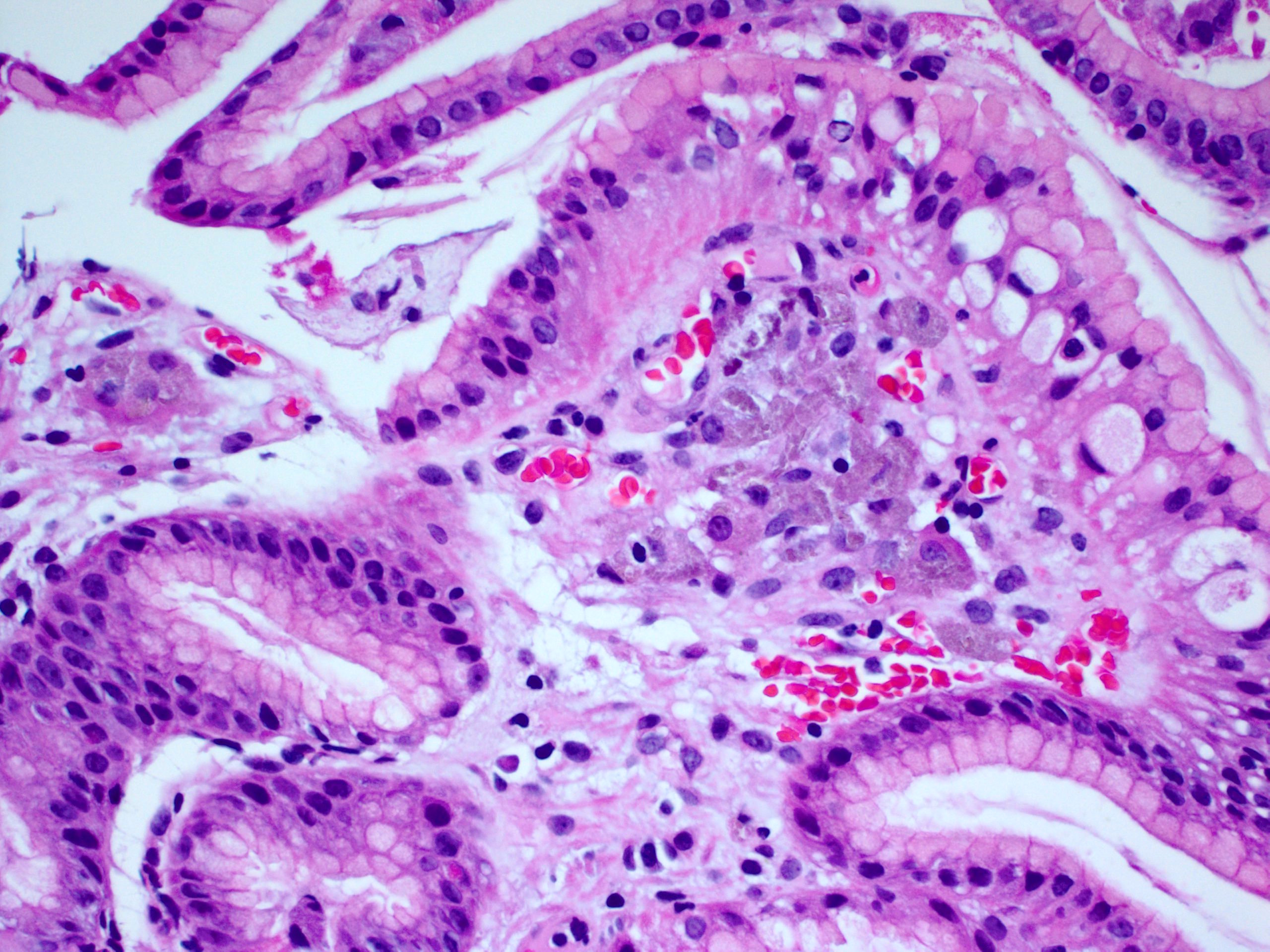
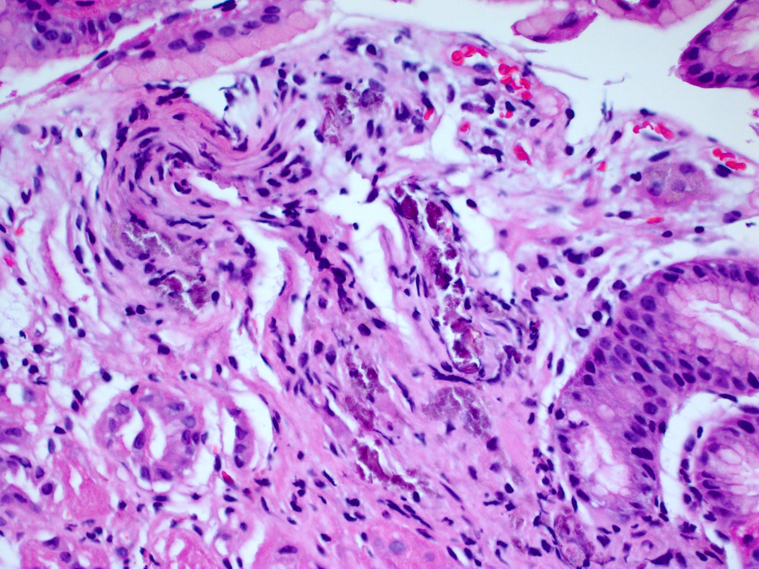
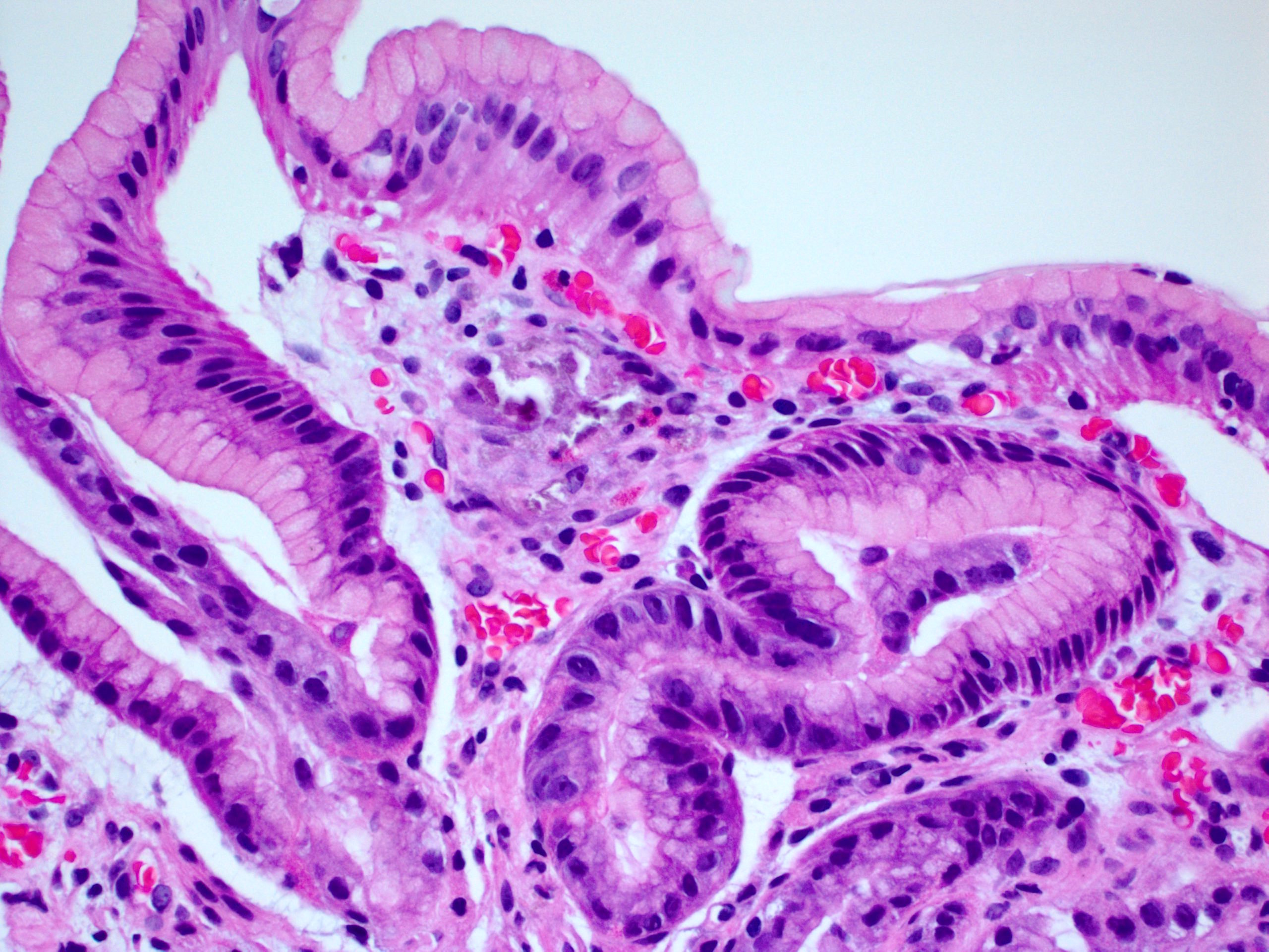
Histopathology images:

What is your diagnosis?
Diagnosis:
Gastric mucosal calcinosis and mucosal lanthanum carbonate deposition
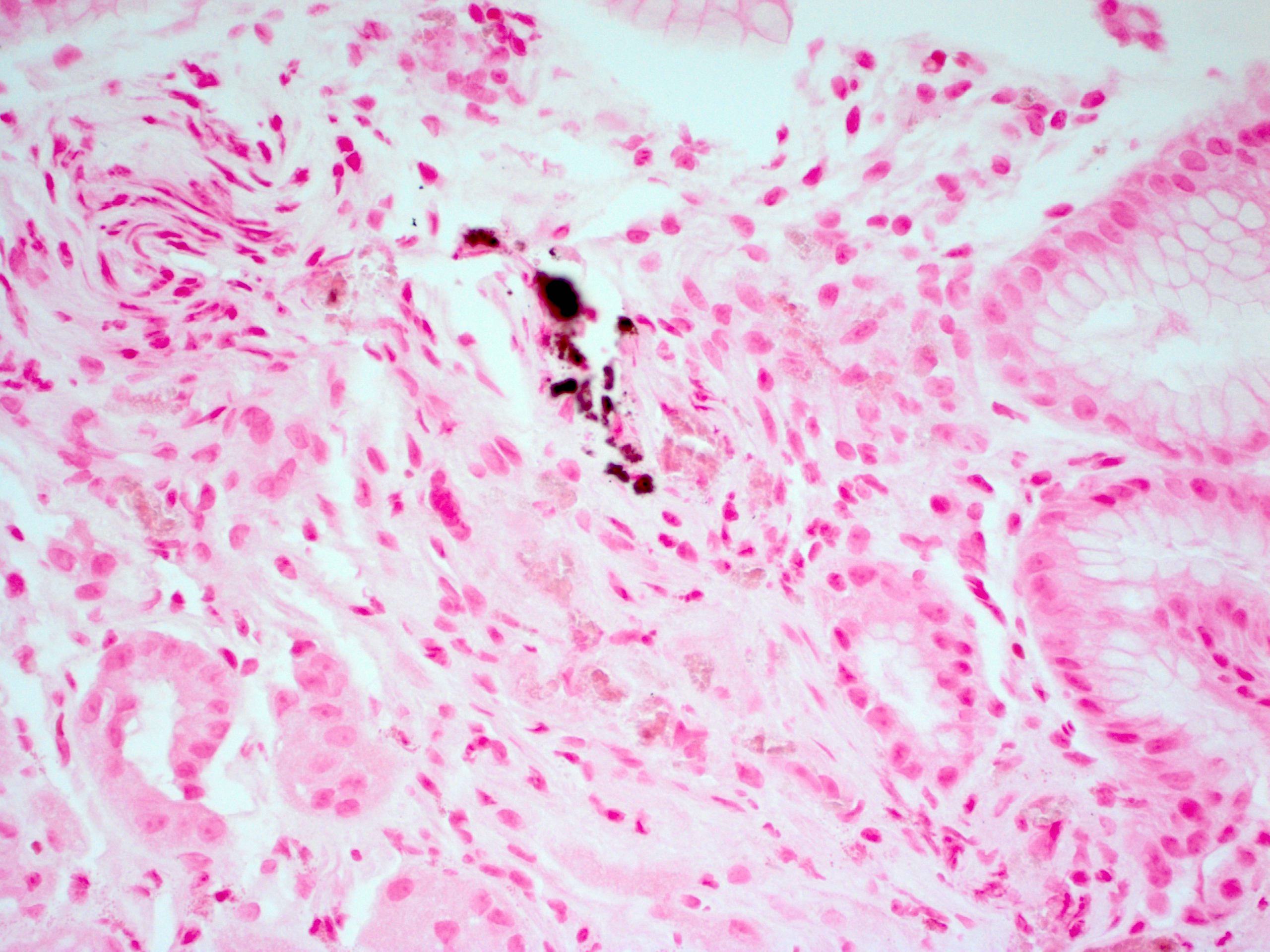
Special stains:
Test question (answer at the end):
A 35 year old man with no history of renal failure undergoes upper endoscopy and colonoscopy for abdominal pain. Crystalline material is observed deposited in his gastric mucosa. What is the most likely identity of this material?
A. Calcium
B. Iron
C. Kayexalate
D. Lanthanum
E. OsmoPrep
Discussion:
Lanthanum carbonate is an oral phosphate binder used to treat hyperphosphatemia in patients with end stage renal disease. Recent reports have indicated that the medication can deposit throughout the gastrointestinal tract mucosa, most commonly in the stomach (Am J Surg Pathol 2015;39:767) but also in the small intestine and colon (Int J Surg Pathol 2016;24:89). Lanthanum may become embedded in the mucosa following digestion by gastric acid (Pathol Int 2017;67:389), and deposition may be detected years after cessation of therapy.
Lanthanum deposition can be detected radiographically but is most often observed microscopically on biopsy tissue. It appears as aggregates of brown purple amorphous material in the mucosa, engulfed by epithelioid histiocytes. The overlying mucosa may be intact or eroded. The identity of the material can be confirmed by energy dispersive X-ray spectrometry, if necessary.
The main differential diagnosis is mucosal calcinosis, which also occurs in dialysis patients. Calcium is typically deeper purple, chunkier, and not contained within histiocytes (Am J Surg Pathol 1993;17:45). Special stains (such as von Kossa) can highlight the calcium; no special stains particularly highlight lanthanum. As in the current example, some patients may show both calcium and lanthanum deposition.
Patients with end stage renal disease may also be taking sodium polystyrene sulfonate (Kayexalate), a cation exchange resin for treatment of hyperkalemia. Kayexalate crystals can also embed in the gastrointestinal mucosa and cause tissue damage (Am J Surg Pathol 1997;21:60). These crystals are also deep purple but are typically angular or polygonal, with a fish scale texture pattern.
Iron can deposit in the stomach in patients taking iron pills but it usually has a distinctive appearance, namely large, coarse, brown chunks of iron (ACG Case Rep J 2013;1:13). A final potential mimic is OsmoPrep, a sodium phosphate tablet used to prepare the bowel for endoscopy. This material appears as coarse black purple deposits along the superficial gastric mucosa and the background stomach may show reactive change (Am J Surg Pathol 2016;40:1550). The material is positive on von Kossa, mimicking calcium, so patient history is essential in confirming the diagnosis (i.e. calcium deposition only occurs in patients with renal disease).
Test Question Answer:
E. OsmoPrep, used to prepare the bowel for endoscopy
All cases are archived on our website. To view them sorted by case number, diagnosis or category, visit our main Case of the Week page. To subscribe or unsubscribe to Case of the Week or our other email lists, click here.
Thanks to Dr. Raul Gonzalez, University of Rochester Medical Center, Rochester, New York (USA) for contributing this case and the discussion. To contribute a Case of the Week, first make sure that we are currently accepting cases, then follow the guidelines on our main Case of the Week page.
Advertisement
Website news:
(1) Our request this week is for images relevant to:
Cervix > Benign / nonneoplastic lesions > Squamous papilloma - micro images
We believe it is important to have an adequate number of high quality images on our server, because images on other servers often disappear. If you own images of these entities, please email them as attachments to Dr. Nat Pernick at NatPernick@gmail.com, with any corresponding clinical history. There is no payment for image contributions, but we will acknowledge you as contributor, so please indicate how you want your name to be displayed.
(2) Last month saw record traffic. We had 867,188 total sessions (visits) for the month, which averaged 27,974 sessions per day. This is an increase of 67.8% from October 2016. We also had 358,848 total users and 2,040,463 page views! We attribute part of this increase to our new, cleaner topic format and expert author reviews of the textbook content.
(3) We have now posted the Jobs Report for the third quarter of 2017 here. It is also accessible from the jobs page by clicking on the orange link.
Visit and follow our Blog to see recent updates to the website.
Case of the Week #442
Clinical history:
A 75 year old woman on dialysis presented with coffee ground emesis. EGD showed gastritis.
Histopathology images:
What is your diagnosis?
Diagnosis:
Gastric mucosal calcinosis and mucosal lanthanum carbonate deposition
Special stains:
von Kossa
Test question (answer at the end):
A 35 year old man with no history of renal failure undergoes upper endoscopy and colonoscopy for abdominal pain. Crystalline material is observed deposited in his gastric mucosa. What is the most likely identity of this material?
A. Calcium
B. Iron
C. Kayexalate
D. Lanthanum
E. OsmoPrep
Discussion:
Lanthanum carbonate is an oral phosphate binder used to treat hyperphosphatemia in patients with end stage renal disease. Recent reports have indicated that the medication can deposit throughout the gastrointestinal tract mucosa, most commonly in the stomach (Am J Surg Pathol 2015;39:767) but also in the small intestine and colon (Int J Surg Pathol 2016;24:89). Lanthanum may become embedded in the mucosa following digestion by gastric acid (Pathol Int 2017;67:389), and deposition may be detected years after cessation of therapy.
Lanthanum deposition can be detected radiographically but is most often observed microscopically on biopsy tissue. It appears as aggregates of brown purple amorphous material in the mucosa, engulfed by epithelioid histiocytes. The overlying mucosa may be intact or eroded. The identity of the material can be confirmed by energy dispersive X-ray spectrometry, if necessary.
The main differential diagnosis is mucosal calcinosis, which also occurs in dialysis patients. Calcium is typically deeper purple, chunkier, and not contained within histiocytes (Am J Surg Pathol 1993;17:45). Special stains (such as von Kossa) can highlight the calcium; no special stains particularly highlight lanthanum. As in the current example, some patients may show both calcium and lanthanum deposition.
Patients with end stage renal disease may also be taking sodium polystyrene sulfonate (Kayexalate), a cation exchange resin for treatment of hyperkalemia. Kayexalate crystals can also embed in the gastrointestinal mucosa and cause tissue damage (Am J Surg Pathol 1997;21:60). These crystals are also deep purple but are typically angular or polygonal, with a fish scale texture pattern.
Iron can deposit in the stomach in patients taking iron pills but it usually has a distinctive appearance, namely large, coarse, brown chunks of iron (ACG Case Rep J 2013;1:13). A final potential mimic is OsmoPrep, a sodium phosphate tablet used to prepare the bowel for endoscopy. This material appears as coarse black purple deposits along the superficial gastric mucosa and the background stomach may show reactive change (Am J Surg Pathol 2016;40:1550). The material is positive on von Kossa, mimicking calcium, so patient history is essential in confirming the diagnosis (i.e. calcium deposition only occurs in patients with renal disease).
Test Question Answer:
E. OsmoPrep, used to prepare the bowel for endoscopy
