
20 June 2018 - Case of the Week #458
All cases are archived on our website. To view them sorted by case number, diagnosis or category, visit our main Case of the Week page. To subscribe or unsubscribe to Case of the Week or our other email lists, click here.
Thanks to Dr. Faheema Hasan, Moti Lal Nehru Medical College, Allahabad (India) for contributing this case and Dr. Abha Soni, Johns Hopkins Hospital, Baltimore, Maryland (USA) for writing the discussion. To contribute a Case of the Week, first make sure that we are currently accepting cases, then follow the guidelines on our main Case of the Week page.

Advertisement
Website news:
(1) We are improving the Board Review page, particularly the Question Bank, which now has 397 questions. We add ~25 questions a month. We have improved the format so questions are numbered consecutively and there is less clutter from headers. Try it out, and let us know if you have any comments or questions.
(2) We are pleased that traffic to our website continues to increase. In May 2018, we had a record number of home page views (235,437), page views (2,217,179), total sessions (918,716) and sessions per day (29,636) which is an increase of 22% from May 2017.
(3) We have now added an Instagram button to the header at the very top of our pages. Please follow us there, as we post interesting pictures from cases we receive. The link is https://www.instagram.com/pathoutlines/.
Visit and follow our Blog to see recent updates to the website.
Case of the Week #458
Clinical history:
A 14 year old boy presented with an arm swelling for 2 months, measuring 4 x 3 x 3 cm. The mass was excised due to increasing pain.
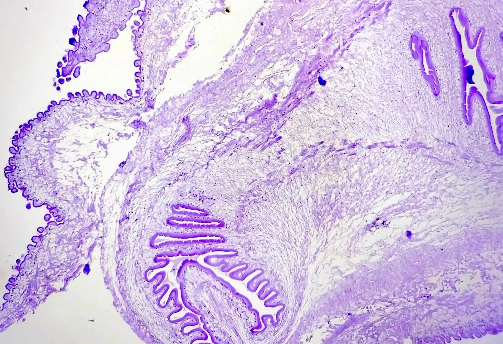
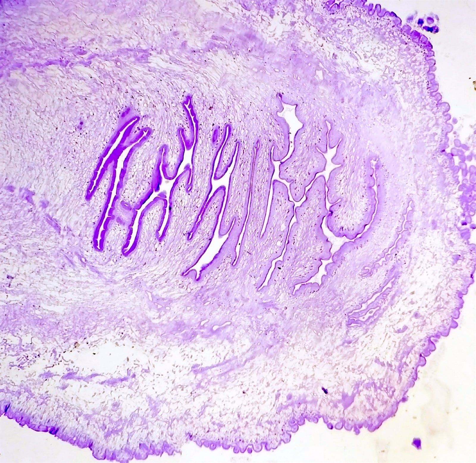
Histopathology images:


What is your diagnosis?
Diagnosis:
Cysticercosis
Test question (answer at the end):
This infection is caused by the larval form of?
A. Taenia solium
B. Taenia saginatum
C. Echinococcus granulosus
D. Paragonium westermani
Discussion:
Cysticercosis is a systemic infection caused by ingestion of larval cysts of the cestode Taenia solium (pork tapeworm). It is acquired by swallowing food, water or feces from contaminated T. solium eggs. The human is considered an intermediate host. When the eggs are ingested they release onchospheres that penetrate the gut epithelium and enter the bloodstream. These egg forms then embed themselves into various tissues throughout the body, encyst and differentiate into cysticerci (Siddeswari et al, Rubin's Pathology: Clinicopathologic Foundations of Medicine 6th Edition, 2012, Centers for Disease Control and Prevention).
Clinically, cysticercosis symptoms change based on the site affected. In the brain it manifests as headaches, seizures and convulsions. In the heart it may cause arrhythmias. Cutaneous involvement is rare, but if present it presents as a palpable, subcutaneous nodule that may become painful if large enough (Siddeswari et al, Rubin's Pathology: Clinicopathologic Foundations of Medicine 6th Edition, 2012).
In the present case, the gross specimen was composed of a cystic mass measuring 4 x 3 x 3 cm. The cut section showed a milky white to clear cyst with an envaginated scolex (head of tapeworm). Histology reveals a convoluted pattern of multilayered cysts composed of an outer cuticle layer, a middle nuclear layer and an inner parenchymal layer, diagnostic of cysticercosis. Birefringent hooklets can be present and a granulomatous reaction with mixed inflammatory infiltrate, fibrosis and calcification may also be appreciated.
The differential diagnosis includes other infestations. Taenia saginatum causes human taeniasis, but it causes cysticercosis in cattle. Echinococcus granulosis is a tapeworm that resides in the small bowel of dogs, sheep, goats and cattle. Humans are the intermediate hosts. They become infected by ingesting tapeworm eggs and this results in the development of hydatid cysts (Siddeswari et al, Rubin's Pathology: Clinicopathologic Foundations of Medicine 6th Edition, 2012). Histologically, these cysts surround a thick fibrous capsule with an inner germinal layer which may contain a clear hydatid fluid. The cyst is most commonly found in the liver and may cause obstructive jaundice. Paragonium westermani is a lung fluke that causes paragonimiasis, a pulmonary infection caused by ingestion of undercooked crab. Histologically, the paragonium eggs are found in tissue sections and display a thick, highly retractile wall with operculum on either end of the eggs with adjacent non-necrotizing granulomas and organizing pneumonia (Burger, P. et al, Diagnostic Pathology: Neuropathology, 2nd edition, 2016).
Treatment for cysticercosis involves anti-helminthic therapy such as praziquantel and albendazole or surgery depending on the size and location of lesions (Am Fam Physician 2007;76:91).
Additional references:
Sacchidanand S., Namitha P., Mallikarjuna M., Nataraj H.V., Disseminated cutaneous cysticercosis and neurocysticercosis: A rare occurrence (Indian Dermatol Online J 2012;3:135).
Siddeswari R., Manohar S., Sudarsi B., Cutaneous cysticercosis (J Acad Med Sci 2012;2:132).
Test Question Answer:
A. Taenia solium
All cases are archived on our website. To view them sorted by case number, diagnosis or category, visit our main Case of the Week page. To subscribe or unsubscribe to Case of the Week or our other email lists, click here.
Thanks to Dr. Faheema Hasan, Moti Lal Nehru Medical College, Allahabad (India) for contributing this case and Dr. Abha Soni, Johns Hopkins Hospital, Baltimore, Maryland (USA) for writing the discussion. To contribute a Case of the Week, first make sure that we are currently accepting cases, then follow the guidelines on our main Case of the Week page.
Advertisement
Website news:
(1) We are improving the Board Review page, particularly the Question Bank, which now has 397 questions. We add ~25 questions a month. We have improved the format so questions are numbered consecutively and there is less clutter from headers. Try it out, and let us know if you have any comments or questions.
(2) We are pleased that traffic to our website continues to increase. In May 2018, we had a record number of home page views (235,437), page views (2,217,179), total sessions (918,716) and sessions per day (29,636) which is an increase of 22% from May 2017.
(3) We have now added an Instagram button to the header at the very top of our pages. Please follow us there, as we post interesting pictures from cases we receive. The link is https://www.instagram.com/pathoutlines/.
Visit and follow our Blog to see recent updates to the website.
Case of the Week #458
Clinical history:
A 14 year old boy presented with an arm swelling for 2 months, measuring 4 x 3 x 3 cm. The mass was excised due to increasing pain.
Histopathology images:
What is your diagnosis?
Diagnosis:
Cysticercosis
Test question (answer at the end):
This infection is caused by the larval form of?
A. Taenia solium
B. Taenia saginatum
C. Echinococcus granulosus
D. Paragonium westermani
Discussion:
Cysticercosis is a systemic infection caused by ingestion of larval cysts of the cestode Taenia solium (pork tapeworm). It is acquired by swallowing food, water or feces from contaminated T. solium eggs. The human is considered an intermediate host. When the eggs are ingested they release onchospheres that penetrate the gut epithelium and enter the bloodstream. These egg forms then embed themselves into various tissues throughout the body, encyst and differentiate into cysticerci (Siddeswari et al, Rubin's Pathology: Clinicopathologic Foundations of Medicine 6th Edition, 2012, Centers for Disease Control and Prevention).
Clinically, cysticercosis symptoms change based on the site affected. In the brain it manifests as headaches, seizures and convulsions. In the heart it may cause arrhythmias. Cutaneous involvement is rare, but if present it presents as a palpable, subcutaneous nodule that may become painful if large enough (Siddeswari et al, Rubin's Pathology: Clinicopathologic Foundations of Medicine 6th Edition, 2012).
In the present case, the gross specimen was composed of a cystic mass measuring 4 x 3 x 3 cm. The cut section showed a milky white to clear cyst with an envaginated scolex (head of tapeworm). Histology reveals a convoluted pattern of multilayered cysts composed of an outer cuticle layer, a middle nuclear layer and an inner parenchymal layer, diagnostic of cysticercosis. Birefringent hooklets can be present and a granulomatous reaction with mixed inflammatory infiltrate, fibrosis and calcification may also be appreciated.
The differential diagnosis includes other infestations. Taenia saginatum causes human taeniasis, but it causes cysticercosis in cattle. Echinococcus granulosis is a tapeworm that resides in the small bowel of dogs, sheep, goats and cattle. Humans are the intermediate hosts. They become infected by ingesting tapeworm eggs and this results in the development of hydatid cysts (Siddeswari et al, Rubin's Pathology: Clinicopathologic Foundations of Medicine 6th Edition, 2012). Histologically, these cysts surround a thick fibrous capsule with an inner germinal layer which may contain a clear hydatid fluid. The cyst is most commonly found in the liver and may cause obstructive jaundice. Paragonium westermani is a lung fluke that causes paragonimiasis, a pulmonary infection caused by ingestion of undercooked crab. Histologically, the paragonium eggs are found in tissue sections and display a thick, highly retractile wall with operculum on either end of the eggs with adjacent non-necrotizing granulomas and organizing pneumonia (Burger, P. et al, Diagnostic Pathology: Neuropathology, 2nd edition, 2016).
Treatment for cysticercosis involves anti-helminthic therapy such as praziquantel and albendazole or surgery depending on the size and location of lesions (Am Fam Physician 2007;76:91).
Additional references:
Sacchidanand S., Namitha P., Mallikarjuna M., Nataraj H.V., Disseminated cutaneous cysticercosis and neurocysticercosis: A rare occurrence (Indian Dermatol Online J 2012;3:135).
Siddeswari R., Manohar S., Sudarsi B., Cutaneous cysticercosis (J Acad Med Sci 2012;2:132).
Test Question Answer:
A. Taenia solium
