

Contributed by Firas G. Petros, M.D.
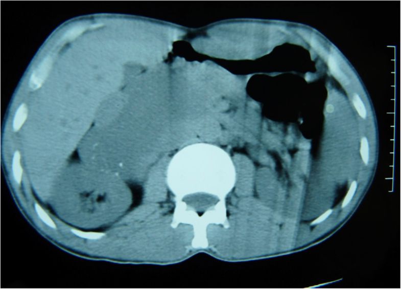
CT of left adrenal mass
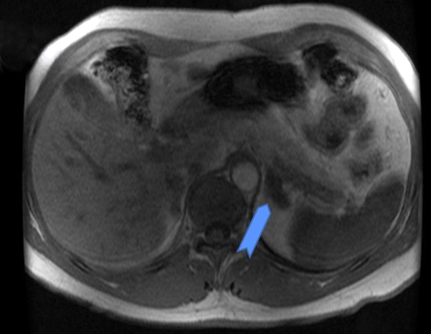
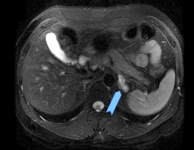
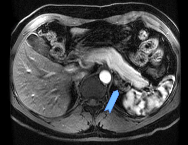
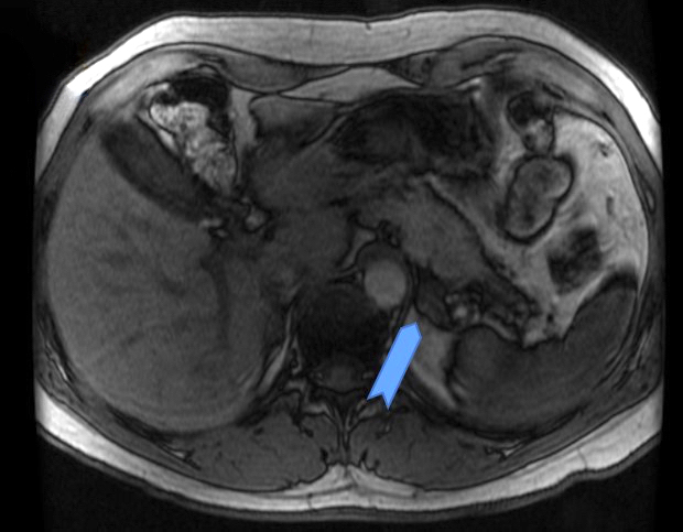
MRI of left adrenal mass
Contributed by Debra L. Zynger, M.D.

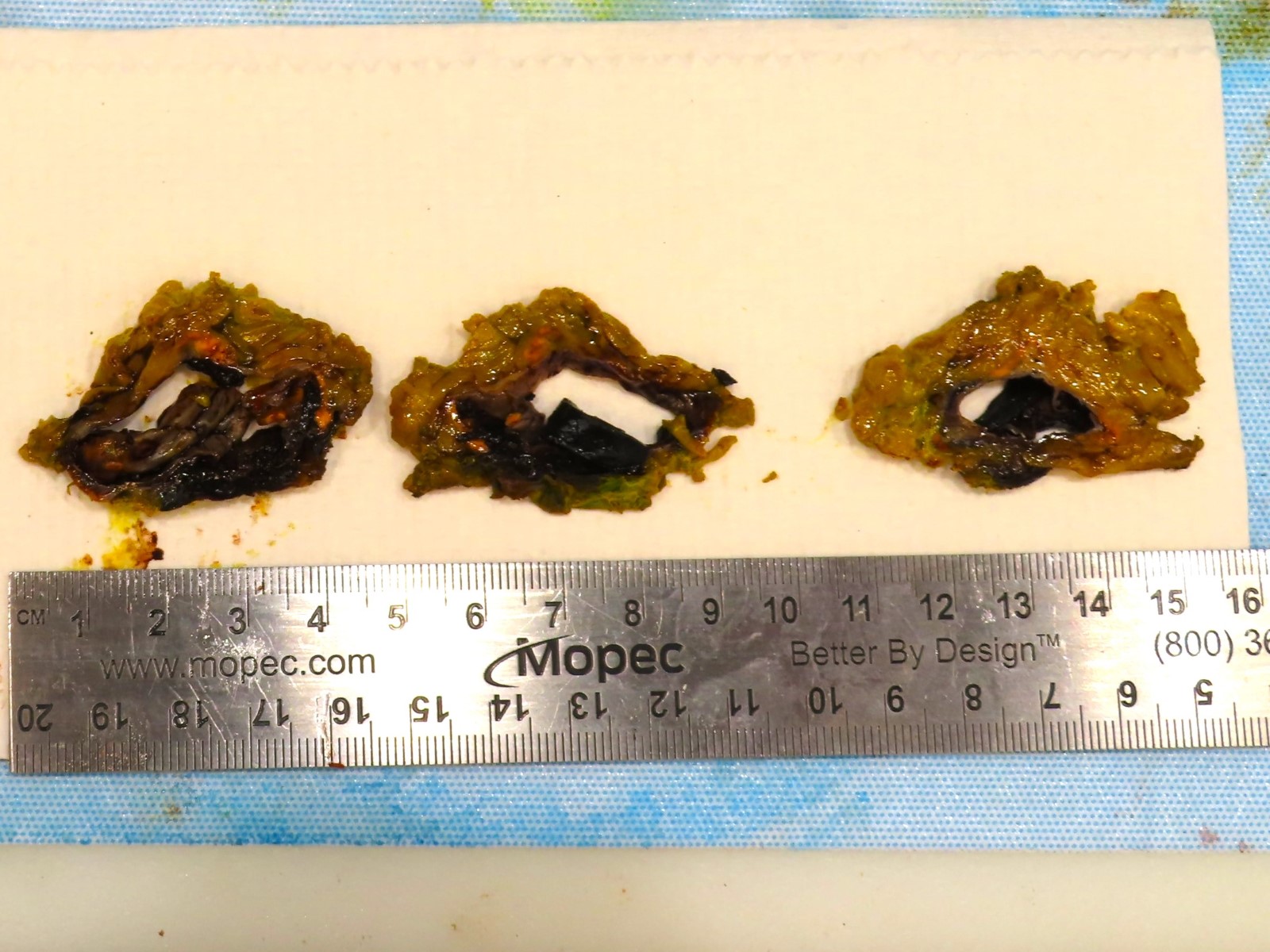
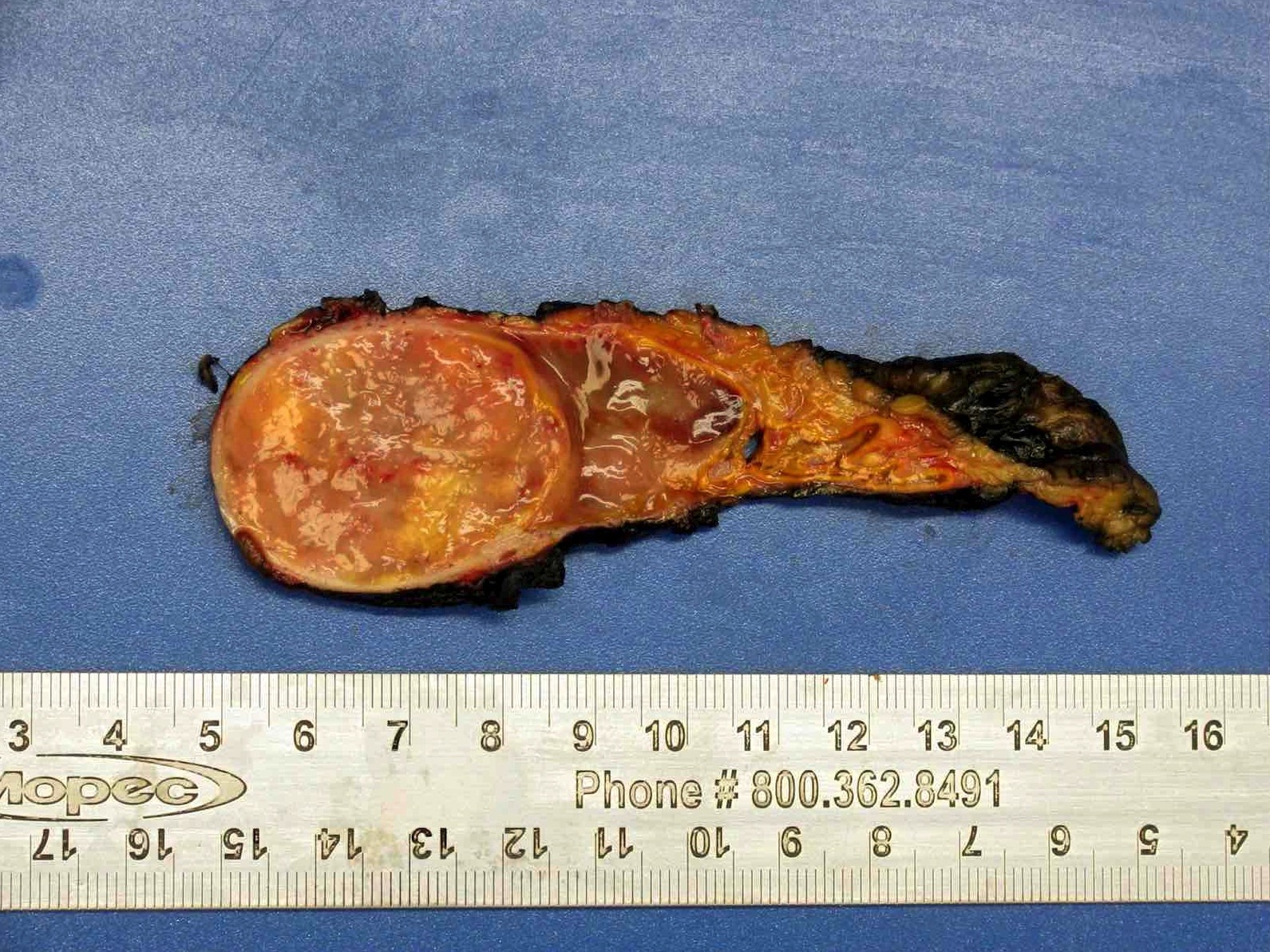
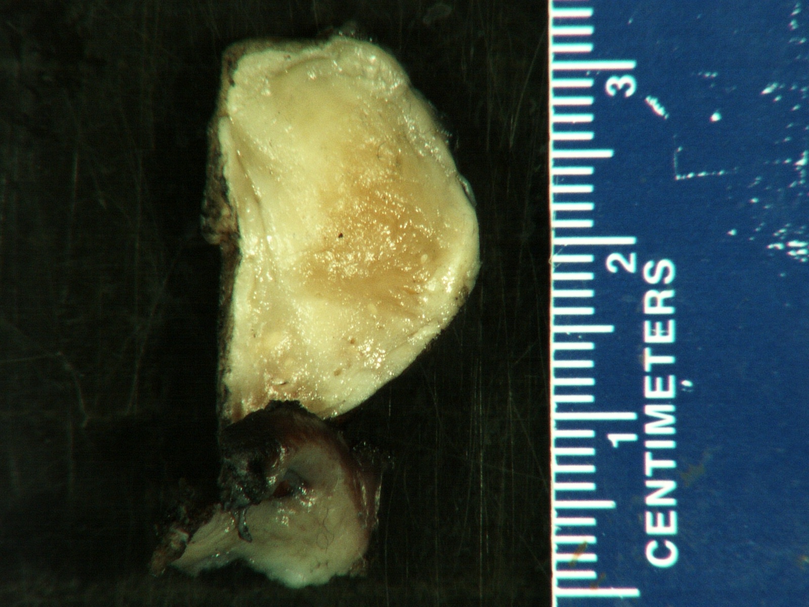
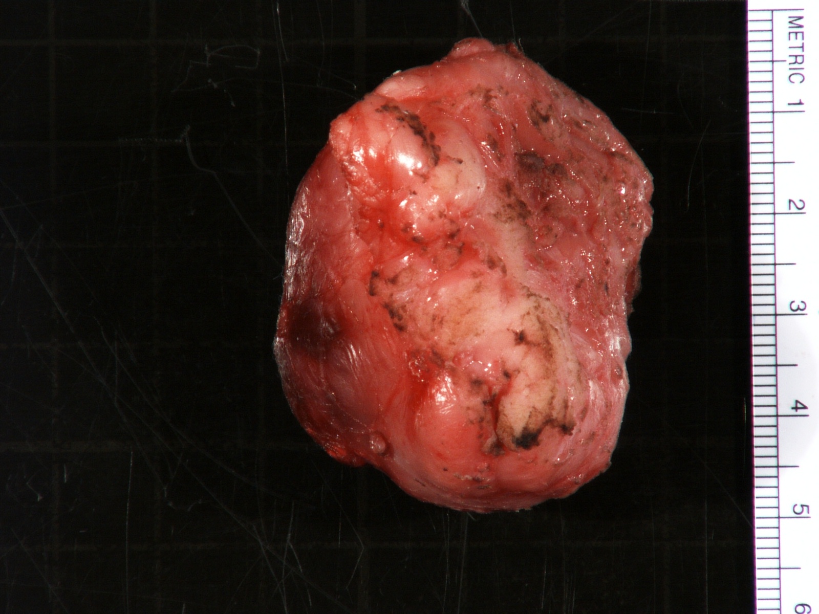
Ill defined spongy lesion
Images hosted on other servers:

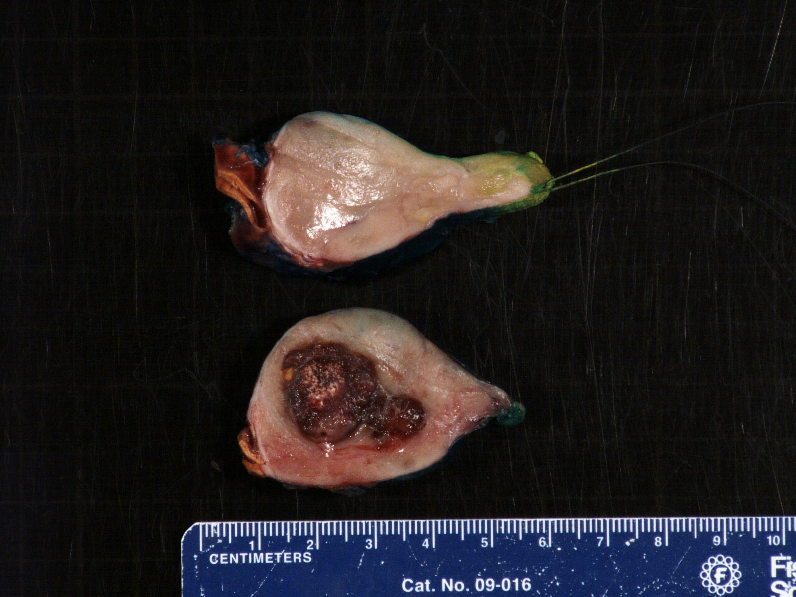
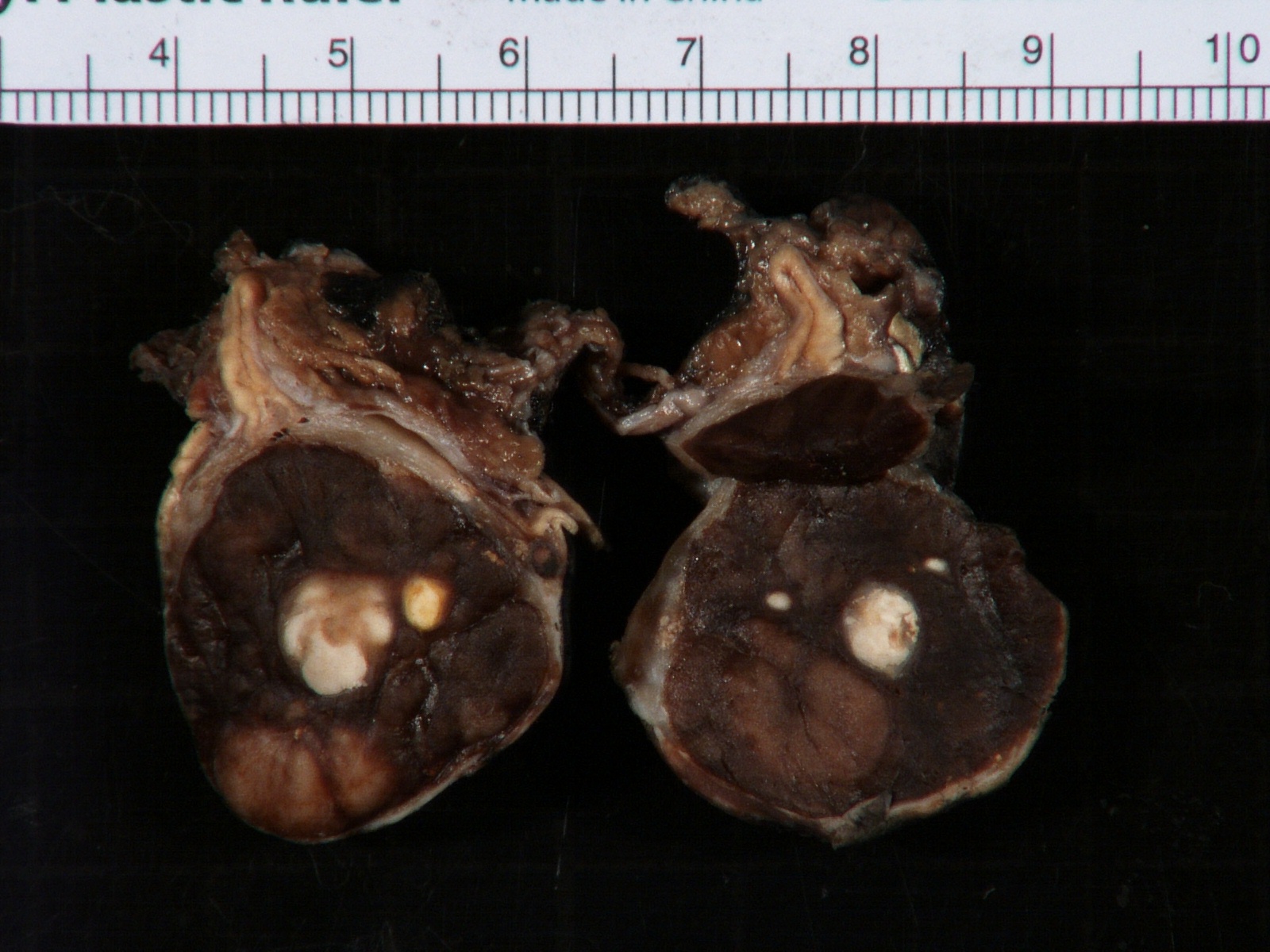
White solid and cystic surface

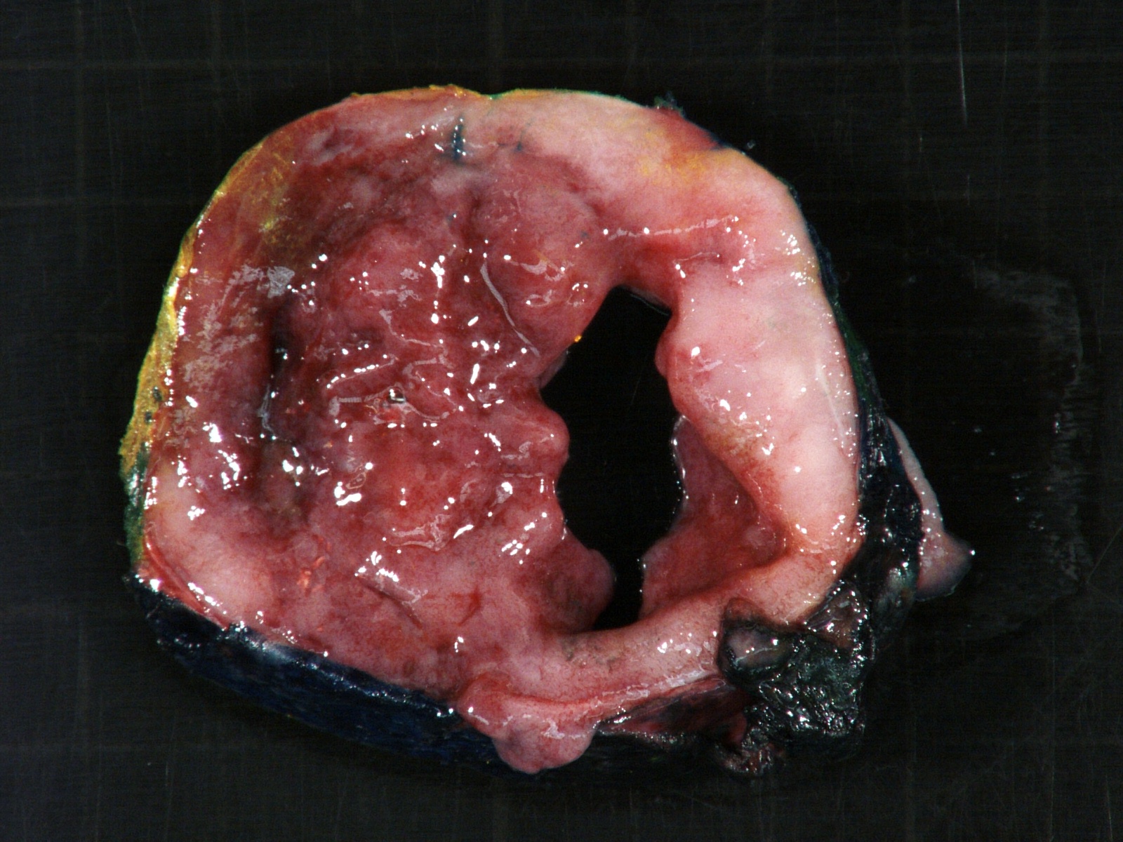
Cystic surface

Cystic spongy surface

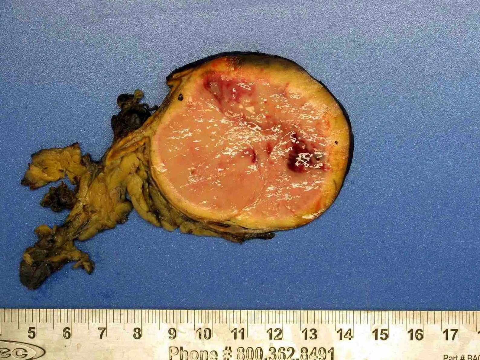
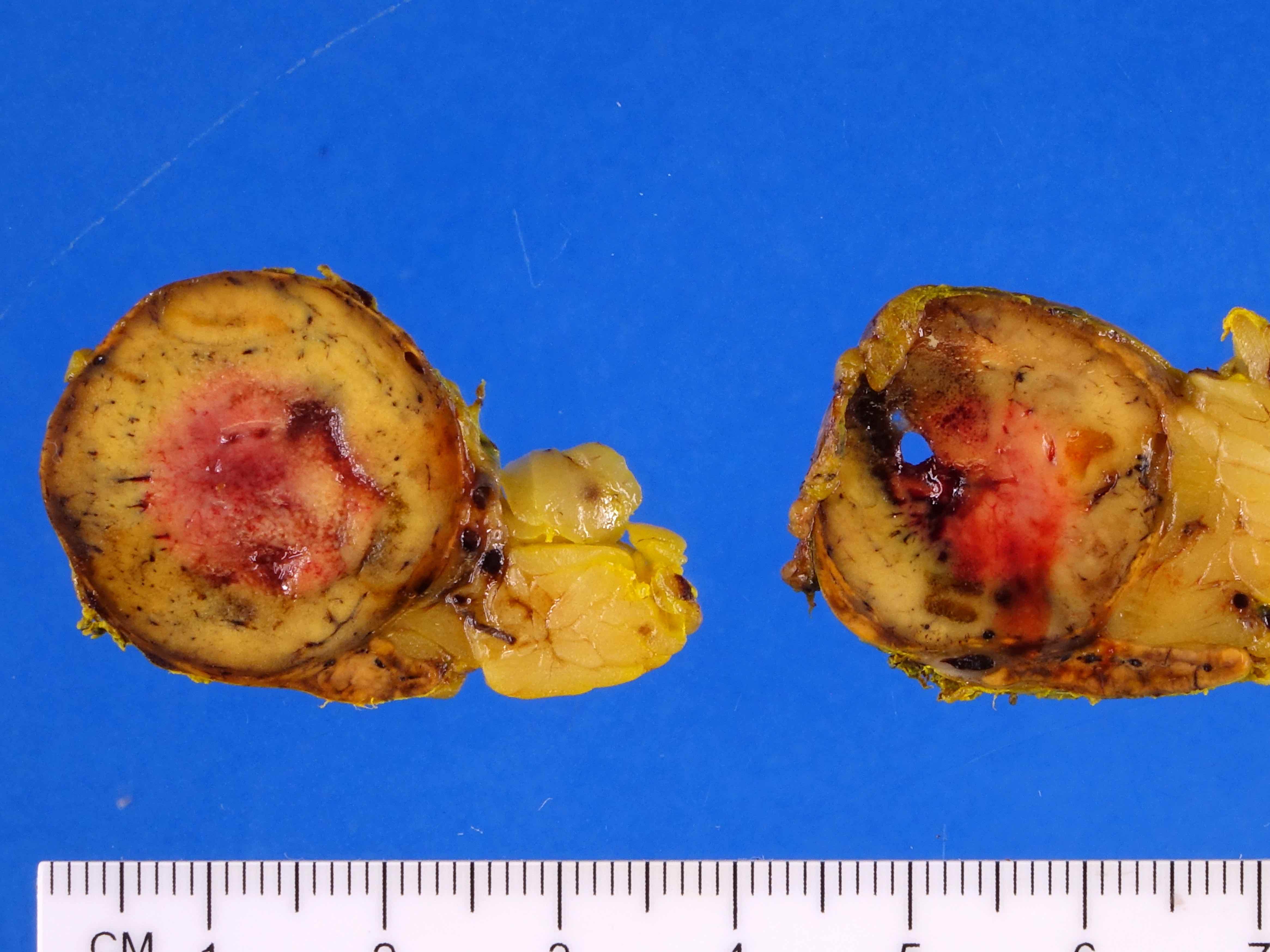
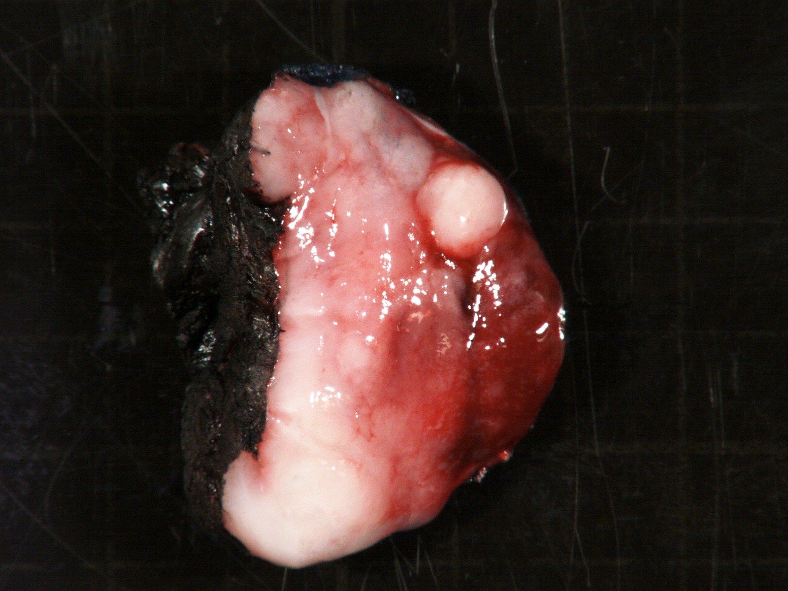
Solid gray surface
Contributed by Debra L. Zynger, M.D.
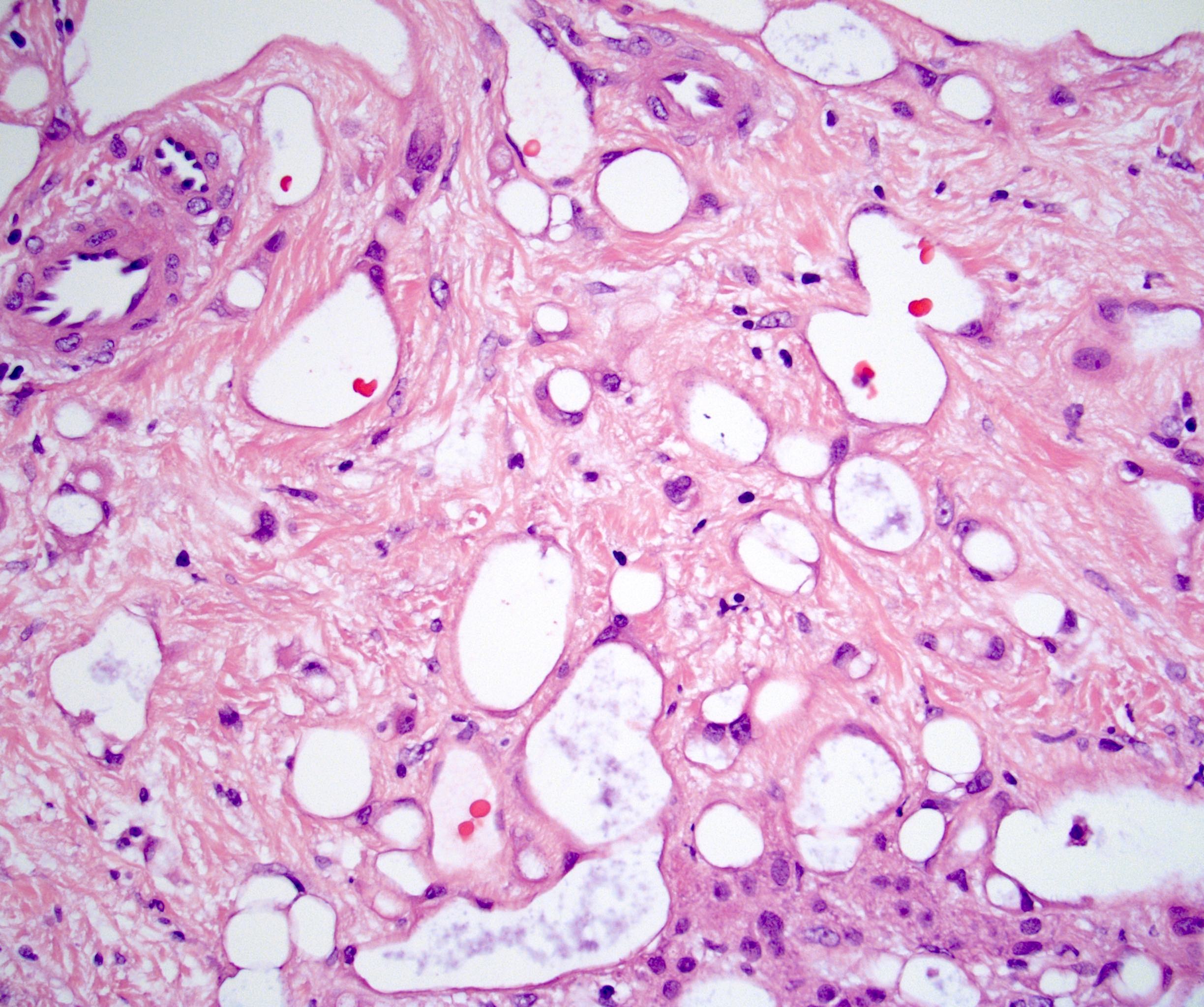
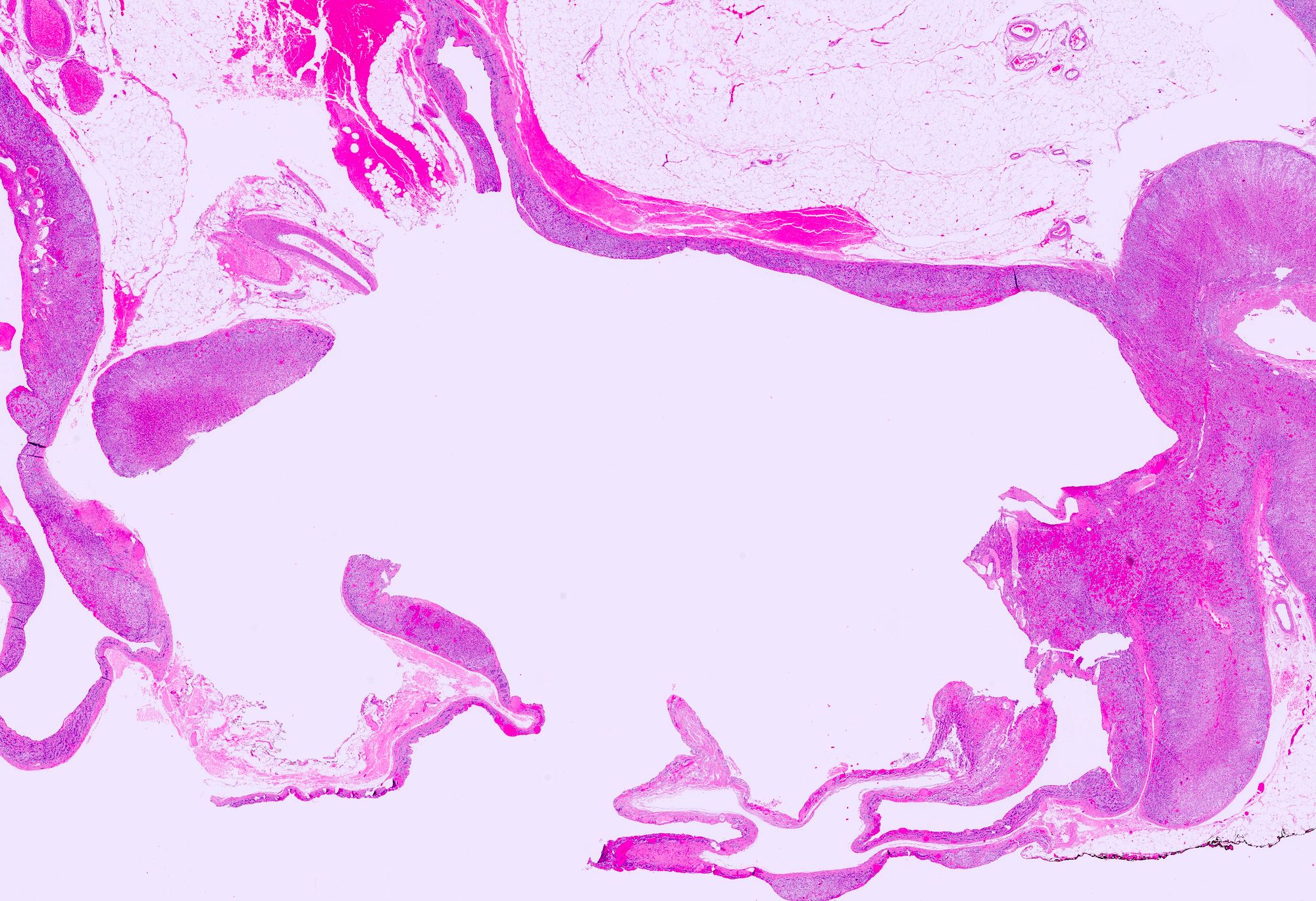
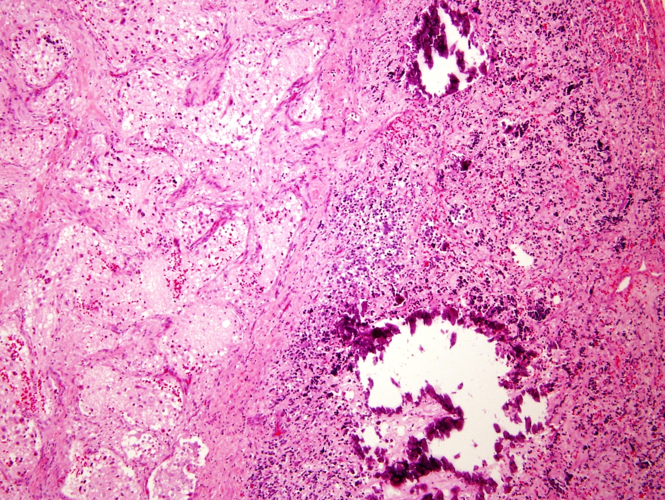
Cystic lesion
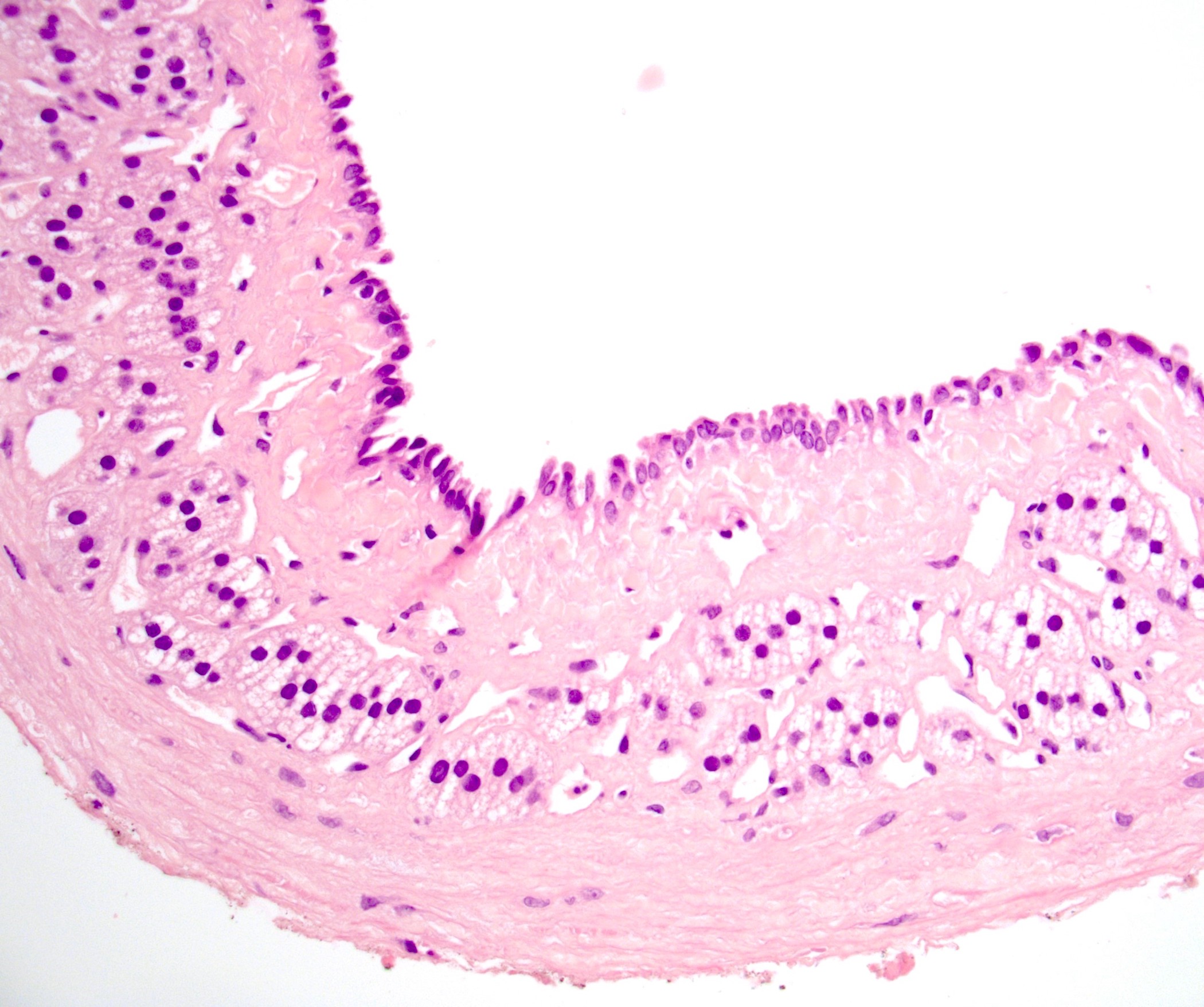
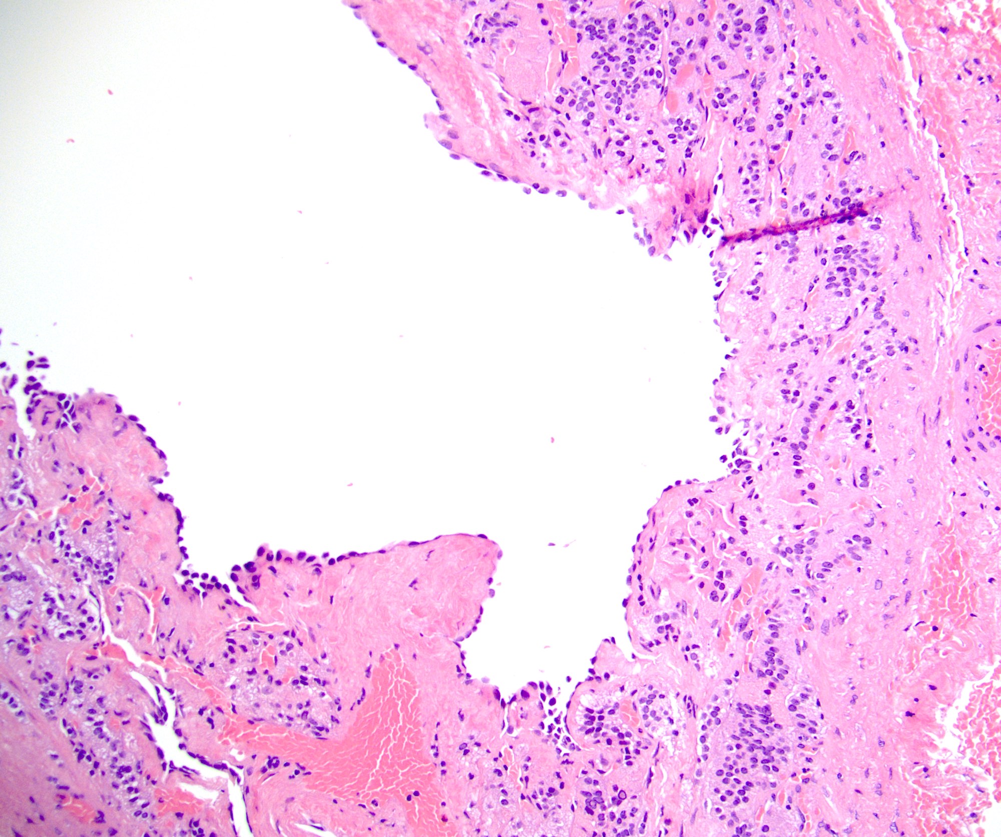
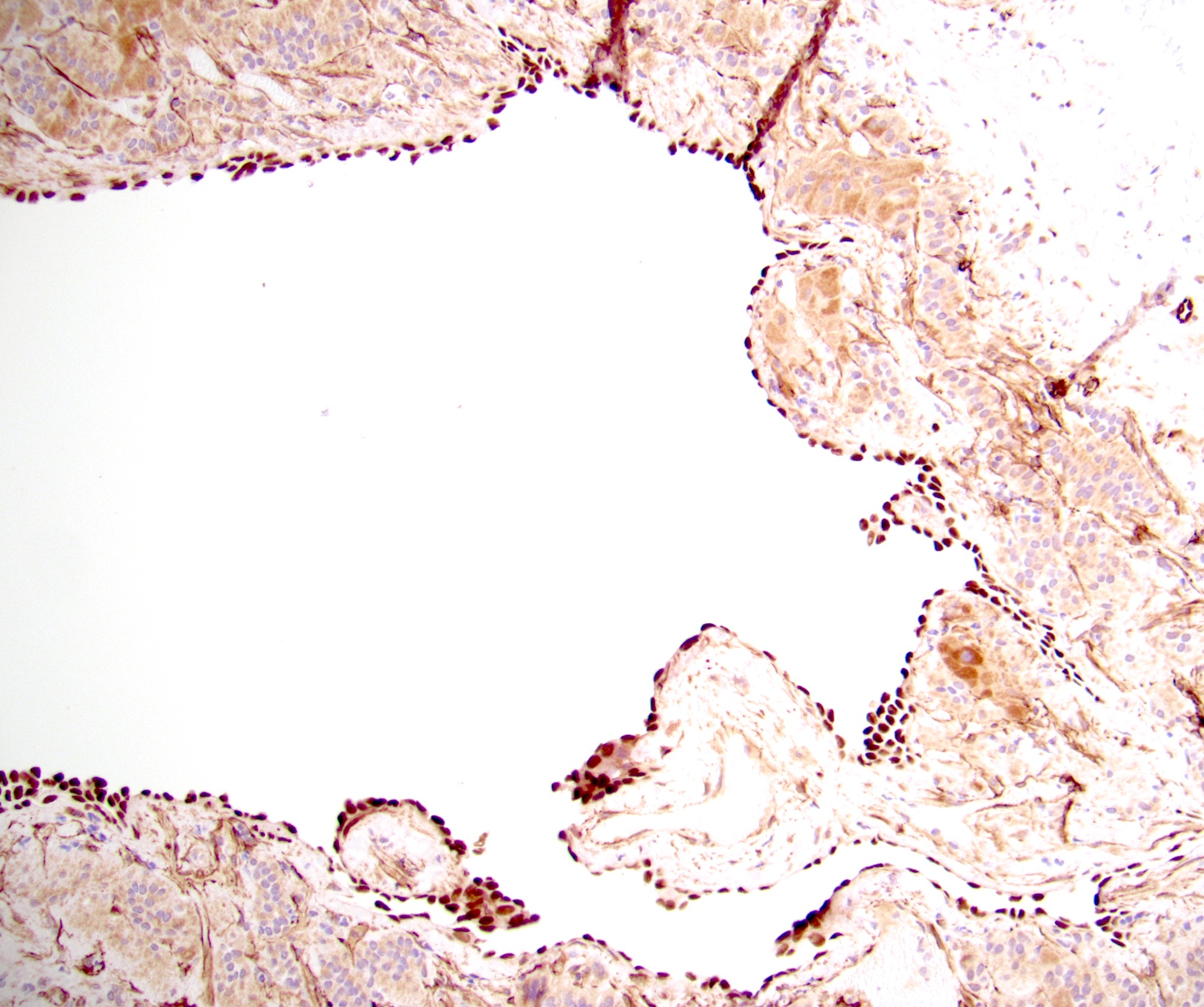
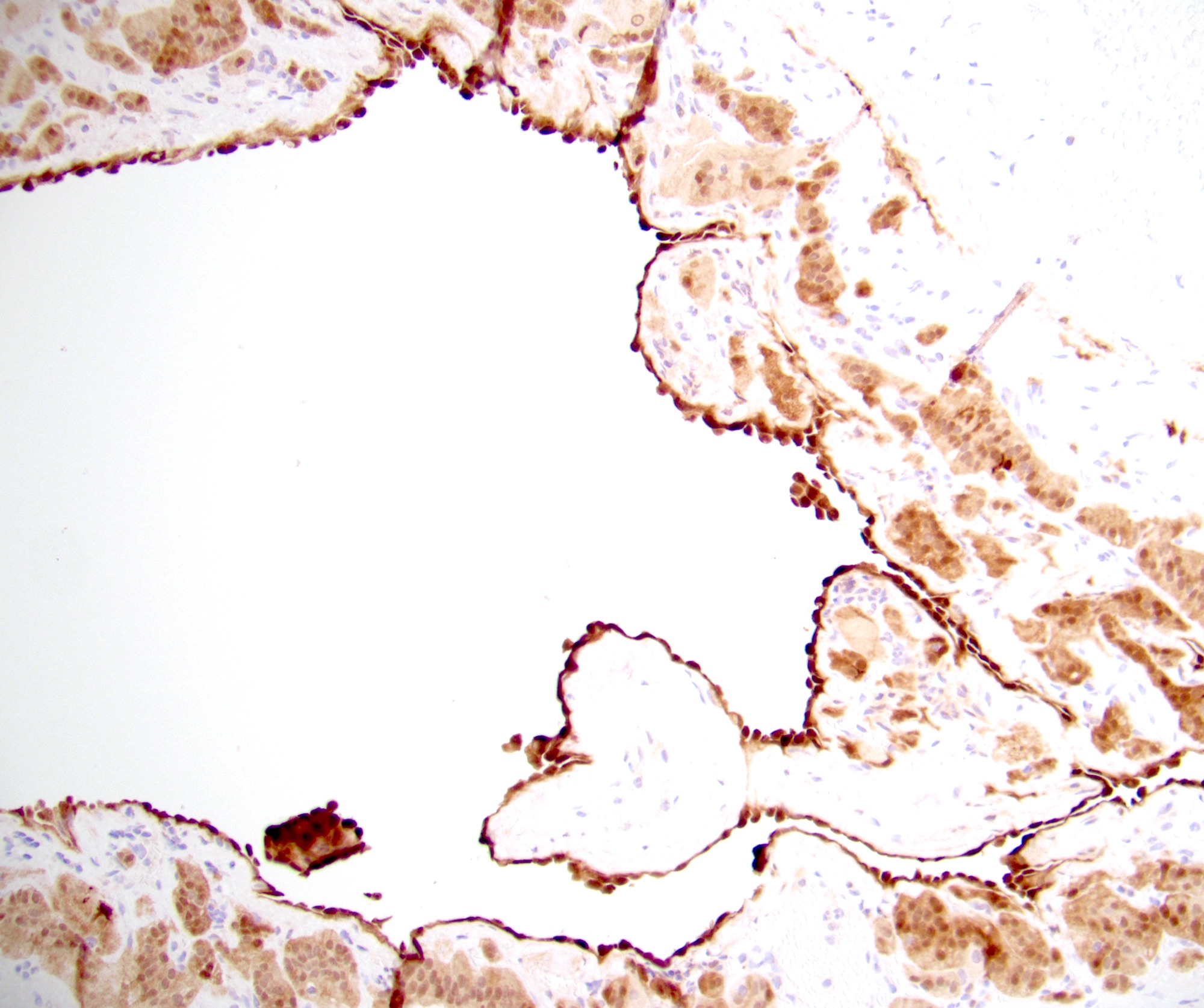
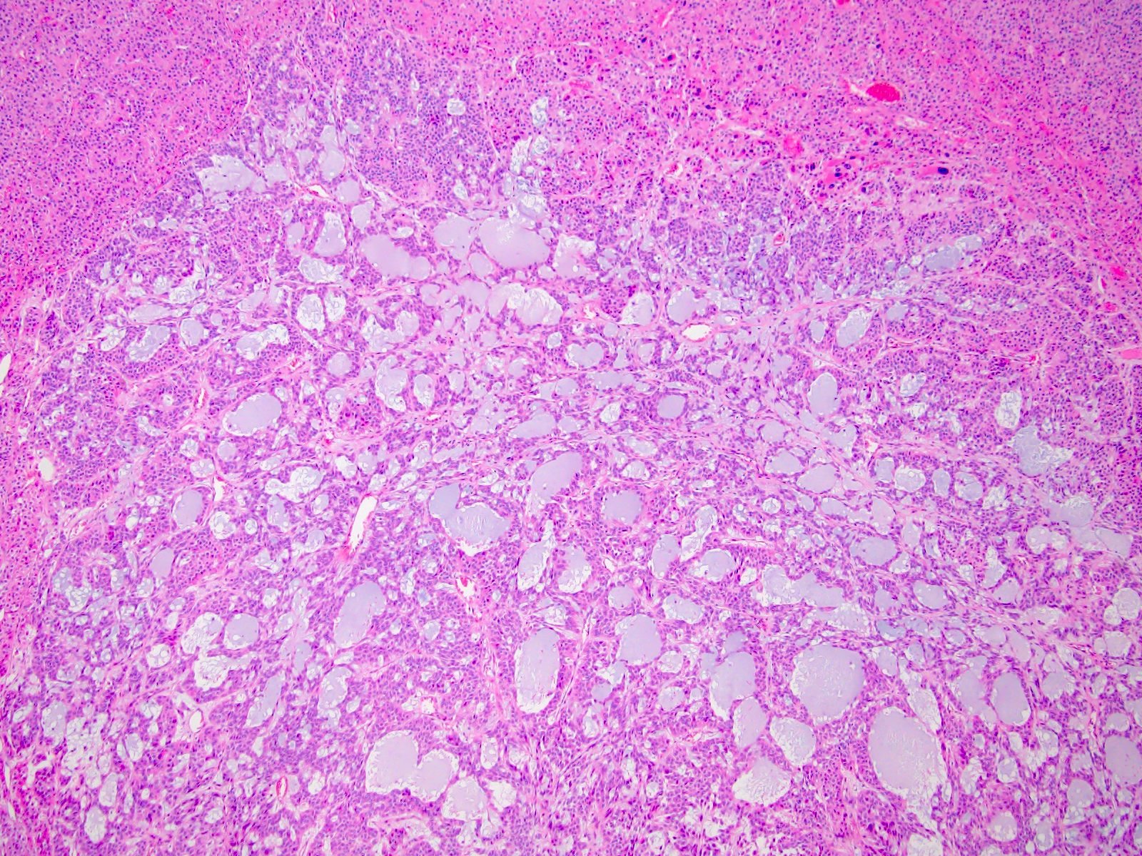
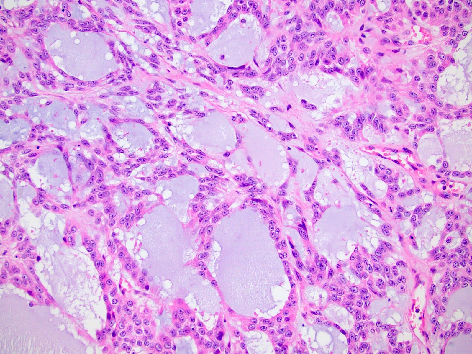
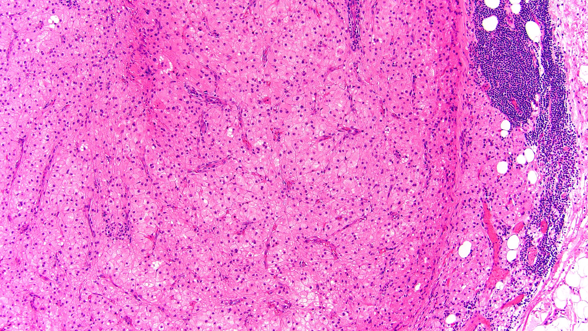
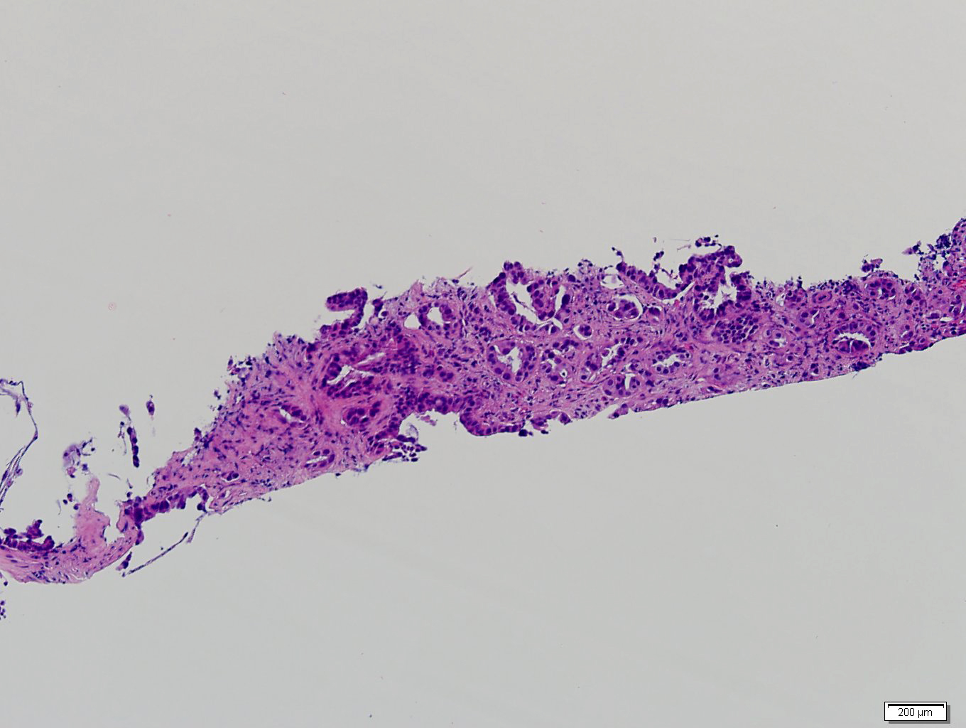
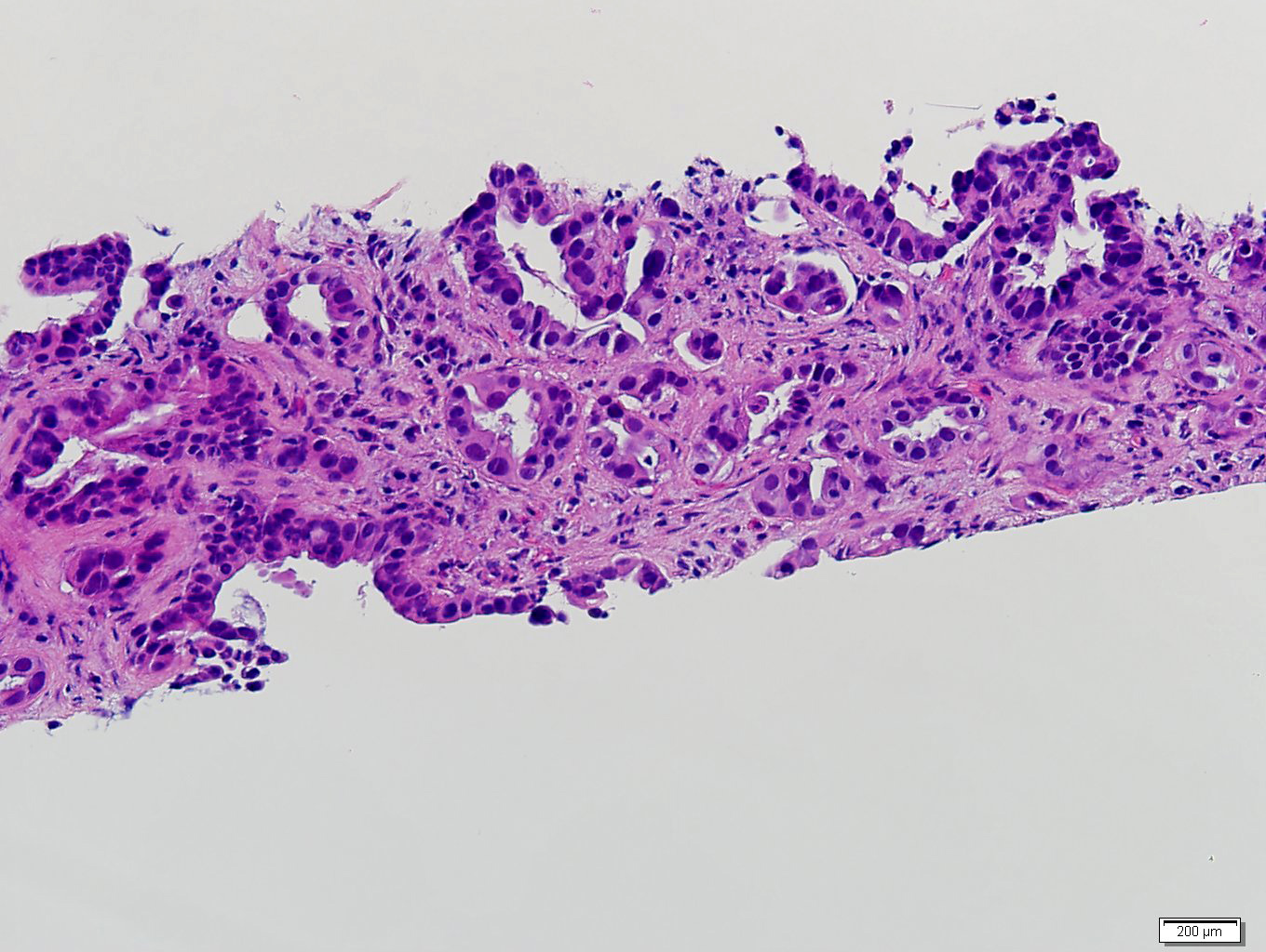
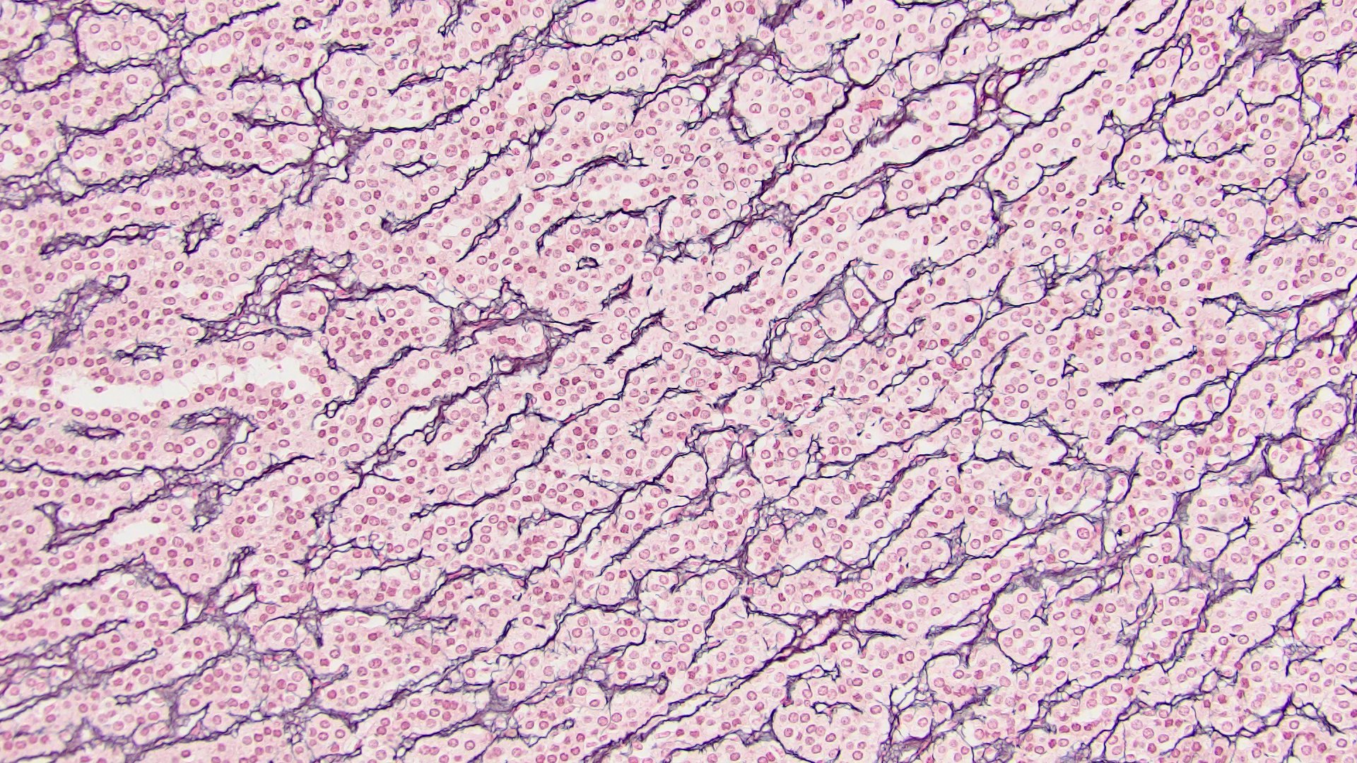
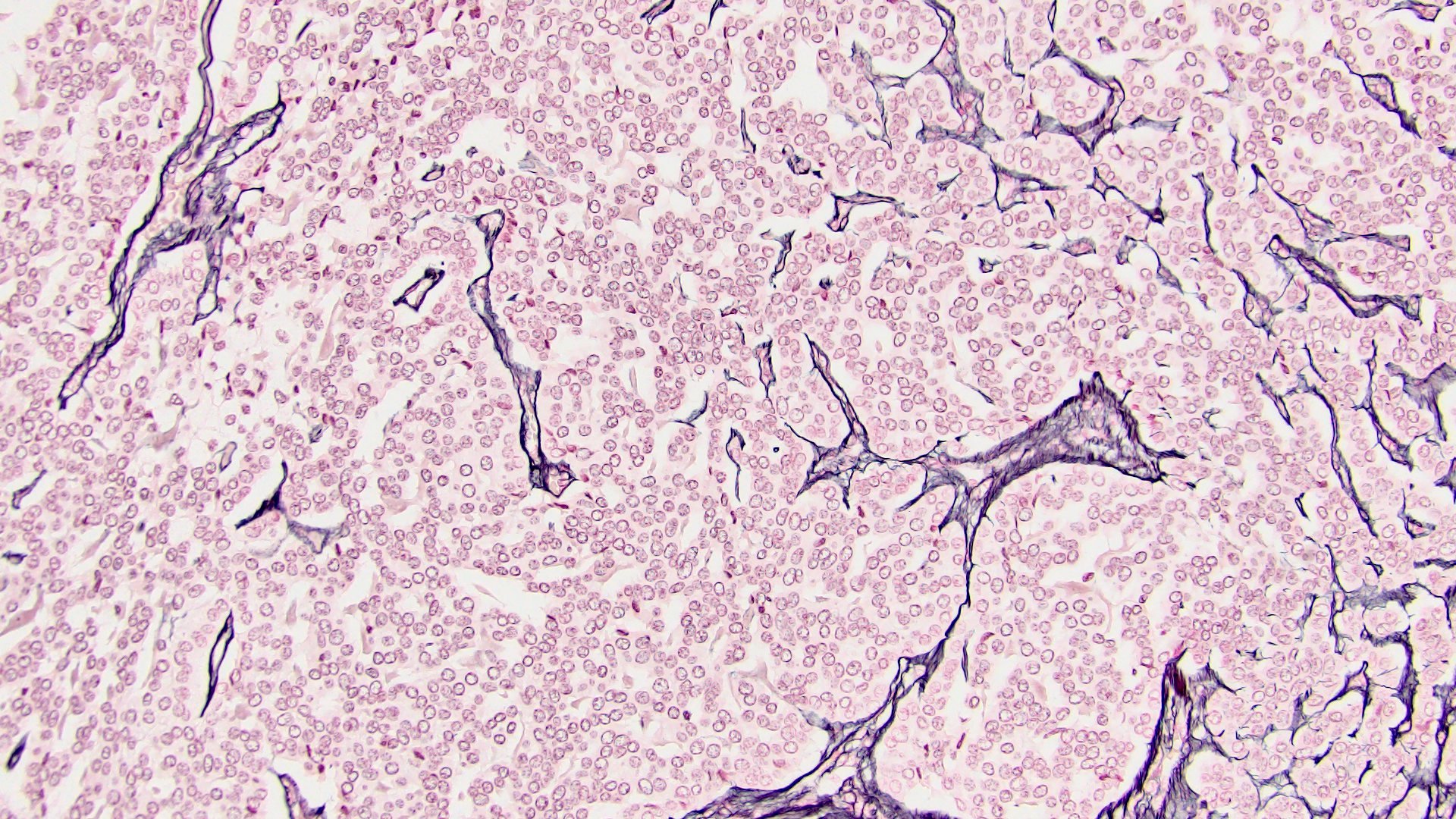
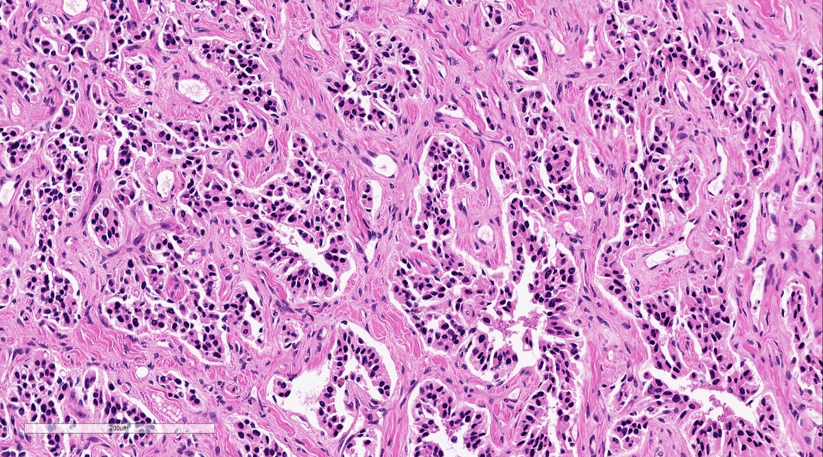
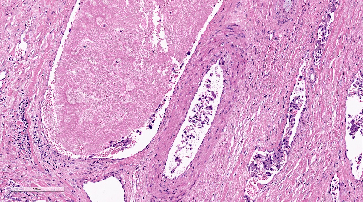
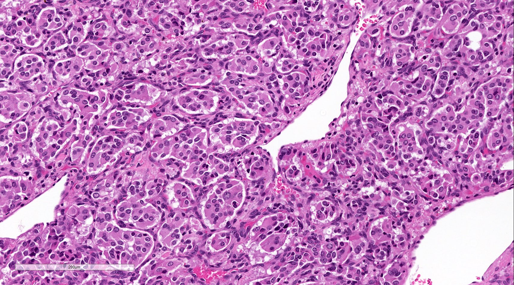
Anastomosing channels
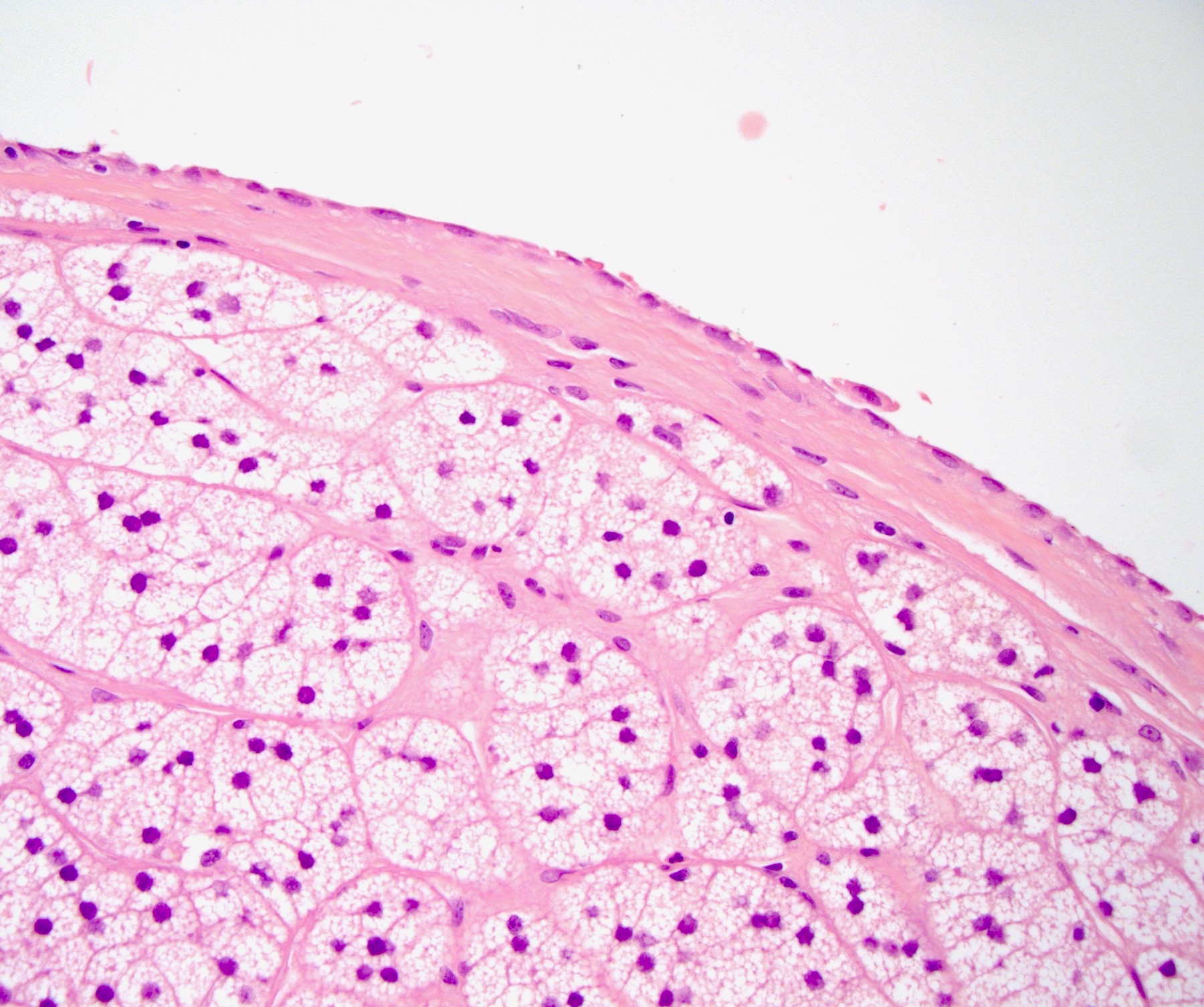
Microcysts
Delicate septae
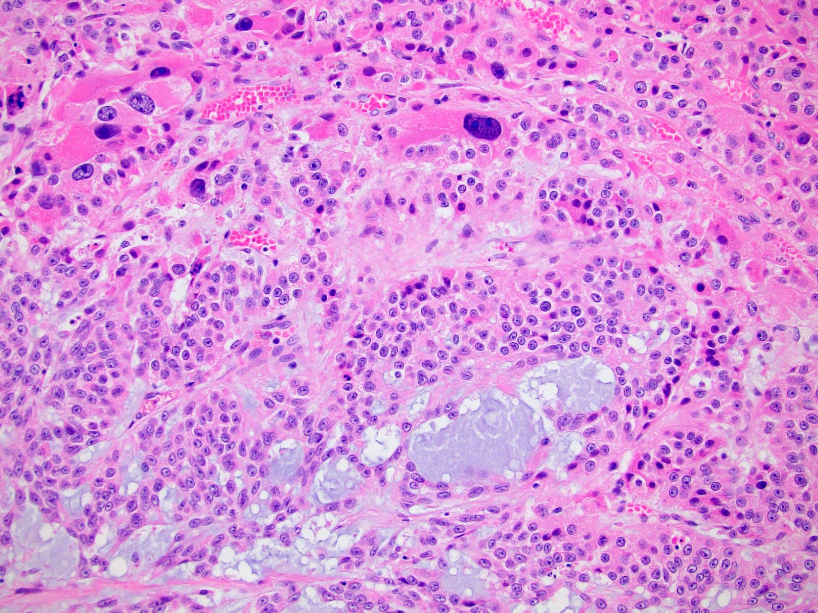
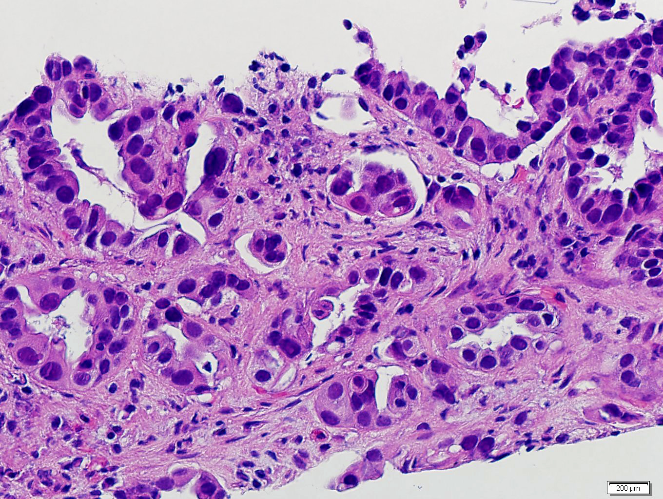
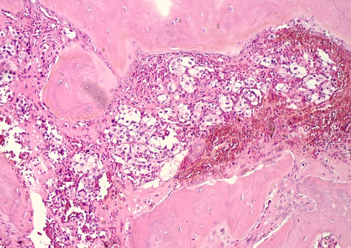
Tubules and signet rings
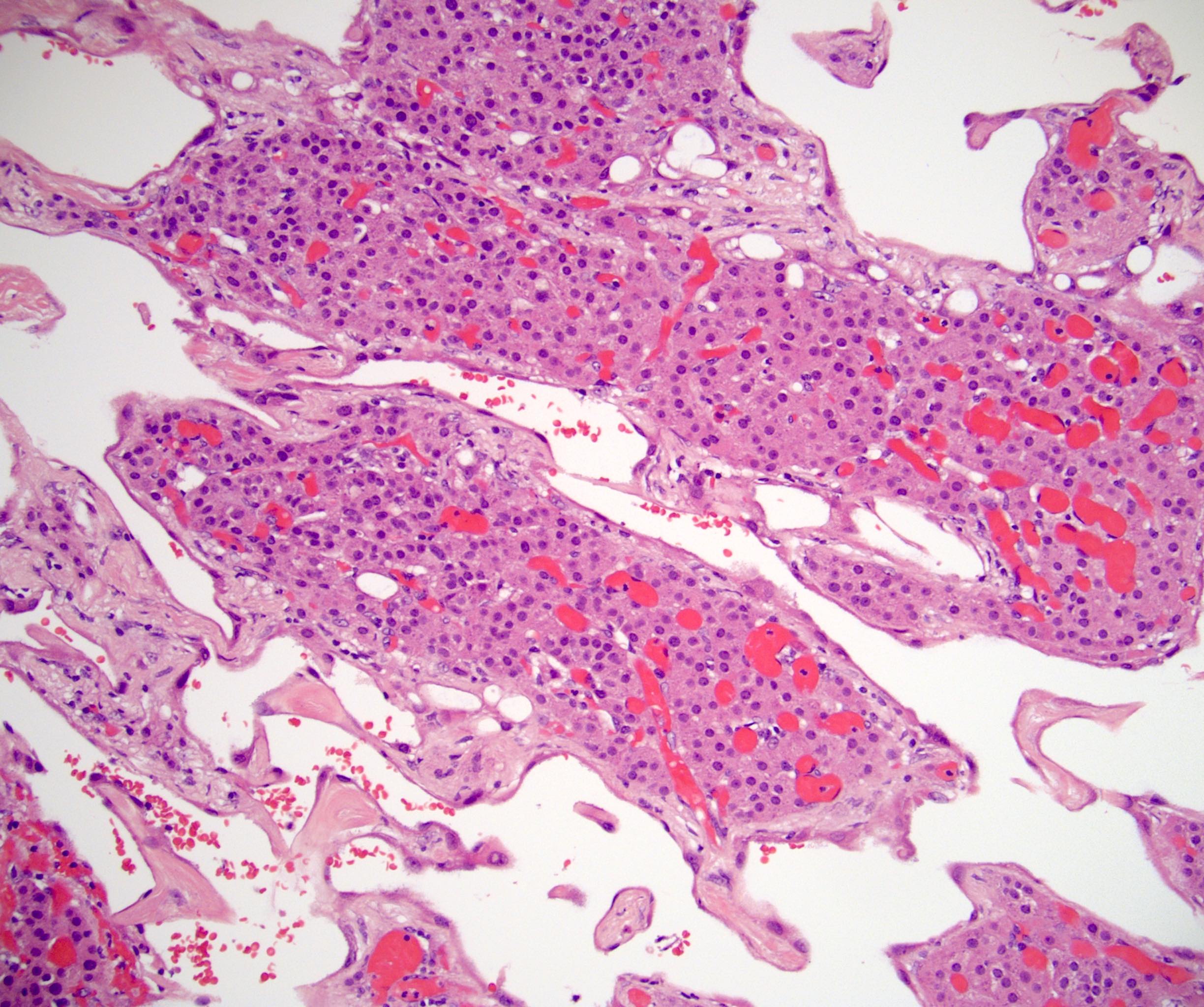
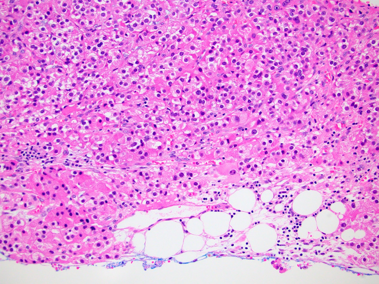
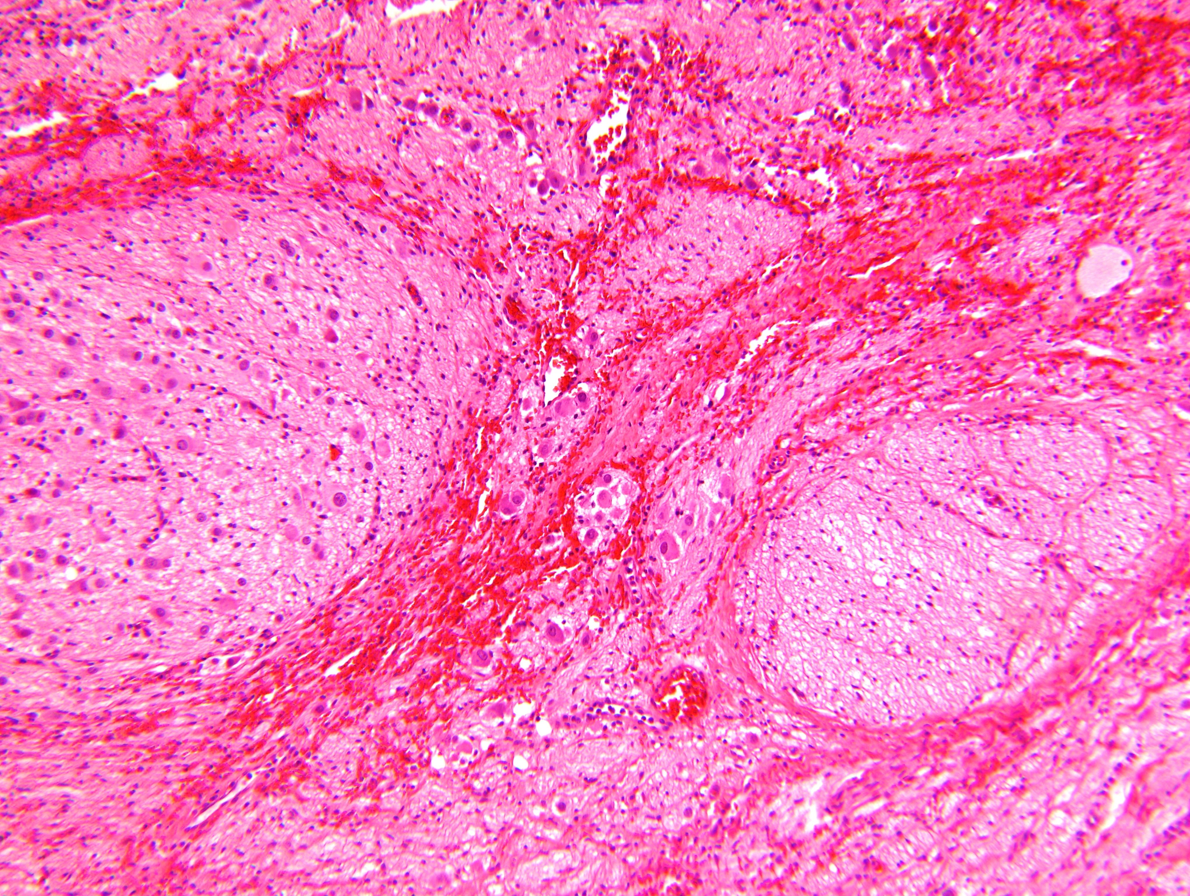
Entrapped cortex
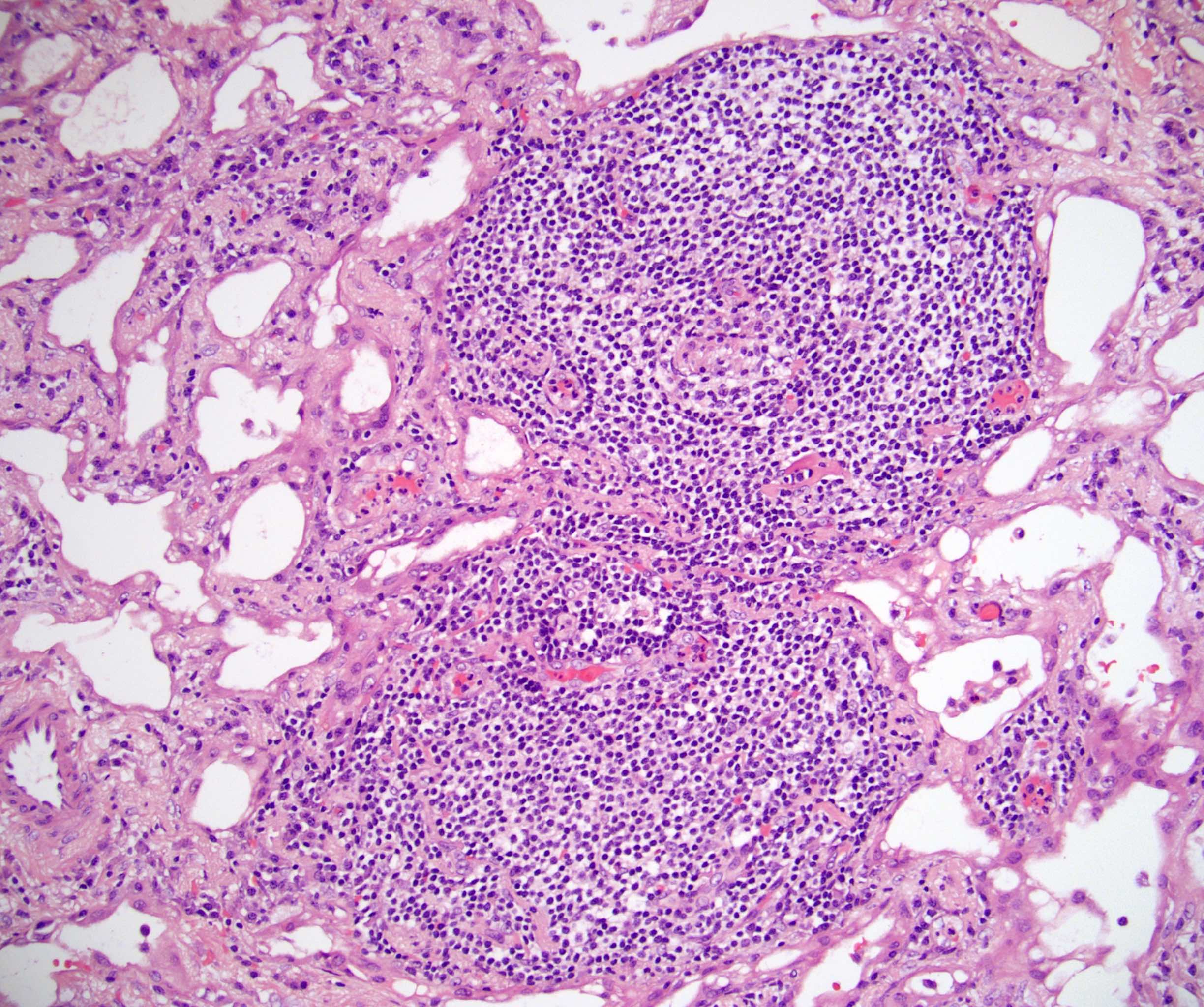
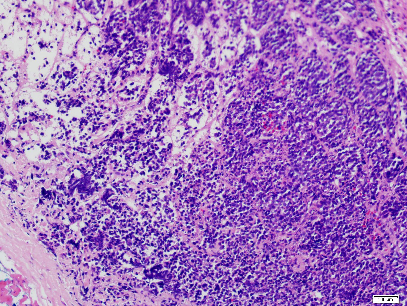
Lymphoid aggregates
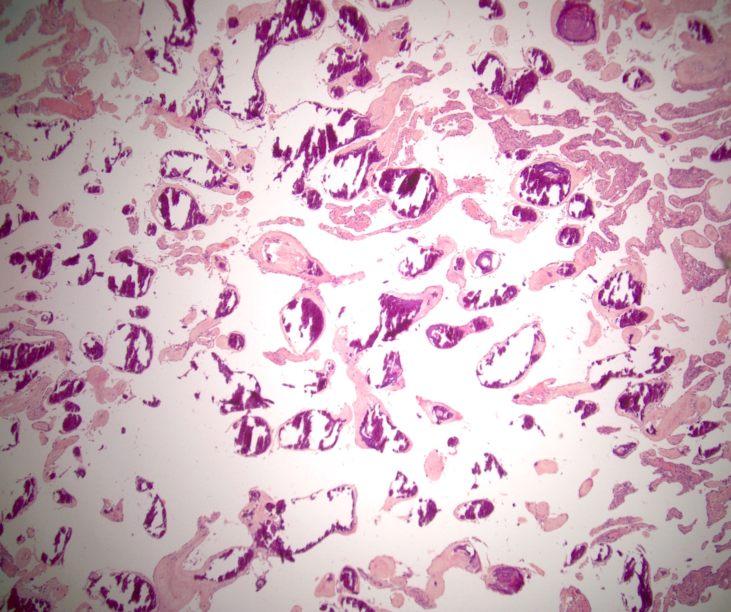
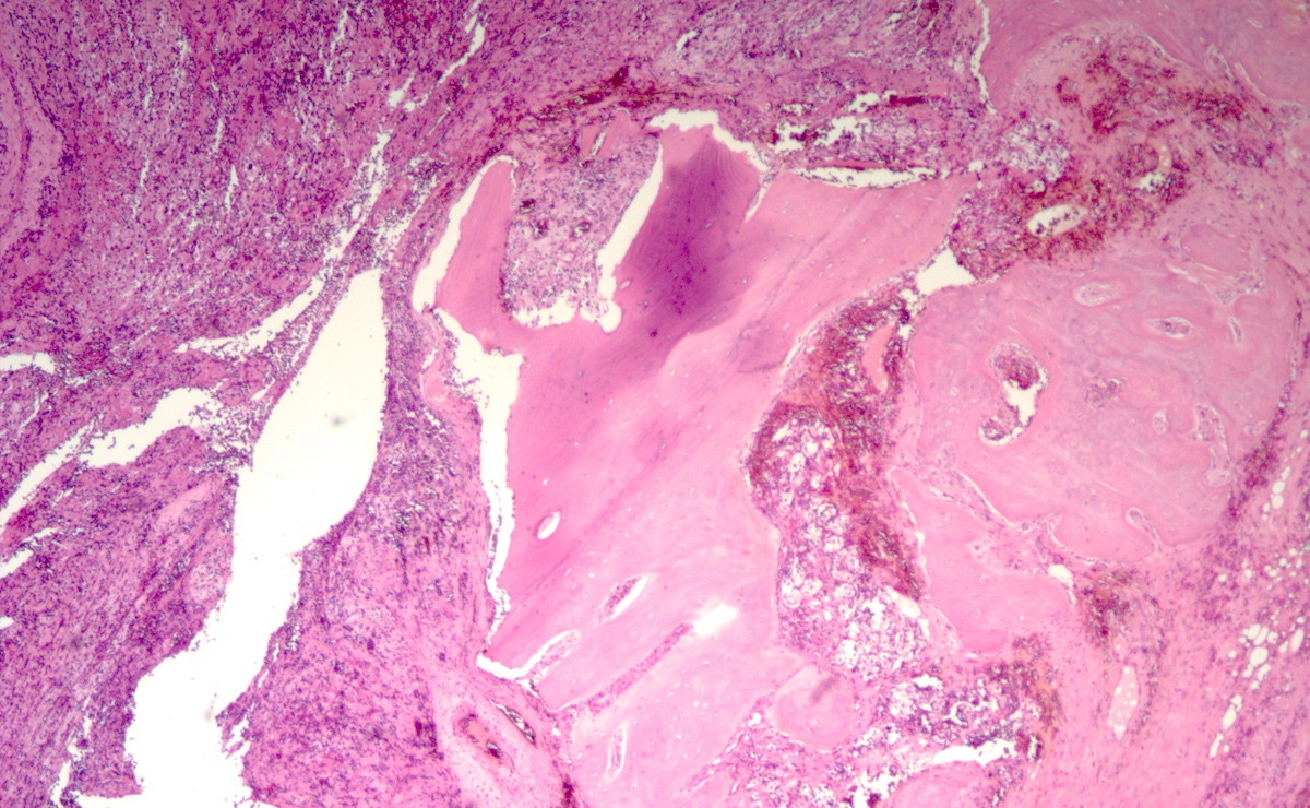
Calcification
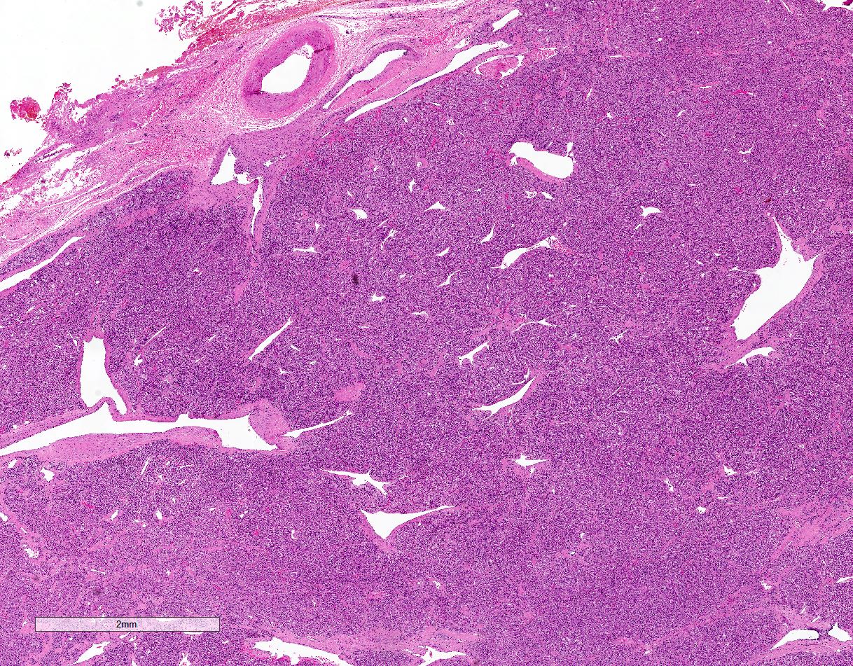
Infiltrative
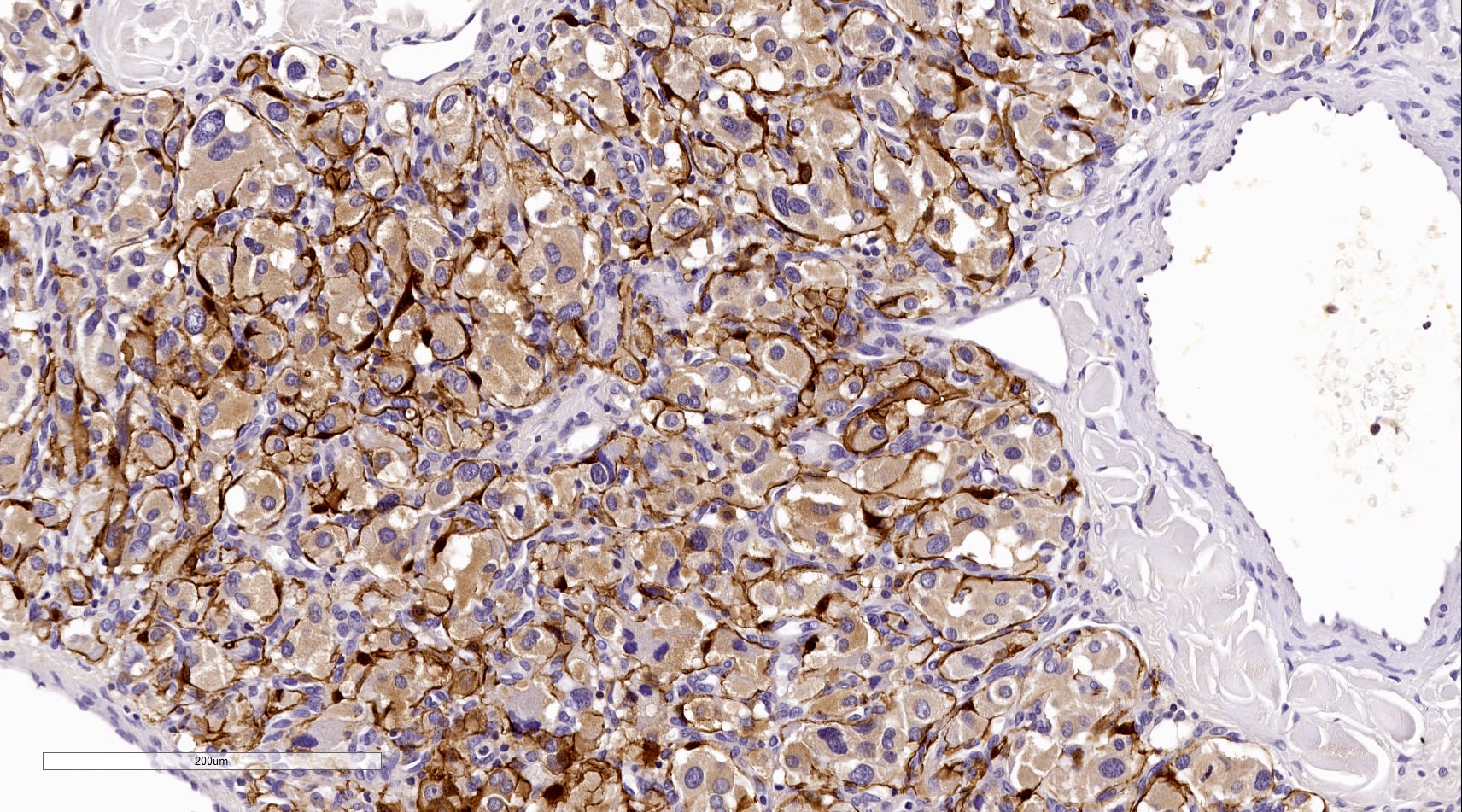
CK7
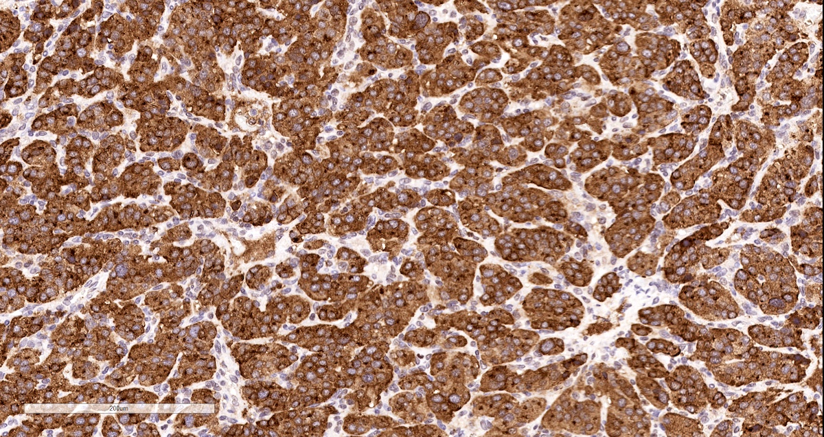
WT1
MelanA
Images hosted on other servers:
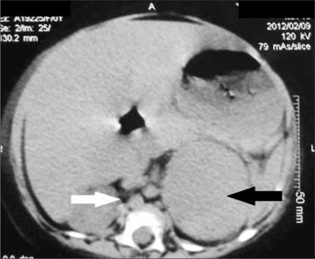
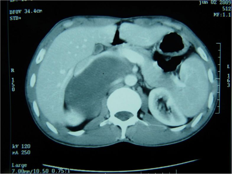
Homogeneous left adrenal mass with distinct borders

Left adrenal mass

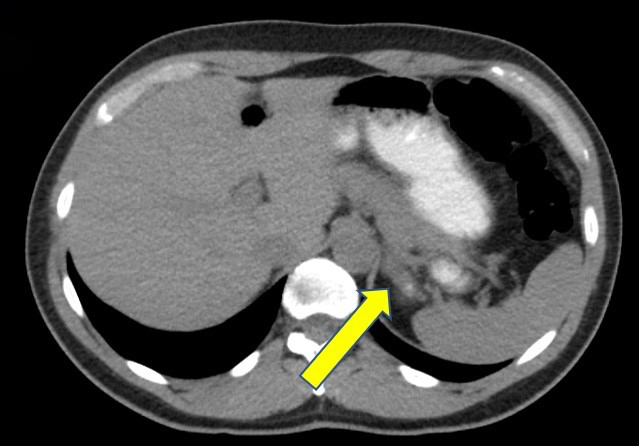
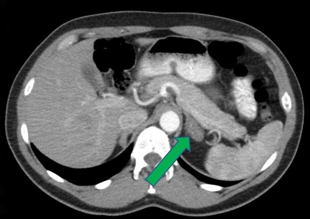
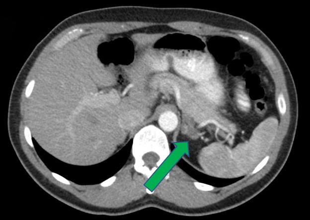
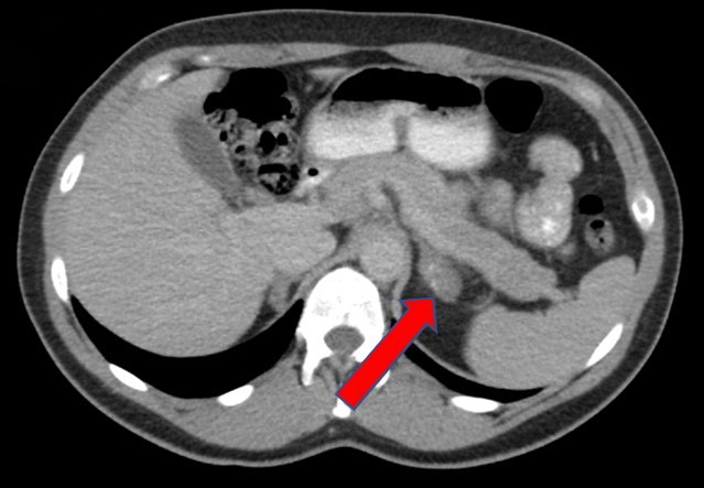
(A) Plain CT and (B) contrast CT show left suprarenal mass
Images hosted on other servers:

Moon facies and central obesity
Contributed by @Andrew_Fltv and @SueEPig on Twitter
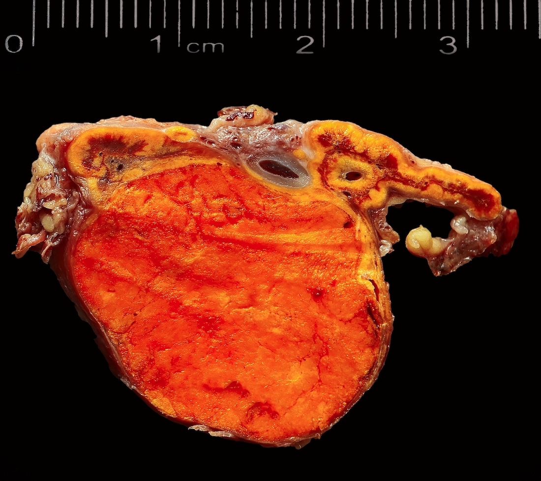
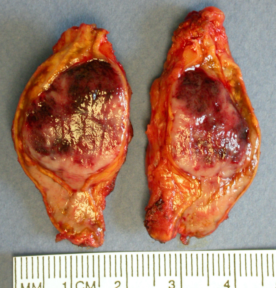
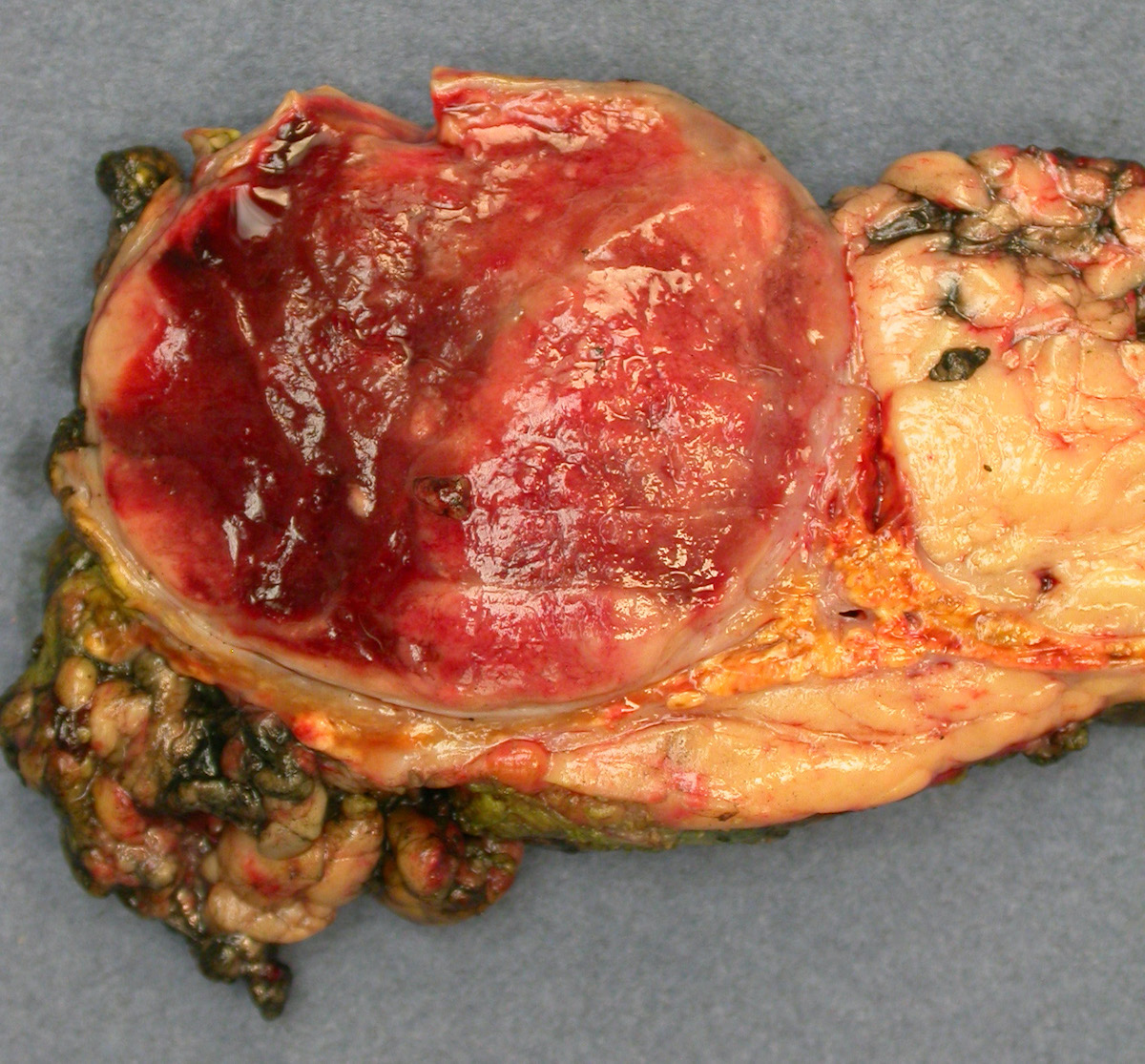
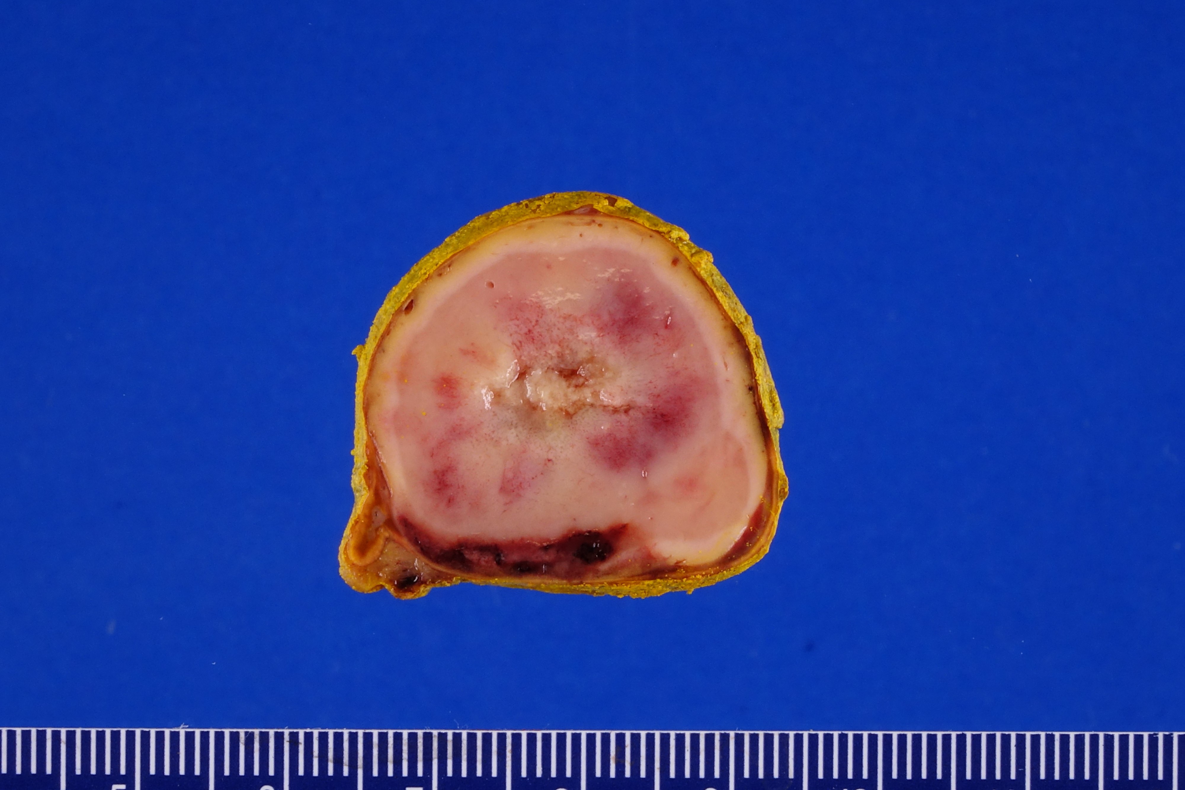
Adenoma
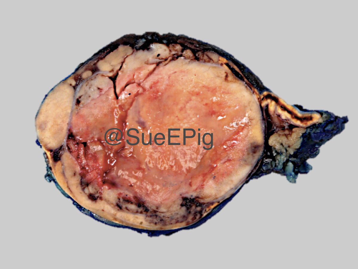
Adenoma
Images hosted on other servers:

External surface, during surgery

Intact capsule

Well circumscribed

1.3 cm left adrenal adenoma

3 x 3 cm exophytic adrenal mass
Contributed by Xiaoyin "Sara" Jiang, M.D., Debra Zynger, M.D., @Andrew_Fltv on Twitter and @SueEPig on Twitter
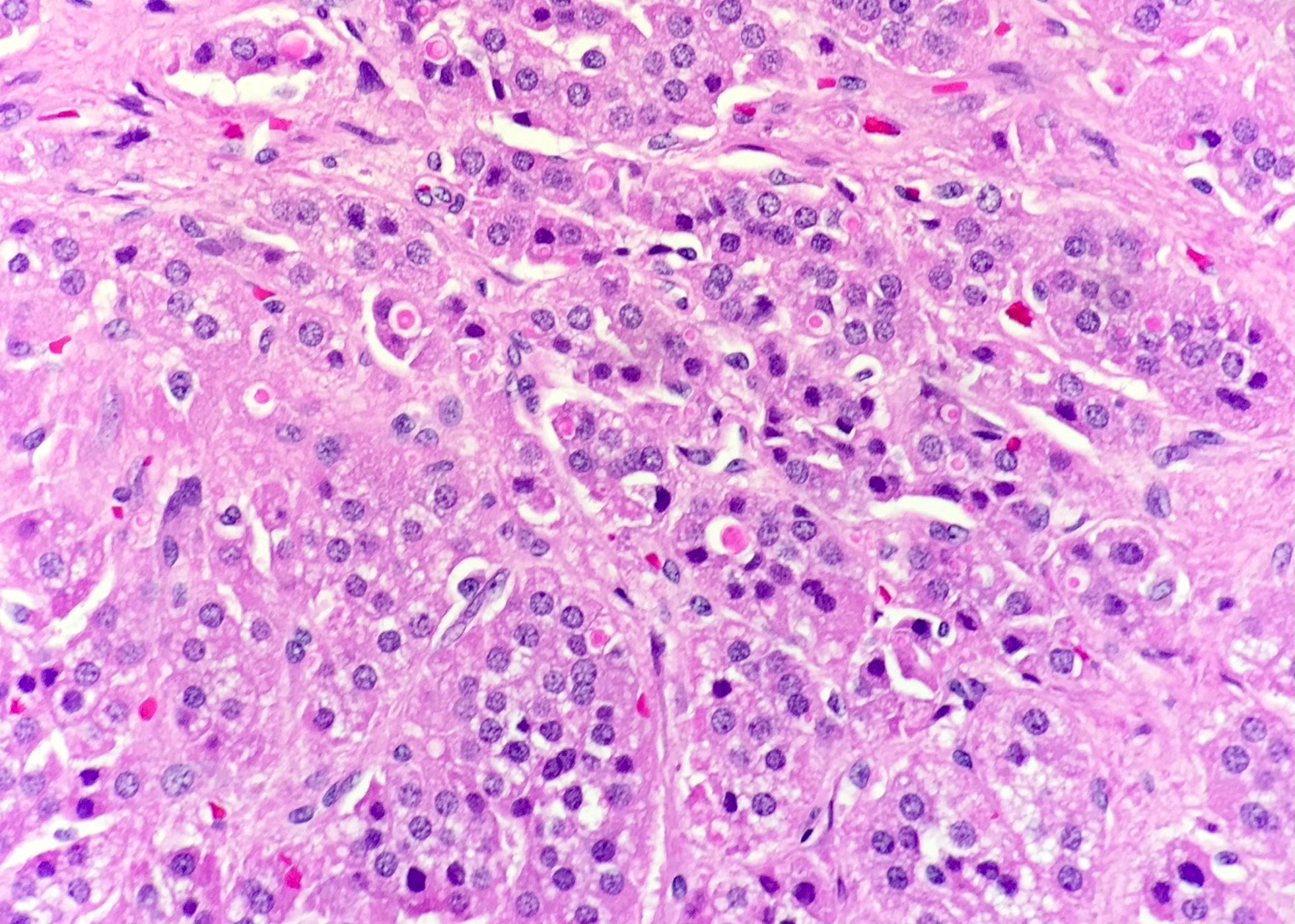
Spironolactone bodies with aldosteronoma
Adenoma with spironolactone bodies
Adrenal cortical aldosterone producing adenoma
Adenoma
Images hosted on other servers:

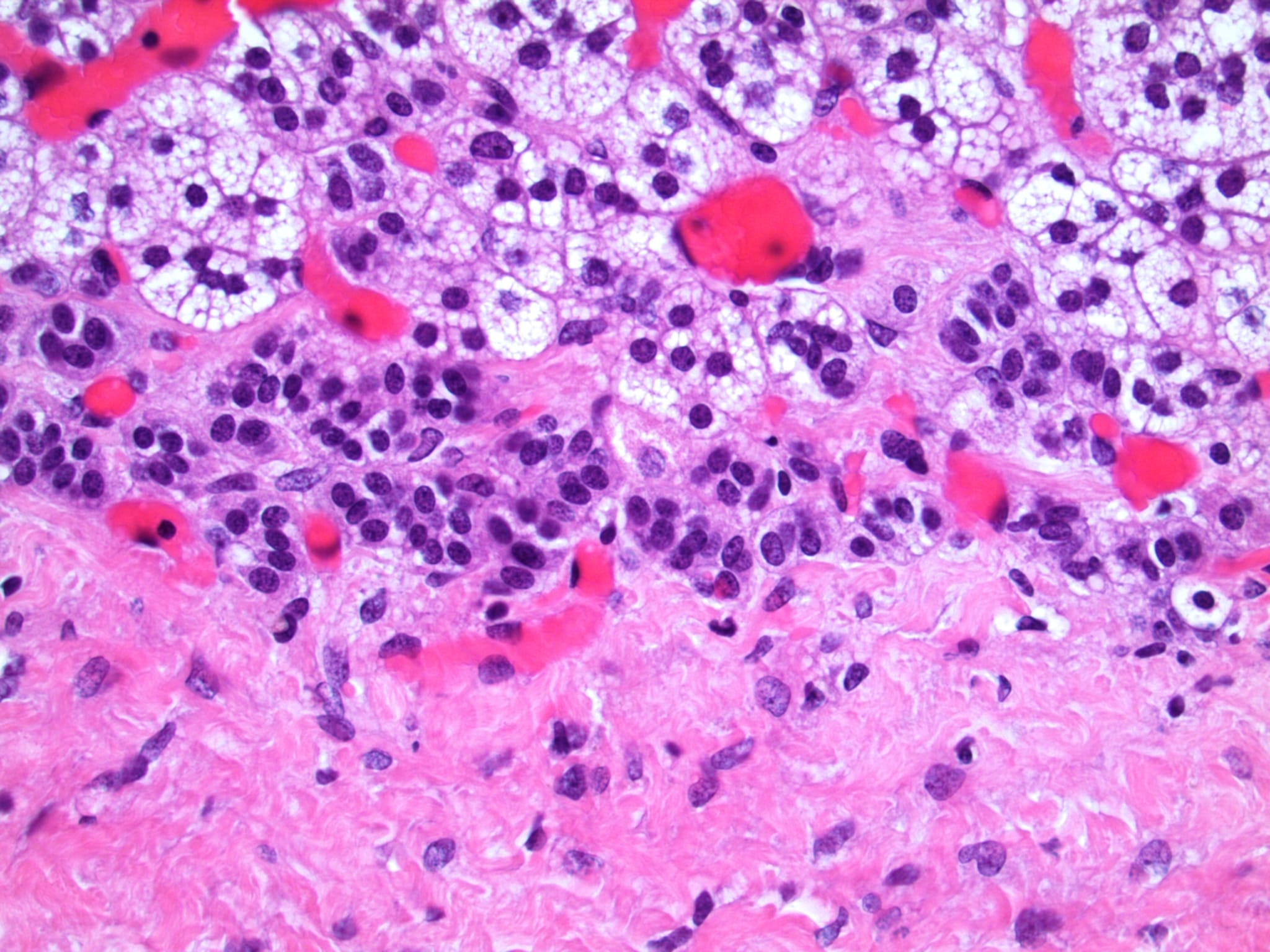
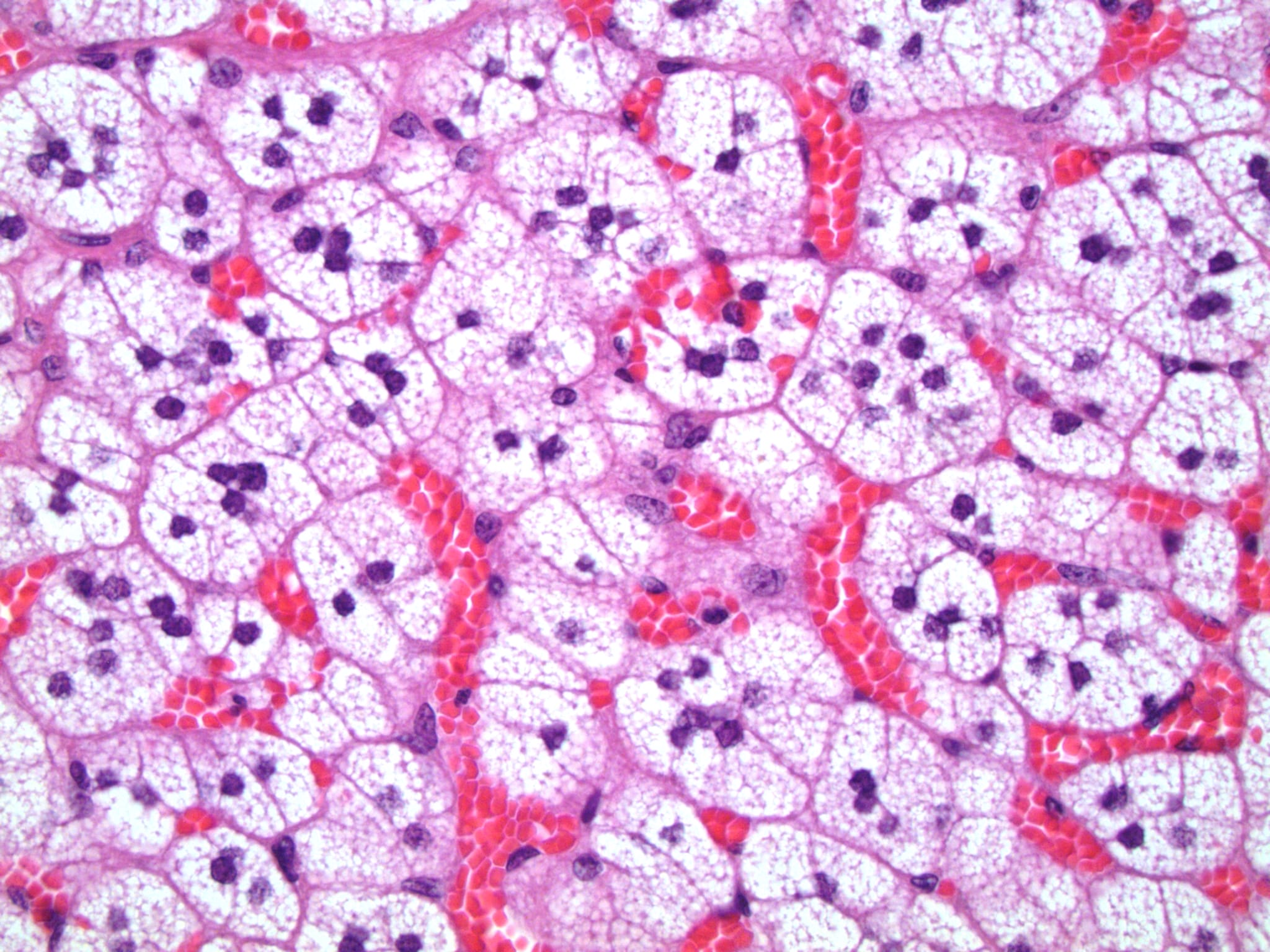
Multiple ACAs comprised of clear cells

Resembles normal adrenal fasciculata

Clear cells

Low power

S100- basement cells
Contributed by Edward Calomeni, B.S.
Spironolactone bodies
Images hosted on other servers:

Diagnostic algorithm for ACC stratification
Images hosted on other servers:

Heterogeneous enhancing mass with lymphadenopathy

CT imaging of advanced ACC
Contributed by Debra Zynger, M.D.
Organ confined, ≤ 5 cm (pT1)
Organ confined, > 5 cm (pT2)
Extra-adrenal invasion (pT3)
Liver invasion (pT4)
Images hosted on other servers:

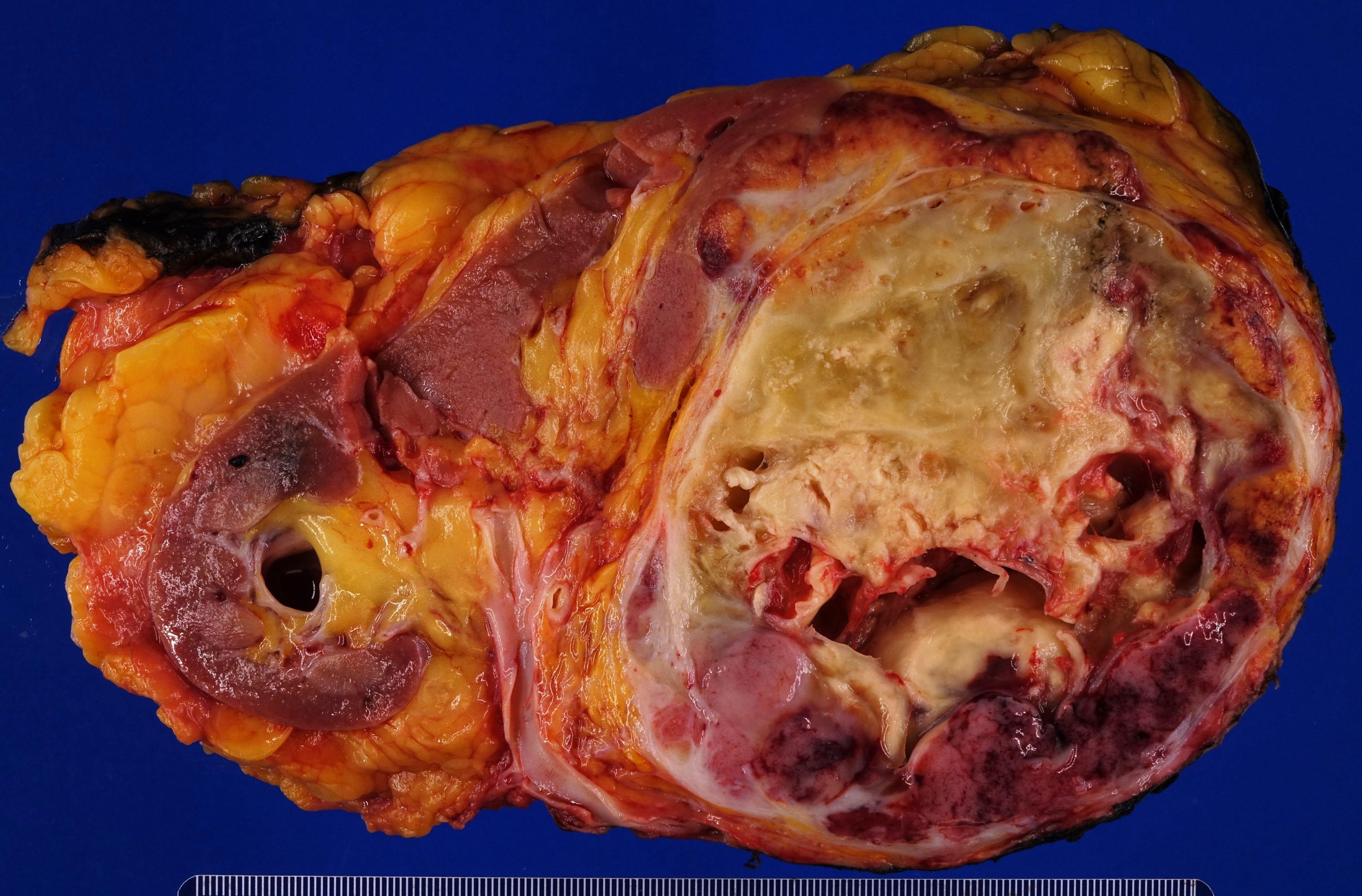
250 g, 10 cm tumor

Large tumor compressing kidney

Large tumor dwarfing kidney
Contributed by Maria Tretiakova, M.D., Ph.D.
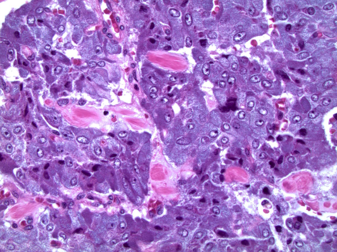
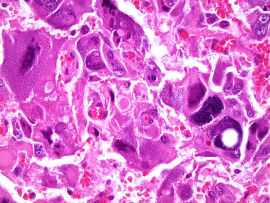
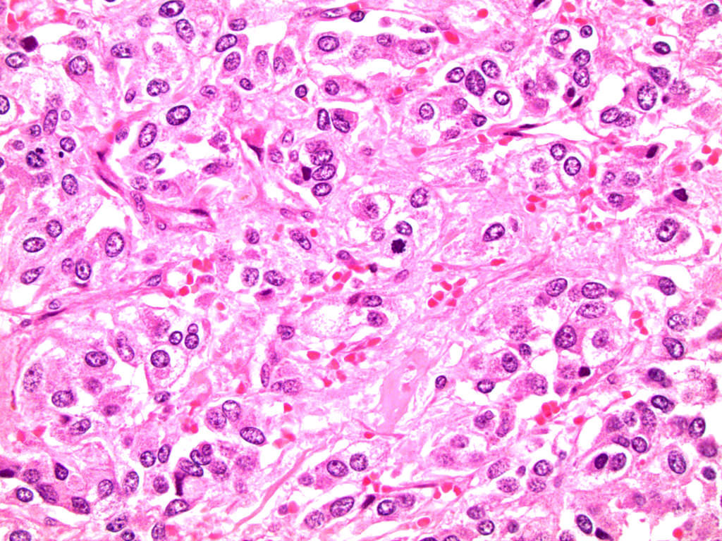
High nuclear grade
Pleomorphic ACC
Necrosis
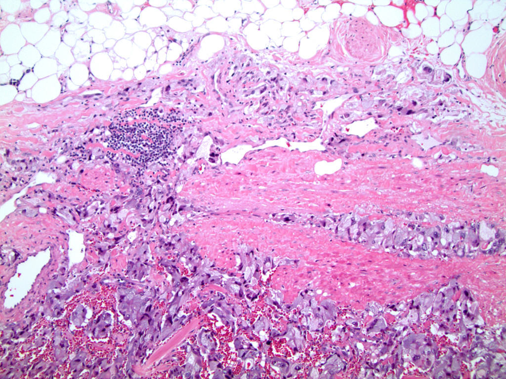
Capsule / fat invasion
Positive margin
Reticulin loss
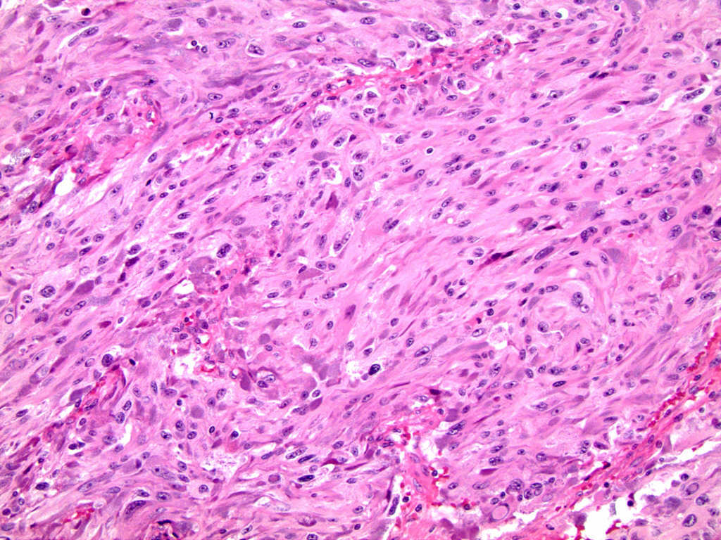
Diffuse growth
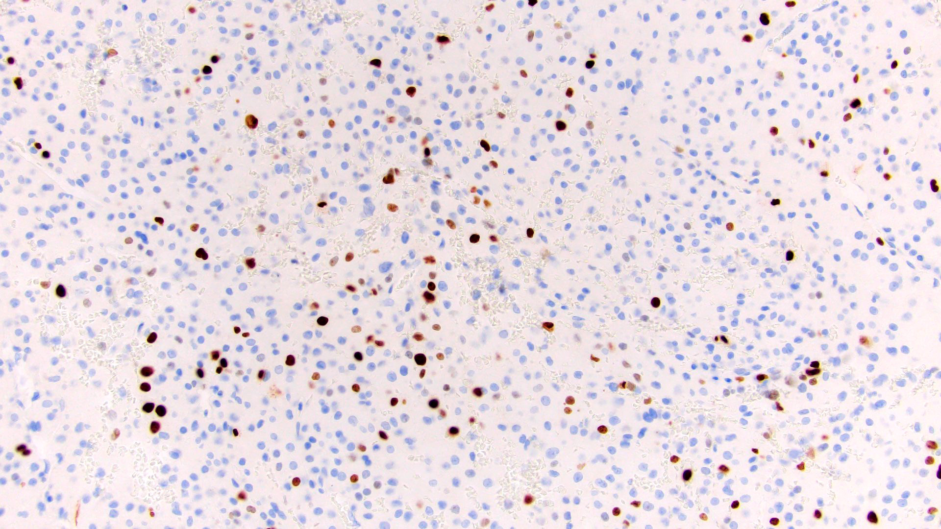
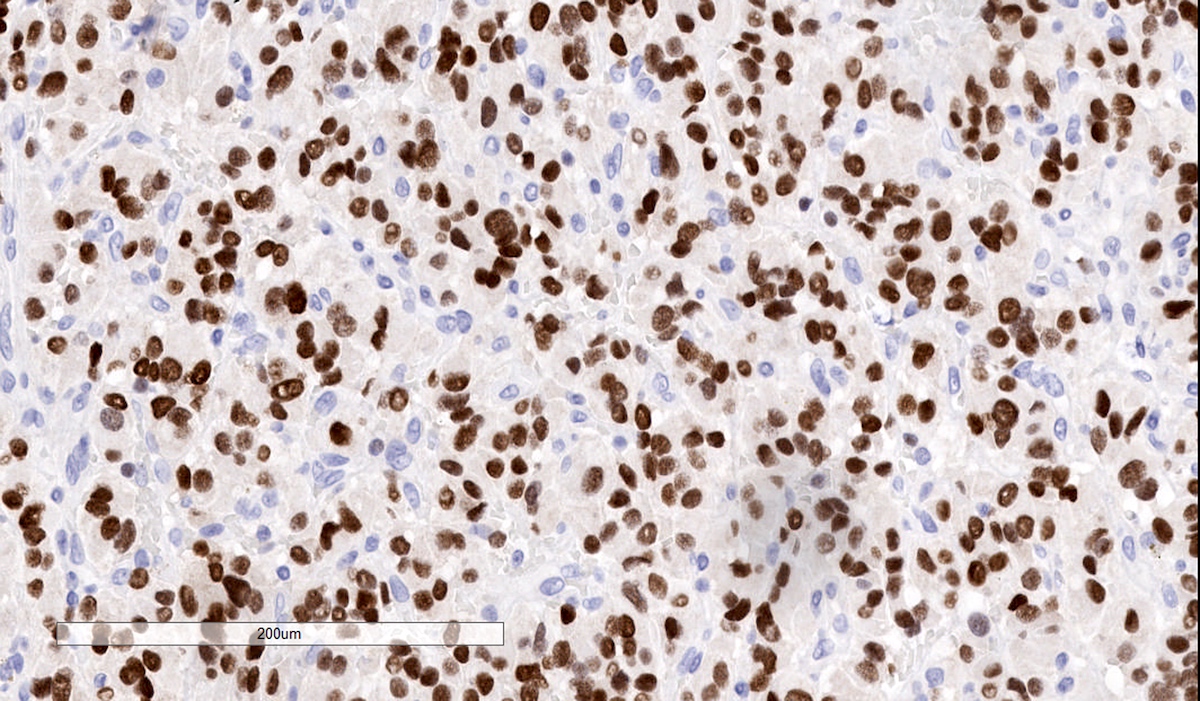
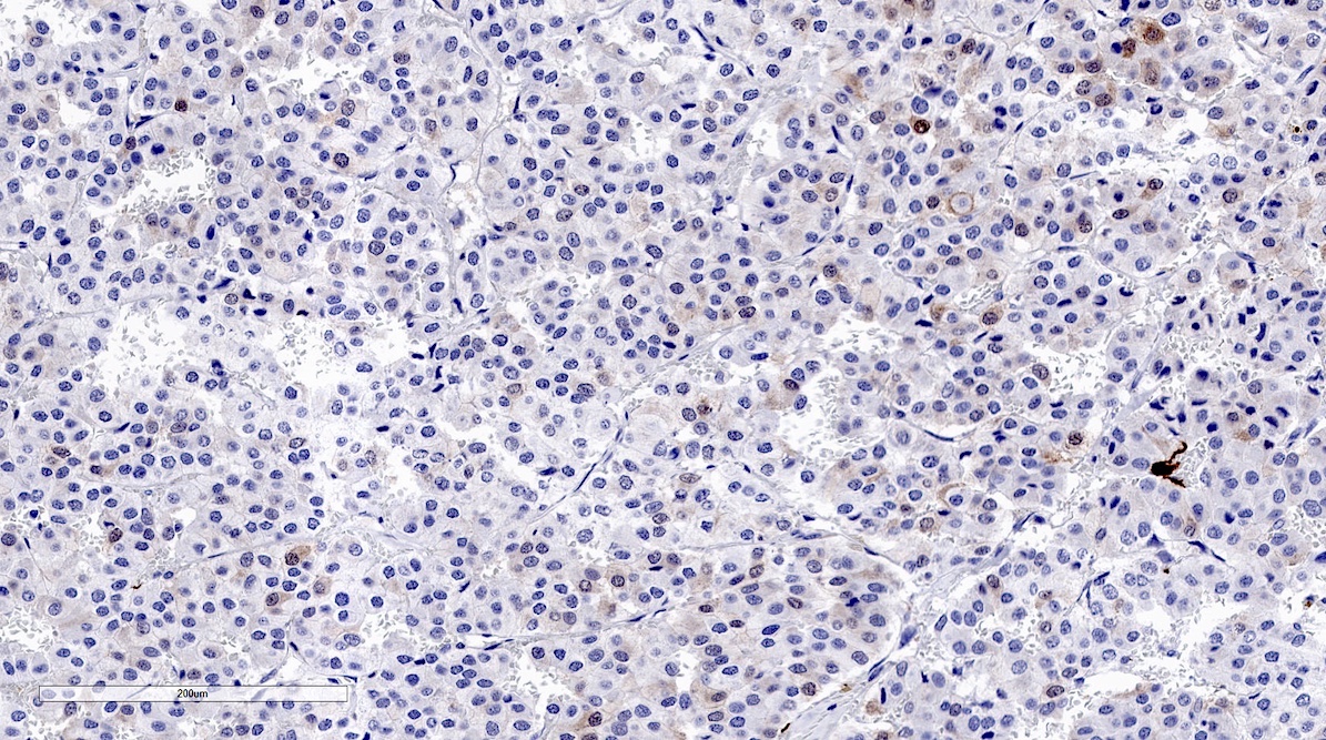
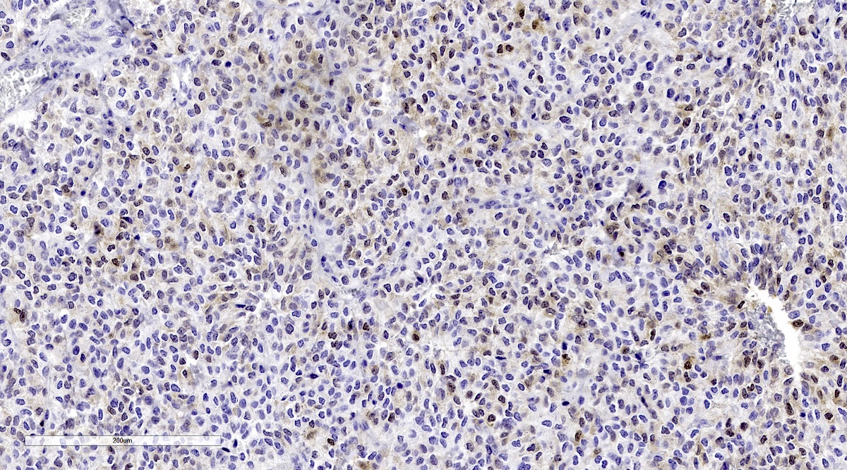
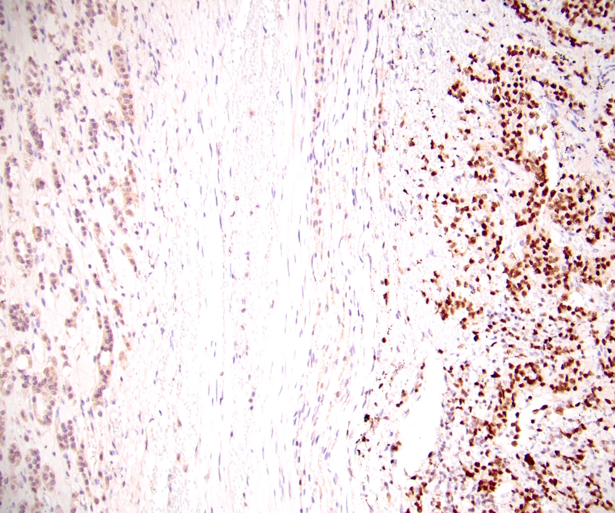
Ki67
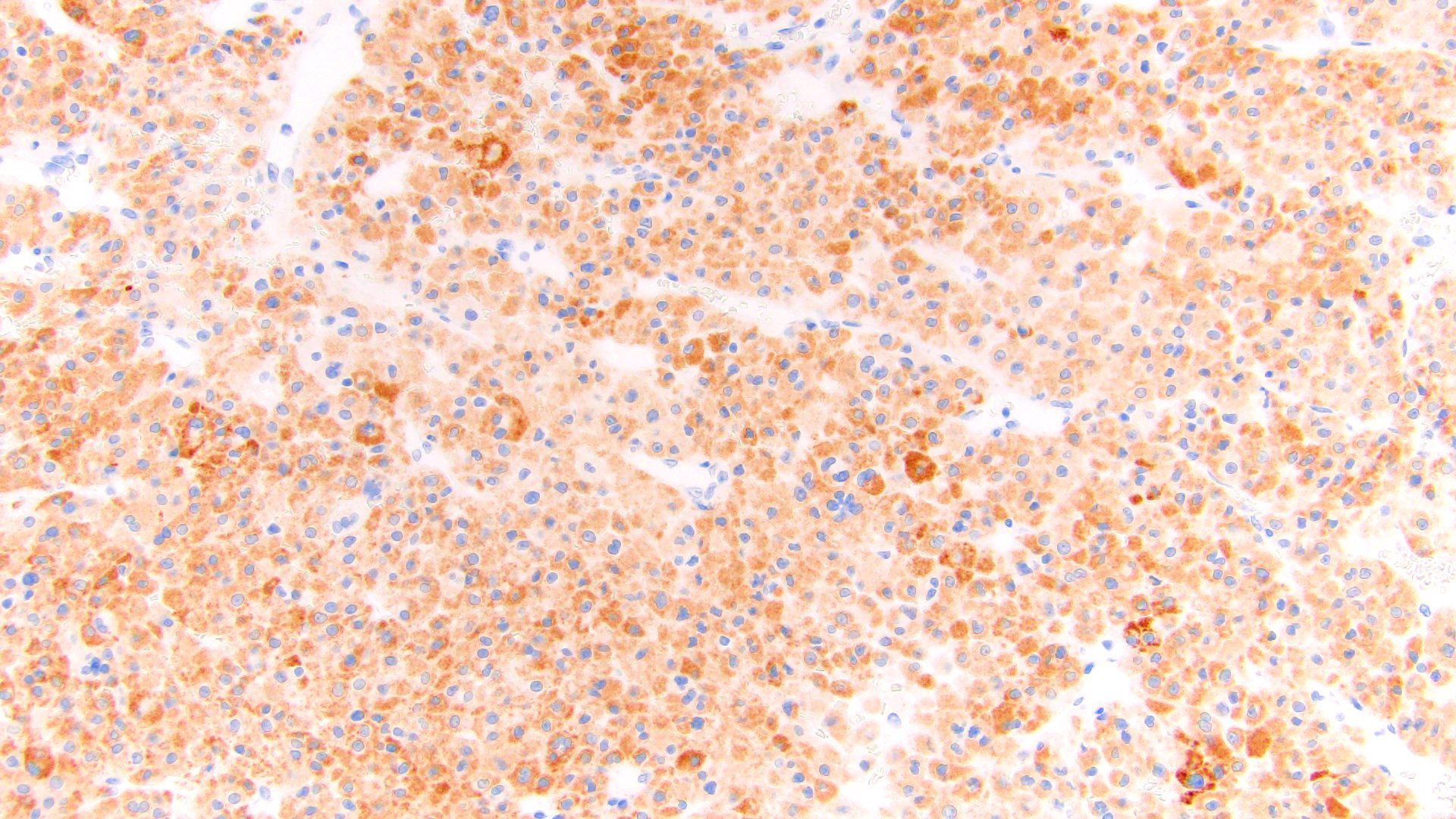
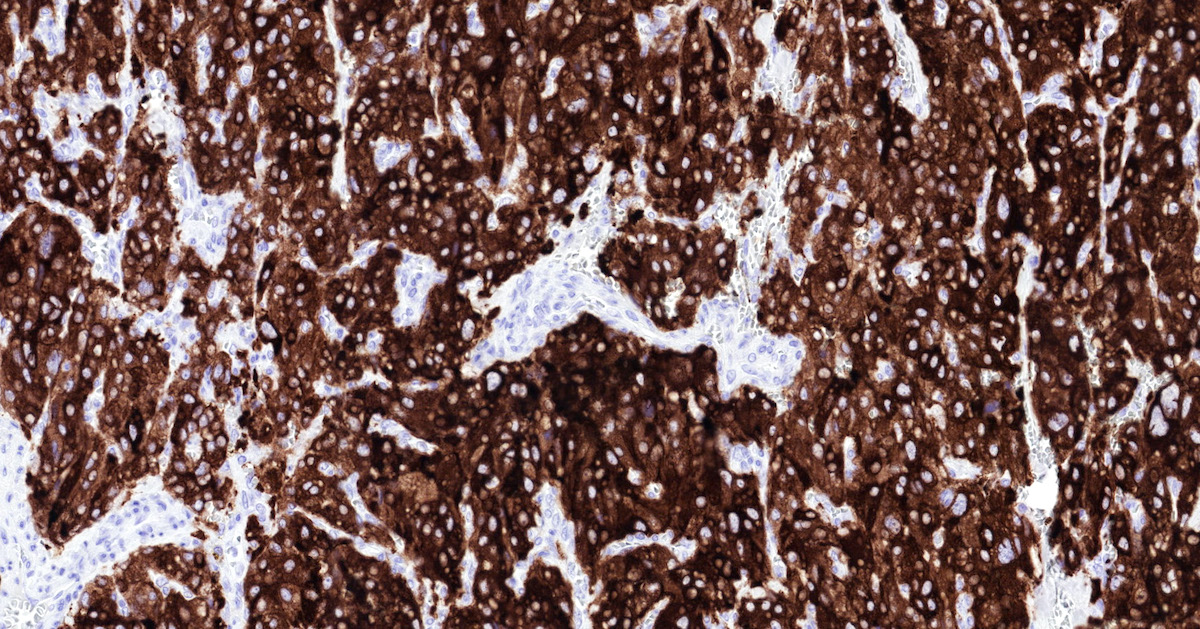
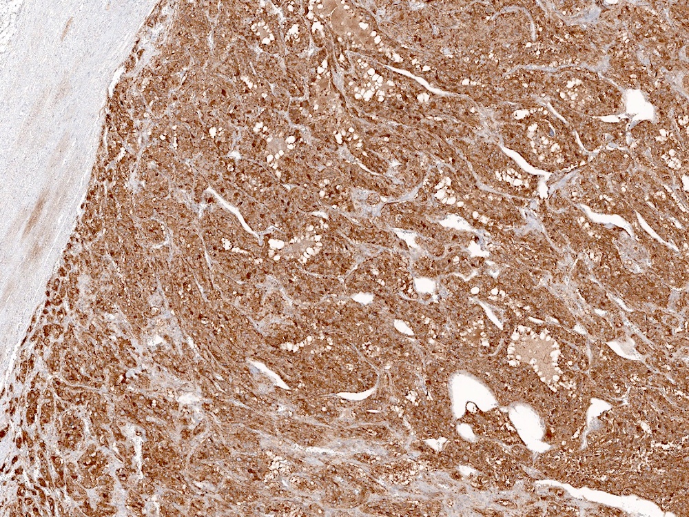
MelanA
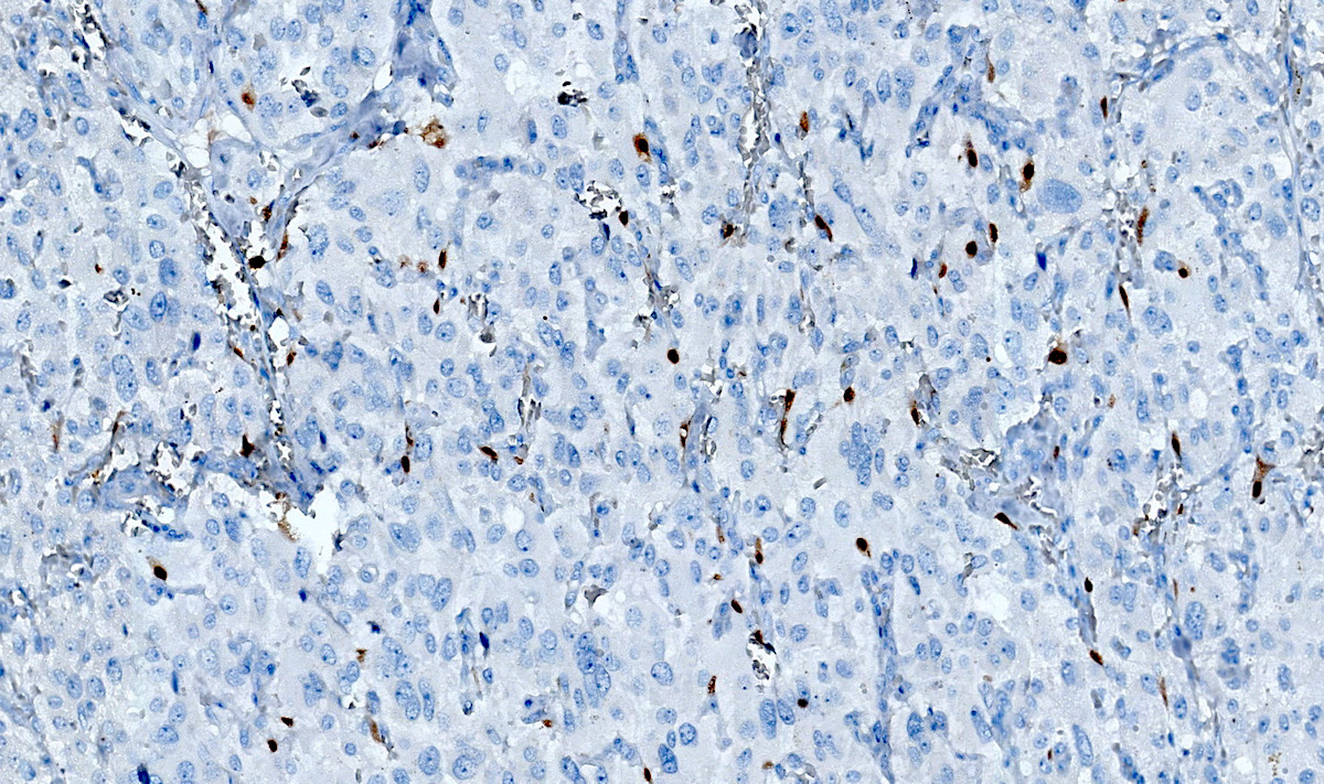
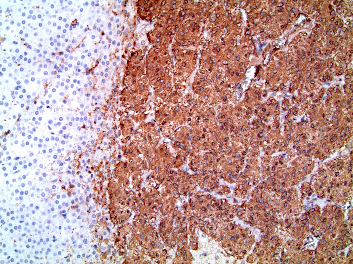
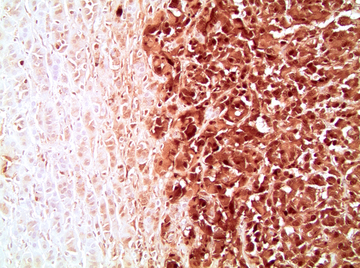
Inhibin
Contributed by Debra Zynger, M.D.
High mitotic rate
Lymphovascular invasion
Extra-adrenal adipose invasion
Liver involvement
Images hosted on other servers:

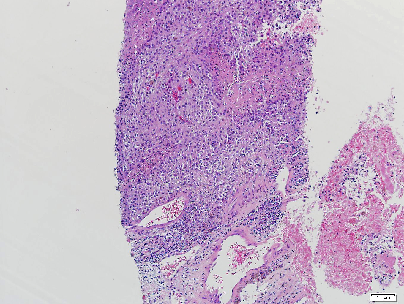
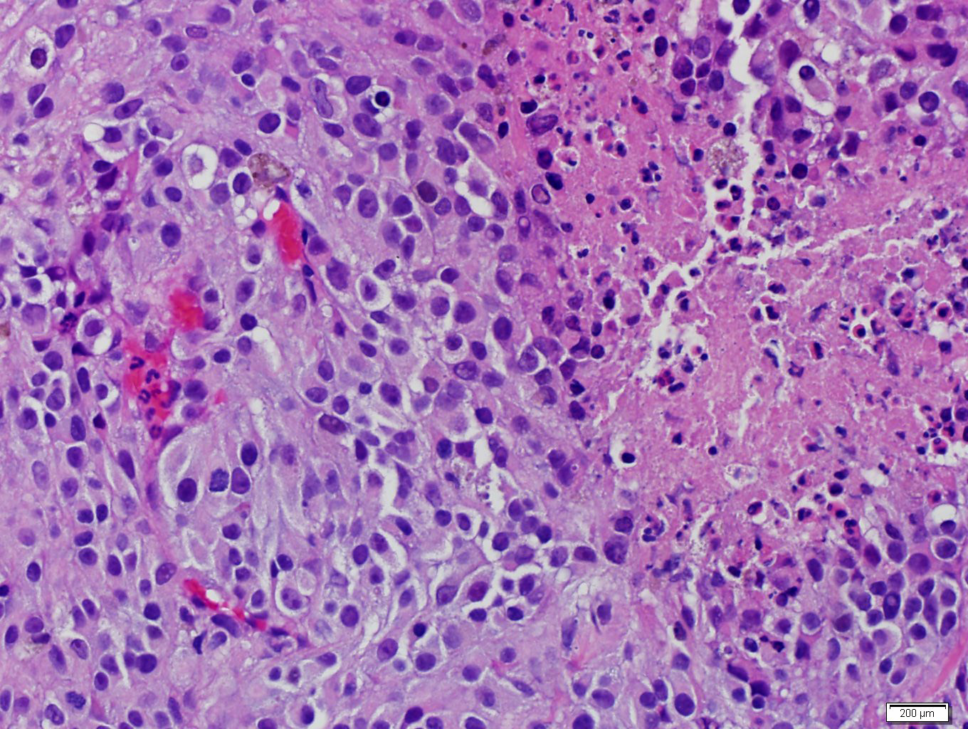
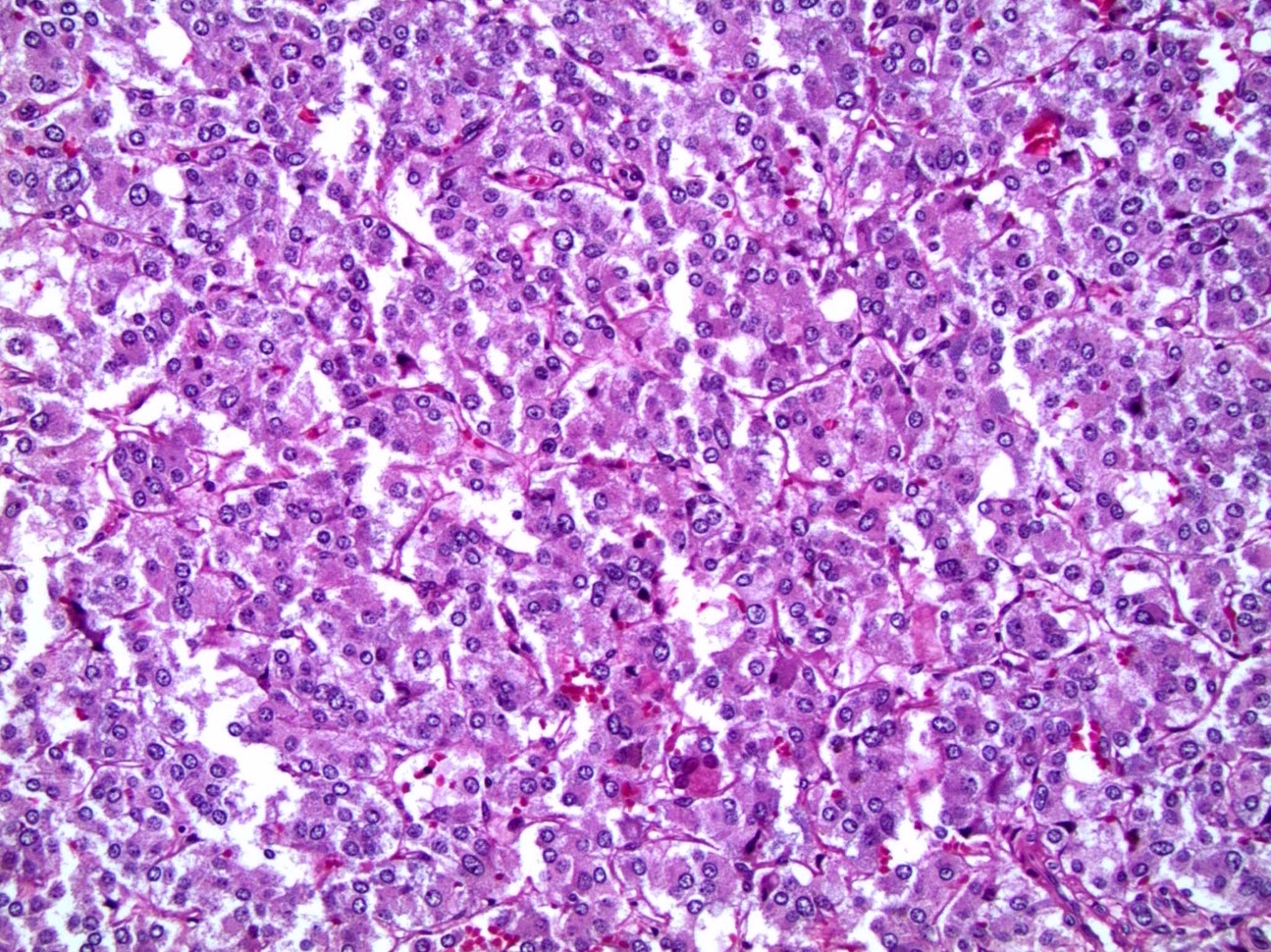
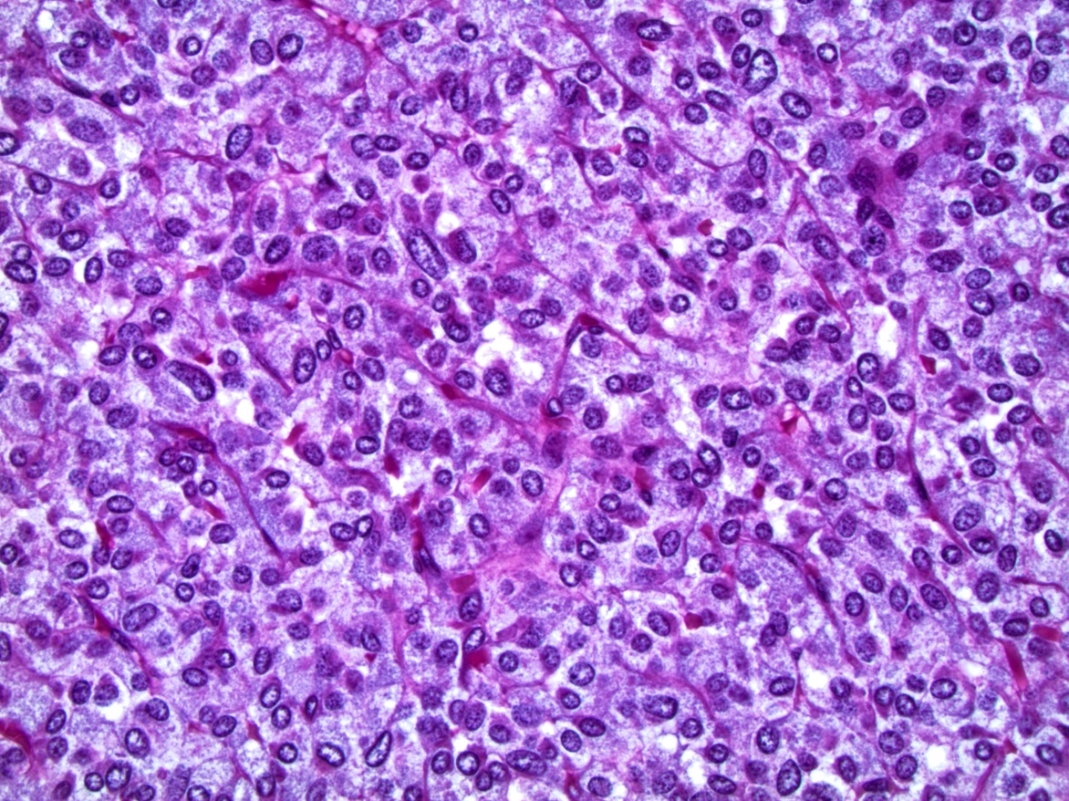
Discohesive, round to polygonal tumor cells
Images hosted on other servers:

Hallmark molecular features
Contributed by Debra L. Zynger, M.D.
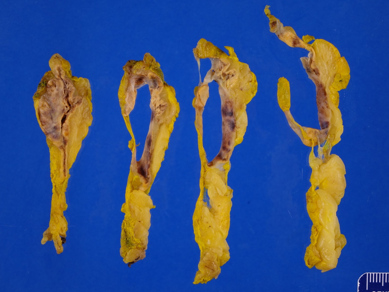
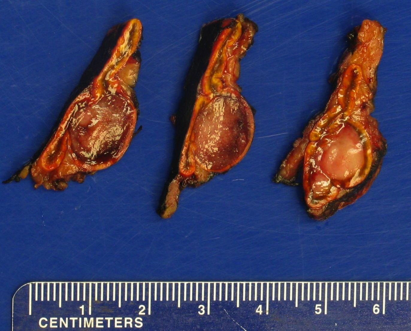
Partial nephrectomy
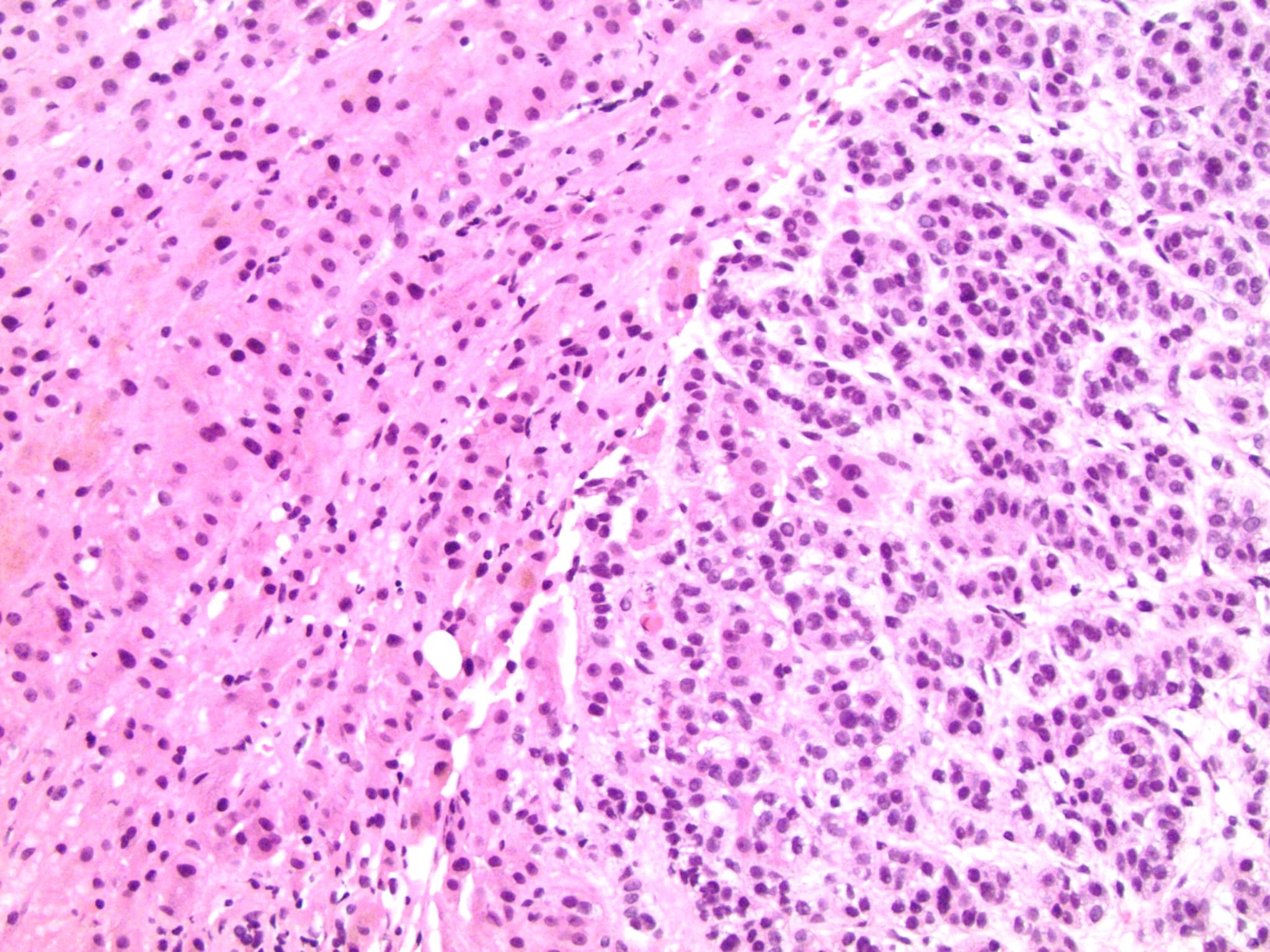
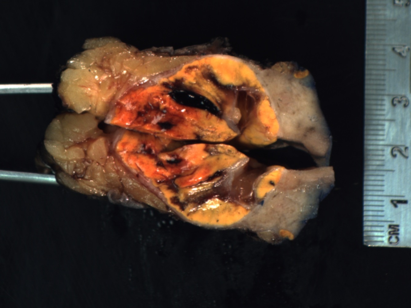
Contributed by Yuto Yamazaki, M.D., Ph.D., Hironobu Sasano, M.D., Ph.D., Debra L. Zynger, M.D. and Sean R. Williamson, M.D.
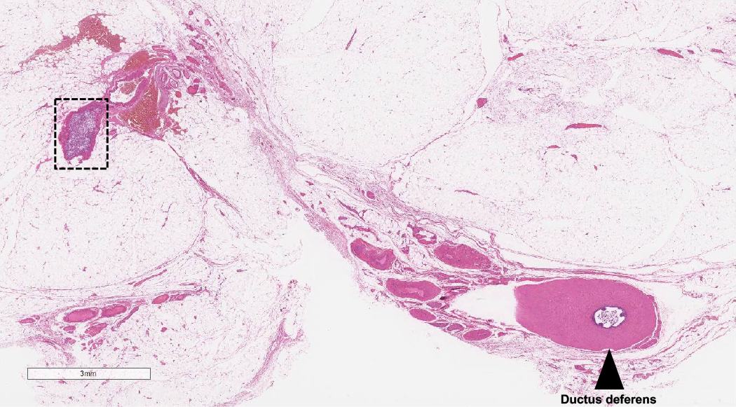
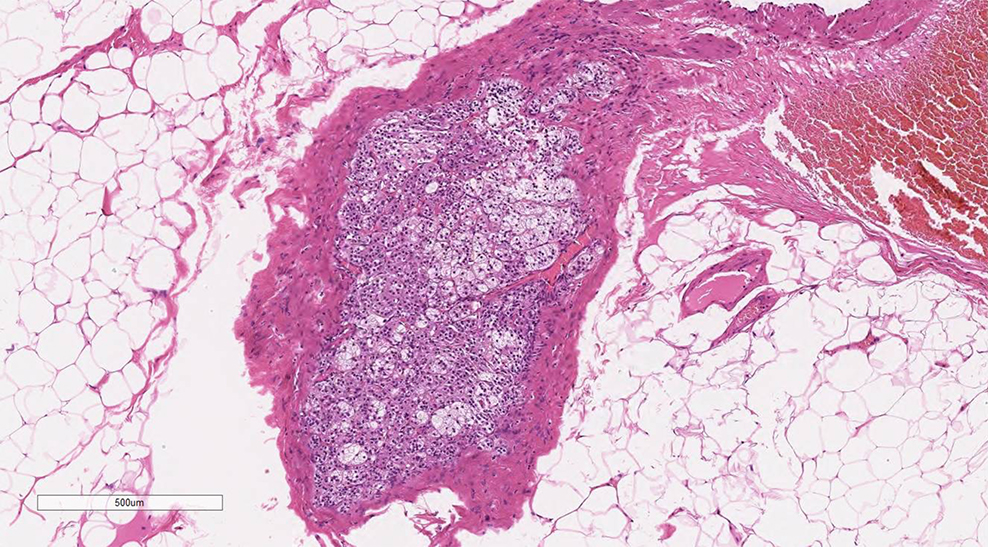
Spermatic cord adrenal rest and SF1
Spermatic cord adrenal rests
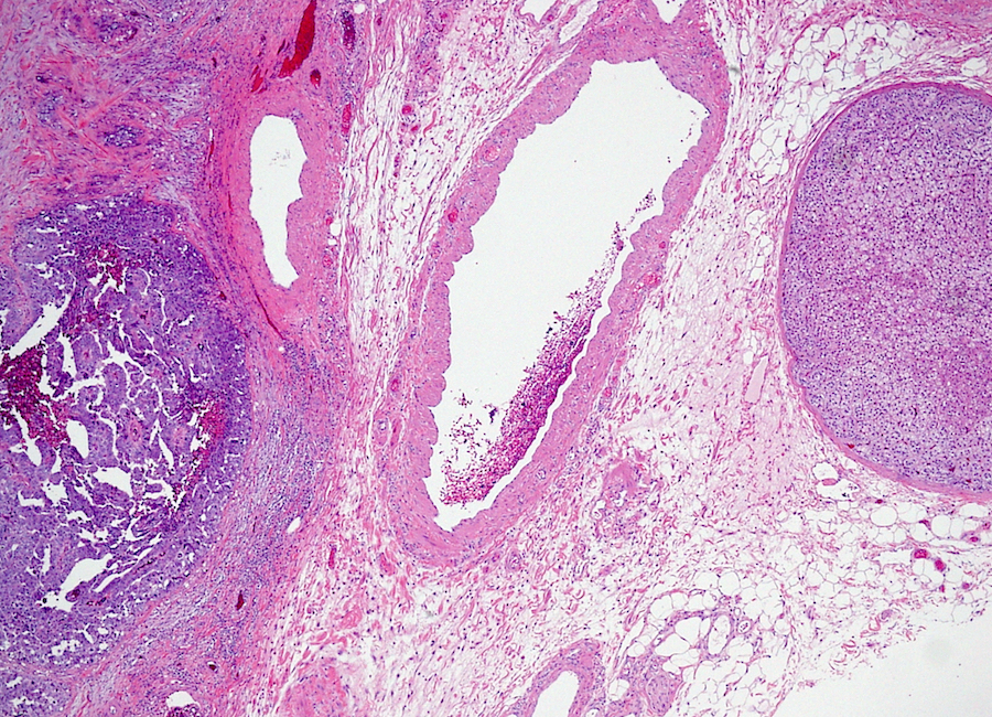
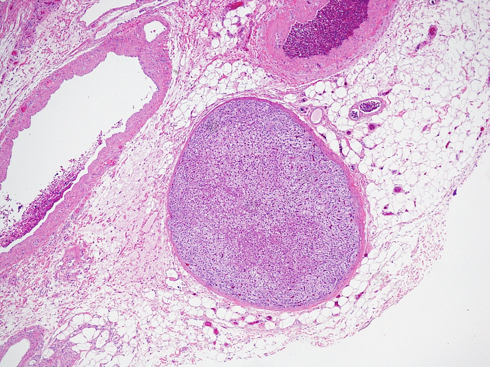
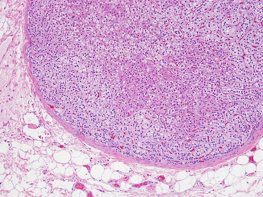
Paratesticular adrenal cortical rest
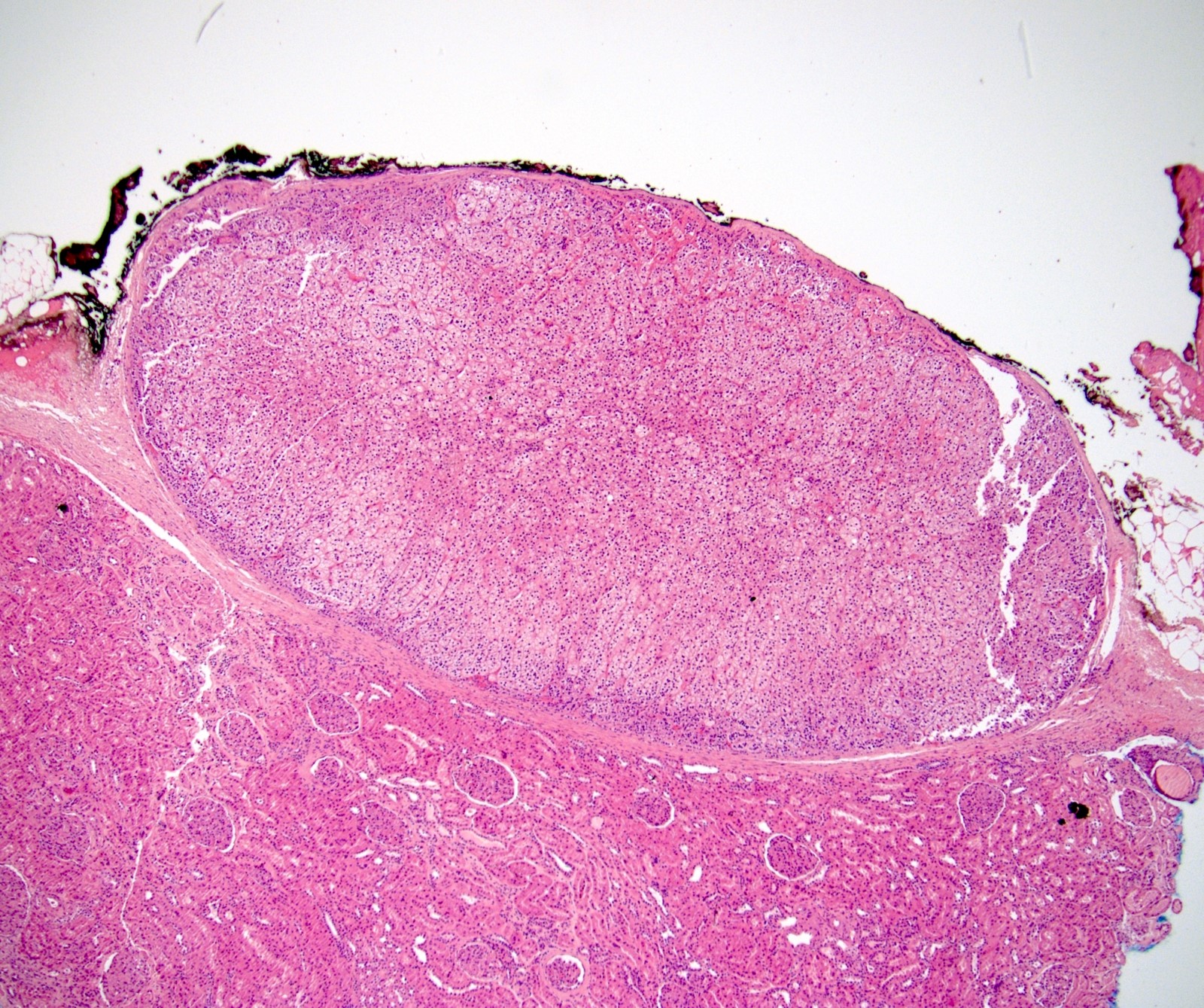
Intrarenal adrenals
Images hosted on other servers:

Human adrenal gland

Adrenal gland zones and products

Paraganglia, anatomic location
Images hosted on other servers:

Longitudinal ultrasound, infant

CT, normal, teen

CT of normal adrenal glands

MRI of normal adrenal glands
Images hosted on other servers:

Normal adult glands, whole

Normal adult gland, sectioned

Right adrenal gland in situ
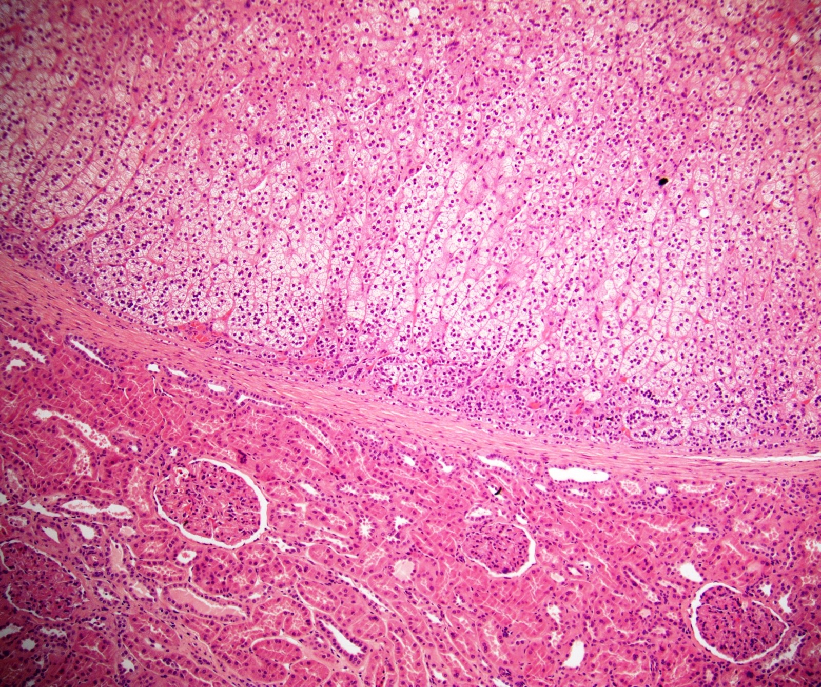
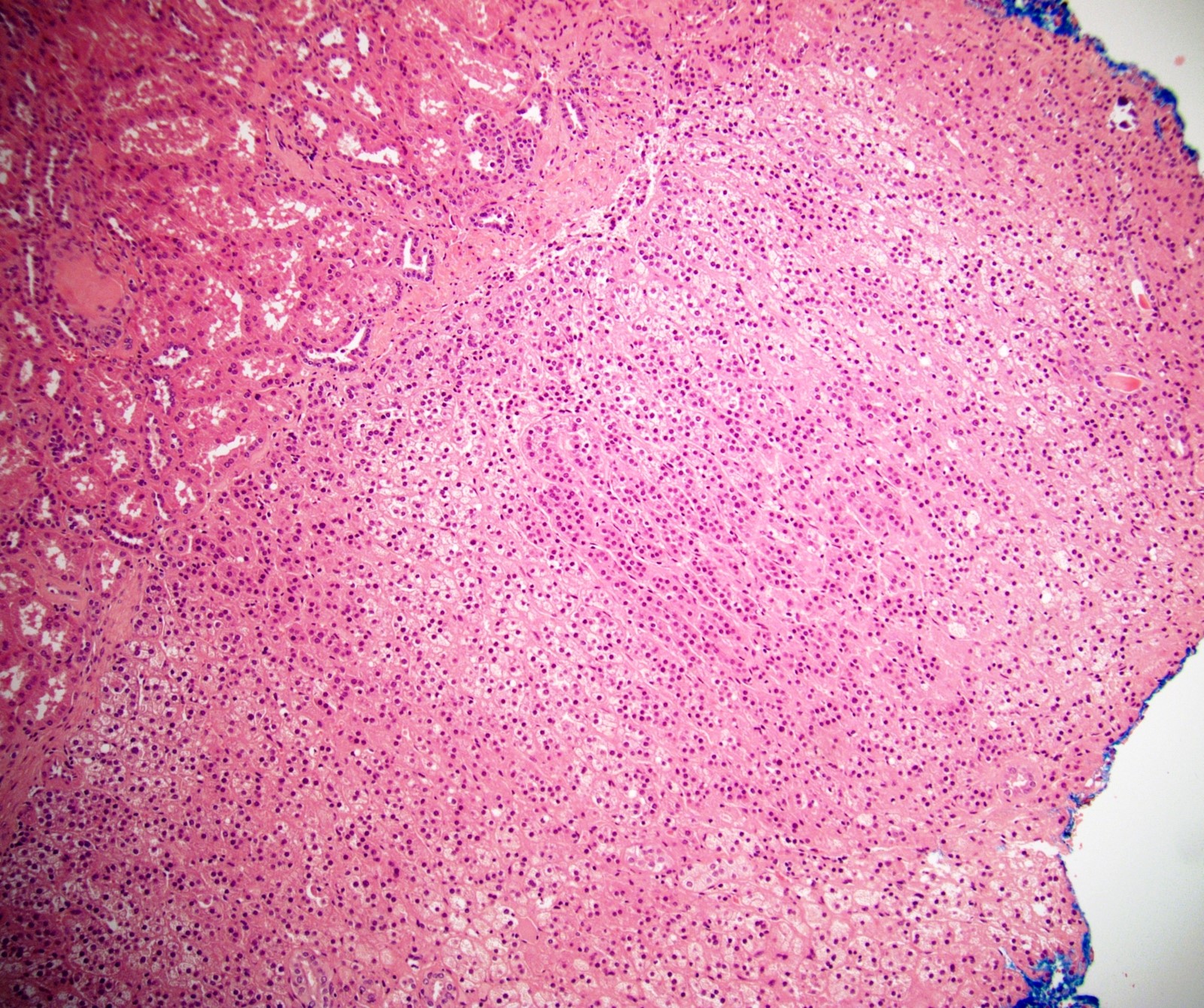
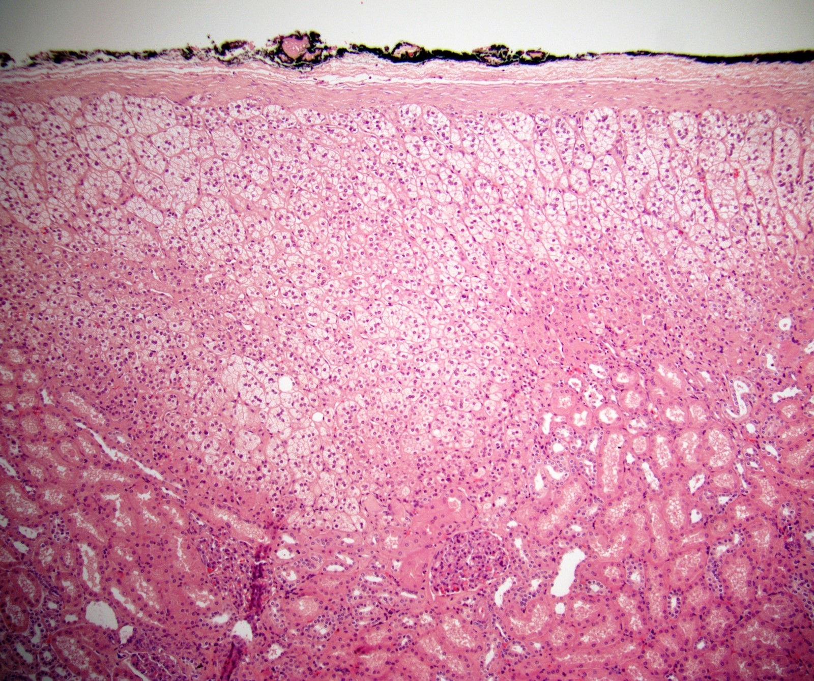
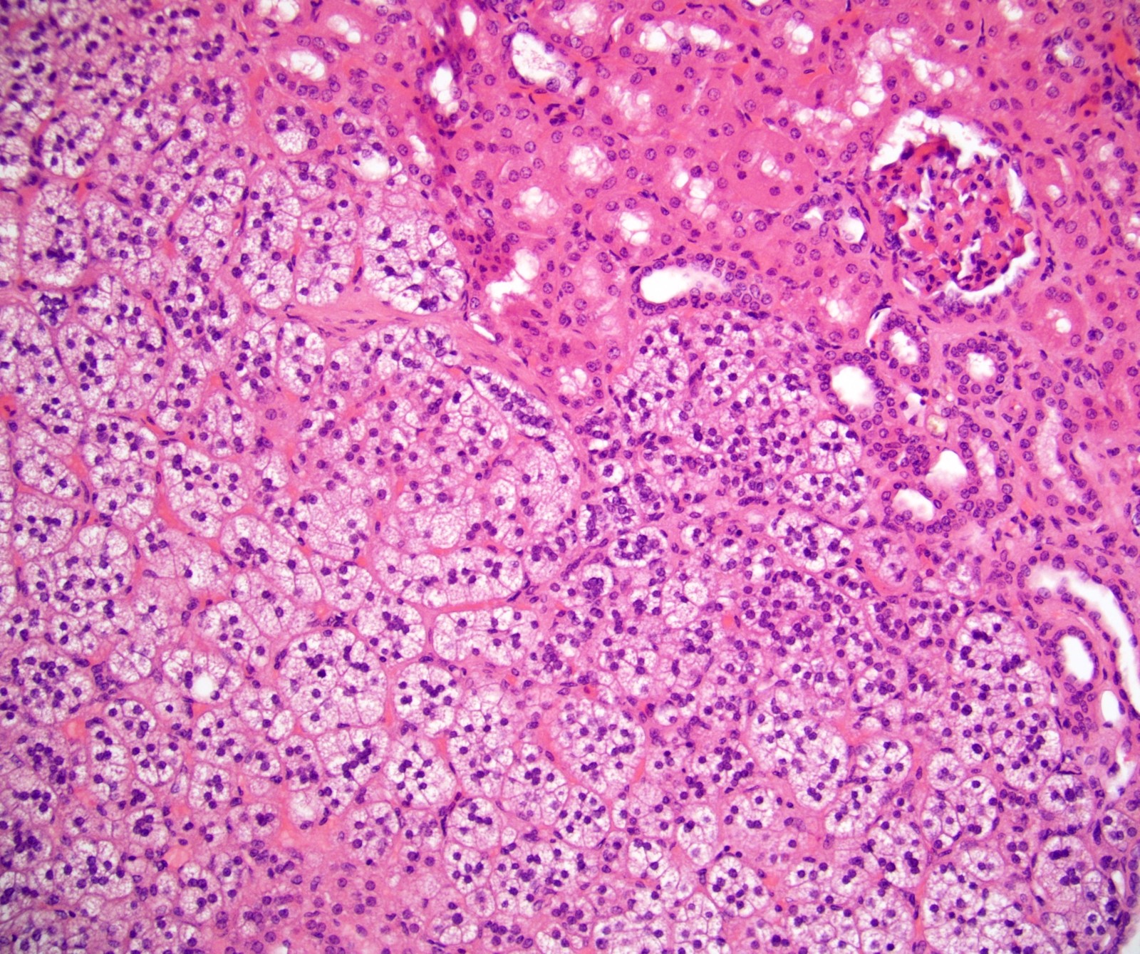
Contributed by Nicole Stringham, Ph.D. (source: University of Michigan virtual slide box) and Debra L. Zynger, M.D.
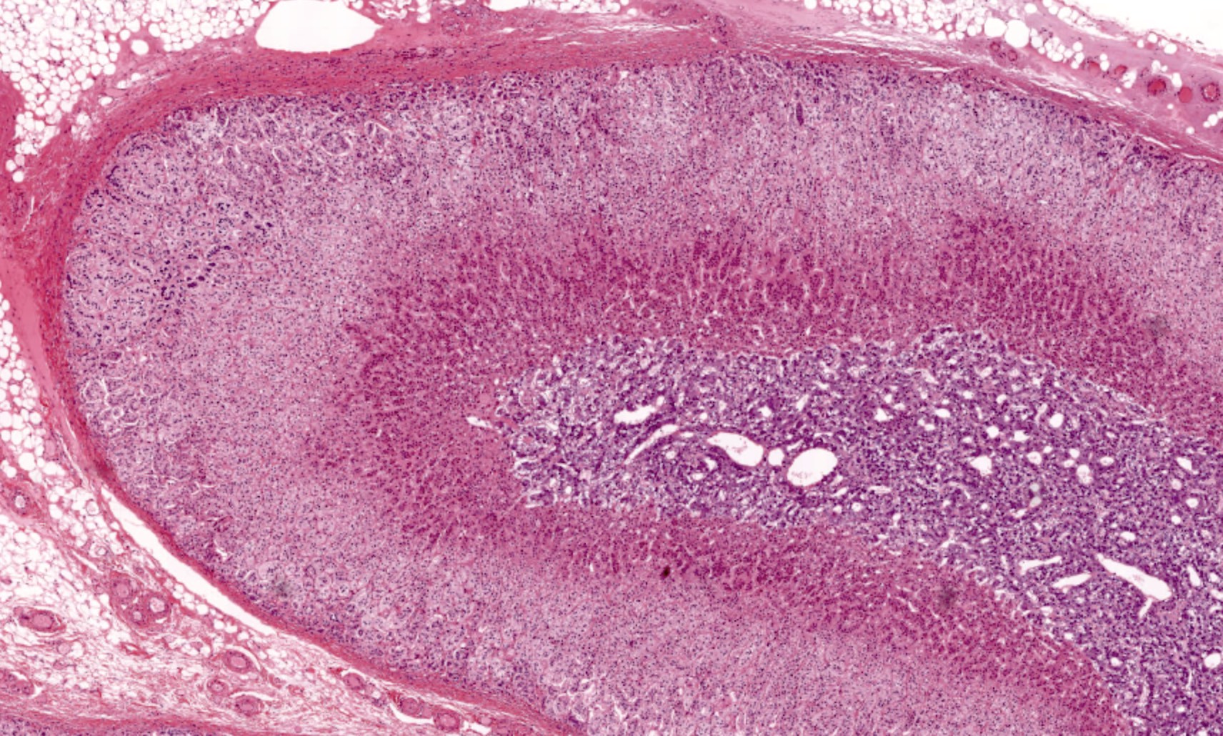
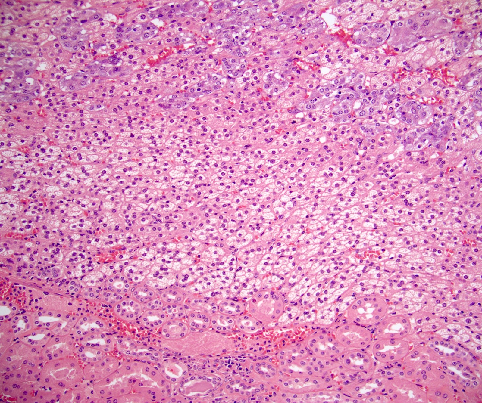
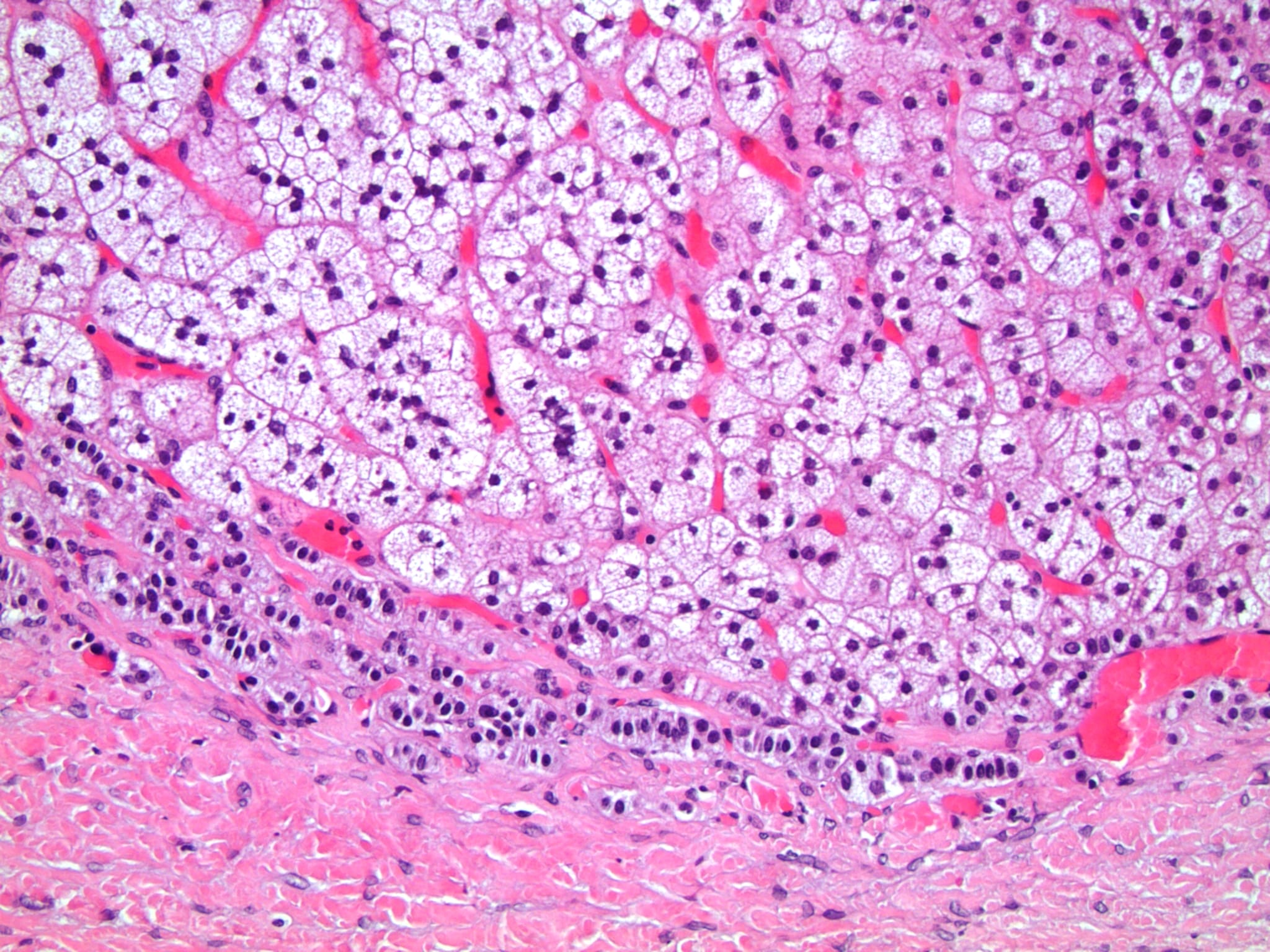
Adrenal gland full thickness
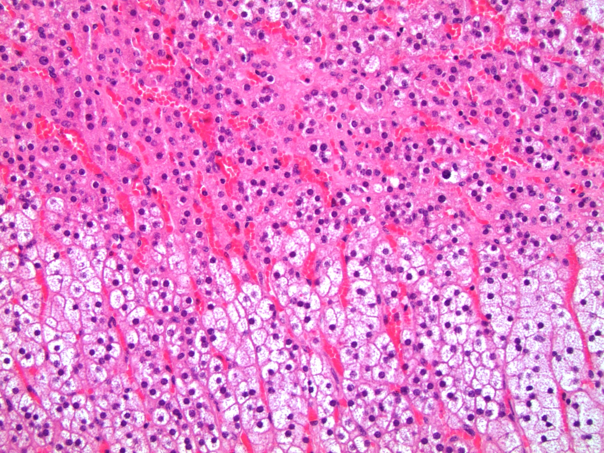
Zona glomerulosa and fasciculata
Zona glomerulosa
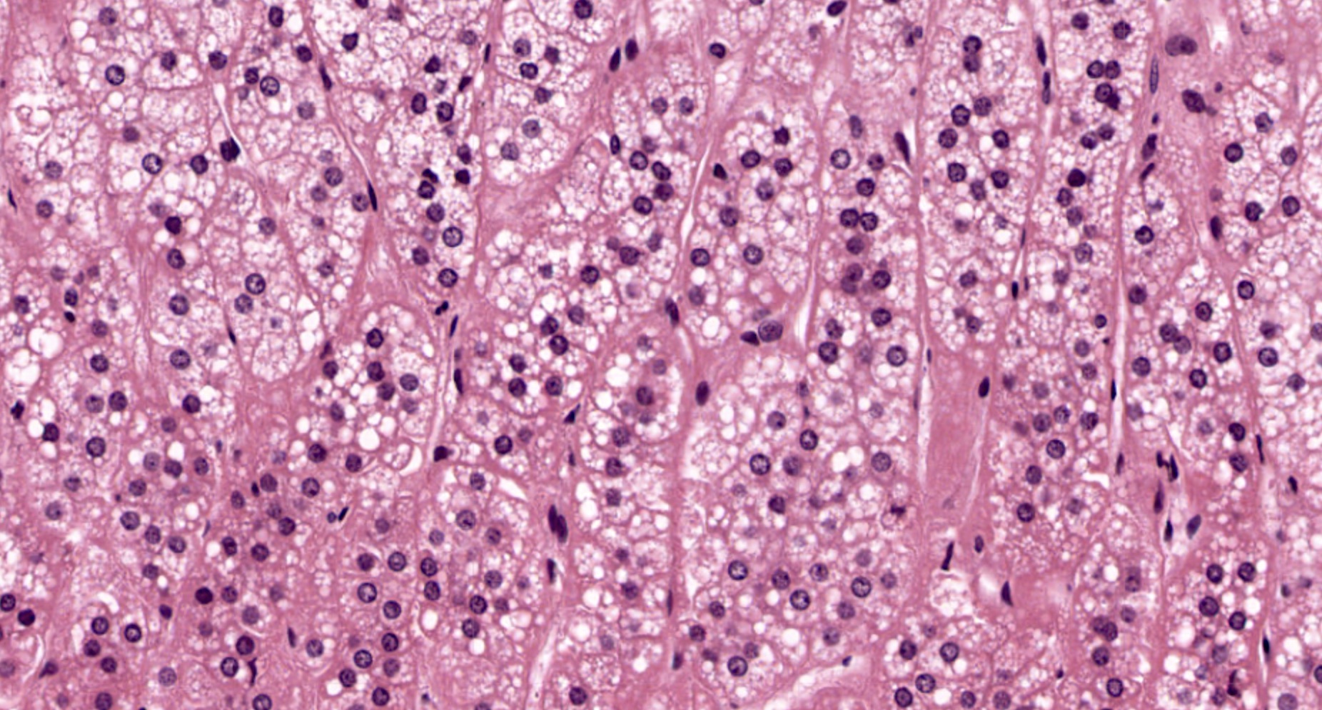
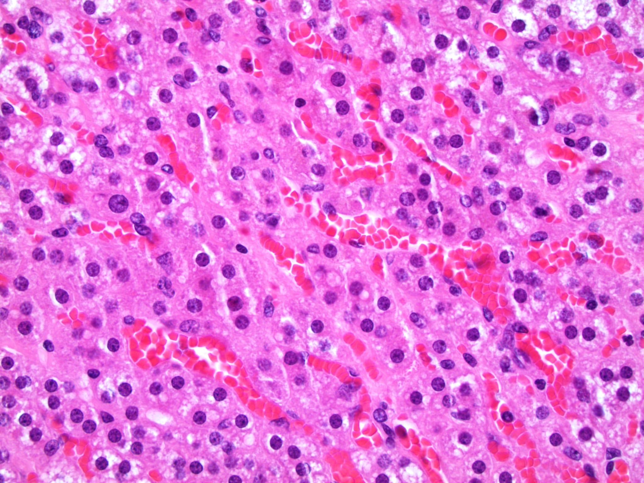
Zona fasciculata
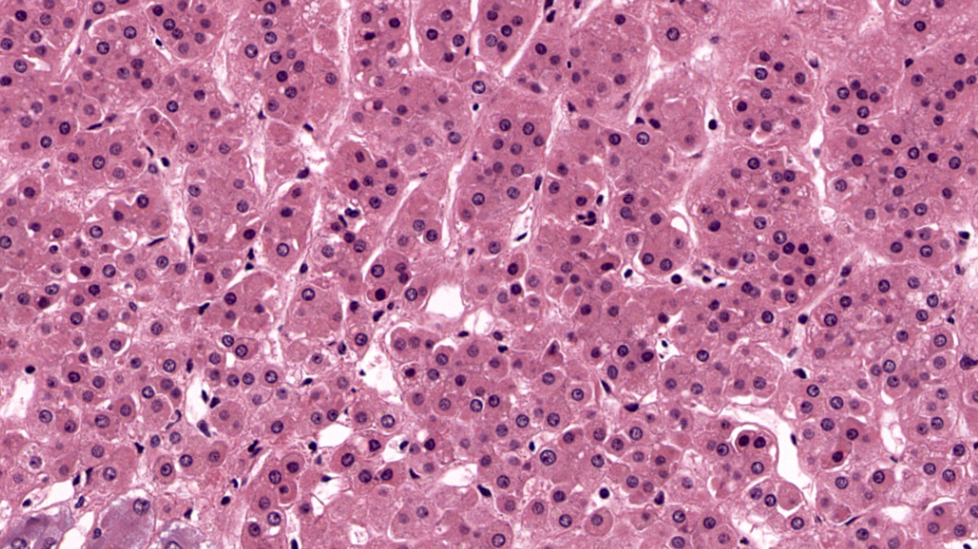
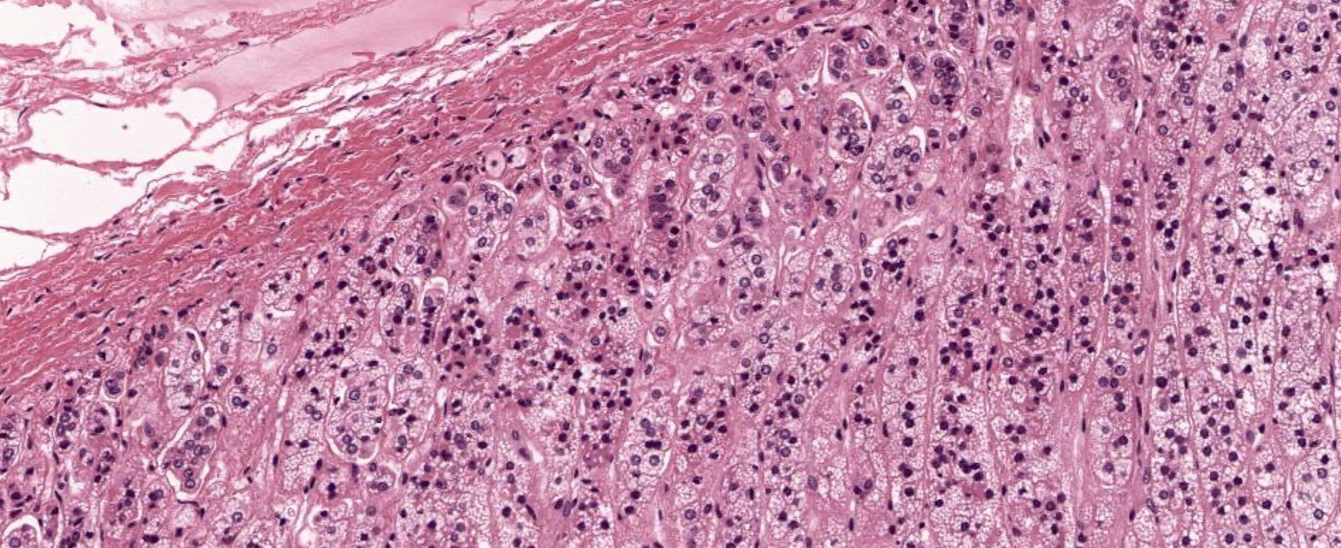
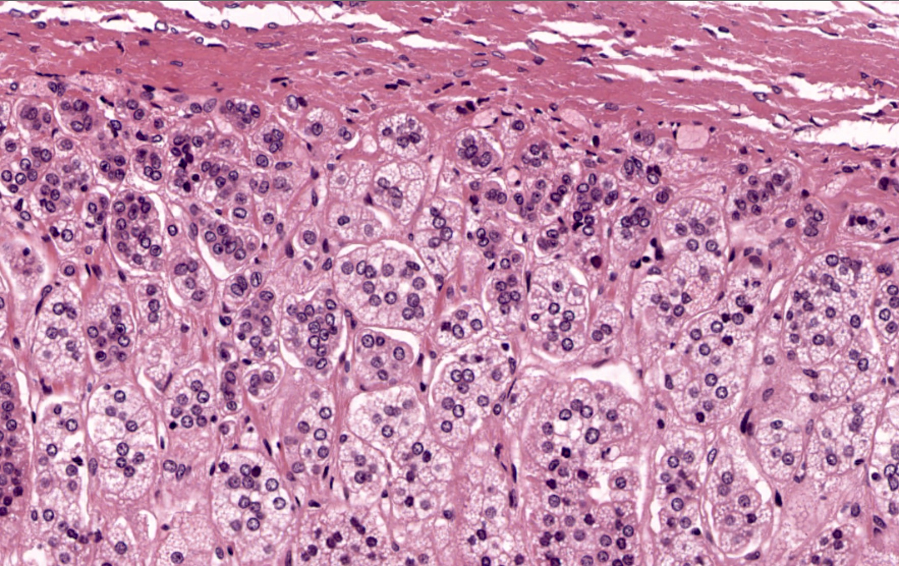
Zona reticularis
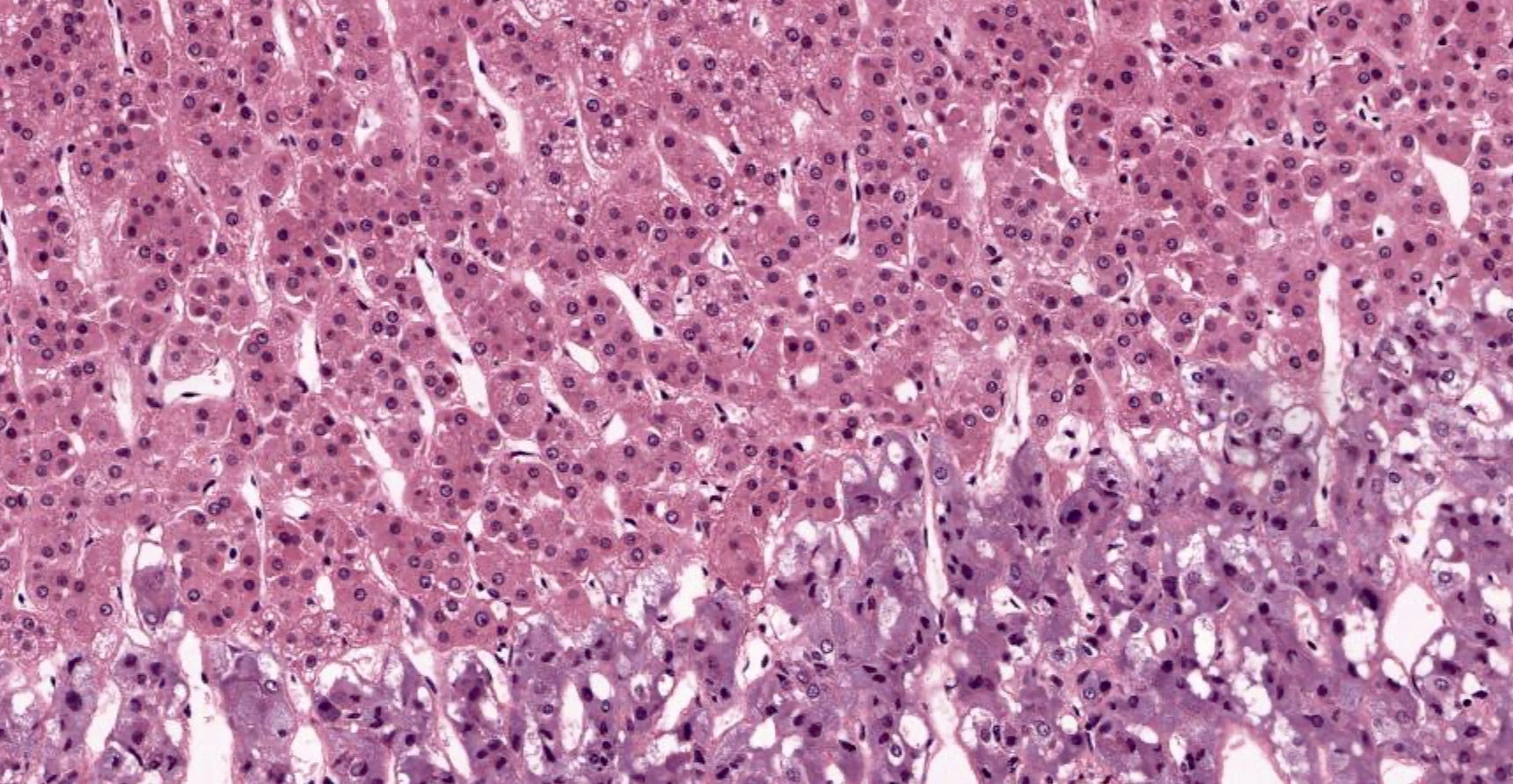
Cortex medulla junction
Zona glomerulosa and fasciculata
Zona fasciculata
Zona fasciculata and reticularis
Zona reticularis

Zona reticularis with lipofuscin

Adrenal medulla
Contributed by Xiaoyin "Sara" Jiang, M.D.
Normal adrenal gland
Images hosted on other servers:

Zona glomerulosa

Adrenal chromaffin cells
Adrenal gland histology
Images hosted on other servers:

CT scan of abdomen showing a cystic adrenal lesion on the right
Images hosted on other servers:

Anterior linear crease

Posterior helical ear pits
Images hosted on other servers:

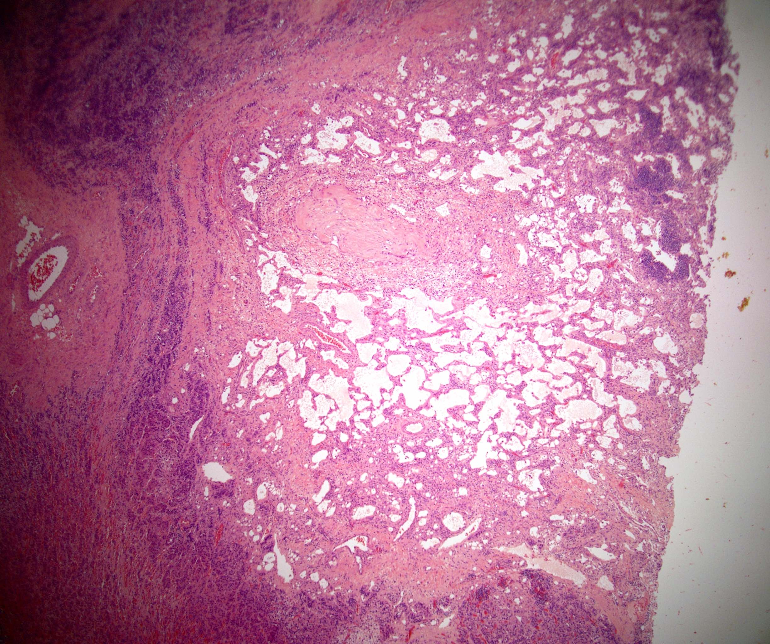
Large lobulated adrenal glands

Enlarged placenta with multiple vesicles
Images hosted on other servers:

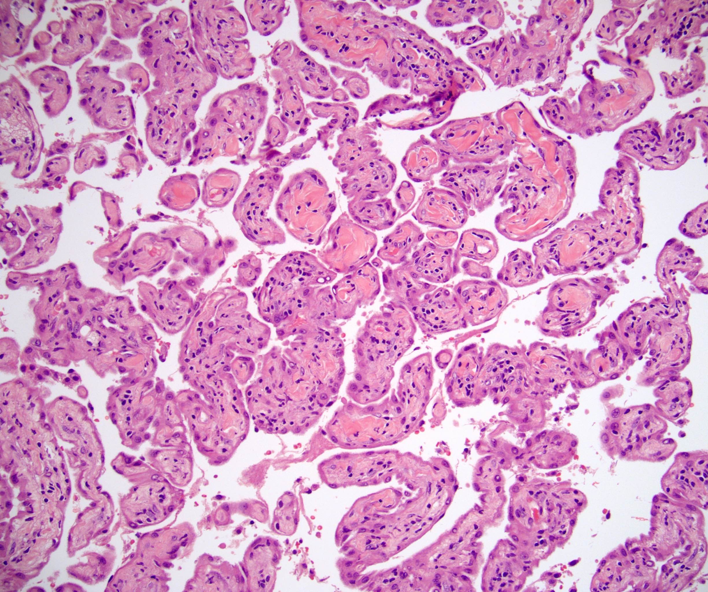
Adrenocortical cytomegaly

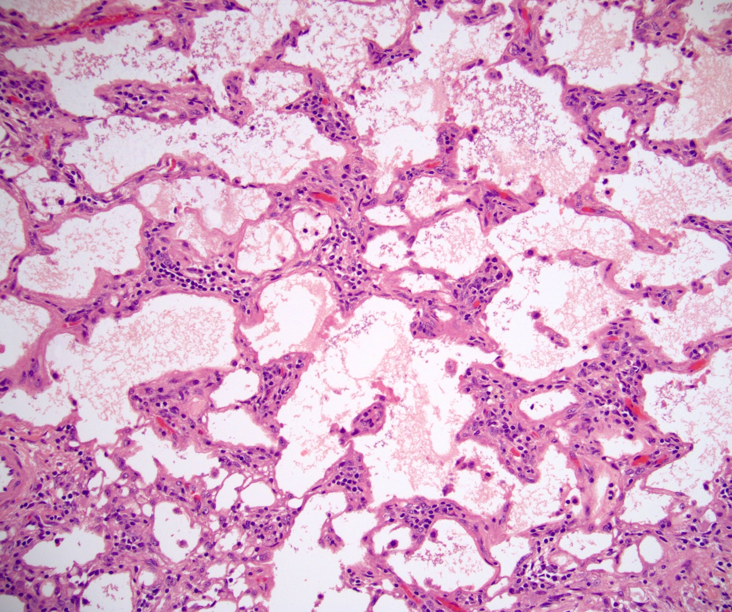
Visceral anomalies in the fetus

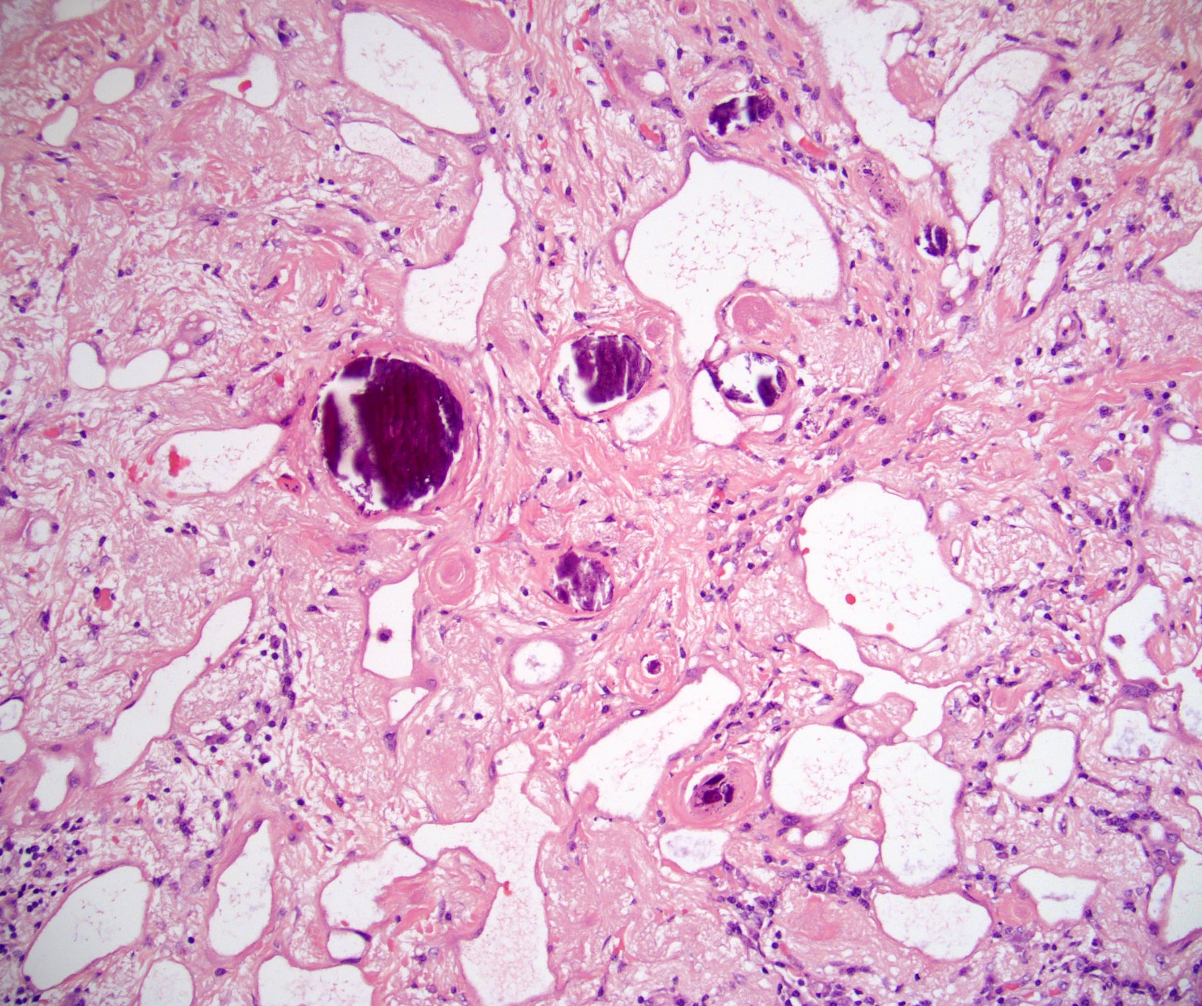
Placental histological anomalies
Images hosted on other servers:


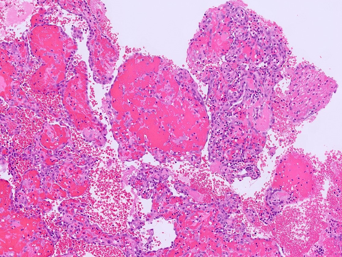
Giant endothelial cyst

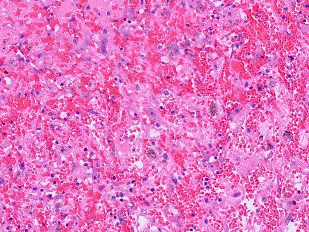
Giant pseudocyst

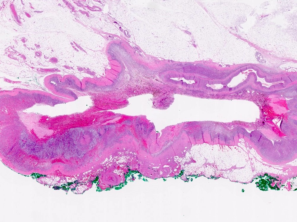
Hydatid cyst
Images hosted on other servers:


Pseudocyst


Hydatid cyst
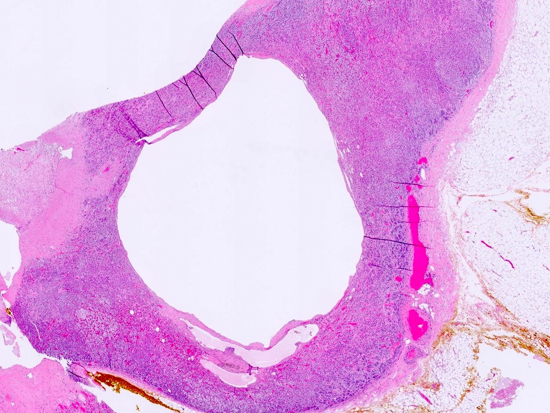
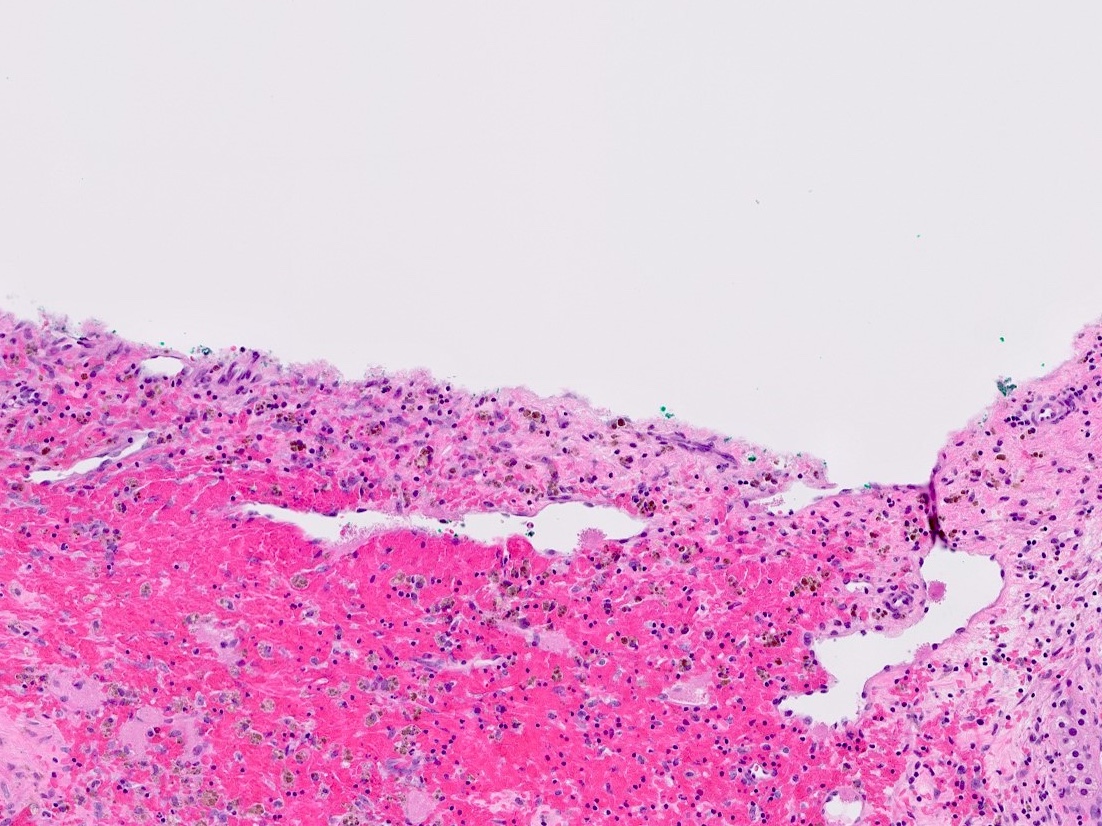
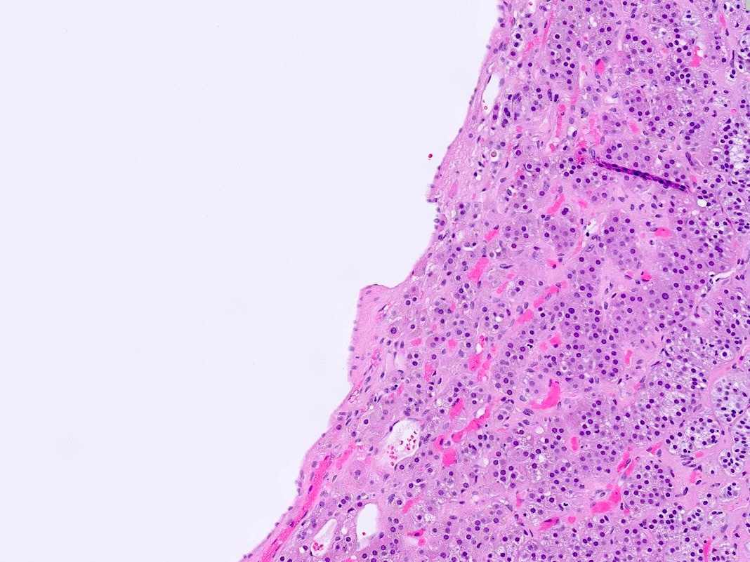
Contributed by Debra L. Zynger, M.D.
Endothelial cyst with clot
Endothelial cyst with minimal blood
Pseudocyst reclassified as endothelial cyst
Pseudocyst
Contributed by Debra L. Zynger, M.D. and Lan L. Gellert, M.D., Ph.D.
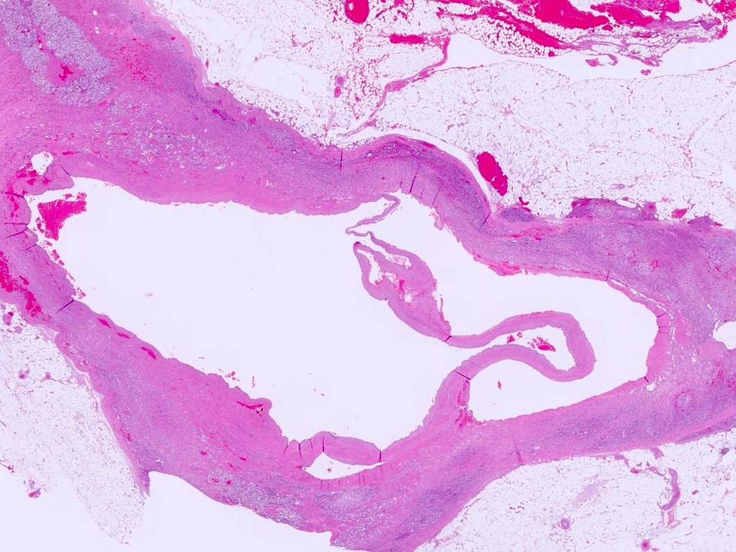
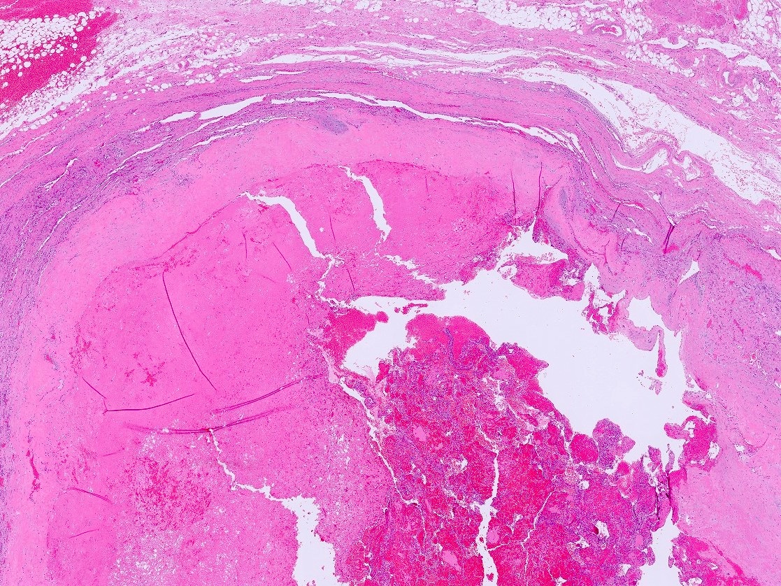

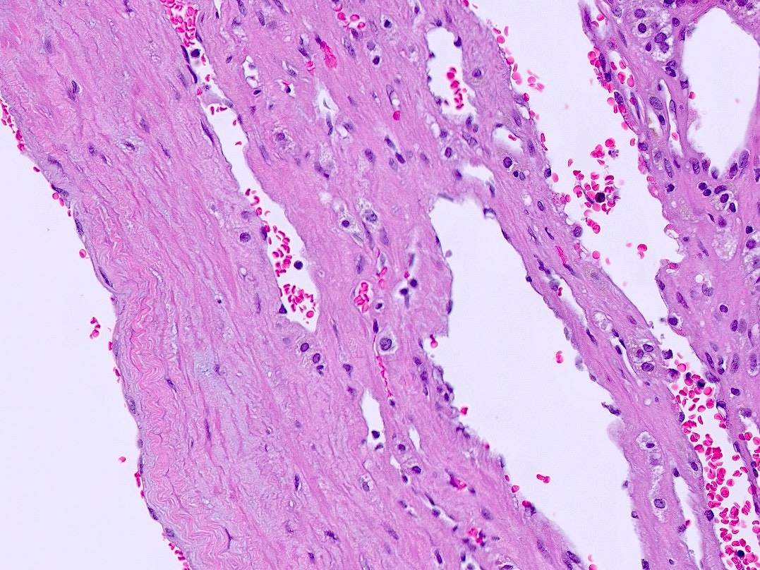
Endothelial cyst
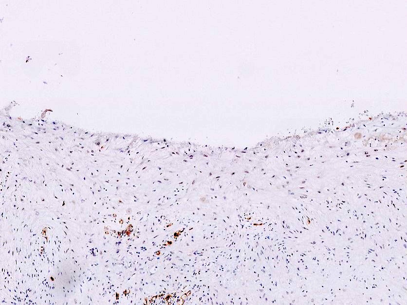

Pseudocyst
Mesothelial cyst
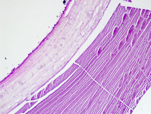
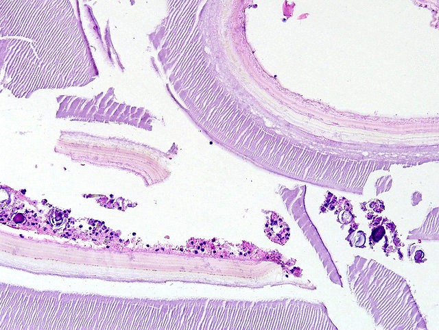
Hydatid cyst
Images hosted on other servers:

Hydatid cyst
Contributed by Debra Zynger, M.D. and Maria Tretiakova, M.D., Ph.D.
Extra-adrenal adipose invasion (pT3)
High mitotic rate
Lymphovascular invasion
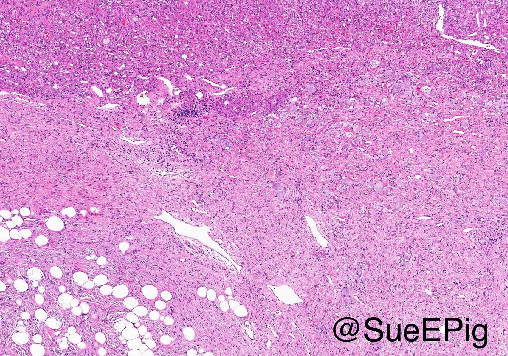
Myxoid variant
Oncocytic variant
Liver involvement (pT4)
Transition to myxoid area
Myxoid variant
Extra-adrenal adipose invasion (pT3)
Positive margin
Regional node involvement (pN1)
Contributed by Debra L. Zynger, M.D.
pT1
pheochromocytoma
pT2
pheochromocytoma
pT3 pheochromocytoma
pT2 sympathetic
paraganglioma
Contributed by Debra L. Zynger, M.D.

Capsular invasion
Vascular, capsular and adipose invasion
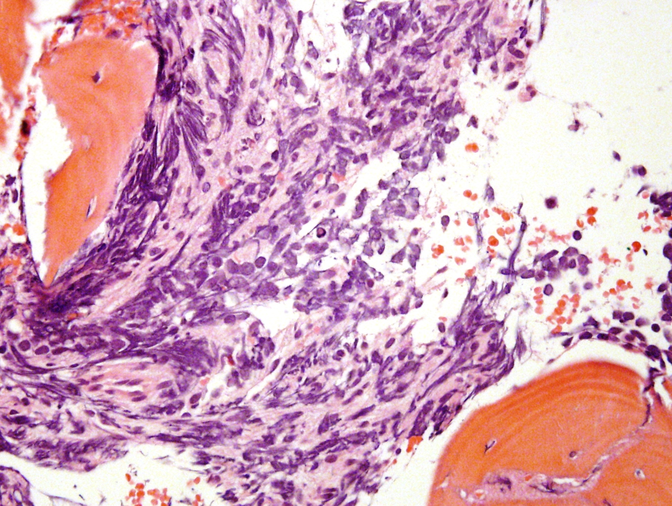
Cellular spindling

Nuclear pleomorphism

Mitosis
pT2 sympathetic
paraganglioma
pT3 pheochromocytoma
with extra-adrenal
adipose invasion
pT3 pheochromocytoma
with invasion
of the kidney
Images hosted on other servers:

Various images
Image hosted on other servers:
Spiral CT: hyperechoic mass
Contributed by Carmen Perrino, M.D. and Debra L. Zynger, M.D.
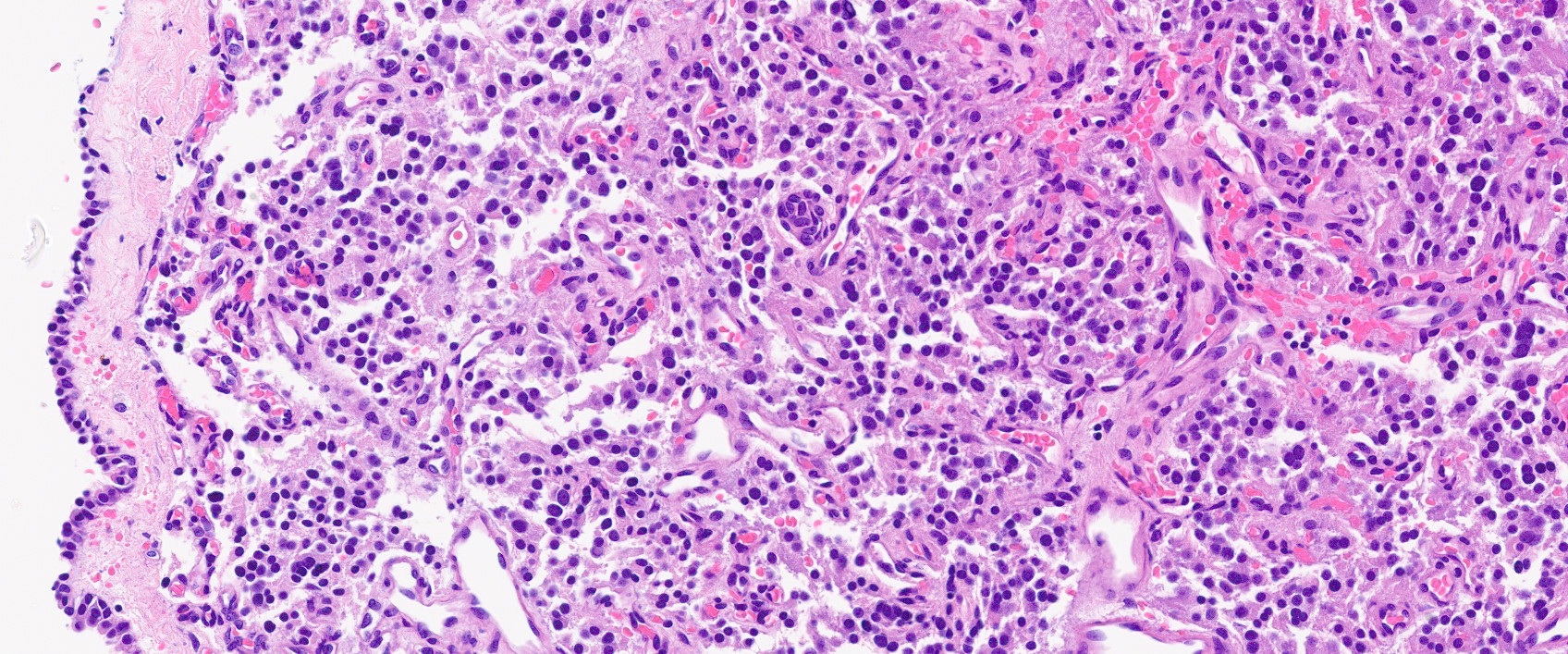
Stroma rich, intermixed type
Focal undifferentiated component
Stroma rich, nodular type
With treatment related changes
Contributed by Carmen Perrino, M.D. and Debra L. Zynger, M.D.
Intermixed type
Composite types

Intermixed type
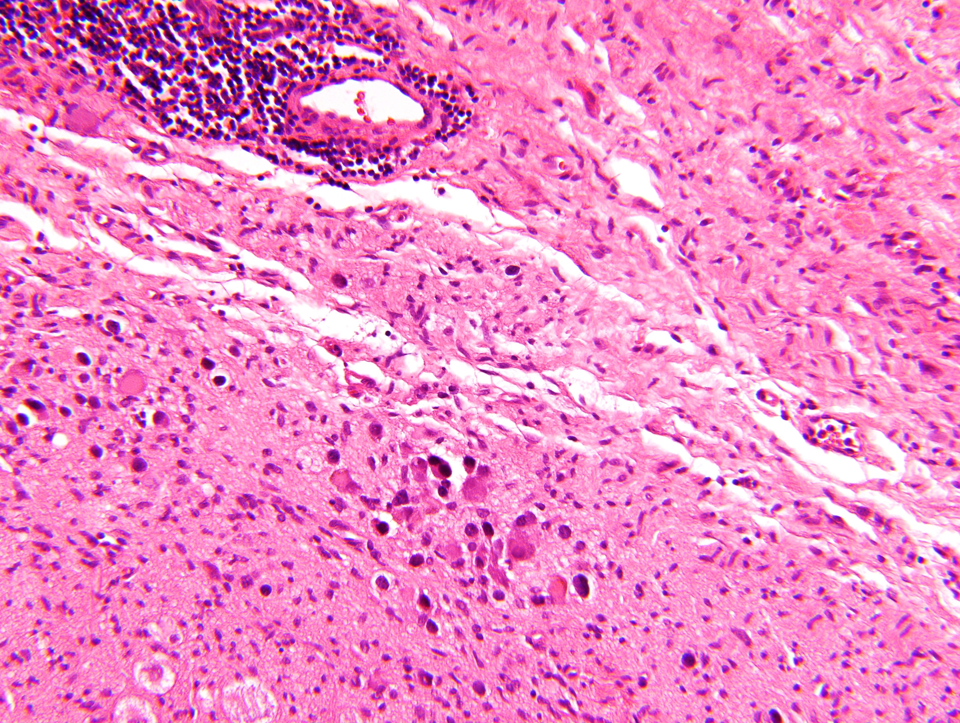
Ganglion cells
Fibrillary background
Aggregate of ganglion cells
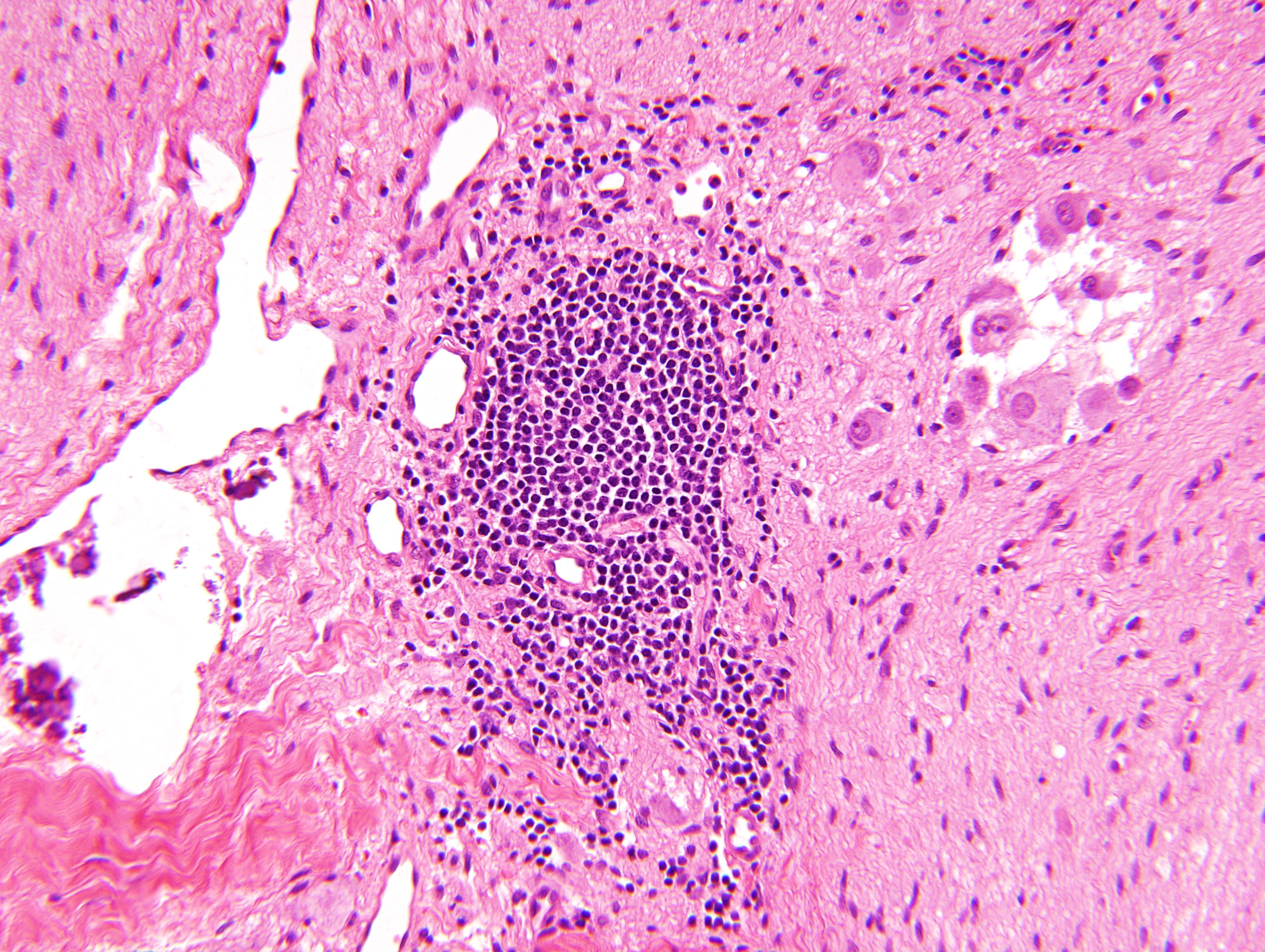
Lymphocyte aggregate
Area with Schwannian stroma
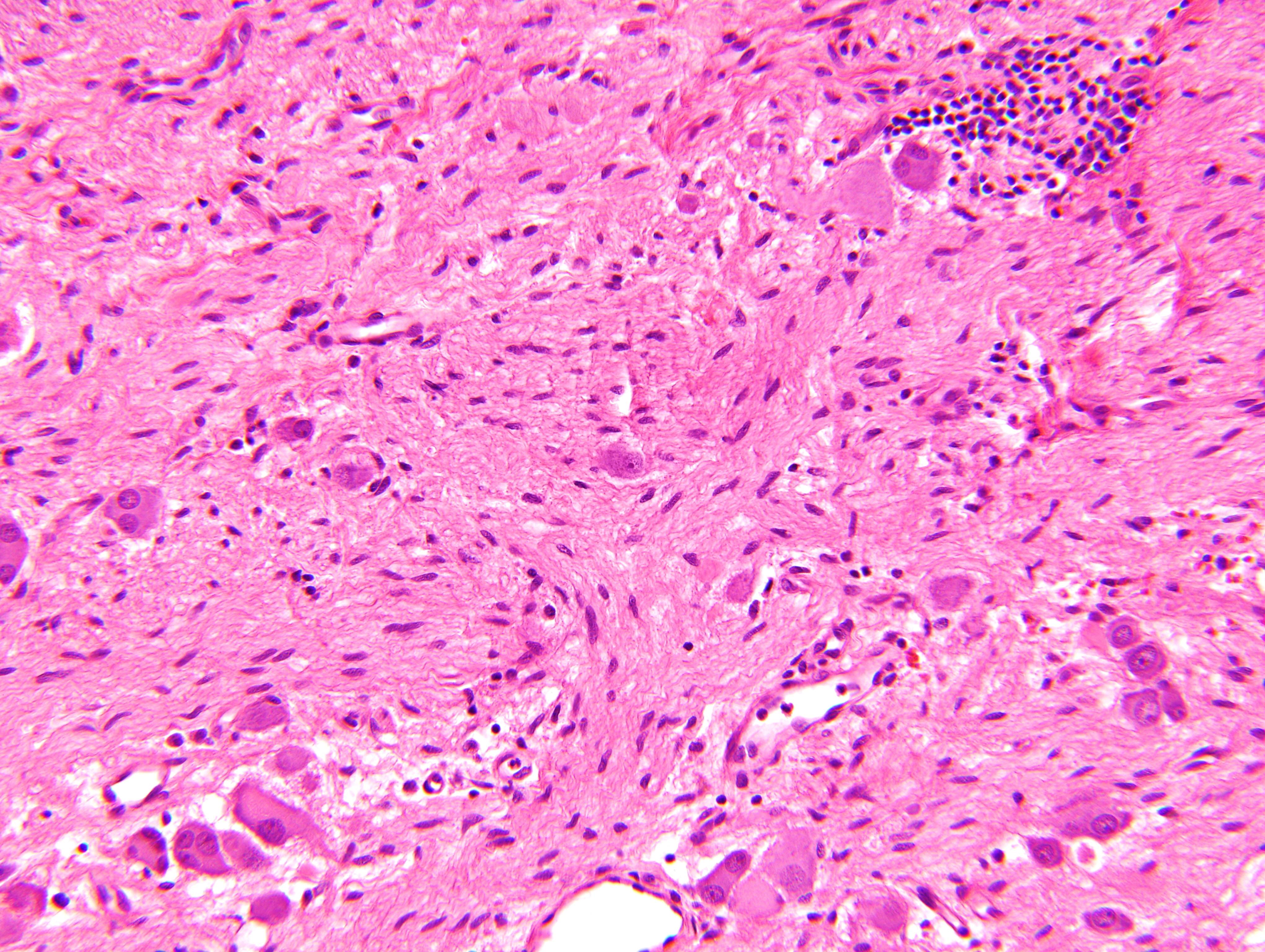
Maturing ganglion cells
Stroma rich, intermixed type
Nodular type
Stroma rich, nodular type
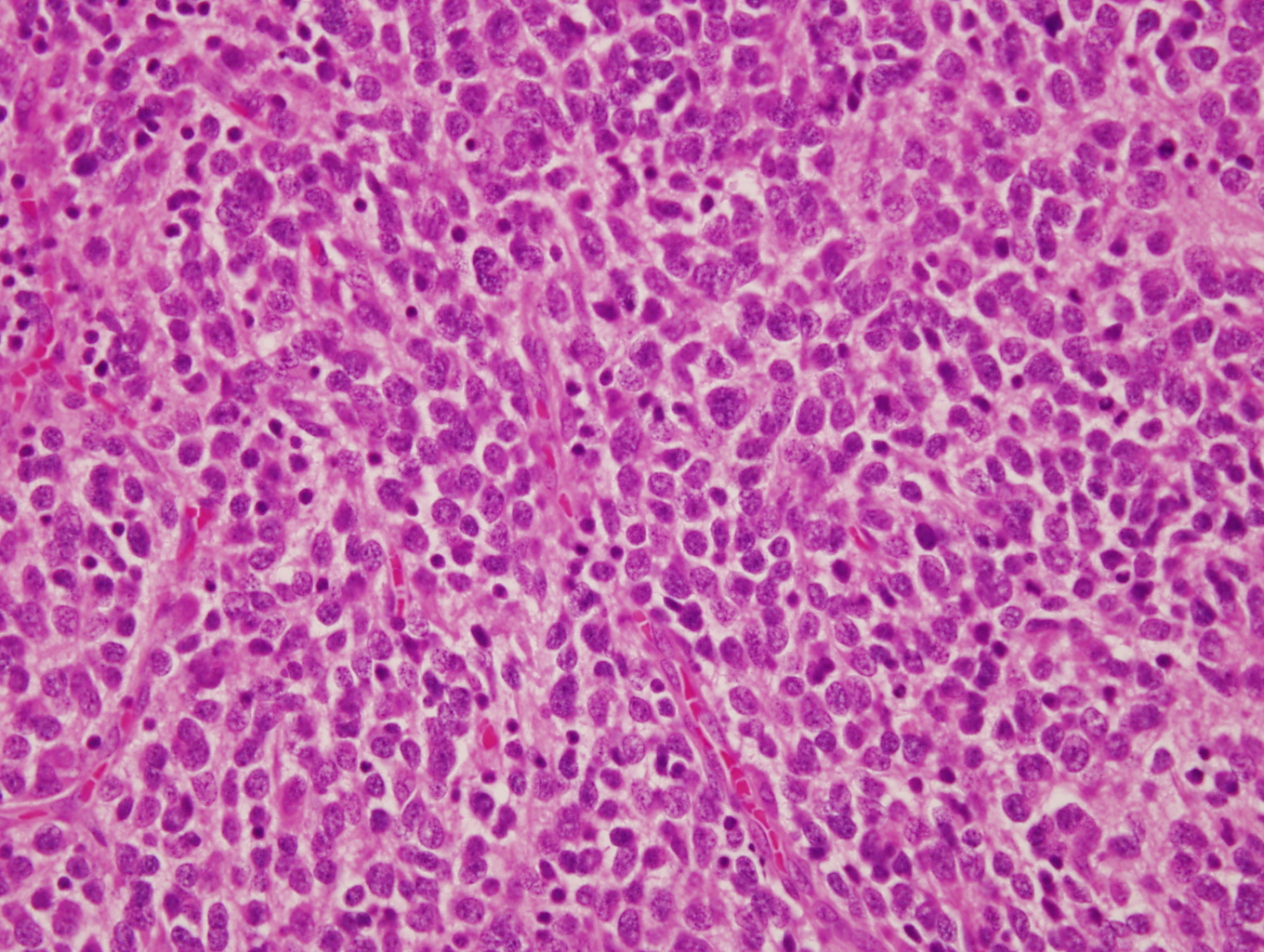
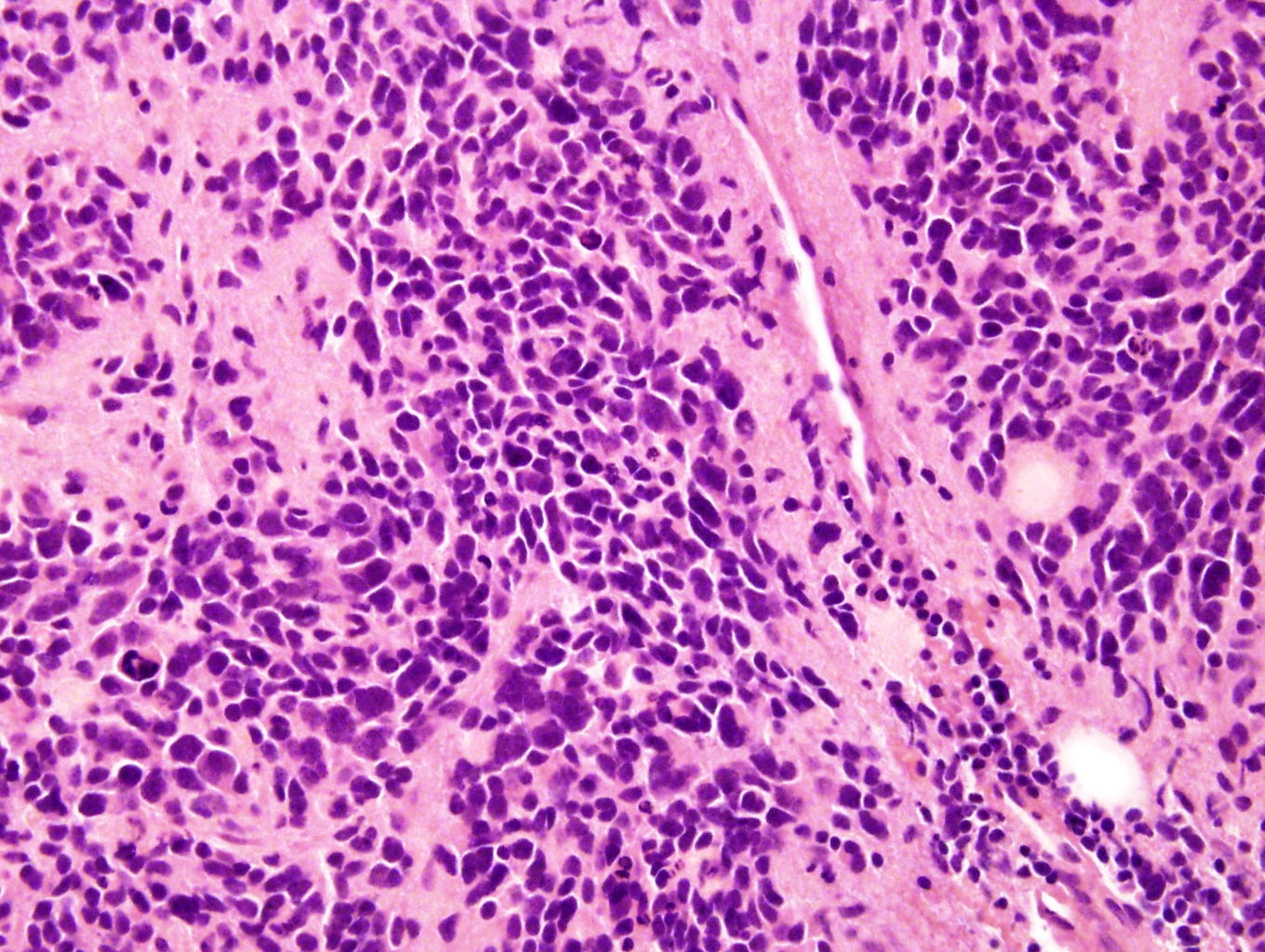
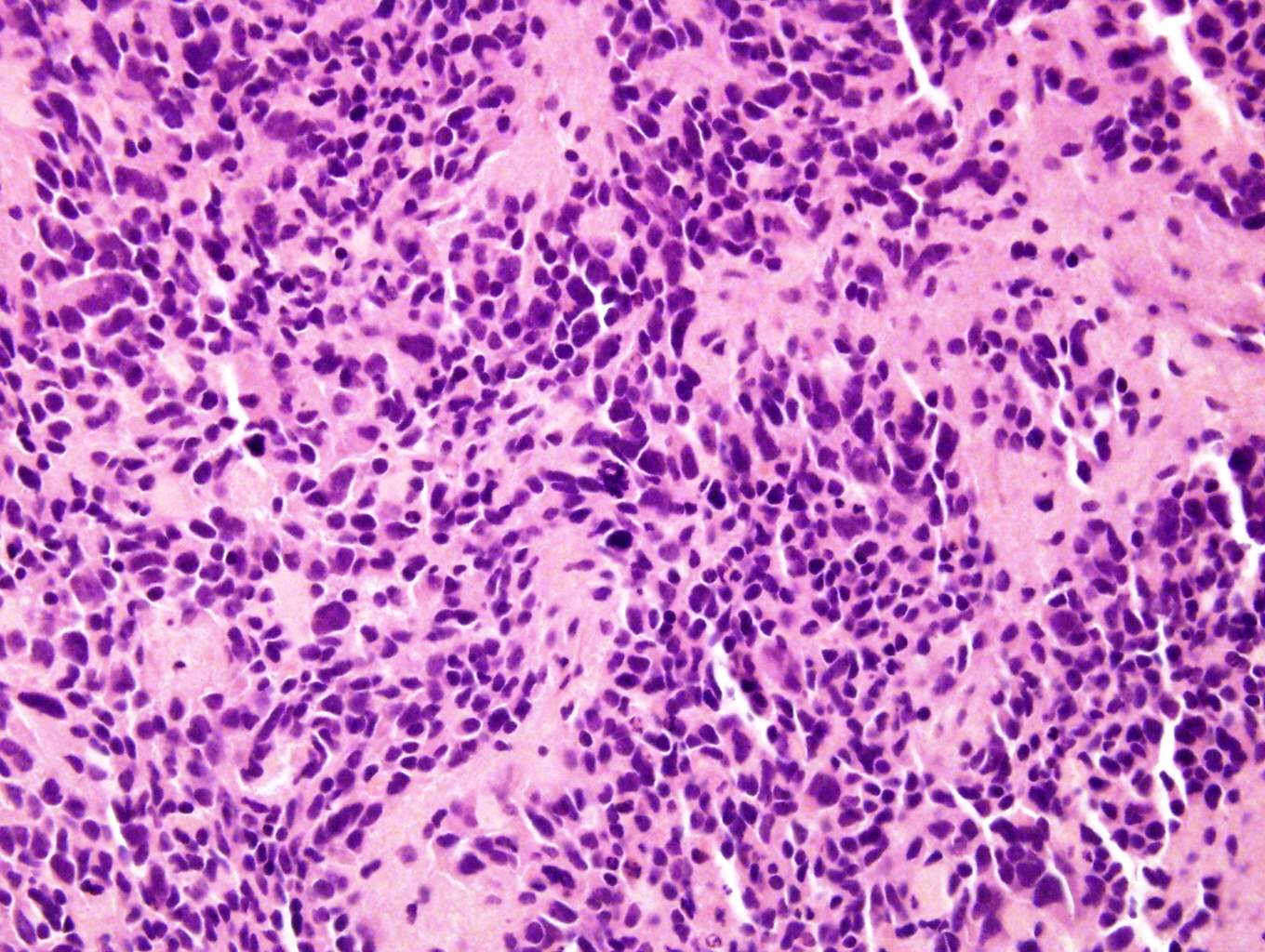
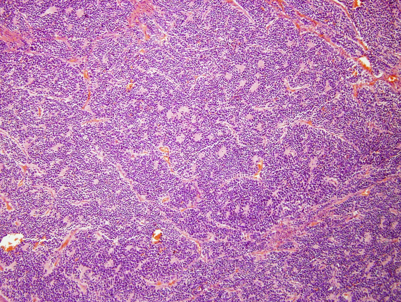
Neuroblast cell component
Homer Wright pseudorosettes

Vaguely formed Homer Wright pseudorosettes
Hemorrhagic component
Contributed by Carmen Perrino, M.D. and Debra L. Zynger, M.D.
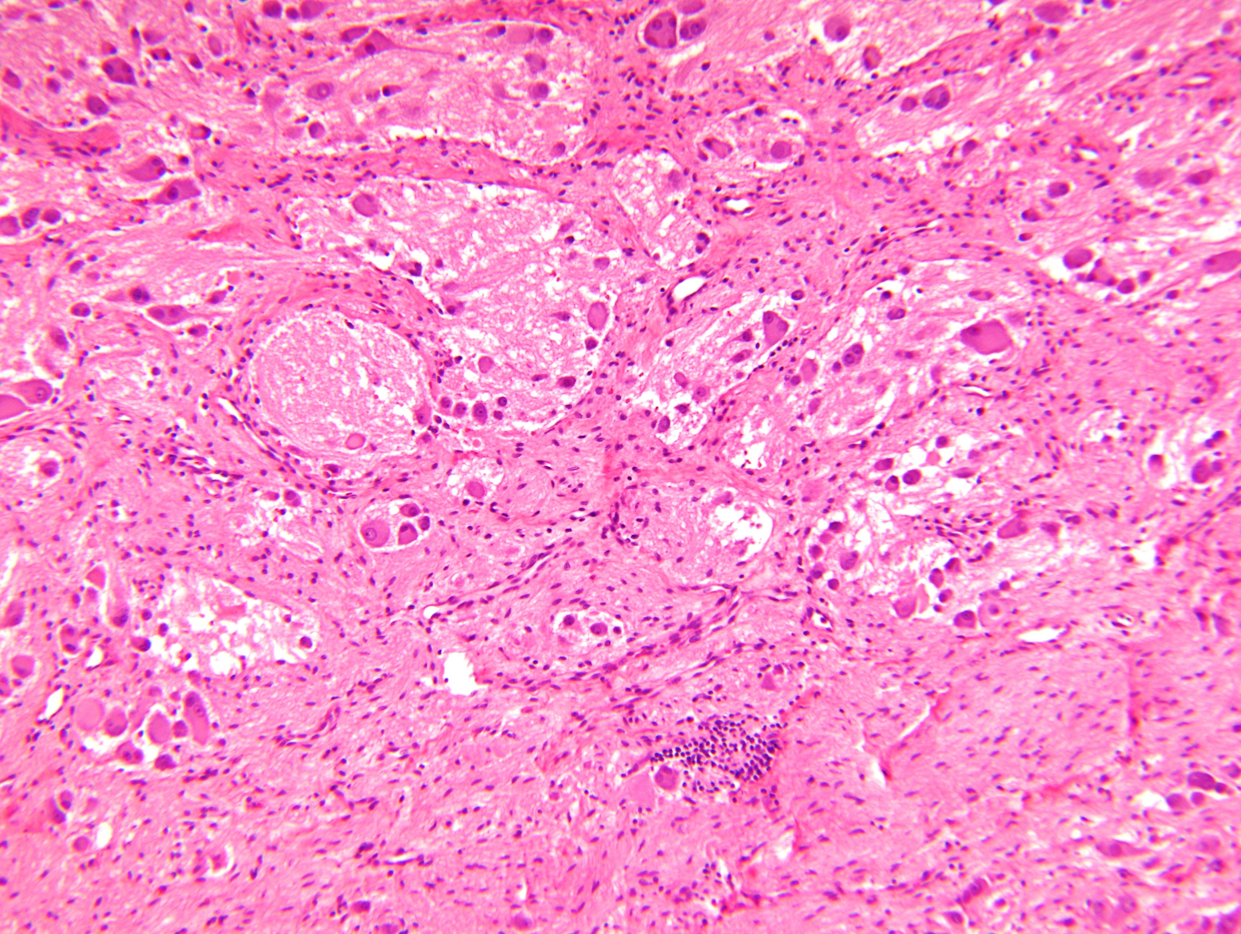
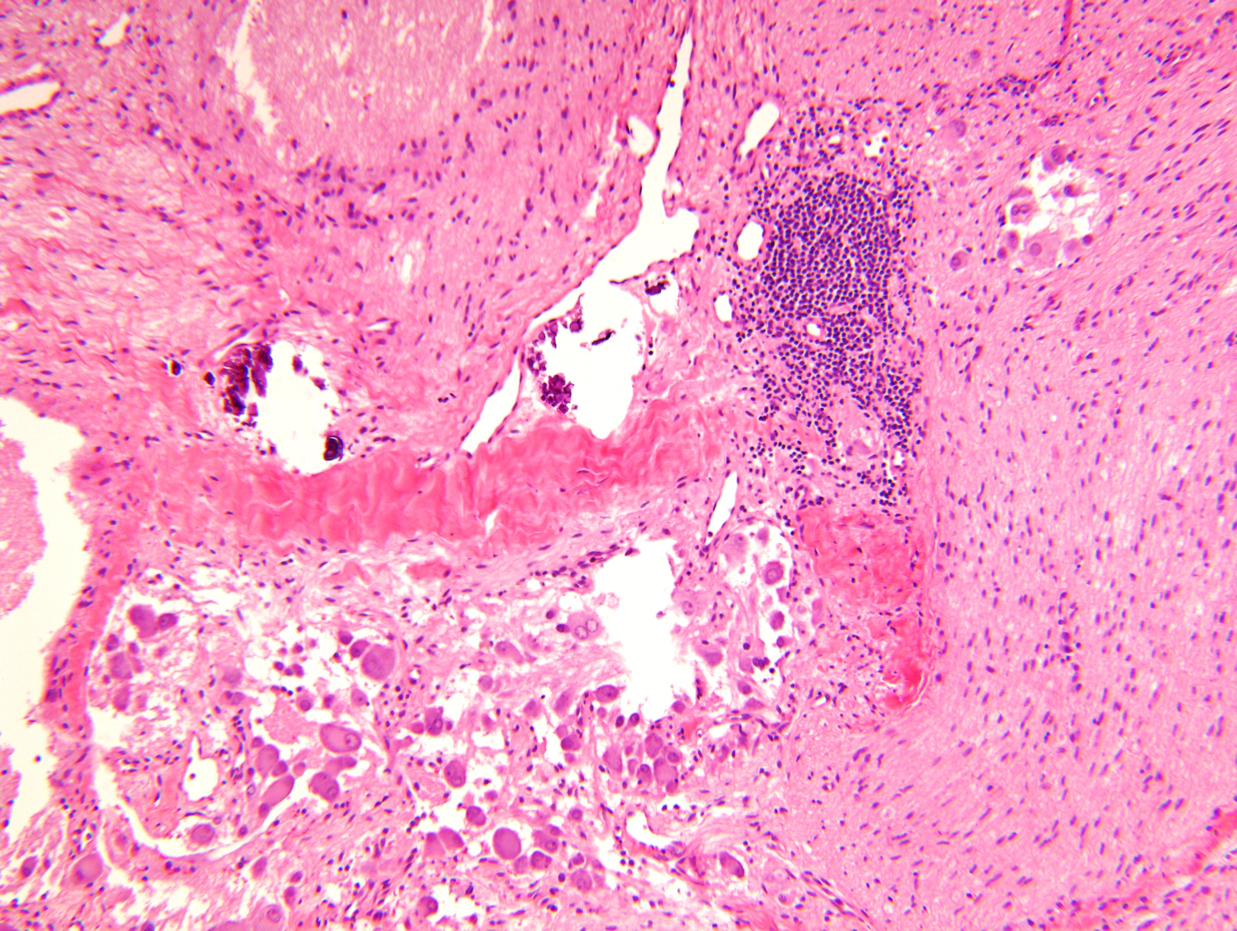
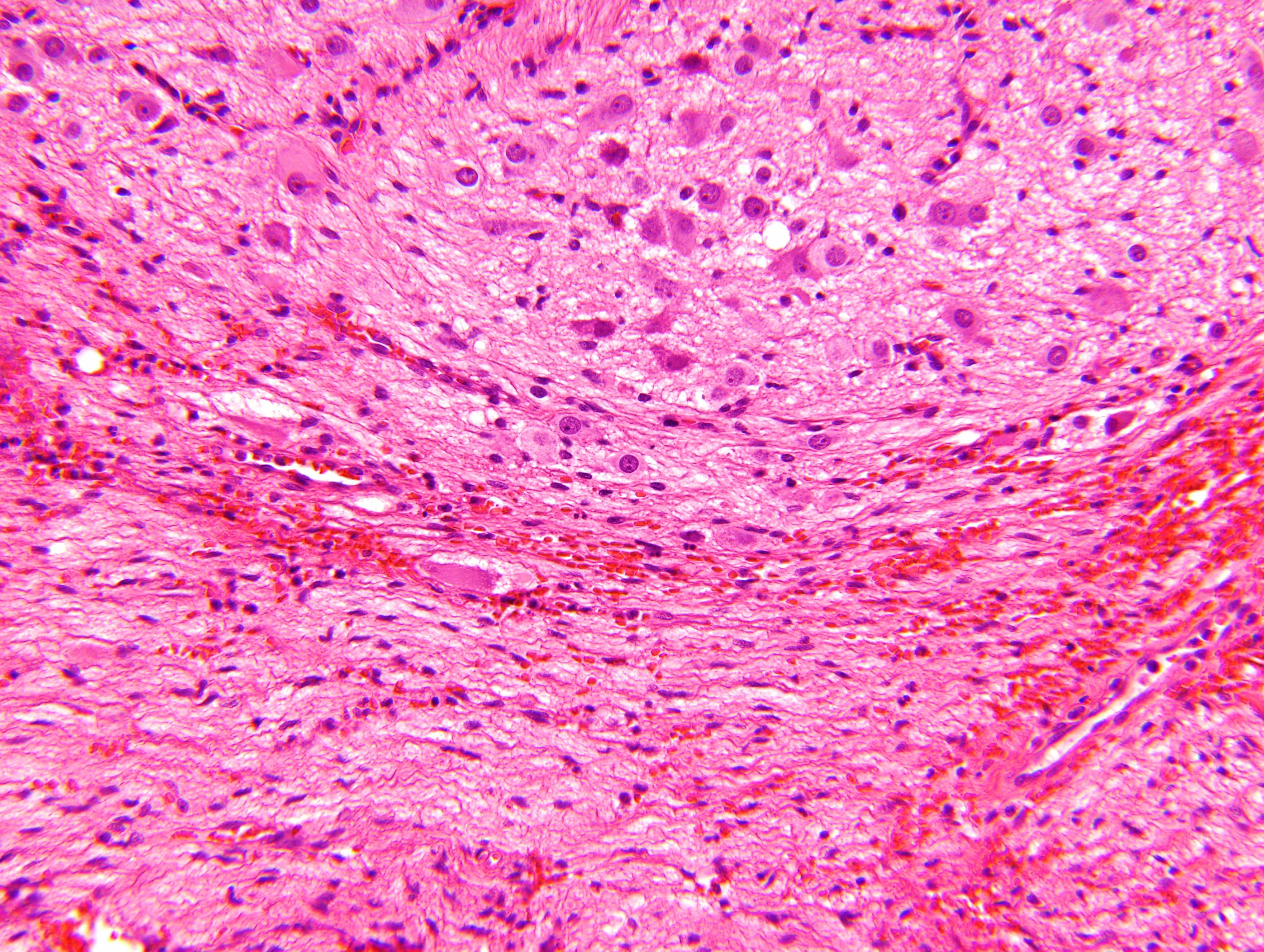
Numerous ganglion cells
Contributed by Leica Biosystems
MYCN (2p24) / AFF3 (2q11)
Images hosted on other servers:
Various images
Contributed by @SueEPig on Twitter
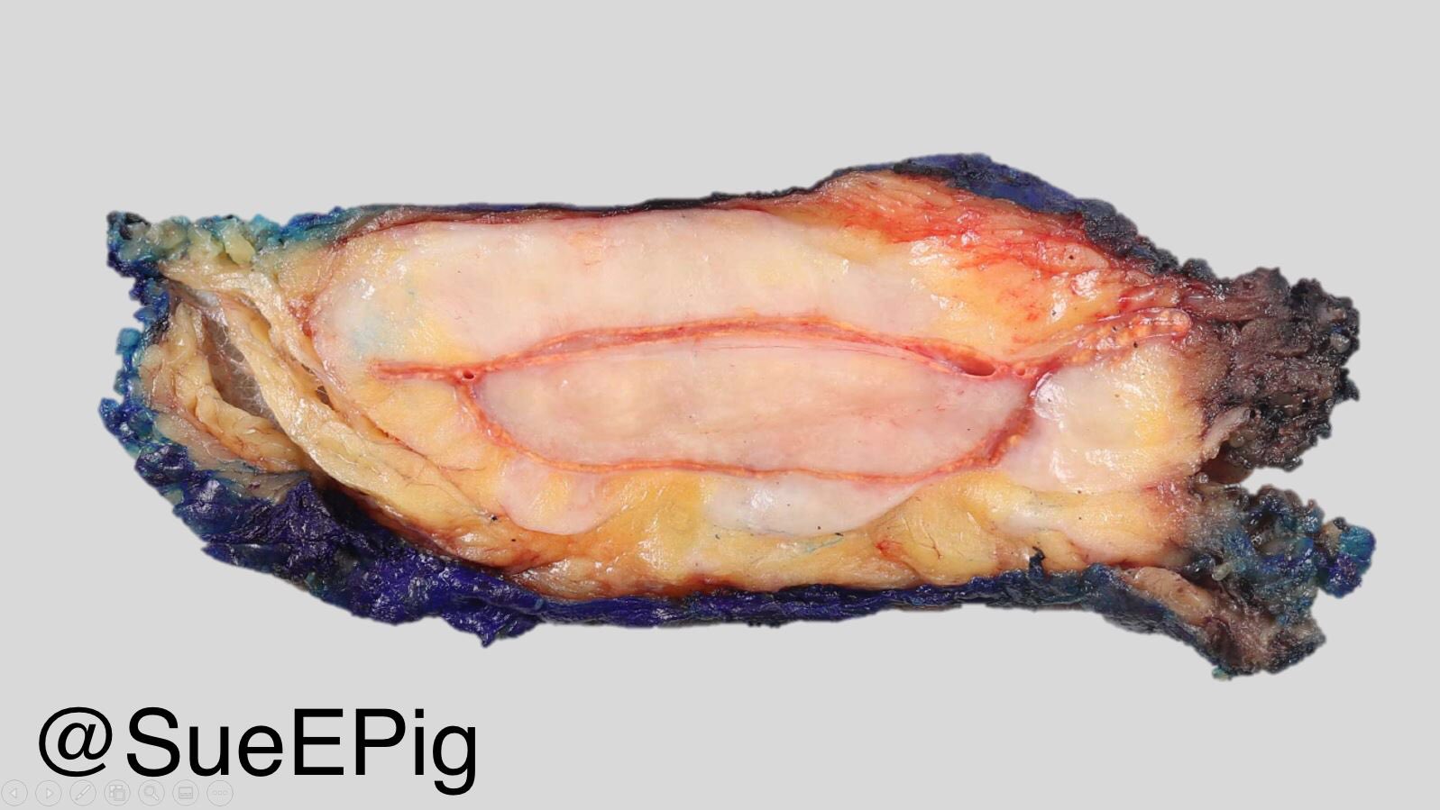
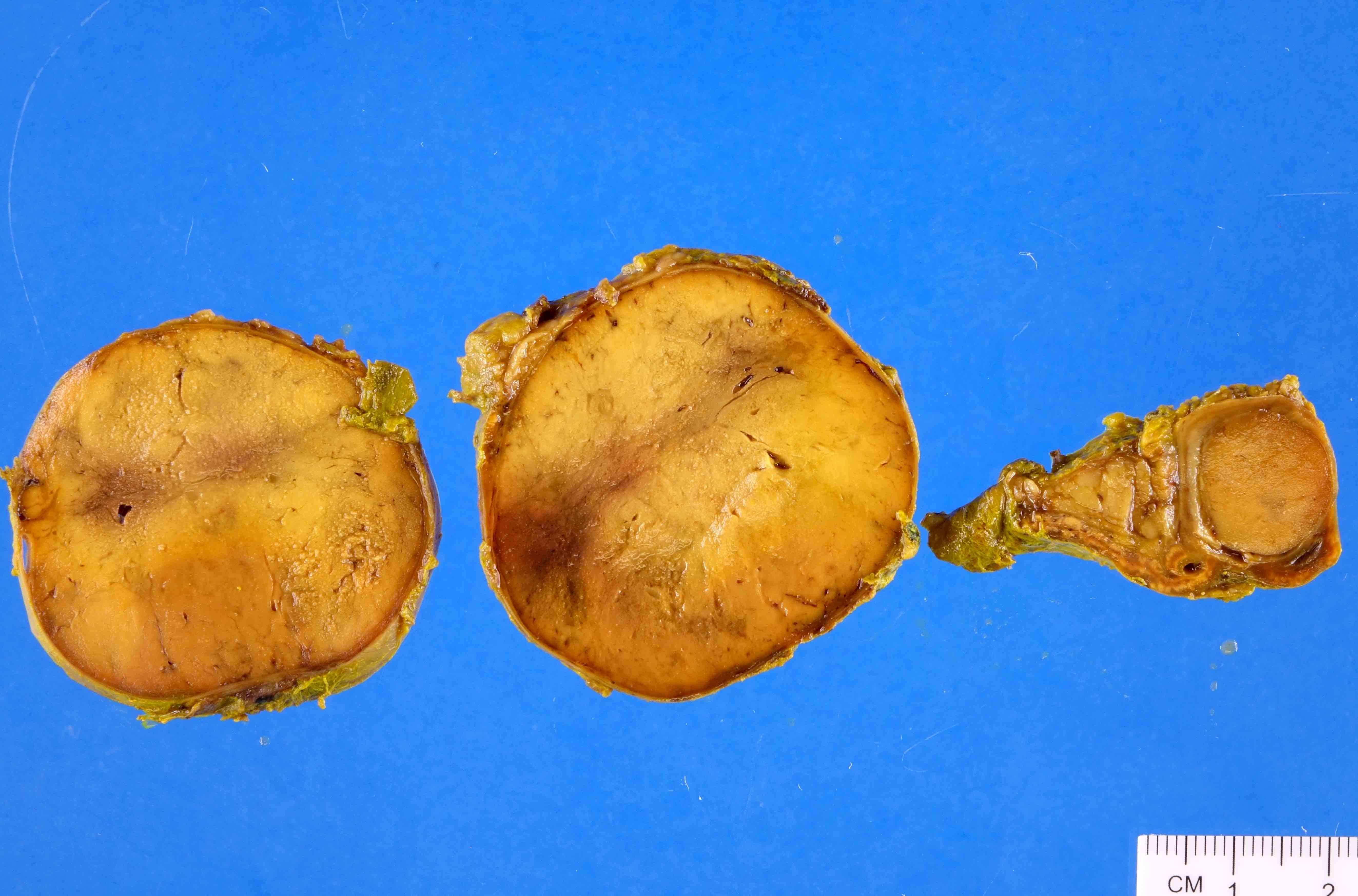
Adrenal ganglioneuroma
Images hosted on other servers:

Adrenal ganglioneuroma

Adrenal ganglioneuroma, cut surface
Contributed by Carmen Perrino, M.D., Debra Zynger, M.D. and @SueEPig on Twitter
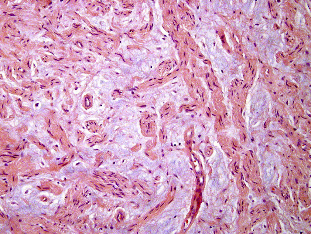
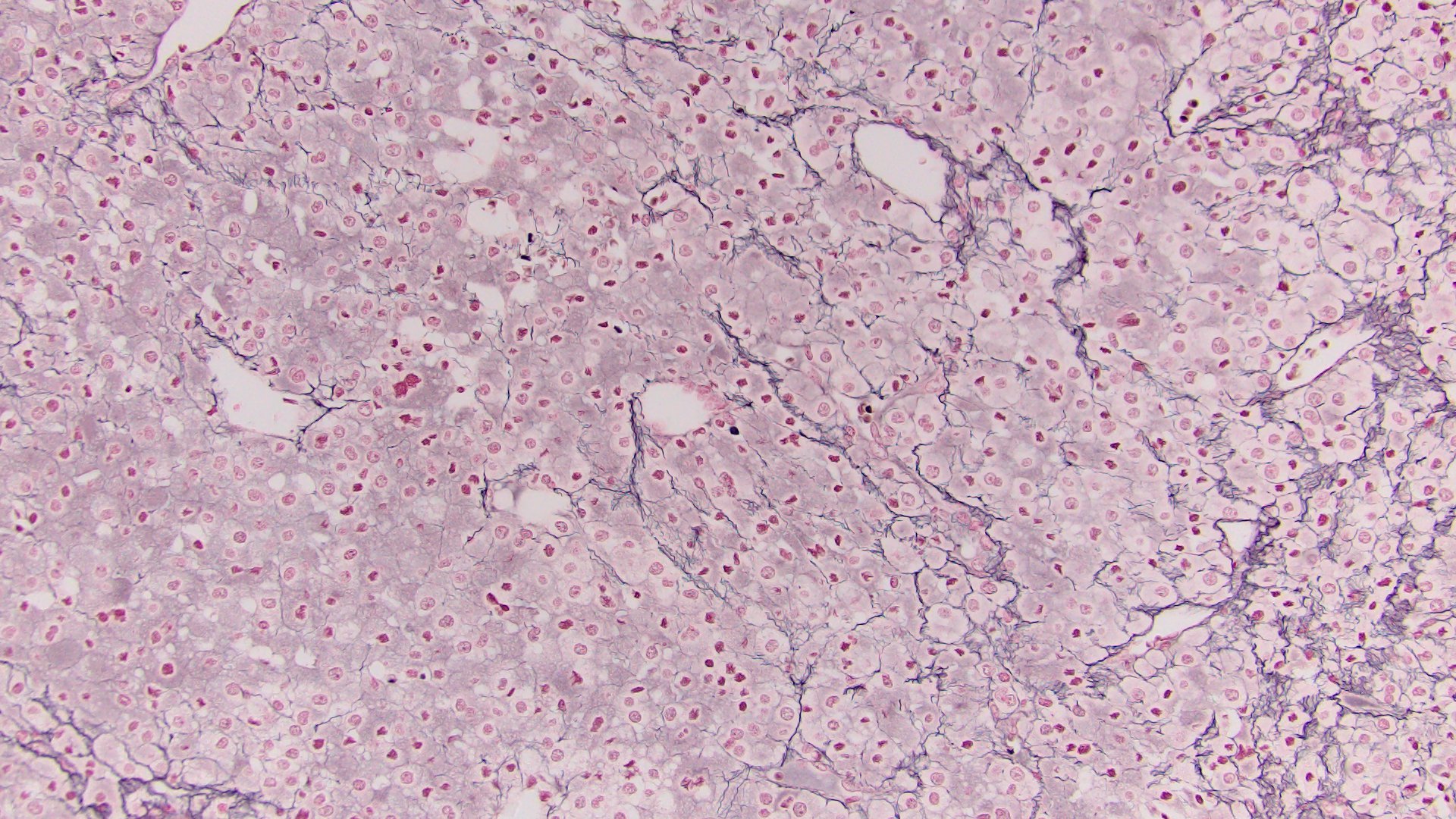
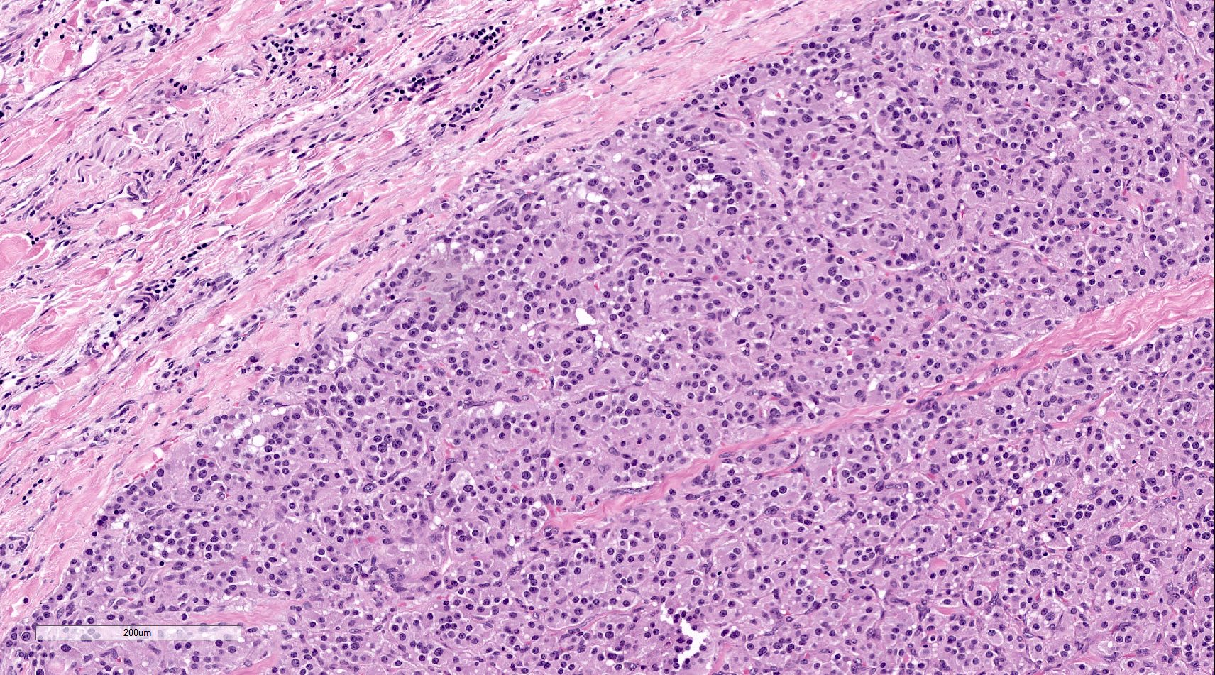
Prominent myxoid background
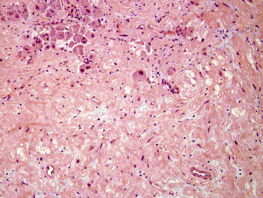
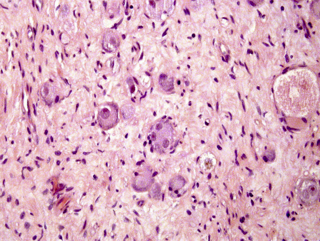
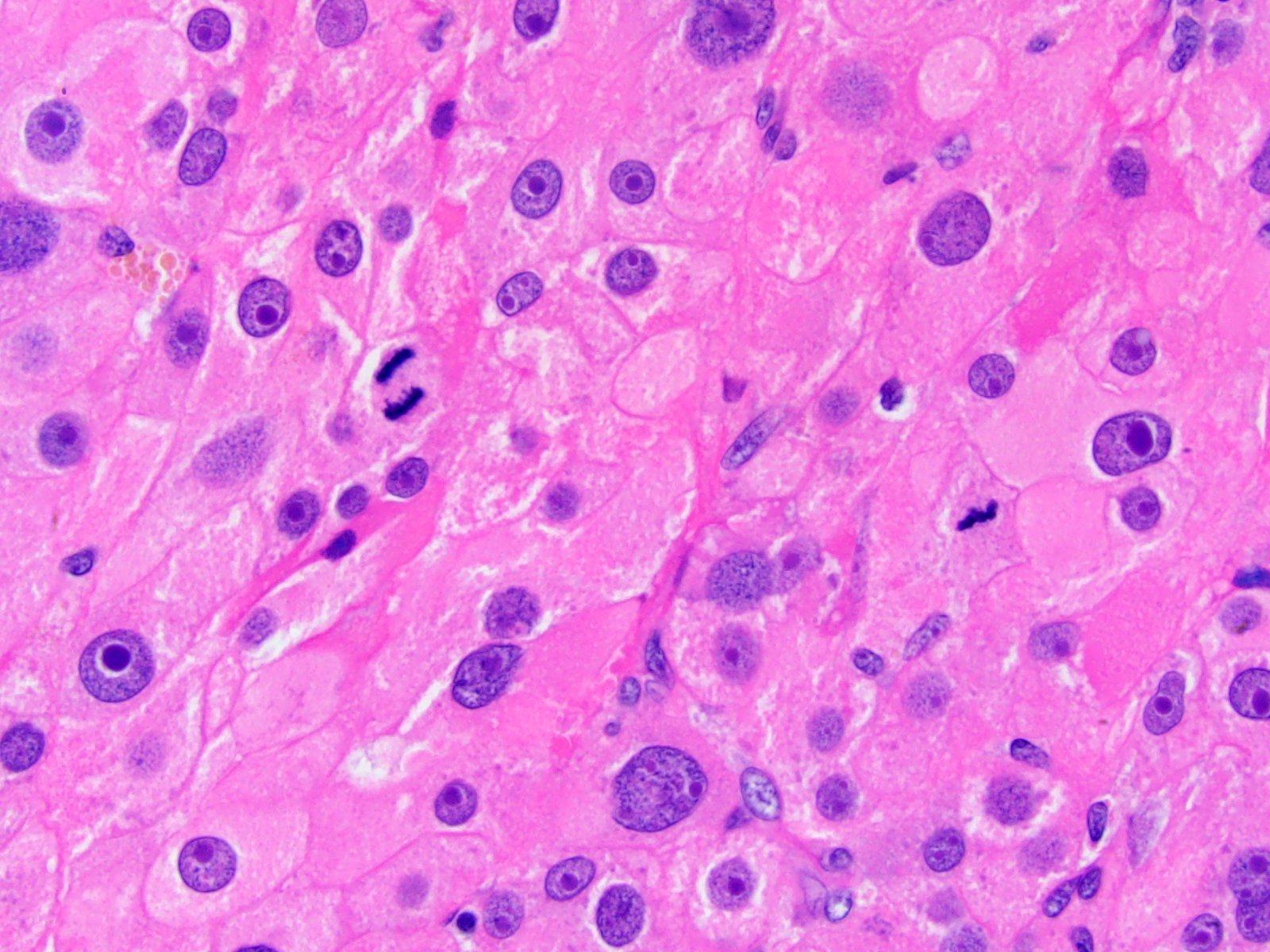
Scattered mature ganglion cells
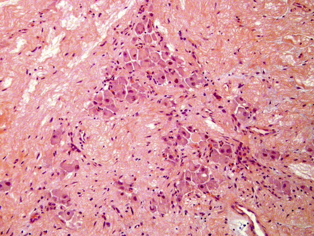
Cluster of mature ganglion cells
Intervening stroma
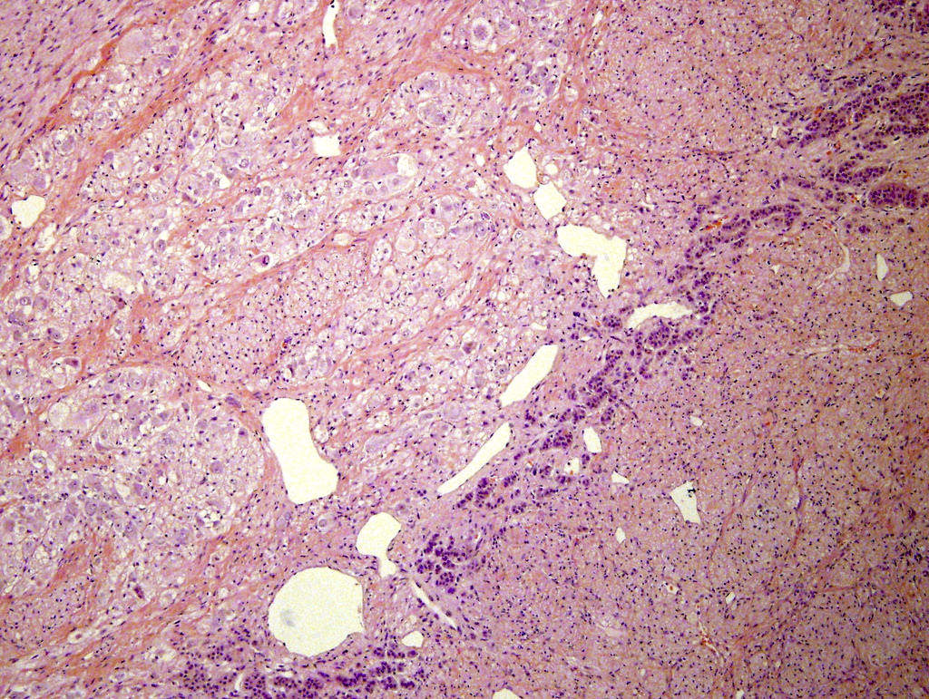
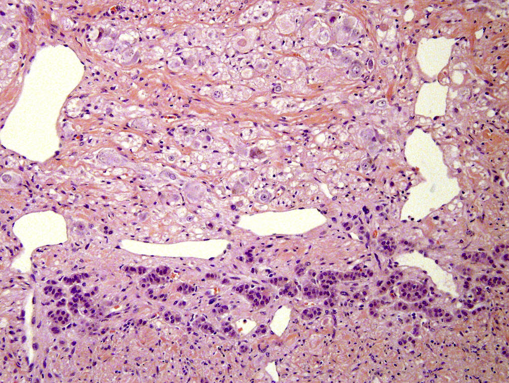
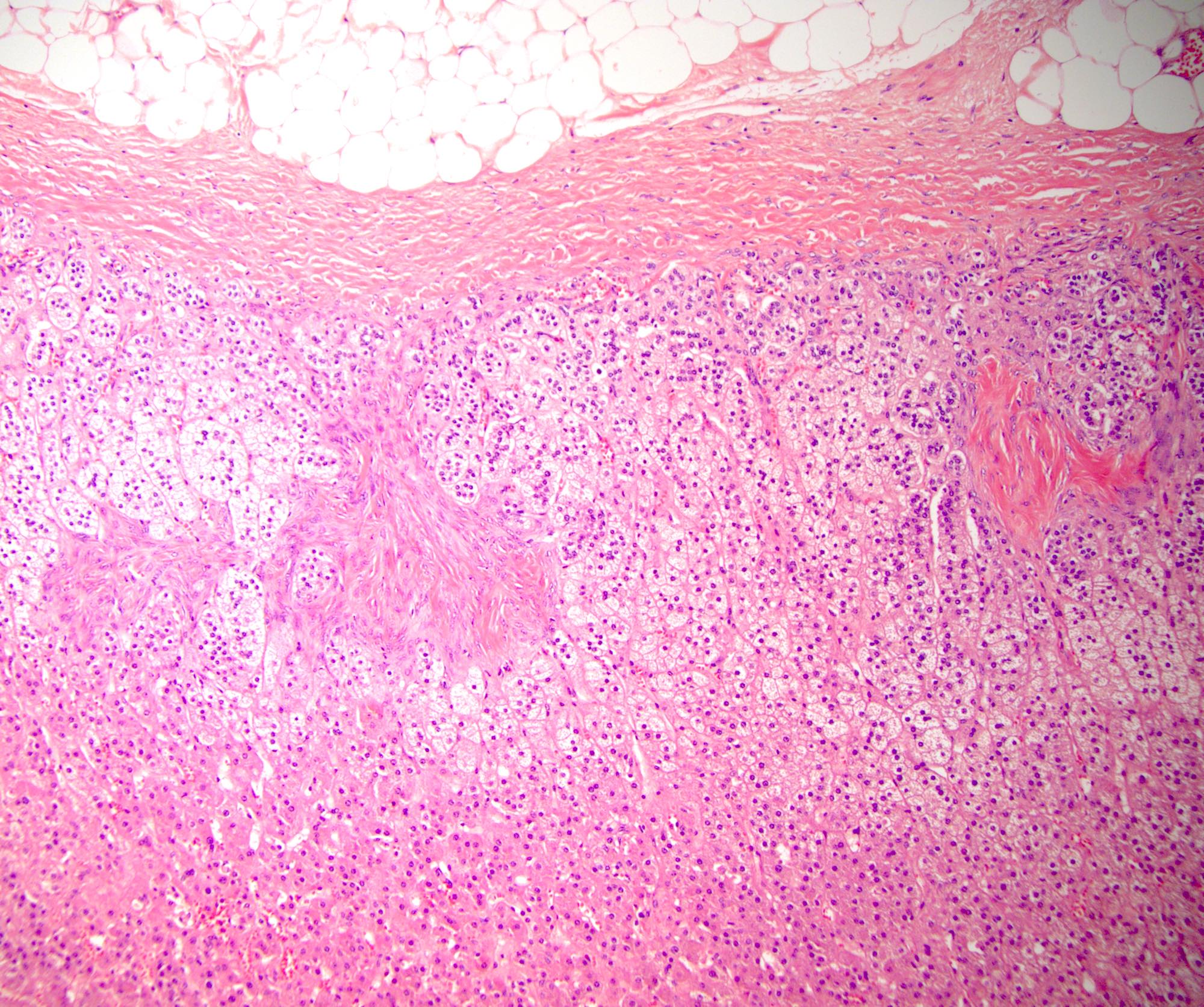
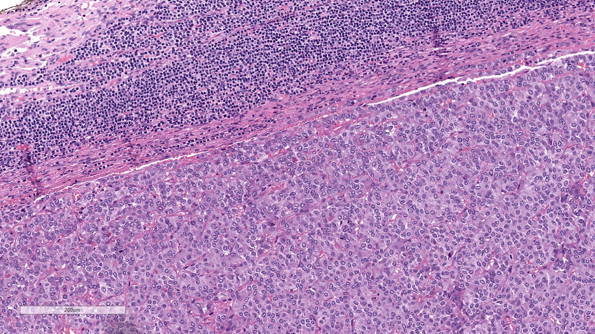
Ganglioneuroma (top) and residual normal adrenal cortical cells (bottom)
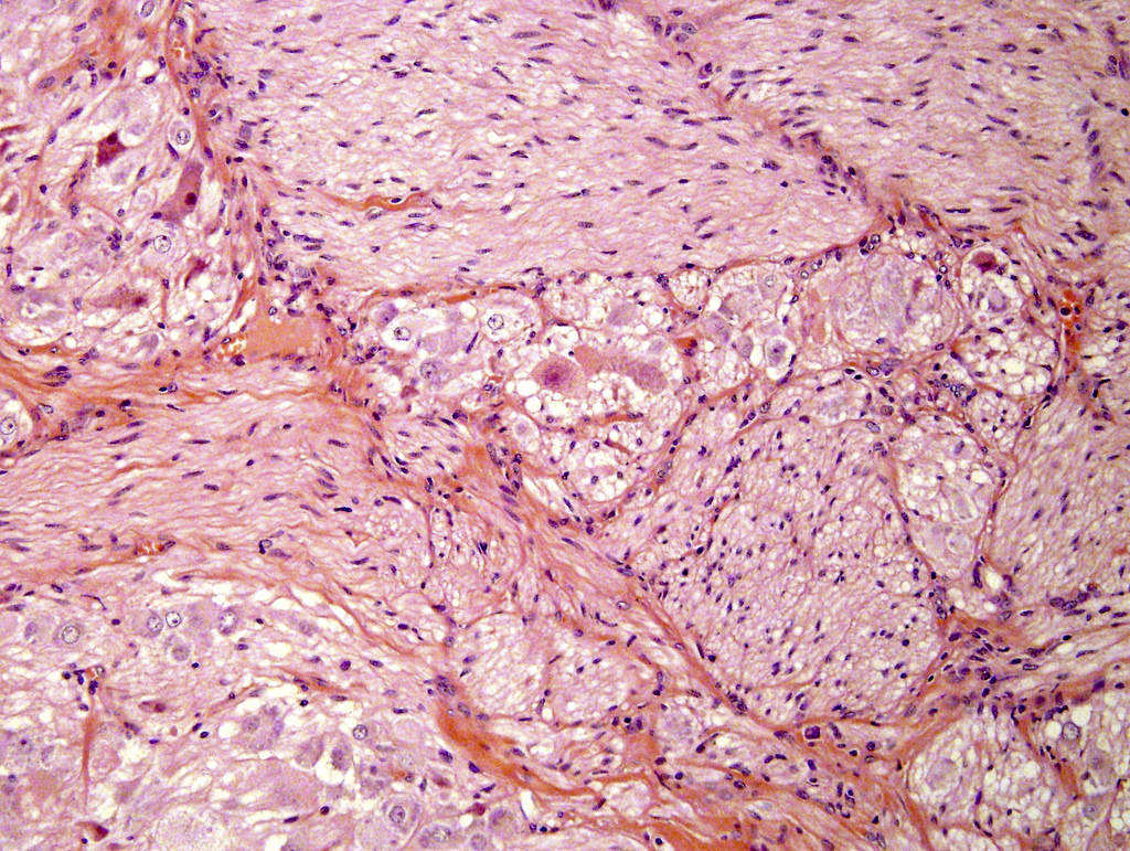
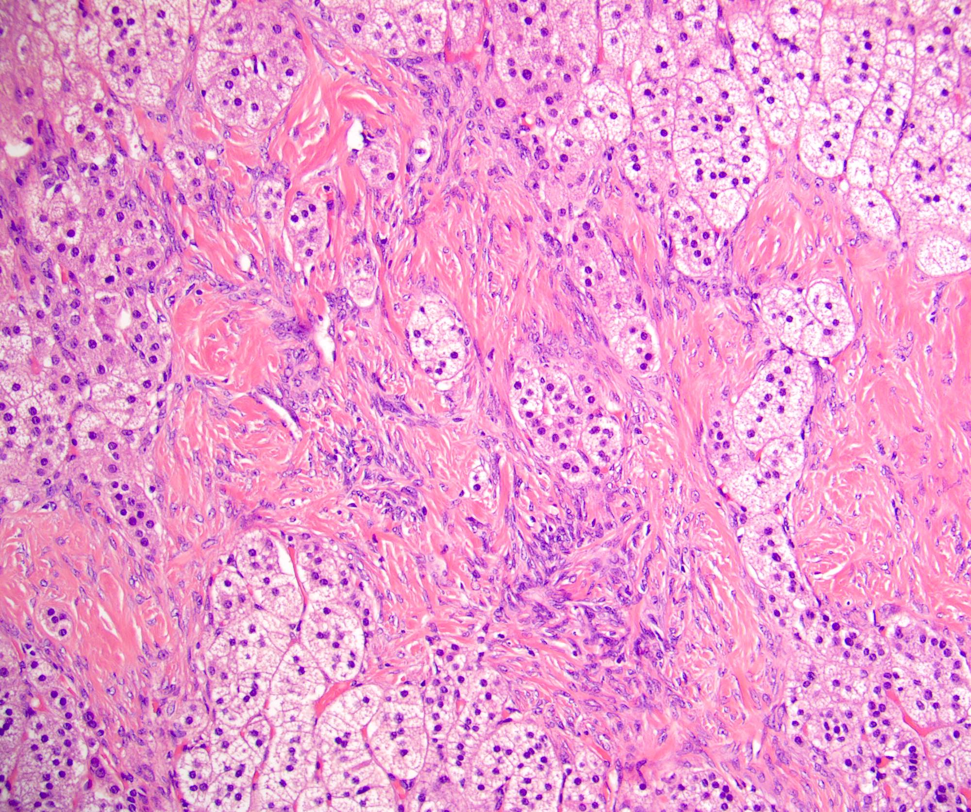
Mature ganglion cells admixed with stroma
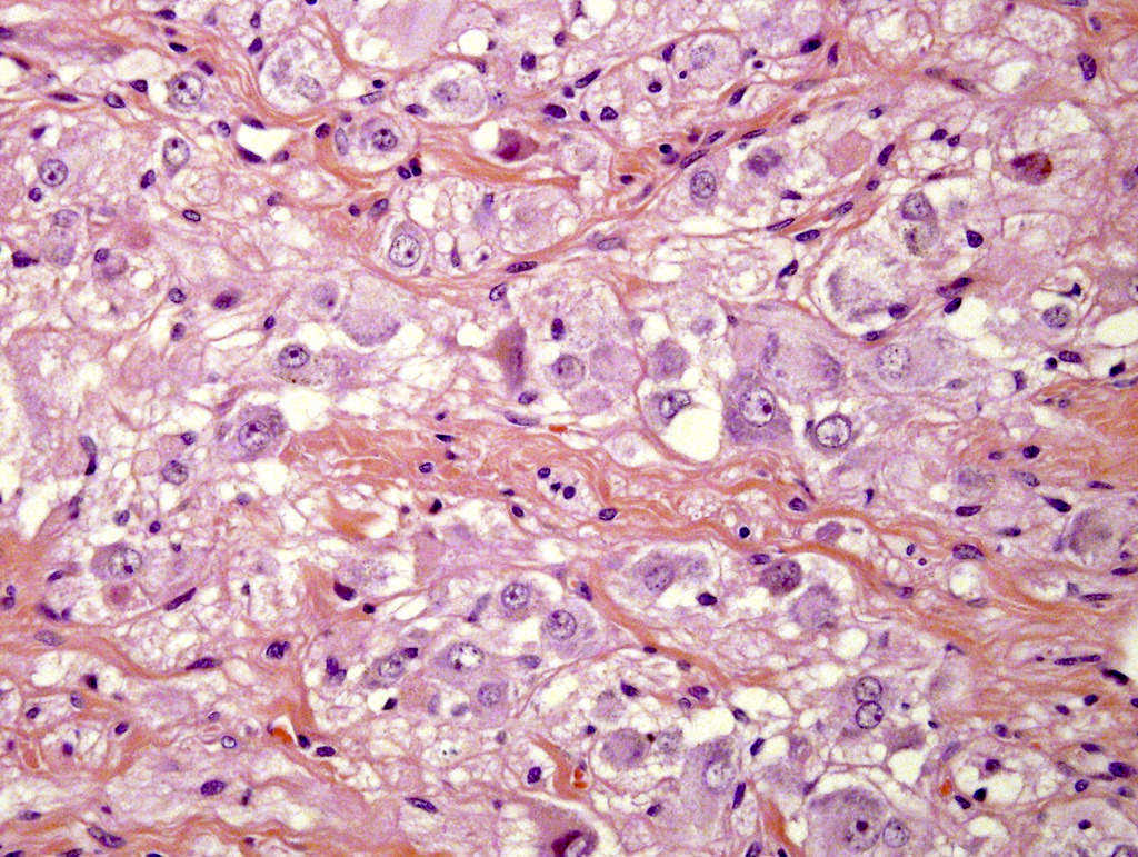
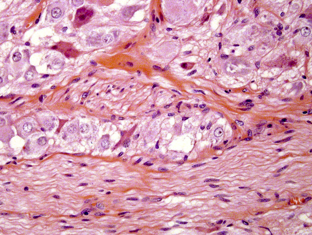
Mature ganglion cells


Adrenal ganglioneuroma
Images hosted on other servers:

Schwann cells and admixed ganglion cells (insets), neck

Schwann cells (a, c) and ganglion cells (b, d), neck
Images hosted on other servers:

Left adrenal gland
Images hosted on other servers:

Diffuse and nodular adrenal medullary hyperplasia
Contributed by Lan L. Gellert, M.D., Ph.D.

Adrenal medullary hyperplasia
Images hosted on other servers:

Unilateral adrenal metastasis from breast cancer


Right renal mass and the left adrenal metastatic mass on CT

Adrenal metastasis from
colon adenocarcinoma
before and after
chemotherapy
Images hosted on other servers:

Adrenal metastasis from ovarian carcinoma
Contributed by Lan L. Gellert, M.D., Ph.D.
Adrenal metastasis from lung adenocarcinoma
Adrenal metastasis from lung small cell carcinoma
Adrenal metastasis from clear cell renal cell carcinoma

Adrenal metastasis from melanoma
Images hosted on other servers:

Adrenal metastasis
from moderately
differentiated sigmoid
colon adenocarcinoma


Adrenal metastasis from ovarian clear cell carcinoma
Images hosted on other servers:

Abdominal US

CT abdomen findings

MRI findings

Giant bilateral myelolipomas of abdomen

CT axial section showing mass in right renal gland
Images hosted on other servers:

Large suprarenal mass
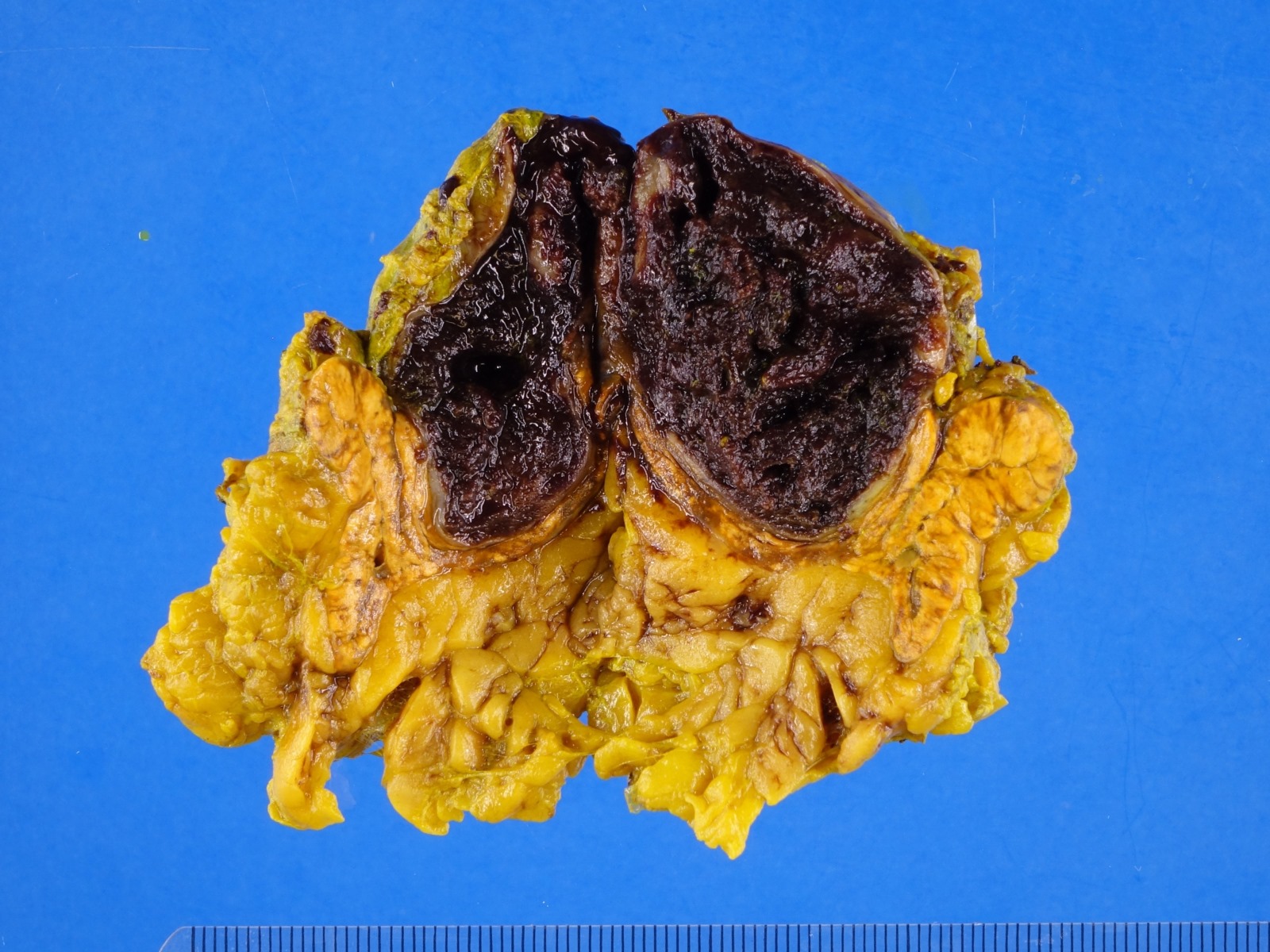
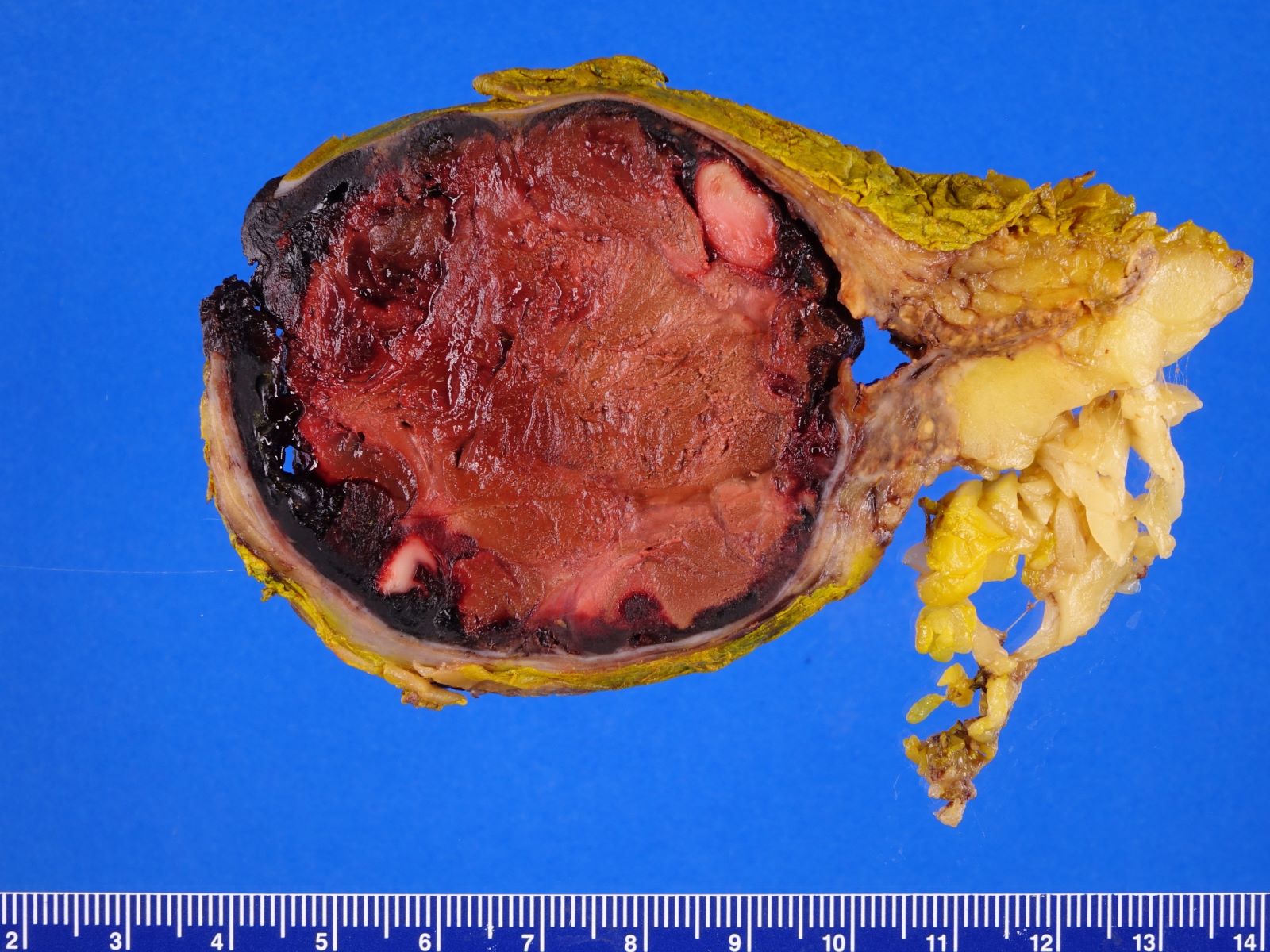
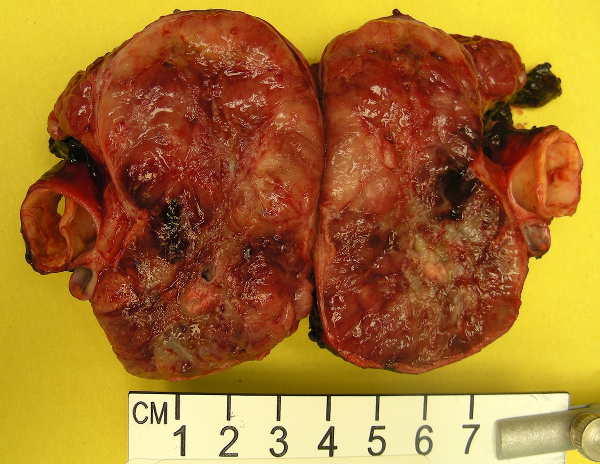
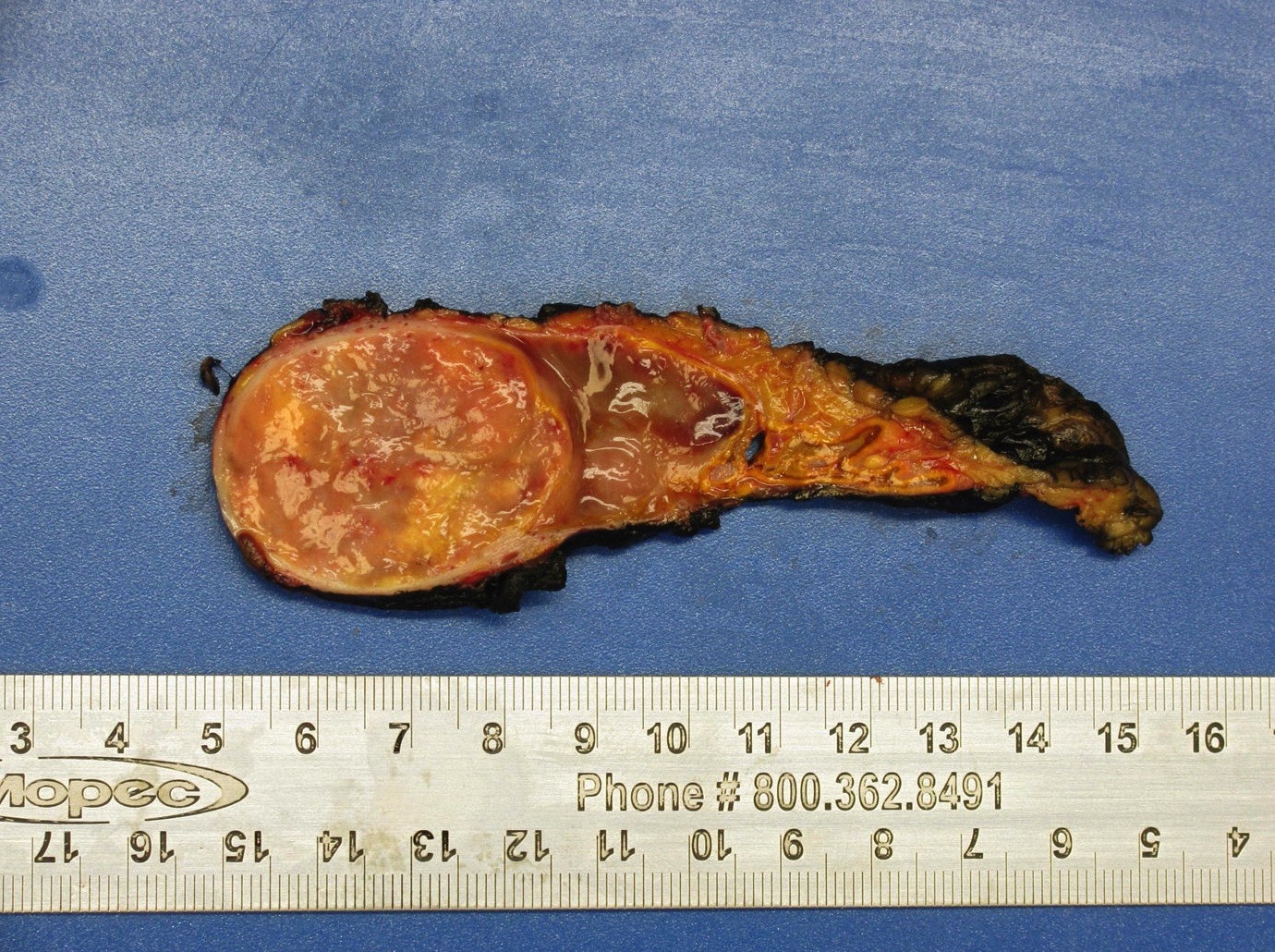
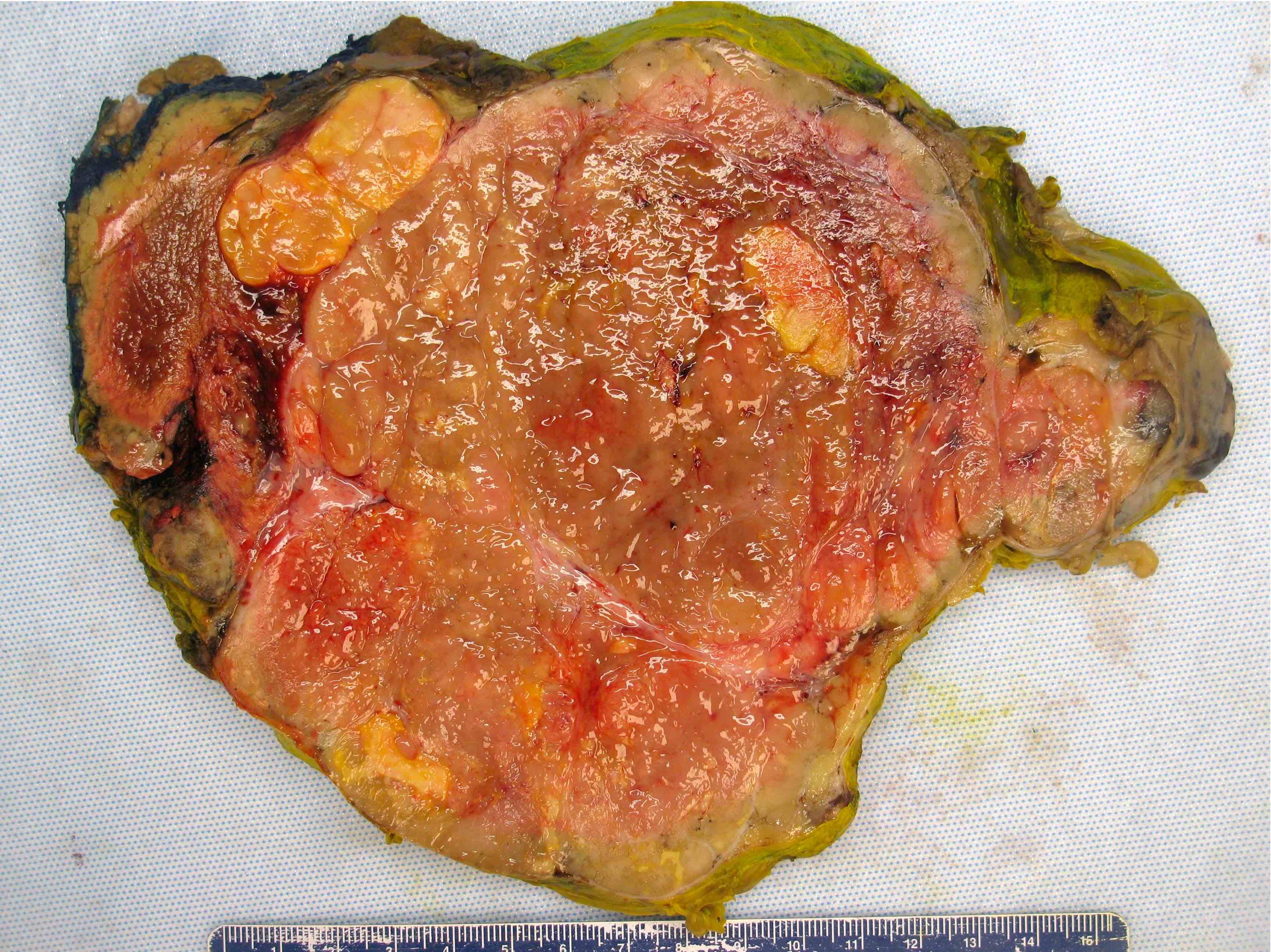
Contributed by Debra L. Zynger, M.D. and Anil Parwani, M.D., Ph.D.
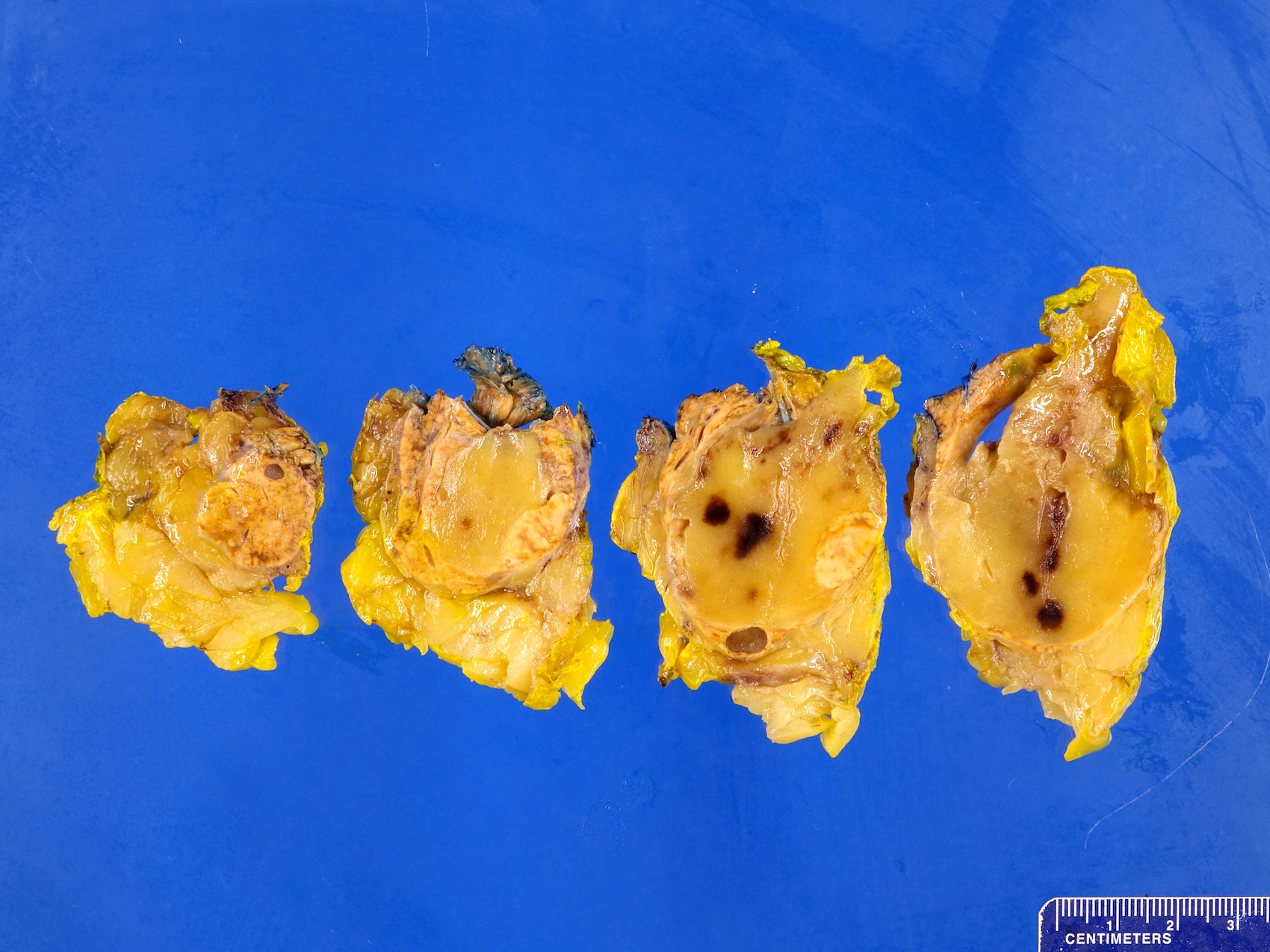
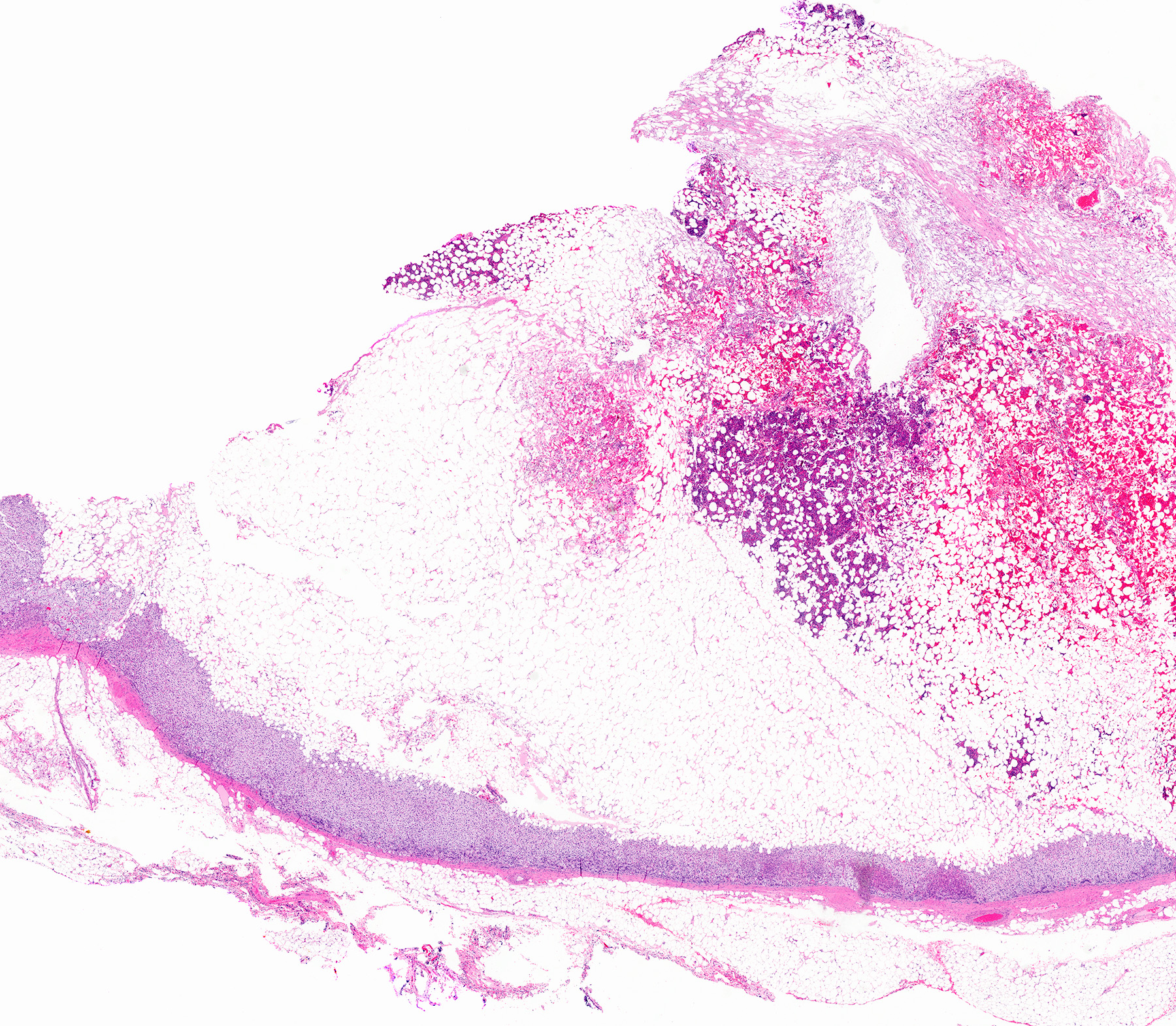
Fat predominant
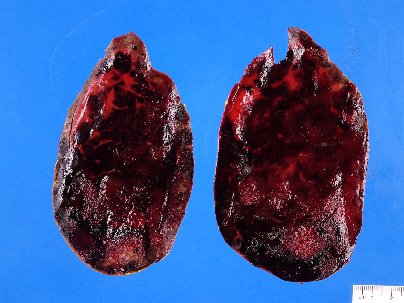
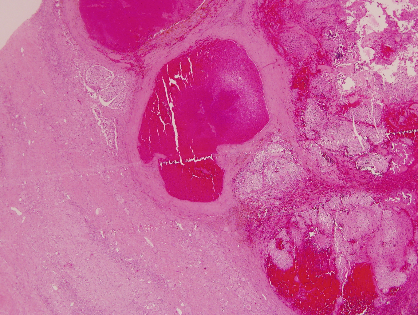
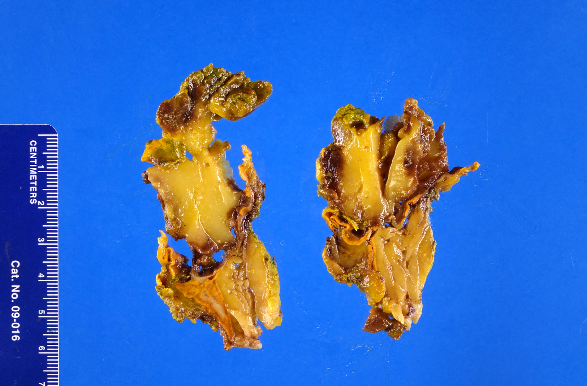
Extensive hemorrhage
Images hosted on other servers:


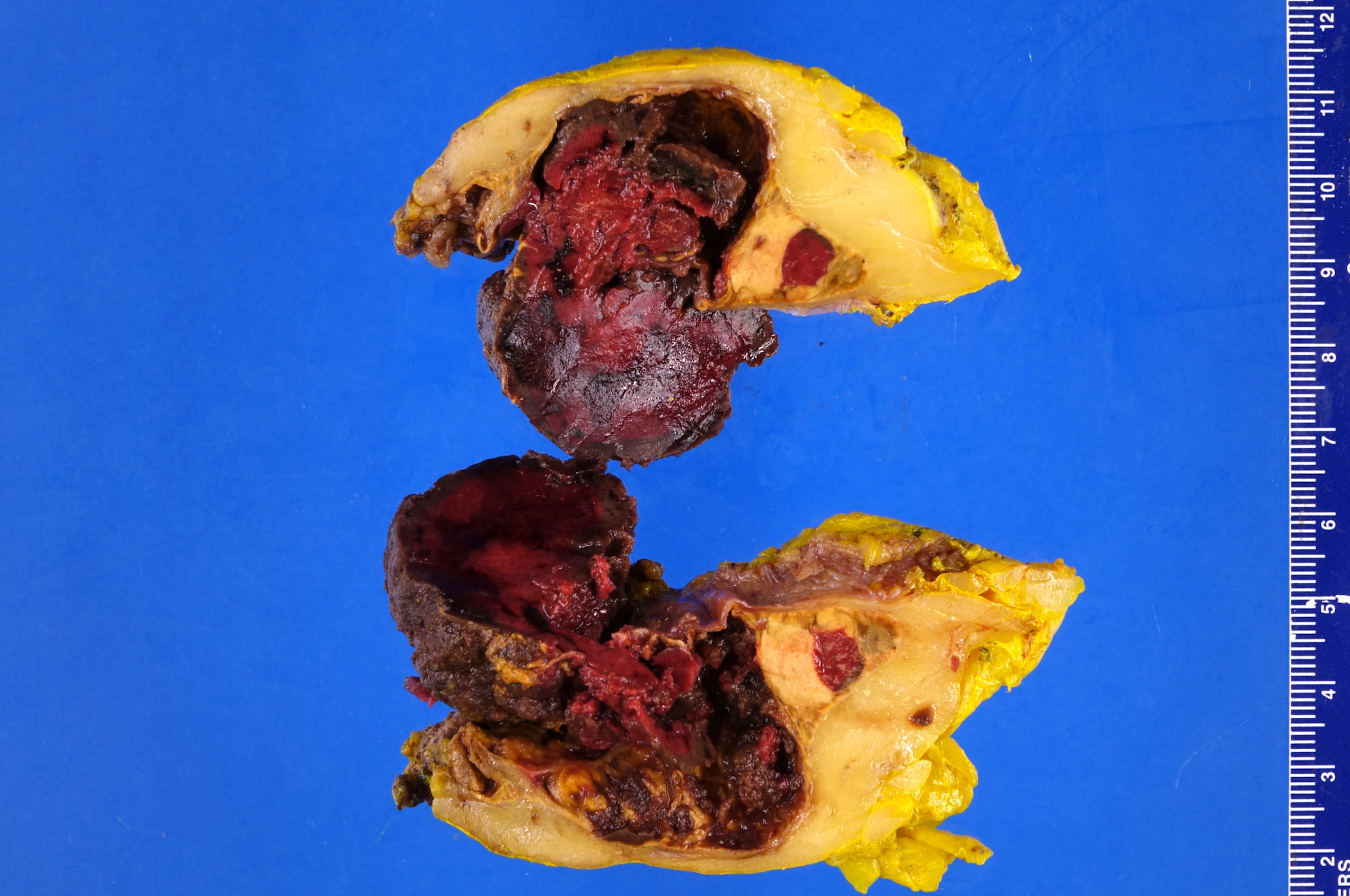
Intact adrenal specimen


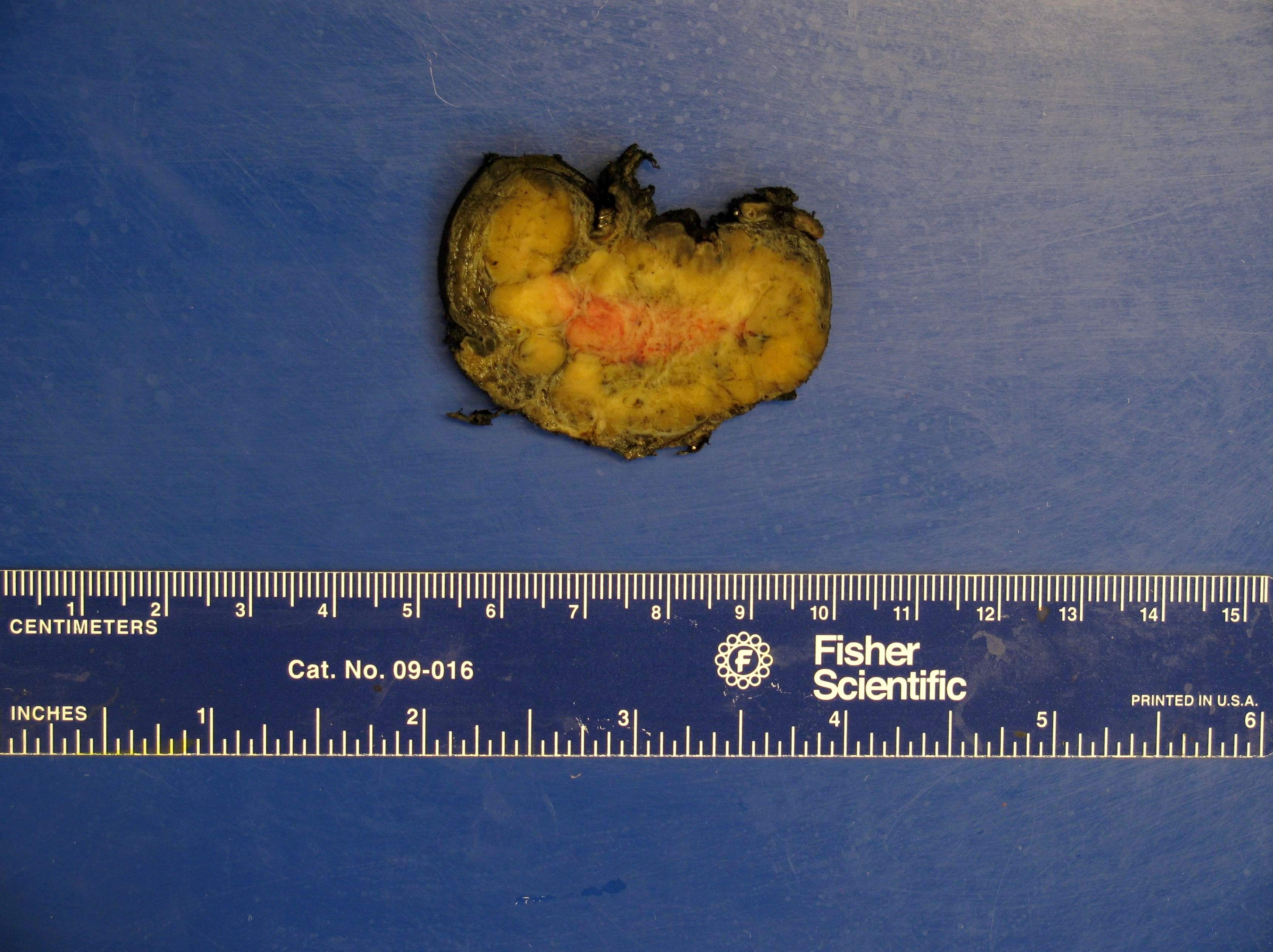
Bright yellow and focally red, fatty tumor with extensive hemorrhage

Grey / brown / yellowish with hemorrhage

Well circumscribed
tumor with hemorrhage
Contributed by Debra L. Zynger, M.D., Anil Parwani, M.D., Ph.D., O. Hans Iwenofu, M.D., Ph.D. and @ThatGlassTho on Twitter
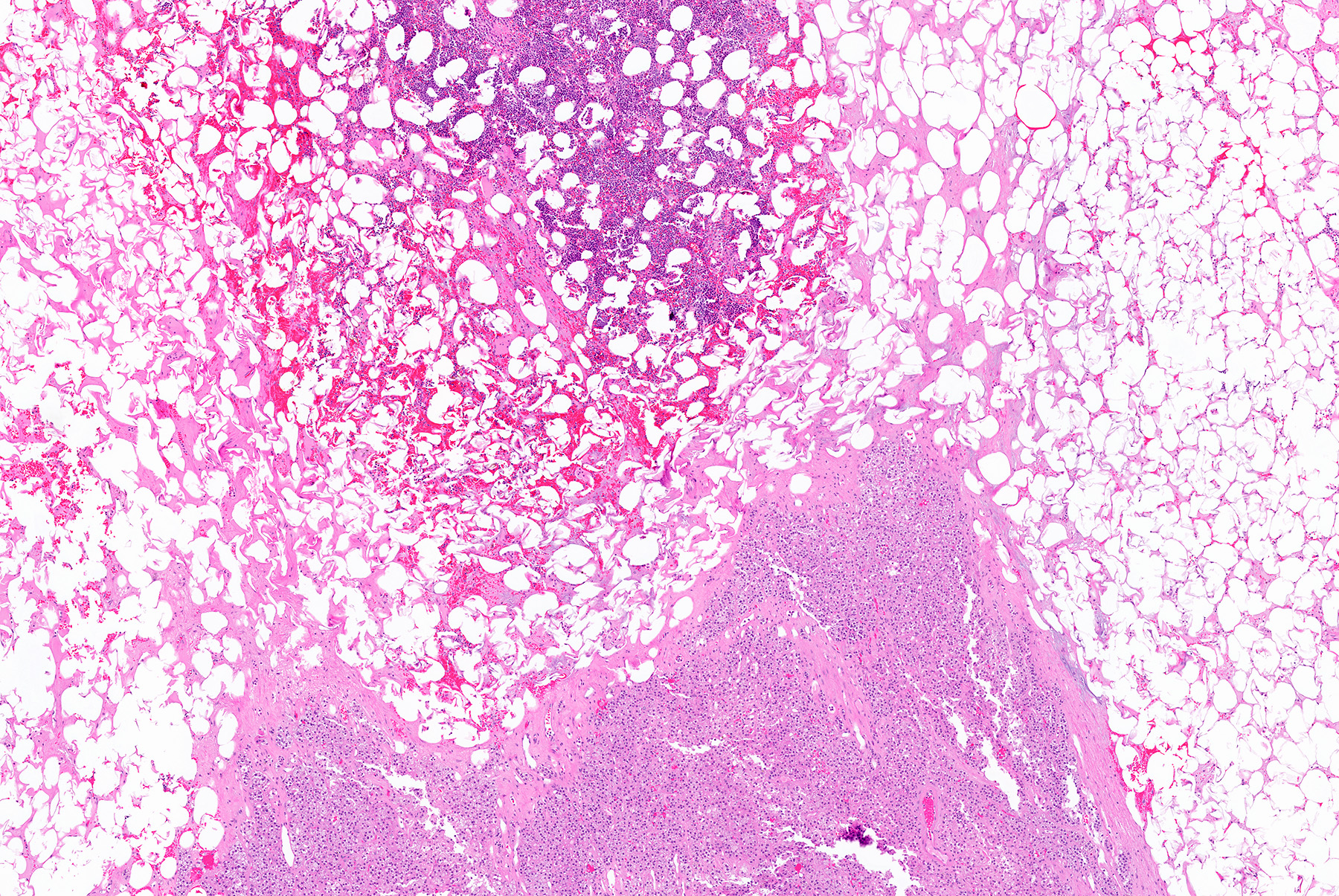
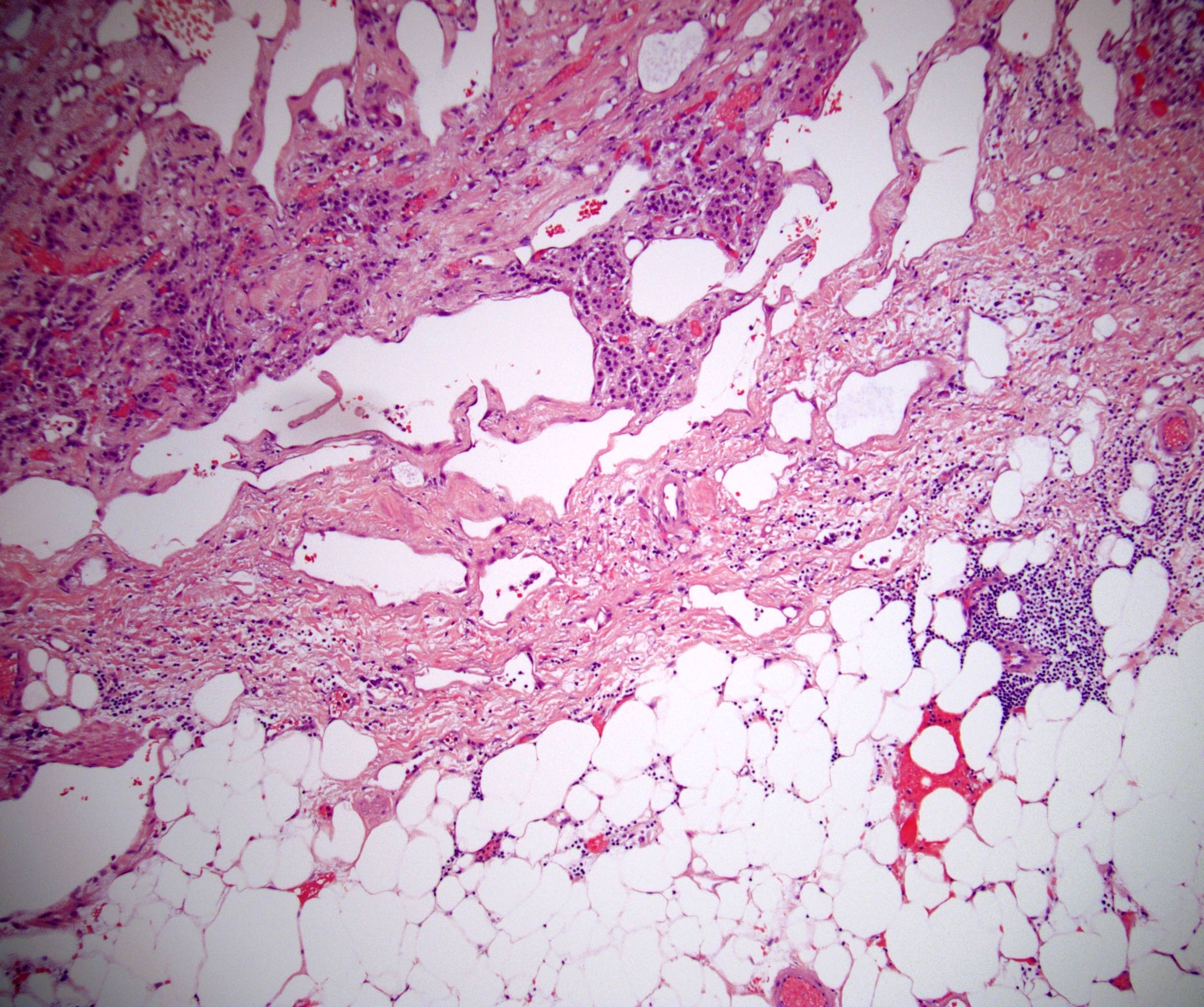
Hematopoietic cells
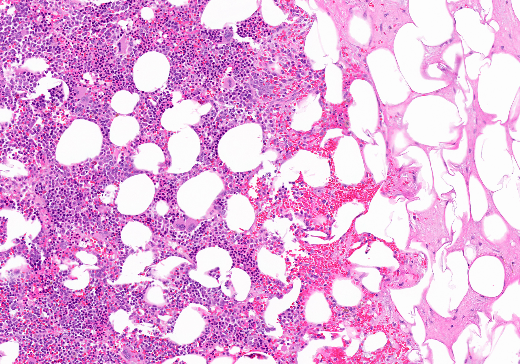
Increased megakaryocytes
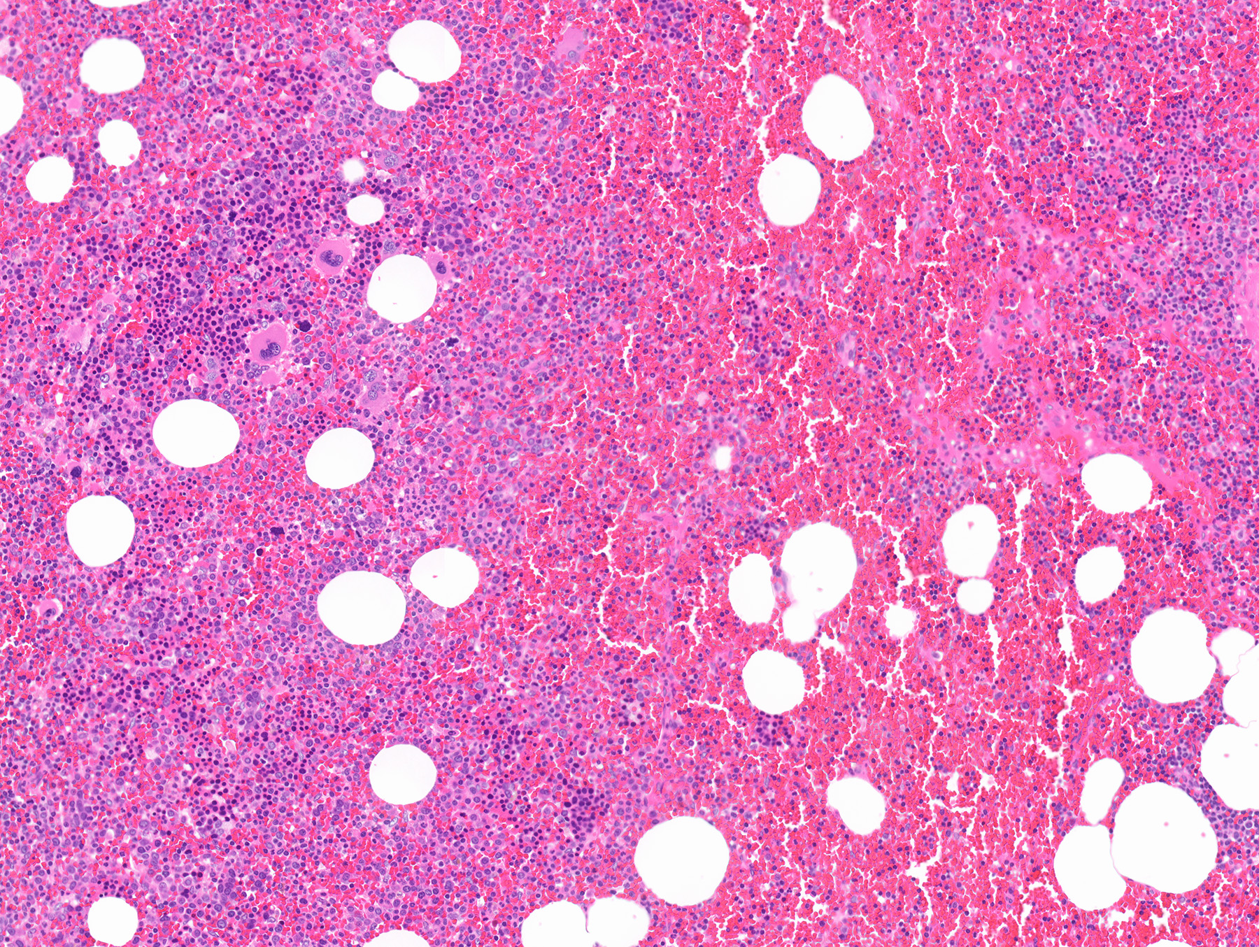
Hematopoietic cells and mature fat
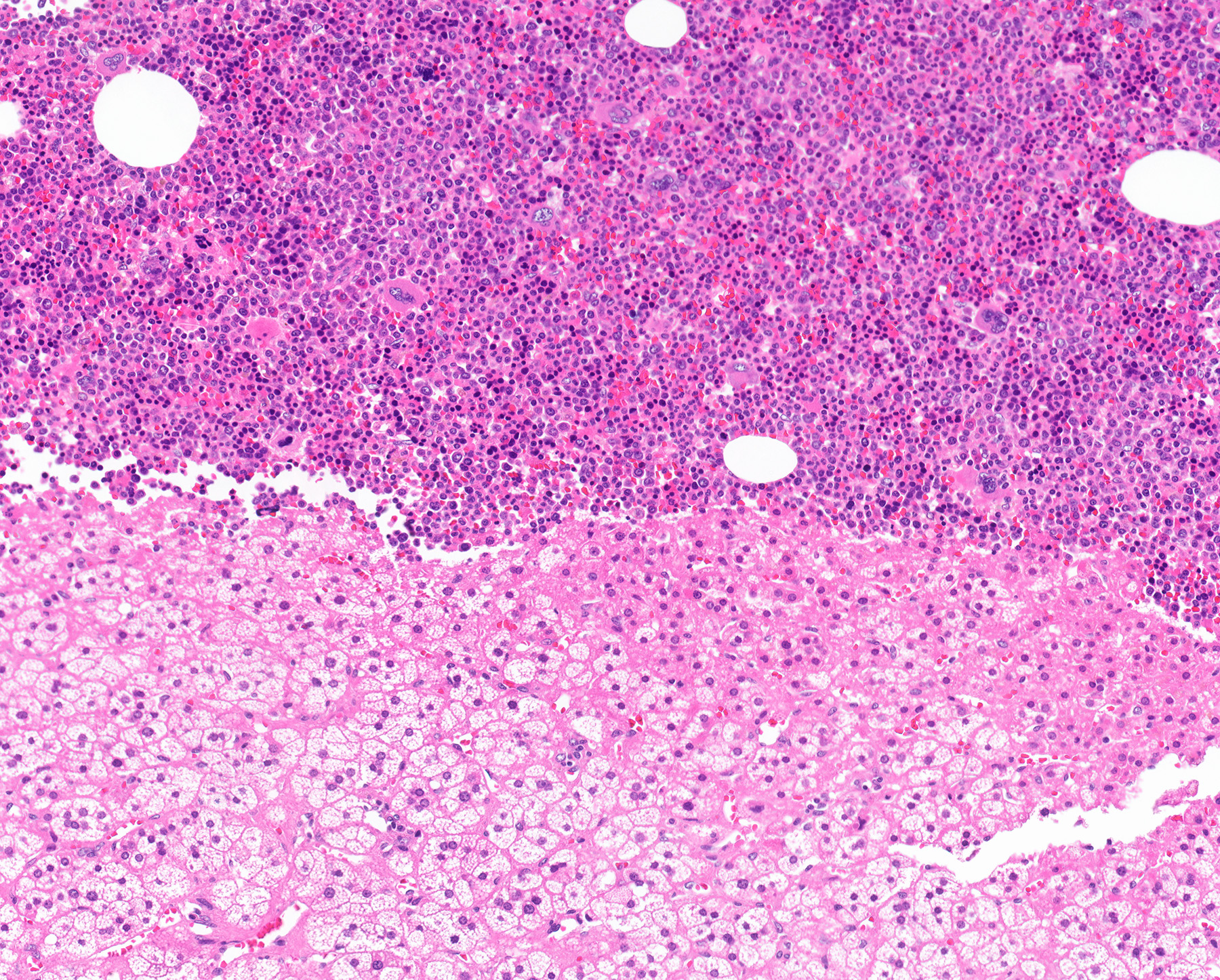
Hematopoietic cells and adrenal cortex

Myelolipoma in adrenal gland
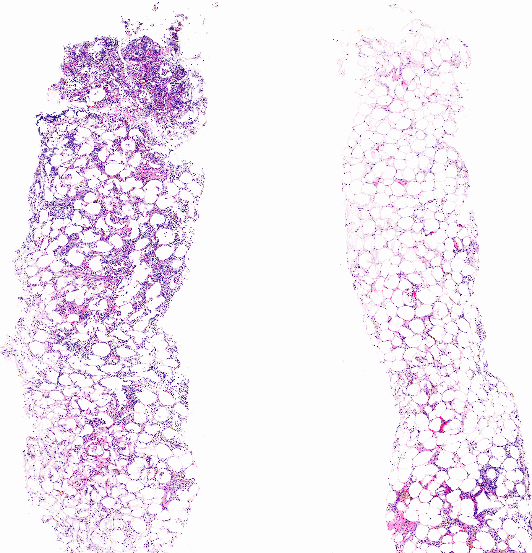
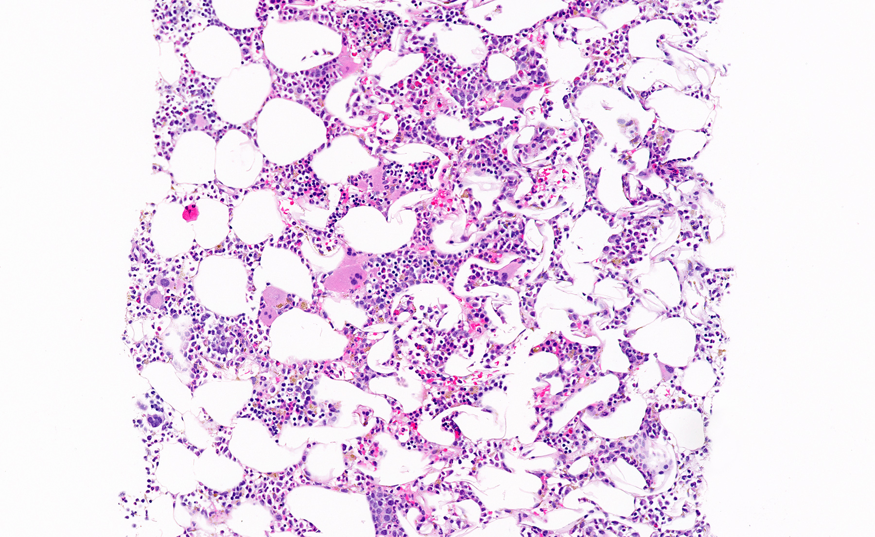
Hematopoietic cells on core biopsy

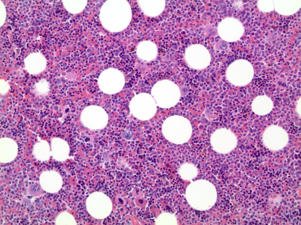
Myelolipoma
Images hosted on other servers:

Survival analysis comparing myxoid ACC to other variants
Images hosted on other servers:

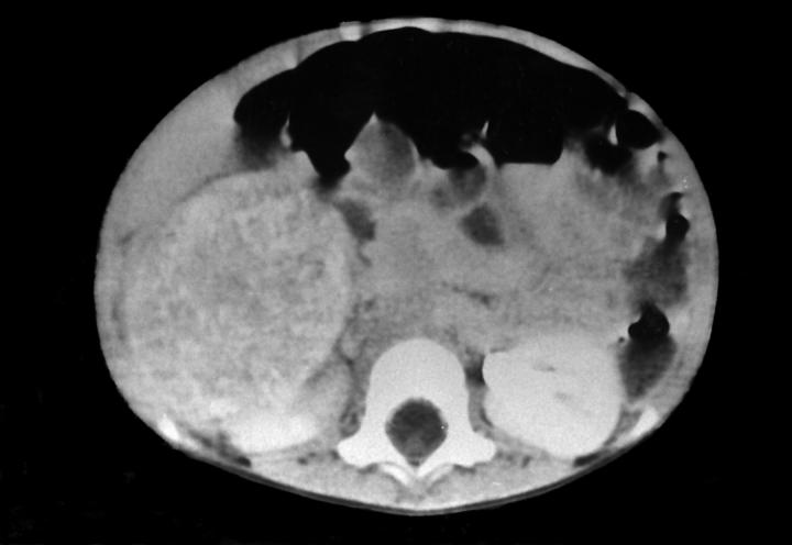
Myxoid ACC on CT, MRI and PET / CT imaging
Images hosted on other servers:

Robot assisted laparoscopic adrenalectomy
Images hosted on other servers:

Gelatinous and solid whitish appearance

Obvious areas of hemorrhage and necrosis
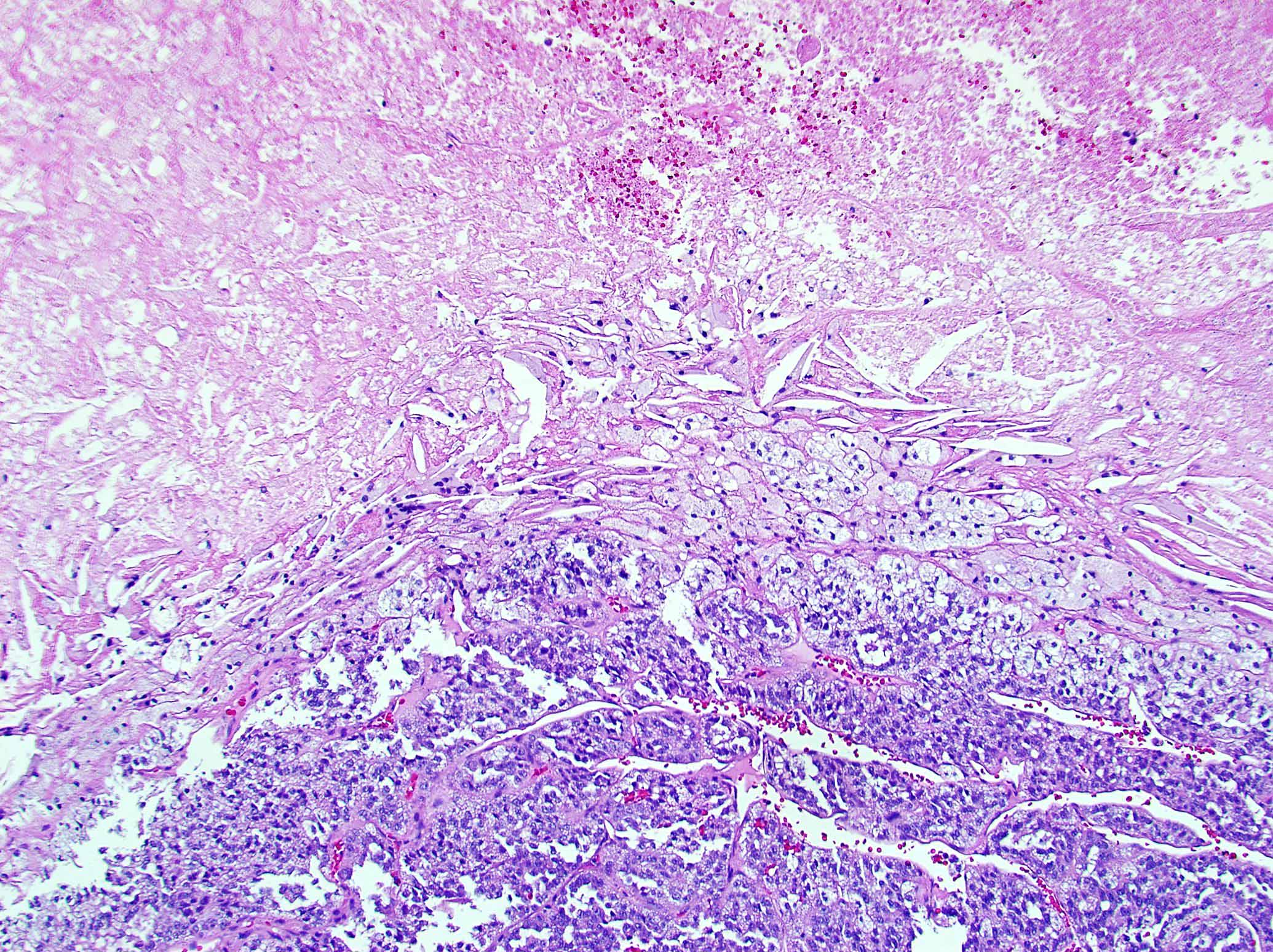
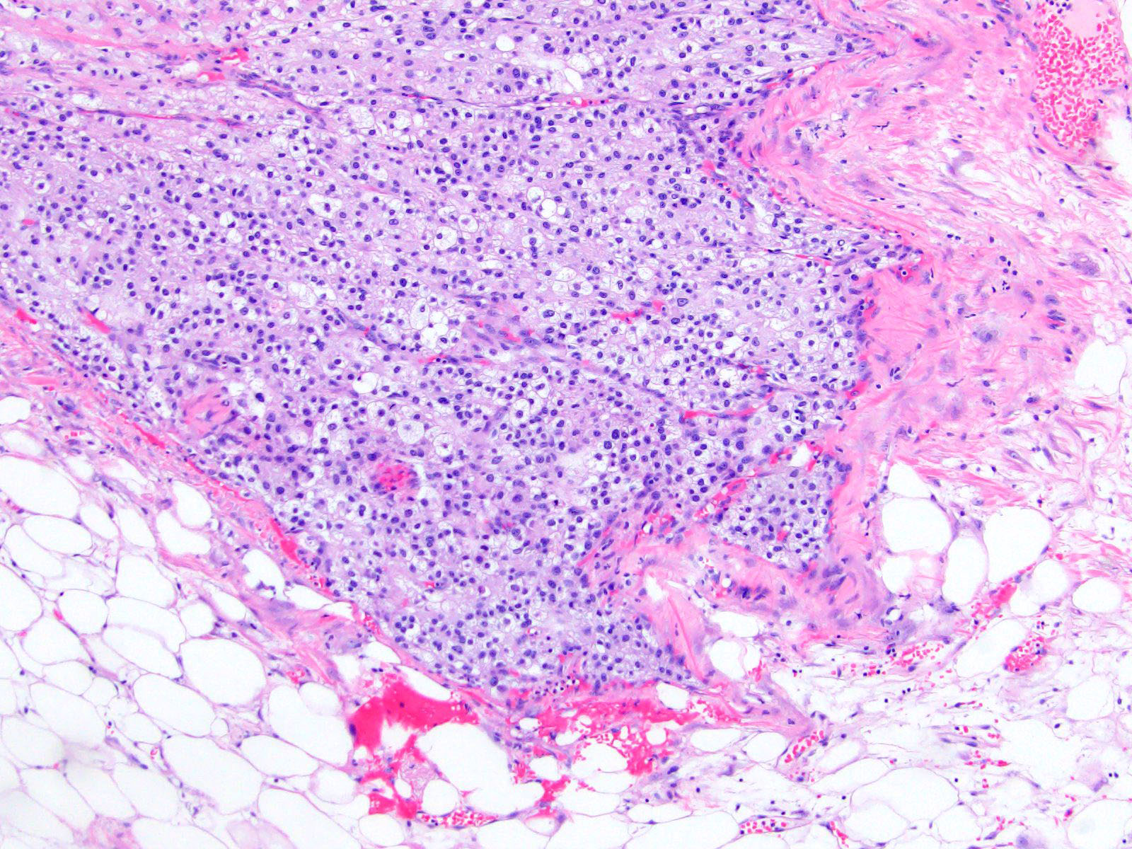
Contributed by Maria Tretiakova, M.D., Ph.D.

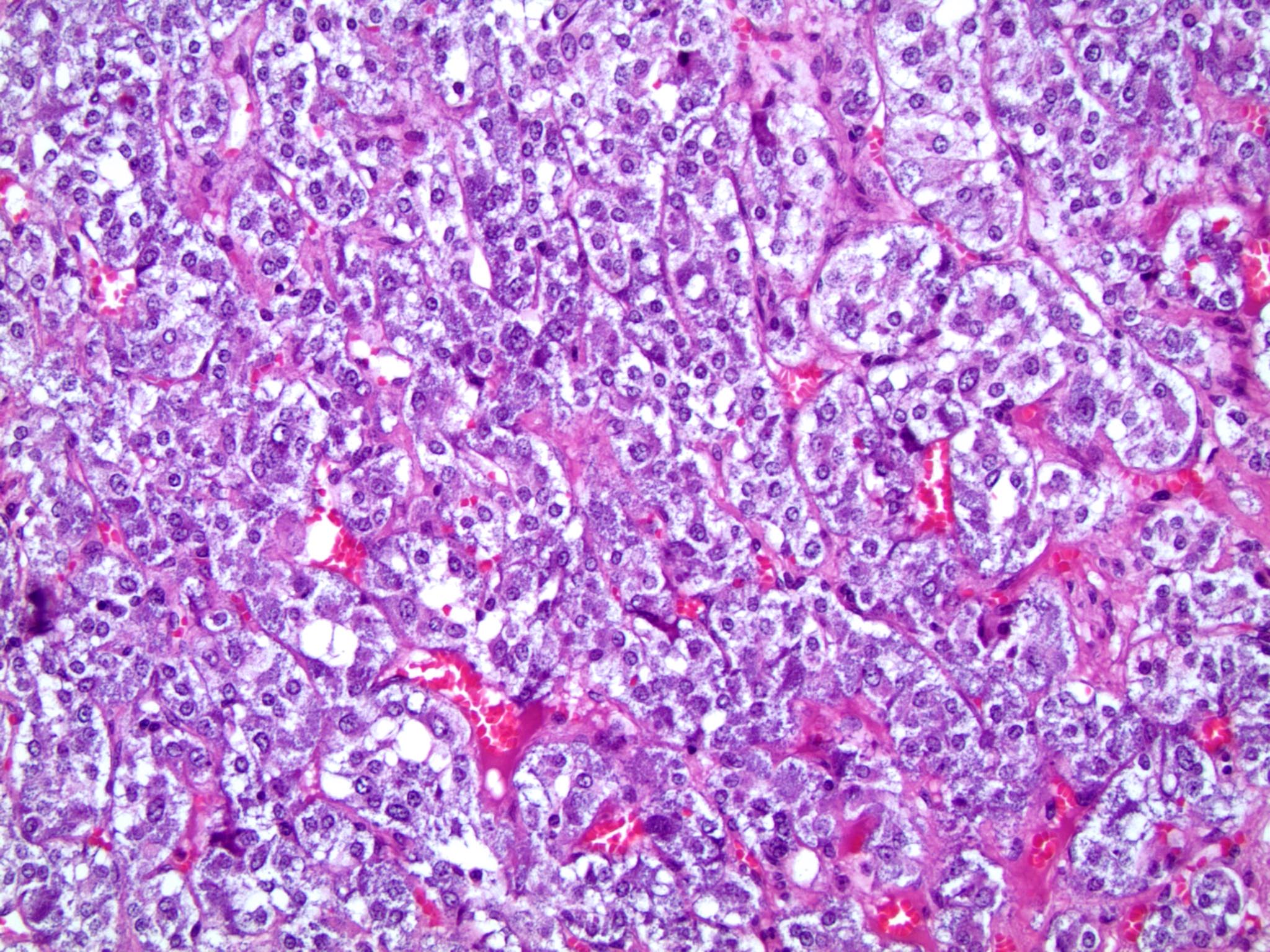
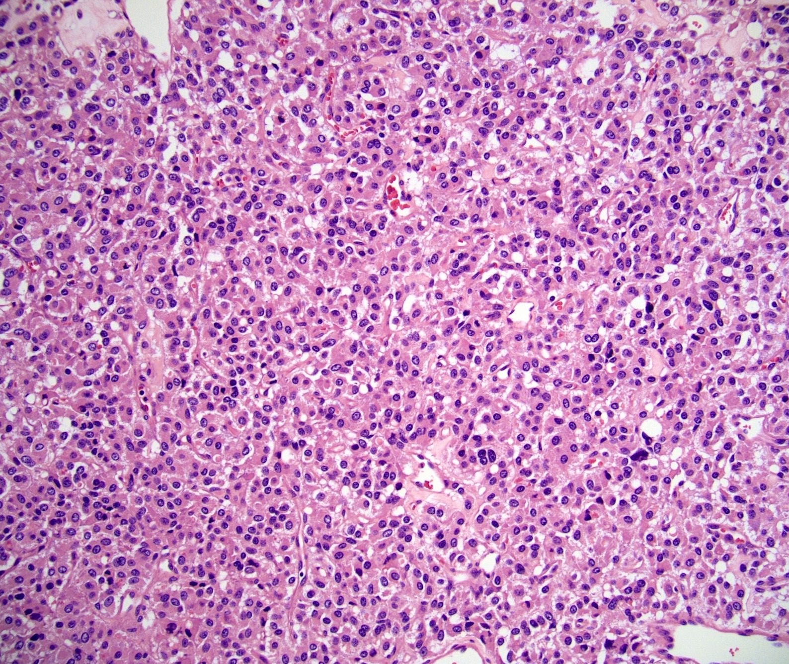
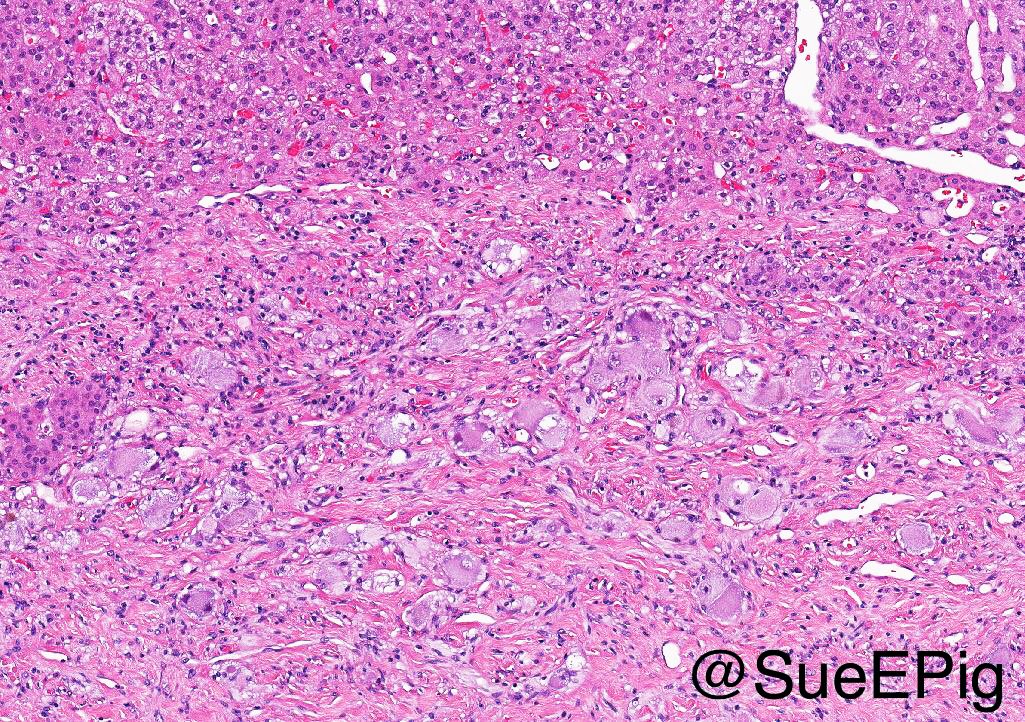
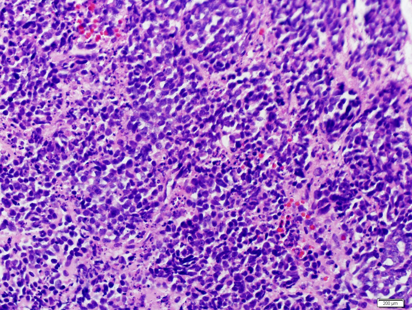
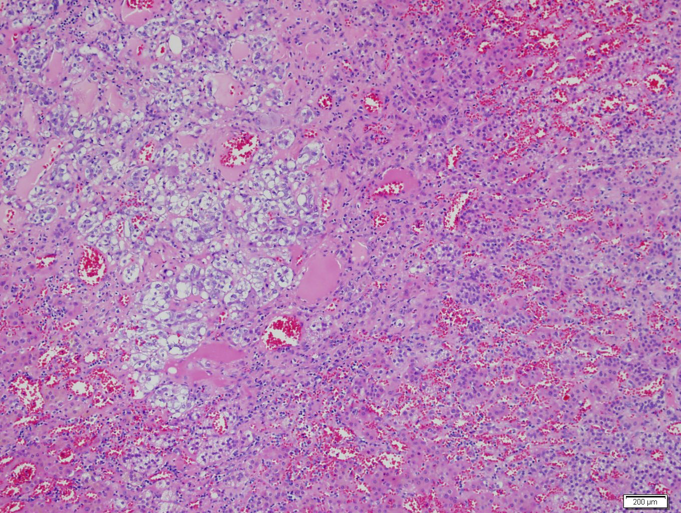
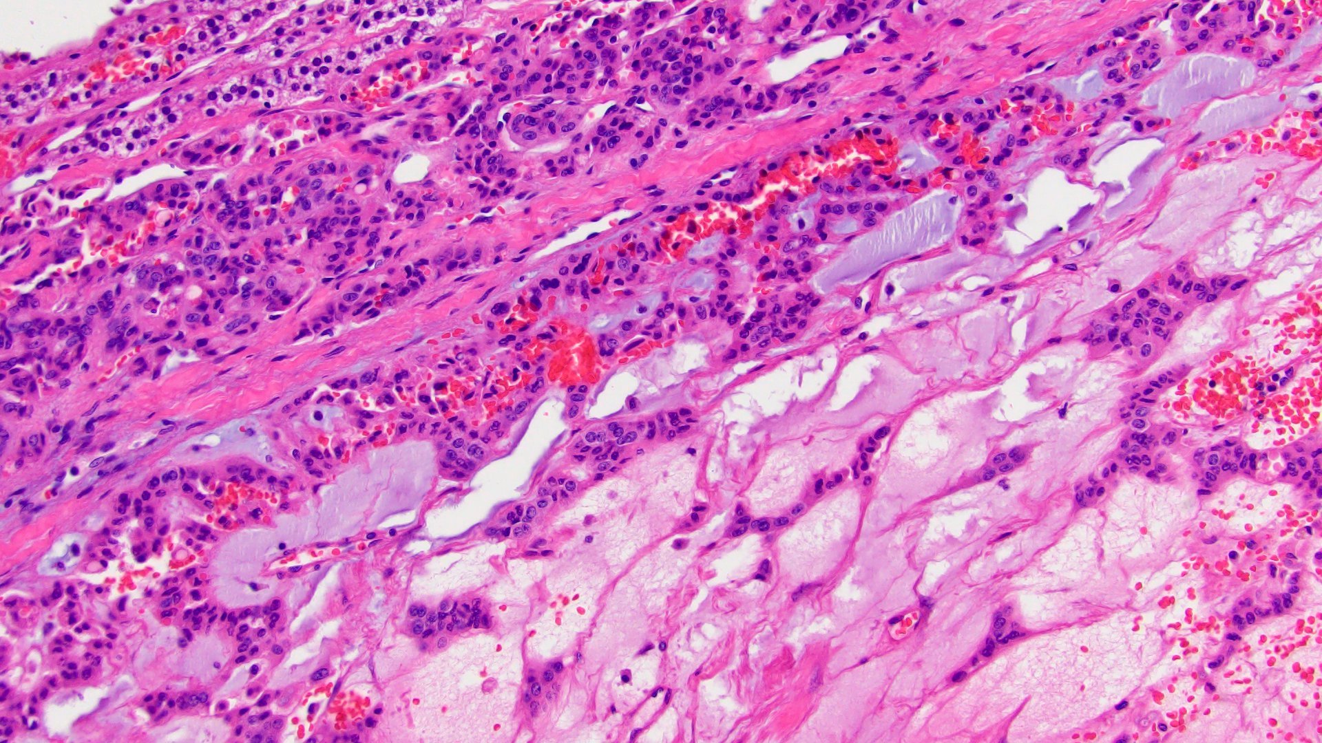
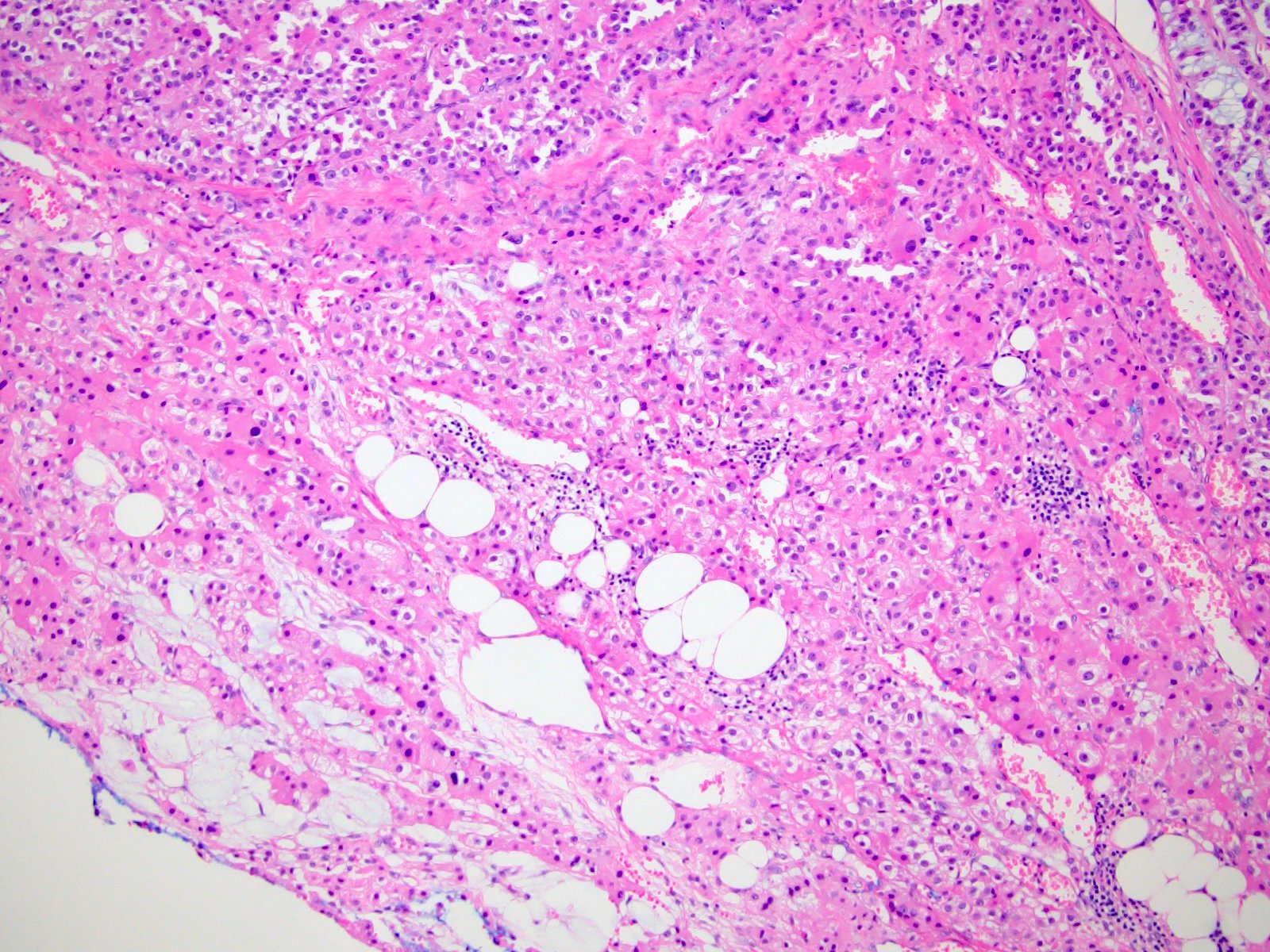
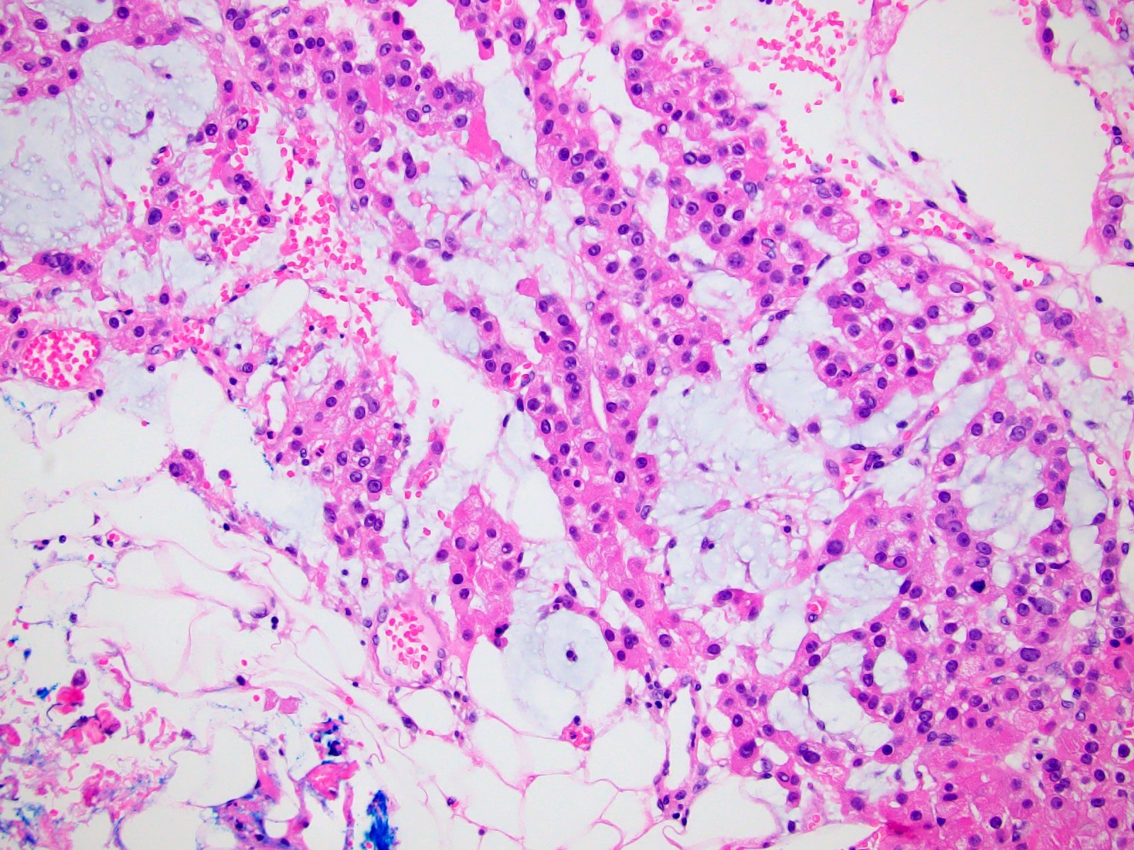
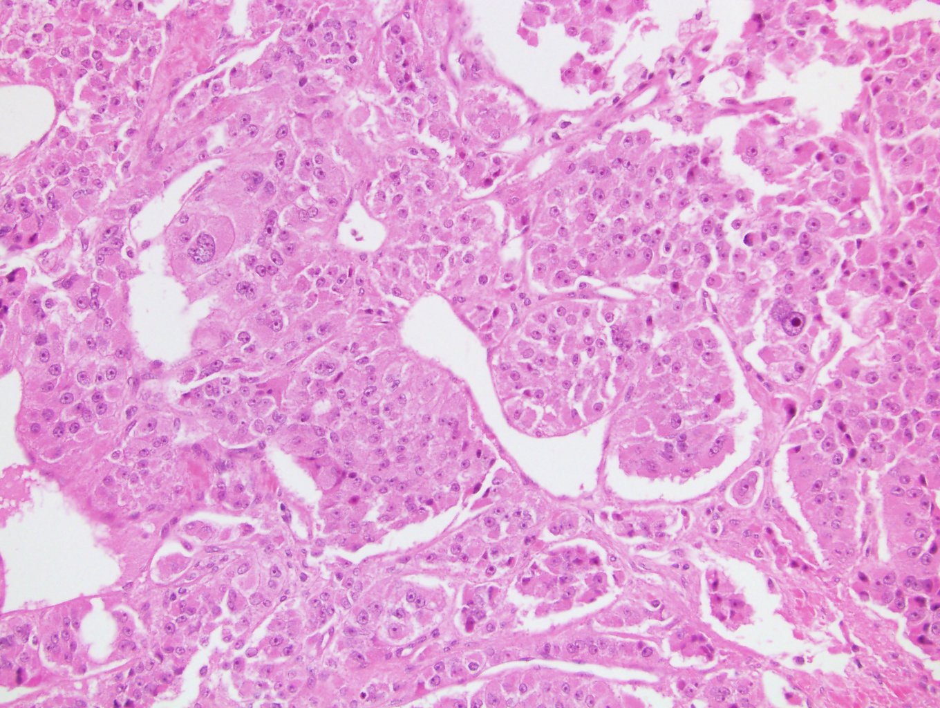
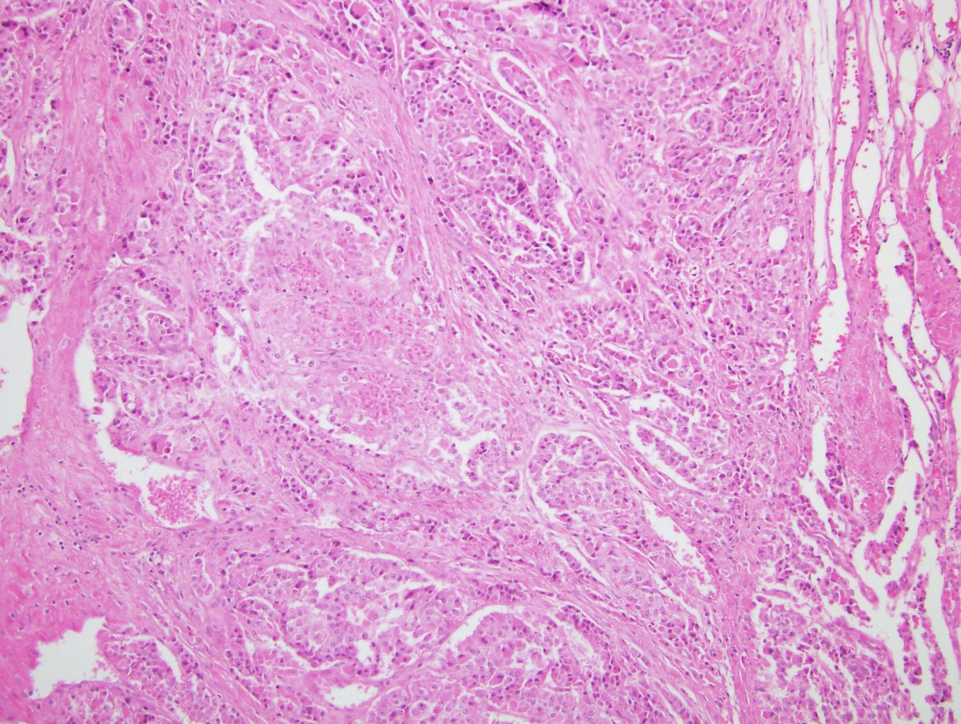
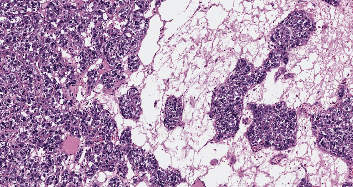
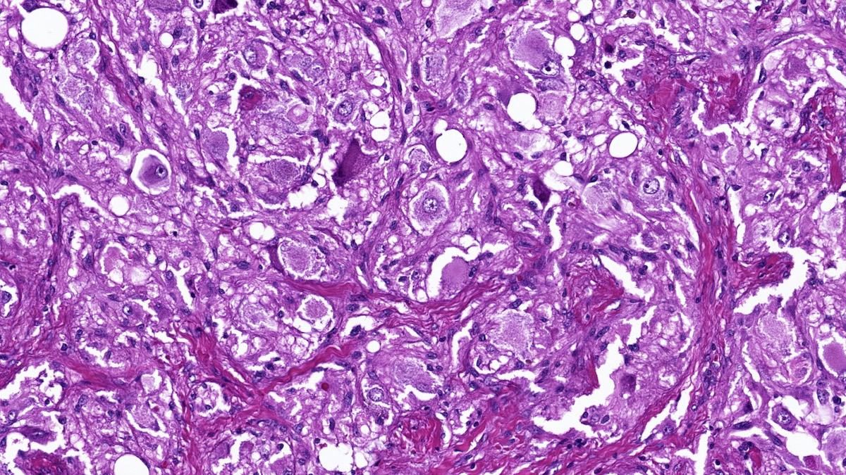
ACC with predominant myxoid component

Delicate arborizing cords

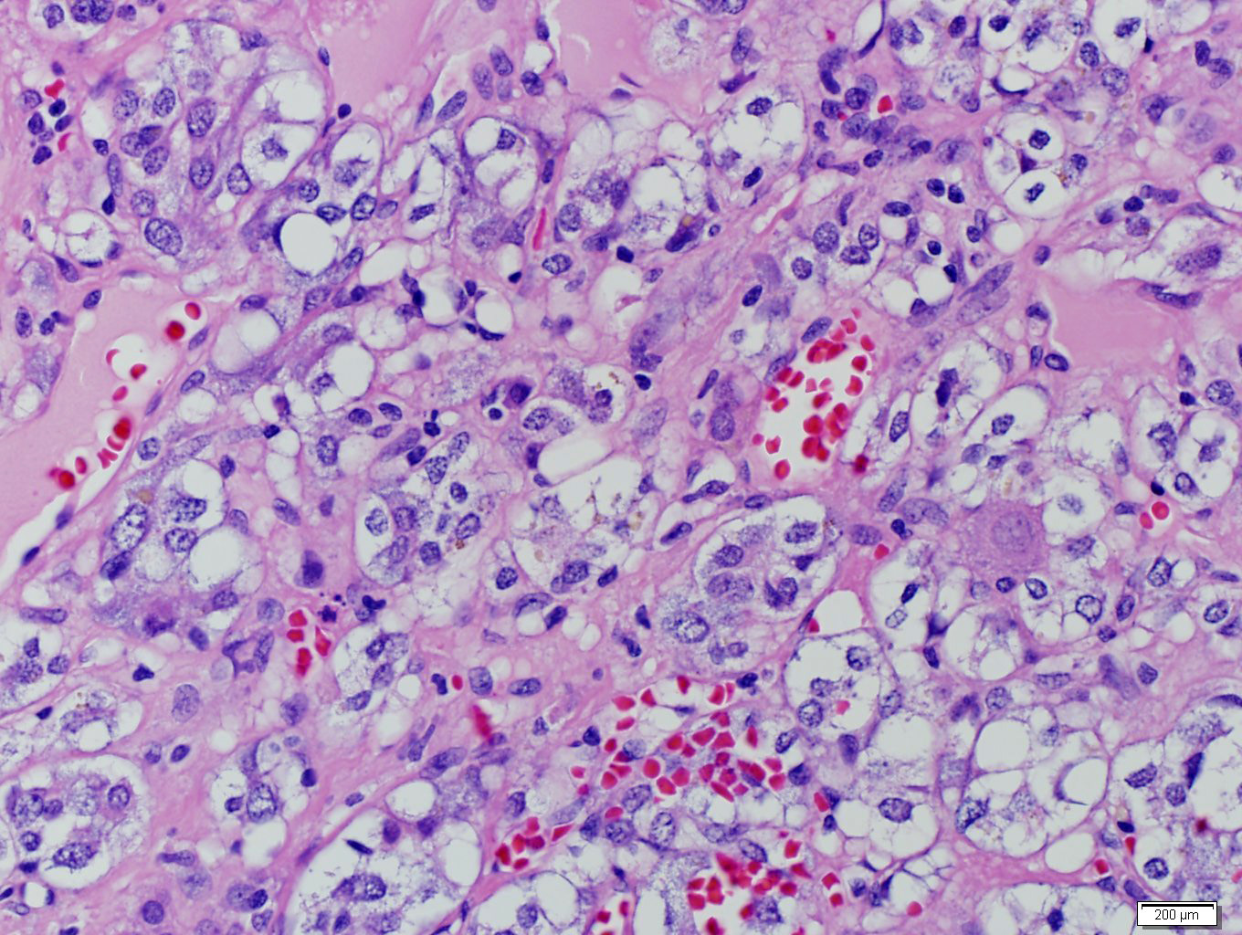
Mucin pools with floating cells
Pseudoglandular architecture
Complex architecture
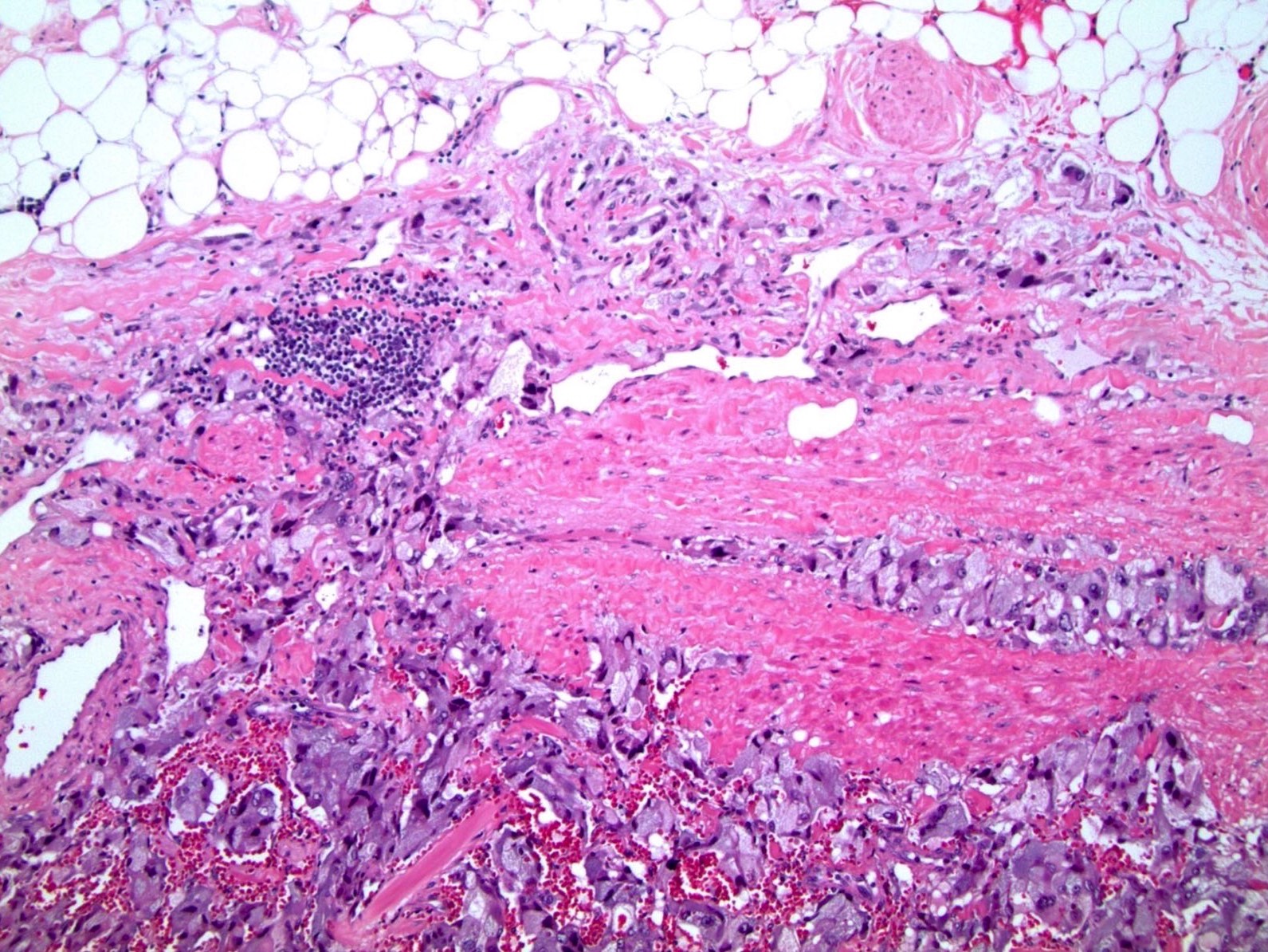
Adjacent benign adrenal
Focal myxoid pattern
Transition to myxoid area
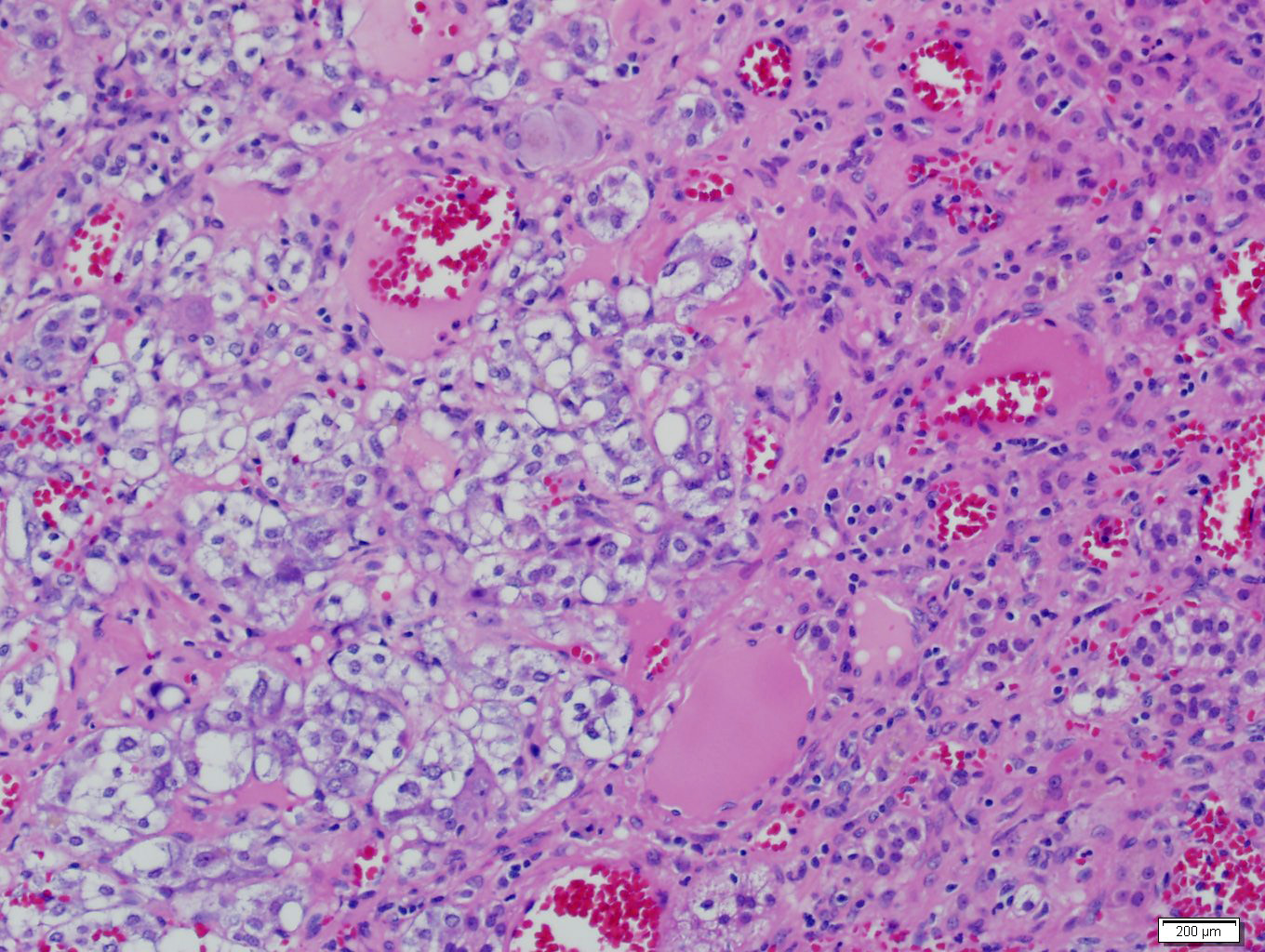
Microcystic architecture
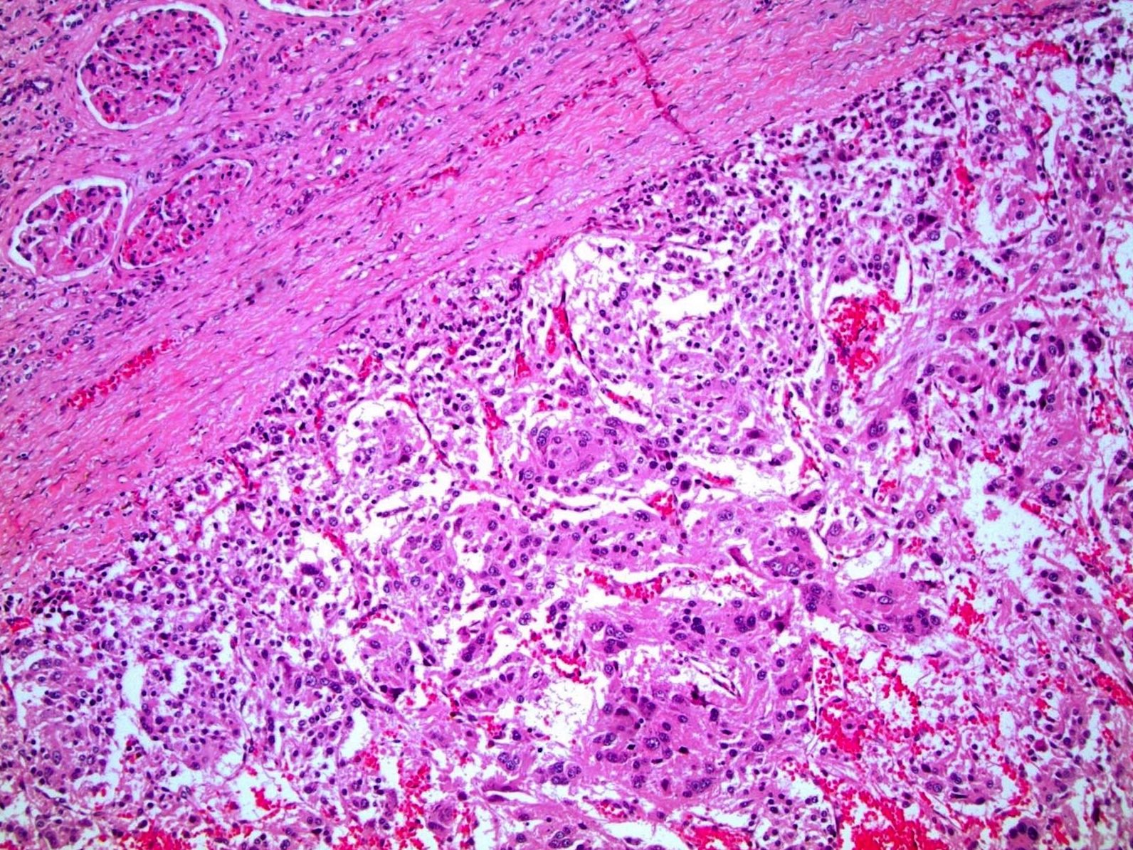
Positive margin
pT3 myxoid ACC
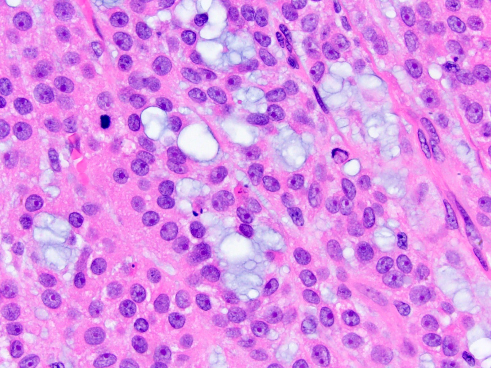
Mitoses
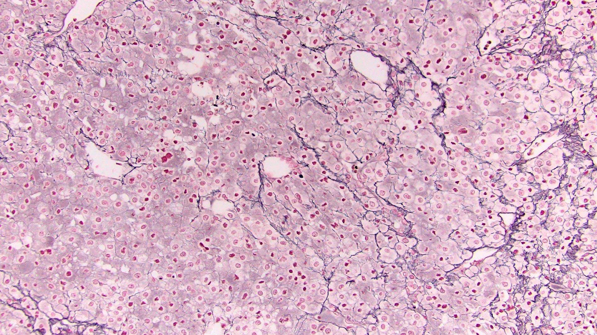
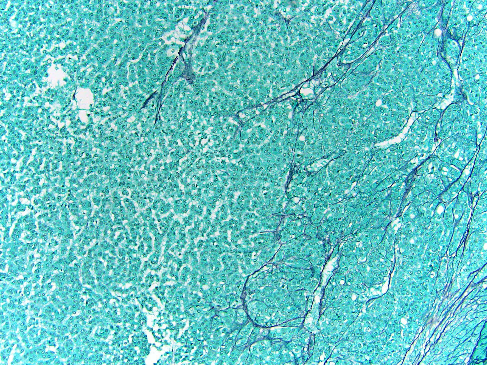
Normal reticulin network
Loss of reticulin framework
Images hosted on other servers:

Right adrenal mass
Images hosted on other servers:

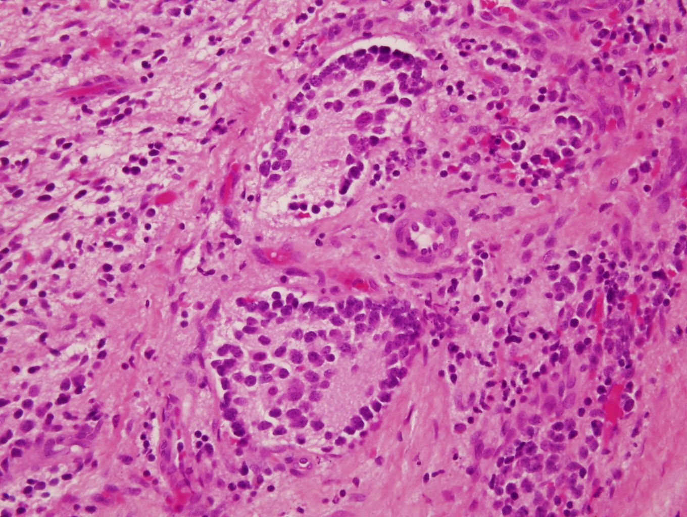
Congenital neuroblastoma

Caudally located neuroblastoma
Contributed by Carmen Perrino, M.D.
With anaplasia
Anaplasia and a mitotic figure
Homer-Wright pseudorosettes
High power pseudorosettes
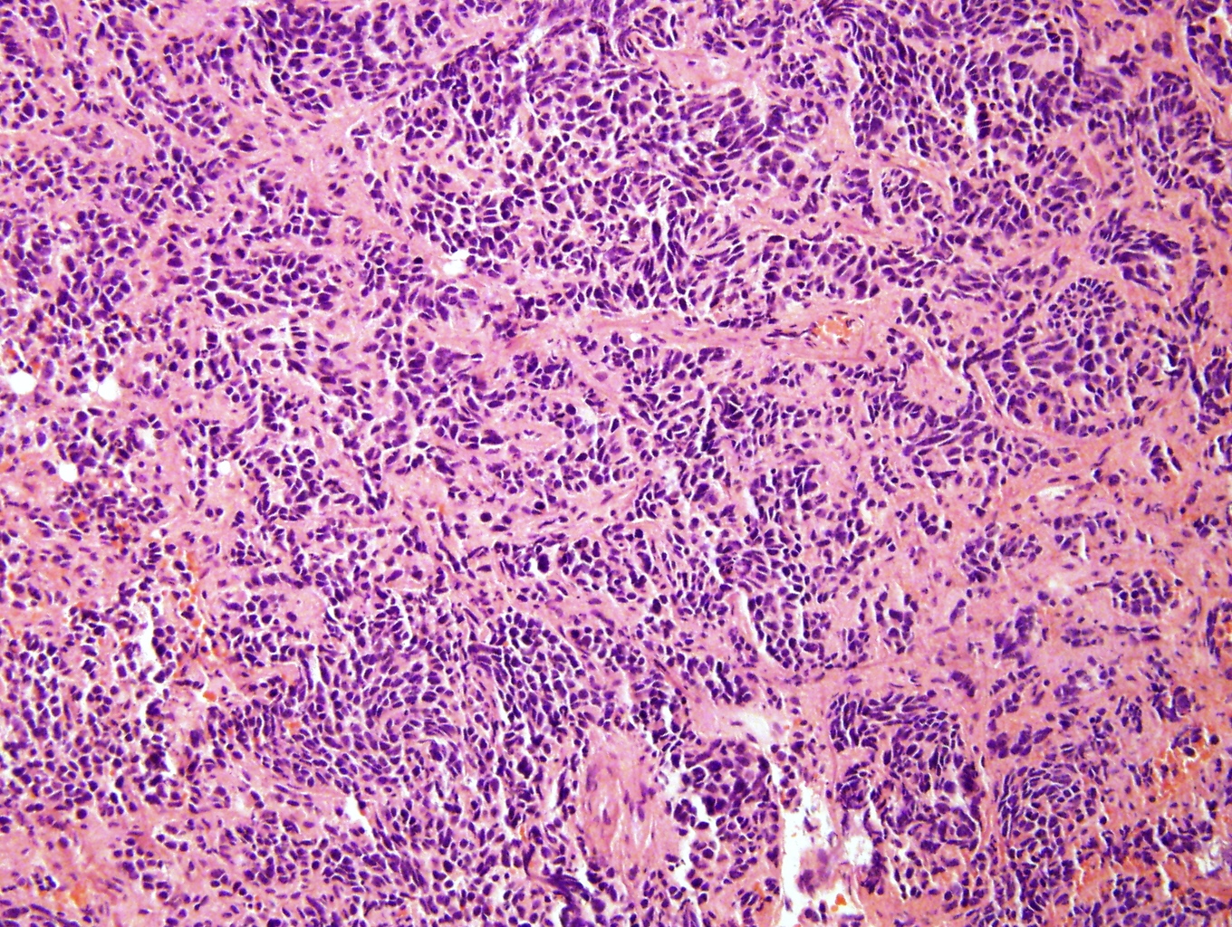
Poorly differentiated
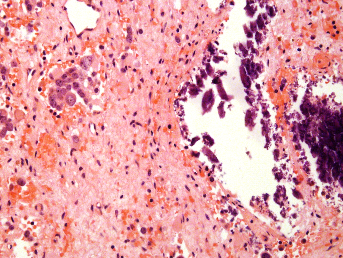
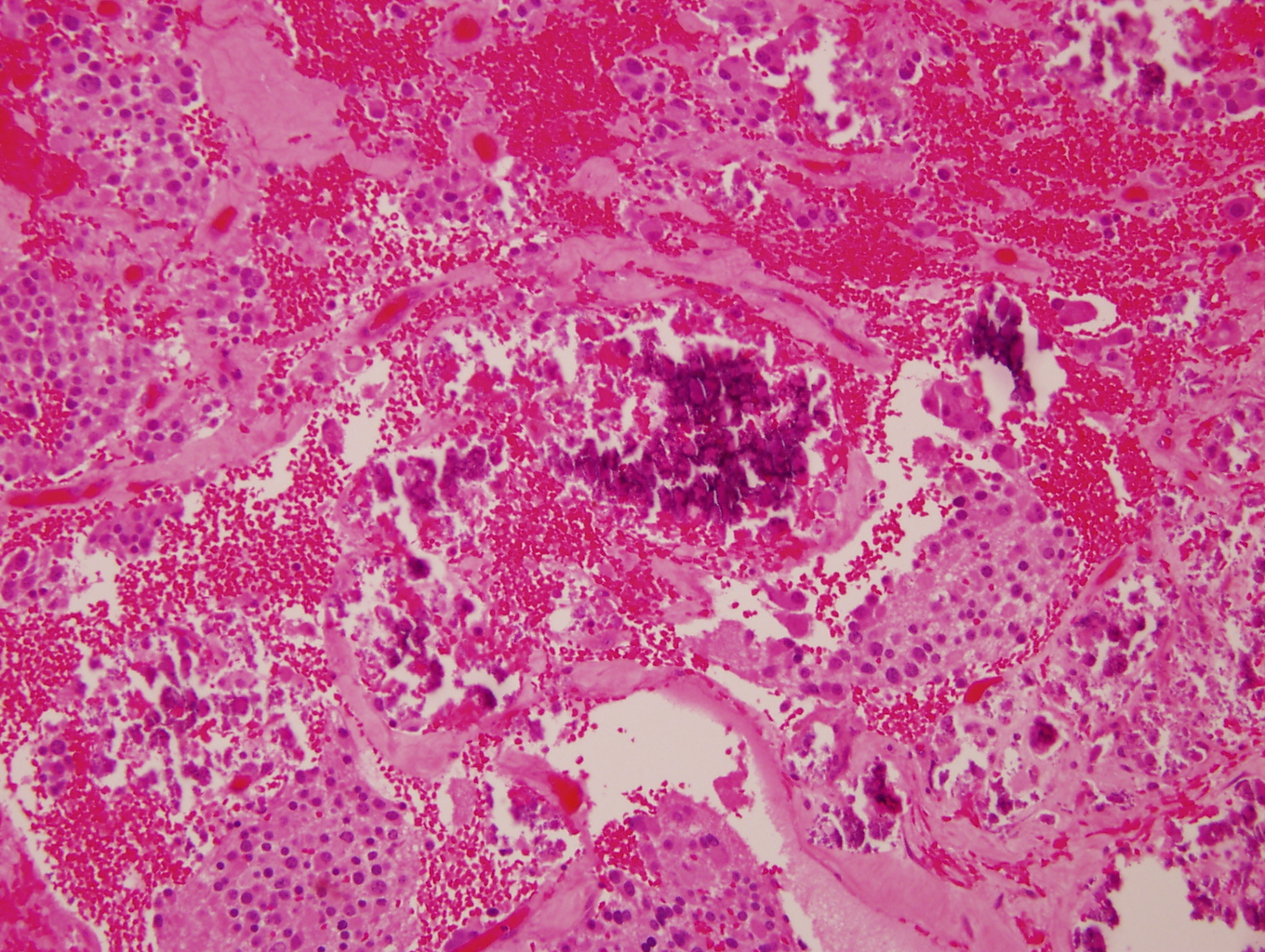
Dystrophic calcification
Posttreatment
Metastatic to bone marrow

Significant crush artifact
Images hosted on other servers:

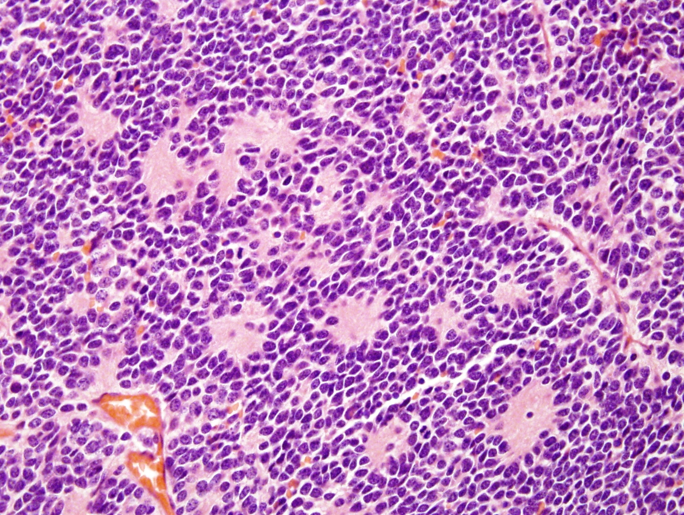
Small round blue cell

Neuroblasts

Mats of neuropil and focal resetting

Differentiating bladder neuroblastoma
Images hosted on other servers:

Occasional rossetting
Images hosted on other servers:

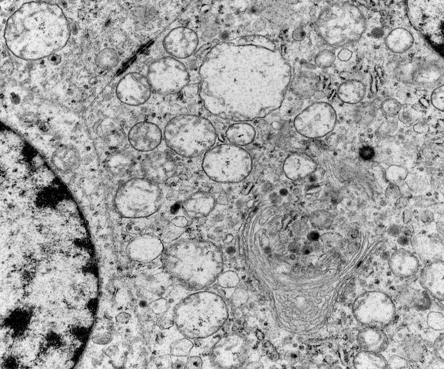
C: neurosecretory dense core granules in the cytoplasm

Poorly differentiated neuroblasts

Widespread neuritic processes
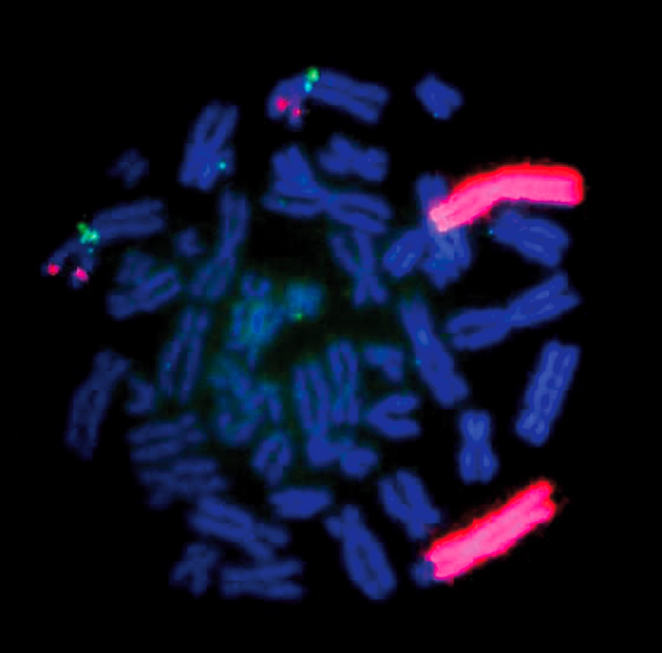
Contributed by Leica Biosystems
MYCN (2p24) / AFF3 (2q11)
Images hosted on other servers:

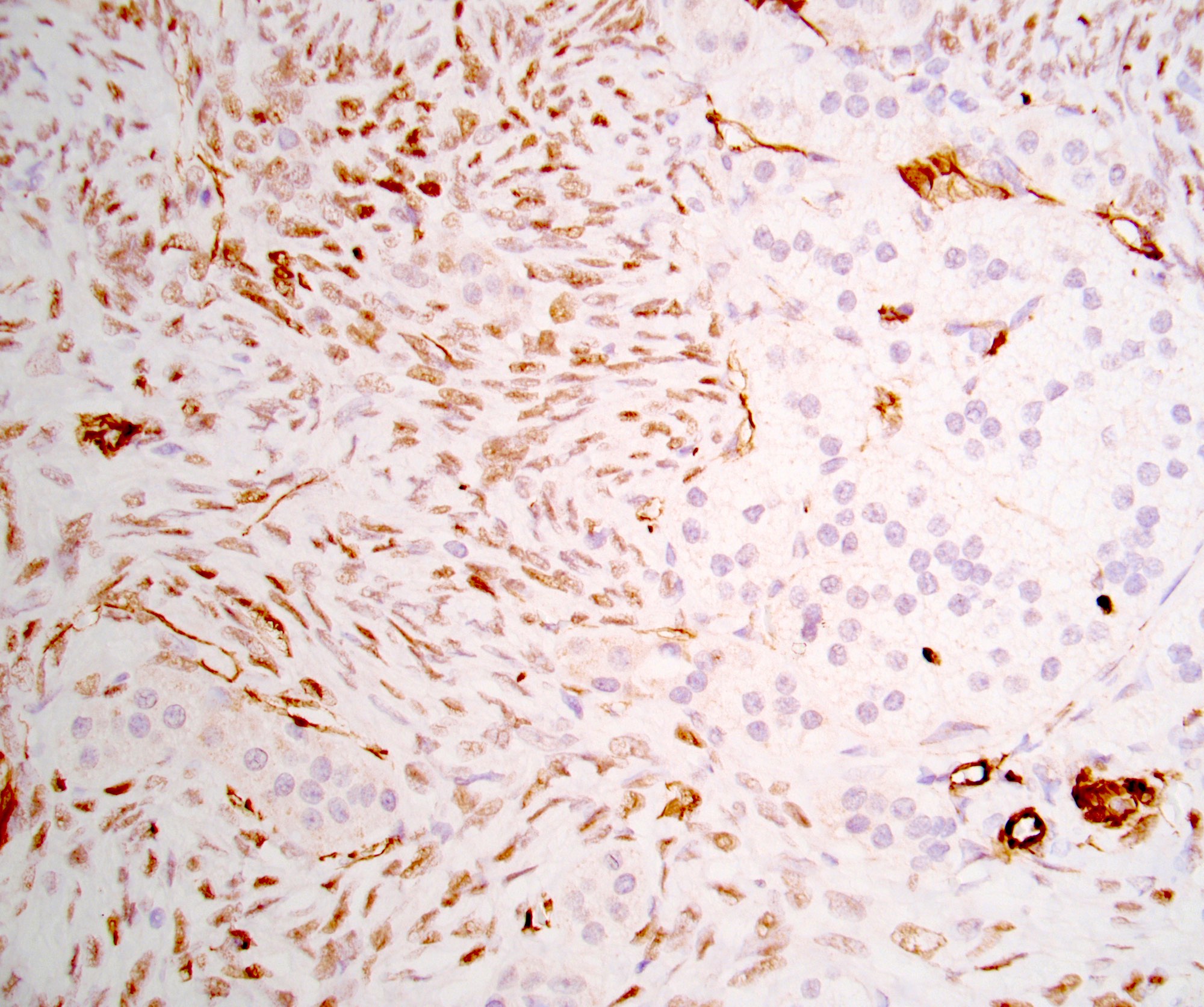
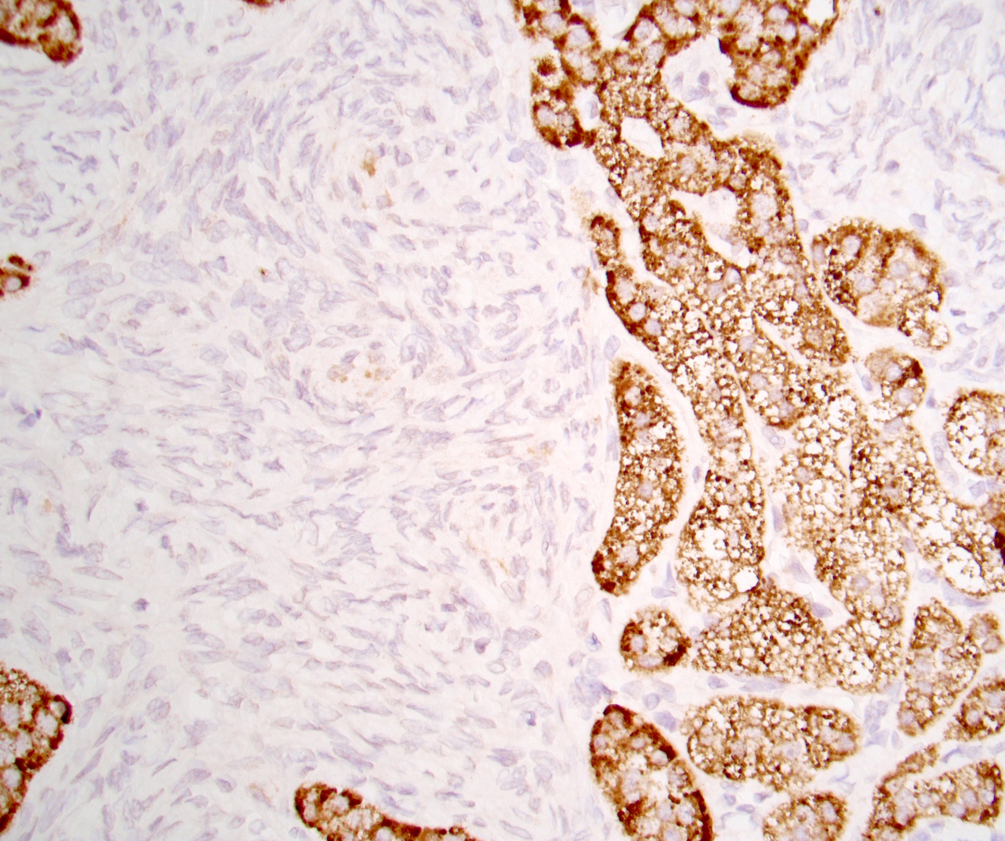
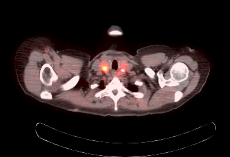
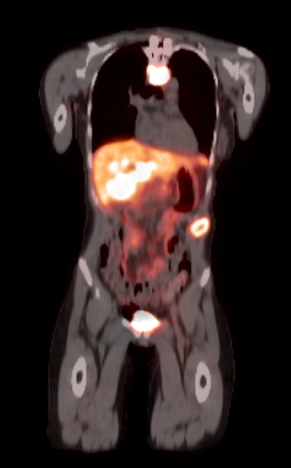
CT imaging of oncocytic ACC

F18 FDG PET of oncocytic ACC
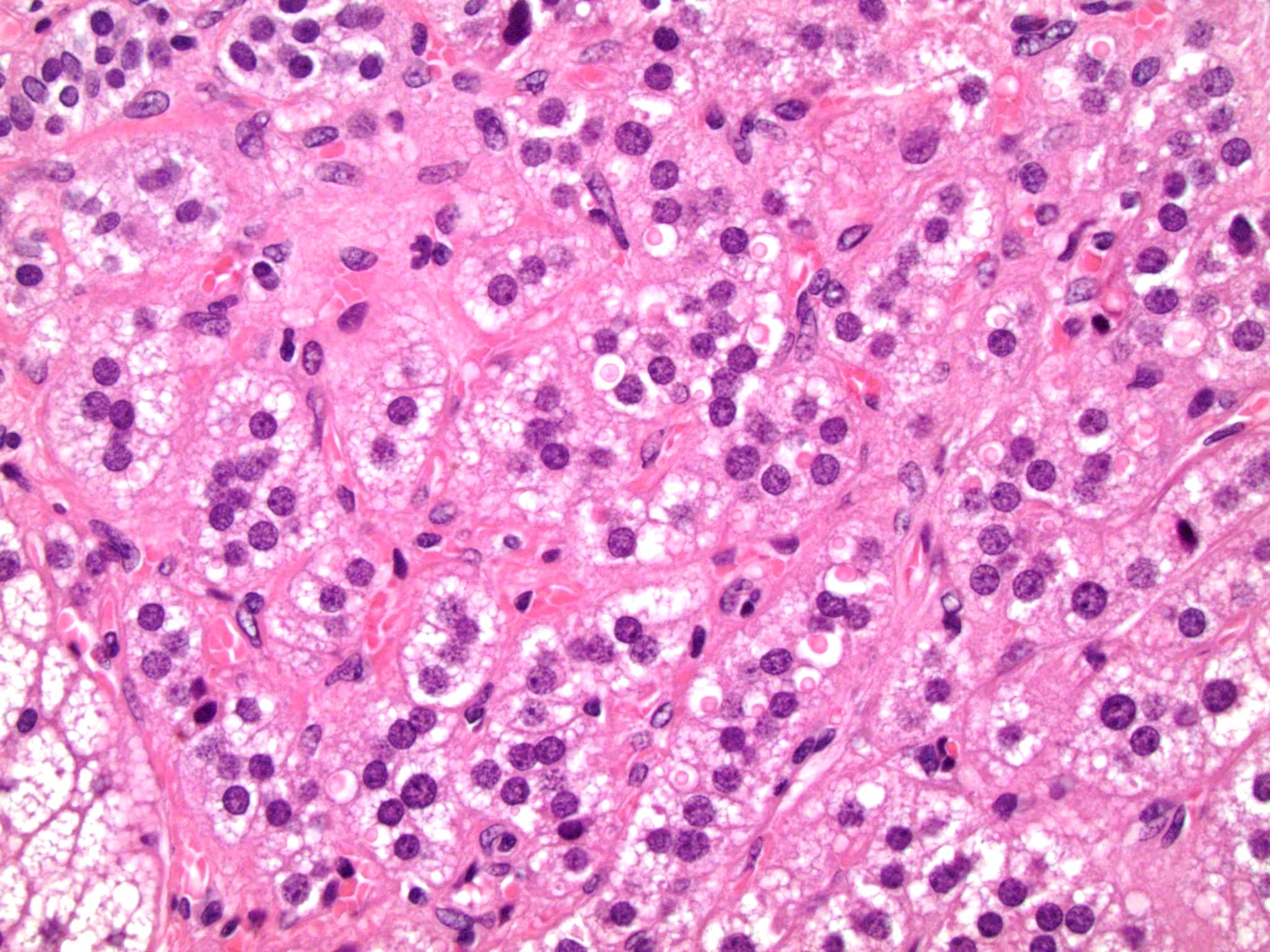
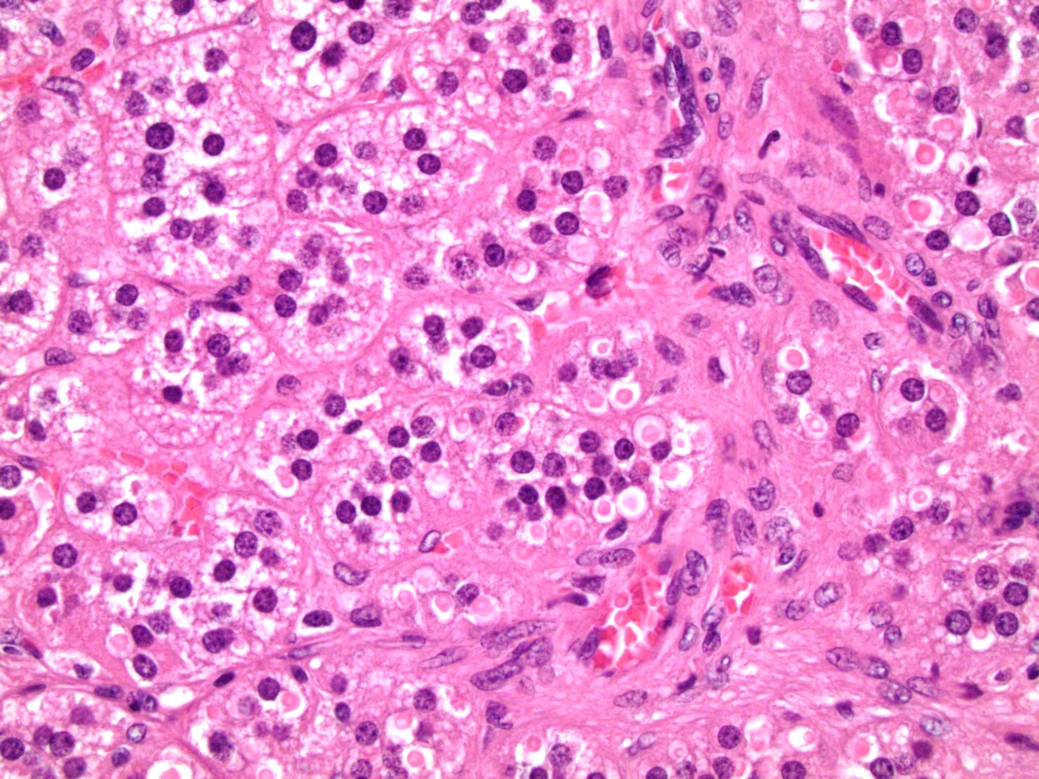
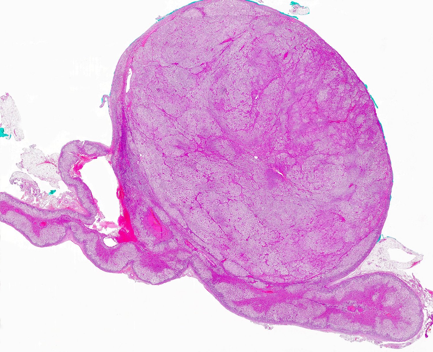
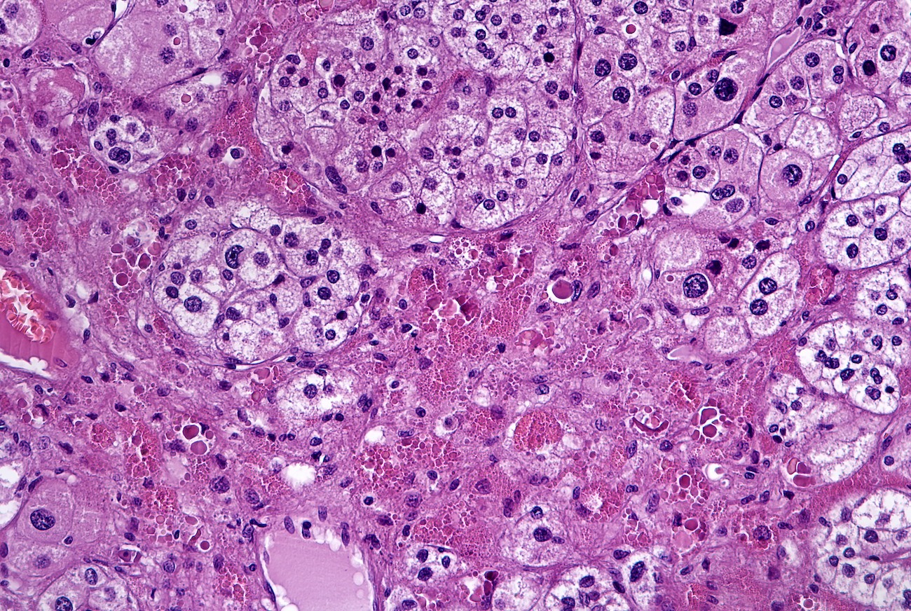
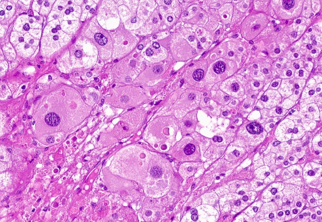
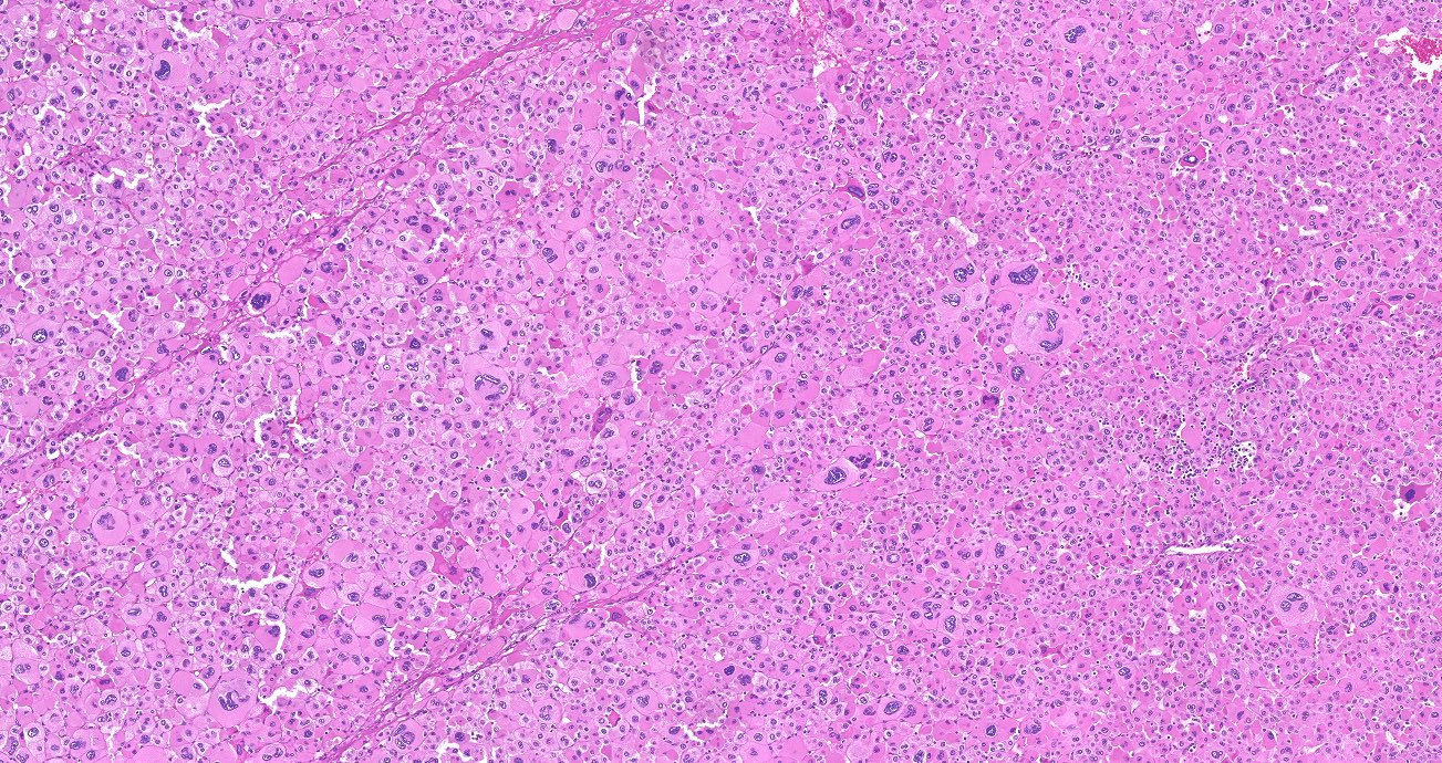
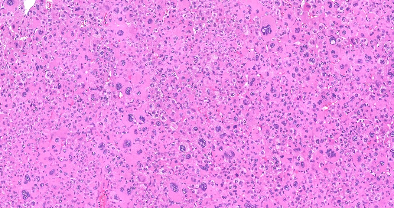
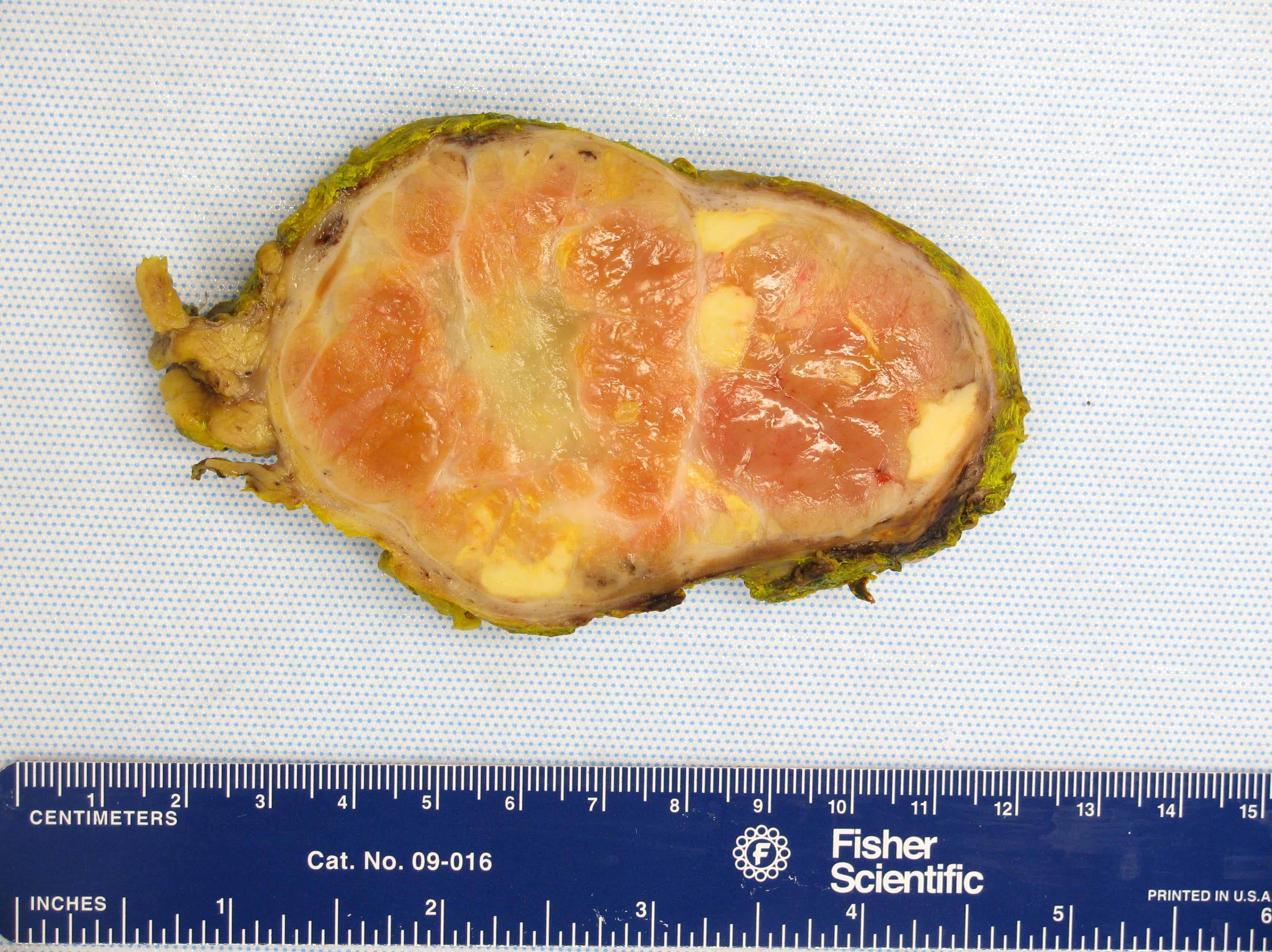
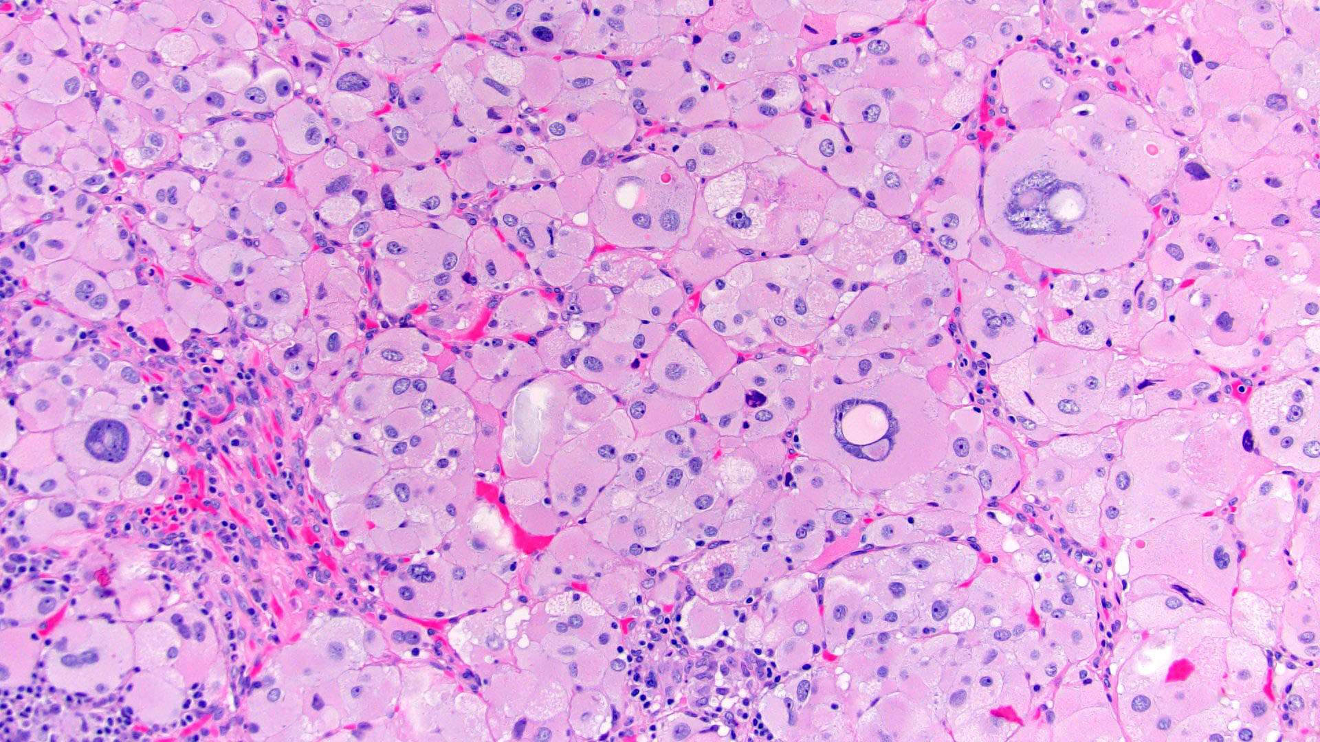
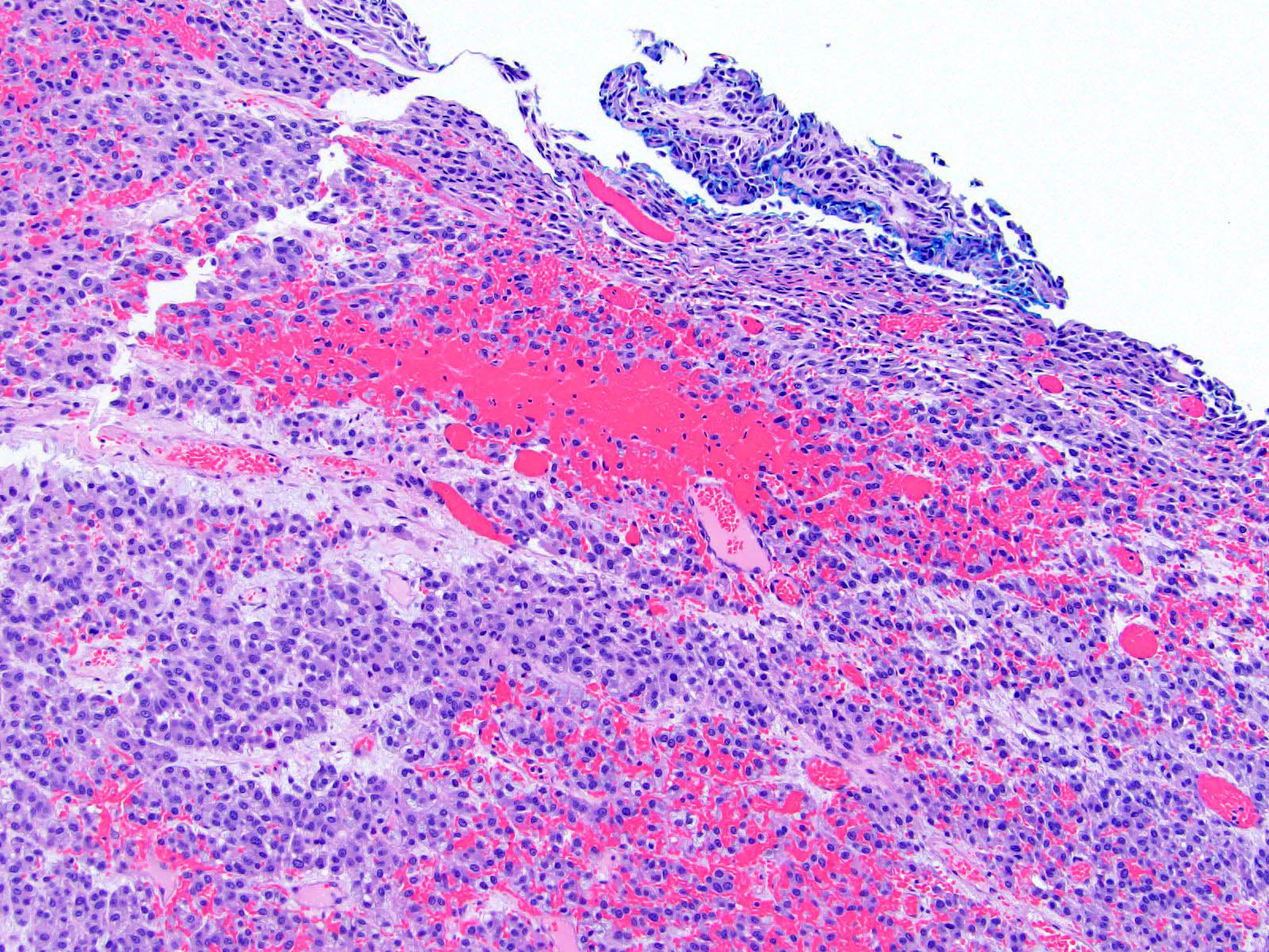
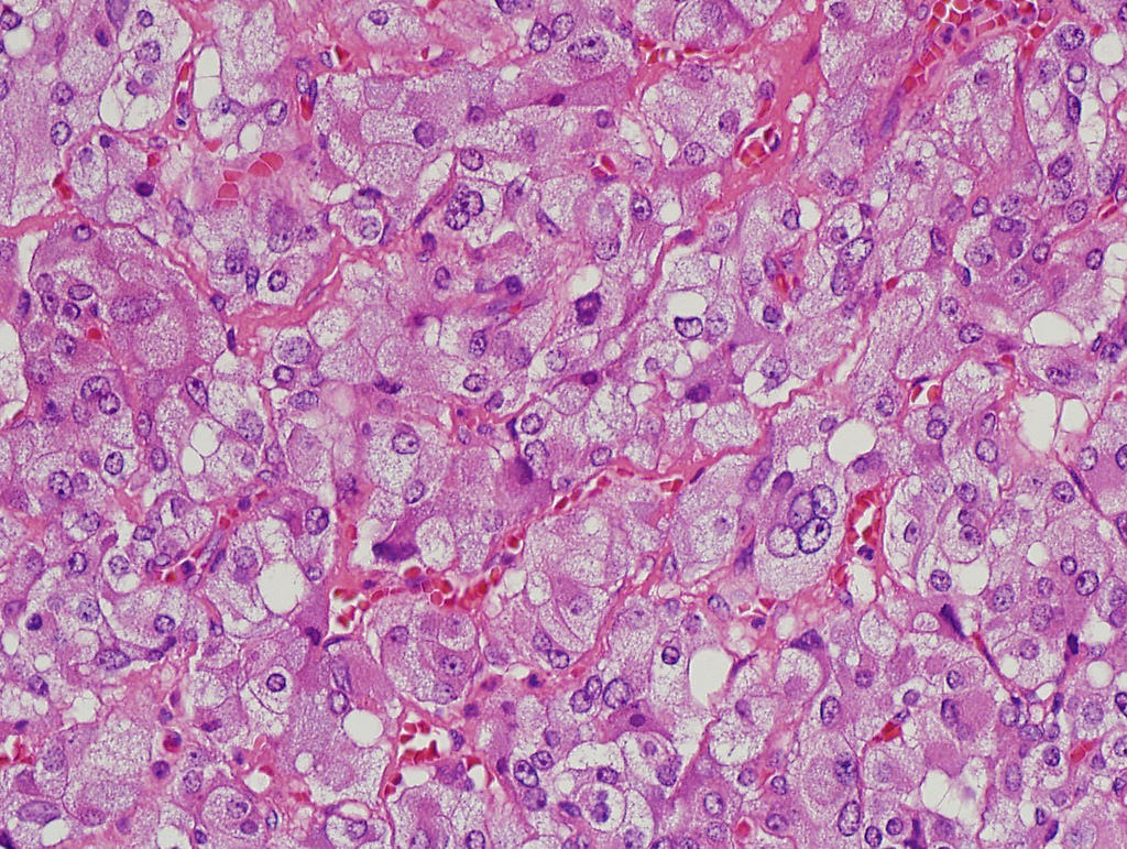
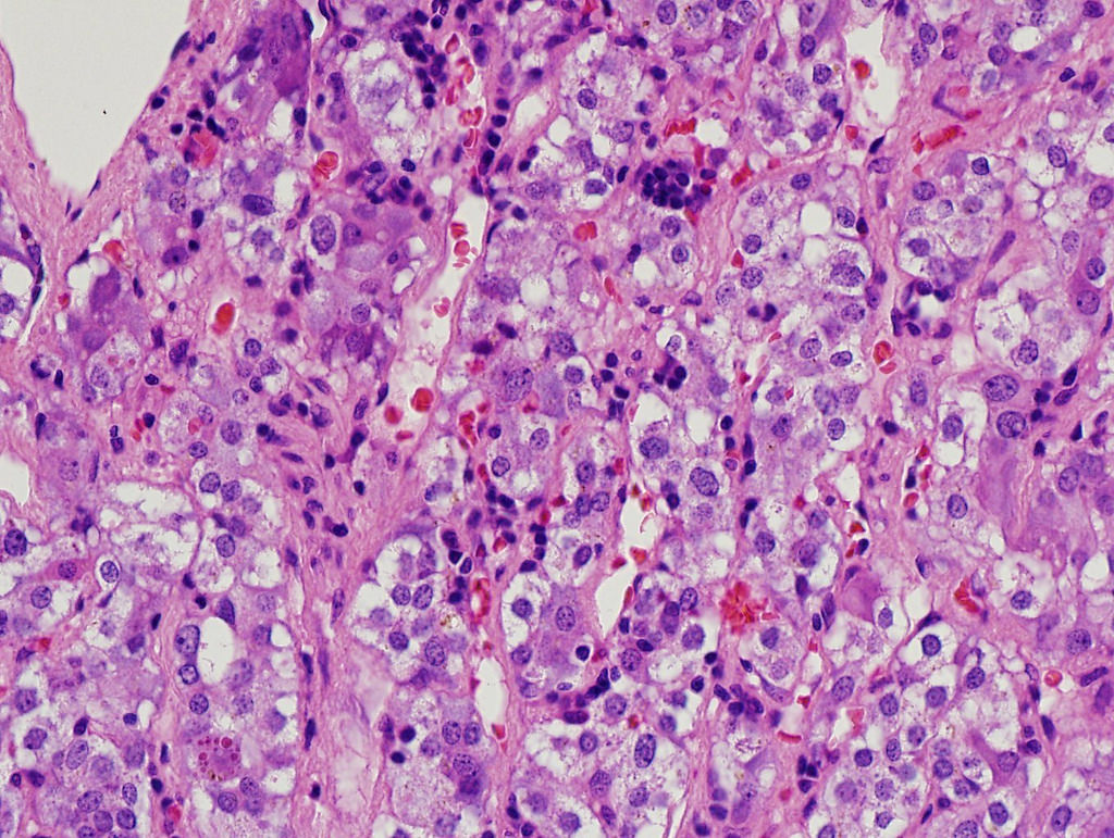
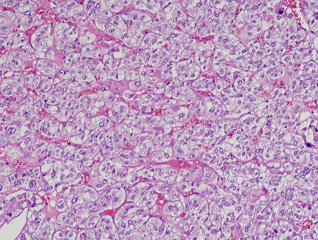
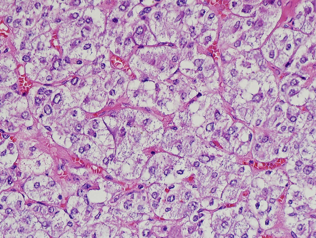
Contributed by Maria Tretiakova, M.D., Ph.D.
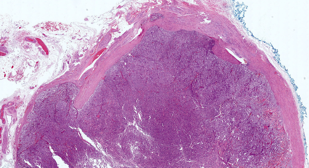
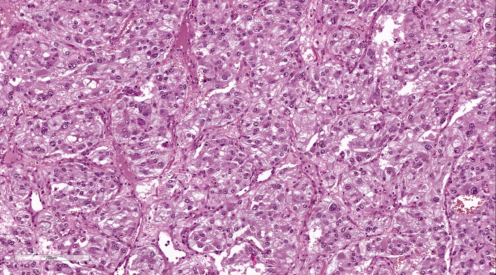
Large lobulated yellow mass
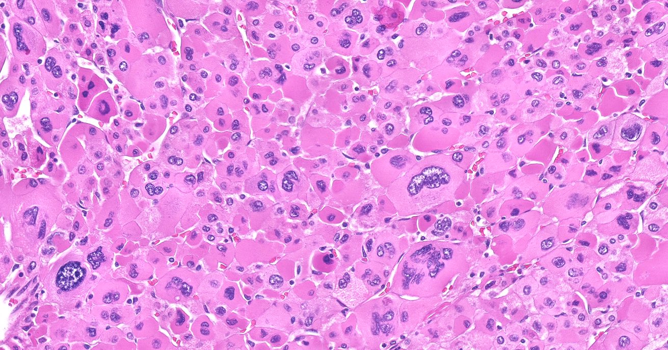
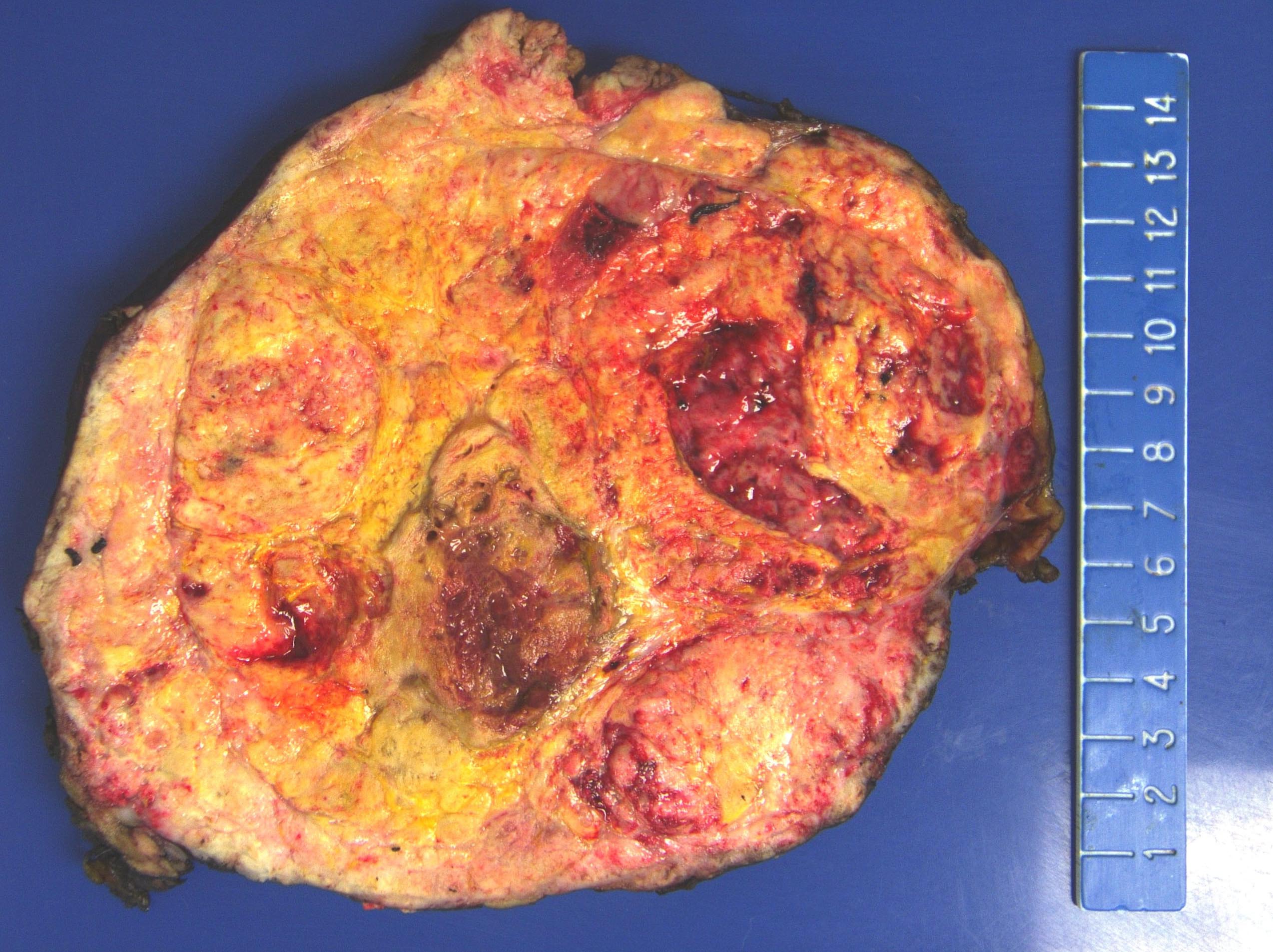
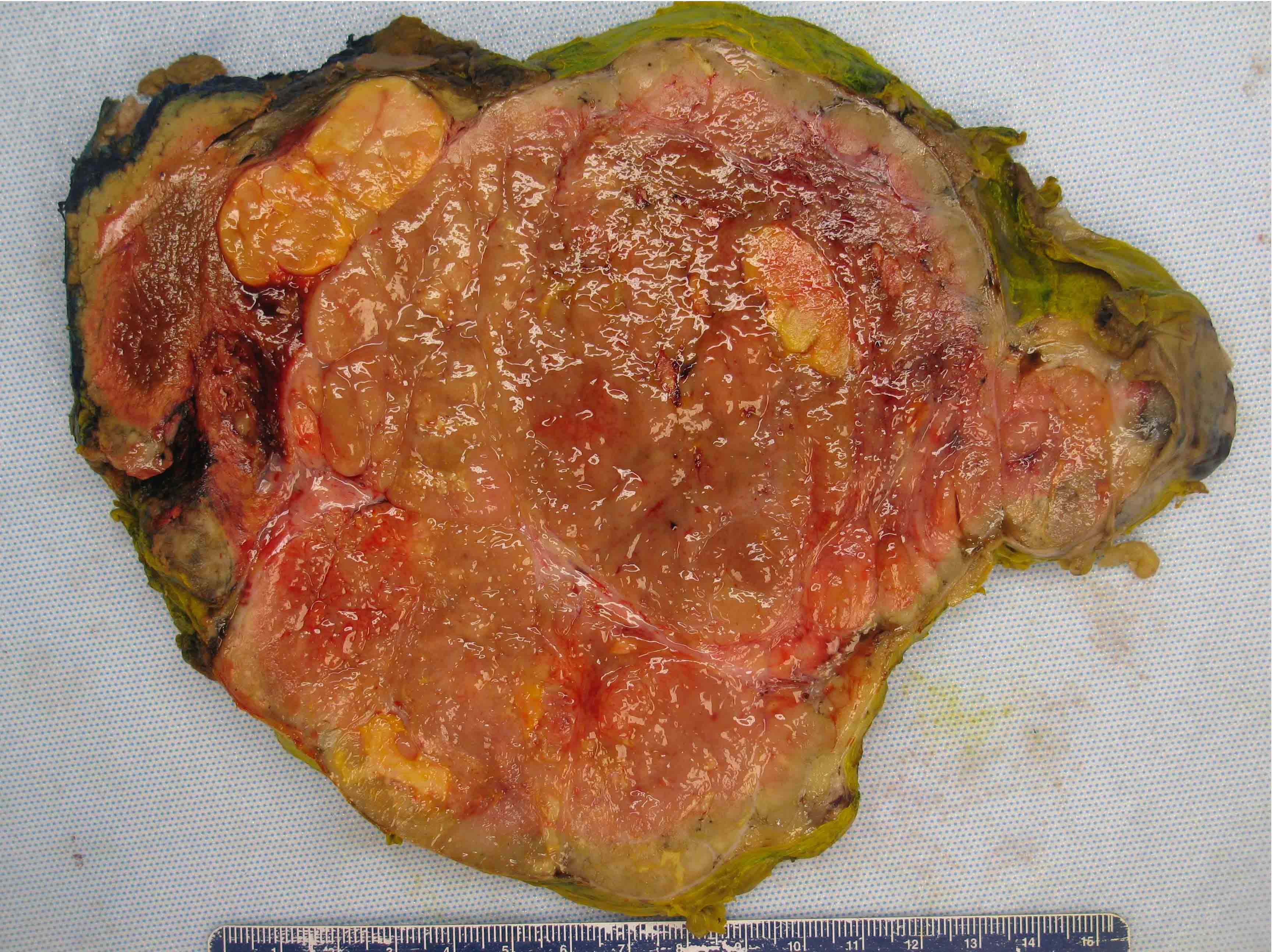
Contributed by Maria Tretiakova, M.D., Ph.D.
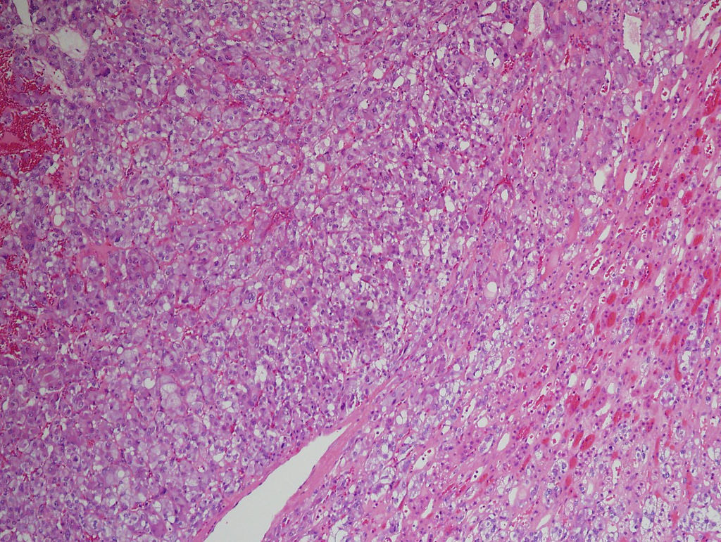
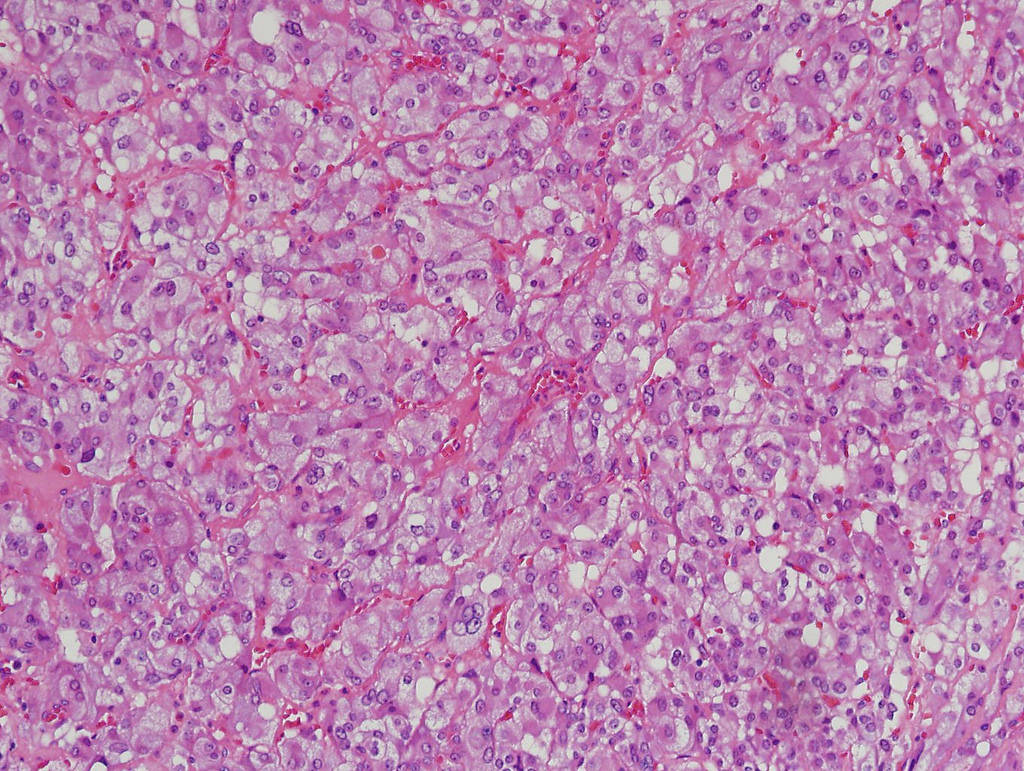
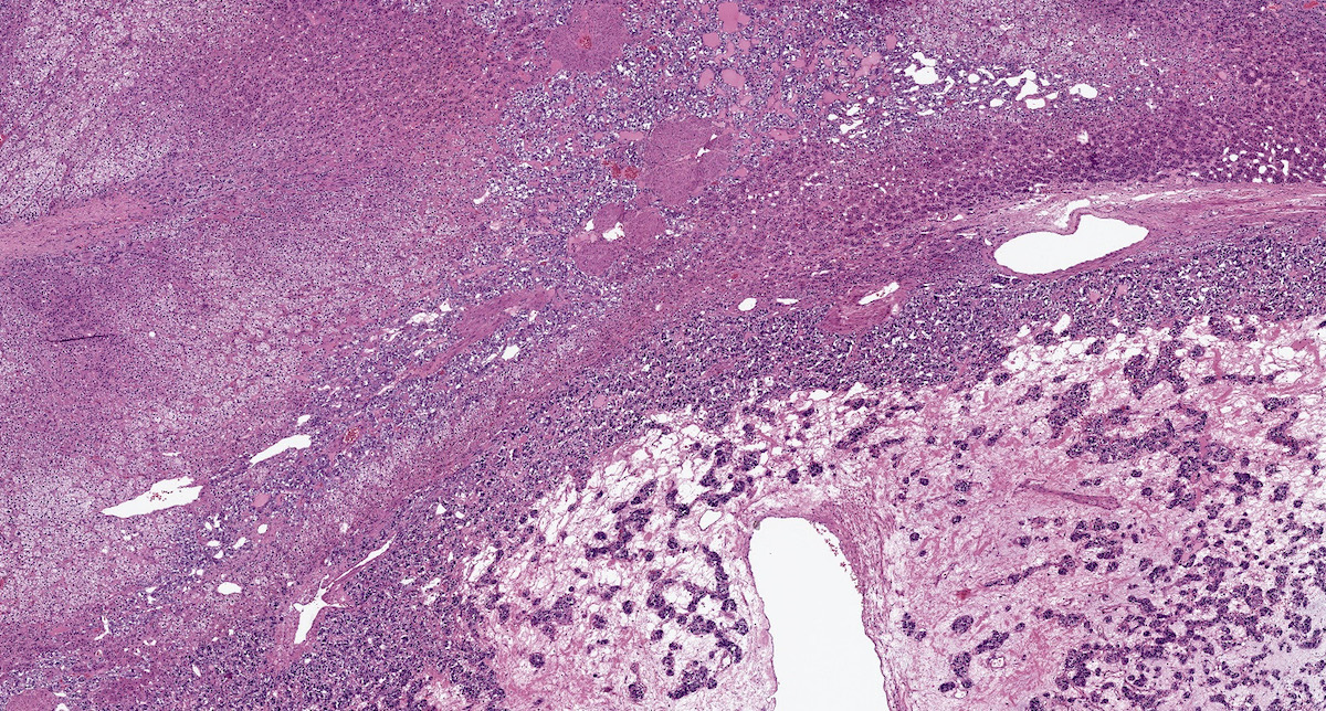
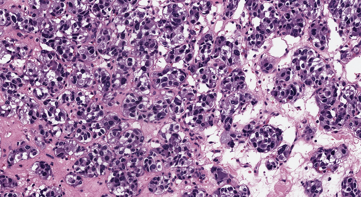
Oncocytic ACC with confluent necrosis
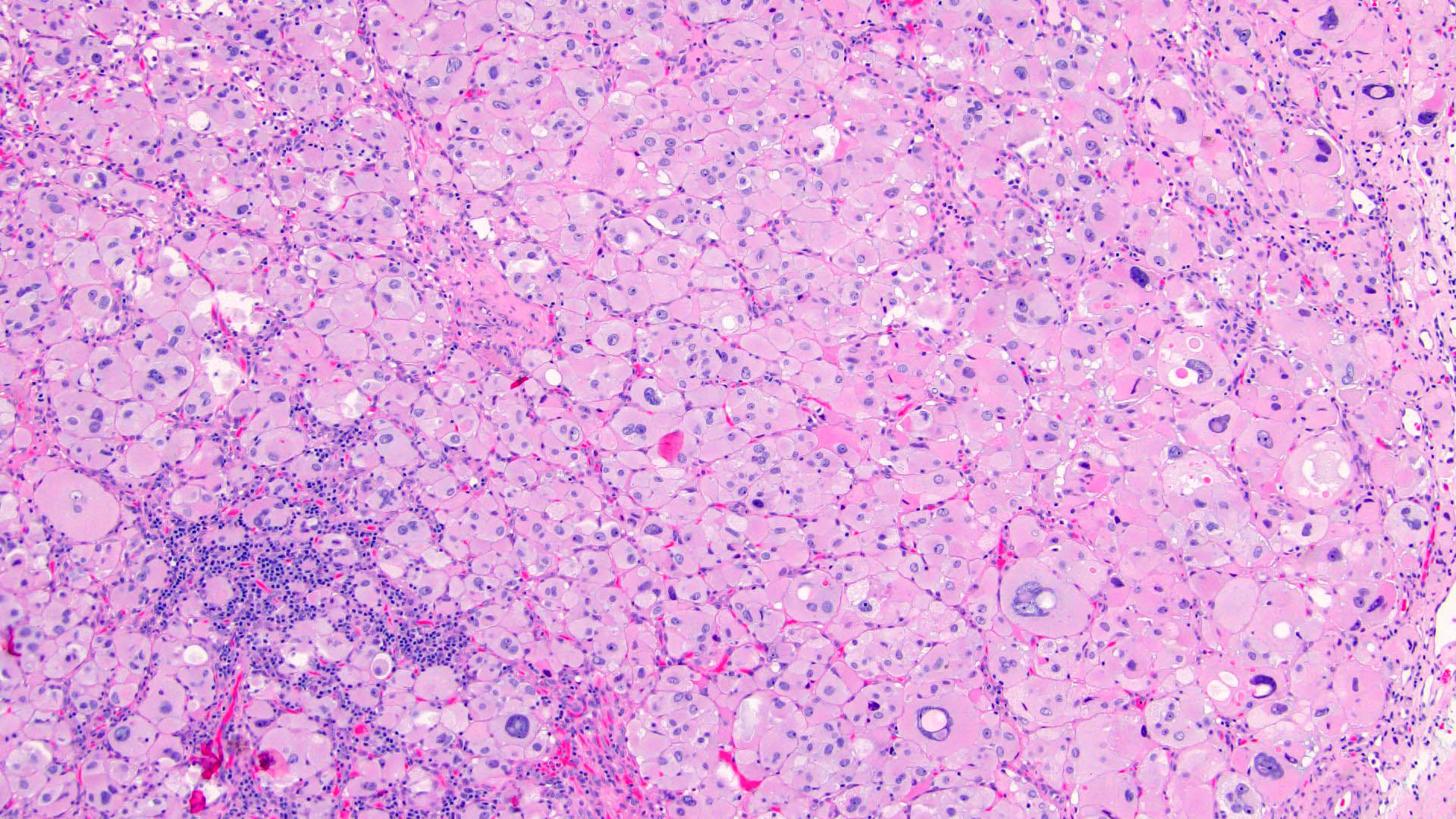
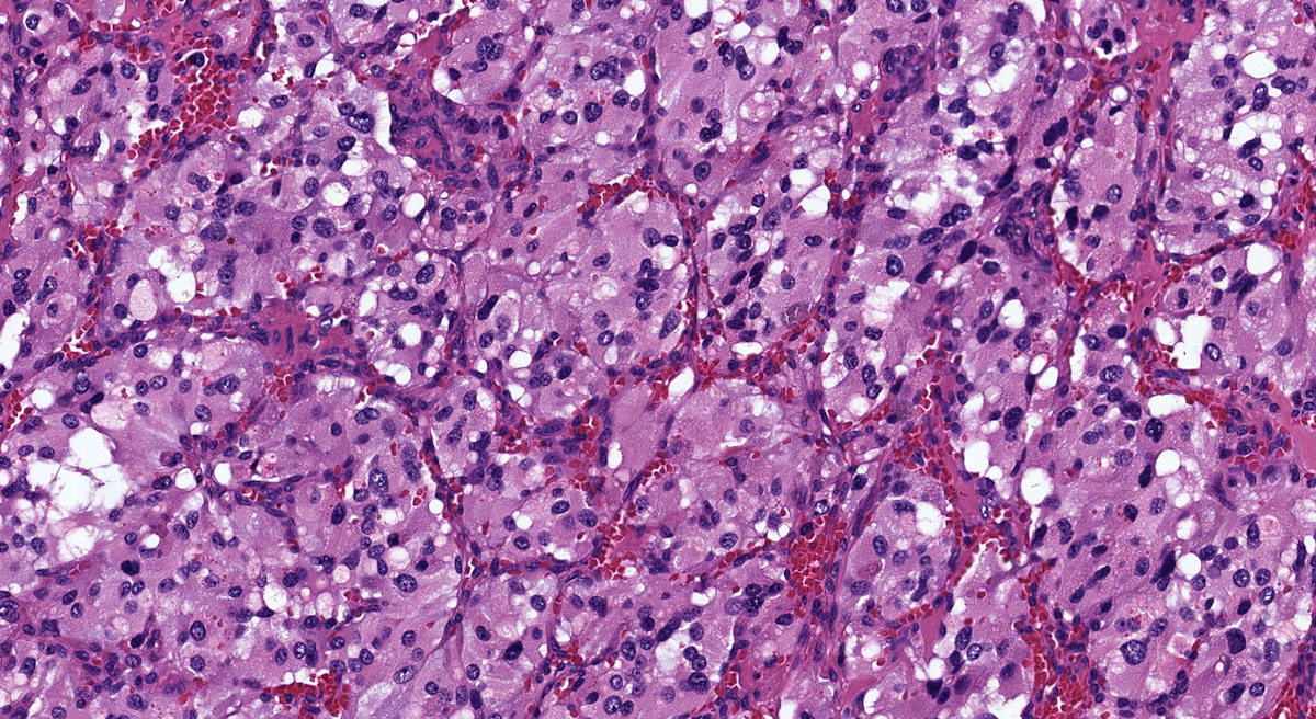
Diffuse discohesive growth
Trabecular pattern
Capsule invasion
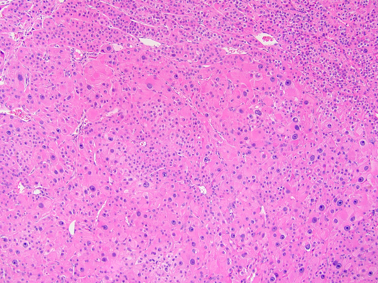
Pleomorphic morphology
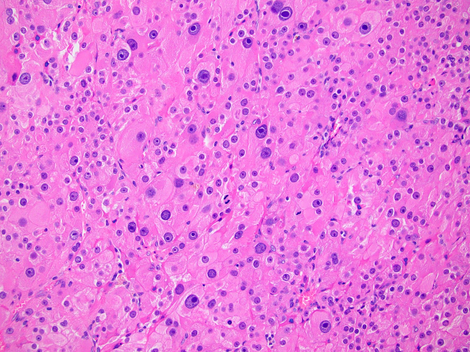
Nuclear pleomorphism
Abundant mitoses
Fat invasion & positive margin
Regional node involvement
Disrupted reticulin network
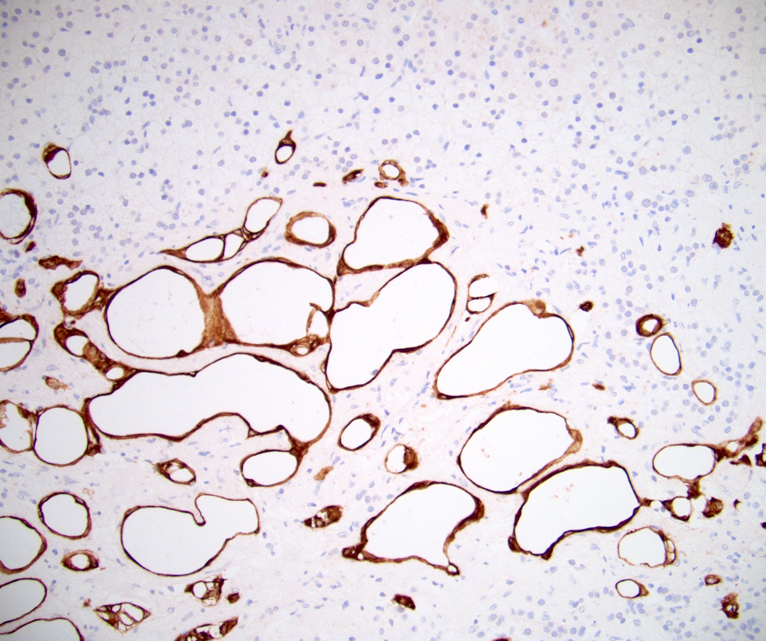
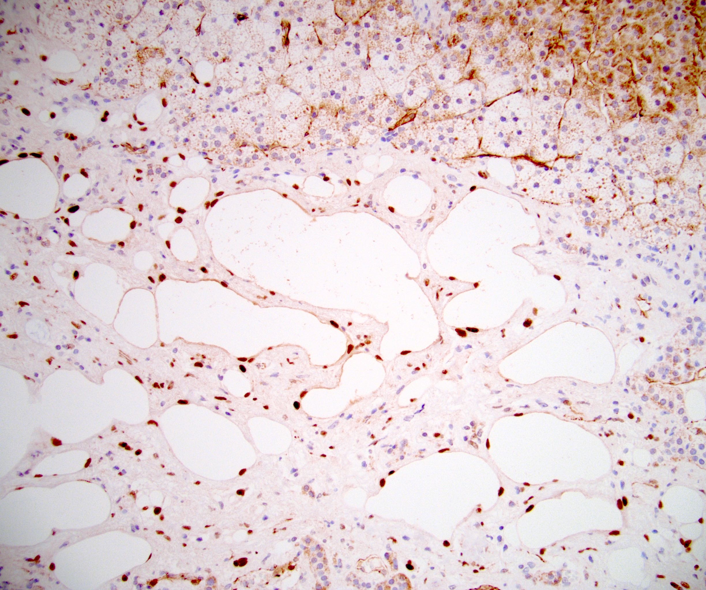
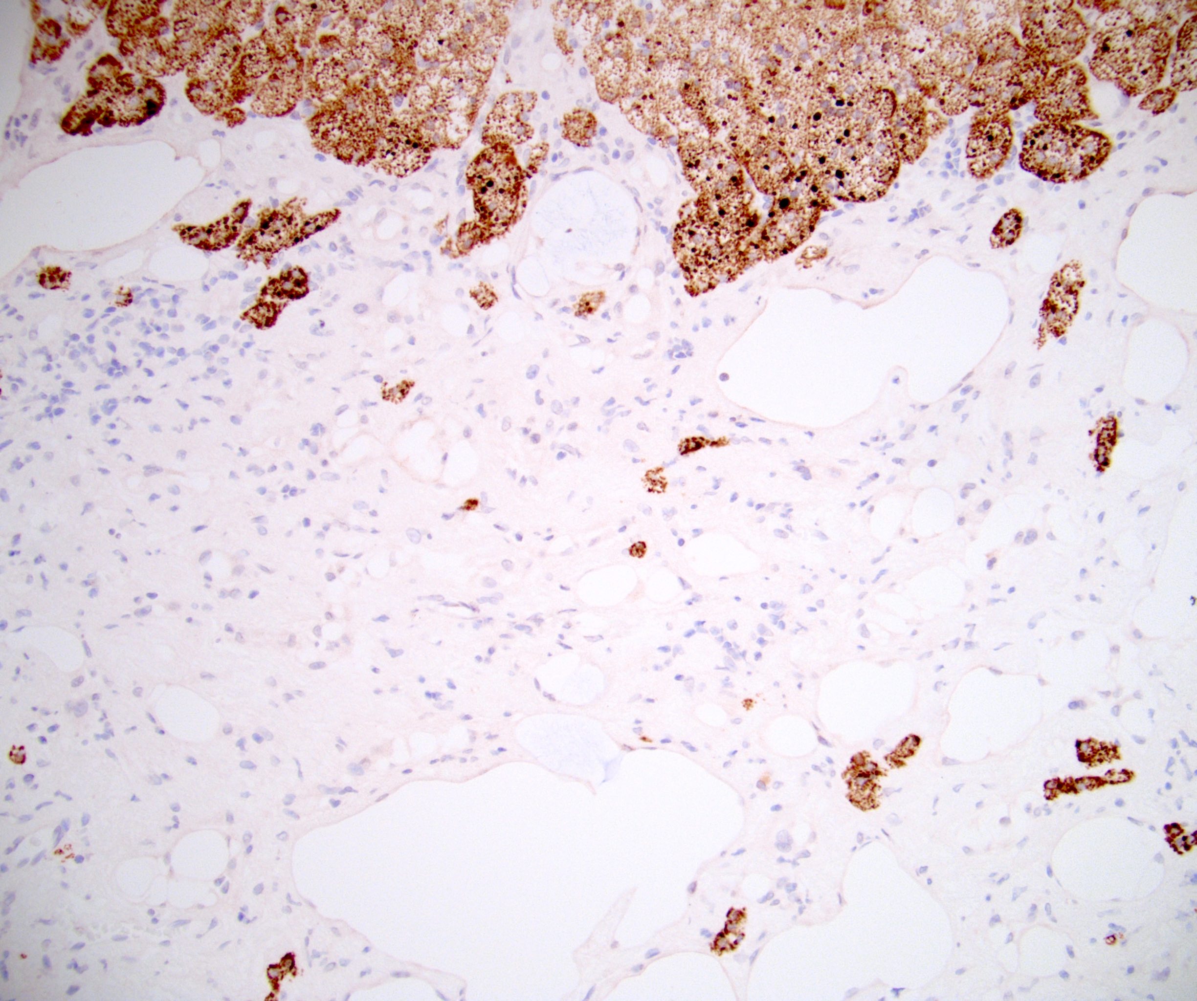
Contributed by Debra L. Zynger, M.D.

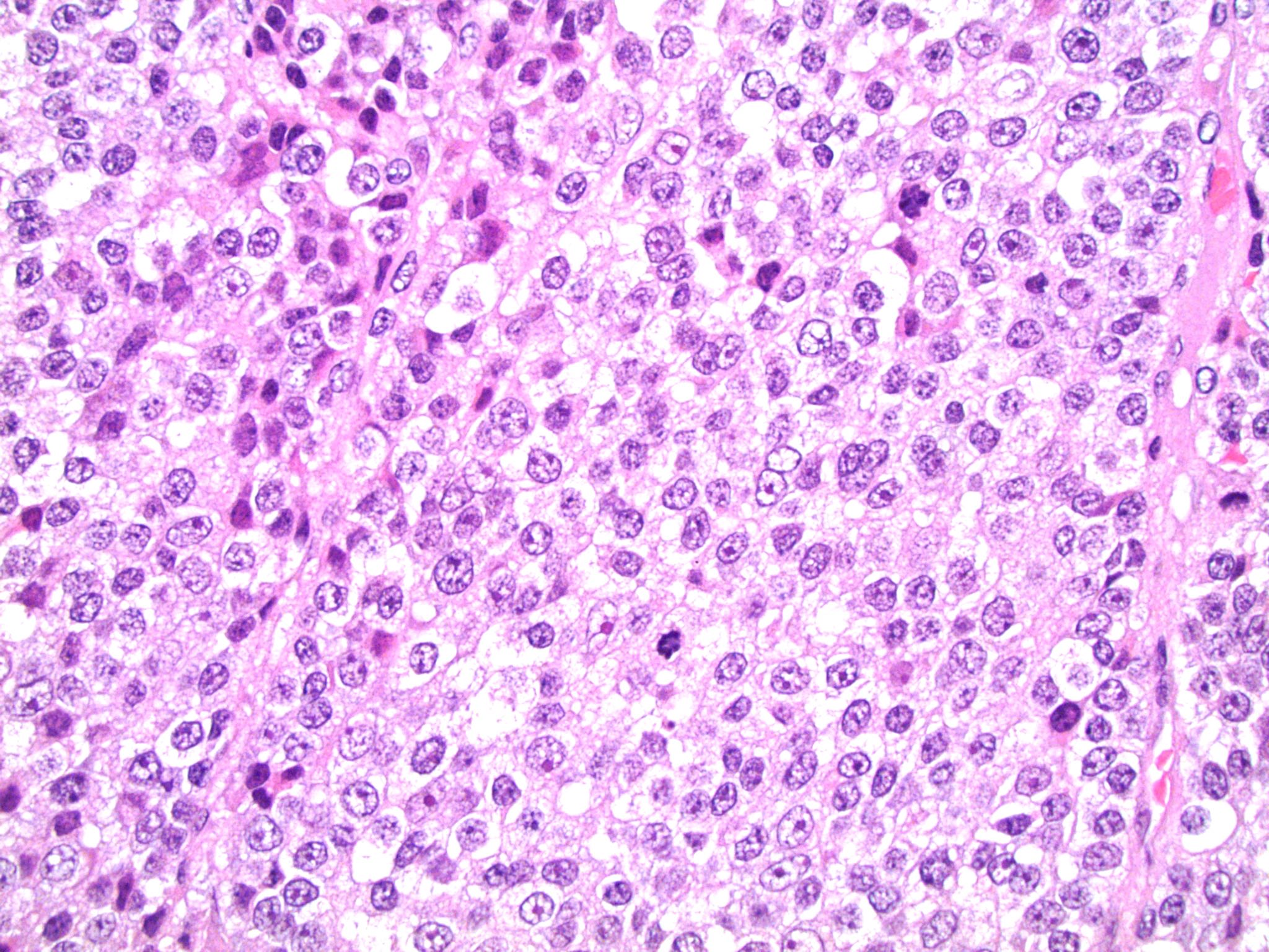
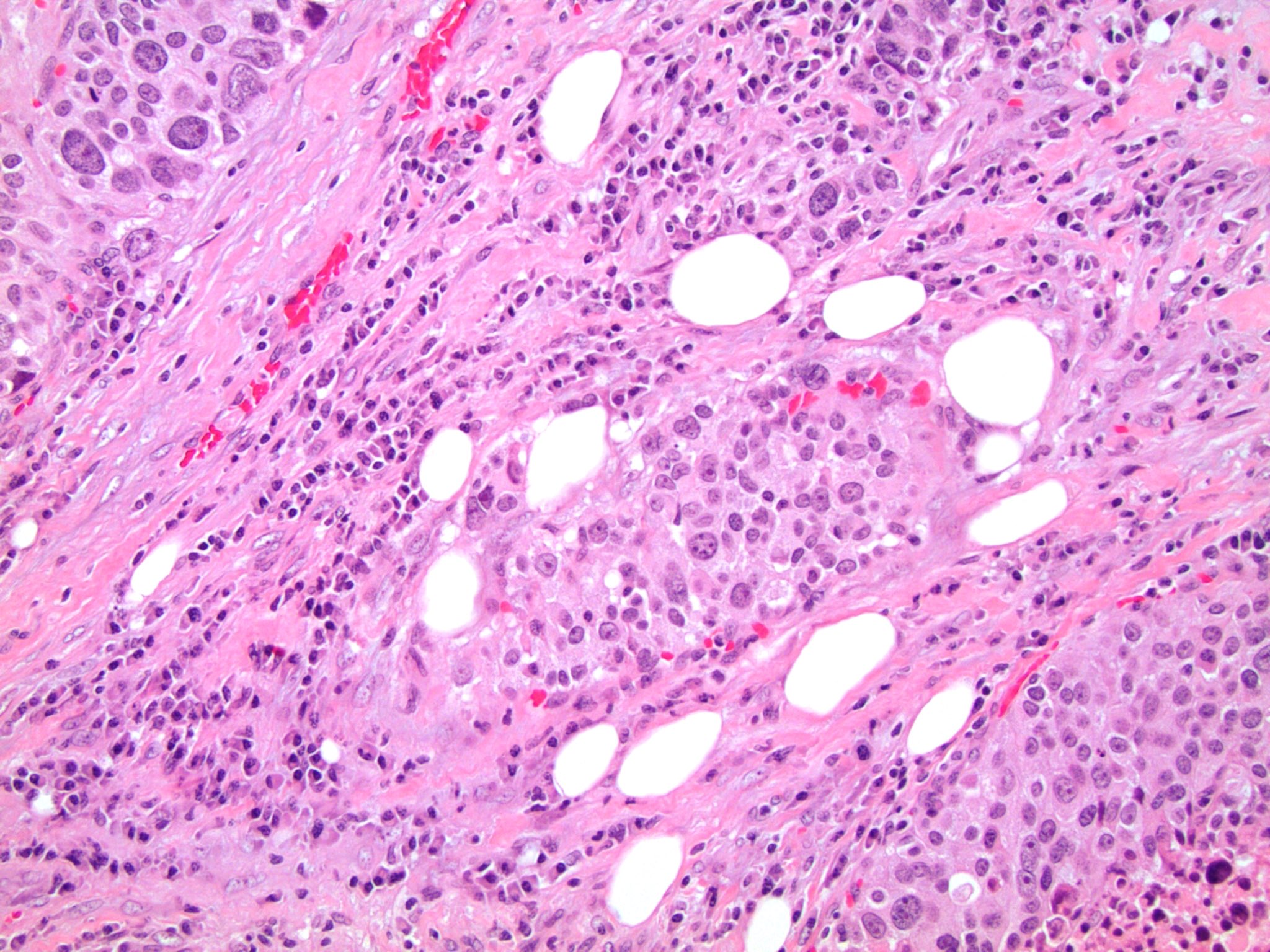
Subcapsular spindle cells

Spindle cells with entrapped adrenal cortical cells


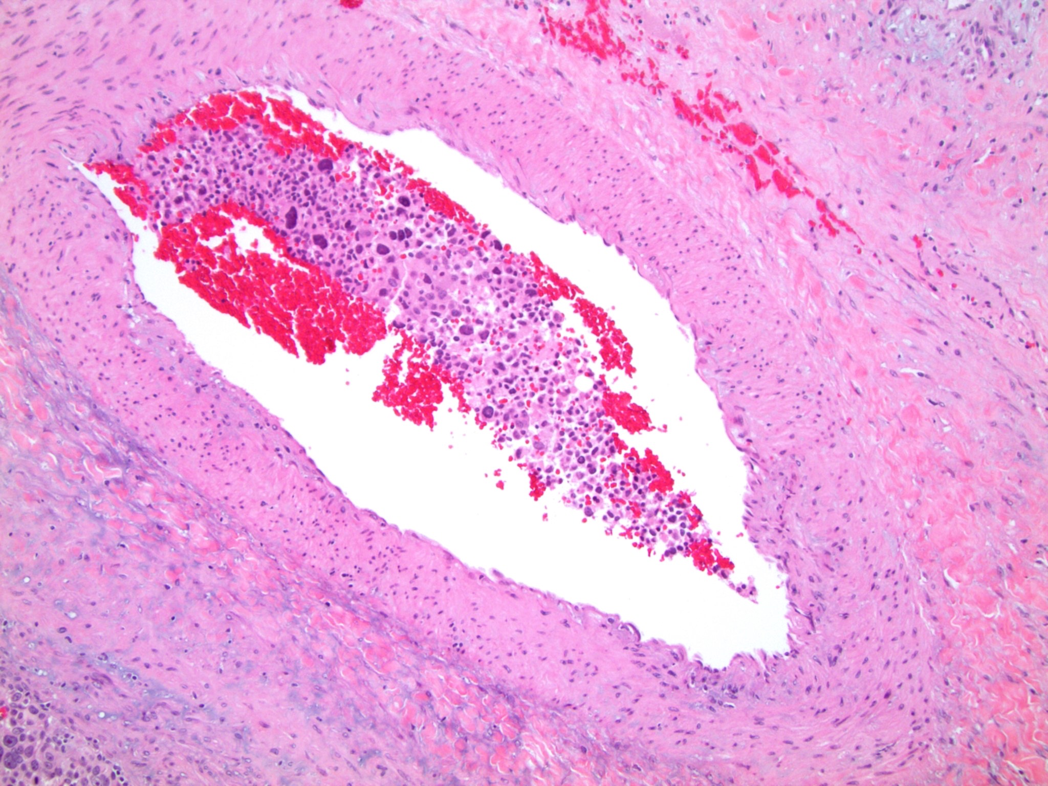
Bland spindle cells


Thick hyalinized fibers
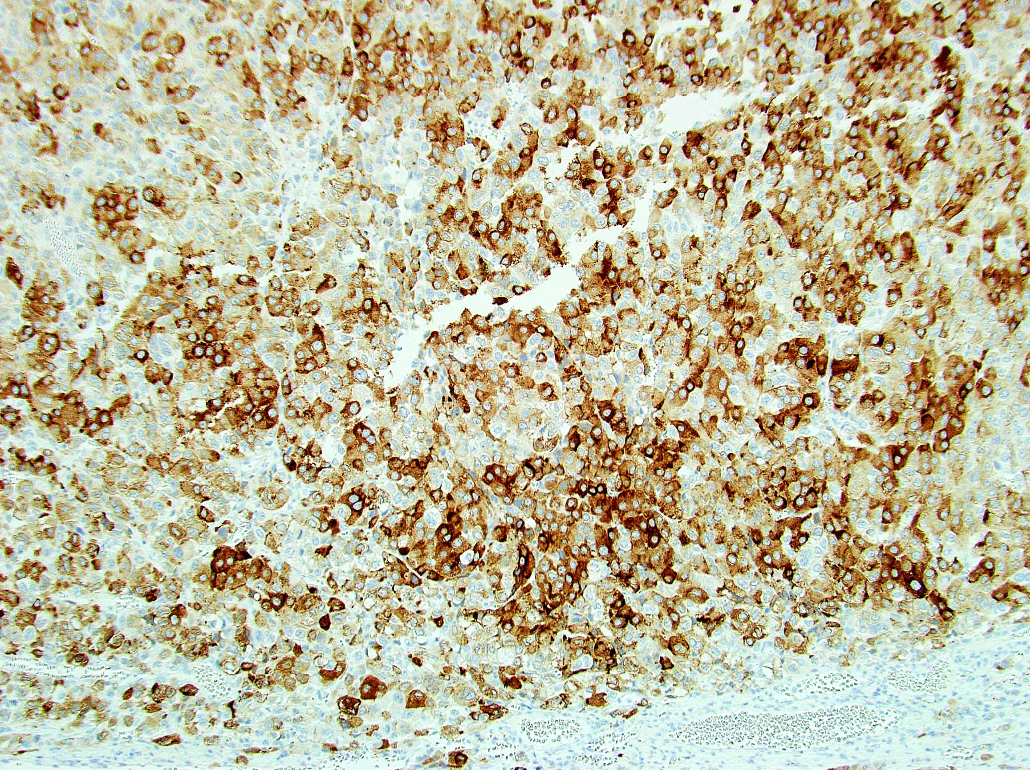
WT1

Desmin
MelanA


Contributed by Luvy Delfin, M.D., Sylvia L. Asa, M.D., Ph.D. and Debra Zynger, M.D.
Adrenal pheochromocytoma
Para-aortic paraganglioma
Carotid body paraganglioma
Parapharyngeal paraganglioma
Contributed by Luvy Delfin, M.D. and Sylvia L. Asa, M.D., Ph.D.
Adrenal pheochromocytoma
Duodenum
Pheochromocytoma
Organ of Zuckerkandl
Pheochromocytoma in VHL
Pheochromocytoma with metastatic behavior
Metastatic pheochromocytoma in lymph node
Metastatic paraganglioma in bone
Composite pheochromo-
cytoma with ganglioneuroma
Middle ear: jugulotympanic paraganglioma
Cardiac paraganglioma
Para-aortic paraganglioma
Carotid body paraganglioma
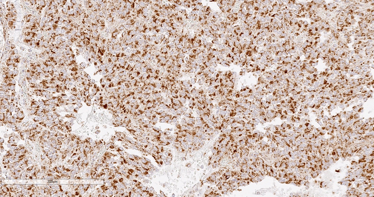
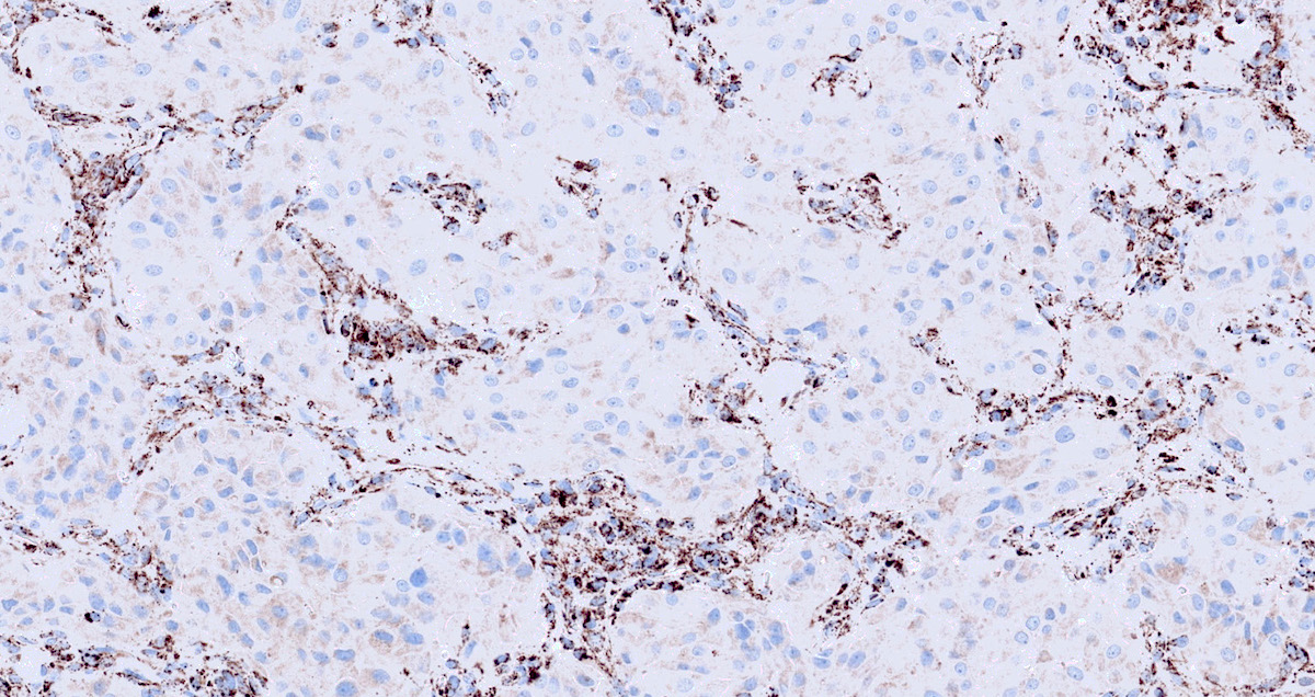
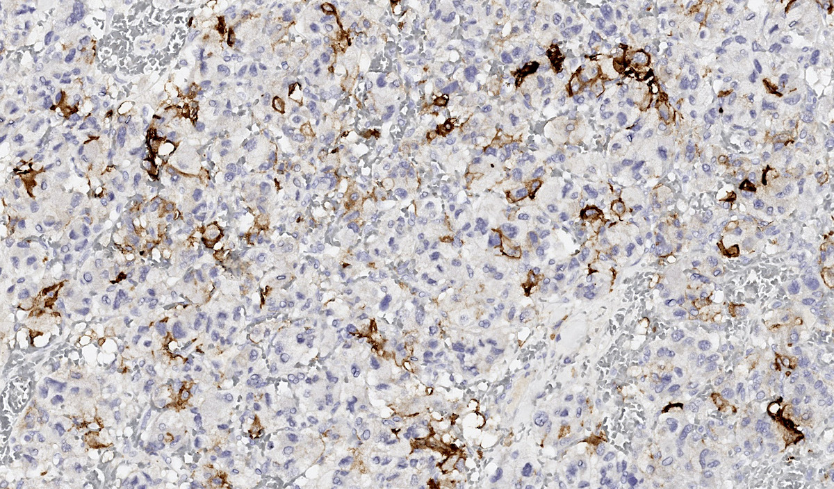
S100
Chromogranin
GATA3
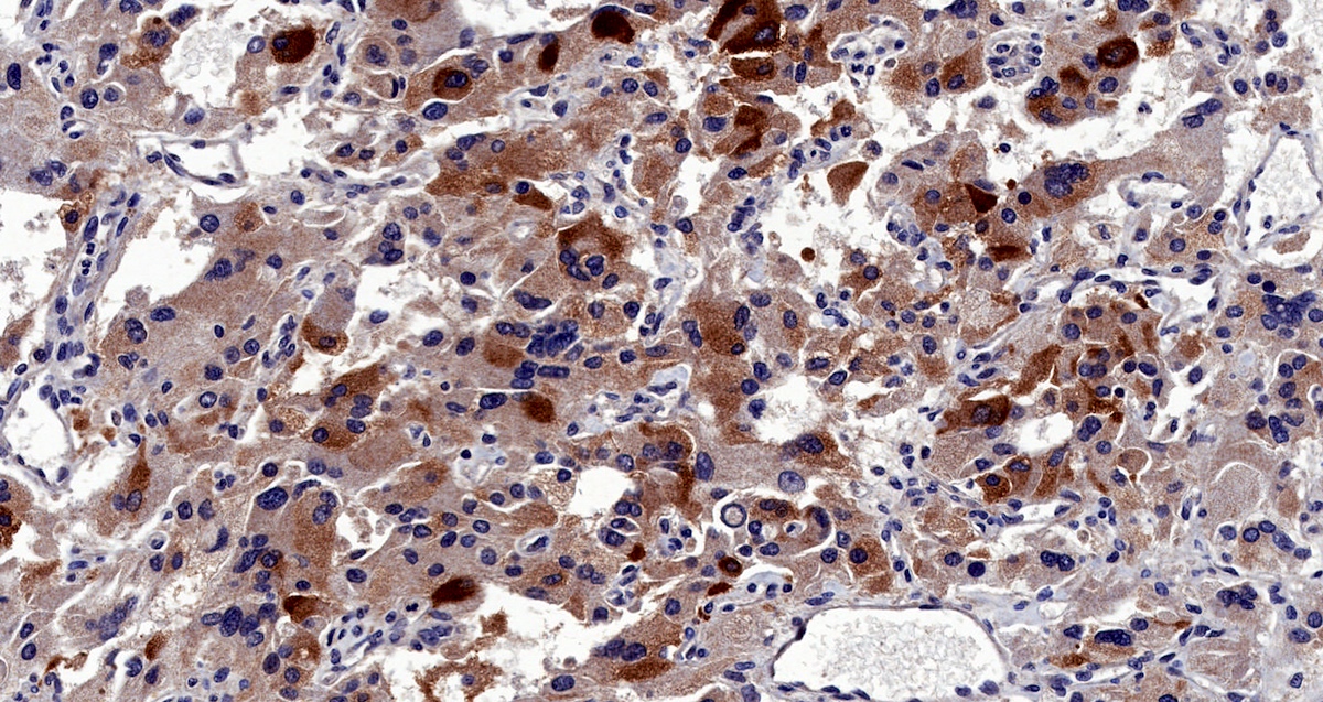
Tyrosine hydroxylase

S100 protein
SOX10
Ki67
SDHB intact
SDHB deficient
CAIX
Inhibin
S100 staining in metastatic pheochromocytoma
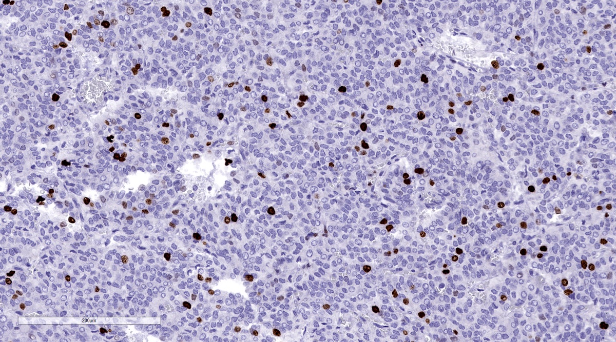
Ki67 in metastatic pheochromocytoma
Contributed by Luvy Delfin, M.D. and Sylvia L. Asa, M.D., Ph.D.
Paraganglioma
Images hosted on other servers:

CT and scintigraphy

MRI

PET / CT and scintigraphy
Images hosted on other servers:
Café au lait spots and axillary freckling in NF1

Intraoperative
images of giant
pheochromocytoma
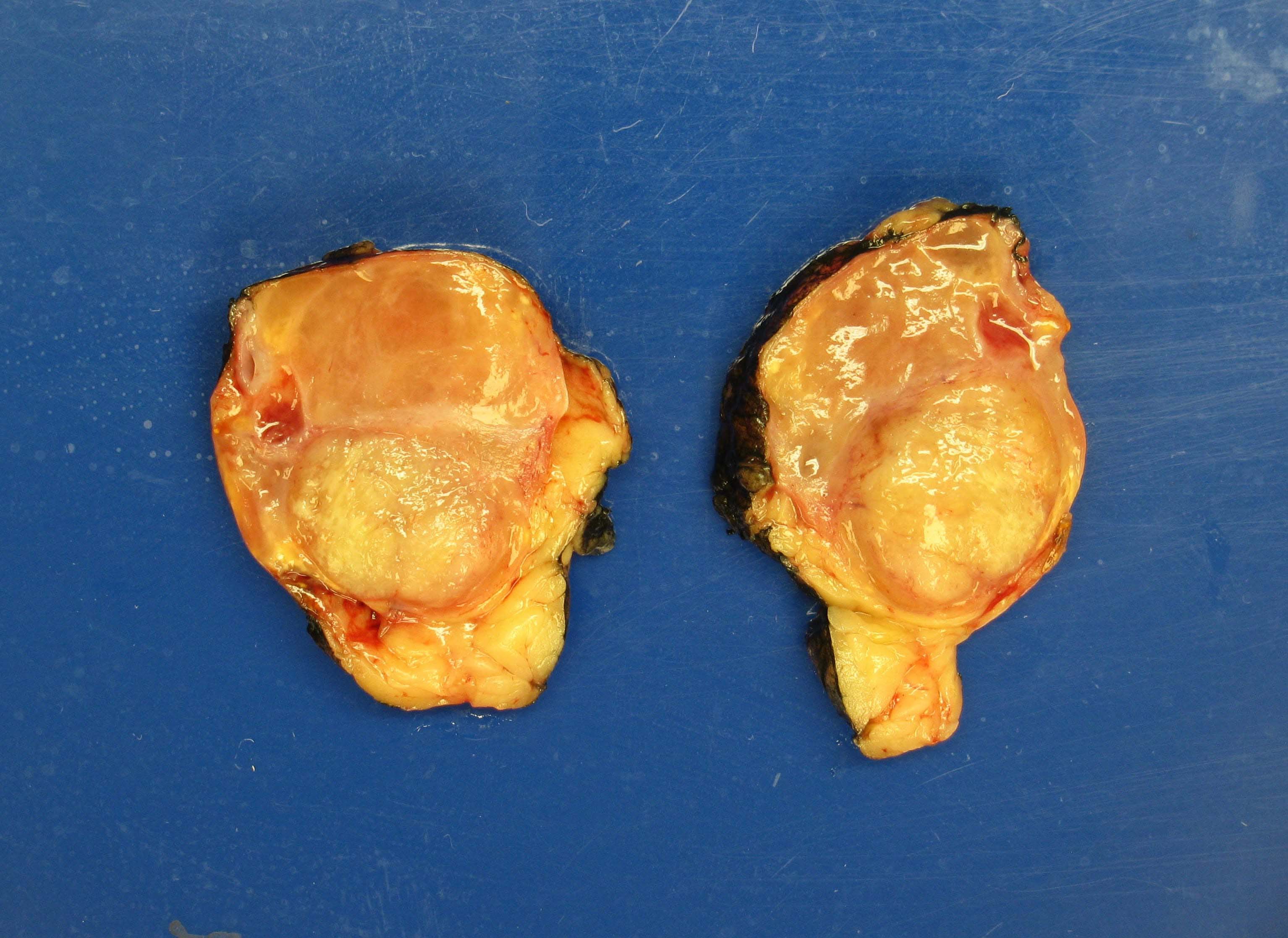
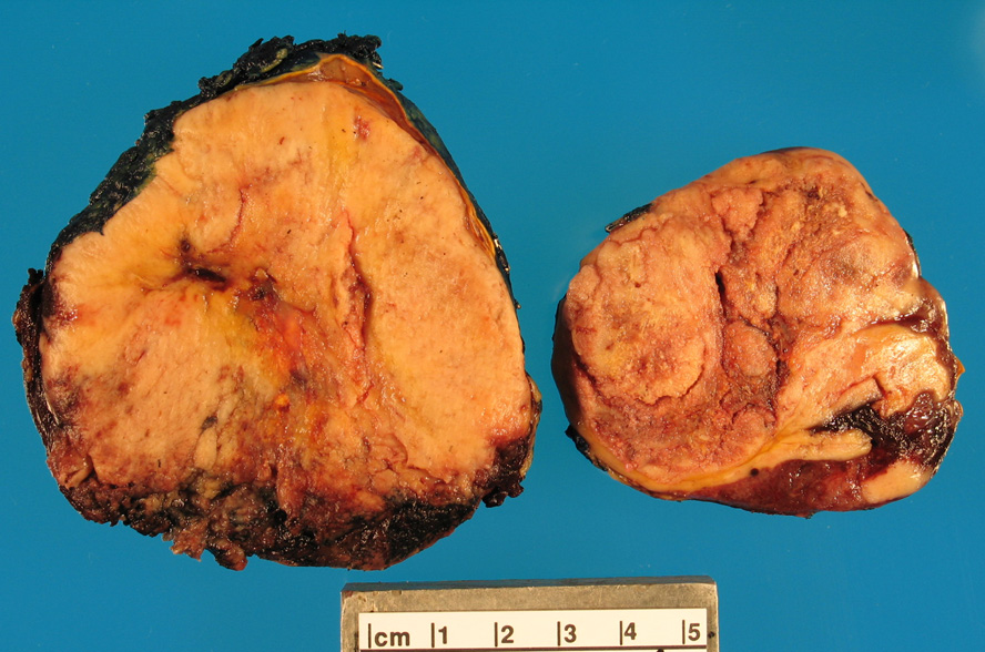
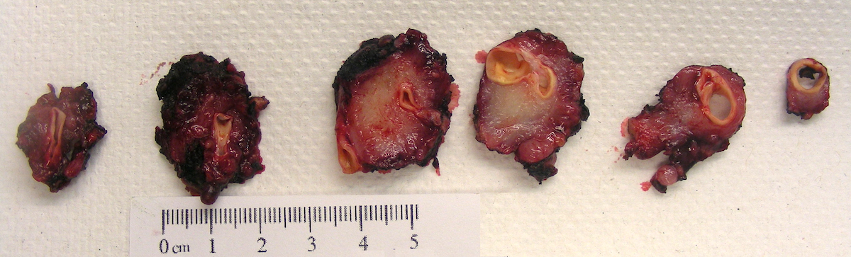
Contributed by Debra L. Zynger, M.D. (Case #319)

Well circumscribed tan mass
Well circumscribed tan mass
Renal invasion
Composite
pheochromocytoma
Contributed by Debra L. Zynger, M.D. (Case #319), Brian Werstein, M.D. and Nicole K. Andeen, M.D. (Case #489)
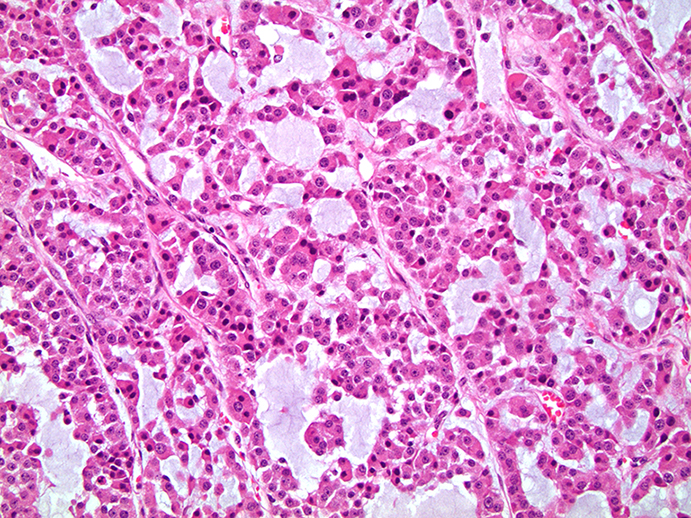
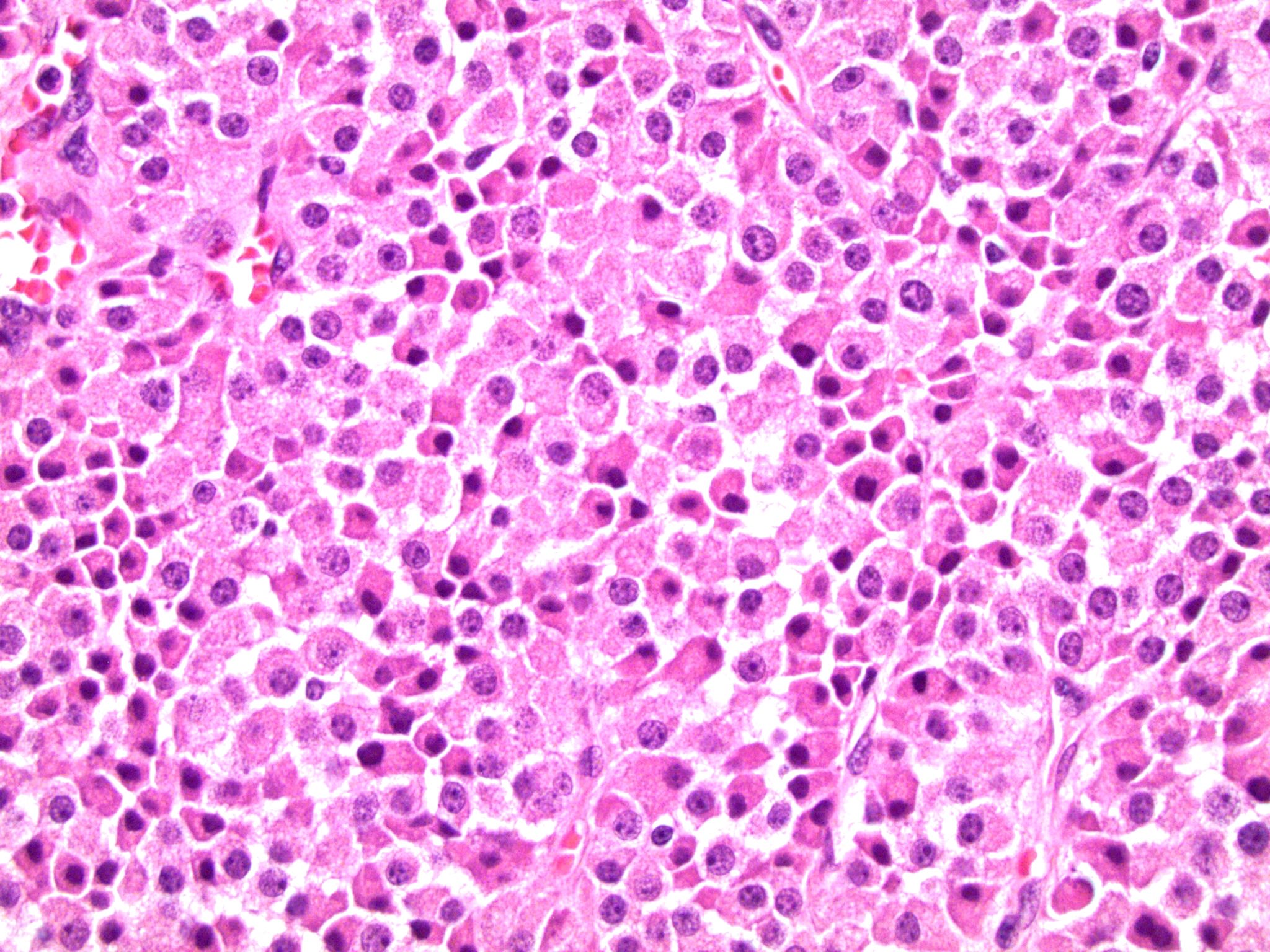
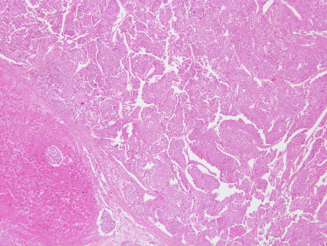
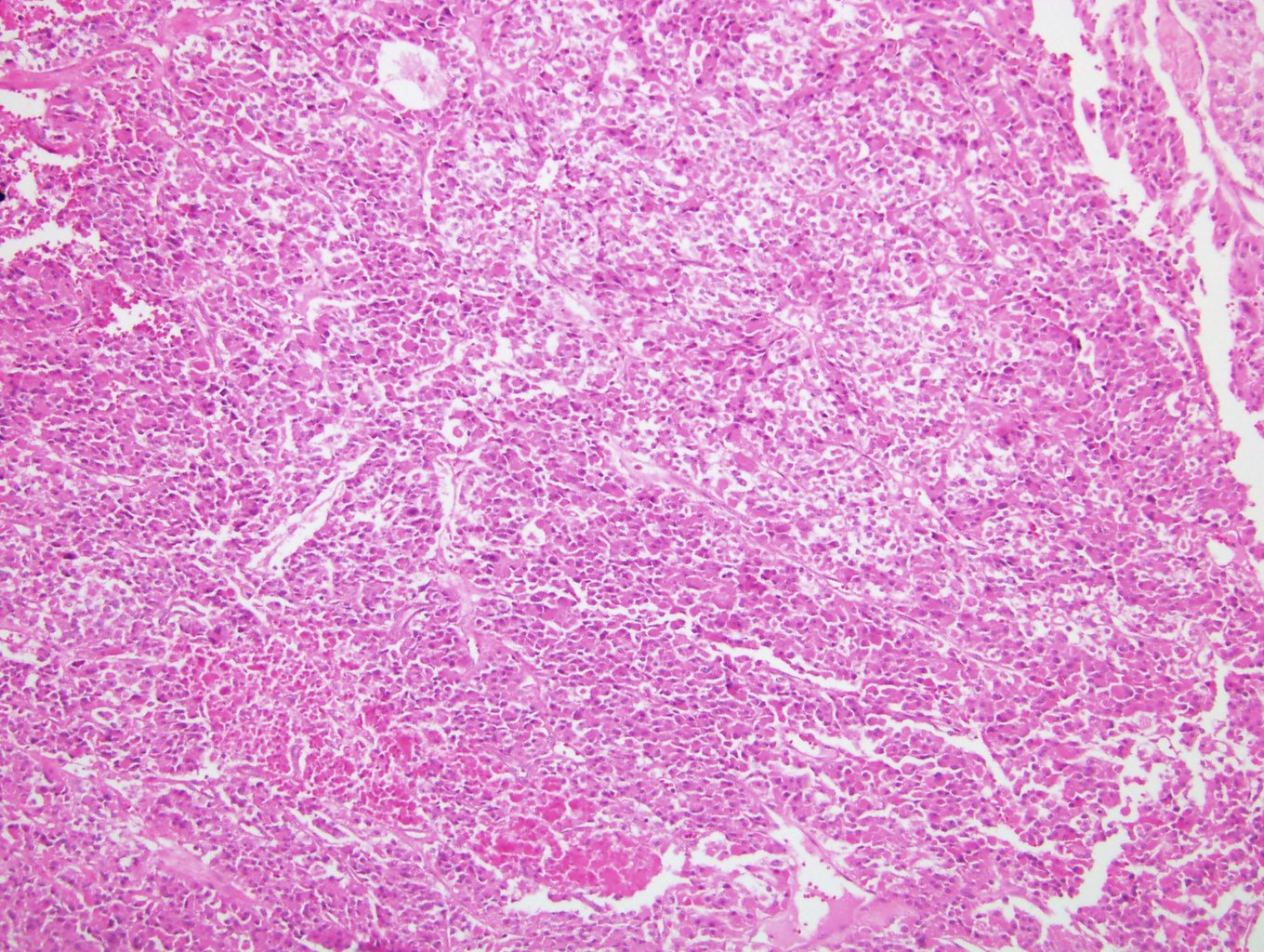
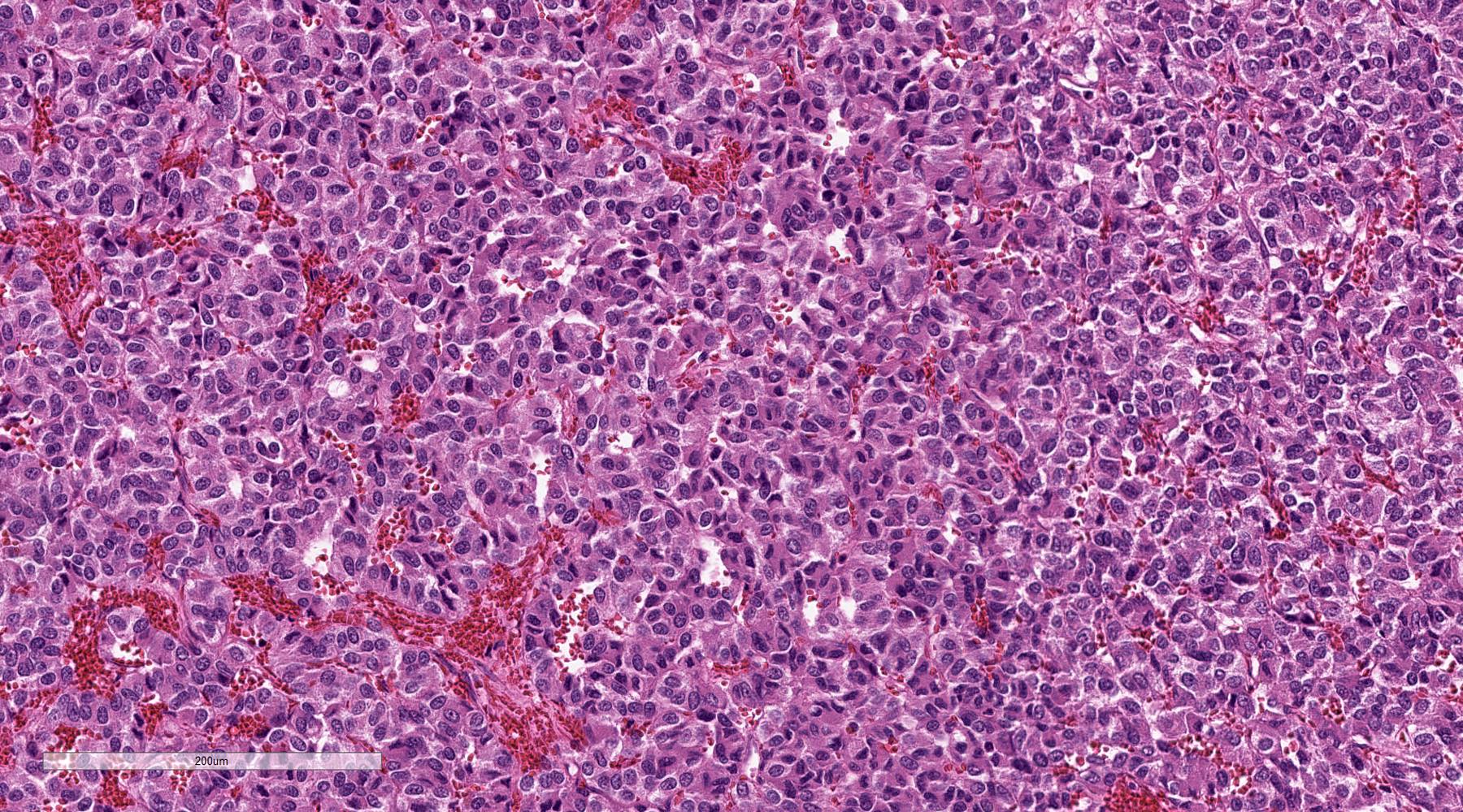
Nested architecture
Sheets of cells
Abundant cytoplasm

Capsular invasion
Adipose invasion

Vascular invasion
Cellular spindling
Nuclear pleomorphism
Mitotic activity
Necrosis

Large nests
Lymph node metastasis
Synaptophysin
Chromogranin
GATA3
S100
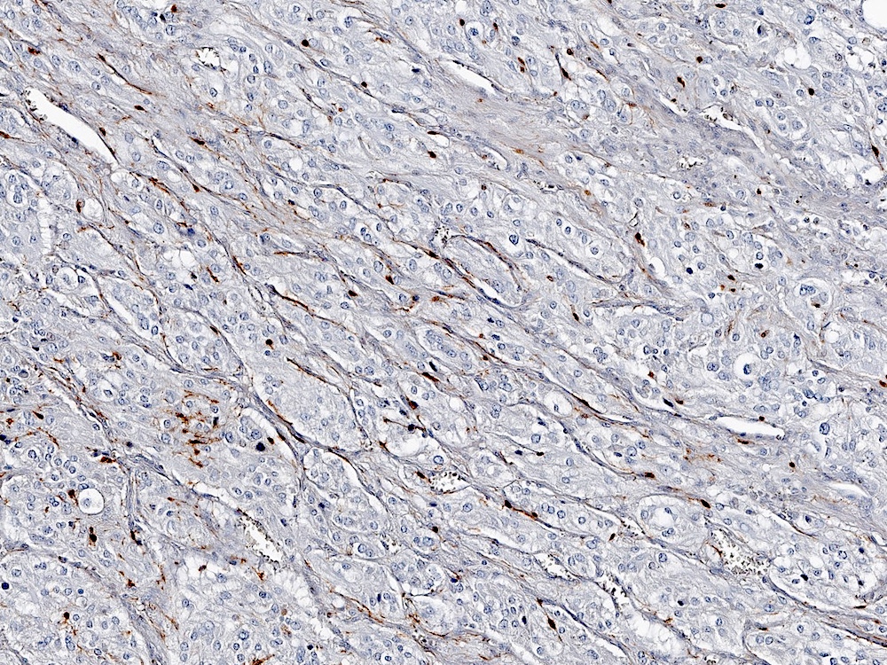
S100


Composite pheochromocytoma with ganglioneuroma
Contributed by Debra L. Zynger, M.D.
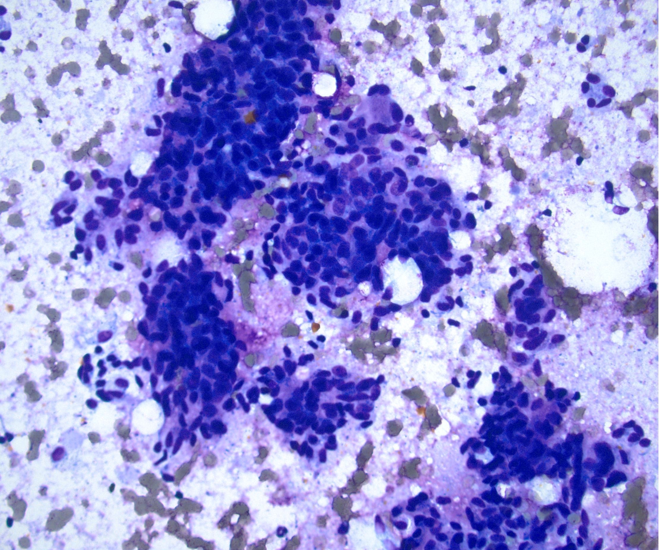
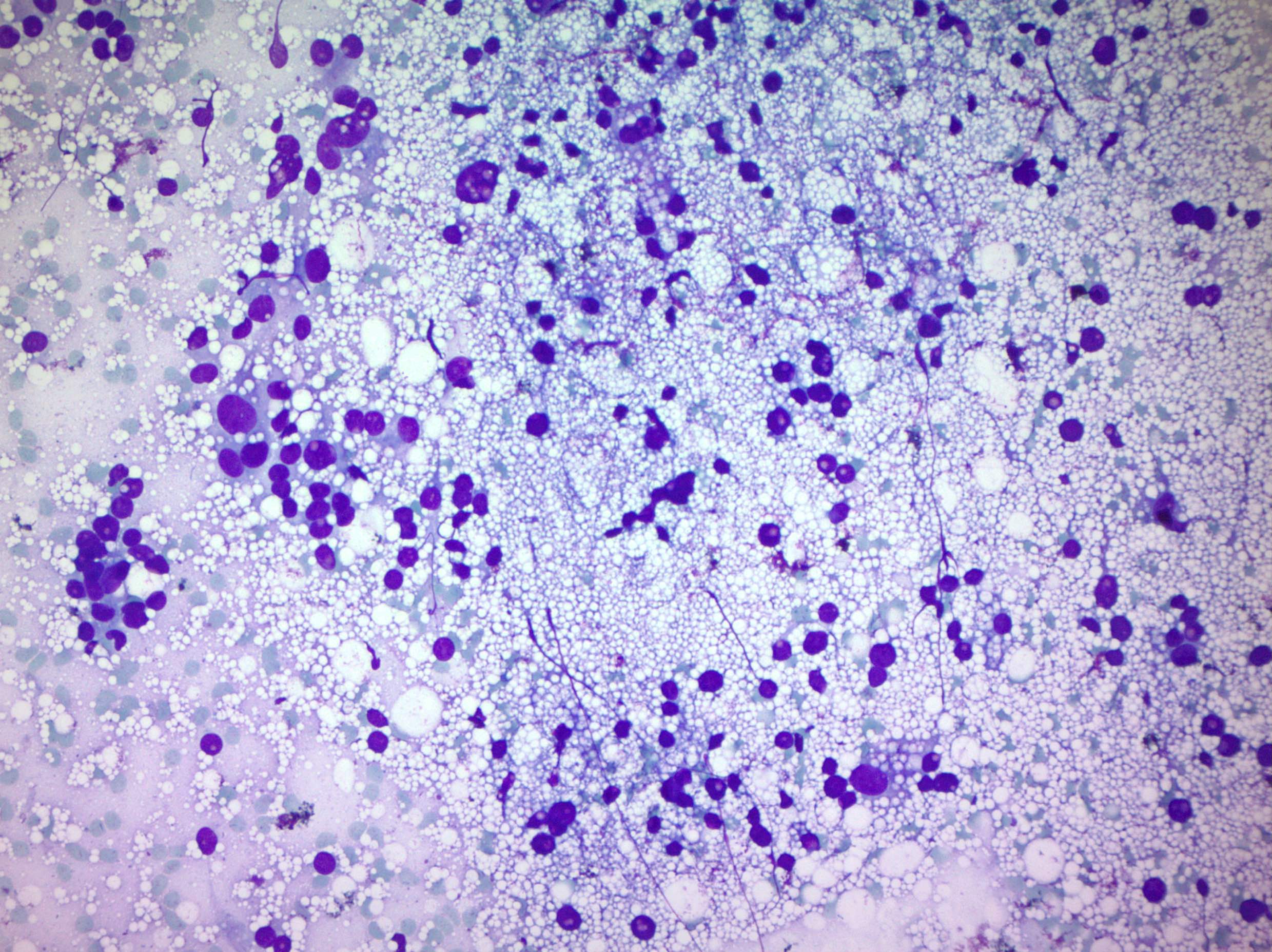
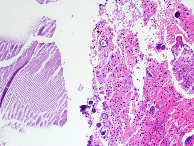
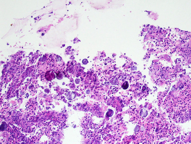
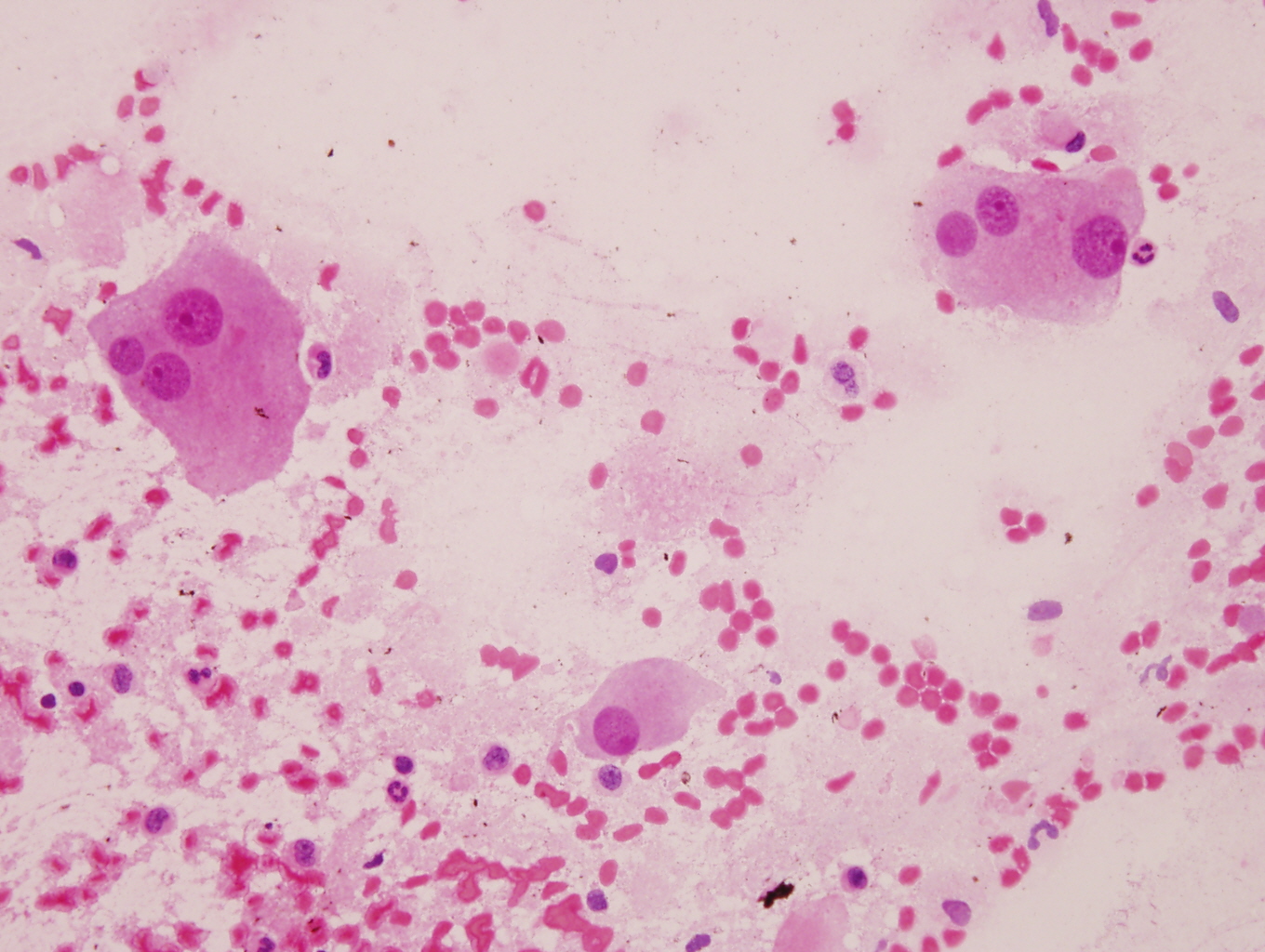
Diff-Quik
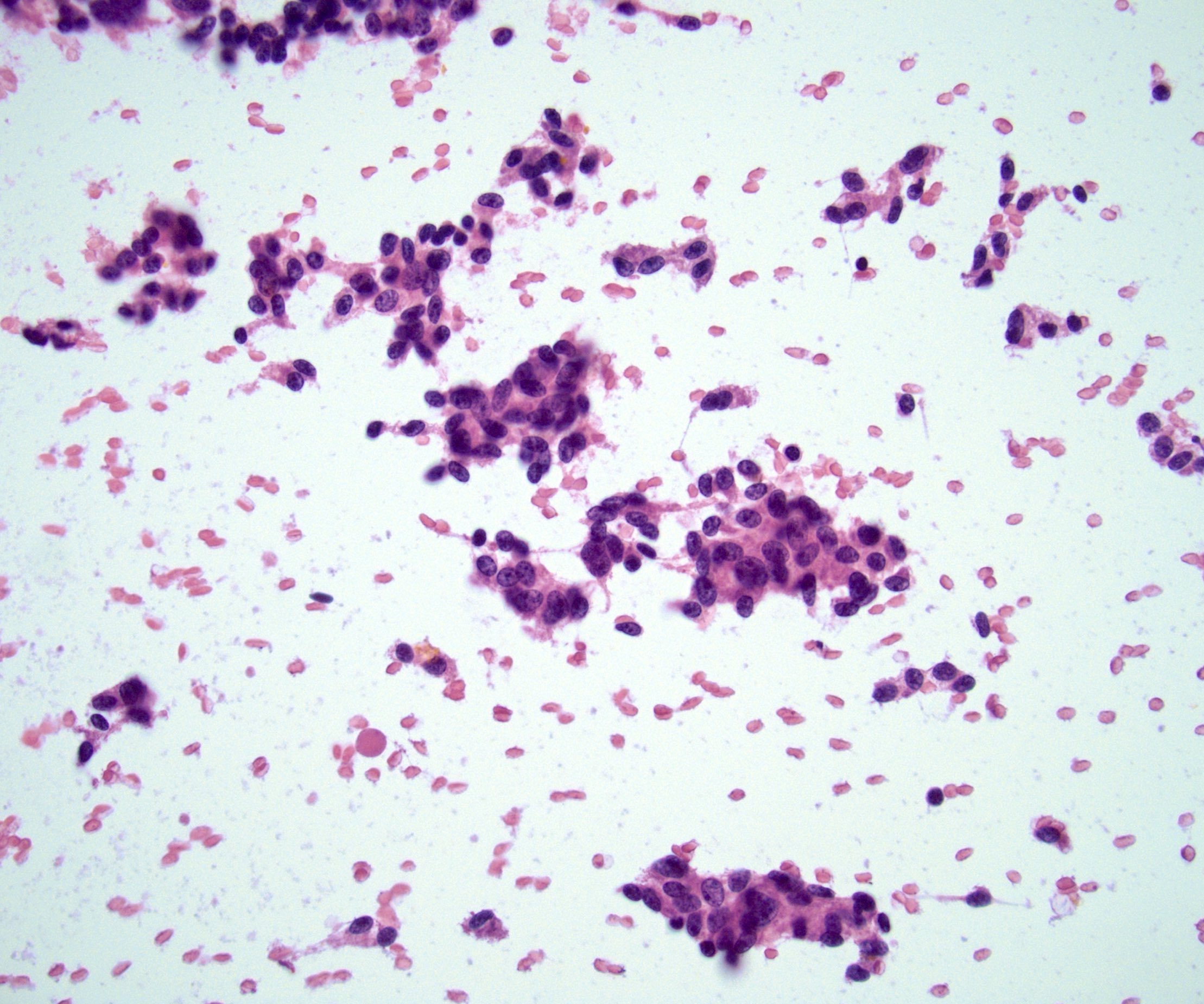
Touch preparation
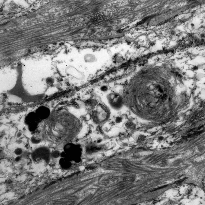
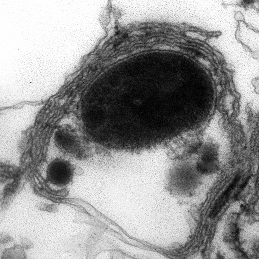
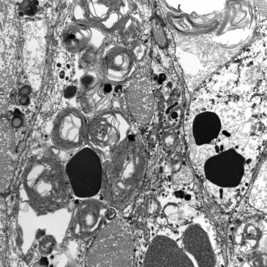
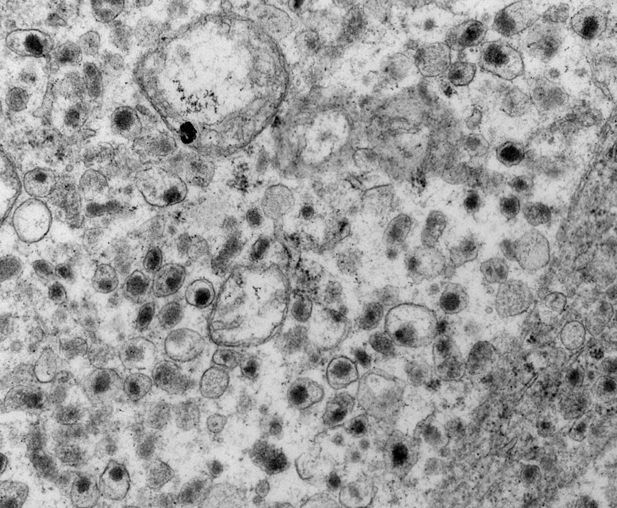
Contributed by Debra L. Zynger, M.D. and Edward Calomeni, B.S.
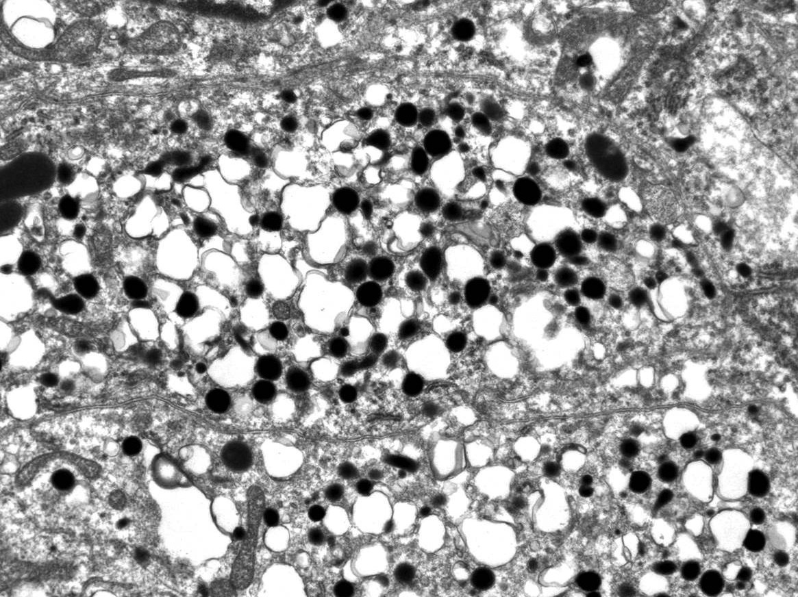
Numerous epinephrine granules
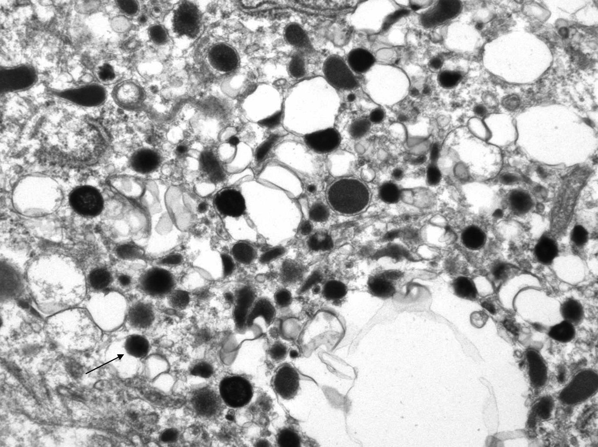
Norepinephrine granules
Images hosted on other servers:


Neurosecretory granules
Images hosted on other servers:

NF1 mutation

TMEM127 mutation
Images hosted on other servers:

Survival analysis:
sarcomatoid ACC
versus other
variants
Images hosted on other servers:

CT scan: necrotic areas replacing adrenal gland

Coronal abdominal CT with large adrenal mass
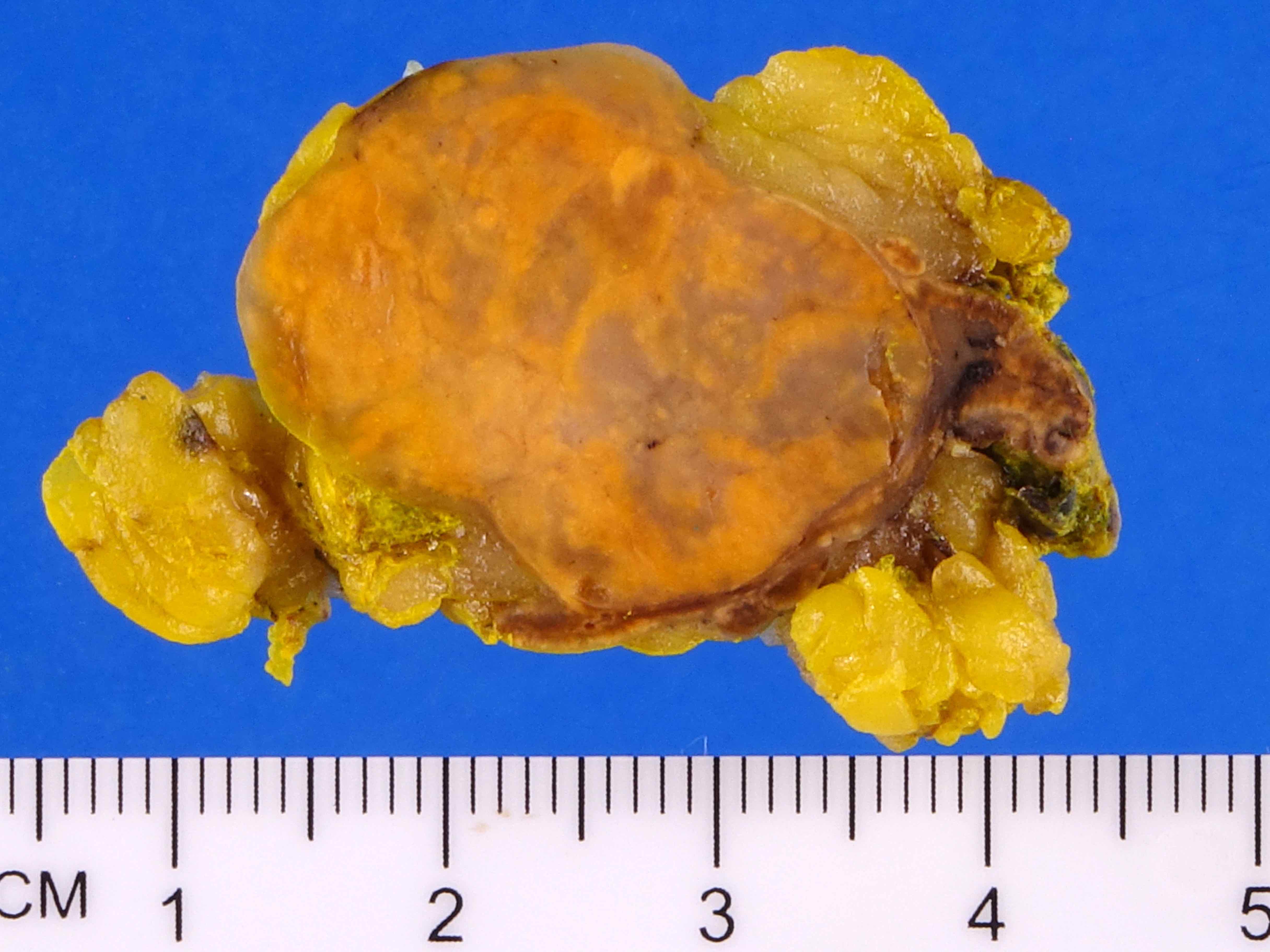
Image hosted on other servers:

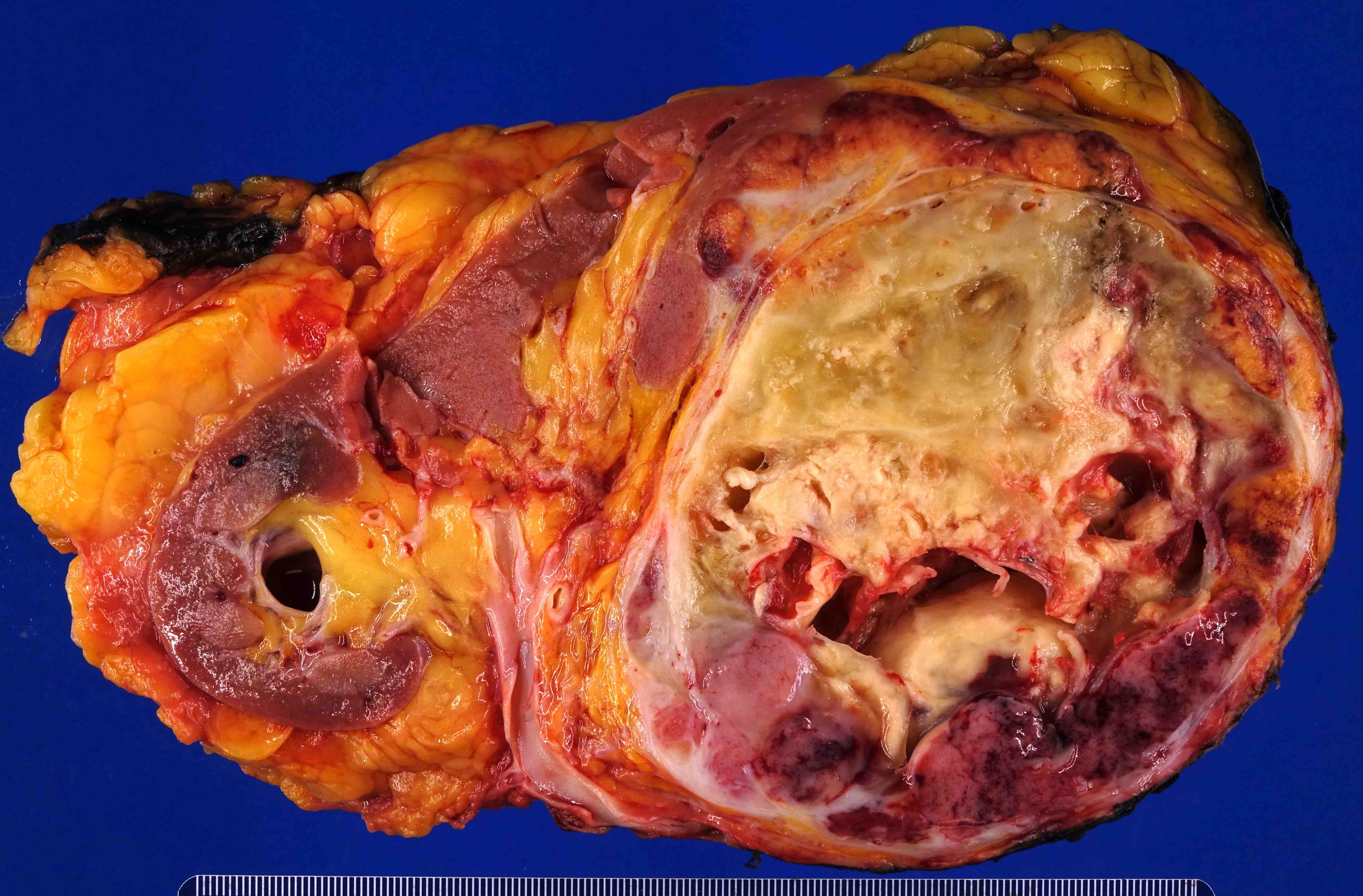
Whitish fleshy, variegated hemorrhagic and necrotic surface

Tumor adherent
to pancreas (A)
and compressing
kidney (B)
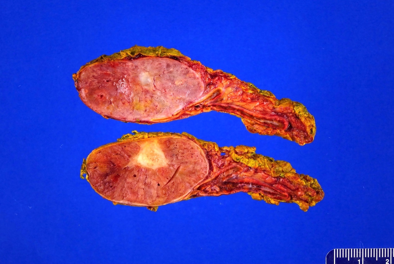
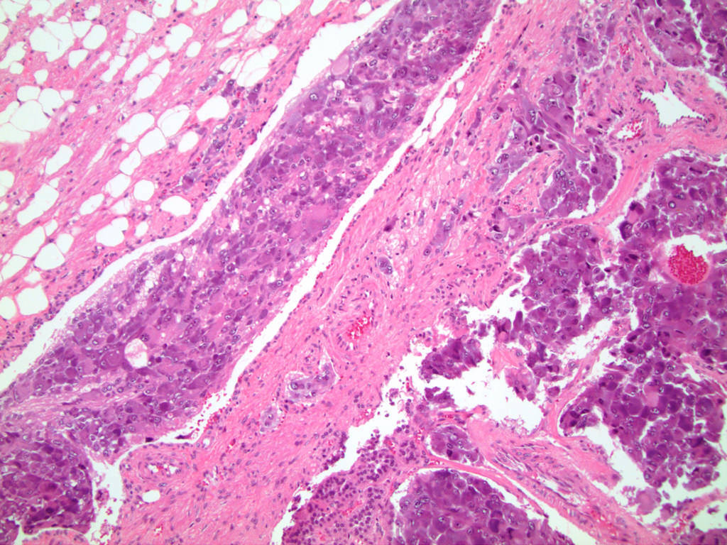
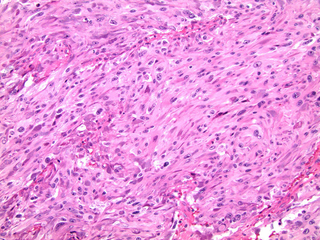
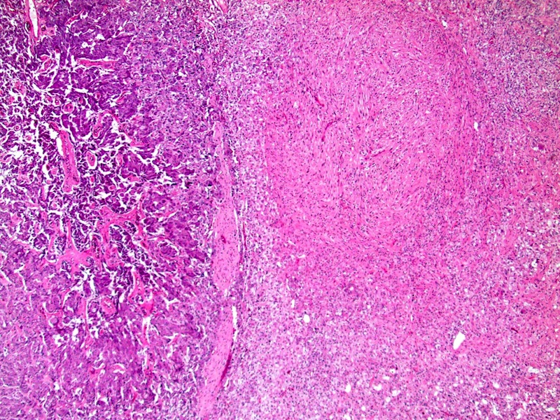
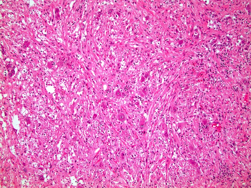
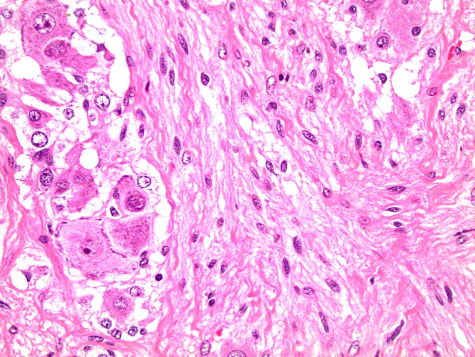
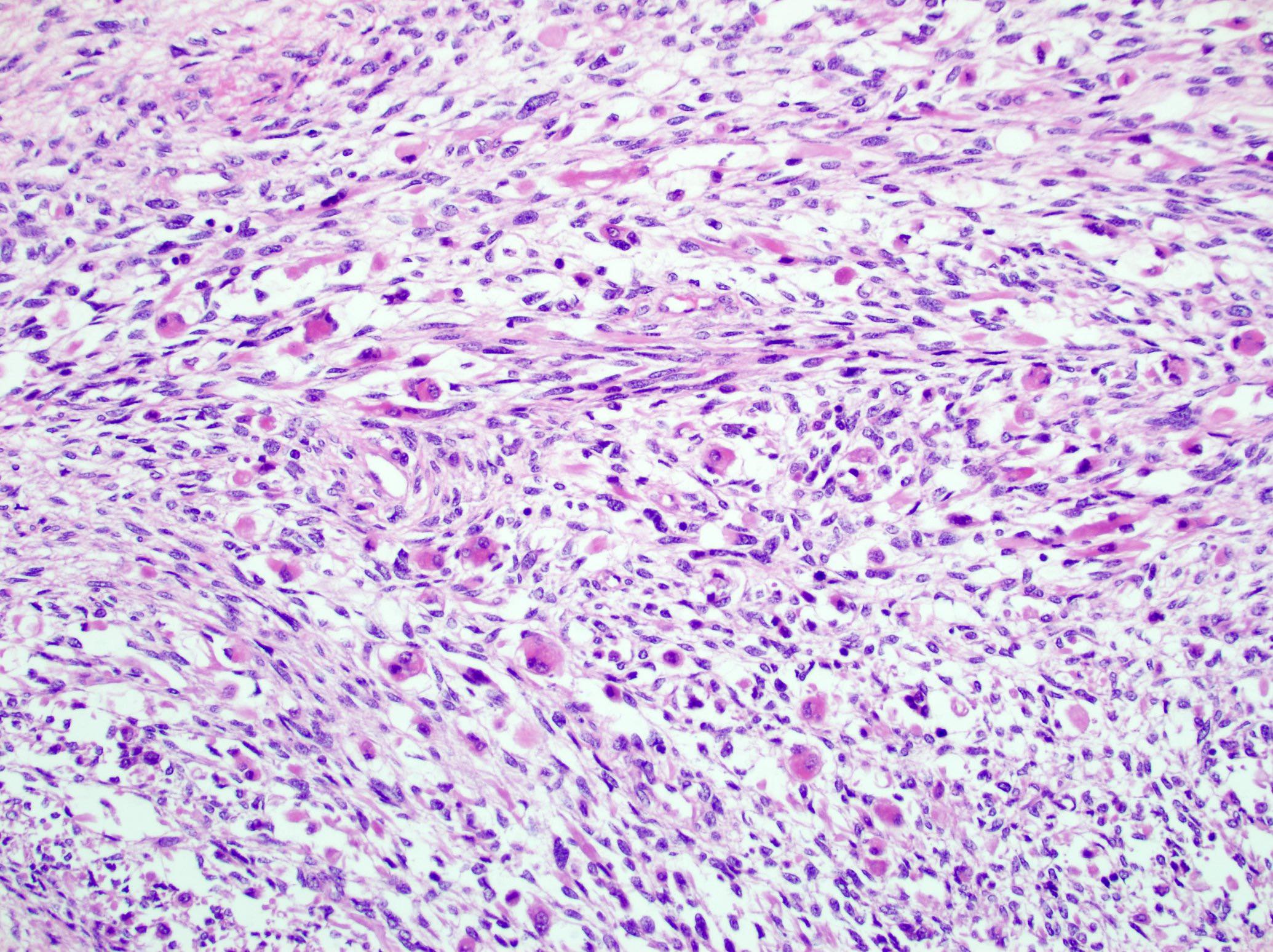
Contributed by Maria Tretiakova, M.D., Ph.D.
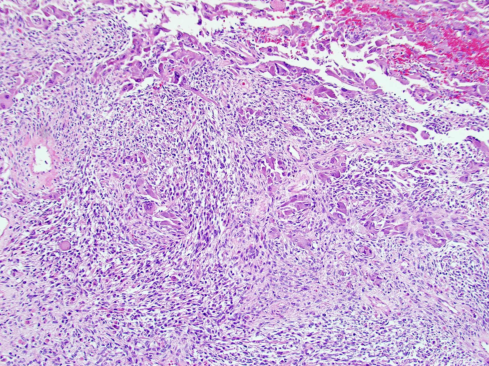
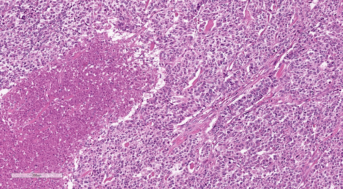
Sarcomatoid ACC biphasic
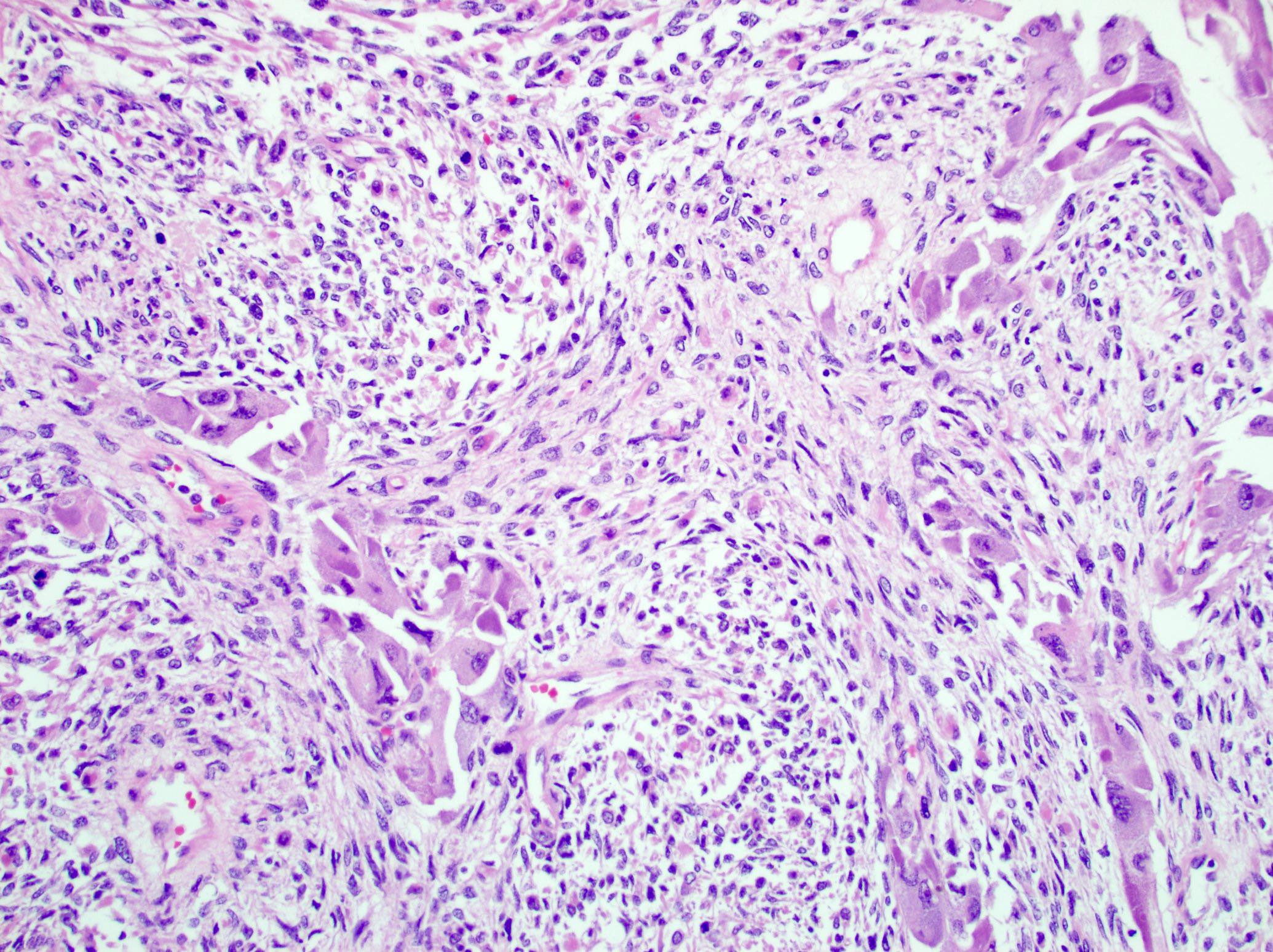
Epithelioid and spindle cells
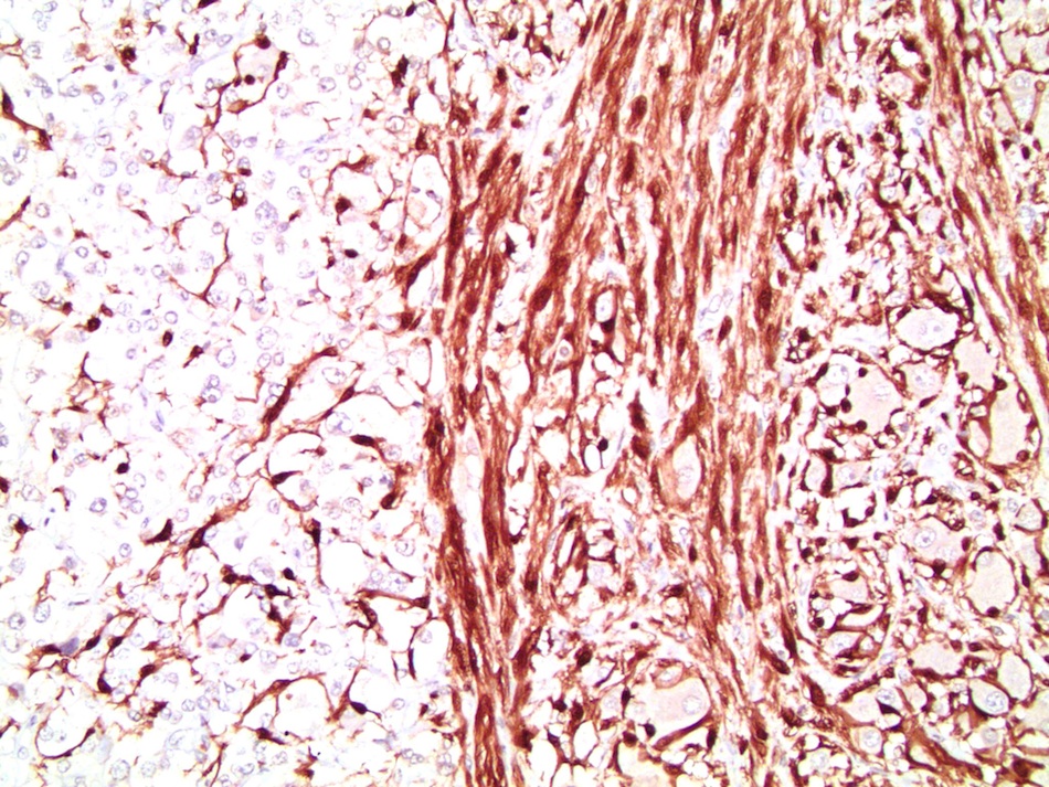
Heterologous differentiation
Images hosted on other servers:

Epithelioid and spindle cell components
Contributed by Debra Zynger, M.D.

Organ confined, ≤ 5 cm (pT1)

Organ confined, > 5 cm (pT2)

Extra-adrenal invasion (pT3)
Liver invasion (pT4)
Contributed by Debra Zynger, M.D. and Maria Tretiakova, M.D., Ph.D.
Extra-adrenal adipose invasion (pT3)
Liver involvement (pT4)
Regional node involvement (pN1)
Images hosted on other servers:

Children's Oncology
Group (COG)
neuroblastoma risk
grouping system

INRG stage definitions

INRG consensus pretreatment classification
Contributed by Debra L. Zynger, M.D.
pT1
pheochromocytoma
pT2
pheochromocytoma
pT3 pheochromocytoma
pT2 sympathetic
paraganglioma
Contributed by Debra L. Zynger, M.D., Brian Werstein, M.D. and Nicole K. Andeen, M.D. (Case #489)
pT2 sympathetic
paraganglioma
pT3
pheochromocytoma
with extra-adrenal
adipose invasion
pT3
pheochromocytoma
with invasion
of the kidney
pN1
pheochromocytoma
Contributed by Eric L. Vey, M.D
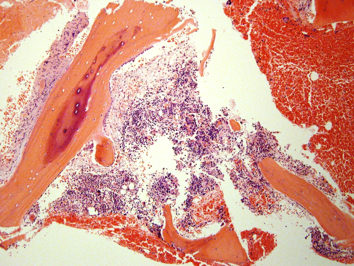
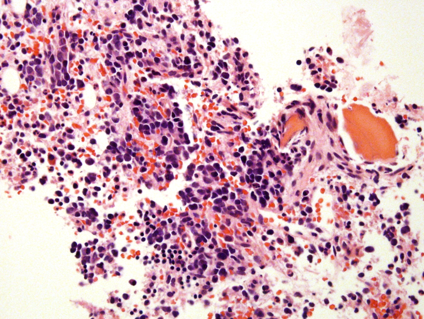
Fatal meningococcemia in 10 month old boy
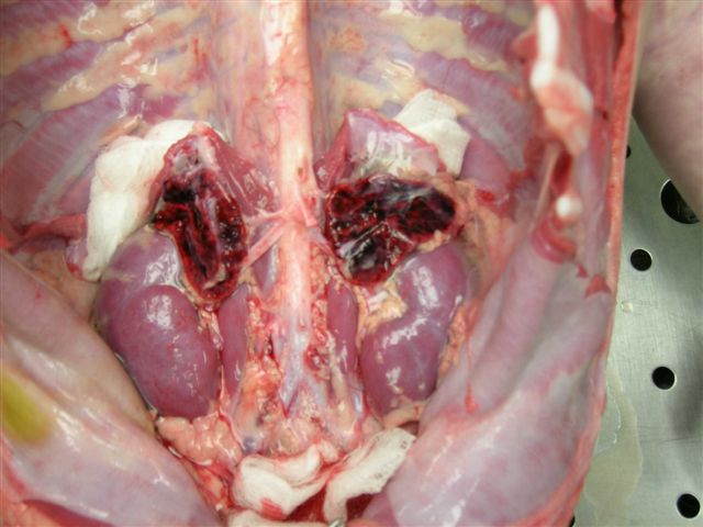
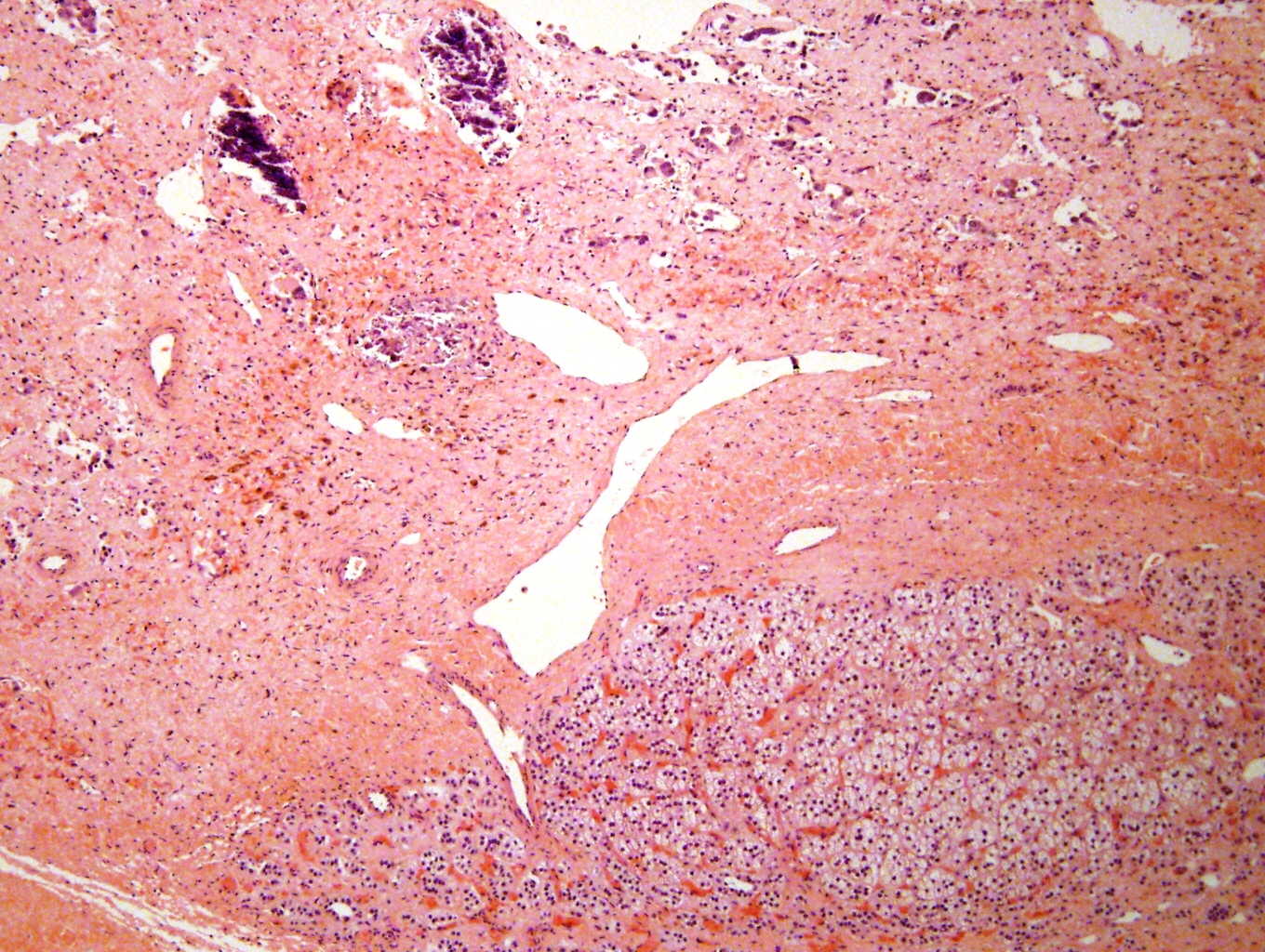
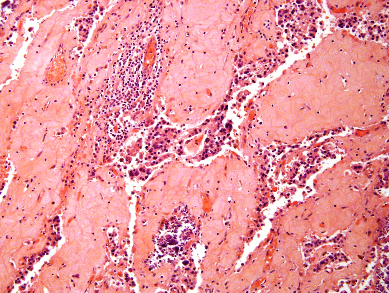
Adrenal glands in situ

Normal (top) and hemorrhagic (bottom) adrenal glands
Gill: 2022
IARC: 2017
Lack: 2007
Lloyd: 2002
Mete: 2016
Nosé: 2022
Tickoo: 2015
VandenBussche: 2022
Find related Pathology books: cytopathology, GI, GU/adrenal, head & neck/endocrine
