
Images hosted on other servers:

Classification
of anal
adenocarcinoma
Images hosted on other servers:

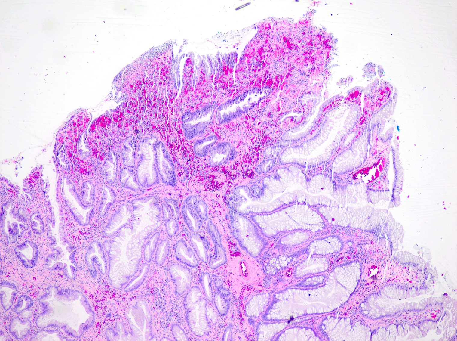
Irregular mucosa and angioectasia
Images hosted on other servers:

Anal canal cancer
Contributed by Raul S. Gonzalez, M.D.
Infiltrating, gland forming
Infiltrating glands
Mucinous and signet ring
Fistula tract associated
Images hosted on other servers:

Coronal section of rectum and anal canal

Columns of Morgagni and the anal valves

Inner wall of the lower end of the rectum and anus

Median sagittal section of male pelvis

Median sagittal section of female pelvis
Images hosted on other servers:

Anal / rectal junction and sphincter
Images hosted on other servers:

Perianal basal cell carcinoma
Contributed by Raul S. Gonzalez, M.D.
Basal cell carcinoma
Images hosted on other servers:

Cell clusters
Images hosted on other servers:

Tumor invading the rectum

Extensive pelvic infiltration of tumor

Pelvic structures infiltrated by tumor

Tumor expands along the endopelvis

Tumor involves the inguinal area
Contributed by Philippe Simon, M.D. and Andrew L. Atkinson, M.D.
Bulky tumor of vulva and anus
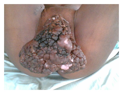
Large exophytic tumor of vulva and perineum
Contributed by Lewis A. Hassell, M.D. (source: Juan Rosai Slide Collection)
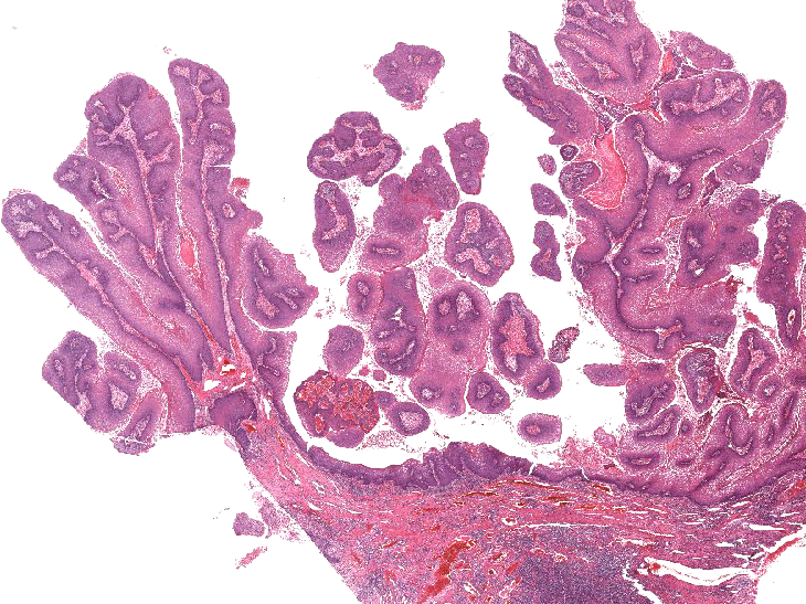
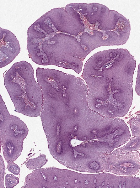
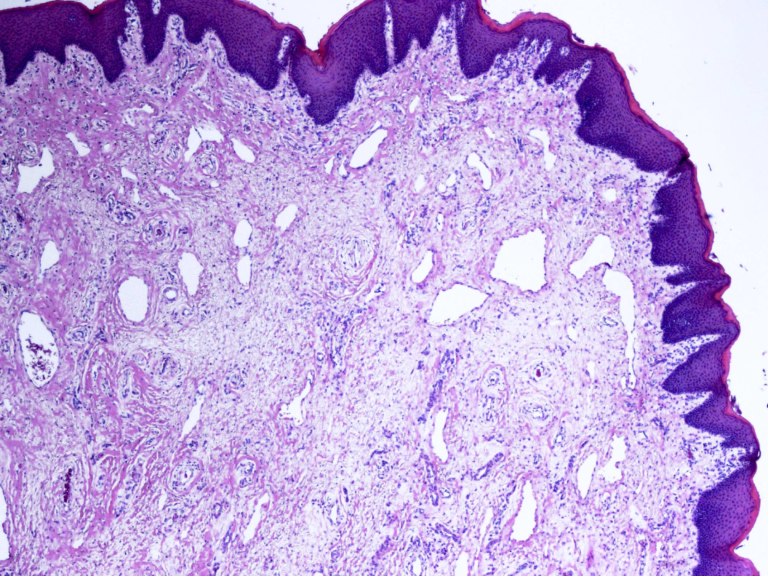
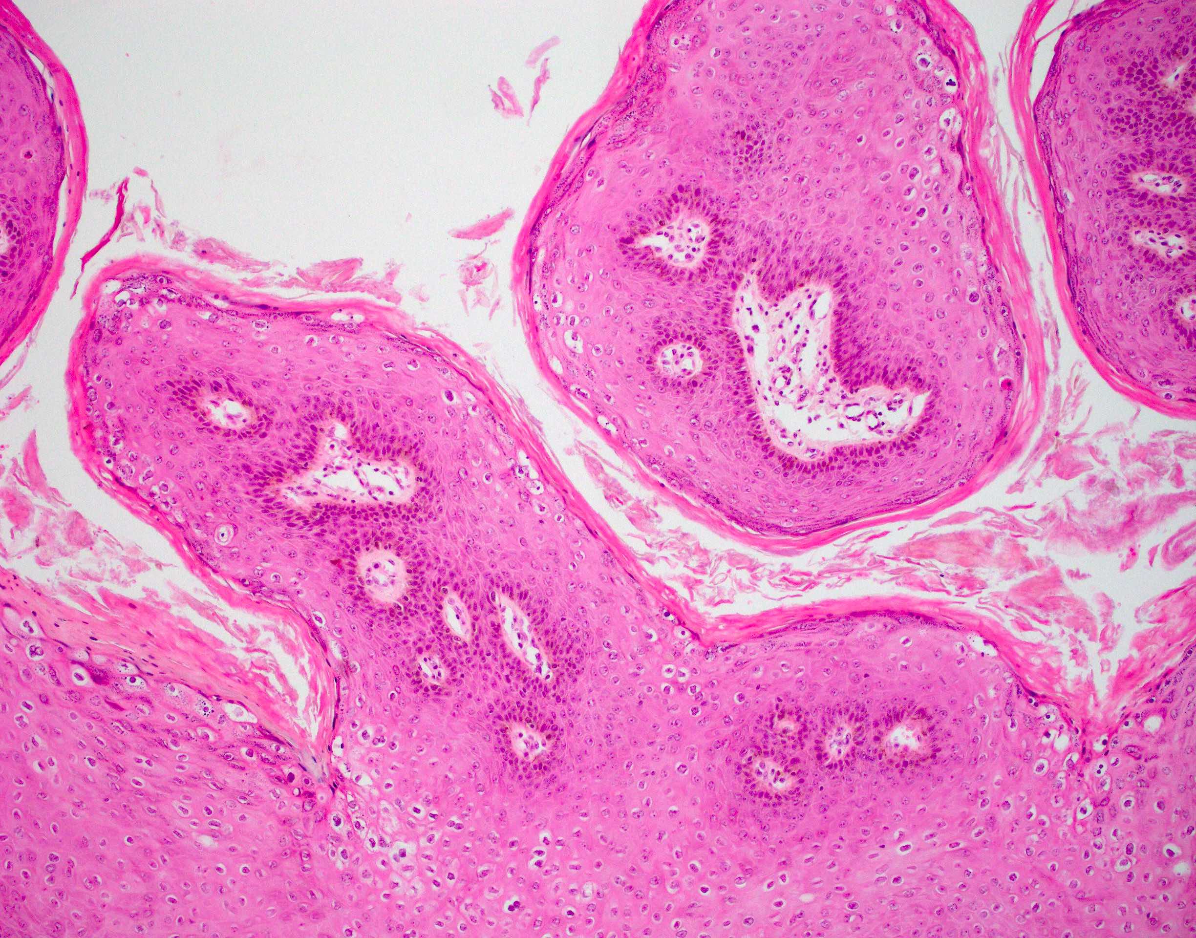
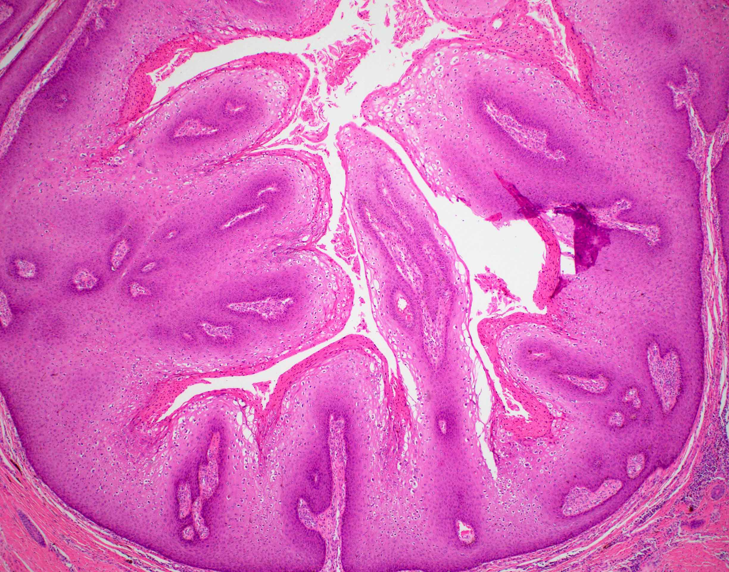
Well formed papillae with fibrovascular cores
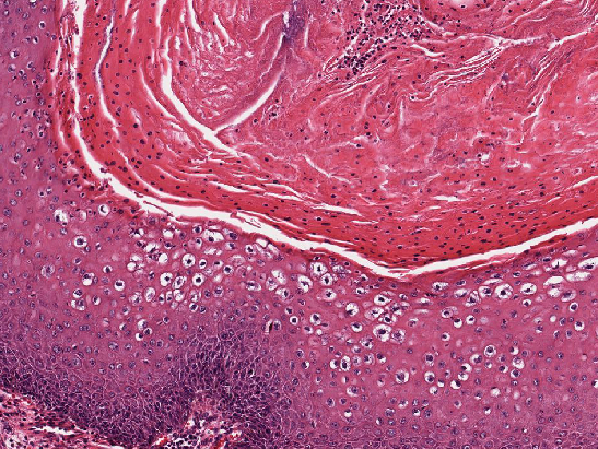
Hyperkeratosis with parakeratosis
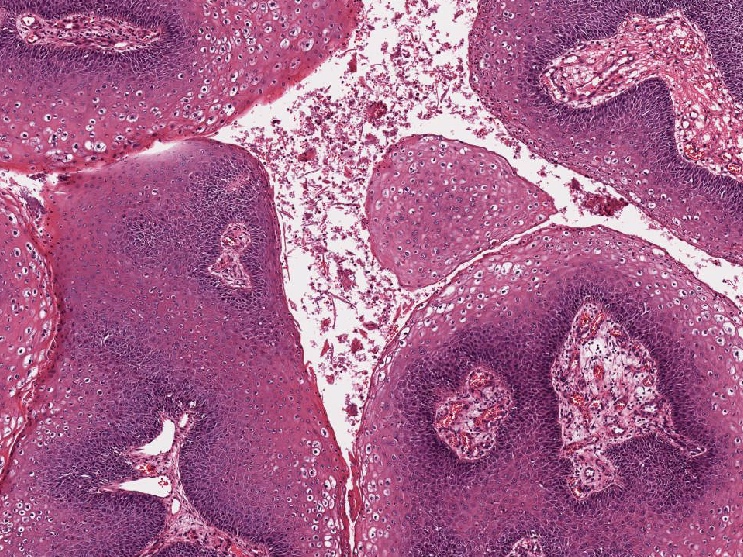
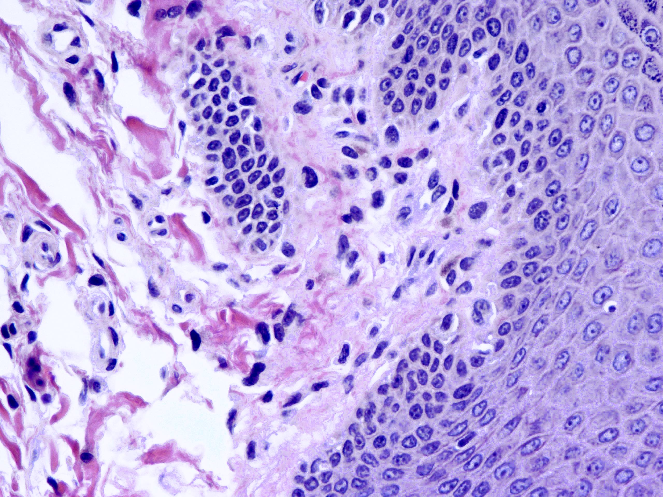
Koilocytes and fungal elements
Koilocytes with perinuclear clearing
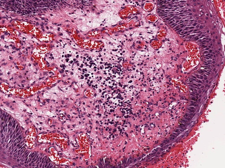
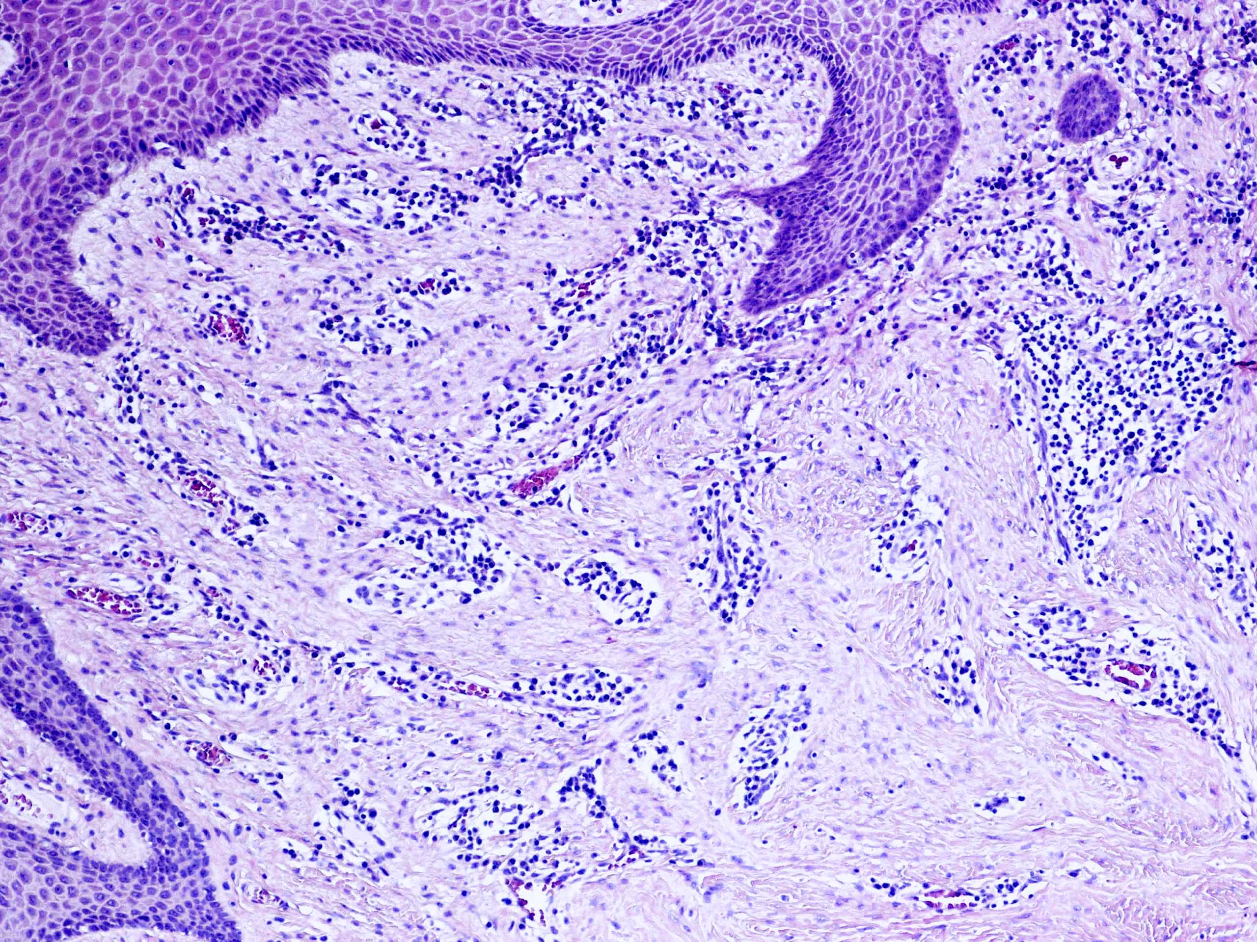
Chronic
inflammation and
vascular changes
in stroma
Giant condyloma
Contributed by Erna Forgó, M.D.

Single warty lesion

Multiple flat lesions
Images hosted on other servers:

Severe case
Contributed by Jian-Hua Qiao, M.D.
Vaginal condyloma
Contributed by Erna Forgó, M.D. and AFIP images
Koilocytosis
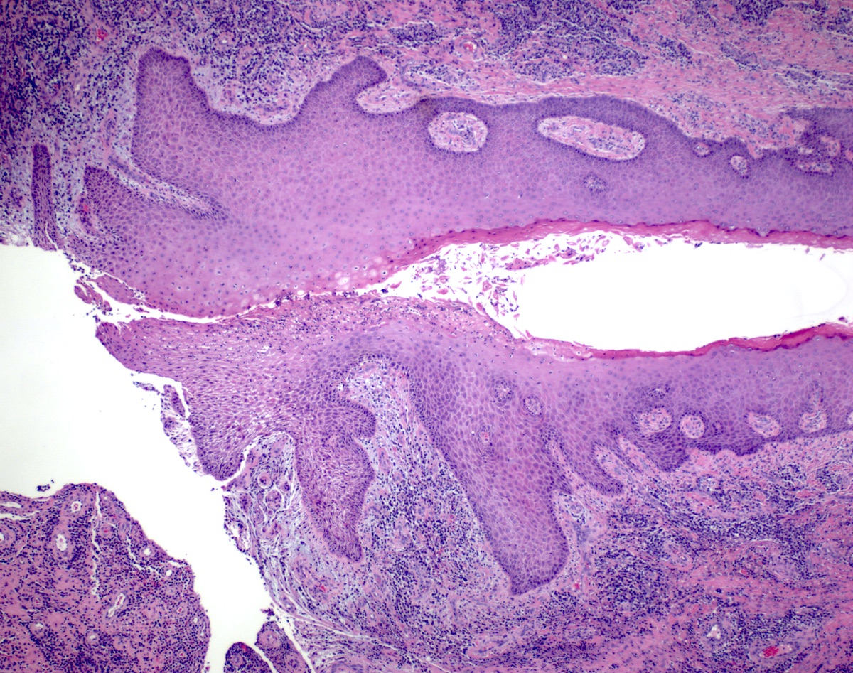
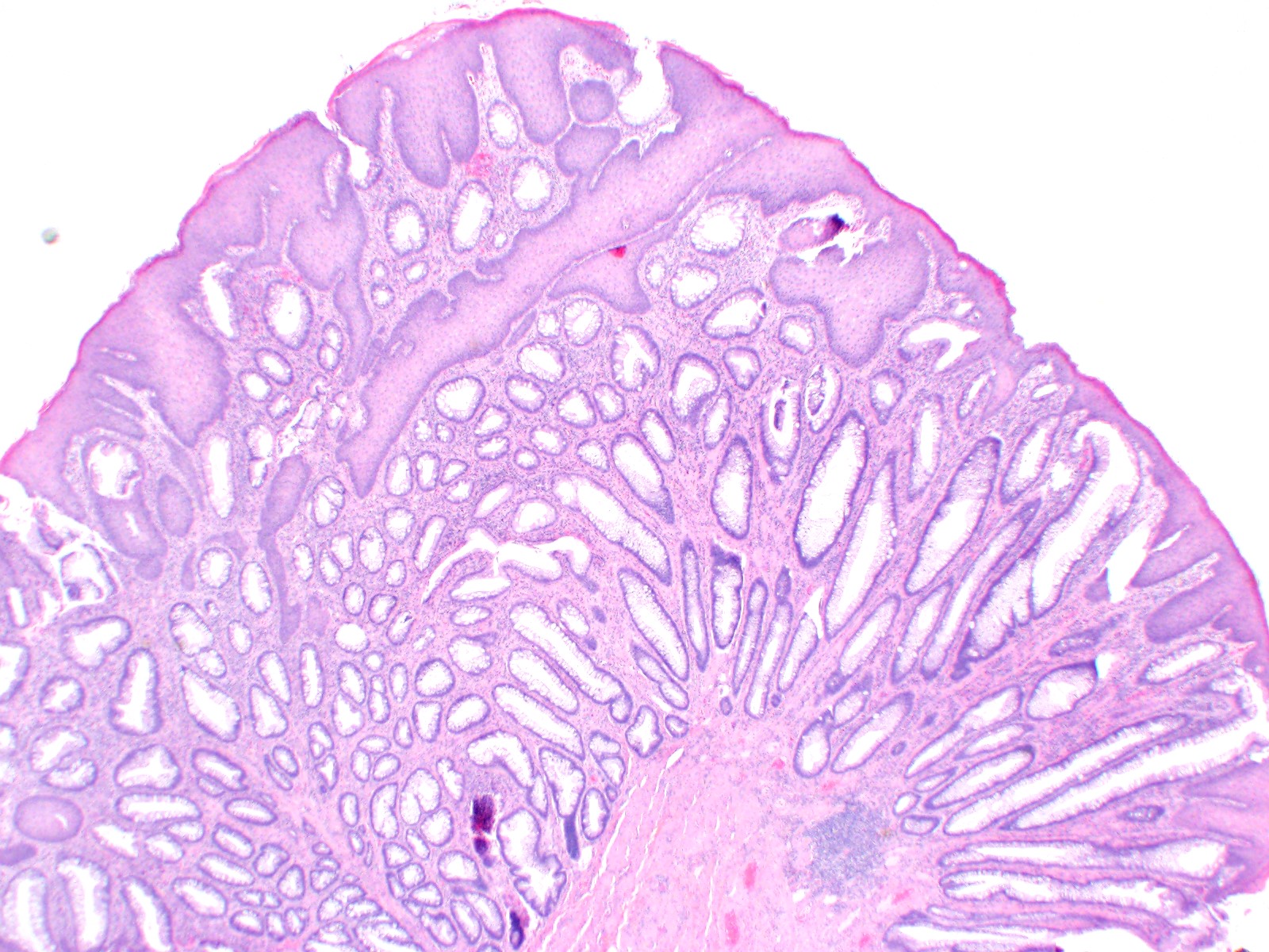
Papillary exophytic squamous epithelium

Hyperplastic squamous epithelium
Papillary exophytic squamous epithelium
Large papillary structures
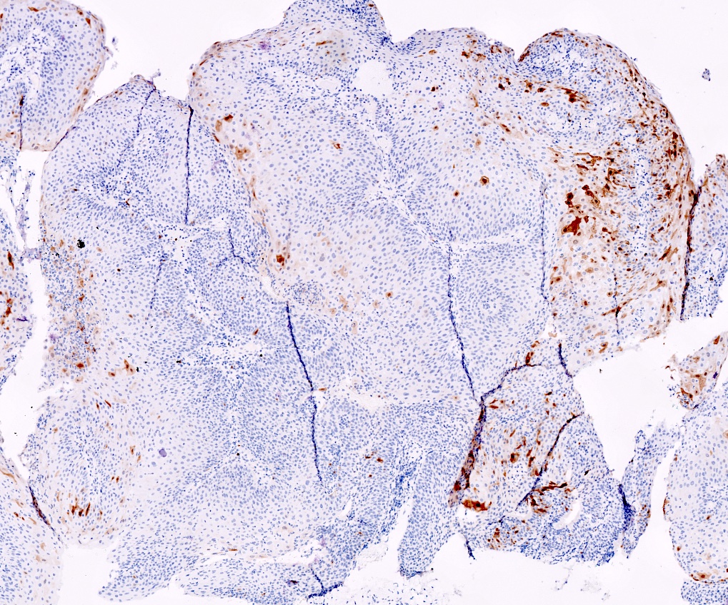
Negative p16 expression (nonblock positivity)
Images hosted on other servers:


LSIL, Pap
Images hosted on other servers:

Large perianal skin tags
Images hosted on other servers:

Colon of 39 year old male
Images hosted on other servers:

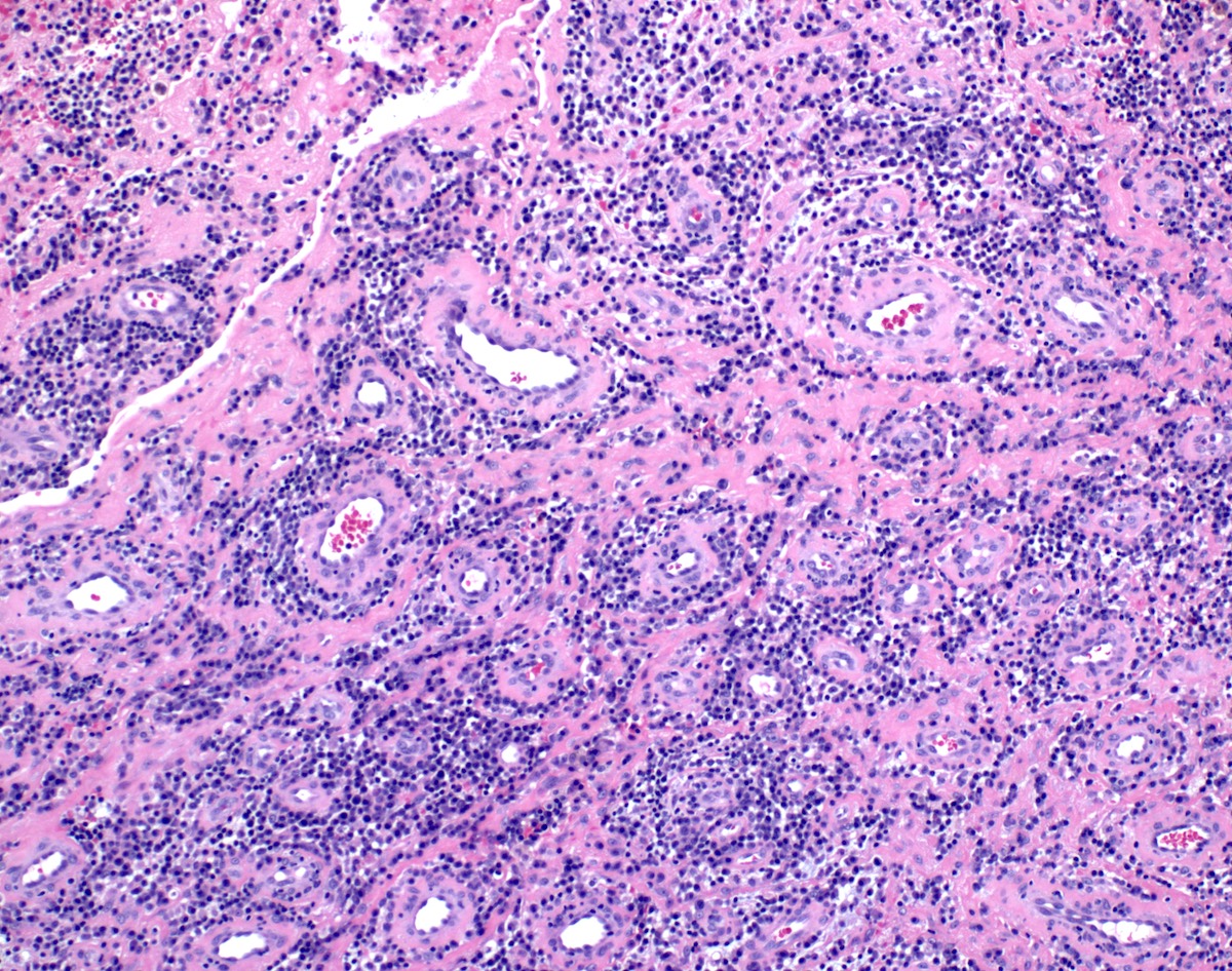
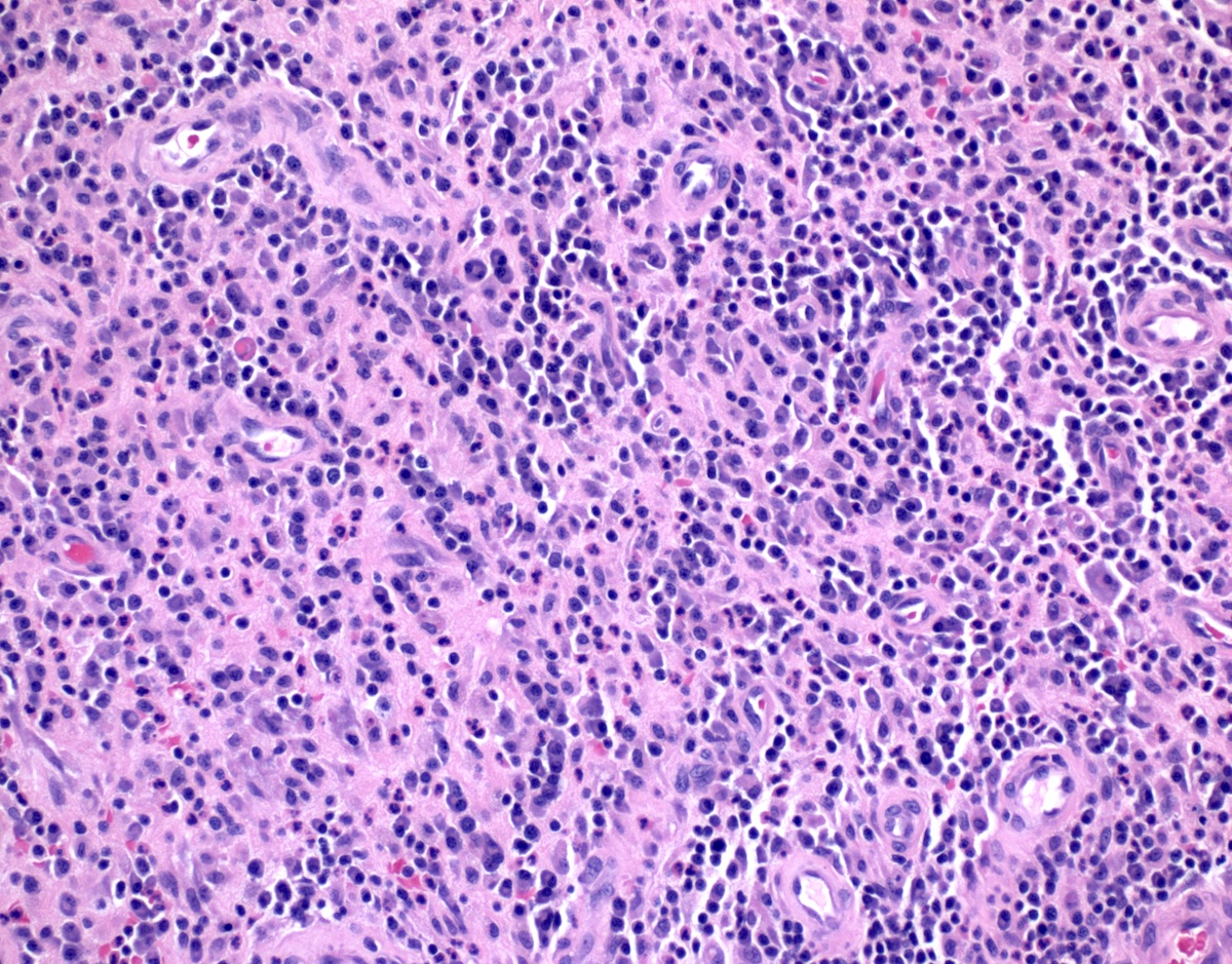
Epithelioid cells, giant cells, lymphocytes

Fistula formation

Fistula opening onto perineum
Contributed by Arvind Rishi, M.D., M.B.B.S.
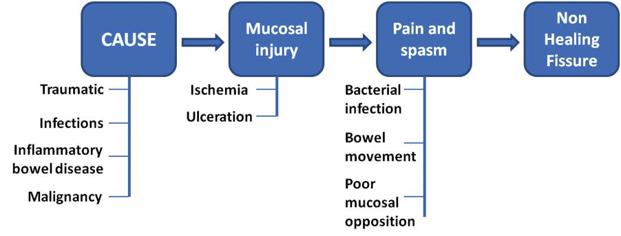
Pathophysiology and progression of anal fissure
Images hosted on other servers:

Perianal abscesses

Fistulae and ischiorectal abscess

Fistula classification
Images hosted on other servers:

Endoanal USG

Inflammatory infiltrate
Images hosted on other servers:

Horseshoe fistula in ano

Multiple fistula tracts
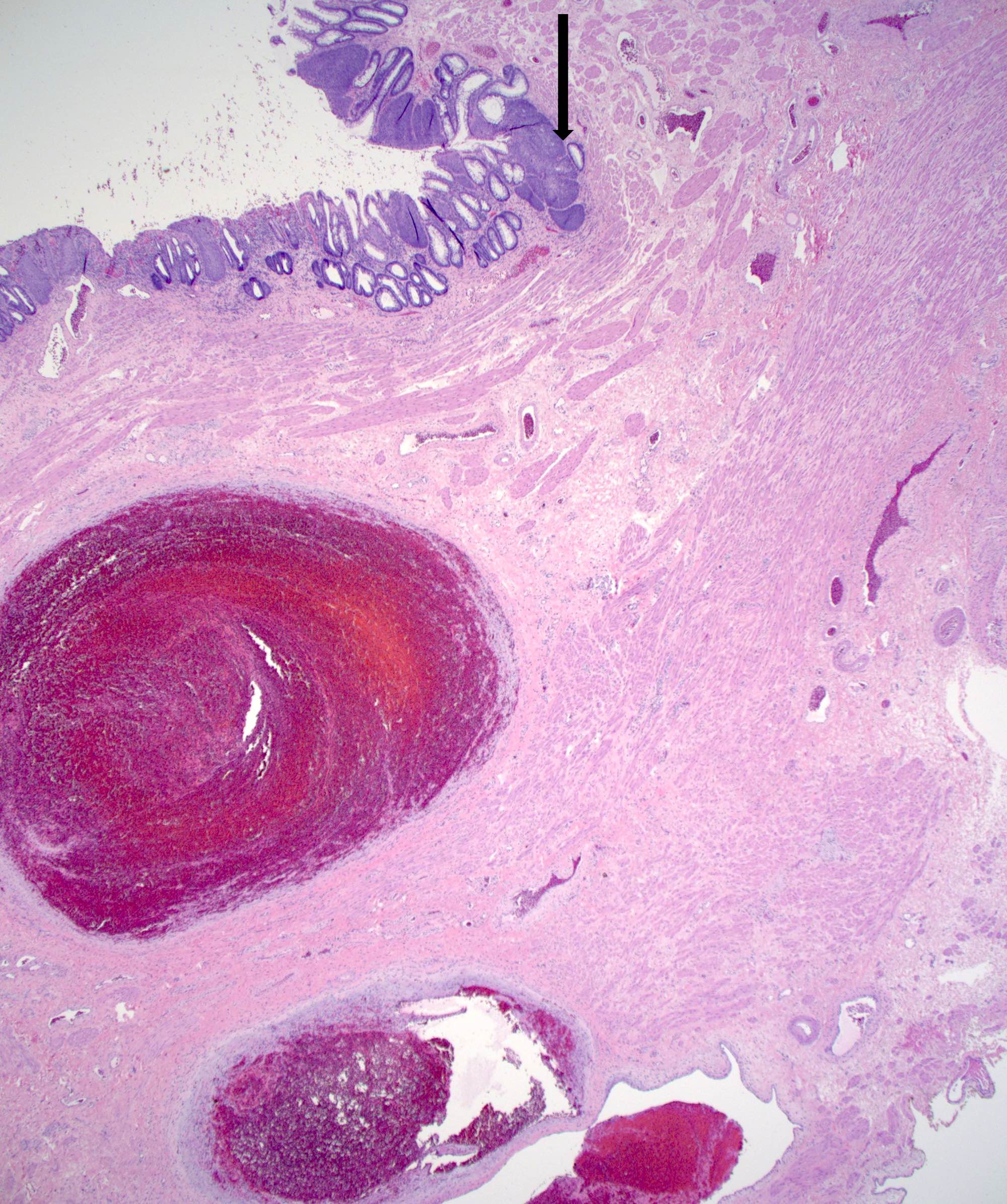
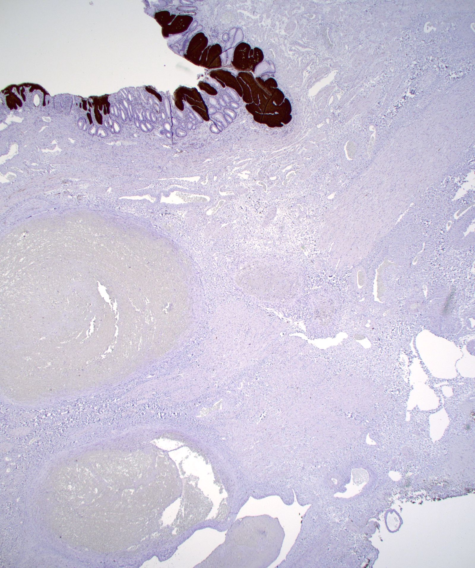
Contributed by Fahire Goknur Akarca, M.D. and Kwun Wah Wen, M.D., Ph.D.
Epithelized anal fistula tract
Granulation tissue with inflammatory cells
Giant cells, in association with granulation tissue
Perianal fistula
Fistula in ano
Images hosted on other servers:

MRI of perianal mass

Ultrasound of perianal mass
Images hosted on other servers:

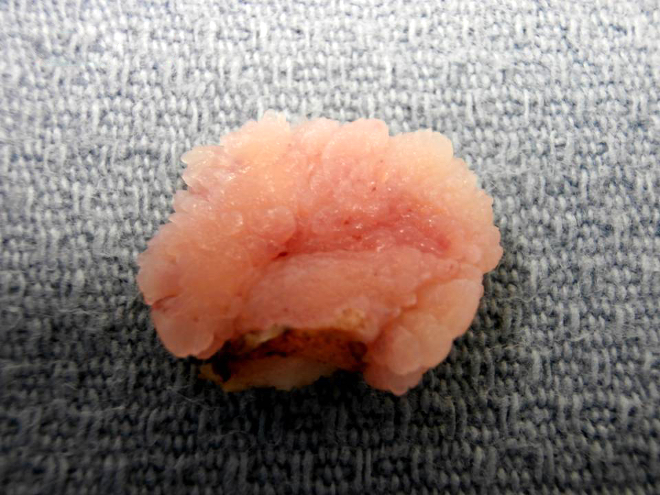
Anal polypoid mass

Perianal cutaneous nodule
Images hosted on other servers:

Excision of perianal tumor
Contributed by Raul S. Gonzalez, M.D.

Gastroesophageal junction lesion
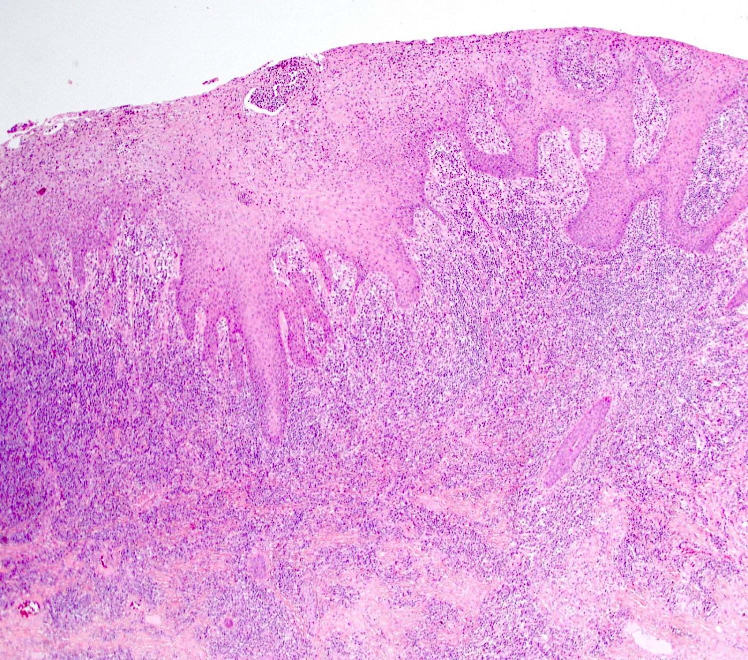
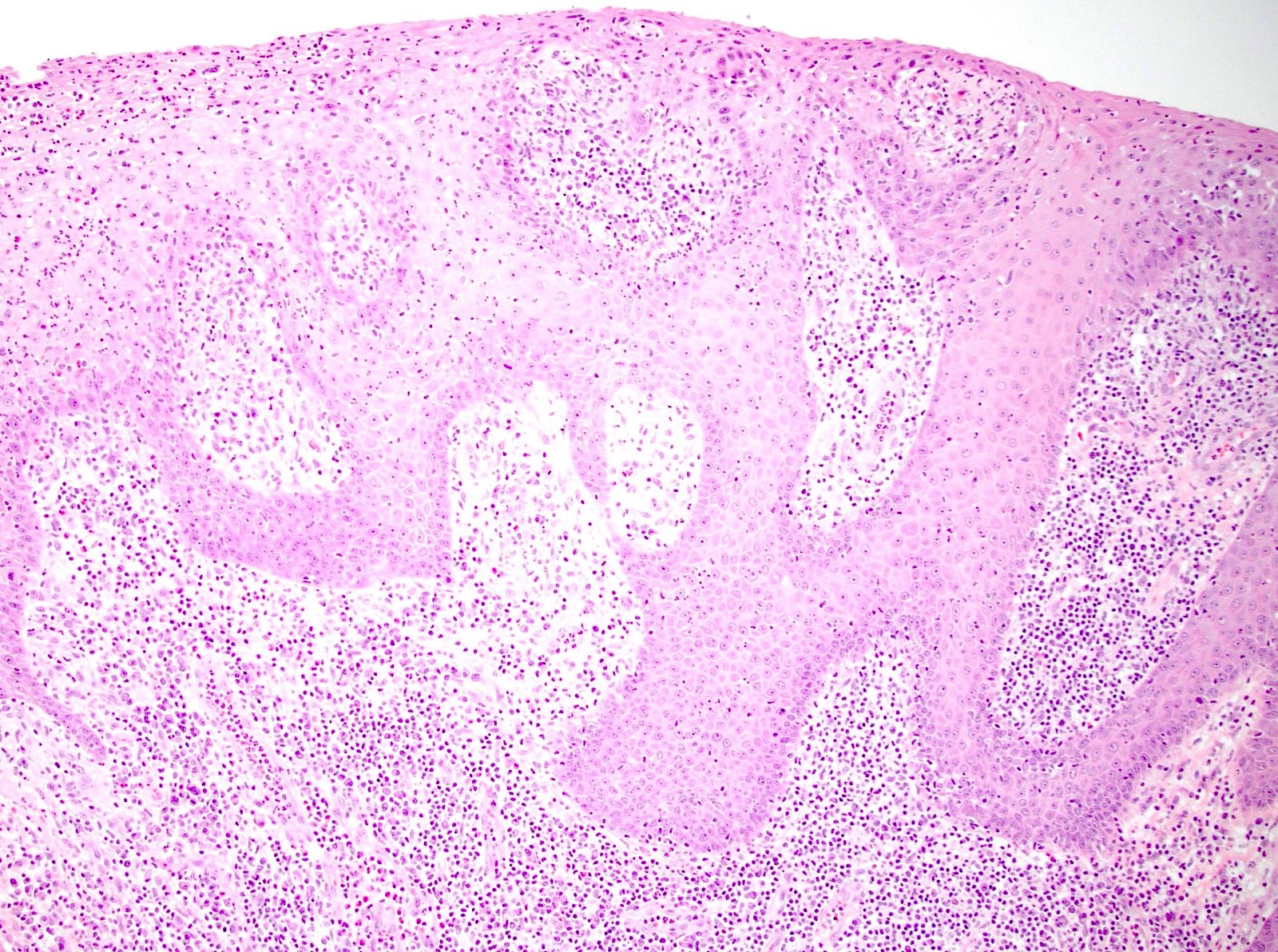
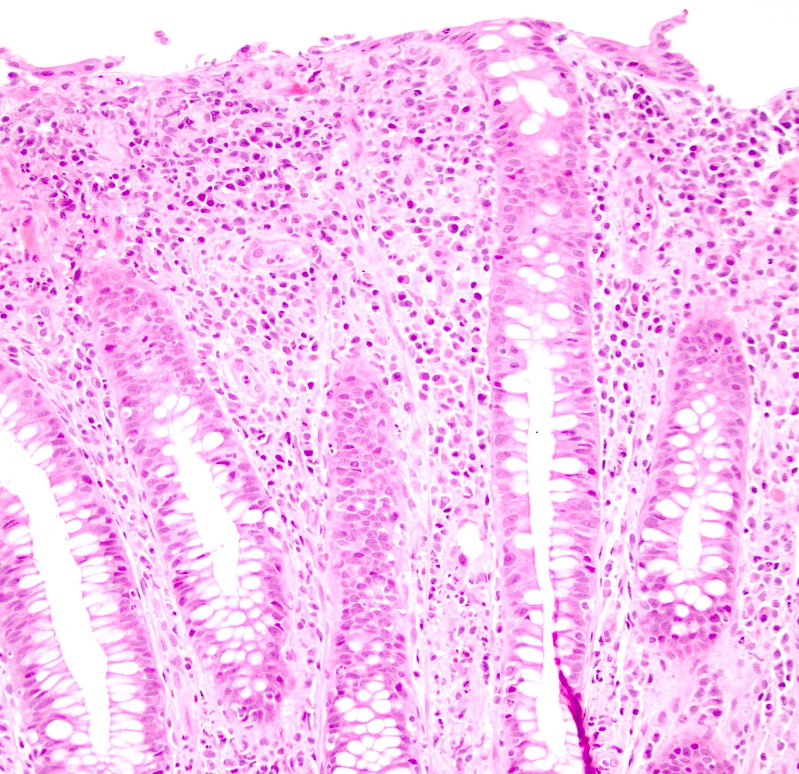
Pseudoepitheliomatous
hyperplasia
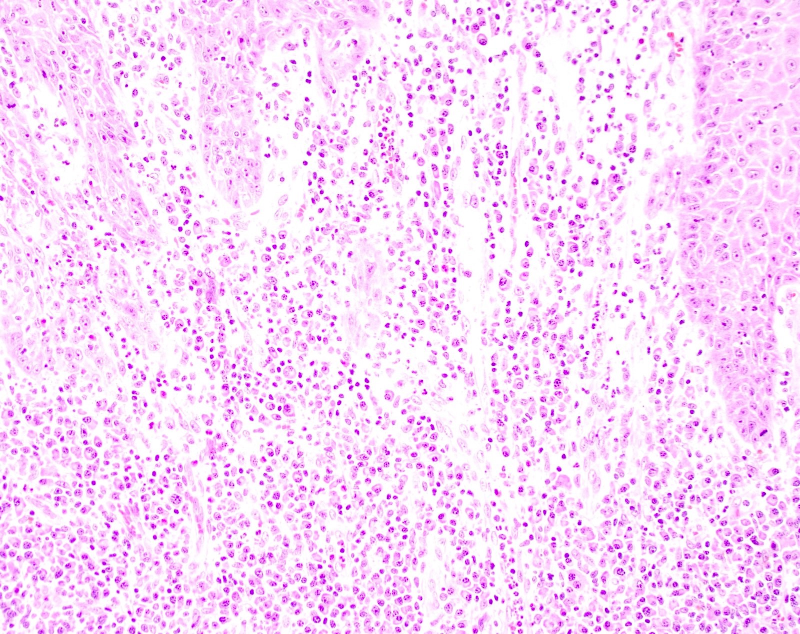
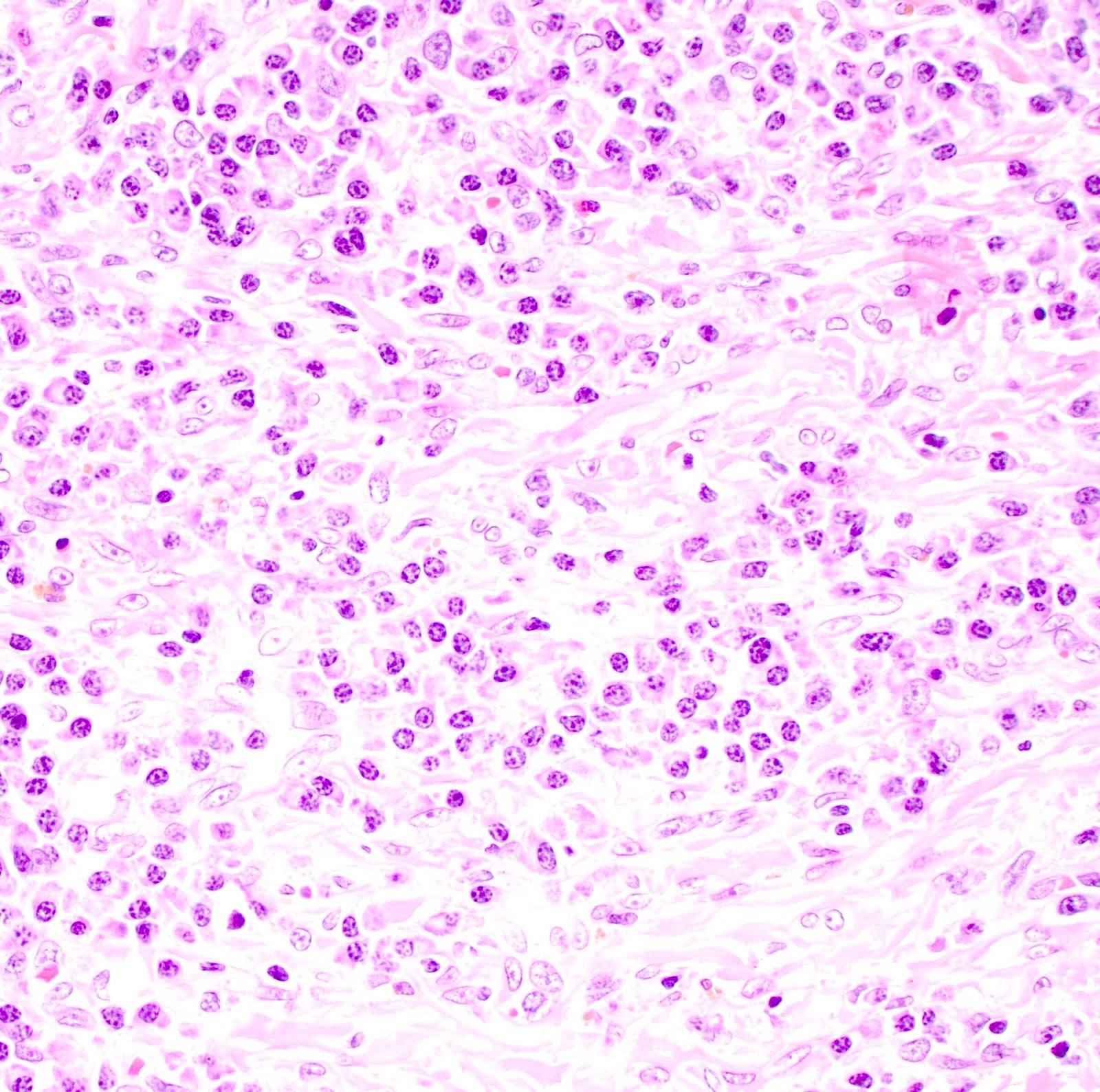
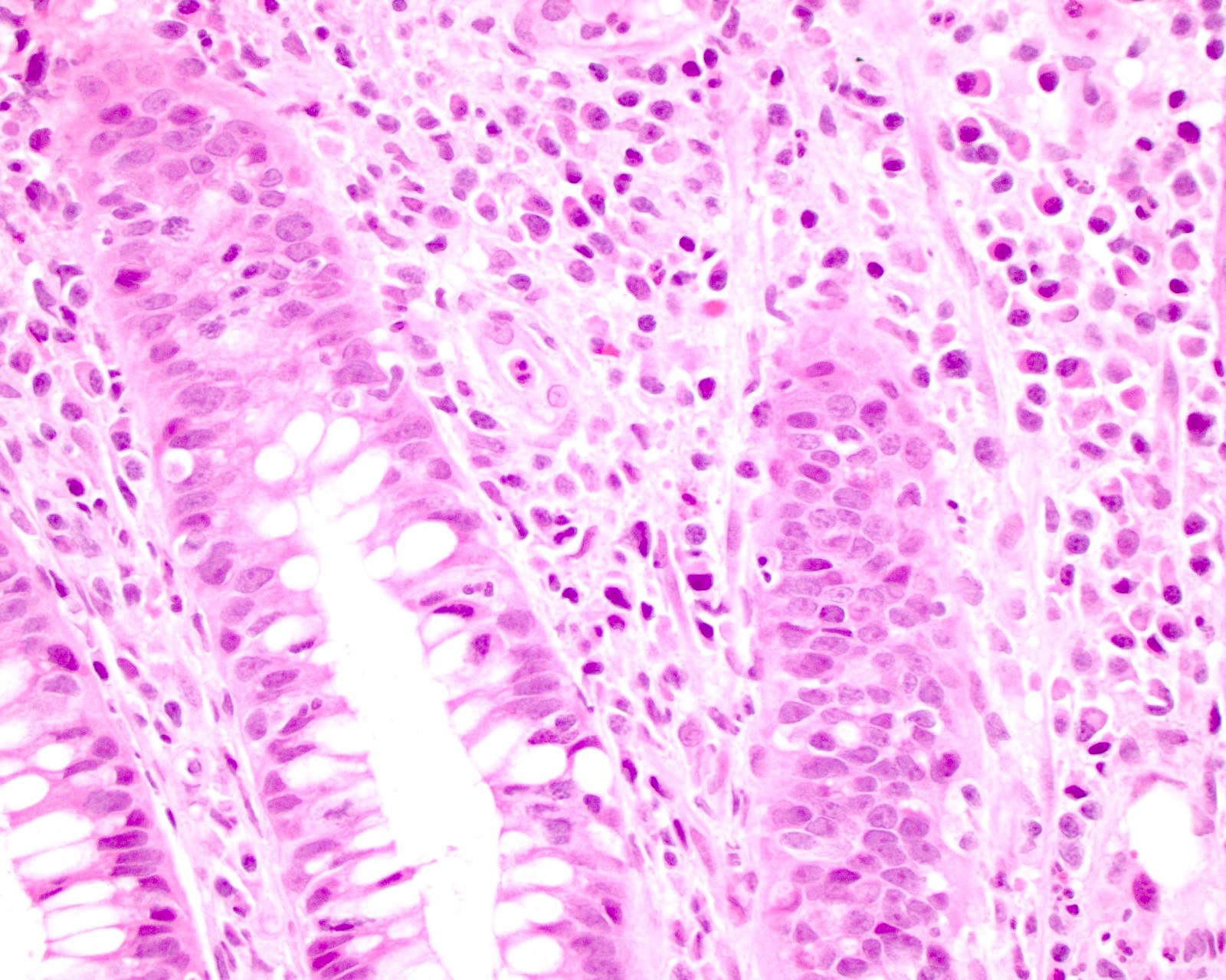
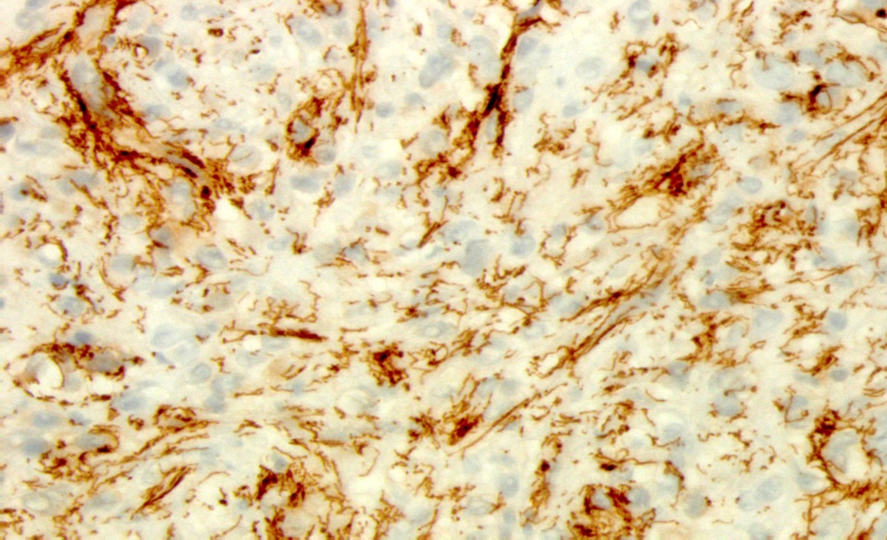
Phagolysosomes
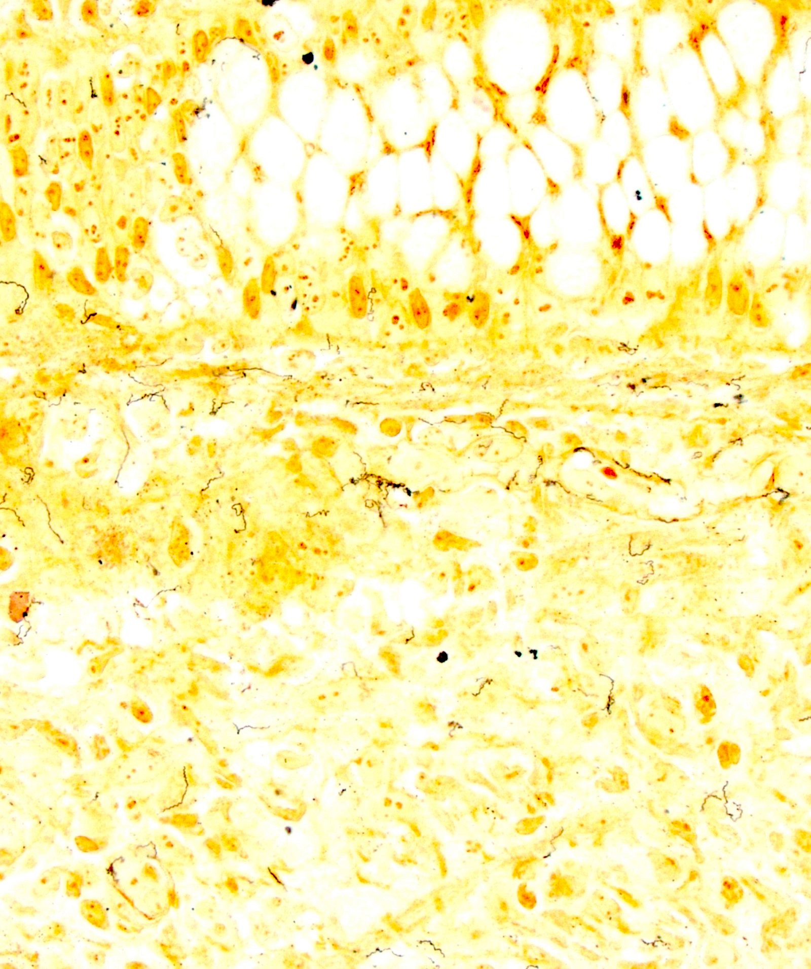
S100
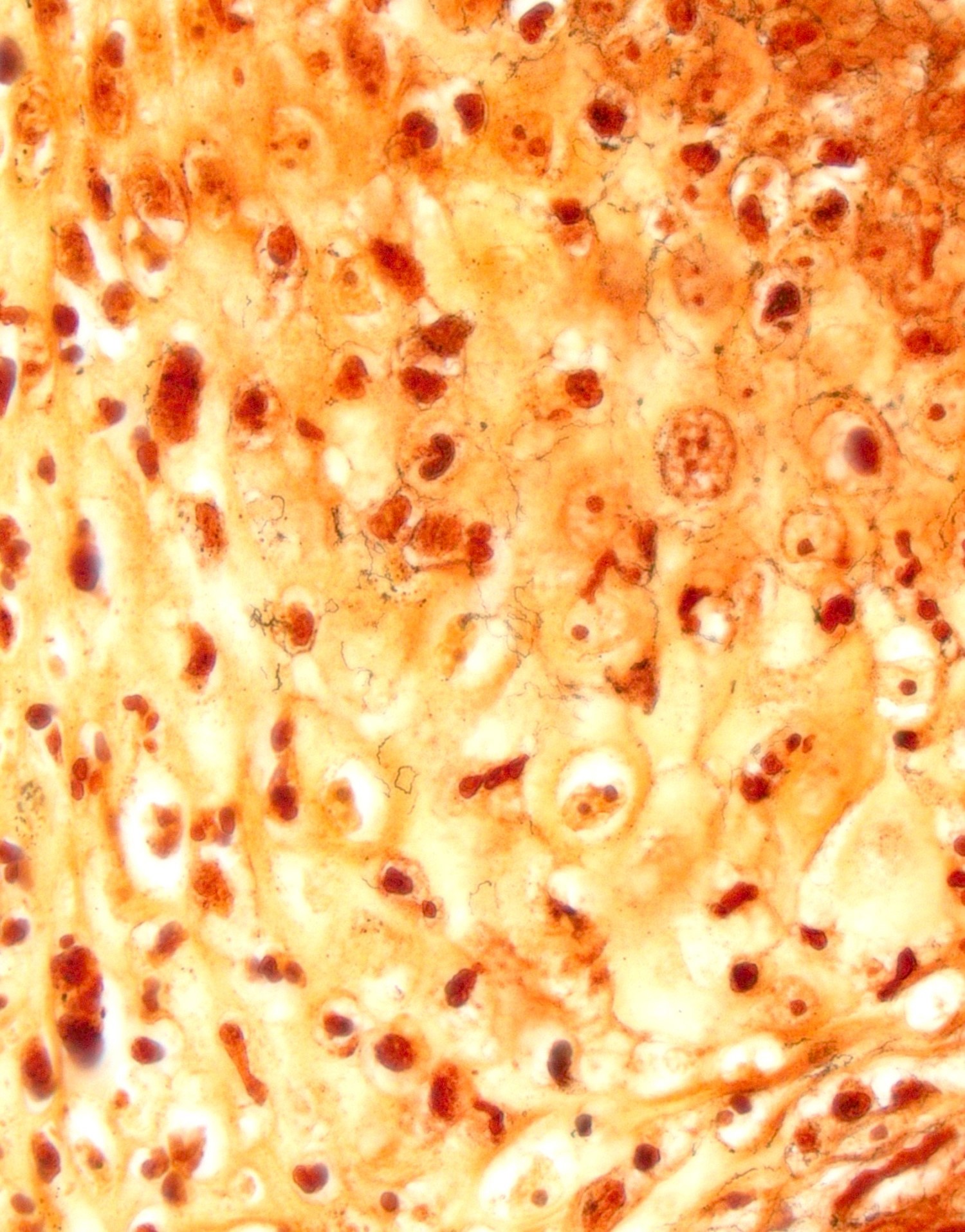
CD68
Images hosted on other servers:

Ultrastructural view
Images hosted on other servers:

Diffuse ulceration

Auto-amputation
Images hosted on other servers:

Safety pin shaped structures

Epithelioid histiocytes


Images hosted on other servers:

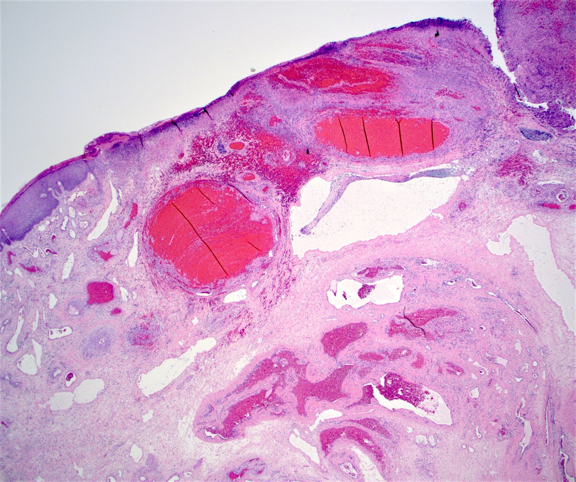
Common sites of major anal and internal hemorrhoids

Prominent prolapsed true (internal) hemorrhoids

Hemorrhoids at anorectal junction
Contributed by James Mueller, M.D., Ph.D. and Vikram Deshpande, M.D.
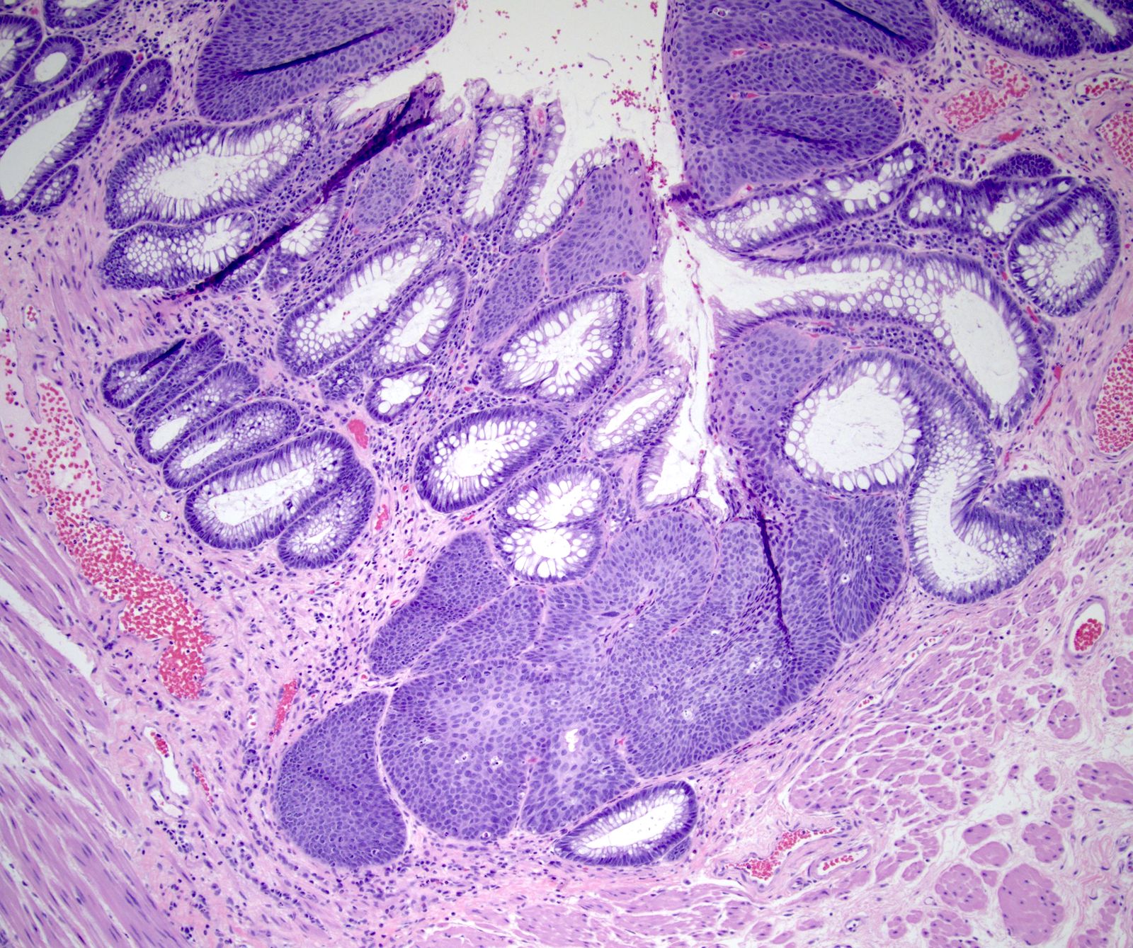
Typical hemorrhoid
Transitional mucosa
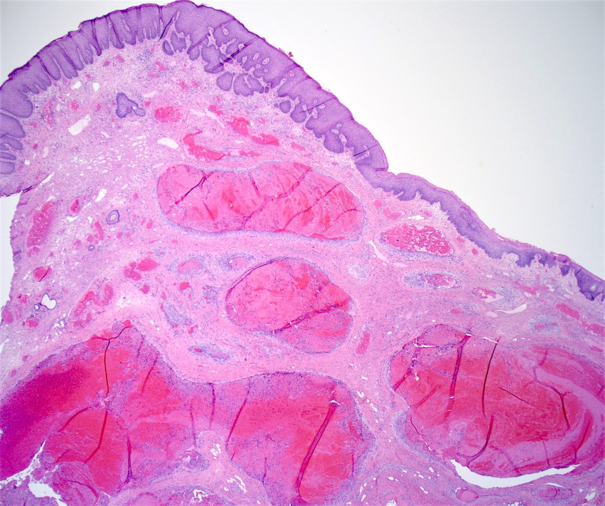
Thrombosed hemorrhoid
Ulcerated hemorrhoid
AIN3
SCC presenting as hemorrhoids
Colorectal adenocarcinoma presenting as hemorrhoids
Papillary endothelial hyperplasia
Incidental AIN
p16 IHC stain
Histopathology of hemorrhoids
Overview of hemorrhoids
Images hosted on other servers:

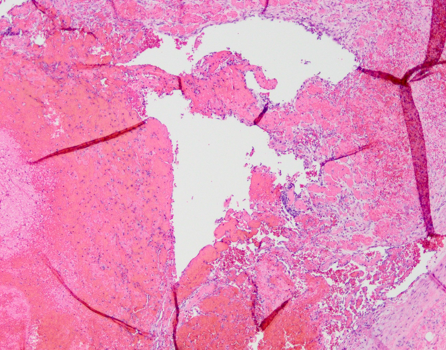
Fistulous tract
Contributed by Qingqing Liu, M.D., Ph.D.
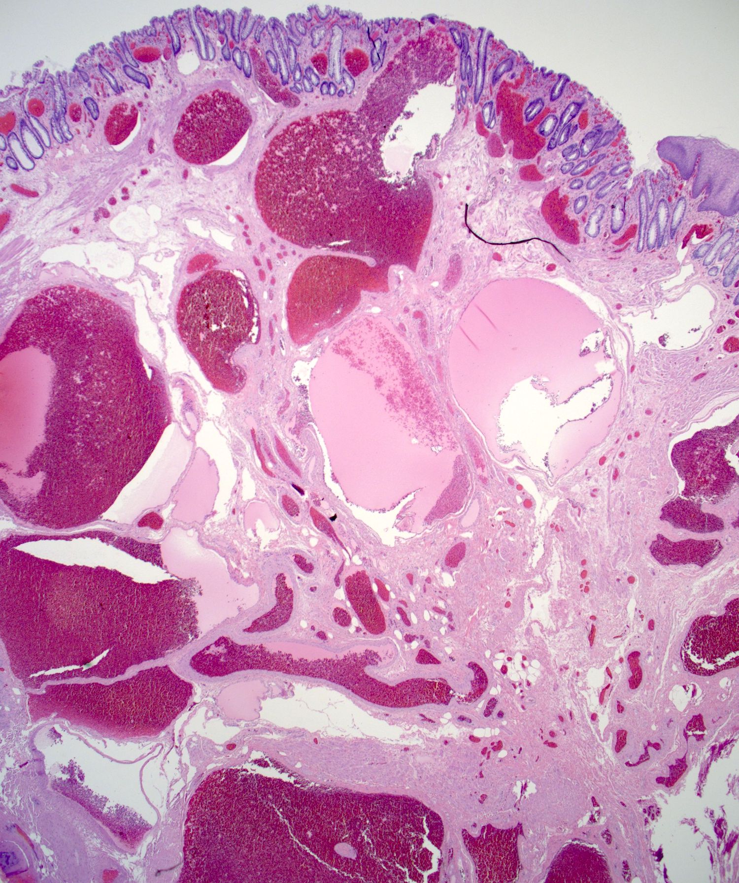
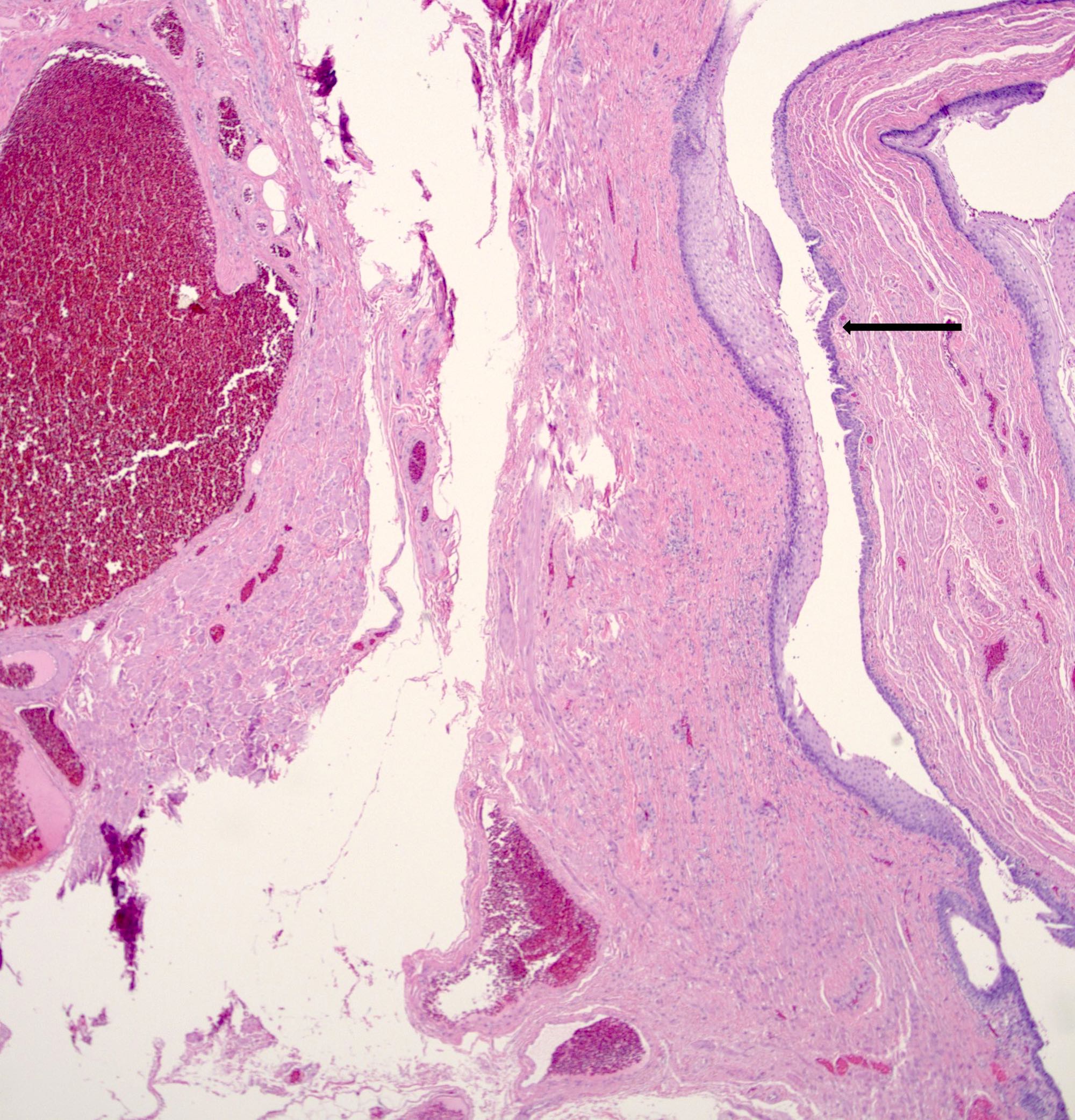
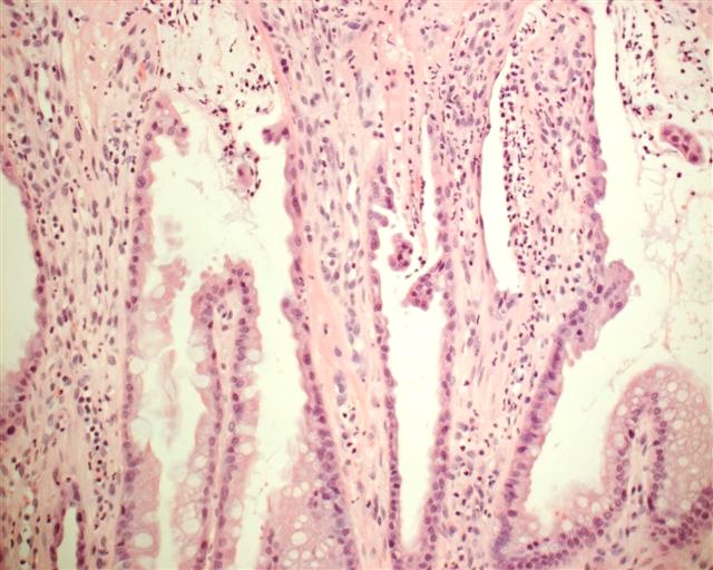
Typical features
Thin walled vessels
Chronic inflammation
Atypical stromal cells
Images hosted on other servers:

Cloacogenic polyps on rectal retroflexion
Contributed by Nikka Khorsandi M.D., M.P.H., Kwun Wah Wen, M.D., Ph.D. and Yvonne Bury, M.D.
Squamous and glandular epithelium
Surface erosion

Mucosal erosion
Regenerative epithelial changes
Images hosted on other servers:

Groove sign (squamous cell carcinoma, not LGV)
Images hosted on other servers:

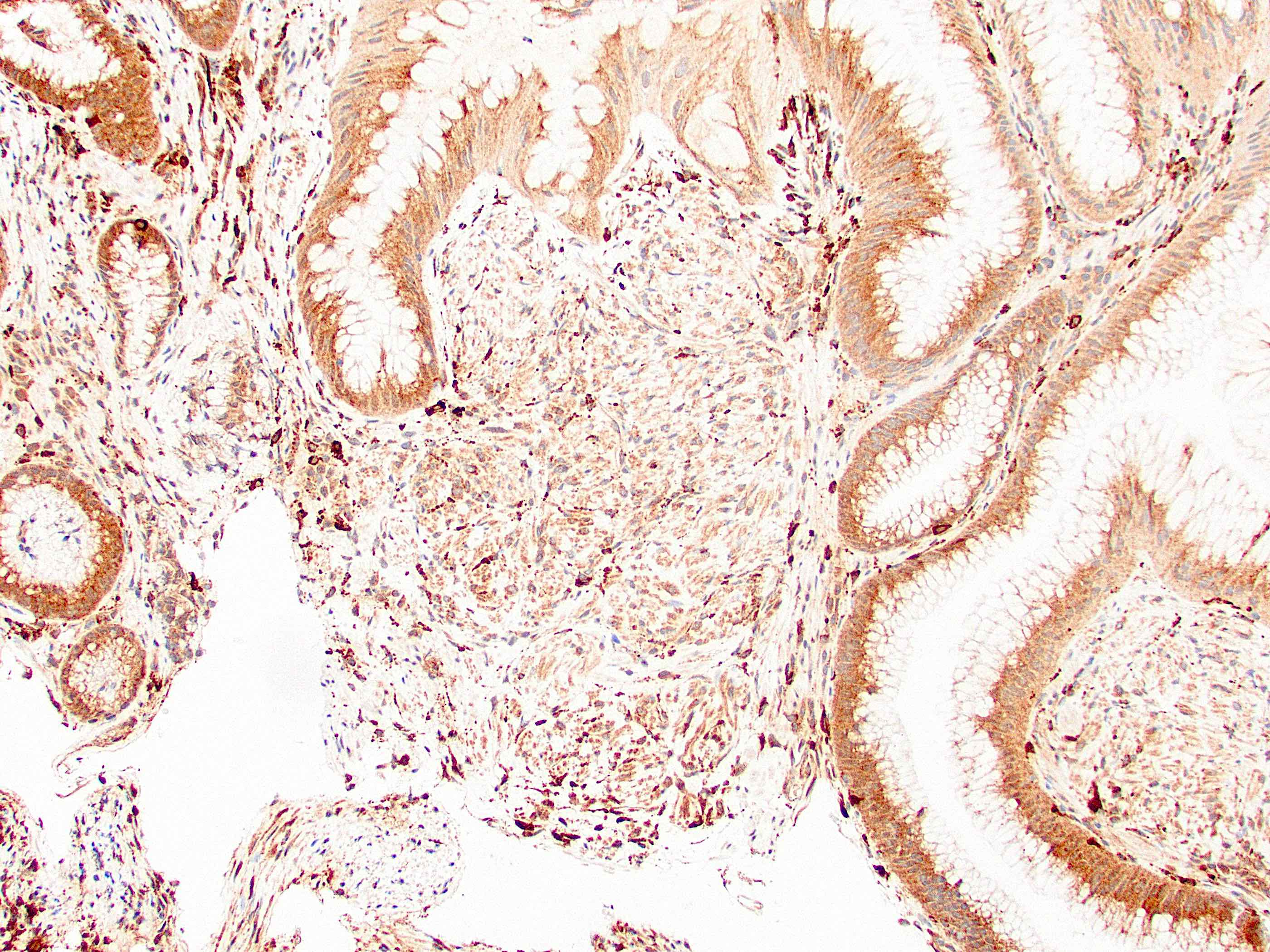
C. trachomatis inclusion bodies (brown)
Images hosted on other servers:

Anorectal mass (CT of pelvis)

Anorectal mass (MRI)

Anorectal mass (MRI / PET)
Images hosted on other servers:

Anorectal mass

Prolapsed anal mass

Prolapsed anorectal mass

Polypoid lesion on colonoscopy
Images hosted on other servers:

Pigmented anal mass

Anorectal mass
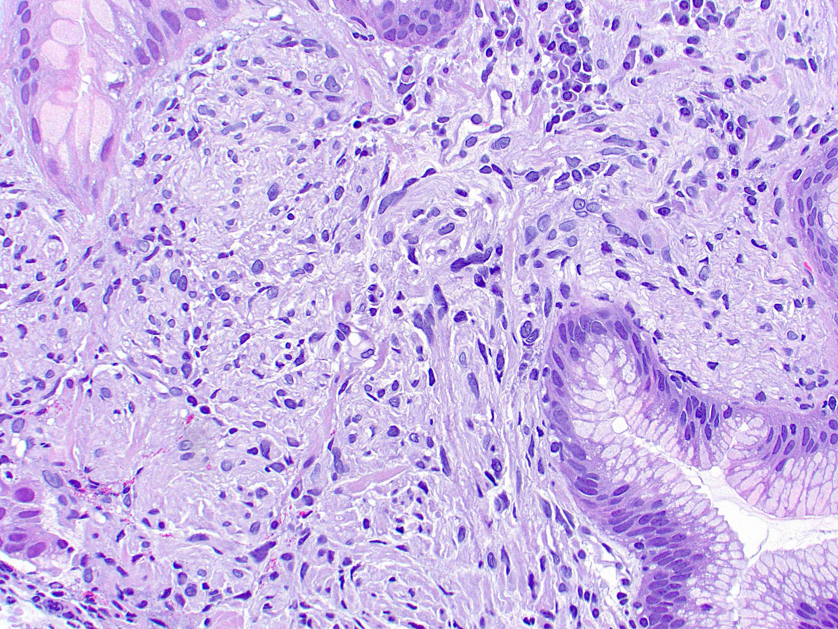
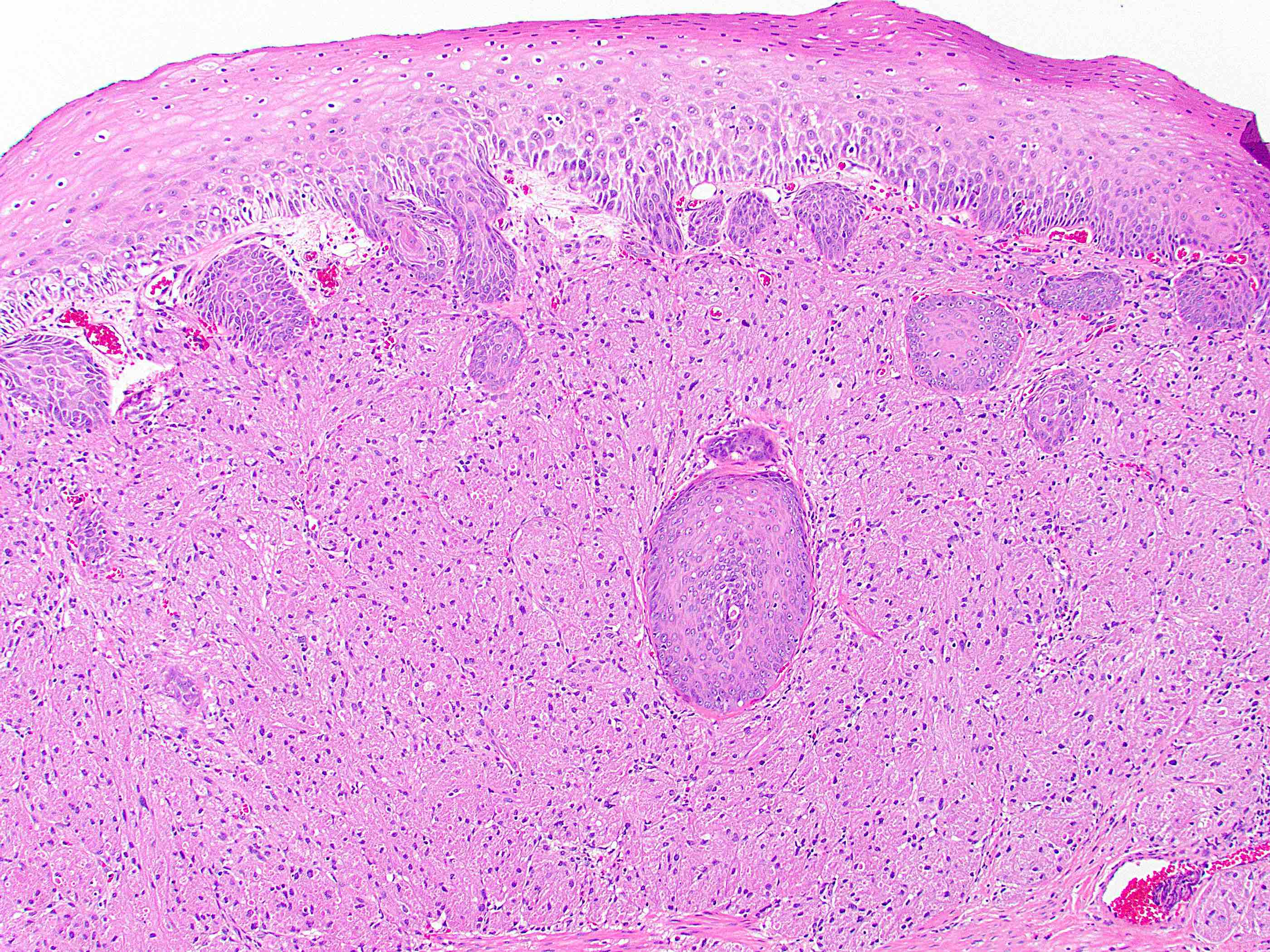
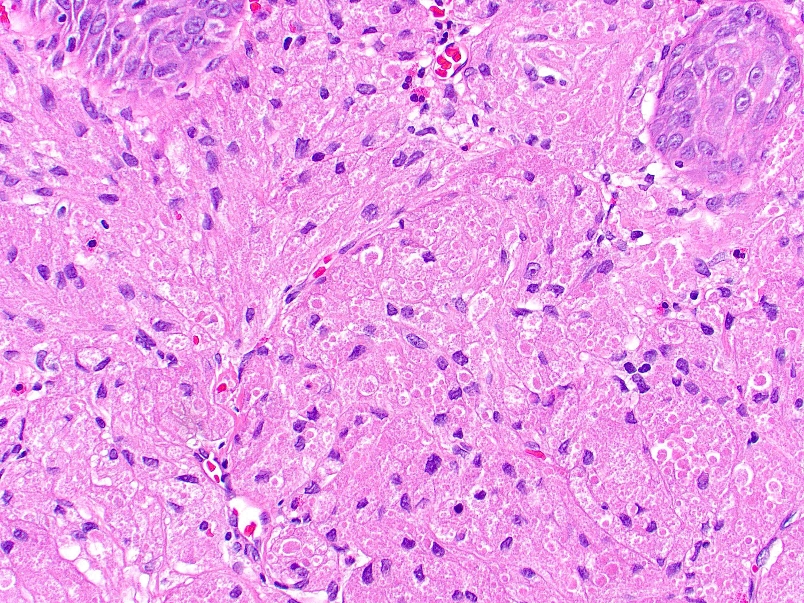
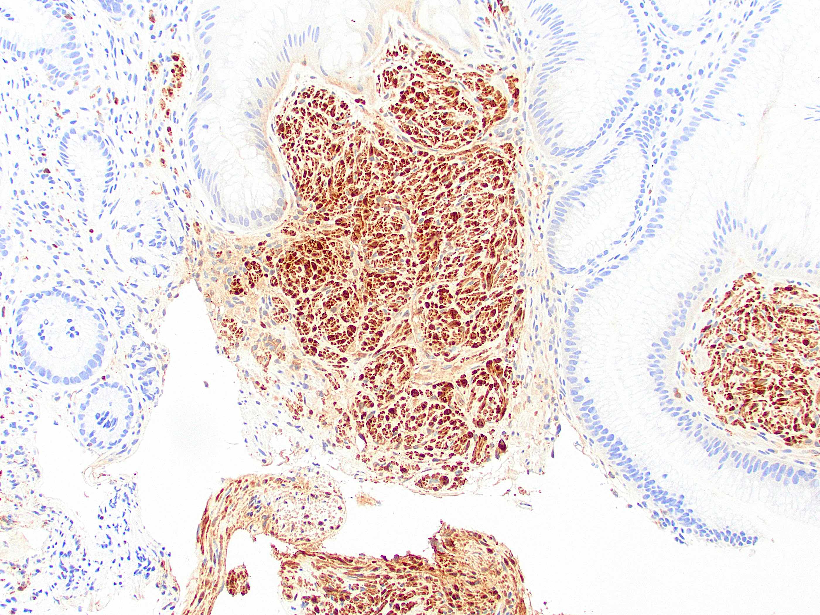
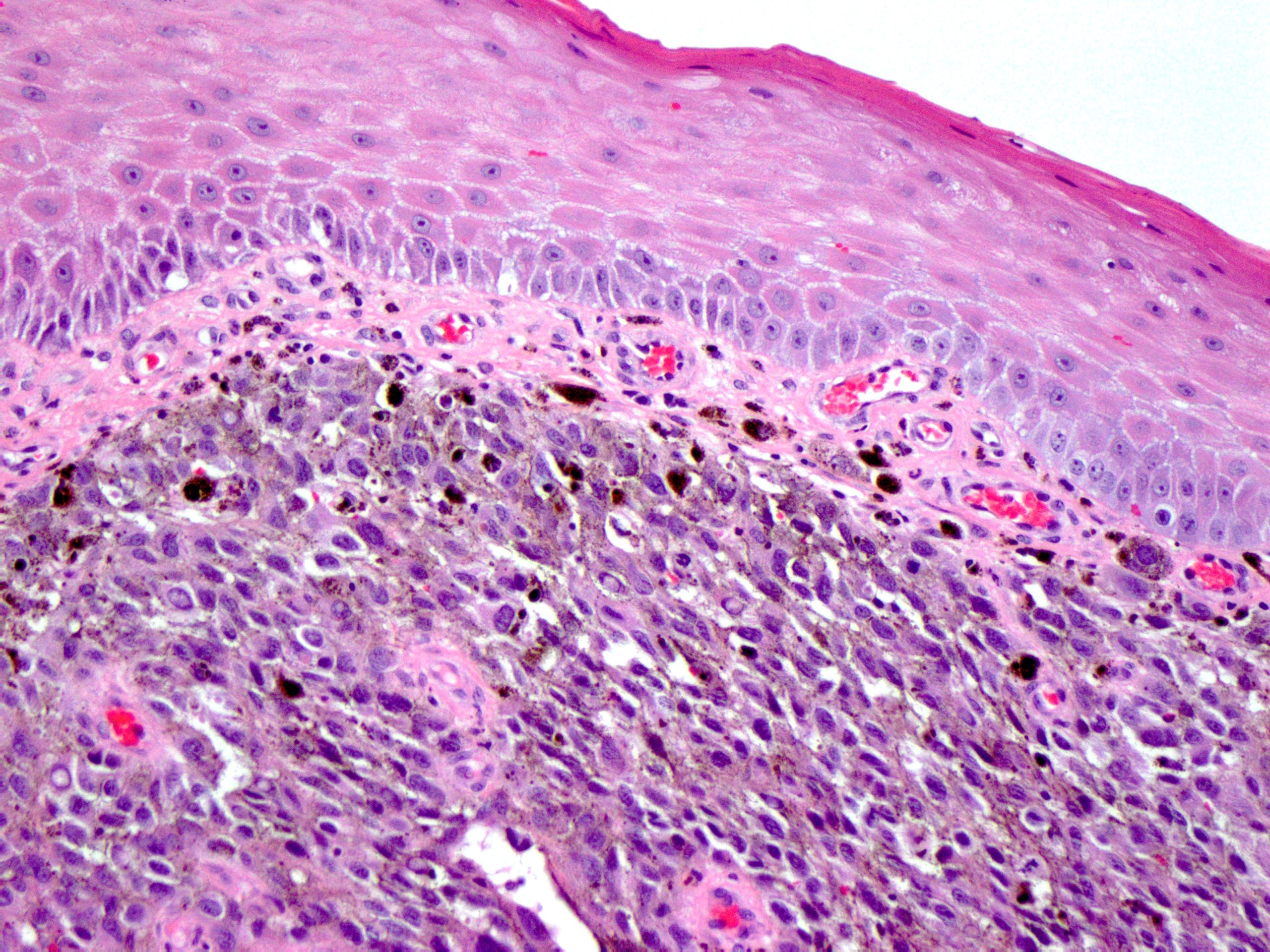
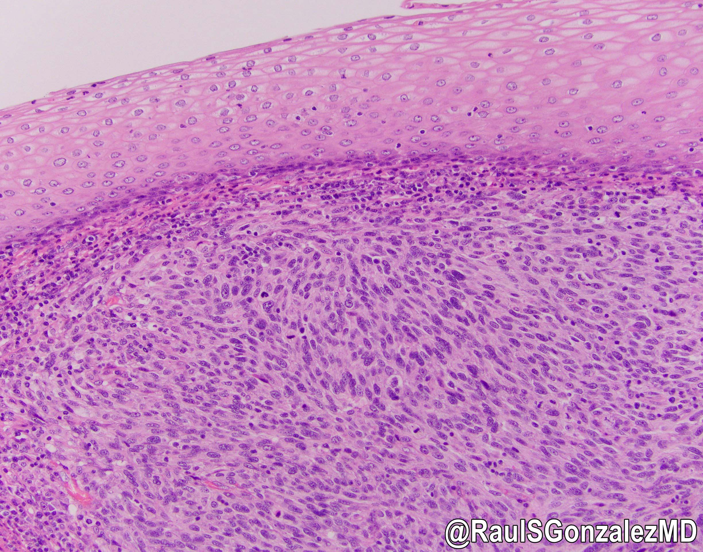
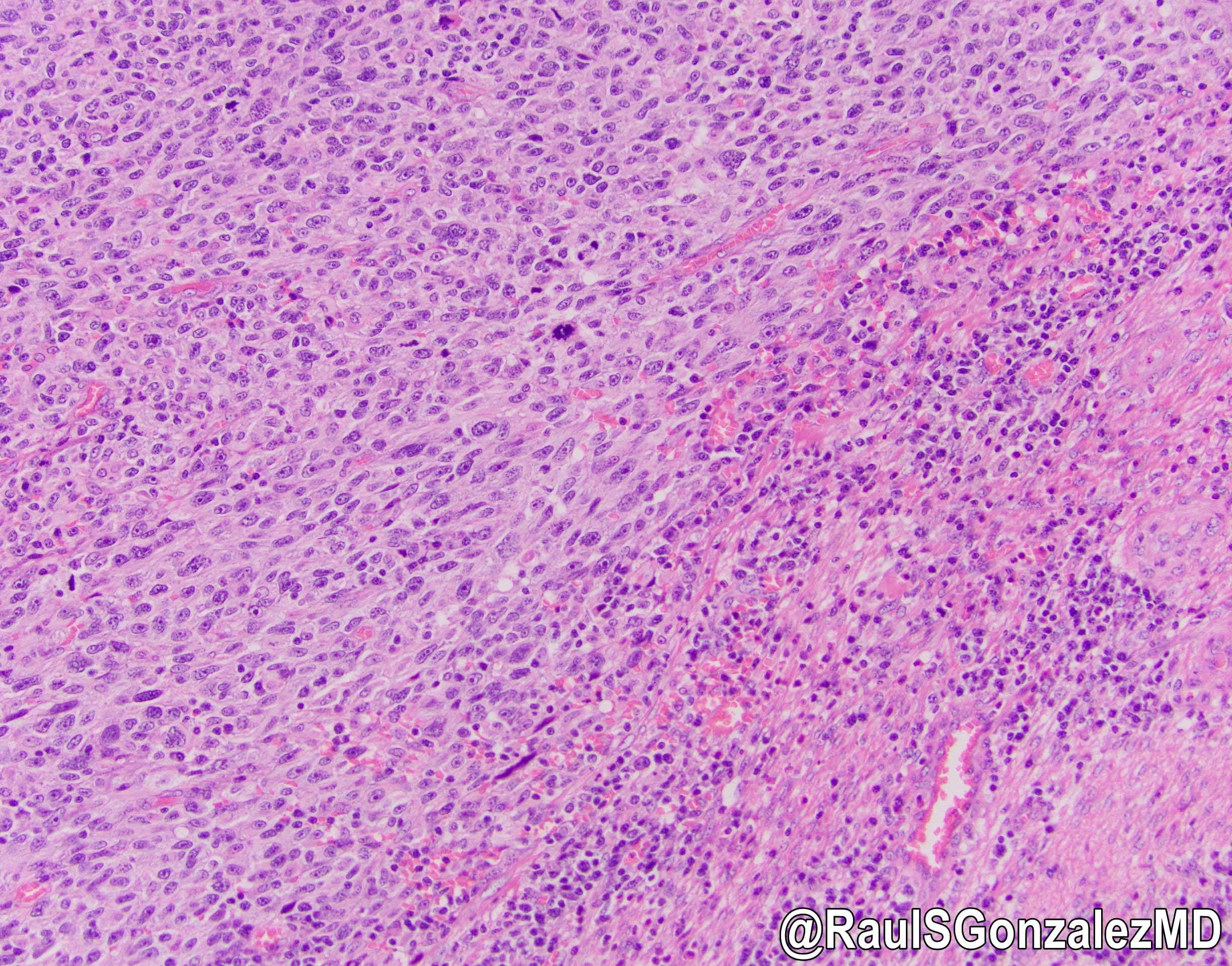
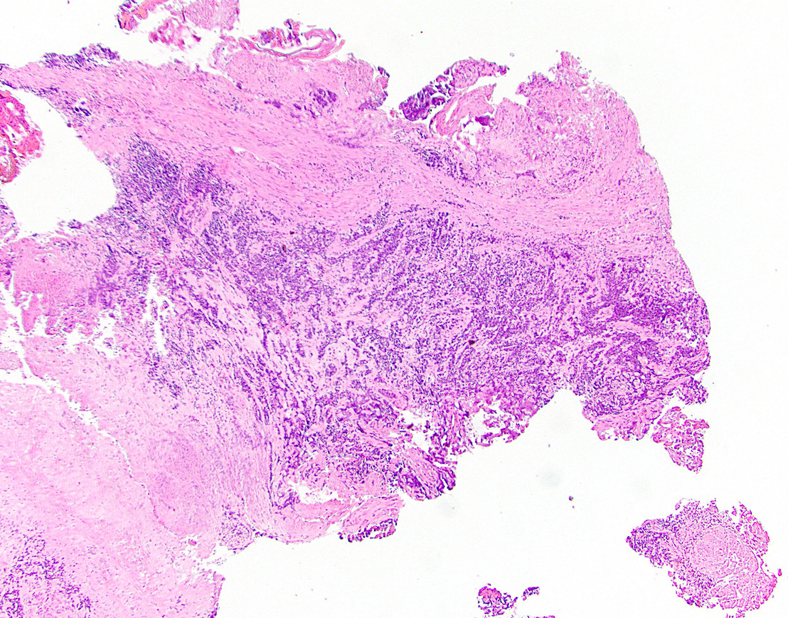
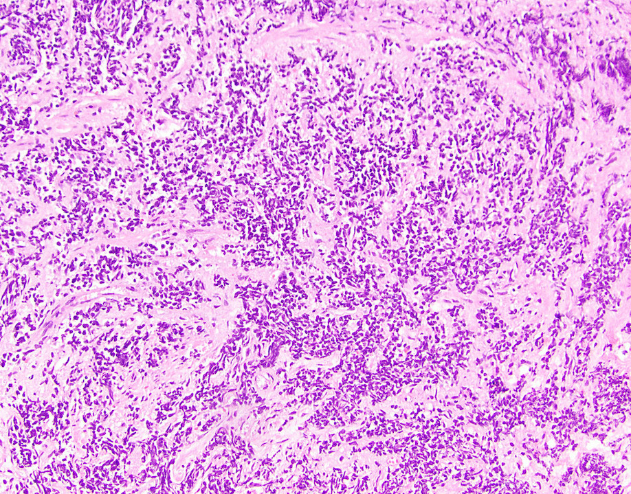
Contributed by Aaron R. Huber, D.O. and @RaulSGonzalezMD on Twitter
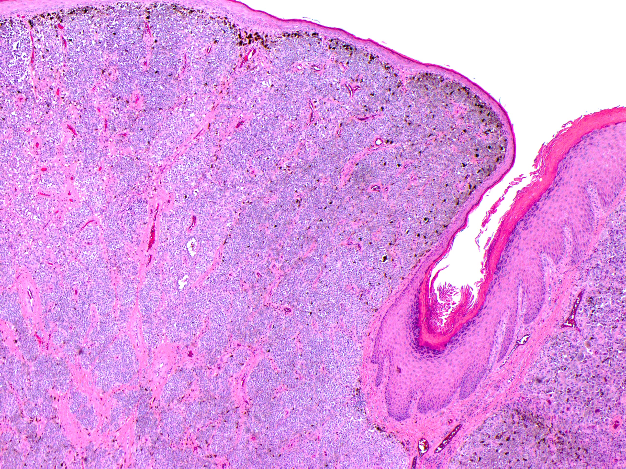
Tumor underlying squamous epithelium
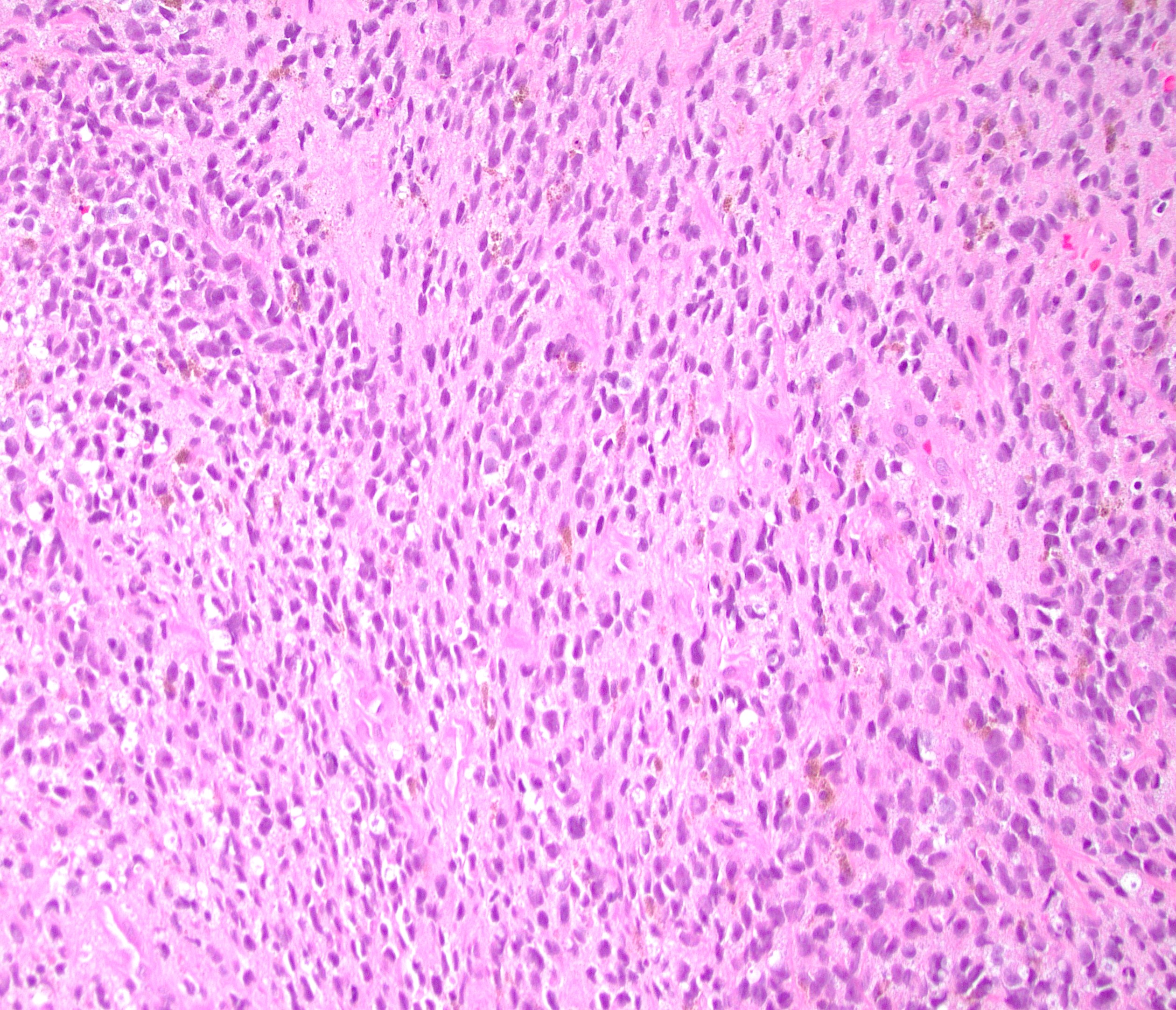
Epithelioid and spindled cells
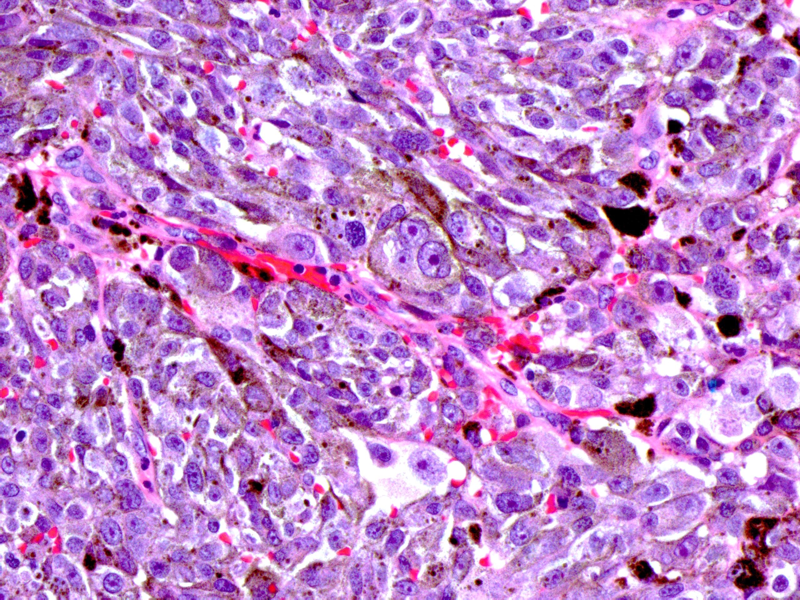
Malignant cells with pigment
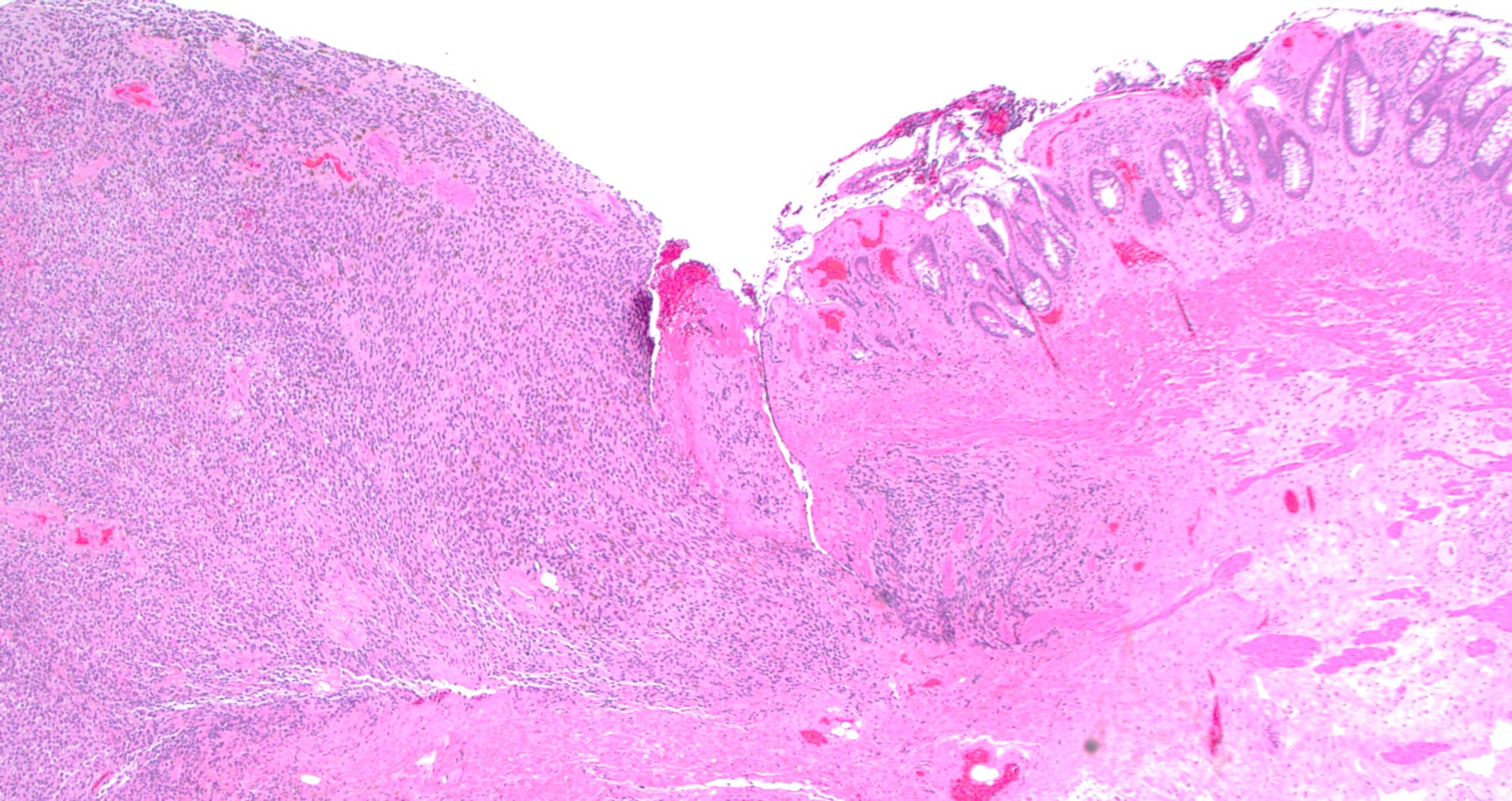
Ulcerated mucosa
Malignant cells with pigment
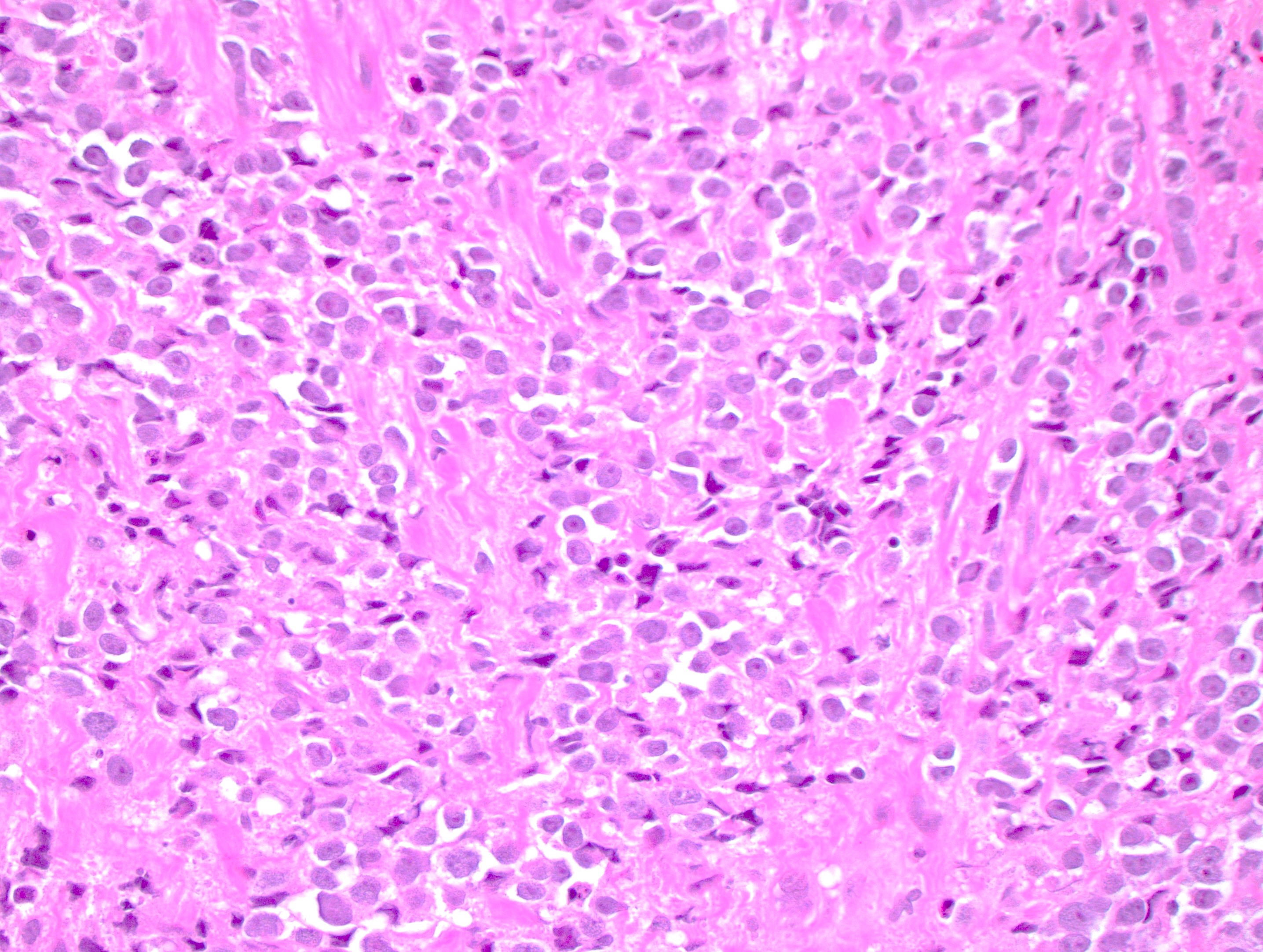
Large nuclei with prominent nucleoli
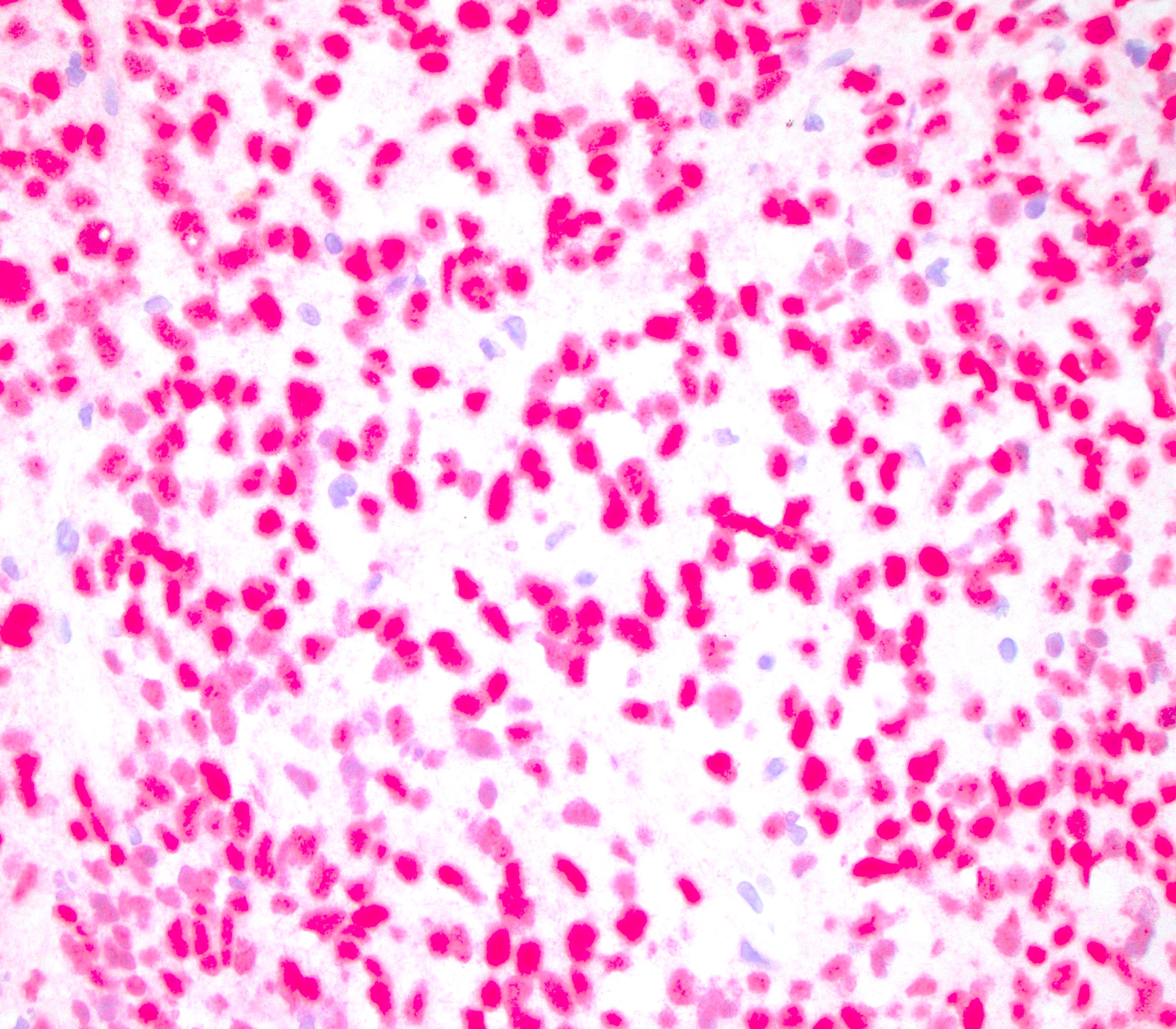
SOX10
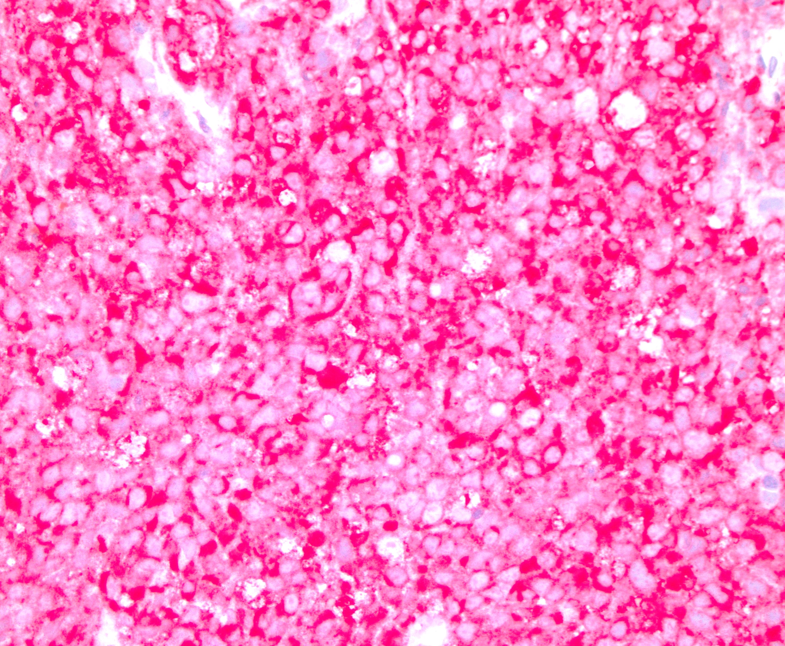
MelanA
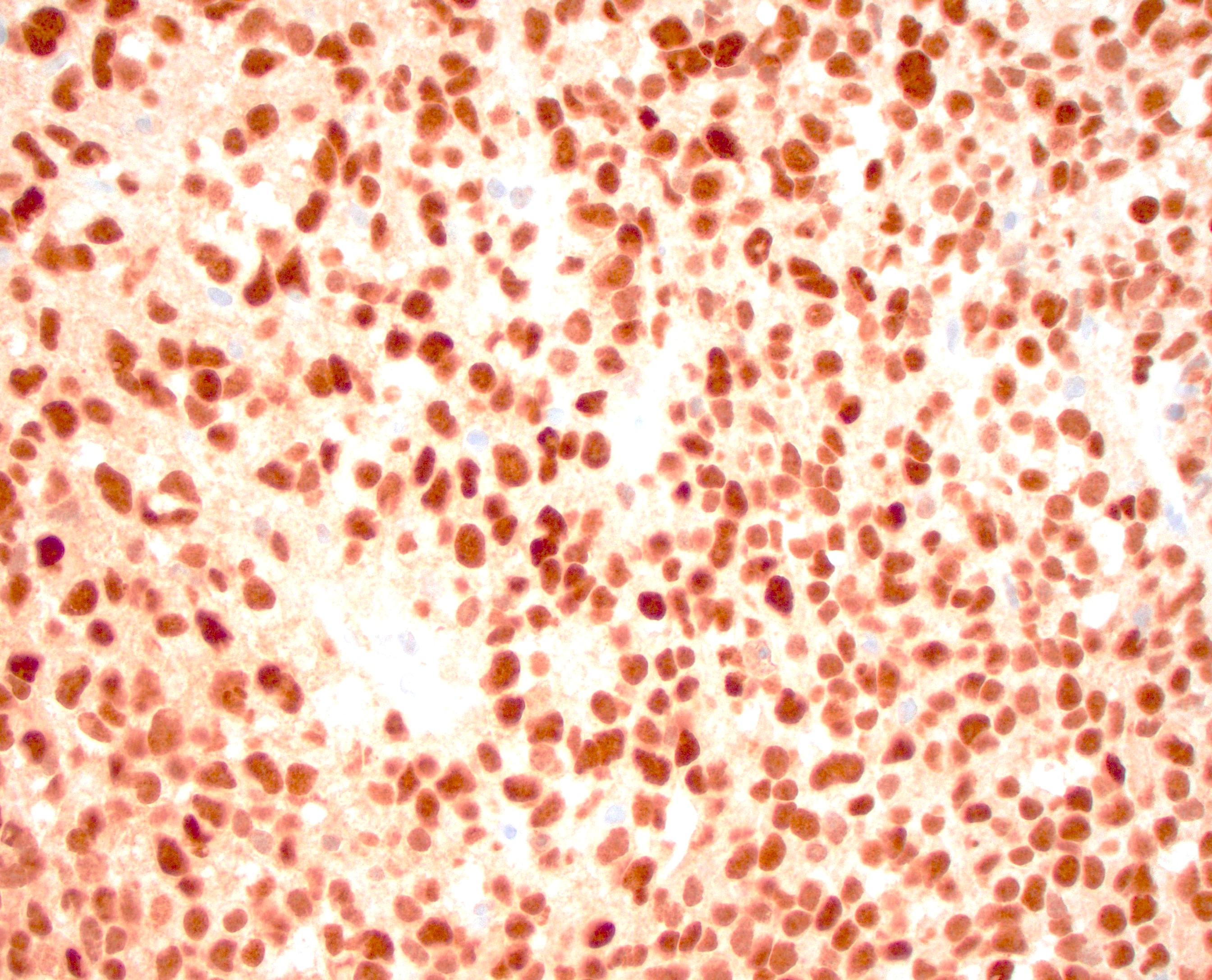
PRAME
Contributed by @RaulSGonzalezMD on Twitter (see original post here)">
 https://pathologyoutlines.com/topic/anusmelanoma.html #pathology #gipath #pathtwitter"
Contributed by @RaulSGonzalezMD on Twitter (see original post here)">
 https://pathologyoutlines.com/topic/anusmelanoma.html #pathology #gipath #pathtwitter"
Contributed by @RaulSGonzalezMD on Twitter (see original post here)">
Primary anal melanoma
Images hosted on other servers:


Metastatic melanoma smears
Images hosted on other servers:

MRI

PET scan

Metastatic disease
Contributed by Raul S. Gonzalez, M.D.
Small cell carcinoma
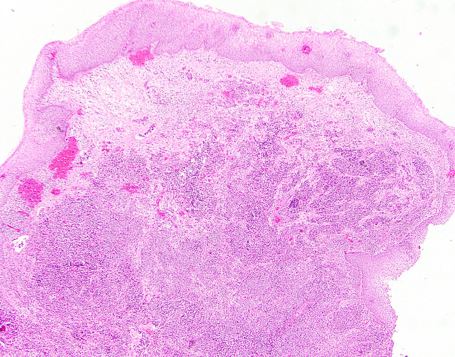
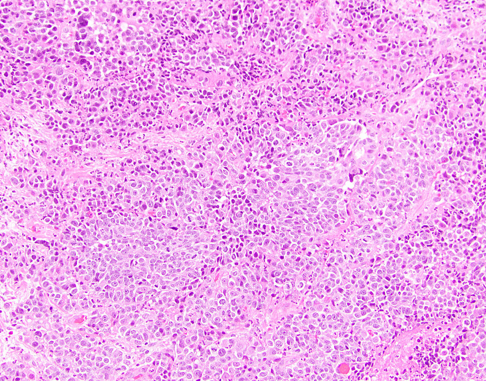
Large cell carcinoma
Images hosted on other servers:

Extensive perianal Paget disease, perineum

Early response

Notable telangiectasia and fibrosis
Contributed by Raul S. Gonzalez, M.D.
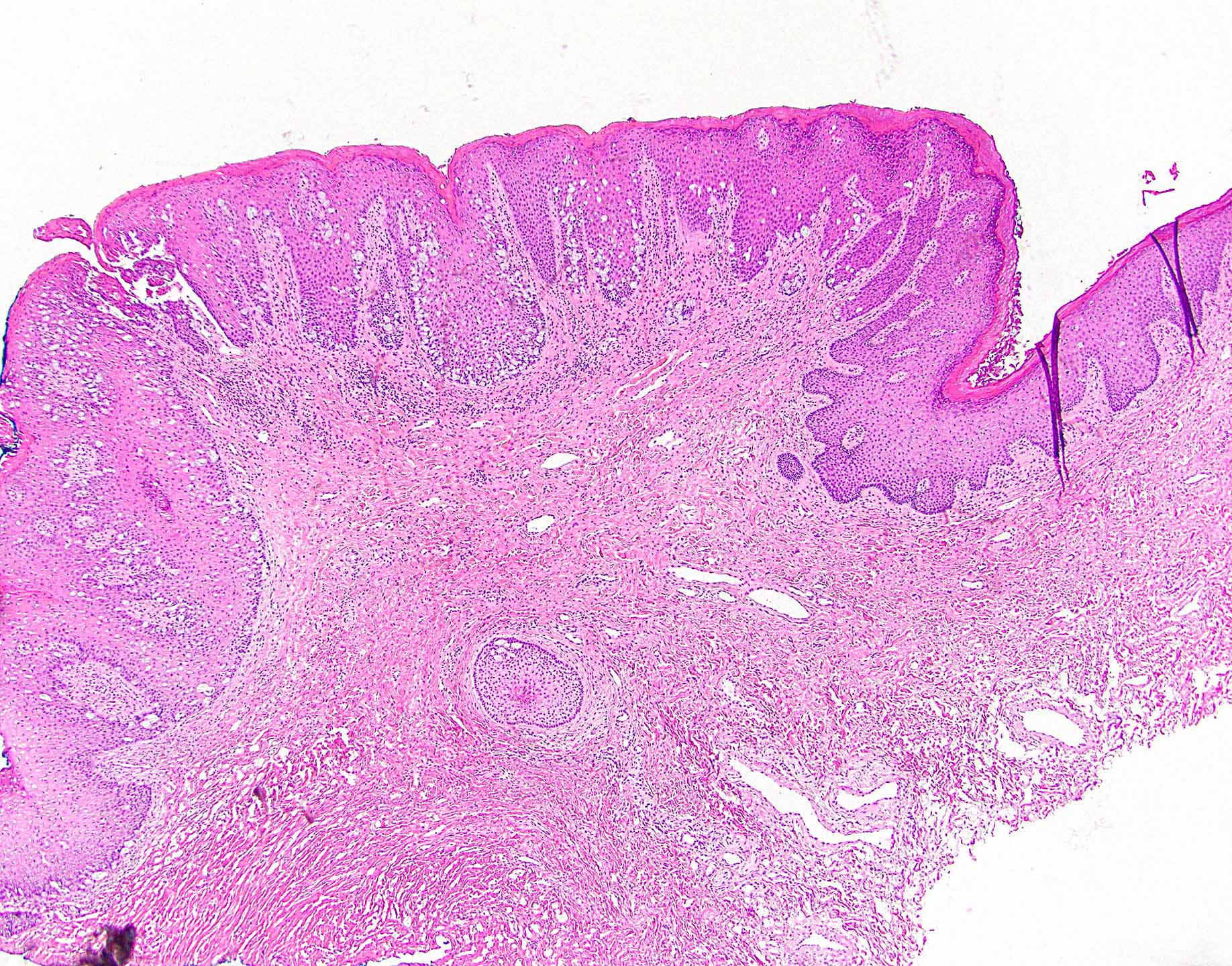
Visible Paget cells
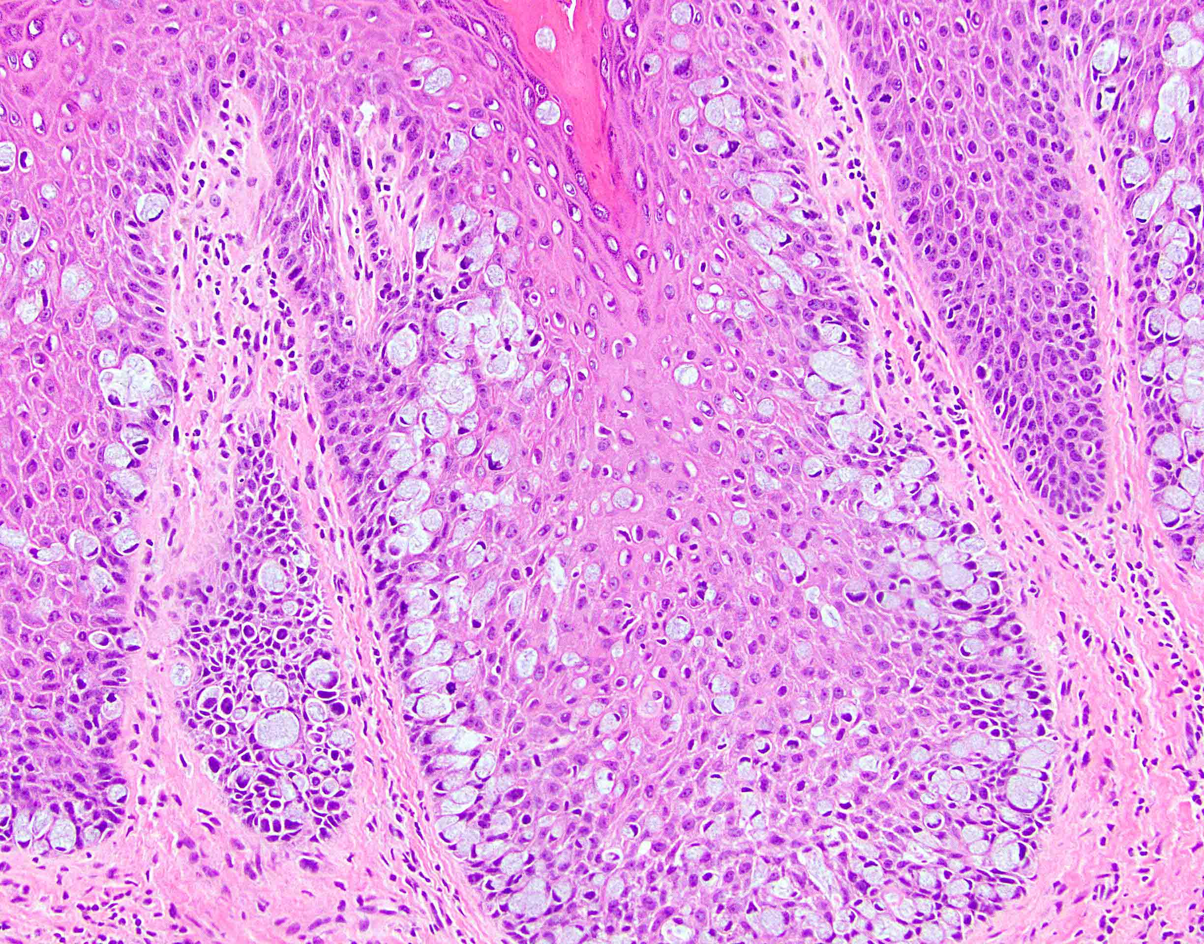
Prominent intracytoplasmic mucin
Contributed by Raul S. Gonzalez, M.D.
Infiltrating anal mass
Prominent nuclear atypia
Necrosis
Contributed by Xiaoyan Liao, M.D., Ph.D.
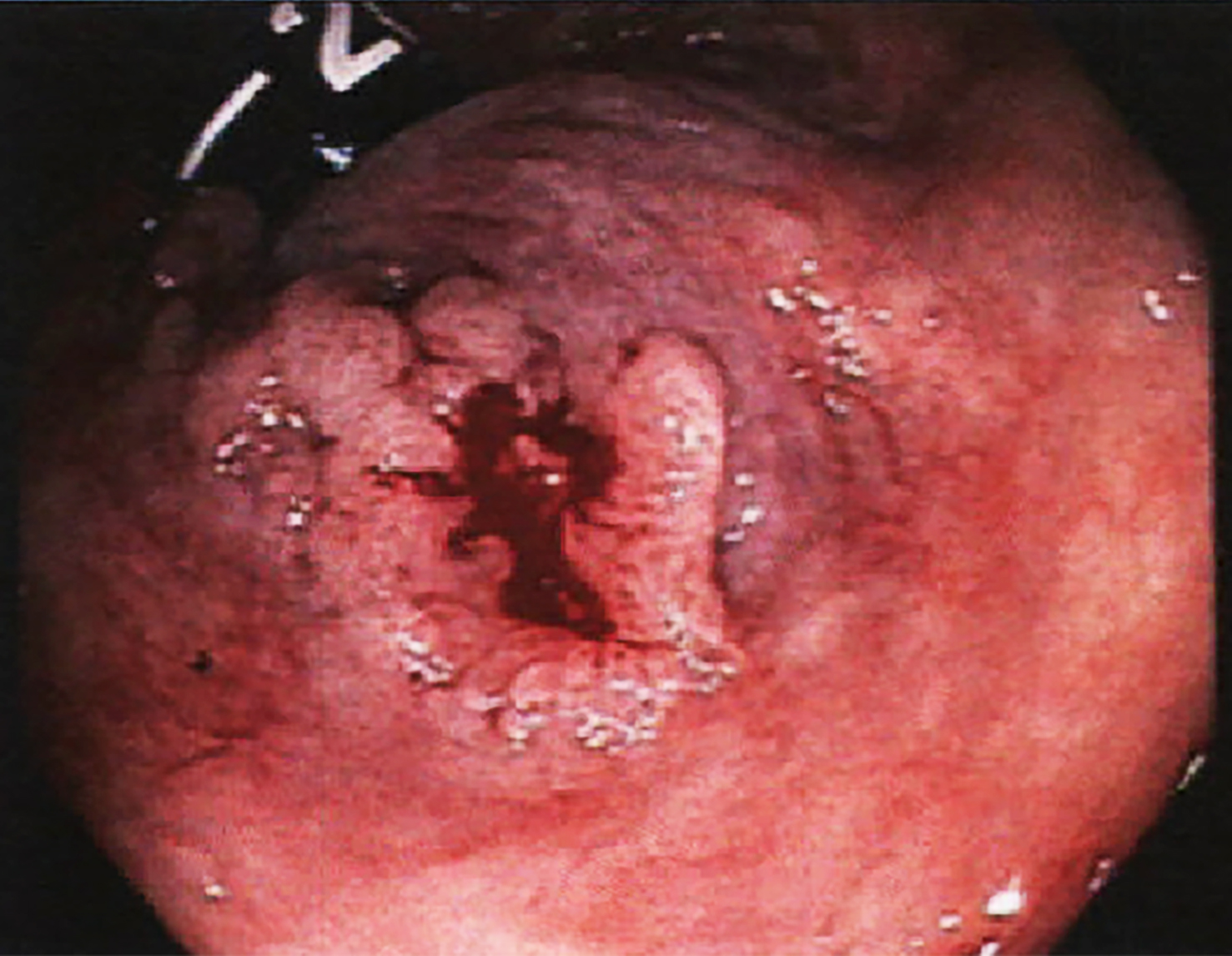
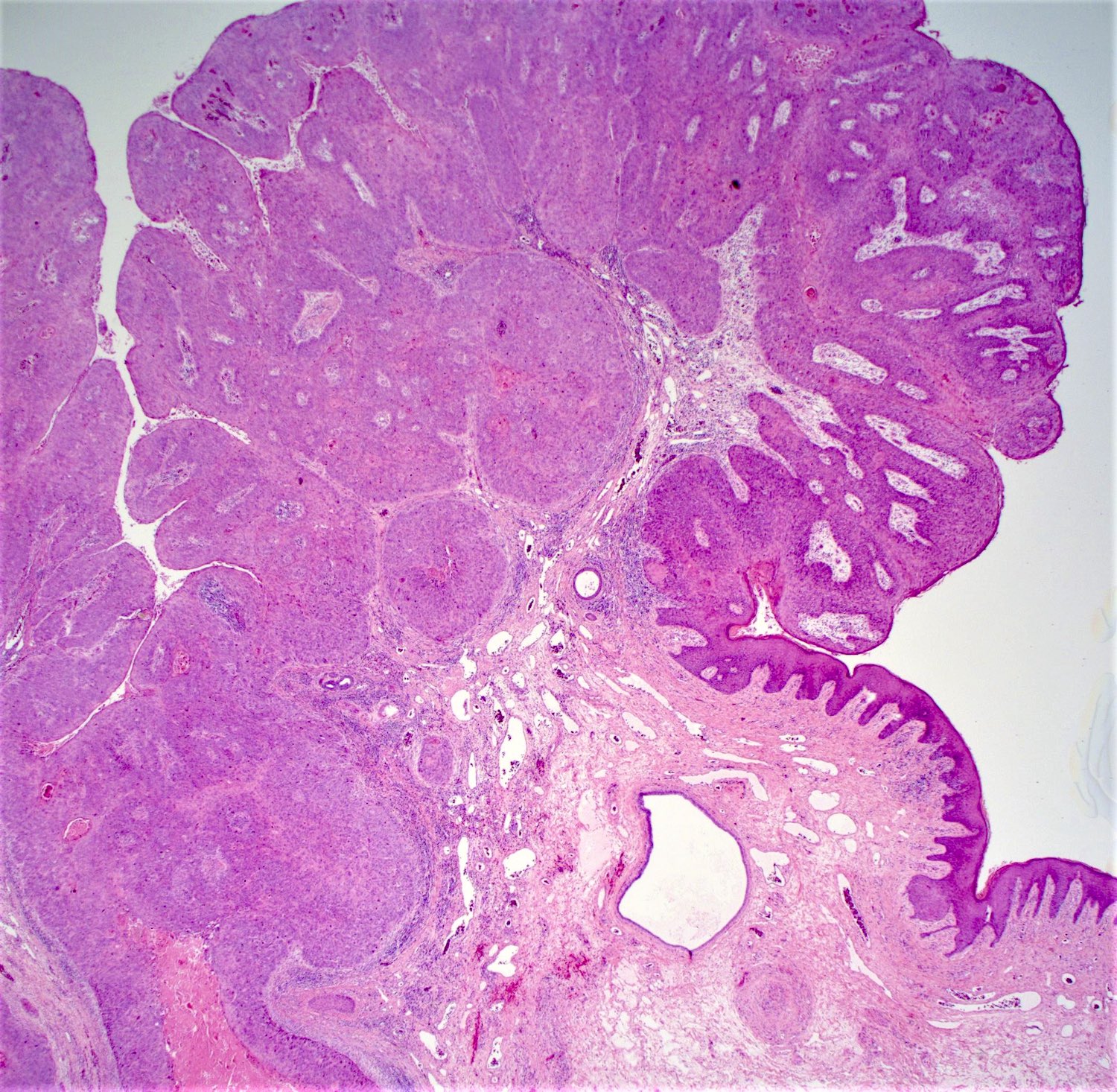
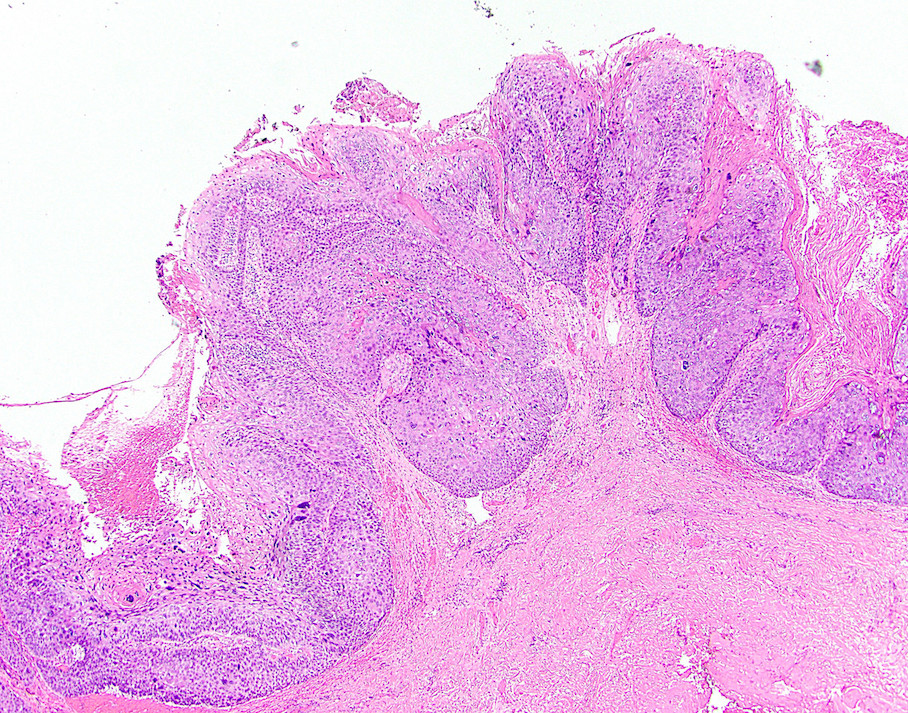
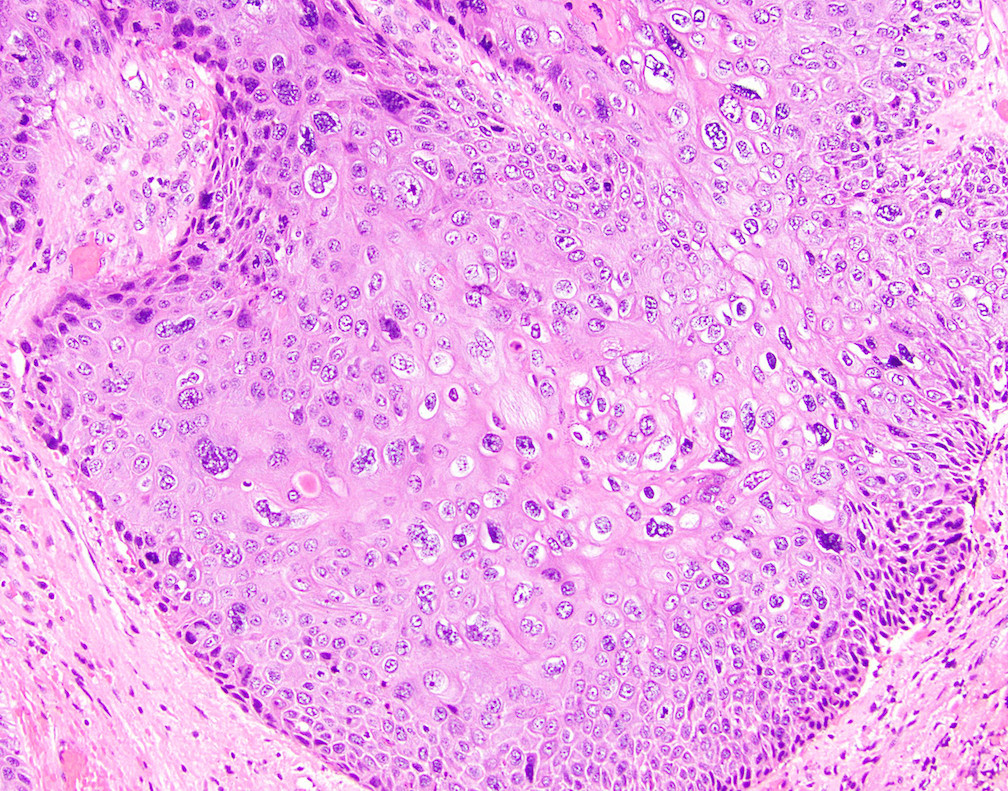
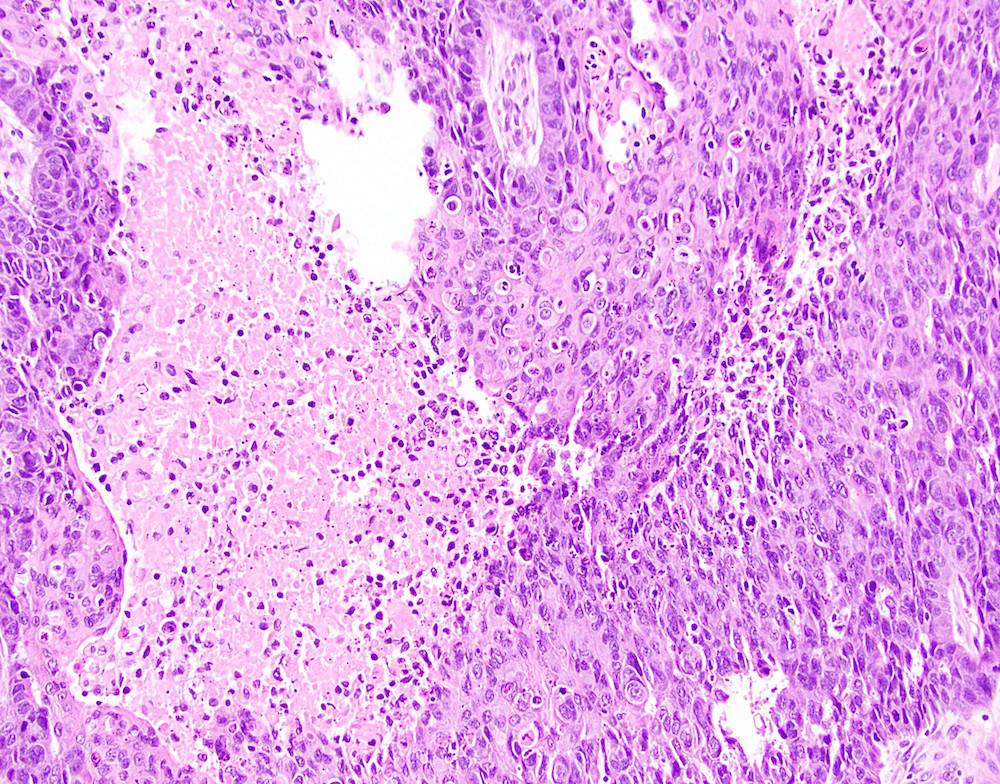
Condyloma acuminatum
Images hosted on other servers:


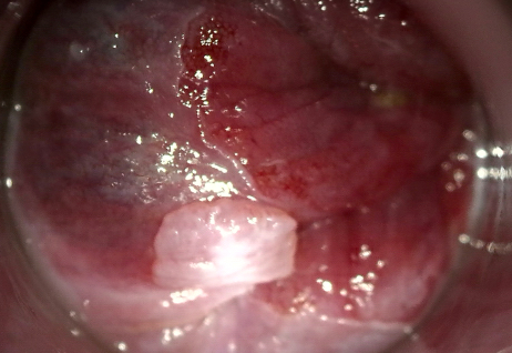
Anoscopy
Contributed by Xiaoyan Liao, M.D., Ph.D. and @AnaPath10 on Twitter
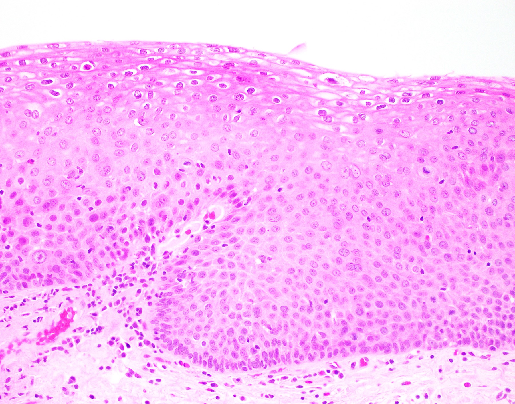
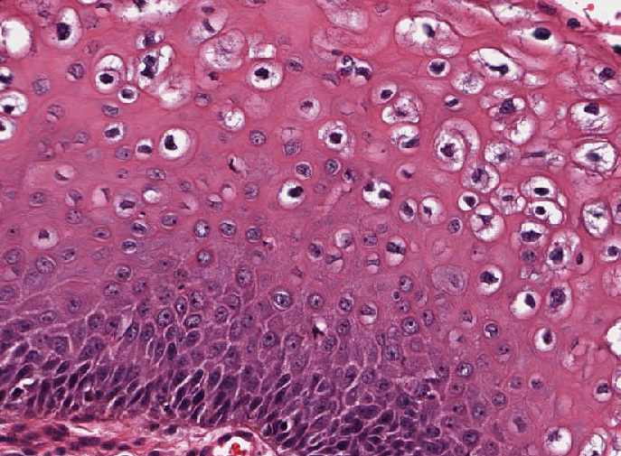
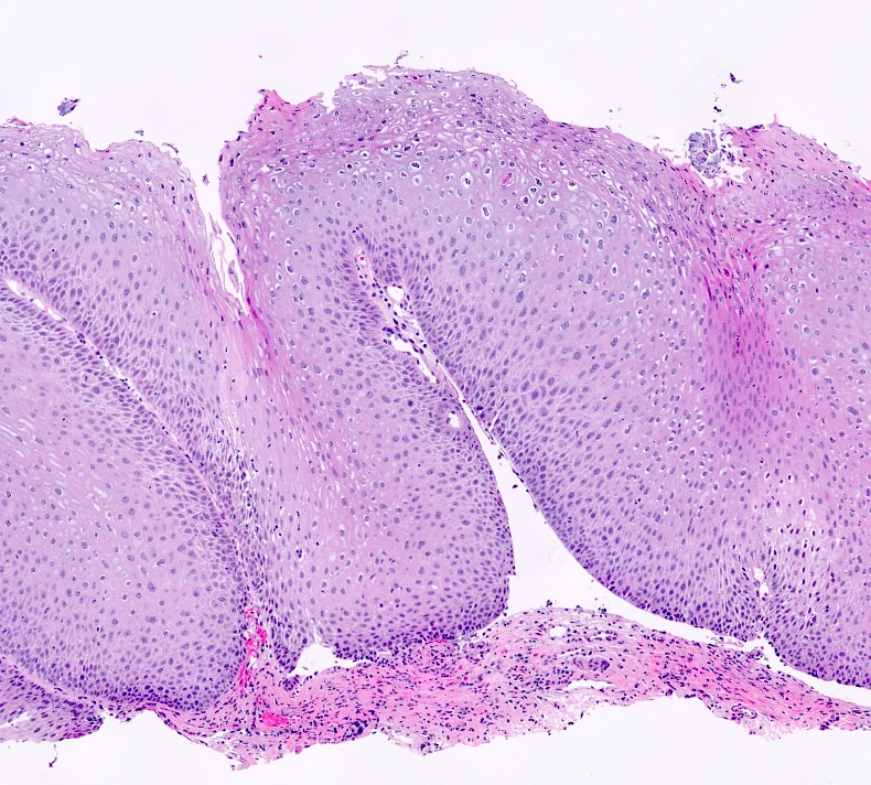
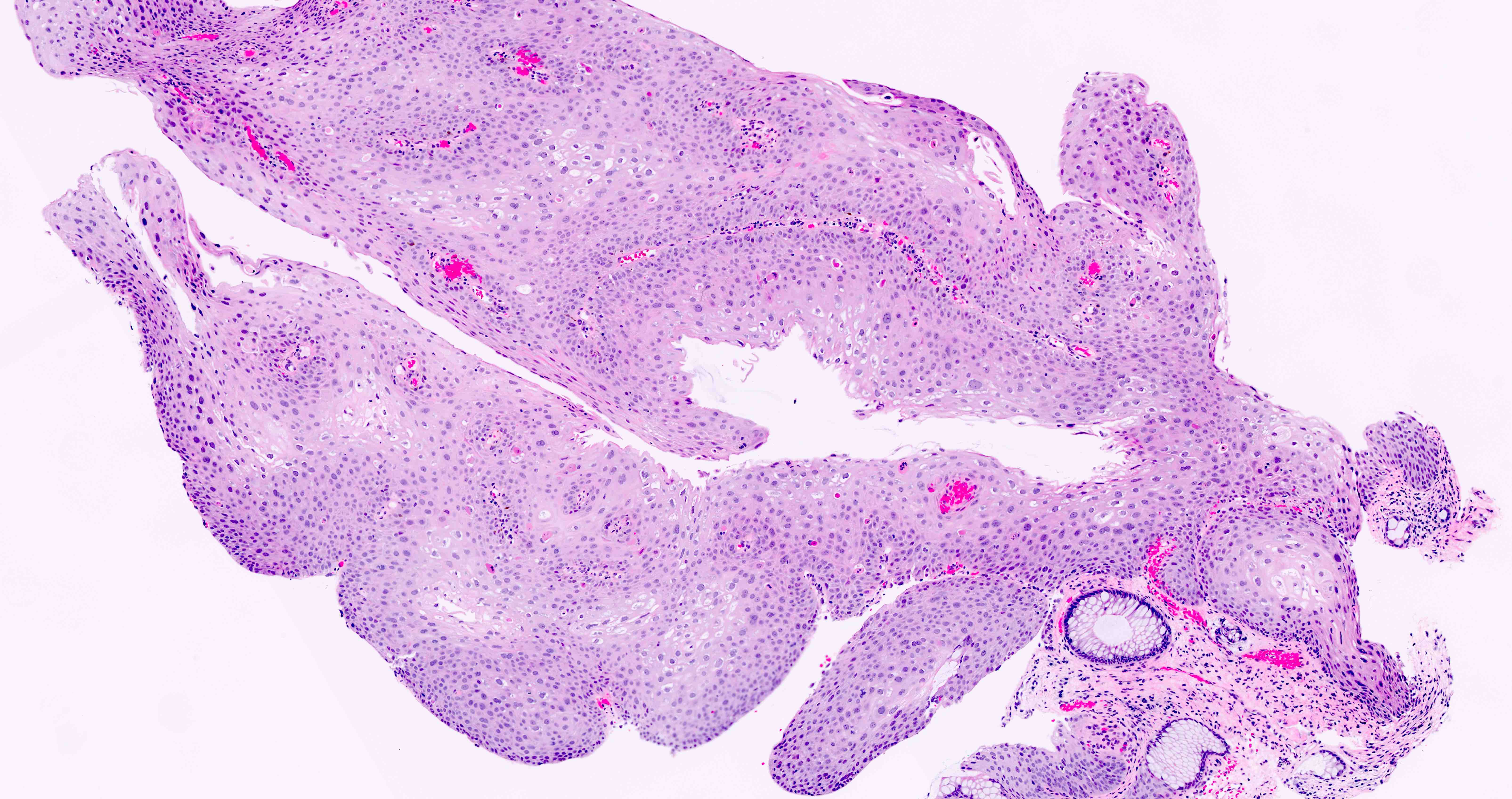
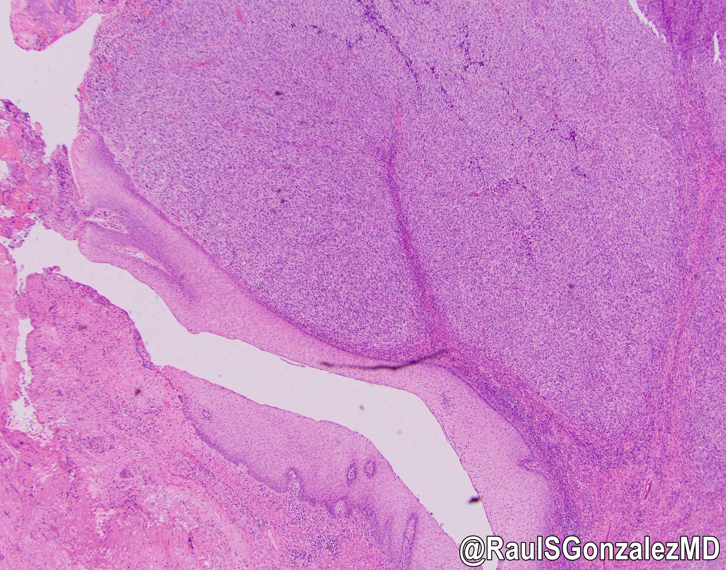
Flat squamous proliferation
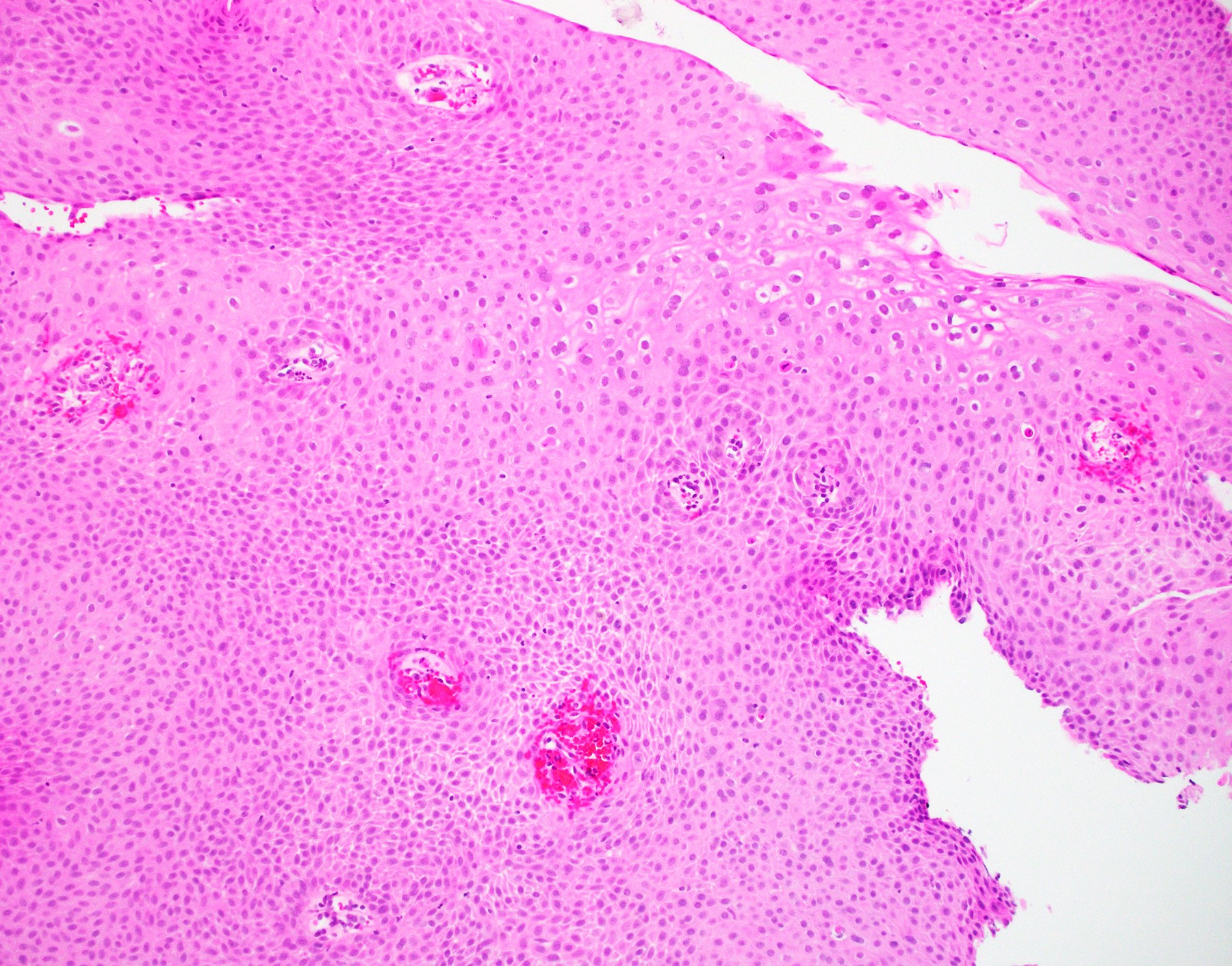
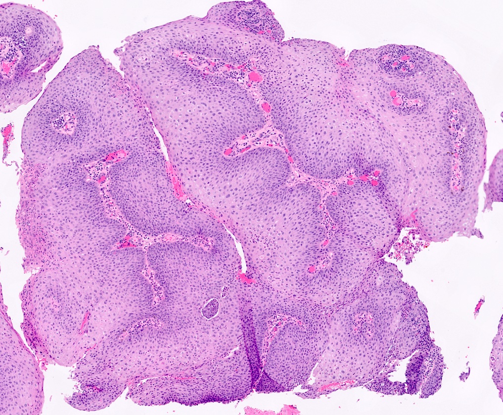
Papillary squamous proliferation
Papillary squamous proliferation, parakeratosis and koilocytosis
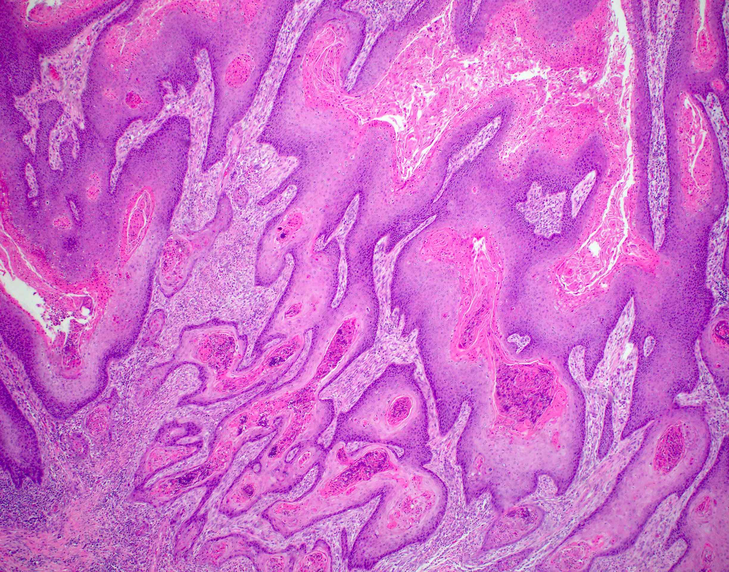
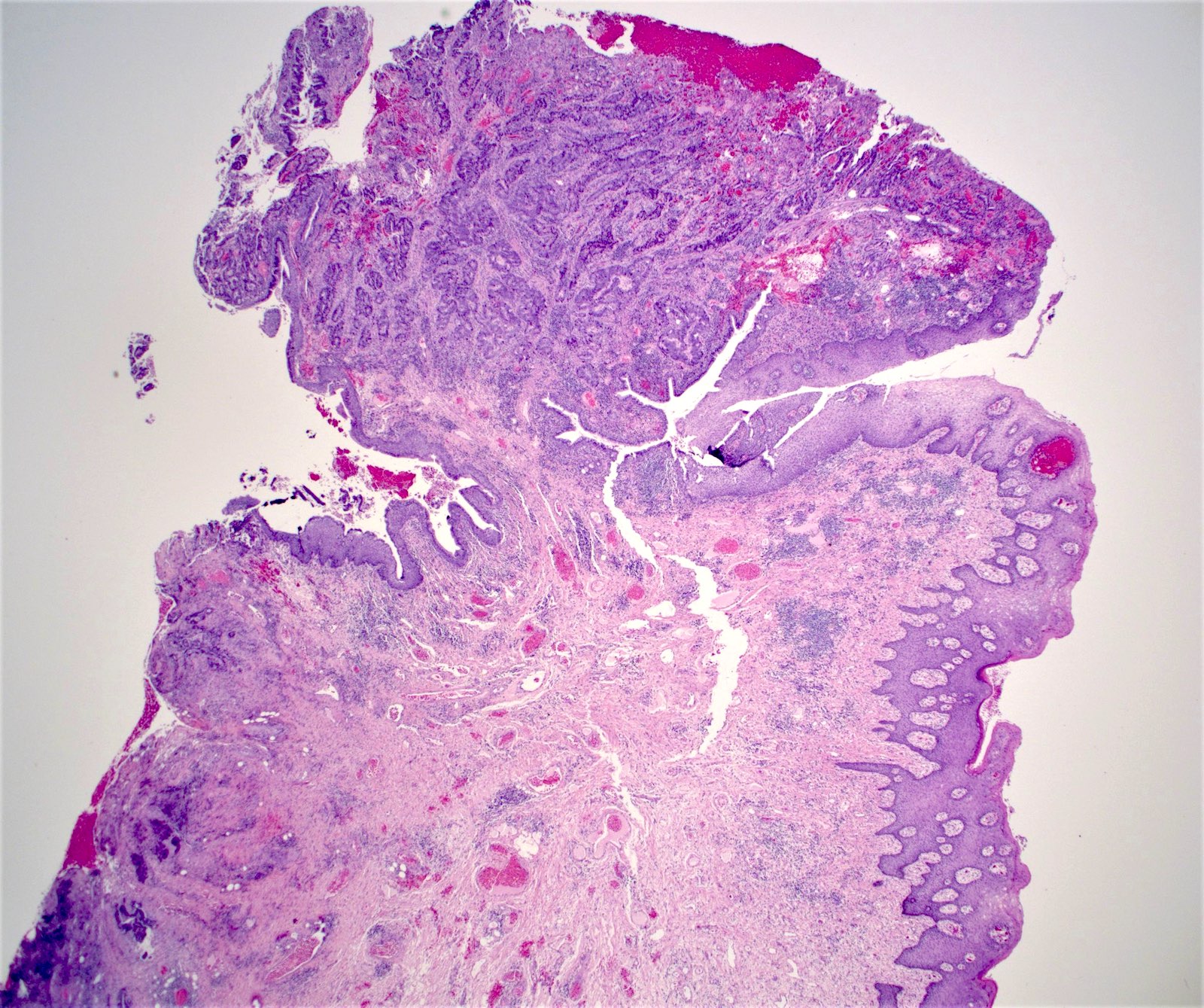
Condyloma with invasion
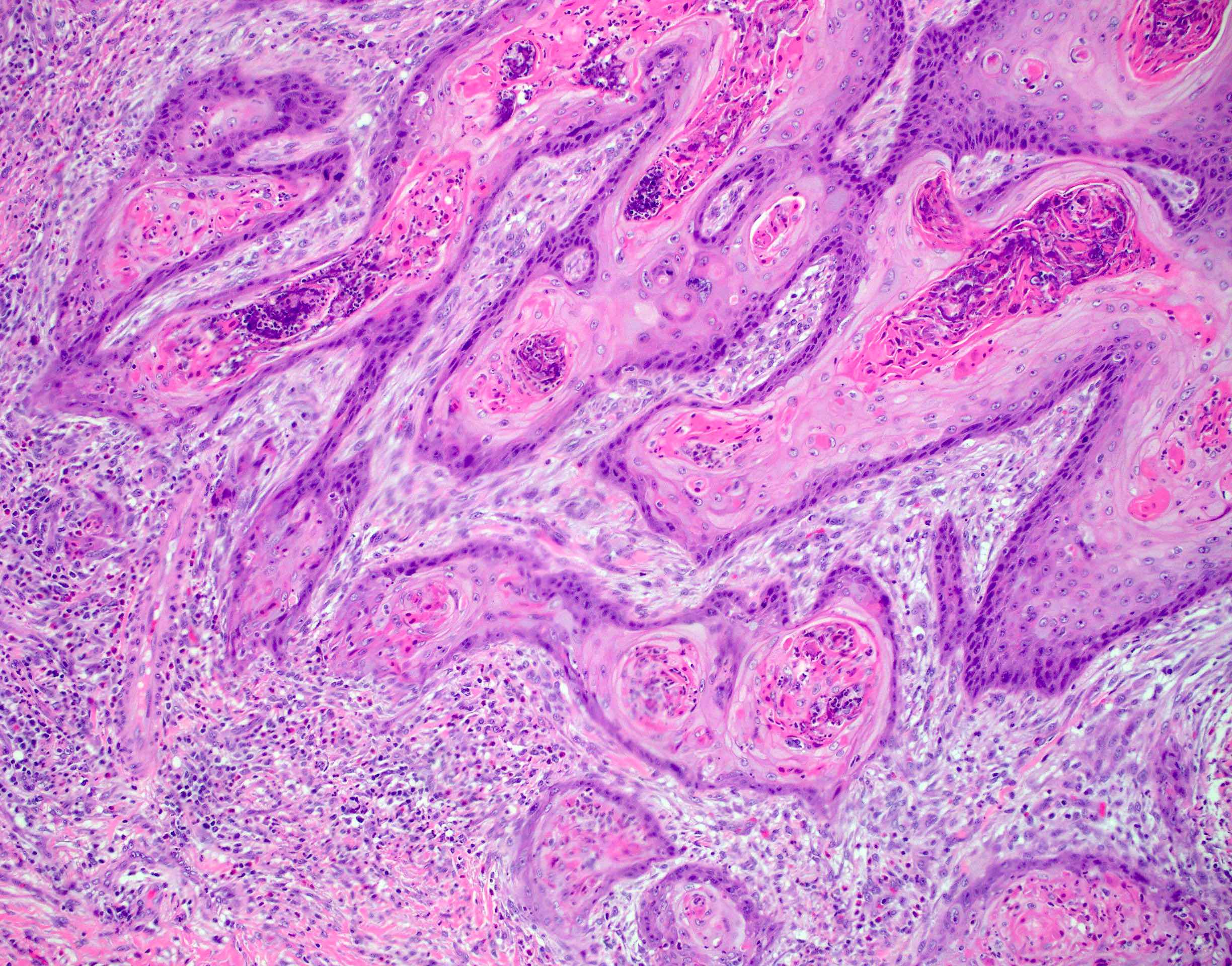
Invasive
squamous cell
carcinoma arising
in condyloma
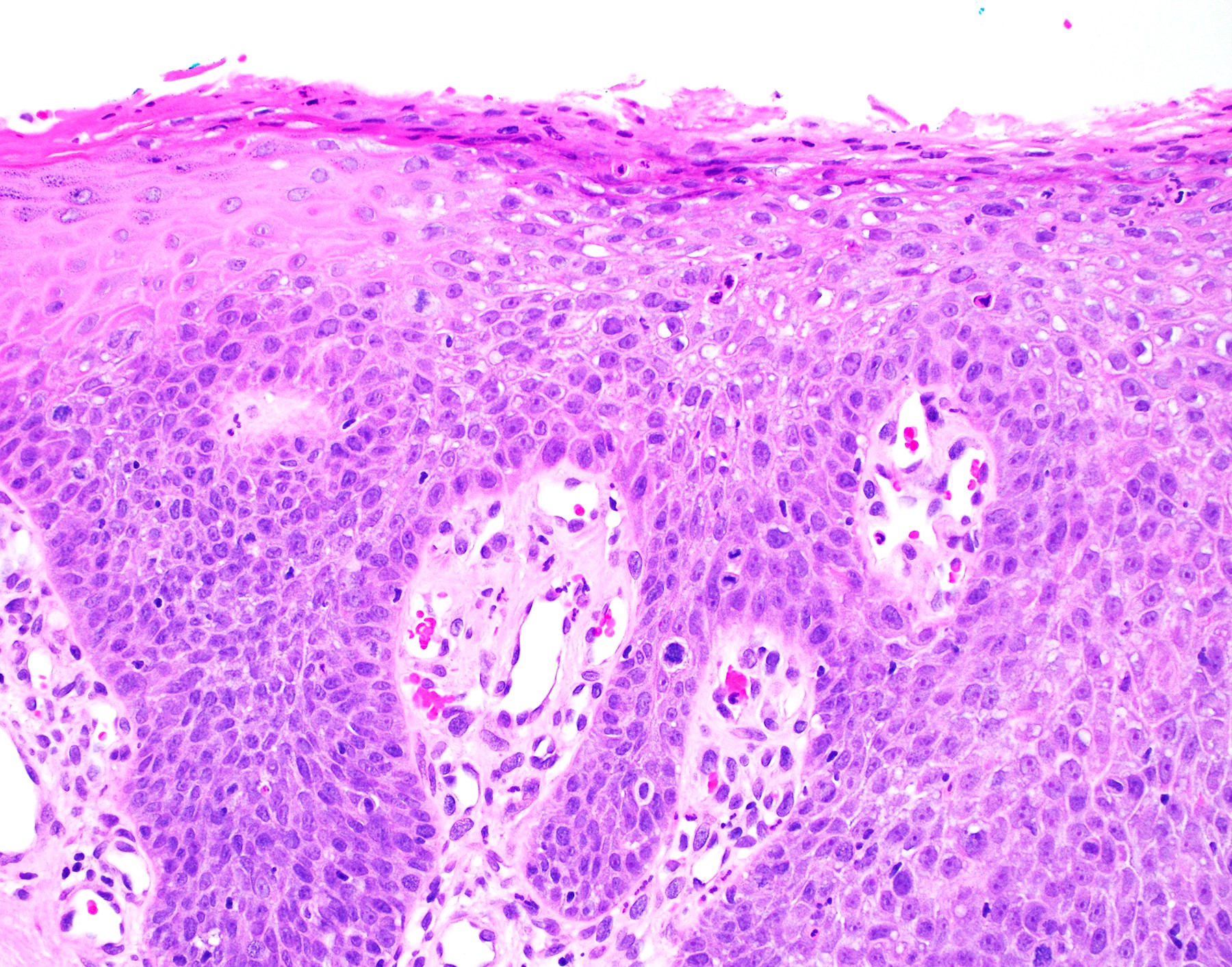
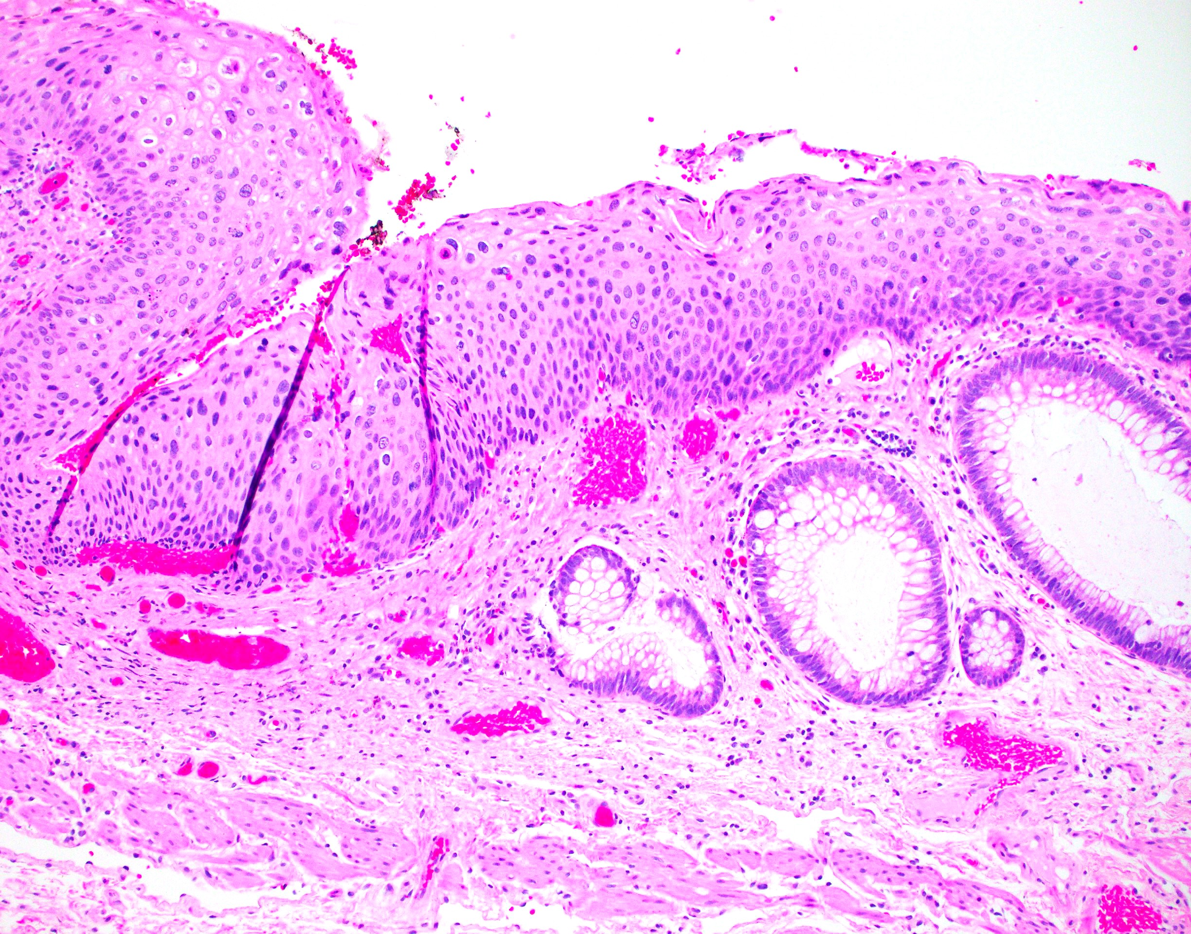
HSIL
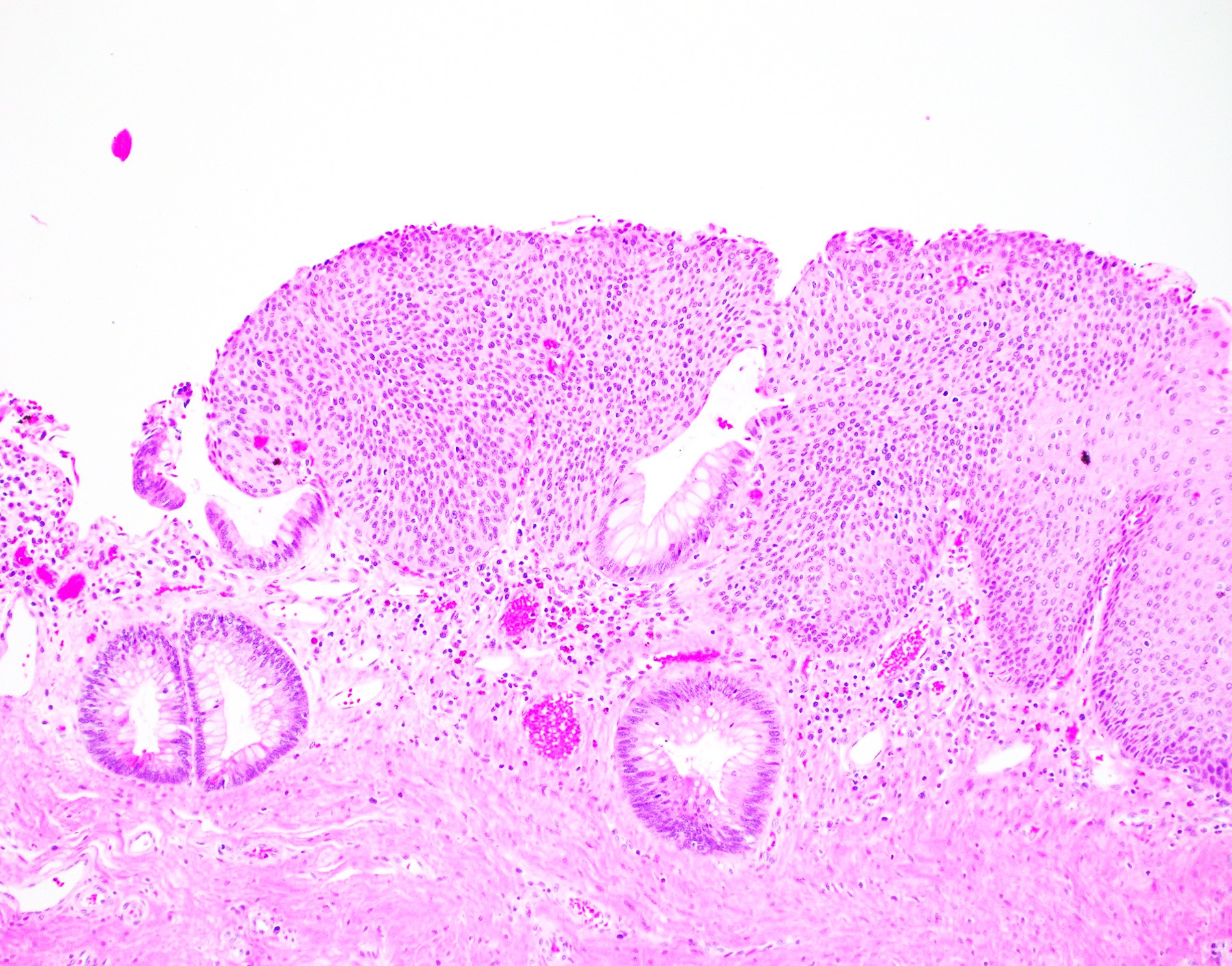
HSIL at the anal transitional zone
Involving a hemorrhoid
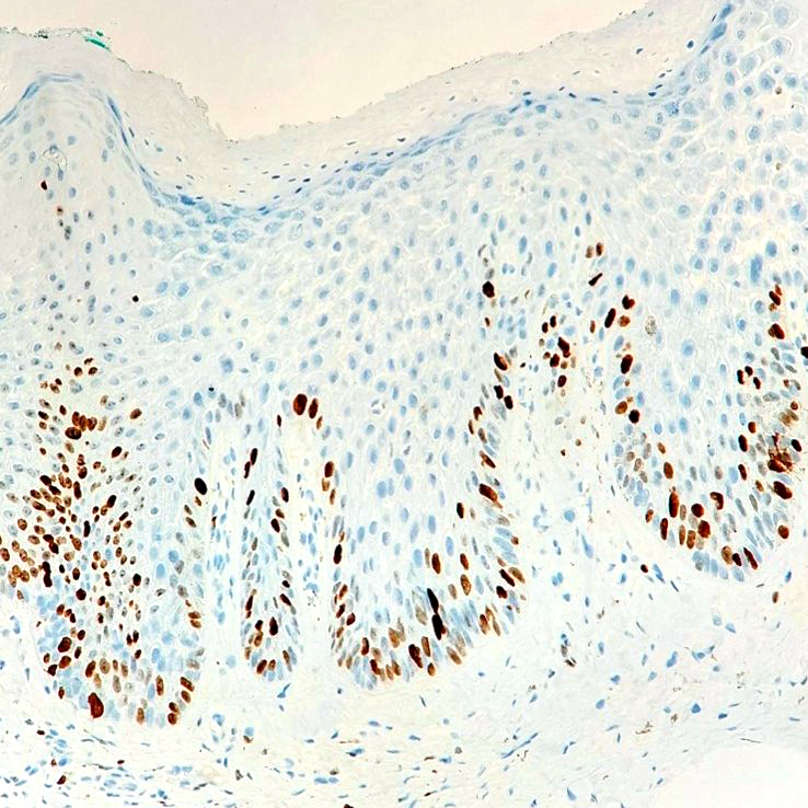
Squamous dysplasia
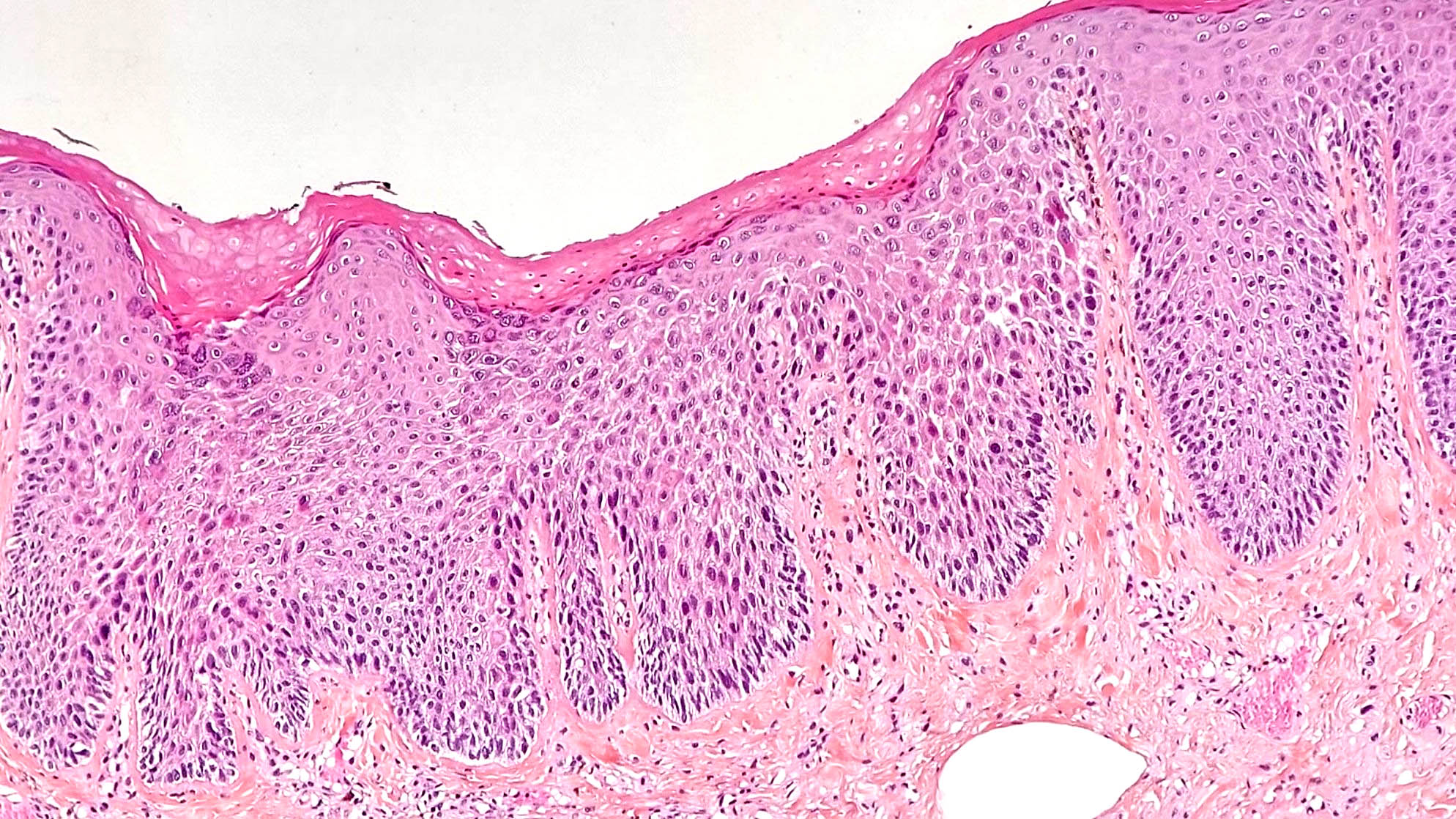
Squamous dysplasia
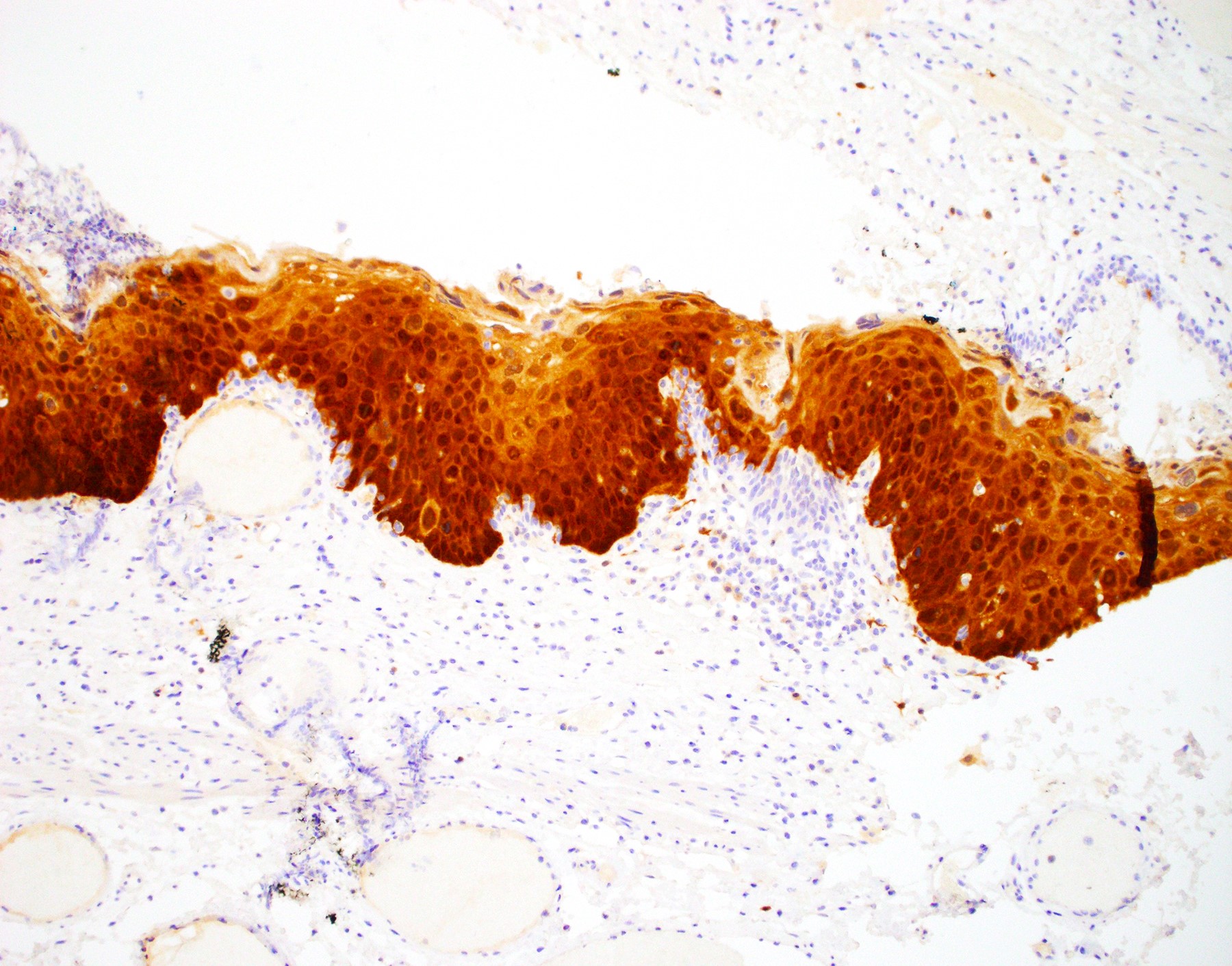
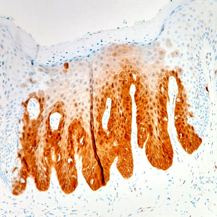
p16 immunohisto-
chemistry
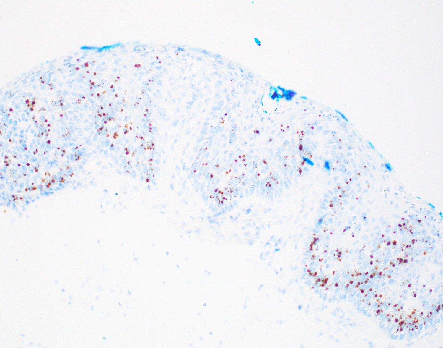
HPV in situ hybridization
Squamous dysplasia
Contributed by Irene Y. Chen, M.D.
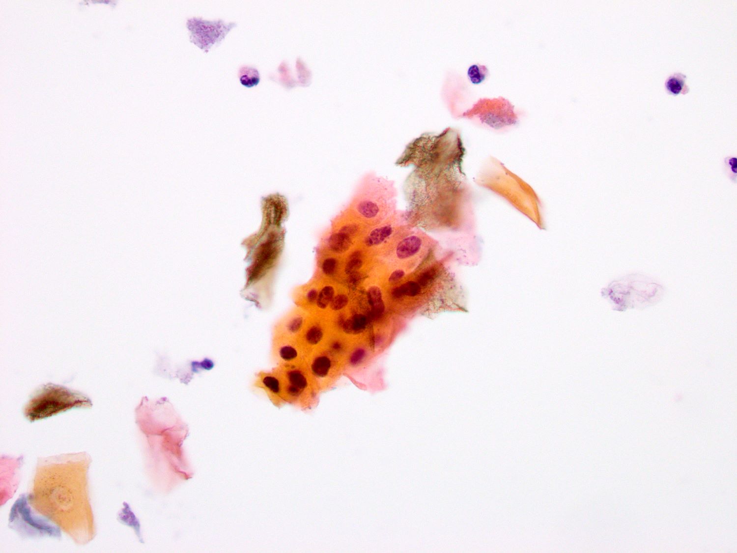
Anal Pap smear
Images hosted on other servers:
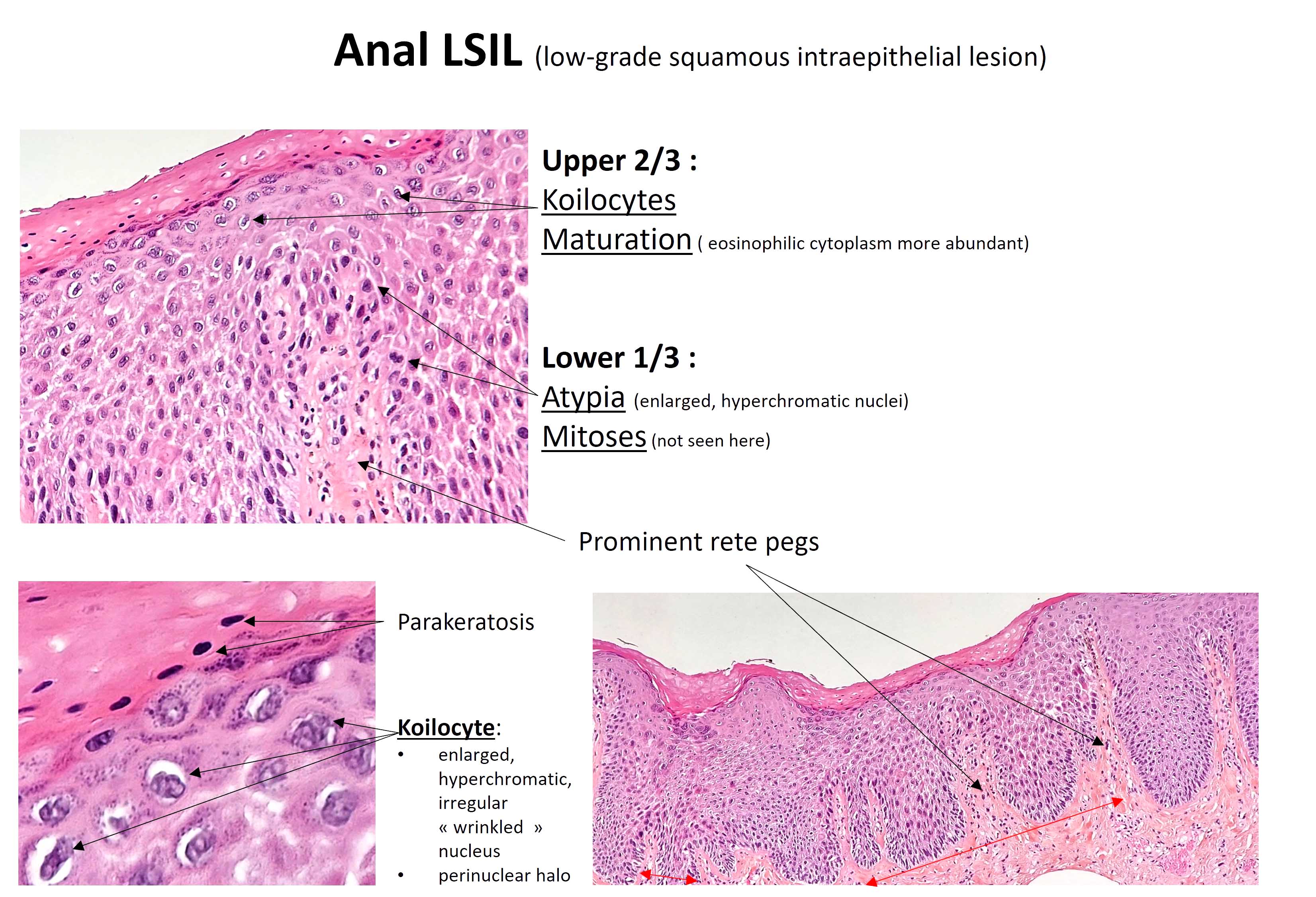
LSIL
Images hosted on other servers:

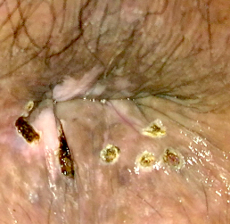
Multiple perianal lesions

Anorectal ulcerated mass

Exophytic perineal lesions

Condylomata lata
Contributed by Aaron R. Huber, D.O.
Epithelial hyperplasia and inflammation
Plasma cell infiltrate
Rectal biopsy with inflammation
Silver stain
T. pallidum IHC
Perianal condyloma lata
Syphilis stains and IHC
Images hosted on other servers:

Retrorectal space
Images hosted on other servers:

Retrorectal cystic mass

Malignant transformation
Images hosted on other servers:

Posterior approach view
Images hosted on other servers:

Intact resection

Mud-like contents

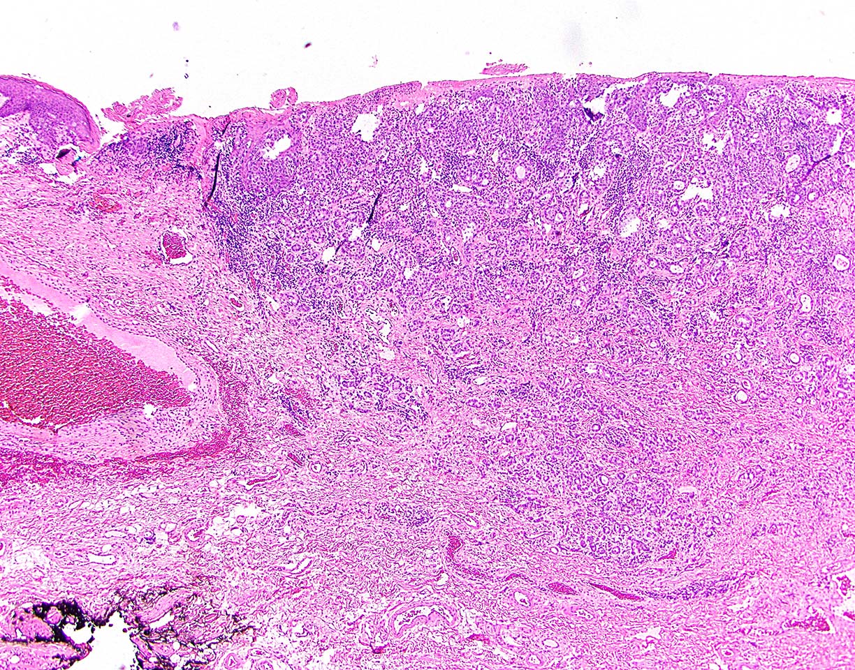
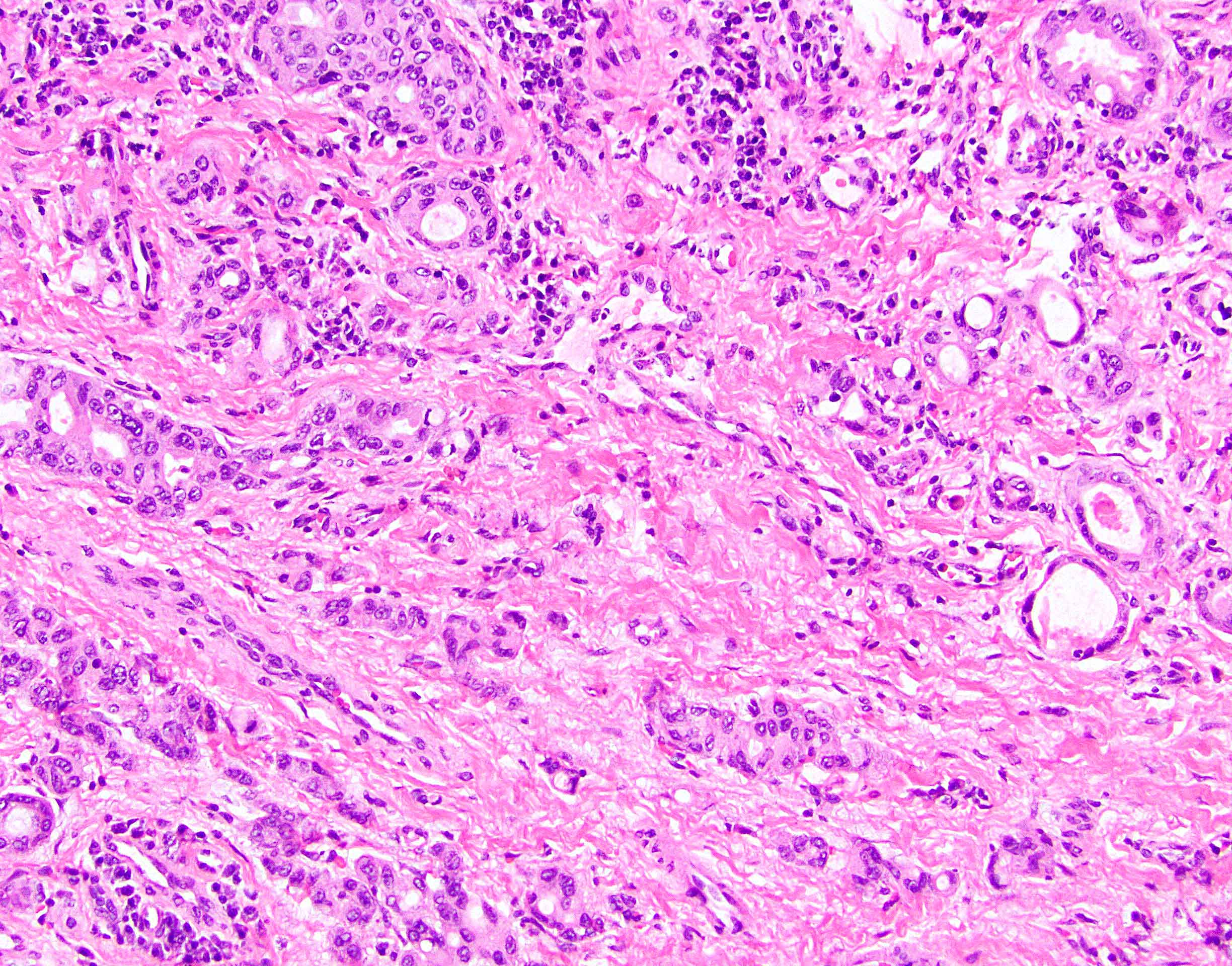
Tailgut cyst adenocarcinoma
Contributed by John D. Paulsen, M.D. and Alexandros D. Polydorides, M.D., Ph.D.
Dense fibroconnective tissue stroma
Disorganized smooth muscle
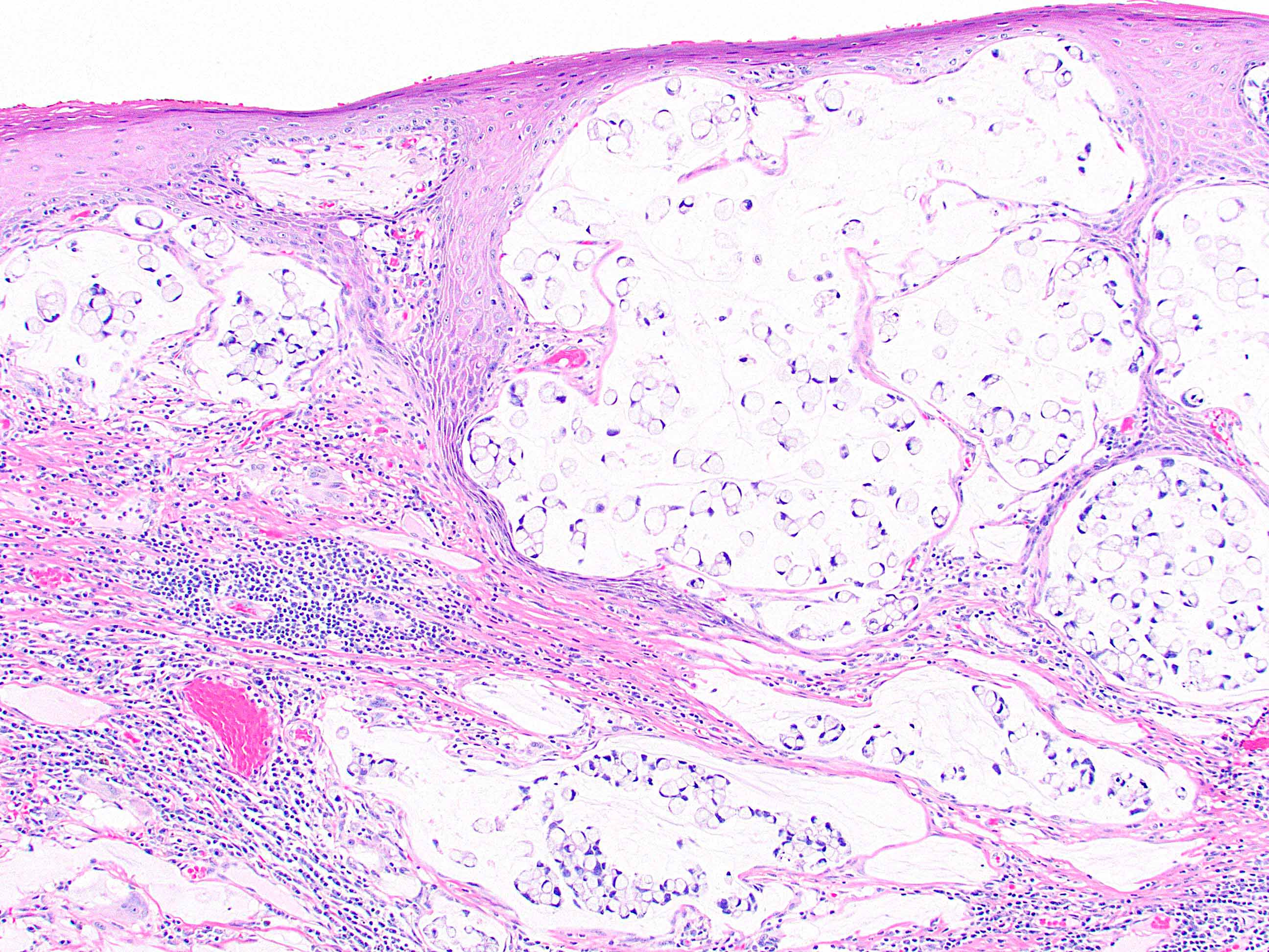
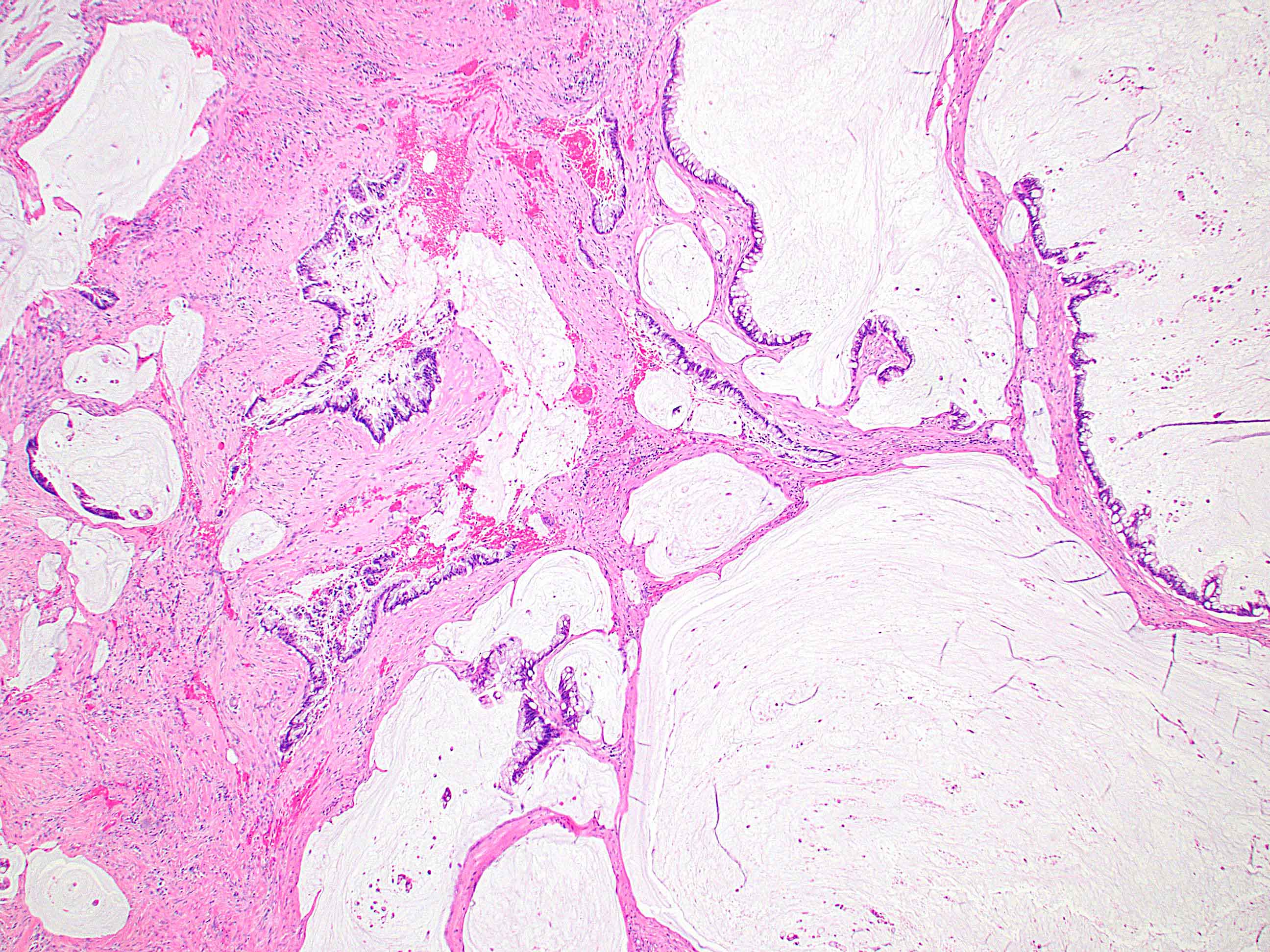
Epithelial linings
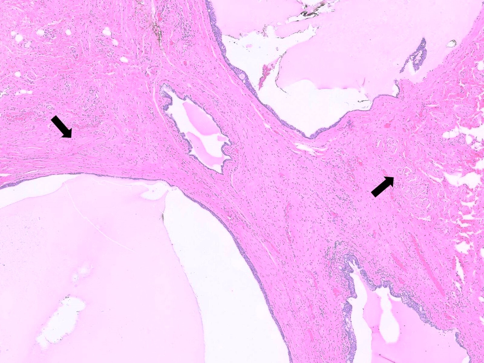
Multiple loculations
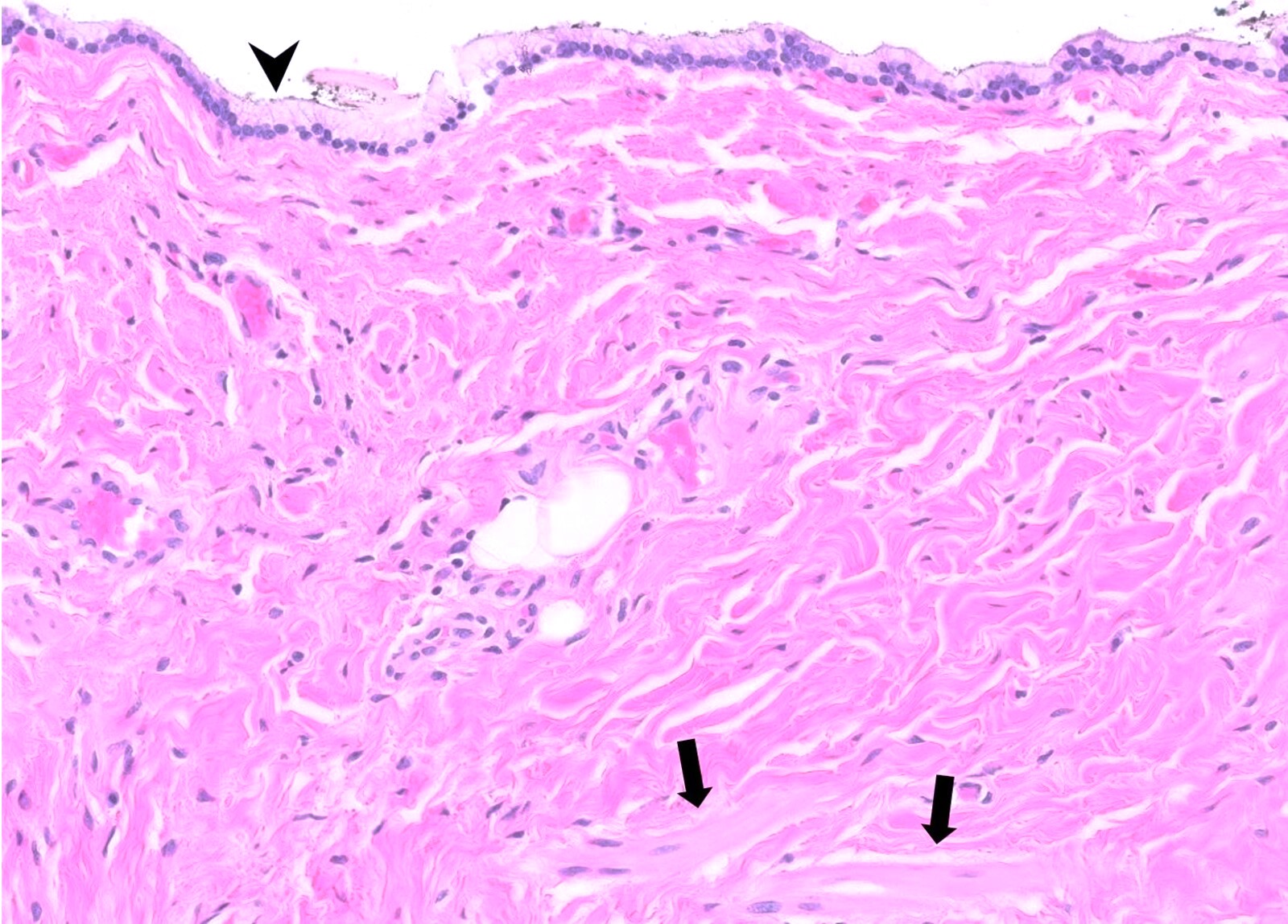
Mucinous columnar epithelium
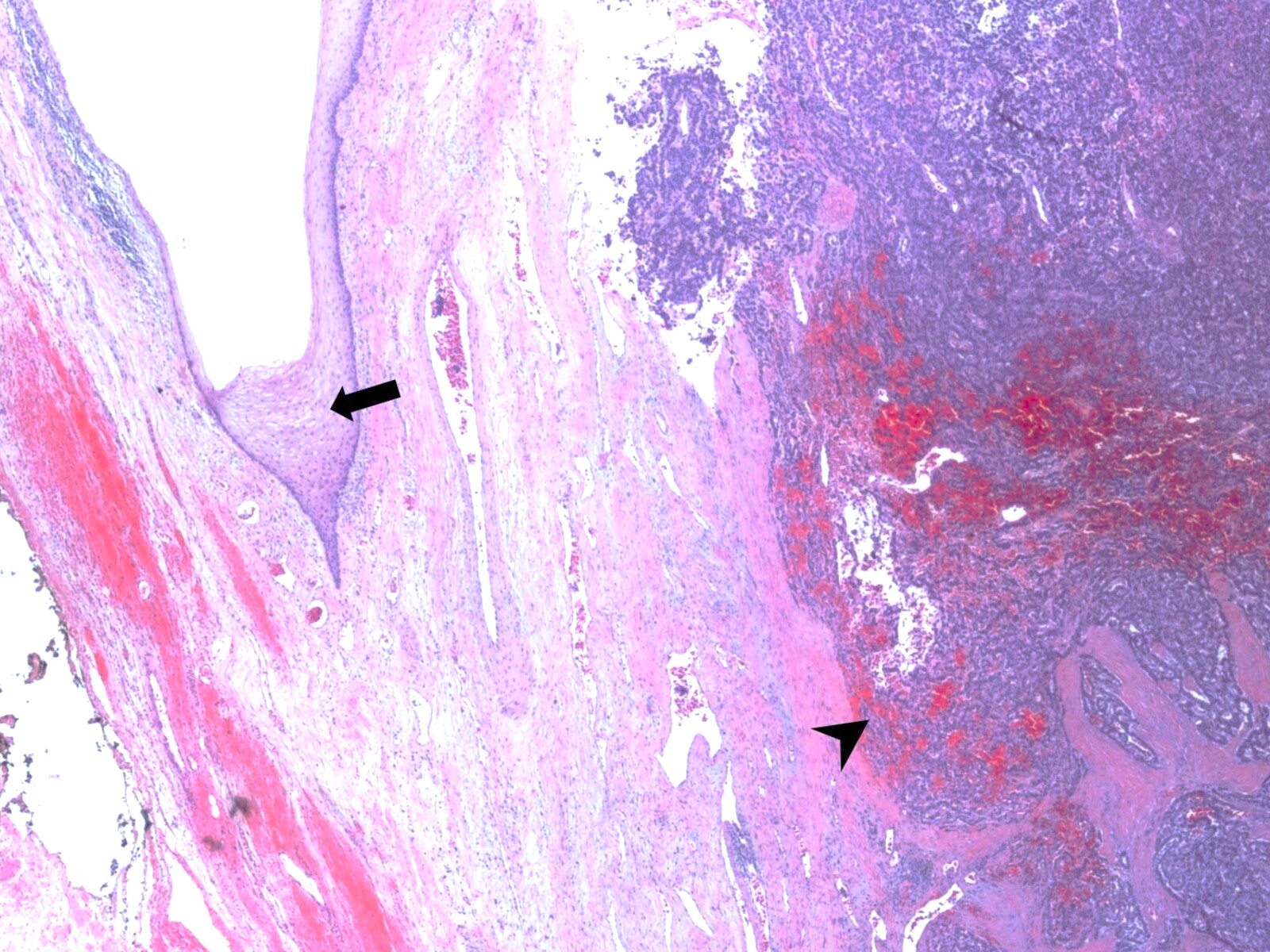
Neoplastic transformation, neuroendocrine tumor
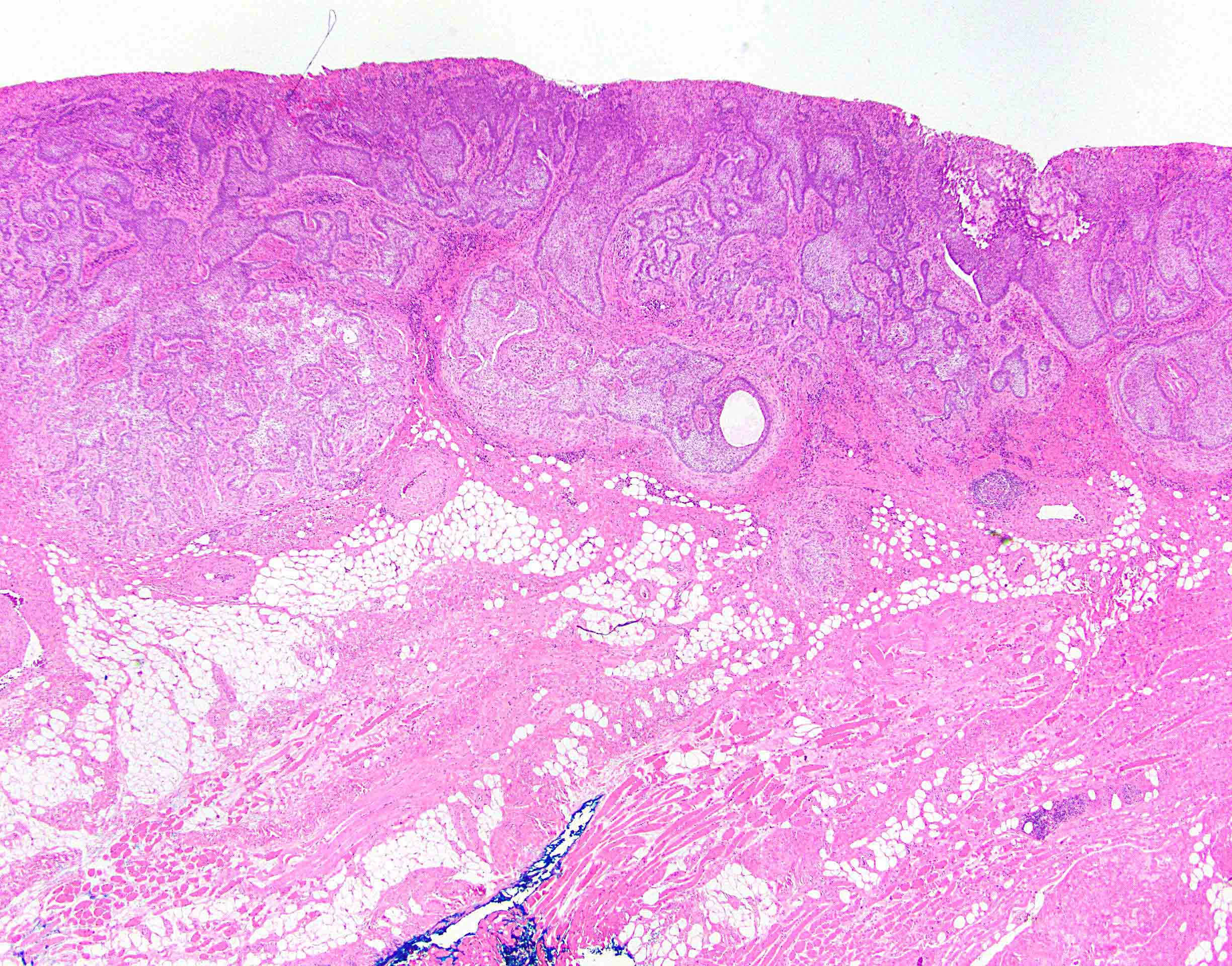
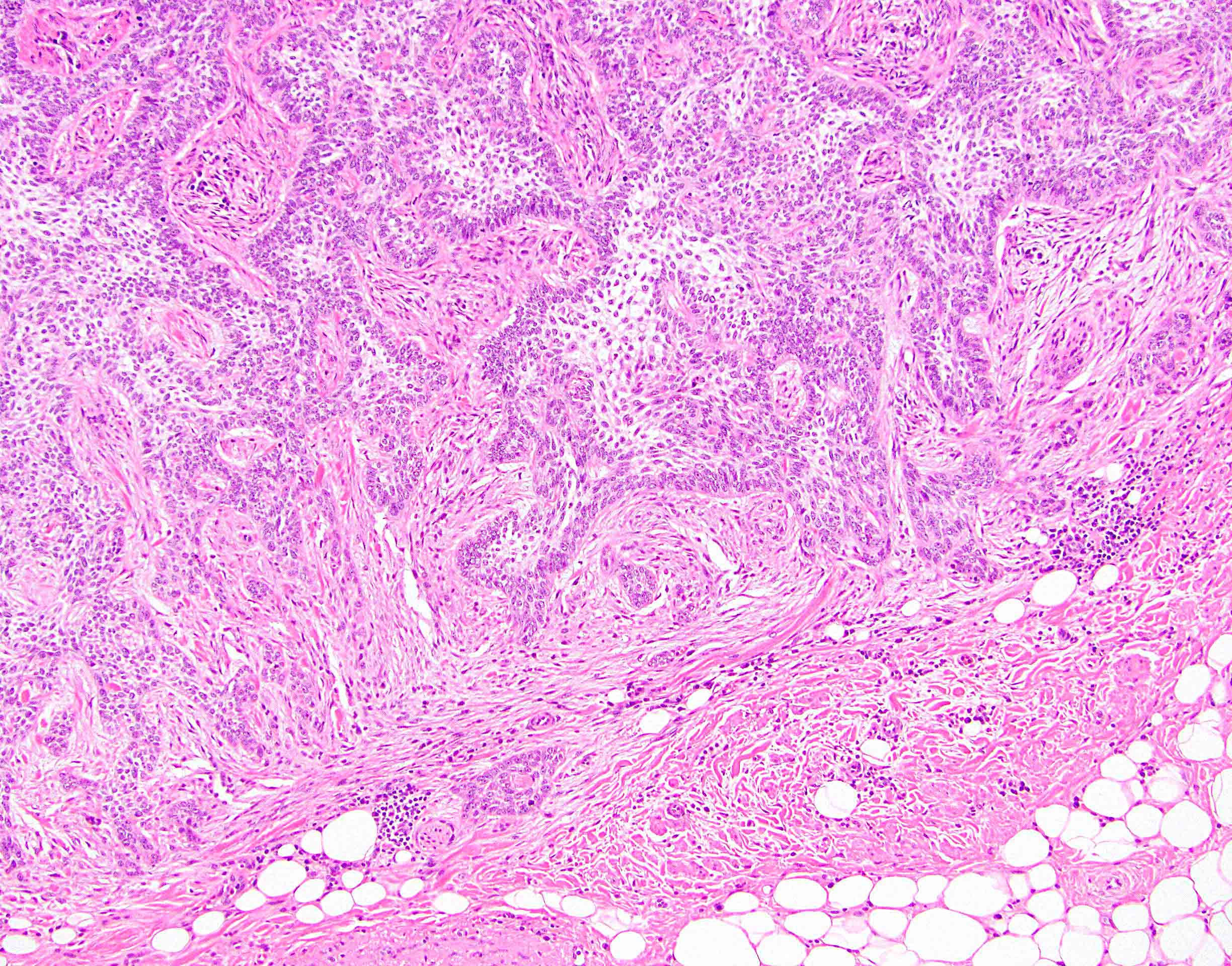
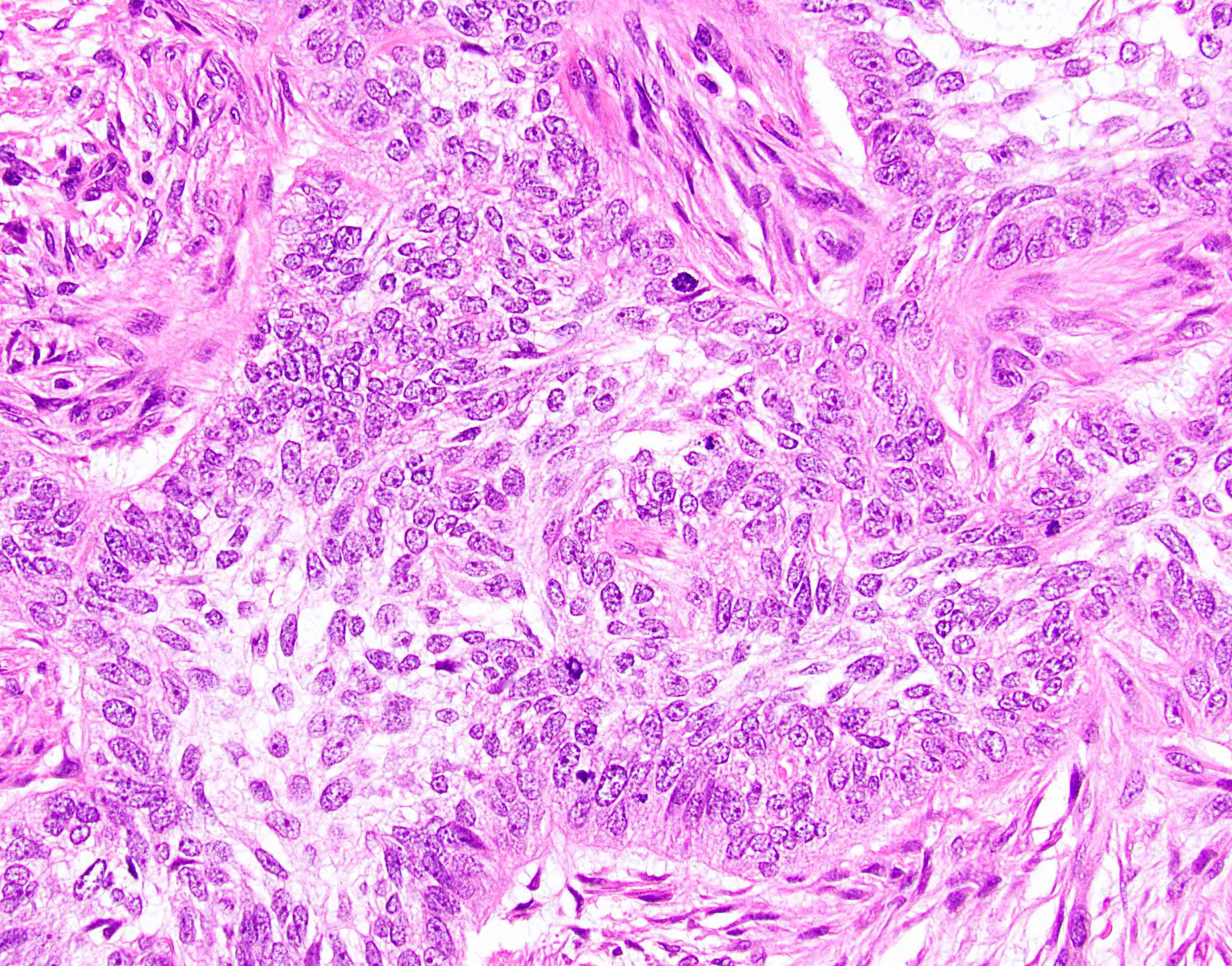
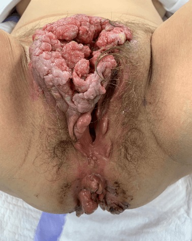
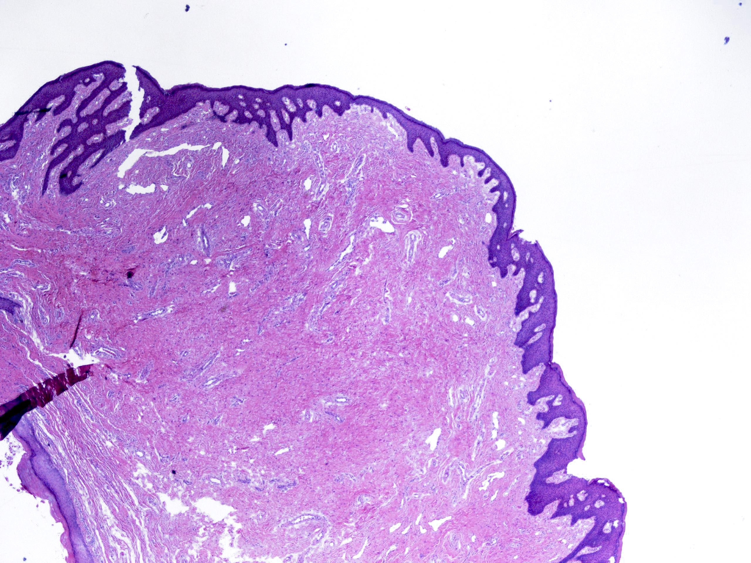
Contributed by Raul S. Gonzalez, M.D.

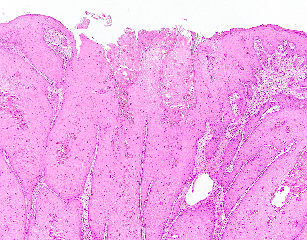
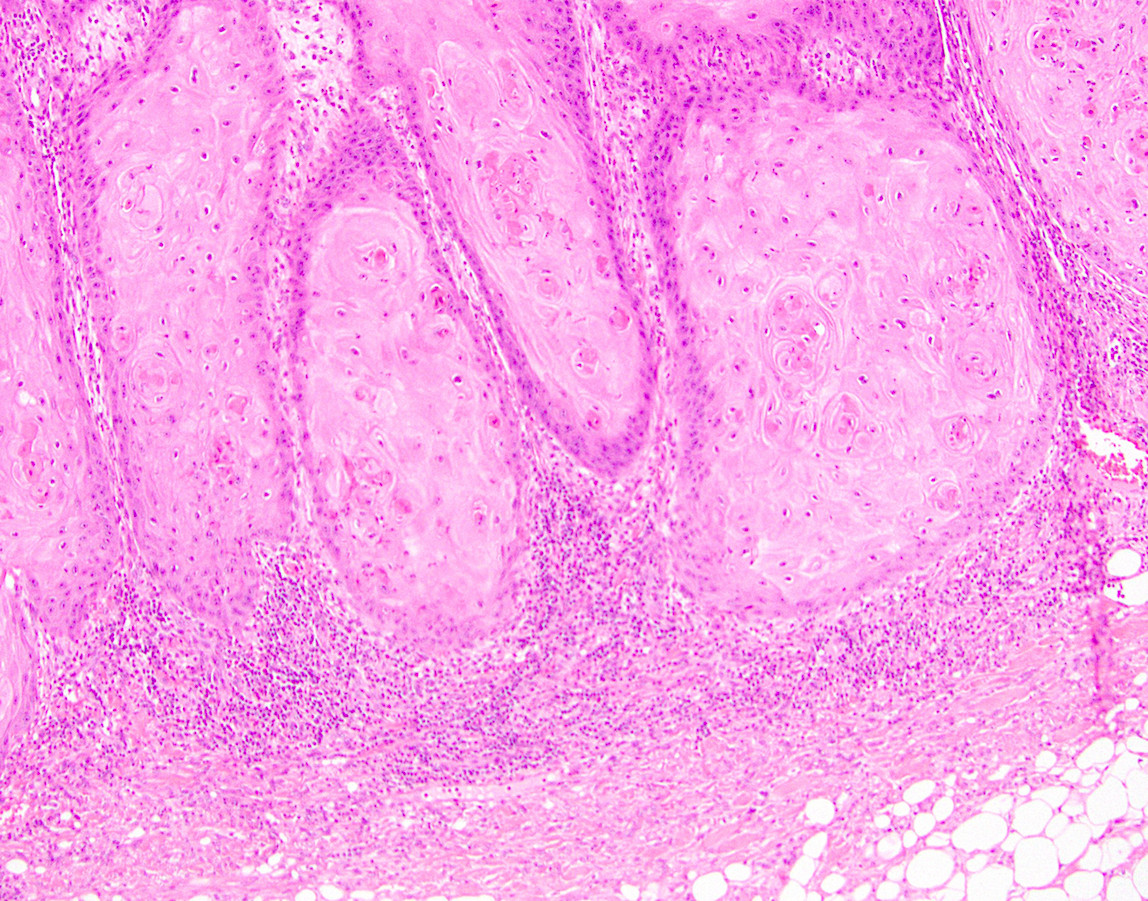
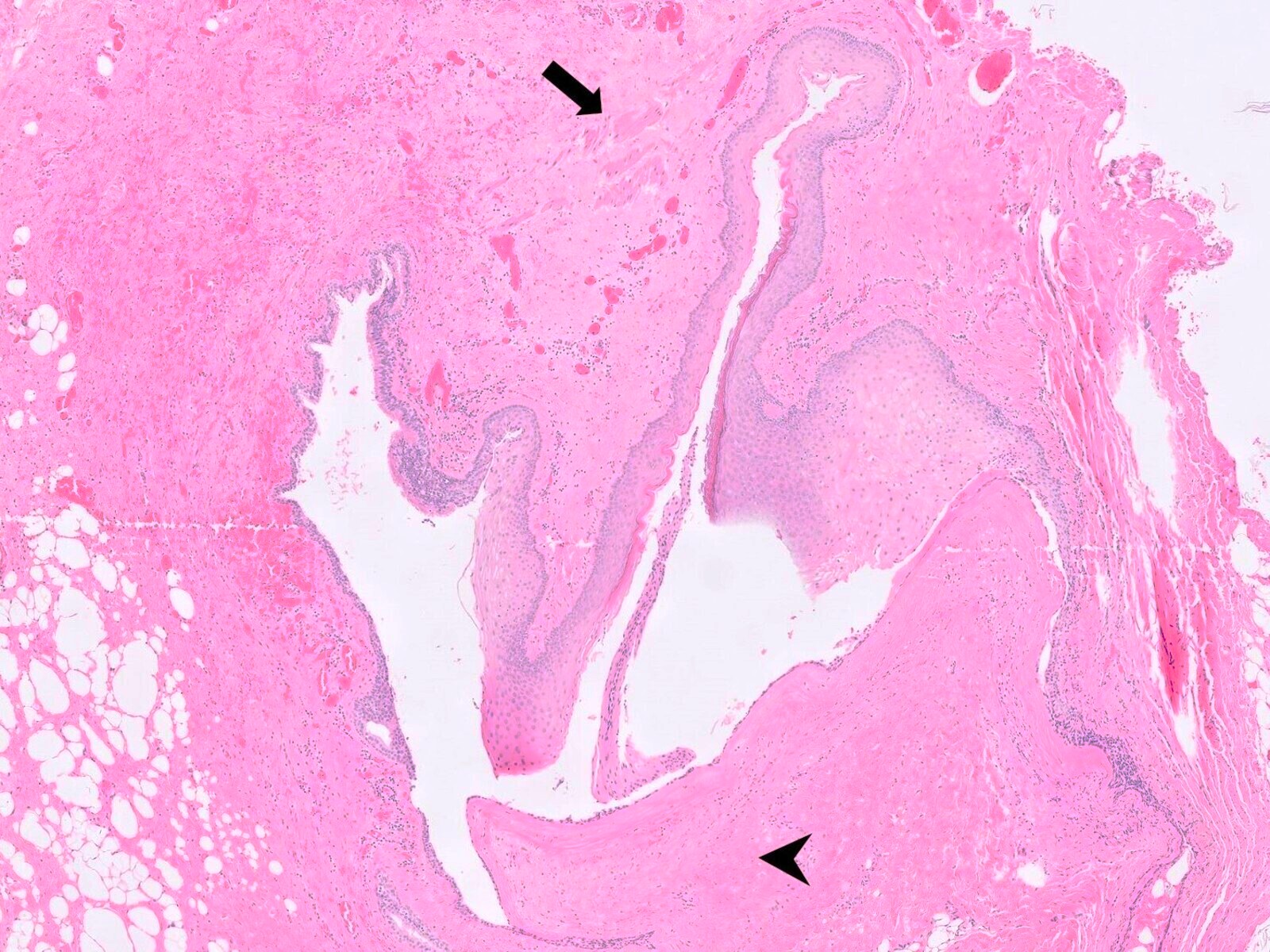
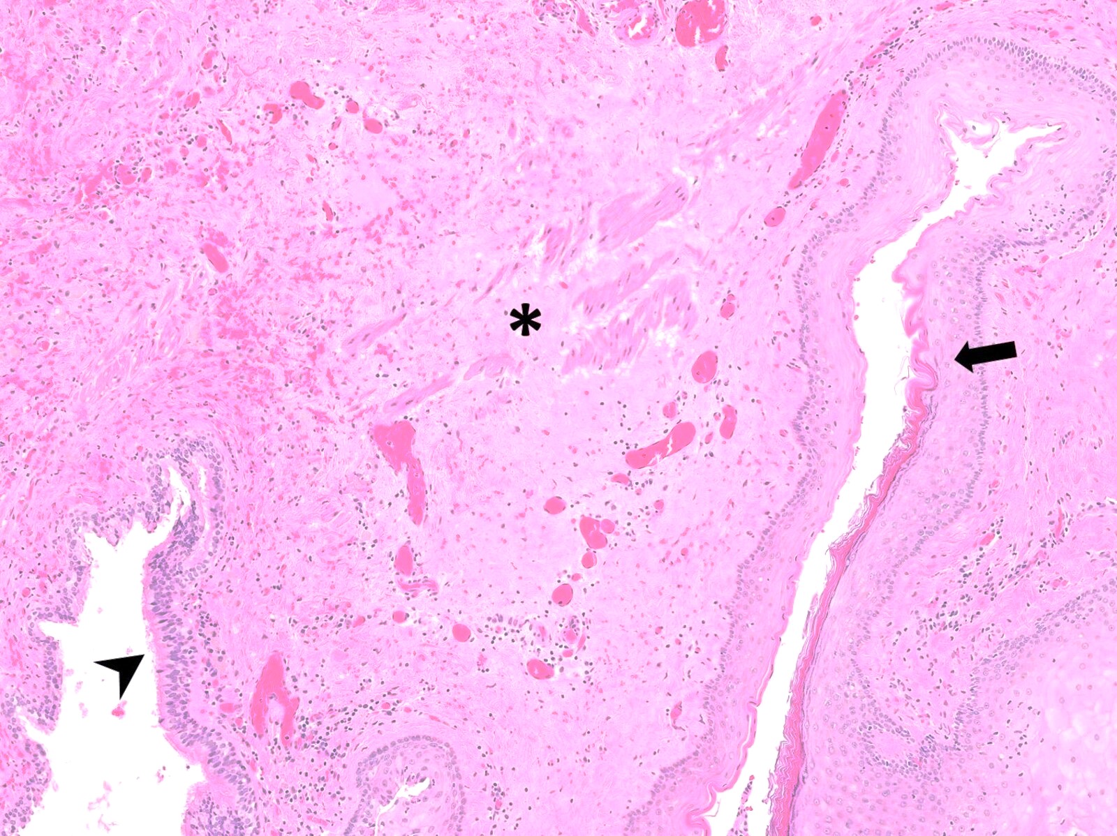
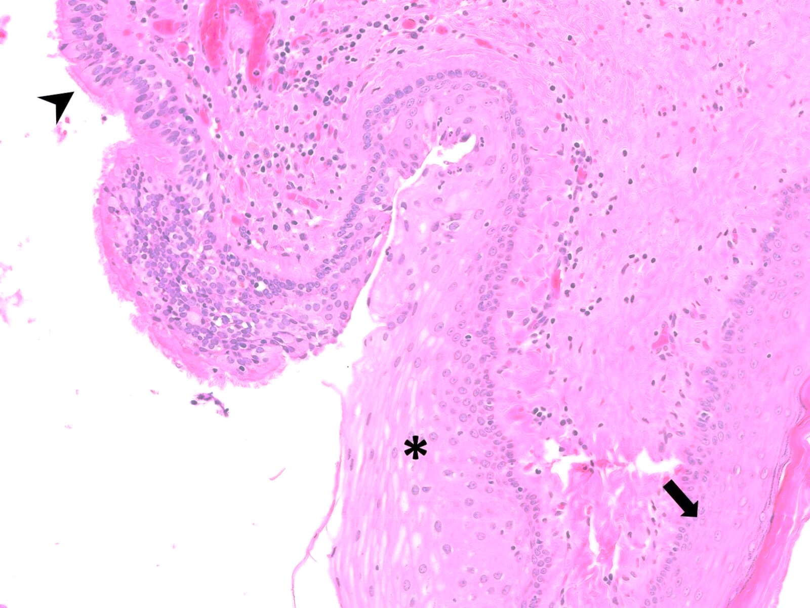
Verrucous carcinoma
Cohen: 2014
Greenson: 2019

IARC: 2019
Lamps: 2015
Montgomery: 2017
Montgomery: 2017
Odze: 2022
Srivastava: 2023
Yantiss: 2021
Zutshi: 2016
Find related Pathology books: GI, liver
