
Contributed by Lorenzo Gitto, M.D., Ponni Arunkumar, M.D. and the Cook County Medical Examiner's Office
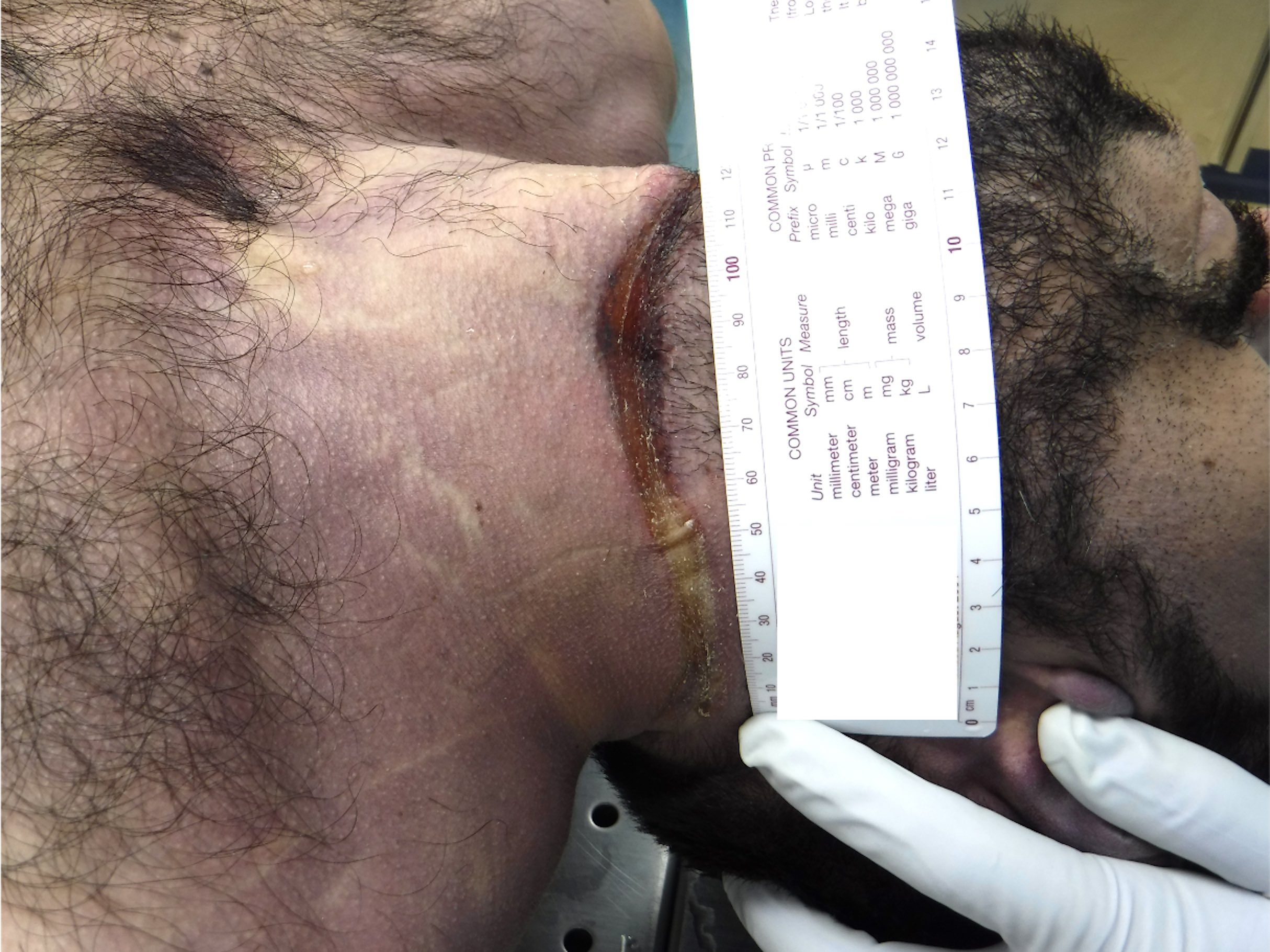
Hanging mark
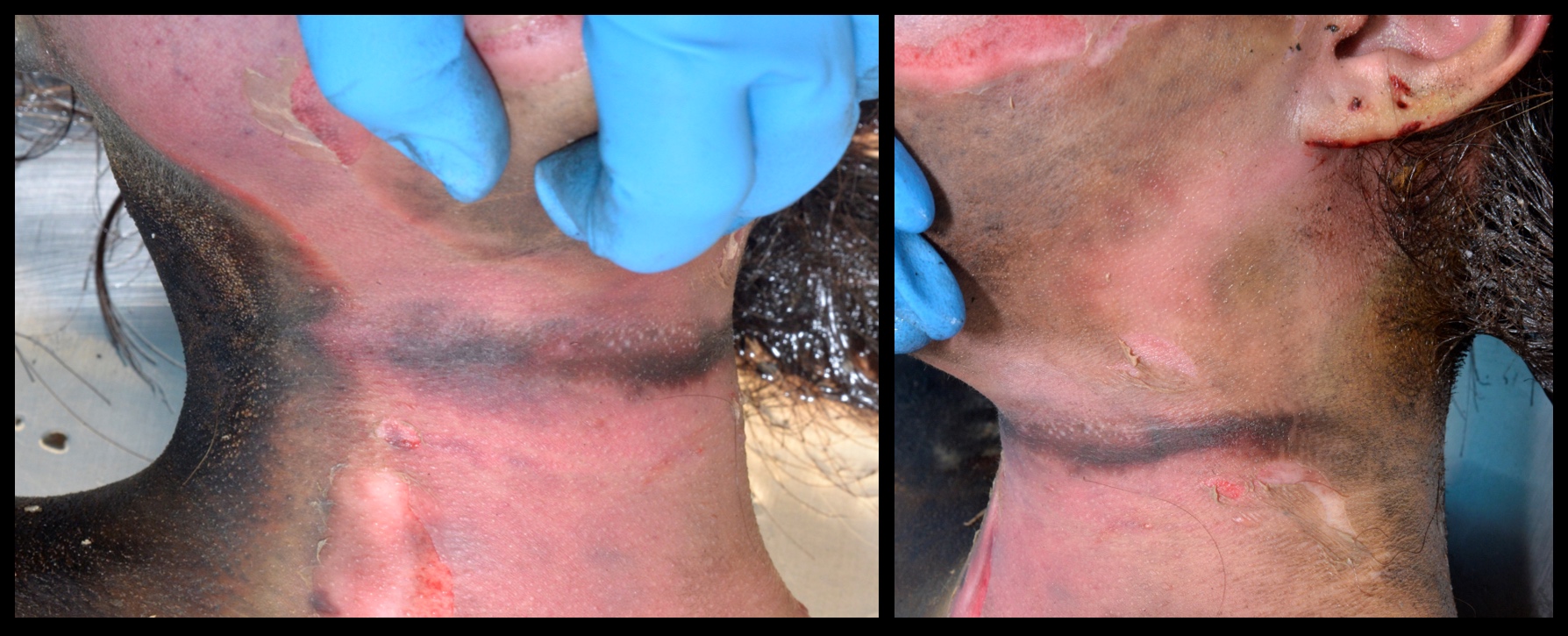
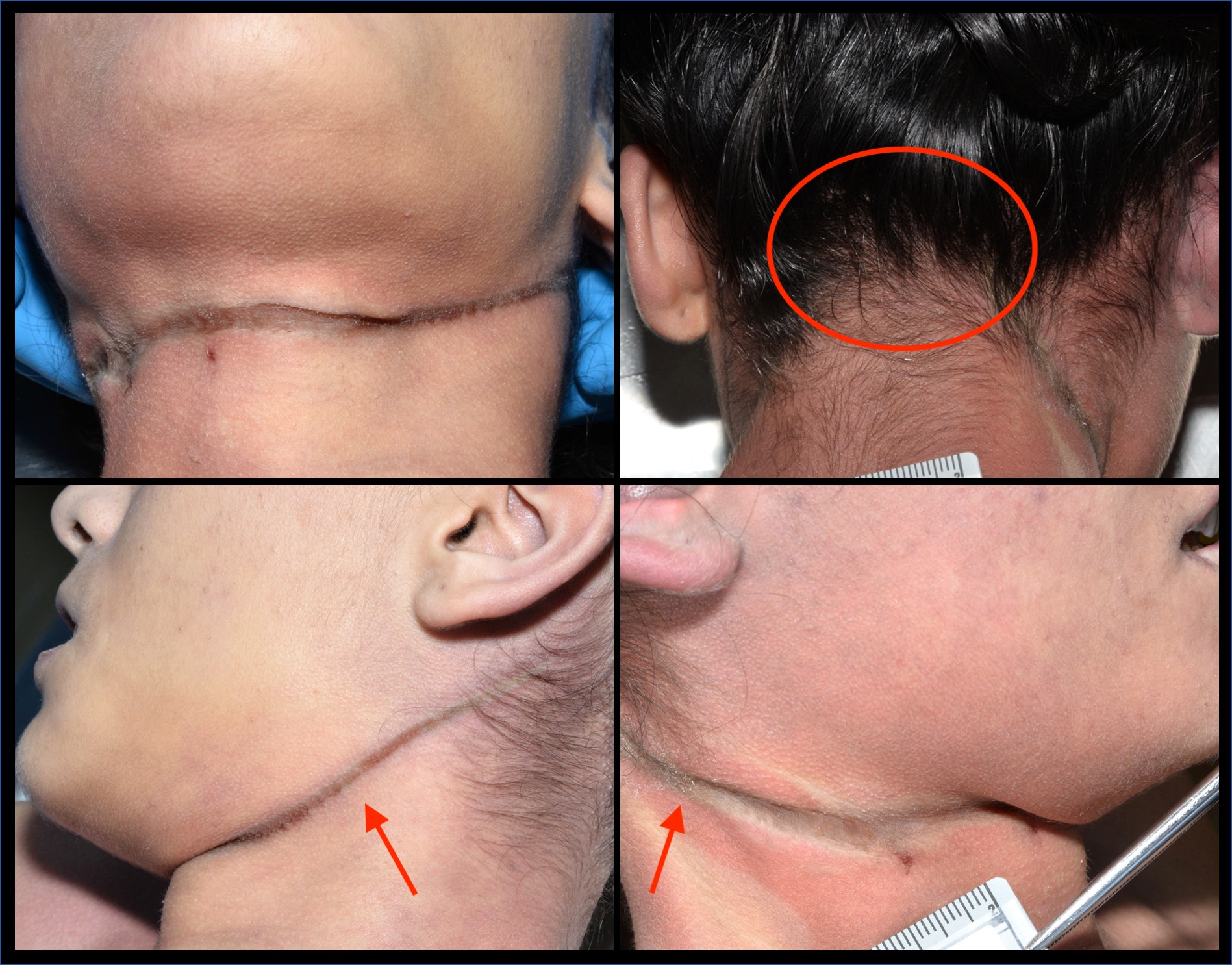
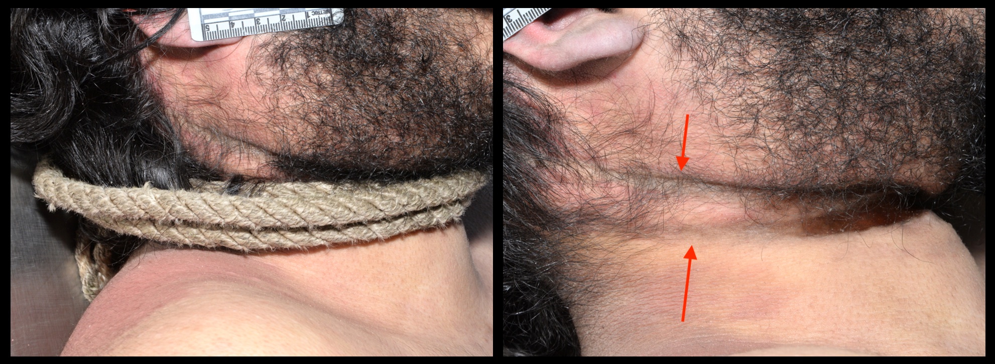
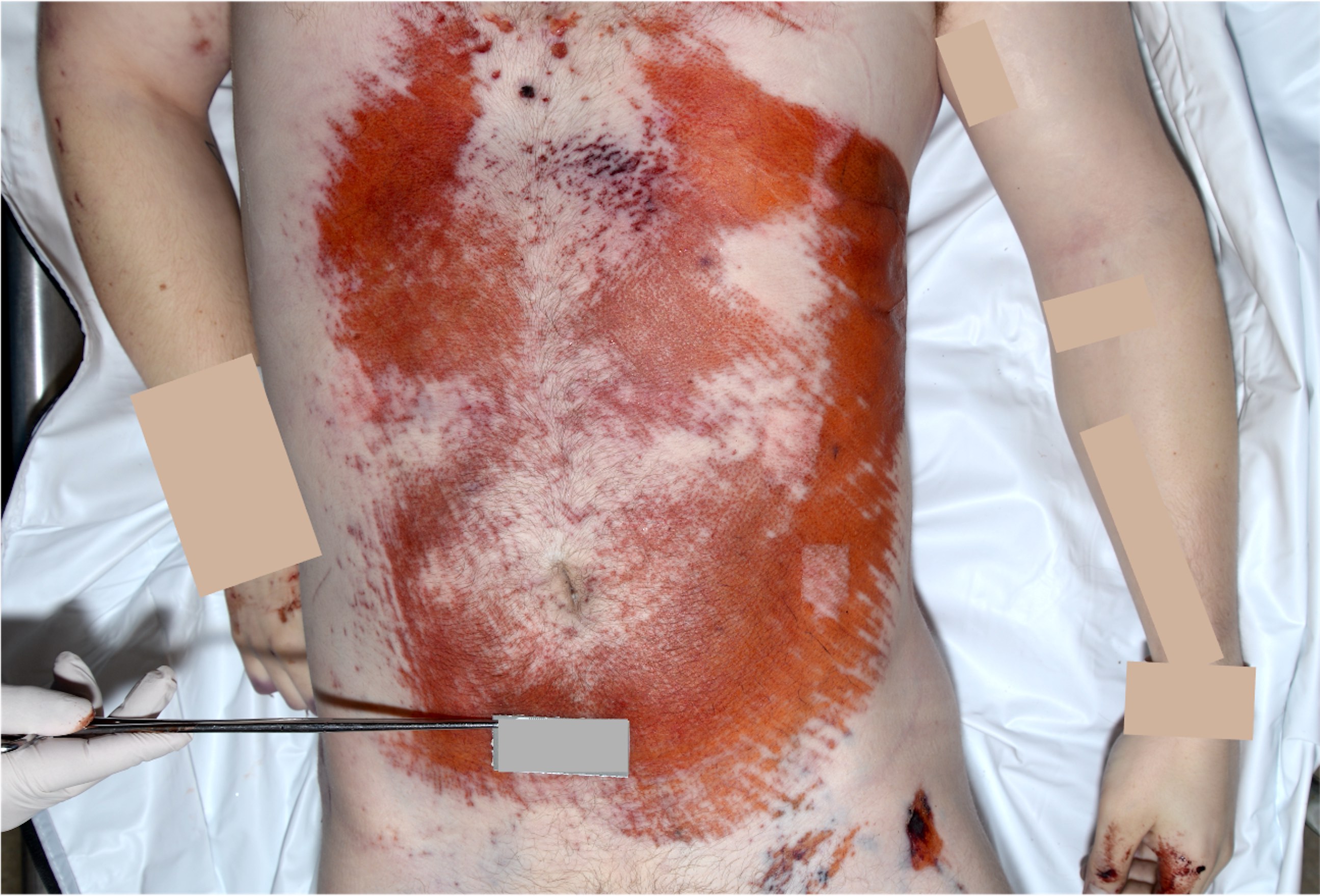
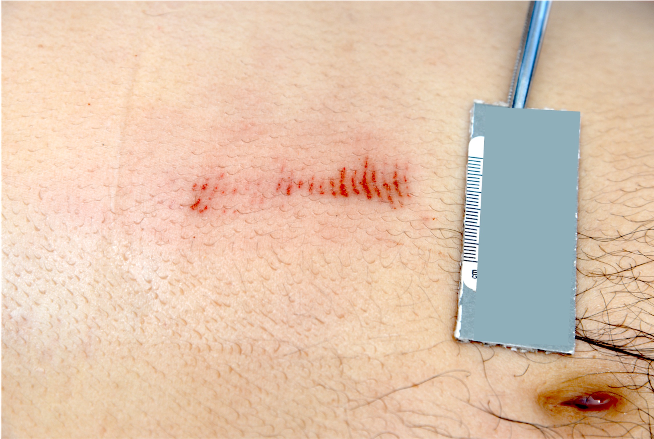
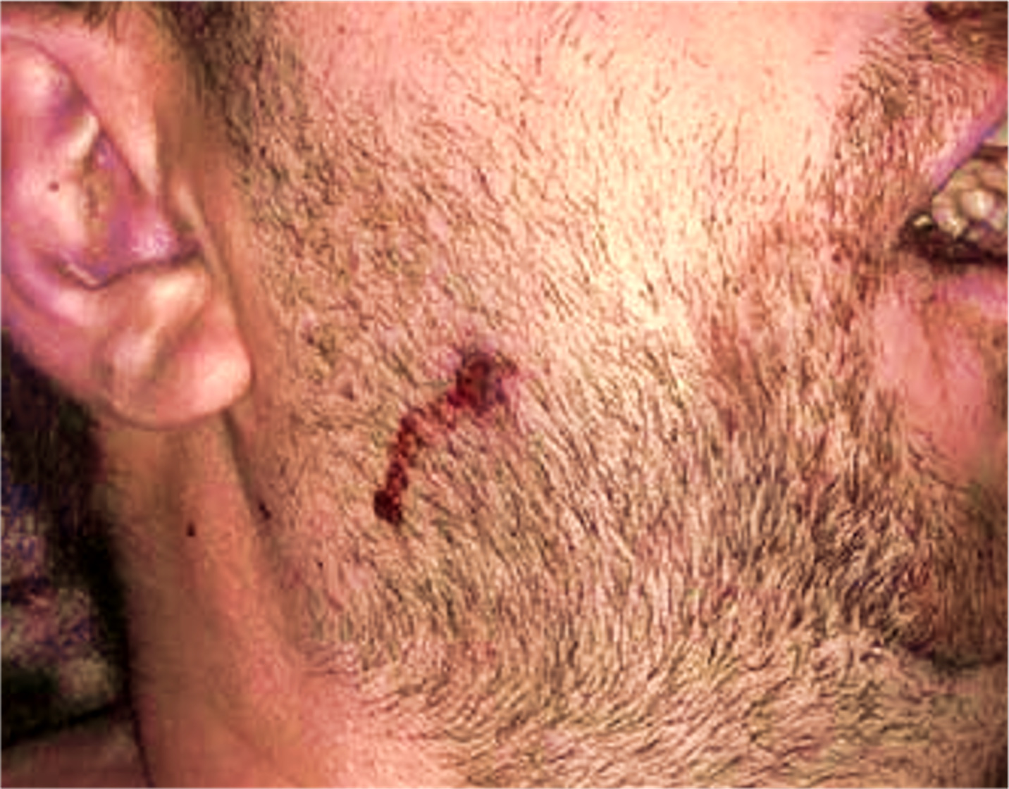
Ligature strangulation mark
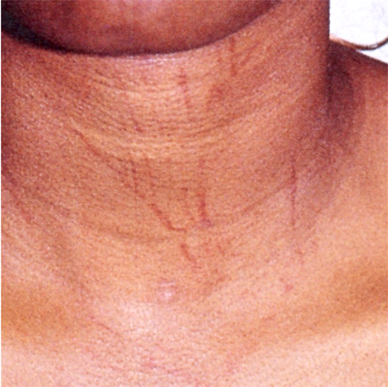
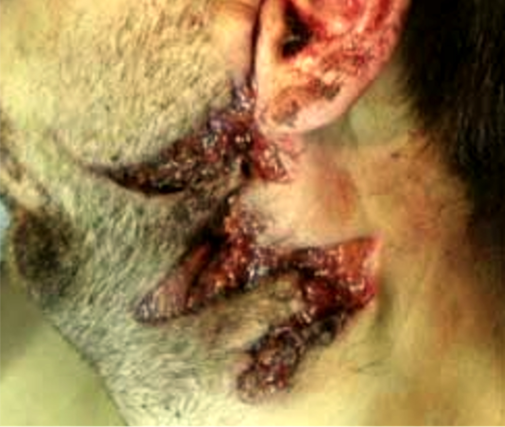
Multiple loop marks

Manual strangulation
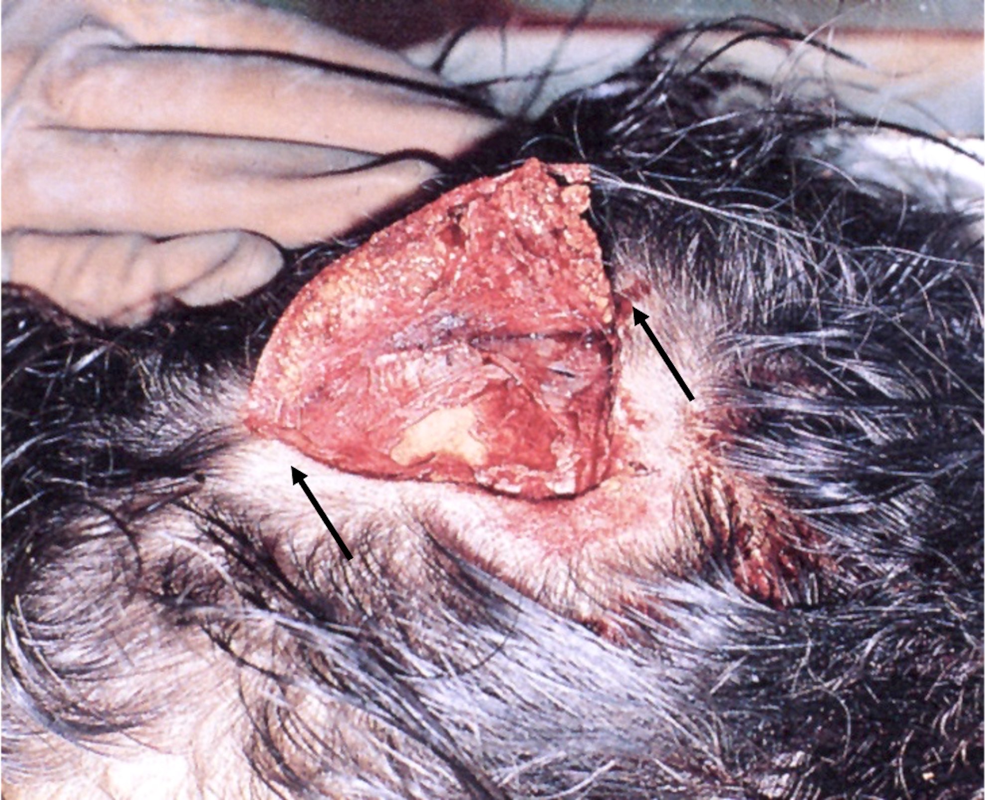
Simon sign
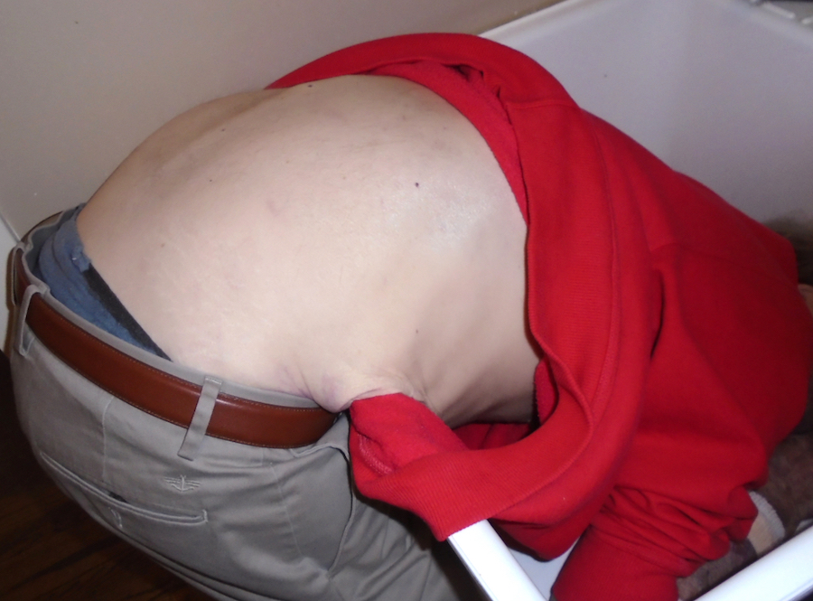
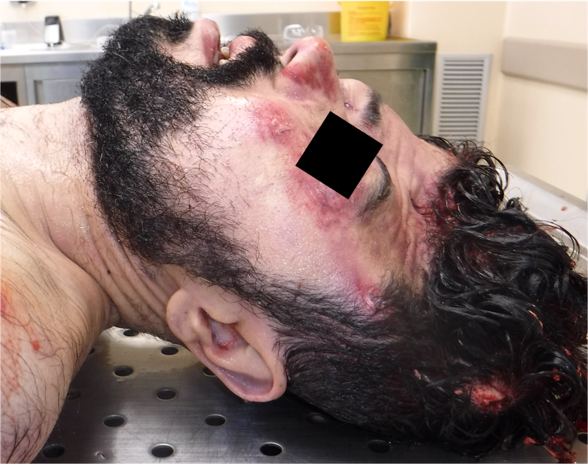
Positional asphyxia
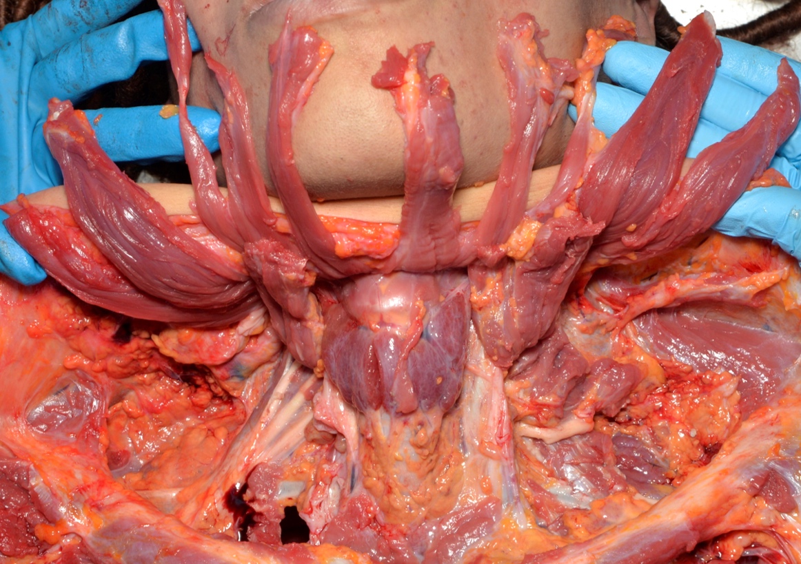
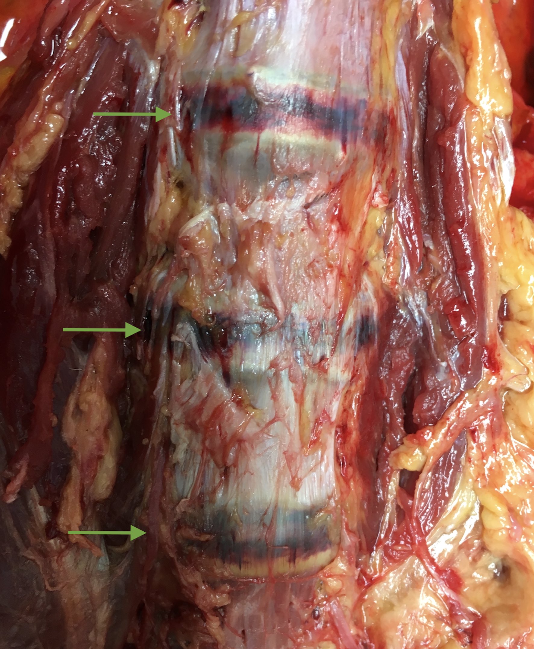
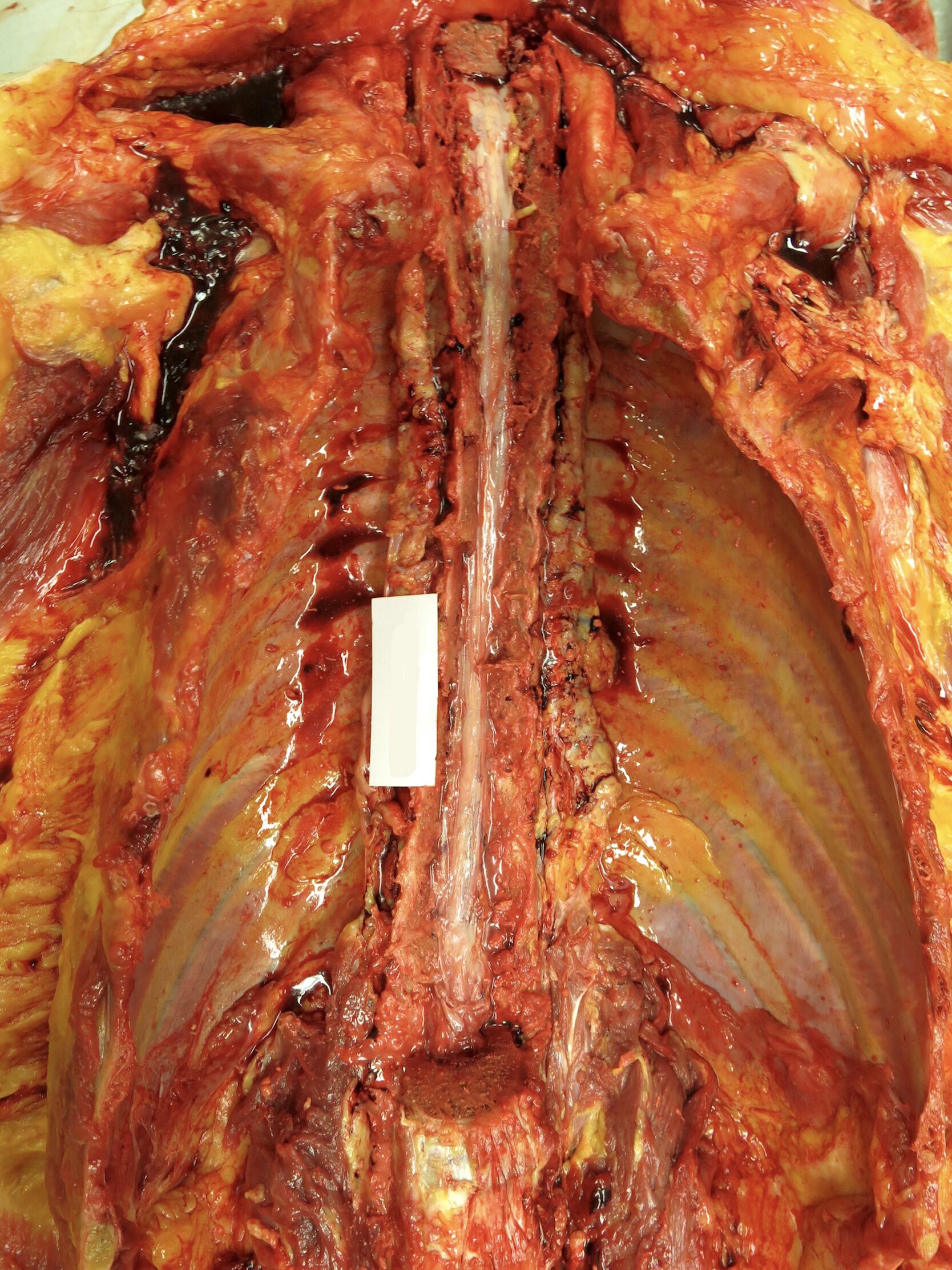
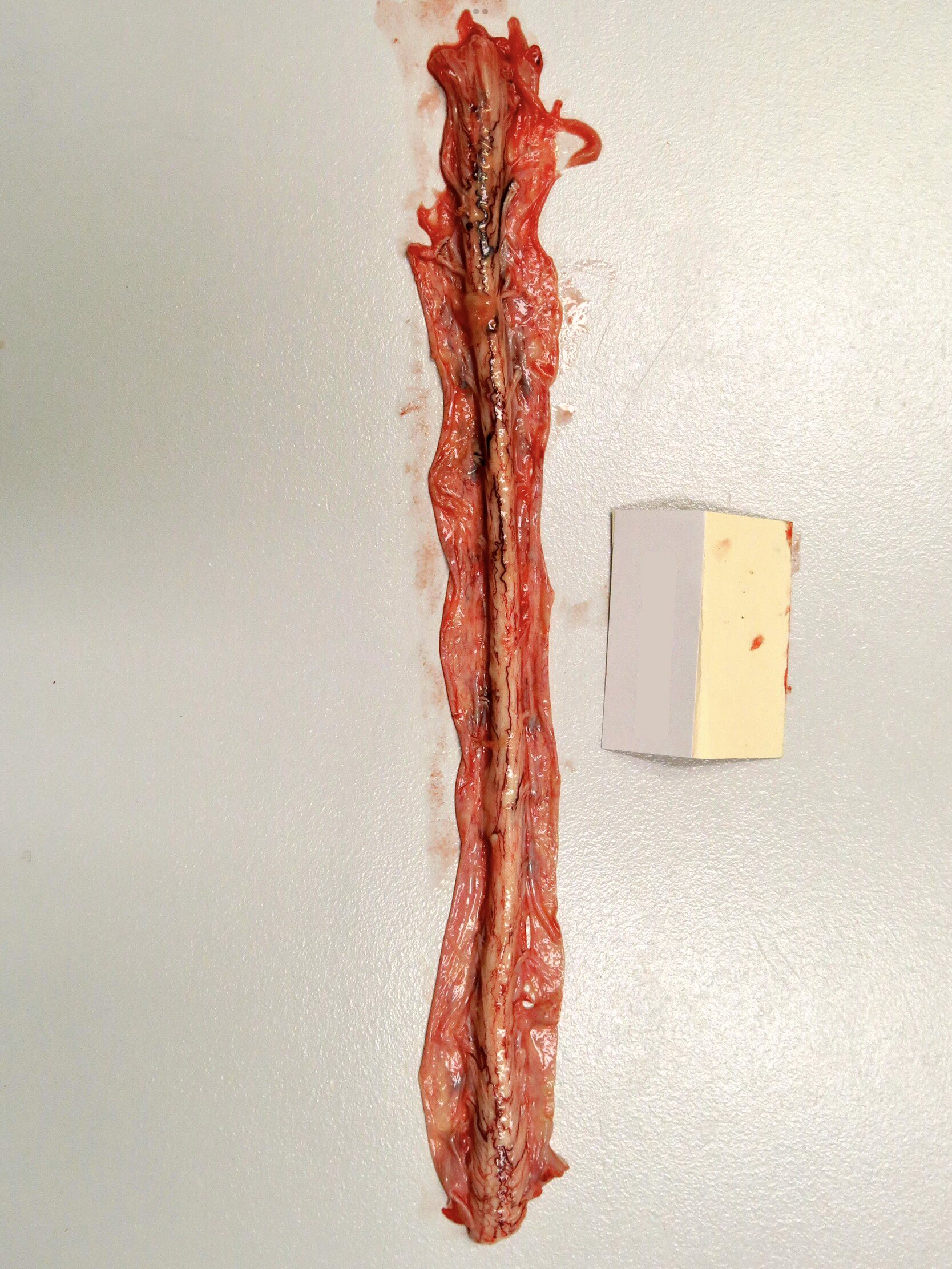
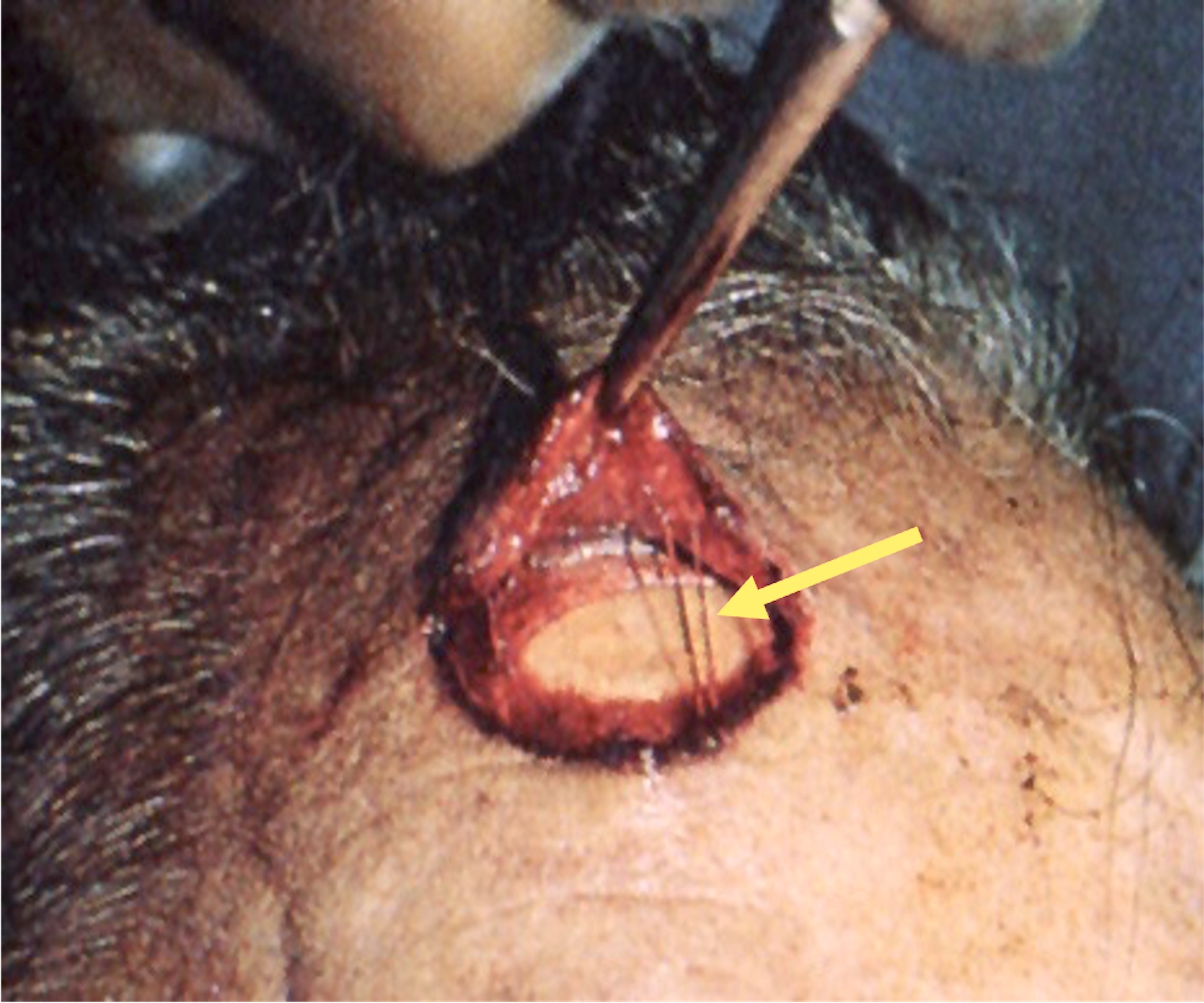
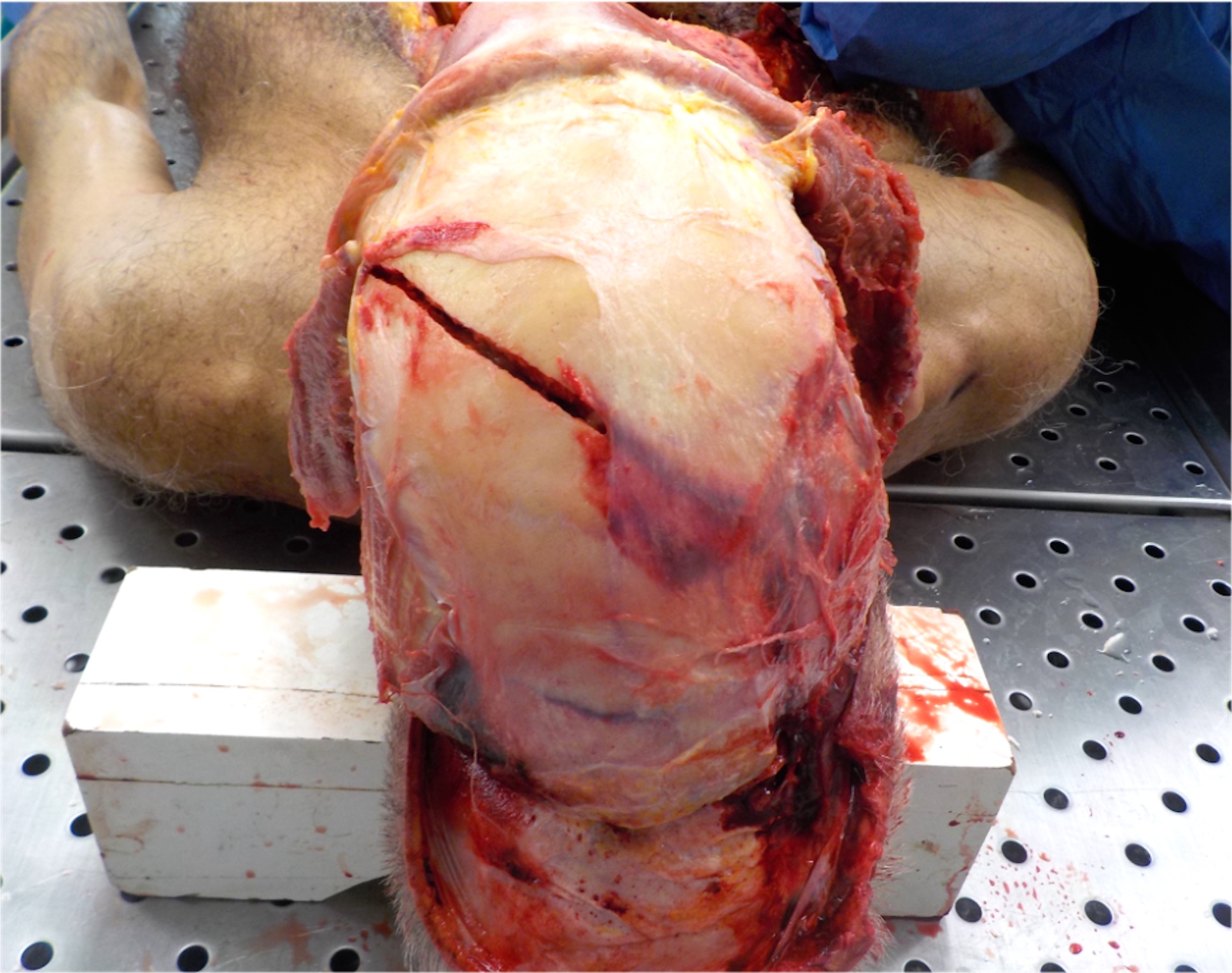
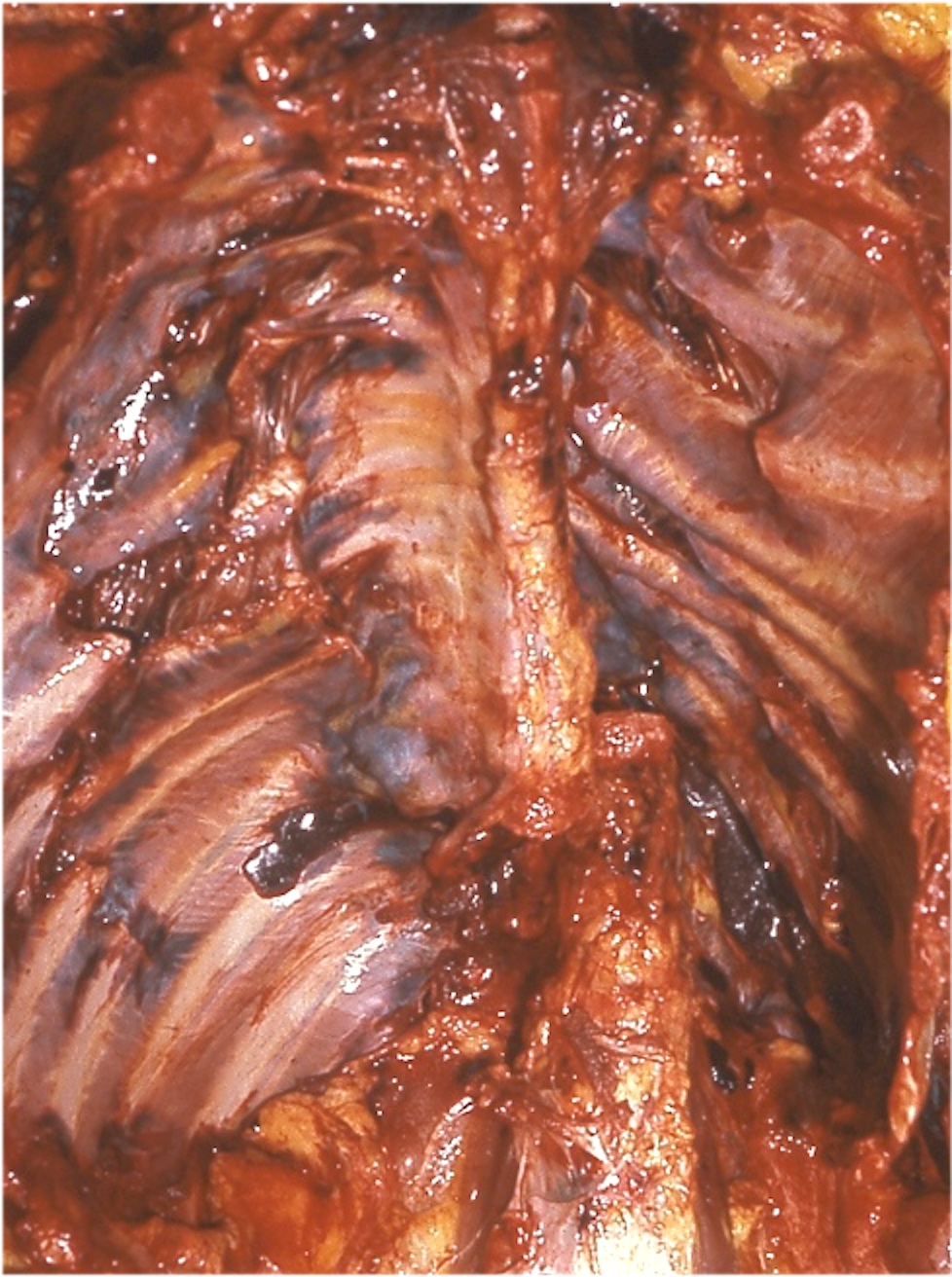
Neck dissection in asphyxia cases
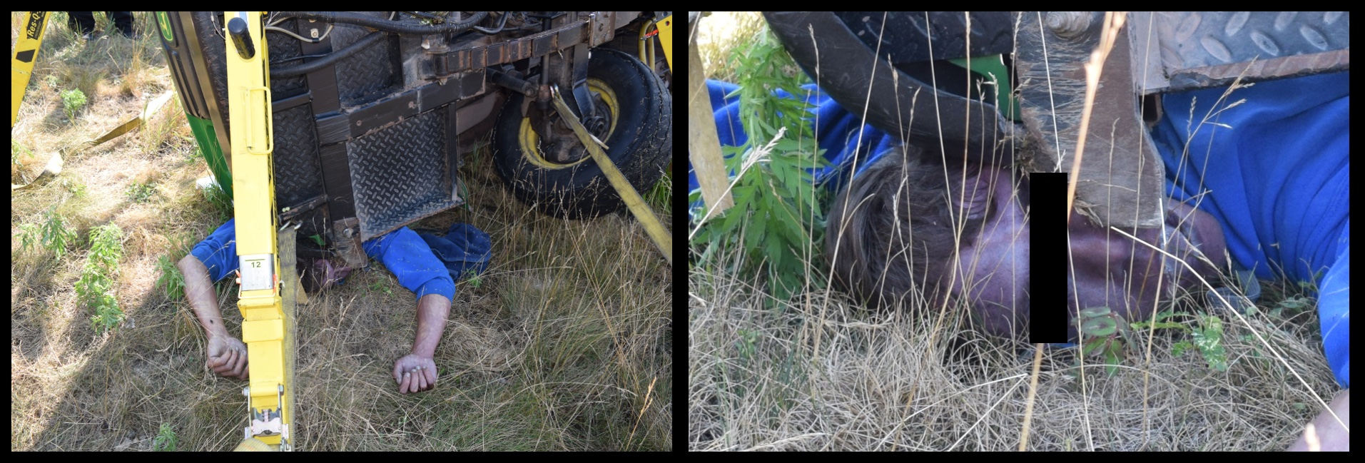
Crush asphyxia

Plastic bag suffocation
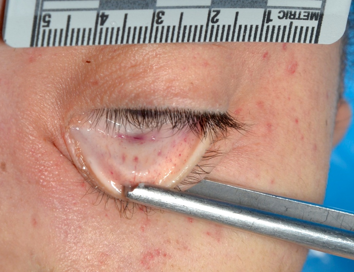
Conjunctival petechiae
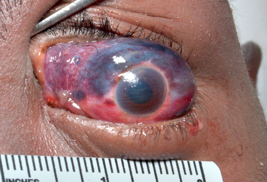
Eye hemorrhage
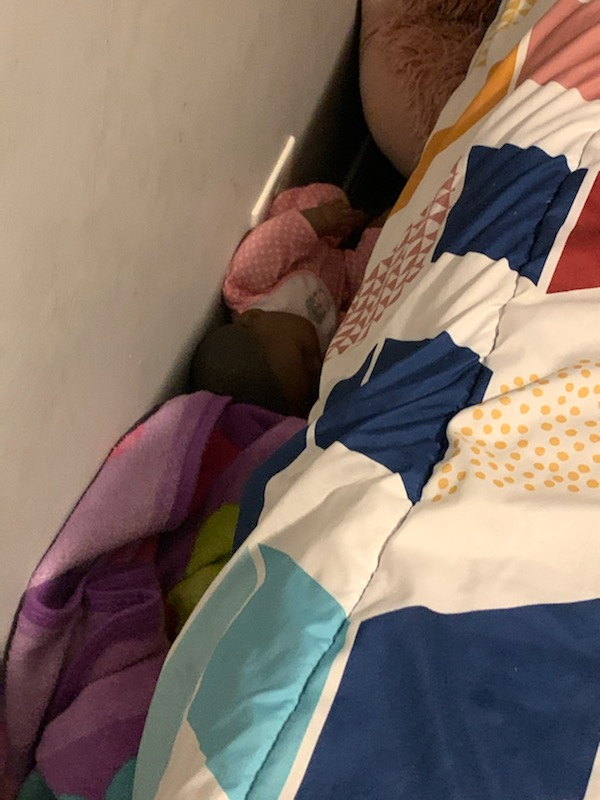
Wedging
Contributed by Lorenzo Gitto, M.D., Ponni Arunkumar, M.D. and the Cook County Medical Examiner's Office
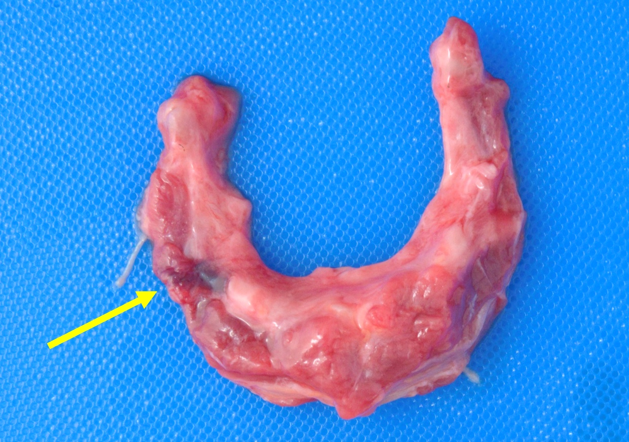
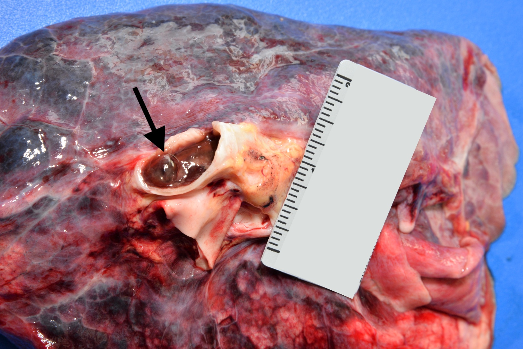
Hyoid bone hemorrhage
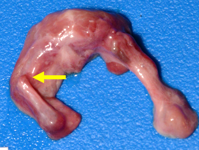
Hyoid bone fracture
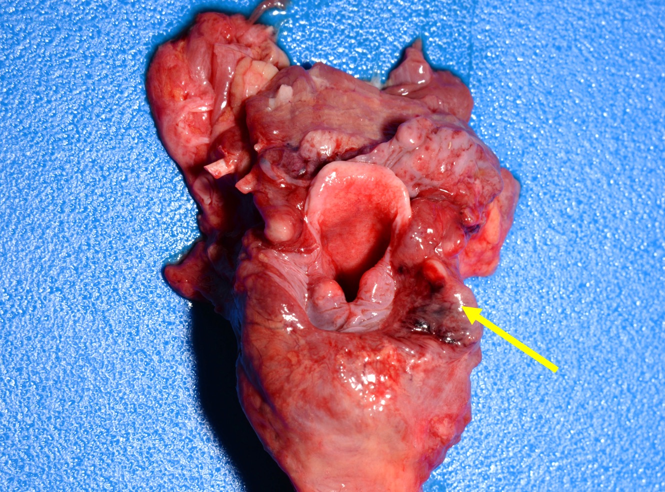
Larynx hemorrhage

Choking
Aspiration


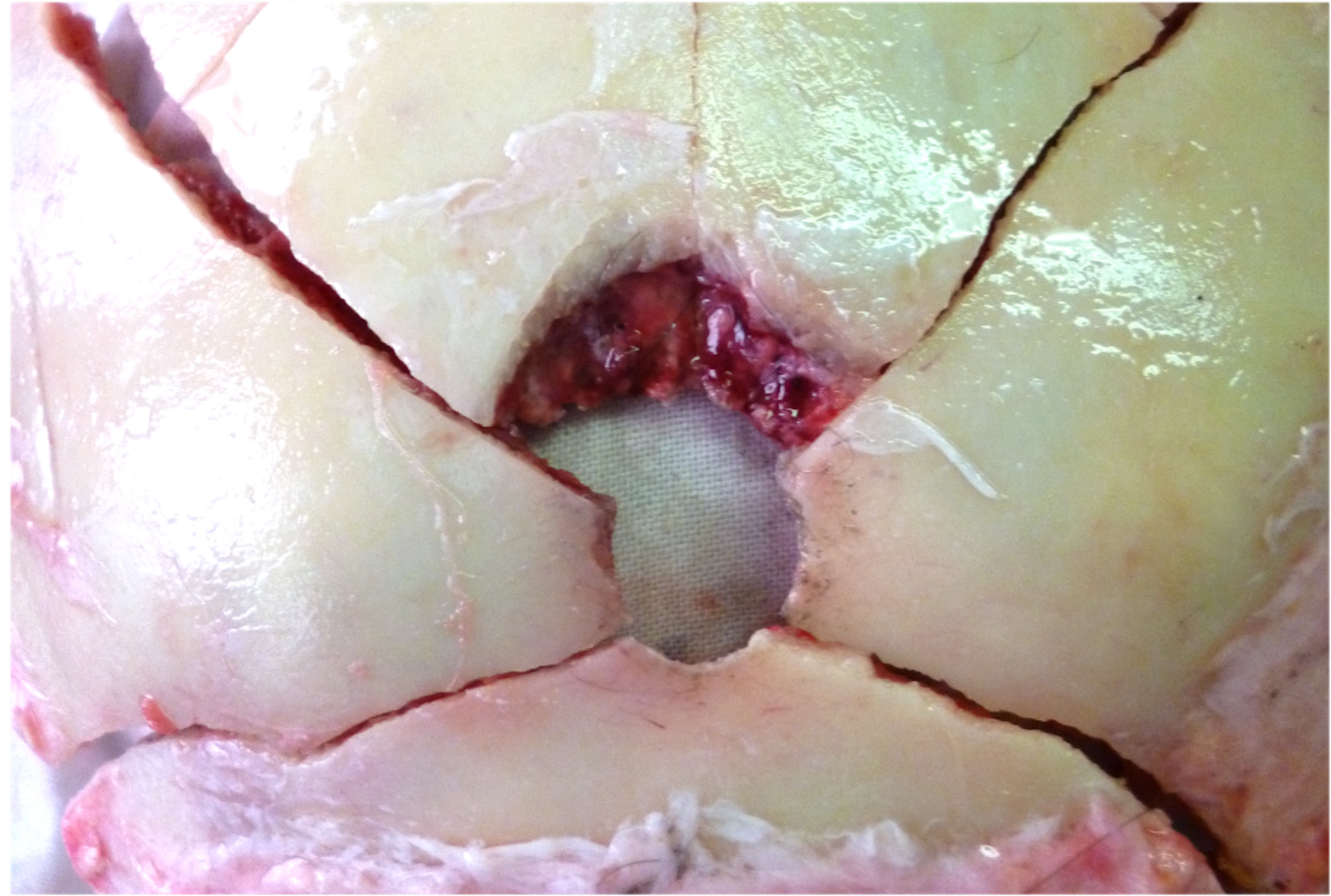
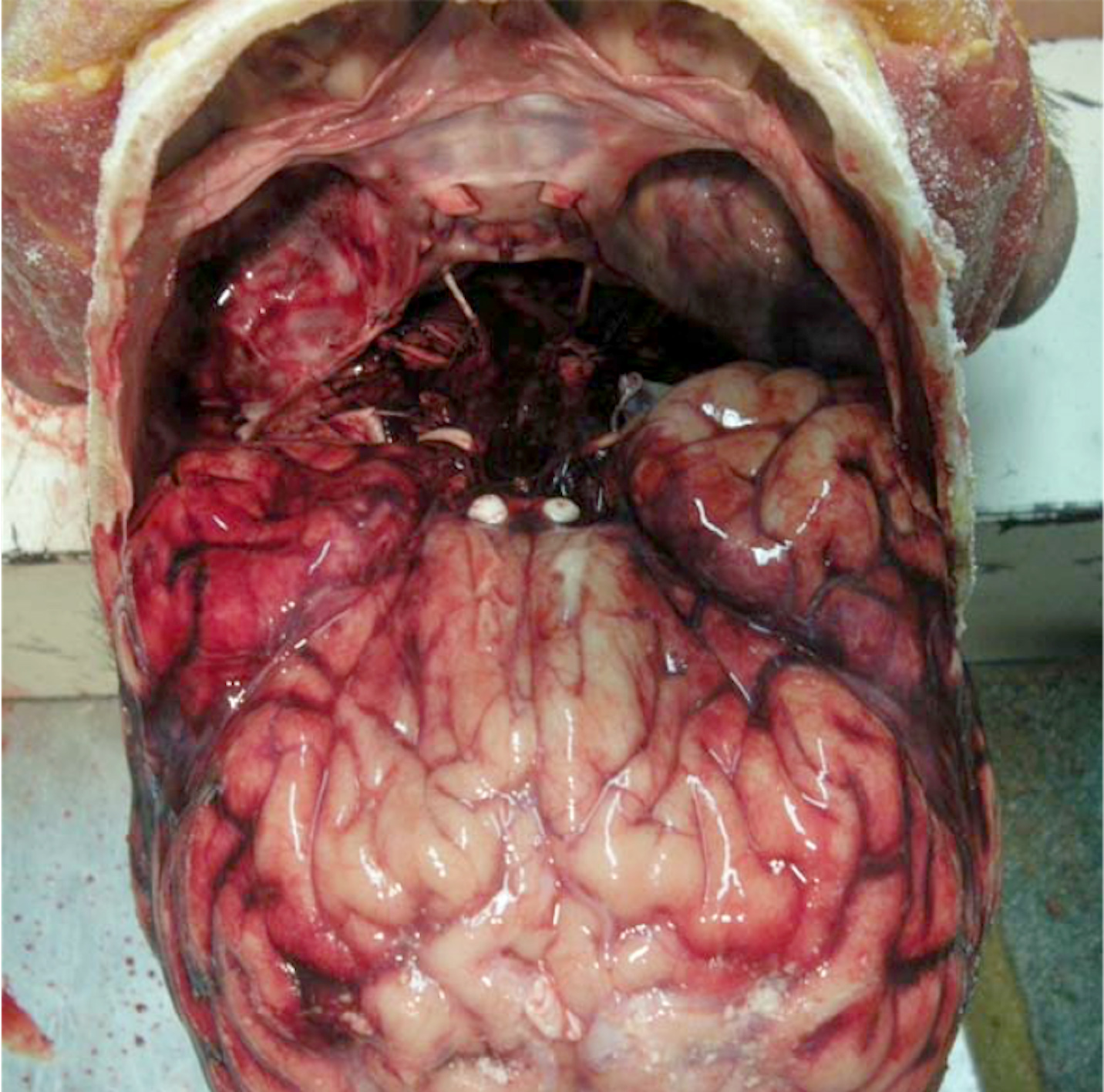
Opening of the skull and removal of the brain
Removal of the orbital contents
Full body autopsy, including the brain, with modified Virchows technique
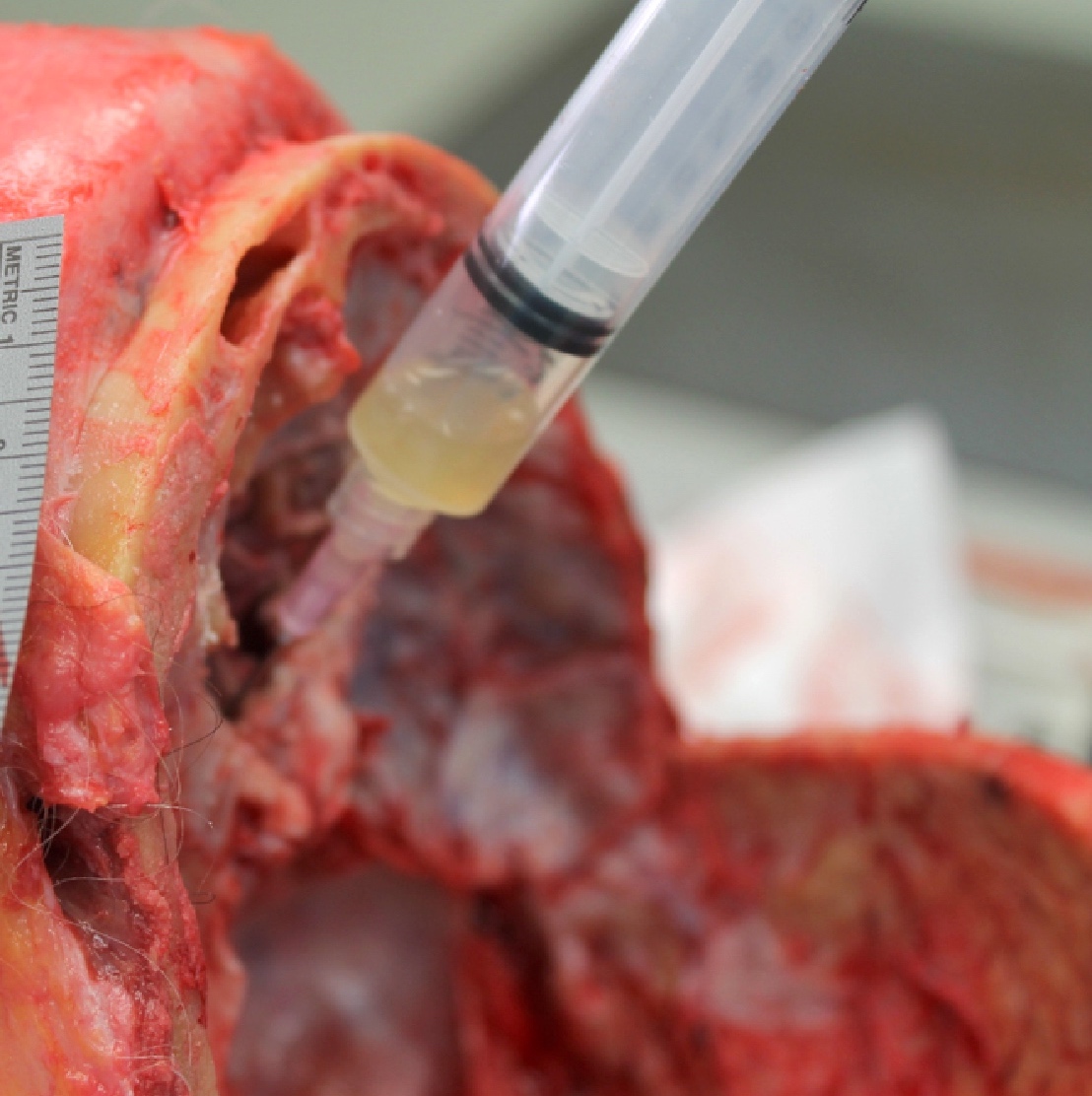
Simple method to check patency of the biliary system
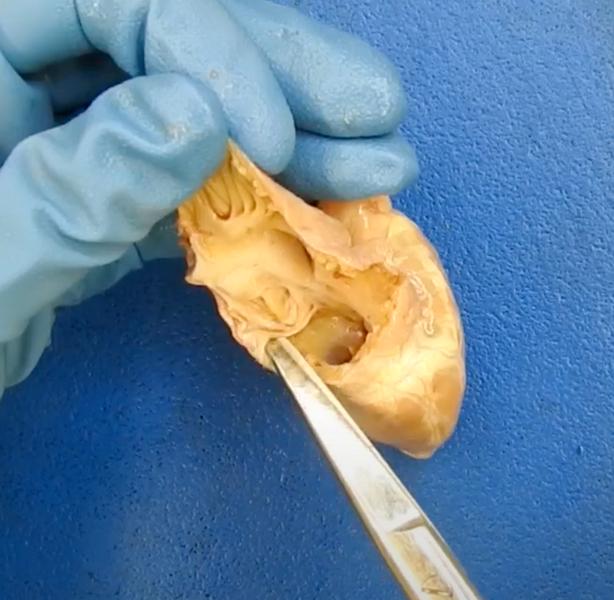
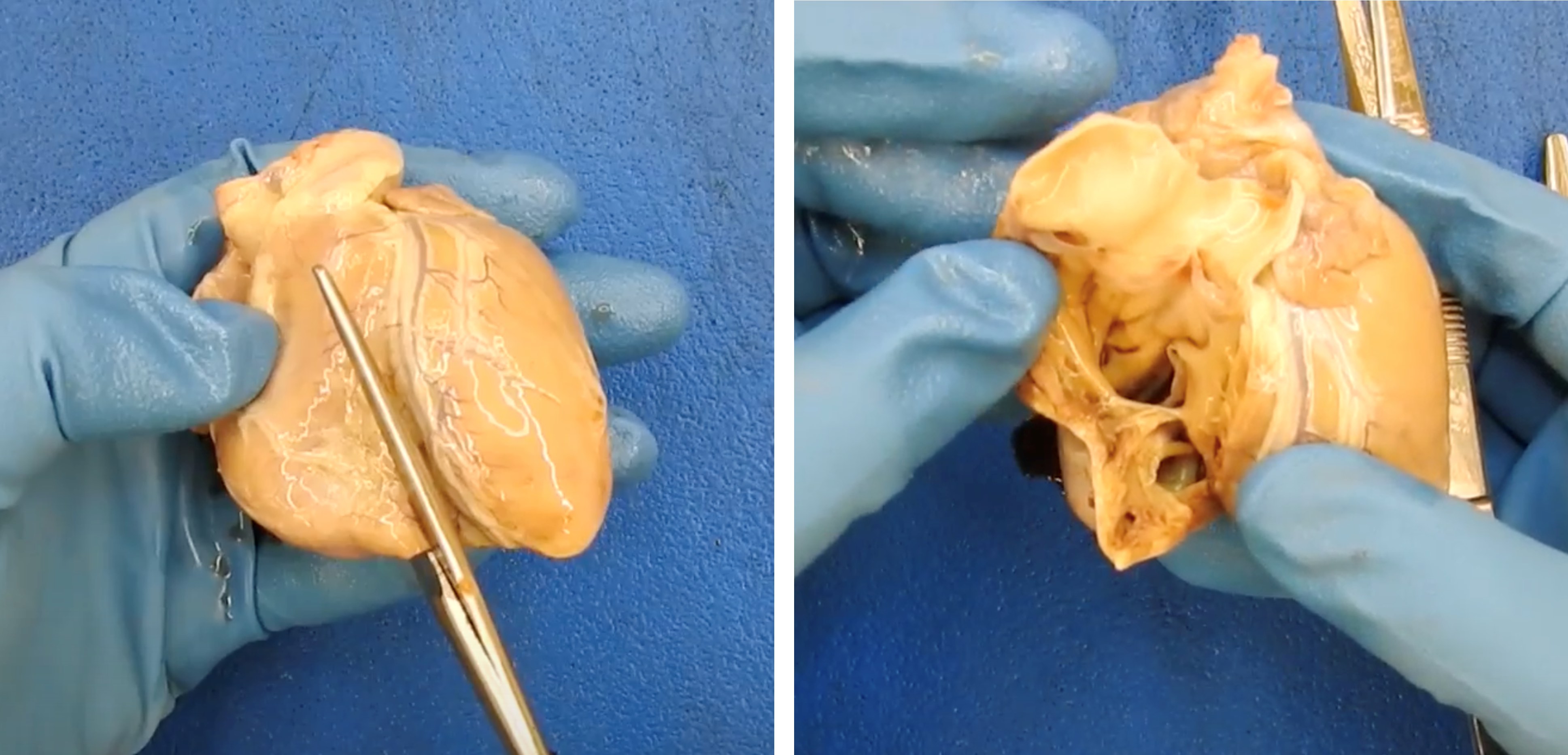
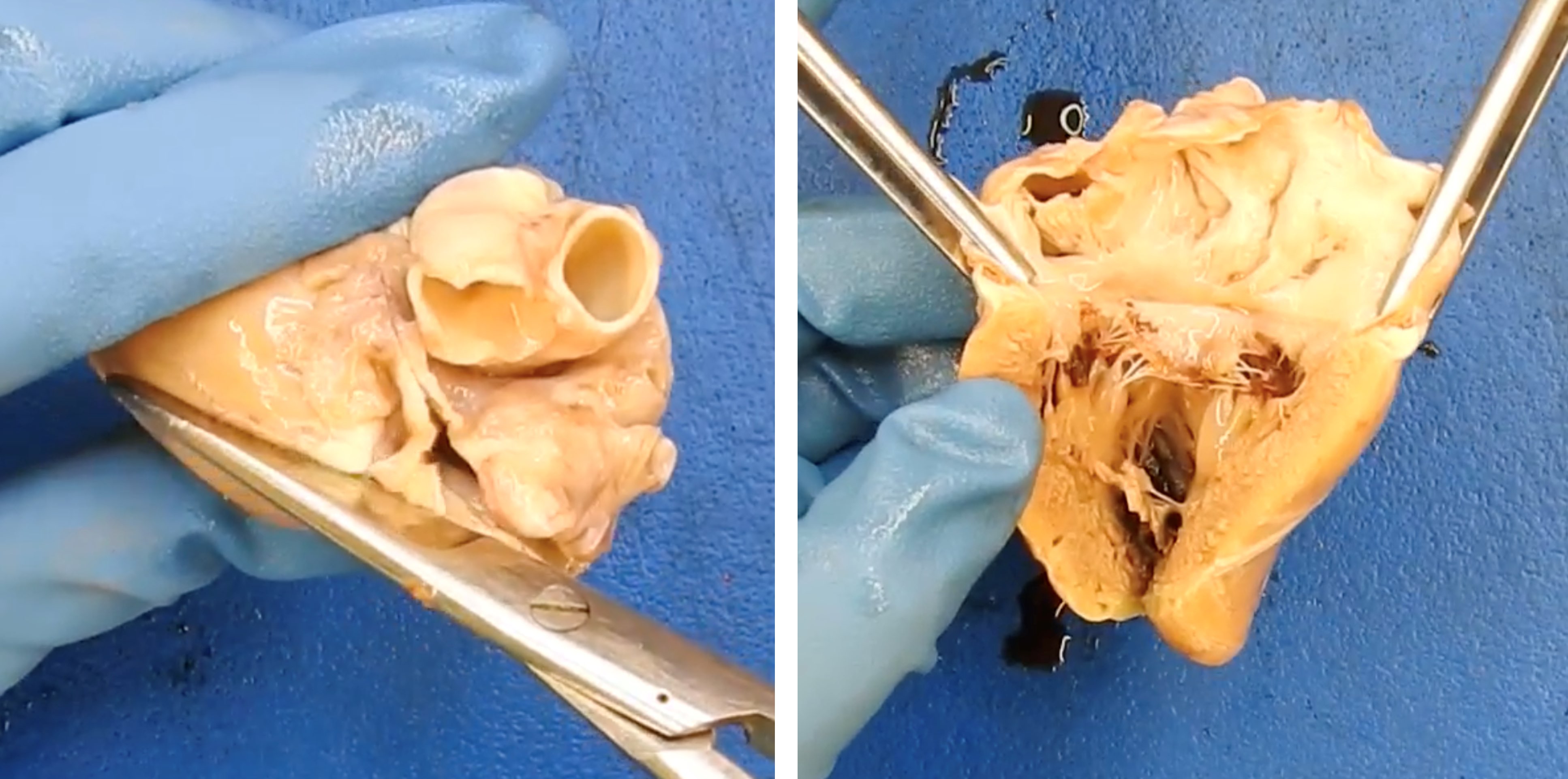
Dissection of the heart (after separation from the body)
Contributed by Lorenzo Gitto, M.D. and Cook County Medical Examiner's Office
Blood flow method
Right atrium

Right ventricle
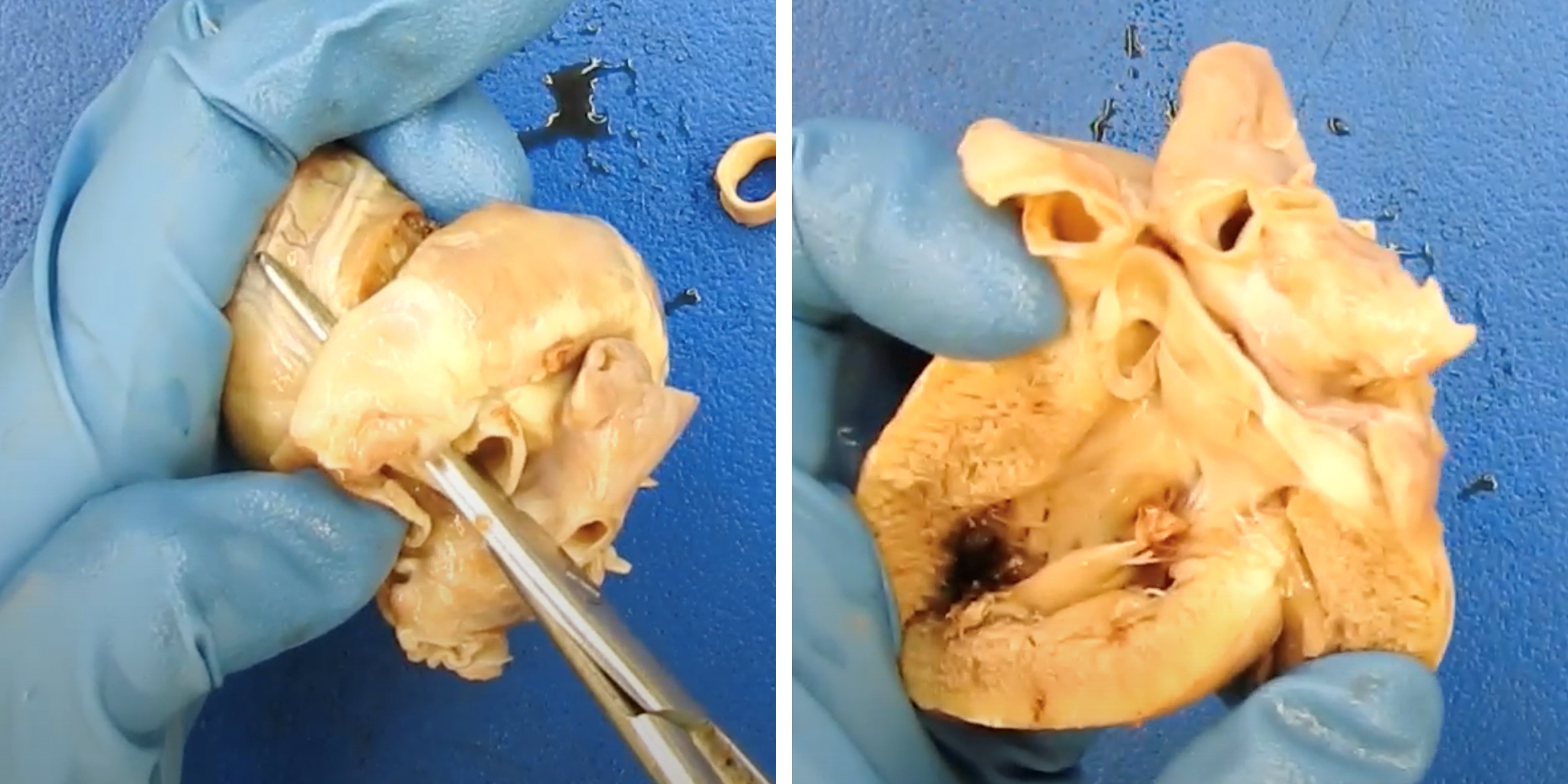
Right ventricle outflow tract and pulmonic valve

Left atrium
Left ventricle
Left ventricle outflow tract


Base of the heart method
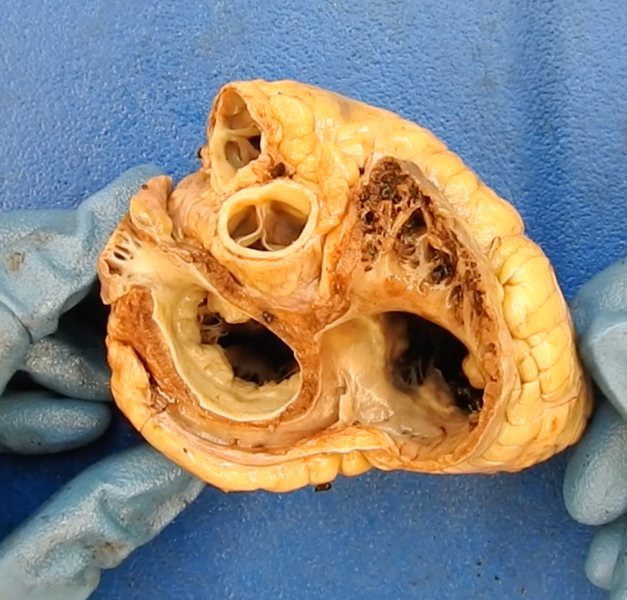
Valve plane
4 chamber method
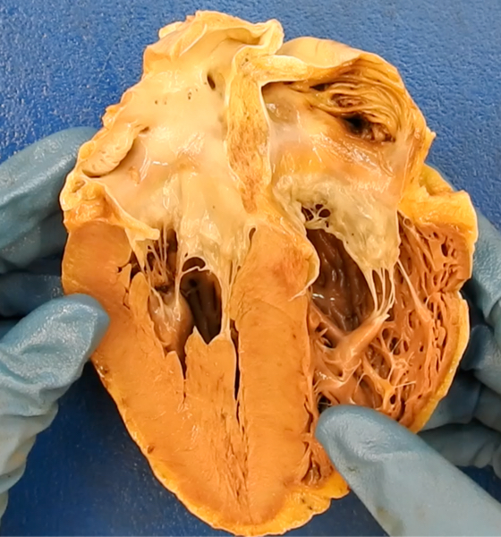
4 heart chambers
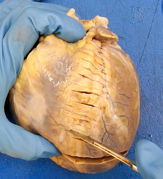
Coronary artery serial sectioning method

Separating vessels at base of heart
Left anterior descending coronary artery
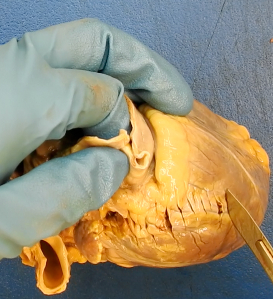
Left circumflex coronary artery

Right coronary artery
Coronary artery removal method

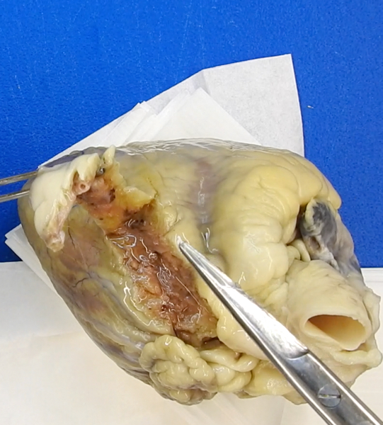
Left anterior descending coronary artery

Left circumflex coronary artery

Right coronary artery

Dissected coronaries
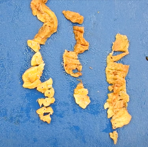
Serial sectioning of the coronaries
Contributed by Lorenzo Gitto, M.D., Robert Stoppacher, M.D., Serenella Serinelli M.D., Ponni Arunkumar, M.D. and Luigi Bonaccorso, M.D.

Abrasion
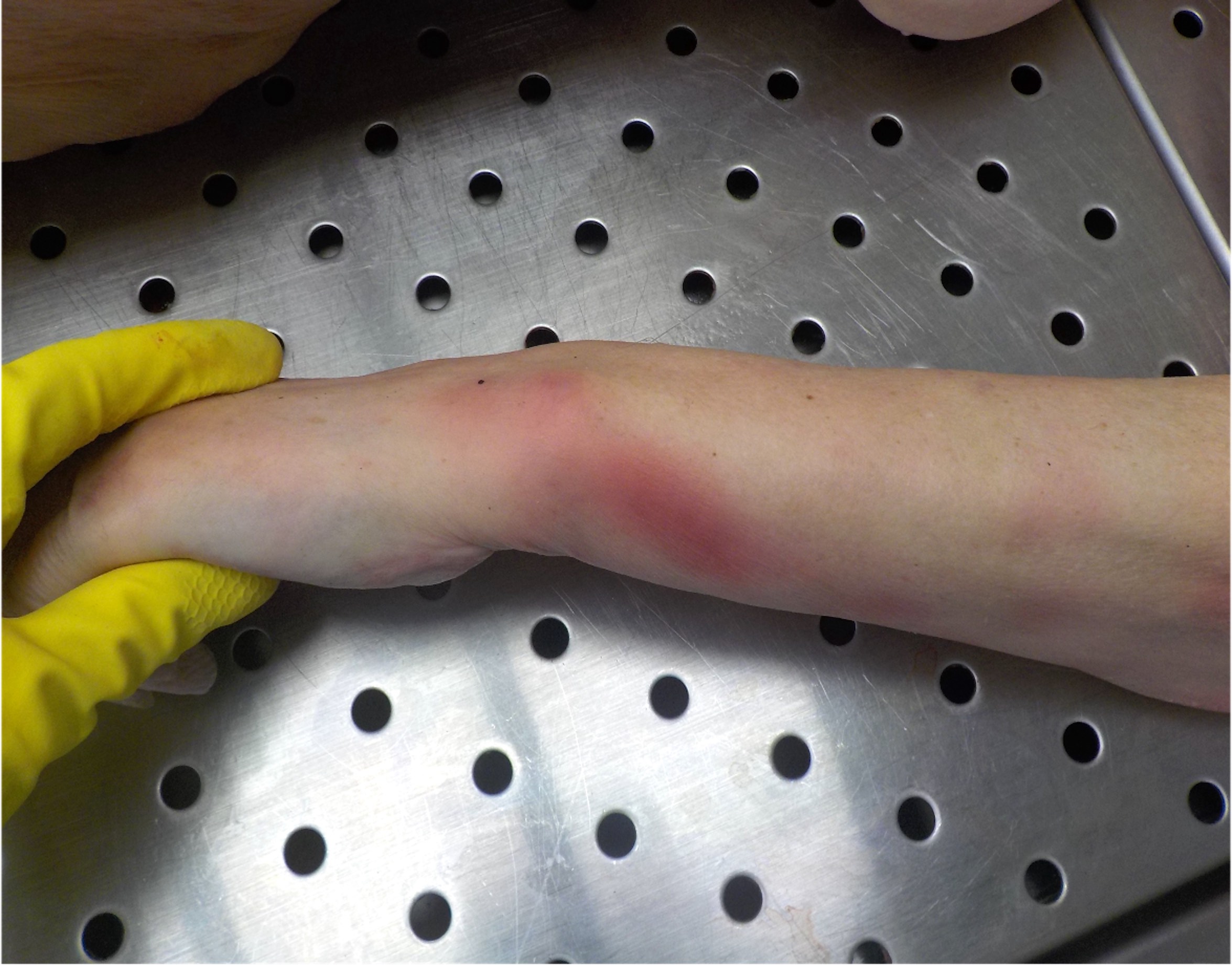
Bruise
Laceration
Fracture
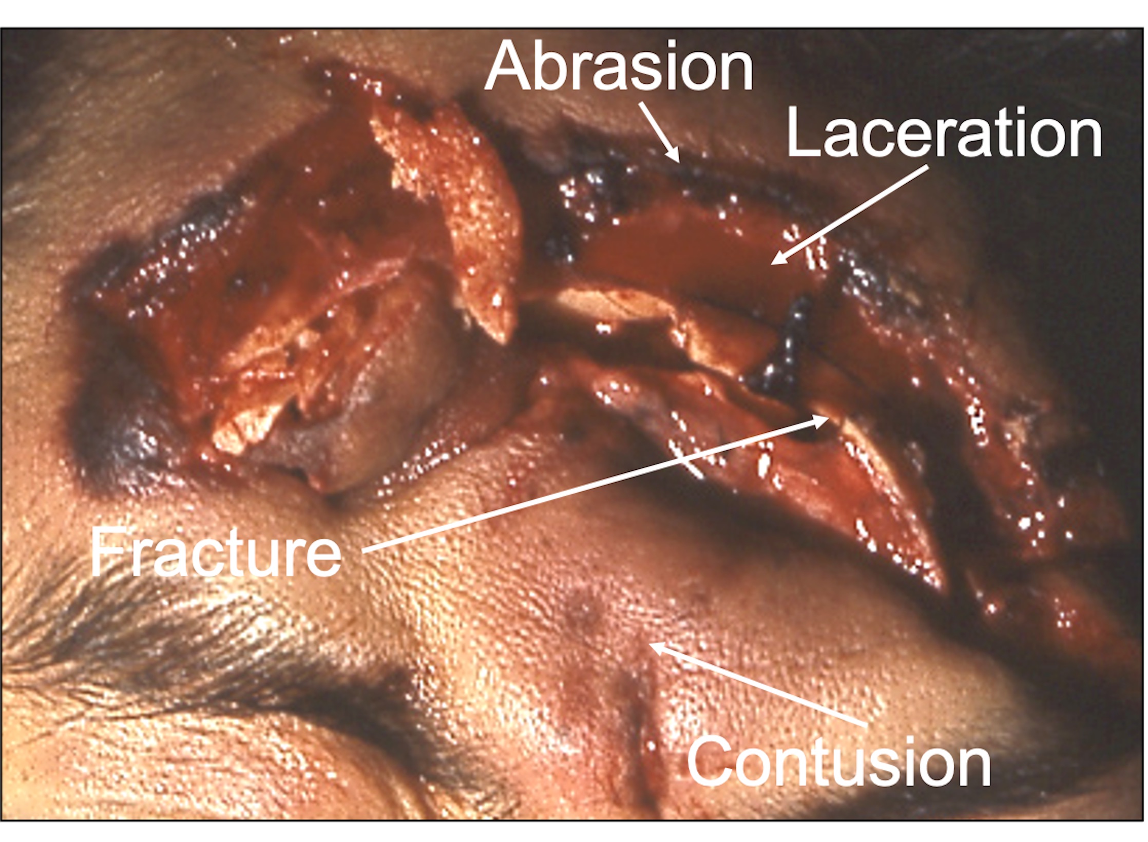
Mixed wound
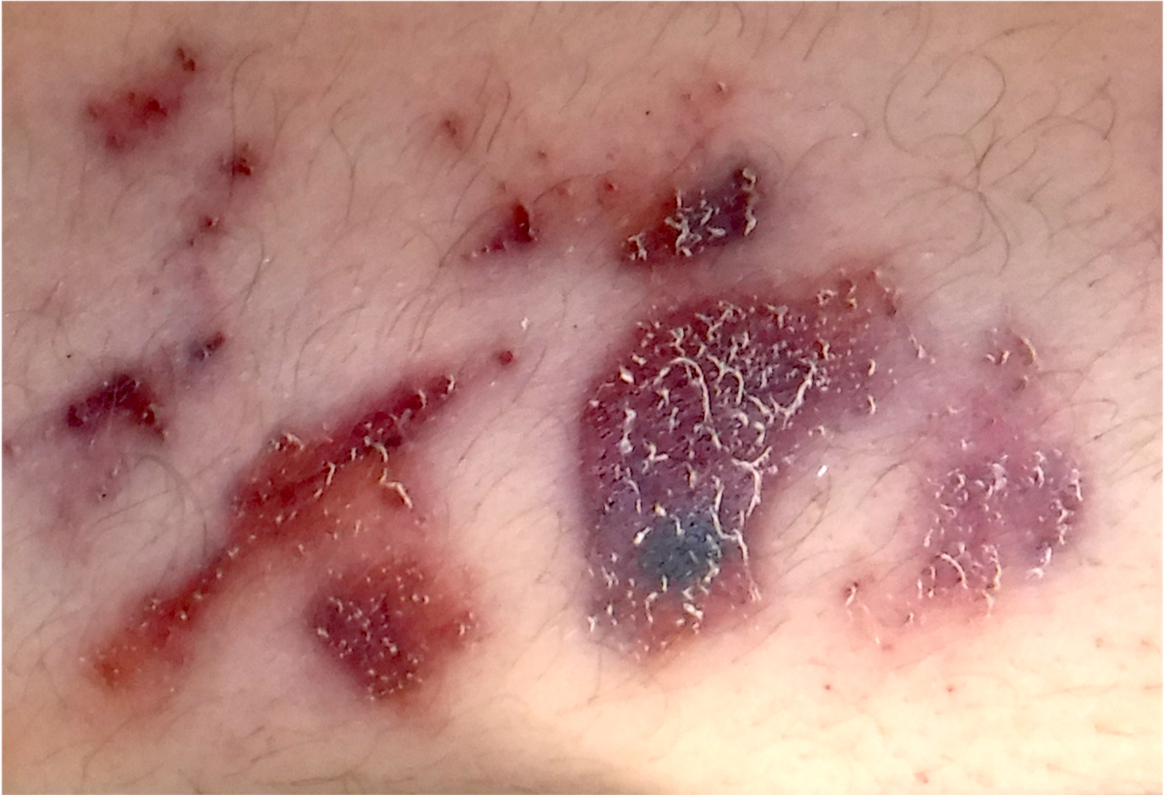
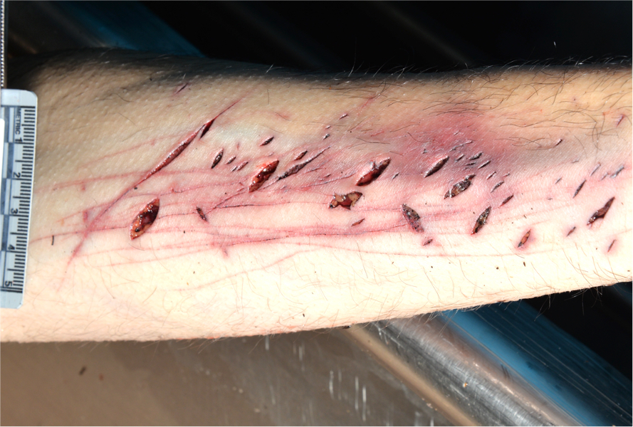
Superficial abrasion with skin tags
Road rash

Patterned abrasions
Fingernail abrasions
Pressure abrasion
Postmortem abrasions

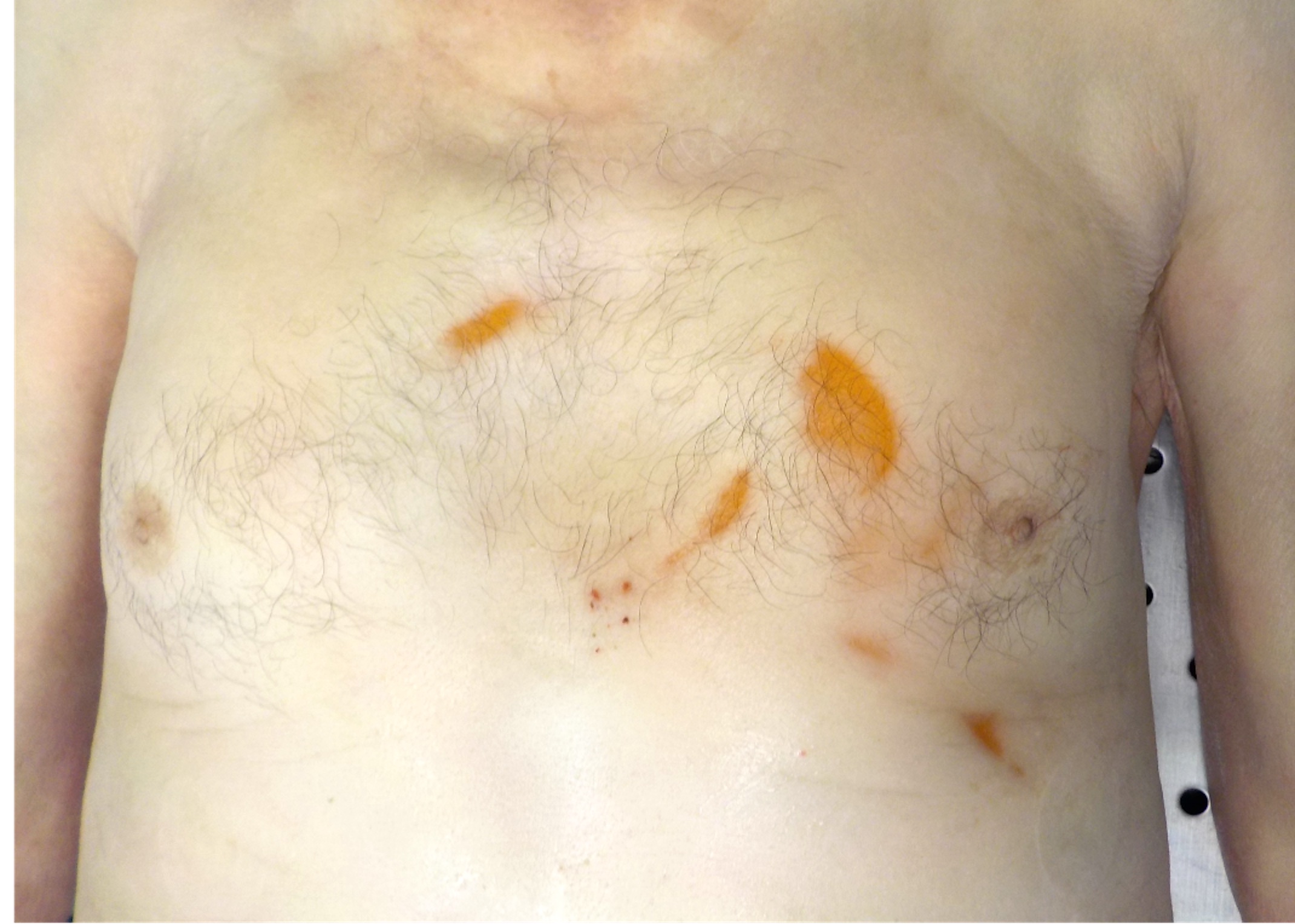
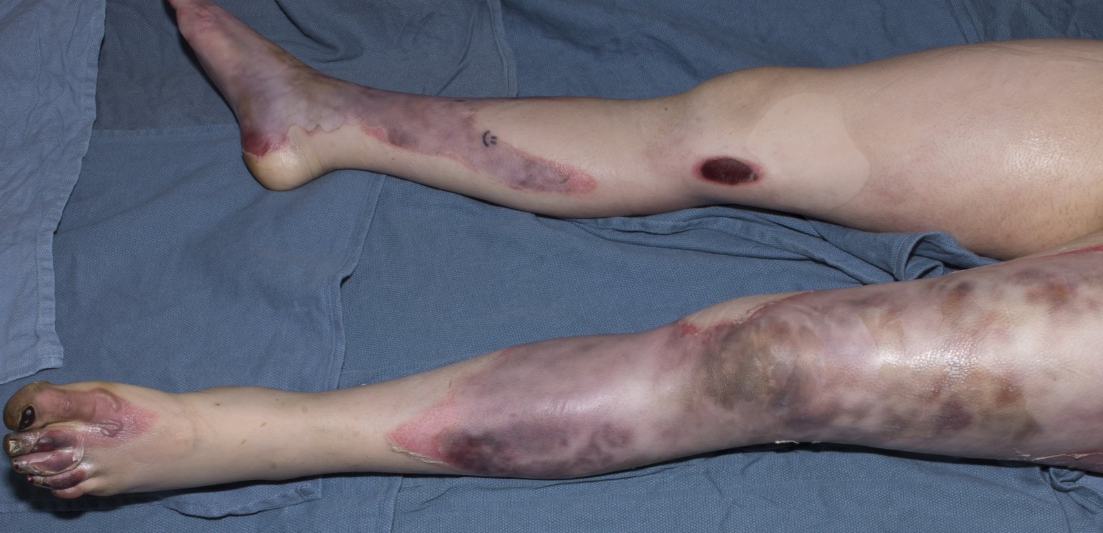
Ecchymosis
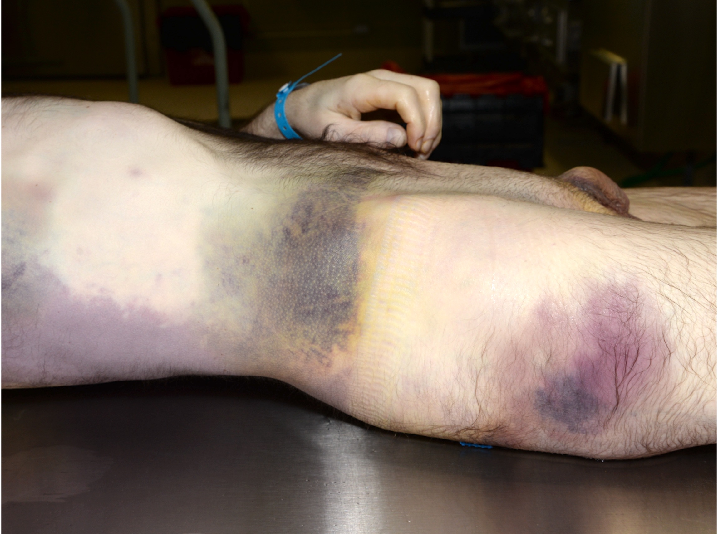
Hematoma
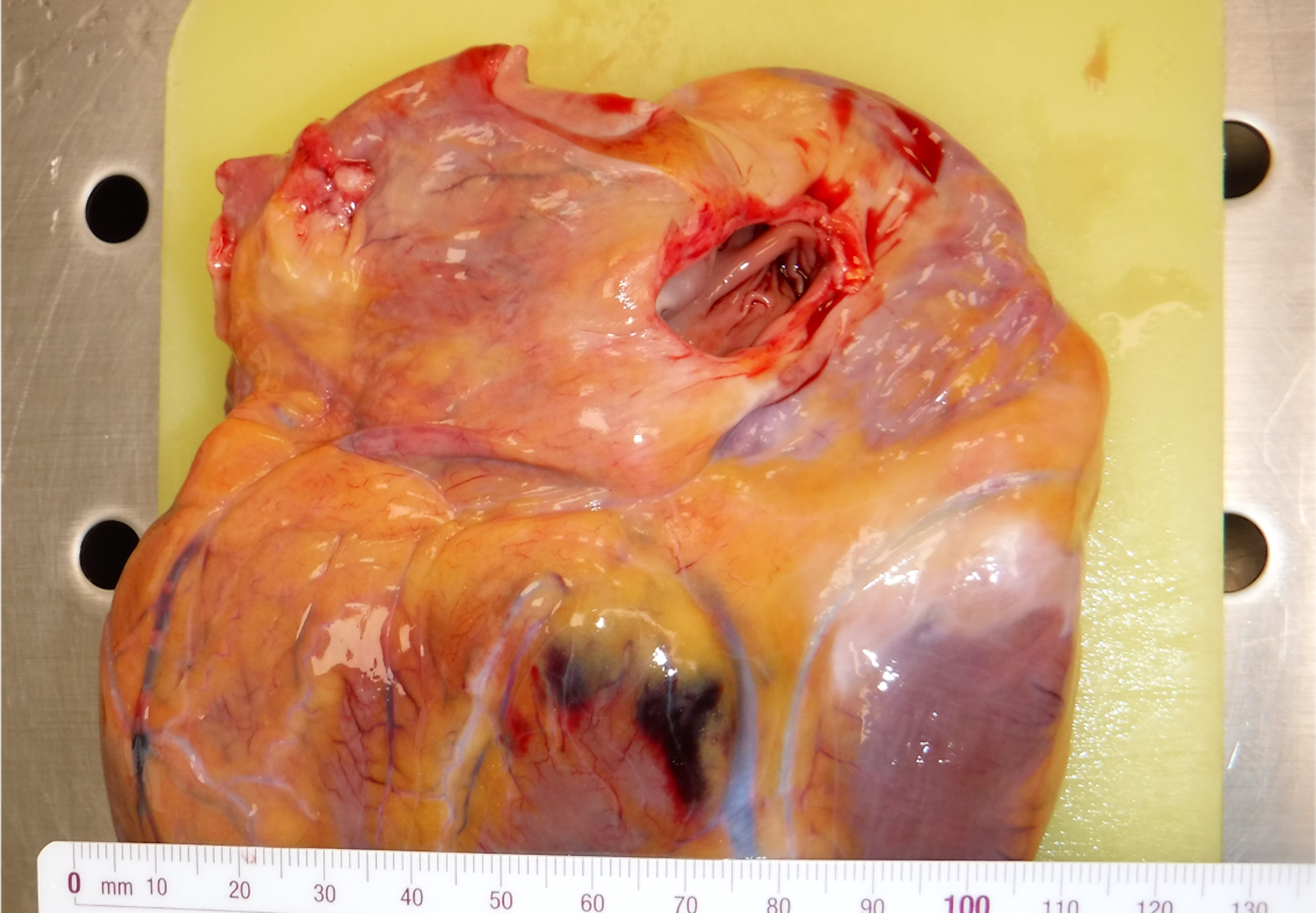
Visceral contusion

Tram-like bruise
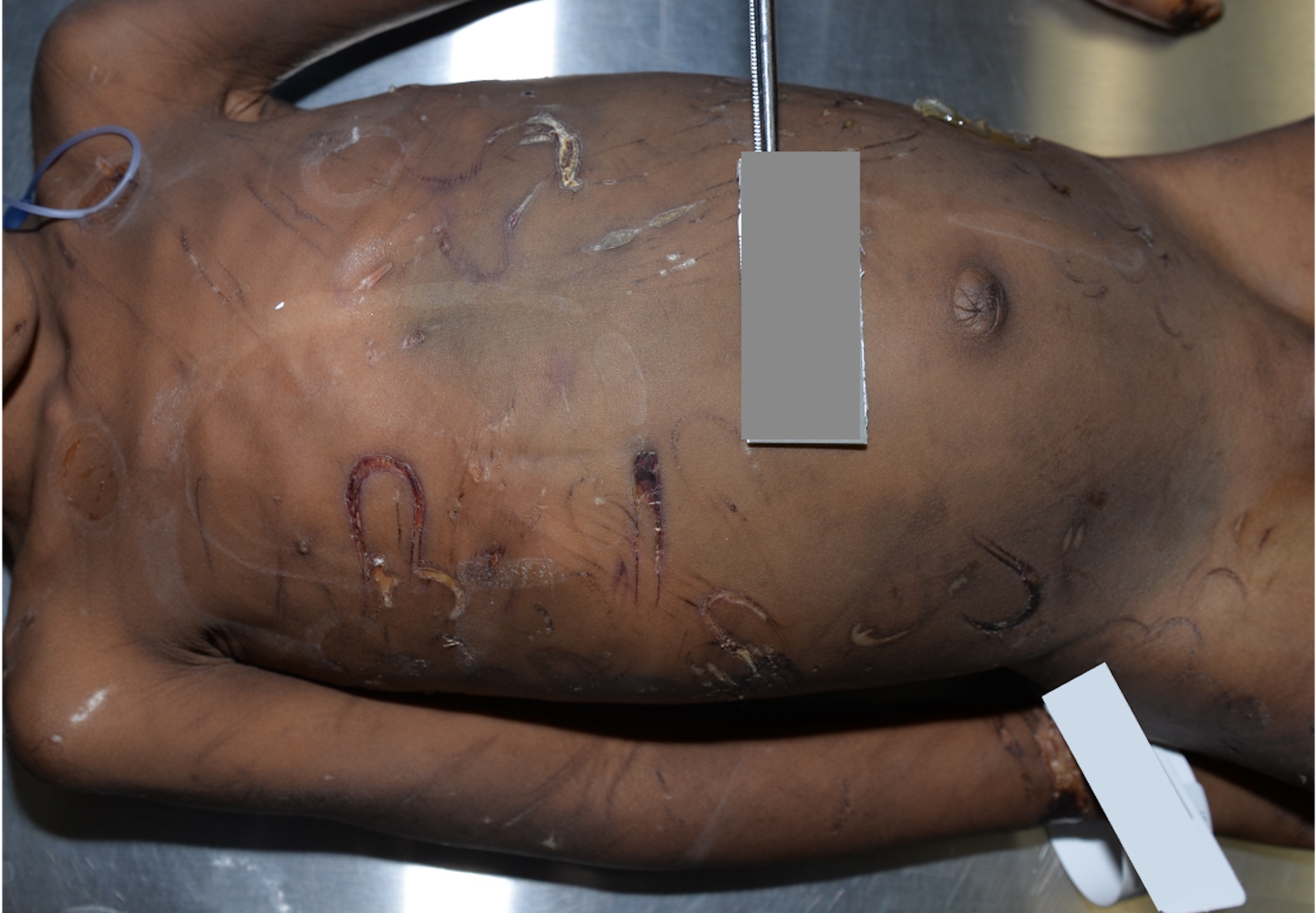
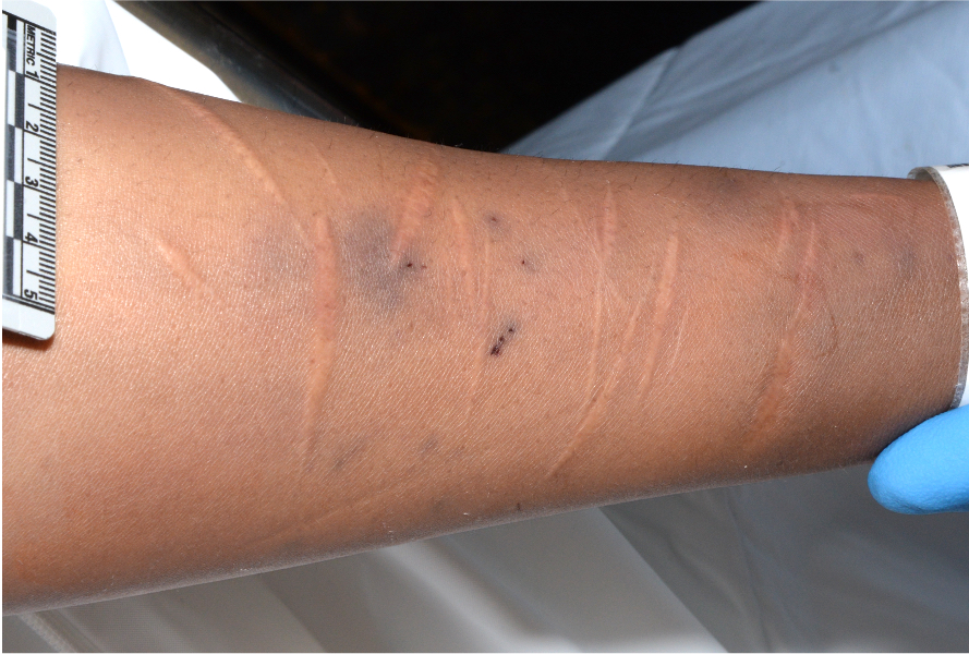
Loop-like bruise

Patterned abraded contusion

Fingertip contusion
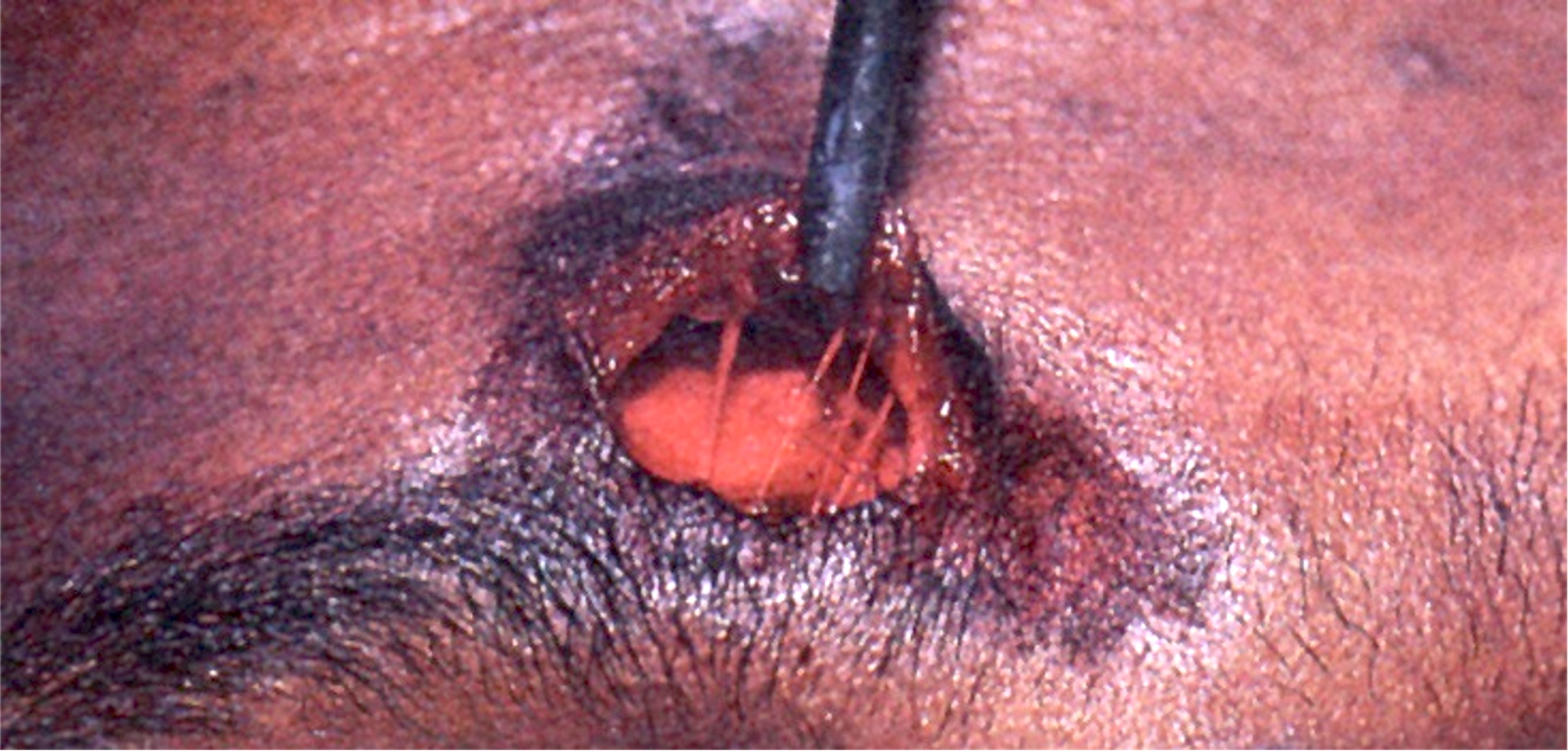
Subcutaneous hemorrhage
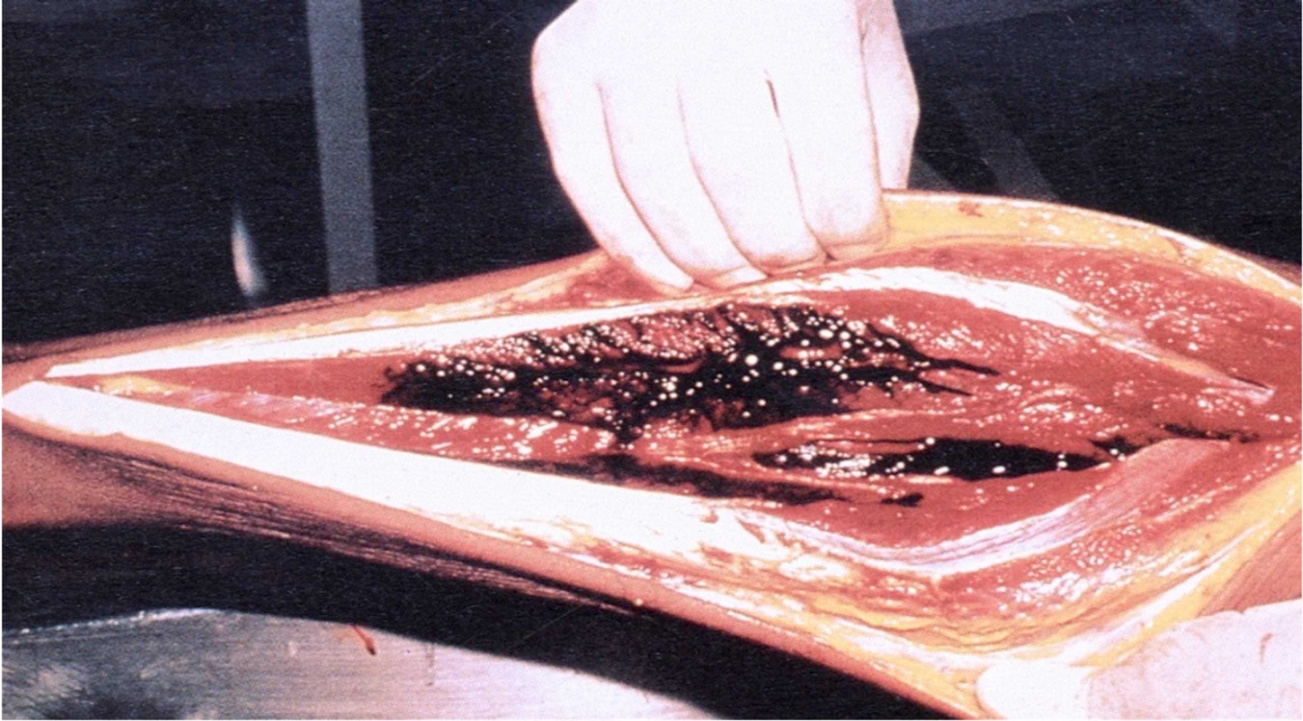
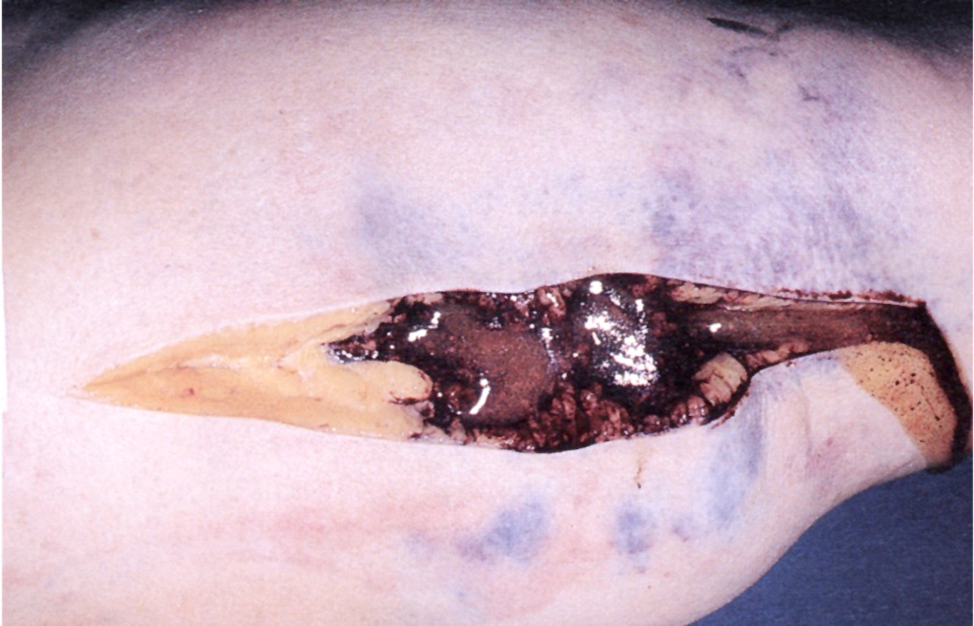
Deep soft tissue hemorrhage

Tardieu spots
Laceration

Hammer lacerations

Aortic lacerations due to motor vehicle collision
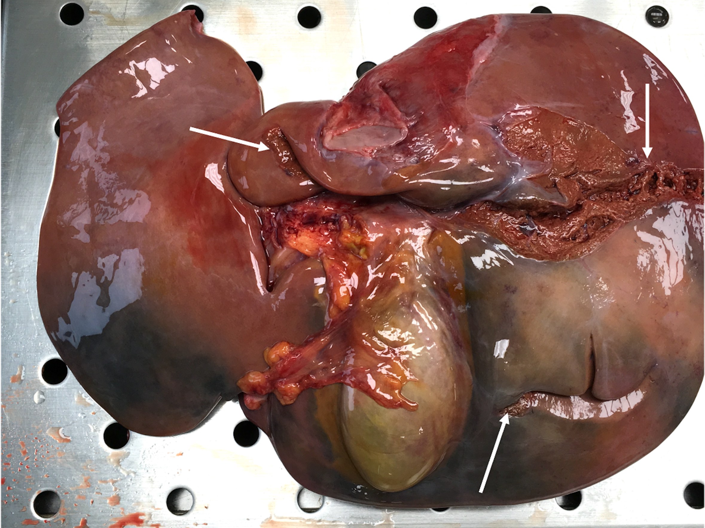
Liver laceration
Avulsion laceration

Closed fracture
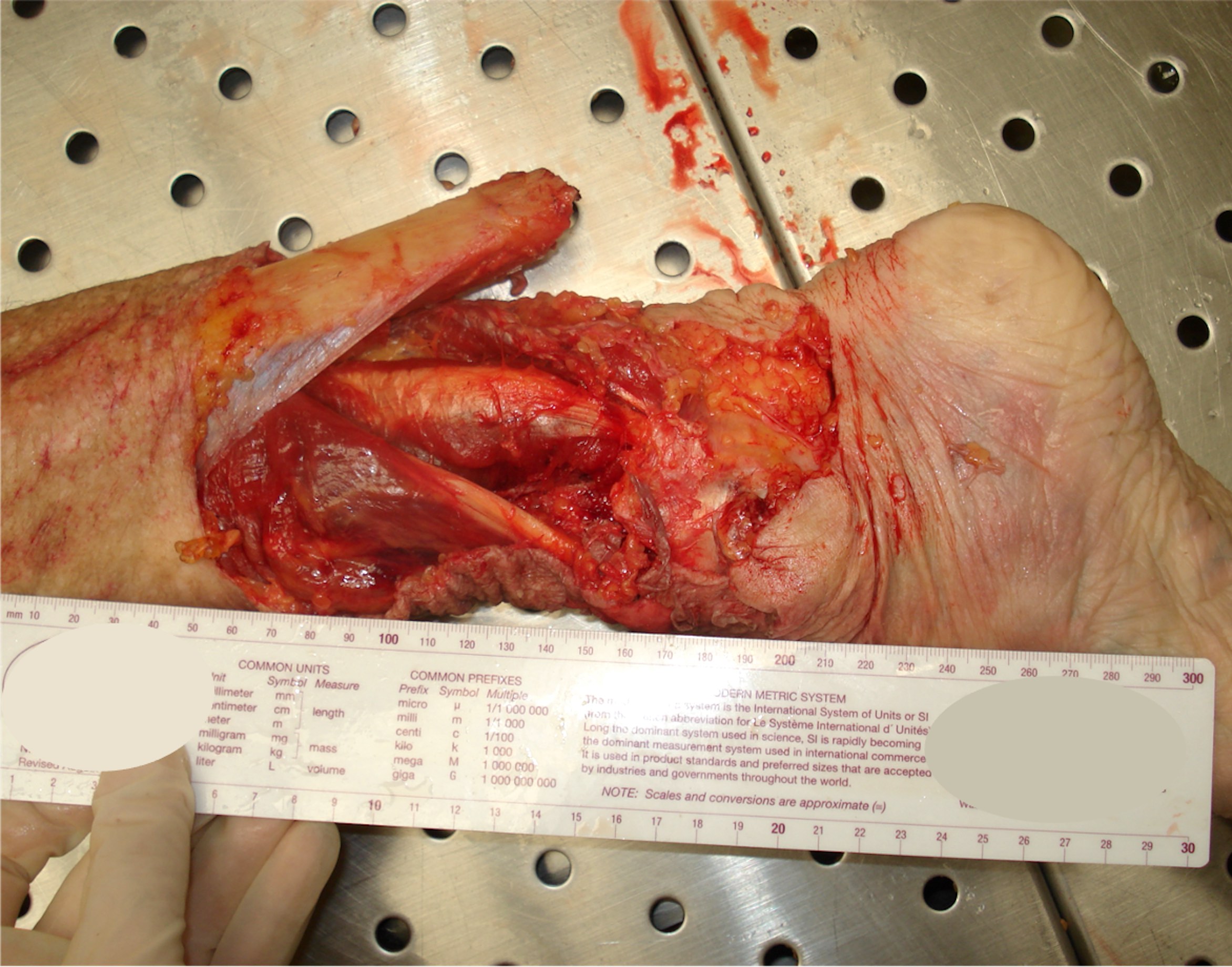
Open fractures
Multiple facial bone fractures
Linear fracture of the cranial vault
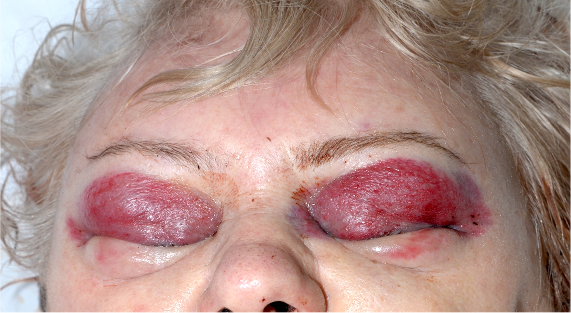
Raccoon eyes
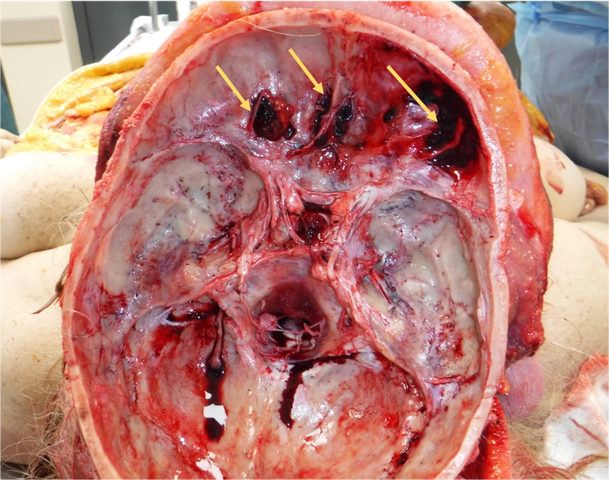
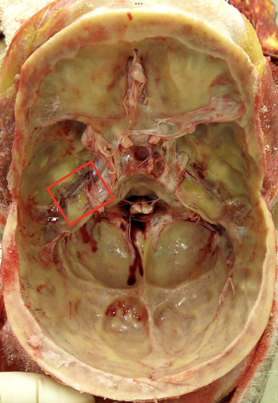
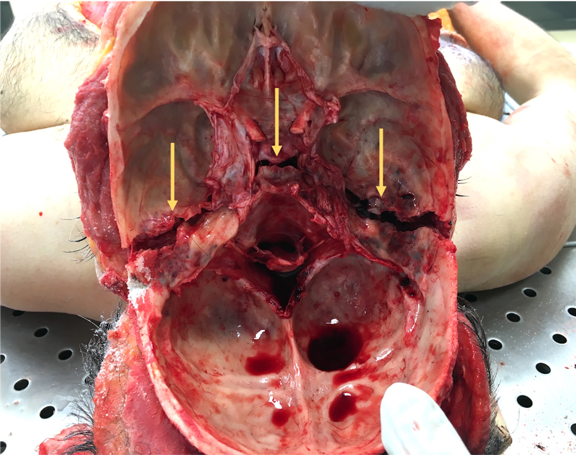
Anterior cranial base fractures

Ear bleeding
Middle cranial base fractures

Depressed skull fracture

Ring fracture
Victim of a motor vehicle collision
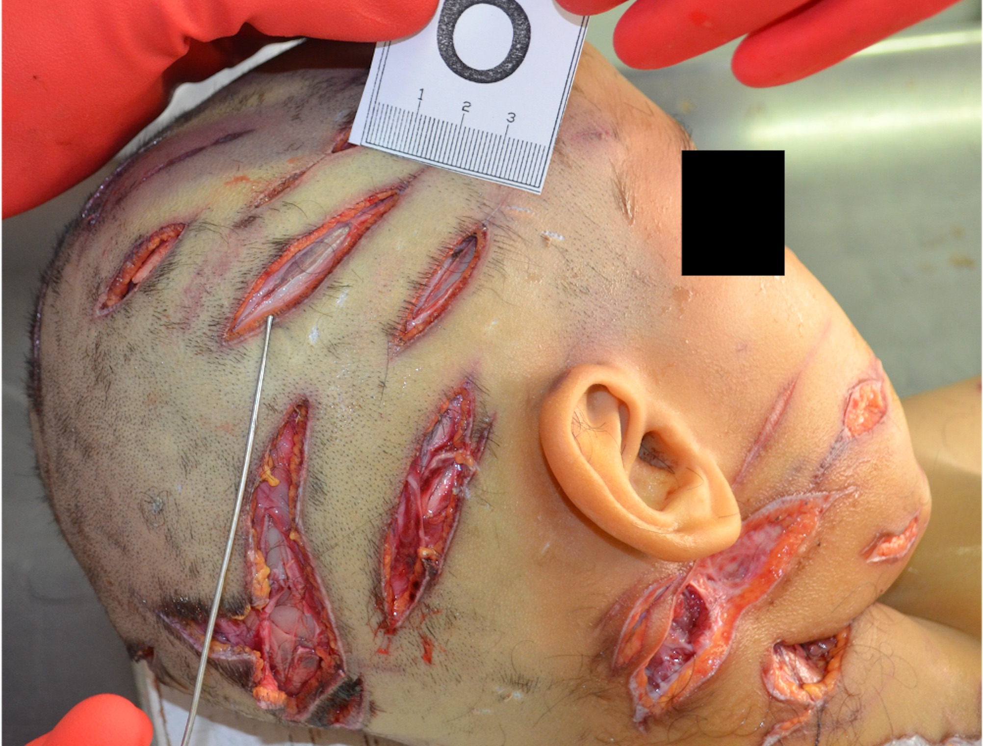
Chop lacerations
Contributed by Michel Tawil, M.D.
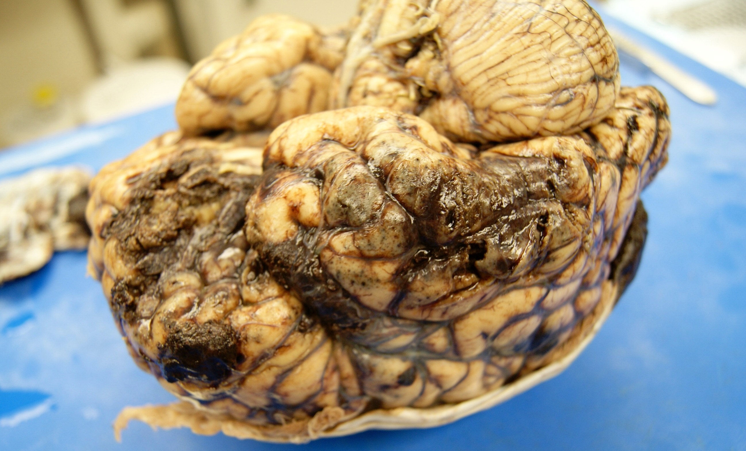
Cortical surface
Contributed by Michel Tawil, M.D.
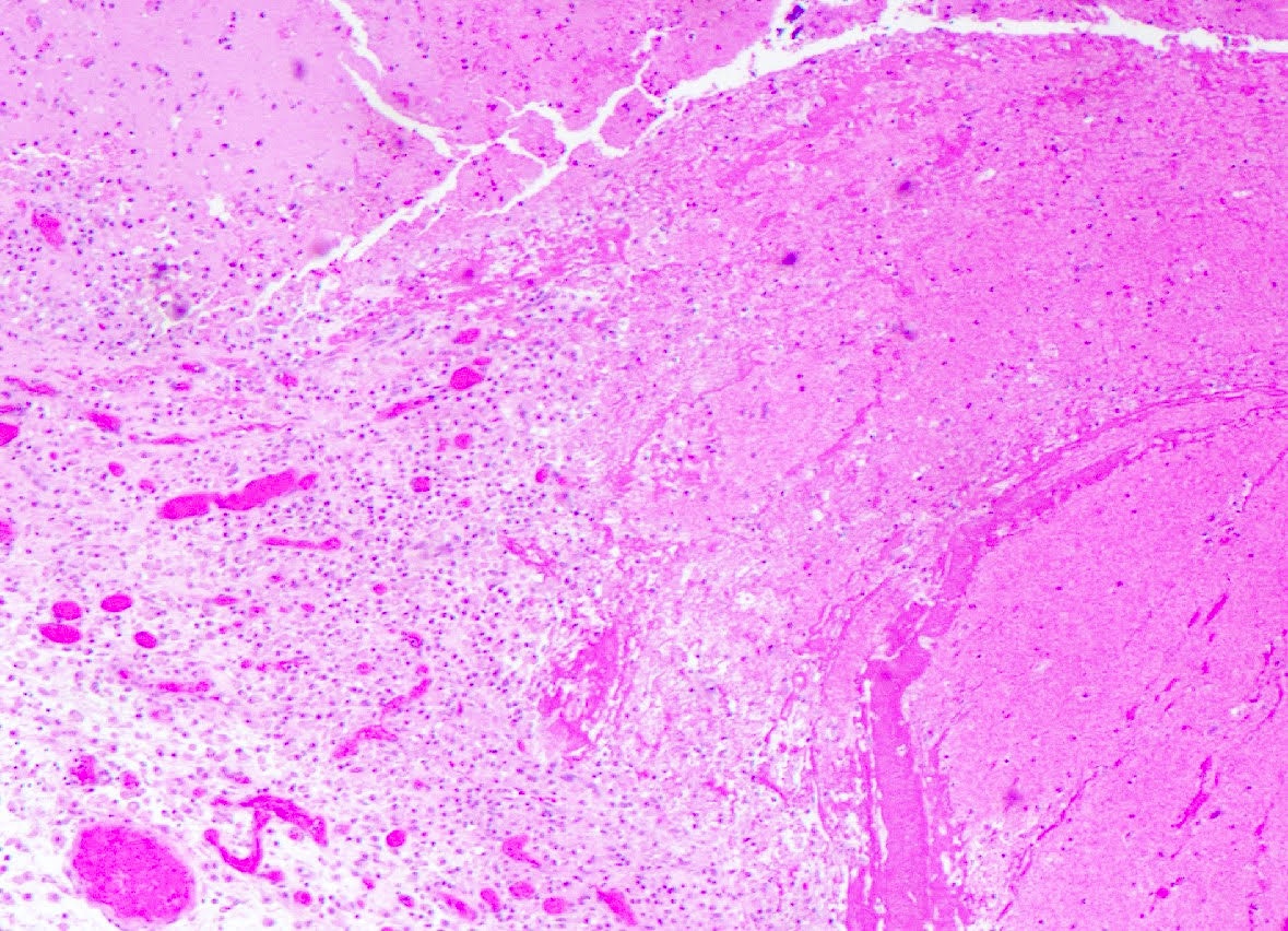
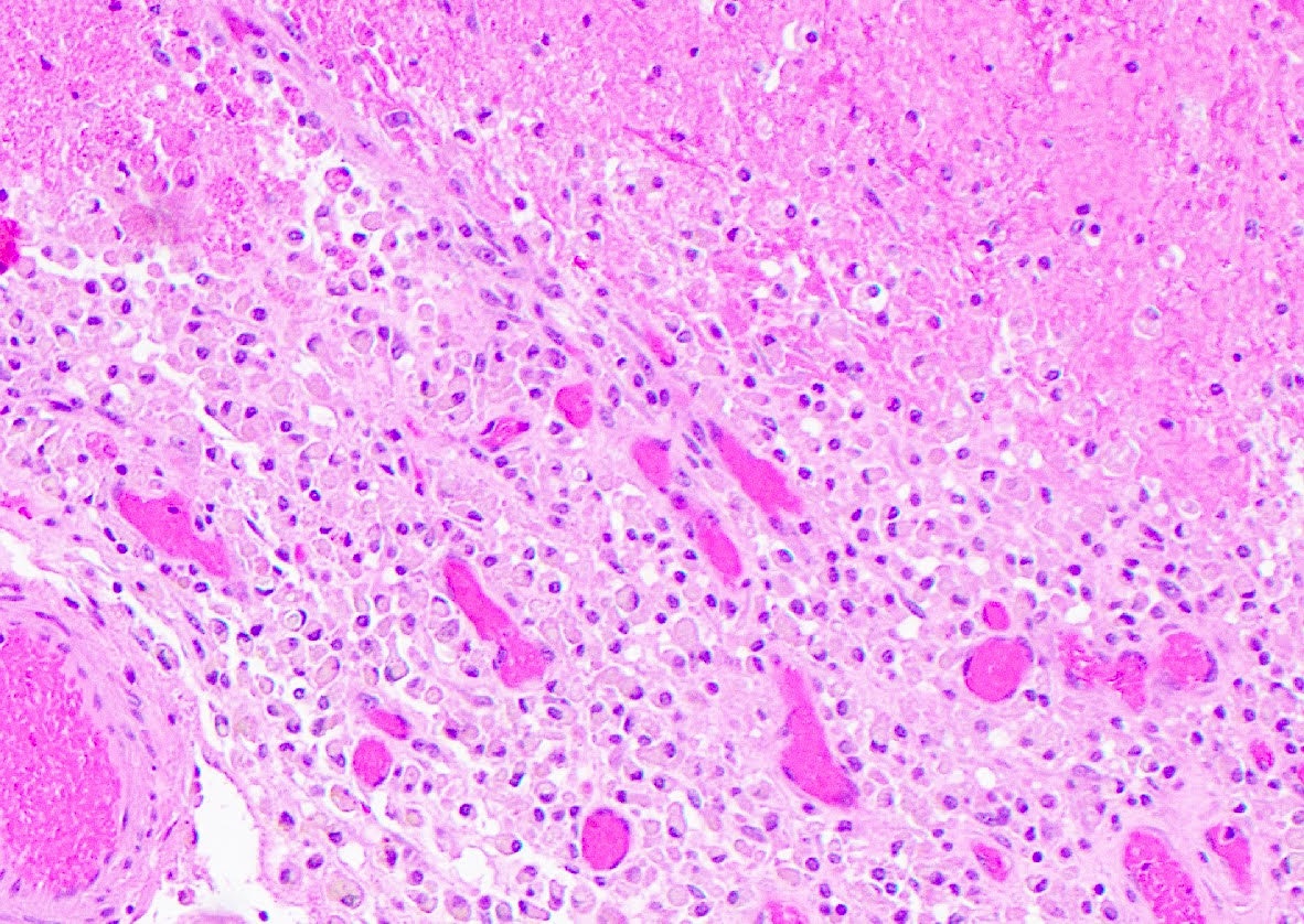
Sections from frontal lobe
Contributed by Lorenzo Gitto, M.D.
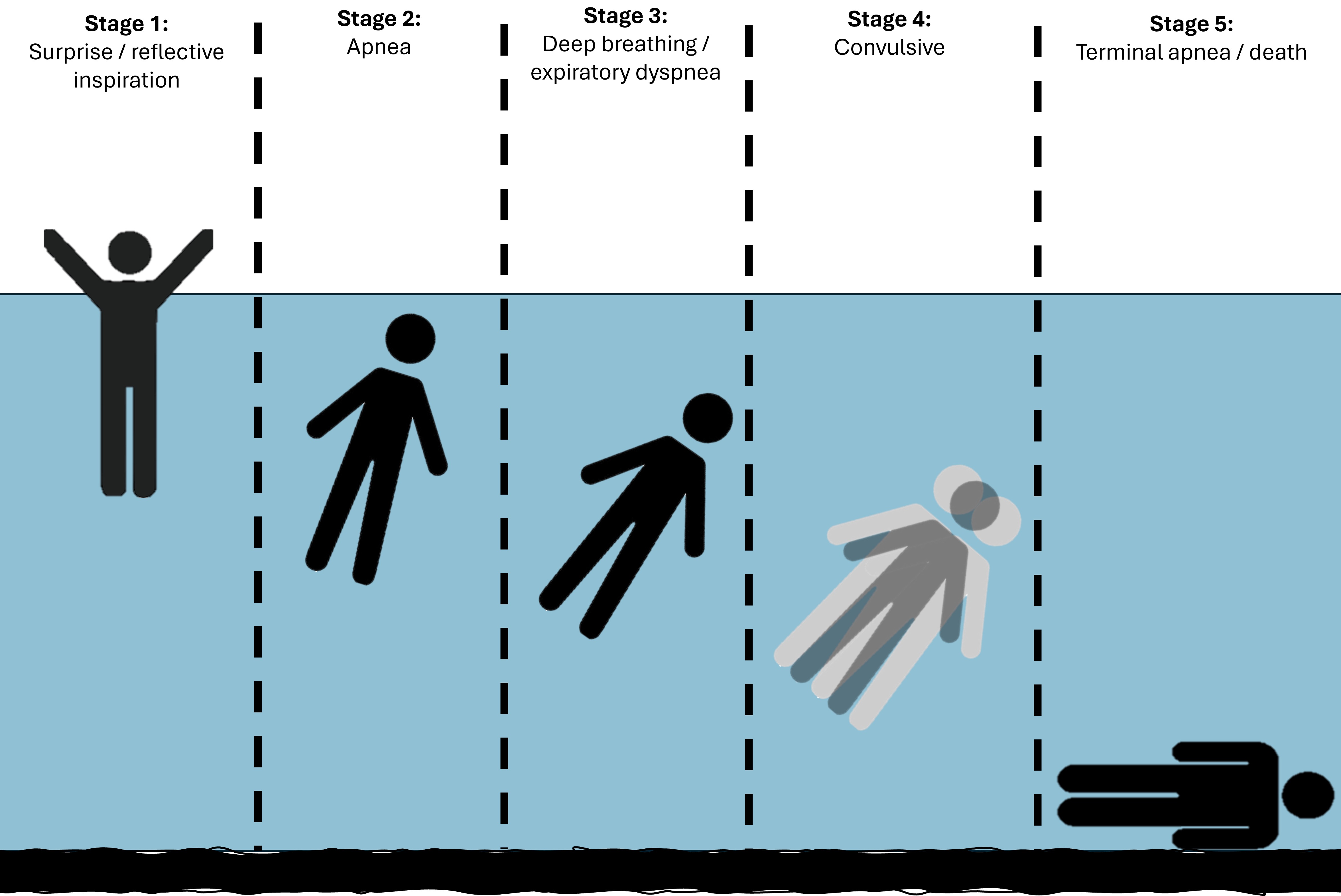
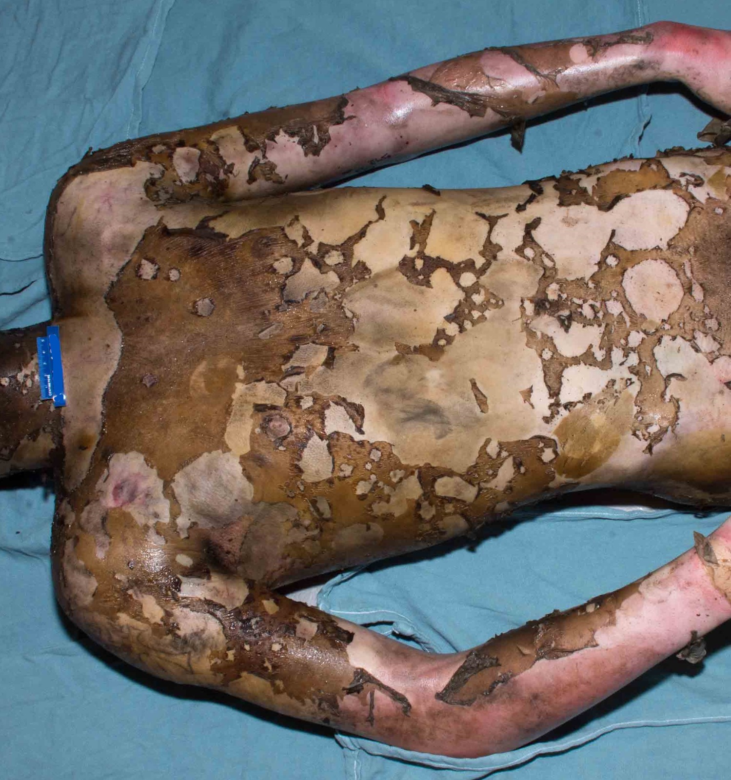
Stages of drowning
Contributed by Allecia M. Wilson, M.D.
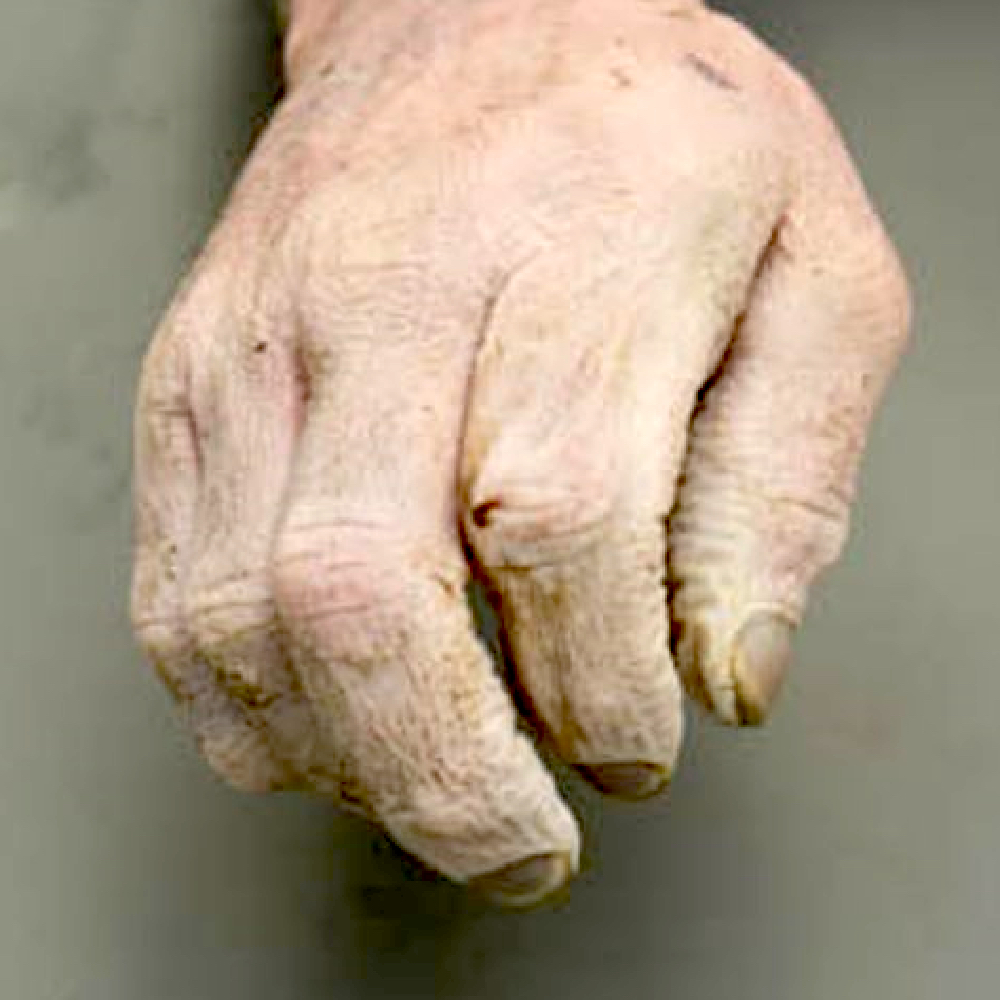
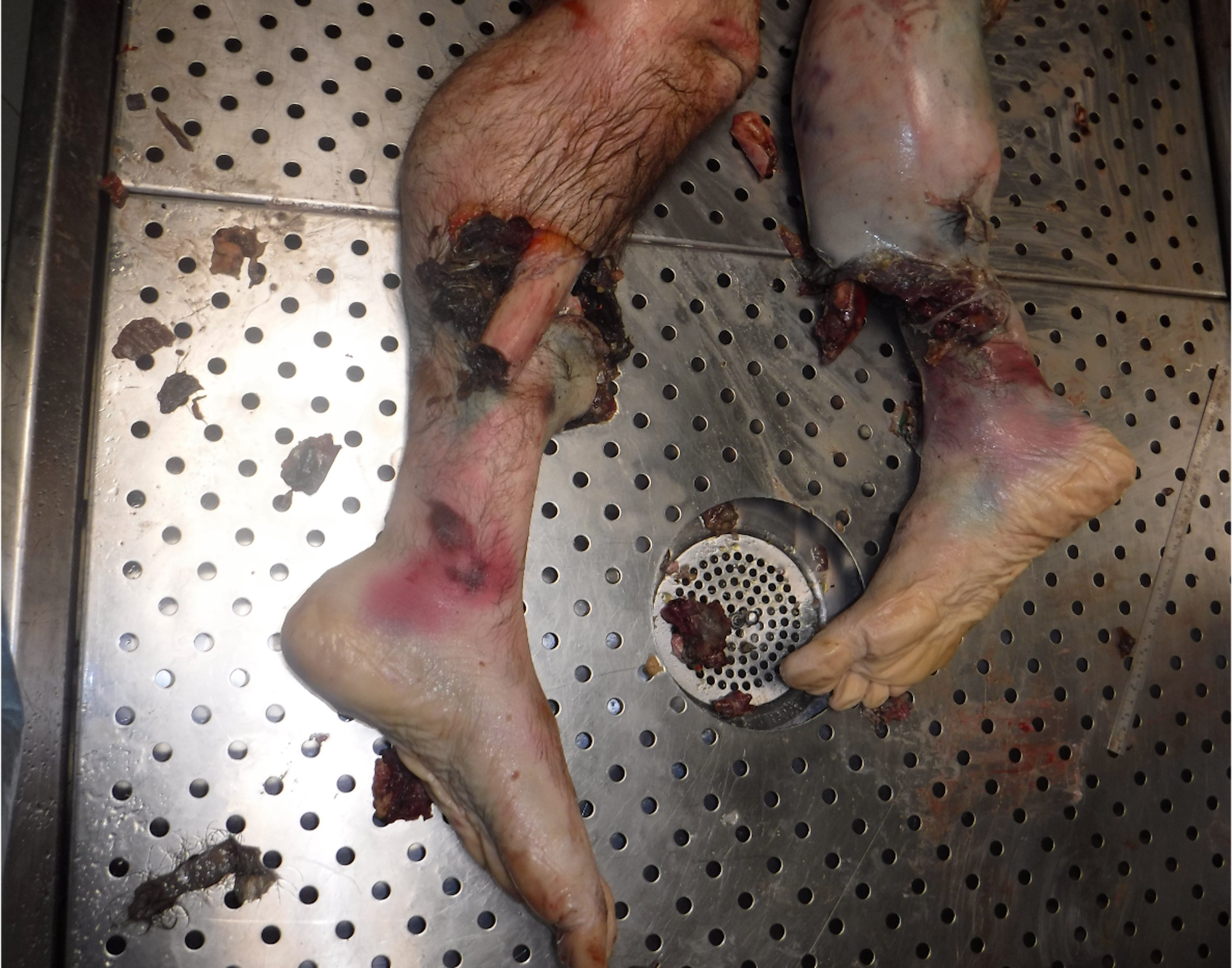
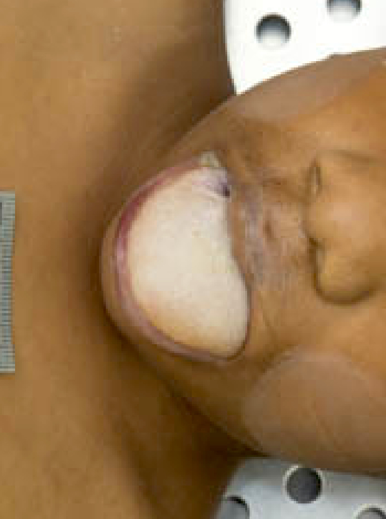
Washerwoman hands
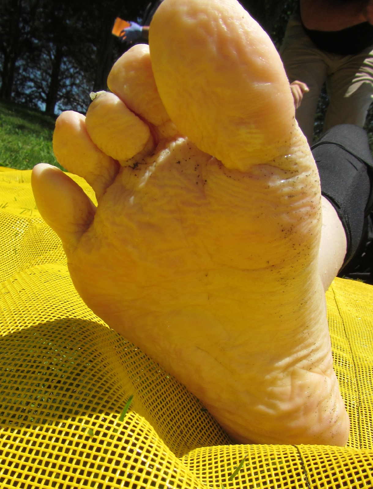
Washerwoman feet
Mouth with foam cone
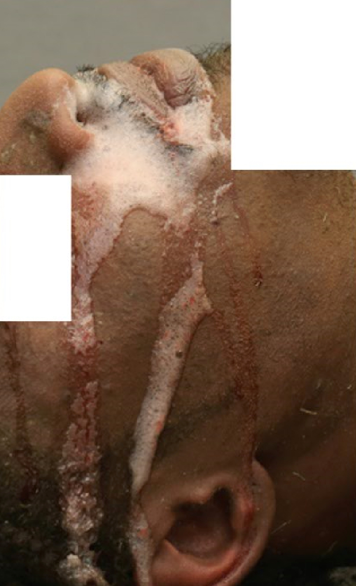
Mouth and nose with foamy contents
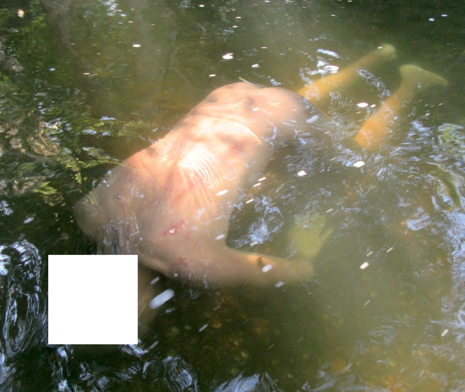
Drowning scene
Contributed by Allecia M. Wilson, M.D.
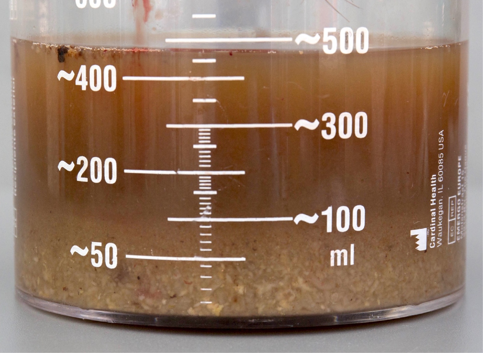
Stomach contents with muddy water and sediment
Extraction of watery sphenoid contents
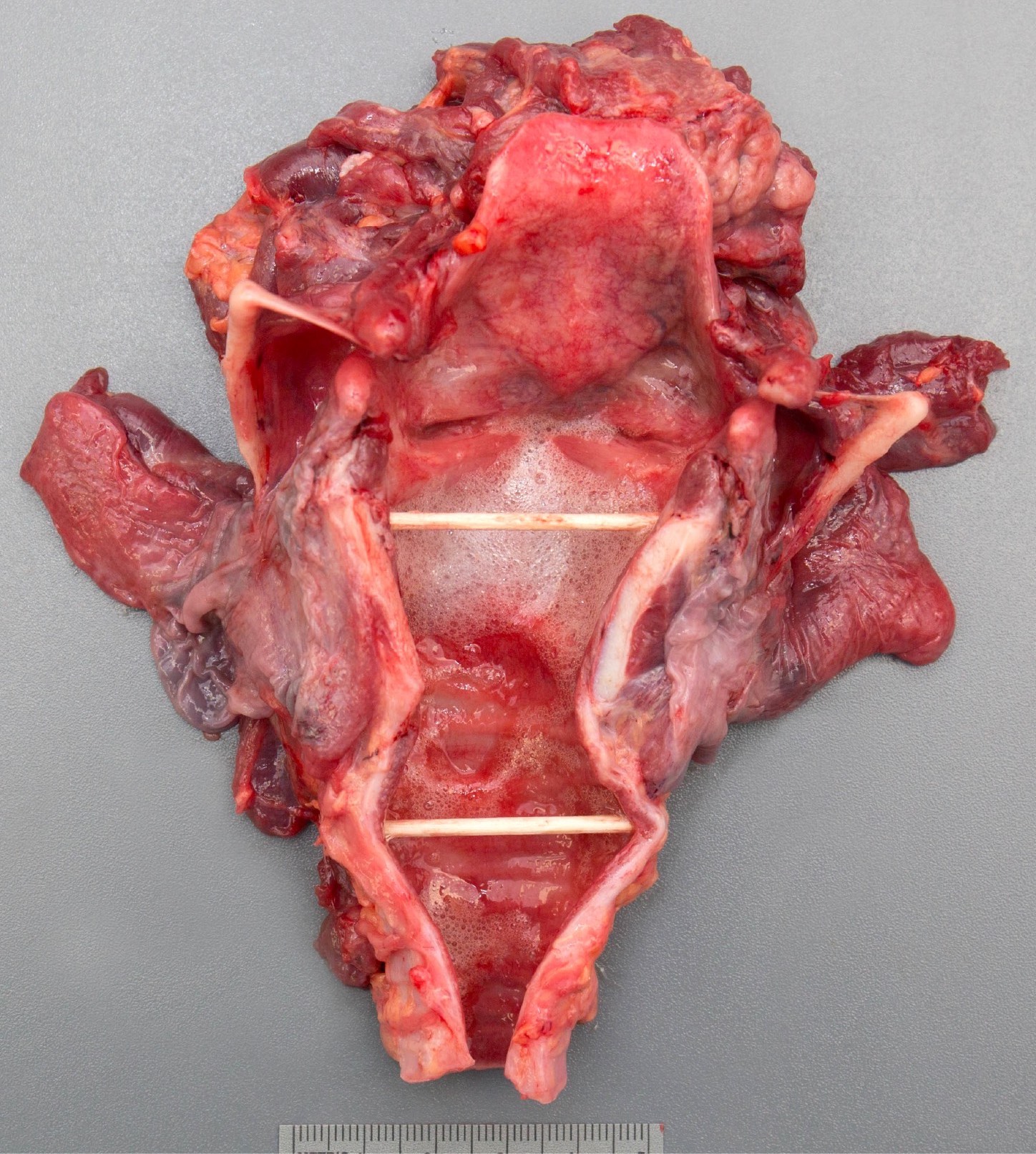
Larynx with foamy contents
Contributed by Mark A. Giffen, Jr., D.O., Jerri L. McLemore, M.D. and Patrick E. Lantz, M.D.
Partial thickness burns
Full thickness burns
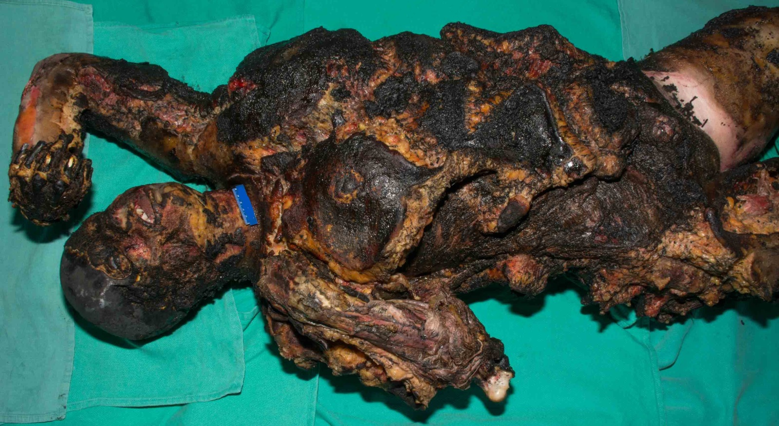
Thermal charring

Electrical burn of wrist

Cutaneous Lichtenburg figures
Contributed by Mark A. Giffen, Jr., D.O.
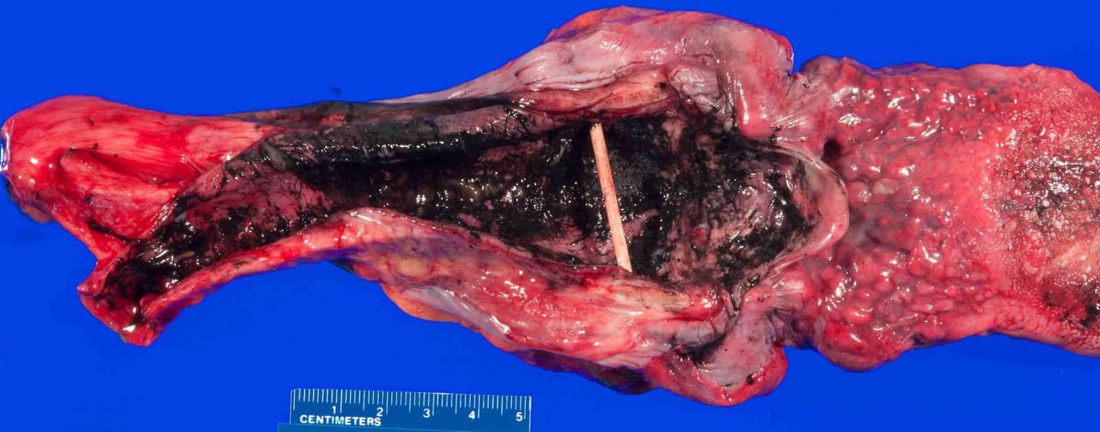
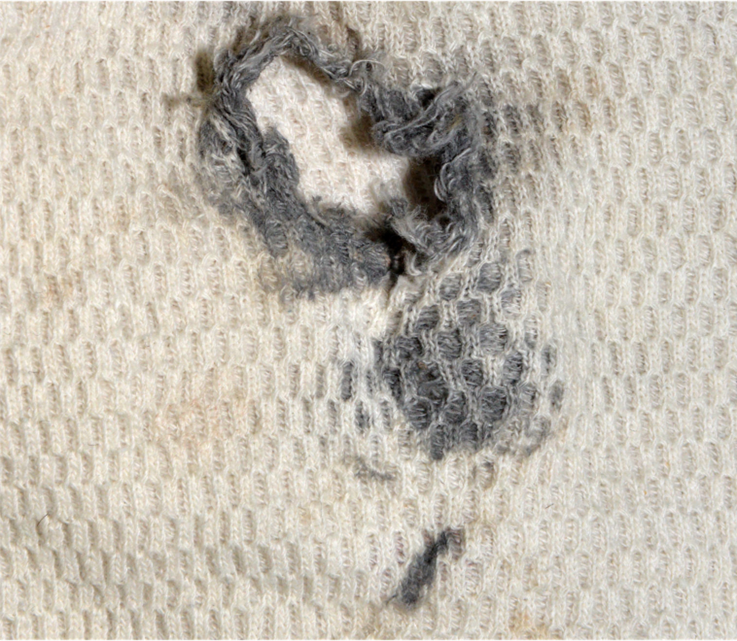
Soot within upper airway

Wischnewski spots
Contributed by Anna McDonald, M.D.

Electrical burn microscopy
Contributed by Priya Banerjee, M.D.
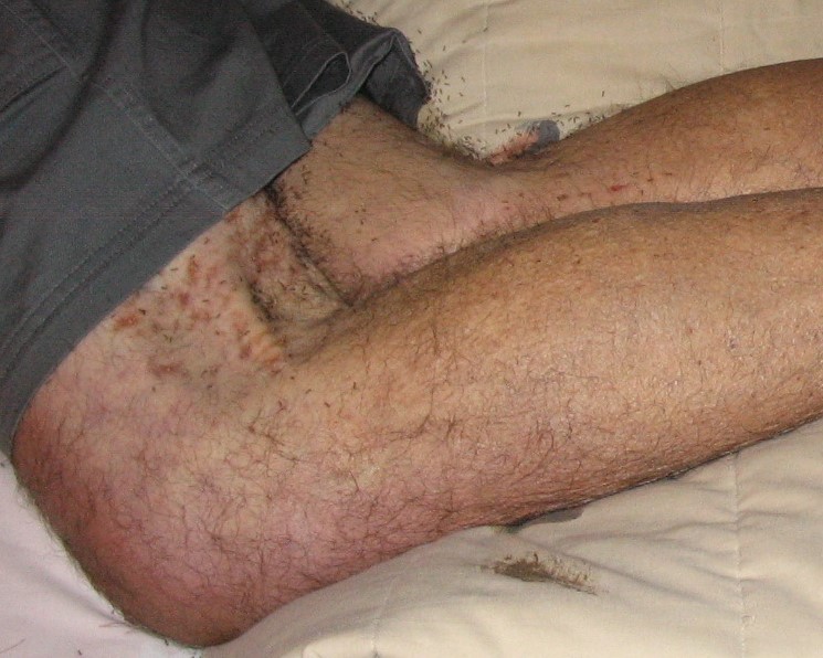
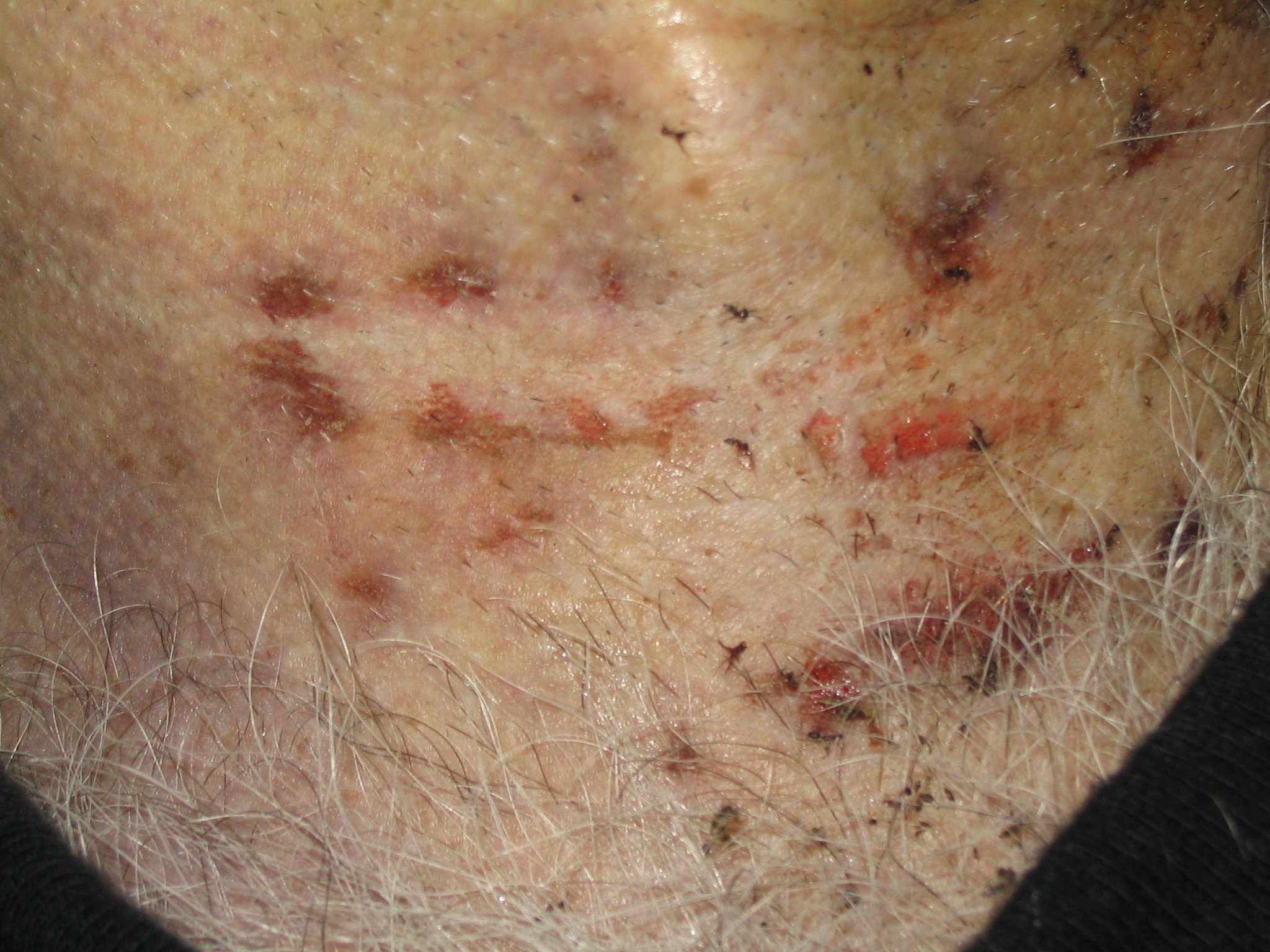
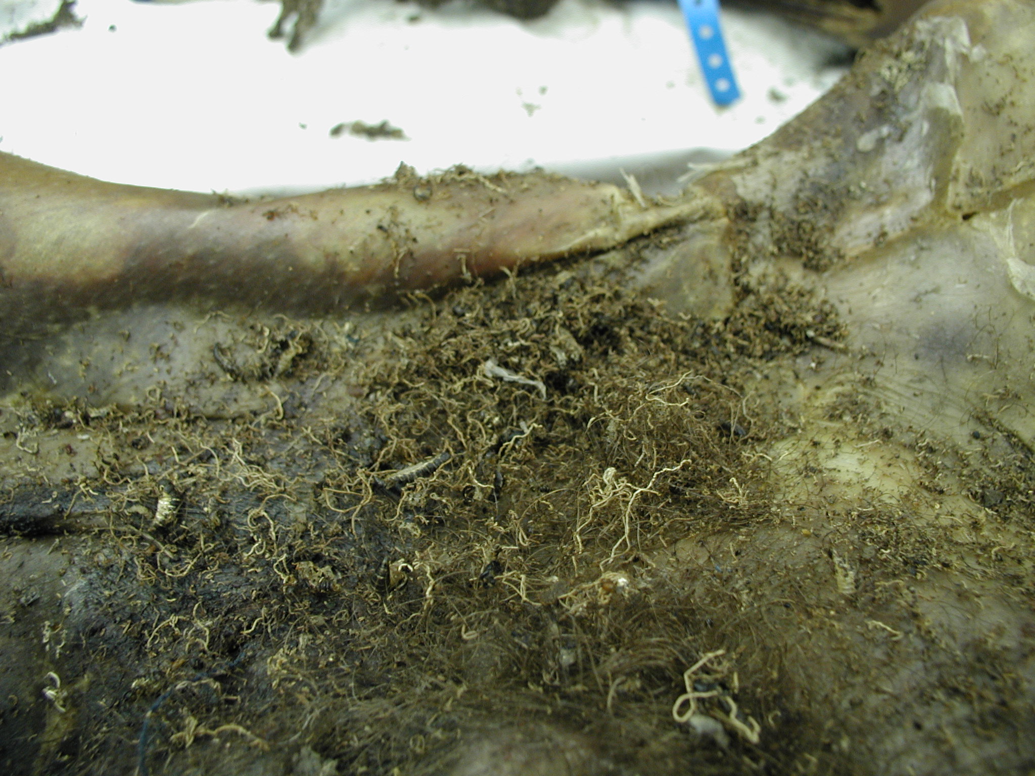
Ant feeding artifacts
Ant feeding artifact along clothing
Beetle frass
Images hosted on other servers:

Circular maggot feeding artifacts

Irregular ant feeding artifact
Contributed by Mark A. Giffen, Jr., D.O.
Air embolism
Postmortem angiography
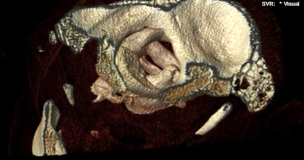
3 dimensional
reconstruction of
occipitalatlantal
dislocation
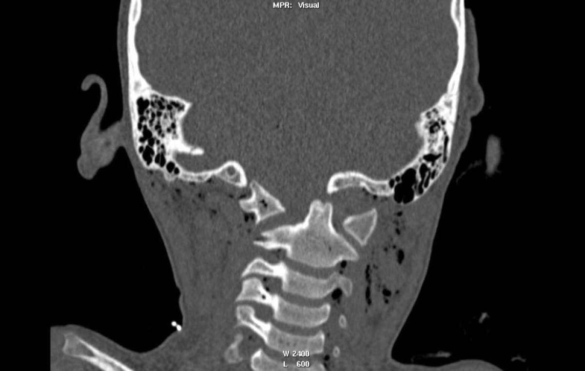
Occipitalatlantal dislocation
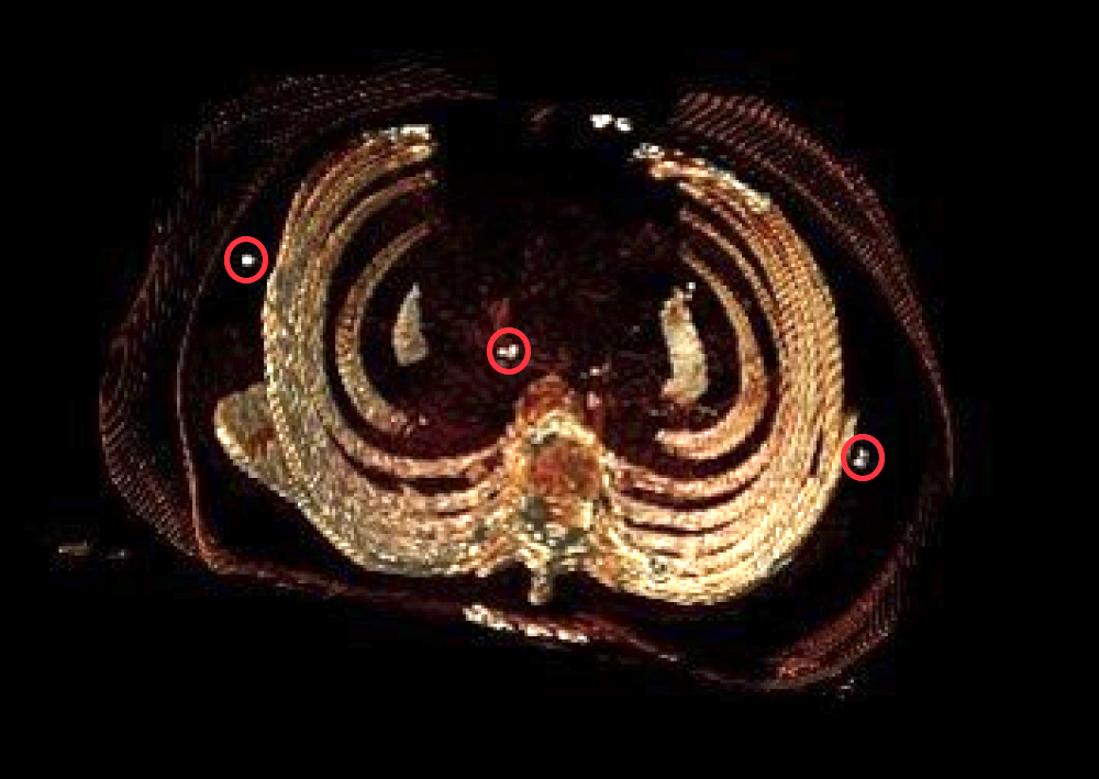
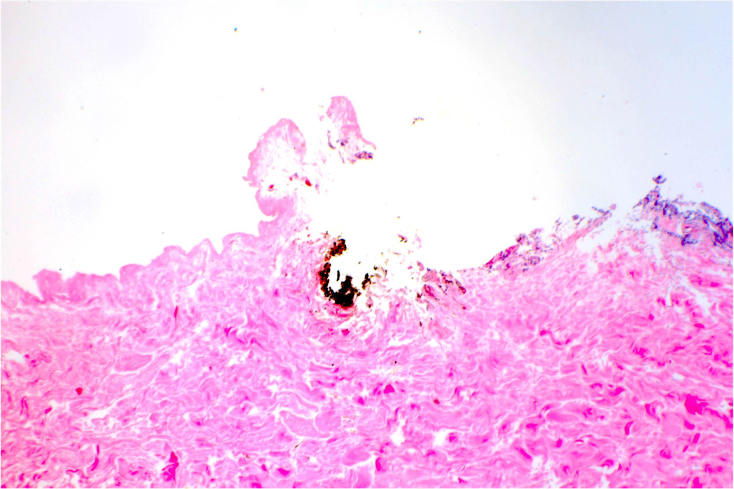
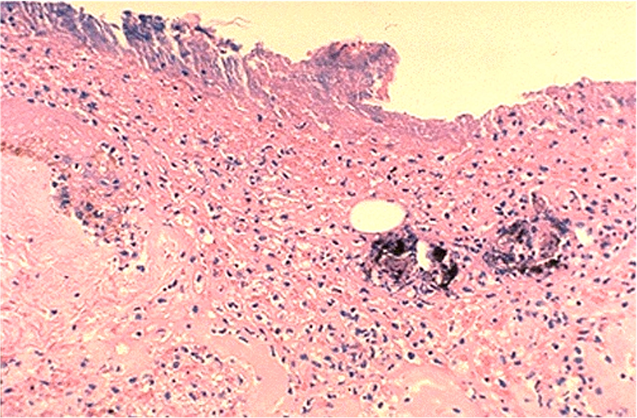
Projectile wound track
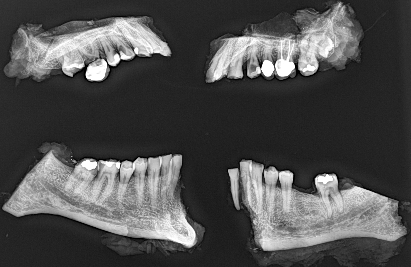
Dental Xrays for identification
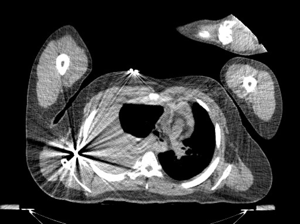
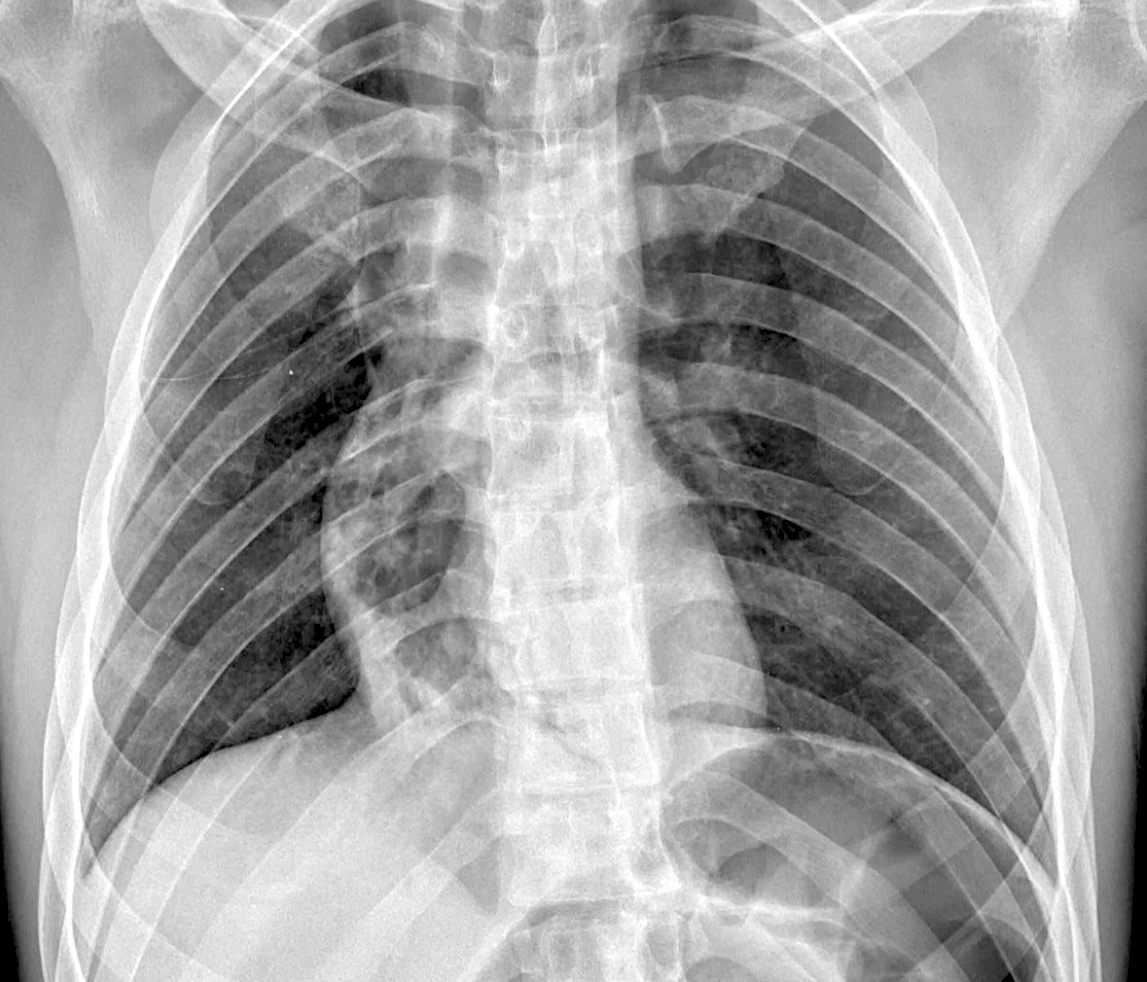
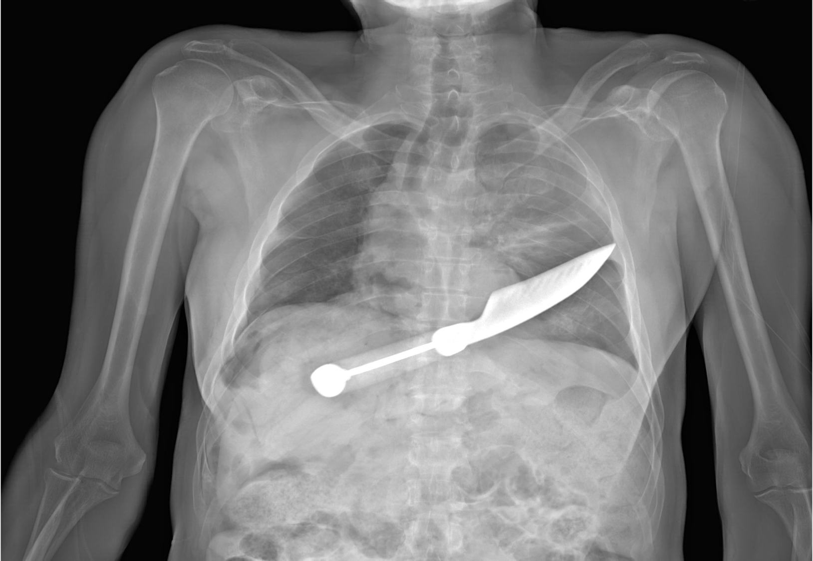
Thoracic projectile
Contributed by Lorenzo Gitto, M.D. and Robert Stoppacher, M.D.
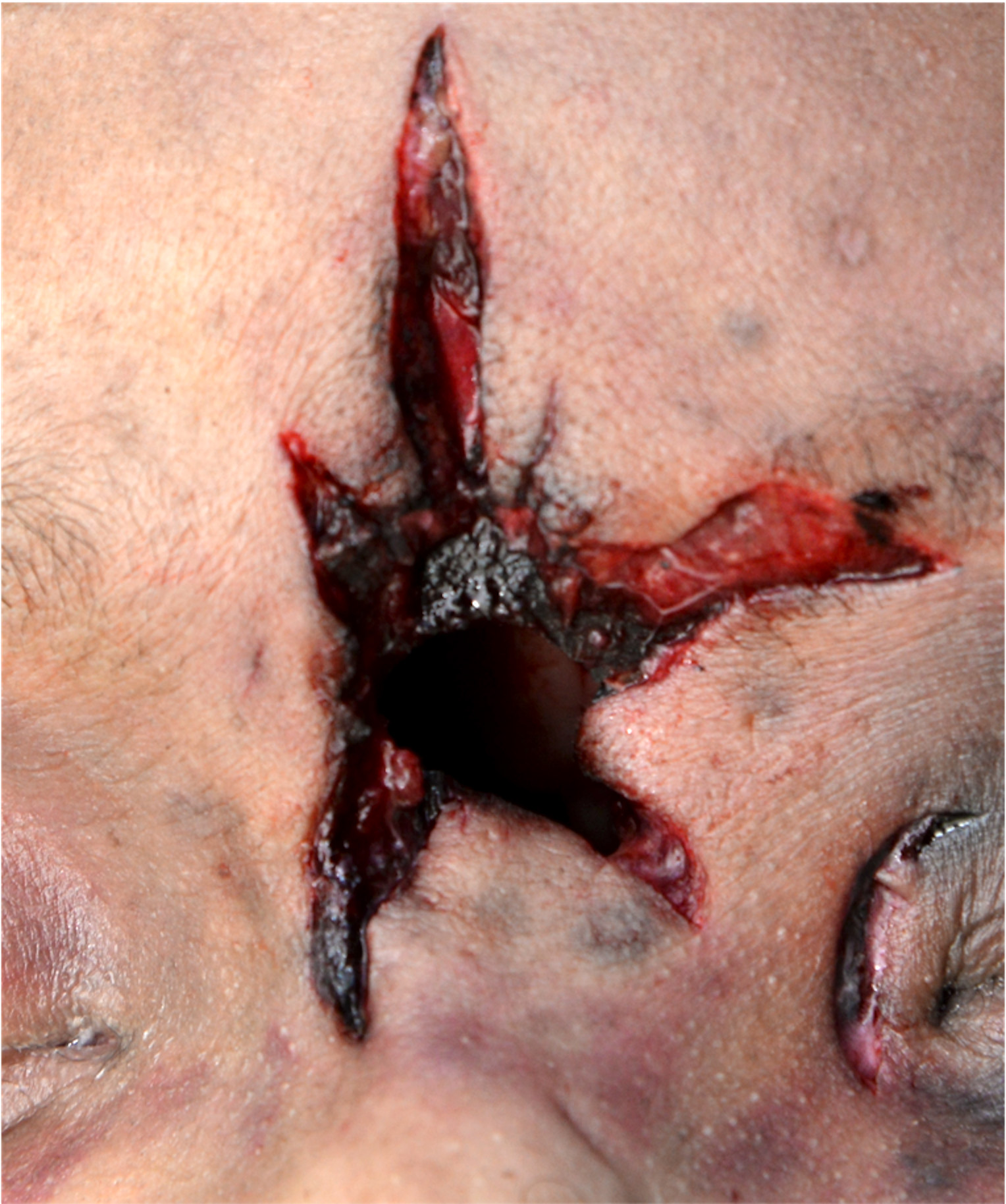
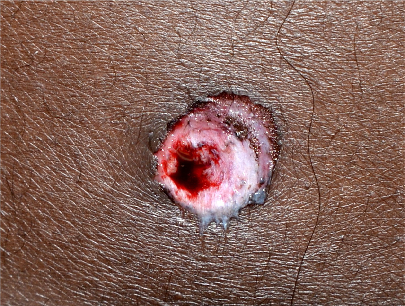
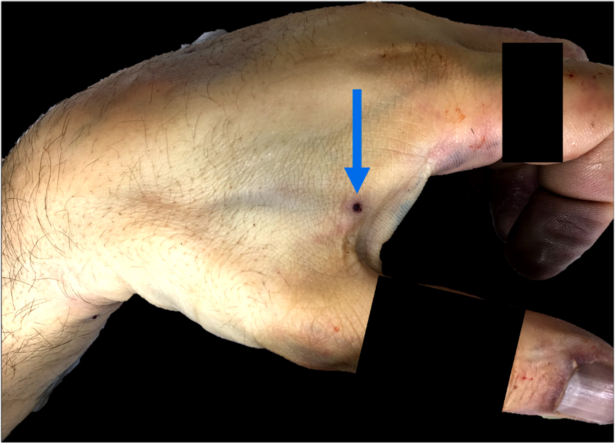
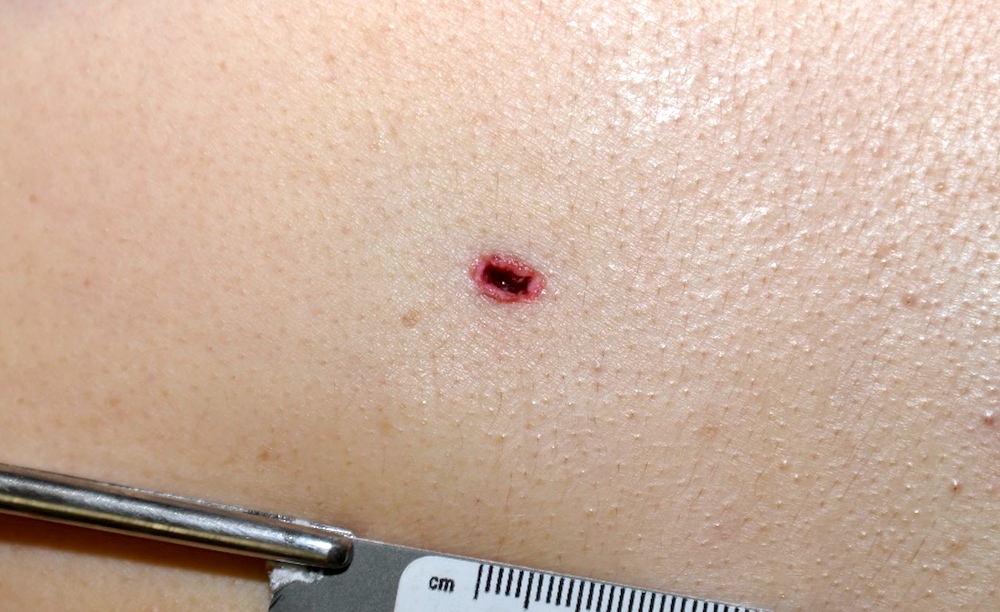
Hard contact entrance gunshot wound
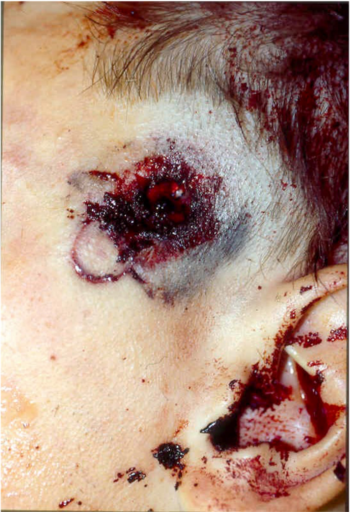
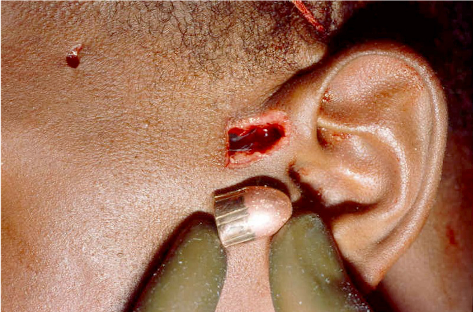
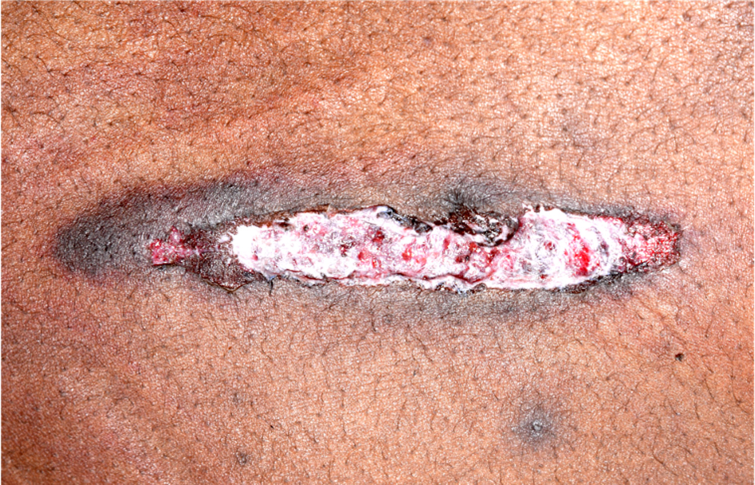
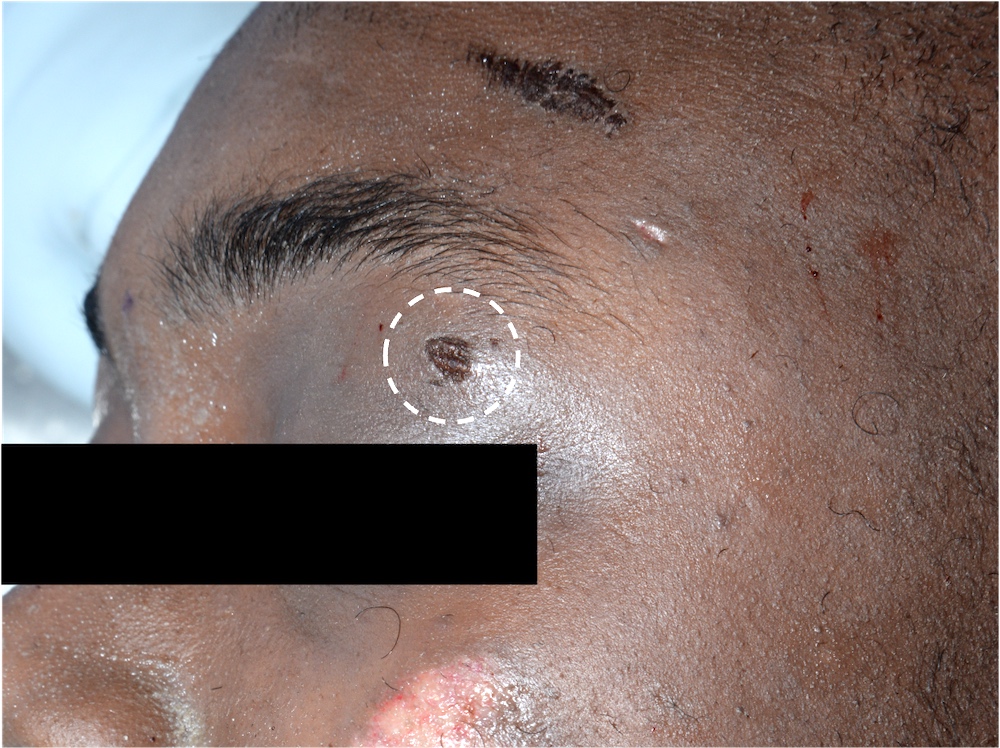
Loose contact entrance gunshot wound

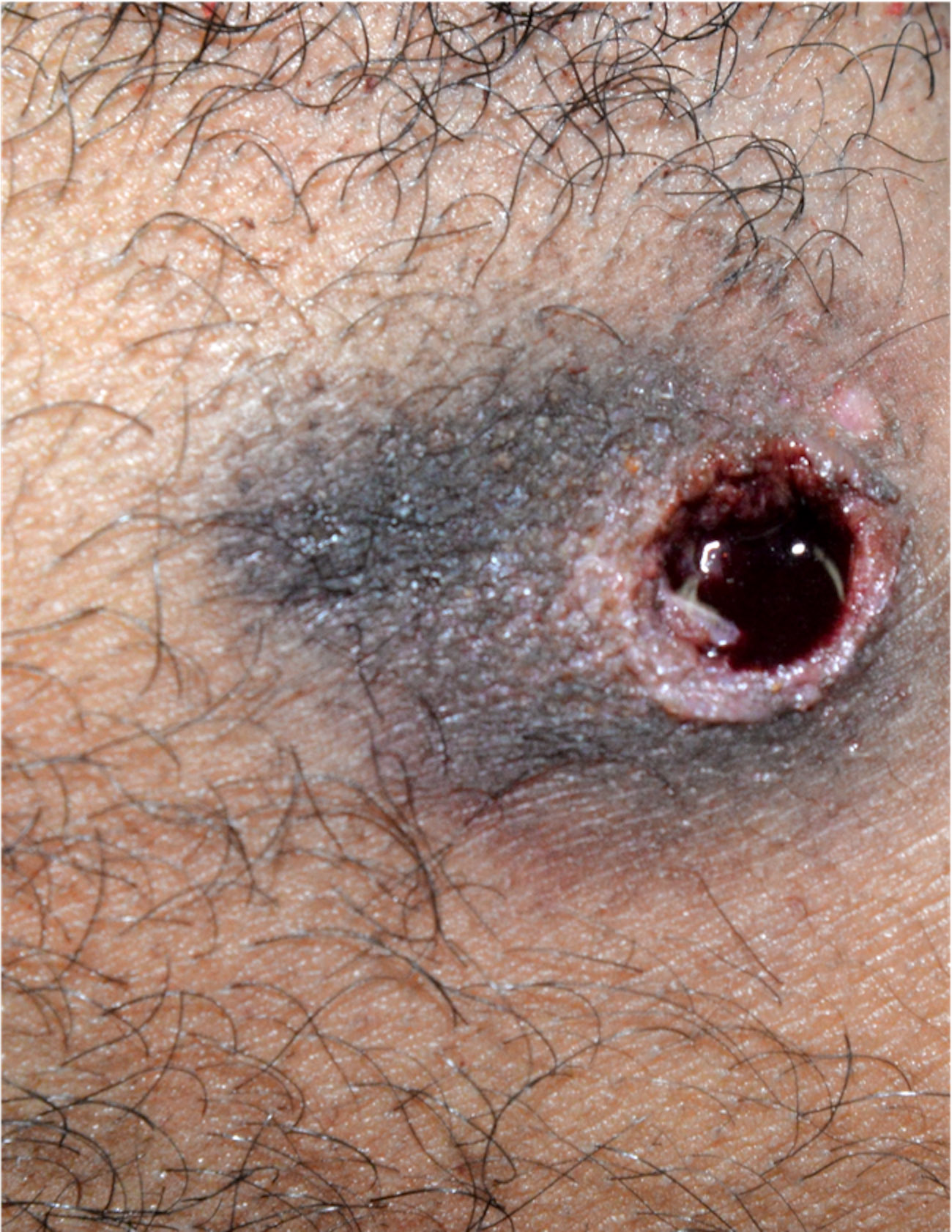
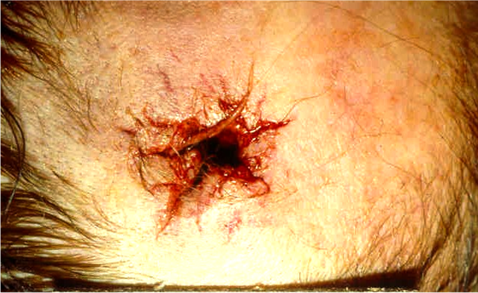
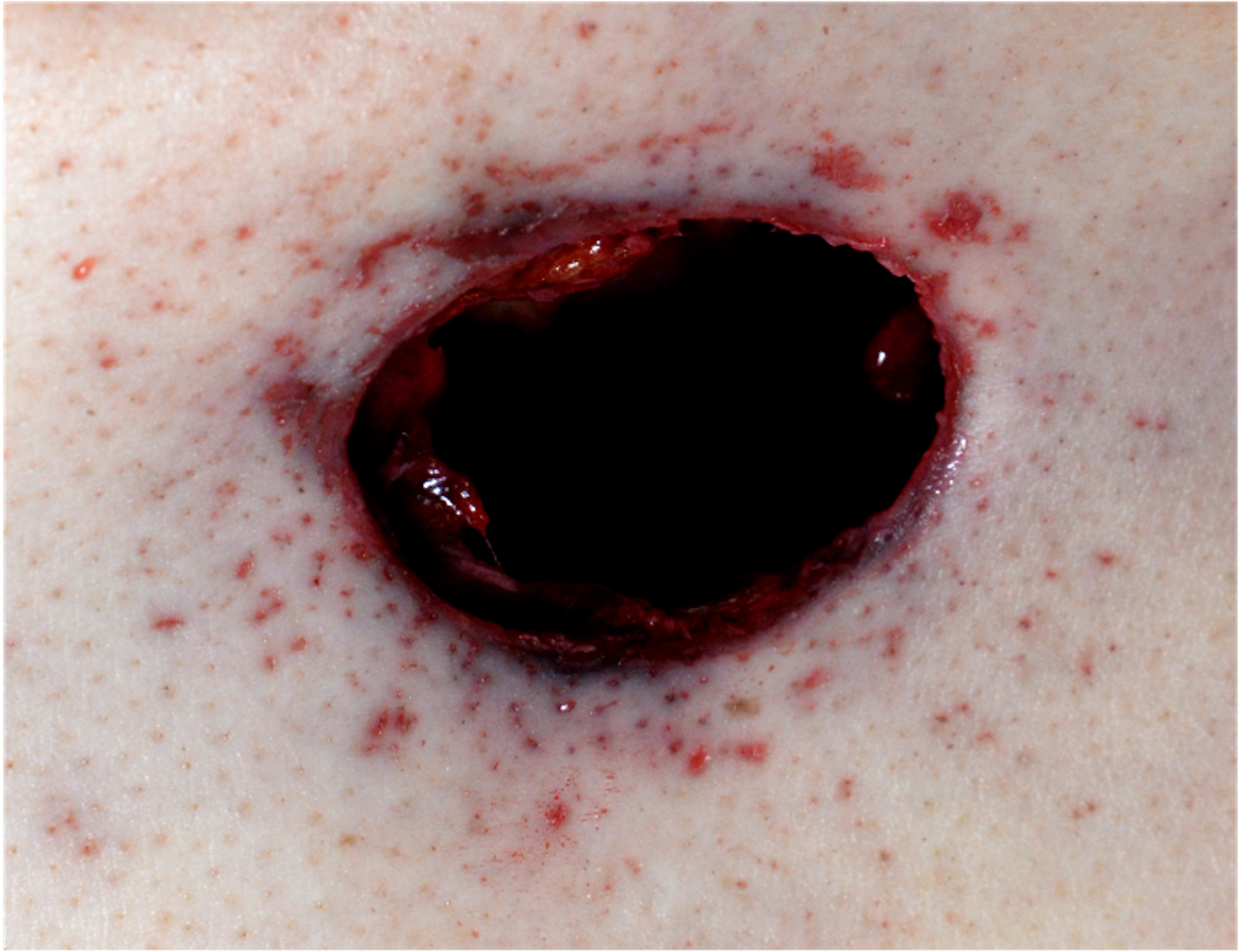
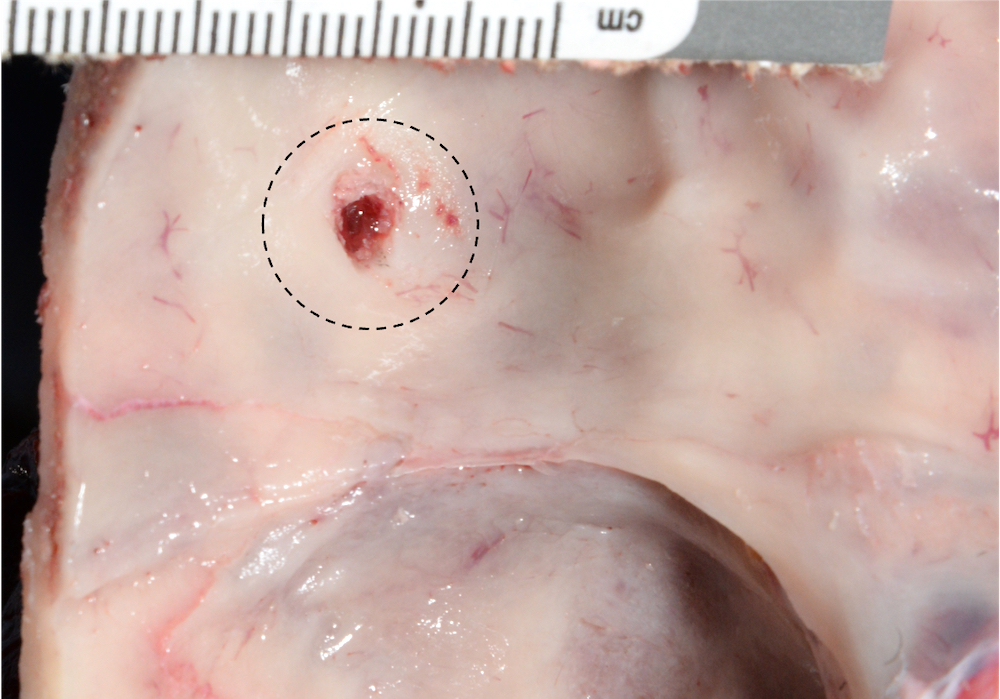
Near contact entrance gunshot wound
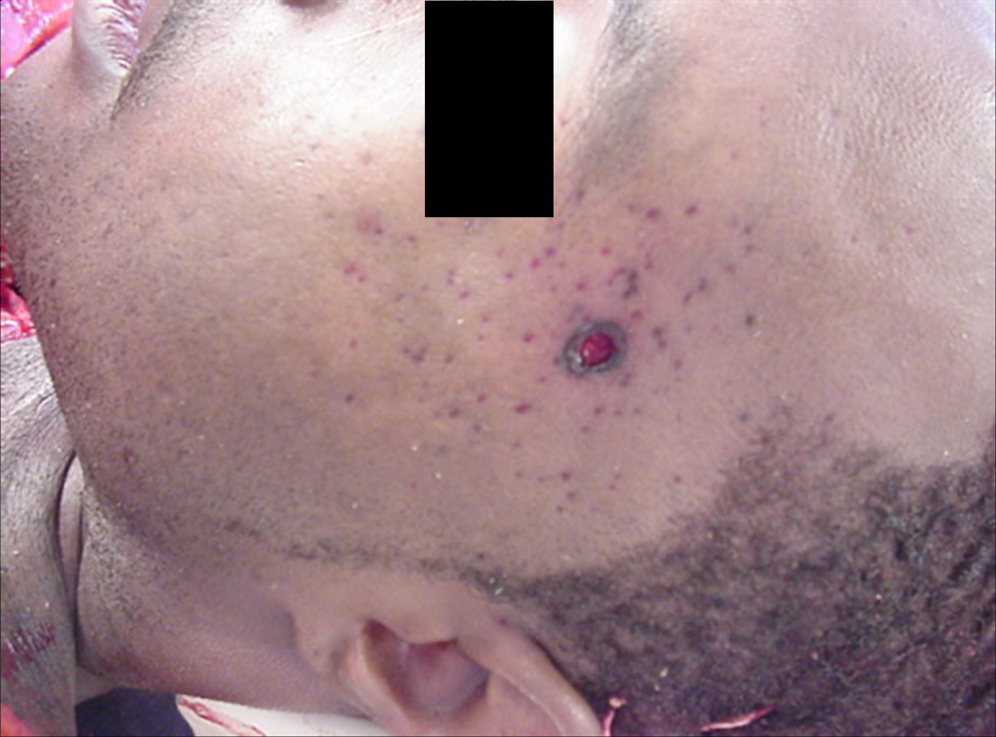
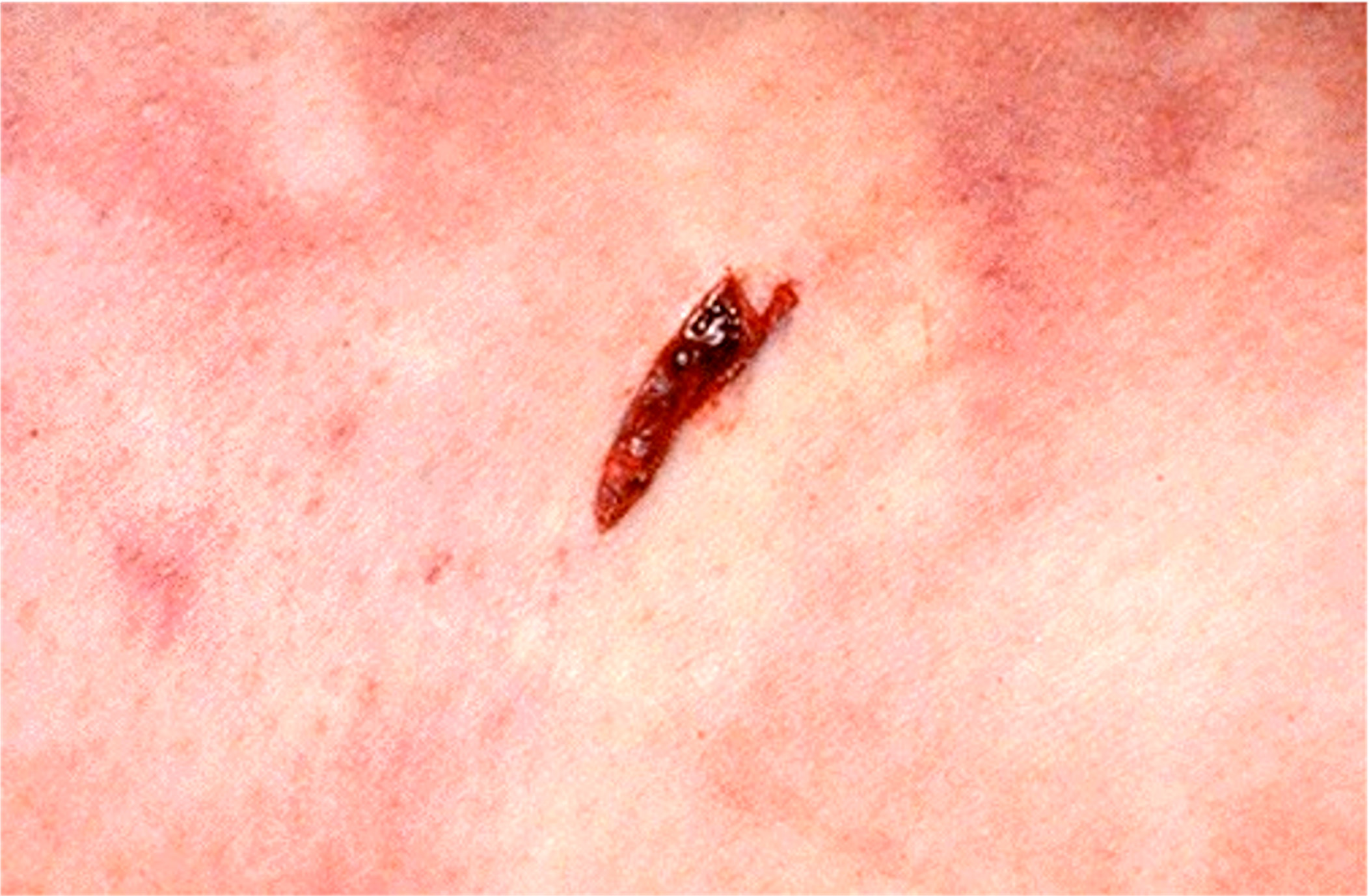
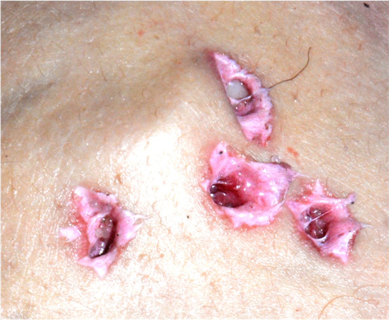
Intermediate range gunshot wound
Distant (or indeterminate) range gunshot wound
Atypical entrance
Exit gunshot wound
Felc mark
Graze wound
Contact shotgun entrance wound
Exit shotgun wounds
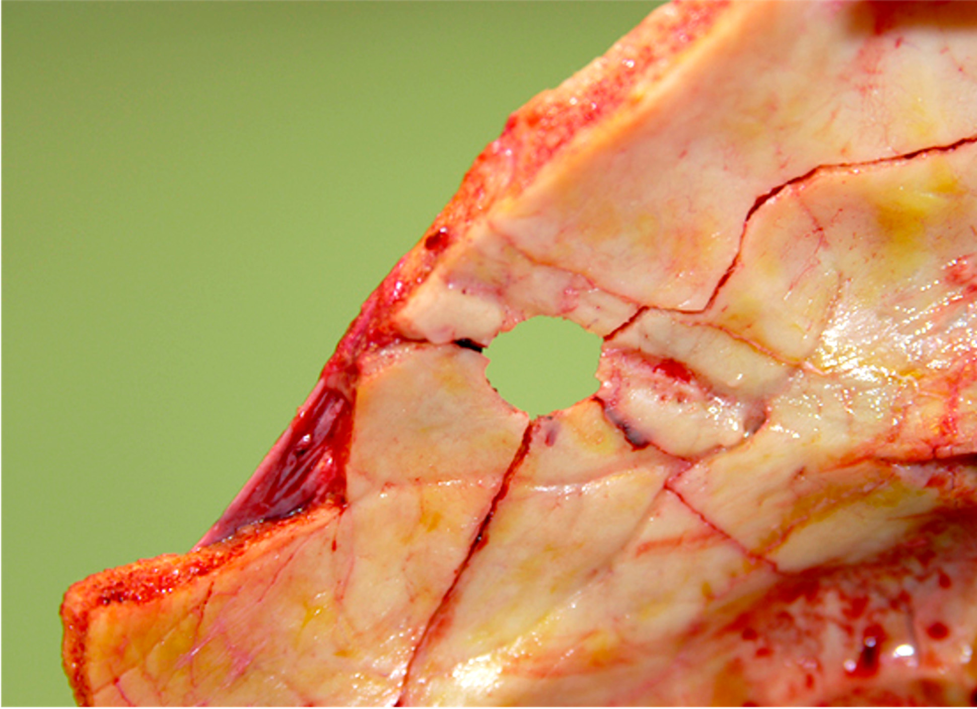
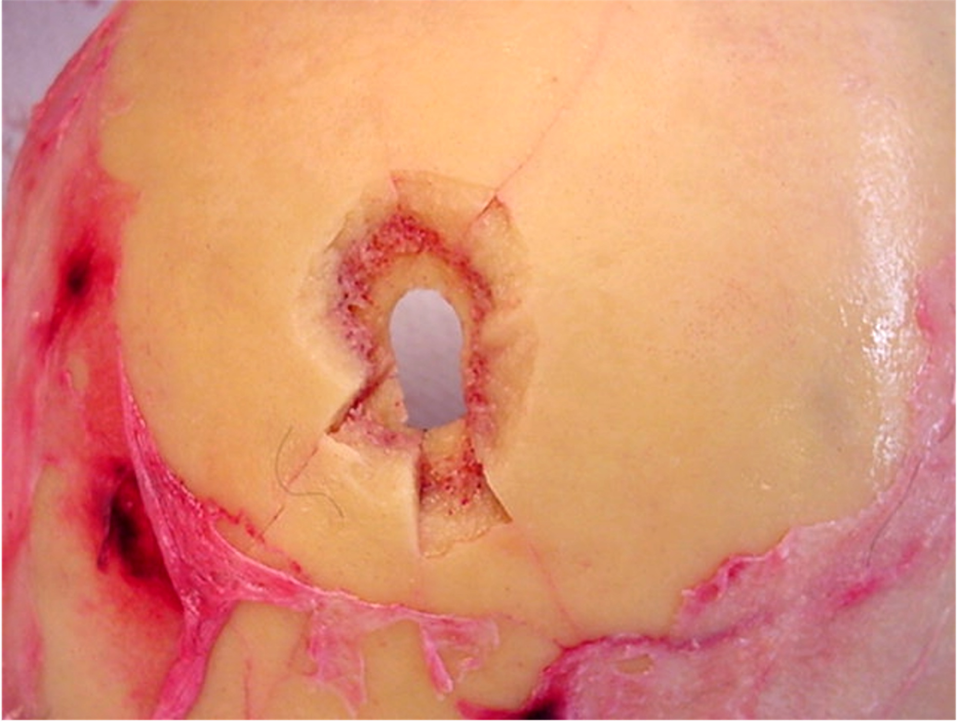
Keyhole lesion of the skull
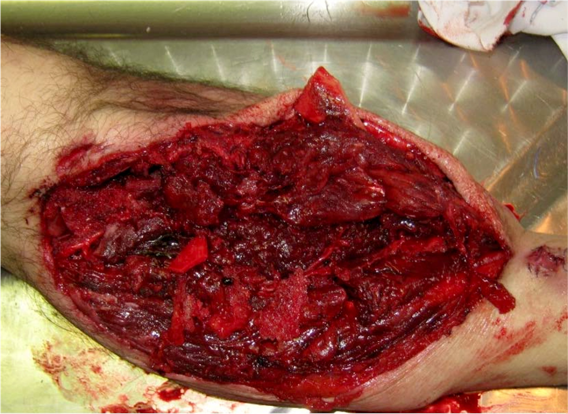
Gunshot wound to the head
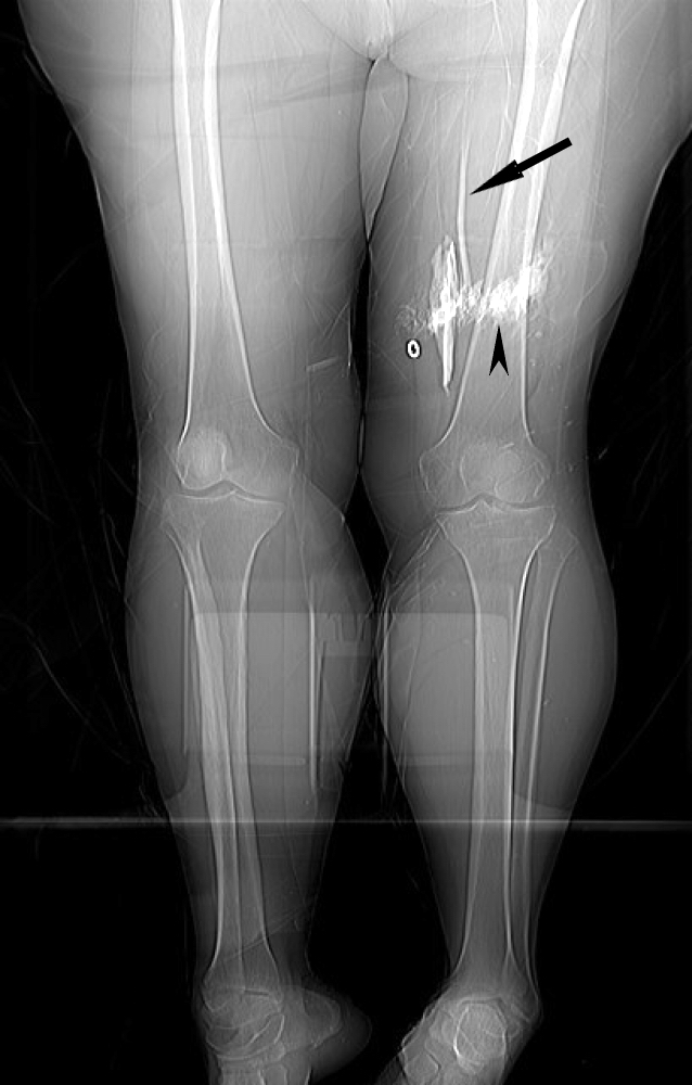
Rifle wound to the thigh
Rifle wound to the face
Rifle wound to the face
Clothes examination
Contributed by Lorenzo Gitto, M.D. and Robert Stoppacher, M.D.
Gunshot entrance wound
Contact gunshot entrance wound
Contributed by Lorenzo Gitto, M.D., Ponni Arunkumar, M.D., Luigi Bonaccorso, M.D. and the Cook County Medical Examiner's Office
Postmortem radiology
Contributed by Lorenzo Gitto, M.D., Ponni Arunkumar, M.D., Luigi Bonaccorso, M.D. and the Cook County Medical Examiner's Office
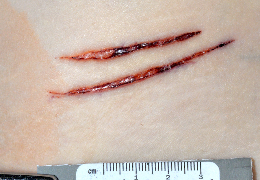
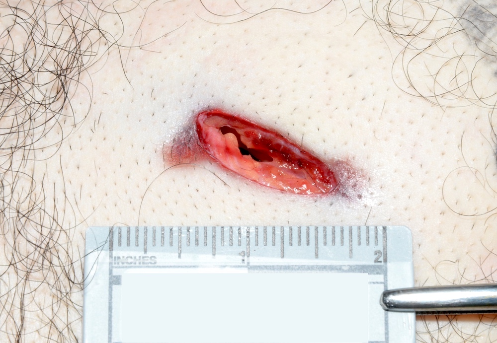
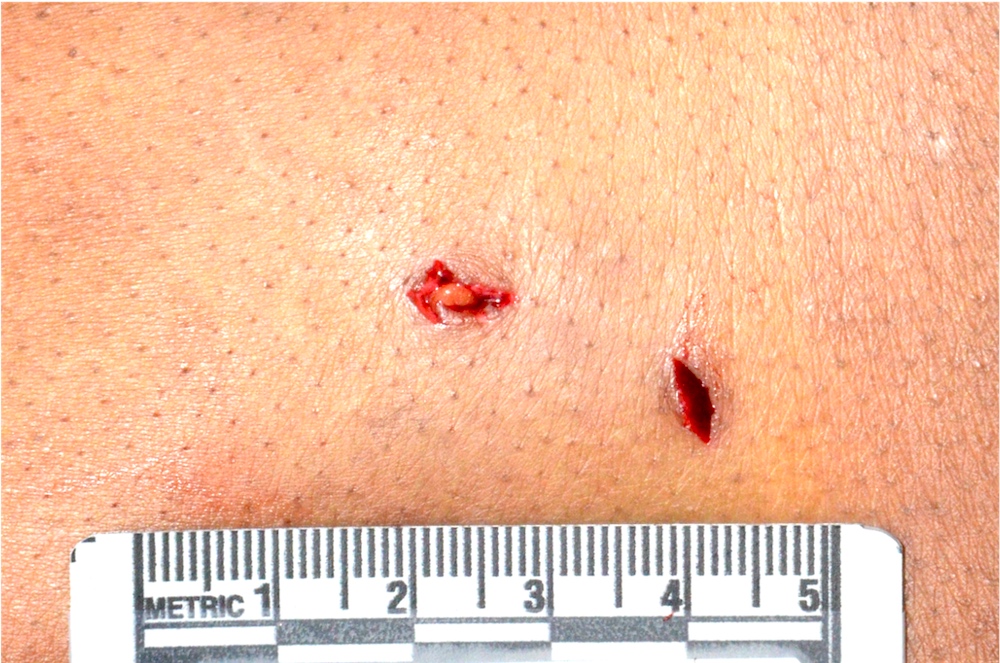
Incised wounds
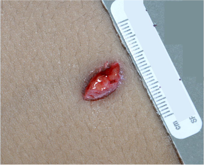
Stab wound
Chop wounds to the head
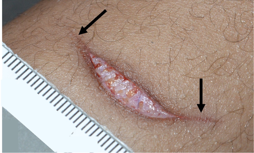
Wound tailing
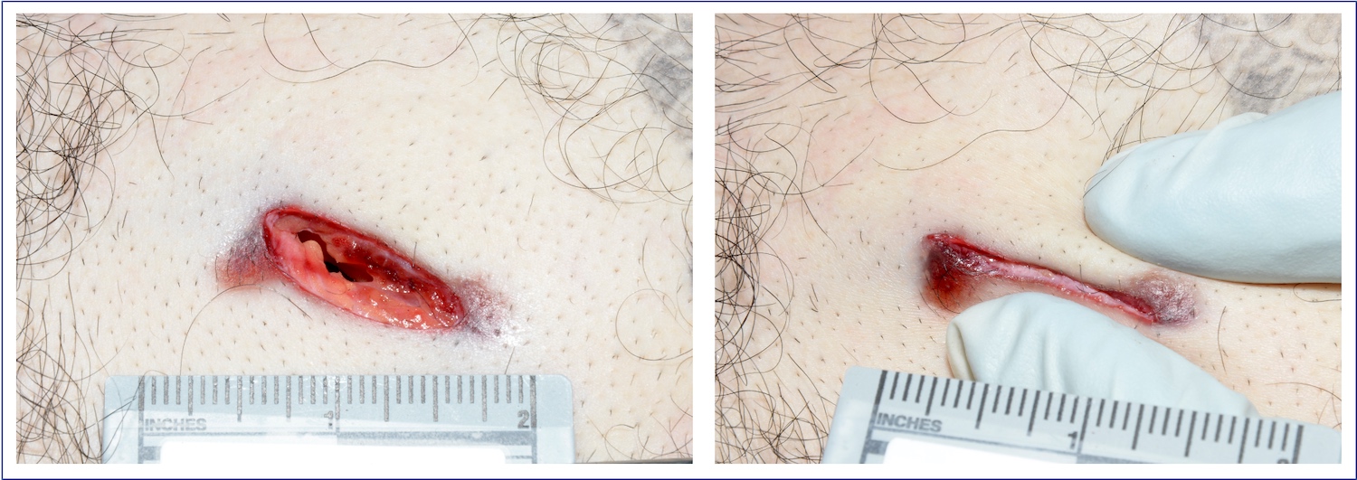
Wound margin approximation
Hesitation marks - recent
Hesitation marks - remote
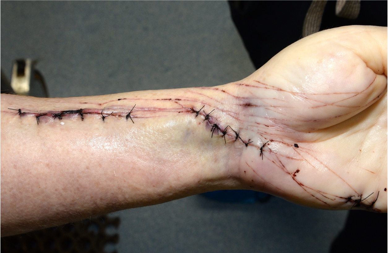
Medical intervention
V shaped stab wound
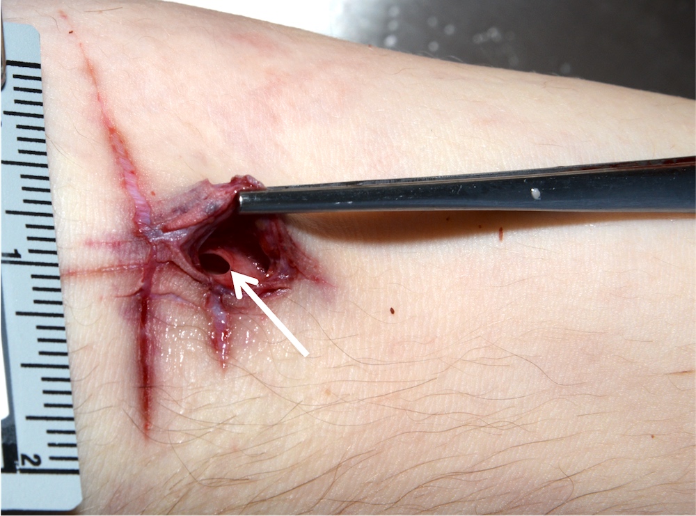
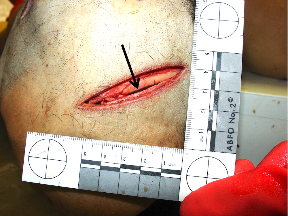
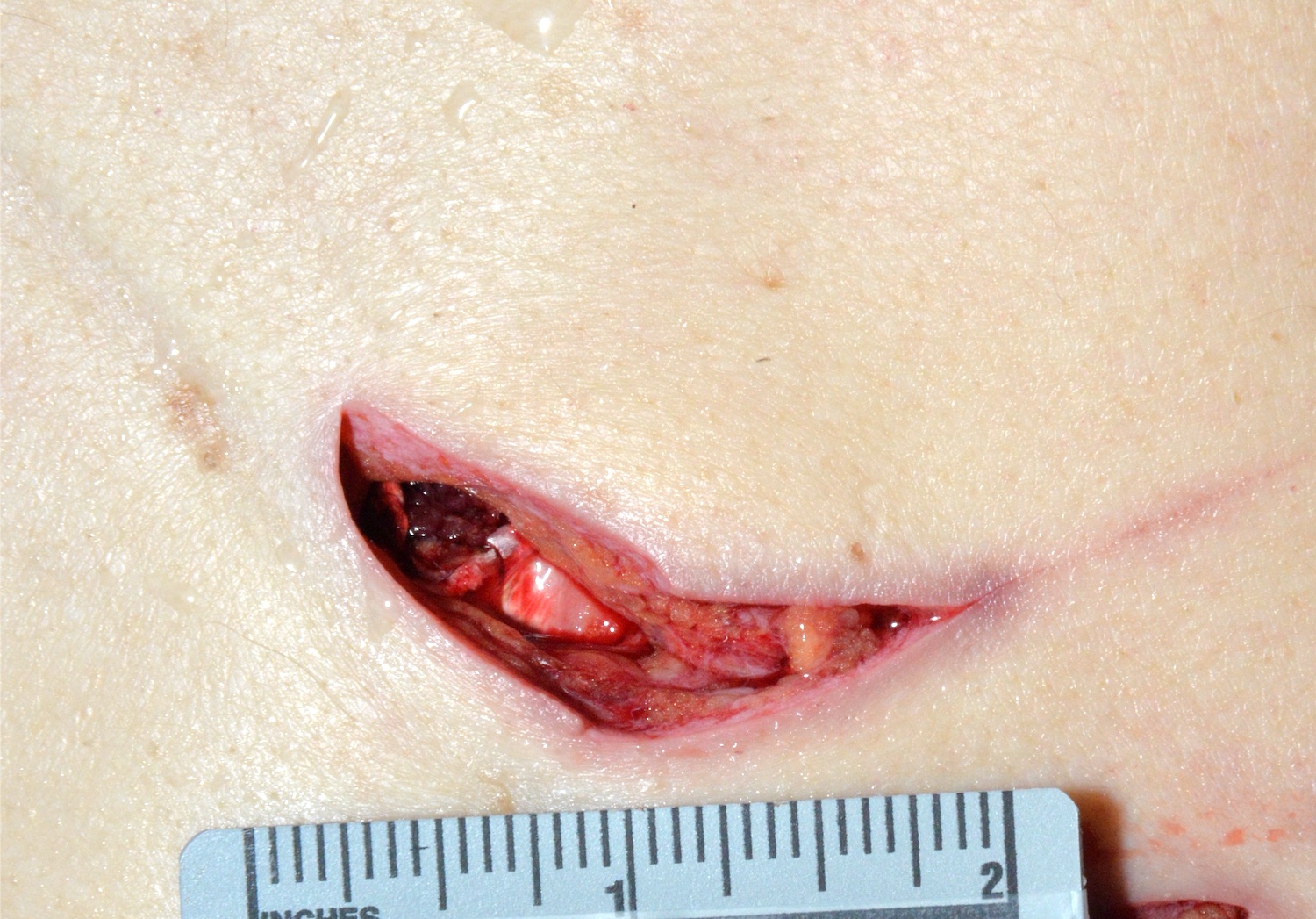
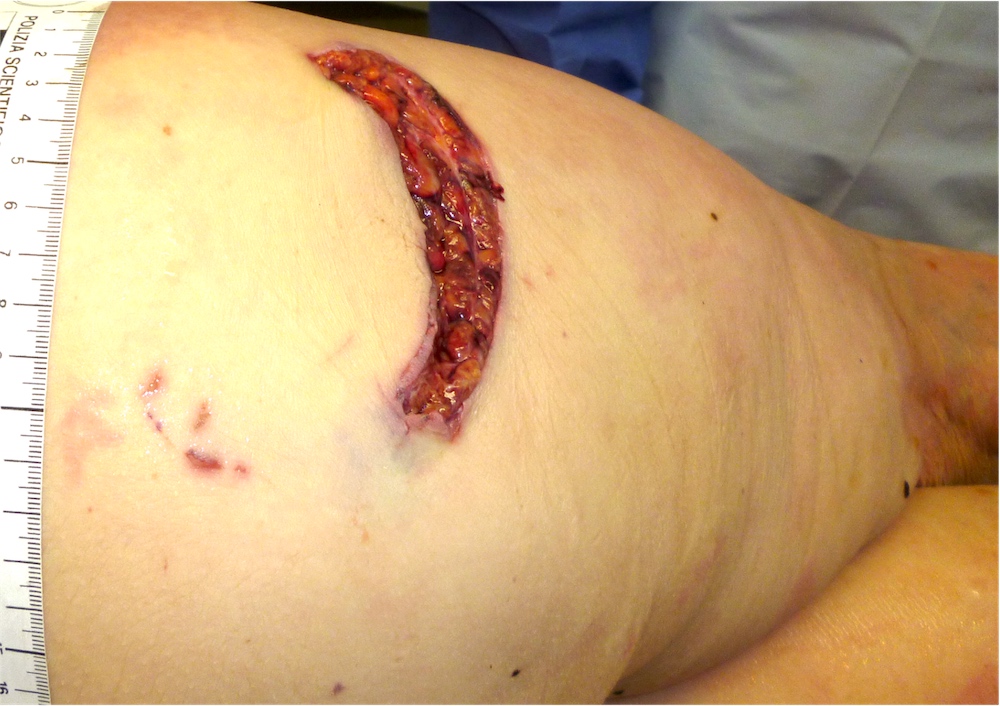
Vascular injury
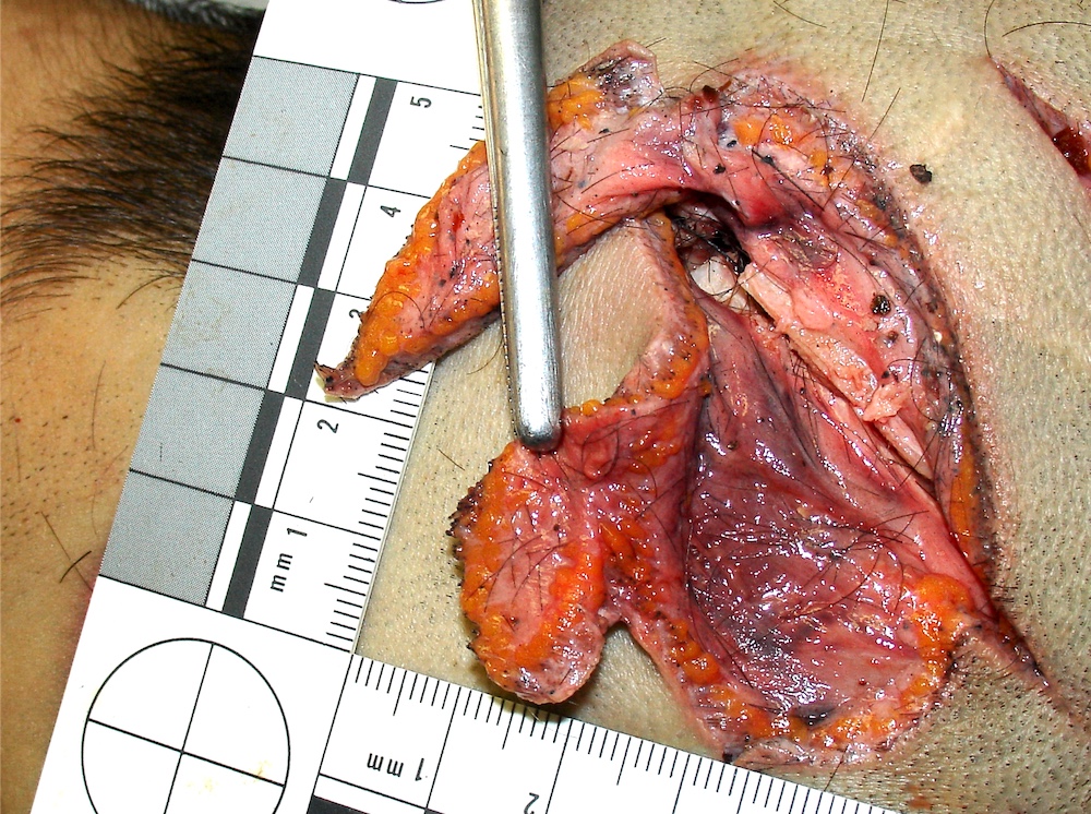
Partial skin flap
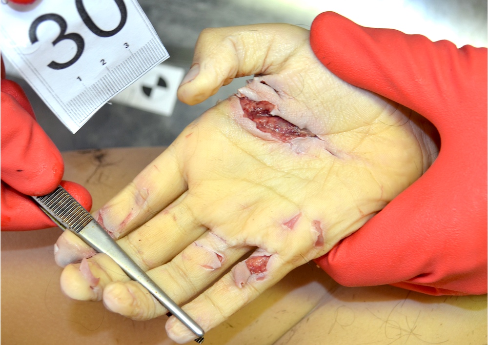
Active defense wounds
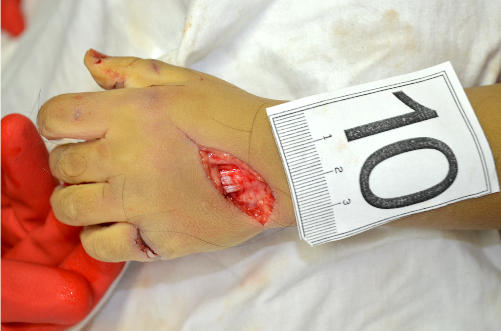
Passive defensive wounds
Broken glass
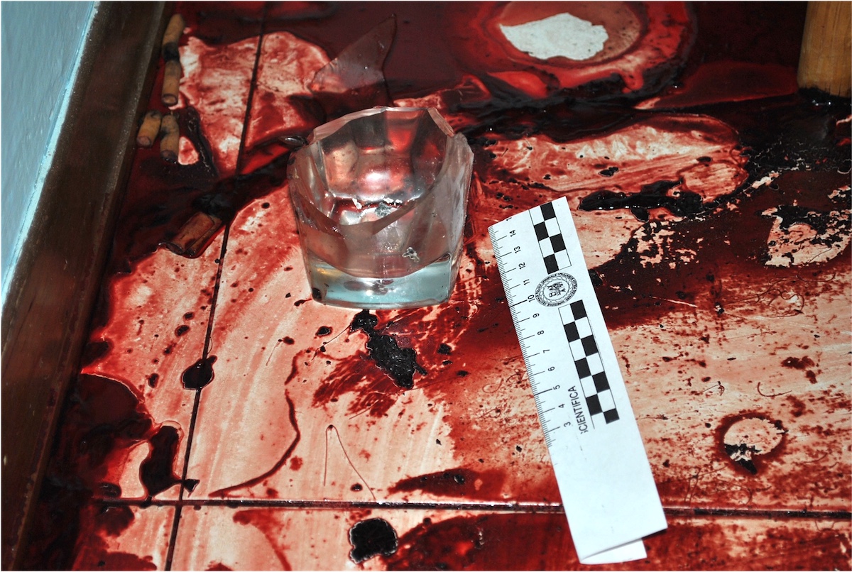
Broken glass - causative object
Scissors
Cross tip screwdriver
Flathead screwdriver
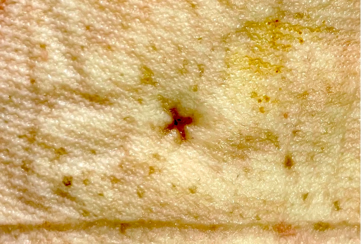
Ice pick - cutaneous lesion
Ice pick - skull lesion
Barbecue fork
Contributed by Lorenzo Gitto, M.D., Ponni Arunkumar, M.D., Luigi Bonaccorso, M.D. and the Cook County Medical Examiner's Office
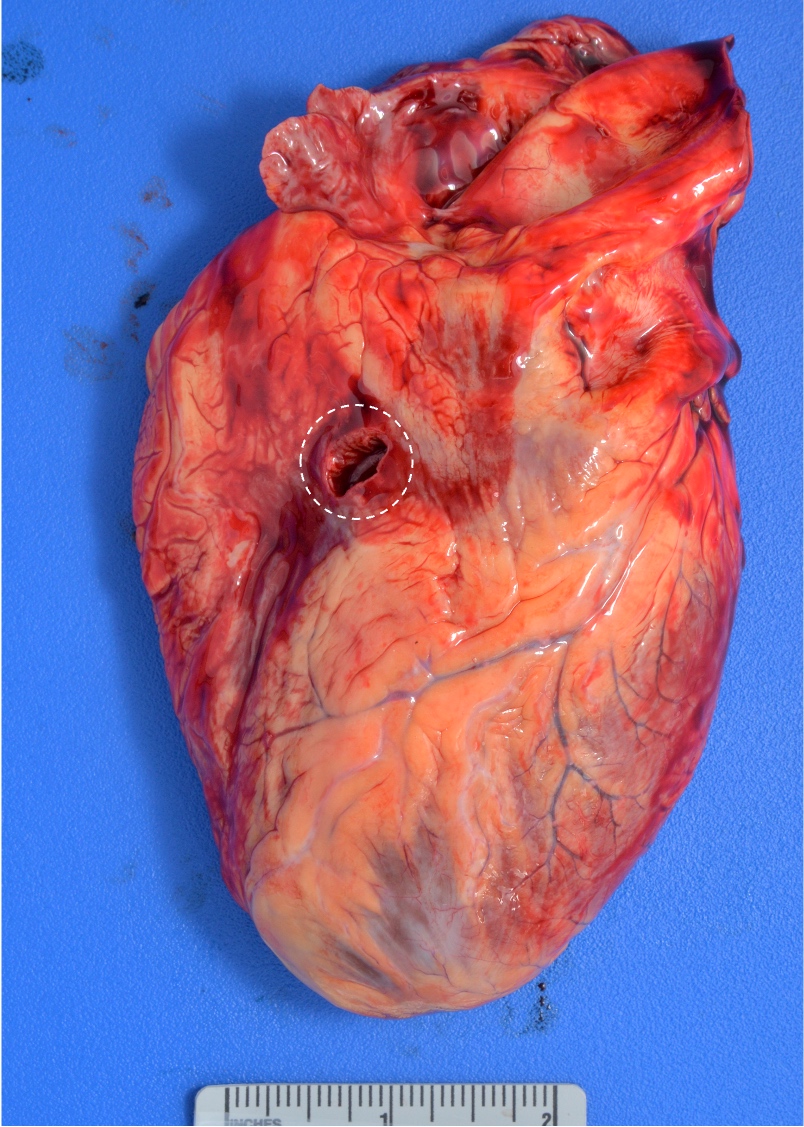
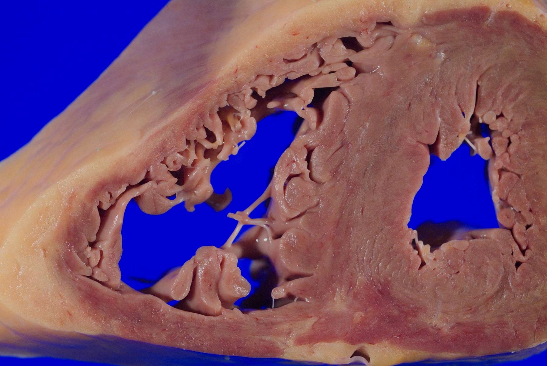
Heart stab wound
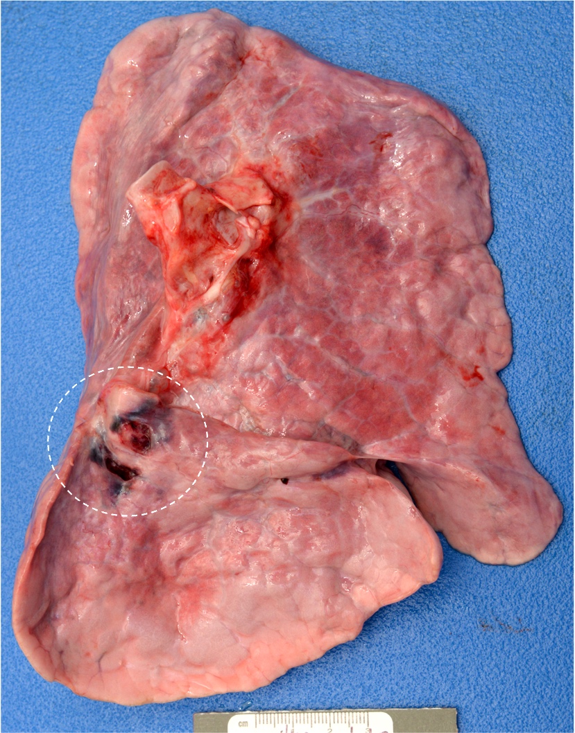
Lung stab wound
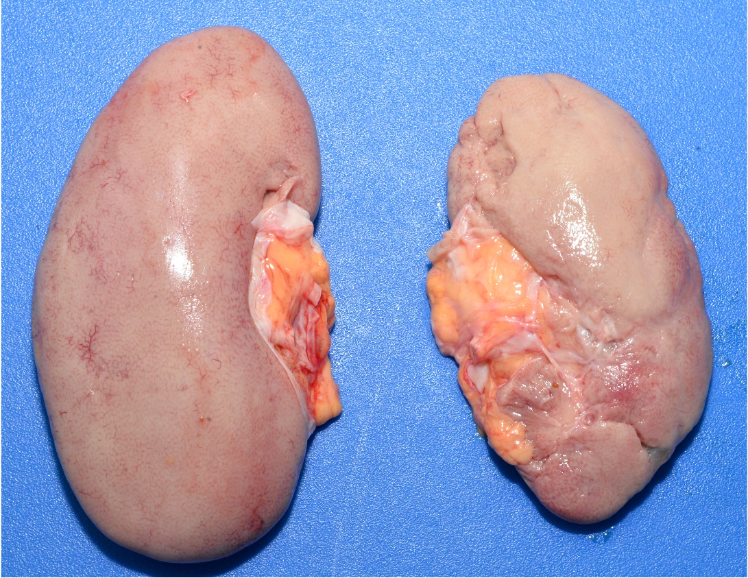
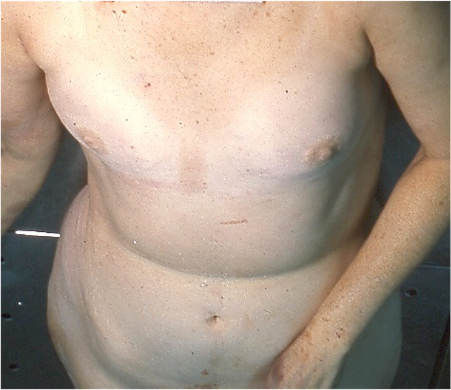
Pale organs
Contributed by Patrick Lantz, M.D. and Jerri McLemore, M.D.
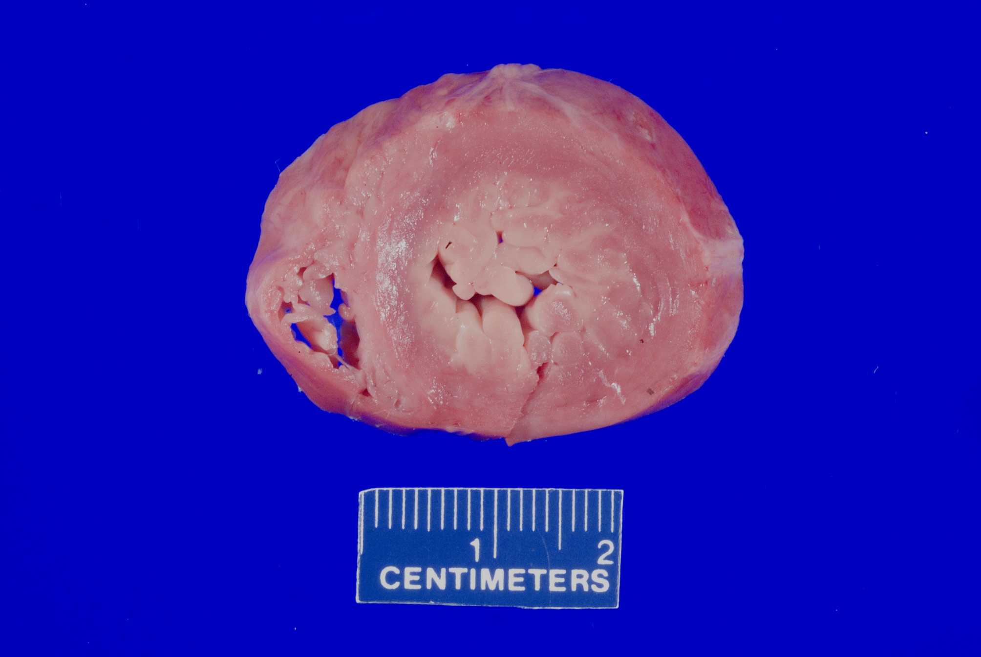
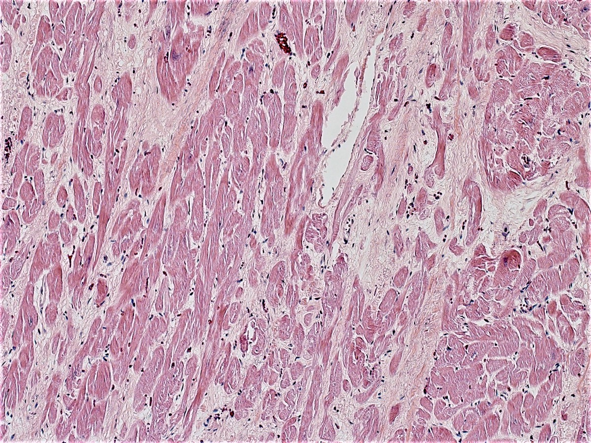
Arrhythmogenic right ventricular cardiomyopathy

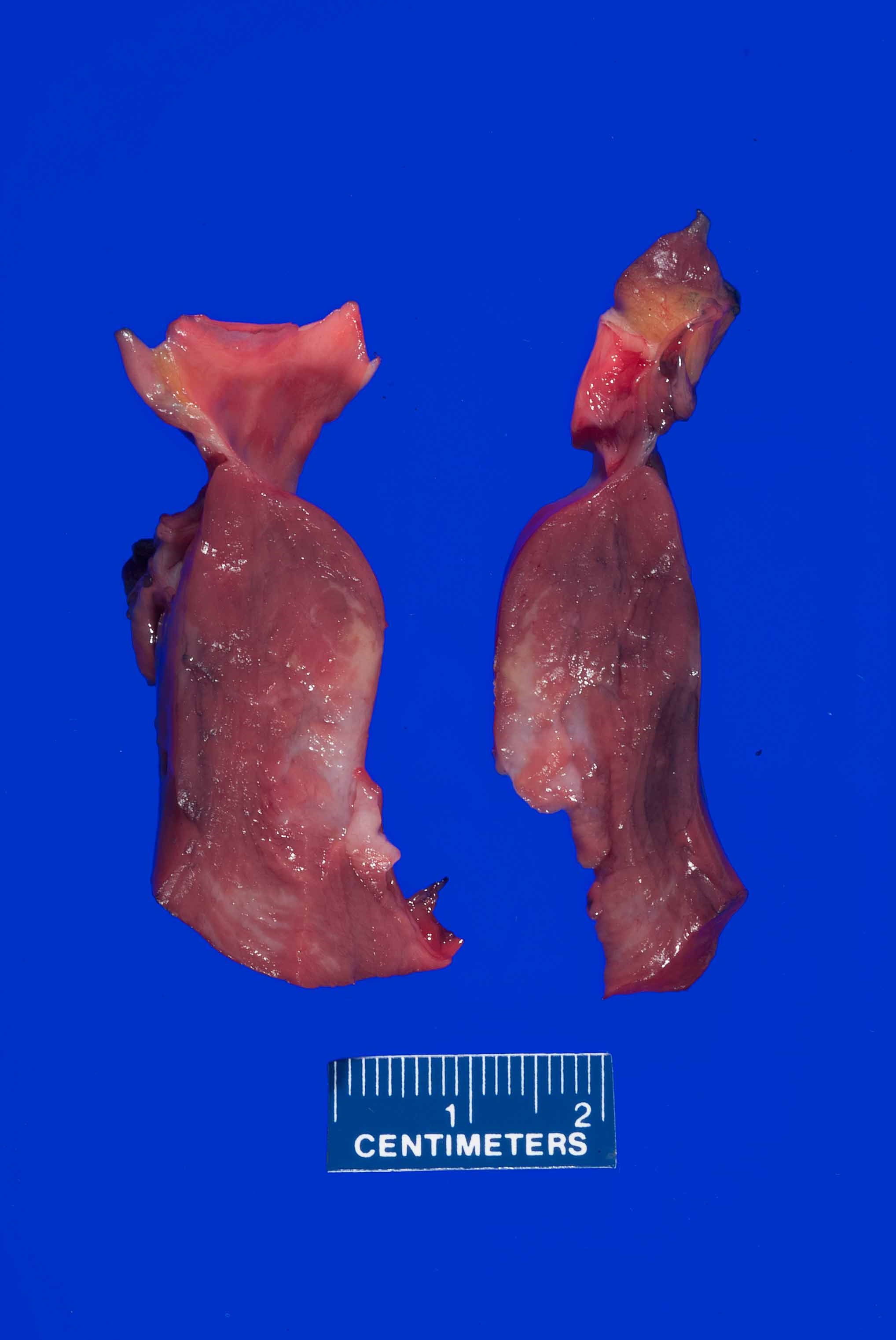
Histiocytoid cardiomyopathy valve and ventricular ring
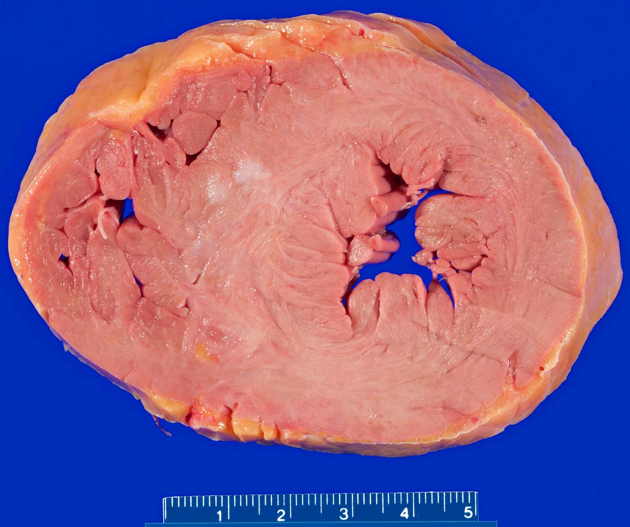
Hypertrophic and obstructive cardiomyopathy
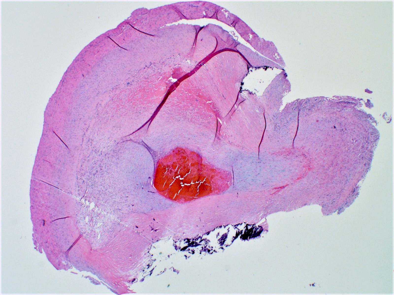
Sarcoidosis atrioventricular node
Contributed by Mehrnoosh Ghandili, M.D., Mark A. Giffen, Jr., D.O. and Jerri McLemore, M.D.
Acute atherosclerotic plaque rupture
Acute myocardial infarction
Cardiac myocyte disarray
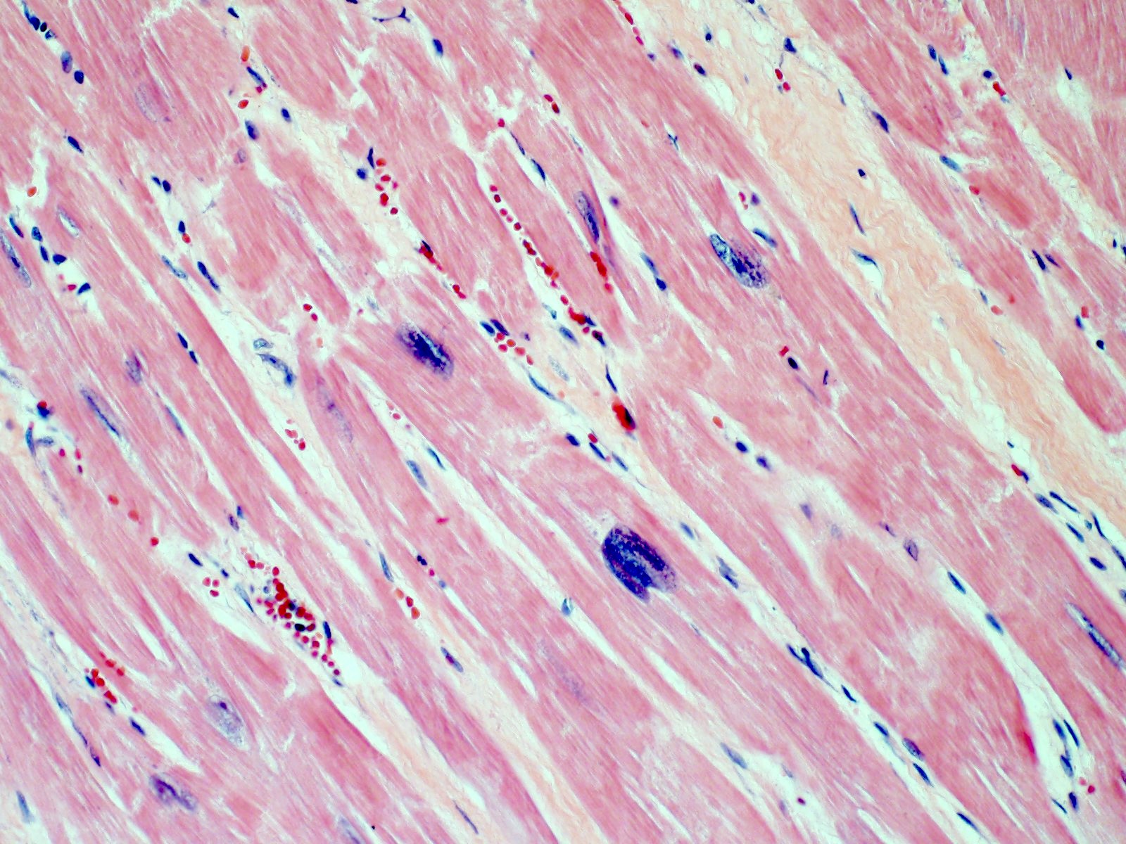
Cardiac myocyte hypertrophy
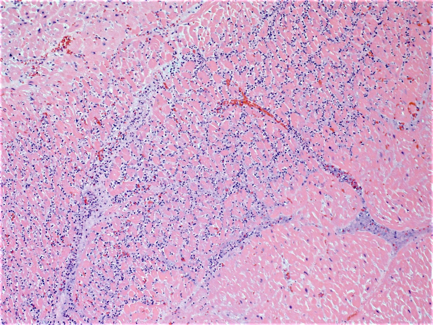
Giant cell myocarditis
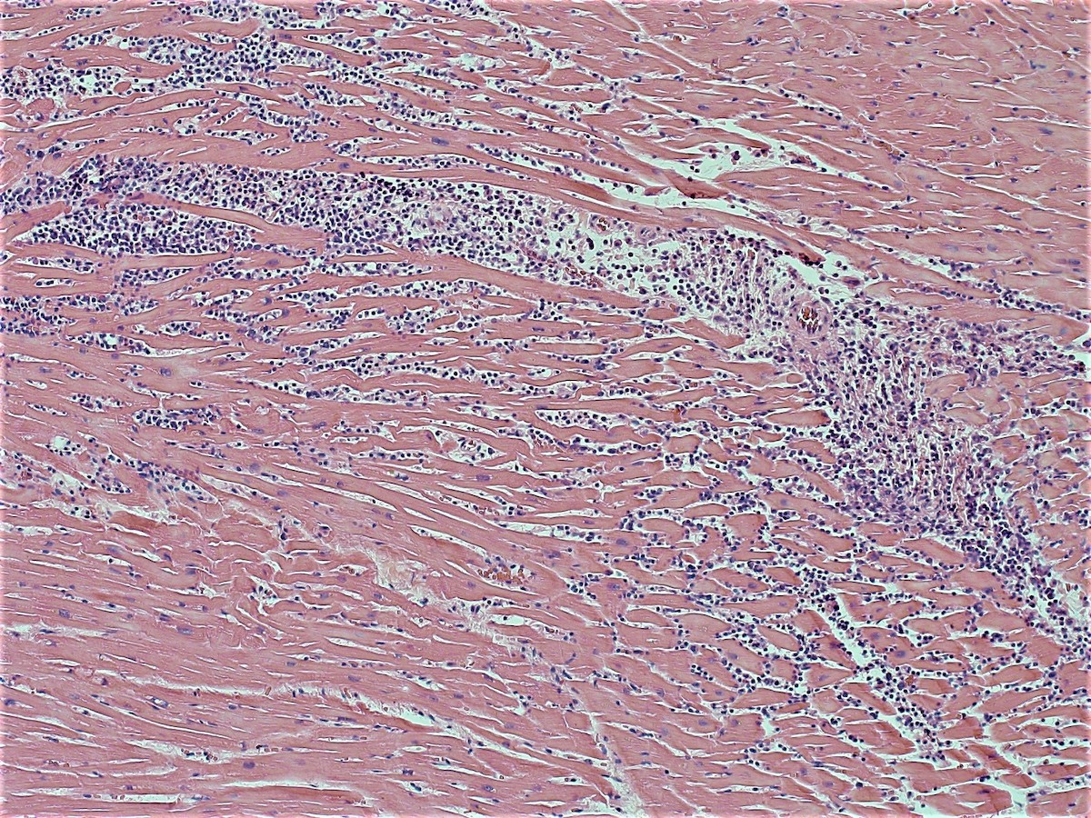
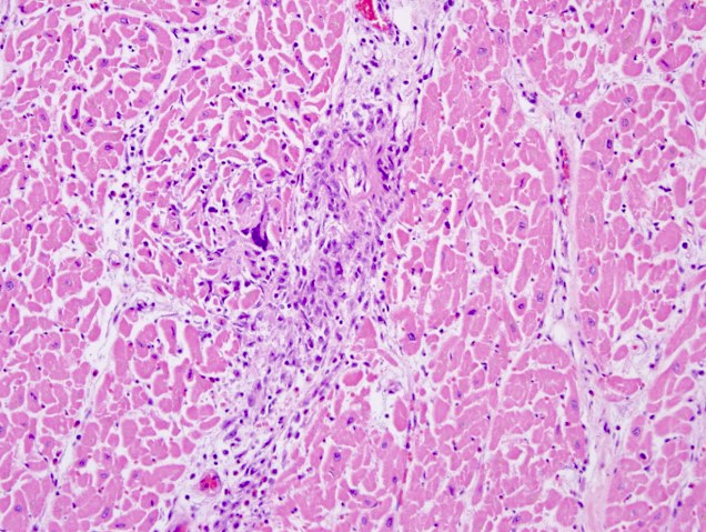
Lymphohistiocytic myocarditis
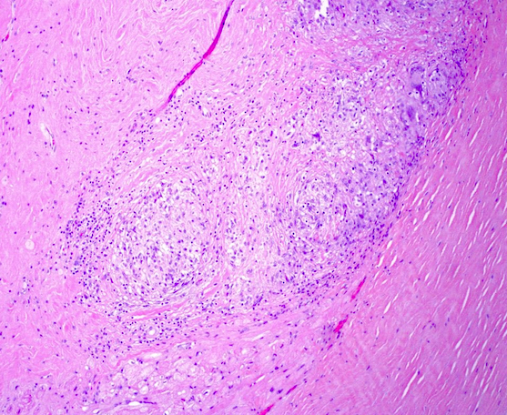
Sarcoidosis in atrioventricular node
Contributed by Mark A. Giffen, Jr., D.O.
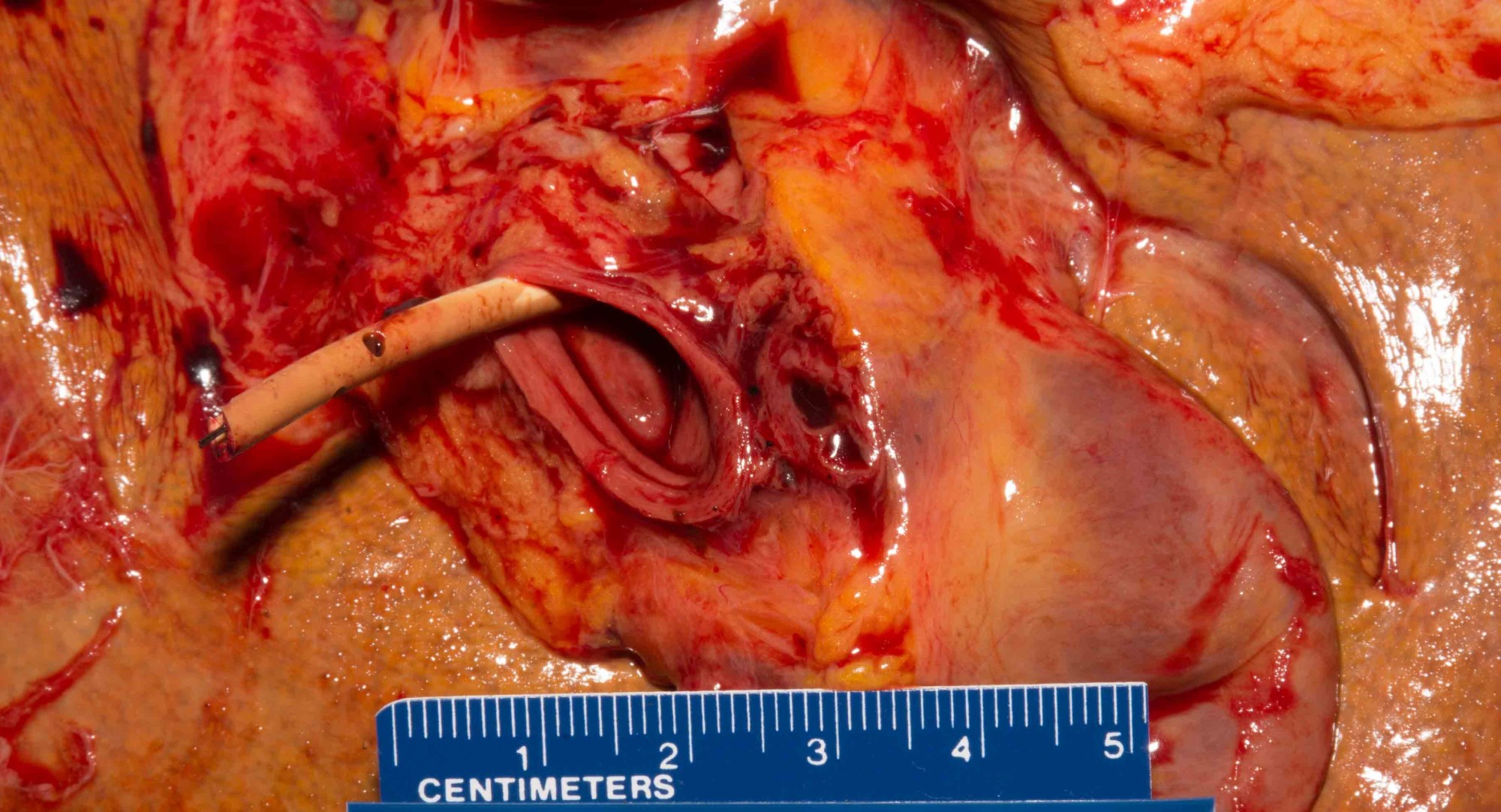
Catheter in portal vein
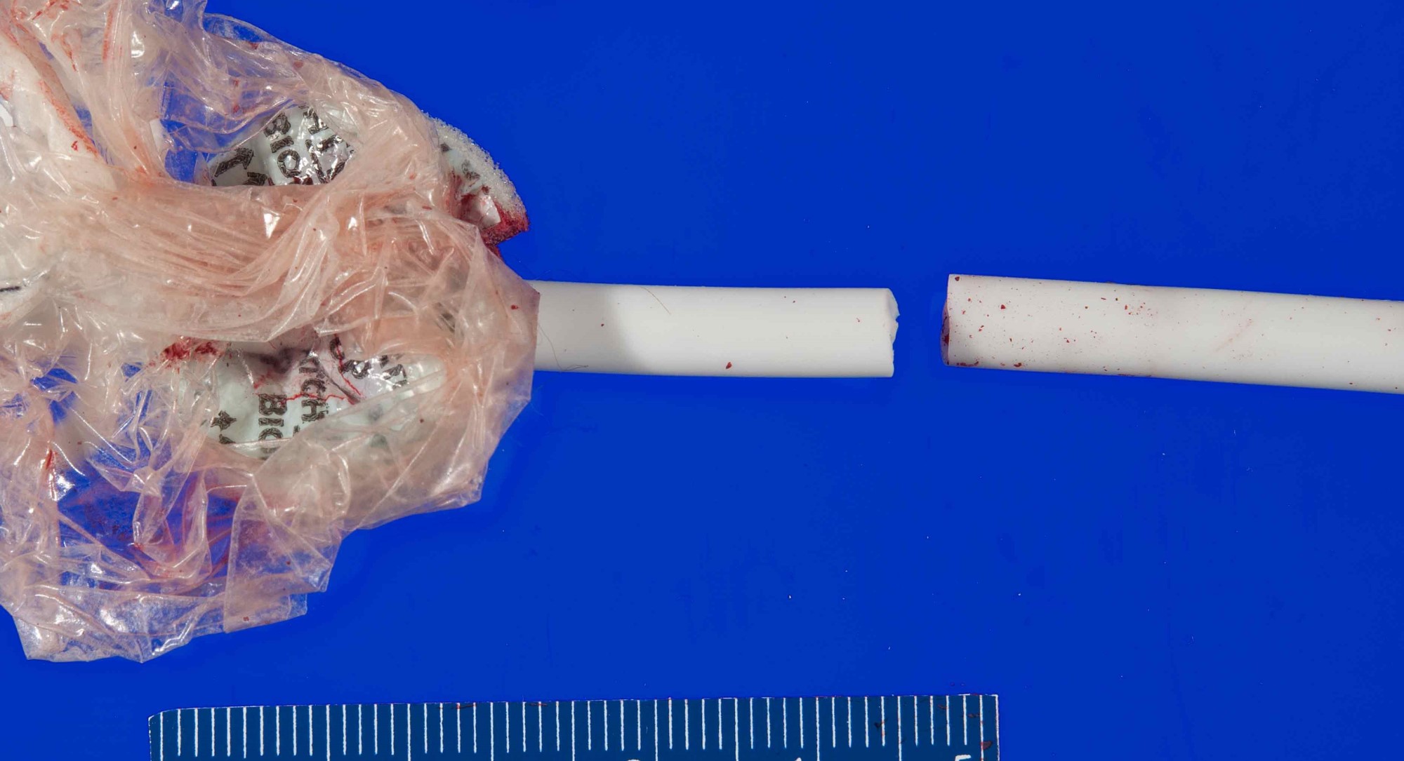
Vascular catheter transection
Contributed by Michel Tawil, M.D.

Petechial hemorrhage
Contributed by Michel Tawil, M.D.

Axonal spheroids
Catanese: 2016
Dettmeyer: 2018
DiMaio: 2021
DiMaio: 2021
Dolinak: 2005
Ely: 2022
Husain: 2021
Madea: 2014
Rutty: 2017
Saukko: 2015
Spitz: 2020
Find related Pathology books: autopsy, forensic, pediatric
