


Contributed by Marilin Rosa, M.D.
Actinomyces
Images hosted on other servers:

Actinomyces

IUD related changes
Images hosted on other servers:

Colposcopy
Contributed by Gulisa Turashvili, M.D., Ph.D., Seema Khutti, M.D., Jijgee Munkhdelger, M.D., Ph.D. and Andrey Bychkov, M.D., Ph.D.
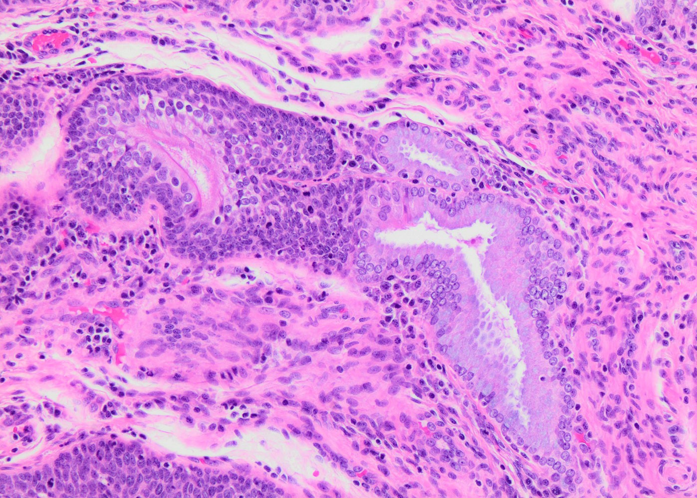
AIS admixed with HSIL
AIS, gastric type
p16 in SMILE
Mucicarmine in SMILE
SMILE
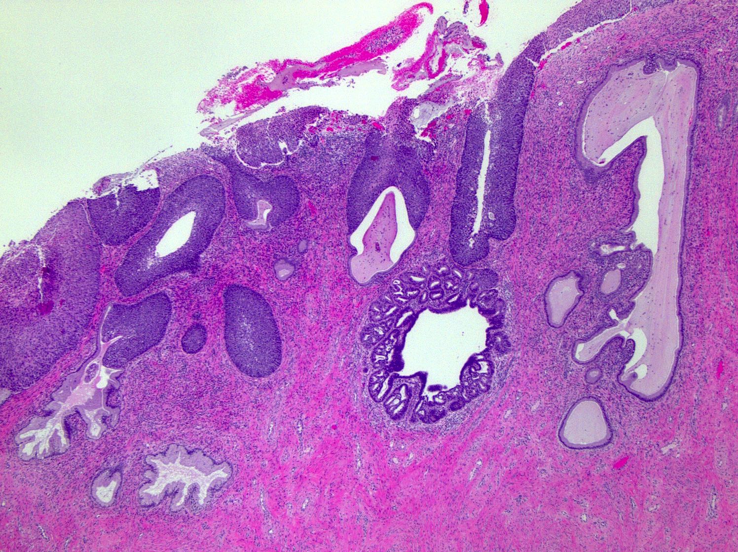
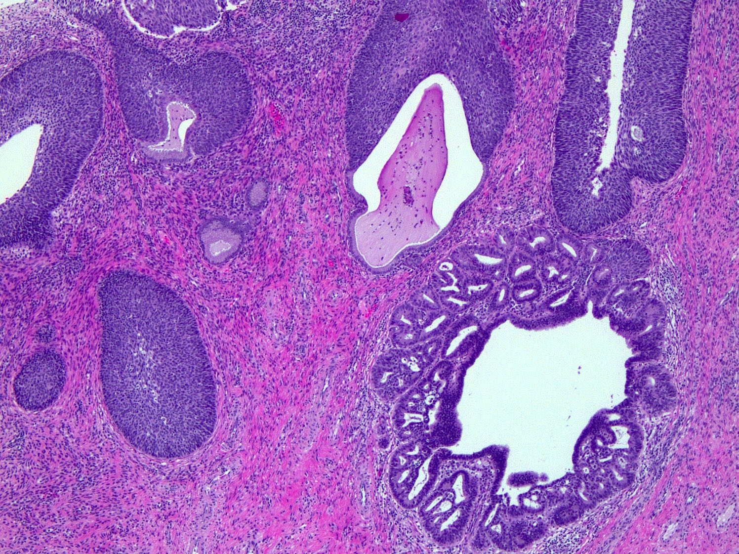
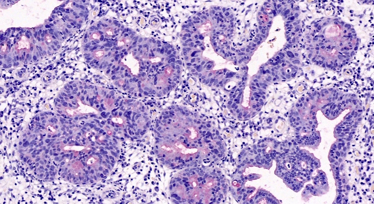
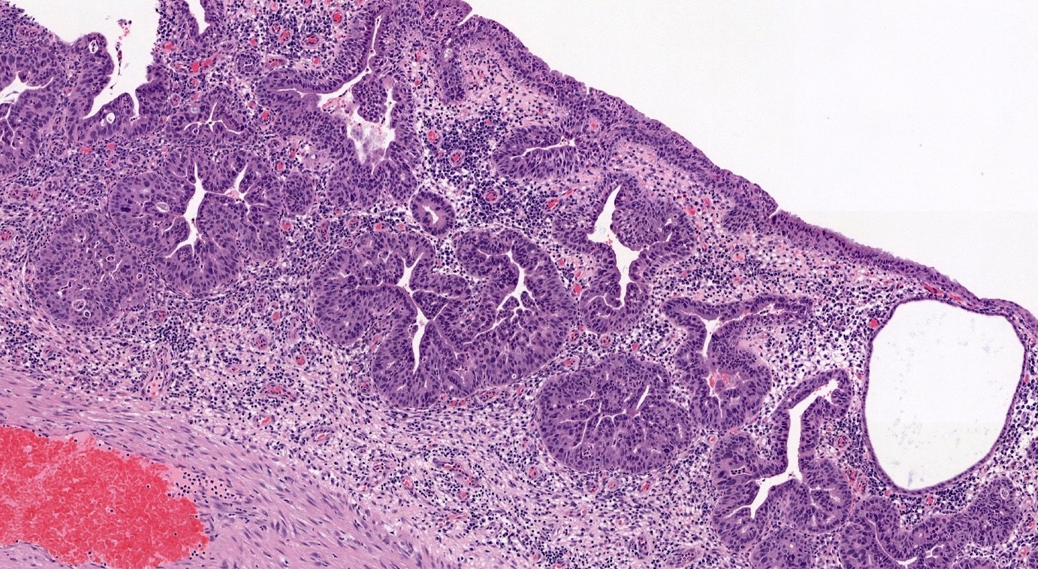
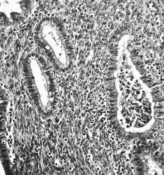
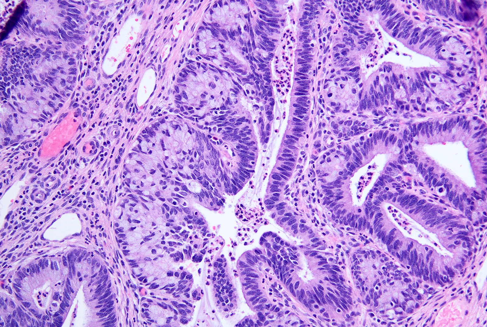
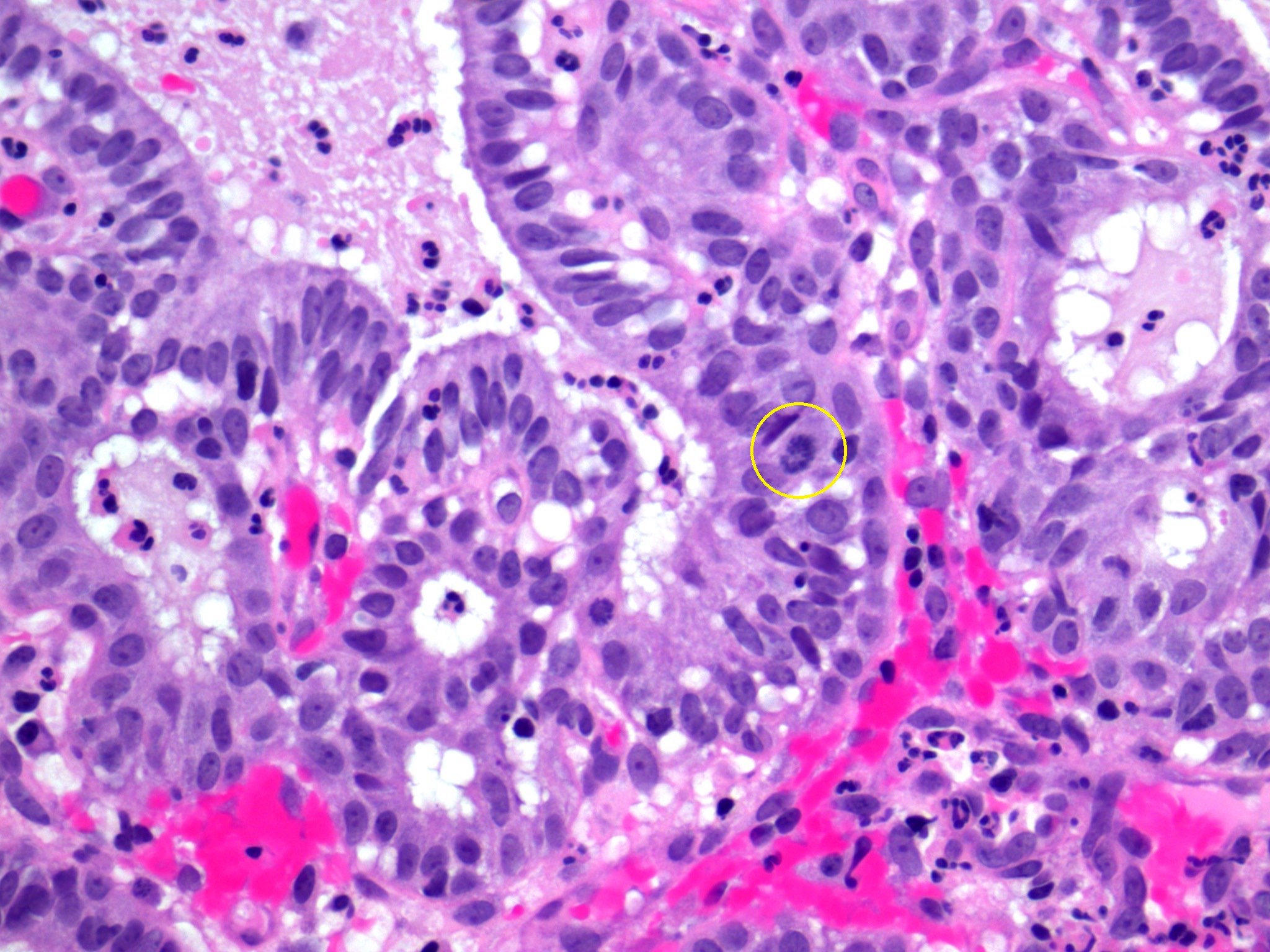
Glands with crowding and stratification
Abundant mitosis and apoptosis
AIS with adjacent uninvolved glands
Transition to AIS
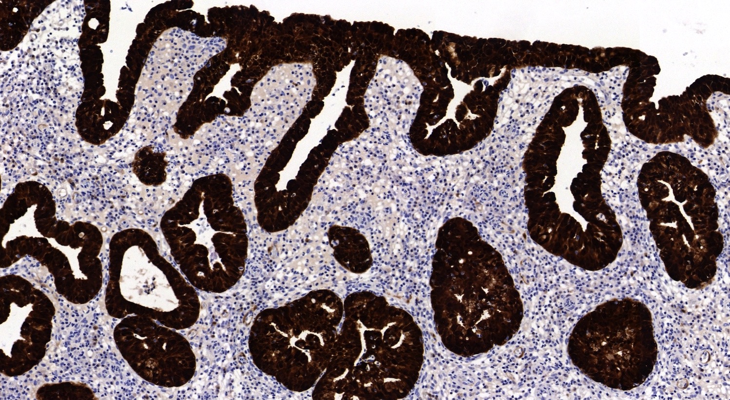
AIS p16
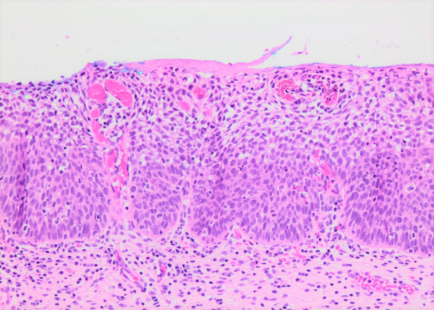
High grade cervical
glandular intraepithelial
neoplasia
High power
Nuclear atypia
Images hosted on other servers:
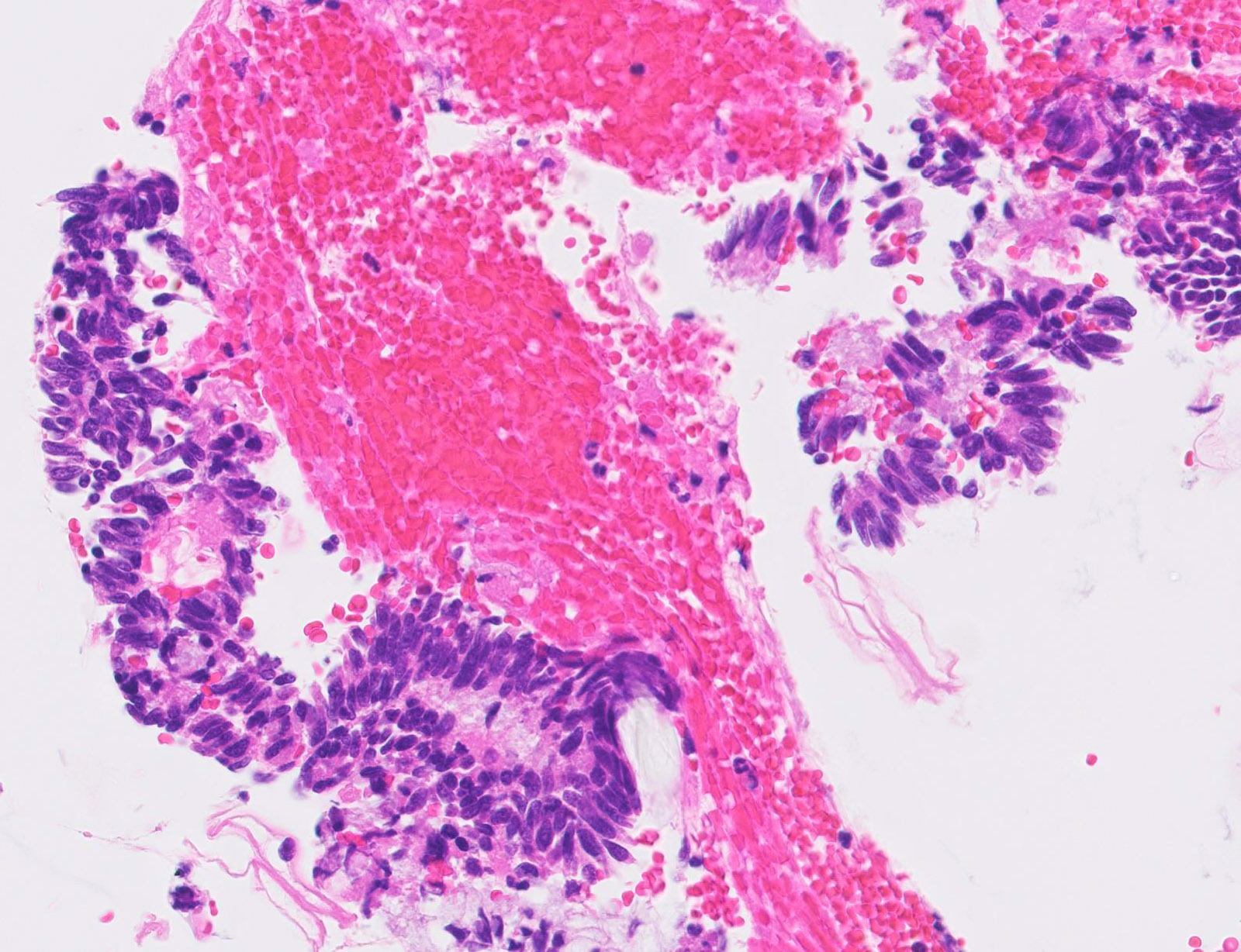
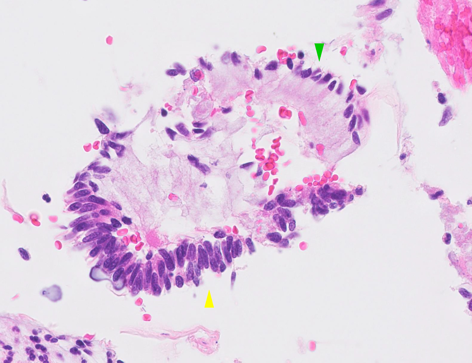
Atypical glandular cells

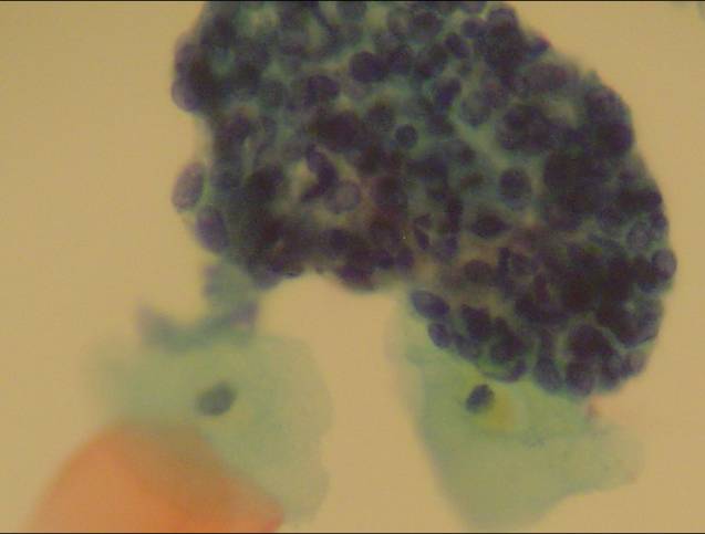
Groups of atypical rosette-like structures

Feathering


Feathering and enlarged hyperchromatic nuclei

Palisading and enlarged nuclei

Palisading and feathering

Forming a rosette-like structure


Enlarged nuclei

Feathering and enlarged nuclei


Homogenous chromatin pattern

Slightly atypical metaplastic cells
Images hosted on other servers:

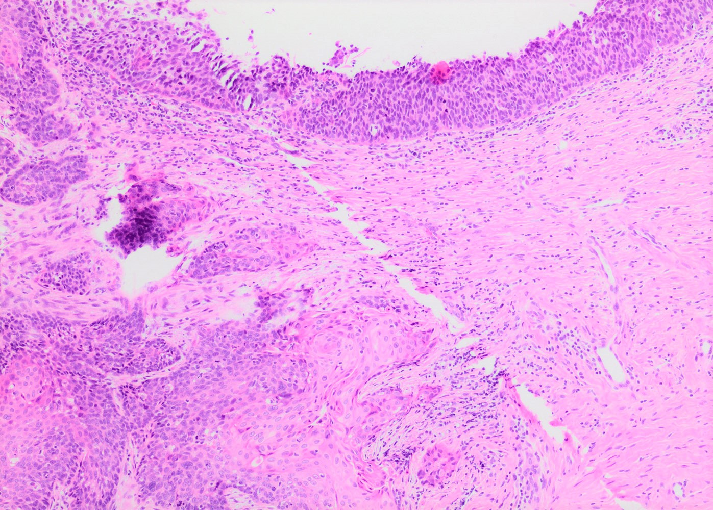
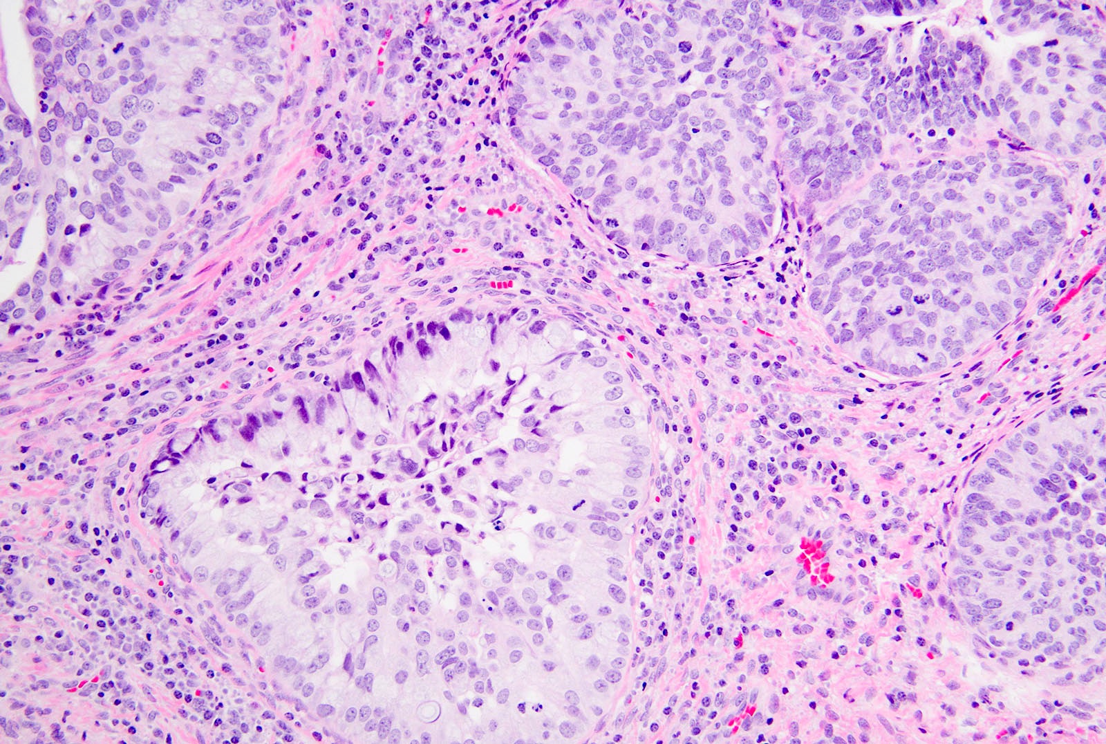
Rounded nests of basaloid cells infiltrating the stroma

Small acini

Palisading arrangement around cellular nest

Cervical stroma

Cytologic and histopathologic features

IHC stains

EMA
Contributed by Ihab Hosny, M.D.
Various images
AFIP images
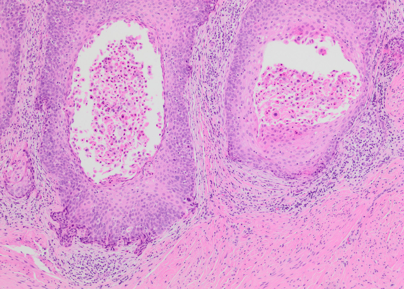
Cribriform architecture and basement membrane material
Contributed by Carlos Parra-Herran, M.D.

Contributed by Ihab Hosny, M.D.


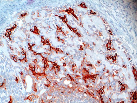
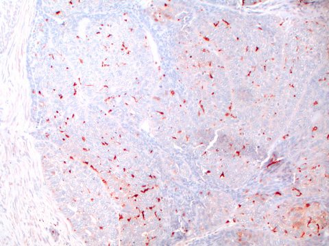
Vascular invasion

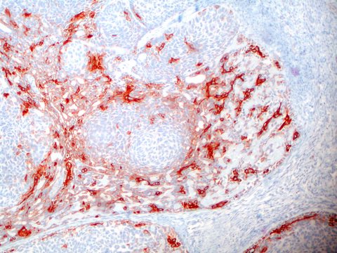
Actin
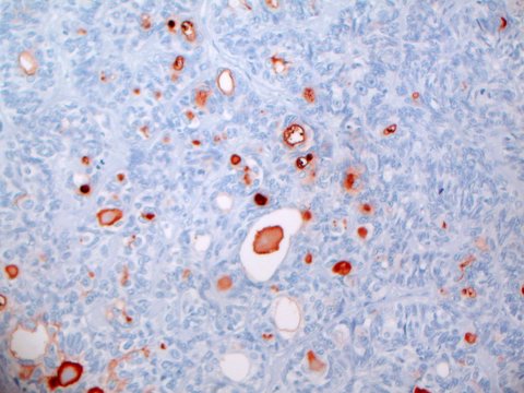
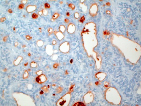
CEA
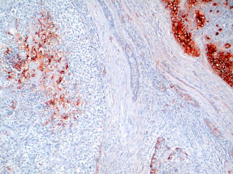
EMA
High molecular weight keratin
S100
Images hosted on other servers:

Three dimensional structures
Contributed by Leonel Maldonado, M.D.
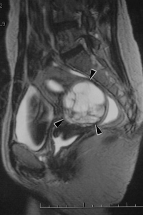
MRI of the pelvis
Sagittal T2
Images hosted on other servers:

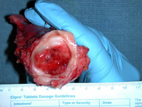
Bulging tumor from anterior cervix (fig 1)
Contributed by Leonel Maldonado, M.D.
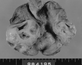
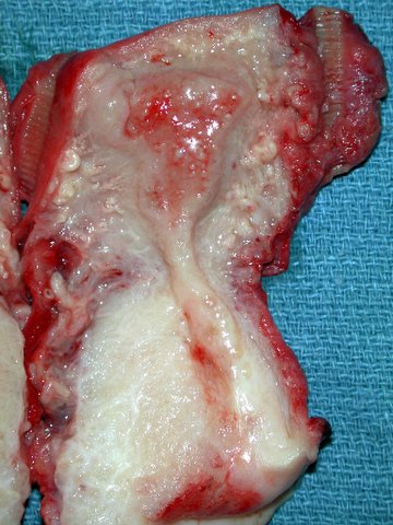
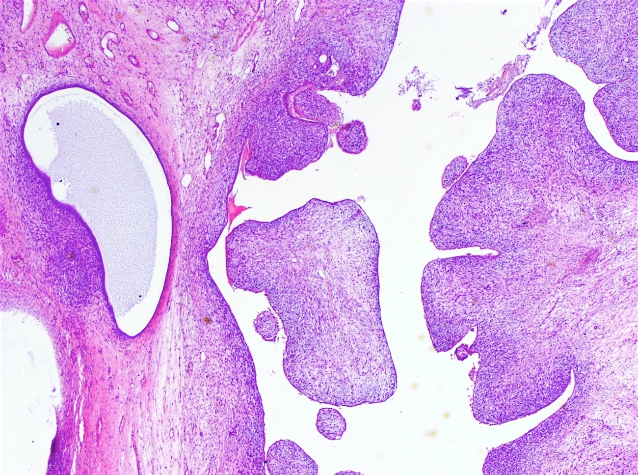
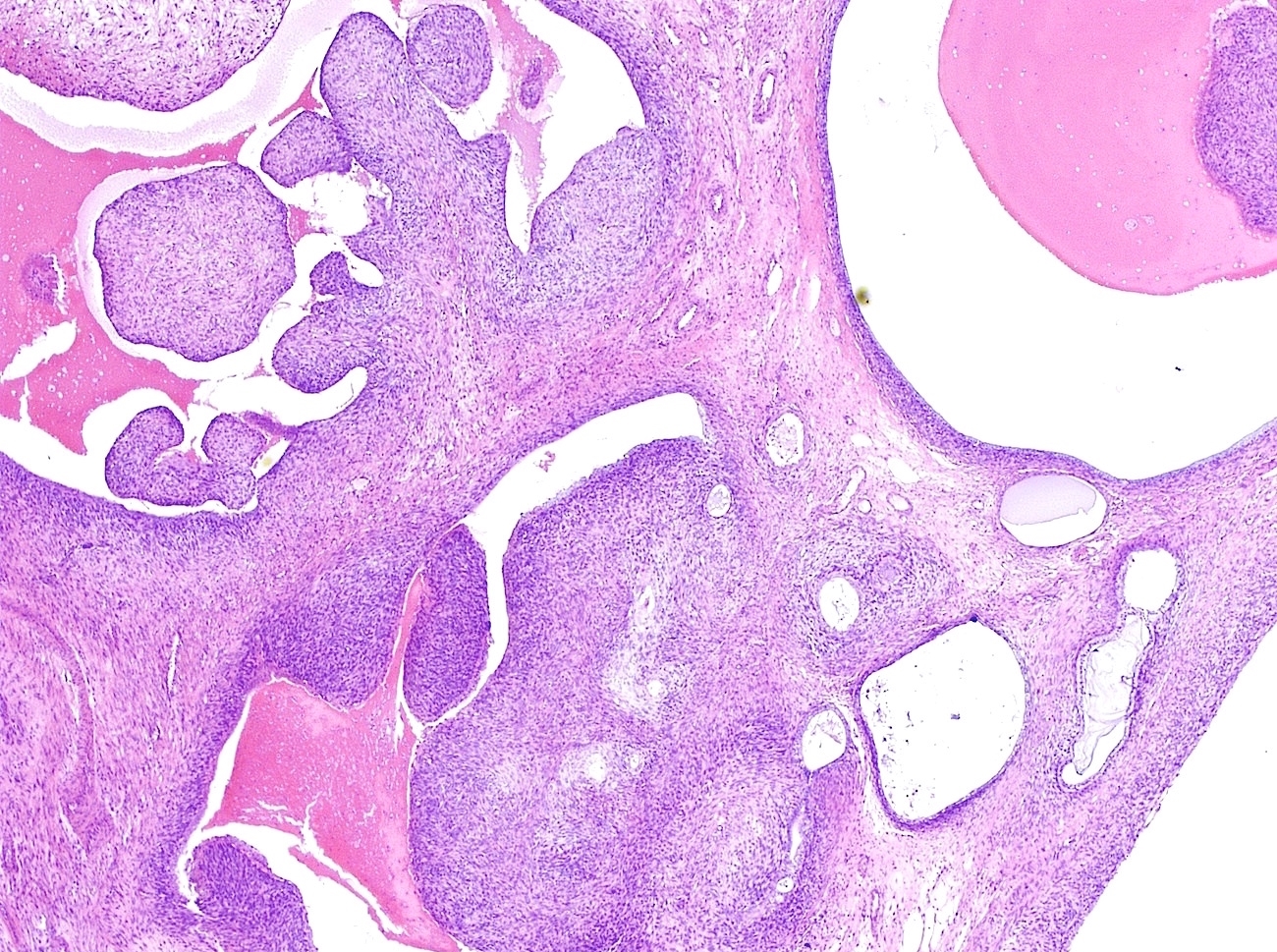
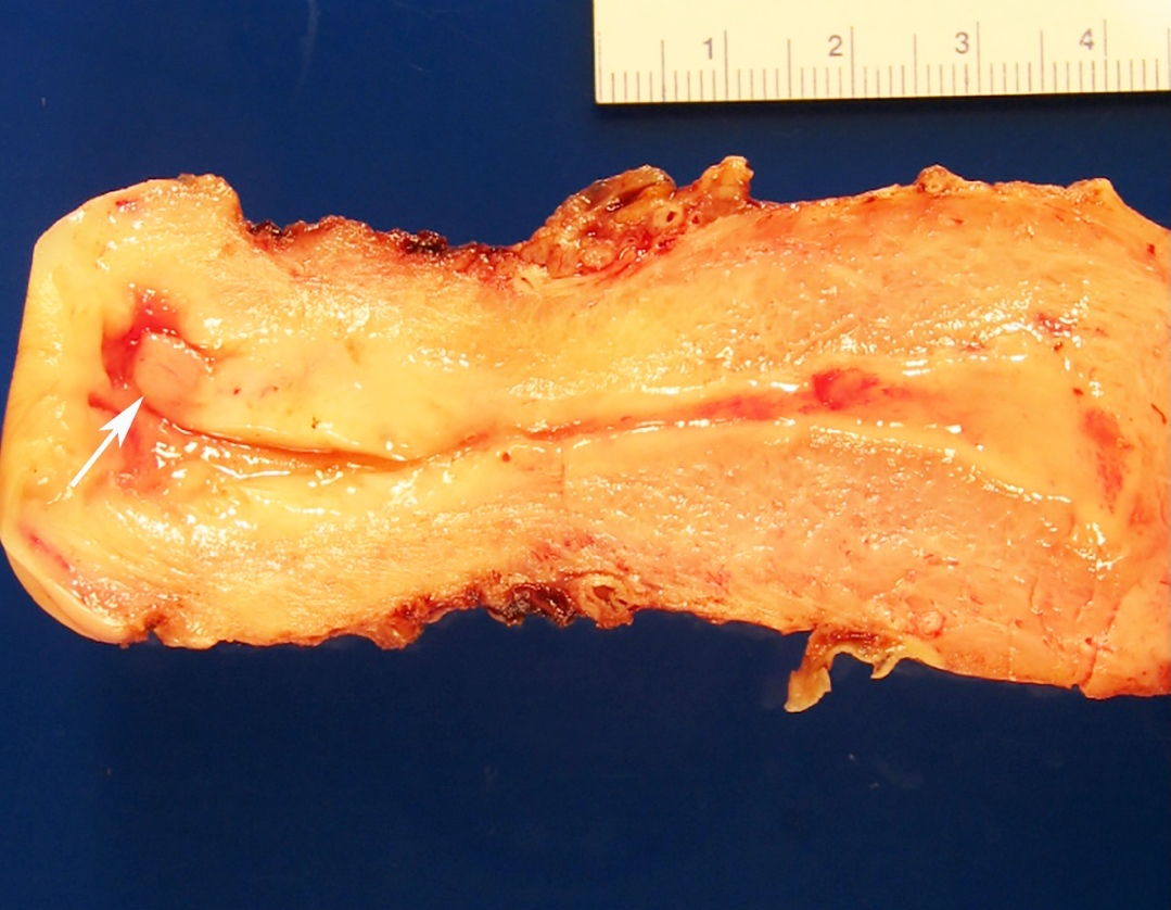
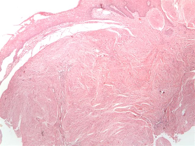
Endocervical mural tumor
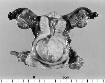
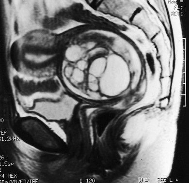
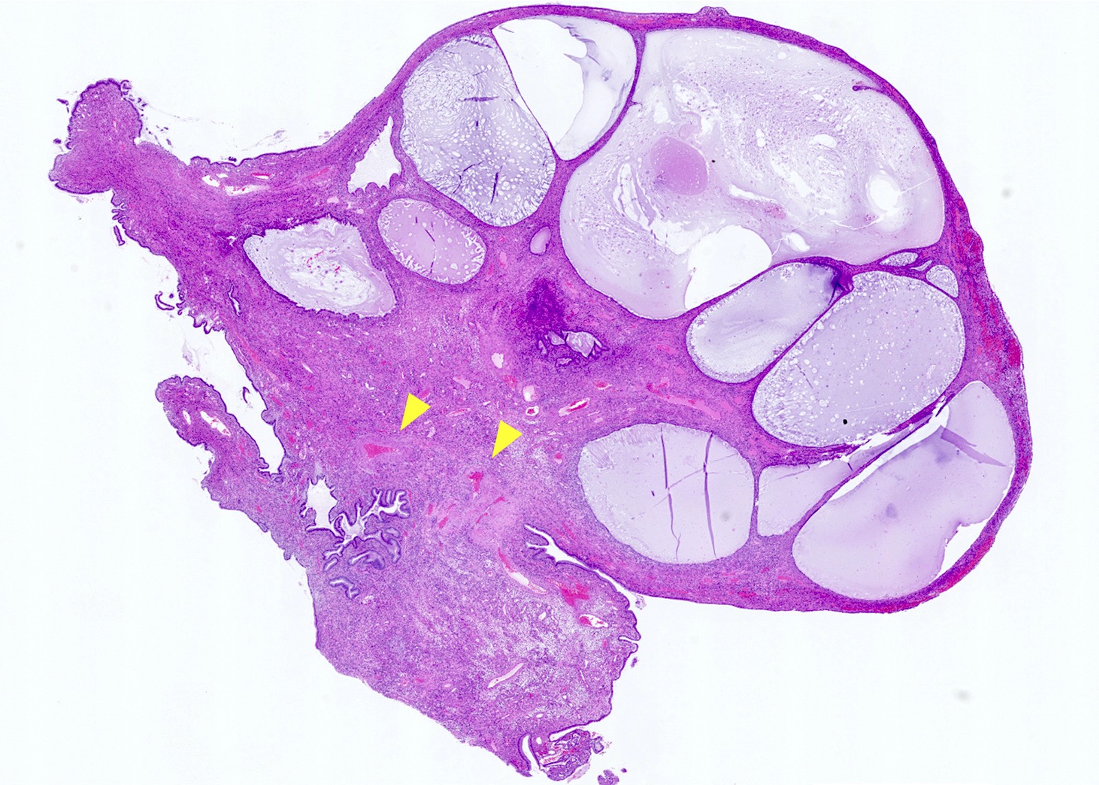
Polypoid mass from uterine cervical wall
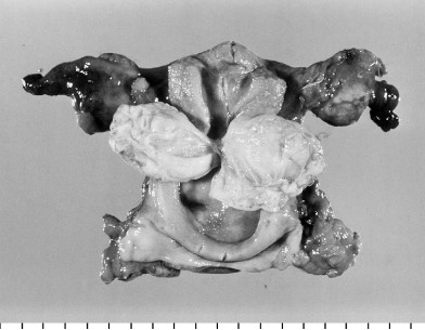
Polypoid mass with numerous cysts
Contributed by Leonel Maldonado, M.D.
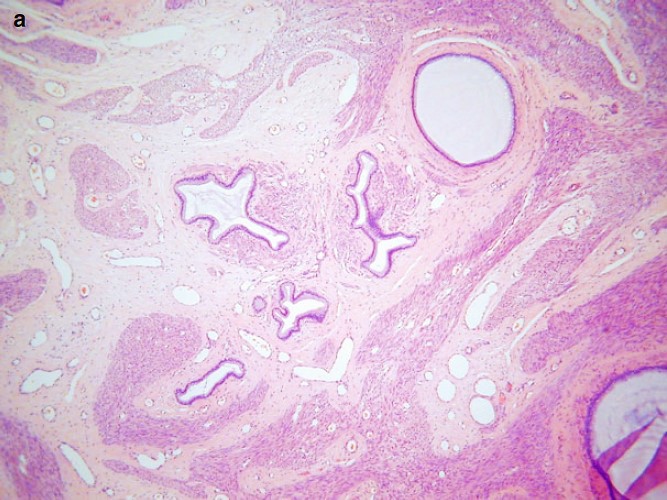
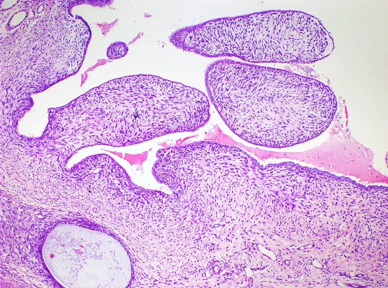
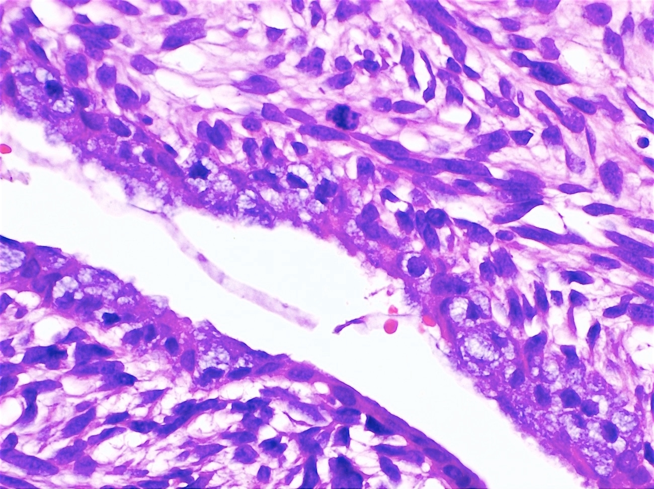
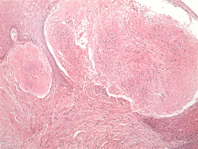
Endocervical type glands surrounded by smooth muscle
Contributed by Leonel Maldonado, M.D.

Fragment of endocervical polyp
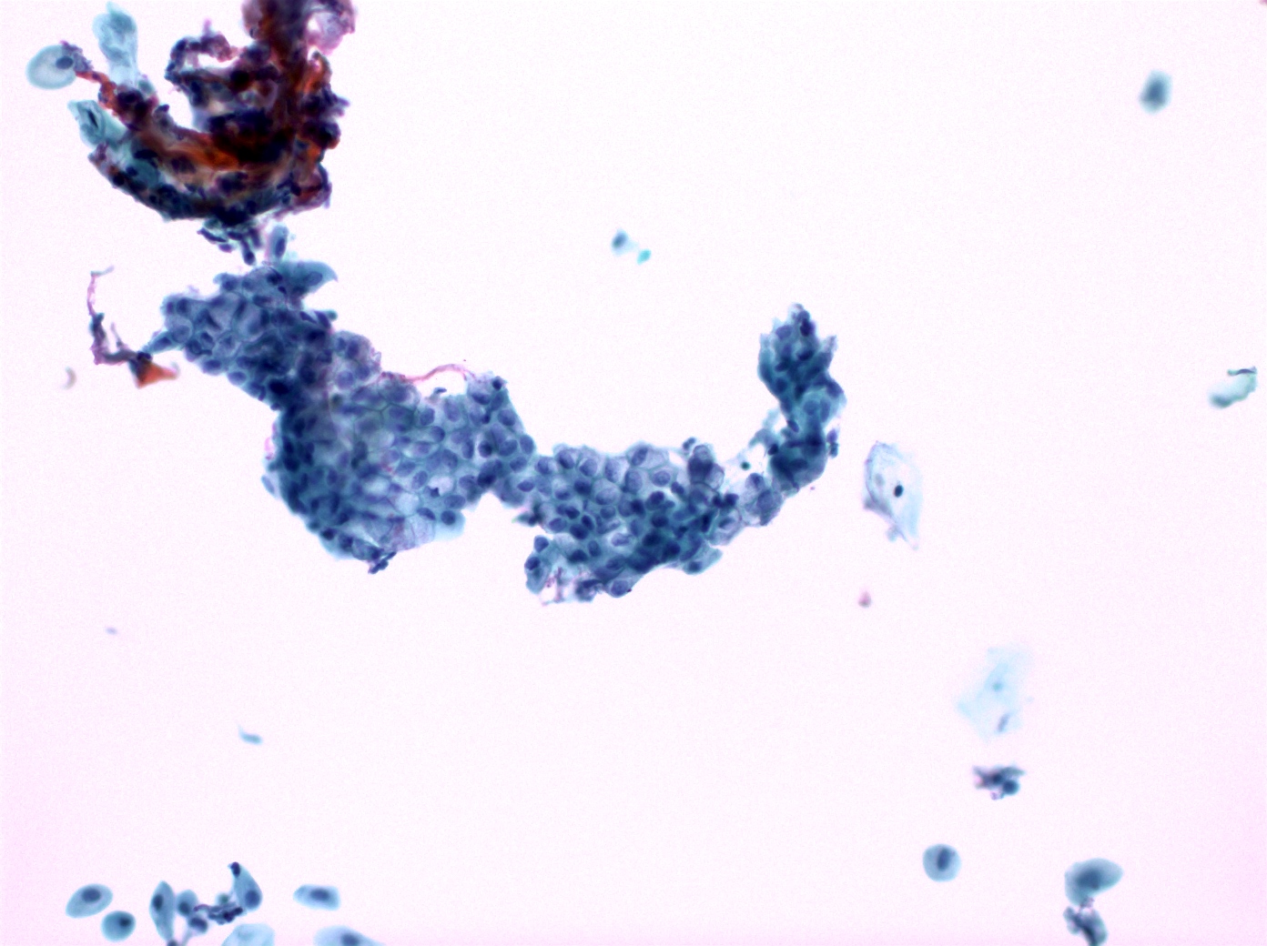
Benign endocervical cells
Images hosted on other servers:

Endocervical polypoid lesion
Contributed by Carlos Parra-Herran, M.D. and AFIP images
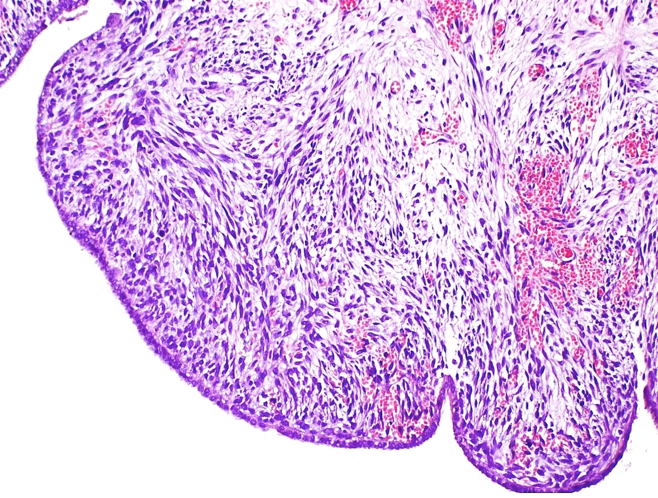
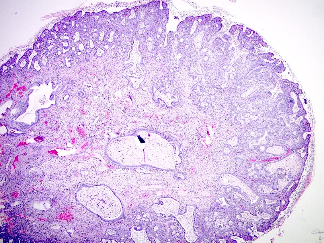
High grade adenosarcoma
Adenosarcoma with sarcomatous overgrowth
Low grade adenosarcoma

Phyllodes tumor-like pattern
Contributed by Ayse Ayhan, M.D., Ph.D.
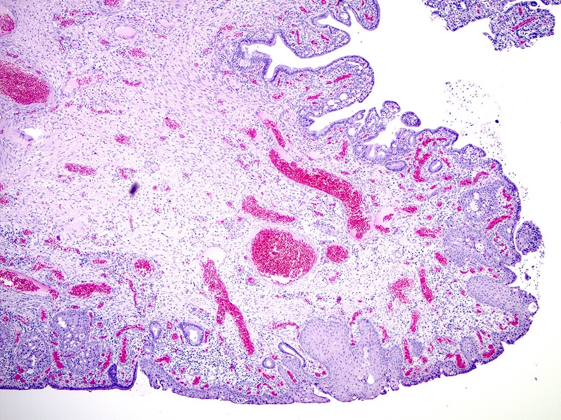
Biphasic tumor
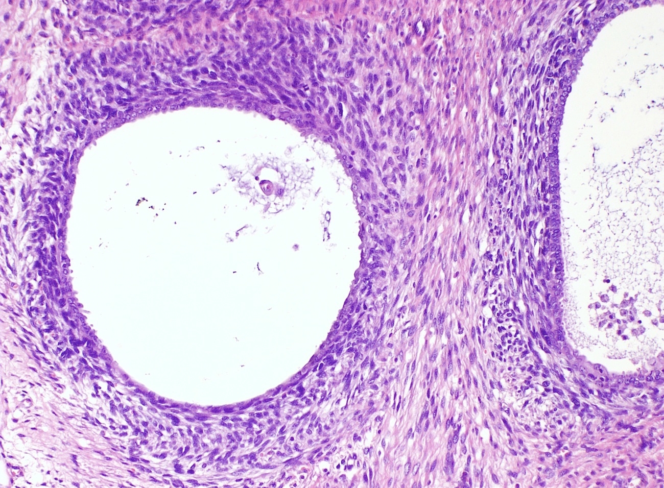
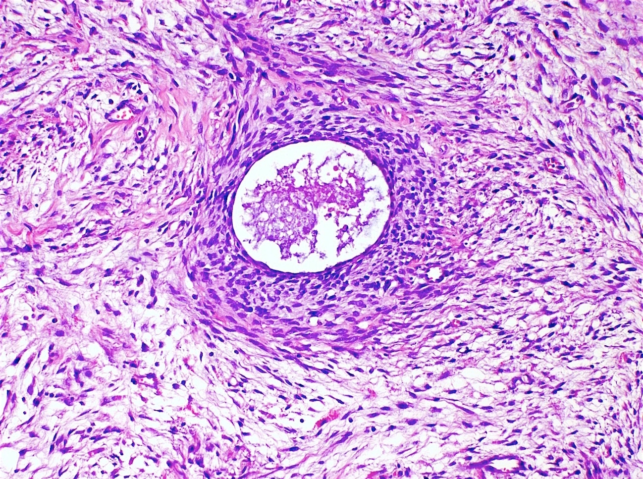
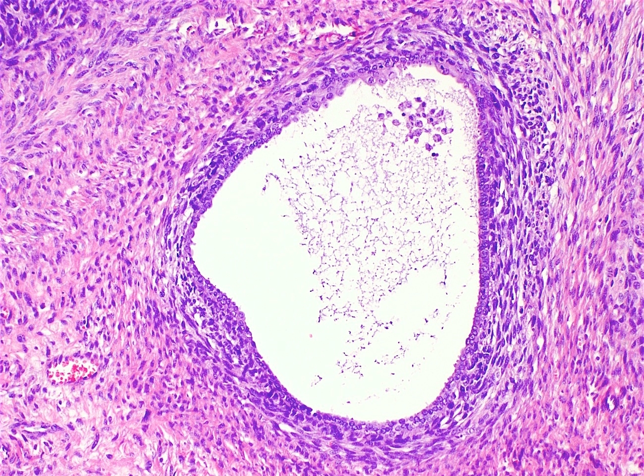
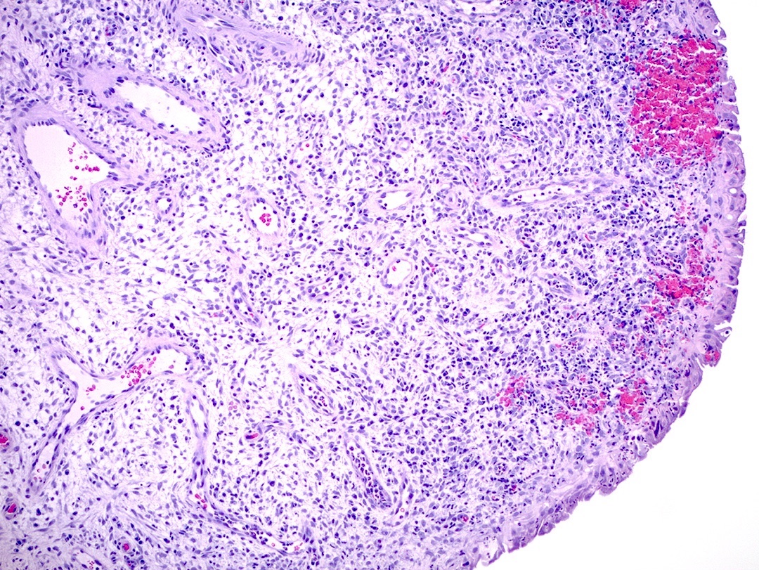
Periglandular cuff
Intraglandular papillae
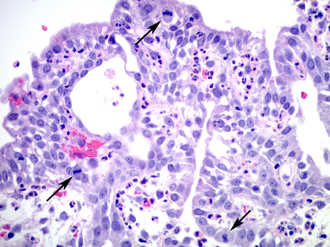
Stromal mitoses
Squamous metaplasia
Sarcomatous overgrowth
Heterologous elements
AFIP images
Bulky exophytic mass
AFIP images
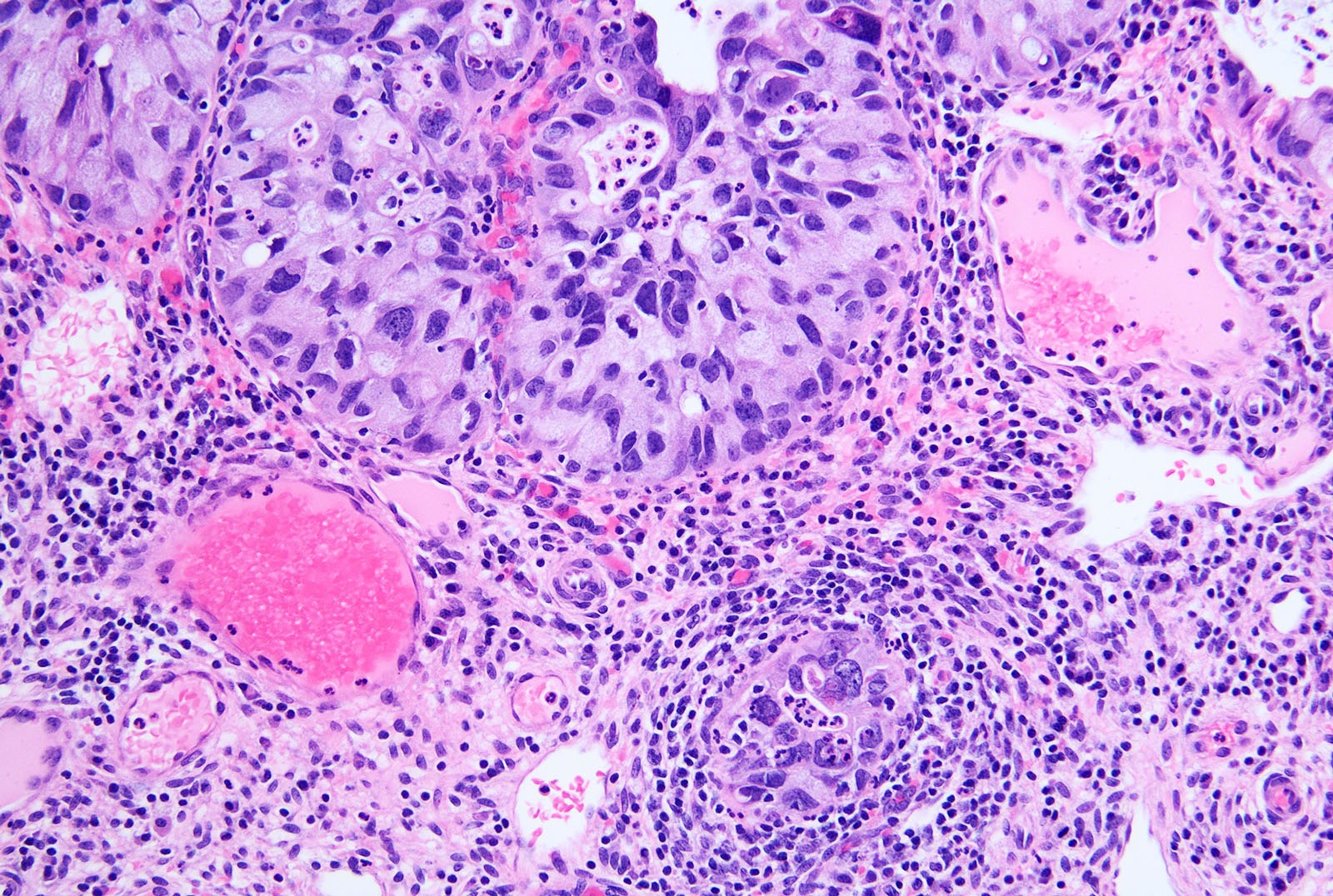
Poorly formed glands and squamous components
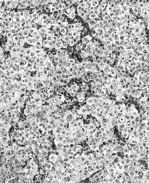
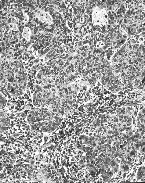
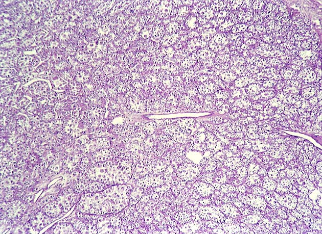
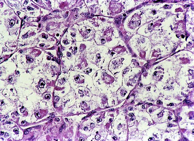
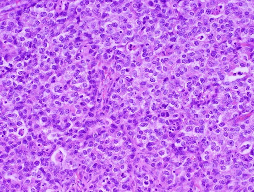
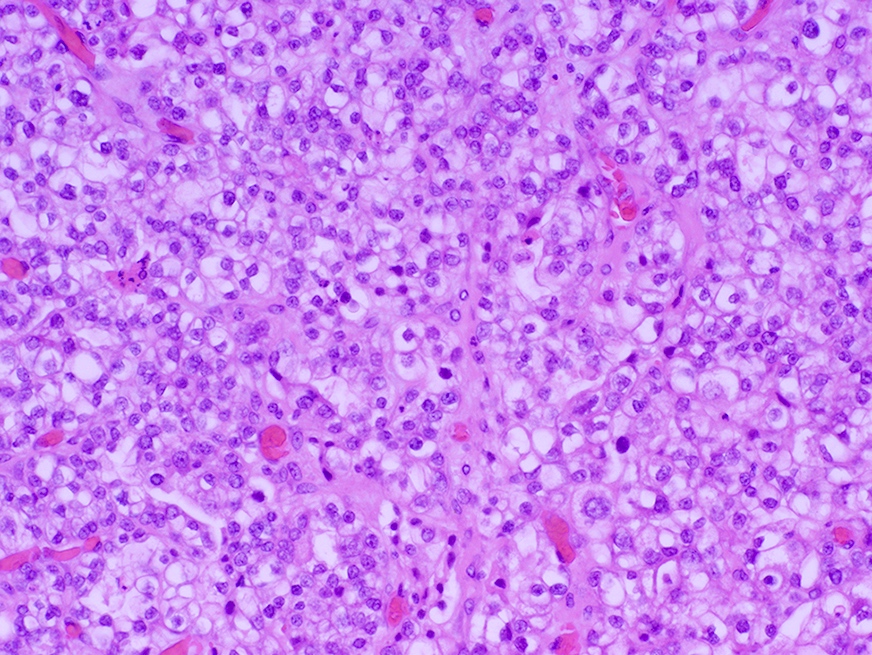
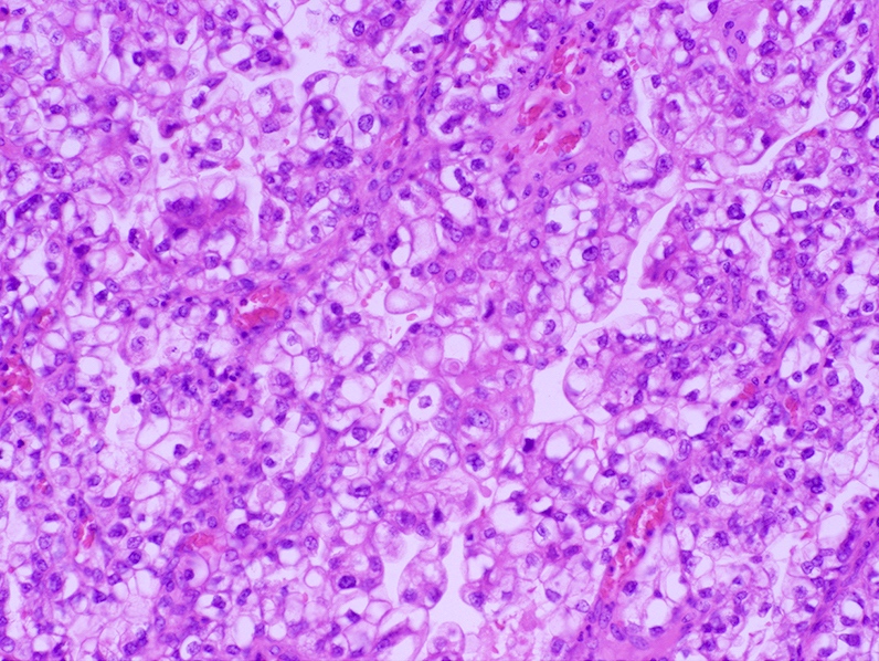
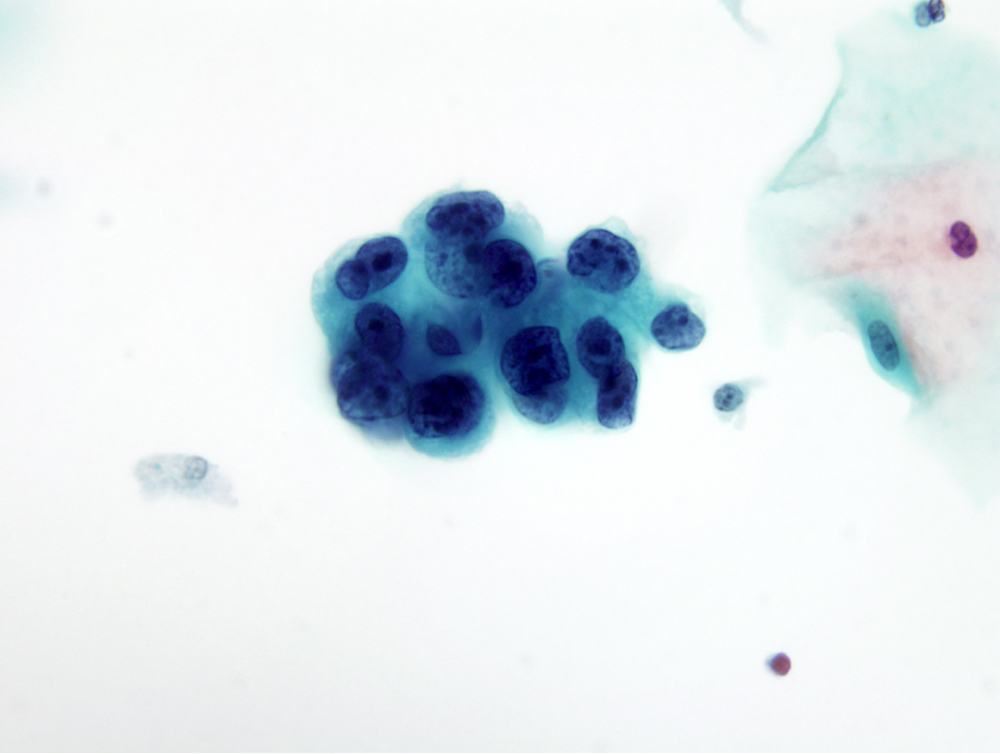
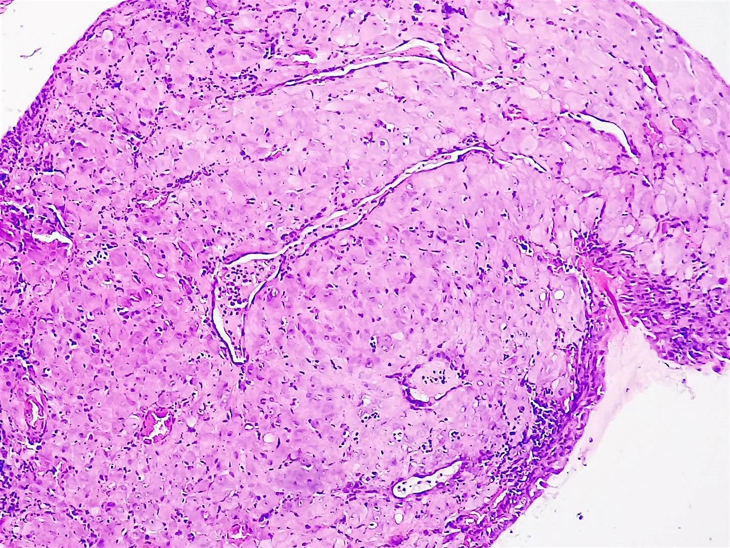
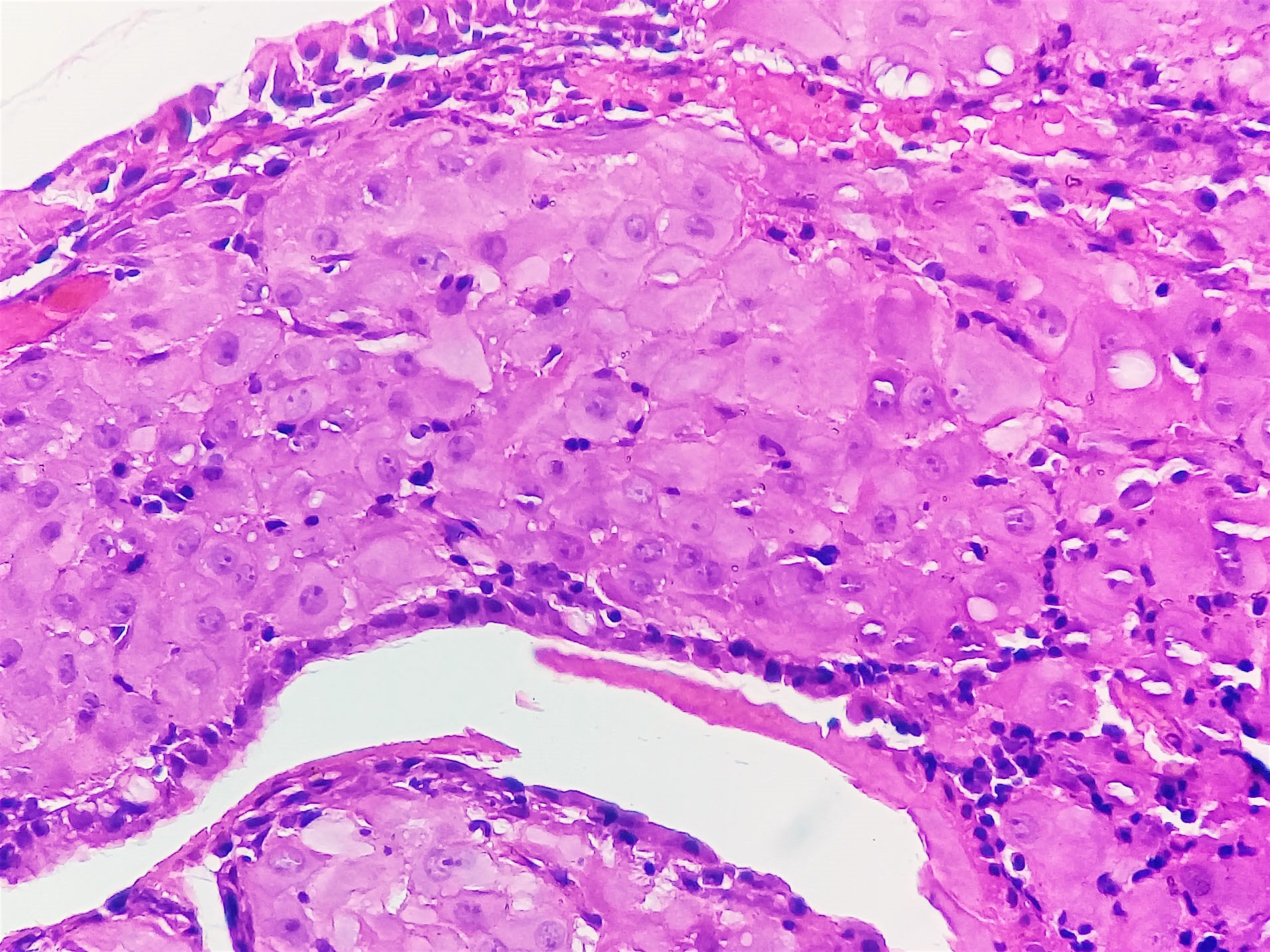
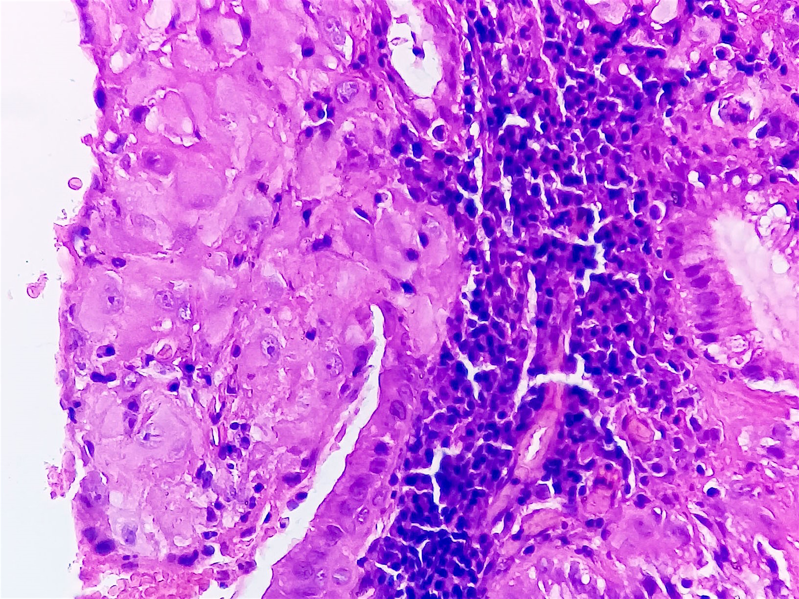
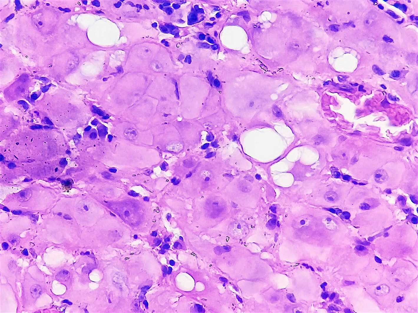
Sheets of cells with abundant lightly stained cytoplasm
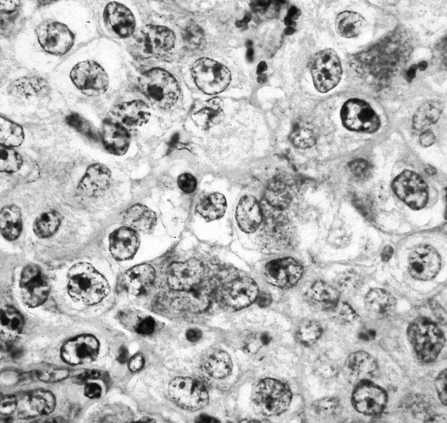
Distinct cell border and prominent nucleoli
Images hosted on other servers:

Abundant granular cytoplasm
Images hosted on other servers:
Nests of tumor cells with PAS+ crystals
Contributed by Nada Mohamed, M.D.
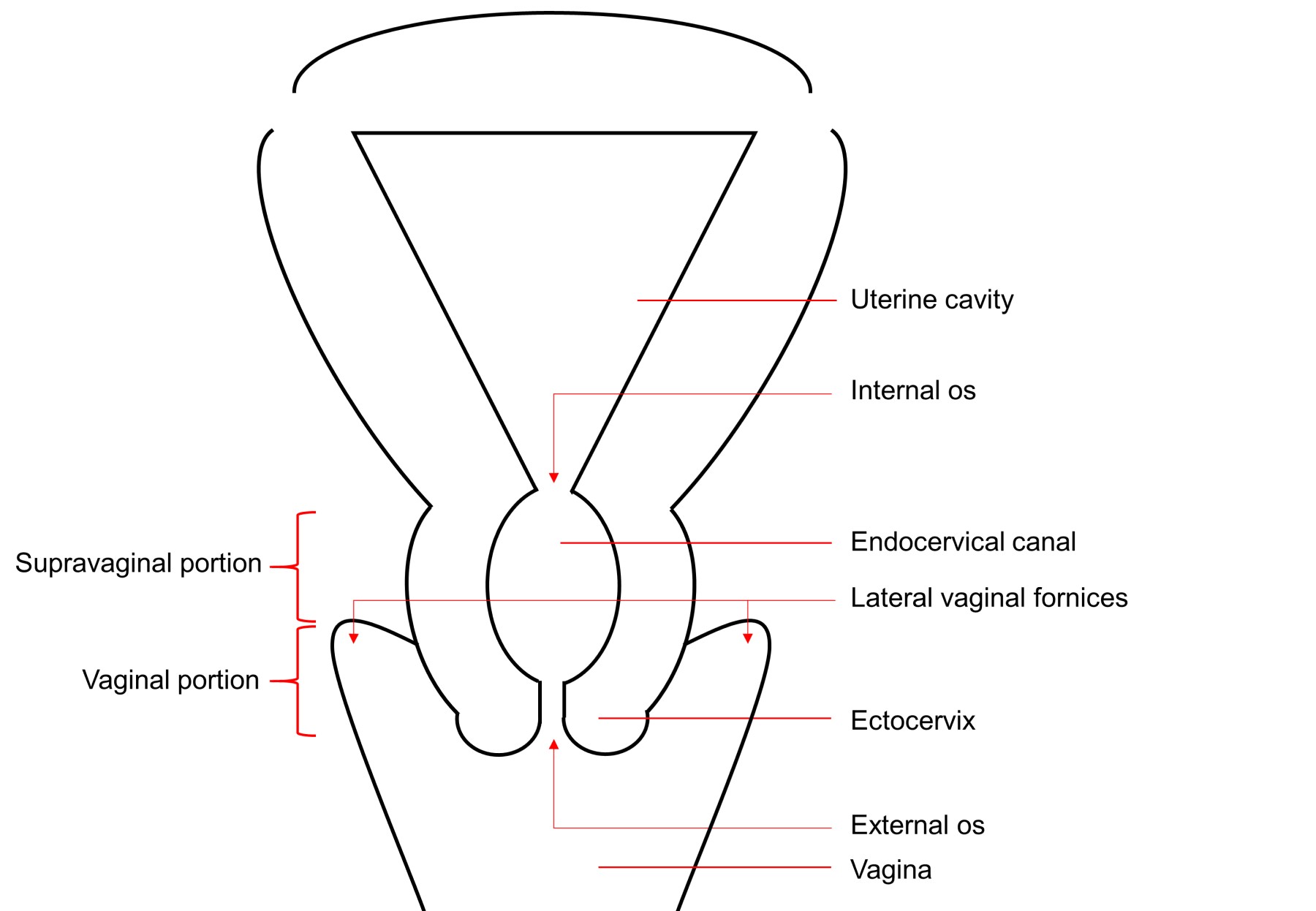
Schematic of cervical anatomy
Images hosted on other servers:

Migration of squamocolumnar junction with age

Local anatomy

Microanatomy


Sagittal section of local anatomy

Uterus, cervix and vagina

Vasculature
Images hosted on other servers:

Zonal anatomy of cervix
Images hosted on other servers:

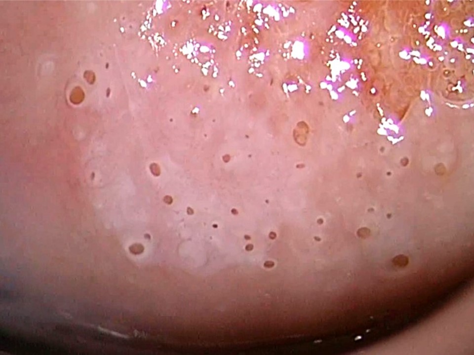
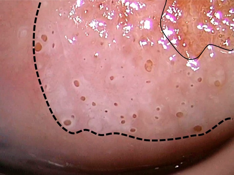
Normal cervix colposcopy

Migration of squamocolumnar junction with age
Contributed by Misty Hensch, P.A.
Anatomy of cervix
Images hosted on other servers:

Nulliparous cervix
Contributed by Nada Mohamed, M.D.
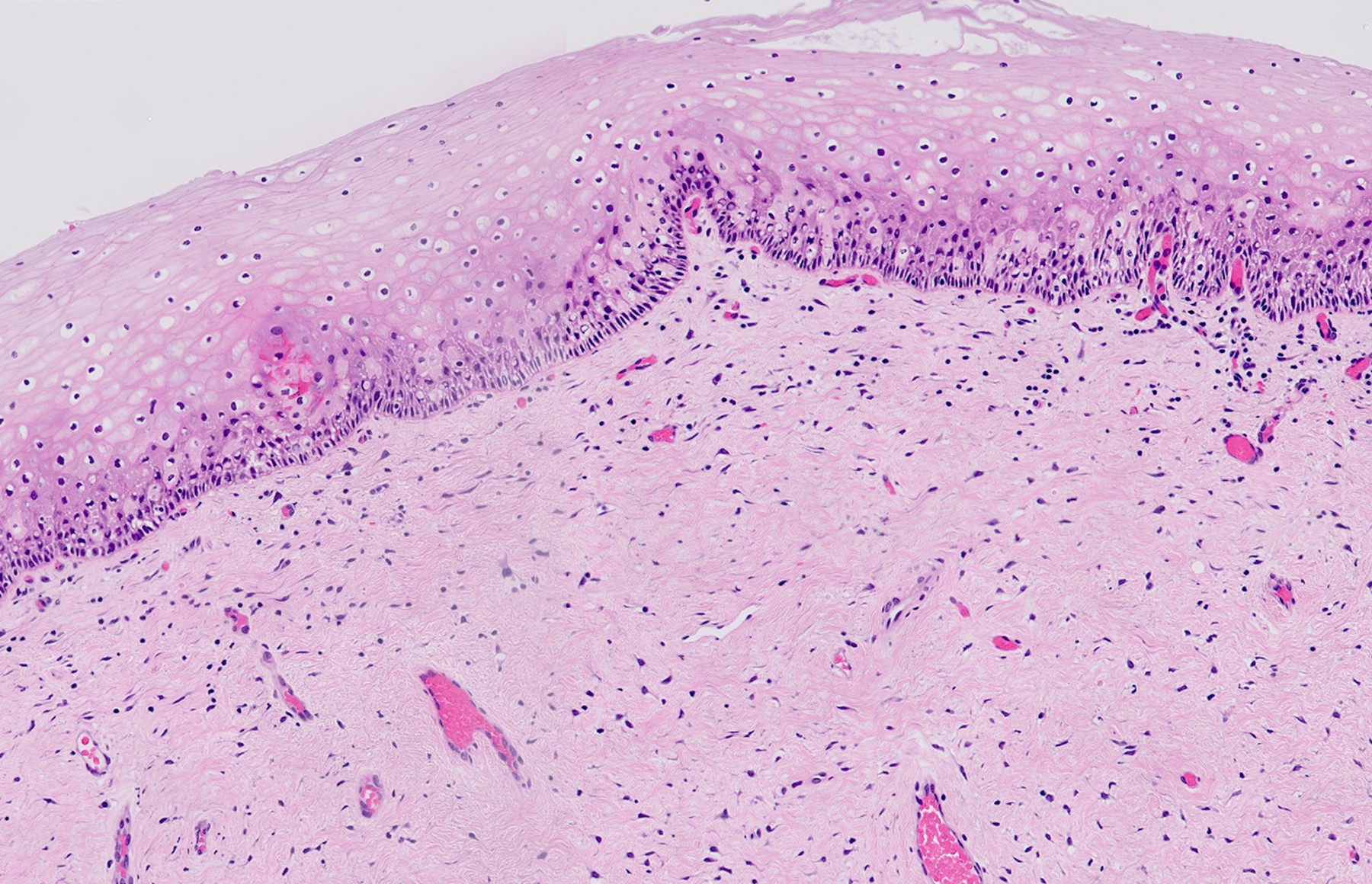
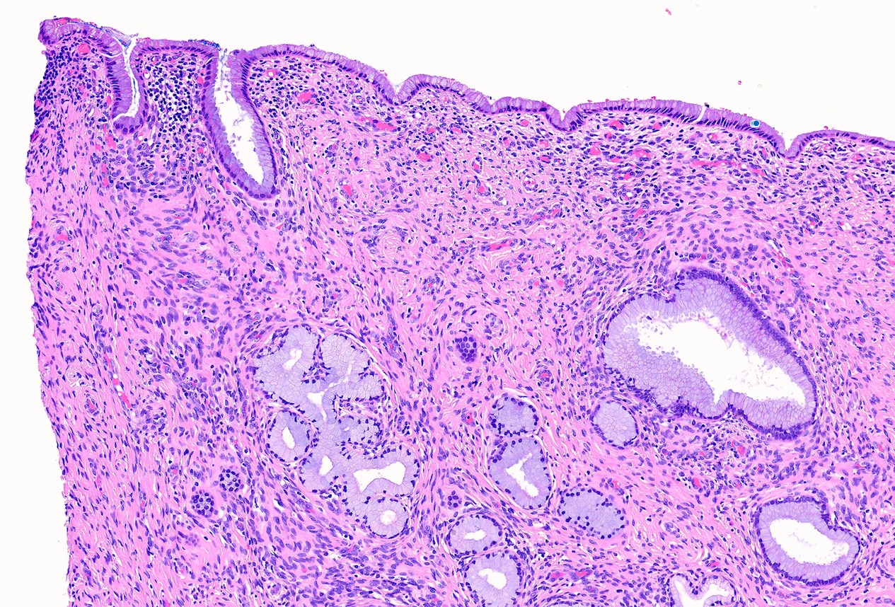
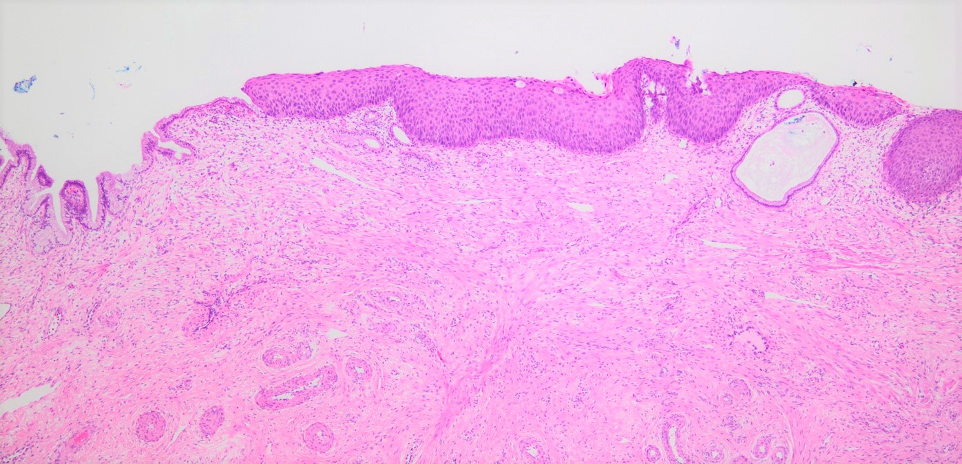
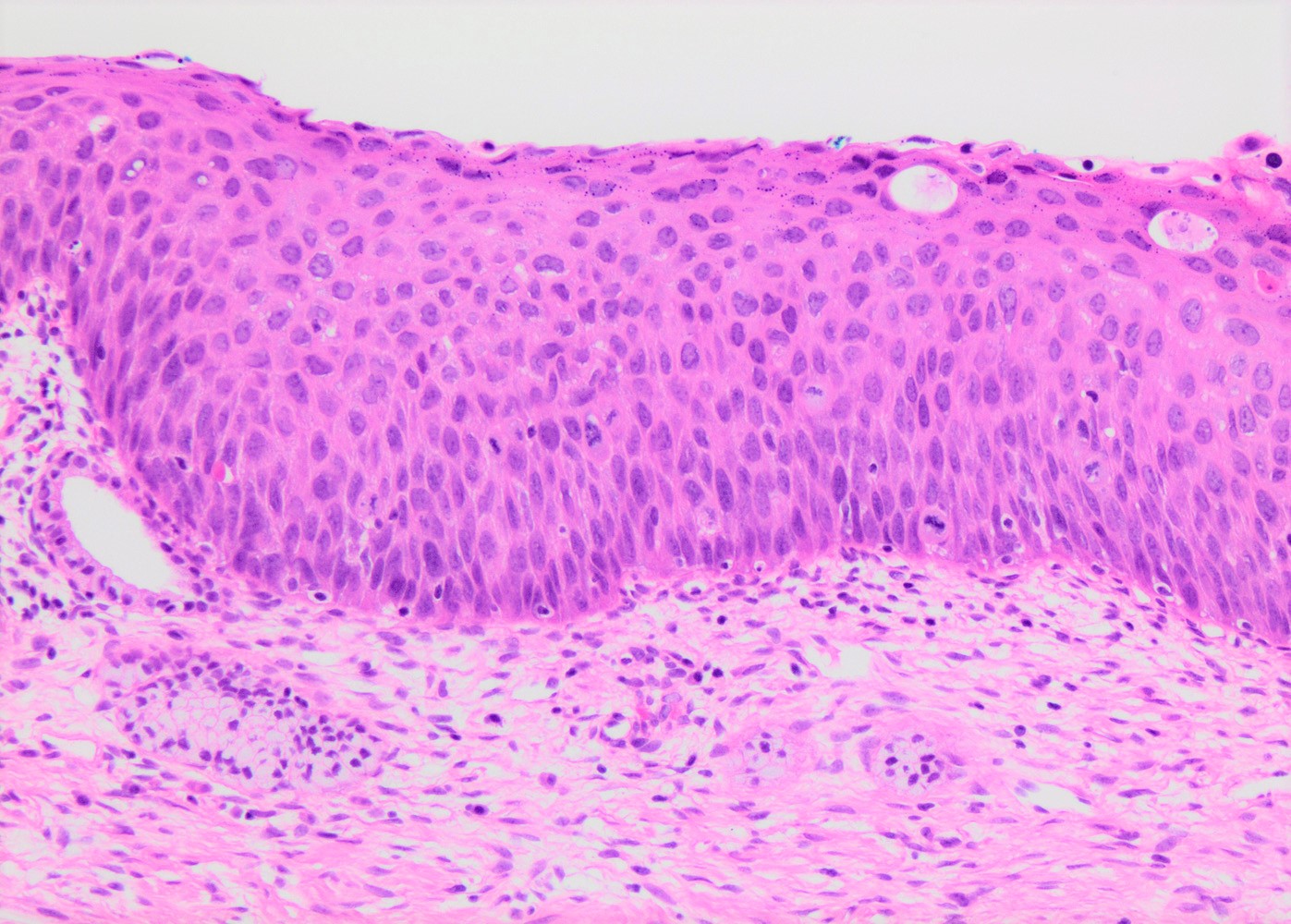
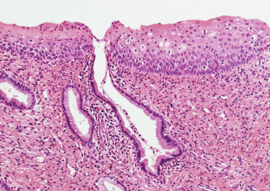
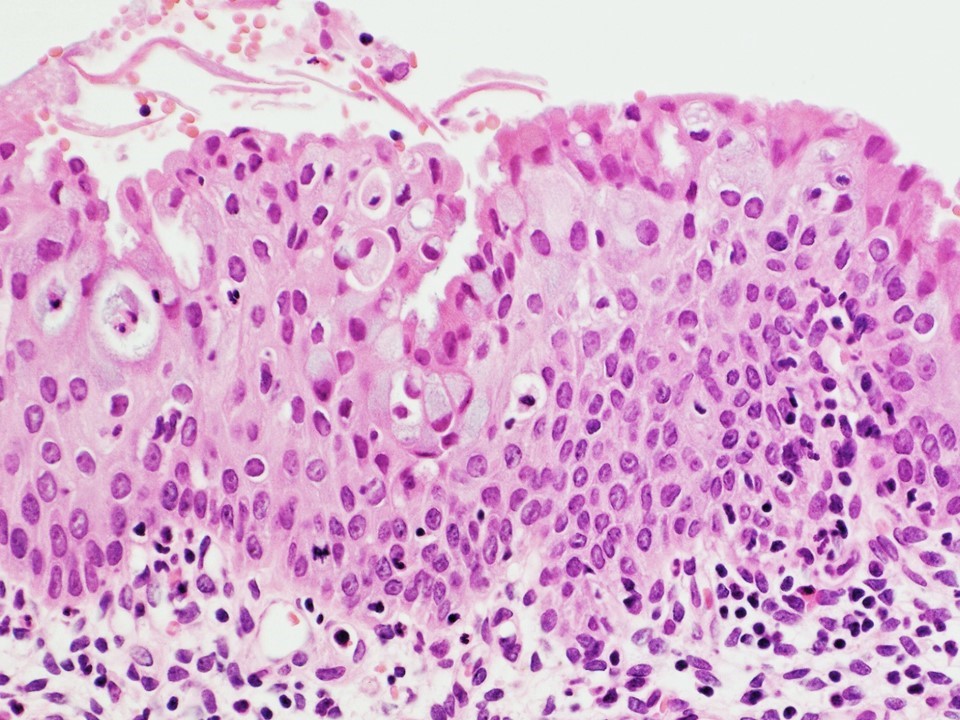
Normal cervical squamous epithelium
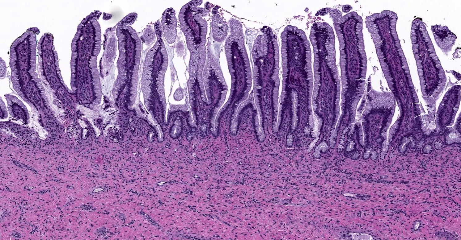
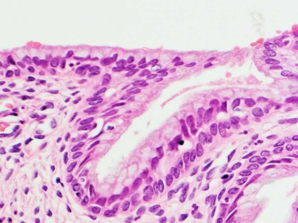
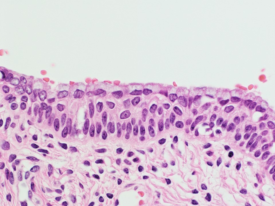
Normal cervical glandular epithelium
Histology of the cervix
Contributed by Leonel Maldonado, M.D.
Arias-Stella reaction, 10×
Arias-Stella reaction, 20×

Contributed by Bonnie Choy, M.D.
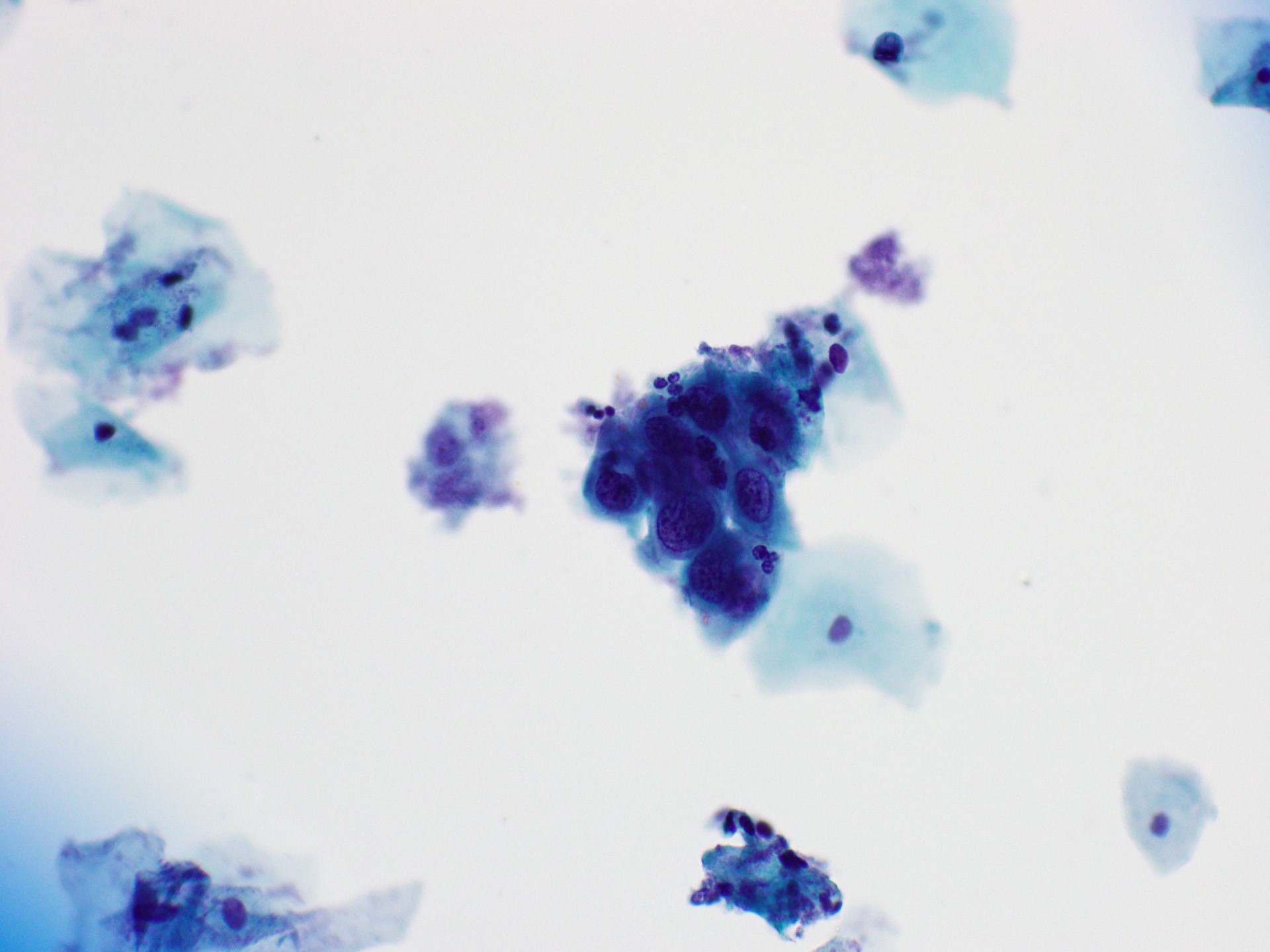
High N:C ratio
Irregular nuclear membranes and hyperchromasia

HSIL
Squamous metaplasia
Atrophy with inflammation

Endometrial cells
Images hosted on other servers:

WHO digital atlas

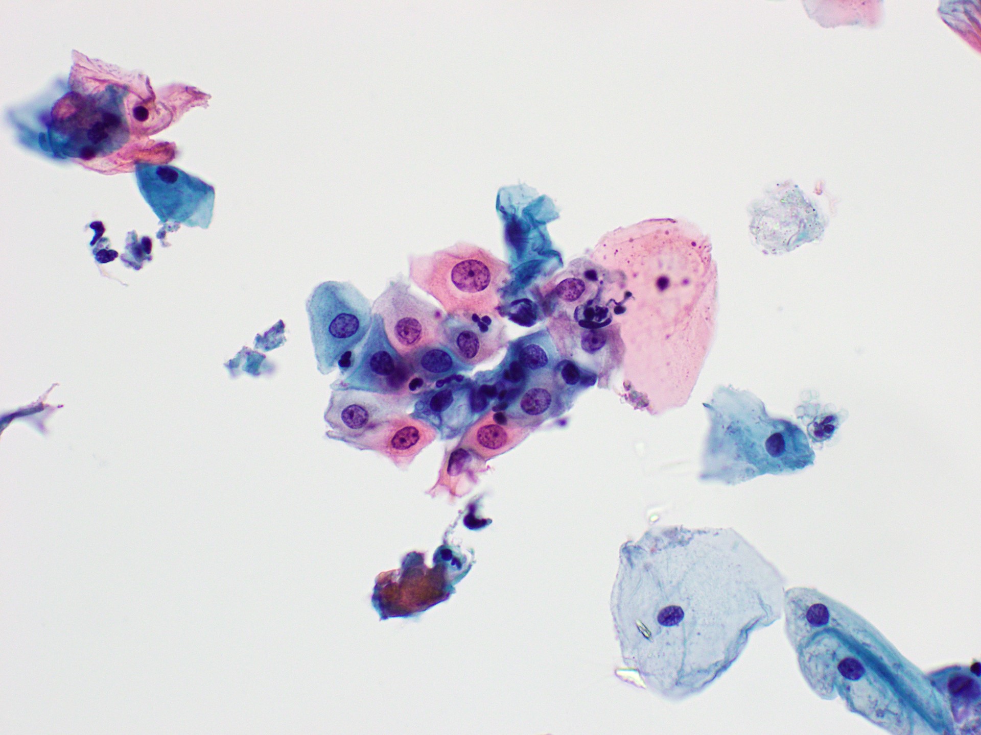
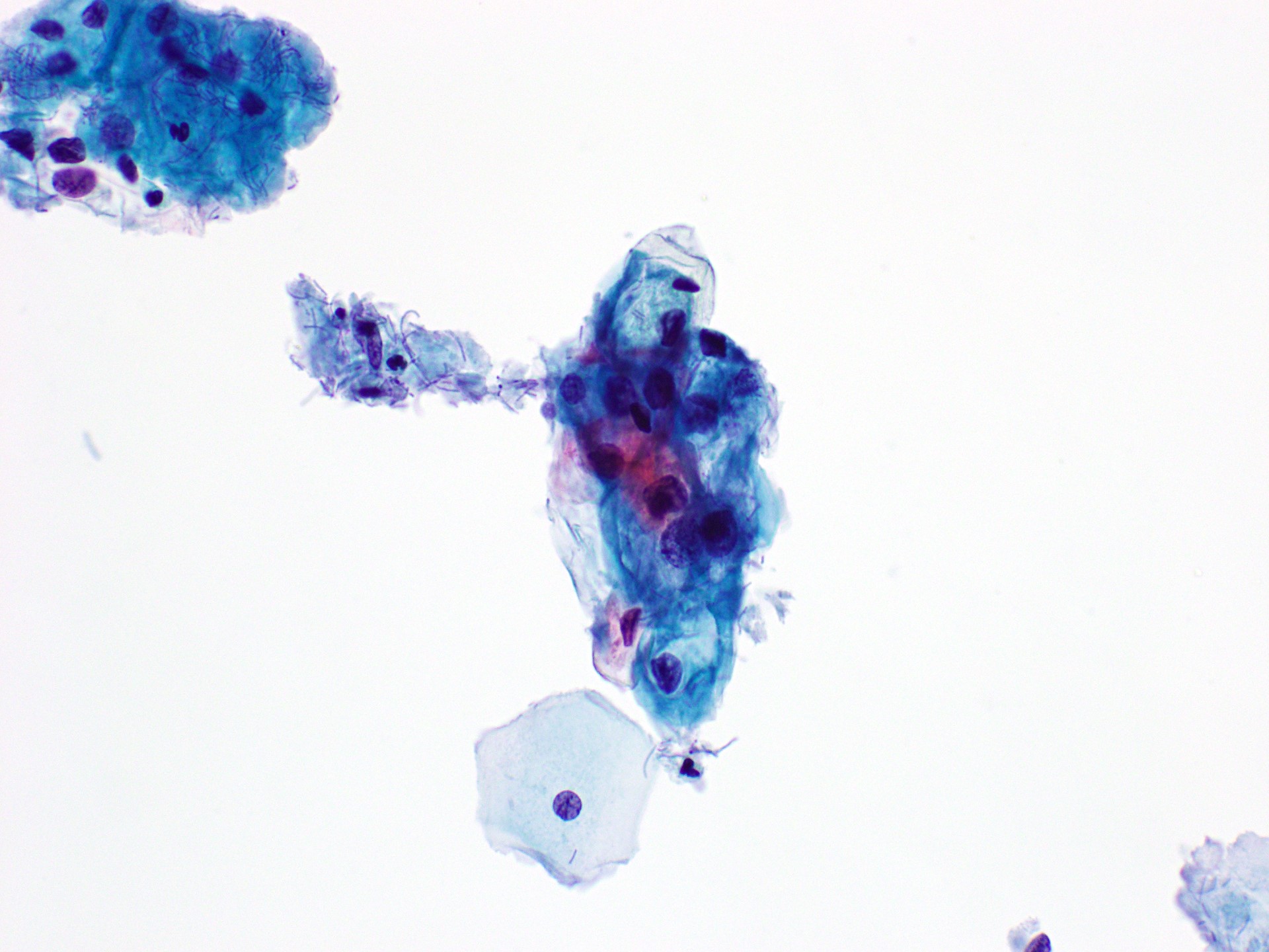
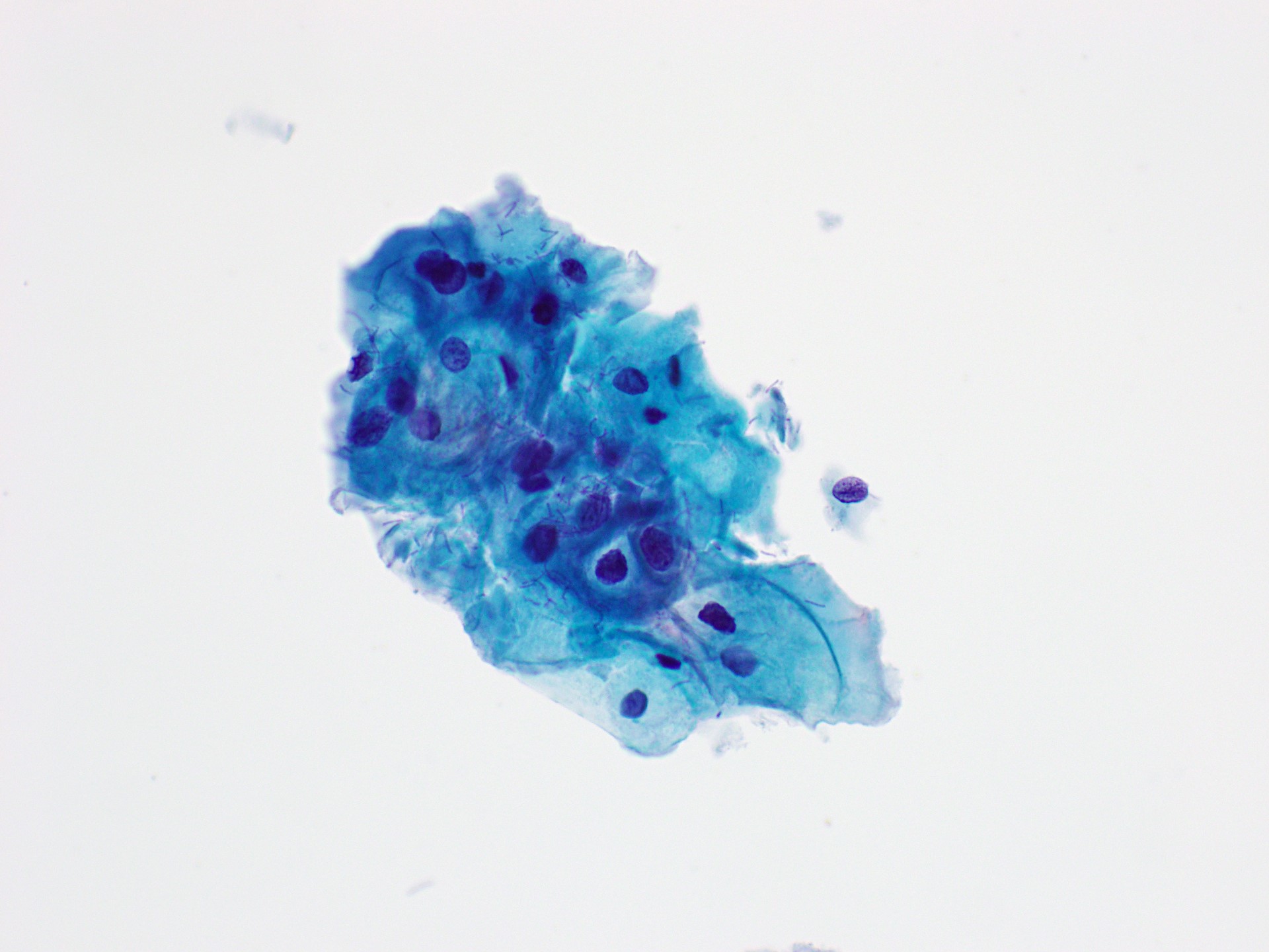
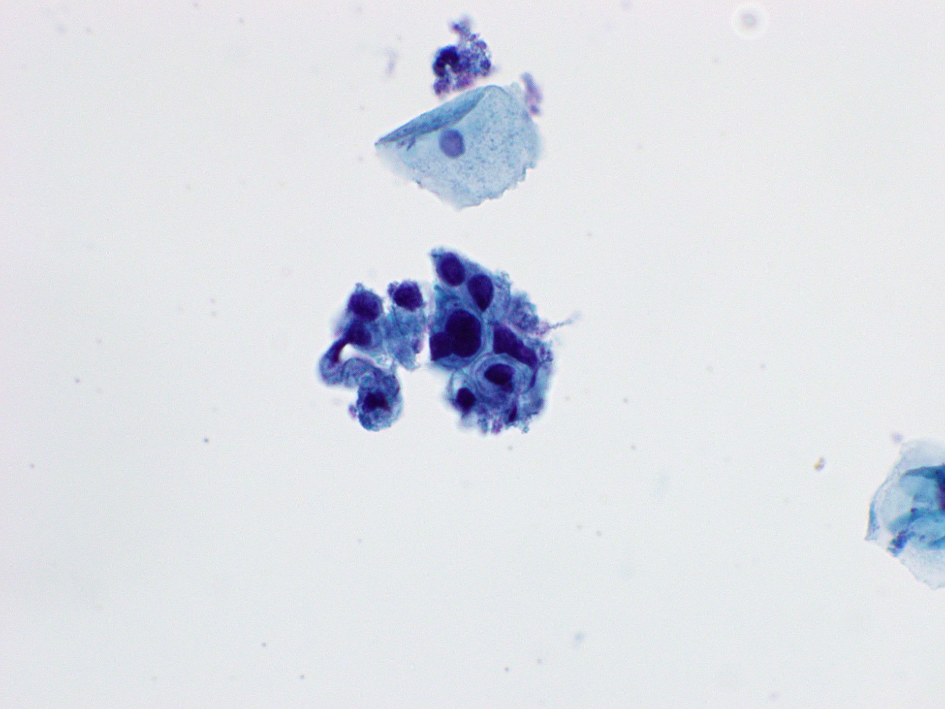
Contributed by Joseph Reznicek, M.D., Bonnie Choy, M.D. and Lucy Jager, M.D.
Enlarged nuclei
Slight hyperchromasia
Atypical parakeratosis
Radiation changes
LSIL

Pseudokoilocytes
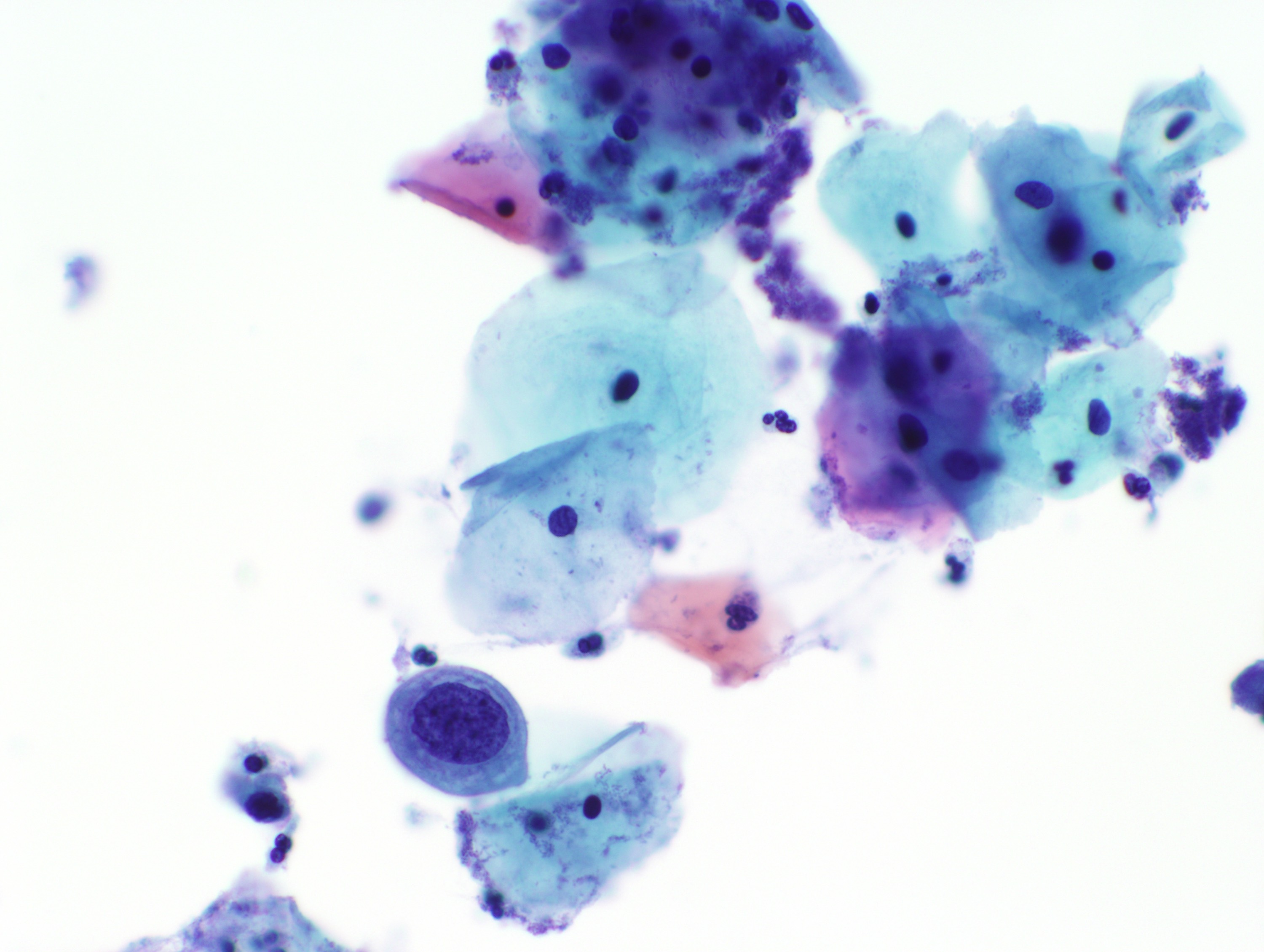
Herpes cytopathic effect
Images hosted on other servers:

WHO digital atlas
Contributed by Marilin Rosa, M.D.
Atrophic sheet
Images hosted on other servers:

Various images
Images hosted on other servers:

Algorithm for initial workup of AGC

Algorithm for management after diagnostic examination
Contributed by Joseph Reznicek, M.D.
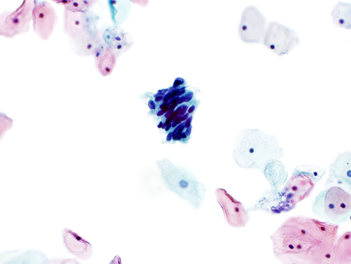
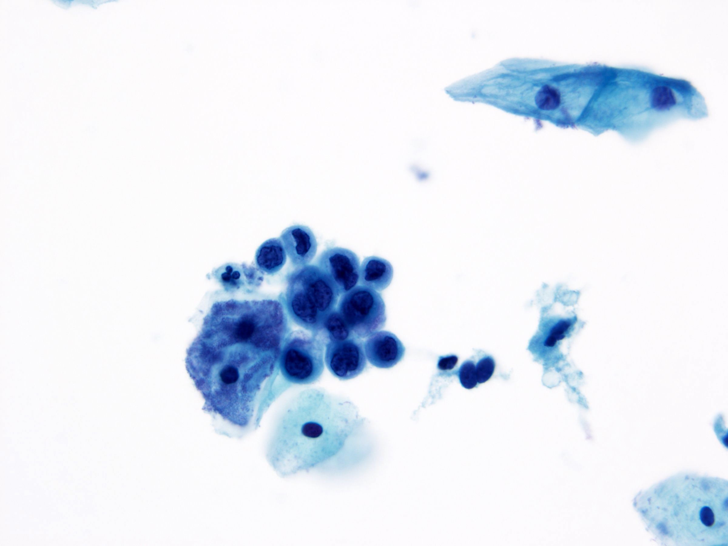
Atypical endocervical cells, favor neoplastic
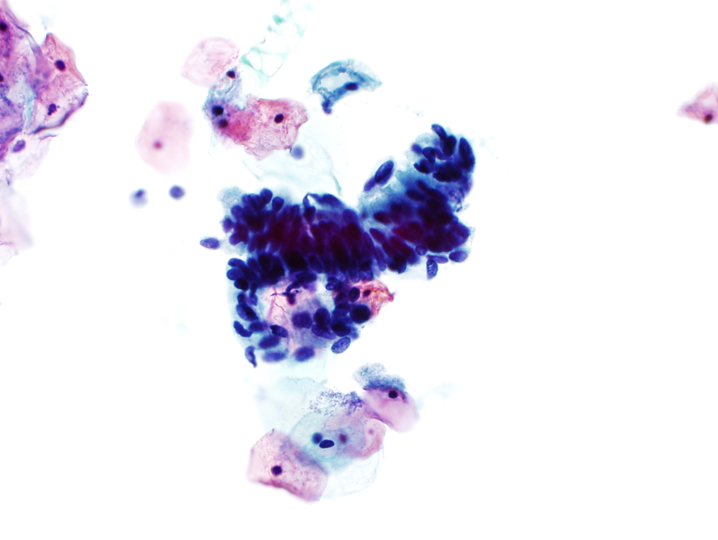
Atypical glandular cells, NOS

Atypical glandular cells, NOS
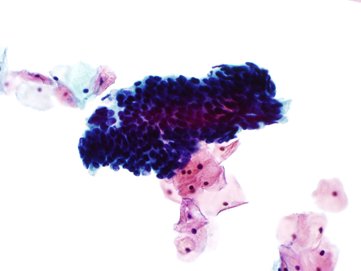
Endocervical adenocarcinoma
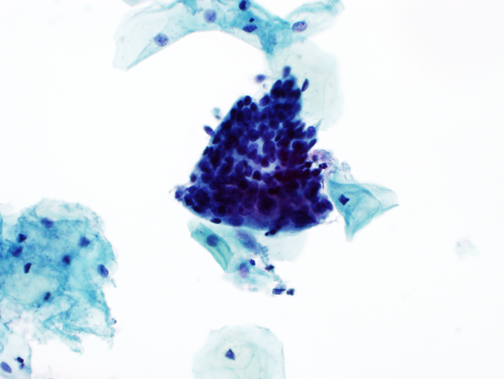
Endometrial adenocarcinoma
Images hosted on other servers:

WHO digital atlas
Images hosted on other servers:

Conventional smears
Contributed by Rachel Jug, M.B.B.Ch., B.A.O. and Sarah M. Bean, M.D.
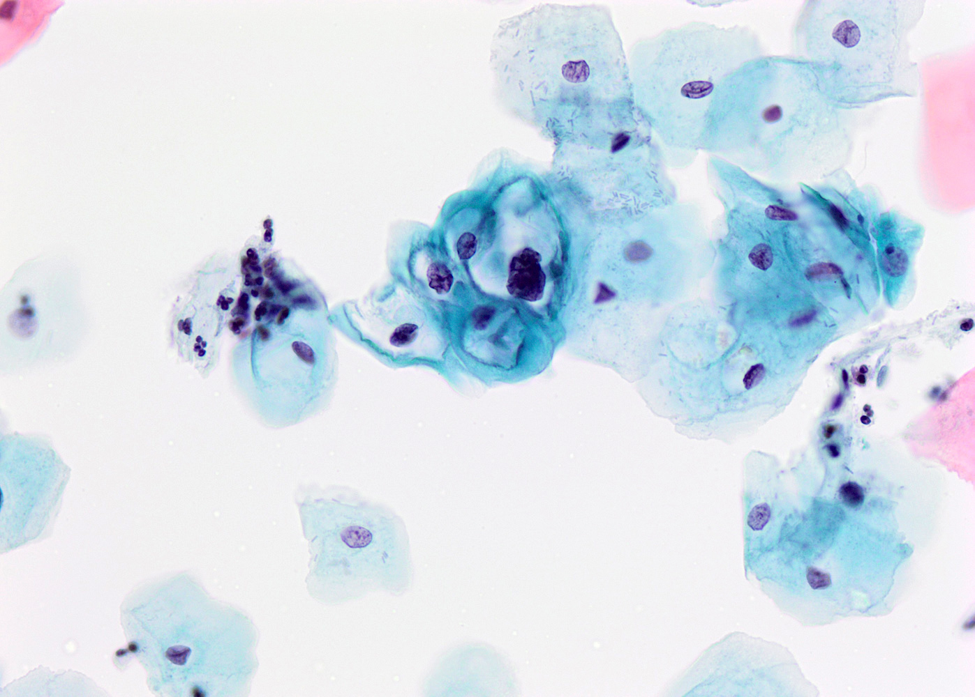
Resembling koilocytes
Nuclear enlargement
Koilocyte
Binucleation
High N:C ratio
Hyperchromasia
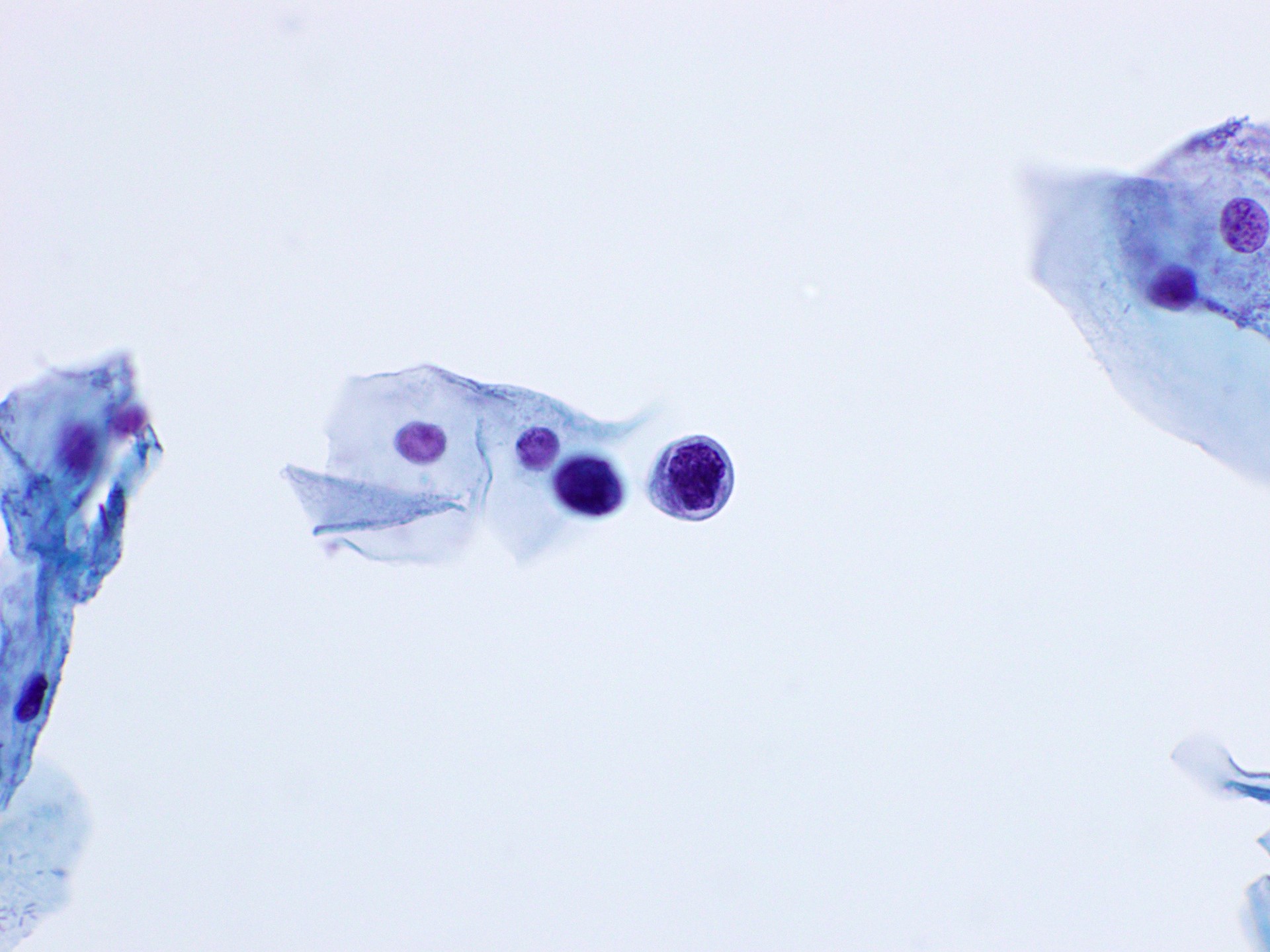
Nuclear contour irregularities
Binucleated cell with hyperchromasia, LSIL
Hyperchromatic nucleus and small halo, ASCUS
Nuclear overlap and elongation, atypical glandular cells
Hyperchromatic and irregular nucleus, atypical glandular cells
AFIP images
Pigment containing nevus cells in cervical stroma
Contributed by @AnaPath10 on Twitter
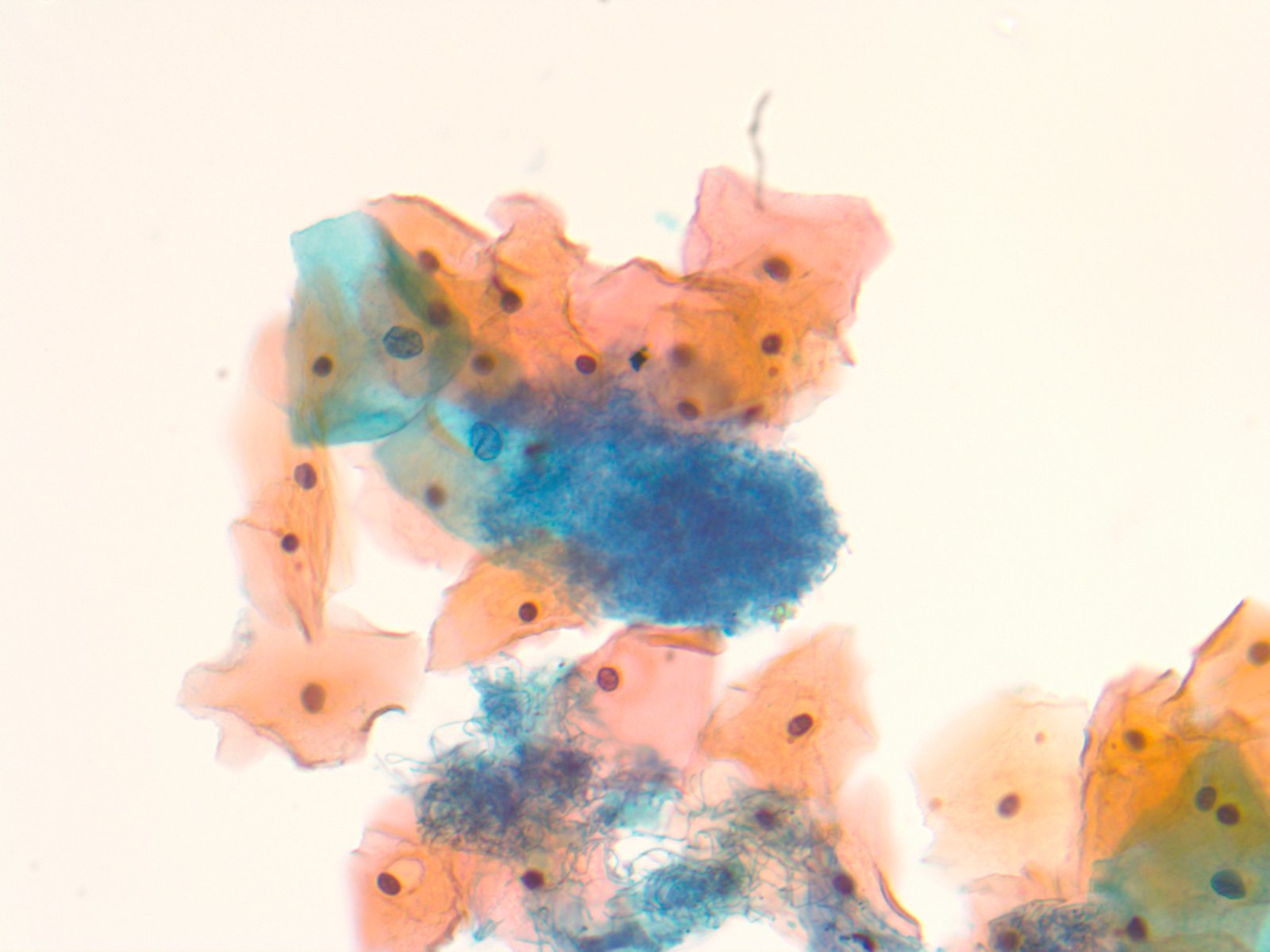
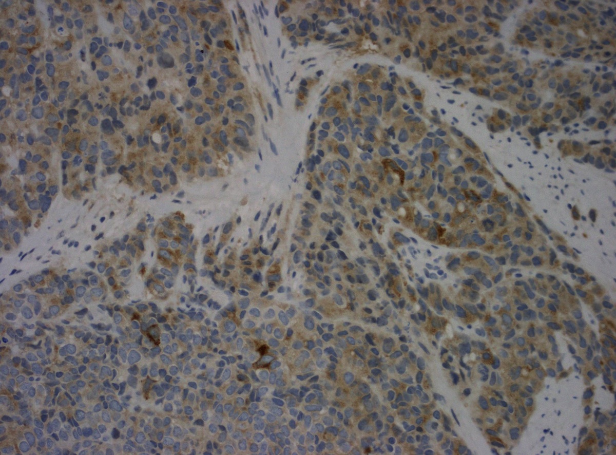
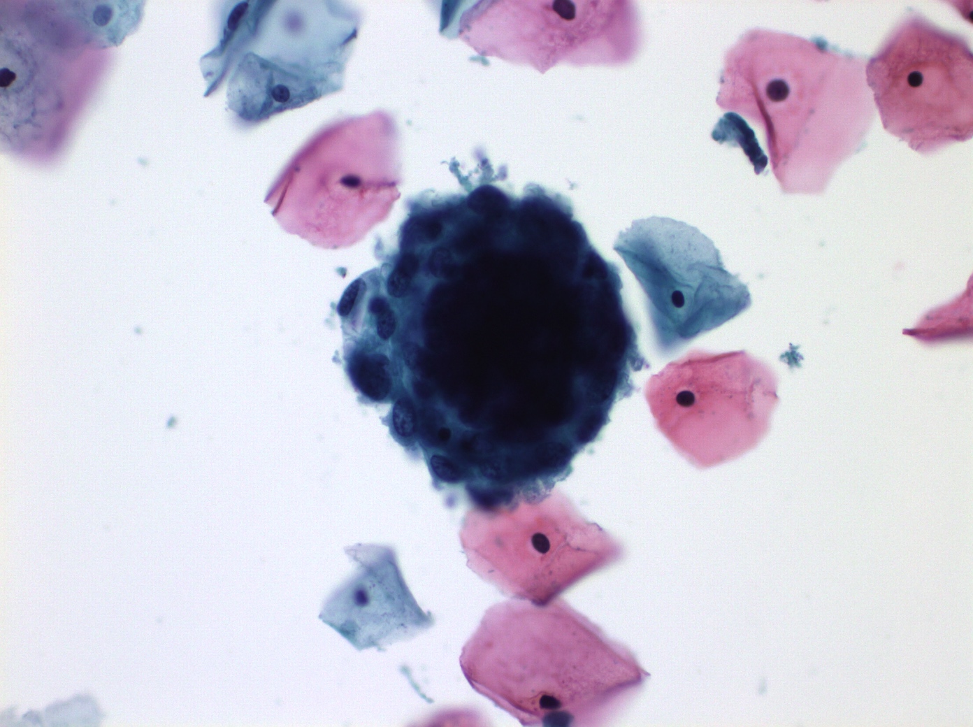
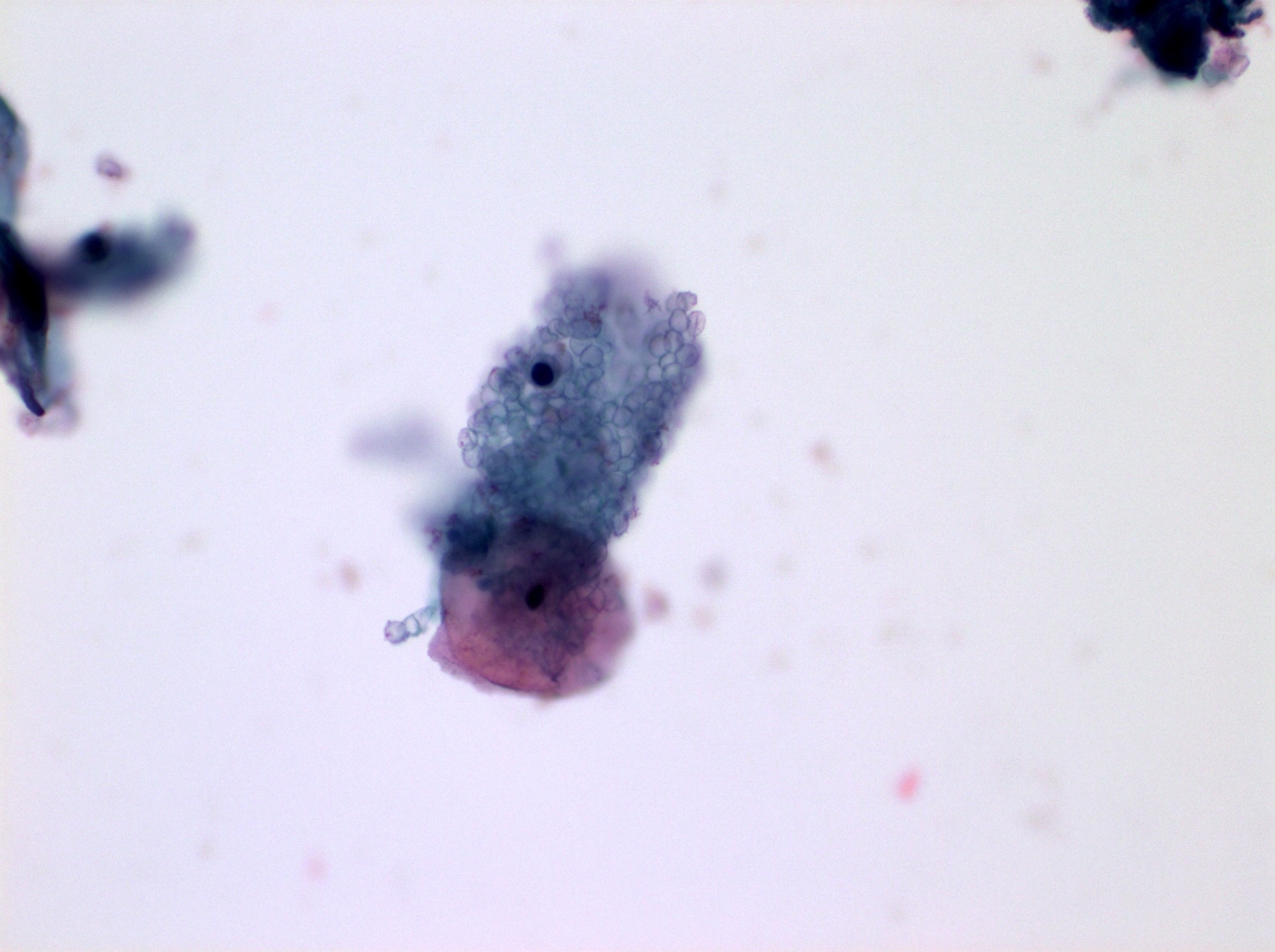
Candida / fungi
Candida / fungi
Candida / fungi
Images hosted on other servers:

Candida albicans
Images hosted on other servers:

Various images
Contributed by Ali Ismail, M.B.B.S.
Chronic inflammation
Images hosted on other servers:


Lymphofollicular cervicitis in a
53 year old woman
Images hosted on other servers:

Vascularized, indurated lesion
Contributed by Philip T. Valente, M.D.

Tan-white lesion
Images hosted on other servers:

Exophytic mass on the posterior lip of cervix

Didelphys uterus with a cervical mass
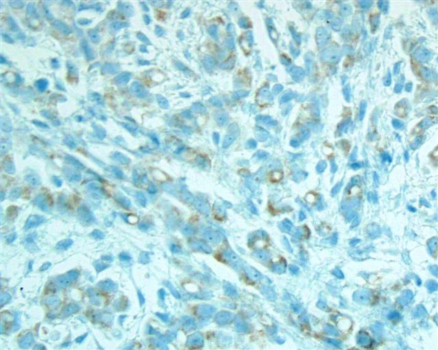
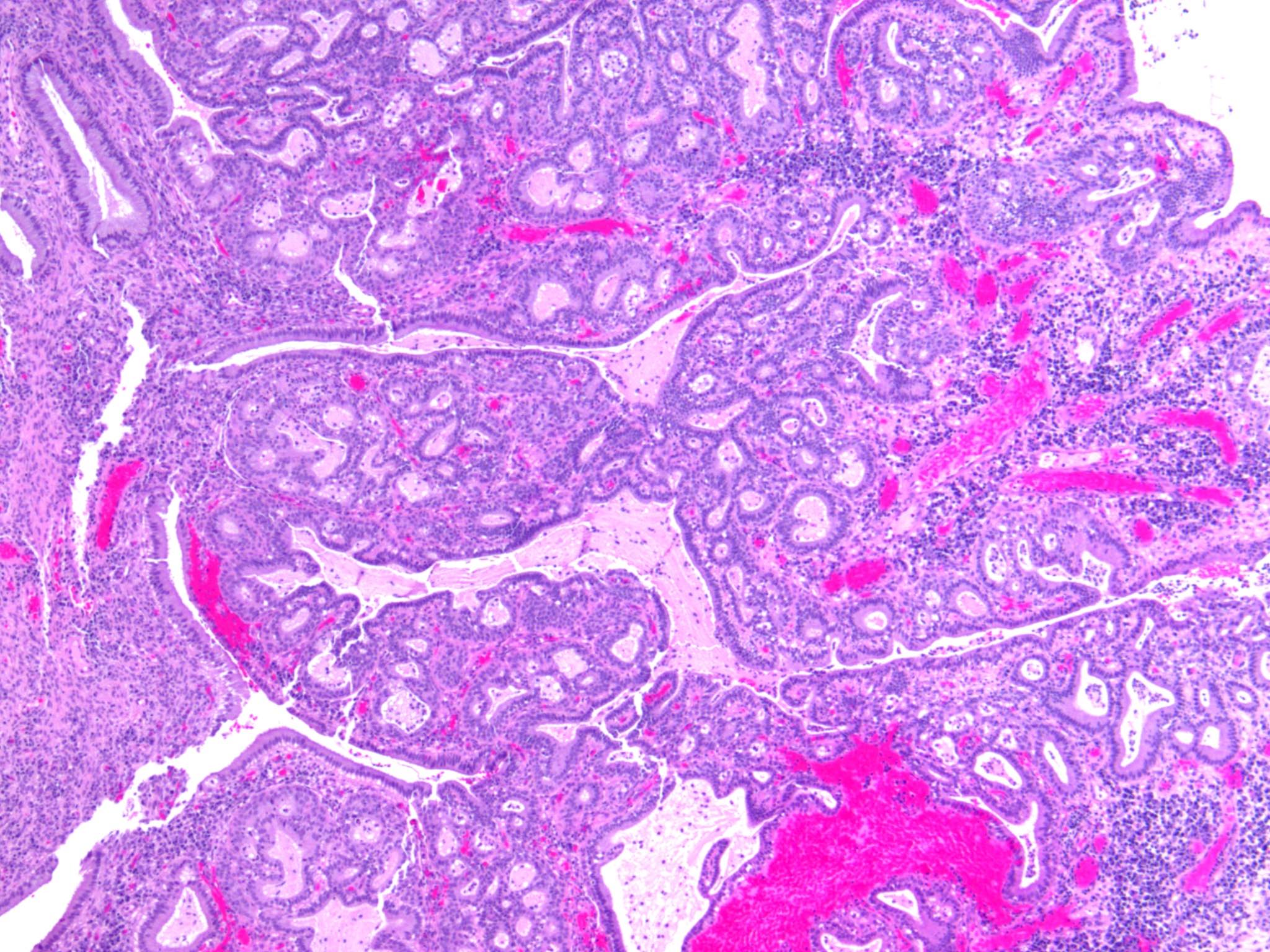
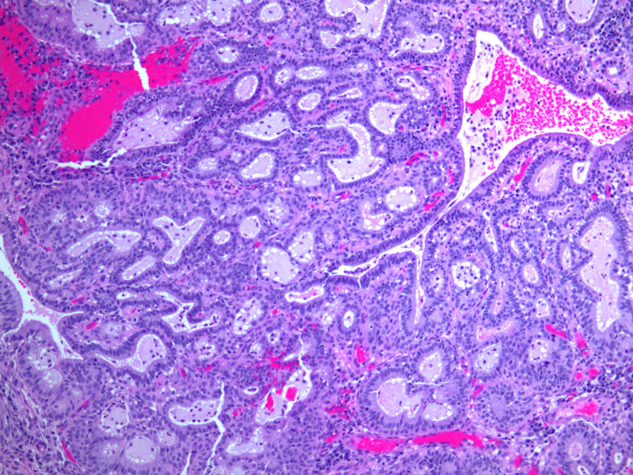
Contributed by Nadia Hameed, M.D.
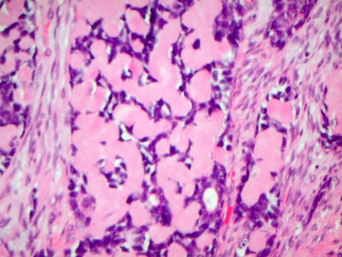
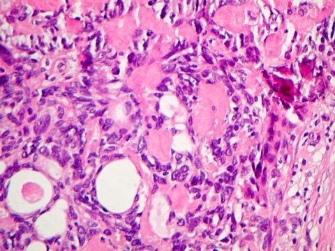
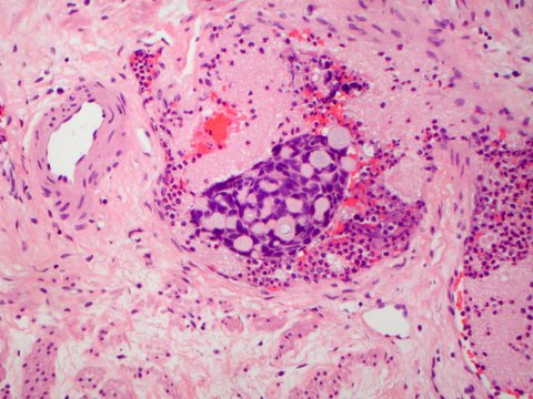
Tubulocystic pattern
Tubulocystic pattern
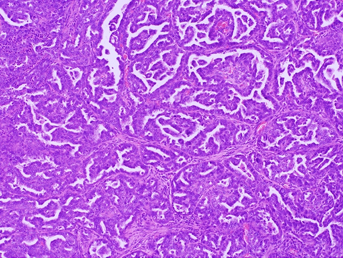
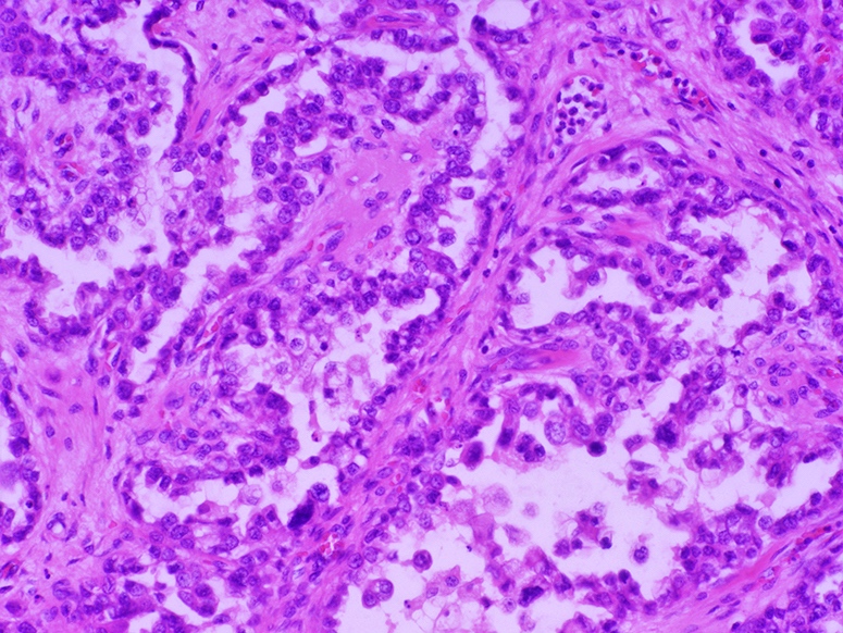
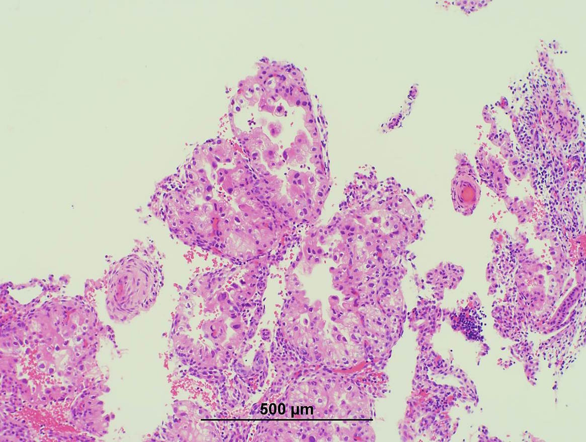
Papillary pattern
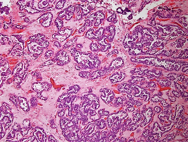
Papillary pattern
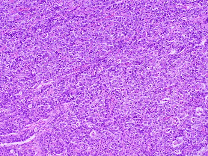
Solid pattern
Solid pattern
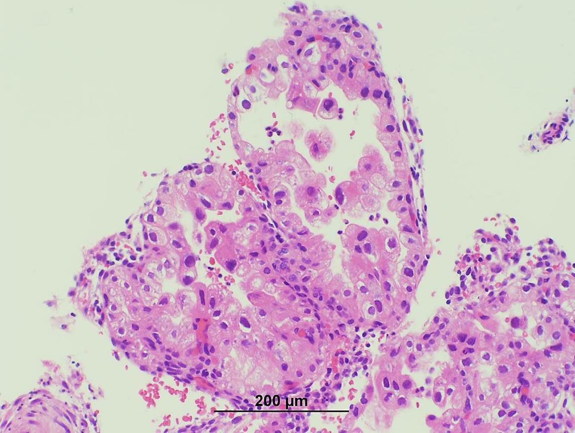
Rare intracytoplasmic eosinophilic globules
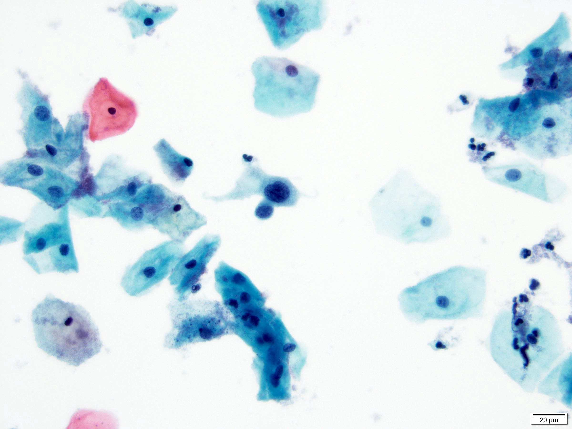
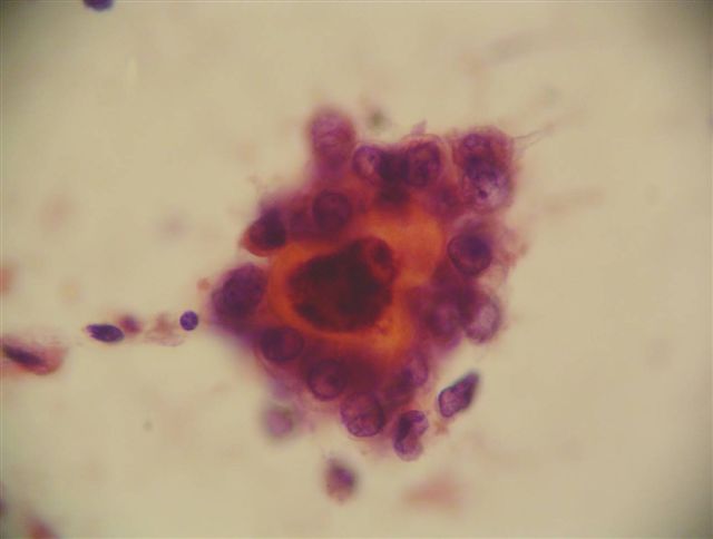
Contributed by Nadia Hameed, M.D.
SurePath Pap test
Images hosted on other servers:

Mitochondria and rough endoplasmic reticulum
Clear cell neoplasms of the gynecologic tract
Images hosted on other servers:

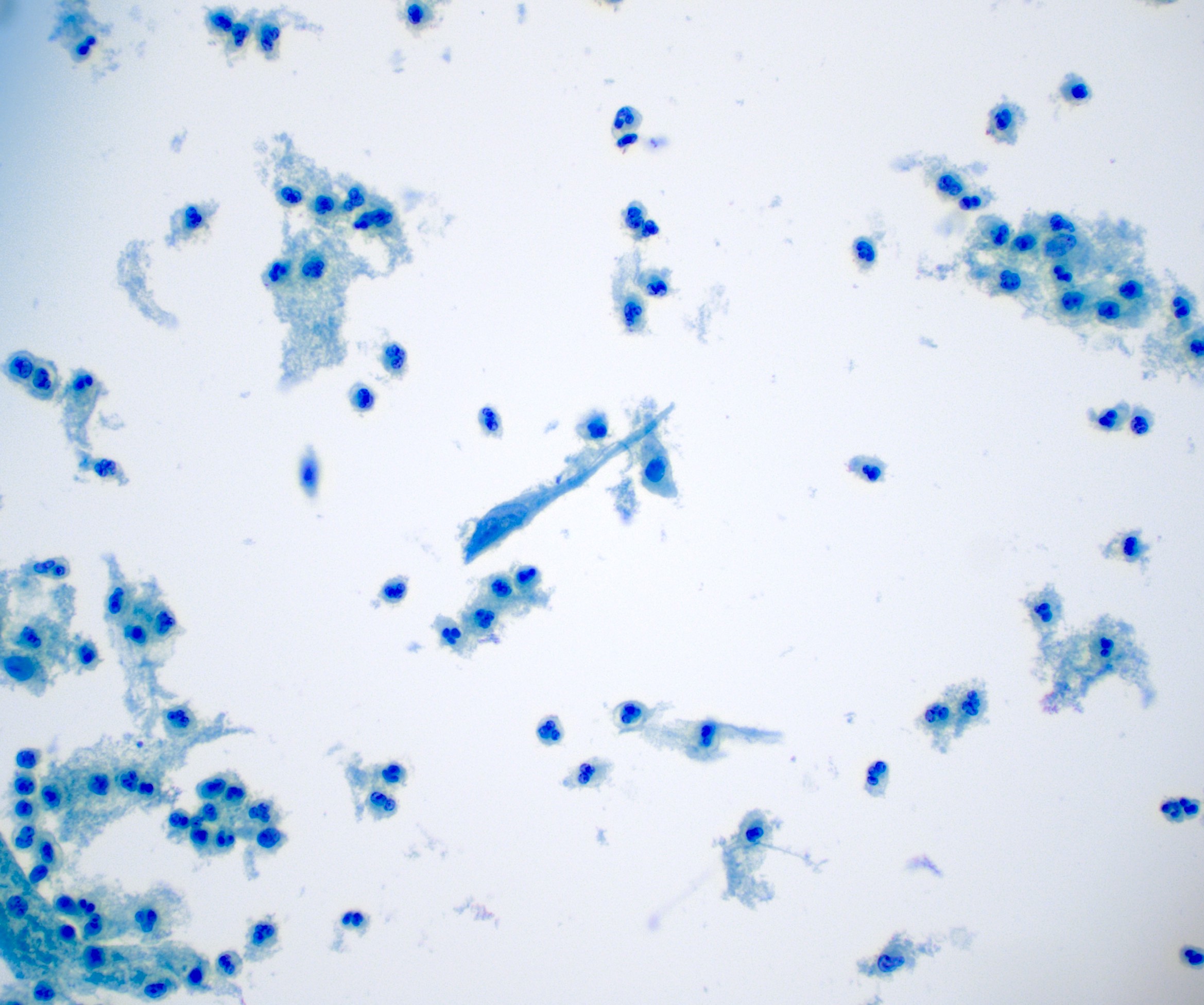
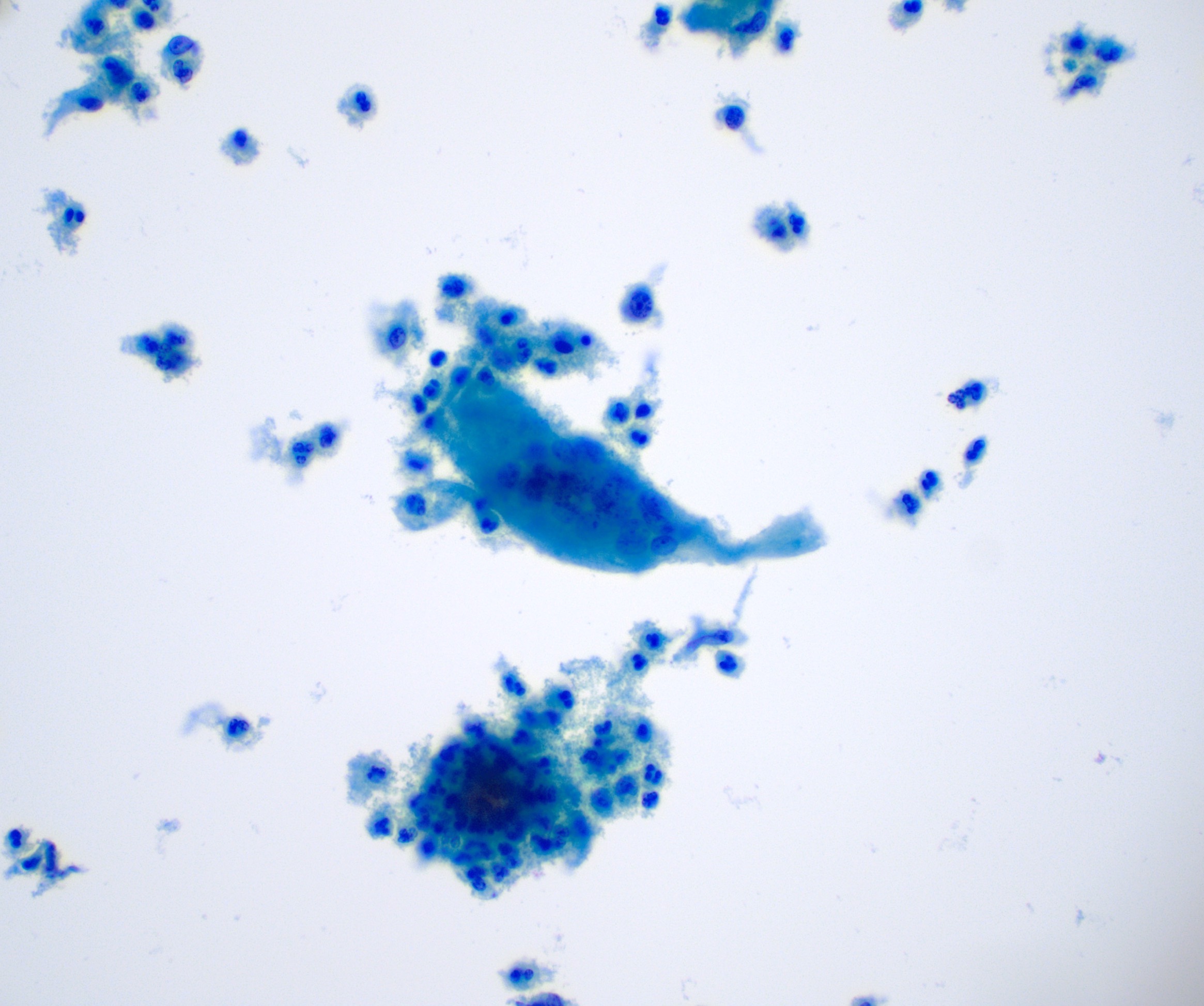
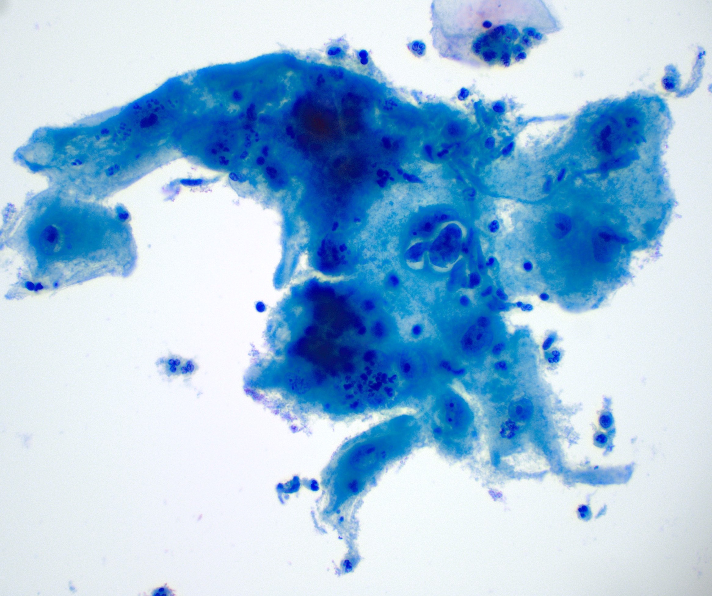
Intracytoplasmic inclusions #1 (endocervical cells)

#2 (endothelial cells)

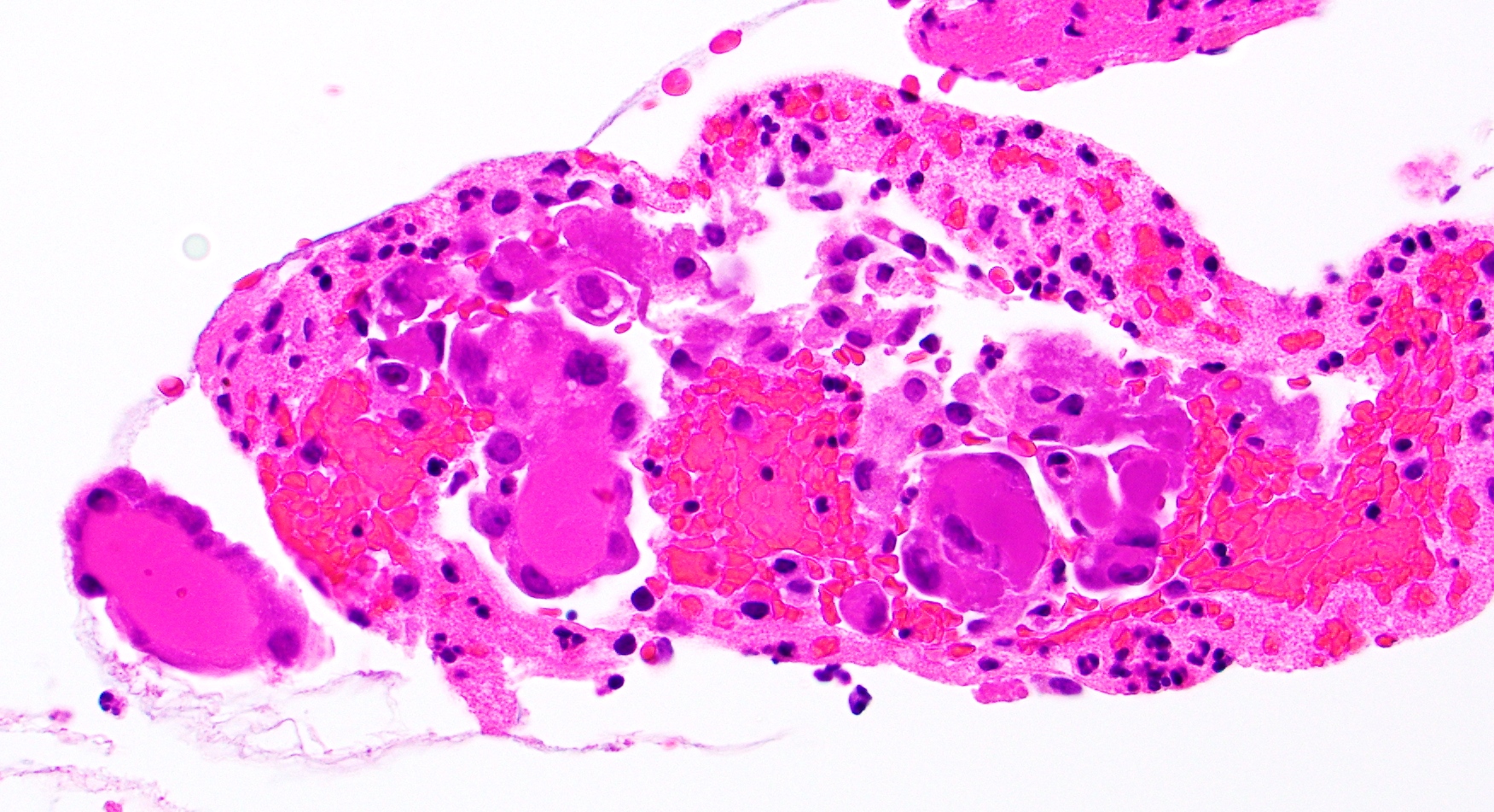
CMV+ glands and stroma

Associated acute inflammatory infiltrate

Intracytoplasmic vacuoles within endocervical glandular cells

Fibrin thrombi within small vessels
Not cervix:

Lung #1 - Giemsa stain

Kidney

Pancreas
Contributed by Aung Phyo, M.D. (Case #480)
Decidualized cervical nodule
Multiple prominent nucleoli
Low N:C ratio
Intermixed lymphocytes
Images hosted on other servers:

Female genital tract development

Müllerian duct embryonic formation

Fusion of Müllerian ducts

Classic theory versus Acien theory


Congenital uterine malformations
Images hosted on other servers:

MRI showing uterovaginal absence

Hysterosalpingogram
showing double
cervix
Images hosted on other servers:

Speculum examination showing double cervix
Images hosted on other servers:


Fetal female reproductive tract
Images hosted on other servers:

Fusion of Müllerian ducts to UVC

Histology of UVC

Cervical development
Female genital tract embryology
Images hosted on other servers:

Colposcopy

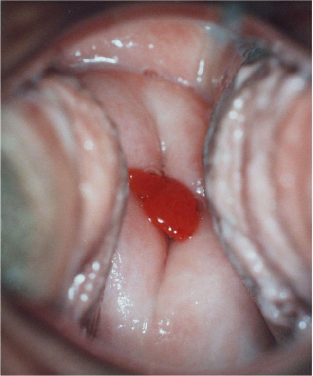
Polyp
Contributed by Natalia Buza, M.D.
Small polyp
Contributed by Natalia Buza, M.D. and Andrey Bychkov, M.D., Ph.D.
Thick walled arteries
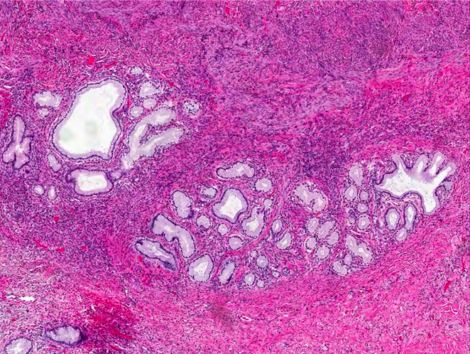
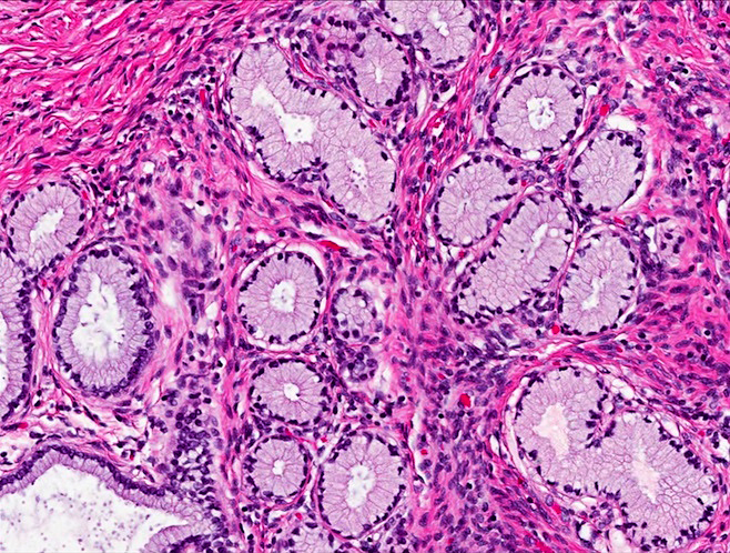
Microglandular hyperplasia
Squamous metaplasia
Chronic inflammation
Mitoses

Cystically dilated glands
Contributed by Marilin Rosa, M.D. and Carmen Luz, M.D.
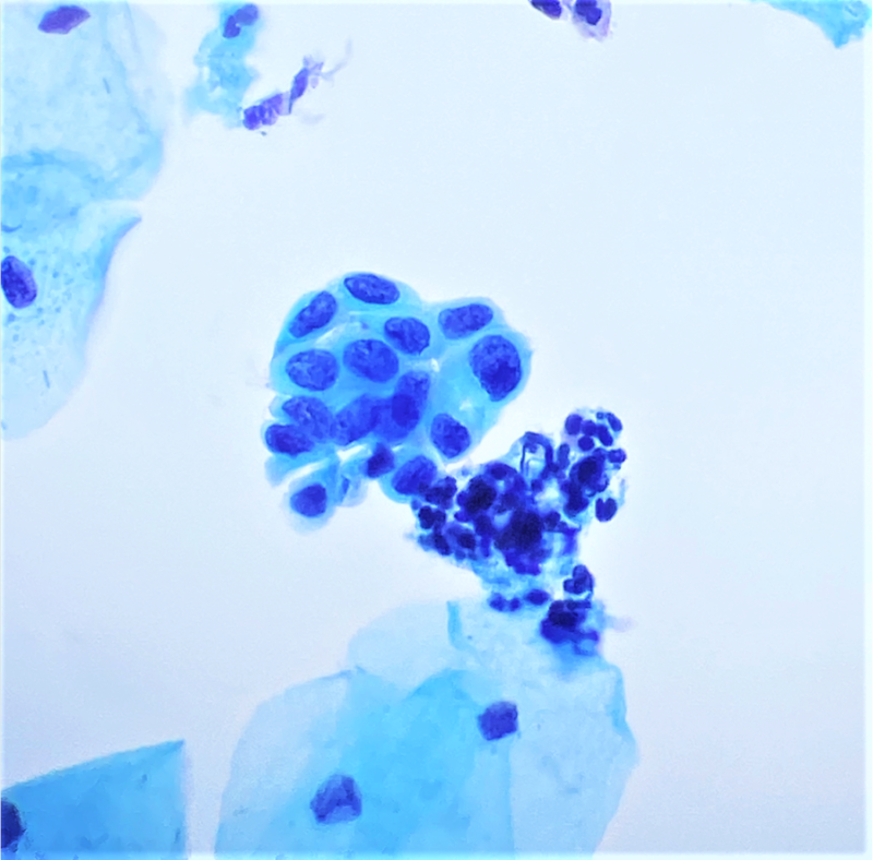
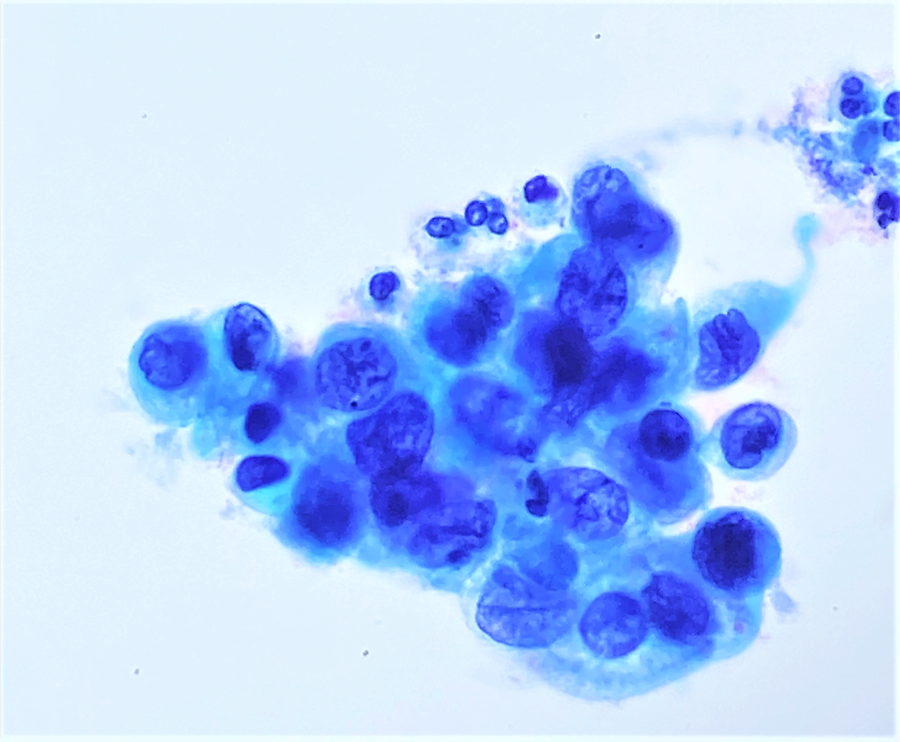
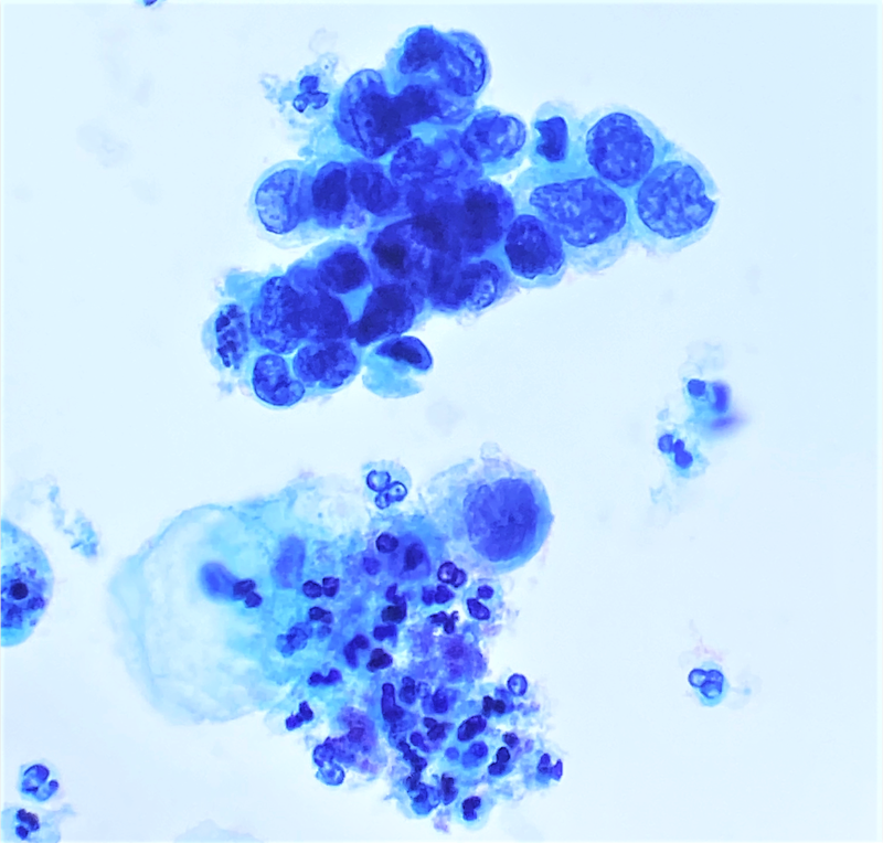
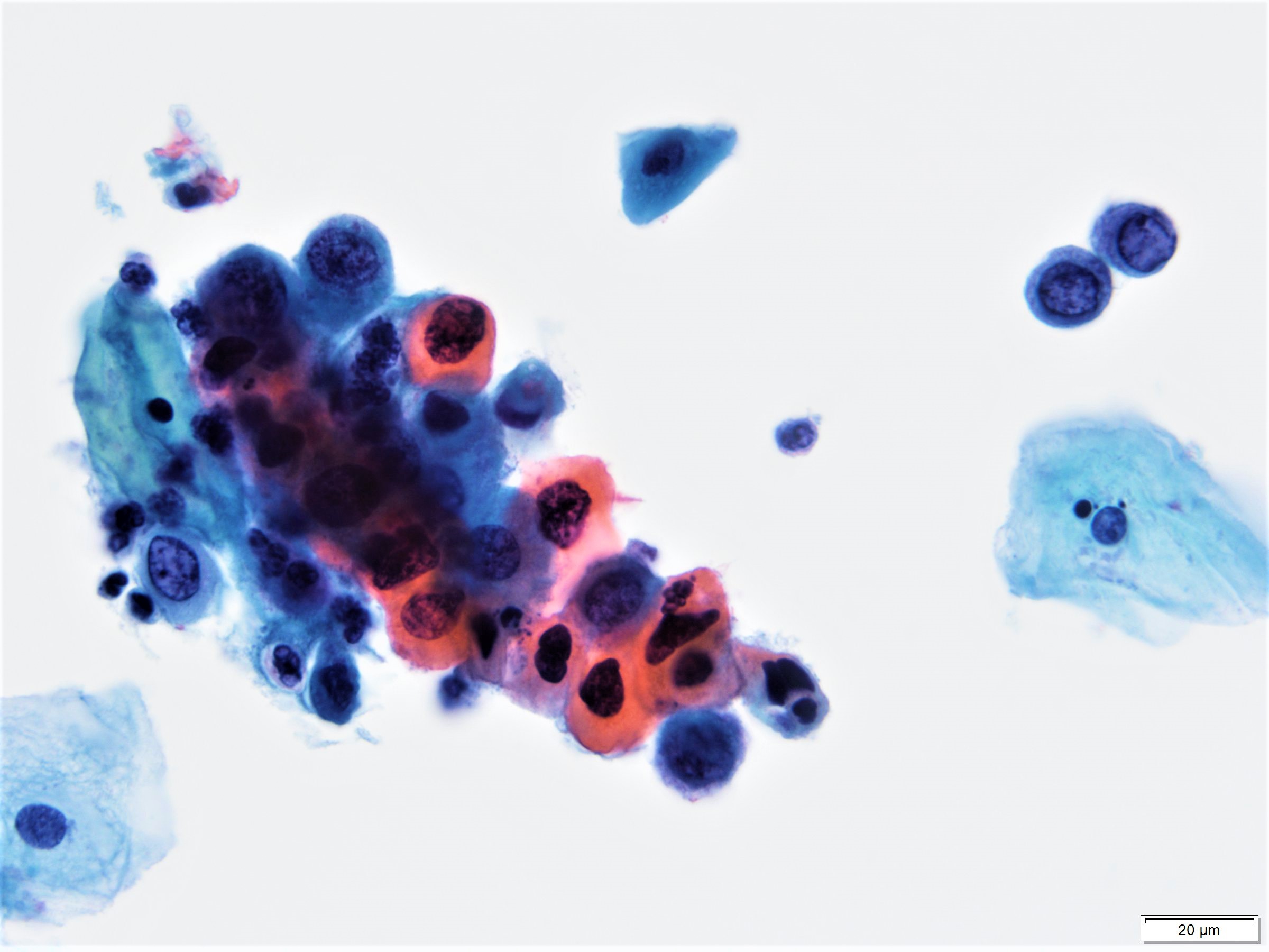
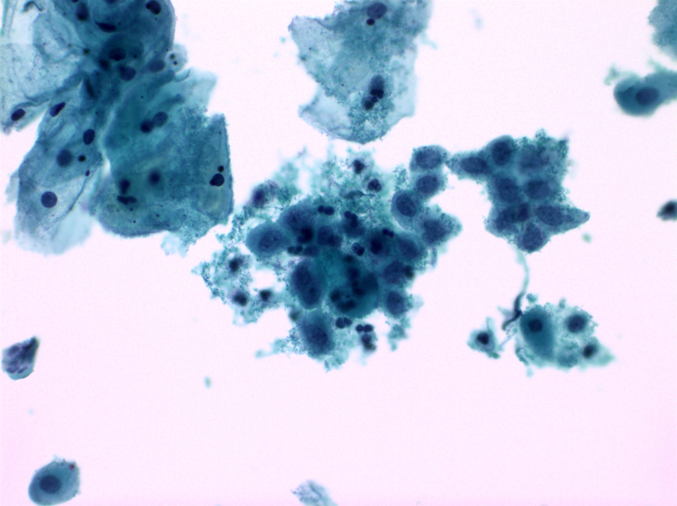
Endometrial adenocarcinoma
Endometrial group with atypia
Images hosted on other servers:
Various images
AFIP images
Endometriosis
Images hosted on other servers:

Barrel shaped cervix on MRI in MDA

Ill defined cervical mass on MRI in MDA
Contributed by Gulisa Turashvili, M.D., Ph.D.
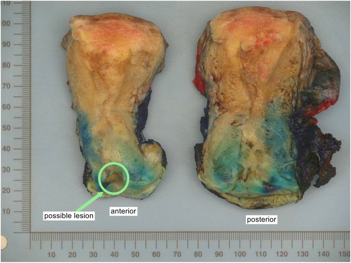
Radical hysterectomy for gastric type adenocarcinoma
Images hosted on other servers:

Bulging cervix in MDA

Cervical swelling without a mass in MDA
Contributed by Gulisa Turashvili, M.D., Ph.D.
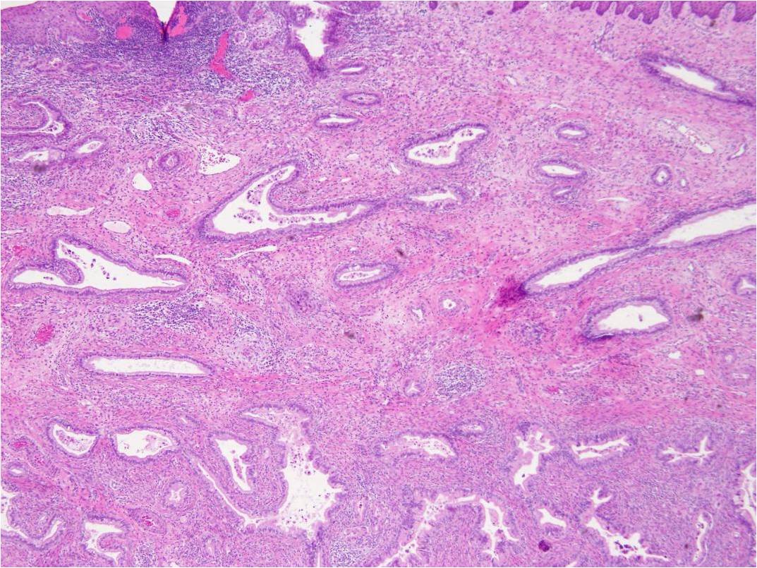
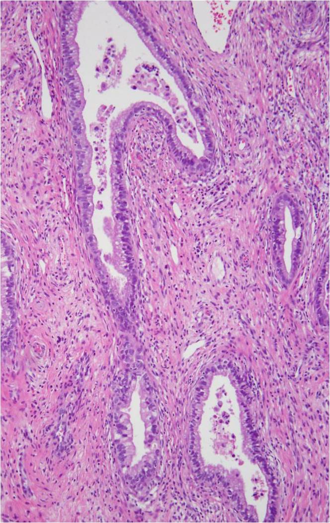
Well differentiated gastric type adenocarcinoma
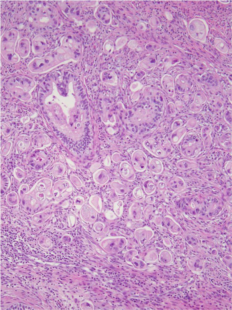
Poorly differentiated gastric type adenocarcinoma
Well differentiated gastric type adenocarcinoma

Gastric type adenocarcinoma in hysterectomy
Poorly differentiated gastric type adenocarcinoma

Gastric type adenocarcinoma involving fallopian tube
Contributed by Gulisa Turashvili, M.D., Ph.D.
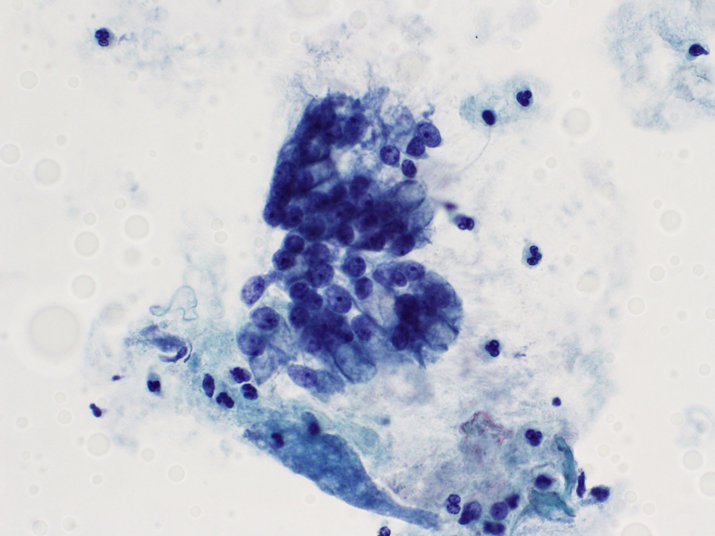
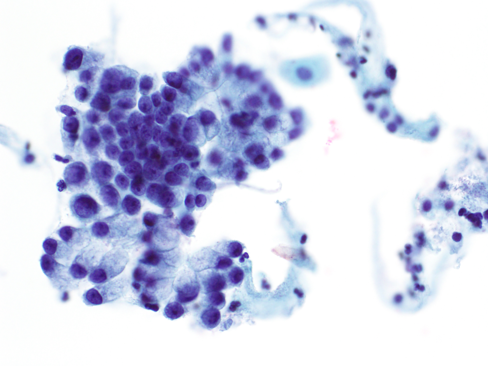
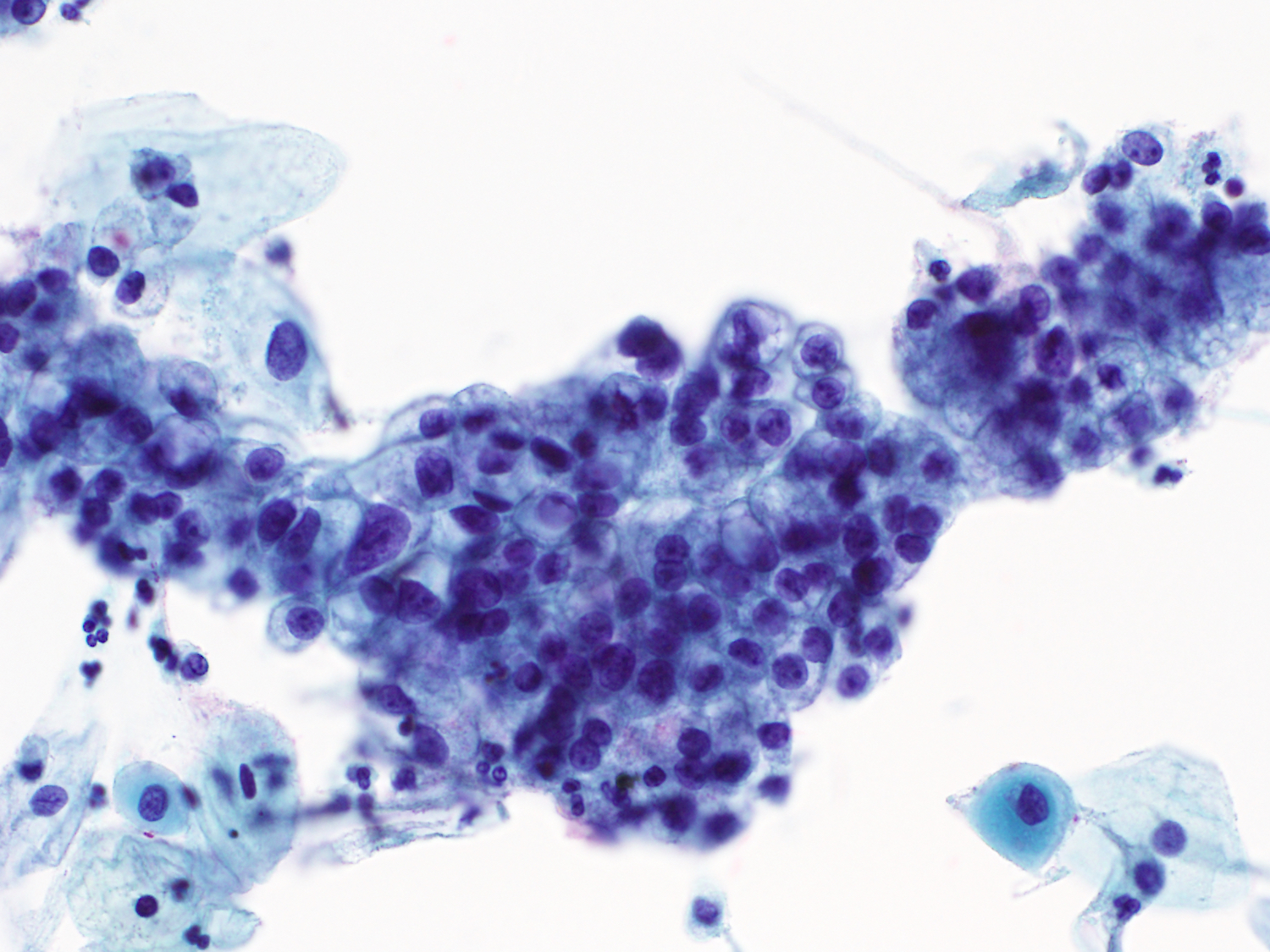
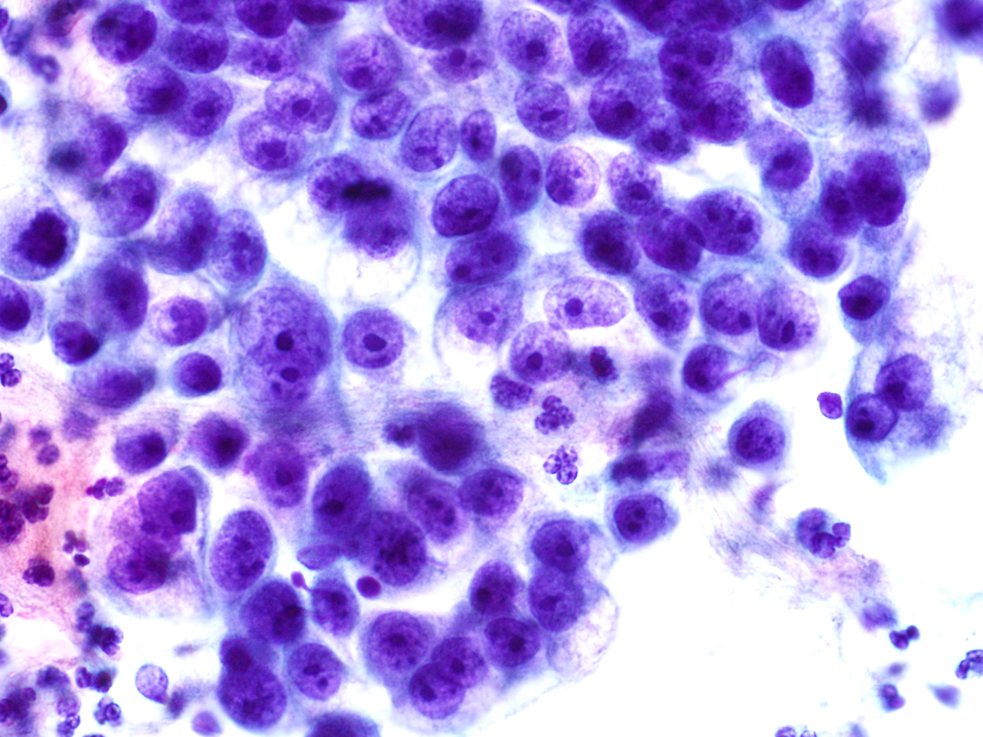
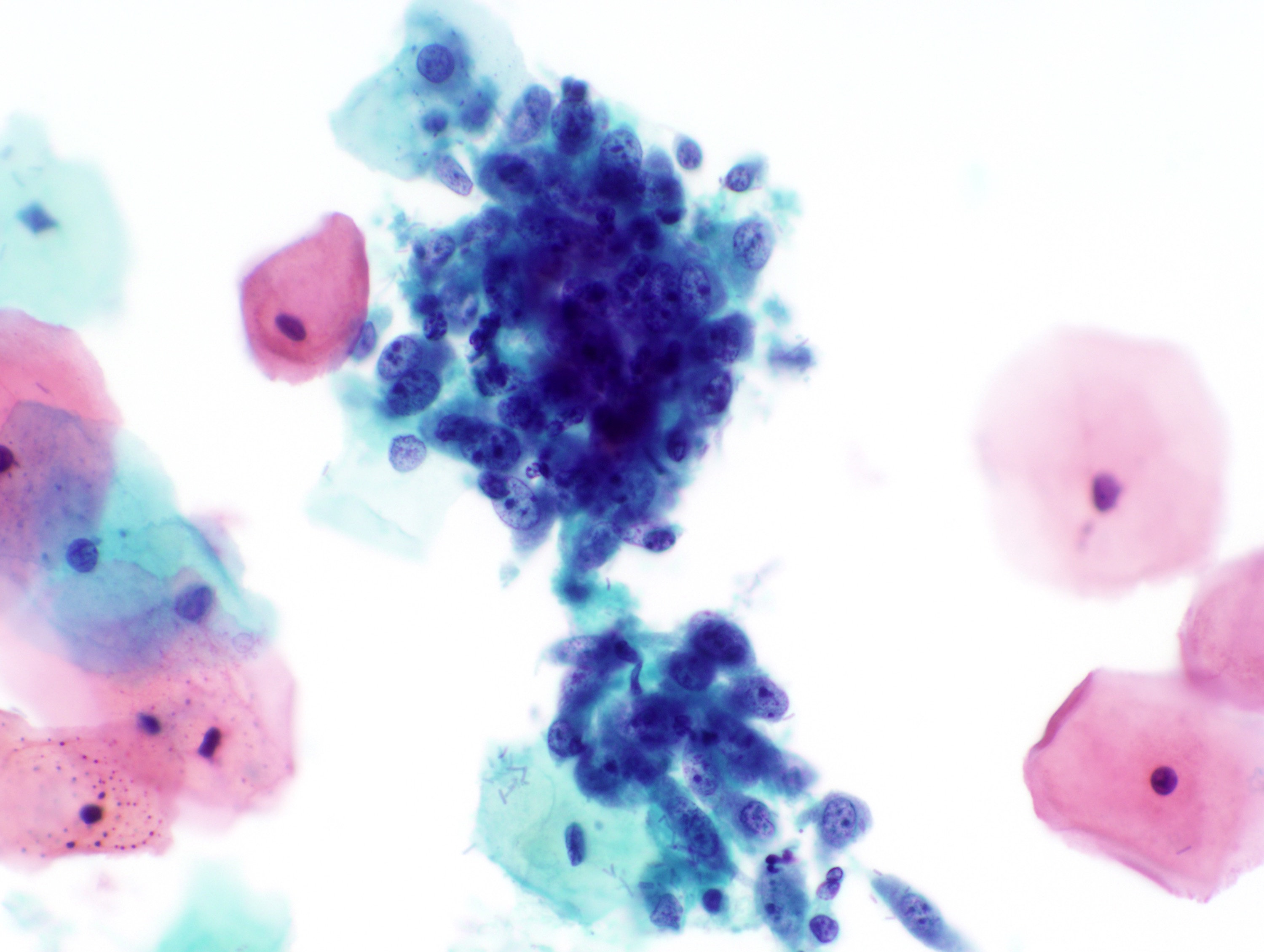
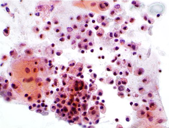
Gastric type adenocarcinoma on Pap cytology
AFIP images

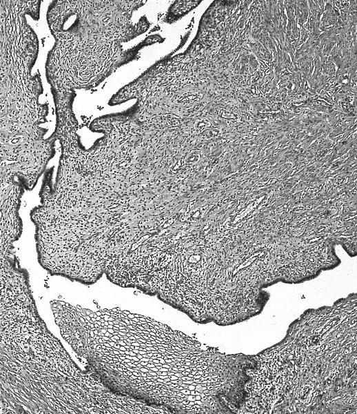
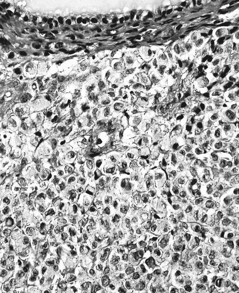
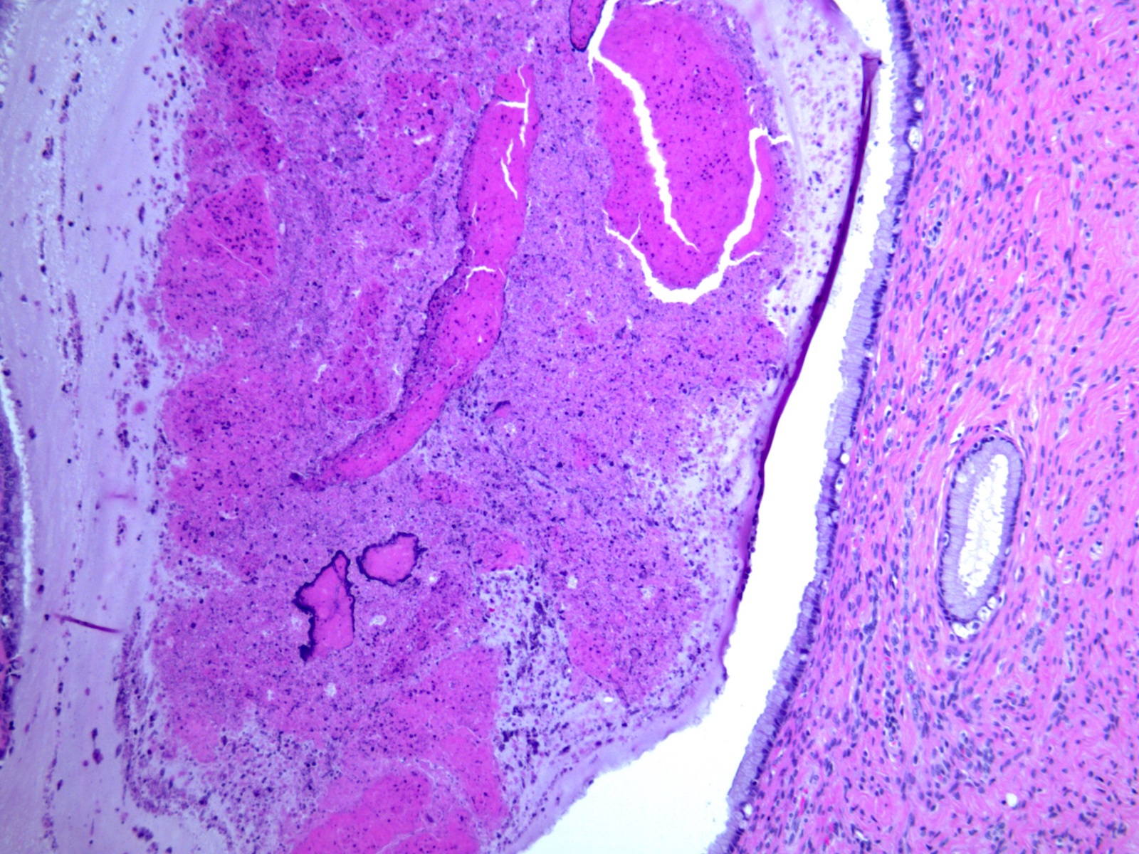
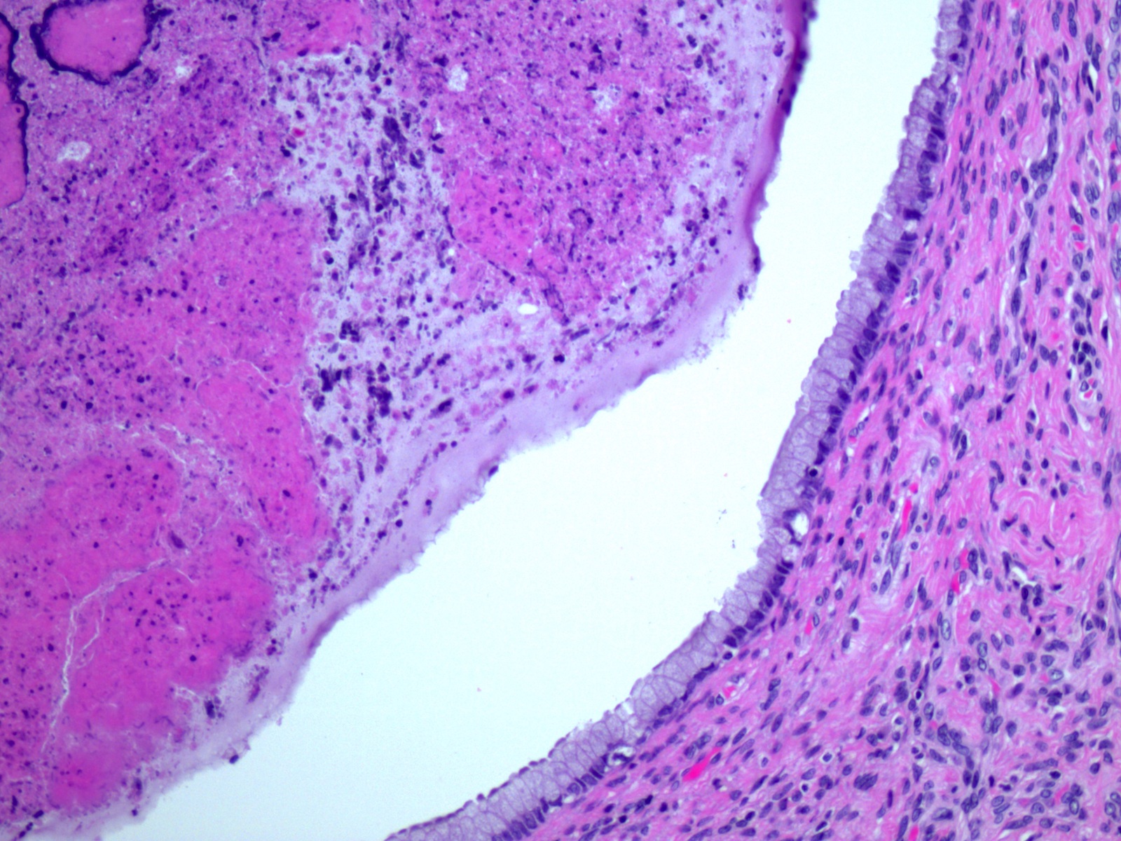
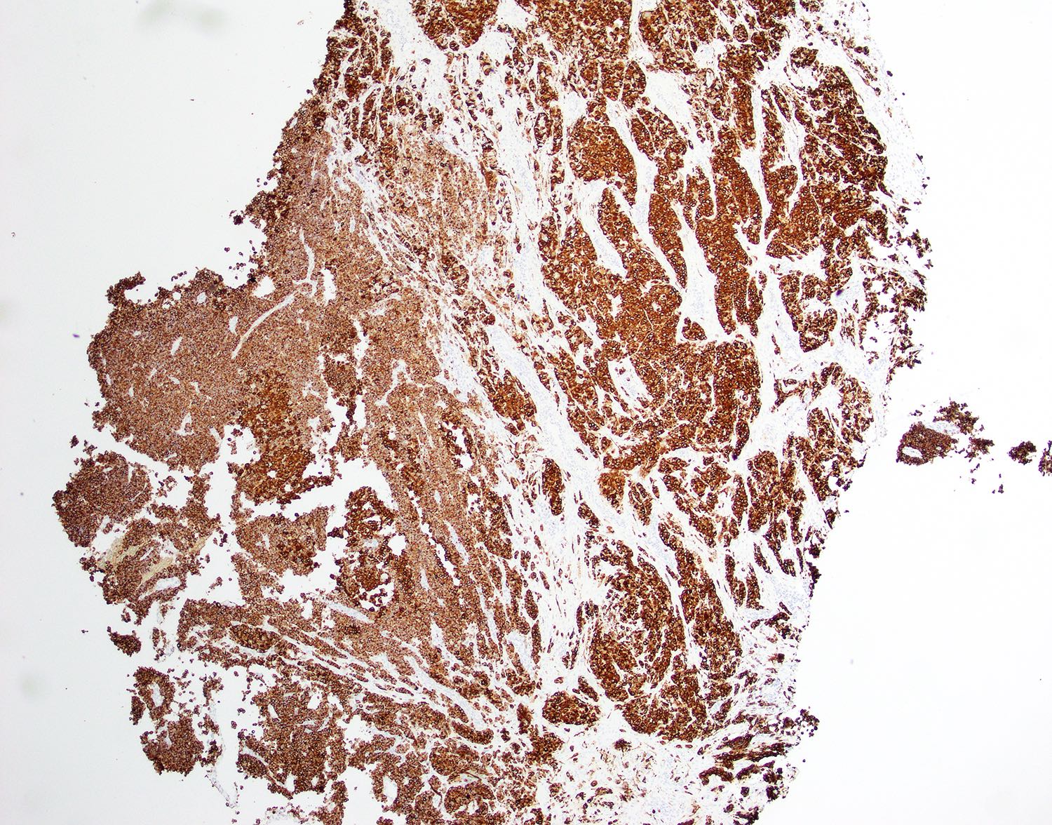
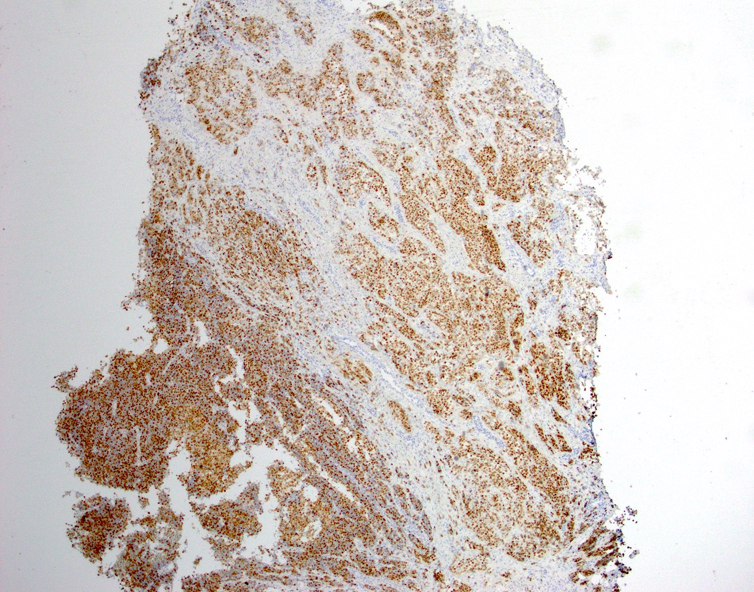
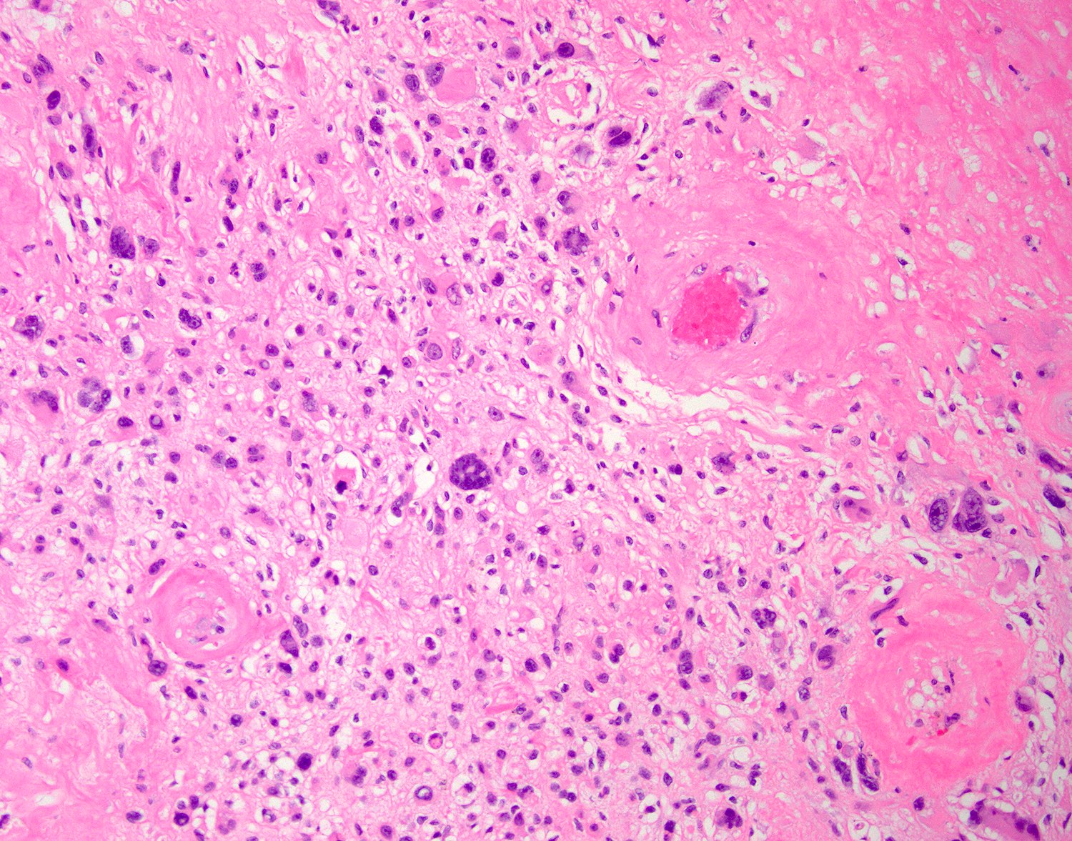
Polypoid mass of glia below endocervical surface
Case #135


H&E and GFAP
Images hosted on other servers:

Grossing diagrams
Images hosted on other servers:

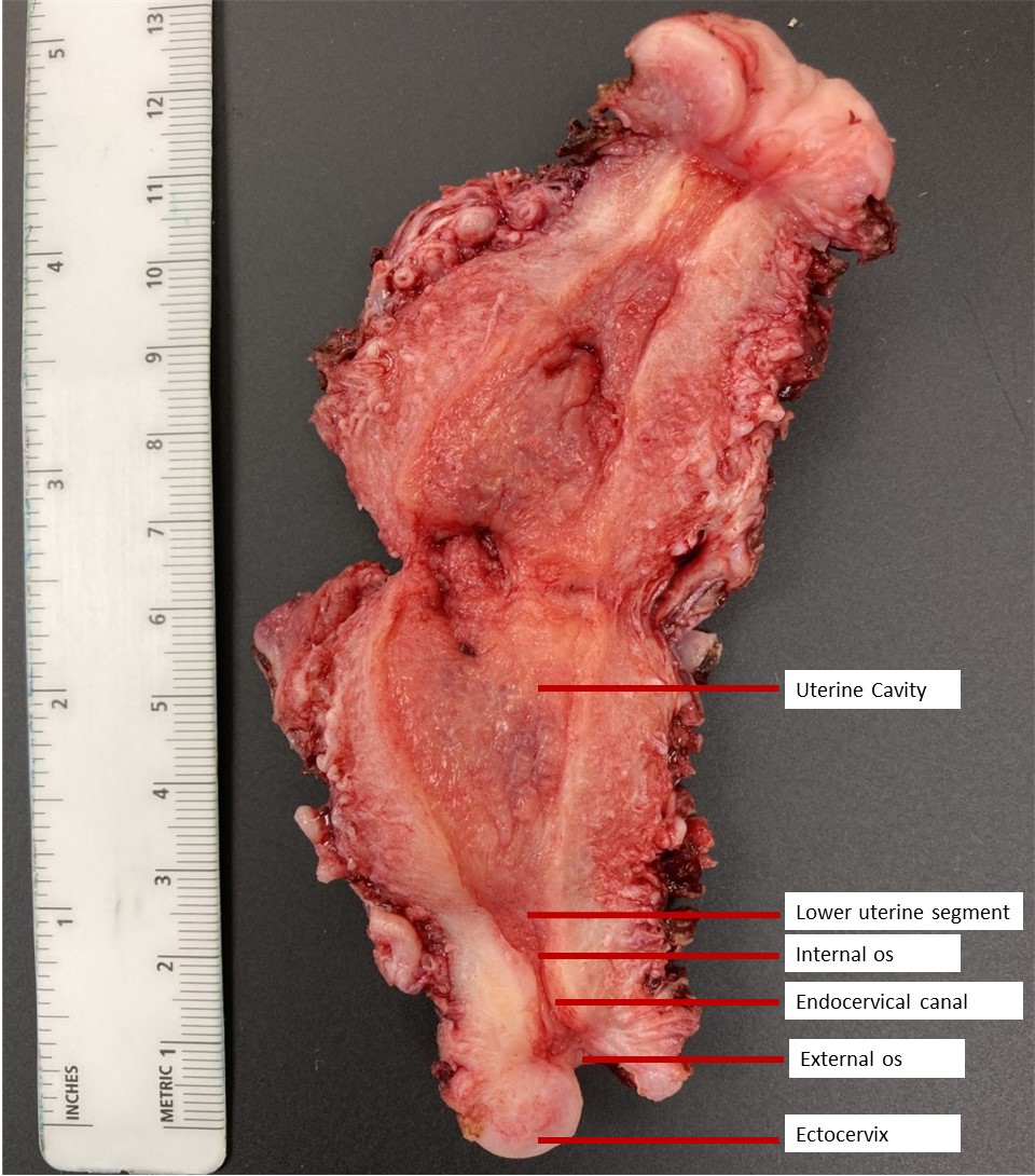
Hysterectomy specimen
Contributed by Kyle Devins, M.D.

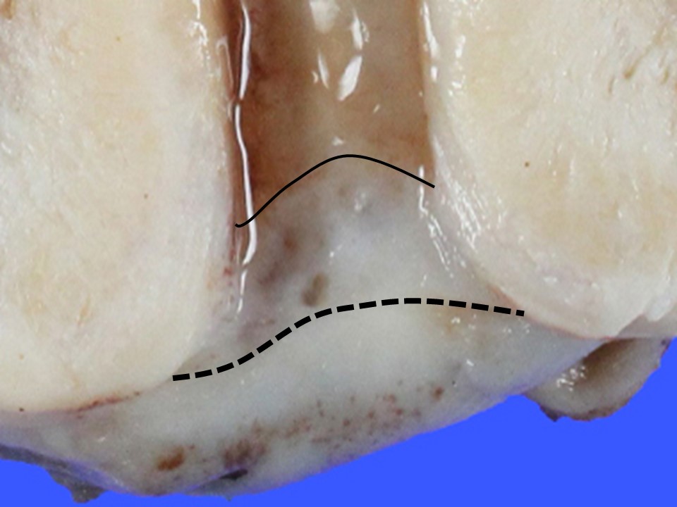
Schematic of exocervix and transformation zone
Images hosted on other servers:

Location of glandular and squamous epithelium
Contributed by Kyle Devins, M.D.
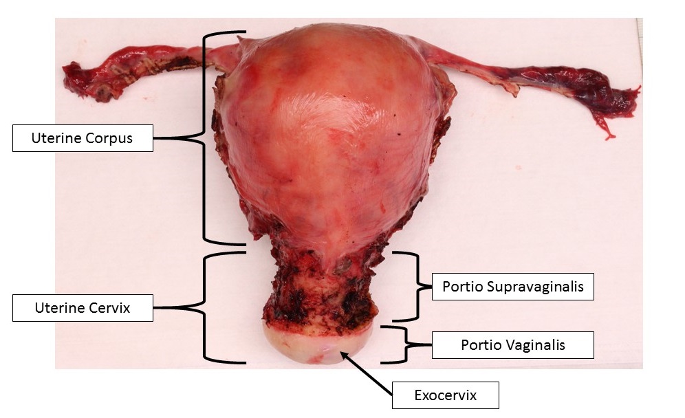
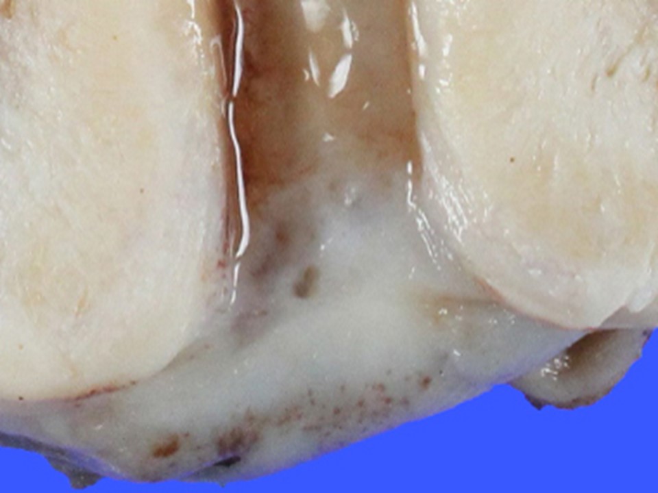
Uterus and cervix
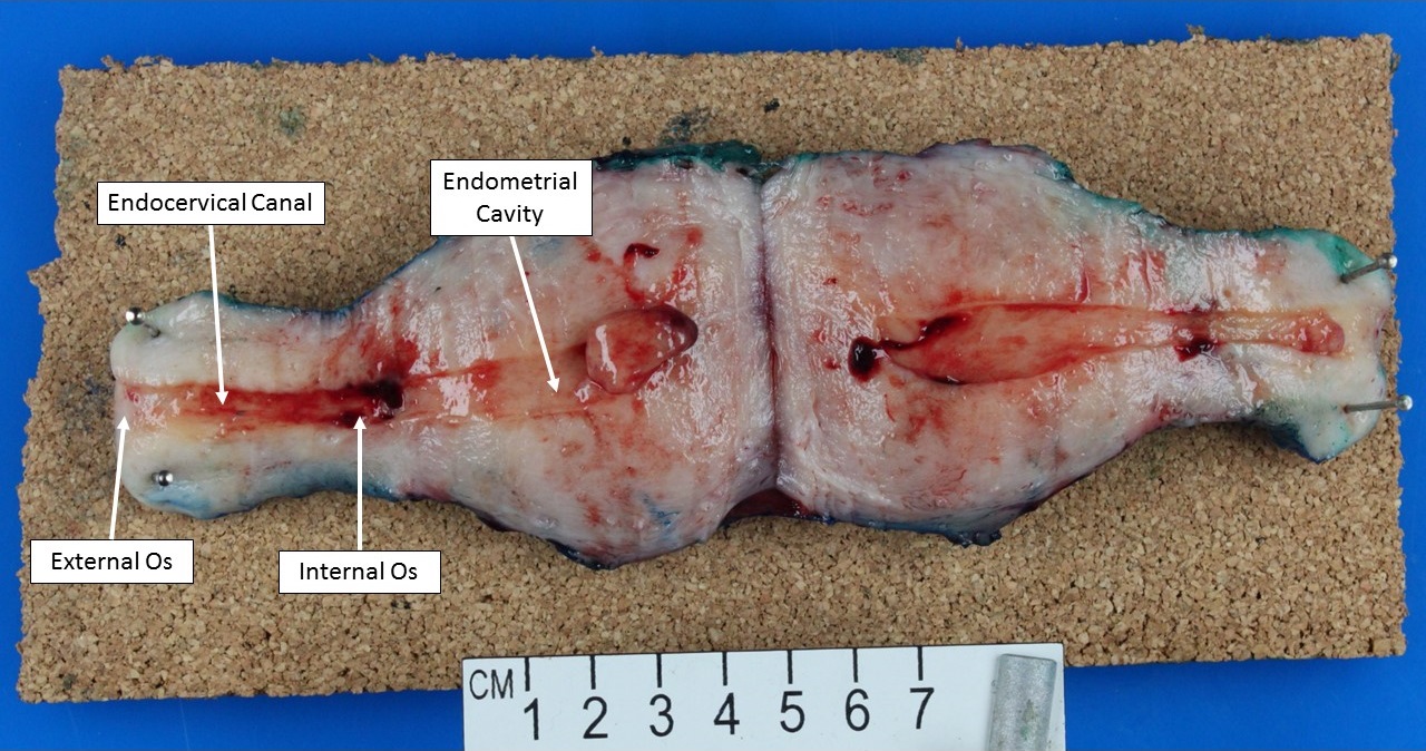
Bivalved uterus and cervix
Contributed by Kyle Devins, M.D.
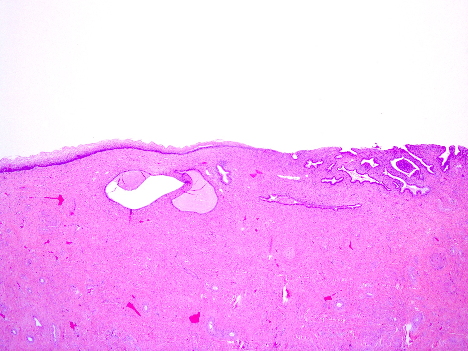
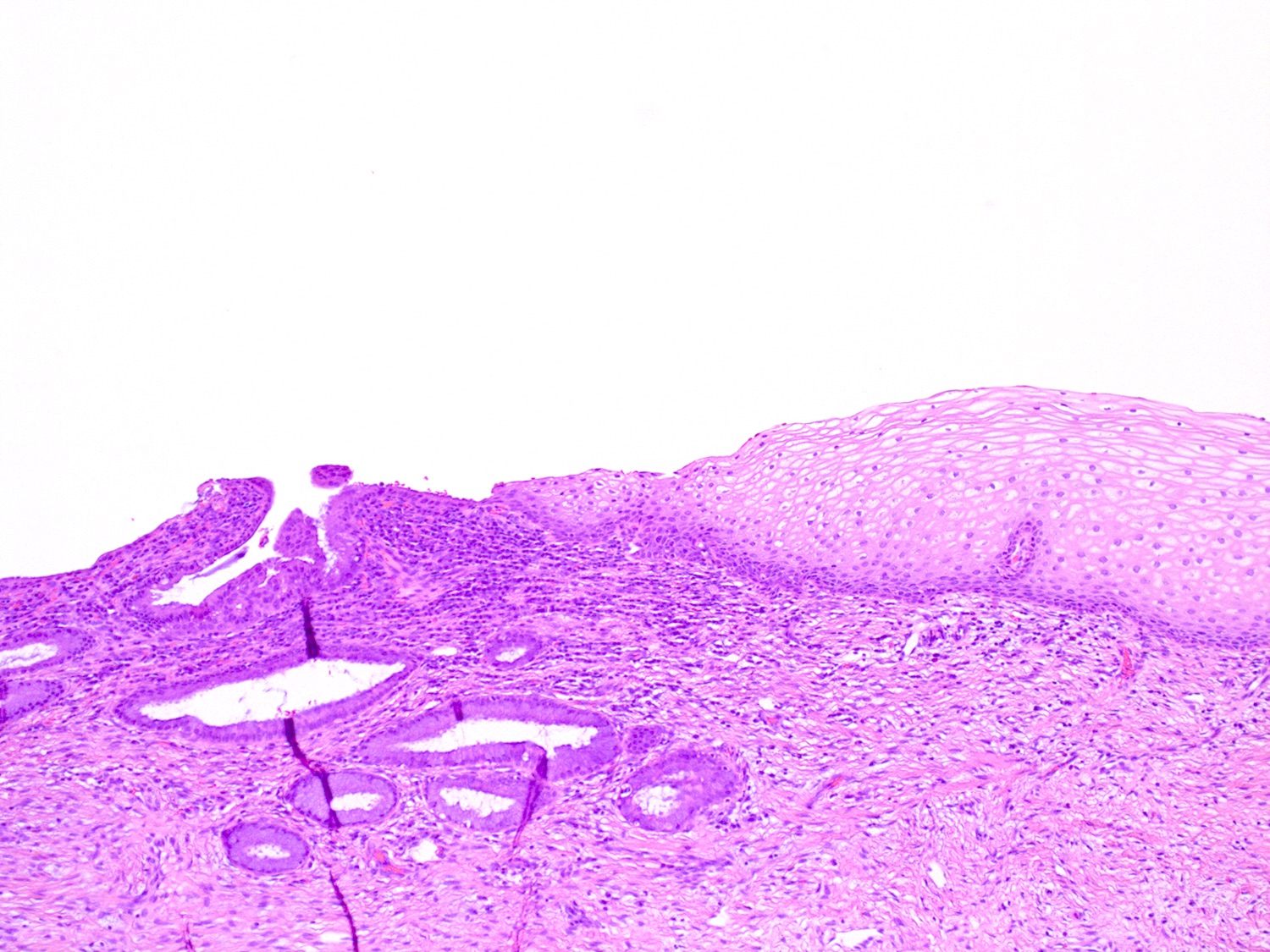
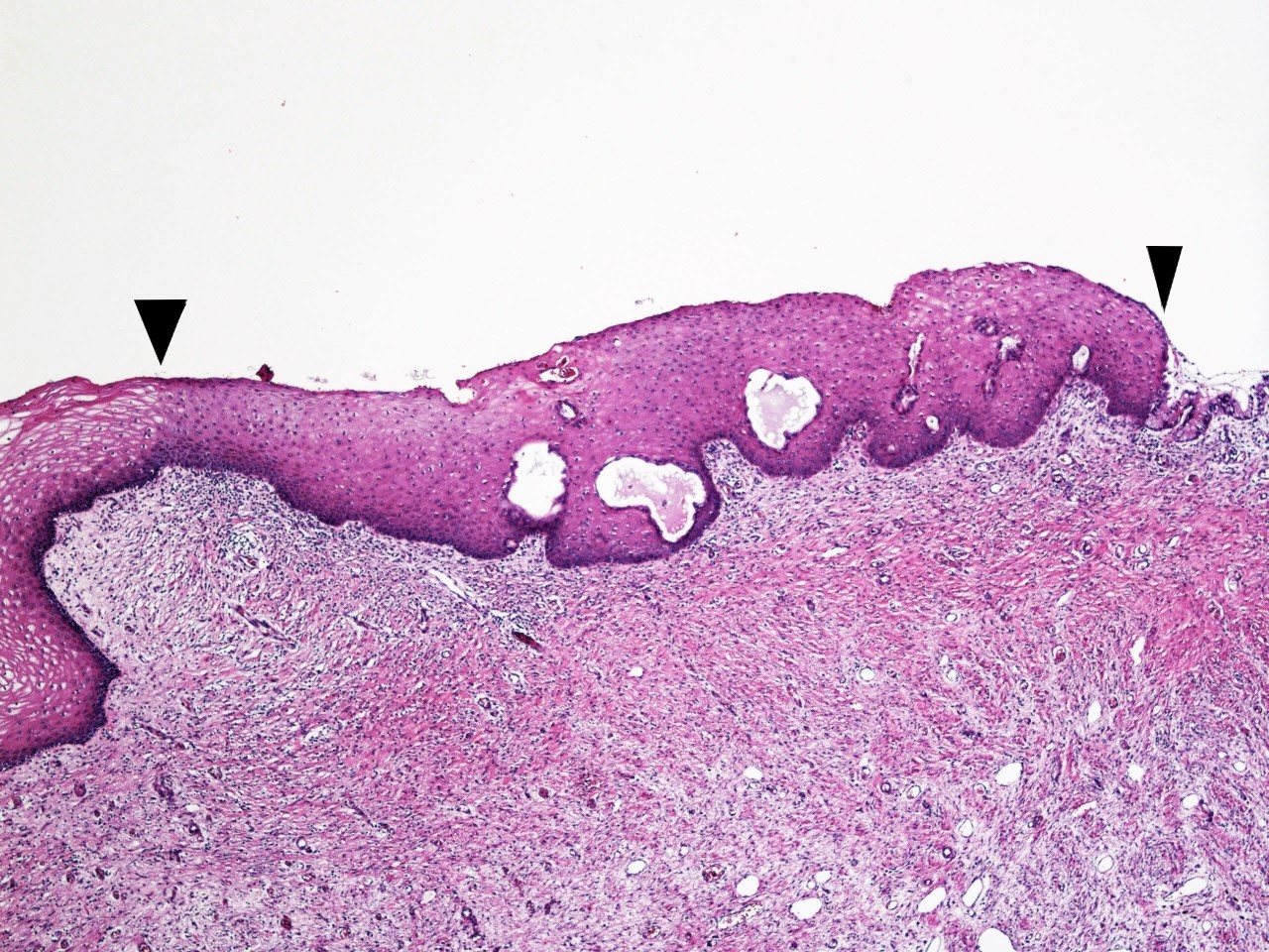
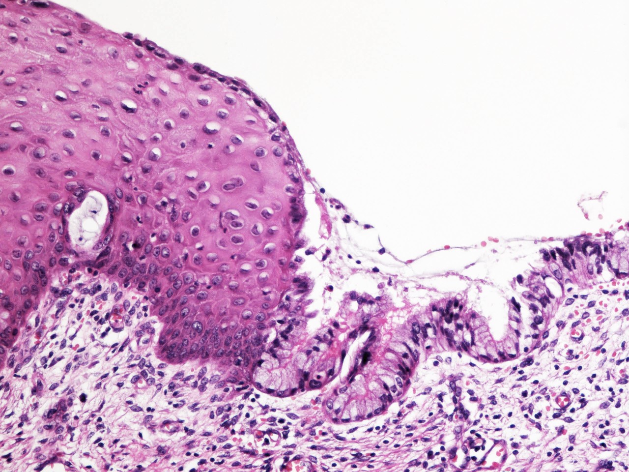
Squamocolumnar junction

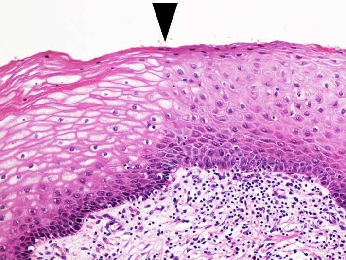
Cervical squamous epithelium
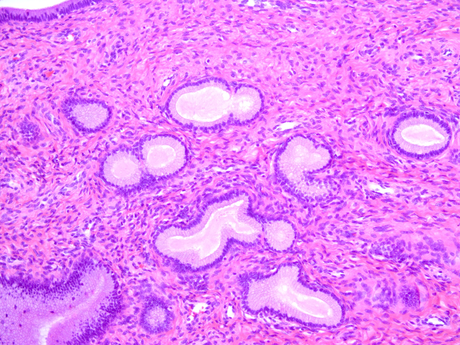
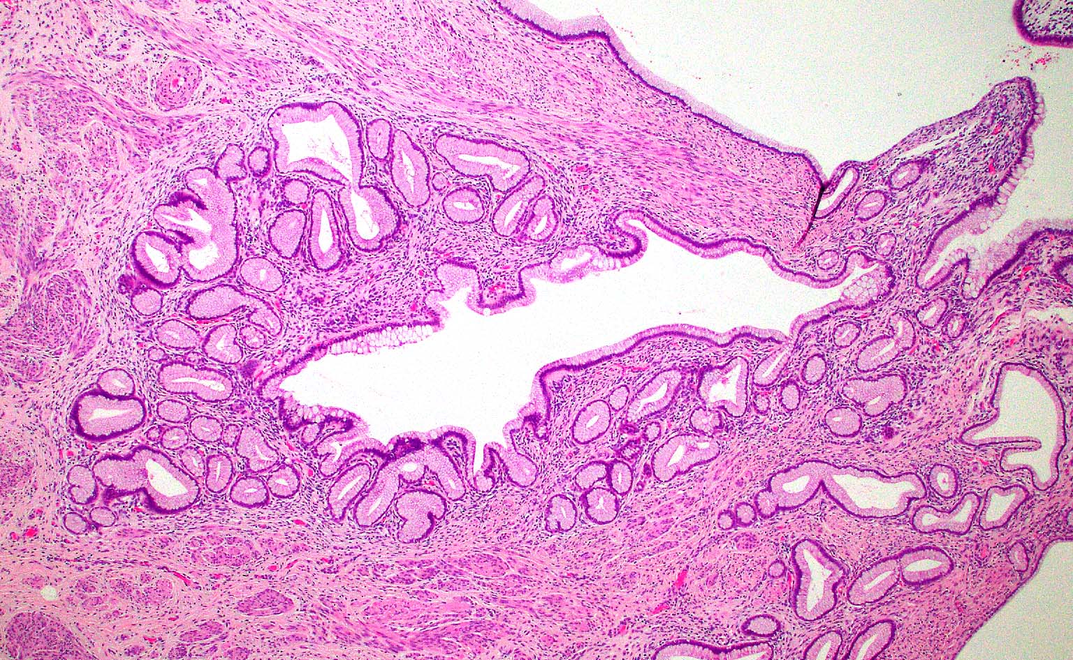
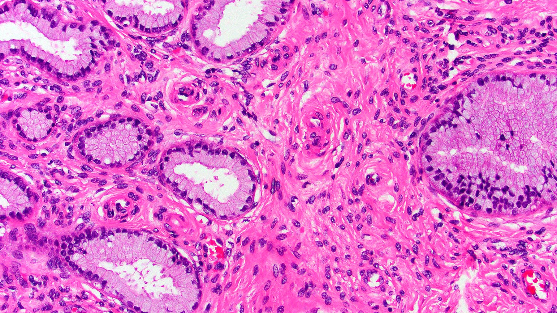
Benign endocervical glands
Dilated endocervical glands

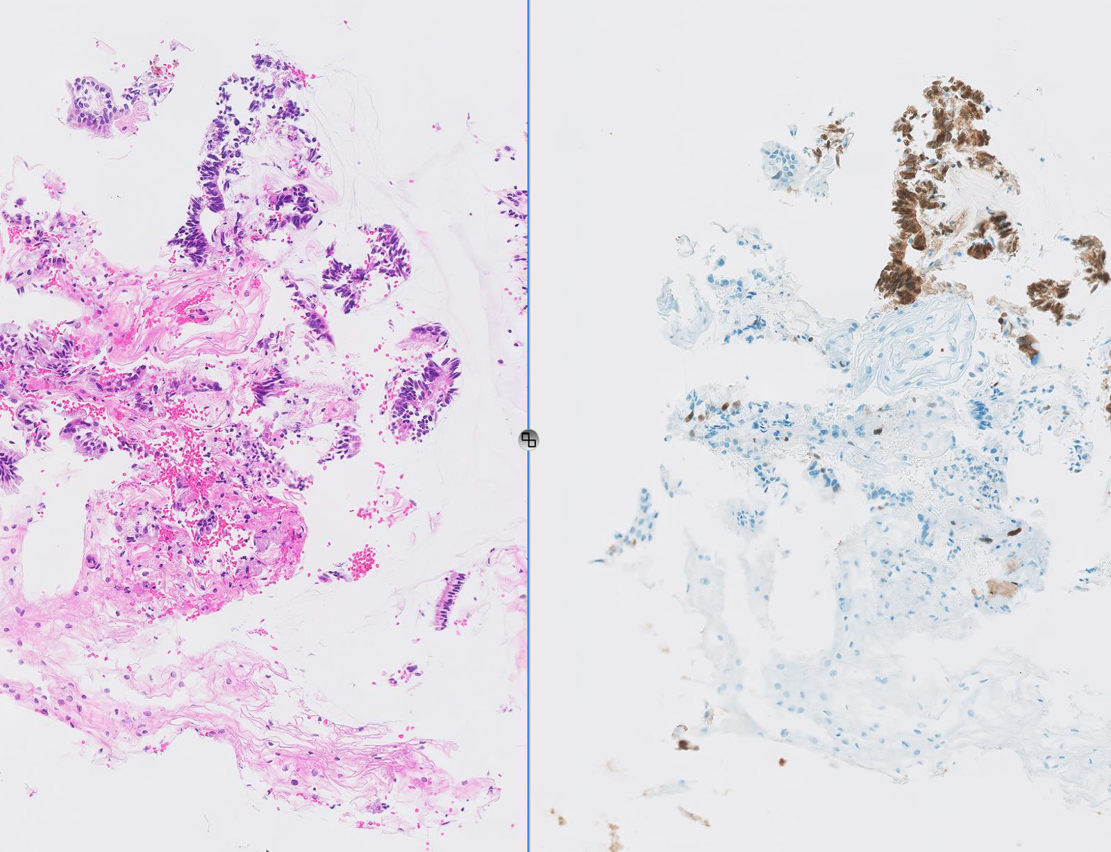
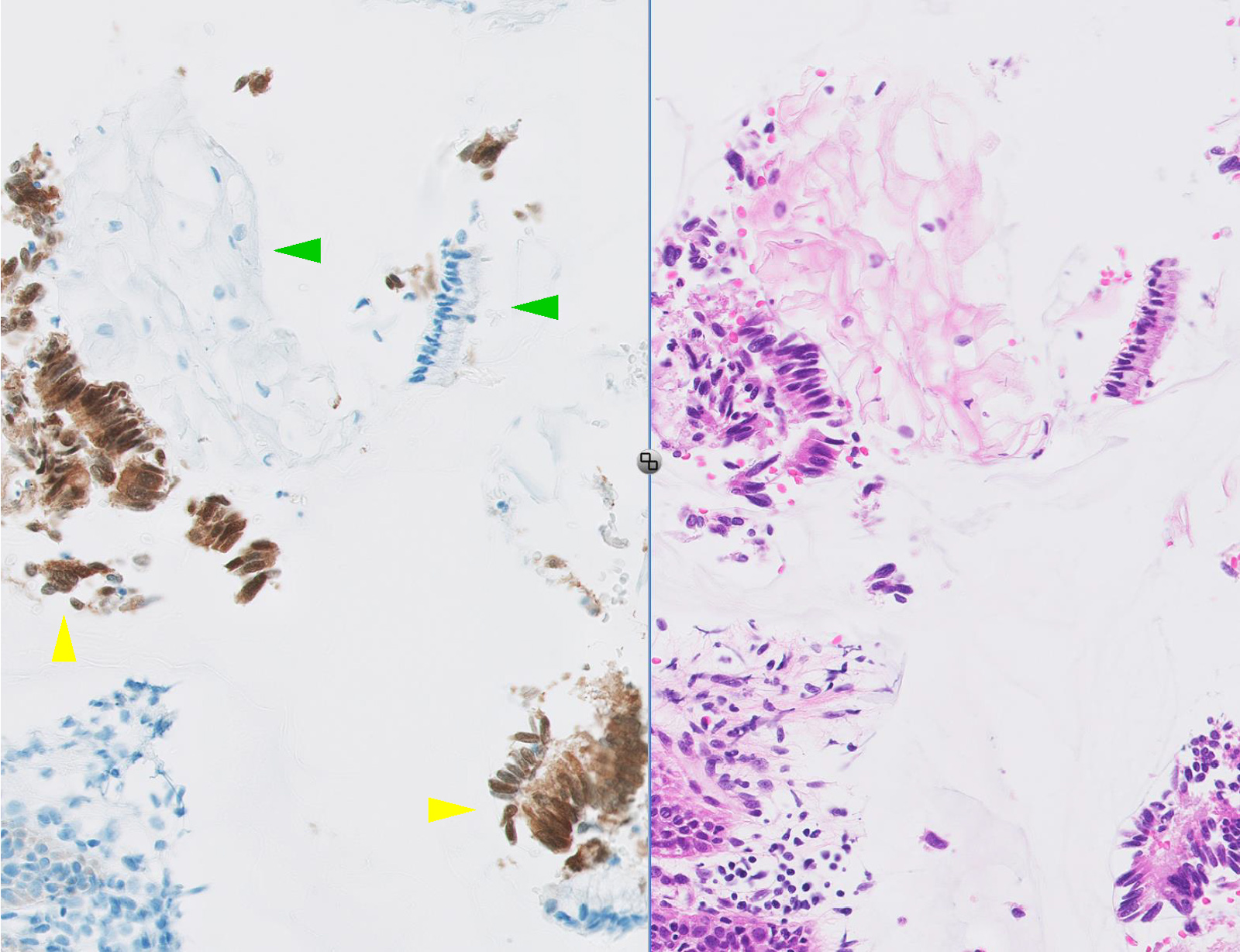
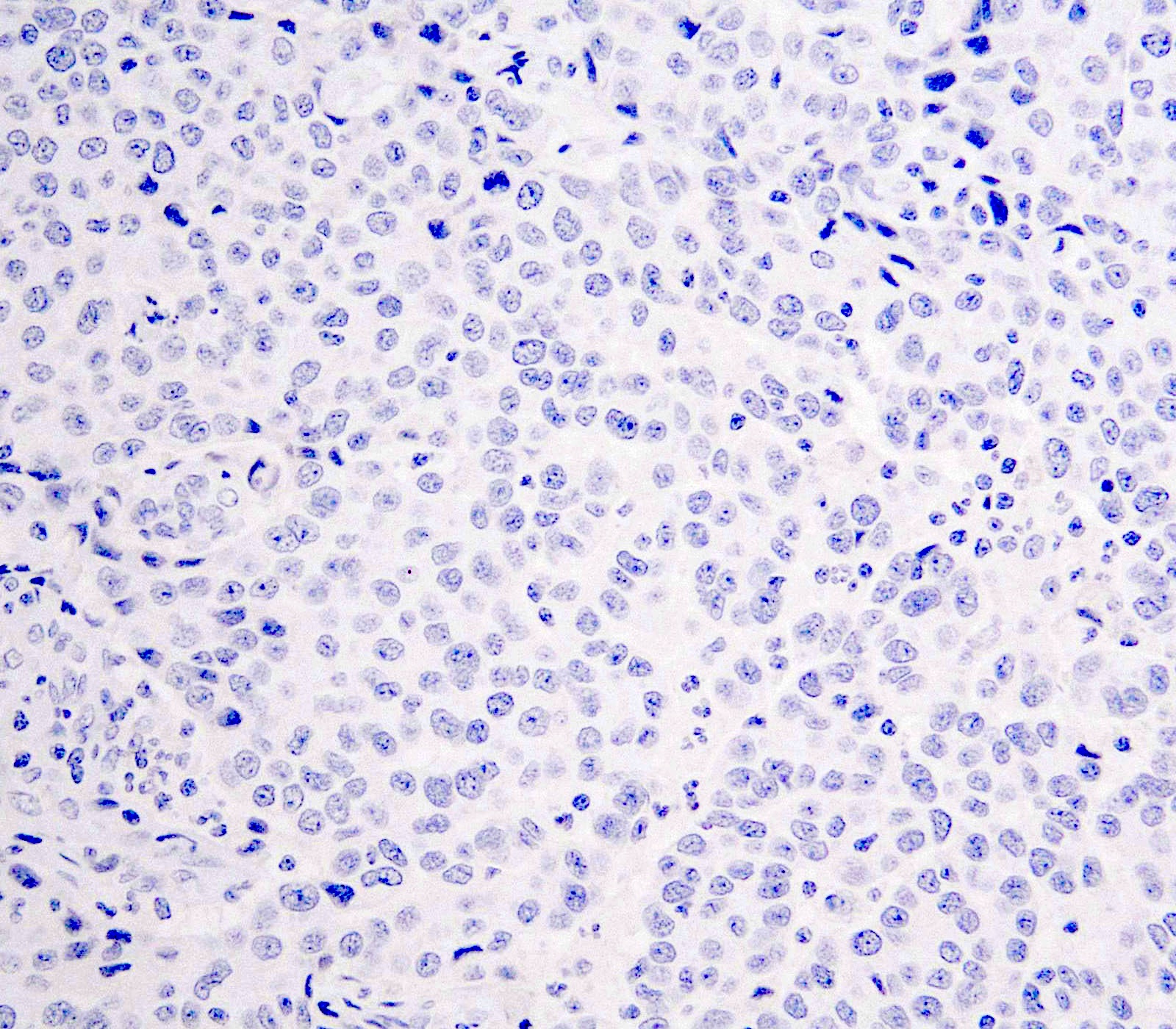
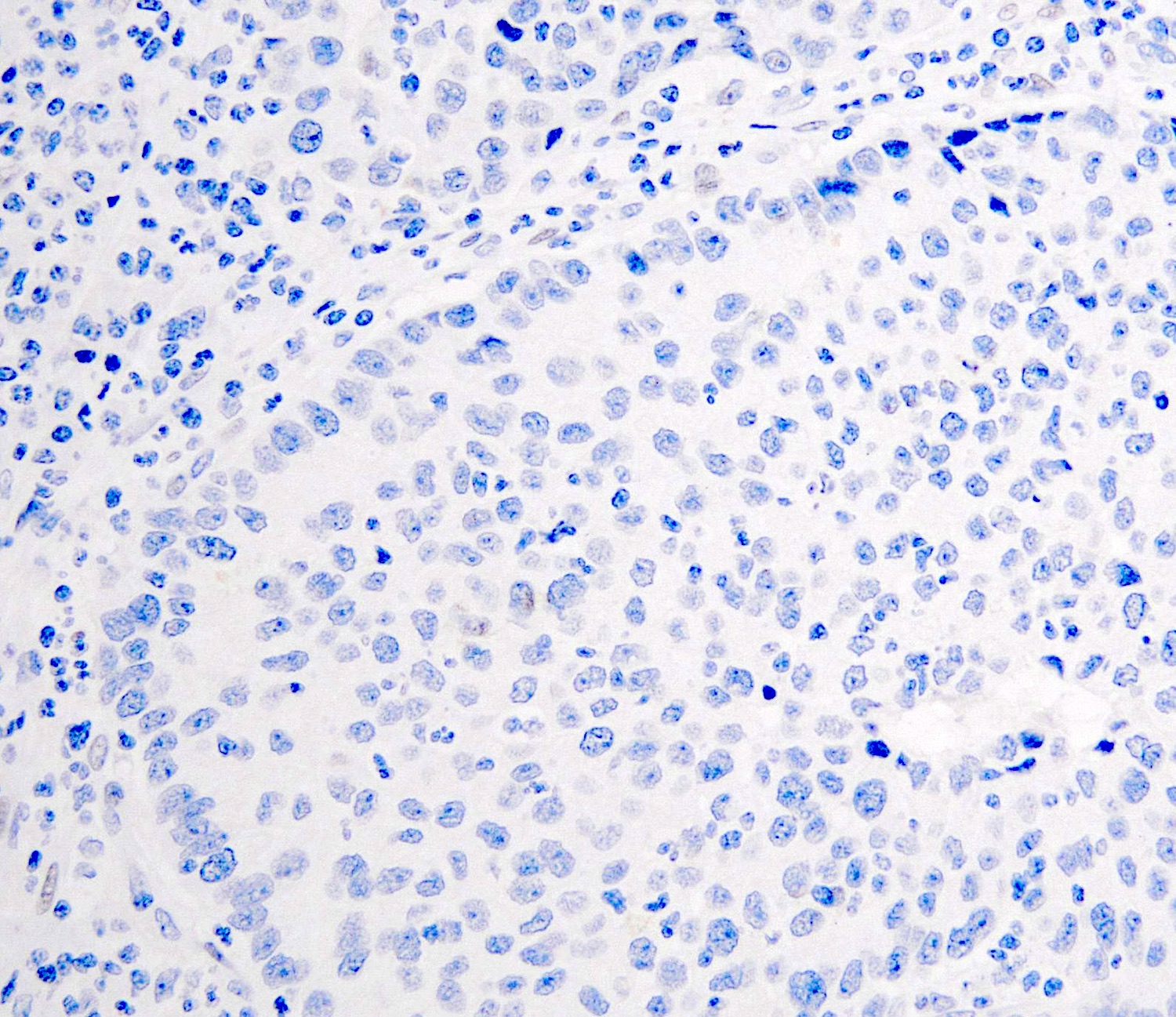
Normal cervical squamous p16 IHC
Normal endocervical p16 IHC
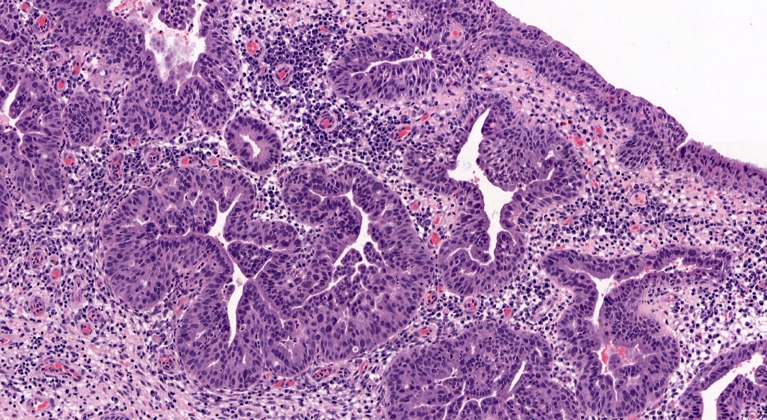
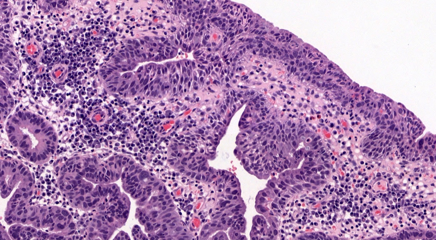
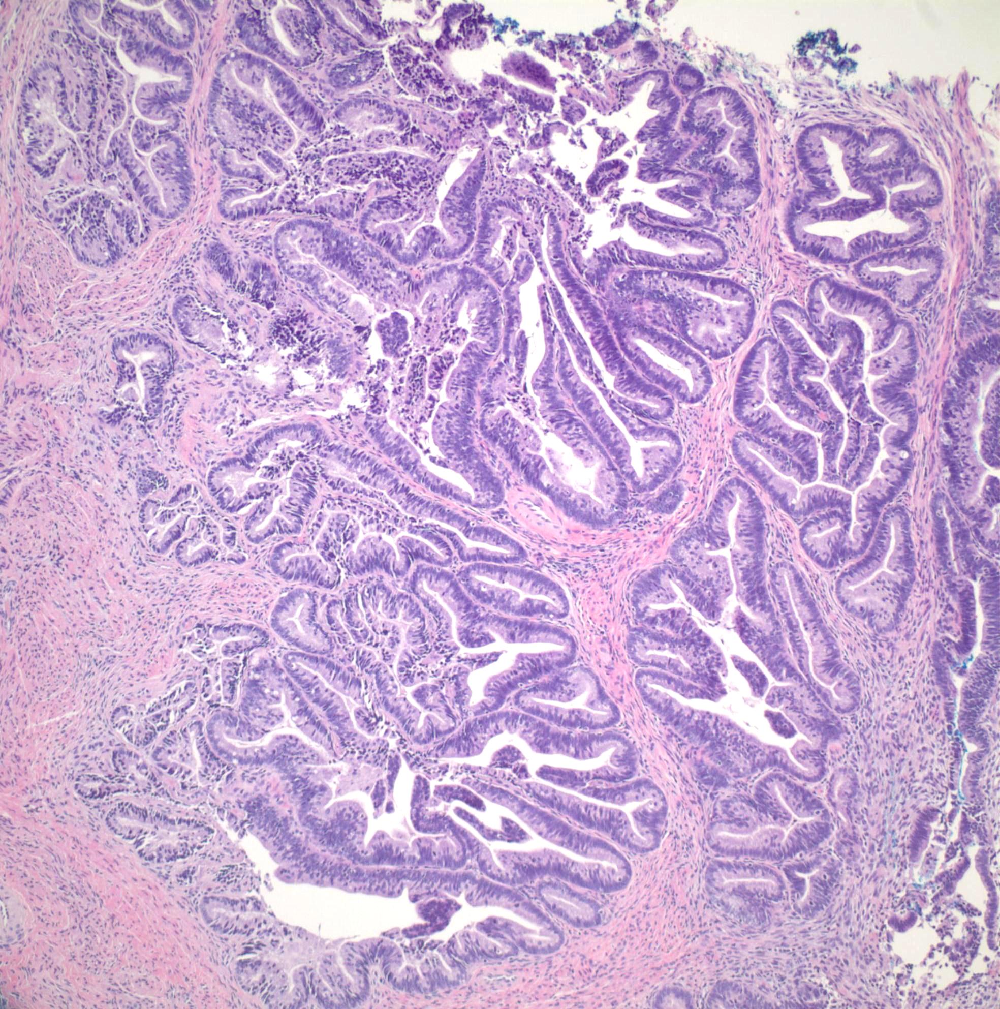
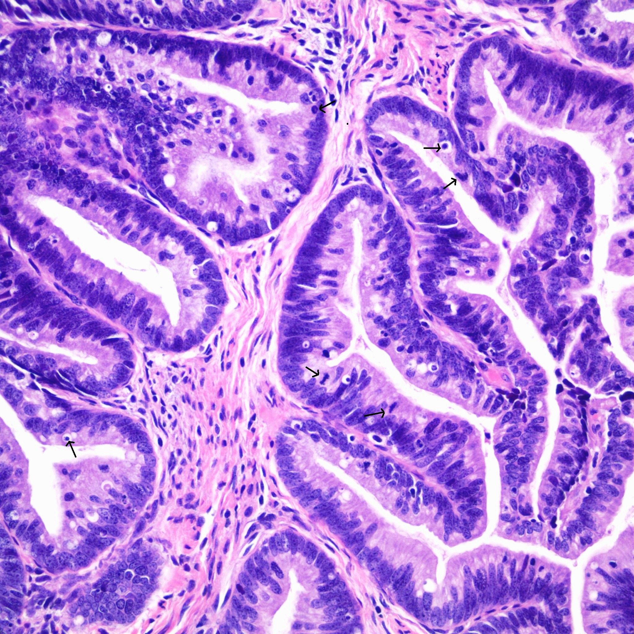
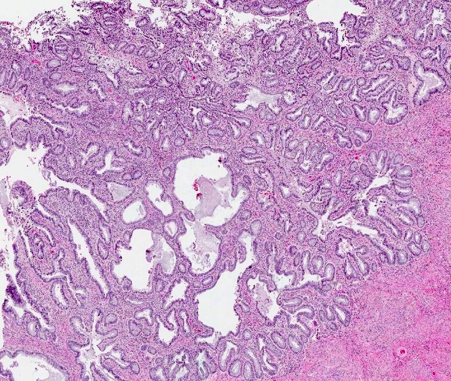
Contributed by Carlos Parra-Herran, M.D.
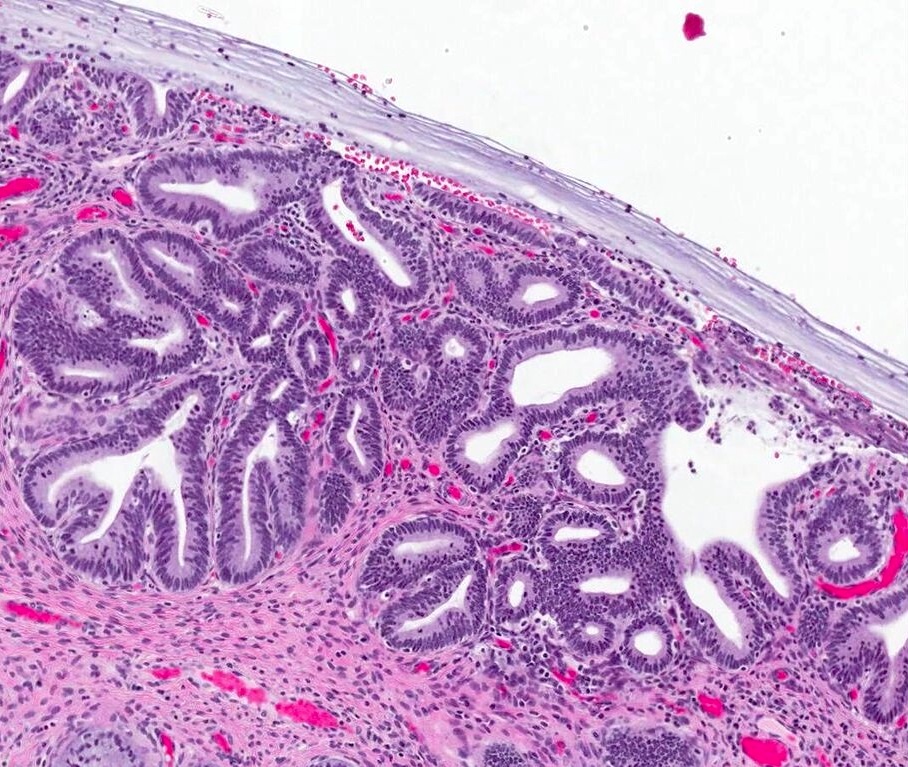
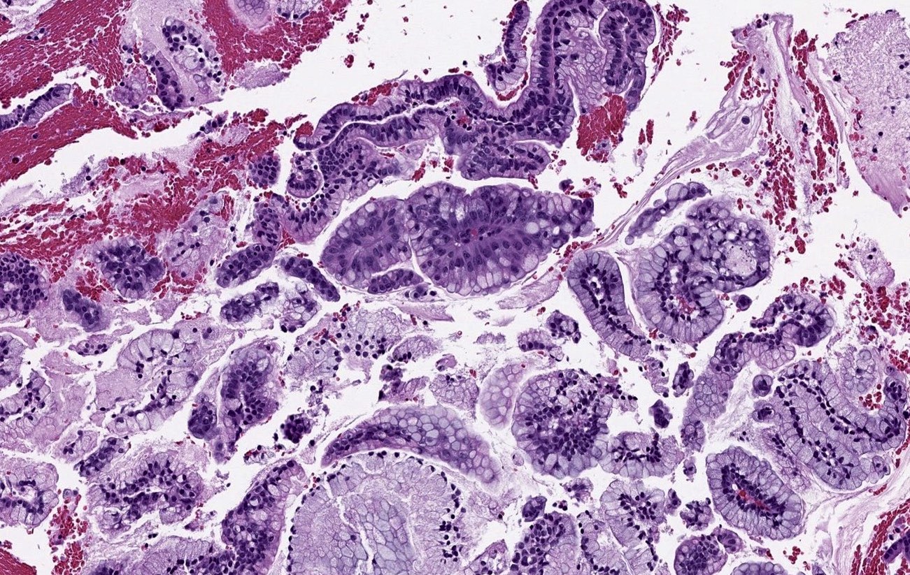
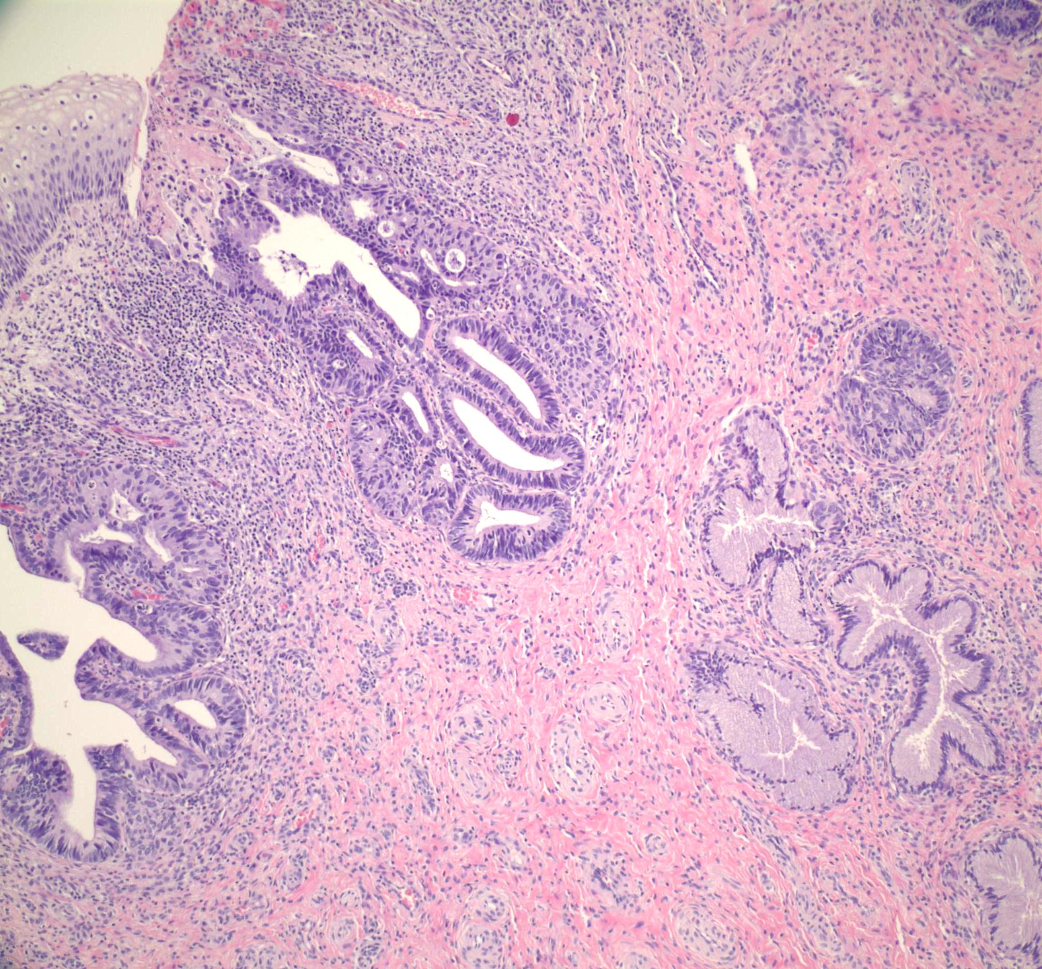
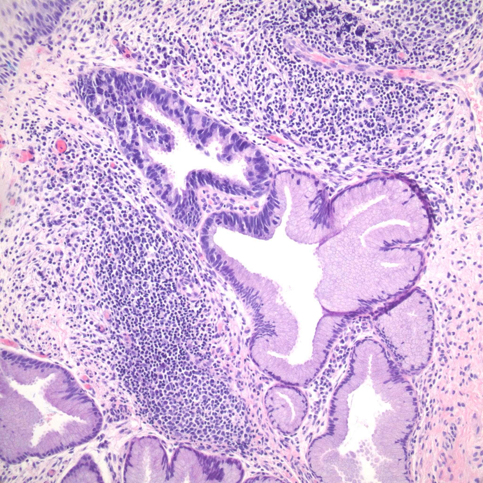
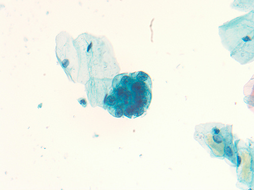
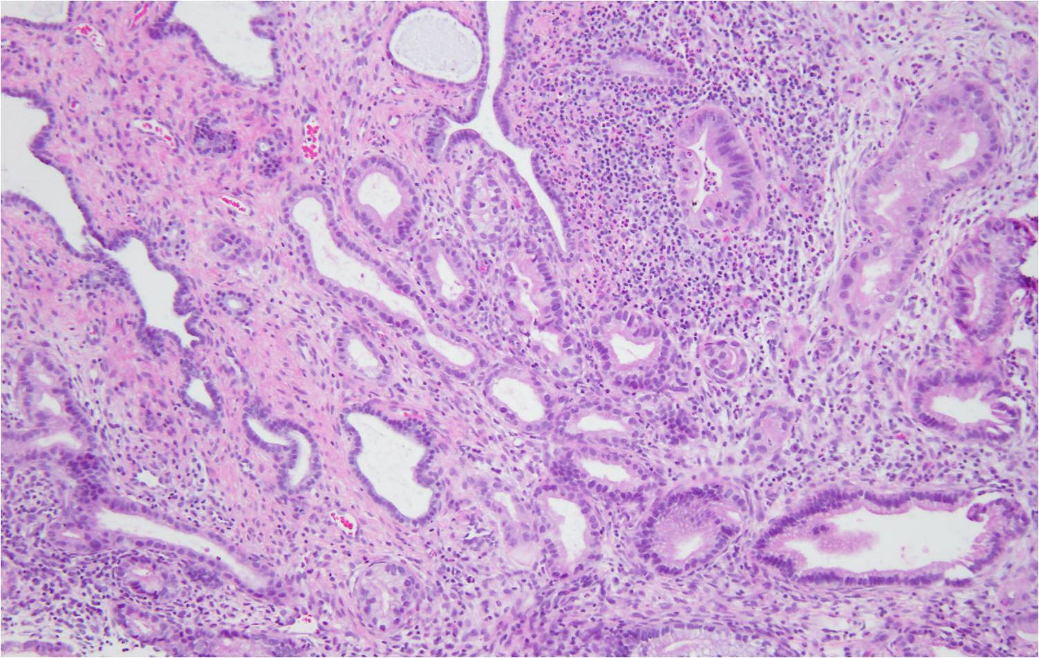
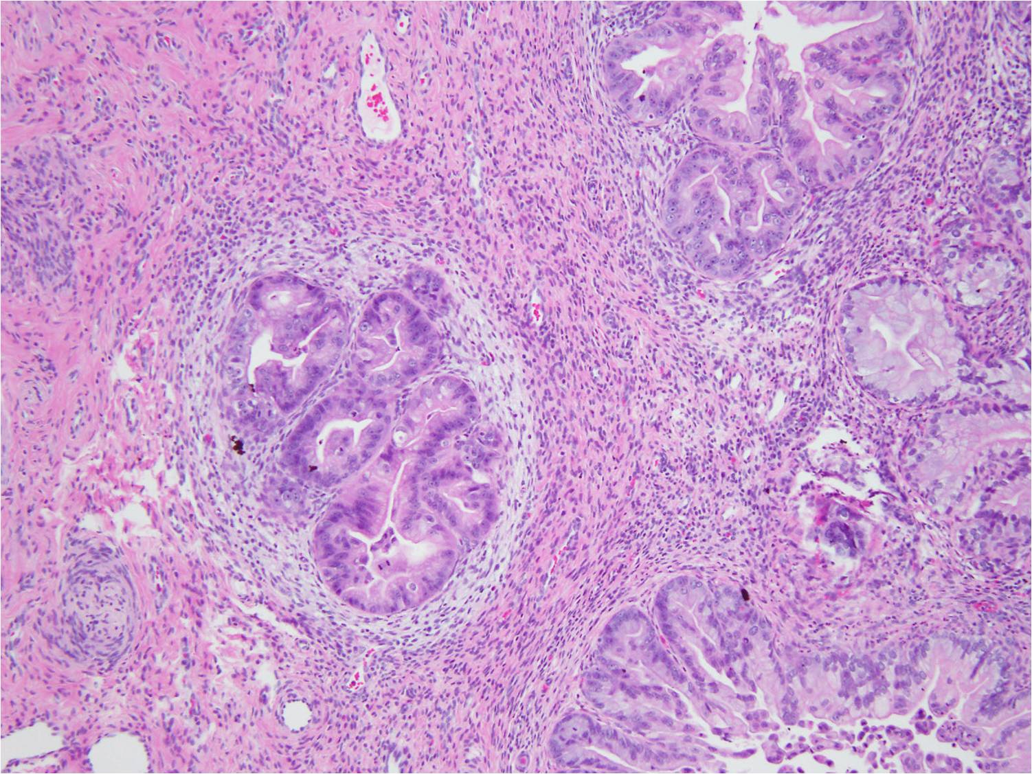
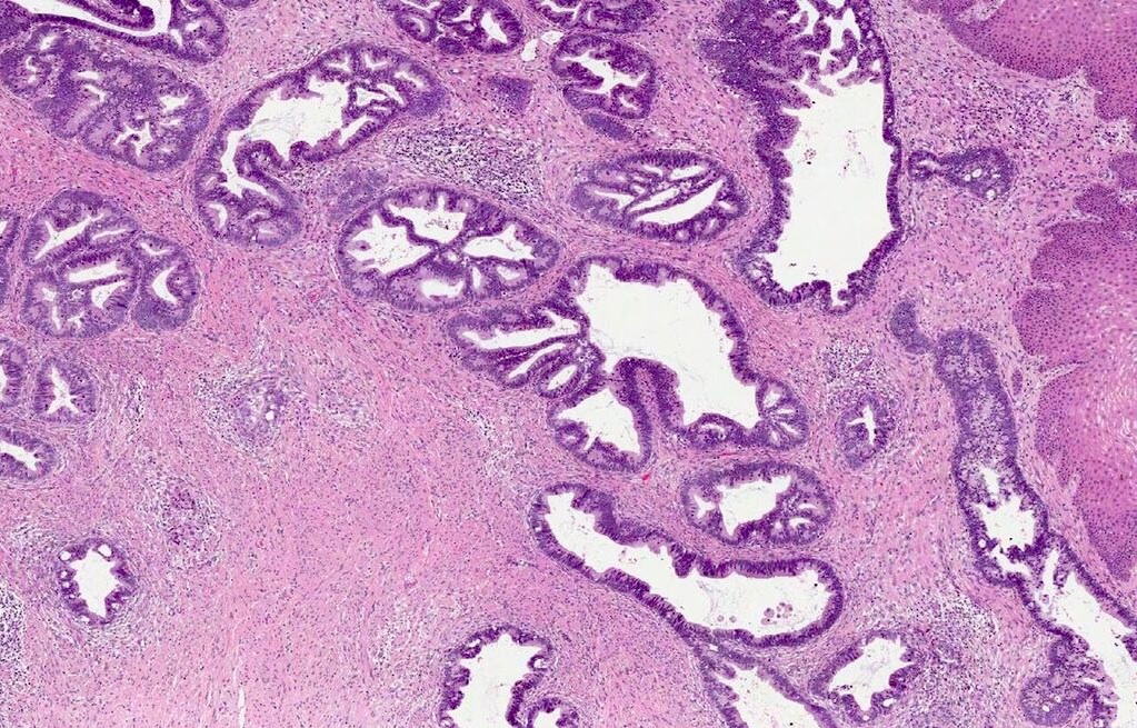
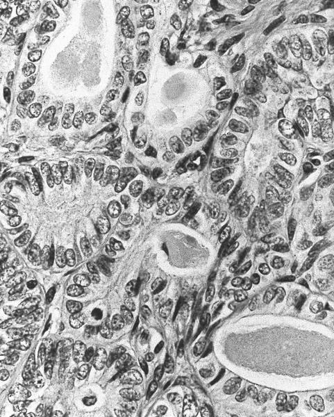
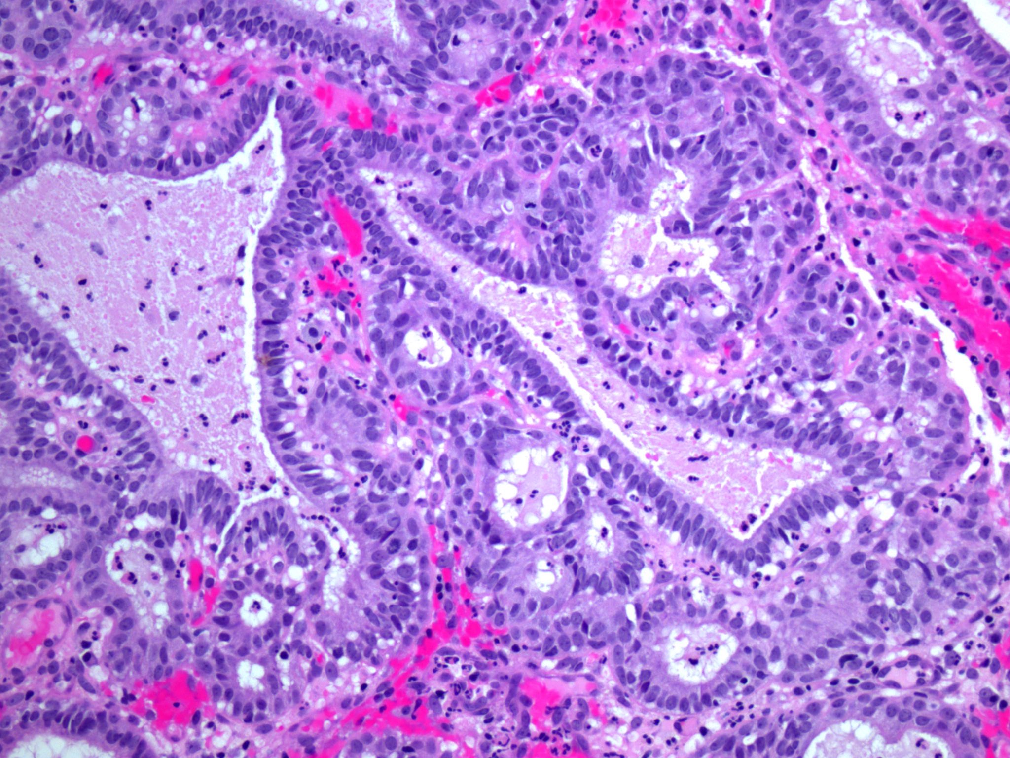
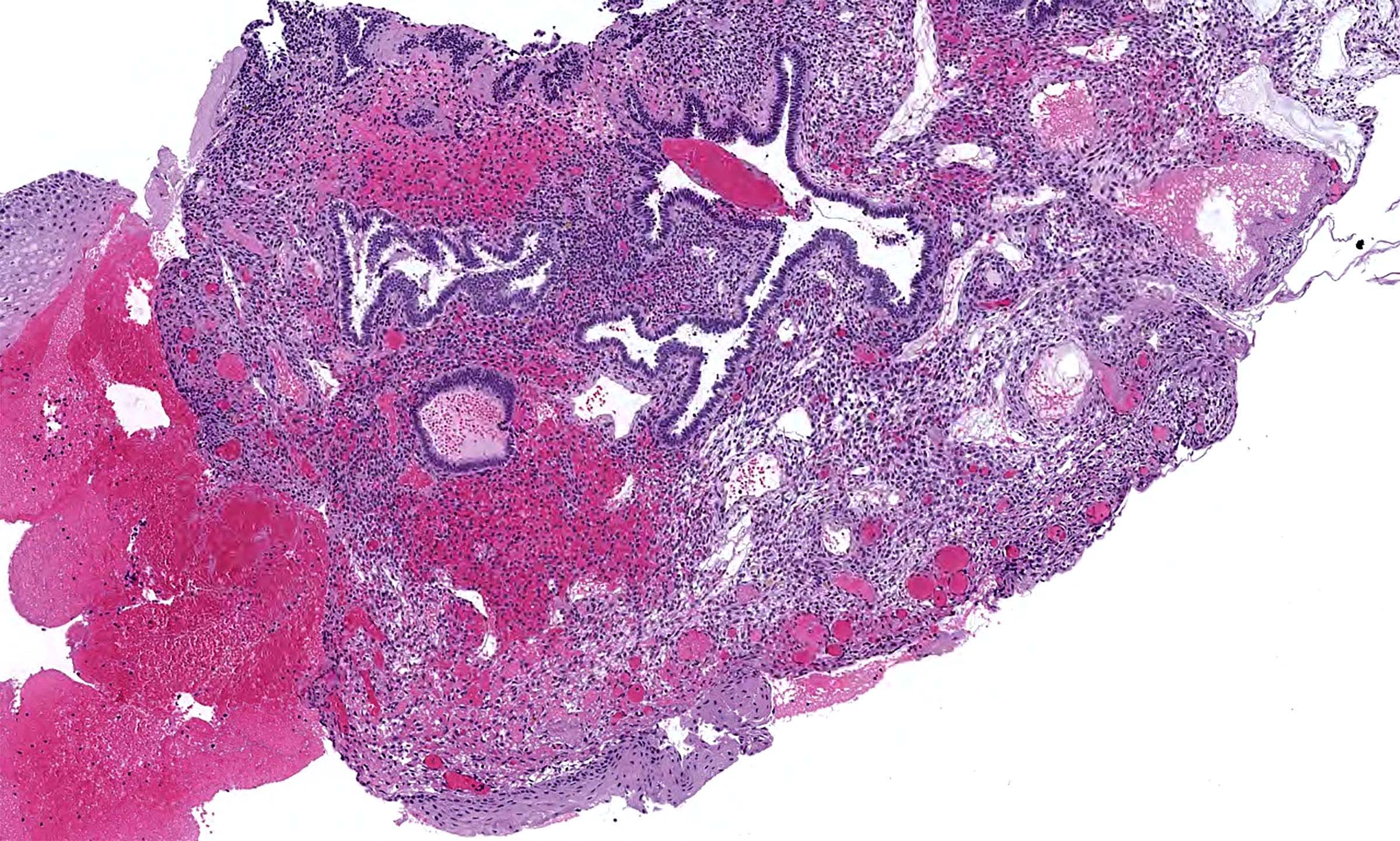
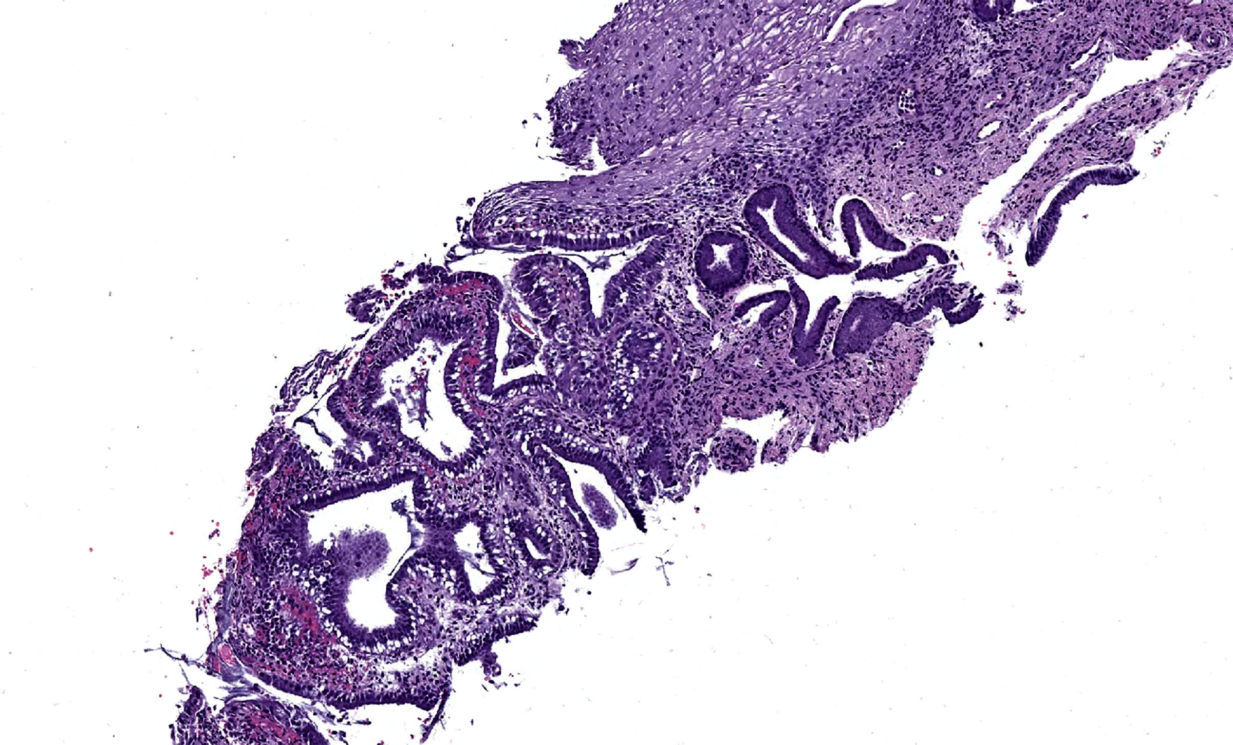
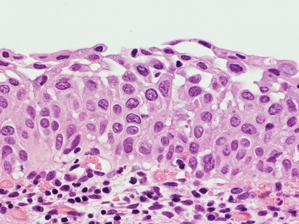
Adenocarcinoma in situ
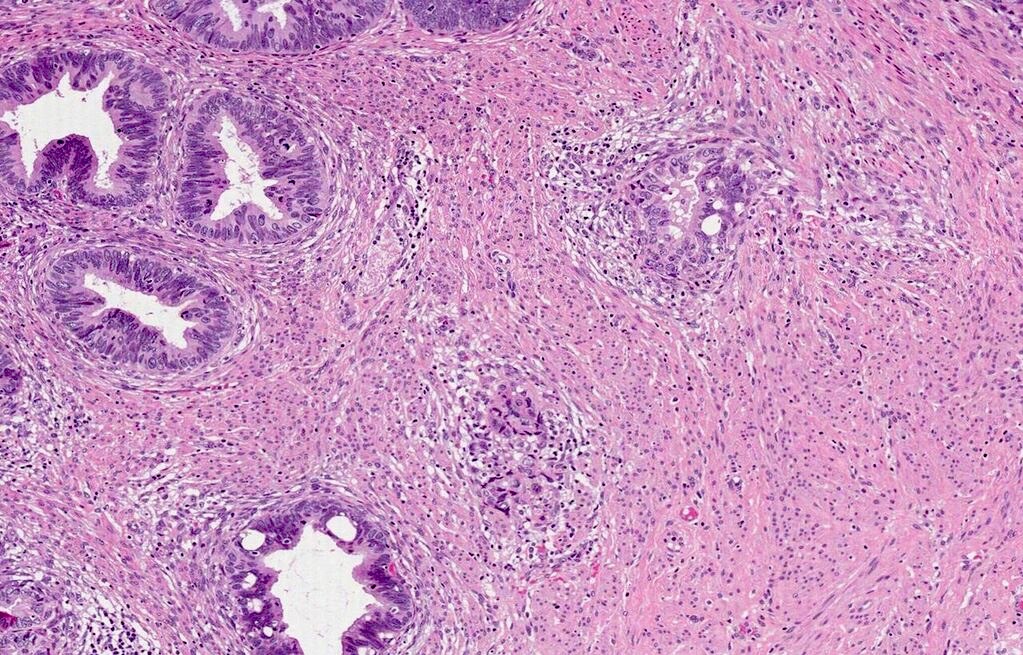
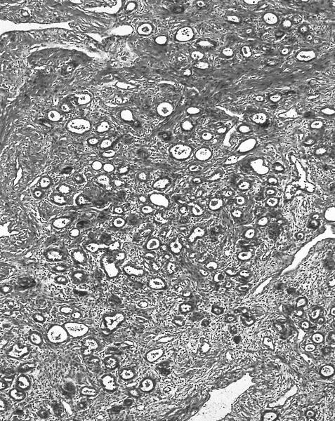
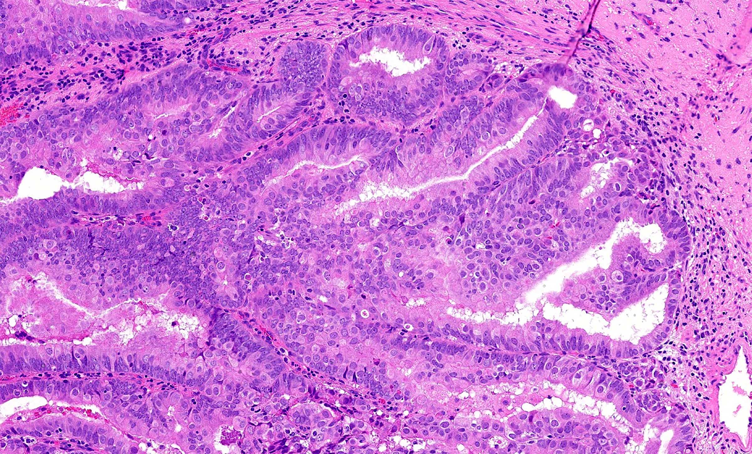
Invasive adenocarcinoma, pattern A
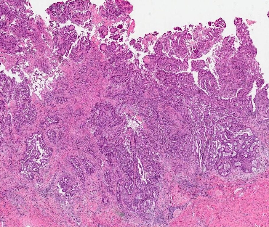
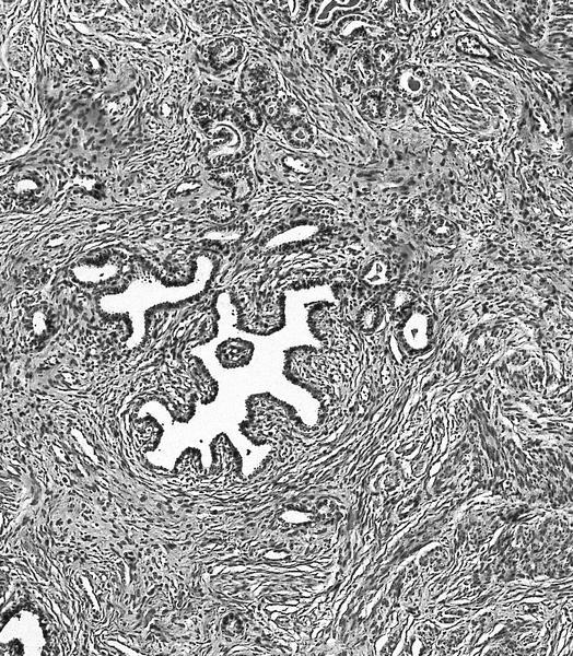
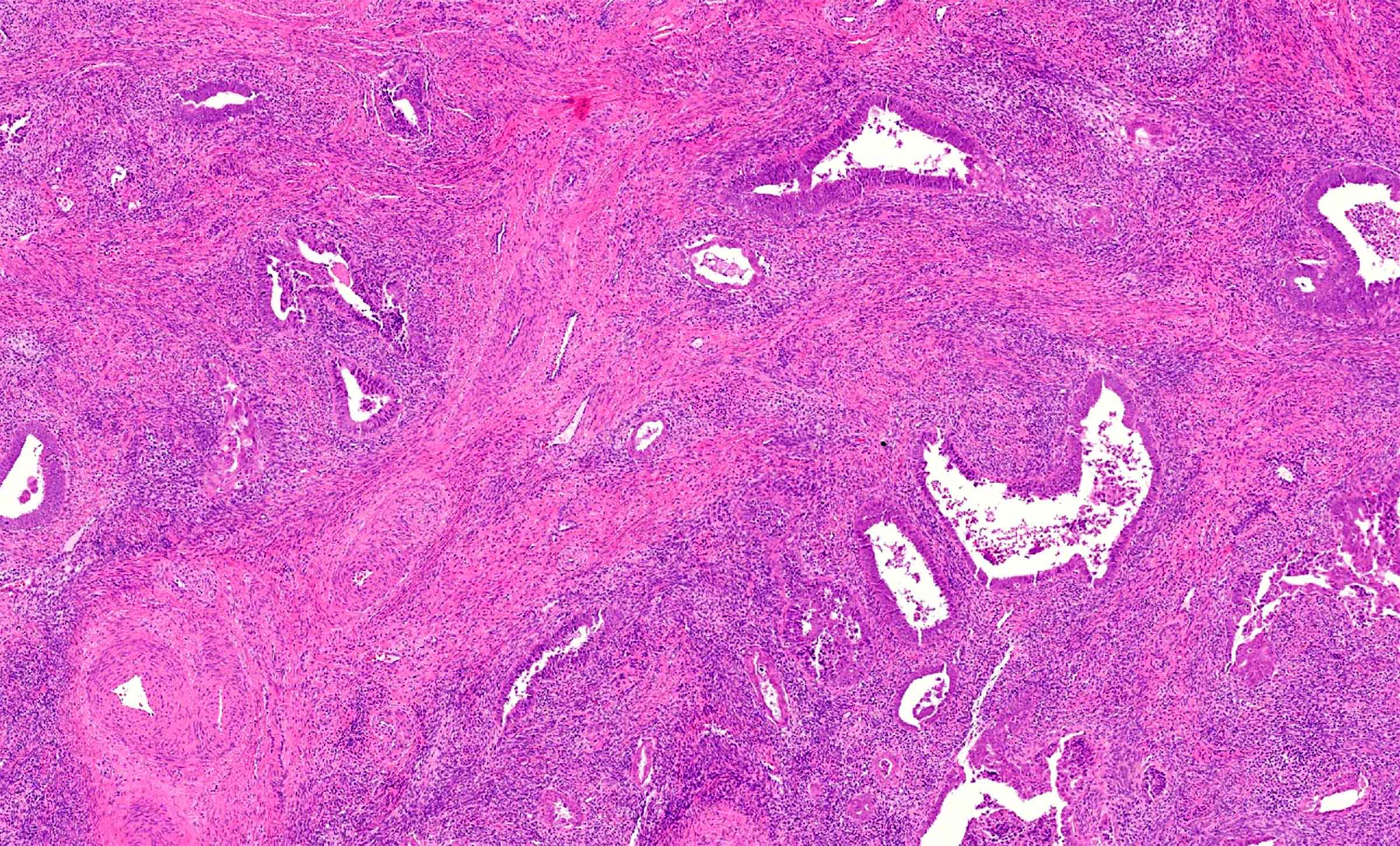
Invasive adenocarcinoma, pattern B
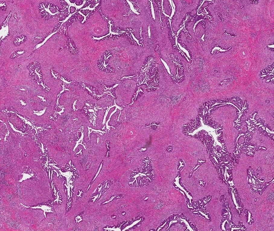

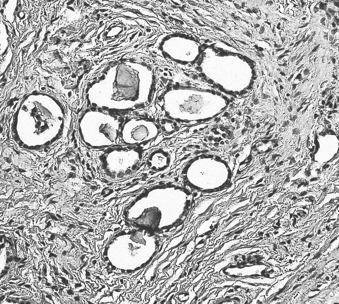
Invasive adenocarcinoma, pattern C
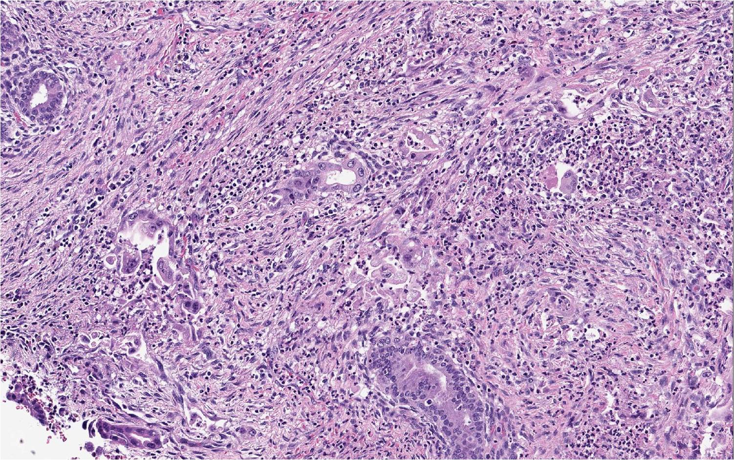
Contributed by Tanner Storozuk, M.D. and Jennifer Bennett, M.D. (Case #495)

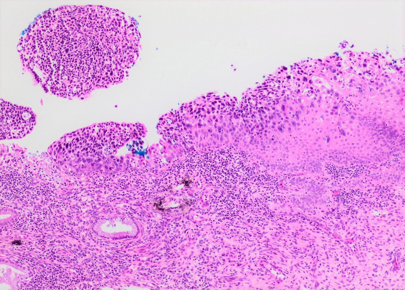
Invasive stratified mucin producing carcinoma (iSMILE)
Images hosted on other servers:
Inflammatory exocervical smear
Images hosted on other servers:

MRI
Images hosted on other servers:

Stage I

Stage III

Stage IV
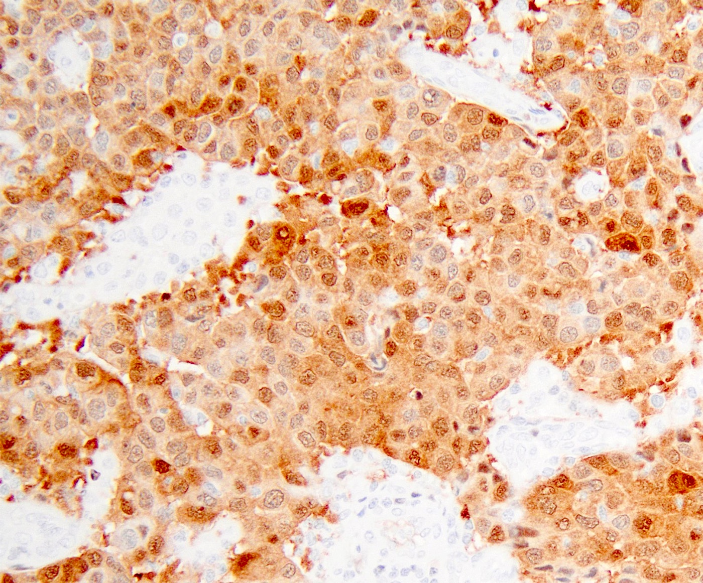
Contributed by Gulisa Turashvili, M.D., Ph.D., Jijgee Munkhdelger, M.D., Ph.D. and Andrey Bychkov, M.D., Ph.D.
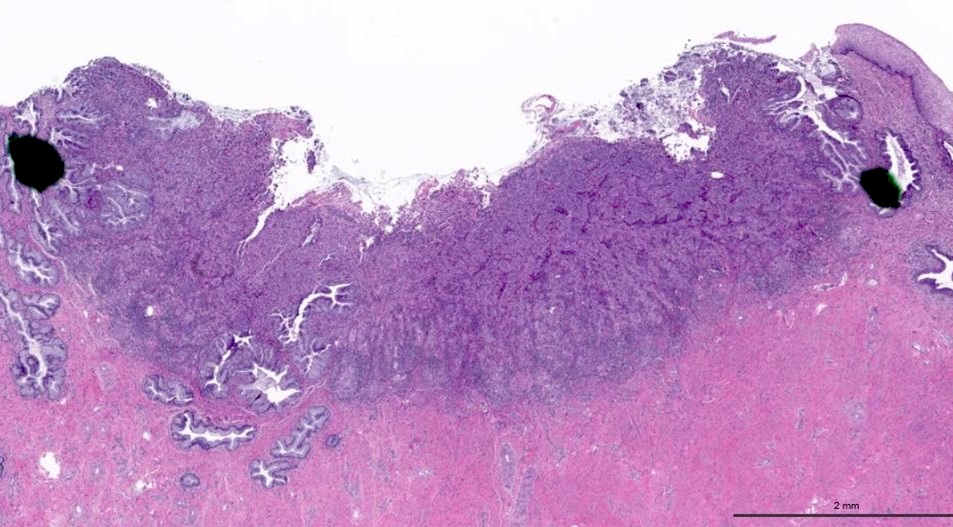
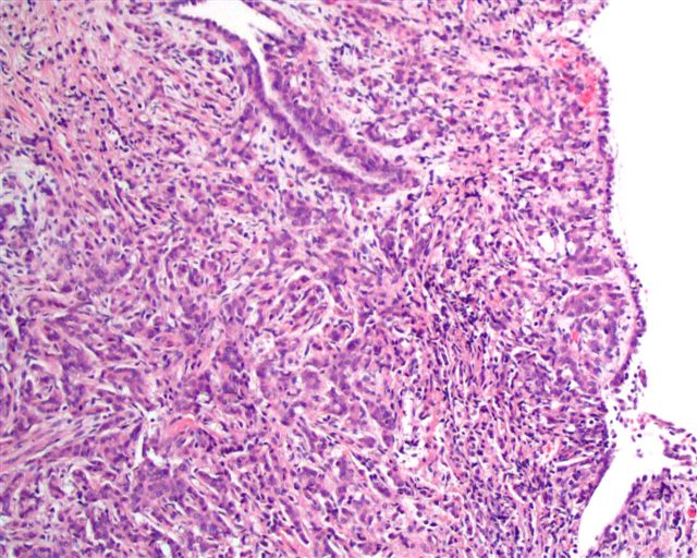
Tumor involving transformation zone
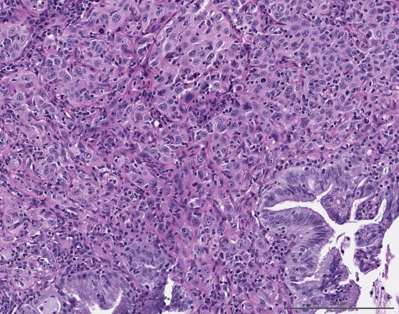
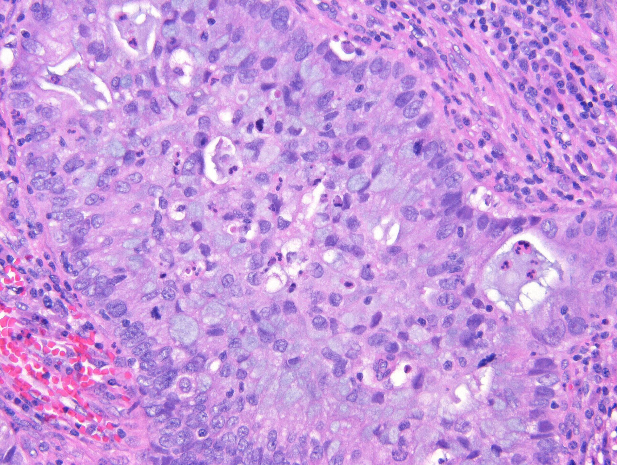
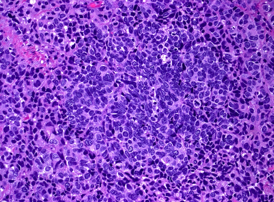
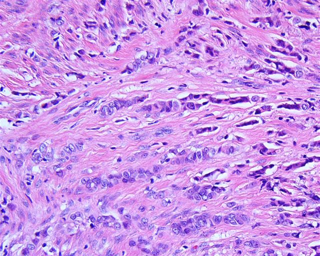
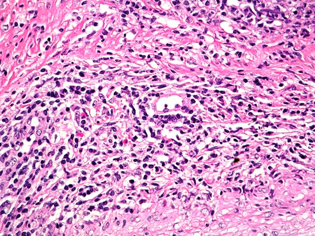
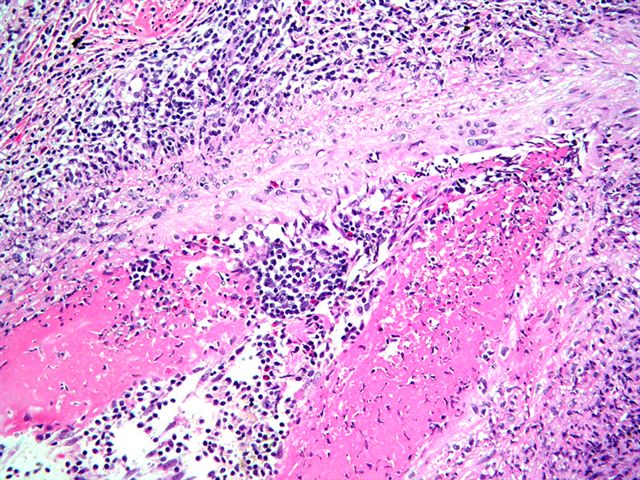
Nests of epithelioid cells
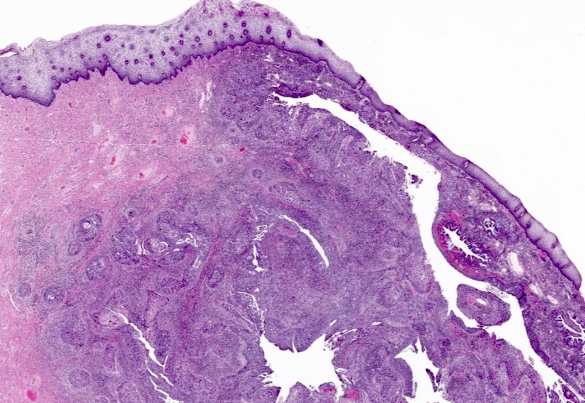
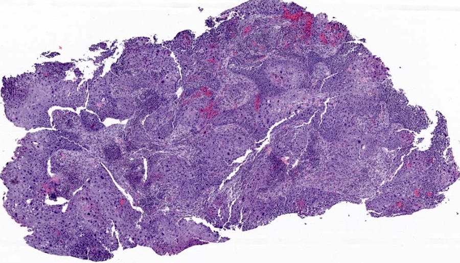
Deeply infiltrative tumor
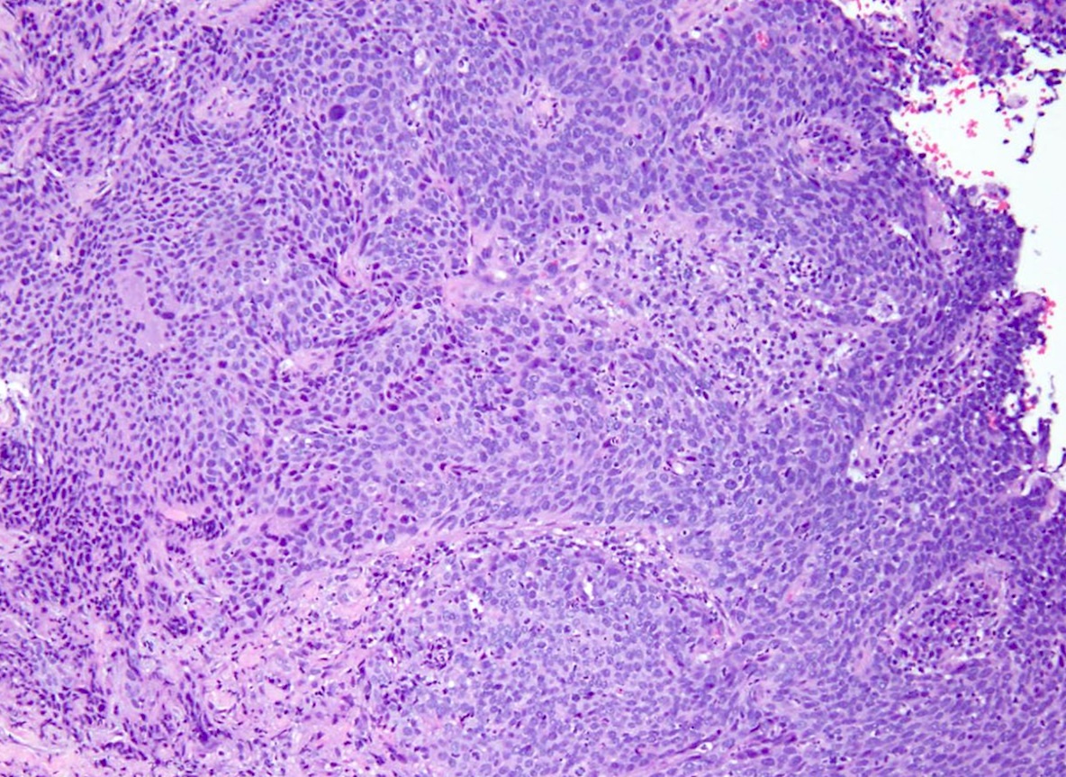
Atypical epithelioid cells
Atypical cells
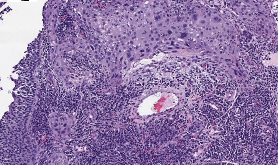
Atypical cells within inflamed stroma

Marked pleomorphism
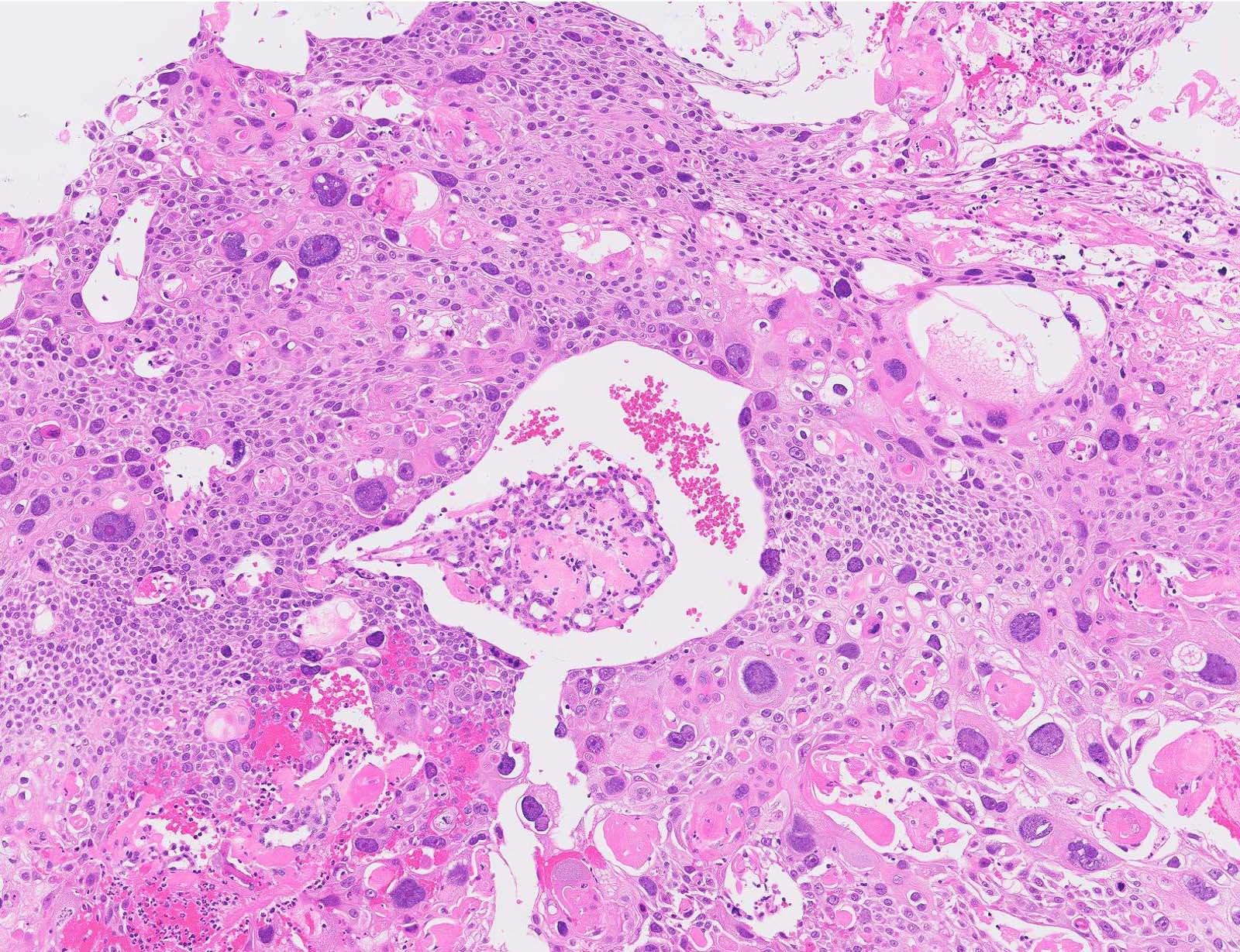
Bizarre cells
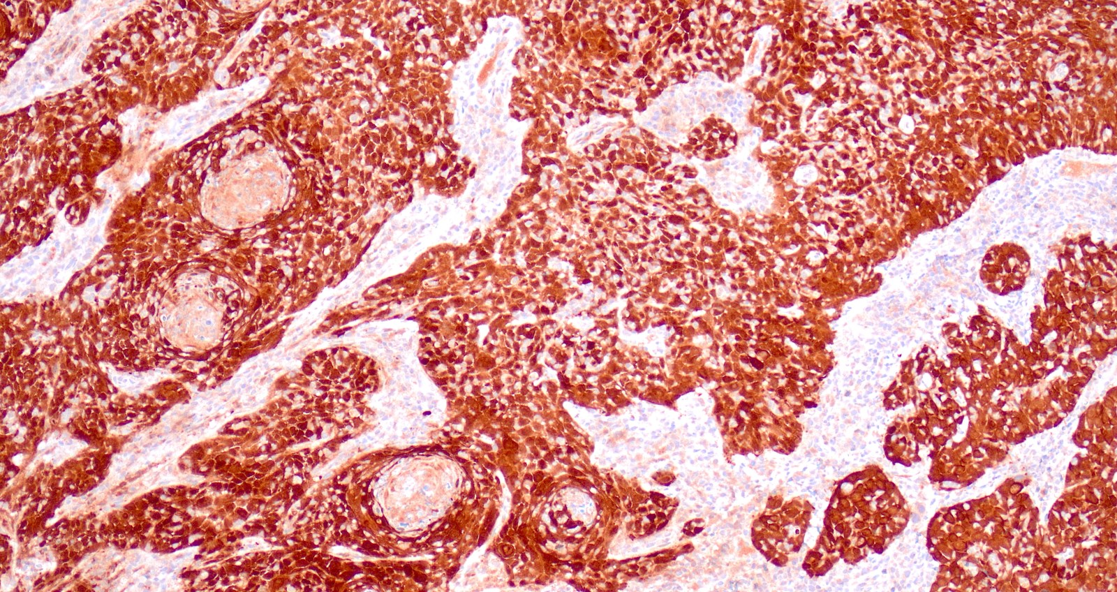
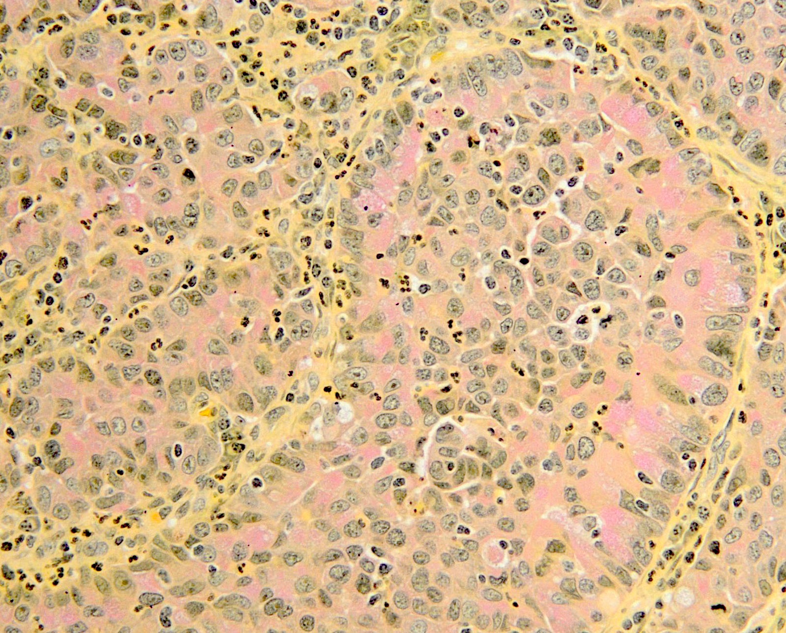
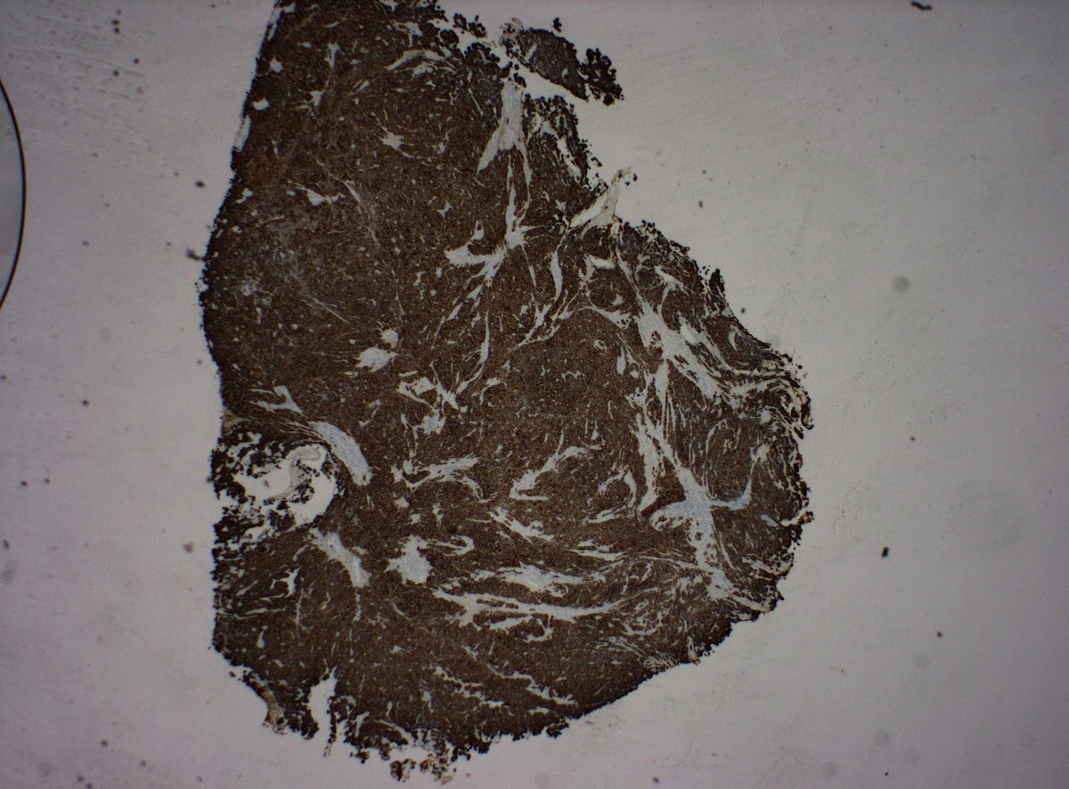
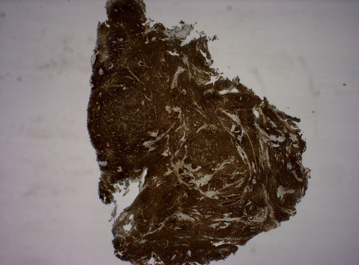
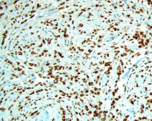
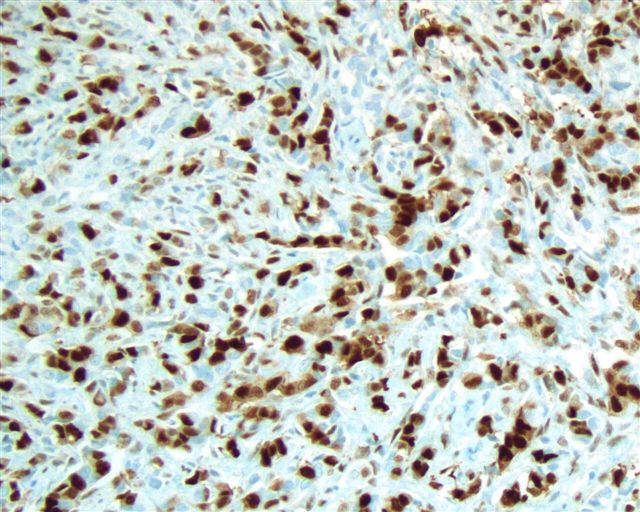
p16 immunostain
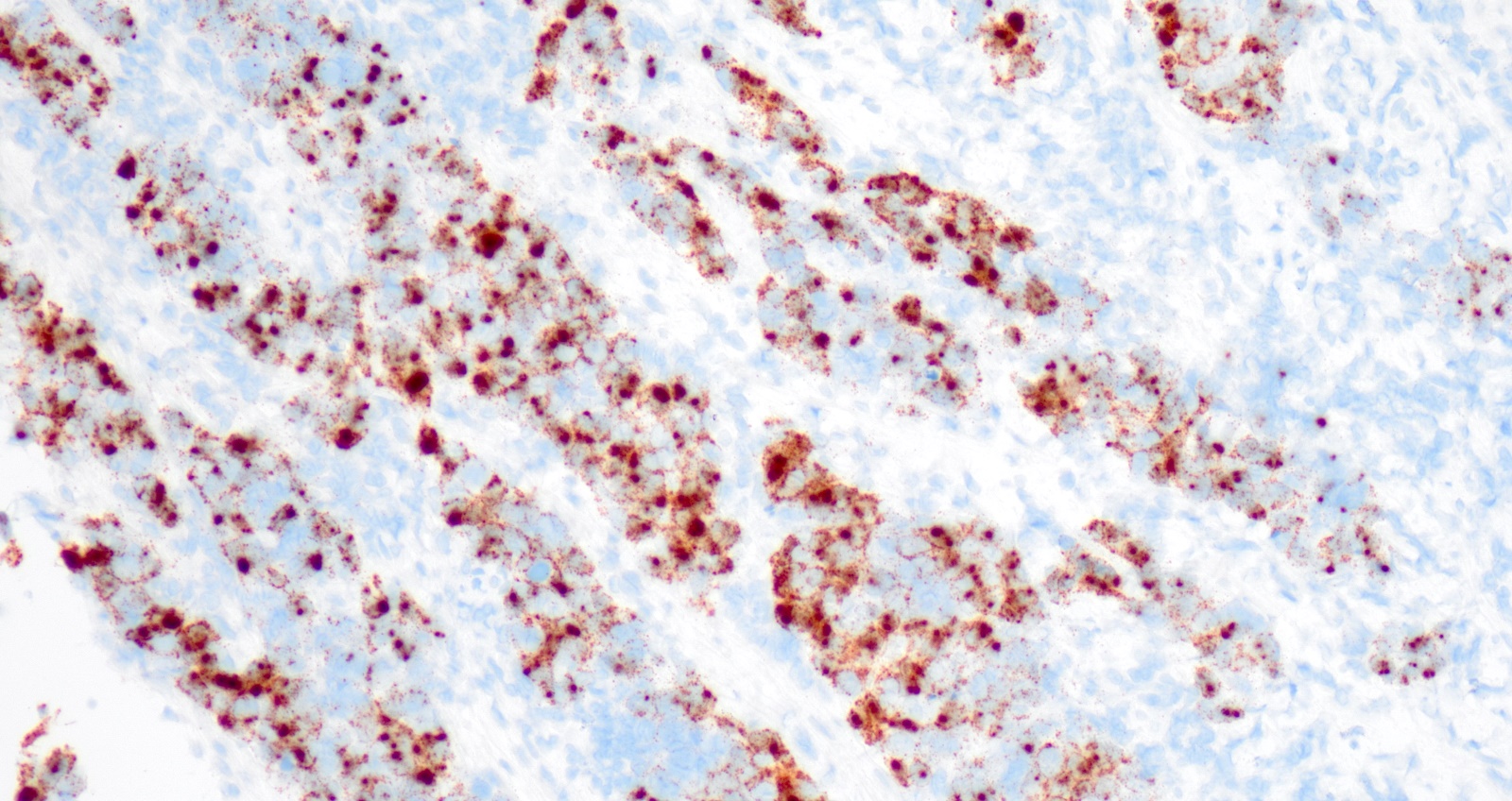
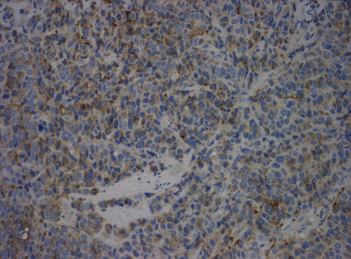
HPV in situ hybridization
Images hosted on other servers:


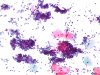
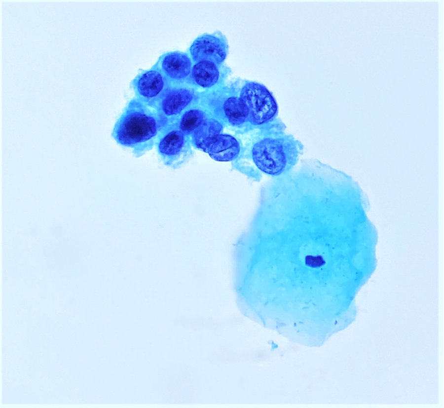
Pleomorphic malignant cells

Pleomorphic and keratinized malignant cells


Pleomorphic malignant cells, isolated or in clusters
Images hosted on other servers:

Tumor cell in intratumoral vessel
Contributed by Yevgen Chornenkyy, M.D., M.Sc. and Bonnie Choy, M.D.
High N/C ratio
Metaplastic or dense cytoplasm
Irregular nuclear contour, indentations / grooves
Lack of nucleoli
Variable cytoplasm
Images hosted on other servers:

WHO digital atlas
Images hosted on other servers:

Low grade disease with unsatisfactory colposcopy

Acetowhite epithelium

High grade disease and unsatisfactory colposcopy

Before acetic acid

Dense acetowhite epithelium after acetic acid

Before acetic acid

Acetowhite epithelium
Contributed by Khaled J. Alkhateeb, M.B.B.S.
Squamocolumnar junction HSIL
Cytoplasmic maturation
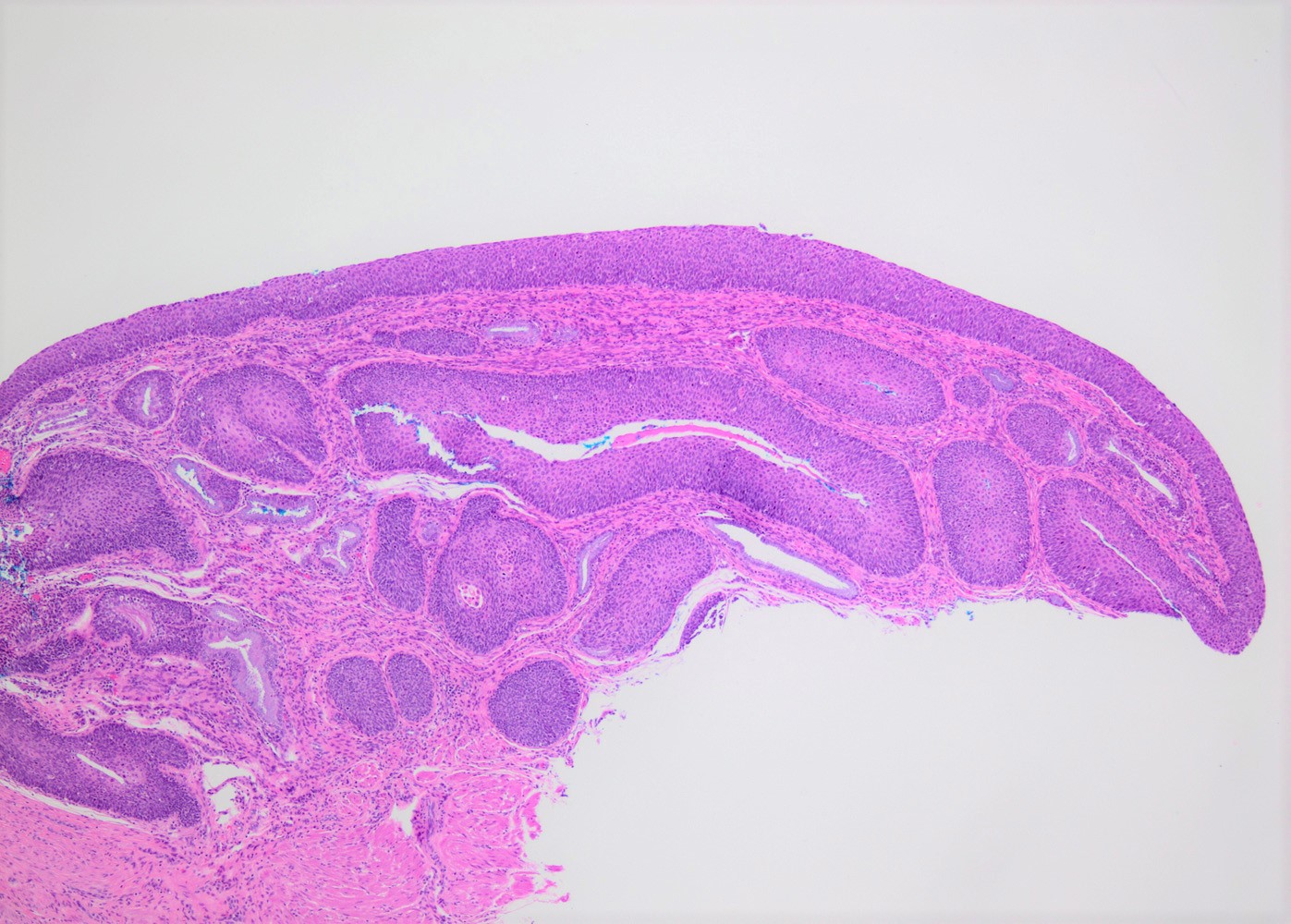
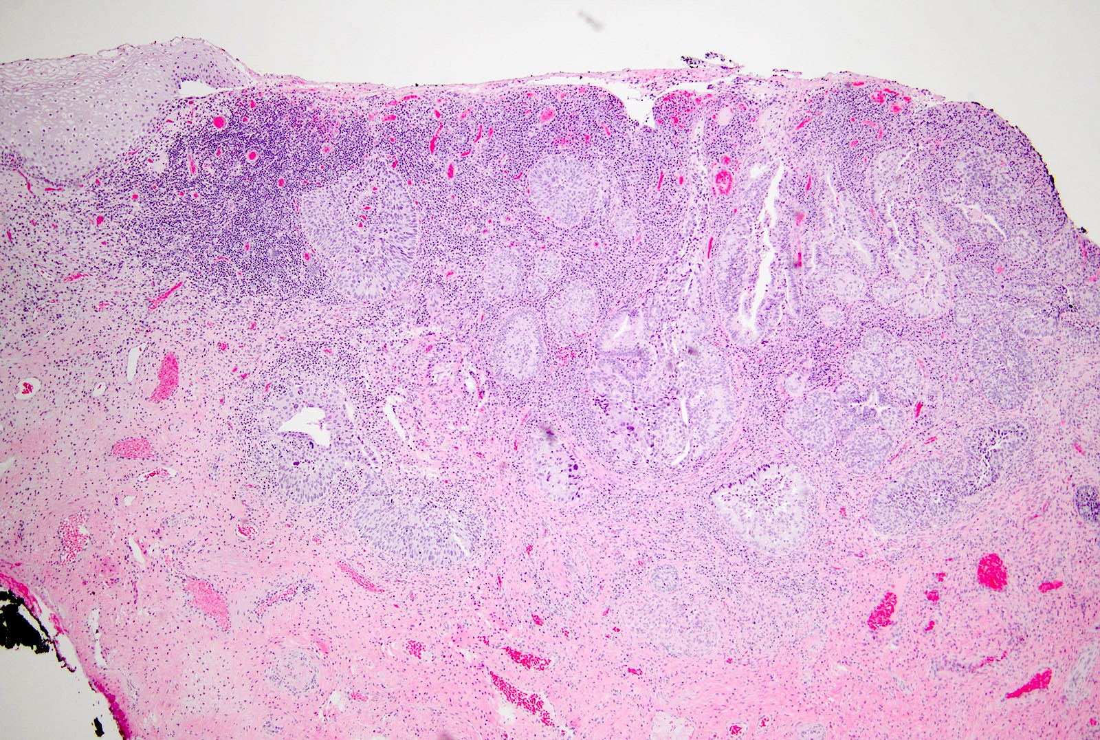
CIN III involving endocervical glands
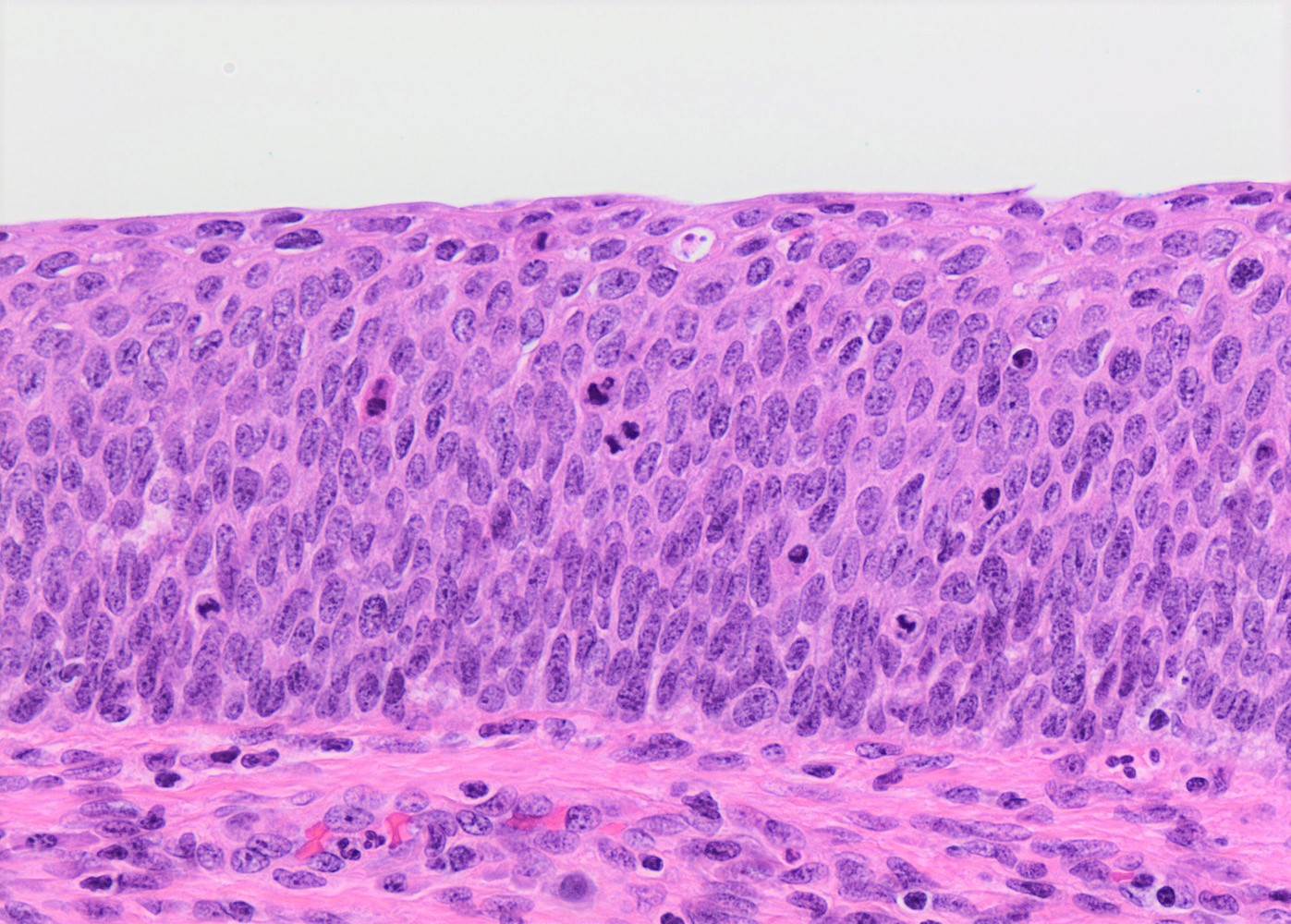
Full thickness atypia
HSIL involving endocervical glands
HSIL with superficial keratinization
HSIL / CIN III with adjacent squamous cell carcinoma
Invasive squamous cell carcinoma
HSIL with significant nuclear pleomorphism
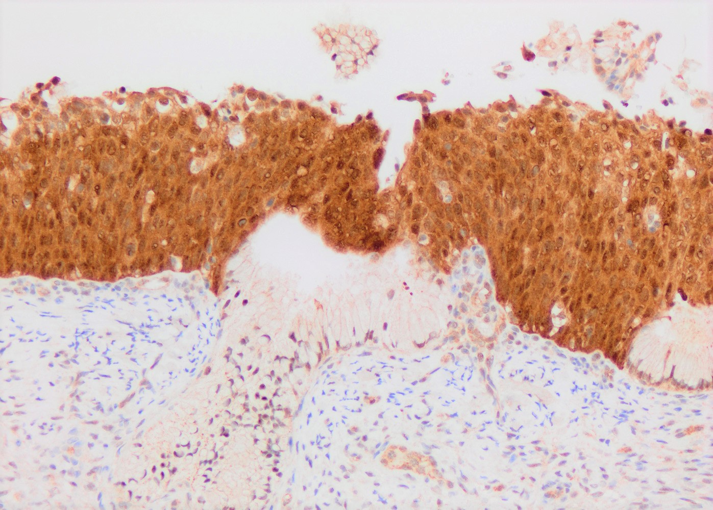
p16 IHC
Contributed by Ziyan T. Salih, M.D.
Marked nuclear atypia

Sharp contrast from surrounding benign cells
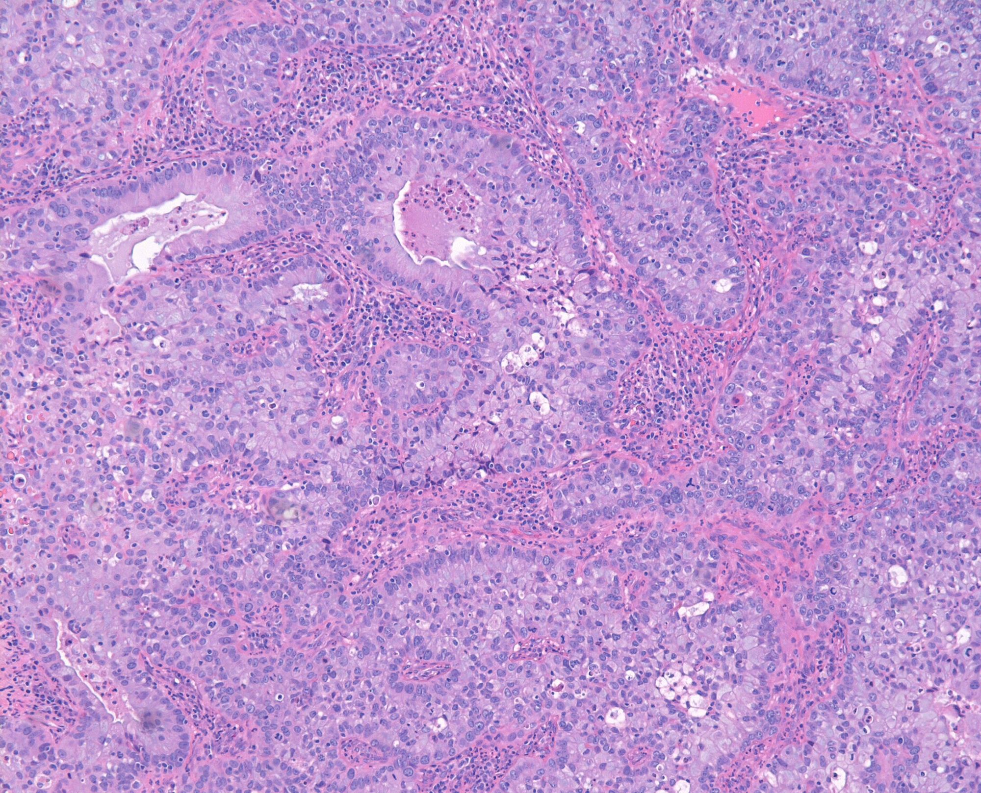
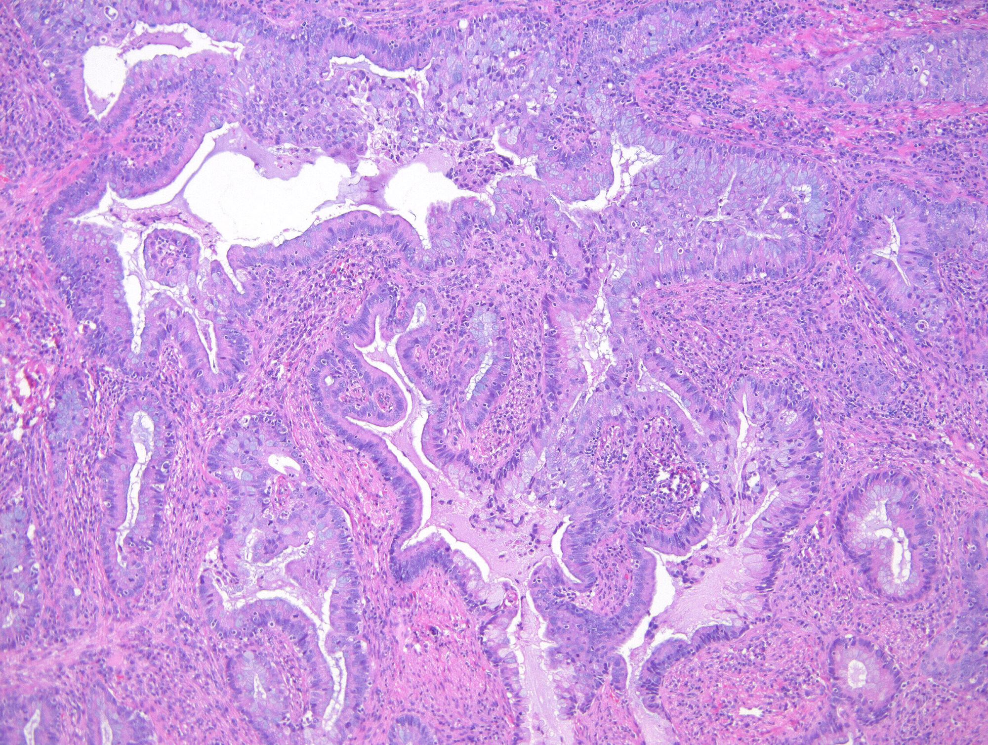
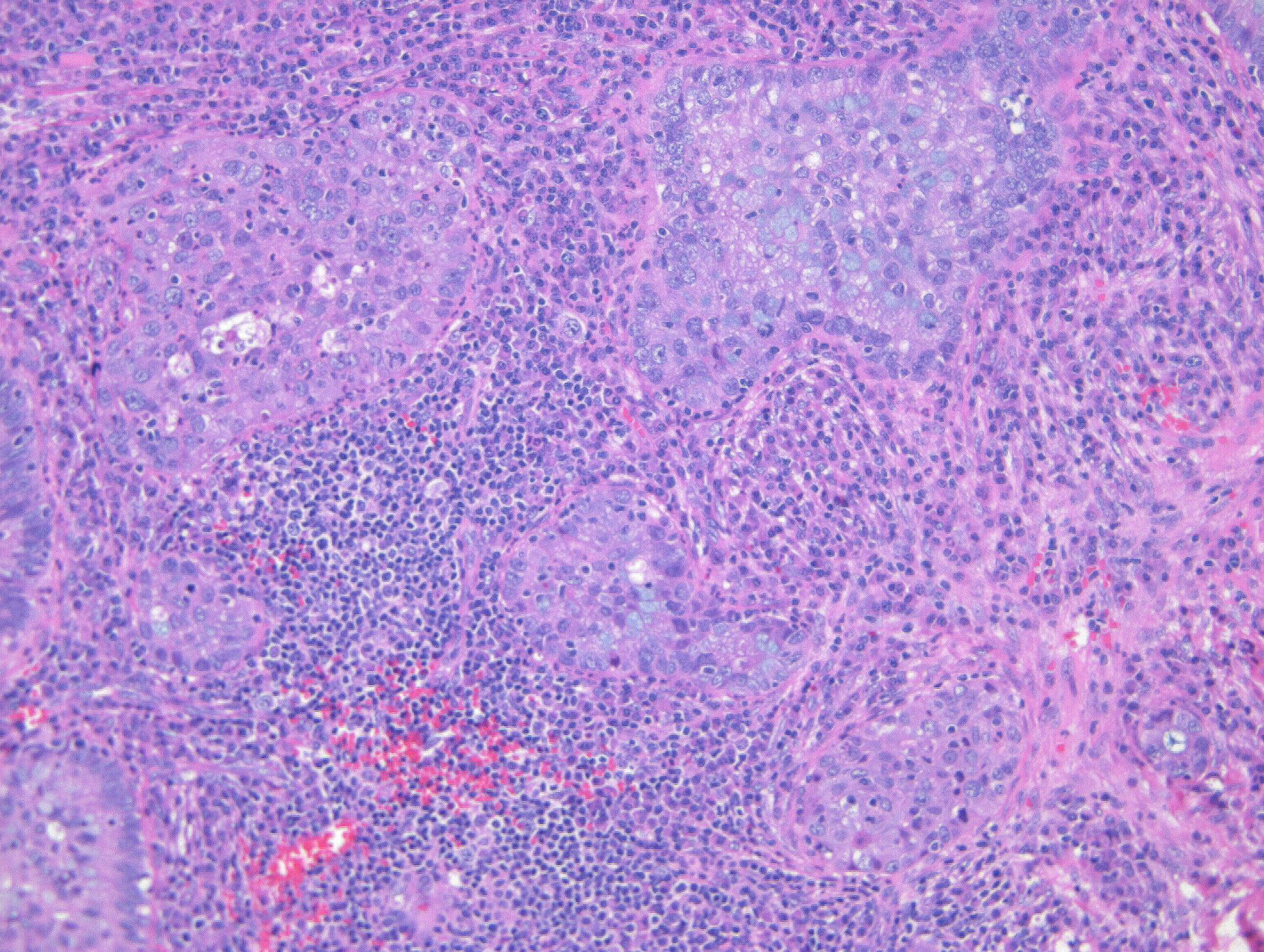
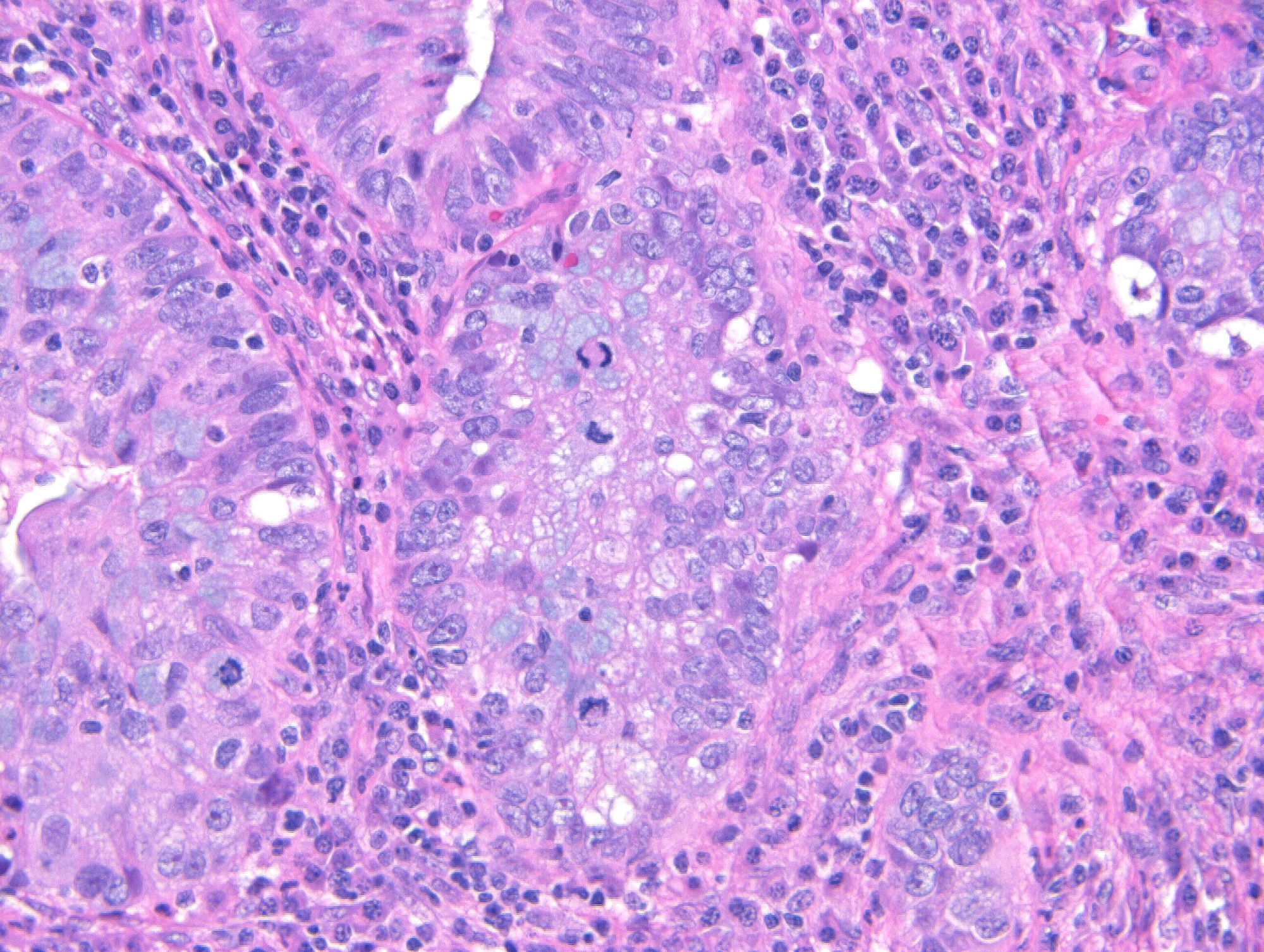
Contributed by Julieta E. Barroeta, M.D., Andreas Kontosis, M.D. and Ricardo R. Lastra, M.D.
Infiltrative nests
Intratumoral and peritumoral inflammation

Metastatic ISMC
SMILE and AIS
Mucicarmine
p16 IHC

PAX8 IHC
p40 IHC
ER IHC
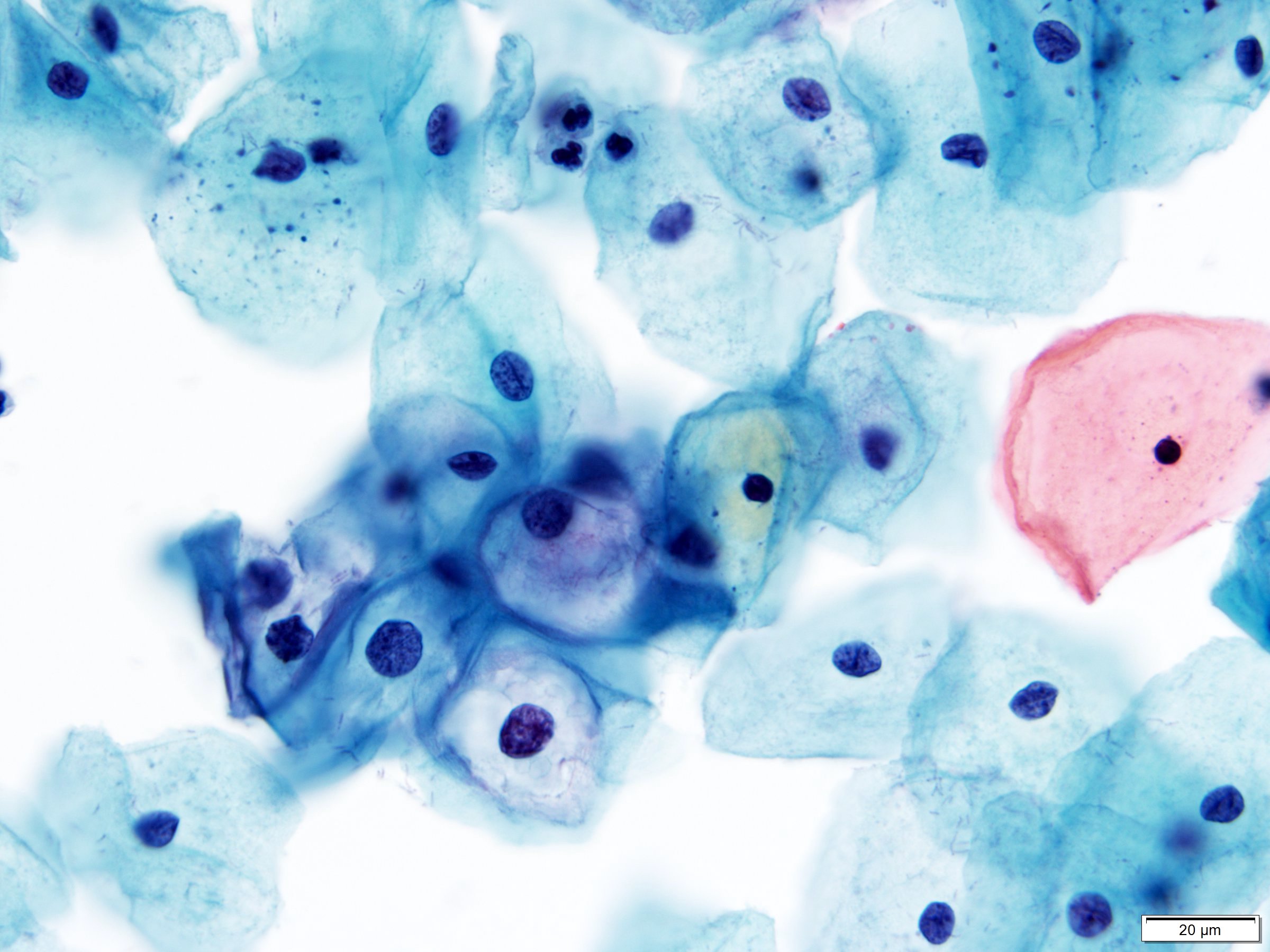
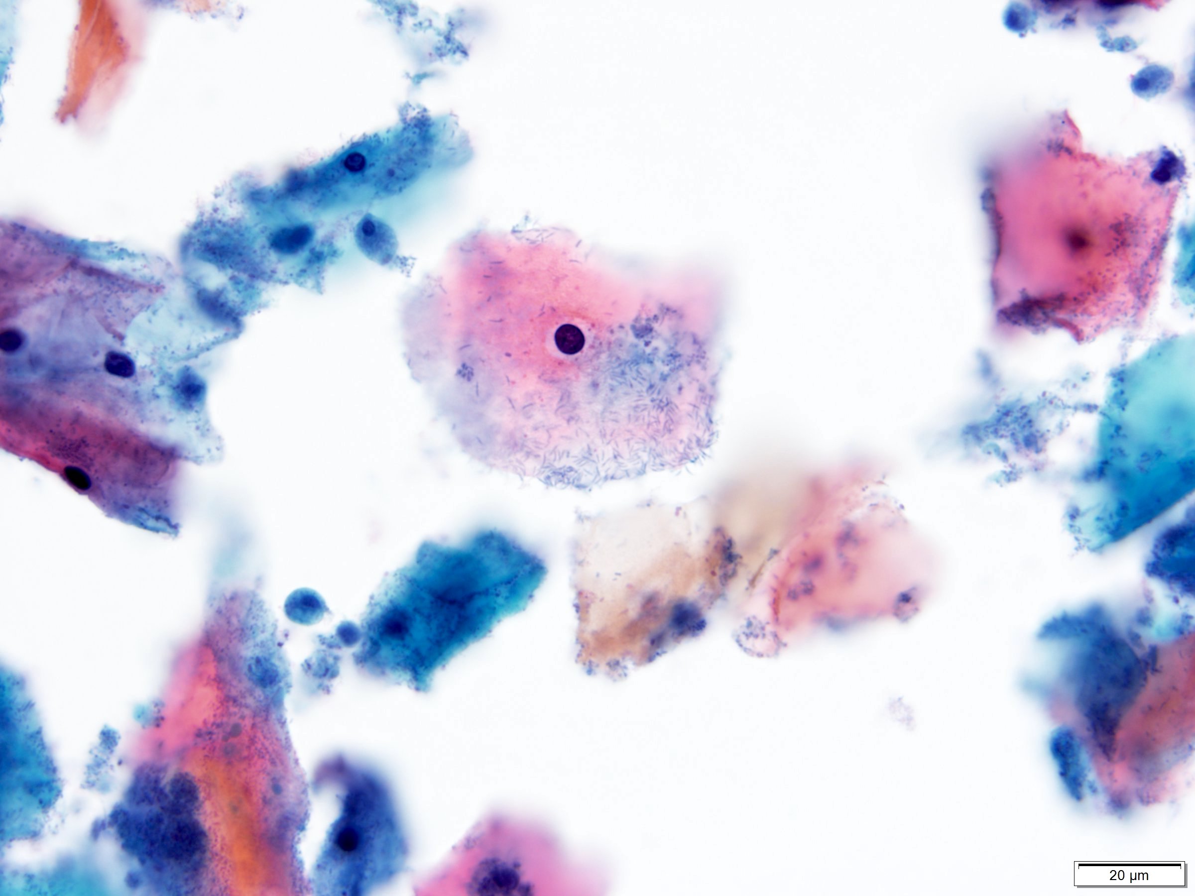
Contributed by Hiba Al Dallal, M.D.
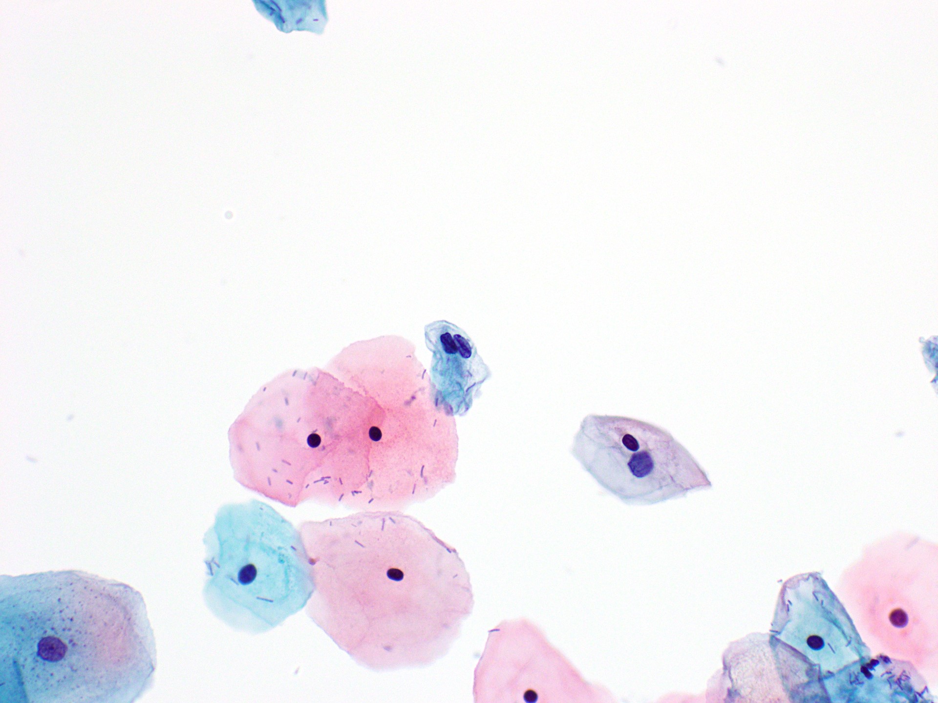
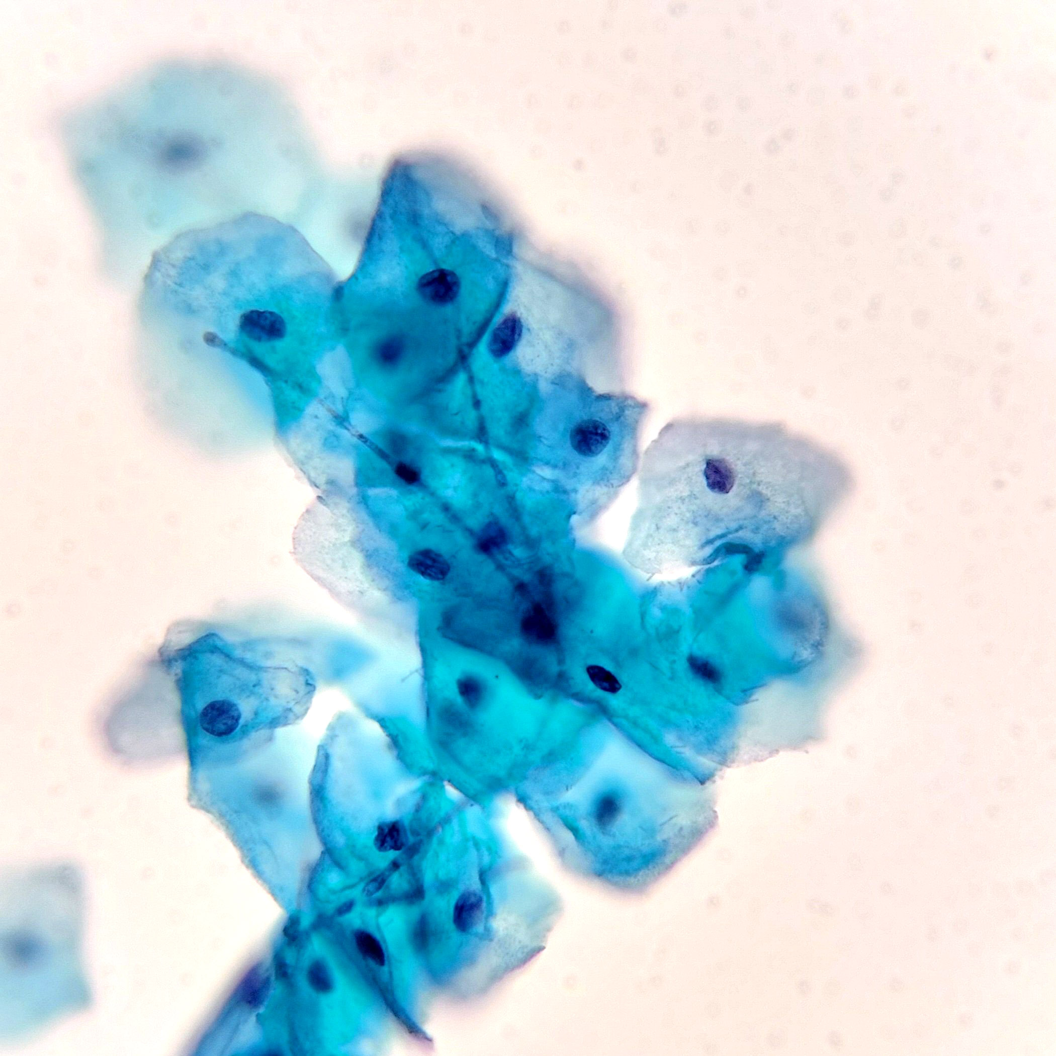
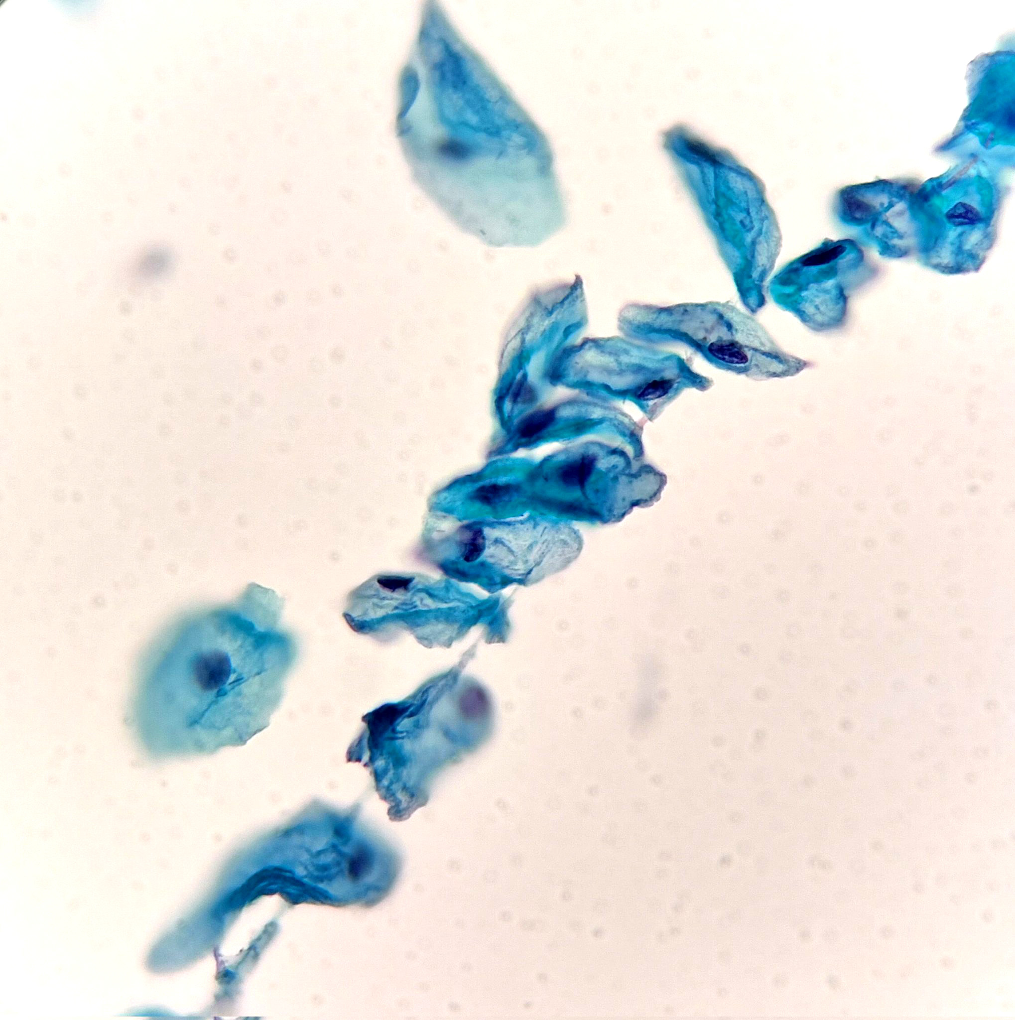
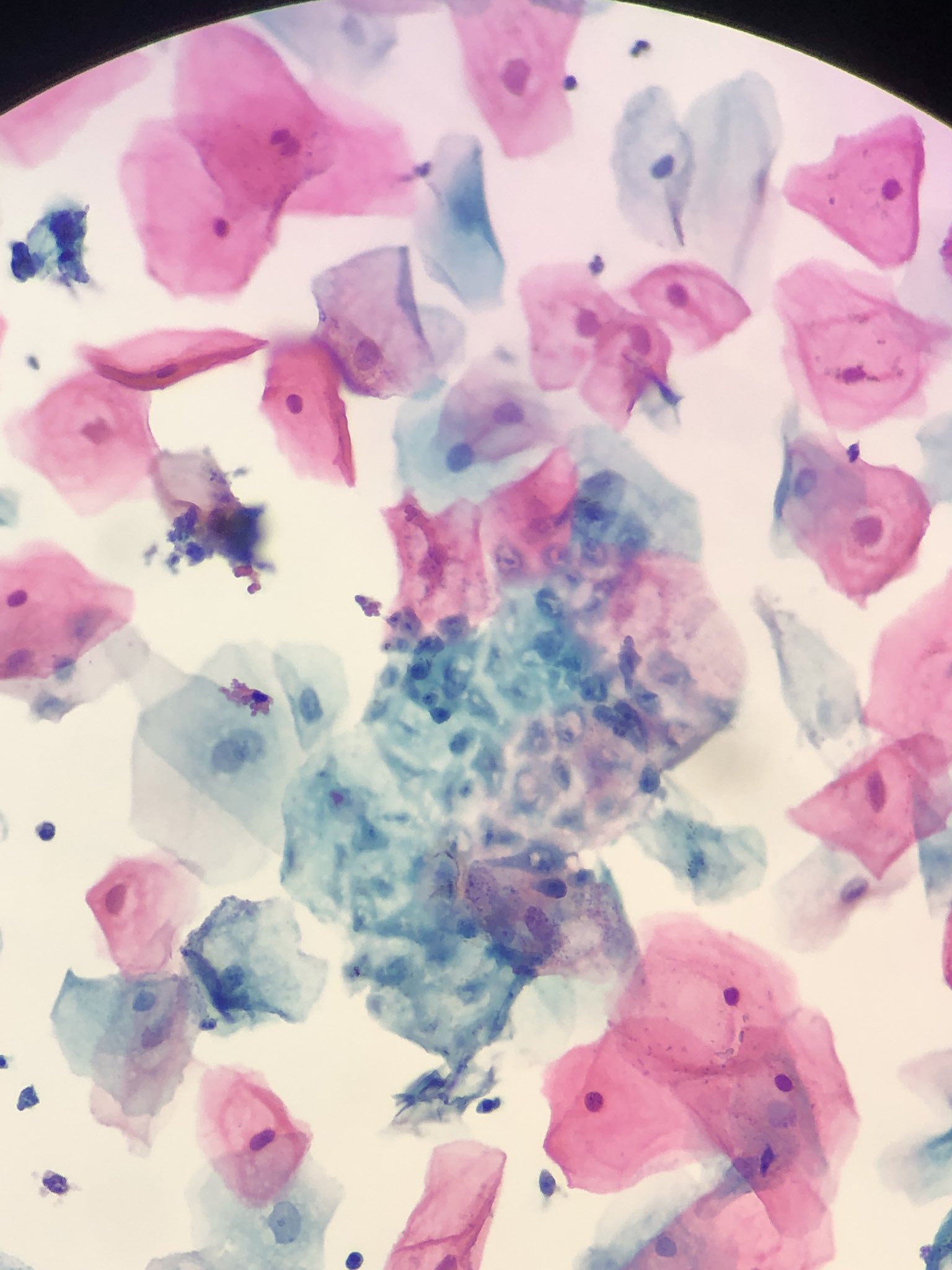
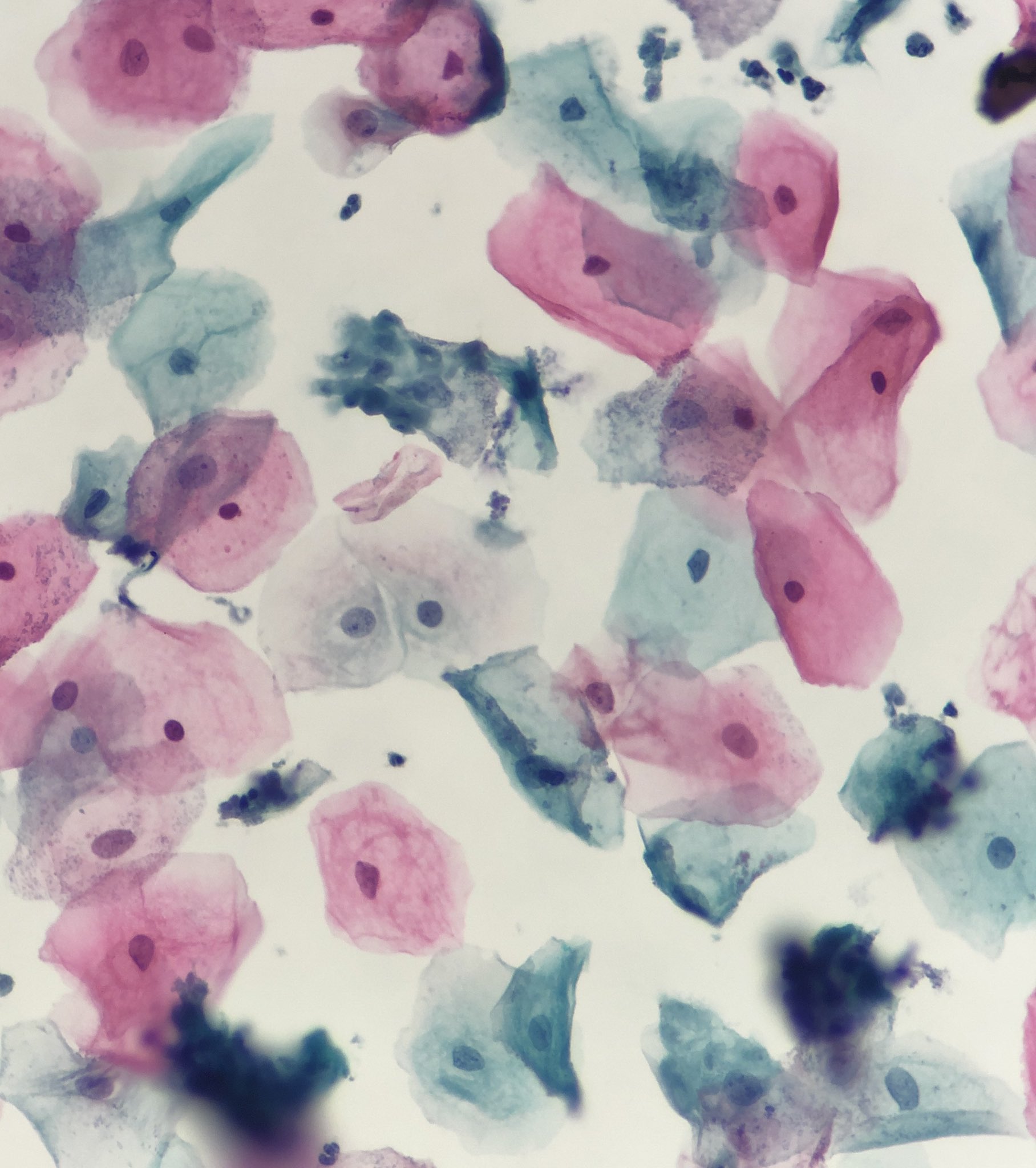
Cytolysis
Cytolysis
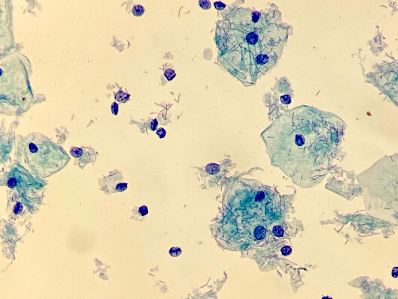
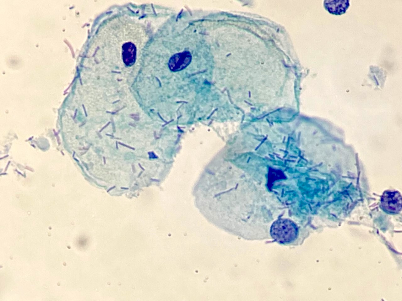
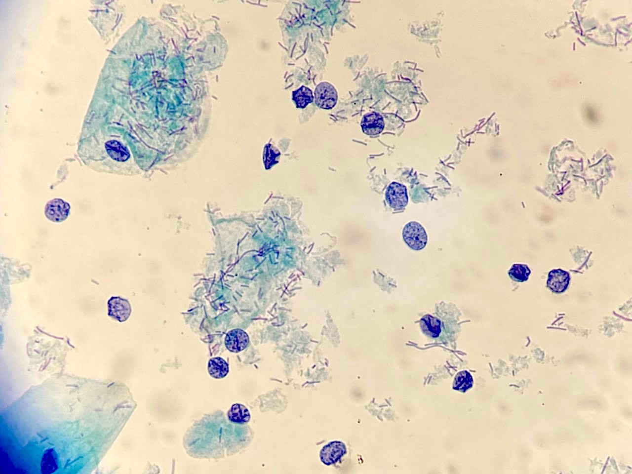
Lactobacillus on the top of squamous cells
Lactobacillus, normal vaginal bacterial rods
Case #327
H&E
CK7
p16
Chromogranin
Synaptophysin
Images hosted on other servers:

Pseudorosette
Contributed by Ziyan T. Salih, M.D.
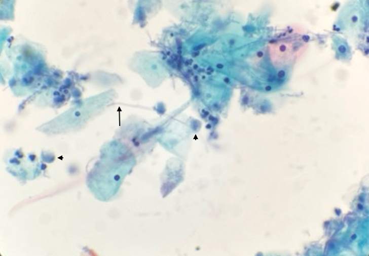
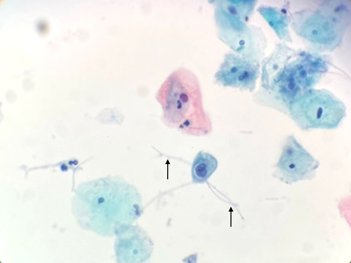
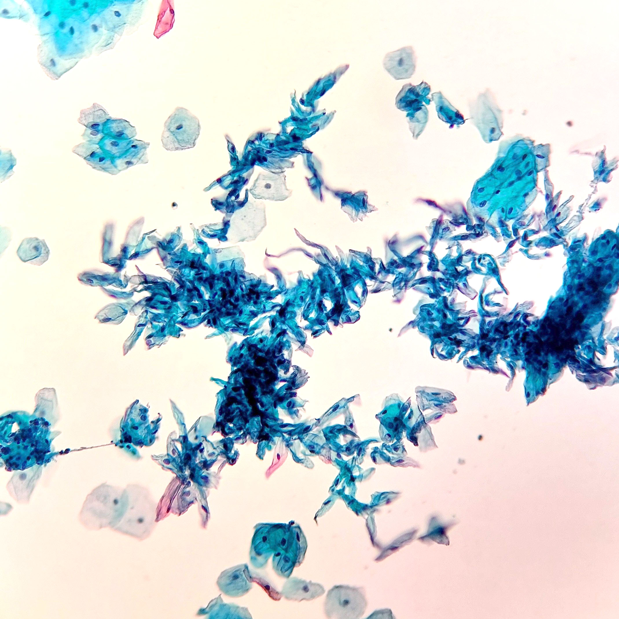
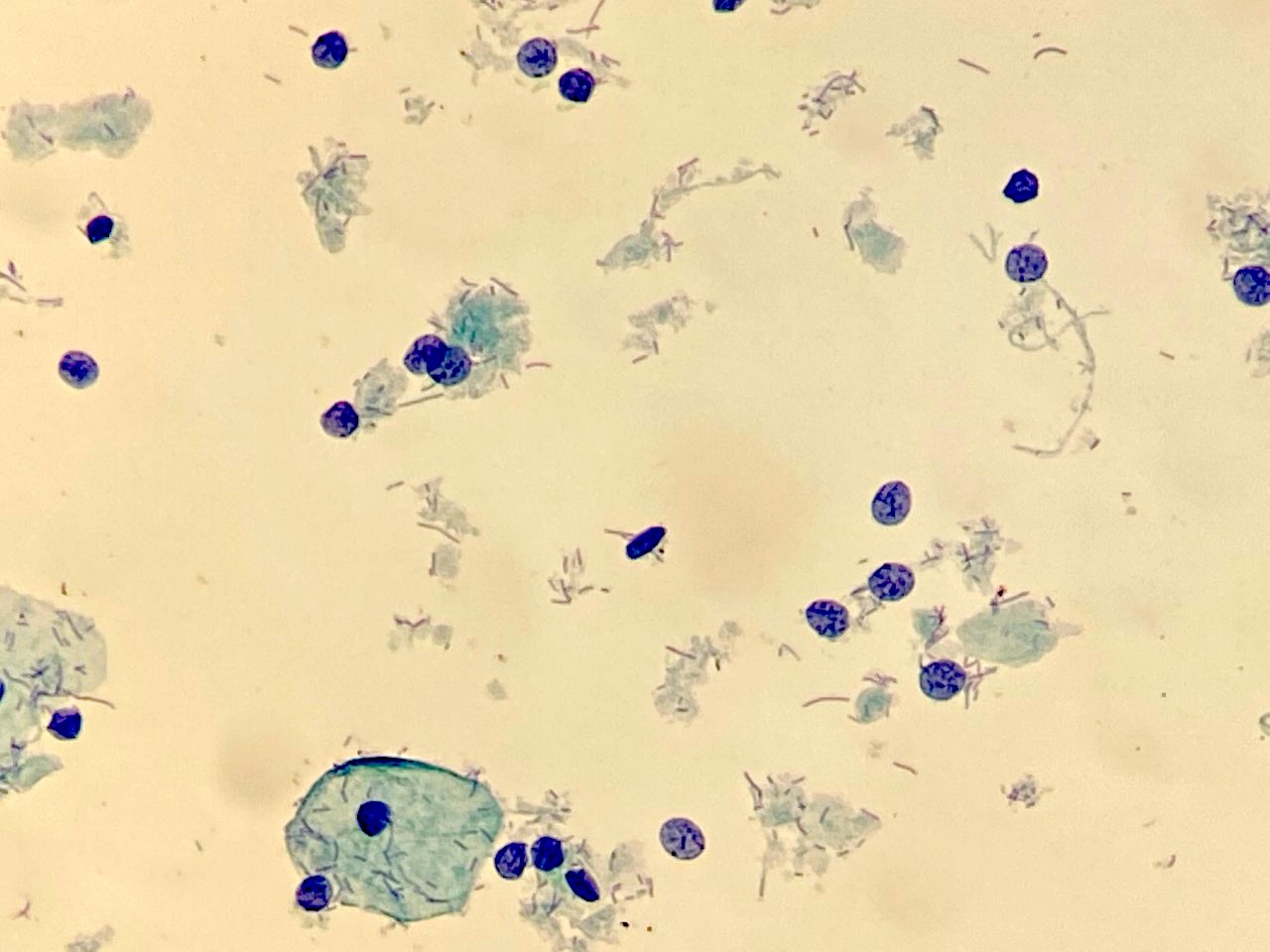
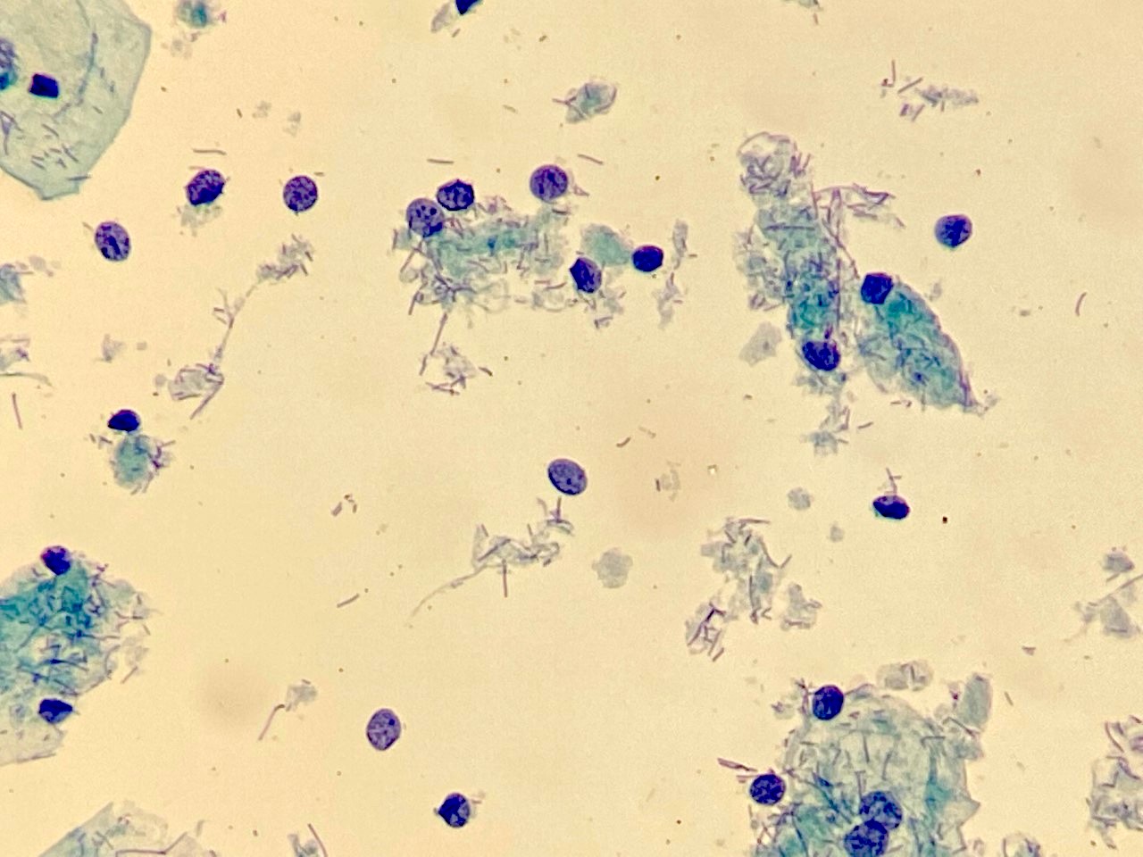
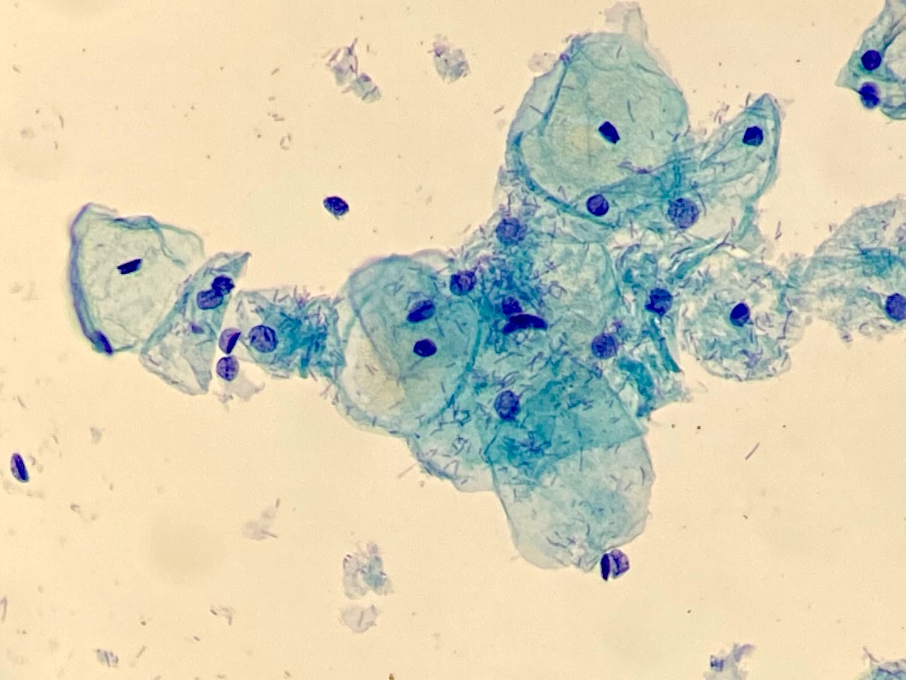
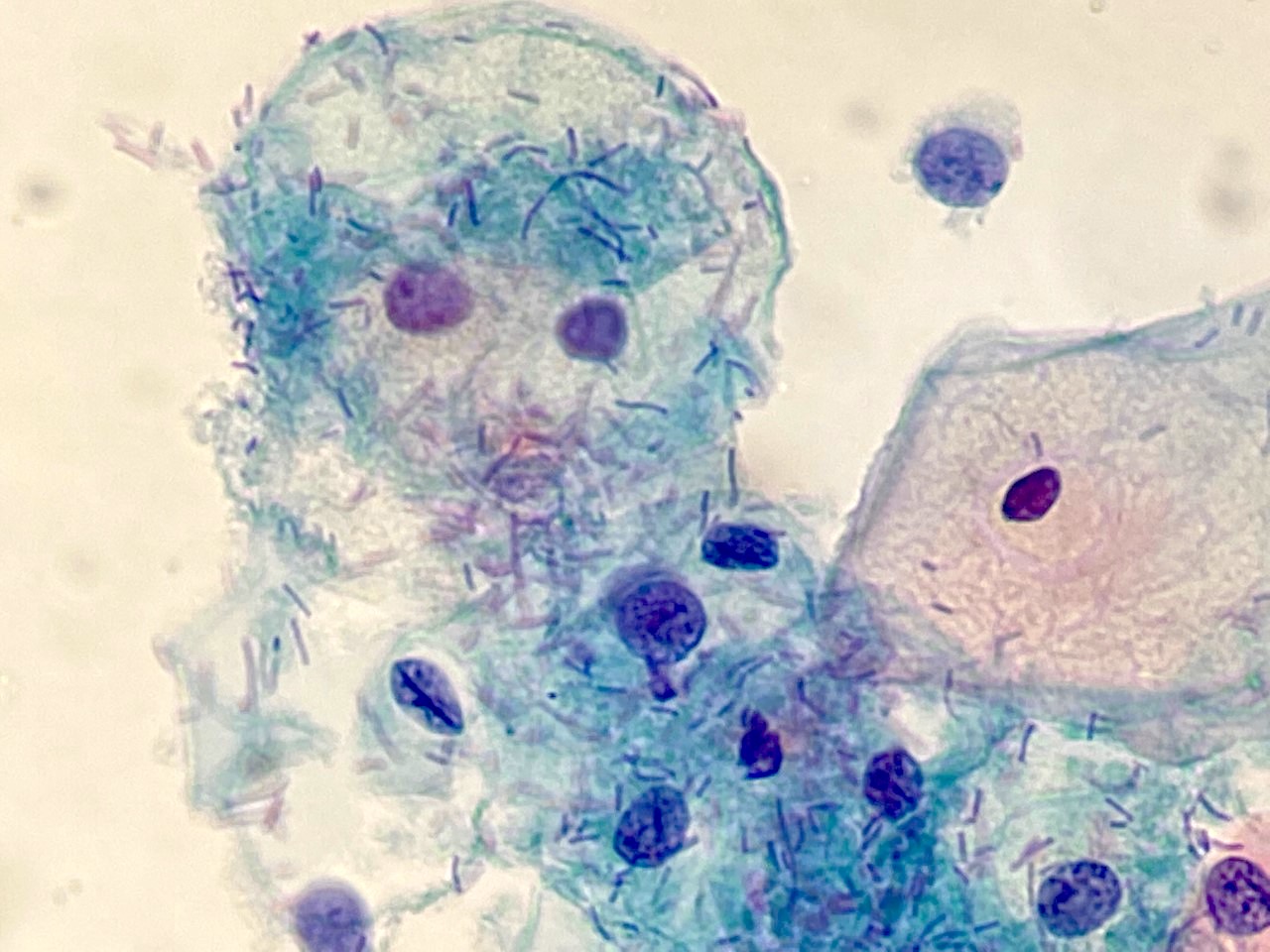
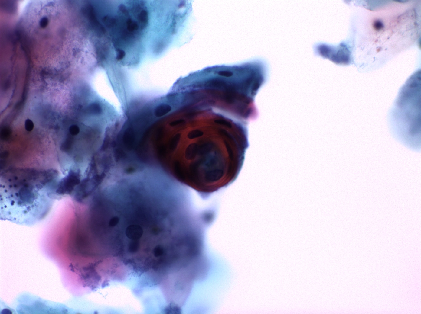
Cytologic smear with Leptothrix
Images hosted on other servers:

Filamentous Leptothrix species

Leptothrix
LBC: Leptothrix with Trichomonas

Long, filamentous structure
Images hosted on other servers:

Gastric type cervical lesion spectrum

Proposed management algorithm
Images hosted on other servers:


LEGH with cosmos pattern
Contributed by Carlos Parra-Herran, M.D.
Densely packed glands
Round mucinous glands
Lobulated / floret-like appearance
Glands lined by benign columnar cells
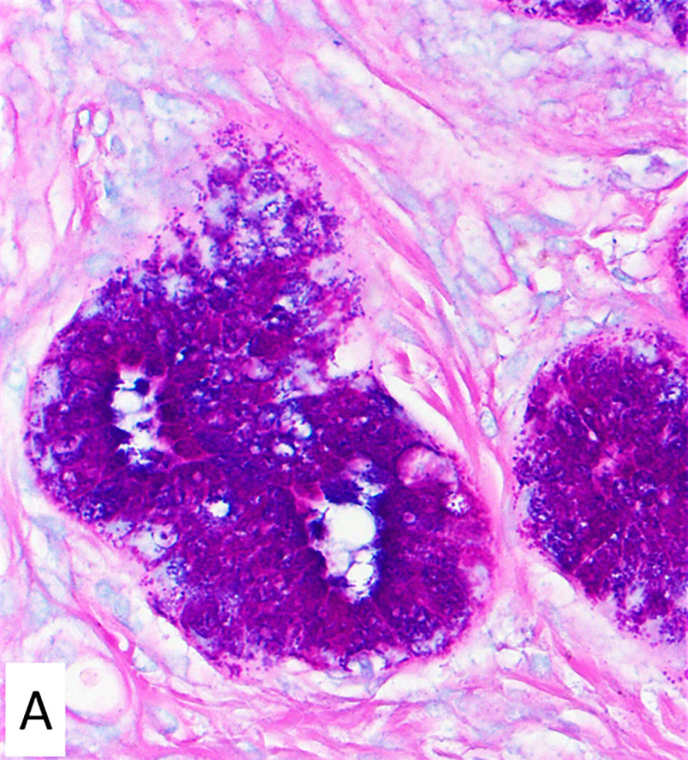
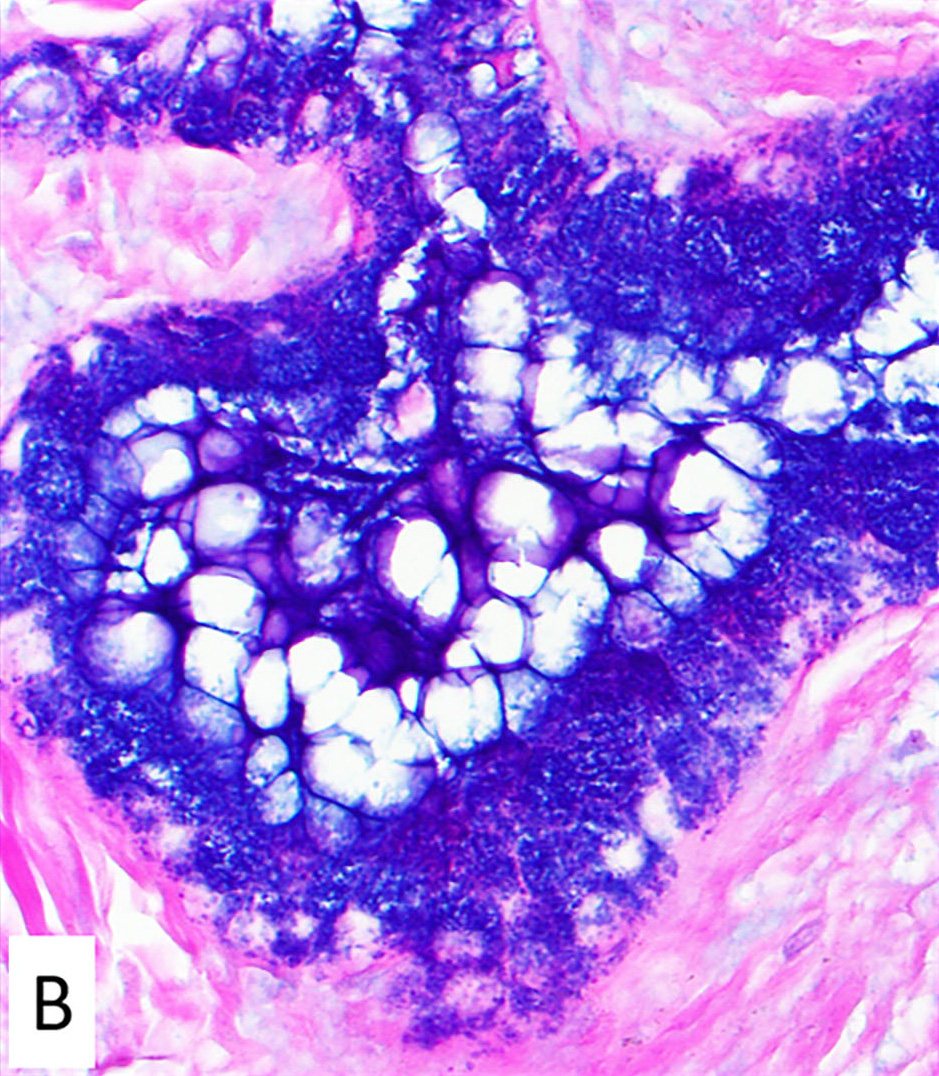
PAS - Alcian blue stain
Contributed by @MirunaPopescu13 on Twitter
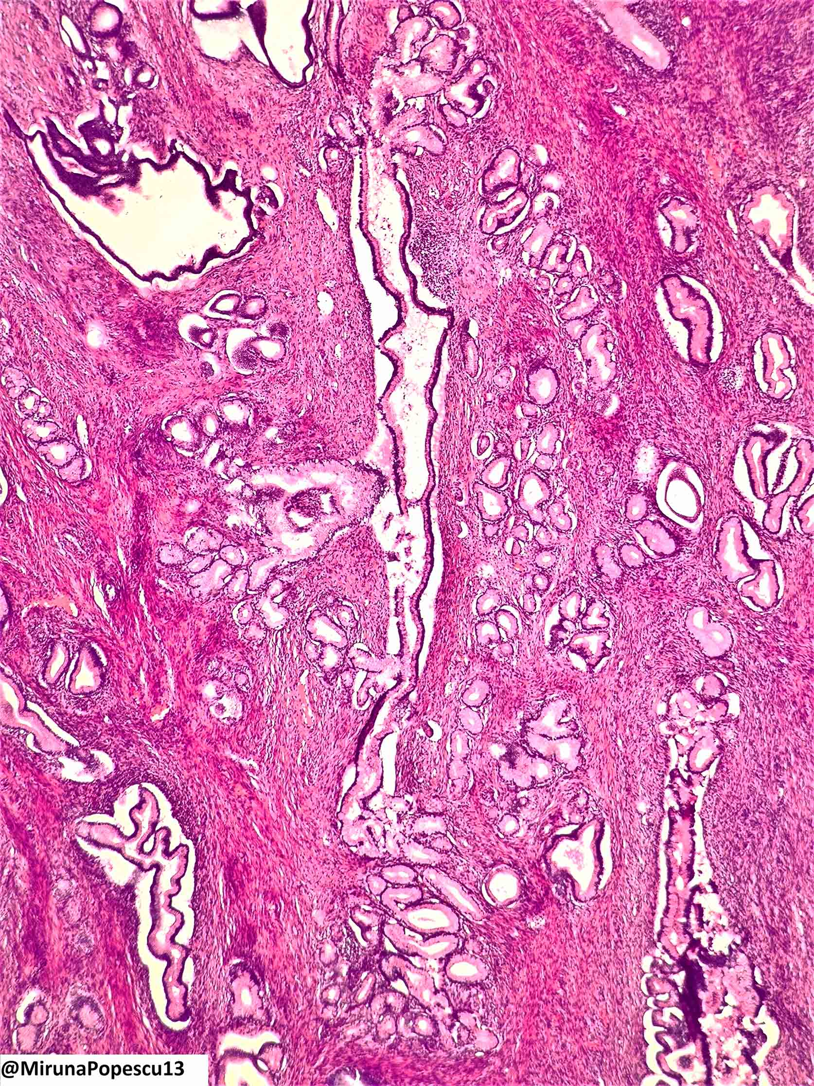
Lobular endocervical glandular hyperplasia
Images hosted on other servers:

LEGH and MDA
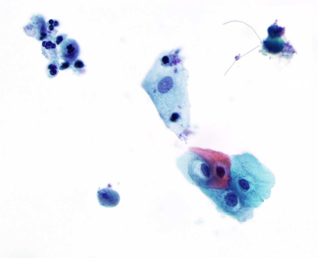
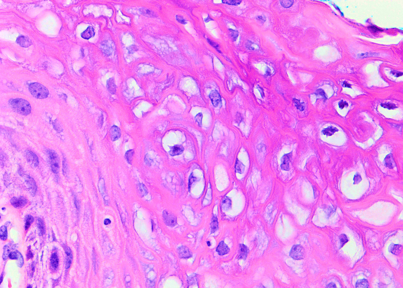
Contributed by Lucy Jager, M.D. and Bonnie Choy, M.D.
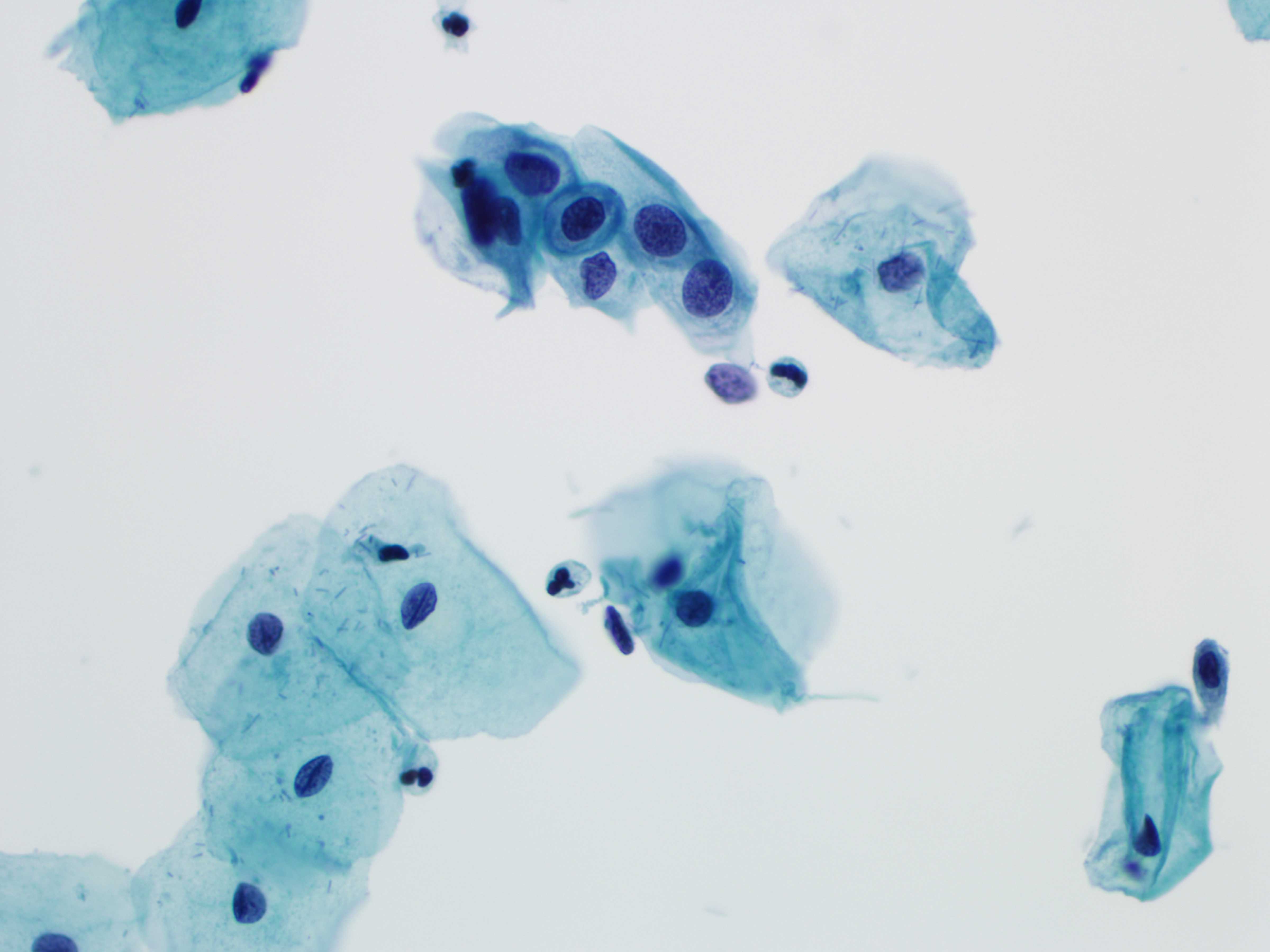
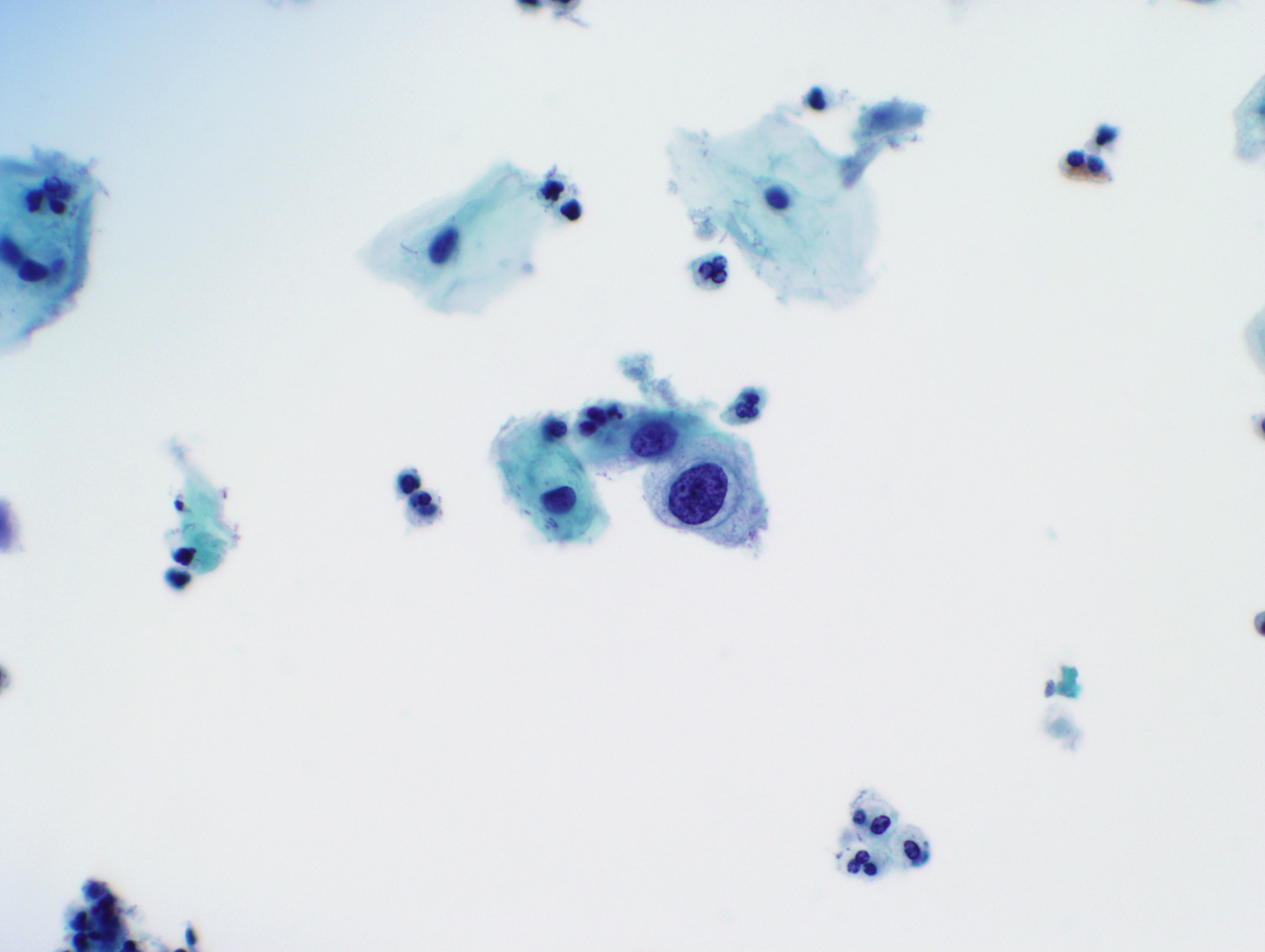
Nuclear enlargement and hyperchromasia
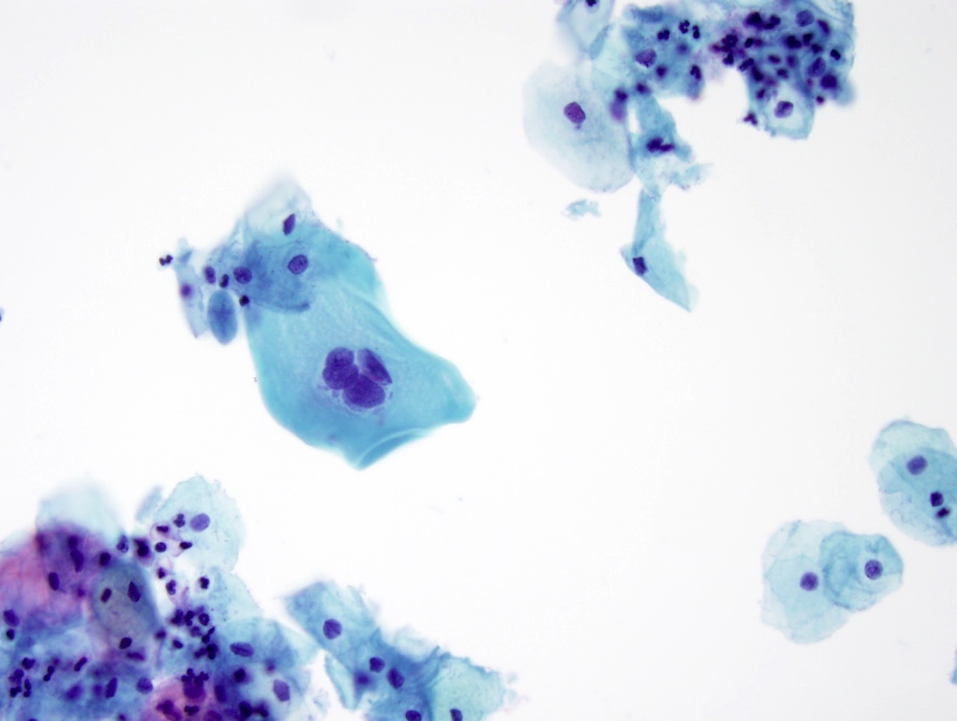
Multinucleation

Binucleation and perinuclear cavitation
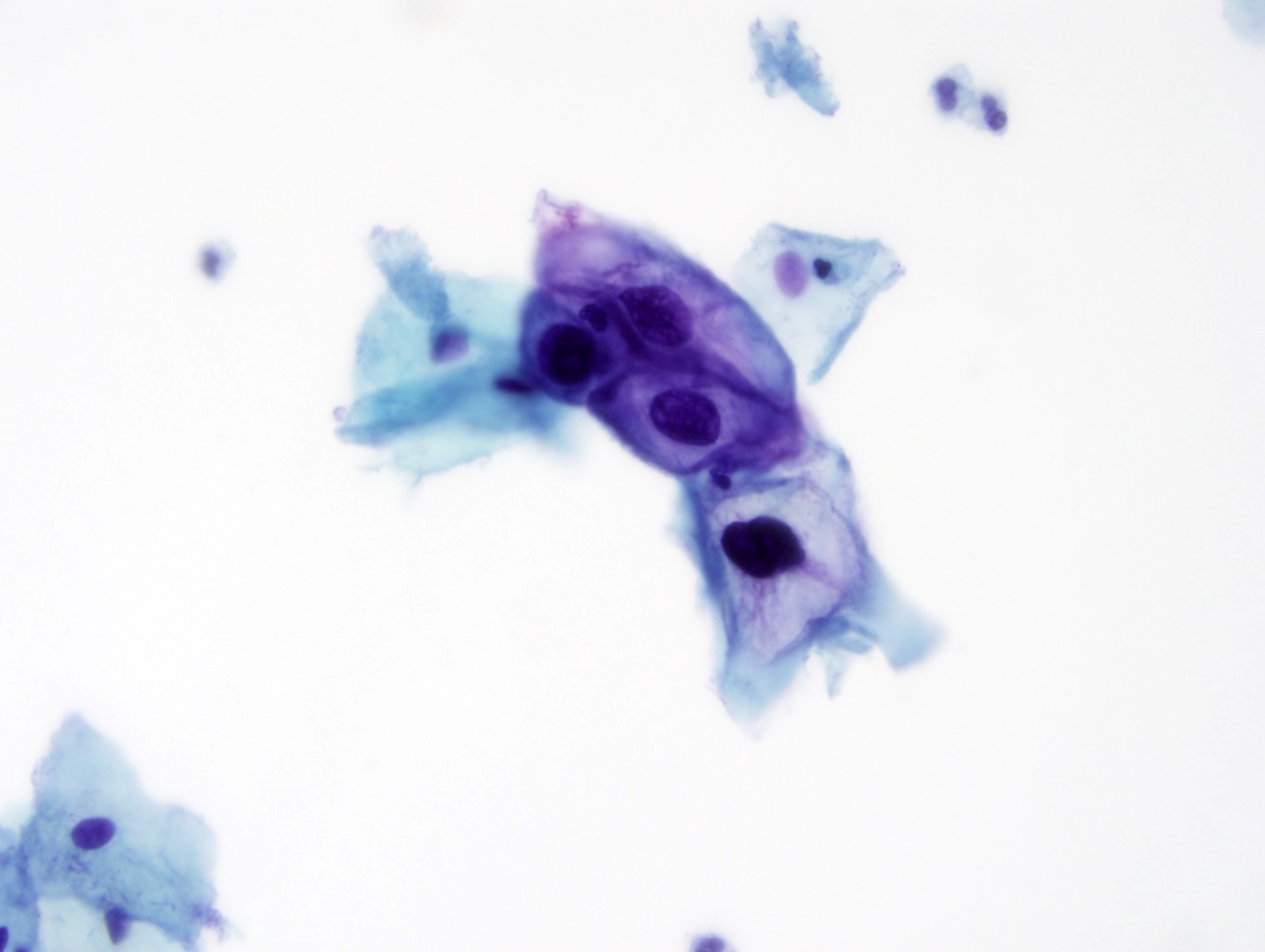
Koilocytosis with nuclear abnormalities
Koilocytosis with nuclear abnormalities
Pseudokoilocytes
Herpes cytopathic effect
Images hosted on other servers:
WHO digital atlas

Contributed by Ziyan T. Salih, M.D.
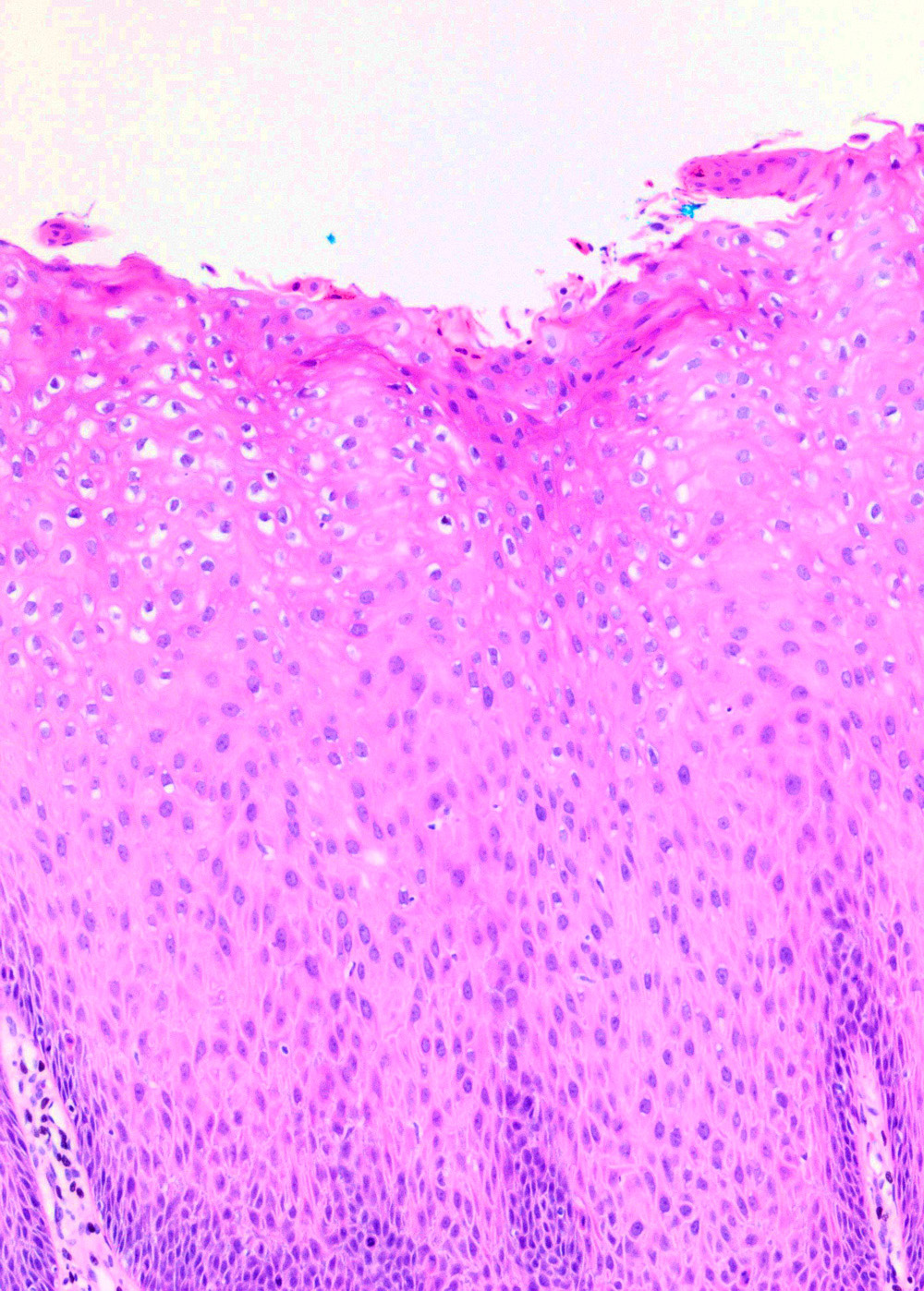
LSIL / koilocytic atypia
Koilocytic atypia
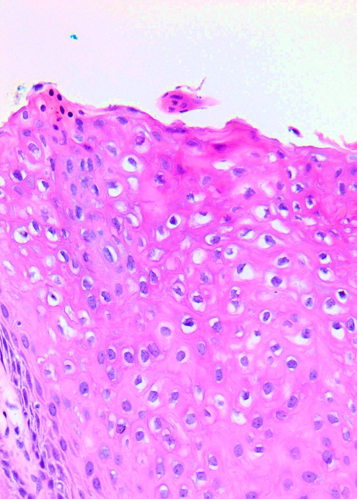
Cervical biopsy (LSIL)
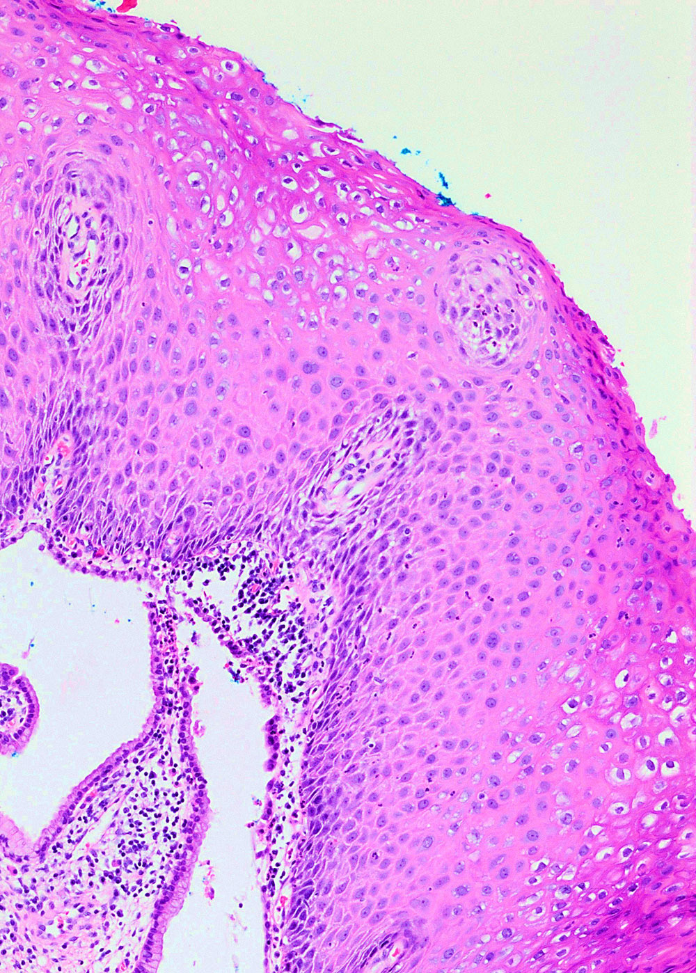
Contributed by Ziyan T. Salih, M.D.
Koilocytosis (LSIL)
Squamous lesions of the cervix and their differential diagnosis
by Carlos Parra-Herran, M.D.
Images hosted on other servers:

Vagina: well circumscribed tumor
Images hosted on other servers:

Various Images
Images hosted on other servers:

Various images and immunostains

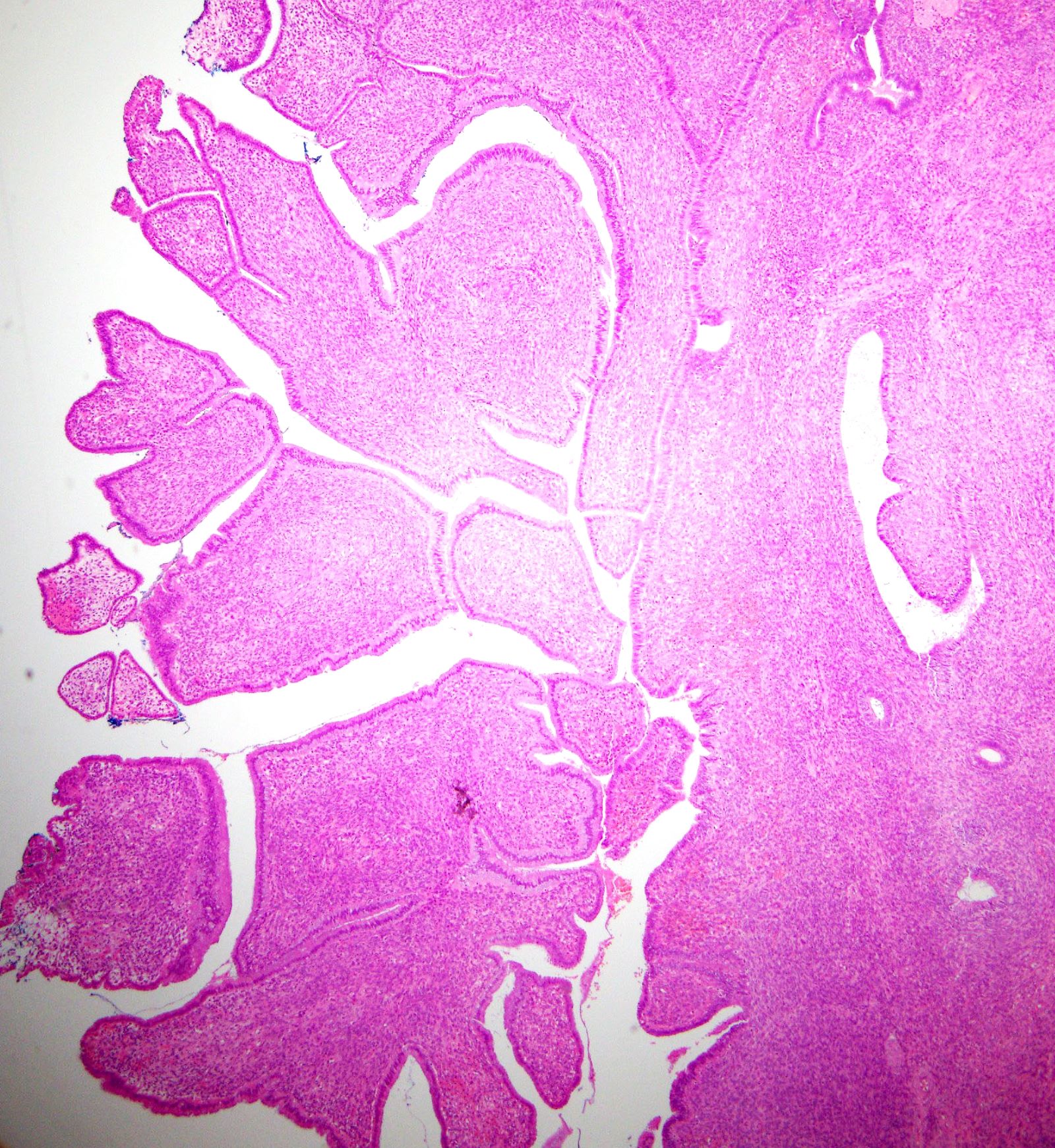
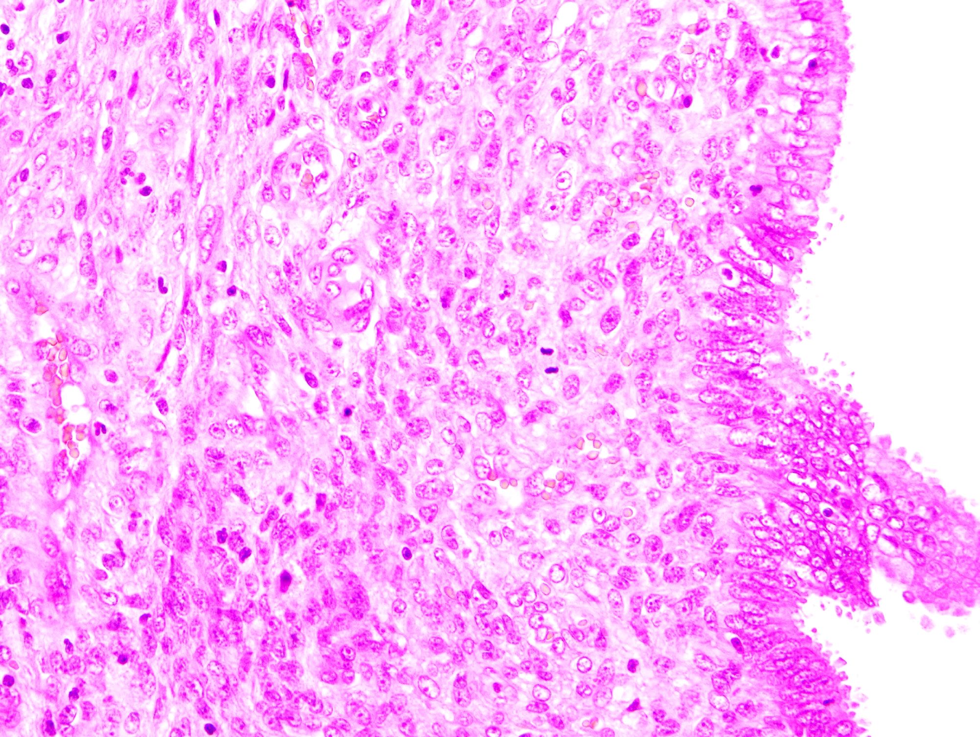
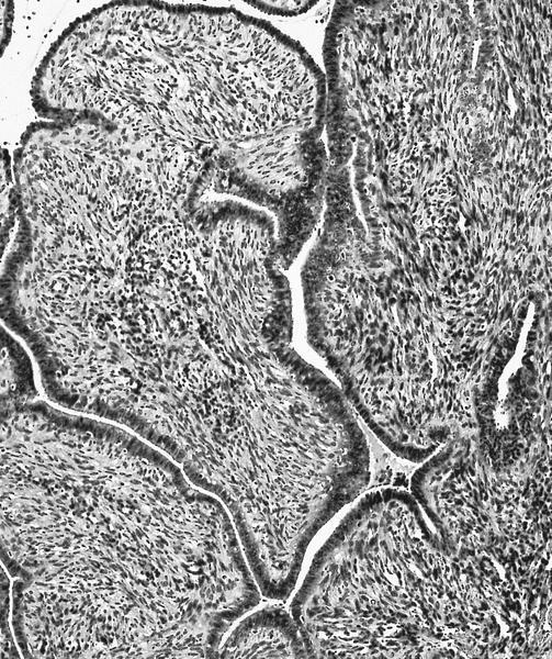
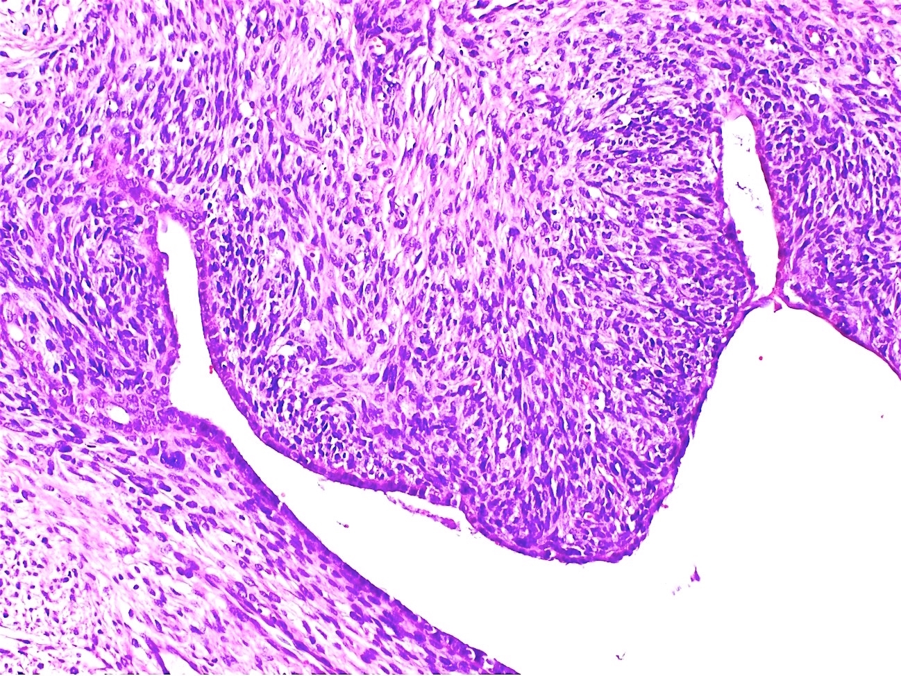
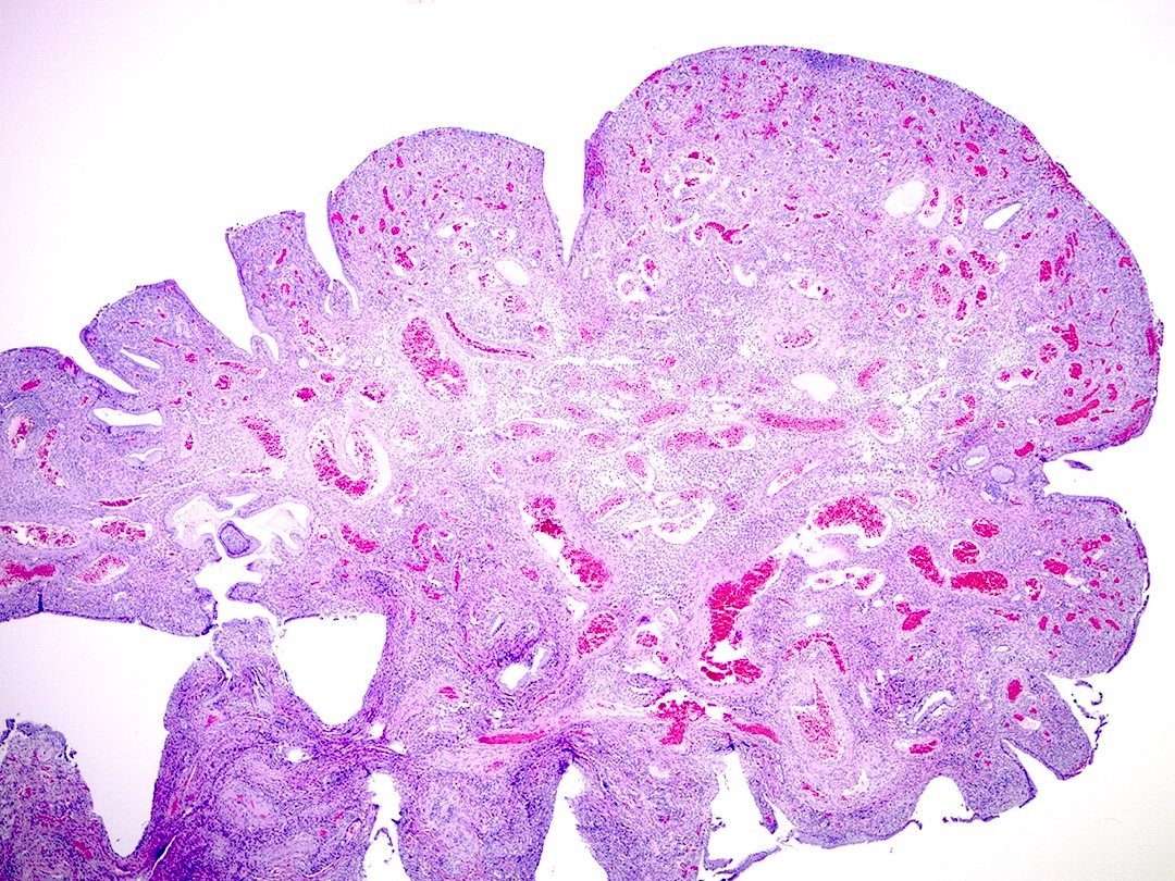
Papillary tumor with various epithelial types

Focal atypia due
to stratification,
pleomorphism and
atypical mitotic figure
Images hosted on other servers:

MRI cervical tumor
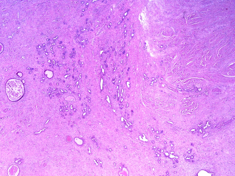
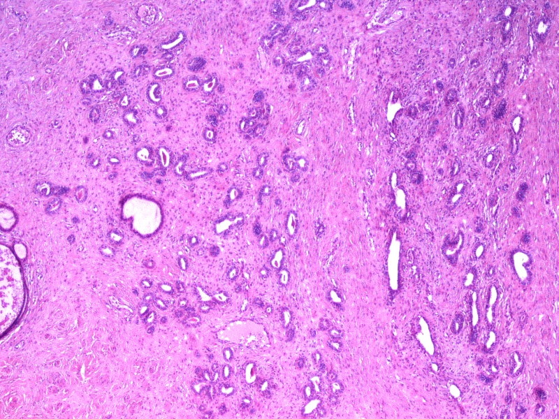
Contributed by Lynn Hoang, M.D.
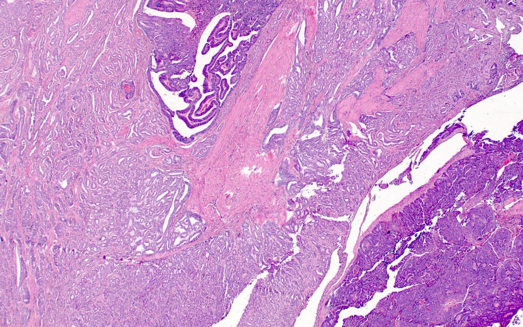
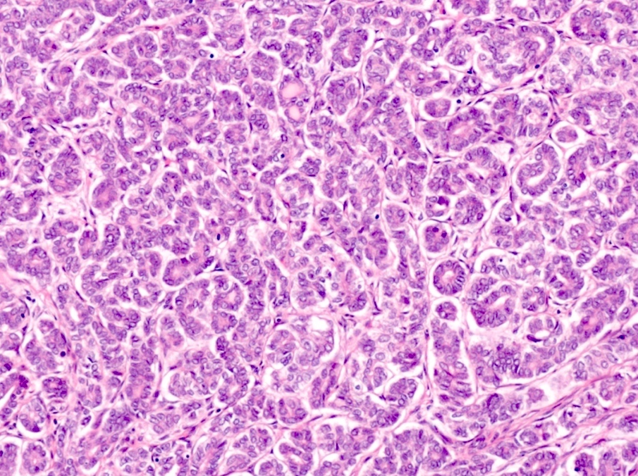
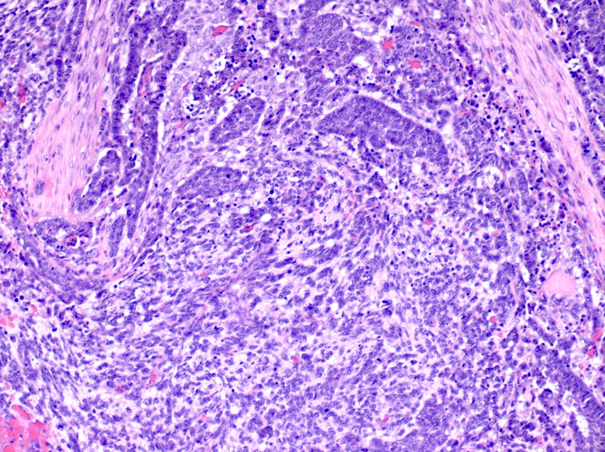
Mix of morphologic patterns
Tubular pattern
Tubular pattern with adjacent mesonephric remnants
Tubular pattern with desmoplasia
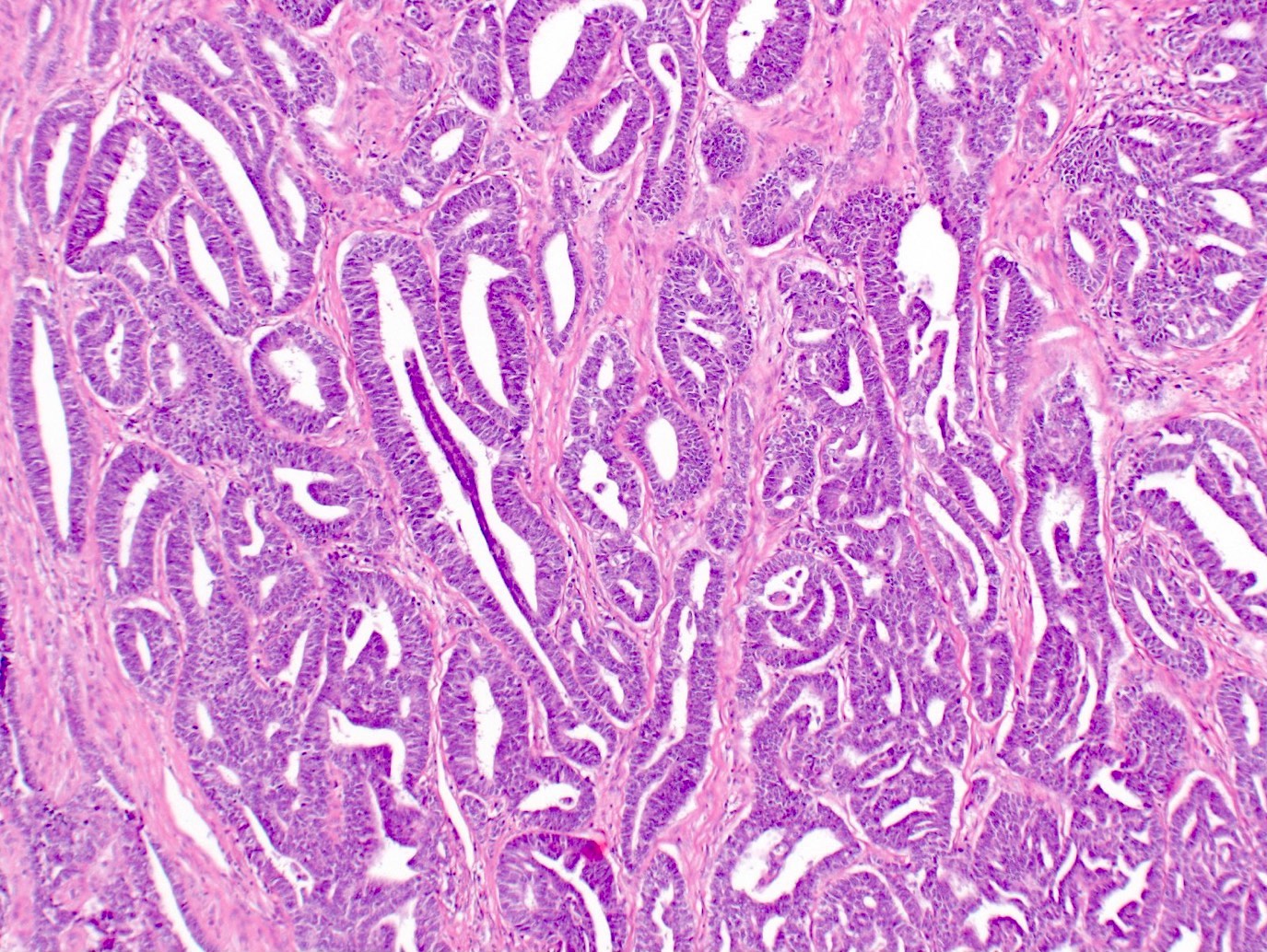
Ductal
(pseudoendometrioid)
pattern

Ductal pattern
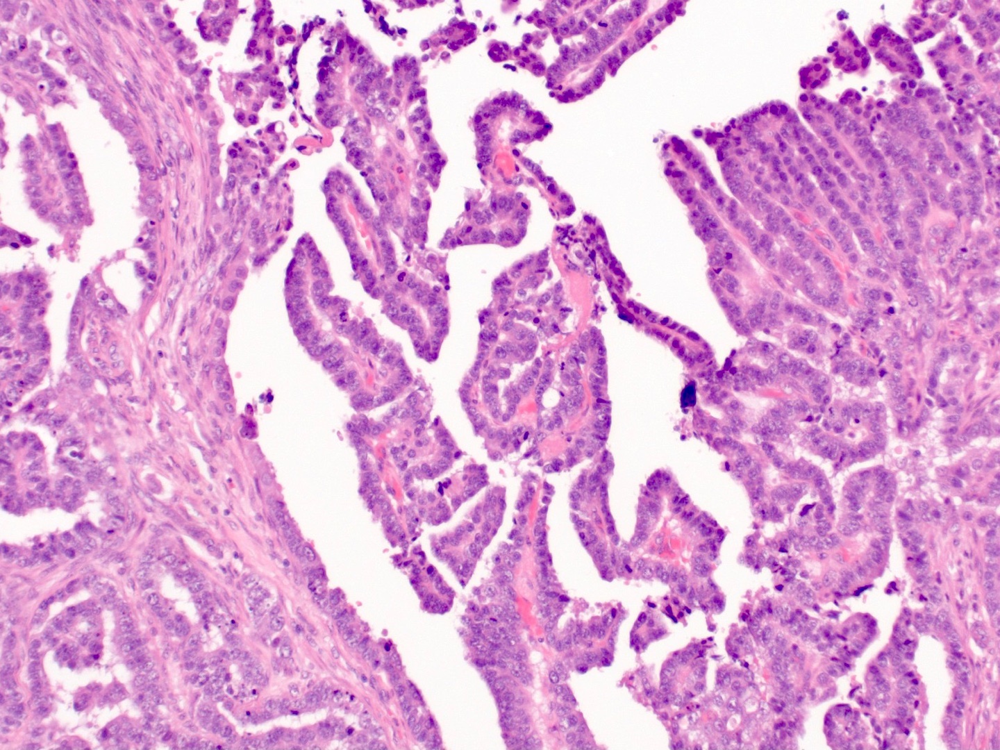

Papillary pattern
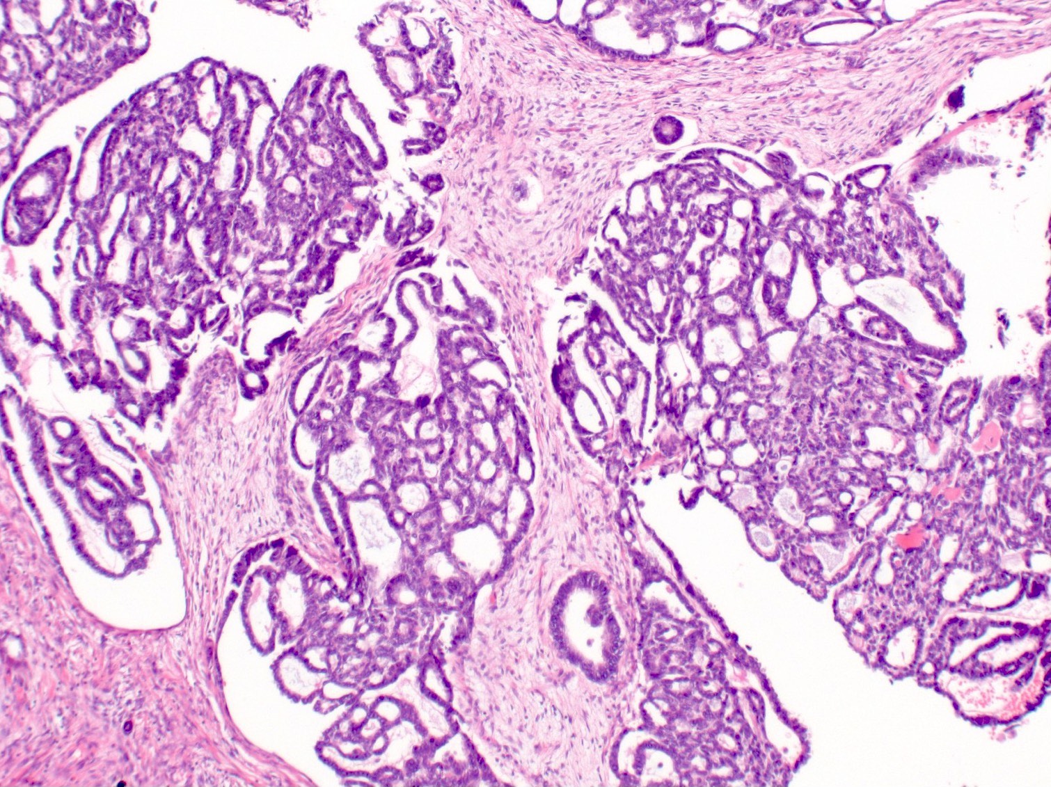
Sieve-like pattern
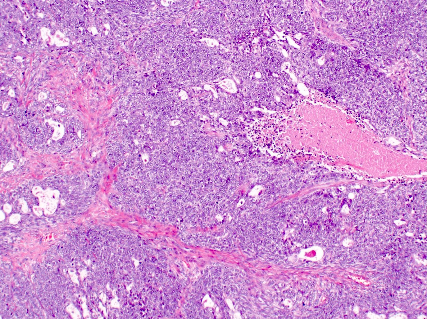
Solid pattern
Mesonephric carcinosarcoma

GATA3+
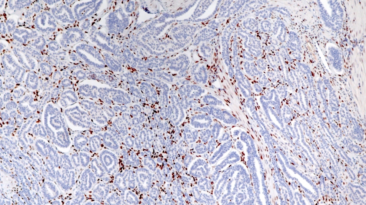
ER-
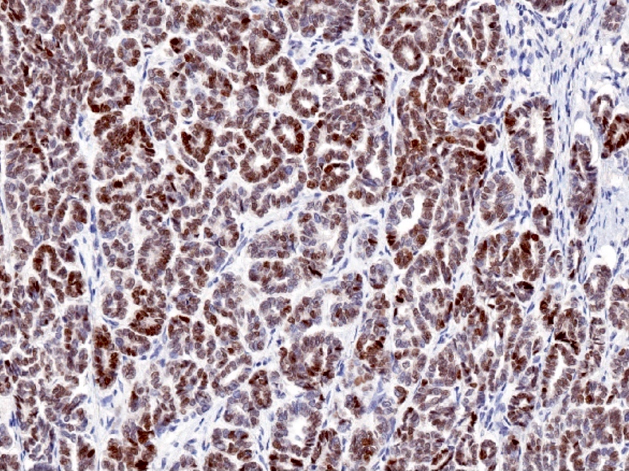
TTF1+
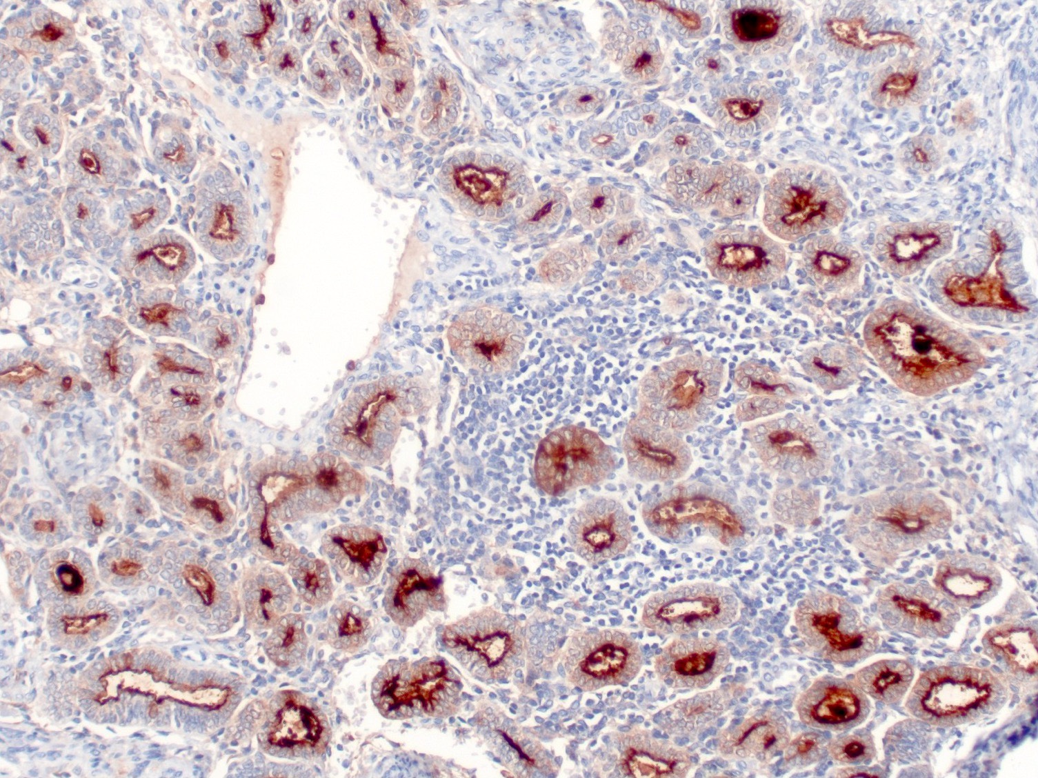
CD10+
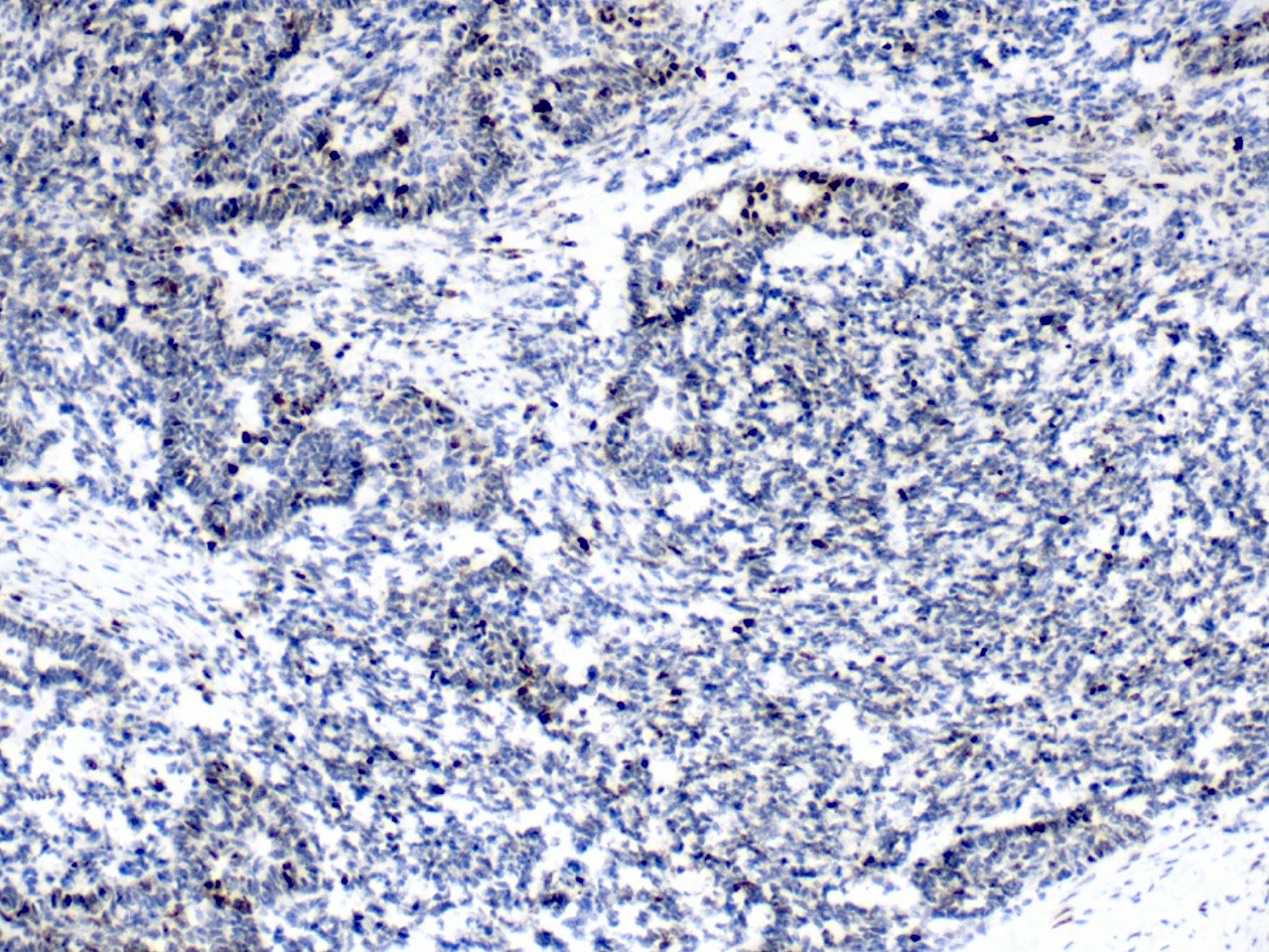
Wild type p53
Contributed by Michela Campora, M.D., Carlos Parra-Herran, M.D. and AFIP
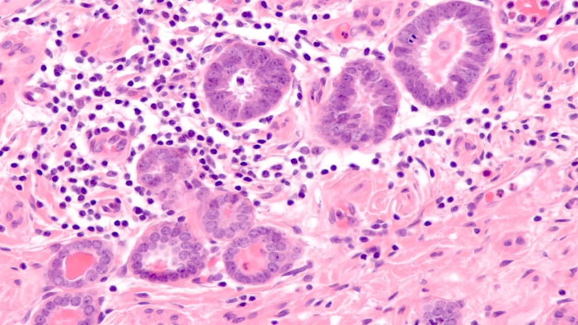
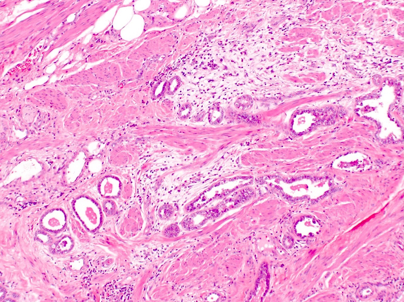
Mesonephric hyperplasia
Marked tubular proliferation
More nuclear variation
Bland glands deep in cervical stroma
Clusters of mesonephric tubules
Mesonephric remnants

Cuboidal cells
Focal squamous metaplasia
AFIP images
Breast carcinoma metastatic to cervix
Case #125 (metastatic lobular carcinoma to endometrial polyp)
Various images
ER
PR
GCDFP-15
Contributed by Carmen Luz, M.D.
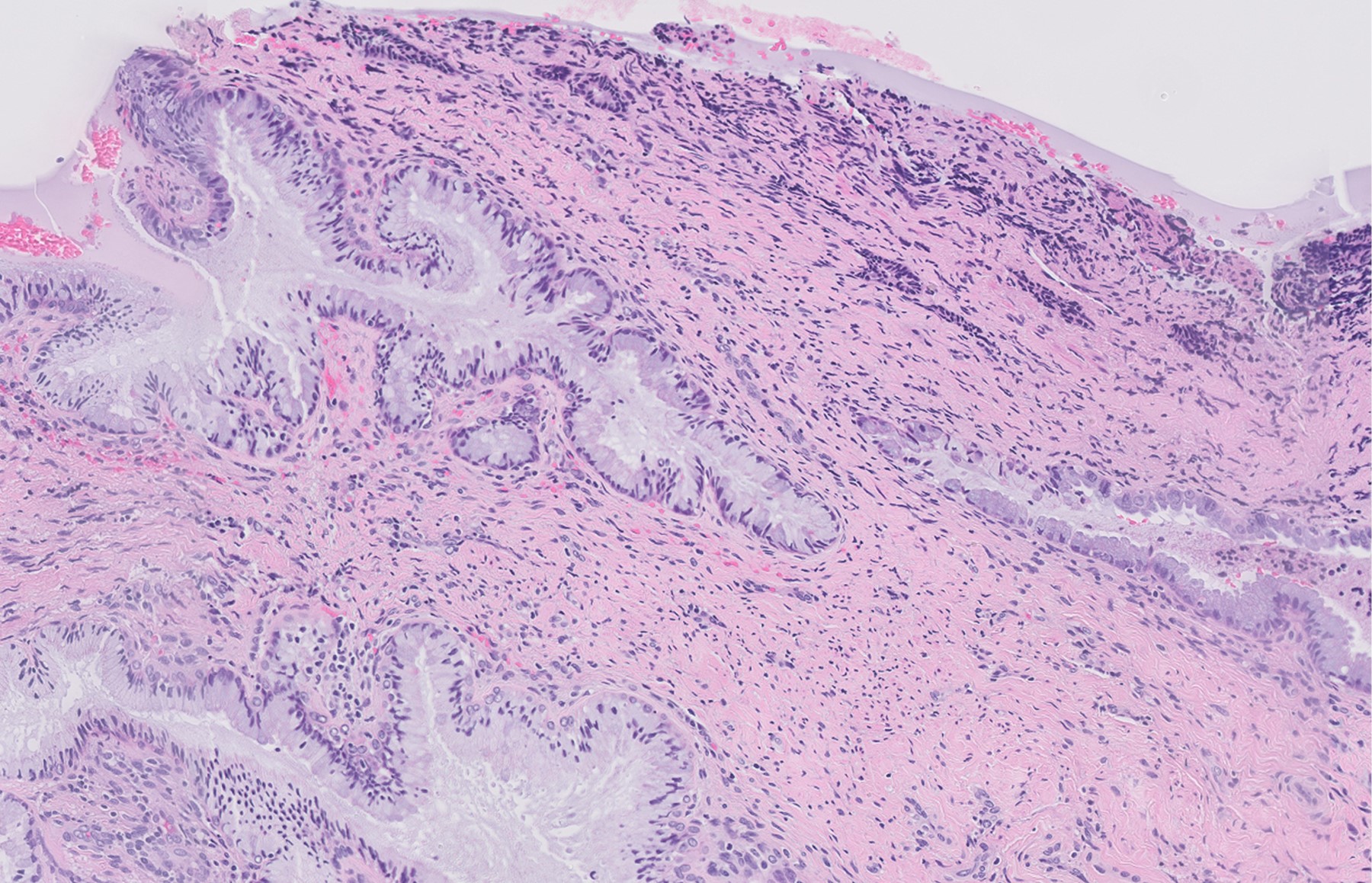
Ovarian clear cell carcinoma
Contributed by Gulisa Turashvili, M.D., Ph.D.
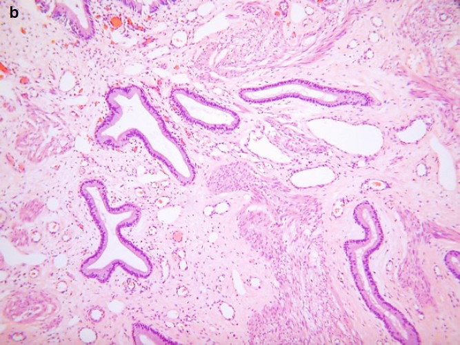
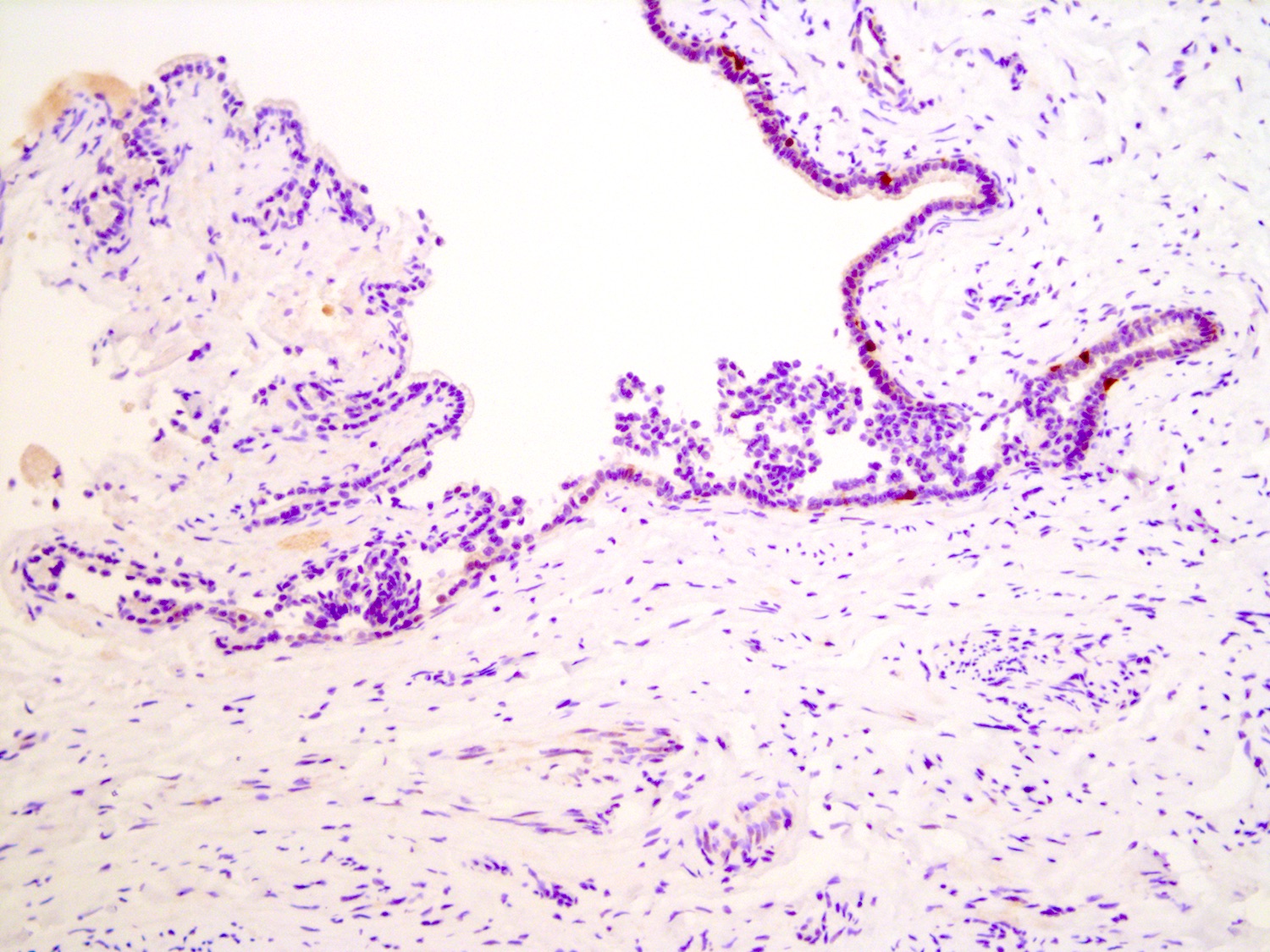
Benign glandular proliferation
Back to back glands lacking atypia
Bland epithelial lining
Rare mitosis
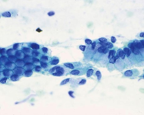
Images hosted on other servers:
Isolated rounded endocervical cells
Images hosted on other servers:

Nabothian cysts on computed tomography
Images hosted on other servers:

Multiple nabothian cysts

Nabothian cyst
Images hosted on other servers:

Nabothian cysts
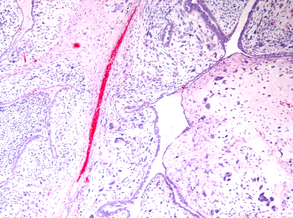
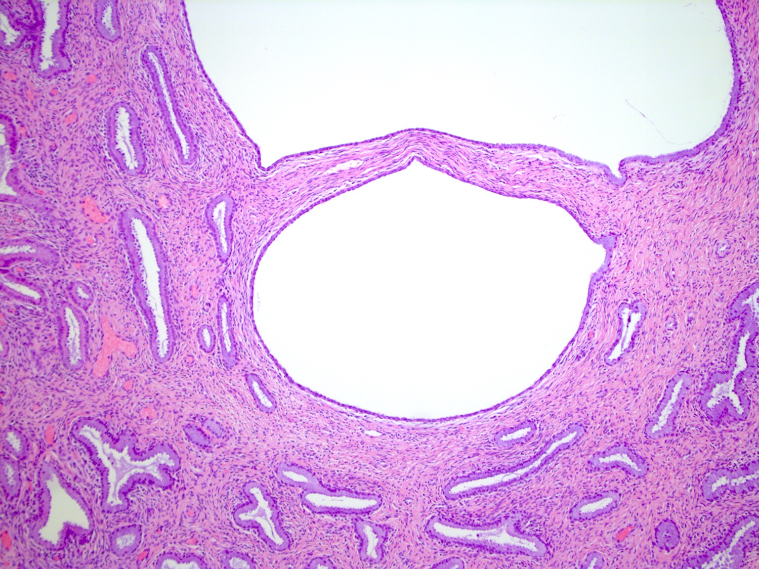
Contributed by Gulisa Turashvili, M.D., Ph.D. and Andrey Bychkov, M.D., Ph.D.
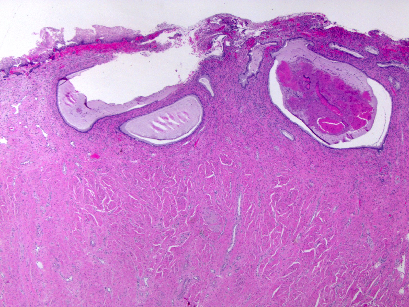
Cystically dilated glands
Bland epithelial lining
Endocervical polyp with nabothian cysts
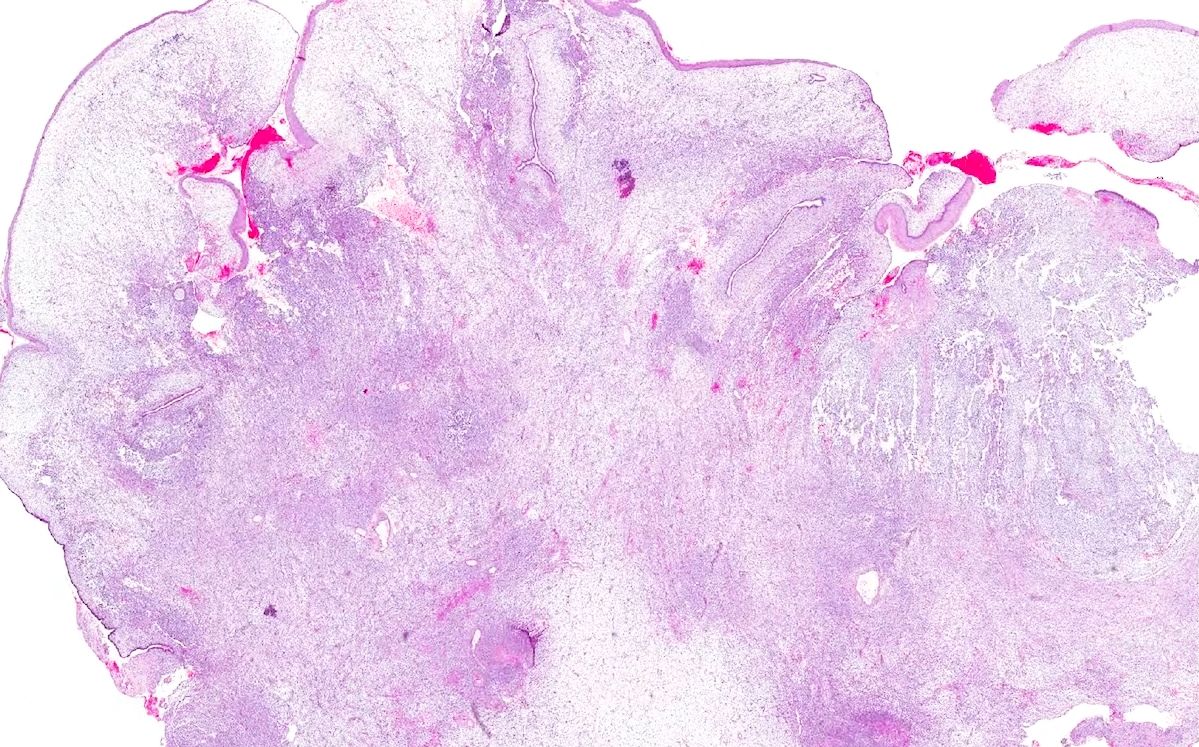
Images hosted on other servers:

Hysterectomy specimen
Contributed by Shuyue Ren, M.D., Ph.D.
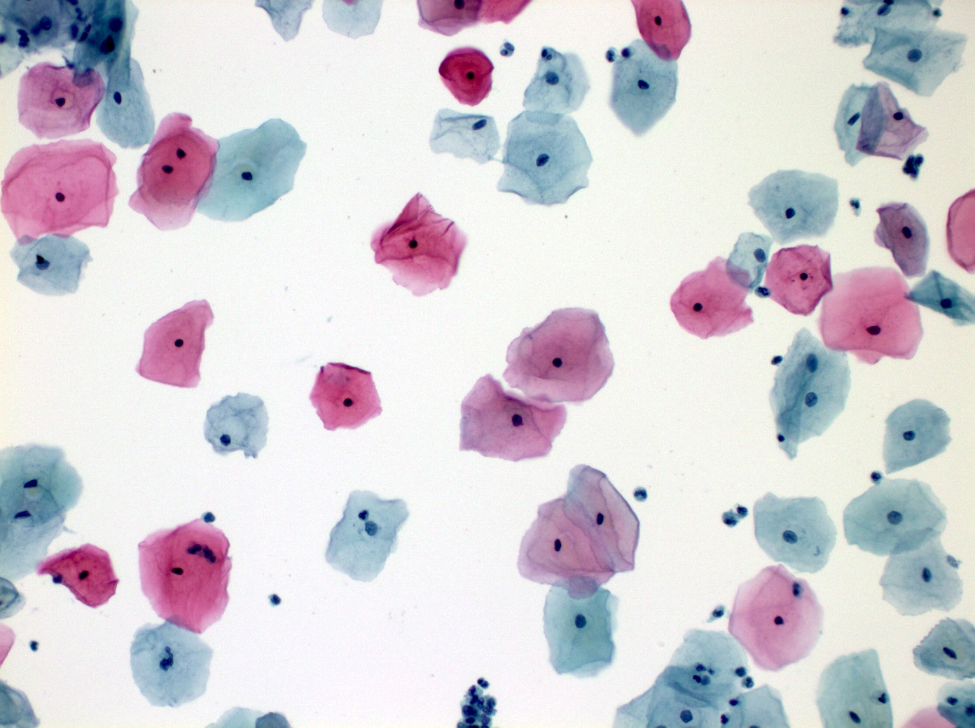
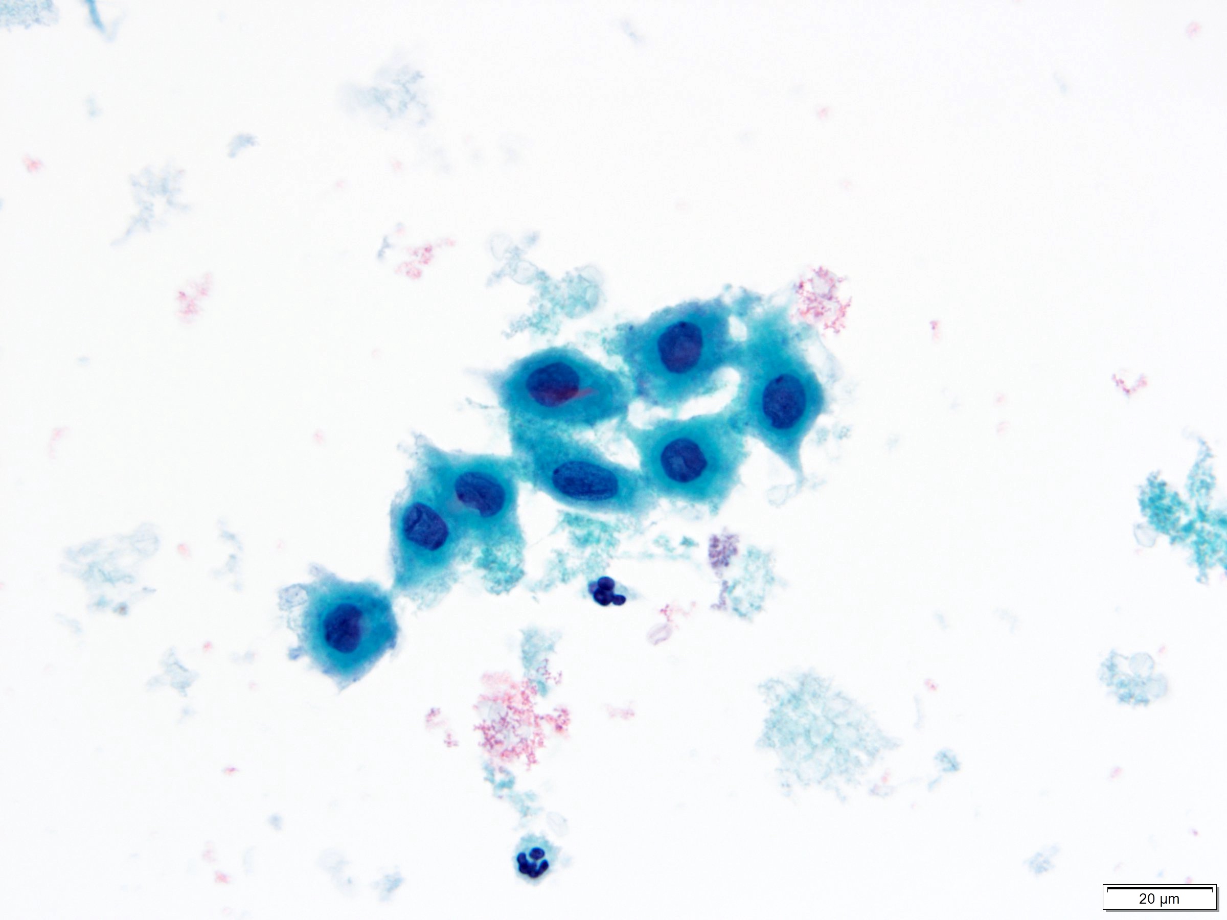
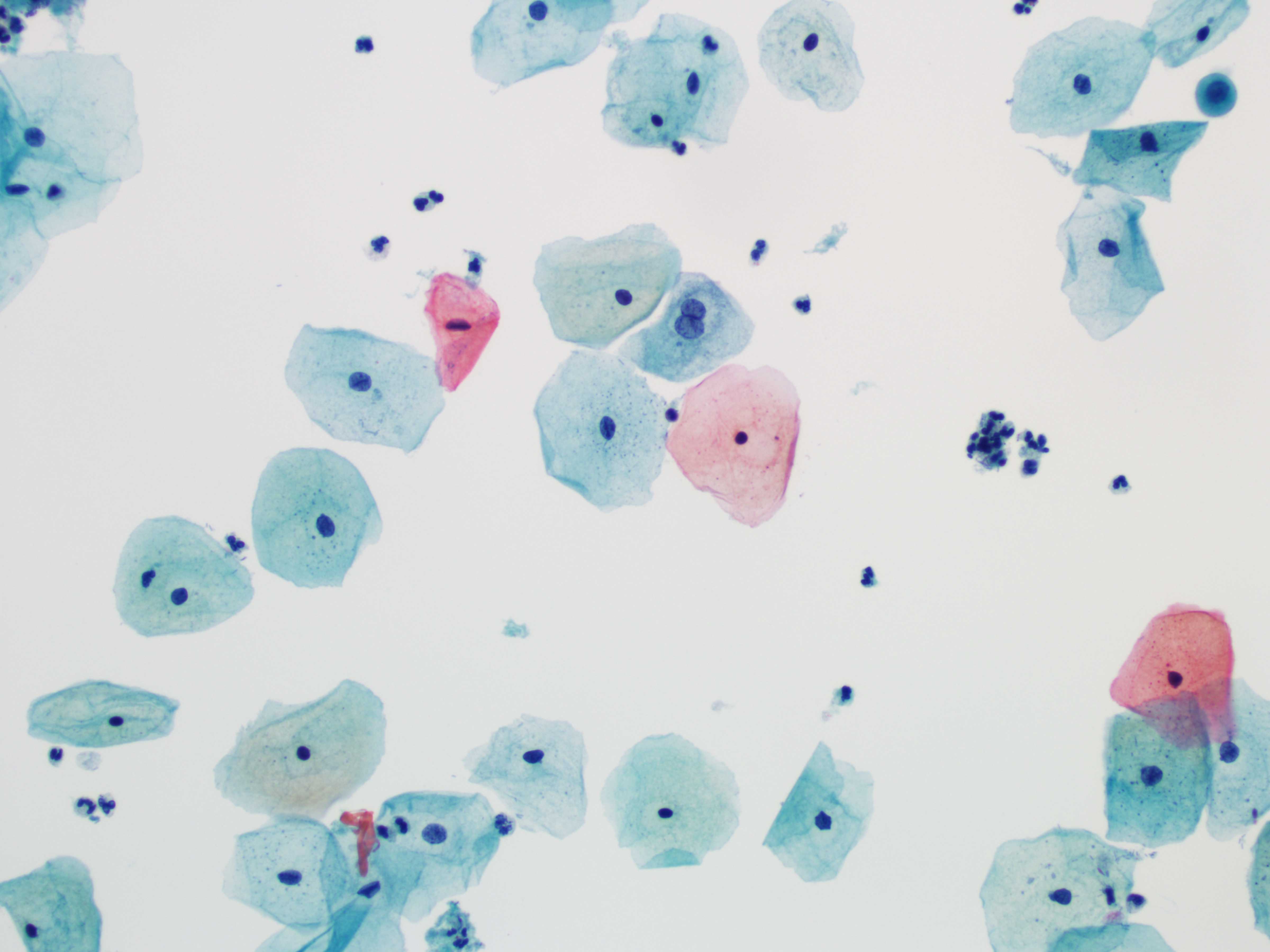
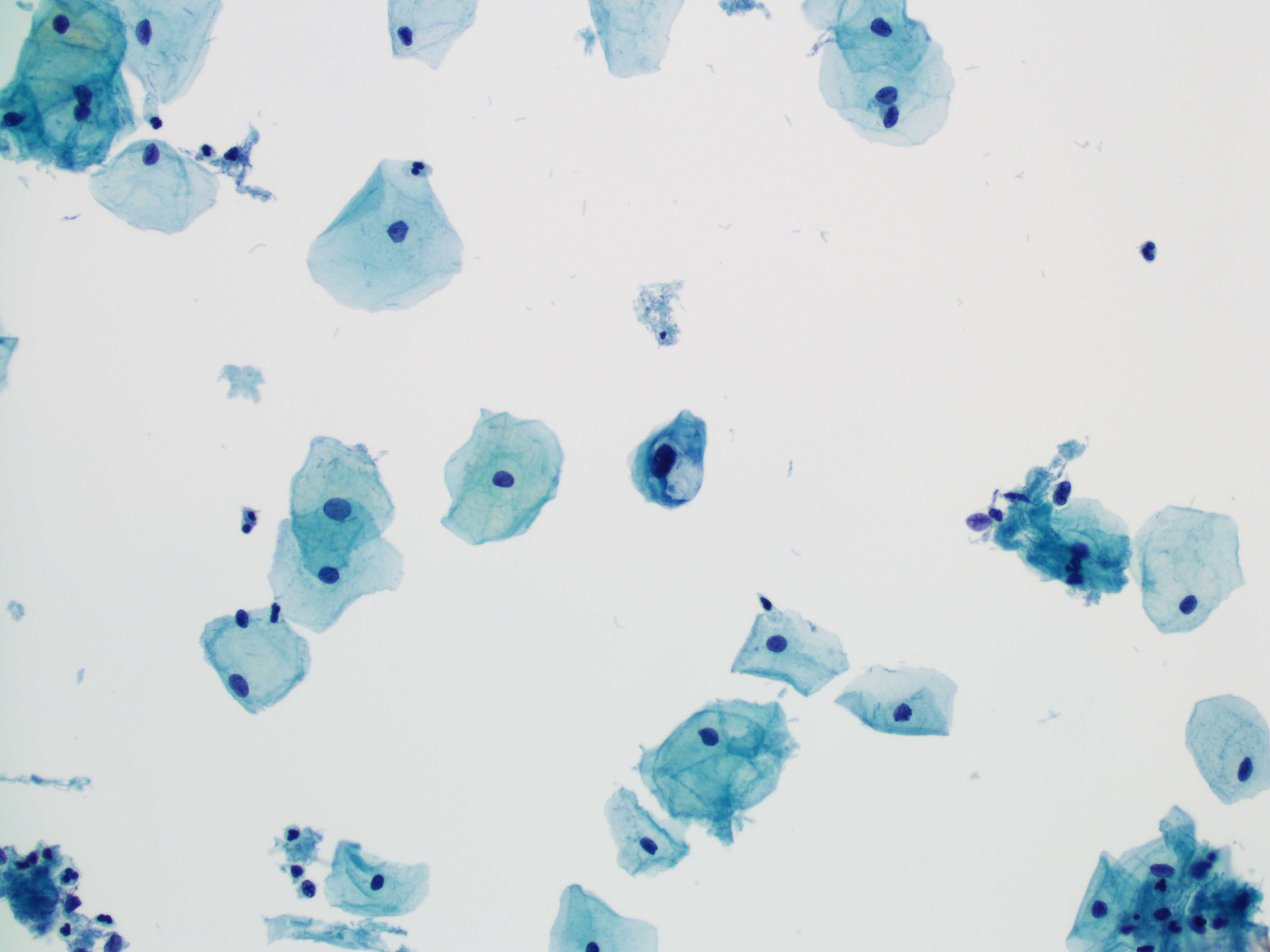
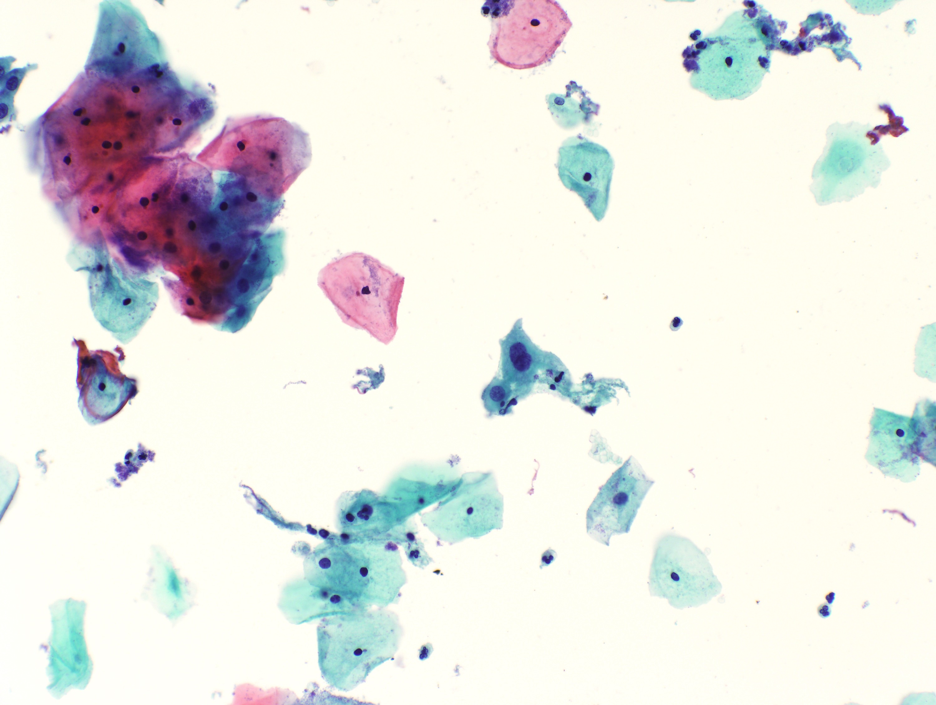
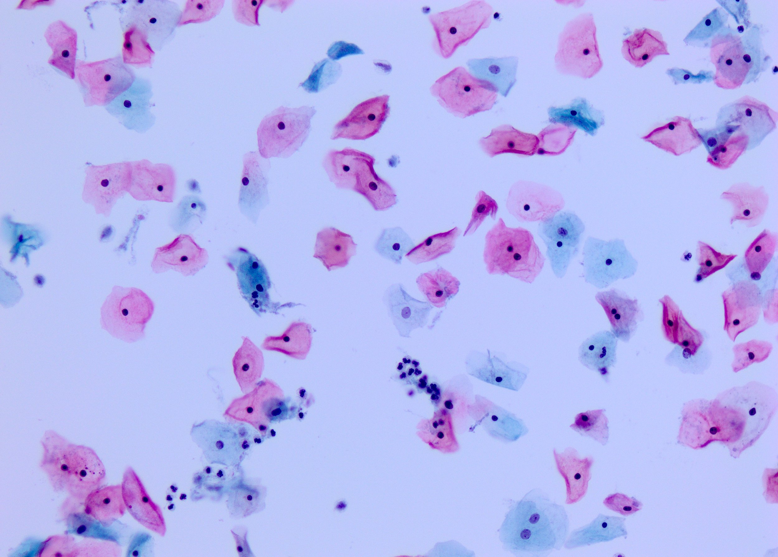
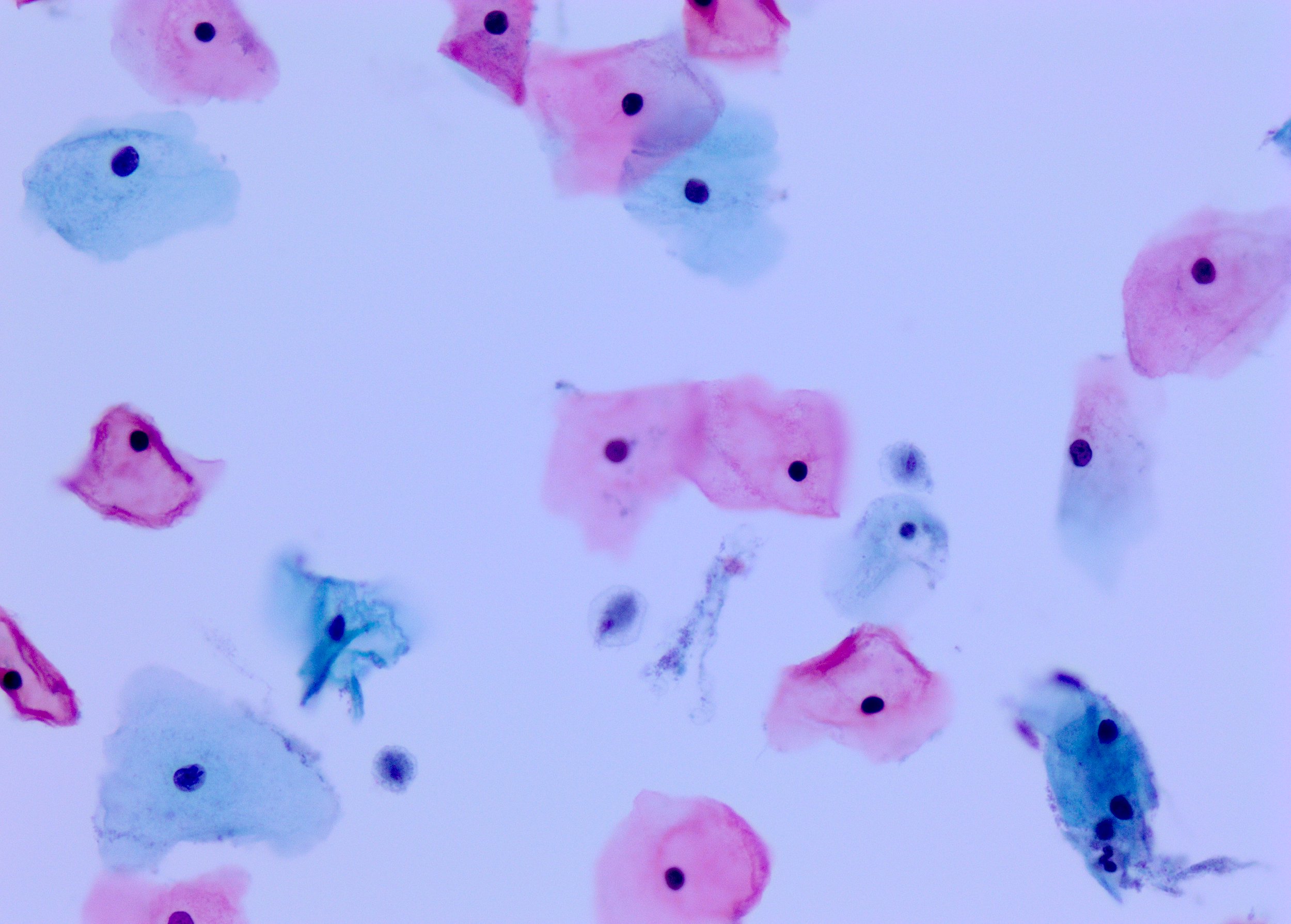
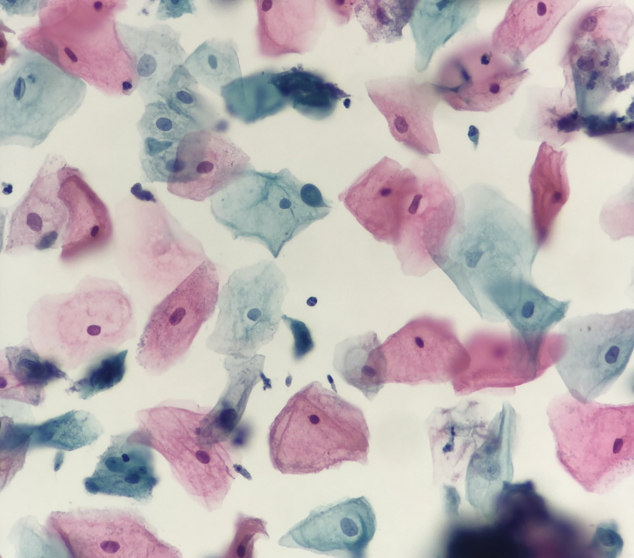
Squamous cells, superficial
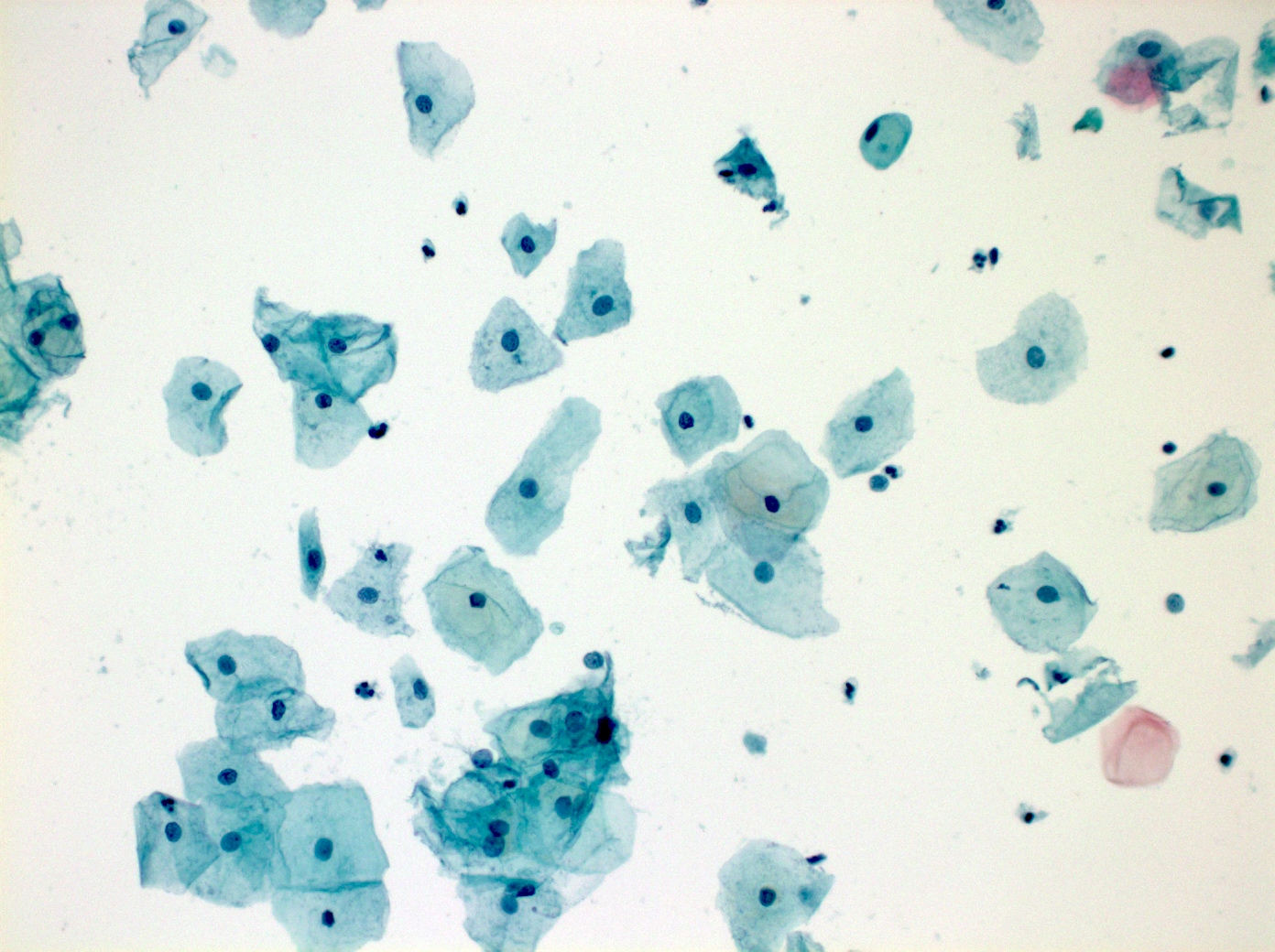
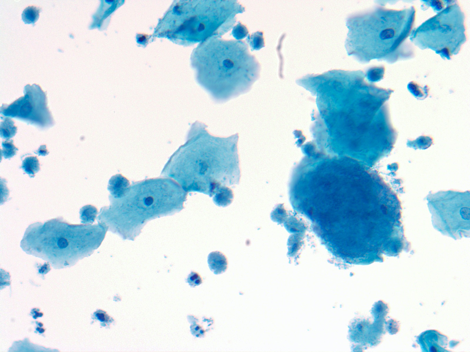
Squamous cells, intermediate
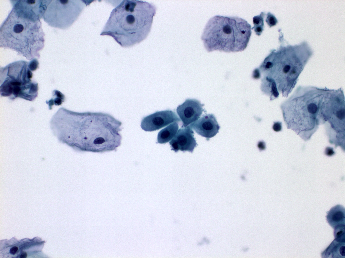
Squamous cells, parabasal and basal cells
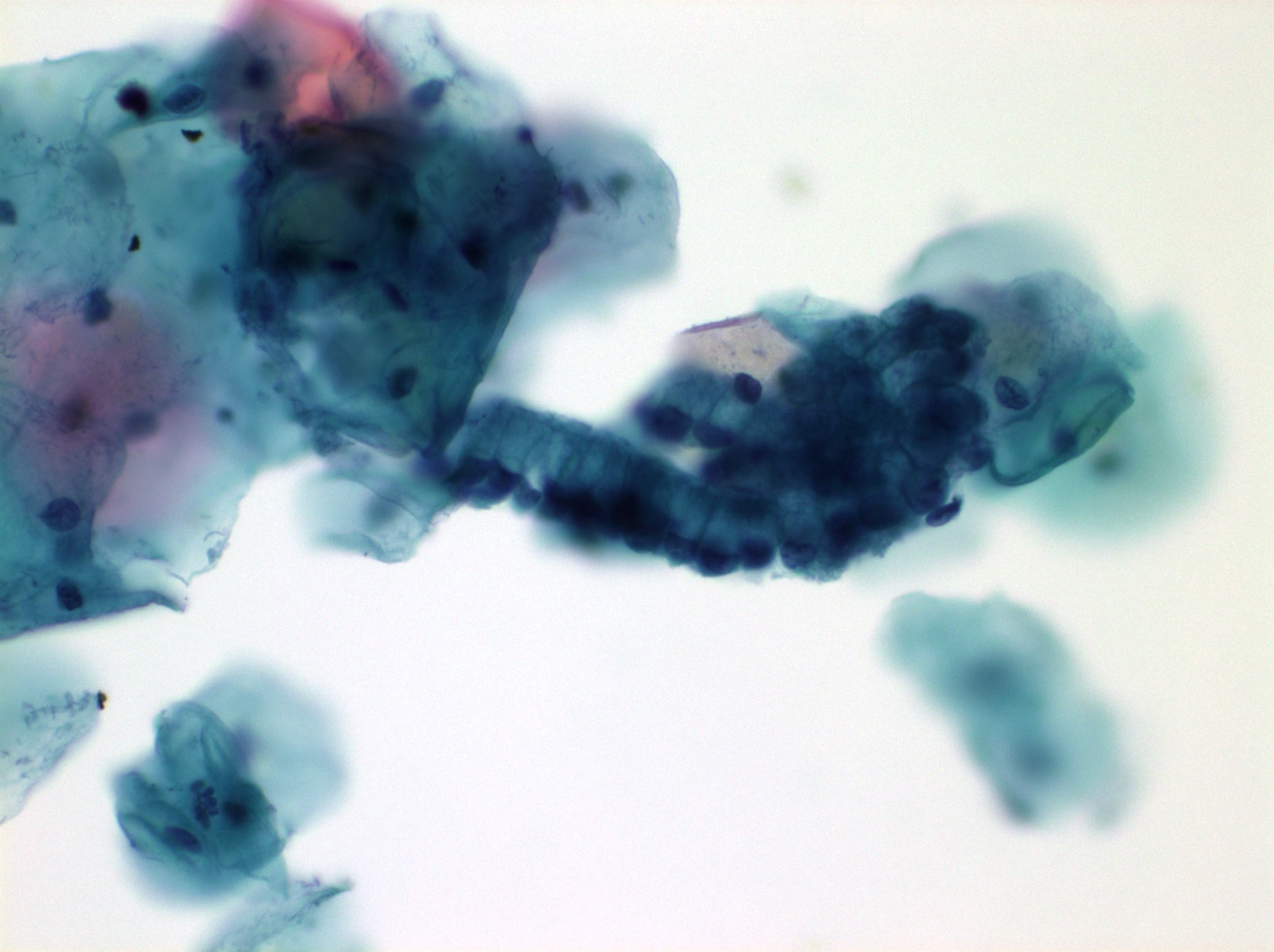
Endocervical glandular cells
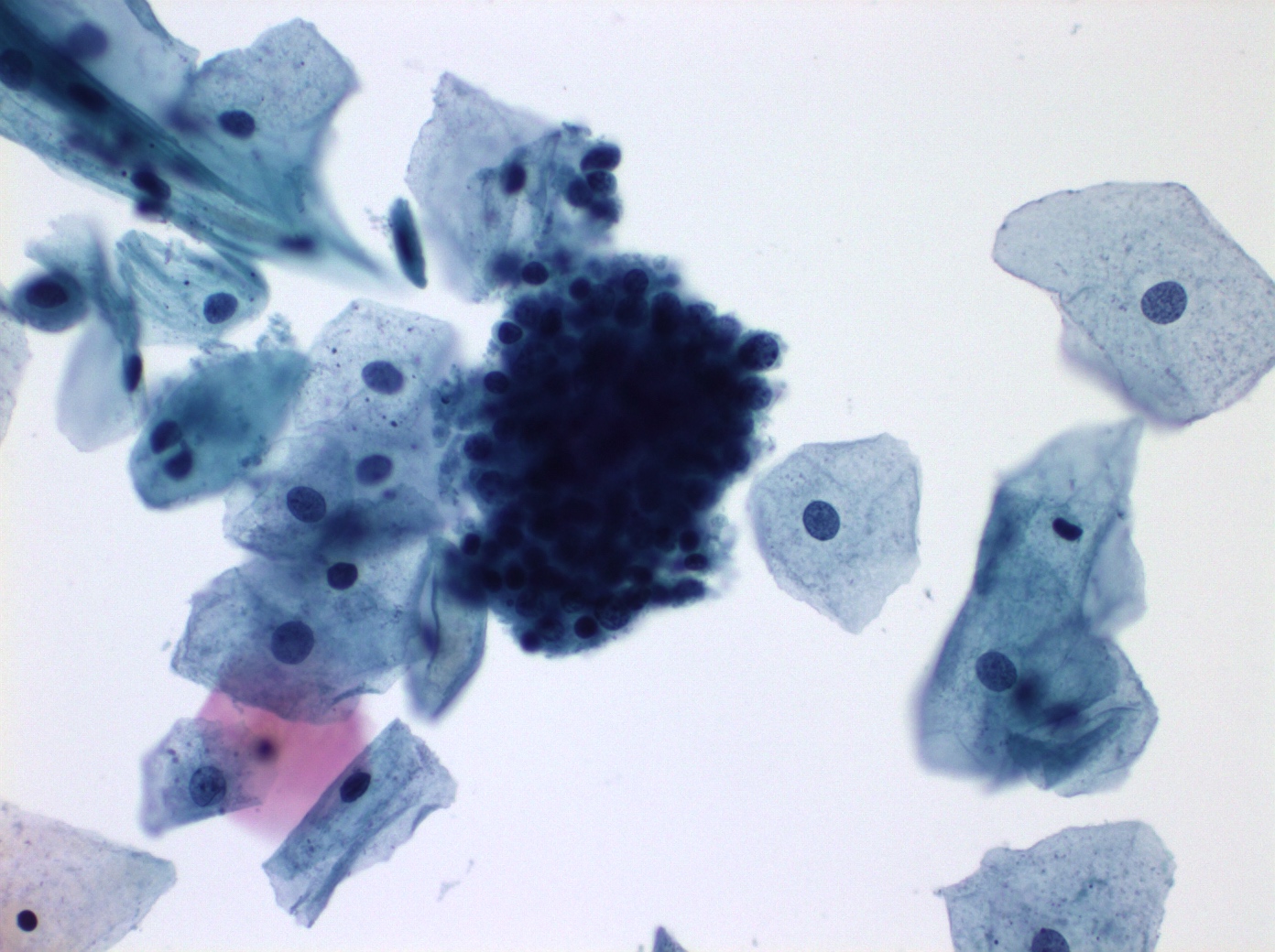
Endometrial cells
Endometrial cells: exodus

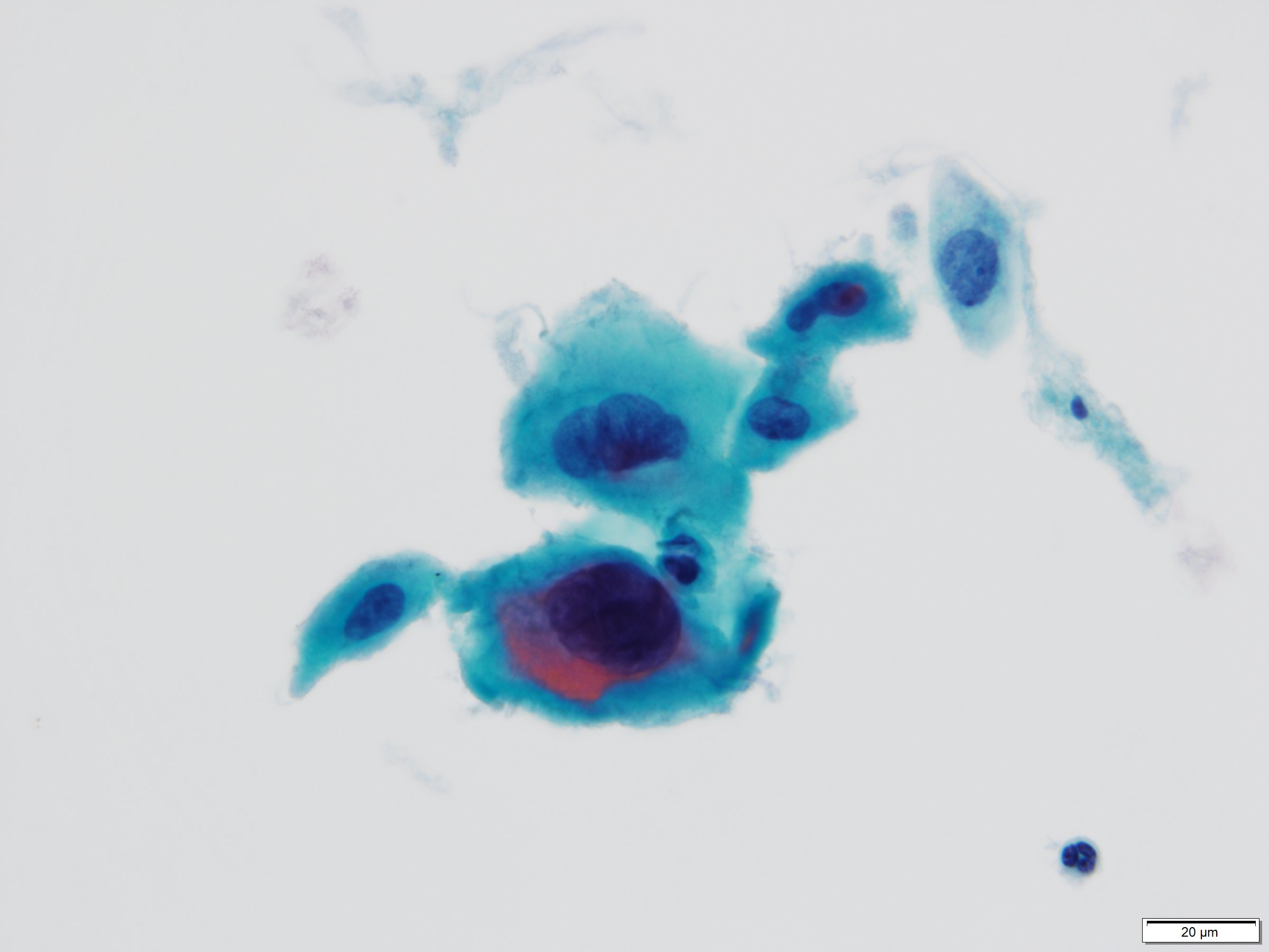
Squamous cells: bland enlargement

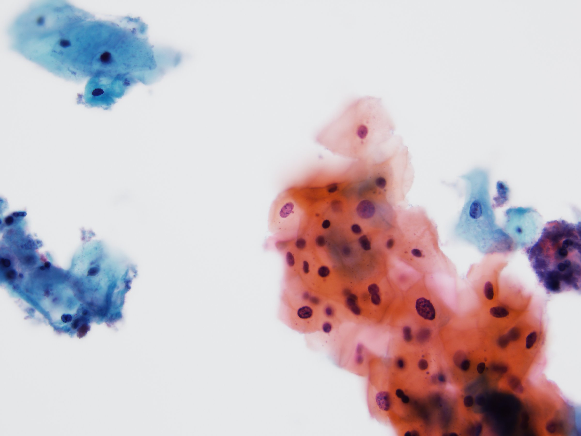
Hyperkeratosis
Parakeratosis
Tubal metaplasia
Squamous metaplasia
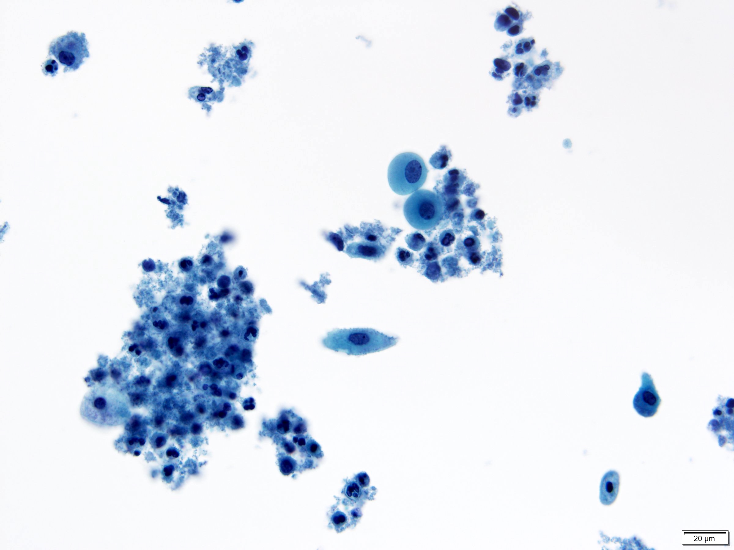
Neutrophils
Abundant blood
Glandular cells status posthysterectomy
Reactive changes
associated with
inflammation
Contributed by David B. Chapel, M.D.
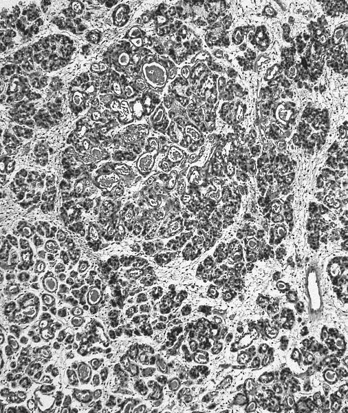
Cervical endometriosis


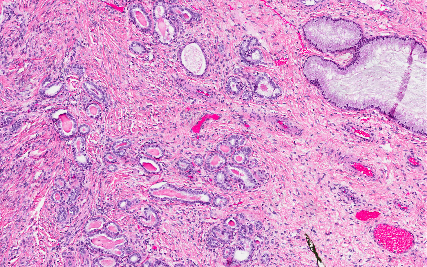
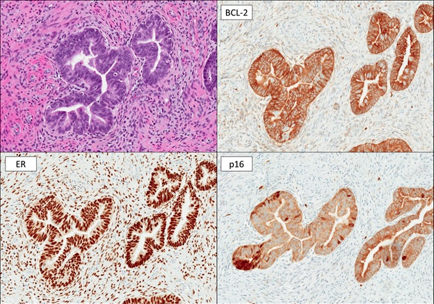
Cervical endometrioid adenocarcinoma, FIGO grade 1
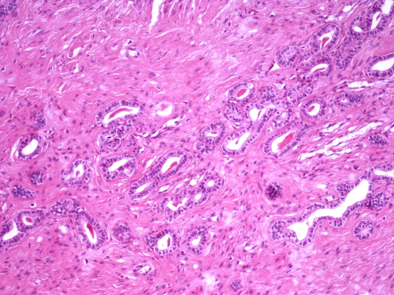
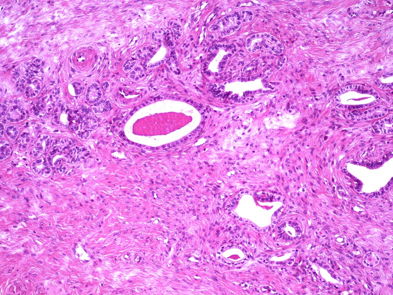
Minimal deviation endometrioid adenocarcinoma
HPV independent glandular lesions
Benign endocervical lesions
Cytology: glandular cervical lesions
AFIP images

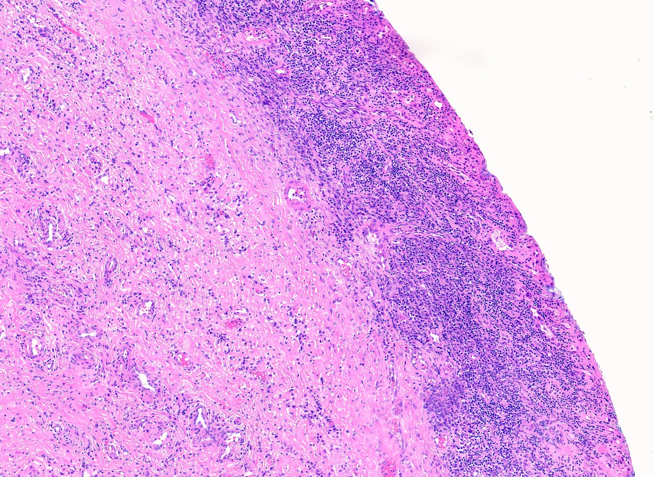
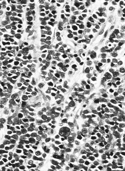
Dense lymphoid
infiltrate with
germinal centers
Contributed by Carlos Parra-Herran, M.D.

Radiation atypia - squamous epithelium
Radiation atypia - vascular endothelium
Contributed by Erika Baardsen, D.O.

Pap smear
Images hosted on other servers:
Pap smear
Images hosted on other servers:


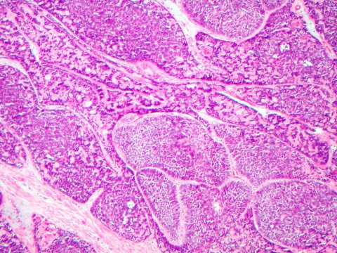
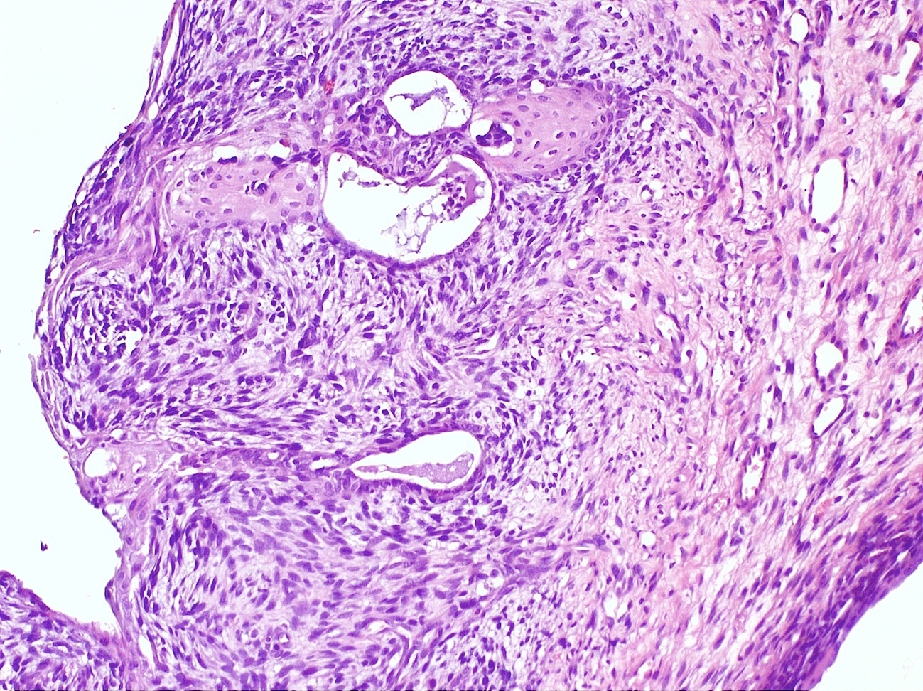
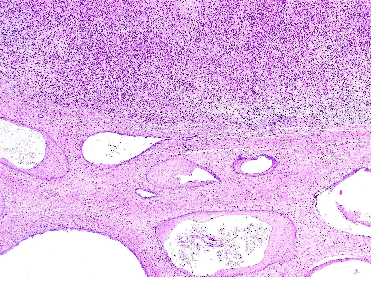
Vaginal embryonal rhabdomyosarcoma
Images hosted on other servers:

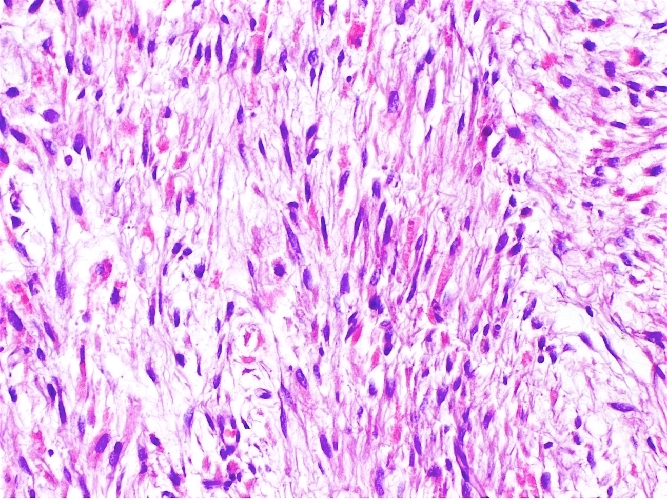
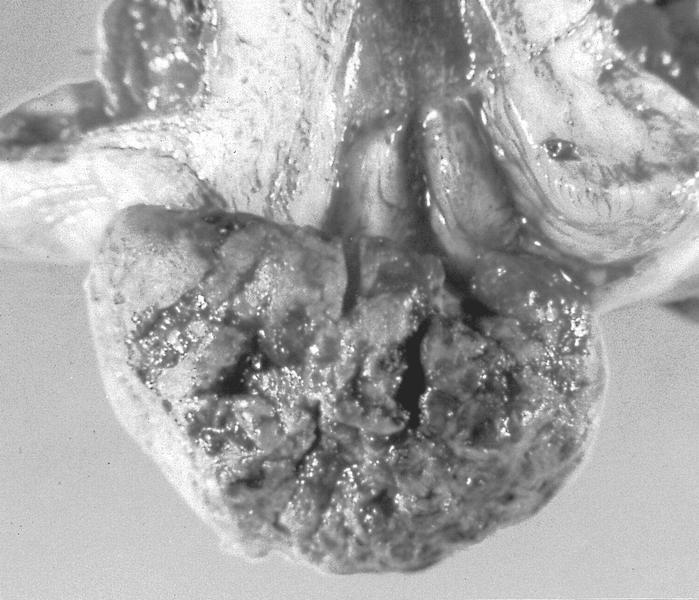
Cervical embryonal
rhabdomyosarcoma
Images hosted on other servers:
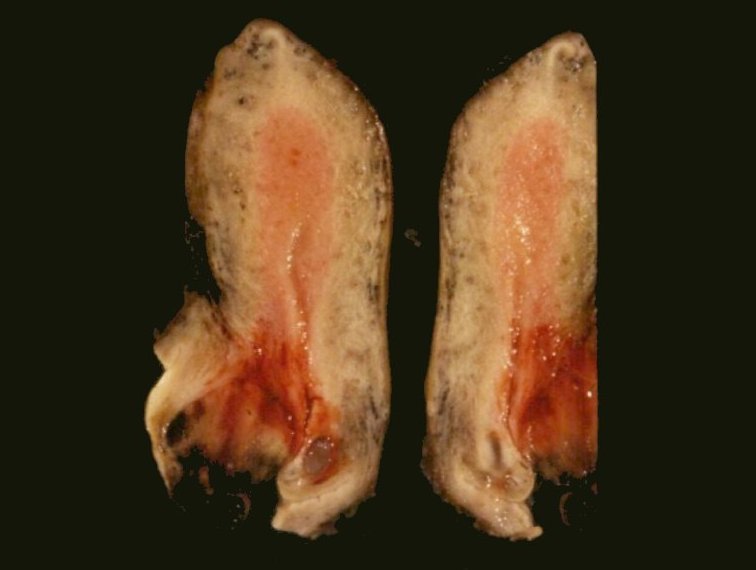
Gray surface and areas of hemorrhage
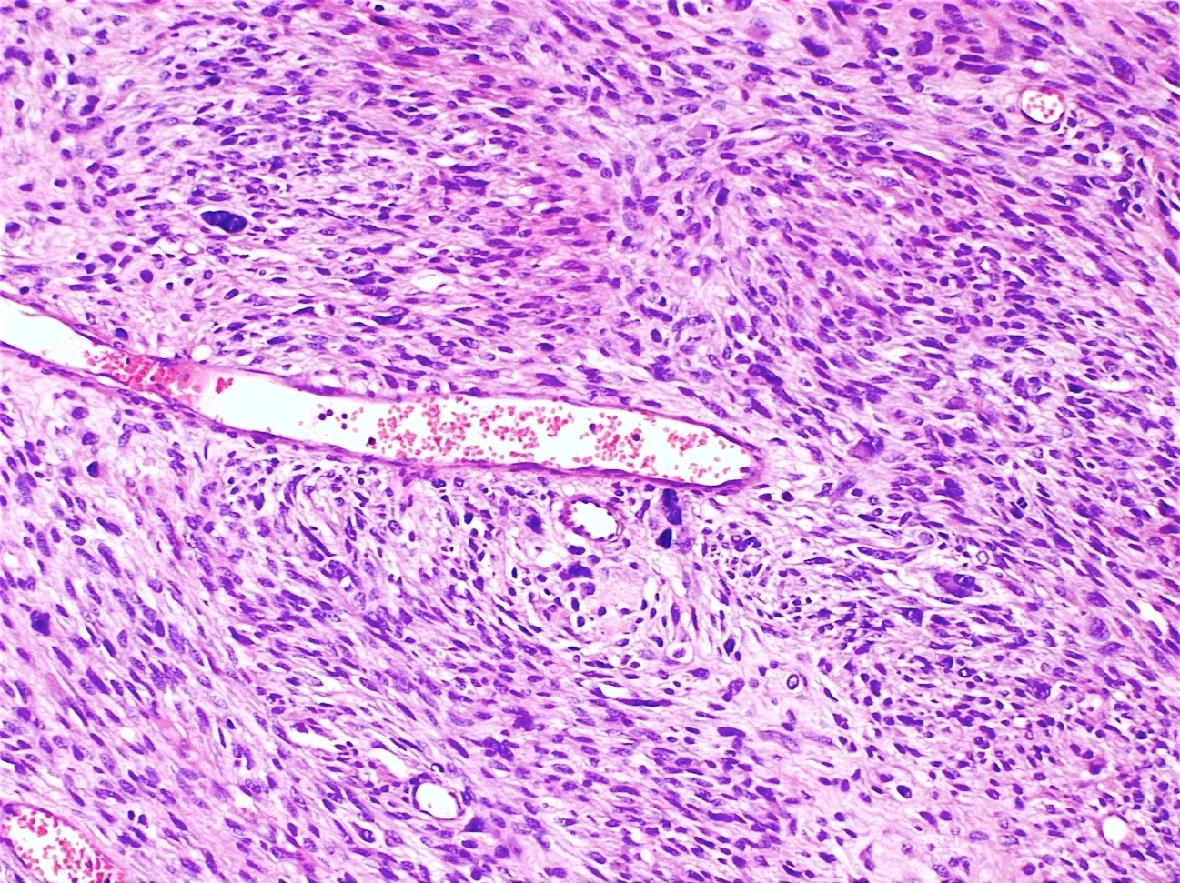
Contributed by Kyle Devins, M.D.
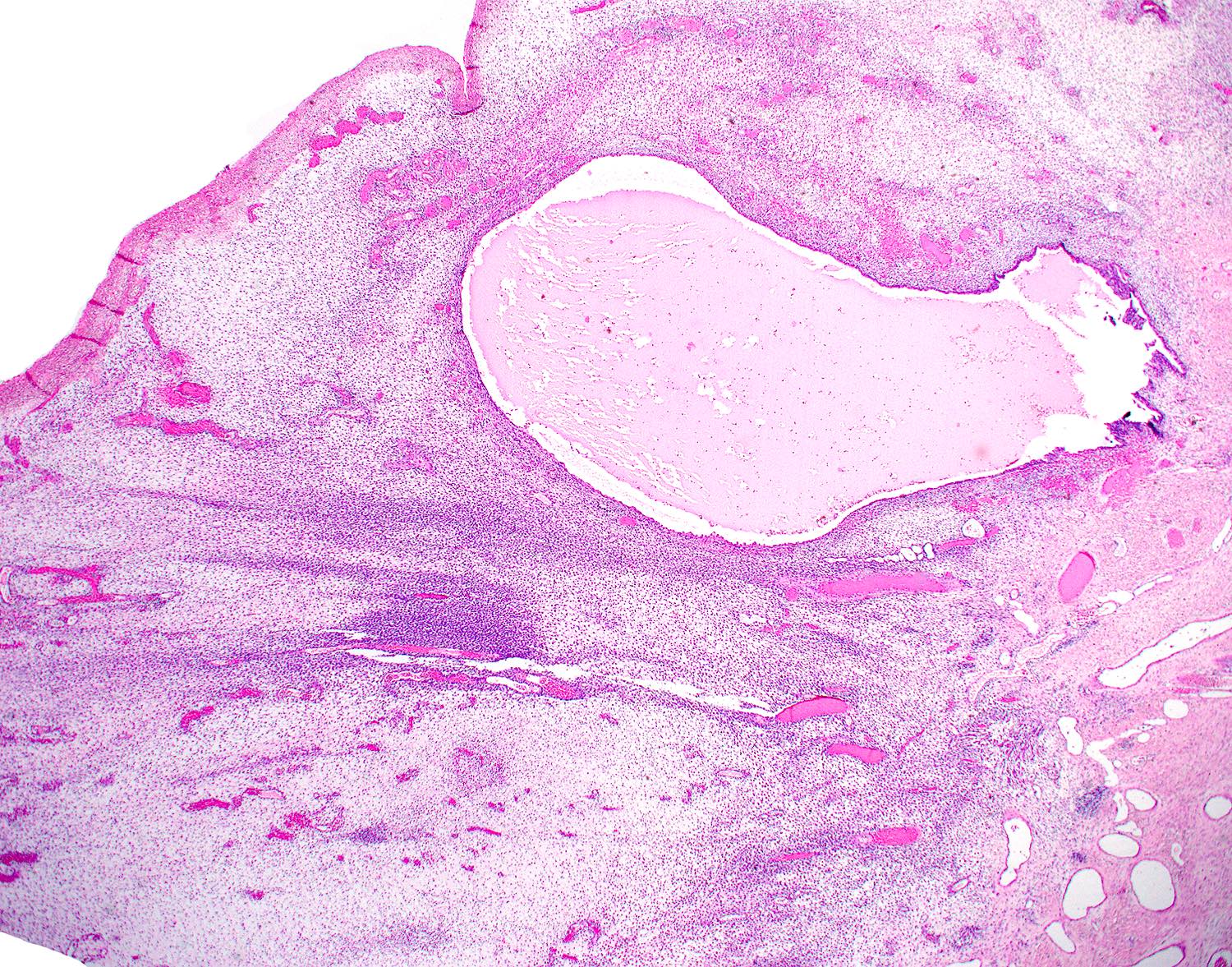
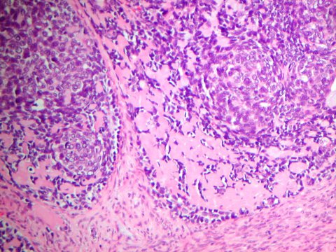
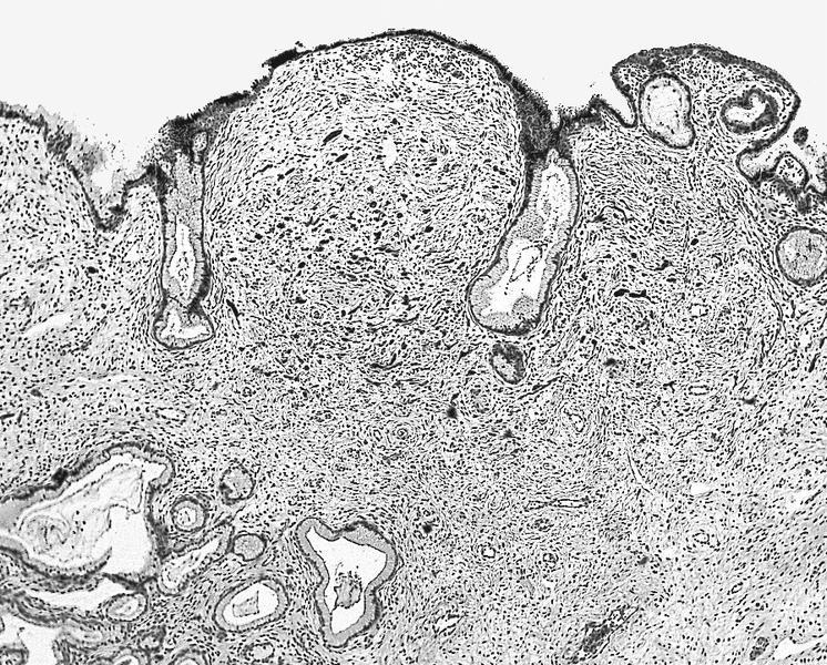
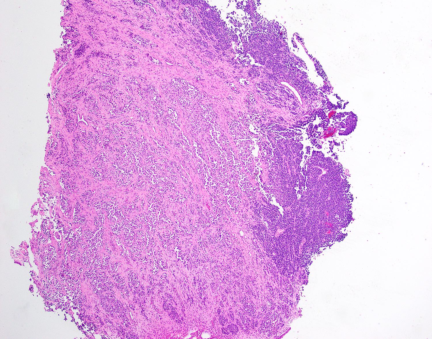
Edematous stroma
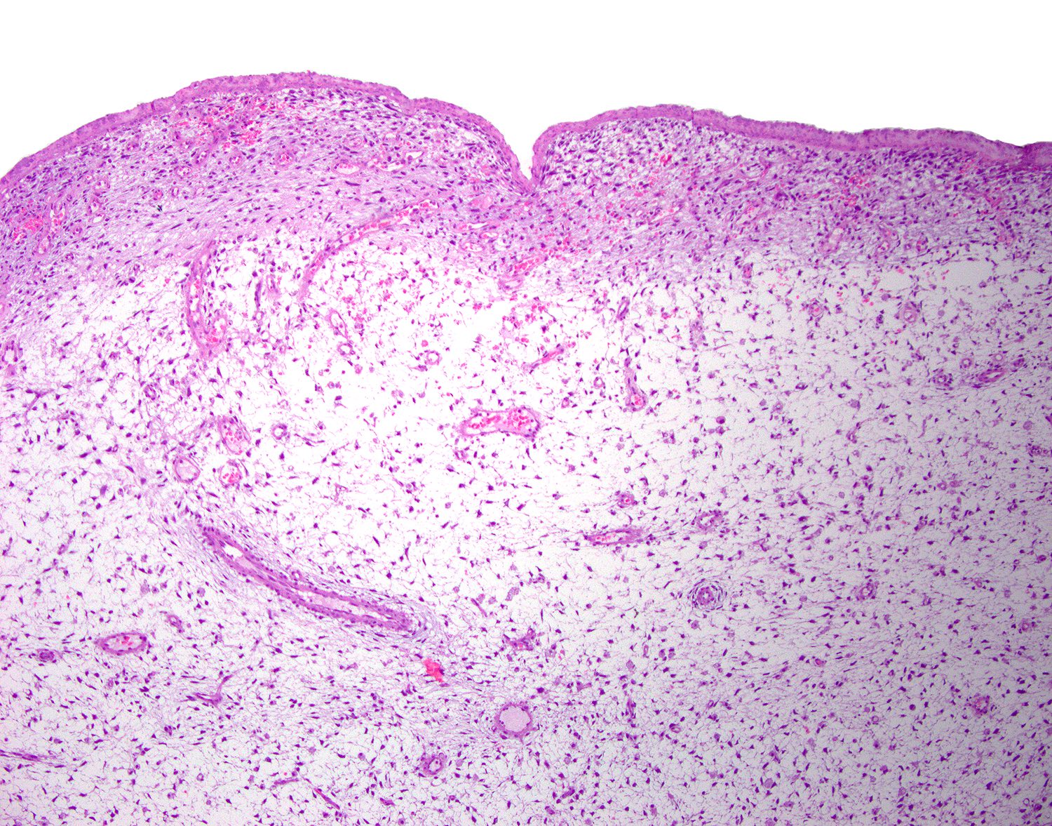
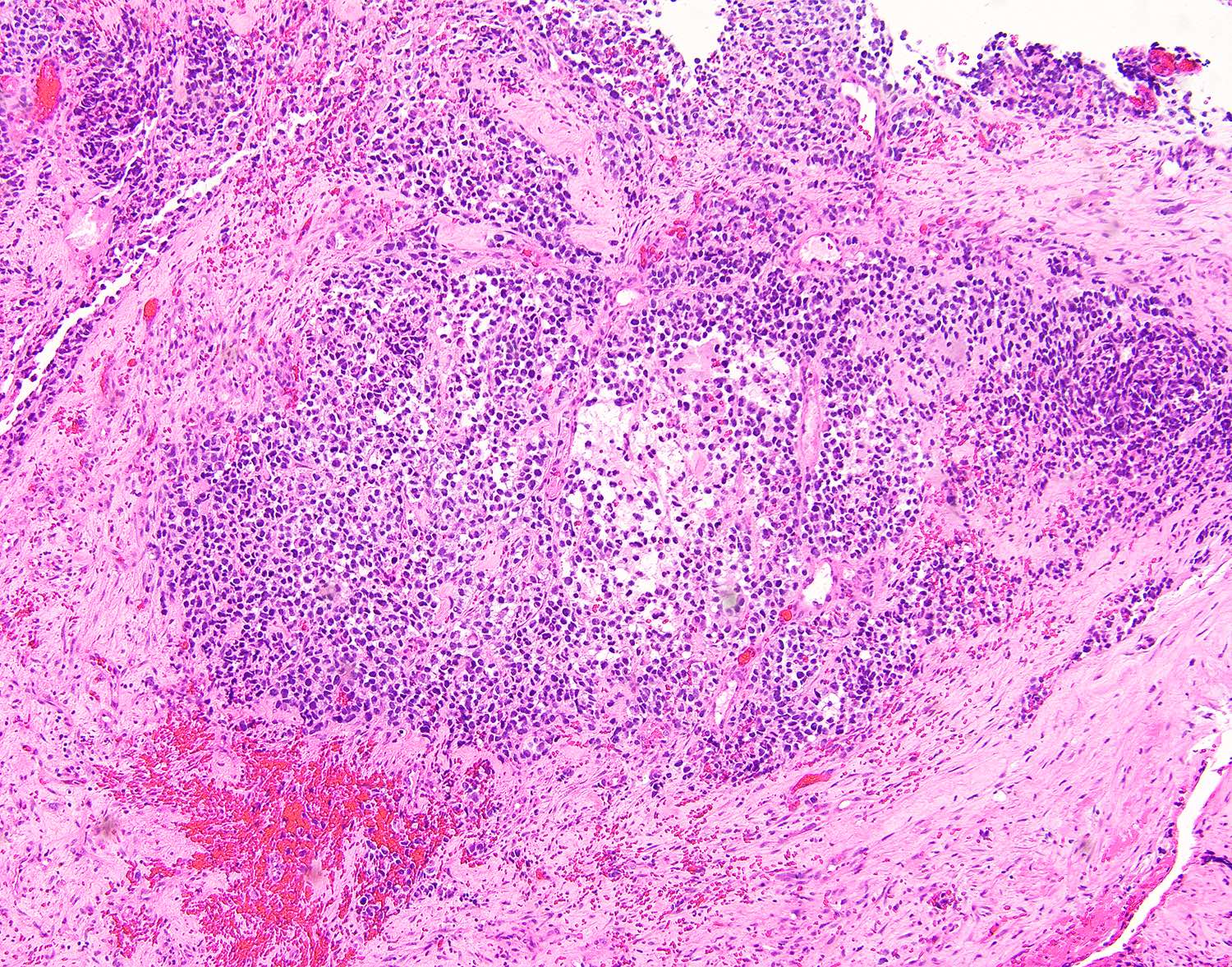
Cambium layer
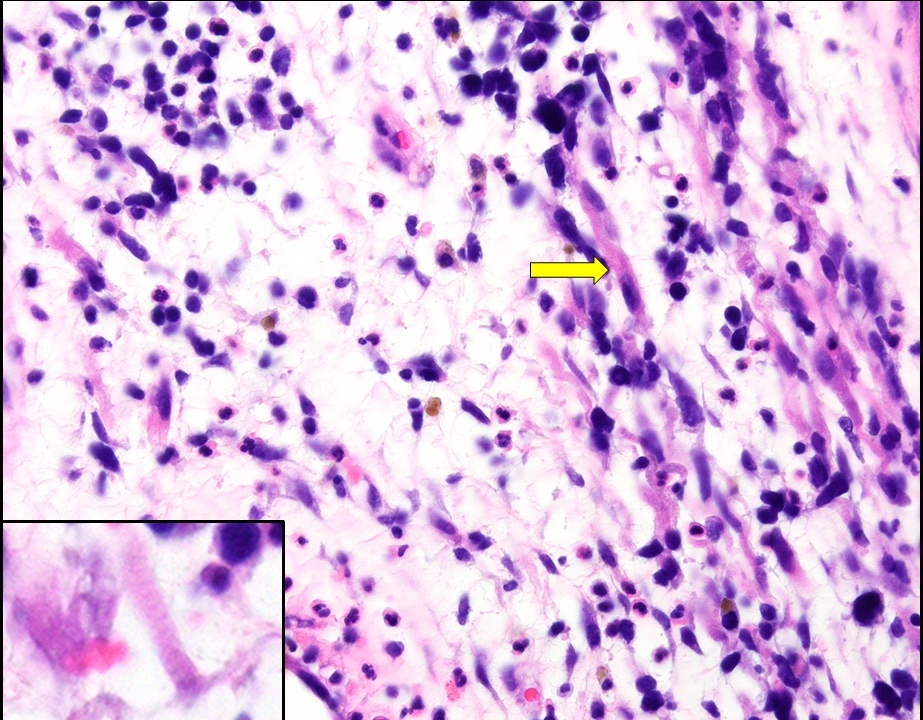
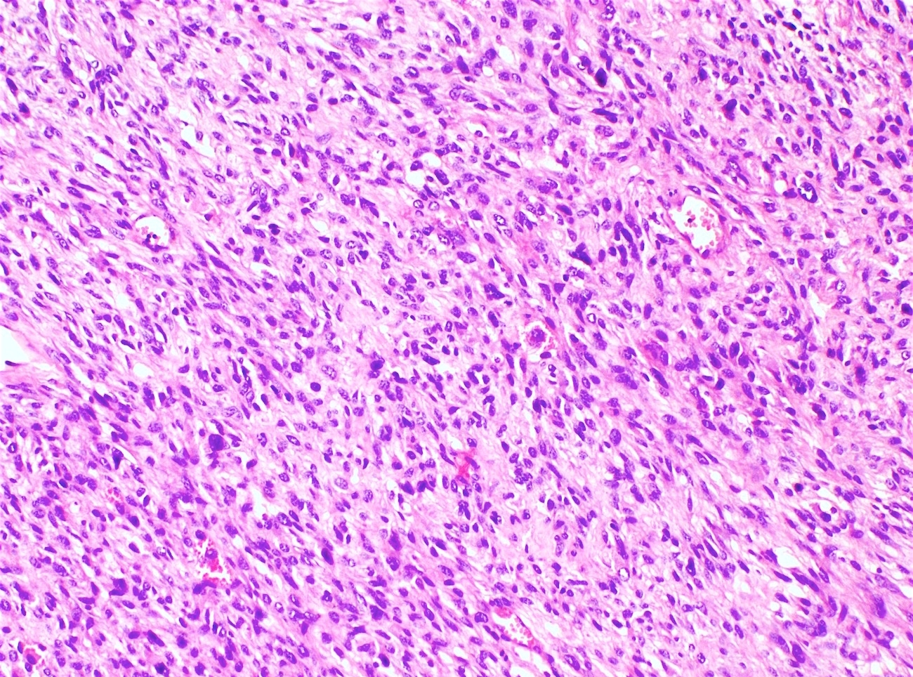
Rhabdomyoblasts
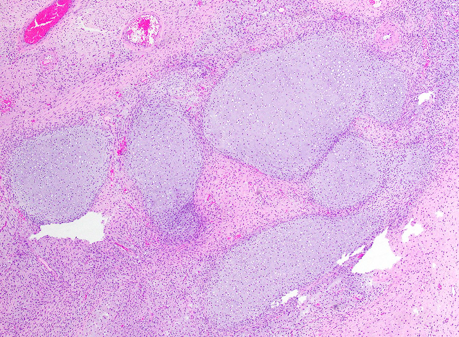
Hyaline cartilage
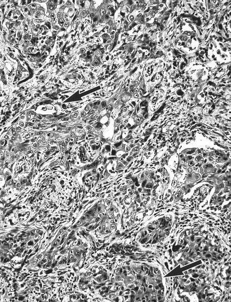
Nests, sheets and alveolar spaces
Alveolar spaces
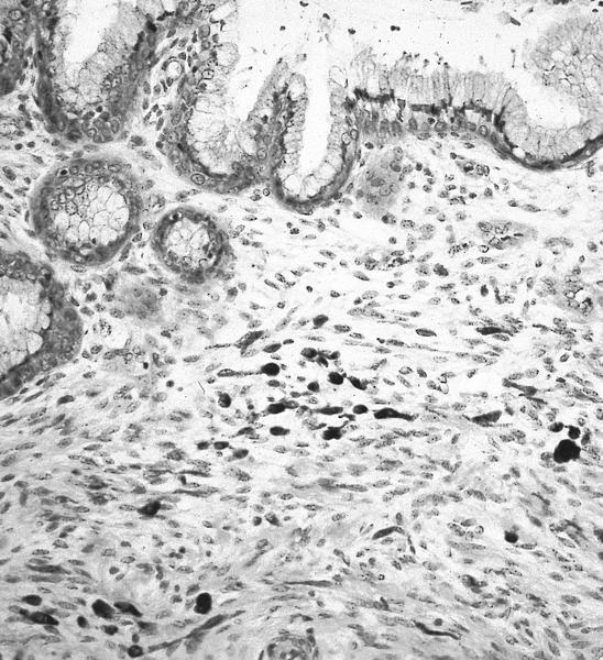
Desmin
Myogenin
Nuclear pleomorphism
Images hosted on other servers:

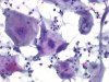
Brushing of
uterine embryonal
rhabdomyosarcoma
AFIP and Case #327
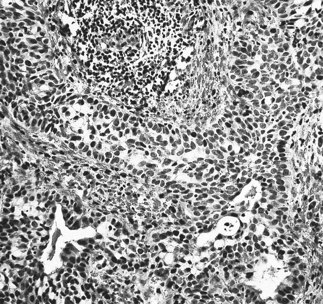
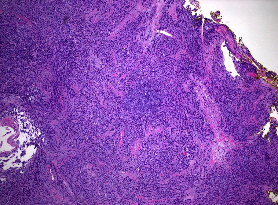
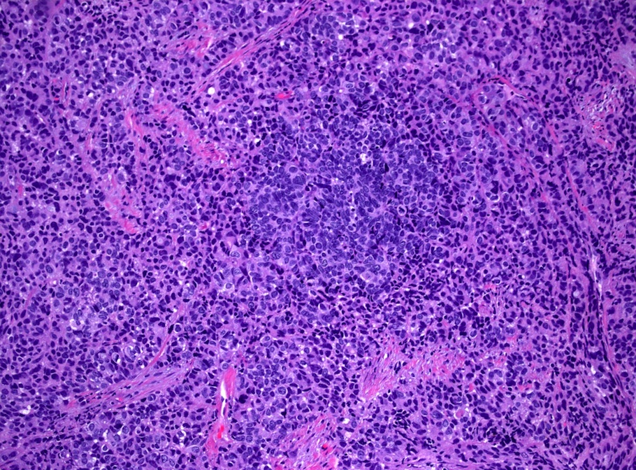
Sheets of small cells with scant cytoplasm and hyperchromatic nuclei
H&E
CK7
p16
Chromogranin
Synaptophysin
Images hosted on other servers:

Lesional cells, Pap

Single necrotic cells, Pap

Amphophilic cytoplasm

Large aggregates of malignant cells

Malignant cells loosely cohesive

Scant cytoplasm

PanCK

Synaptophysin

p16
Images hosted on other servers:

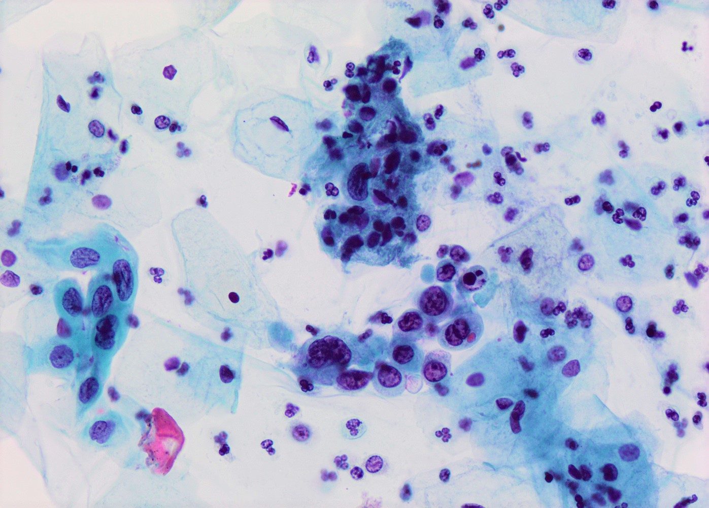
Resembles HSIL but with abundant mucin


Adenocarcinoma in situ stratified type
Contributed by Masatsugu Ueda, M.D.
Transformation zone
Contributed by Mikami Yoshiki, M.D., Ph.D.
Transformation zone
Contributed by Mikami Yoshiki, M.D., Ph.D.
Transformation zone
Primary squamocolumnar junction
Secondary squamocolumnar junction
Squamous metaplasia
overlying
preexisting glands
Reserve cell hyperplasia
Immature squamous metaplasia
Squamous metaplasia involving endocervical glands
Contributed by Mikami Yoshiki, M.D., Ph.D.
Squamous metaplasia
Images hosted on other servers:

Trophozoite
Images hosted on other servers:

Strawberry cervix
Contributed by Soumya Jaladi, M.B.B.S., Marilin Rosa, M.D. and @zaalruwai83 on Twitter
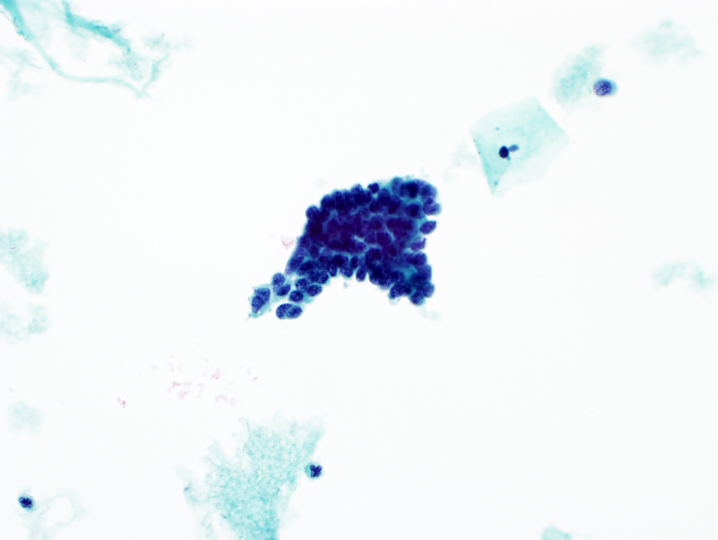
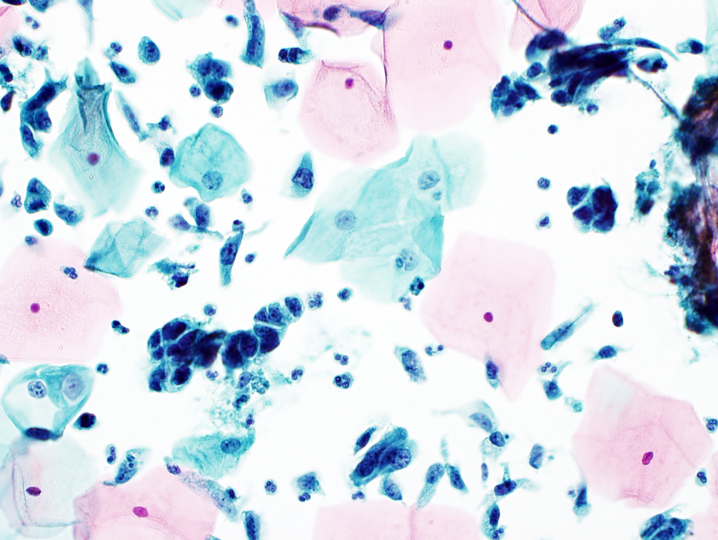
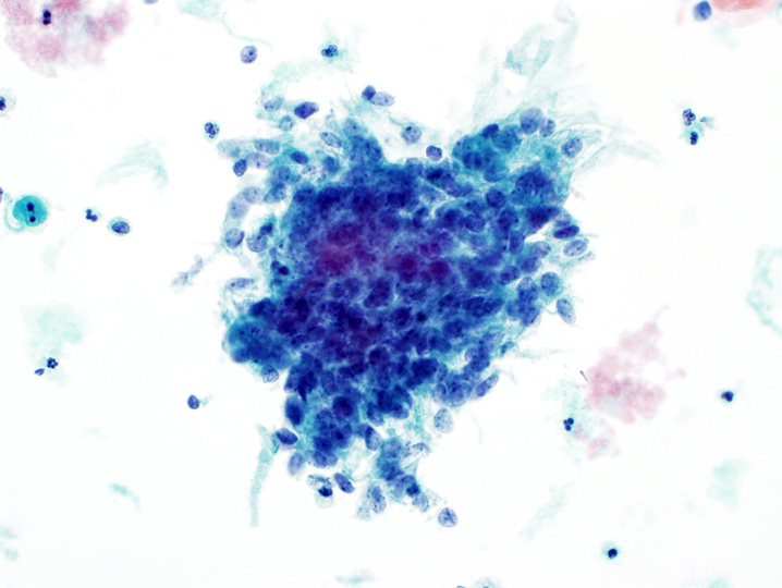
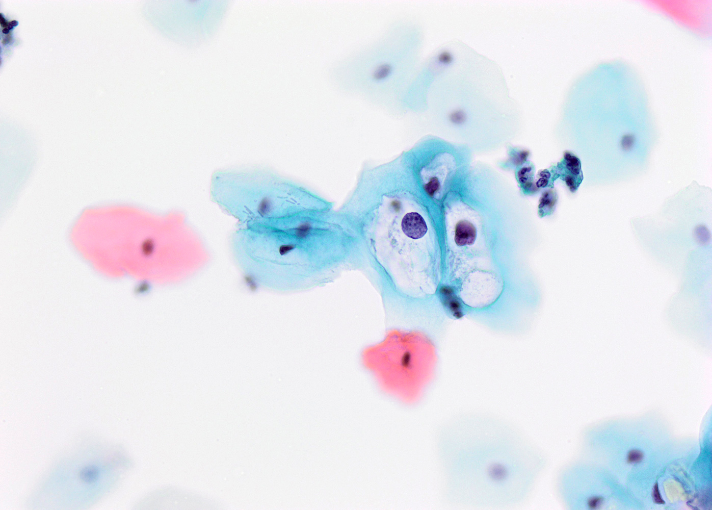
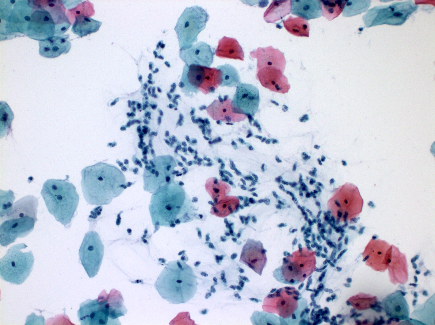
Trichomonas in a cytologic preparation
Trichomonas

Trichomonas vaginalis
Trichomonas vaginalis
Images hosted on other servers:

Filamentous Leptothrix species

Trichomonas in wet mount
Trichomonas in conventional
Pap smear
Trichomonas vaginalis with Leptothrix

Trichomonas

Trophozite


Inflammatory ectocervix with Trichomonas infection
Images hosted on other servers:

T. vaginalis parasite
Contributed by Carlos Parra-Herran, M.D.
TEM and relevant stains
Contributed by Gulisa Turashvili, M.D., Ph.D.
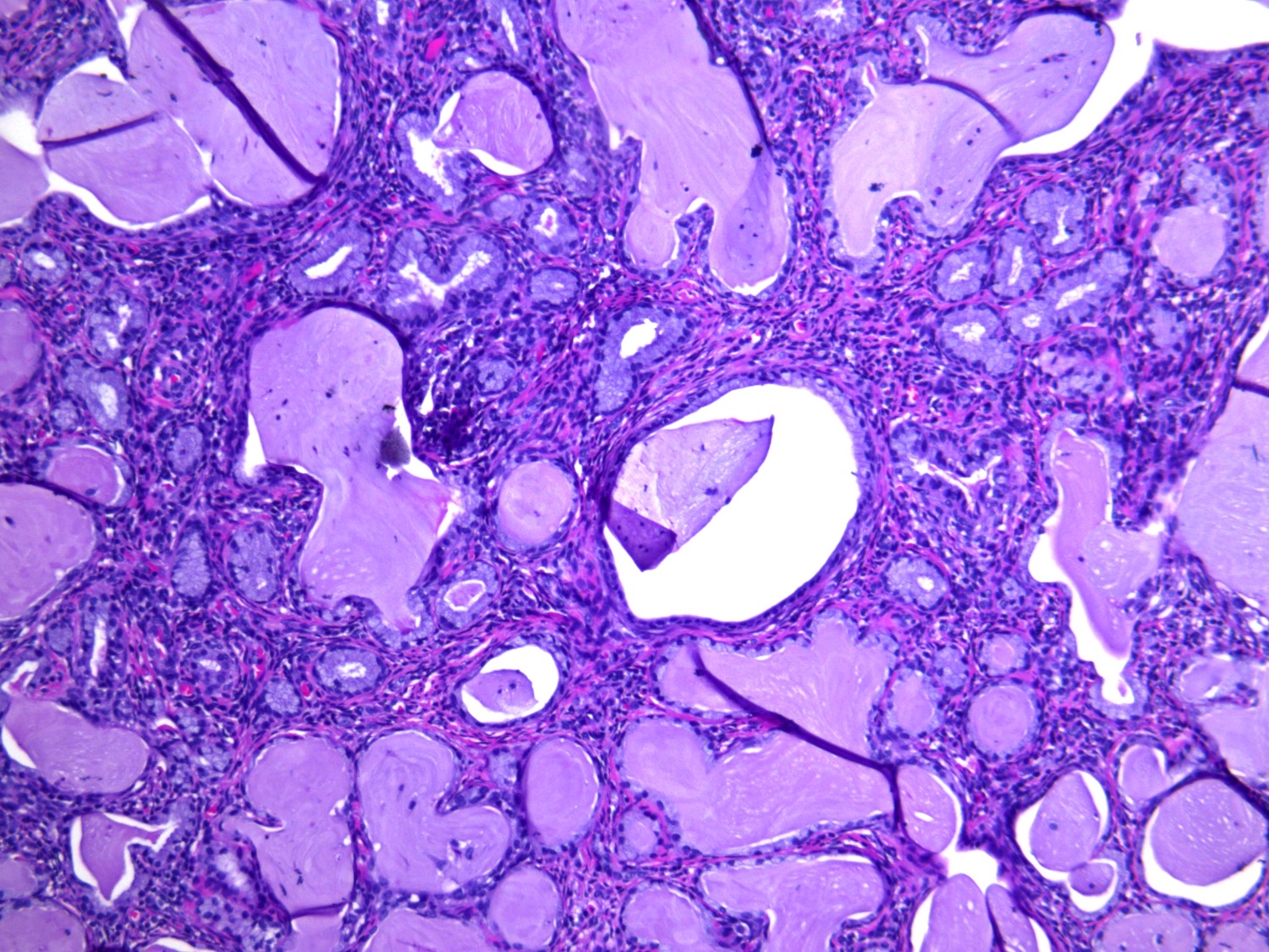
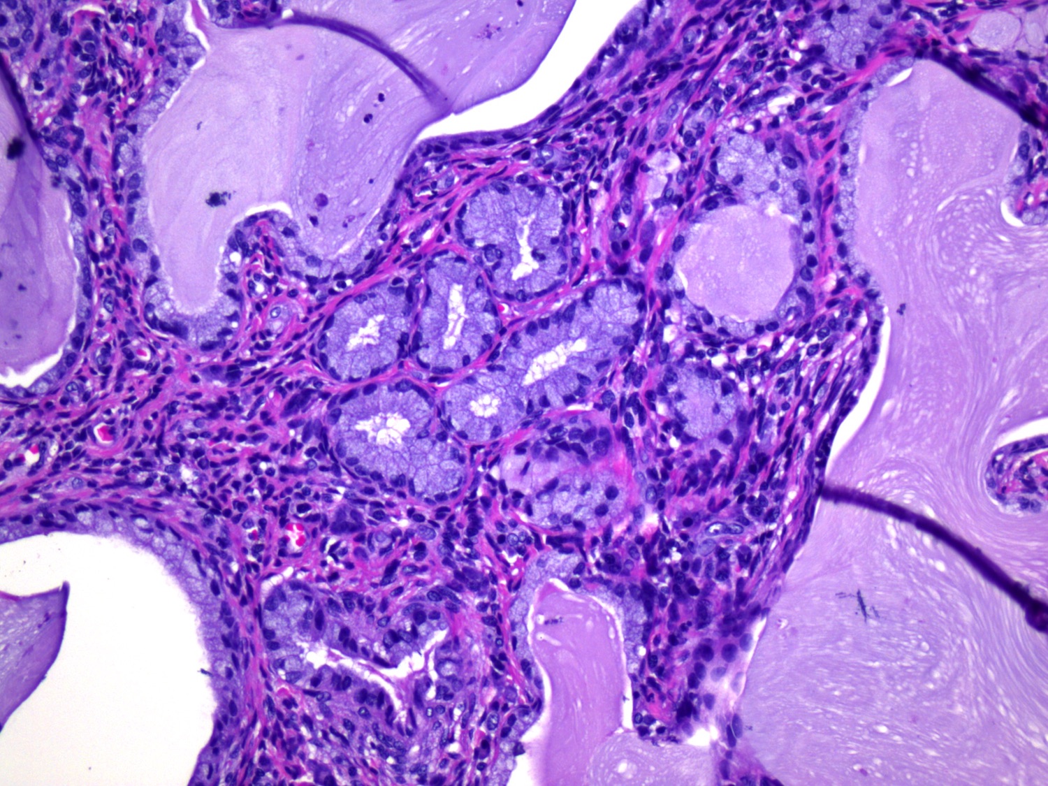
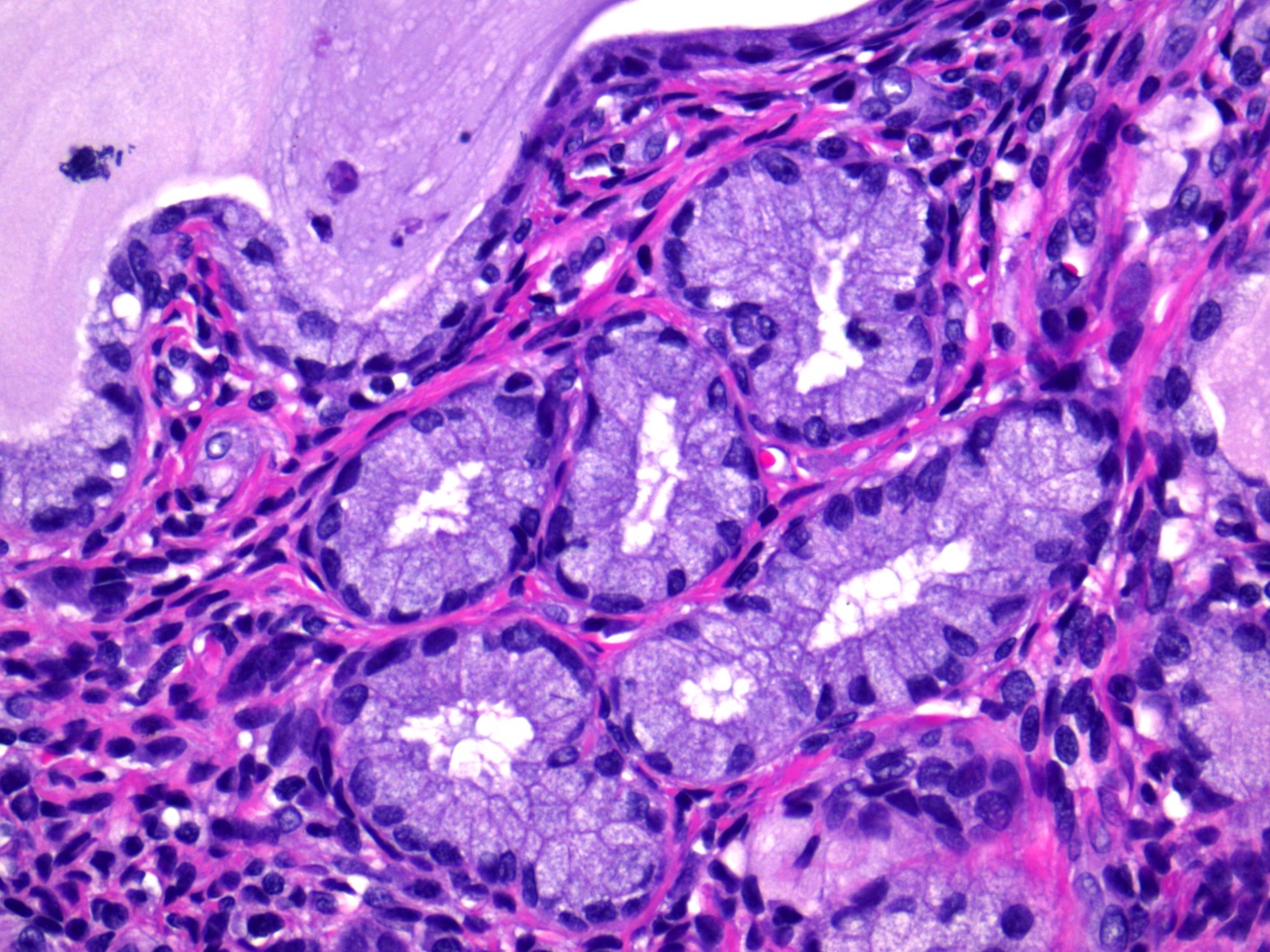
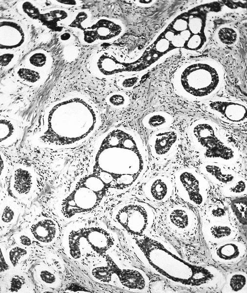
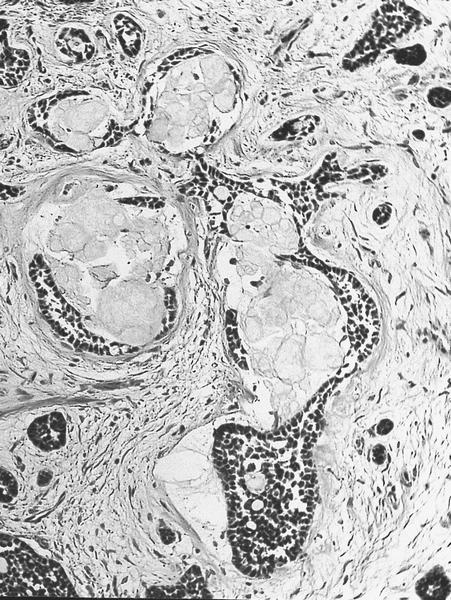
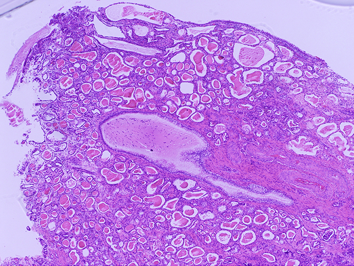
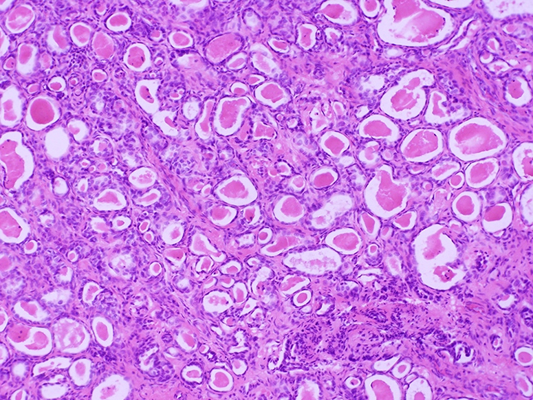
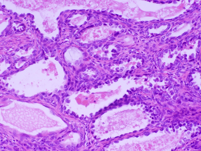
Type A tunnel clusters
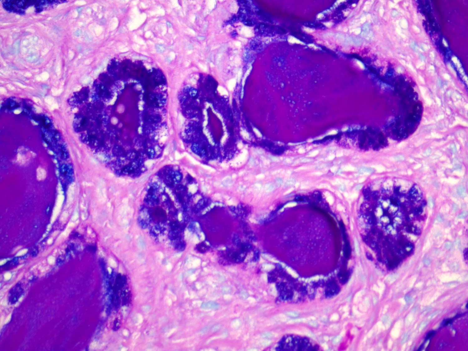
Alcian blue / PAS in type A tunnel clusters

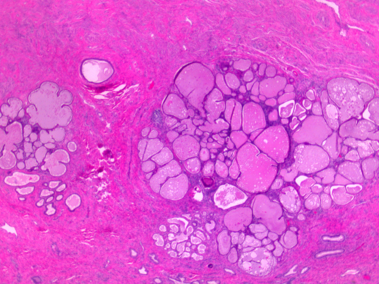

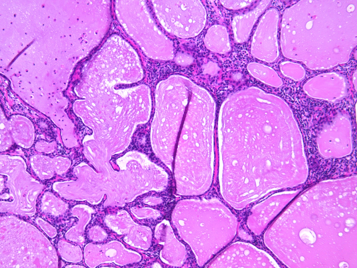
Type B tunnel clusters
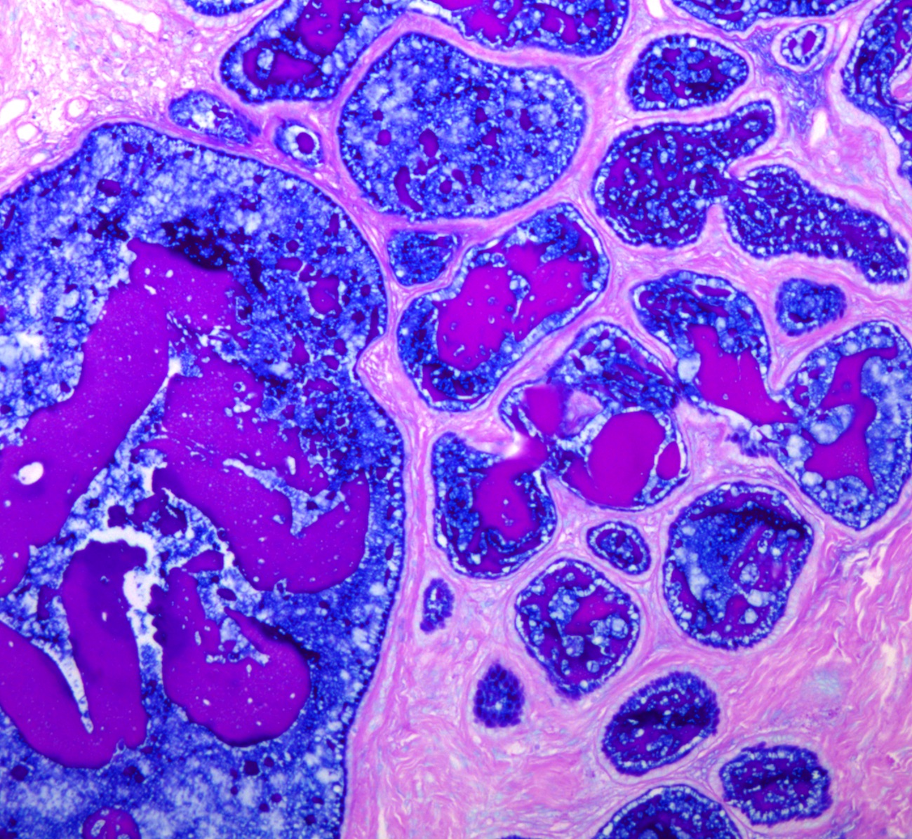
Alcian blue / PAS in type B tunnel clusters
Case #91
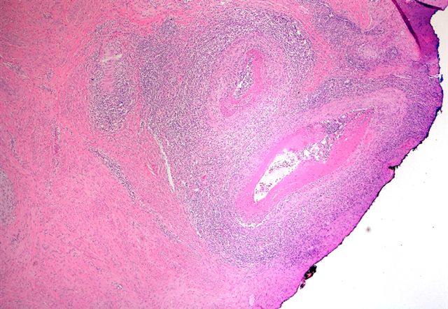
Inflammatory infiltrate in the vessel wall
Lymphocytes, neutrophils and occasional eosinophils
Fibrinoid necrosis within the media

Ali: 2019

Clement: 2019
Crum: 2017

Howitt: 2019
Hui: 2015
IARC: 2020
Malpica: 2015
Mody: 2022
Nayar: 2015

Nucci: 2020
Nucci: 2023
Nucci: 2023

Soslow: 2020
Vang: 2017

Zheng: 2019

Zheng: 2019
Find related Pathology books: cytopathology, gynecologic, frozen section
