


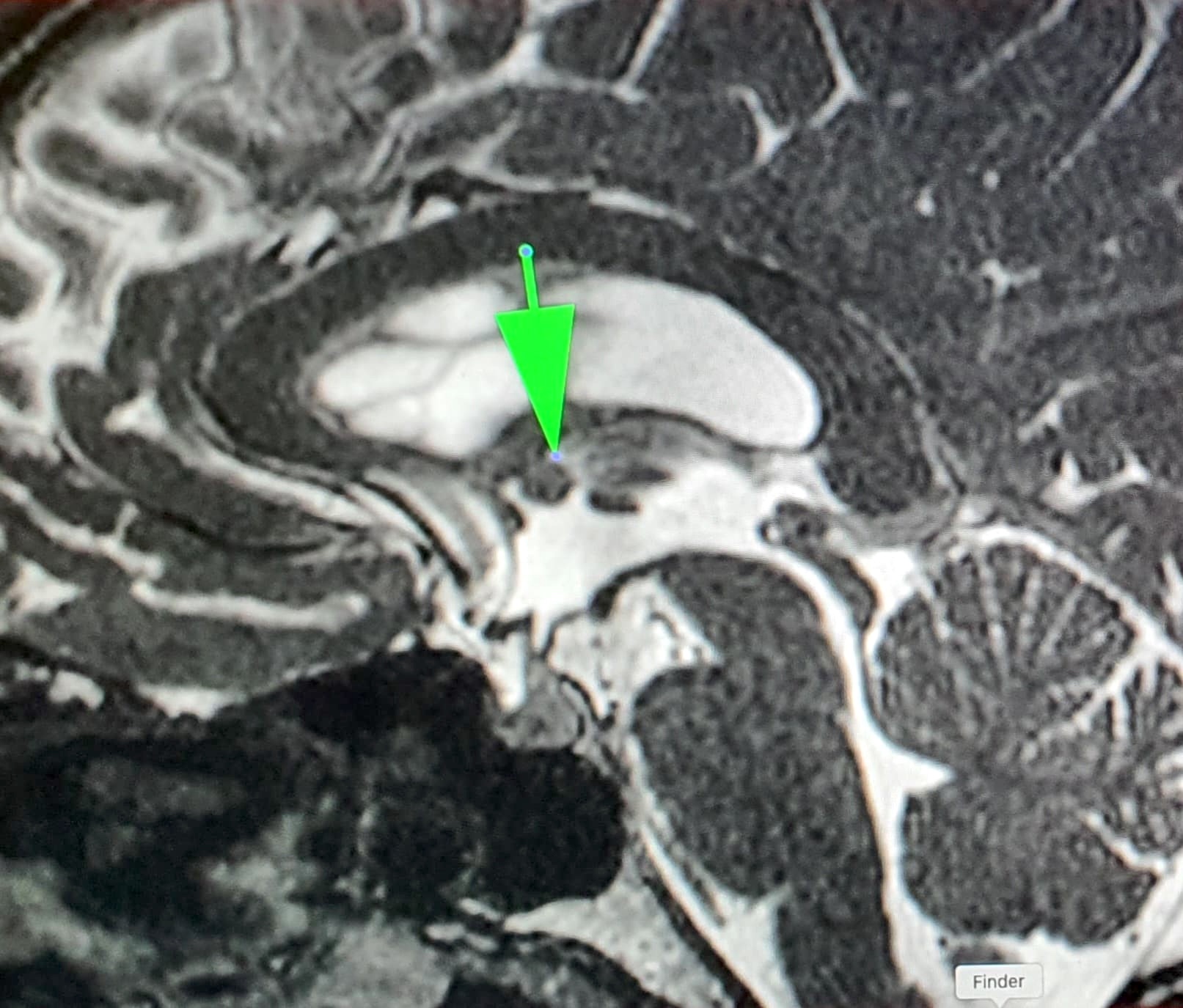
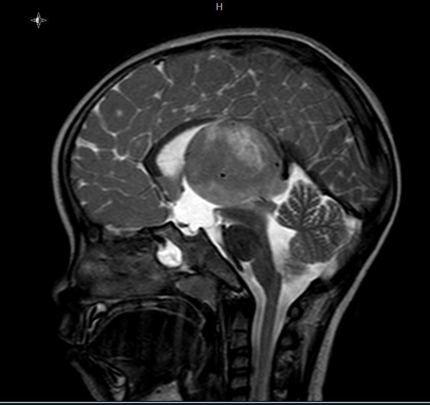
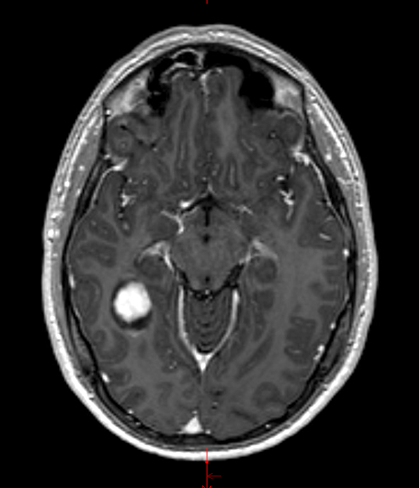
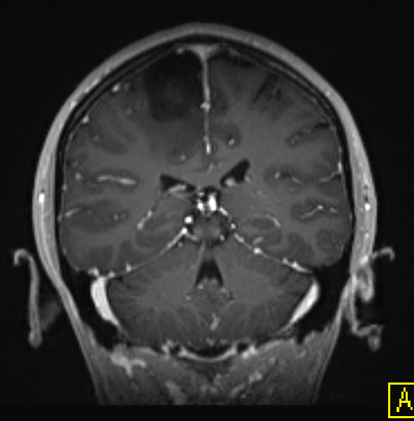
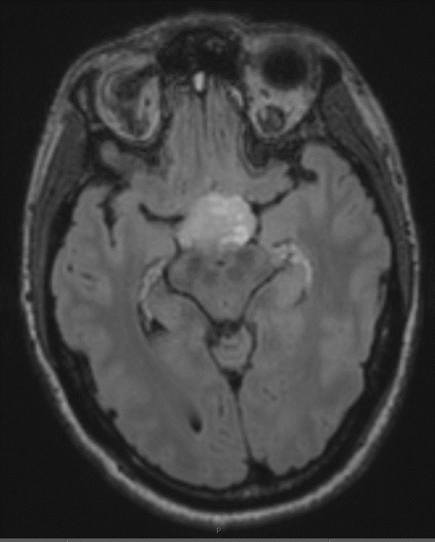
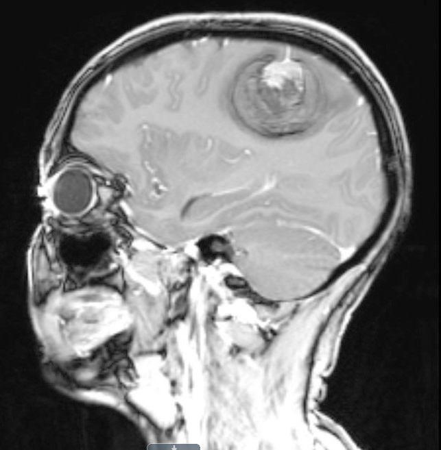
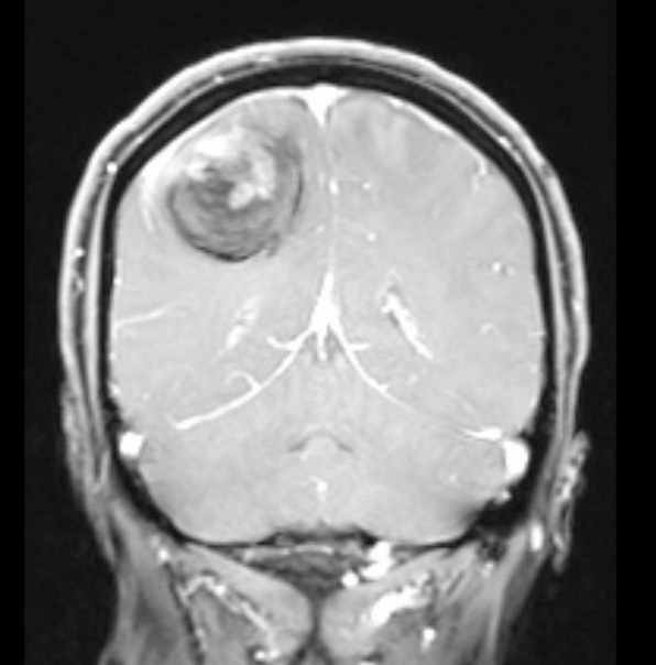
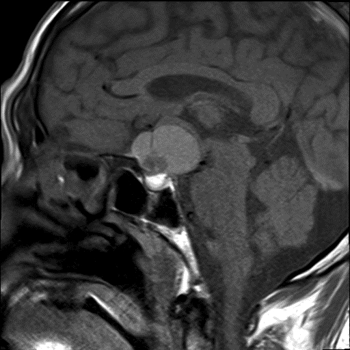
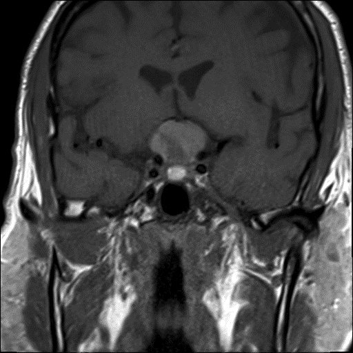
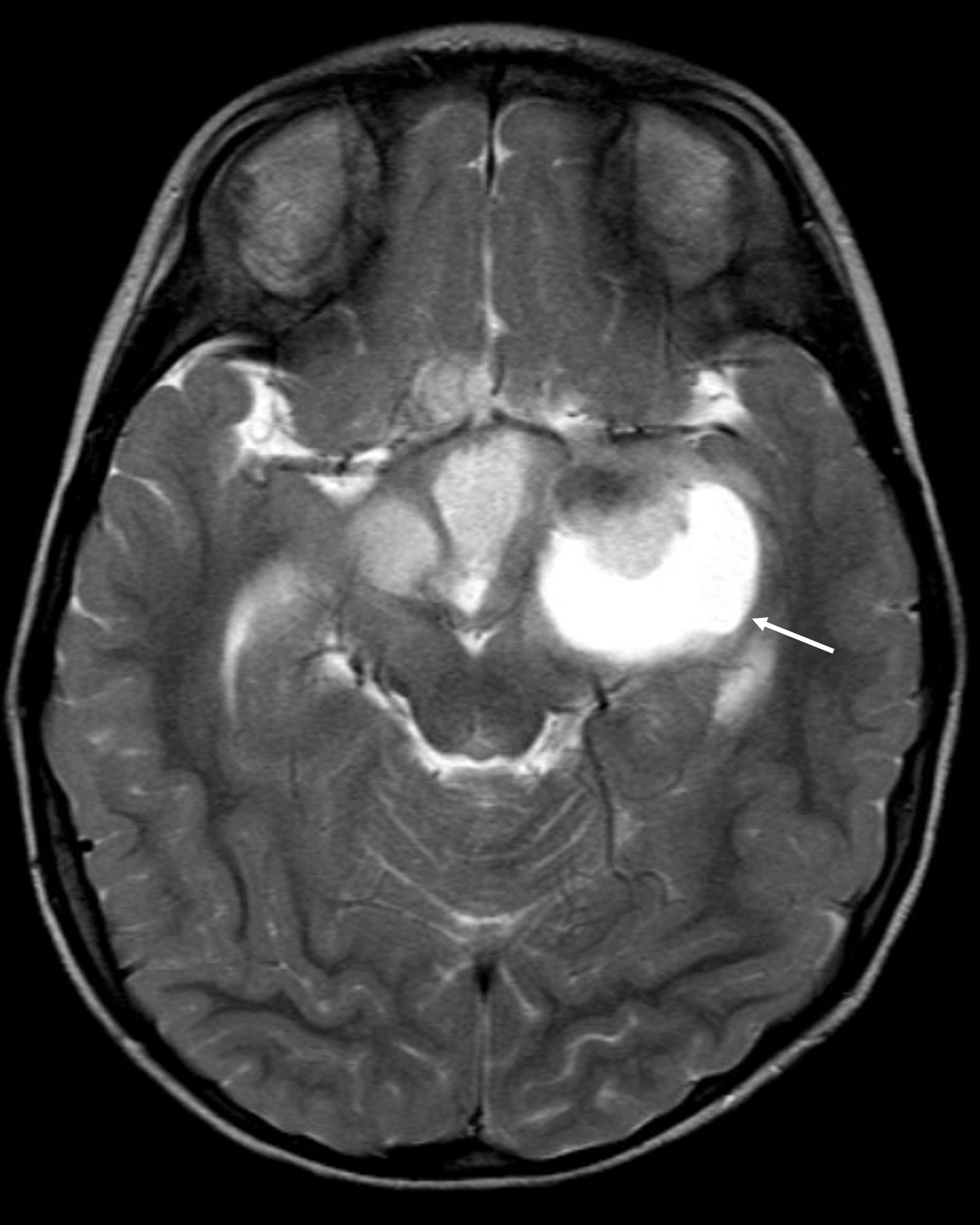
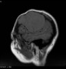
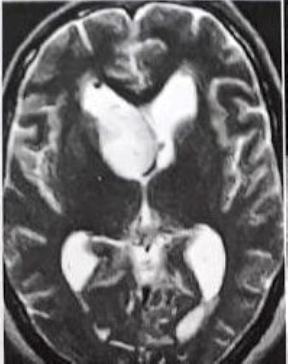
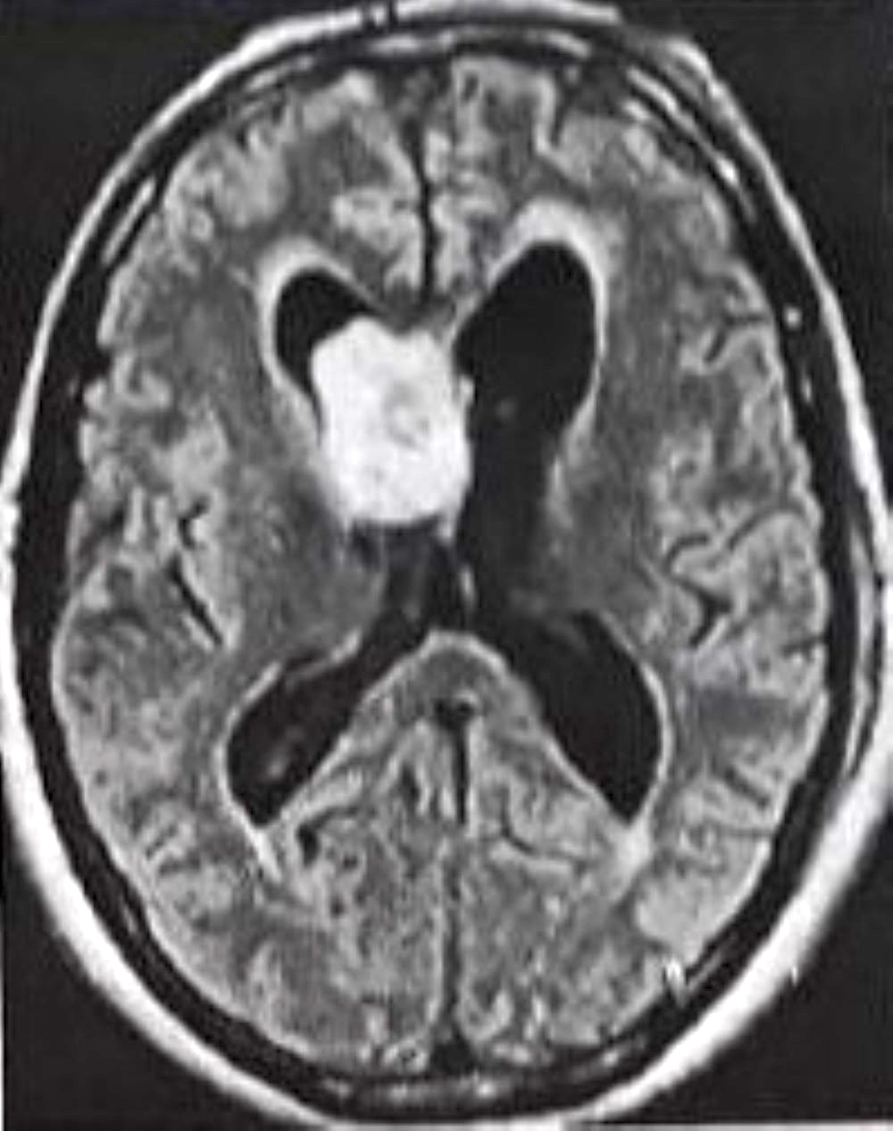
Images hosted on other servers:

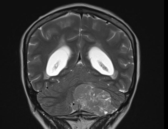
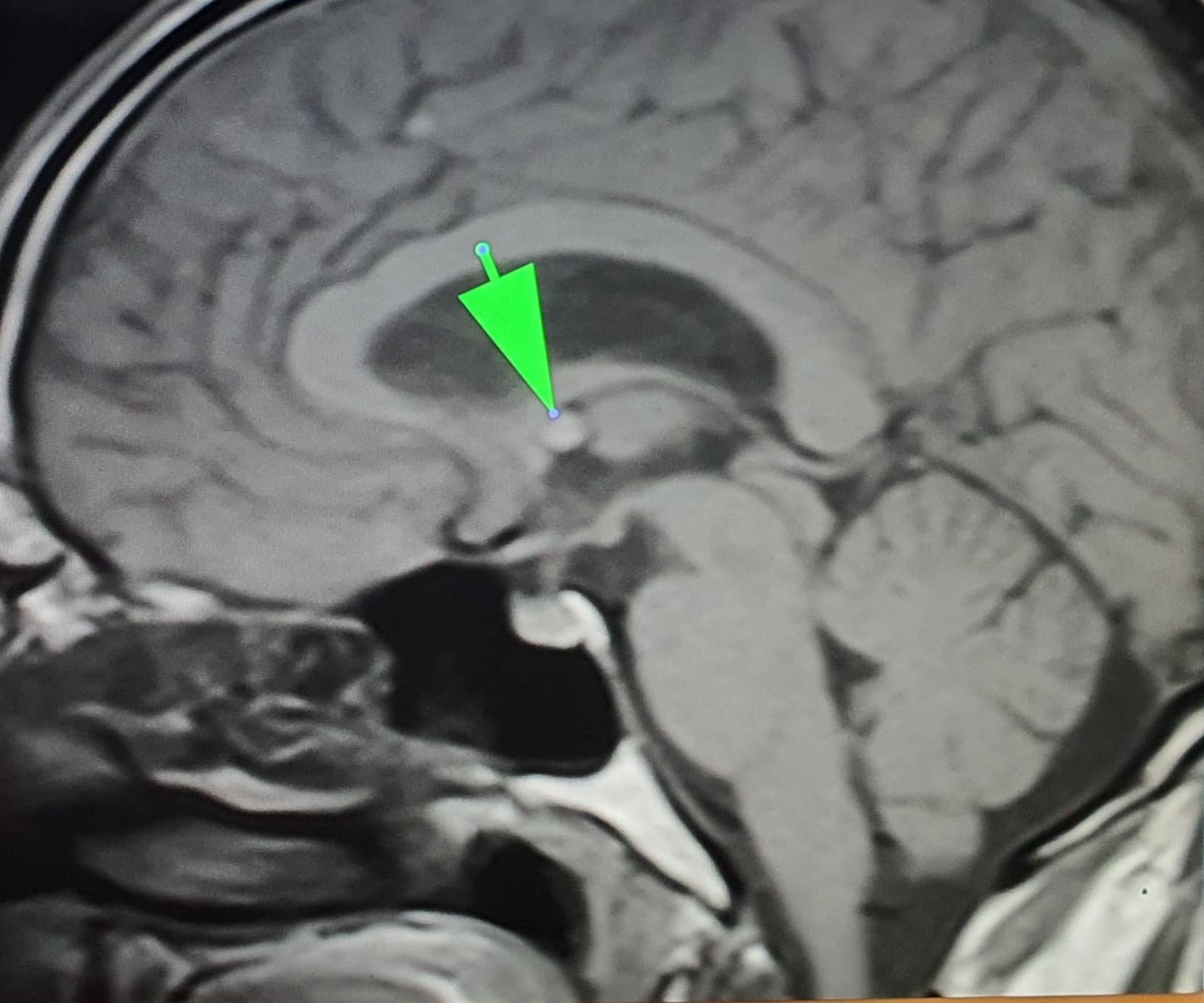
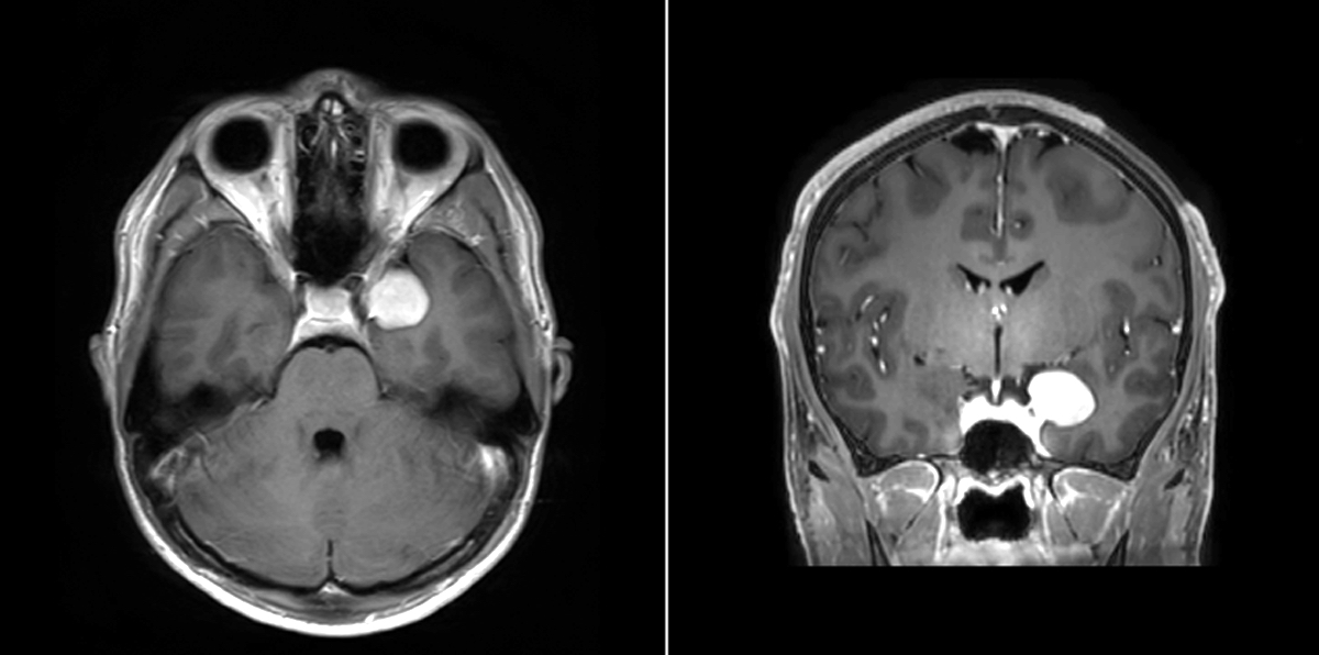
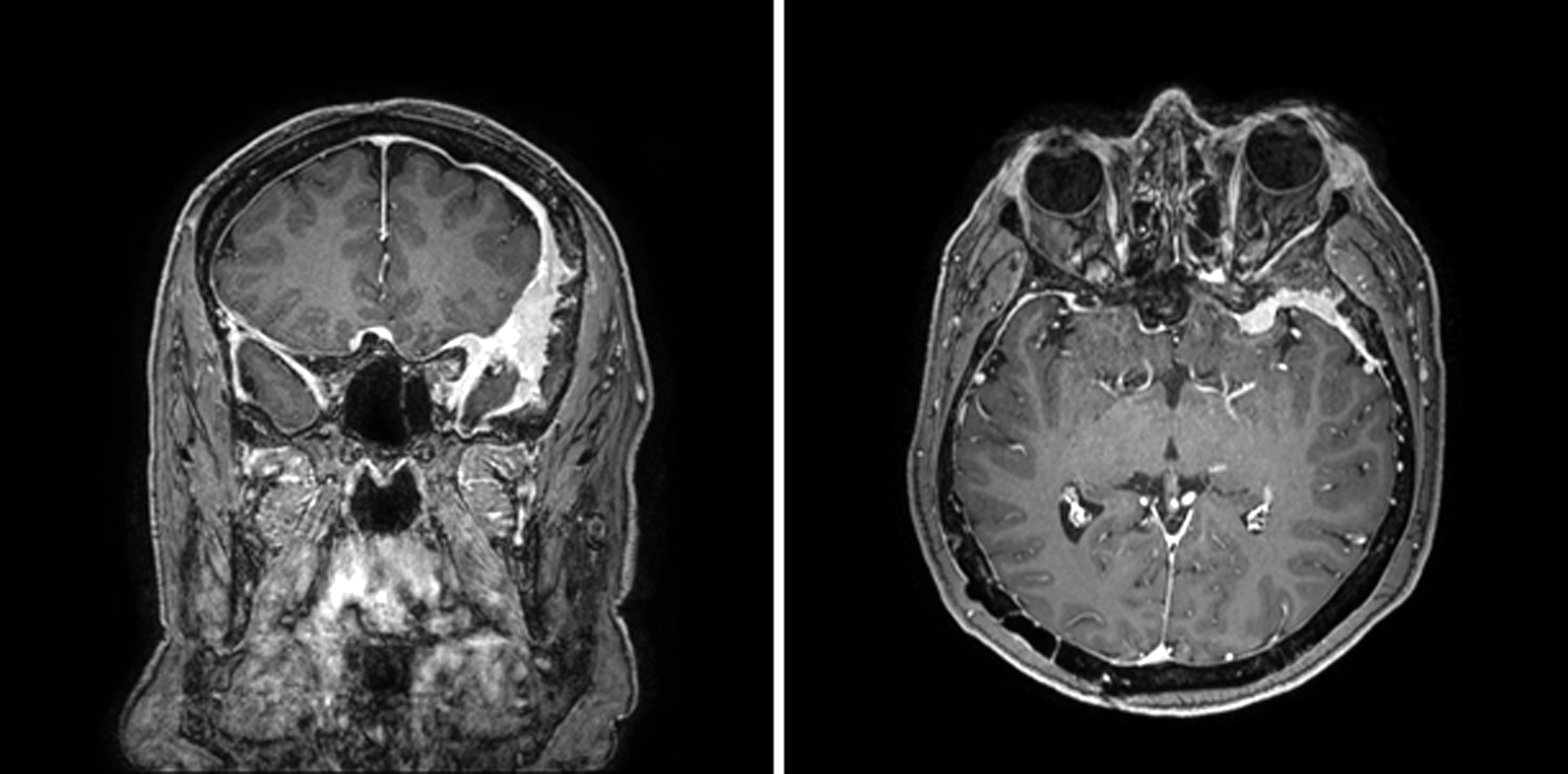
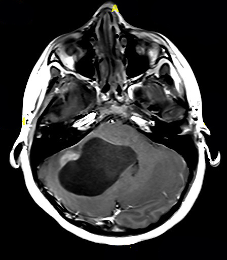
MRI suprasellar mass
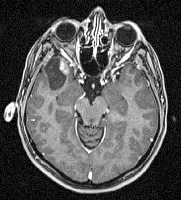
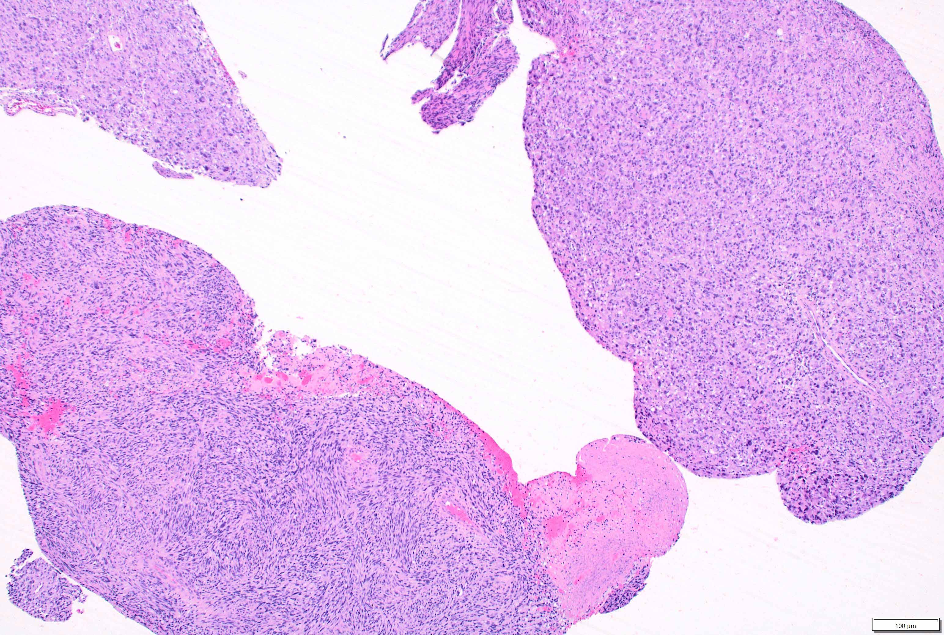
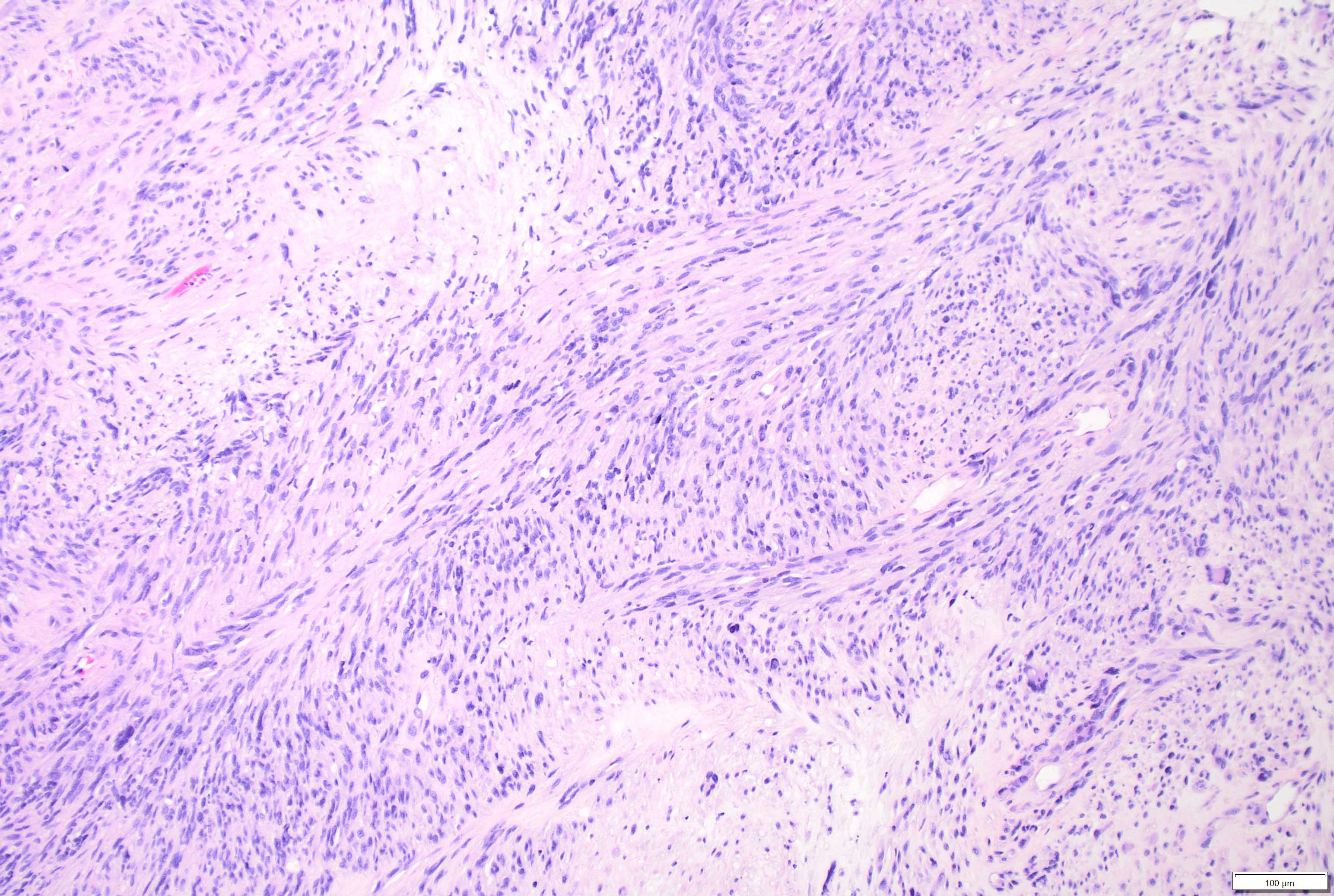
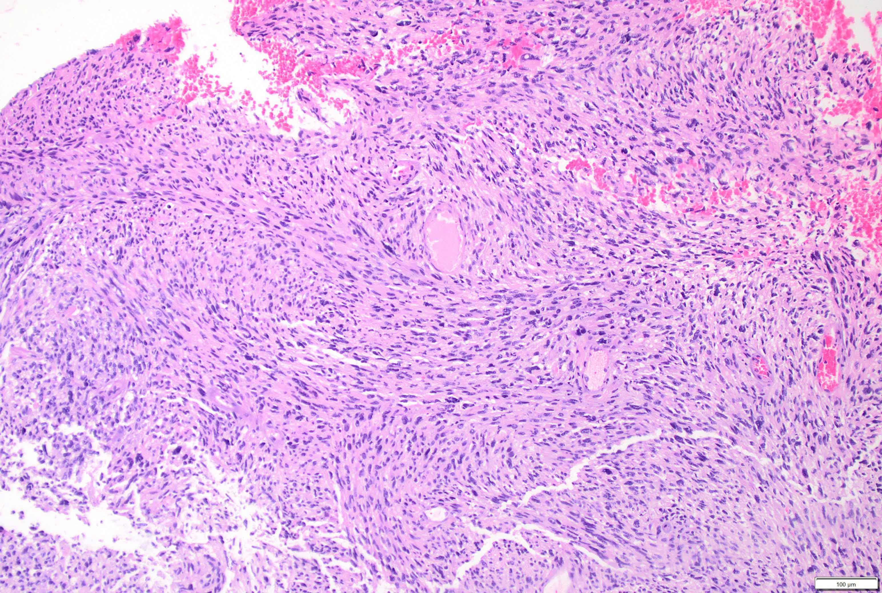
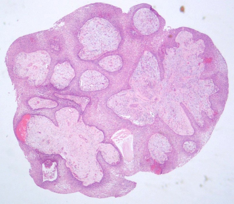
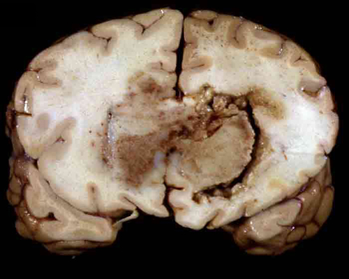
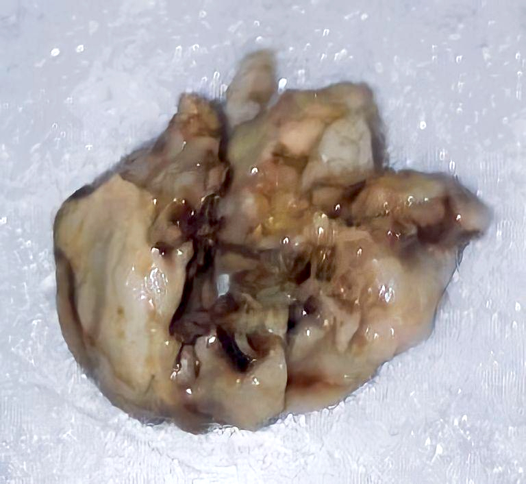
Images hosted on other servers:
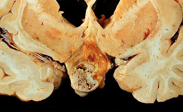
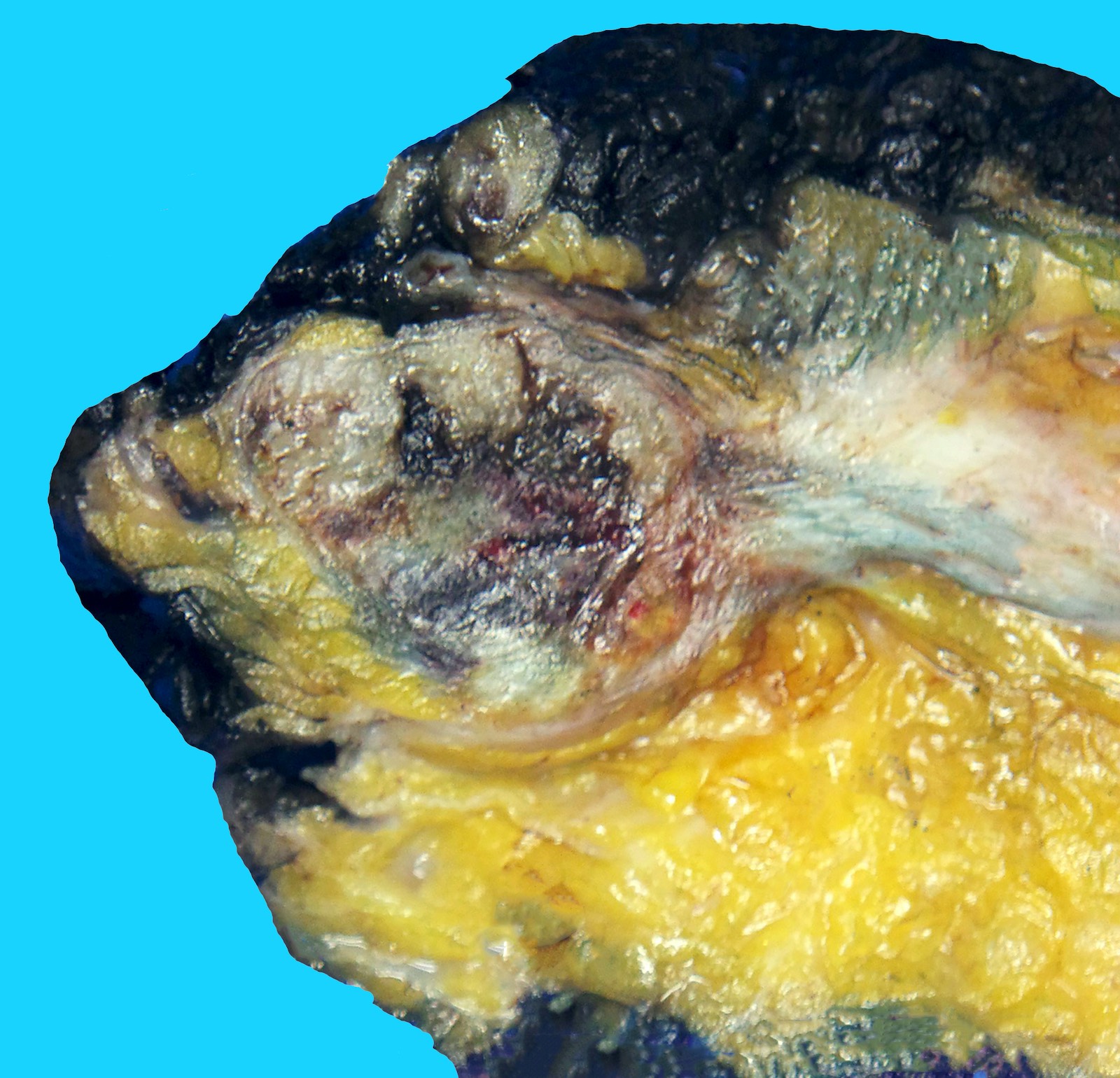
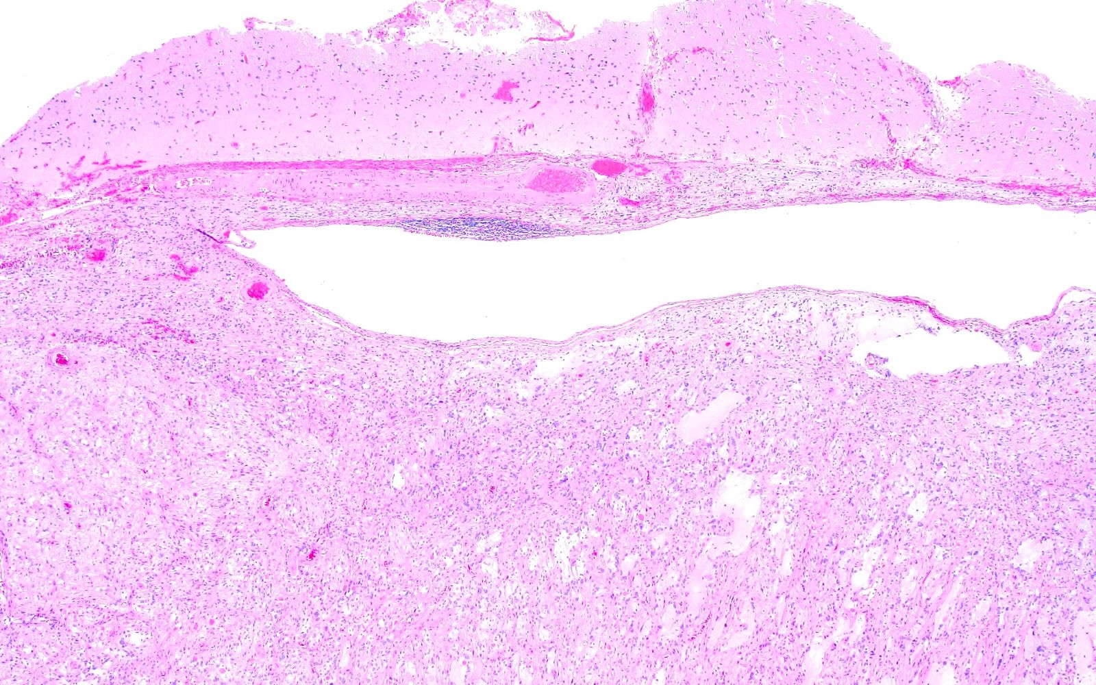
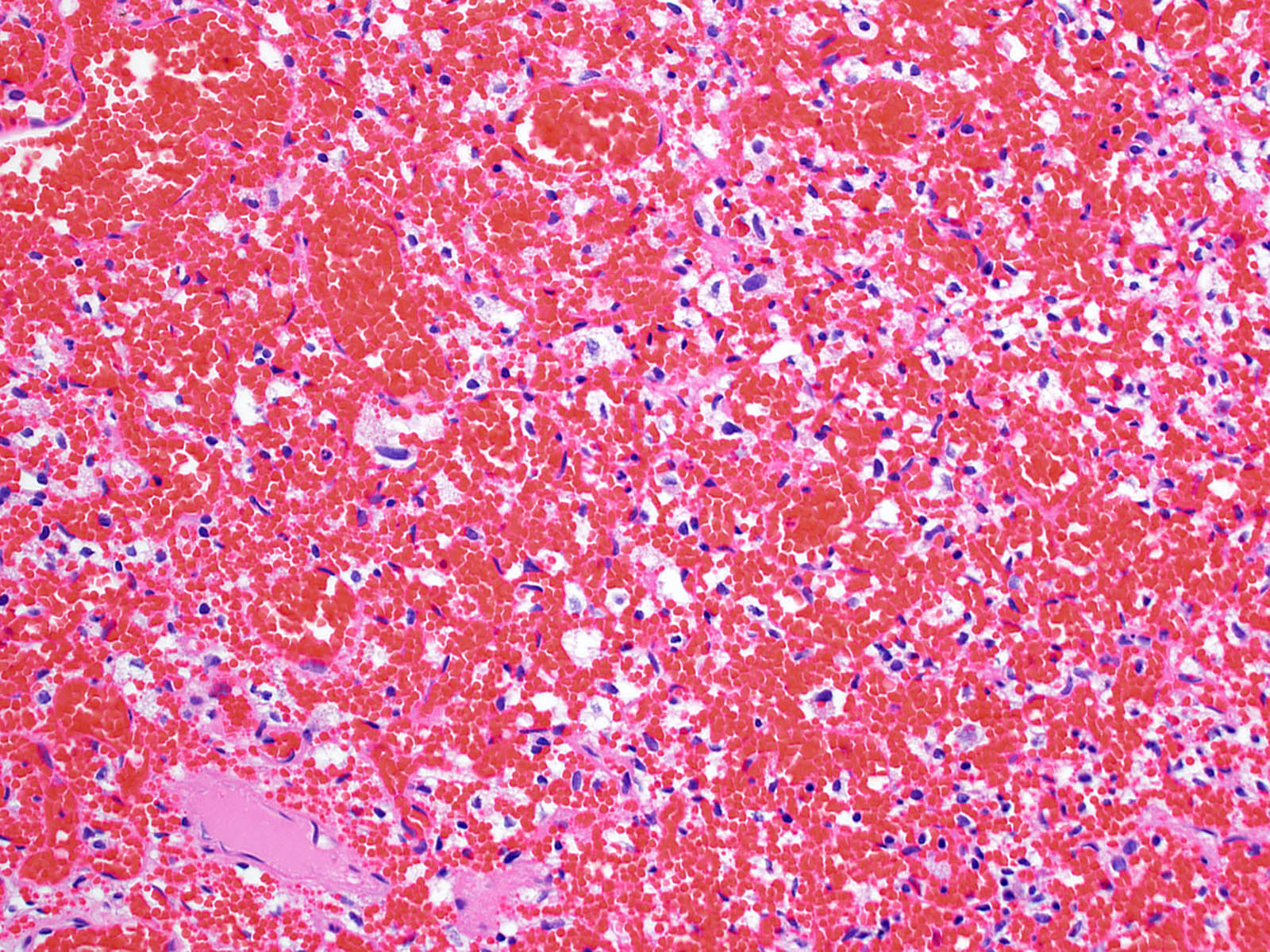
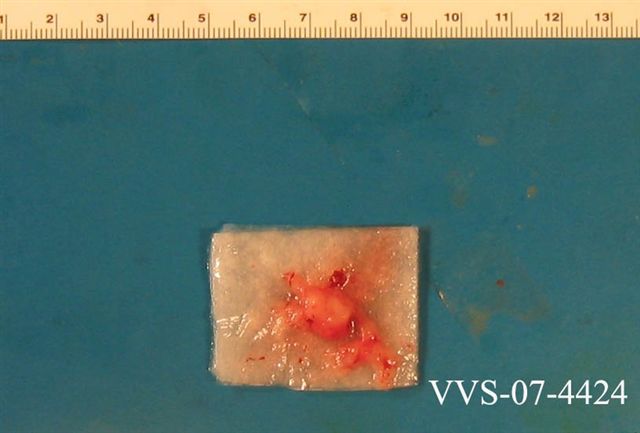
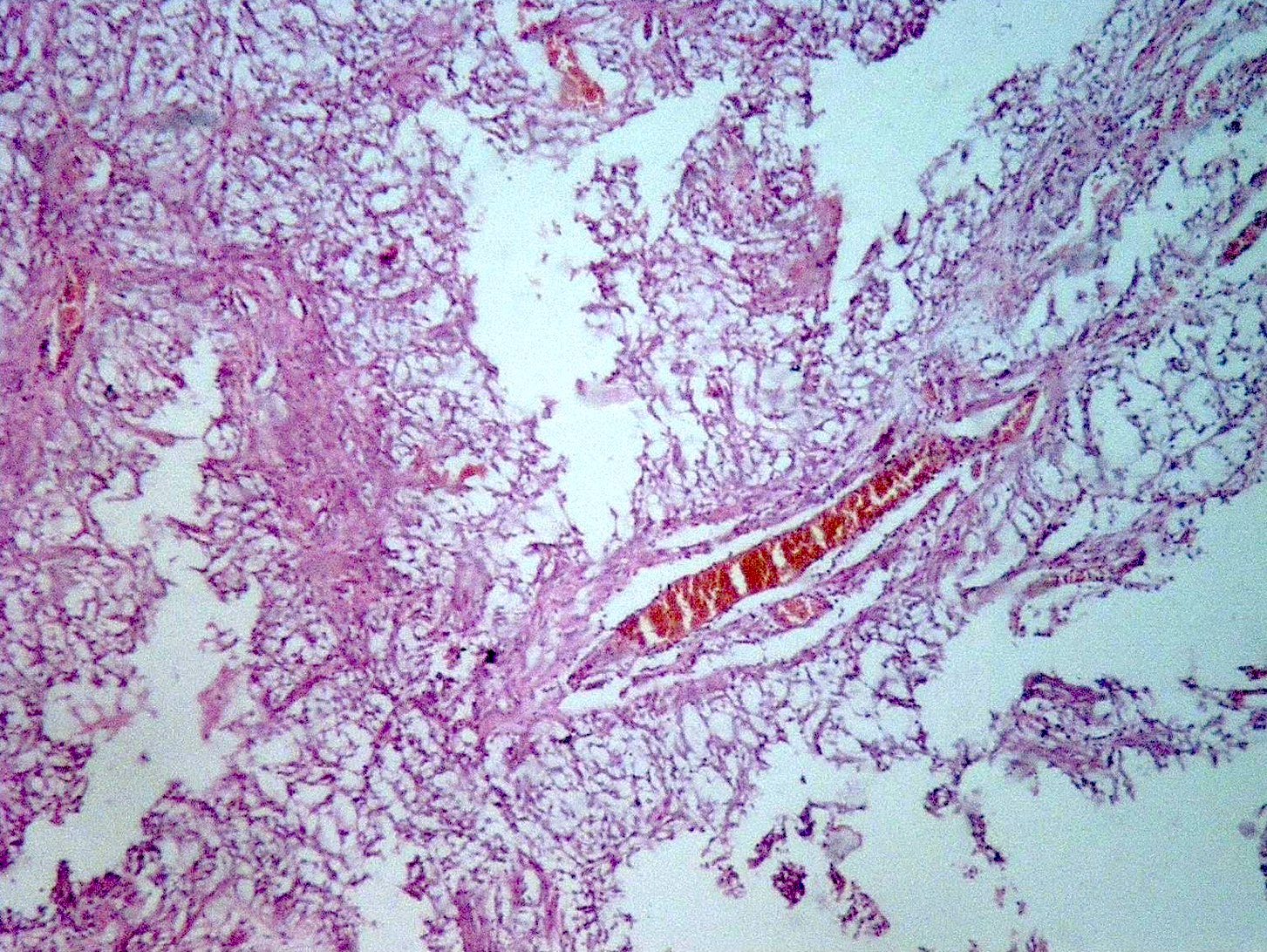
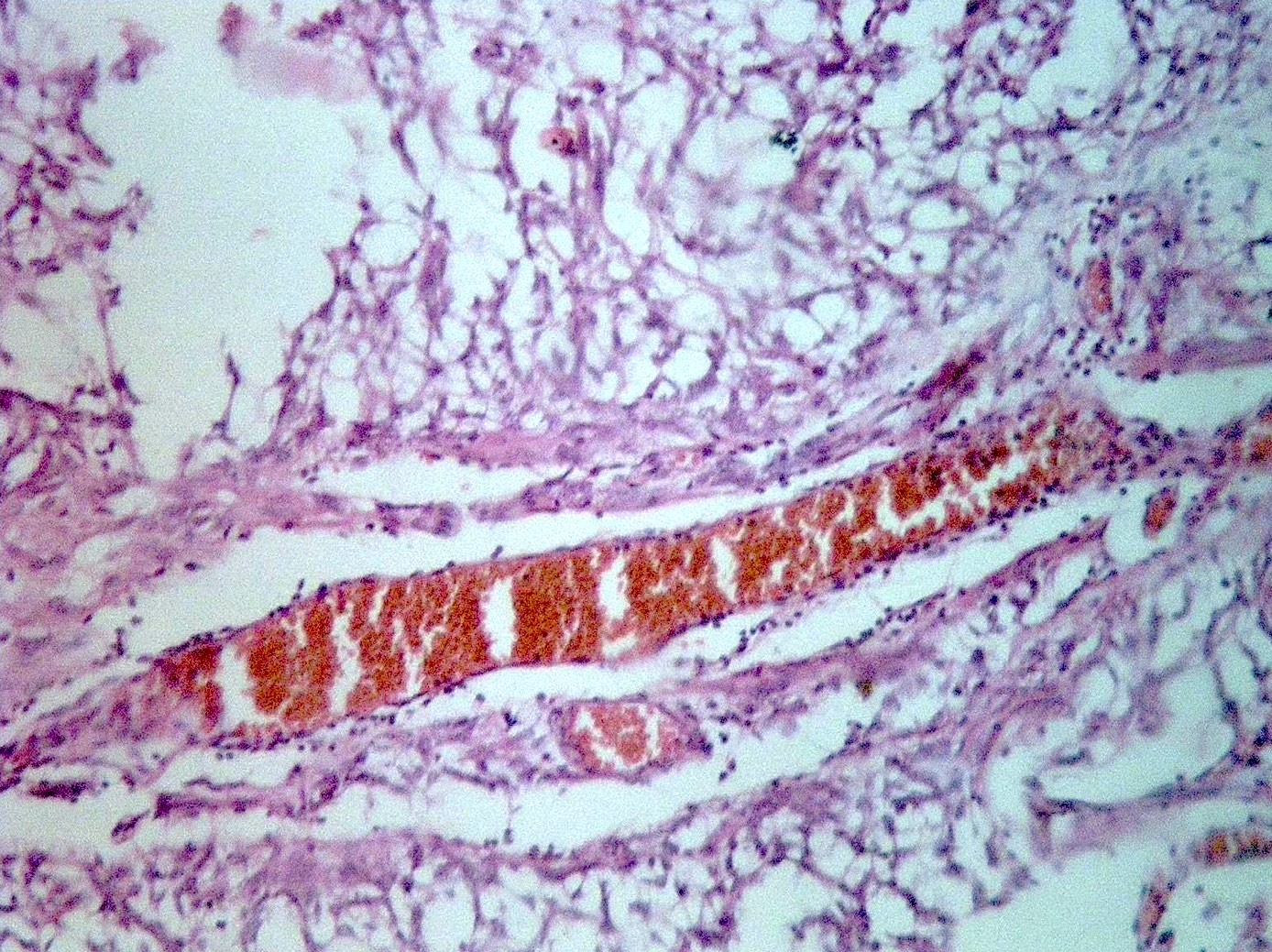
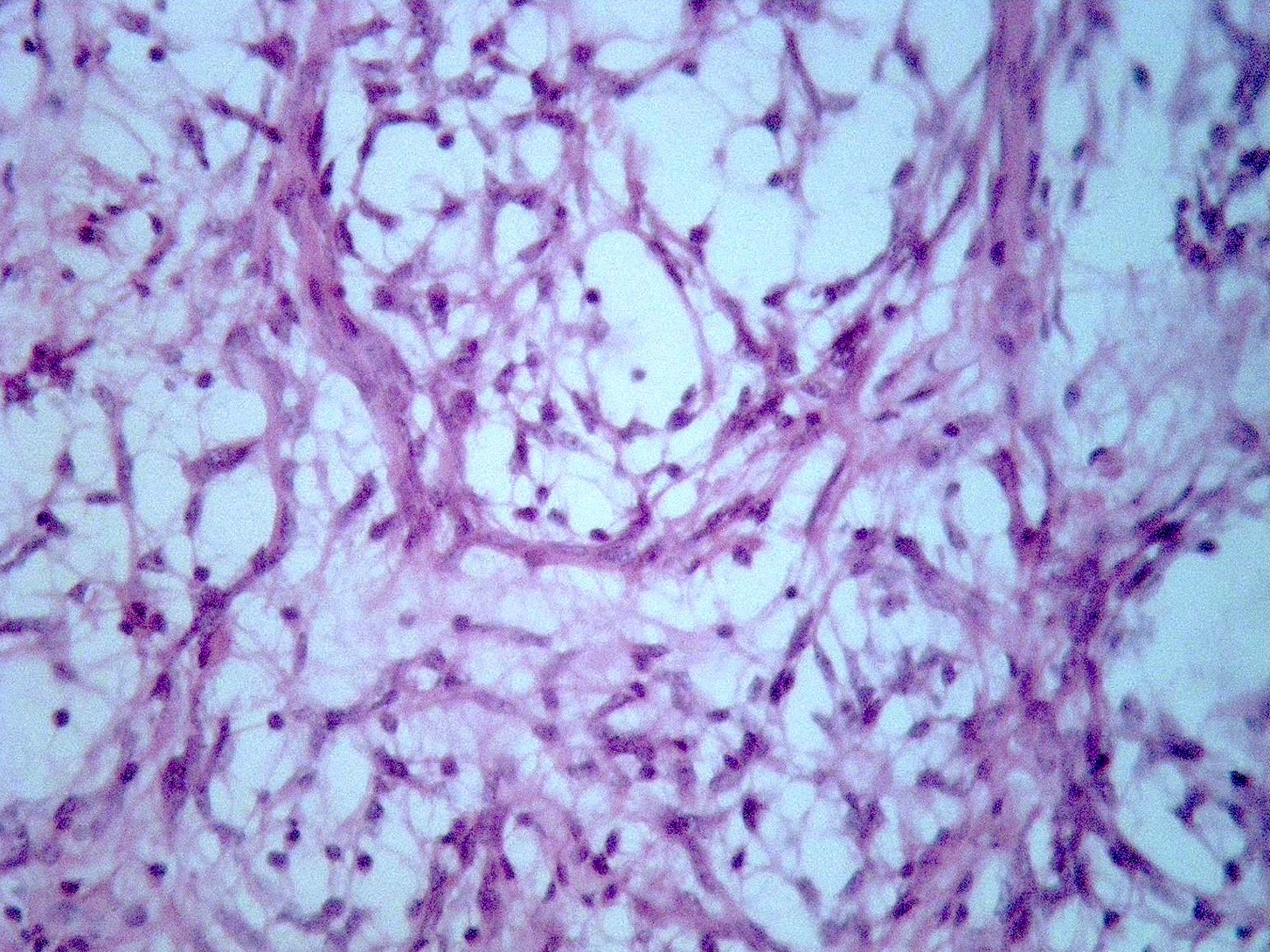
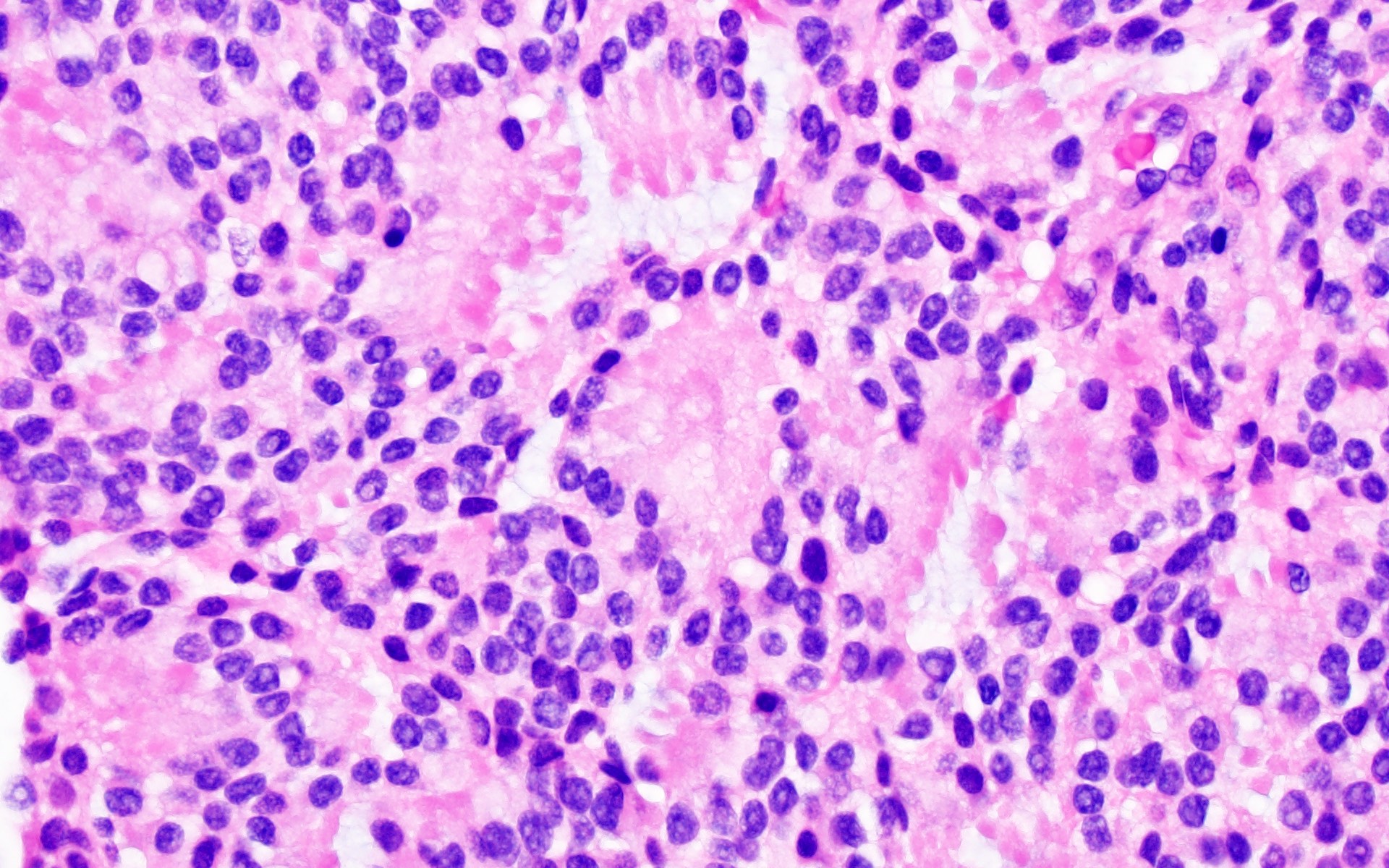
Autopsy image
Contributed by Nelli S. Lakis M.D., M.Sc.
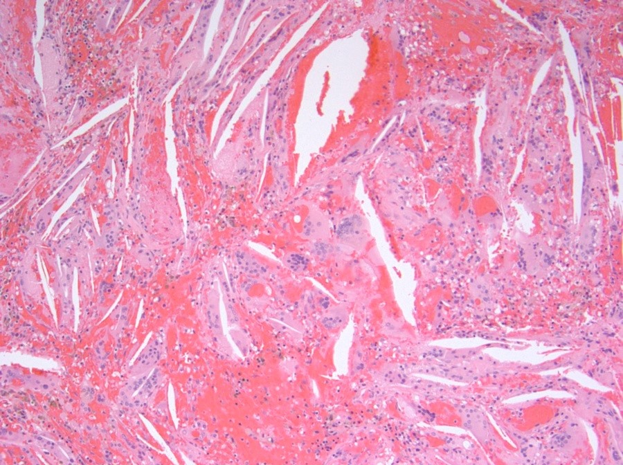
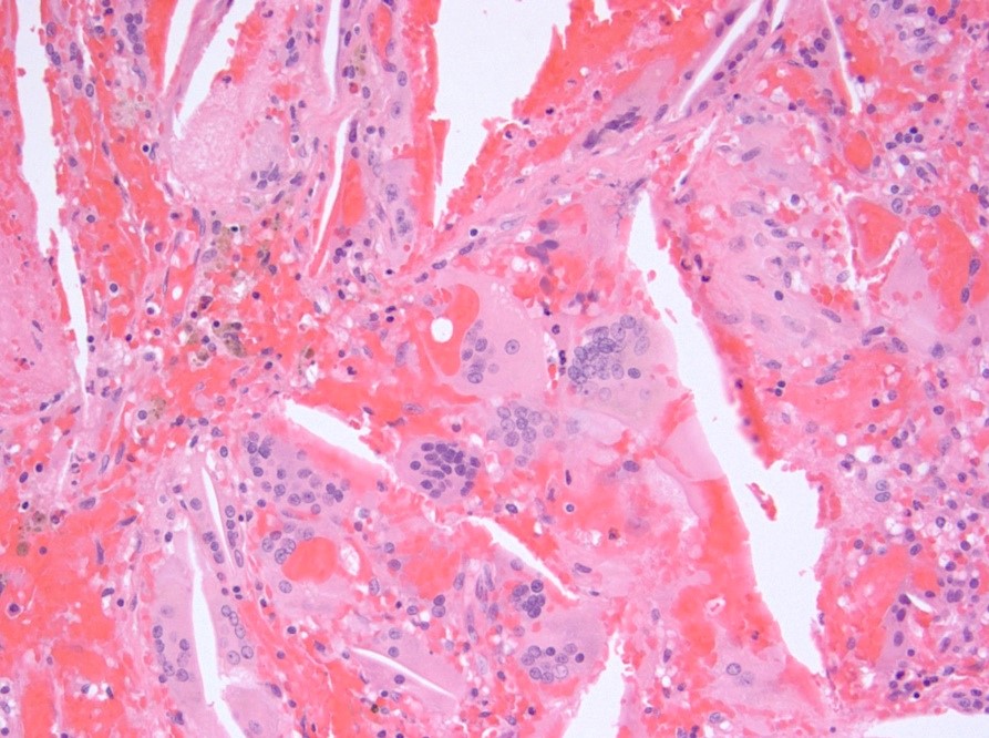
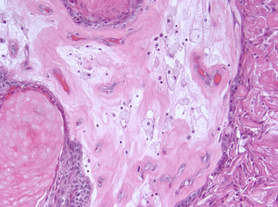
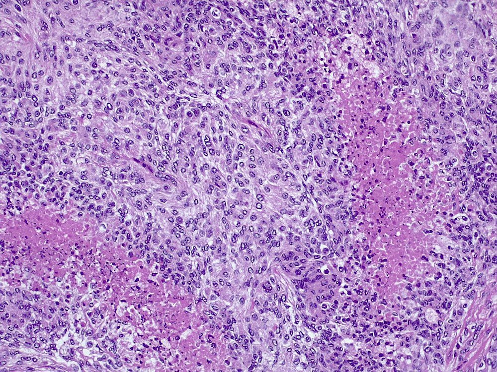
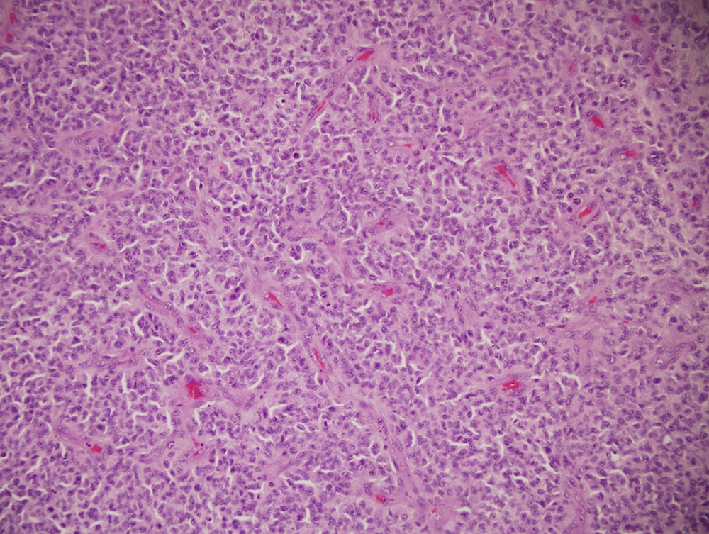
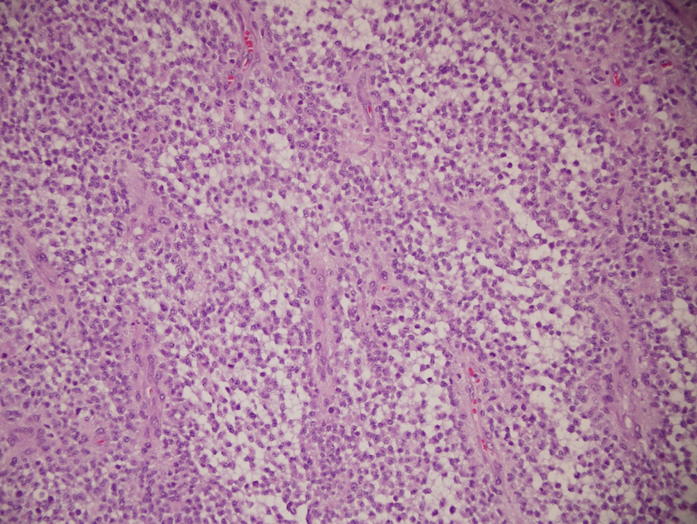
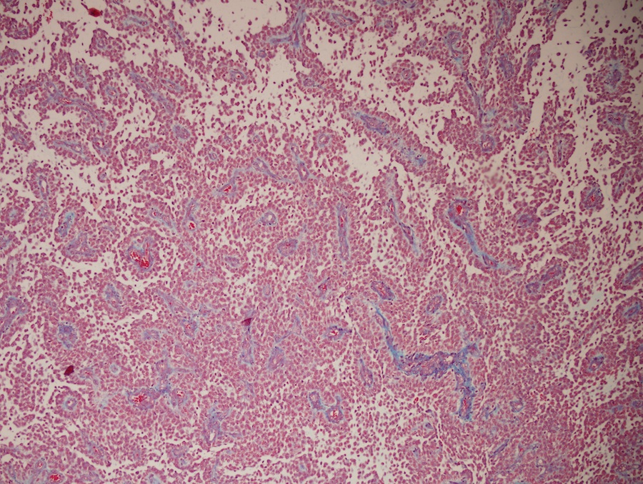
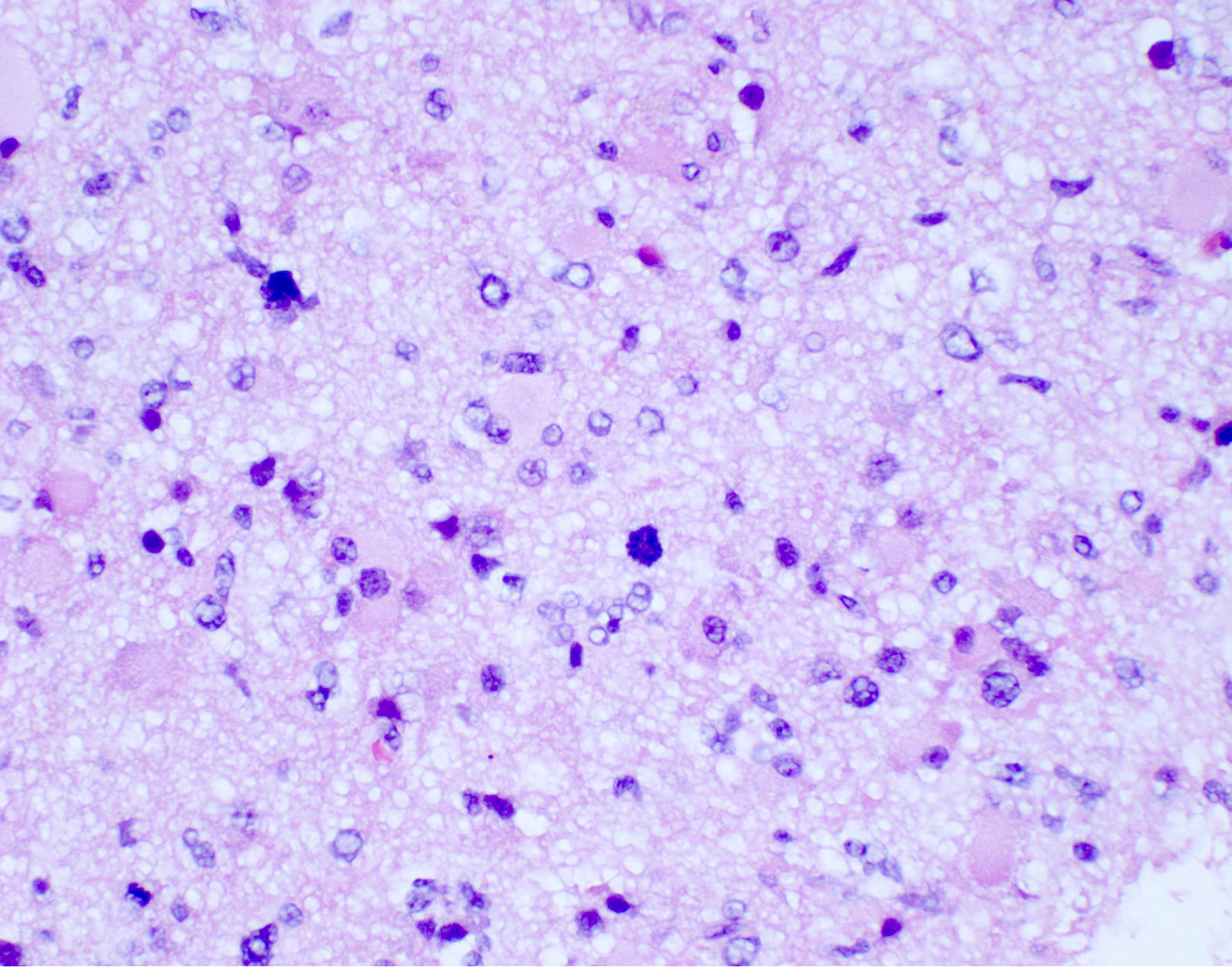
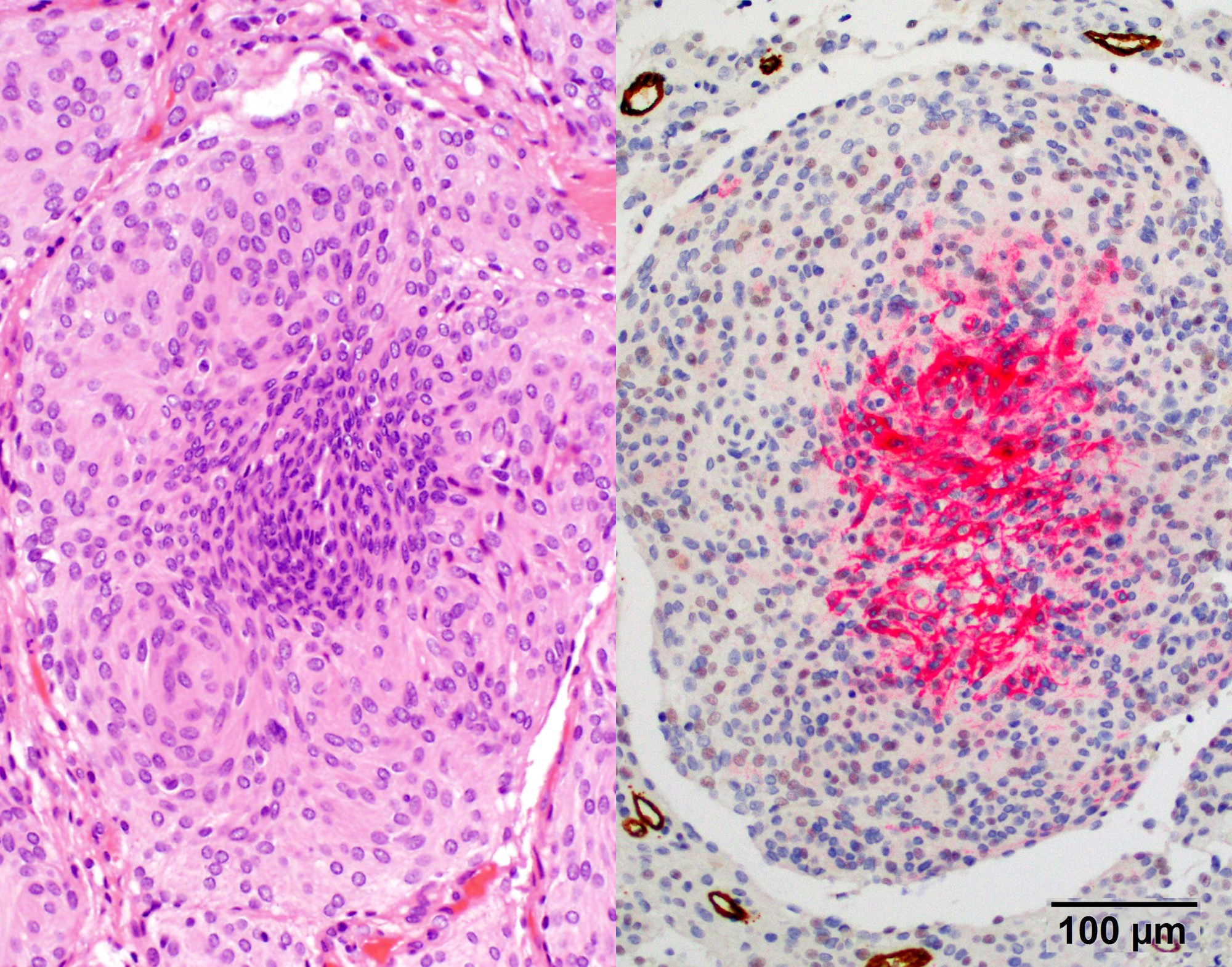
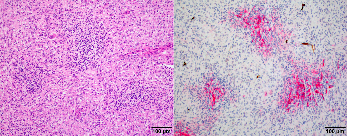
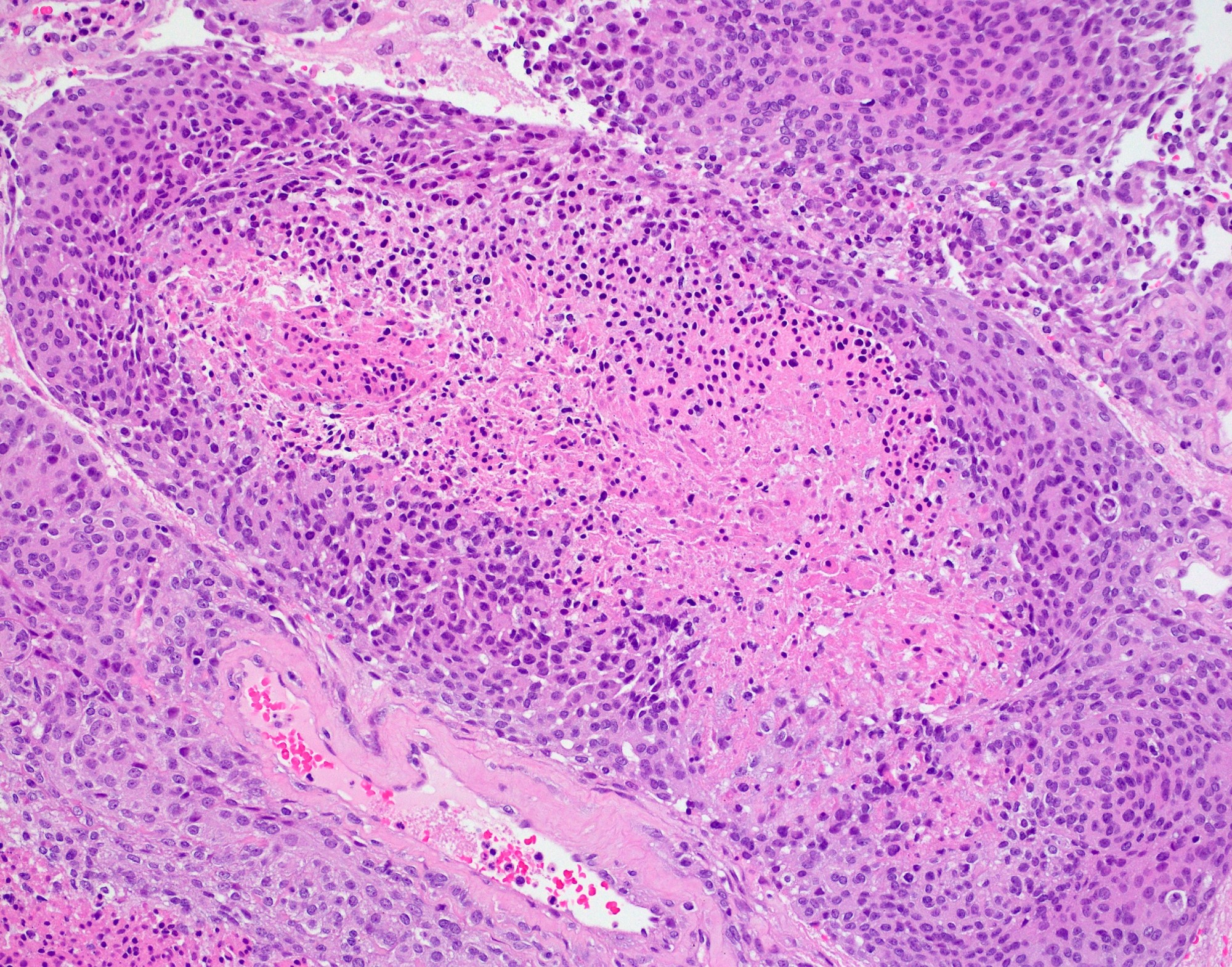
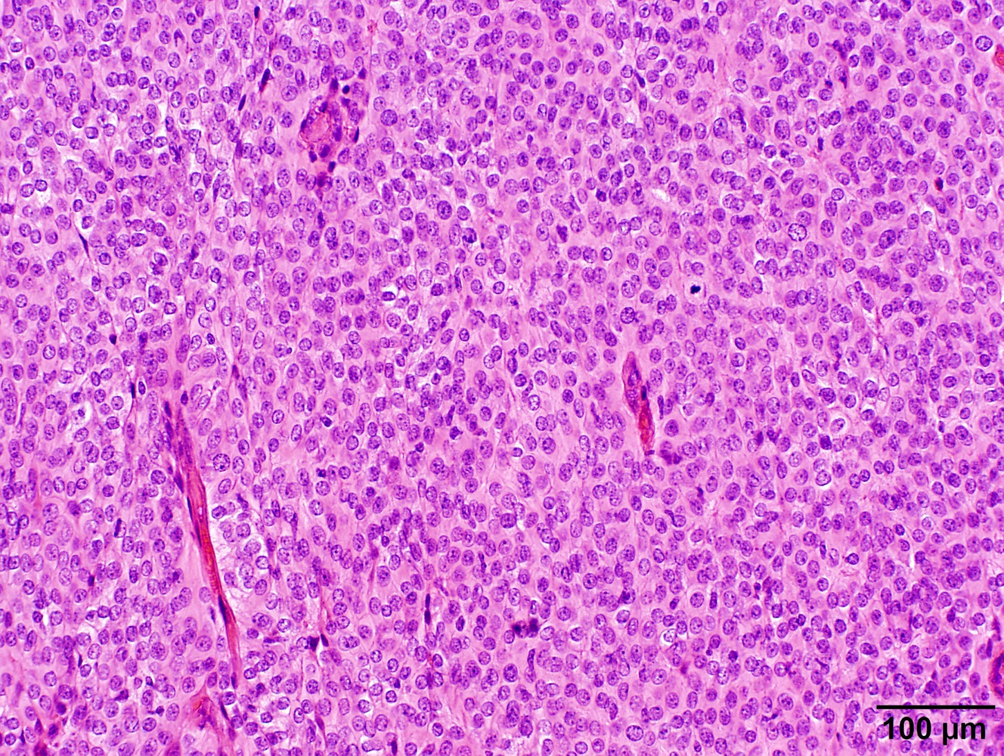
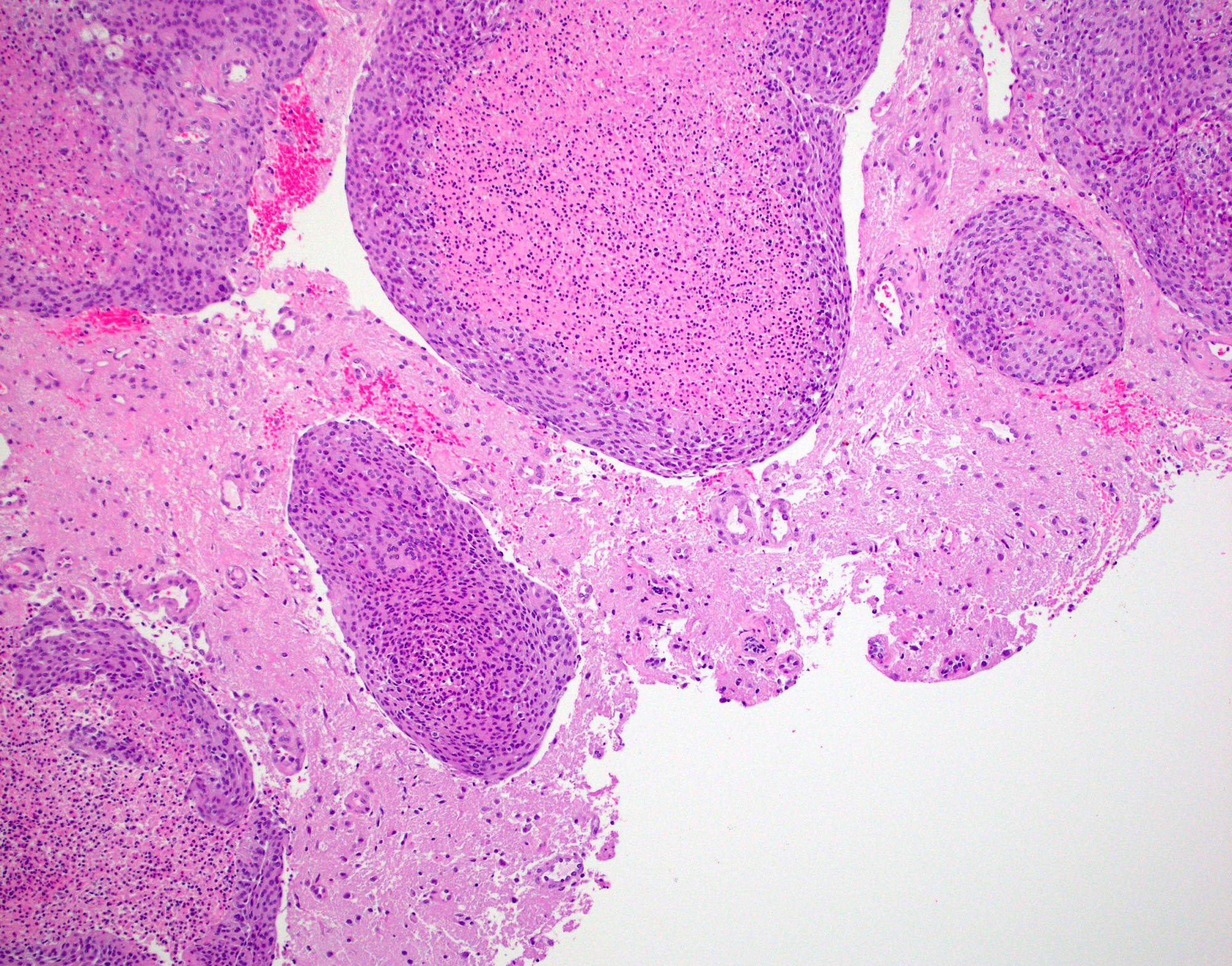
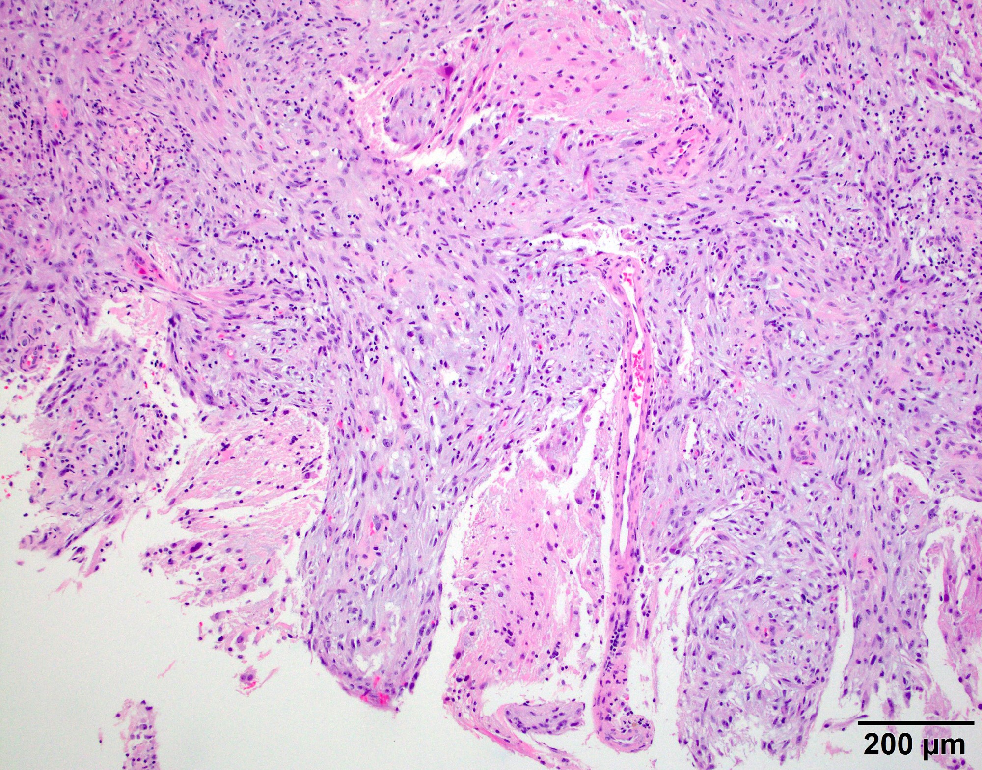
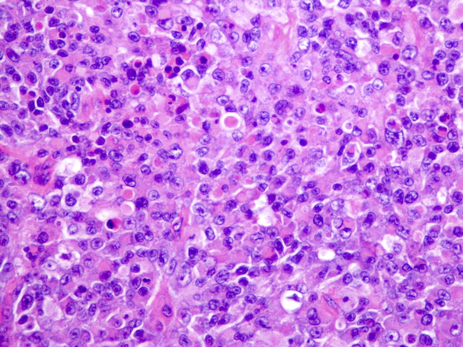
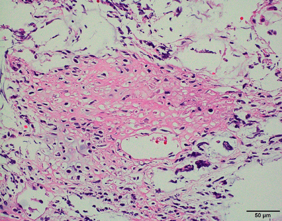
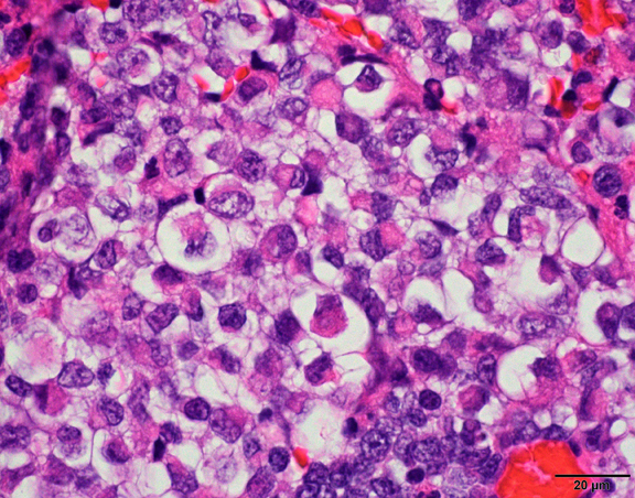
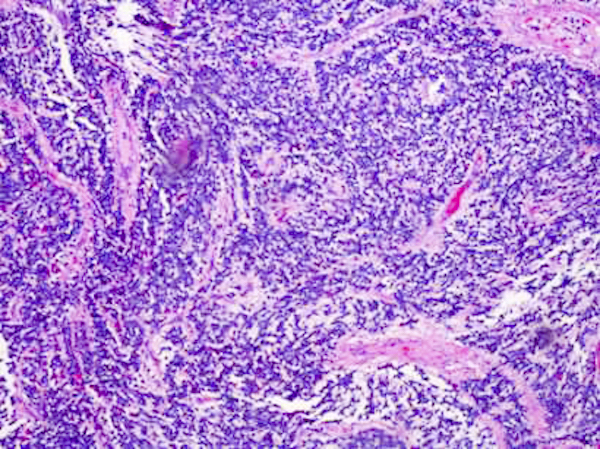
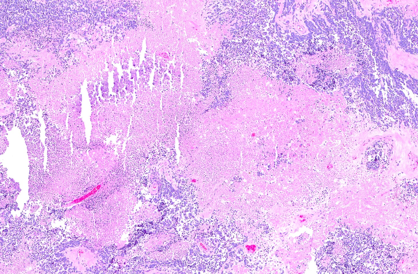
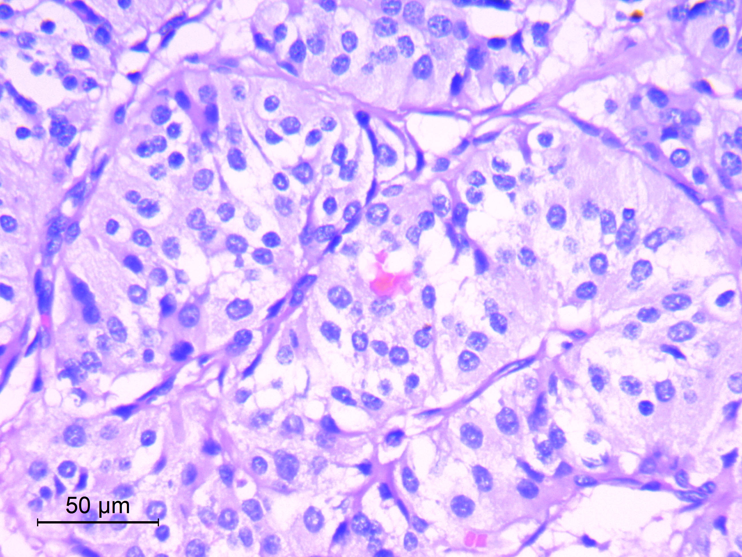
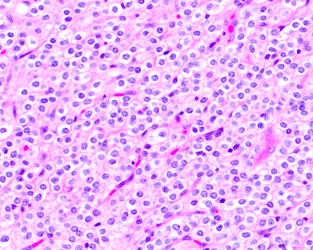
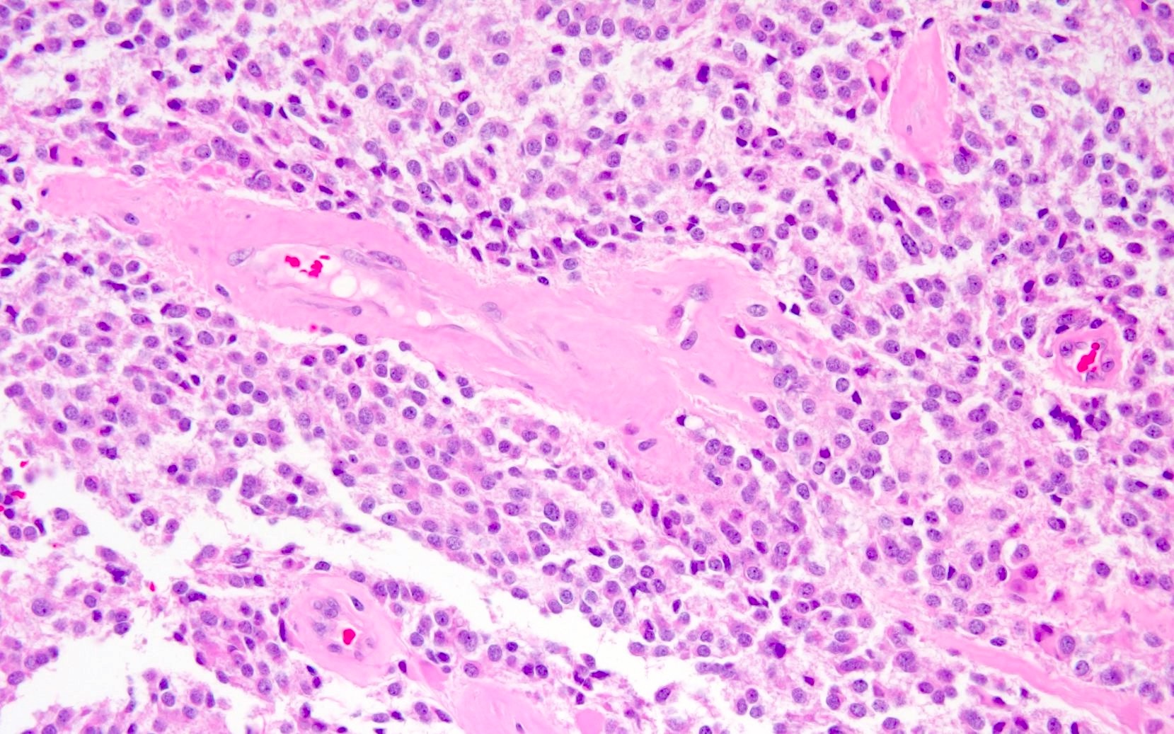
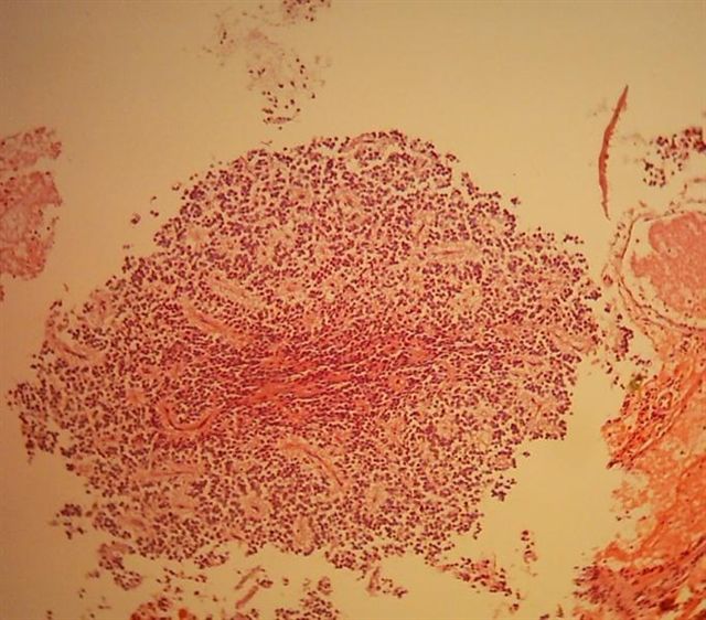
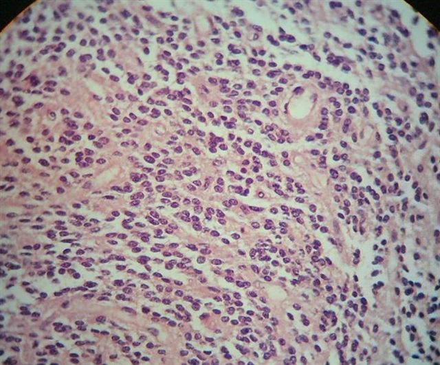
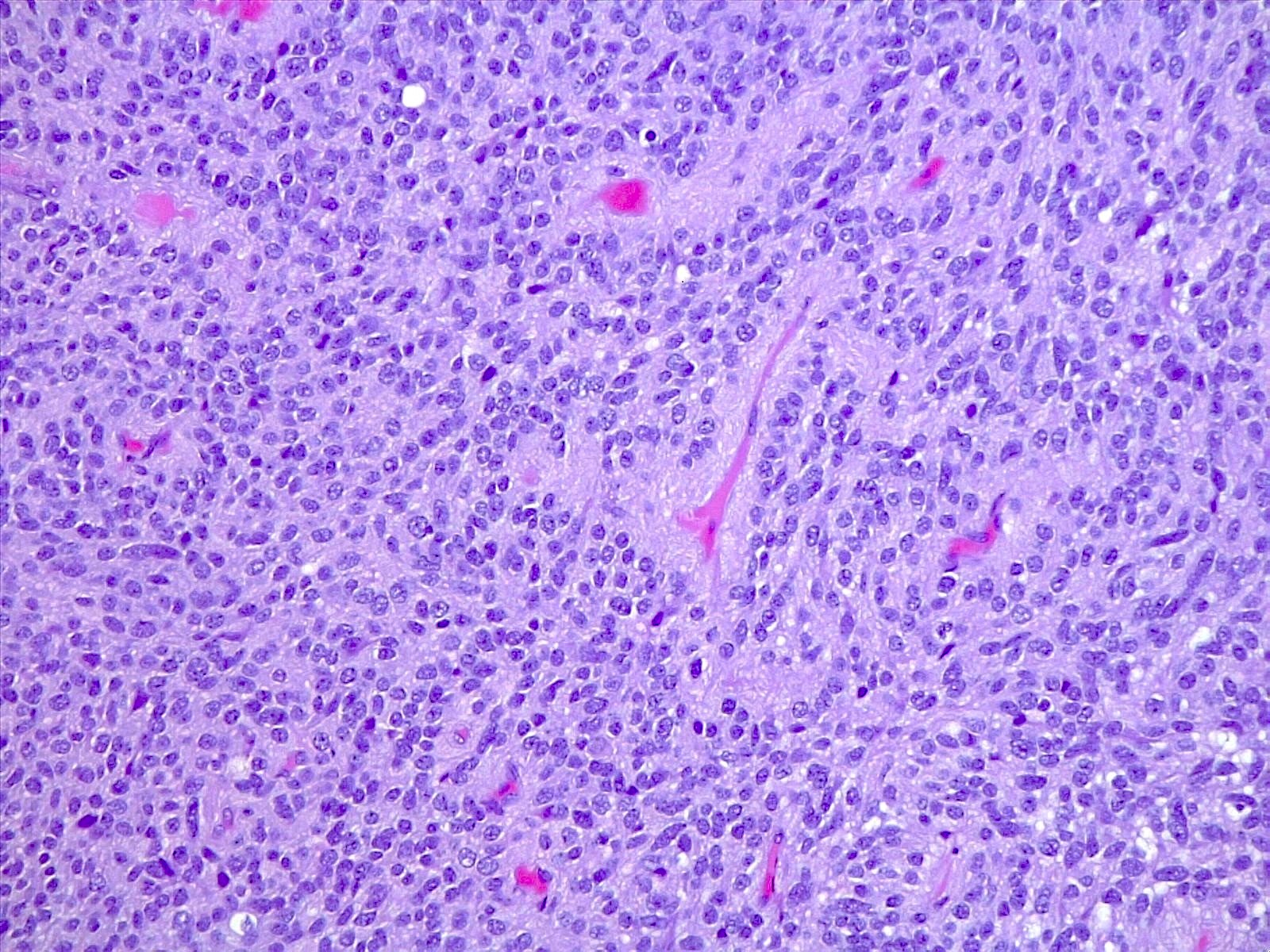
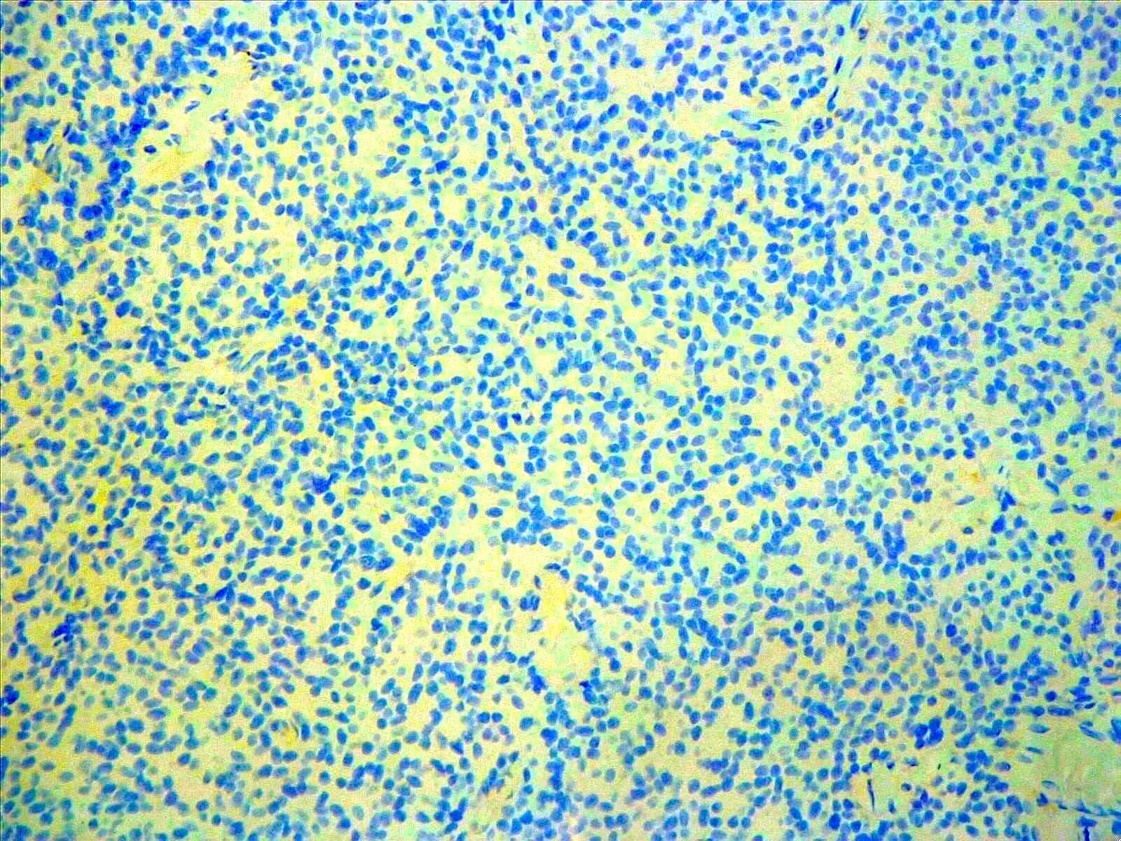
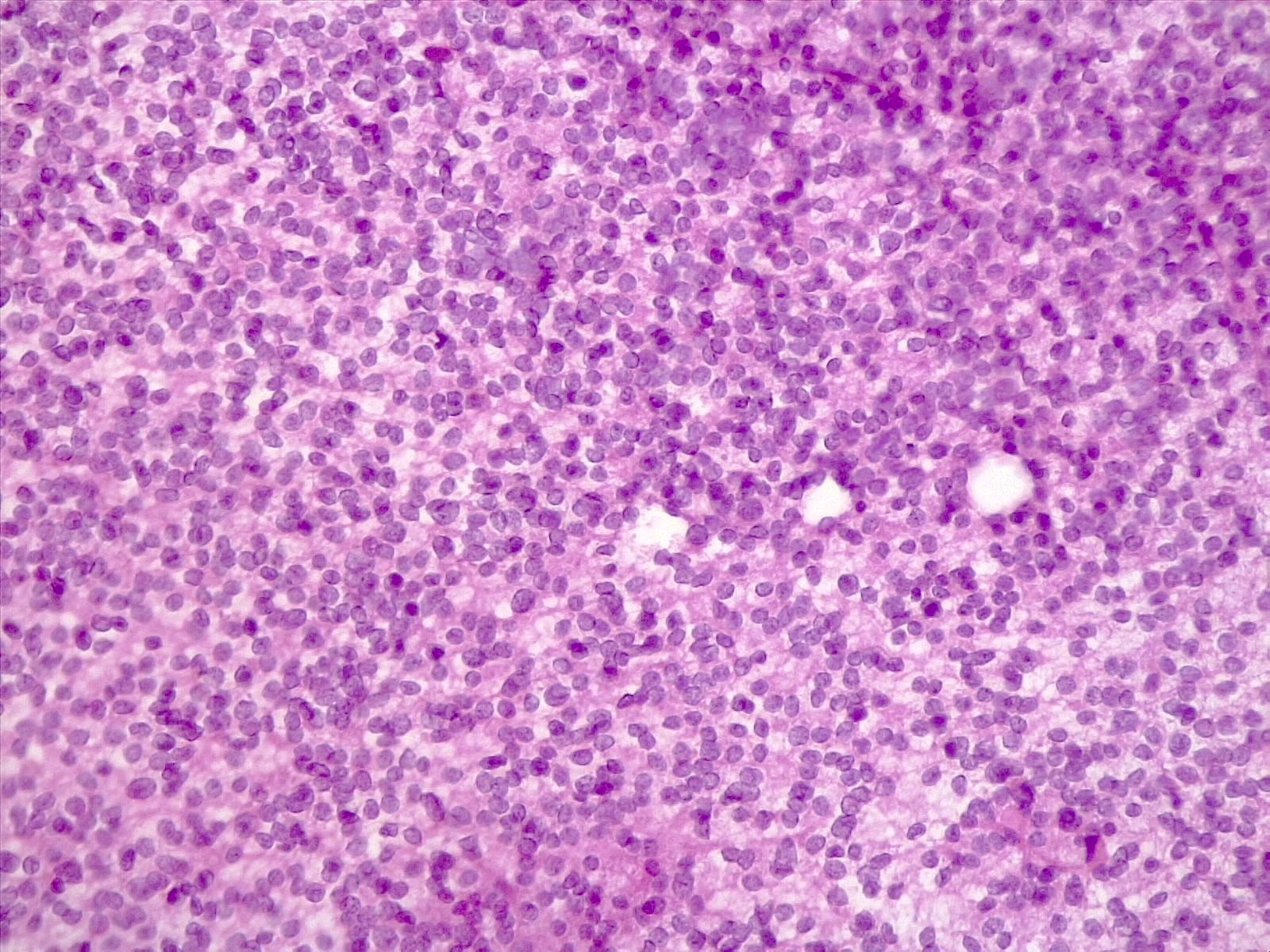
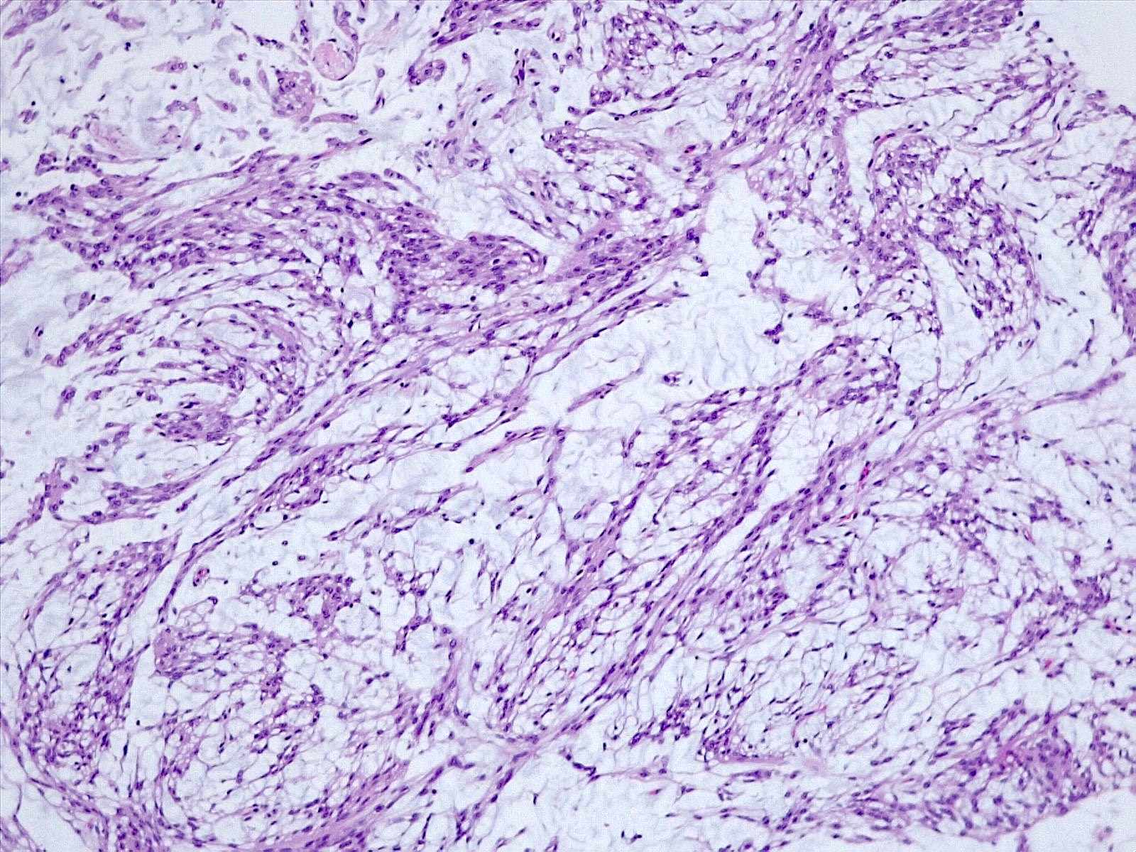
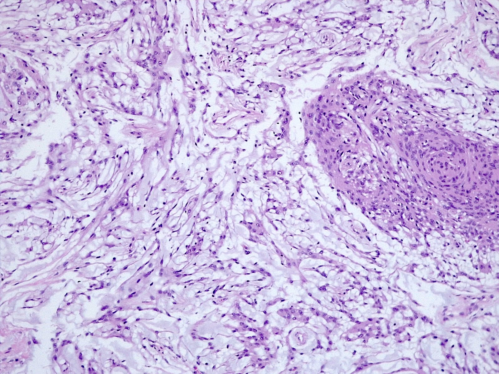
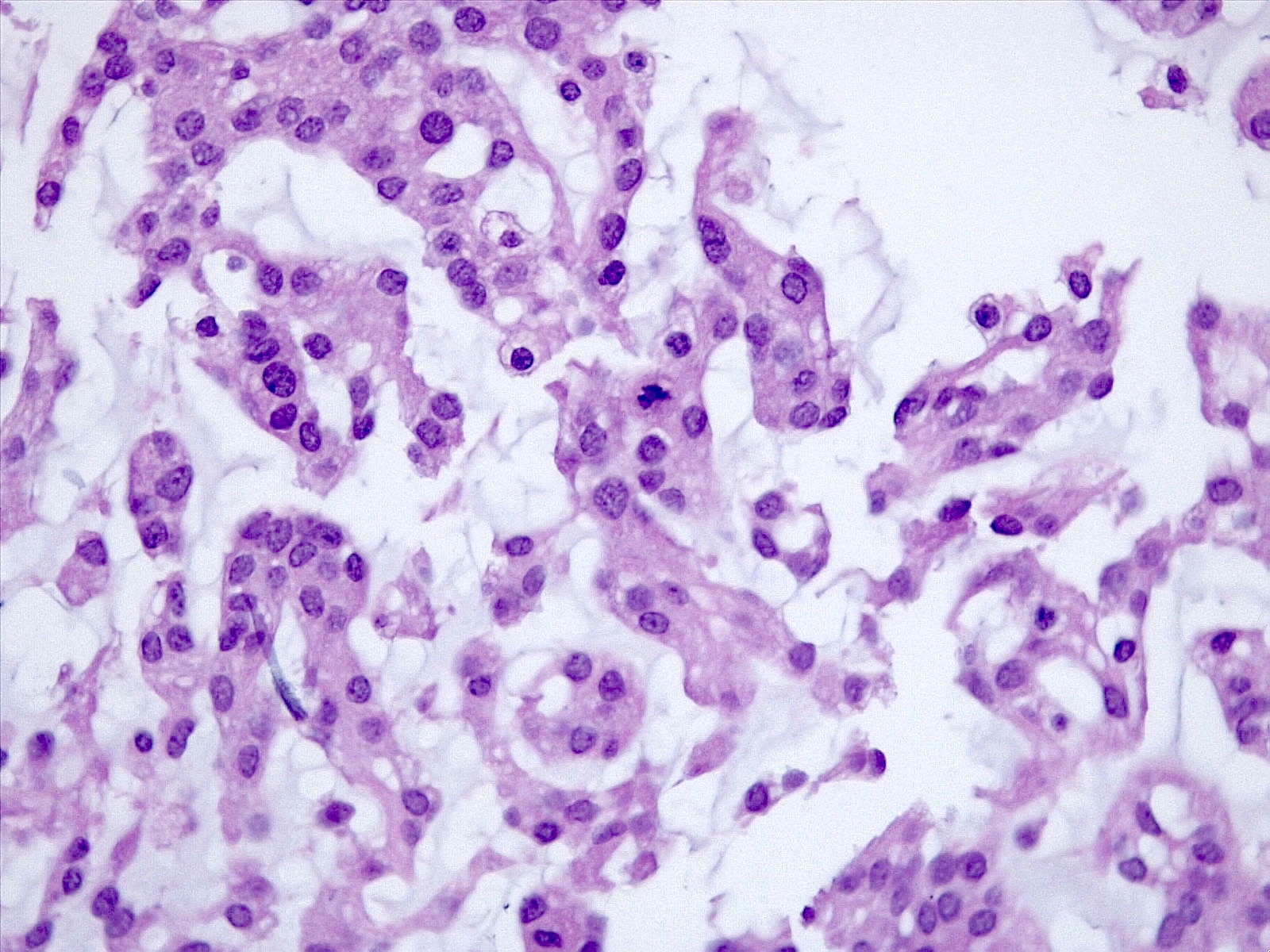
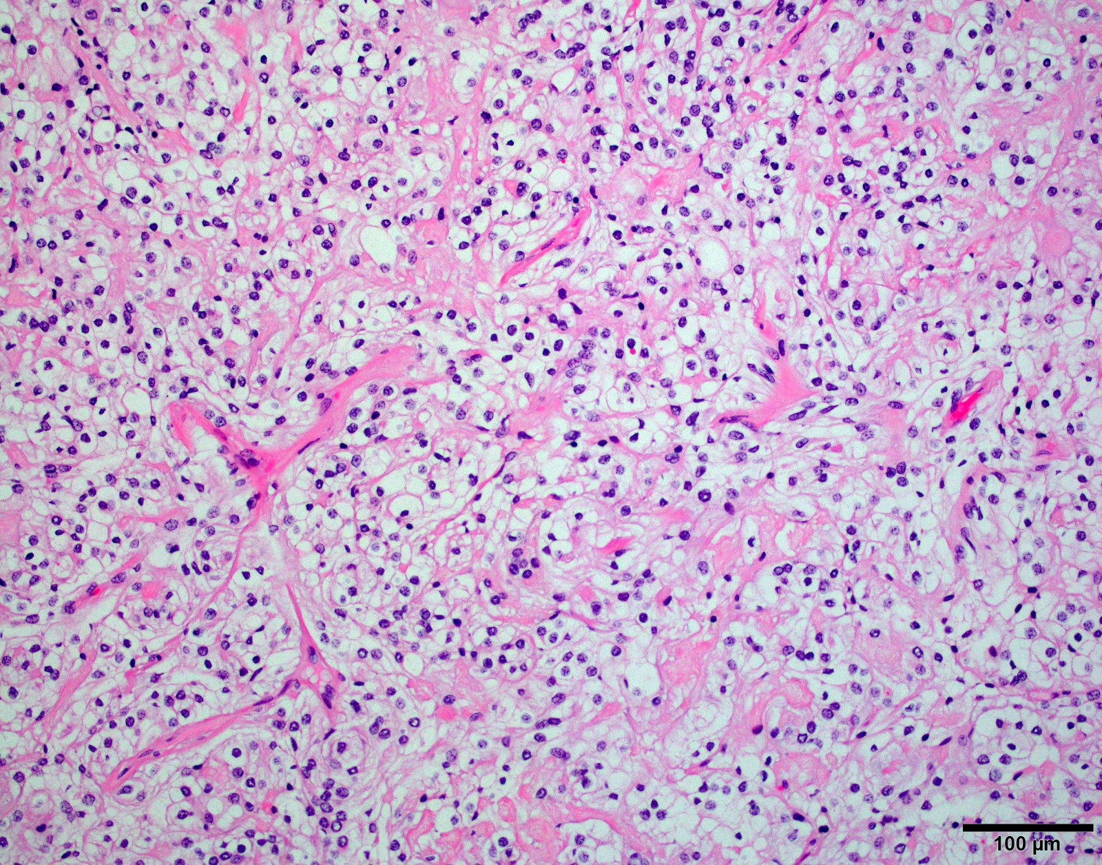
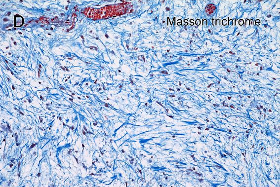
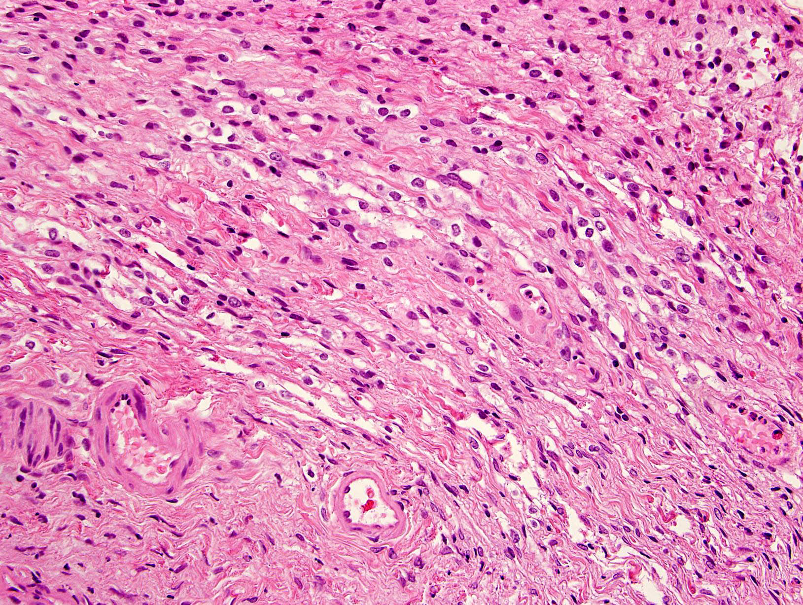
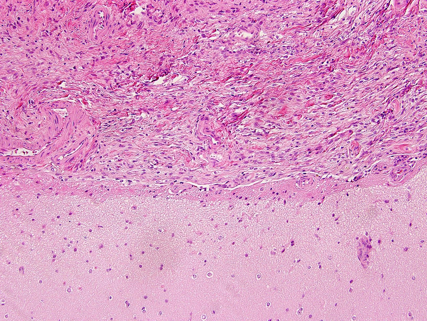
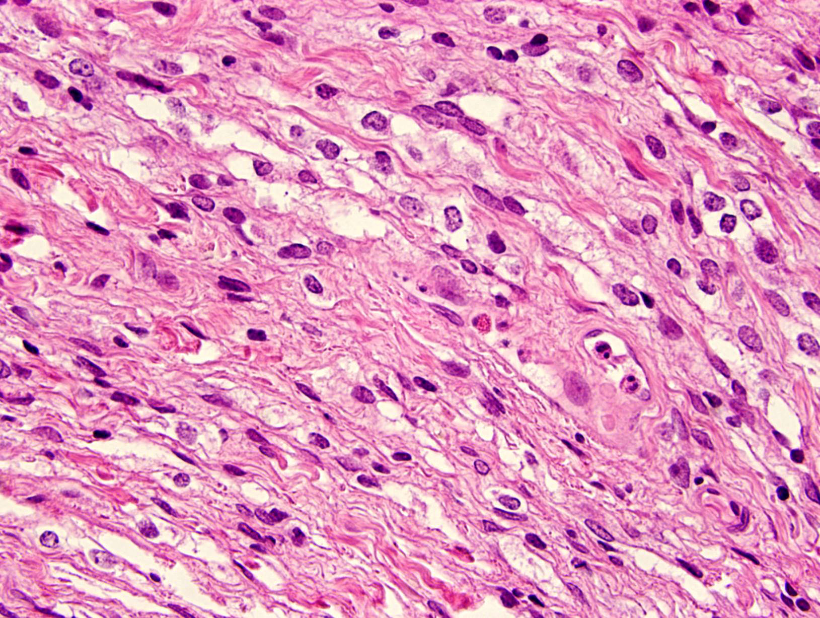
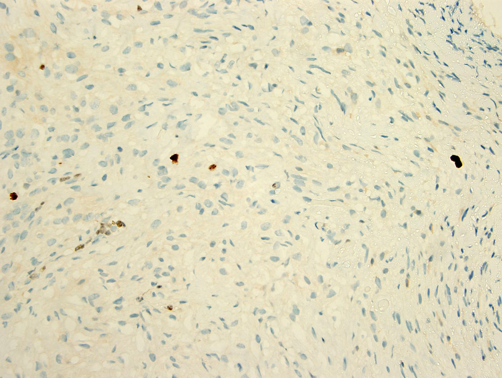
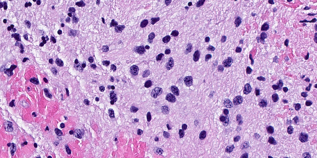
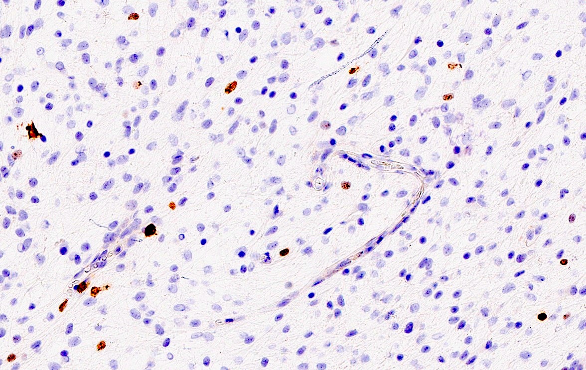
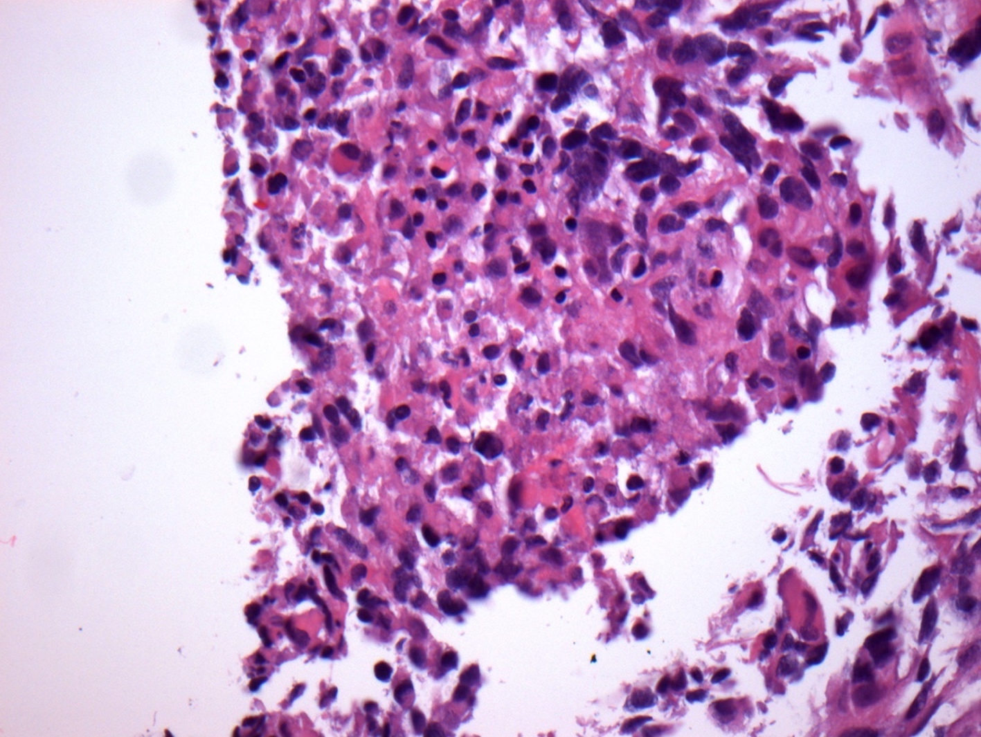
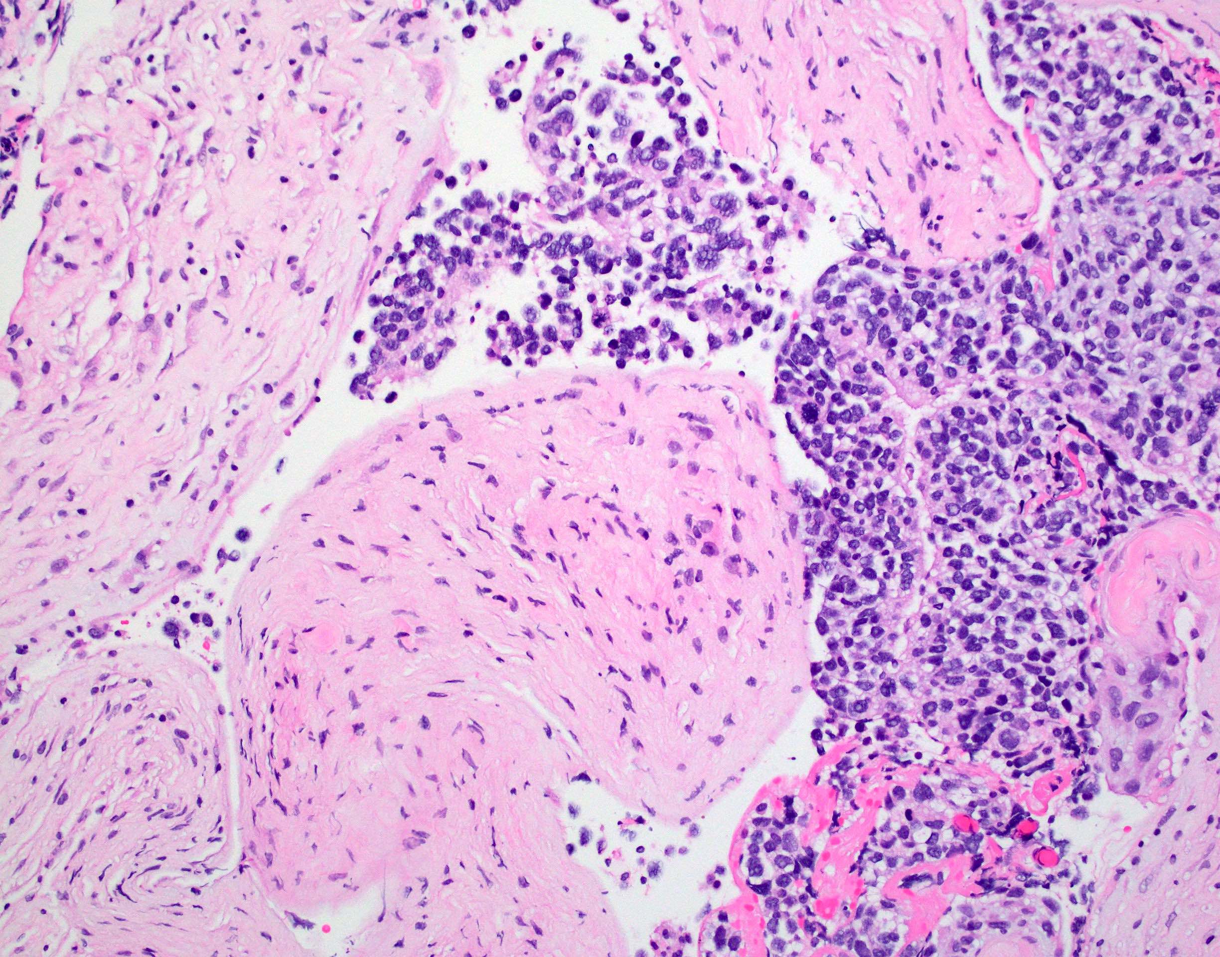
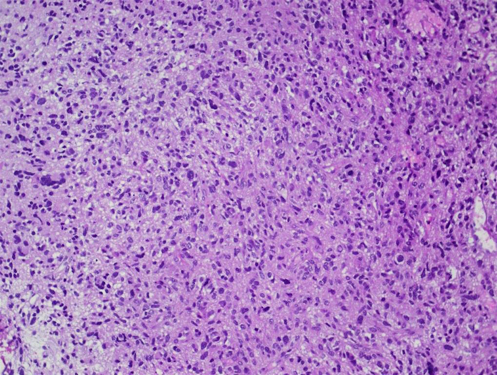
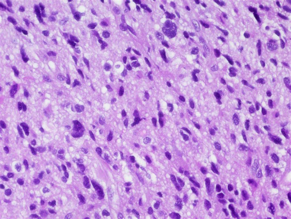
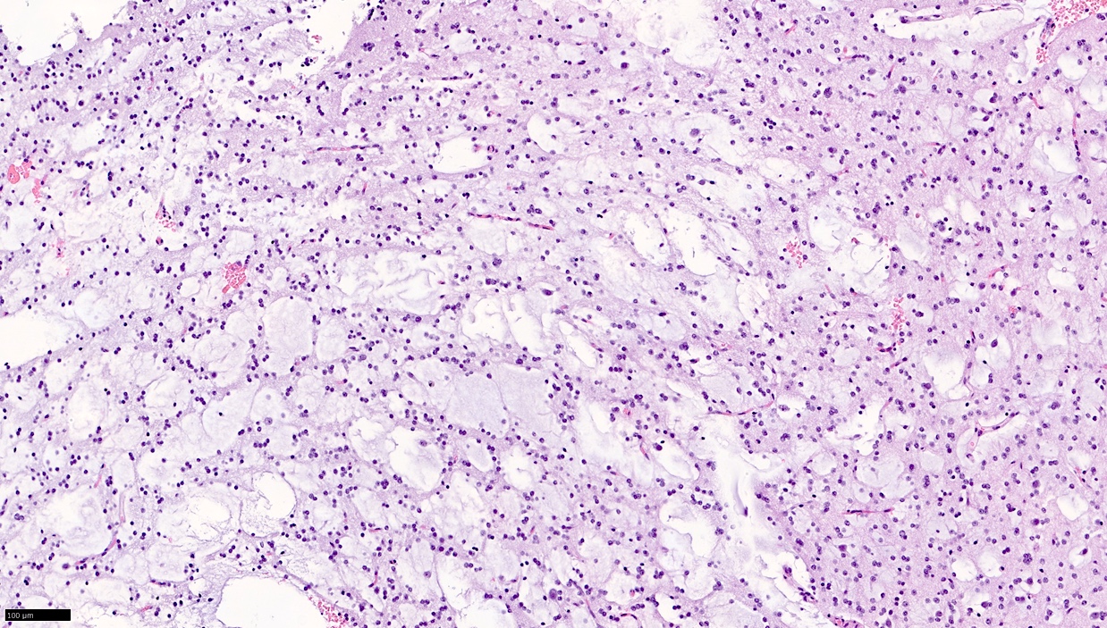
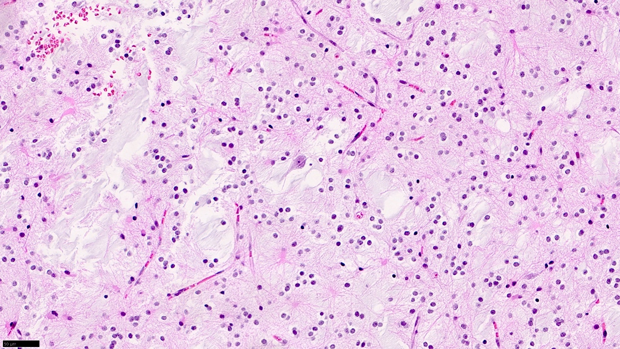
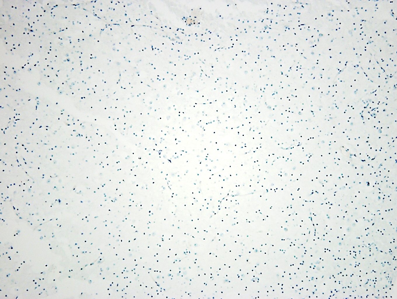
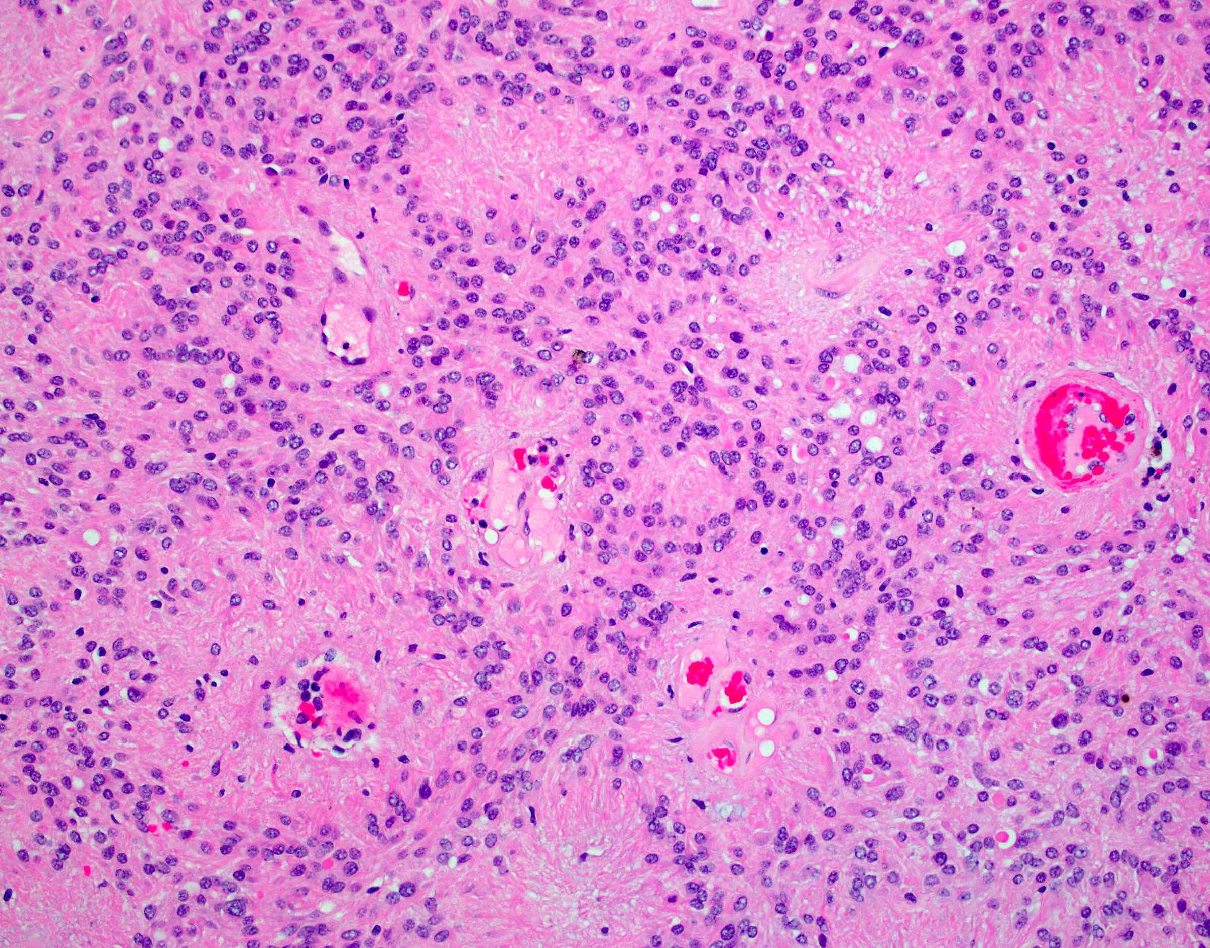
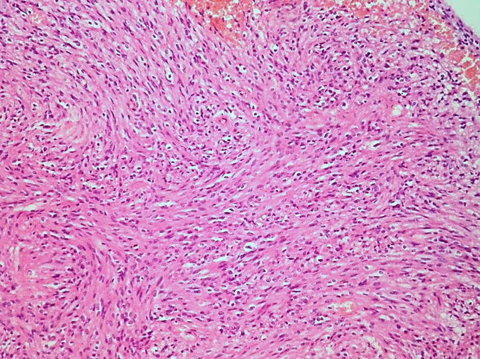
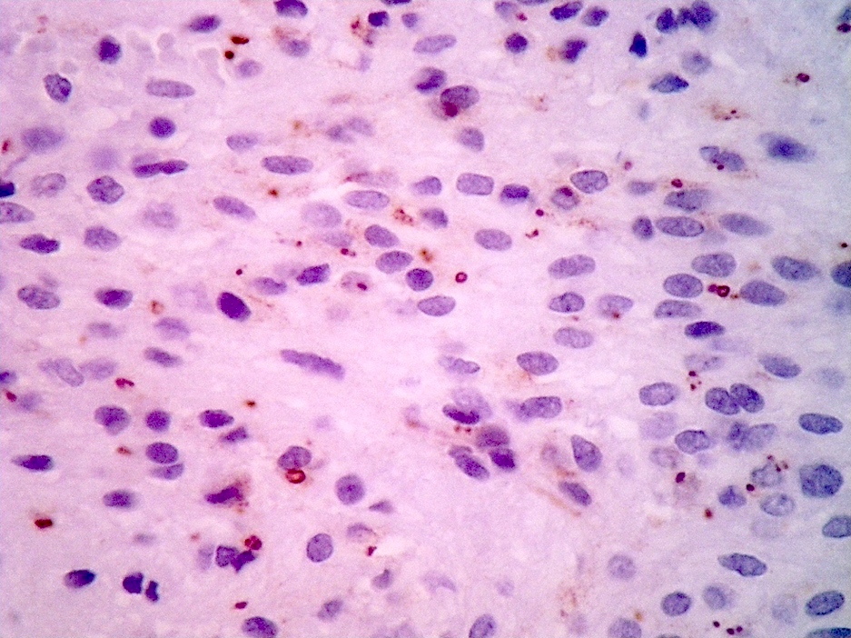
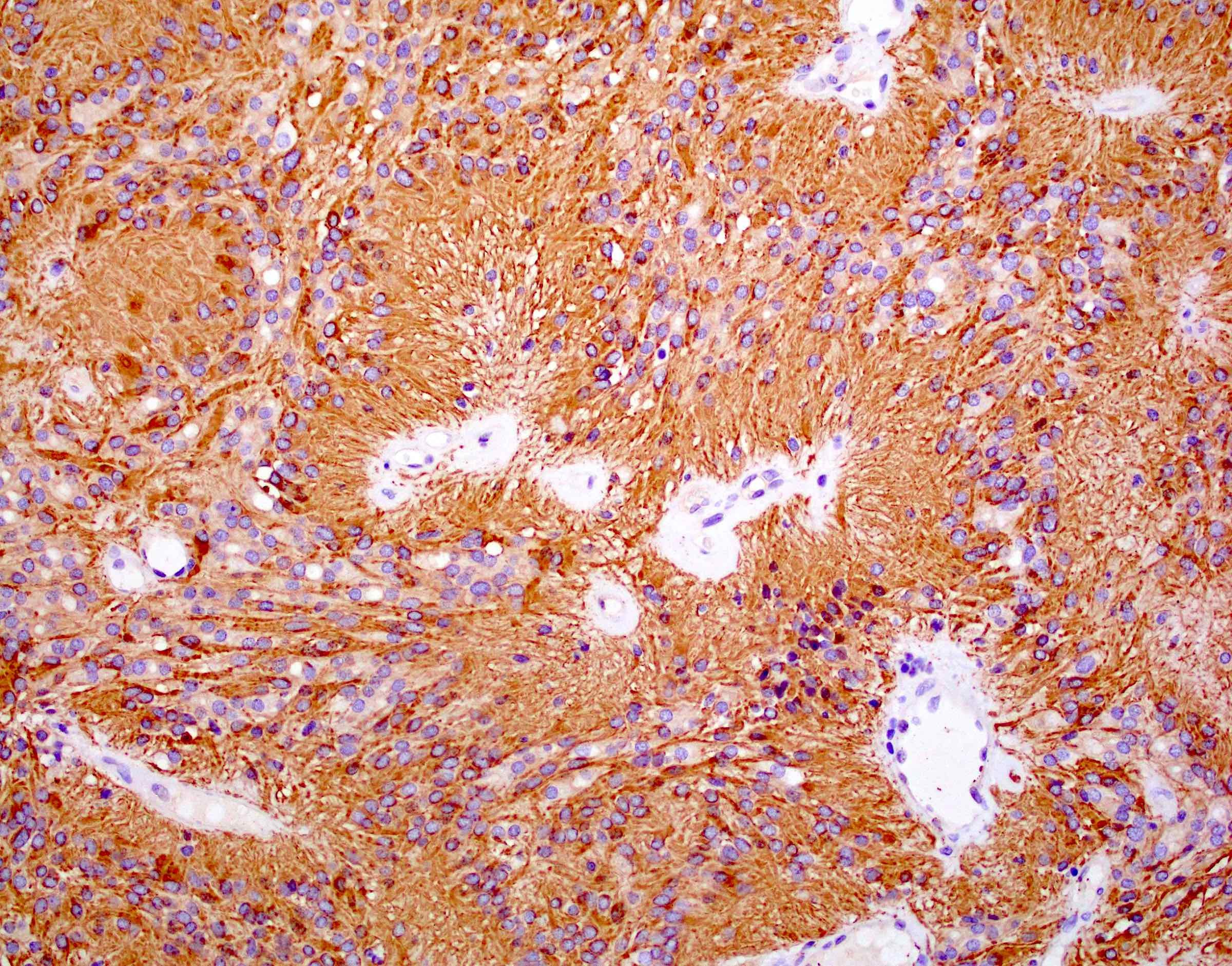
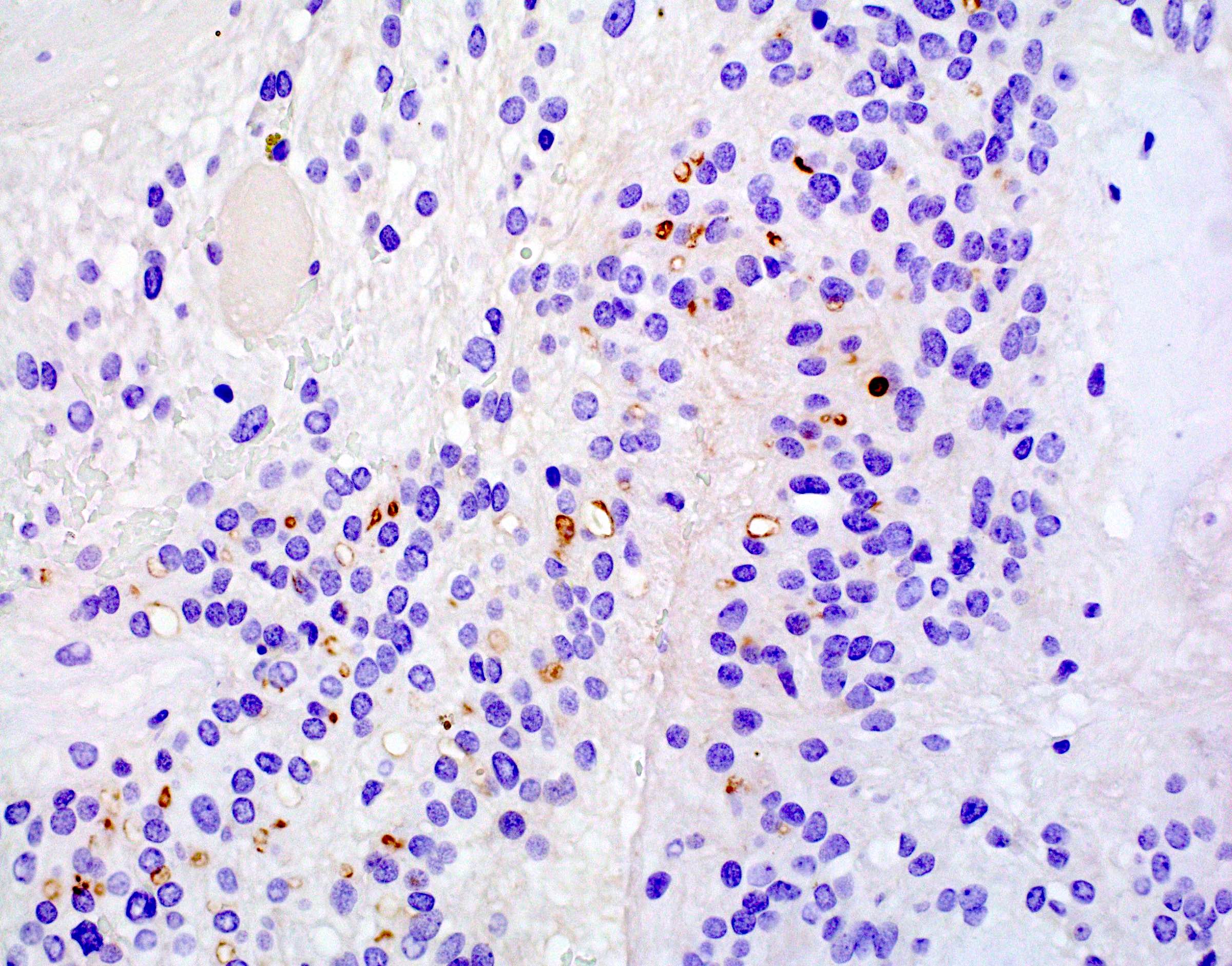
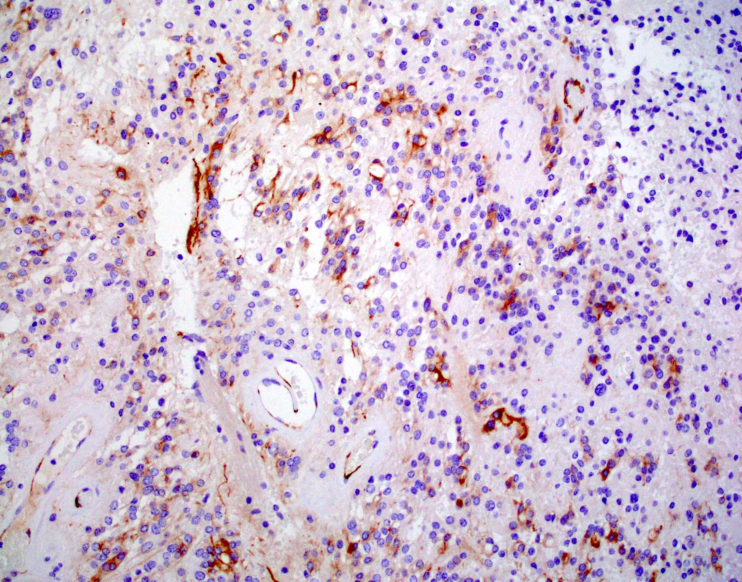
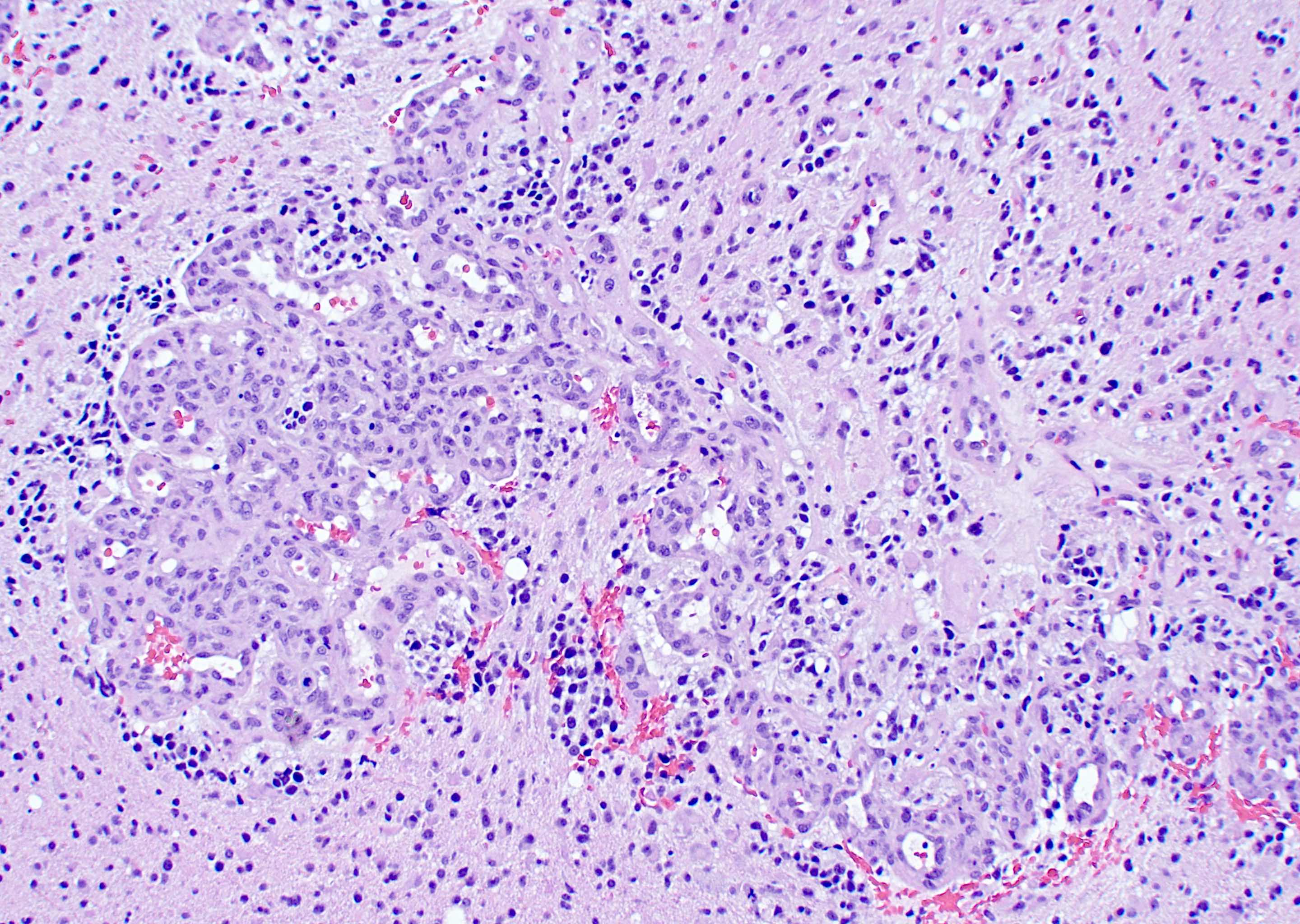
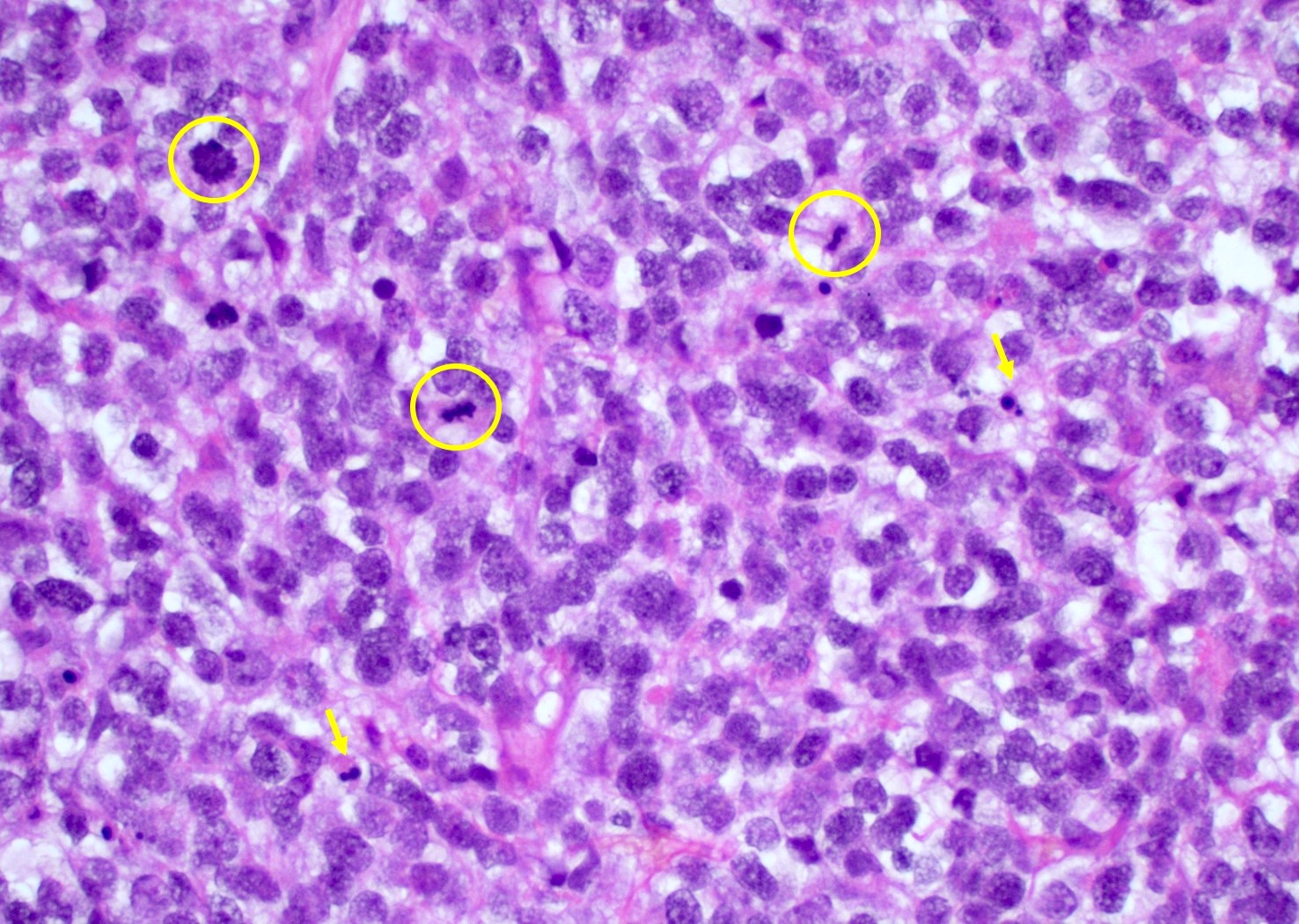
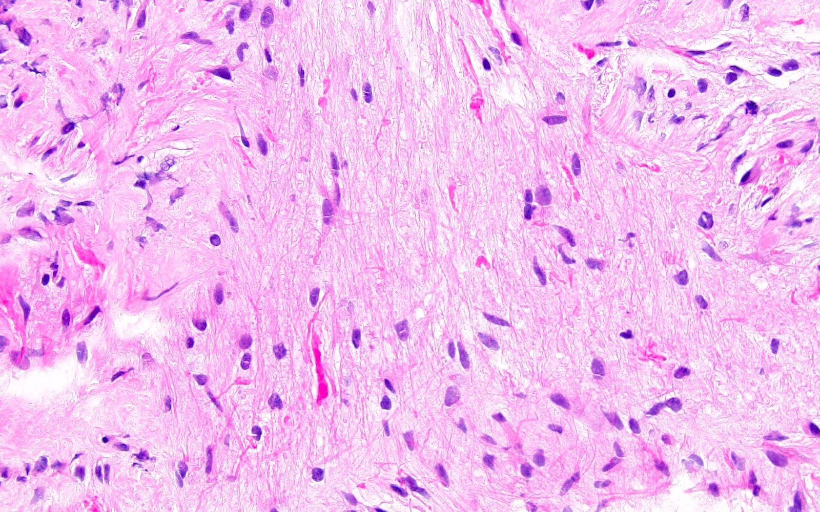
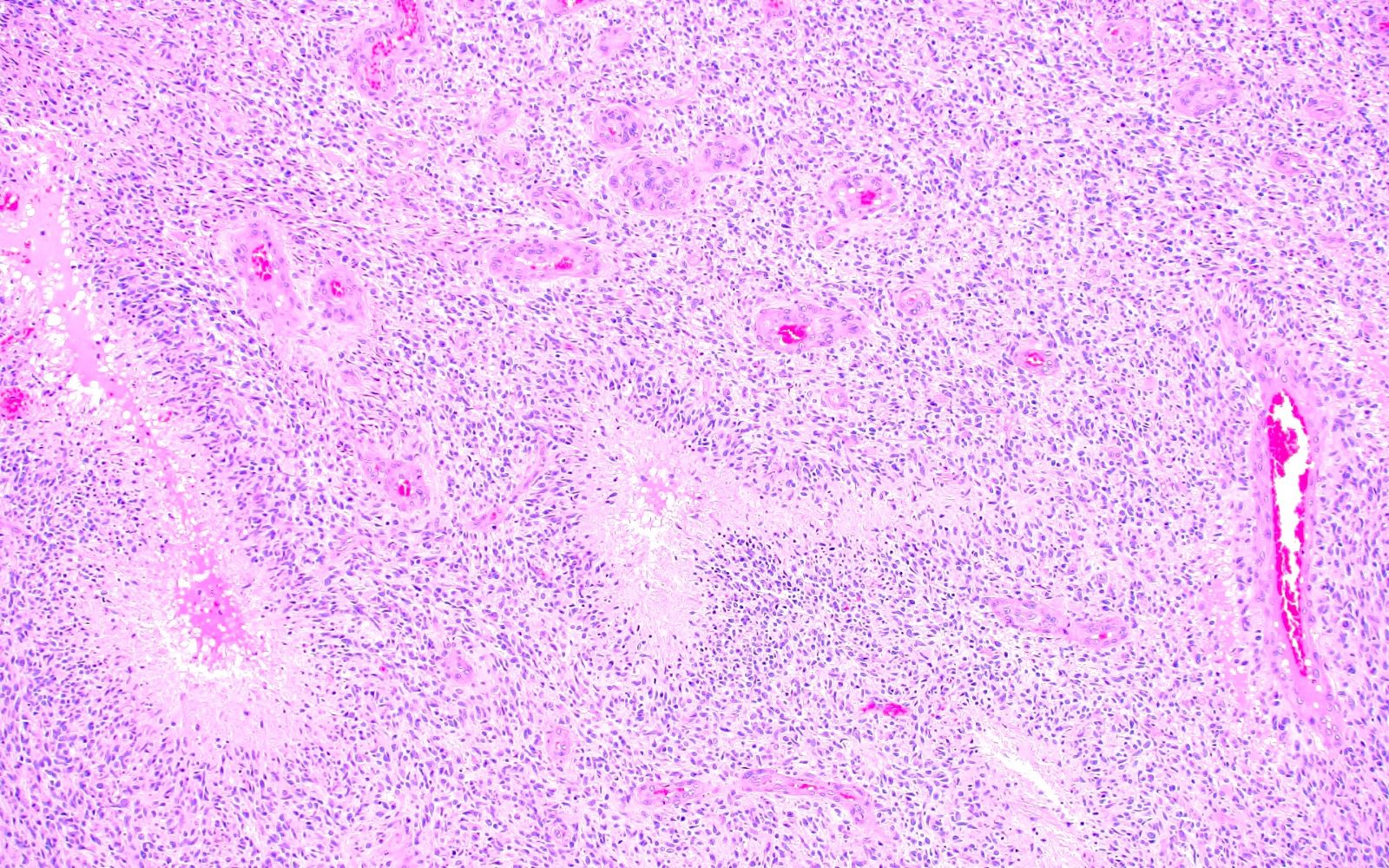
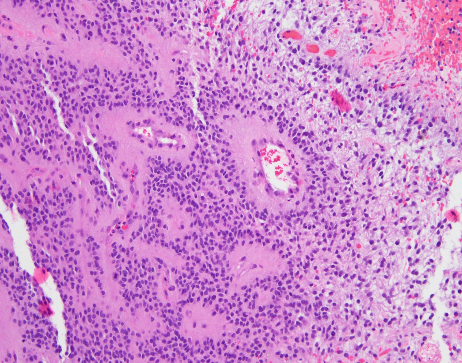
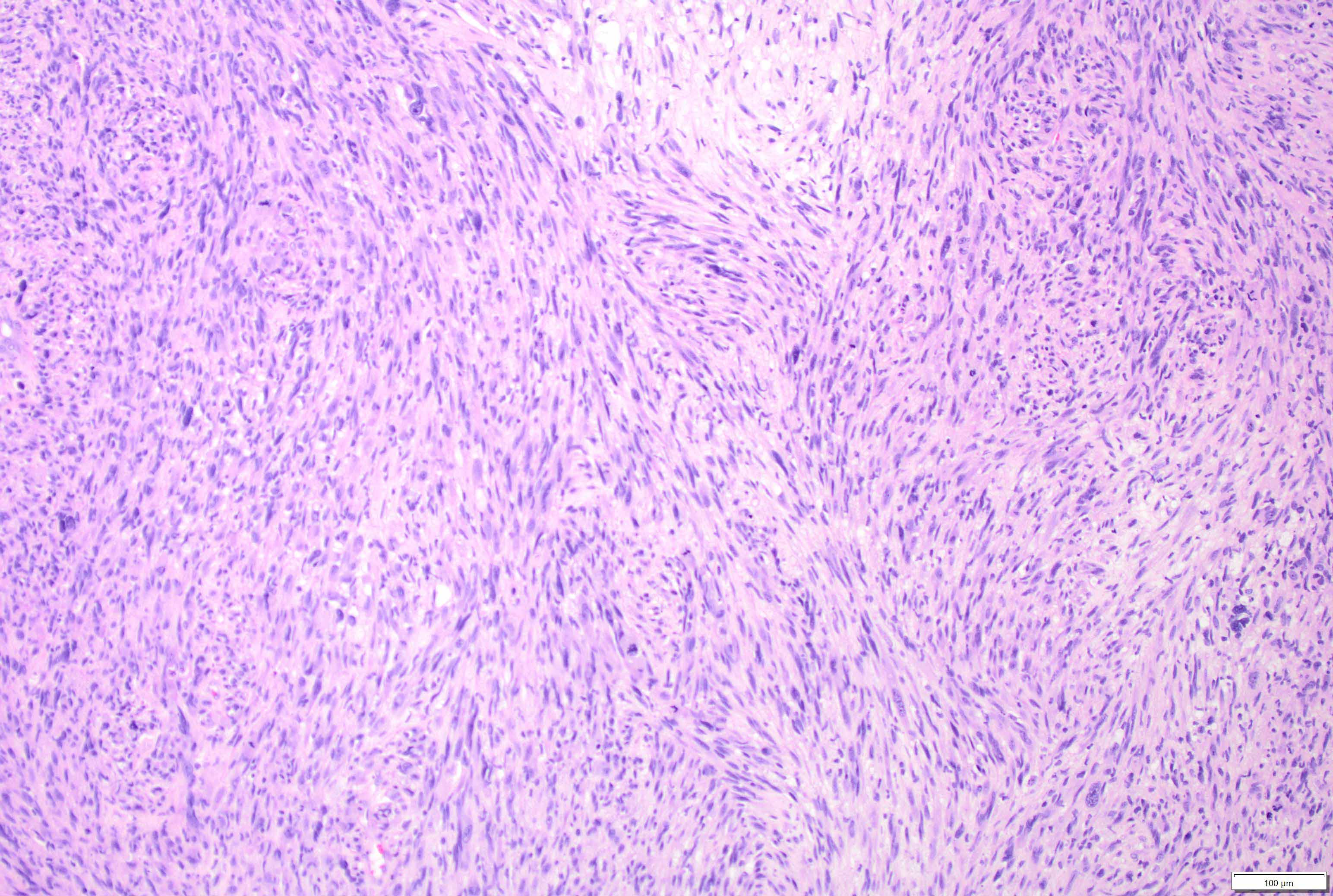
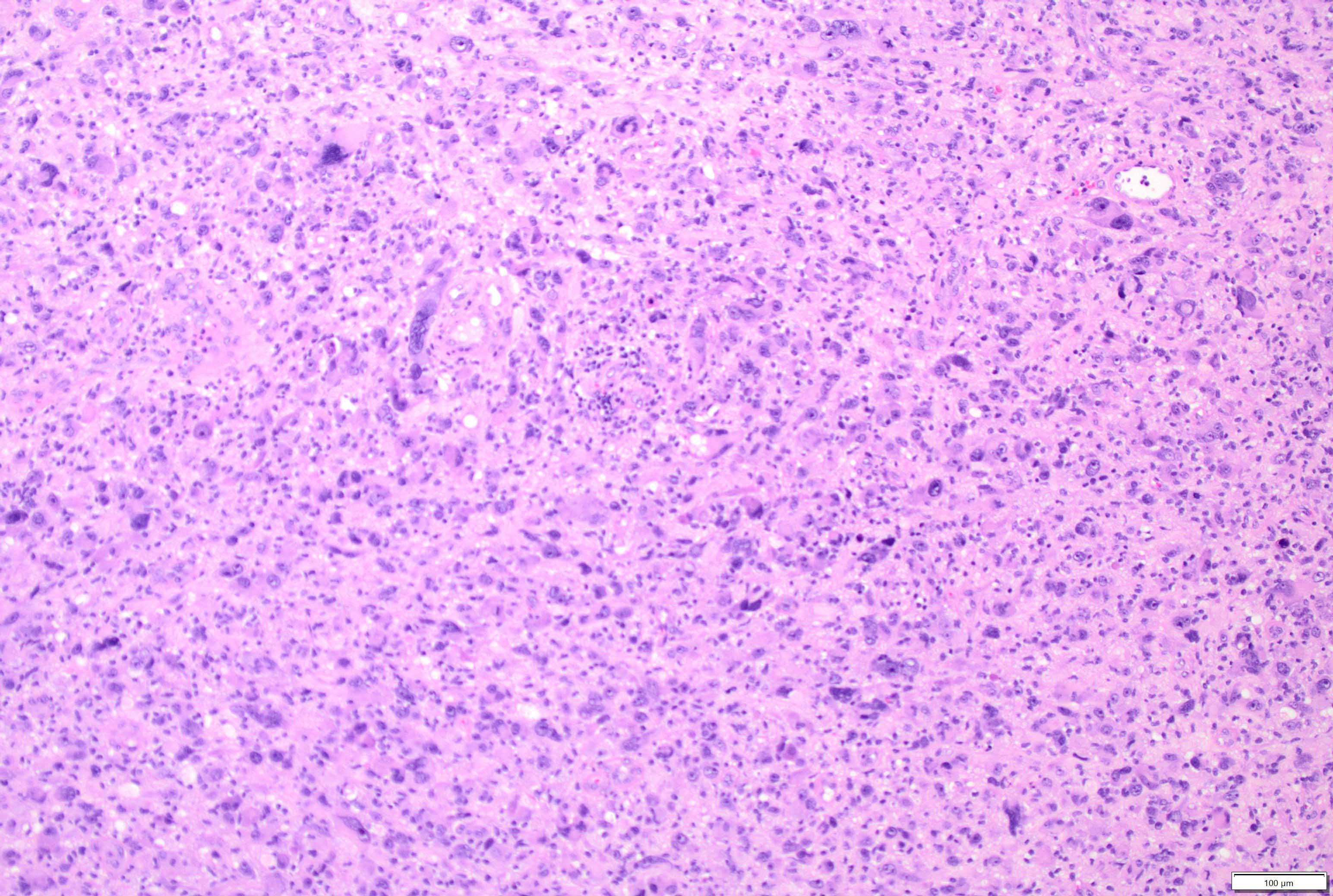
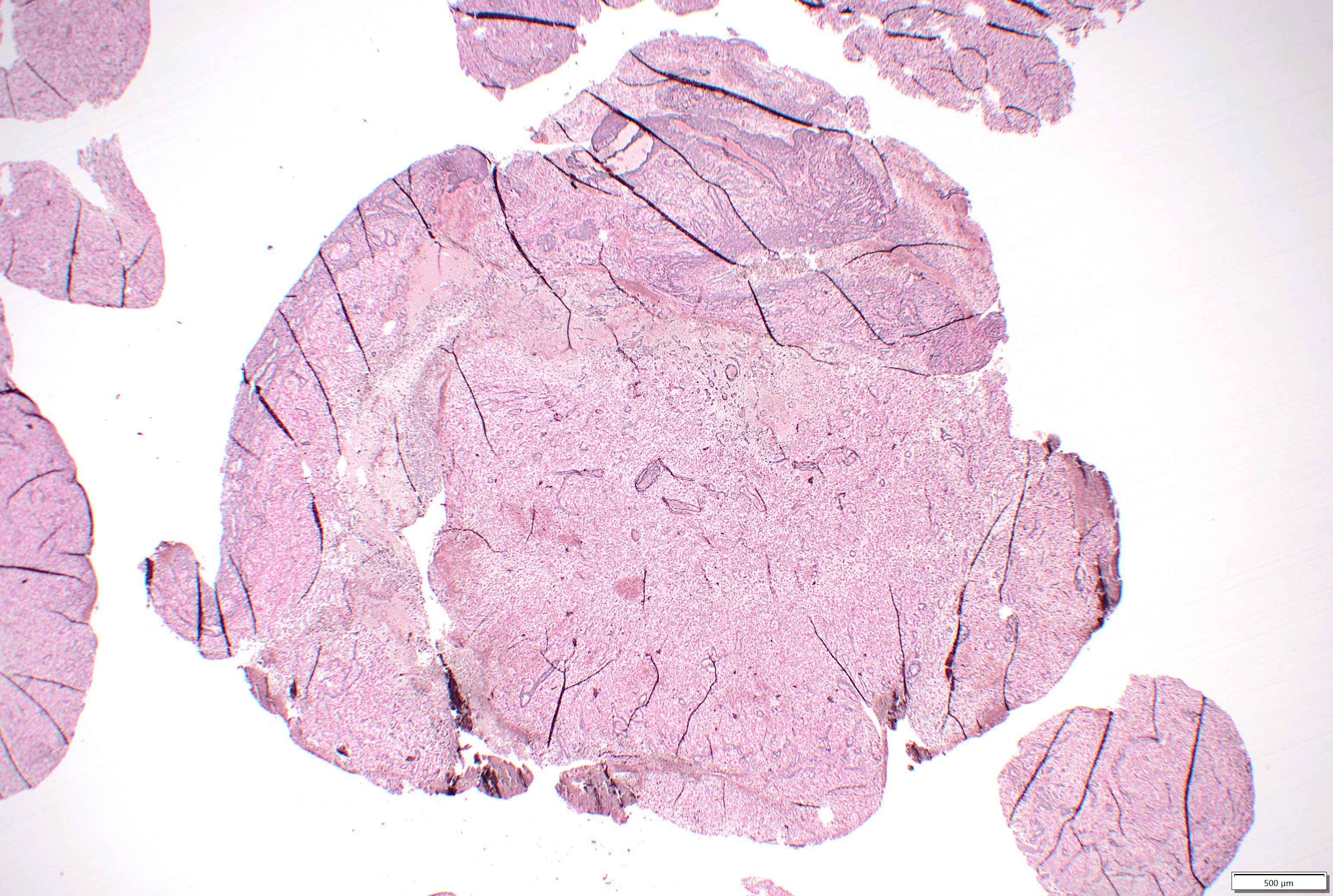
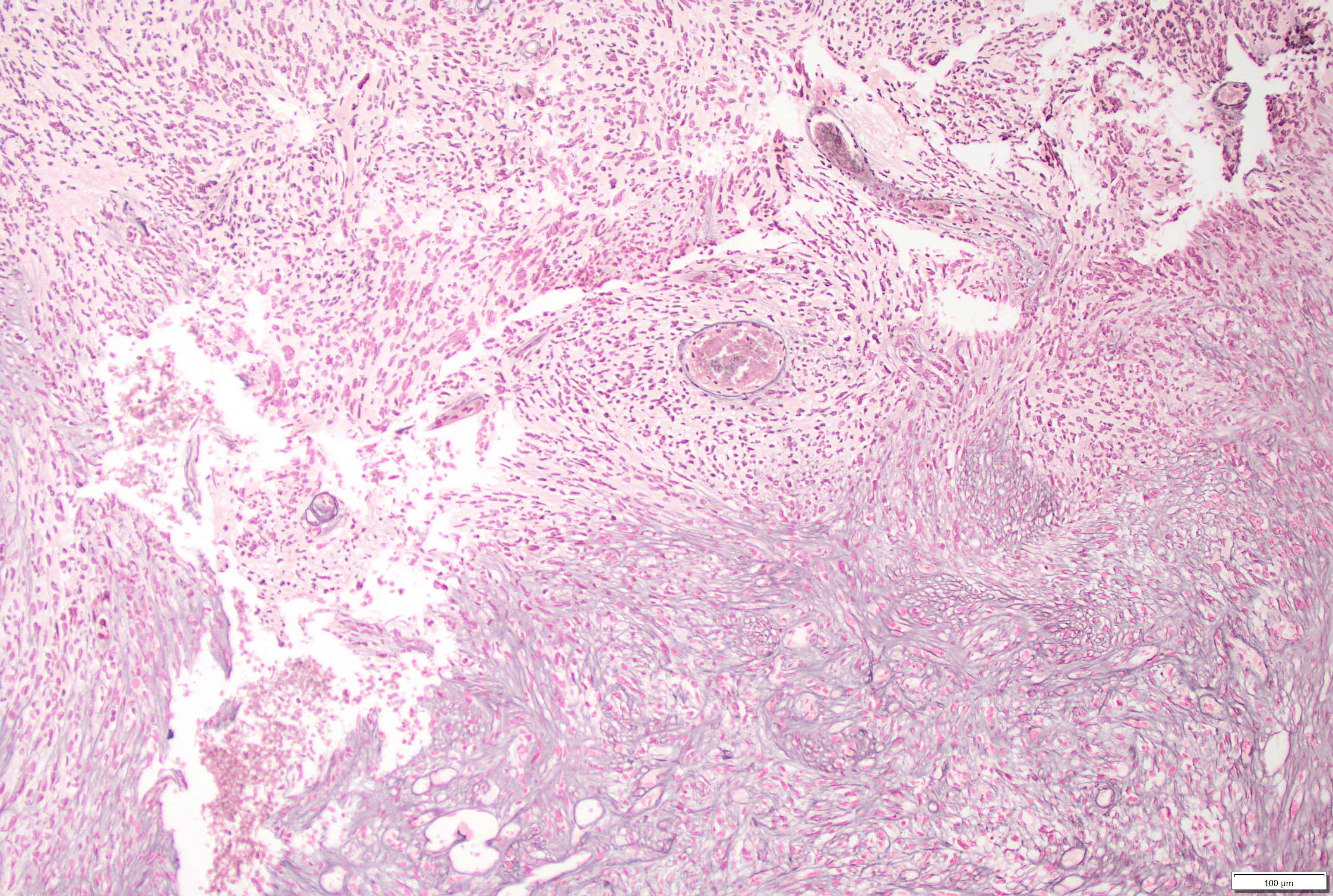
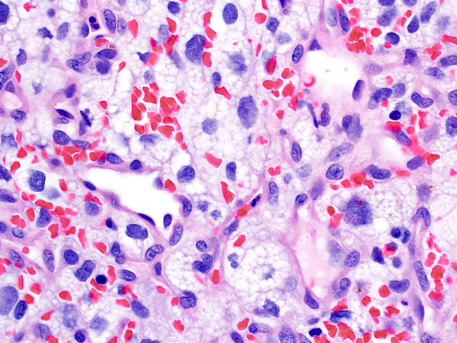
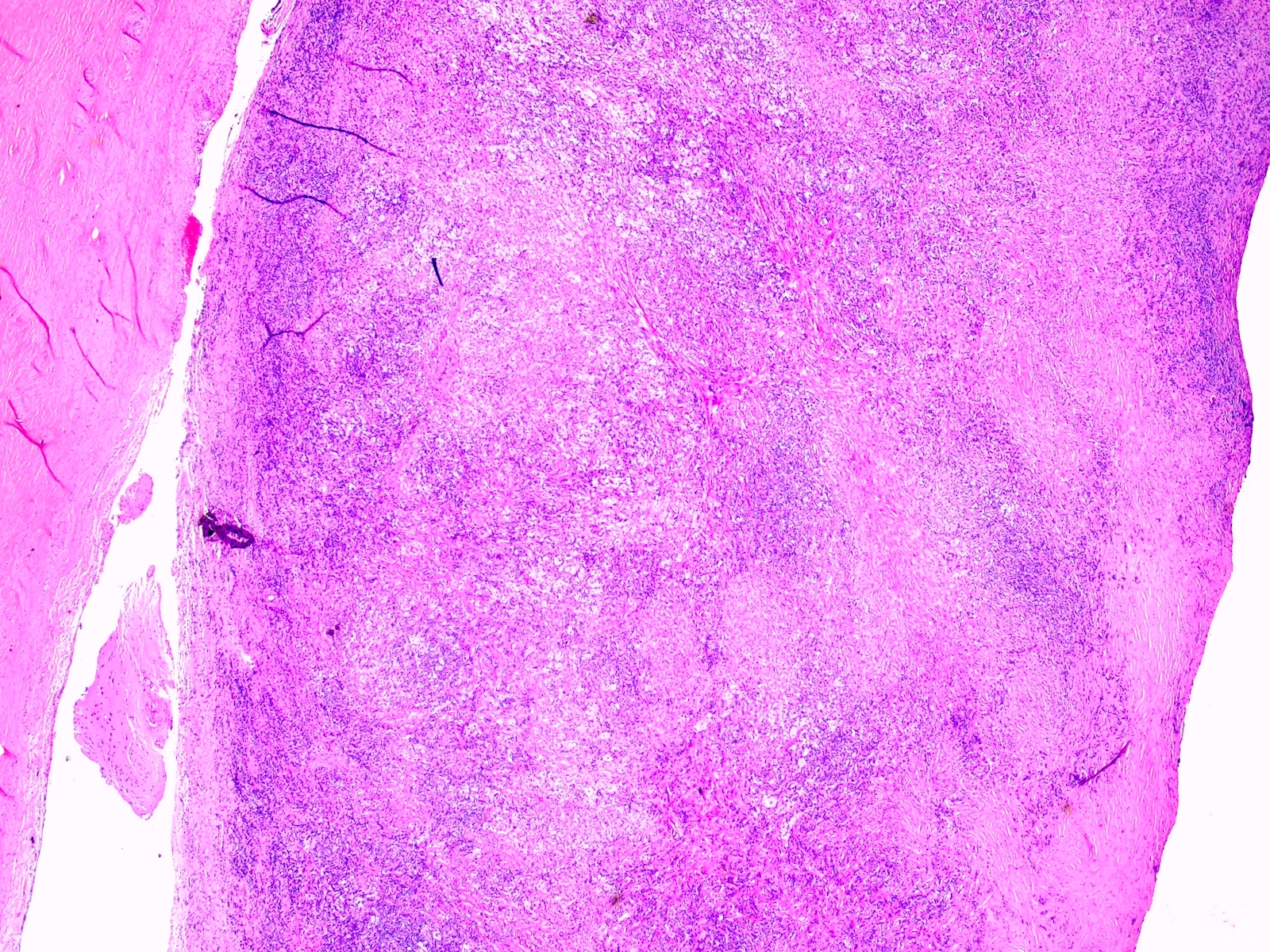
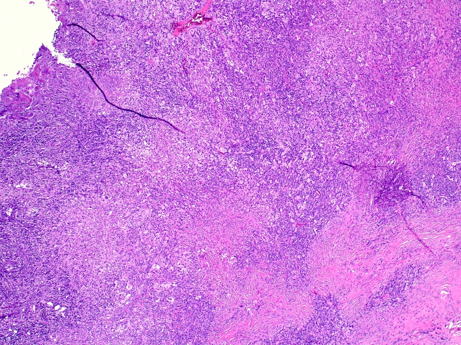
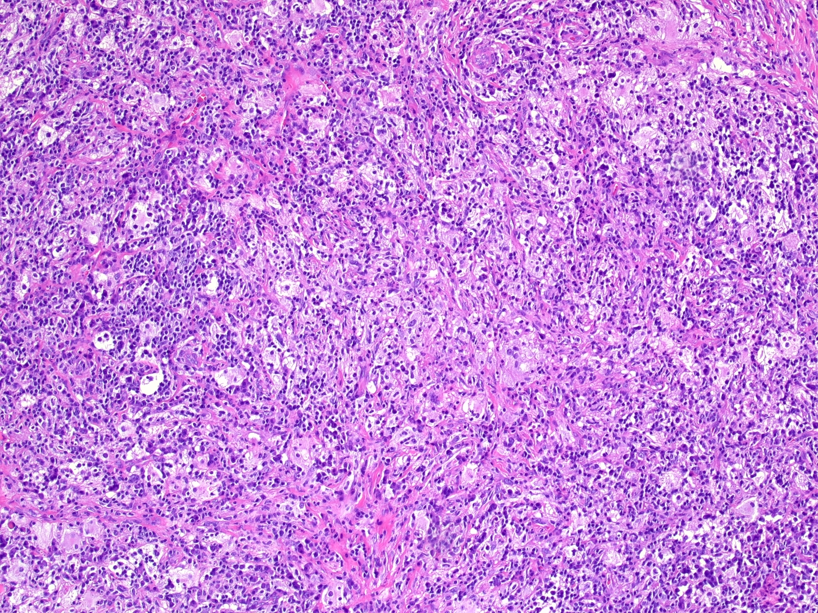
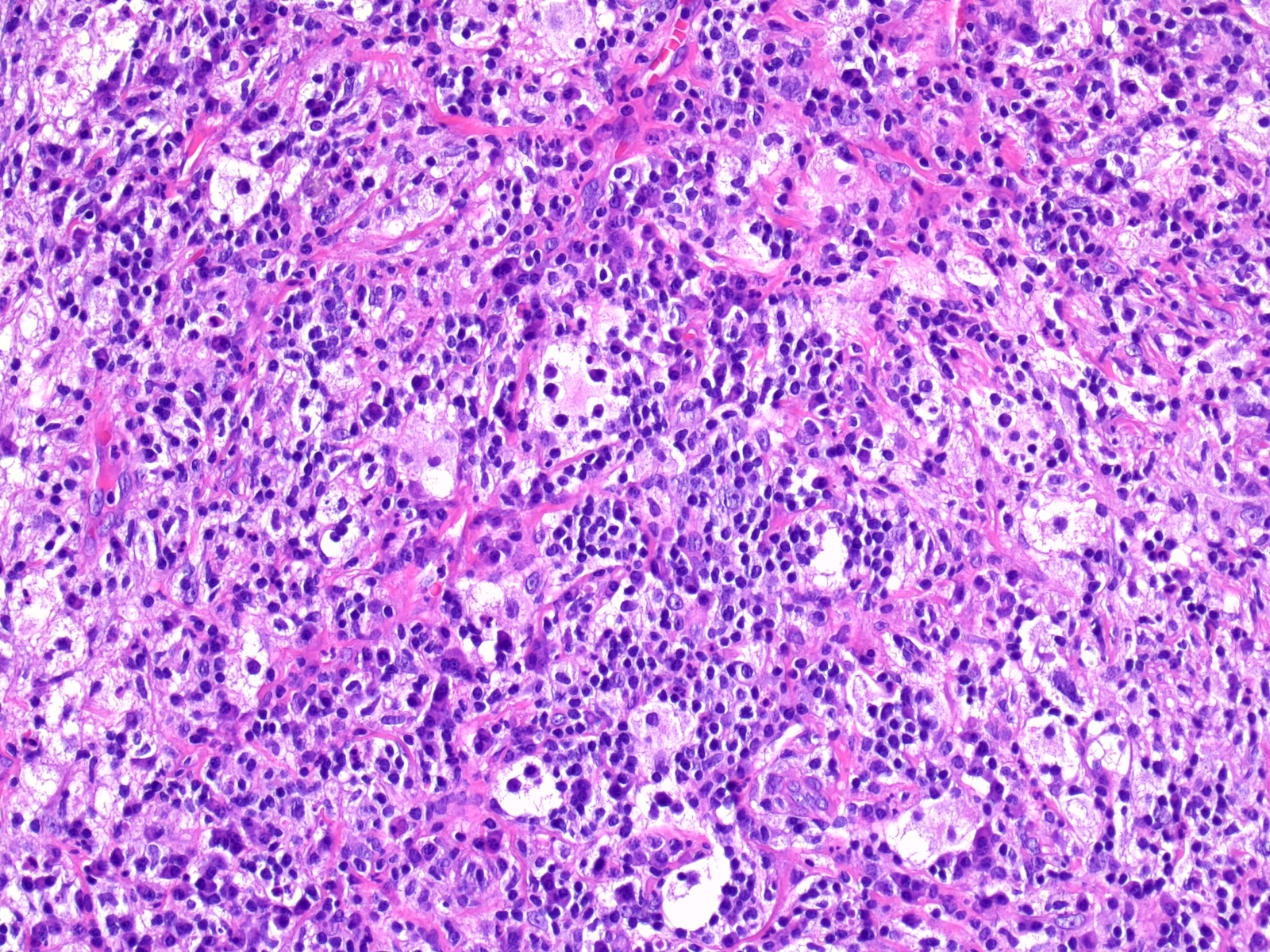
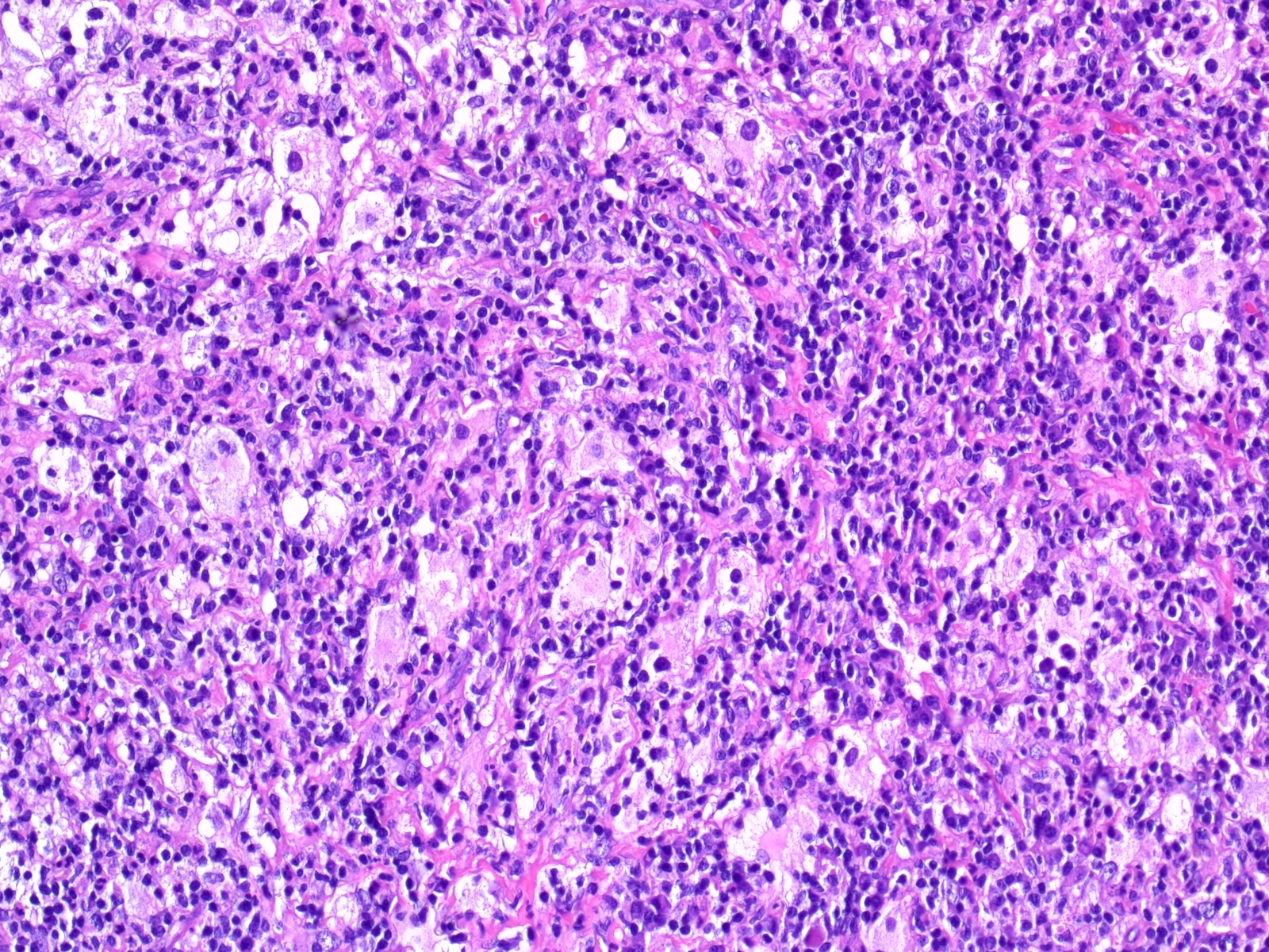
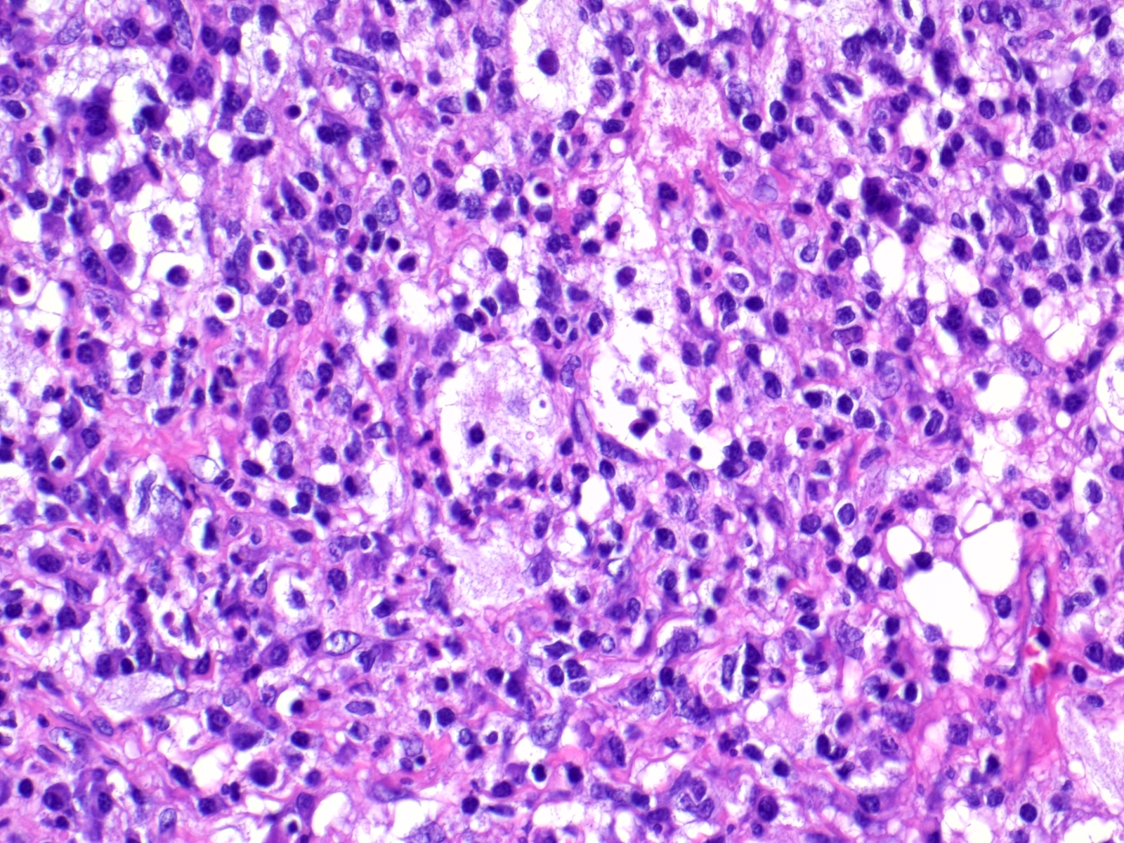
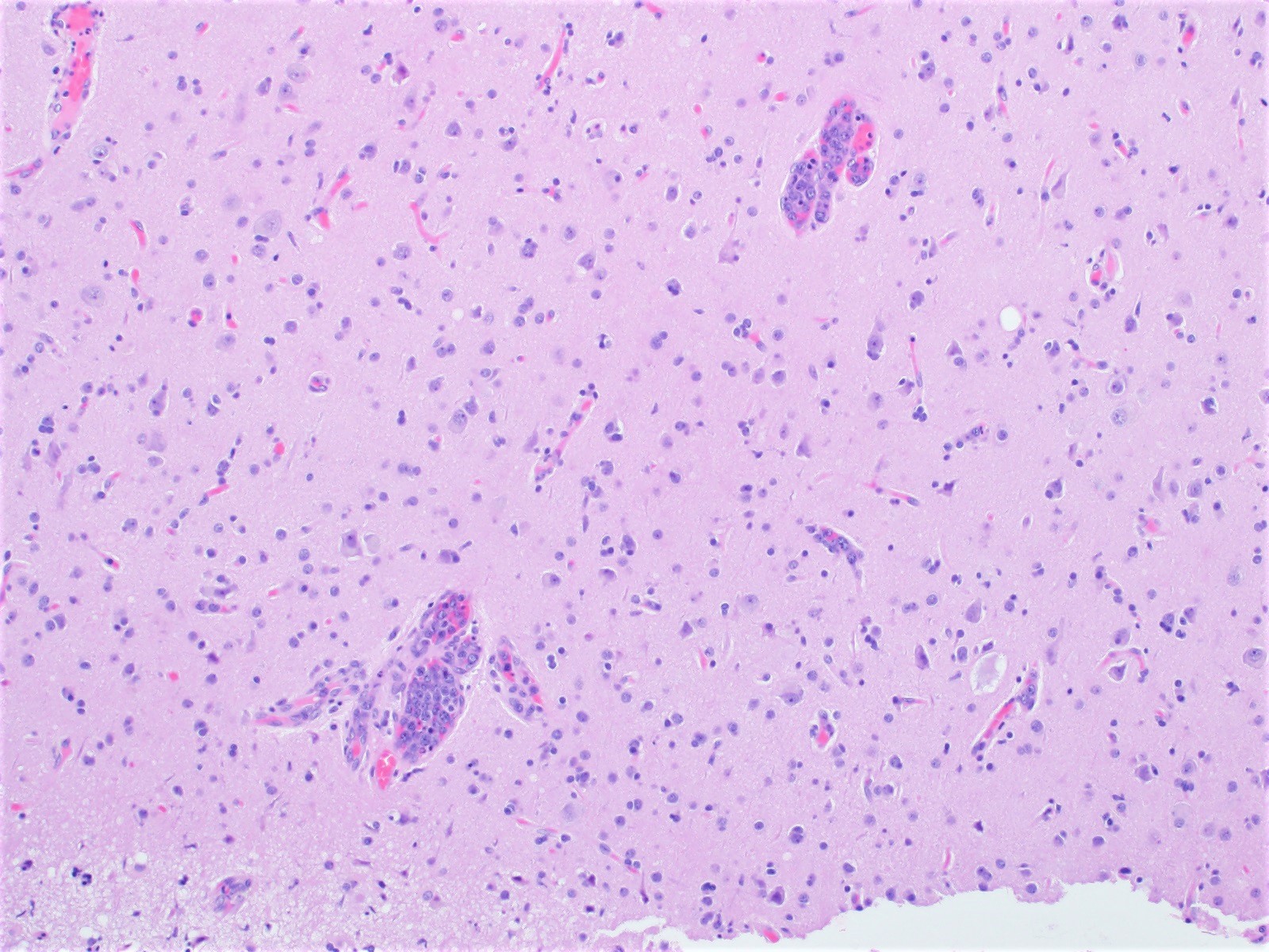
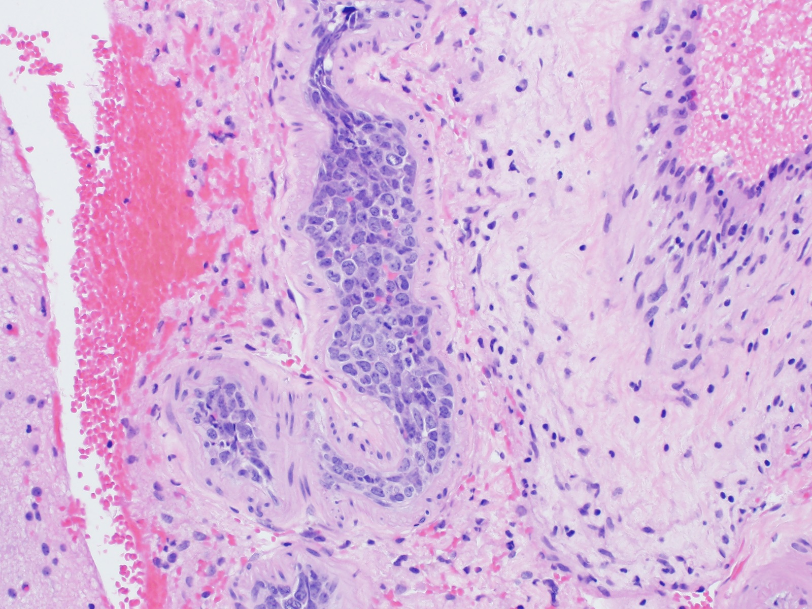
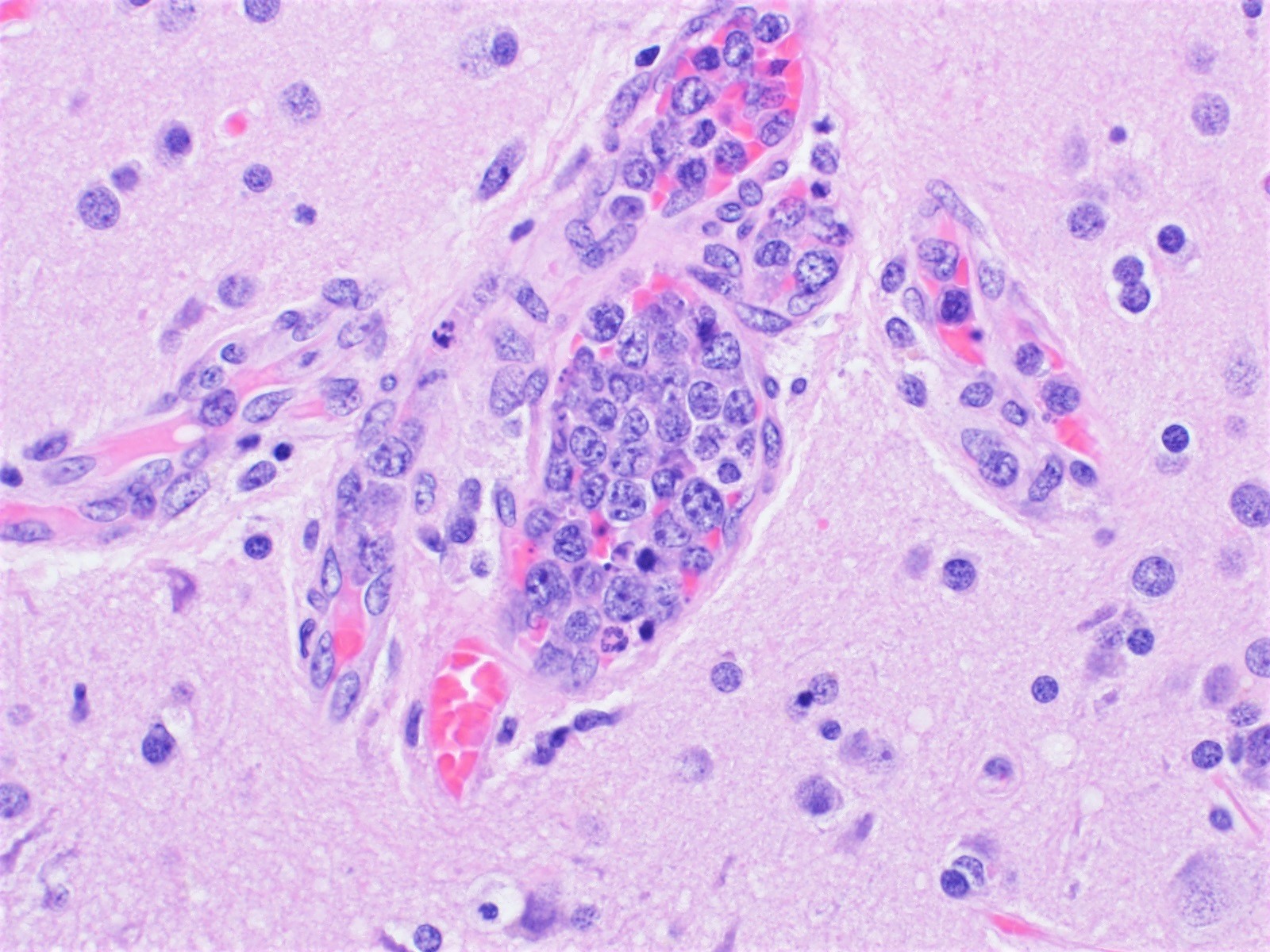
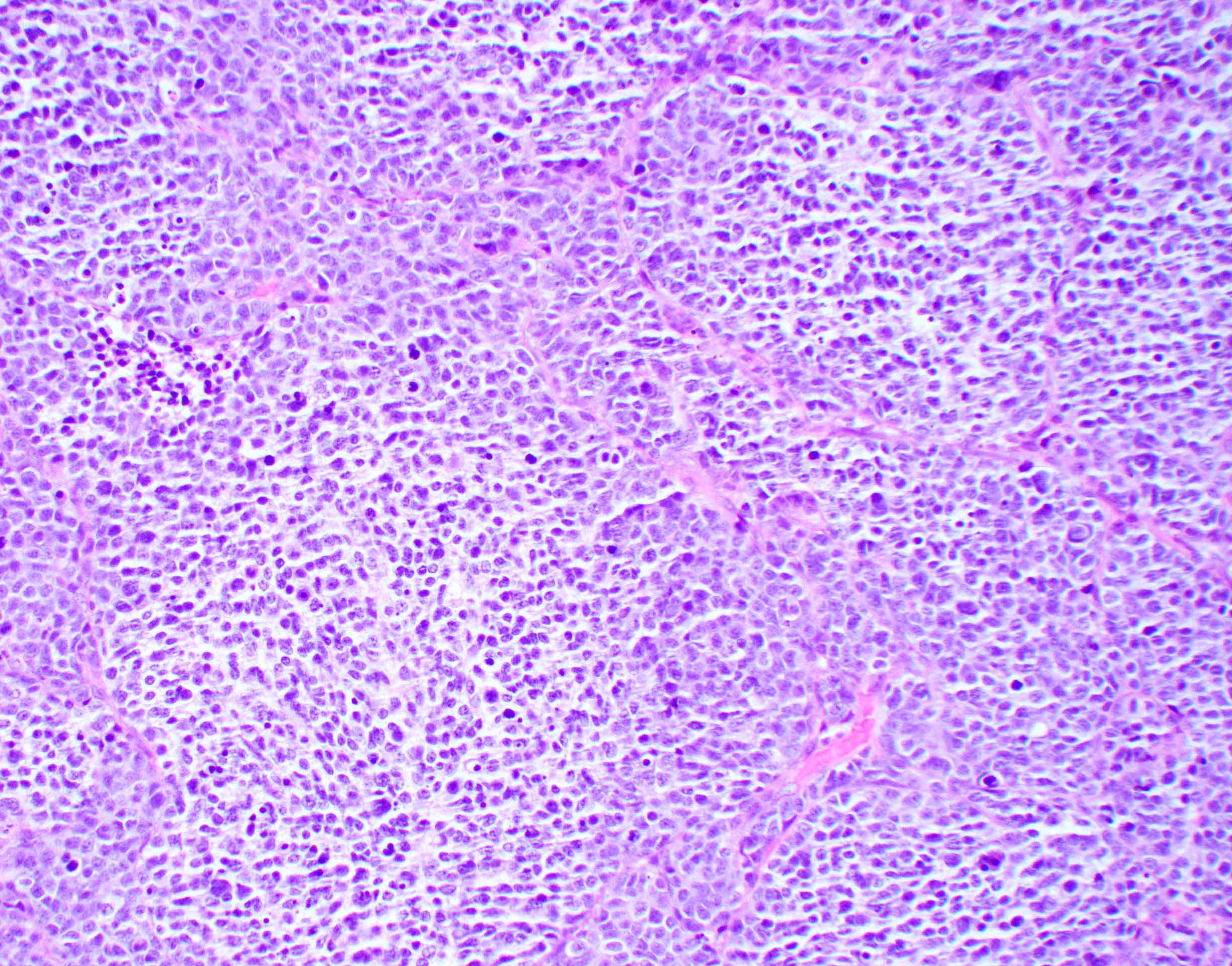
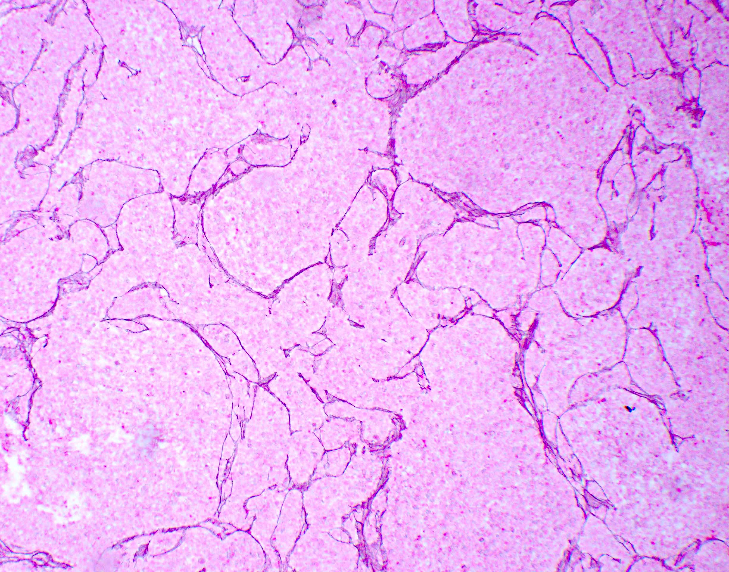
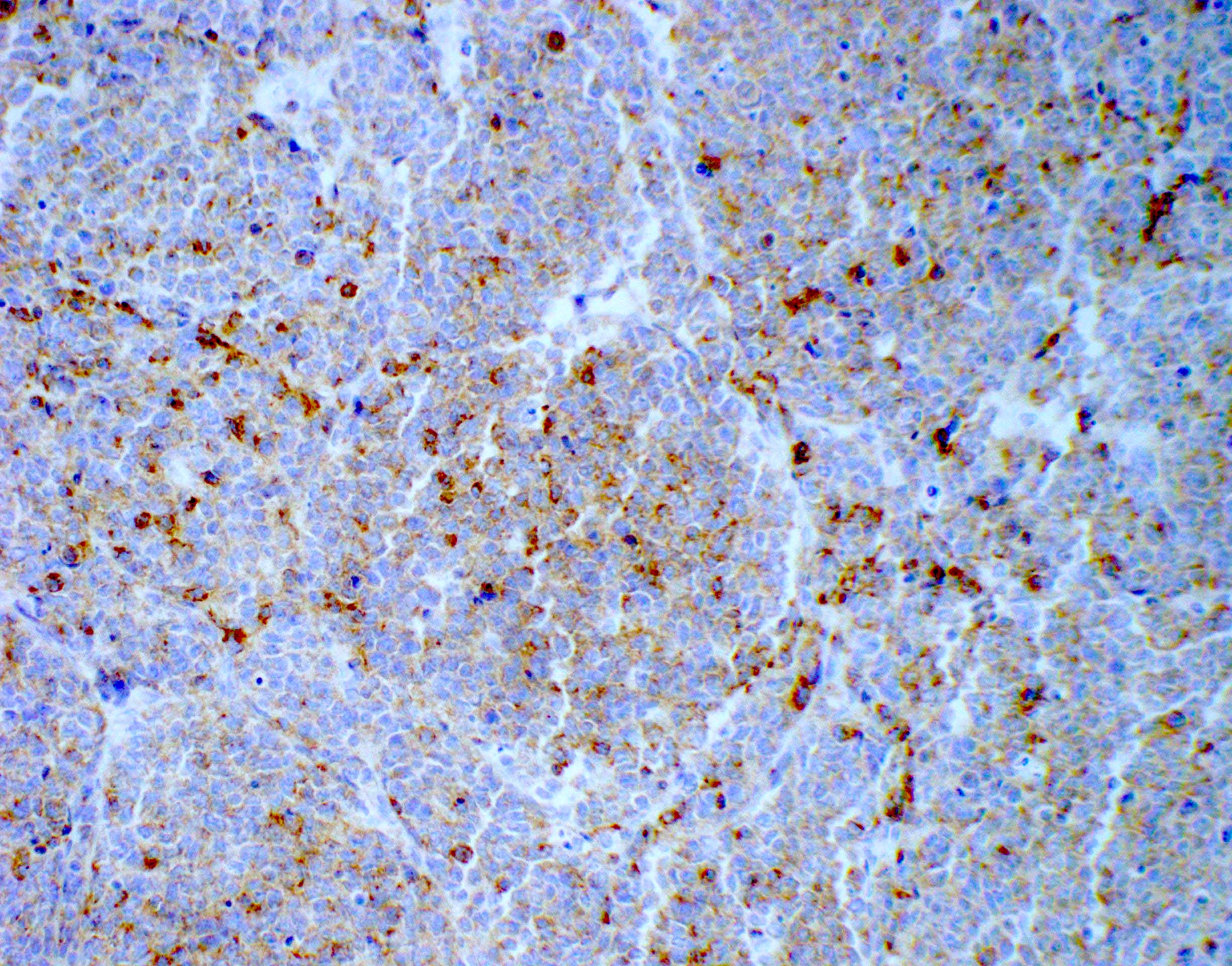
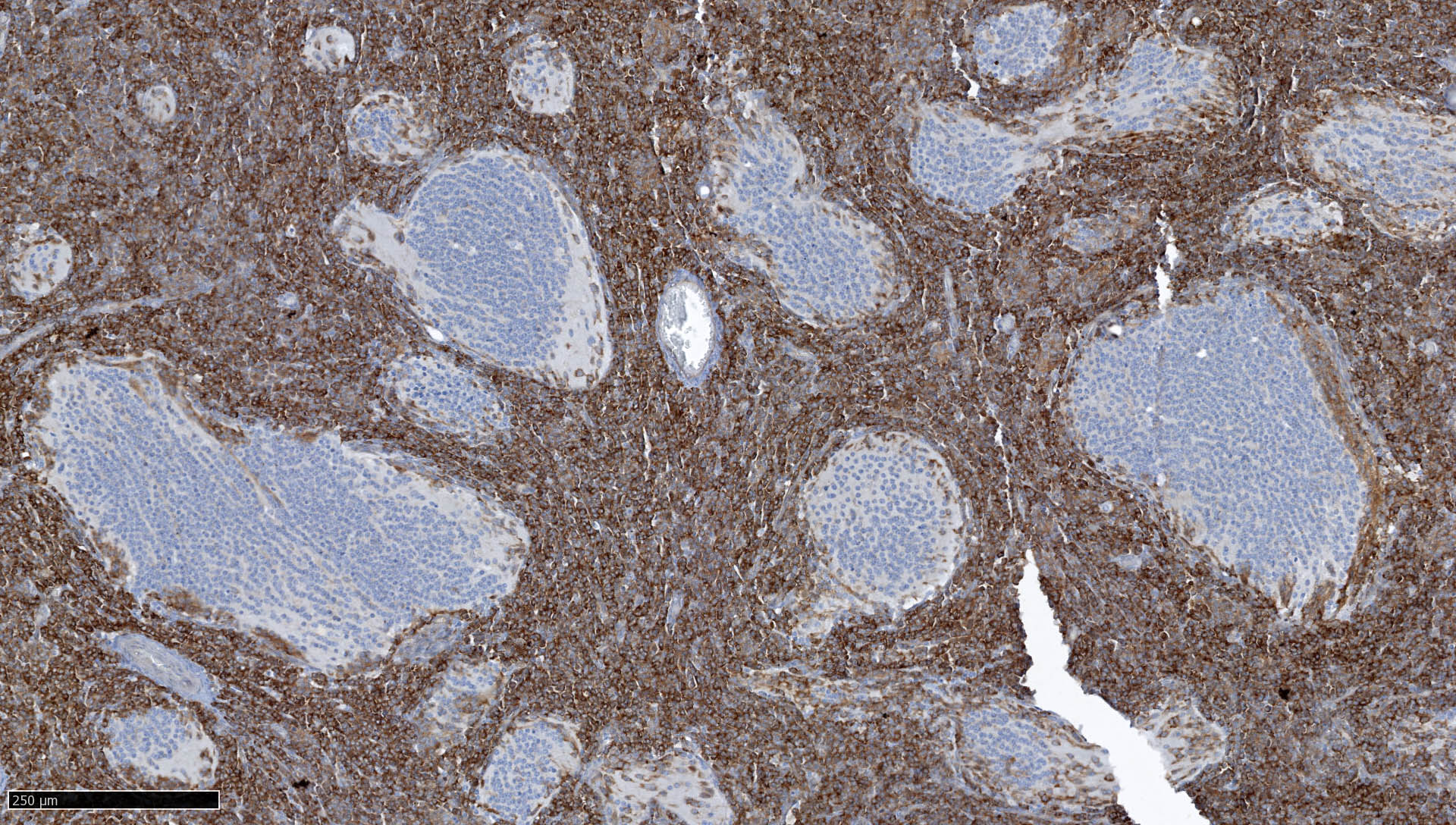
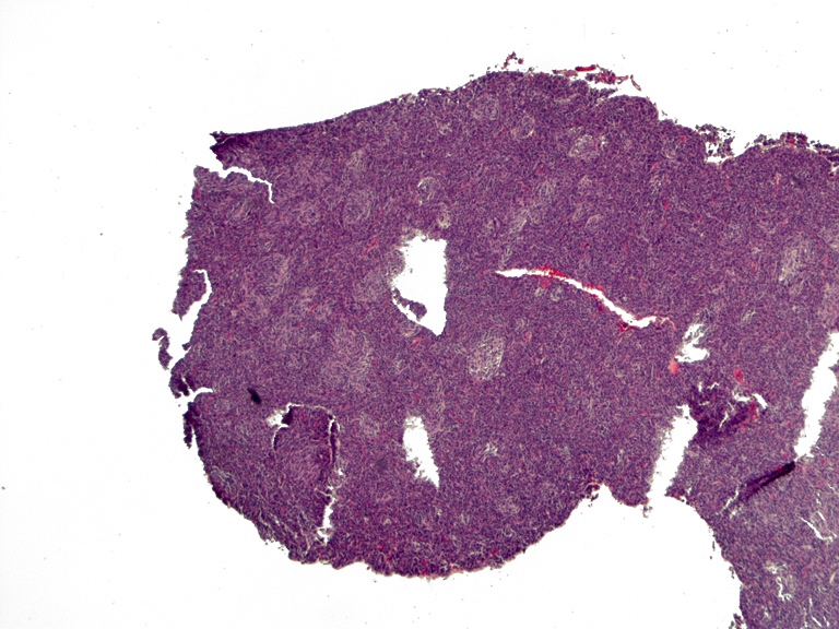
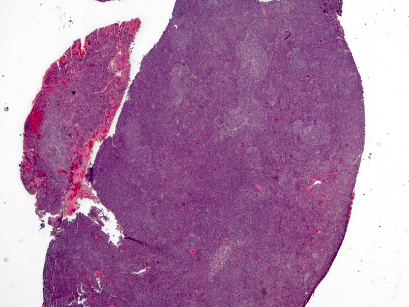
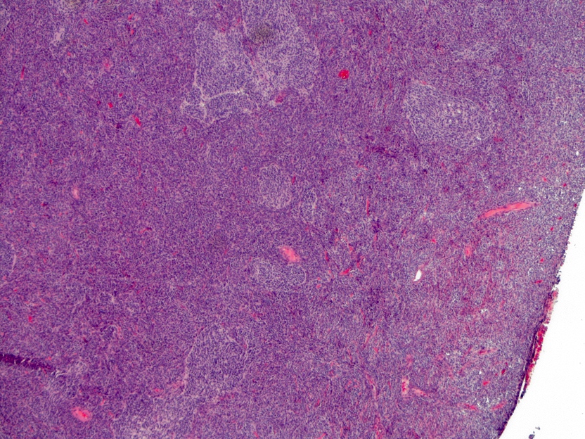
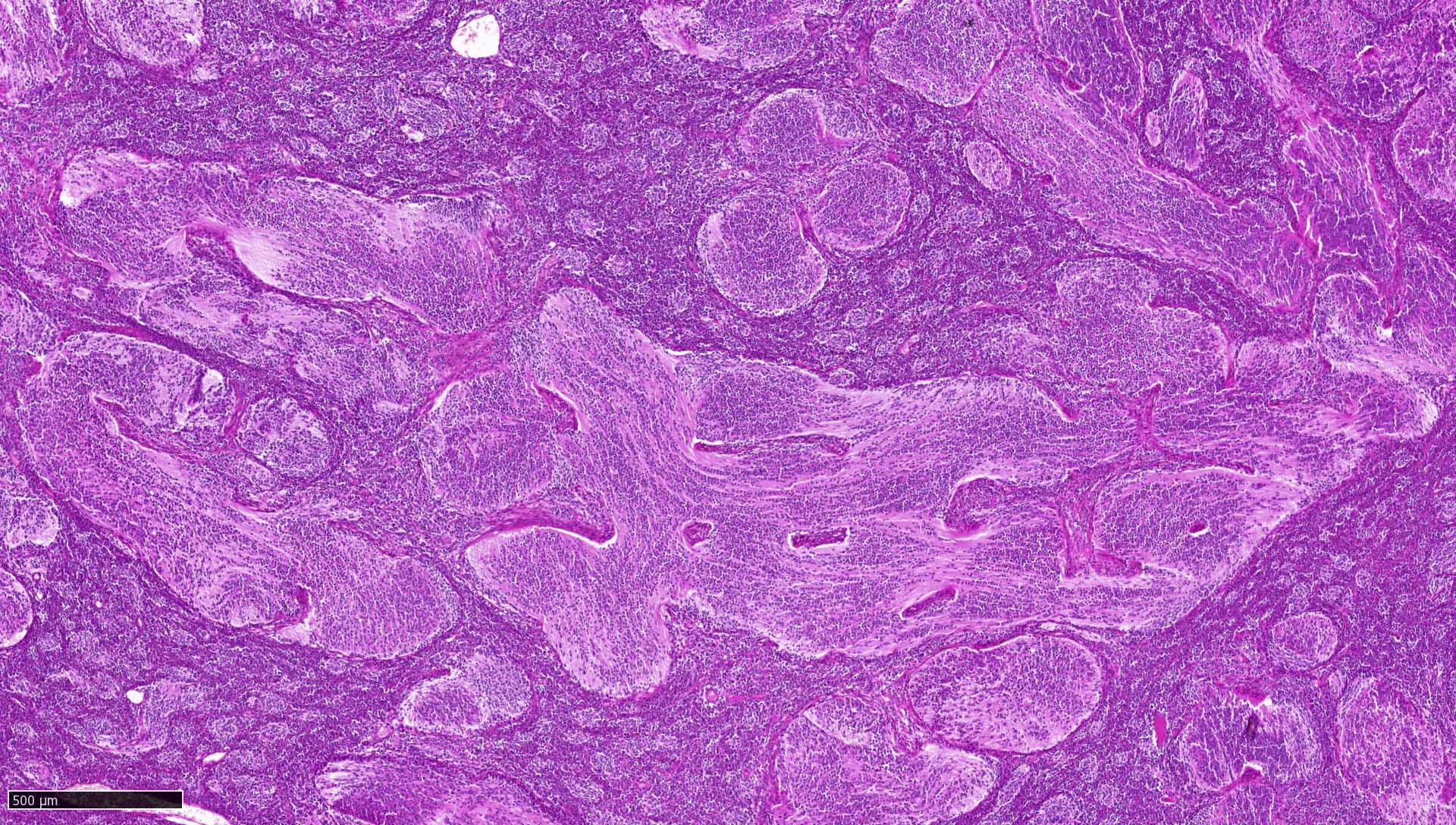
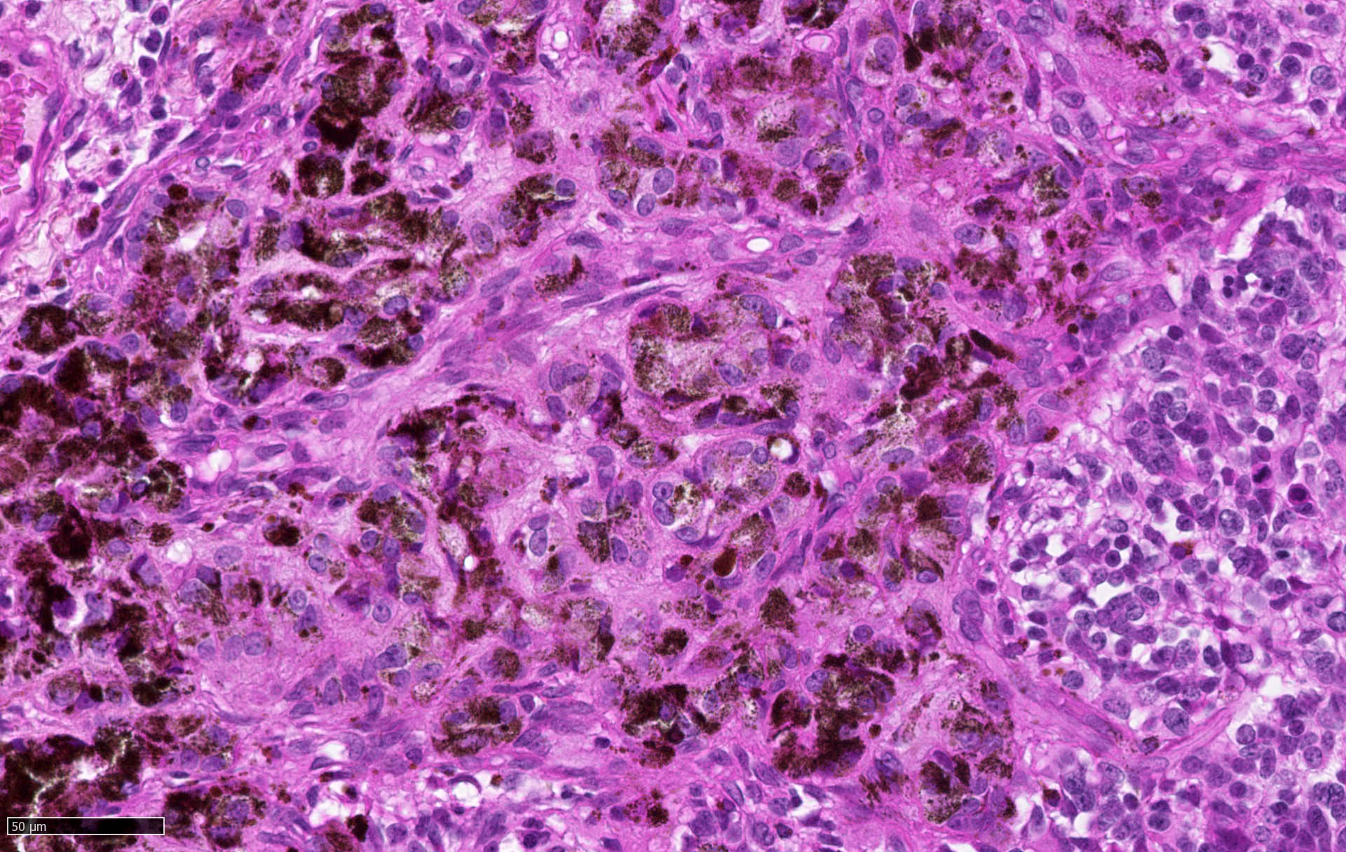
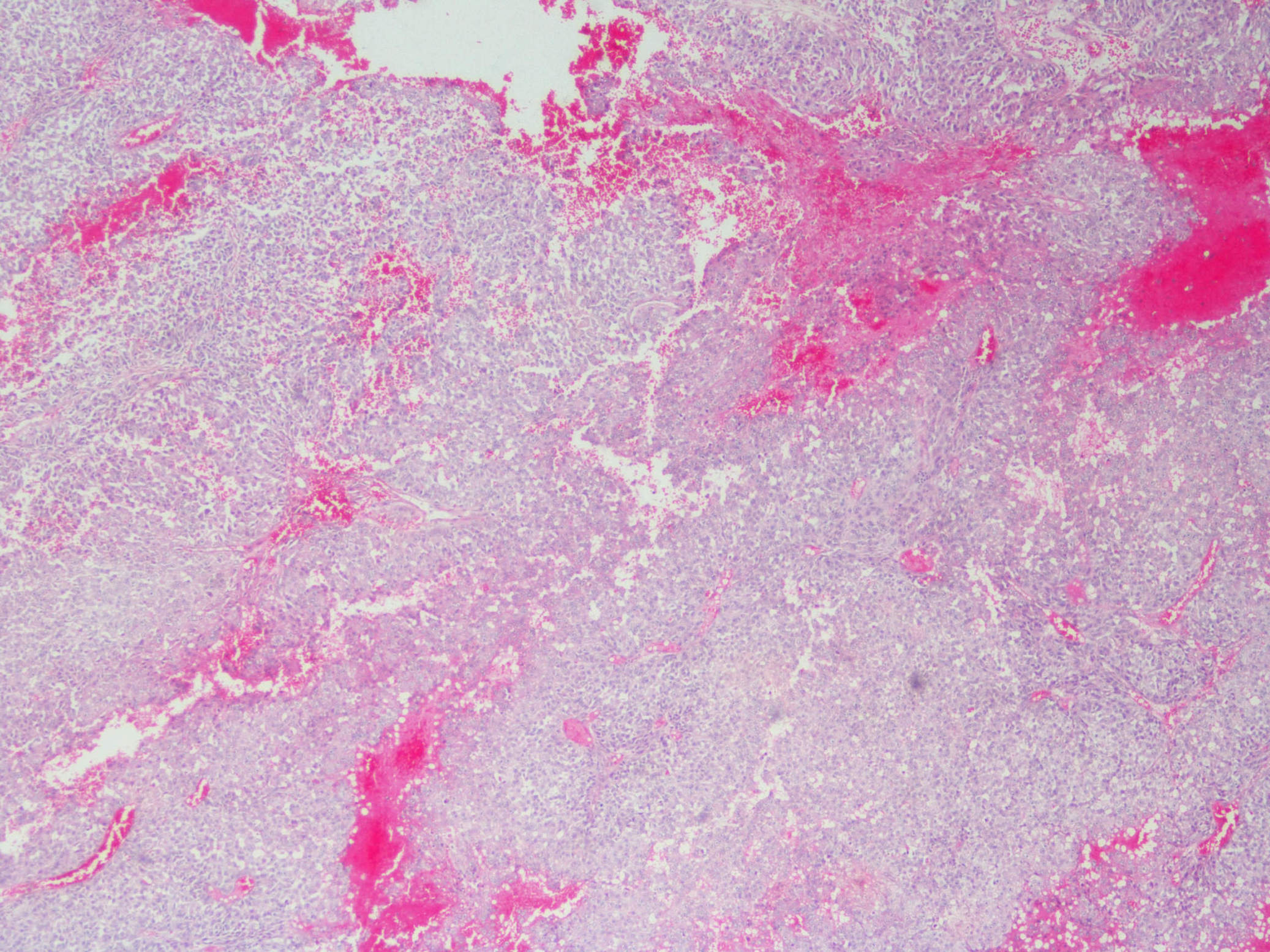
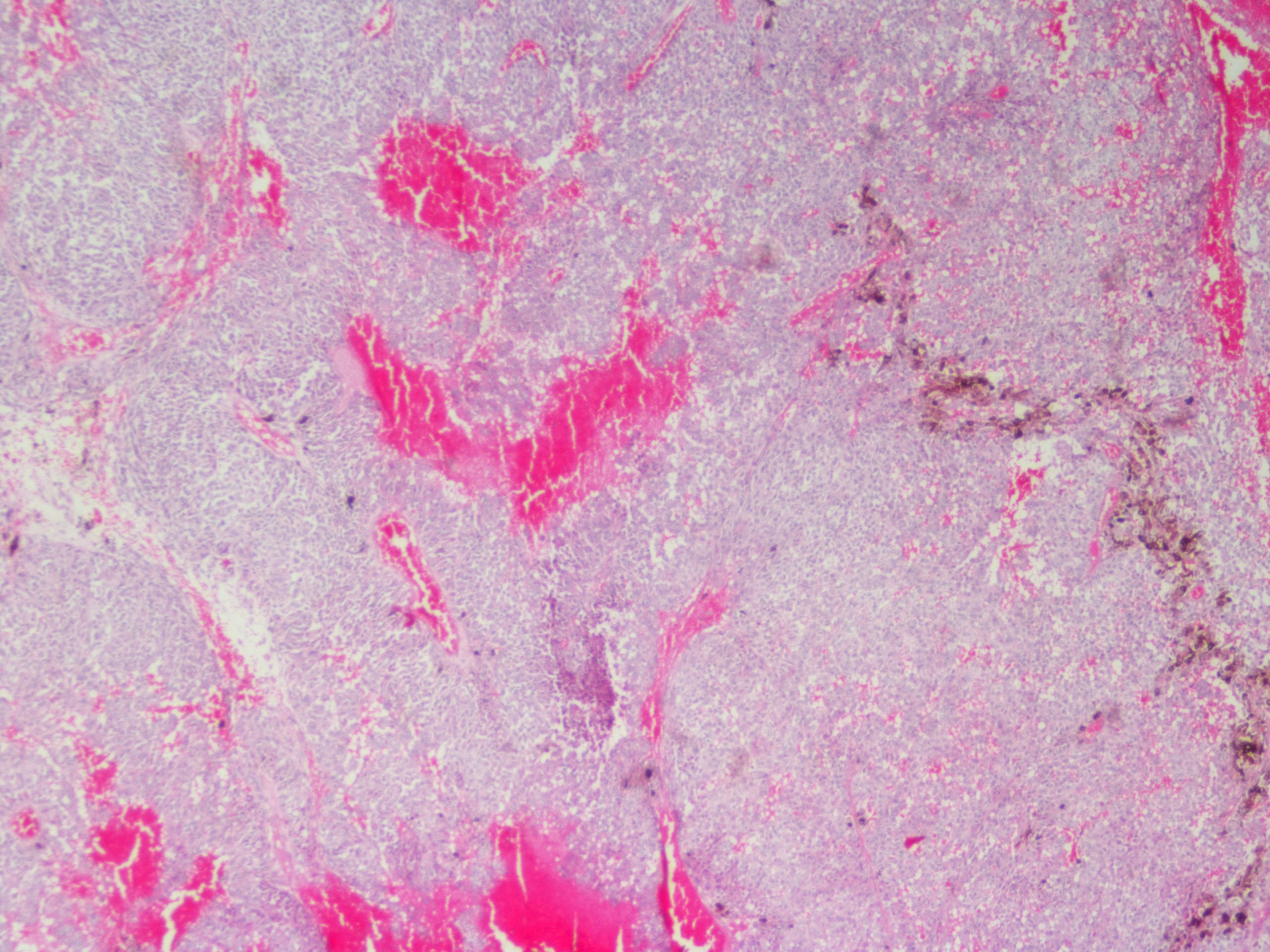
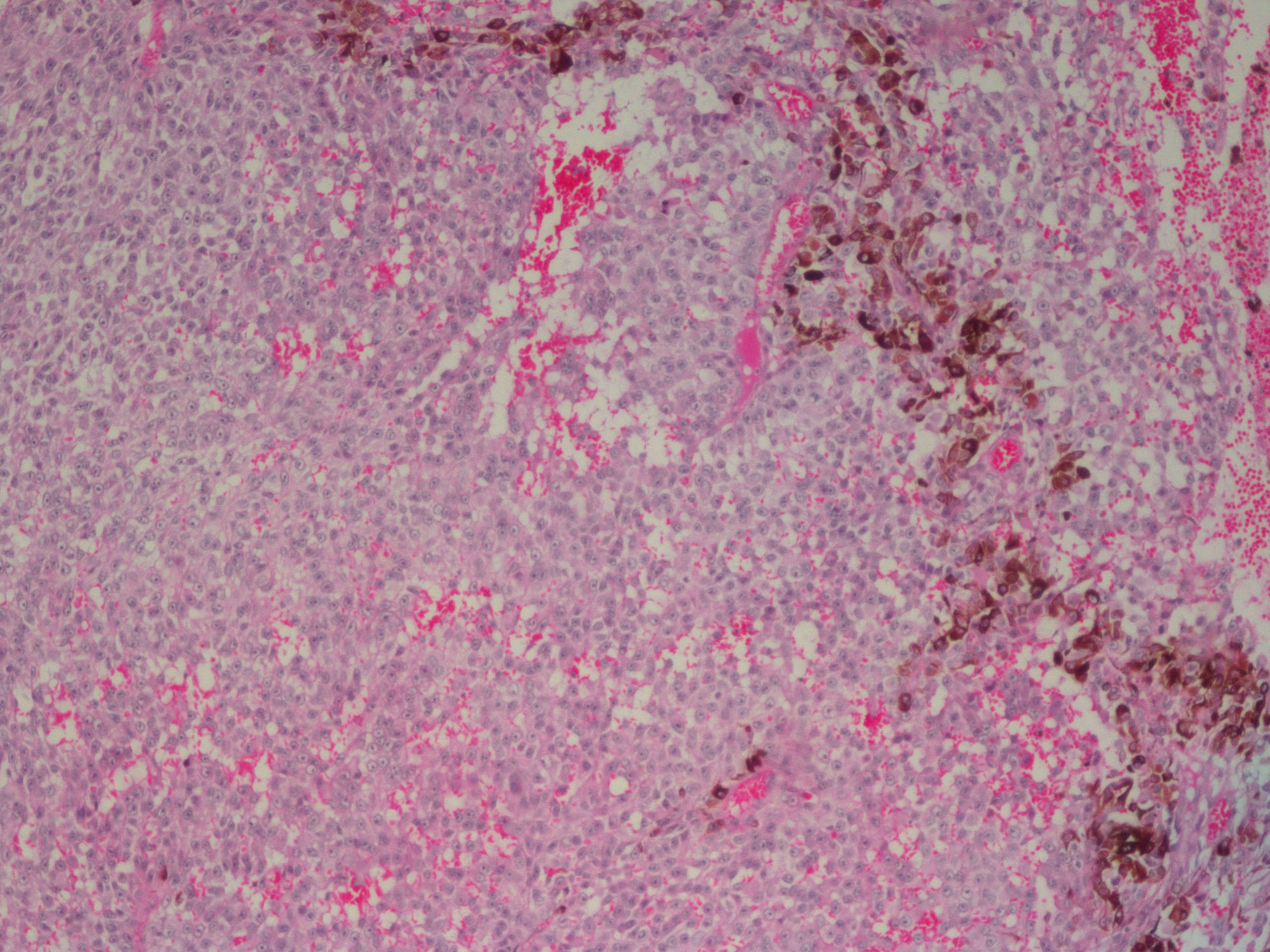
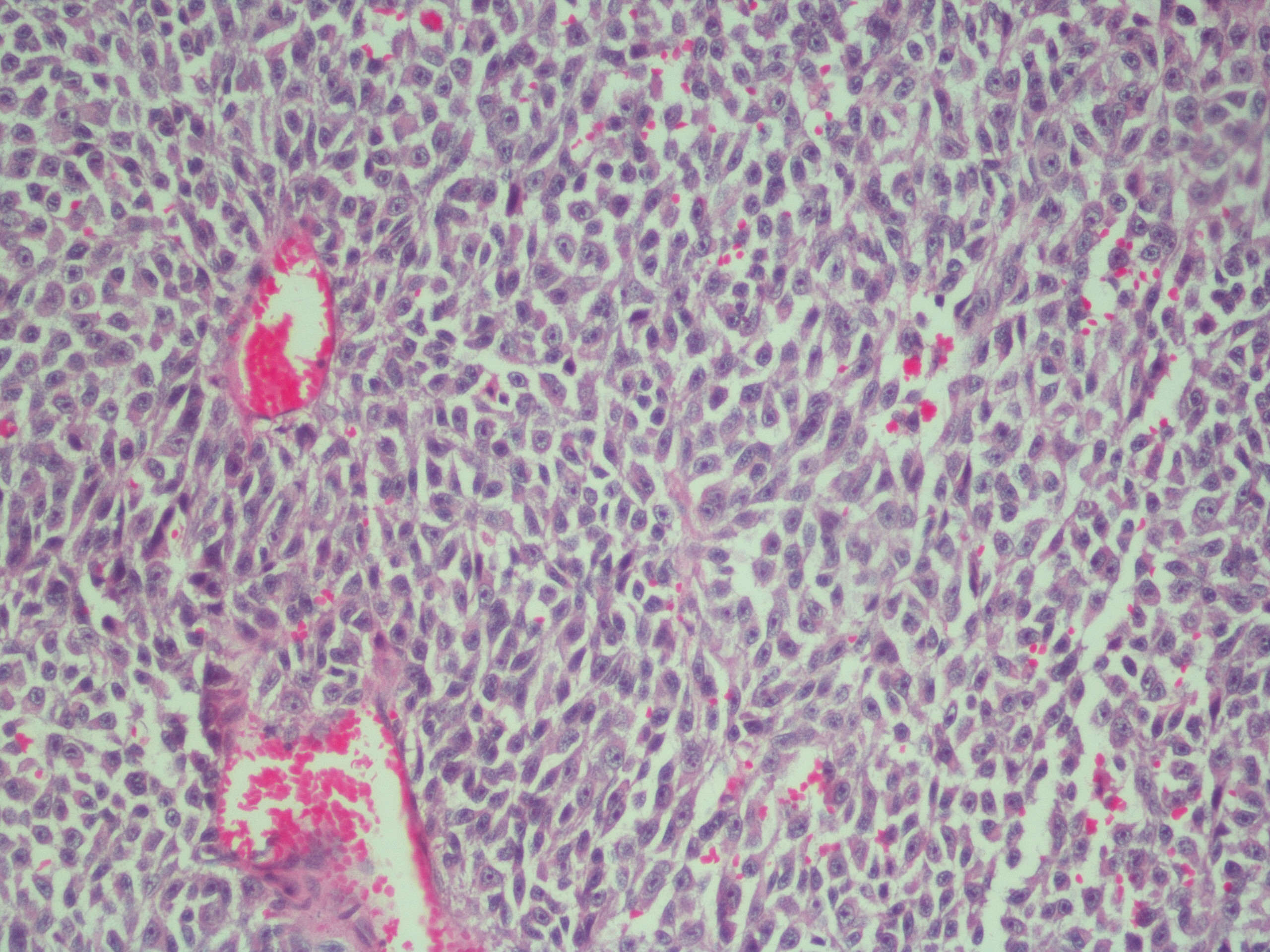
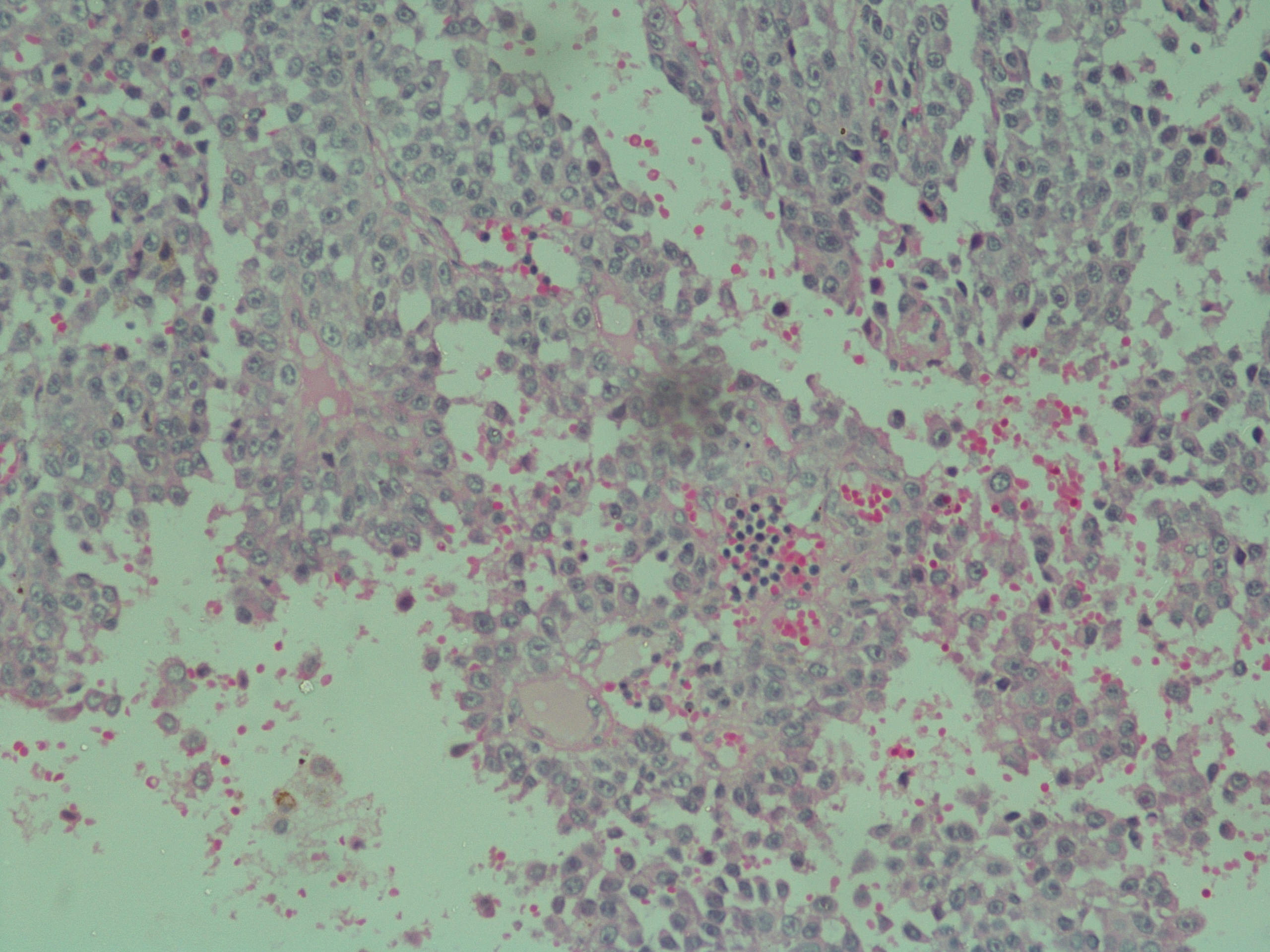
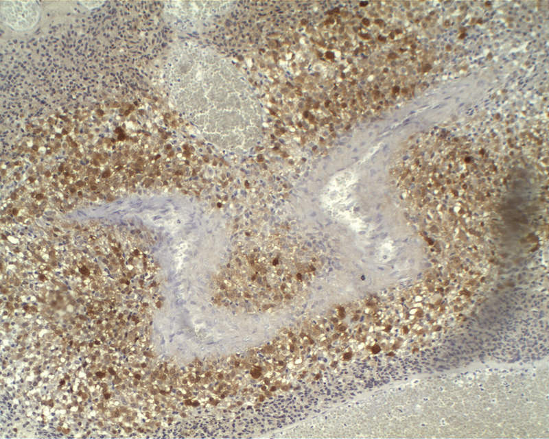
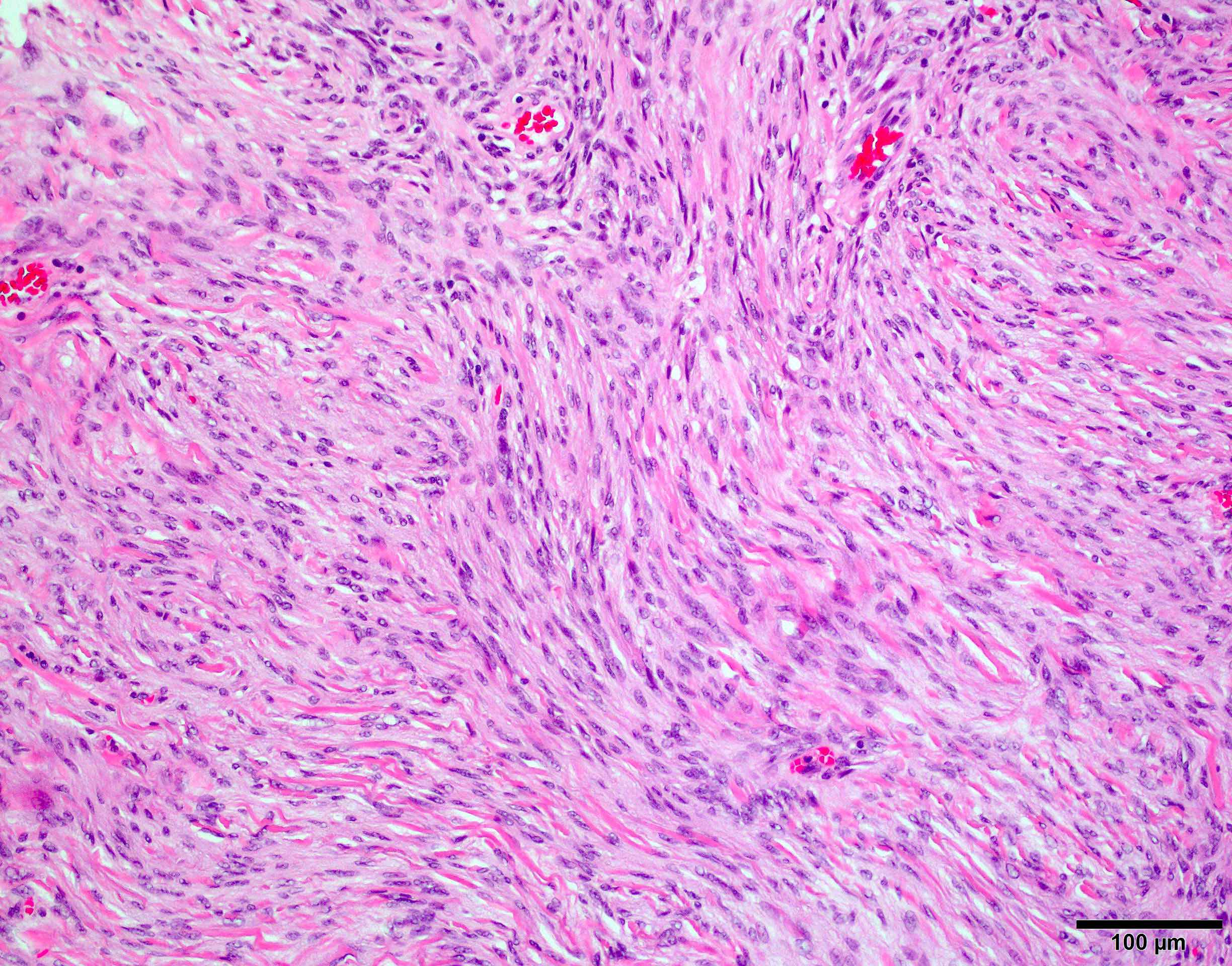
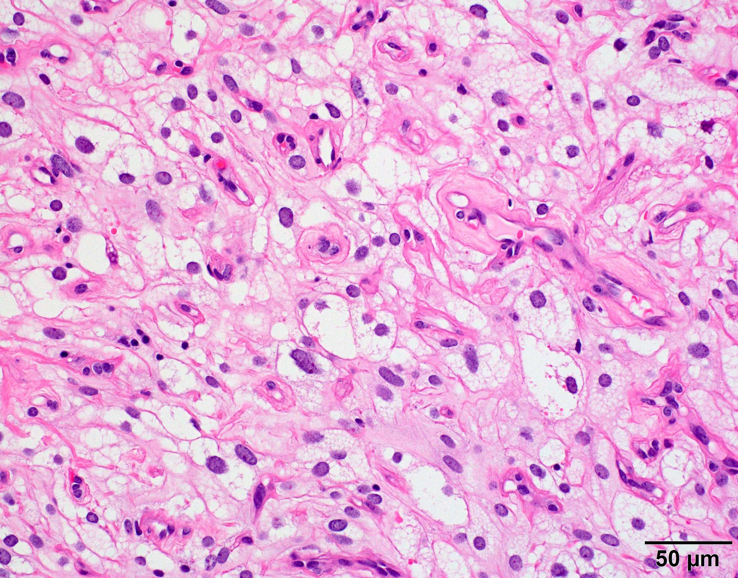
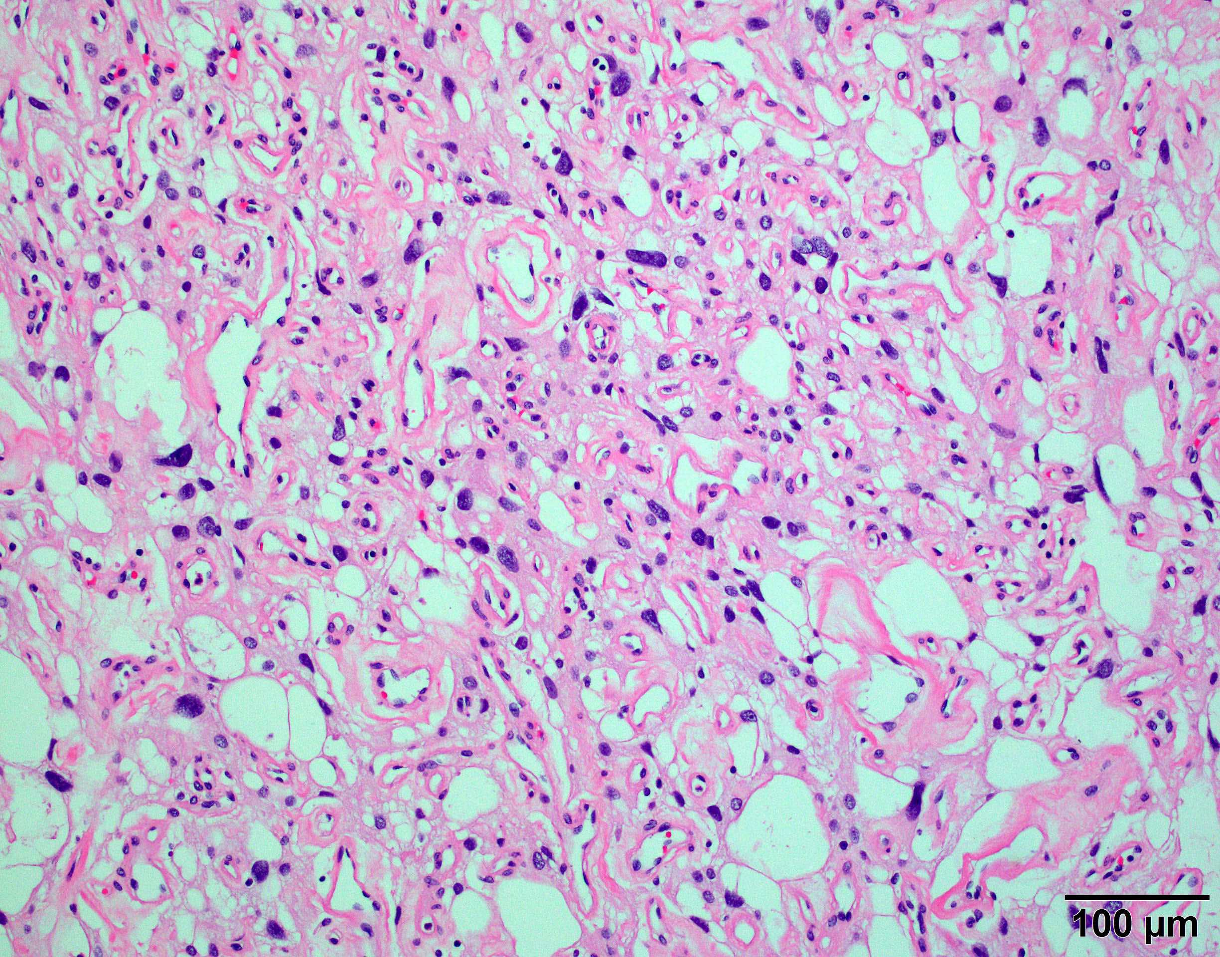
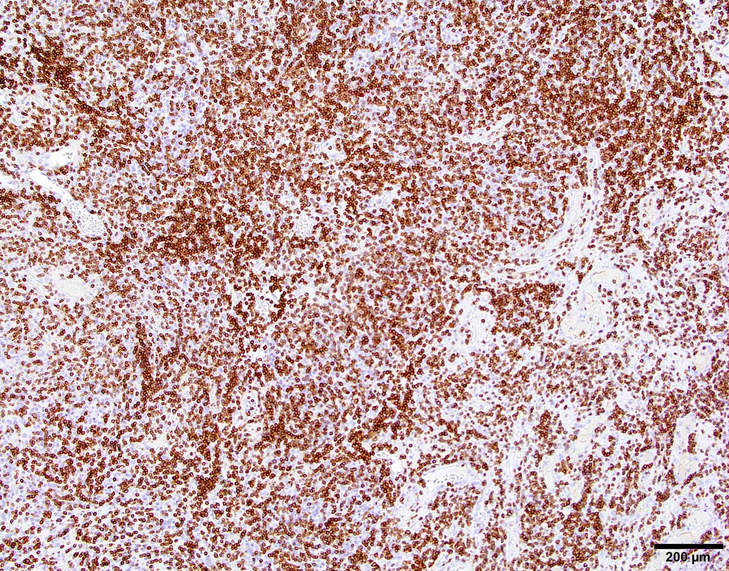
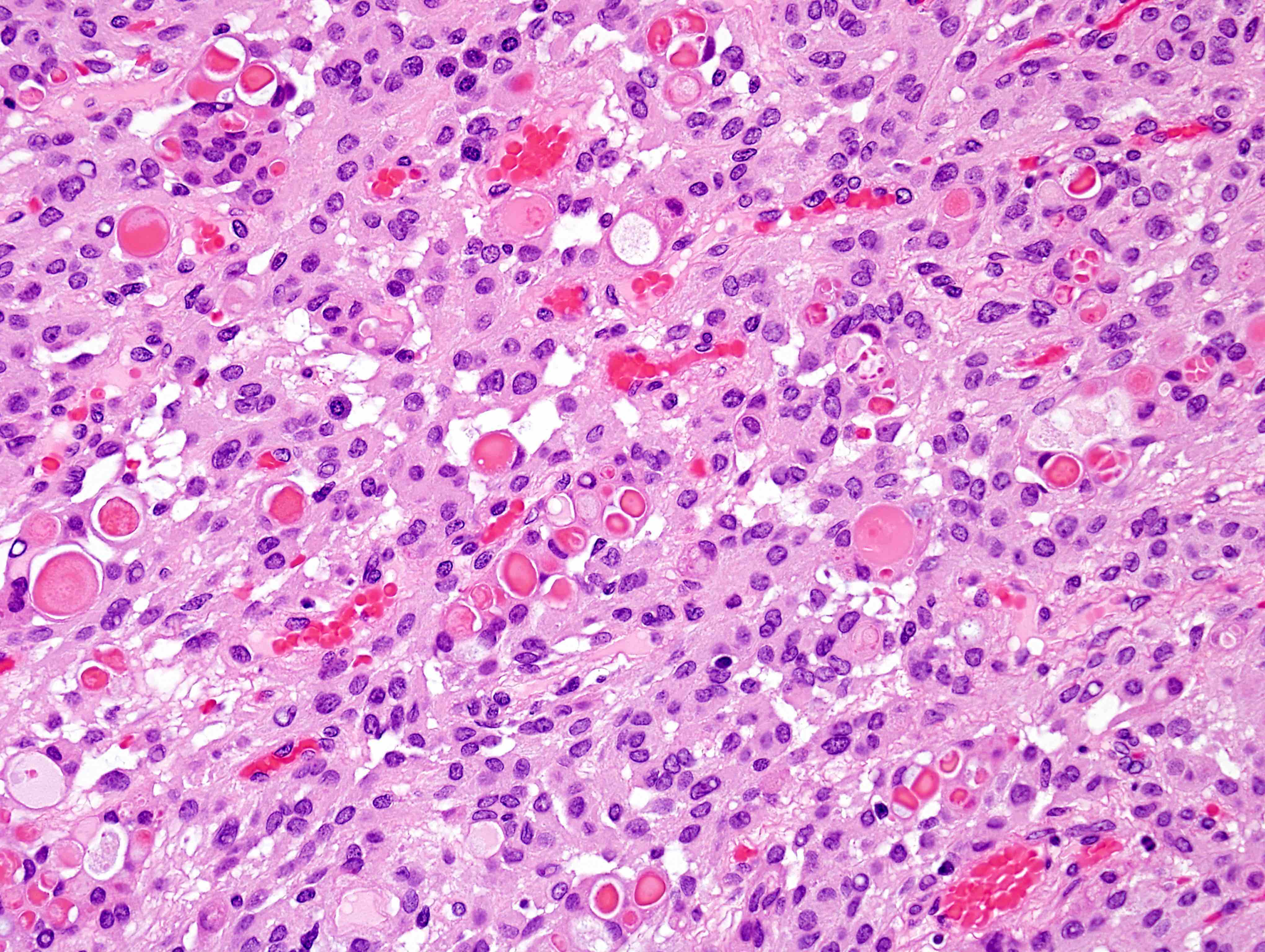
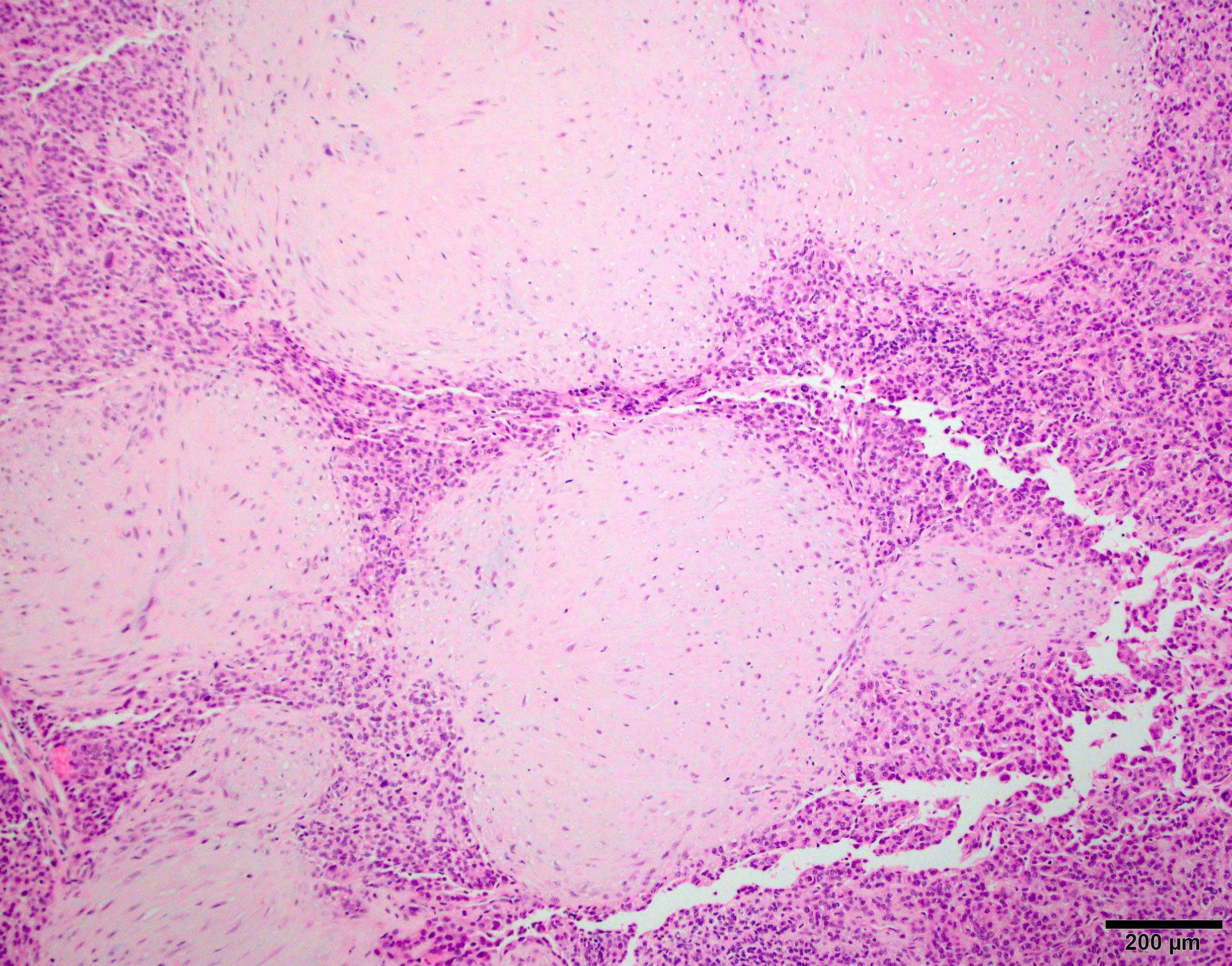
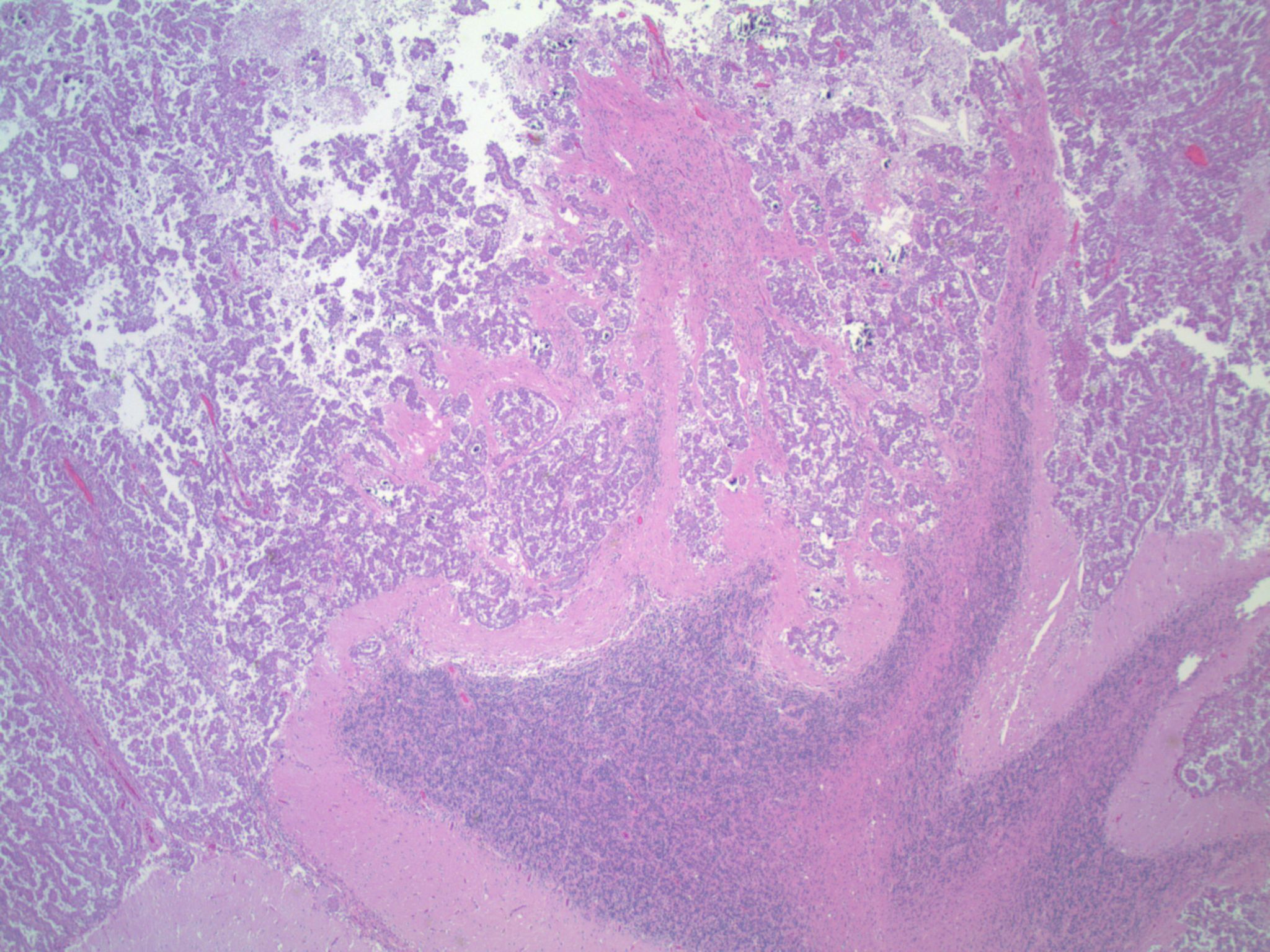
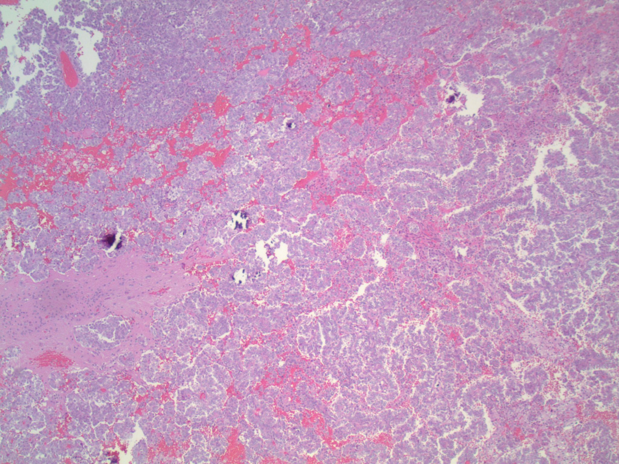
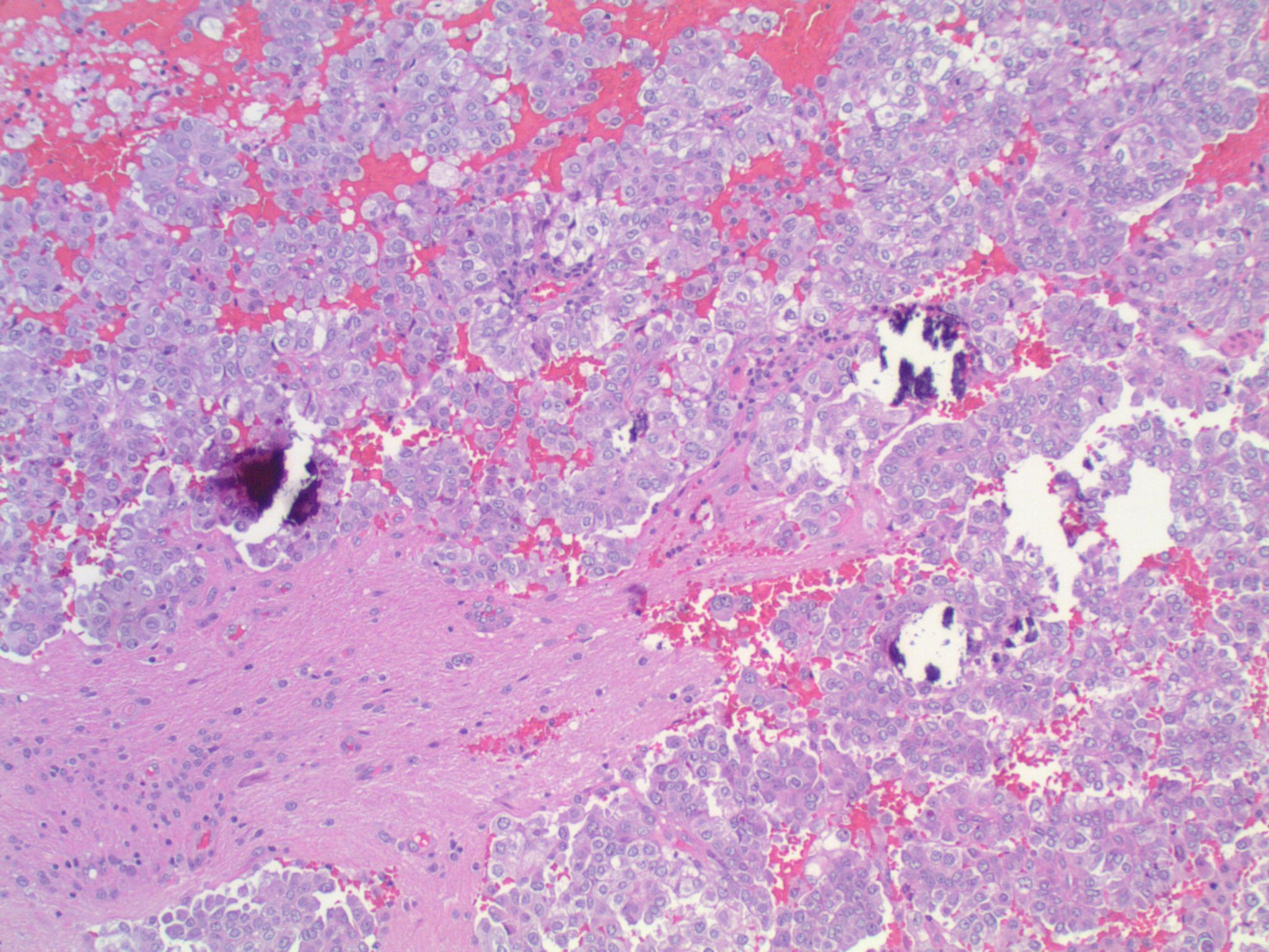
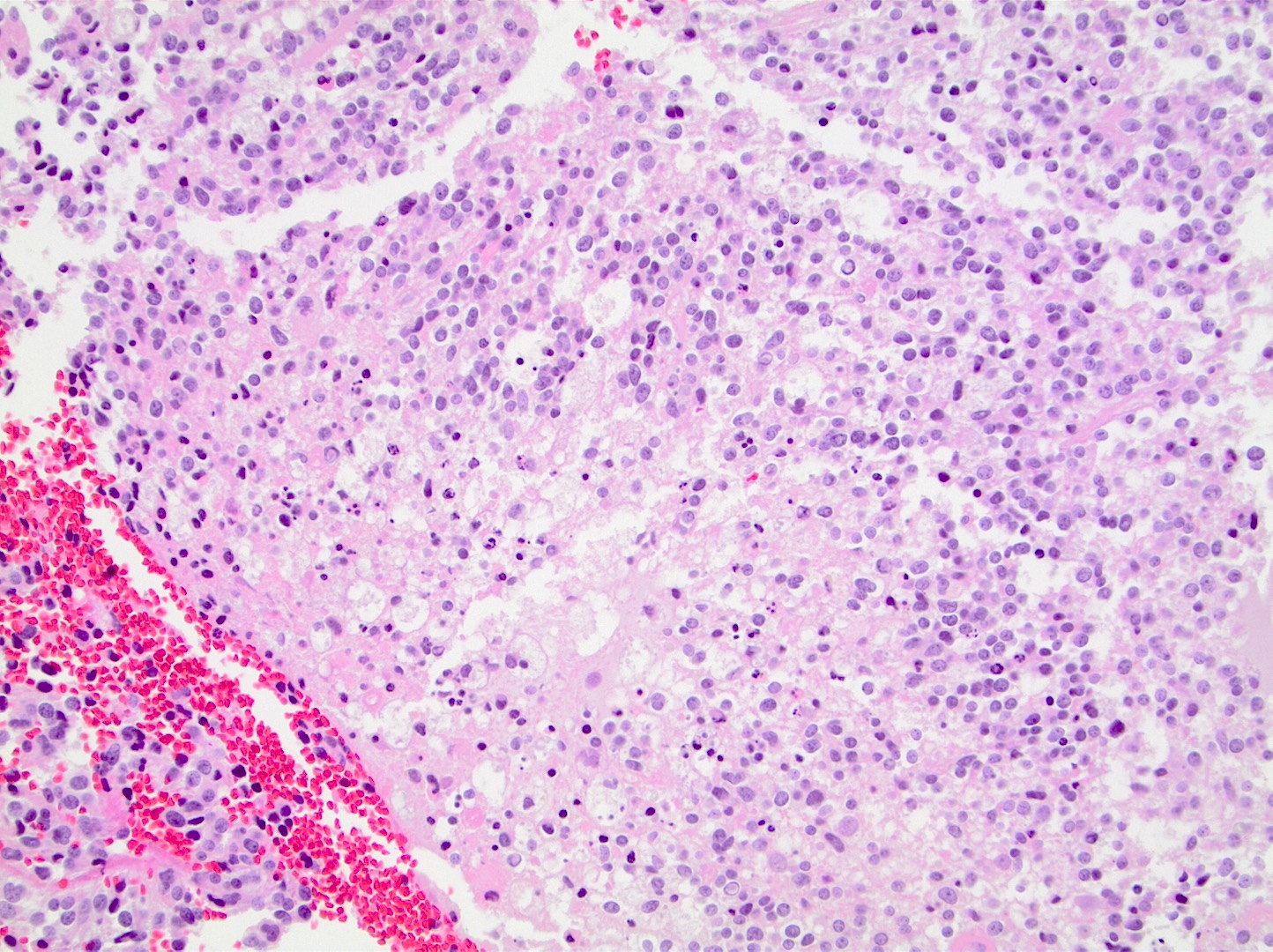
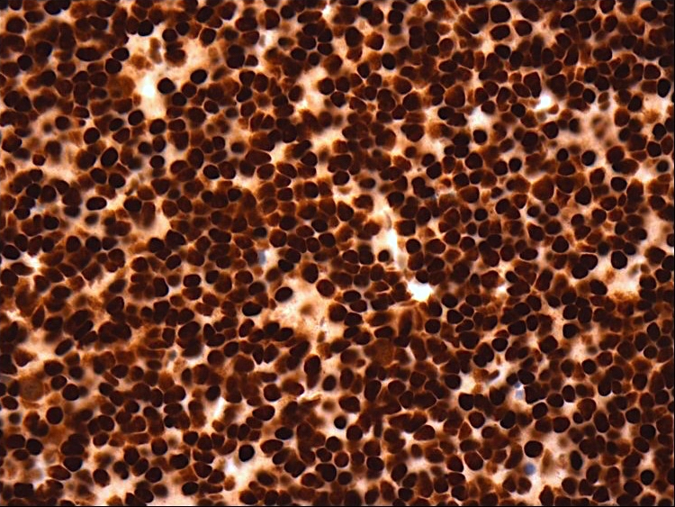
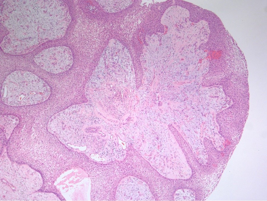
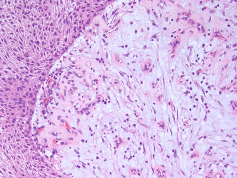
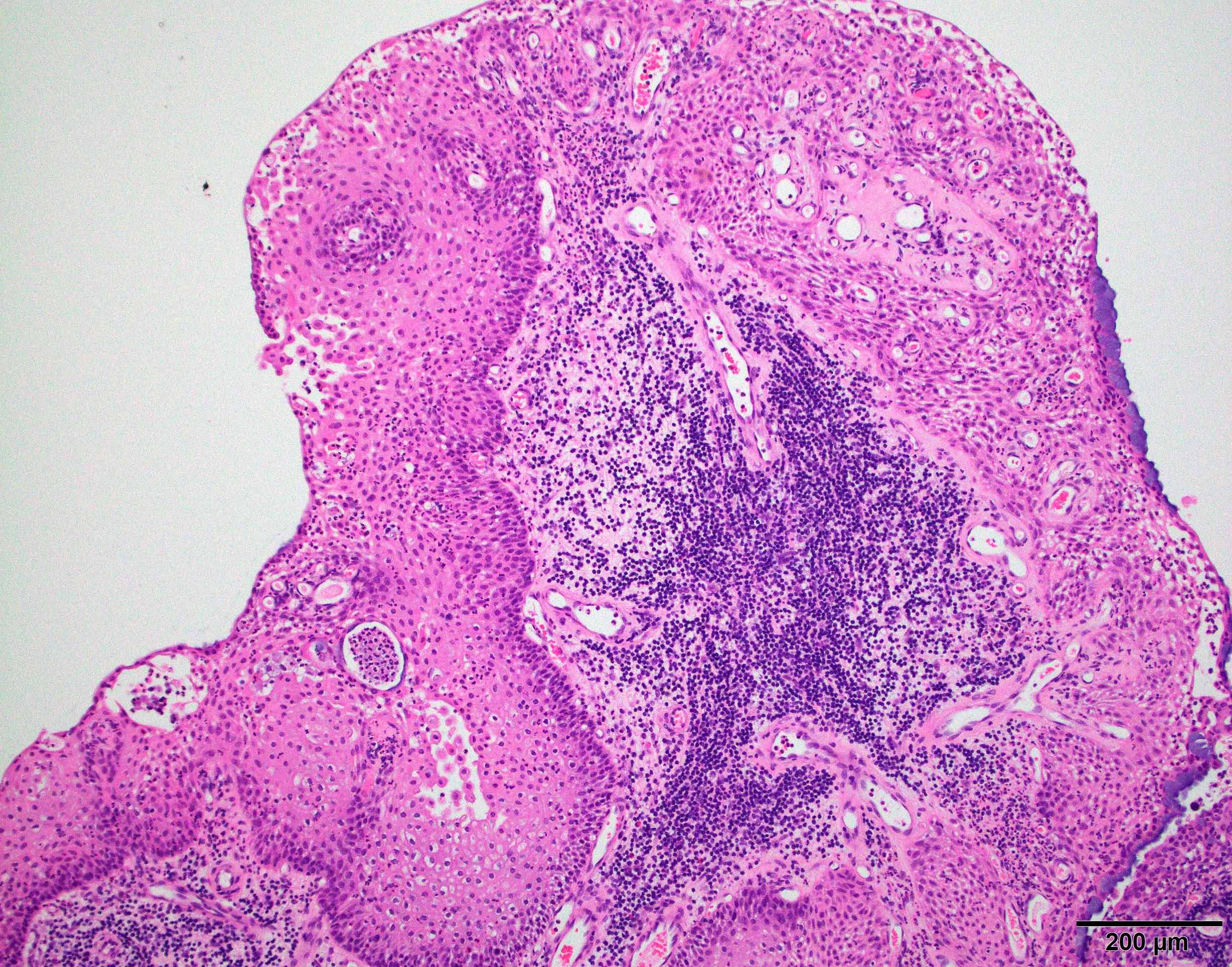
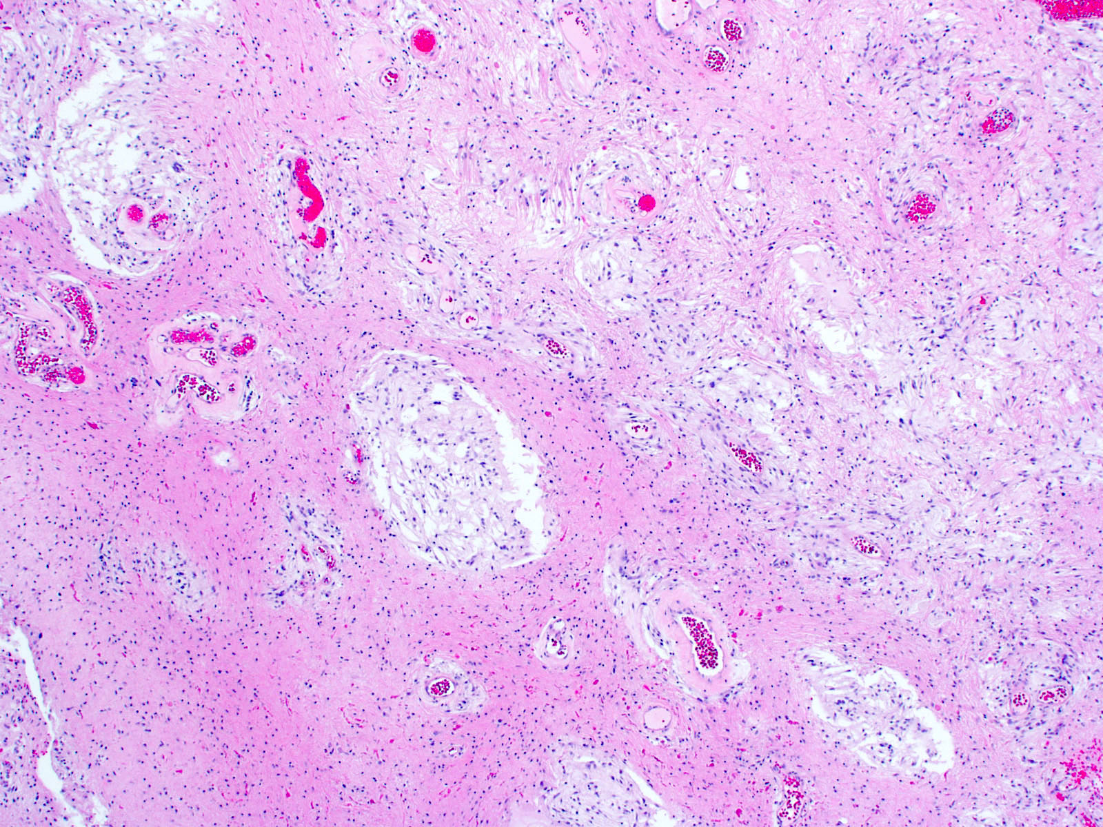
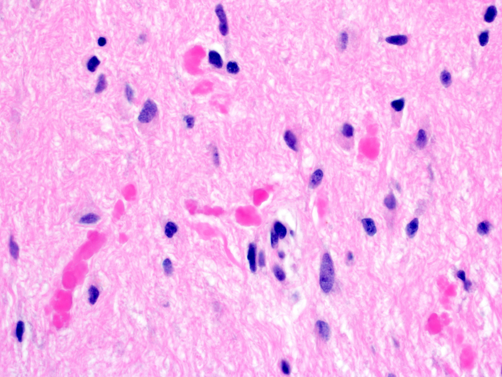
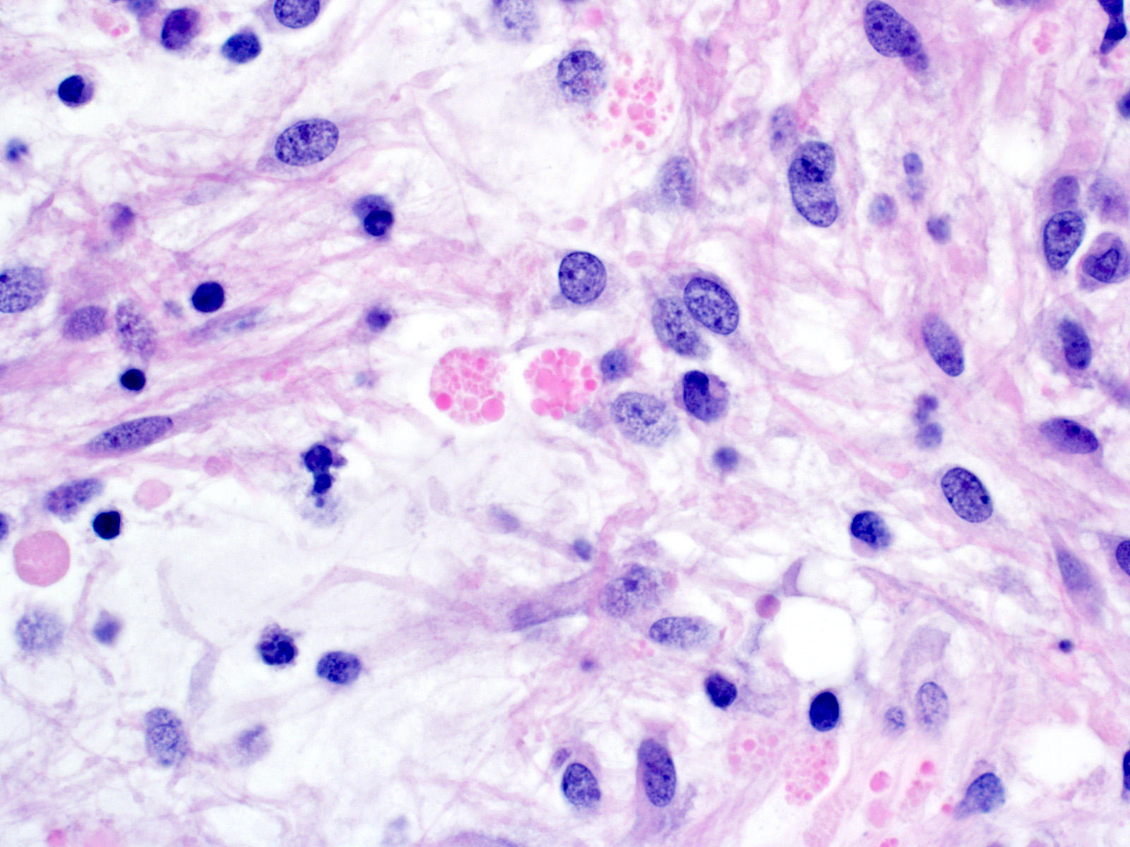
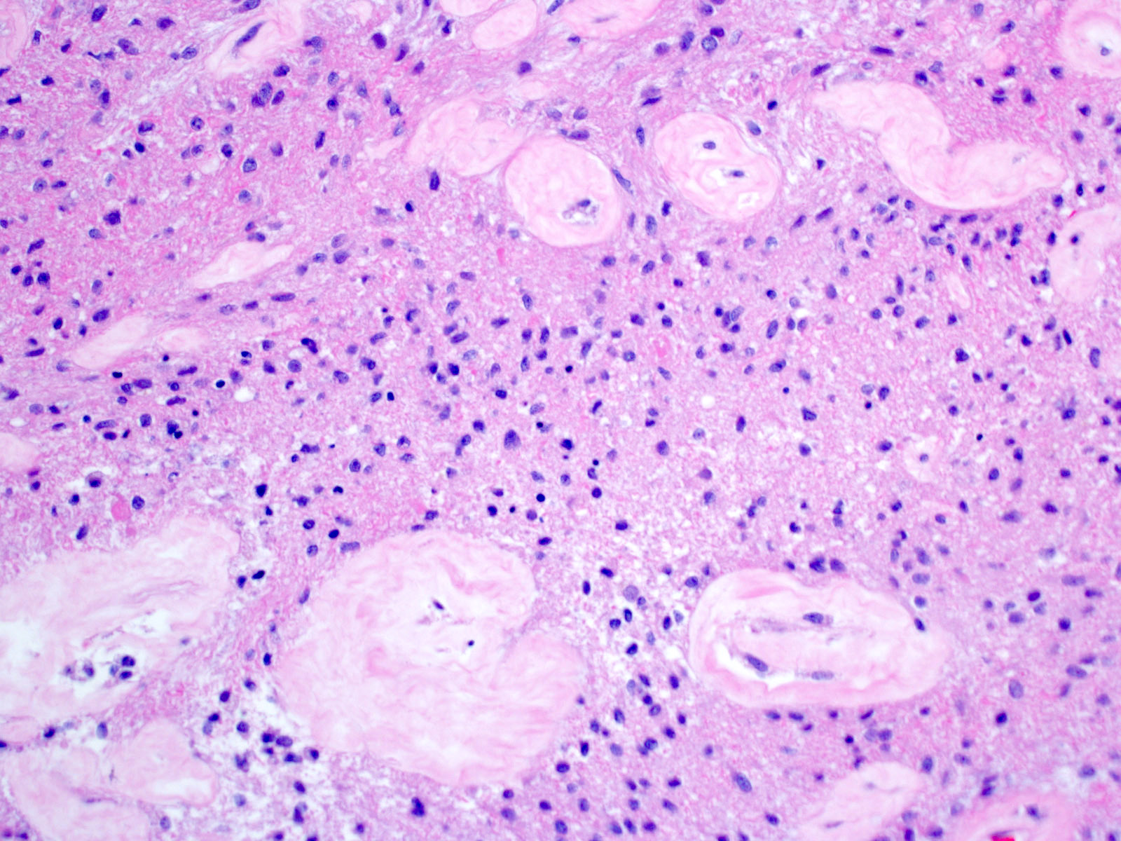
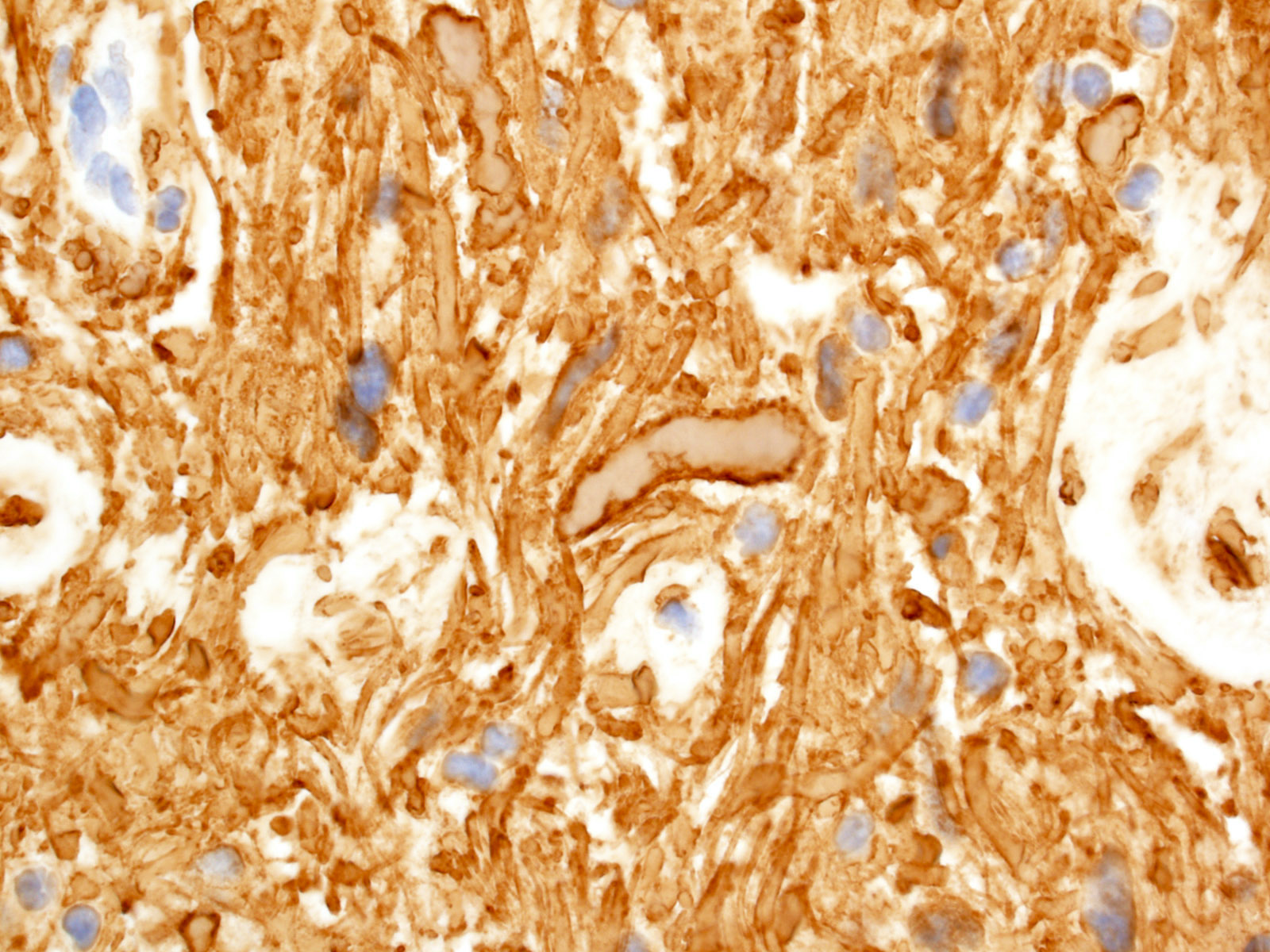
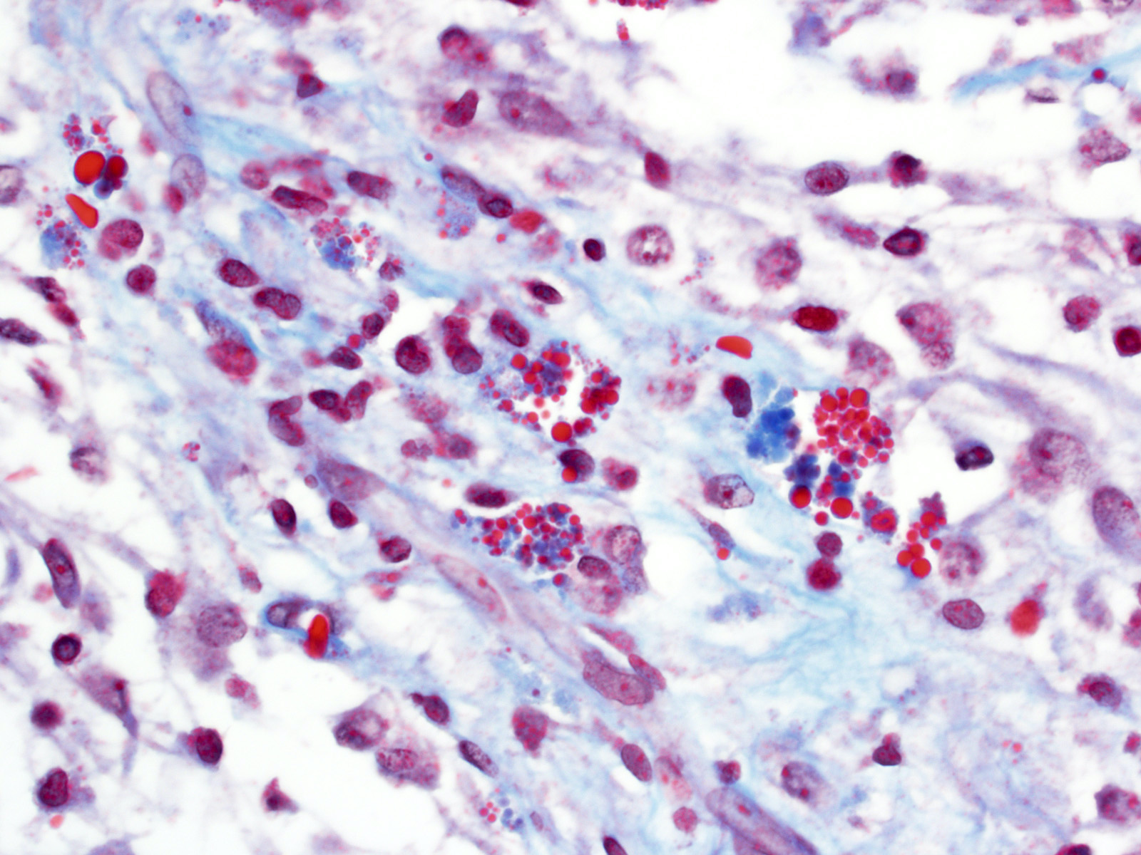
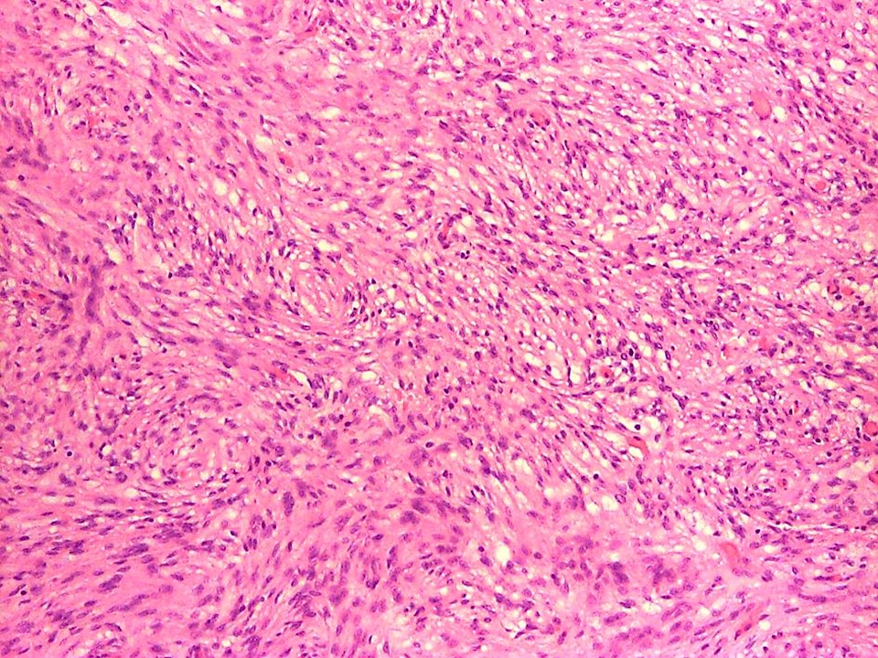
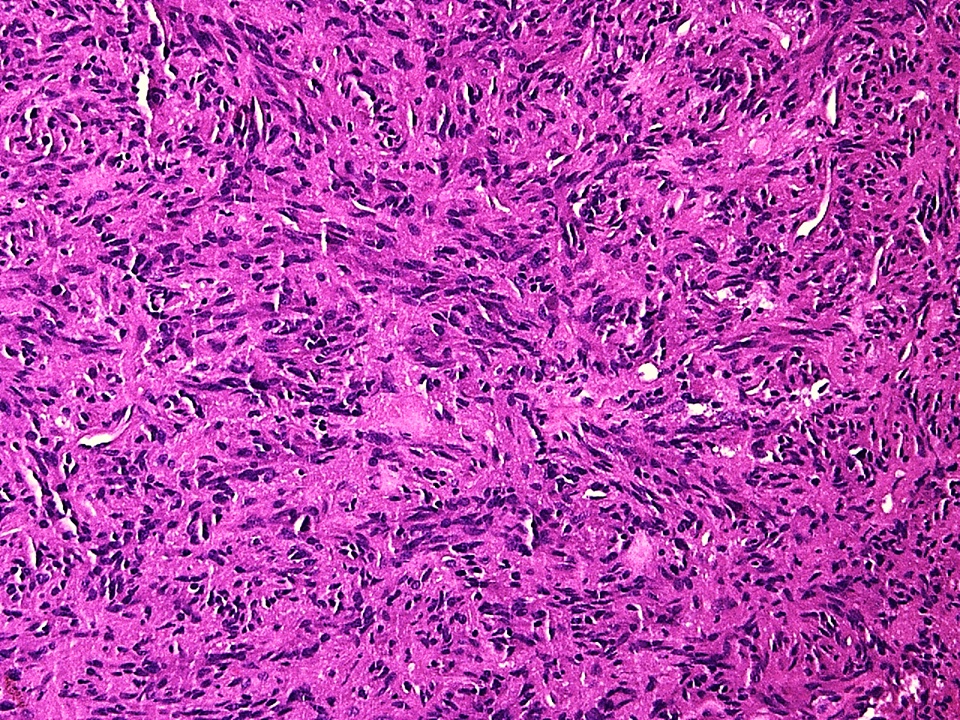
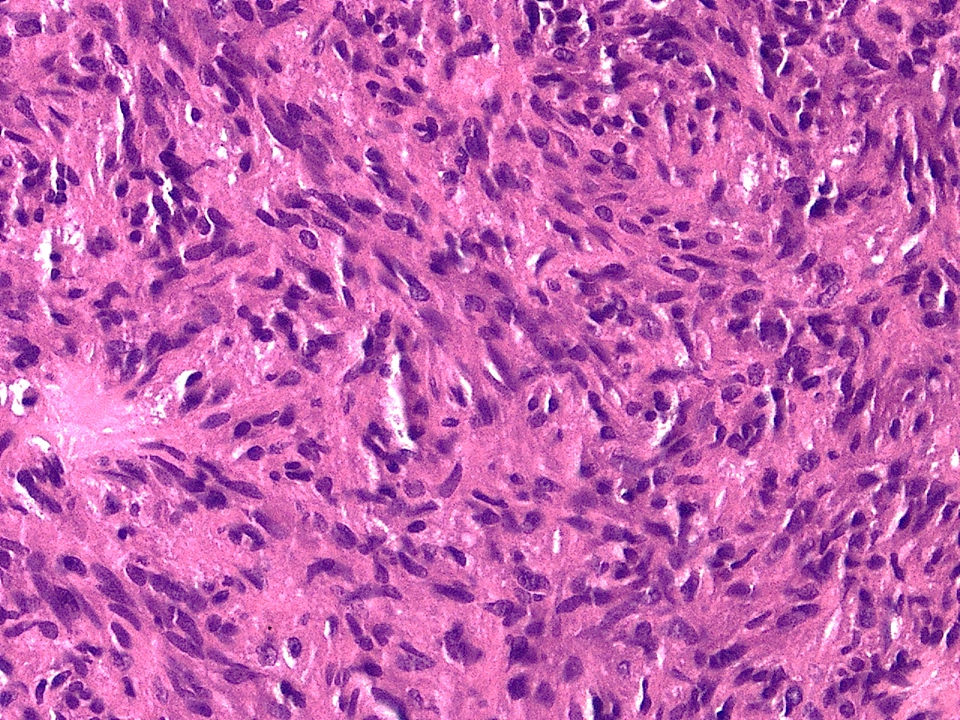
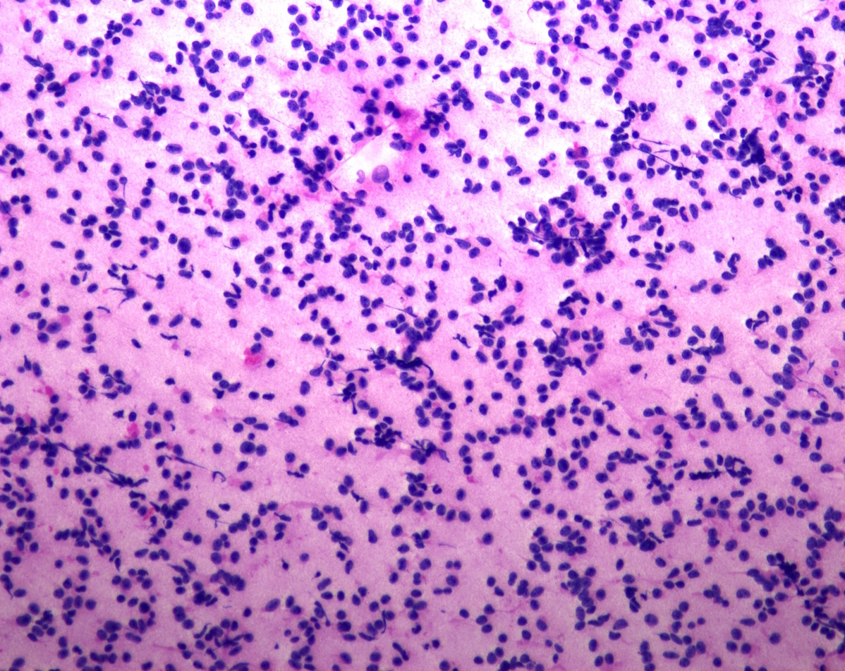
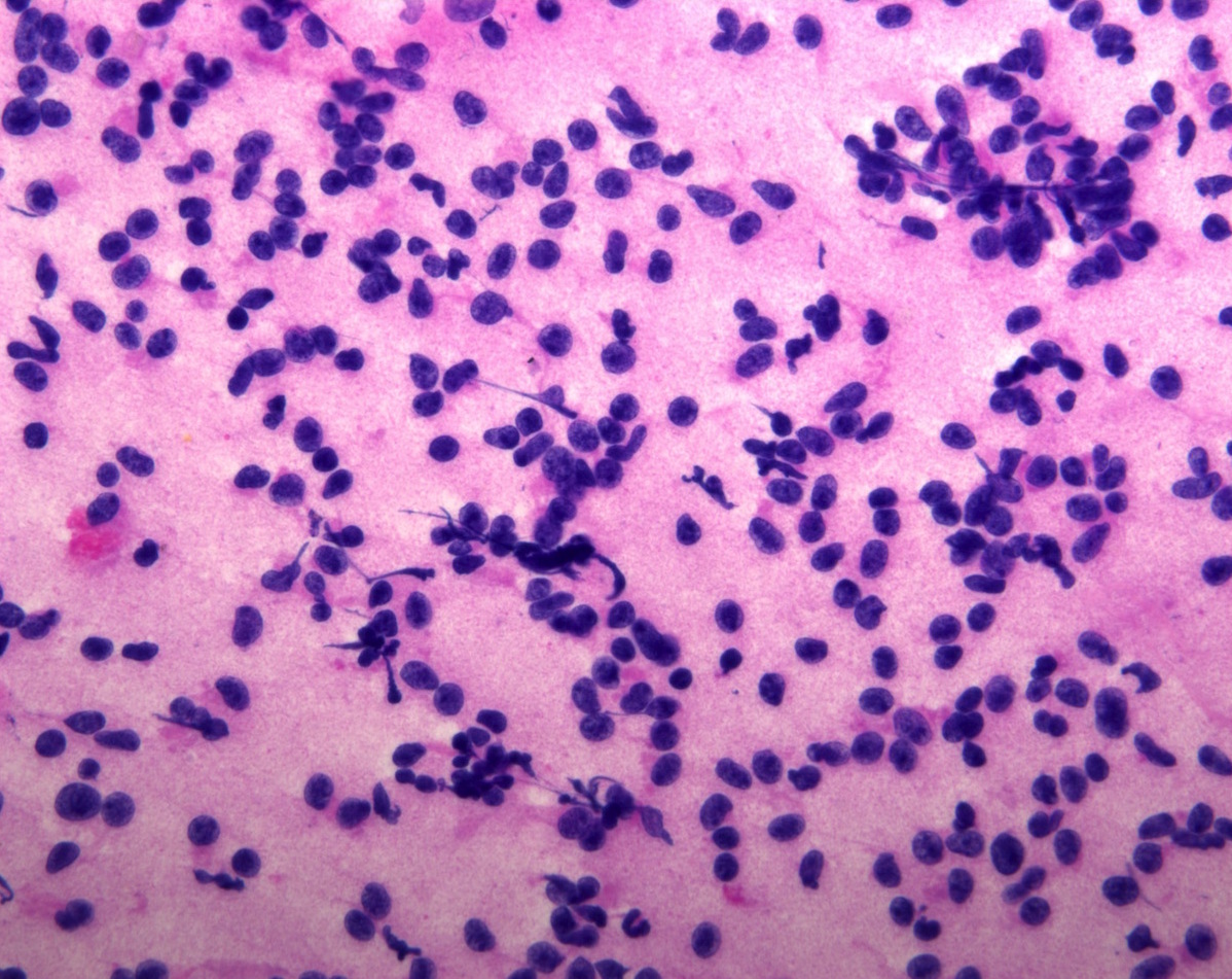
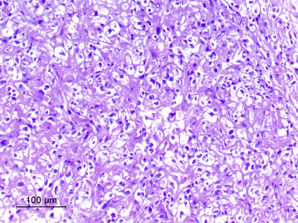
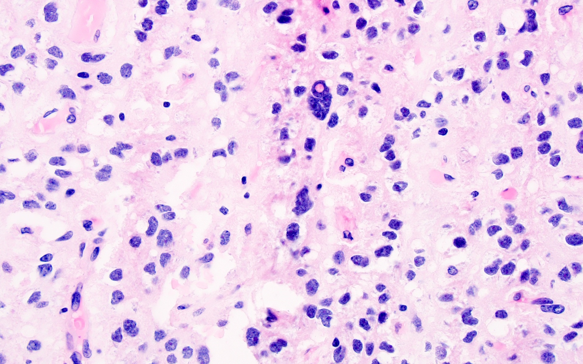
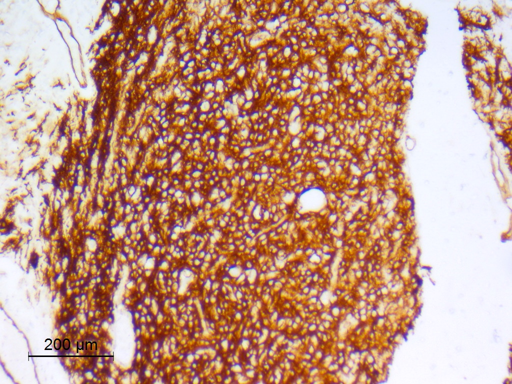
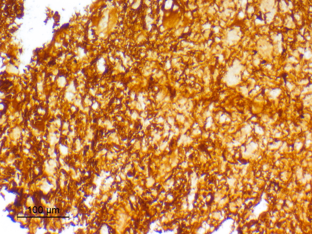
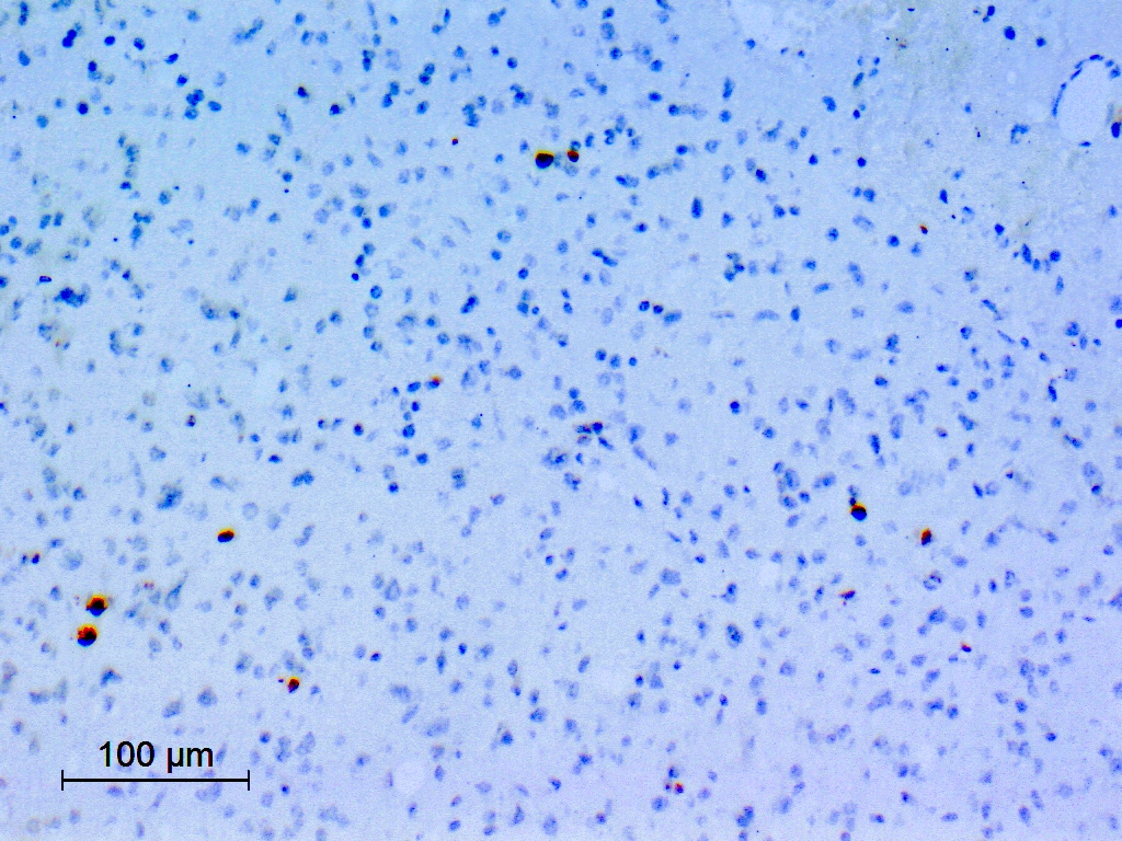
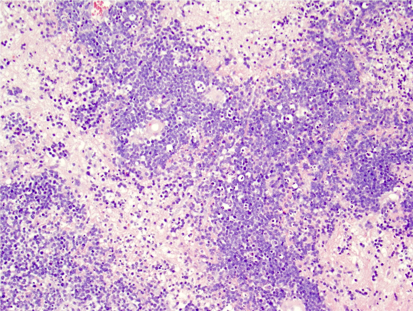
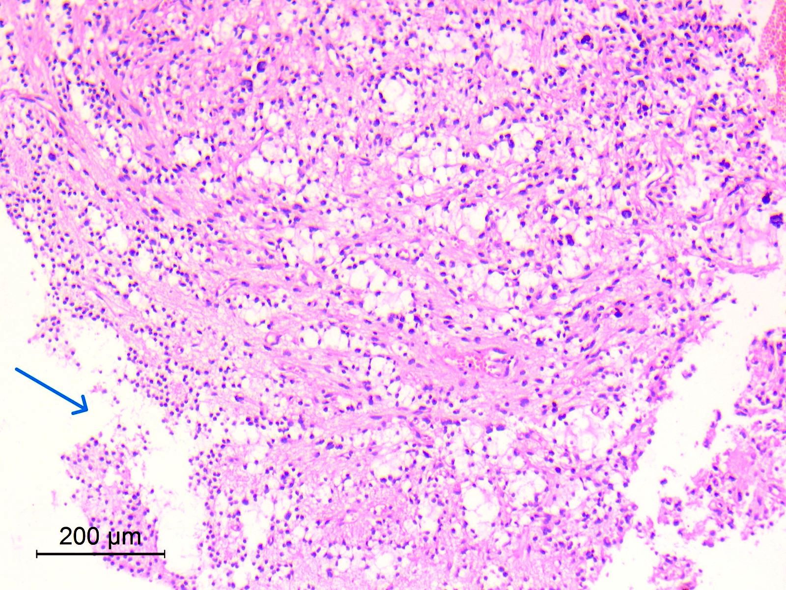
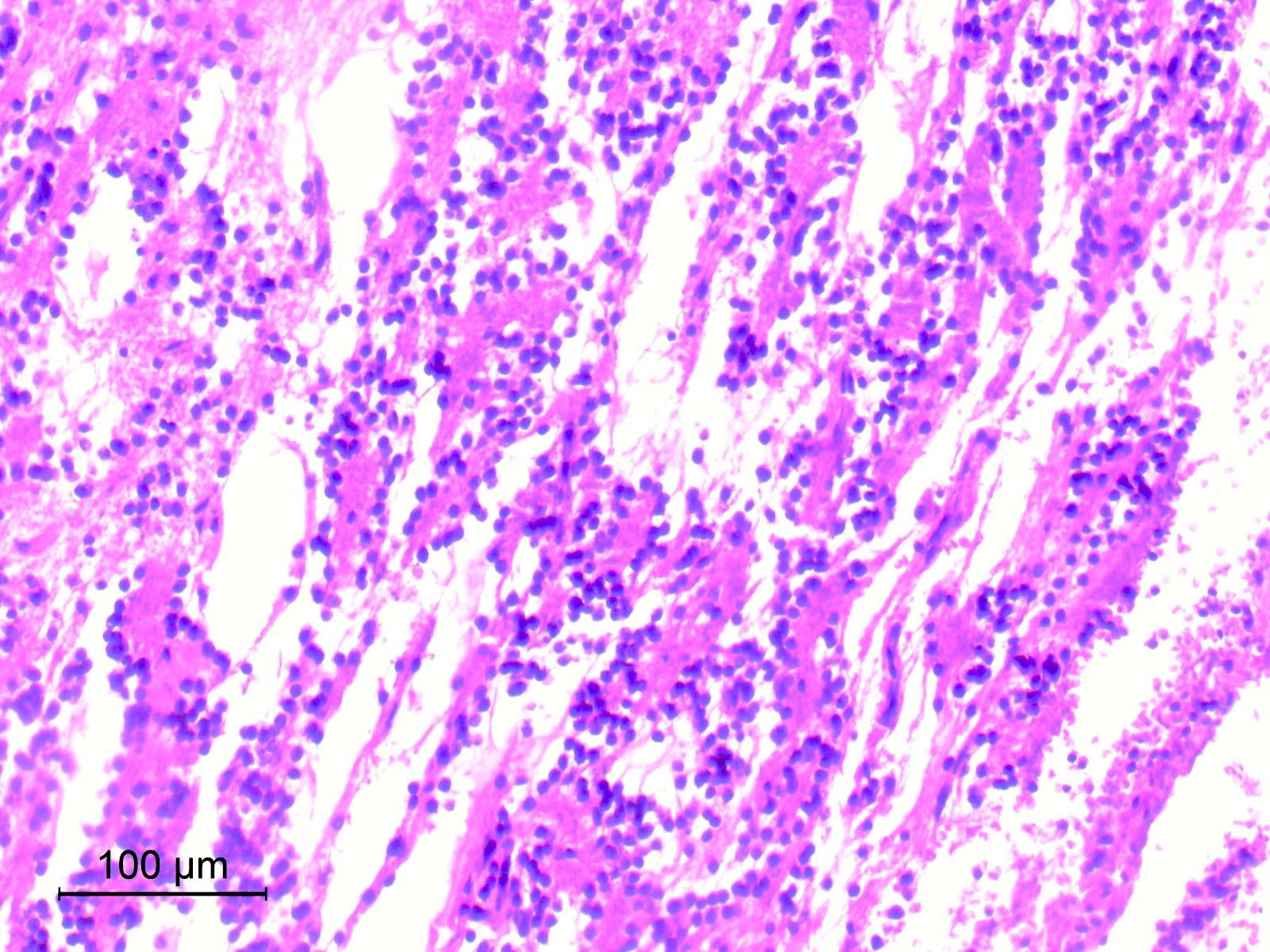
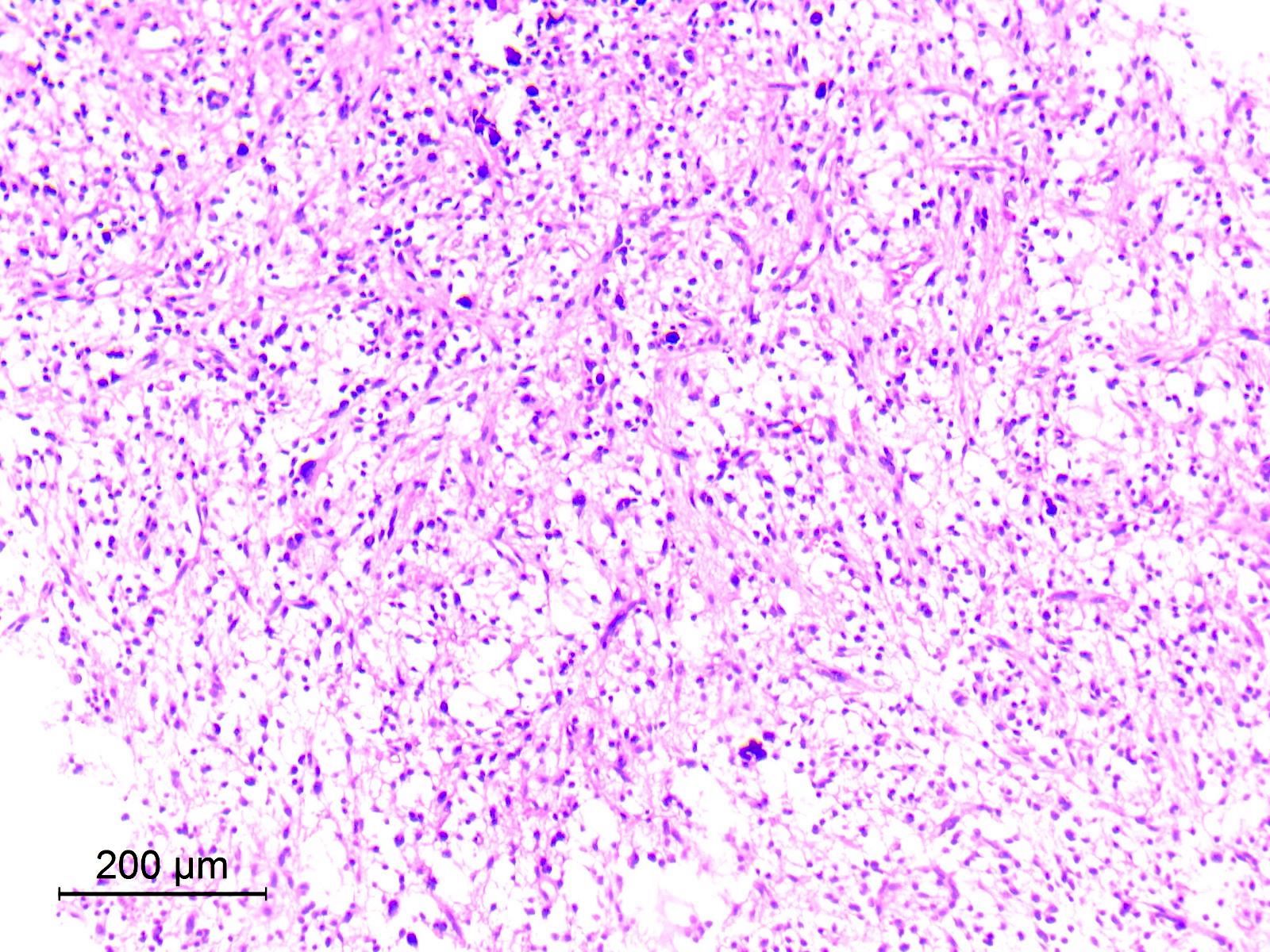
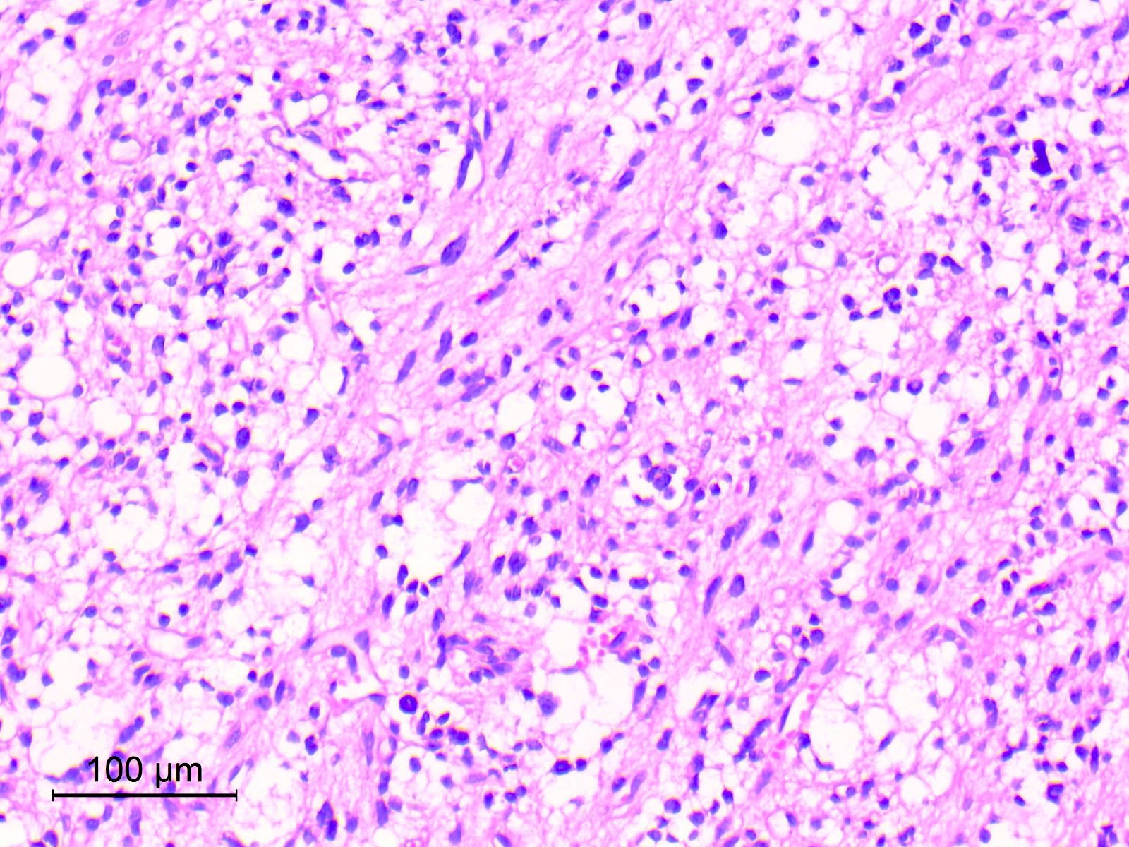
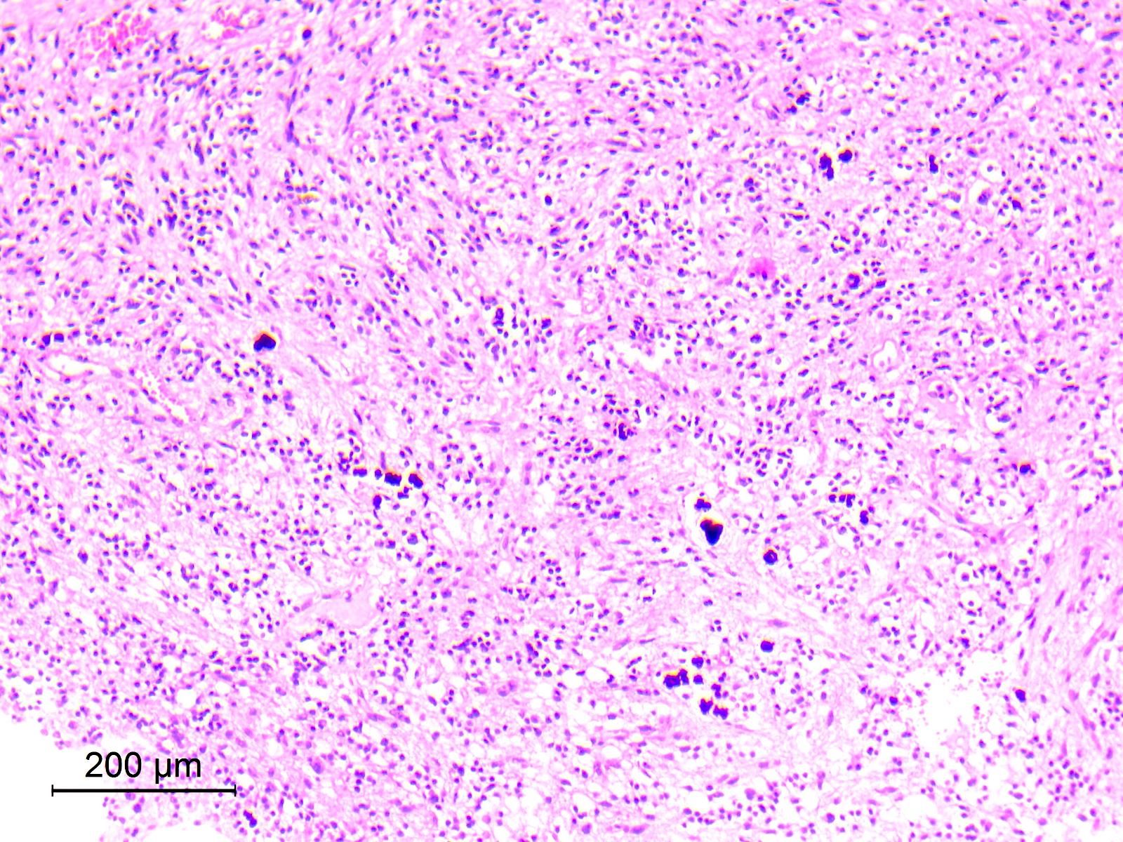
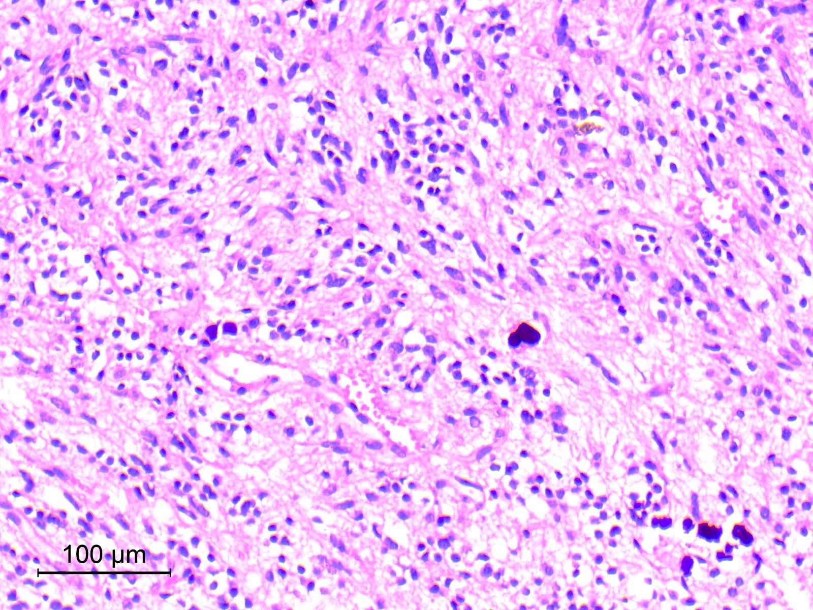
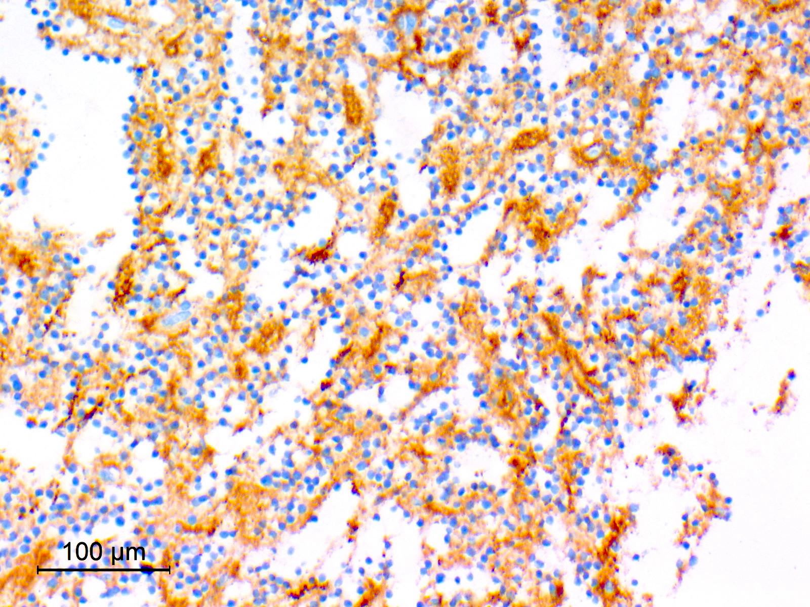
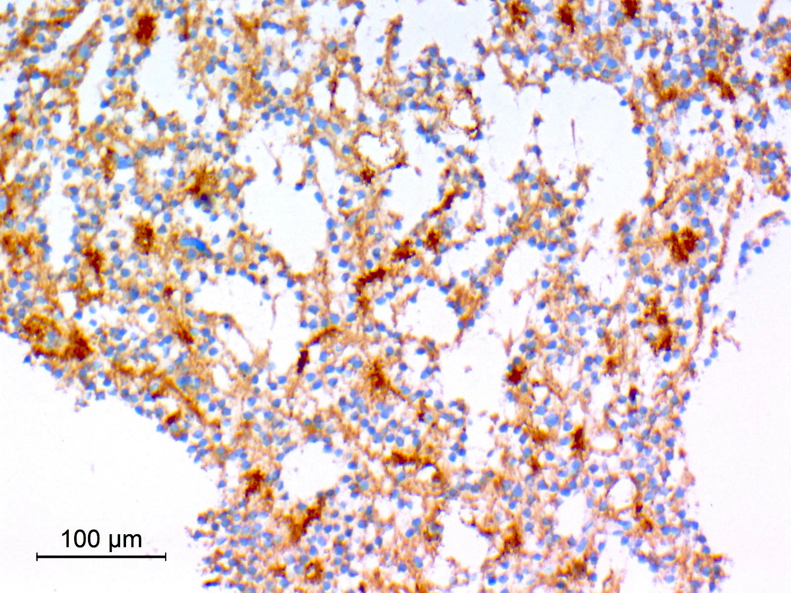
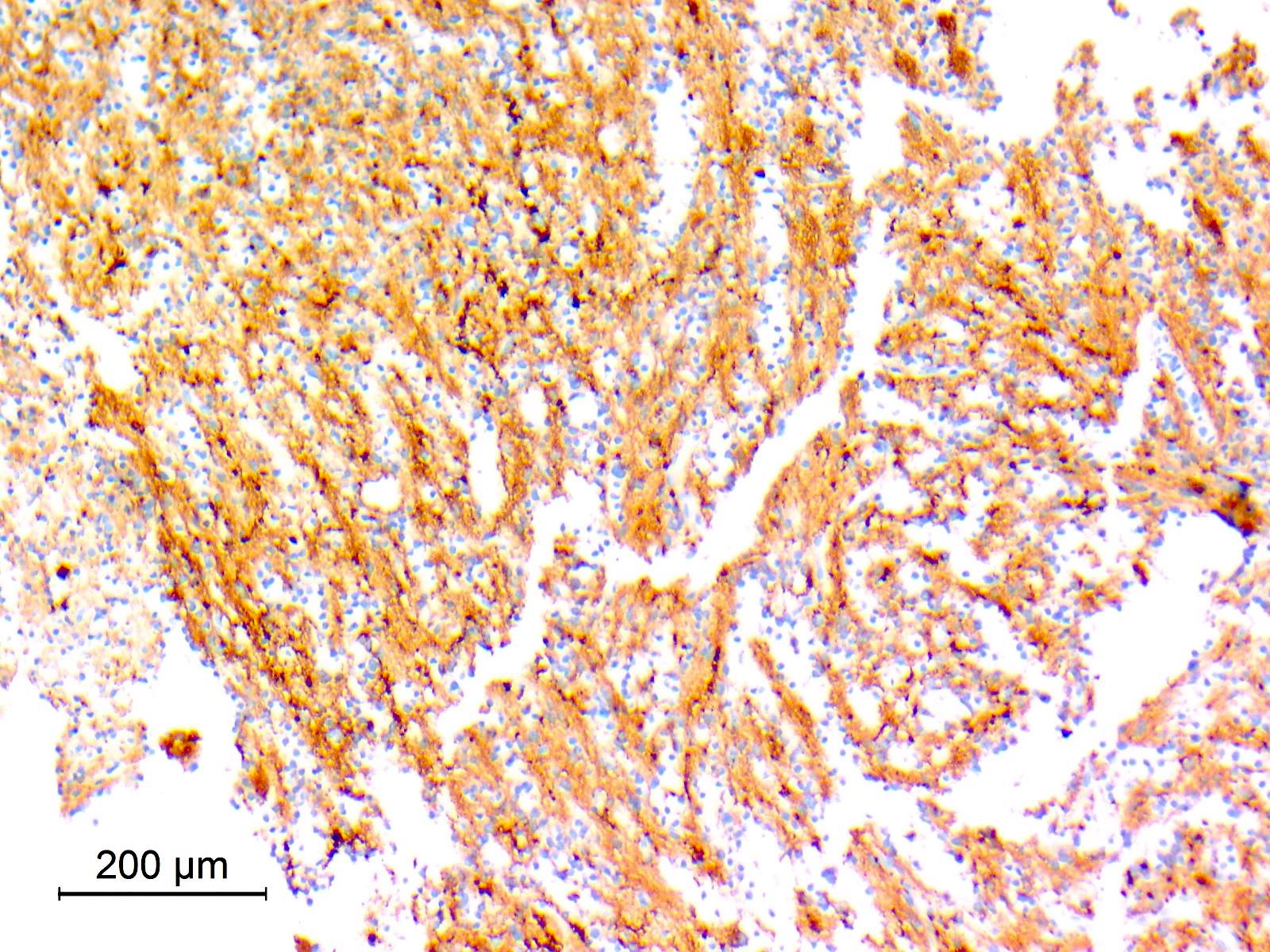
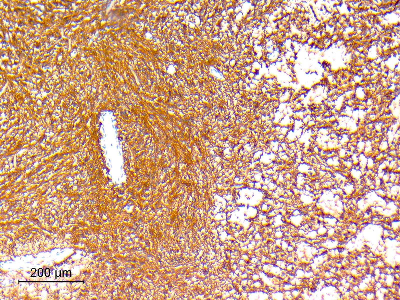
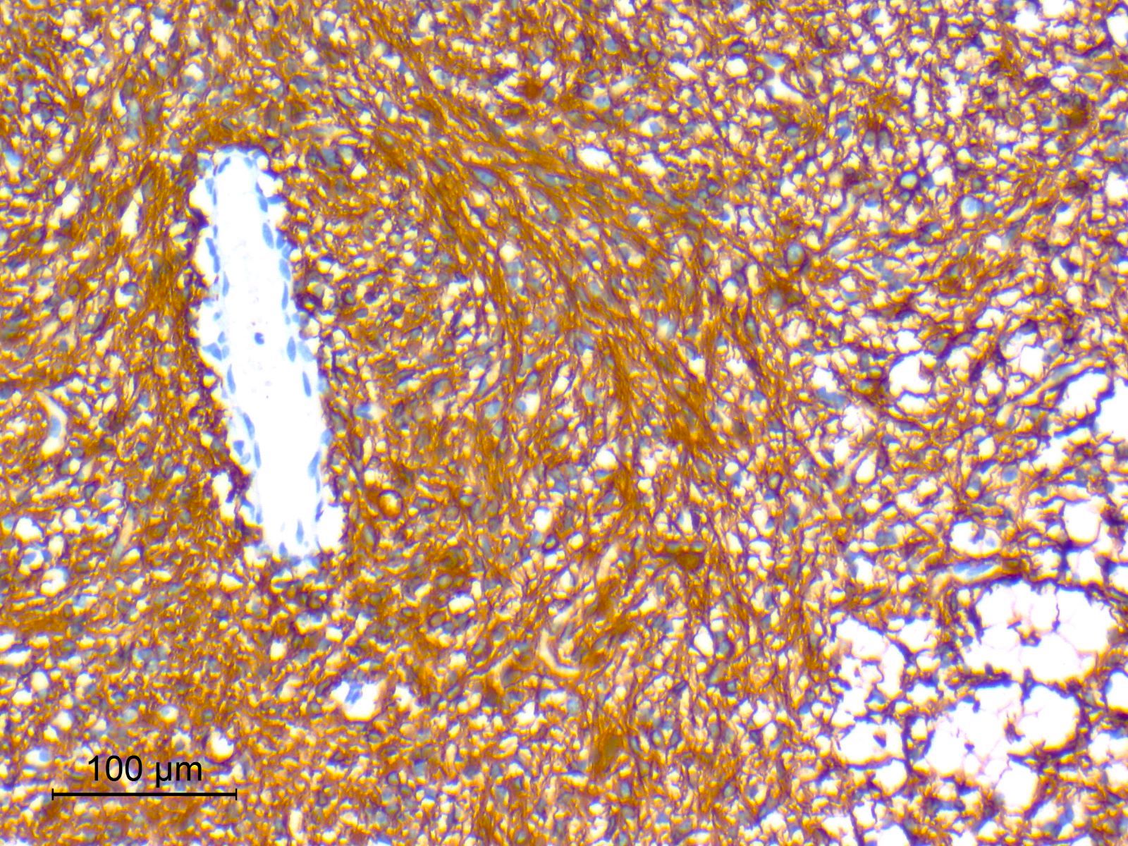
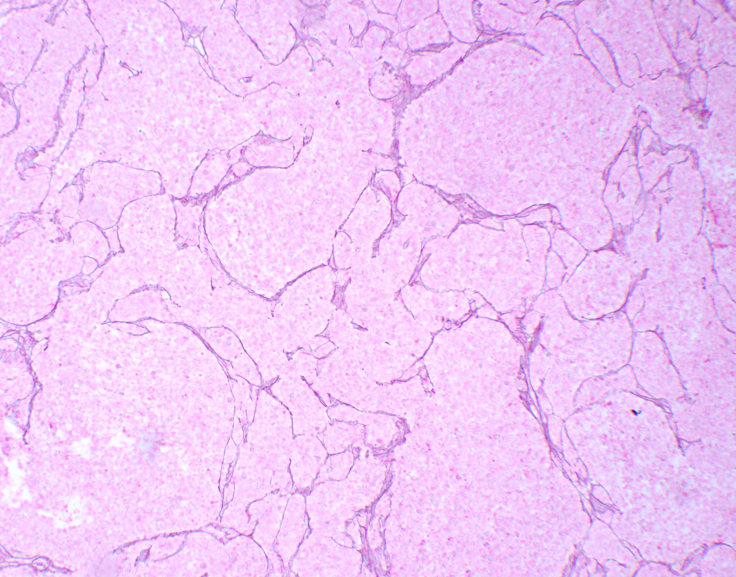
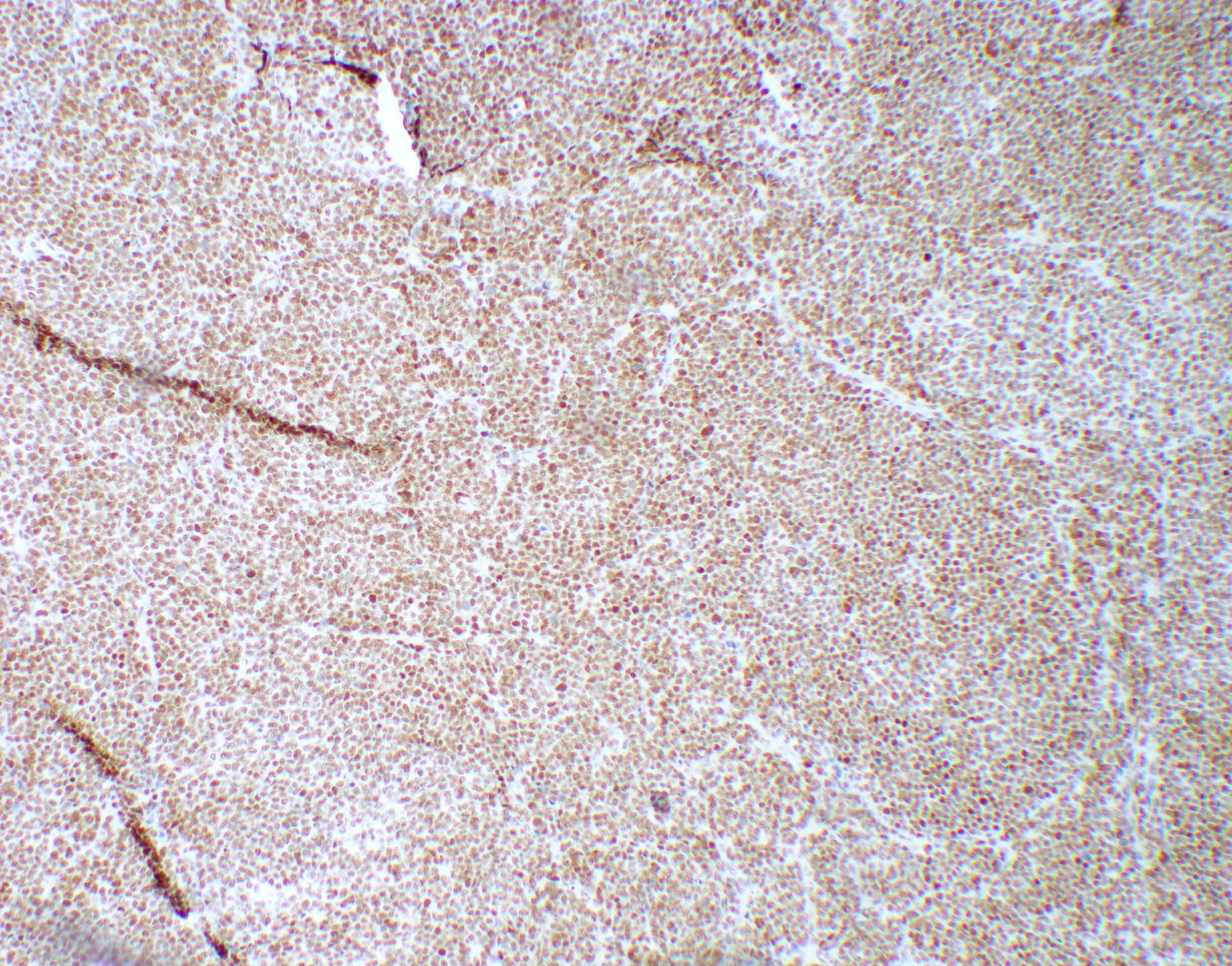
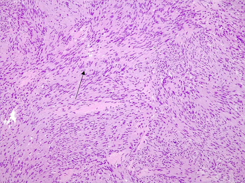
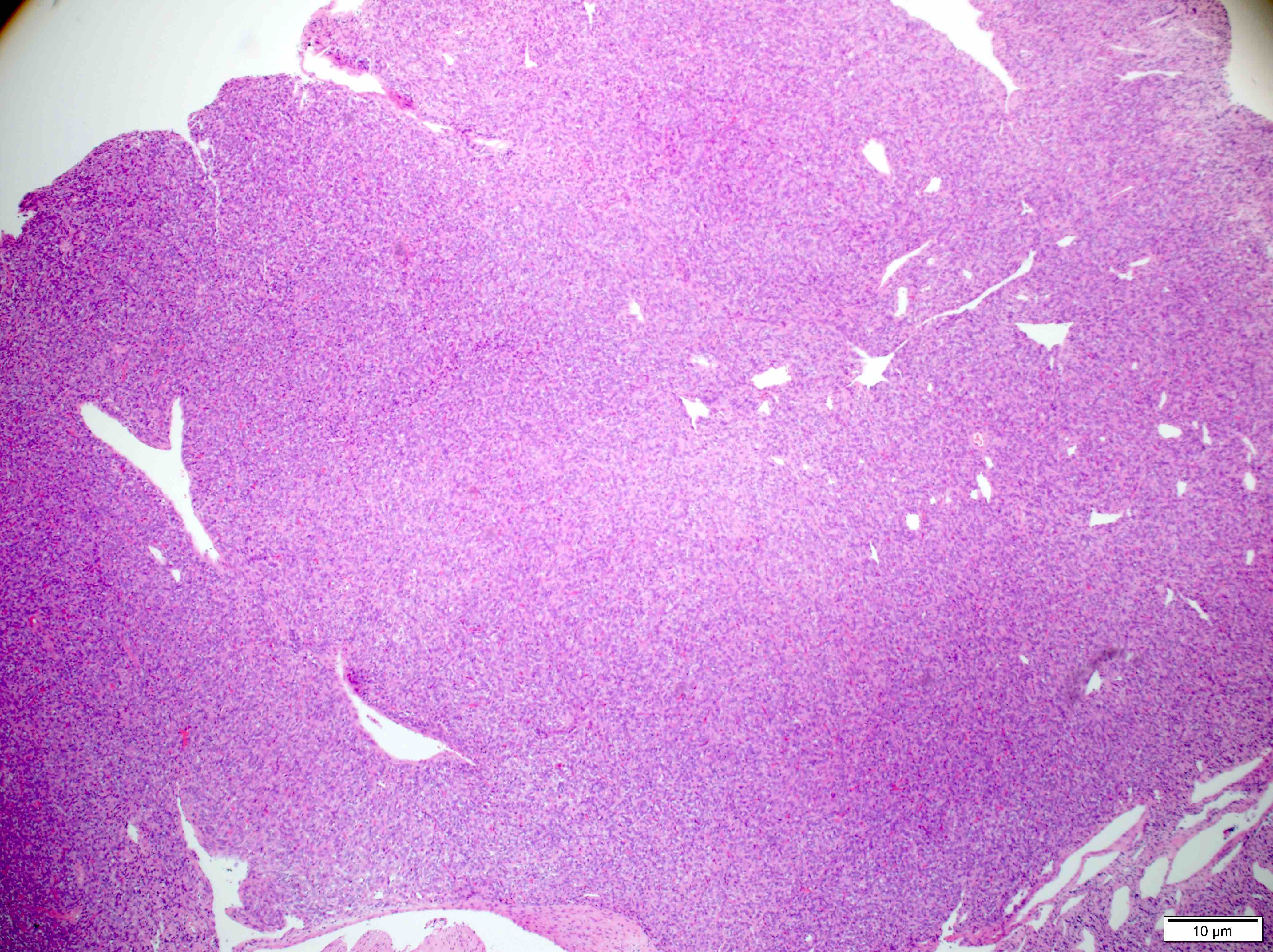
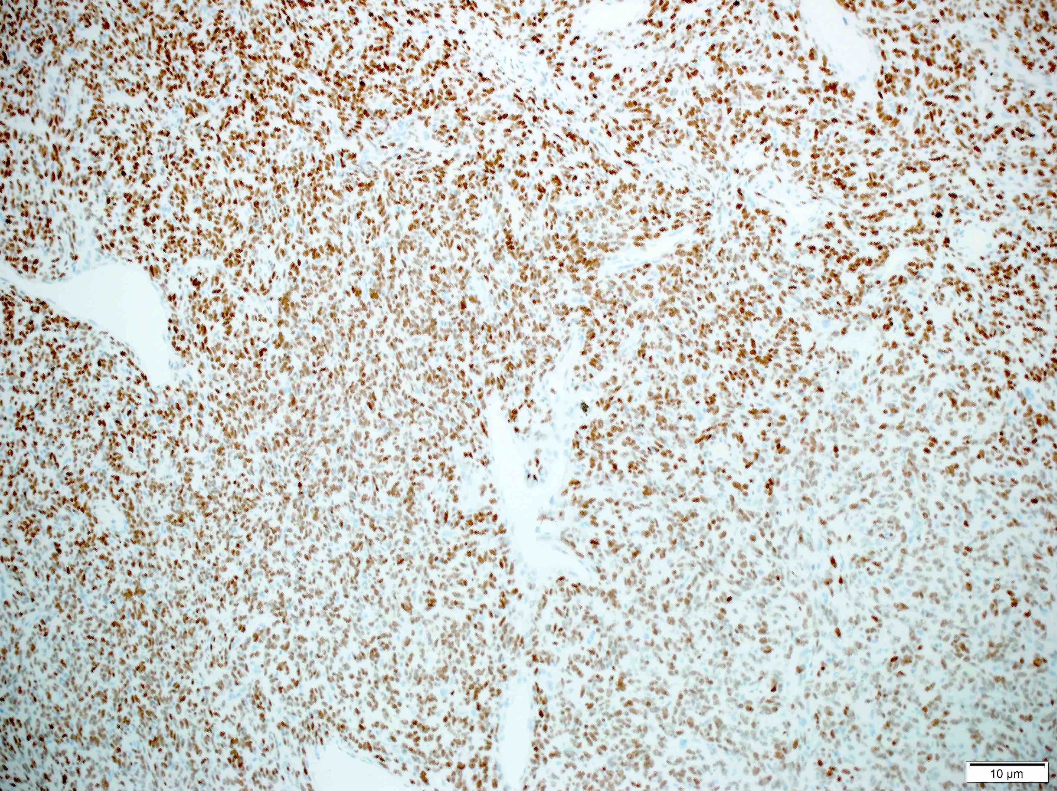
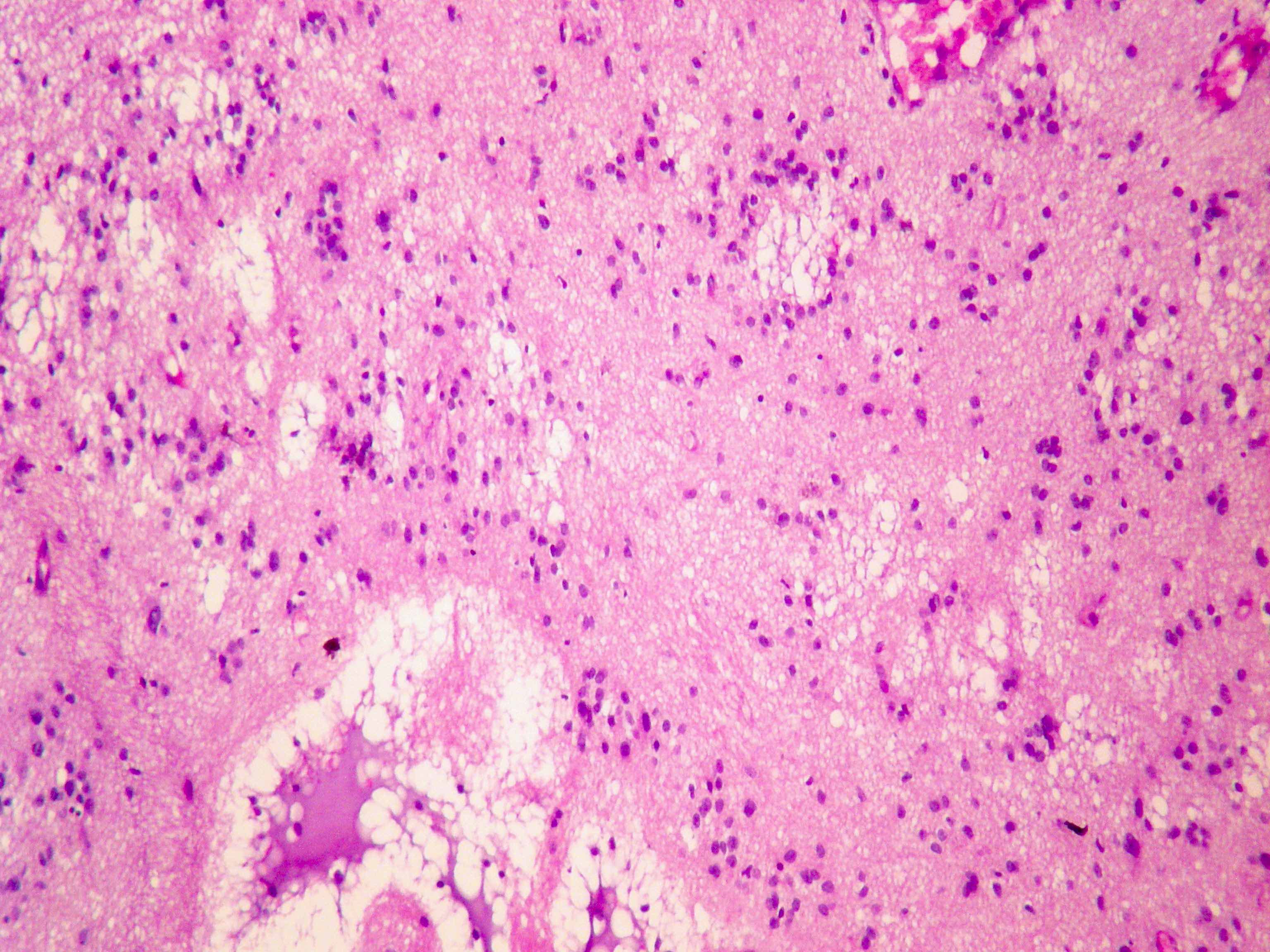
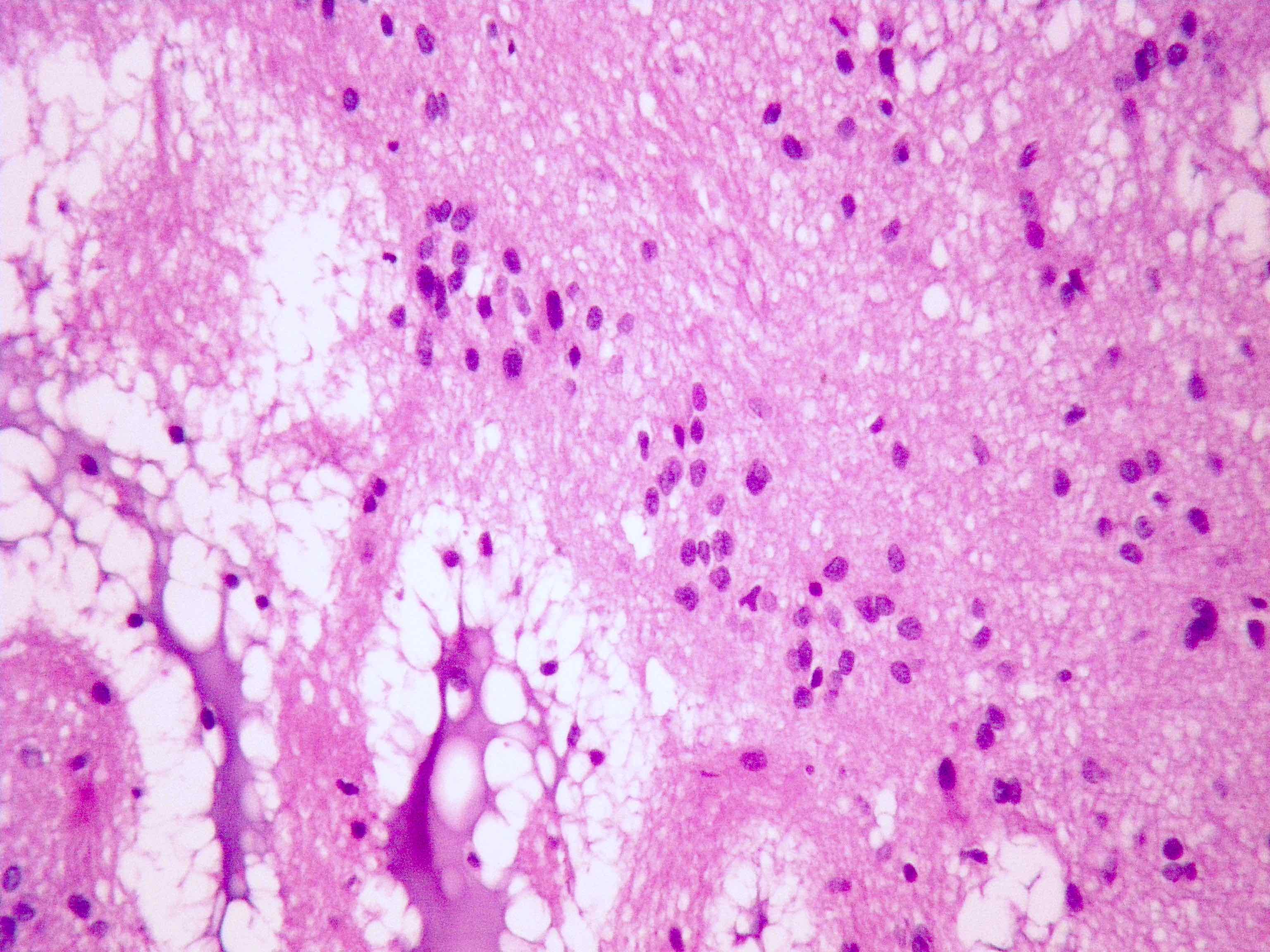
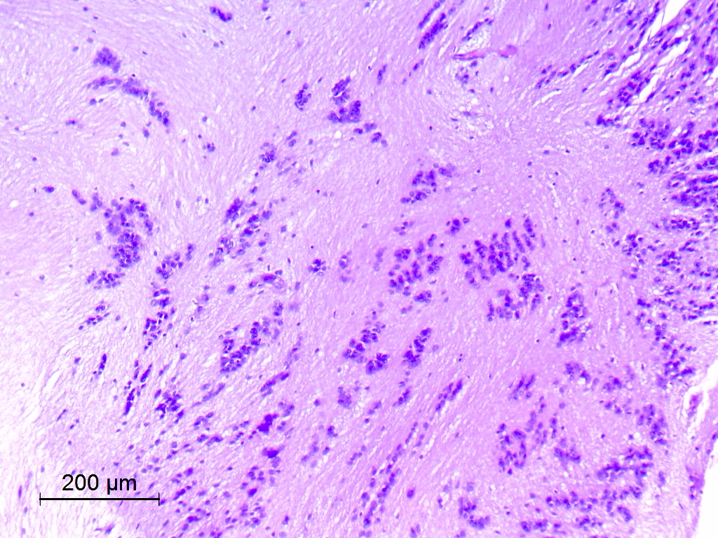
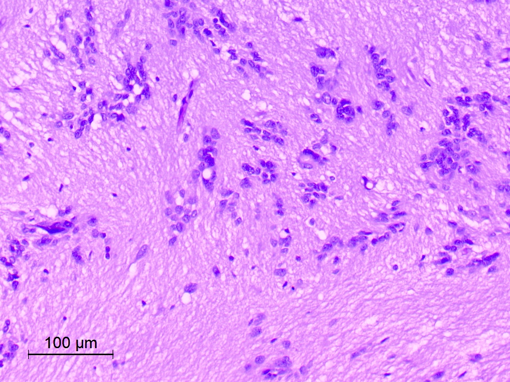
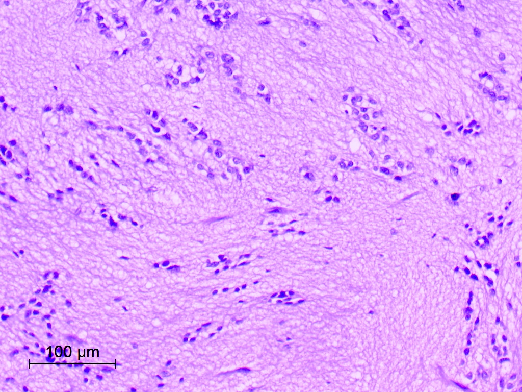
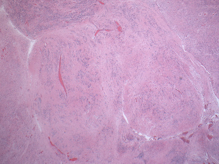
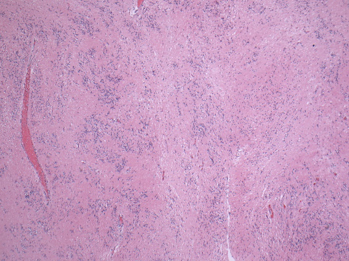
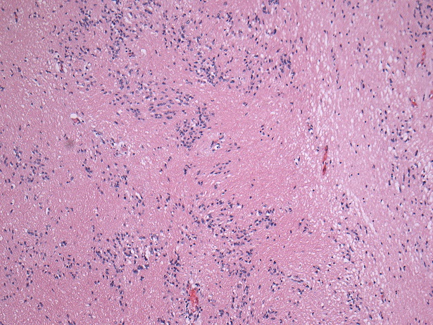
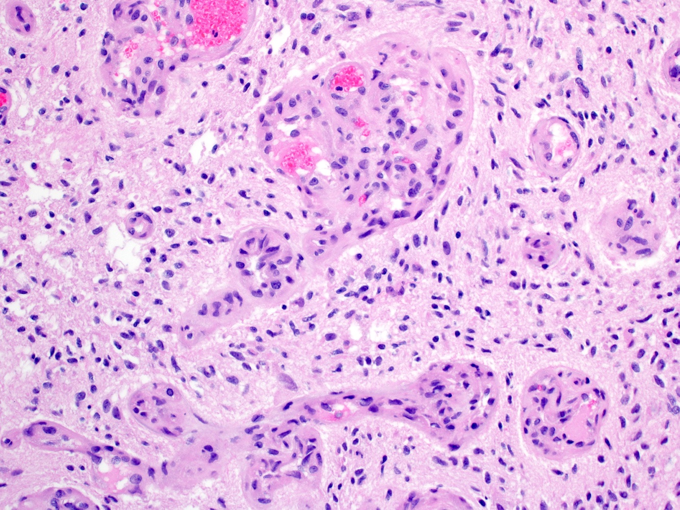
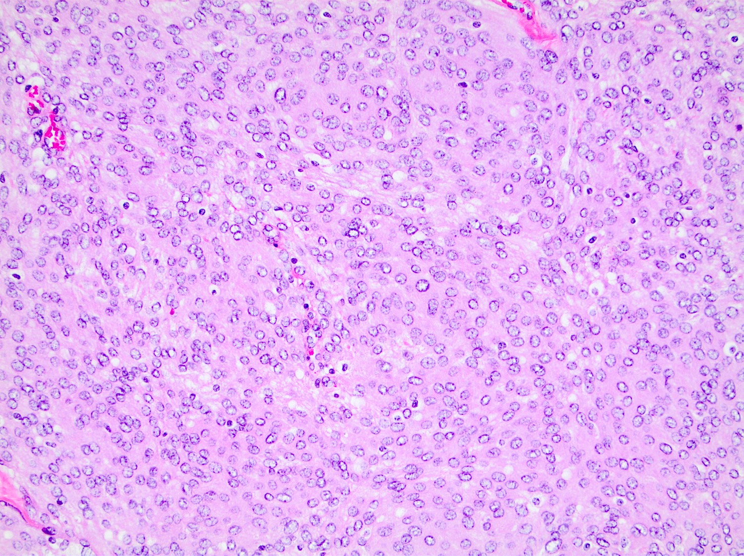
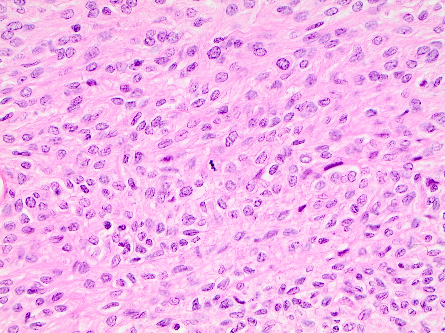
Xanthogranulomatous reaction
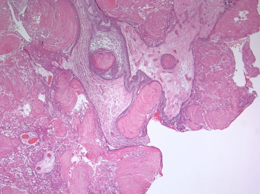
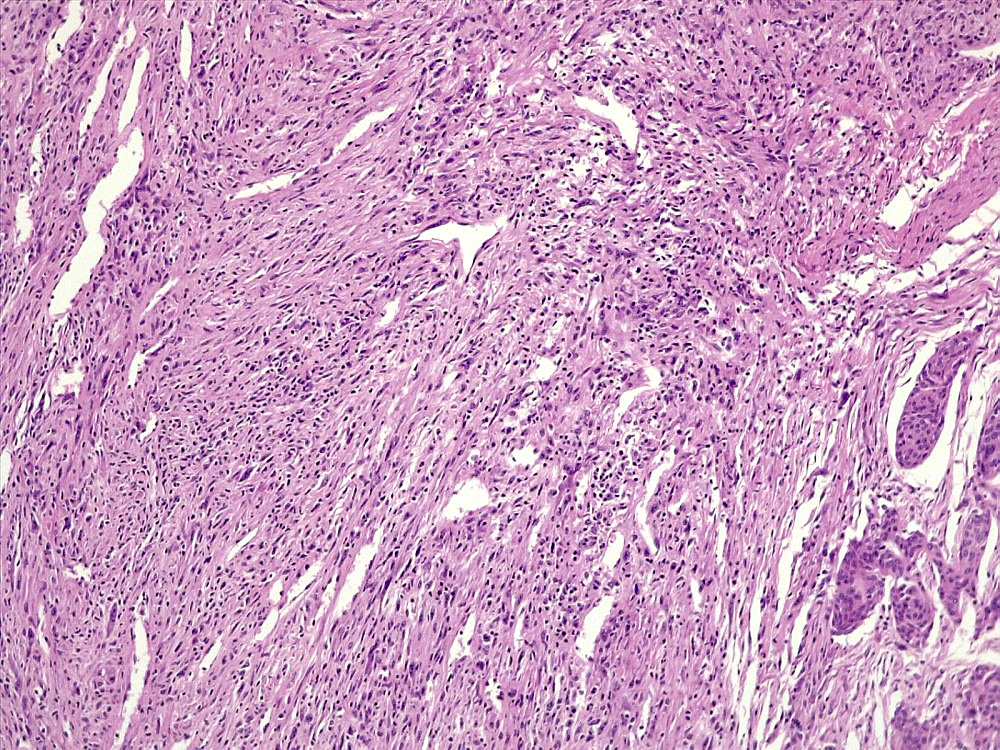
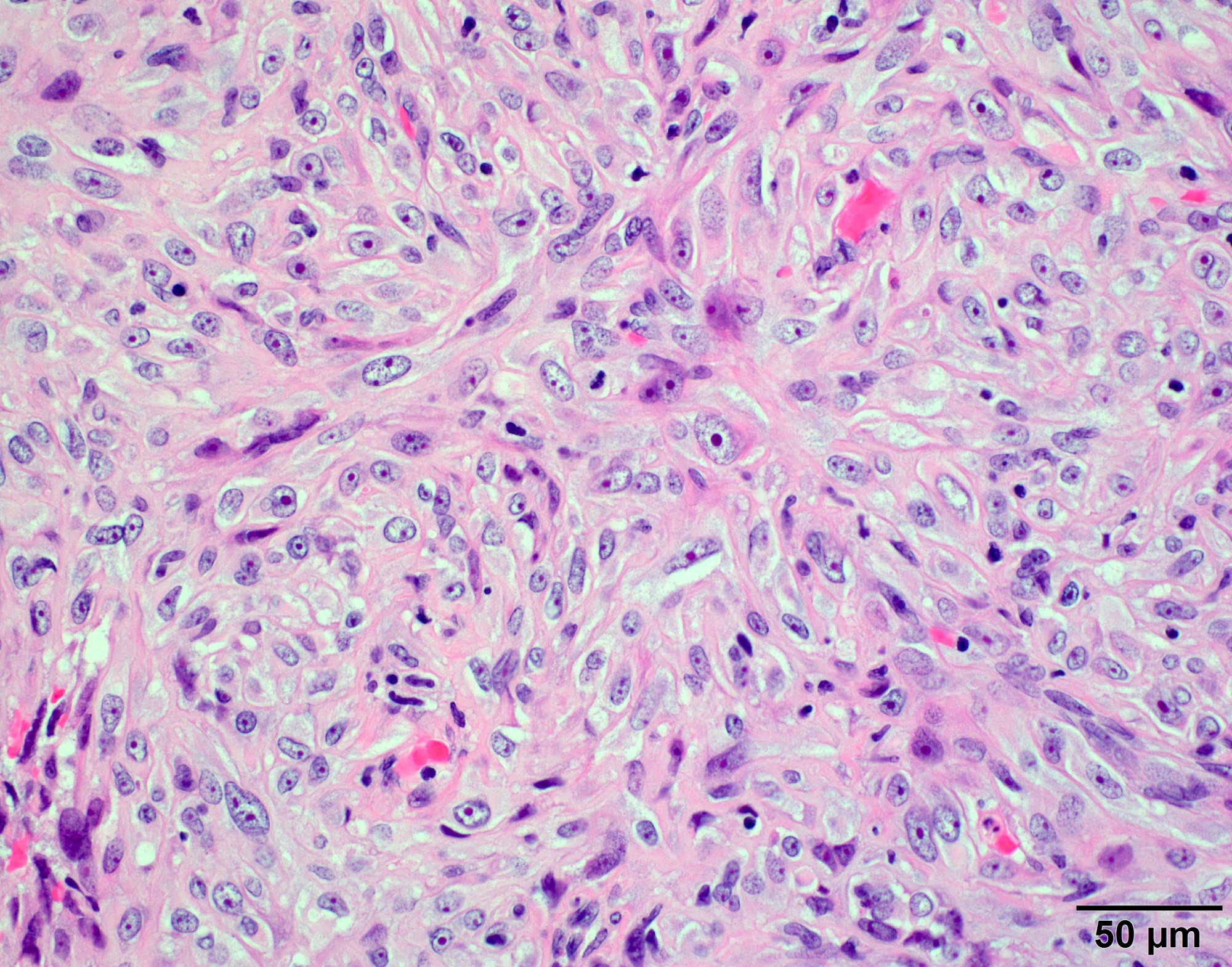
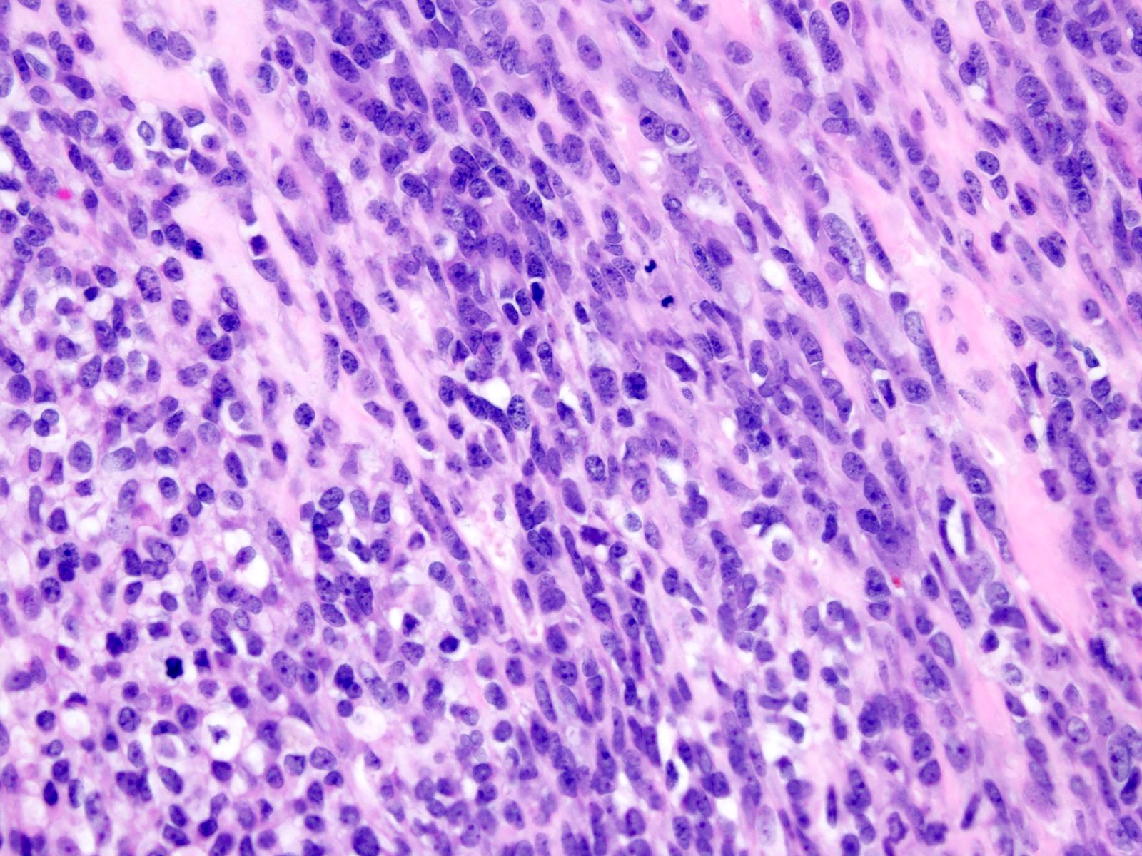
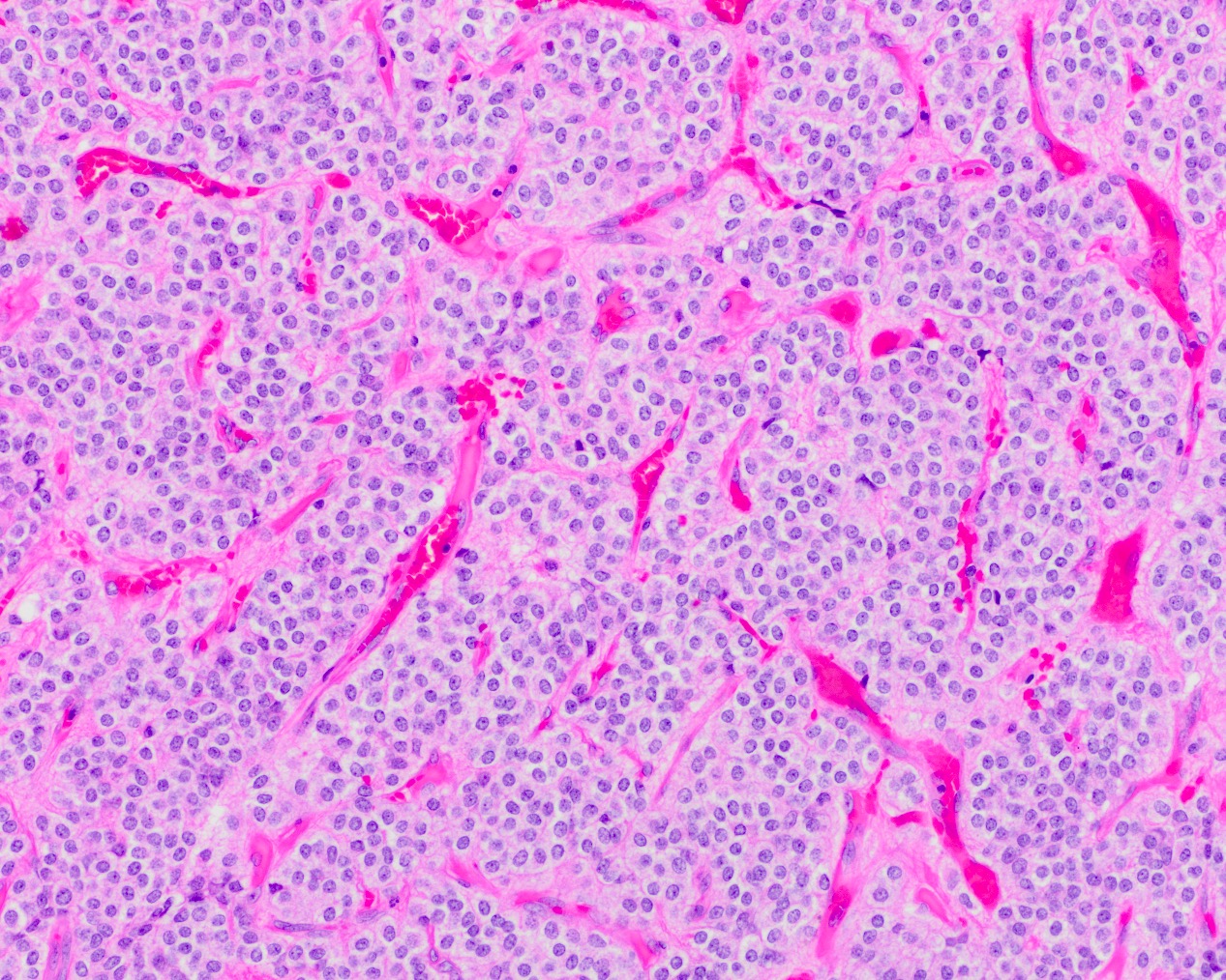
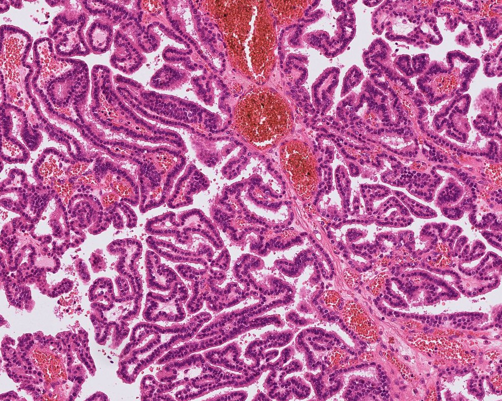
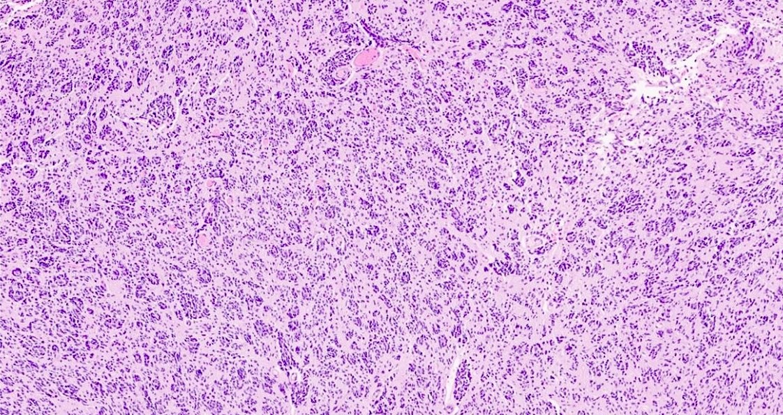
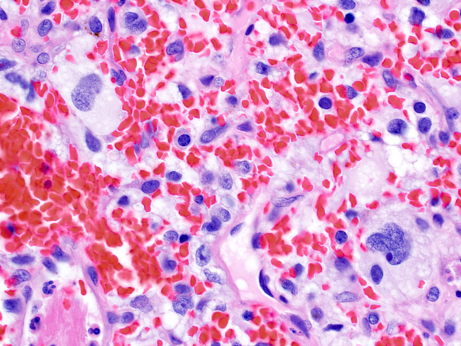
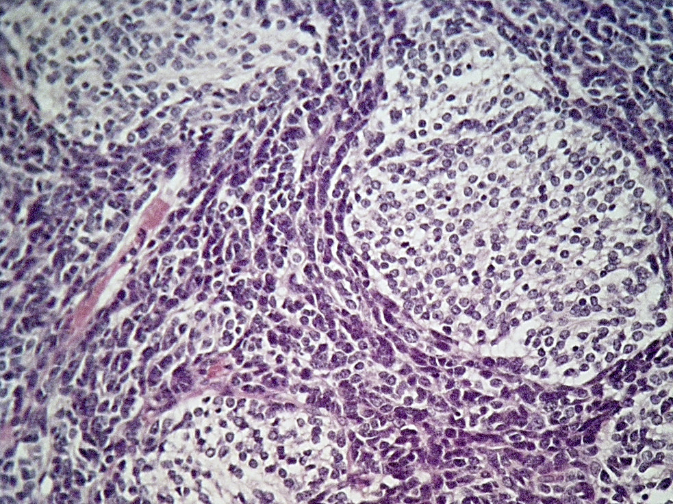
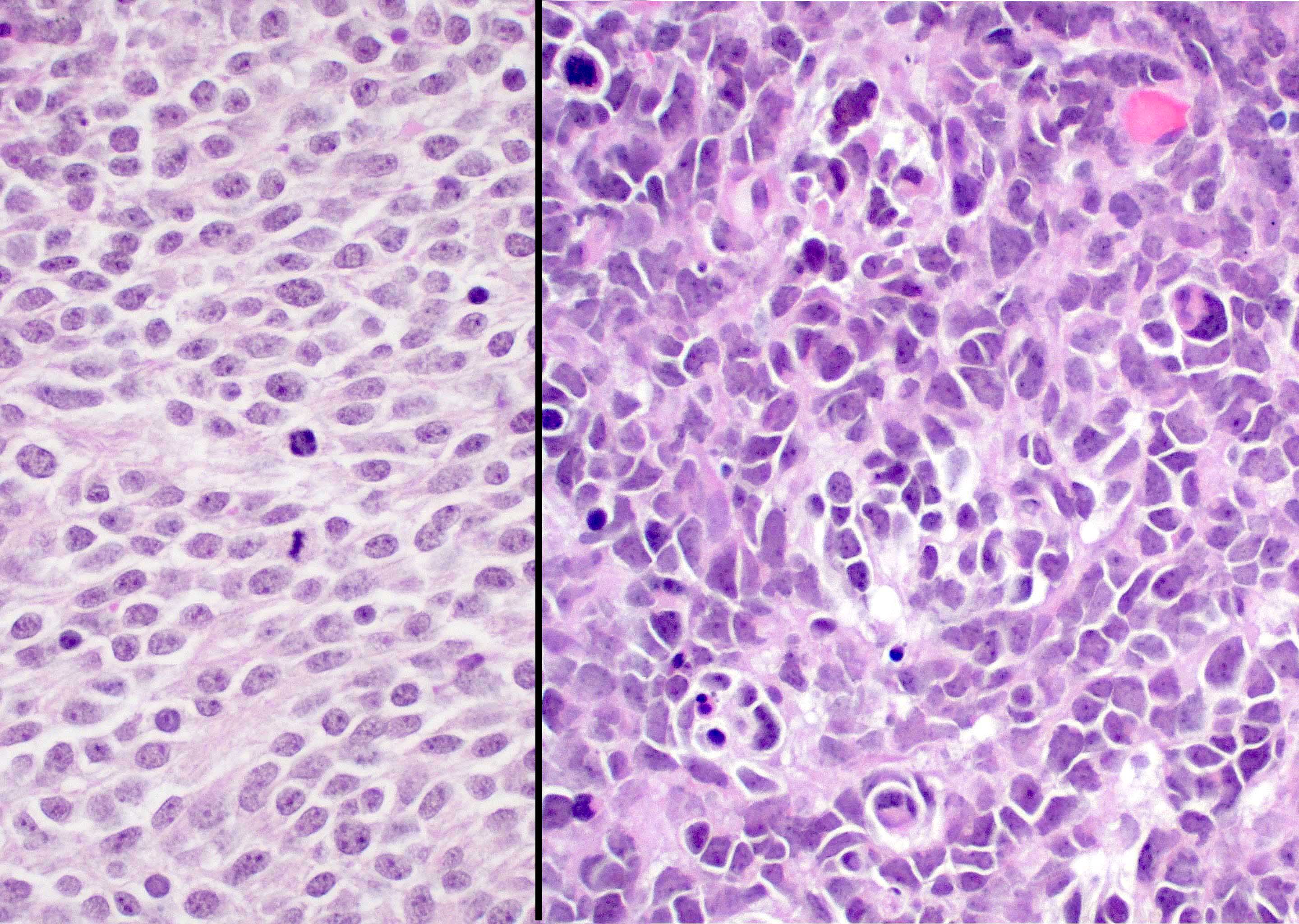
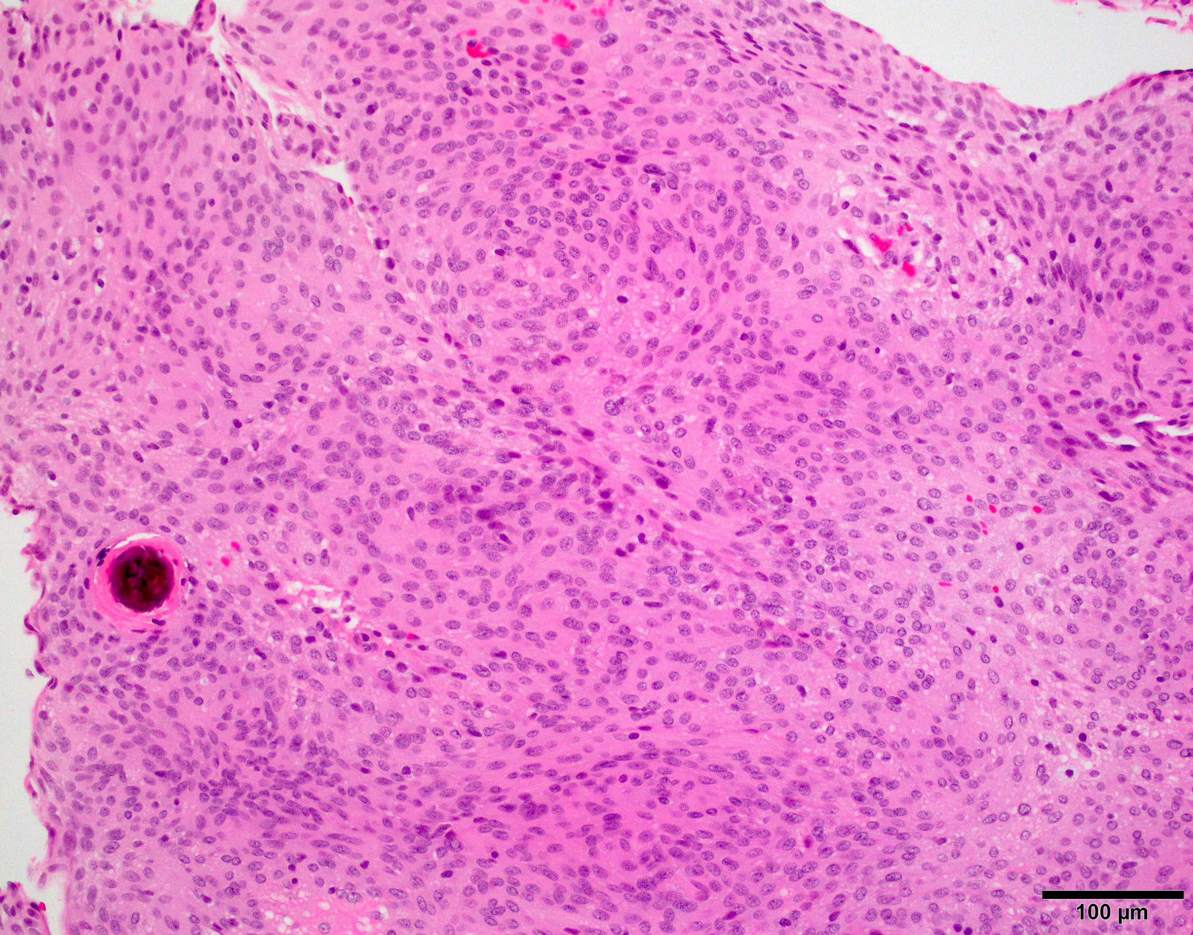
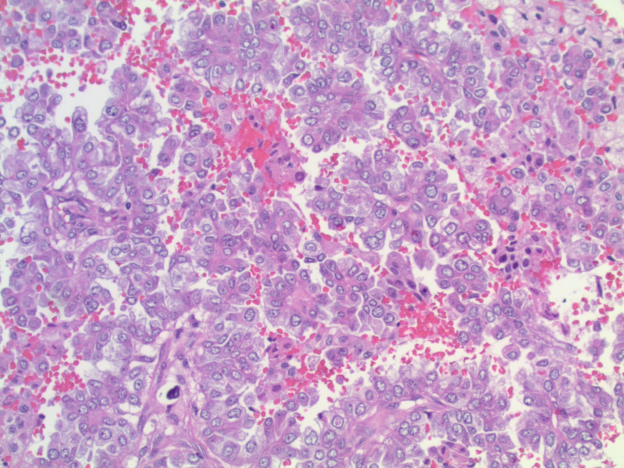
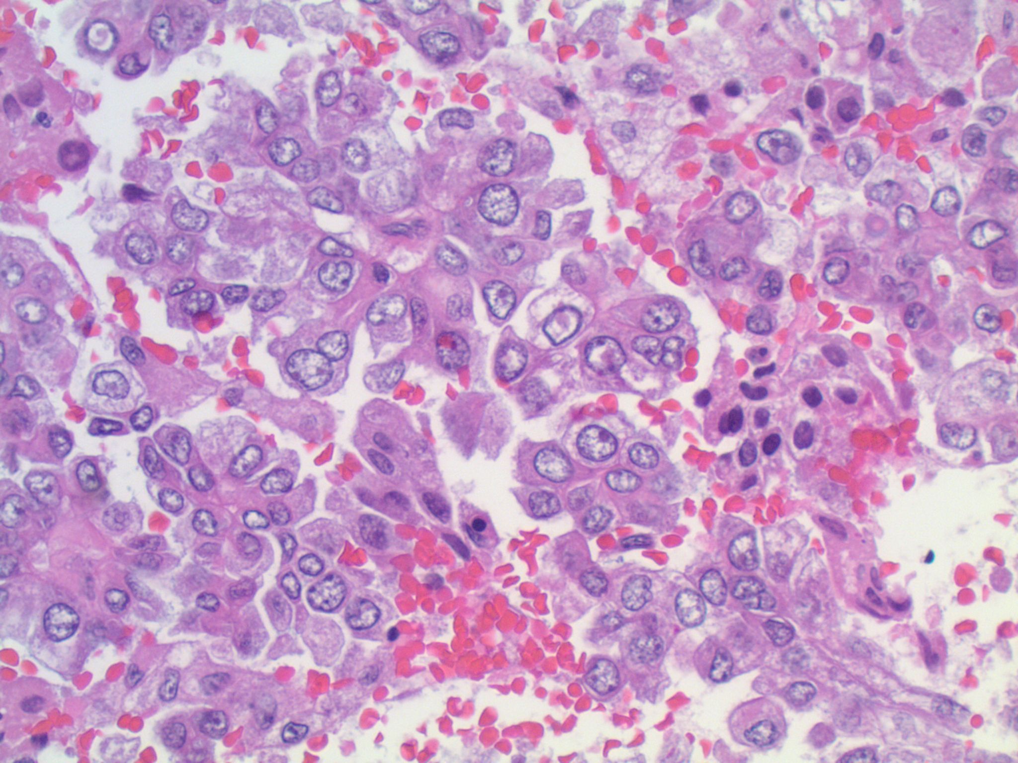
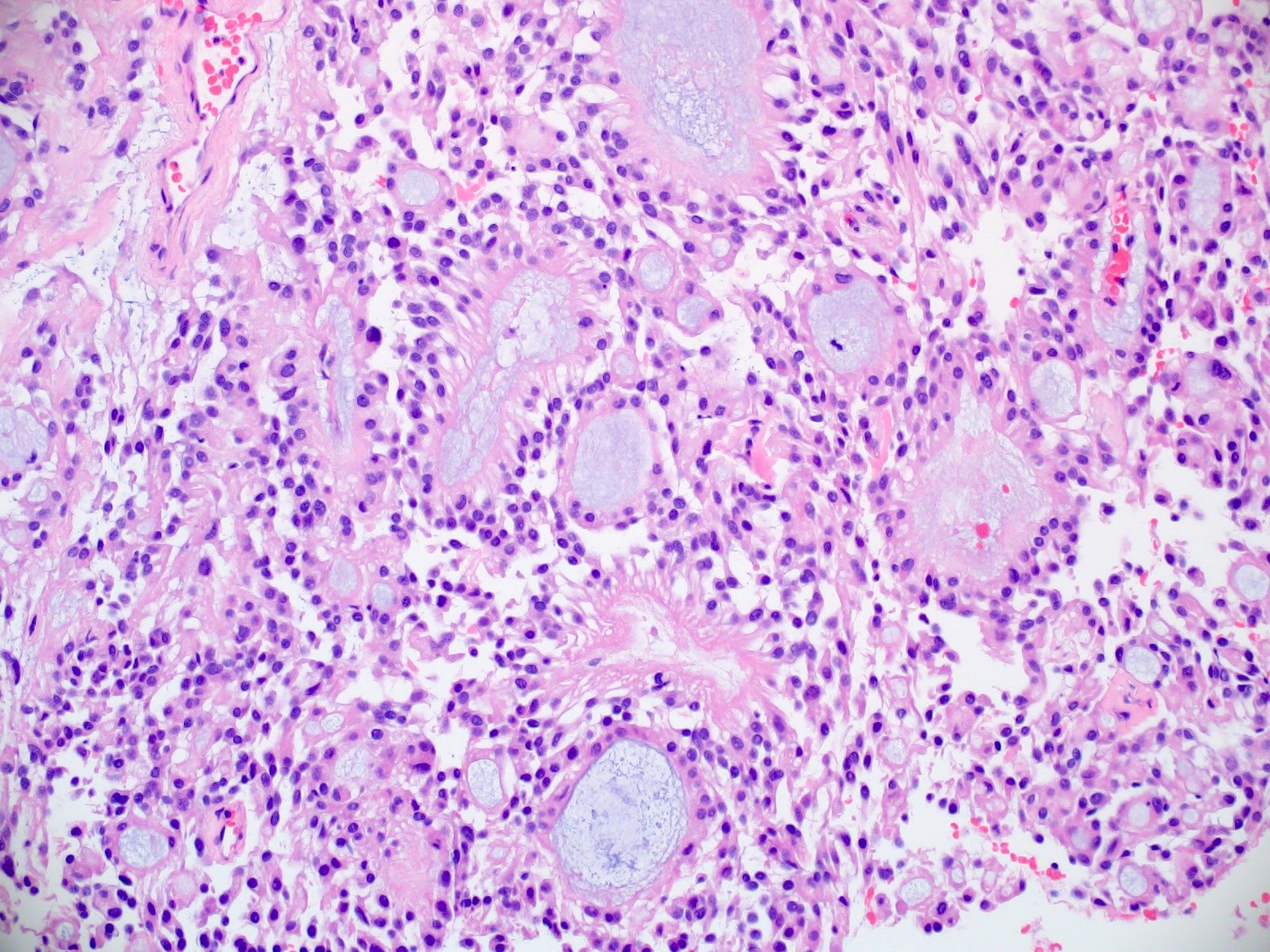
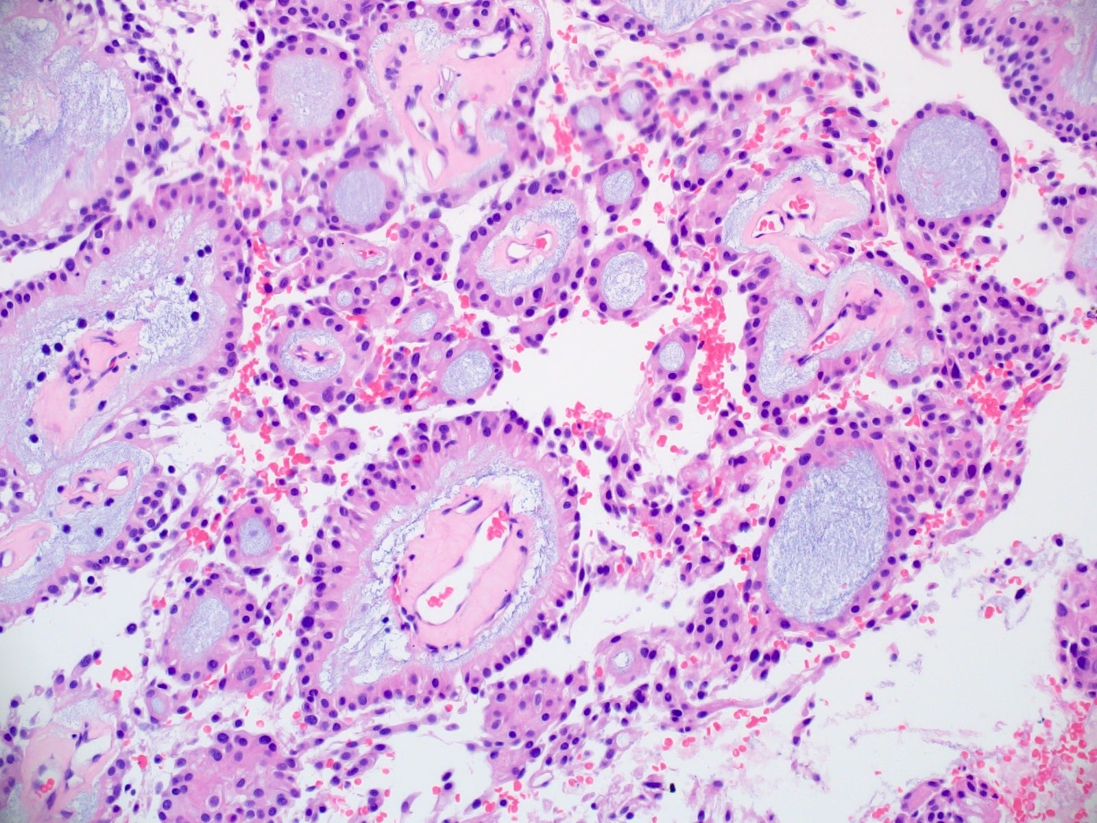
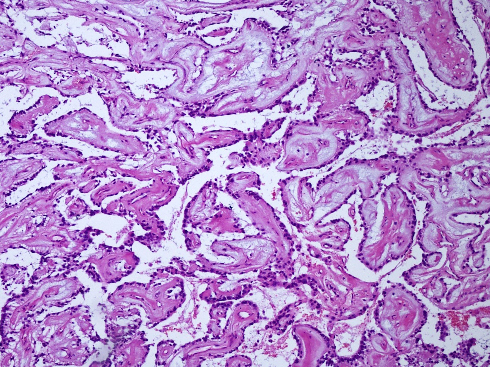
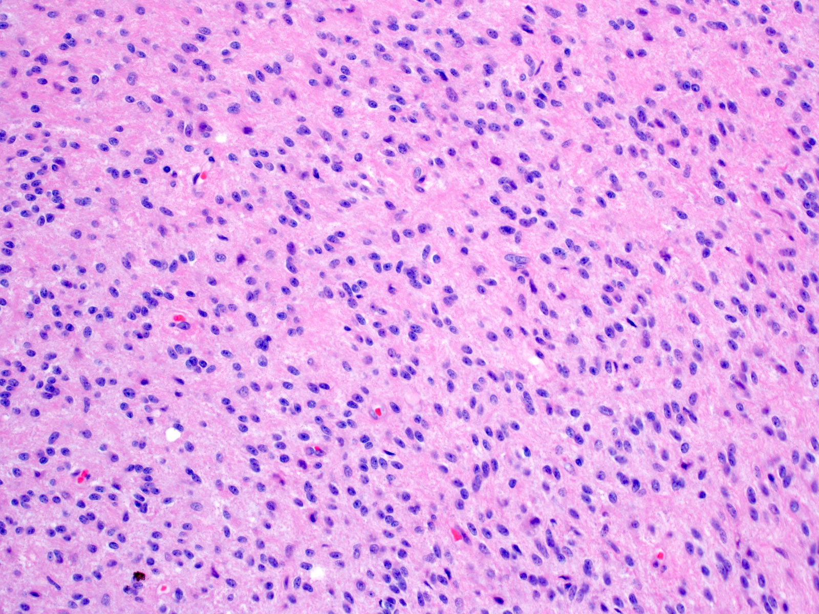
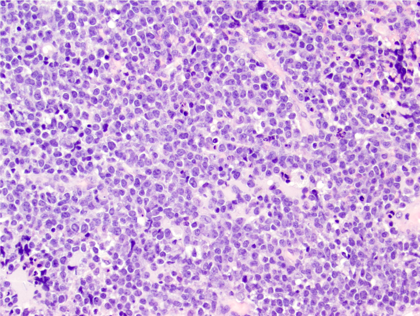
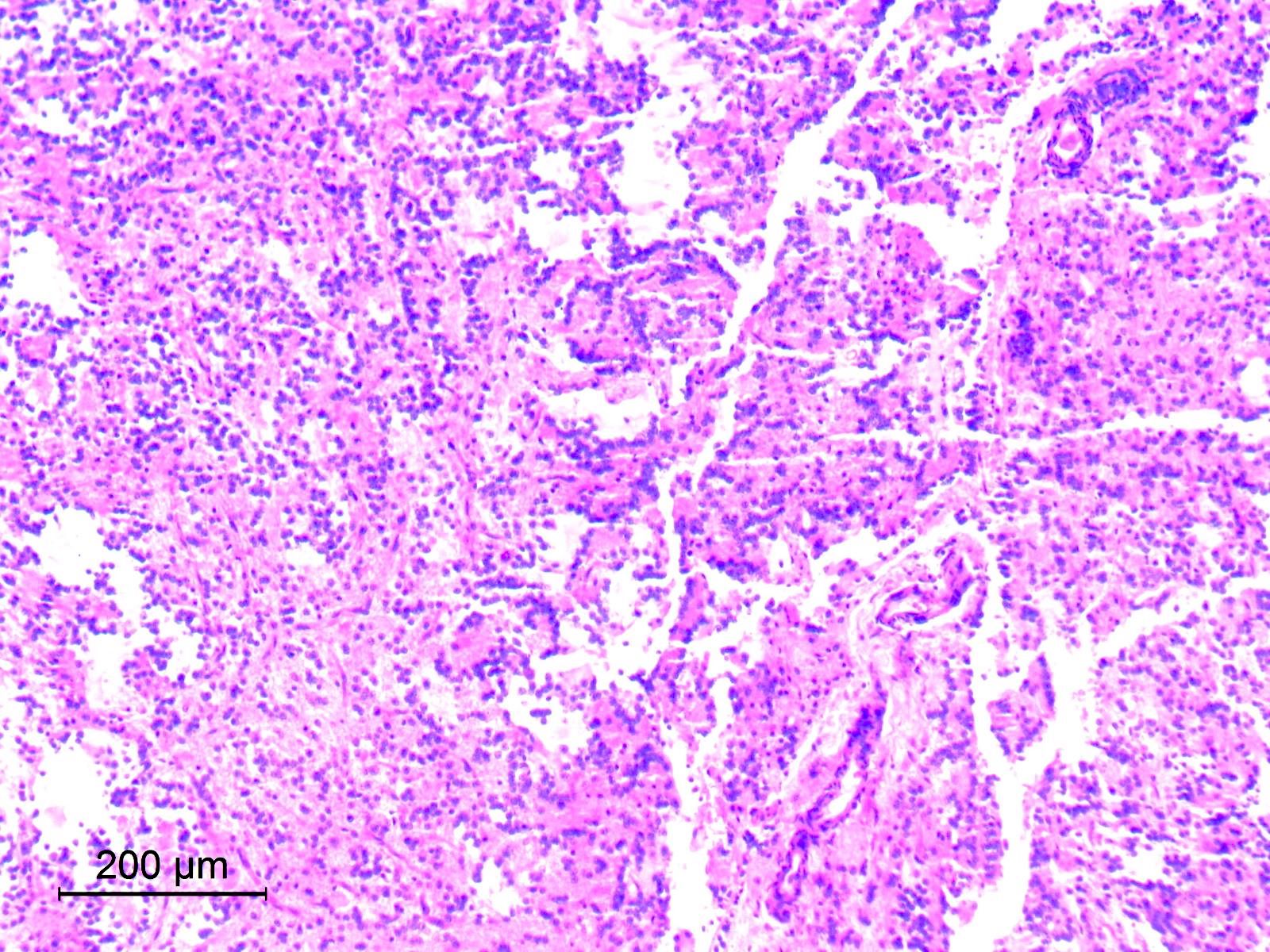
3 components
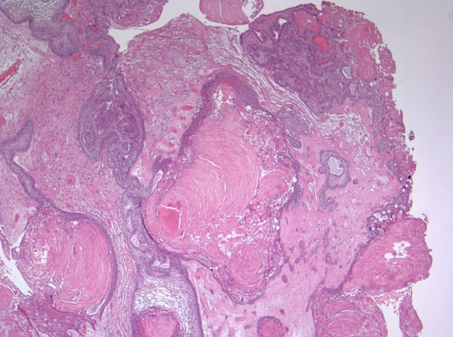
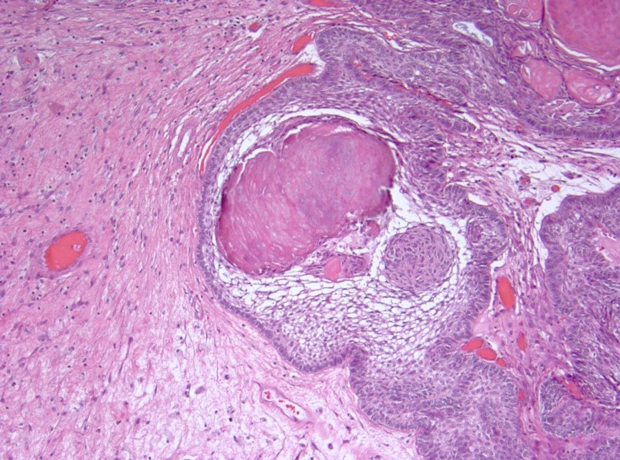
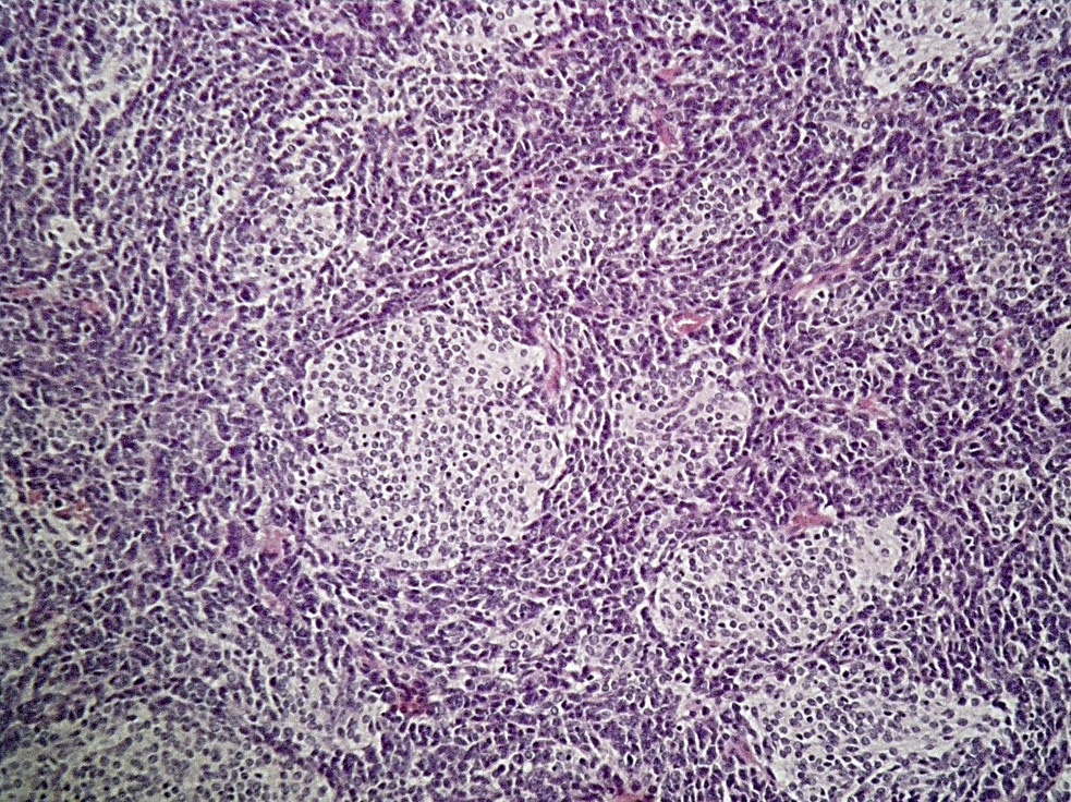
Tumor tongues
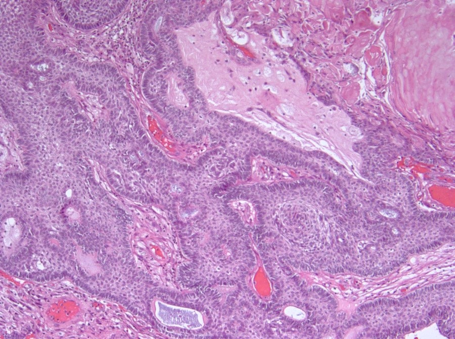
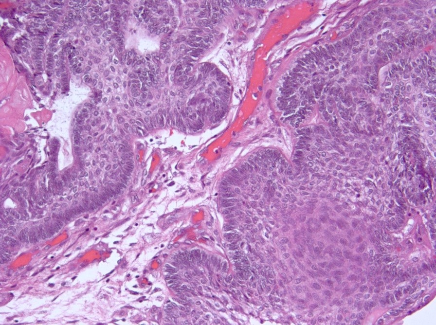
Nuclear palisading
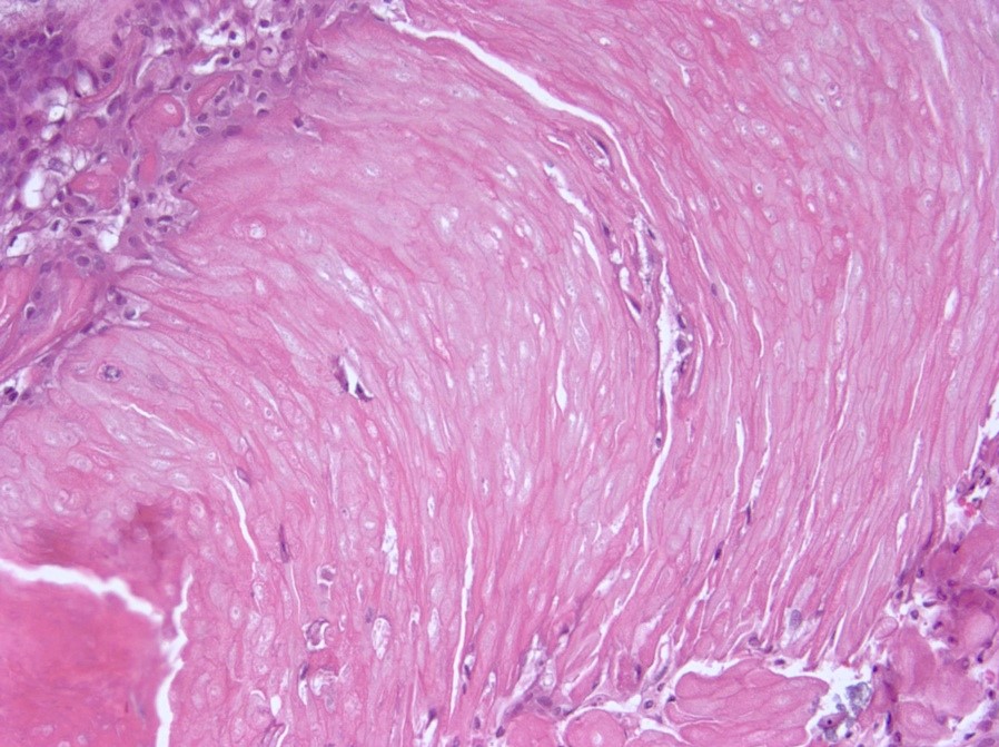
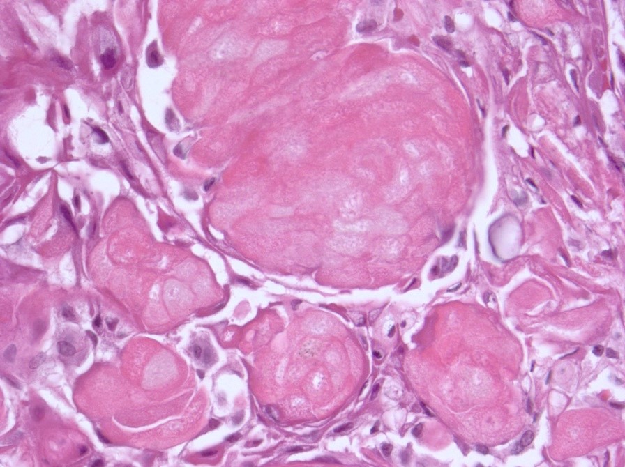
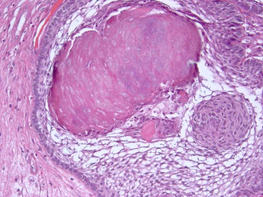
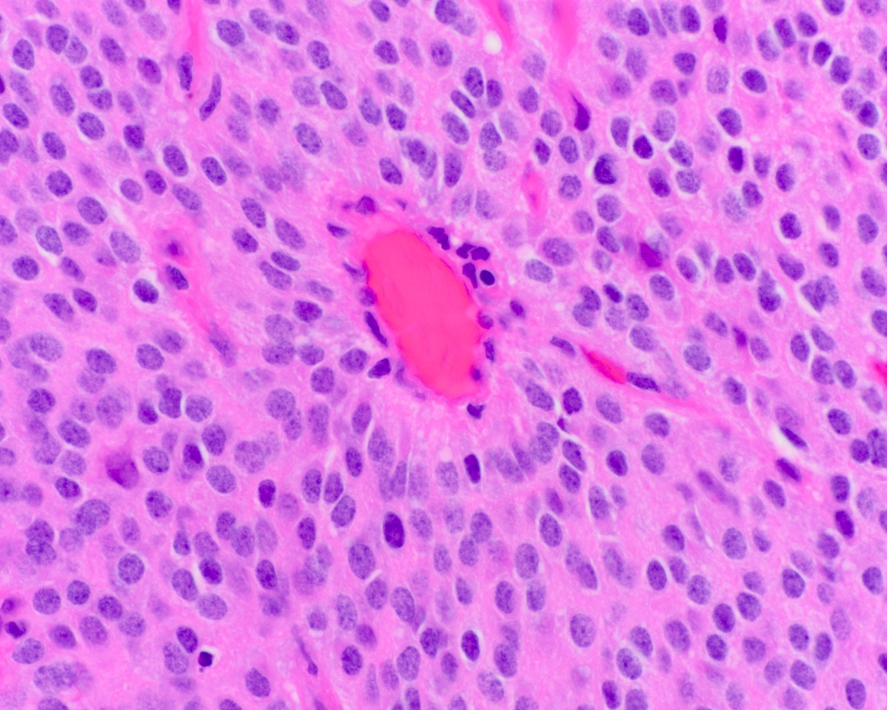
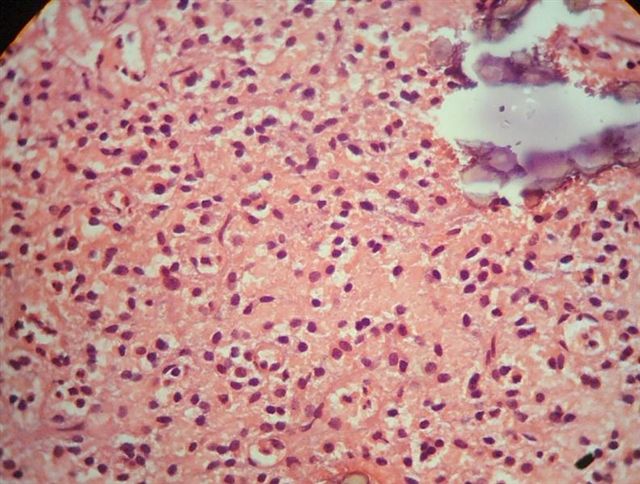
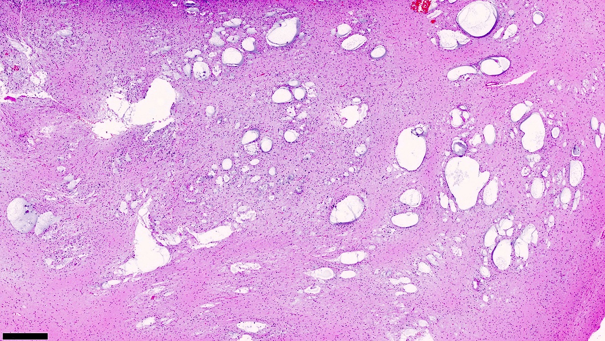
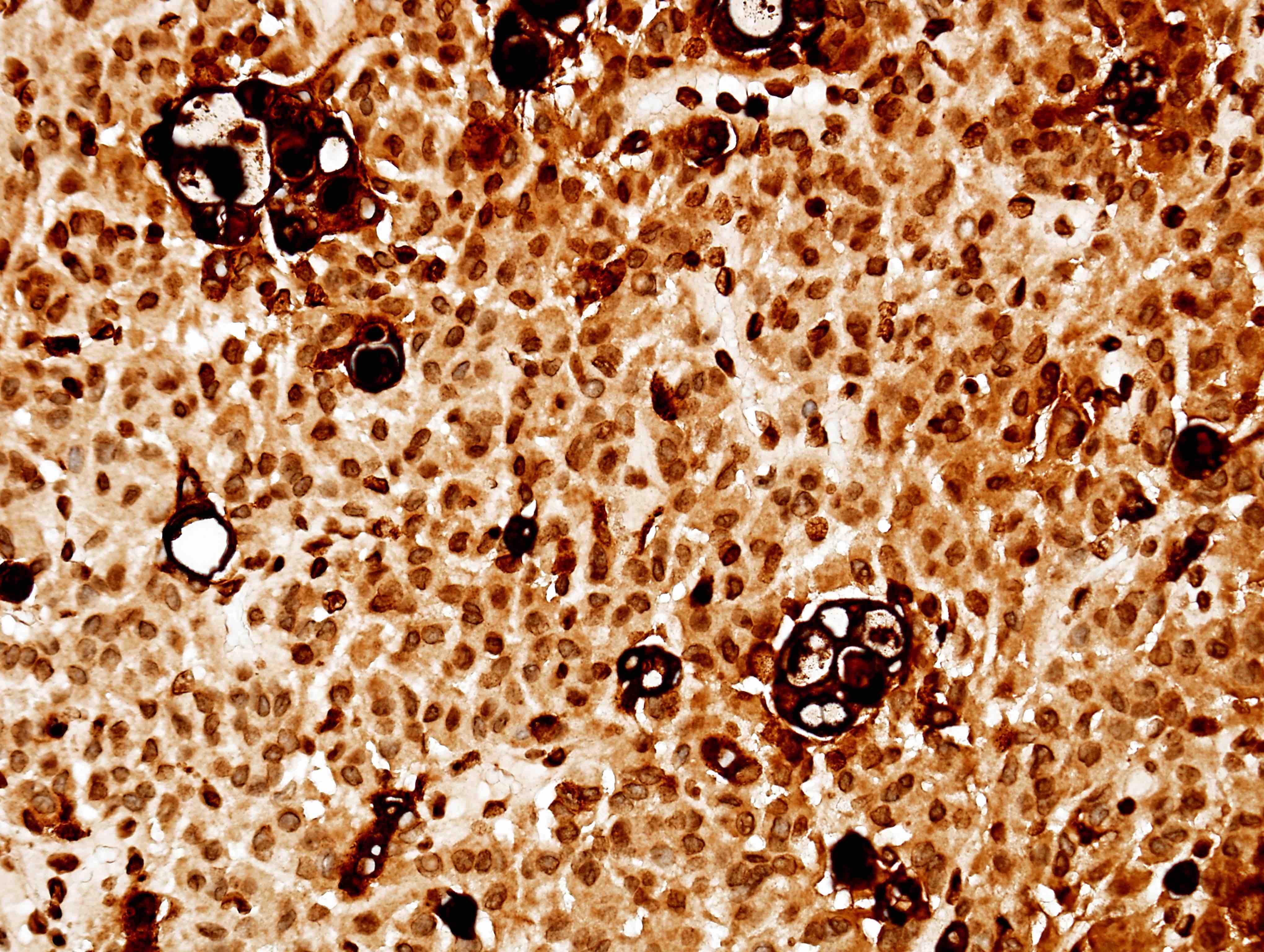
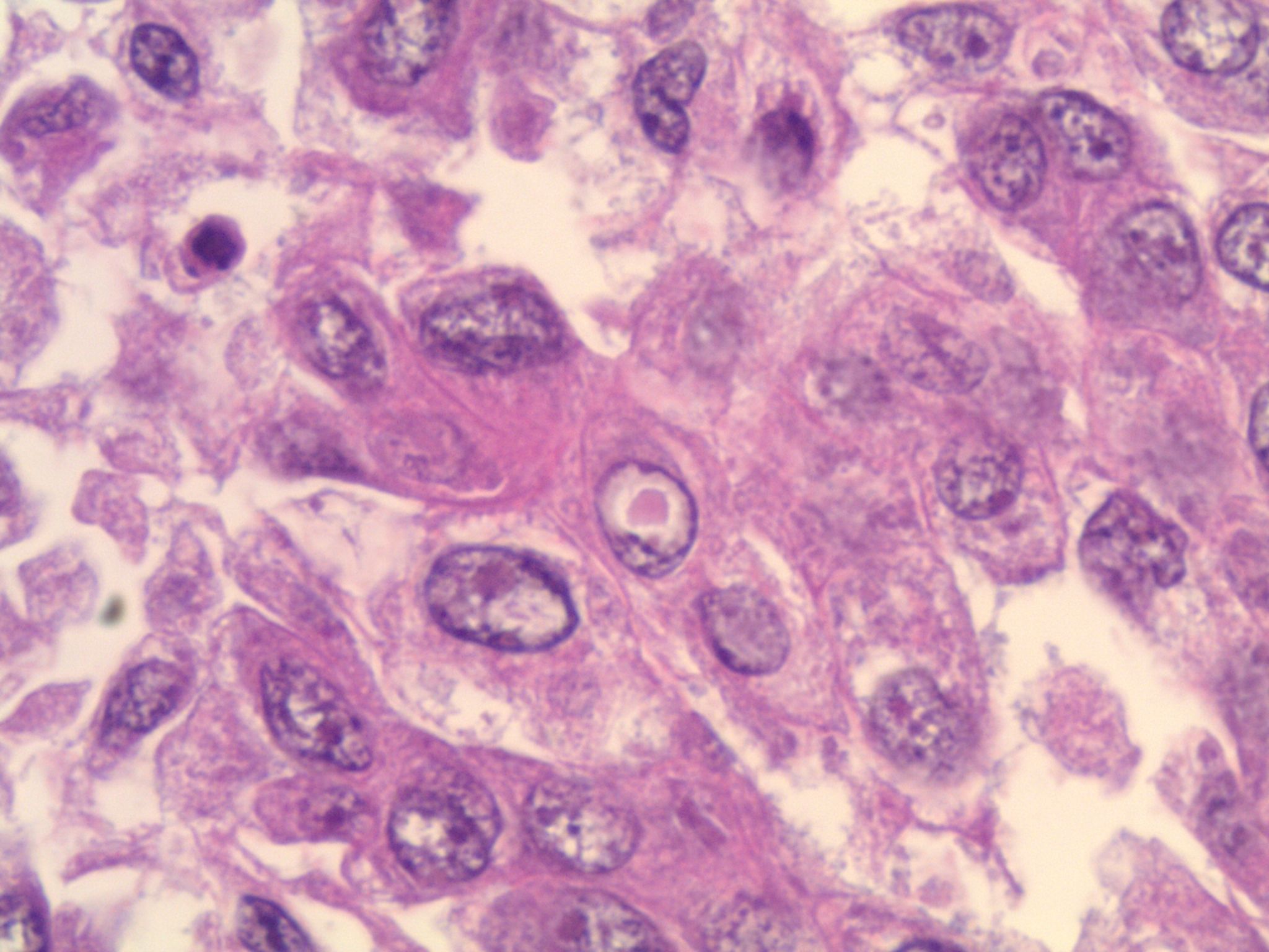
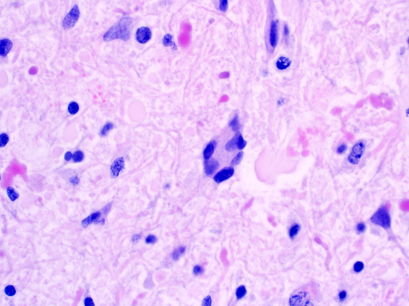
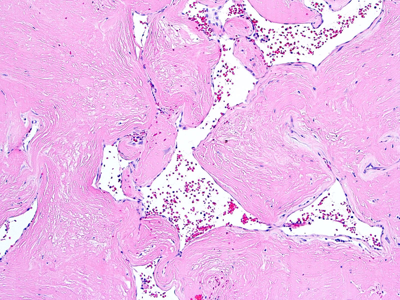
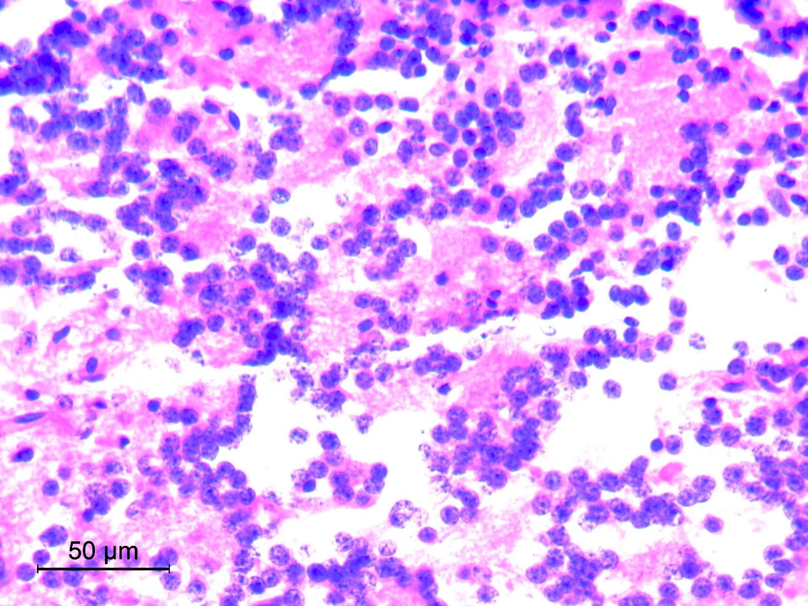
Wet keratin
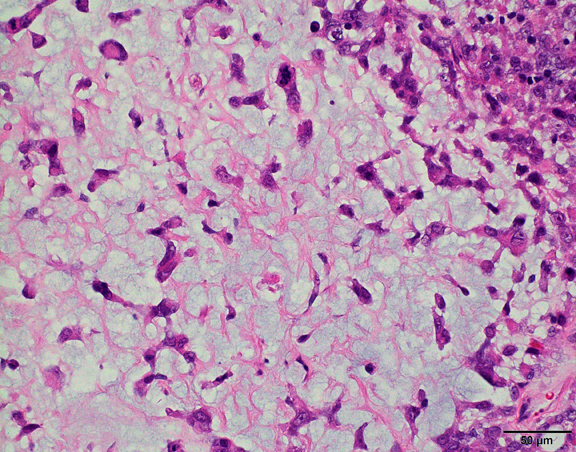
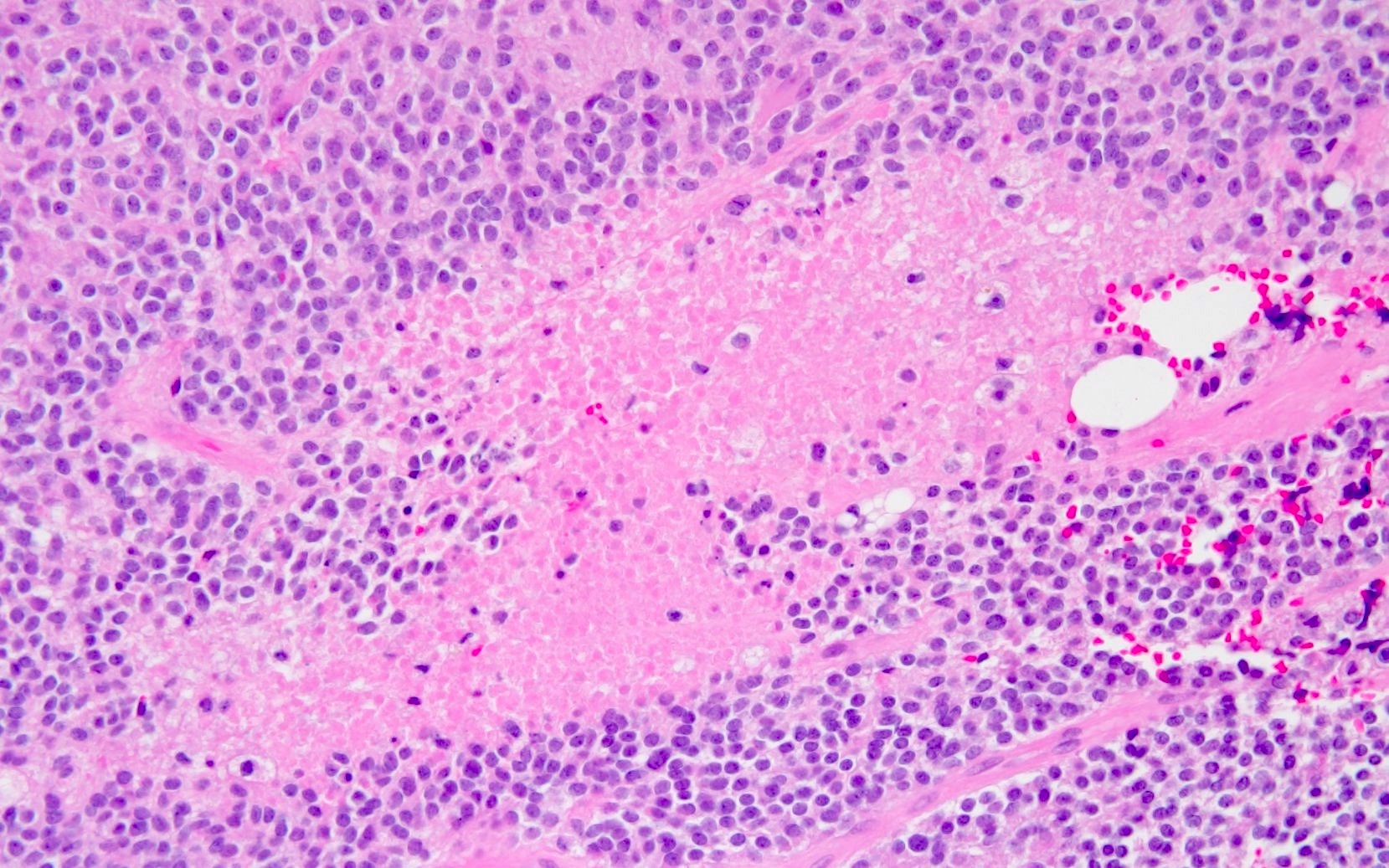
Cystic degeneration
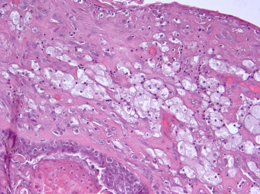
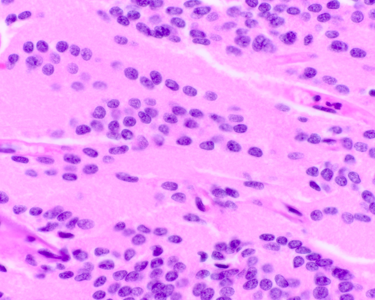
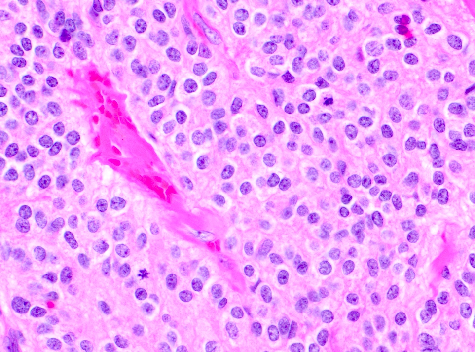
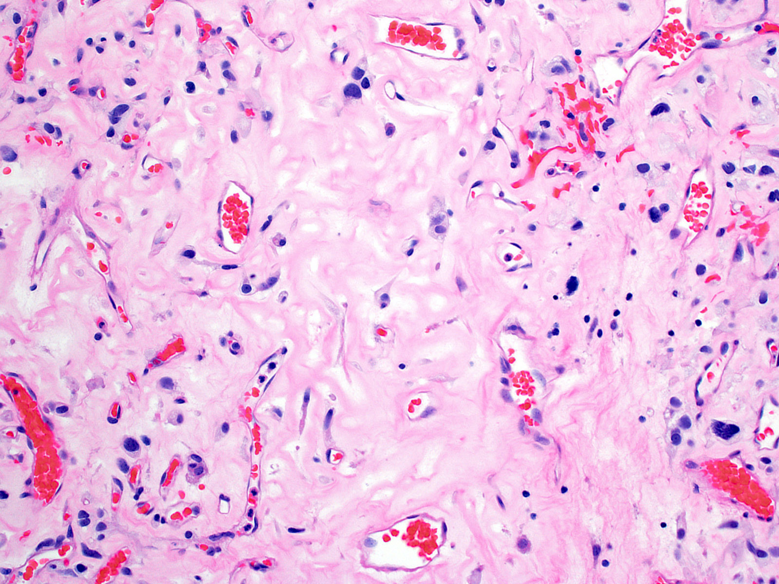
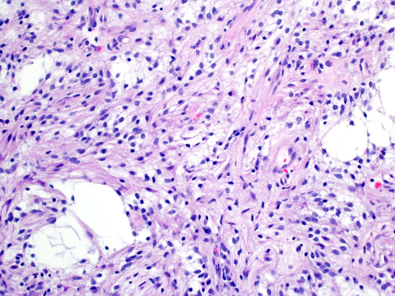
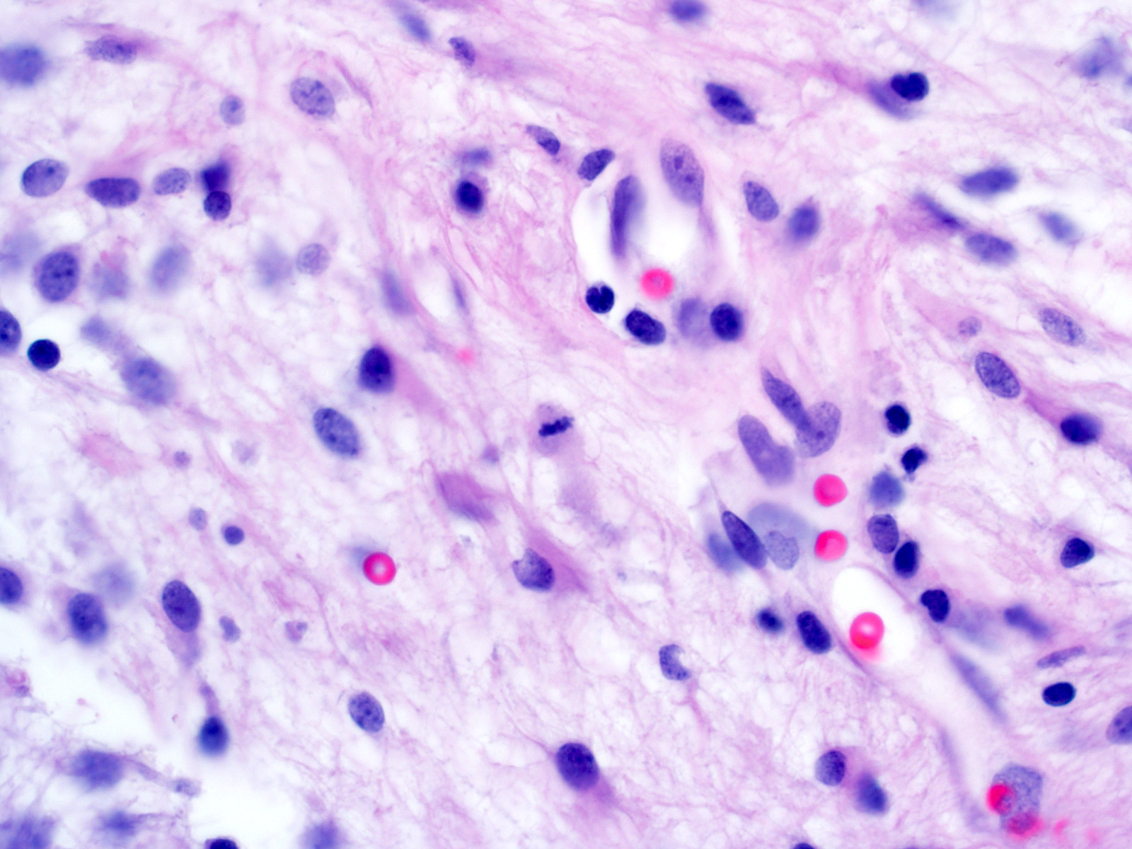
Fibrosis
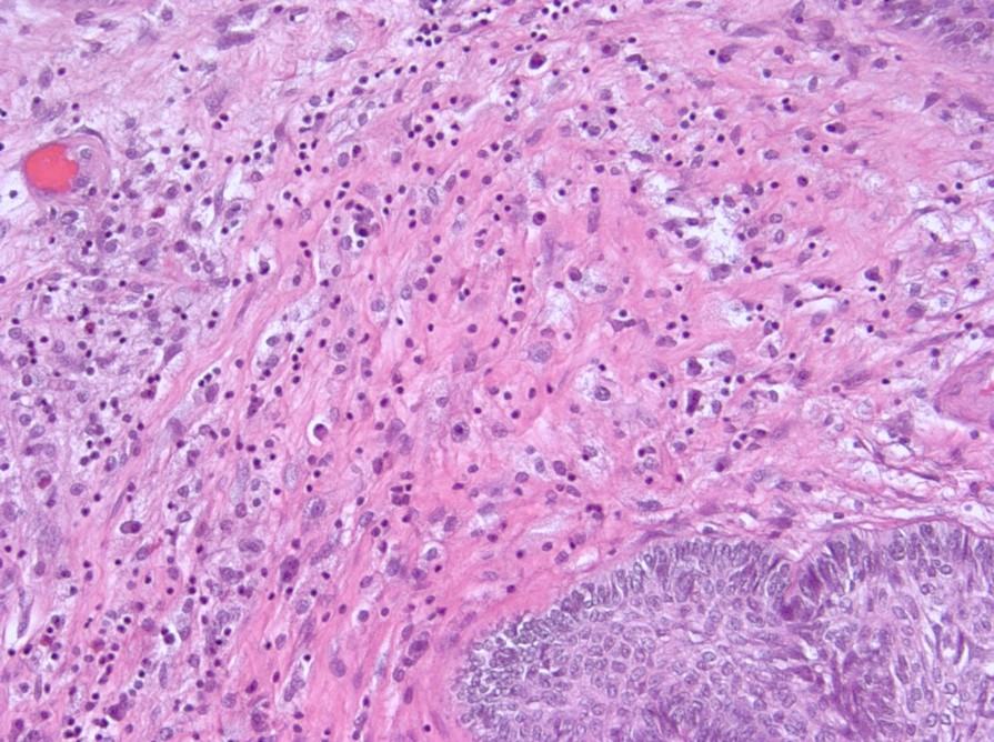
Inflammatory infiltrate
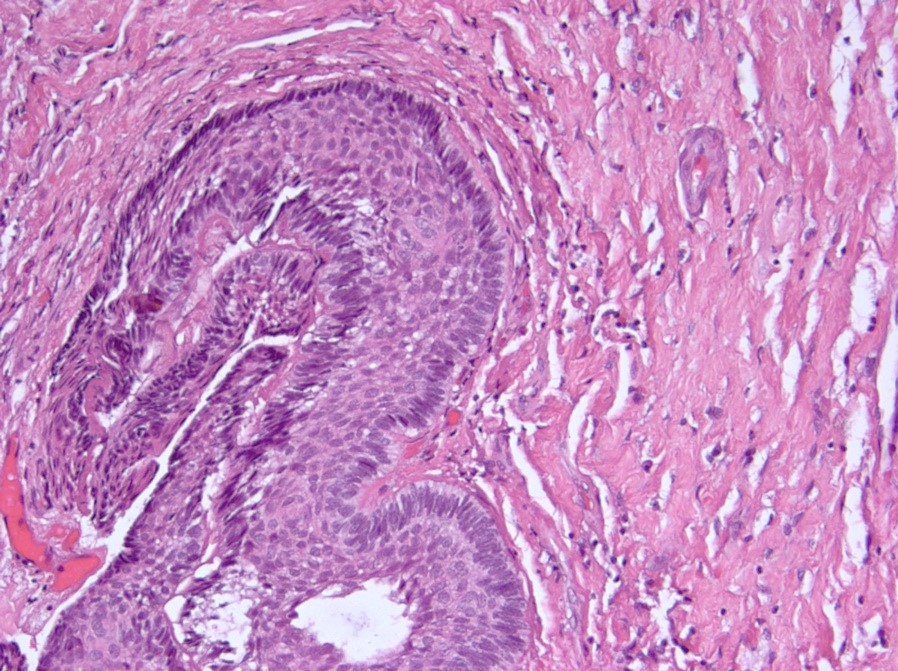
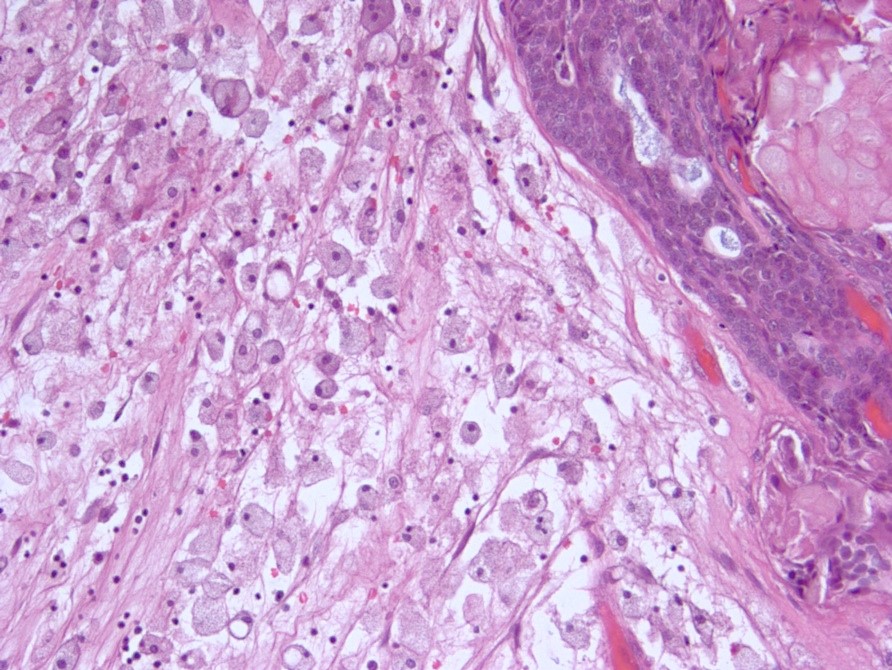
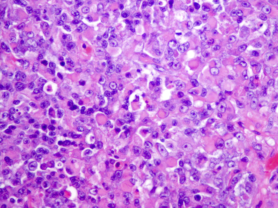
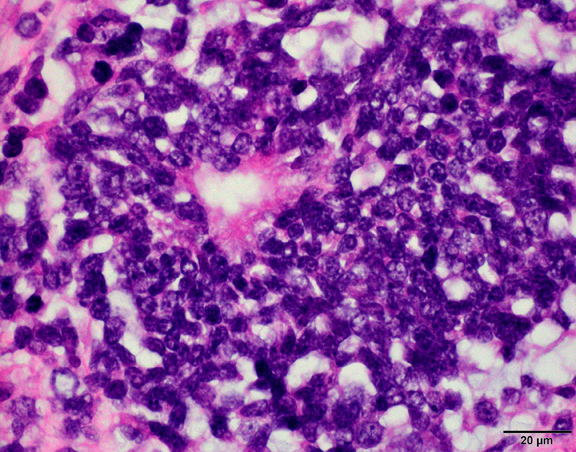
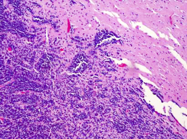
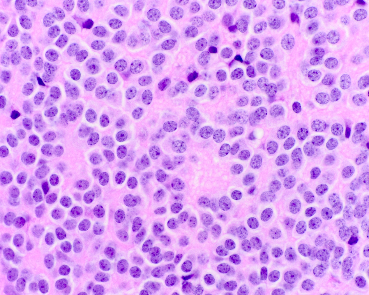
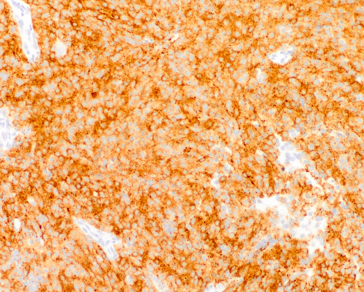
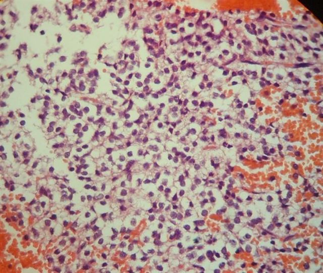
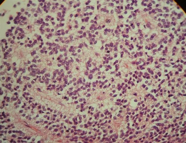
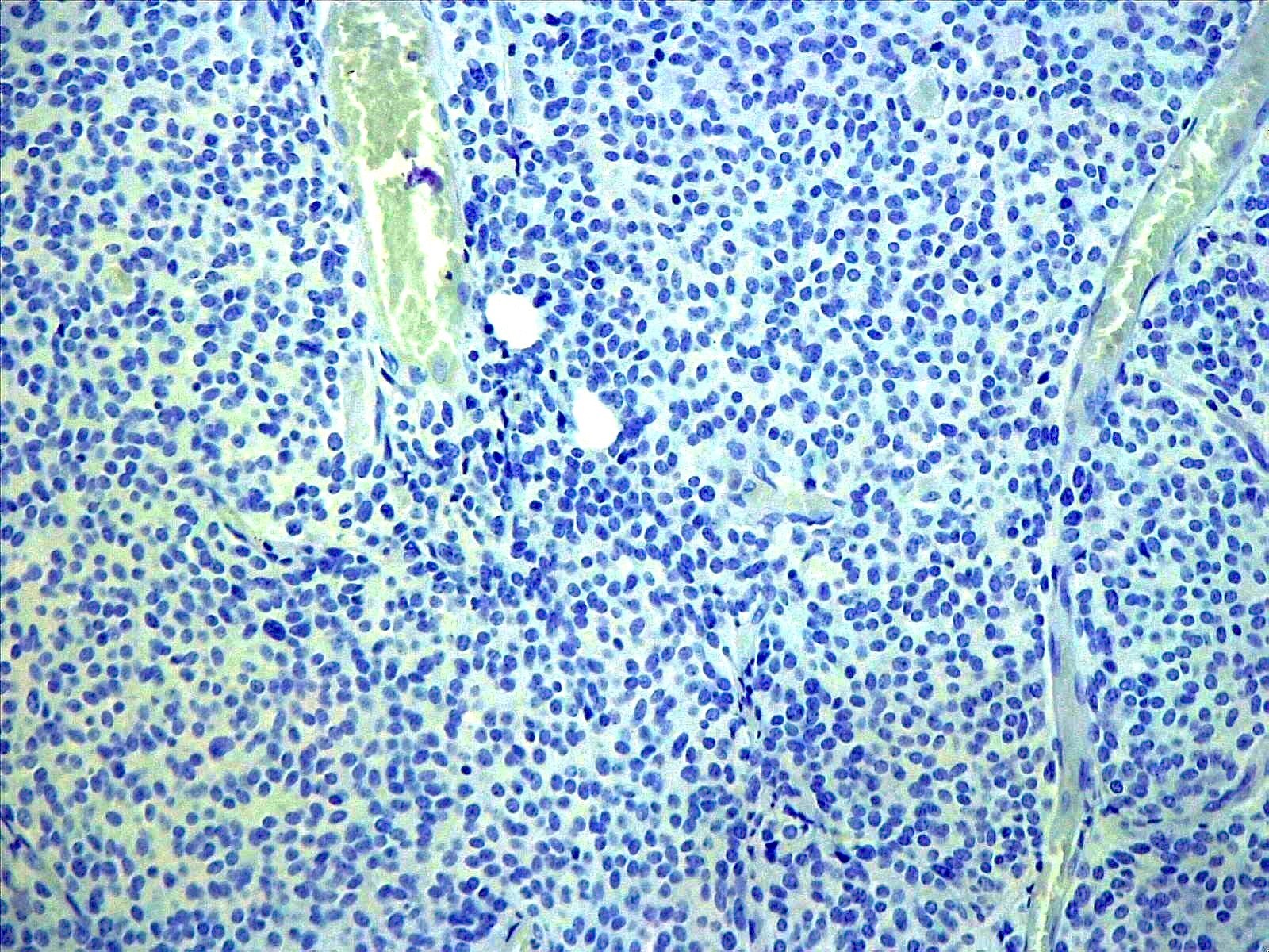
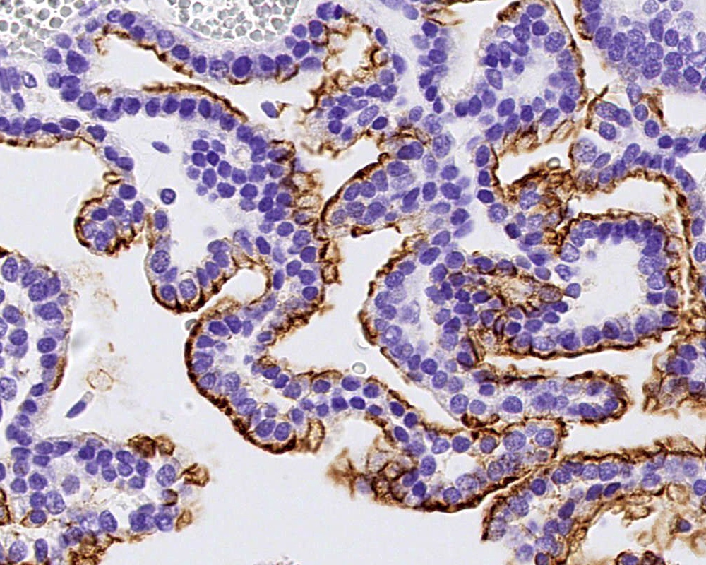
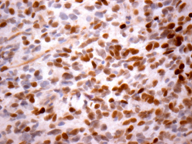
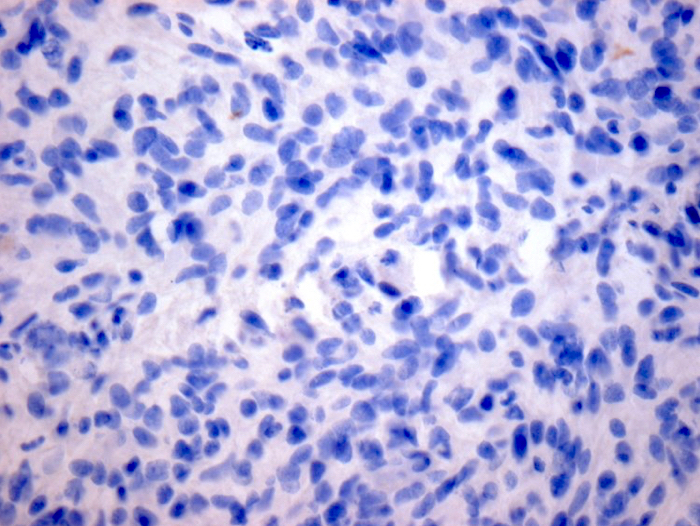
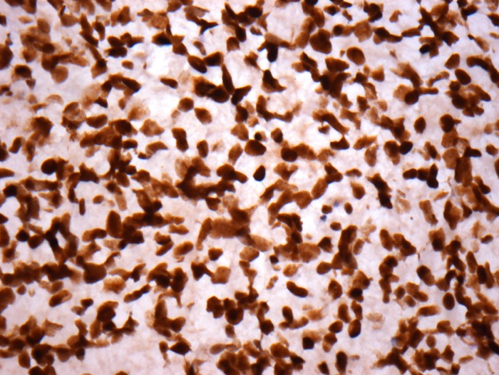
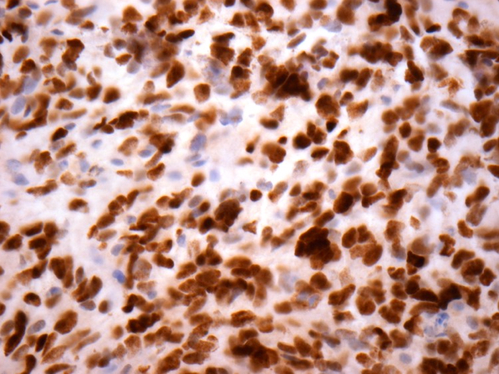
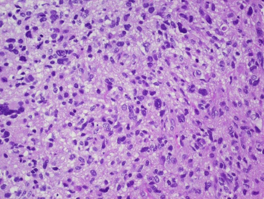
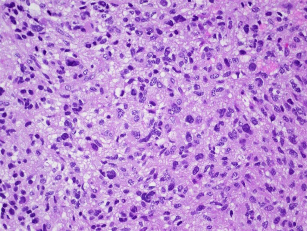
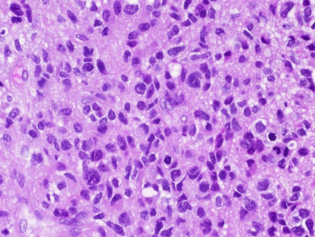
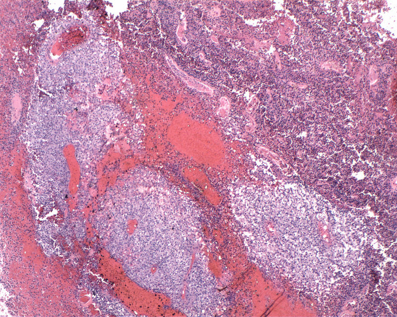
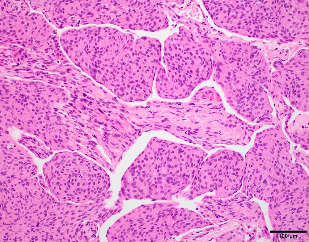
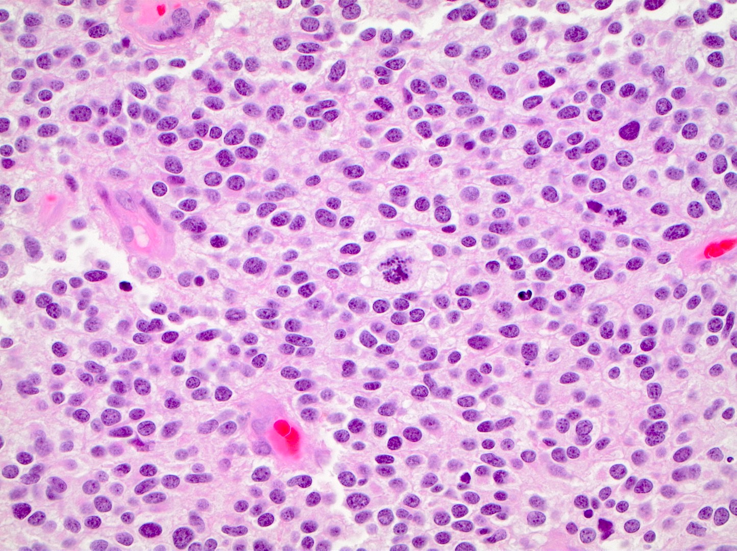
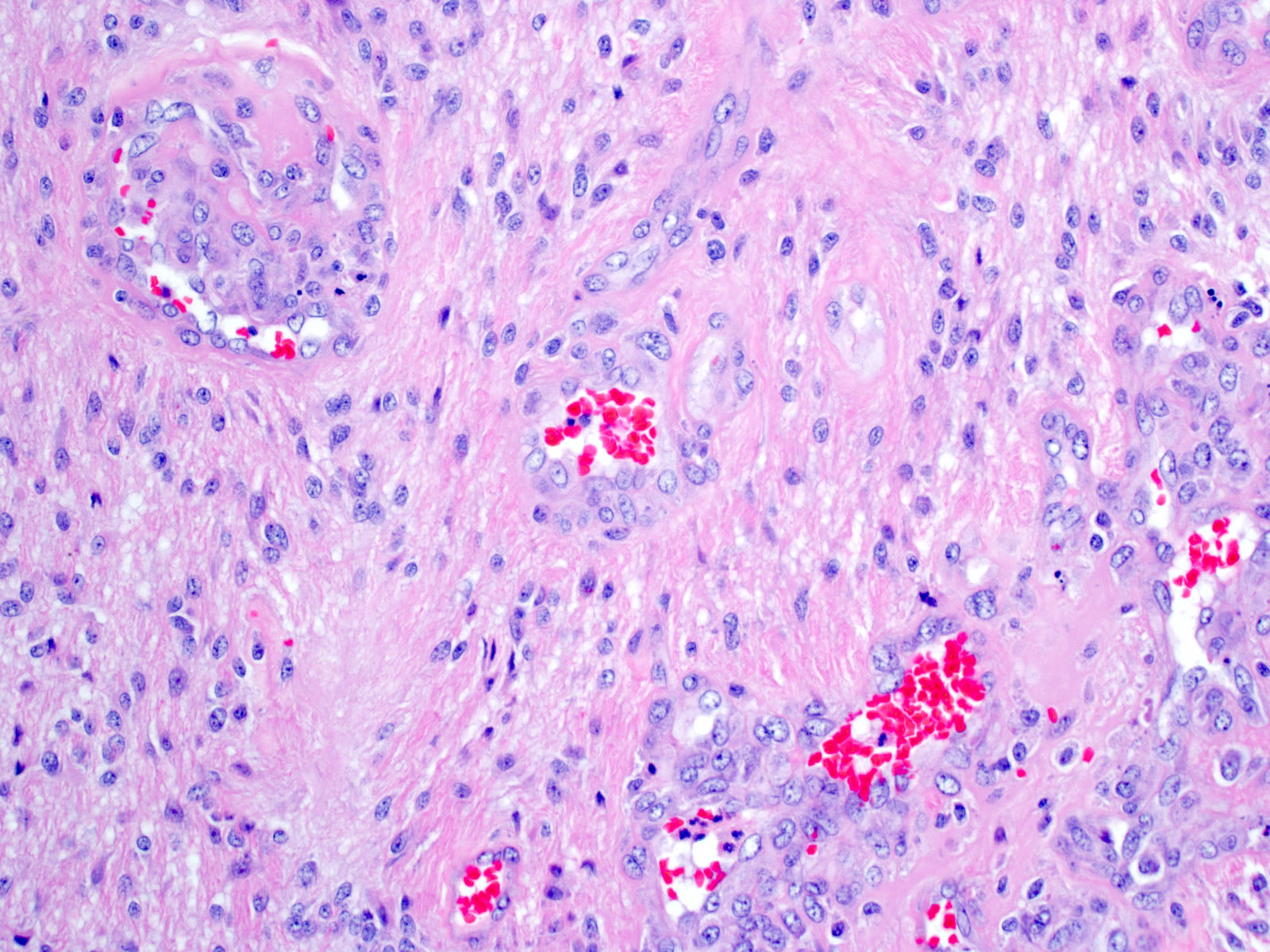
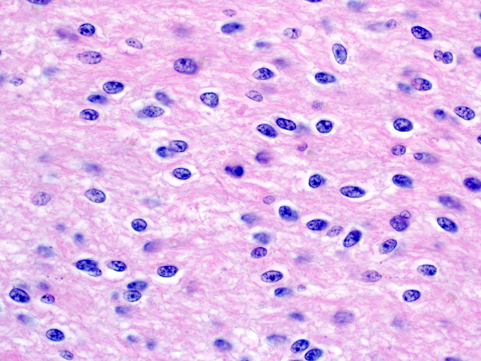
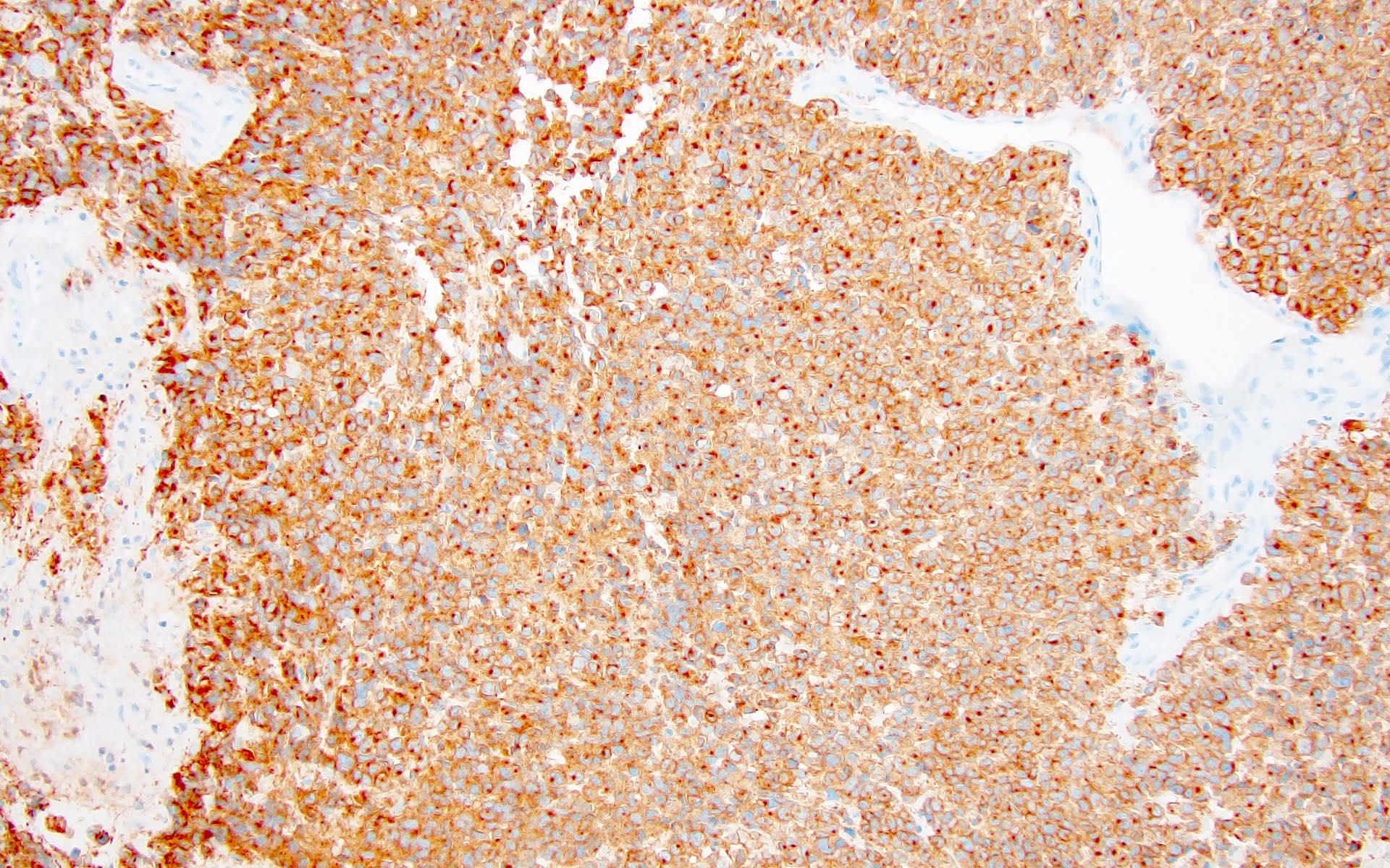
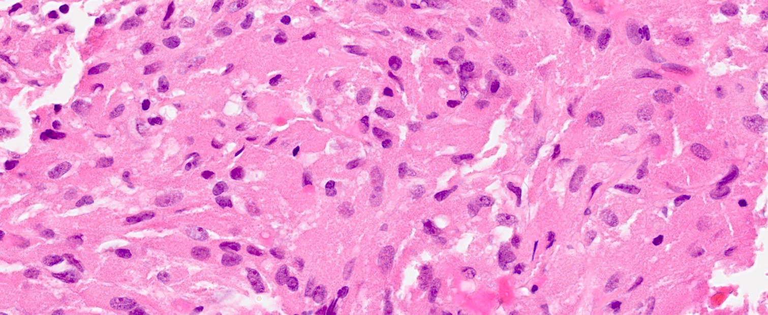
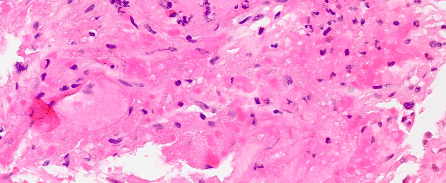
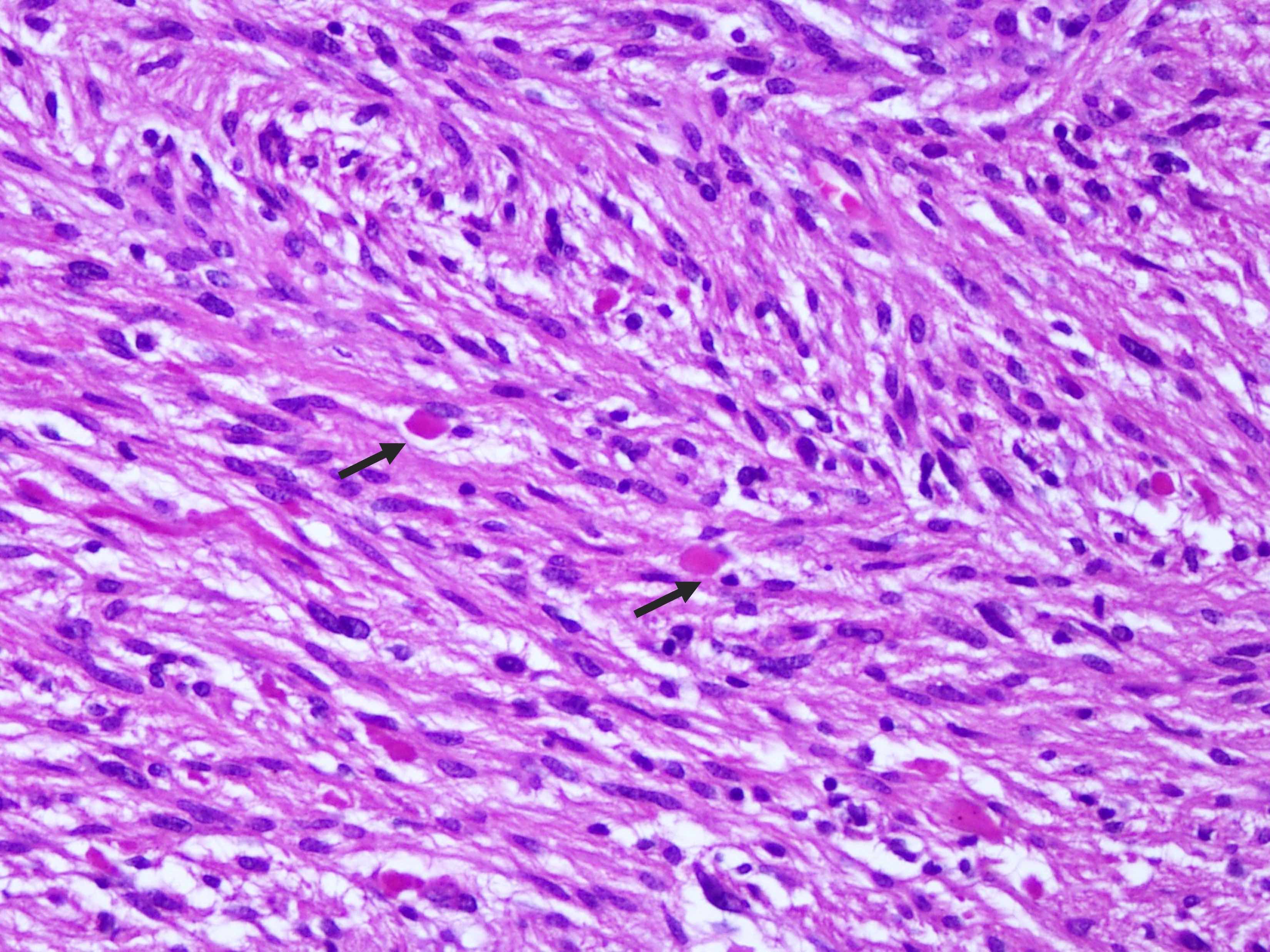
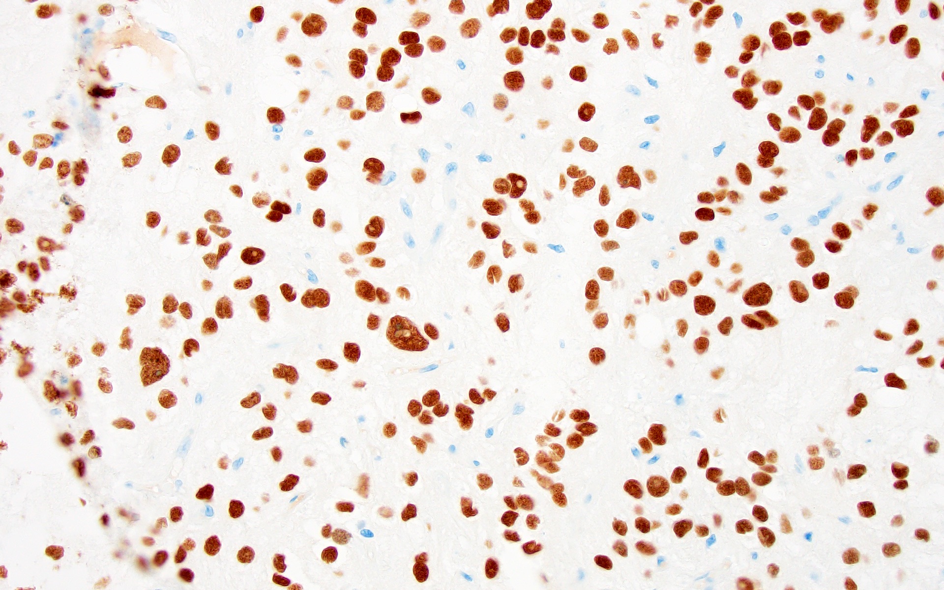
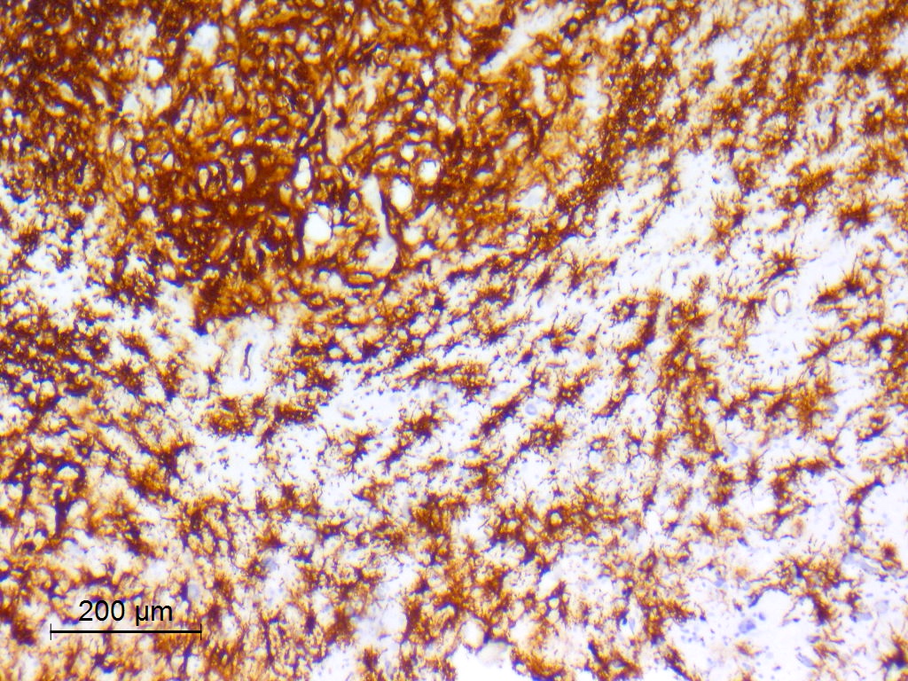
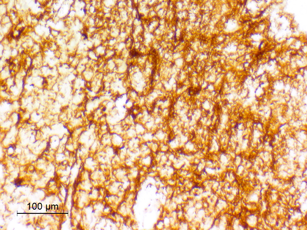
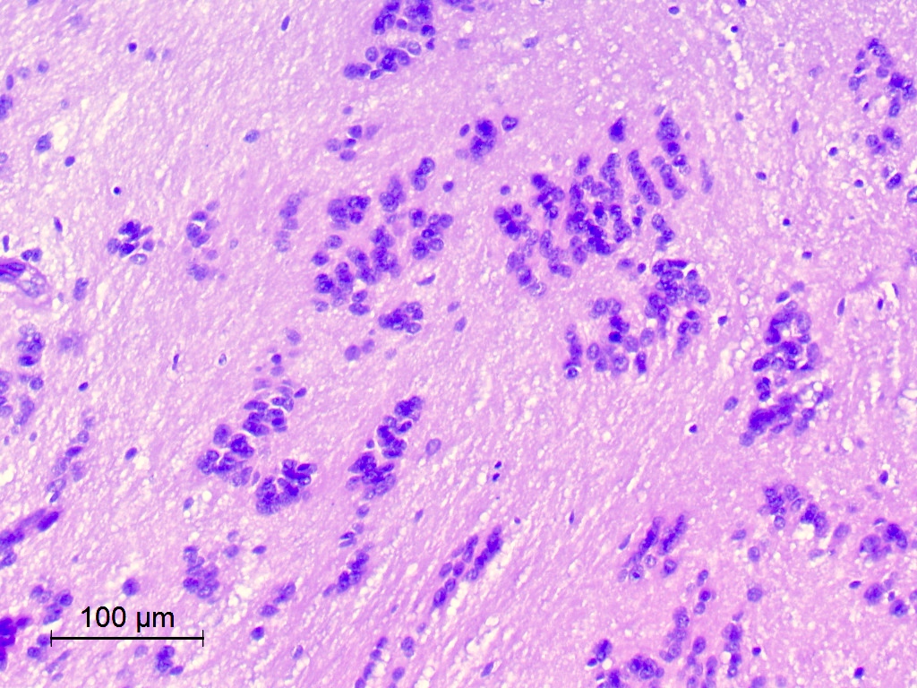
Adamantinomatous
epithelium
Stellate reticulum
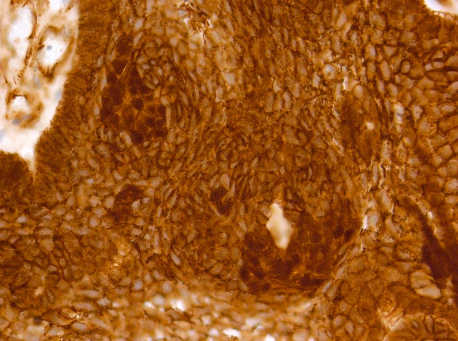
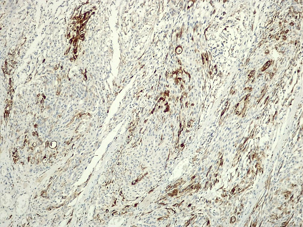
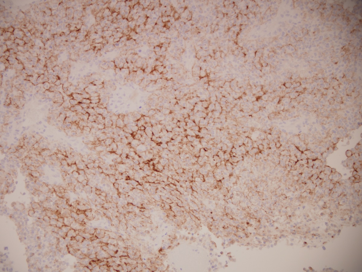
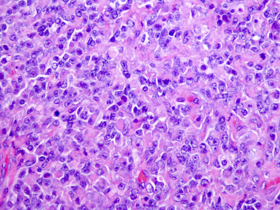
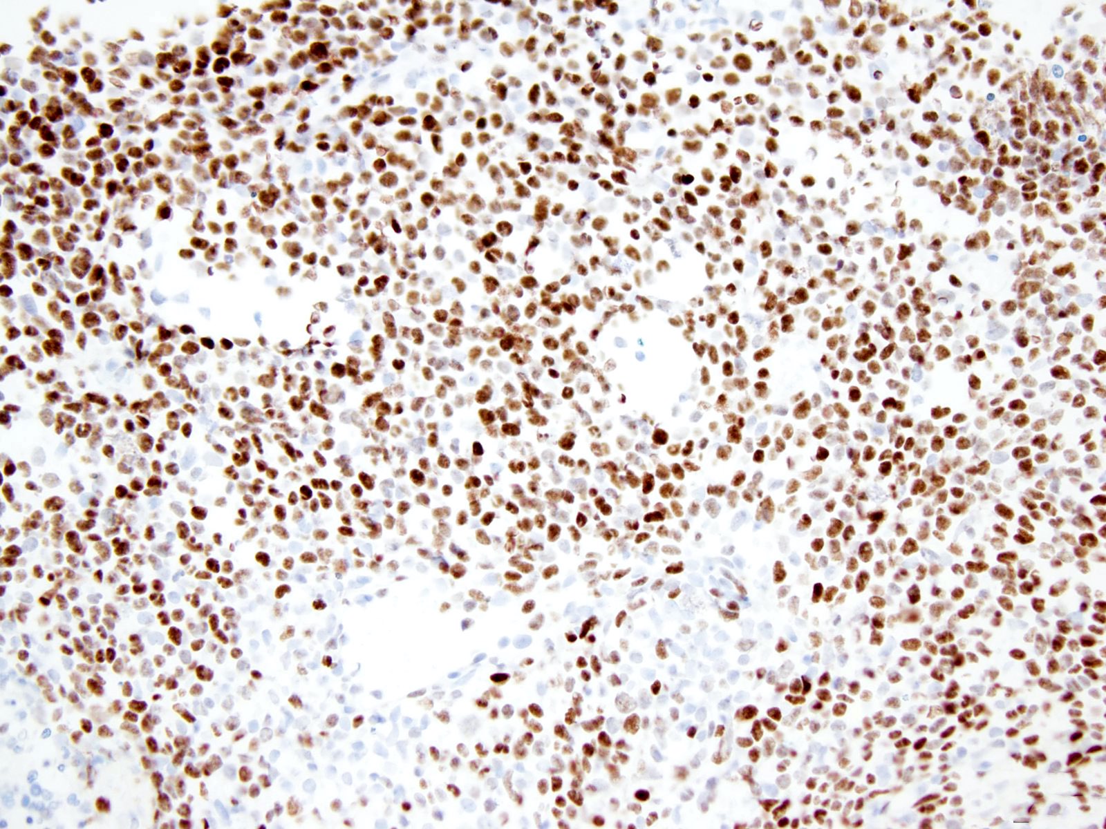
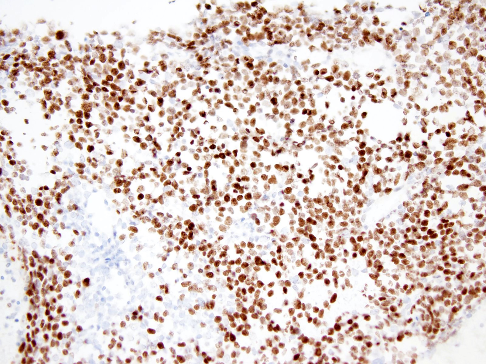
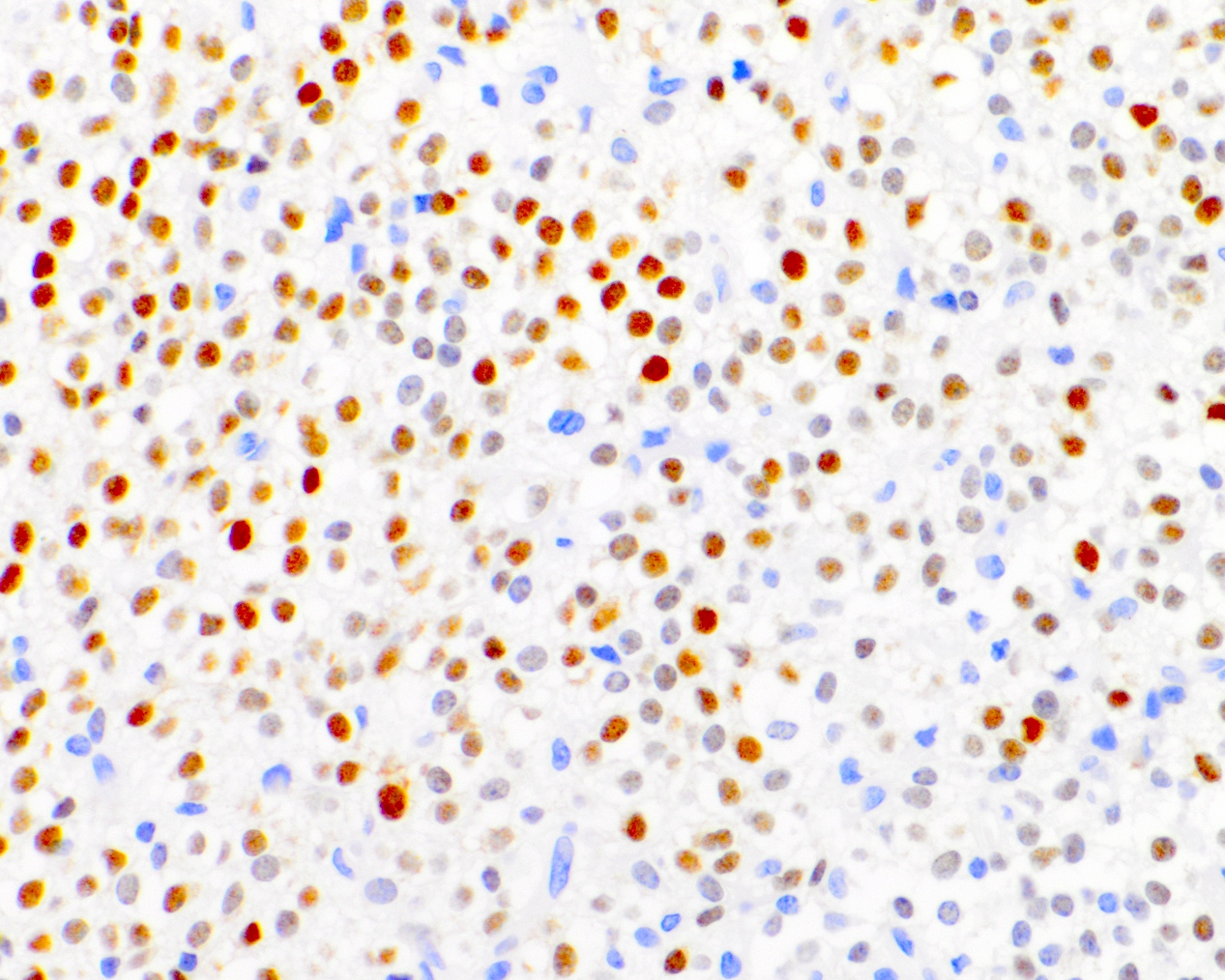
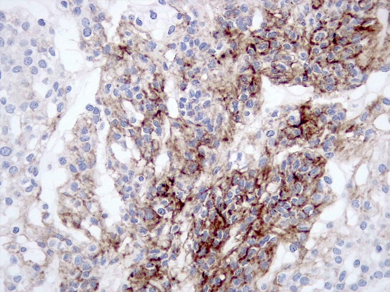
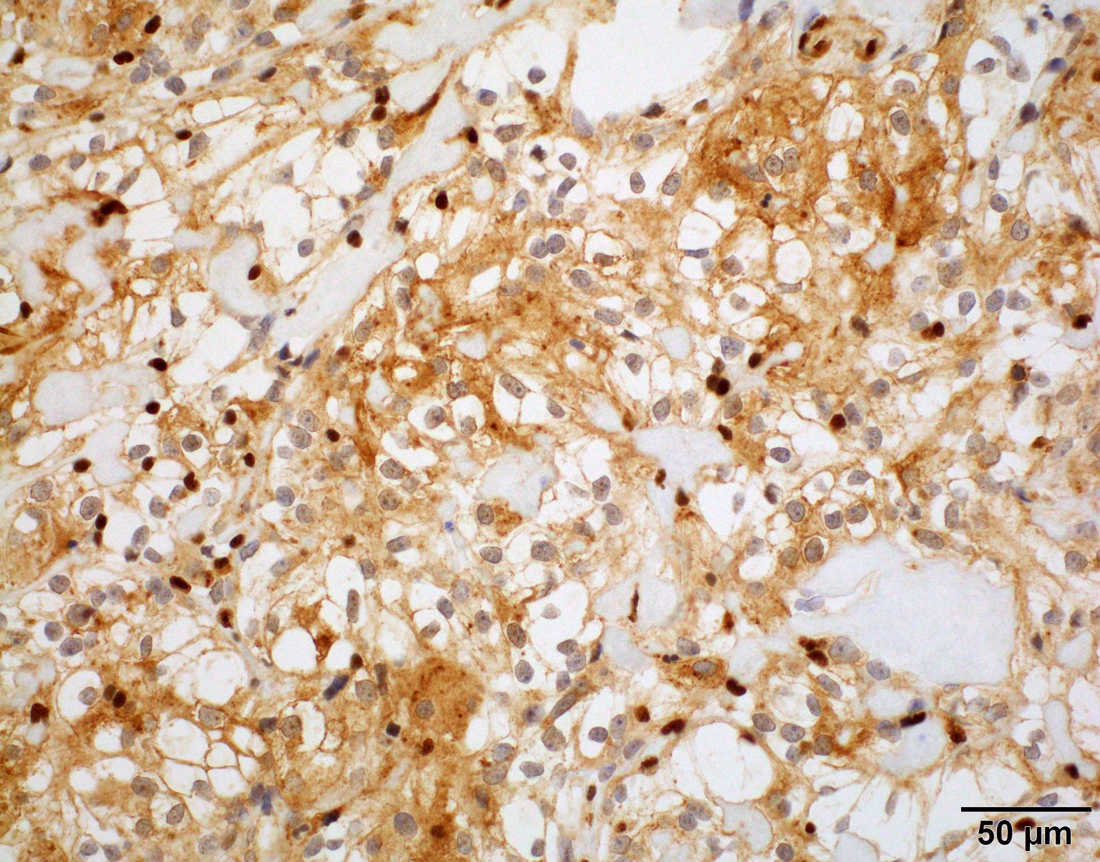
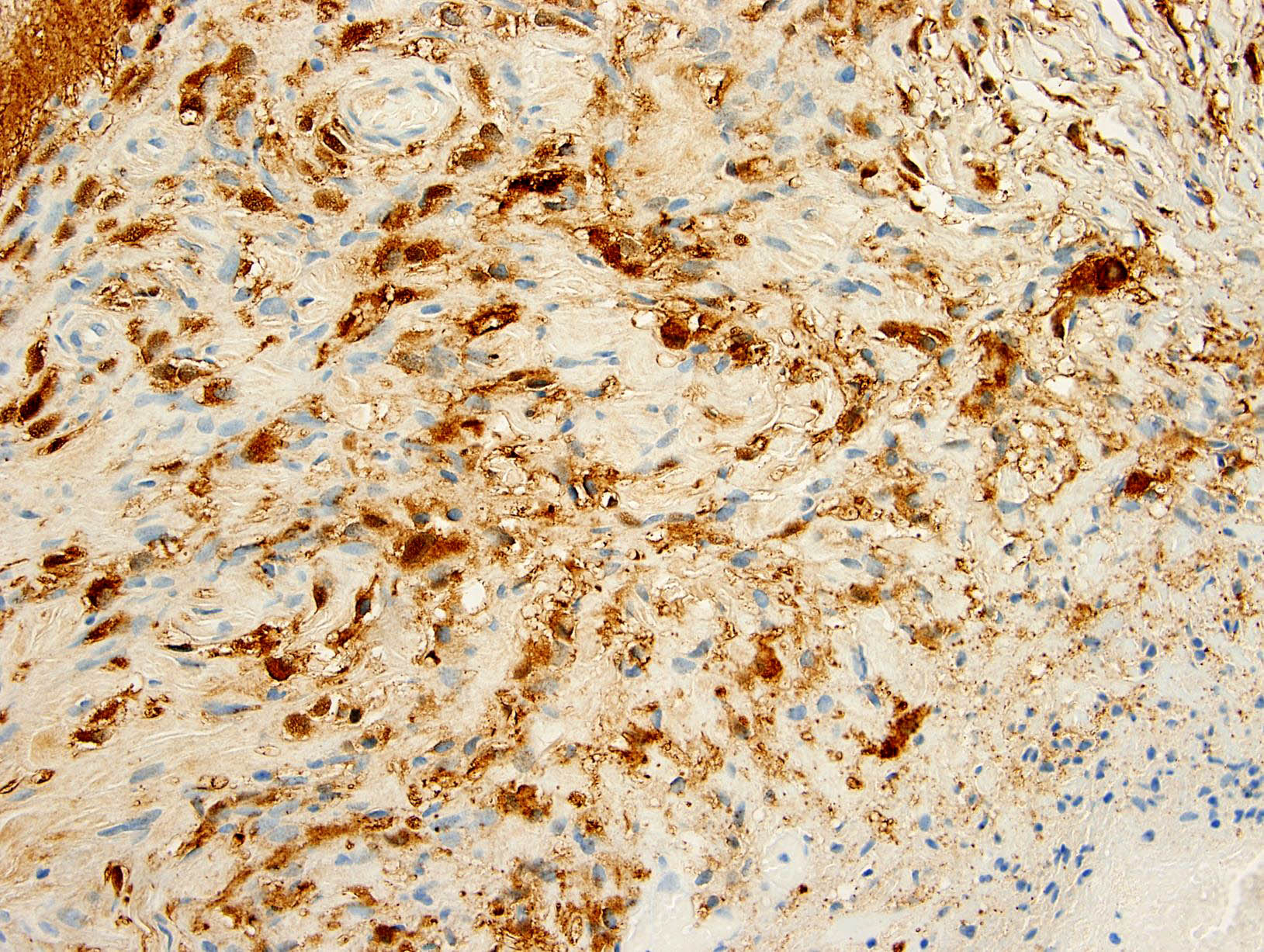
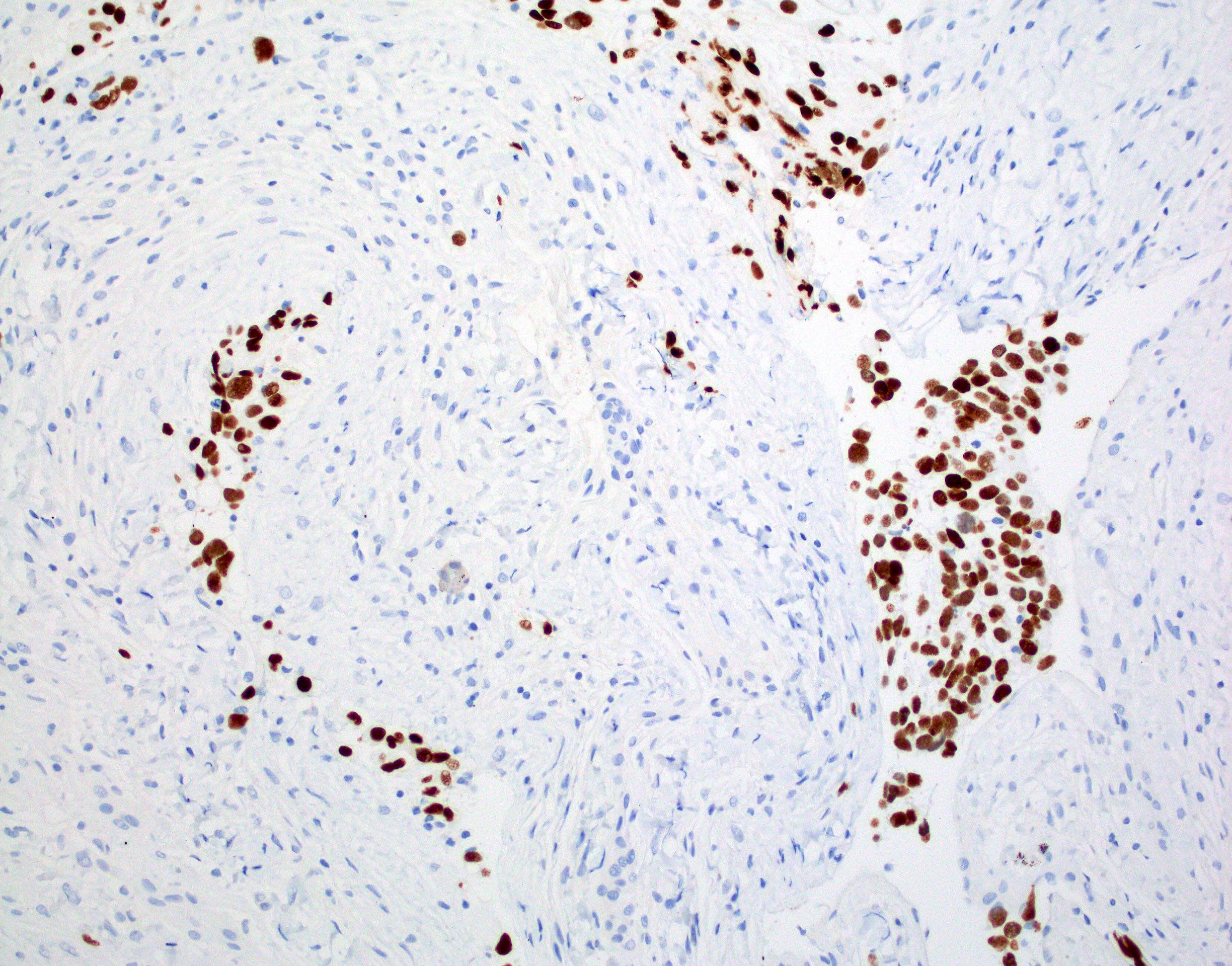
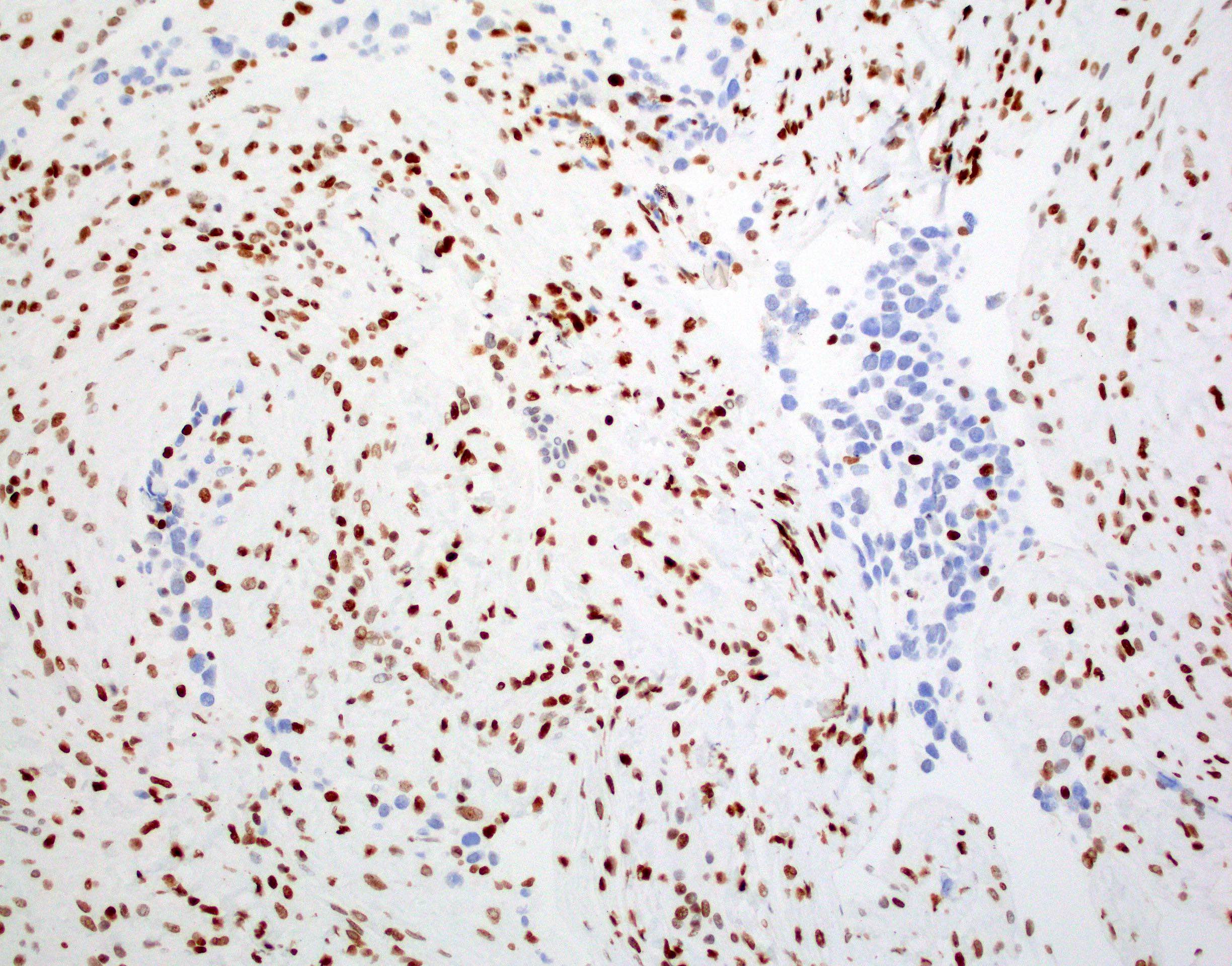
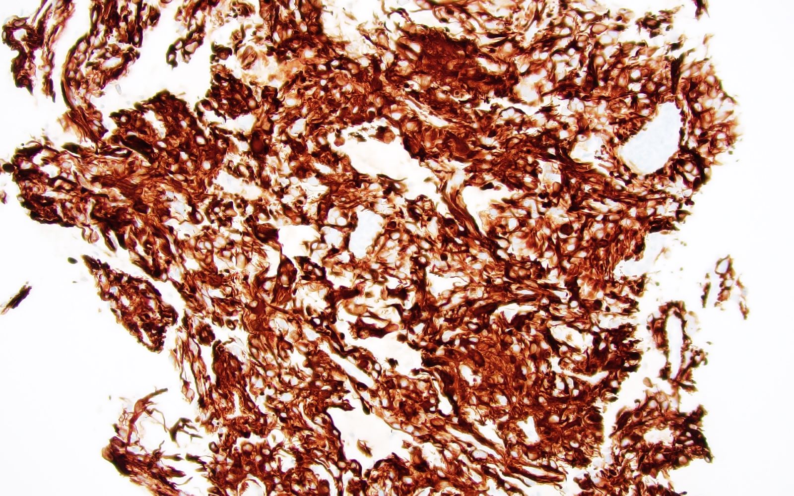
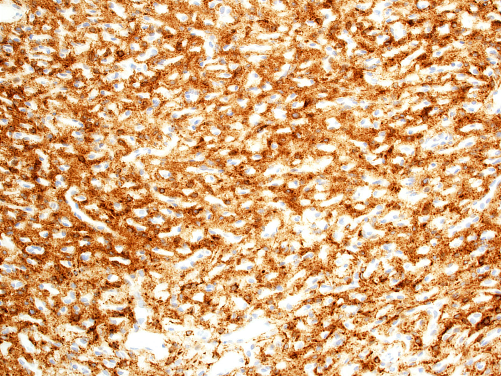
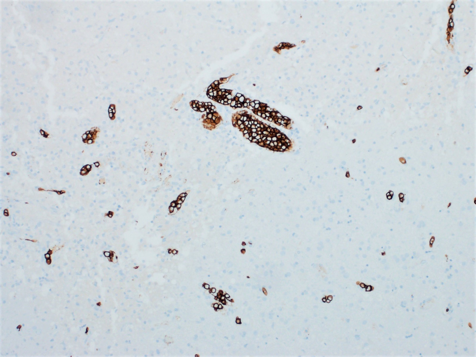
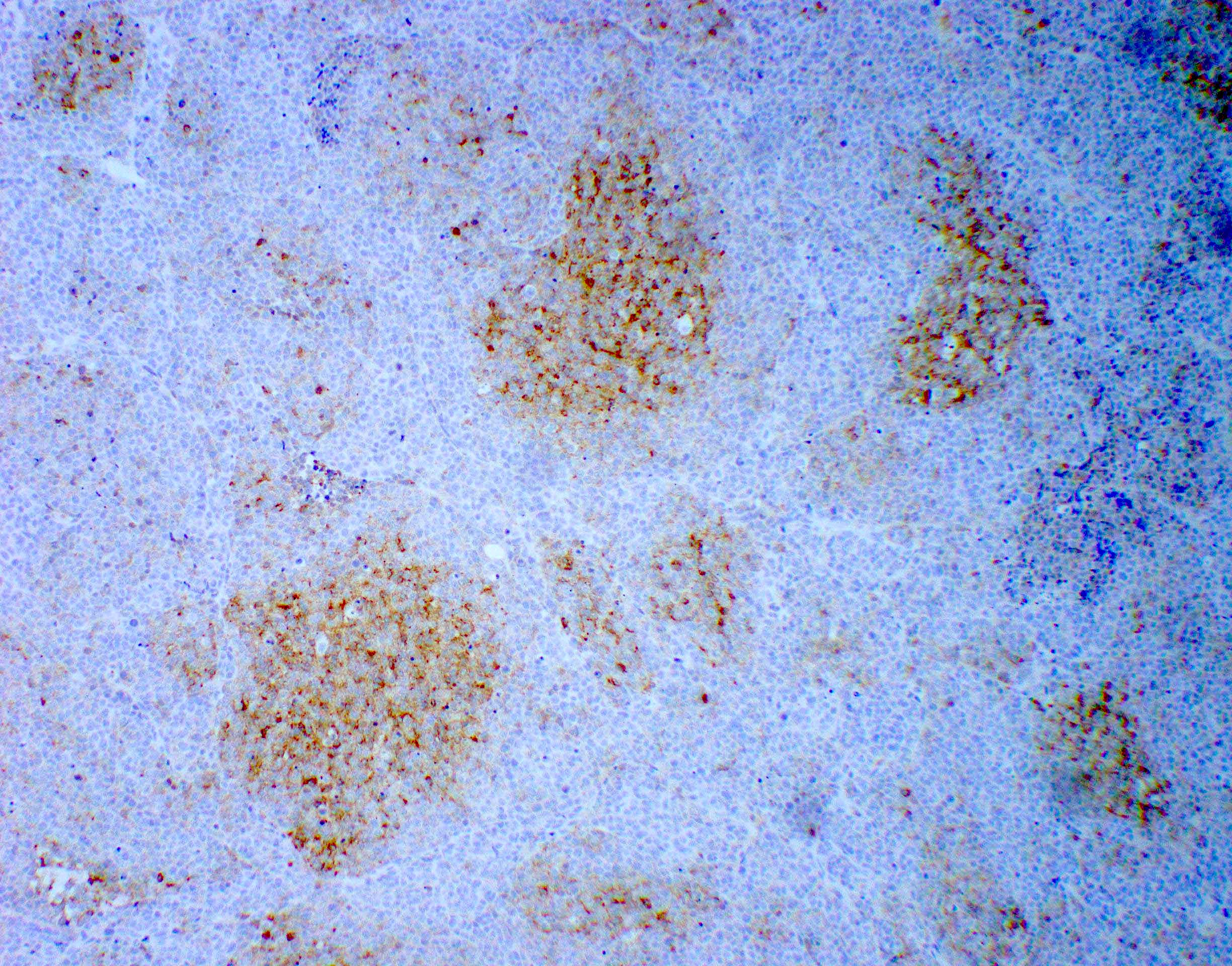
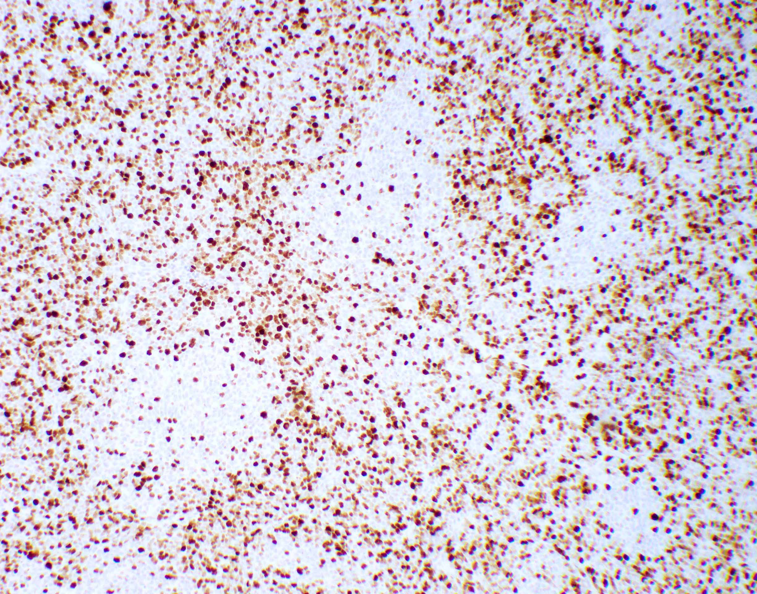
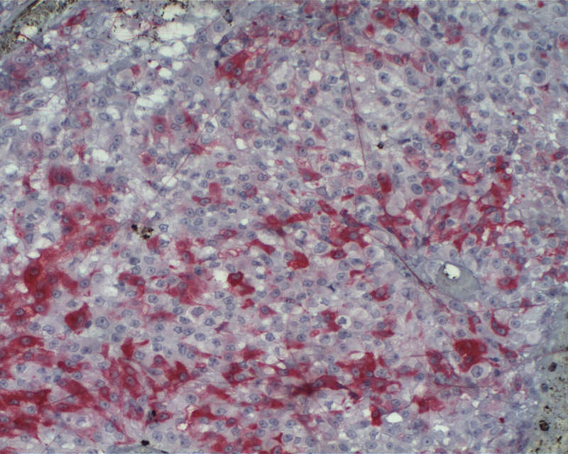
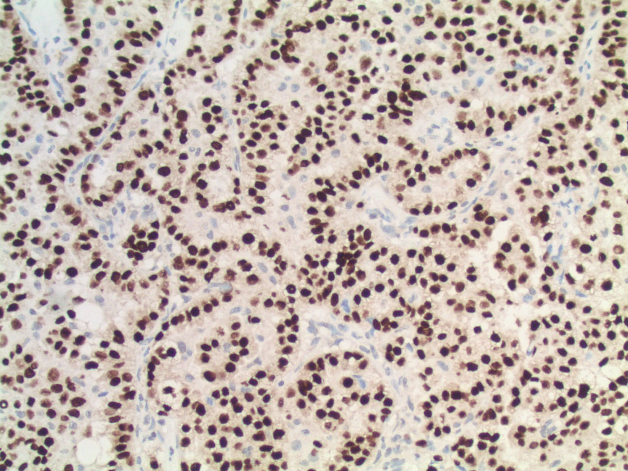
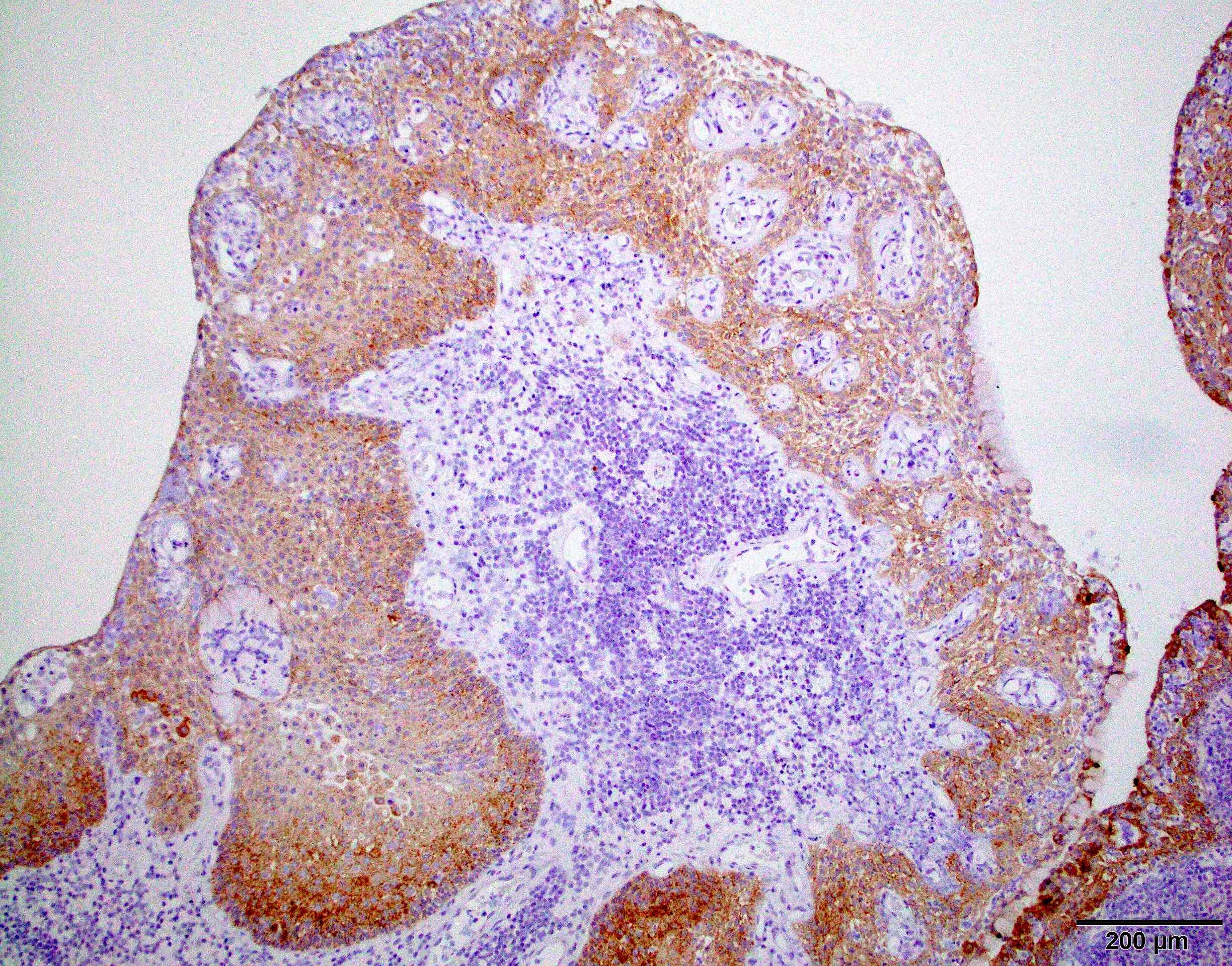
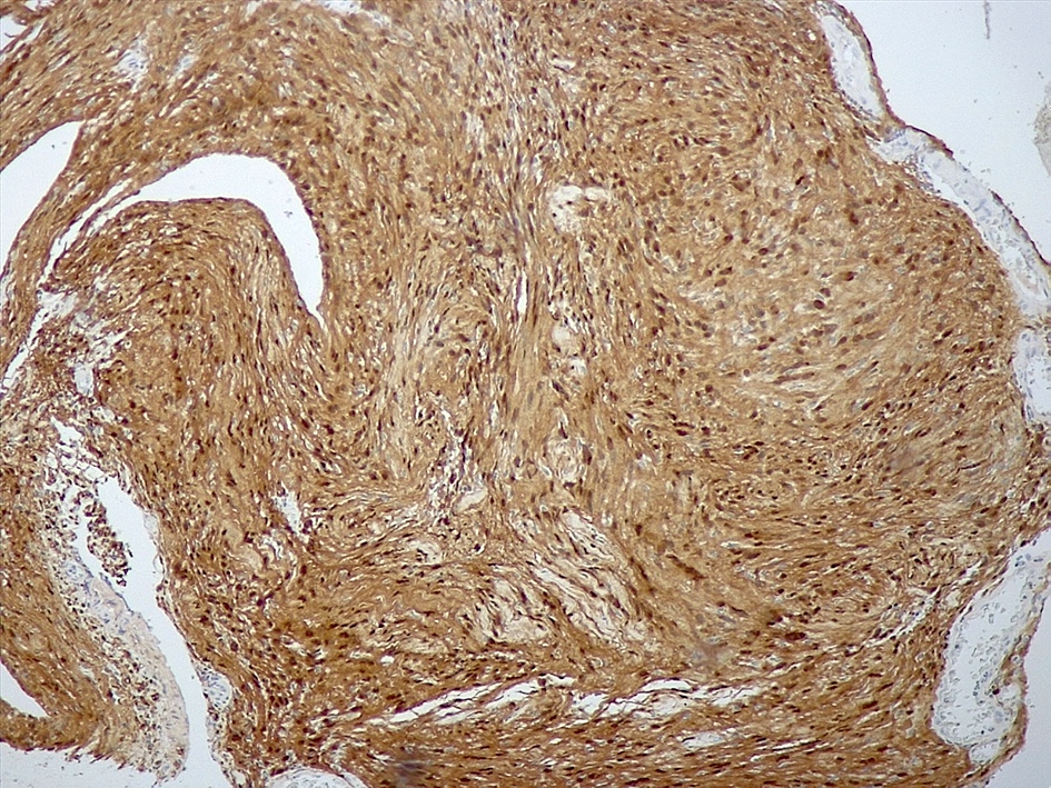
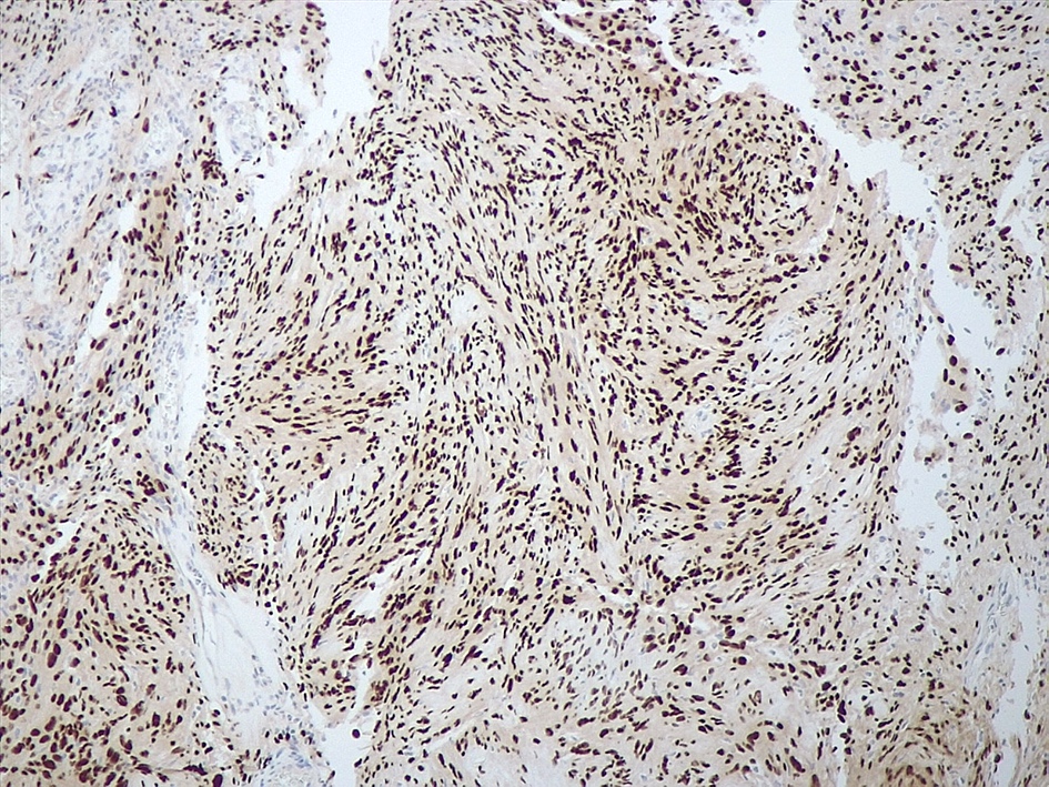
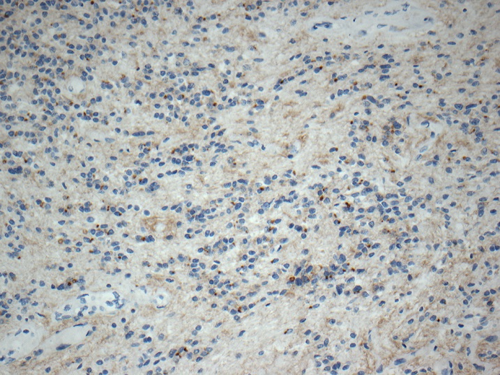
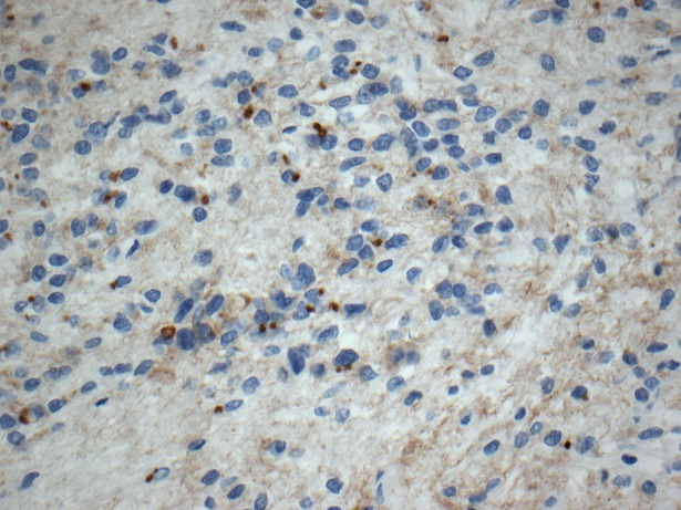
Beta catenin
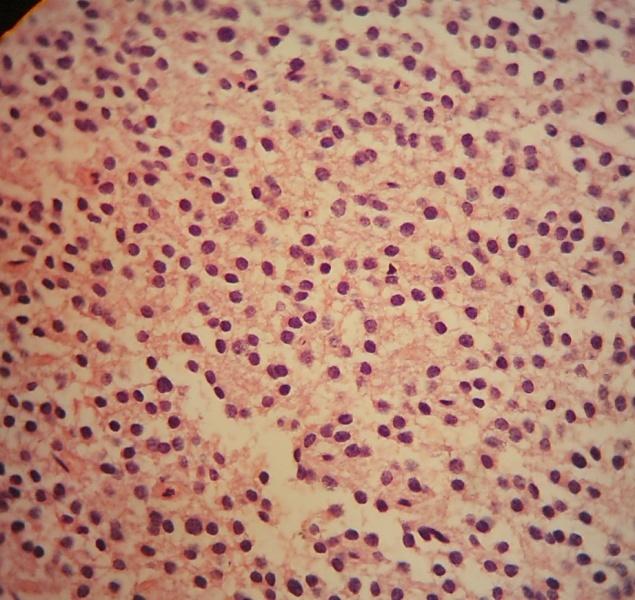
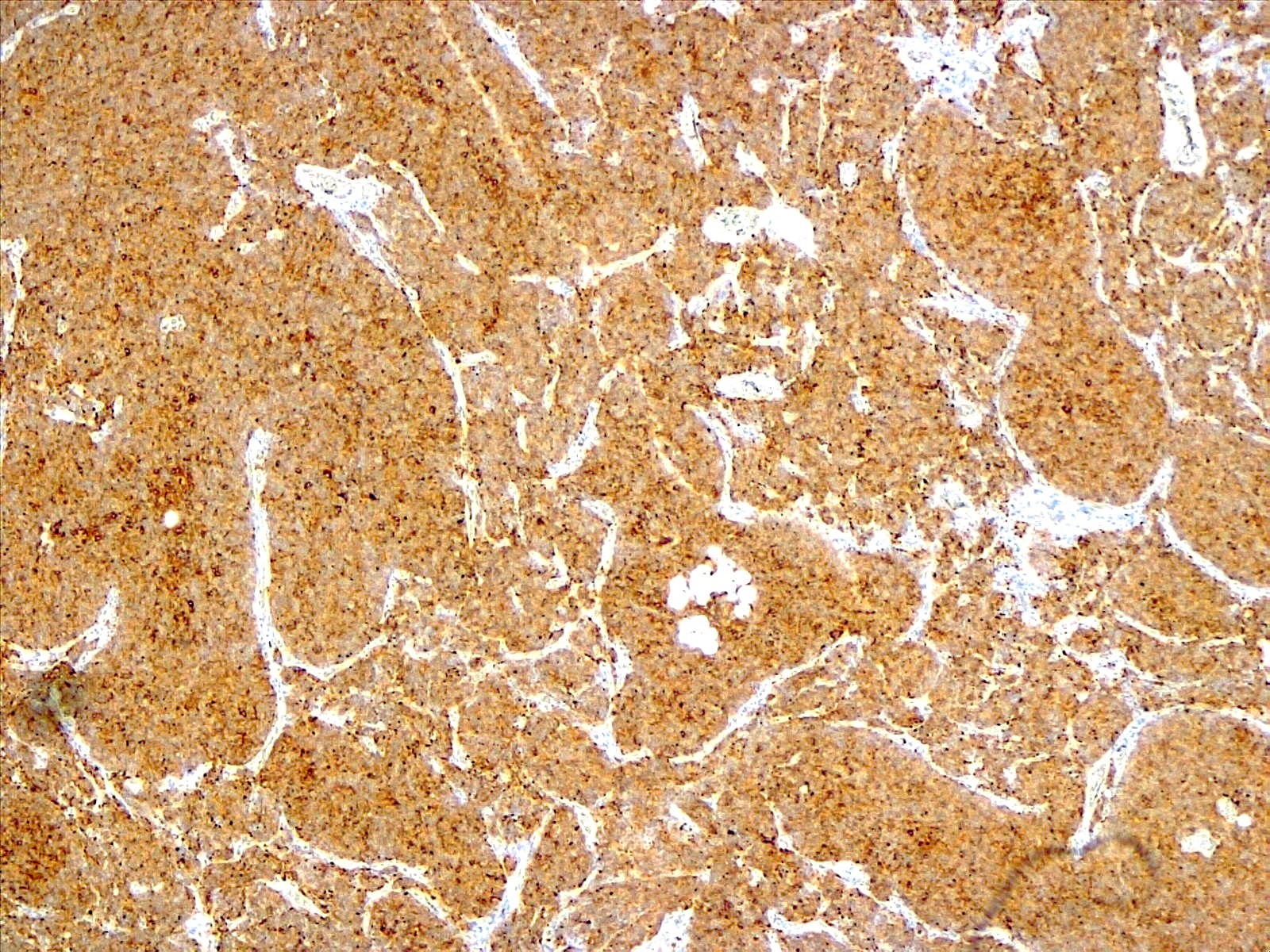
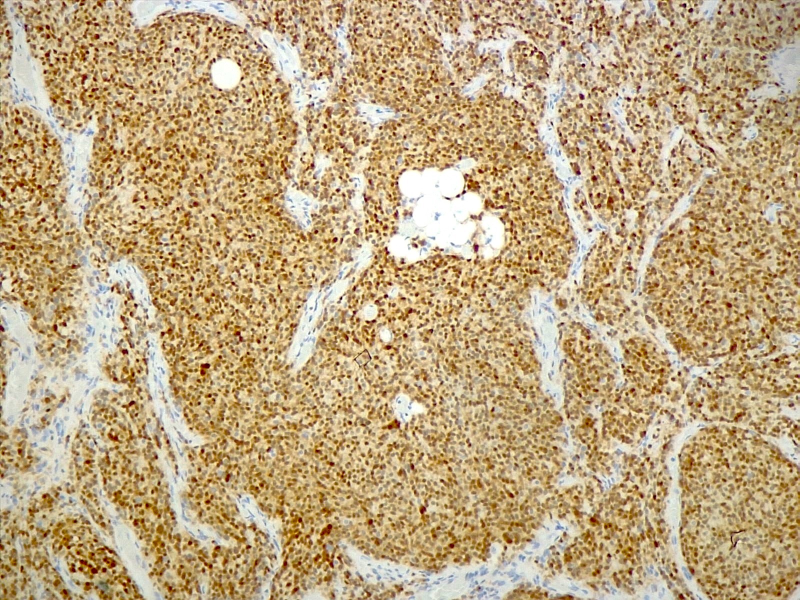
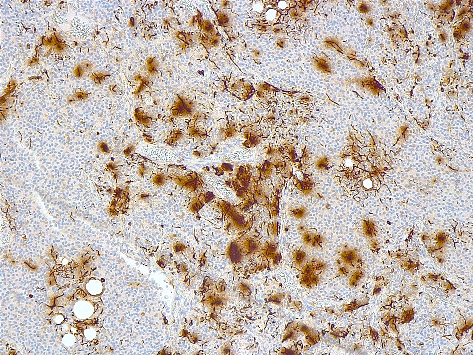
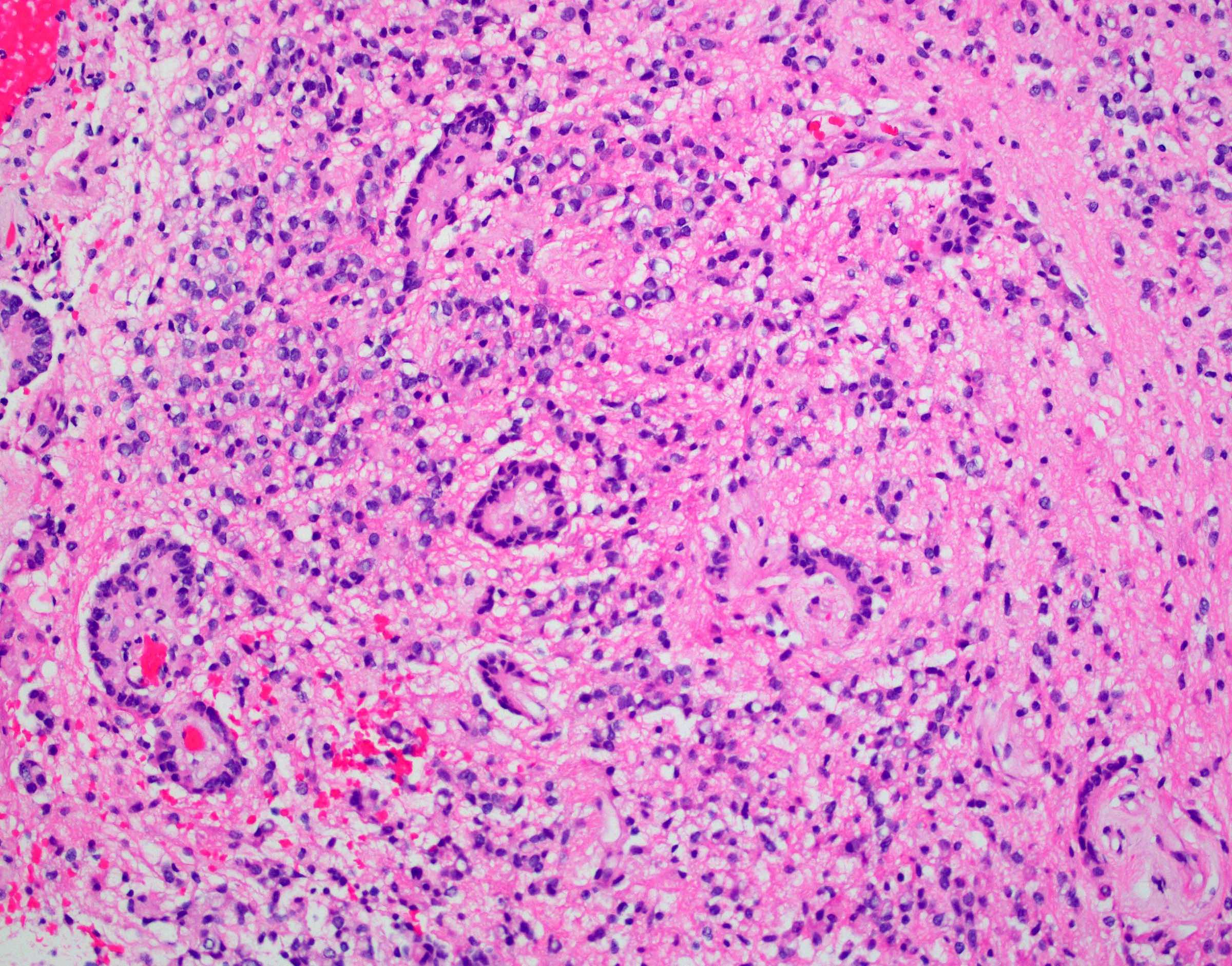
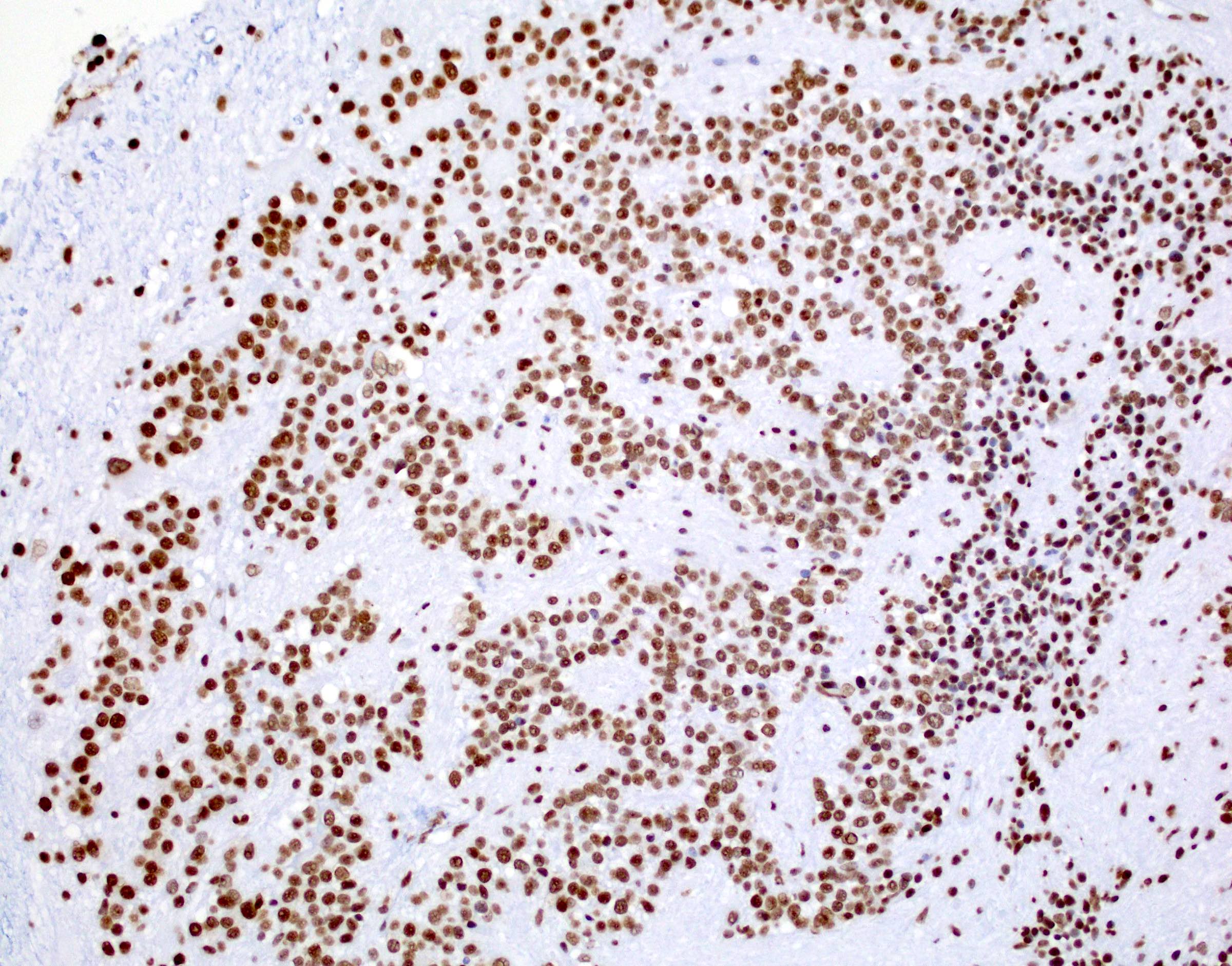
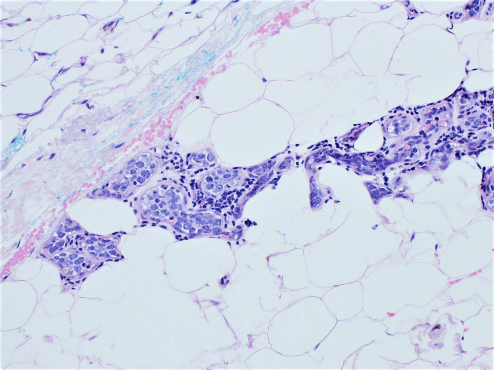
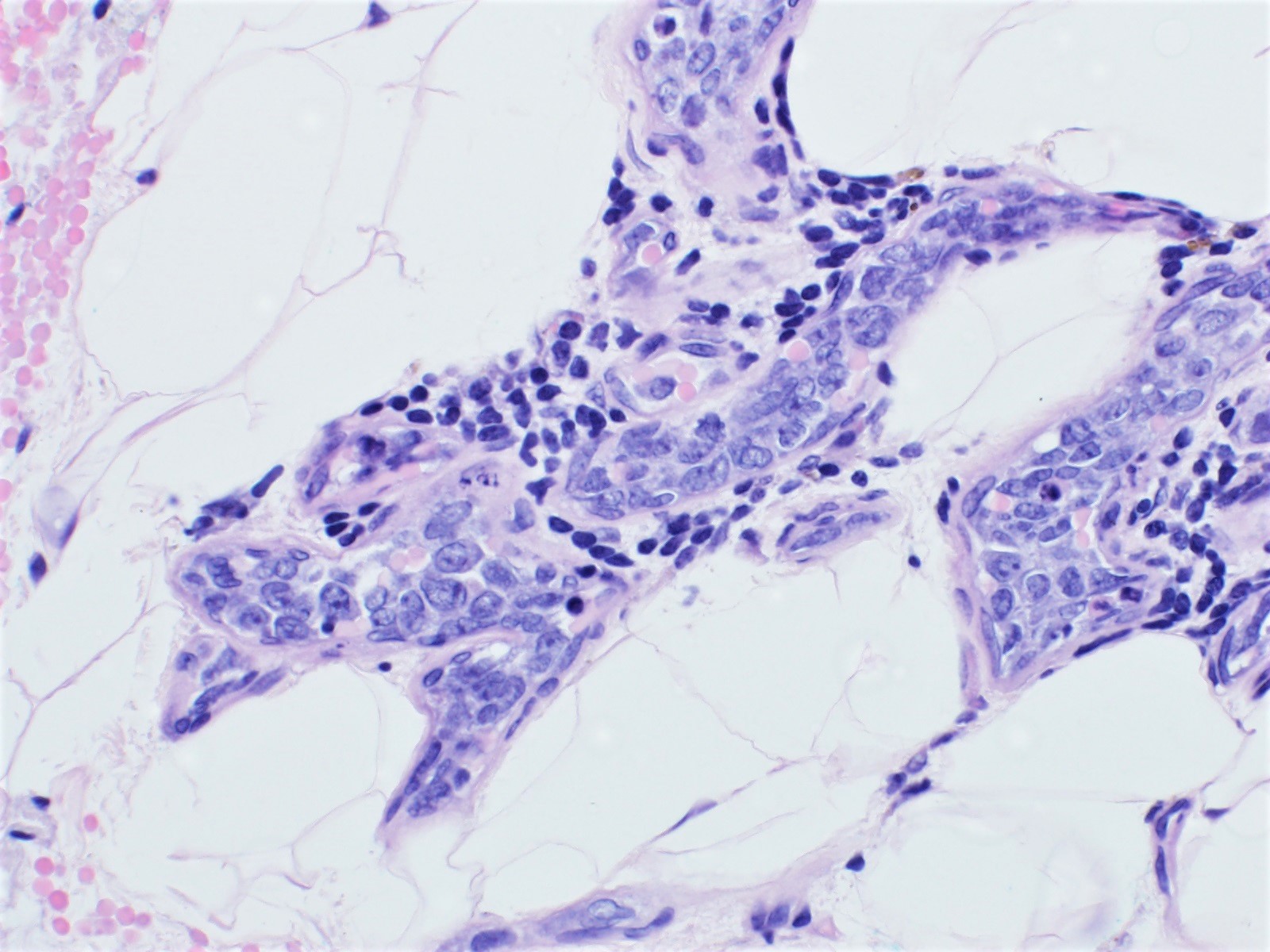
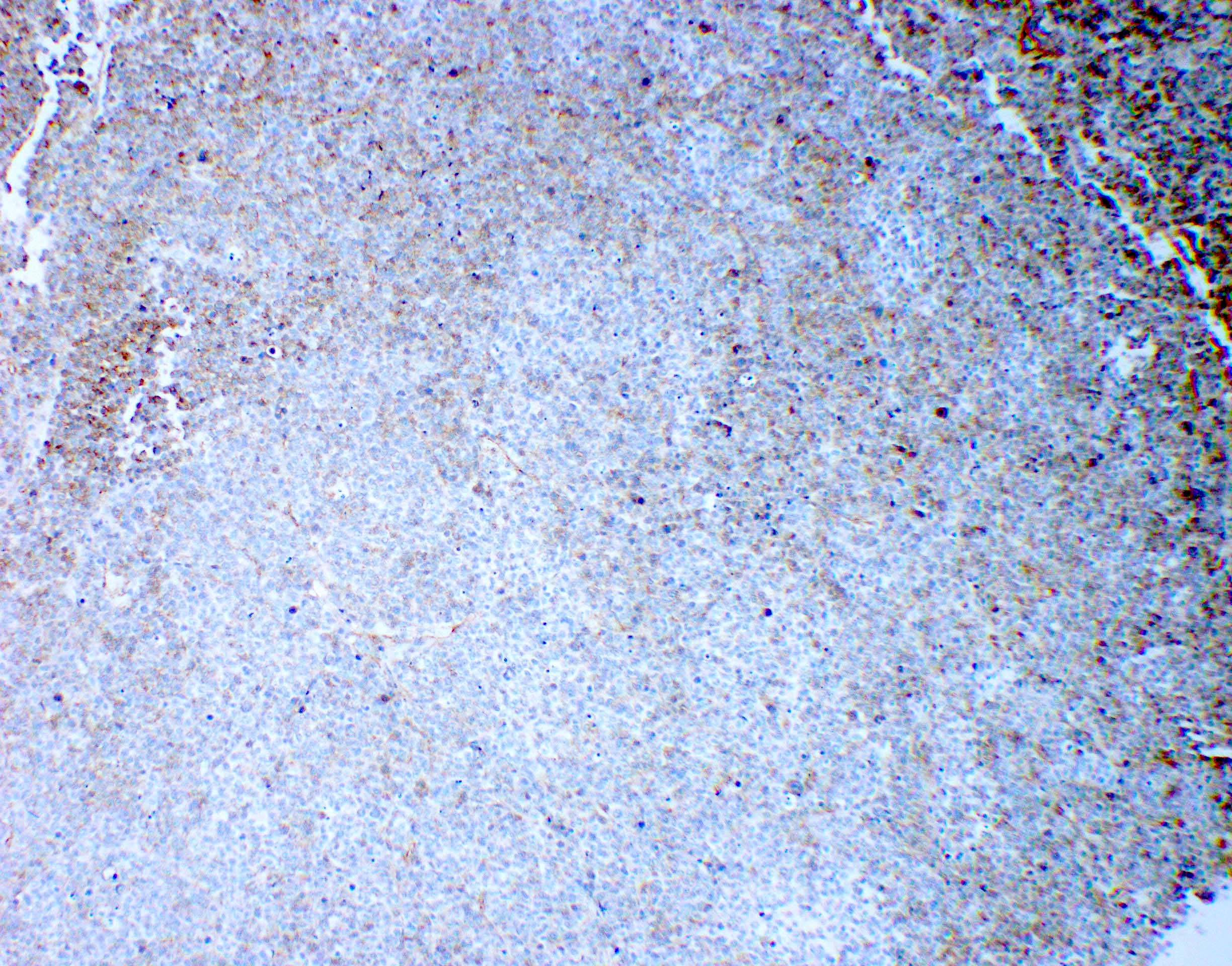
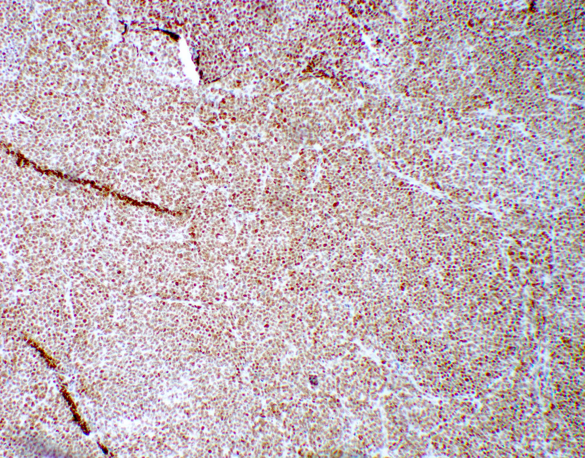
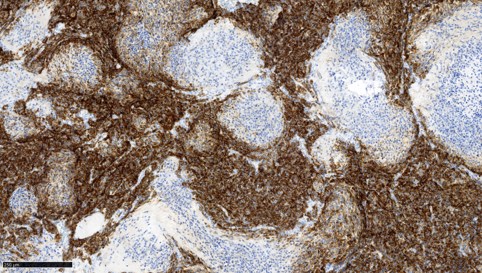
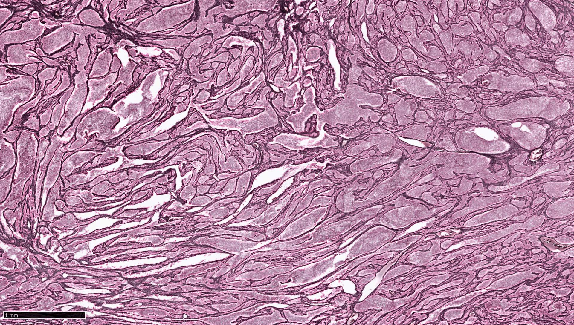
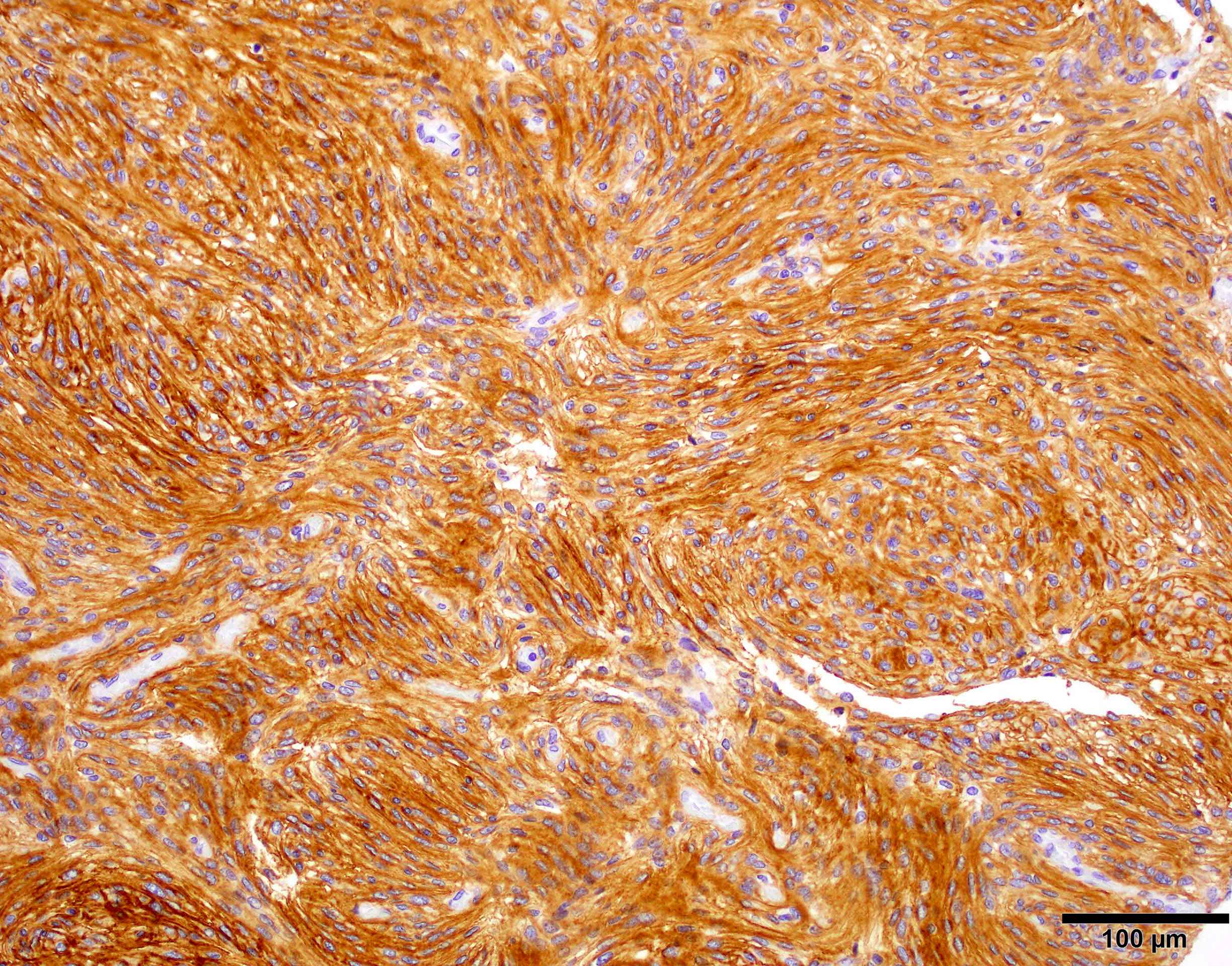
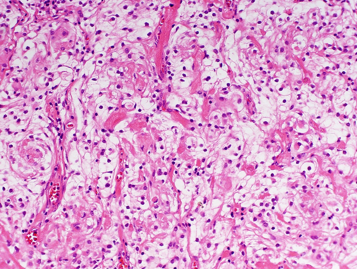
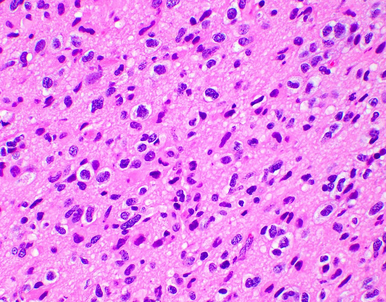
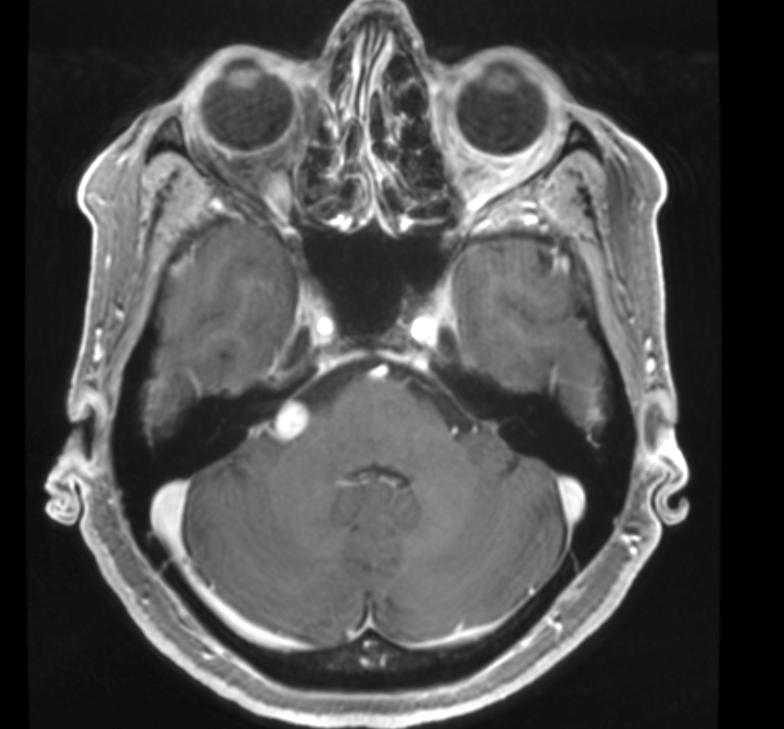
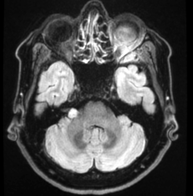
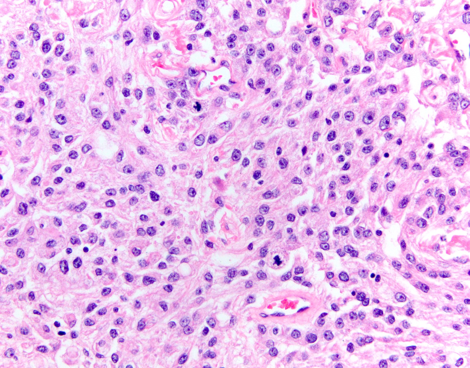
Contributed by Valeria Barresi, M.D, Ph.D.
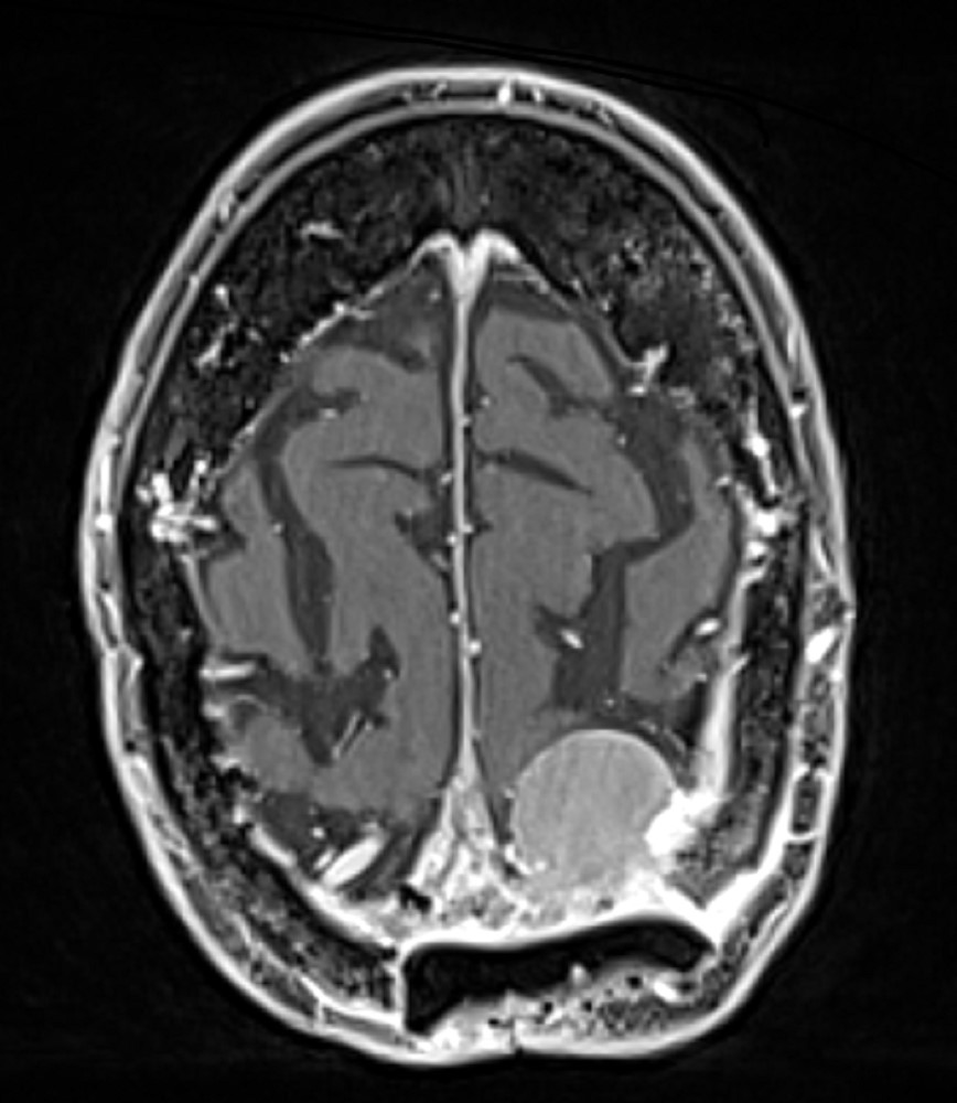
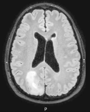
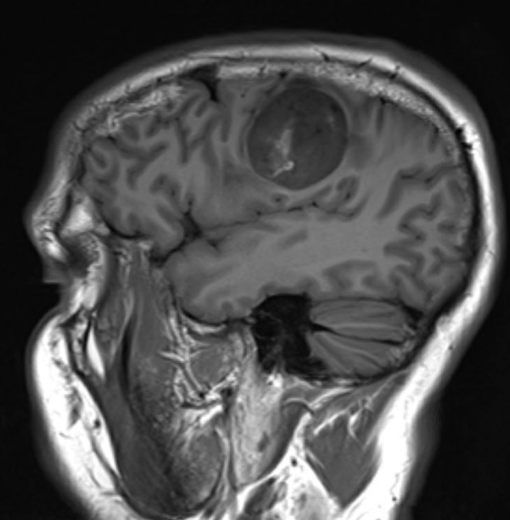
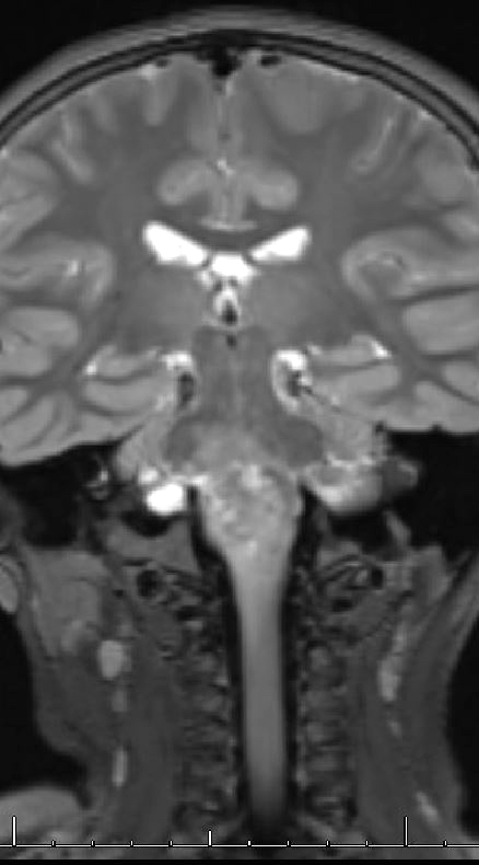
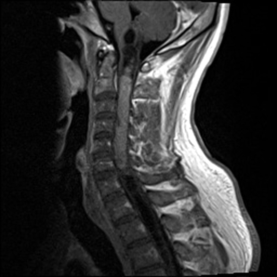
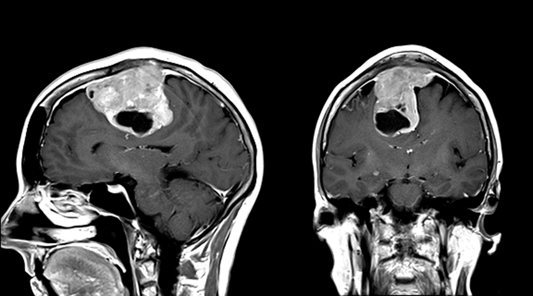
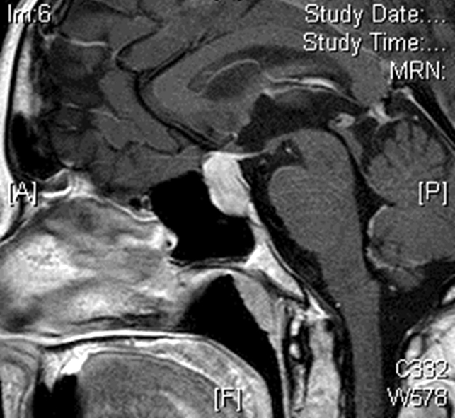
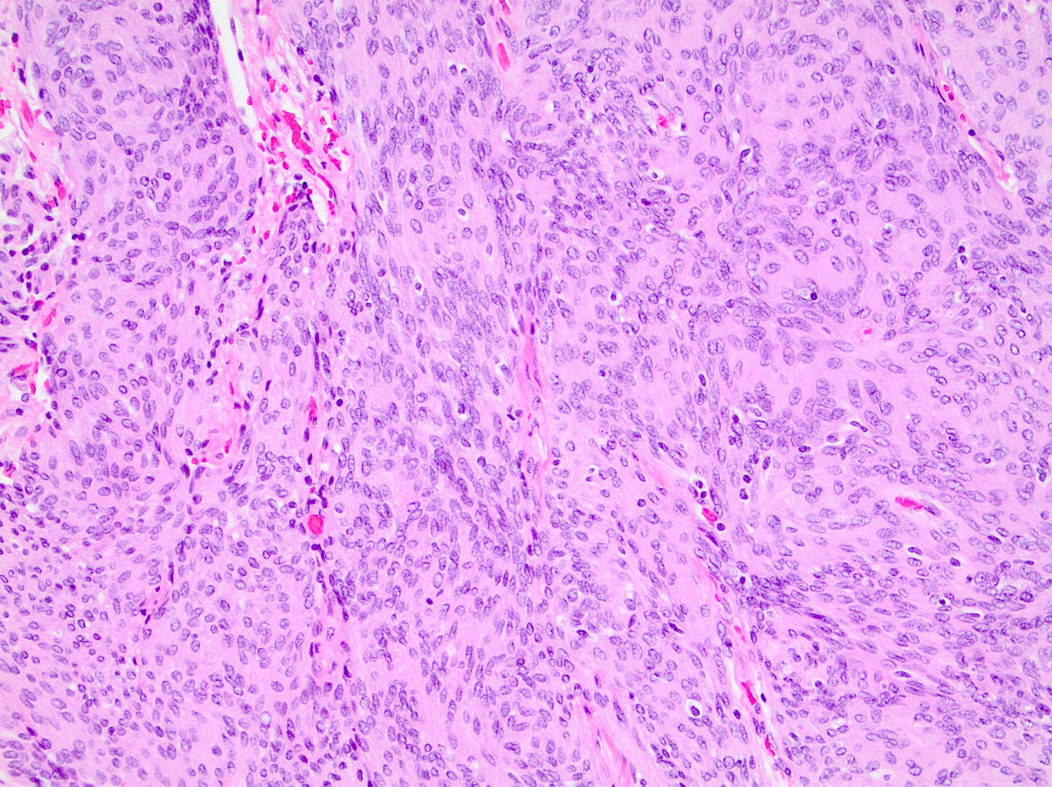
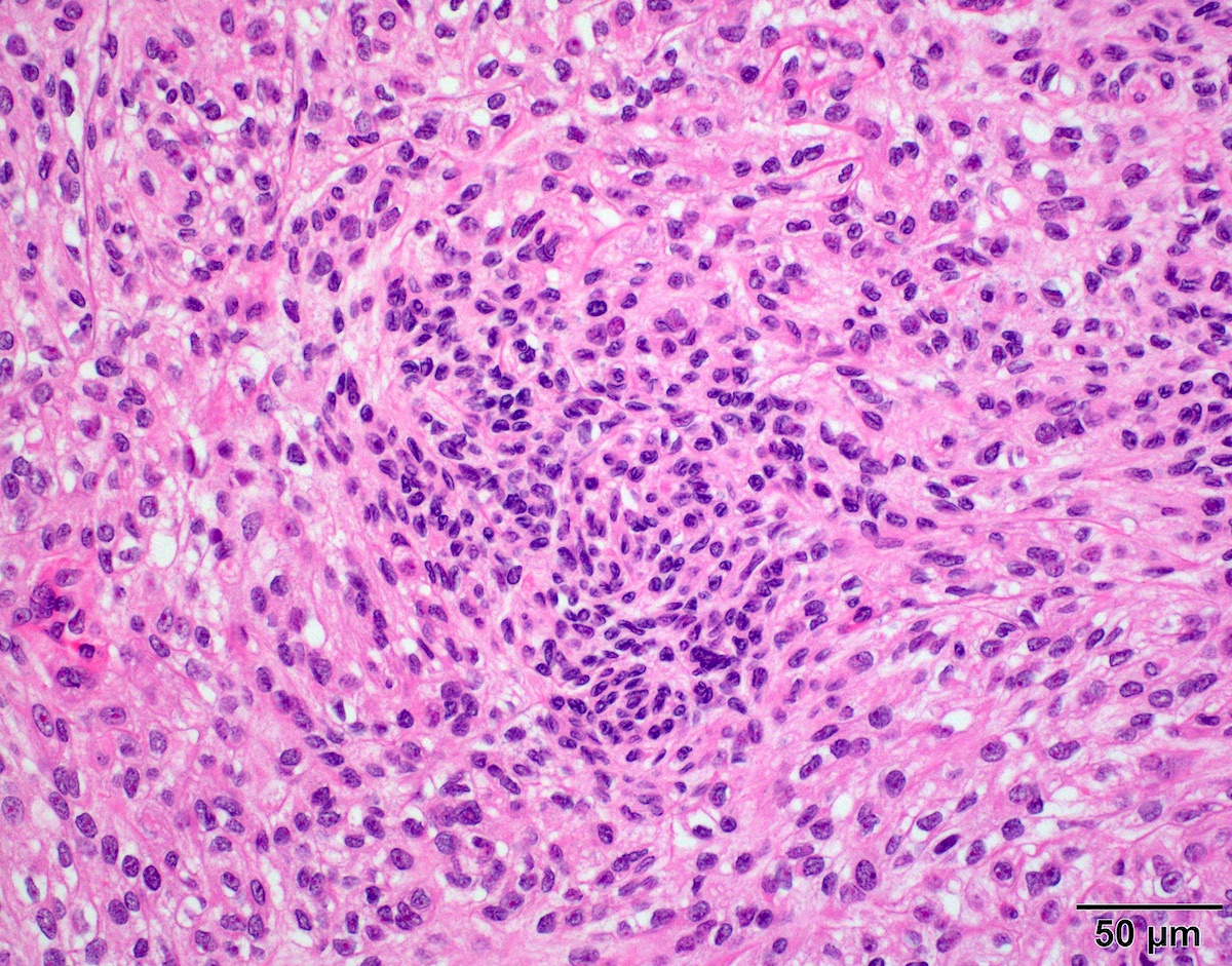
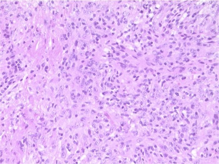
Extra-axial, dural mass
Images hosted on other servers:

MRI
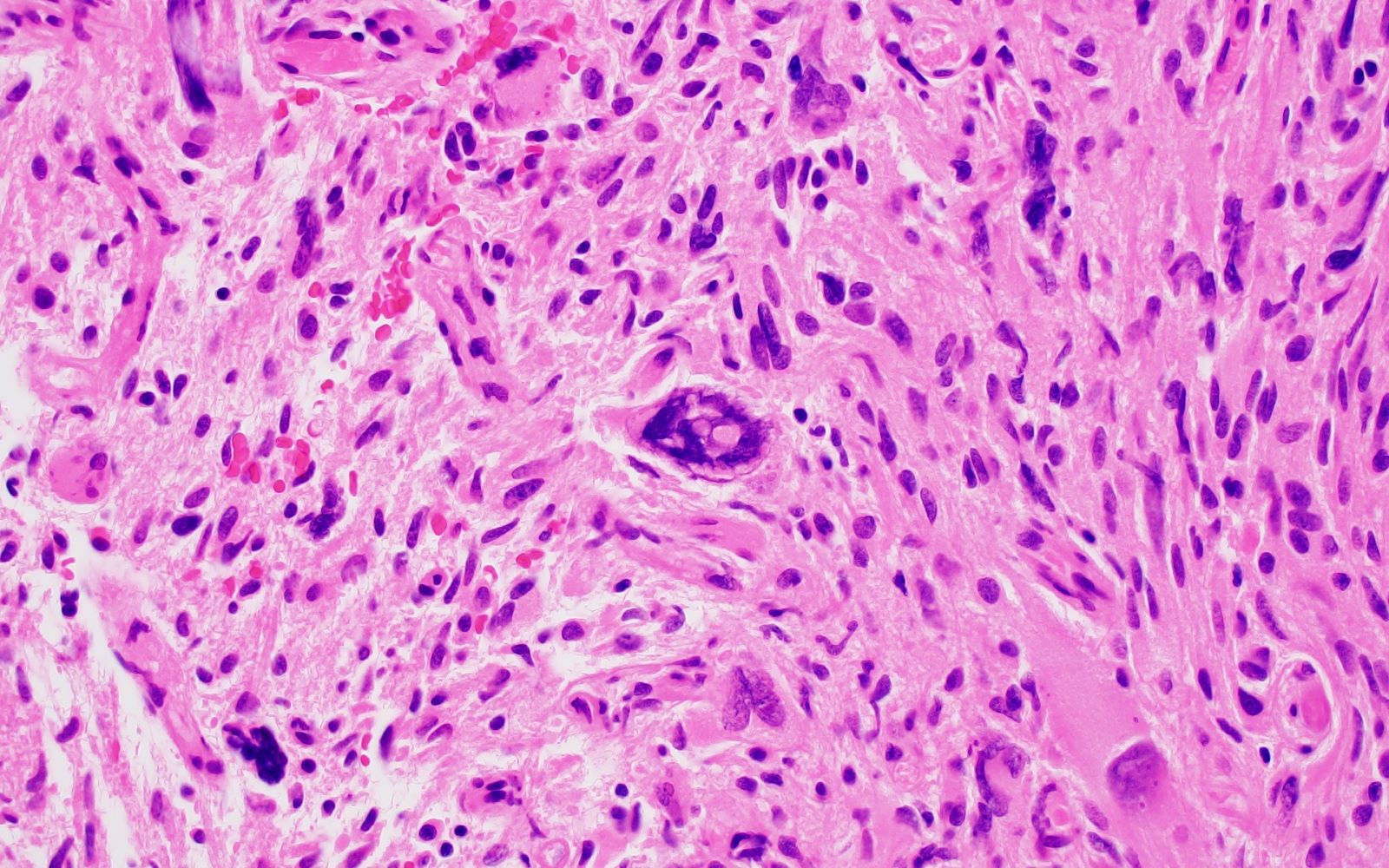
Contributed by Valeria Barresi, M.D, Ph.D.
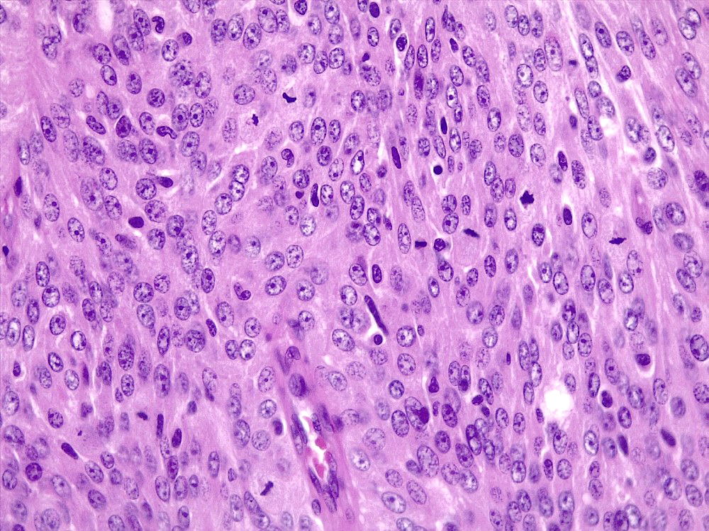
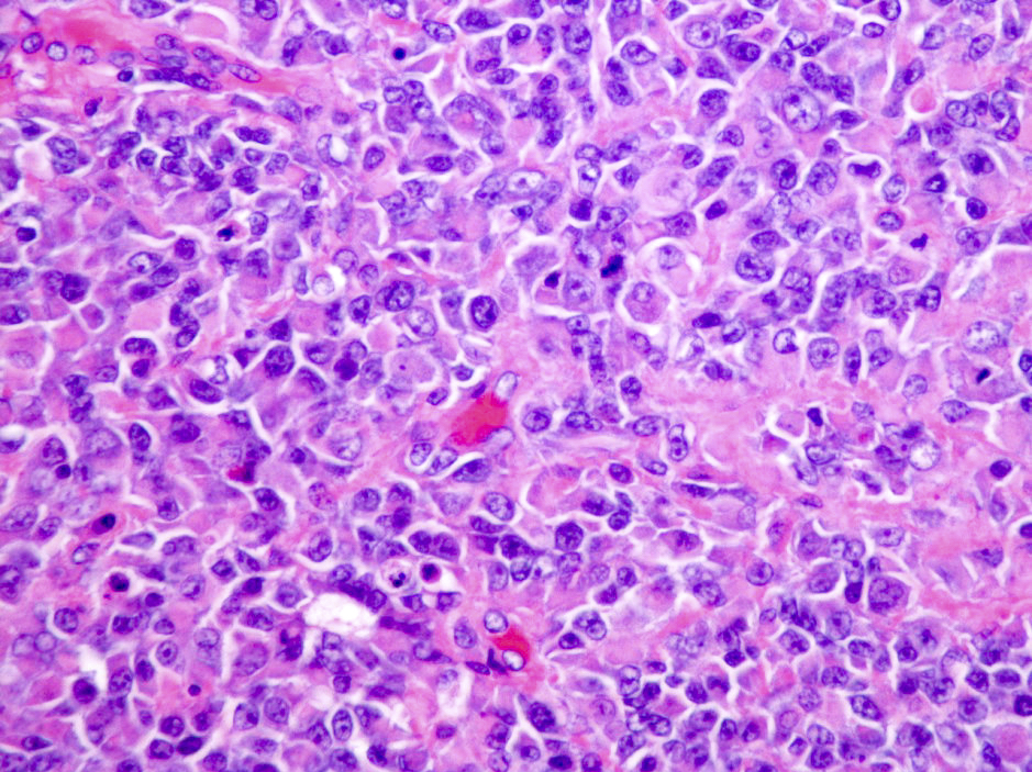
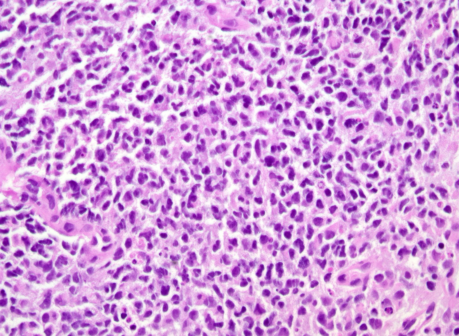
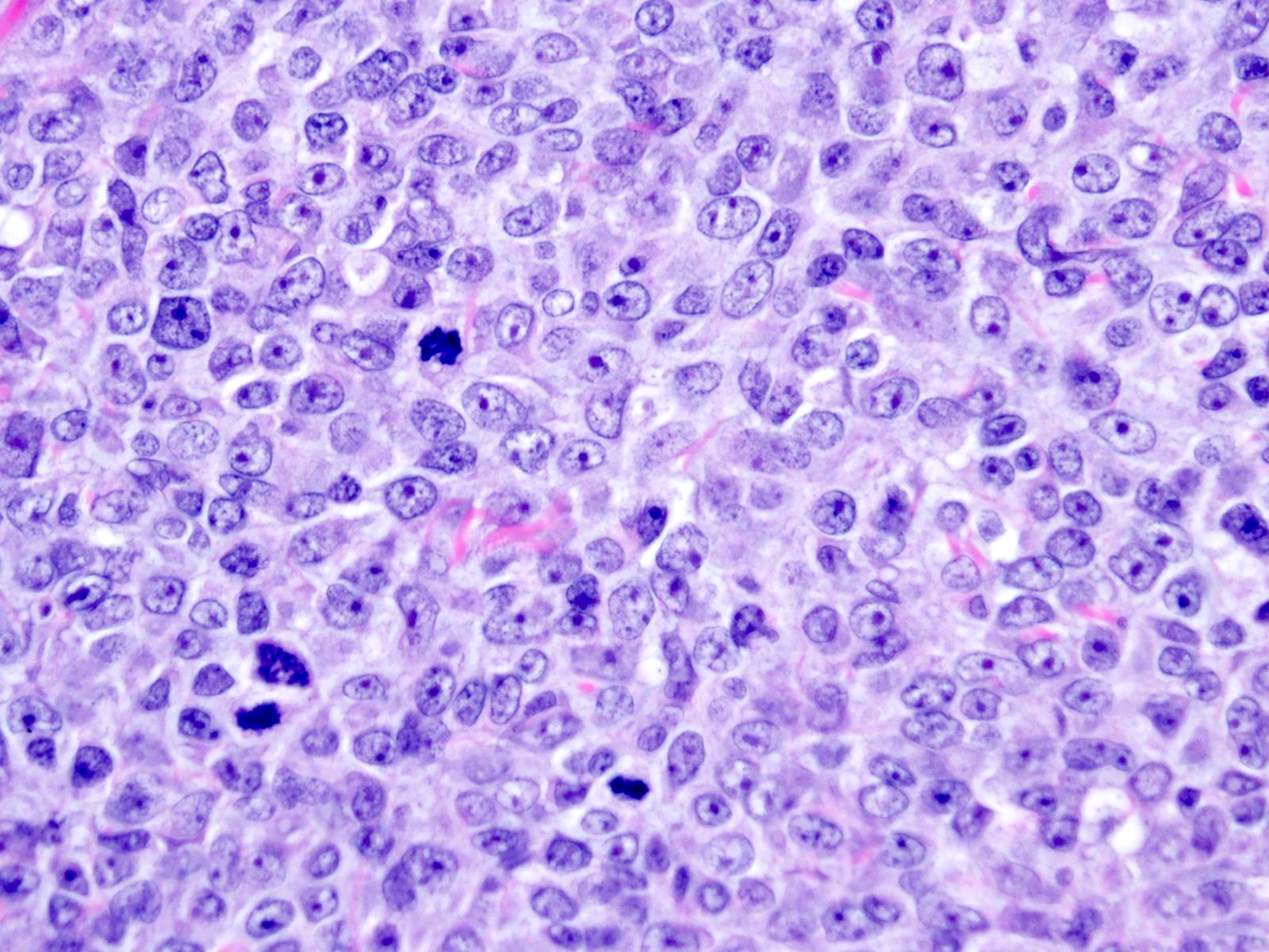
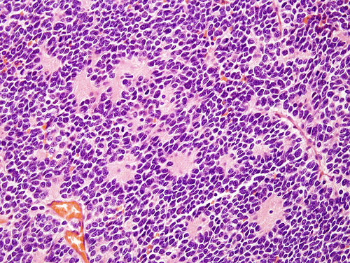
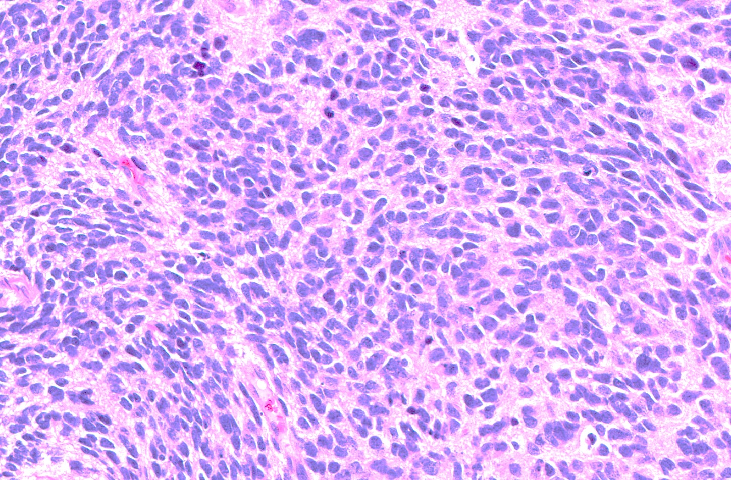
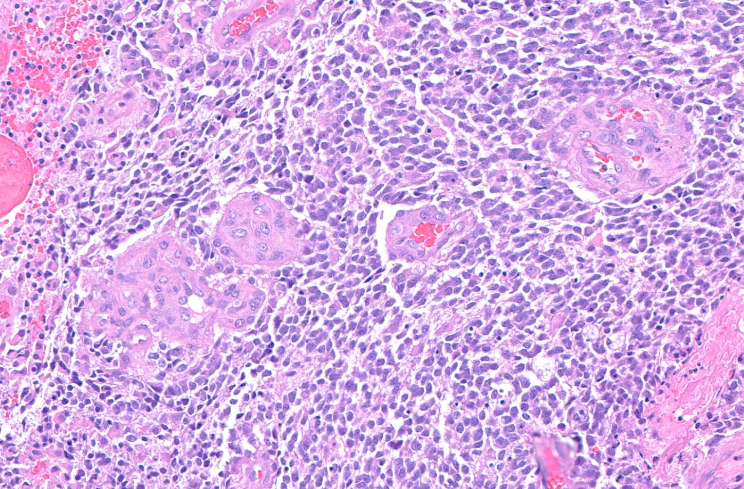
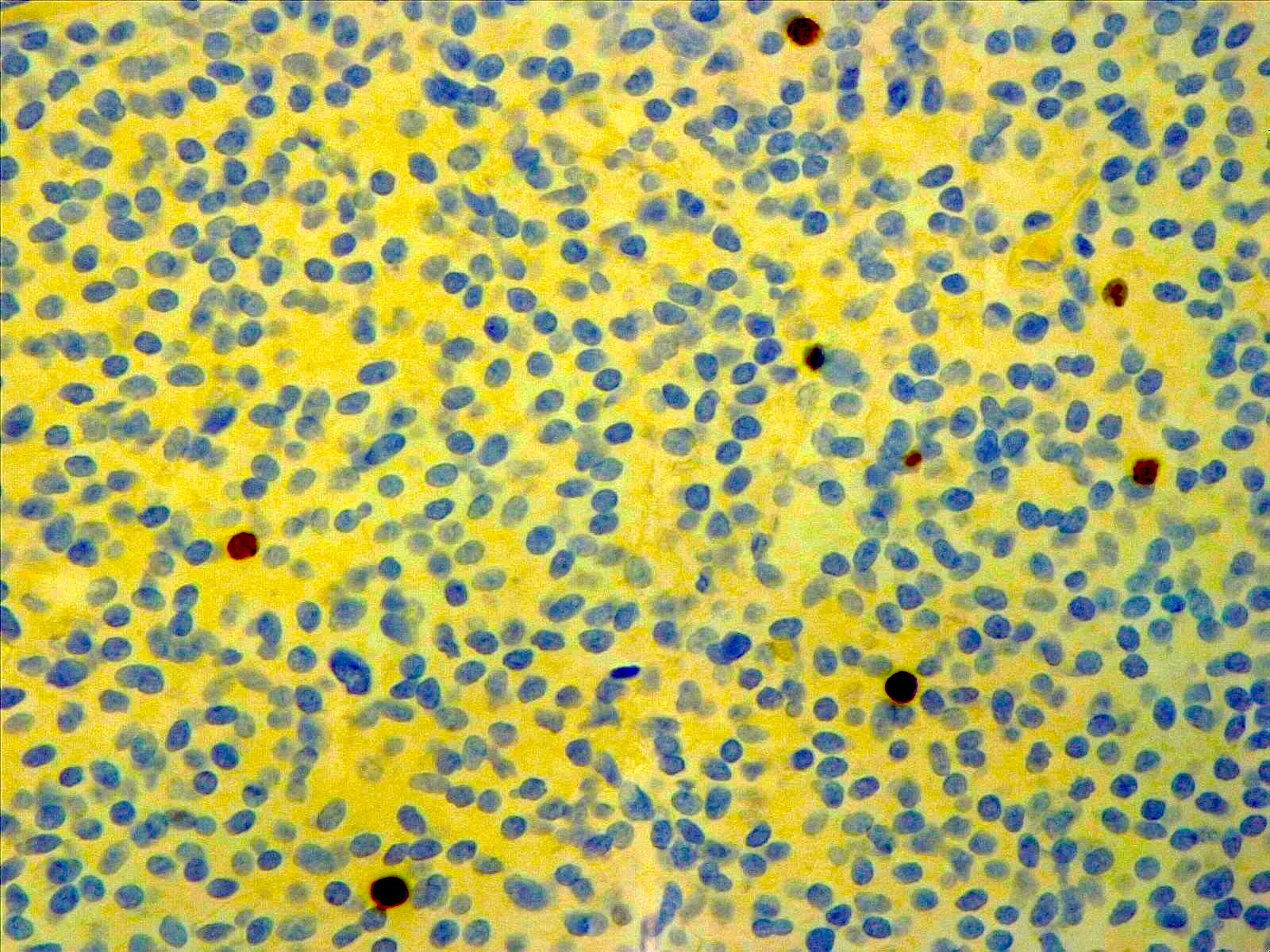
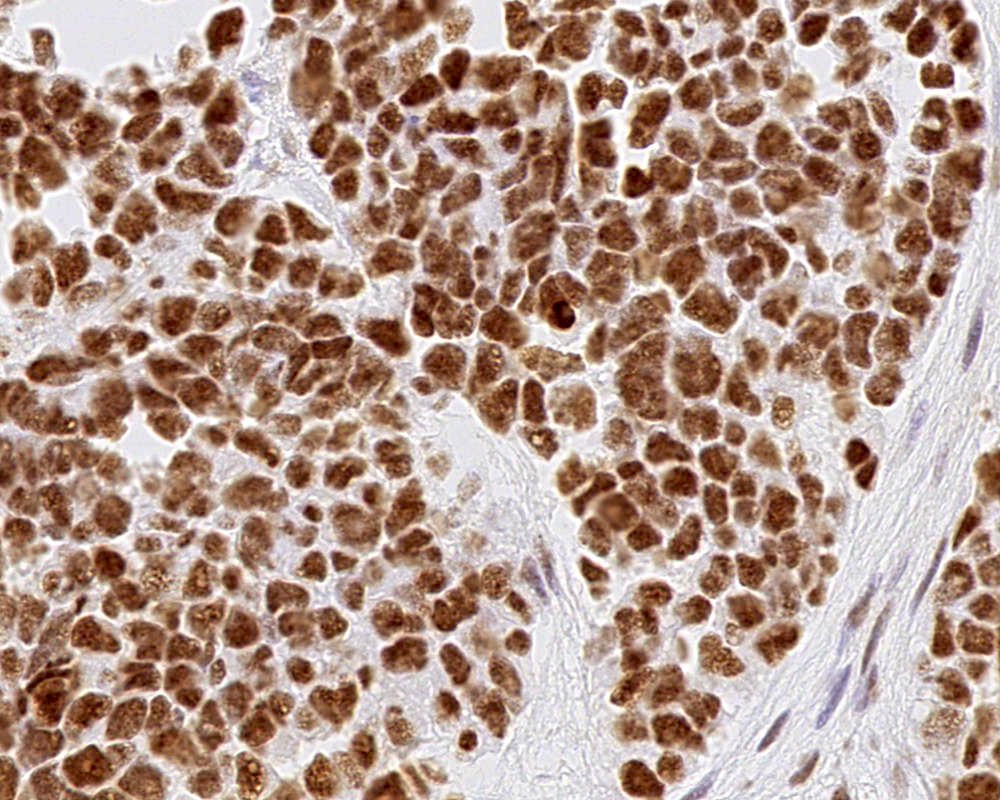
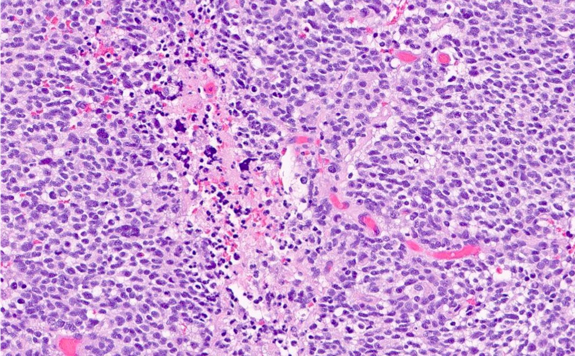
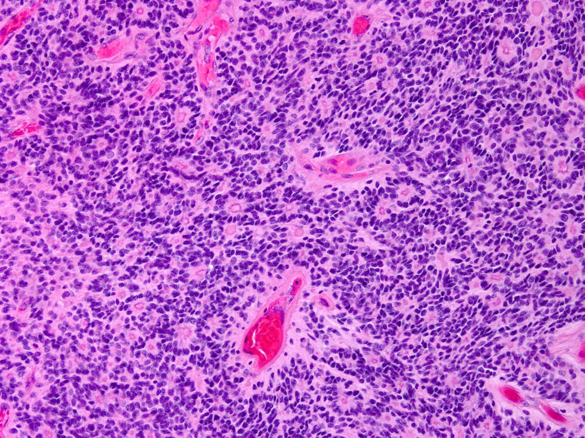
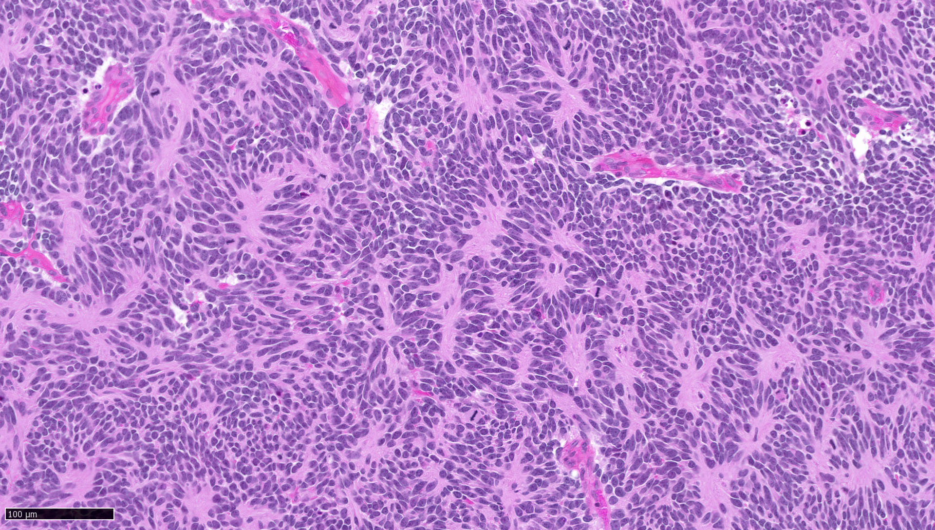
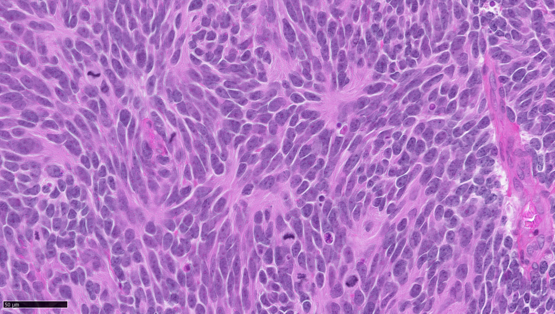
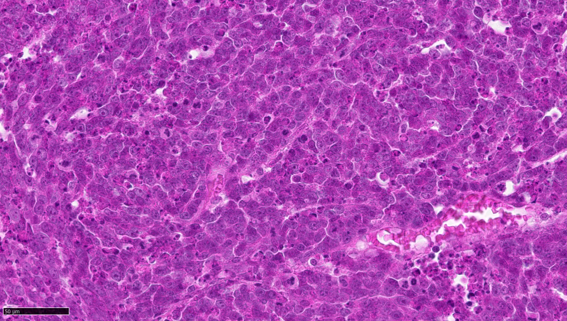
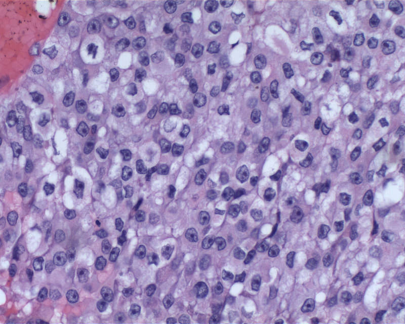
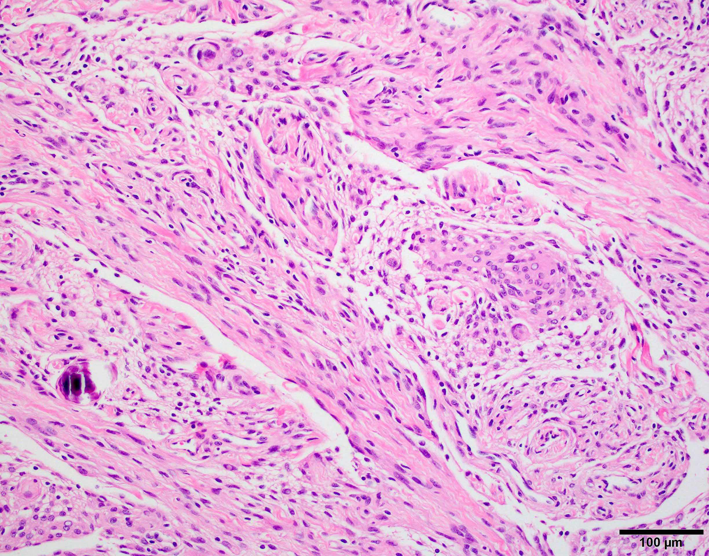
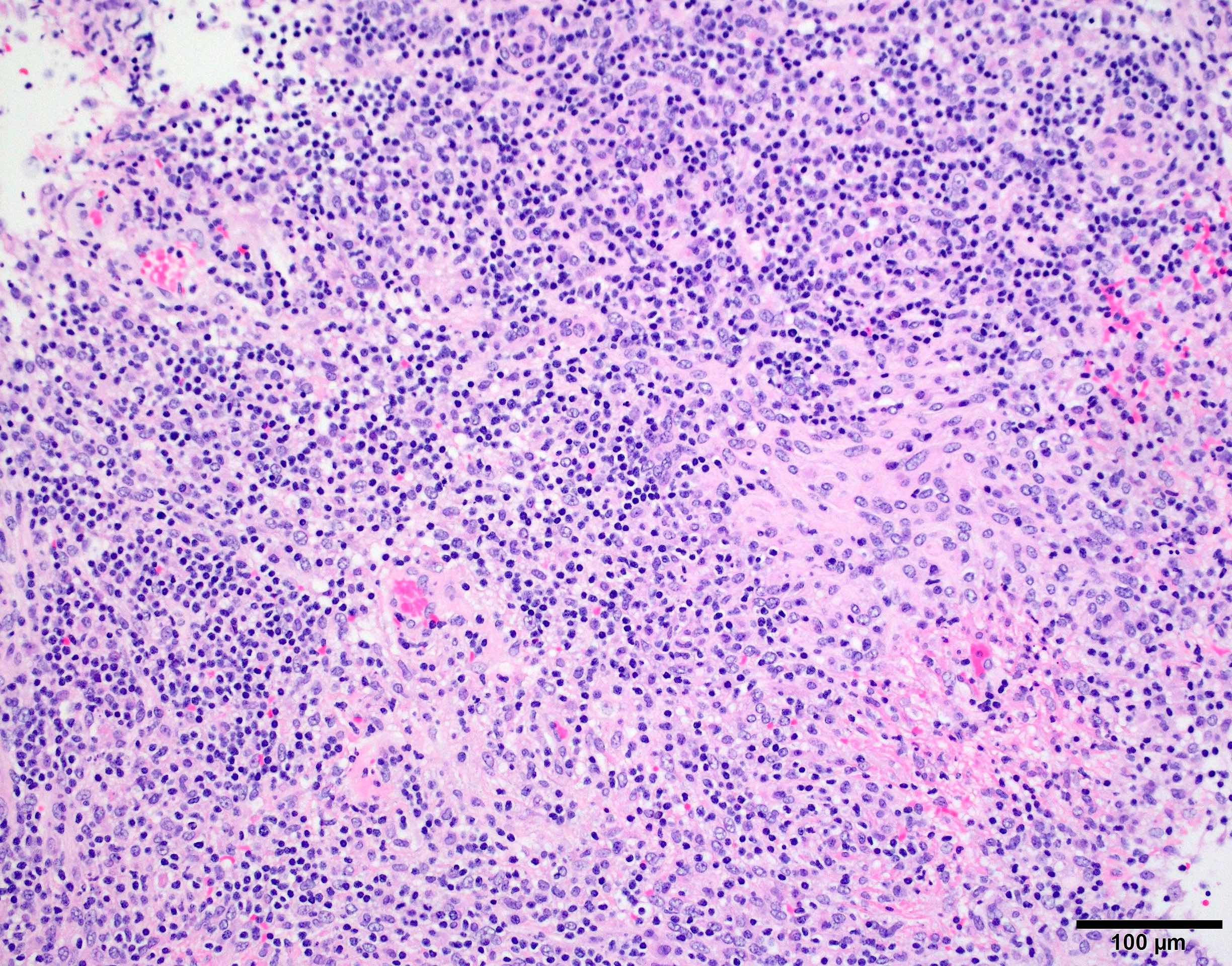
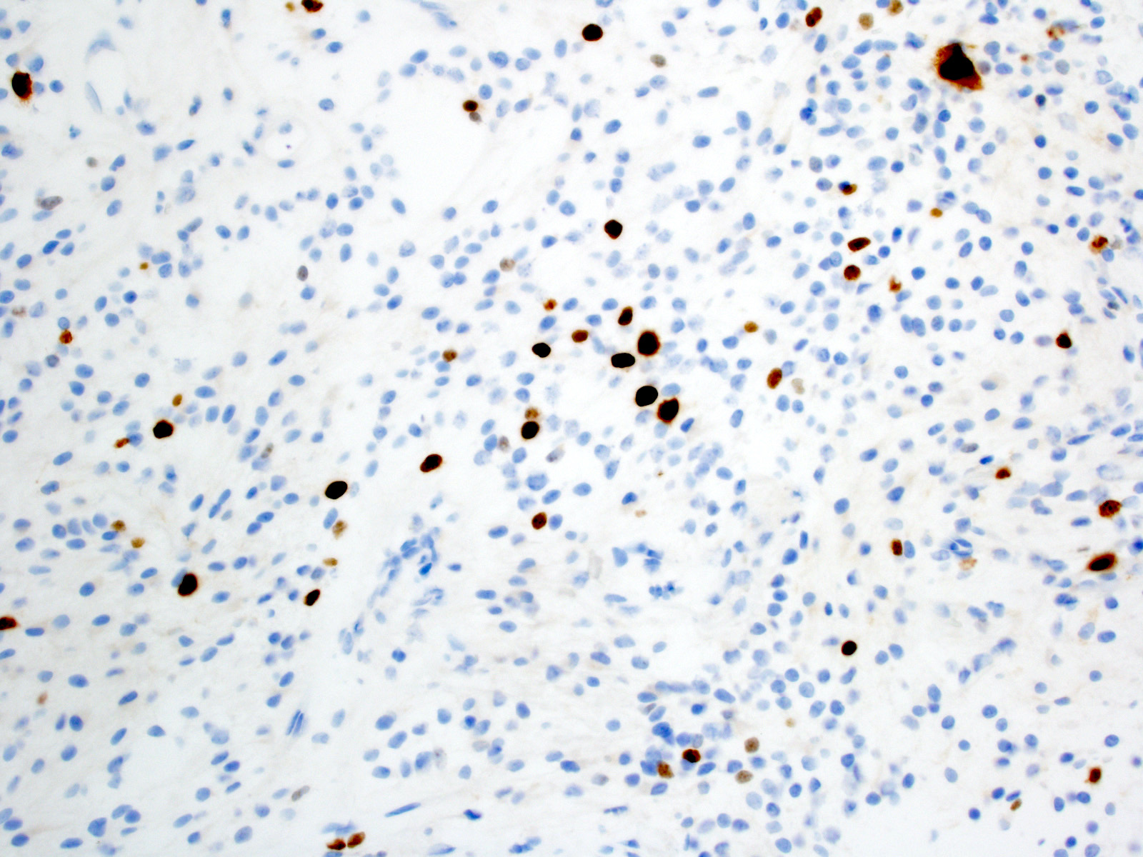
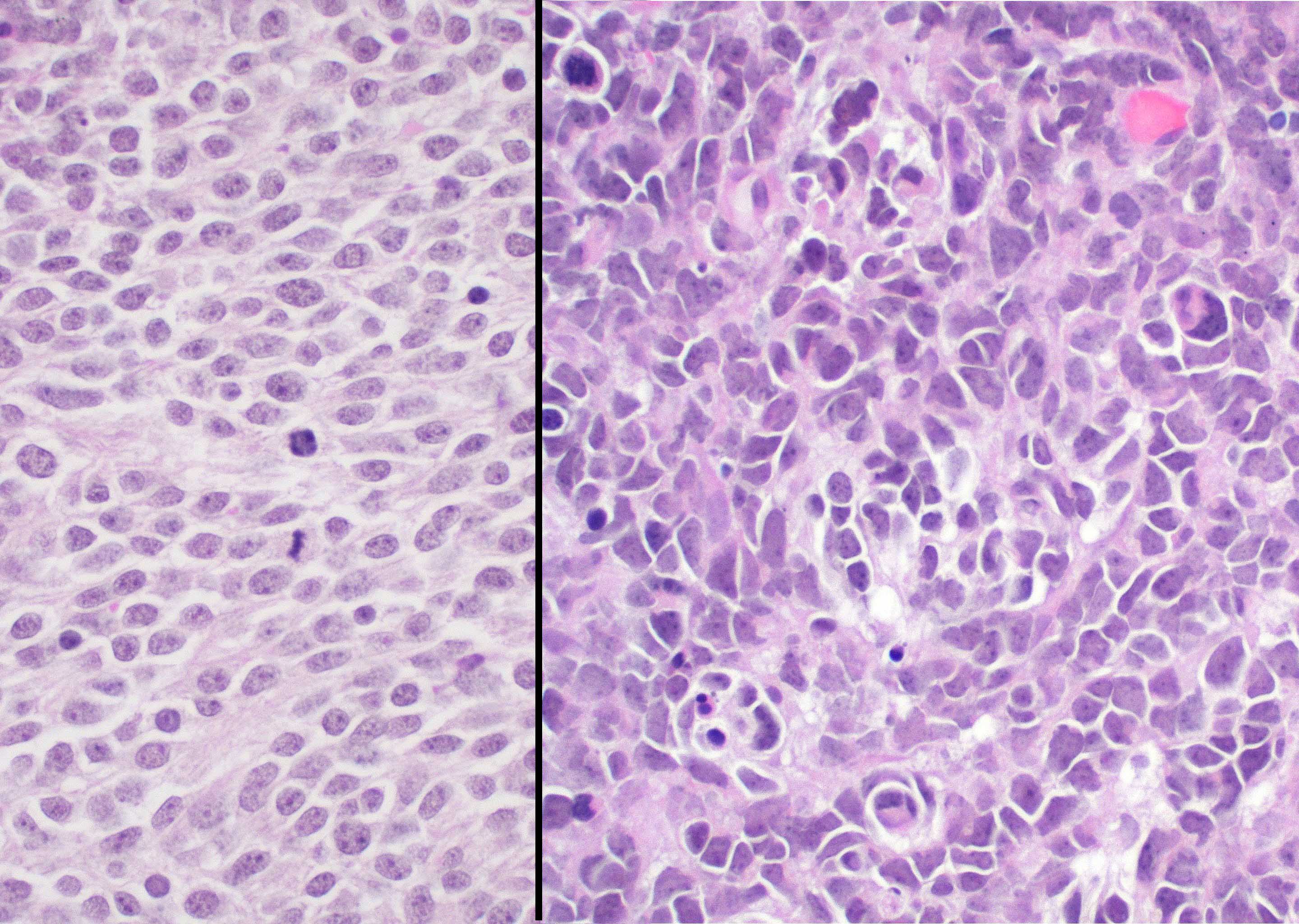
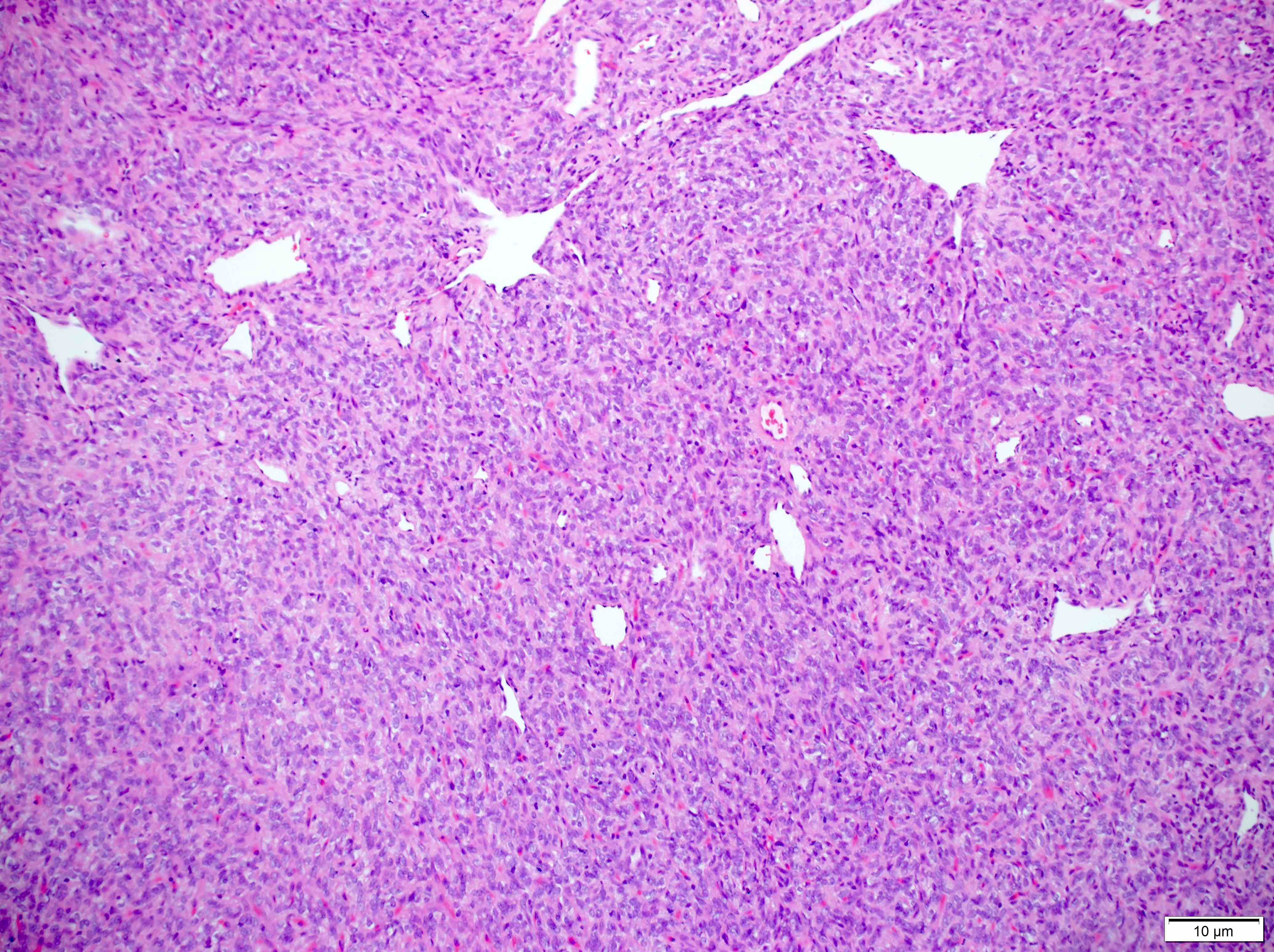
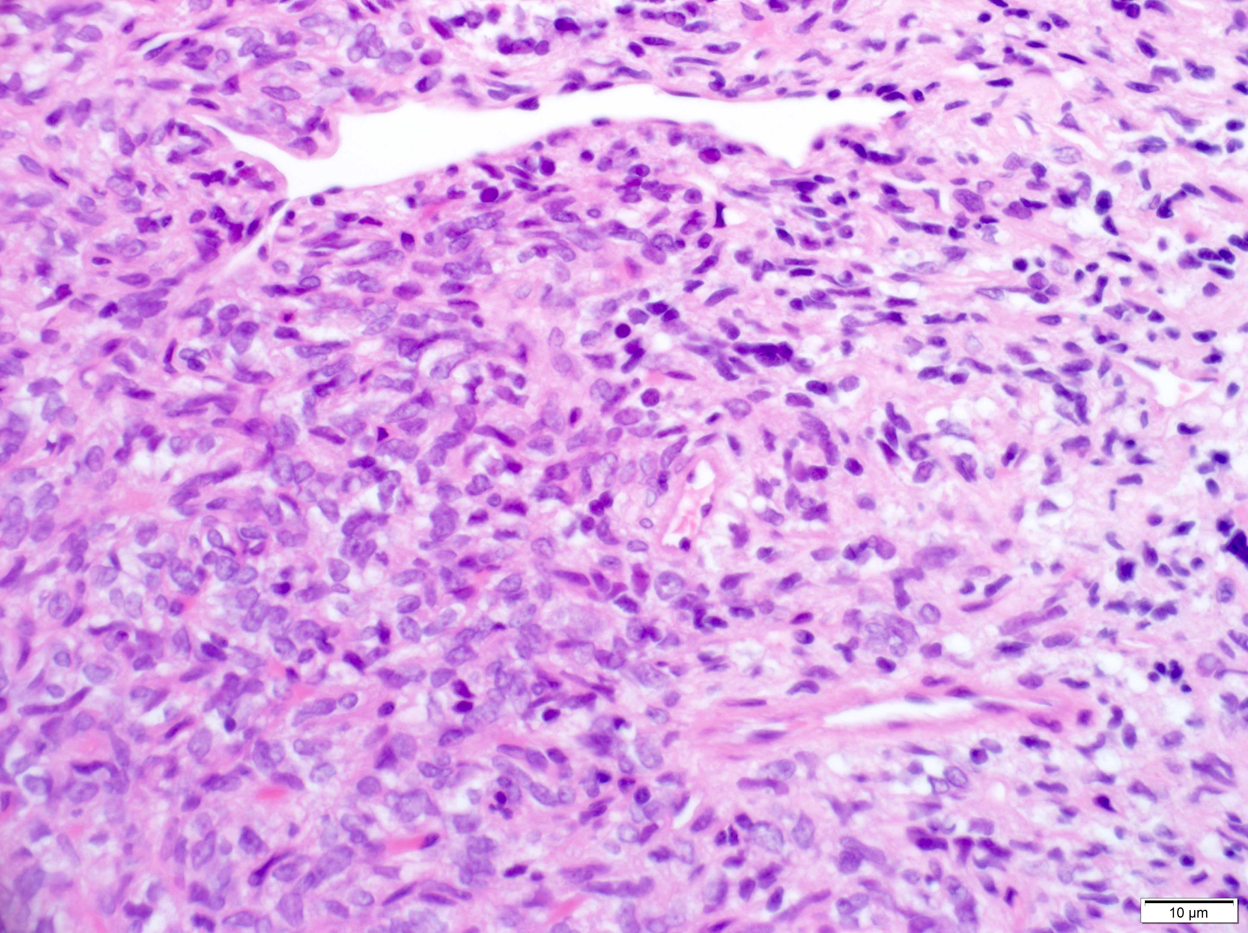
Mitoses
Malignant morphology
Whorls
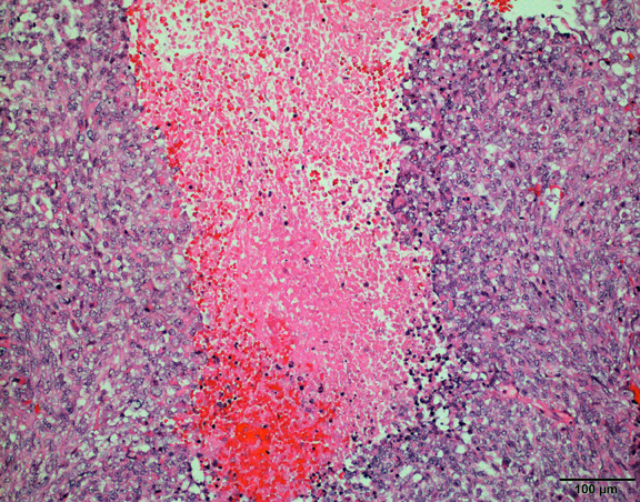
Necrosis

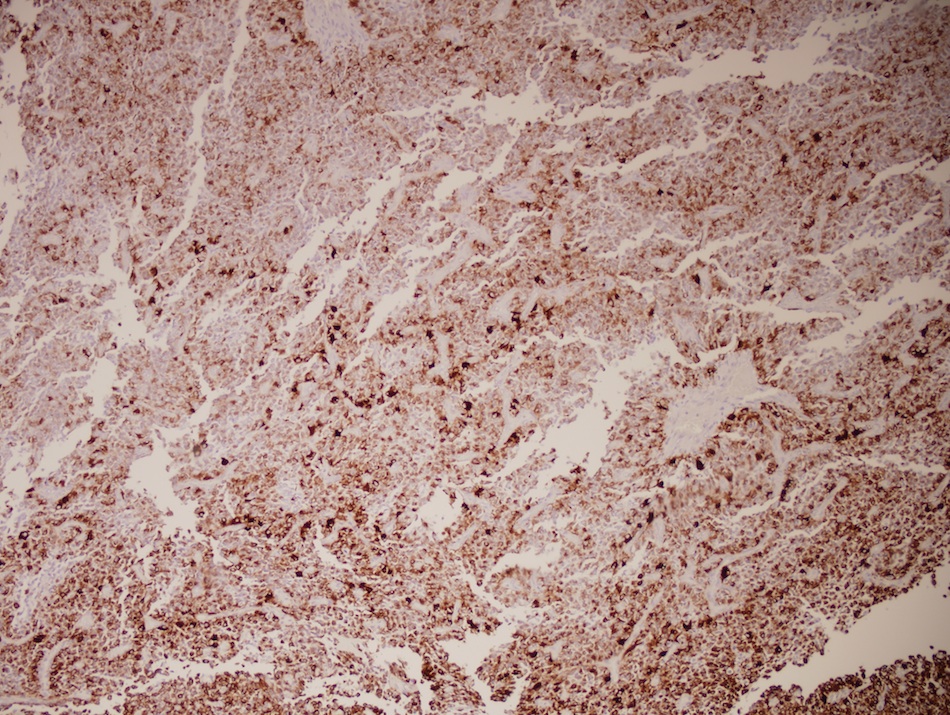
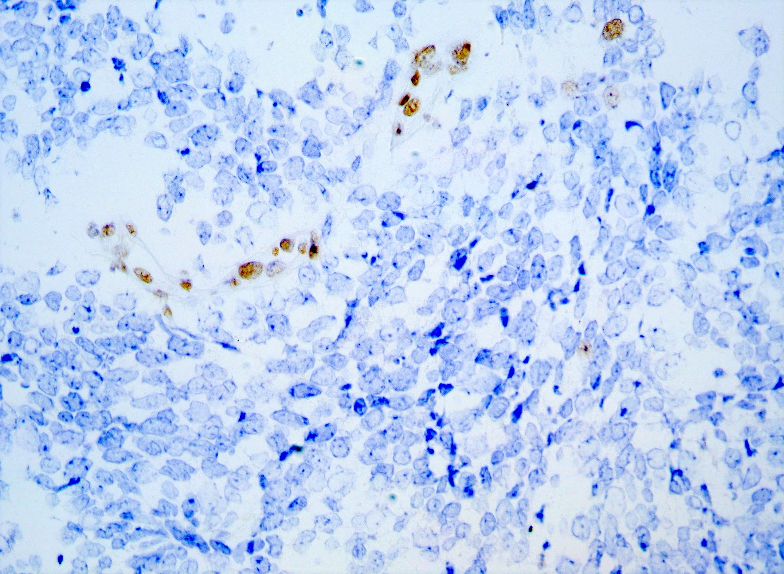
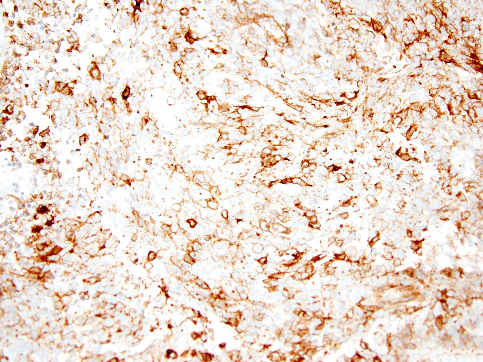
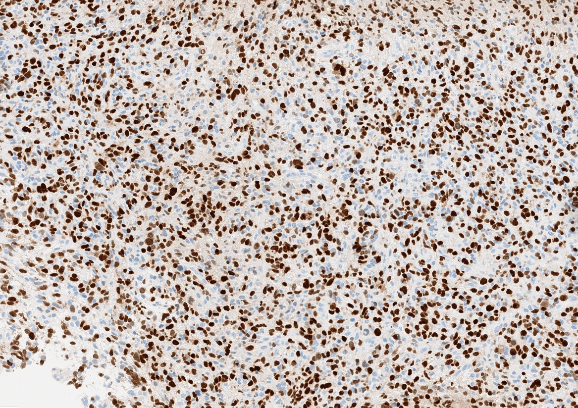
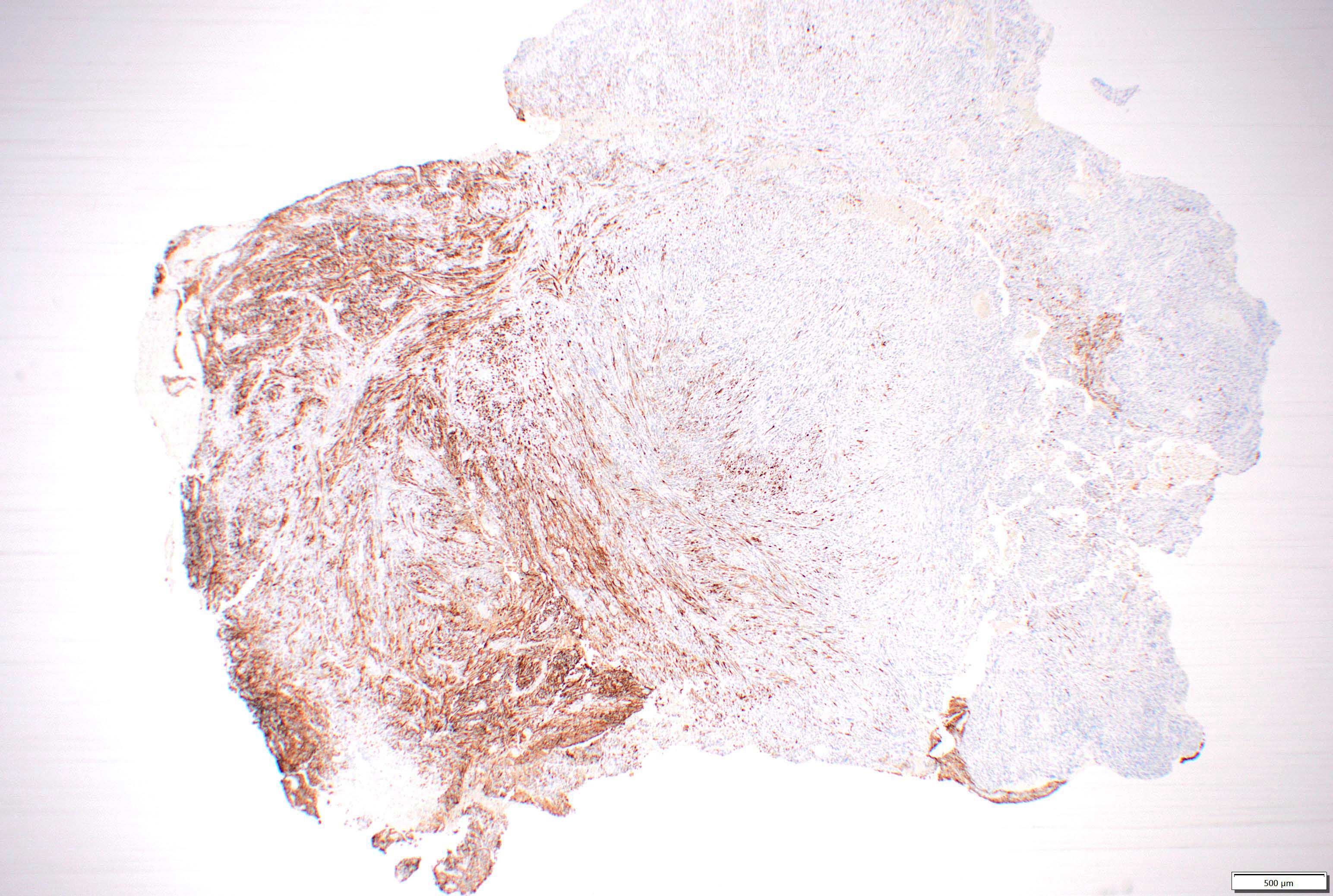
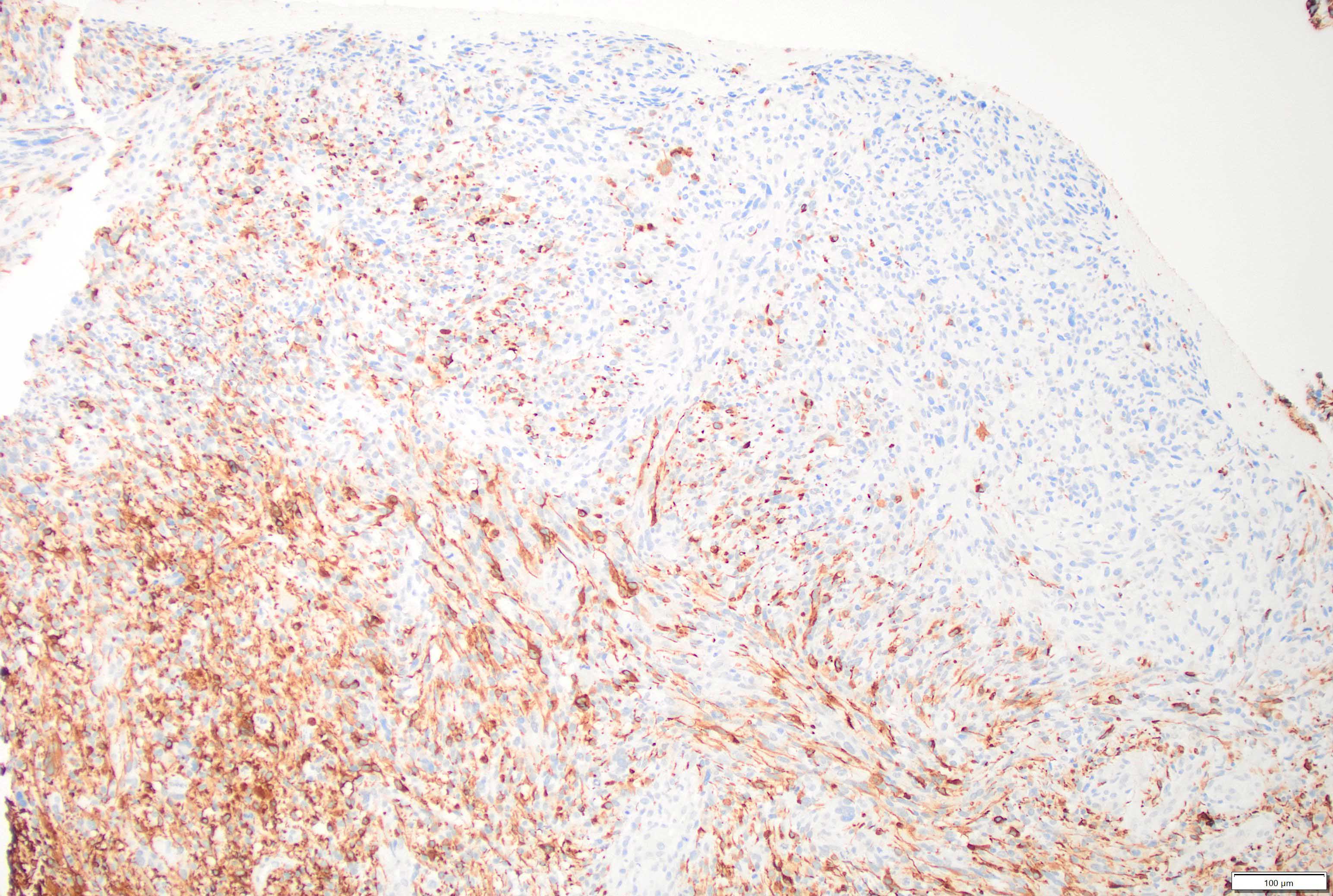
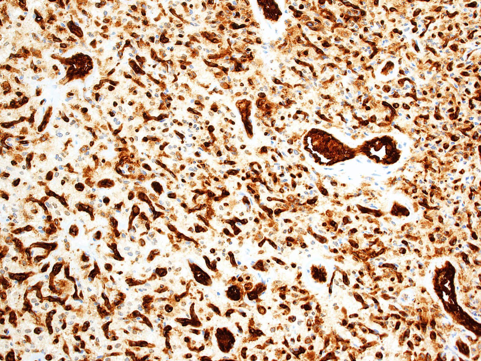
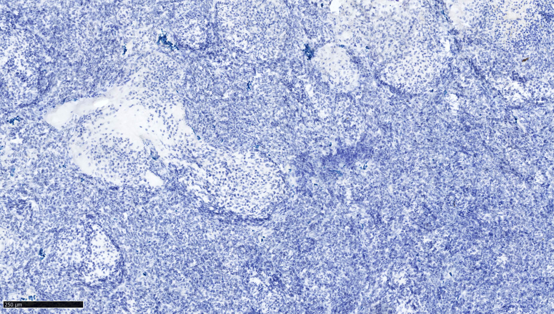
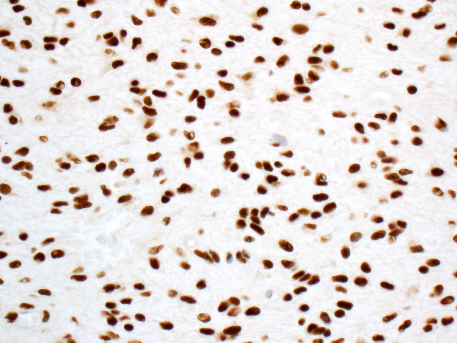
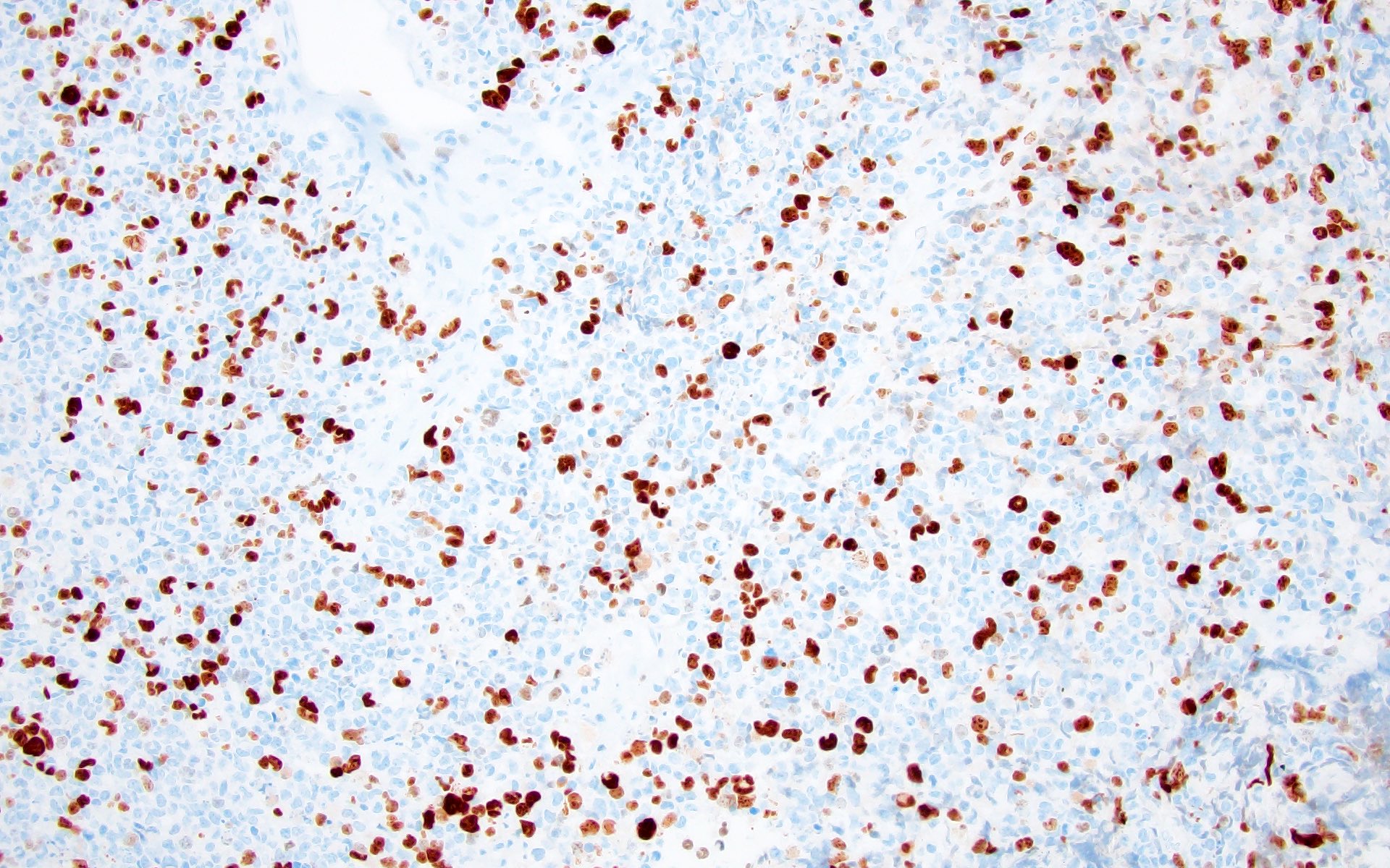
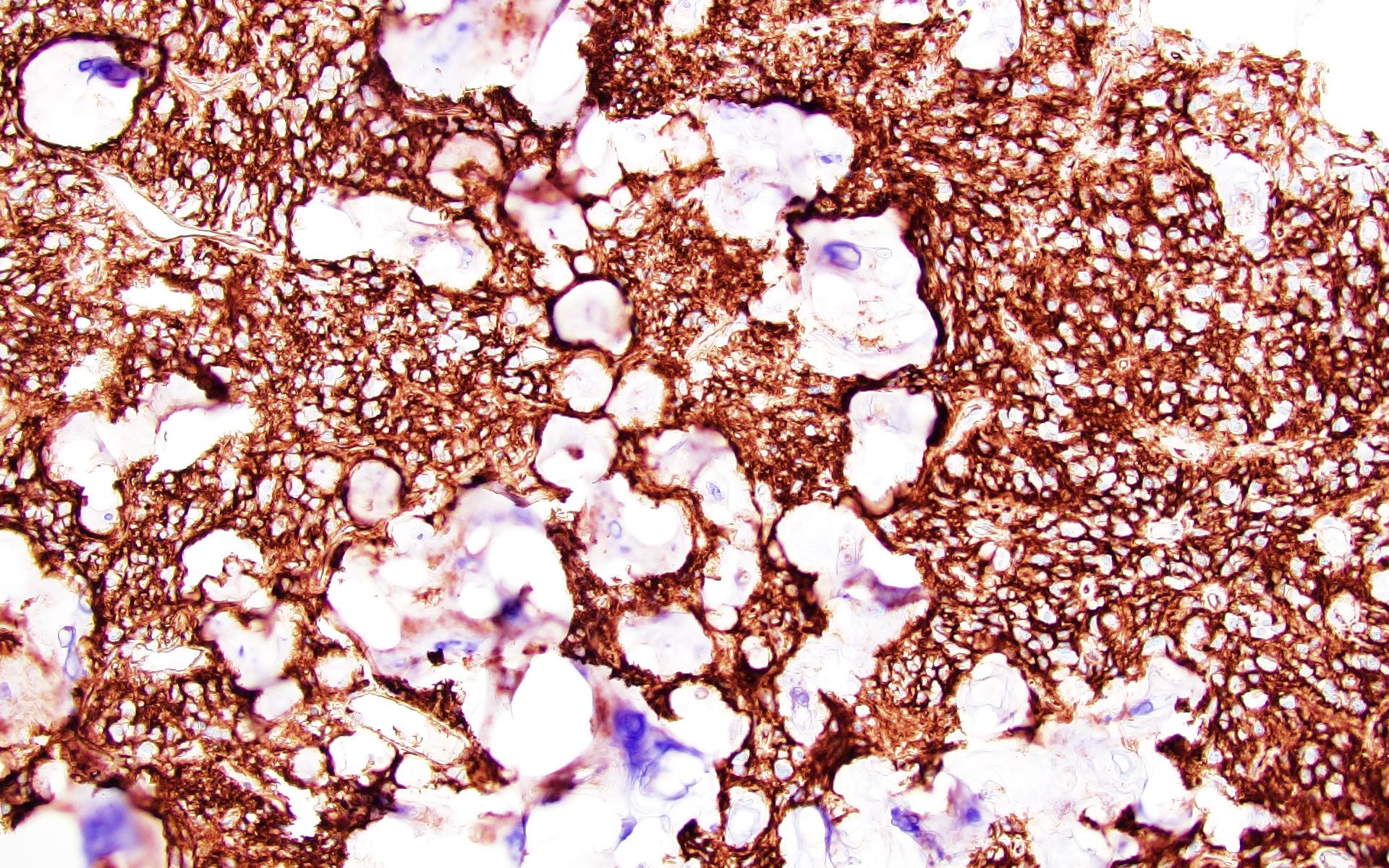
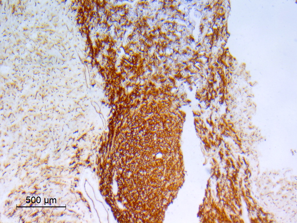
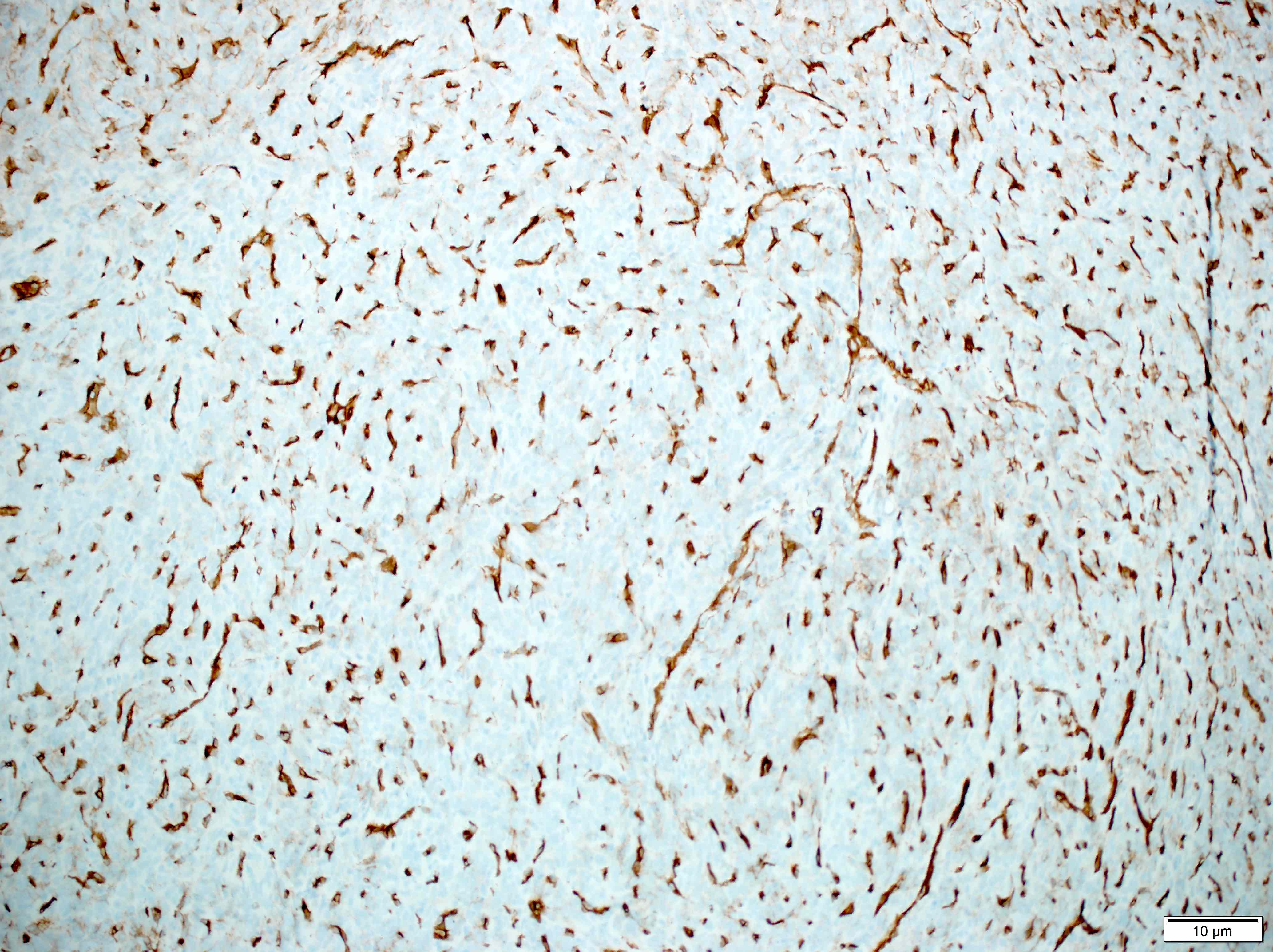
EMA
CK AE1 / AE3
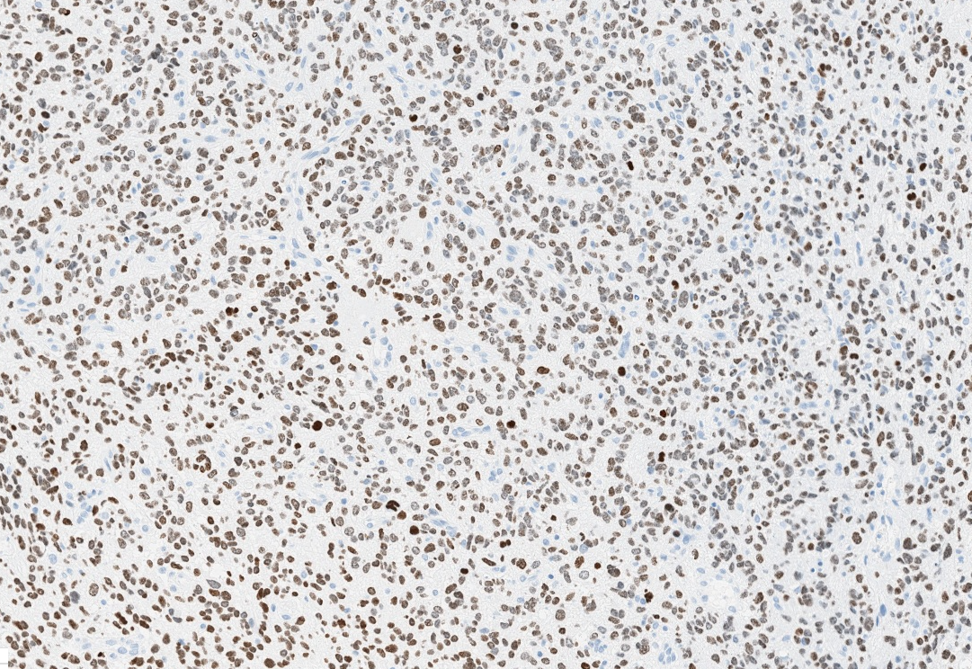
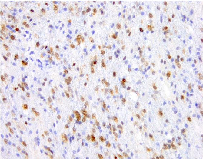
H3K27me3
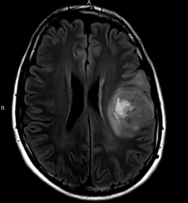
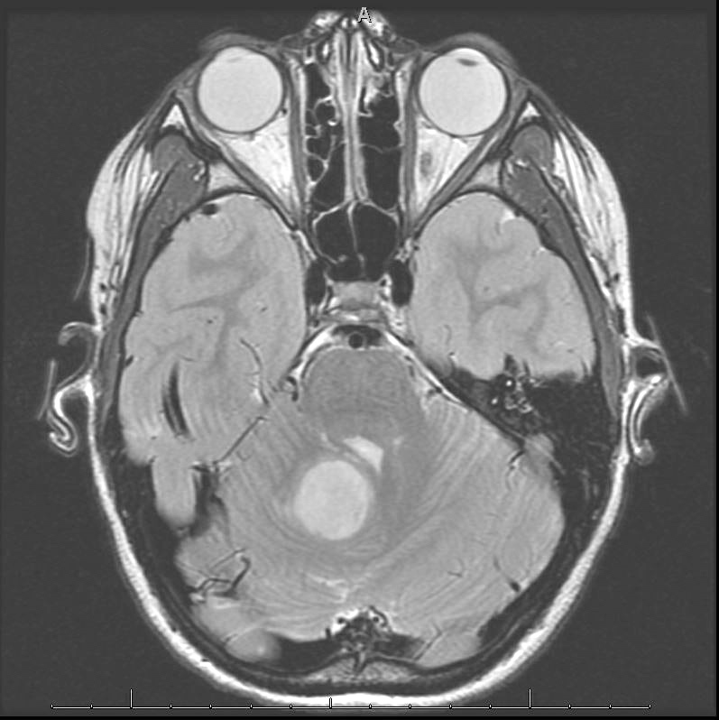
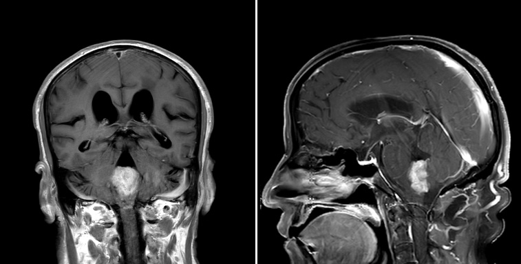
Images hosted on other servers:
Location in brain
Images hosted on other servers:

Local anatomy (horse)
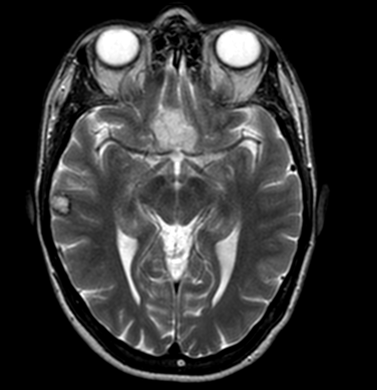
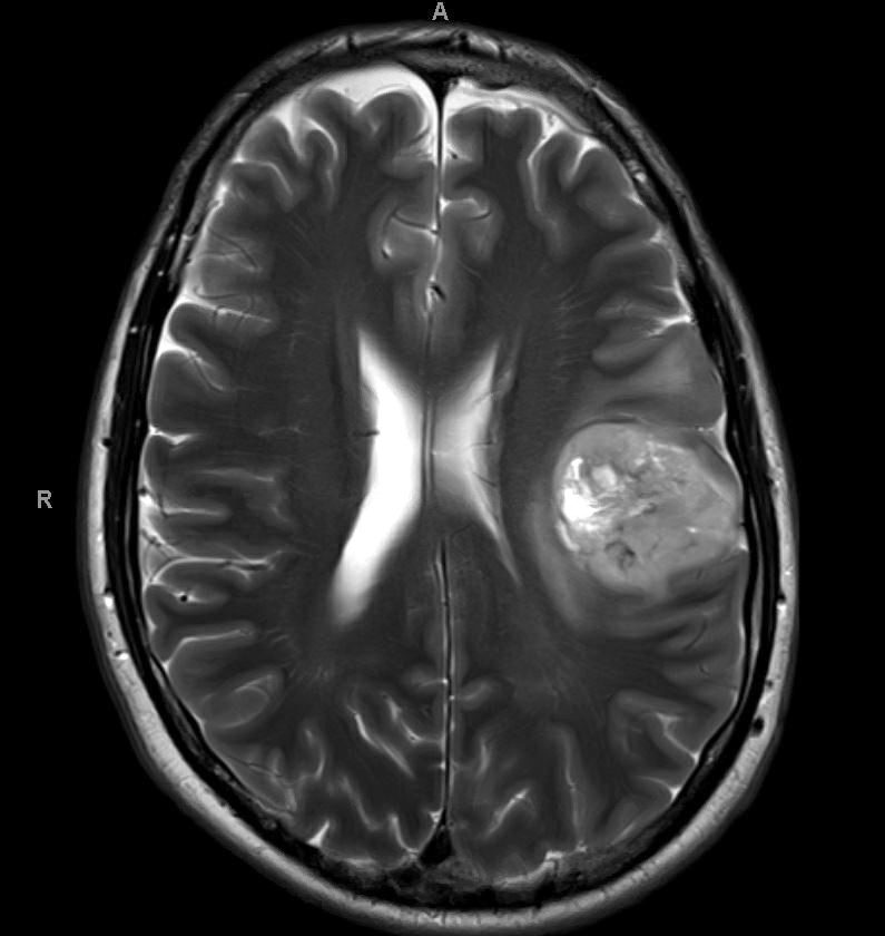
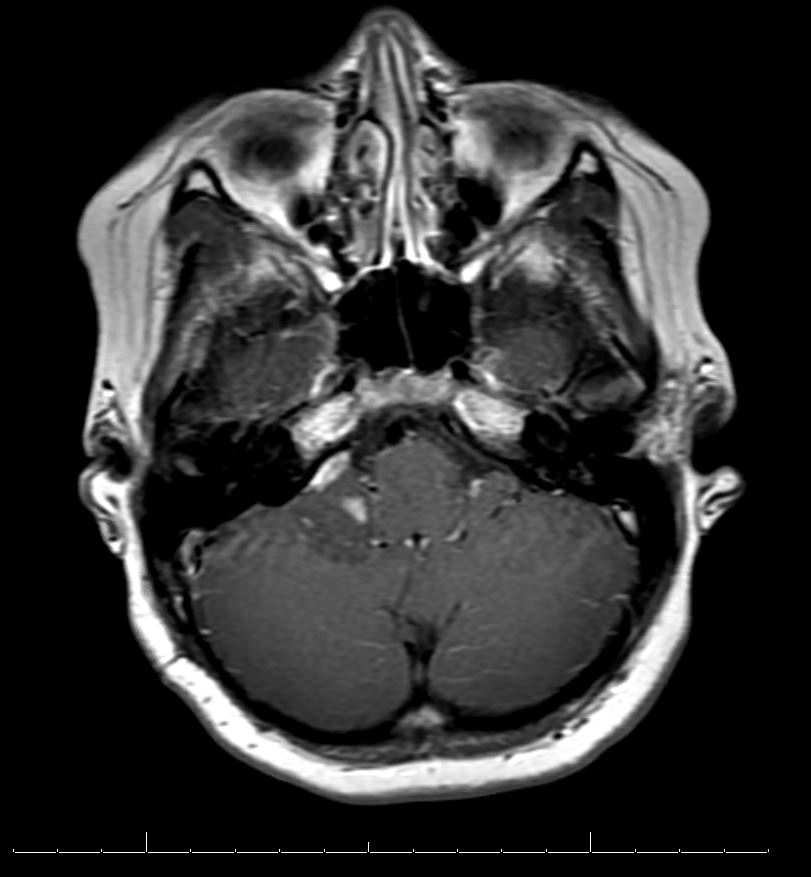
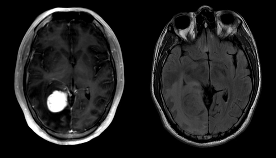
Images hosted on other servers:

Axial MR images

Axial T1 and T2 weighted MR images

Hyperintense cortically based mass
Contributed by Saman Seyed Ahmadian, M.D.
T2 FLAIR MRI

T2 MRI
Images hosted on other servers:

T2 MRI
Contributed by Saman Seyed Ahmadian, M.D.
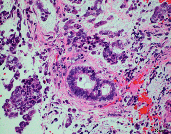
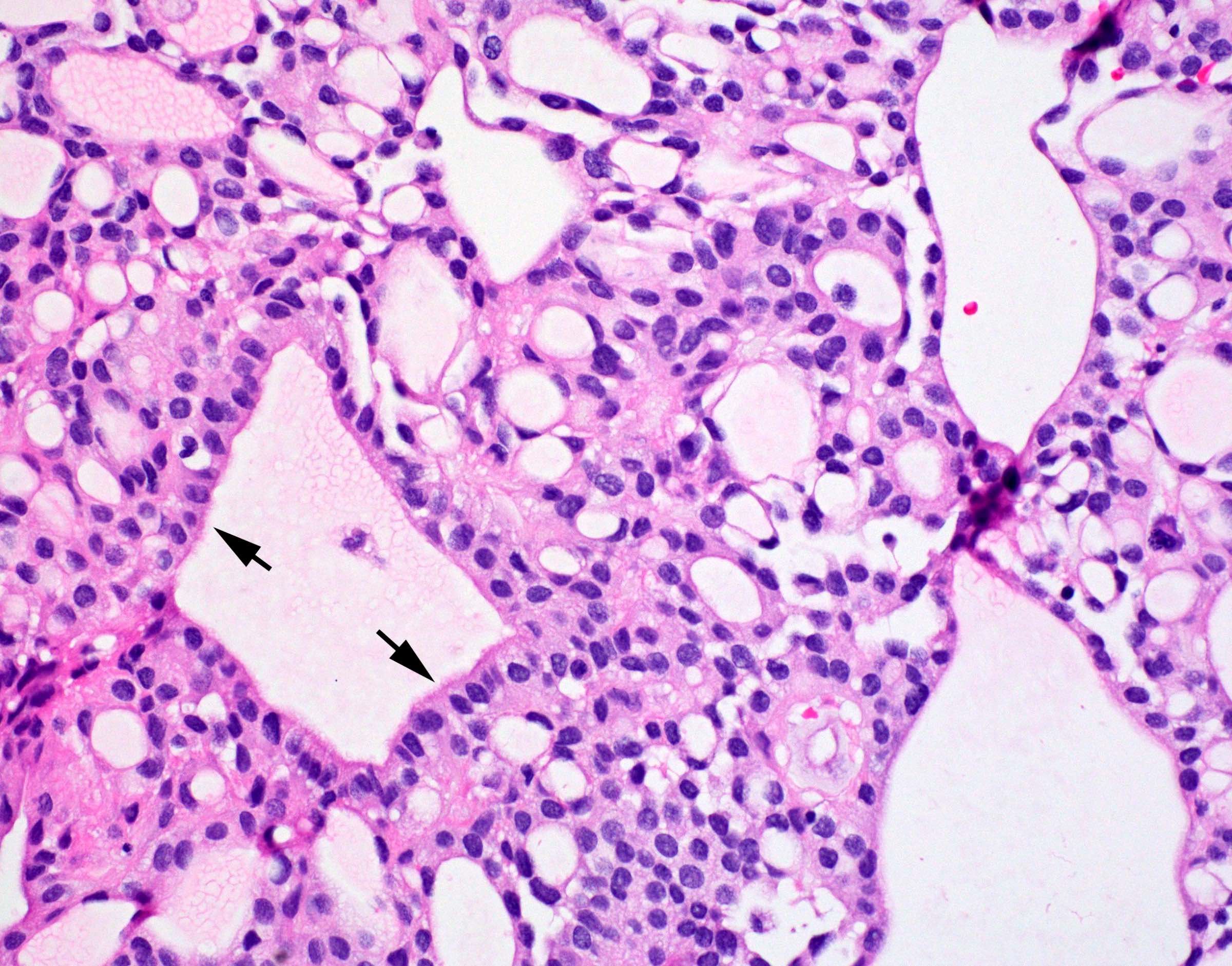
Cyst lining
Meningothelial hyperplasia
Calcification
No apparent meningothelial cells
EMA
Images hosted on other servers:

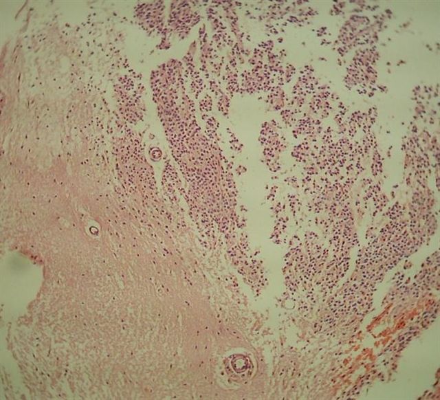
Huge well demarcated mass in frontal lobe
Case #312

EMA
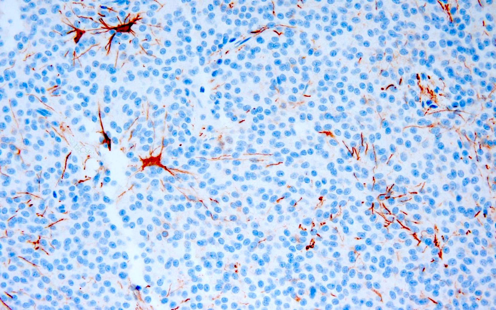
GFAP
Trichrome
Contributed by John DeWitt, M.D., Ph.D. and Meaghan Morris, M.D., Ph.D.
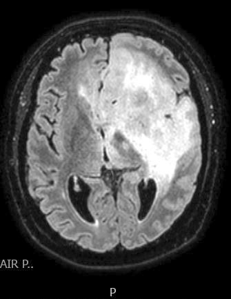
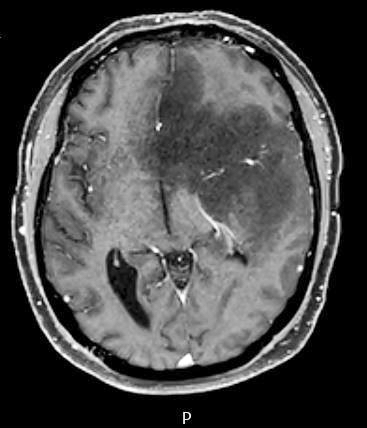
MRI axial T1 FLAIR
MRI axial T1 precontrast
MRI axial T1 postcontrast
MRI axial T1 FLAIR
MRI axial T1 postcontrast
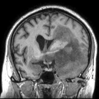
MRI coronal T1 precontrast
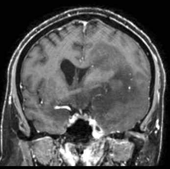
MRI coronal T1 postcontrast
Bright on FLAIR
Contrast enhancing
Images hosted on other servers:

Glioma with cystic spaces in the cerebral hemisphere
Contributed by Eman Abdelzaher, M.D., Ph.D., John DeWitt, M.D., Ph.D. and Meaghan Morris, M.D., Ph.D.
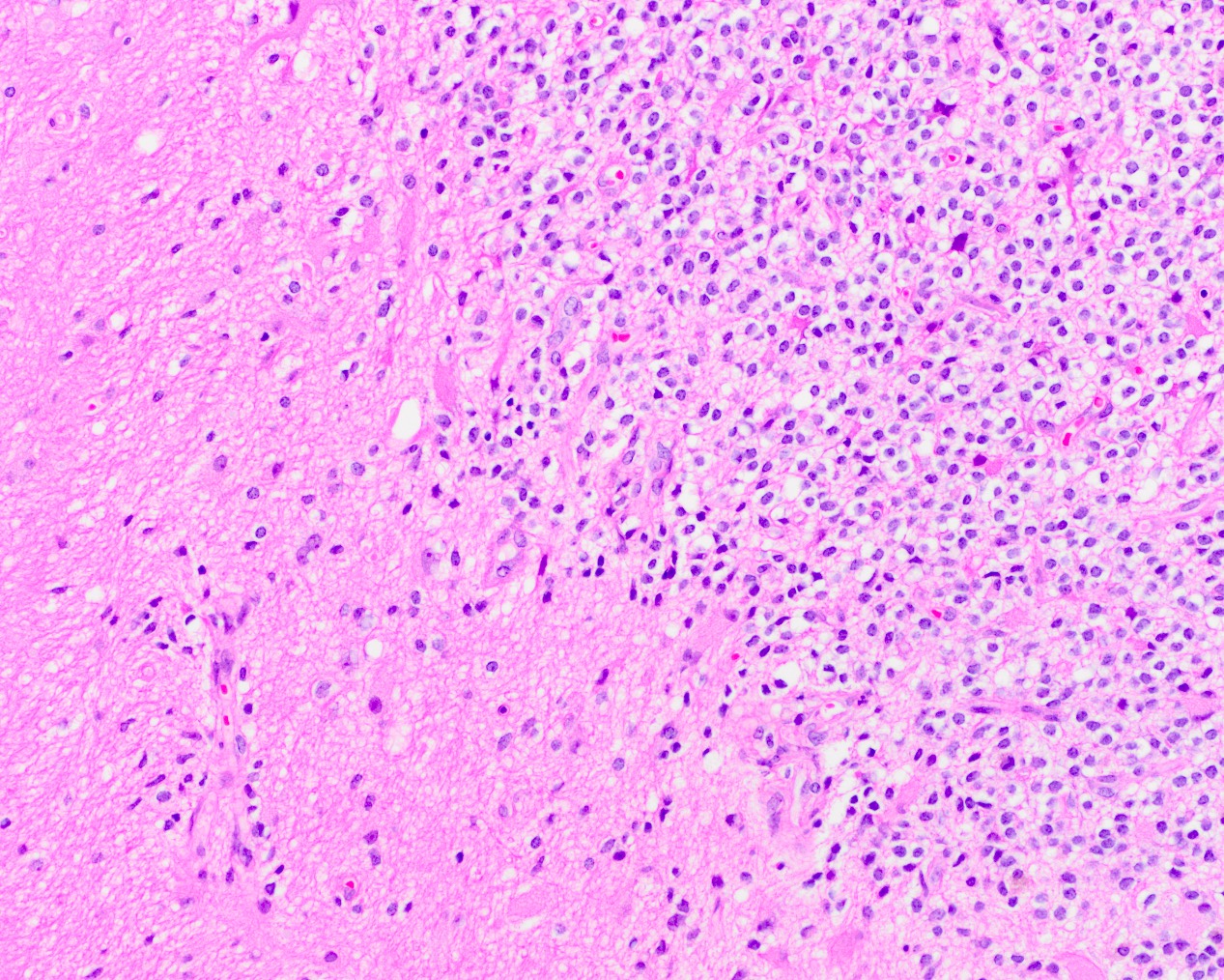
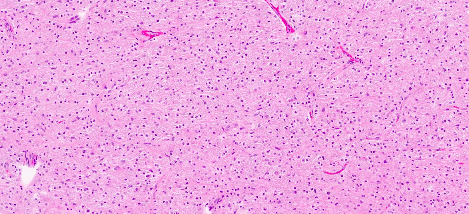
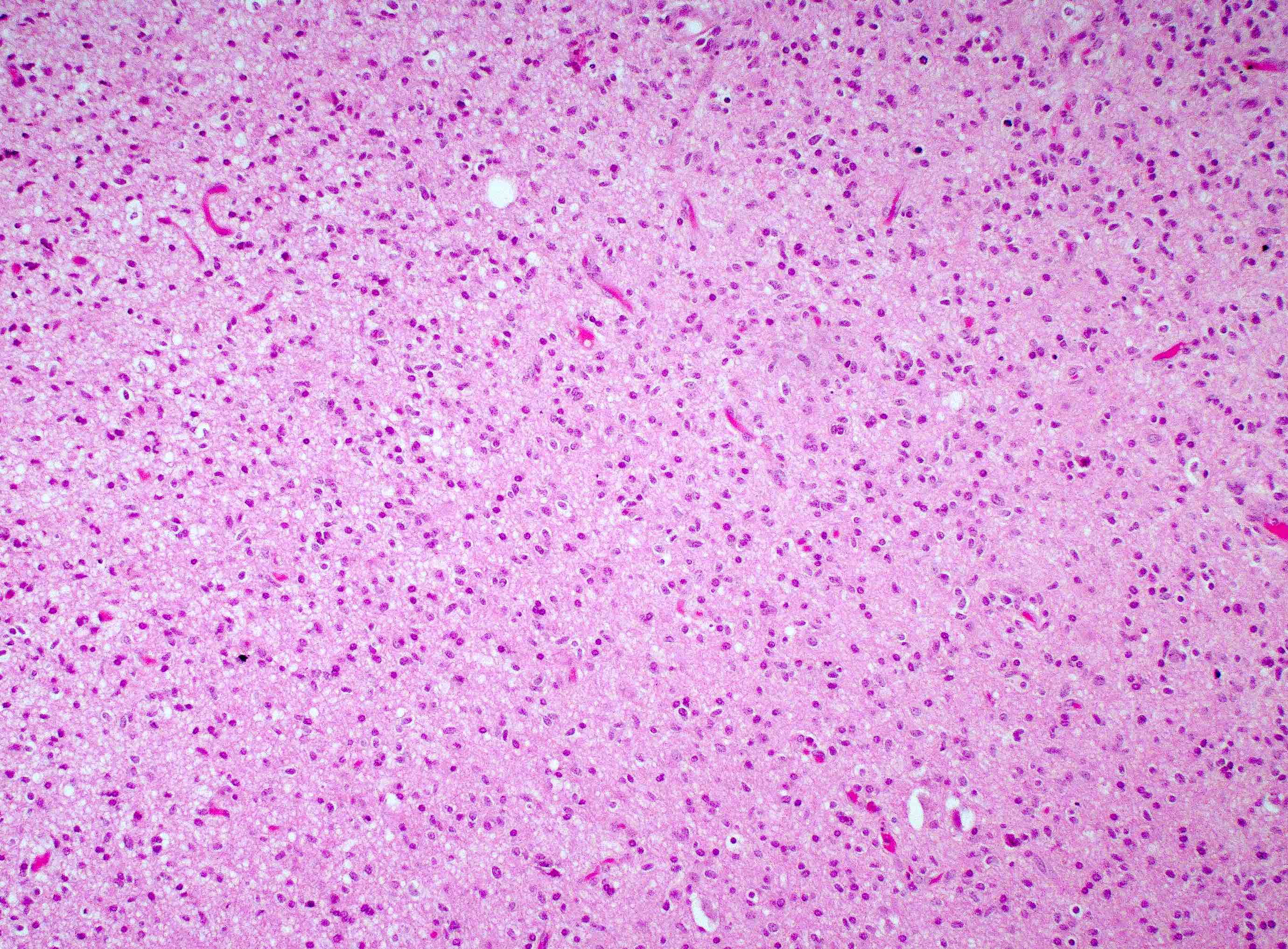
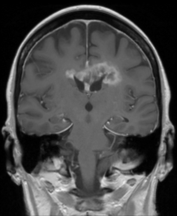
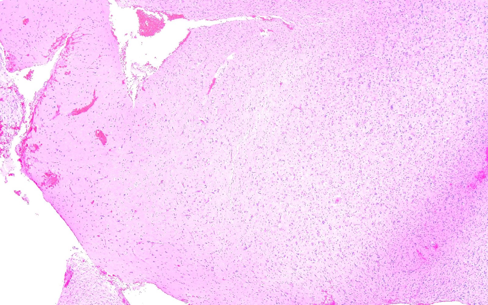
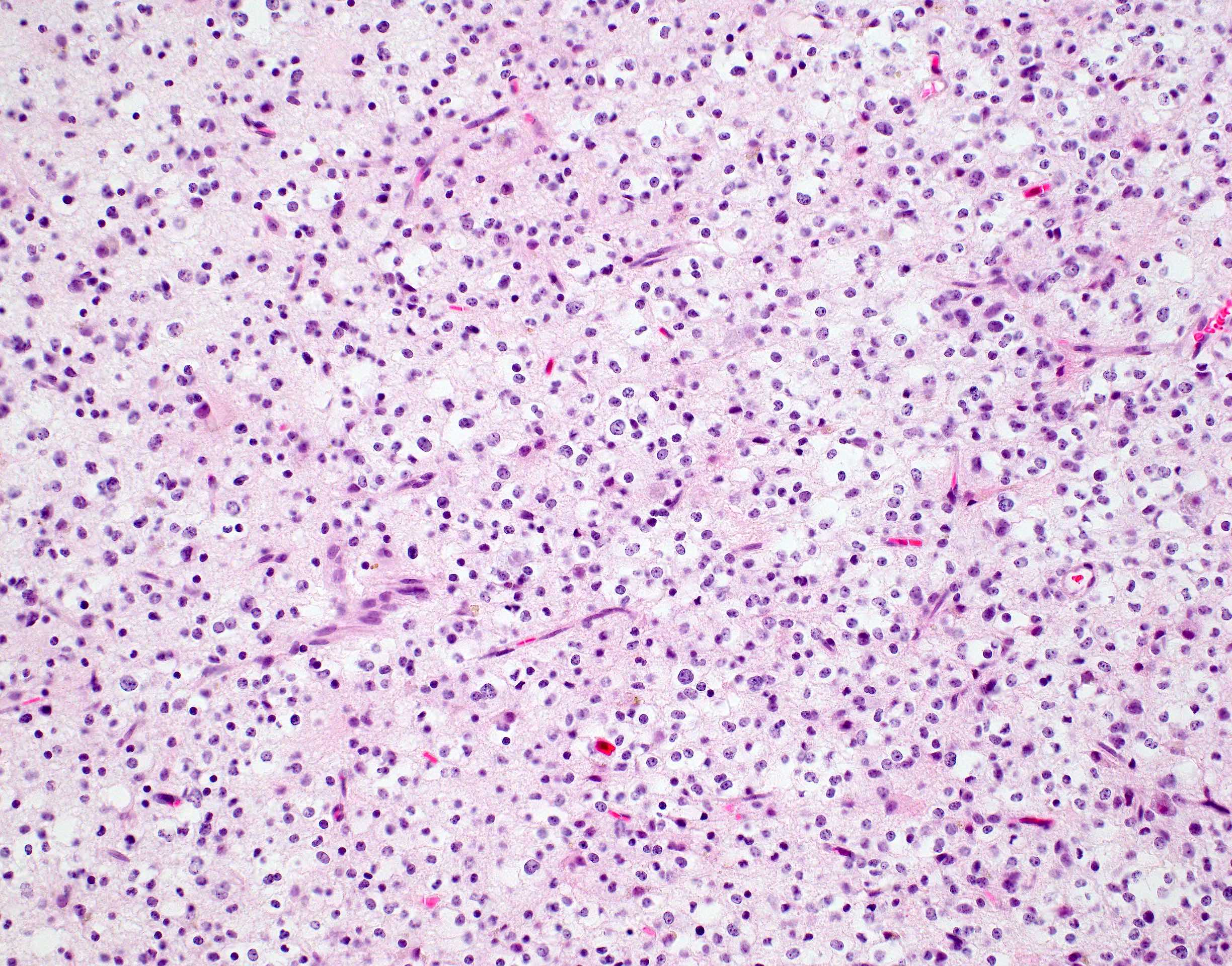
CNS WHO grade 2 astrocytoma, NOS
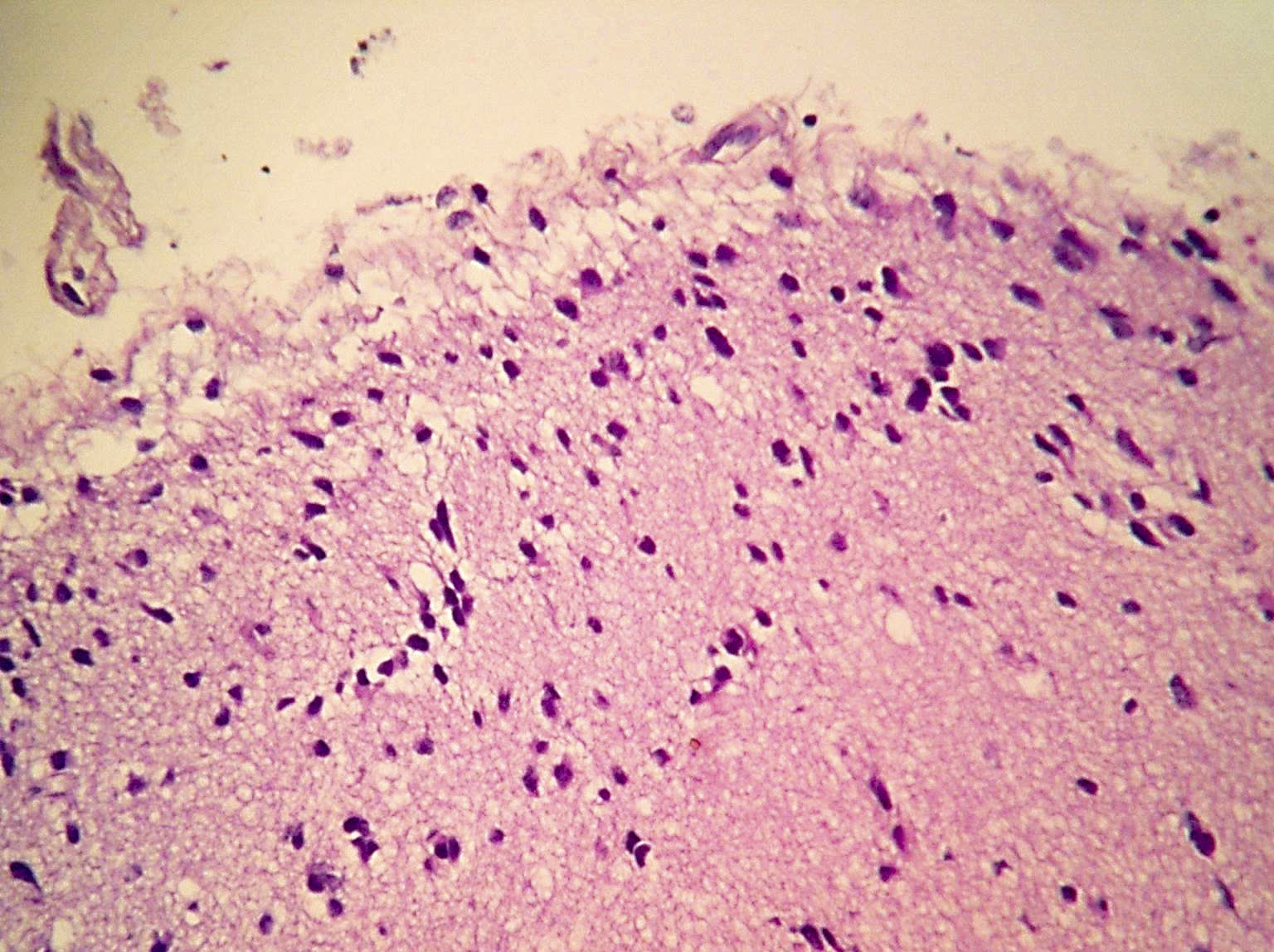
Subpial accumulation
CNS WHO grade 2 astrocytoma, NOS

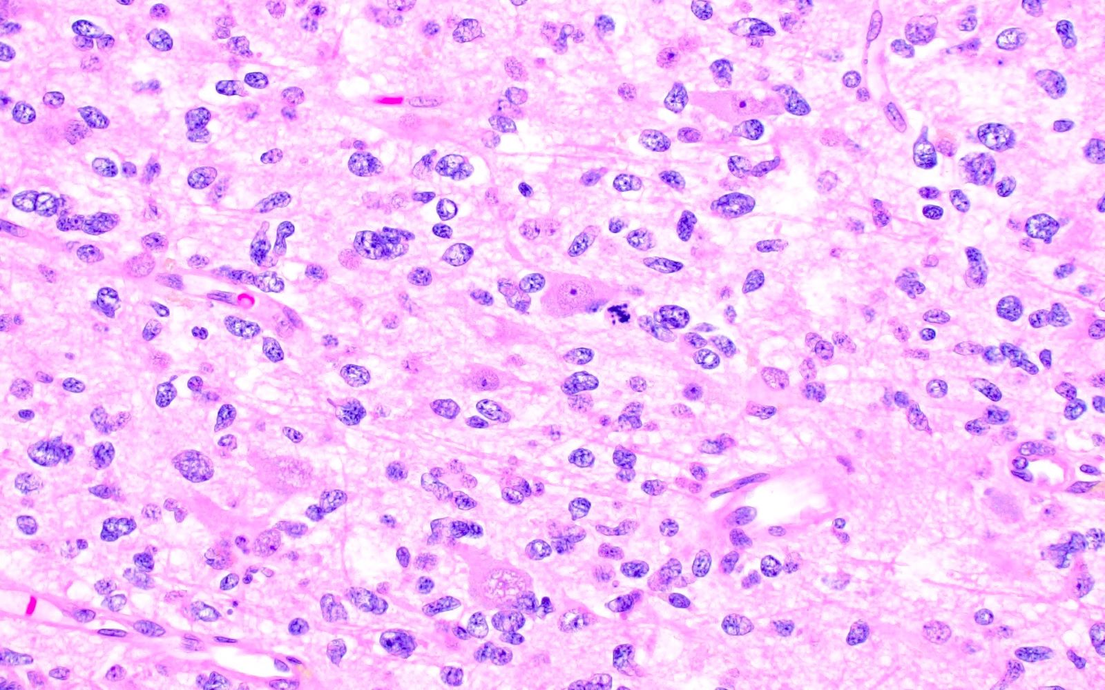
Perineuronal satellitosis
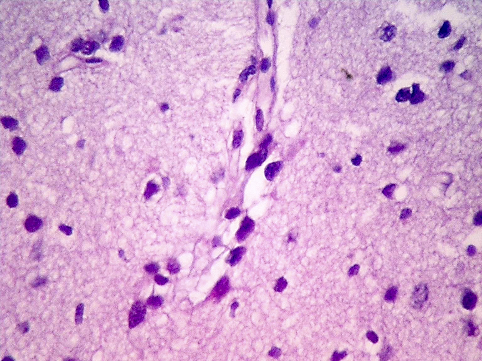
Perivascular accumulation
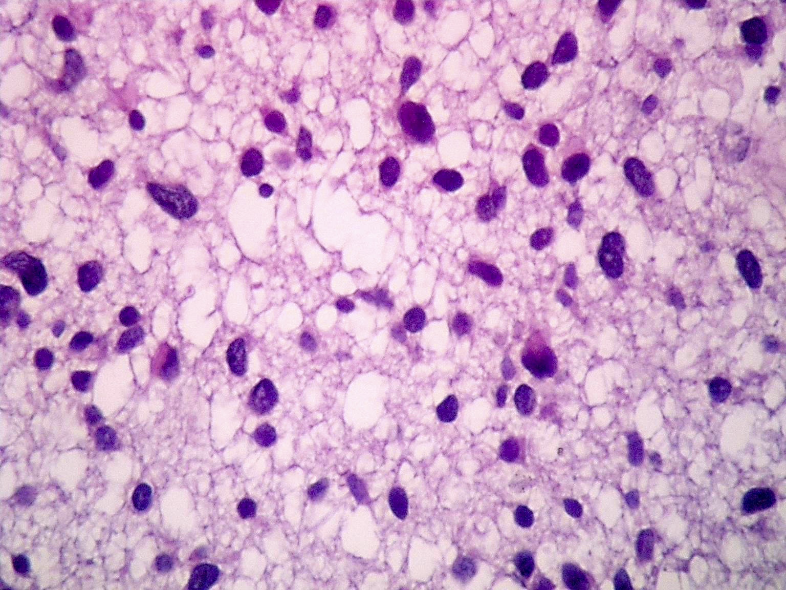
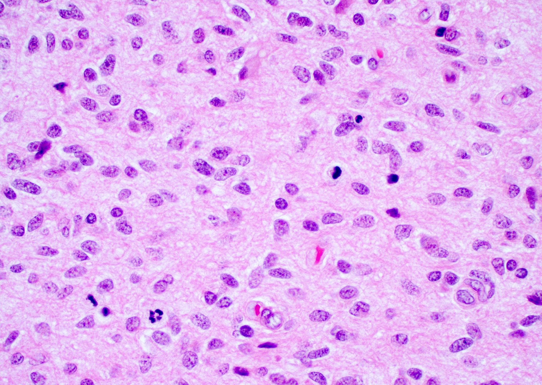
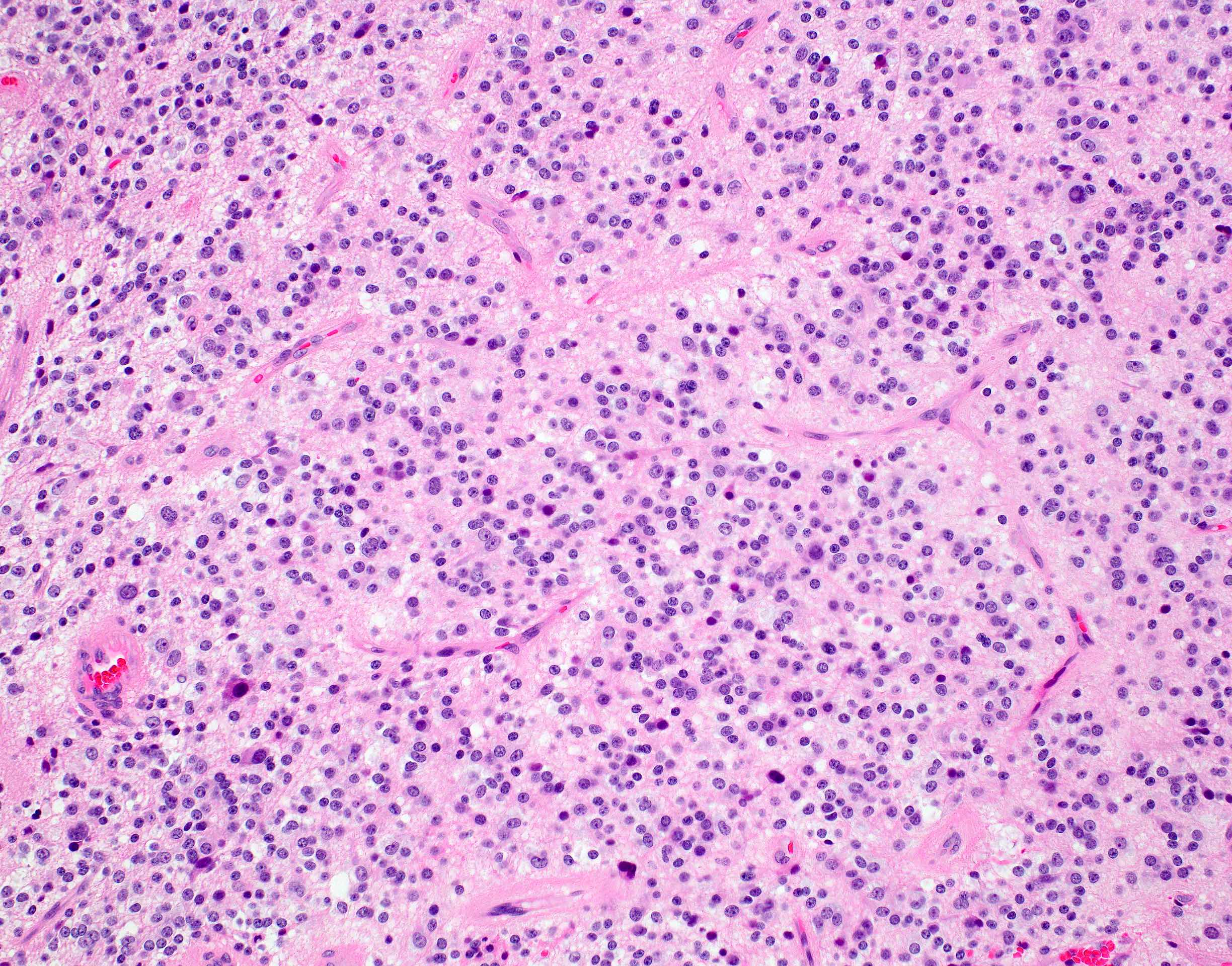
Mitotically active
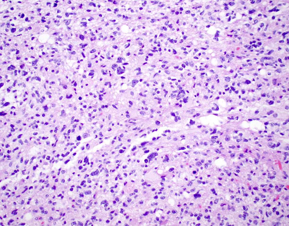
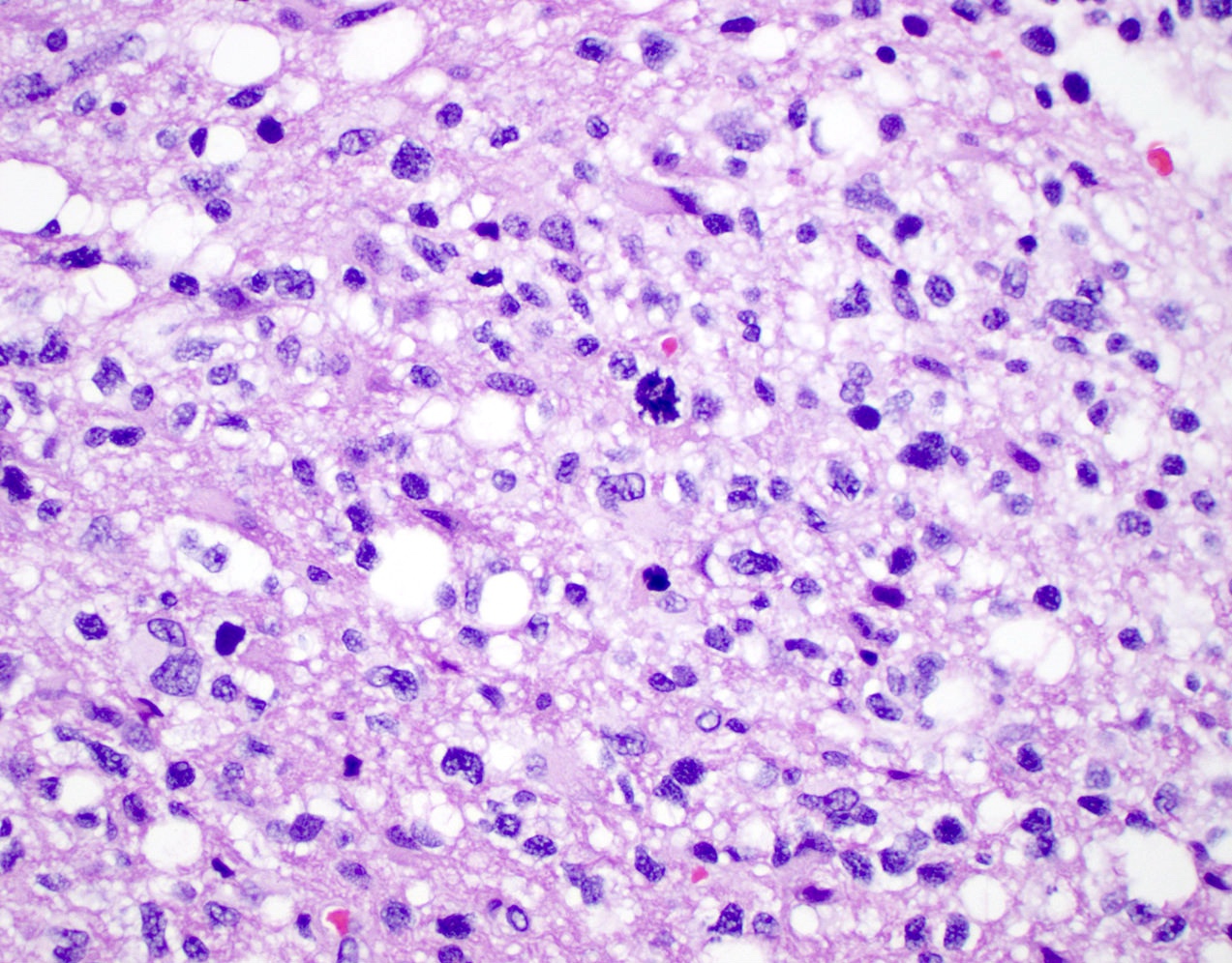
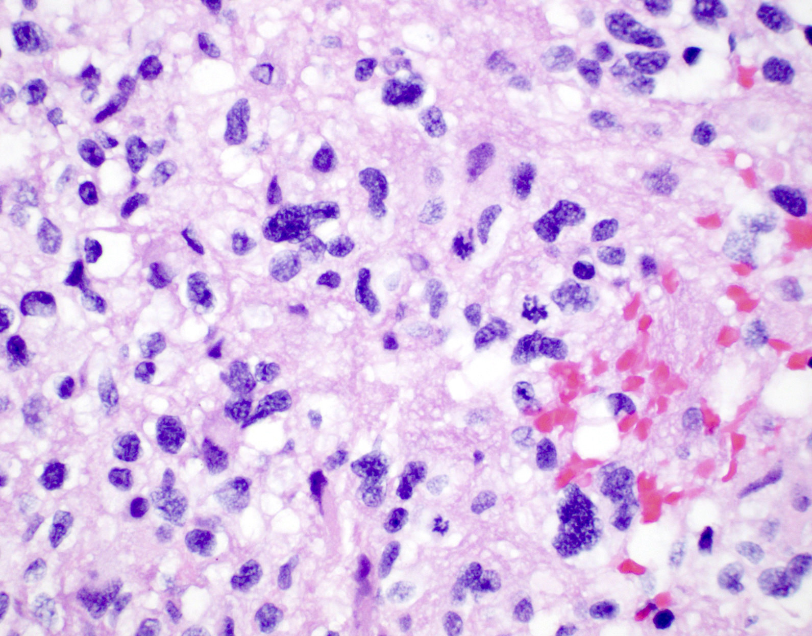
Pleomorphic hypercellular astrocytic appearance
Gemistocytic histology
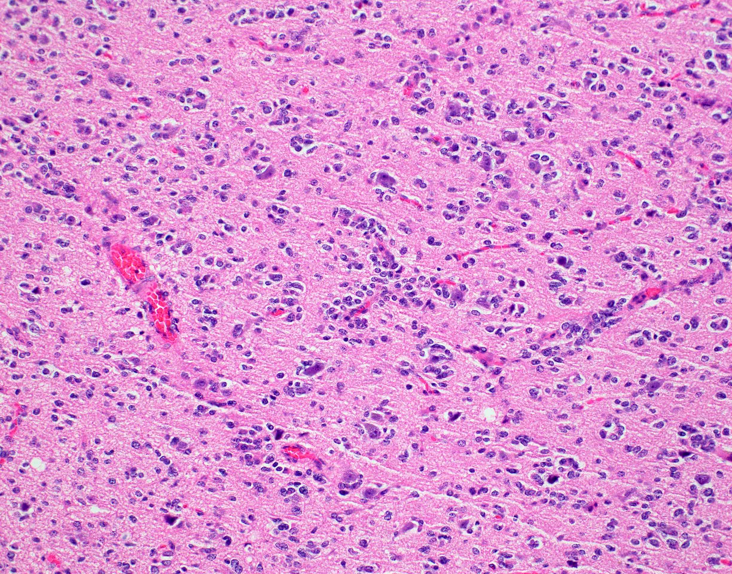
Variable morphology and necrosis
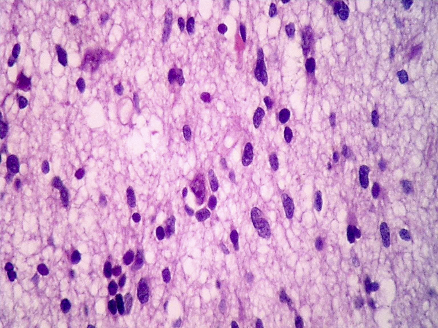
Microvascular proliferation

Entrapped neurons

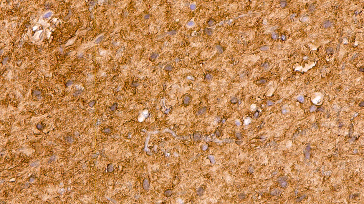
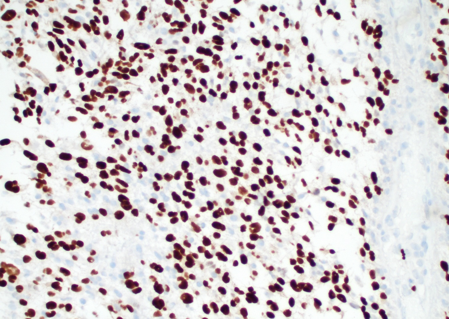
R132H IDH1

ATRX
p53
Ki67
Olig2
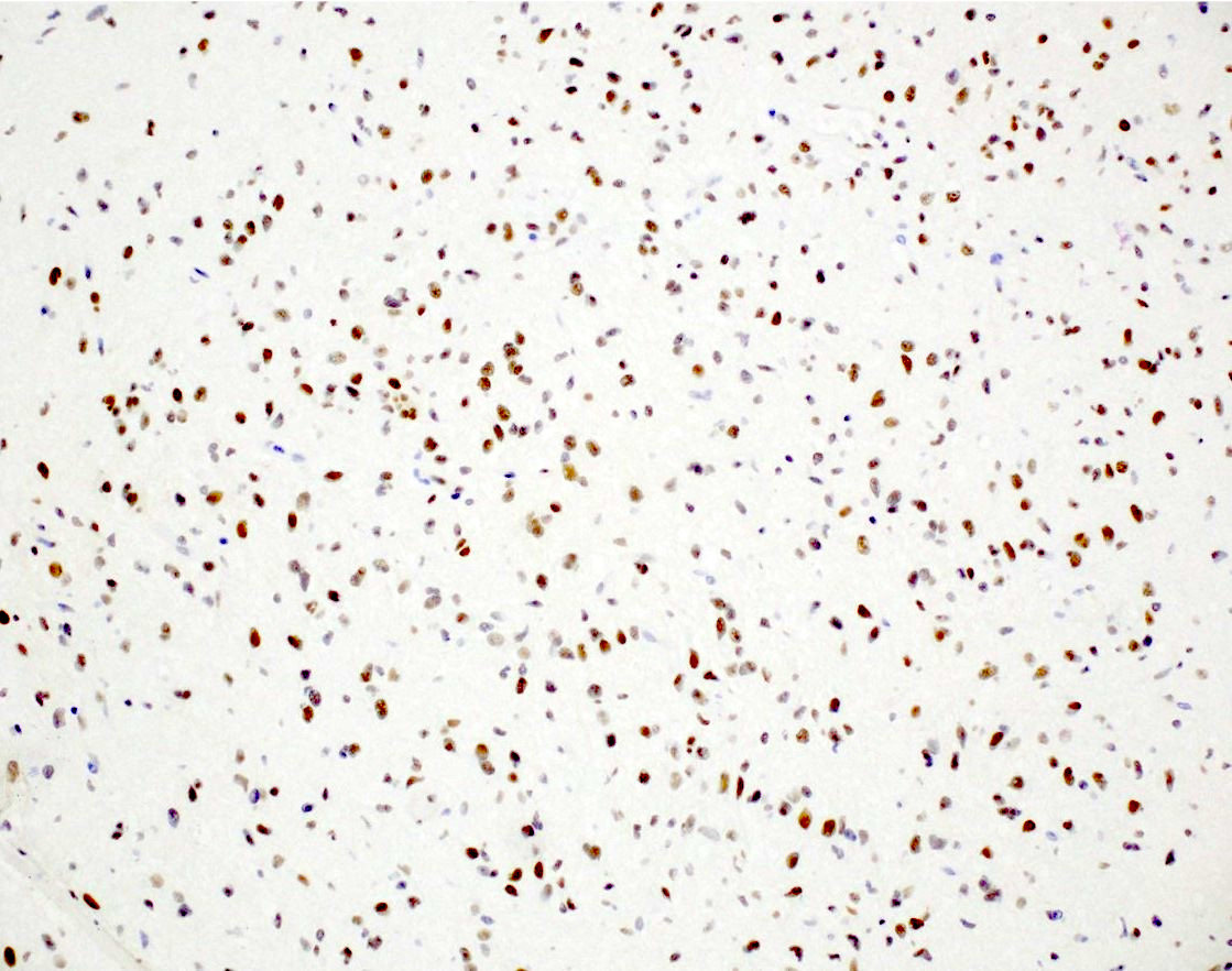
IDH

ATRX

p53
Ki67
Contributed by Chunyu "Hunter" Cai, M.D., Ph.D.
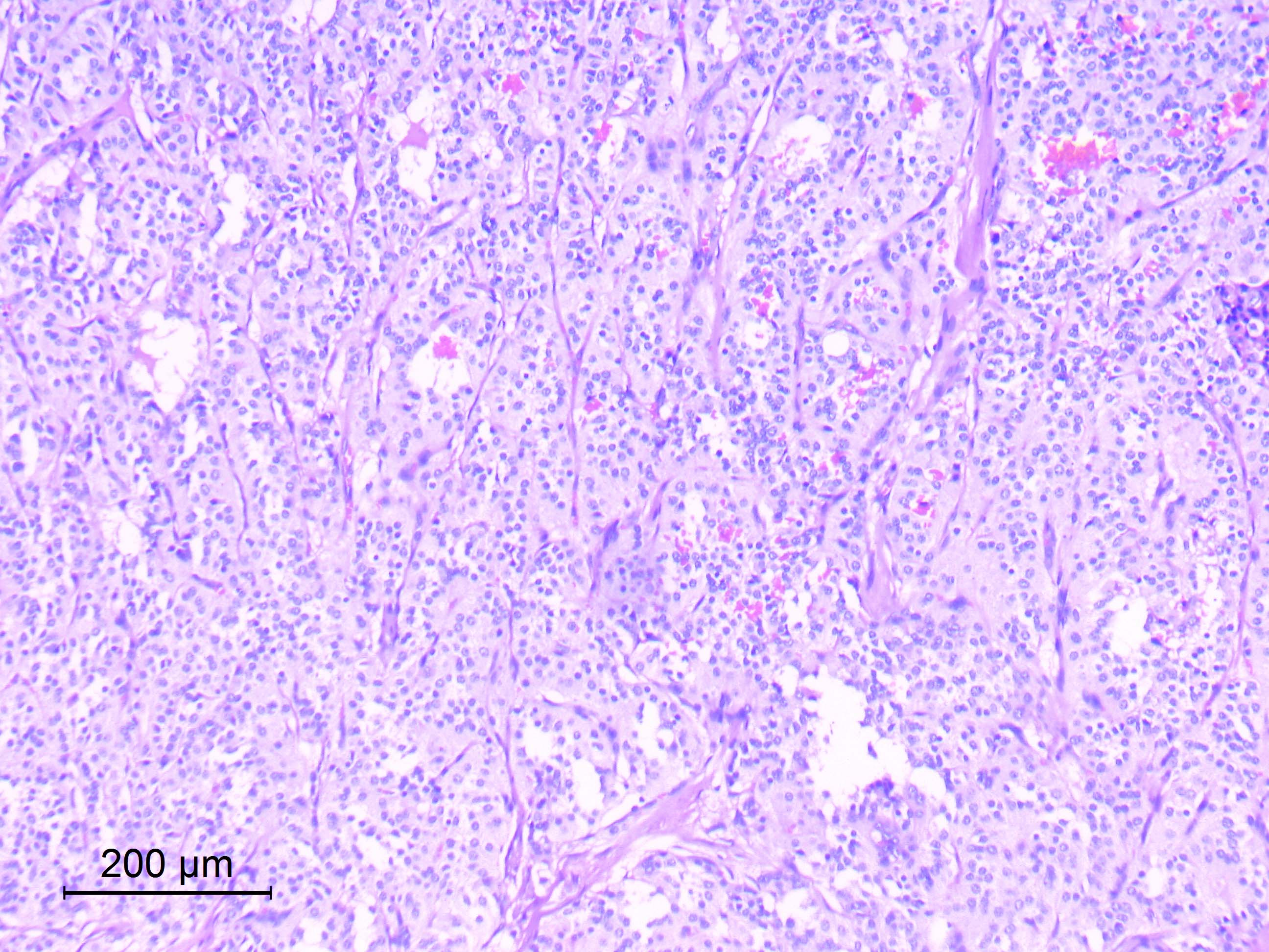
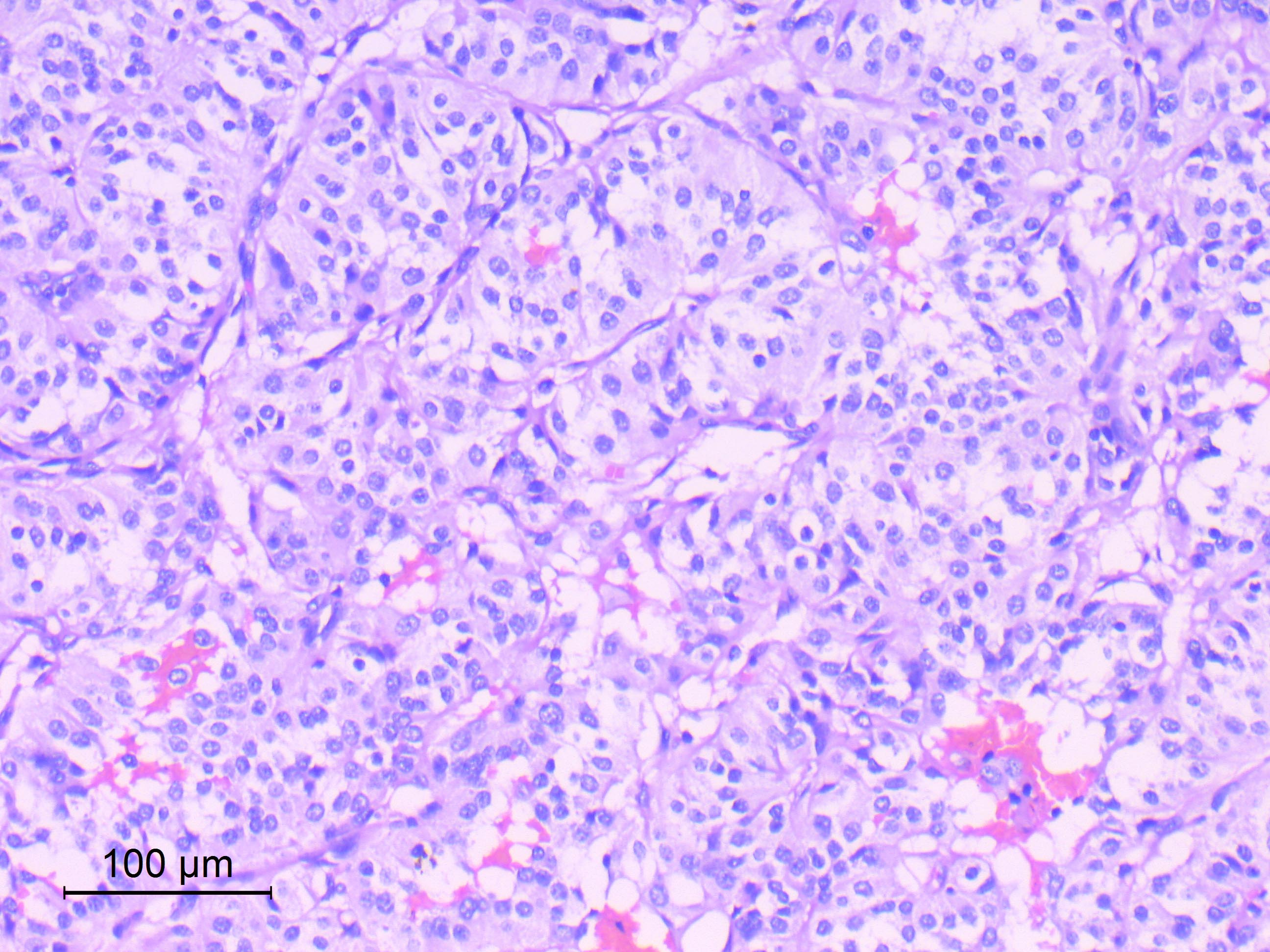
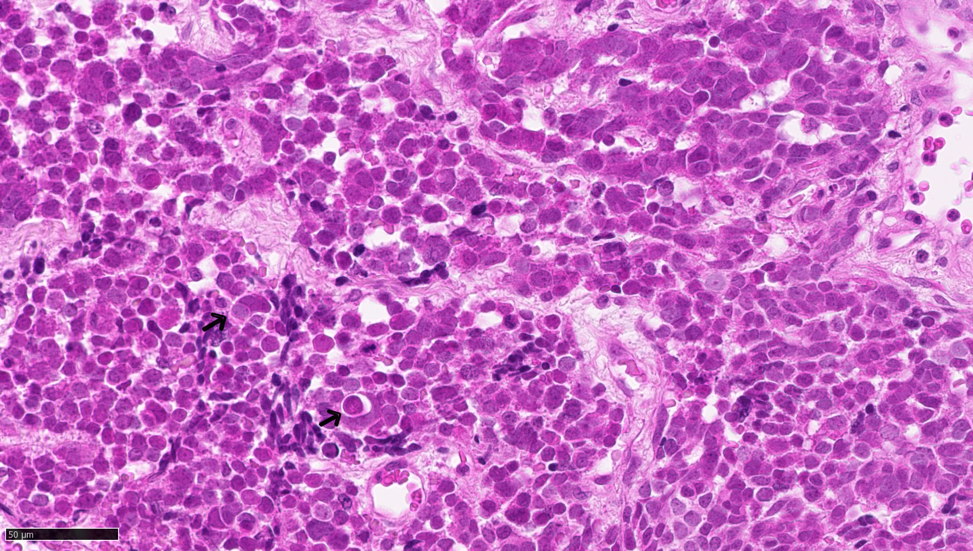
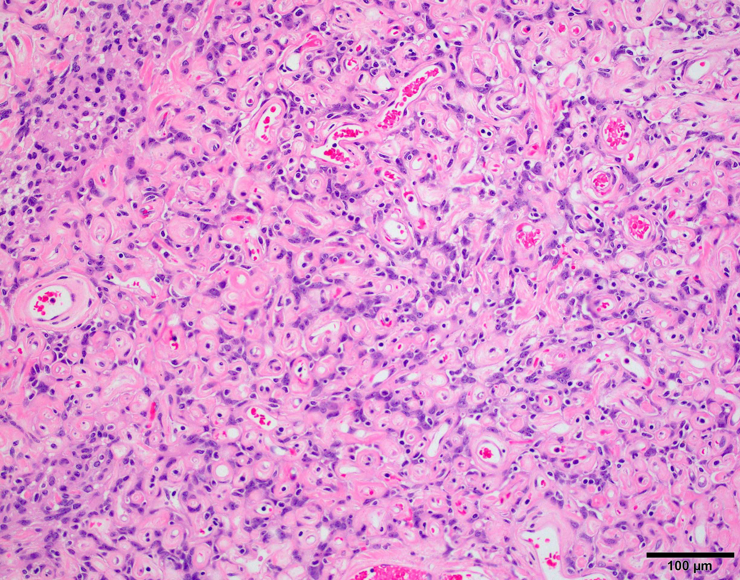
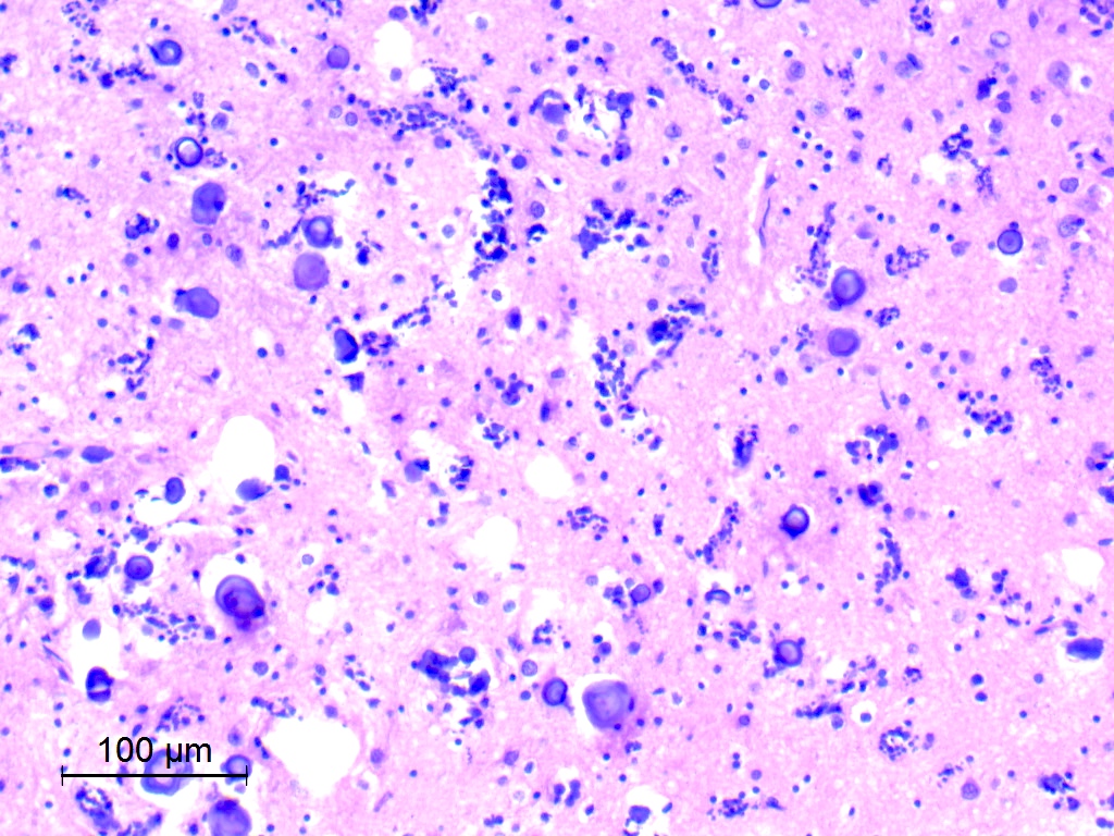
Small cell change - meningothelial
Small cell change - transitional
Spontaneous necrosis
Macronuclei
Sheeting
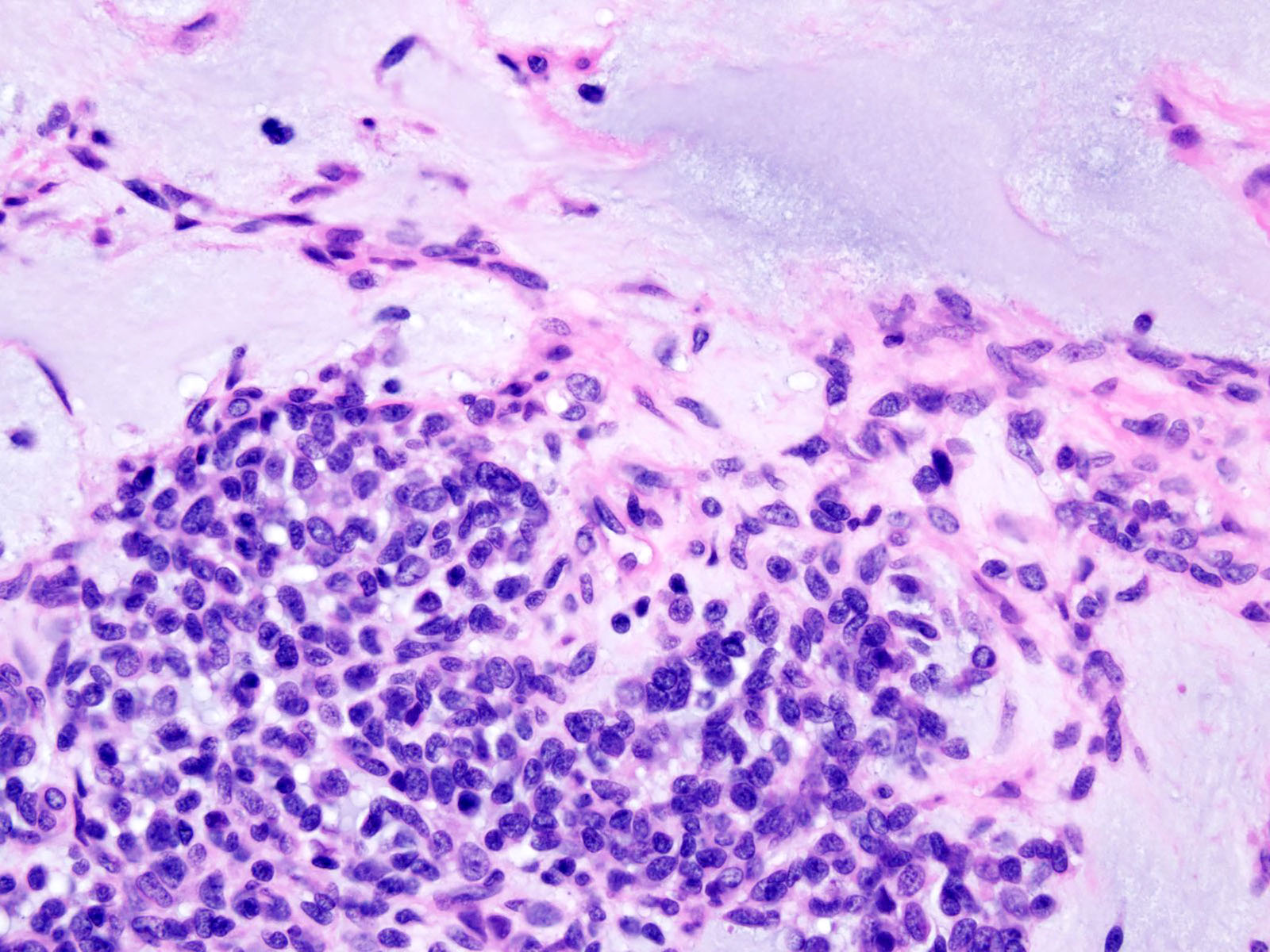
Brain invasion - nest
Brain invasion - protrusions
Images hosted on other servers:

Features of AT / RT subtypes
Contributed by Chunyu Cai, M.D., Ph.D. (Case #502)

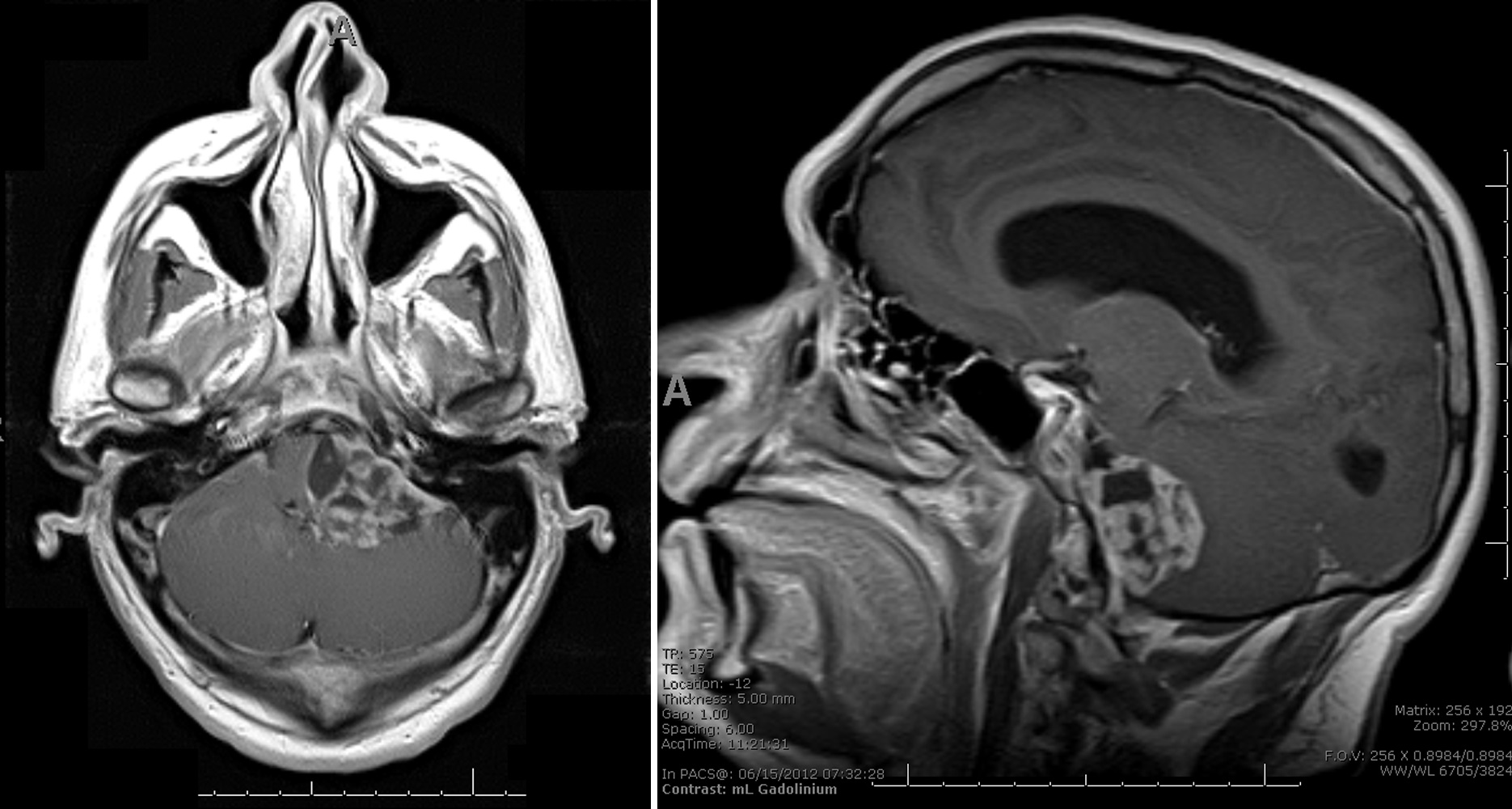
MRI T1 postsagittal
Images hosted on other servers:

AT / RT in a 9 month old

Axial CT head, unenhanced

MRI brain, unenhanced and enhanced
Images hosted on other servers:

Posterior fossa, cerebellum and brain stem tumors
Contributed by Nirupama Singh, M.D., Ph.D., Chunyu Cai, M.D., Ph.D. (Case #502) and Geling Li, M.D., Ph.D.
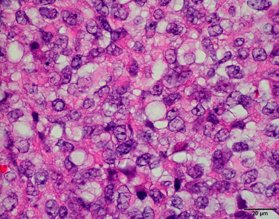
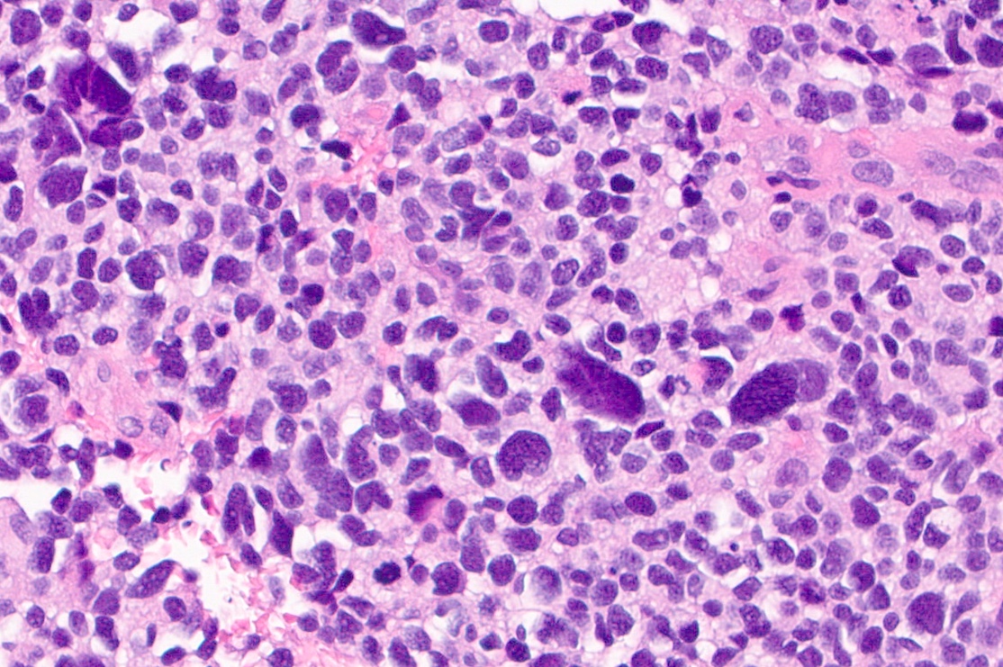
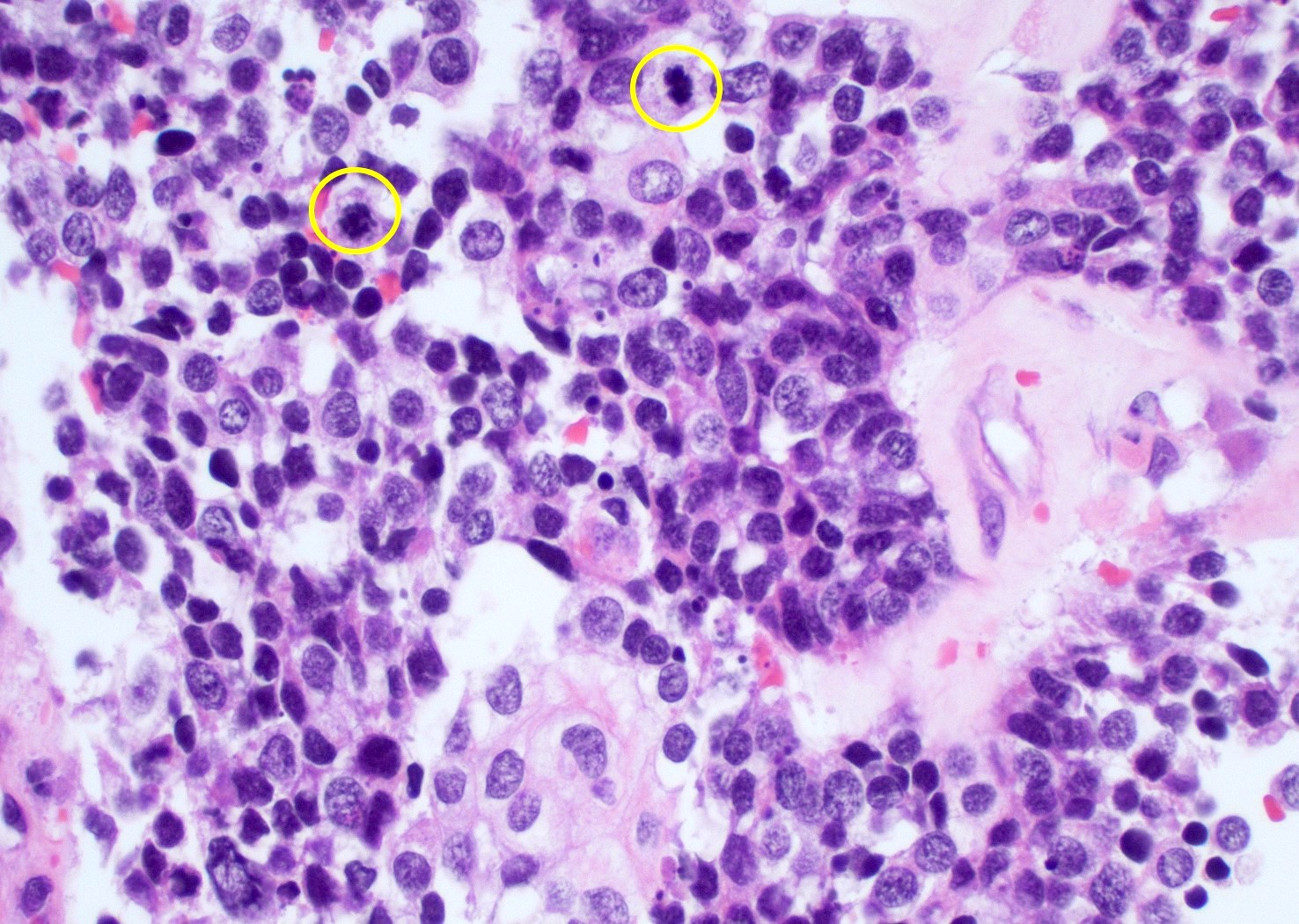
Vesicular chromatin and prominent nucleoli
Eosinophilic globs
Mitotic figures
Myxoid area
Chondroid
Geographic necrosis
Gland
Large pale cell
Rhabdoid cell
Small blue cell
Densely packed immature cells
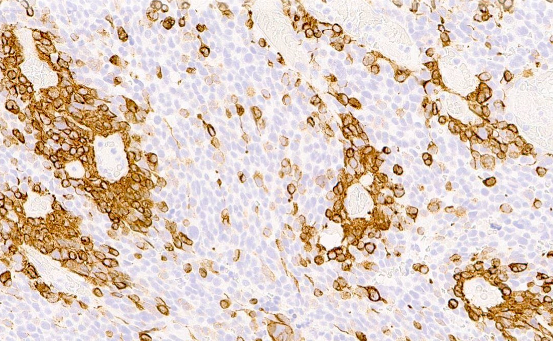
INI1
Images hosted on other servers:

Rhabdoid cell
Images hosted on other servers:

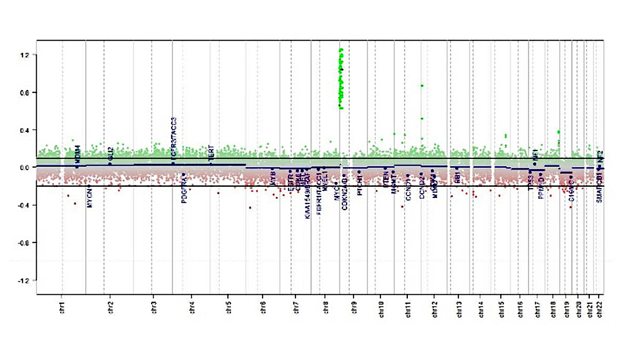
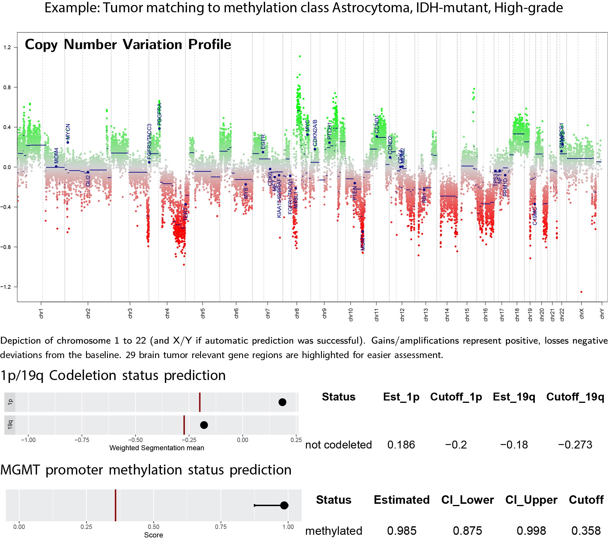
Methylation array analysis

Cluster analysis
Contributed by Borislav Alexiev, M.D.
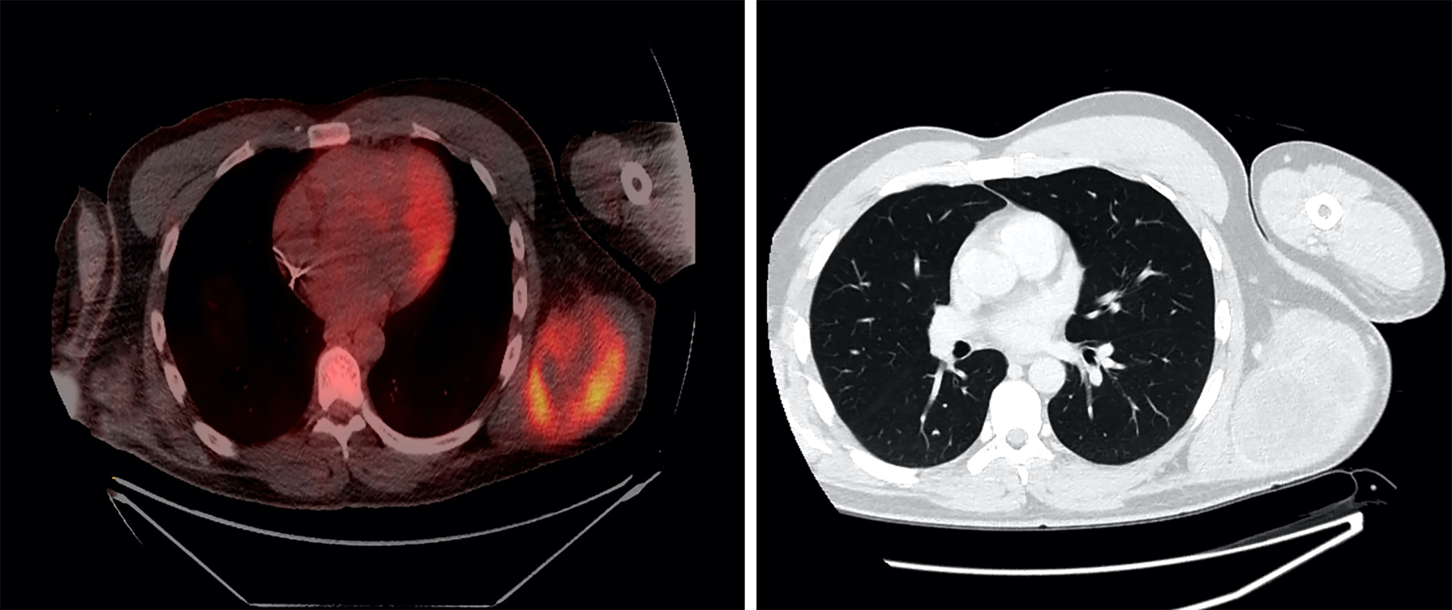
PET / CT
Contributed by Borislav Alexiev, M.D.
Soft tissue mass
Contributed by Borislav Alexiev, M.D.

Chest wall mass
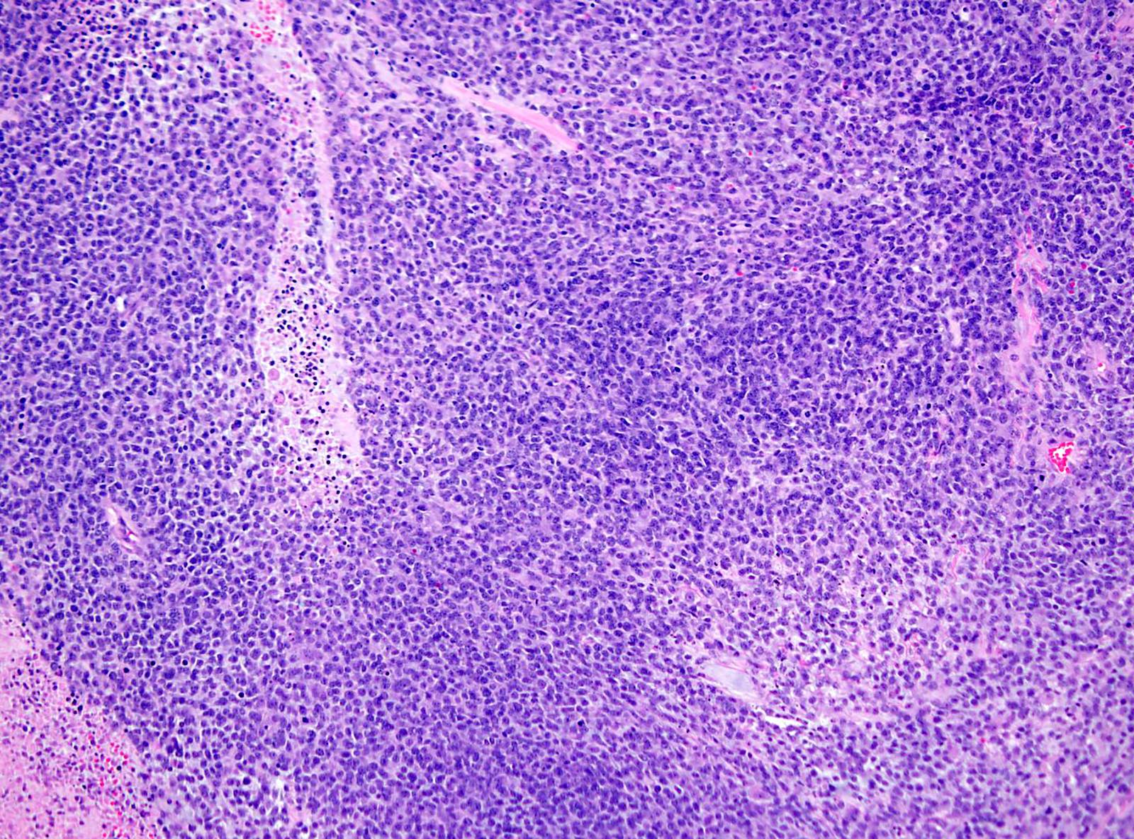
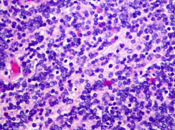
Small round cell morphology
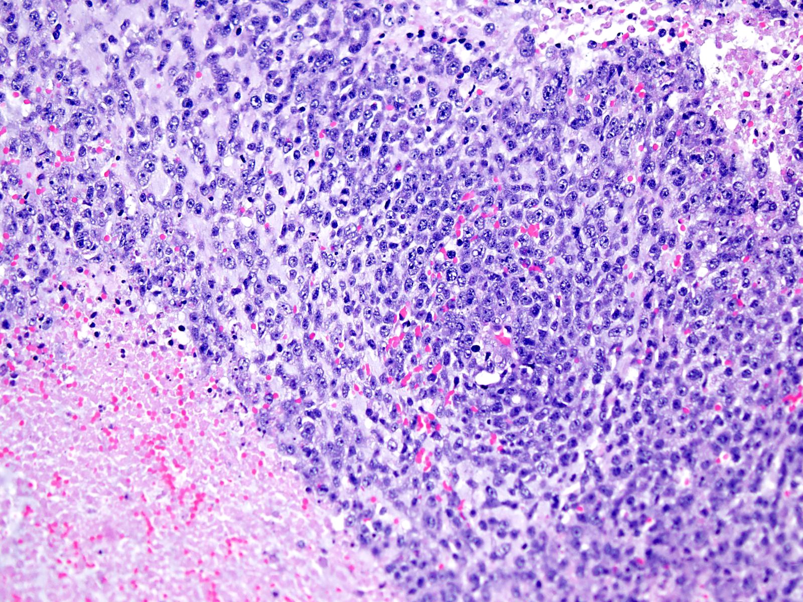
Solid growth pattern
Spindle cell morphology
Mitoses
Myxoid stroma
CD99 expression
WT1 expression
MYC expression
Contributed by Lawrence J. Jennings, M.D., Ph.D.
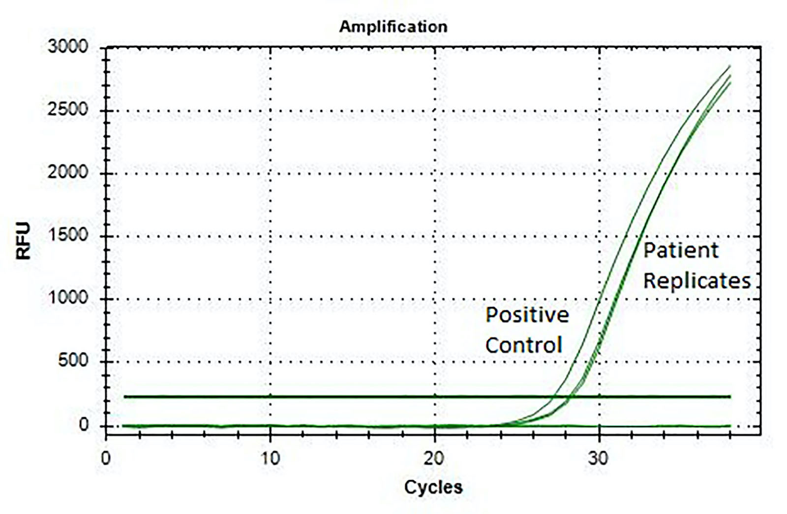
Real time RT-PCR
Images hosted on other servers:

T2 weighted and postcontrast T1 weighted axial
Contributed by Nirupama Singh, M.D., Ph.D., Carmen Perrino, M.D. and P.J. Cimino, M.D., Ph.D.
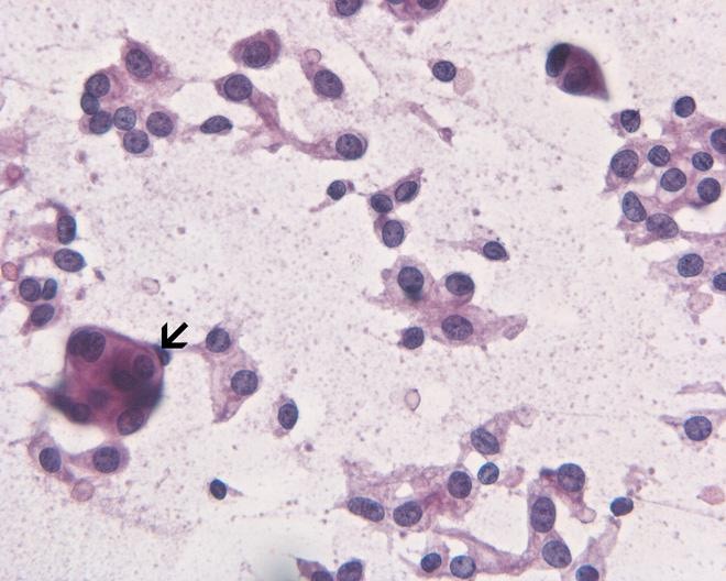
Neuroblasts with no cytoplasm
Homer Wright rosettes
Small tumor cells
Pseudorosettes
Embryonal cells

Neurocytic cells

Focal ganglion cells
Necrosis
MVP
Images hosted on other servers:

Recurrent molecular alterations
Case presentation of CNS NB FOXR2
Images hosted on other servers:

MRI

Intradural enhancing lesion

Spinal angiography

Scalloping of lumbar vertebrae
Images hosted on other servers:

Intraoperative

Lesion after durotomy

Hypervascular well marginated mass
Images hosted on other servers:

Lesion after en block removal

Hemorrhagic cystic change
Contributed by Eman Abdelzaher, M.D., Ph.D.


Encapsulated lesion
Zellballen architecture
Zellballen architecture

Salt and pepper chromatin

Focal sclerosis
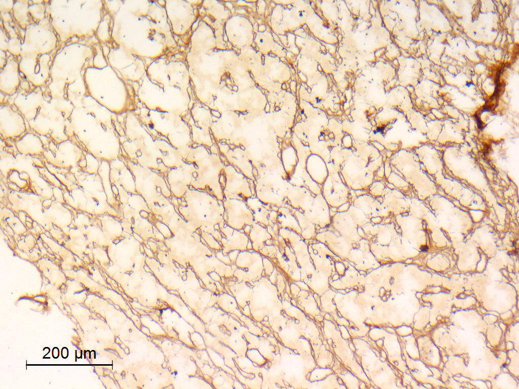
Reticulin

Synaptophysin

S100 positive sustentacular cells
S100 positive chief and sustentacular cells

GFAP negative

GFAP positive sustentacular cells
EMA
Low Ki67
Images hosted on other servers:

Numerous perinuclear dense core granules

Prominent rough endoplasmic reticulum and Golgi apparatus
Images hosted on other servers:

DNA methylation cluster analysis of CEPs and other tumors

Chromosomal copy number analysis
Case 8: paraganglioma of the filum terminale
Images hosted on other servers:

Coronal section diagram
Images hosted on other servers:

Intraventricular mass on MRI

Calcifications on CT

T1+C MRI
Images hosted on other servers:

Endoscopic view of intraventricular tumor
Images hosted on other servers:

Intraventricular mass
Contributed by Rebecca Yoda, M.D.
Circumscribed border
Fibrillary matrix
Perinuclear clearing
Perivascular pseudorosette
Neurocytic rosettes
Linear arrangement

Calcifications
Branching capillaries
Hyalinized vessels

Lipomatous change
Necrosis
Mitotic activity
Synaptophysin IHC
NeuN IHC
GFAP IHC
Contributed by Nazar M. T. Jawhar, M.D. (Case #119)
Circumscribed border
Perivascular pseudorosettes
Perinuclear clearing
Rosettes and perivascular pseudorosettes
Calcification
Linear cellular arrangement
Uniform, round cells
Images hosted on other servers:

TEM showing neuronal features

Ultrastructure recapitulating neuropil and synapses
Images hosted on other servers:

DNA methylation based classification
Contributed by Valeria Barresi, M.D., Ph.D.

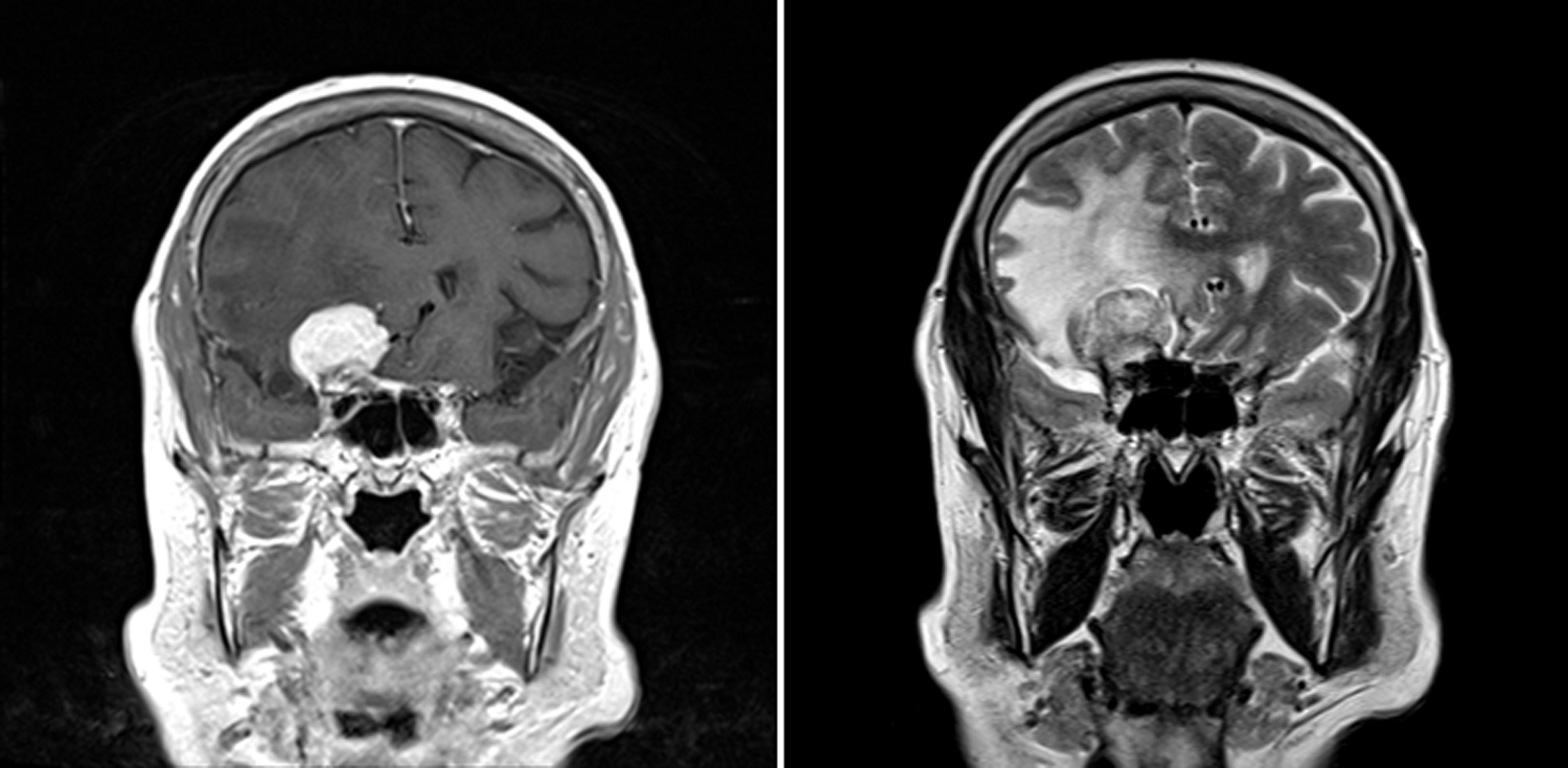
T1 weighted MRI
T2 weighted MRI
Images hosted on other servers:

Right cerebellopontine angle mass
Contributed by Valeria Barresi, M.D., Ph.D.

Lipidized cells
Perivascular matrix
Synaptophysin immunostaining
NeuN immunostaining
GFAP immunostaining
Olig2 immunostaining
EMA immunostaining
Ki67 immunostaining
Contributed by Valeria Barresi, M.D., Ph.D.
Sheets of uniform neurocytic cells
Images hosted on other servers:

Axial CT scan reveals hyperdense mass

Various images
Images hosted on other servers:

Optic chiasm enlargement
Contributed by Valeria Barresi, M.D., Ph.D.
T1 weighted sagittal MRI

T1 sagittal contrast enhancement
T2 weighted axial MRI

FLAIR axial MRI
Contributed by Valeria Barresi, M.D., Ph.D.
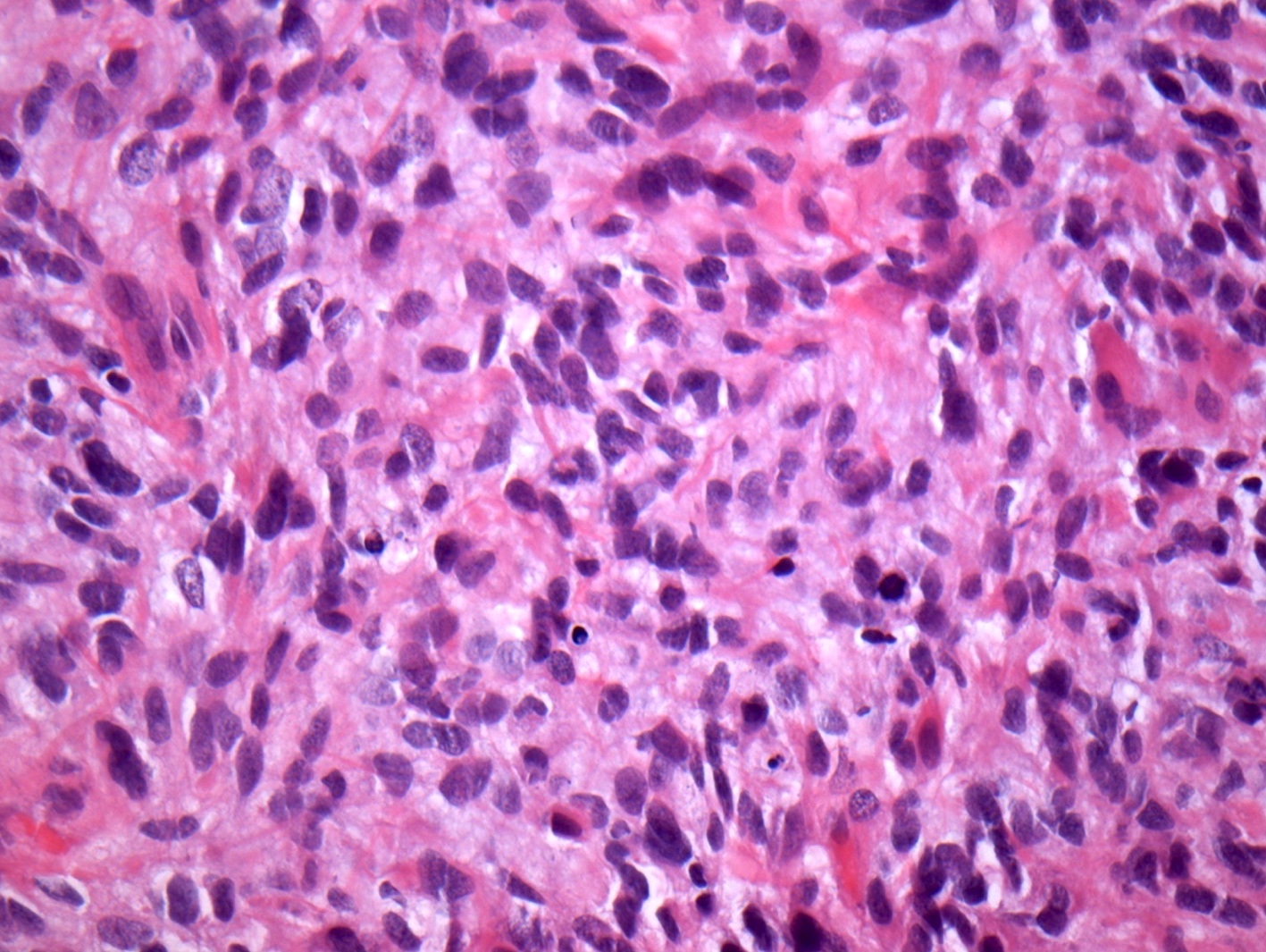
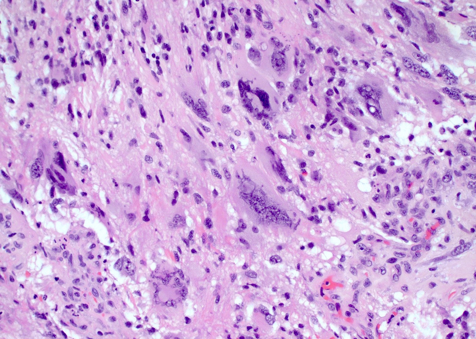
Epithelioid cells
Spindle cells
Intermingled typical meningioma
Mitoses

Alcian blue
Podoplanin immunostaining
Images hosted on other servers:

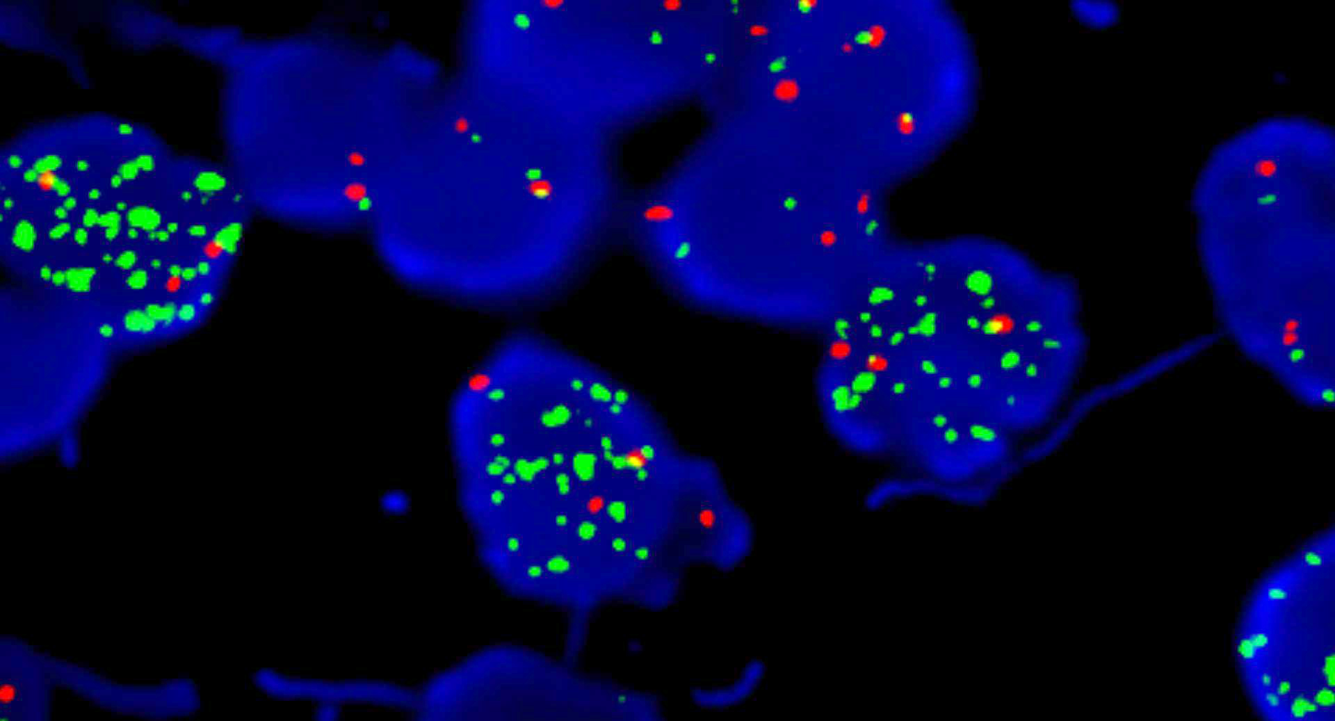
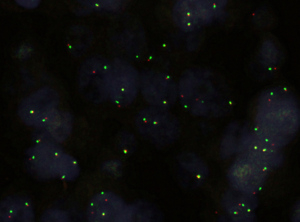
FISH 18p, 22q, 14q, 1p
Images hosted on other servers:

MR shows small cyst
Images hosted on other servers:

Cyst at foramen of Monro
Contributed by Kshitij Mankad, M.D.
MRI coronal
MRI sagittal
MRI transverse
Images hosted on other servers:

Fourth ventricle tumor
Contributed by Ashirwad Merve, M.B.B.S., Ph.D.

Choroid plexus papilloma
Ki67 in choroid plexus papilloma
Atypical choroid plexus papilloma
Ki67 in atypical choroid plexus papilloma
Choroid plexus carcinoma
Ki67 in choroid plexus carcinoma
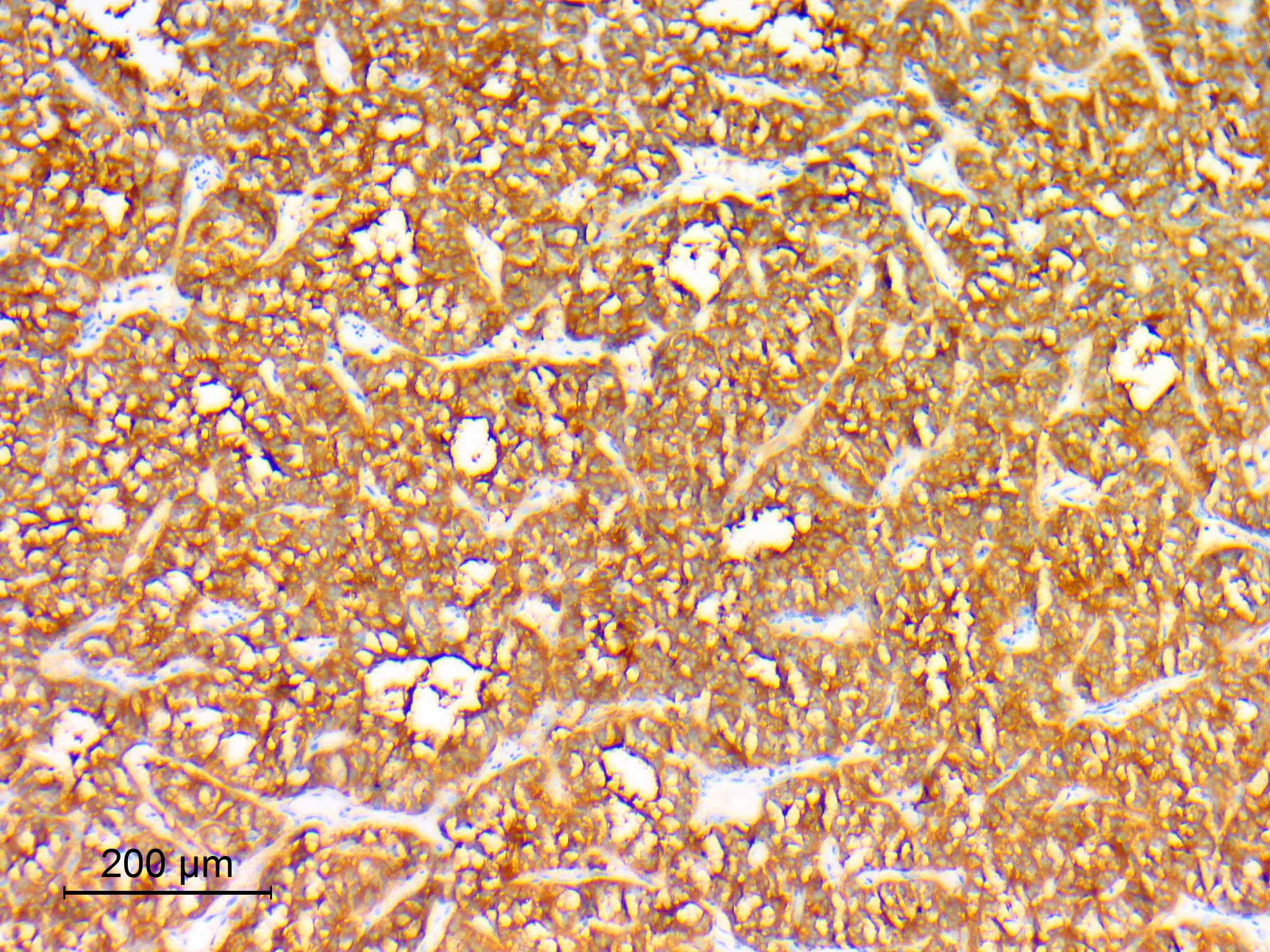
Transthyretin
Cytokeratin
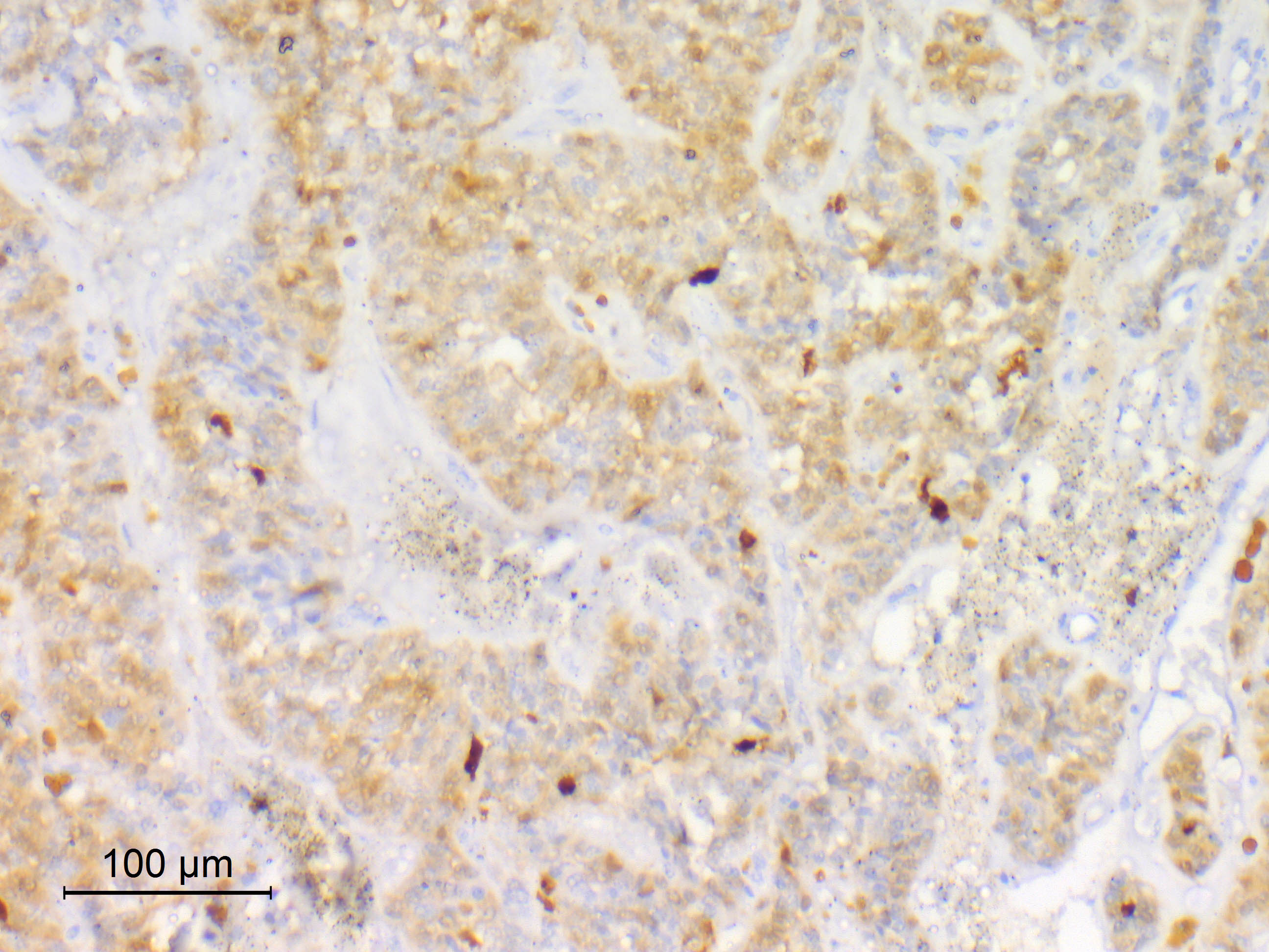
SMARCB1 (INI1)
Contributed by Ashirwad Merve, M.B.B.S., Ph.D.

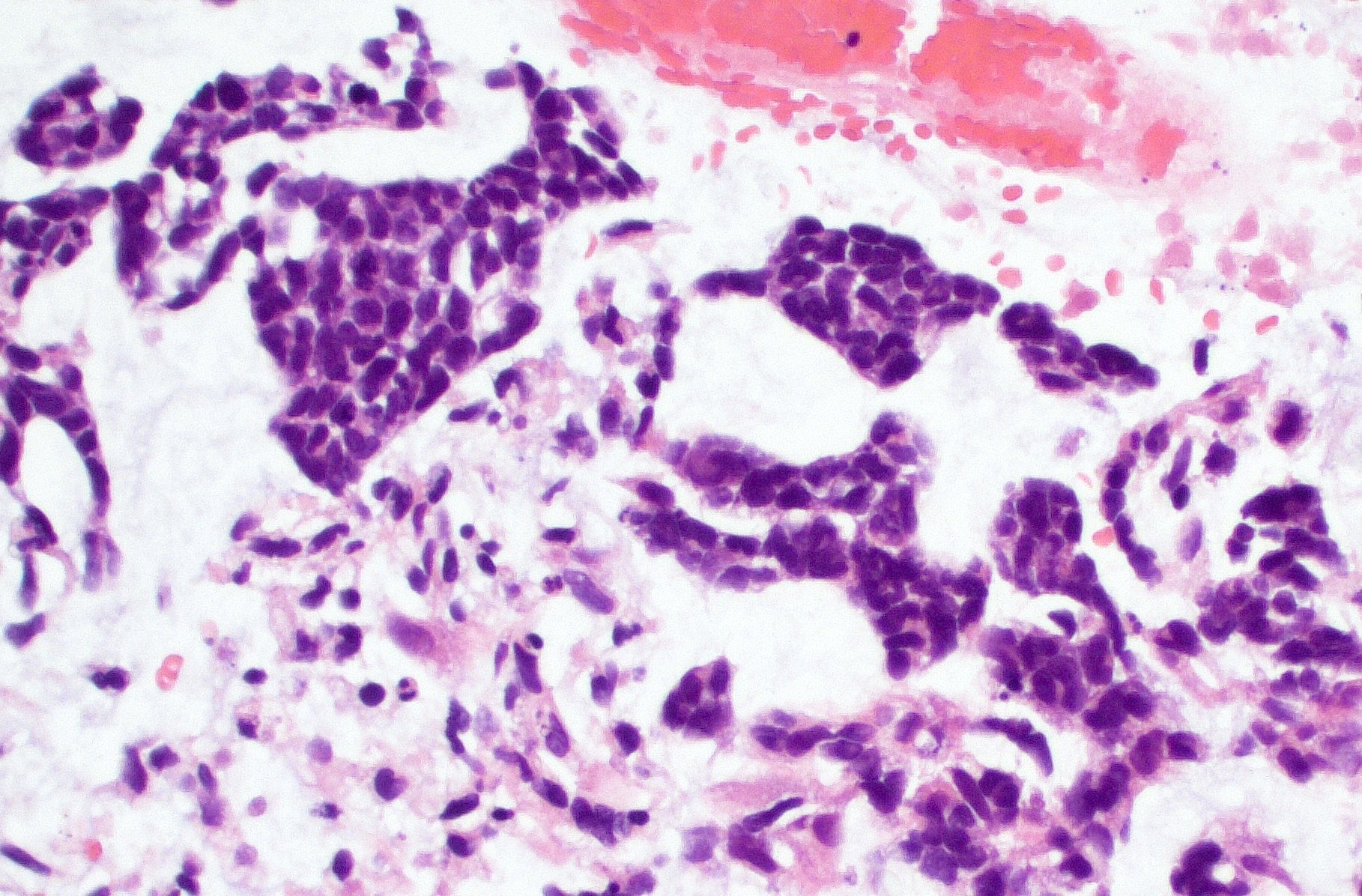
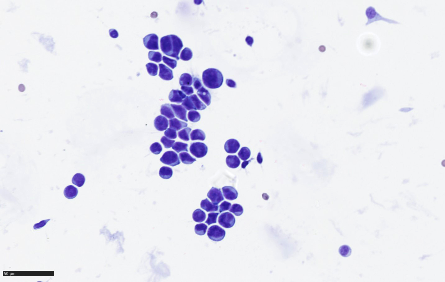
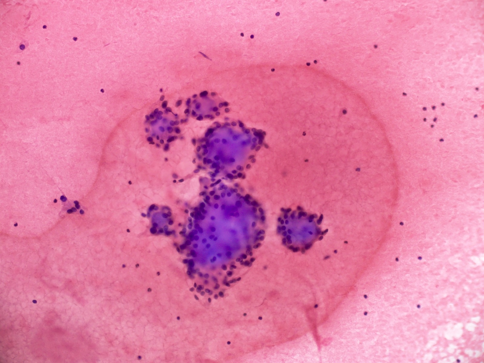
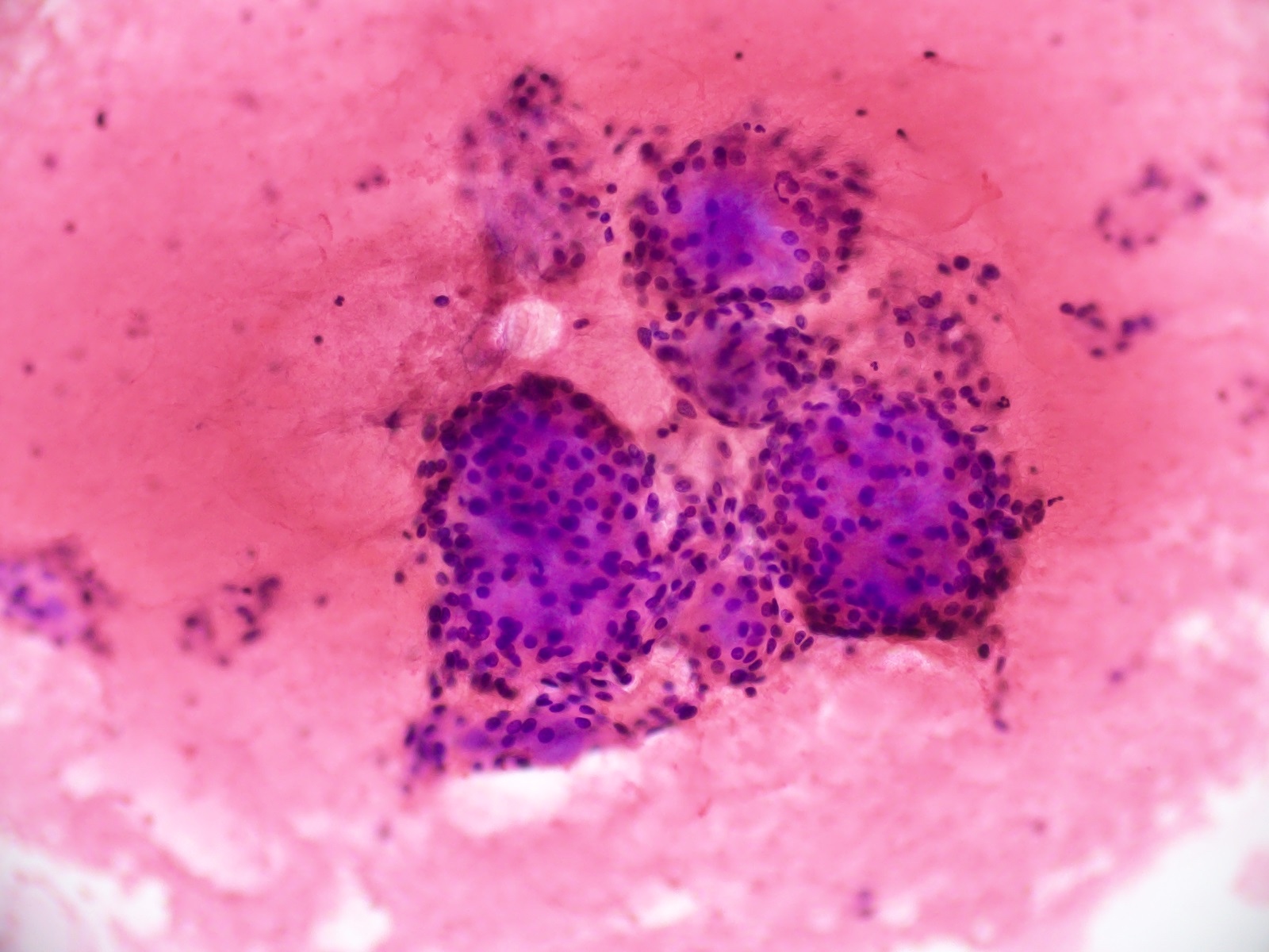
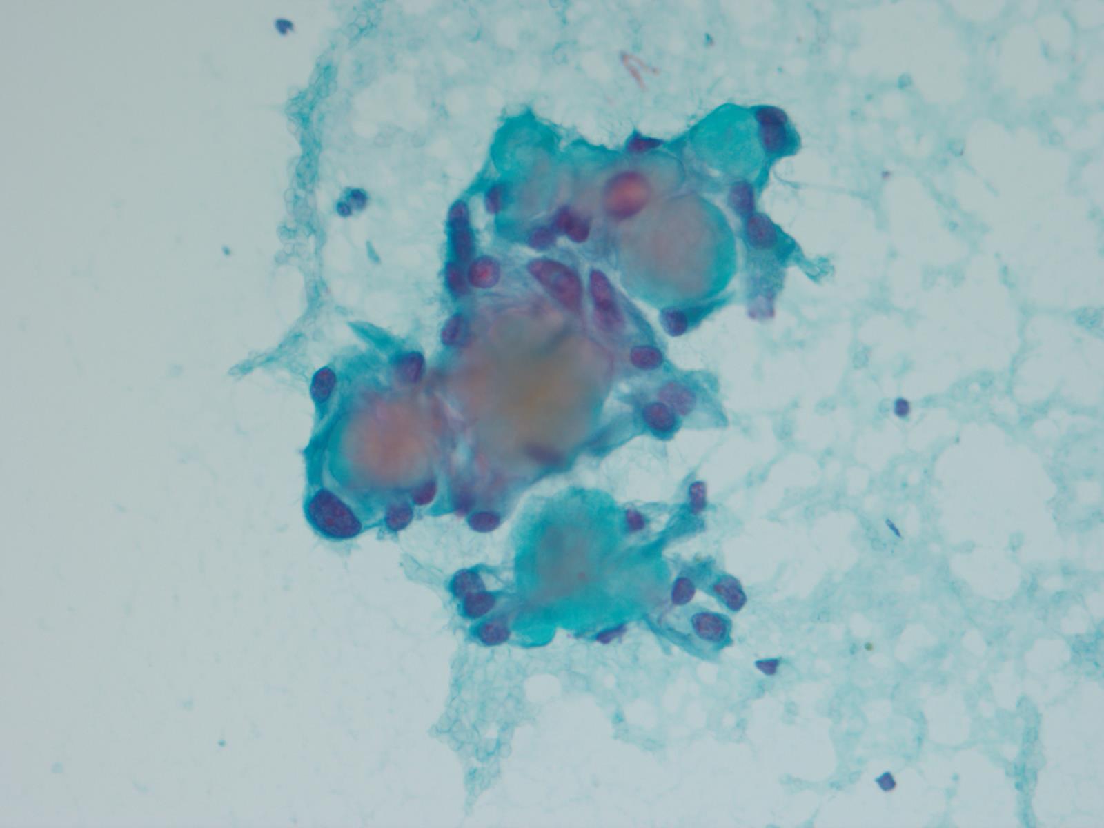
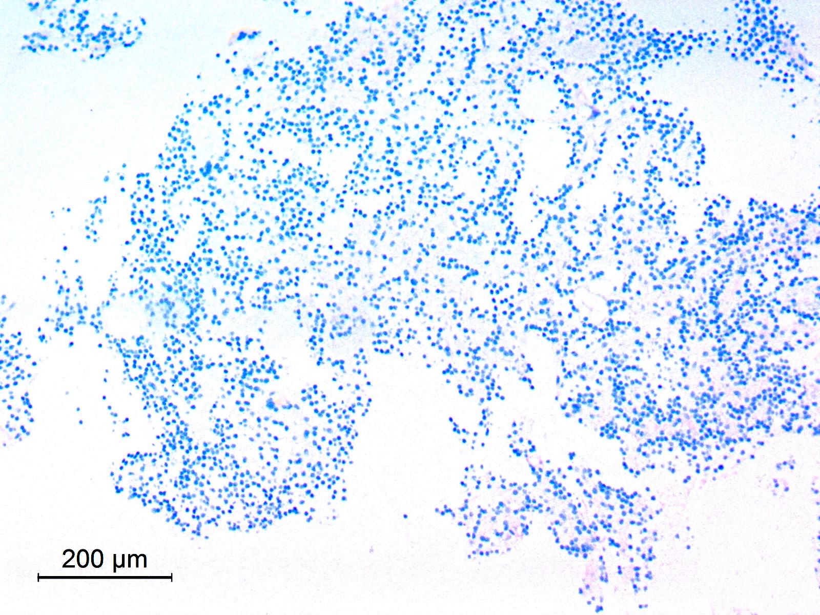
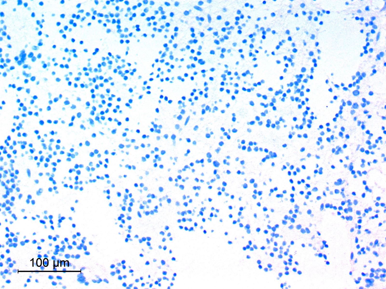
Intraoperative smear
Contributed by Ashirwad Merve, M.B.B.S., Ph.D.
Methylation array copy number variation plot
Contributed by Chunyu Cai, M.D., Ph.D. (Case #514)
MRI
Images hosted on other servers:

Various images
Contributed by Chunyu Cai, M.D., Ph.D. (Case #514)
H&E clear cell

H&E blocky collagen
IHC SMARCE1
IHC CAIX
Images hosted on other servers:

Mechanisms of sudden death due to colloid cyst

Typical location of colloid cyst
Contributed by Mohamed Kayed, M.D., Ph.D.
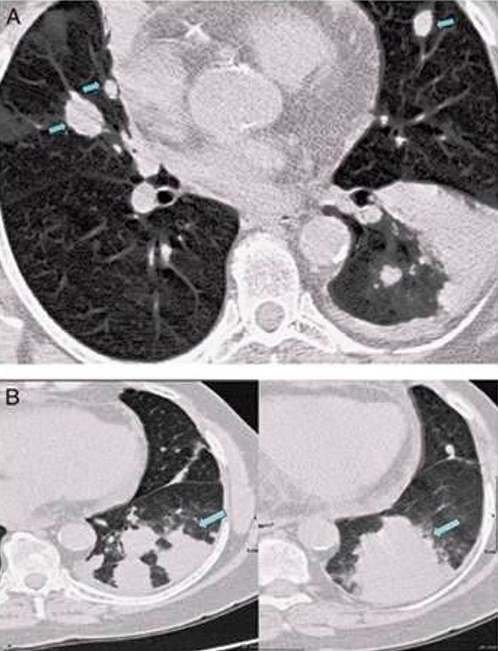
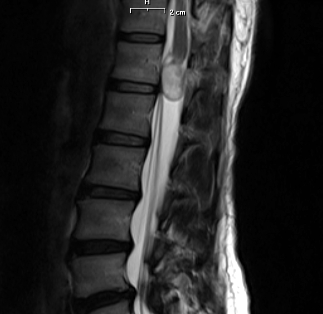
Small colloid cyst, T1 sagittal MRI
Small colloid cyst, T2 sagittal MRI

Large colloid cyst, MRI

Large colloid cyst, CT
Images hosted on other servers:

Extensive chronic hydrocephalus

Large colloid cyst

T1 hyperintense colloid cyst
Images hosted on other servers:

Intraoperative view of a colloid cyst
Images hosted on other servers:

2 cm colloid cyst


Endoscopically excised colloid cyst
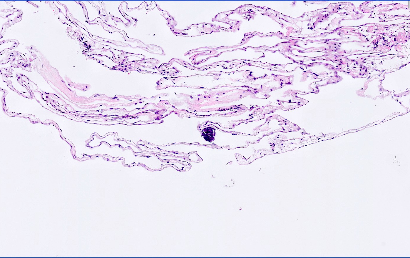
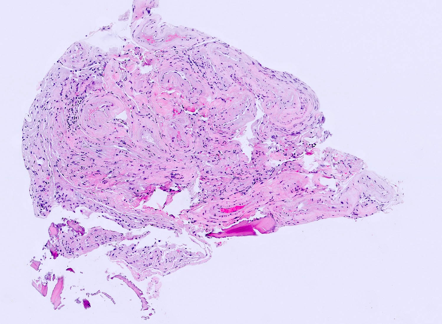
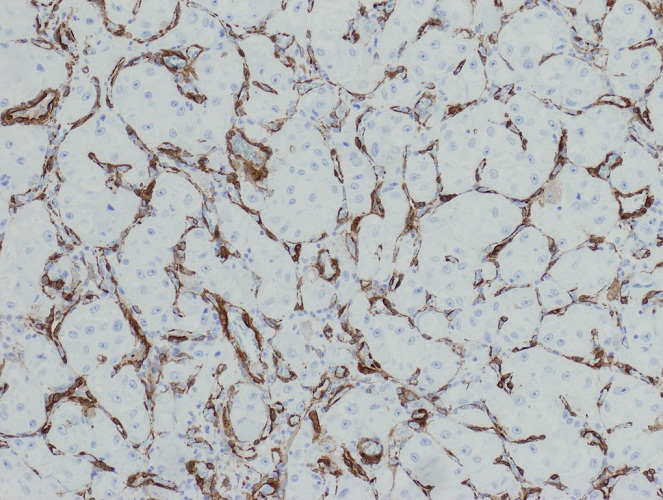
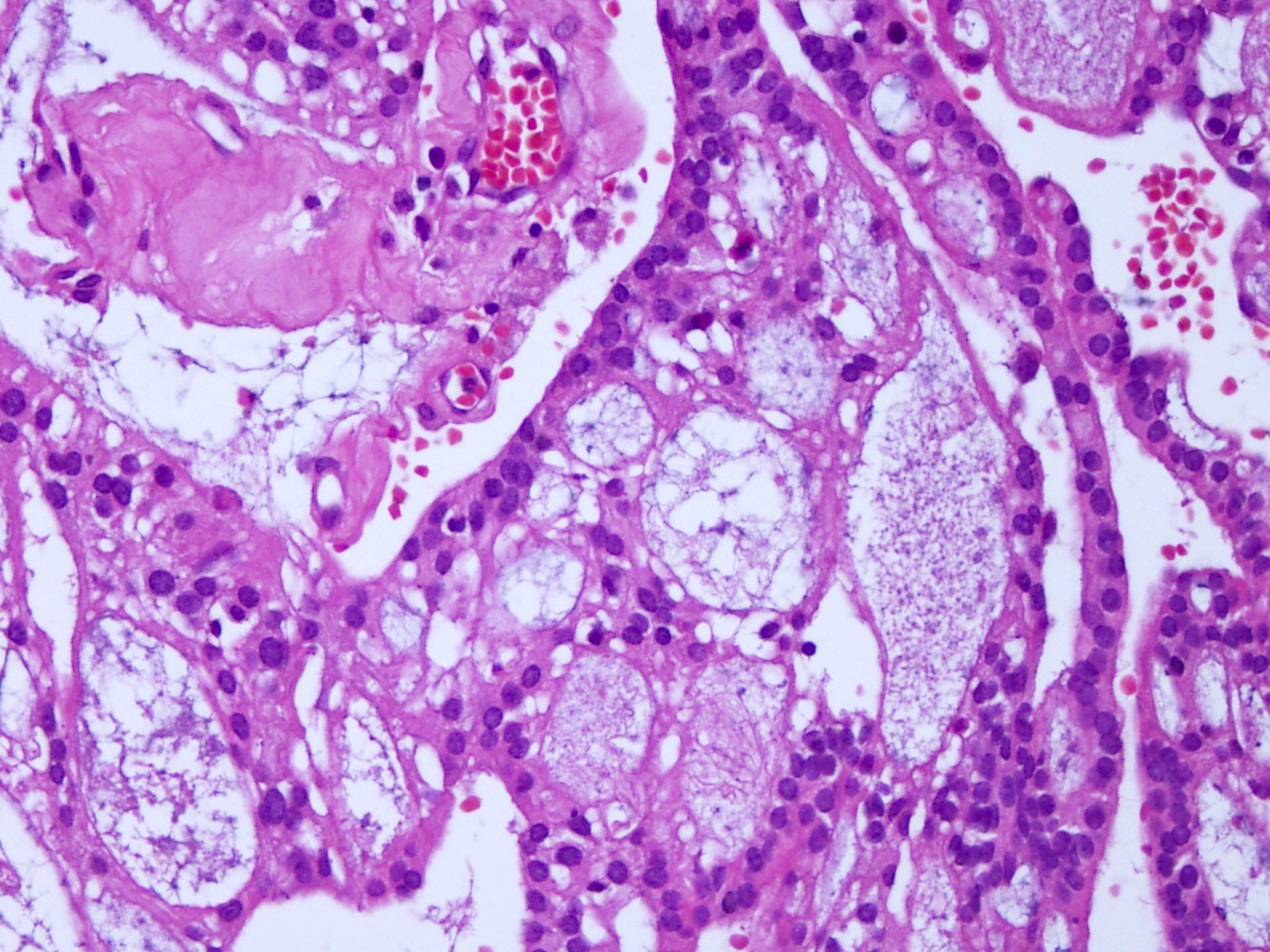
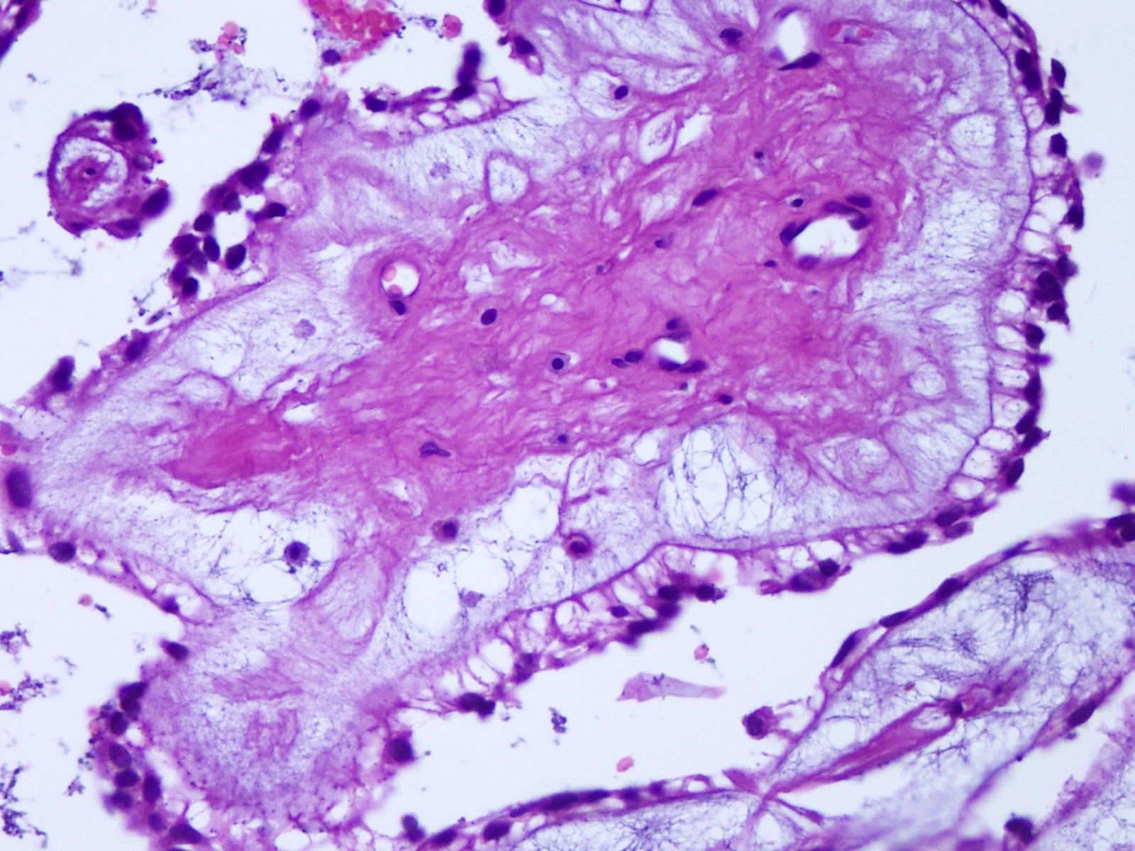
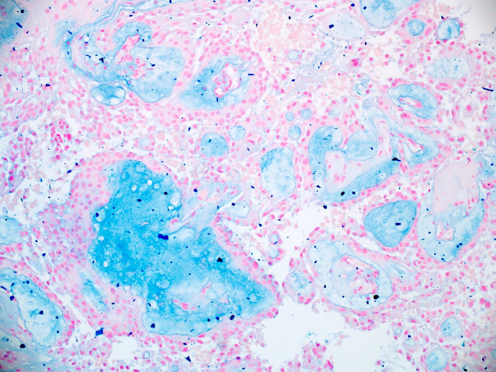
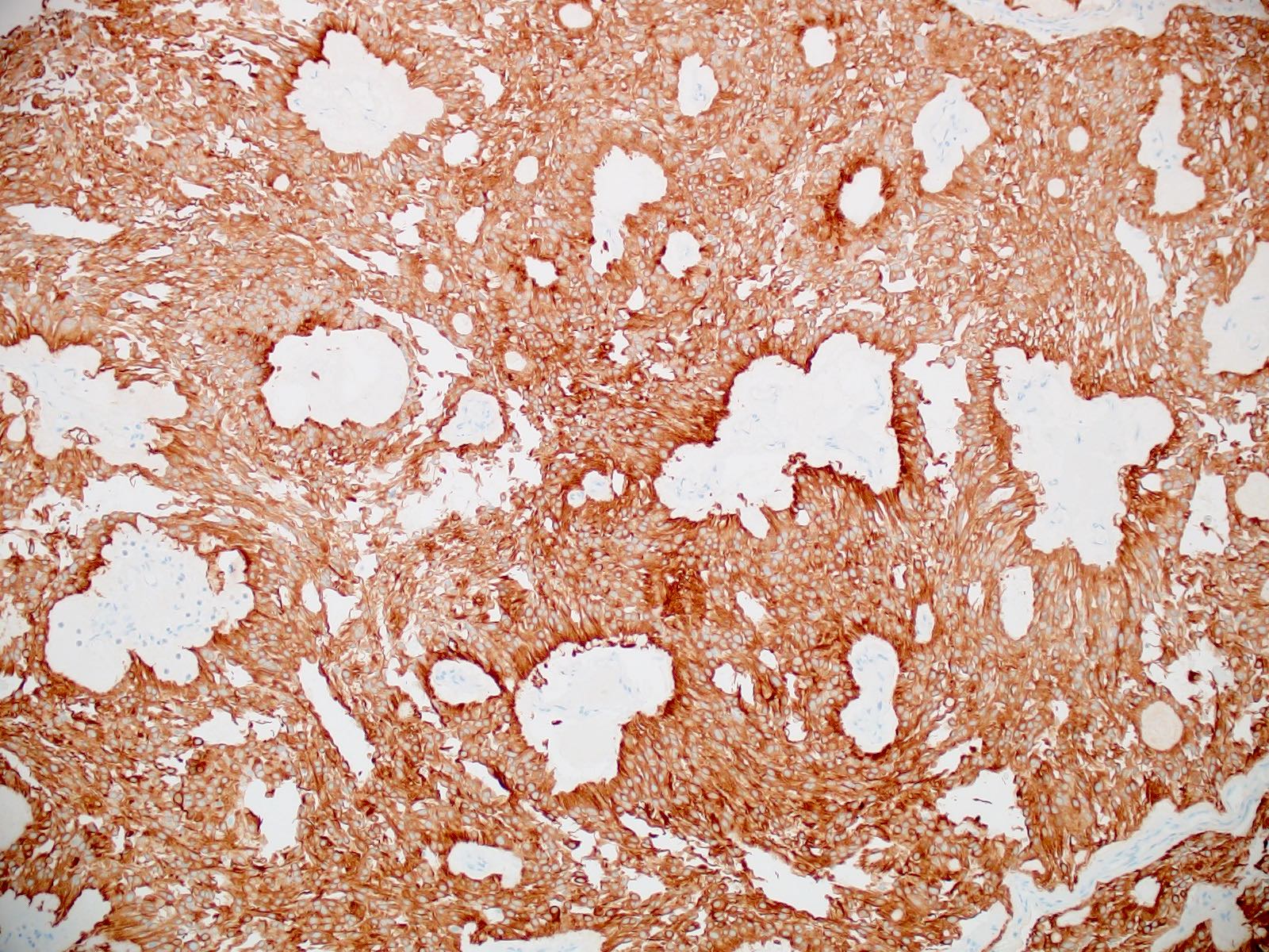
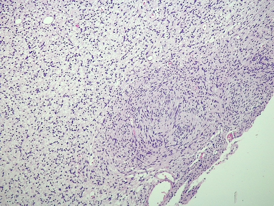
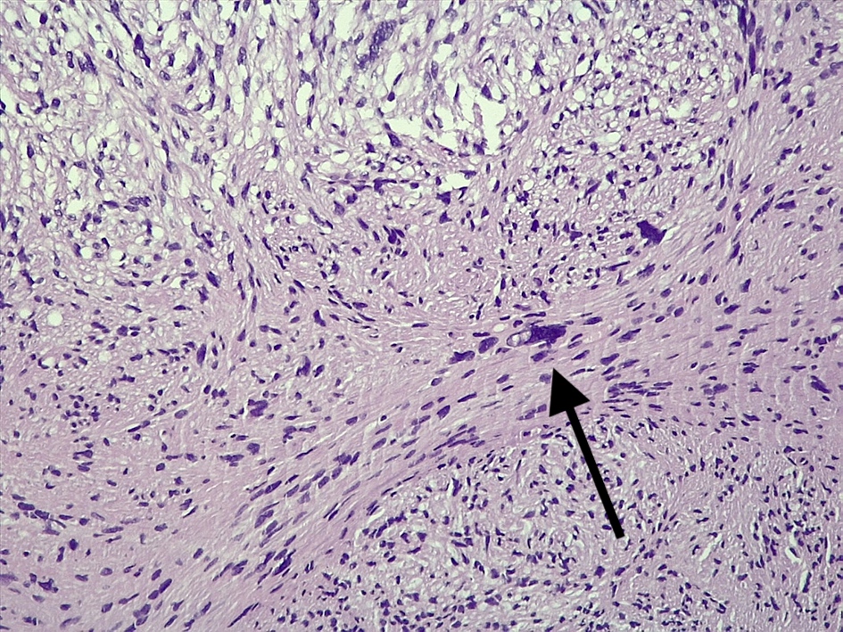
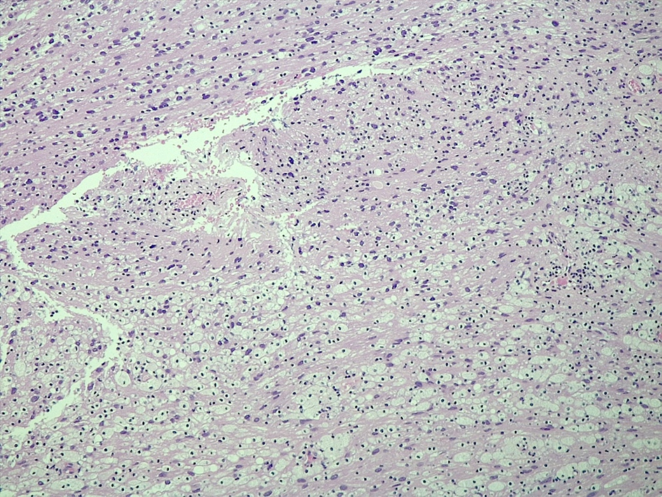
Contributed by Eman Abdelzaher, M.D., Ph.D.
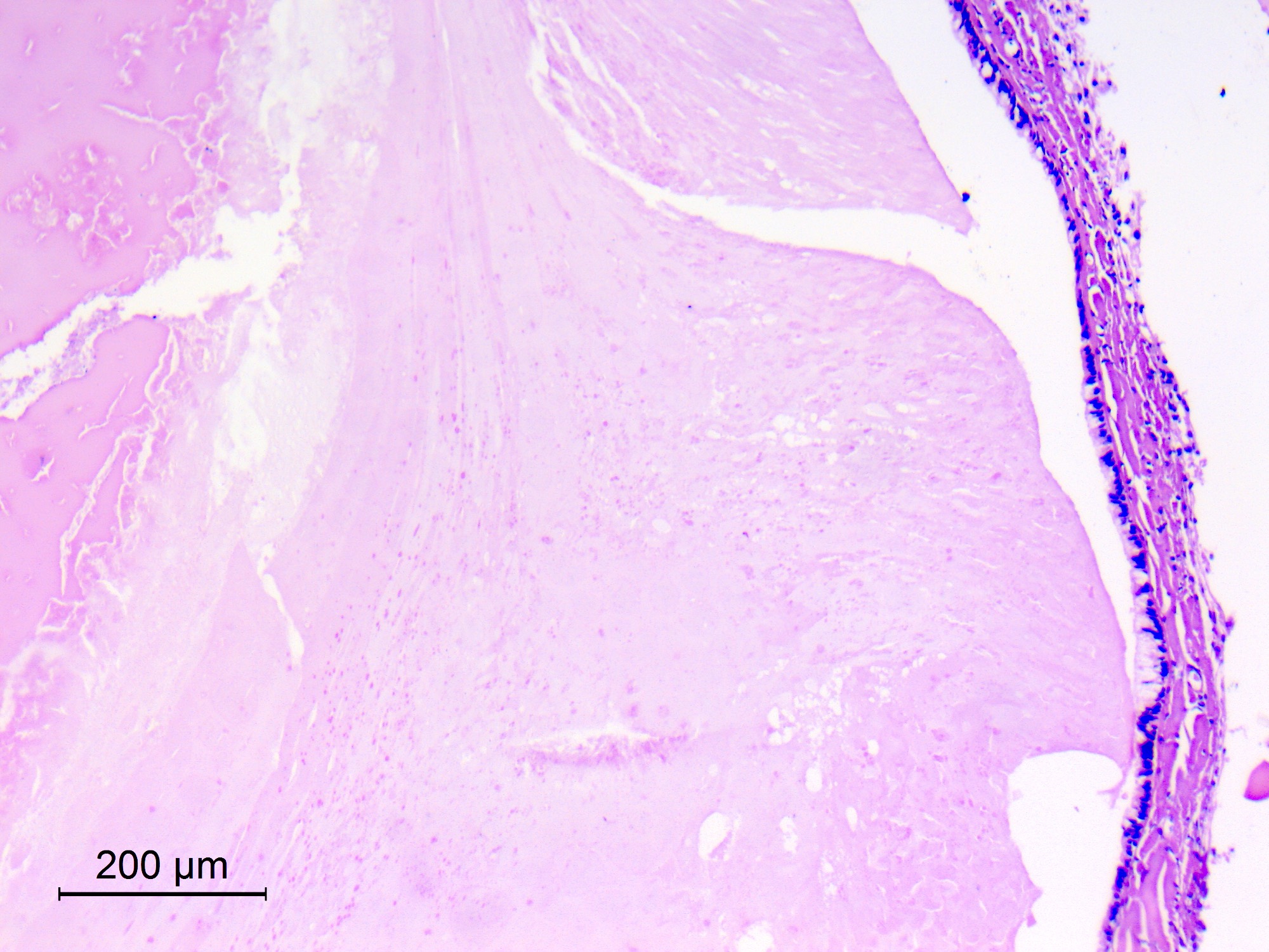
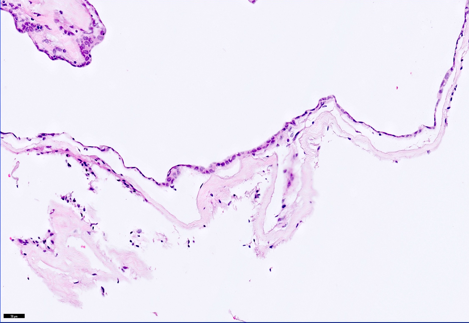
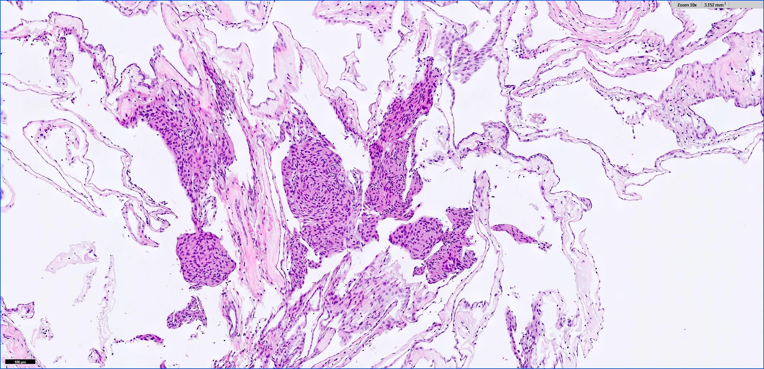
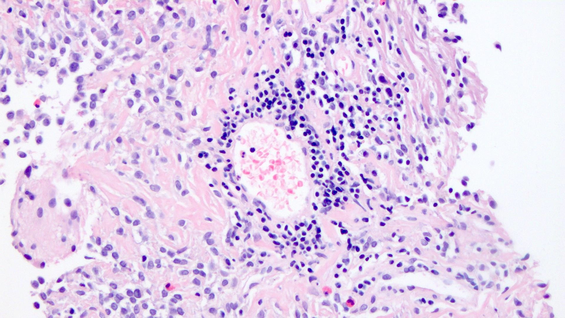
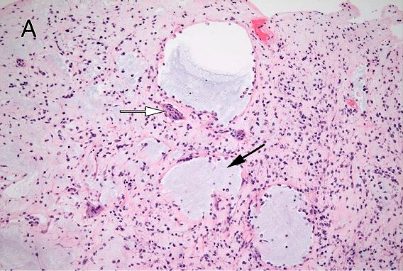
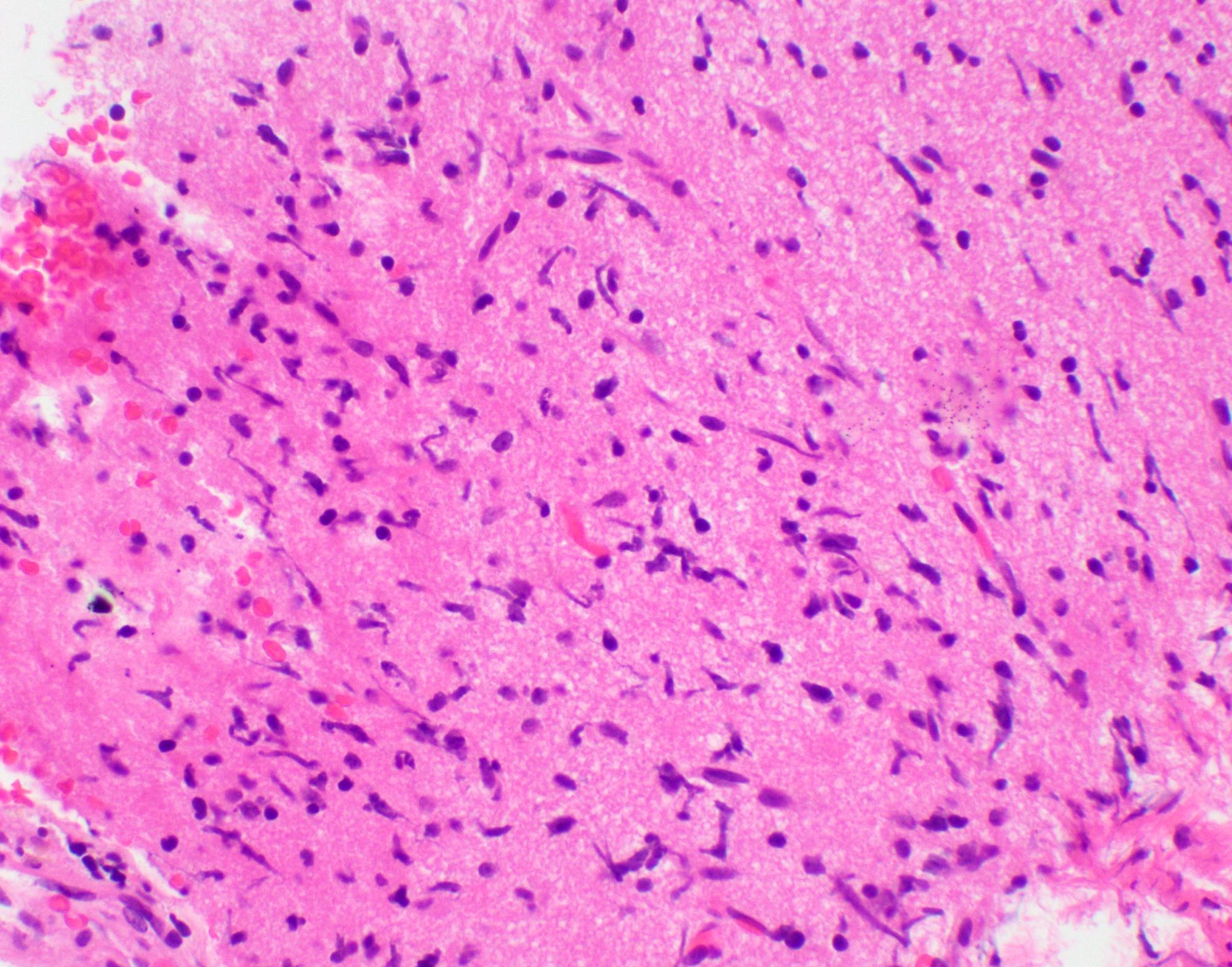
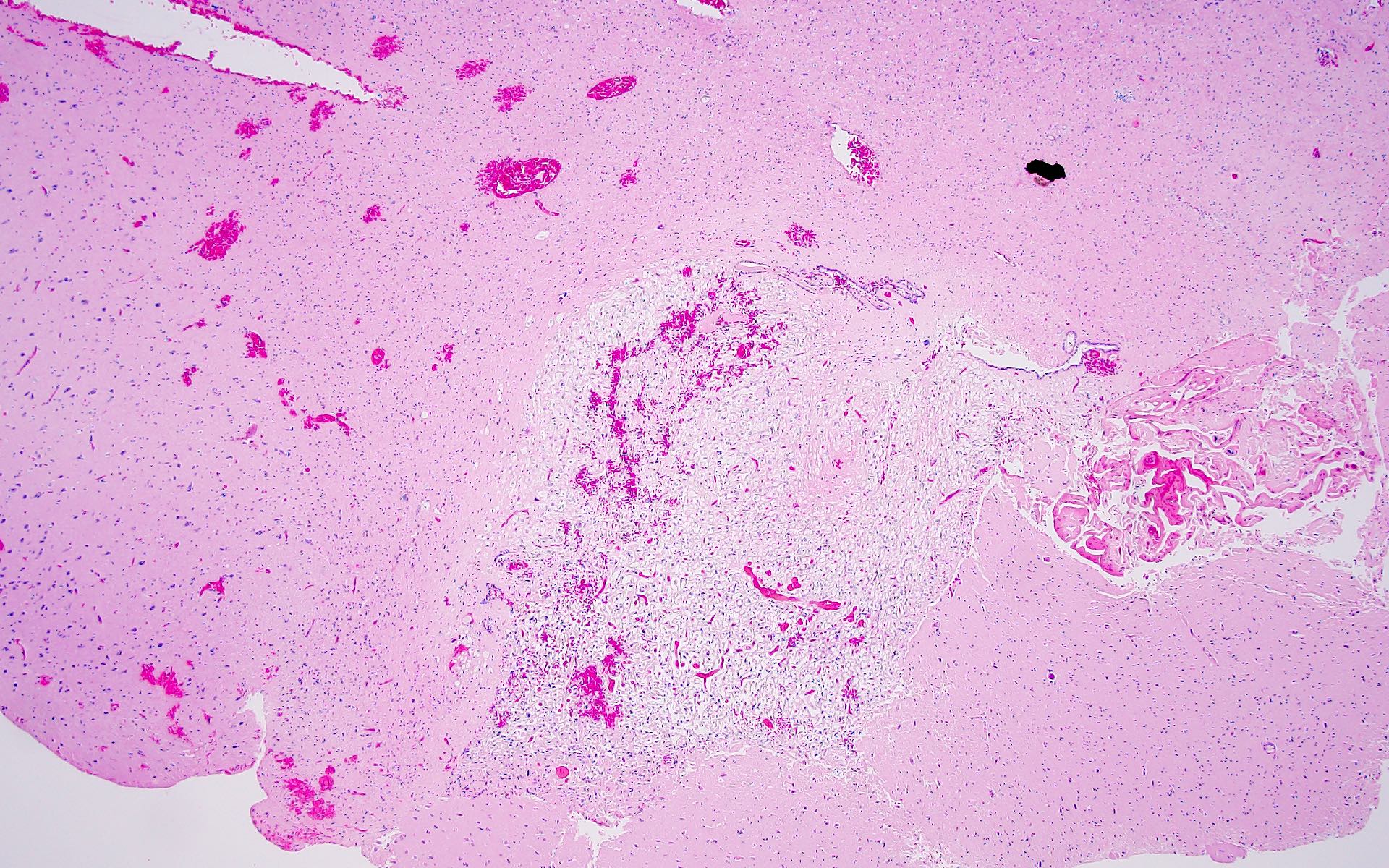
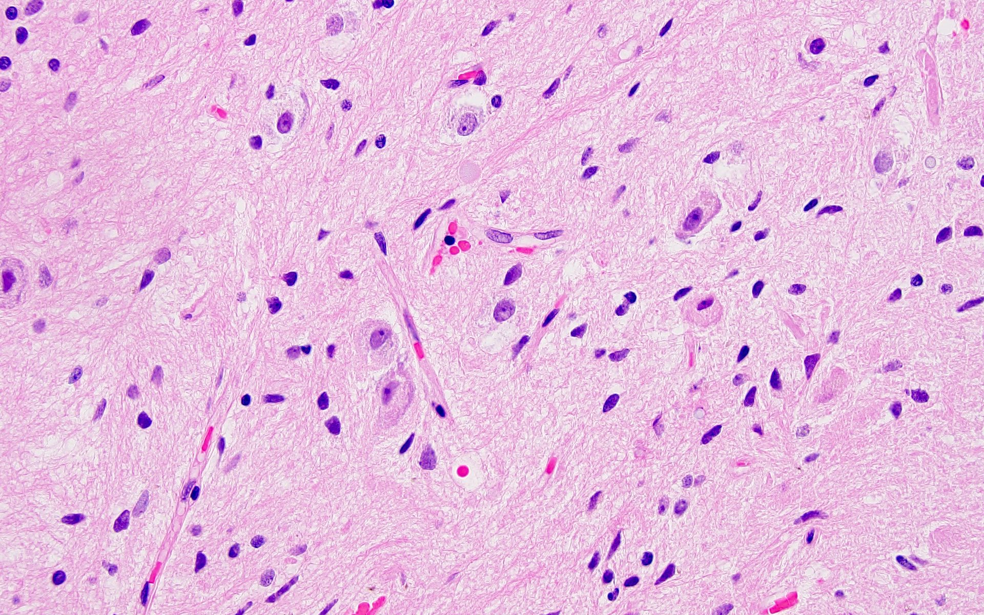
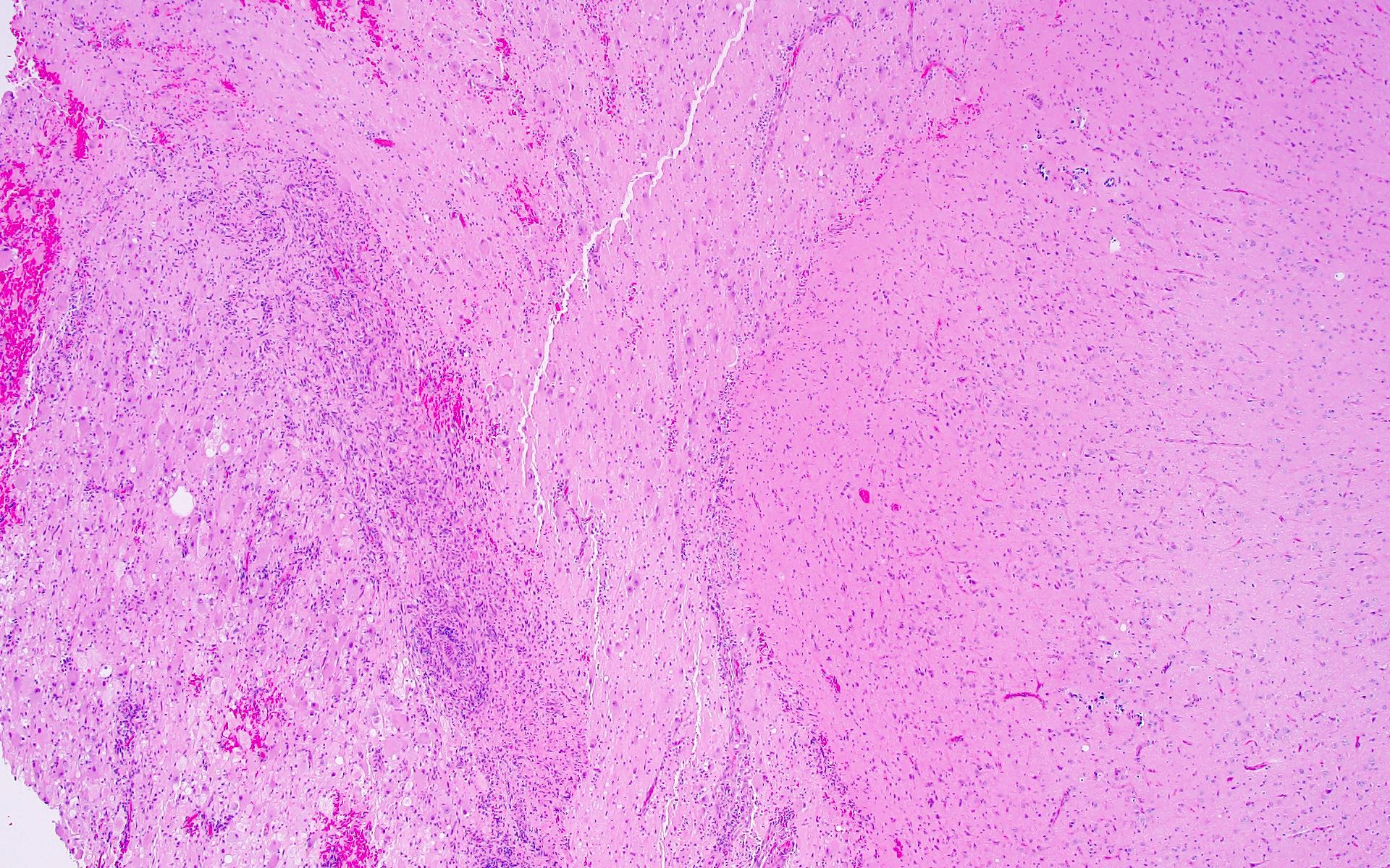
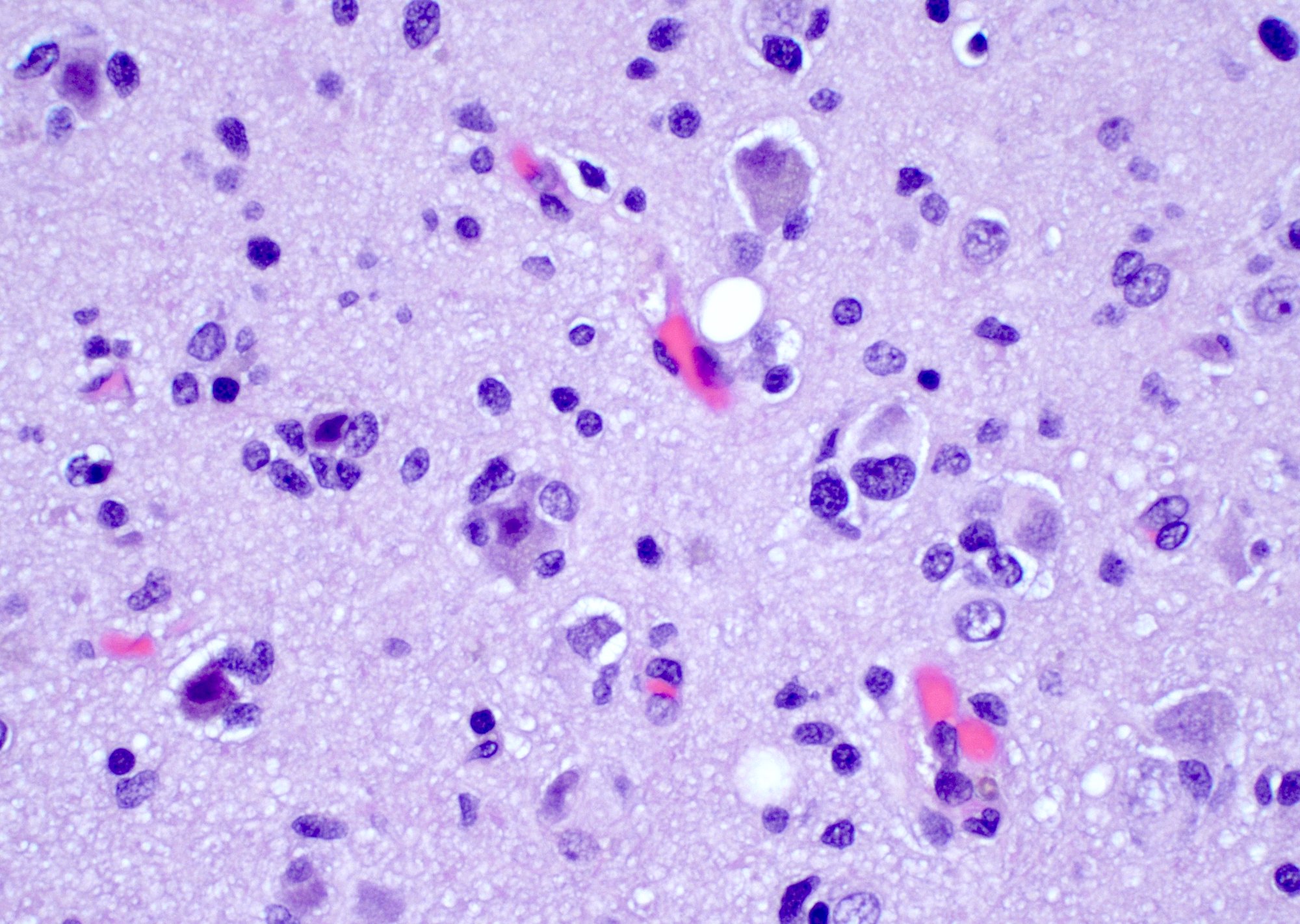
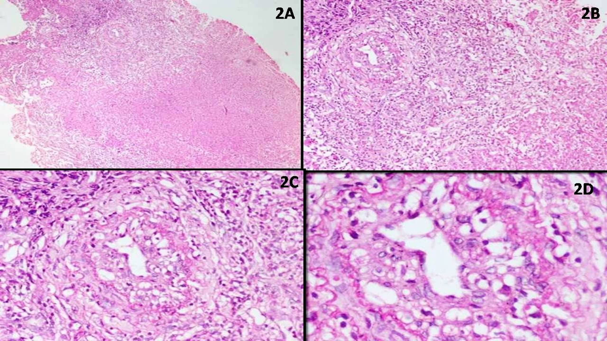
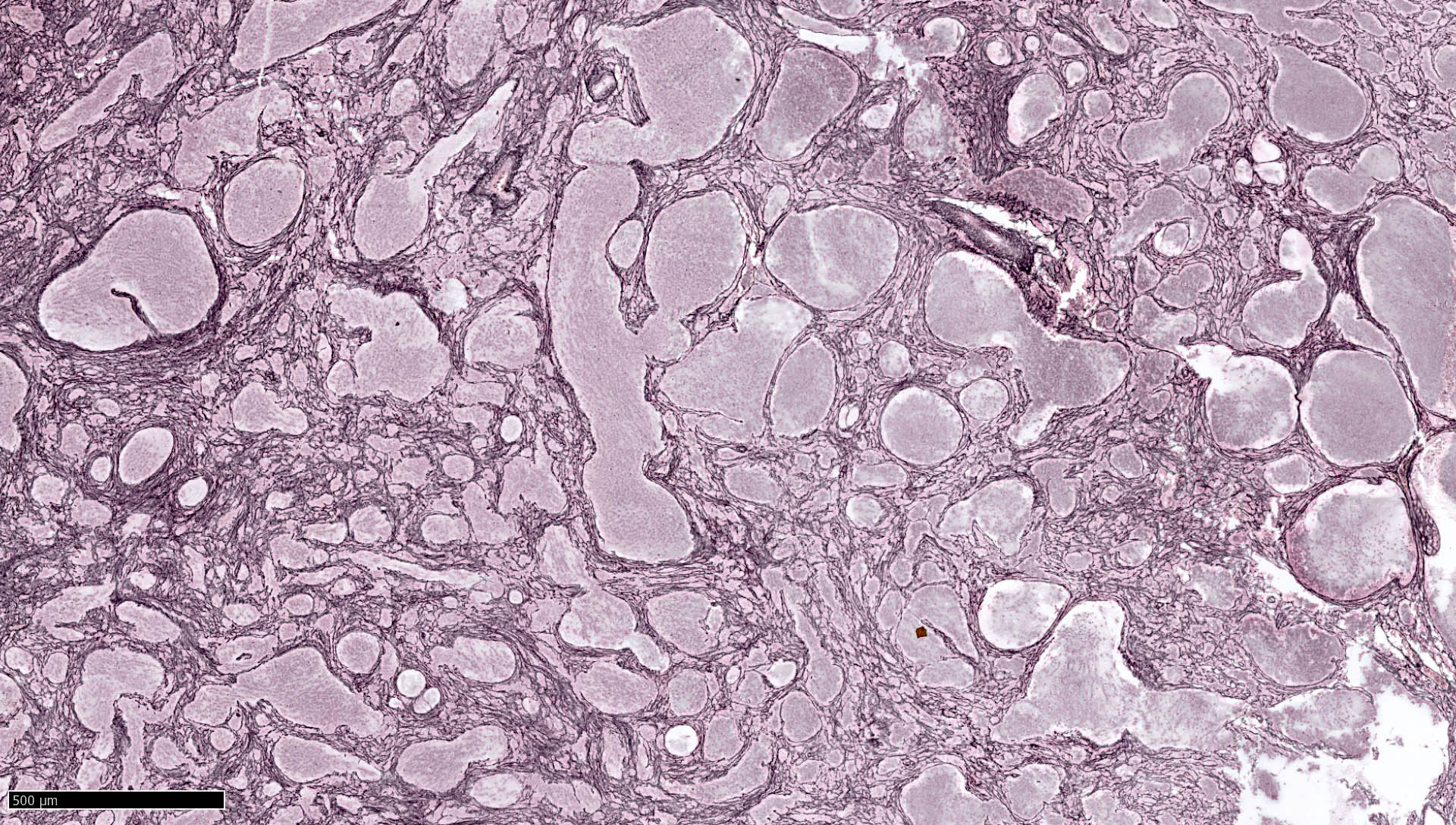
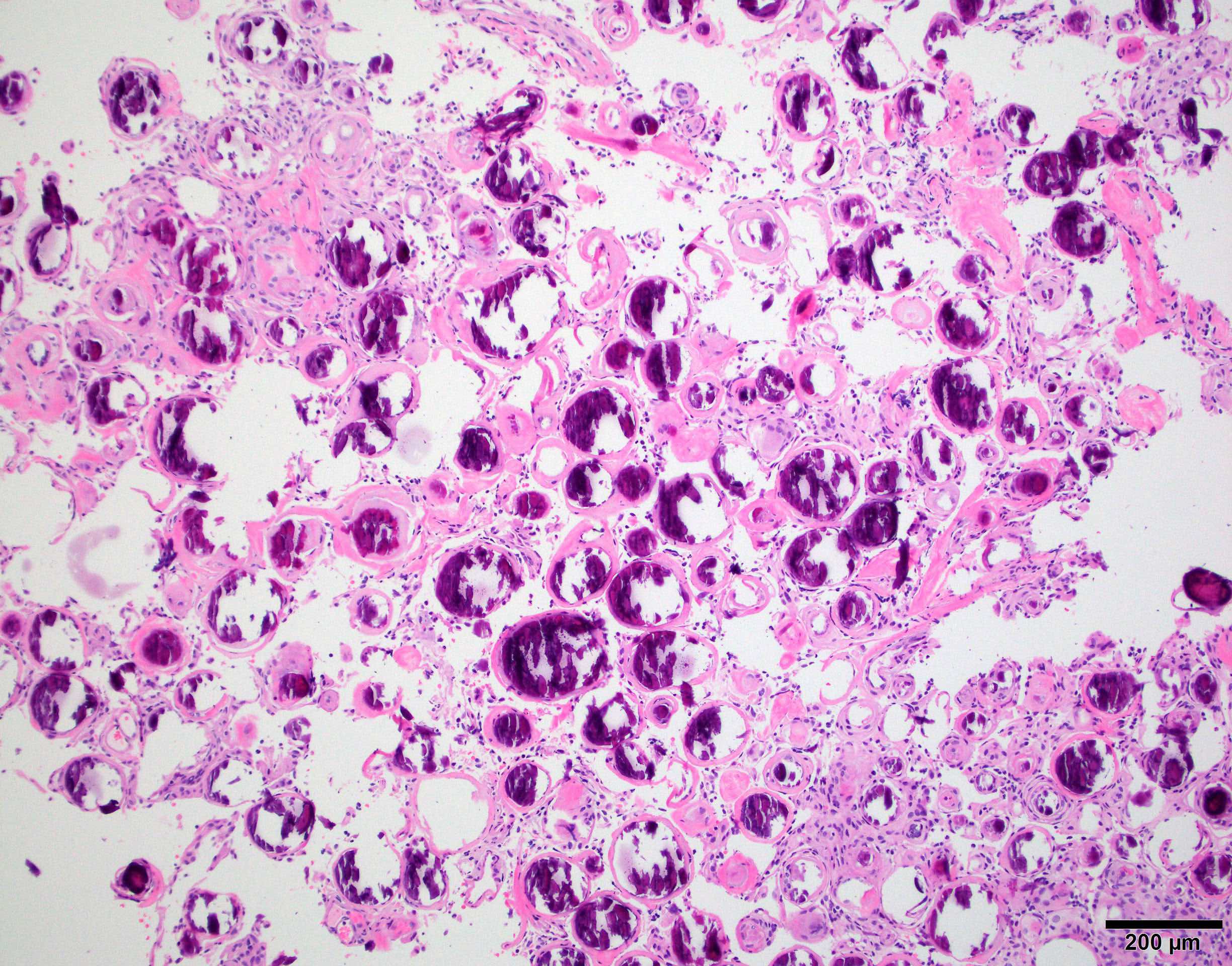
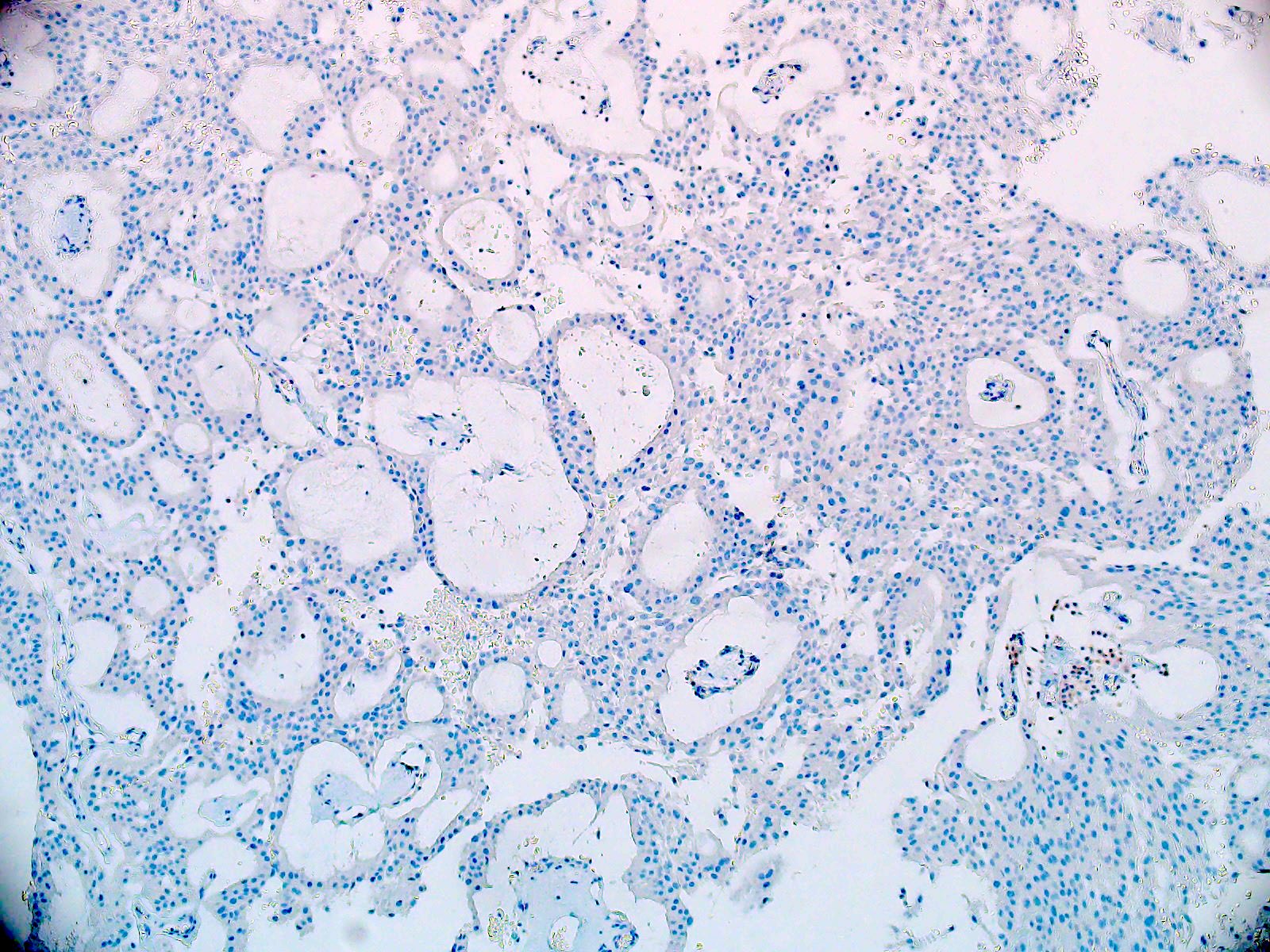
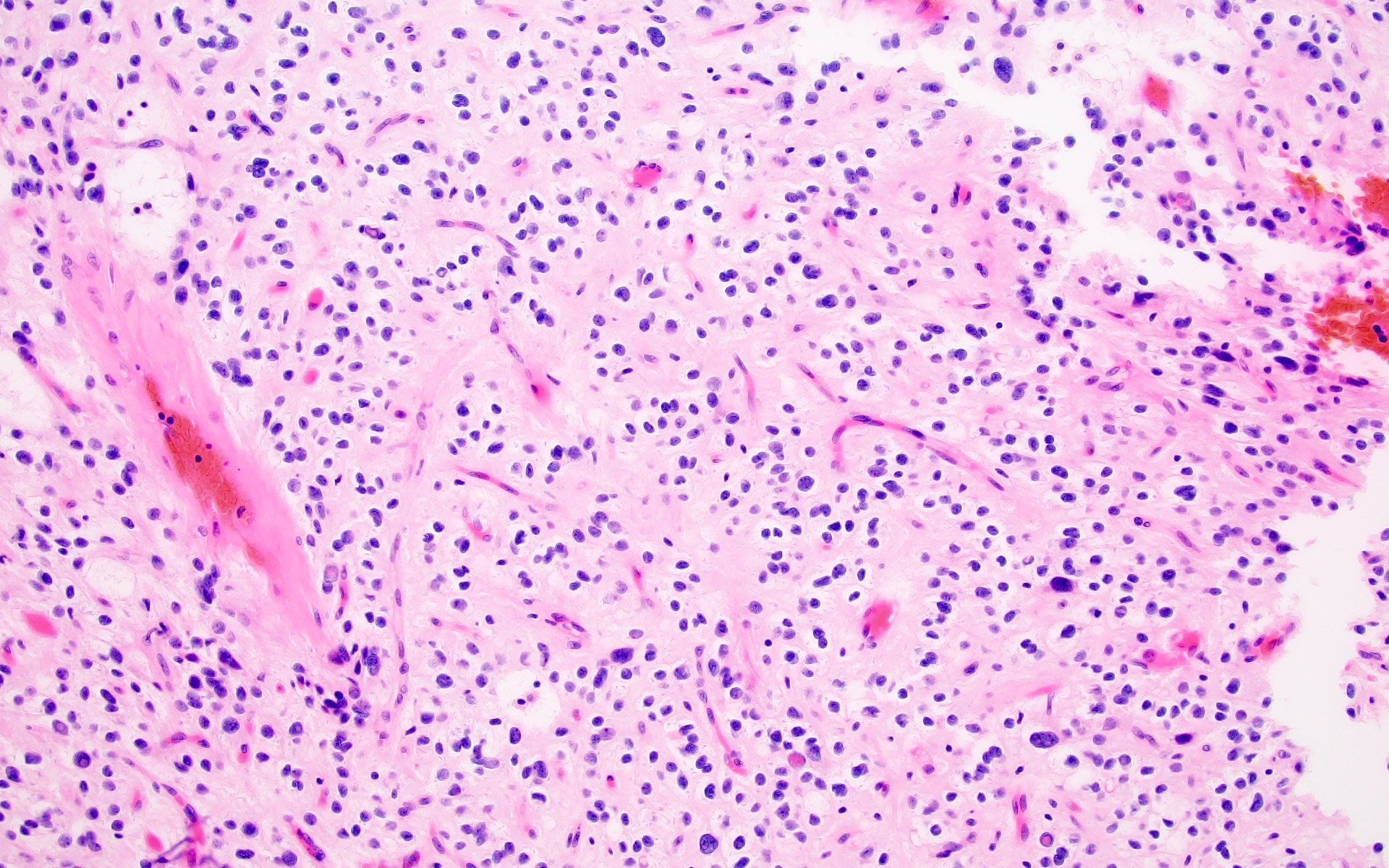
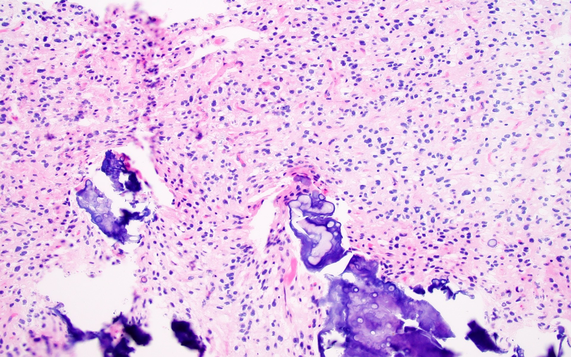
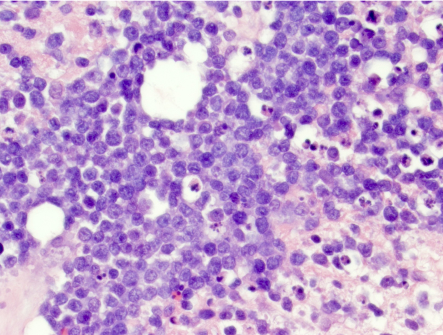
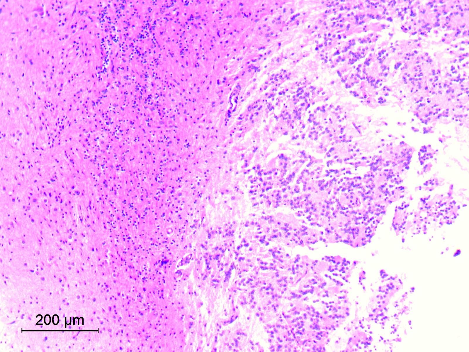
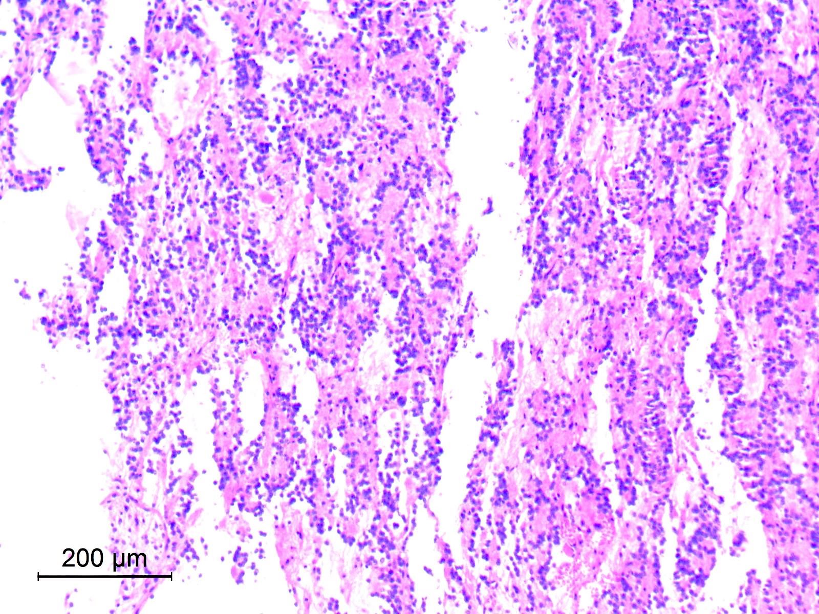
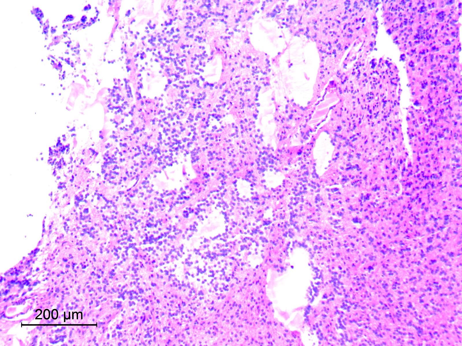
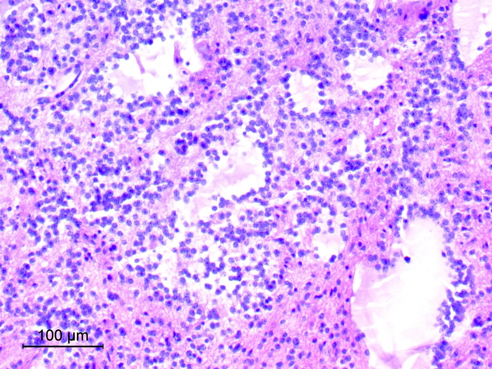
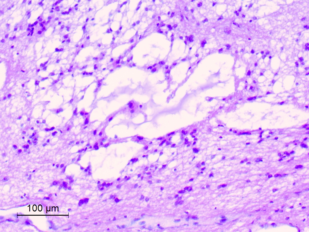
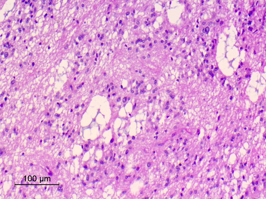
Fibrous wall and proteinaceous contents

Attached choroid plexus
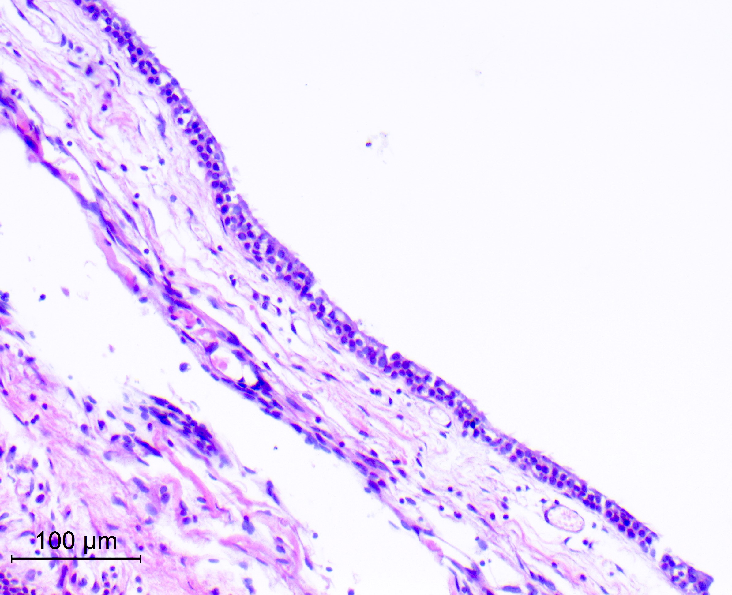
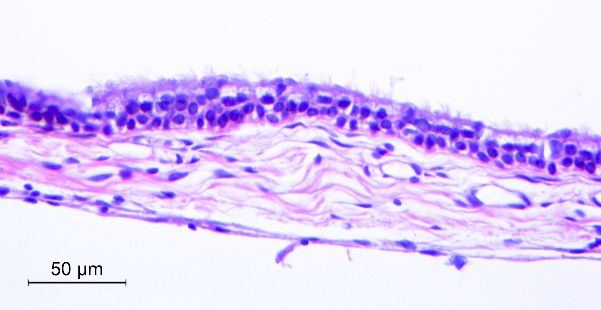
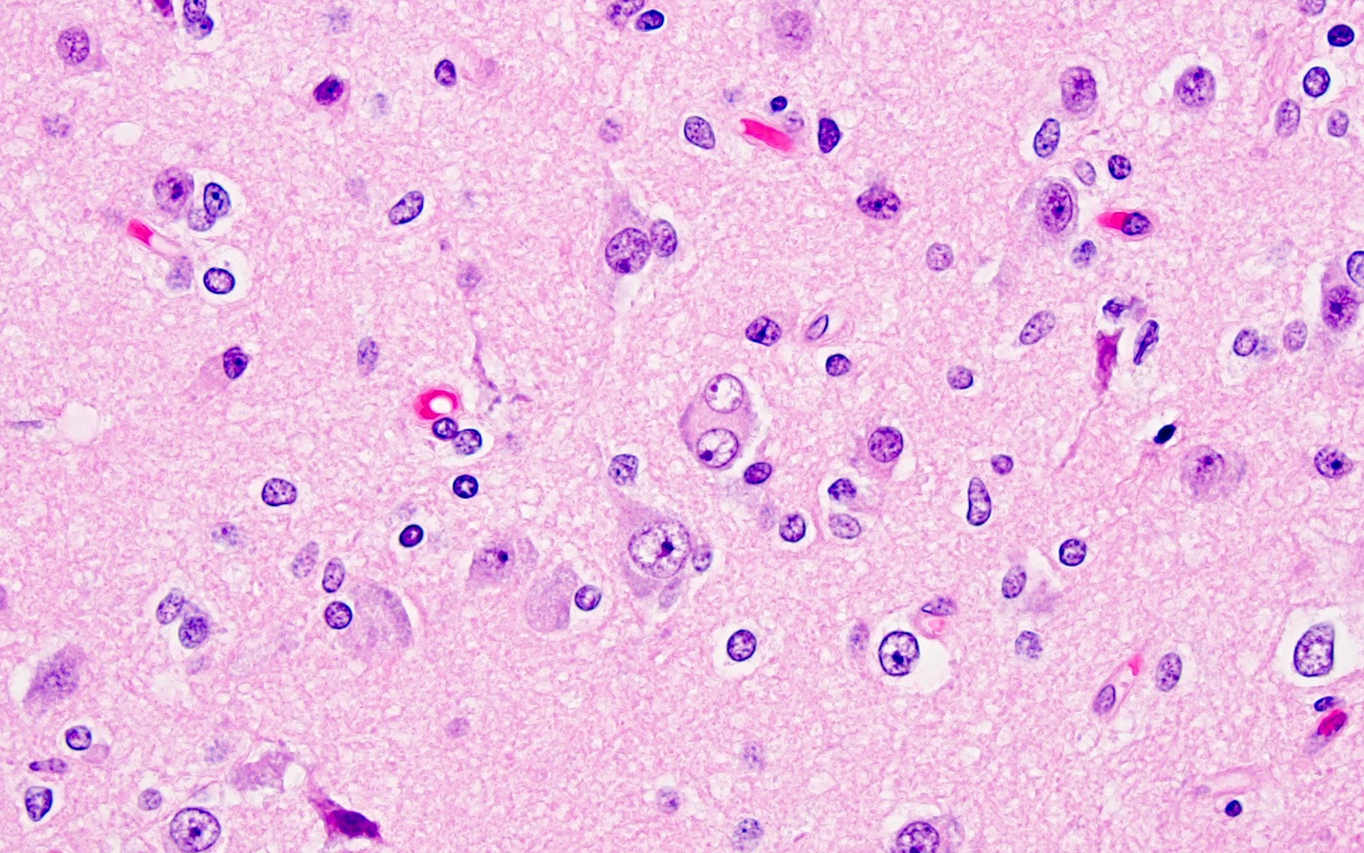
Pseudostratified ciliated cells
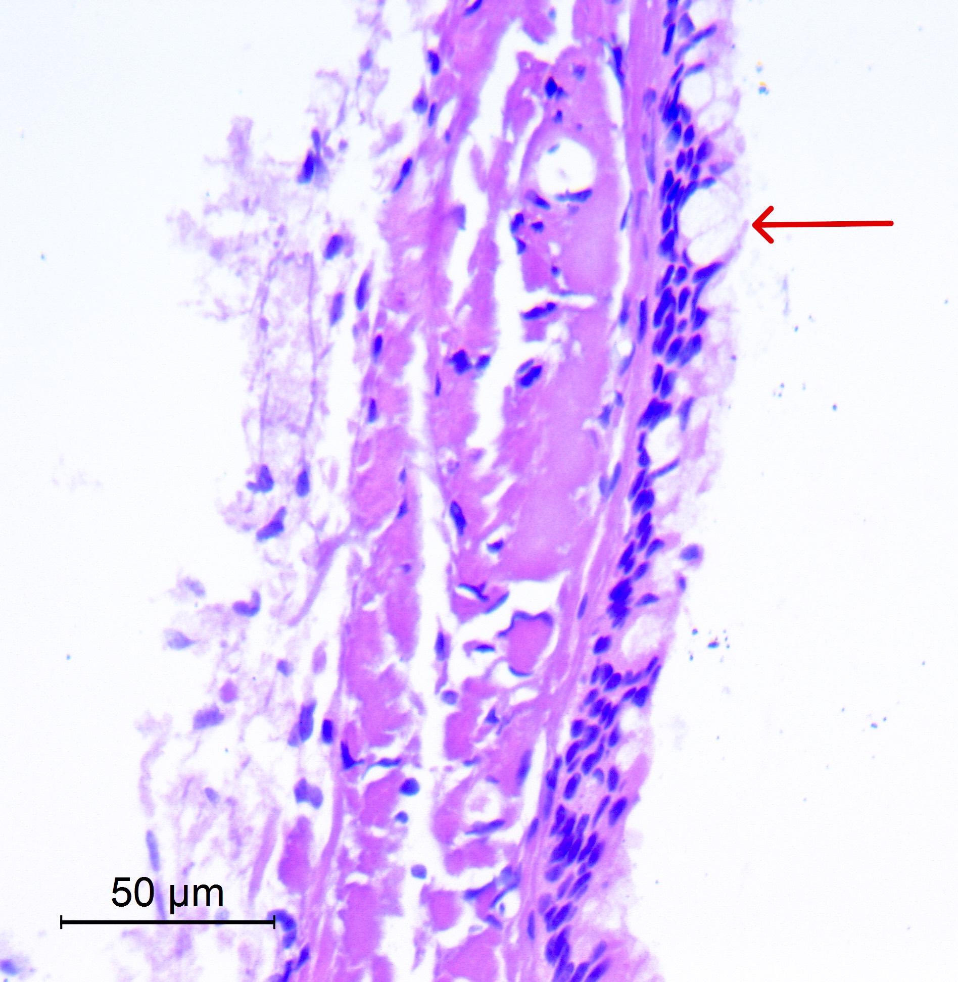
Goblet cells
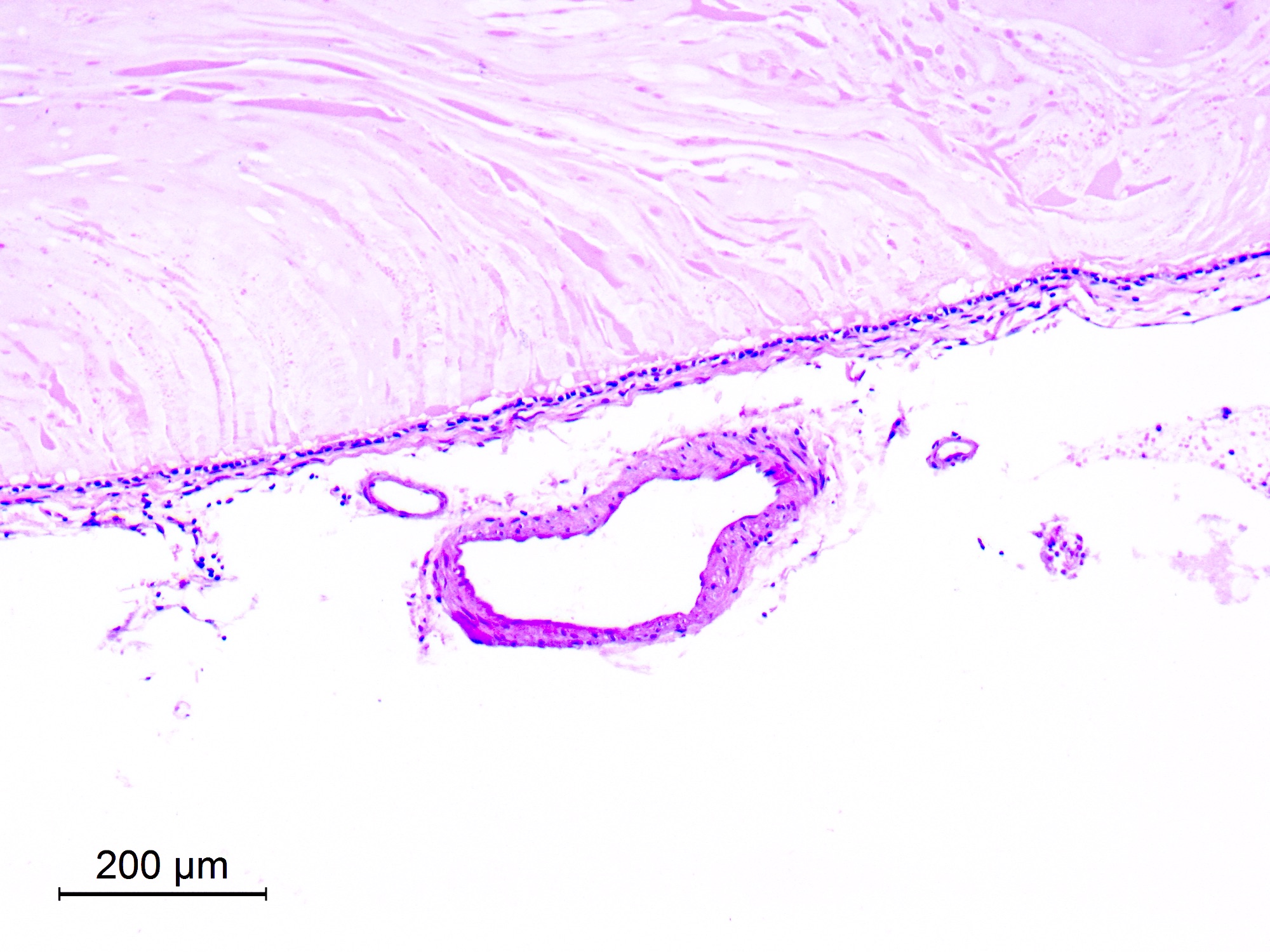
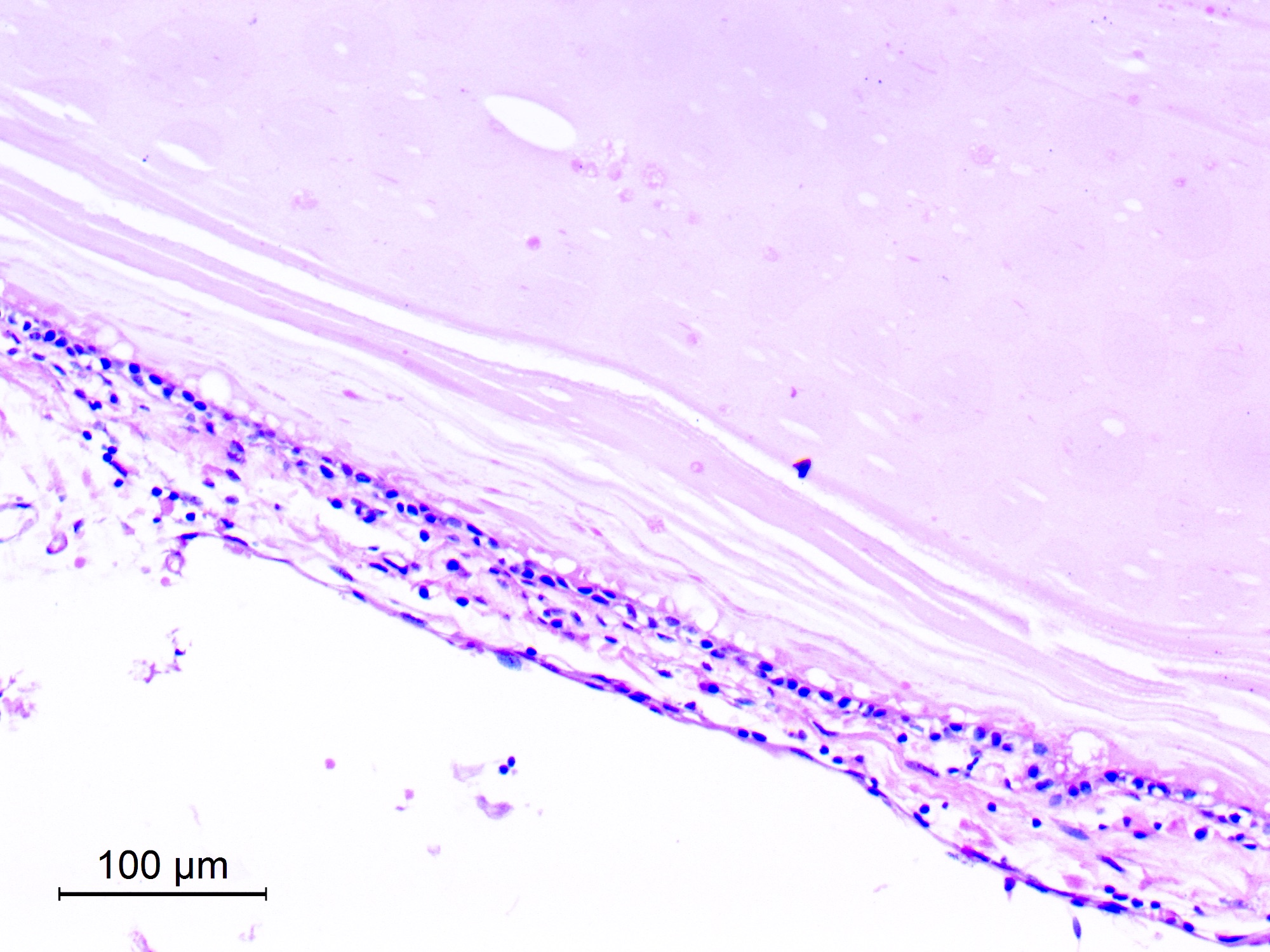
Cuboidal lining
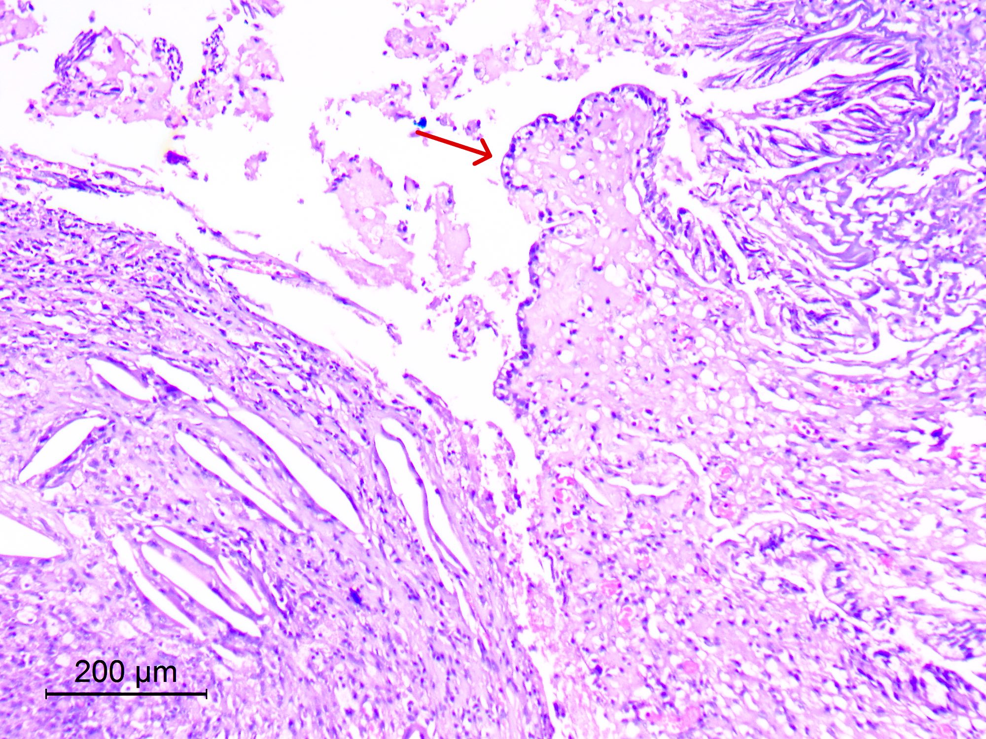
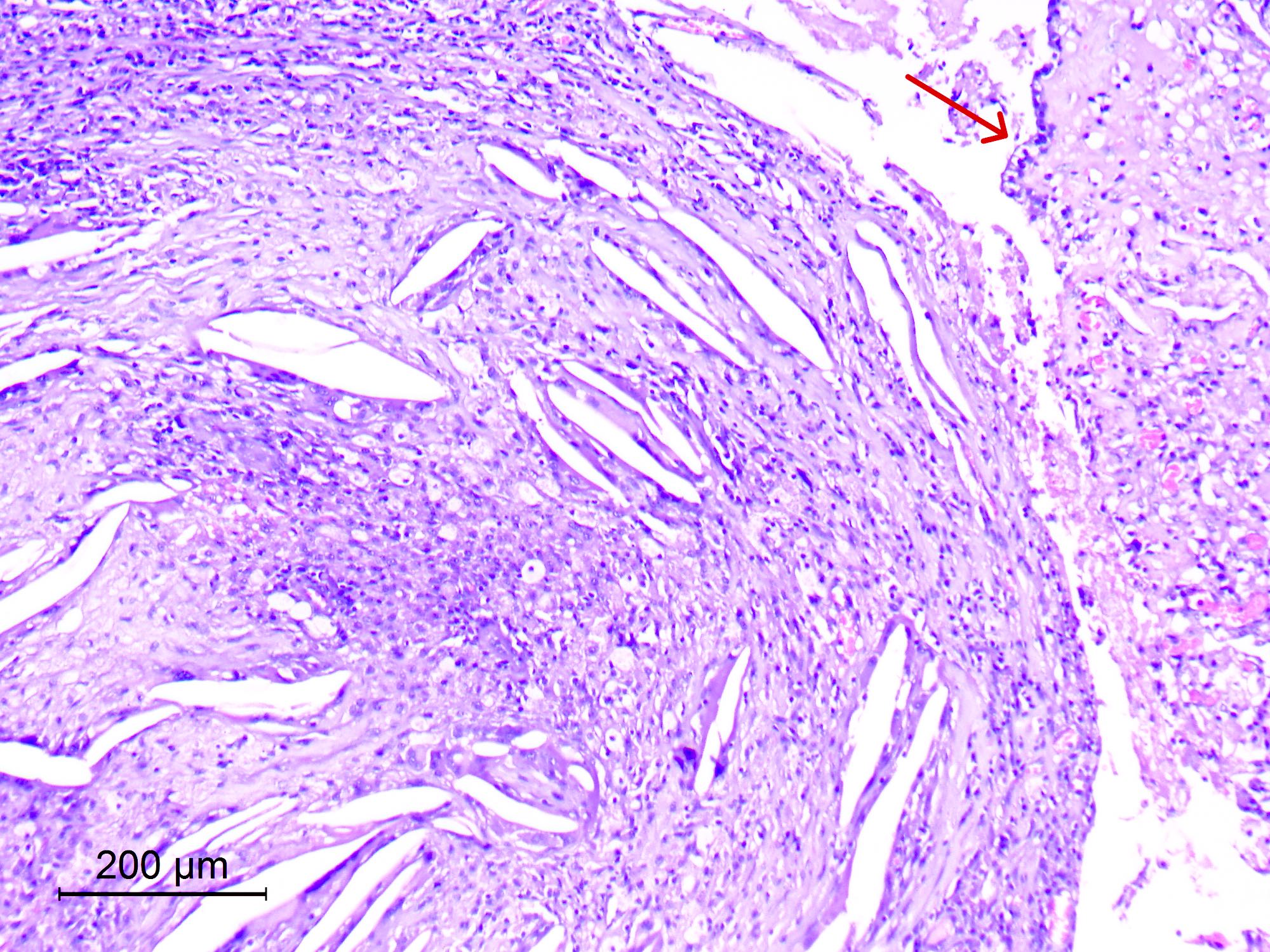
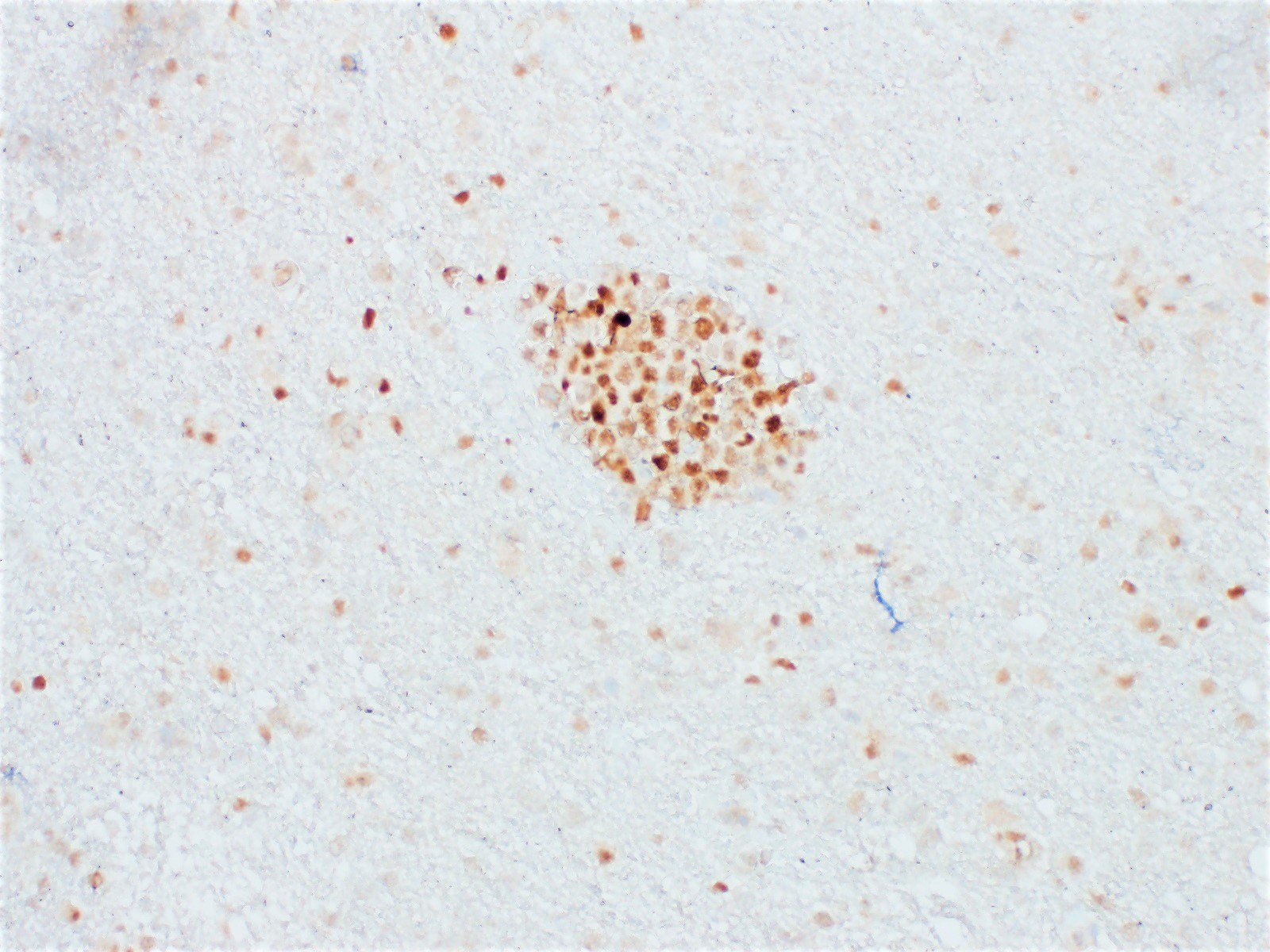
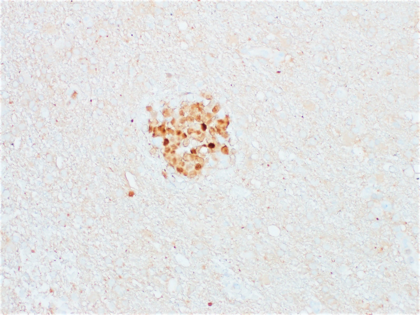
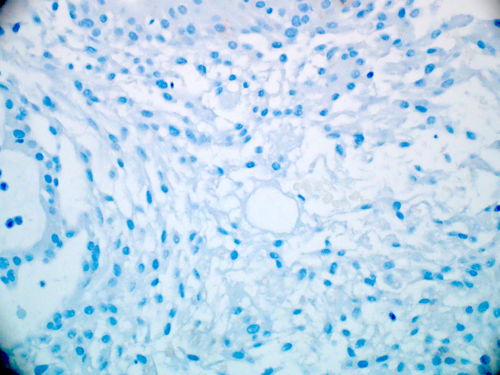
Xanthogranulomatous reaction


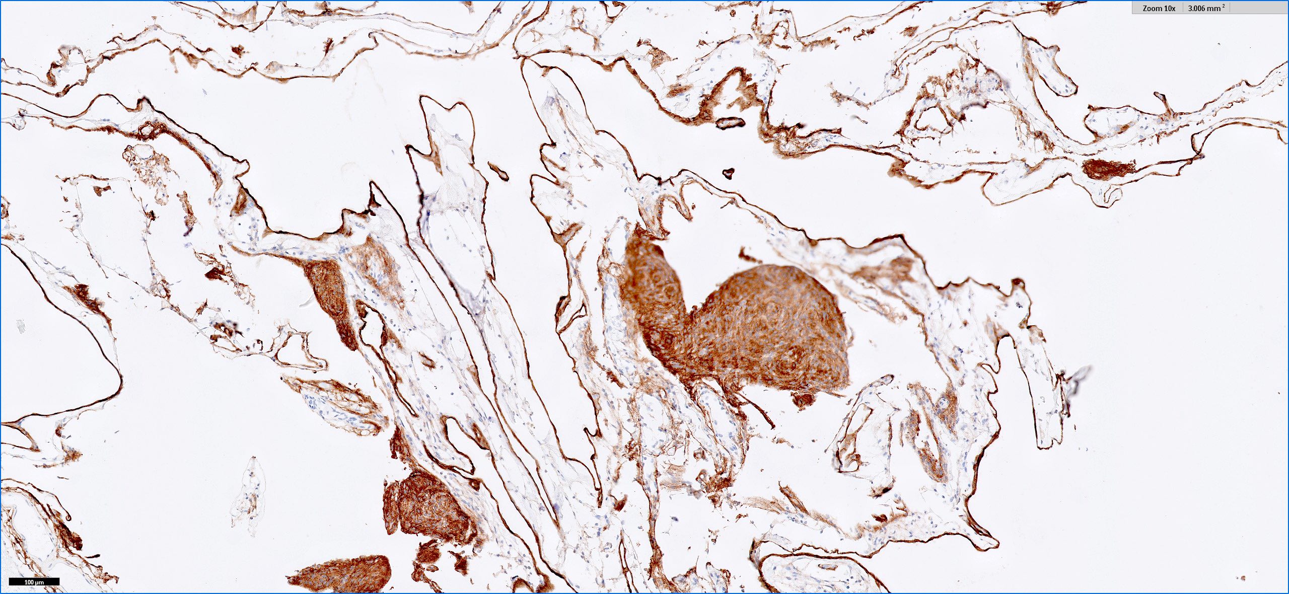
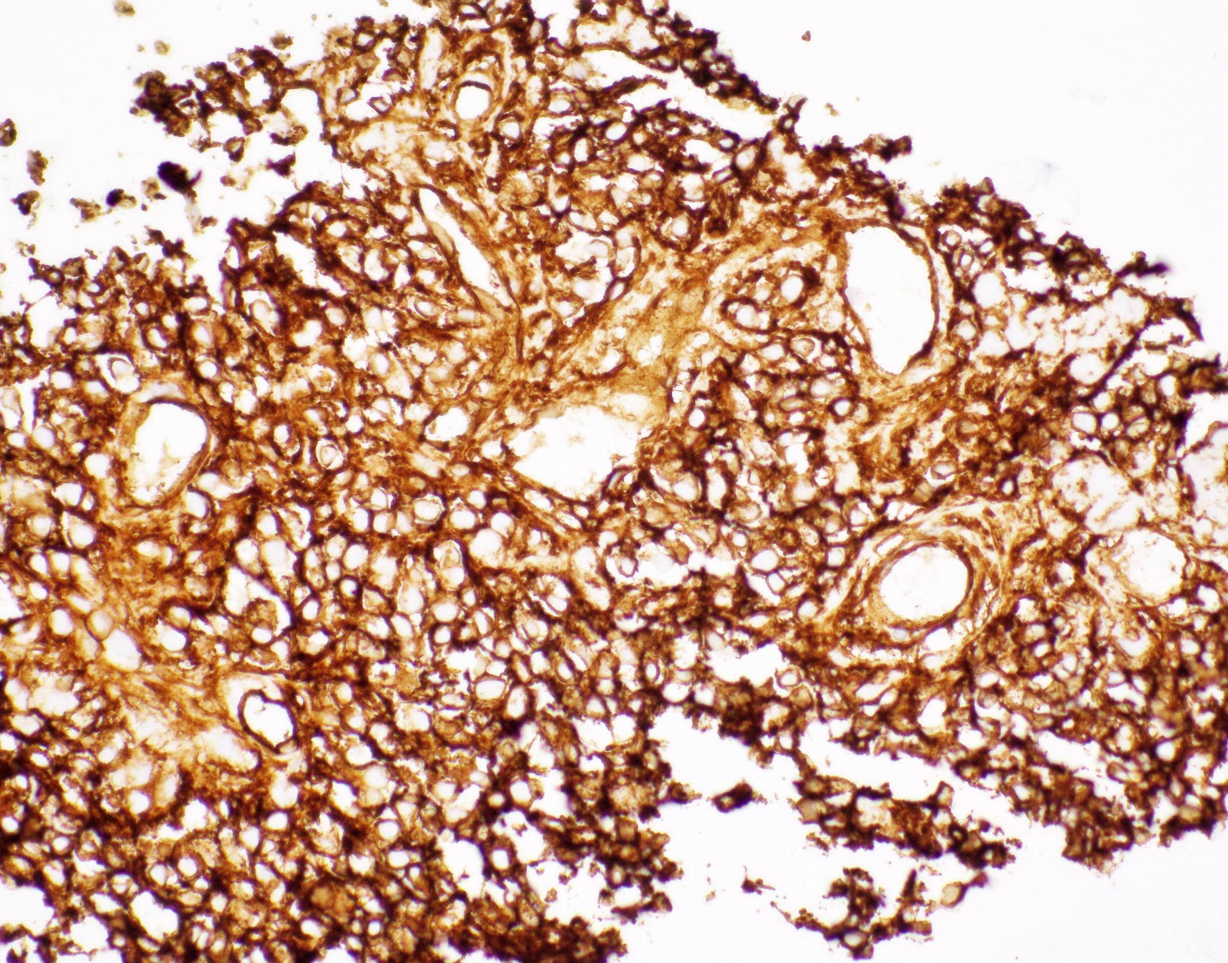
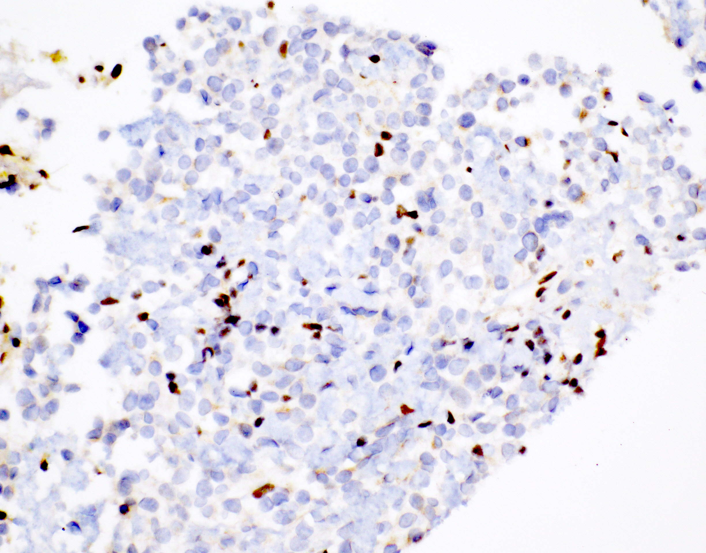
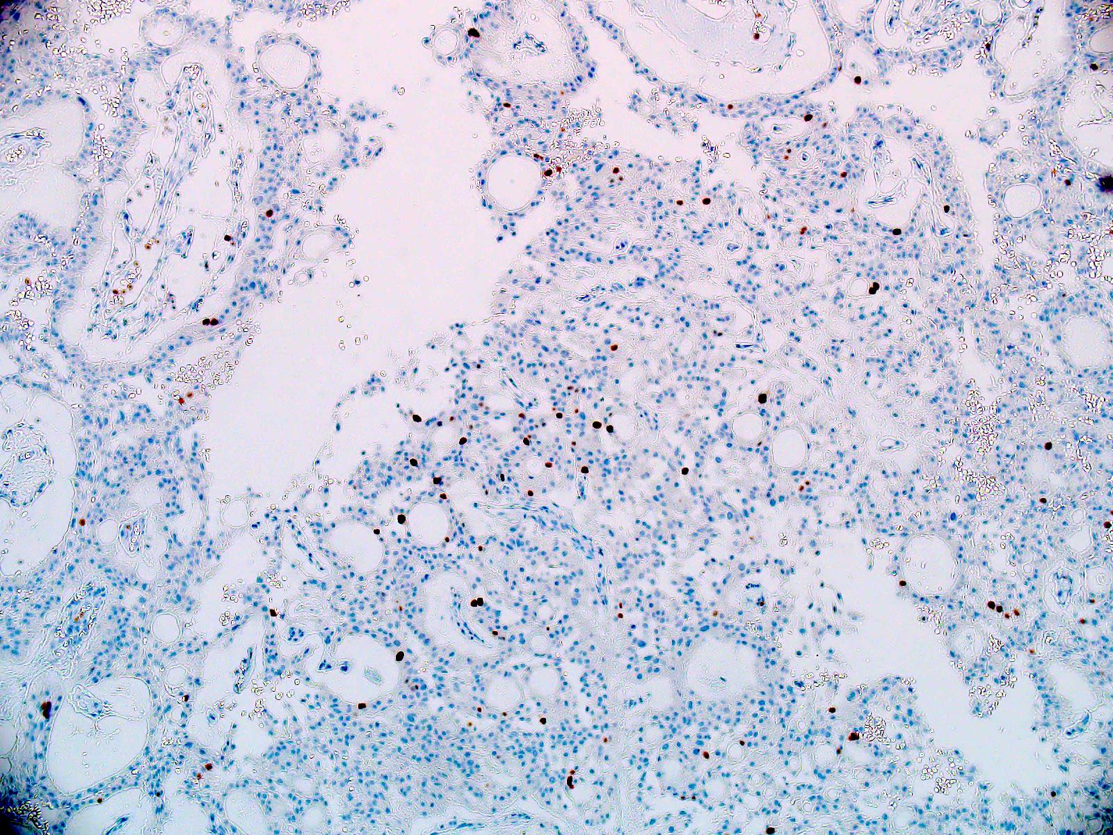
EMA

S100
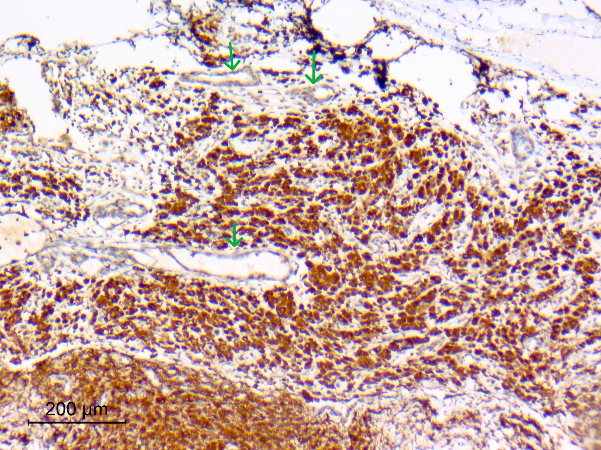
CD68
Images hosted on other servers:

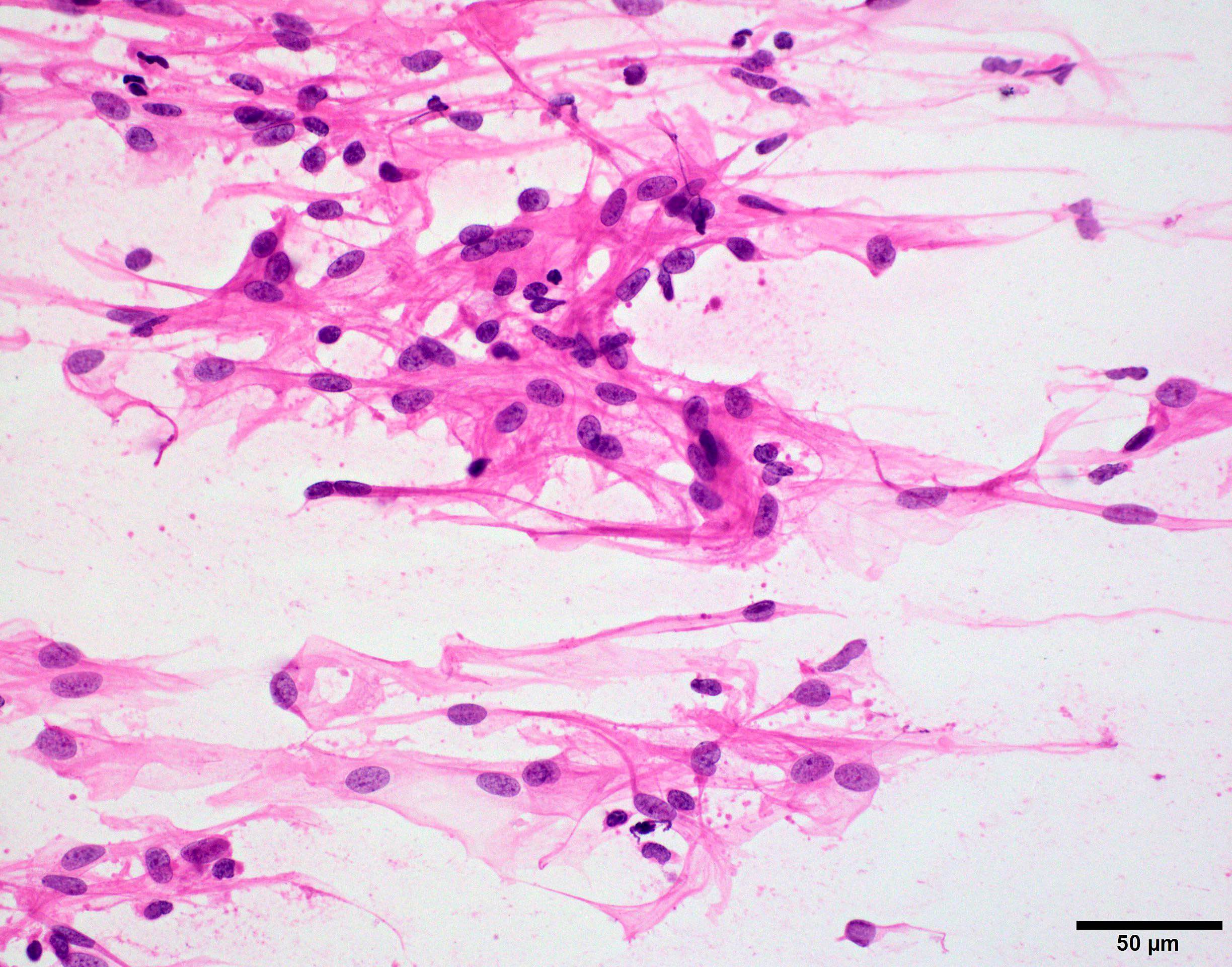
Thick proteinaceous material
Images hosted on other servers:

Ciliary pattern of colloid cyst
Colloid cyst
Contributed by P.J. Cimino, M.D., Ph.D. and Chris Dampier, M.D.
Methylation copy number plot
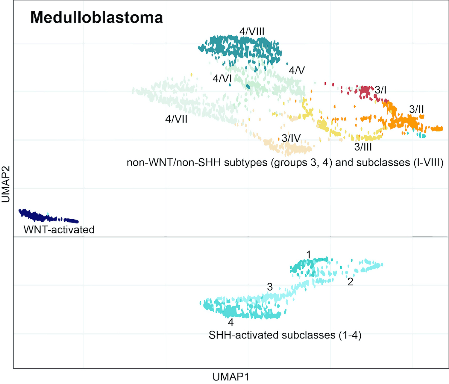
Dimensionality reduction of medulloblastoma
Images hosted on other servers:

Methylation based CNS tumor clustering

Impact of tumor purity

Workflow in surgical neuropathology

Heterogeneity of histologic diagnoses
Images hosted on other servers:

Large right hemispheric mass
Contributed by Yue-E Wang, M.S., Ph.D., Jingjing Chen, Ph.D., Wei Wang, Ph.D., An-Li Zhang, M.D., Wenchao Zhou, Ph.D. and Hai-Bo Wu, M.D., Ph.D.

MRI - sagittal T1
MRI - sagittal T2
Contributed by Mariana Voudouri, M.D. and George Zanazzi, M.D., Ph.D.
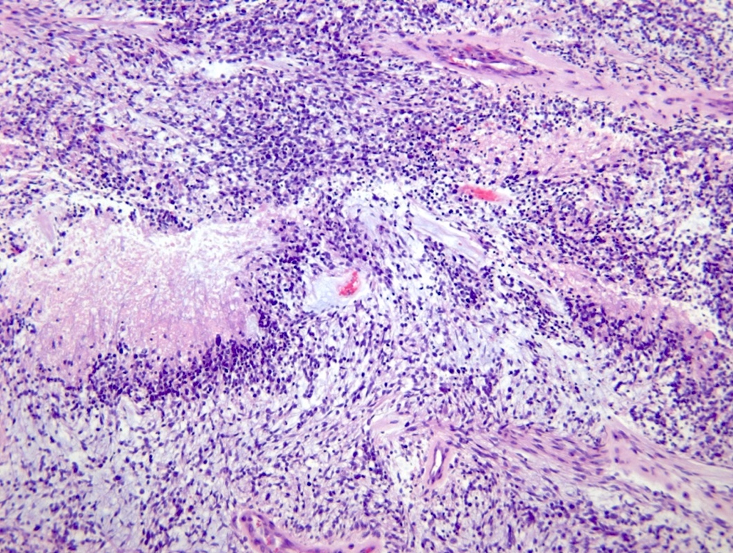
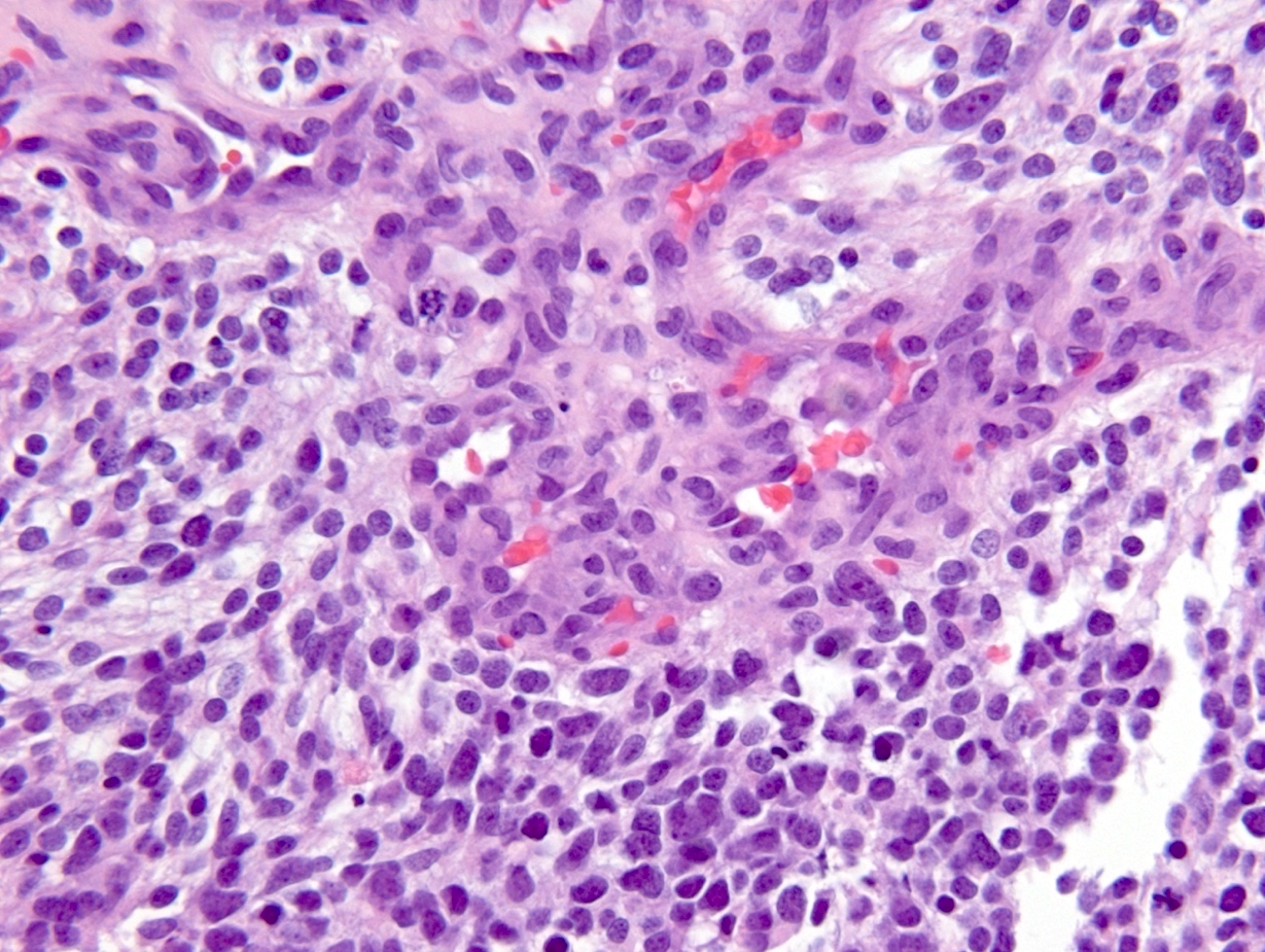
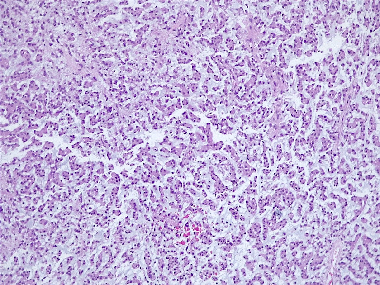
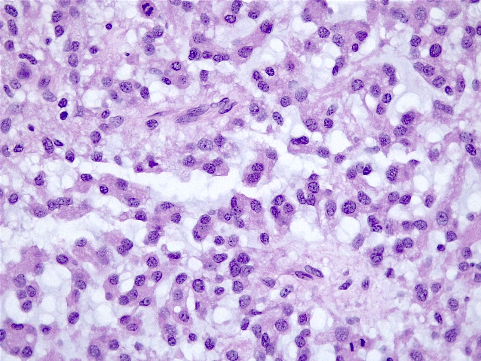
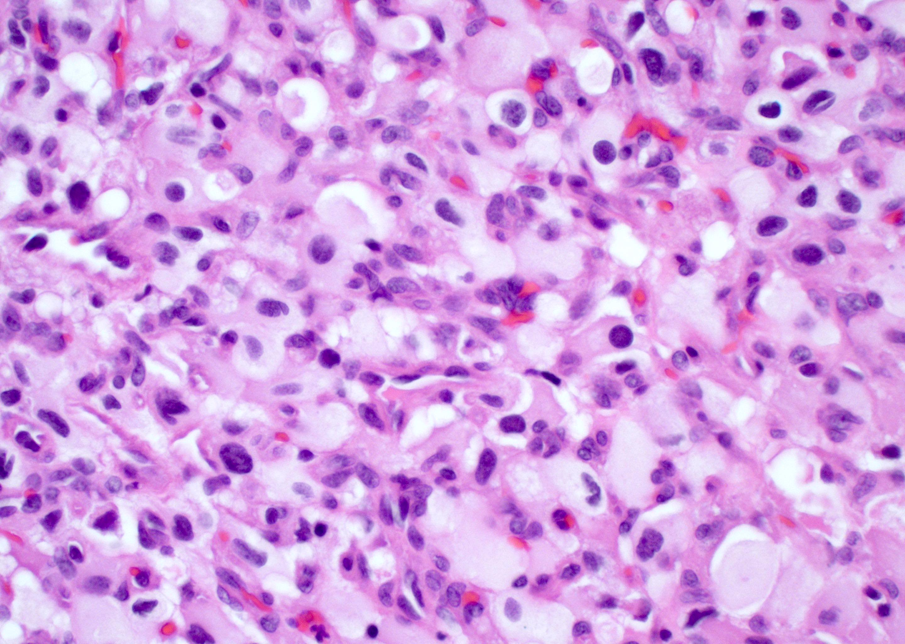
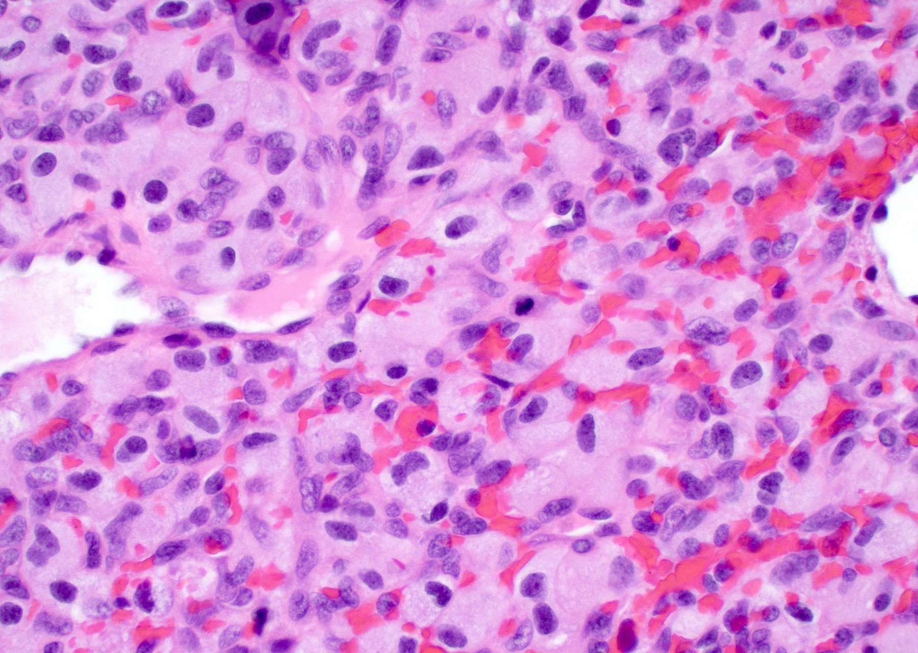
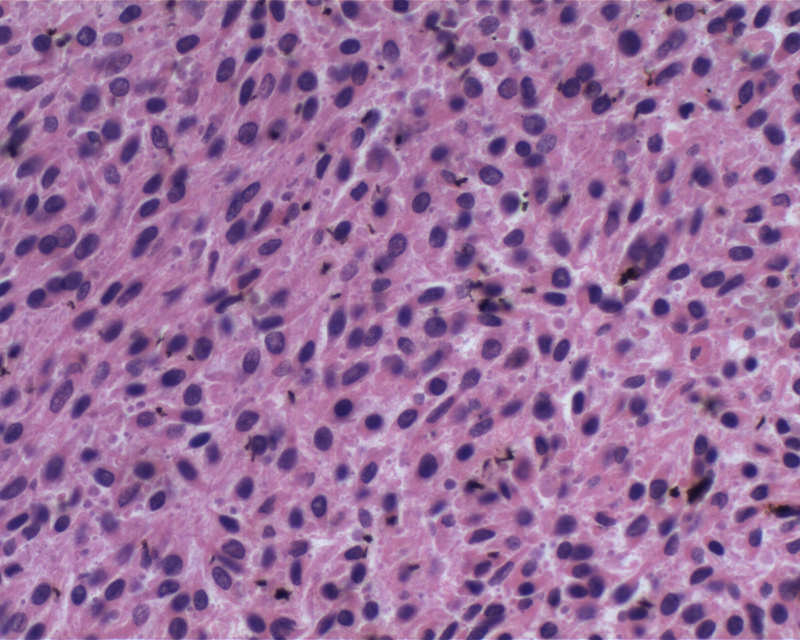
Epithelioid neoplasm with desmoplasia
Cellular atypia
Perivascular tumor cell accumulation
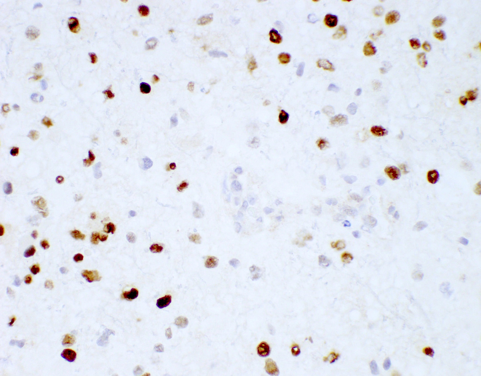
CD34
SMARCB1 / INI1
Contributed by Branavan Manoranjan, M.D., Ph.D., Abdelsimar T. Omar II, M.D., Hai-Bo Wu, M.D., Ph.D., Robert Nordal, M.D. and Yves Starreveld, M.D., Ph.D.
Microcysts and myxoid matrix
Trichrome
Contributed by Mariana Voudouri, M.D. and George Zanazzi, M.D., Ph.D.
MRI of posterior left frontal tumor
Sagittal T1
Axial T2 / FLAIR
Axial postcontrast
Contributed by Mariana Voudouri, M.D. and George Zanazzi, M.D., Ph.D.
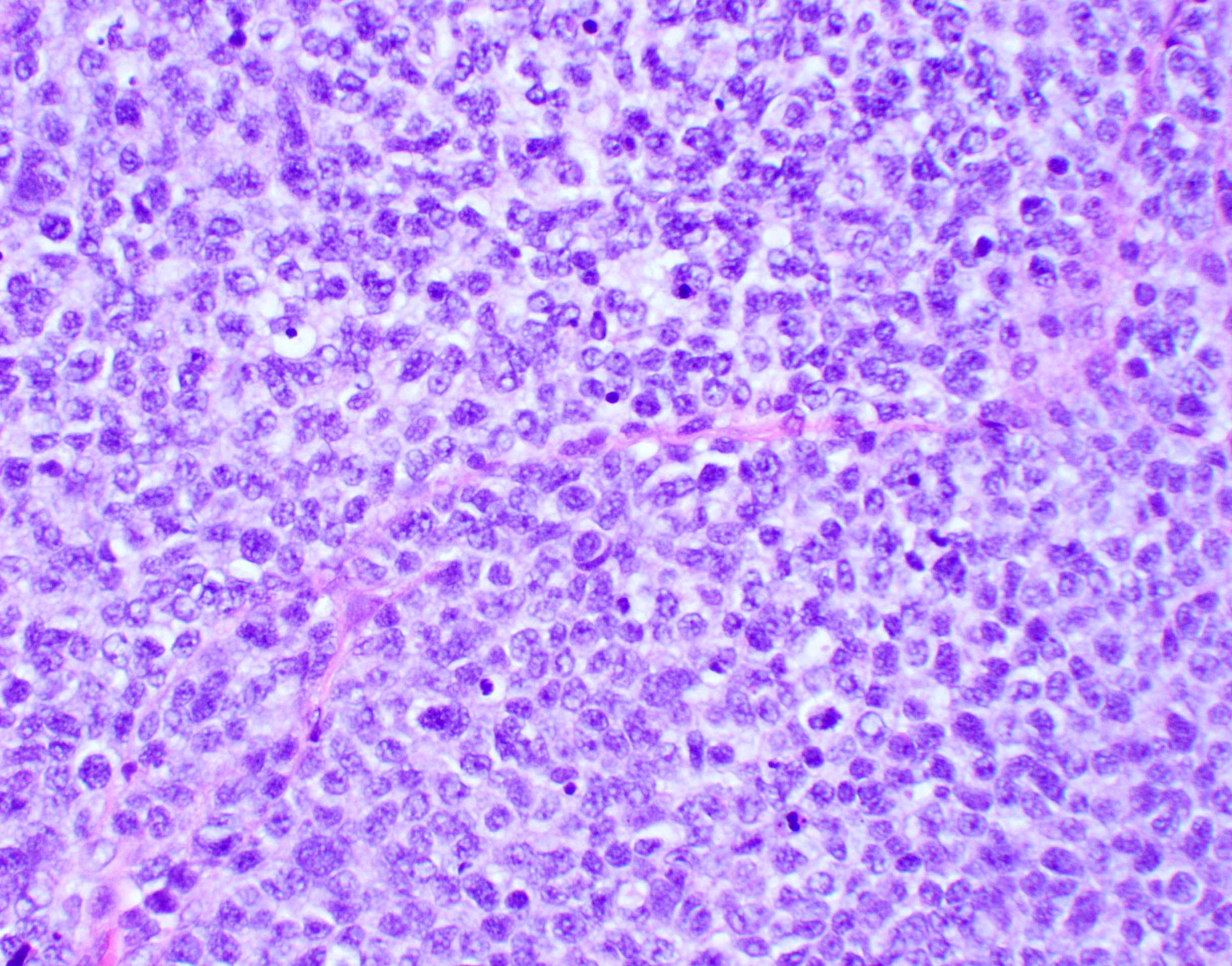
Hypercellular with clustering
Area of necrosis
Marked pleomorphism

ATRX
GFAP

Synaptophysin

p53
- See Case reports
Contributed by Katherine Schwetye, M.D., Ph.D.
Oligodendroglial-like appearance
Tumor adjacent parenchyma
Bland cytomorphology and nuclear features
S100 positive
Ki67 low
These photographs were previously published in Human Pathology, Vol. 70, Schwetye KE et al., "Unusual high-grade features in
pediatric diffuse leptomeningeal glioneuronal tumor: comparison with a typical low-grade example", p.105-112 Copyright Elsevier (2017).
Contributed by Mariana Voudouri, M.D. and George Zanazzi, M.D., Ph.D.
MRI coronal T2 brainstem tumor
MRI axial T2 / FLAIR right cerebellar tumor
MRI axial T1 post contrast right cerebellar flocculus tumor
Contributed by Mariana Voudouri, M.D. and George Zanazzi, M.D., Ph.D.

Astrocytic morphology
Pleomorphism
Oligodendroglial morphology
GFAP
ATRX
Ki67
Contributed by Rawia Mubarak Mohamed, M.D. and Najla Saleh Ben Gashir, M.D. (Case #477)
Tumor in thalamic region
Images hosted on other servers:

CT - axial noncontrast: pontine tumor

MRI - axial T2: pontine tumor

MRI - sagittal T1: pontine tumor

MRI - sagittal T2: pontine tumor

MRI - axial T2: pontine tumor
Contributed by John DeWitt, M.D., Ph.D.
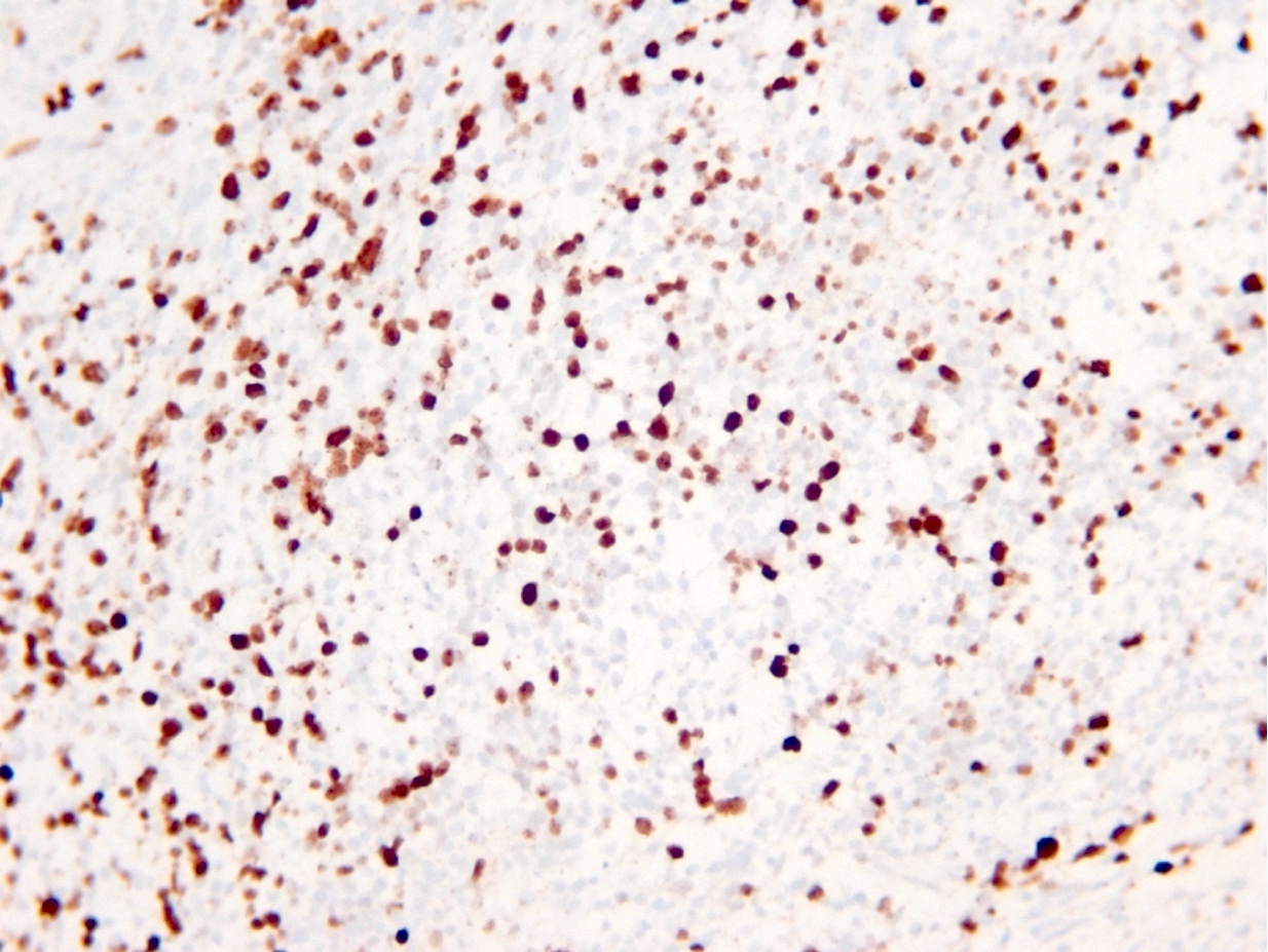
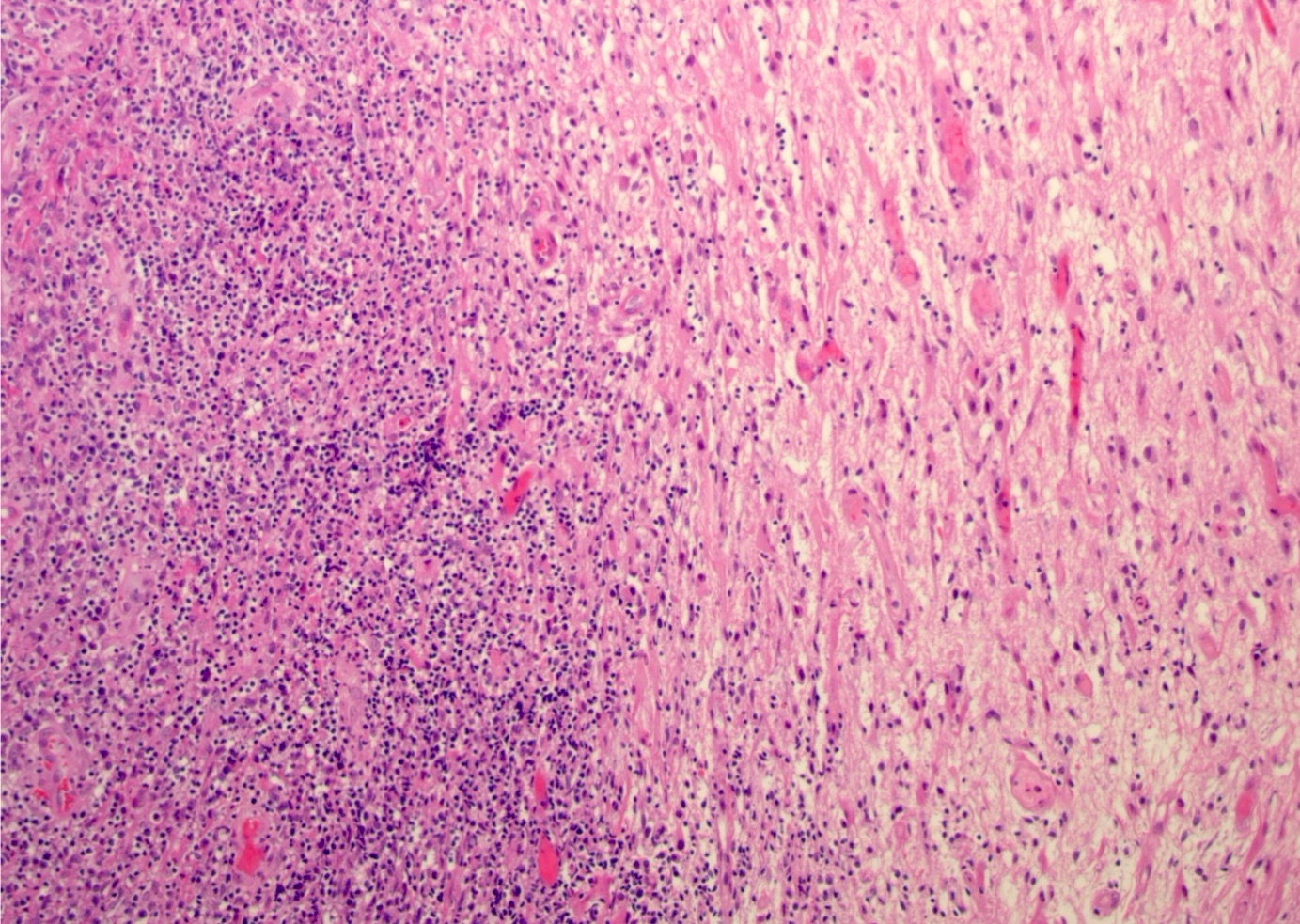
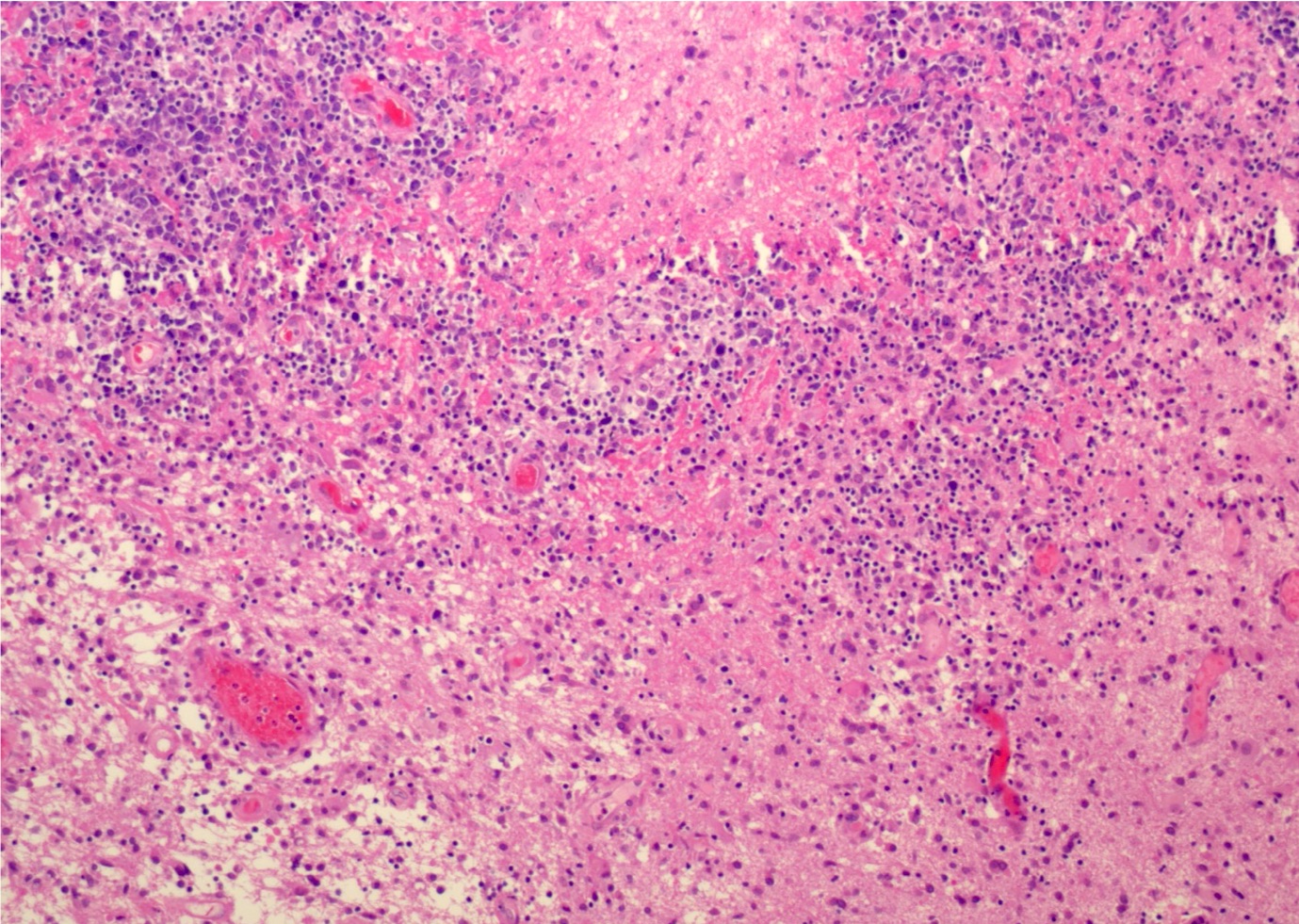
Diffusely infiltrating astrocytic tumor cells
Necrosis
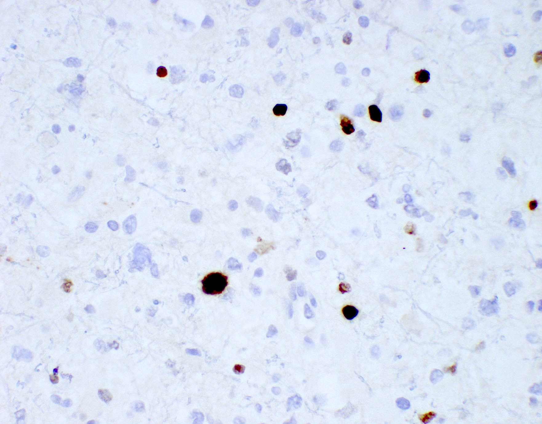
H3 K27M
R132H-IDH1
ATRX
p53
Contributed by Chunyu Cai, M.D., Ph.D.
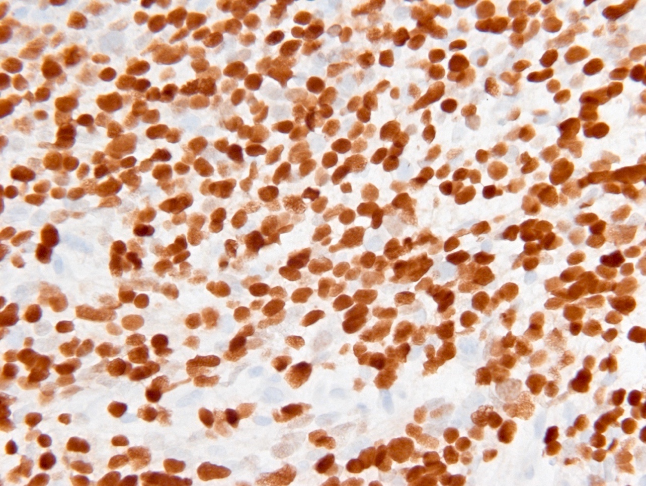
Clusters of tumor cell seeding
H3 K27M
H3K27Me3
Contributed by Rawia Mubarak Mohamed, M.D. and Najla Saleh Ben Gashir, M.D. (Case #477)

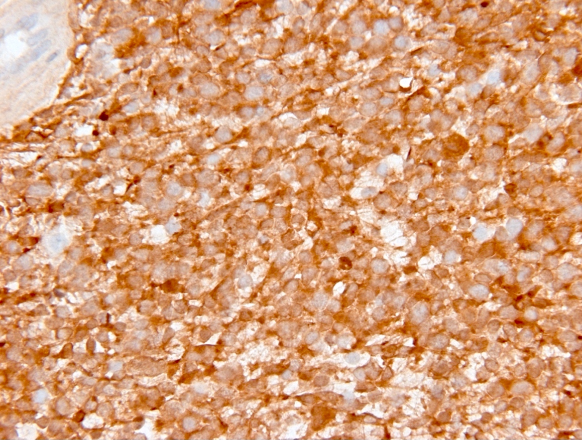
High grade infiltrating glial cells
Olig2
H3 K27M
Images hosted on other servers:

Relative distribution of LEATs

Proposed modification of LEAT classification

Nodular appearing neoplasm
Images hosted on other servers:

Representative imaging features in adolescent

Typical imaging features

10 year old girl with seizure
Images hosted on other servers:

Surgical resection of epileptogenic tumor
Images hosted on other servers:

Frontobasal surgical specimen
Contributed by P.J. Cimino, M.D., Ph.D. and Chris Dampier, M.D.
Nodular growth pattern
Specific glioneuronal (pathognomonic) component
Floating neuron
Cystic component
GFAP
IDH1 p.R132H
Images hosted on other servers:

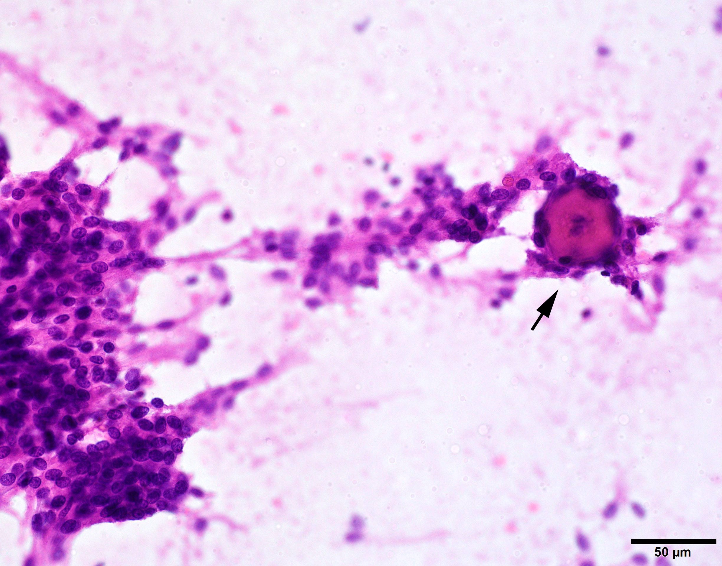
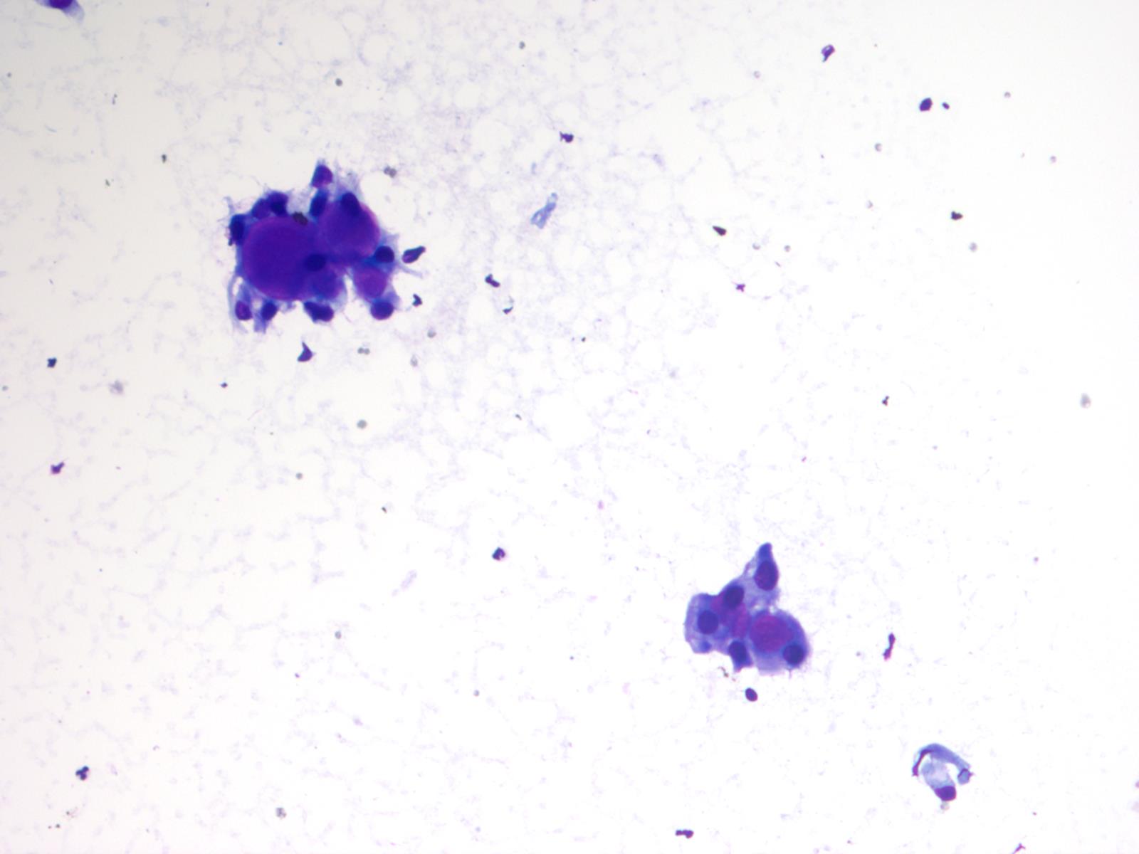
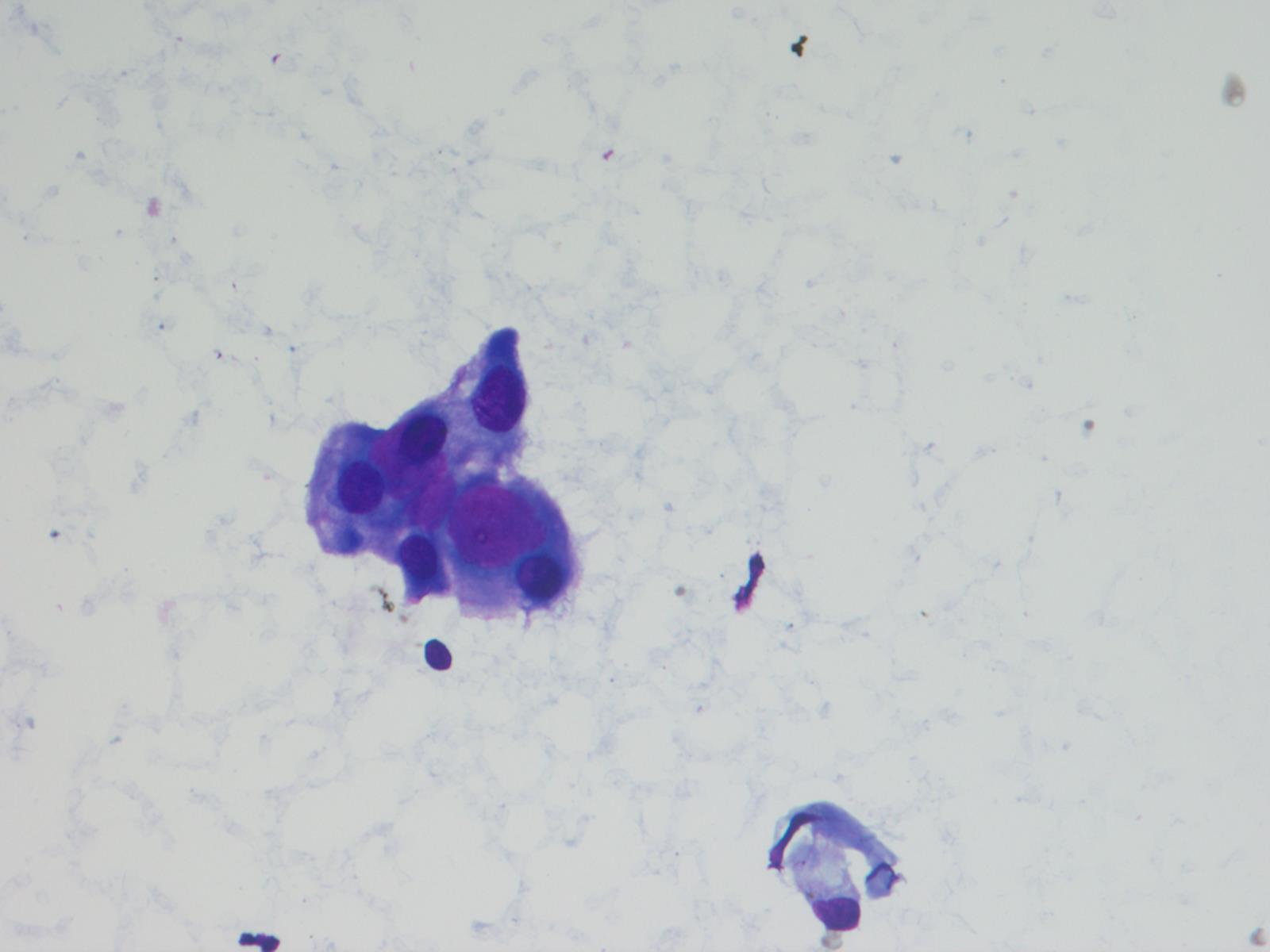
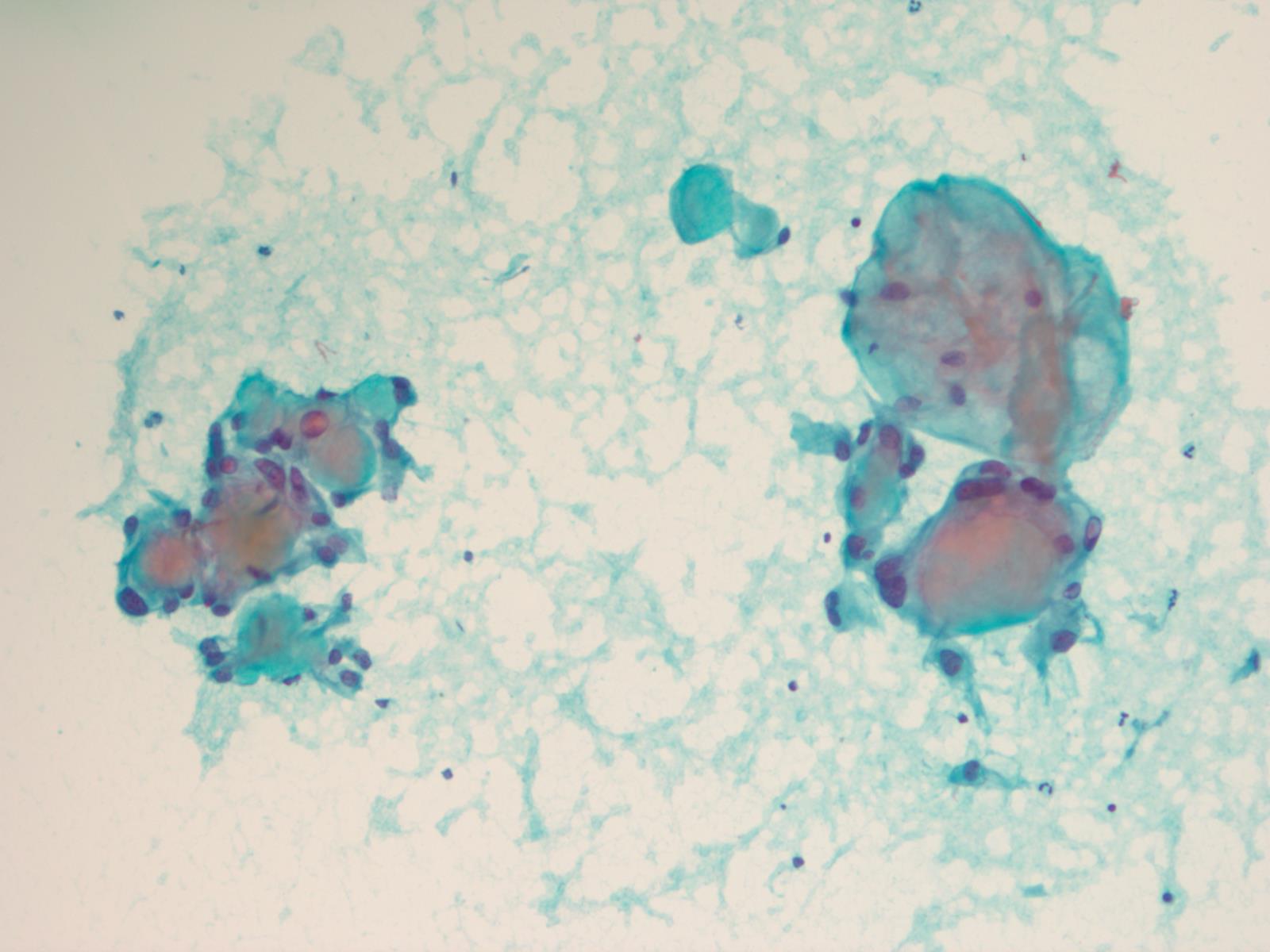
Smear preparations

Partially arranged as columnar structures

Fine neuritic processes
Contributed by P.J. Cimino, M.D., Ph.D. and Chris Dampier, M.D.

SCNA plot
Dysembryoplastic neuroepithelial tumor (DNET)
Contributed by Chunyu Cai, M.D., Ph.D.
MRI cervical spine ependymoma
MRI posterior fossa ependymoma
MRI supratentorial ependymoma
Images hosted on other servers:


MRI: ependymoma of fourth ventricle
Images hosted on other servers:


Arising from floor of fourth ventricle
Contributed by Chunyu Cai, M.D., Ph.D., Maria Martinez-Lage, M.D. and Eman Abdelzaher, M.D., Ph.D.
Perivascular pseudorosettes
Ependymal rosettes
True ependymal rosettes
Ependymoma canal
Tanycytic ependymoma
EMA showing dot-like and ring-like positivity
GFAP
EMA
MAP2
H3K27me IHC PFB
Ependymal tumors by Dr. Rodriguez
Images hosted on other servers:

Cerebellopontine angle
Contributed by Jared T. Ahrendsen, M.D., Ph.D.

Temporal lobe mass
Contributed by Jared T. Ahrendsen, M.D., Ph.D.
Well circumscribed brain lesion
Mixed glioneuronal tumor
Ganglioglioma with binucleate neurons
Well demarcated mass

Dysplastic ganglion-like cells in gangliocytoma
Olig2 IHC
CD34 IHC

Synaptophysin IHC
Neuronal and glioneuronal tumors
by Dr. Fausto Rodriguez (PathCast)
Contributed by Bharat Ramlal, M.D.
T2 FLAIR MRI

T1 postcontrast MRI
Contributed by Bharat Ramlal, M.D. and Meaghan Morris, M.D., Ph.D.
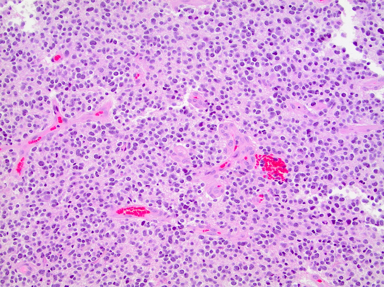
Hypercellular neoplasm
Nuclear atypia and brisk mitotic activity
Atypical astrocytic tumor cells
Glomeruloid microvascular proliferation

Pseudopalisading necrosis
Marked nuclear pleomorphism
Primitive neuronal component
Adenoid morphology

Epithelioid glioblastoma

Granular cell morphology
Small cell glioblastoma

IDH1 R132H

GFAP
Olig2

Ki67

Synaptophysin
Images hosted on other servers:

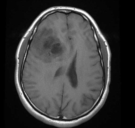
MR: cystic lesion of right frontal lobe
Images hosted on other servers:

Ependymal cyst
Contributed by Jared T. Ahrendsen, M.D., Ph.D.
Glioblastoma MRI
Glioma MRI
Optic pathway glioma MRI
Spinal ependymoma MRI
Contributed by Jared T. Ahrendsen, M.D., Ph.D.
Circumscribed glioma
Pilocytic astrocytoma
Pleomorphic xanthoastrocytoma (PXA)
Glioma with infiltrating edge
Diffuse glioma entrapped neurons
High grade infiltrating glioma
Perivascular pseudorosettes
GFAP positive glioma
Contributed by Nicolas Kostelecky, M.D.
Sagittal T1 postcontrast
Coronal T1 postcontrast
Images hosted on other servers:

Spectrum of
radiologic
phenotypes
(n = 4)

Heterogeneous, solid / cystic, with rim enhancement

Solid / cystic with leptomeningeal involvement

Cystic / solid; temporoparietal region

Before surgery
Contributed by Irfan Yasin, M.B.B.S., Katherine Schwetye, M.D., Ph.D. and Miguel Guzman, M.D.
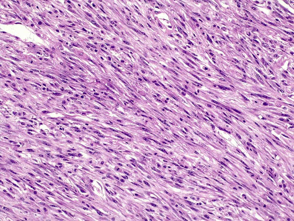
Glial and sarcomatous component
Sarcomatous component
Herringbone architecture
Severe atypia
GFAP
Reticulin
Images hosted on other servers:

VHL regulation of the HIF pathway
Images hosted on other servers:

T1+C MRI posterior fossa
Images hosted on other servers:

Cystic mass in medulla

Multiple spinal masses
Contributed by Rebecca Yoda, M.D.

Hemangioblastoma cut surfaces
Images hosted on other servers:

Variegated red-yellow tumor

Hemangioblastoma in situ
Contributed by P.J. Cimino, M.D., Ph.D.

Circumscribed border

Vascular and stromal cells
Foamy cytoplasm

Large branching vessels
Hyalinized stroma
Extensive hemorrhage
Nuclear atypia
Inhibin

CAIX
CD31

PAX8
Images hosted on other servers:

EM lipid droplets
Images hosted on other servers:

VHL next generation sequencing

Recurrent loss of chromosome 3

DNA methylation based classification

Idiogram of VHL mutation
Hemangioblastoma MRI
Hemangioblastoma histopathology
Case #488
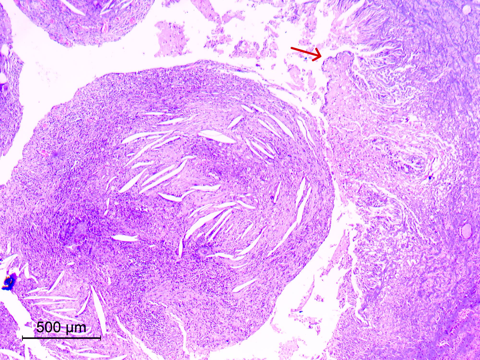
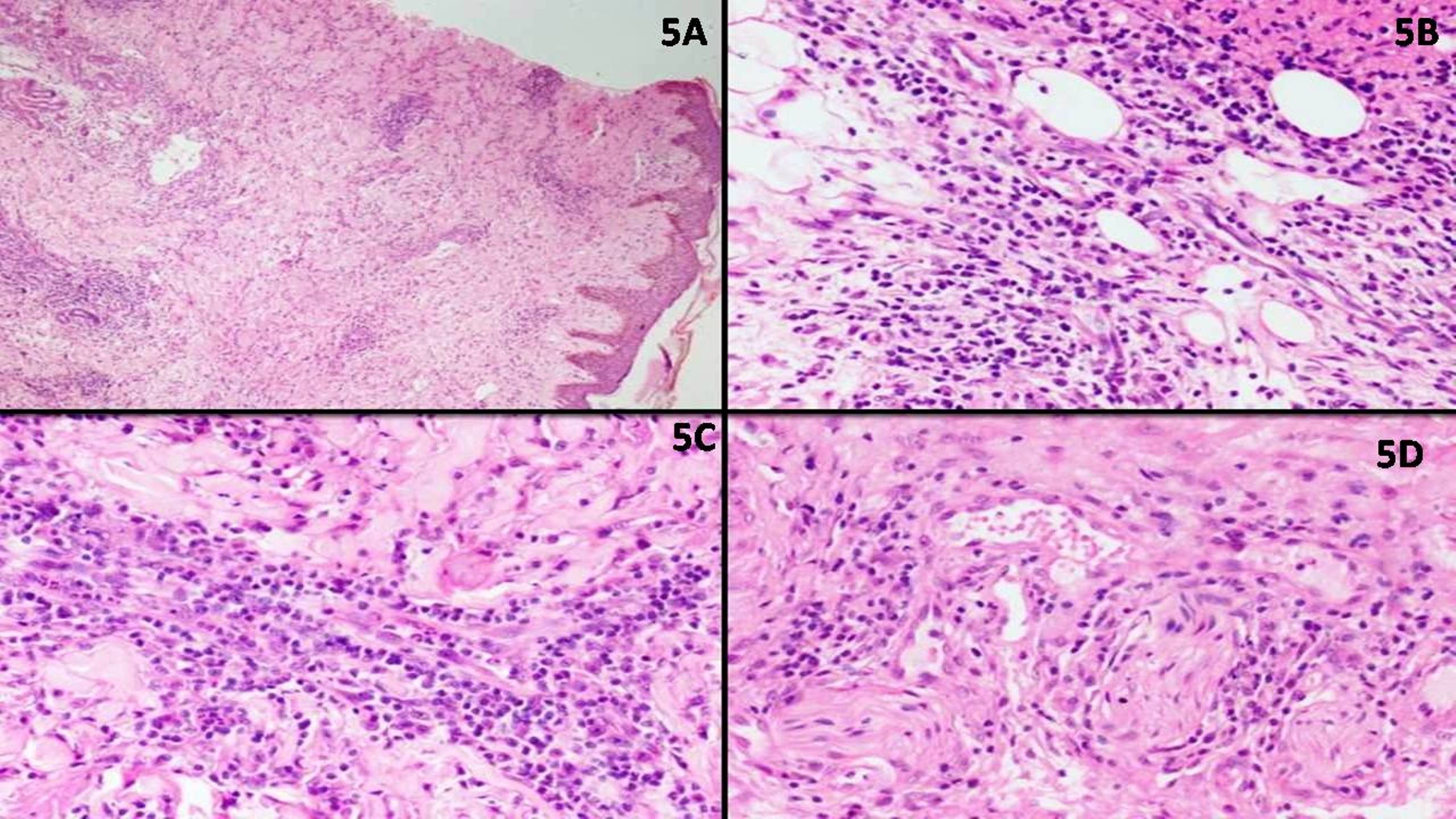
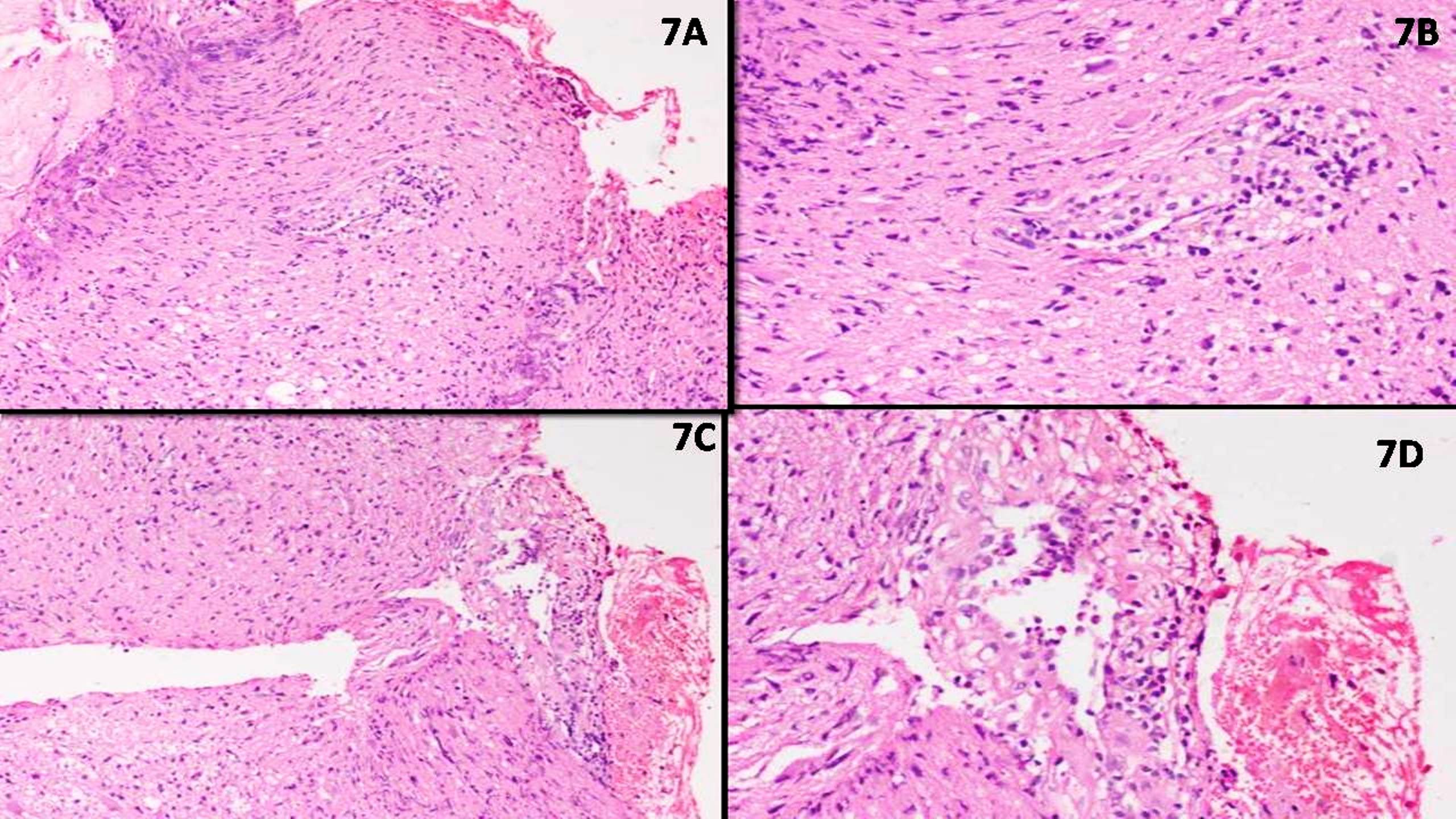
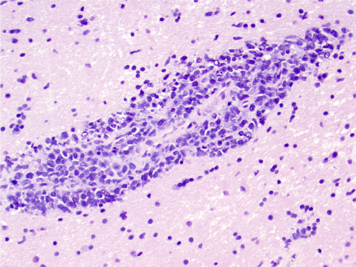
Rosai-Dorfman disease of CNS
Contributed by Kathryn Gibbons, M.D.
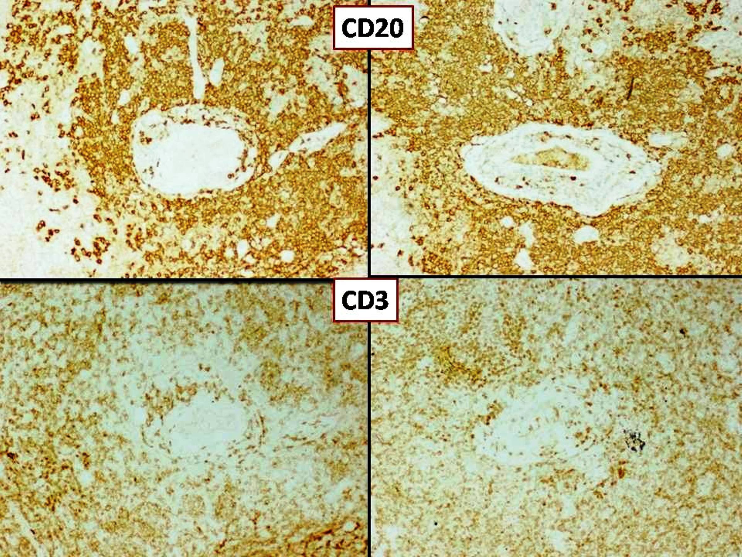
ILBCL involving skin and subcutaneous tissue
Large lymphoma cells in small vessels
ILBCL involving the brain parenchyma
Vessel with involvement by ILBCL
Large lymphoma cells in the vessels
CD20
BCL6
MUM1
Contributed by Nicholas Joseph Dcunha, M.B.B.S., M.D. and Elanthenral Sigamani, M.B.B.S., M.D.
Mass in lung (CT scan)
Contributed by Nicholas Joseph Dcunha, M.B.B.S., M.D. and Elanthenral Sigamani, M.B.B.S., M.D.
Lymphocytic vasculitis with angioinvasion (lung)
CD20 in tumor cells and CD3

CD30 and EBV LMP1 IHC
Lymphocytic vasculitis with angioinvasion (skin)
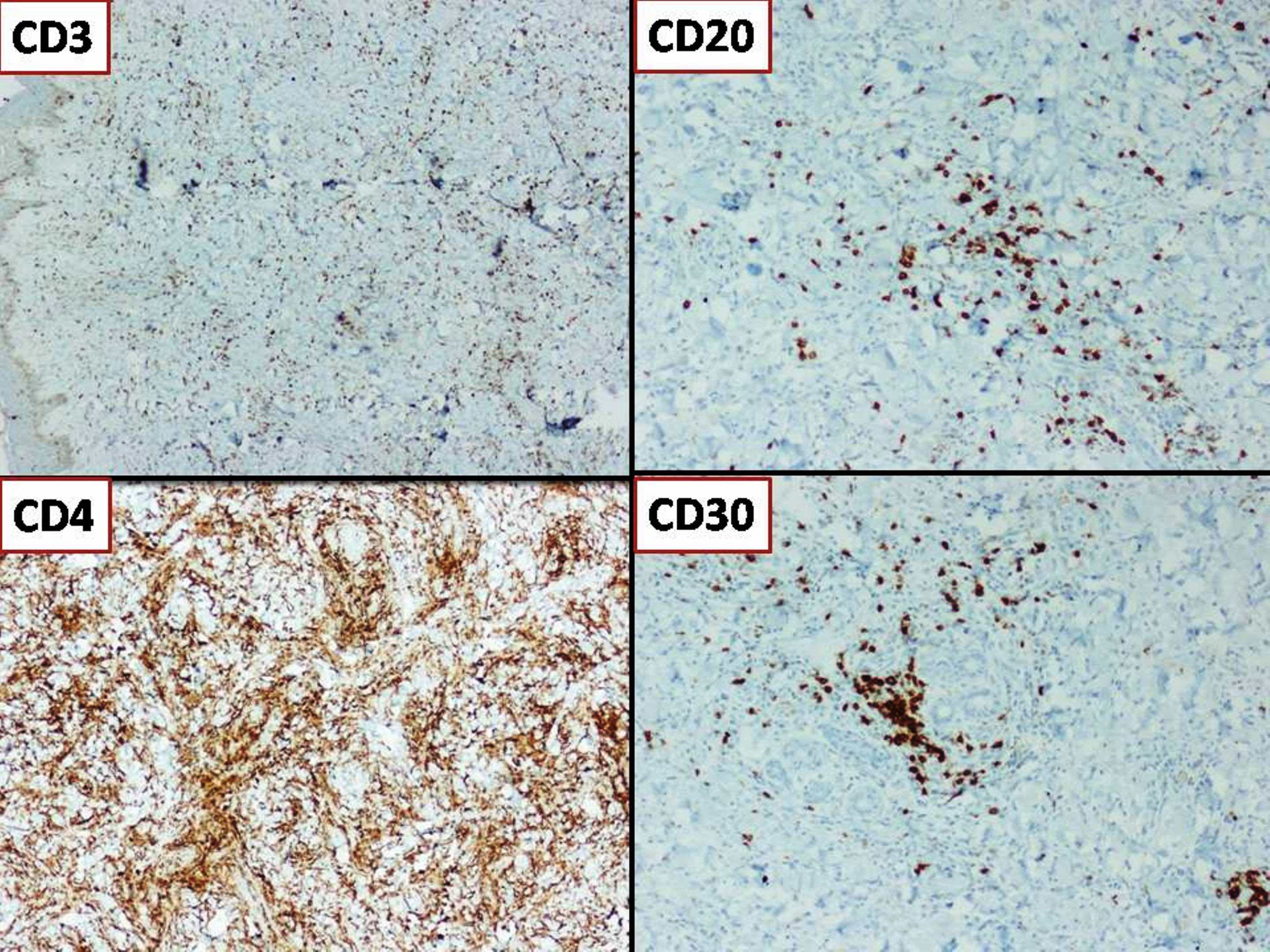
CD20 and CD30 in tumor cells; CD3 and CD4 in reactive T cells
Lymphocytic vasculitis with angioinvasion (CNS)
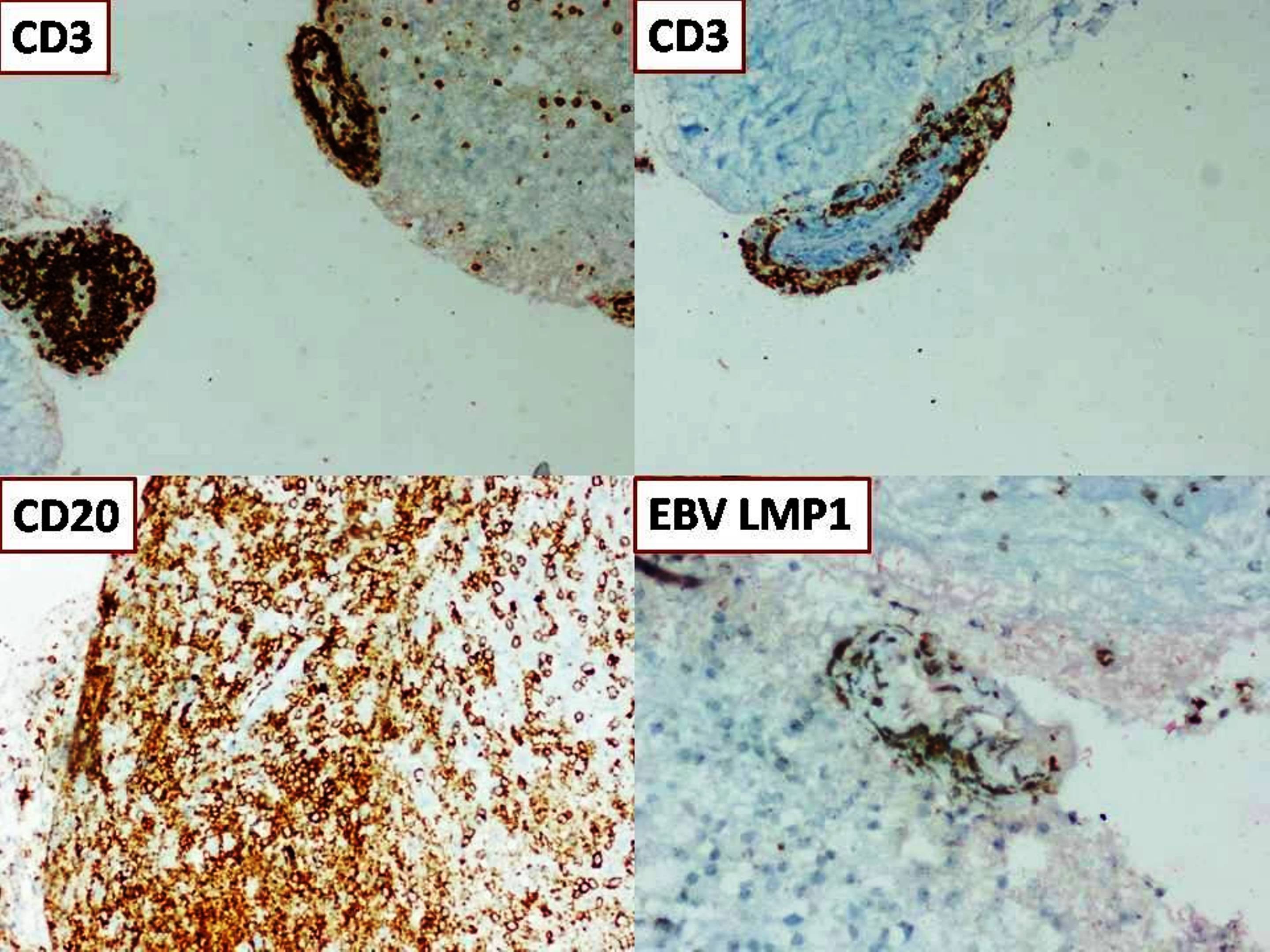
CD20 and EBV LMP1 in tumor cells; CD3 in reactive T cells
Images hosted on other servers:

Cerebellar tumor extending into fourth ventricle
Contributed by Arnault Tauziede-Espariat, M.D., John DeWitt, M.D., Ph.D., Meaghan Morris, M.D., Ph.D.,
Nirupama Singh, M.D., Ph.D. and Eman Abdelzaher, M.D., Ph.D.
Genetically defined
WNT (for wingless integration site) activated
YAP1 immunoreactivity
Beta catenin immunoreactivity
SHH (sonic hedgehog) activated (either TP53 mutant or TP53 wild type)
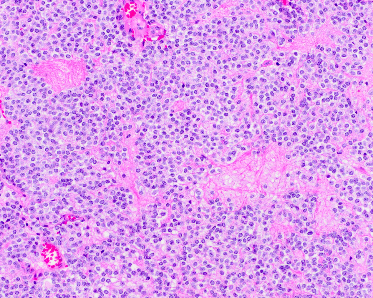
Sheets of undifferentiated cells
Vague nodular formation
Vague nodular formation with reticulin
Neuron specific enolase (NSE)
Ki67
Synaptophysin
Beta catenin
INI1
YAP1 immunoreactivity
GAB1 immunoreactivity
OTX2 immunoreactivity
Histologically defined
Classic
Homer Wright rosettes
Desmoplastic / nodular medulloblastoma
Desmoplastic medulloblastoma
Pale nodular islands
Pale islands formed of uniform cells
Tumor in the subarachnoid space
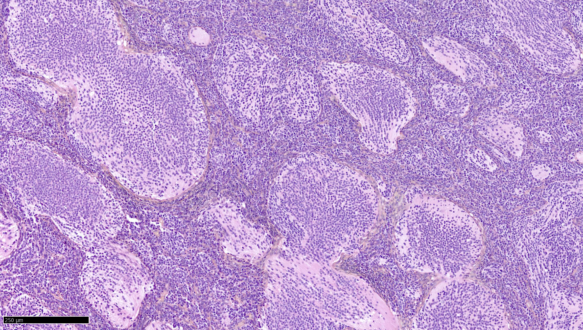
Desmoplastic / nodular medulloblastoma
Reticulin fiber network
Medulloblastoma with extensive nodularity
Extensive nodularity
Large cell / anaplastic medulloblastoma
Apoptotic bodies, prominent nucleoli
Cell wrapping
Anaplasia in medulloblastoma
Special morphology
Melanocytic differentiation

Myogenic differentiation
Contributed by Arnault Tauziede-Espariat, M.D.
MGG staining
Contributed by Arnault Tauziede-Espariat, M.D.
MYCN amplification
Images hosted on other servers:

Diffuse melanocytosis

MRI of thoracic area

Abnormal signal in the left temporal lobe
Contributed by Rana Al-Zaidi, M.B.B.S.


55 year old man with nausea, vomiting and sudden loss of consciousness and 4 cm temporal lesion - primary CNS melanoma
Contributed by Jesse Kresak, M.D.
Melanocytoma
Melanocytoma: S100
Melanocytoma: MelanA

Melanocytoma: HMB45
Images hosted on other servers:

Melanoma cells in CSF
Images hosted on other servers:

Various images (figures 1A - 4C)
Contributed by Chunyu Cai, M.D., Ph.D.
MRI benign meningioma
MRI atypical meningioma
MRI en plaque meningioma
MRI secretory meningioma
Images hosted on other servers:


Tumor displaces brain without invading

Compression of underlying cortex

Resected tumor

Adherent to falx cerebri
Contributed by Chunyu Cai, M.D., Ph.D.
Meningothelial meningioma:
Meningothelial meningioma
Meningothelial meningioma syncycial

Meningothelial meningioma SSTR2
Fibrous meningioma:
Fibrous meningioma
Fibrous meningioma SSTR2a
Transitional meningioma:
Transitional meningioma
Angiomatous meningioma:

Angiomatous meningioma macrovascular
Angiomatous meningioma microvascular
Microcystic meningioma:
Microcystic meningioma vacuolated cytoplasm
Microcystic meningioma nuclear pleomorphism
Clear cell meningioma:
Clear cell meningioma

Clear cell meningioma SMARCE1
Lymphoplasmacyte rich meningioma:
Lymphoplasmacyte
rich meningioma
Lymphoplasmacyte
rich meningioma
CD3

Lymphoplasmacyte
rich meningioma
SSTR2
Secretory meningioma:
Secretory meningioma
Secretory meningioma pCEA

Secretory meningioma EMA
Other meningioma:
Metaplastic meningioma

Chordoid meningioma
Psammomatous meningioma
Contributed by Chunyu Cai, M.D., Ph.D.
Streaked cytoplasm
Psammoma body

Cellular whorls
Images hosted on other servers:
Meningothelial tumor
Molecular classification of meningioma
Images hosted on other servers:

Lung metastasis
Case #222

Alveolar soft part sarcoma (H&E)
MyoD1

PASD
TFE3
Vimentin
Contributed by Ankur Sangoi, M.D. (Case #425)
Metastatic lung adenocarcinoma, ALK rearranged

Metastatic lung adenocarcinoma, ALK rearranged

Napsin A
TTF1

ALK break-apart FISH images (ALK gene in RED)
Images hosted on other servers:
Metastatic GI carcinoma (signet ring cells)
Contributed by Ahmed Ijaz Gilani, M.B.B.S., M.D., Ph.D.
MRI T1 with contrast
MRI T2
Contributed by Ahmed Ijaz Gilani, M.B.B.S., M.D., Ph.D. and Aisha Hassan Memon, M.B.B.S.
Radial arrangement
Epithelioid tumor cells
Papillary structures
Microcystic pattern
Hyalinized fibrovascular core with myxoid matrix
Alcian blue
GFAP
Olig2
EMA
Ki67
Contributed by Ahmed Ijaz Gilani, M.B.B.S., M.D., Ph.D. and Brittany Pakalniskis, M.D. (Case #399)
Stromal globules
Papillary structures and rosettes
Stromal globules (Diff-Quik stain)
Stromal globules (Pap stain)
Contributed by Jared T. Ahrendsen, M.D., Ph.D. and John DeWitt, M.D., Ph.D.
MRI, brain: T1 post
MRI, brain: T2
MRI: frontal lobe tumor with cystic change
Images hosted on other servers:

Axial flair frontal lobe tumor

Axial flair large temporal lobe tumor
Contributed by Jared T. Ahrendsen, M.D., Ph.D. and John DeWitt, M.D., Ph.D.
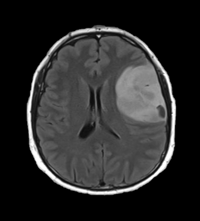
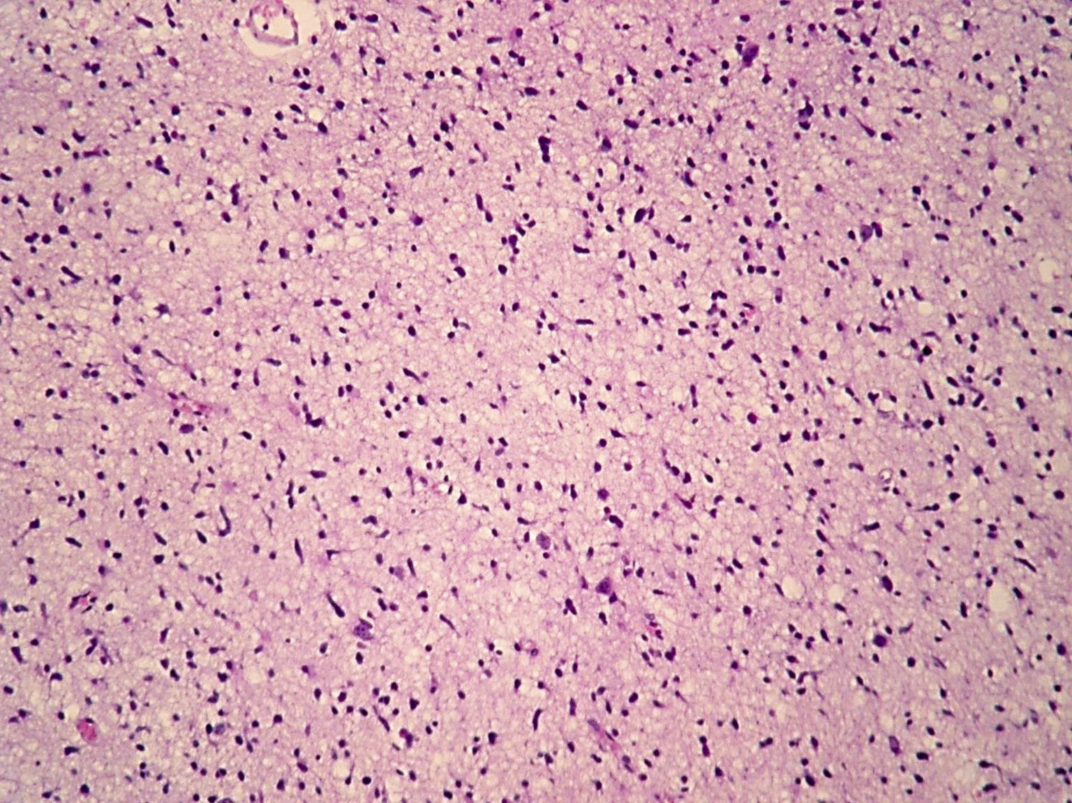
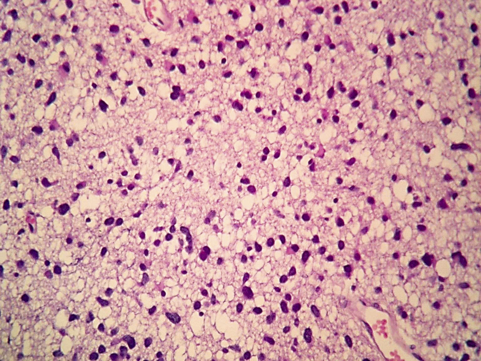
Diffusely infiltrating glial neoplasm
Microcalcifications
Perinuclear halos
Chicken wire vasculature
Secondary structures of Scherer
Cellularity and vascular proliferation
Focal necrosis
Brisk mitotic activity
Mixed astrocytic histology
IDH1 R132H

p53
ATRX
Contributed by Chunyu "Hunter" Cai, M.D., Ph.D.
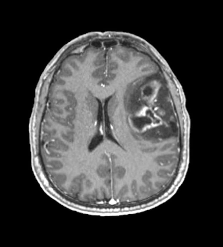
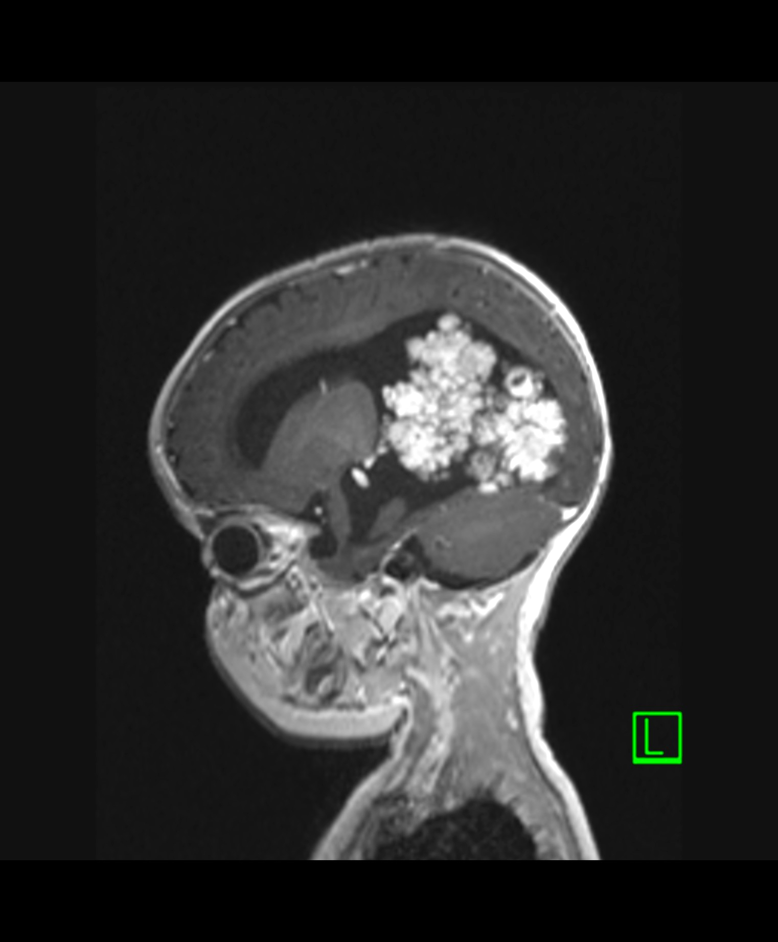
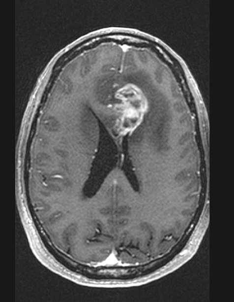
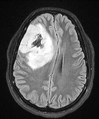
T1 precontrast sagittal
T1 precontrast coronal
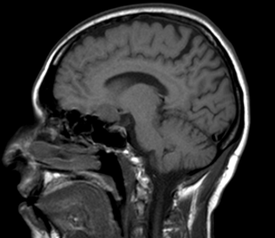
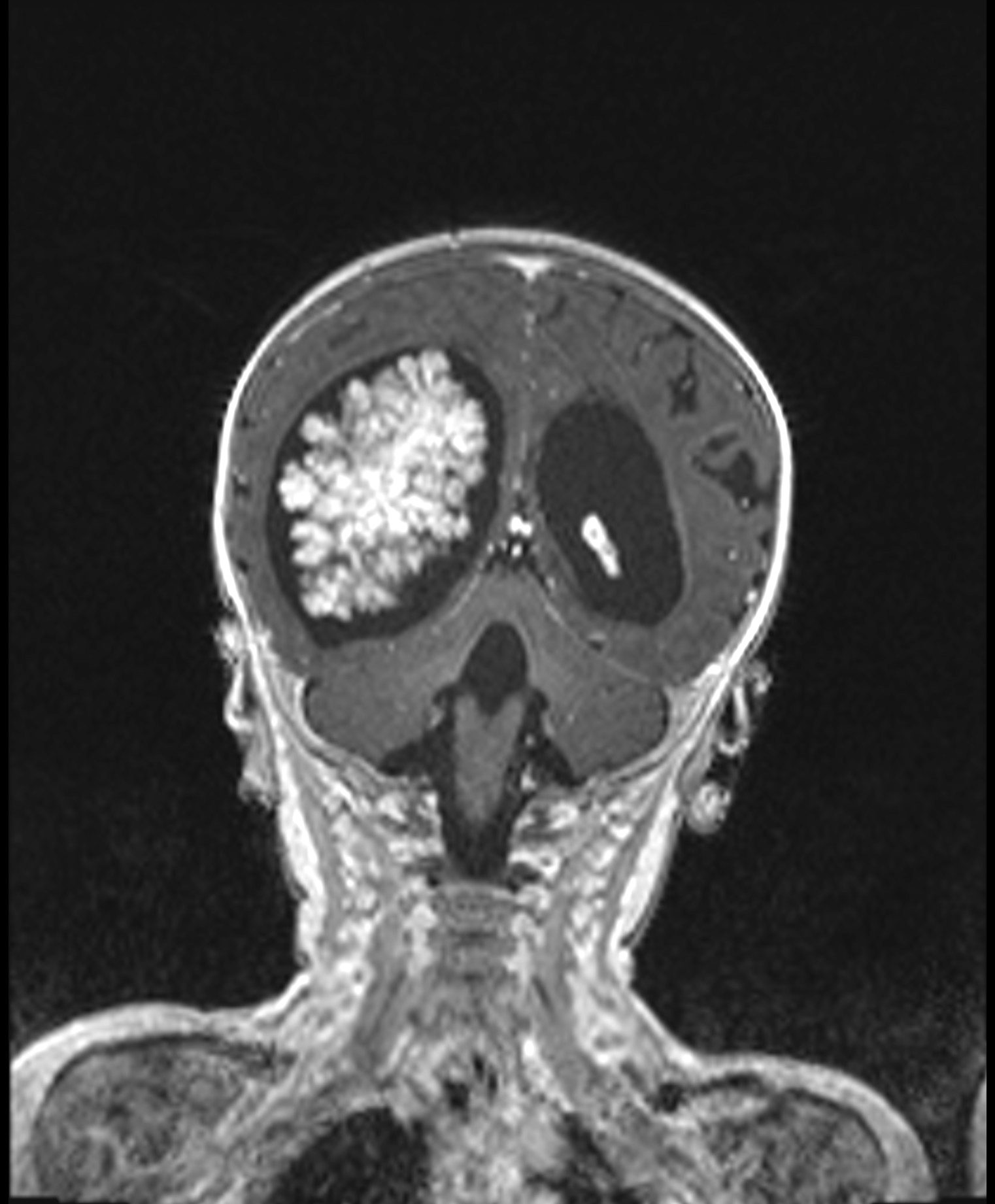
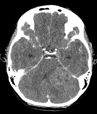
Images hosted on other servers:

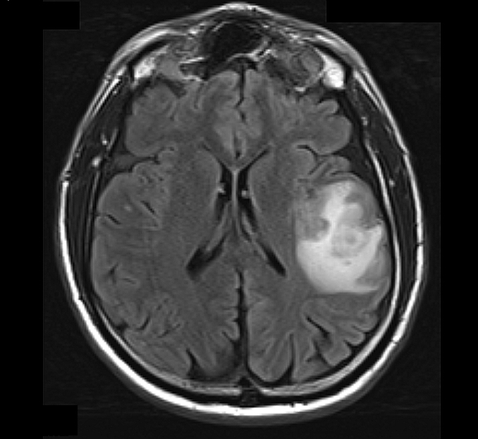
Suprasellar mass

Solid suprasellar mass with small cystic component

Preoperative MRI
Contributed by Nelli S. Lakis, M.D., M.Sc. and Chunyu "Hunter" Cai, M.D., Ph.D.
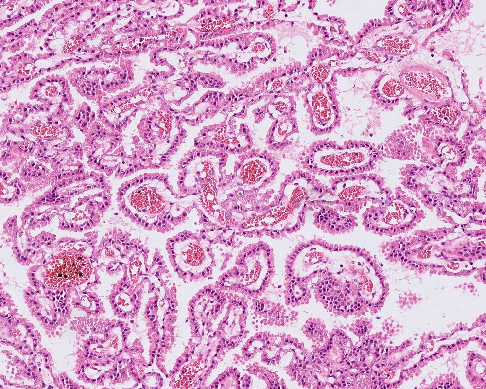
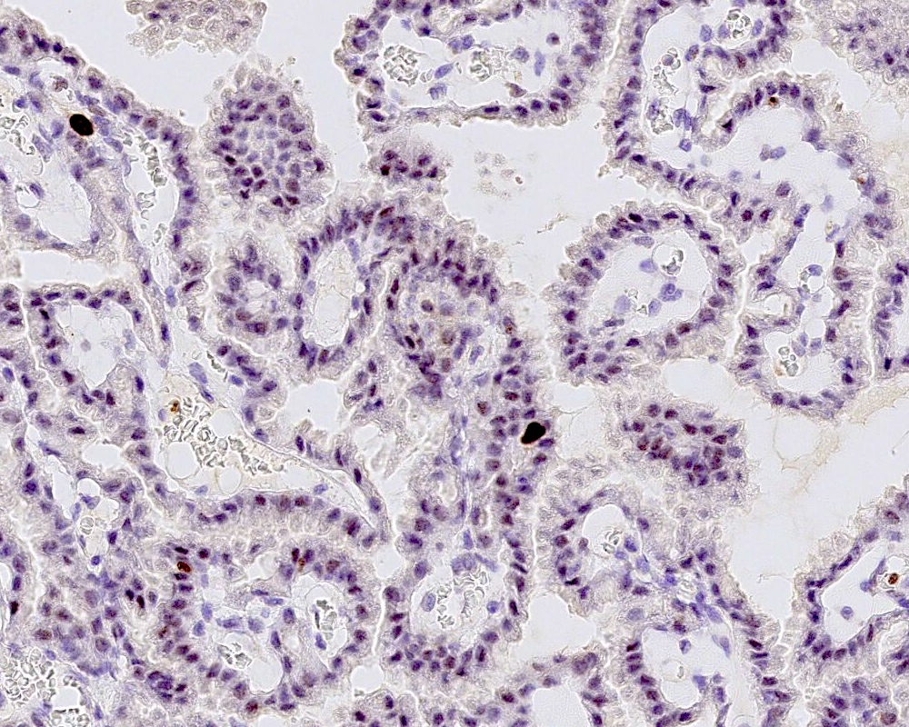
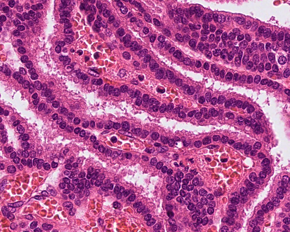
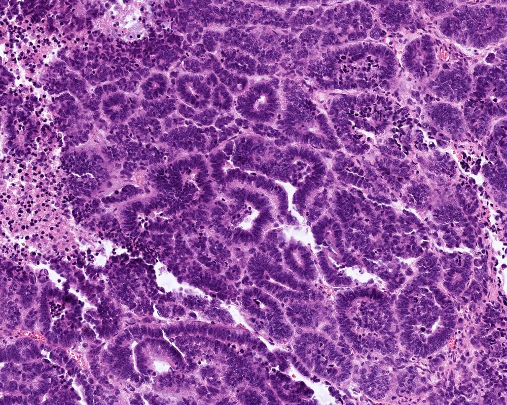
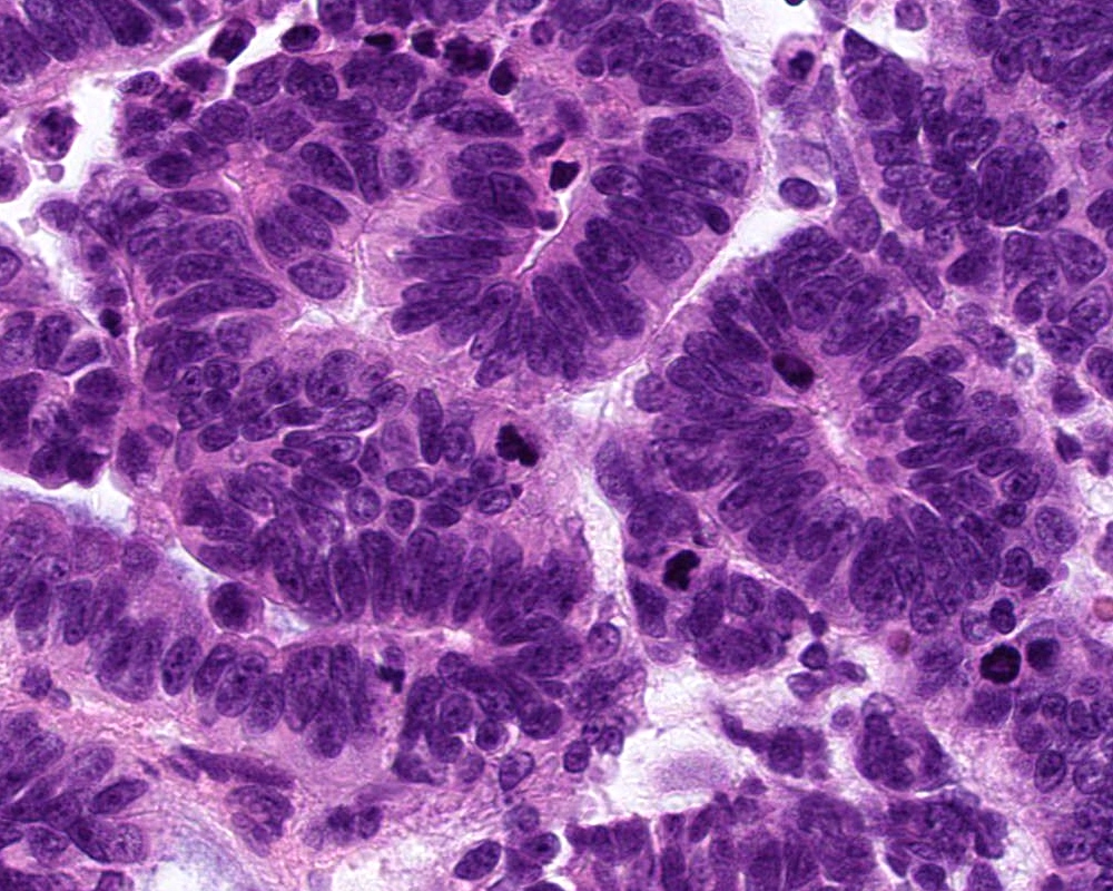
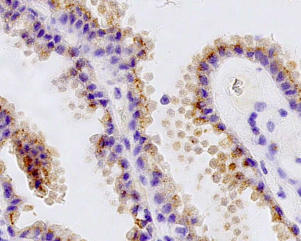
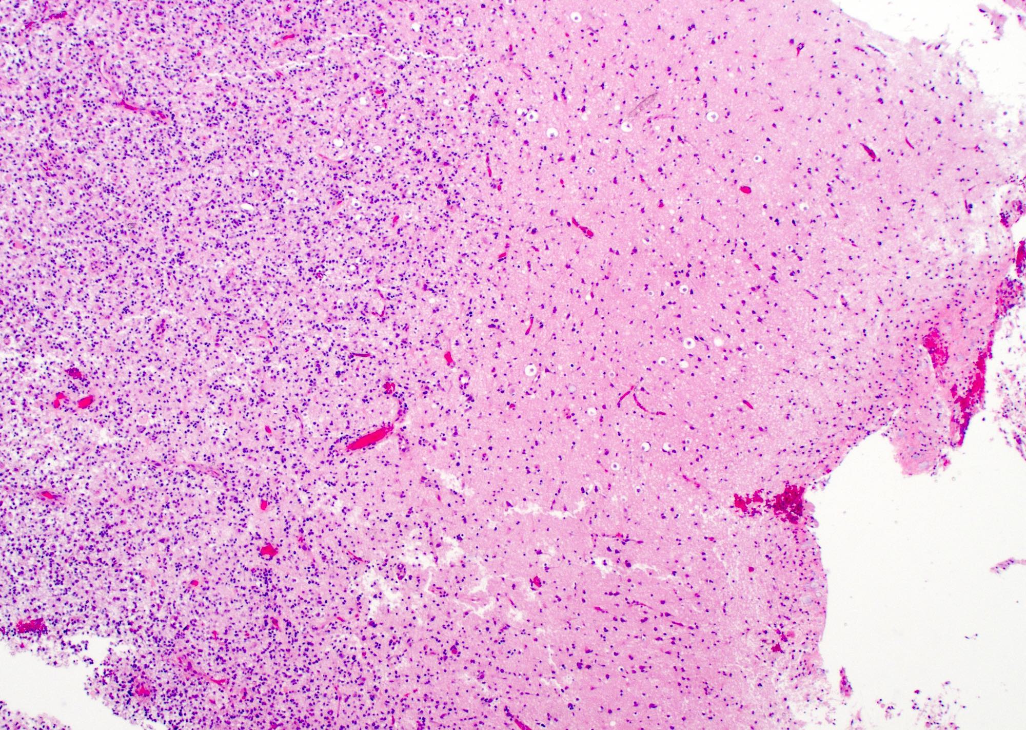
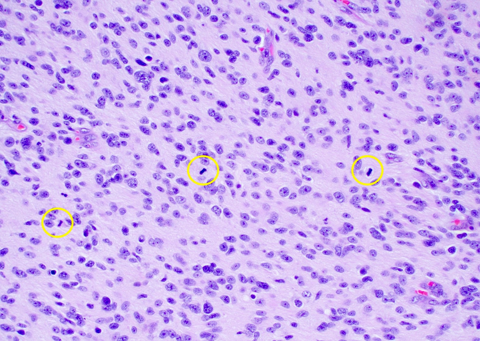
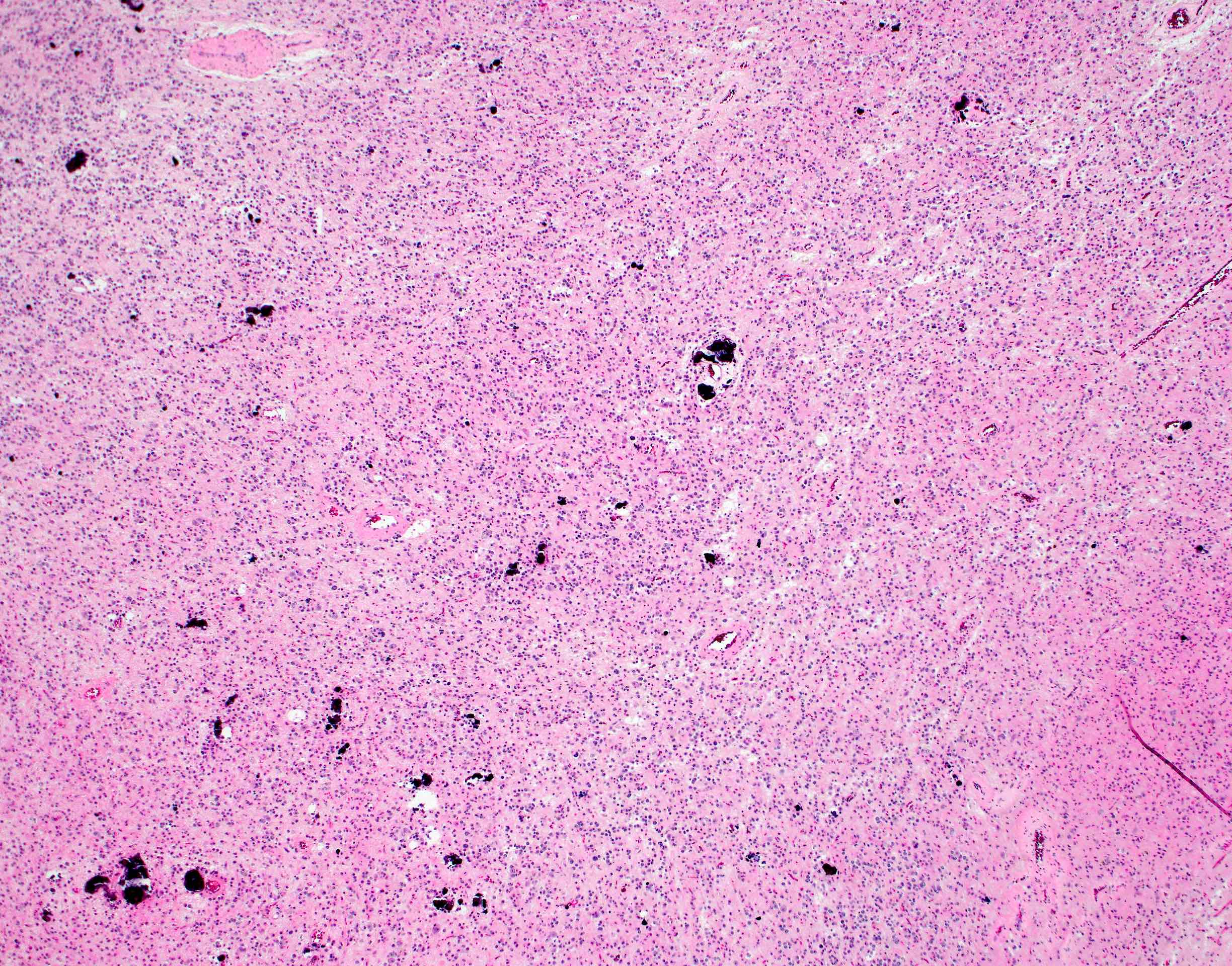
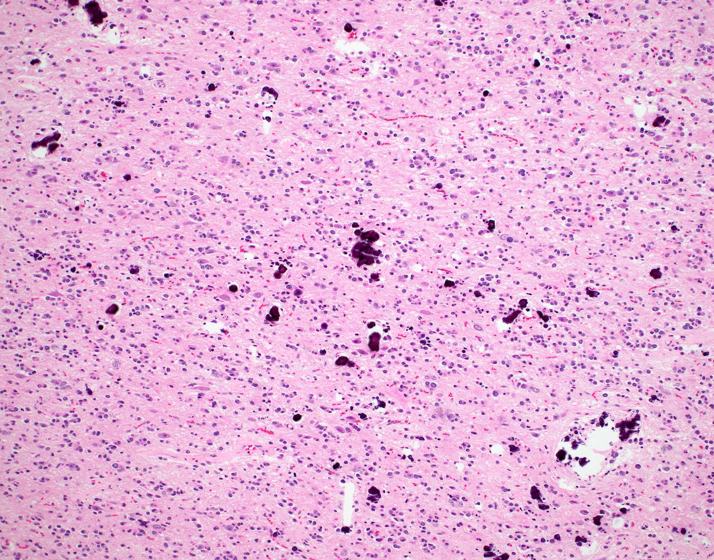
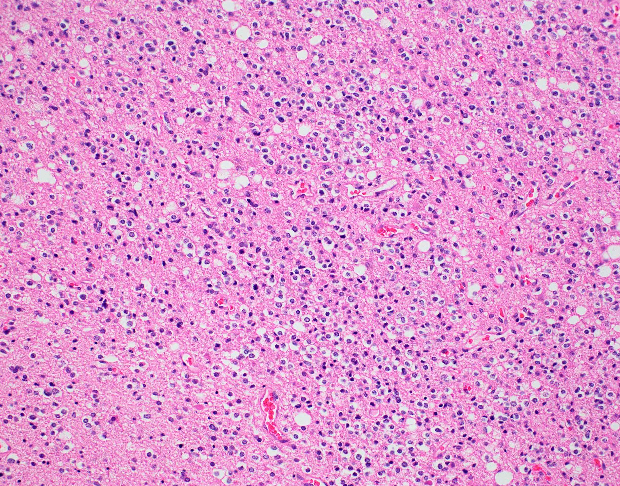
Papillary architecture
Fibrovascular cores
Stroma
Lymphocytic inflammation
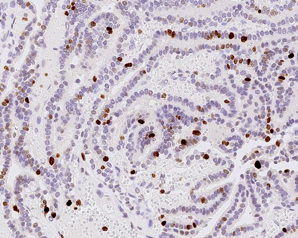
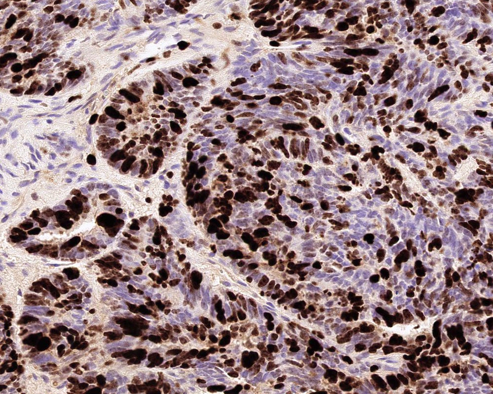
BRAF V600E immunostain
Images hosted on other servers:

Heterogeneously enhancing mass in pineal gland
Images hosted on other servers:

Right frontal papillary meningioma

Large, lobulated, irregular mass

Mass with a broad base

Postoperative shows severe leptomeningeal seeding

Avidly enhancing meningioma

Preoperative sagittal and axial adolinium enhanced MR images
Images hosted on other servers:

Meningioma smear
Images hosted on other servers:

Bundles of intermediate filaments
Images hosted on other servers:

MAPK pathway mutations
Contributed by P.J. Cimino, M.D., Ph.D.
T1 postcontrast MRI
Images hosted on other servers:

Intraoperative surgical image

Exophthalmos - optic nerve glioma

Orbital pilocytic astrocytoma
Images hosted on other servers:

Intraocular pilocytic astrocytoma

Cerebellar tumor
Contributed by P.J. Cimino, M.D., Ph.D.
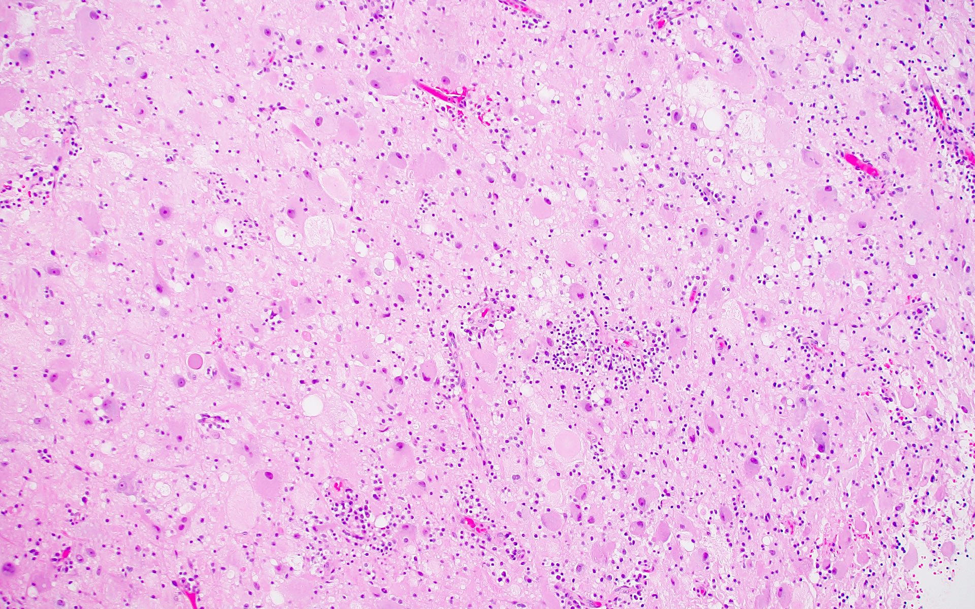
Biphasic appearance
Compact fibrillary area
Cystic area
Rosenthal fibers
Eosinophilic granular bodies
Microvascular proliferation
Multinucleated cells
Hyalinized vessels
Extensive regressive features
Oligodendroglial-like
cytomorphology
Mitosis

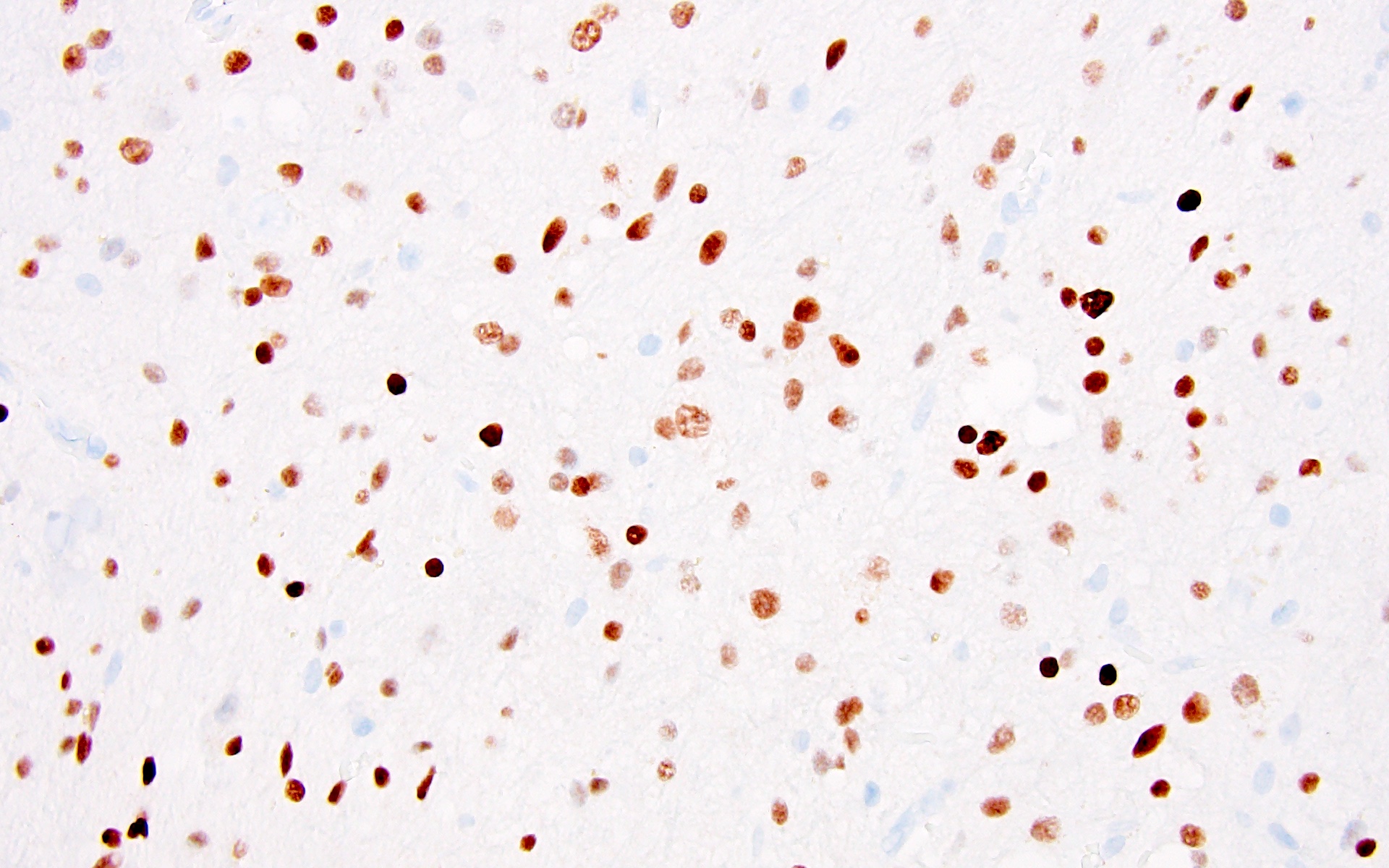
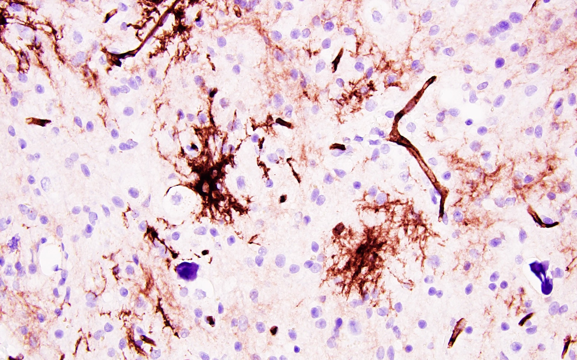
GFAP piloid processes
Rosenthal fiber - GFAP
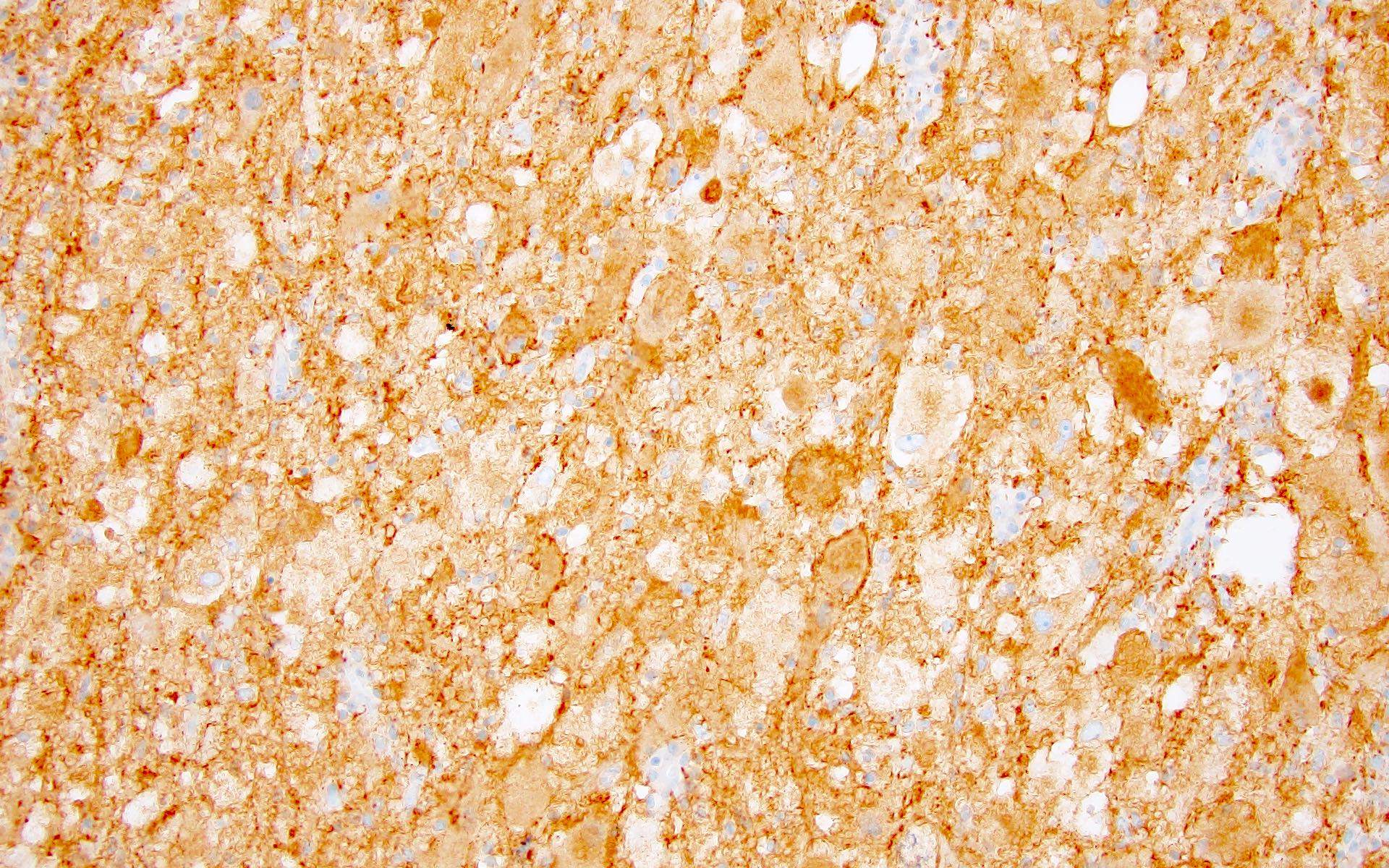
ATRX retained nuclear immunoreactivity
Increased Ki67 with anaplasia
Masson trichrome - EGBs
Images hosted on other servers:

Eosinophilic granular bodies
Images hosted on other servers:

KIAA1549-BRAF fusion by FISH

Copy number variation plot

NTRK2 fusion by next generation sequencing

NF1 biallelic inactivation

Pilocytic astrocytoma methylation classifier
Pilocytic astrocytoma
Rosenthal fibers in pilocytic astrocytoma


Images hosted on other servers:

Chiasmatic / hypothalamic pilomyxoid astrocytoma in a 3 month old infant:
DWI: no sign of restricted diffusion
T2w: homogeneous hyperintense signal
T1w: hypointense signal
T1w+ contrast: solid mass with marked enhancement
Contributed by Eman Abdelzaher, M.D., Ph.D.
Angiocentric pattern
Monomorphous cells in a myxoid background
Images hosted on other servers:

Bipolar, thin processes
extending to basal
lamina of vessels;
dense core granules
Images hosted on other servers:

Pineal cyst
Images hosted on other servers:

Small blue cell neuroepithelial malignant tumor
Images hosted on other servers:


Presence of abundant filopodia and rare intercellular junctions but no definitive synapses
Contributed by Jared T. Ahrendsen, M.D., Ph.D.

Axial MRI T1 postcontrast

Axial MRI T2
Contributed by Jared T. Ahrendsen, M.D., Ph.D.

With necrosis


Histology
Homer-Wright pseudorosettes

Mitotic activity
Synaptophysin positive

Variably chromogranin positive
High Ki67
Images hosted on other servers:

Overview of pineoblastoma subtypes
Contributed by Federico Roncaroli, M.D.
Sagittal postcontrast MRI
Contributed by Federico Roncaroli, M.D. and Sylvia L. Asa, M.D., Ph.D.
Fascicles of spindle cells

Ill defined whorls
Schwannoma-like features
Mild nuclear atypia

Oncocytic pituicytoma
Oncocytic pituicytoma
Granular cell pituicytoma
GFAP expression
TTF1 nuclear expression
Images hosted on other servers:

Sagittal and coronal MRI
Images hosted on other servers:

Pituitary hyperplasia microscopic
| Tumor type | Transcription factor(s) | Hormone(s) | Other biomarkers | Clinical features |
| Tpit family | ||||
| Densely granulated corticotroph tumor | Tpit | ACTH | PAS; intense keratins | Florid Cushing disease or silent |
| Sparsely granulated corticotroph tumor | Tpit | ACTH (weak) | PAS (weak); strong keratins | Cushing disease or silent |
| Crooke cell tumor | Tpit | ACTH | PAS; ring-like keratins | Cushing disease or silent |
| Pit1 family | ||||
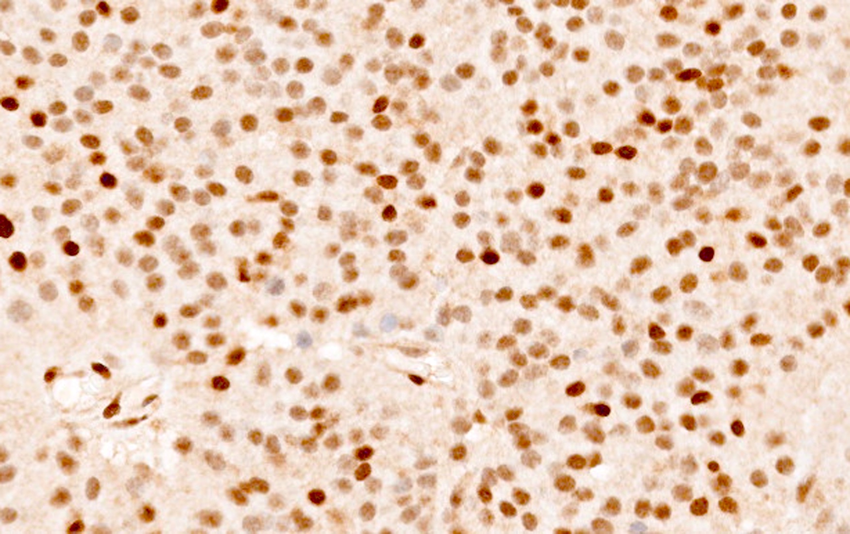
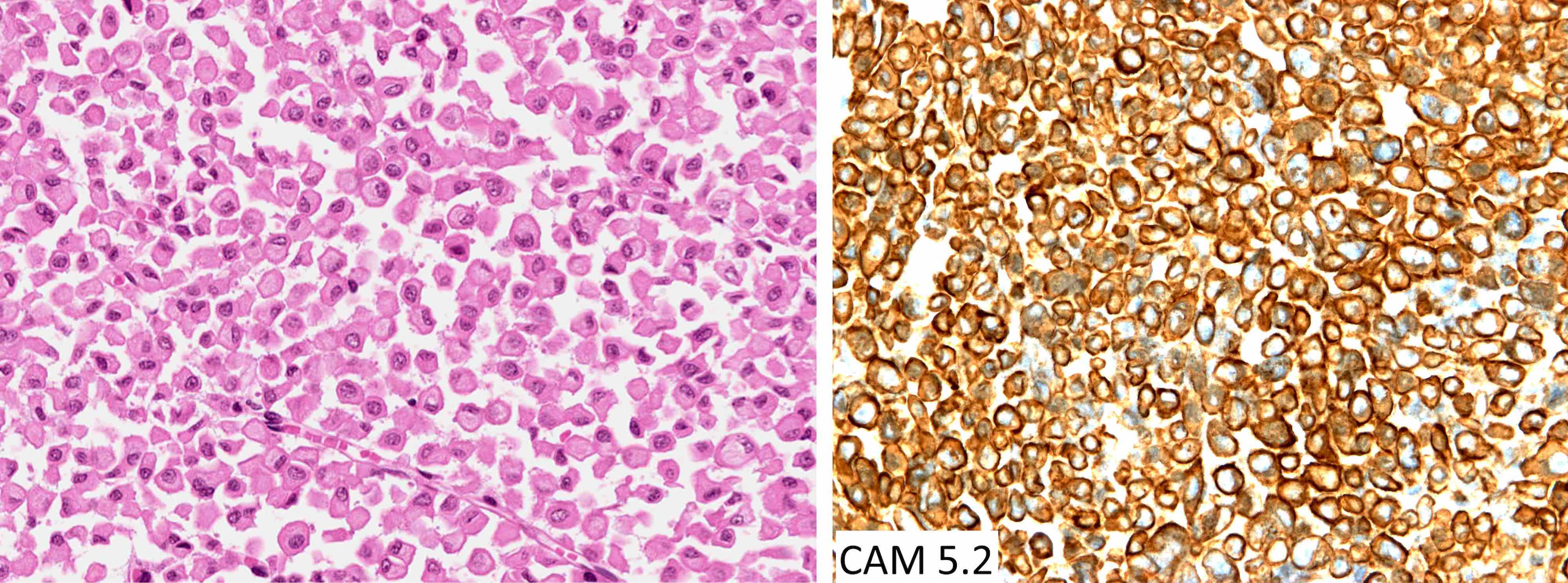
| Densely granulated somatotroph tumor | Pit1 | GH | Perinuclear keratins | Acromegaly |
| Sparsely granulated somatotroph tumor | Pit1 | GH (weak) | Keratin fibrous bodies (> 70%) | Acromegaly or silent |
| Mammosomatotroph tumor | Pit1, ER | GH, PRL | Perinuclear keratins | Acromegaly and hyperprolactinemia |
| Sparsely granulated lactotroph tumor | Pit1, ER | PRL (juxtanuclear) | Variable keratins | Hyperprolactinemia (tumor size correlates with PRL level) |
| Densely granulated lactotroph tumor | Pit1, ER | PRL | Variable keratins | Hyperprolactinemia |
| Acidophil stem cell tumor | Pit1, ER | PRL with or without GH (weak, focal) | Variable keratins with or without fibrous bodies | Hyperprolactinemia |
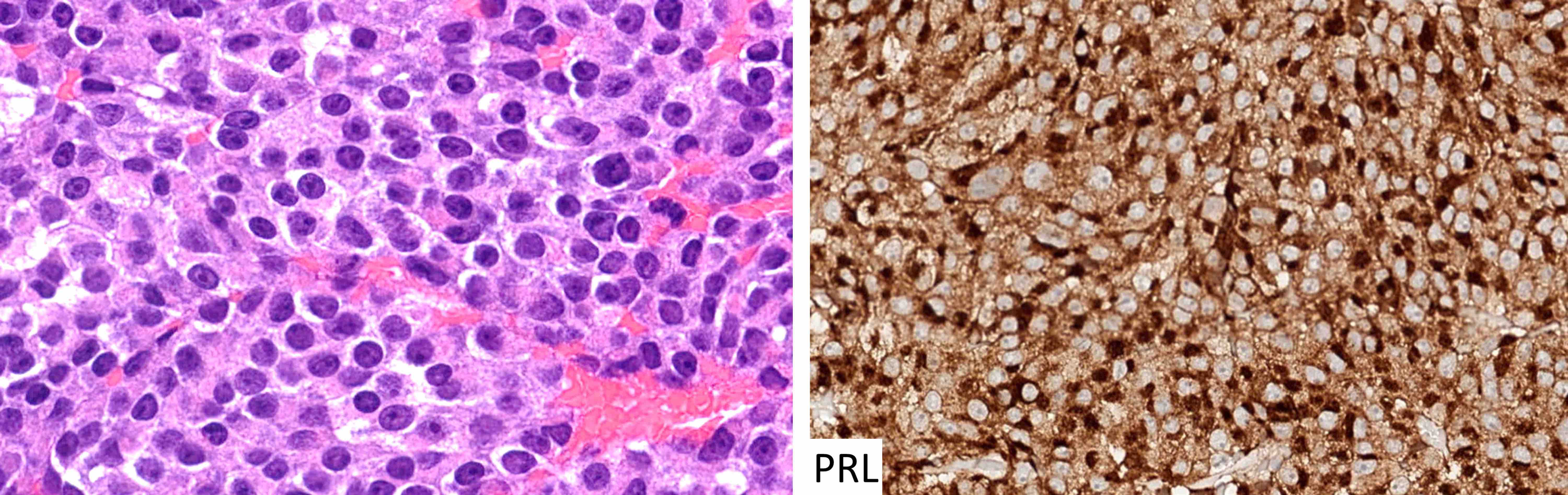
| Thyrotroph tumor | Pit1, GATA3 | TSH | Variable keratins | Elevated TSH with or without hyperthyroidism |
| Mature plurihormonal Pit1 lineage tumor | Pit1, GATA3, ER | Variable, often multiple | Perinuclear keratins | Acromegaly, hyperprolactinemia and hyperthyroidism |
| Immature Pit1 lineage tumor | Pit1 with or without GATA3 with or without ER | Variable, often limited / focal | Variable keratins with or without fibrous bodies | Silent or acromegaly or hyperprolactinemia or hyperthyroidism |
| SF1 family | ||||
| Gonadotroph tumor | SF1, GATA3, ER | FSH, LH | Variable keratins, follicular cells | Silent with or without elevated gonadotropins |
| No lineage fidelity | ||||
| Null cell tumor | None | None | Variable | Silent |
| Unclassified plurihormonal tumor | Multiple combinations | Multiple combinations | Variable | Variable |
Images hosted on other servers:


Sellar and suprasellar masses
Contributed by Sylvia L. Asa, M.D., Ph.D.
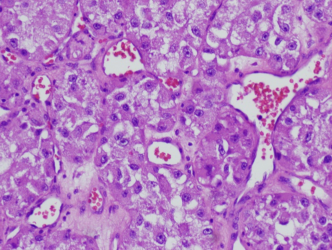
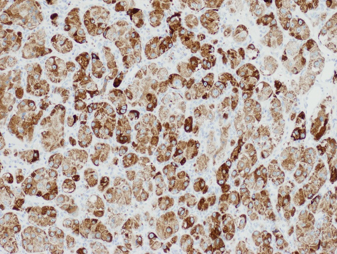
Densely granulated corticotroph tumor
Sparsely granulated corticotroph tumor
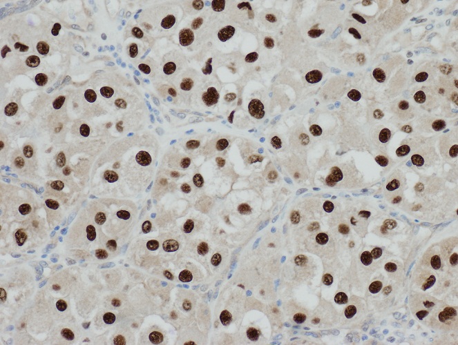
p27 in silent corticotroph tumor
Crooke cell tumor
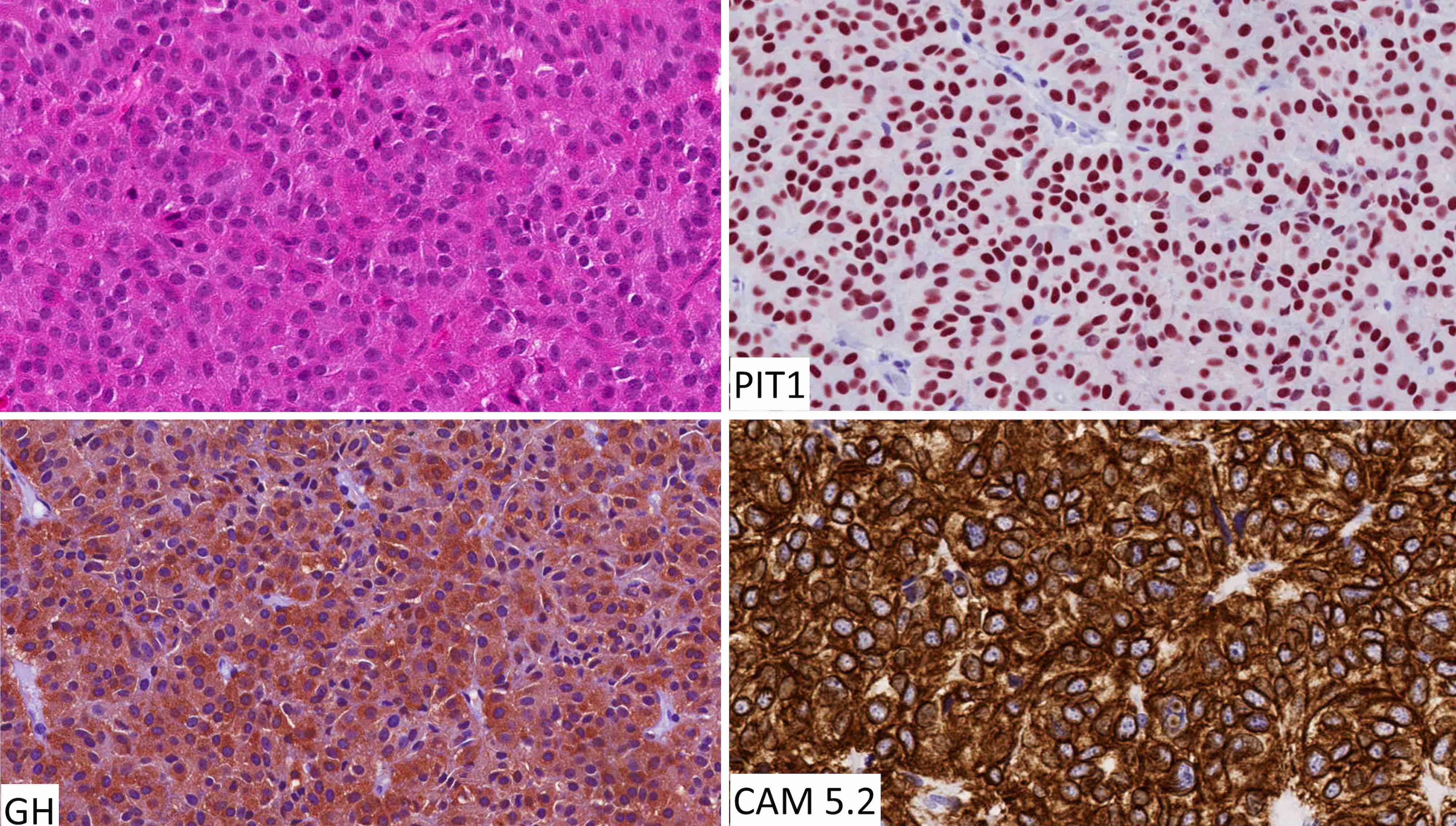
Densely granulated somatotroph tumor
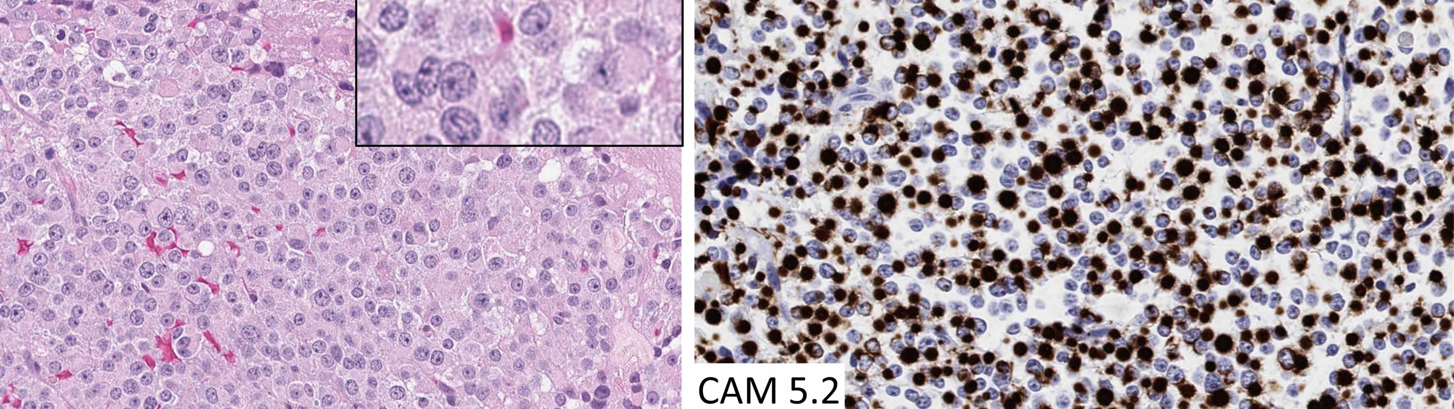
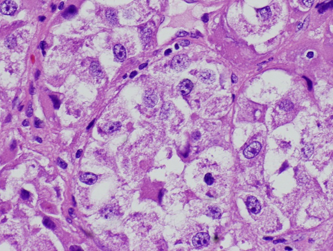
Sparsely granulated somatotroph tumor
Sparsely granulated lactotroph tumor
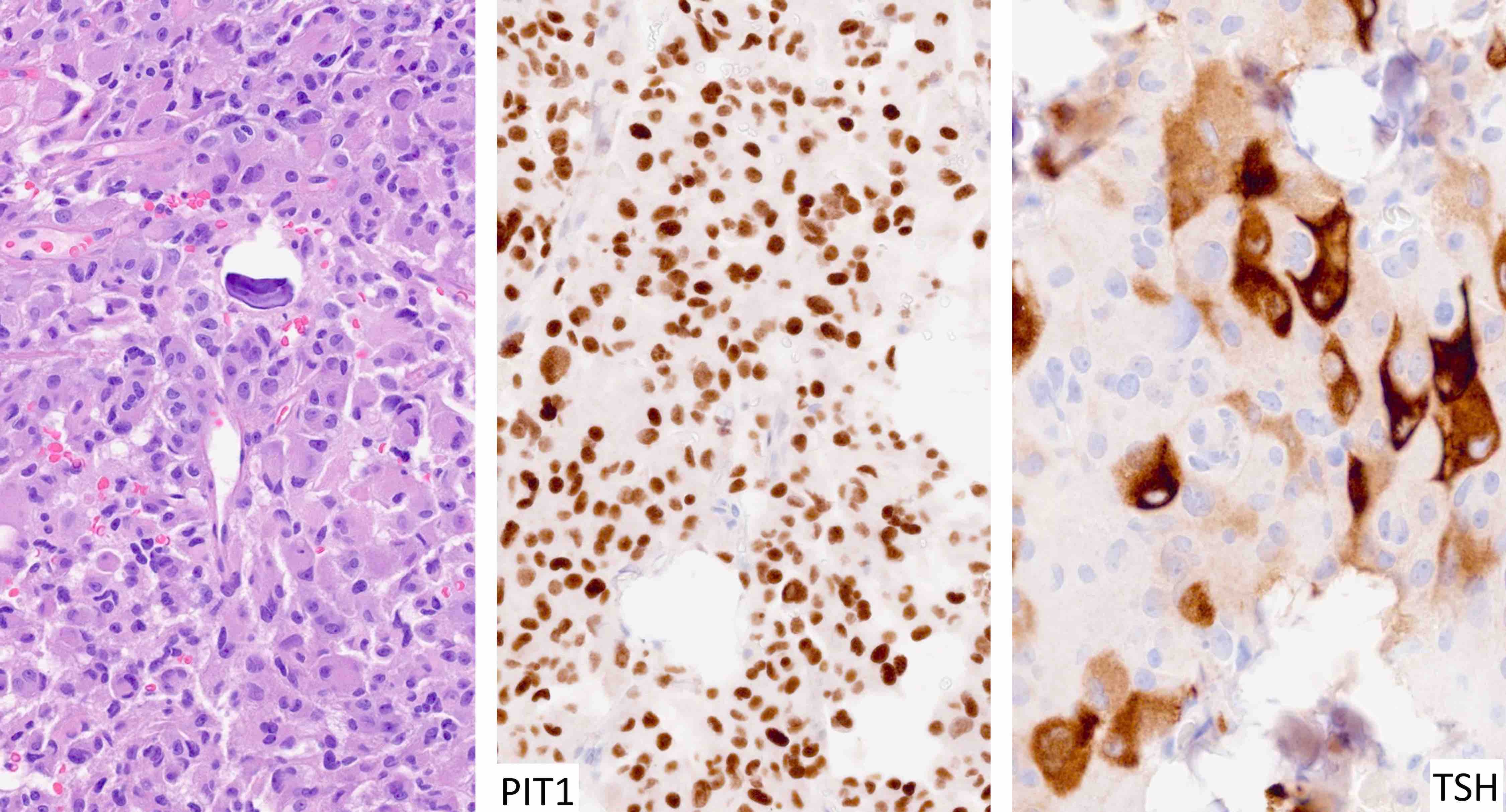
Thyrotroph tumor
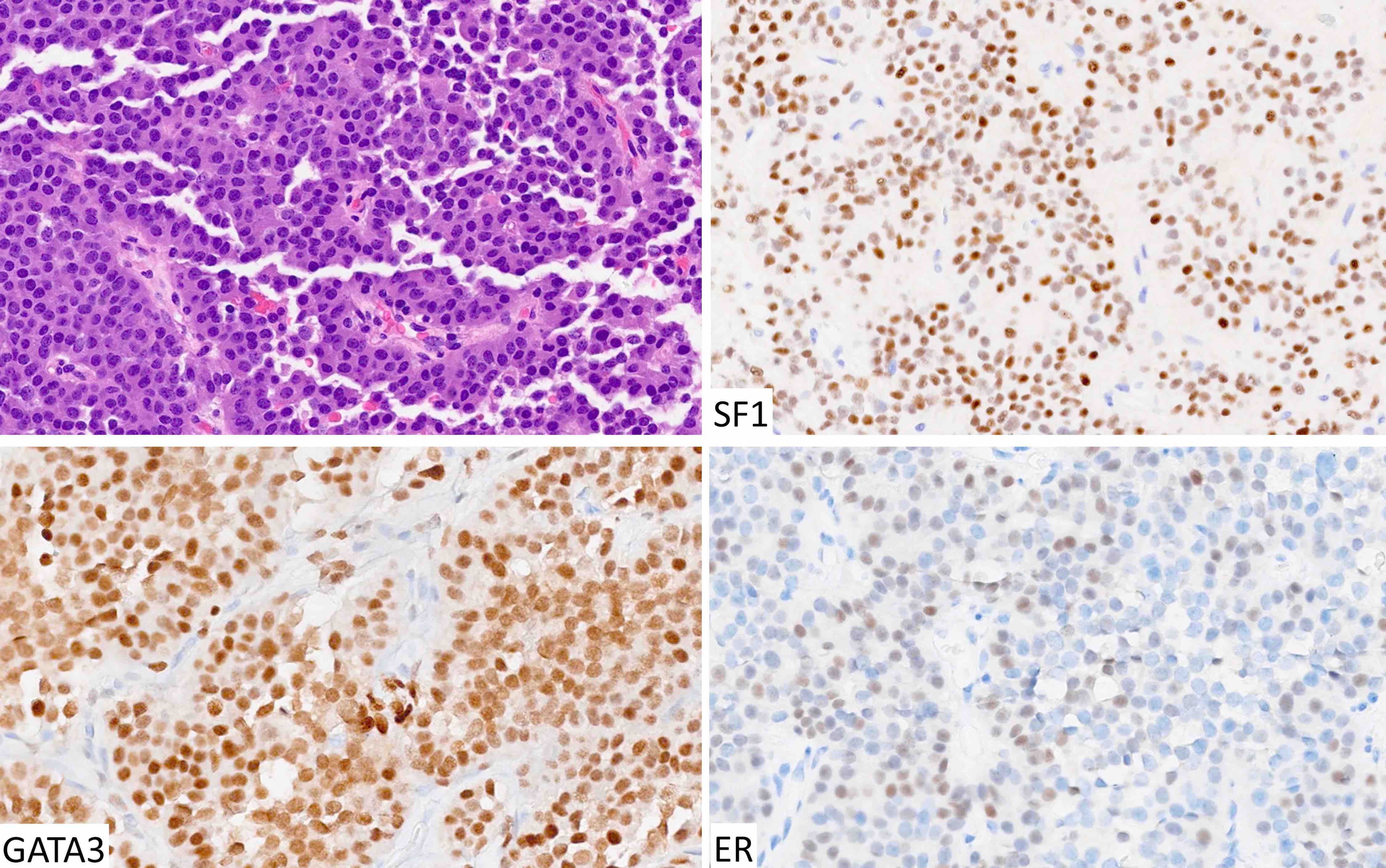
Gonadotroph tumor
Contributed by Michael Punsoni, M.D.
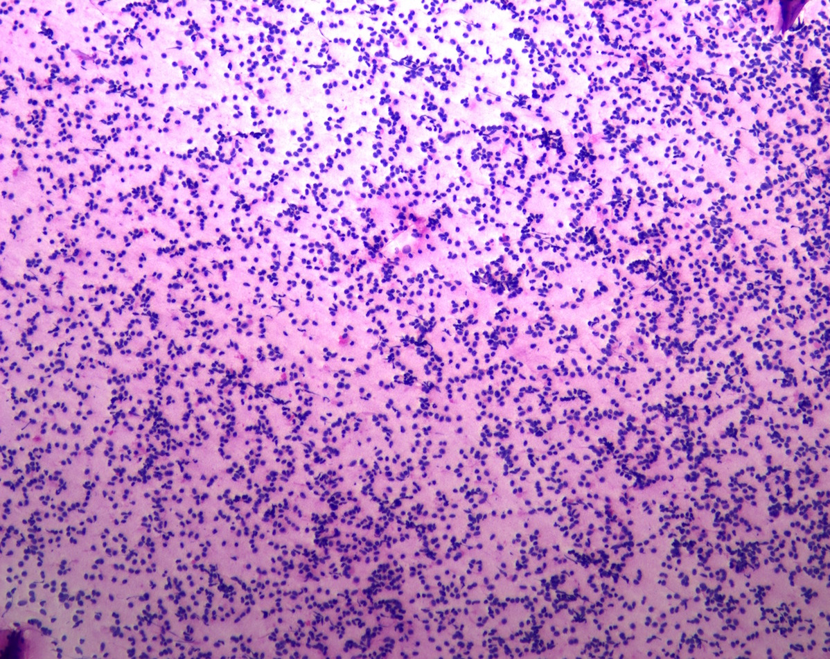
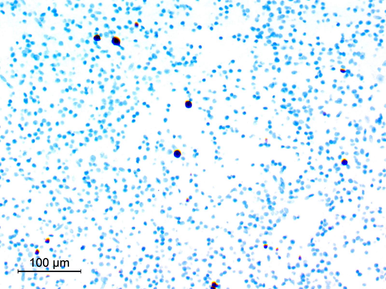
Touch prep showing a cellular population of discohesive small round blue cells
Images hosted on other servers:

Ultrastructural features of some rare pituitary adenomas
Contributed by Shayan Anwar, M.B.B.S.
Axial T2 weighted MRI

Coronal FLAIR MRI

Axial T1 weighted precontrast MRI

Axial T1 weighted postcontrast MRI

Axial apparent diffusion coefficient (ADC)
Contributed by Aisha Memon, M.B.B.S.

Pleomorphic spindle cells

Epithelioid tumor cells
Eosinophilic granular bodies
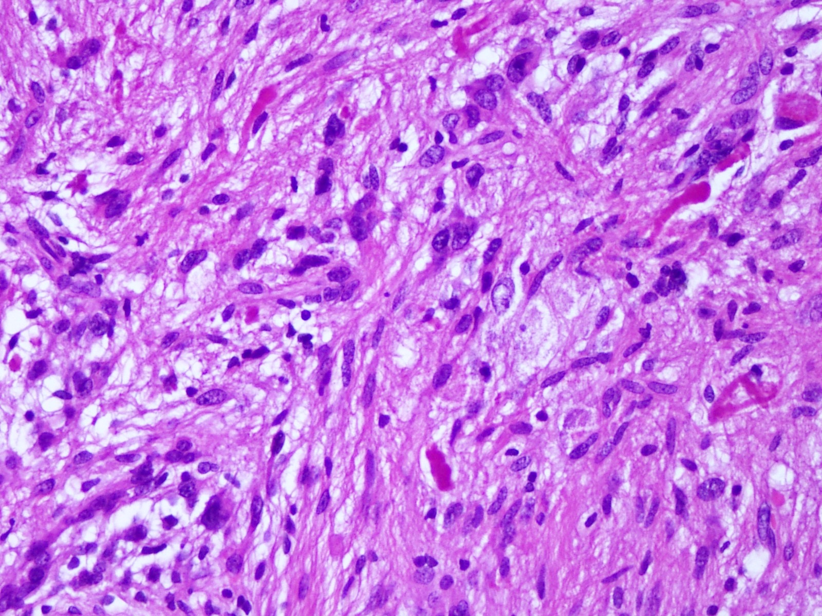
Rosenthal fibers


Xanthomatous cells

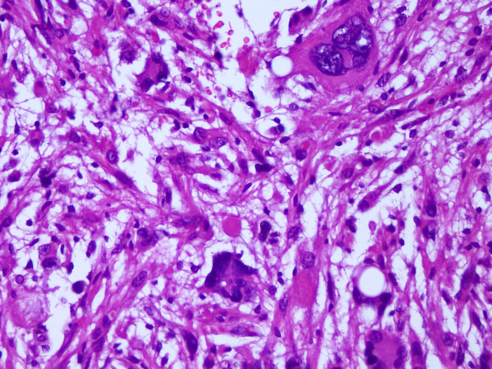
Multinucleated tumor cells

GFAP
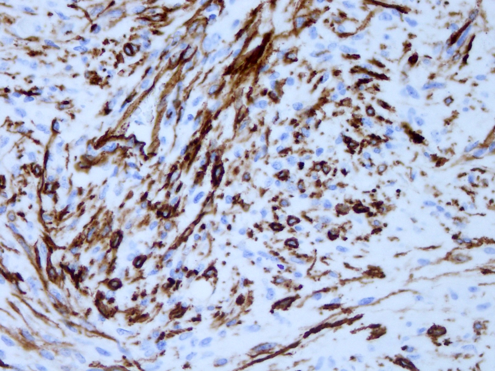
CD34

S100
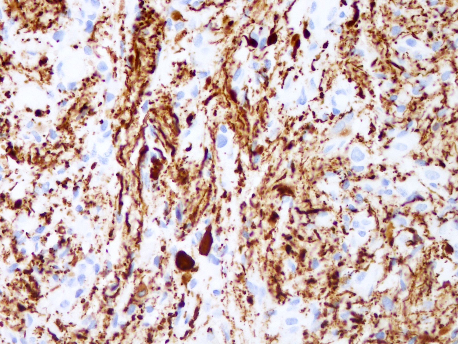
Neurofilament
Reticulin stain
Pleomorphic xanthoastrocytoma:
a brief review
Images hosted on other servers:

Prototypical PLNTY
Images hosted on other servers:

FLAIR, CT scan and T1 postcontrast

Radiological variability in CT and MRI
Images hosted on other servers:

Operative view
Contributed by Eman Abdelzaher, M.D., Ph.D. and Jared T. Ahrendsen, M.D., Ph.D.
Diffuse growth pattern
Diffuse growth pattern of oligodendroglioma-like cells
Oligodendroglioma-like cells with mild nuclear pleomorphism
Calcification
Oligo-like features
Dystrophic calcifications

Polymorphic tumor cells
Olig2 diffusely positive
Strong, diffuse CD34 expression
CD34
CD34 in tumor cells and ramified neural cells
GFAP
Ki67
Images hosted on other servers:

Vague perivascular pseudorosetting
Images hosted on other servers:

Molecular and clinical findings

FGFR fusions

Distinct methylation signature

Genome wide copy number changes
Images hosted on other servers:

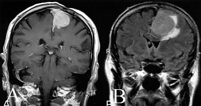
Brain MRI with contrast enhancing lesion

Solid and enhancing nodules on MRI
Images hosted on other servers:
Cerebral lymphoma
spreading across
the corpus callosum
Contributed by Elizabeth Courville, M.D. and Lena Young, D.O.
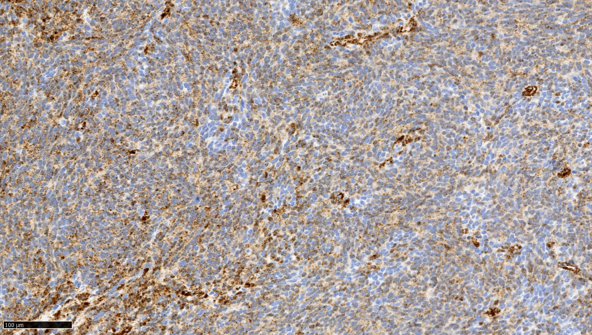
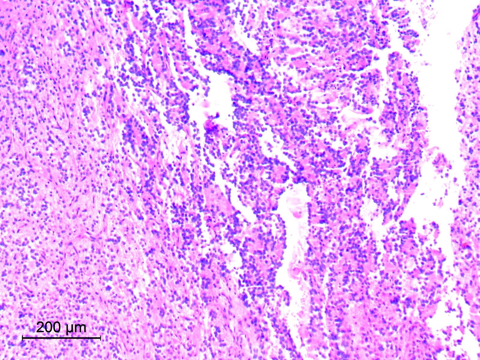
Perivascular infiltrate of atypical lymphoid cells
Diffuse infiltrate
of intermediate
to large
mononuclear cells
Cytology of mononuclear cells
Perivascular cuffing by neoplastic lymphocytes
Infiltrate of neoplastic intermediate to large lymphocytes with background inflammation and necrosis
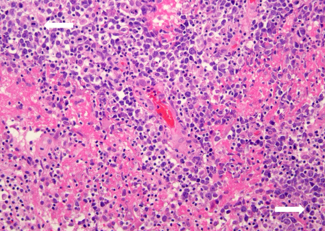
Pleomorphic lymphocytes infiltrating brain

Immunohisto-
chemistry for B and T cells
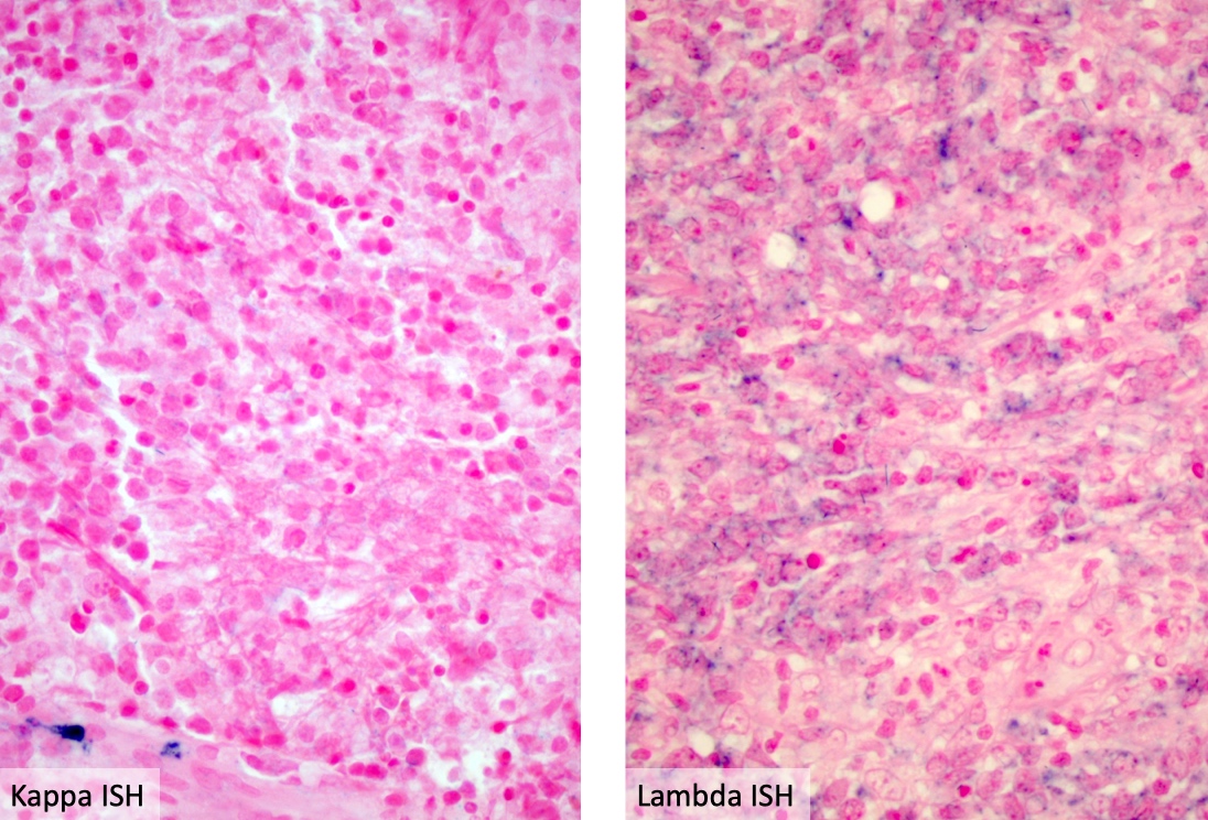
ISH shows lambda restriction
Images hosted on other servers:

Marked enhancing mass at left frontal lobe

Hyperostosis, bony erosion in right anterior clinoid process

Tumor in right infratemporal fossa
Lesion was isointense on T1 and T2 weighted images

Avidly enhancing, complex falcine meningioma
Images hosted on other servers:

Squash prep 40x
Contributed by Eman Abdelzaher, M.D., Ph.D.

CT without contrast

CT with contrast
Images hosted on other servers:

Cerebellar and fourth ventricular RGNT

MRI of cerebellar vermis RGNT

Third ventricle RGNT

MRI of aqueduct and thalamic RGNT

Postcontrast T1 weighted MRI

MRI of pineal region and third ventricle RGNT
Images hosted on other servers:

Right frontal exophytic lesion

Spinal RGNT, intraoperative picture
Contributed by Eman Abdelzaher, M.D., Ph.D.
Biphasic neurocytic and glial components
Biphasic neurocytic and pilocytic components

Neurocytic rosettes
Neurocytes with stippled chromatin

Neurocytic rosettes and pseudorosettes
Cribriform pattern
Microcystic pattern
Microcystic pattern
Pilocytic-like area
Piloid and oligodendroglia-like cells
Synaptophysin

Synaptophysin

Negative neurocytes by GFAP
Glial fibrillary acidic protein (GFAP)
Epithelial membrane antigen (EMA)
Ki67
Images hosted on other servers:

Neurocytic rosettes
Images hosted on other servers:

RGNT, DNA methylation profiling

RGNT, recurrent genetic alterations
WHO classification of CNS tumors: glioneuronal tumors
Contributed by John DeWitt, M.D., Ph.D.
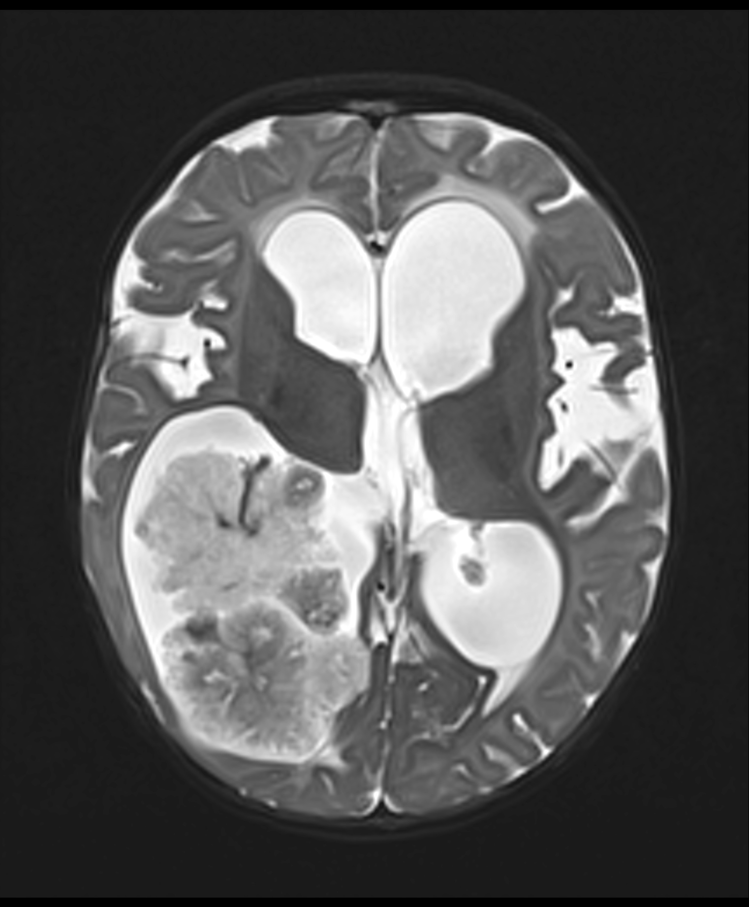
CT scan with contrast
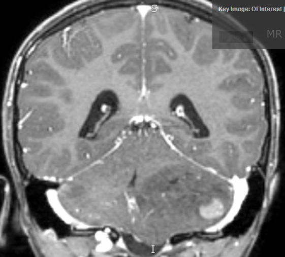
Coronal T1 postcontrast
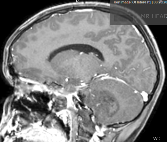
Sagittal T1 postcontrast
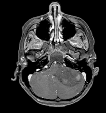
Axial T1 postcontrast
Contributed by John DeWitt, M.D., Ph.D.
Sheets of undifferentiated cells
Vague nodular formation
Vague nodular formation with reticulin
Neuron specific enolase (NSE)

Ki67
Synaptophysin
Beta catenin
INI1
Contributed by Meaghan Morris, M.D., Ph.D.
Anaplasia in medulloblastoma
Contributed by Valeria Barresi, M.D., Ph.D.
T1 weighted MRI
FLAIR MRI
T2 weighted MRI
Images hosted on other servers:

Surgically resected schwannoma

Acoustic schwannoma

Fish flesh tan cut surface
Contributed by Valeria Barresi, M.D., Ph.D.
Compact and loose areas
Atypical nuclei
Lipid laden histiocytes
Verocay bodies
S100
SOX10
Images hosted on other servers:

Intracranial SFT vascular permeability
Images hosted on other servers:

MRI of spinal SFT

MRI of parieto-occipital SFT

MRI of expansive SFT

PET / CT after SFT resection

Vividly enhancing mass, various images
Extra-axial mass, various images

Tumor mass in the left cerebellum

Resection cavity with residual tumor tissue
Images hosted on other servers:

Spinal SFT during resection
Images hosted on other servers:

SFT metastatic to liver
Contributed by Brenndan Crumley, M.D., M.P.H. and Katherine Schwetye, M.D., Ph.D.
Hypercellular neoplasm, patternless pattern
Neoplasm, haphazard spindle cells
Hypercellularity and staghorn vessels
CD34 positivity can be focal
STAT6 nuclear positivity
Images hosted on other servers:

NAB2::STAT6 mutation analysis results

NAB2::STAT6 RT PCR and sequencing
General overview of solitary fibrous tumors (not specific to CNS tumors) by Dr. Jerad Gardner
Images hosted on other servers:

Enhancing mass in the third ventricle
Contributed by Eman Abdelzaher, M.D., Ph.D.
MRI, T2
MRI, FLAIR
Images hosted on other servers:

Lateral ventricle

Spinal cord subependymoma, MRI

Intraparenchymal subependymoma, MRI

Fourth ventricle subependymoma
Images hosted on other servers:

Smooth contoured lobulated mass

Spinal cord
subependymoma

Intraparenchymal
subependymoma
Contributed by Eman Abdelzaher, M.D., Ph.D.
Circumscribed, lobulated mass
Contributed by Eman Abdelzaher, M.D., Ph.D., David Taylor, M.D. and Nazila Azordegan, M.D.
Nuclear clustering and microcystic change

Nuclear clustering
Nuclear clustering
Nuclear clustering

Microcystic change
Microcystic change and nuclear pleomorphism
Uniform nuclei
Uniform nuclei
Lobulated tumor
Clustered nuclei in fibrillary background
Dot-like perinuclear EMA
GFAP
Images hosted on other servers:

Cohesive tissue
clumps, elongated
cytoplasmic
processes
Images hosted on other servers:

Ultrastructural findings in subependymoma
Images hosted on other servers:

Key molecular and clinical characteristics

Methylation profiling

Copy number variations
Subependymoma
Images hosted on other servers:

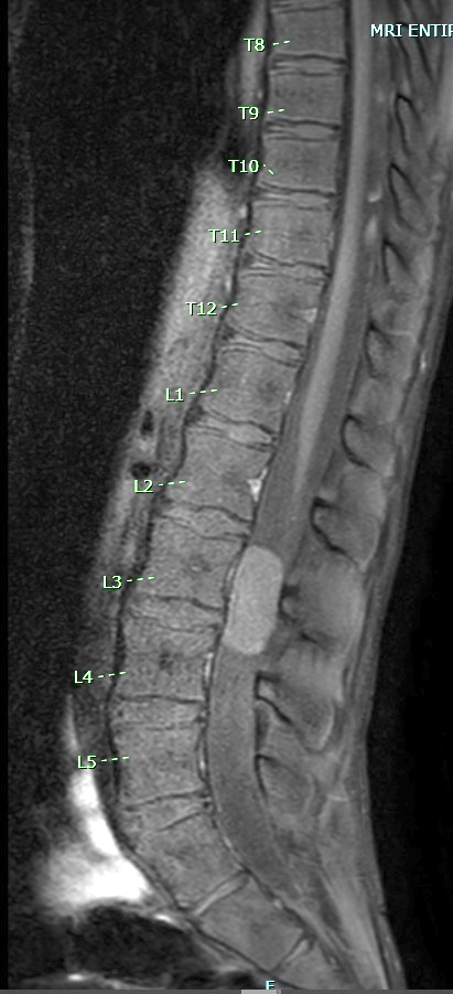
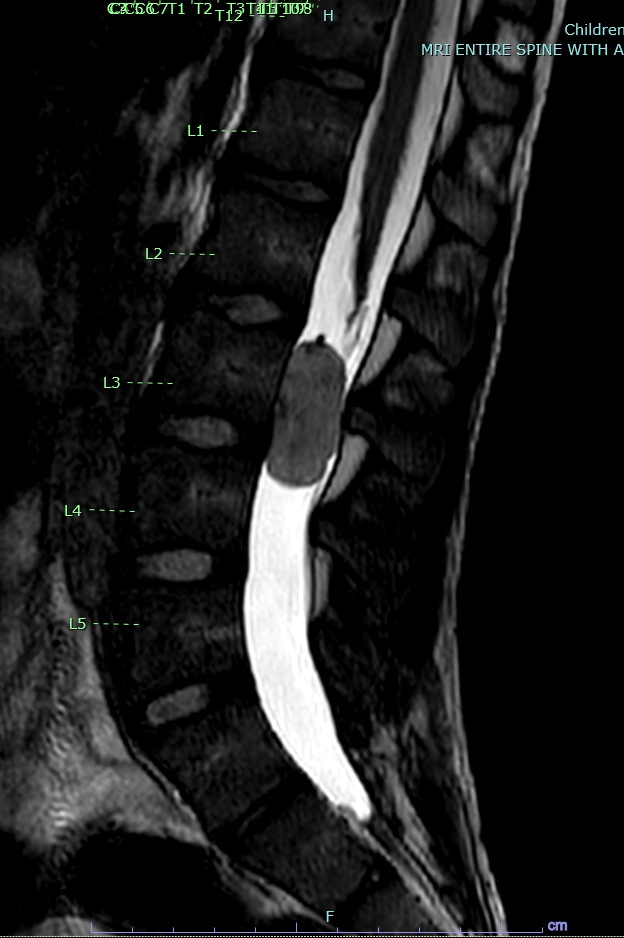
Lumbar synovial cyst
Images hosted on other servers:


Cervical spine MRI
Images hosted on other servers:

Key diagnostic genes, molecules, pathways

CNS WHO grades of selected types

Newly recognized WHO 2021 tumor types
Contributed by P.J. Cimino, M.D., Ph.D.
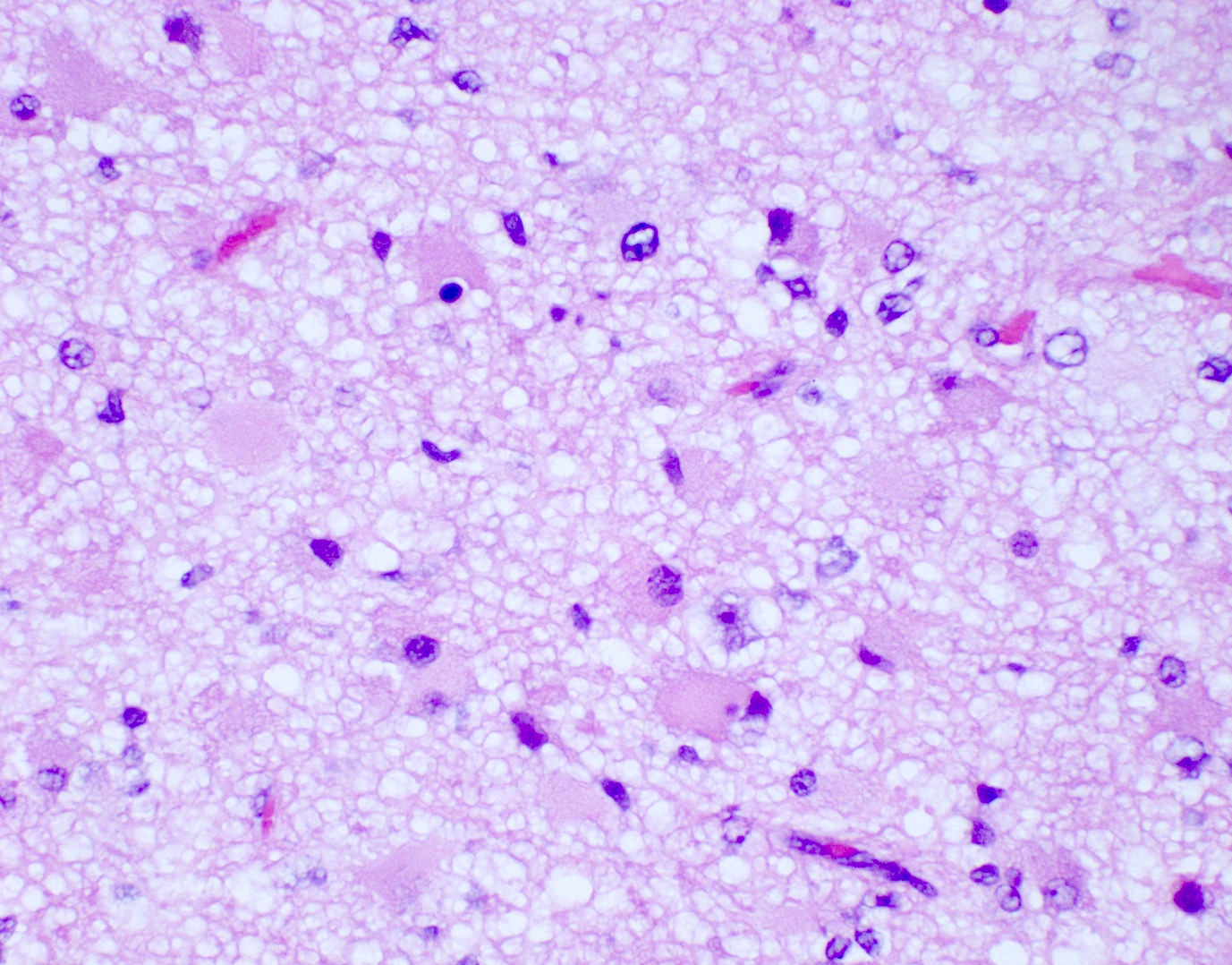
Adult type IDH mutant gliomas
Adult type IDH wildtype gliomas
Contributed by P.J. Cimino, M.D., Ph.D.

Astrocytoma lacking mitoses
Increased mitotic activity
Pseudopalisading necrosis
Microvascular proliferation
Contributed by P.J. Cimino, M.D., Ph.D.
Glioblastoma EGFR amplification

Glioblastoma copy number plot
Images hosted on other servers:

Glioblastoma TERT promoter mutation
2021 WHO classification of CNS tumors: update I - gliomas
Contributed by Jared T. Ahrendsen, M.D., Ph.D.

Criteria for meningioma classification
Contributed by Jared T. Ahrendsen, M.D., Ph.D. and Chunyu Cai, M.D., Ph.D.
Brain invasion
Necrosis
Sheeting
Increased cellularity
Small cell transformation
Prominent nucleoli
Atypical meningioma

Anaplastic meningioma
Asa: 2020
Ellison: 2013
Gill: 2022
Gray: 2018
Gupta: 2016
Husain: 2021
IARC: 2022
Kleinschmidt-DeMasters: 2022
Love: 2015
Perry: 2017
Rodriguez: 2016
Find related Pathology books: GU/adrenal, head & neck/endocrine, muscle and peripheral nerve nontumor, neuropathology, pediatric
