


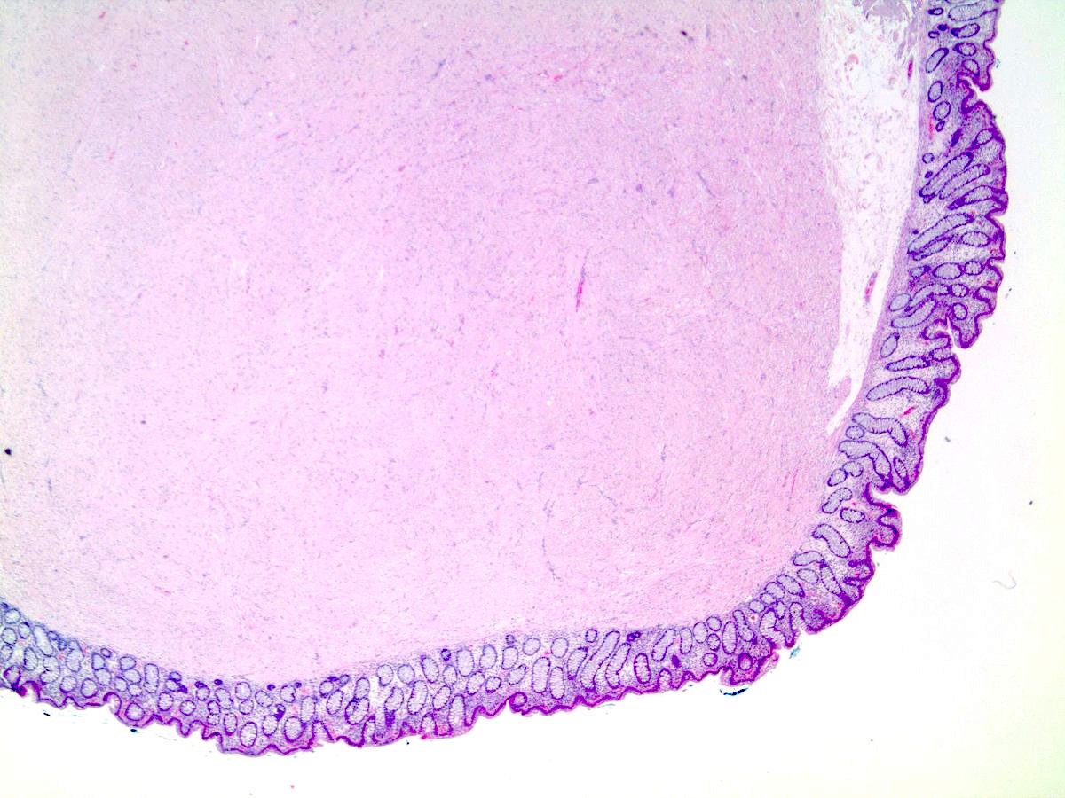
Images hosted on other servers:

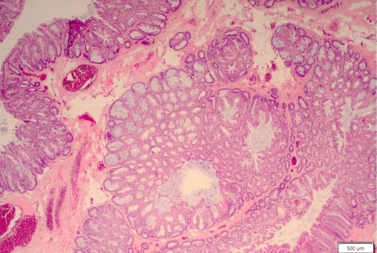
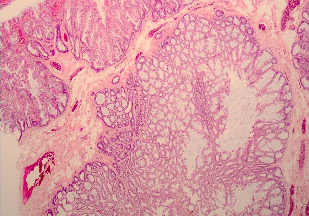
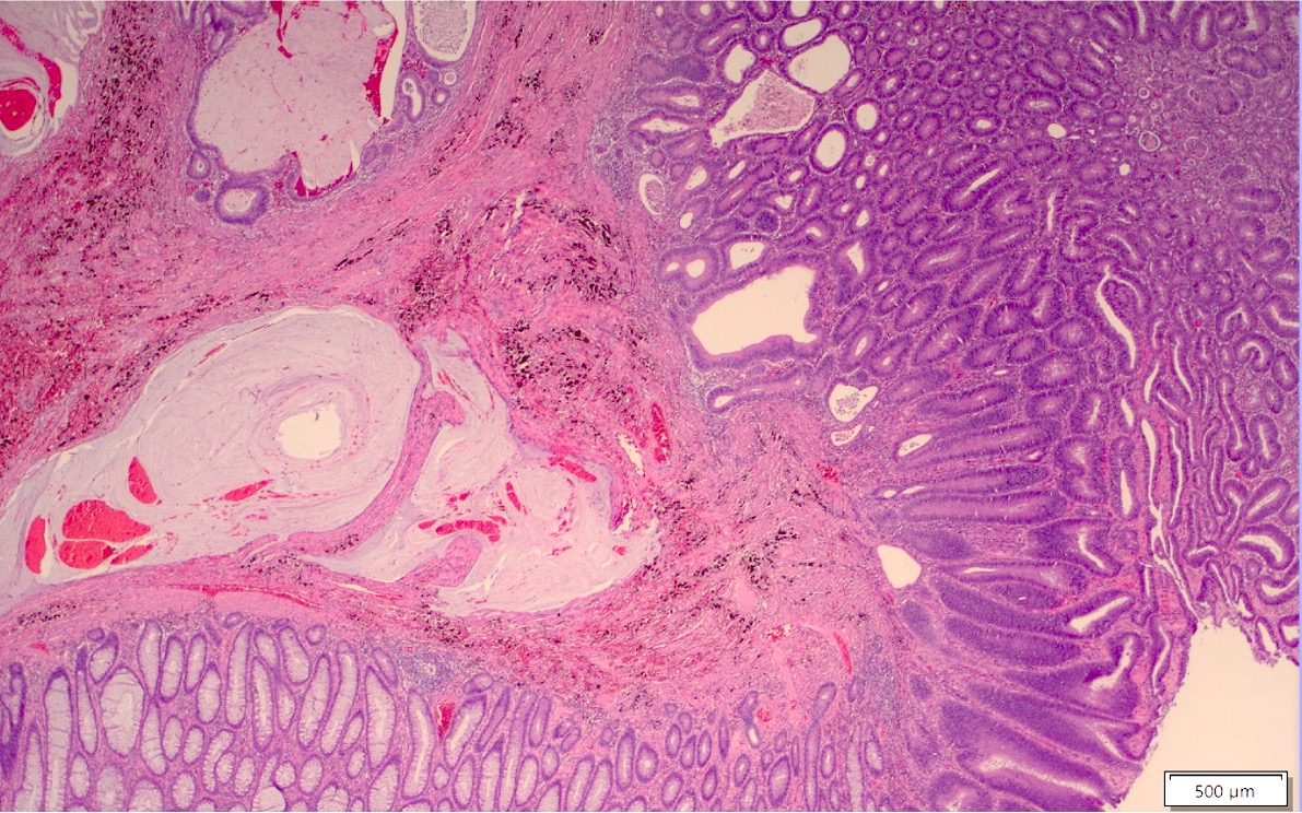
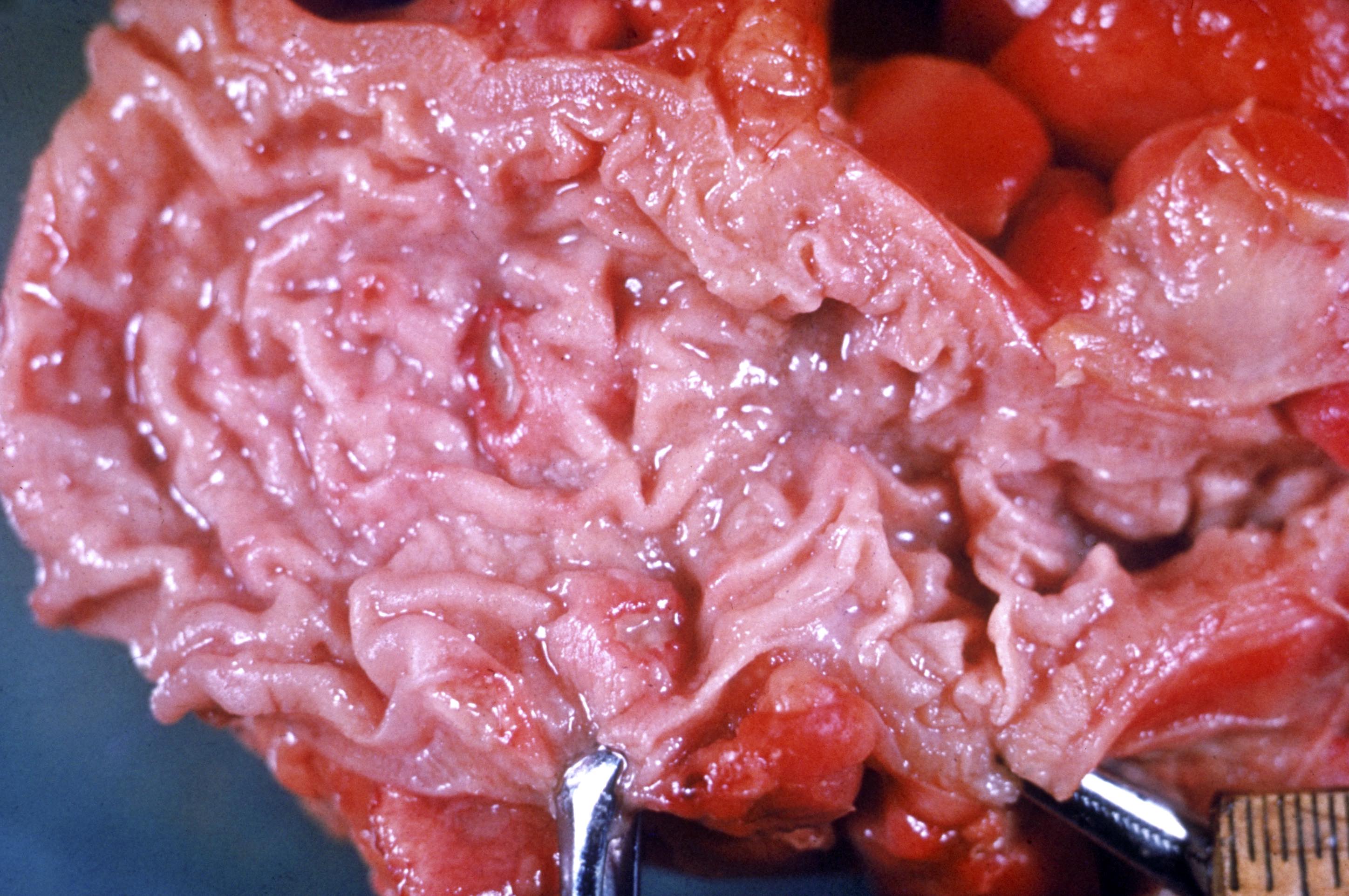
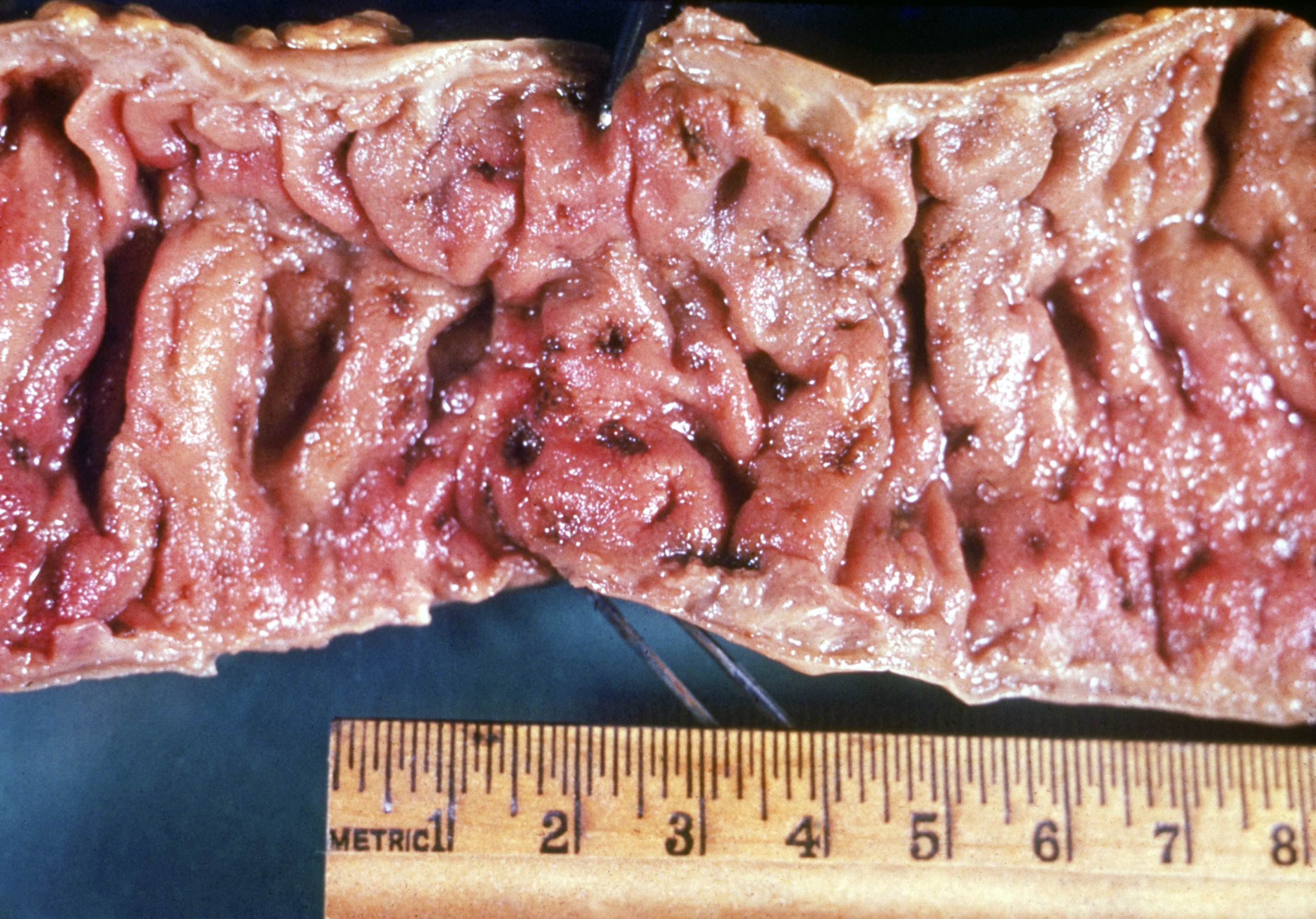
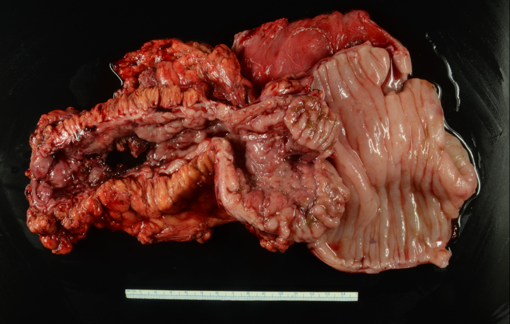
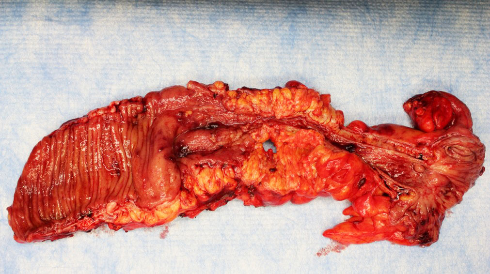
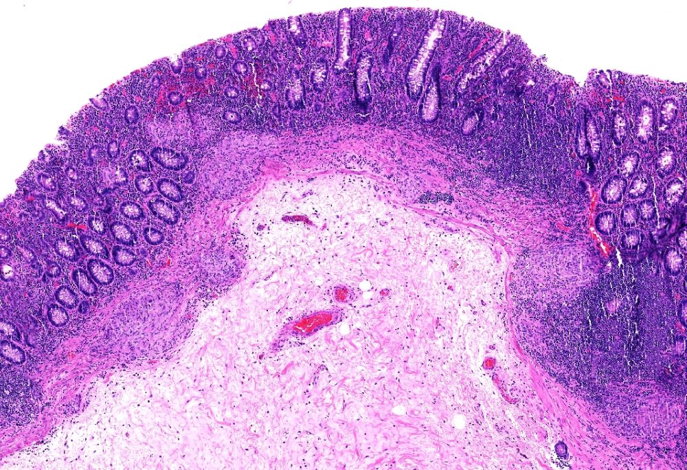
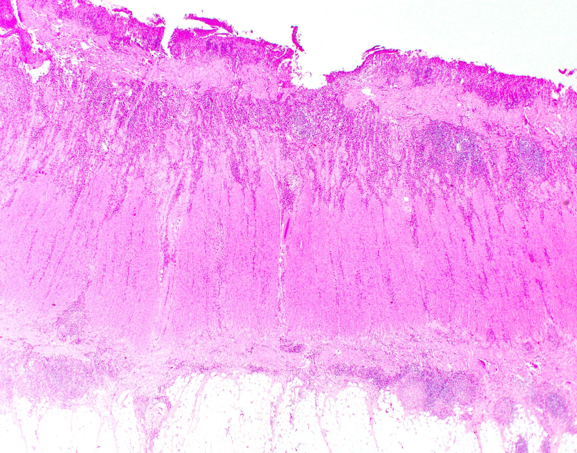
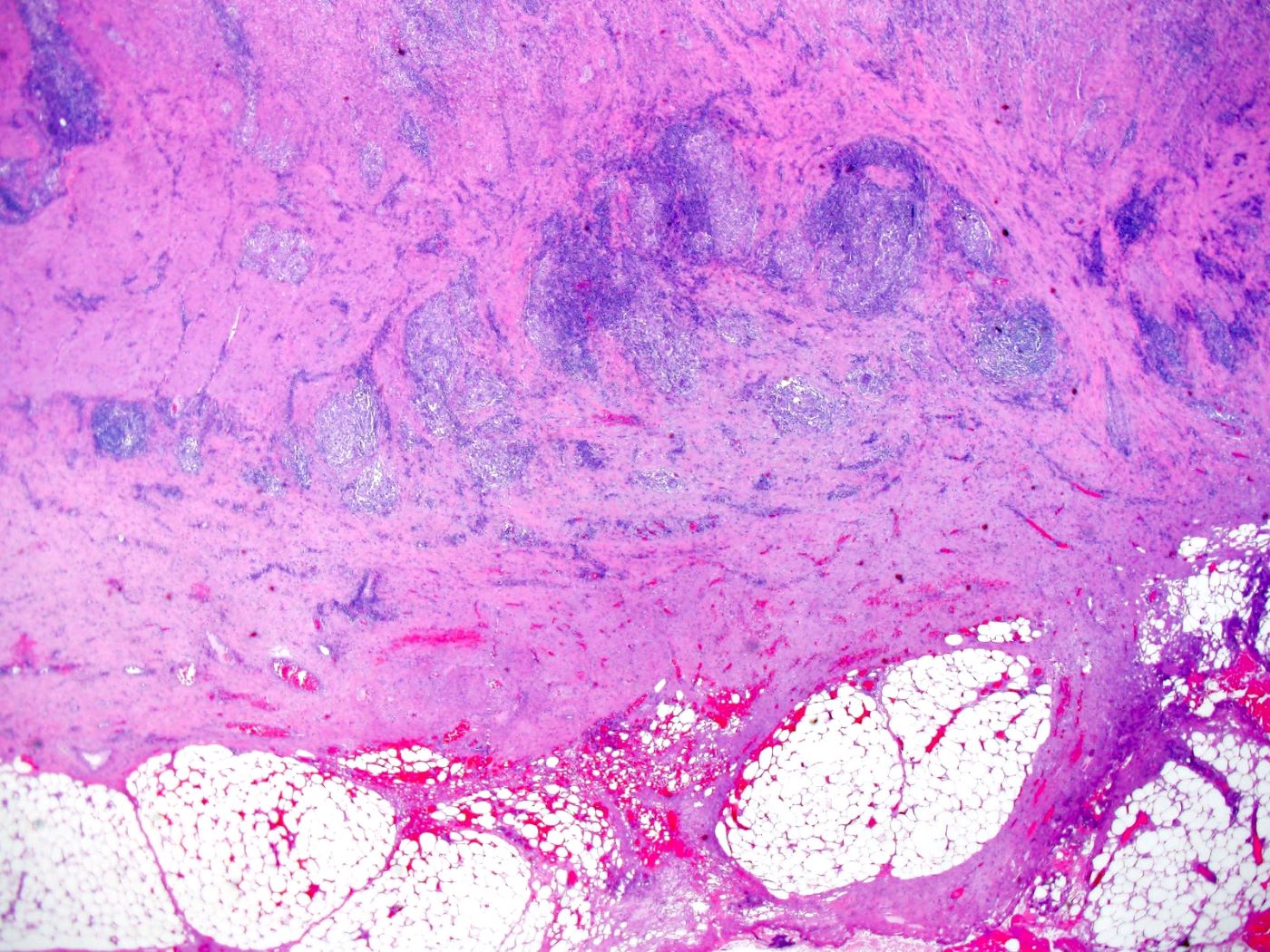
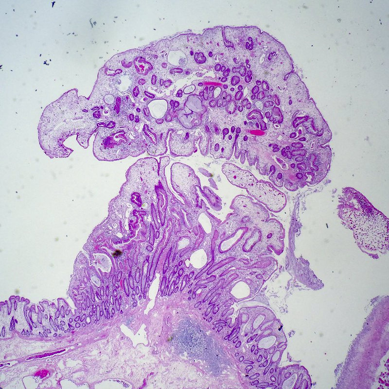
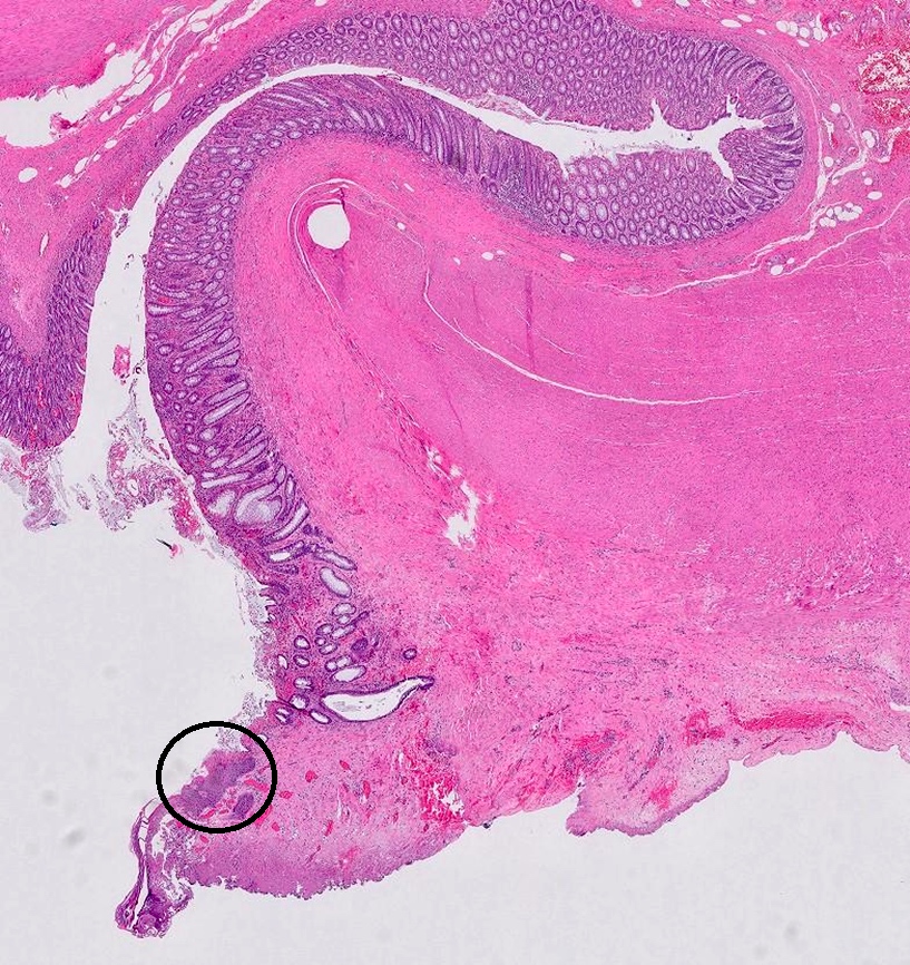
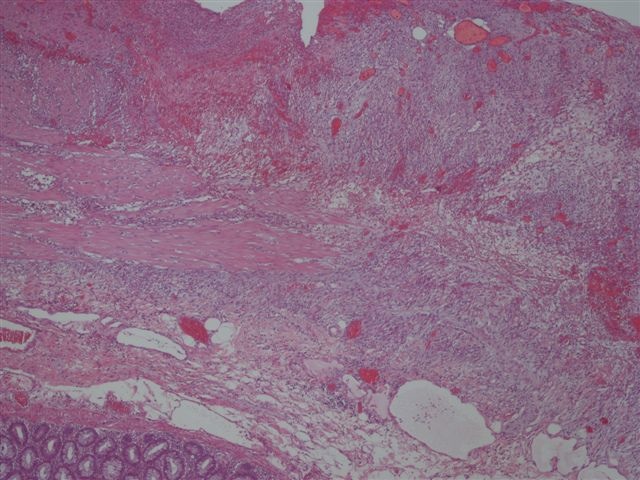
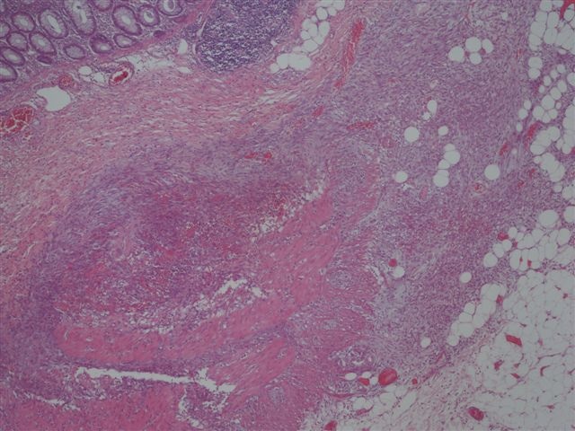
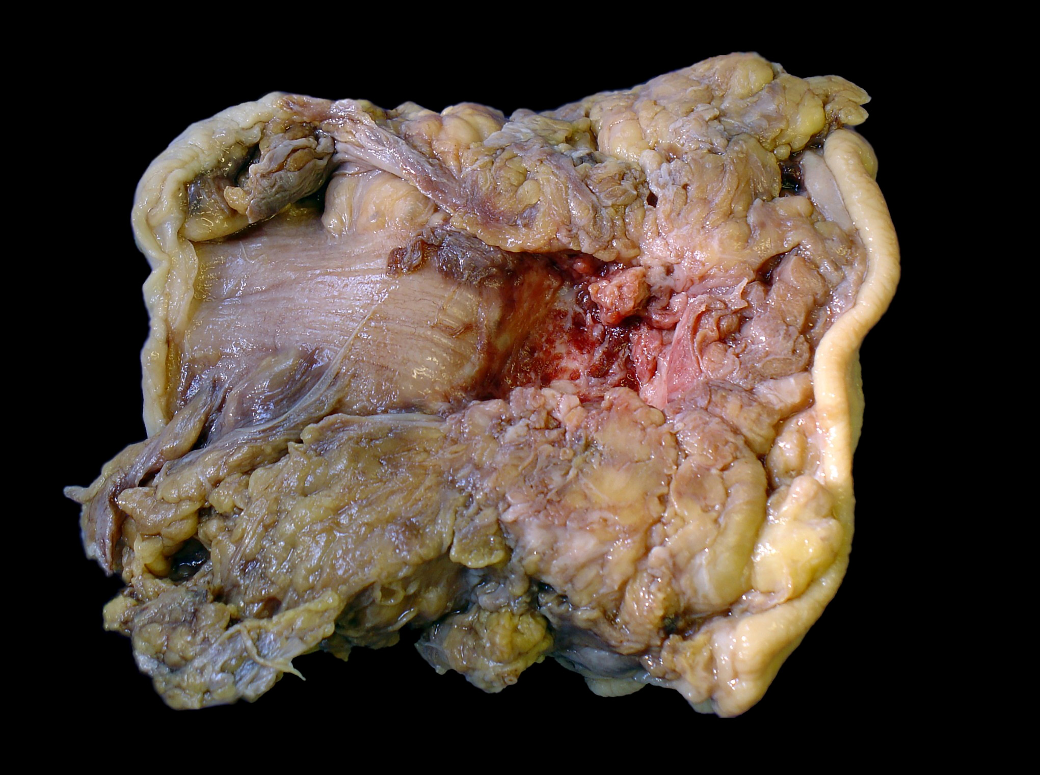
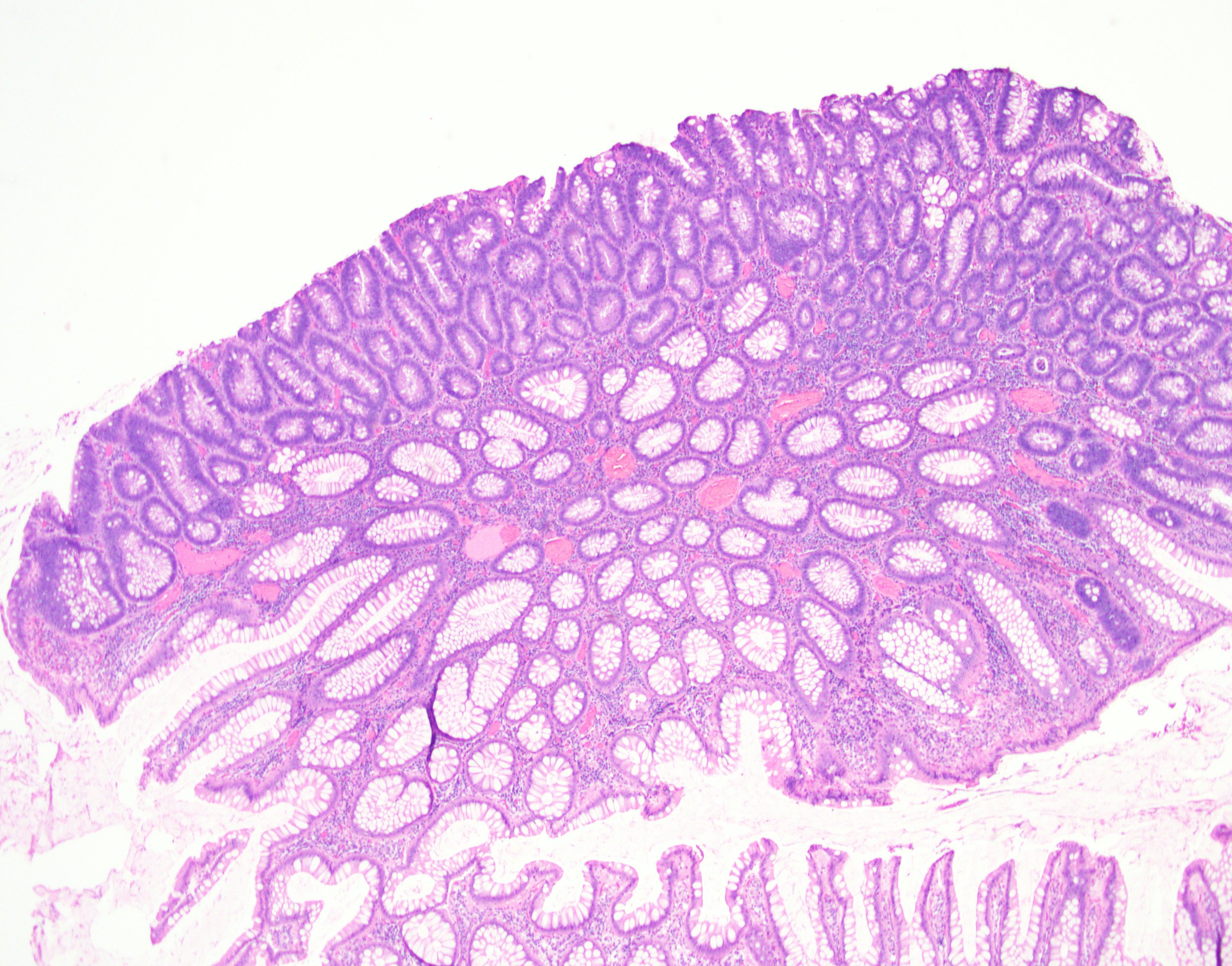
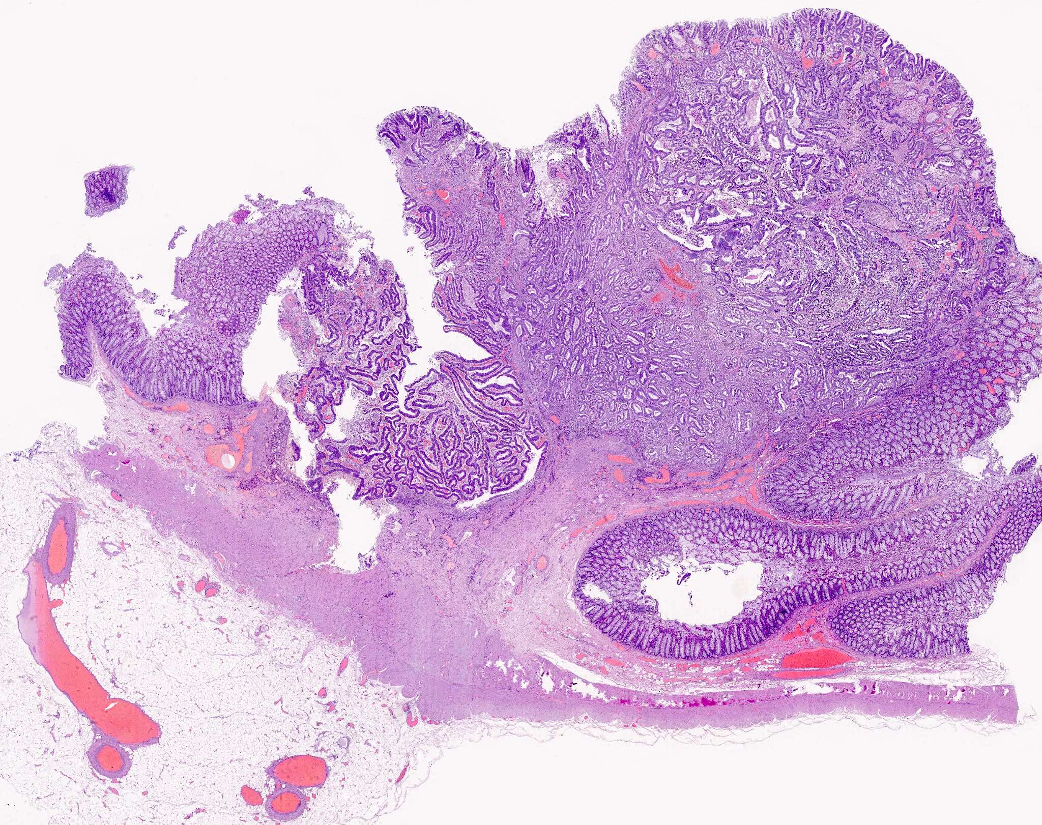
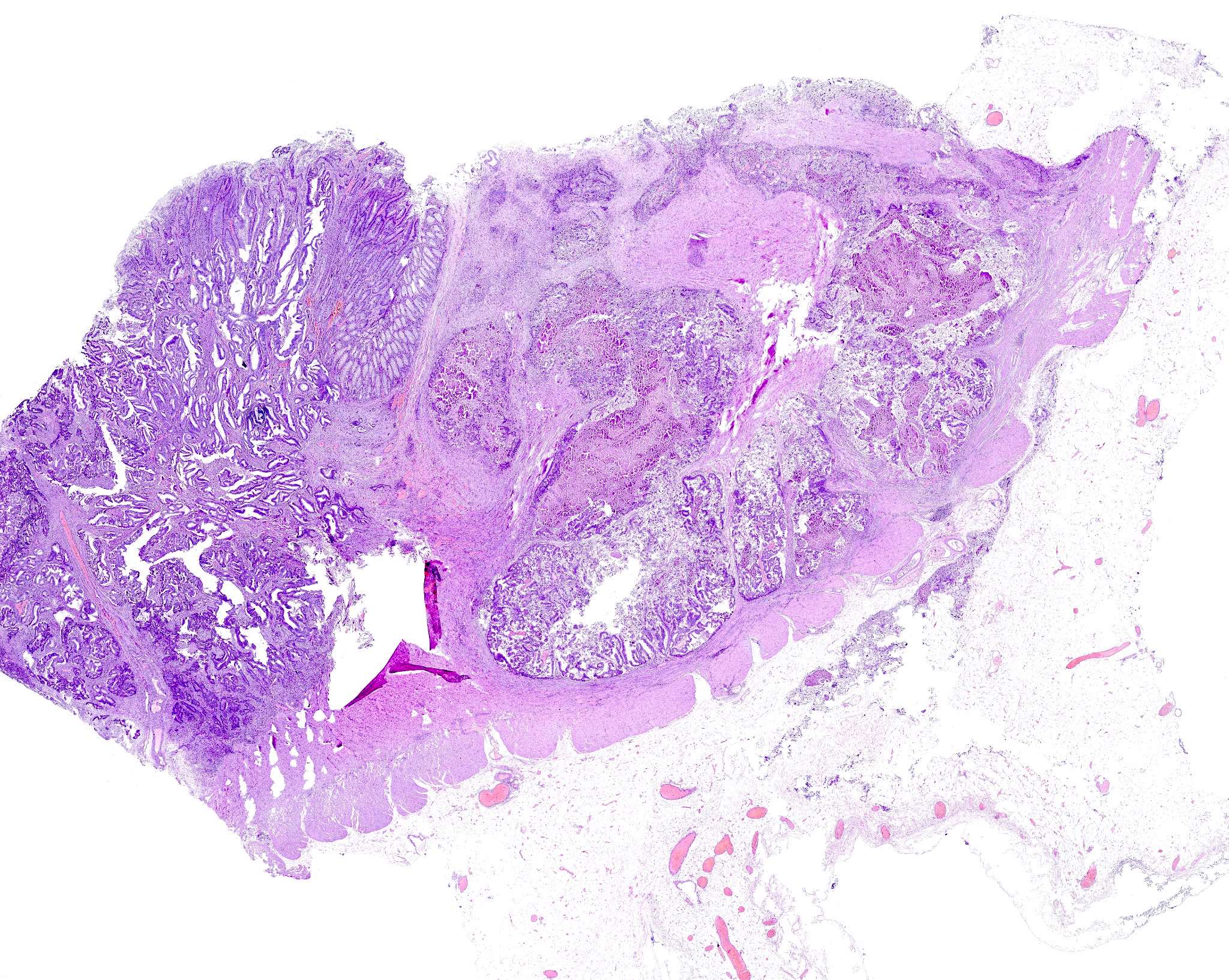
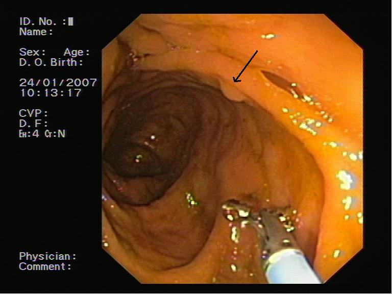
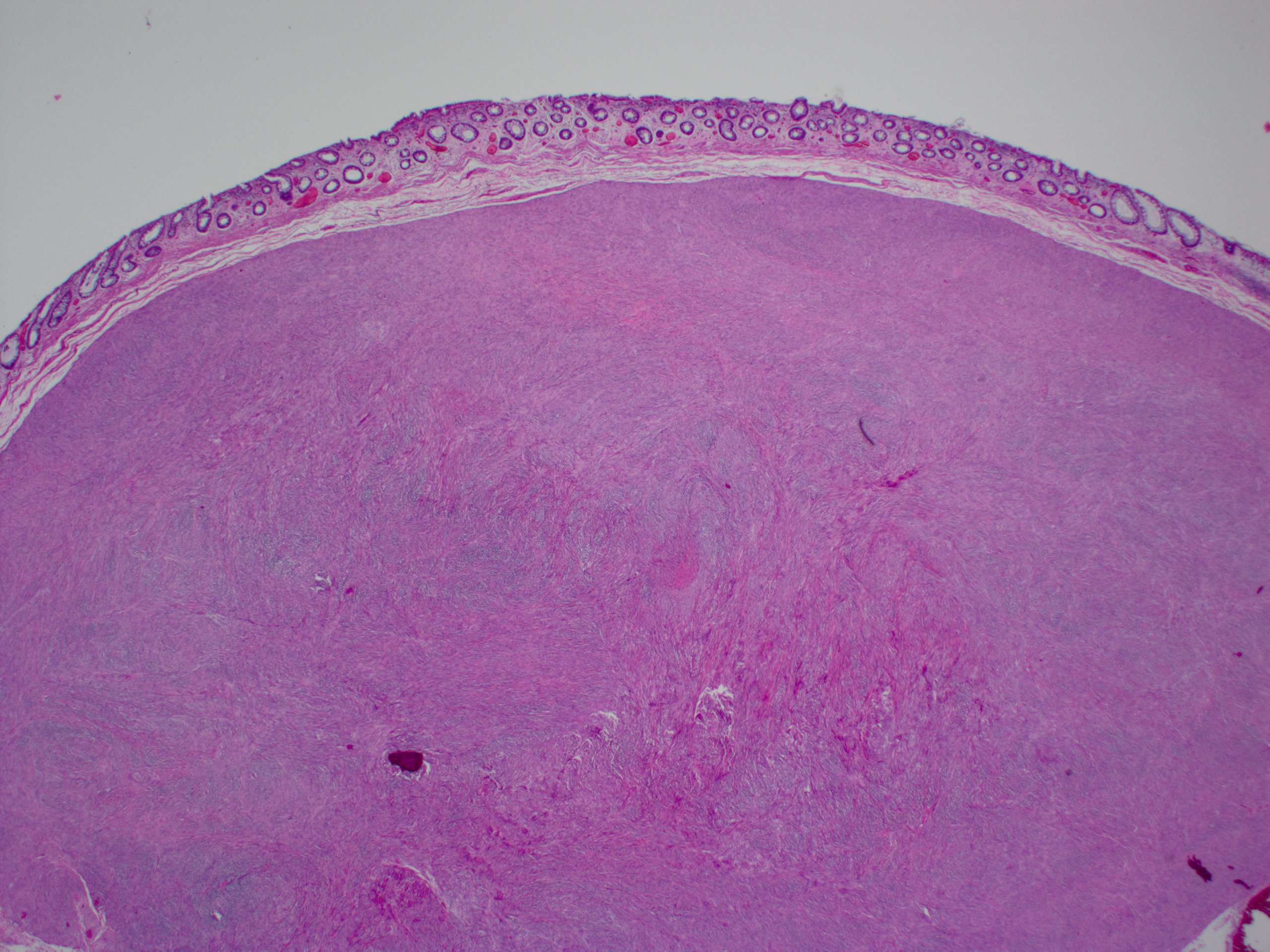
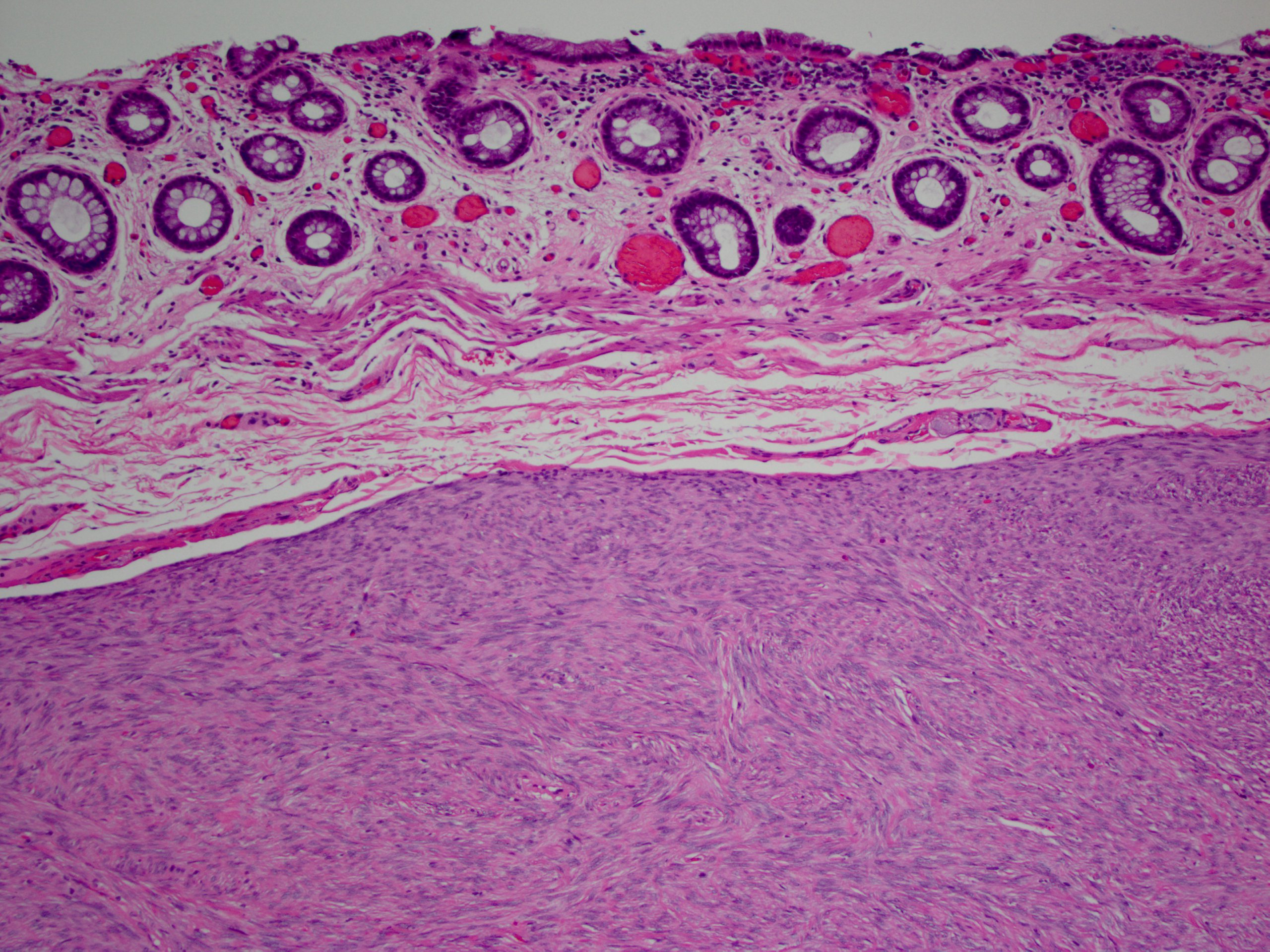
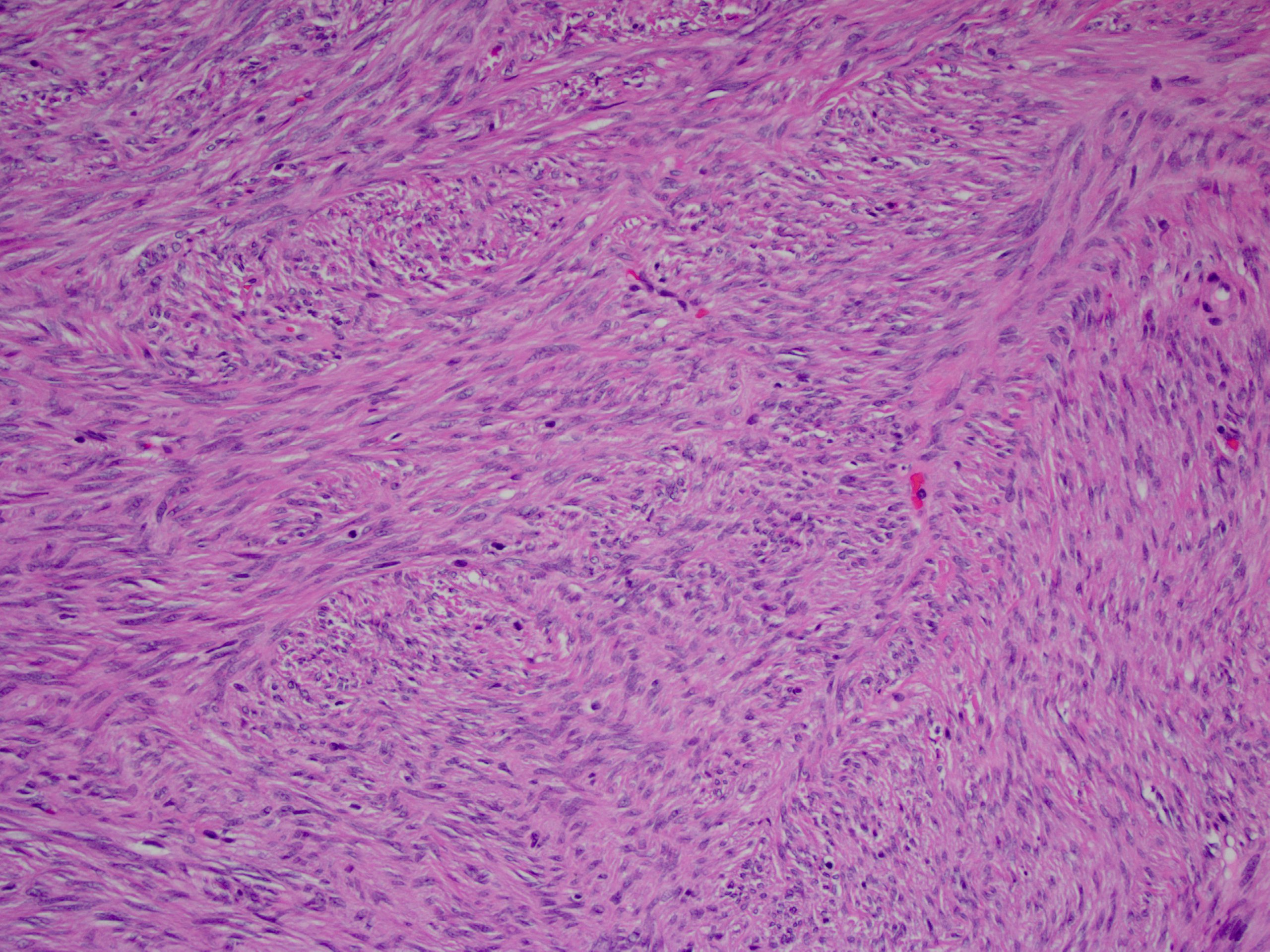
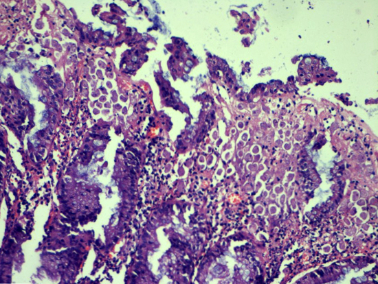
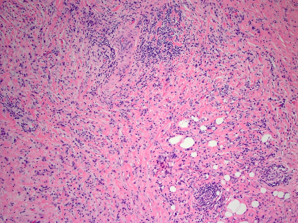
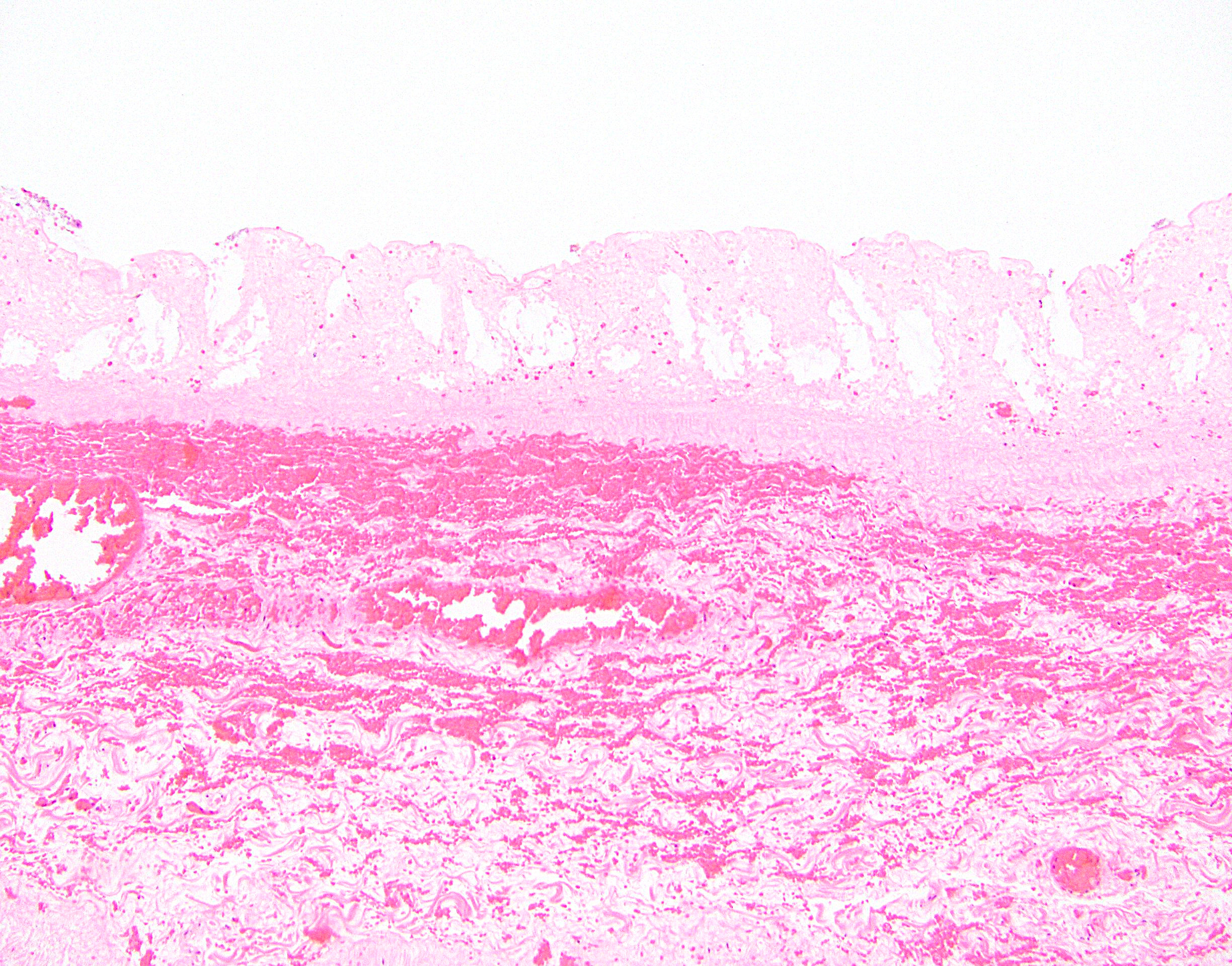
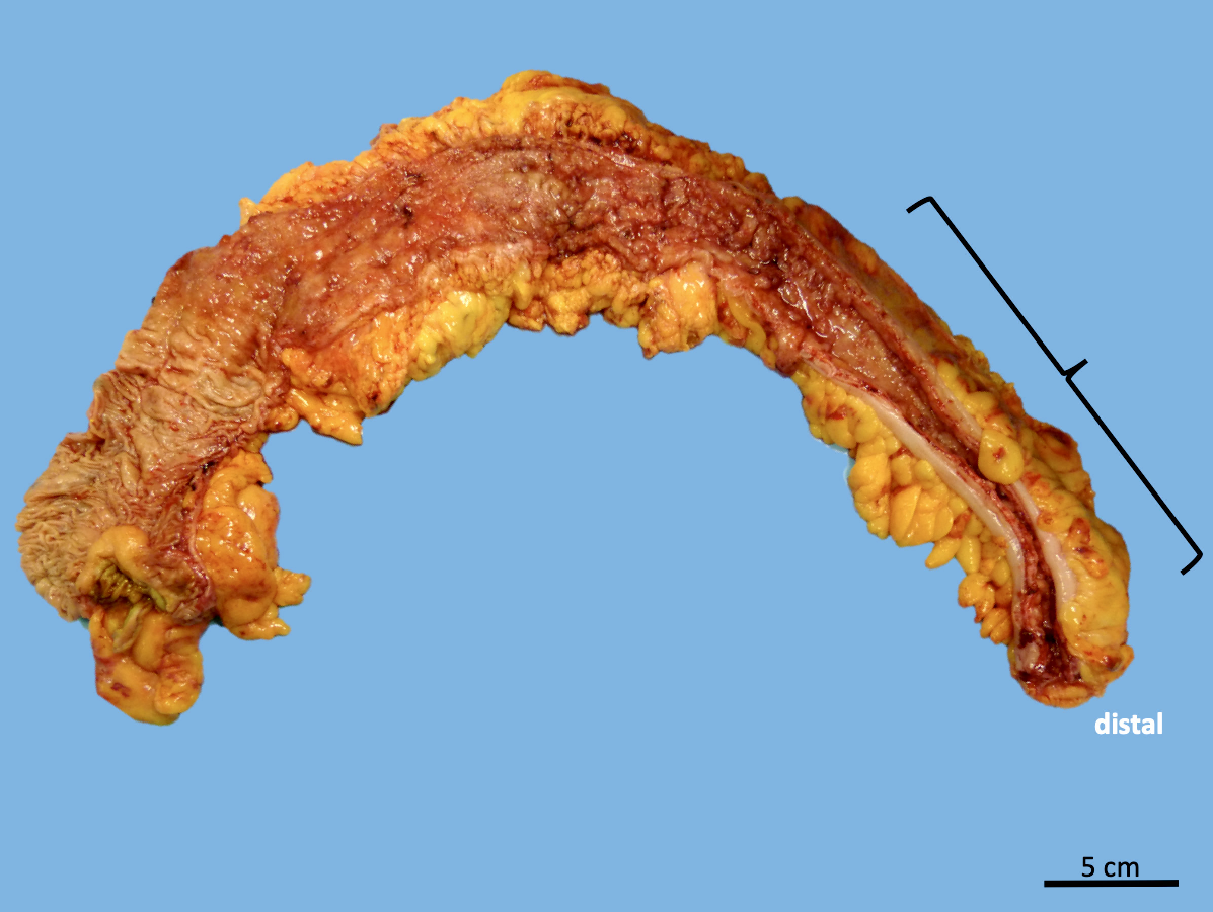
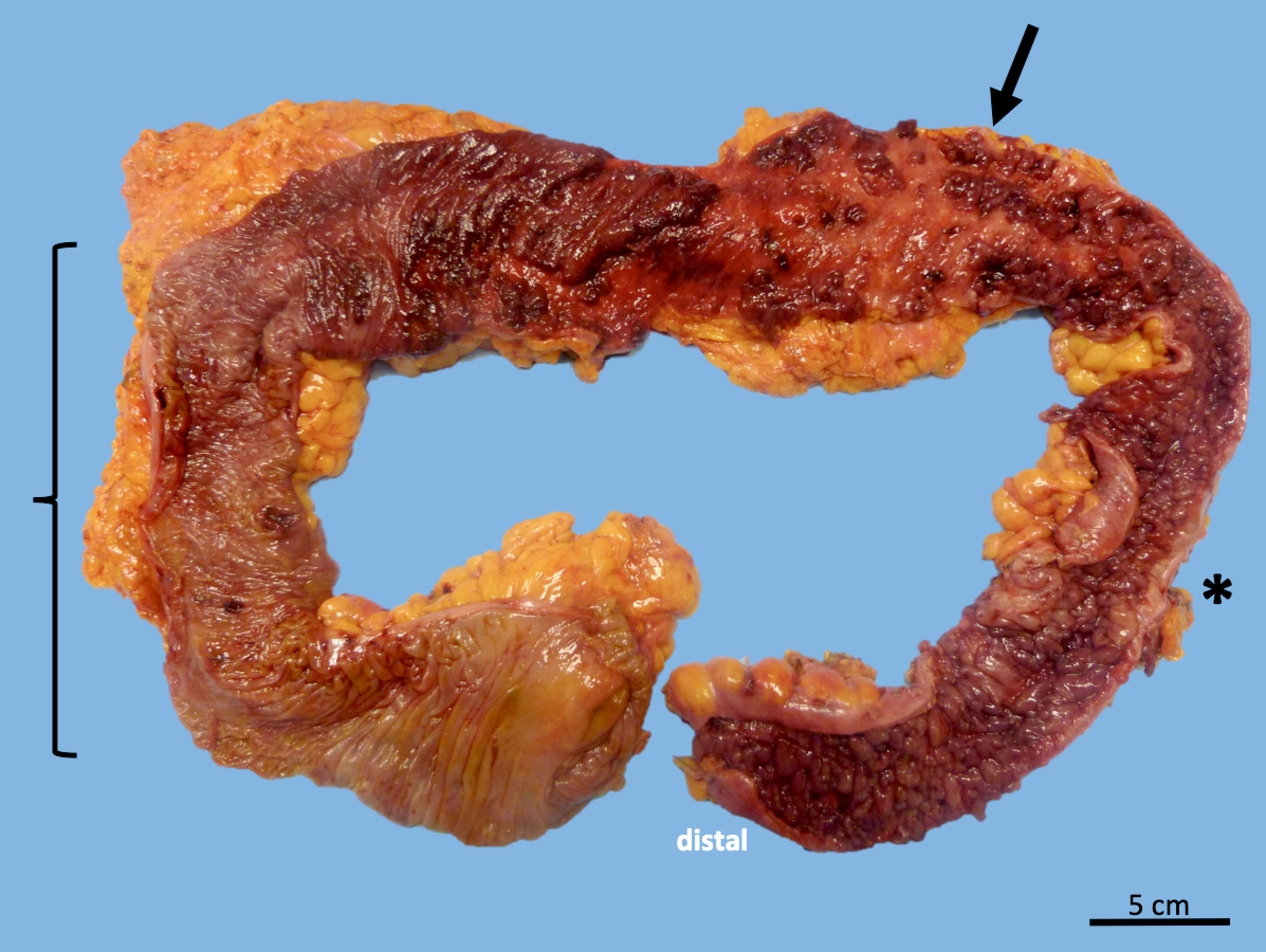
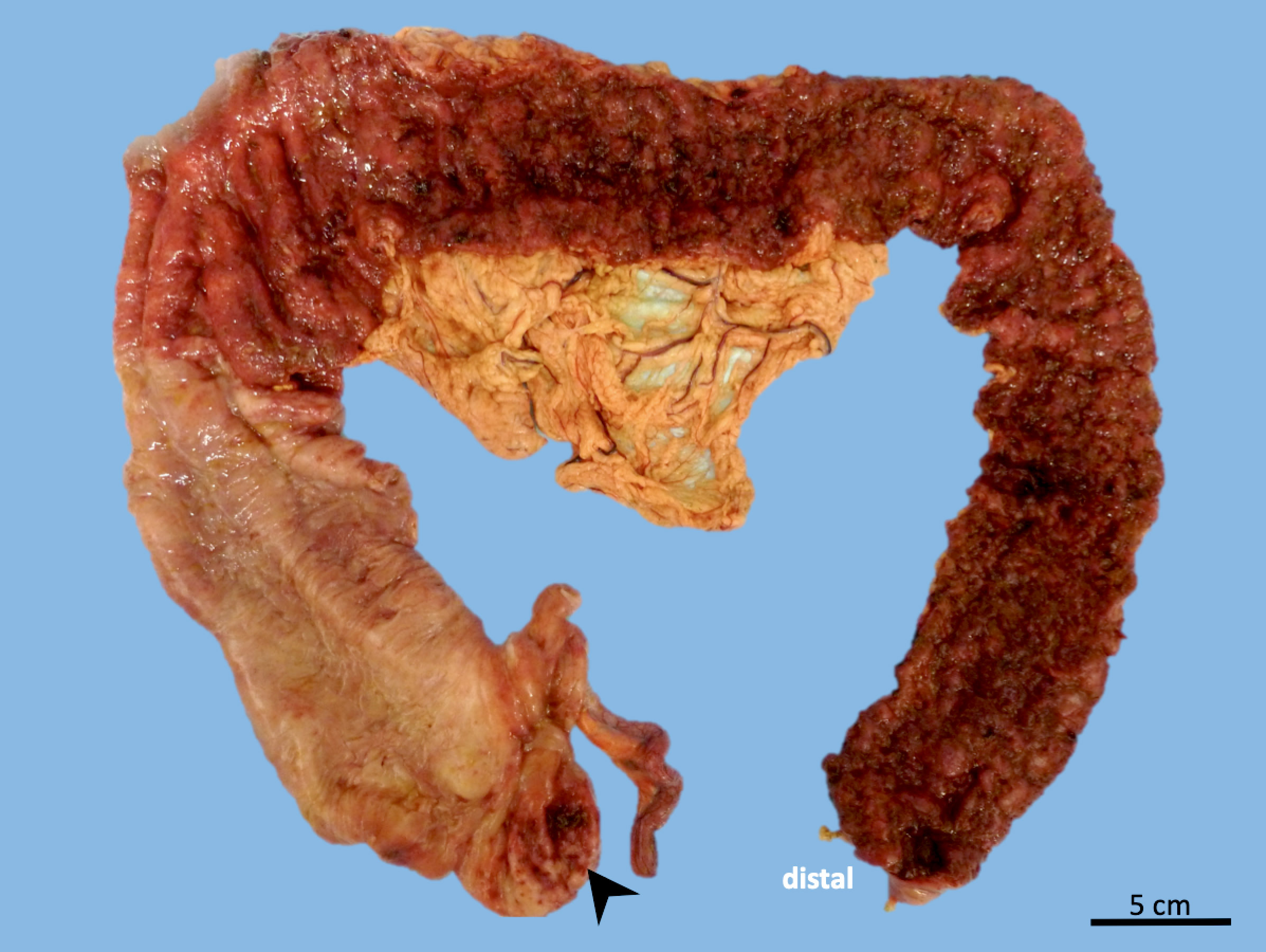
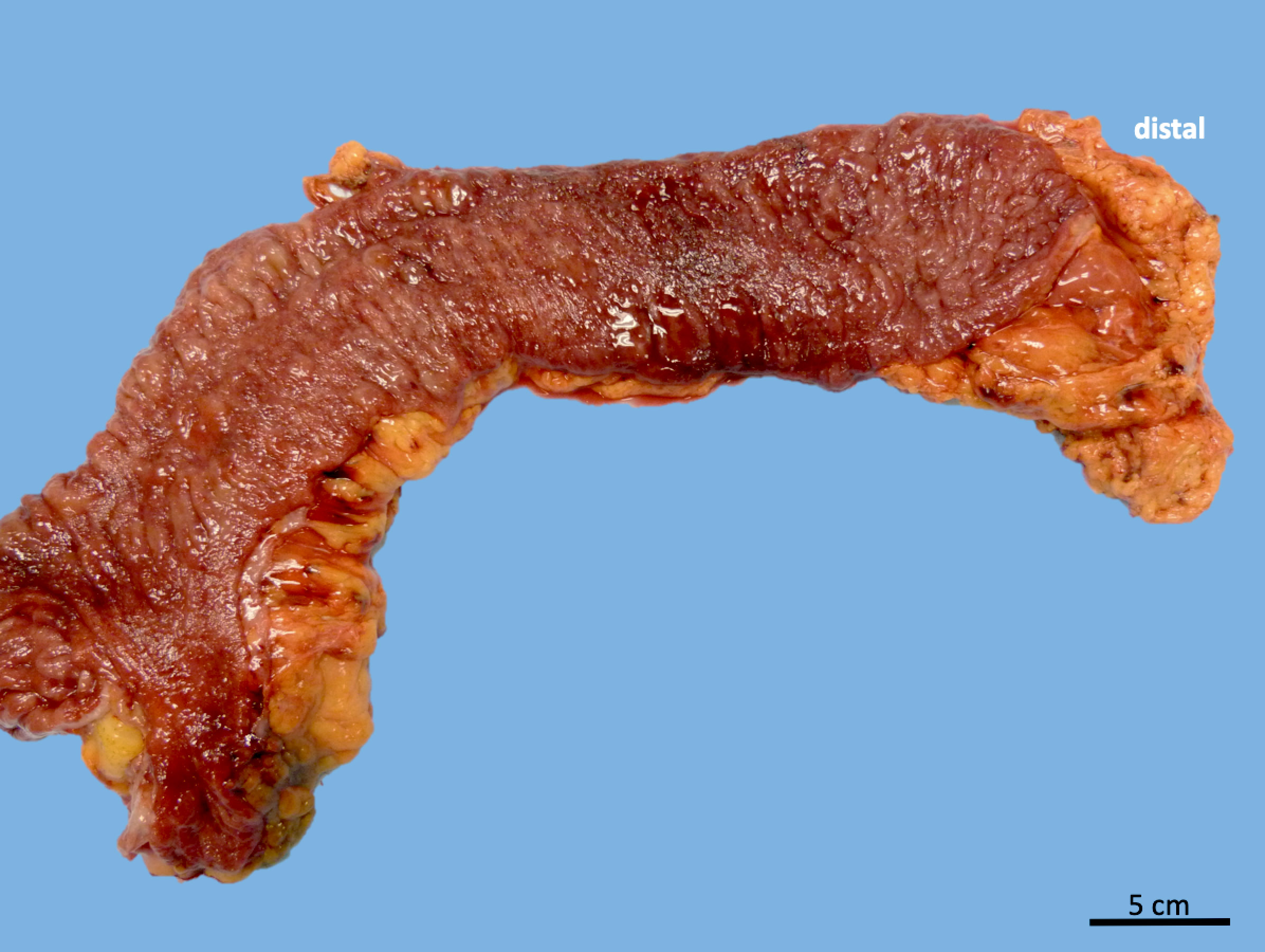
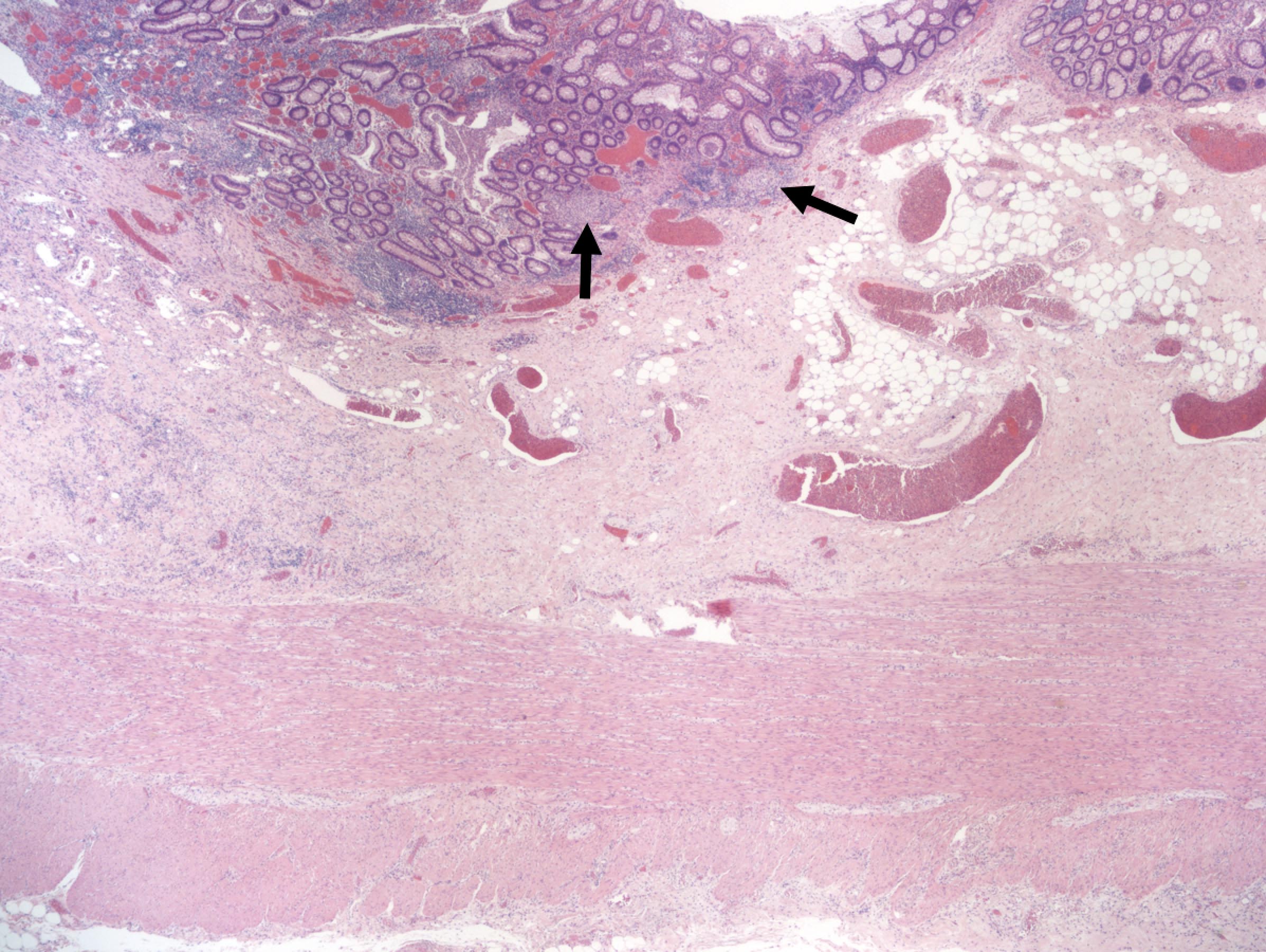
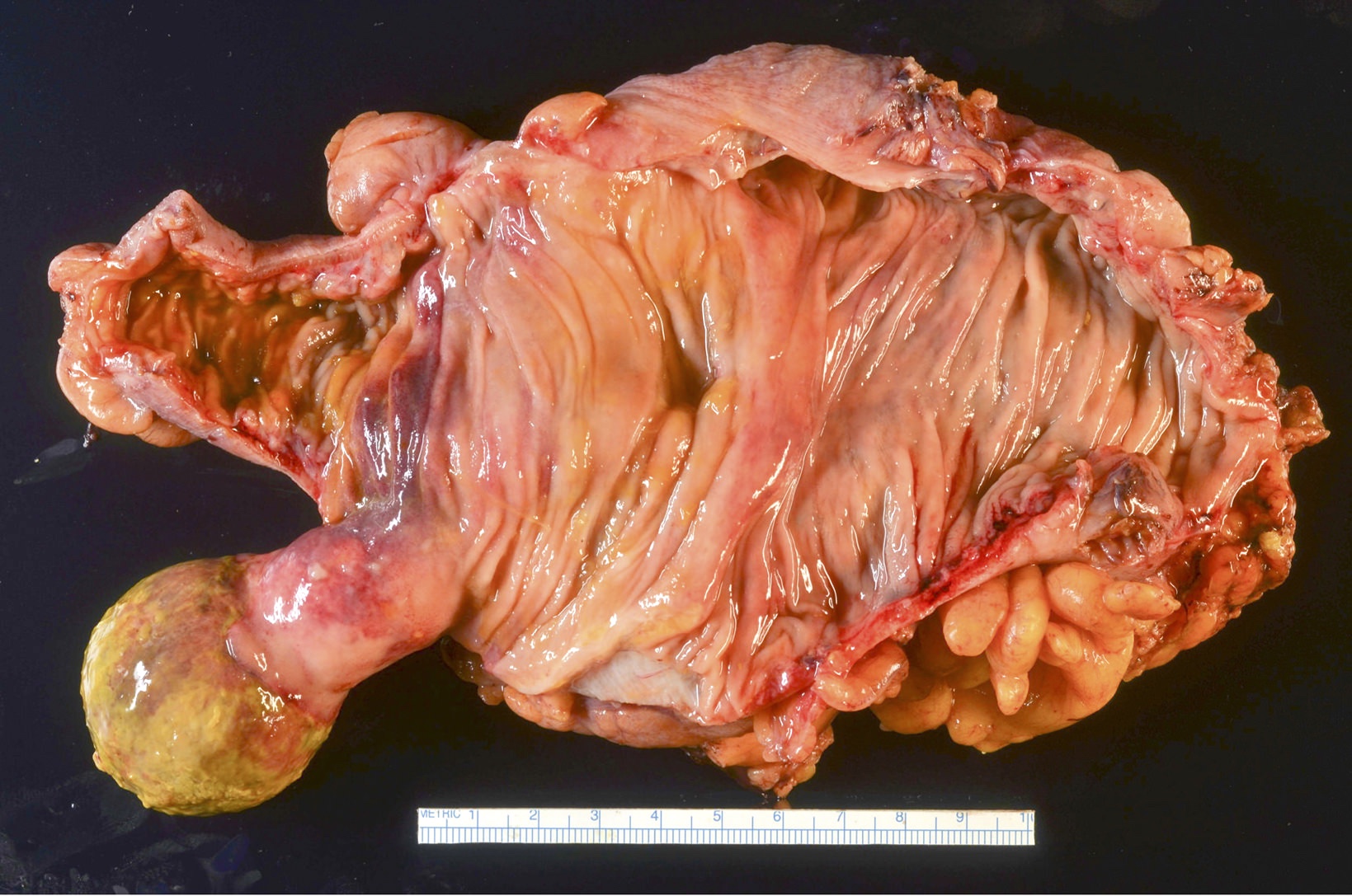
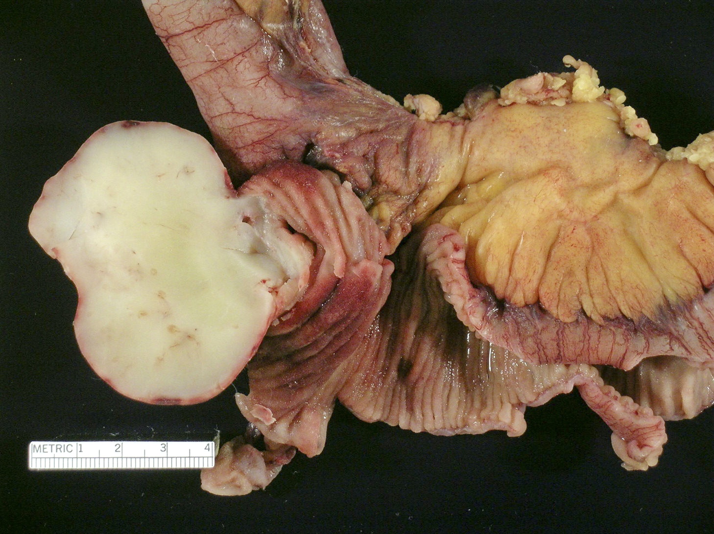
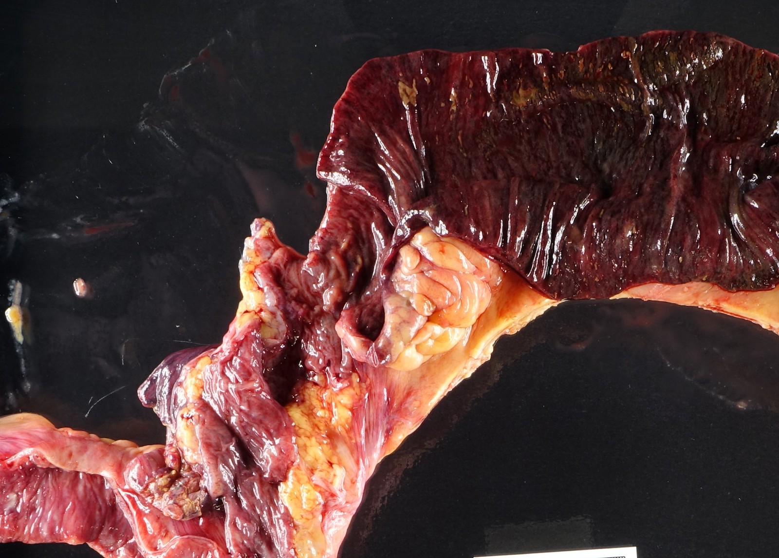
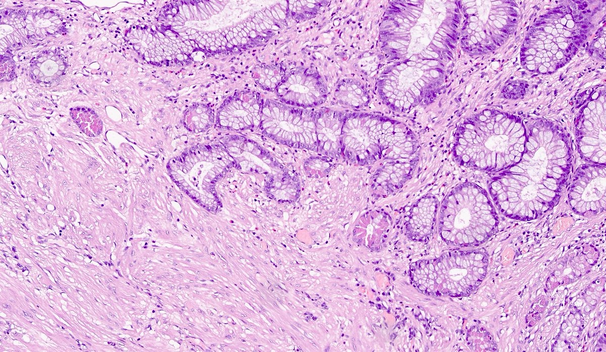
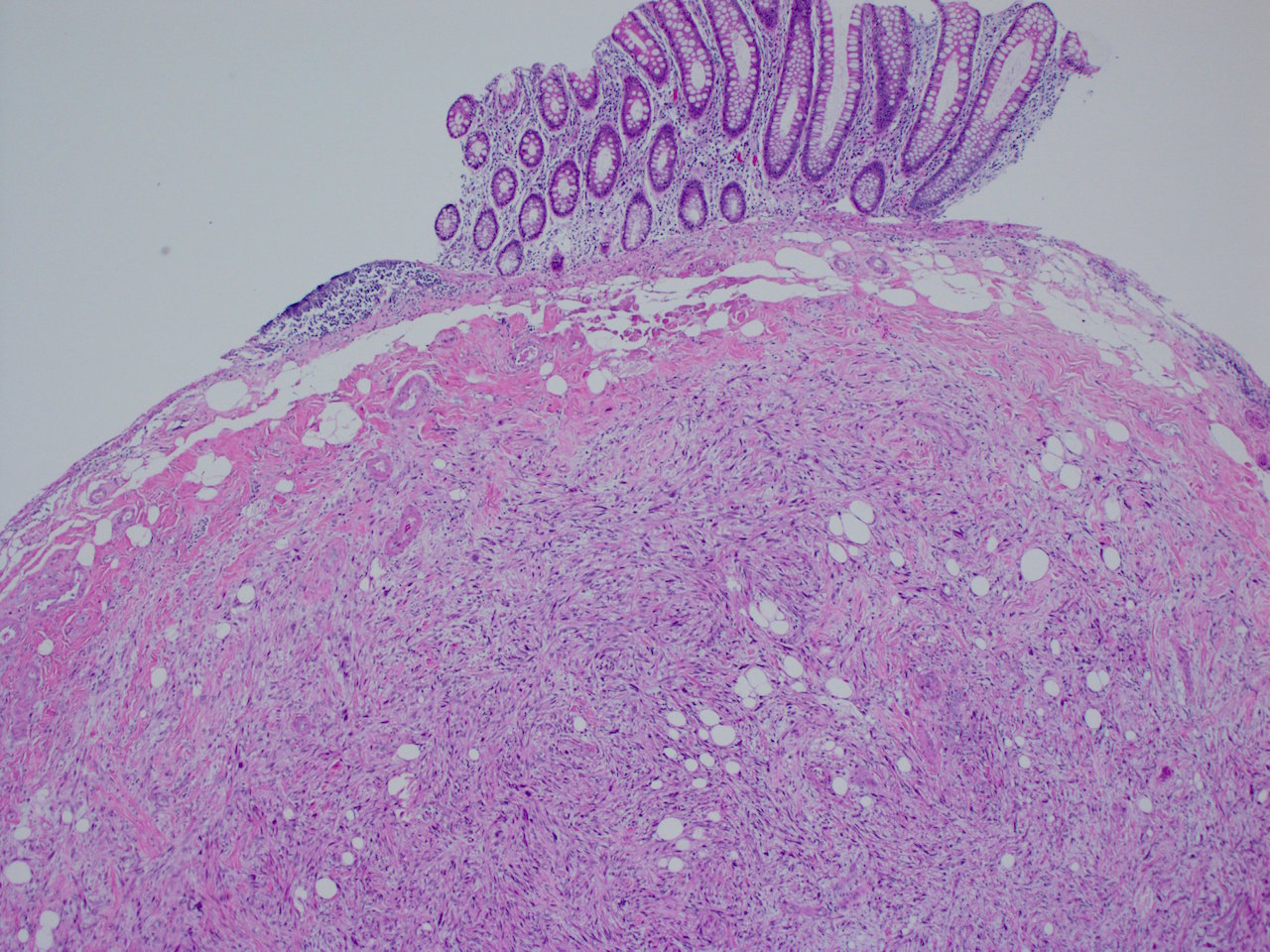
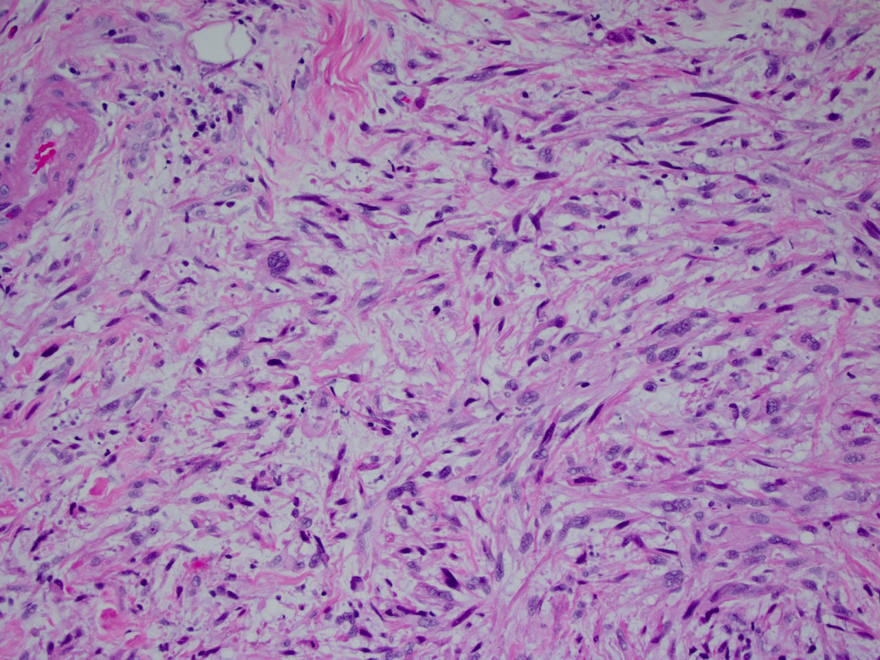
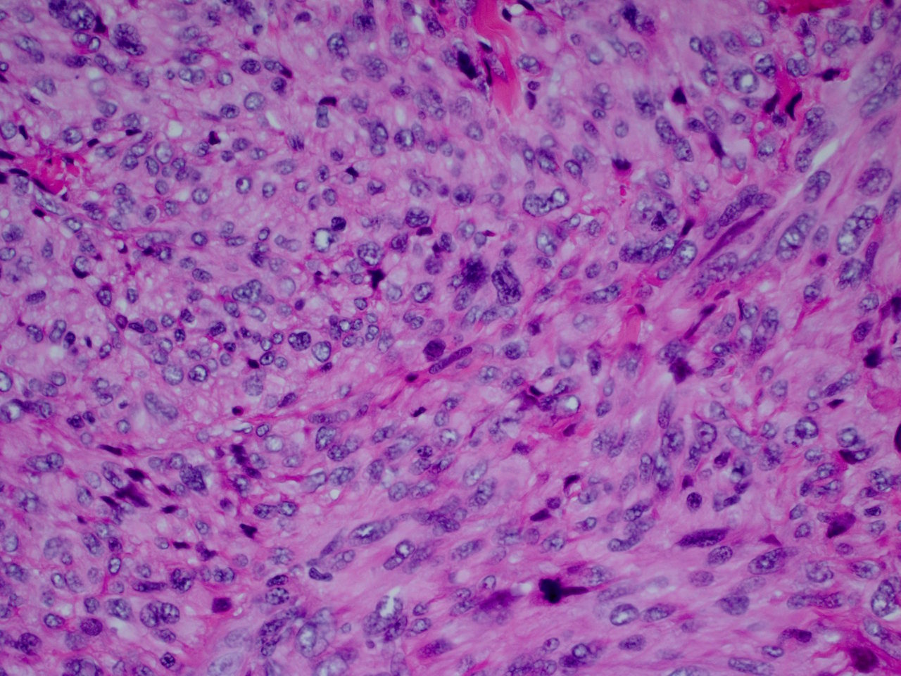
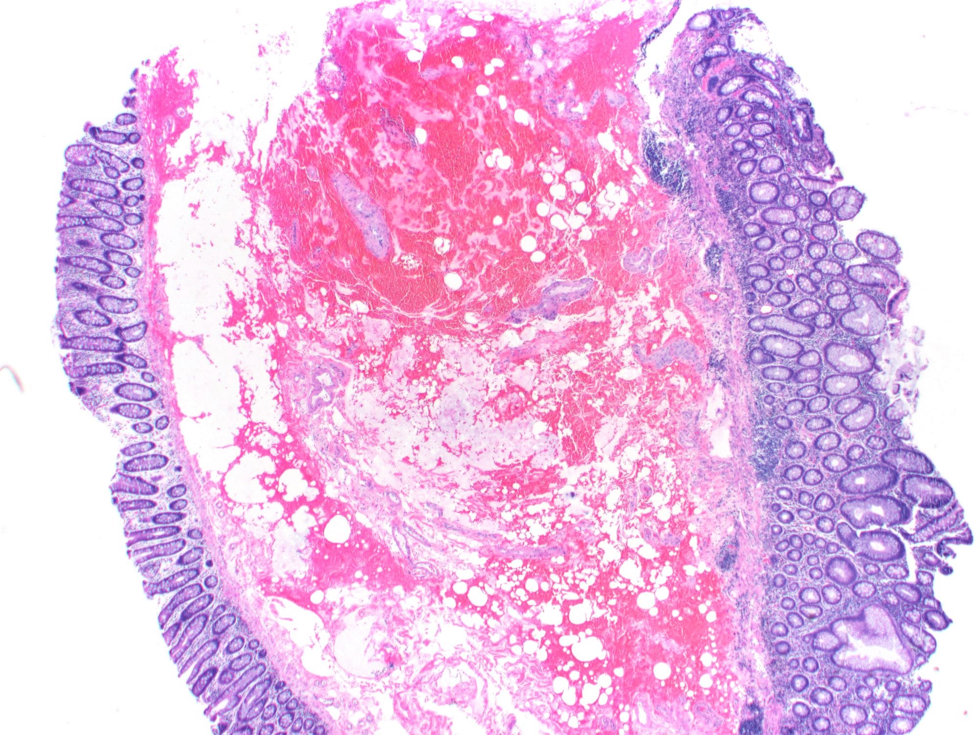
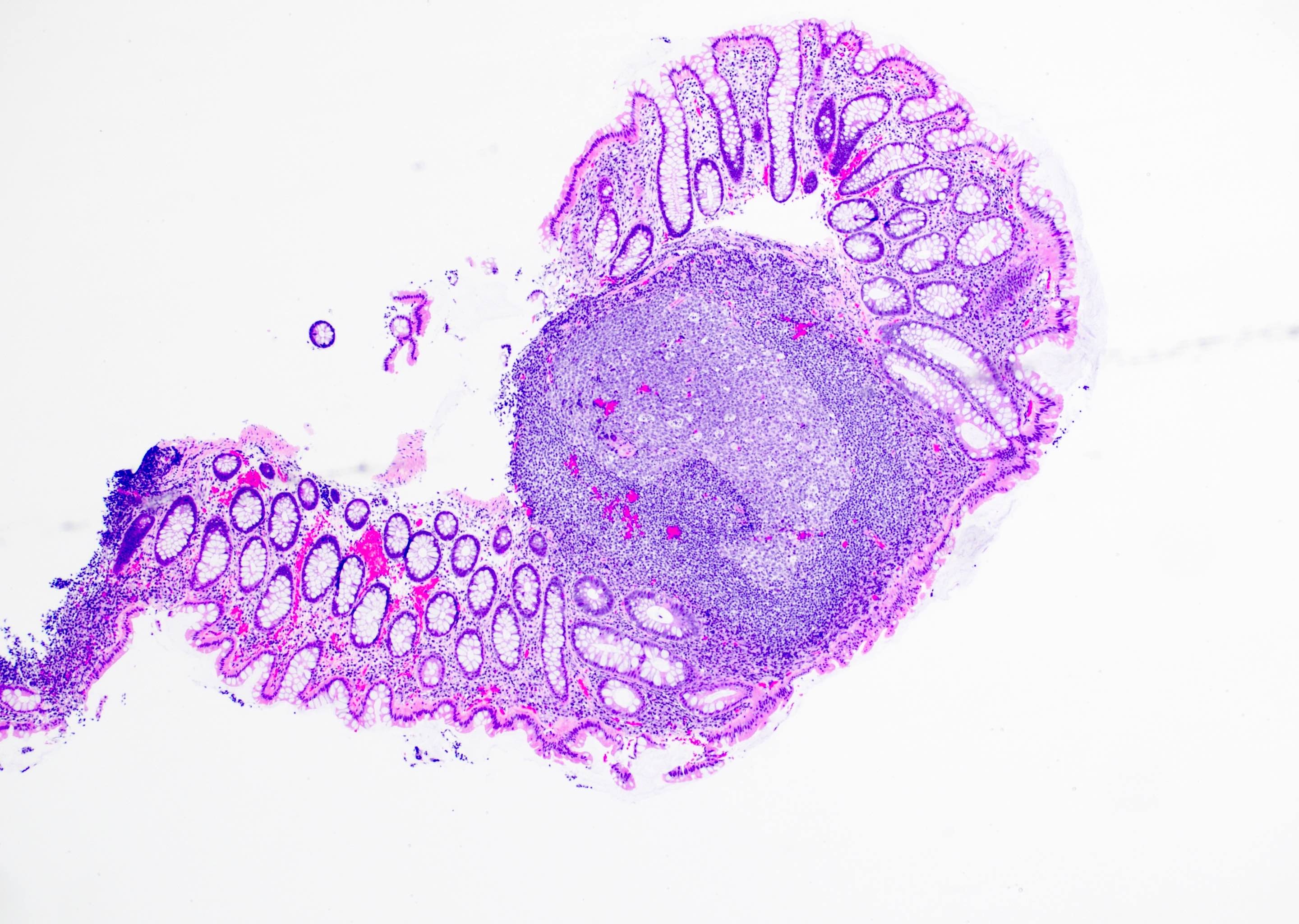
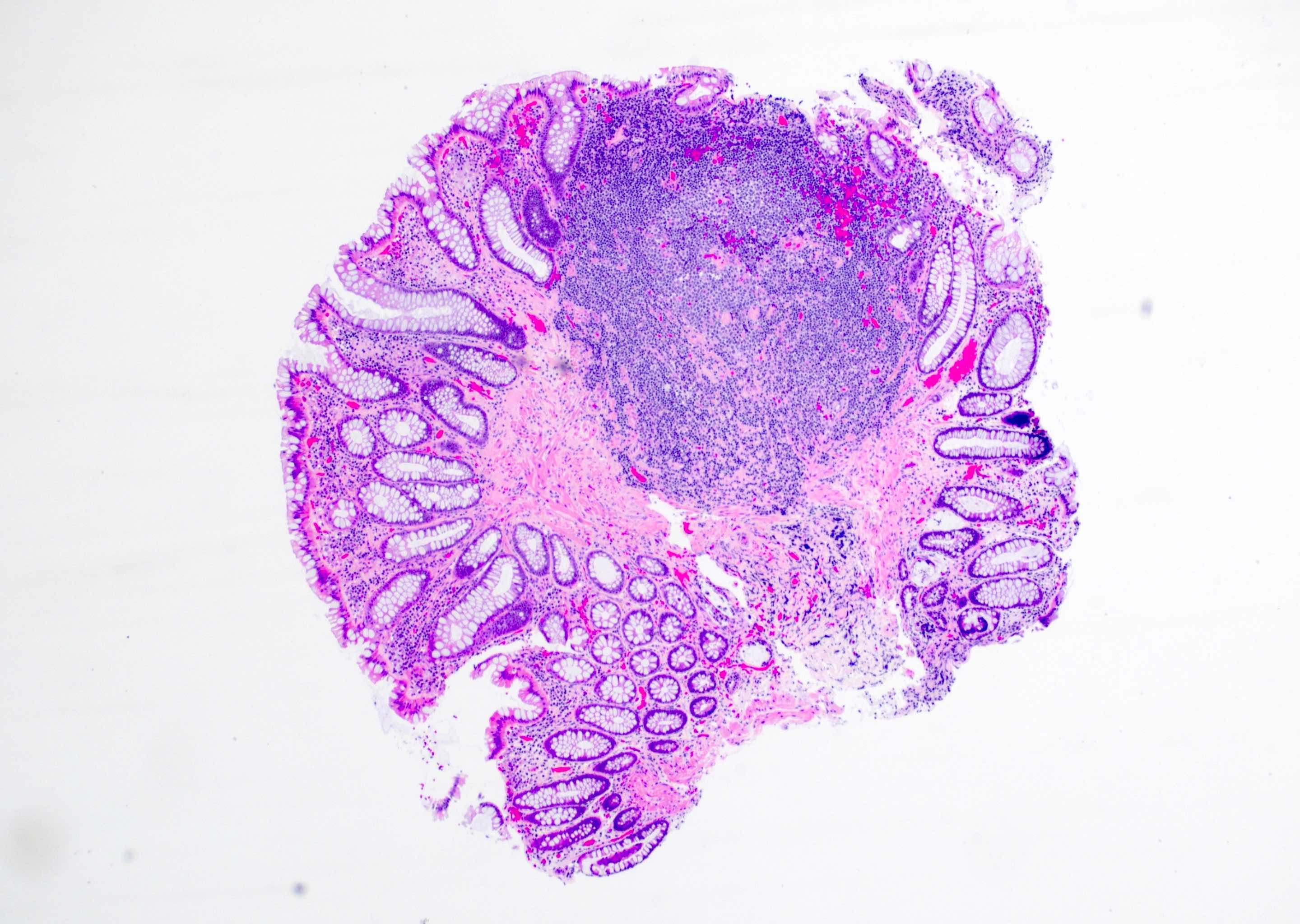
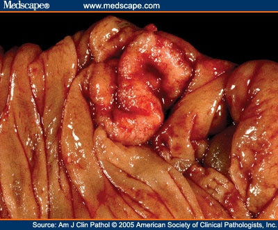
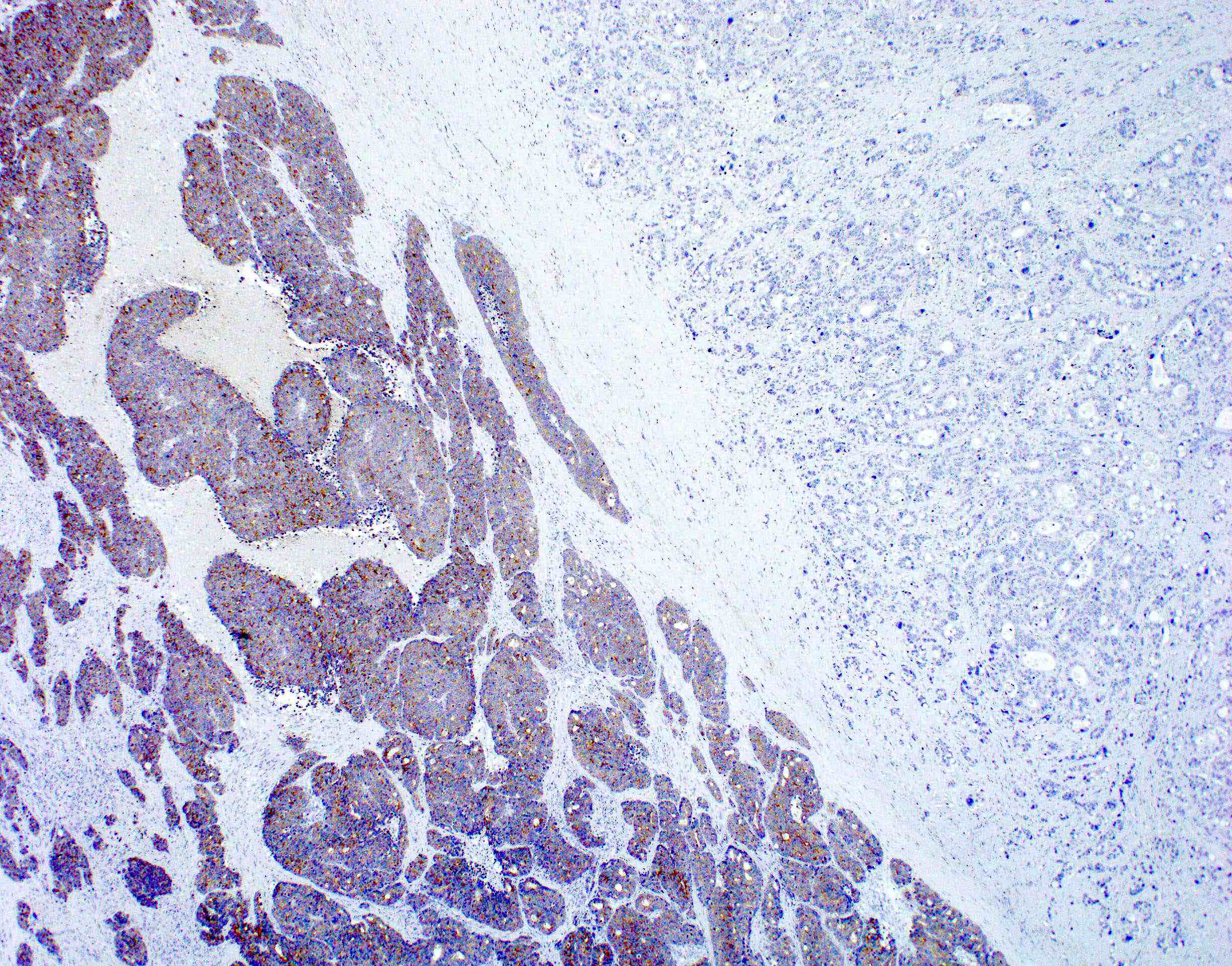
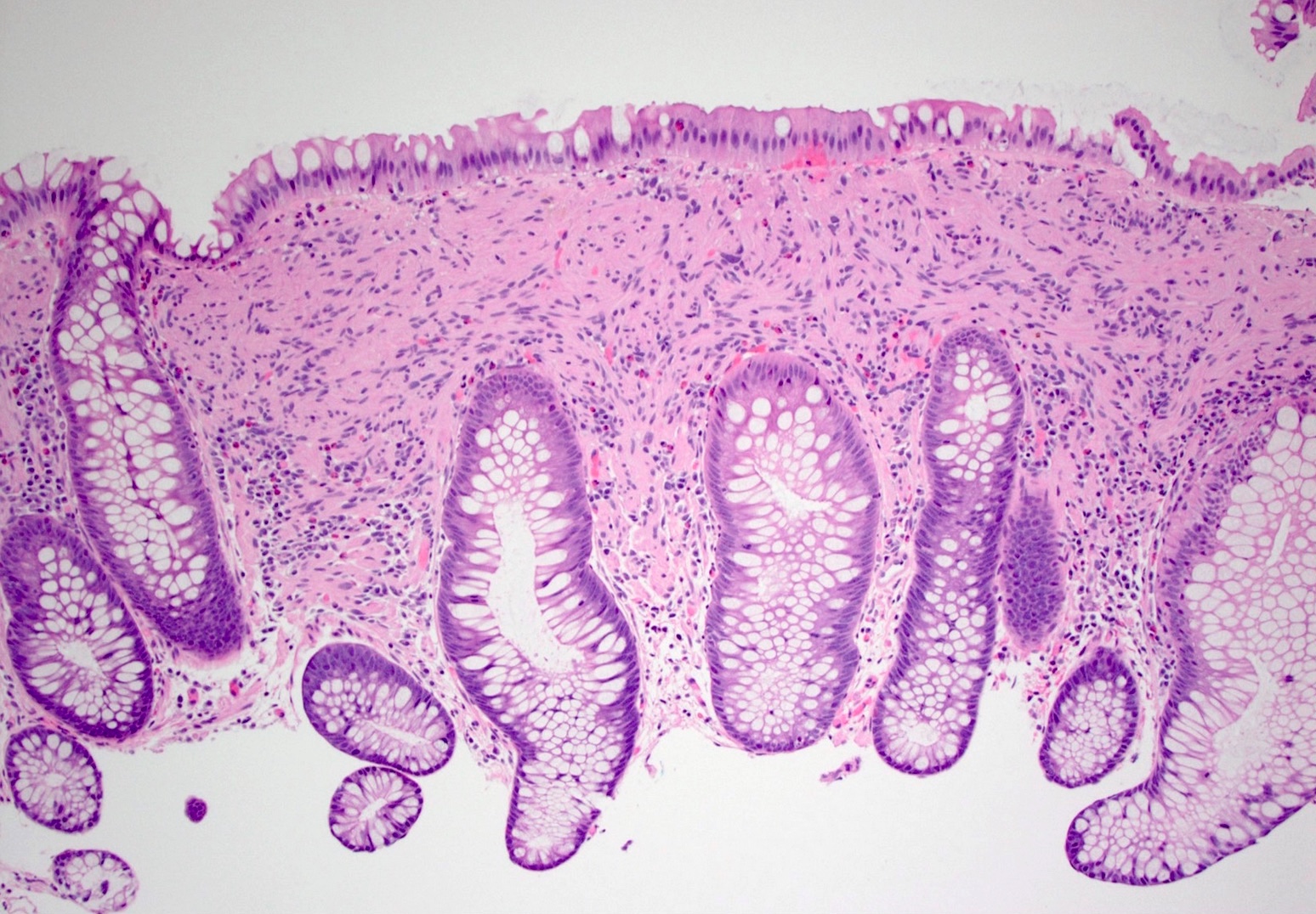
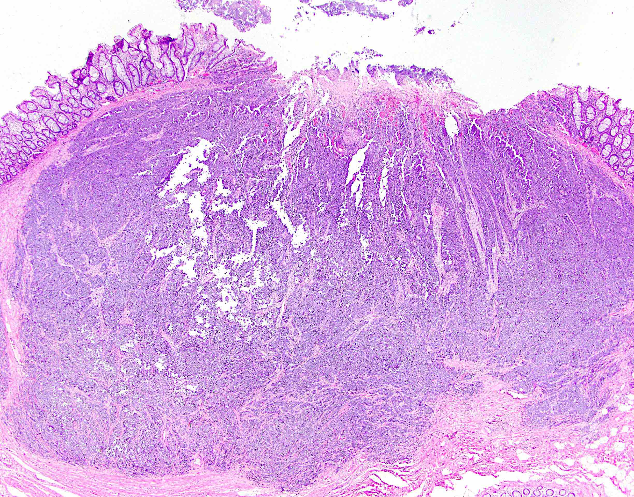
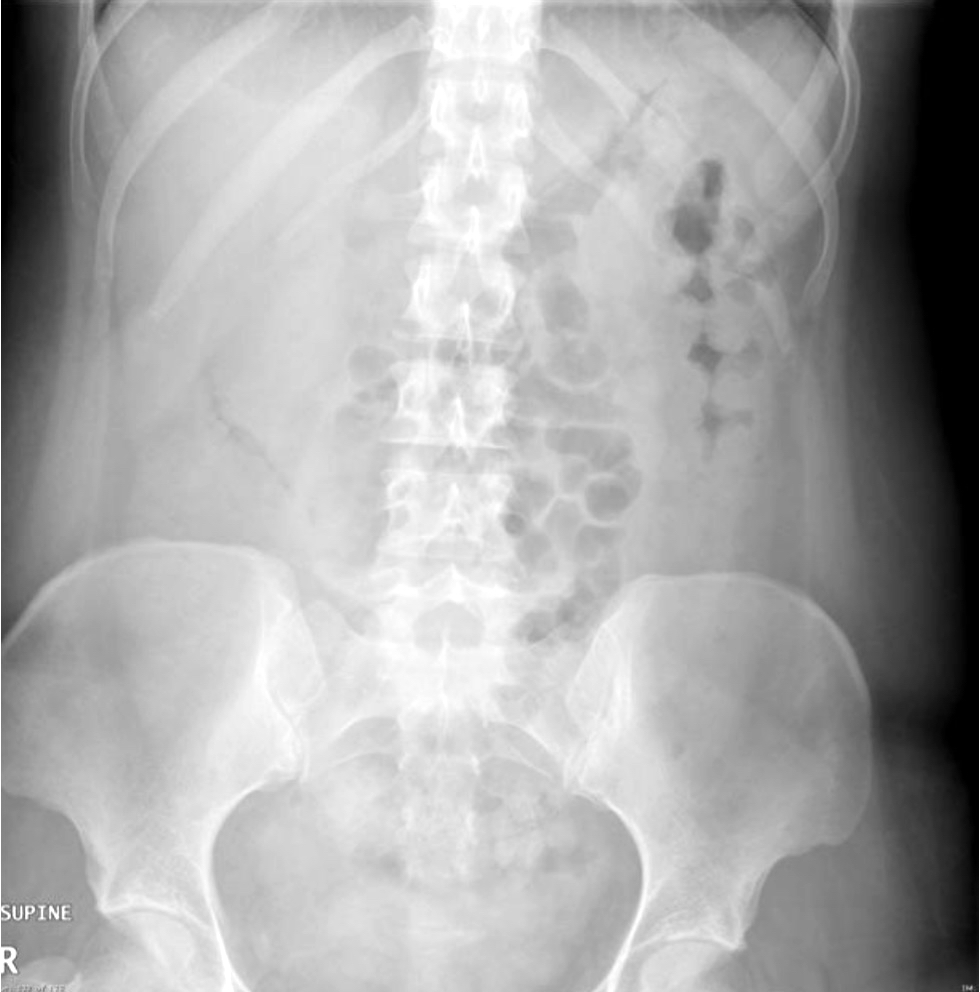
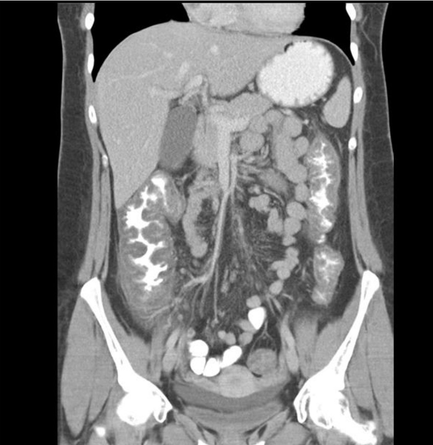
Sigmoid colon mass
Contributed by Asmaa Gaber Abdou, M.D.
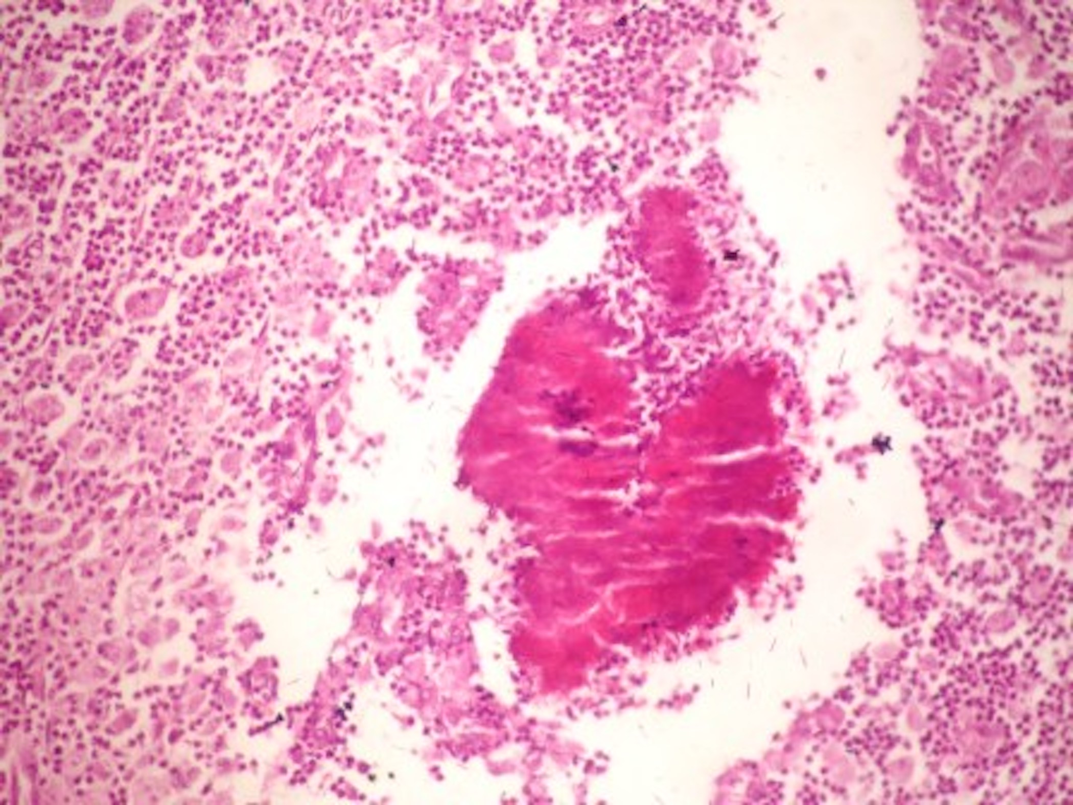
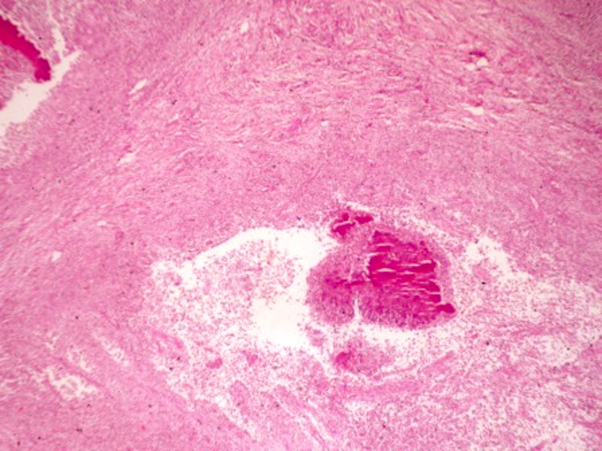
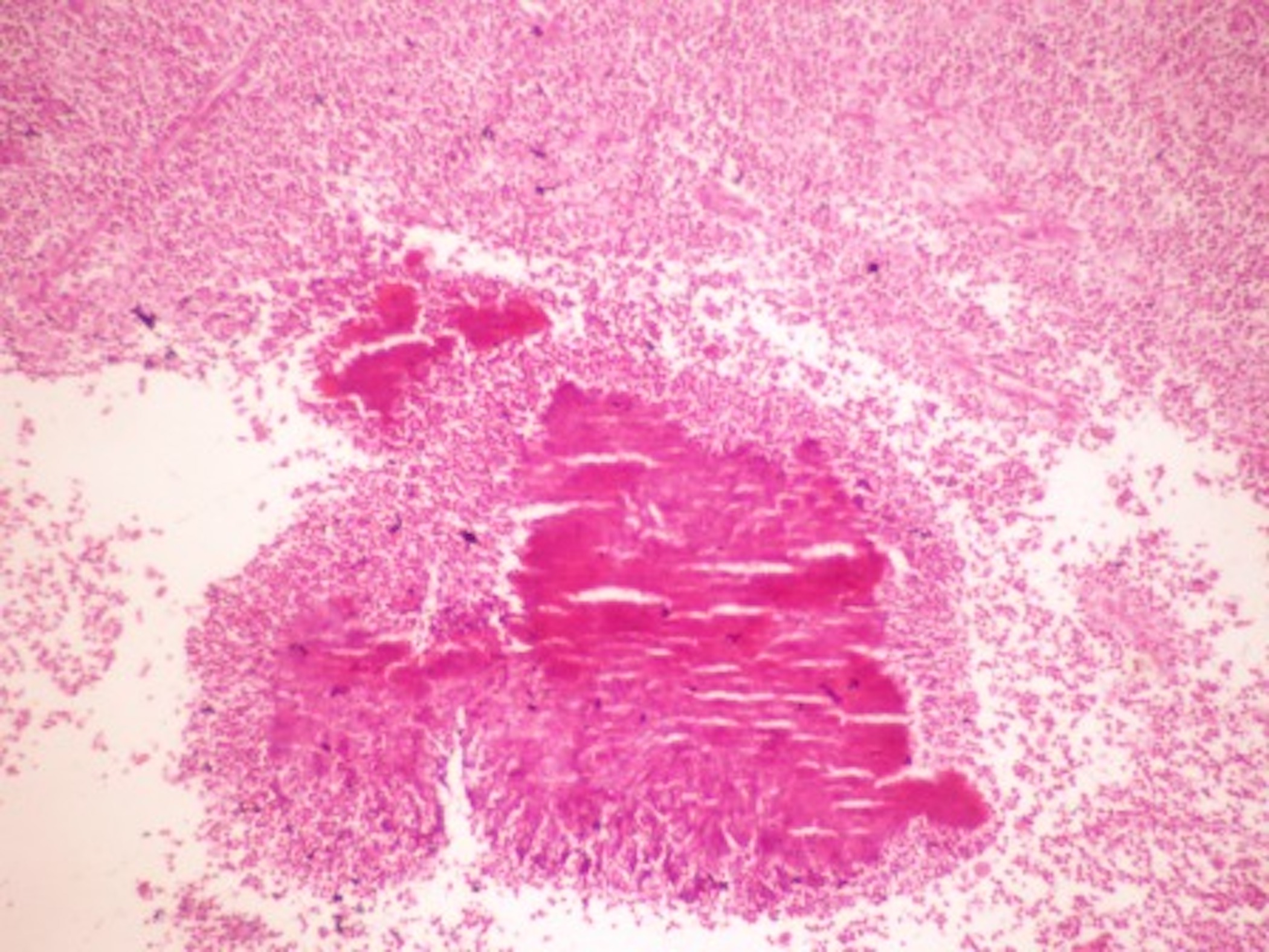
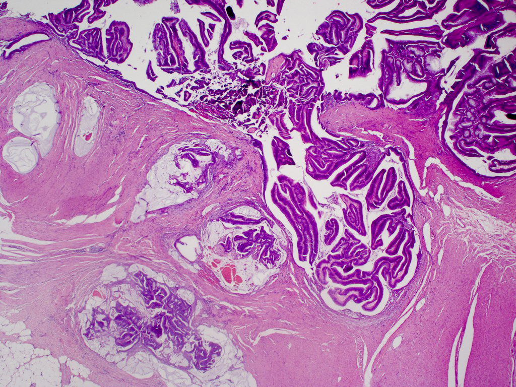
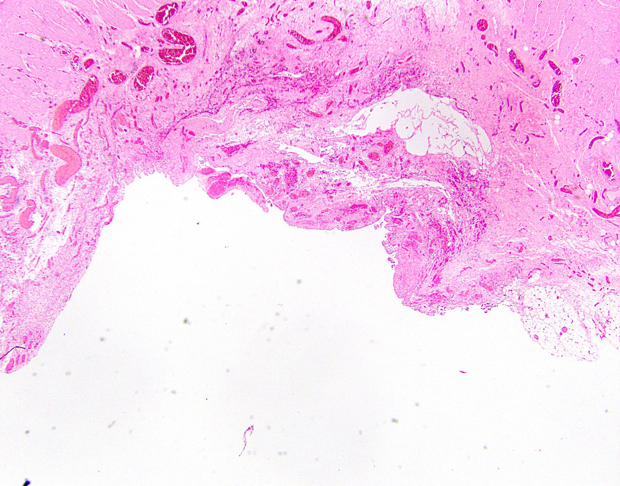
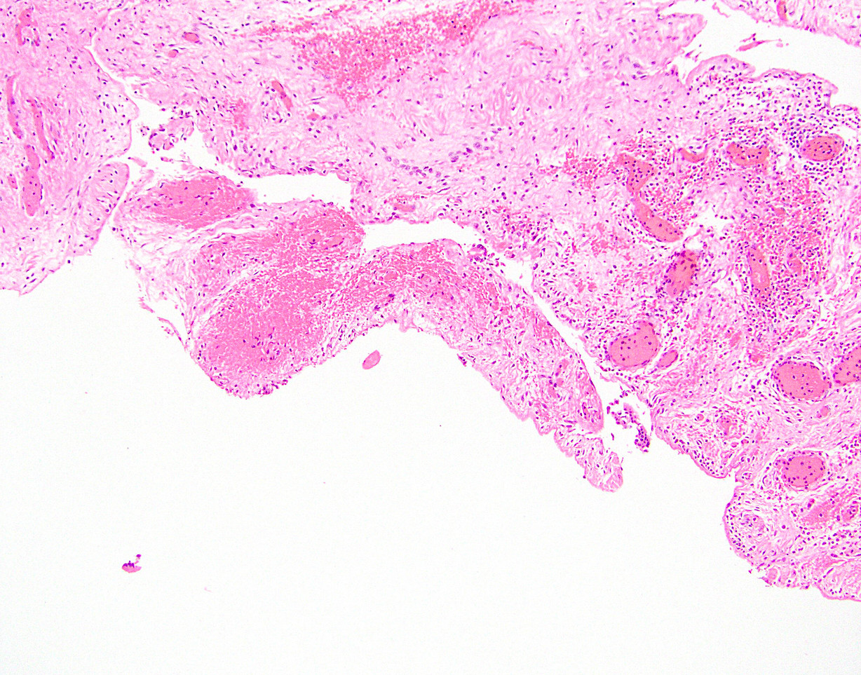
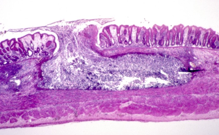
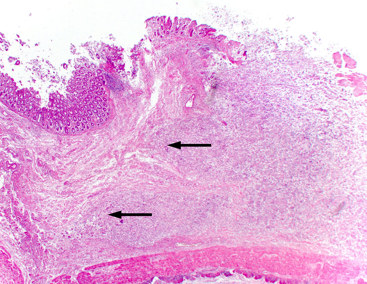
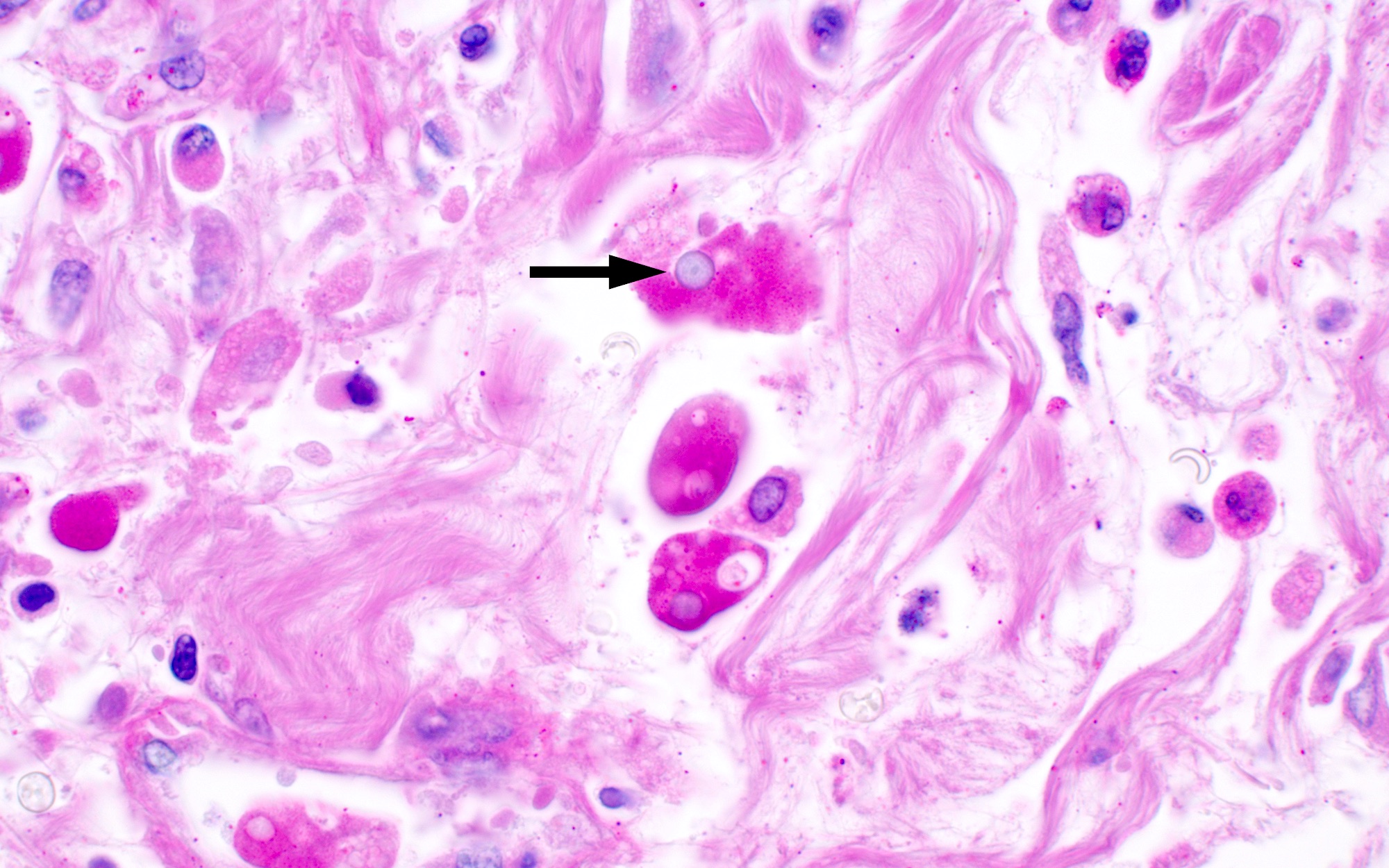
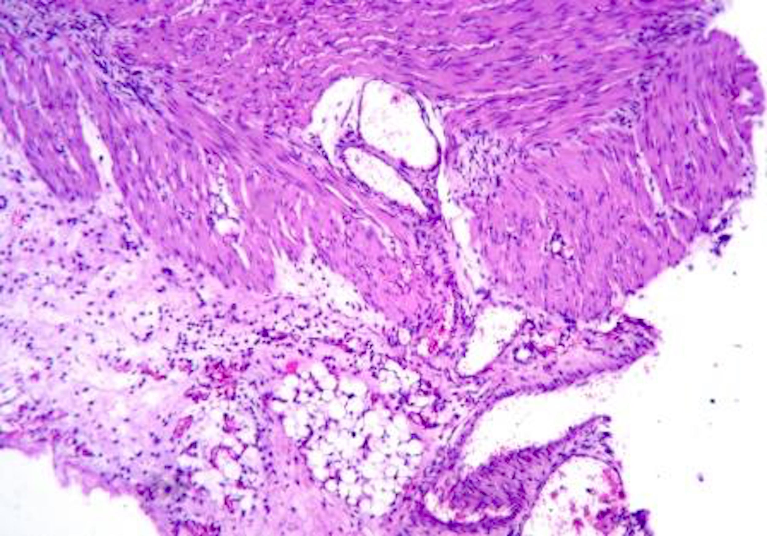
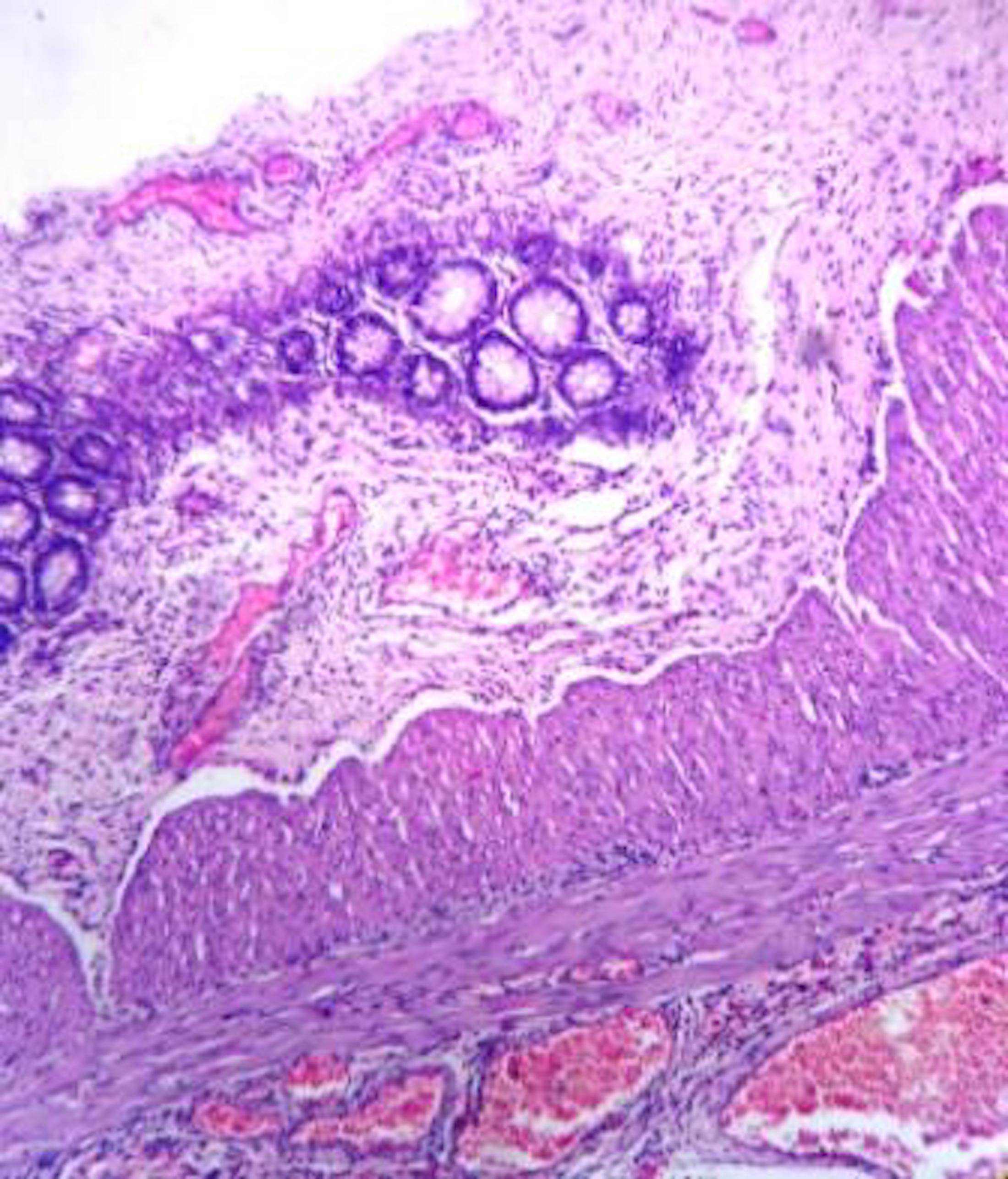
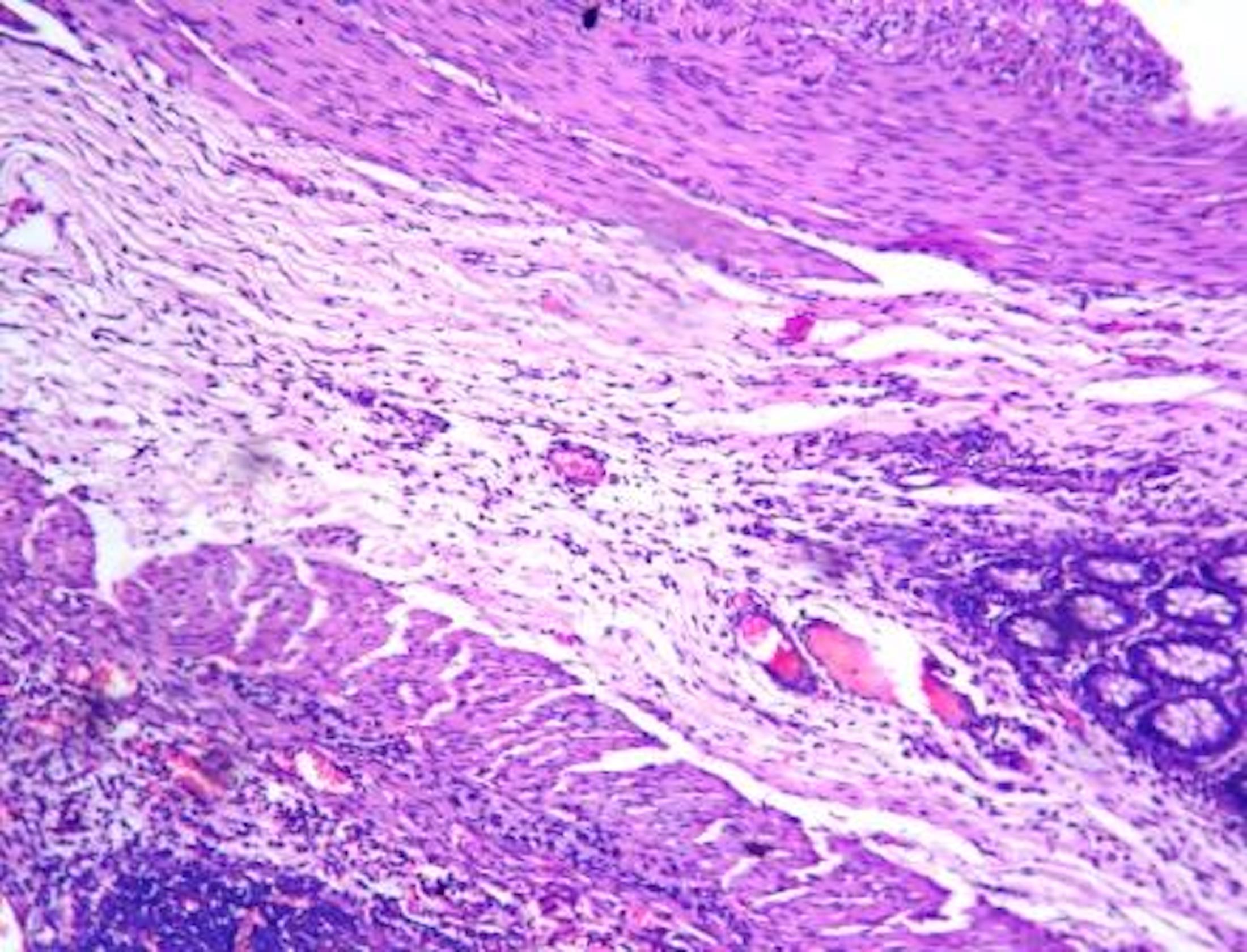
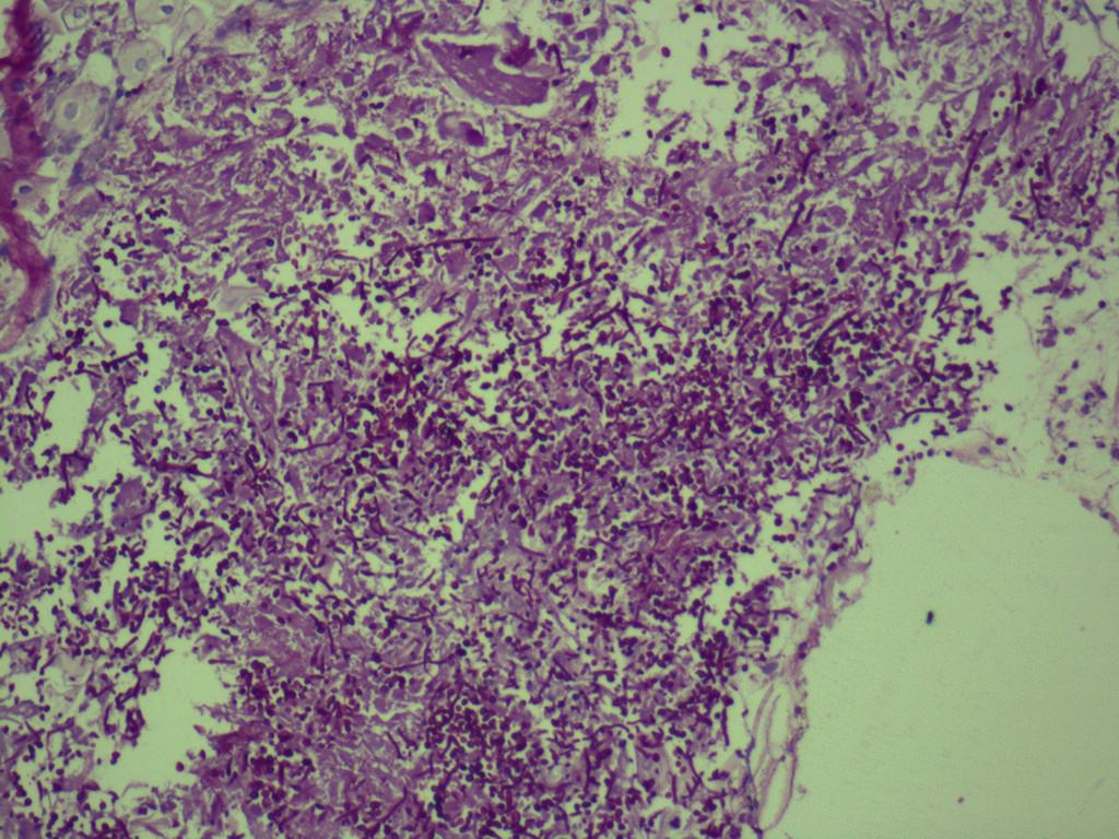
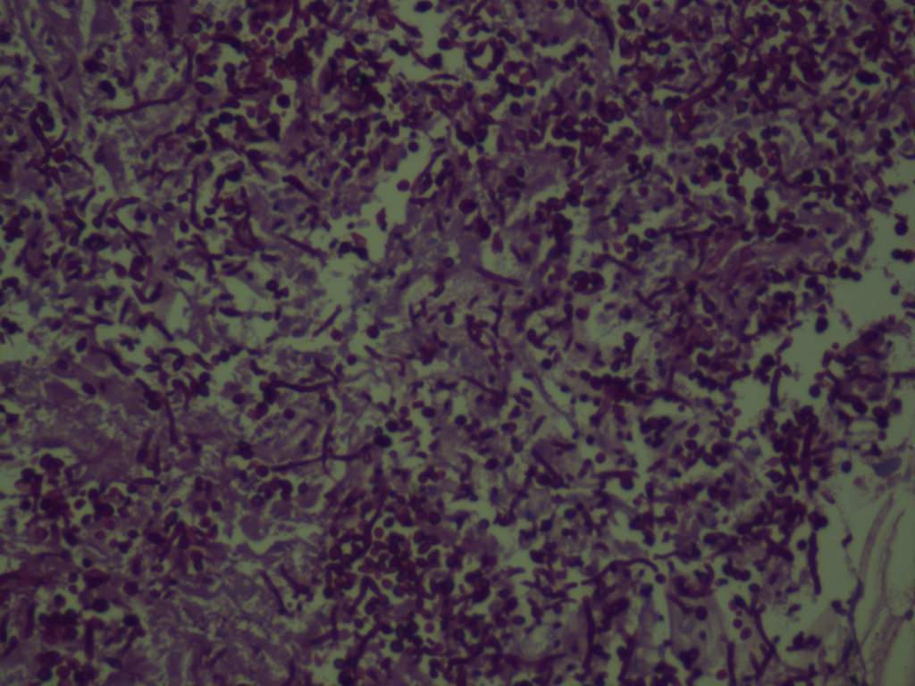
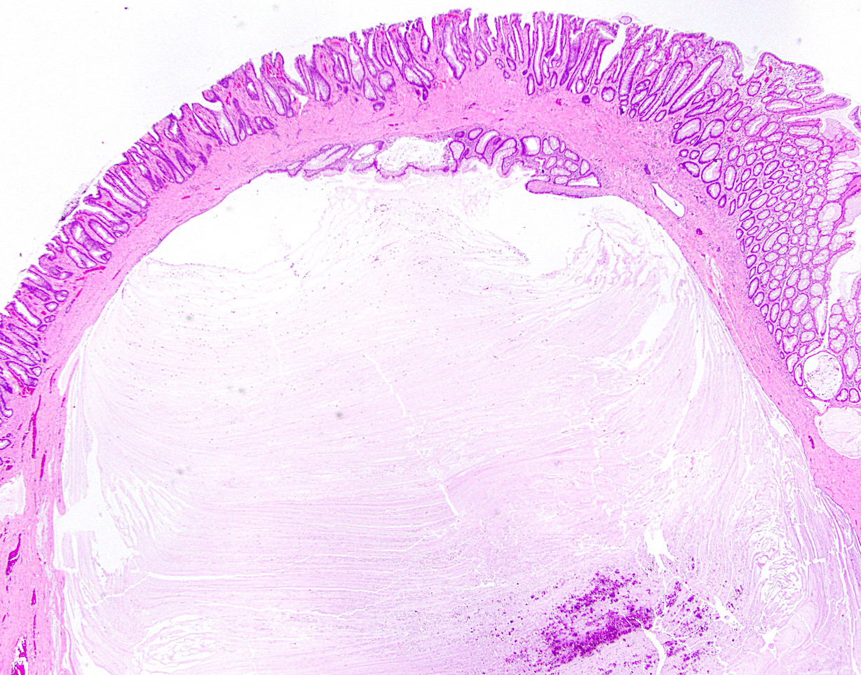
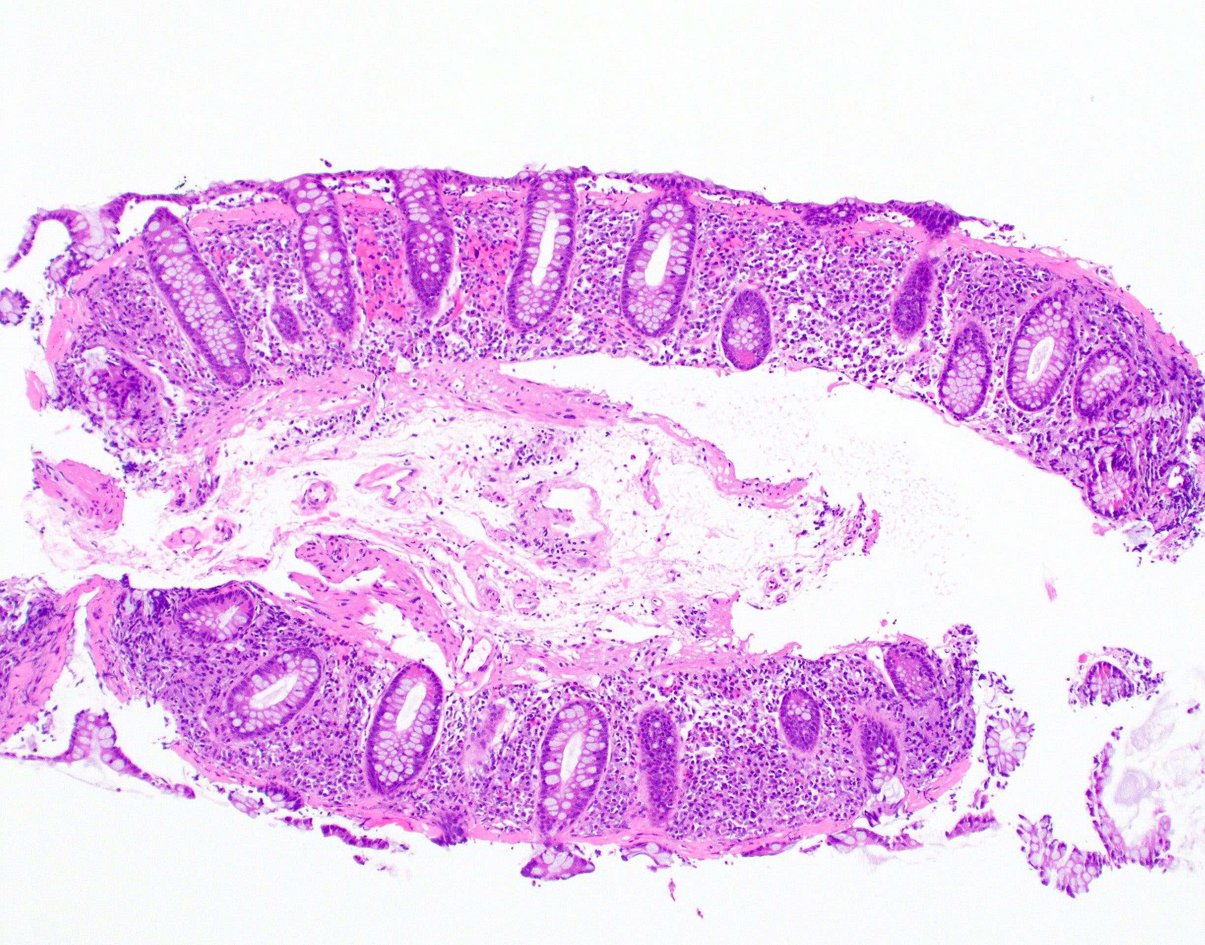
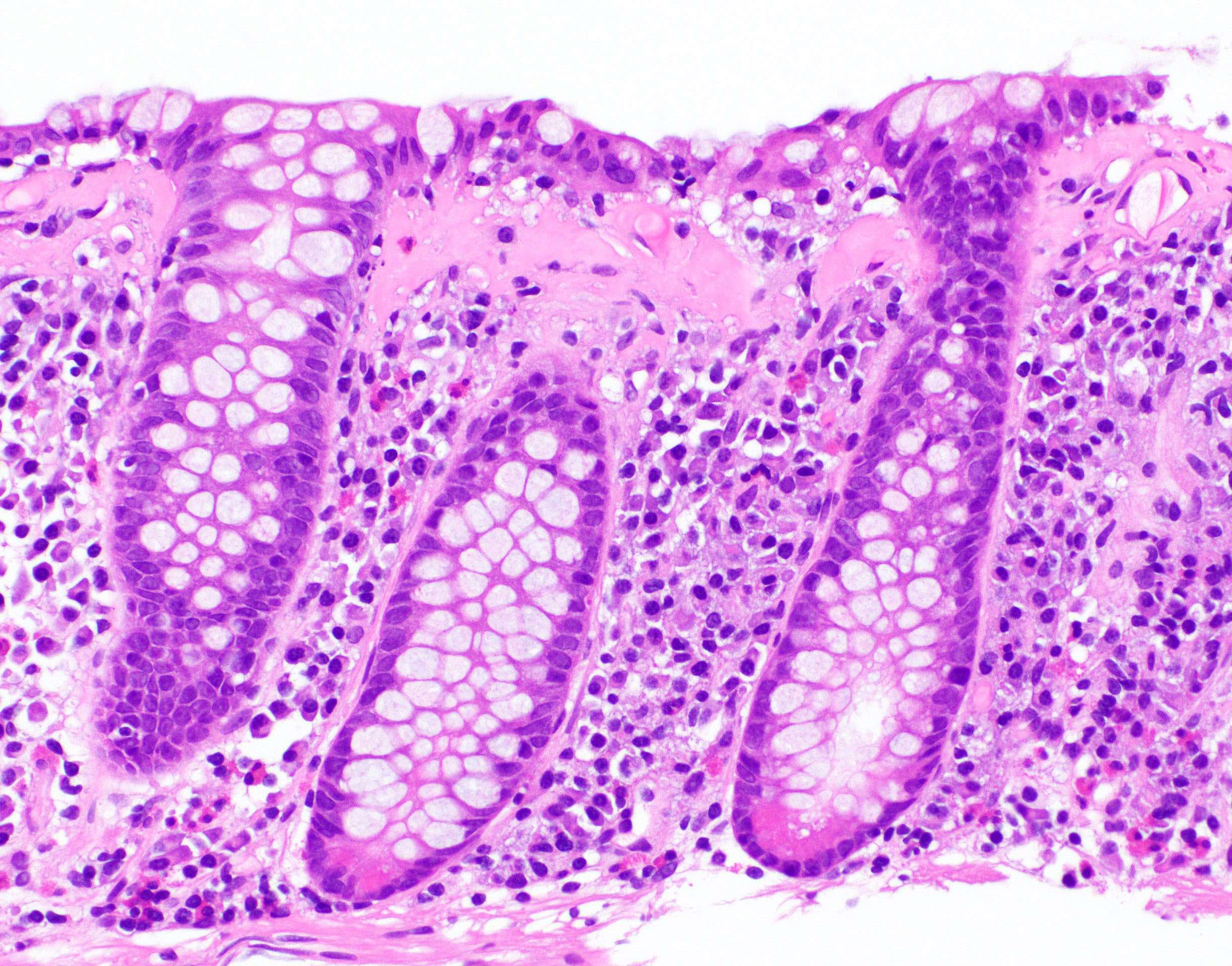
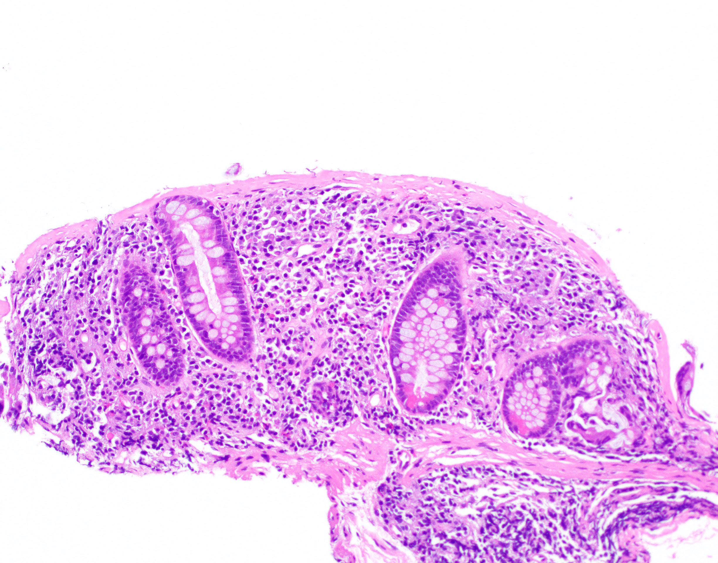
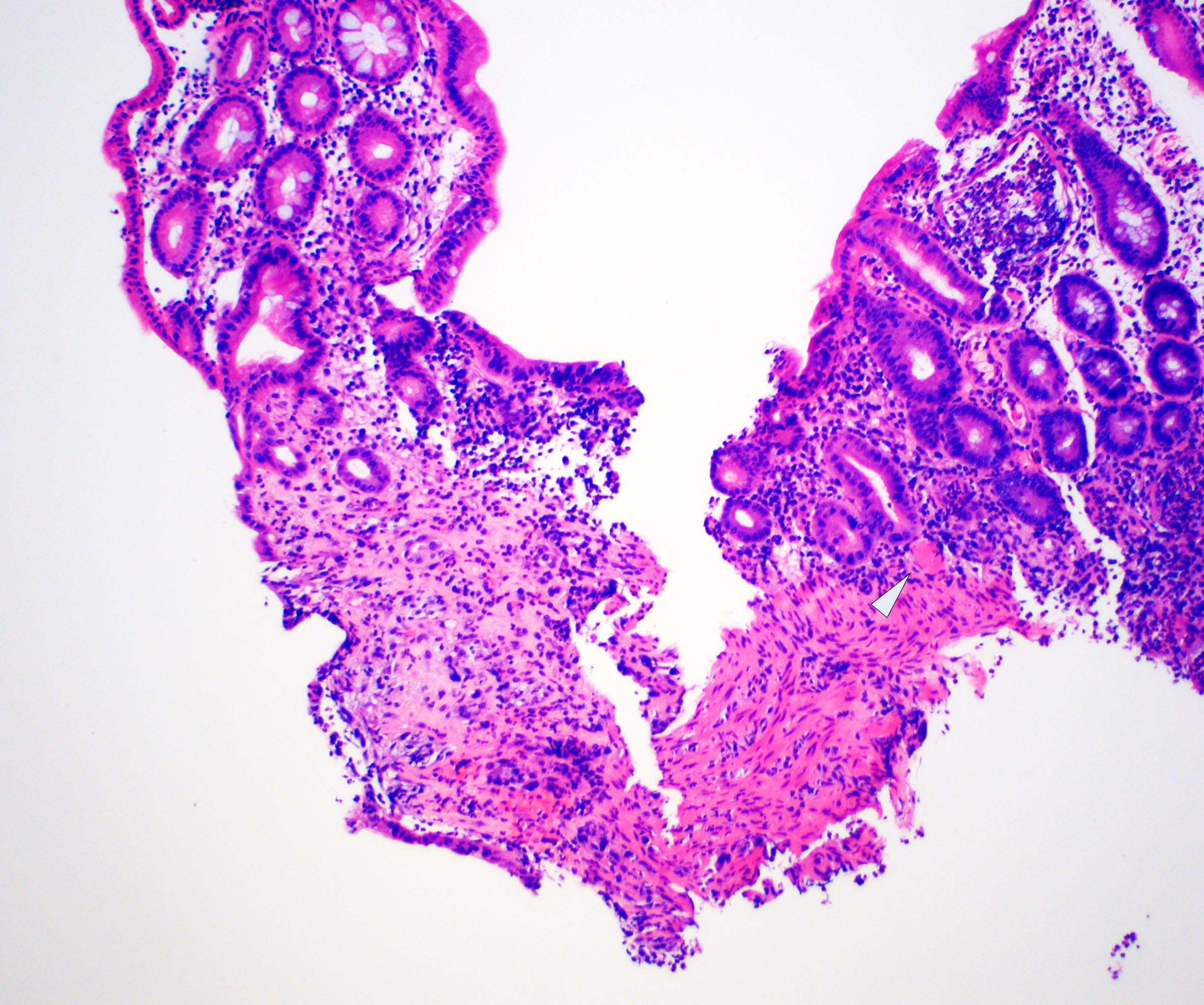
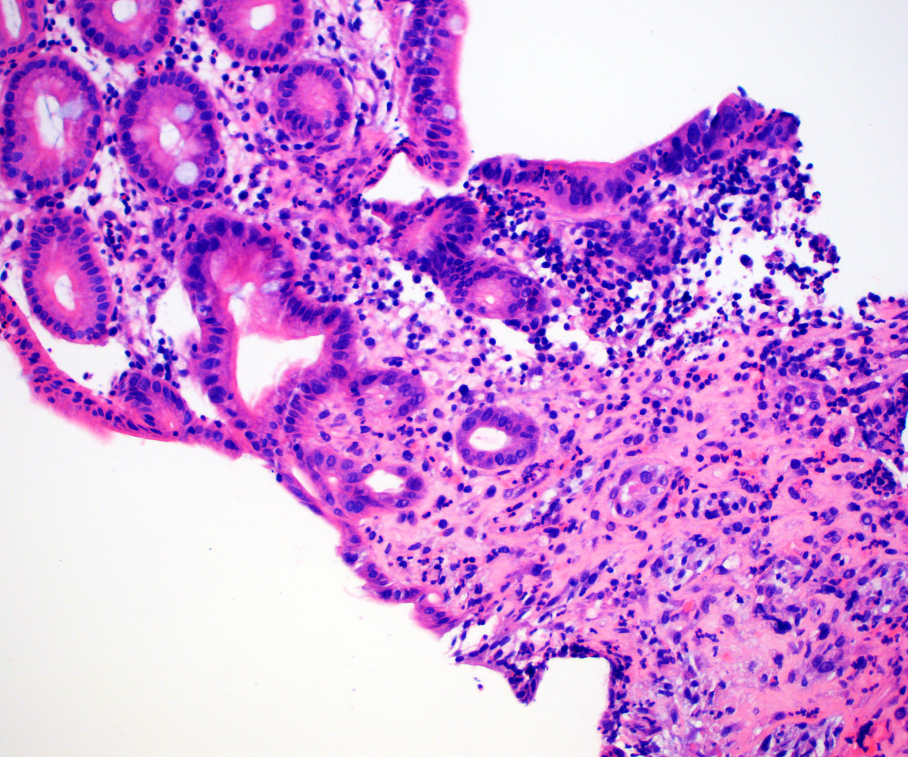
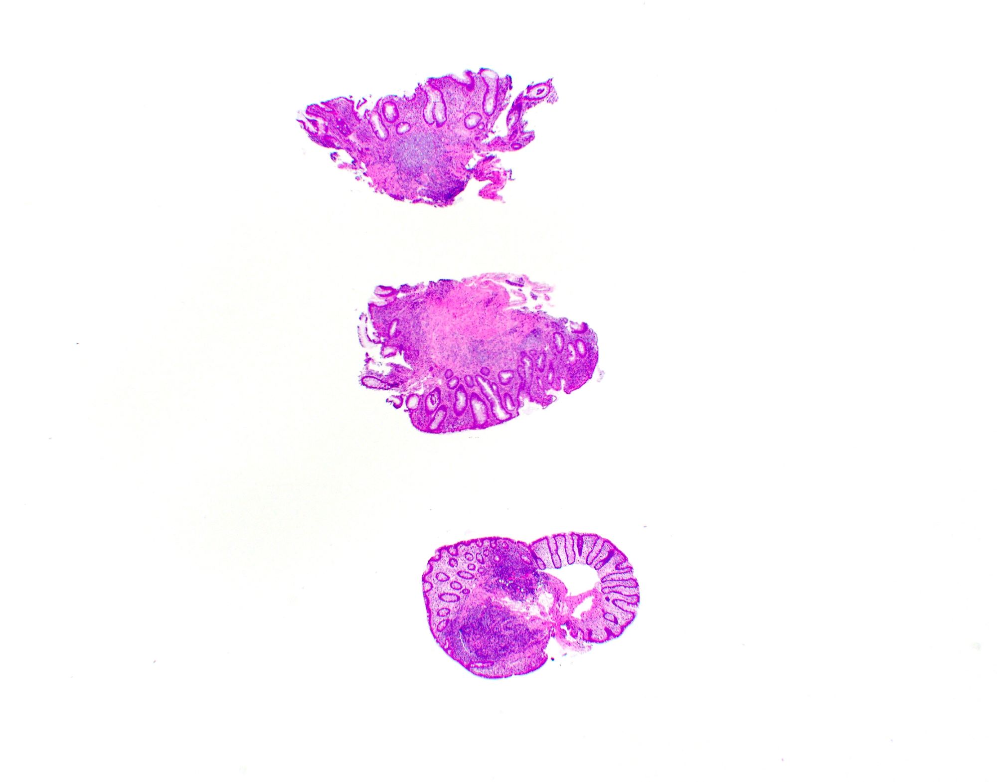
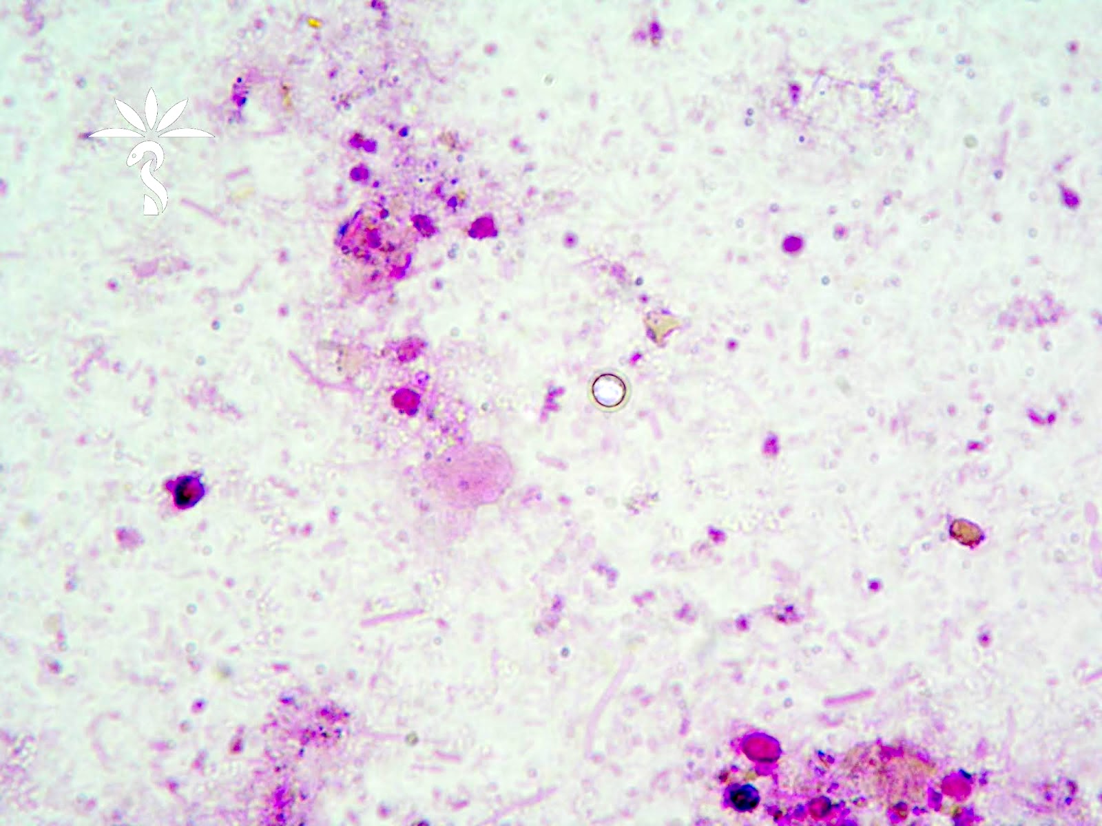
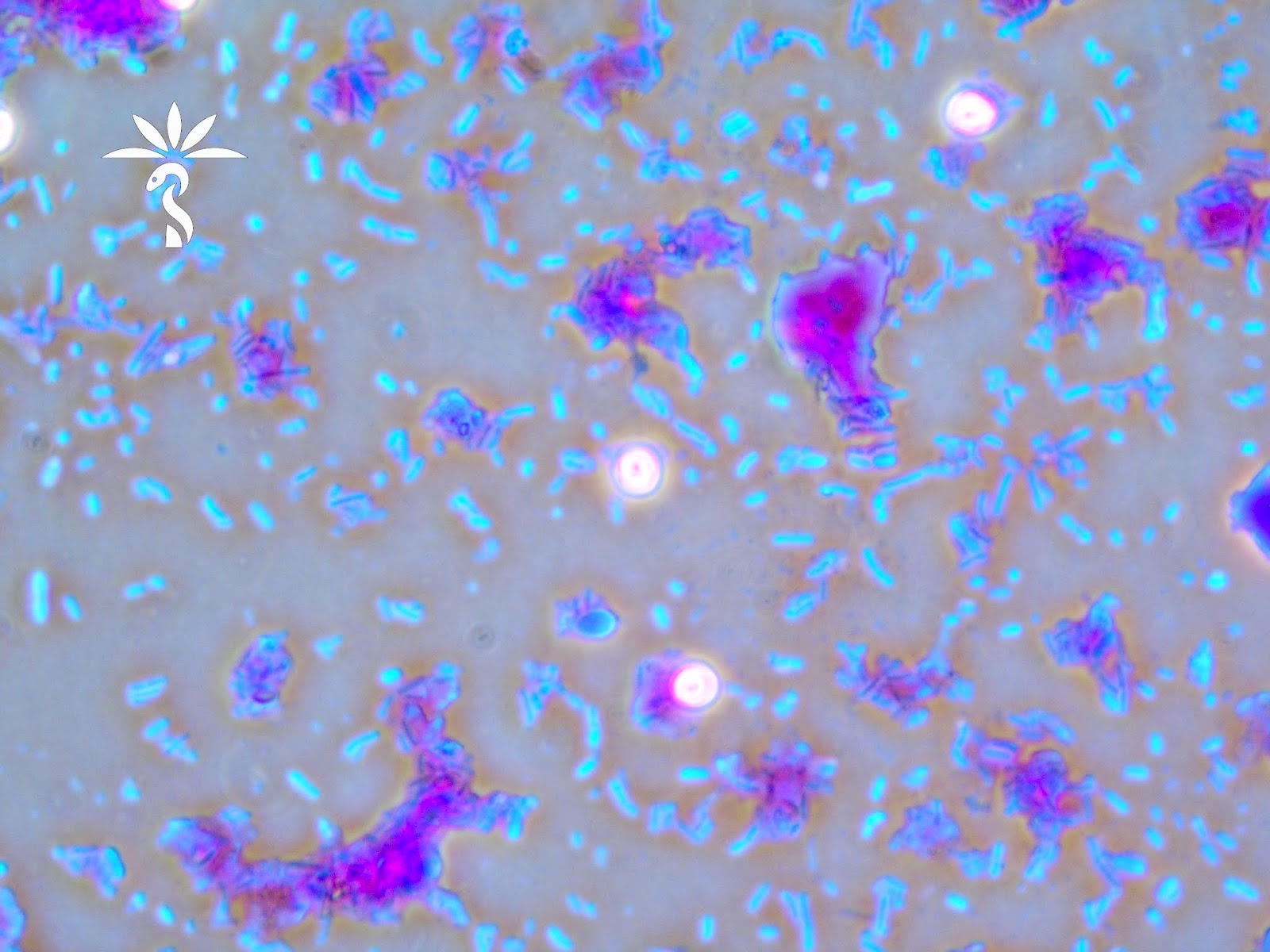
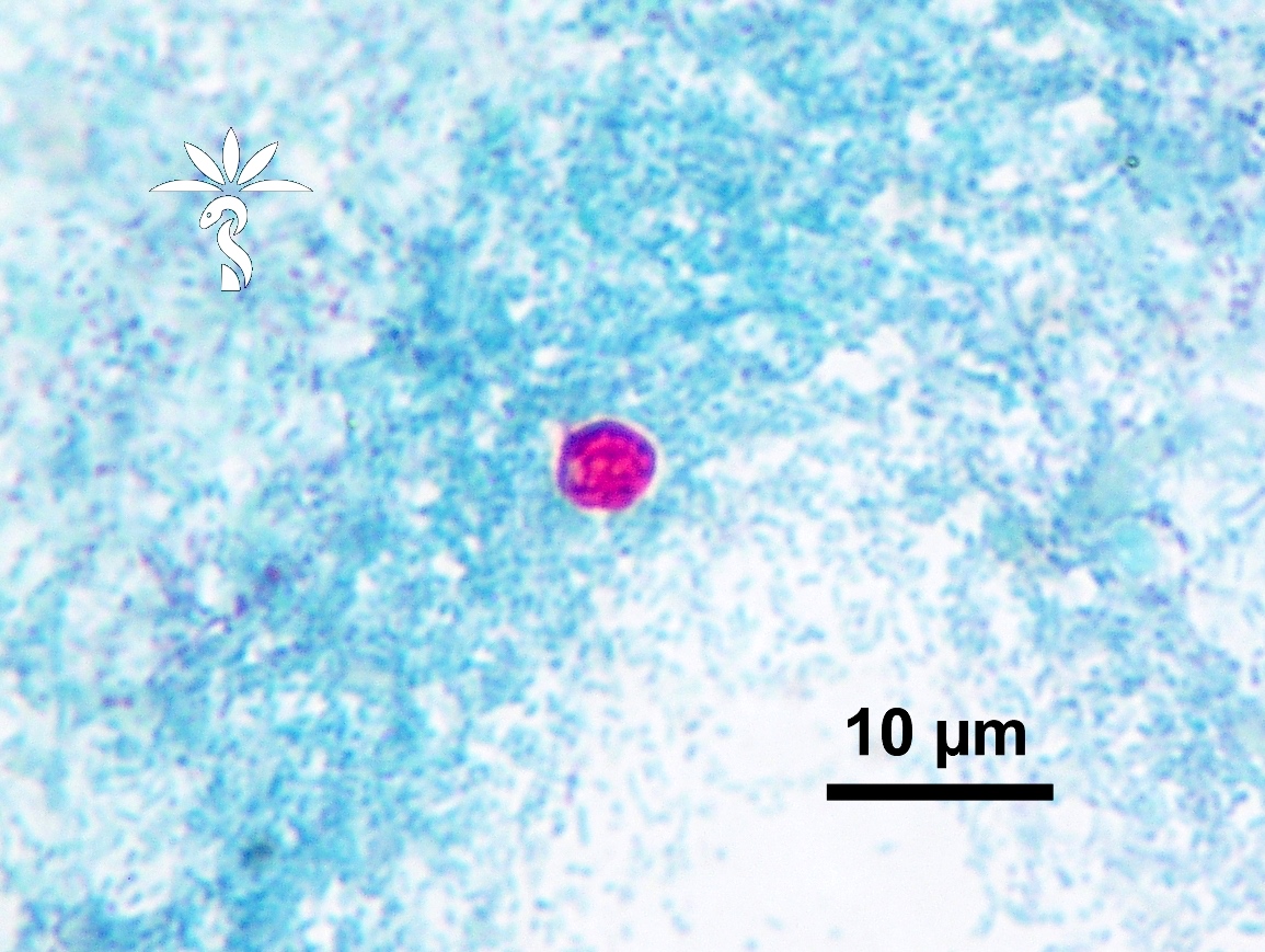
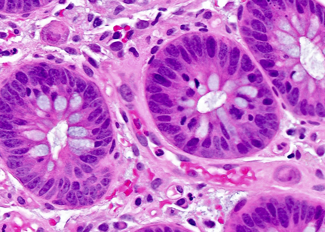
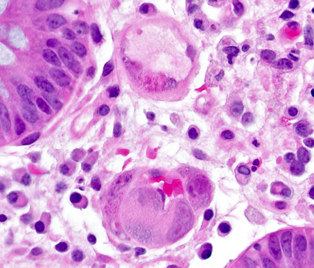
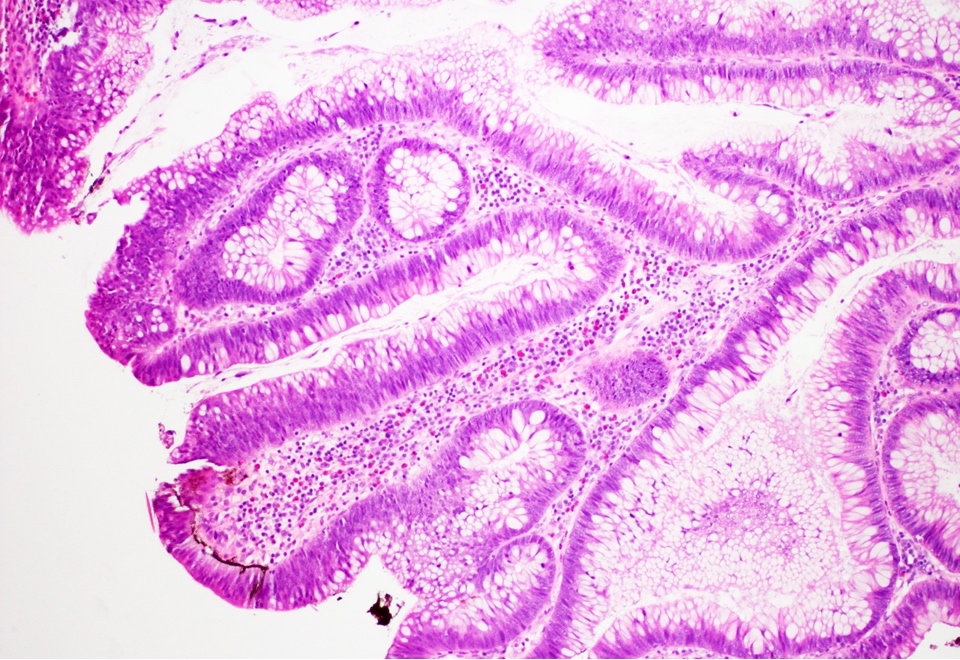
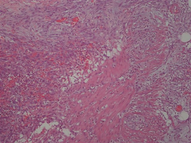
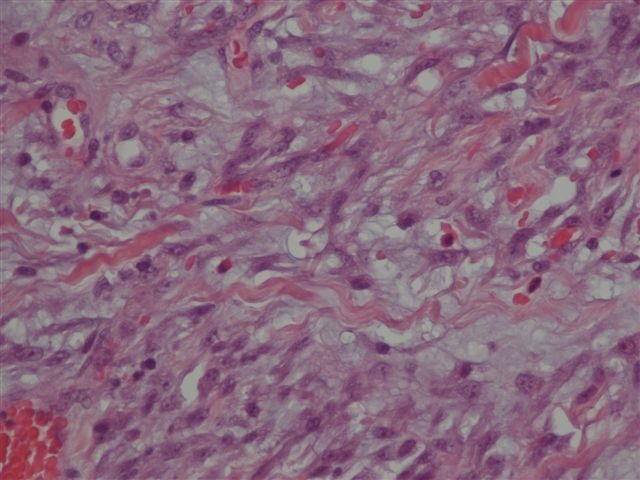
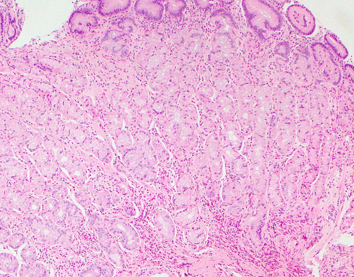
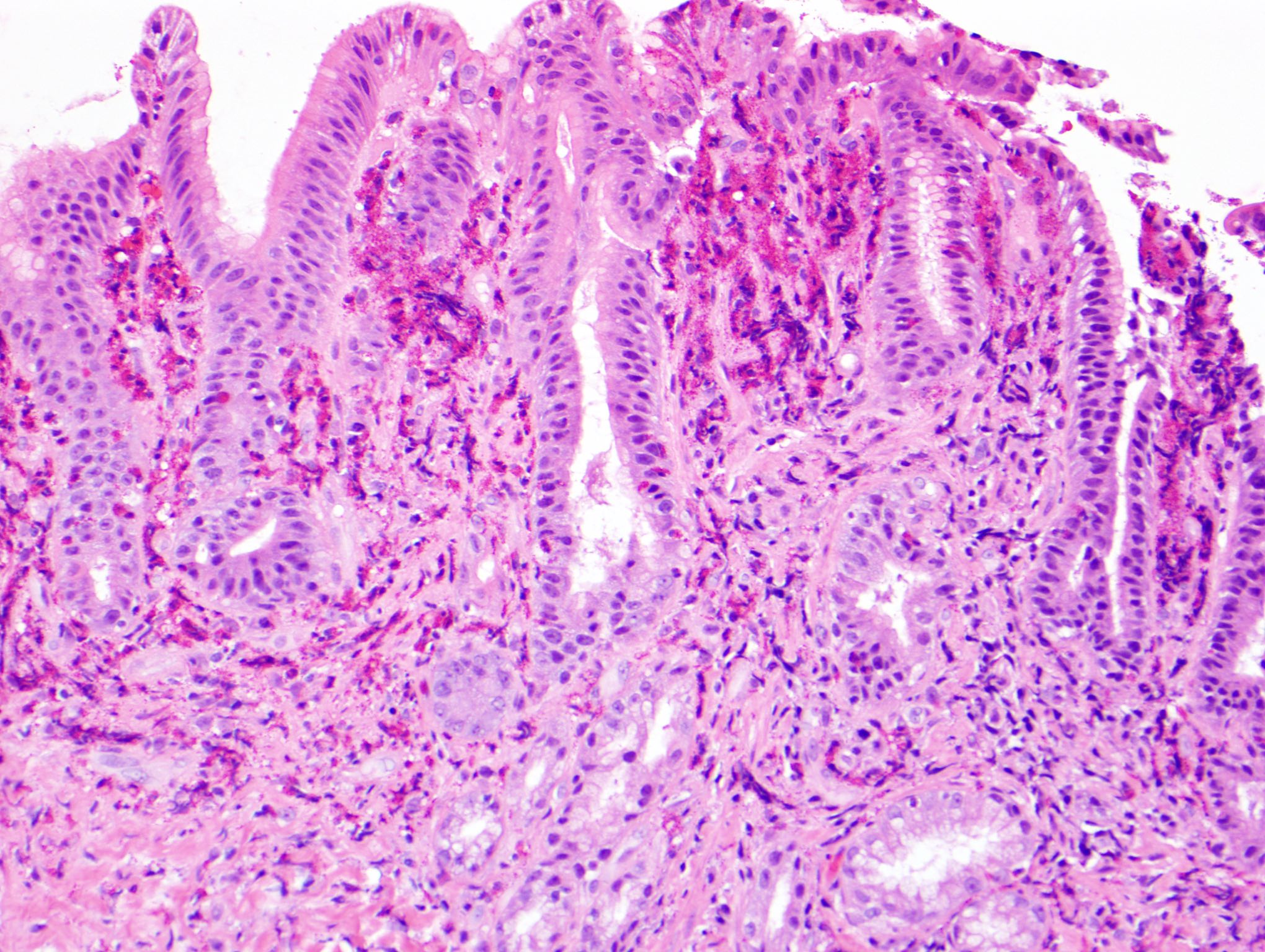
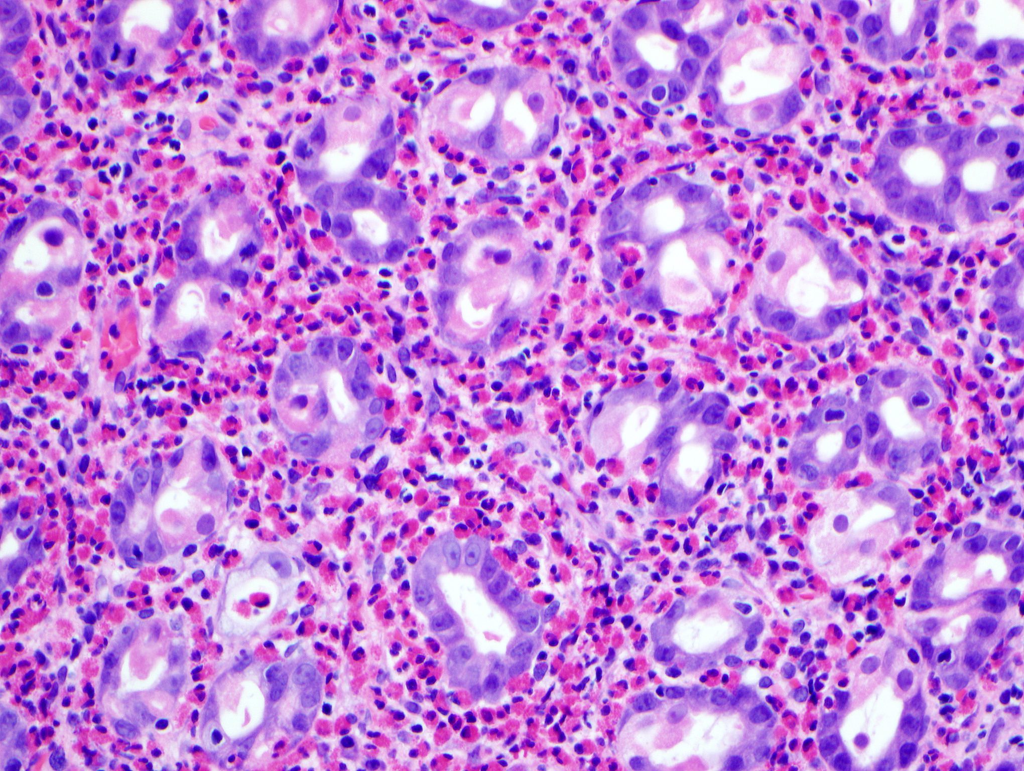
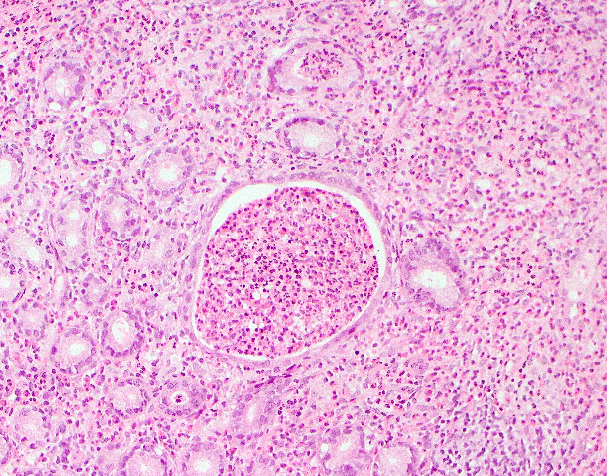
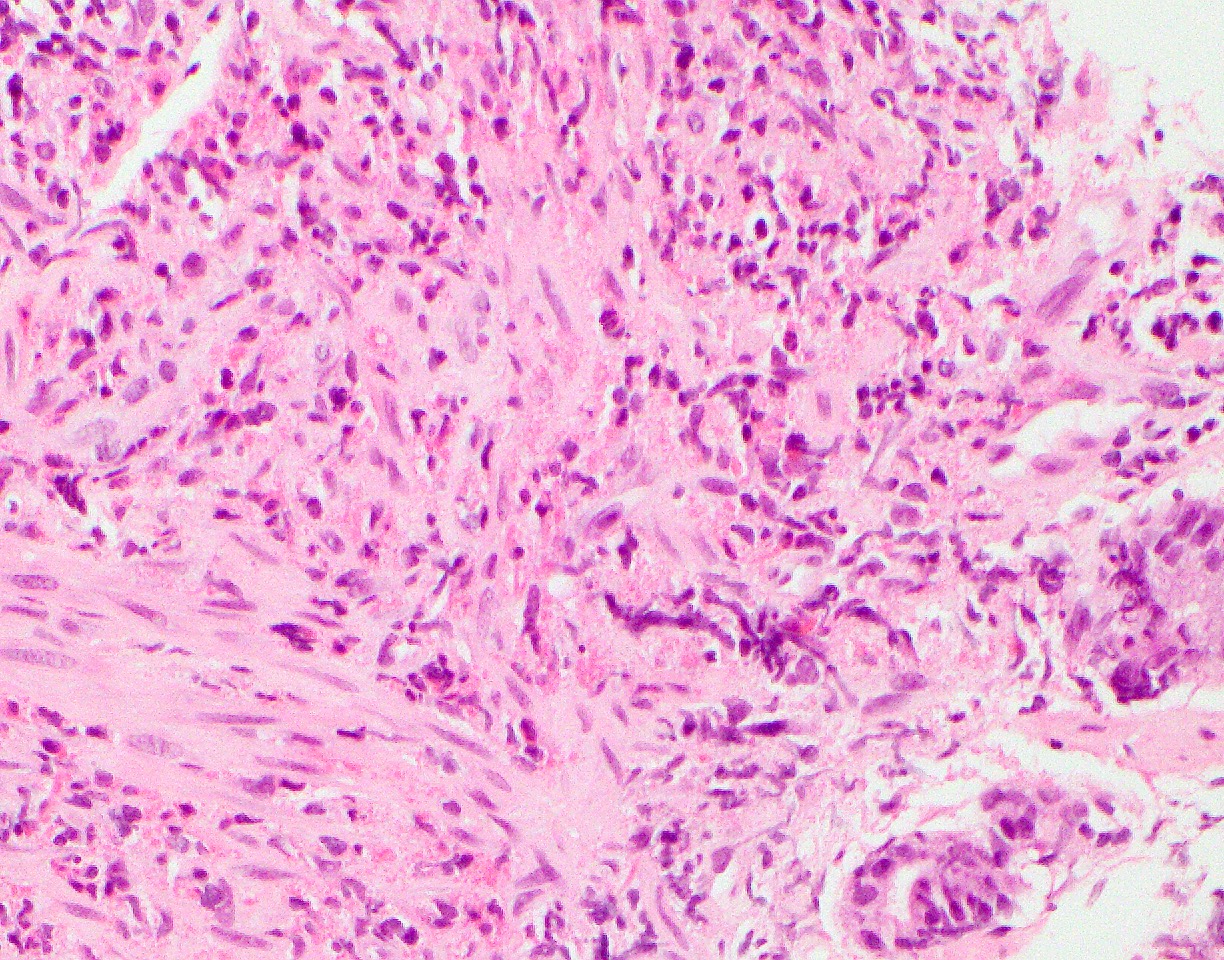
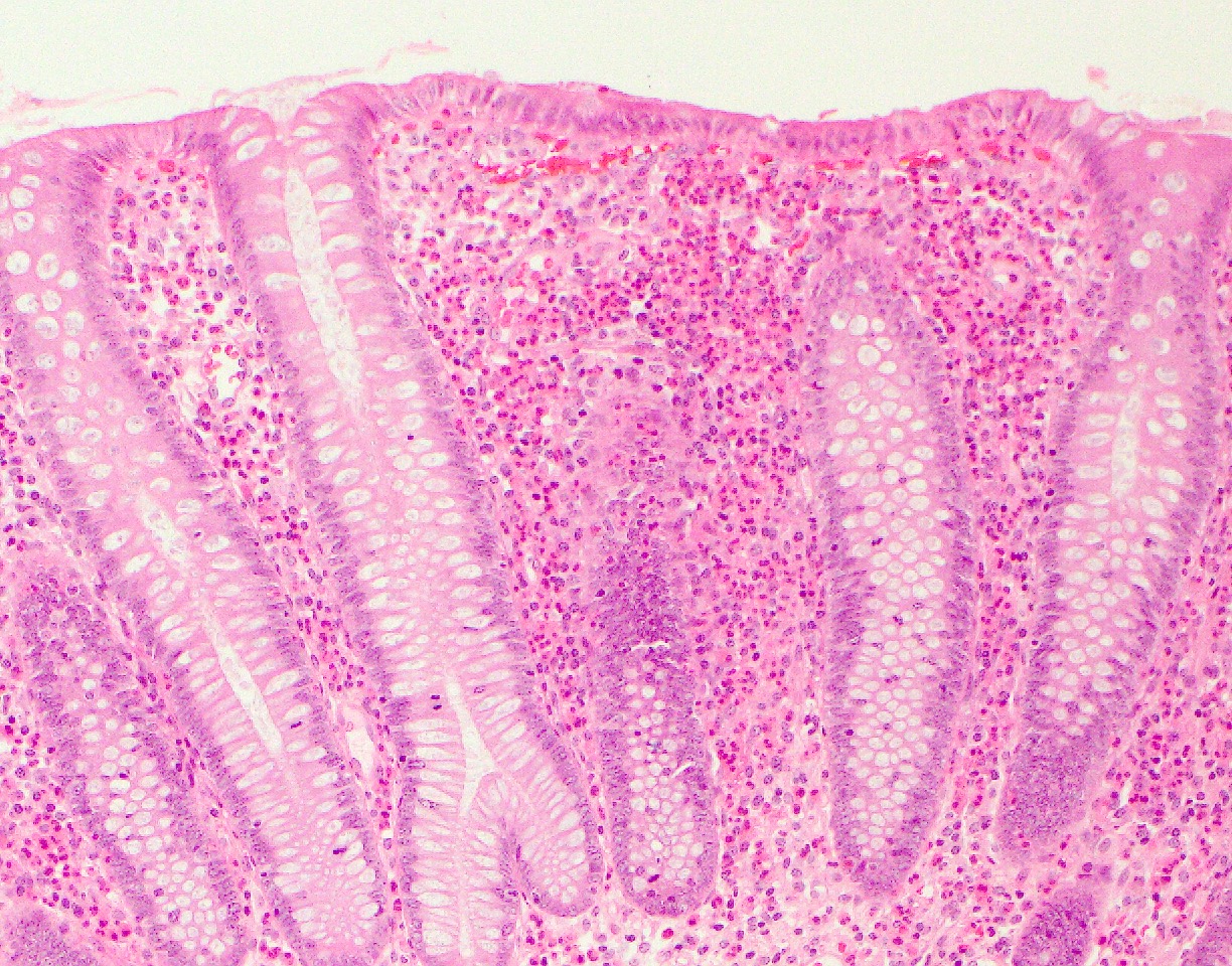
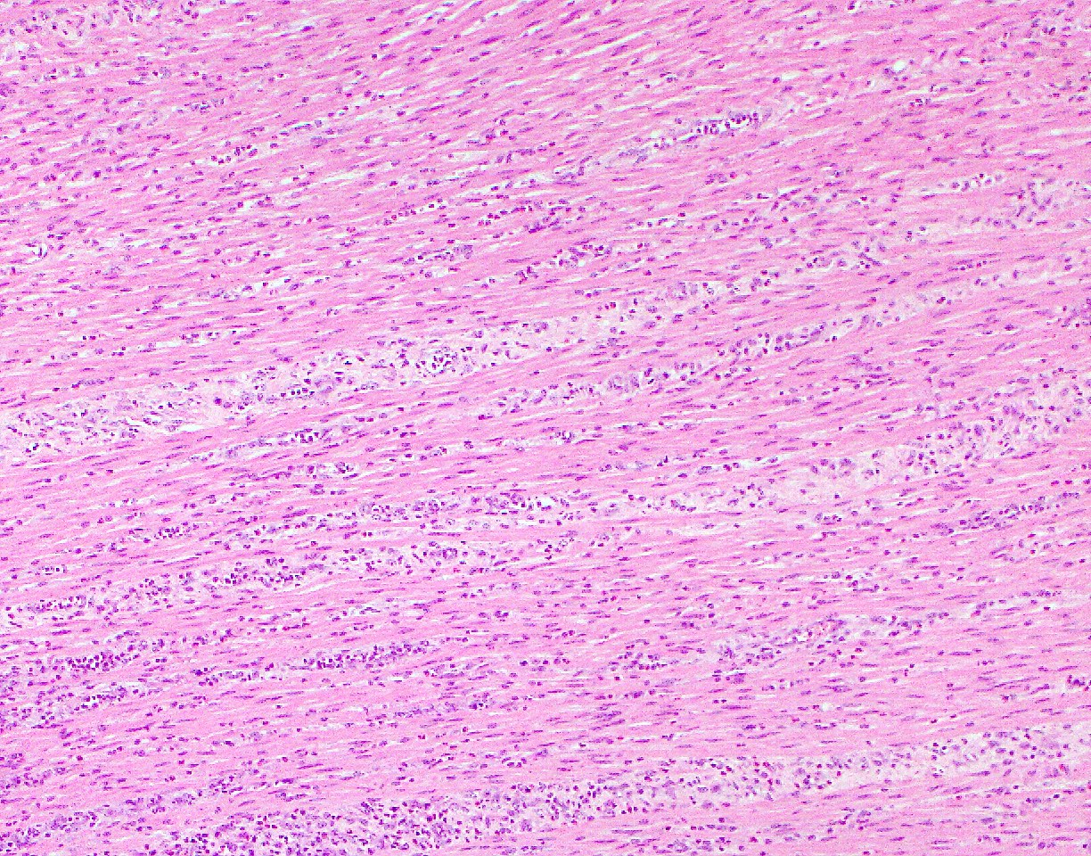
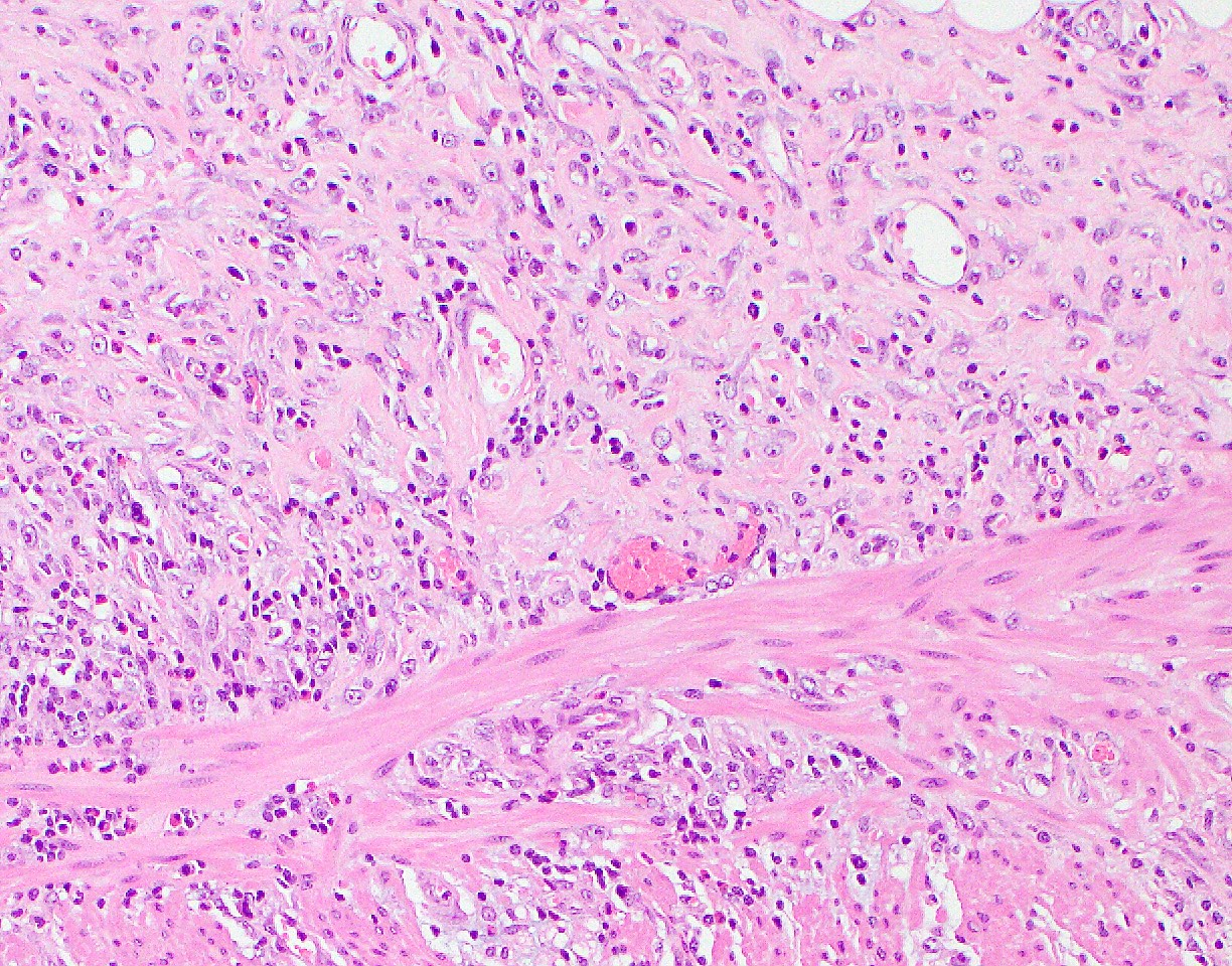
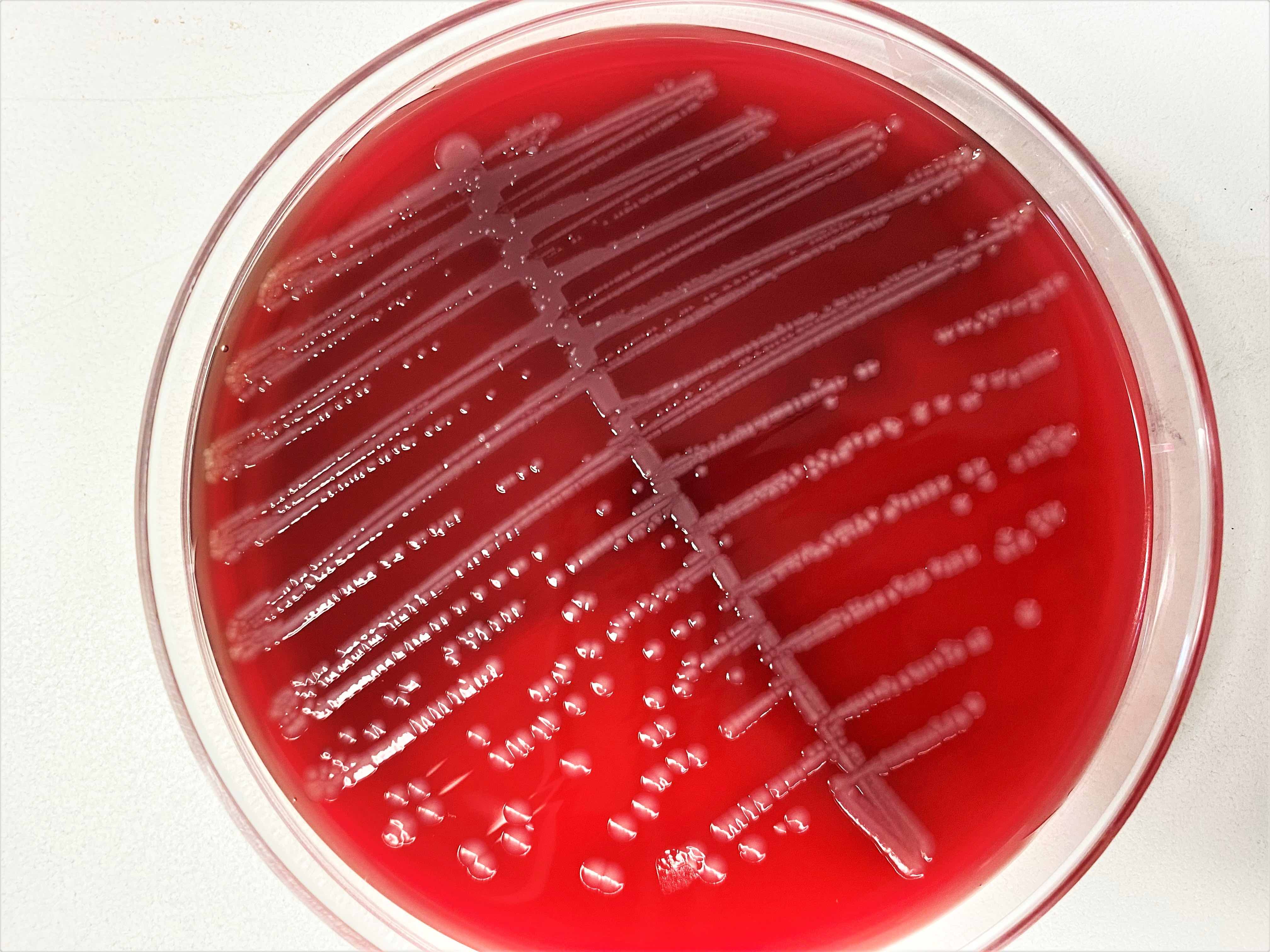
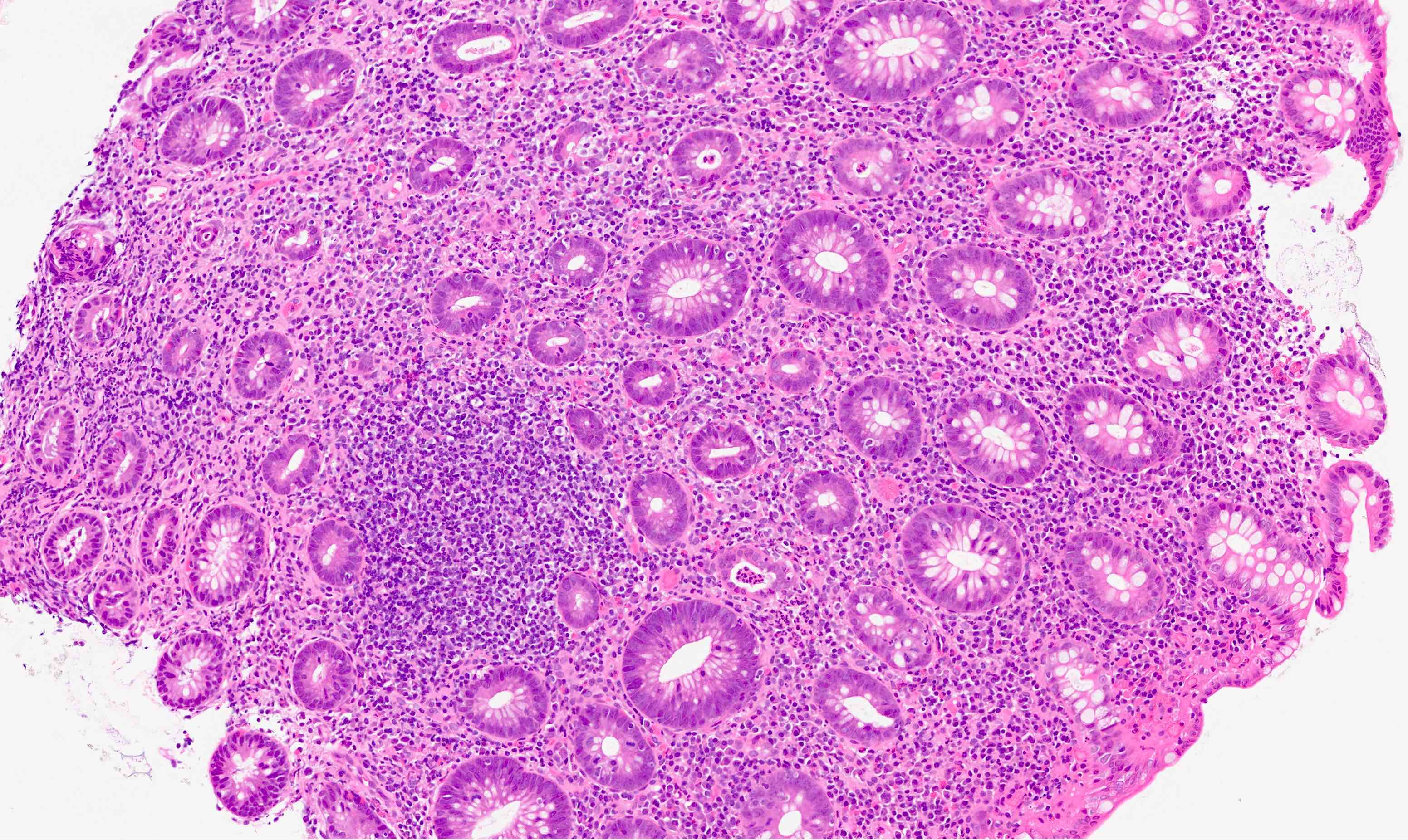
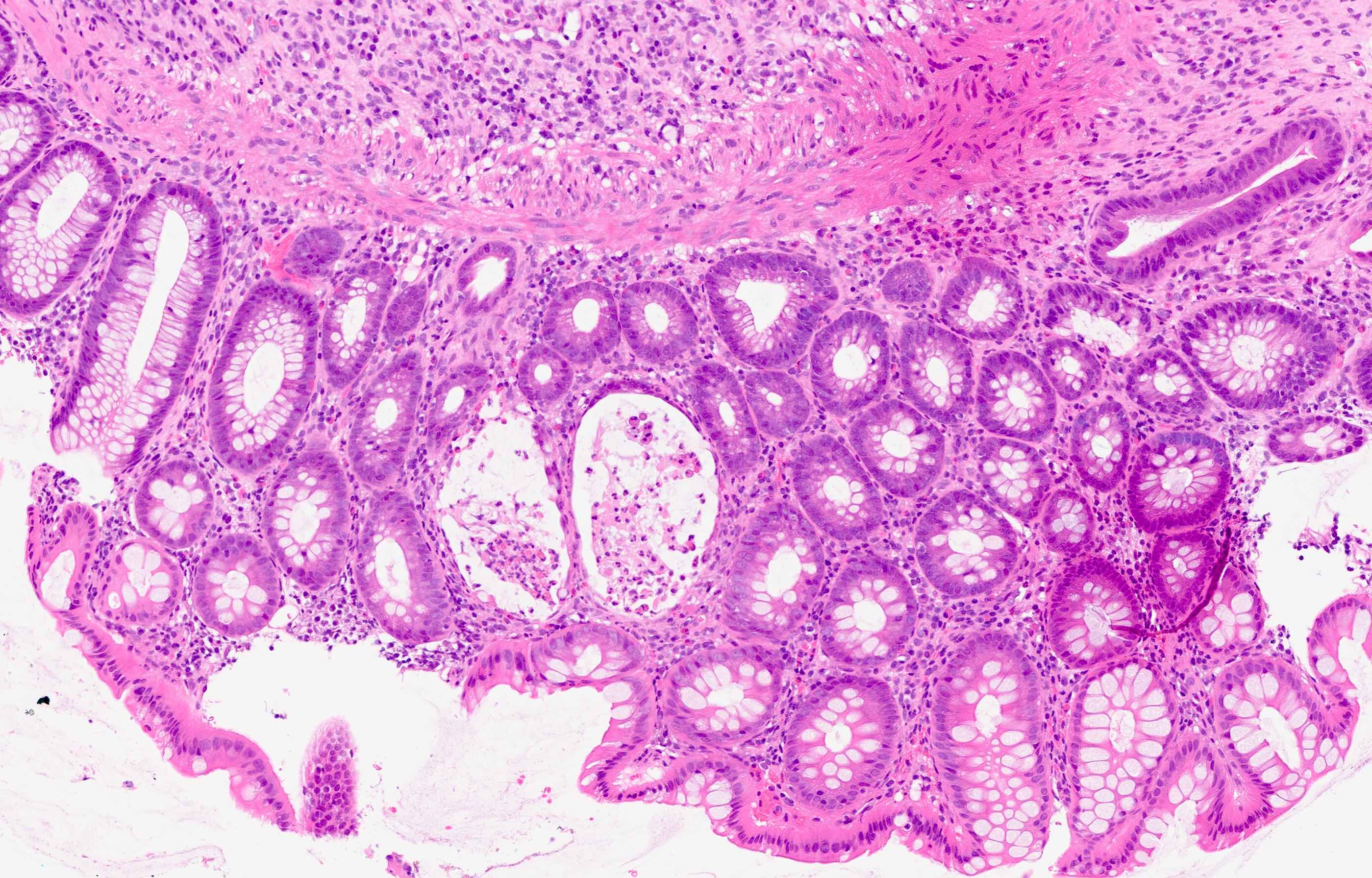
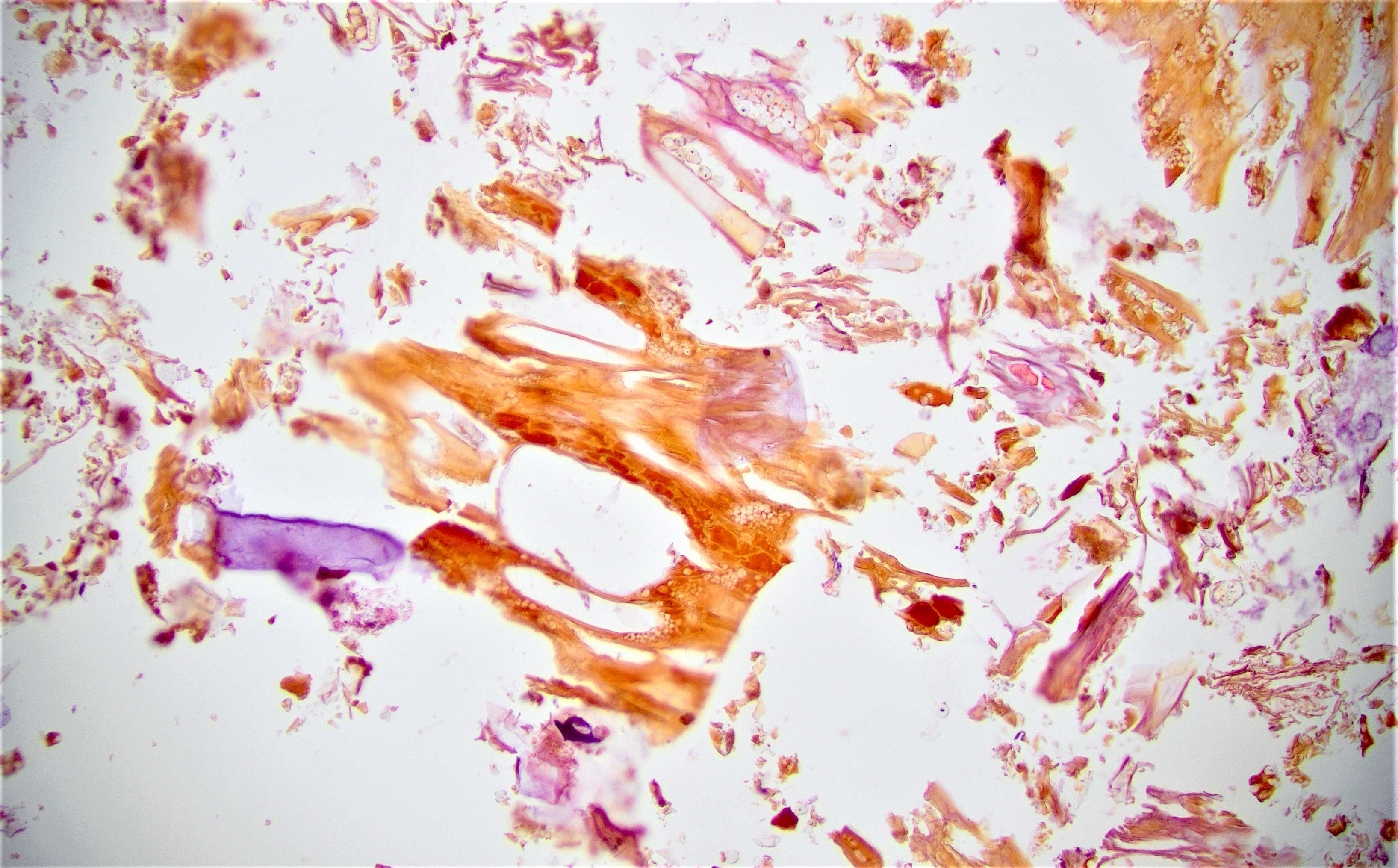
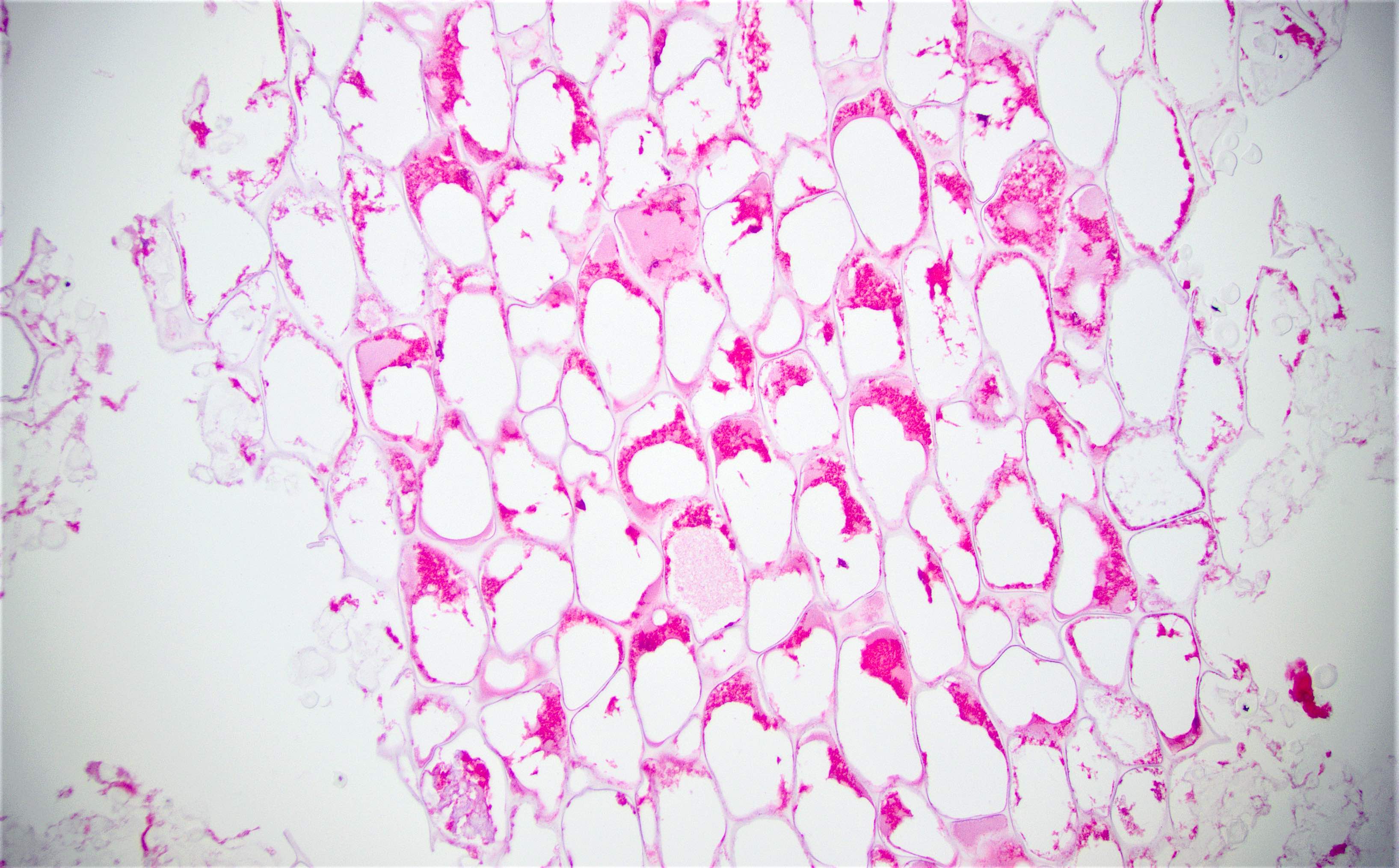
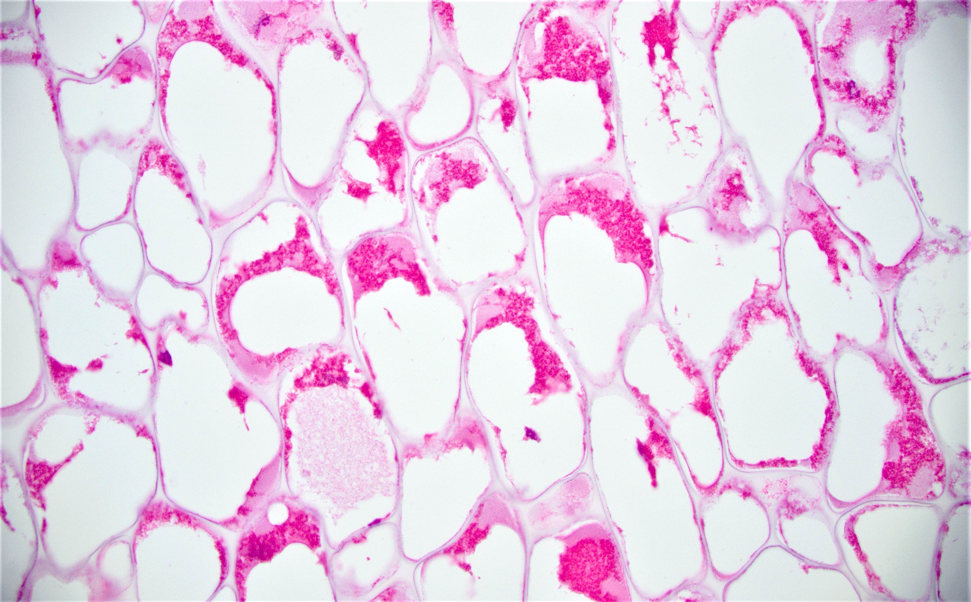
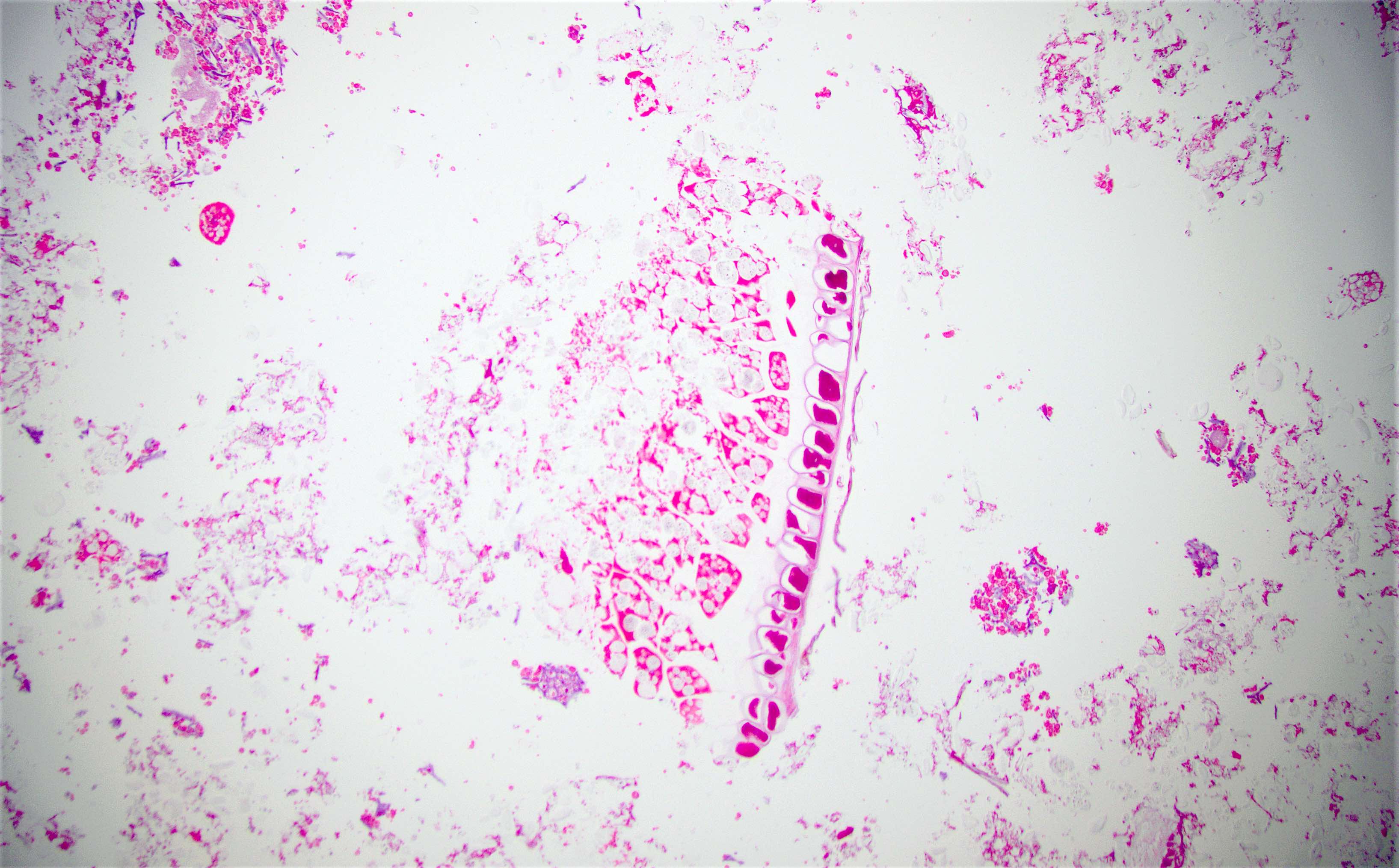
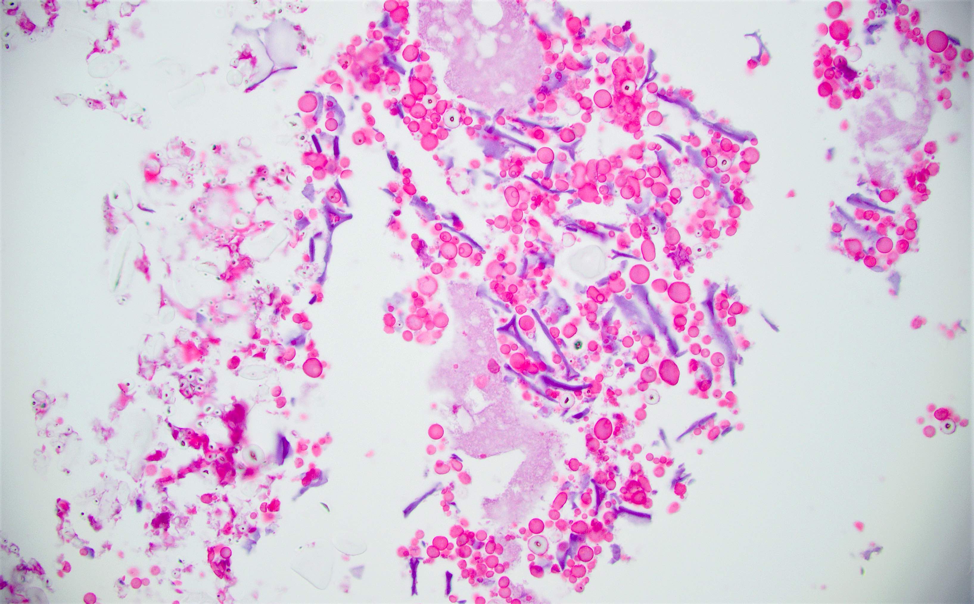
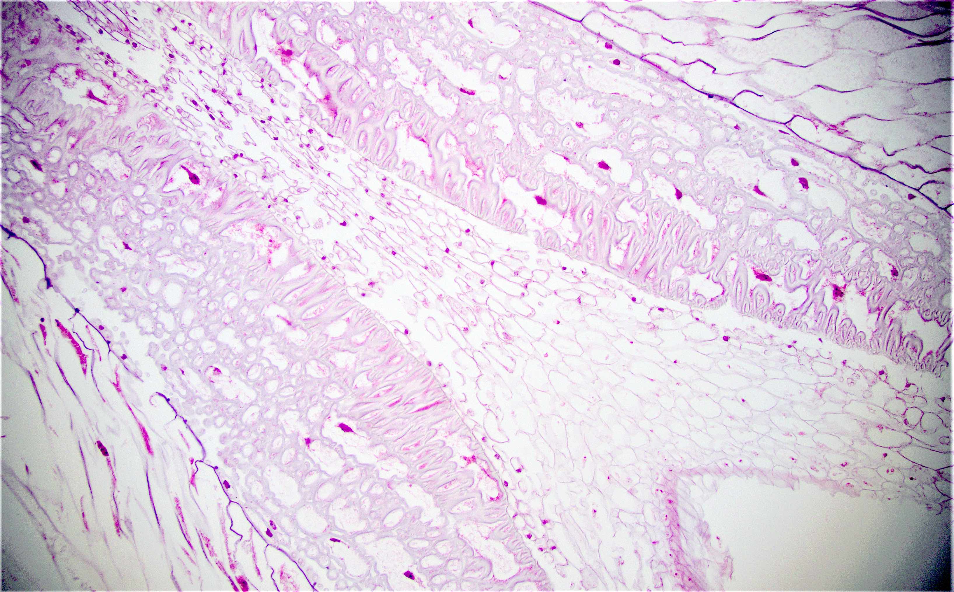
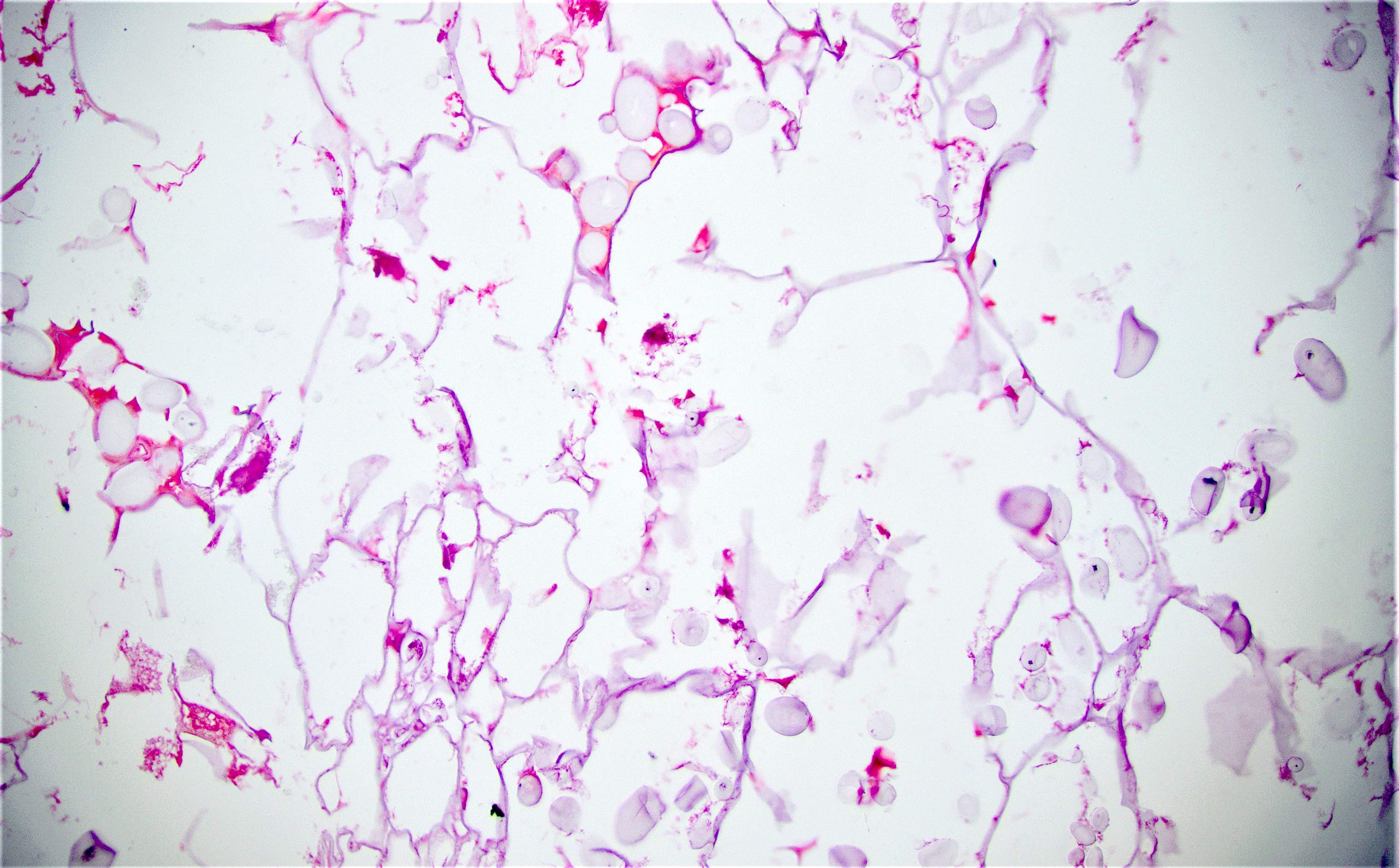
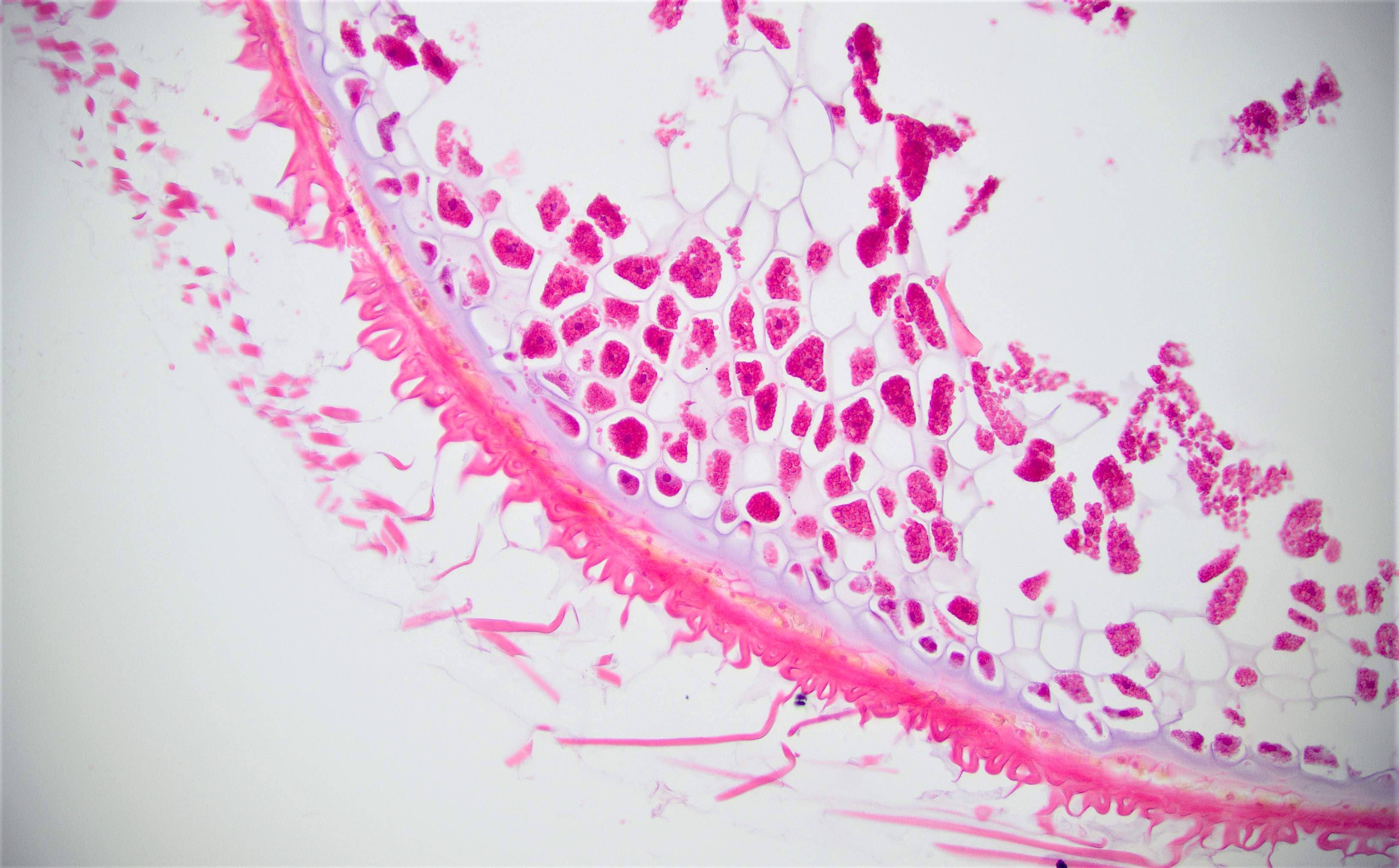
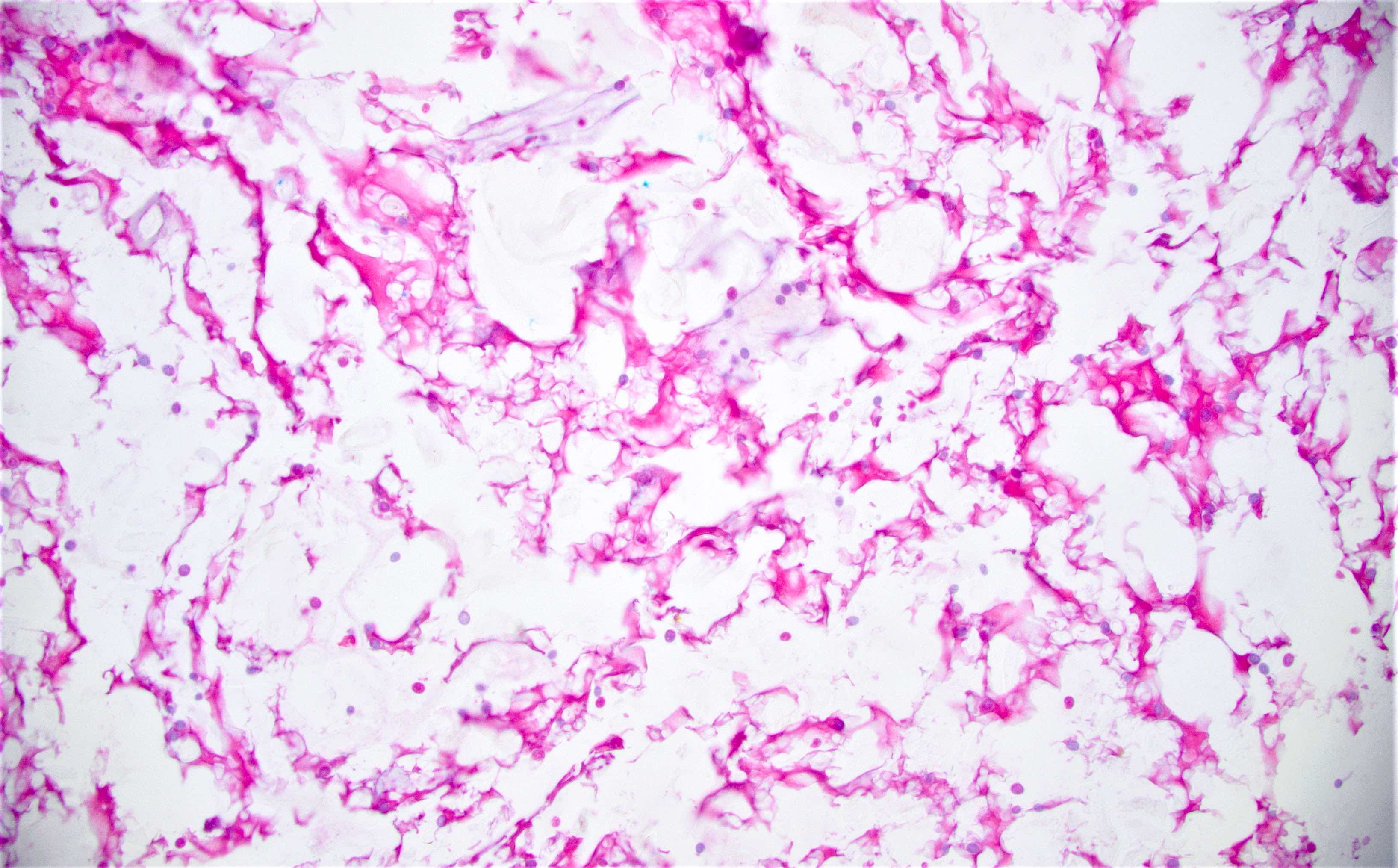
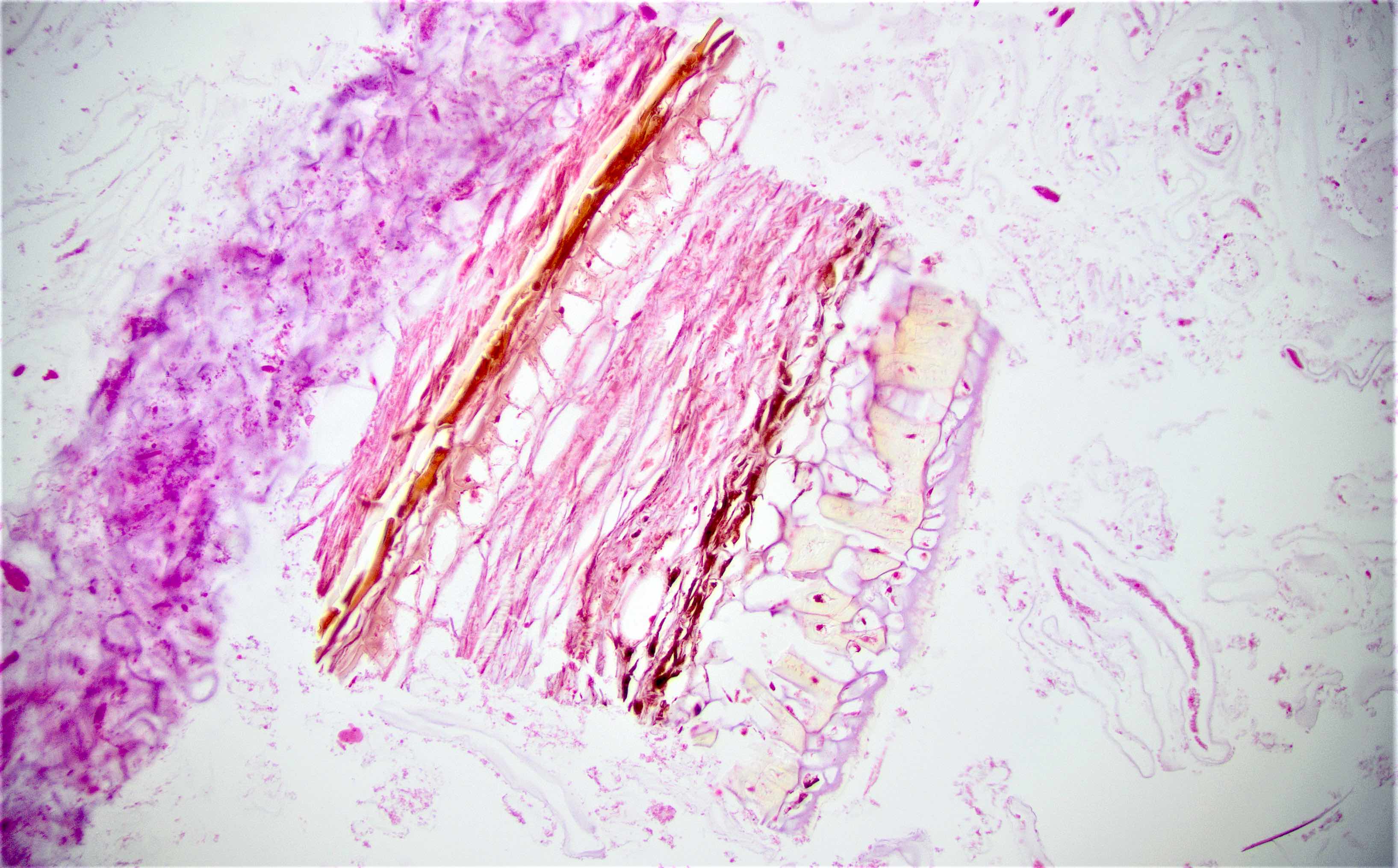
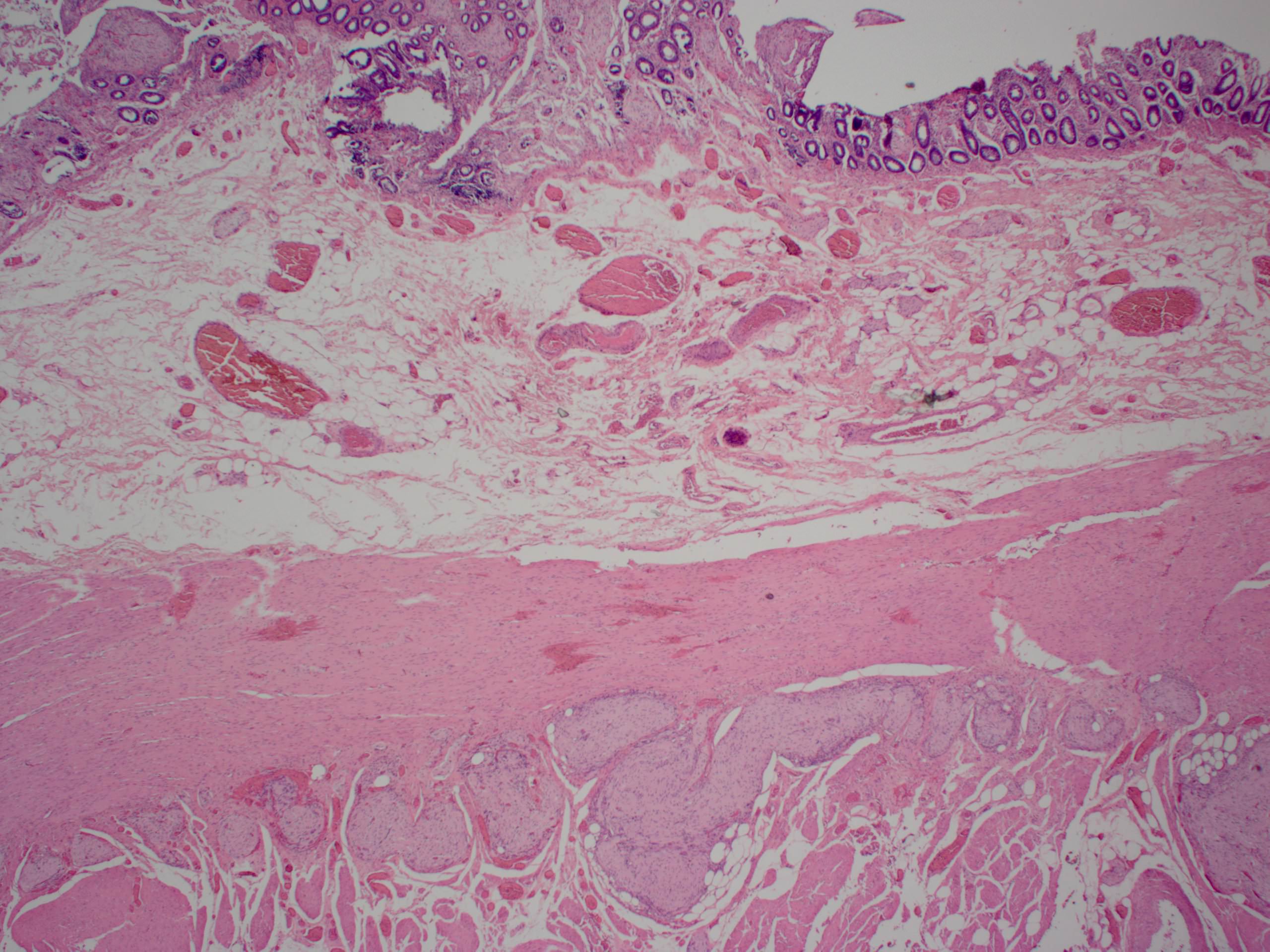
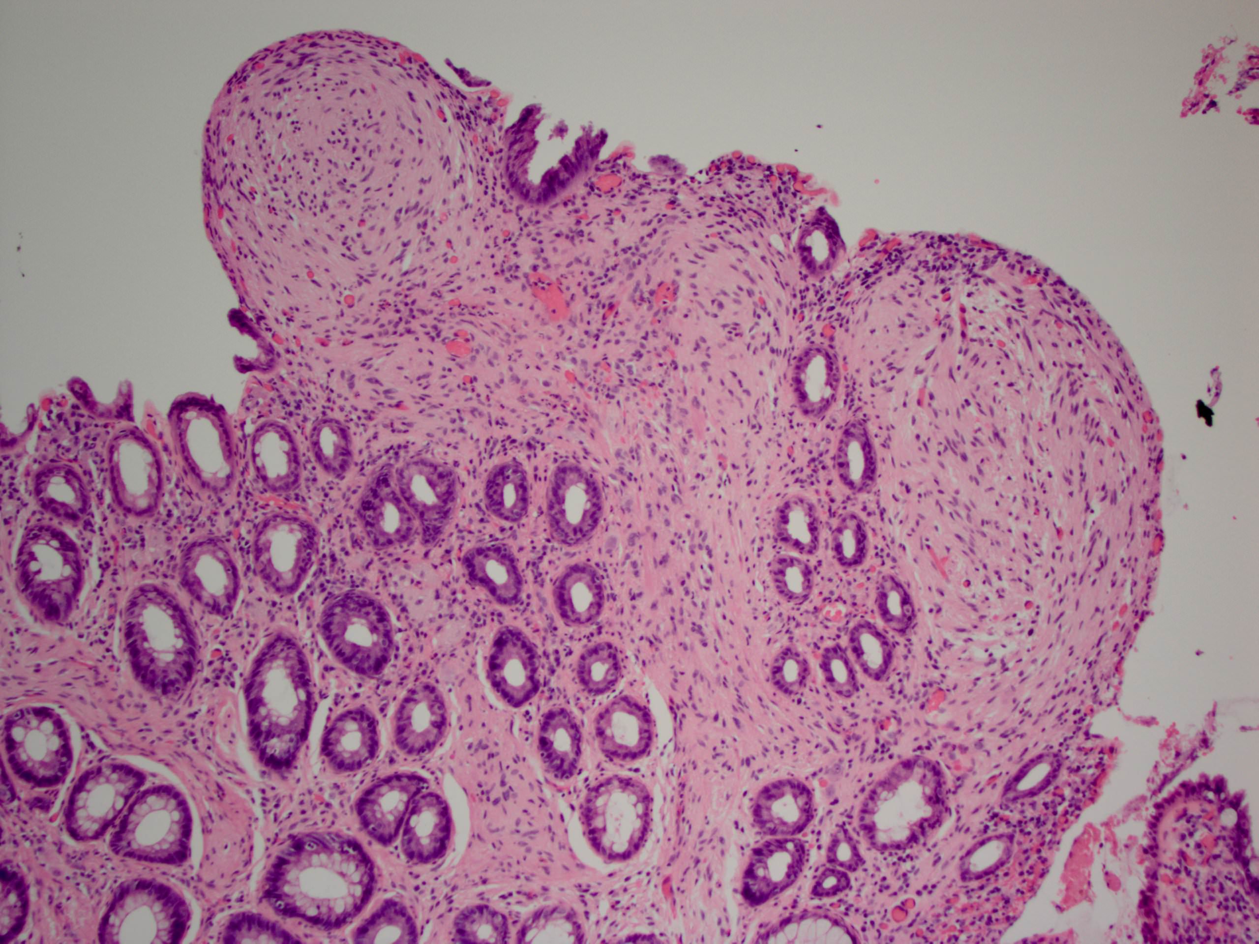
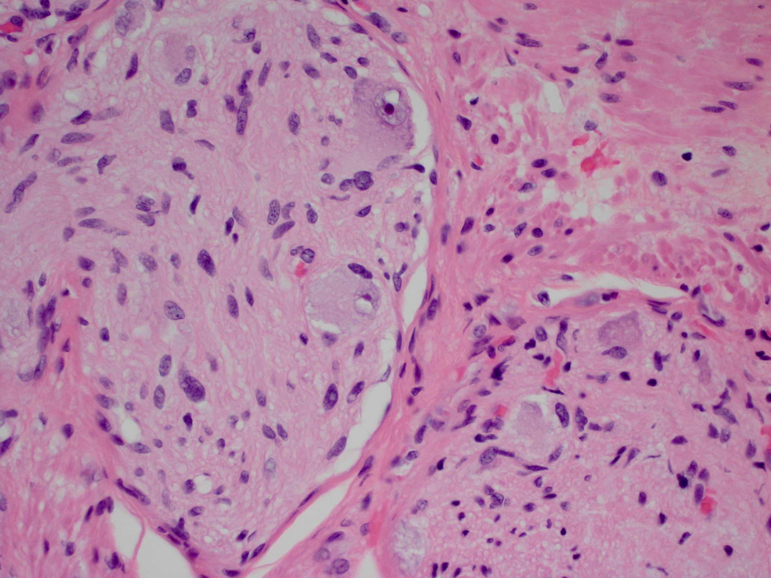
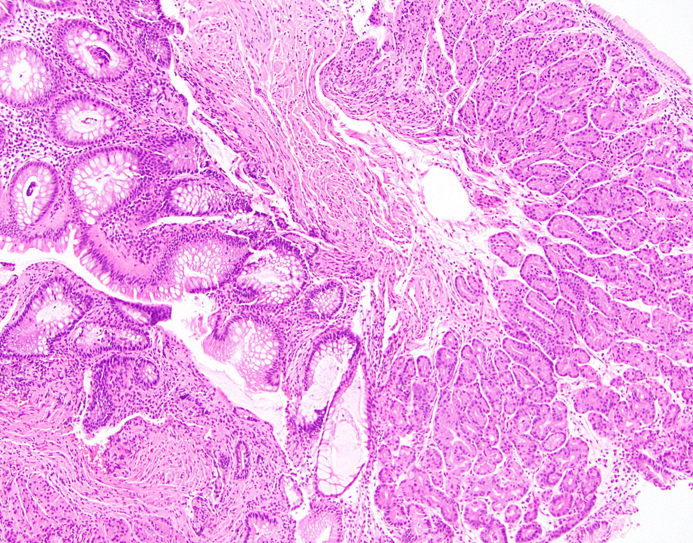
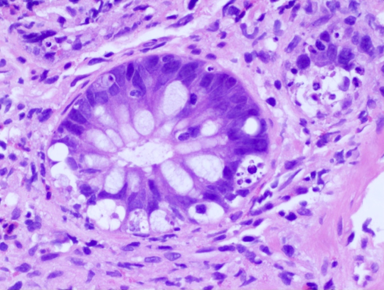
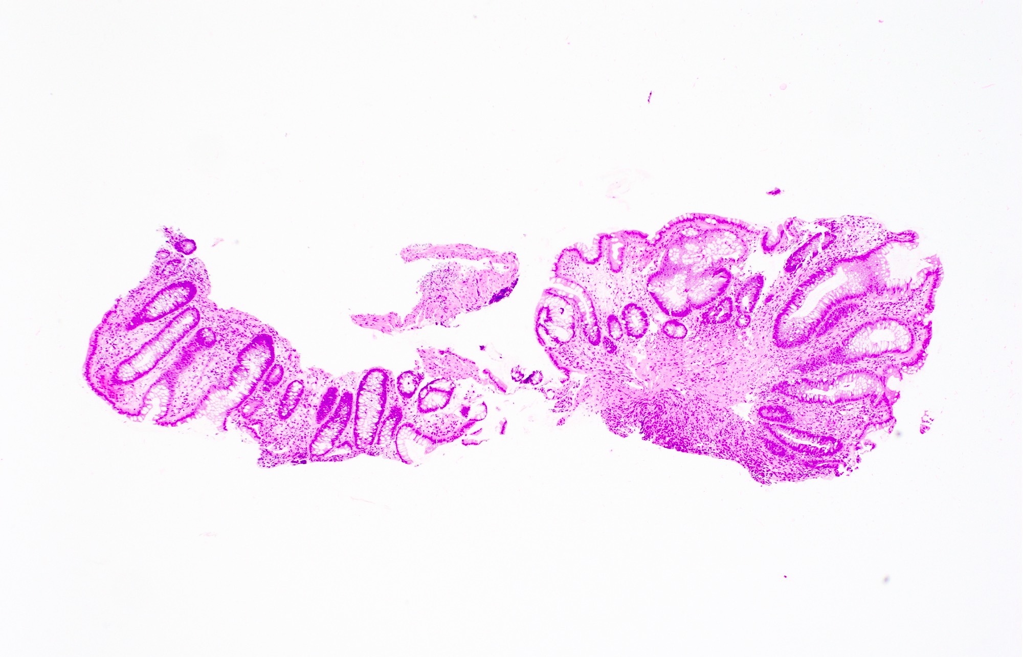
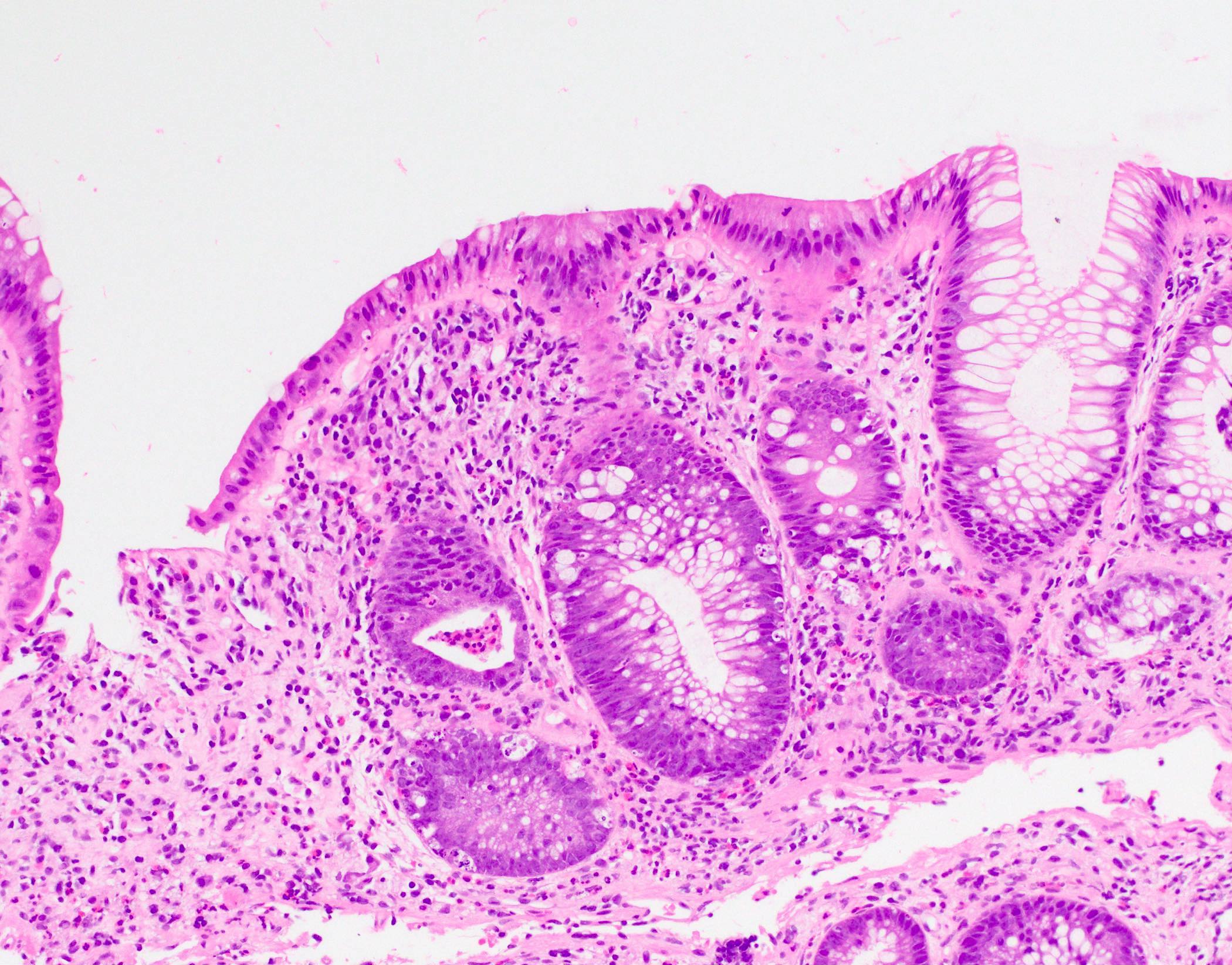
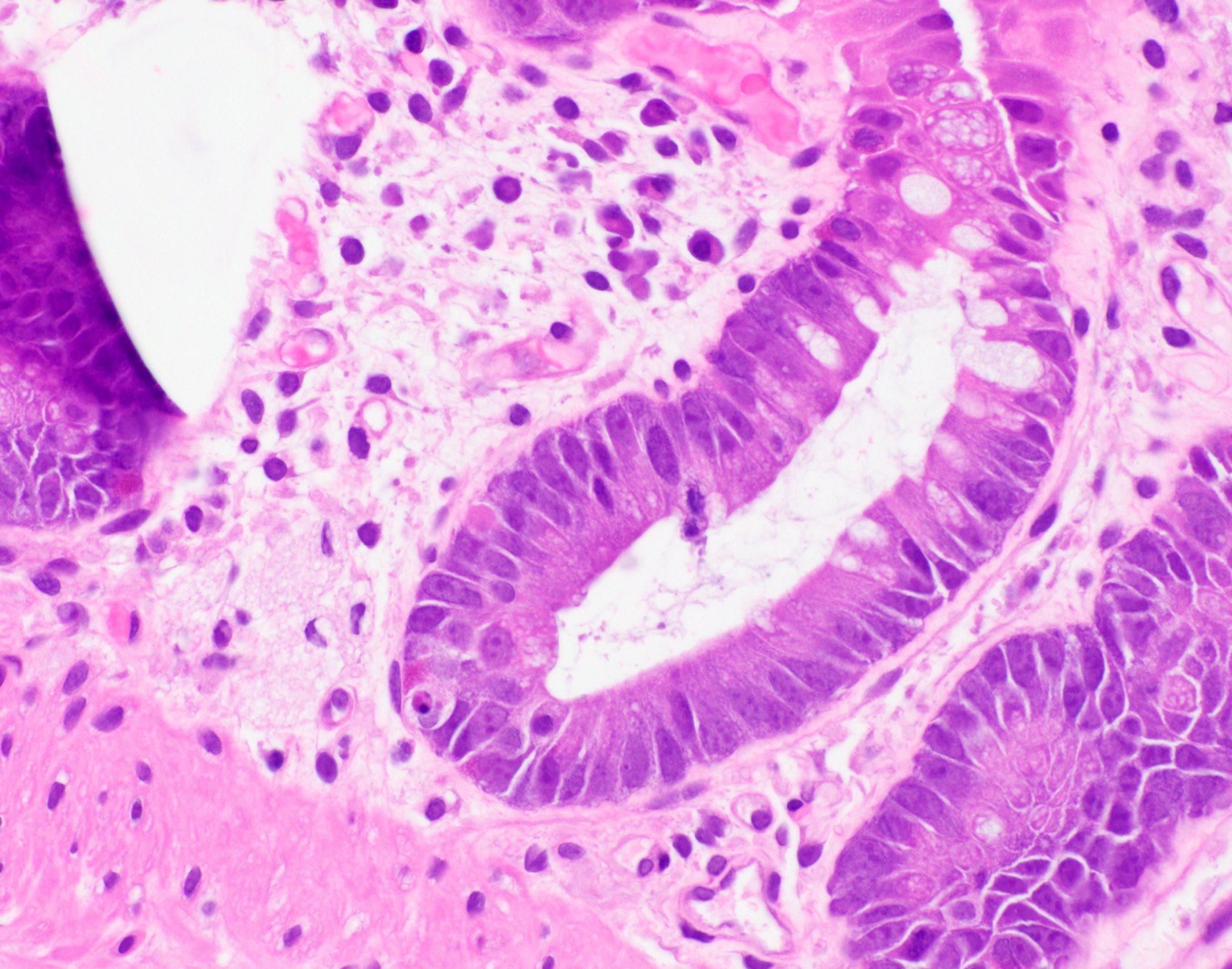
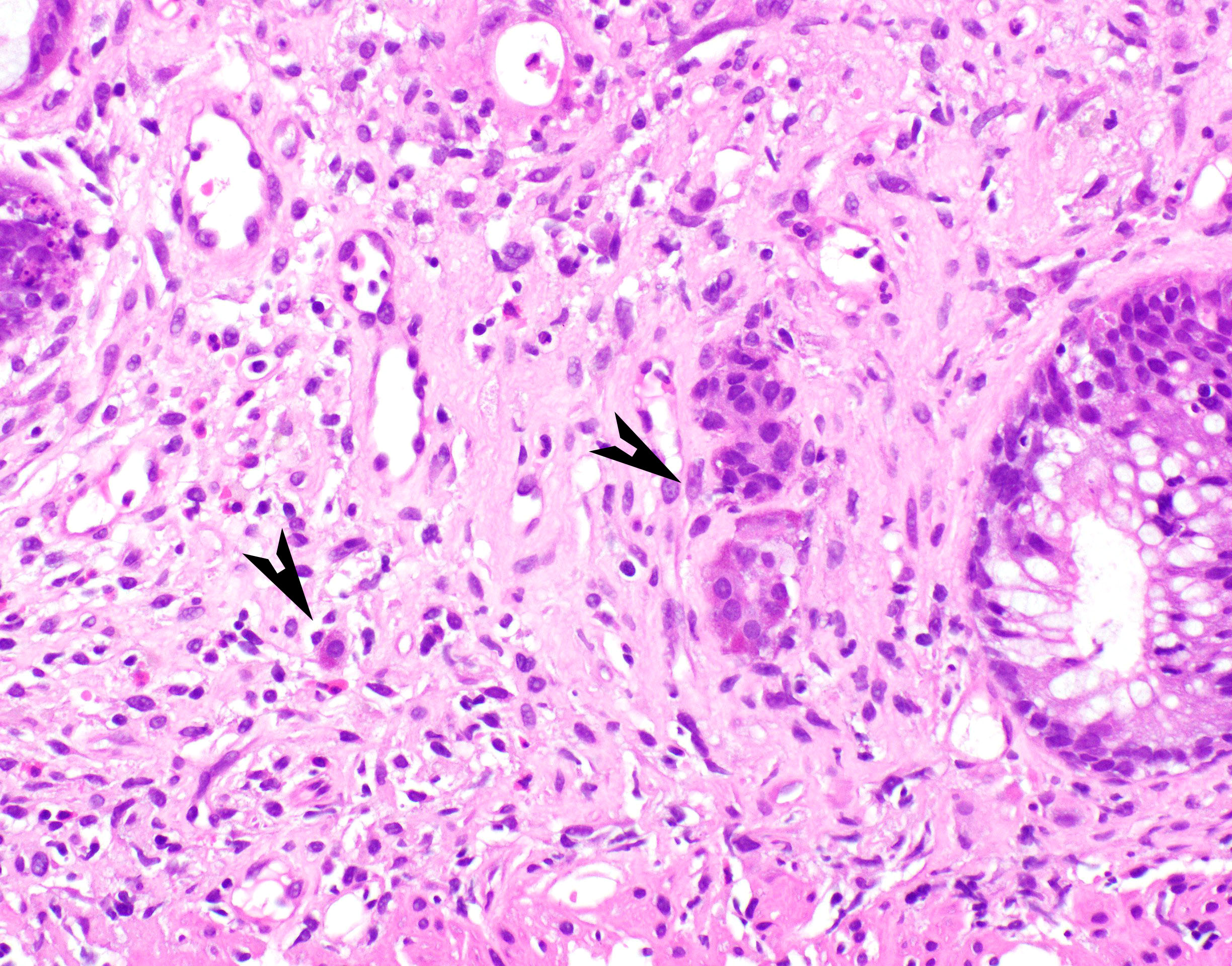
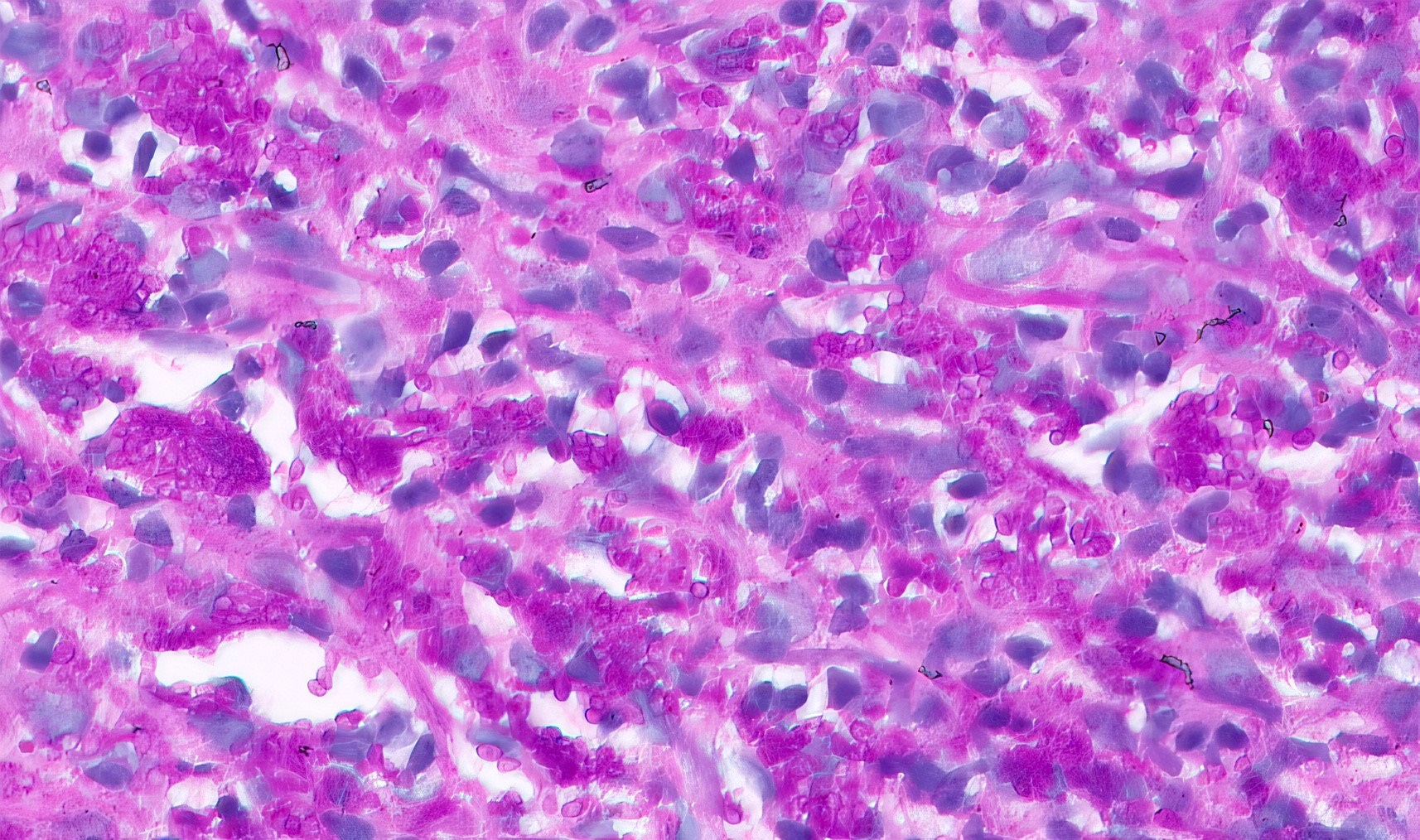
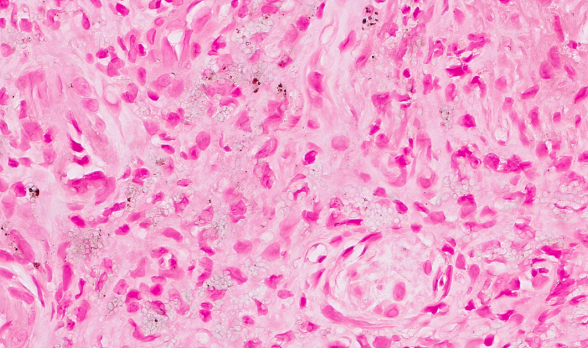
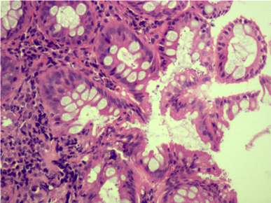
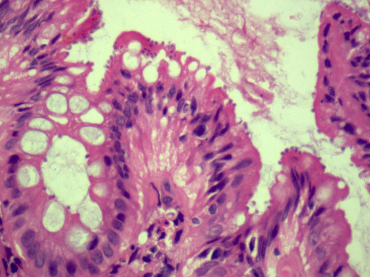
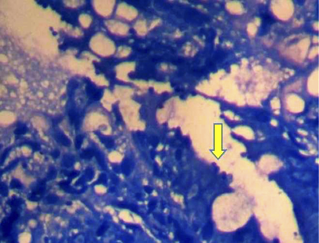
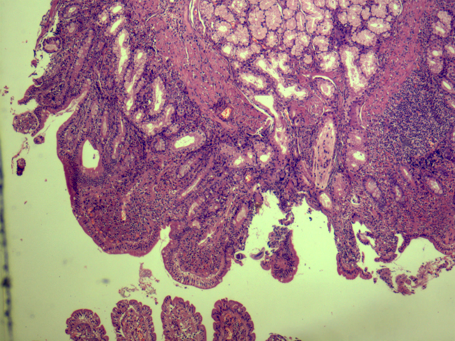
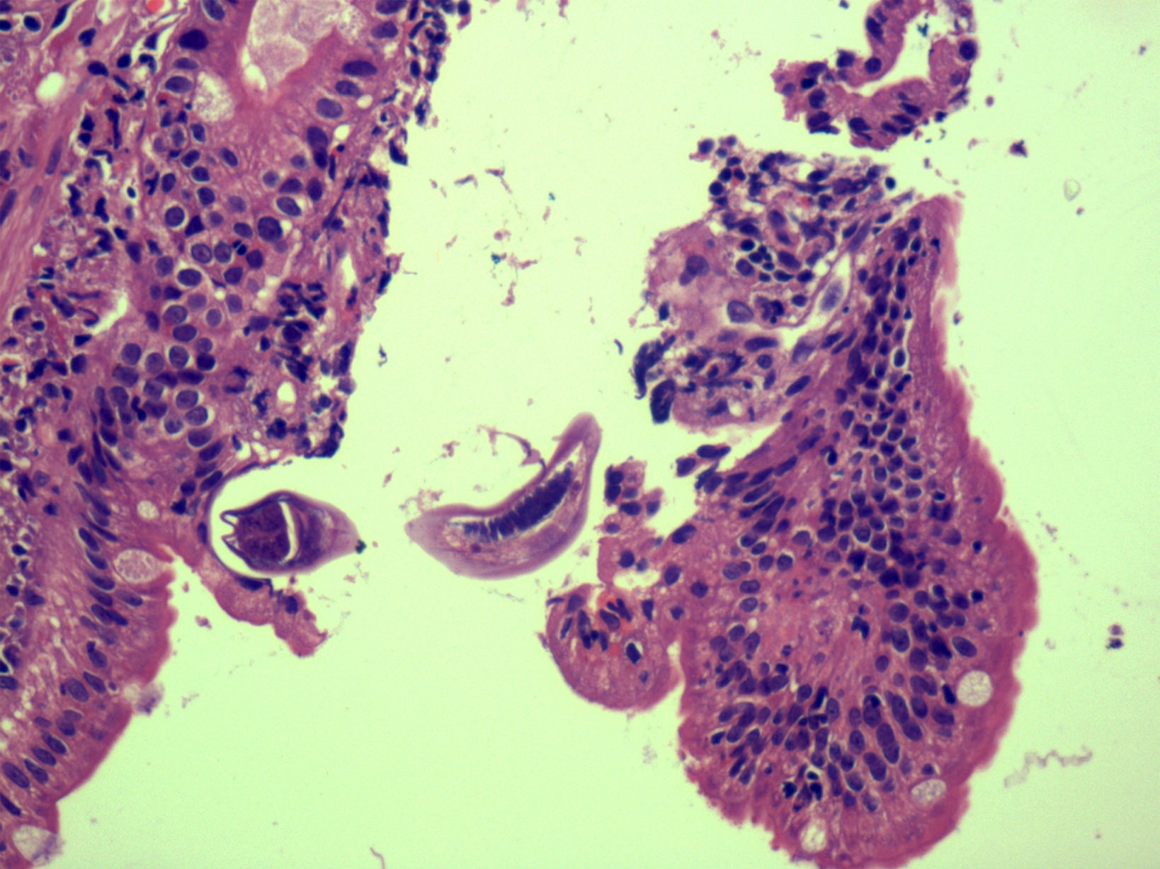
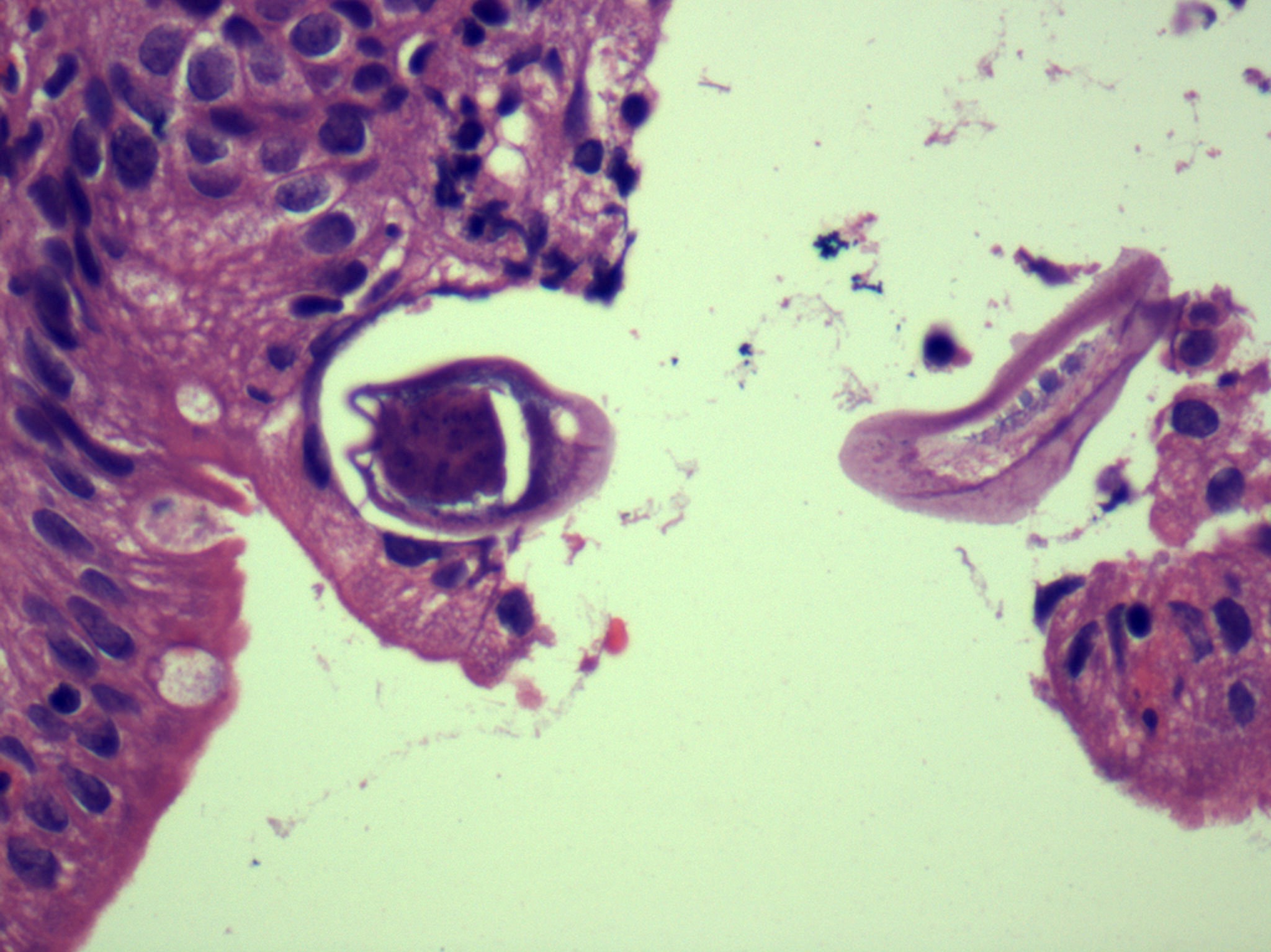
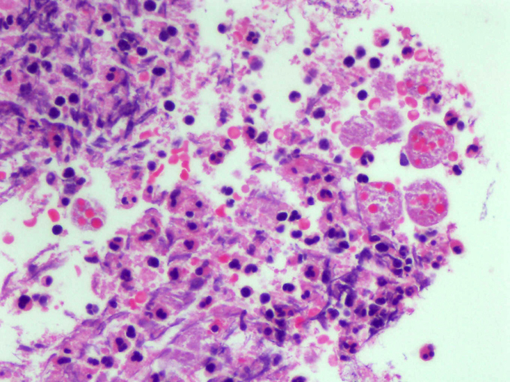
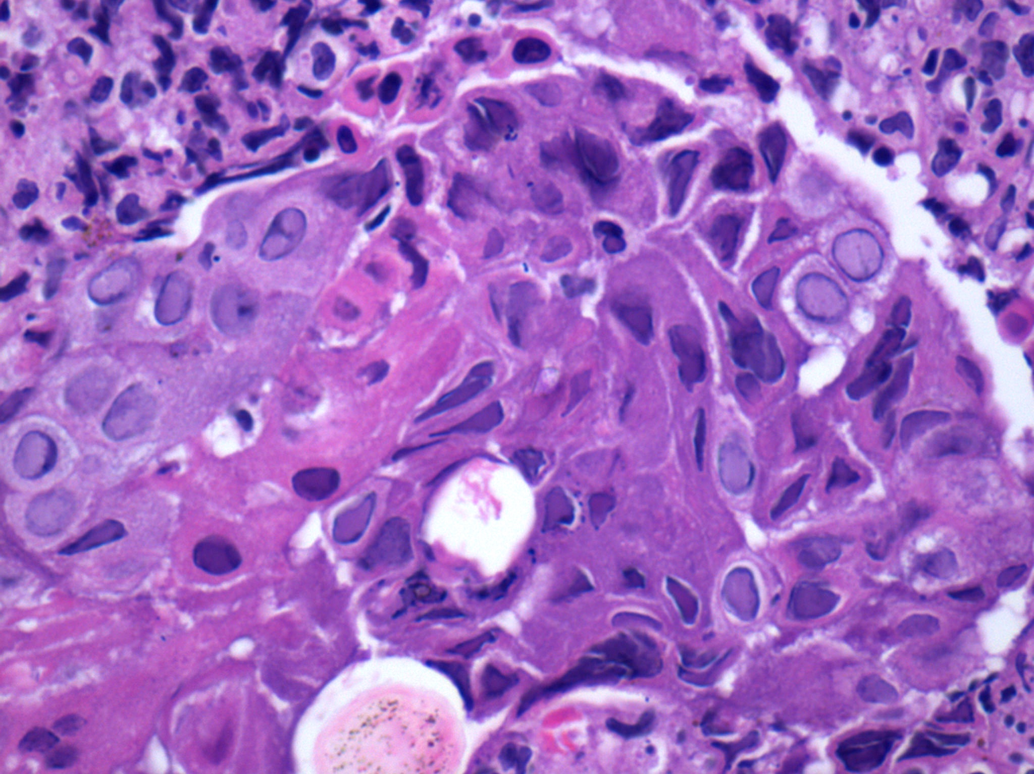
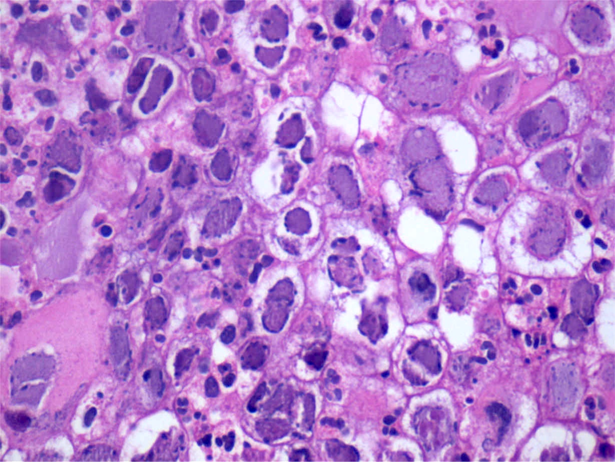
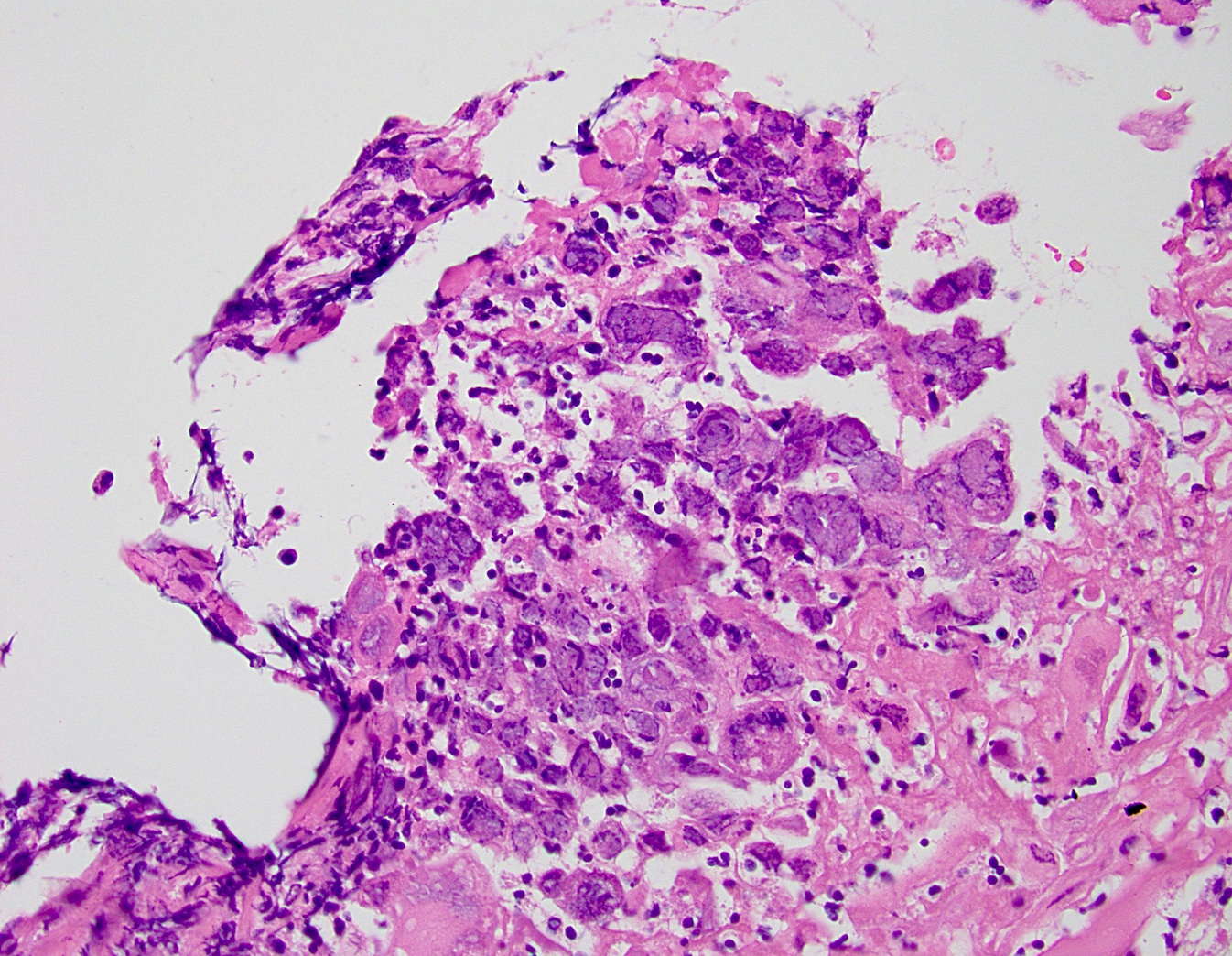
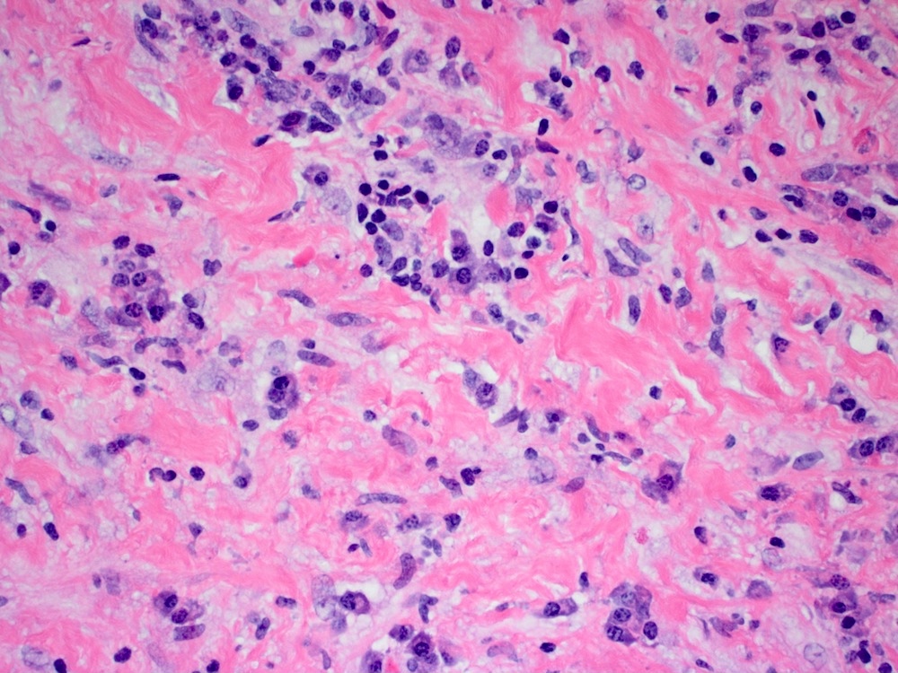
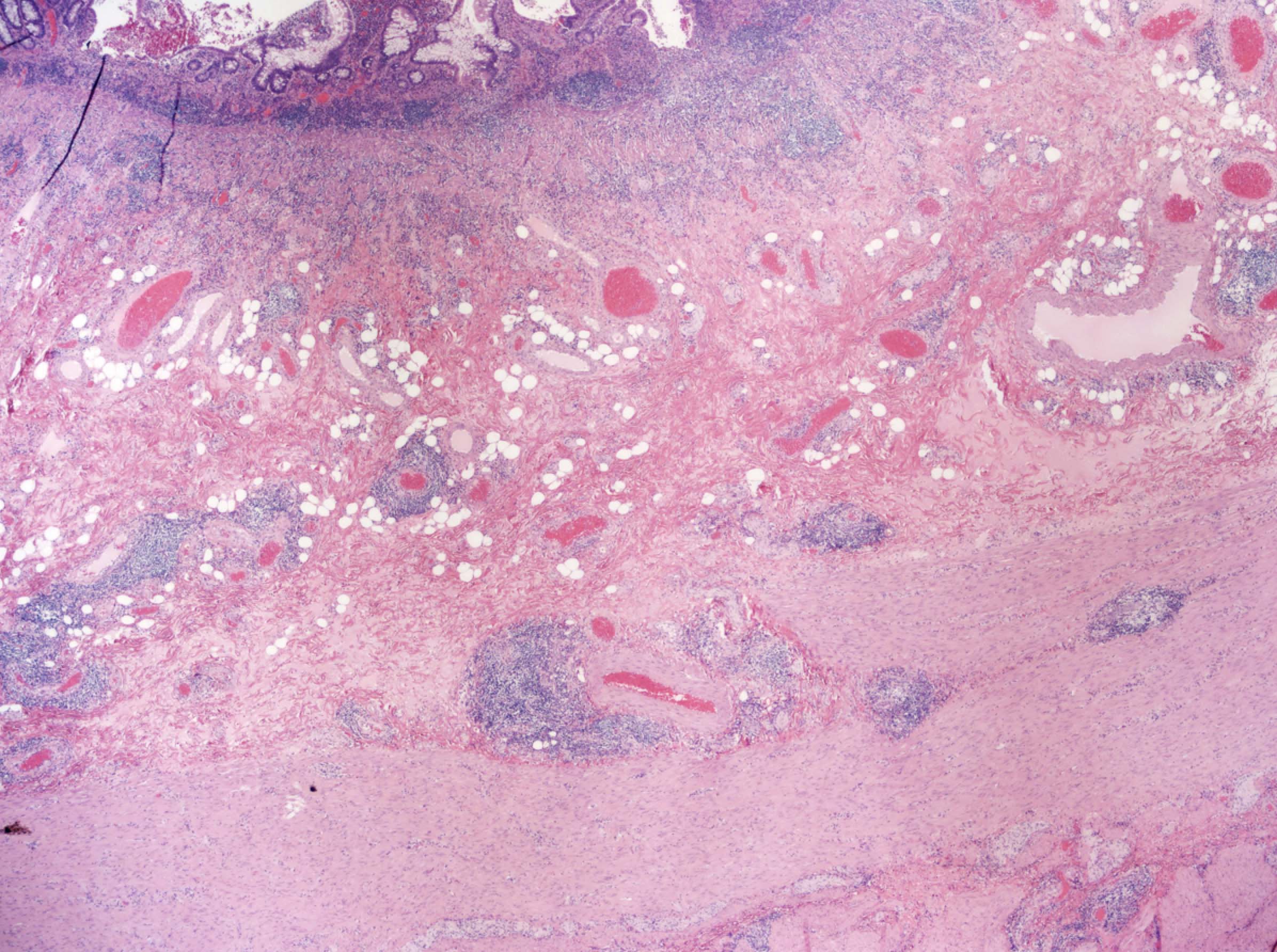
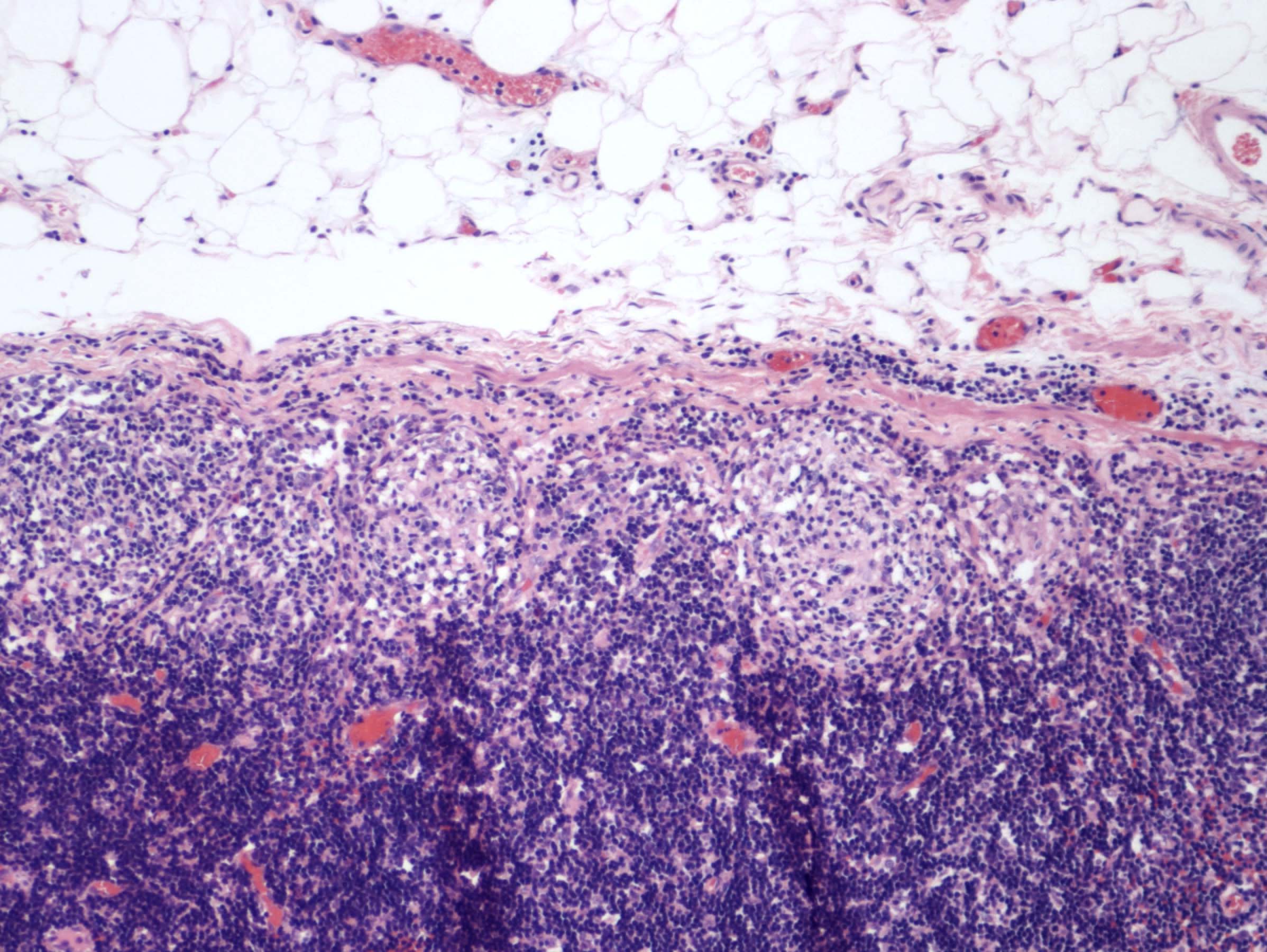
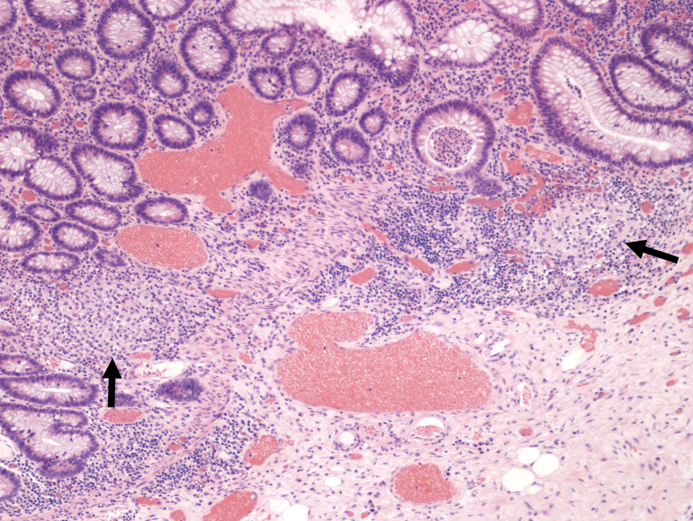
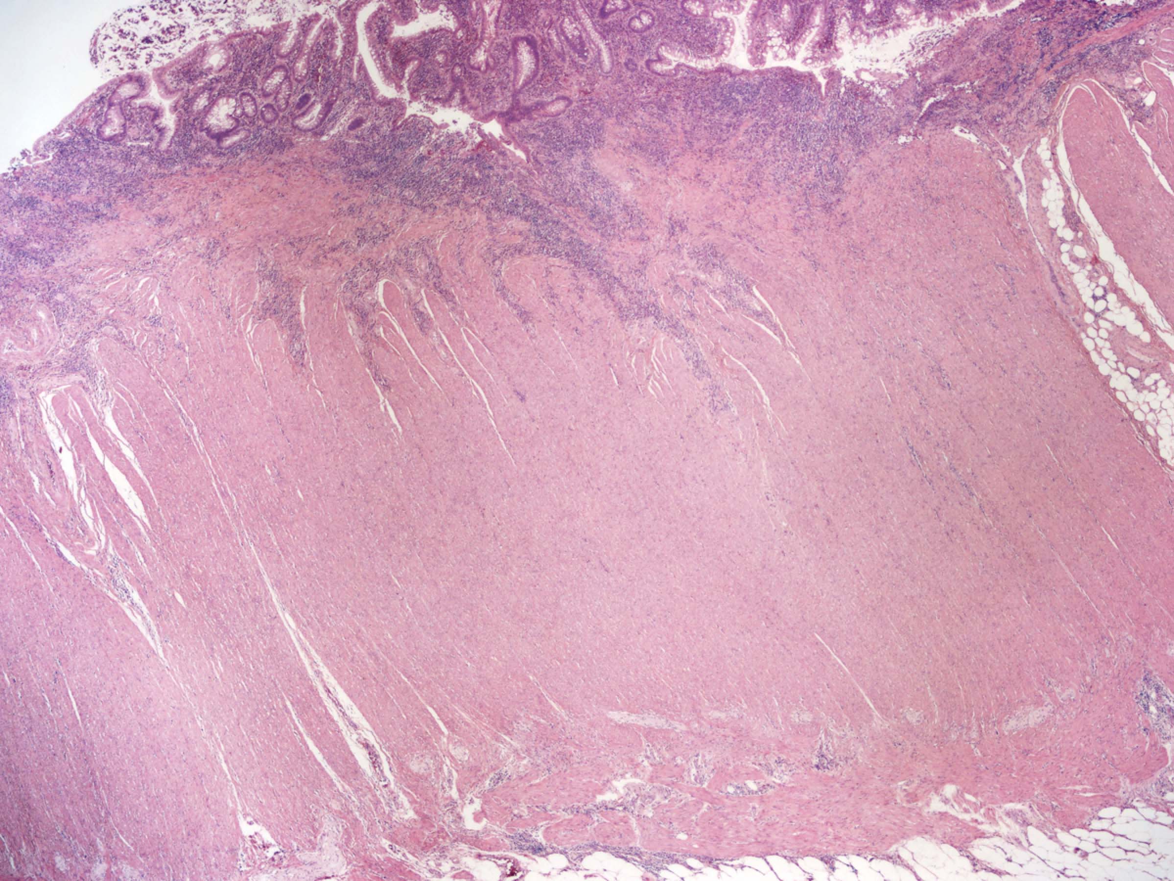
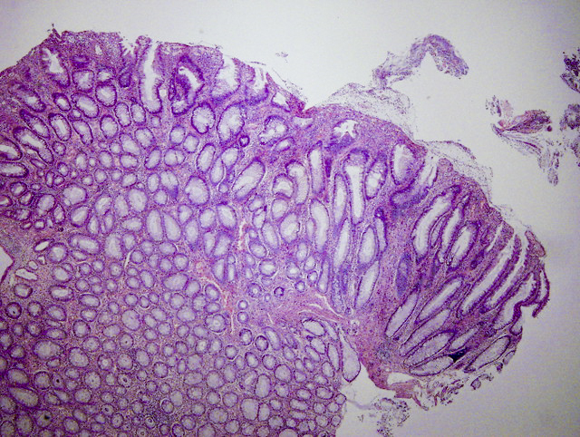
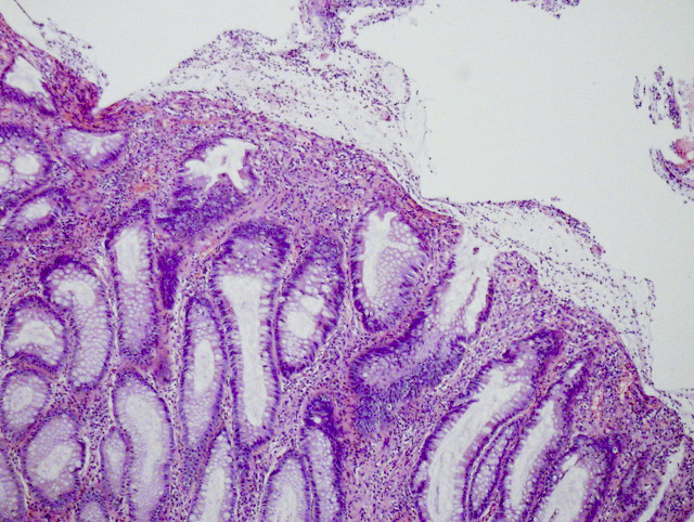
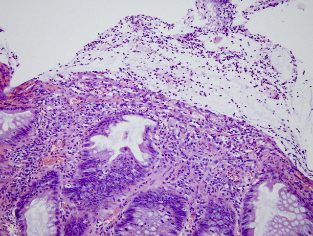
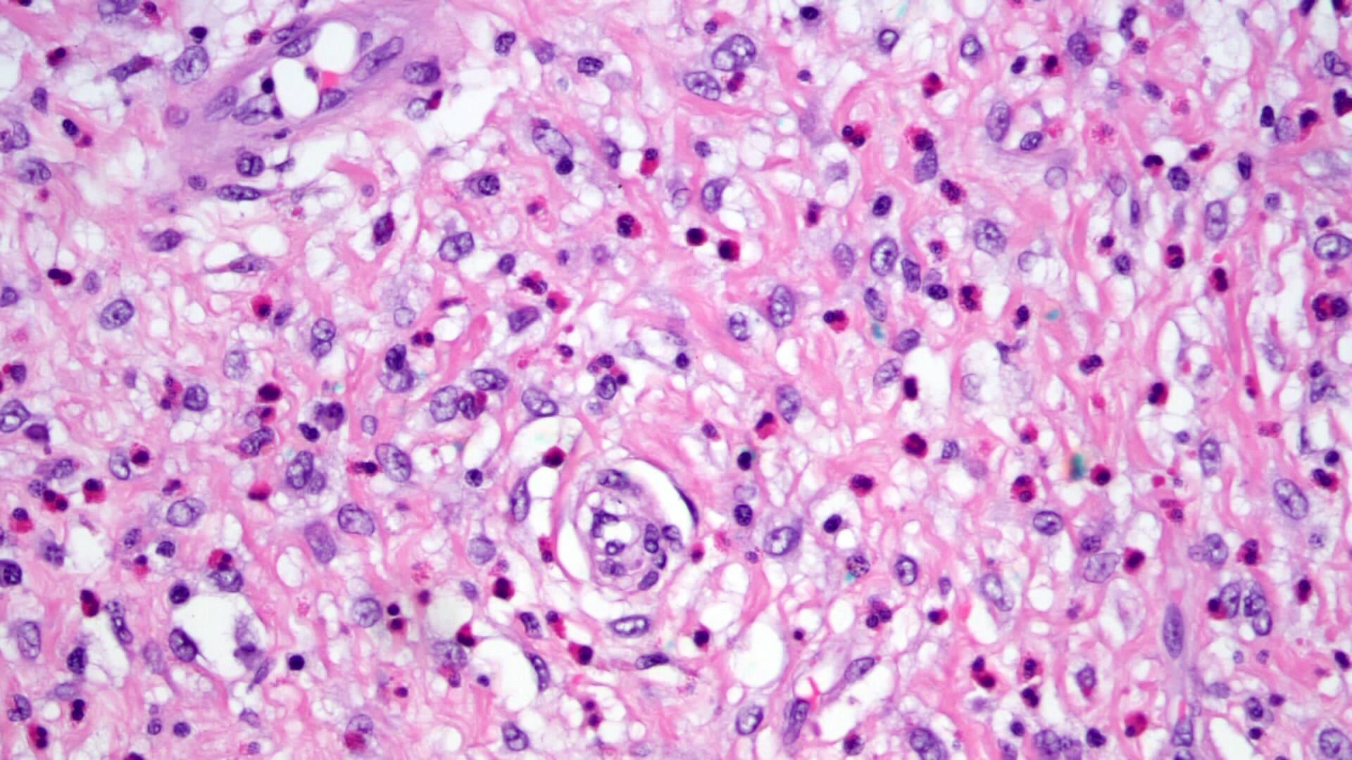
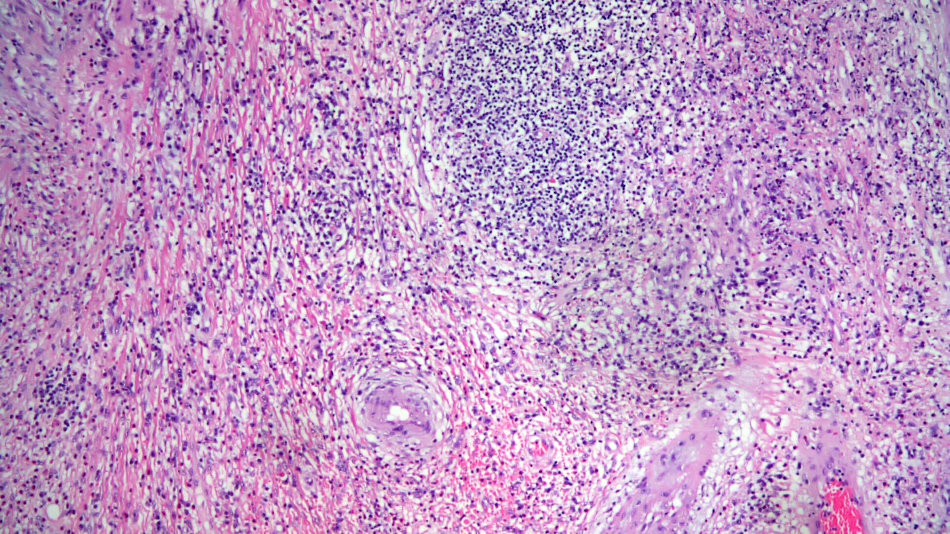
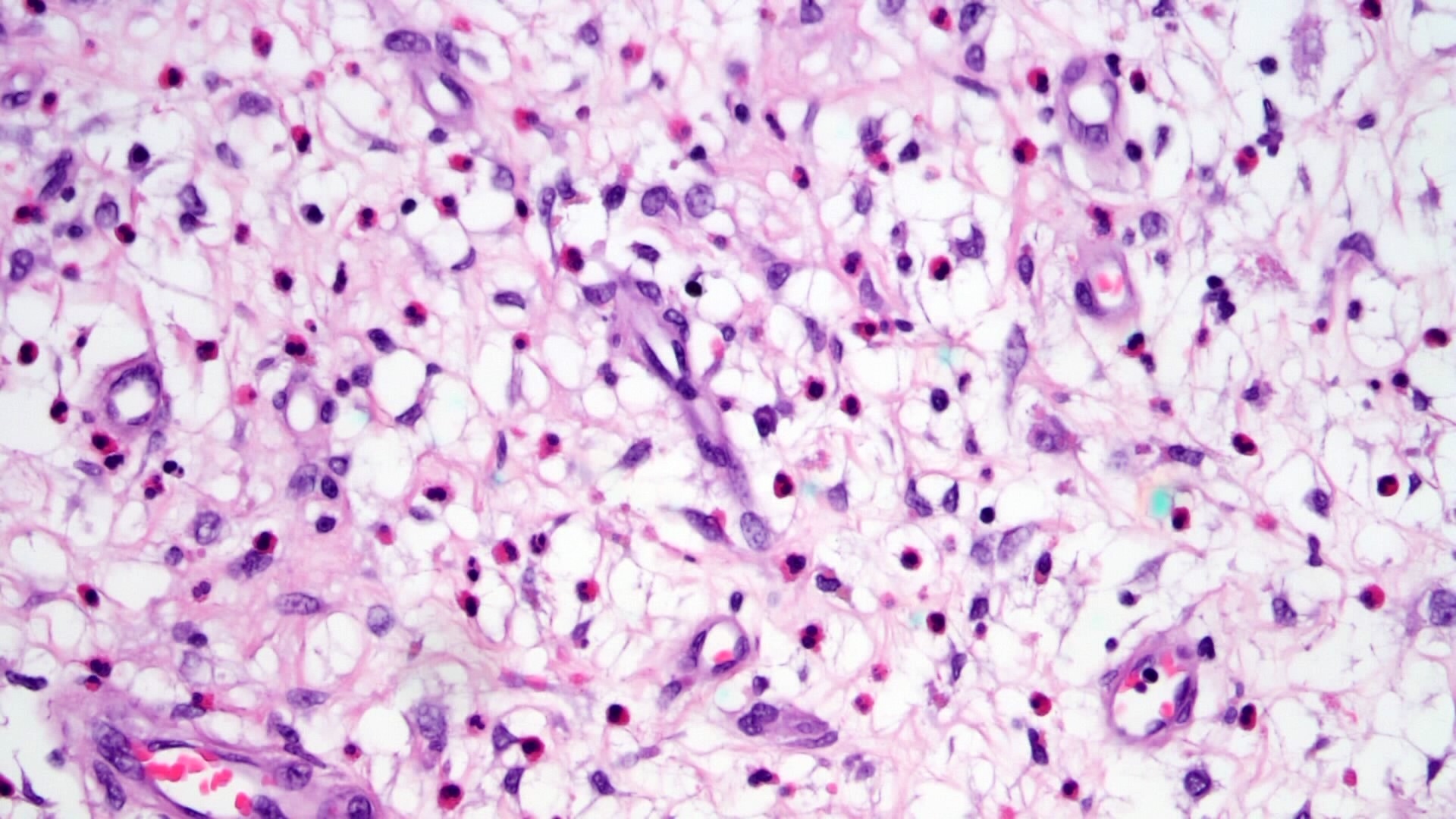
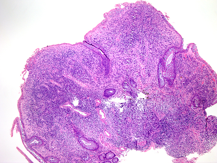
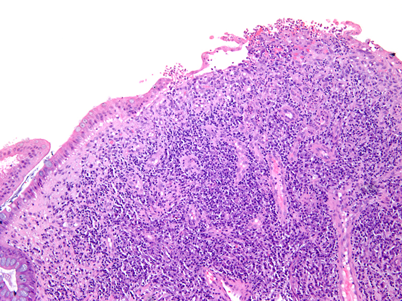
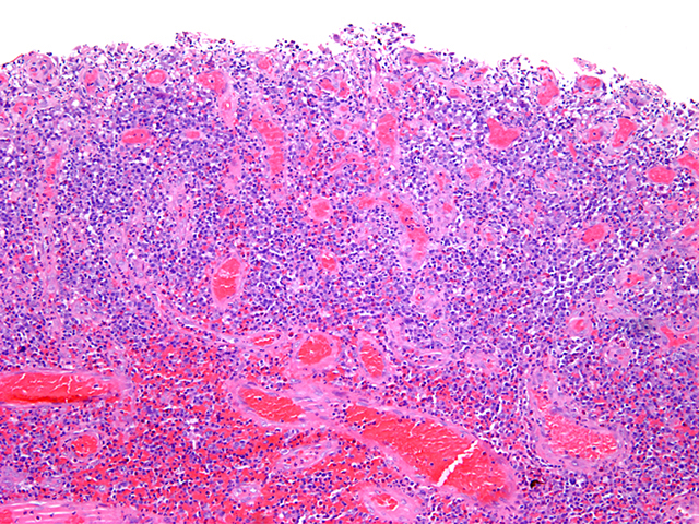
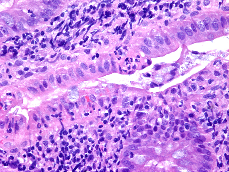
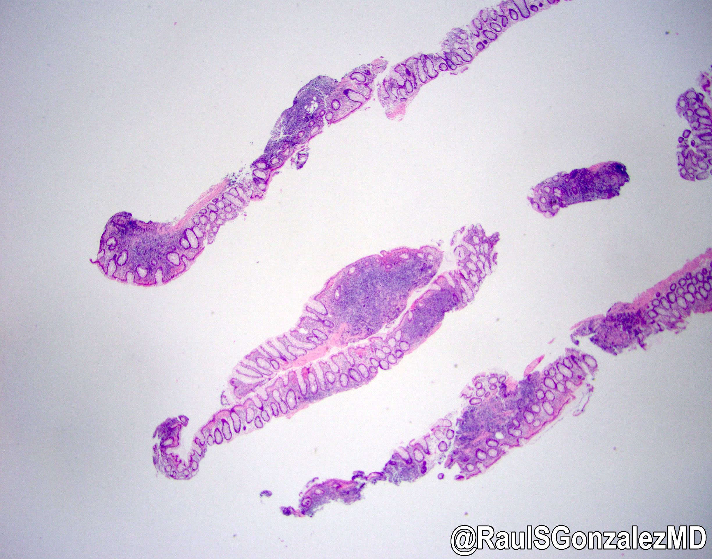
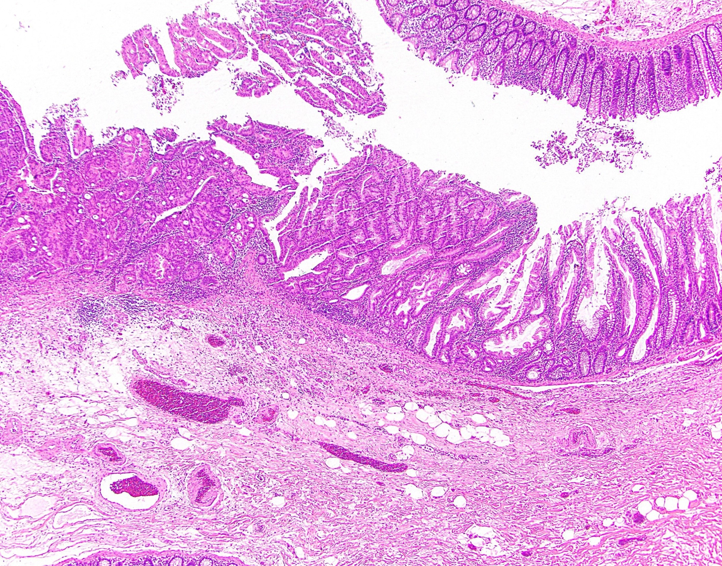
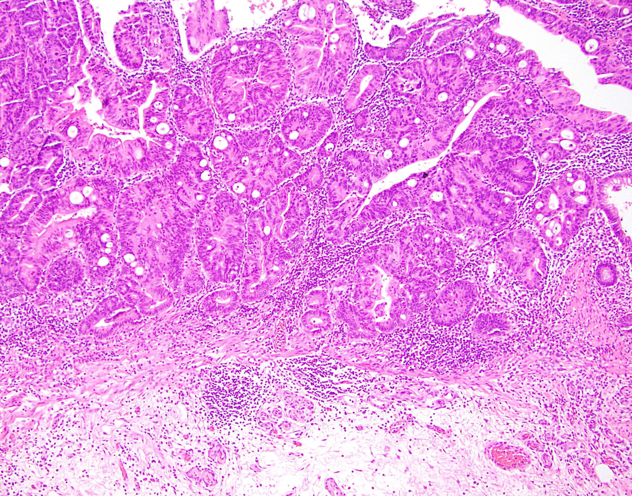
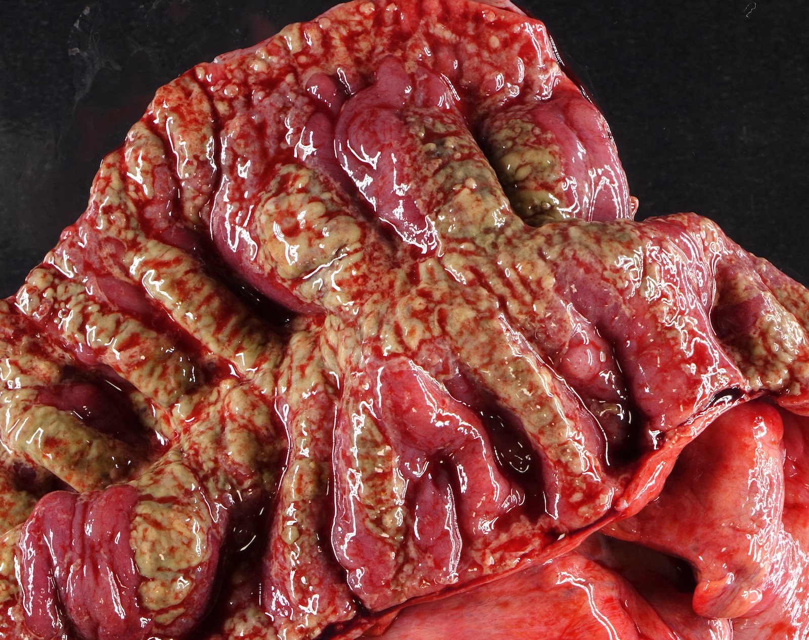
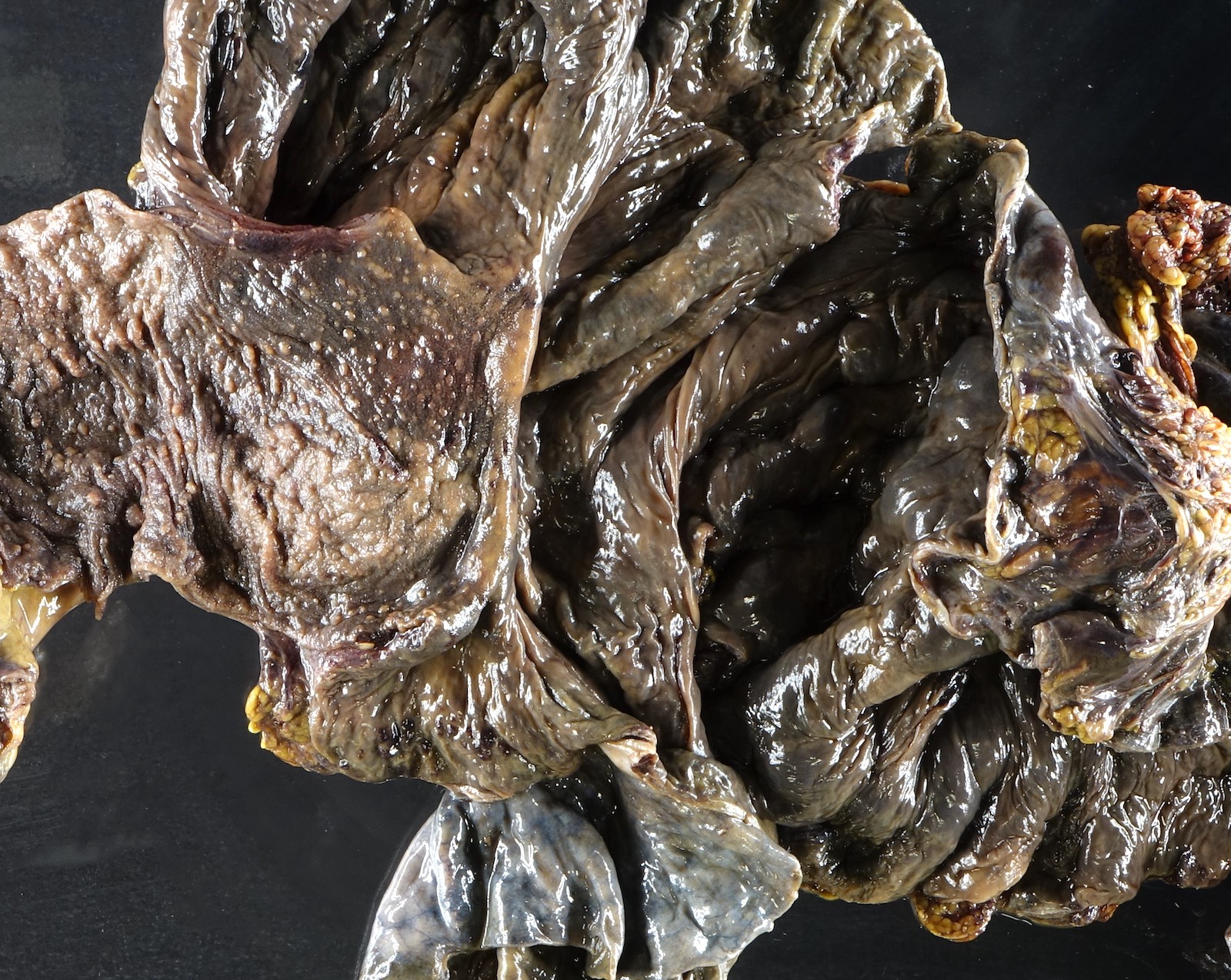
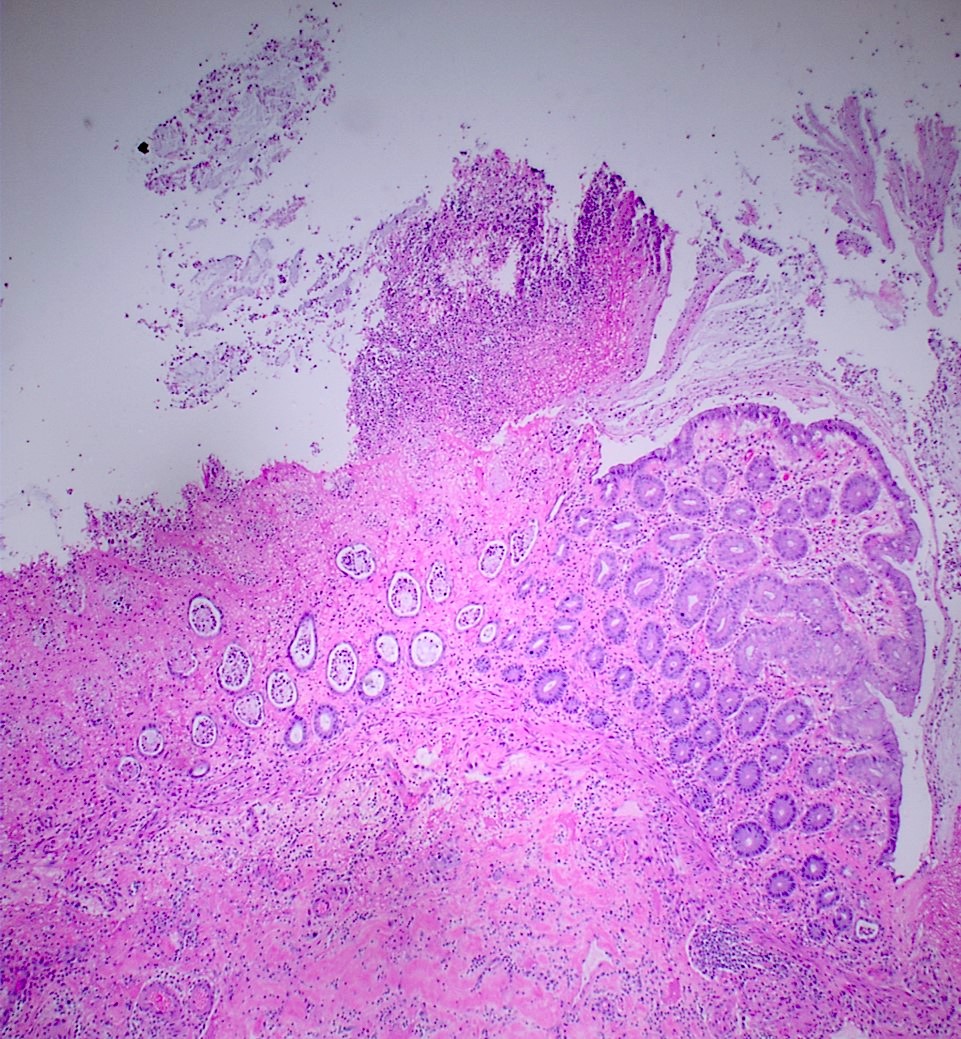
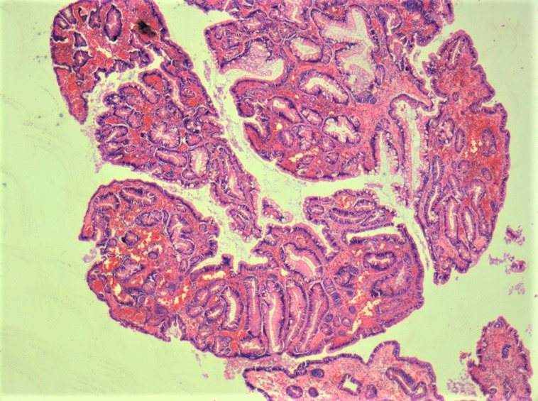
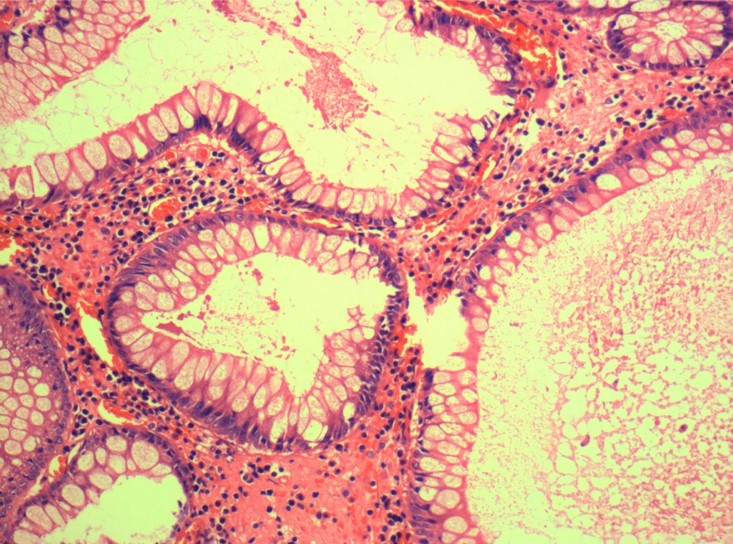
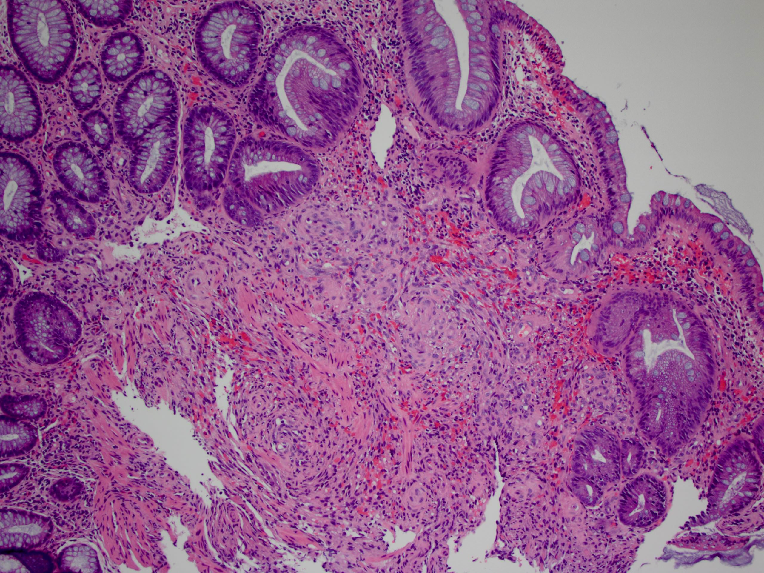
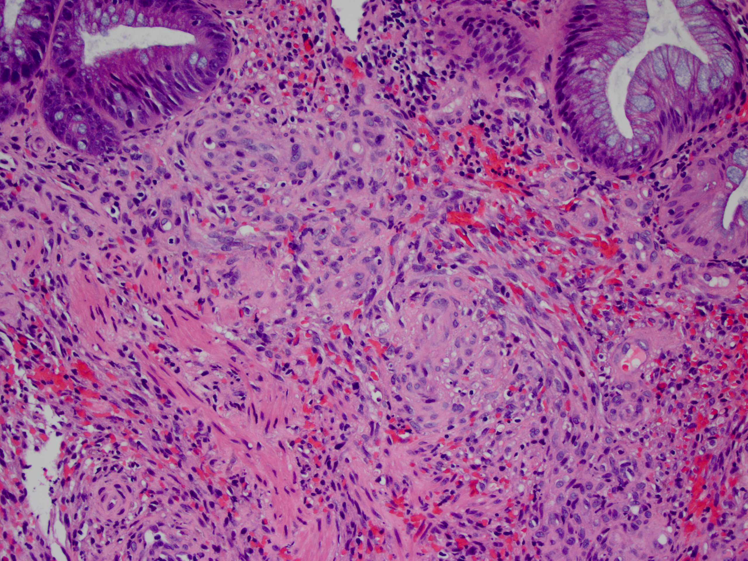
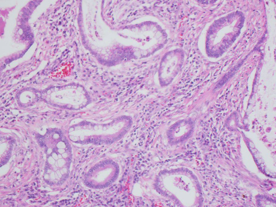
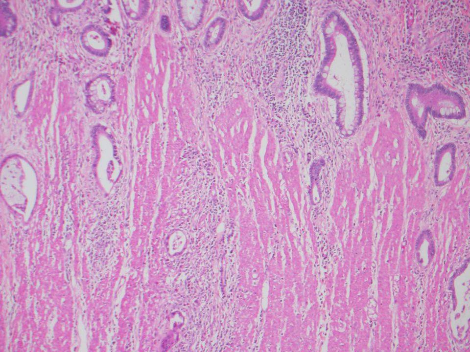
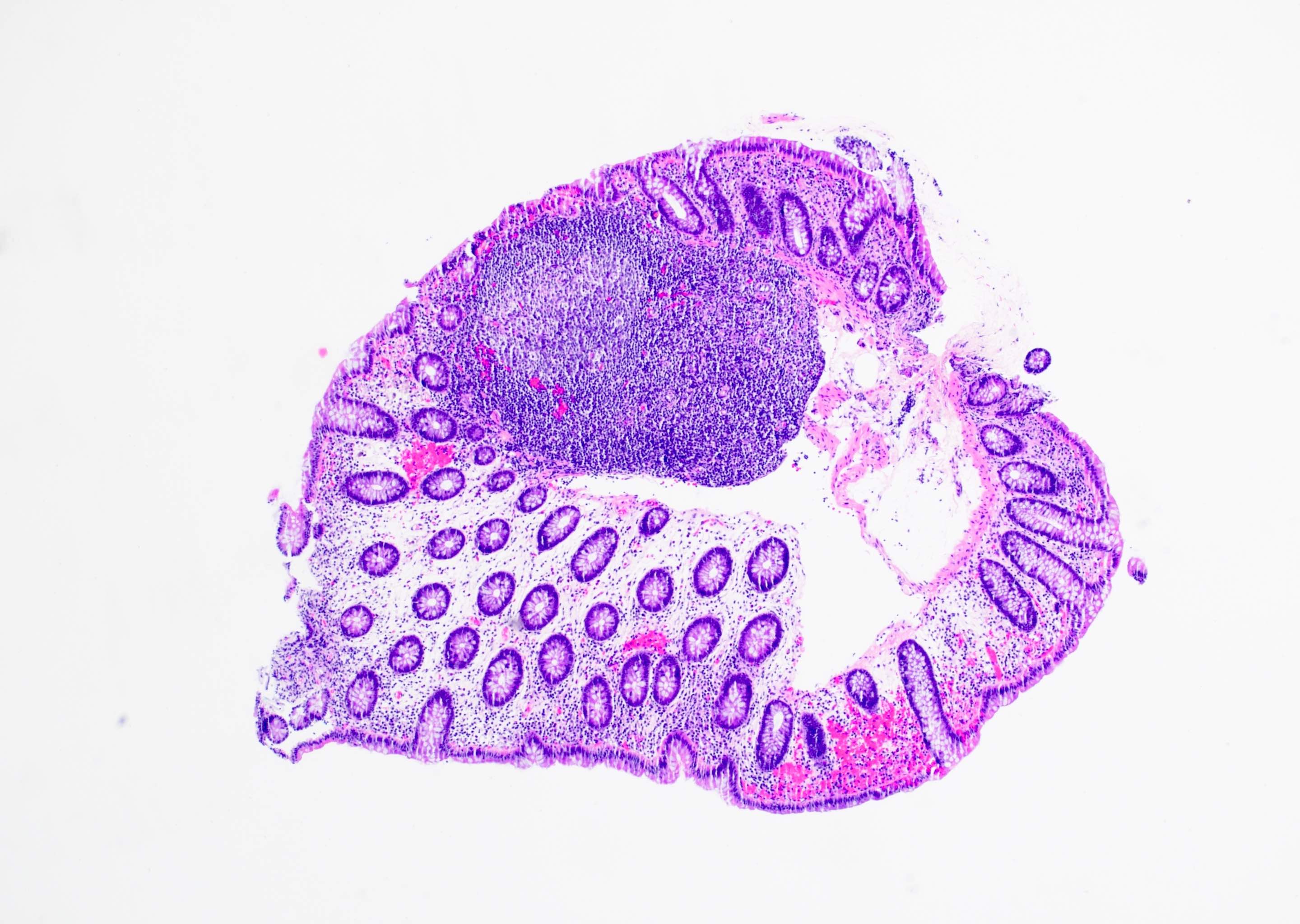
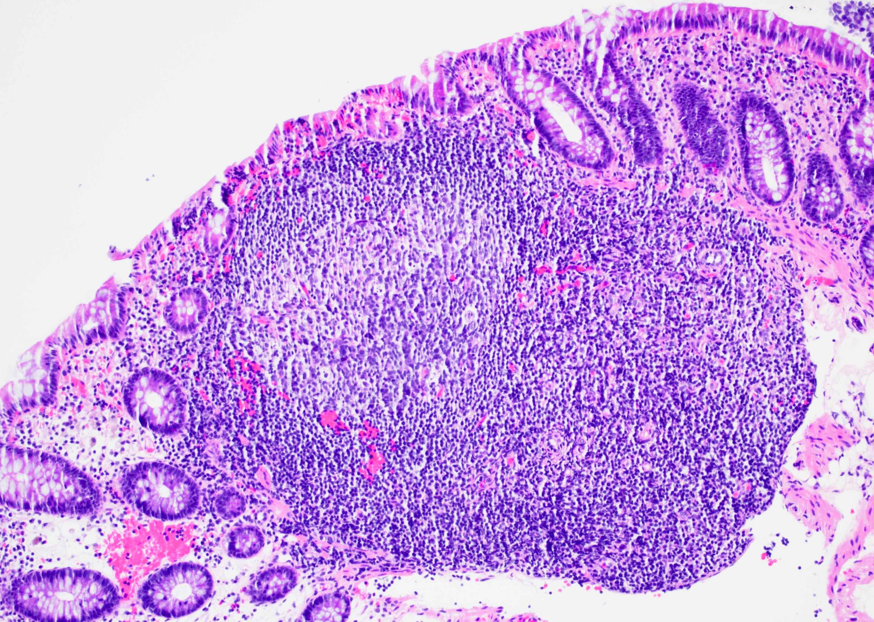
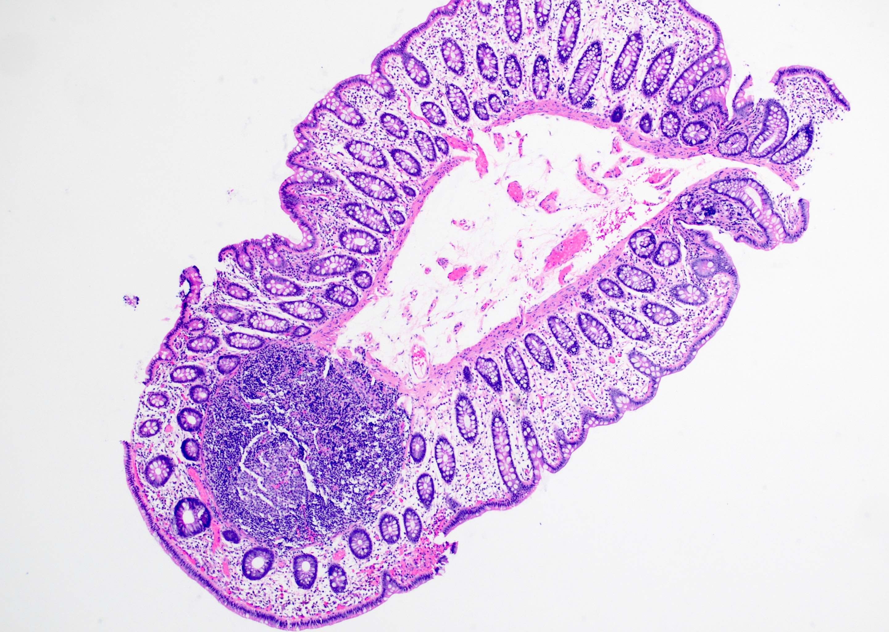
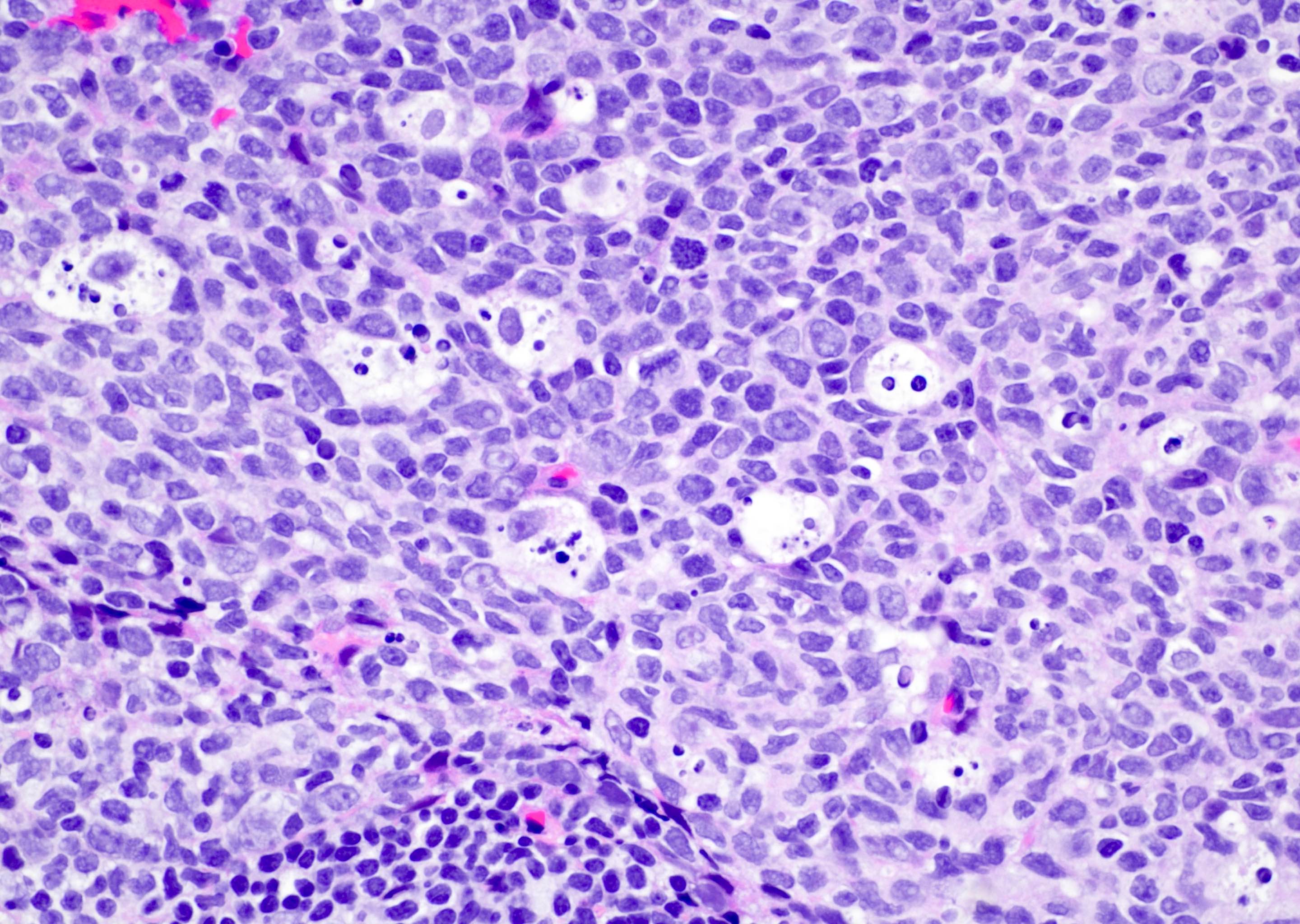
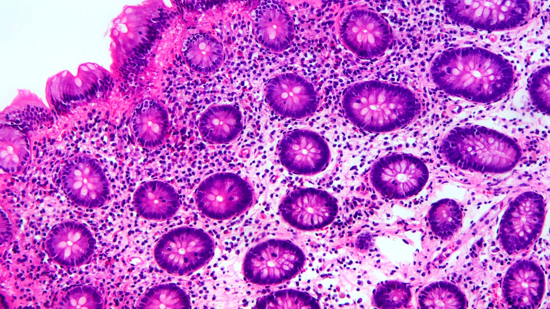
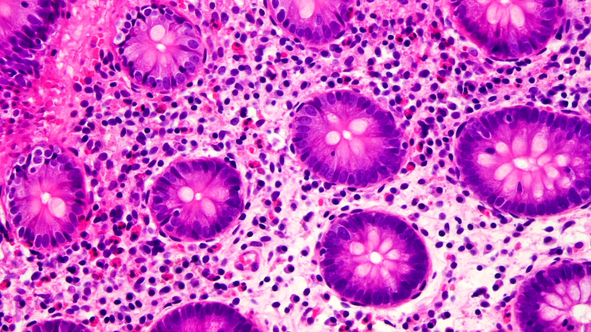
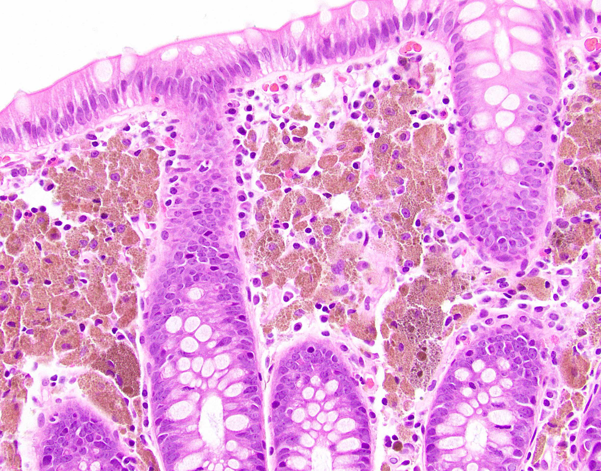
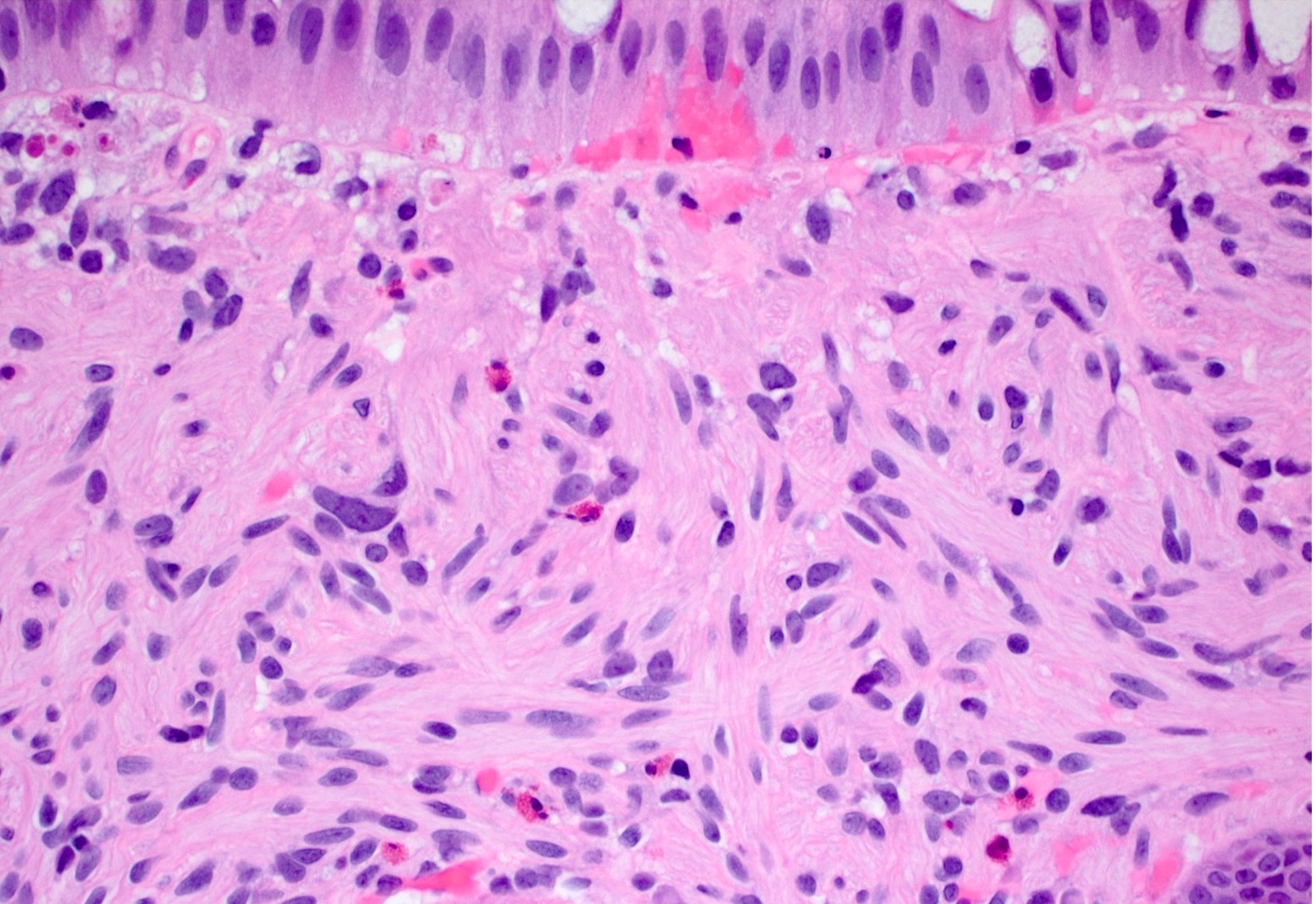
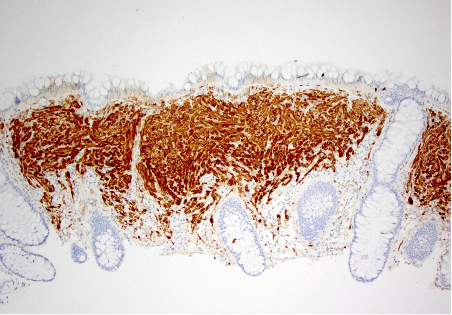
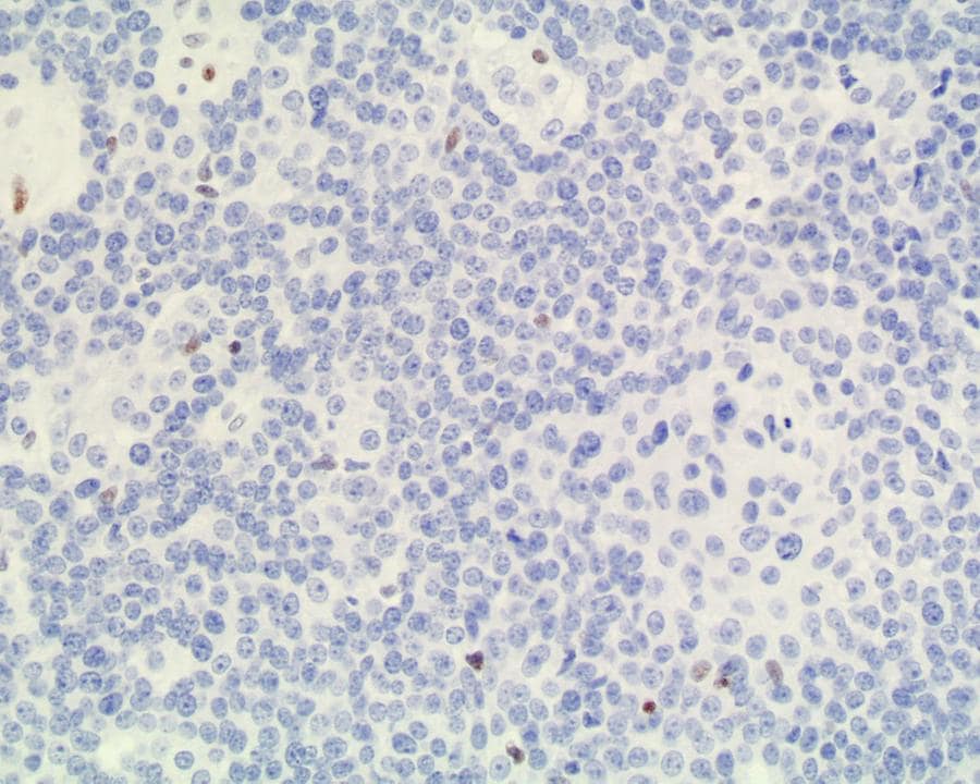
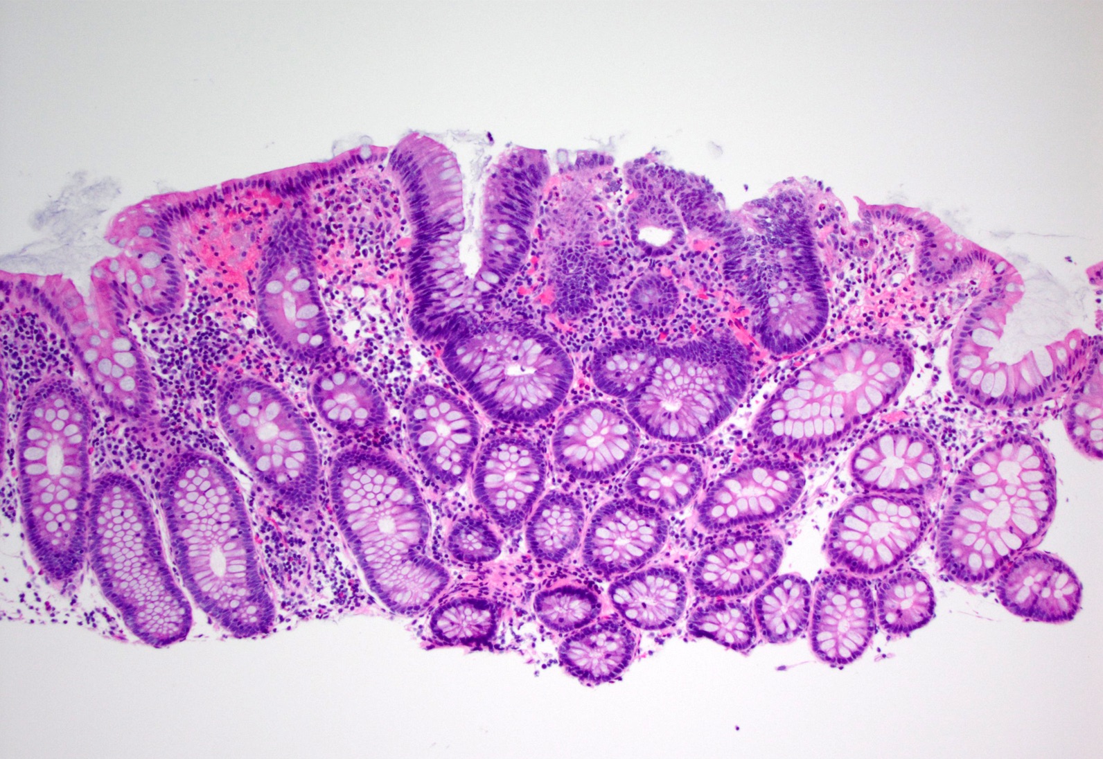
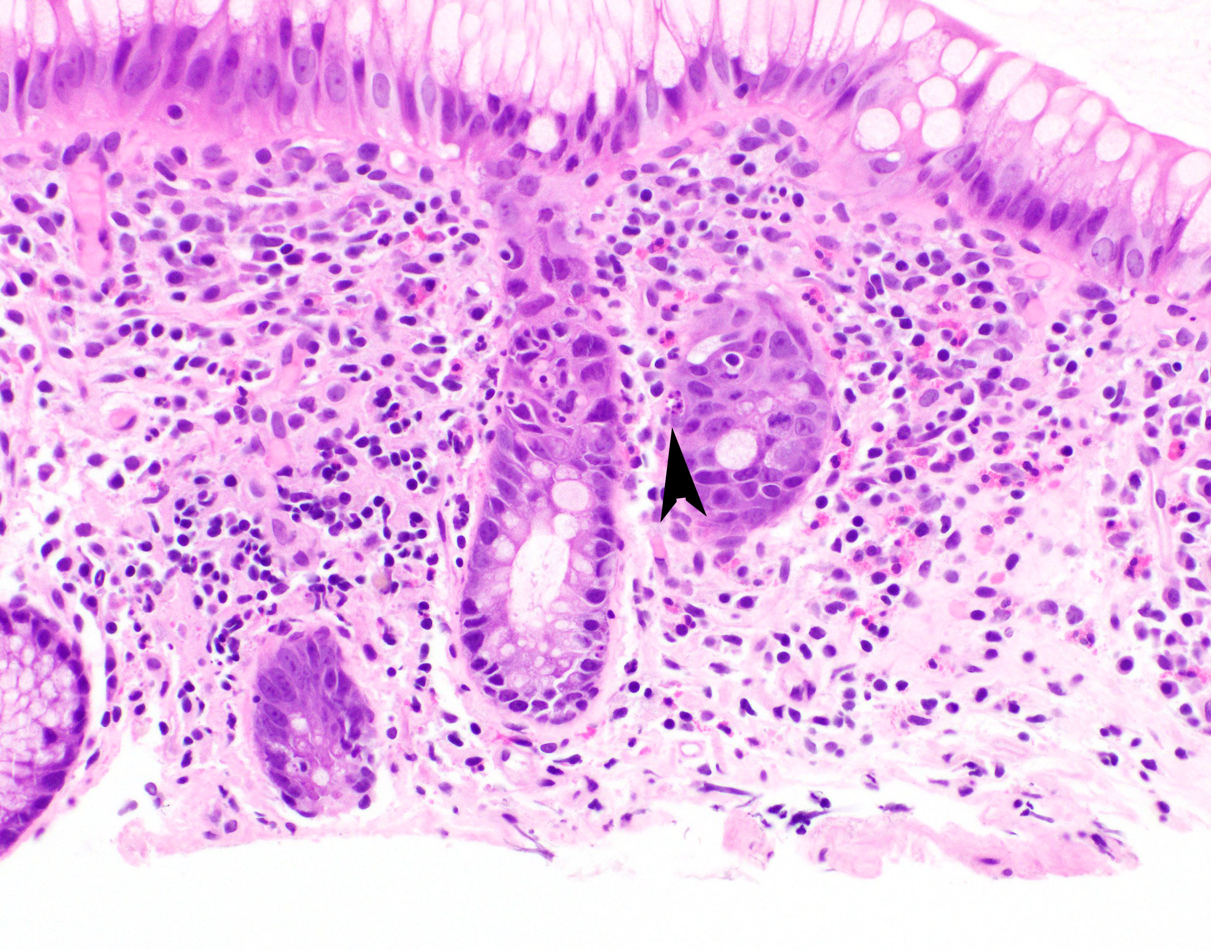
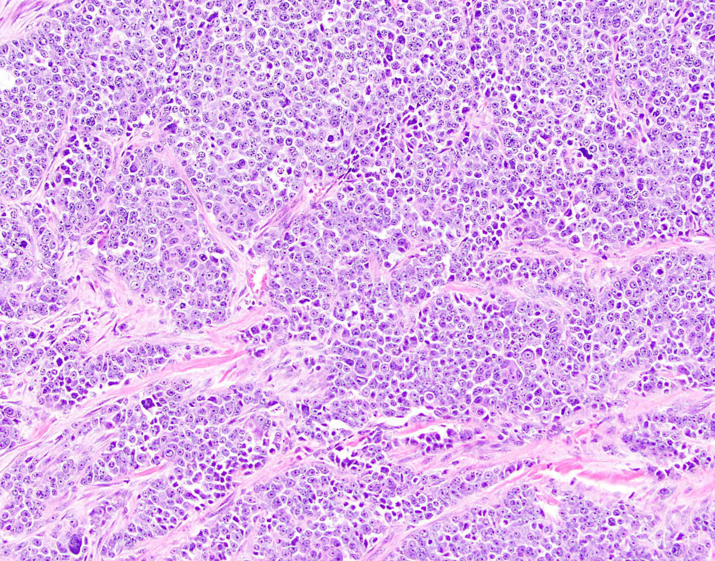
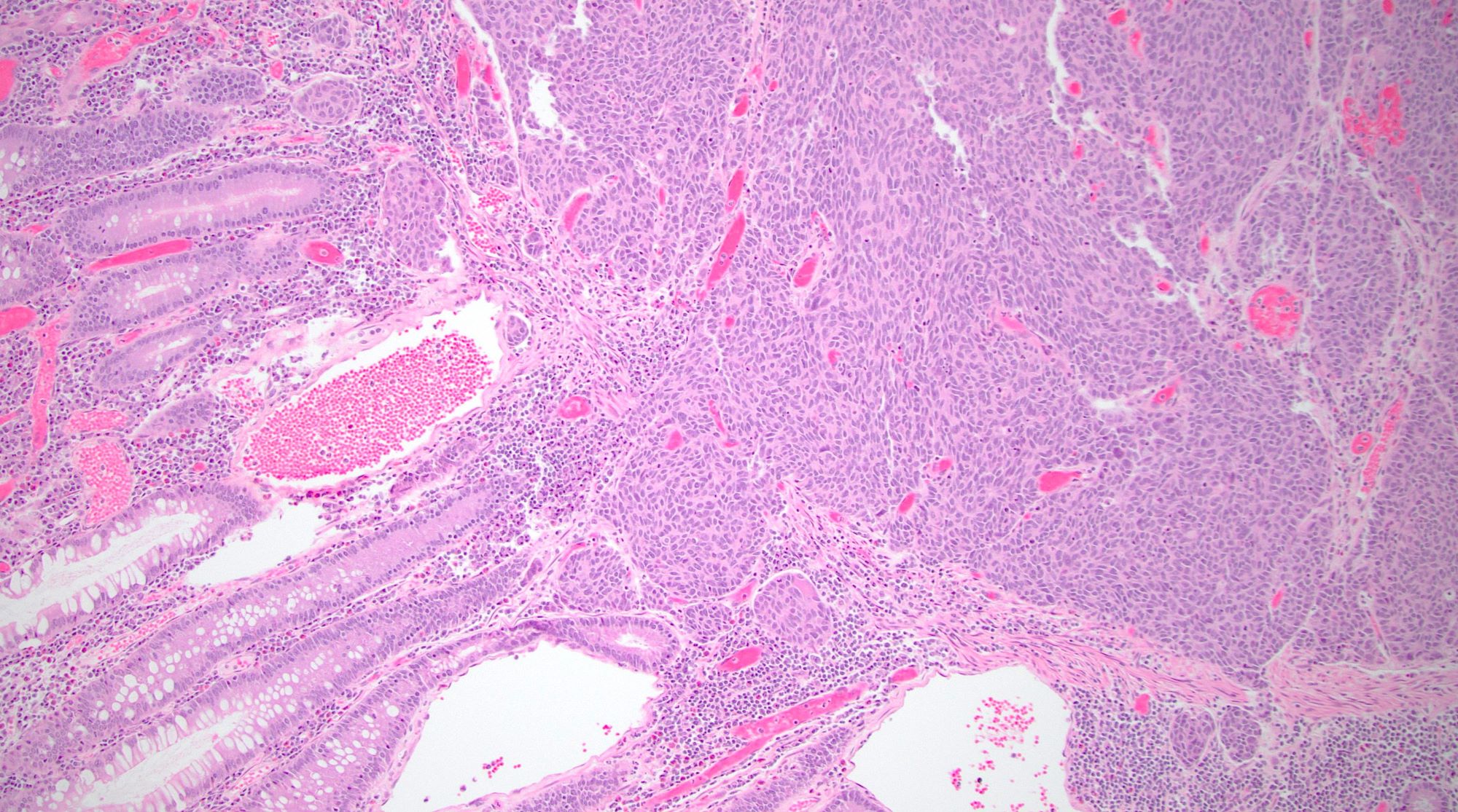
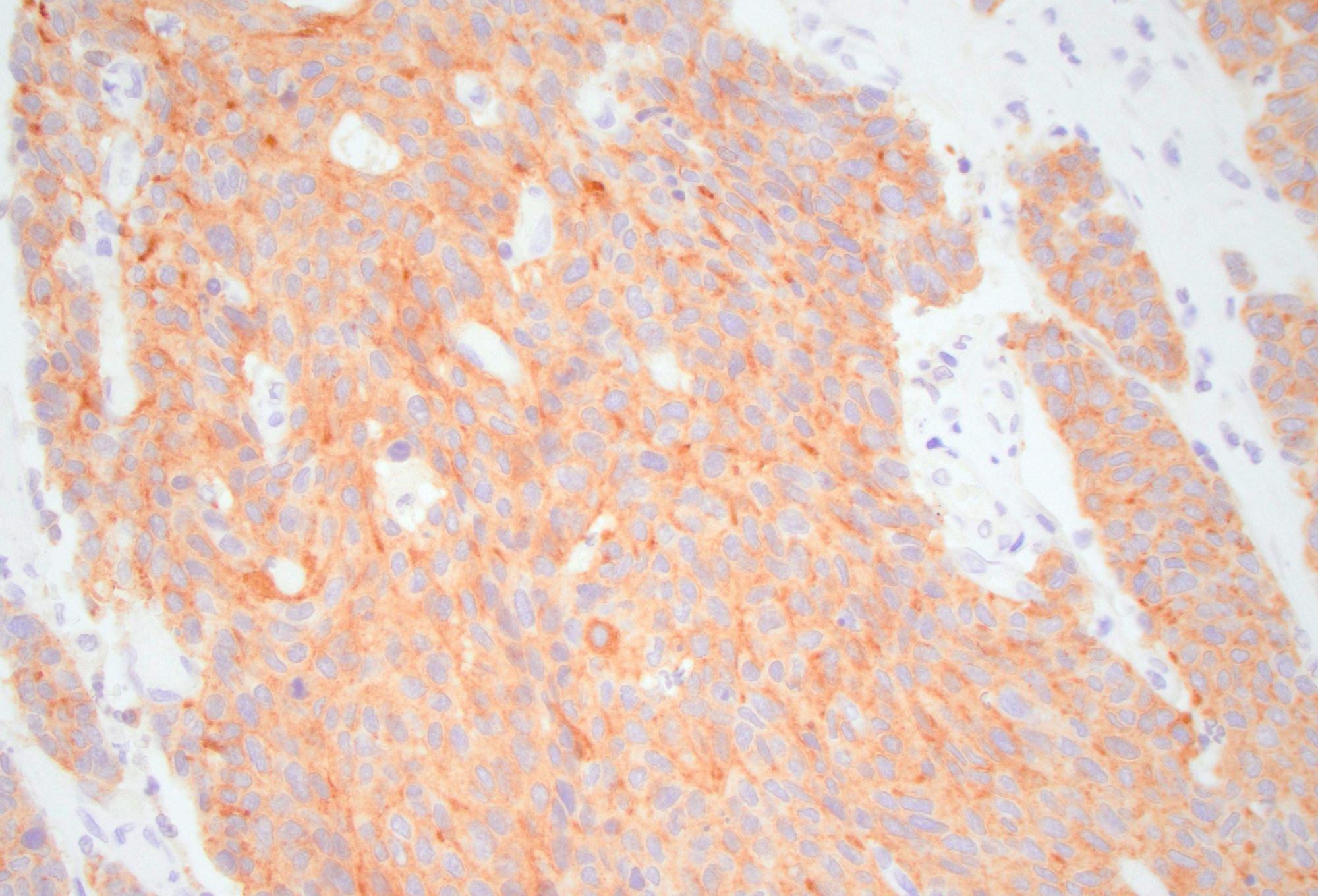
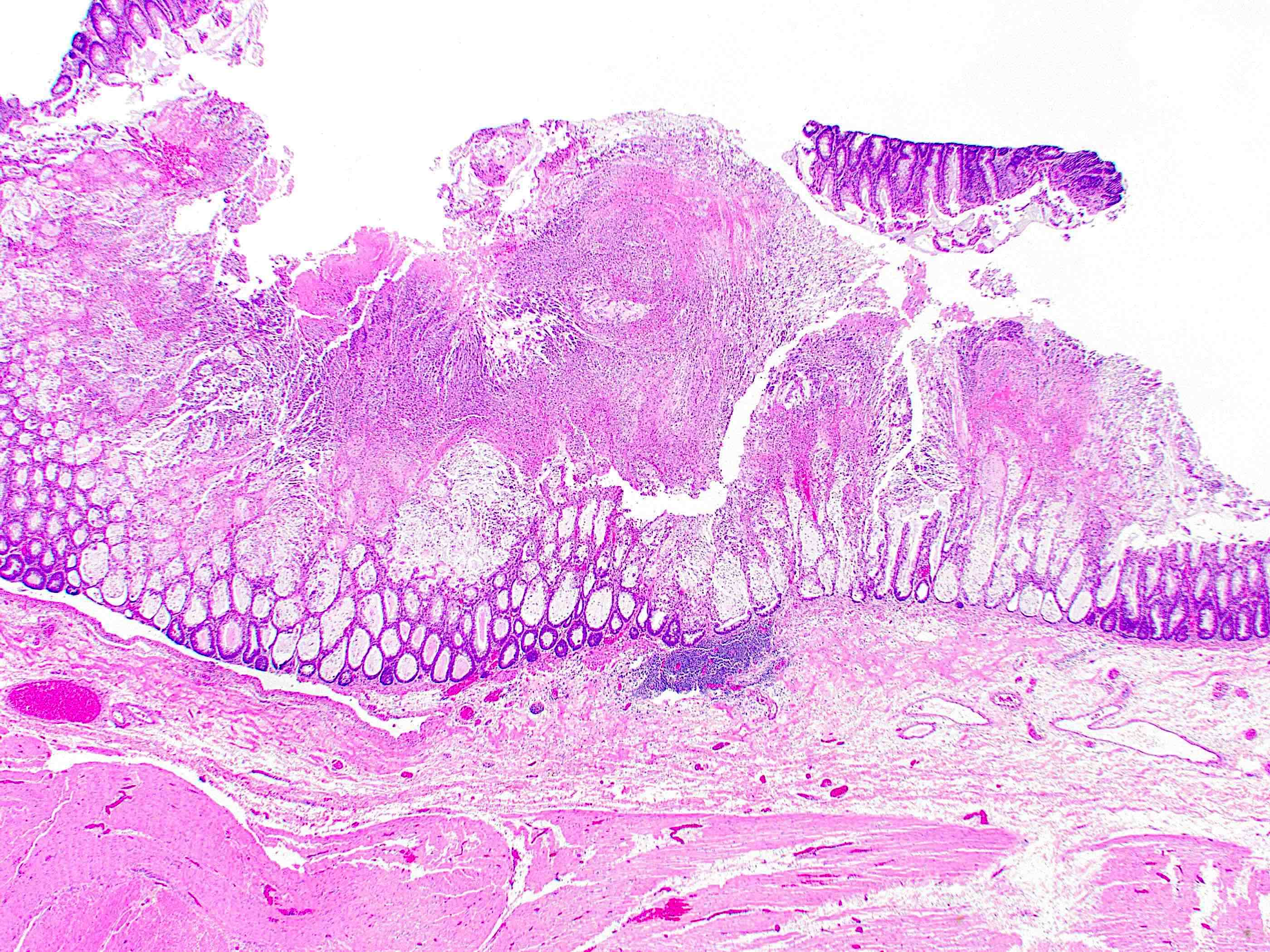
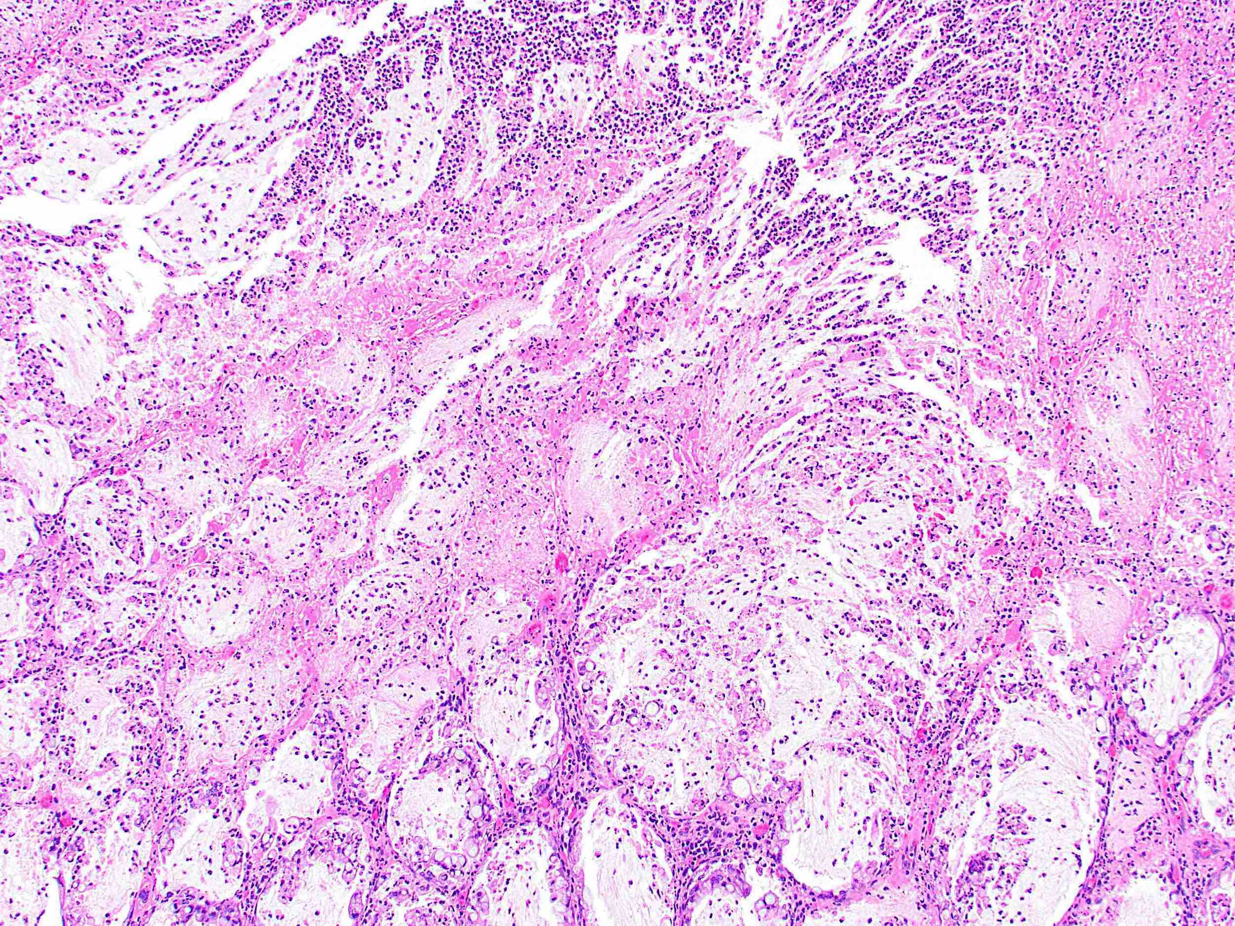
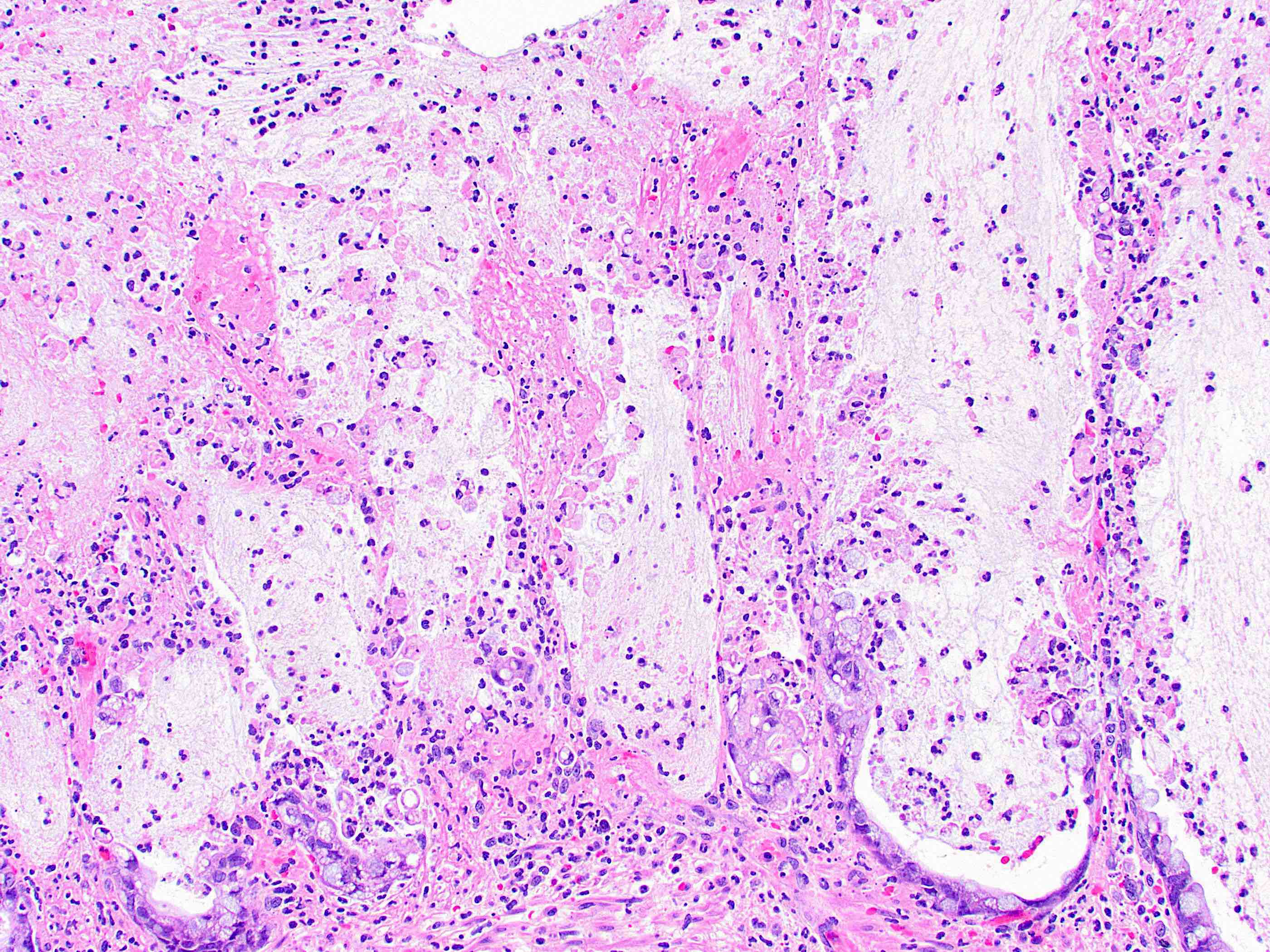
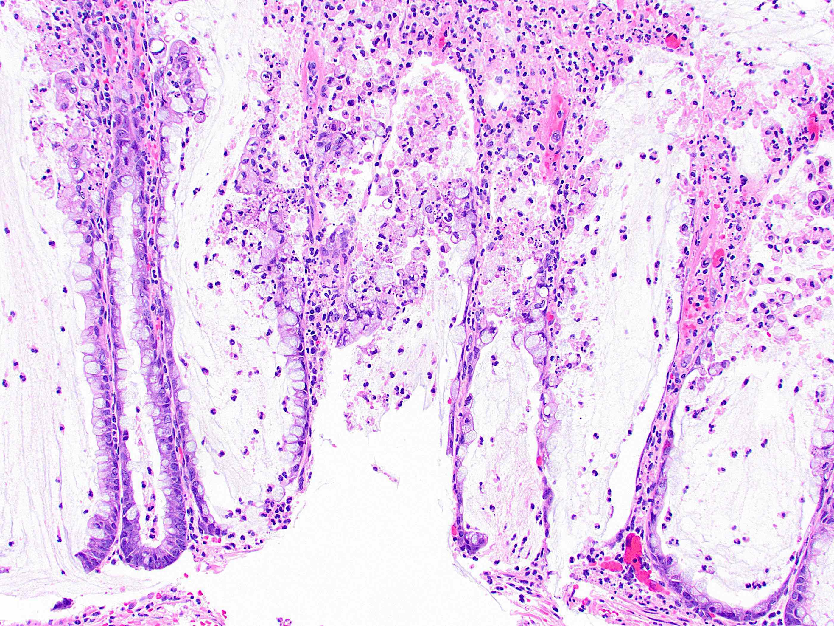
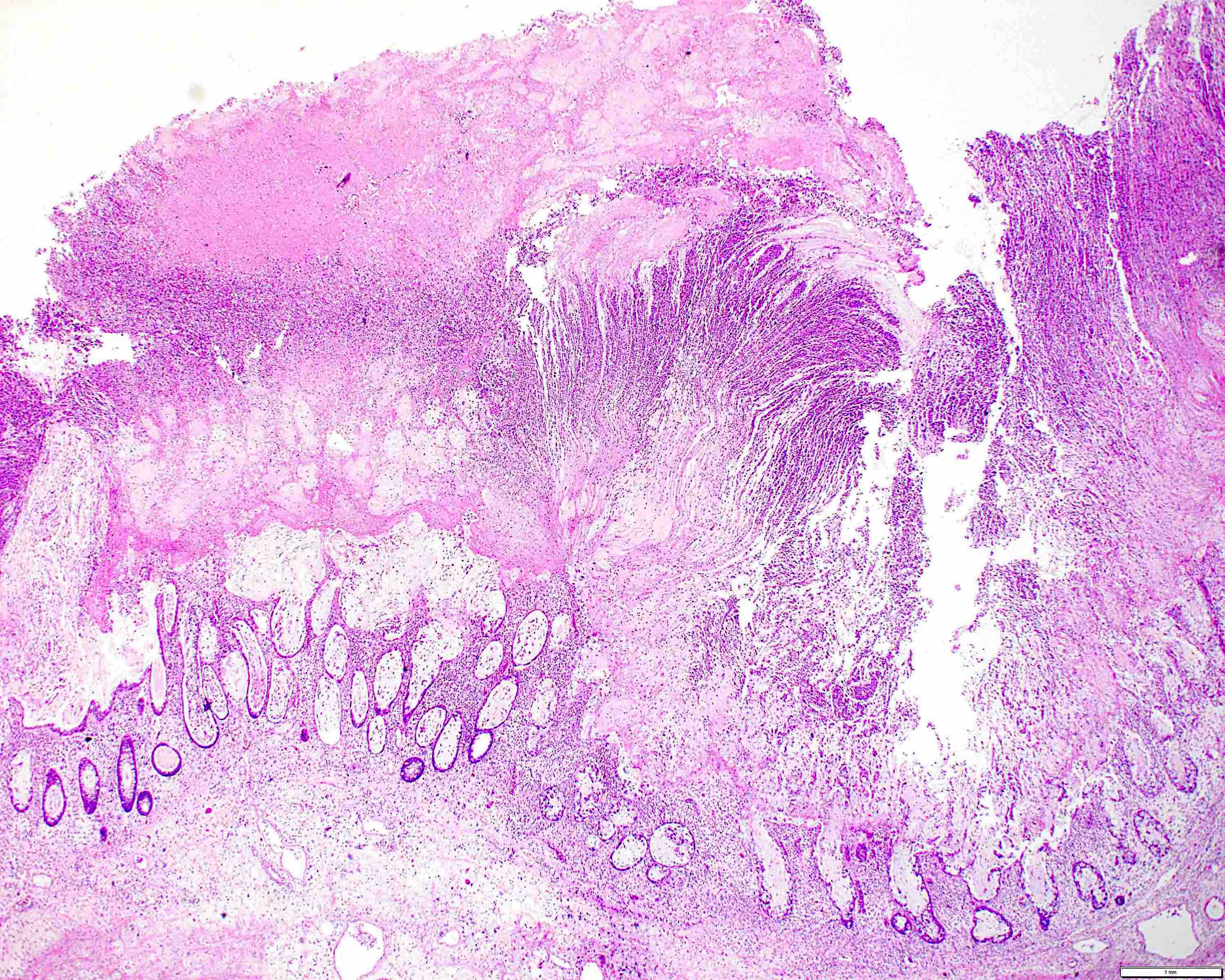
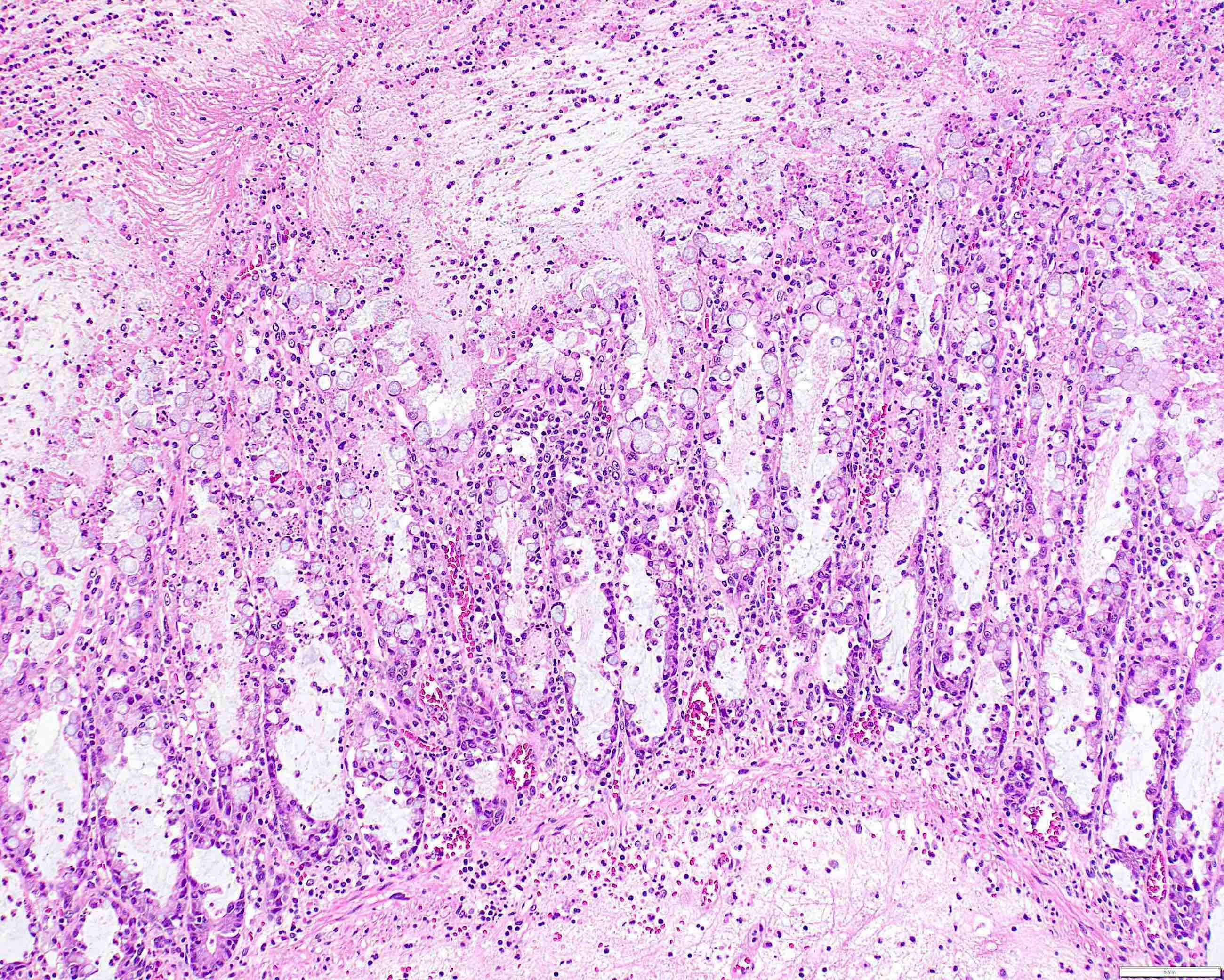
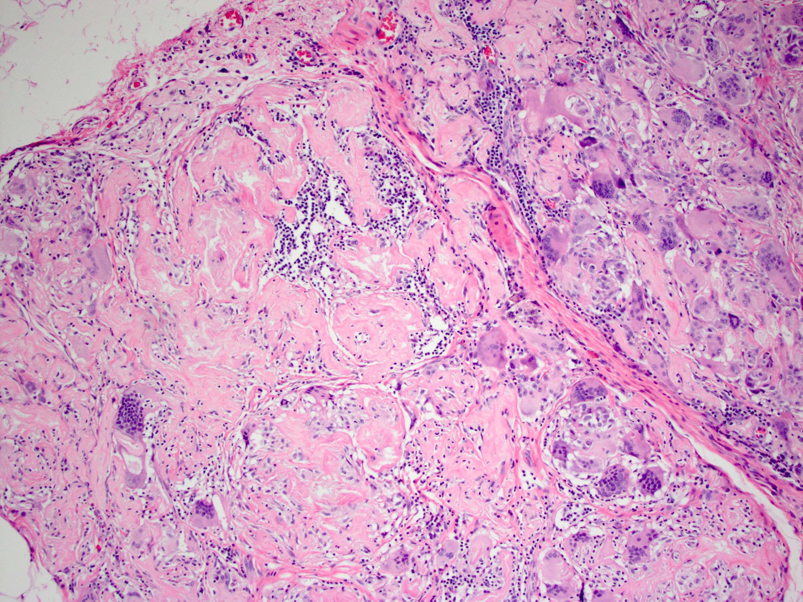
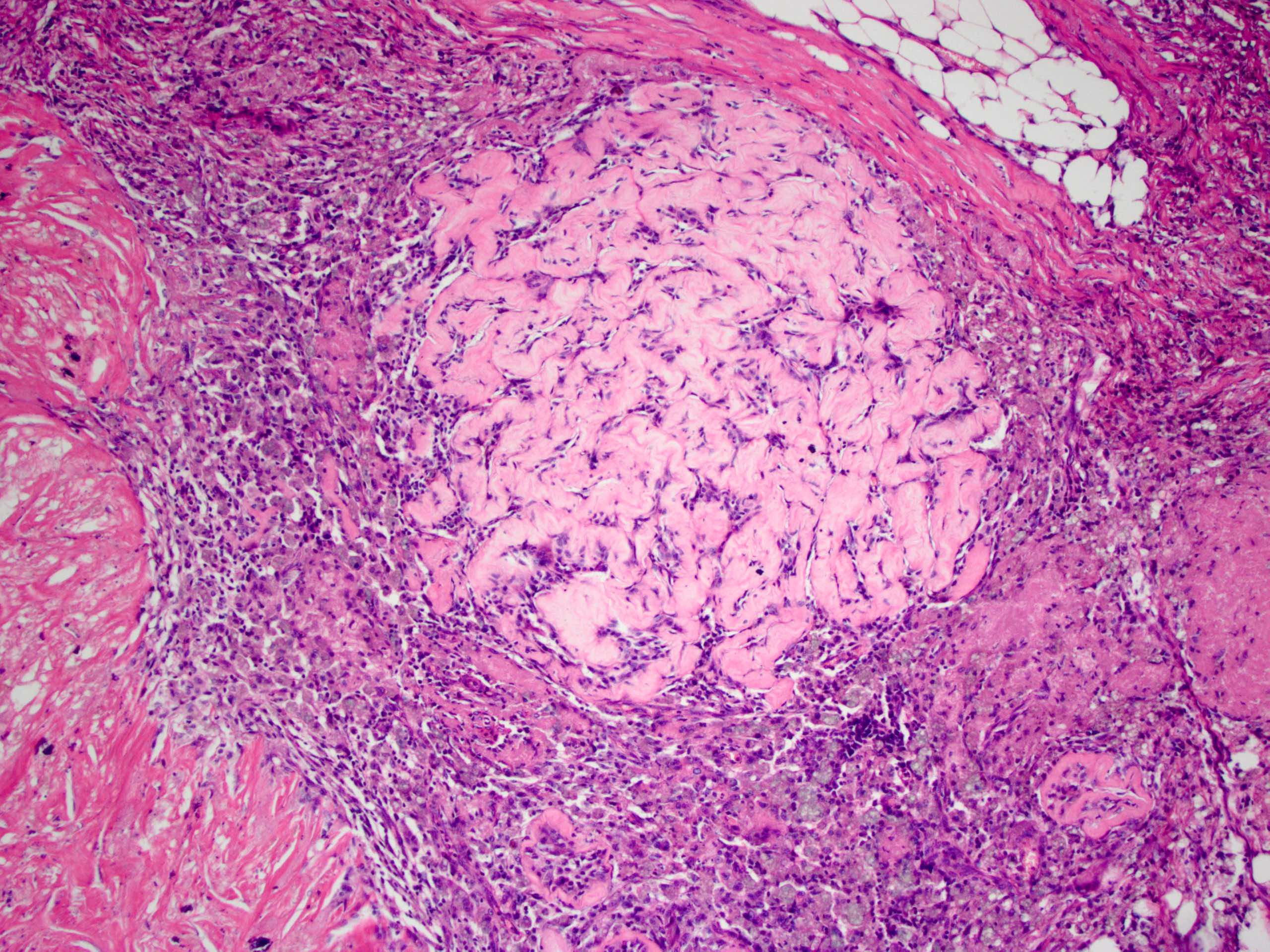
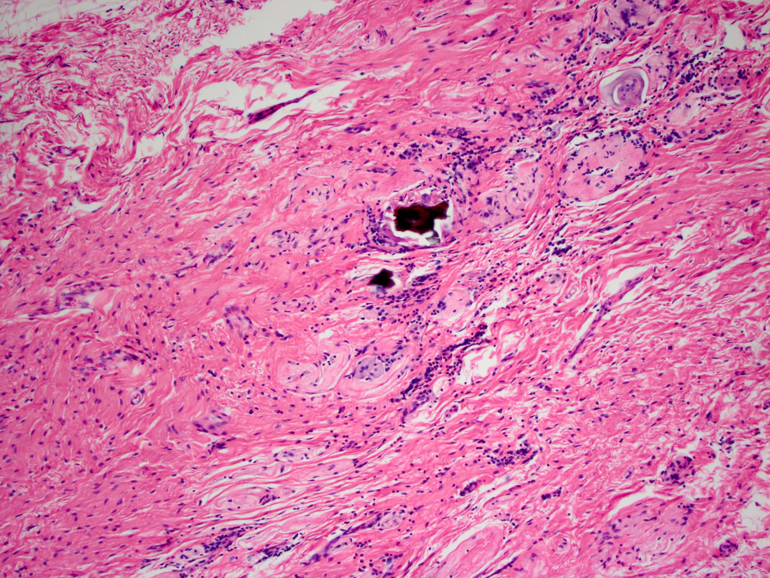
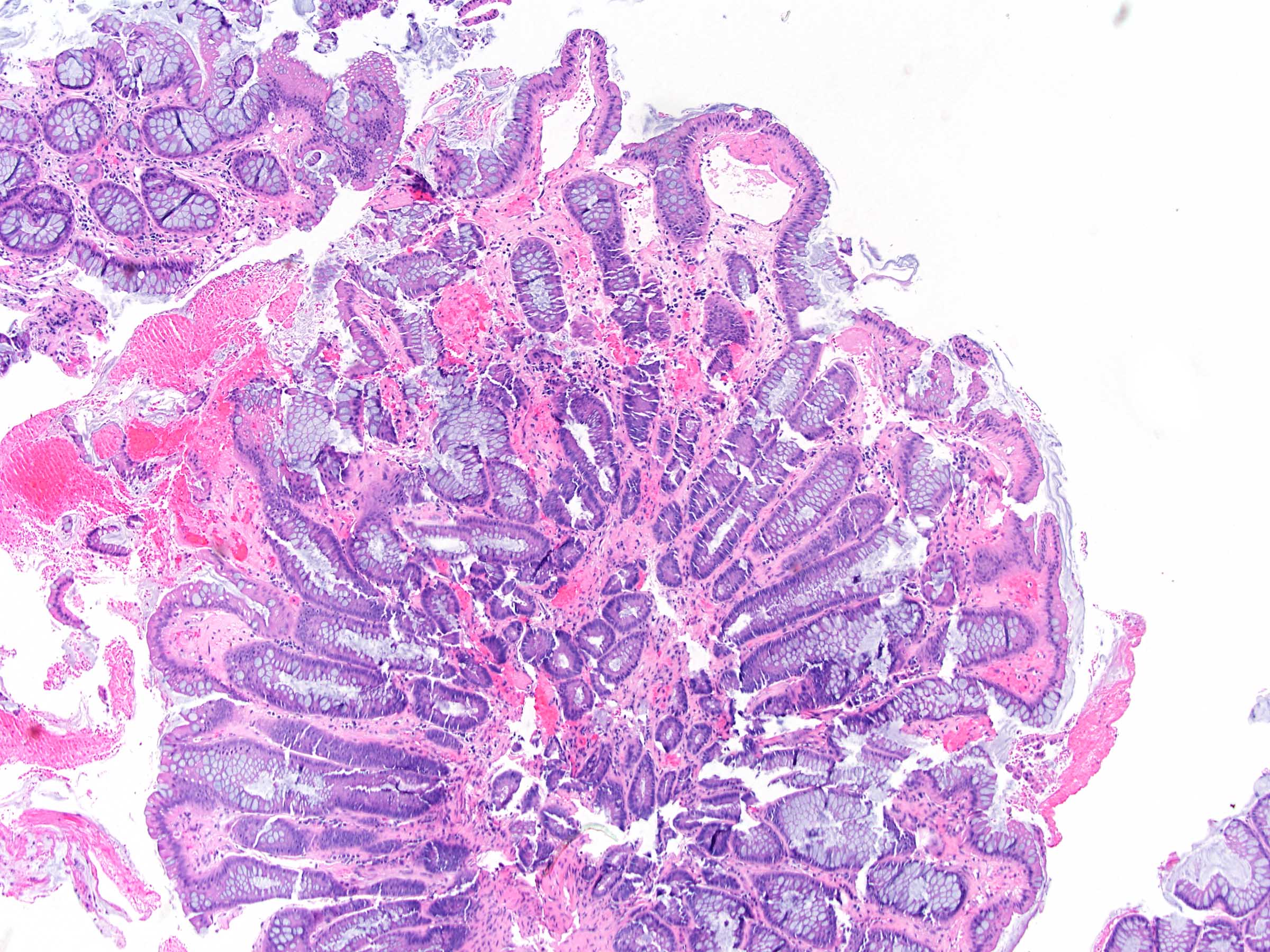
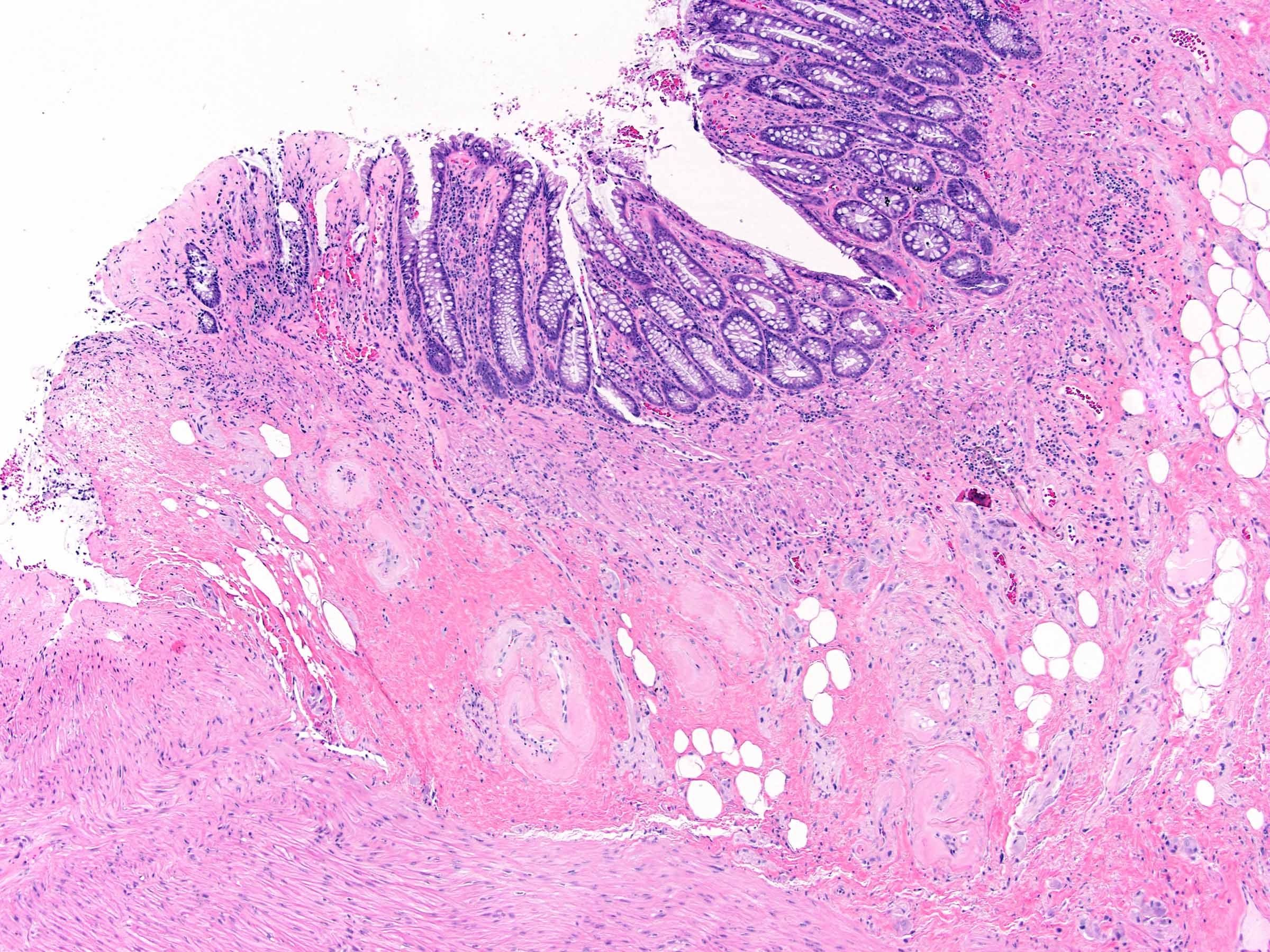
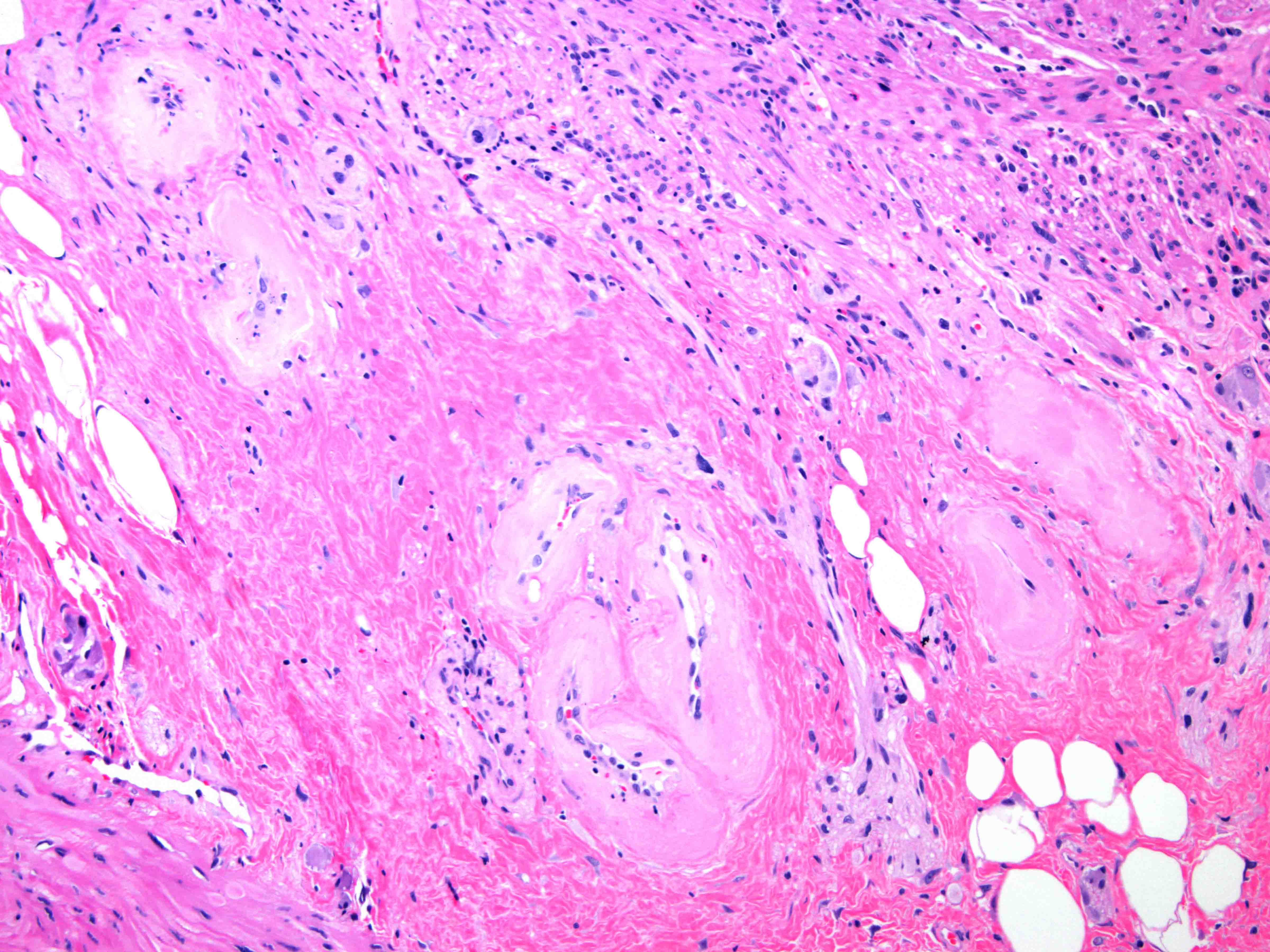
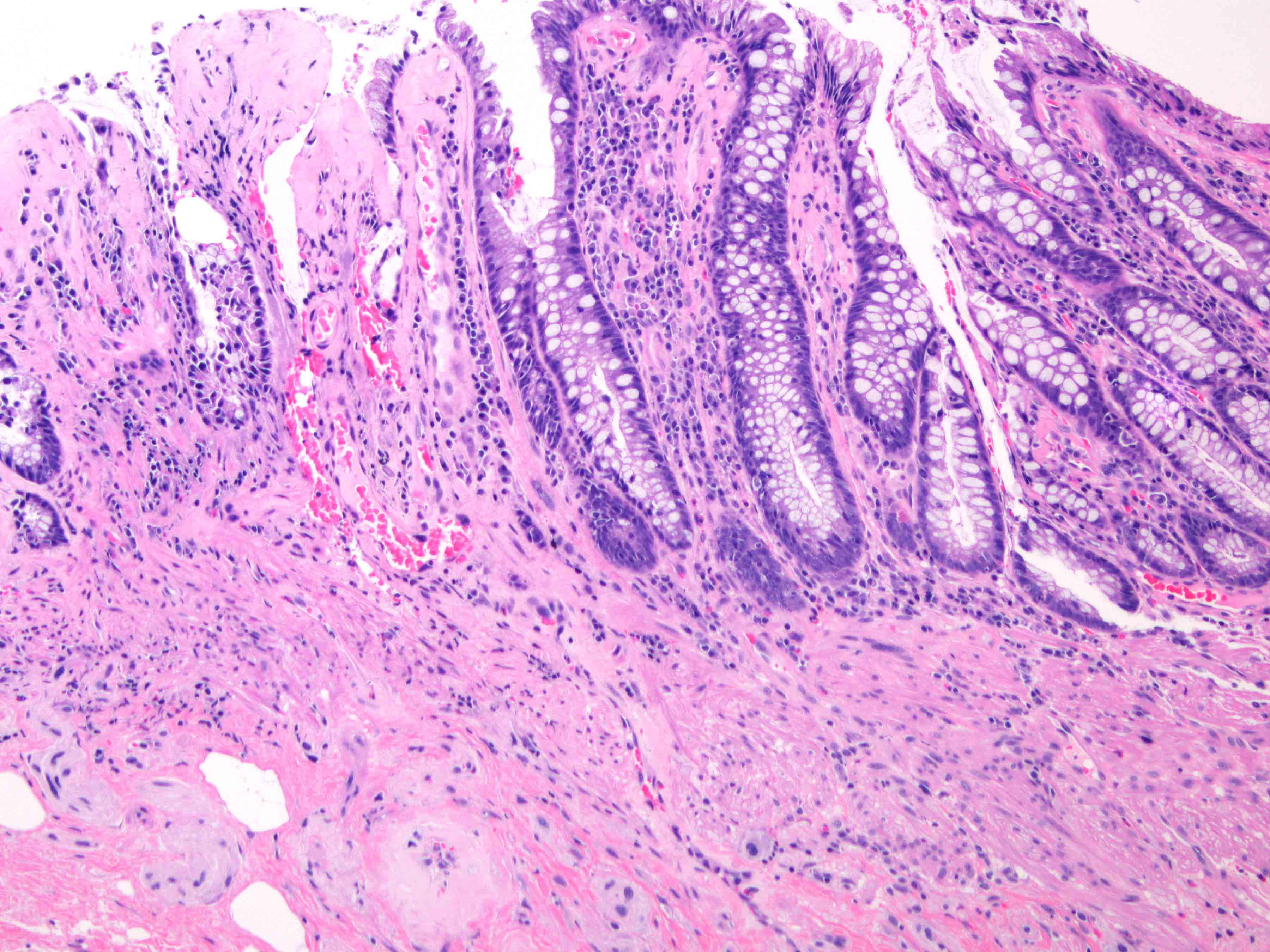
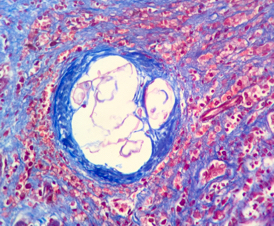
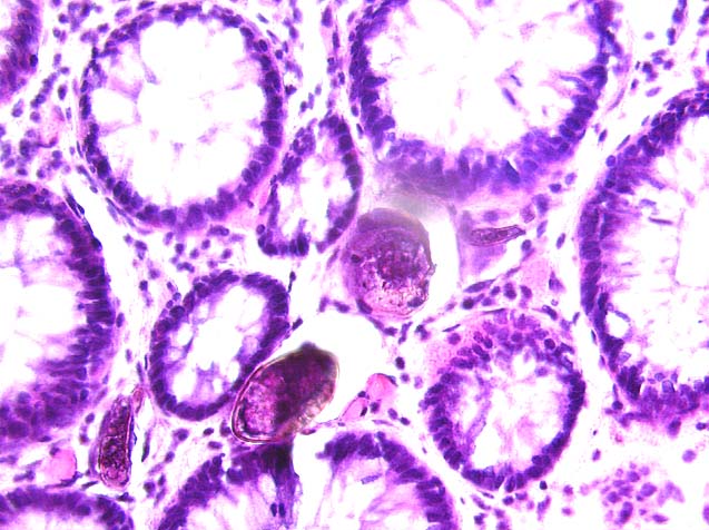
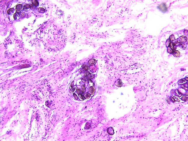
Intestinal actinomycosis
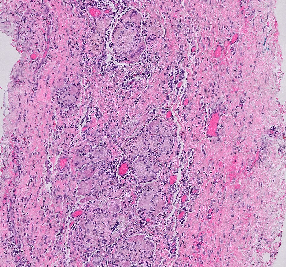
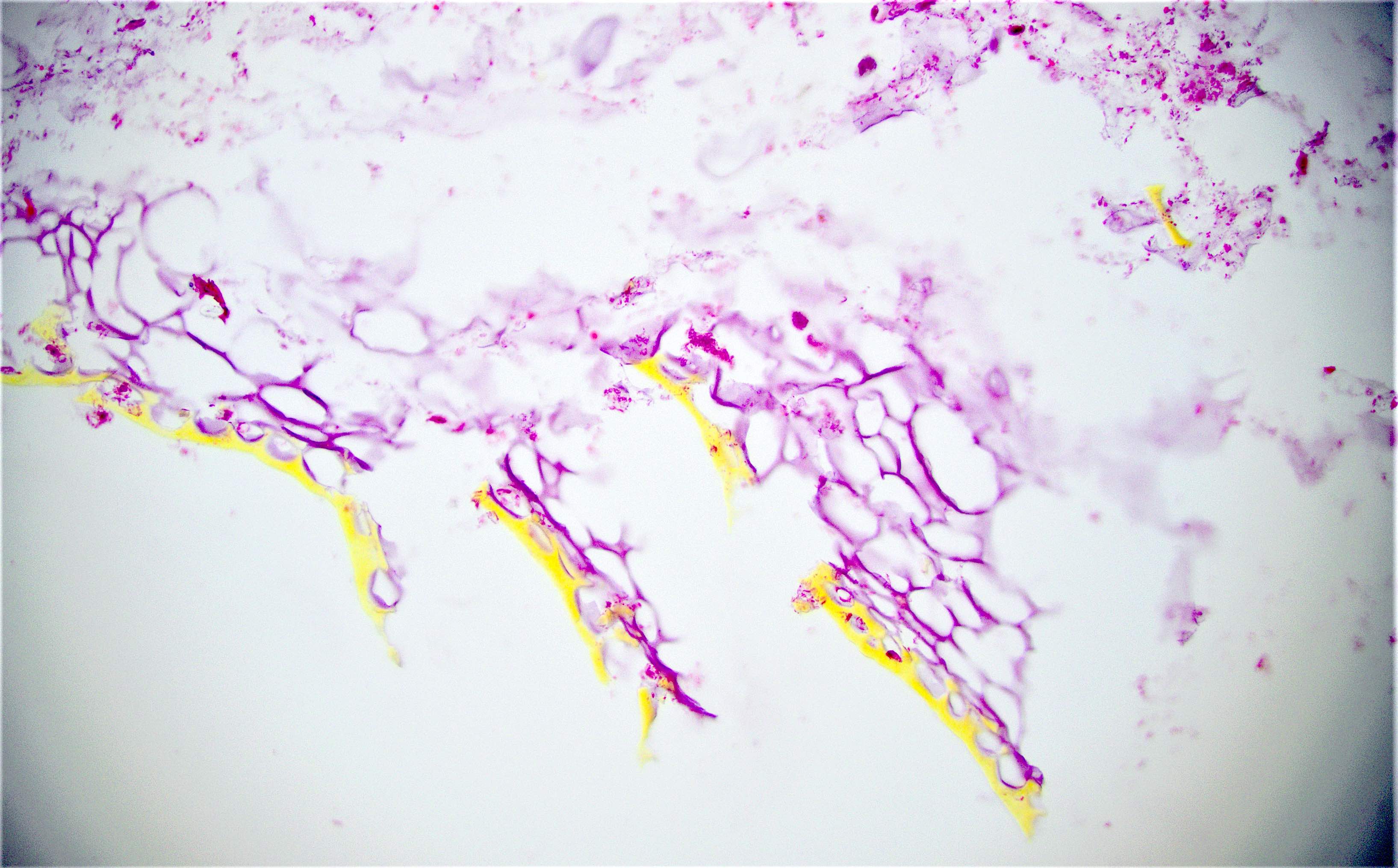
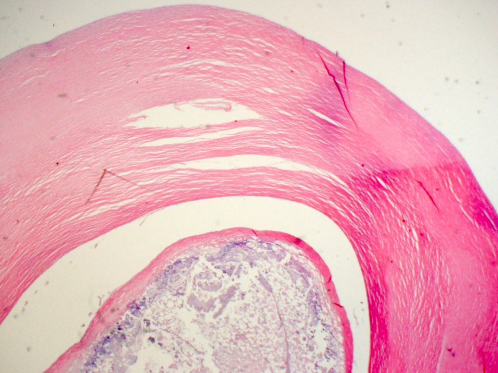
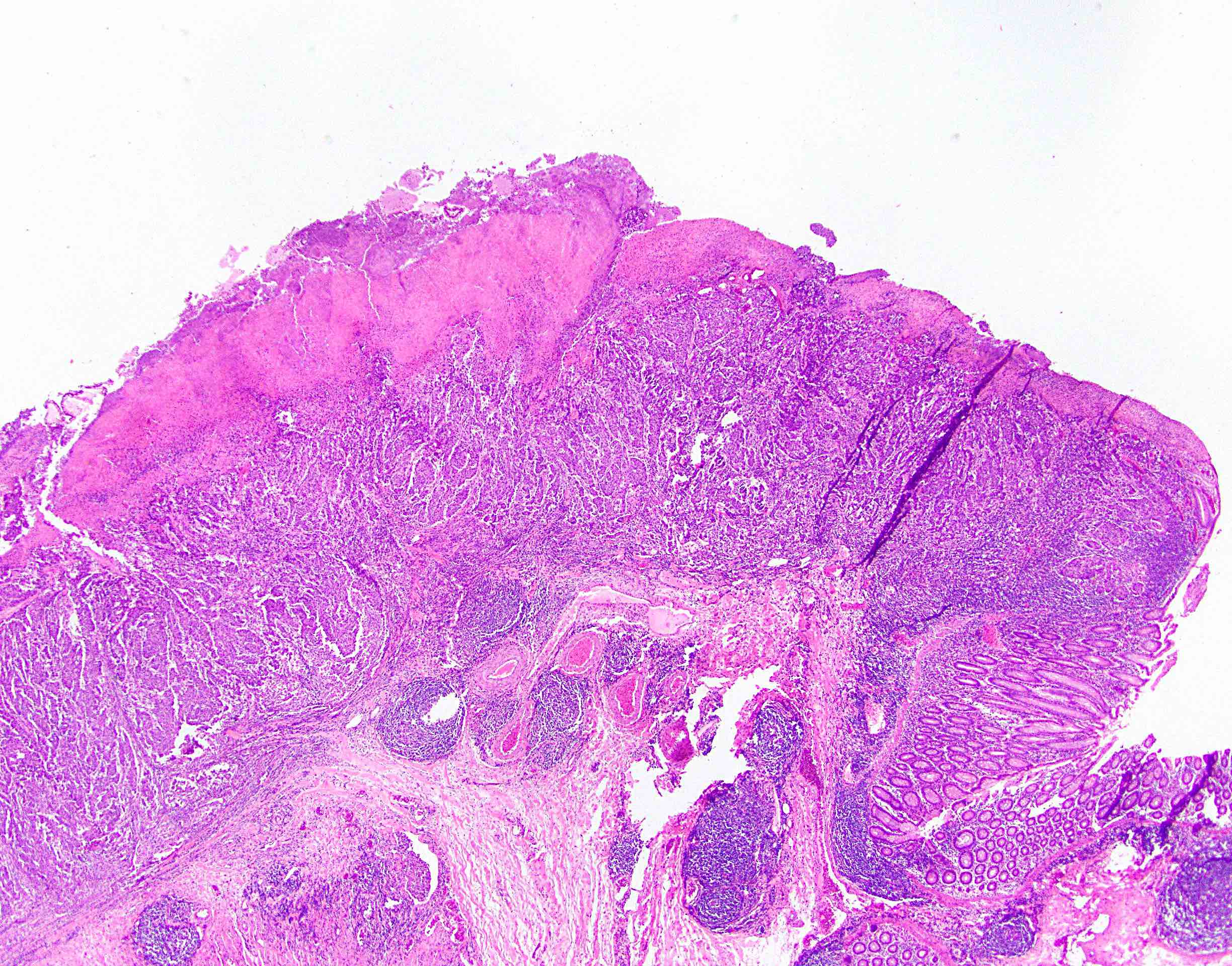
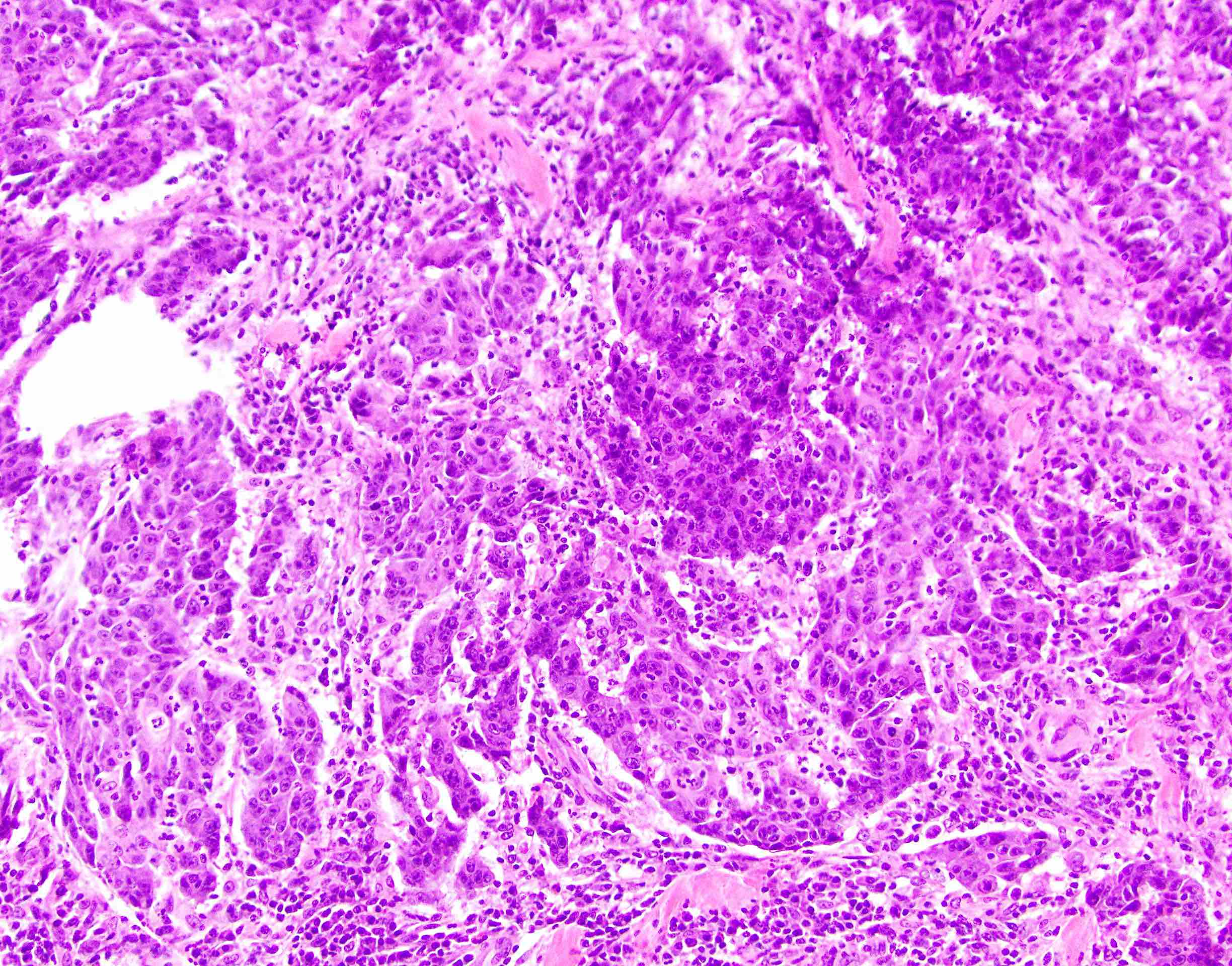
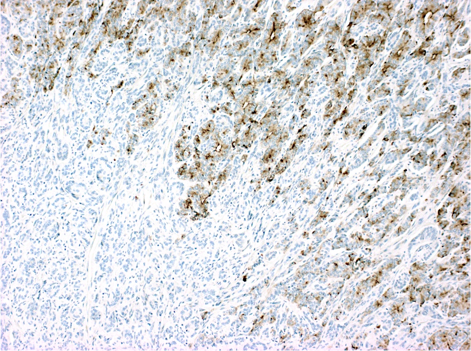
Images hosted on other servers:


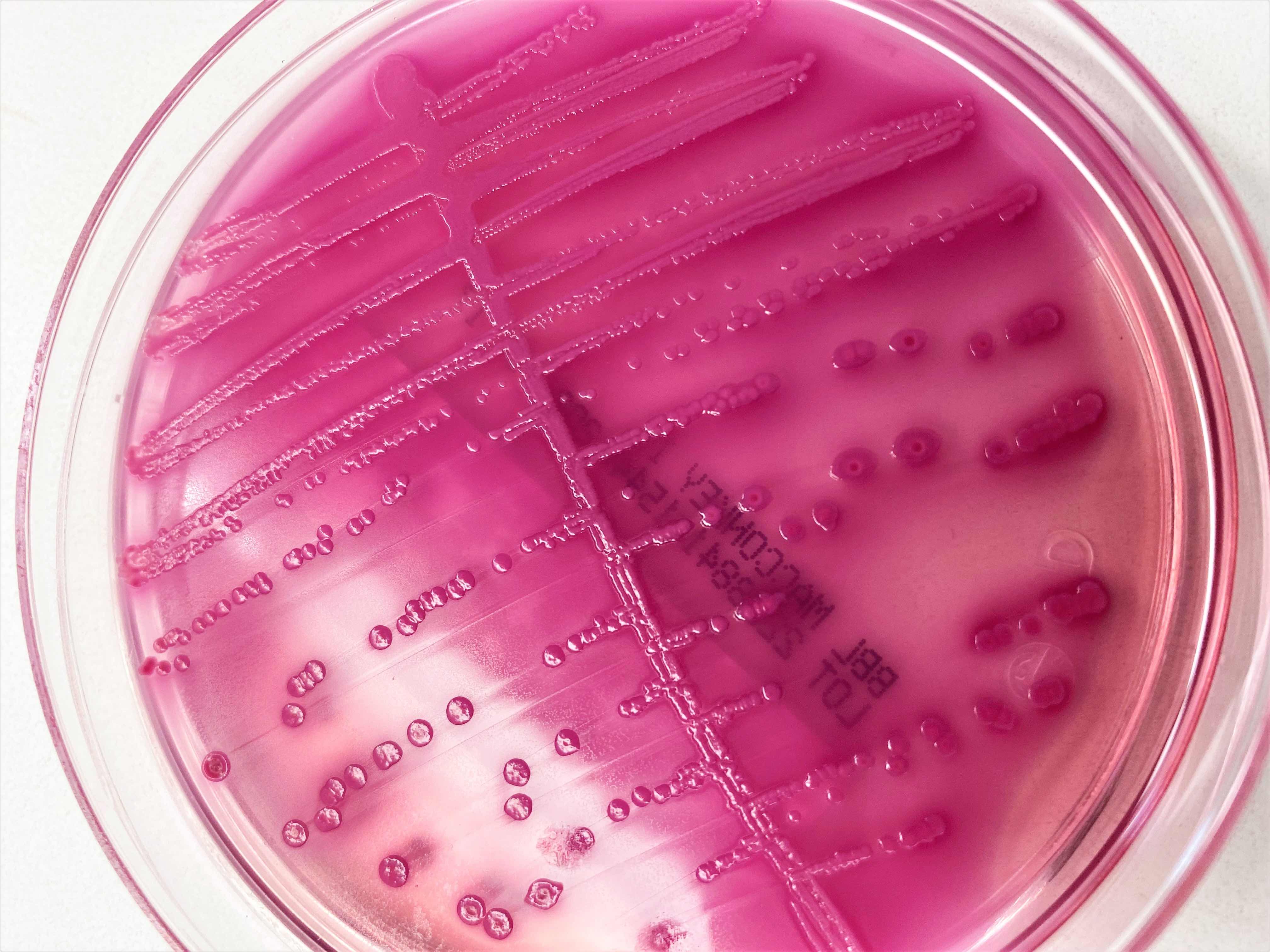
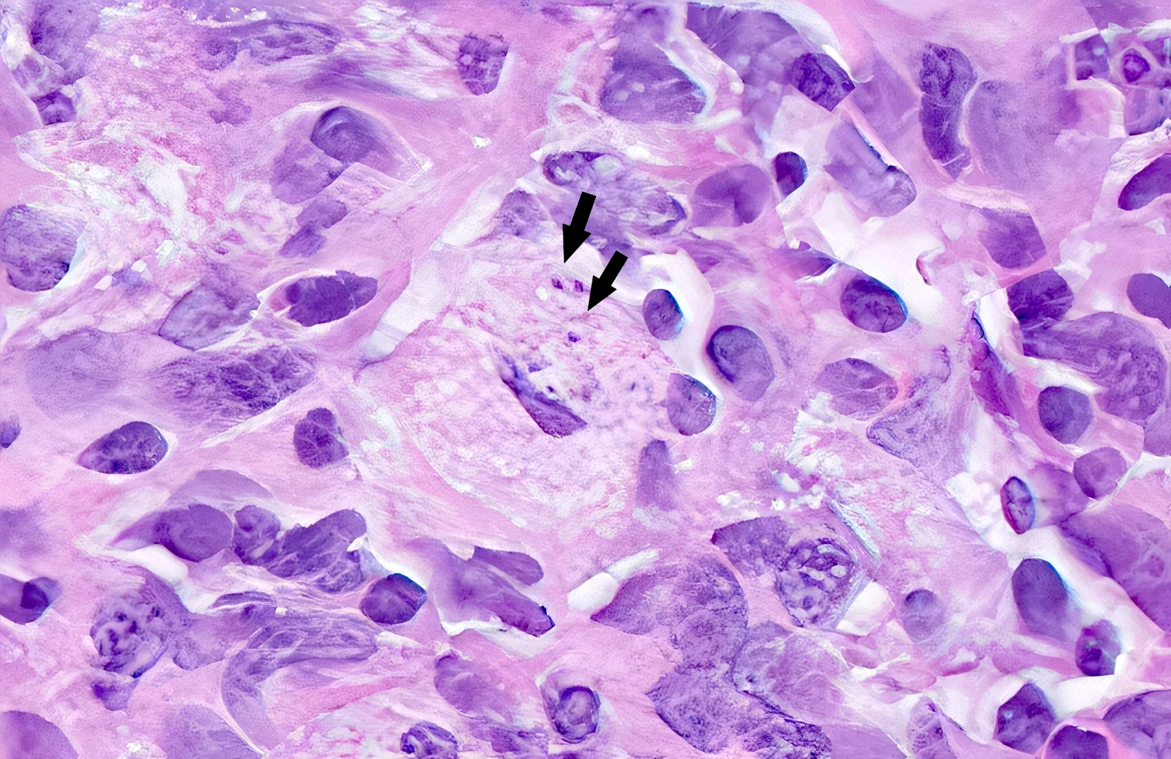
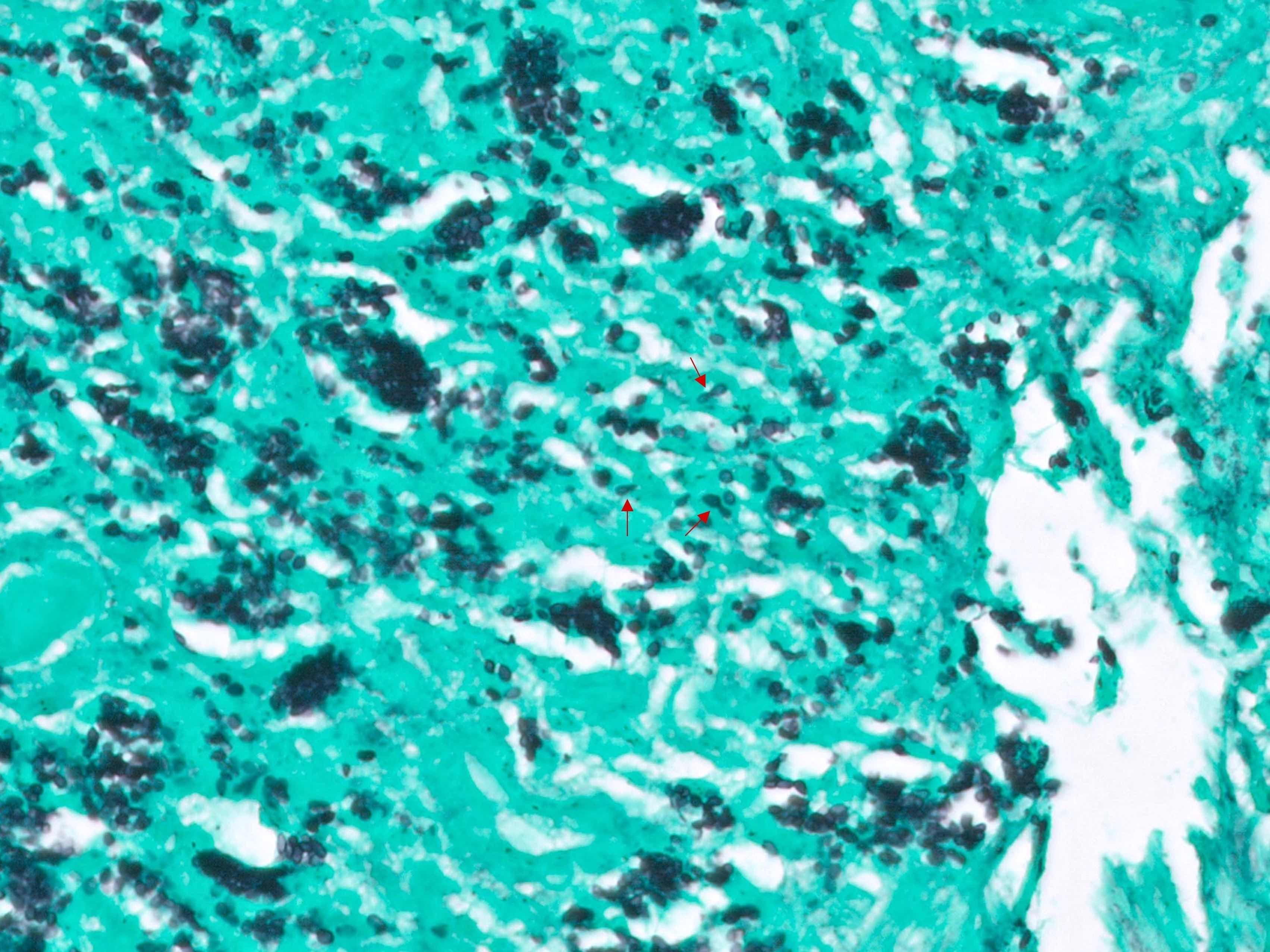
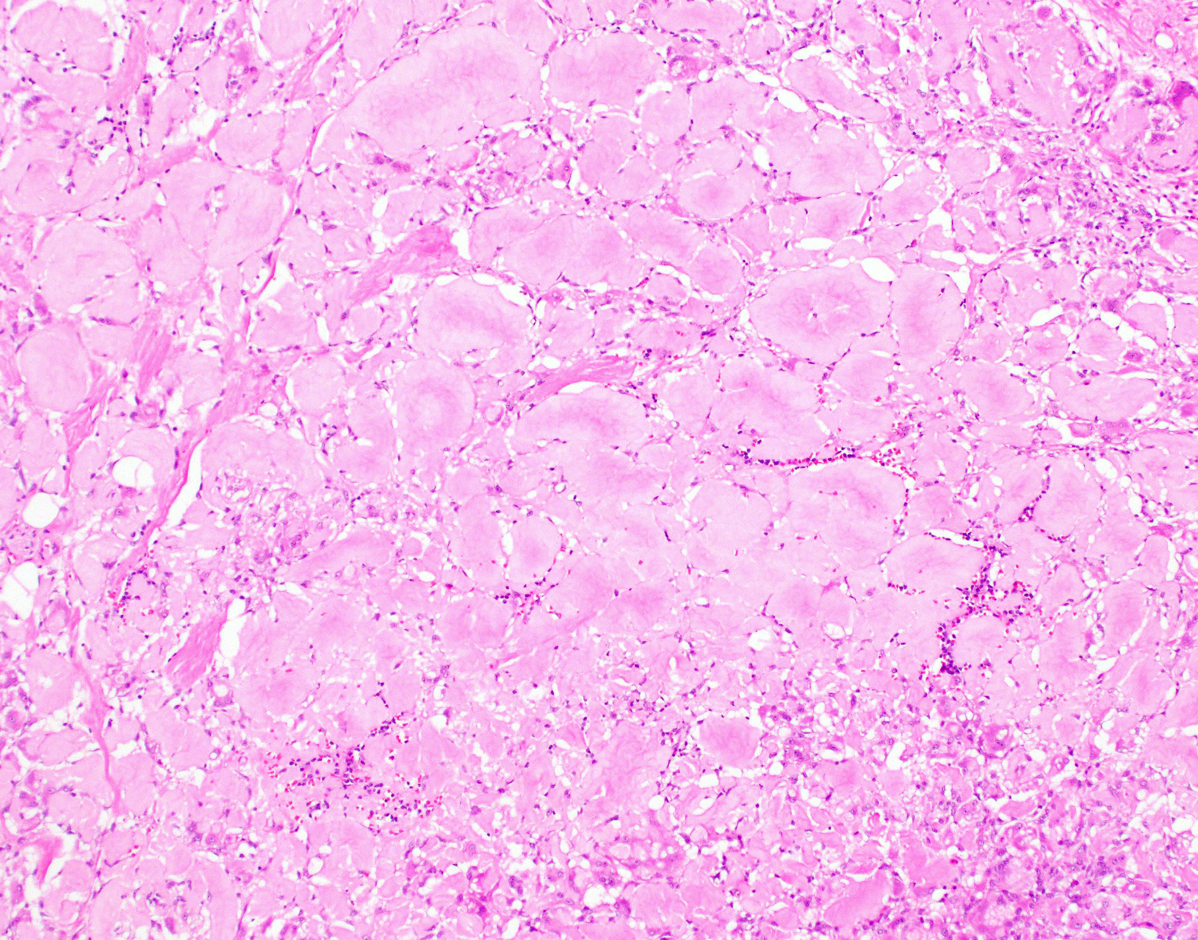
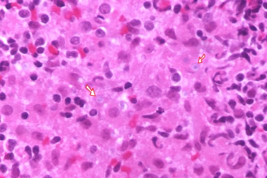
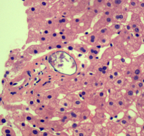
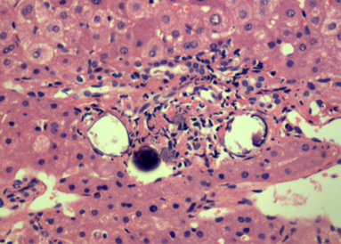
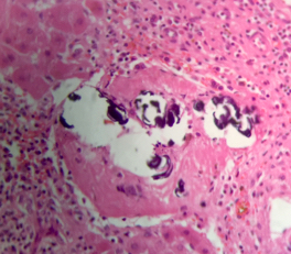
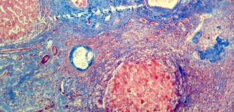
Bacteria and sulfur granules


Colonies of actinomyces

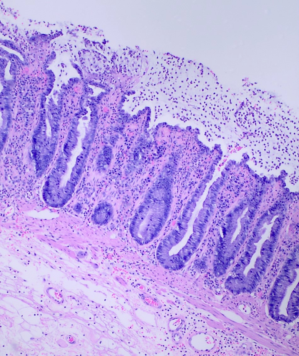
Gram+ staining

Filamentous microorganisms (GMS)
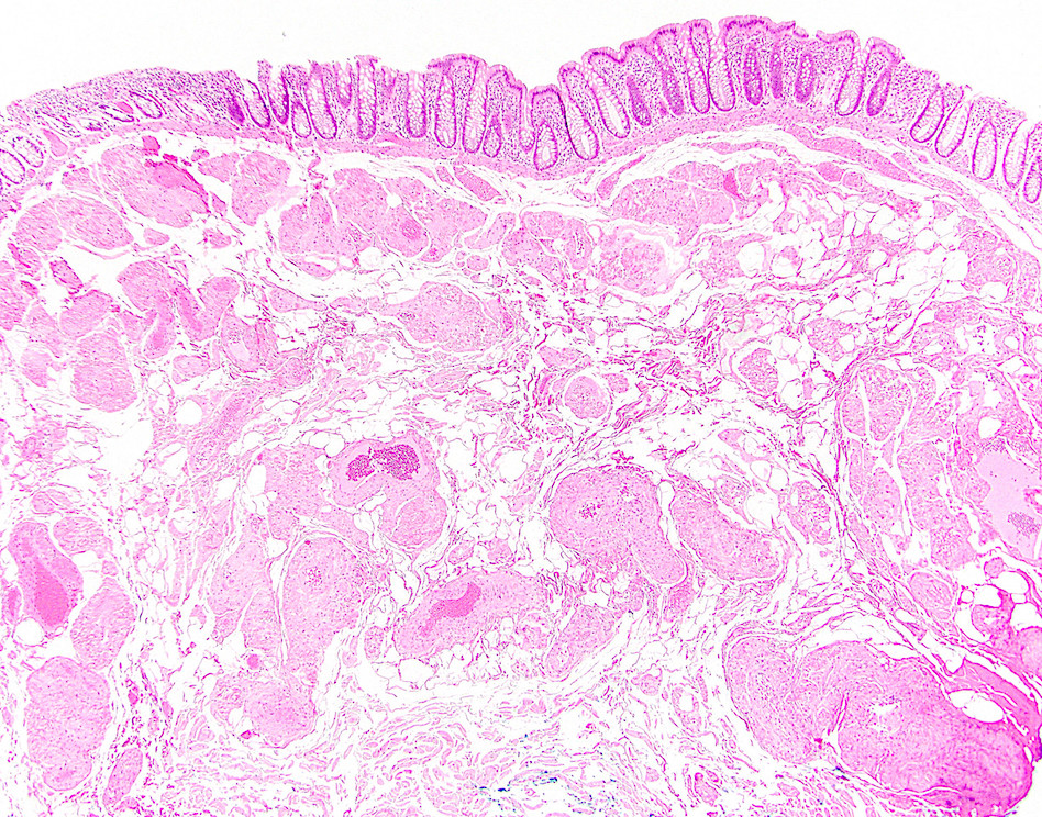
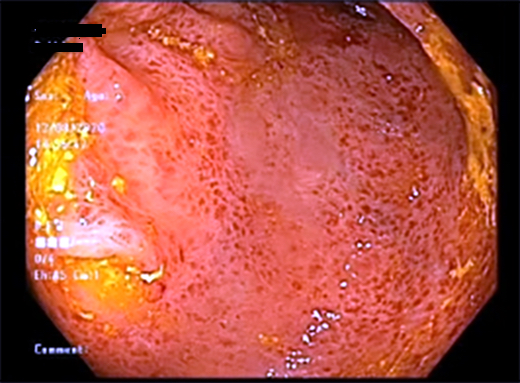
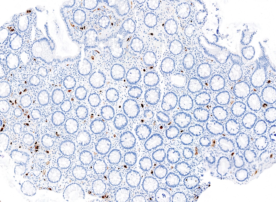
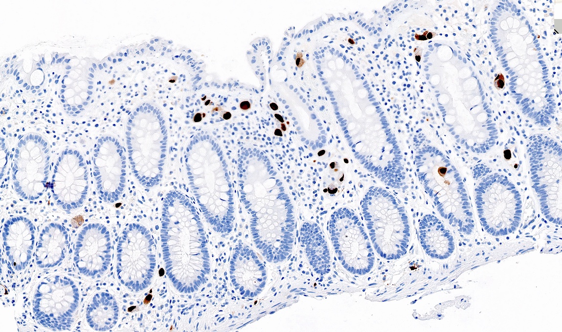
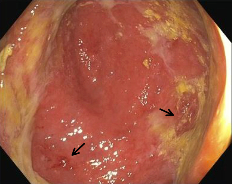
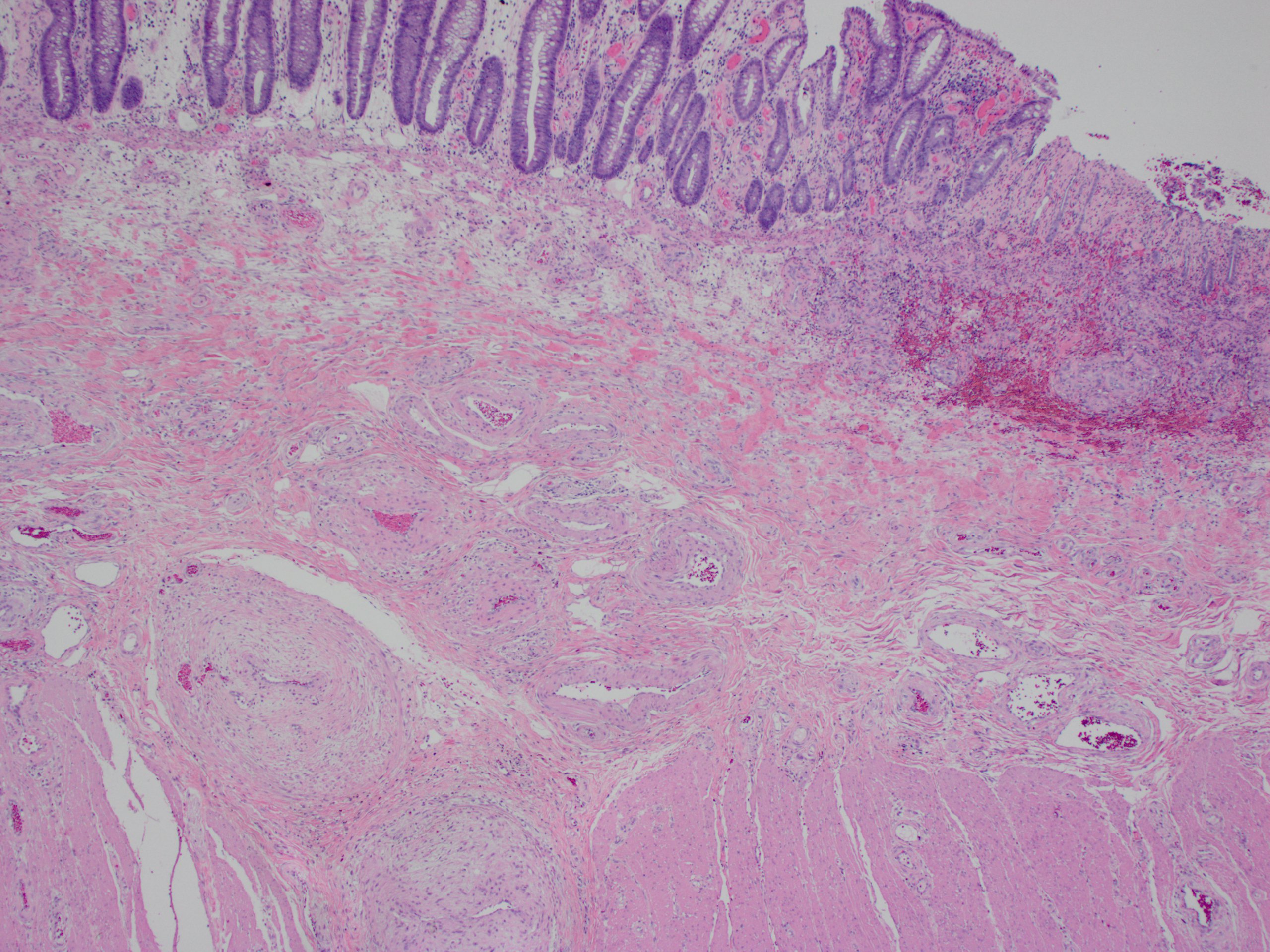
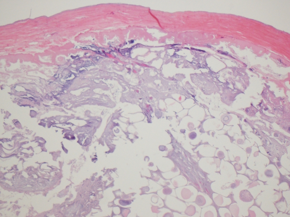
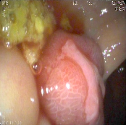
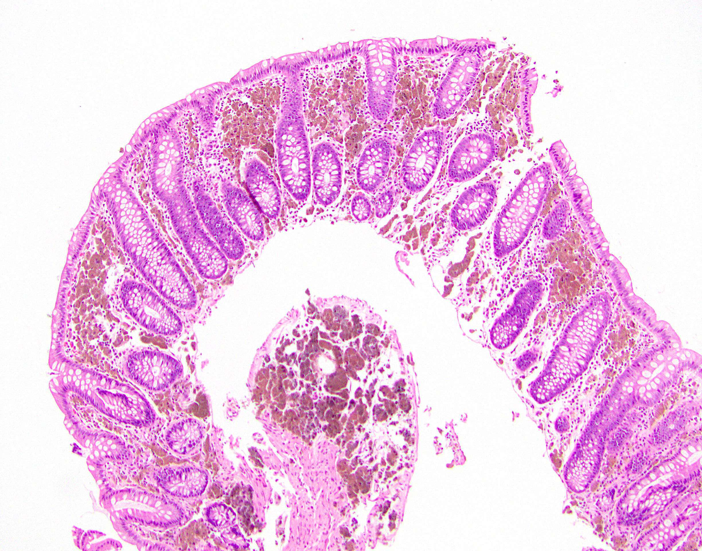
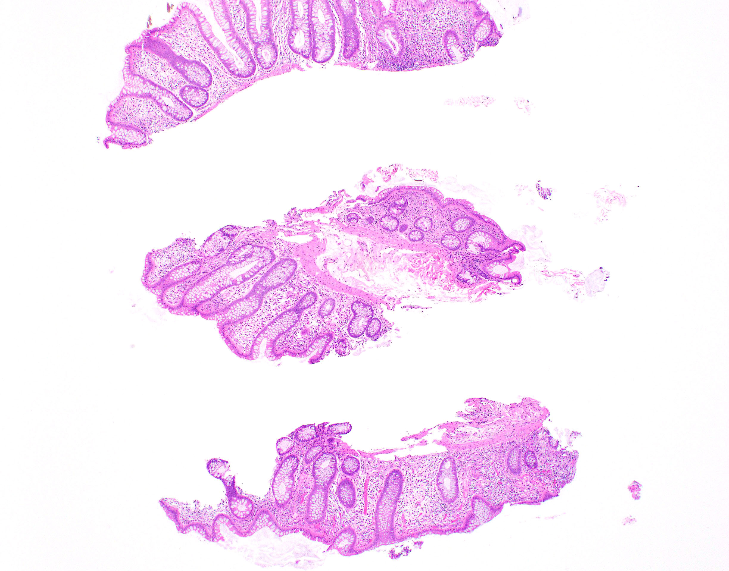
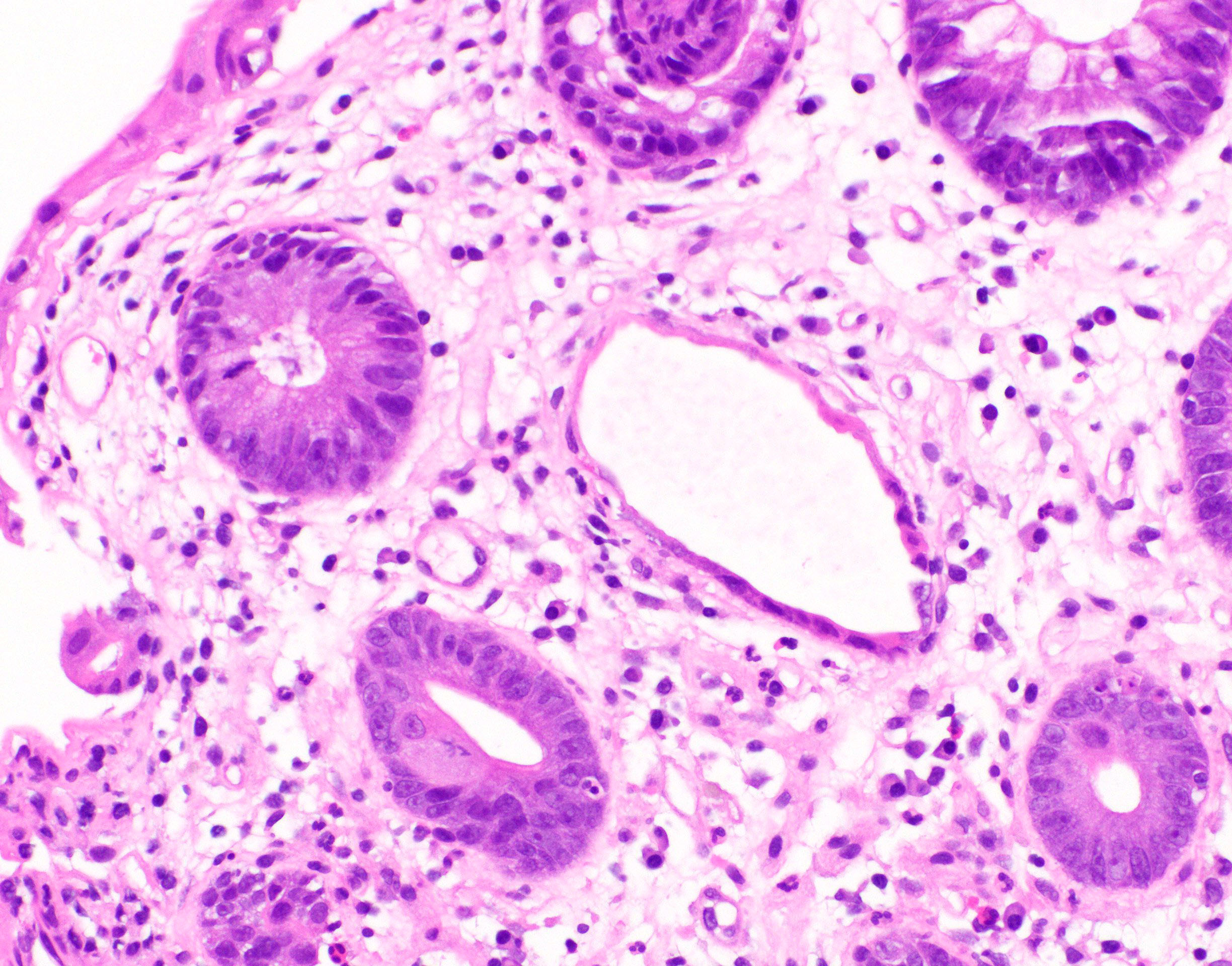
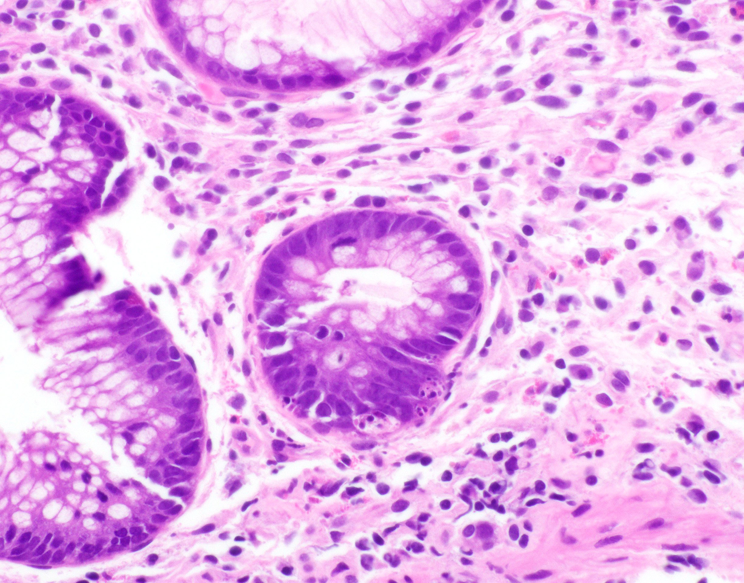
Images hosted on other servers:

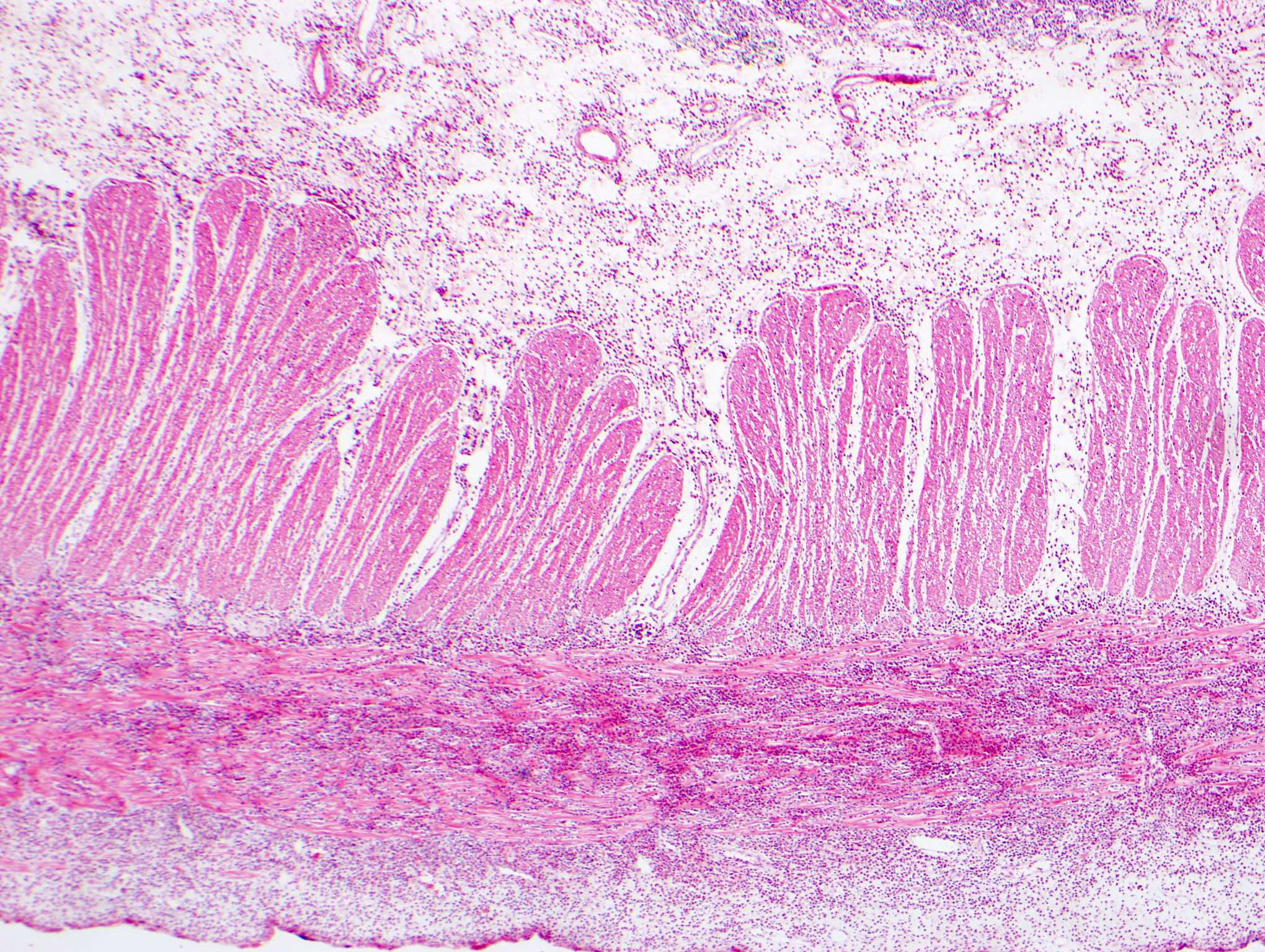
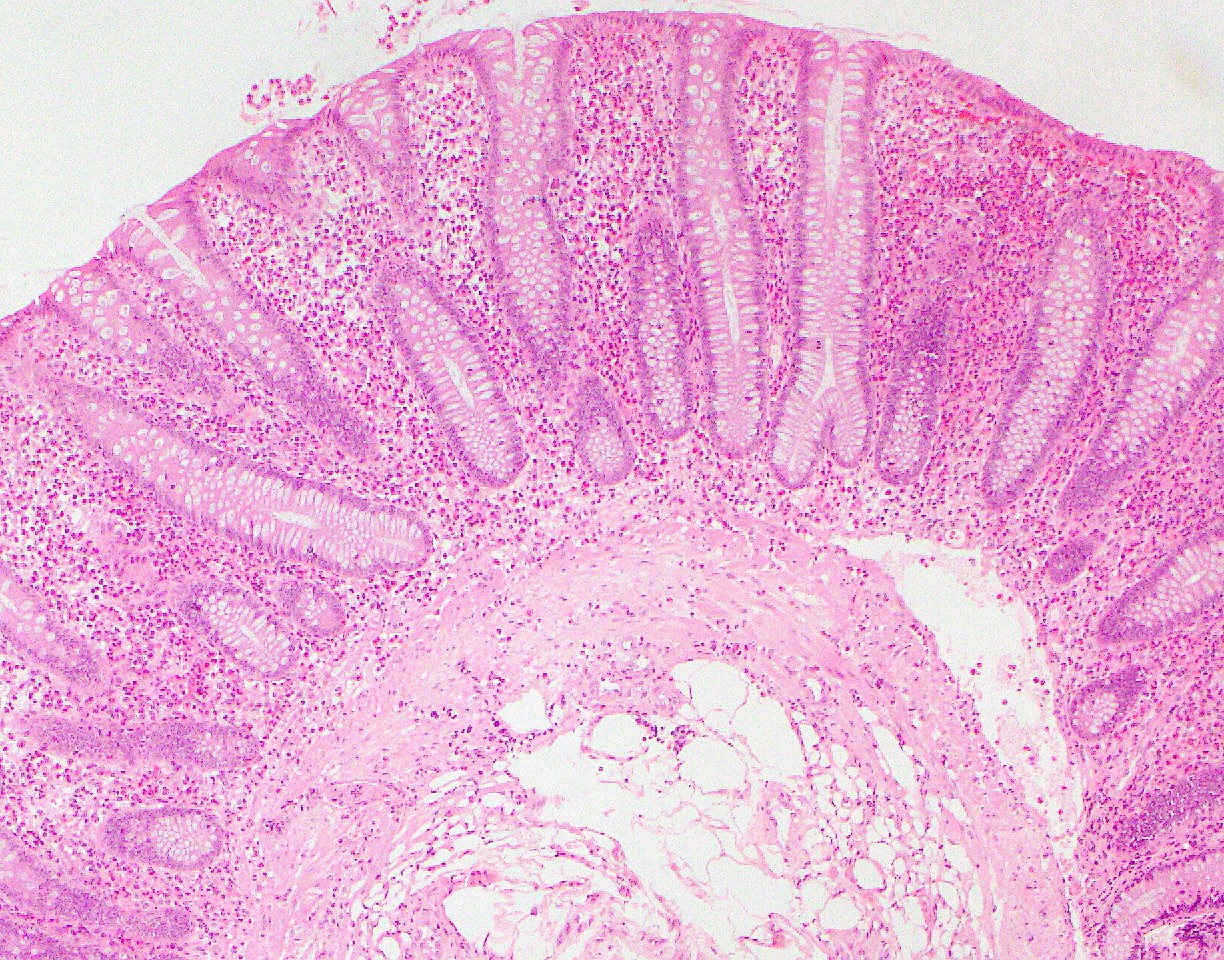
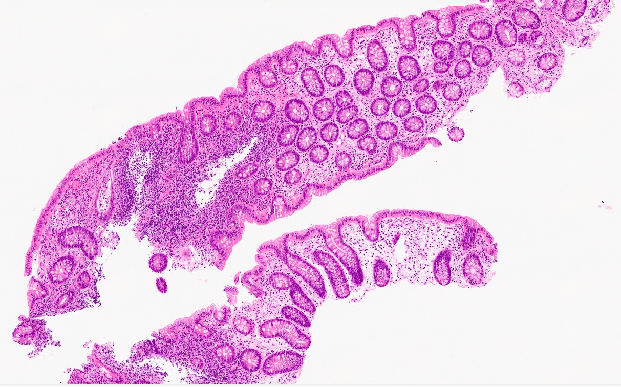
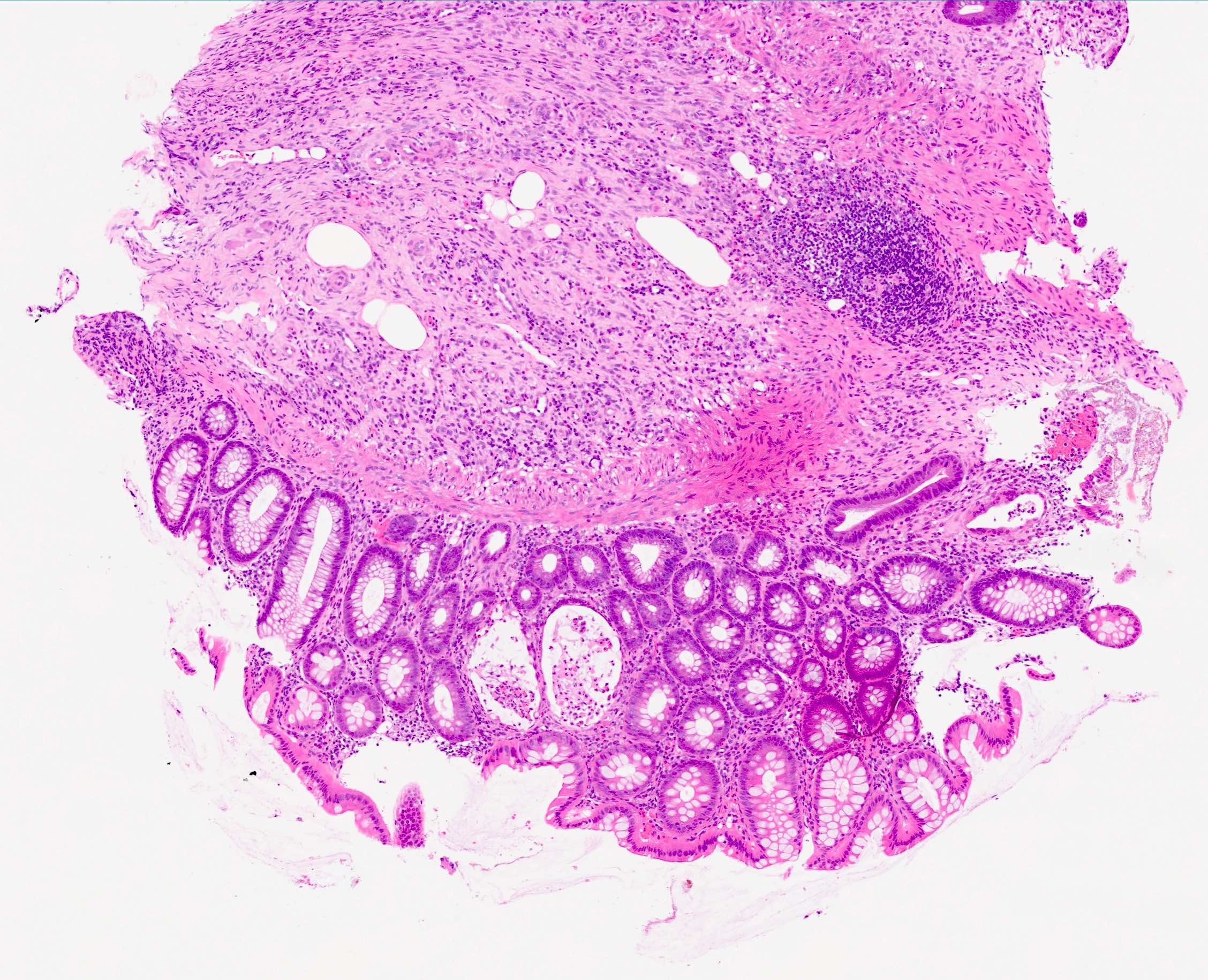
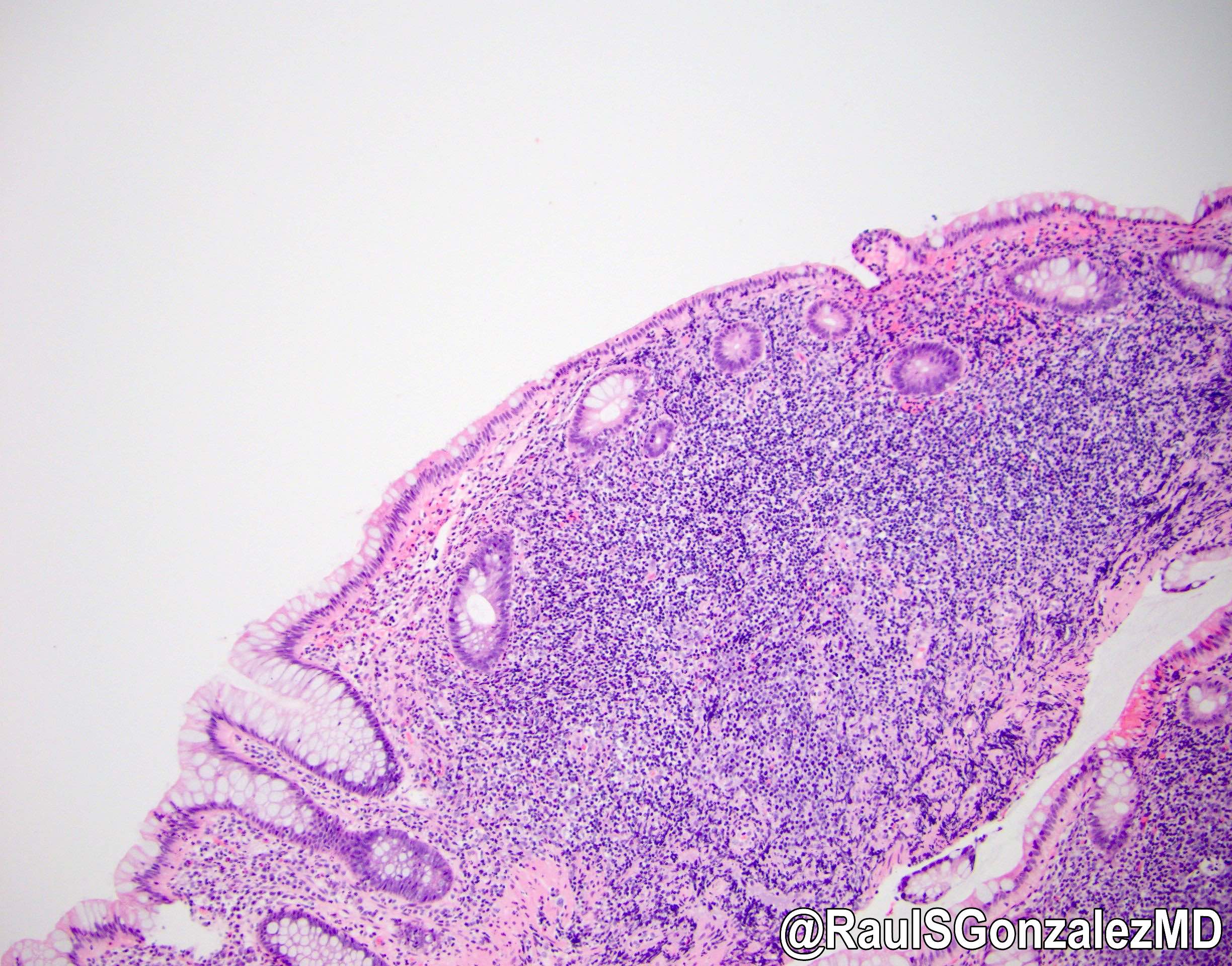
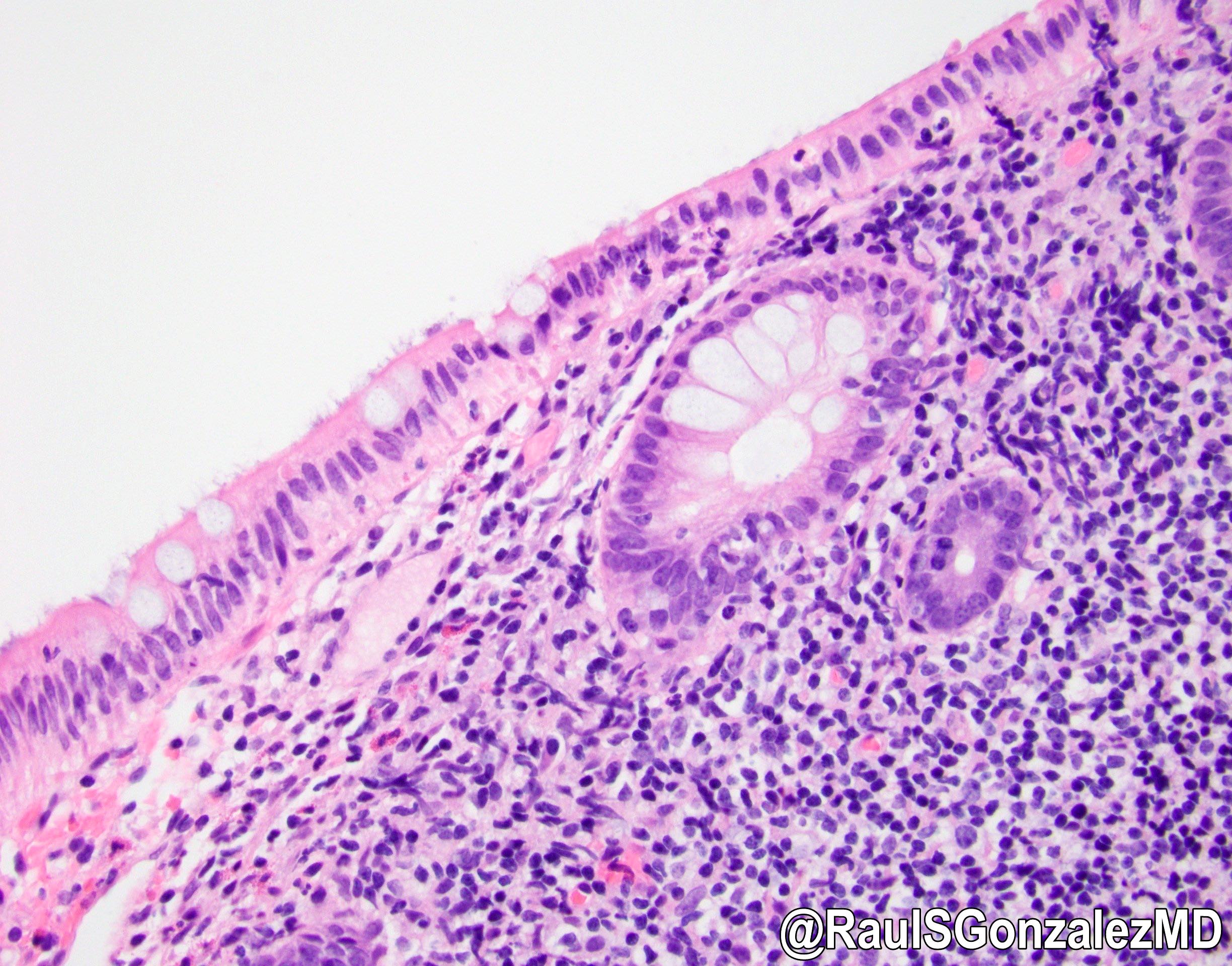
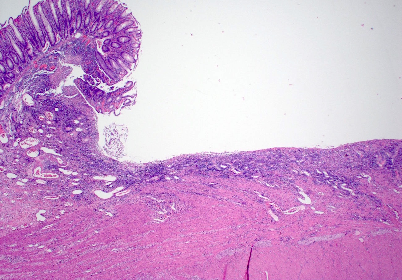
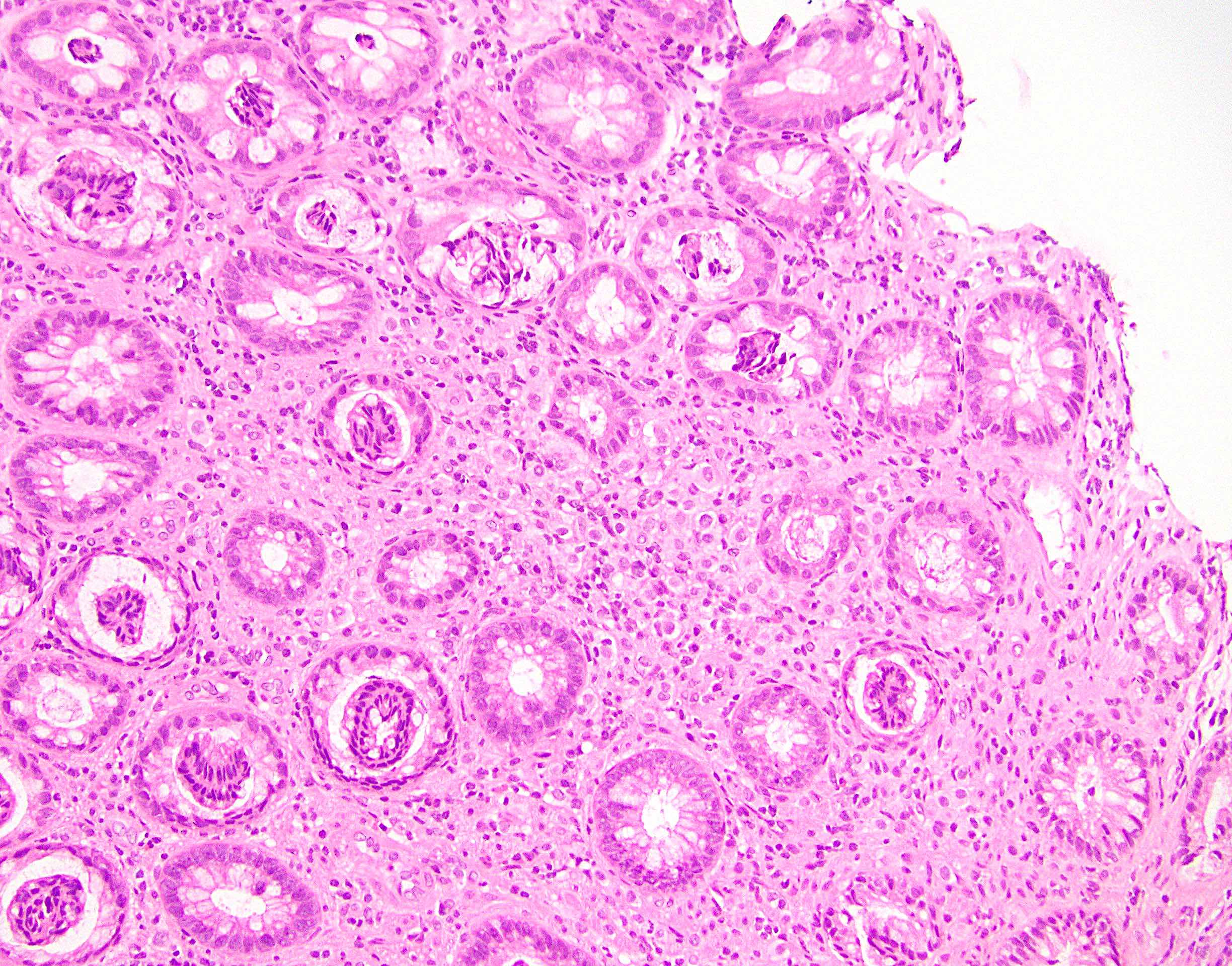
Colonoscopy
Contributed by Lili Lee, M.D.

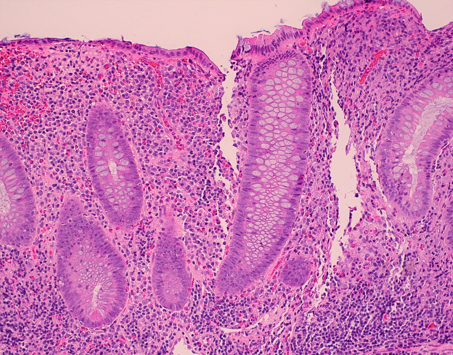
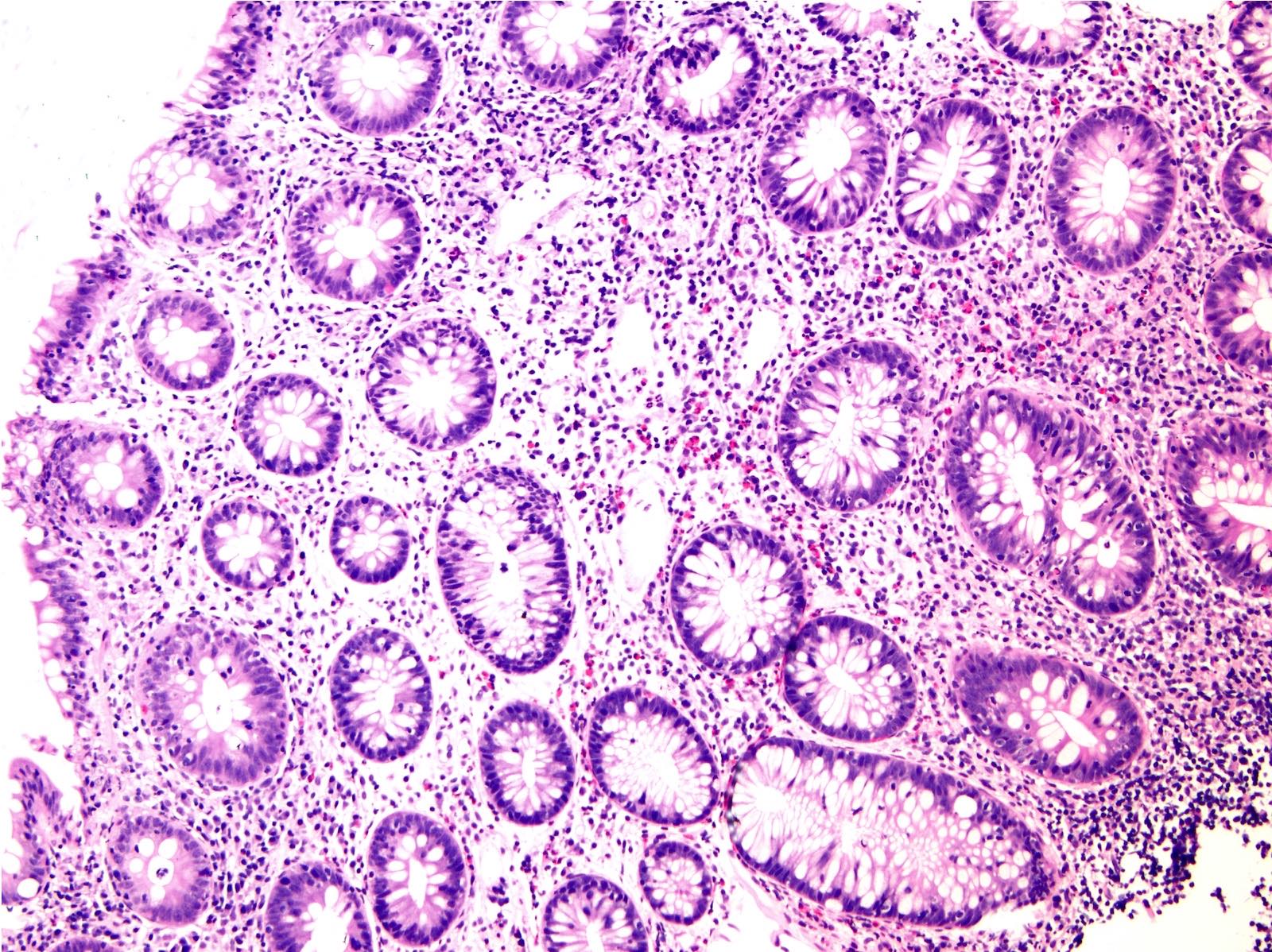
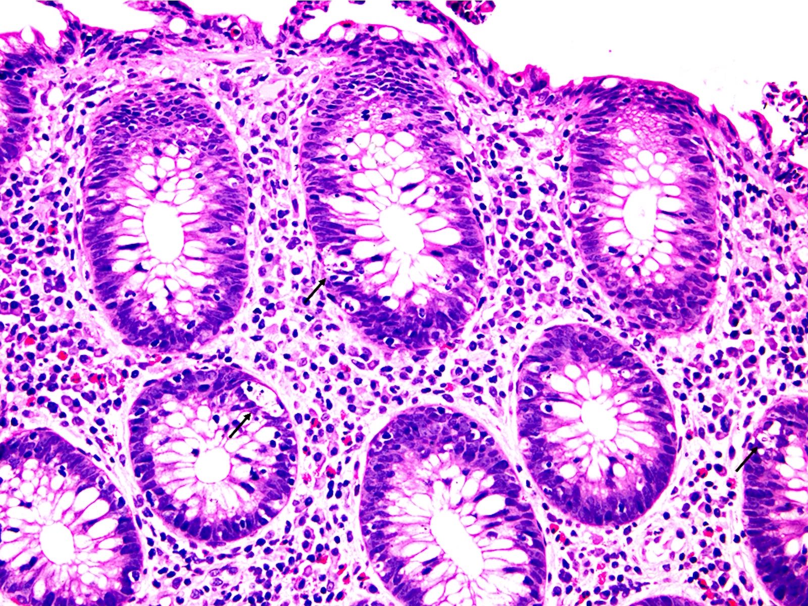
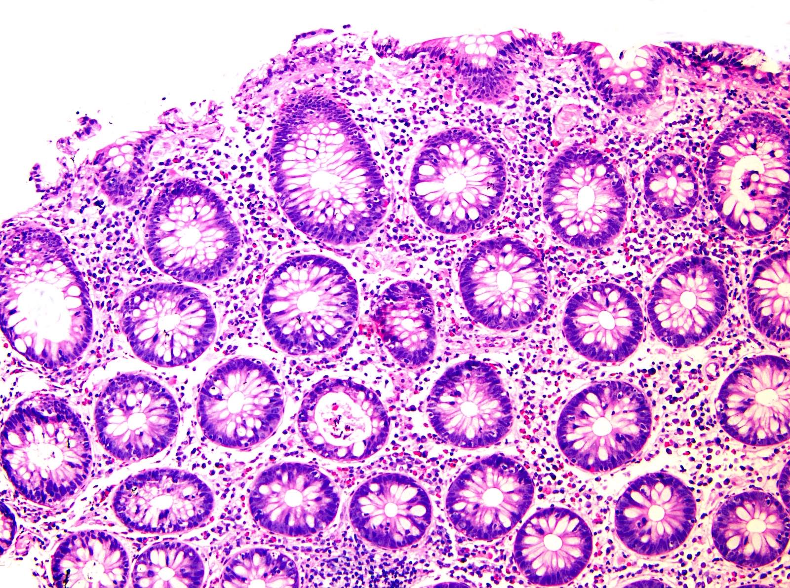
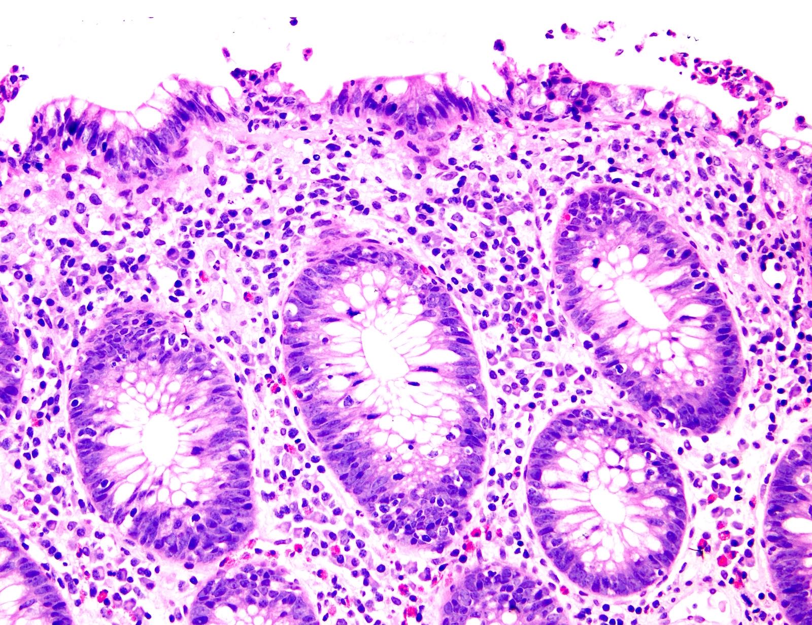
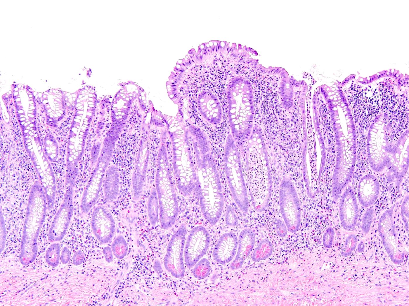
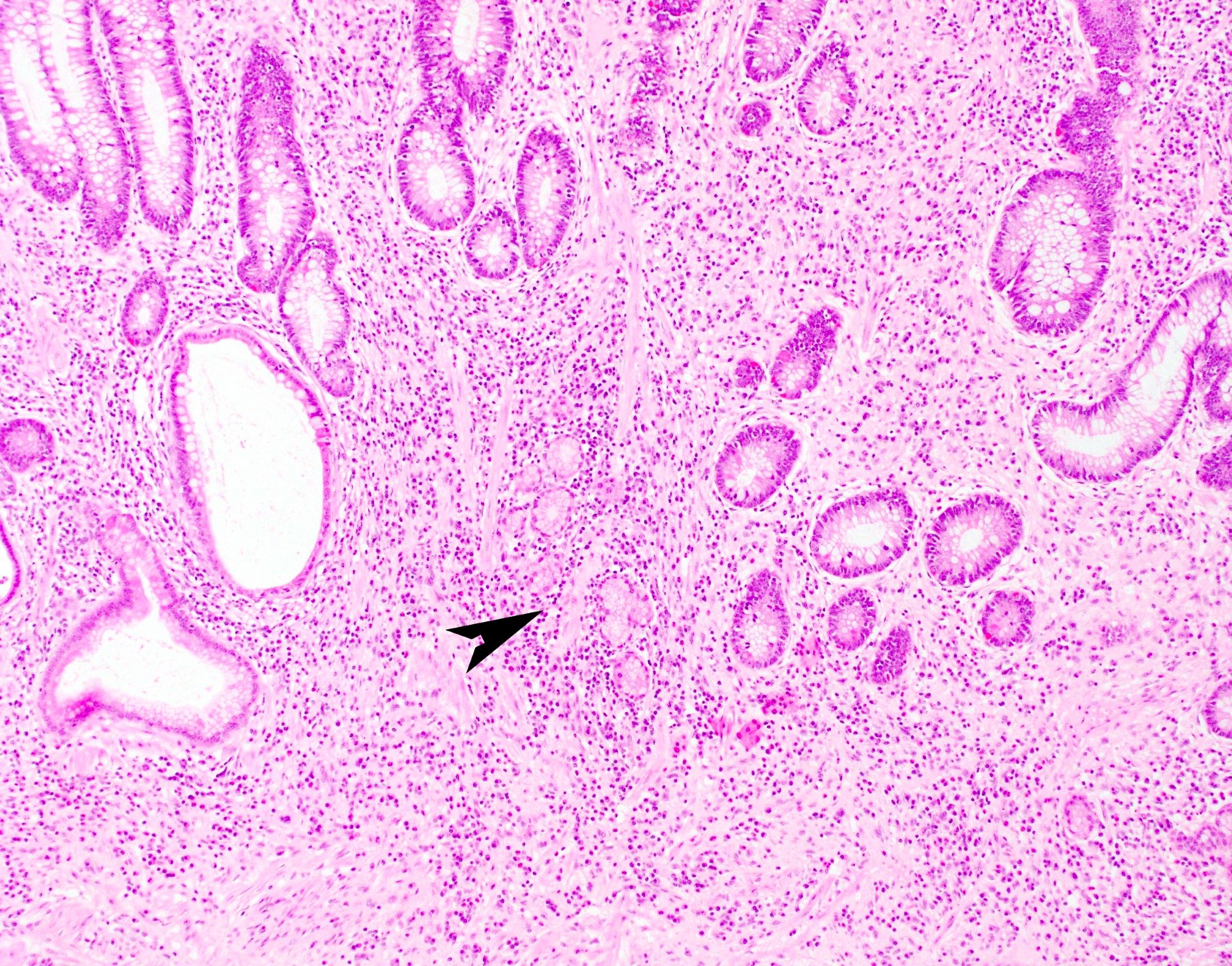
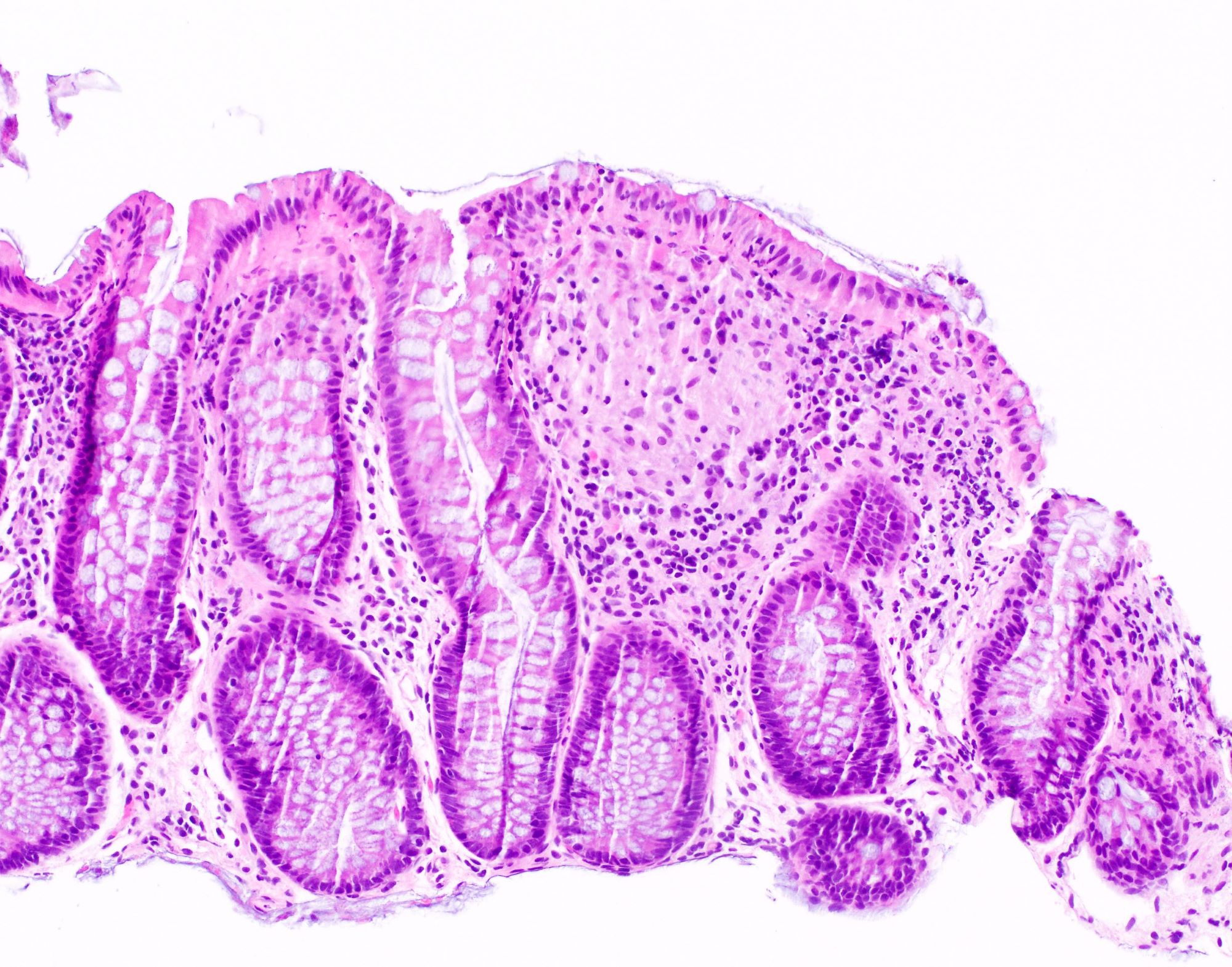
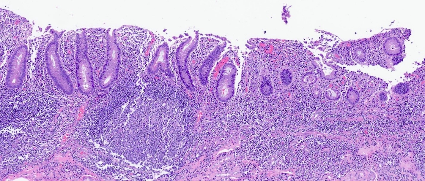
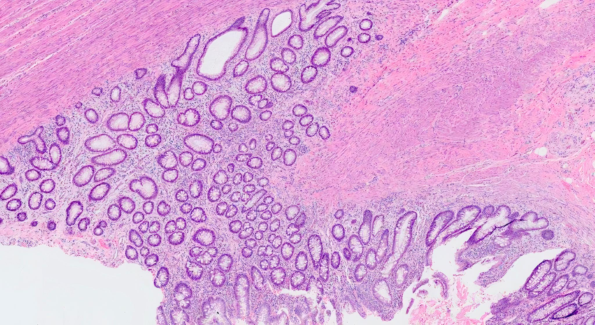
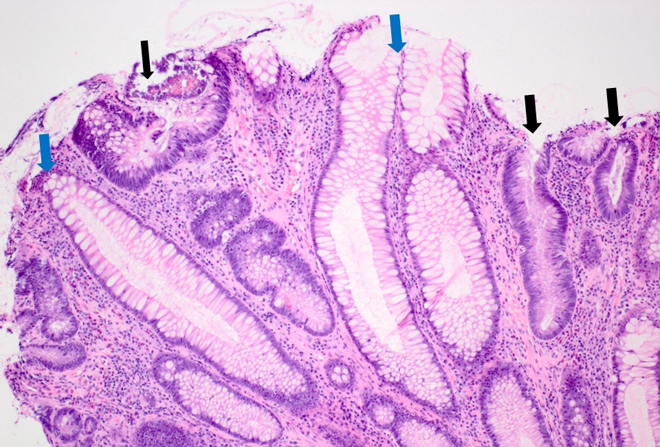
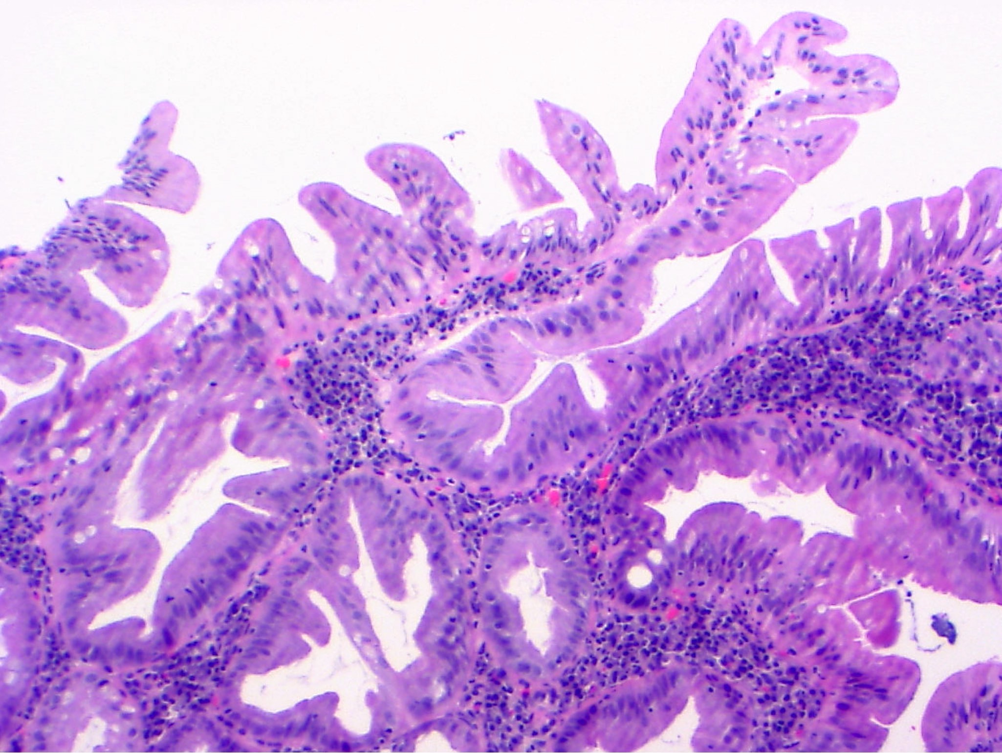
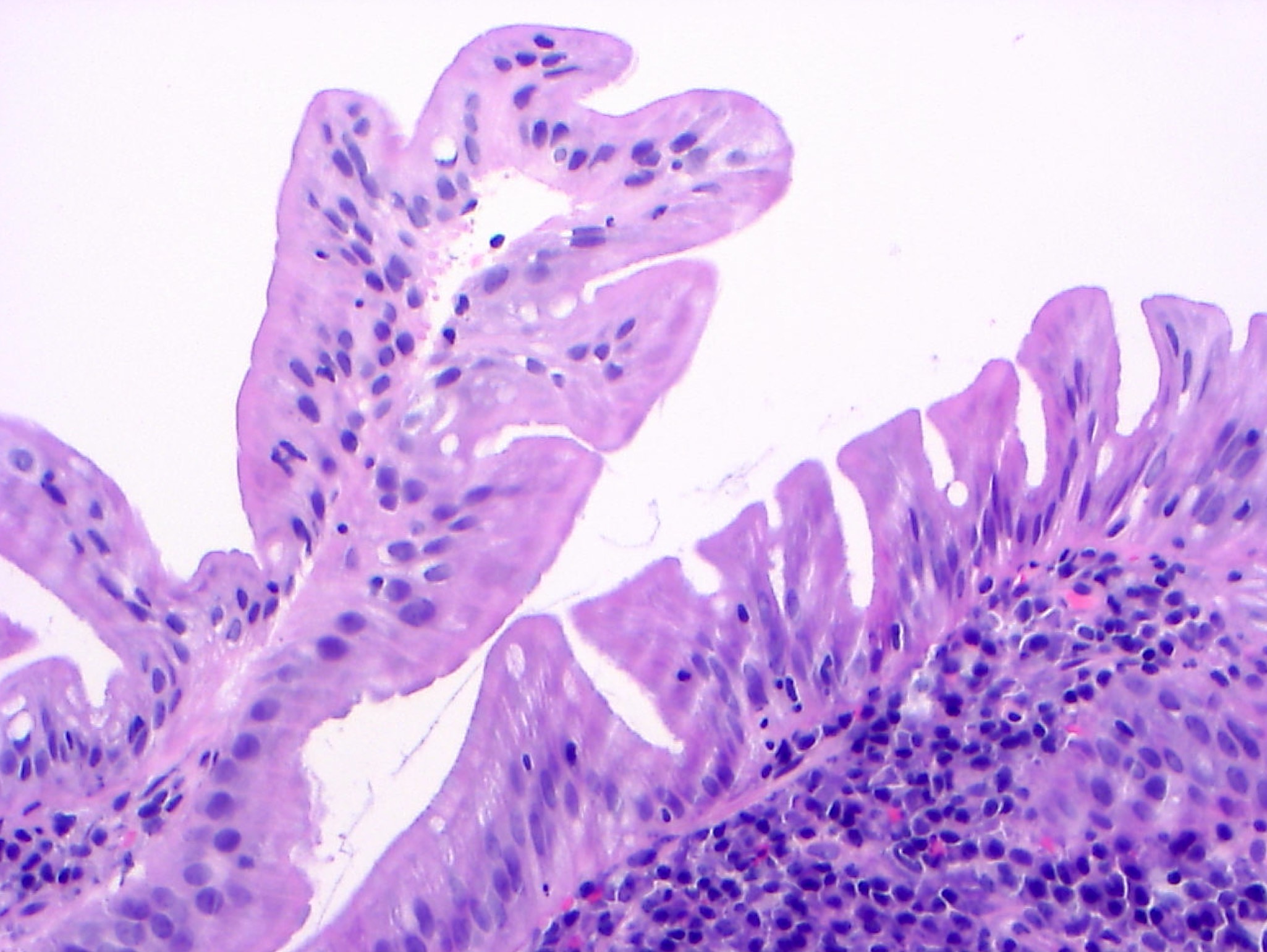
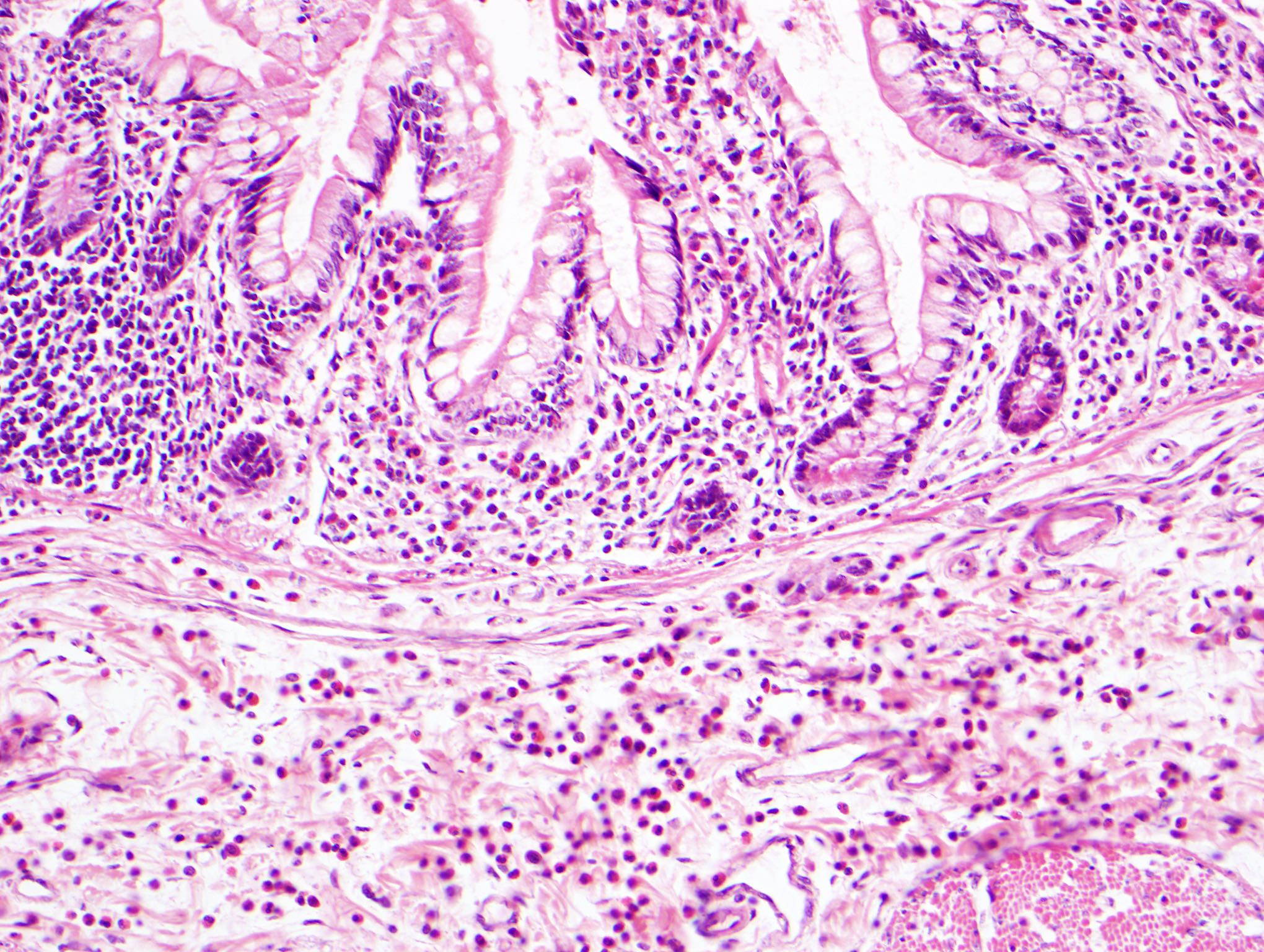
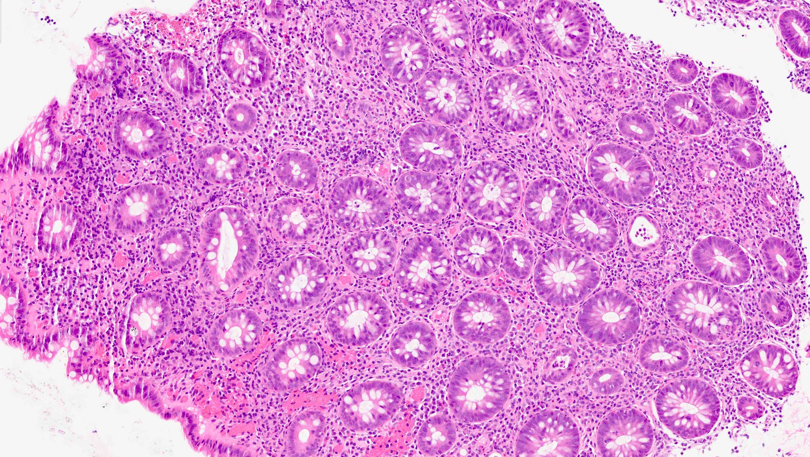
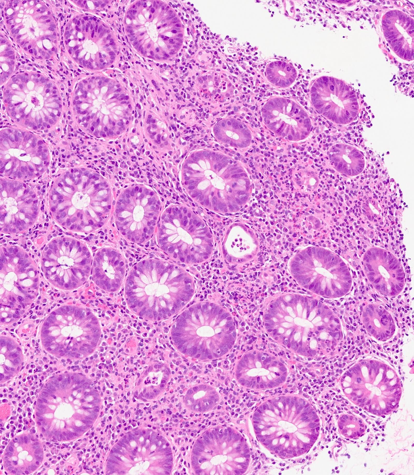
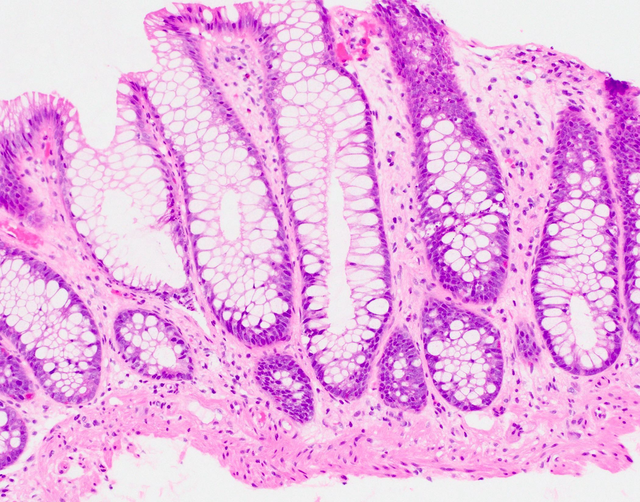
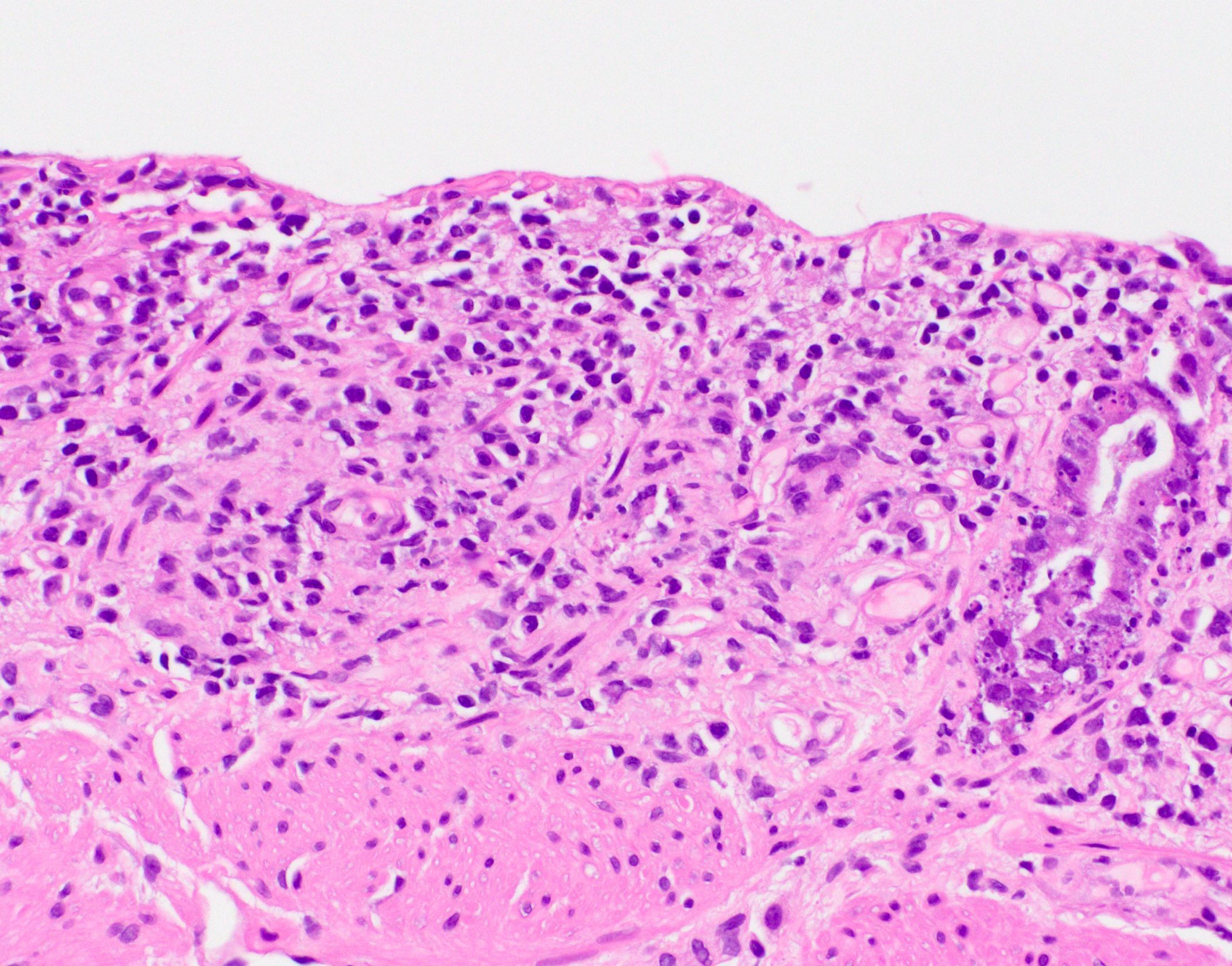
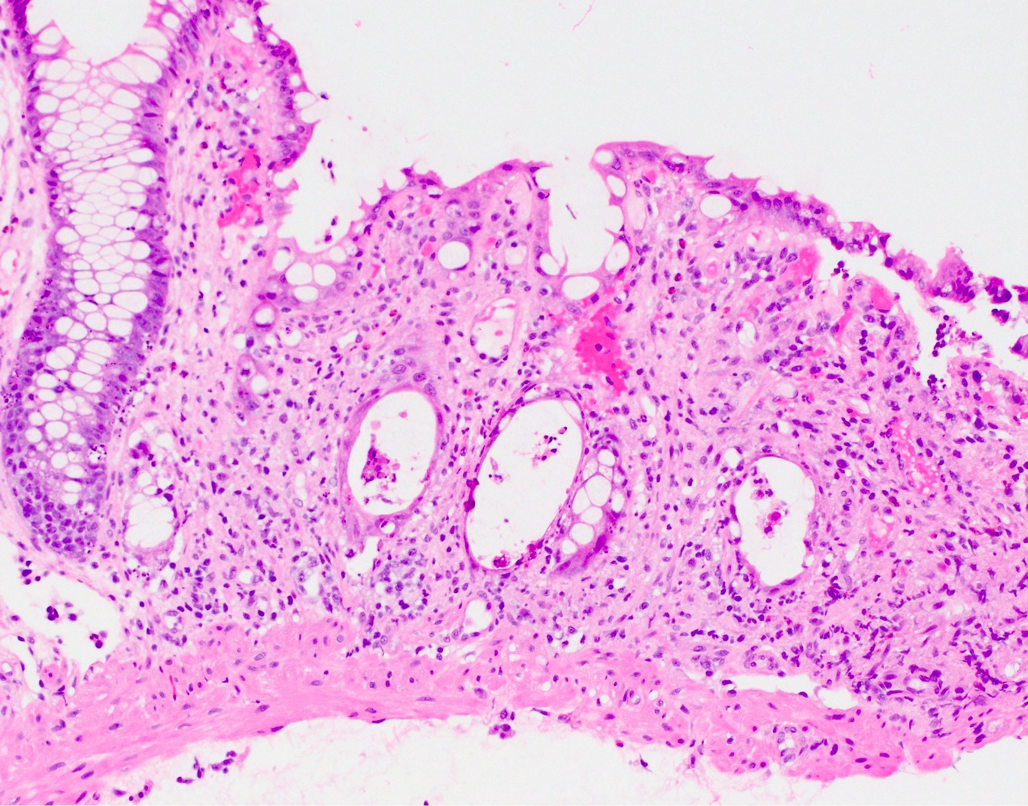
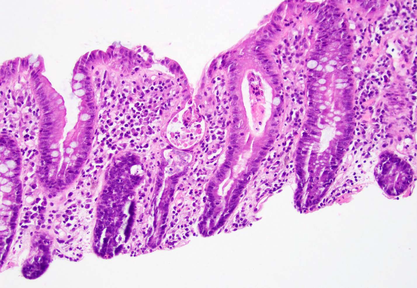
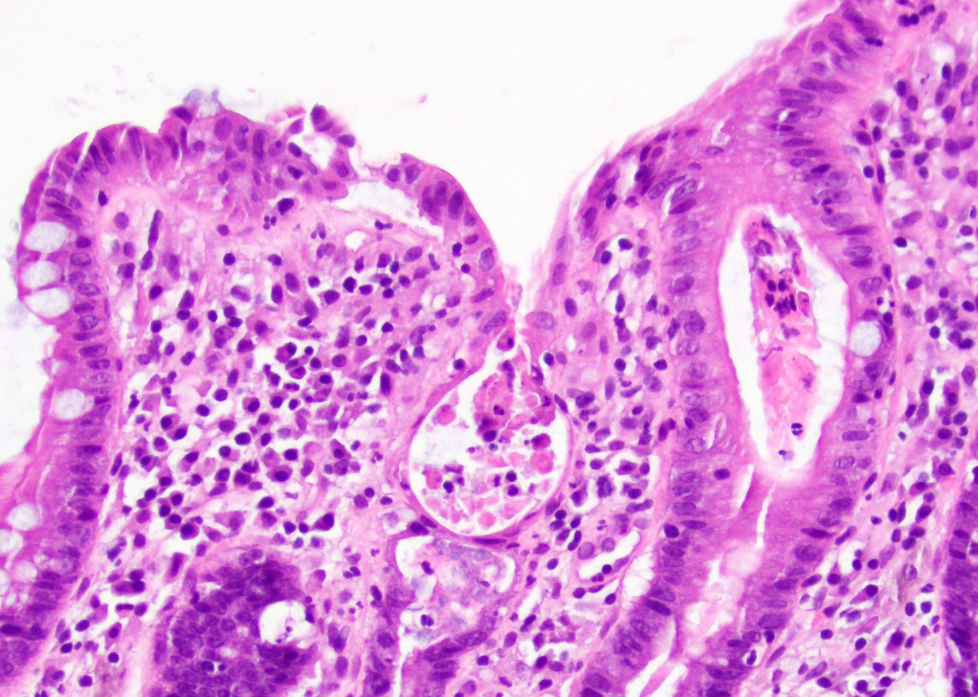
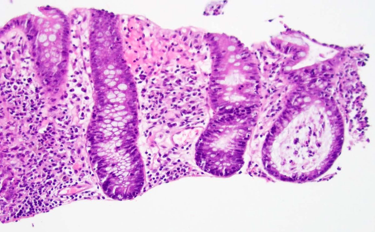
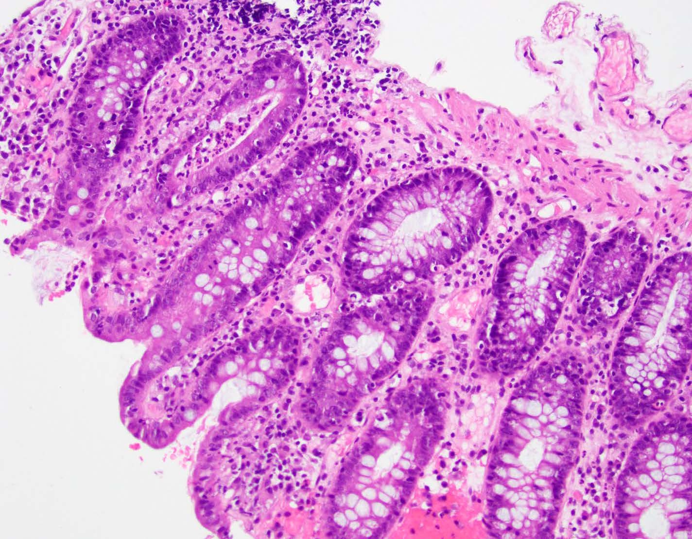
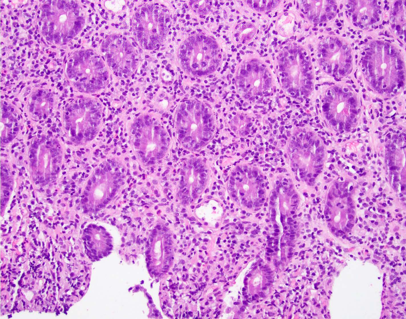
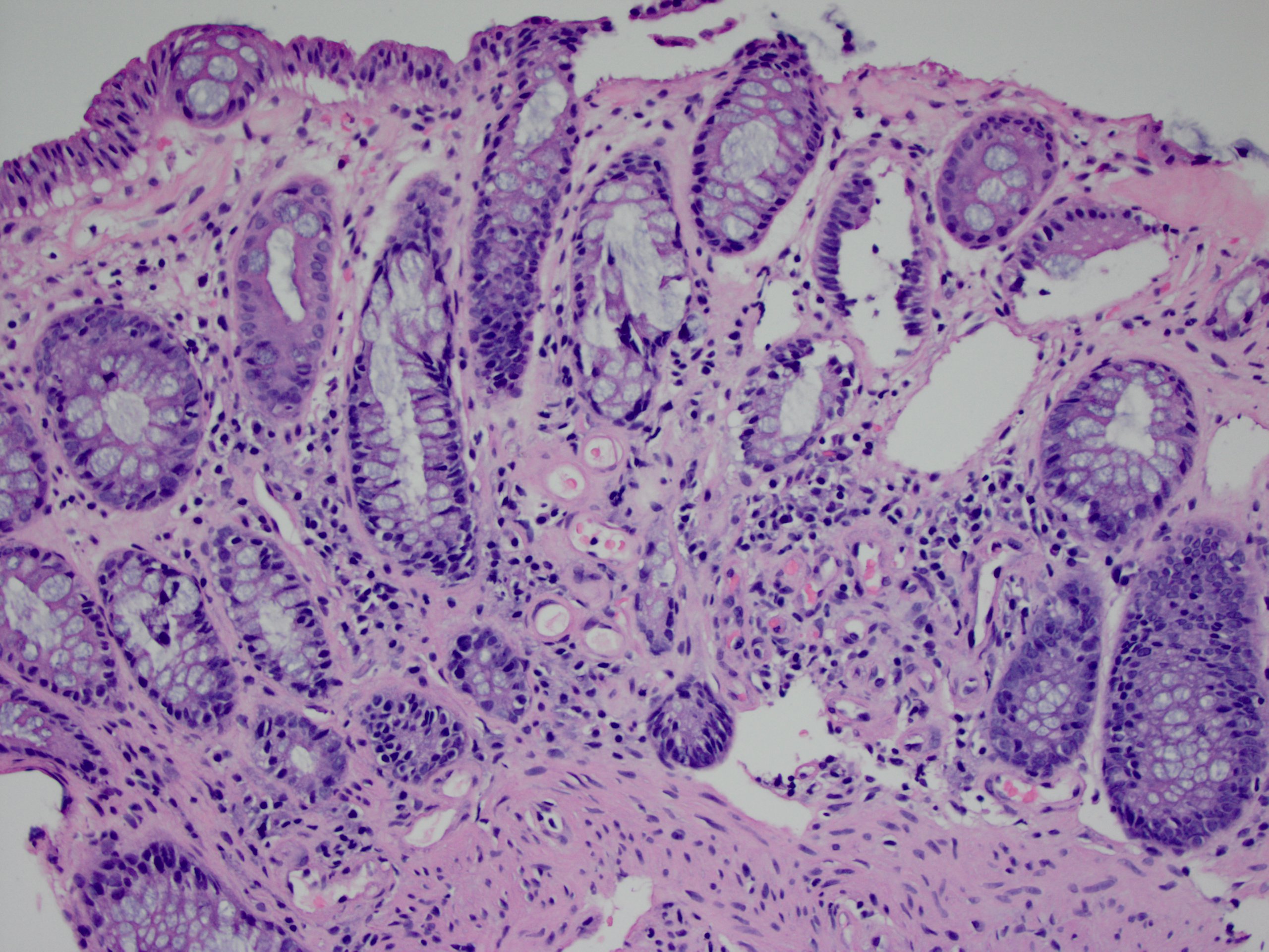
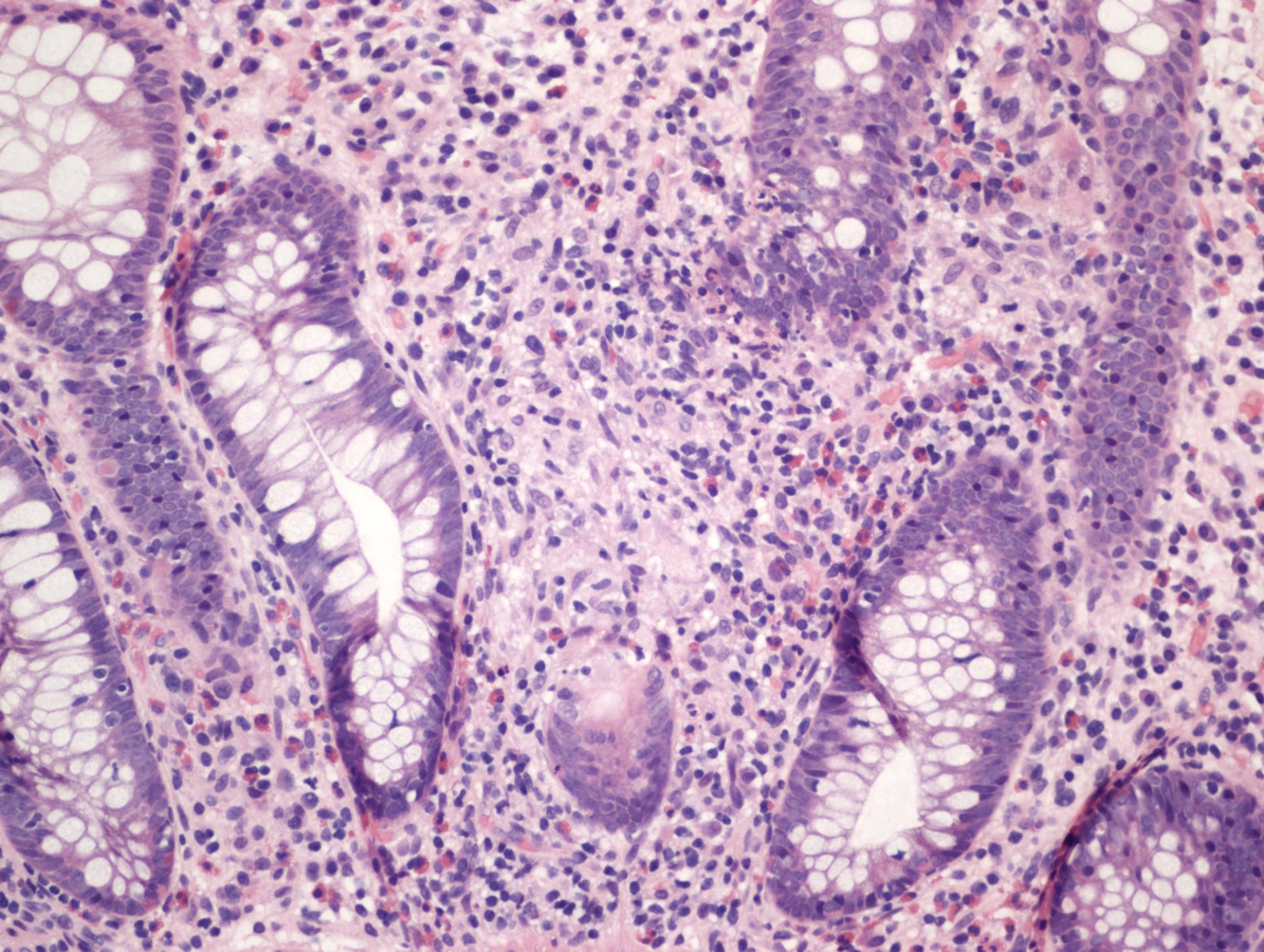
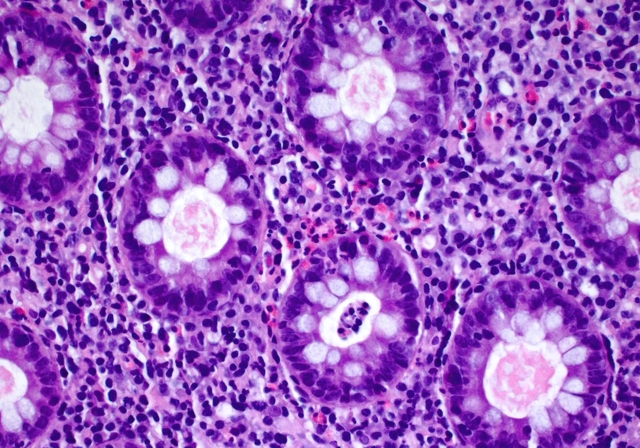
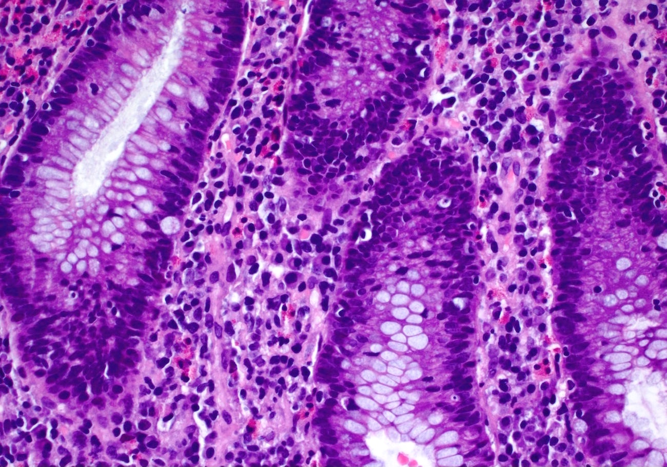
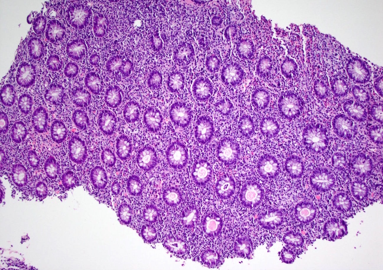
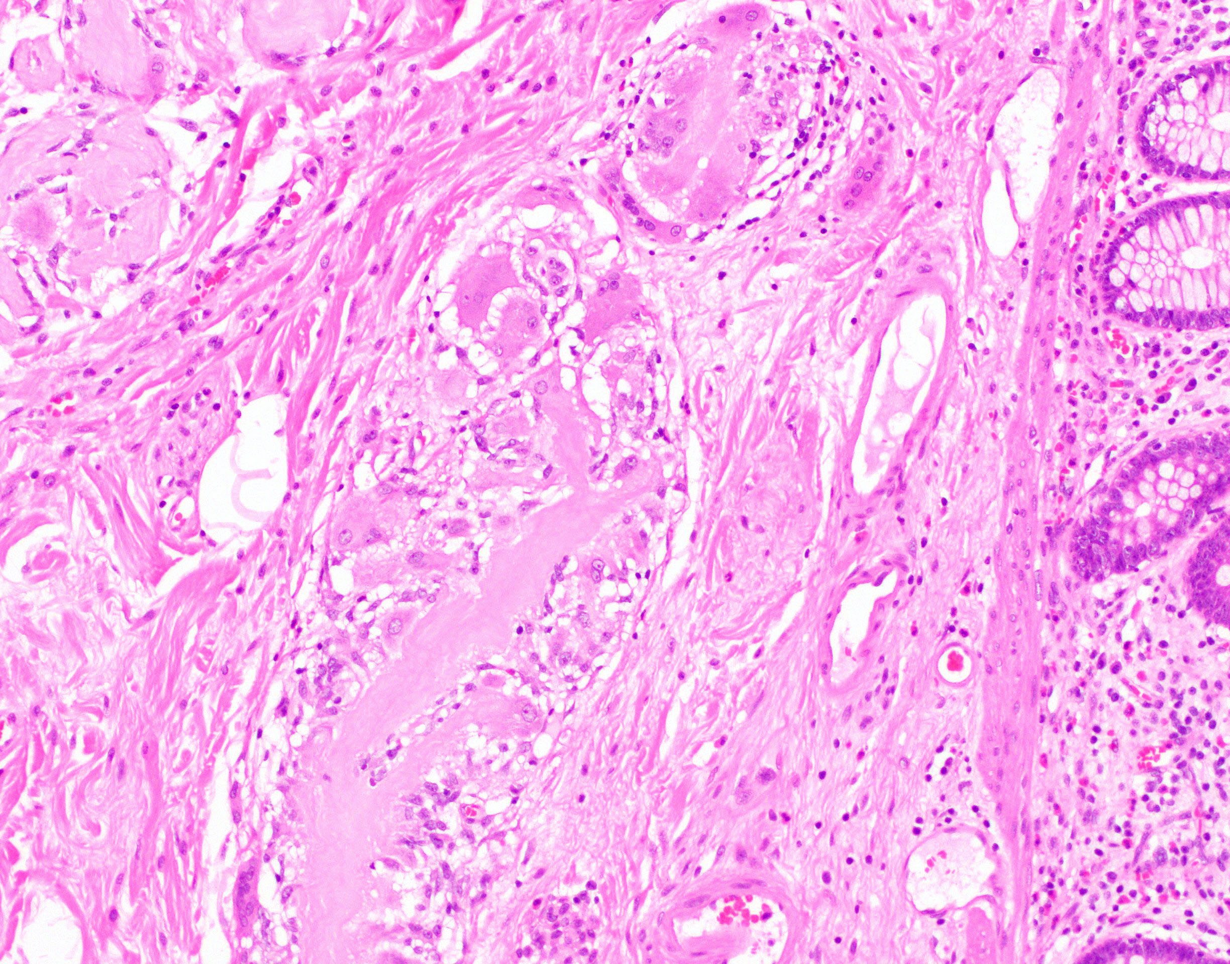
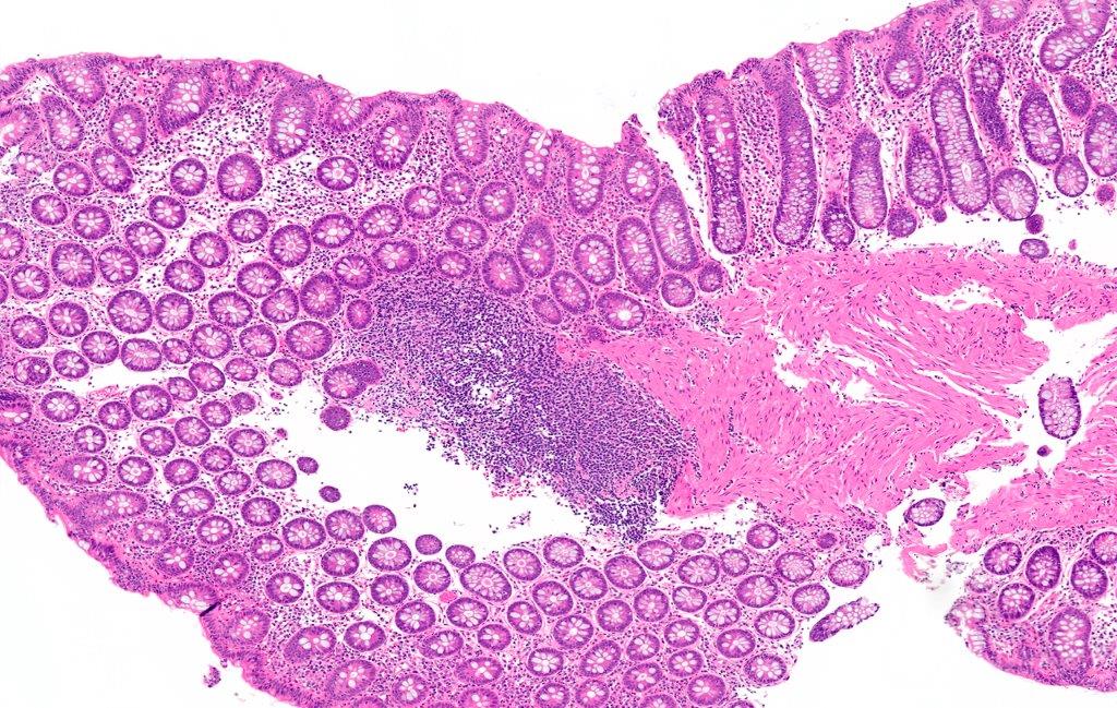
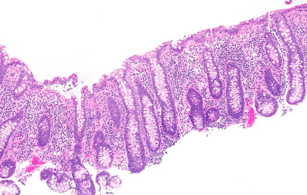
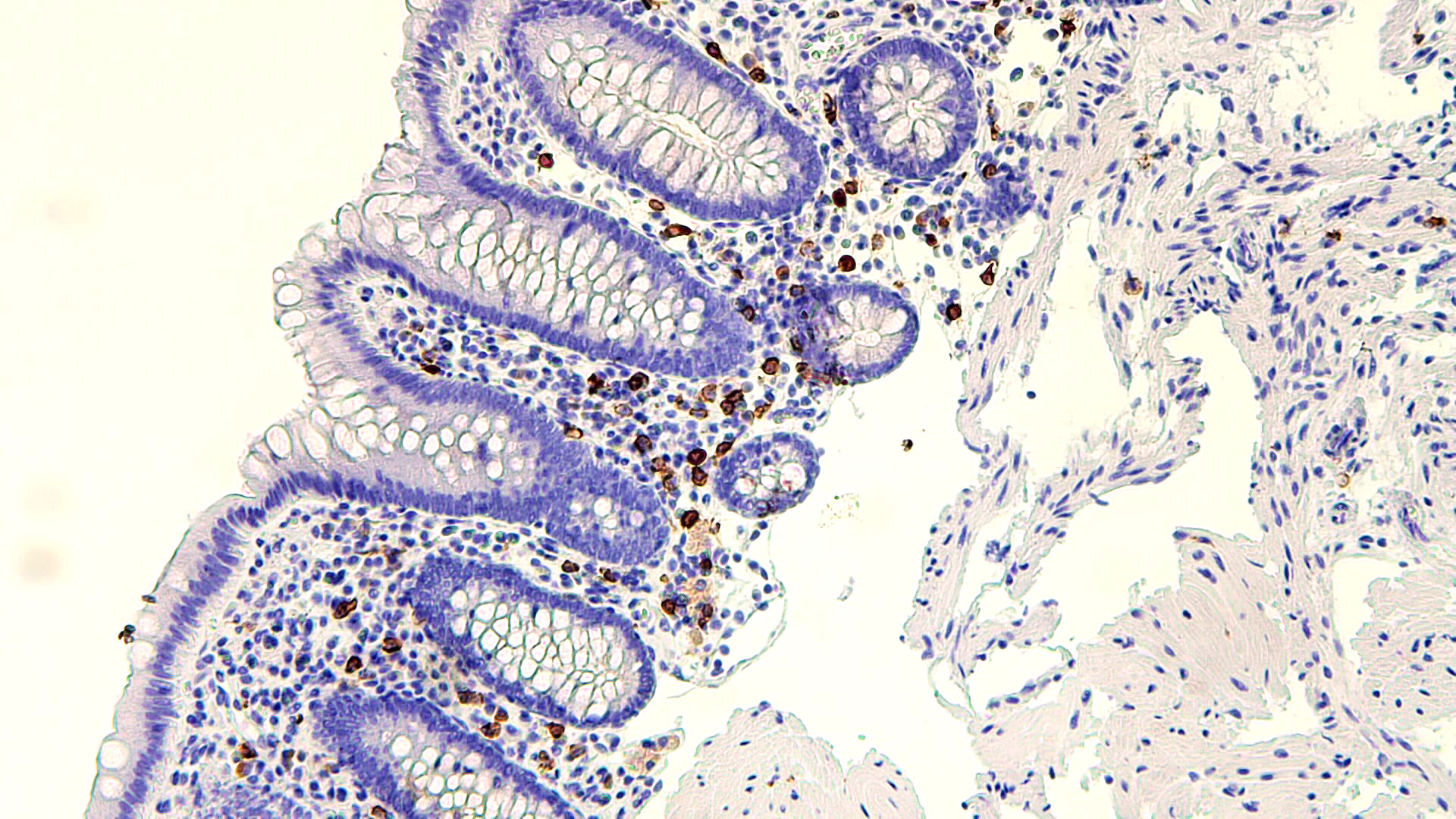
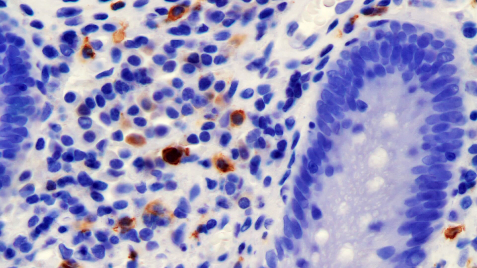
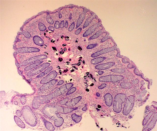
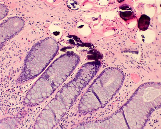
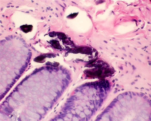
Active colitis
Cryptitis
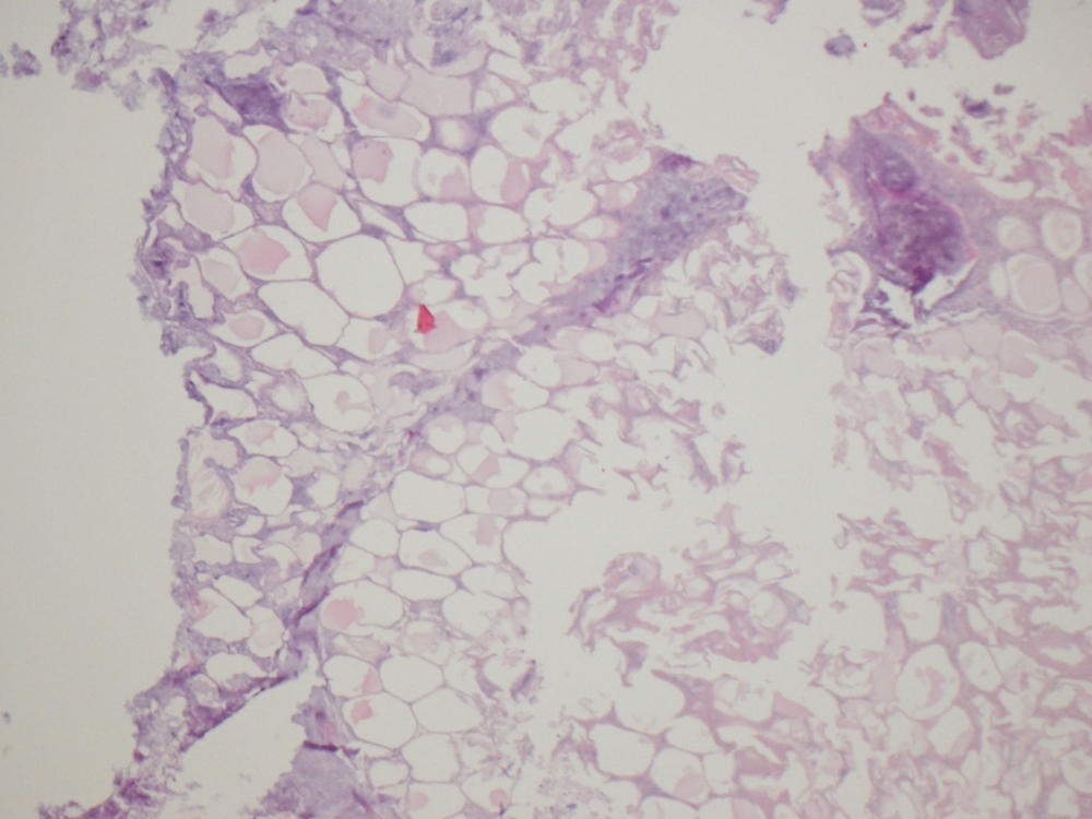
Images hosted on other servers:

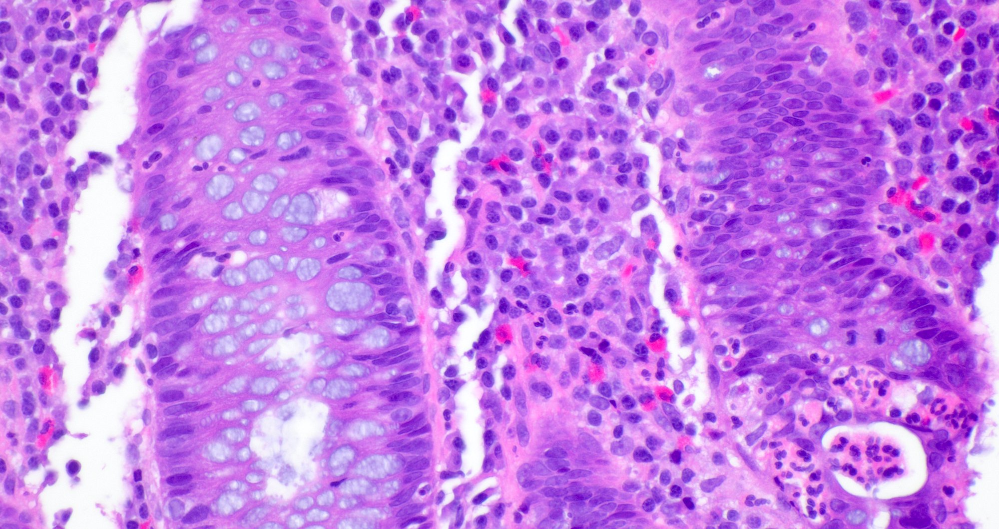
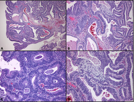
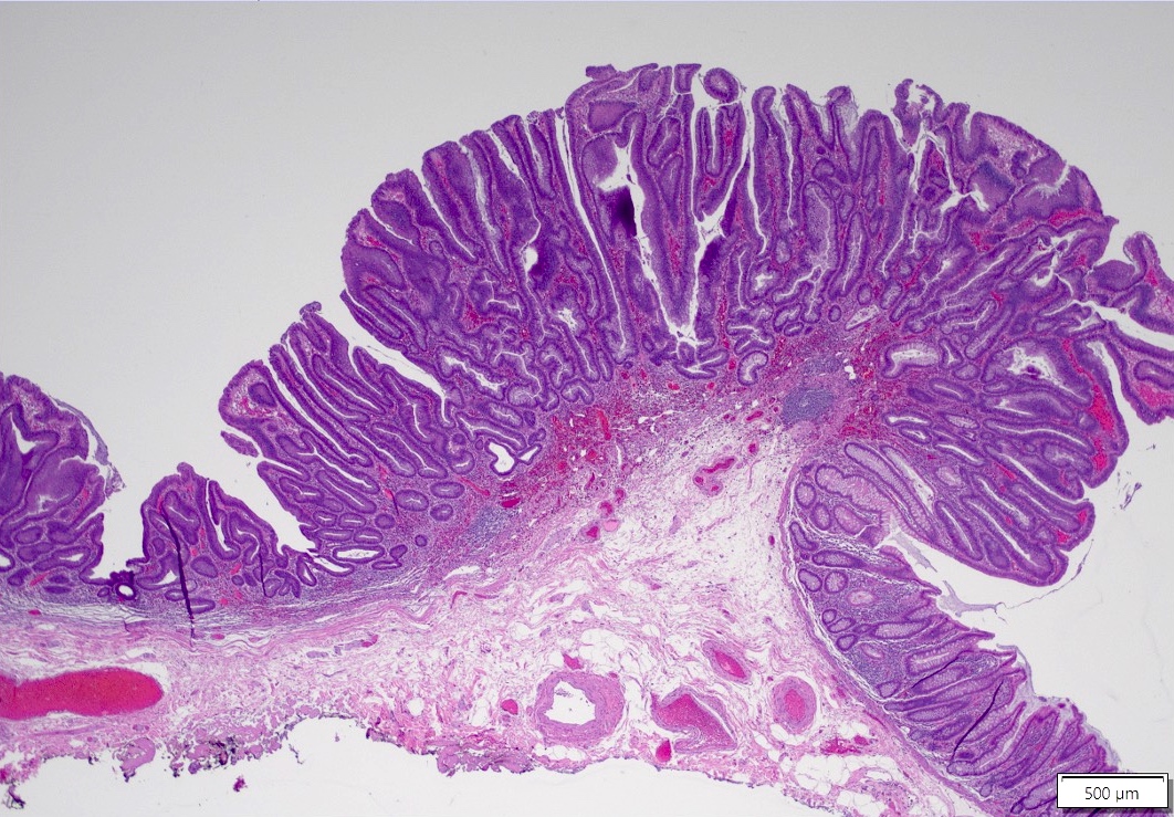
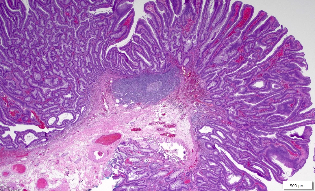
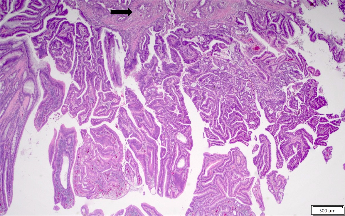
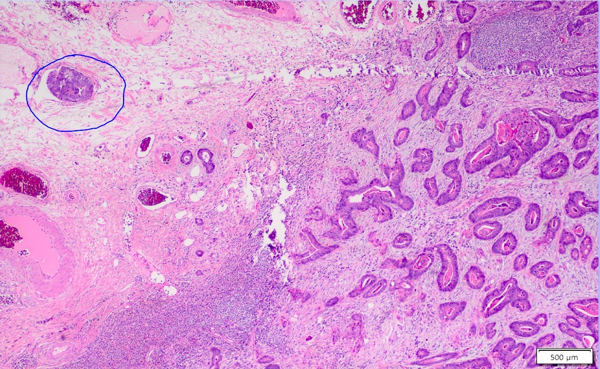
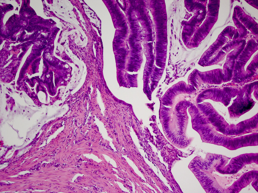
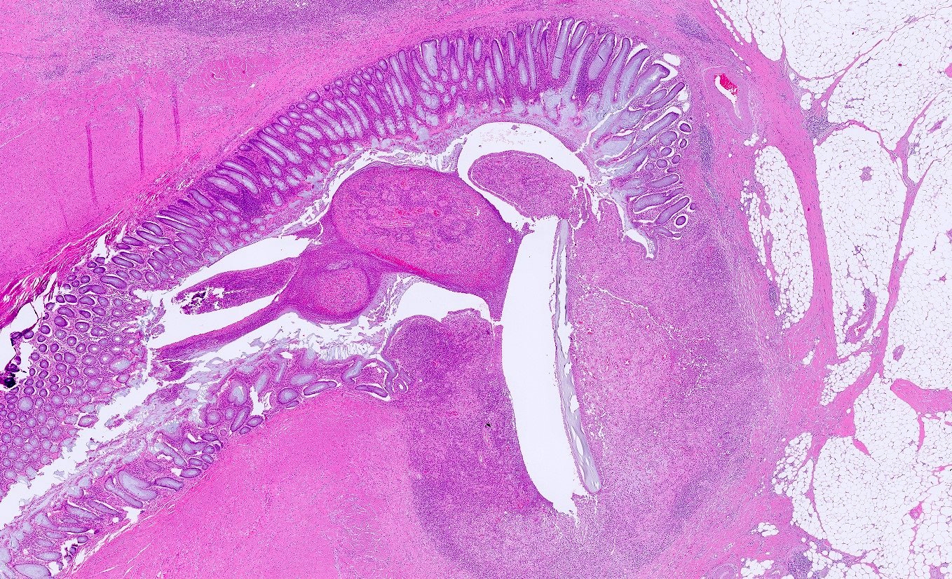
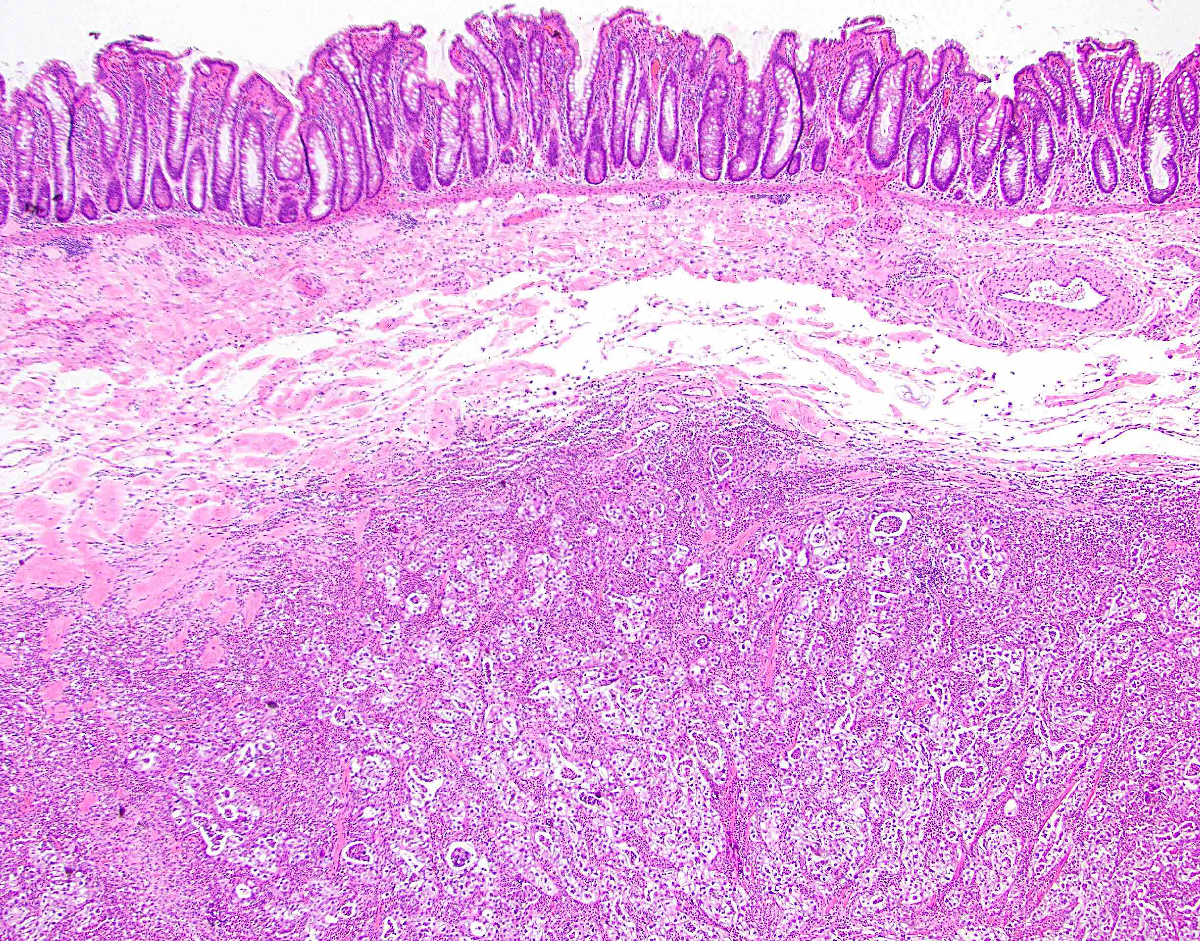
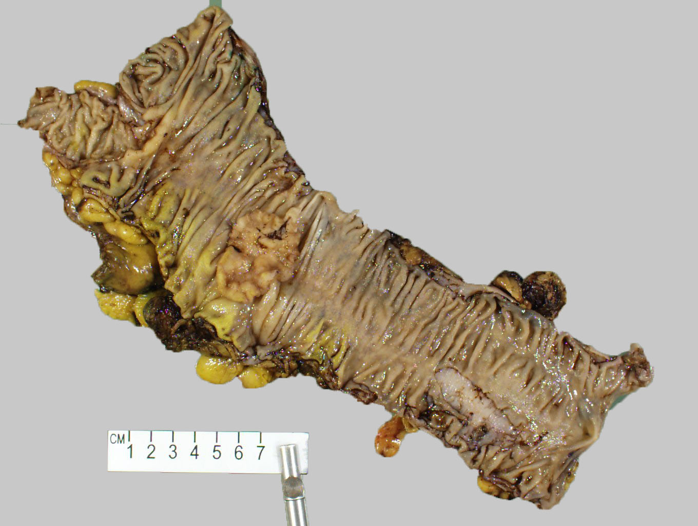
Early, flat tumor

Flat, small adenocarcinoma

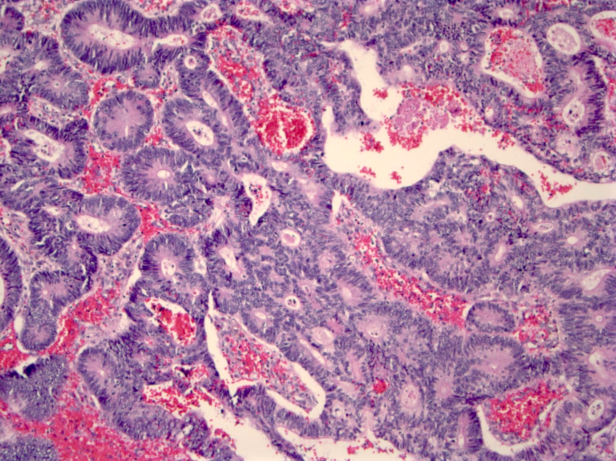
Invasive mucinous adenocarcinoma


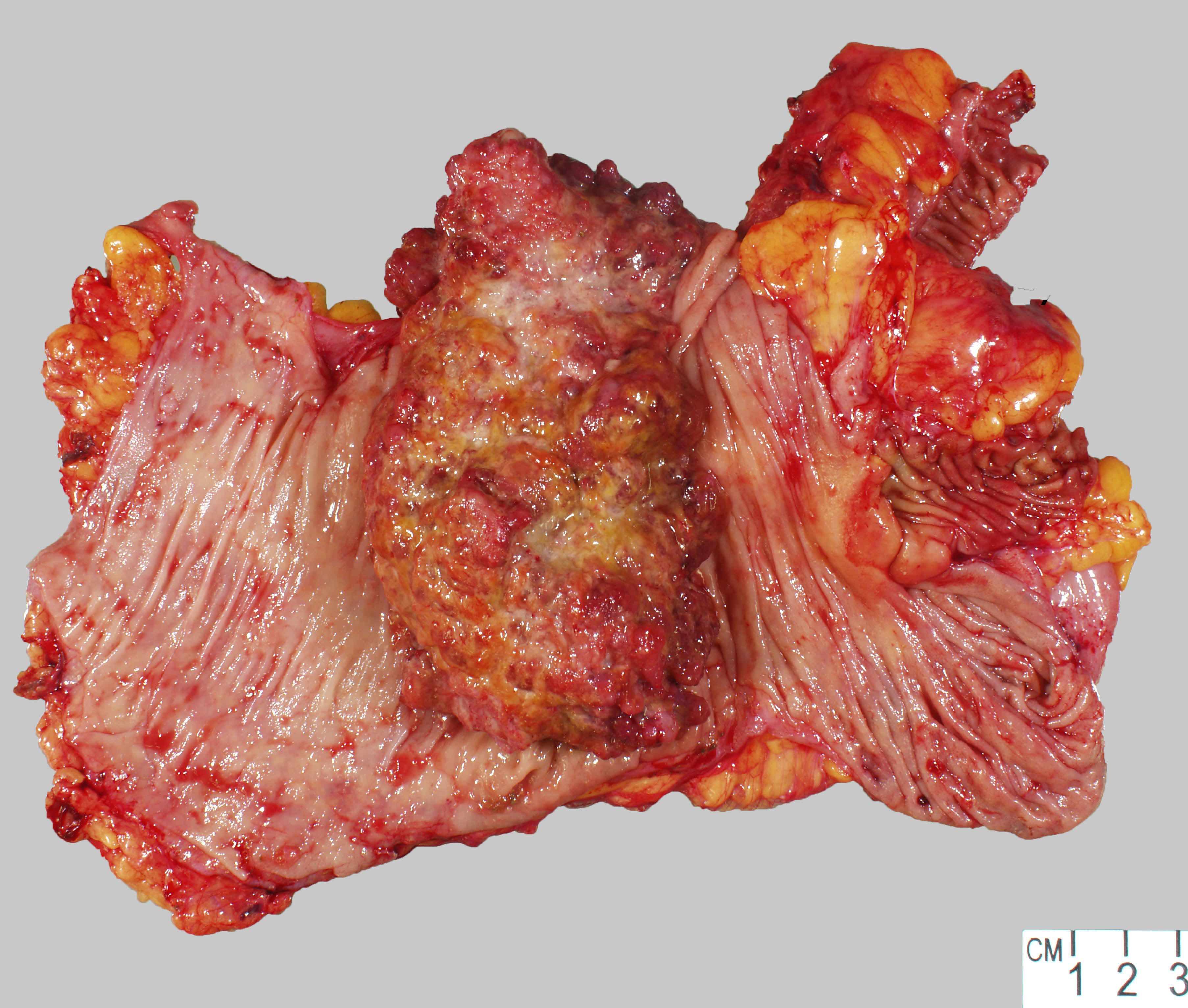
Exophytic lesion
Contributed by Raul S. Gonzalez, M.D.
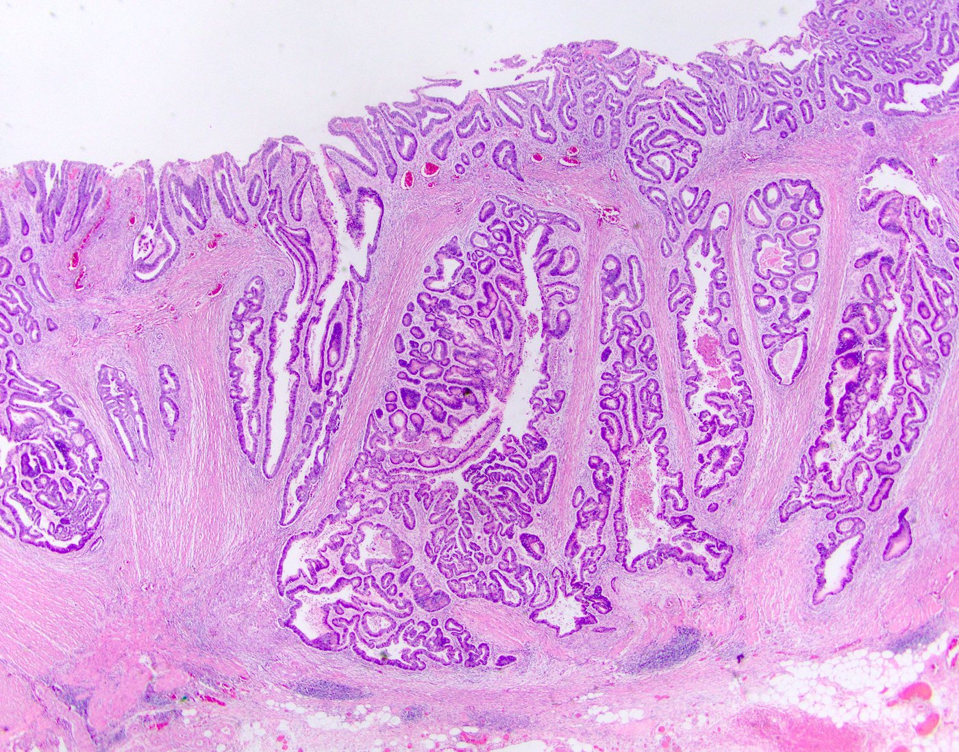
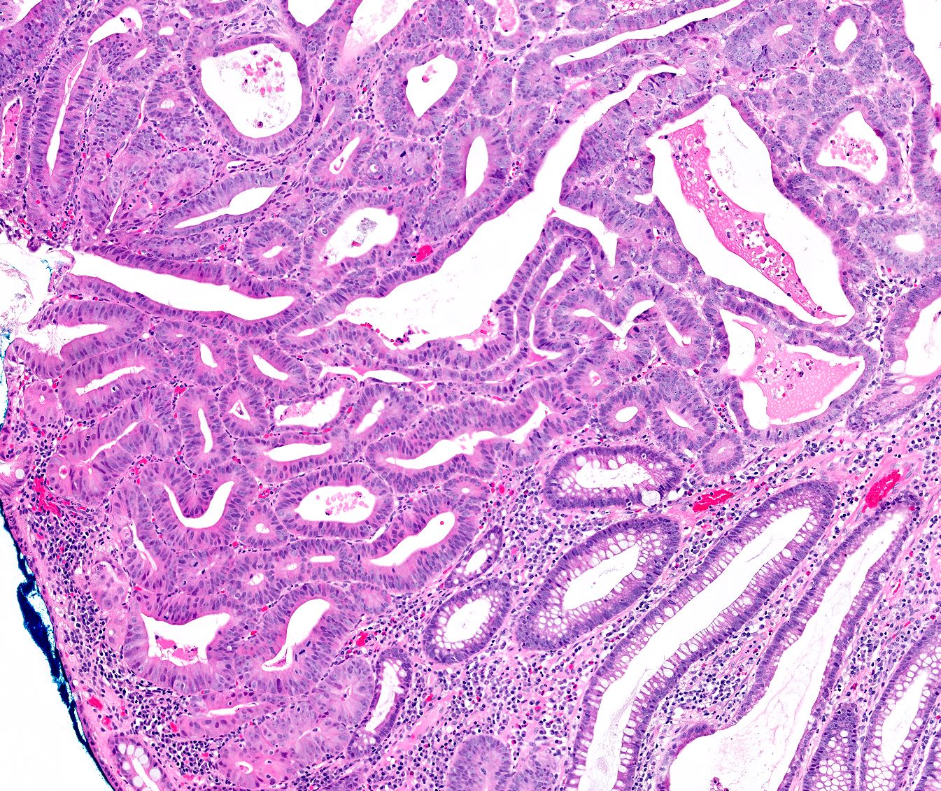
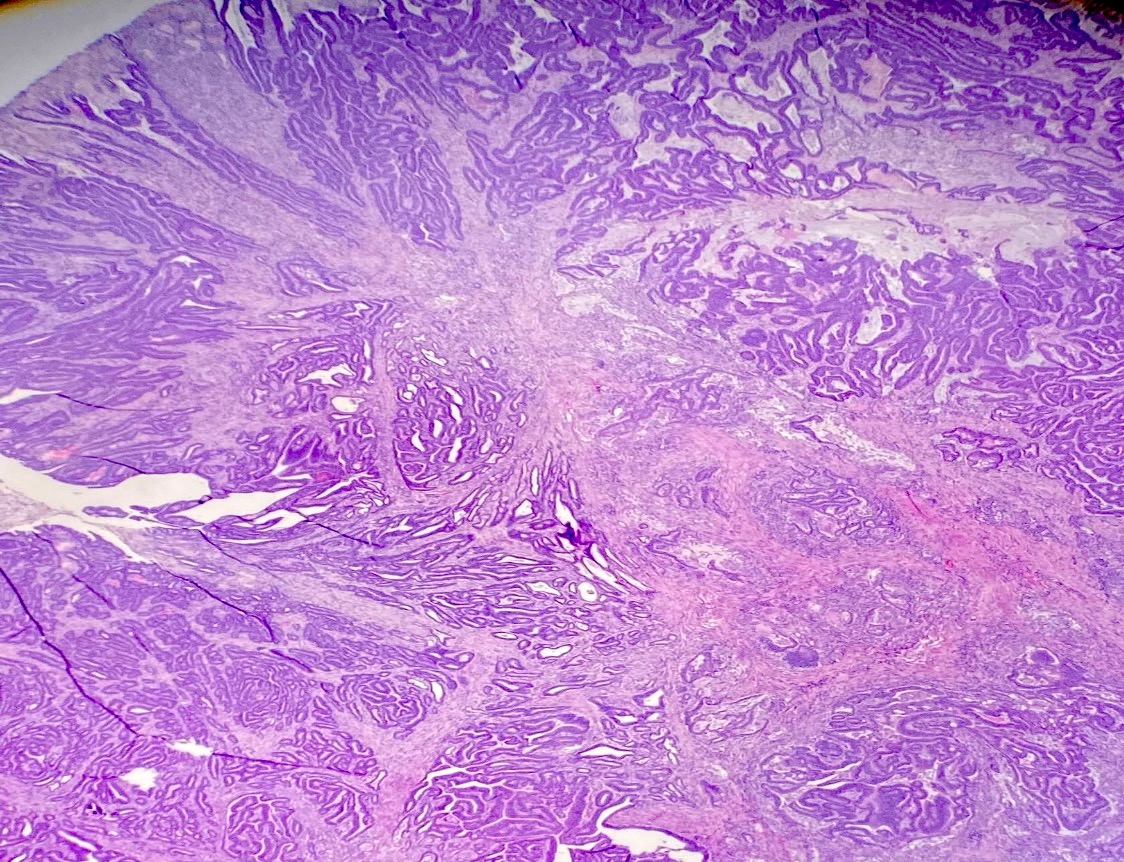
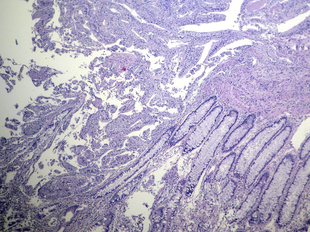
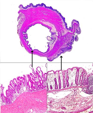
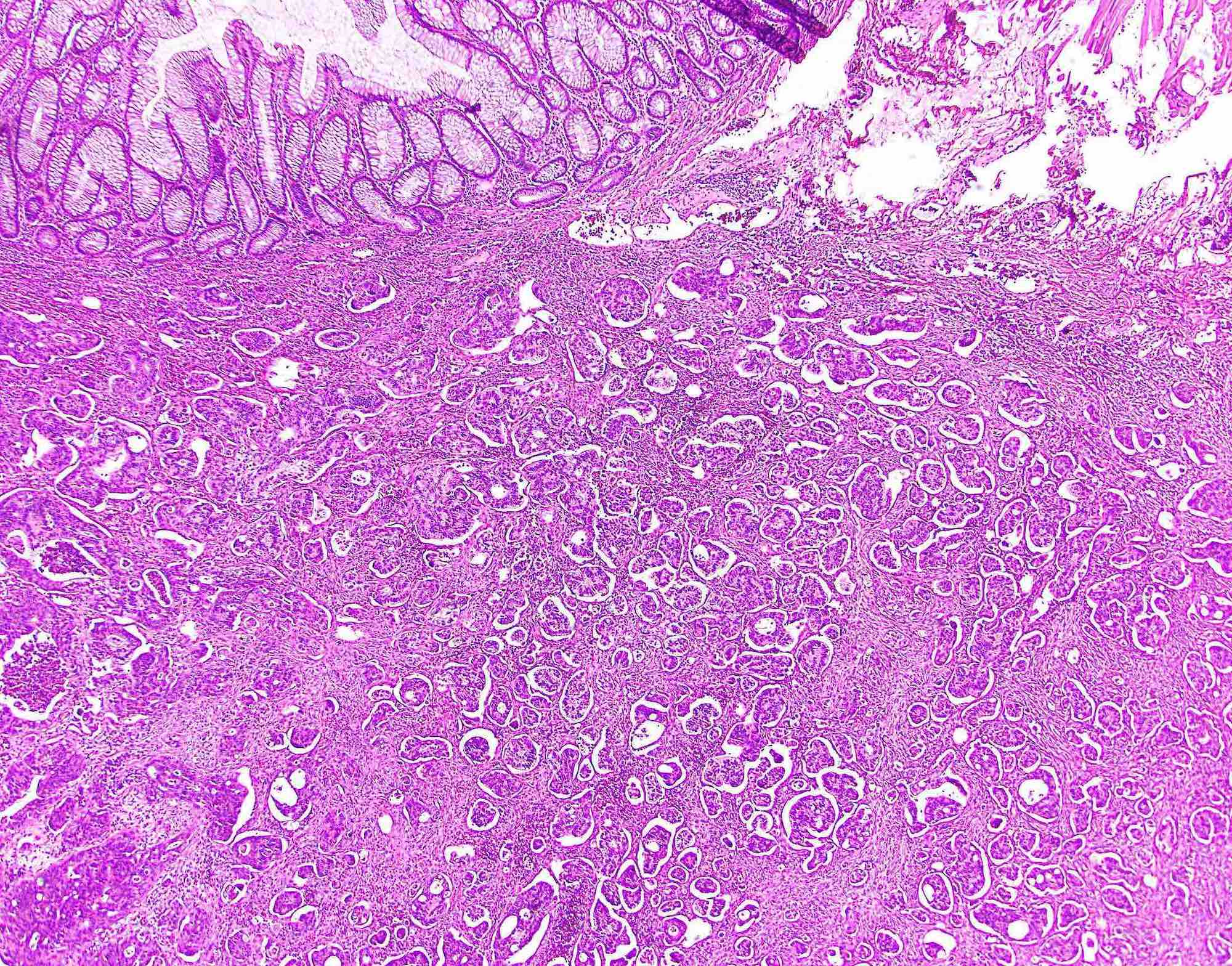
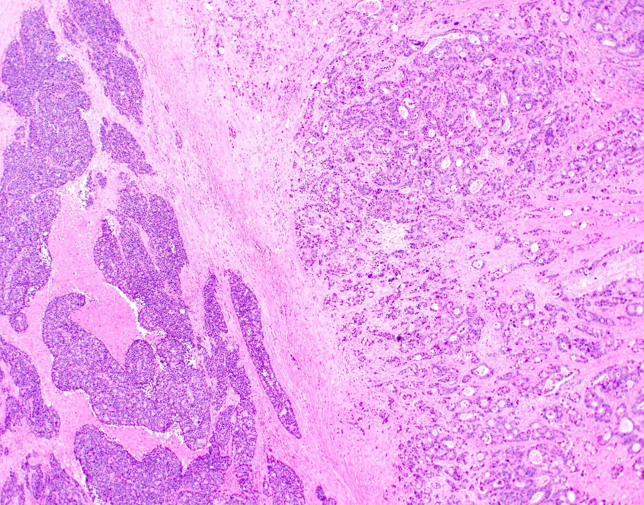
Low power
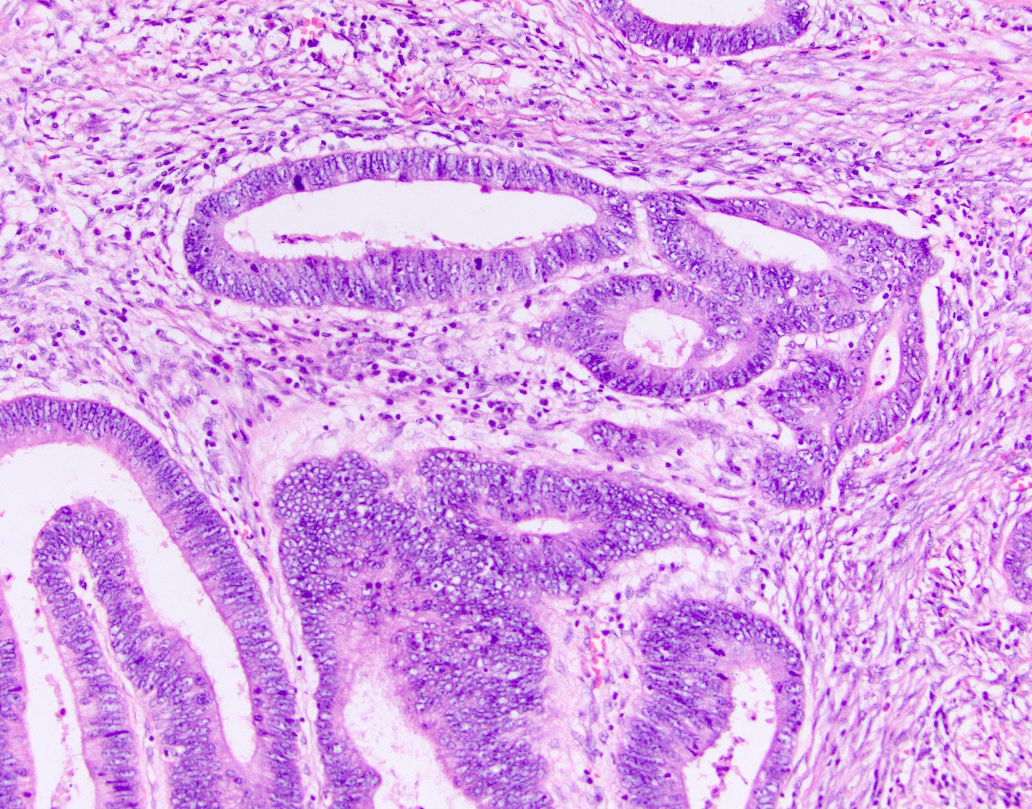
High power

Dirty necrosis
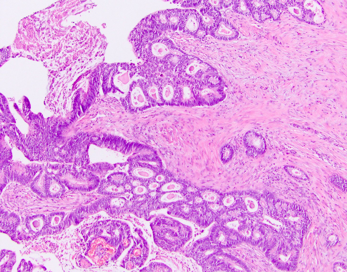
Cribriforming
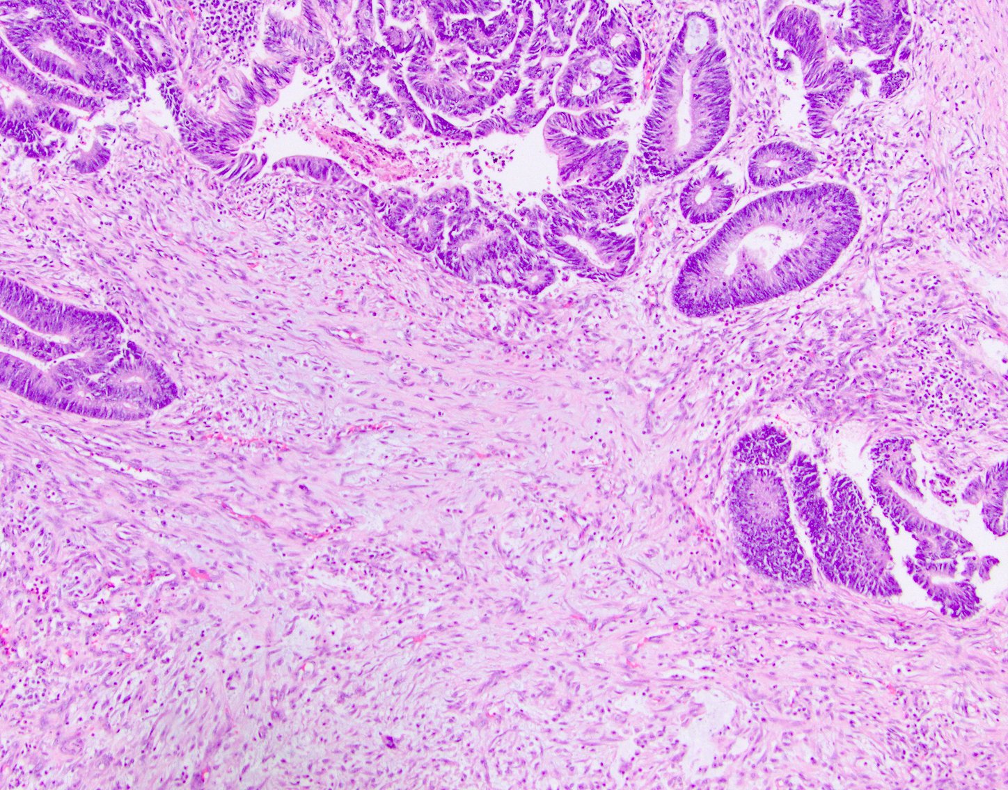
Desmoplasia
Lymphovascular invasion
Contributed by Semir Vranic, M.D., Ph.D. and Beverly Wang, M.D.
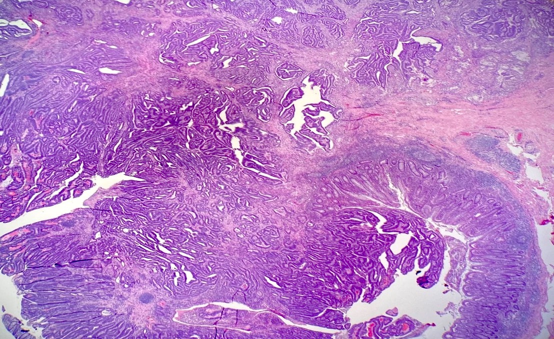
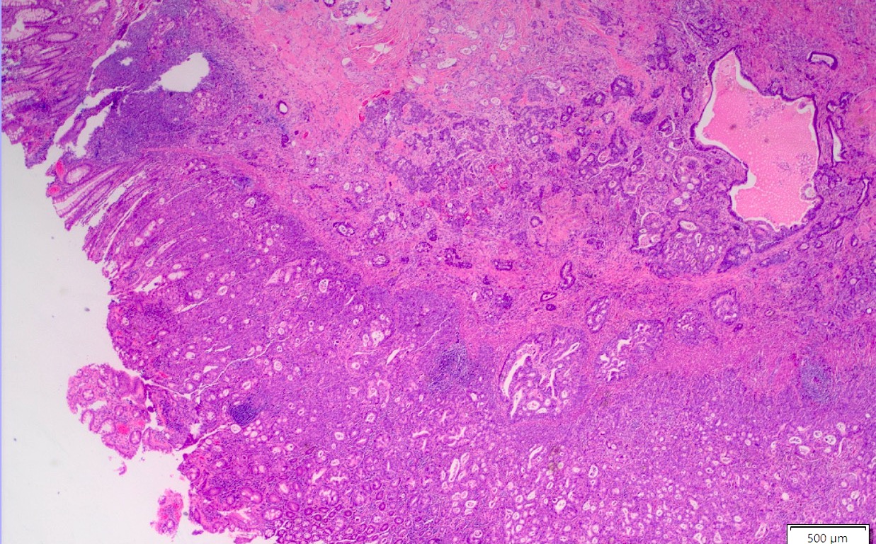
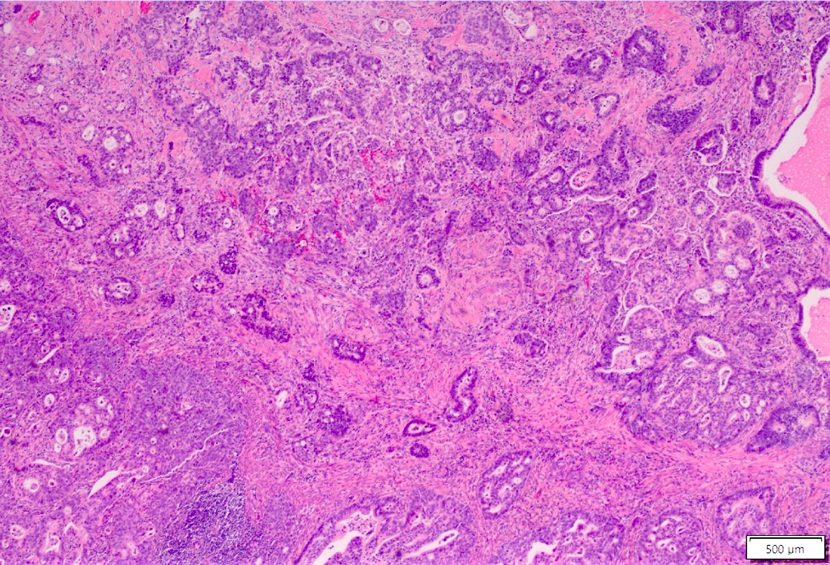
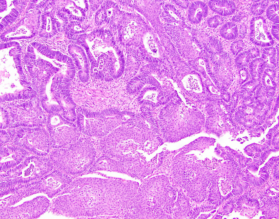
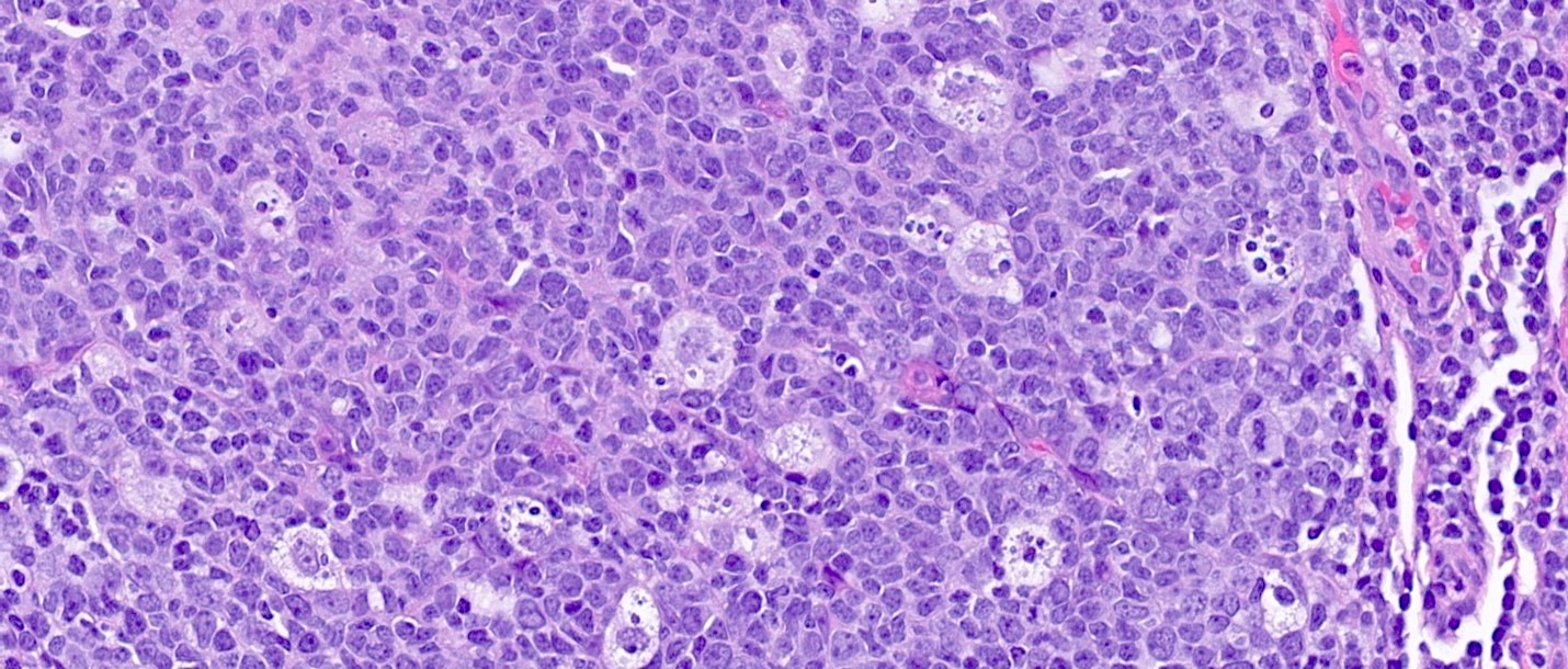
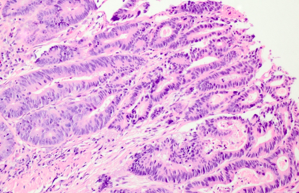
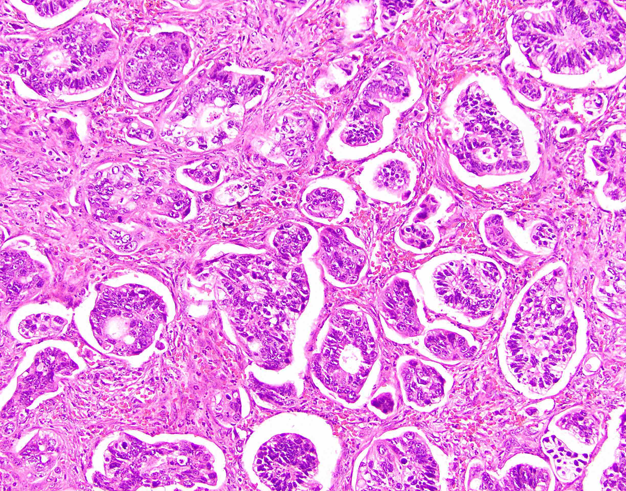
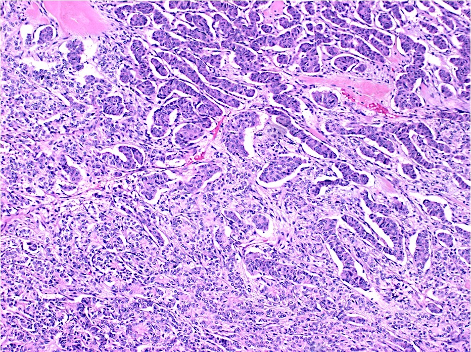
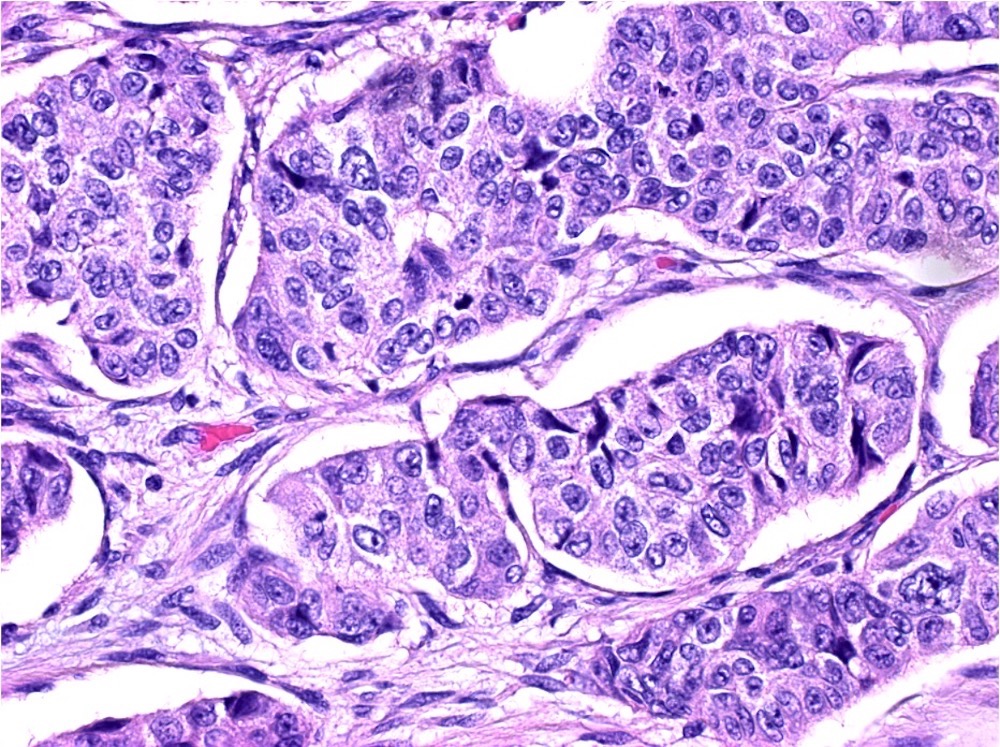
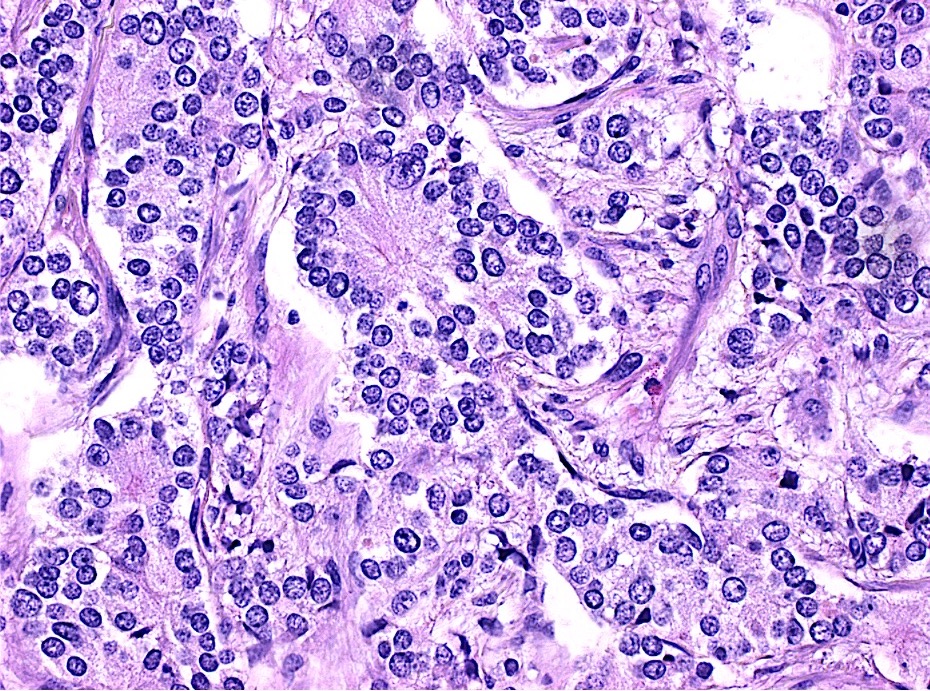
Poorly differentiated adenocarcinoma
Adenocarcinoma
Images hosted on other servers:

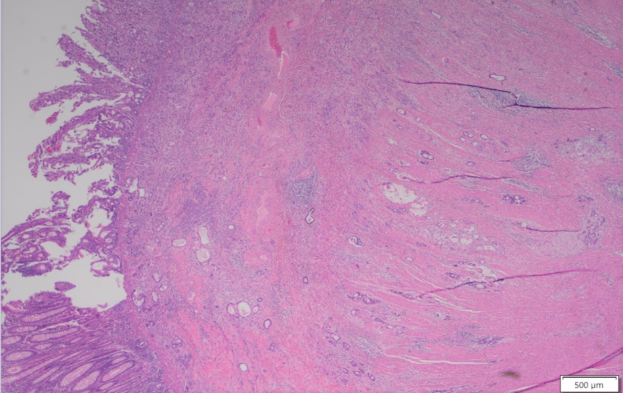
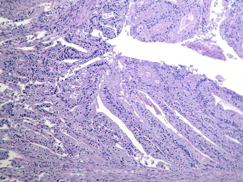
Whole mount scan

Moderately differentiated

Dirty necrosis in gland lumens

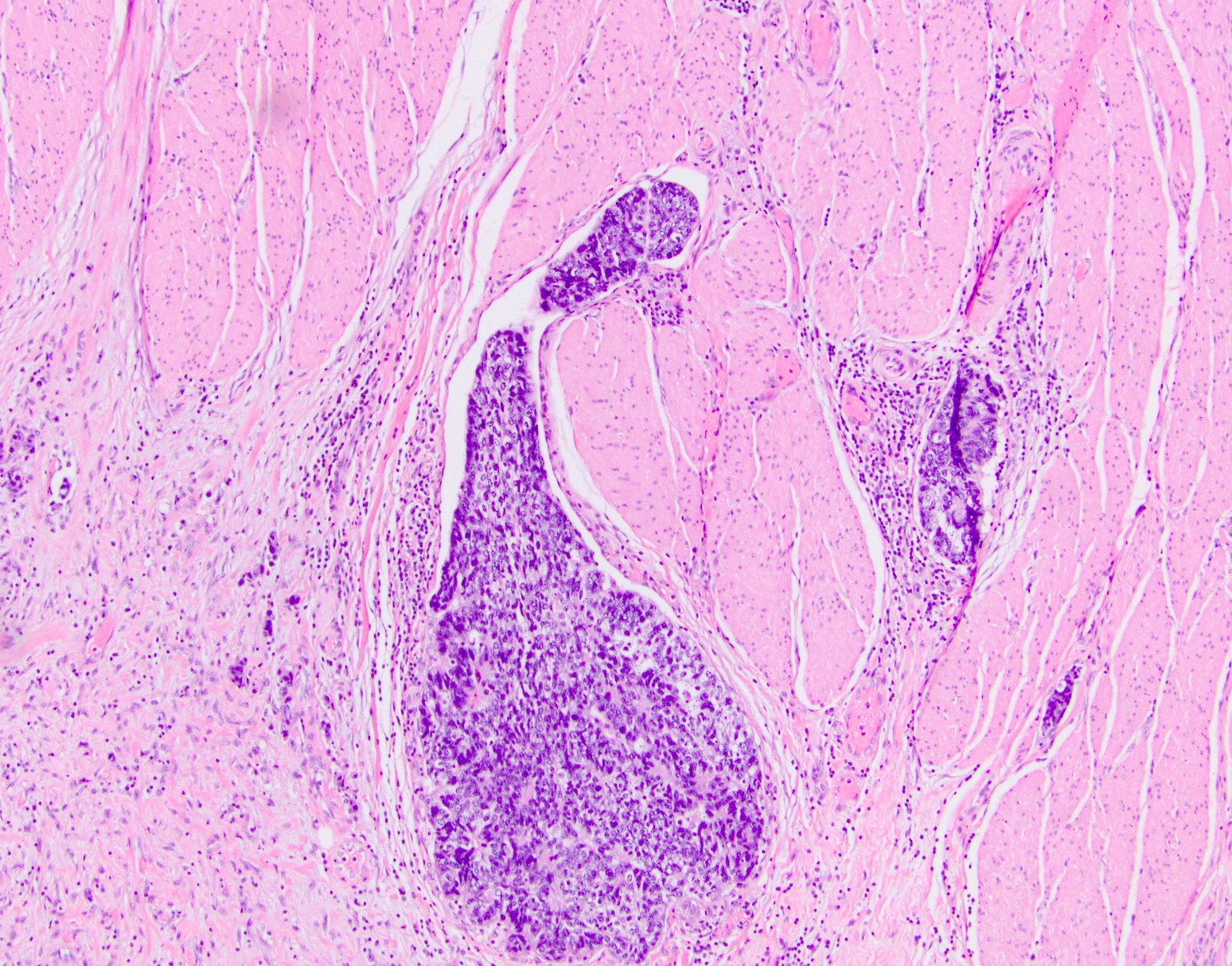
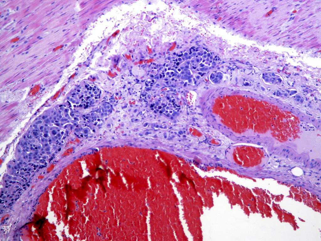
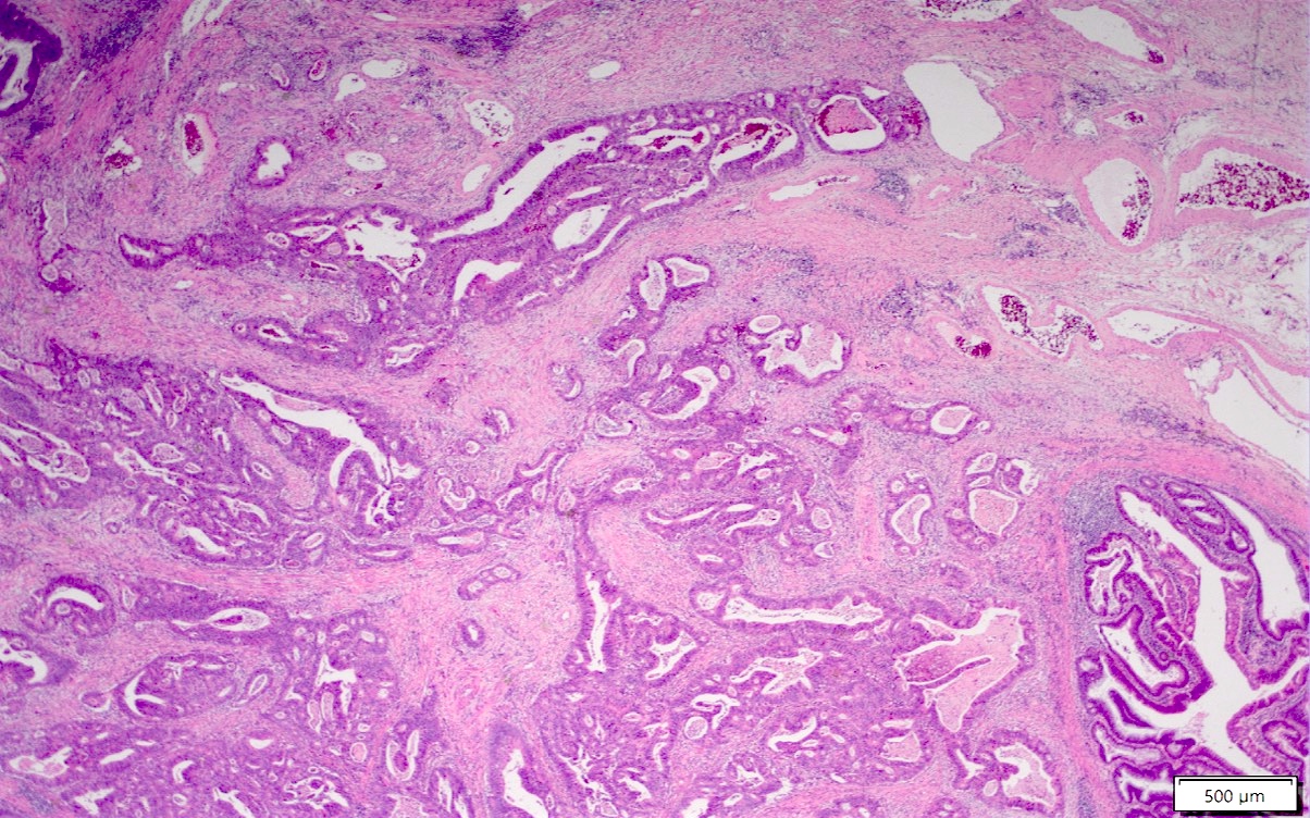
Venous invasion

Serosal penetration

Detached carcinoma cells

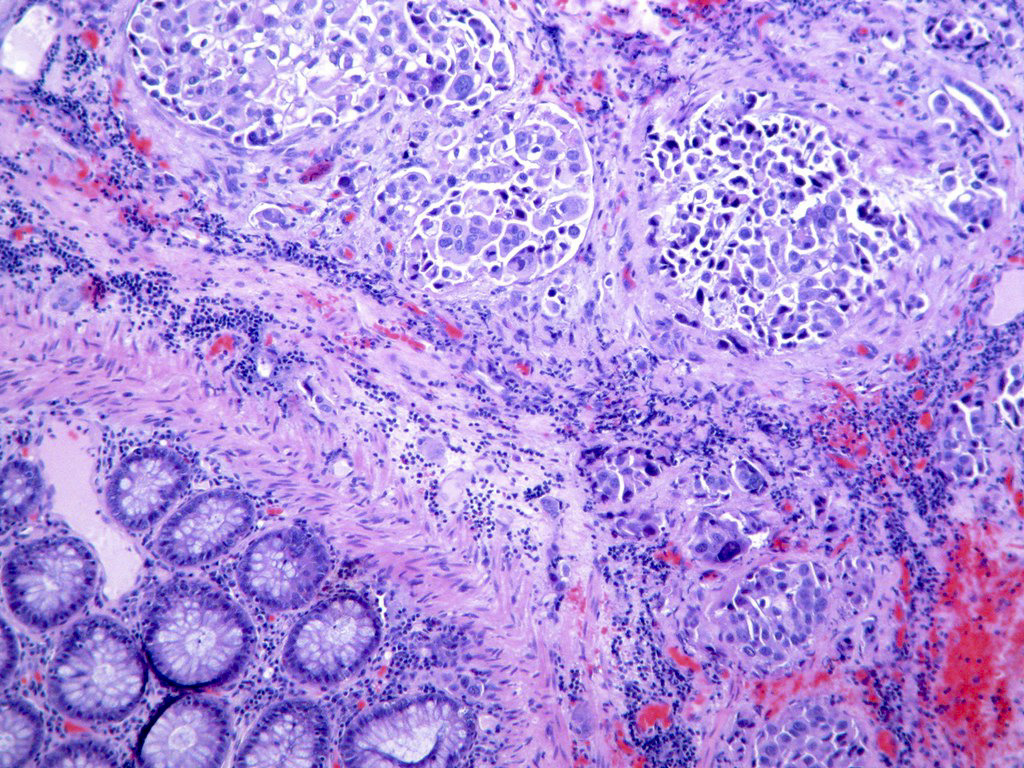
Signet ring morphology

Lymph node metastasis
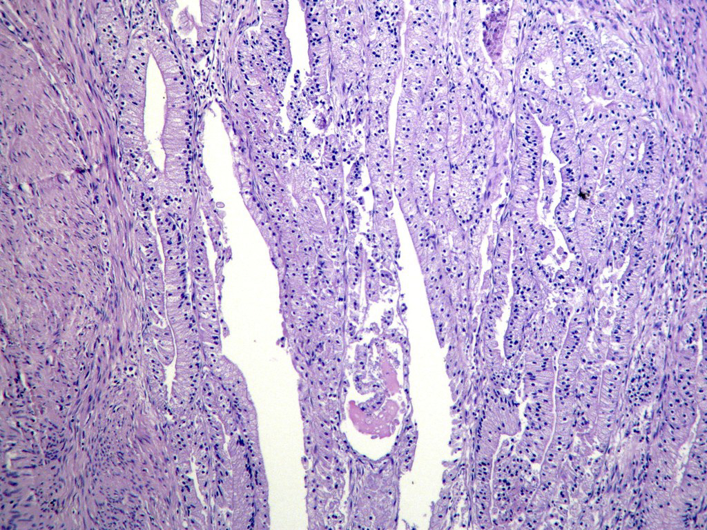
Histopathology colon adenocarcinoma
Images hosted on other servers:

Adenoma carcinoma sequence
Images hosted on other servers:

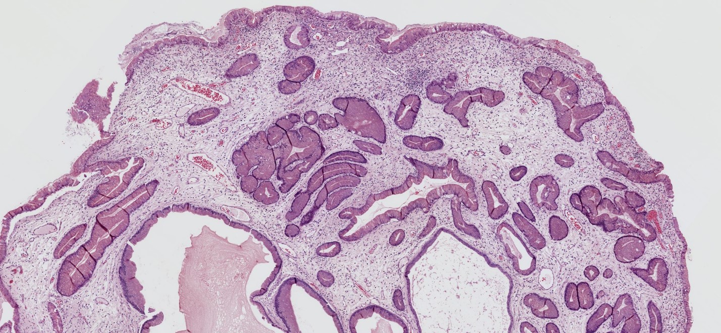
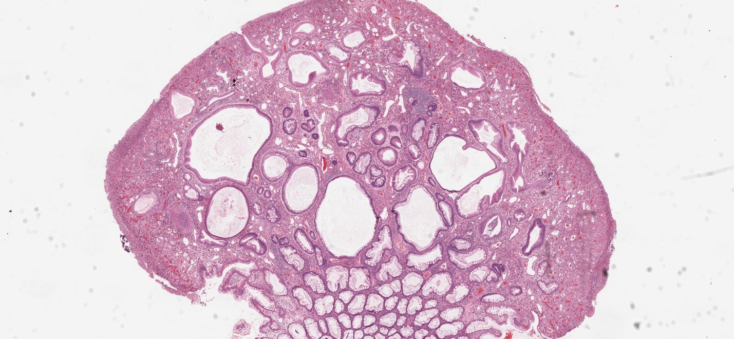
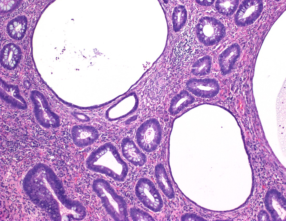
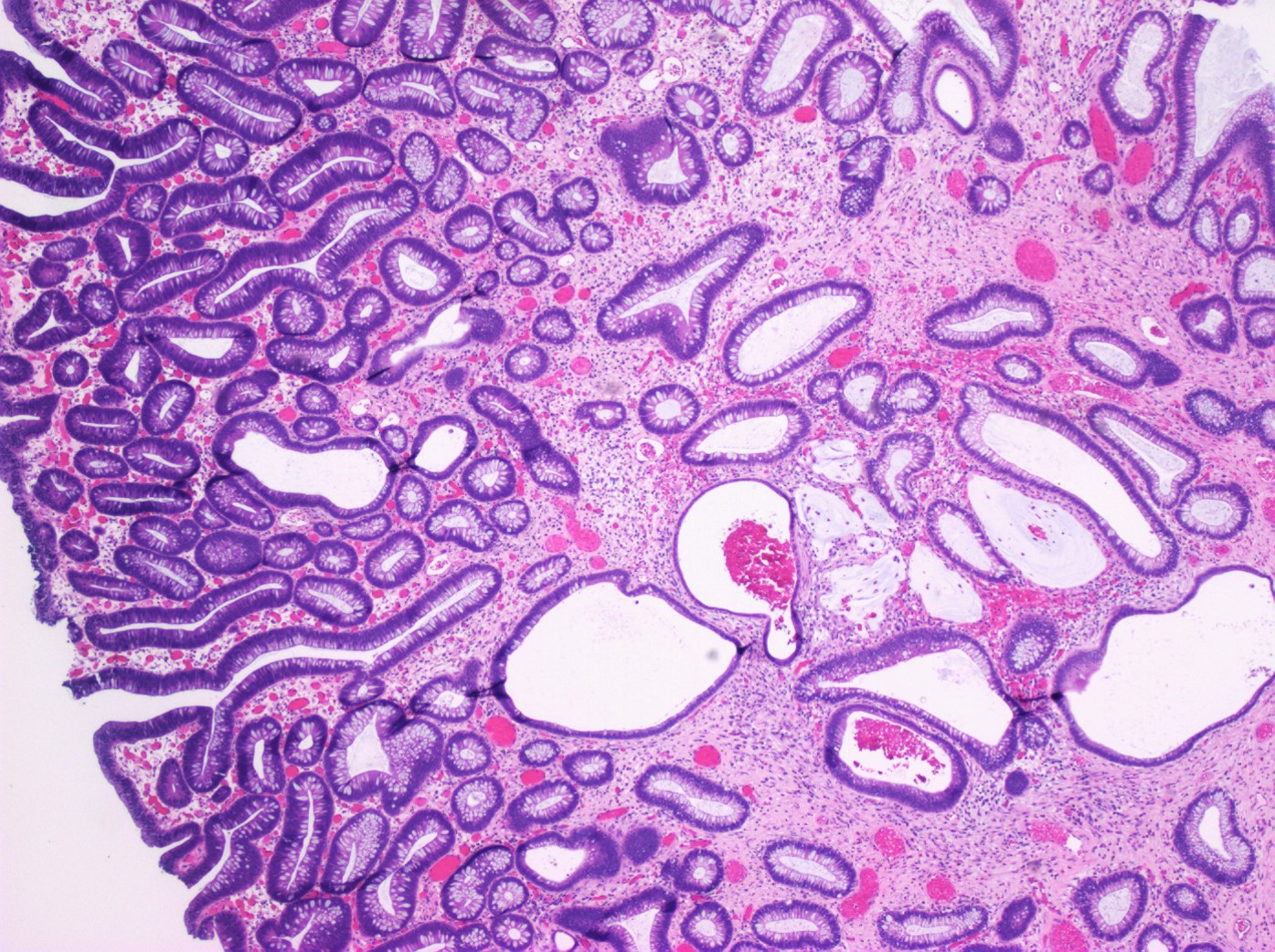
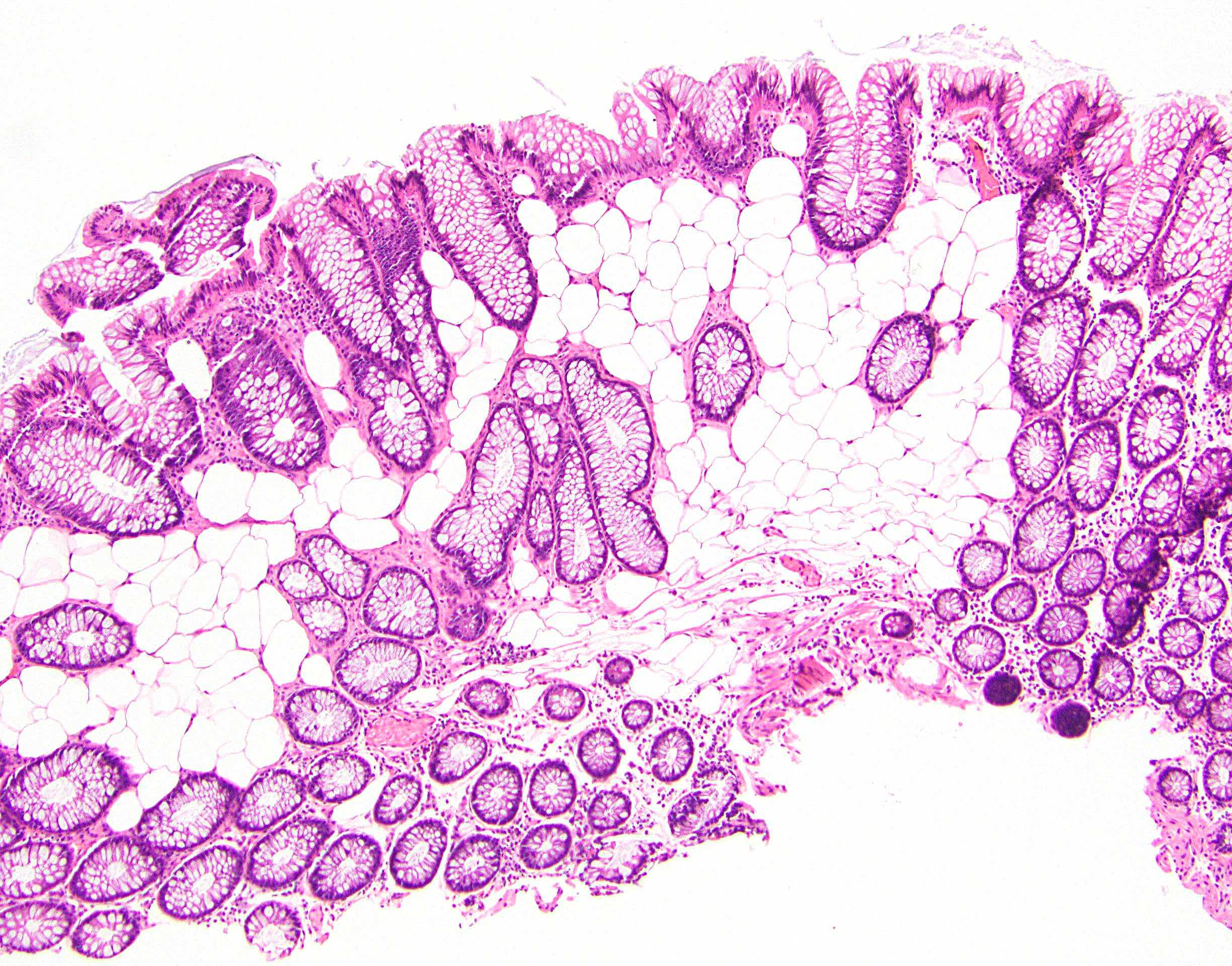
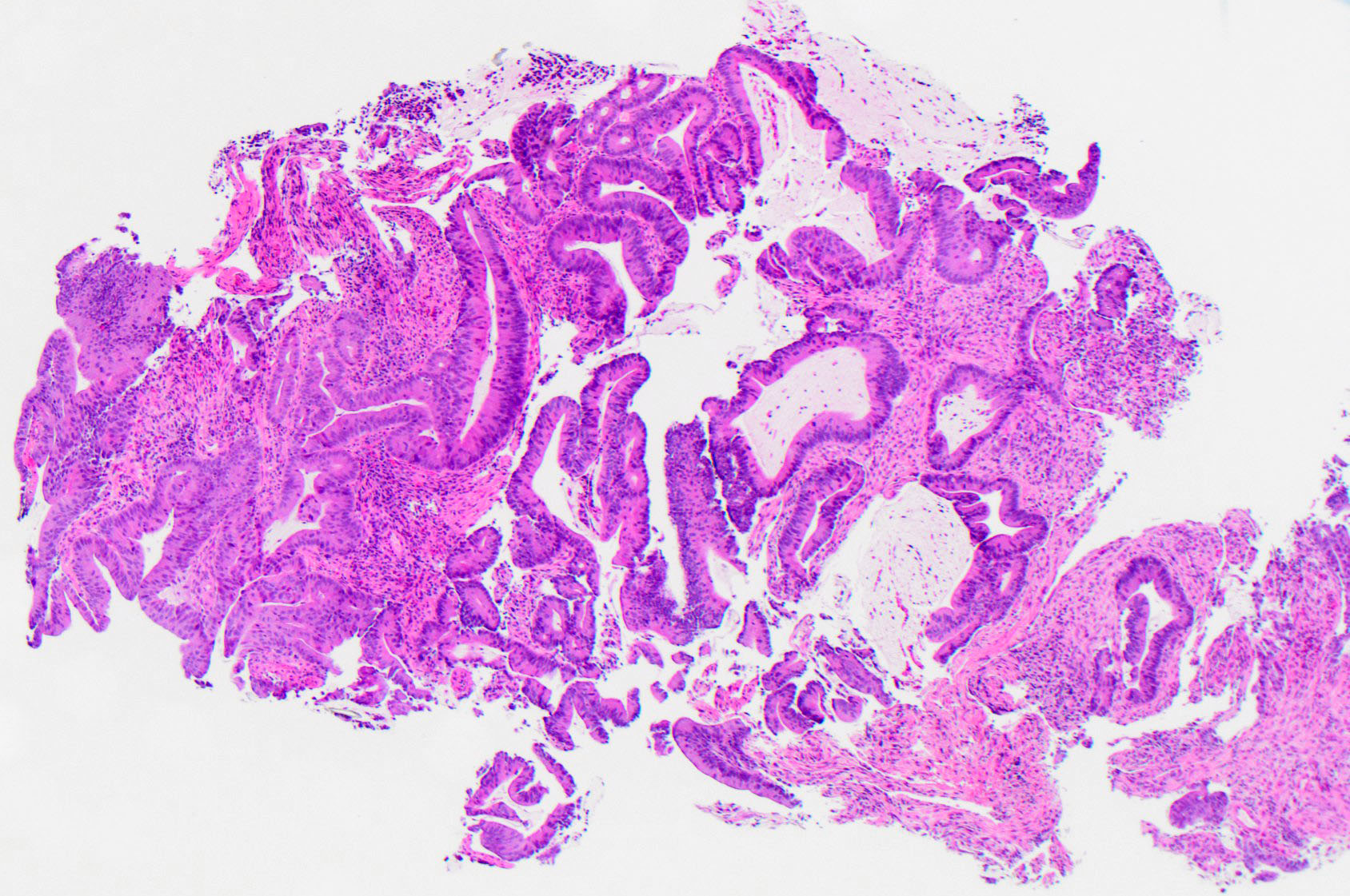
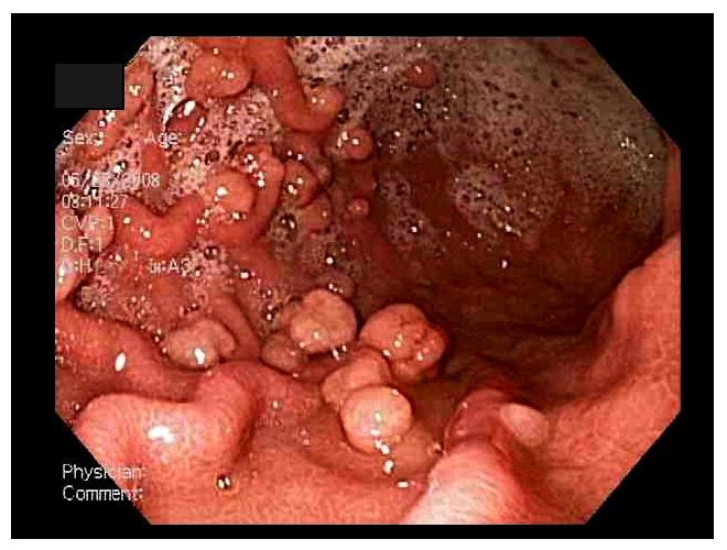
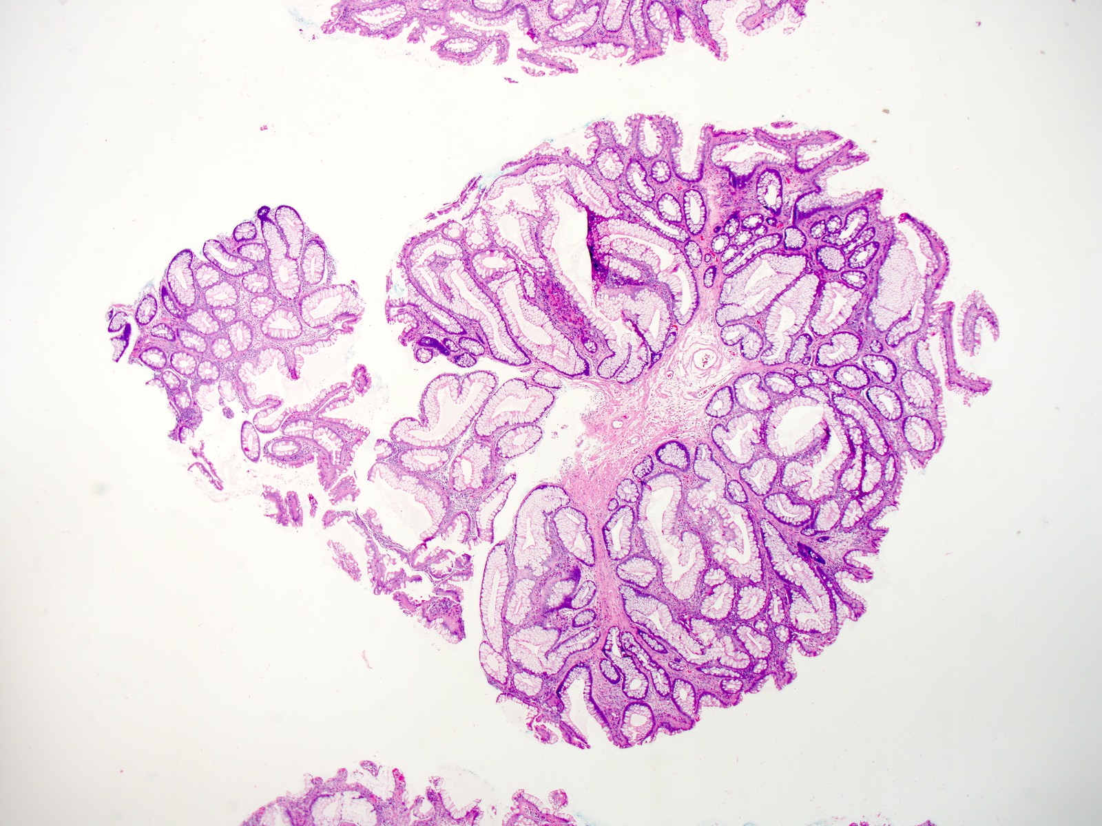
Giant villous adenoma

Several adenomas
Images hosted on other servers:

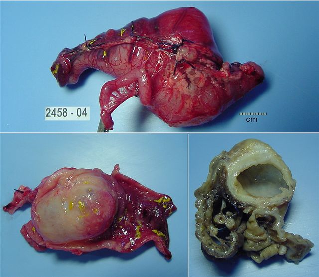
Polypoid tumor with gyrated surface sitting on a short stalk

Polypoid tumor
Contributed by Chien-Kuang Cornelia Ding, M.D., Ph.D., Kwun Wah Wen, M.D., Ph.D. and Enoch Kuo, M.D.
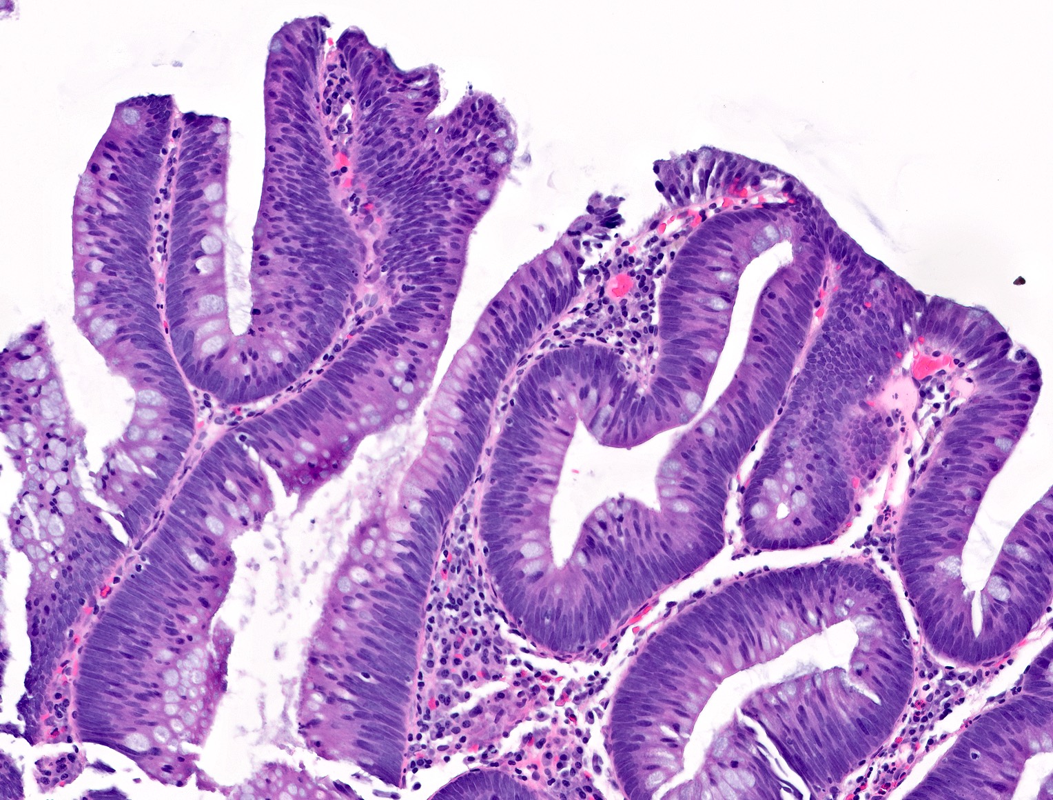
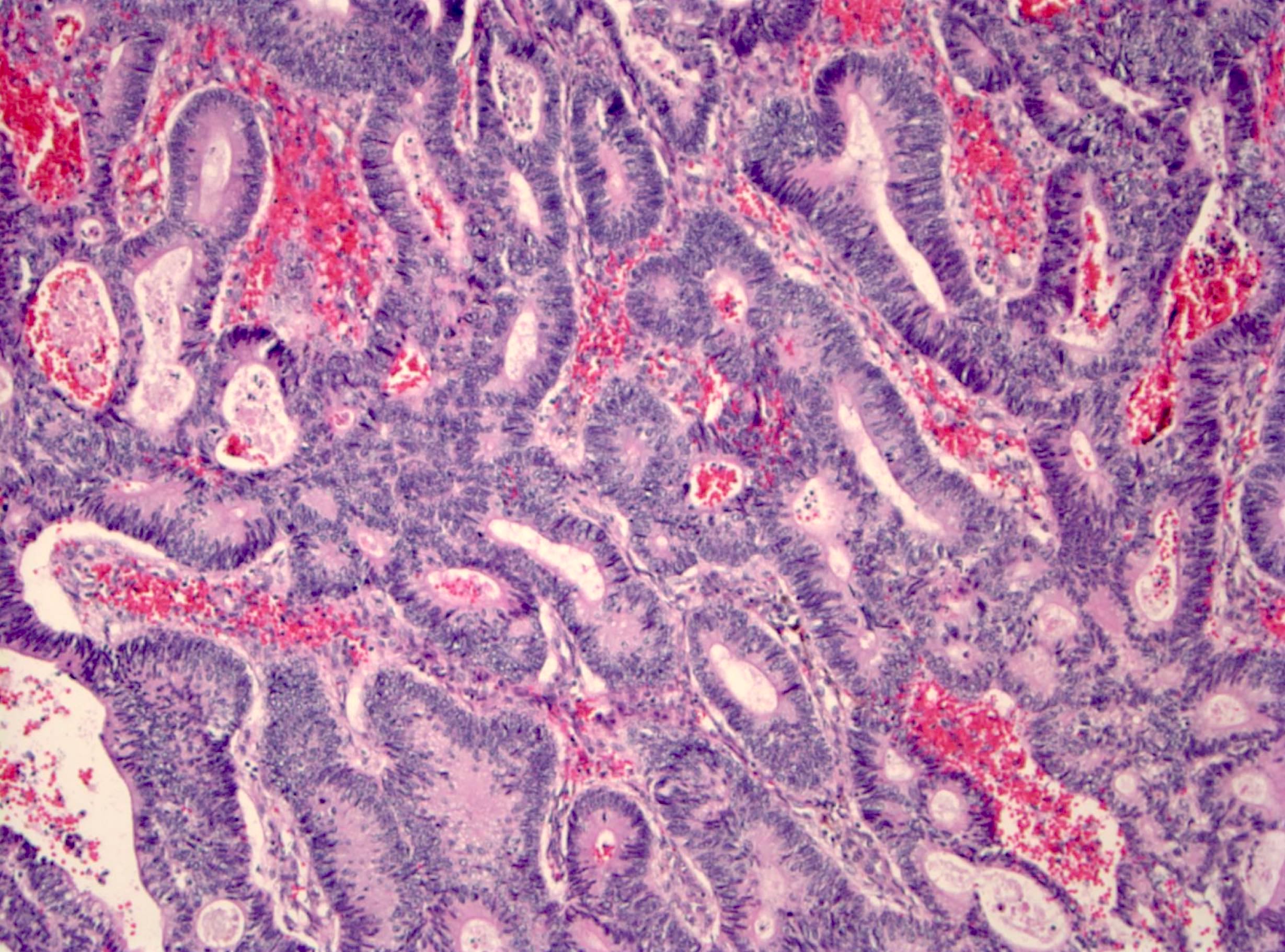
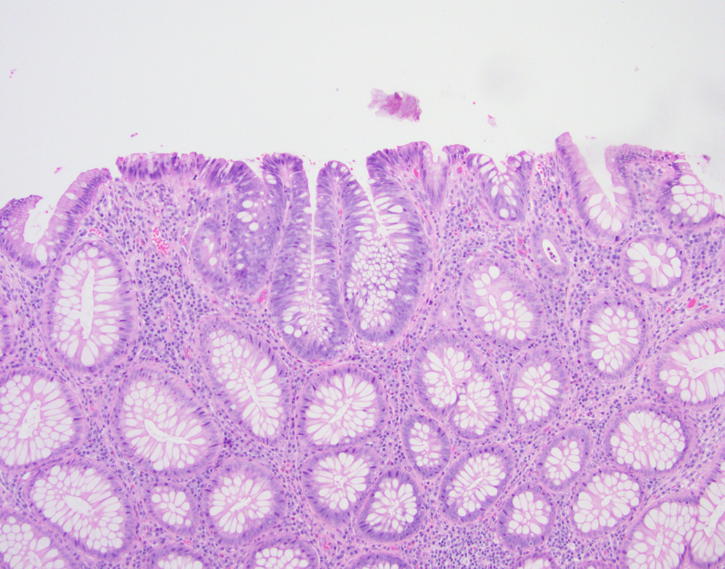
Tubular adenoma
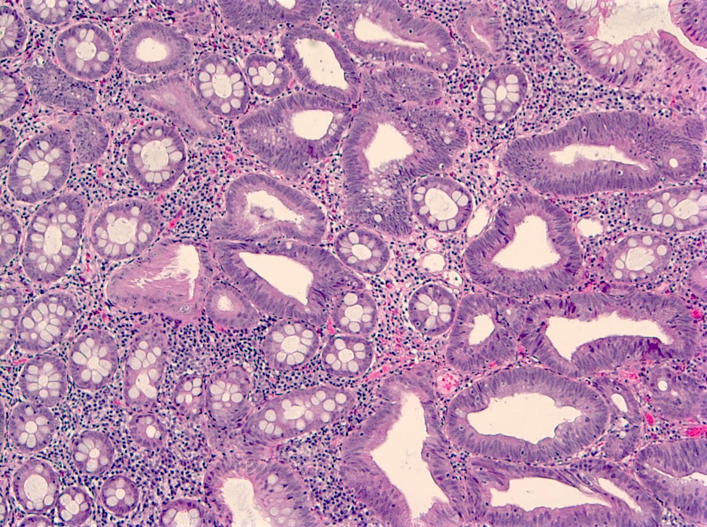
Low grade tubular adenoma
Tubular adenoma with high grade dysplasia
Focal high grade dysplasia
Tubular adenoma
Colon dysplasia
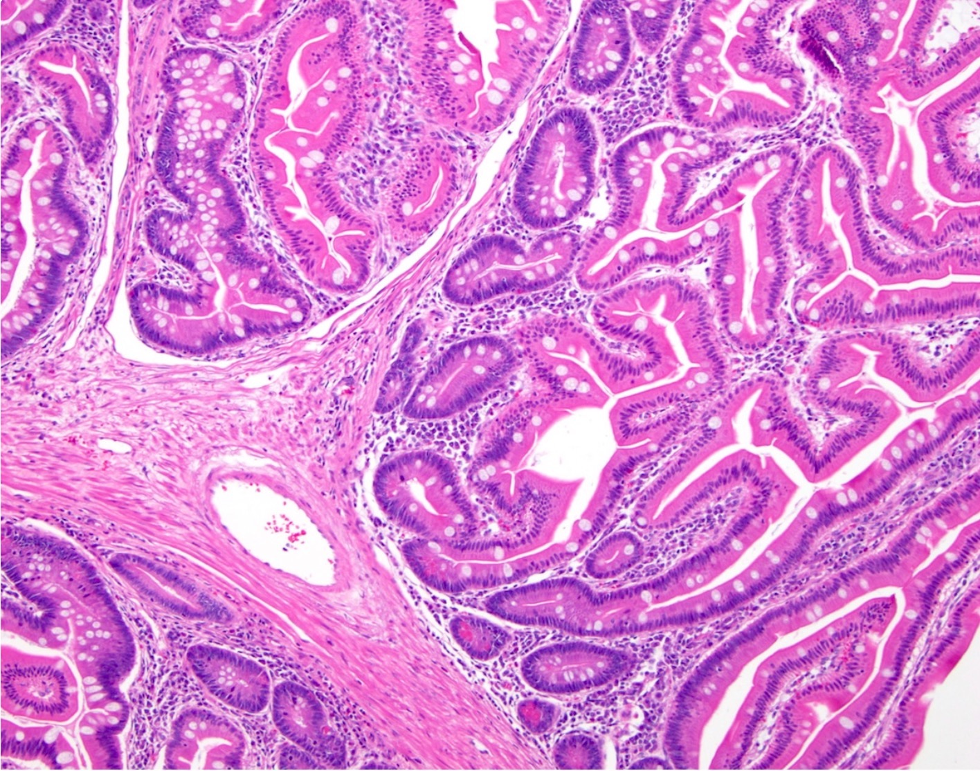
Villous adenoma
Contributed by Albina Joldoshova, M.D. and Naziheh Assarzadegan, M.D.
Pedunculated tubular adenoma
Tubulovillous adenoma
Tubular adenoma with invasive adenocarcinoma

Tubular adenoma
Invasive adenocarcinoma
Lymphovascular space invasion
Poorly differentiated adenocarcinoma

High tumor budding score
Pseudoinvasion
Pseudoinvasion
Images hosted on other servers:

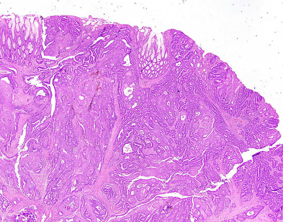
Villous tumor with central ulceration
Contributed by Raul S. Gonzalez, M.D.
Villous adenocarcinoma of colon
Contributed by Semir Vranic, M.D., Ph.D.
5x
10x
10x
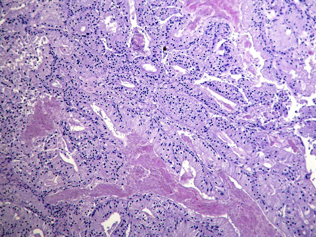
10x

CK7

20x
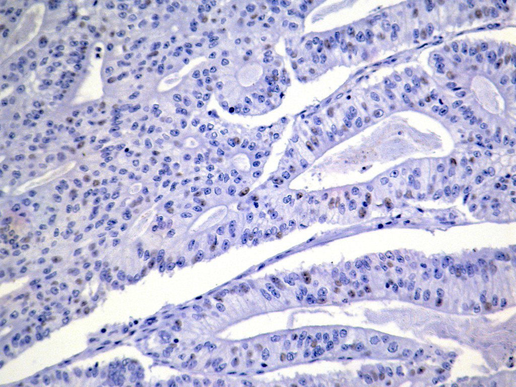
CDX2
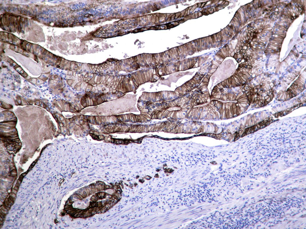
CK20
Images hosted on other servers:


Abrupt transition from benign to malignant mucosa
Images hosted on other servers:

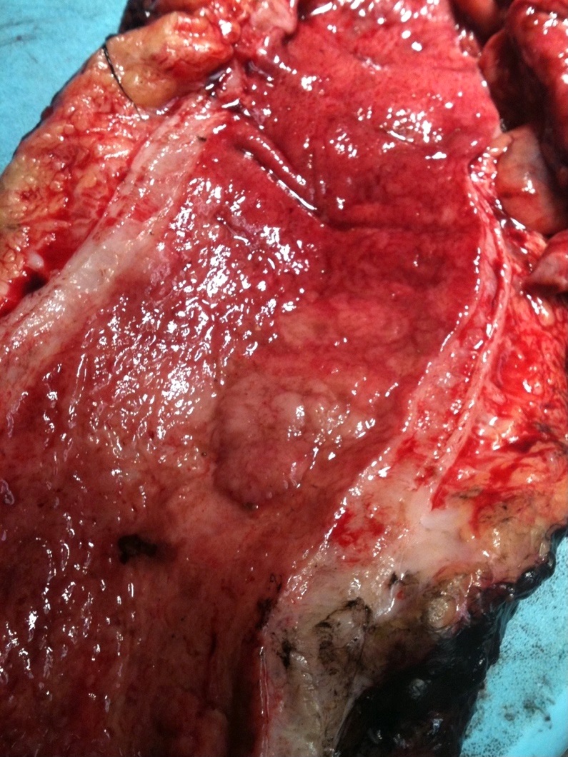
Circumferential obstructive mass

Bleeding circumferential lobulated mass
Images hosted on other servers:

Cross section
Contributed by Raul S. Gonzalez, M.D.
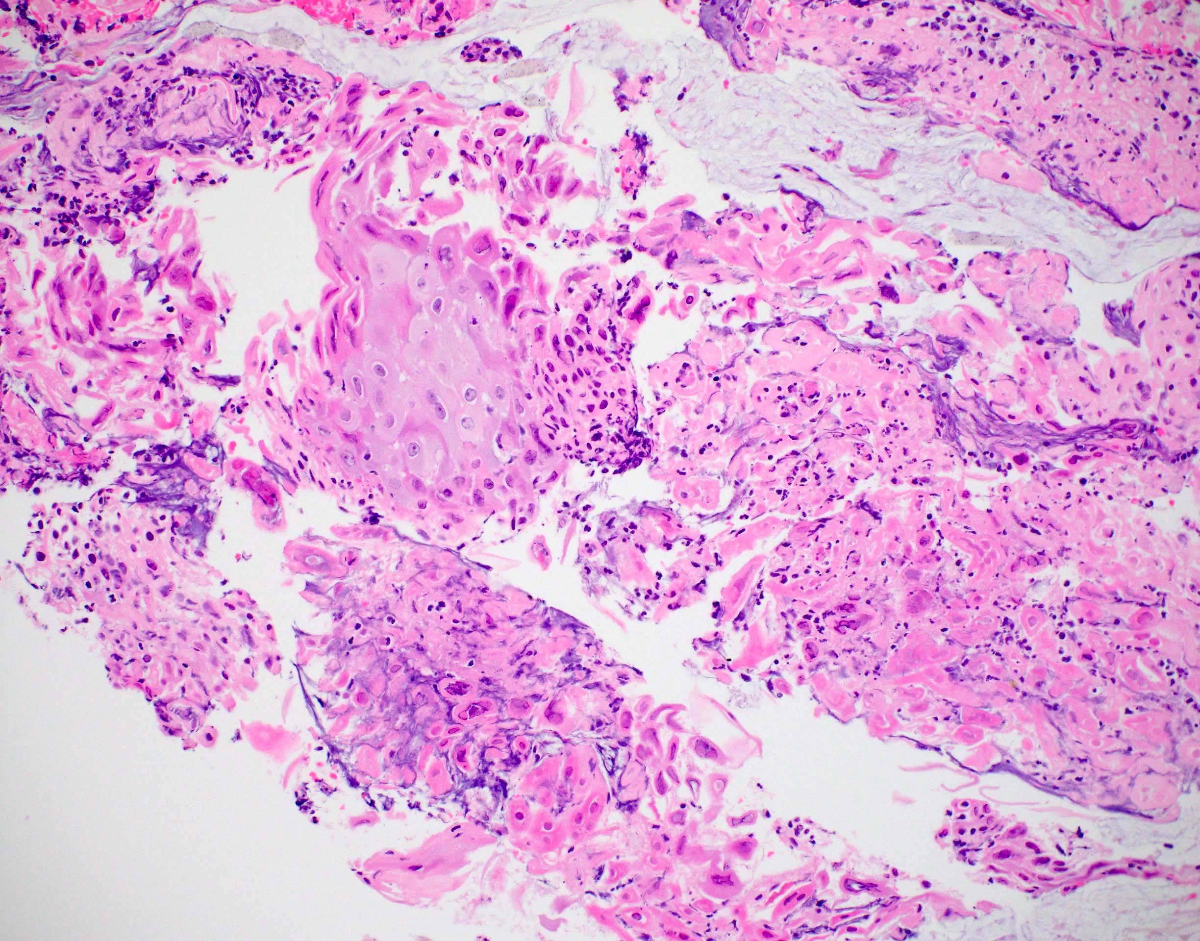
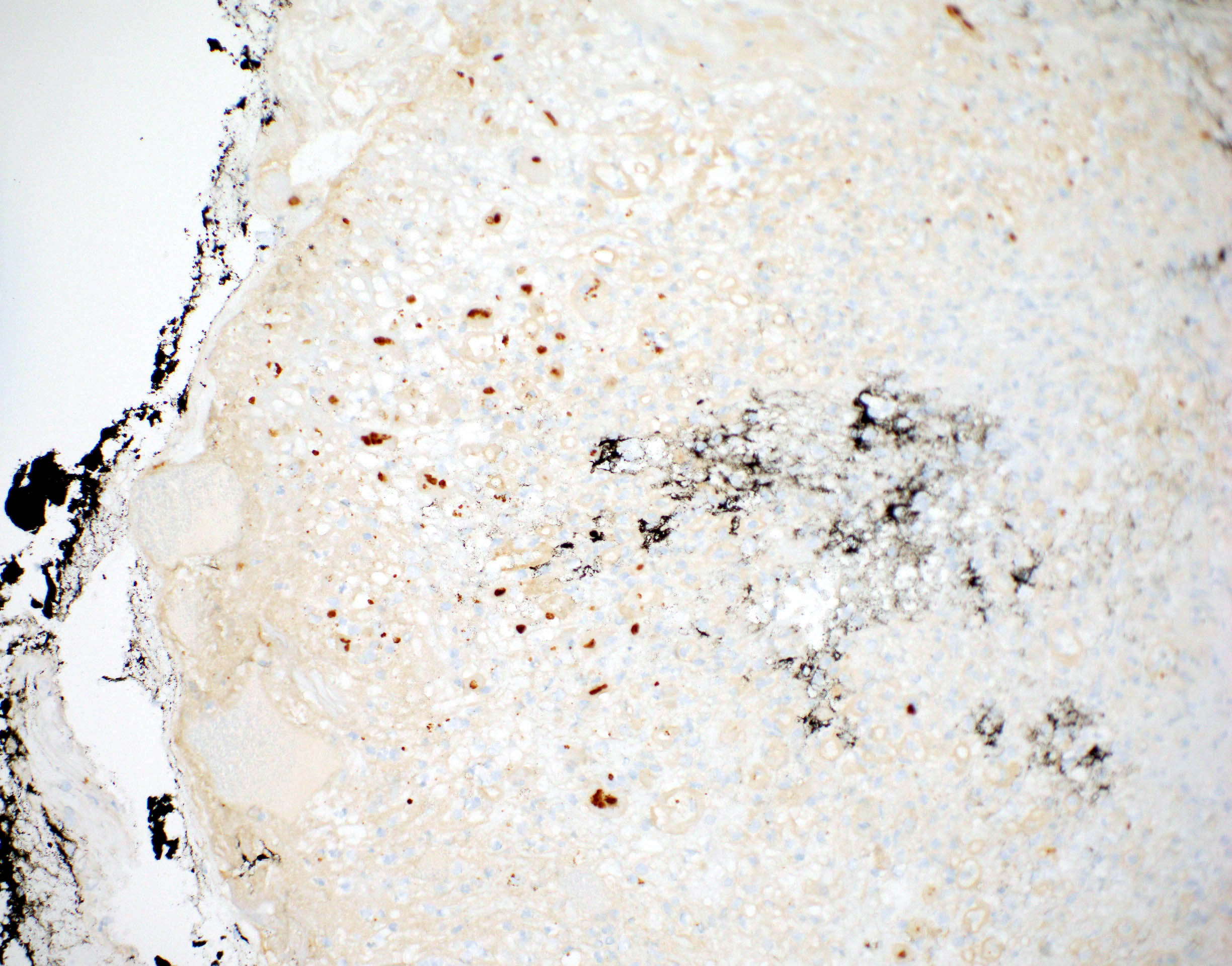
Adenosquamous carcinoma
Images hosted on other servers:

Collision-like areas

Composite-like areas

Adenosquamous carcinoma

Malignant squamous cells with keratinization
Images hosted on other servers:

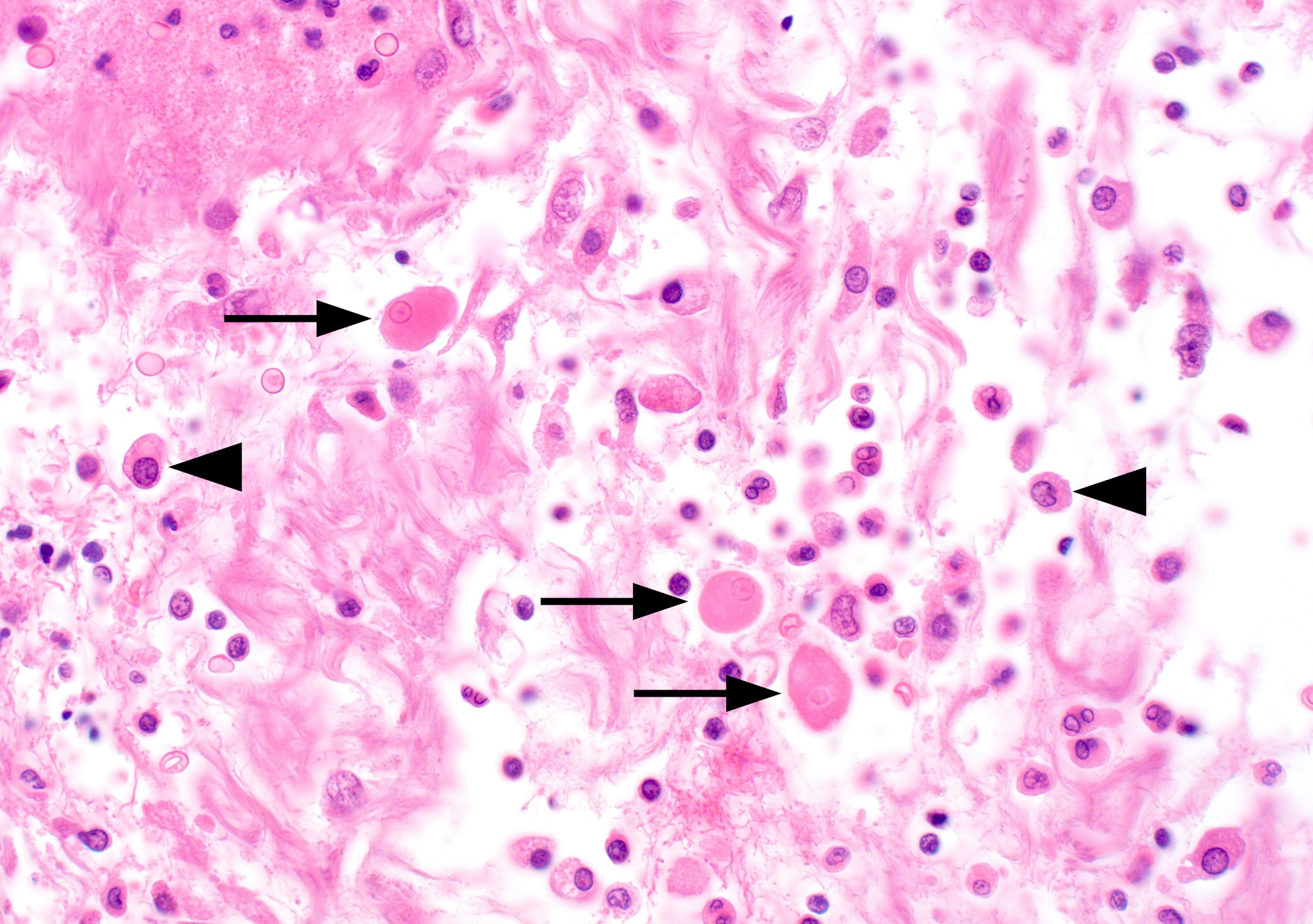
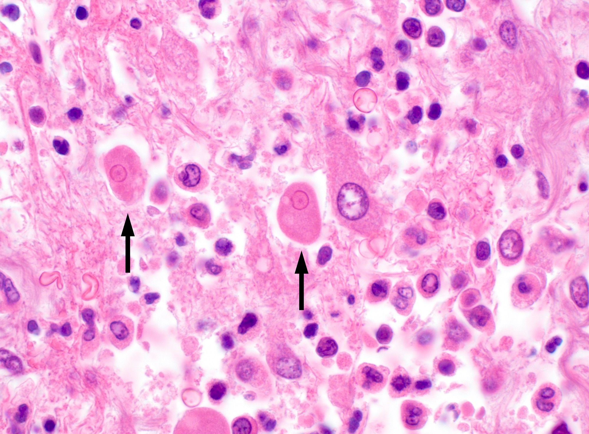
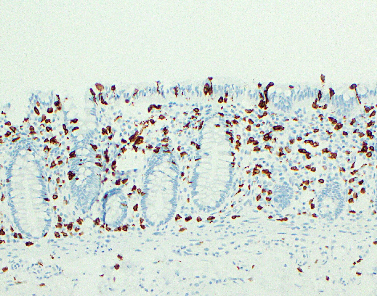
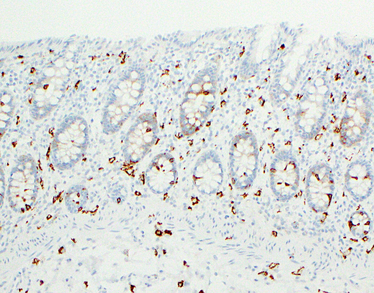
Infected cells with
irregular amphophilic
nuclei

Cells have eccentric
nuclei and vacuolated
cytoplasm

Adenovirus immunostain
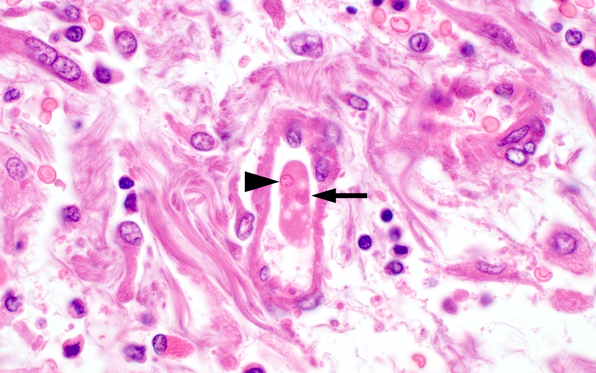
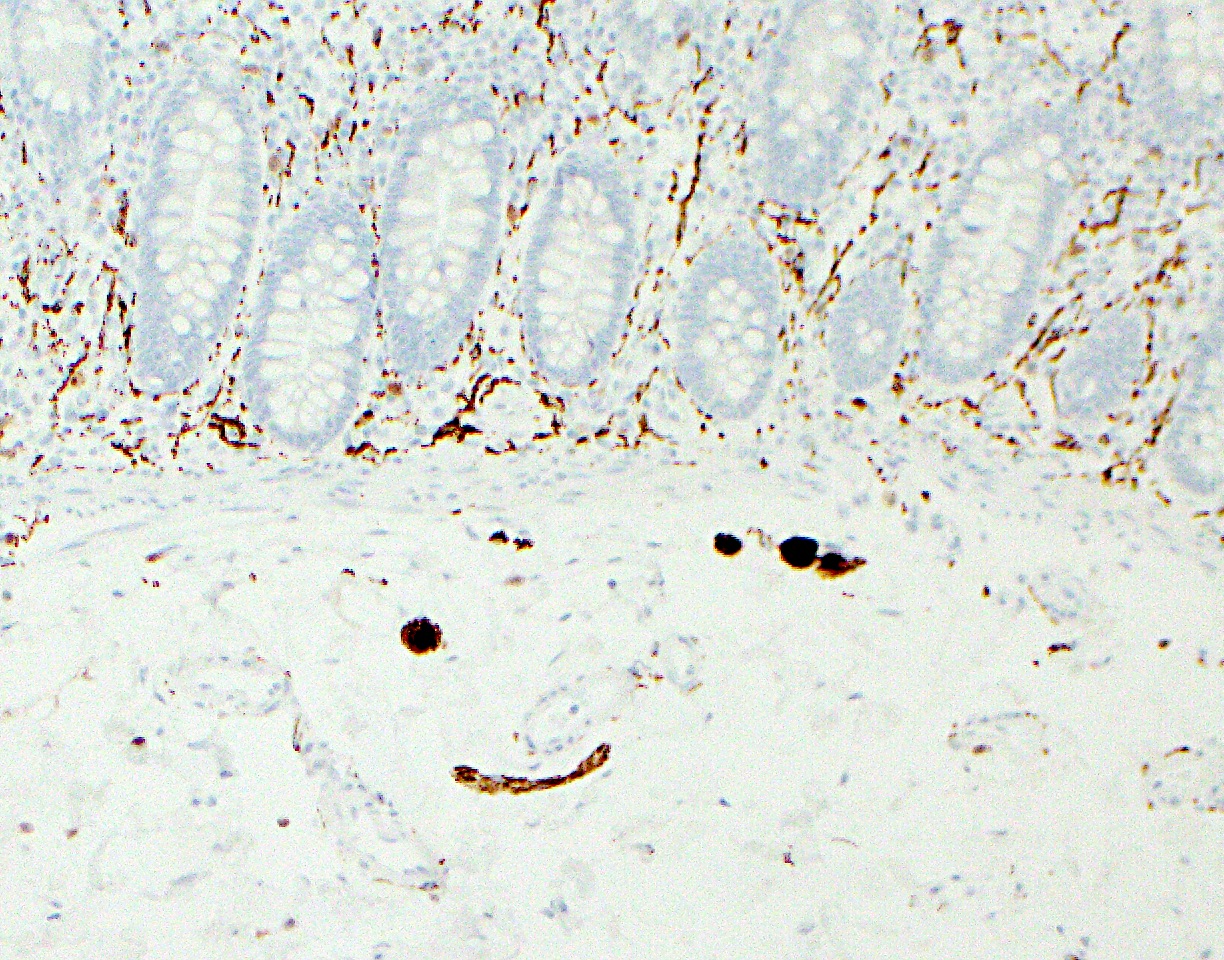
Images hosted on other servers:

Intranuclear inclusions
with (inset) regular
pattern of arrangement
Images hosted on other servers:

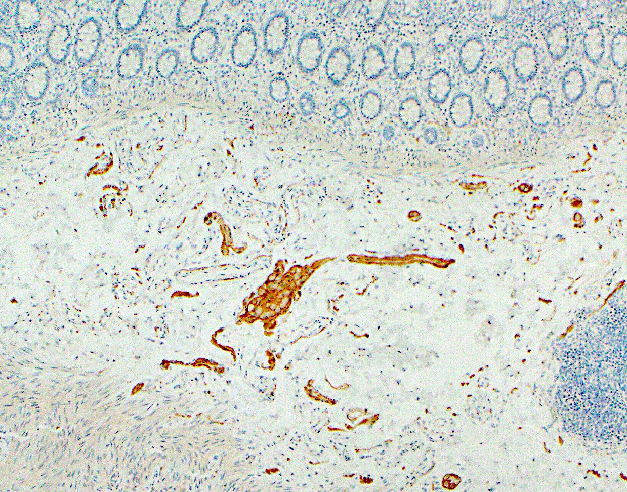
Between loops of intestine
Contributed by Raul S. Gonzalez, M.D.
Serosal adhesions
Images hosted on other servers:

Attached to intestinal
serosa and
linking viscera
Images hosted on other servers:

Endoscopic findings
Images hosted on other servers:

Eosinophilic infiltration

Images hosted on other servers:

Fulminant colitis and hepatic amebiasis
Contributed by Centers for Disease Control and Prevention
Ulcerative intestinal amebiasis

Diffuse ulcerative amebic colitis

Amebic colitis with perforation
Images hosted on other servers:

Ulcer
Contributed by Centers for Disease Control and Prevention, Bobbi S. Pritt, M.D., M.Sc. and Blaine Mathison
Classic flask shaped ulcer
Edge of flask shaped ulcer
Invading trophozoites

Trophozoites invading muscle
Amebic trophozoites
Trophozoite in blood vessel
PAS positive trophozoites

Trichrome stained stool specimen
Images hosted on other servers:

Amyloid tumor (above)
and adenocarcinoma
arising from villous
adenoma (below)
Contributed by Raul S. Gonzalez, M.D.
Colonic amyloid
Images hosted on other servers:

Submucosal vessel involvement

With Congo red stain

Congo red stain
highlights vessel
wall and free
submucosal amyloid

Congo red stain

Subepithelial
deposits resembling
collagenous colitis
Contributed by Reade Quinton, M.D.

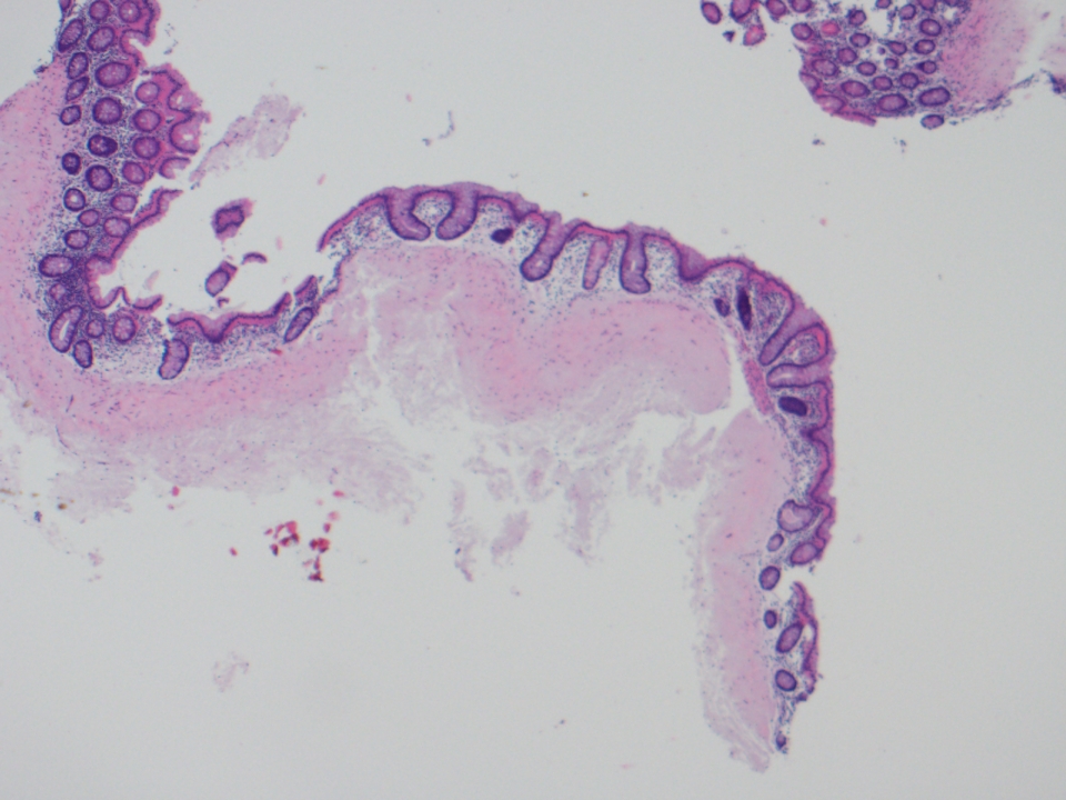
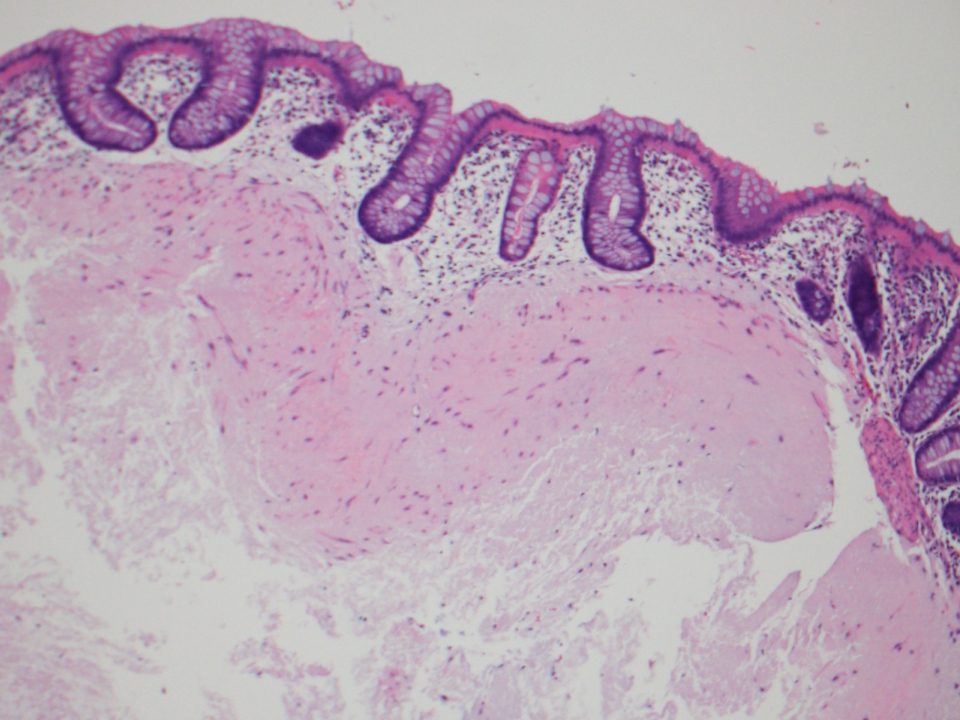
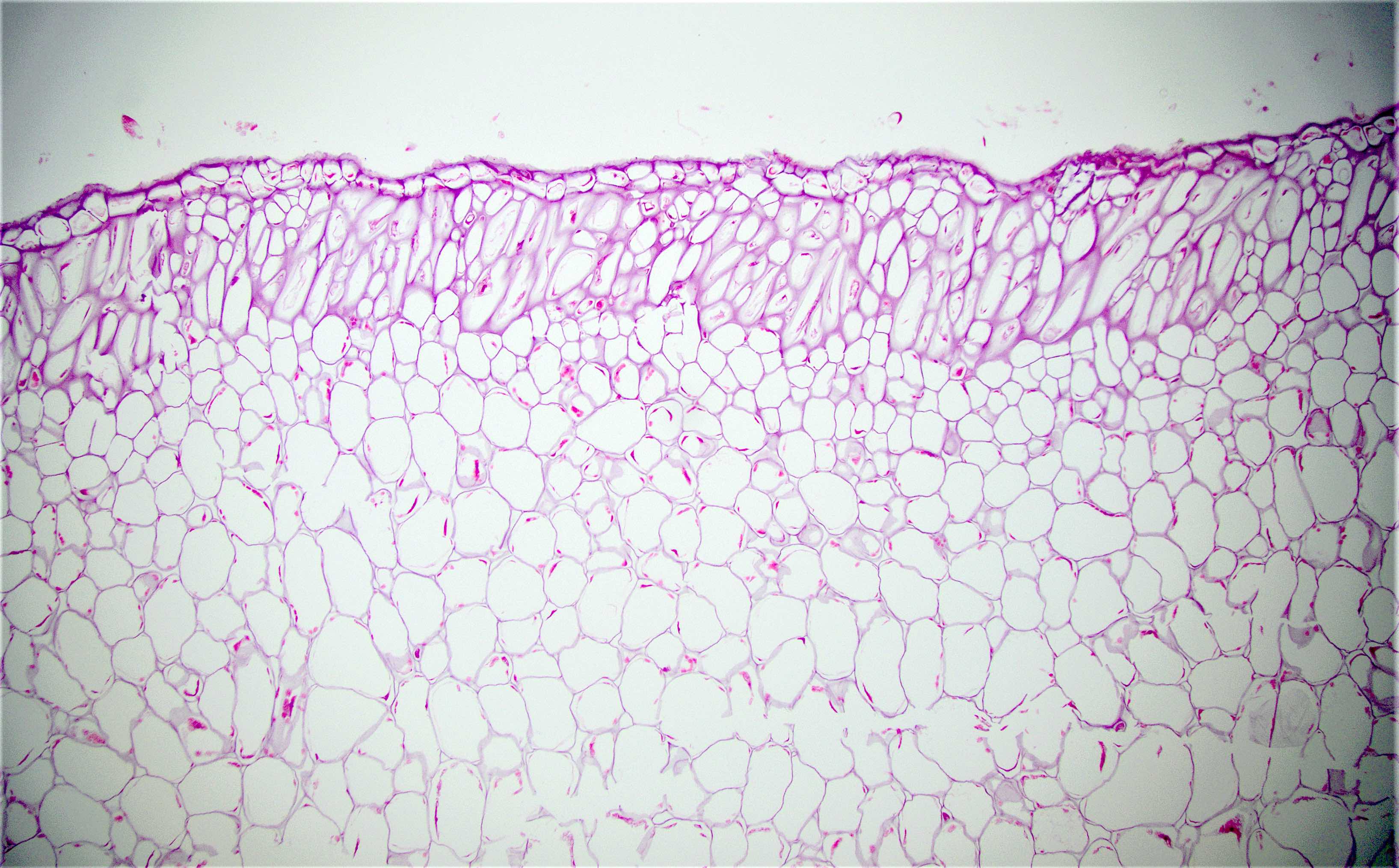
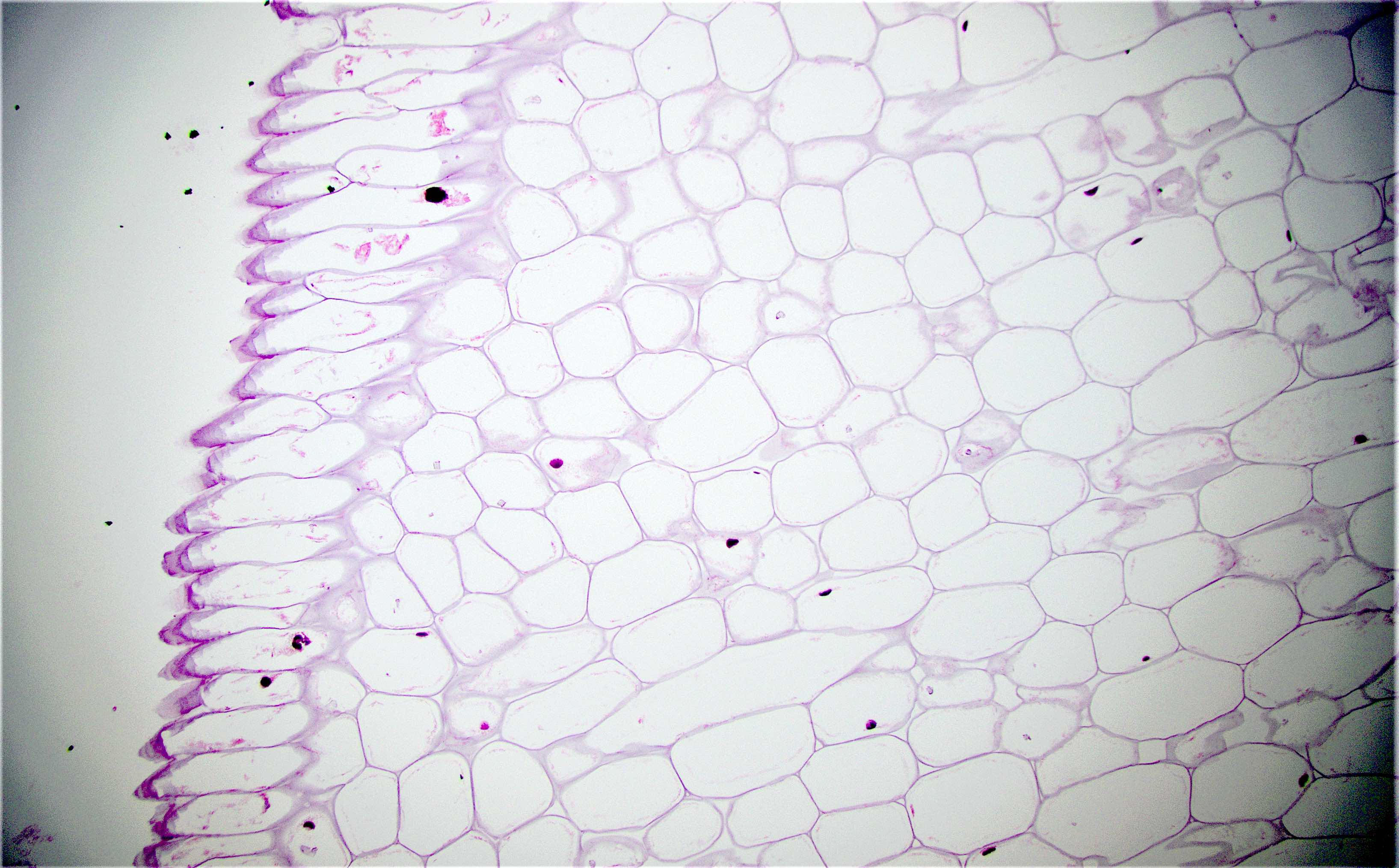
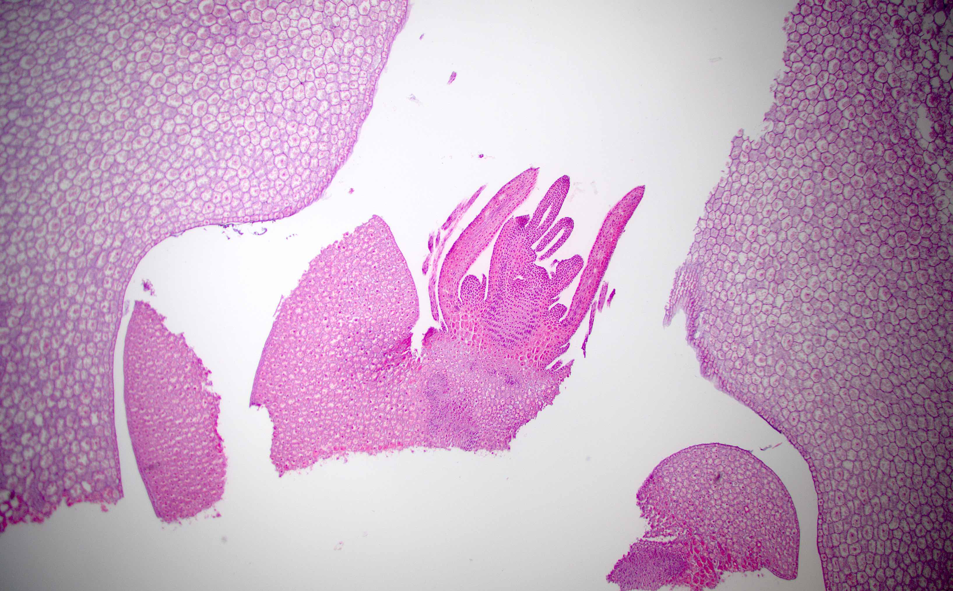
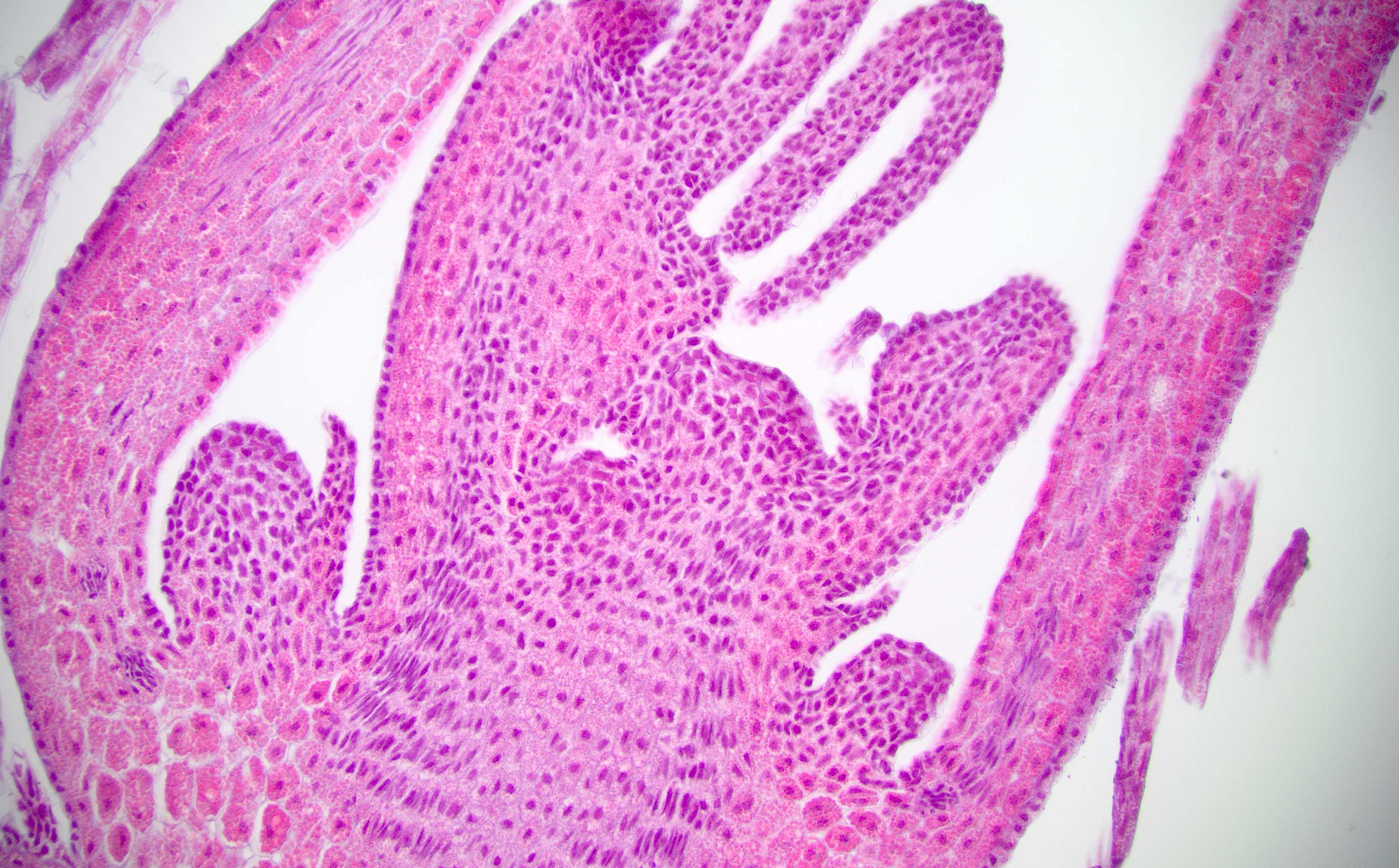
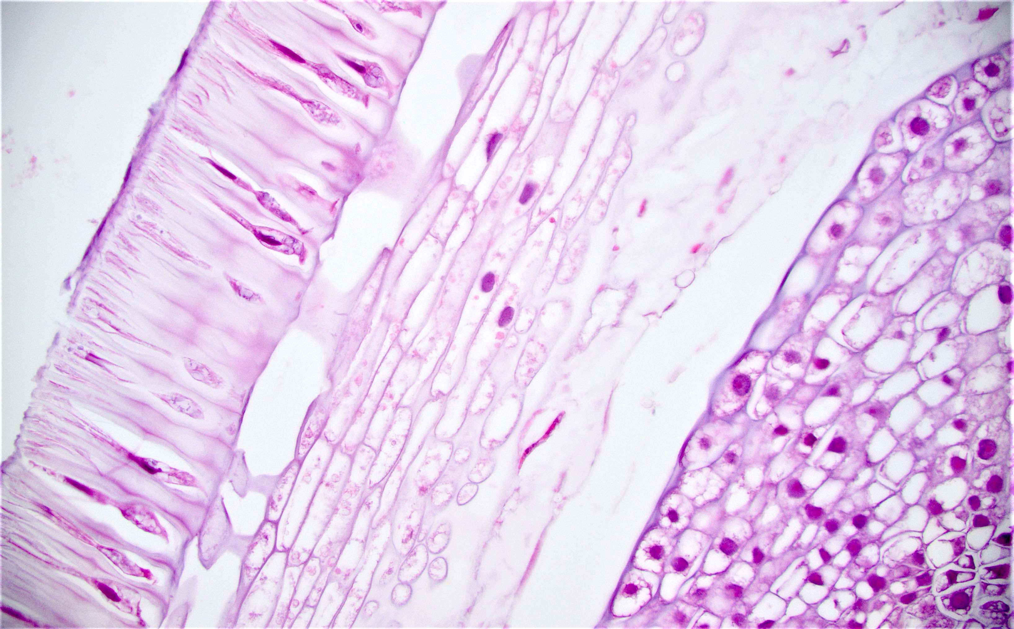
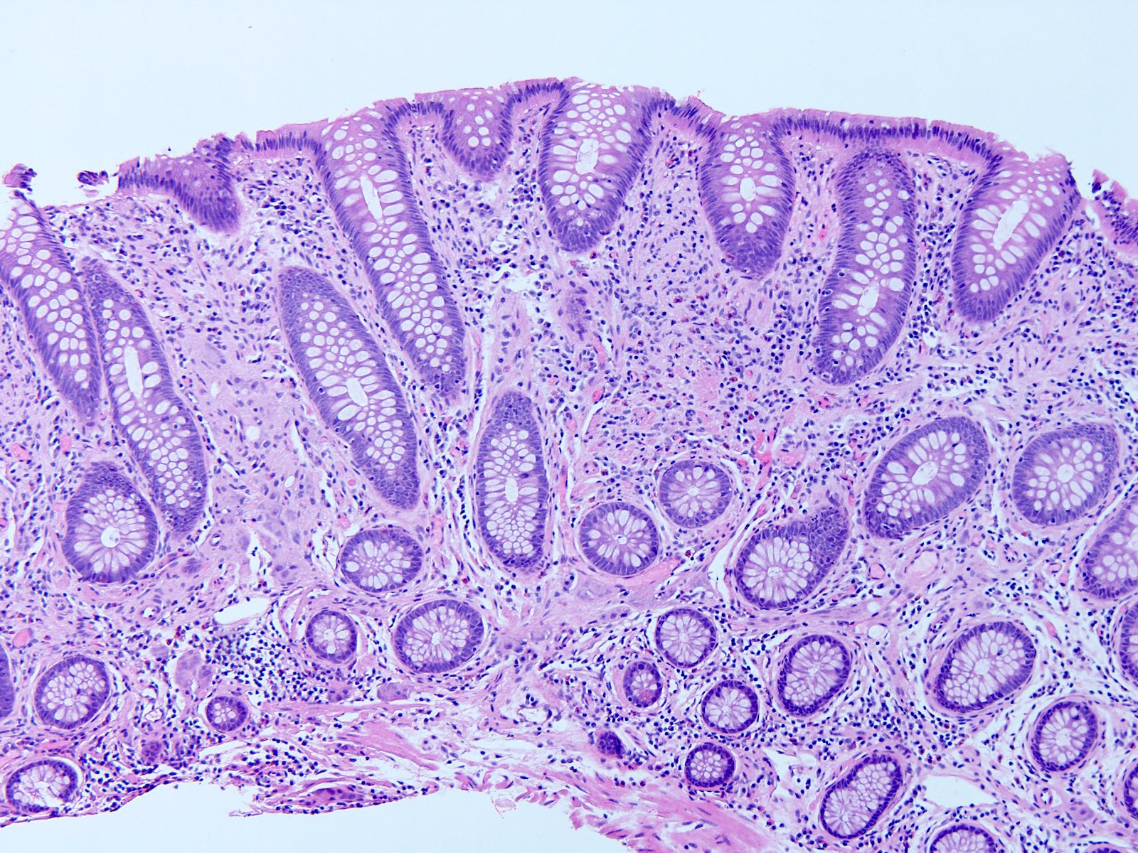
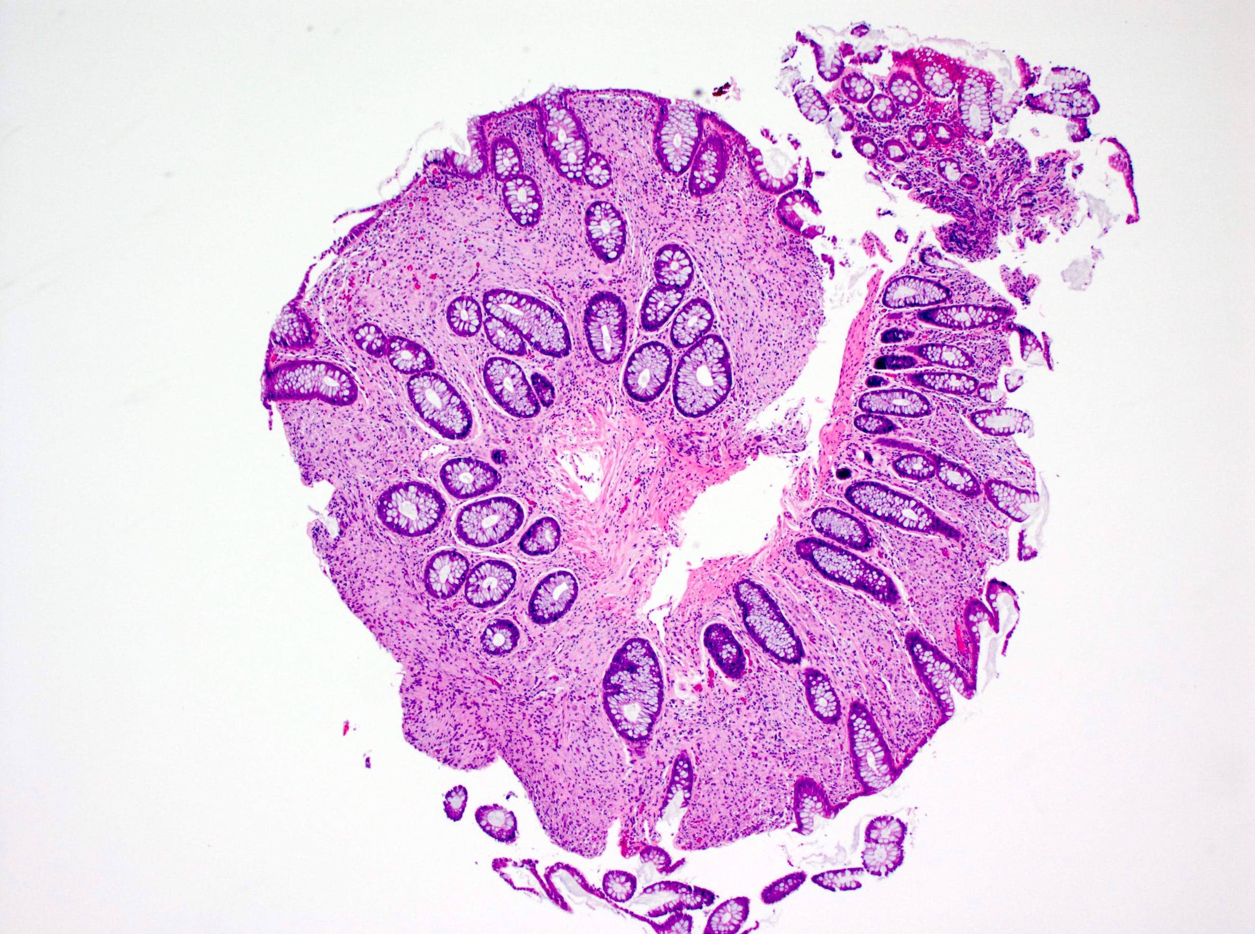
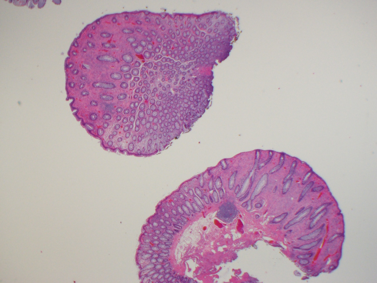
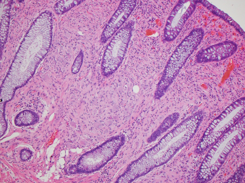
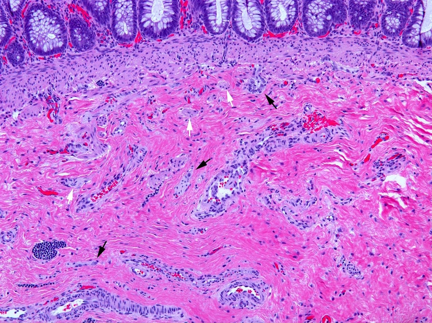
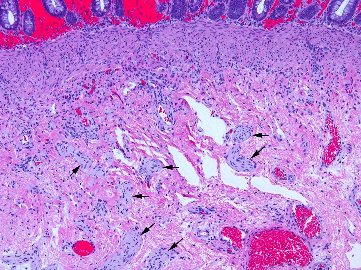
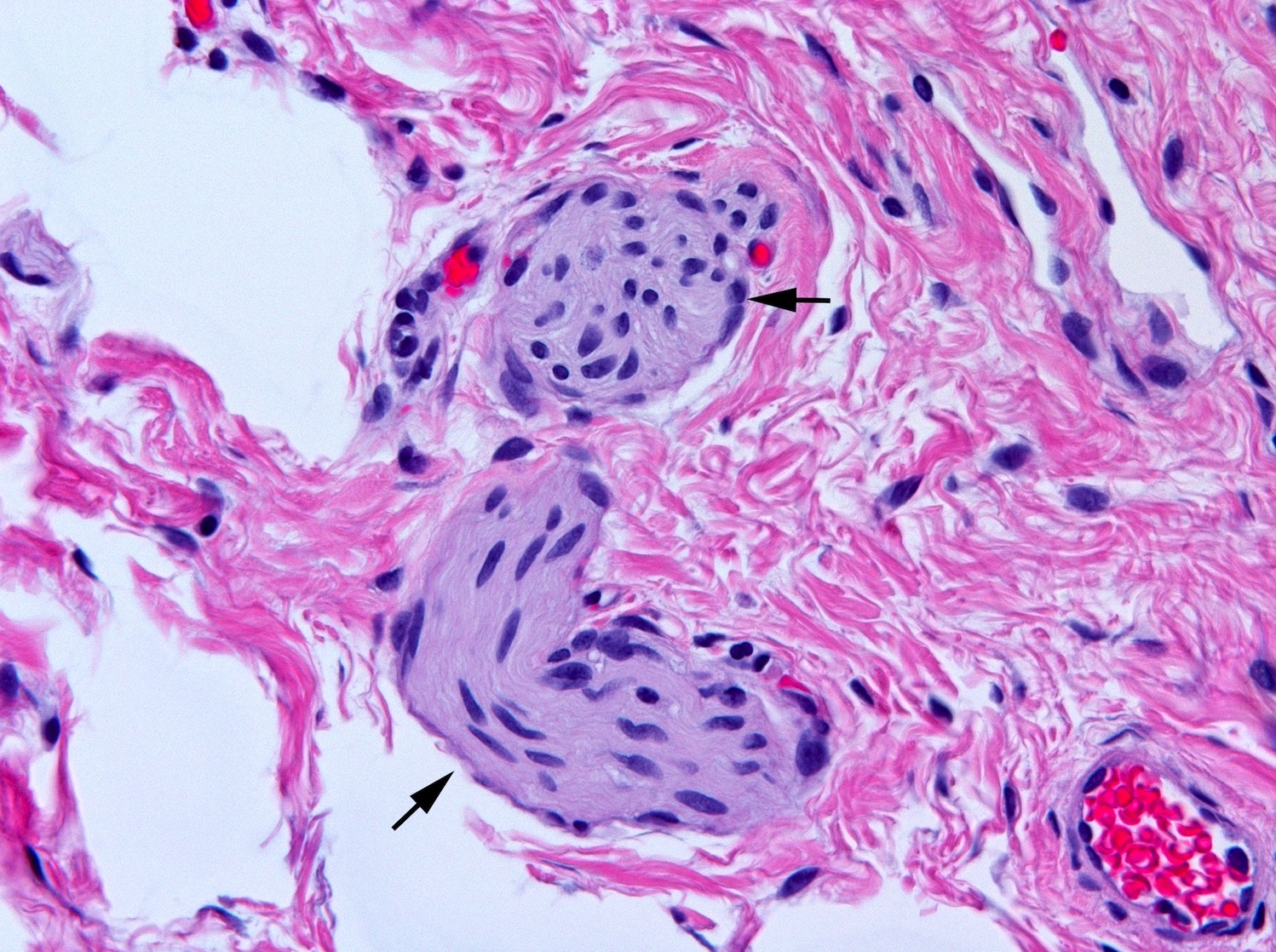
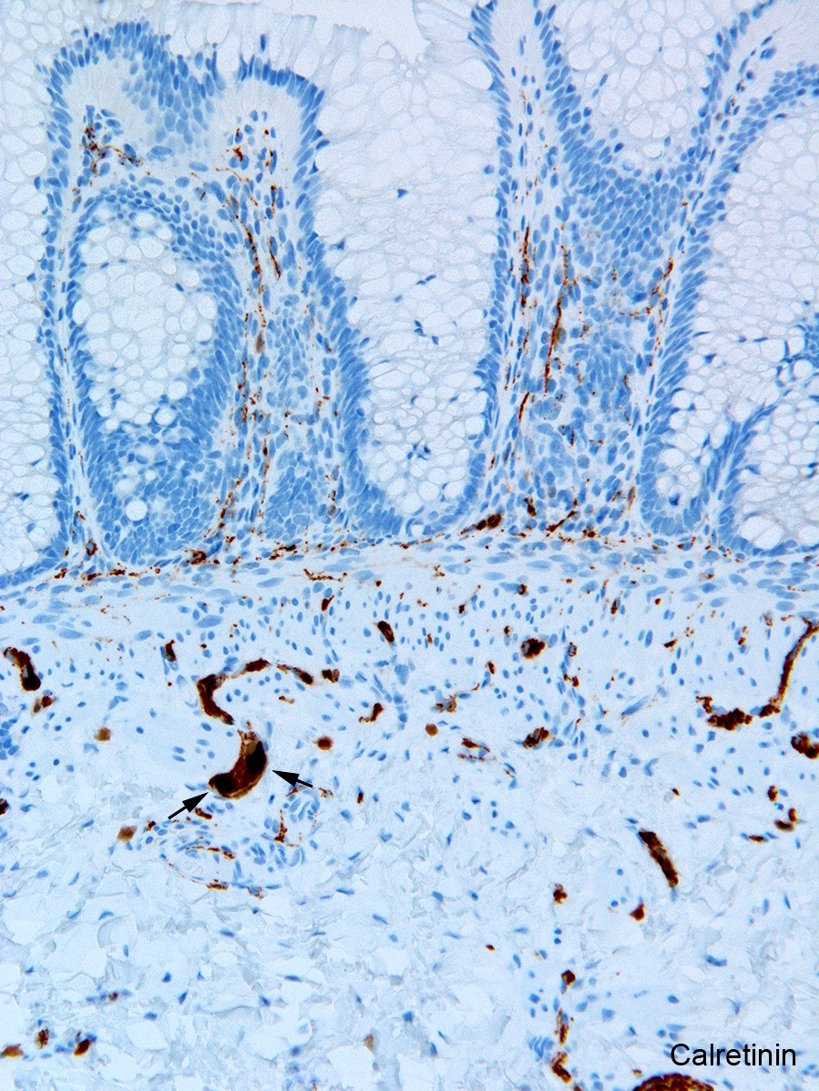
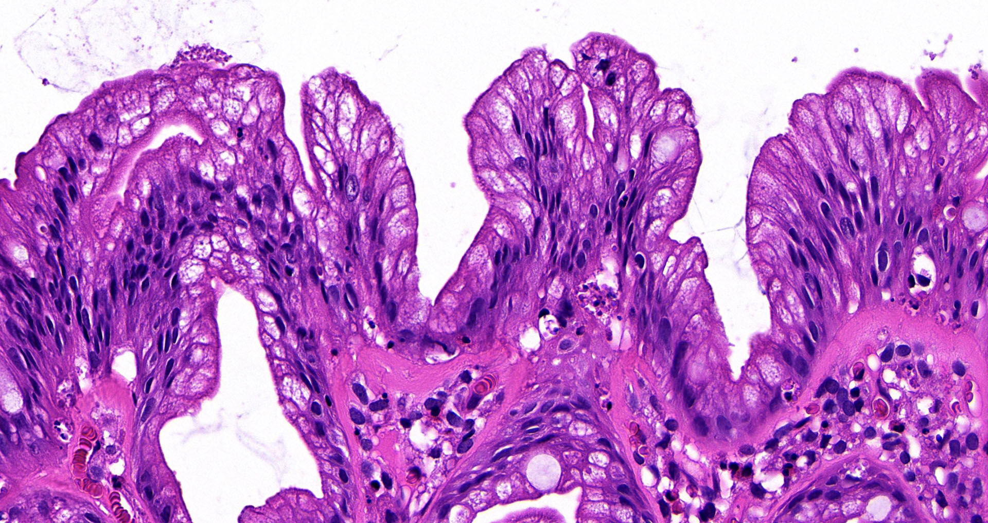
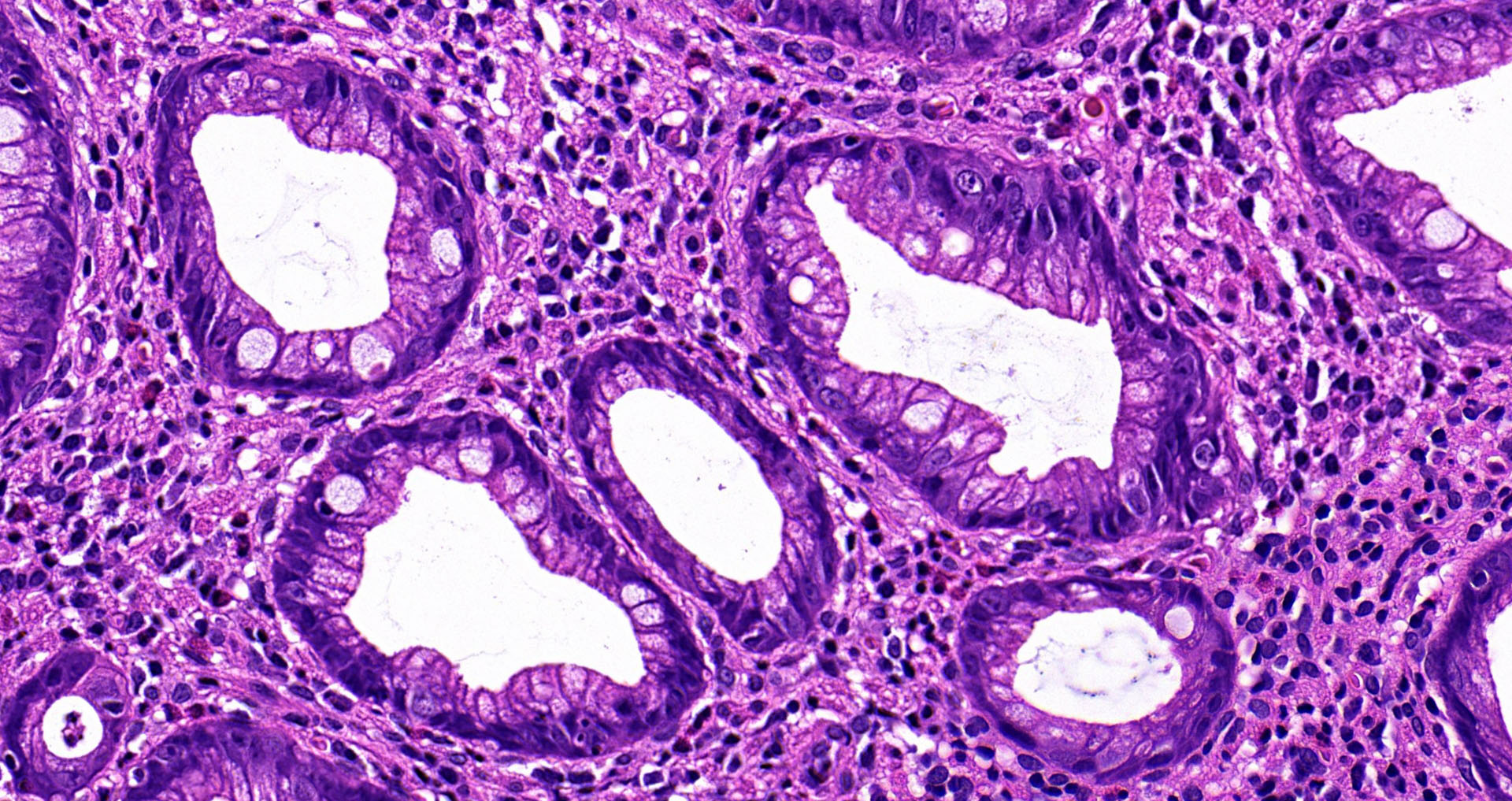
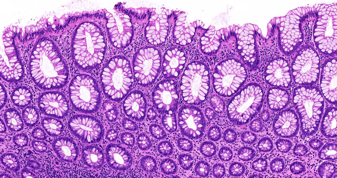
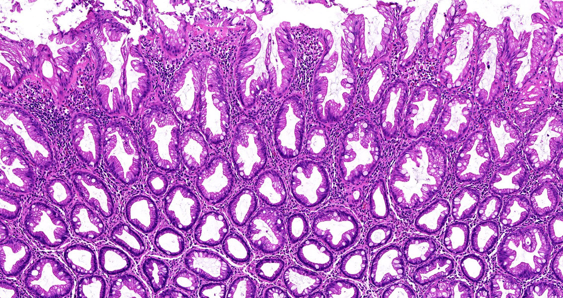
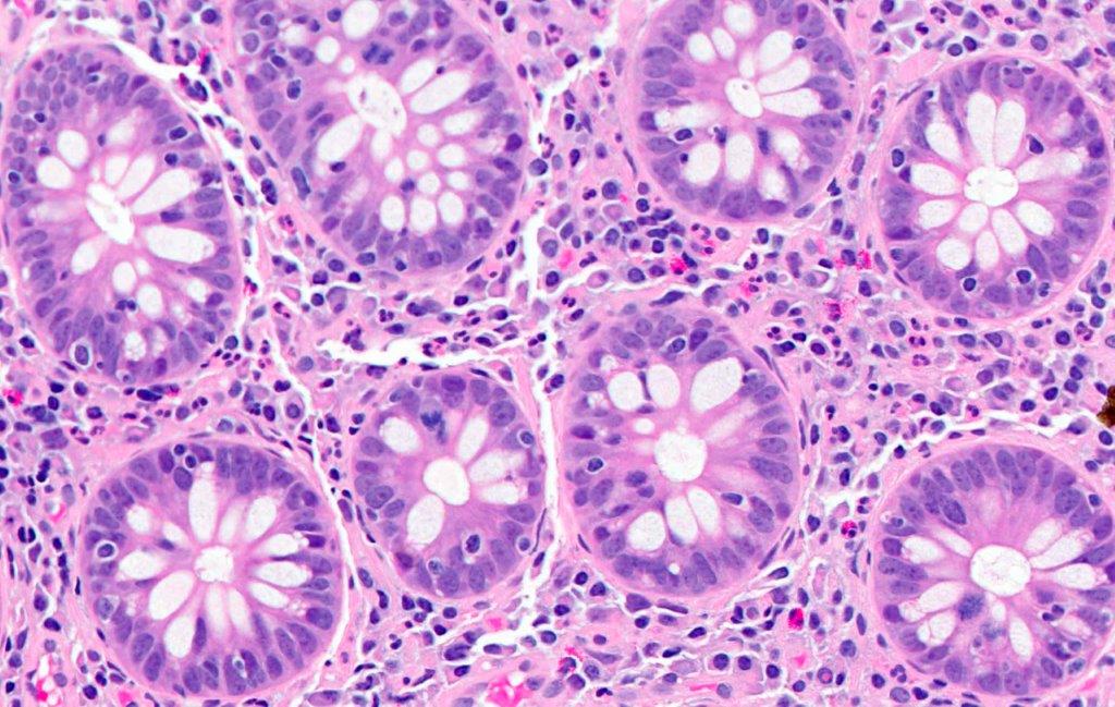
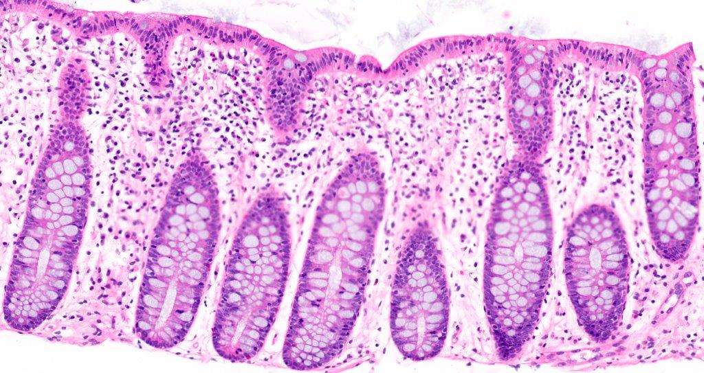
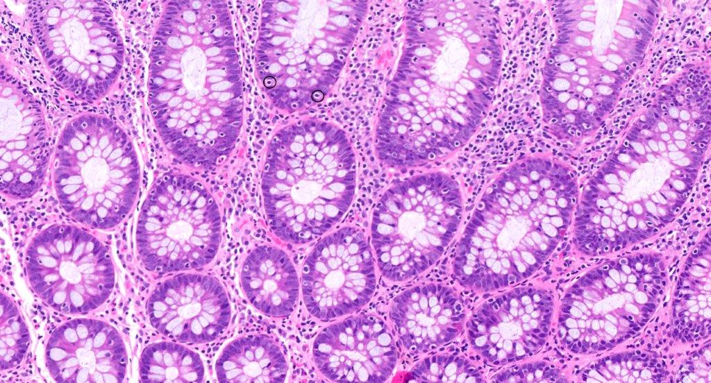
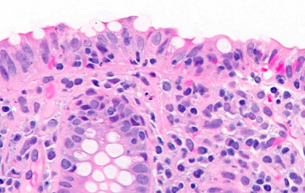
Colon
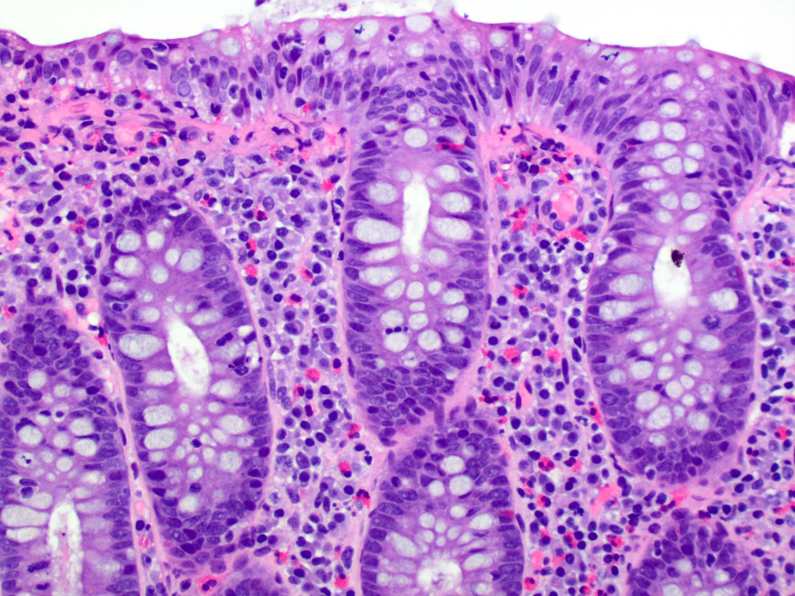
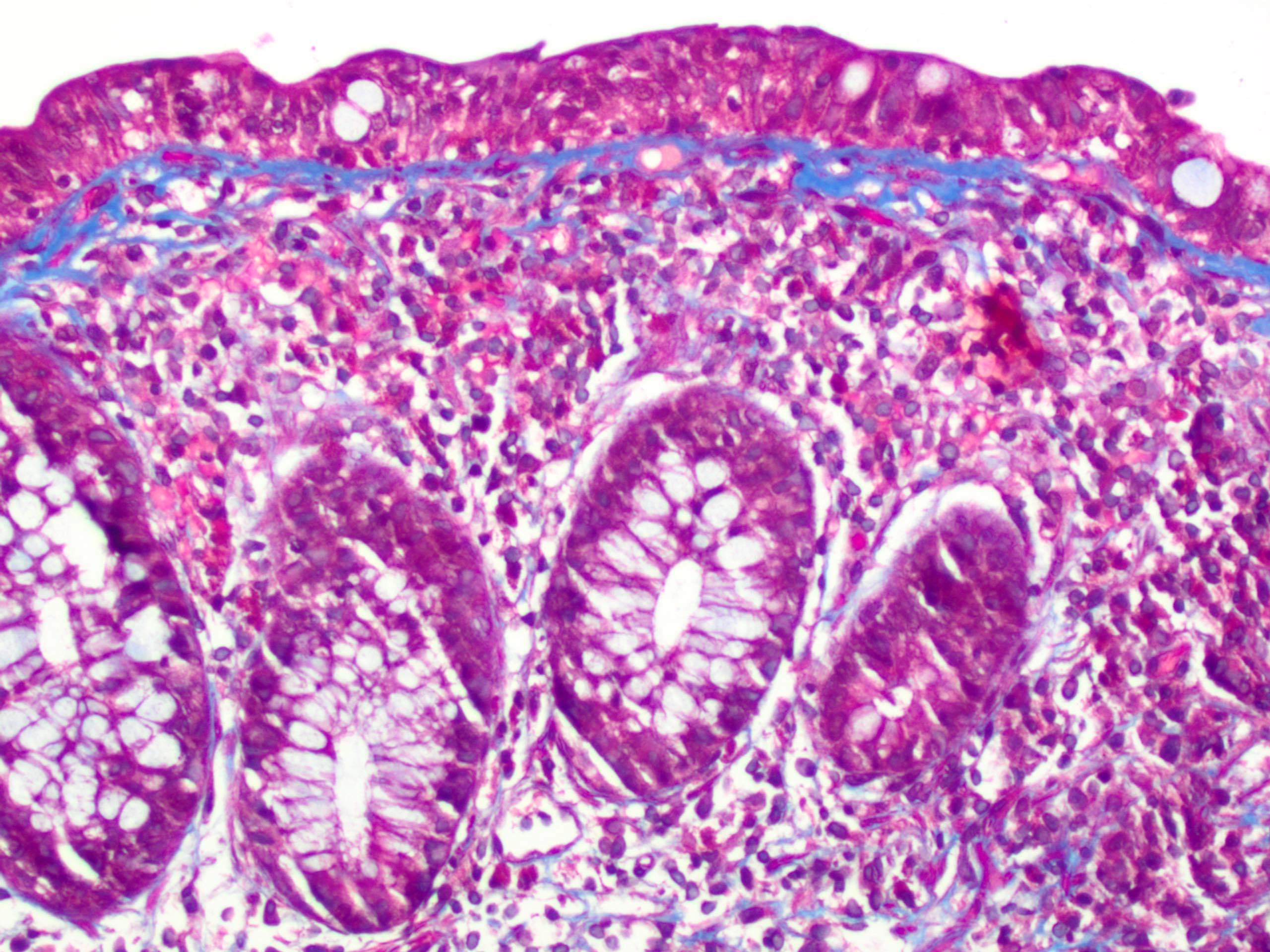
Contributed by Lizhi Zhang, M.D.
Colonic wall
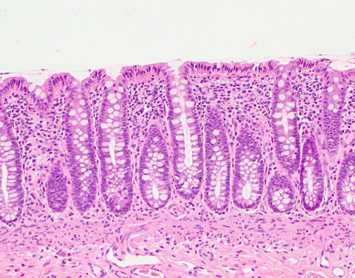
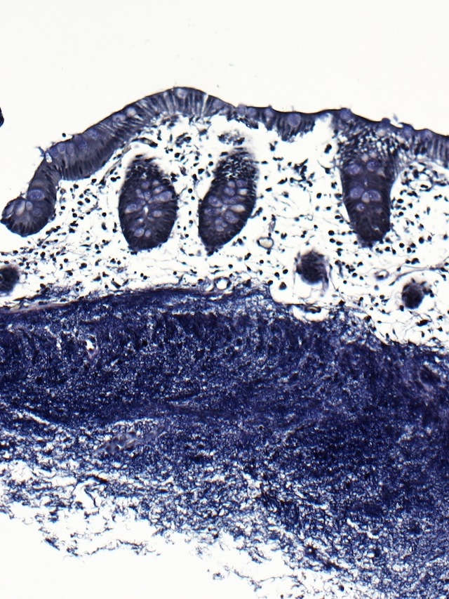
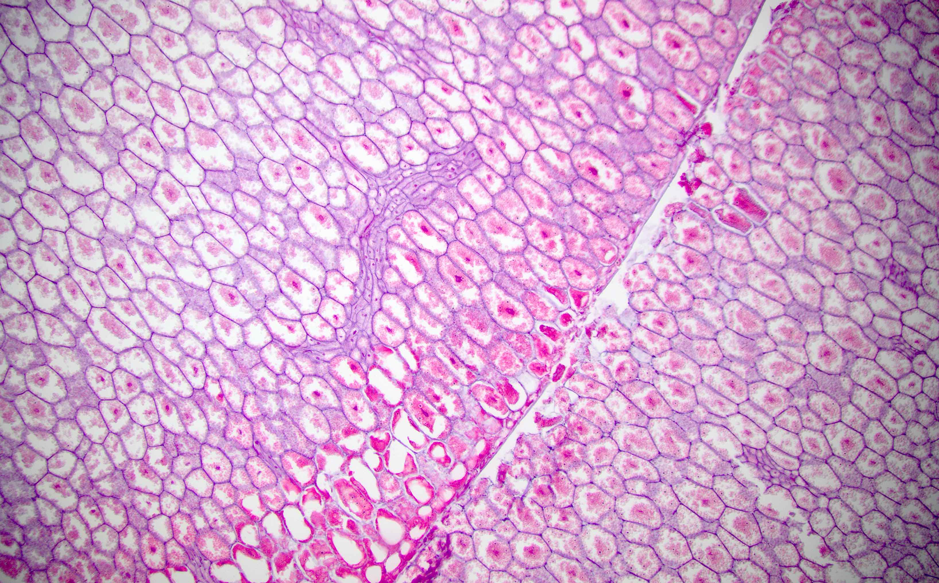
Colonic mucosa

Cross section of crypt and lamina propria
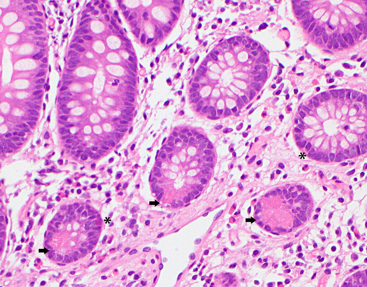
Paneth cells and enteroendocrine cells
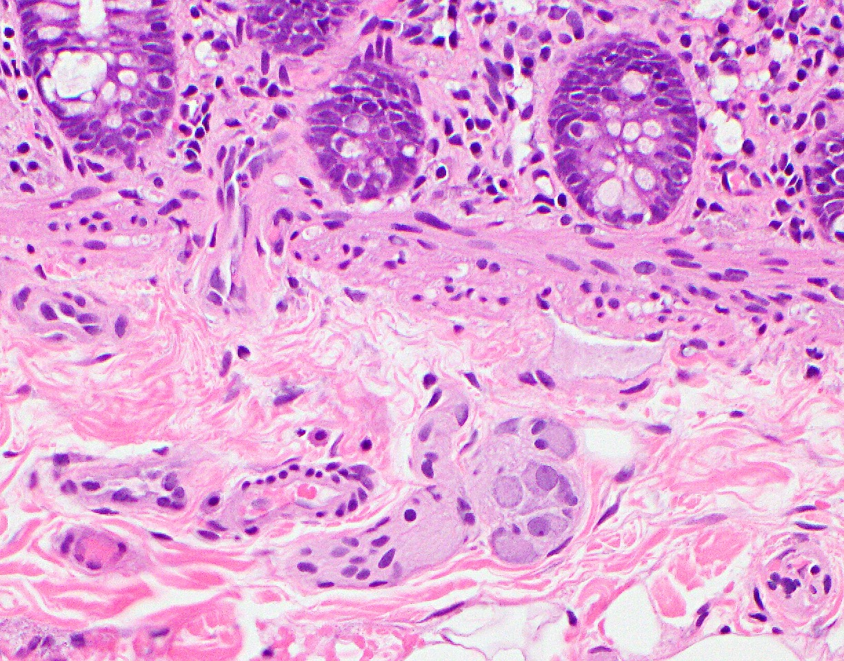
Submucosa and submucosal neural plexuses
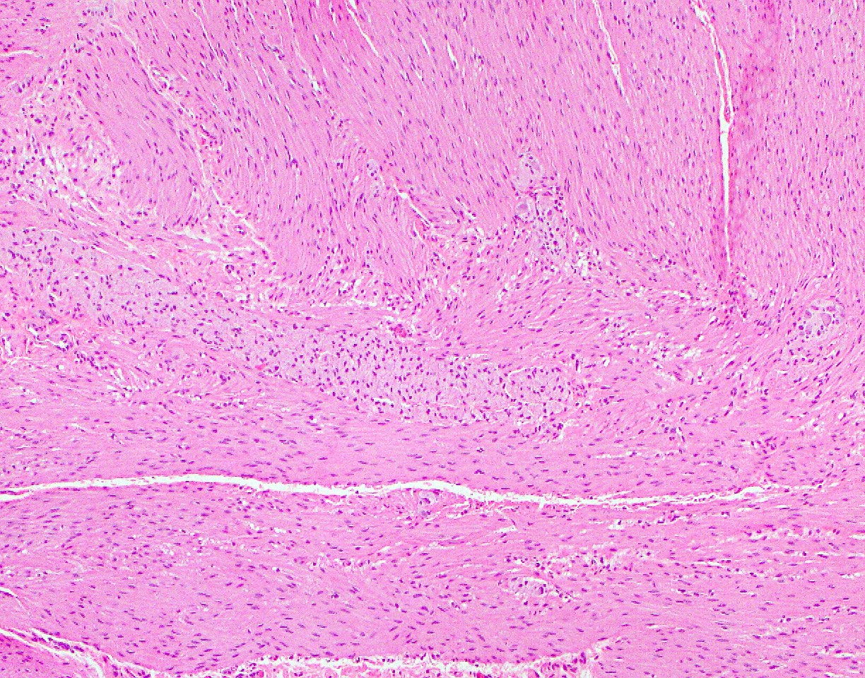
Muscularis propria and myenteric plexus
CD3
Mast cells - KIT
Calretinin
S100
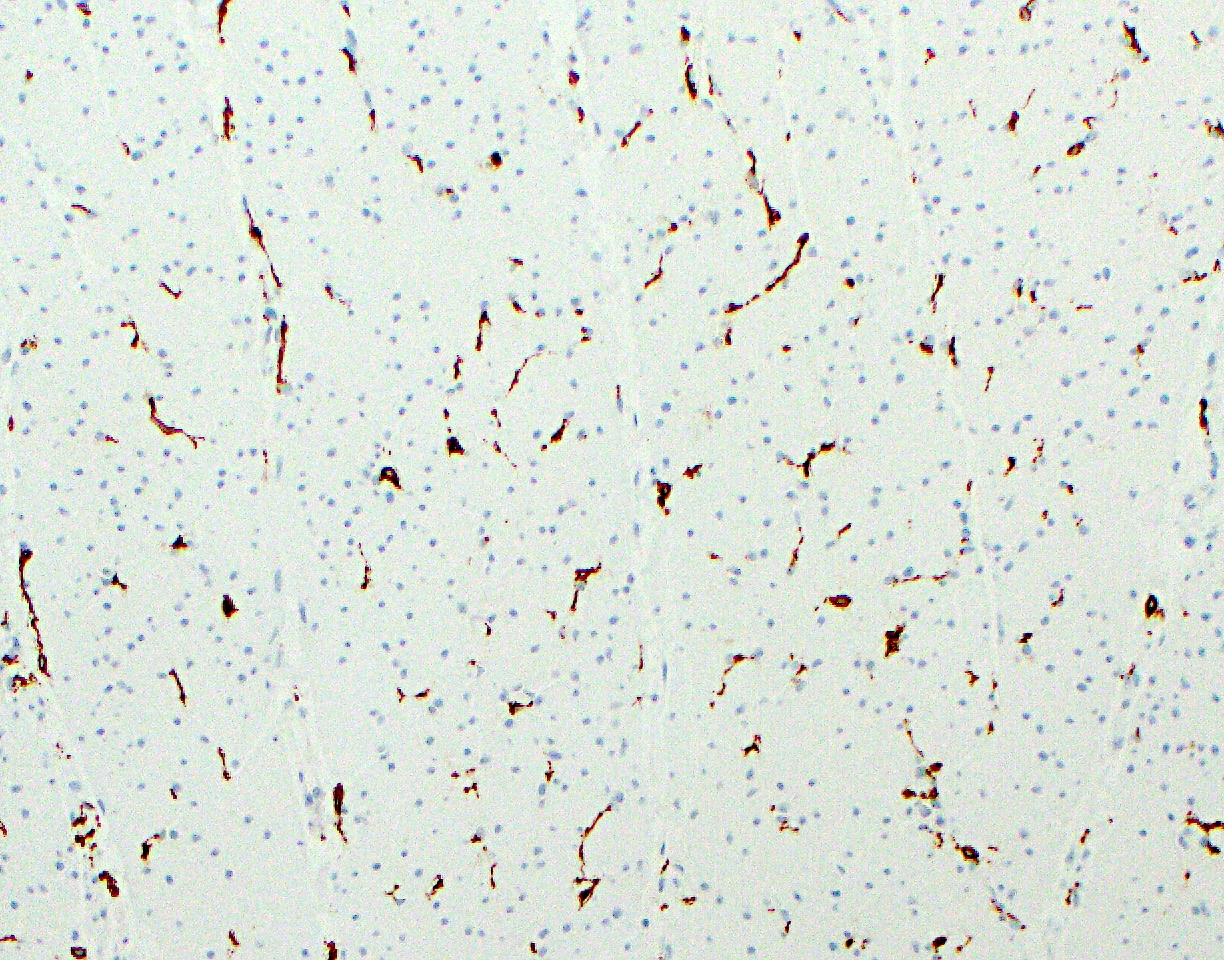
Interstitial cells of Cajal
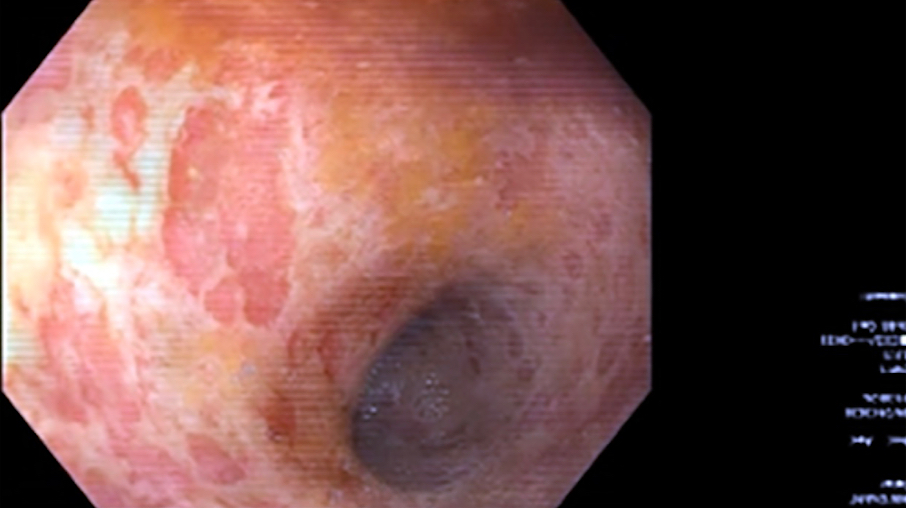
Images hosted on other servers:

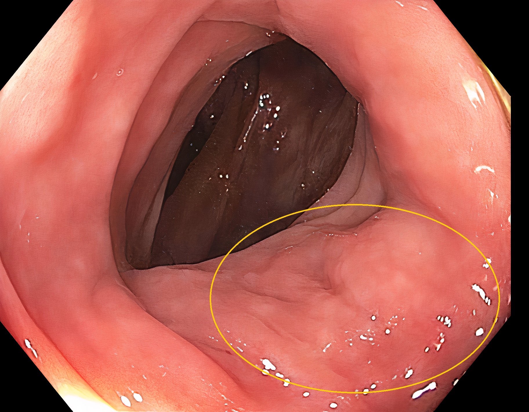
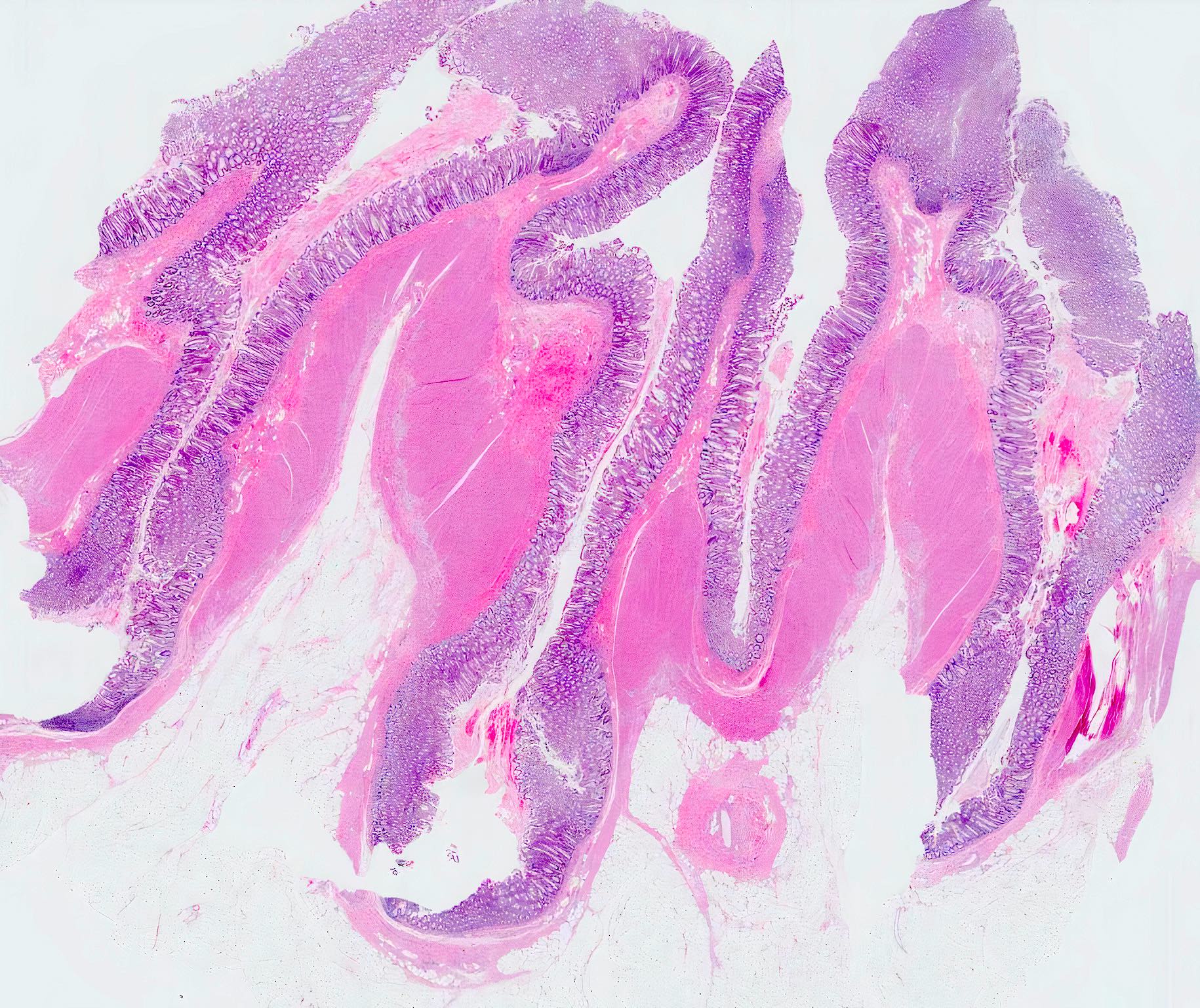
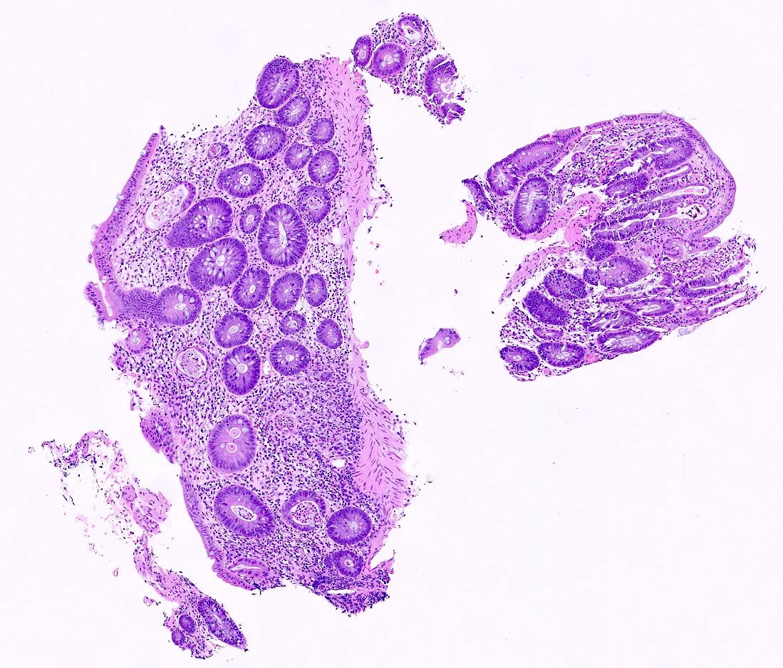
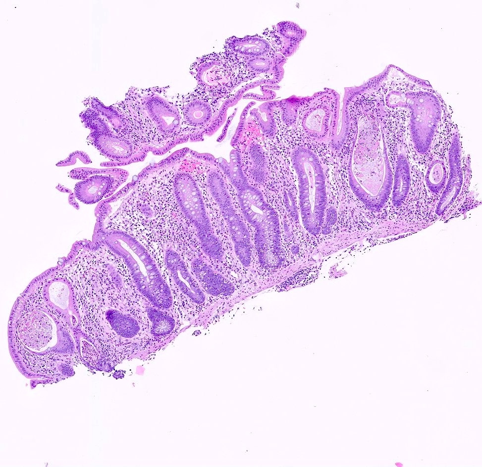
Colonoscopy
Images hosted on other servers:

Small bowel perforation

Perforation and colonic mucosa
Contributed by Maryam Kherad Pezhouh, M.D.
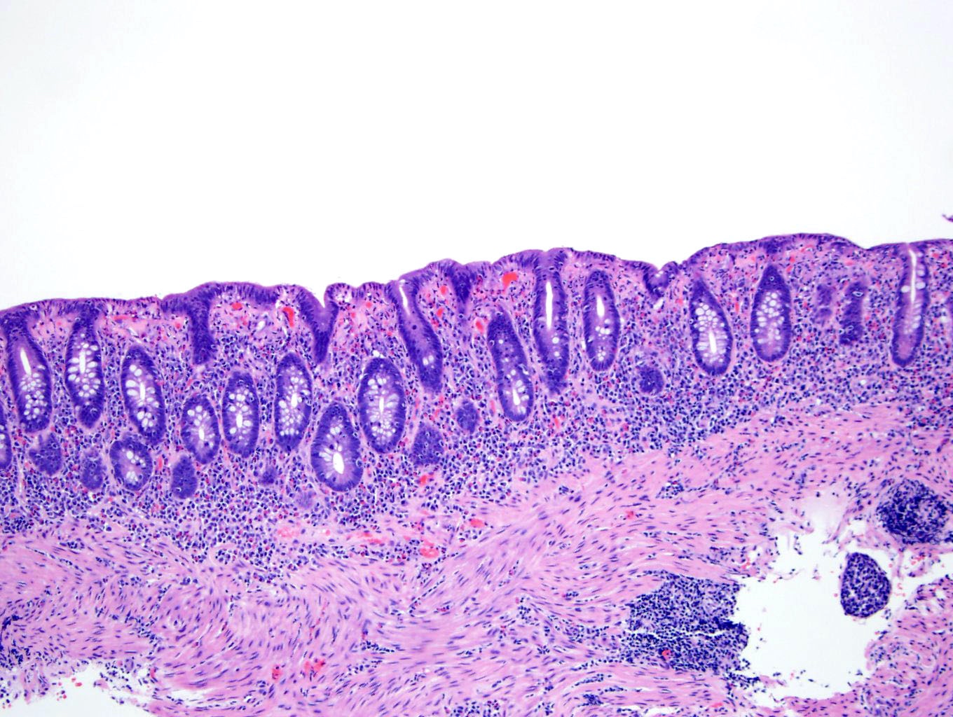
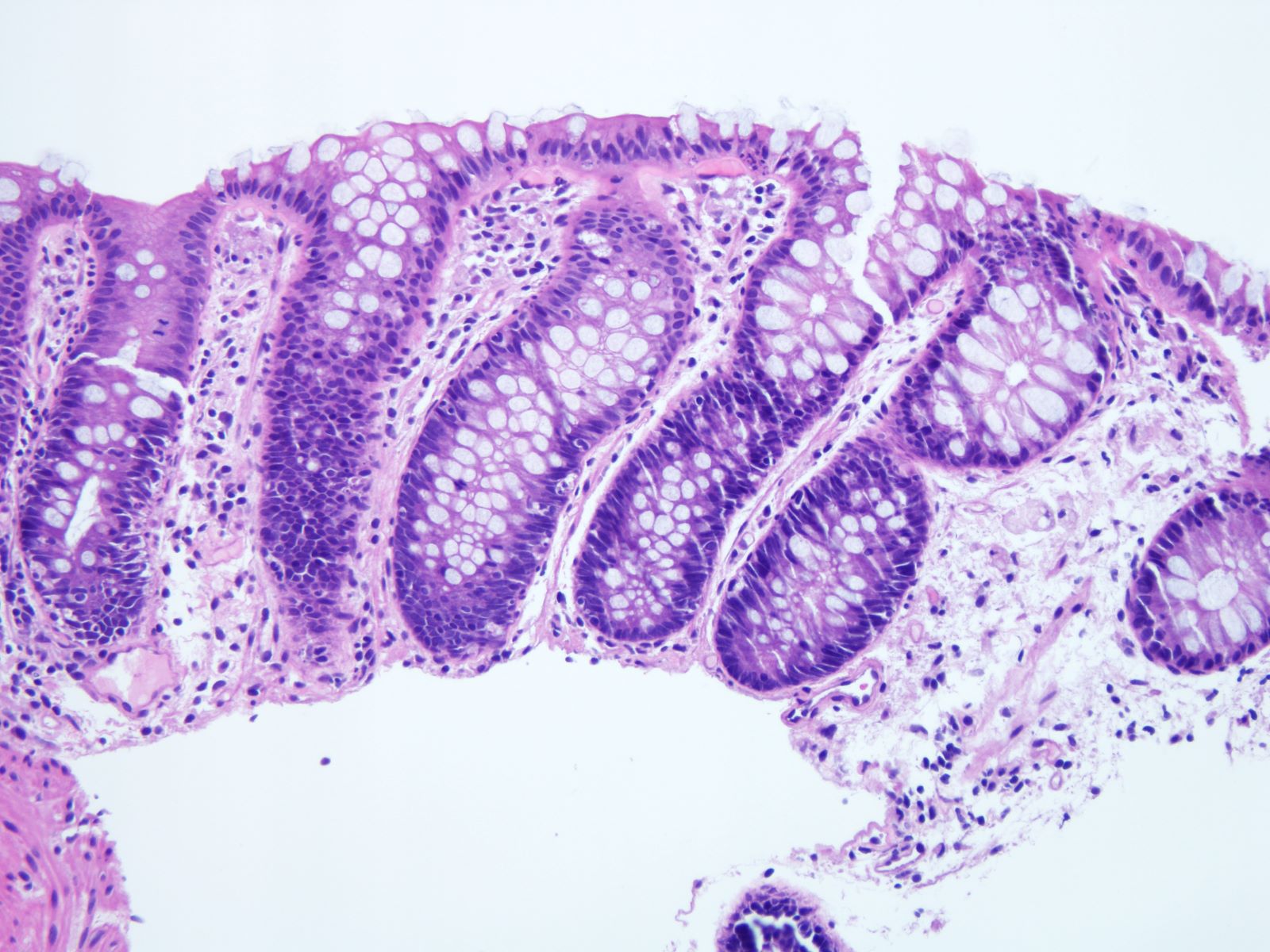
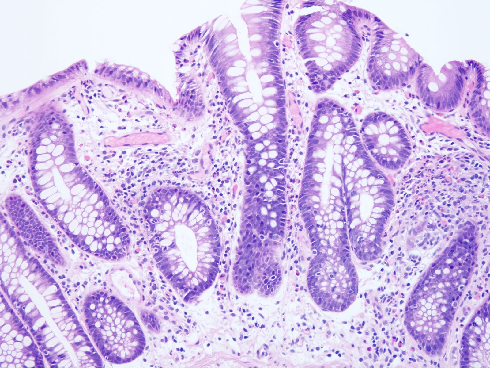
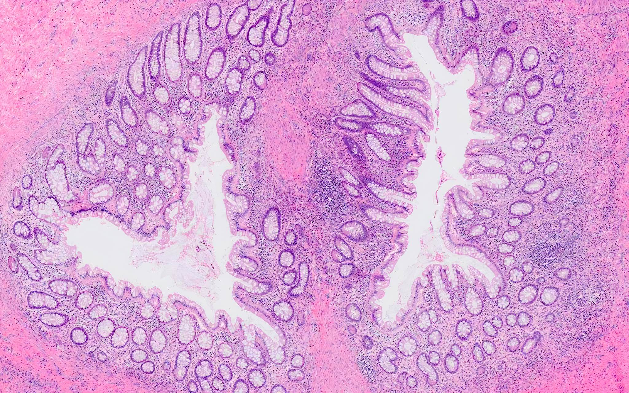
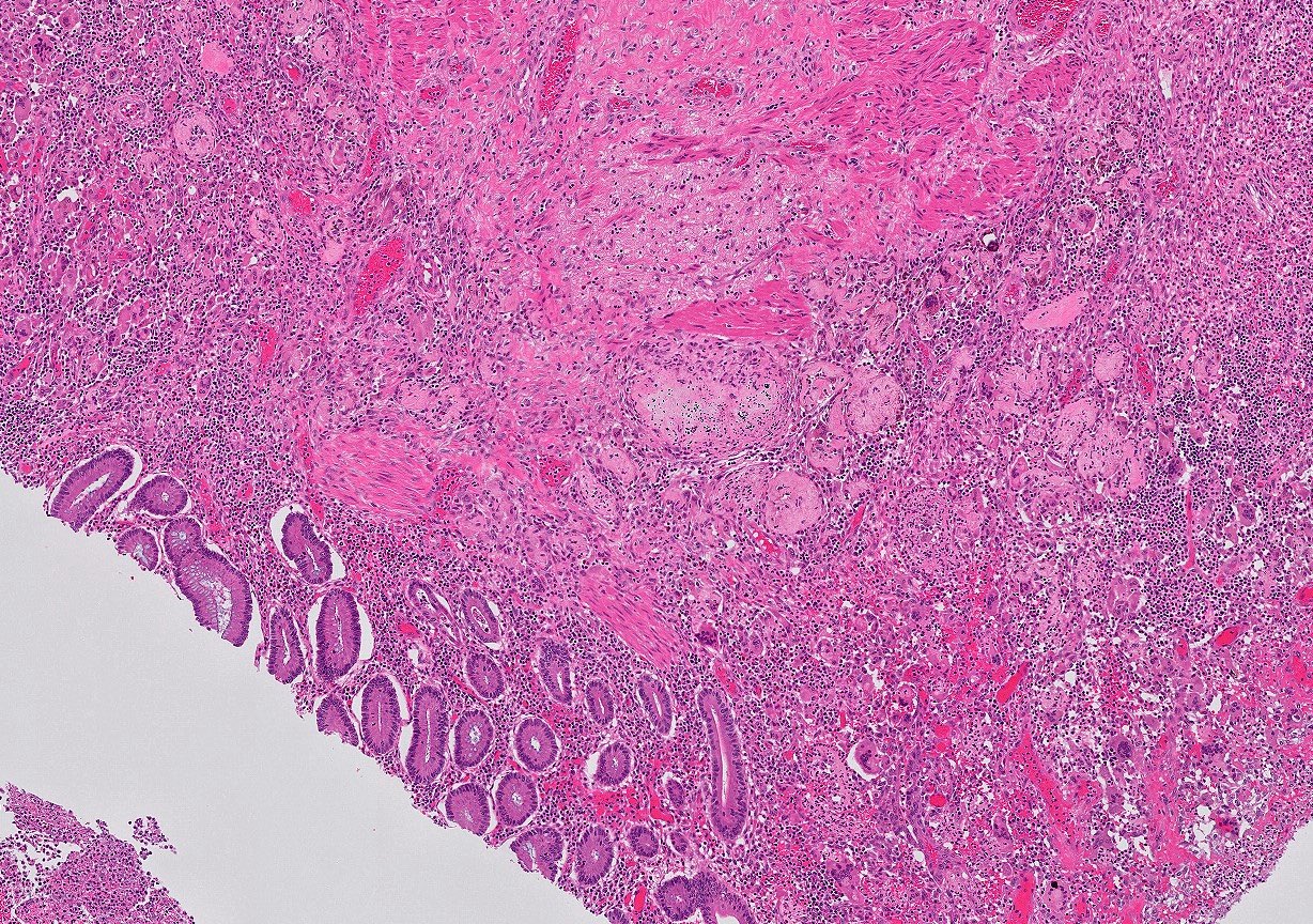
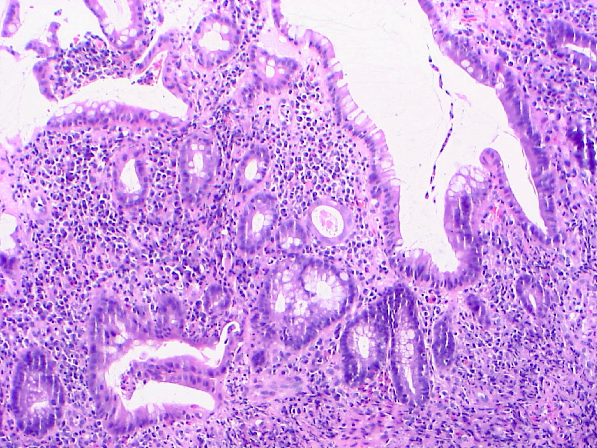
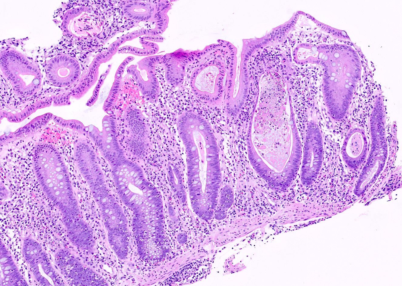
Focal active colitis with crypt drop out
Prominent crypt epithelial cell apoptosis
Neutrophilic crypt microabscesses
Lymphocytic colitis-like pattern
Images hosted on other servers:
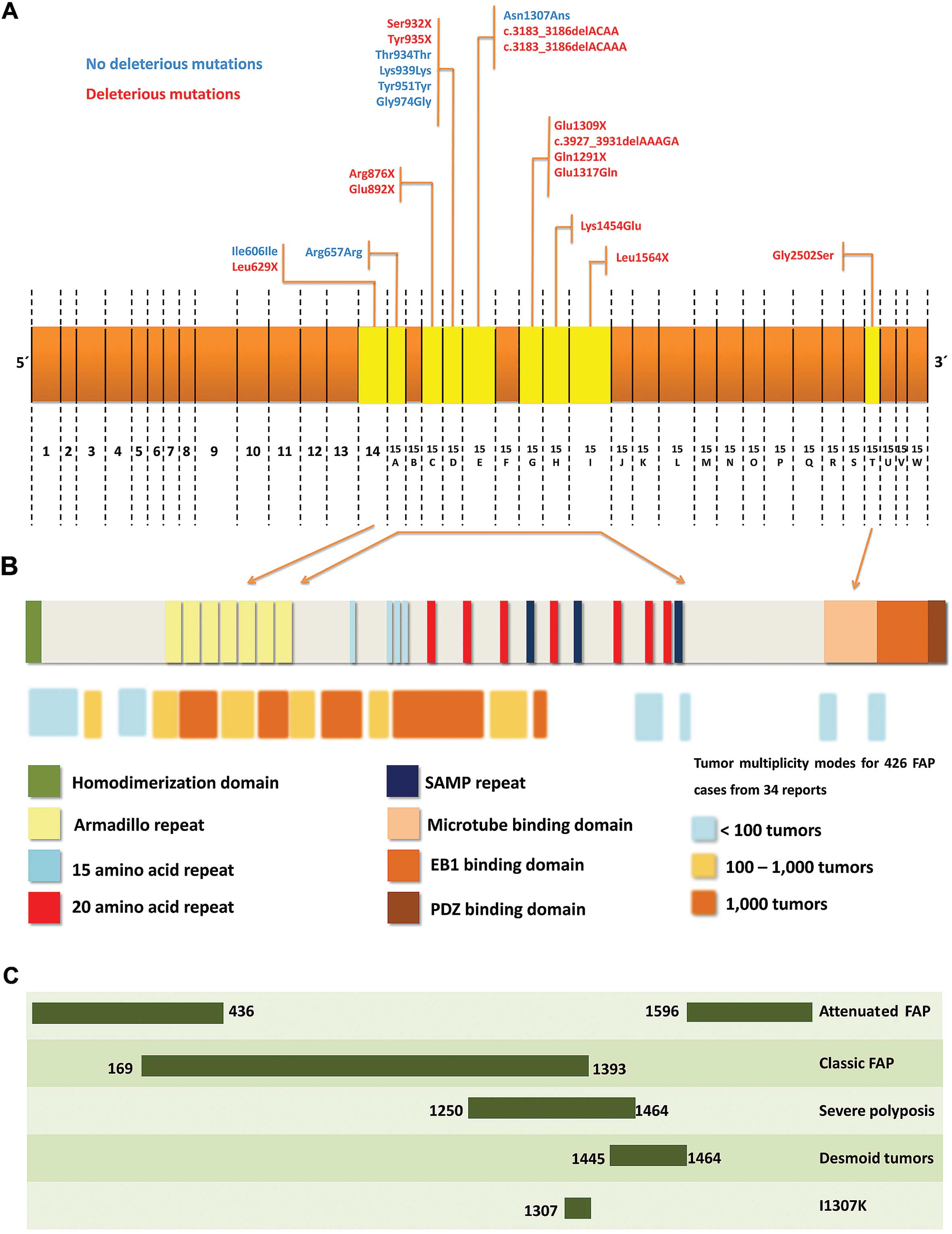
Overview of mutations
Images hosted on other servers:

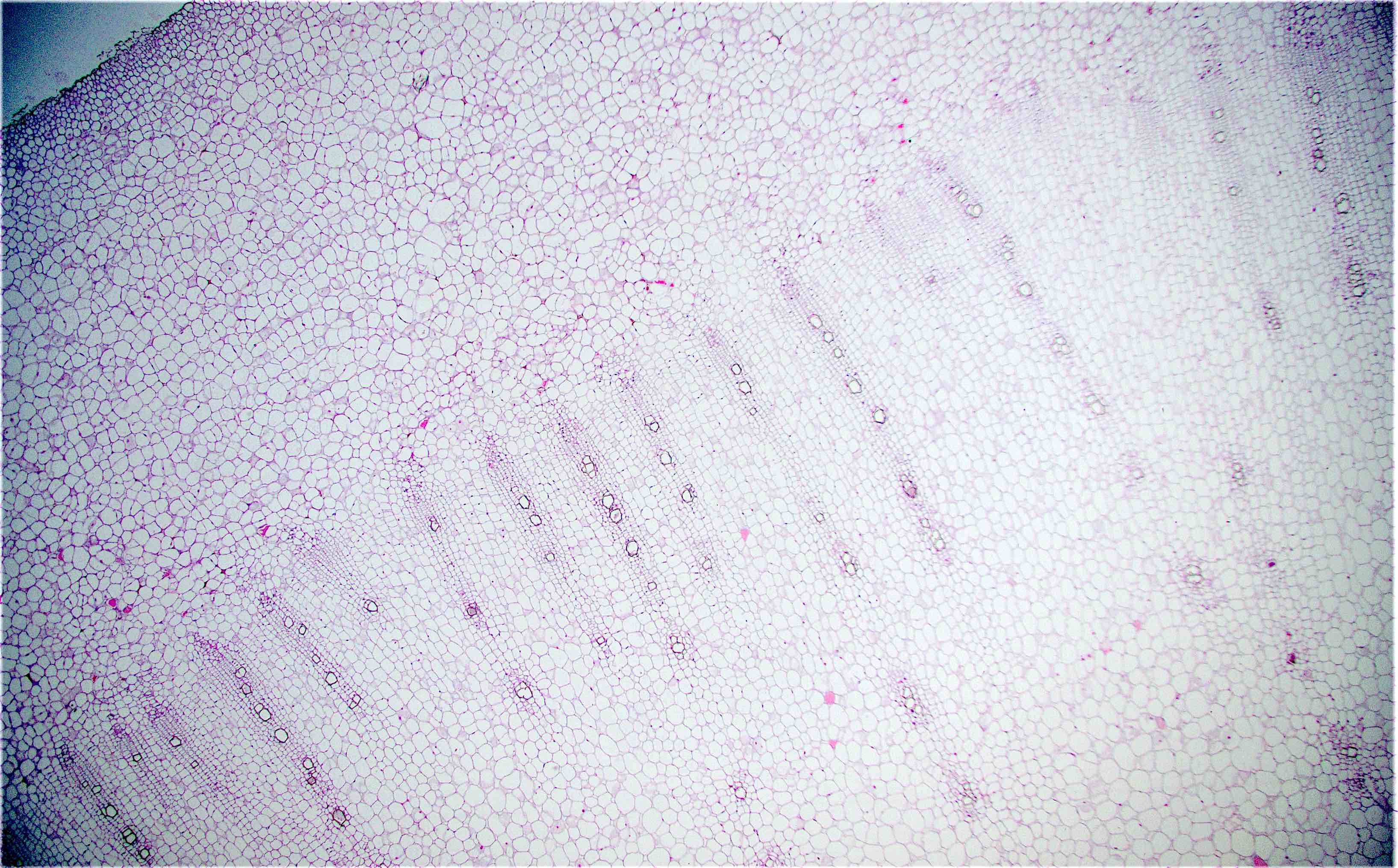
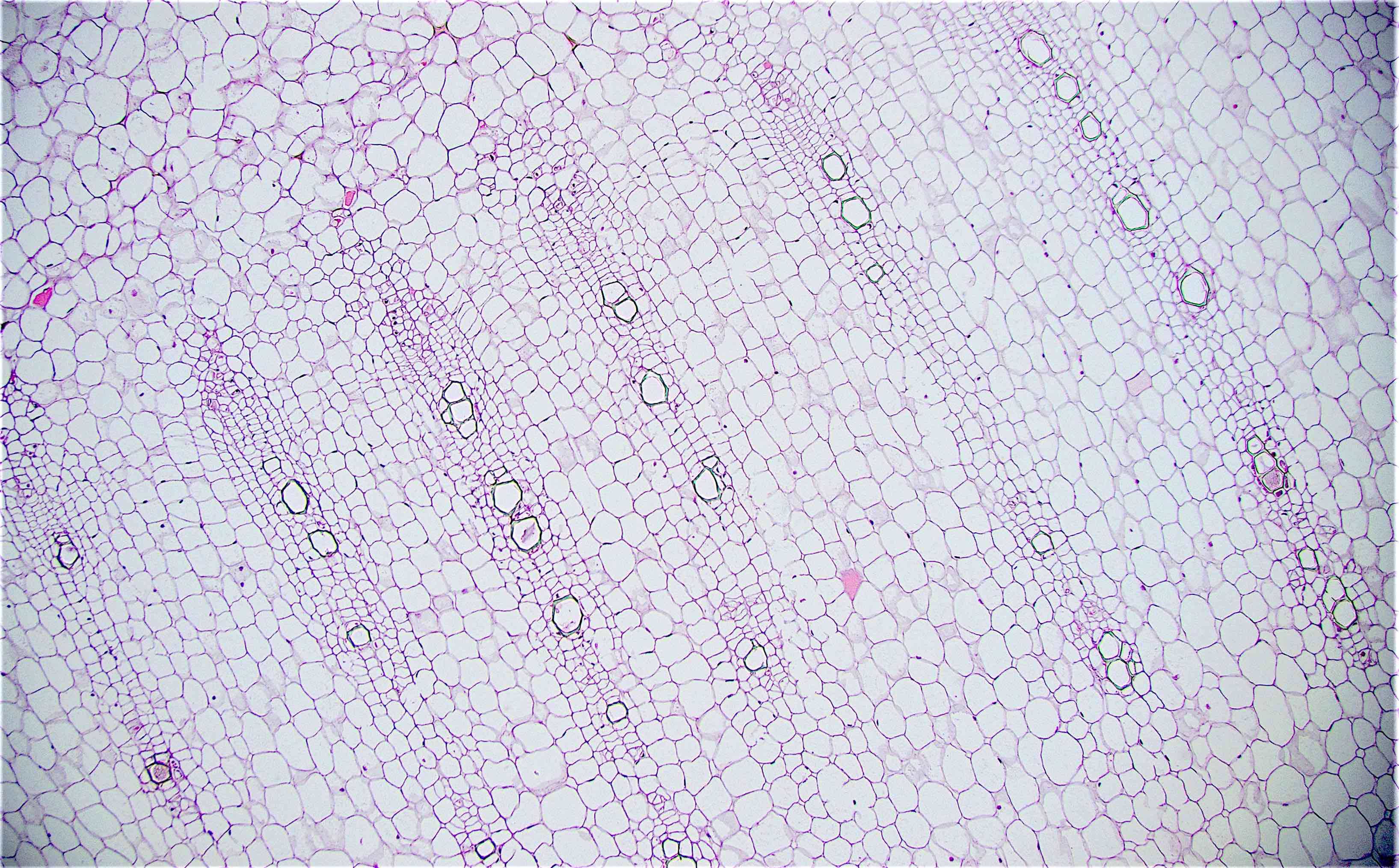
Type I atresia: mucosal septum

Atresia with hugely distended cecum

Atresia Type I
Contributed by Dhiraj B. Nikumbh, M.D.
Congenital pouch colon
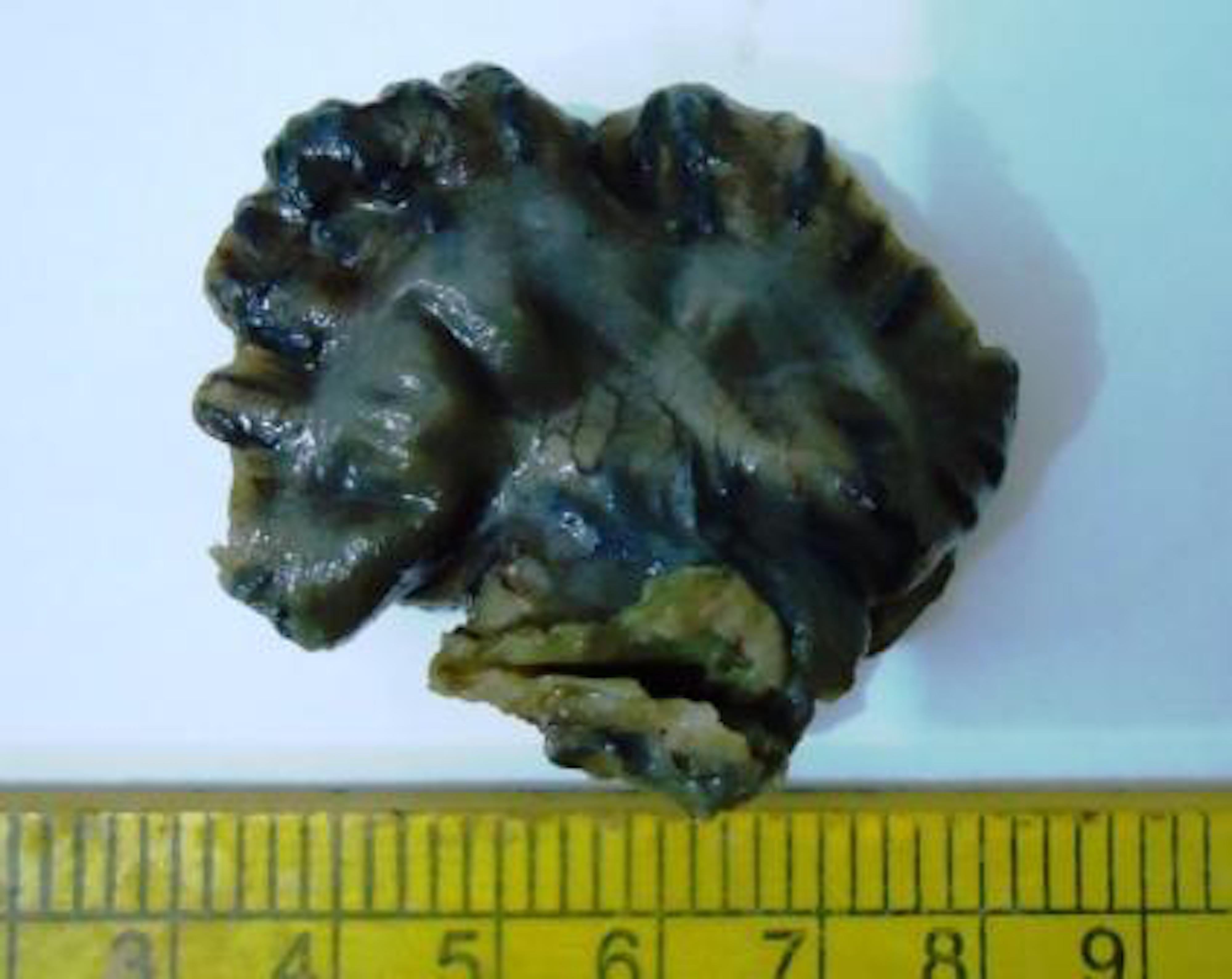
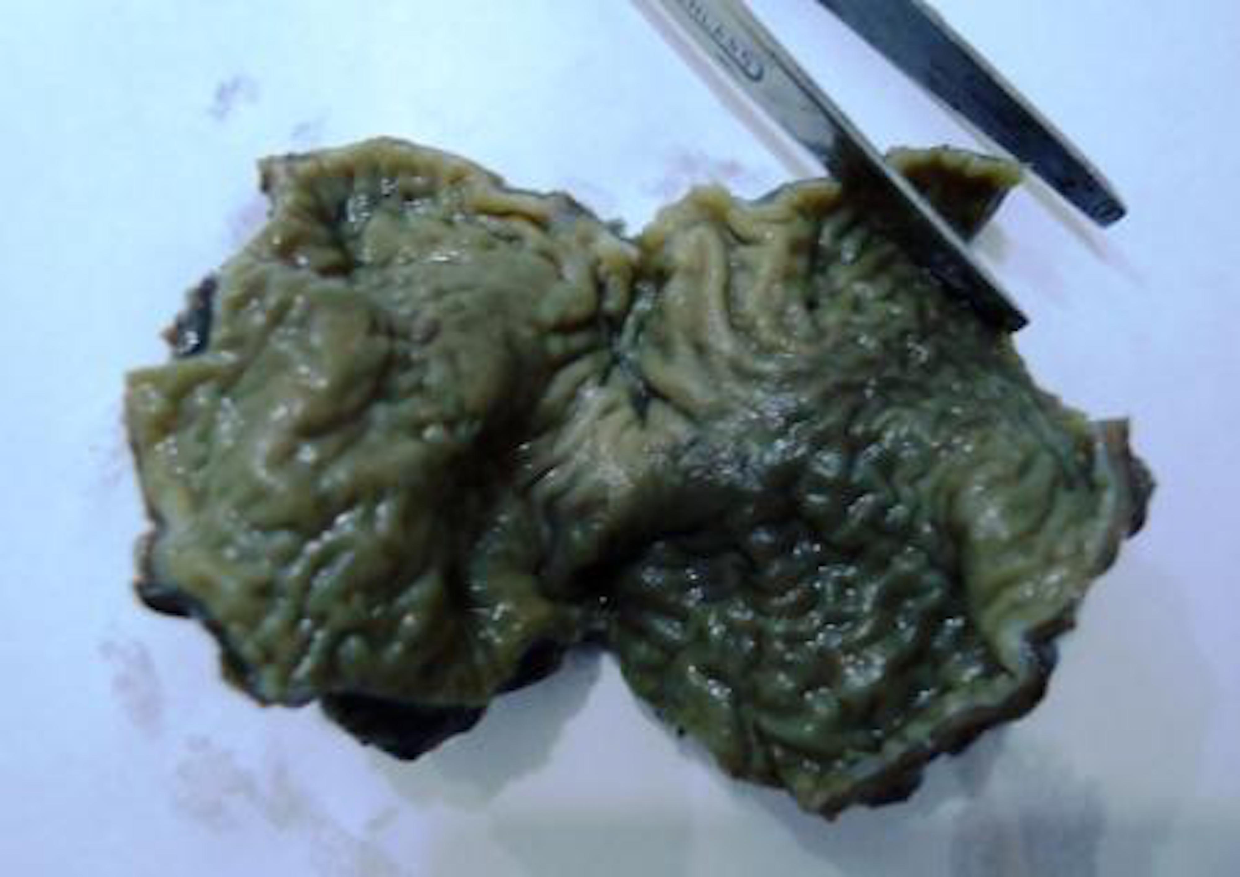
Contributed by Dhiraj B. Nikumbh, M.D.
Congenital pouch colon: 10 day old baby boy admitted with abdominal distension and absent anal opening
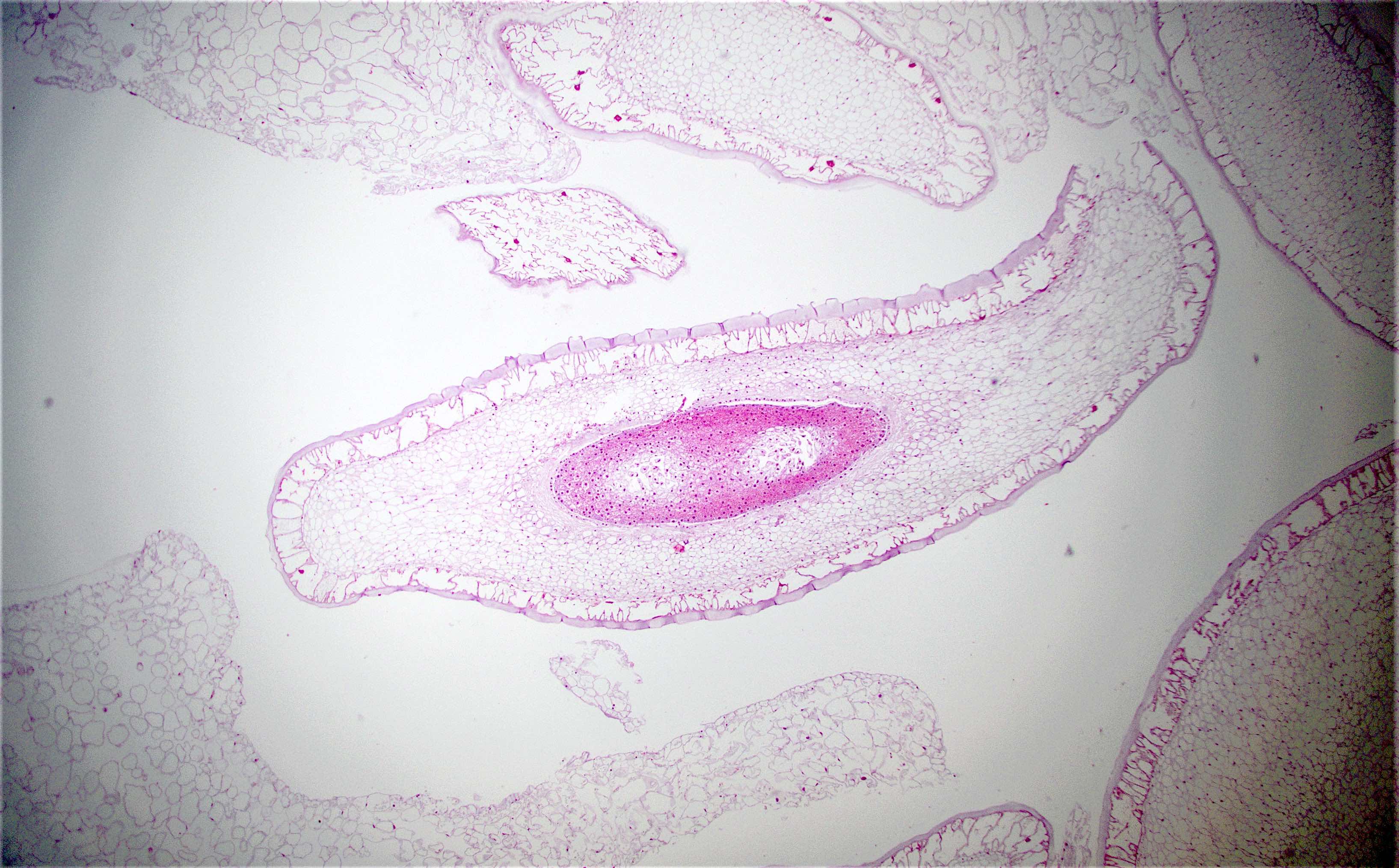
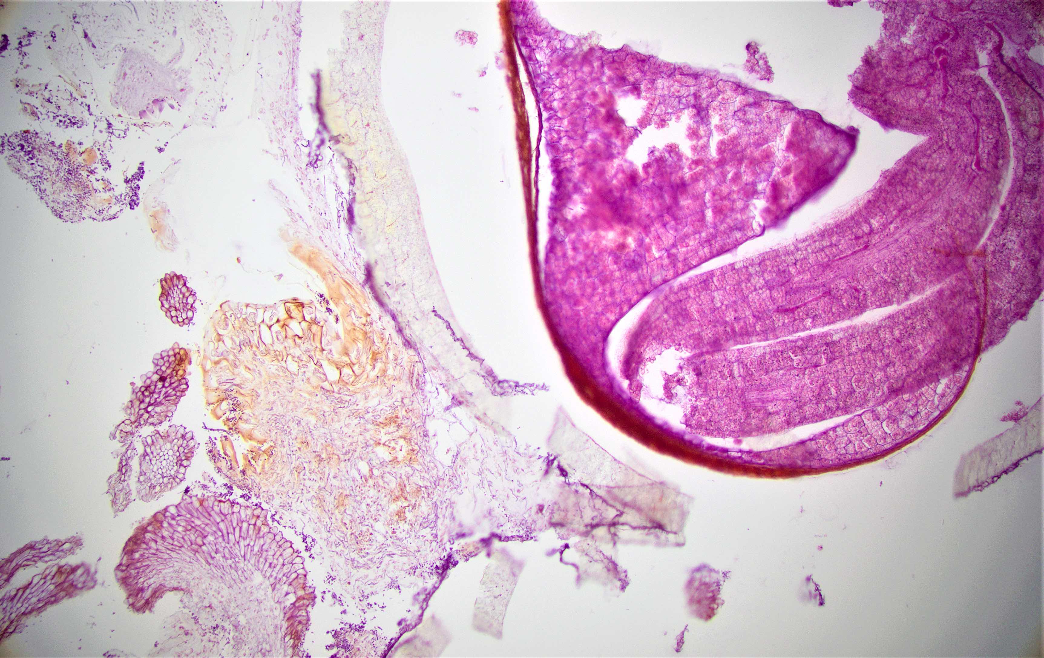
Images hosted on other servers:


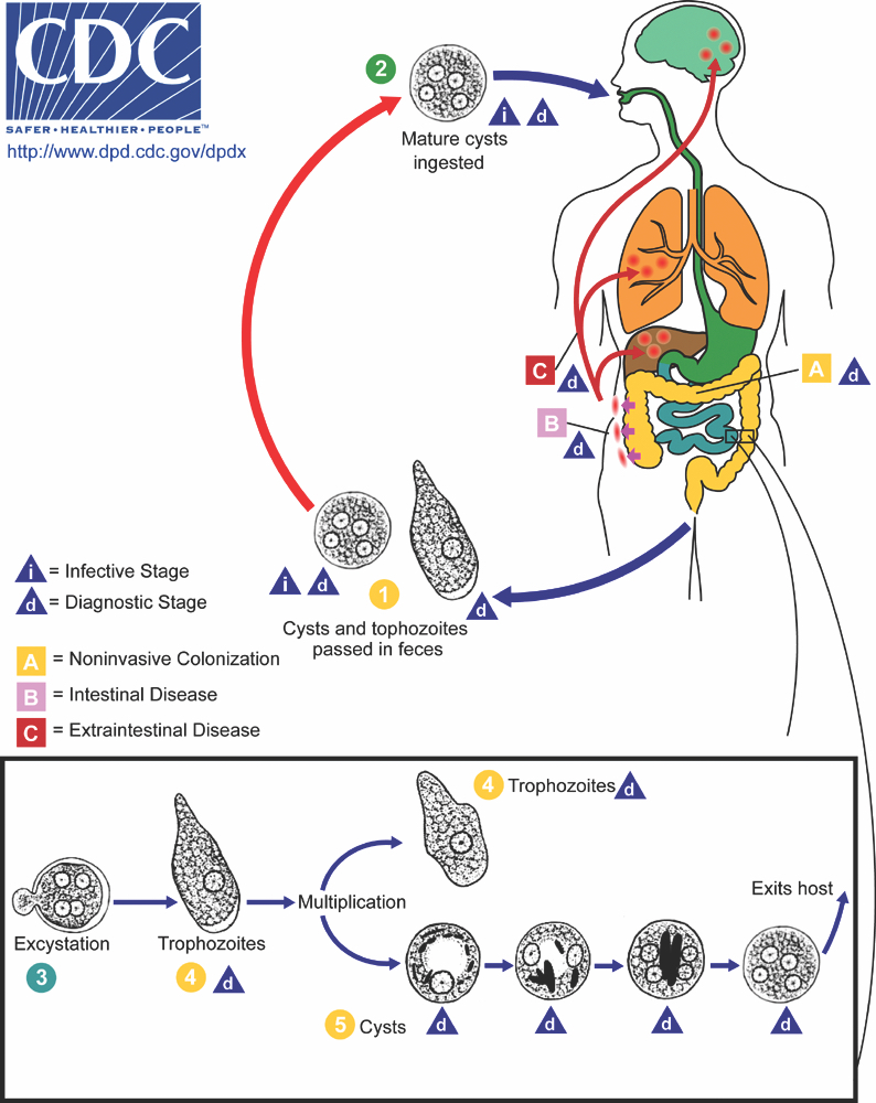
Life cycle
Contributed by Bobbi Pritt, M.D., Richard Bradbury, Ph.D. and Sarah Sapp, Ph.D.


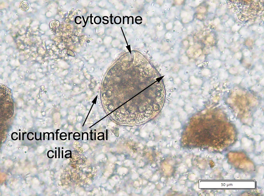
Cyst and trophozoite of Neobalantidium coli (also known as Balantidioides) obtained from West African baboon
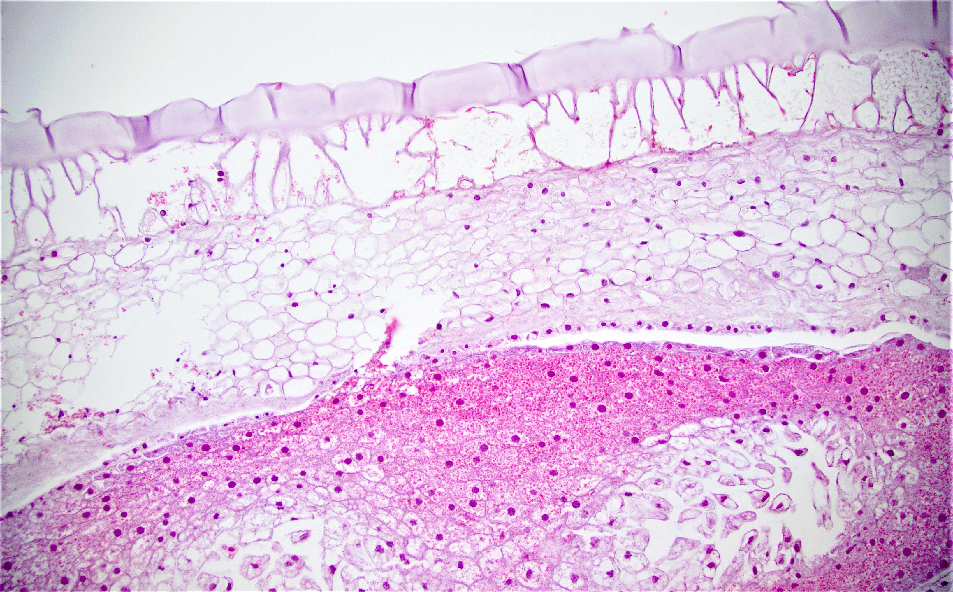
Images hosted on other servers:


Various images

Trophic stage (EM)

Intestinal lumen

Encysted ciliate
Images hosted on other servers:

MRI before surgical intervention and treatment
Generalized markedly
thickened wall of
the small bowel with
edematous changes
Images hosted on other servers:

Edematous left colon

Adherent small bowel loop

Postmortem liver
Images hosted on other servers:

Staining of the tissue biopsy from left retrocolic mass

Histopathology of surgically resected abdominal mass

Broad hyphae with septation


Splendore-Hoeppli phenomenon

Zygospore

Periodic acid-Schiff stain

Liver biopsy

Spore forms

Various images
Images hosted on other servers:

Punched-out ulcer
Images hosted on other servers:

Multiple penetrations

Ulcers
Images hosted on other servers:

Loss of normal mucosa


Various images
Images hosted on other servers:


Cryptitis


Edema, cryptitis and crypt abscess

Cystic crypts and cryptitis

Loss of crypts

Marked edema

Neutrophilic aggregates in lamina propria
Images hosted on other servers:

Fungal colonization
in patients with
GI tract disease
Contributed by Nalini Bansal, M.D.

PAS positive fungal hyphae in a case of gastric adenocarcinoma with fungal infection
Images hosted on other servers:


Fungal hyphae with gastric performation
Images hosted on other servers:

Life cycle
Contributed by Bobbi Pritt, M.D., Nicole L. Achee, Ph.D. and John P. Grieco, Ph.D.
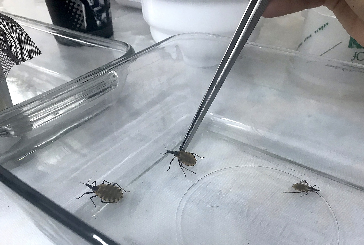
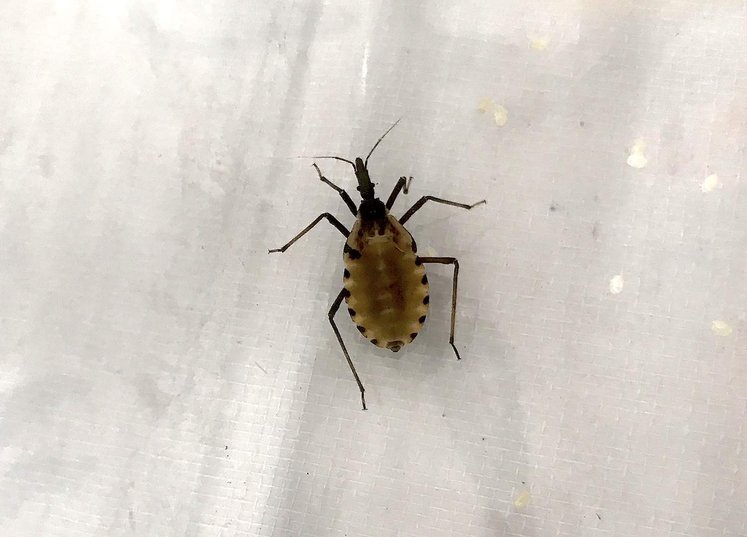
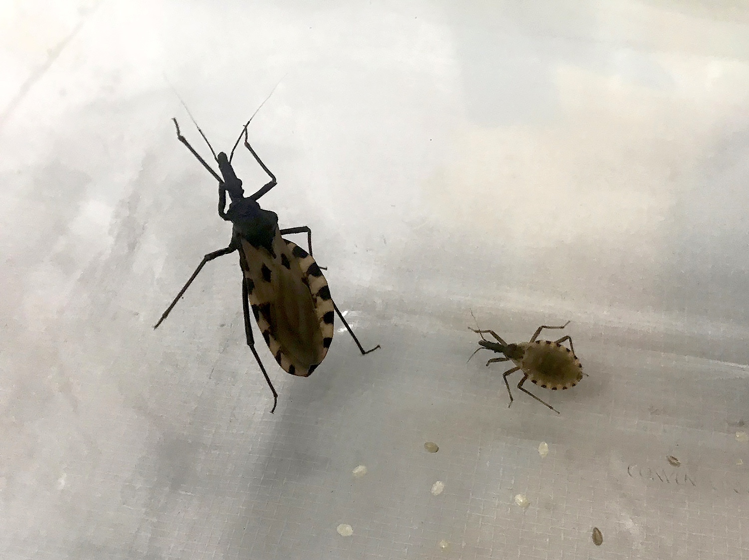
Triatomine bugs
Images hosted on other servers:

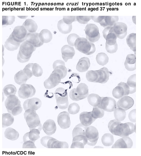
Blood smears
Images hosted on other servers:
Familial autonomic visceral myopathy:


Muscularis propria degeneration

Muscularis mucosae with degenerative changes
Images hosted on other servers:

Binary toxin
Images hosted on other servers:

Chitin degradation
Images hosted on other servers:

Electron micrograph
Contributed by Raul S. Gonzalez, M.D.
Colitis cystica profunda
Images hosted on other servers:

Mucosal tears

Endoscopic findings
Contributed by Catherine E. Hagen, M.D.
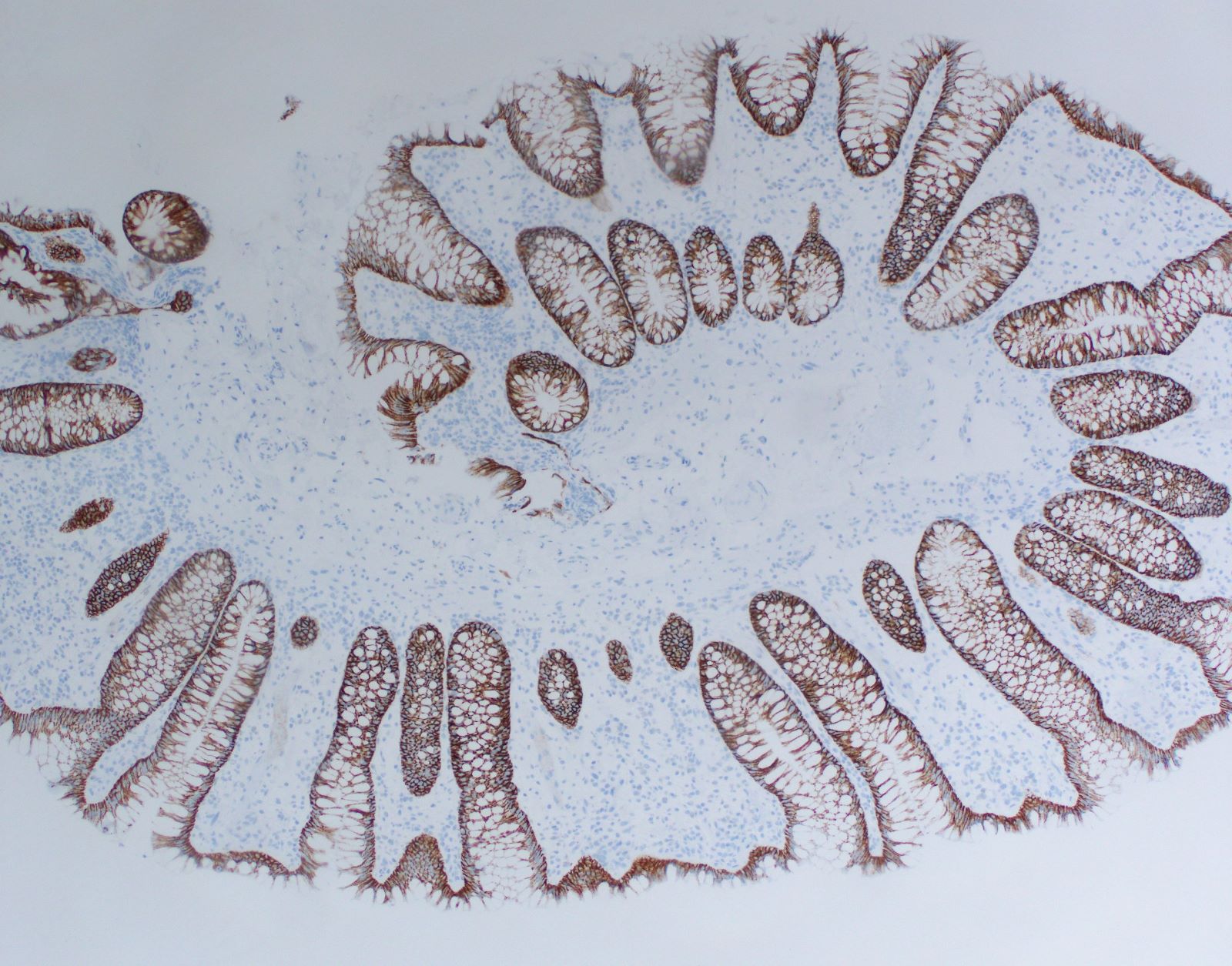
Preserved architecture
Thickened subepithelial collagen
Epithelial detachment
Equivocal collagen
Trichrome stain

Giant cell
Crypt atrophy
Contributed by Benjamin J. Van Treeck, M.D and Christopher Hartley, M.D.

Lamina propria neutrophils

Absence of plasma cells

Increased crypt apoptosis

Mild active colitis
Reduced mucosal plasma cells
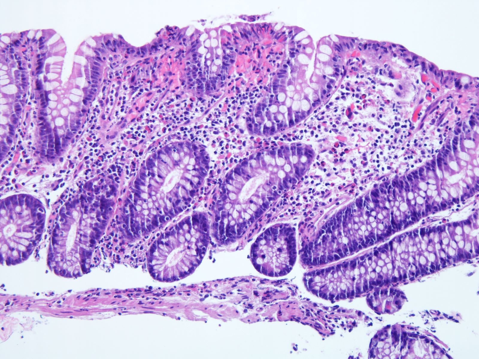
Increased lamina propria eosinophils
Increased lamina propria neutrophils
Mild to moderate active chronic colitis
CD138
Images hosted on other servers:

CT scan
Images hosted on other servers:

Colonoscopic findings
Contributed by Feng Yin, M.D., Ph.D.
Ischemic injury
with microthrombi
Withered crypts with neutrophils
Images hosted on other servers:

Virion particles
Images hosted on other servers:

Feet, face, mouth

Facial papules
Images hosted on other servers:

Juvenile polyp


Polyps with mildly dilated glands
Images hosted on other servers:

CT enterography, small bowel Crohn's disease

MRI of Crohn's disease patient with anovaginal fistula
Images hosted on other servers:

Endoscopic features of Crohn's disease
Contributed by Elizabeth Heidi Cheek-Norgan, M.H.S., PA (ASCP) and Catherine E. Hagen, M.D.
Stricture
Contributed by Catherine E. Hagen, M.D. and Luisa Ricaurte Archila, M.D.
Patchy involvement
Active chronic colitis
Pyloric gland metaplasia
Granuloma
Transmural inflammation
Images hosted on other servers:

Upper endoscopy: gastric polyposis
Contributed by Raul S. Gonzalez, M.D. and Michael Feely, D.O.

Duodenum: elongated, irregular and cystic crypts

Colon: polypoid mucosa with cystically dilated glands
Images hosted on other servers:

Polyp with dilated glands

Duodenum / small intestine
Contributed by Bobbi Pritt, M.D. and Institute of Tropical Medicine Antwerp
Negative fuchsine staining according to Heine
Ziehl-Neelsen staining
Images hosted on other servers:

Oocysts: auramine-rhodamine stain

Small blue organisms at luminal border

Modified acid-fast oocyst in stool
Images hosted on other servers:

Retrorectal space
Images hosted on other servers:

Retrorectal cyst

Oval shaped, gray-white appearance
Images hosted on other servers:

Squamous epithelium and focal columnar epithelium

Early malignant degeneration
Images hosted on other servers:

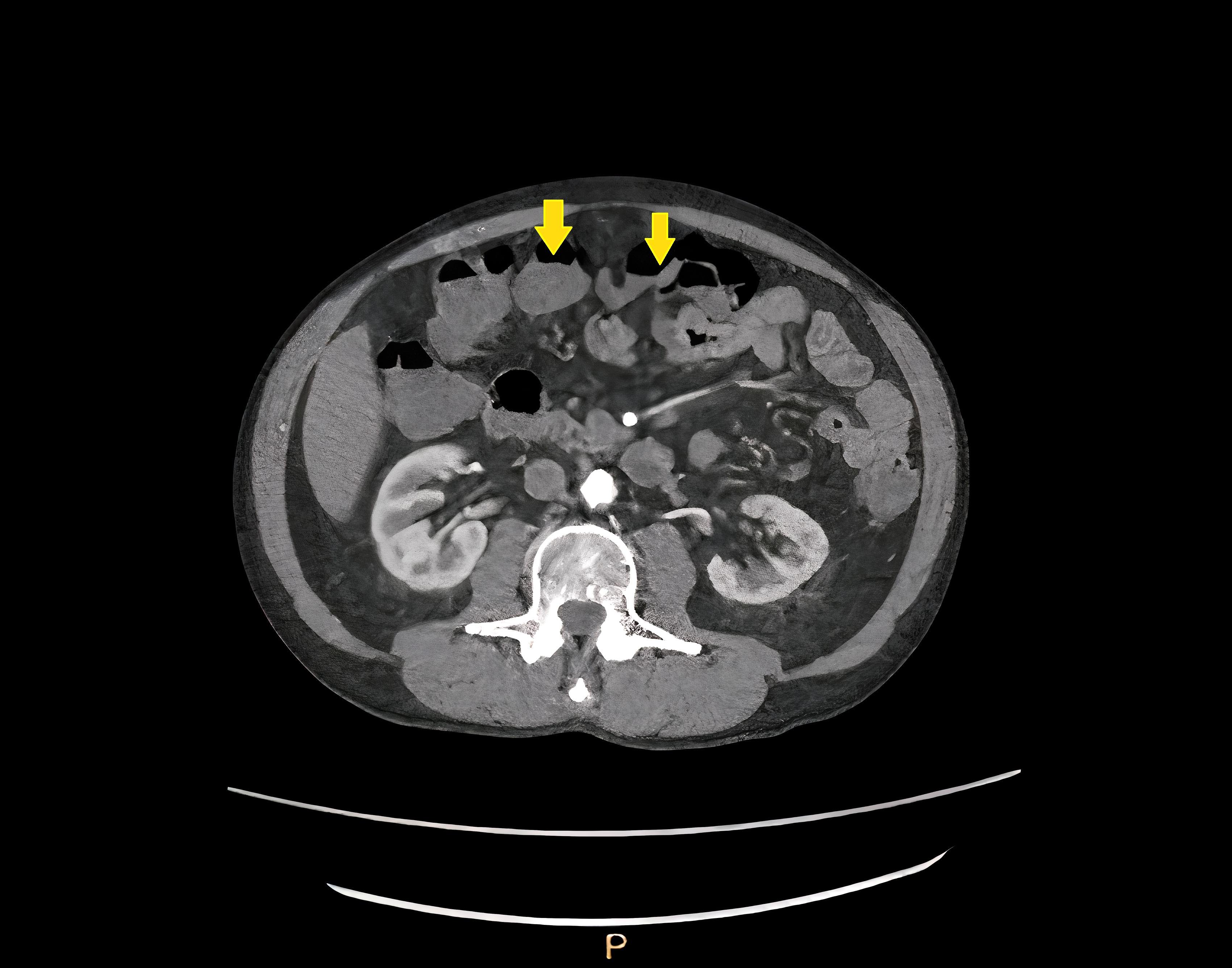
CT findings in a patient with CMV colitis
Contributed by Martha M. Yearsley, M.D.
Diffuse erythema
Pseudomembrane
formation
Irregular ulceration, loss of vascular pattern
Images hosted on other servers:

Diffuse inflammation

Ulcerating mass
Images hosted on other servers:


Ulceration and erythema
Ulceration secondary to CMV colitis

Terminal ileum

Cecum

Multiple, small punctate ulcers in the mucosa
Contributed by Martha M. Yearsley, M.D.

Enlarged cells in colonic lamina propria

Cytomegalic endothelial cells

Owl's eye and coarse red granules
Infected endothelial cells
Cytomegalic endothelial cells partially occluding vessel
Immunoreactivity with CMV immunostain
Scattered CMV positive cells
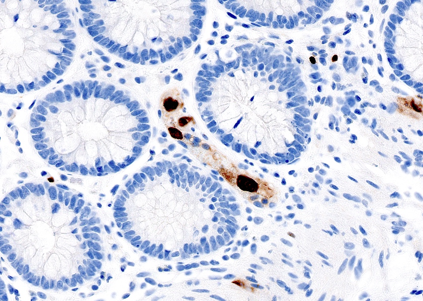
CMV positive endothelial cells
Contributed by @RaulSGonzalezMD on Twitter
Cytomegalovirus (CMV)
CMV colitis in ulcerative colitis and immunocompromised states
Images hosted on other servers:

Chronic diarrhea
Images hosted on other servers:

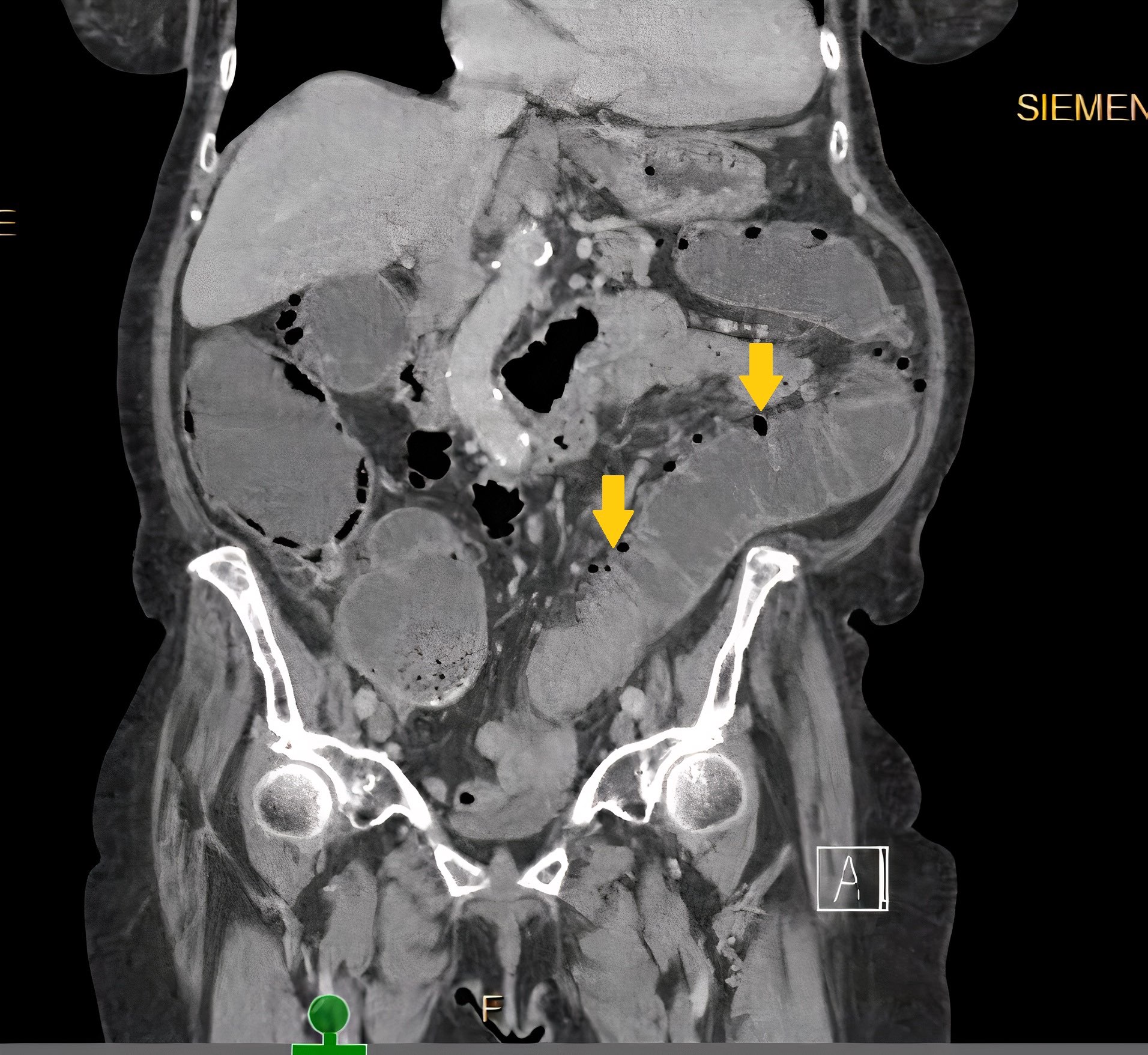
Postcolectomy CT and MRI
Contributed by Catherine E. Hagen, M.D. and Dustin W. Parsons, M.D.
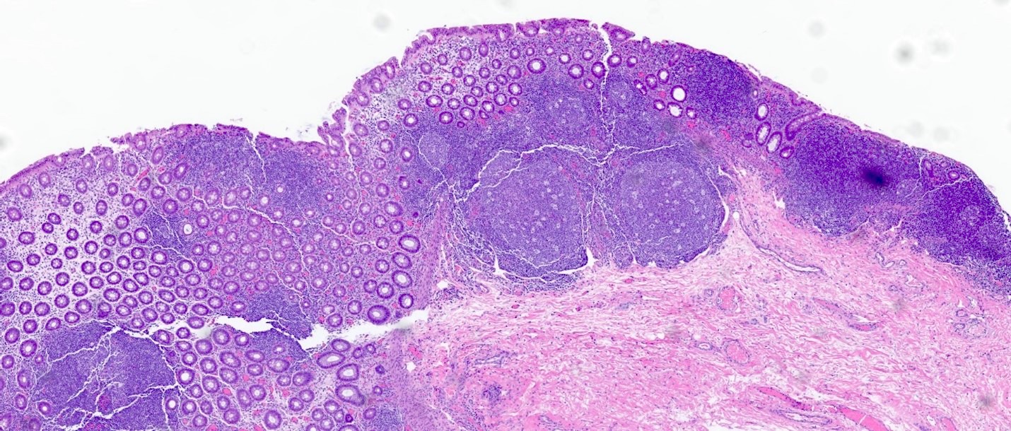
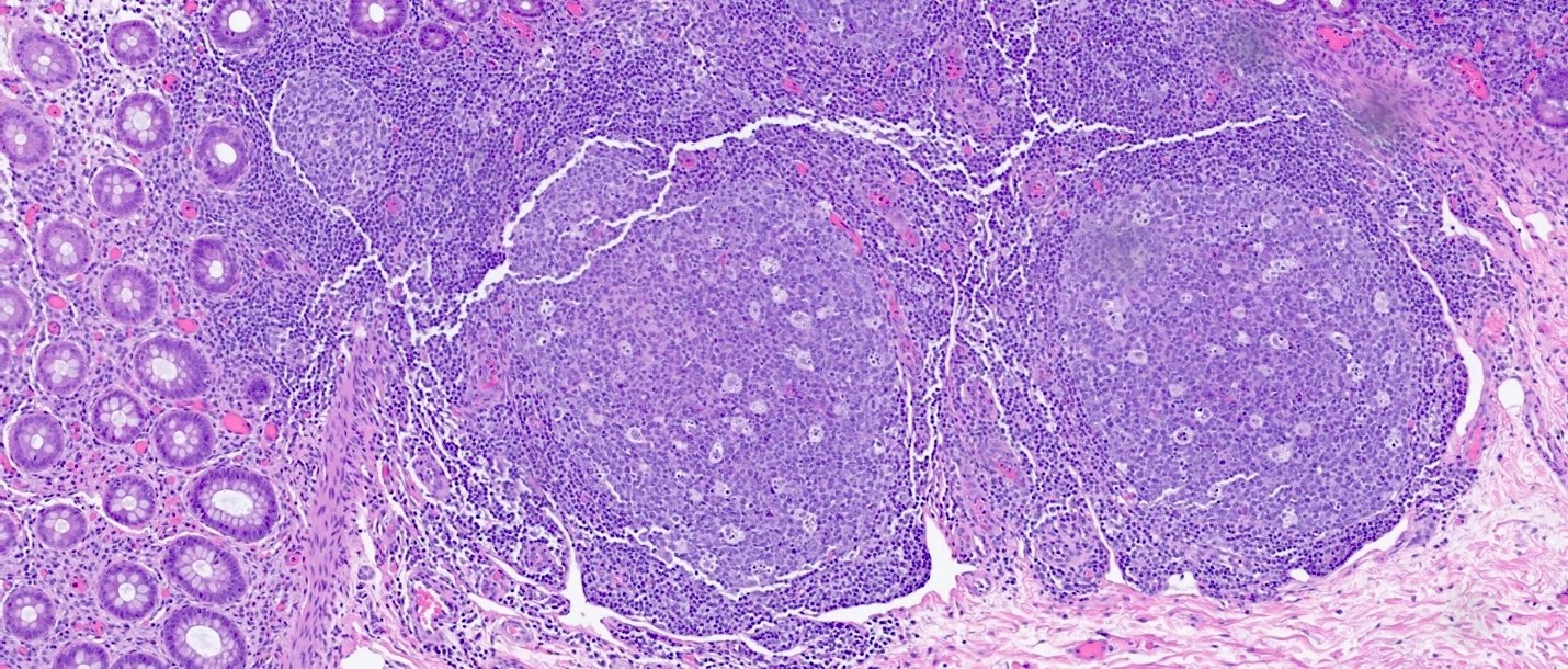
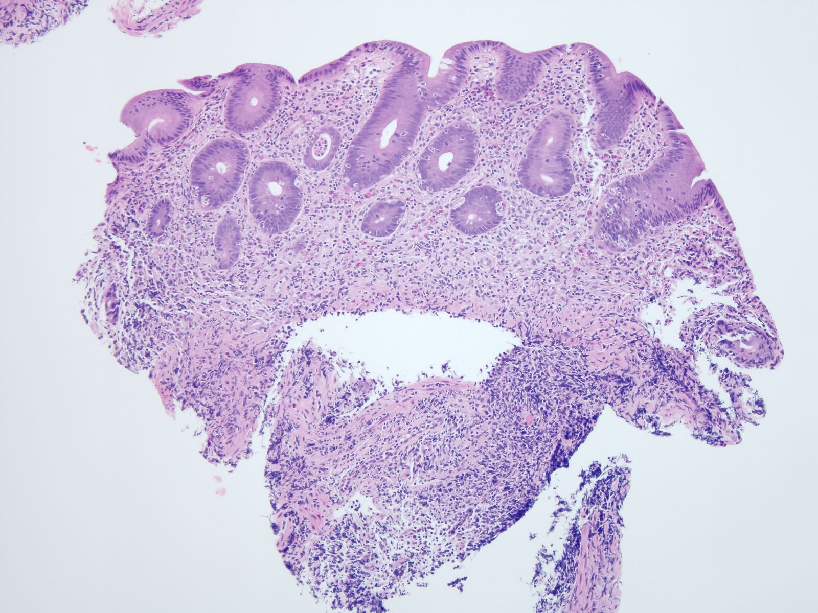
Prominent lymphoid follicles
Crypt abscess
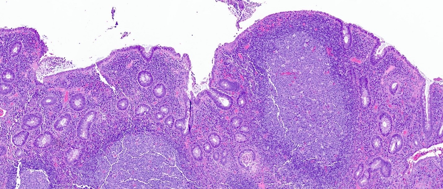
Lymphoid aggregate and architectural changes
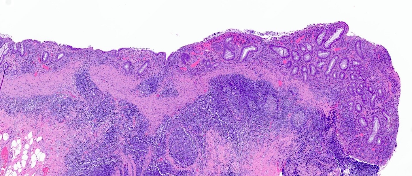
Ulceration
Images hosted on other servers:

Physiological activity
Contributed by Bindu Challa, M.D. and Martha M. Yearsley, M.D.
CT of abdomen and pelvis
Contributed by Bindu Challa, M.D. and Martha M. Yearsley, M.D.
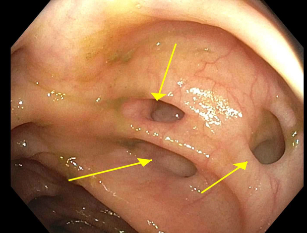
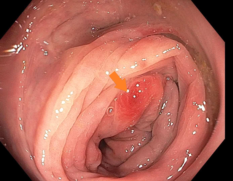
Colonoscopy
Colonoscopy
Images hosted on other servers:


Multiple diverticula

Multiple diverticula


Multiple diverticula

With bowel wall stenosis

Blue-gray diverticula

Perforated single true diverticulum

False diverticulum
Contributed by Bindu Challa, M.D. and Martha M. Yearsley, M.D.
Diverticula
Multiple diverticula
Diverticular abscess
Enterocutaneous fistula
Chronic inflammation in diverticular wall
Diverticular disease associated chronic colitis
Lymphoid aggregates in diverticular wall
Diverticulitis with complications
Histopathology of diverticular disease
Whole slide image of case of diverticulosis with diverticulitis
Images hosted on other servers:

Fecaloma removed

Sigmoid and colonic duplication

Final attachment of blind end

Colonic pouch and Y shaped duplication

Dilated colon and Y shaped duplication
Contributed by Celso Rubens Vieira e Silva, M.D.
Cystic congenital duplication
Images hosted on other servers:

T shaped tubular colonic duplication

Duplicated colon

Various images
Contributed by Celso Rubens Vieira e Silva, M.D.
Cystic congenital duplication
Images hosted on other servers:

Duplicated colon and appendix
Images hosted on other servers:

Polypoid dysplasia identified on chromoendoscopy

Nonpolypoid dysplasia
Contributed by Adela Cimic, M.D.
Descending colon lesion
Images hosted on other servers:

Dysplasia in a Crohn's colitis pseudopolyp
Contributed by Kenrry Chiu, M.D., C.M.

Low grade dysplasia
High grade dysplasia
Contributed by Elaine Alt, M.D.
Low grade dysplasia
Low grade dysplasia - pancolitis
Images hosted on other servers:

High grade dysplasia
Images hosted on other servers:

Model of carcinogenesis in ulcerative colitis
Case #163


9 year old boy with Type IV disease
Contributed by Raul S. Gonzalez, M.D.
Submucosal elastofibromatous change mimicking amyloid
Elastin stain

Contributed by Raul S. Gonzalez, M.D. and @Andrew_Fltv on Twitter
Colonic endometriosis
Endometriosis
Images hosted on other servers:


Within colonic mucosa

Present in wall of colon


Infiltration along nerves
Images hosted on other servers:


CT findings of eosinophilic gastrointestinal disorders
Images hosted on other servers:

Duodenum endoscopy

Colon endoscopy

Stomach endoscopy

Duodenum, antrum, rectum endoscopy

Duodenum endoscopy
Contributed by Byoung Uk Park, M.D. and Lizhi Zhang, M.D.
Prominent eosinophilic infiltration
Clustering of eosinophils
Intraepithelial eosinophils
Eosinophilic abscesses
Eosinophils in muscularis mucosae
Eosinophils in mucosa and submucosa
Transmural eosinophilic infiltration
Prominent eosinophilic infiltration
Intraepithelial eosinophils
Eosinophils in muscularis propria
Eosinophils in serosa
Images hosted on other servers:

CT scan:
thickened colon
with target sign

CECT: pancolitis
with thickened colon
and target sign
Contributed by Ateeqa Mujeeb Ullah, M.D.
E. coli colonies
Contributed by Ateeqa Mujeeb Ullah, M.D.


Acute colitis
Attenuated familial adenomatous polyposis (FAP)
Contributed by @Andrew_Fltv on Twitter
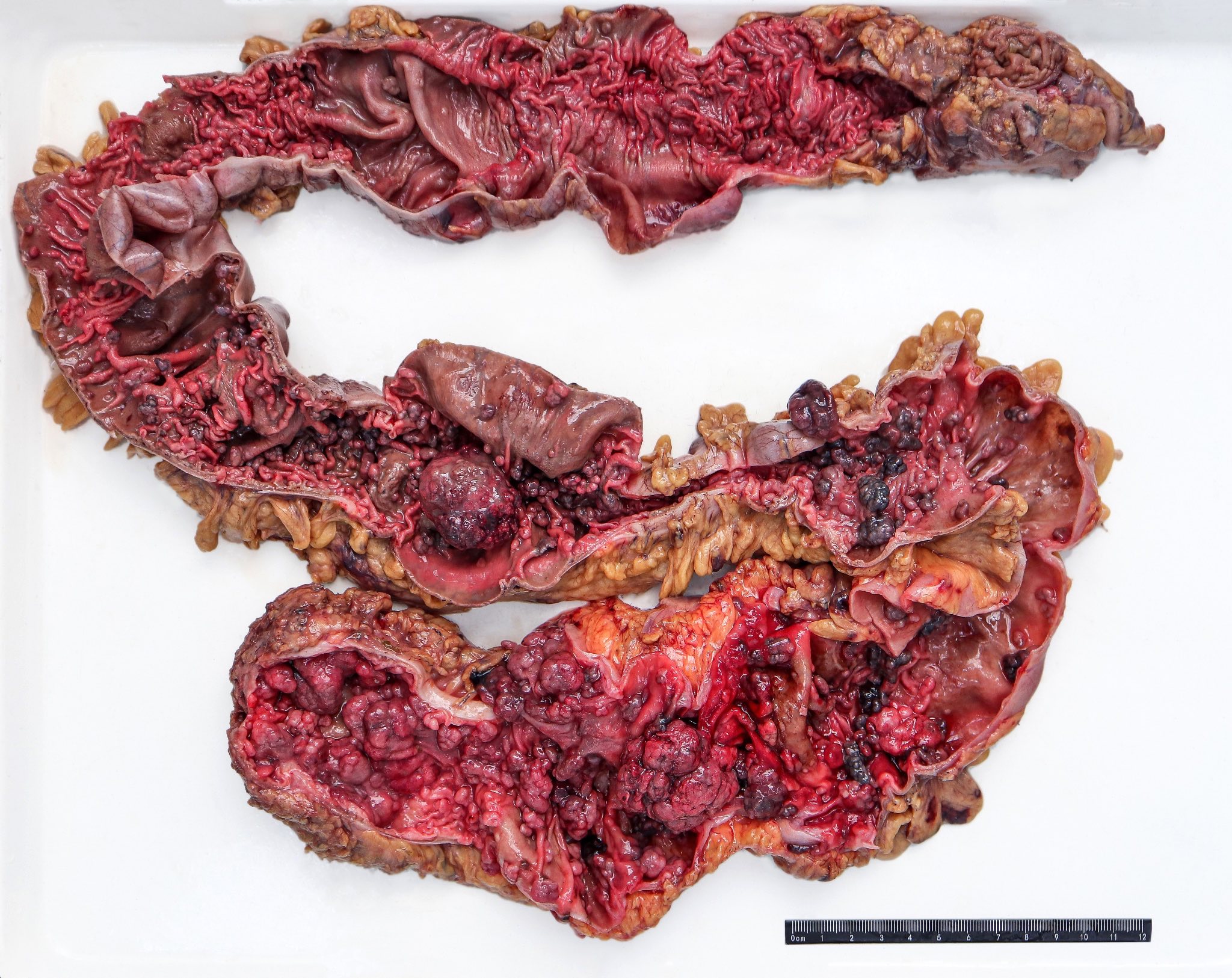
Familial adenomatous polyposis, classic
Images hosted on other servers:


Carpet of adenomatous polyps


Numerous polyps
Contributed by Jennifer Findeis-Hosey, M.D. and @Andrew_Fltv on Twitter
Adenoma involving few crypts
Tubular adenoma polyp, FAP patient
Familial adenomatous polyposis, classic
Images hosted on other servers:

Adenoma and adenocarcinoma

FAP versus non-FAP

BCL2
Full colonoscopy in retroflexed maneuver in a familial adenomatous polyposis coli
Contributed by Safina Ahmed, M.B.B.S. and Saroona Haroon, M.B.B.S.
Colonic mucosa with cryptitis and crypt abscess
Colonic mucosa with crypt abscess
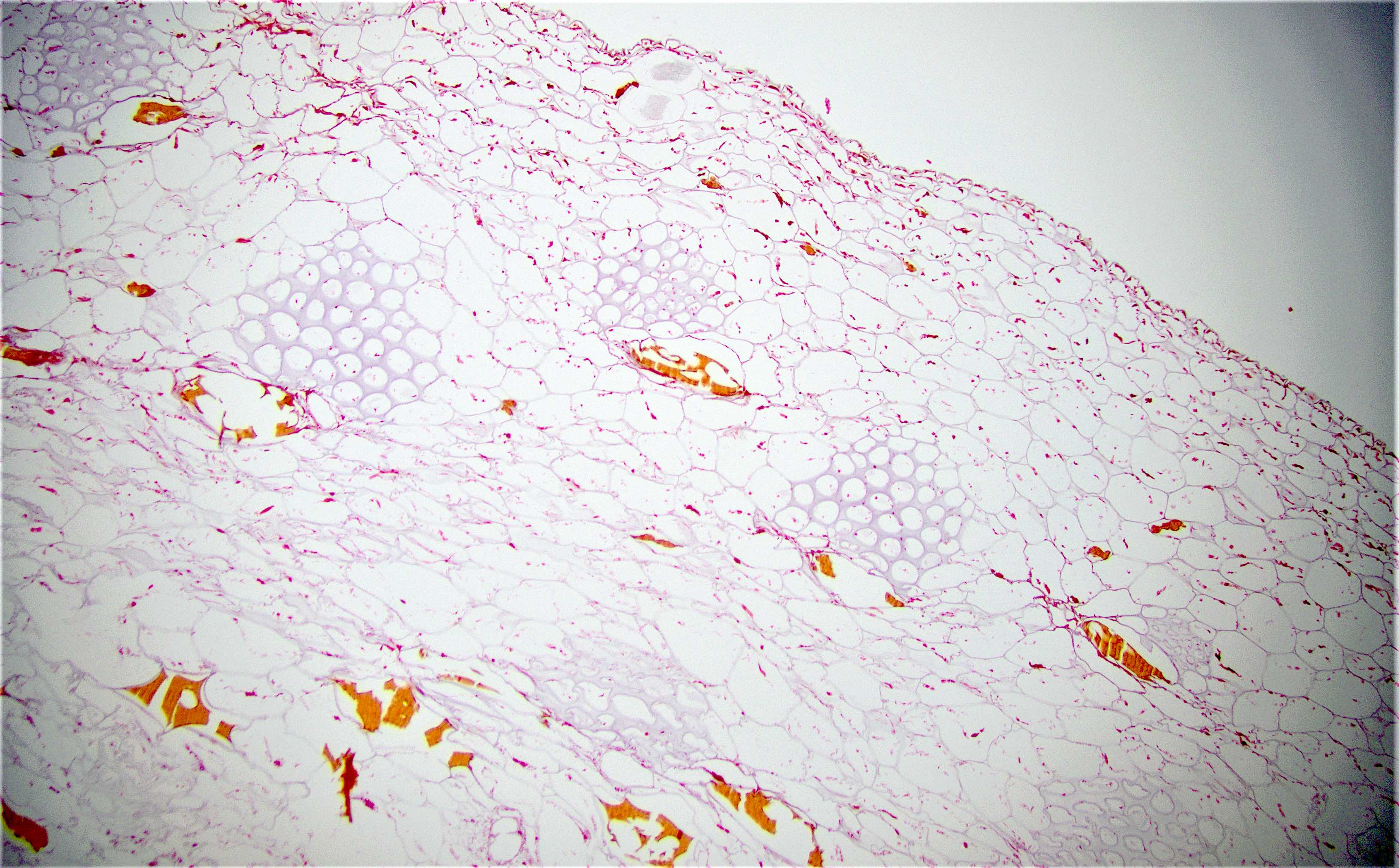
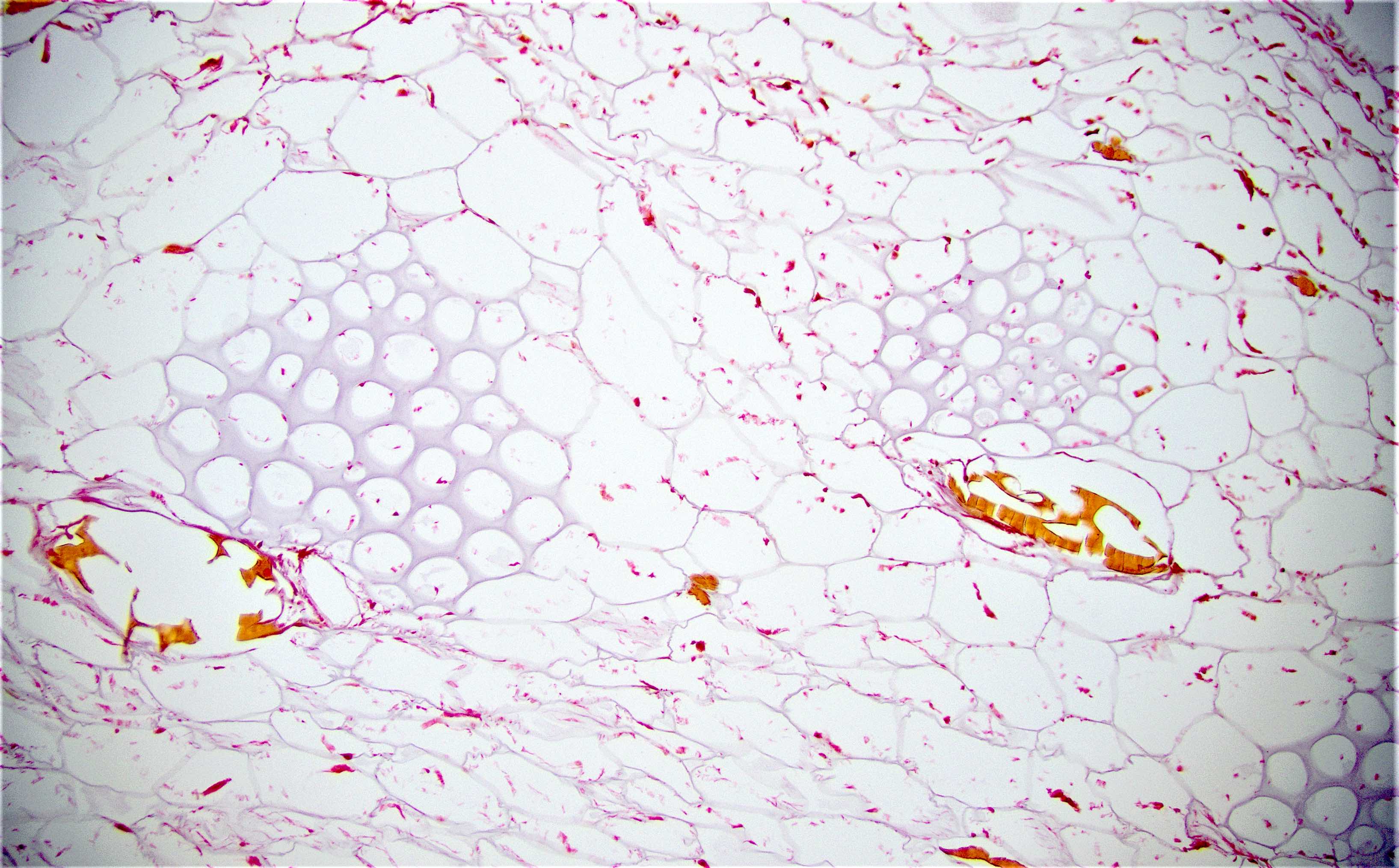
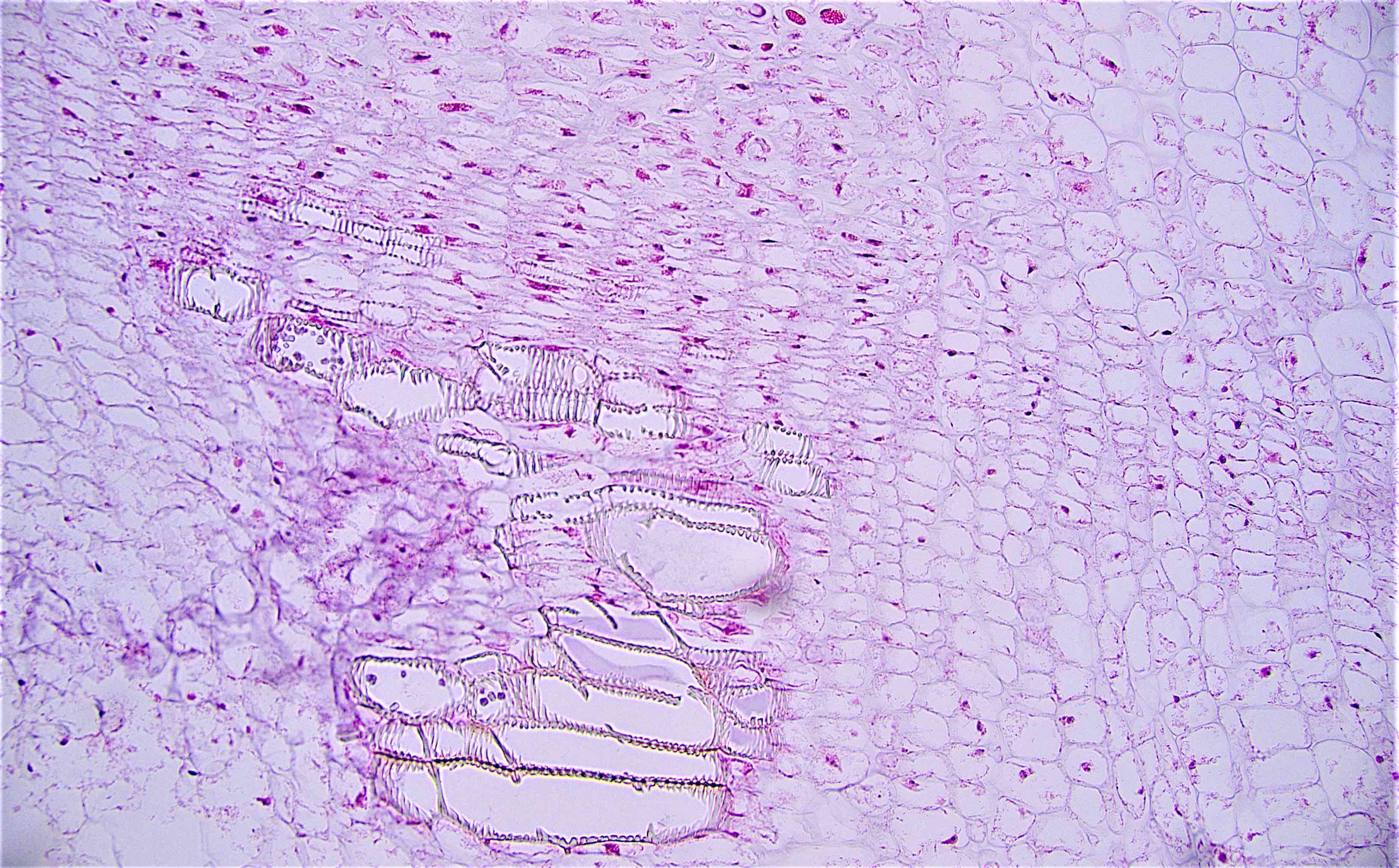
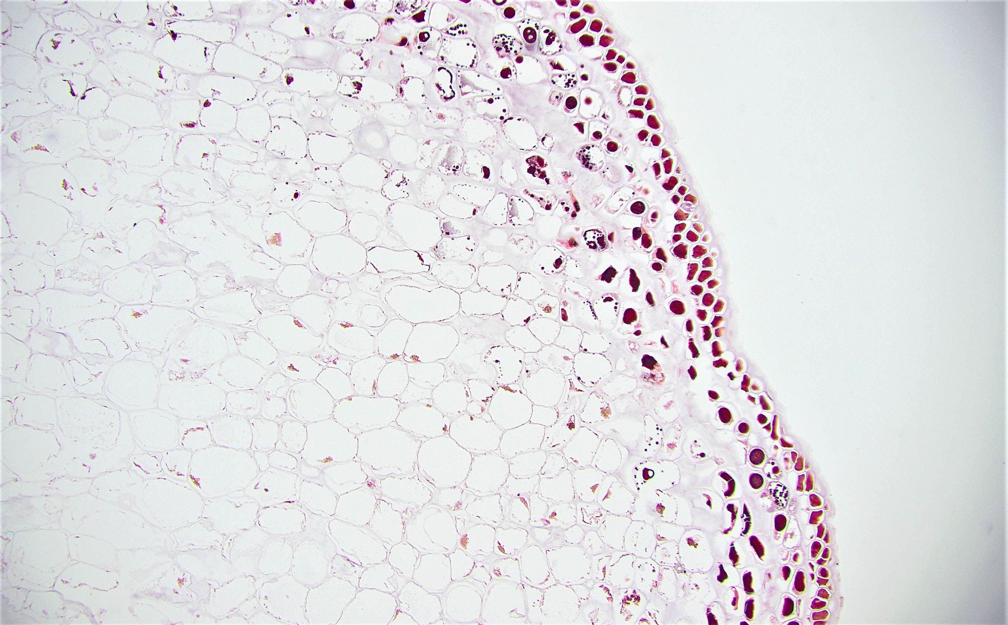
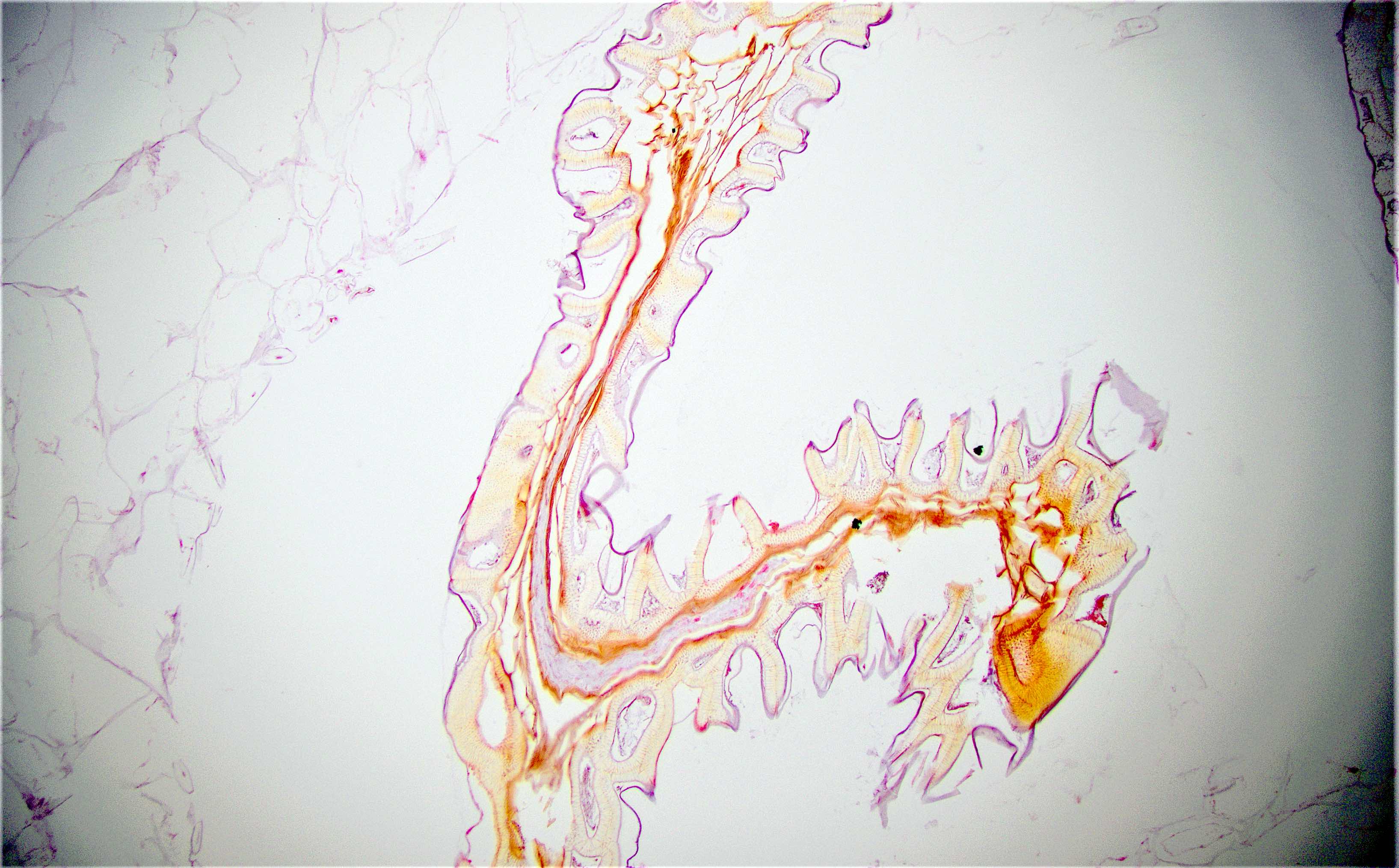
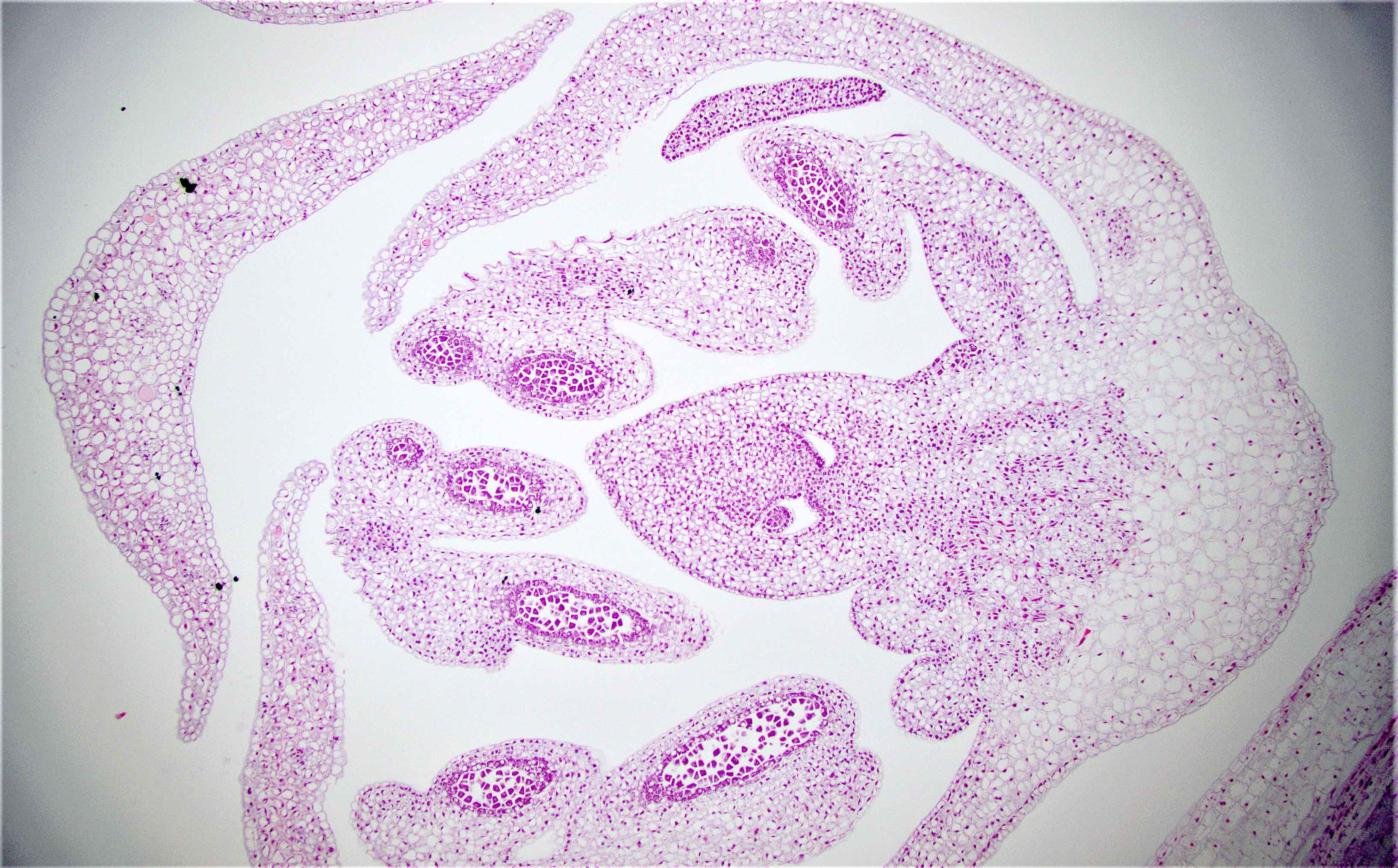
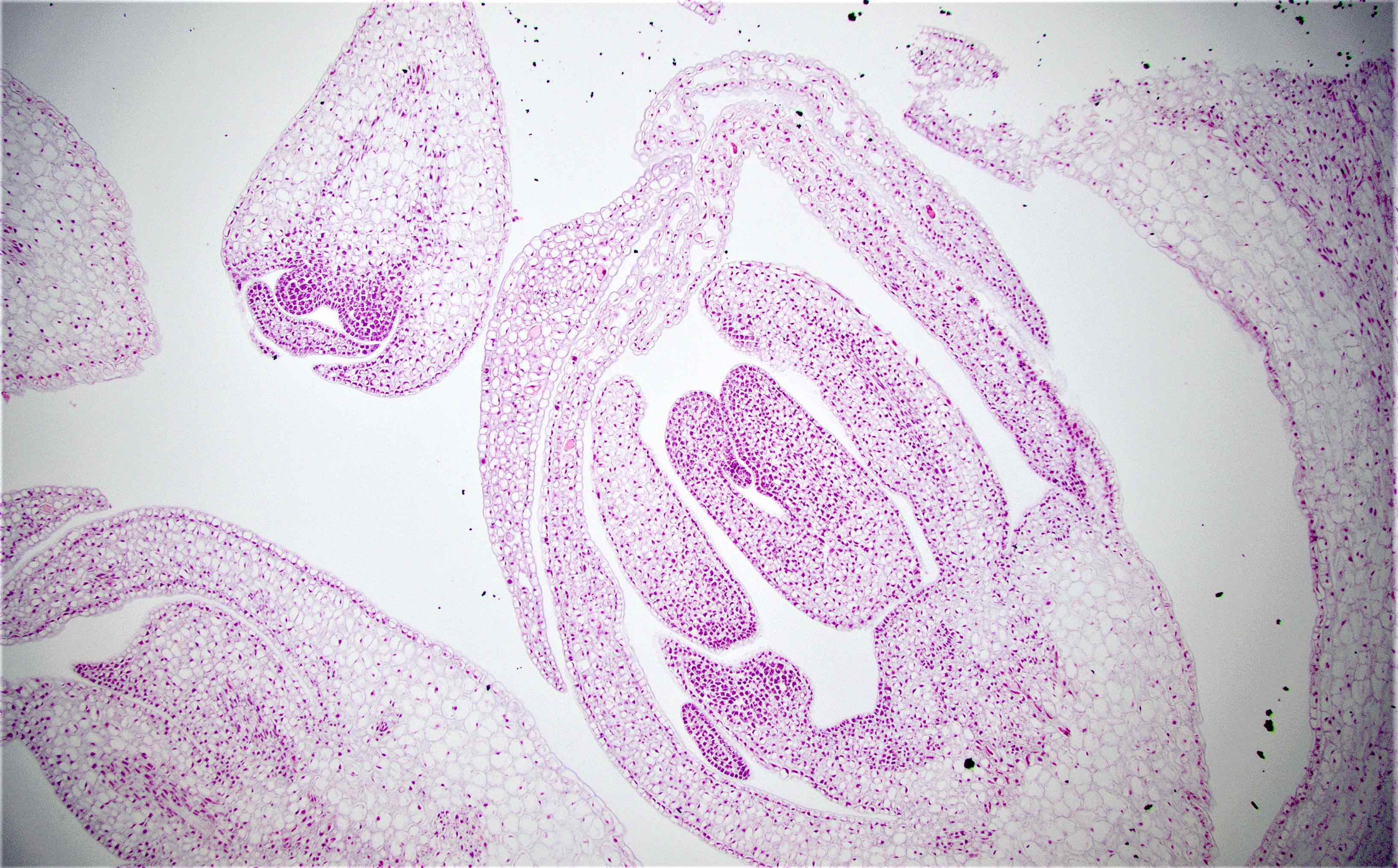
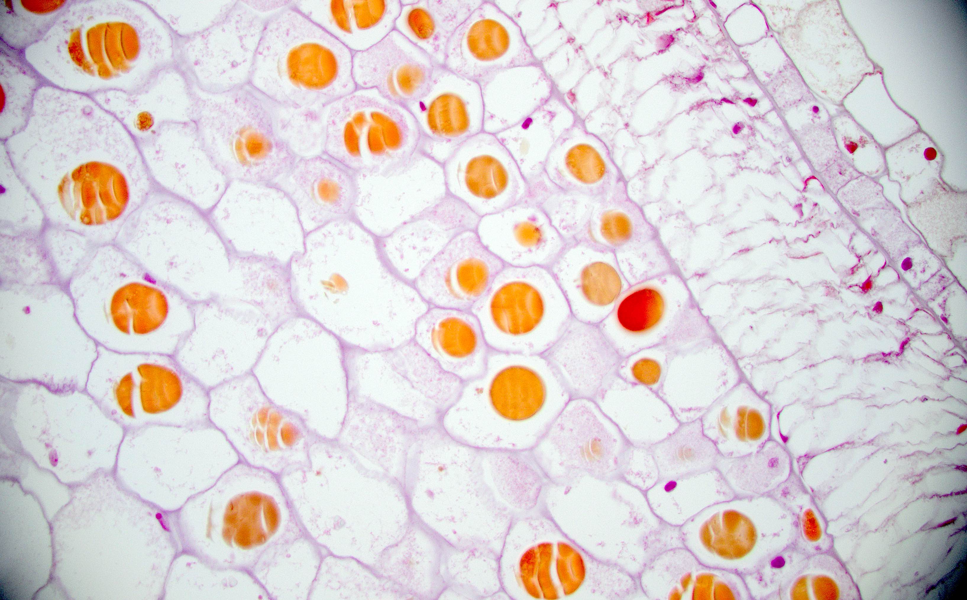
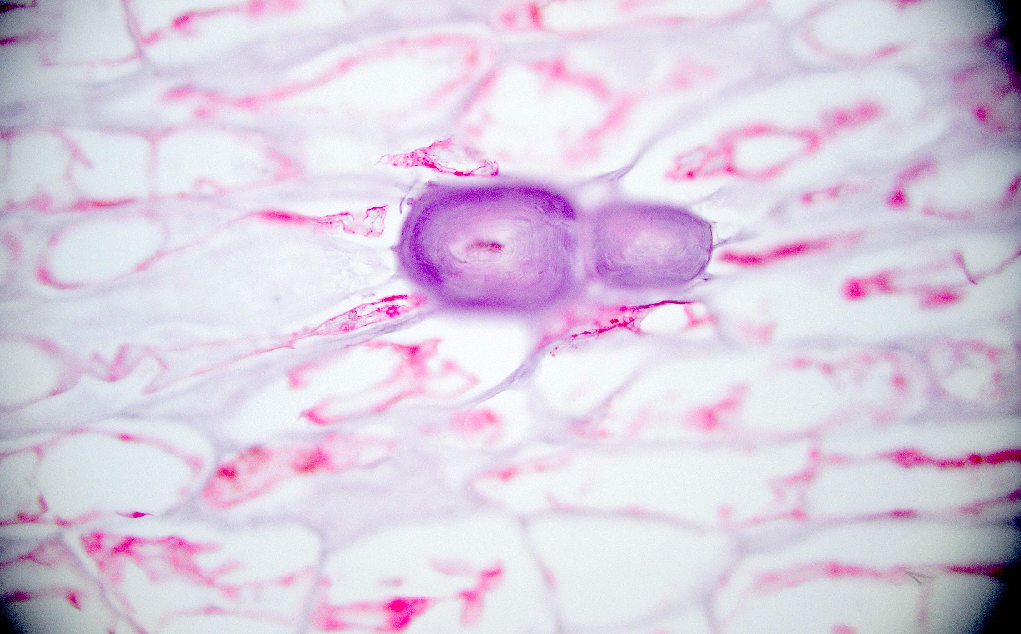
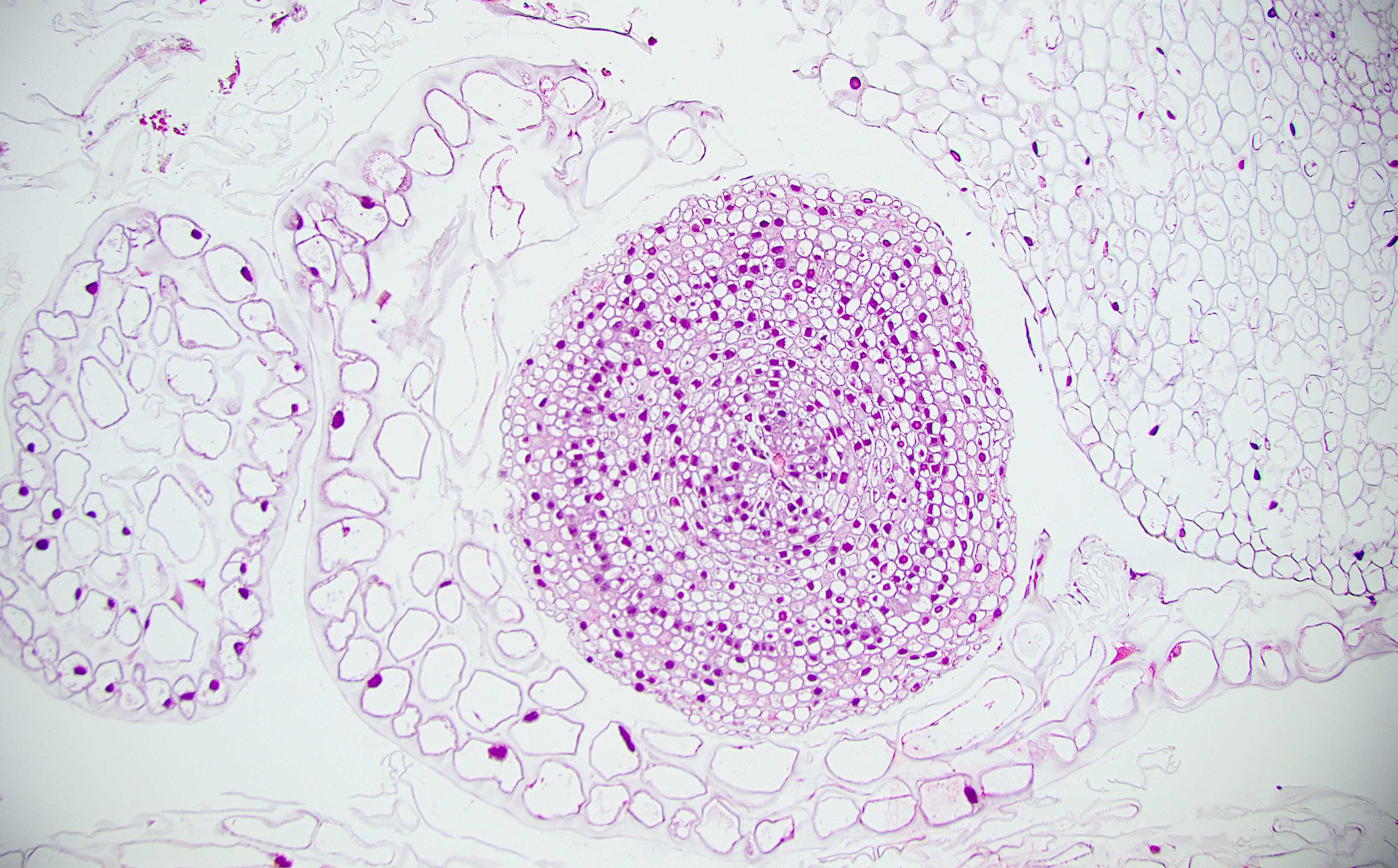
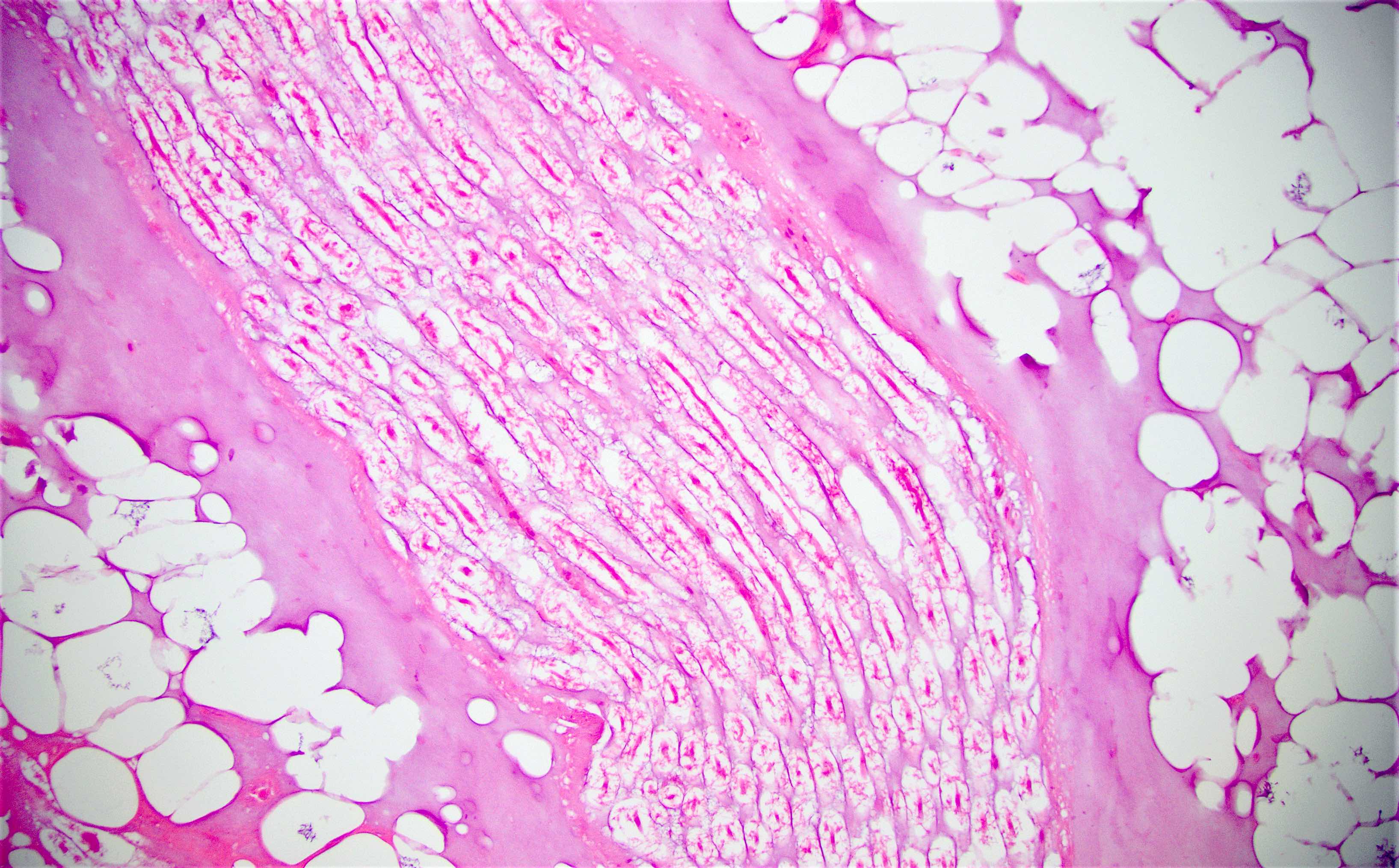
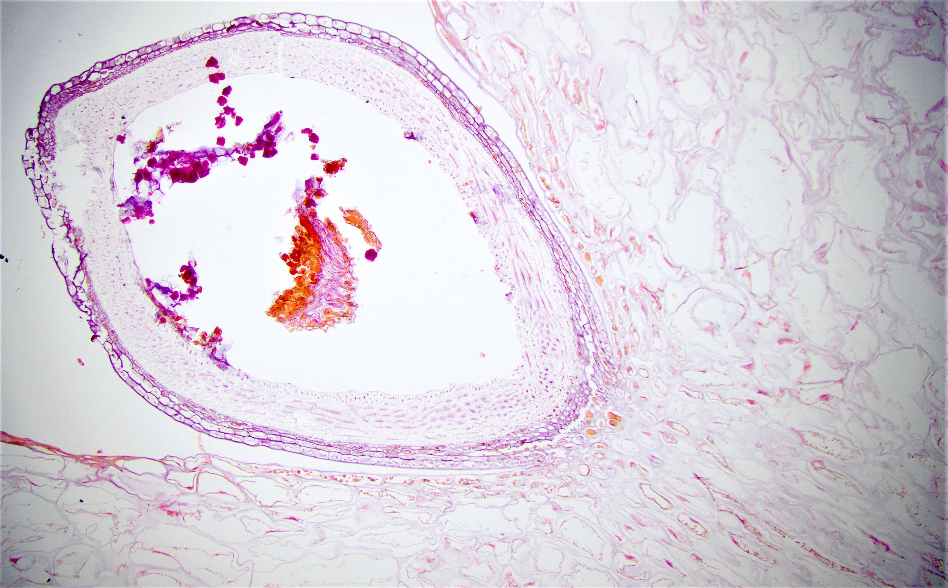
Contributed by Mona Deerwester, M.D., M.Sc.
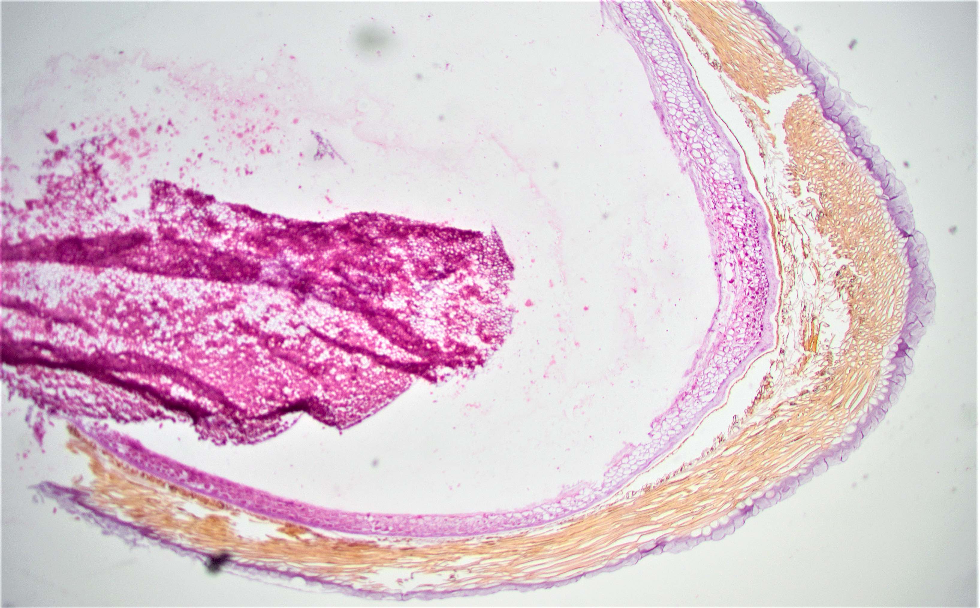

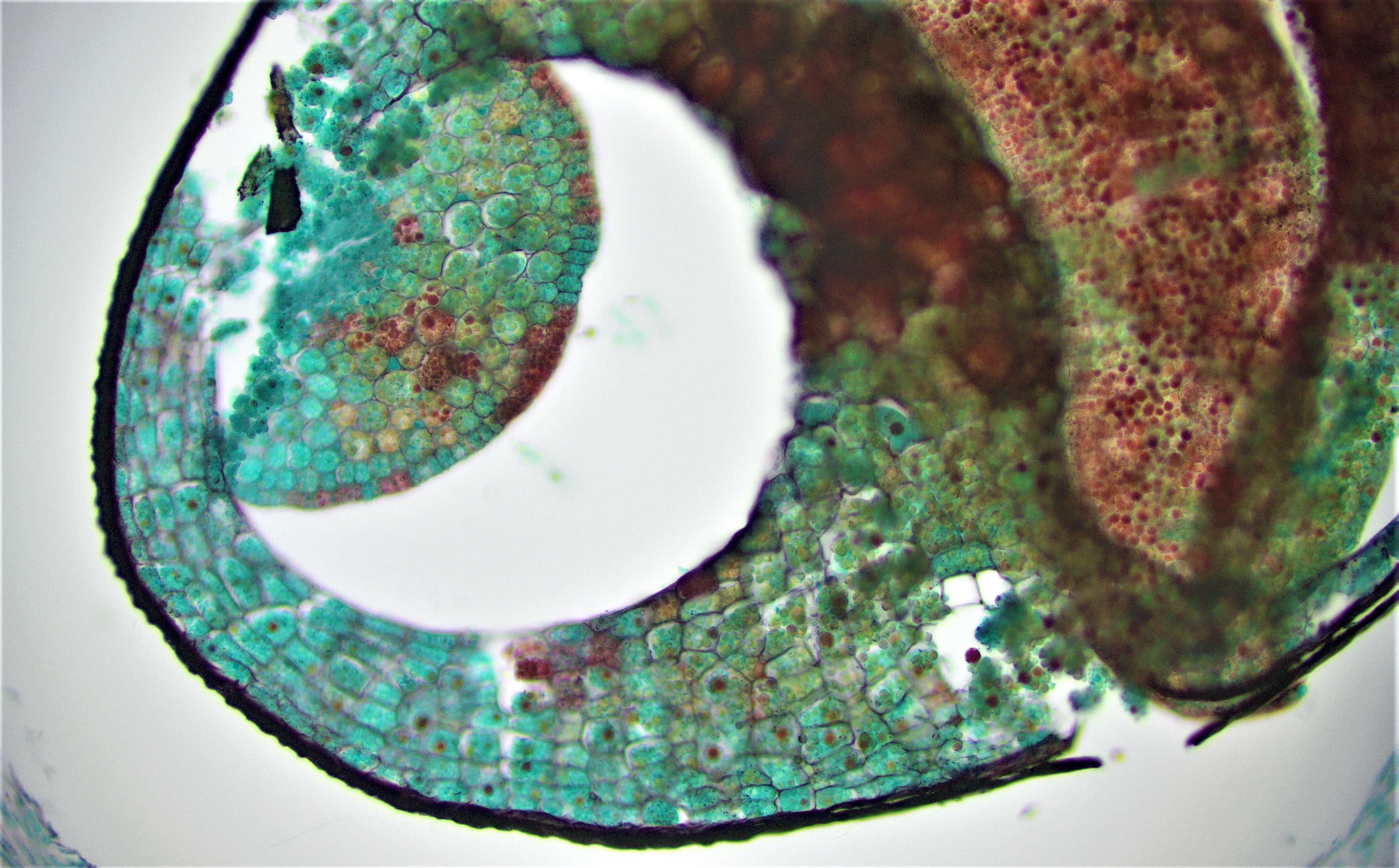
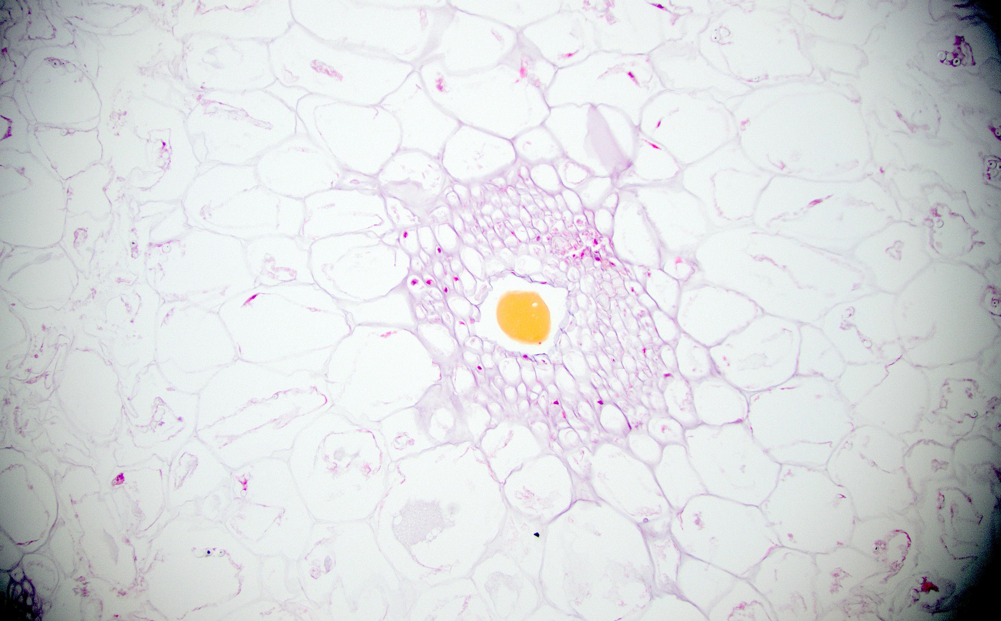
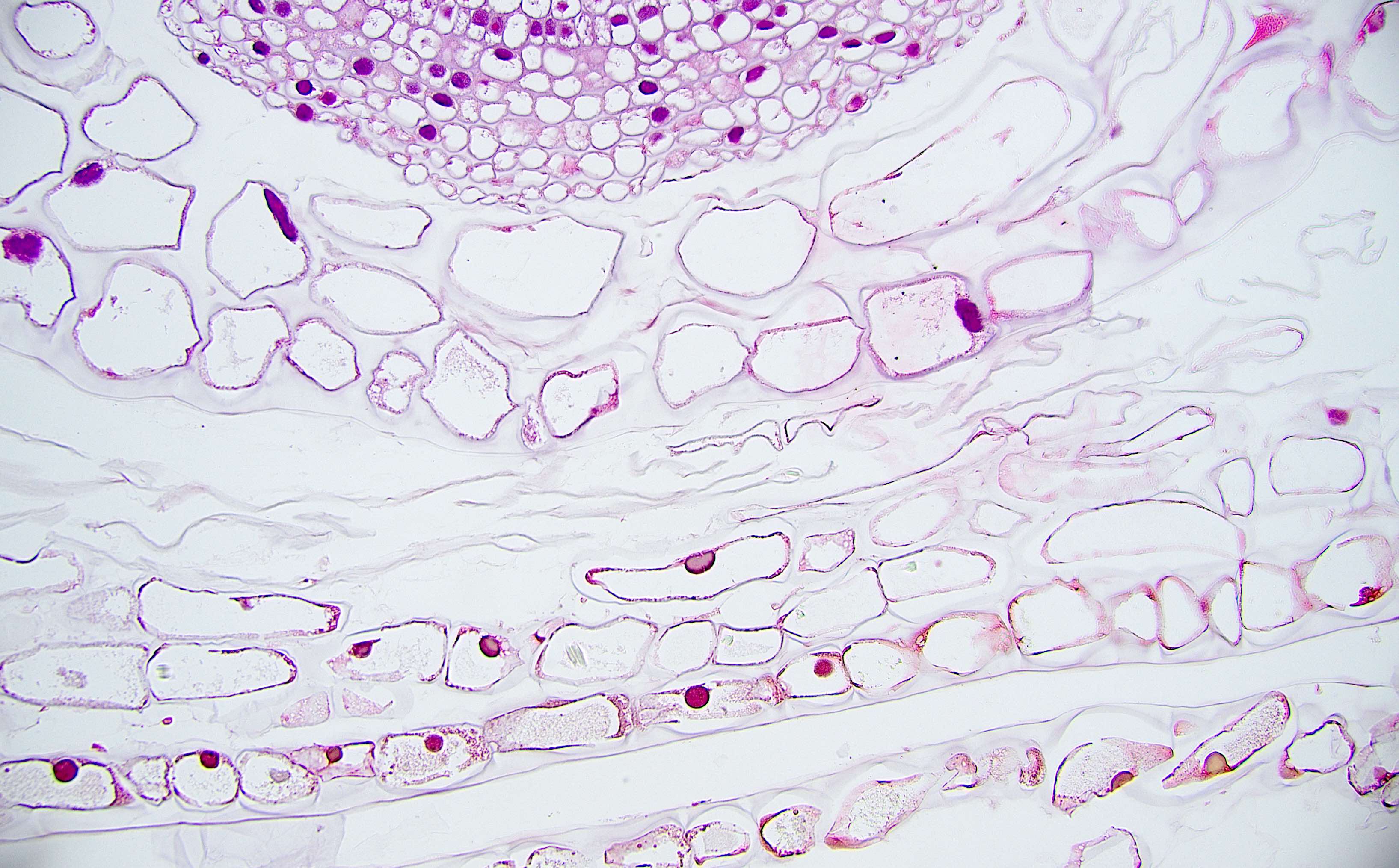
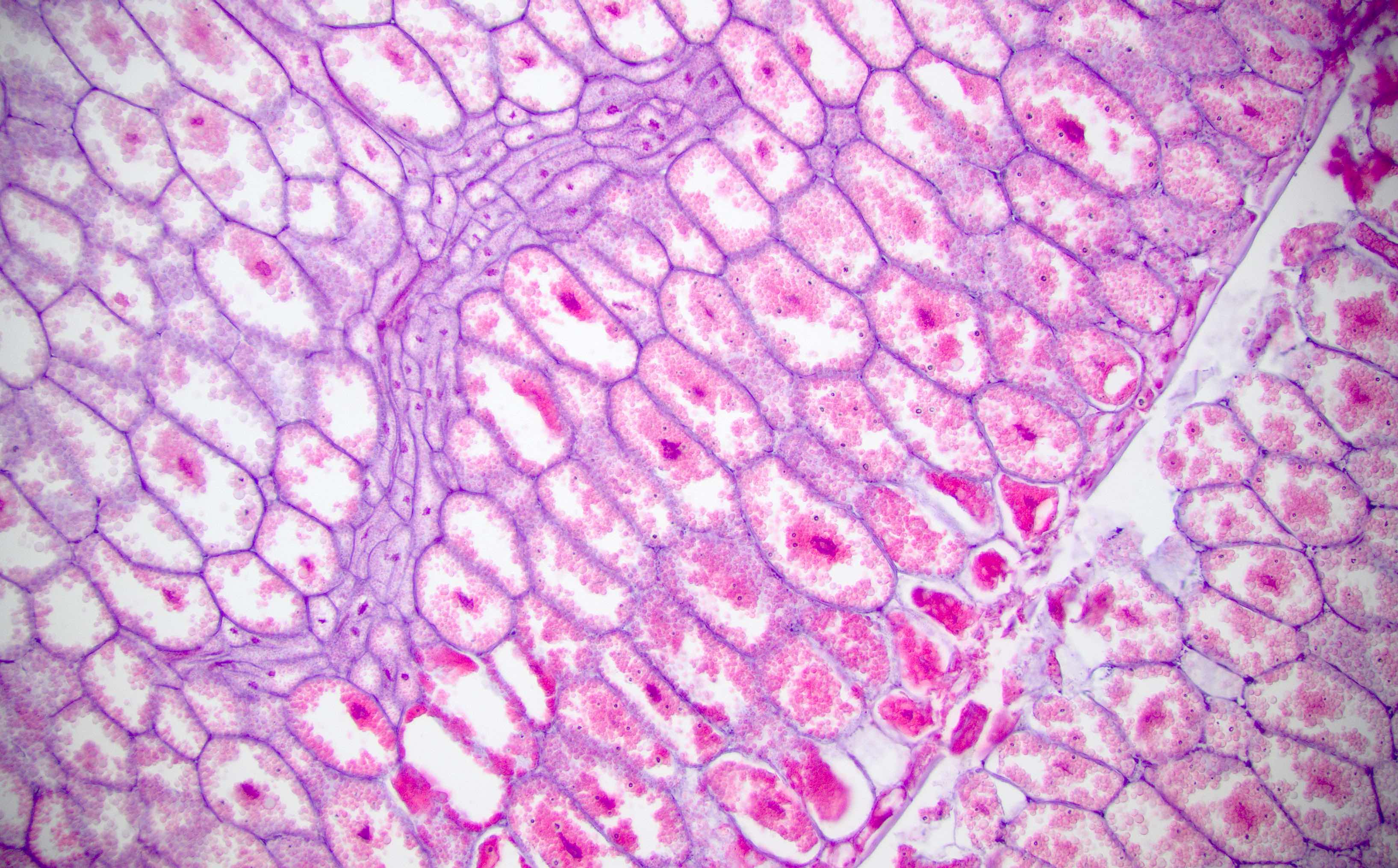
Apple seed

Asparagus
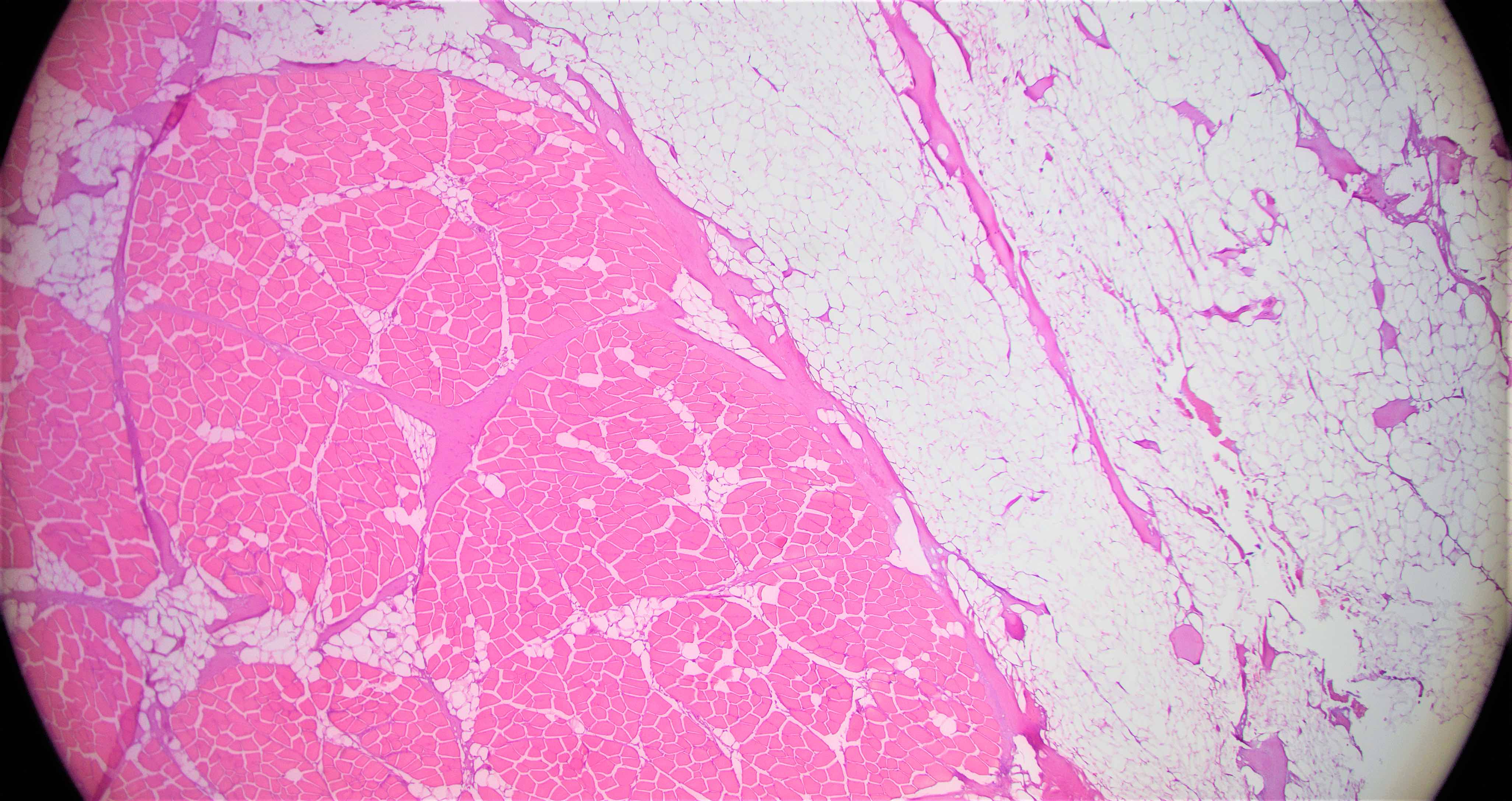
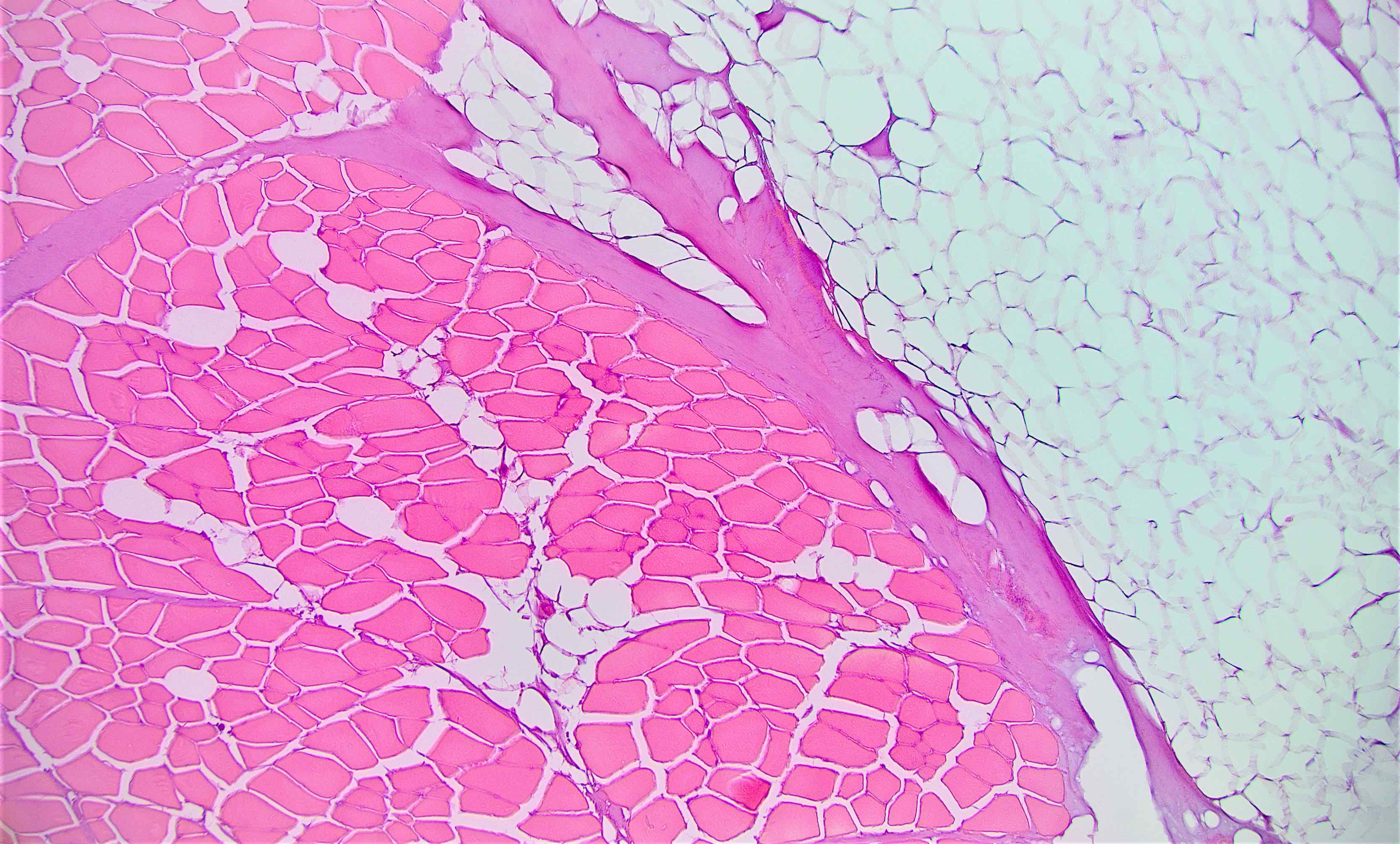
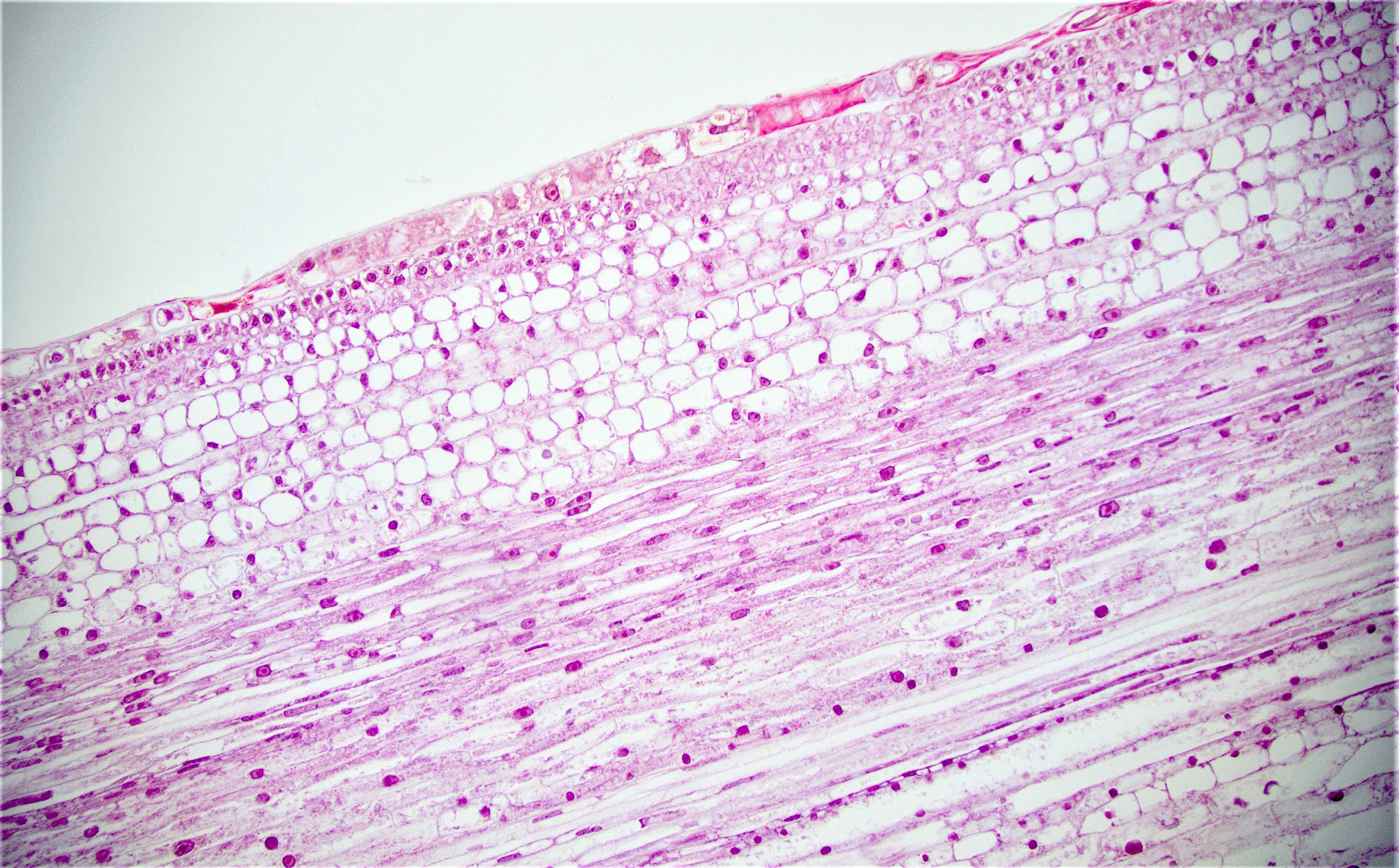
Muscle and adipose tissue of bacon
Banana
Beet
Blackberry
Blueberry seeds
Blueberry skin
Blueberry ultrastructure
Broccoli parenchymal cells and epidermis

Parenchymal cells of broccoli

Parenchymal cells of cantaloupe
Parenchymal, phloem and xylem cells of carrot
Parenchymal cells of celery
Myocytes of chicken
Parenchymal cell of cinnamon with pigment

Parenchymal cell of cinnamon with pigment
Parenchymal cell of coconut
Cookie
Cucumber

Date fruit
Date fruit

Skin of the date fruit
Eggplant
Fig
Fig fruit, GMS
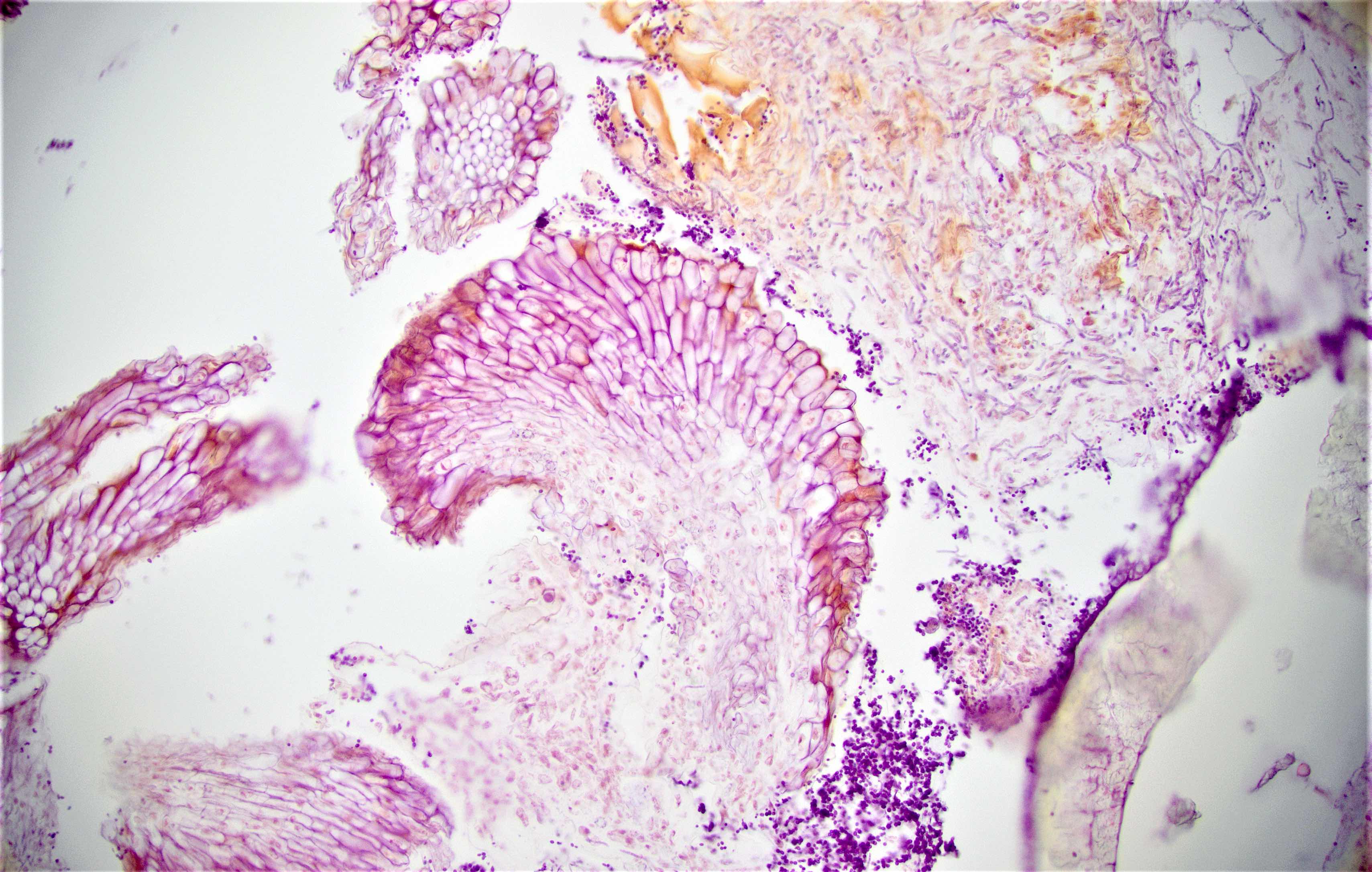

Fig fruit
Seed of a fig fruit

Fig seed
Fish retina (eye)
Fish eye
Garlic
Ginger
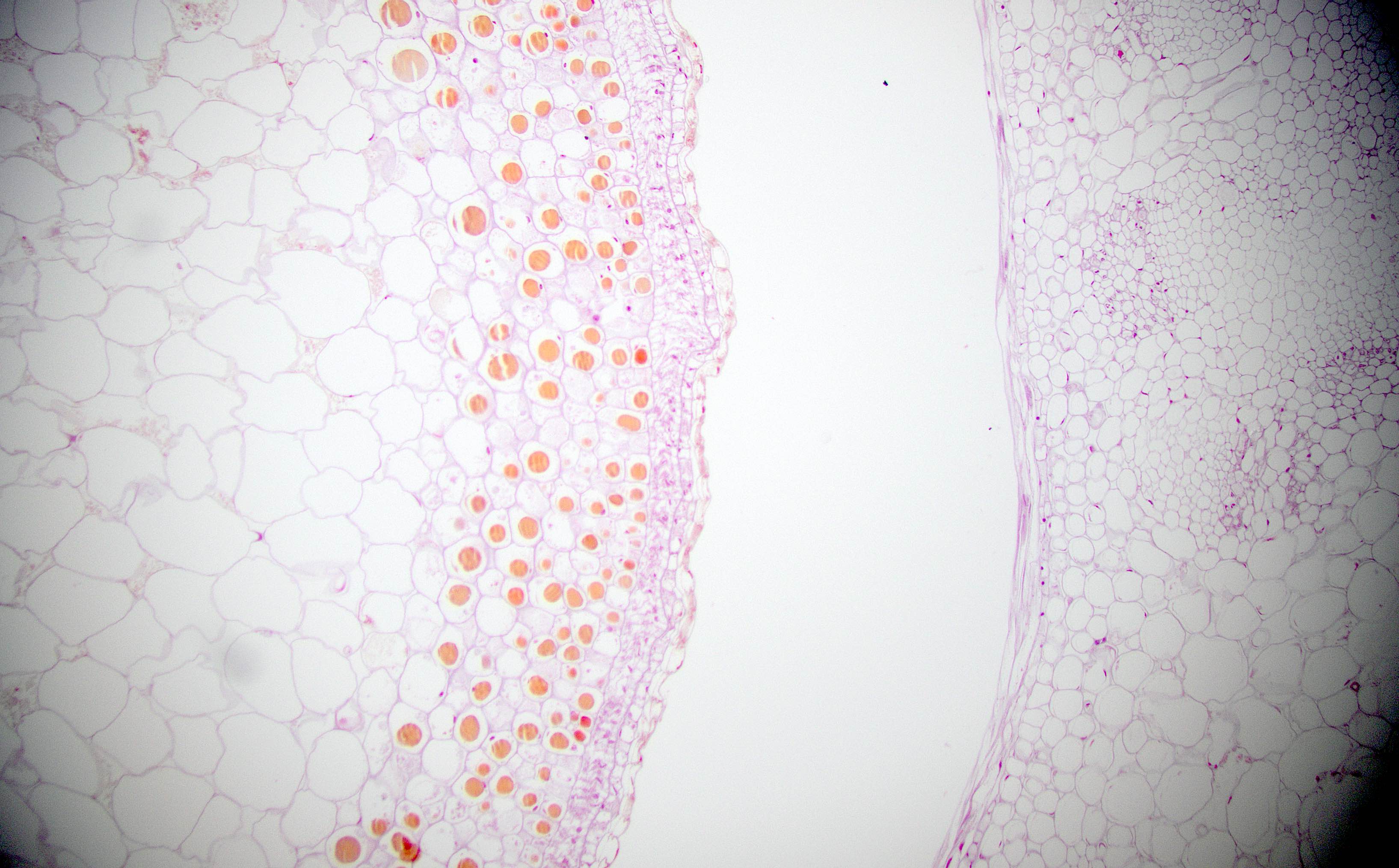
Red grapes

Red grapes
Green bean


Hot dog
Hot dog

Hot dog calcification

Hot dog vasculature
Jalapeño
Leek

Lemon skin

Lettuce
Mango

Mushroom

Oats
Okra

Olive
Olive
Onion

Orange
Pasta noodle
Peanut
Peanut

Peanut

Peas
Peas

Pepper seeds
Pepper seeds
Pepperoni
Pizza dough
Potato
Prune
Pumpkin seed
Radish

Raspberry
Raspberry seed
Red bean
Red bean

Red bell pepper
Rice

Sausage

Sesame seed


Shrimp

SPAM

Spinach
Strawberry seed
Strawberry

Tomato seed
Sliced white bread
Whole wheat dough
Images hosted on other servers:
Colonoscopy
Contributed by Christopher Hartley, M.D. and Case #305

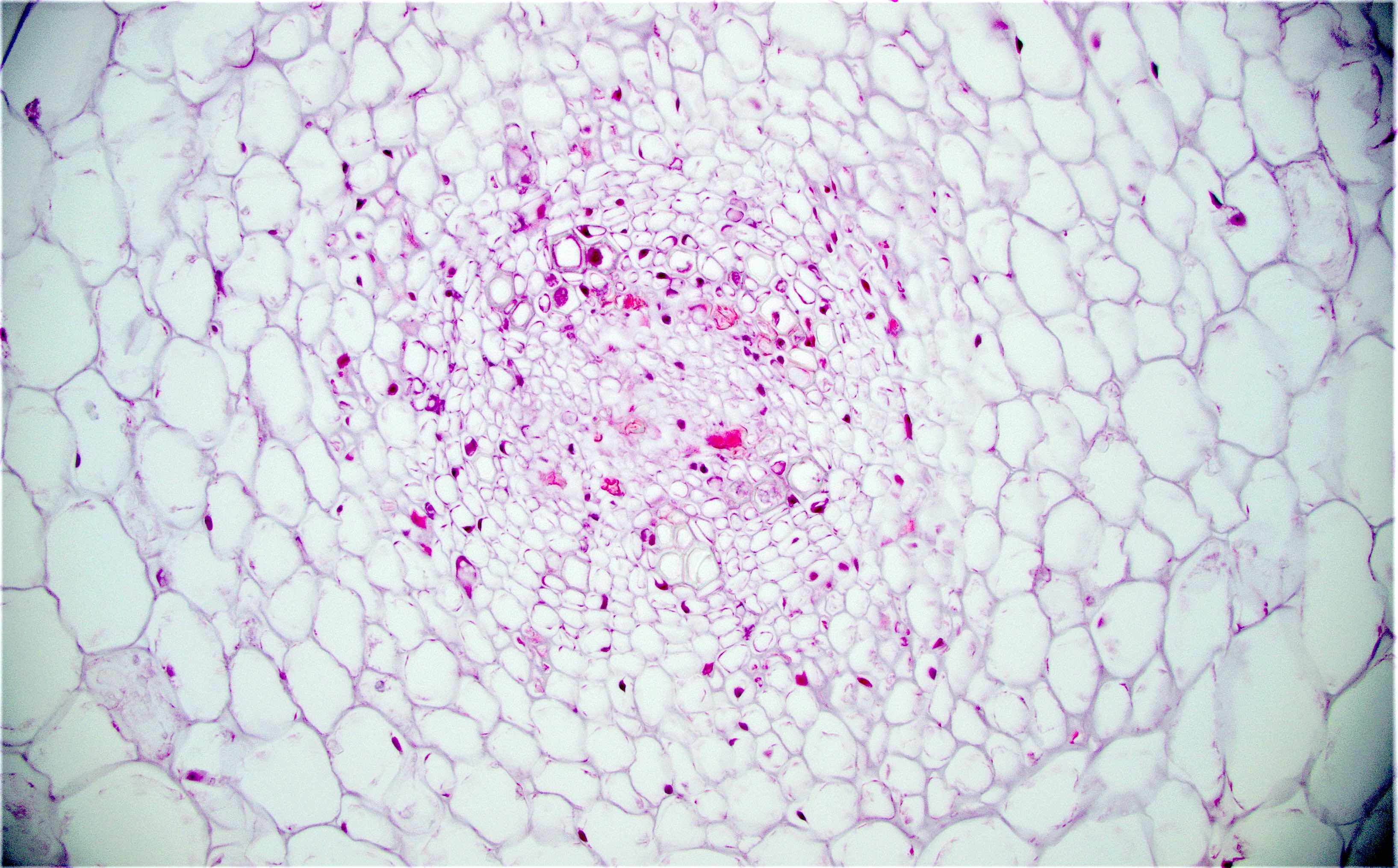
Ganglioneuroma
Images hosted on other servers:

Small bowel follow through showing luminal narrowing
Contributed by Raul S. Gonzalez, M.D.
Mucosal disease
Mural disease
Mucosal disease

Ganglion and
spindled
proliferation in
muscularis propria
Mural disease with numerous ganglion cells
Case #305
Solitary ganglioneuroma
Images hosted on other servers:


Diffuse ganglioneuromatosis
Images hosted on other servers:

Abdominal CT showing
desmoid fibromatosis
in Gardner syndrome
patient
Images hosted on other servers:

Innumerable colon polyps from Gardner patient

Desmoid fibromatosis
Contributed by Raul S. Gonzalez, M.D.
Gastric heterotopia
Images hosted on other servers:

Gastric and colonic mucosa

Gastric and colonic mucosa

Heterotopic gastric mucosa with cystic dilatation
Contributed by Raul S. Gonzalez, M.D.
Rectal GIST
Images hosted on other servers:

Epithelioid cells and osteoclast-like giant cells

Spindled cells

KIT+

CD34+

PDGFRA+
Contributed by Gustavo Moreno, M.D. and Catherine E. Hagen, M.D.
Endoscopic
Contributed by Gustavo Moreno, M.D. and Catherine E. Hagen, M.D.
Apoptosis
Apoptosis, no crypt loss
Ulceration
Crypt dropout
Architectural distortion
Crypt abscess
Indeterminate
Endocrine cell aggregates
Contributed by Chungja C. Shim, M.D.
Cause unknown


Table 1: sectioning protocol based on polyp size
| | |
| Smaller than 0.4 cm | No sectioning |
| 0.4 - 0.8 cm | Bisect; place in 1 cassette |
| 0.9 - 1.2 cm | Trisect by shaving two sides off central section with stalk; place central section in separate cassette |
| Larger than 1.2 cm | More than 3 sections as appropriate |
Images hosted on other servers:

Anatomic subsites of colon and rectum (table 2)


Anatomic subsites of the colon

Mesenteric / radial margin


T category staging

Regional lymph nodes
Contributed by Ed Uthman, M.D. and Raul S. Gonzalez, M.D.
Incidental submucosal
hemangioma in
sigmoid colectomy

Colonic hemangioma
Images hosted on other servers:

Collision with liposarcoma

Associated with ulcerative colitis


Gastric tumor: H&E, AFP+ HepPar1- glypican3+
Images hosted on other servers:

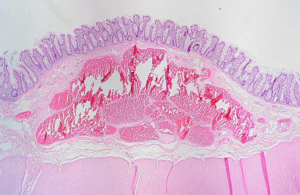
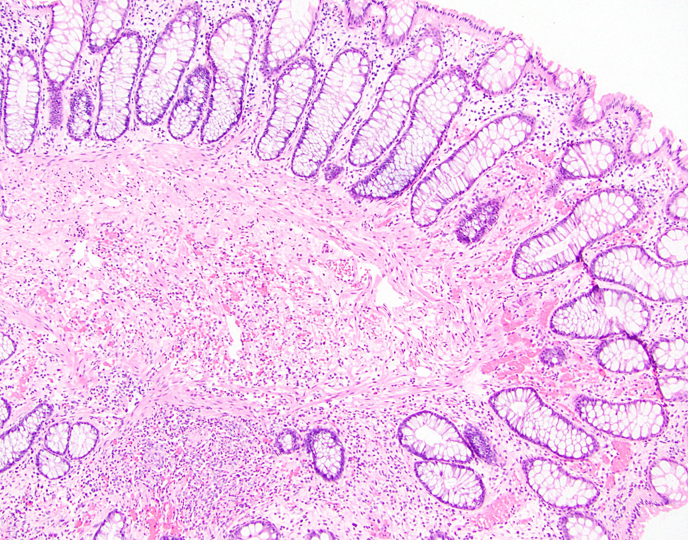
Proximal extent of aganglionic segment and corresponding frequency
Contributed by Raj P. Kapur, M.D., Ph.D.
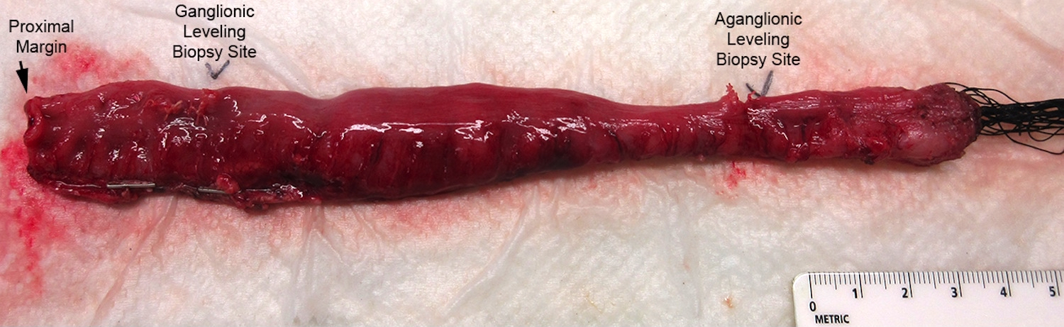
Resection specimen
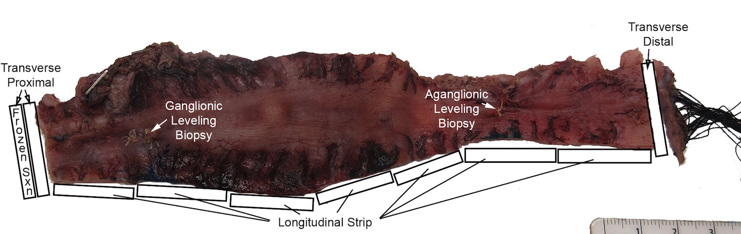
Histological sampling of resection
Images hosted on other servers:


Dilated bowel
Contributed by Raj P. Kapur, M.D., Ph.D.

Ganglionic biopsy
Aganglionic biopsy
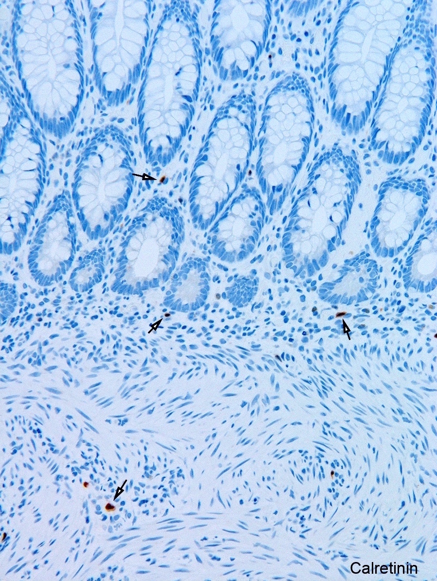
Calretinin: ganglionic biopsy / aganglionic biopsy
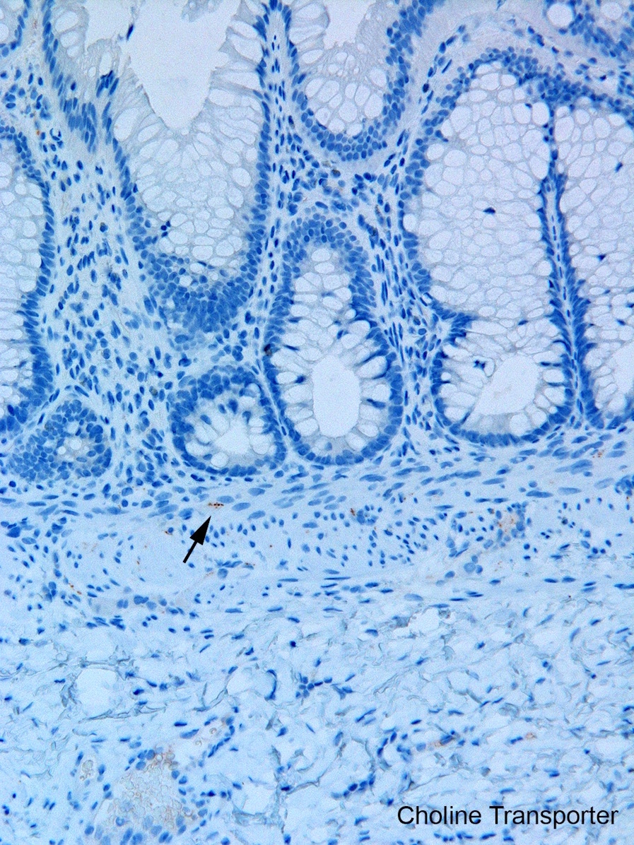

Choline transporter IHC: ganglionic biopsy / aganglionic biopsy
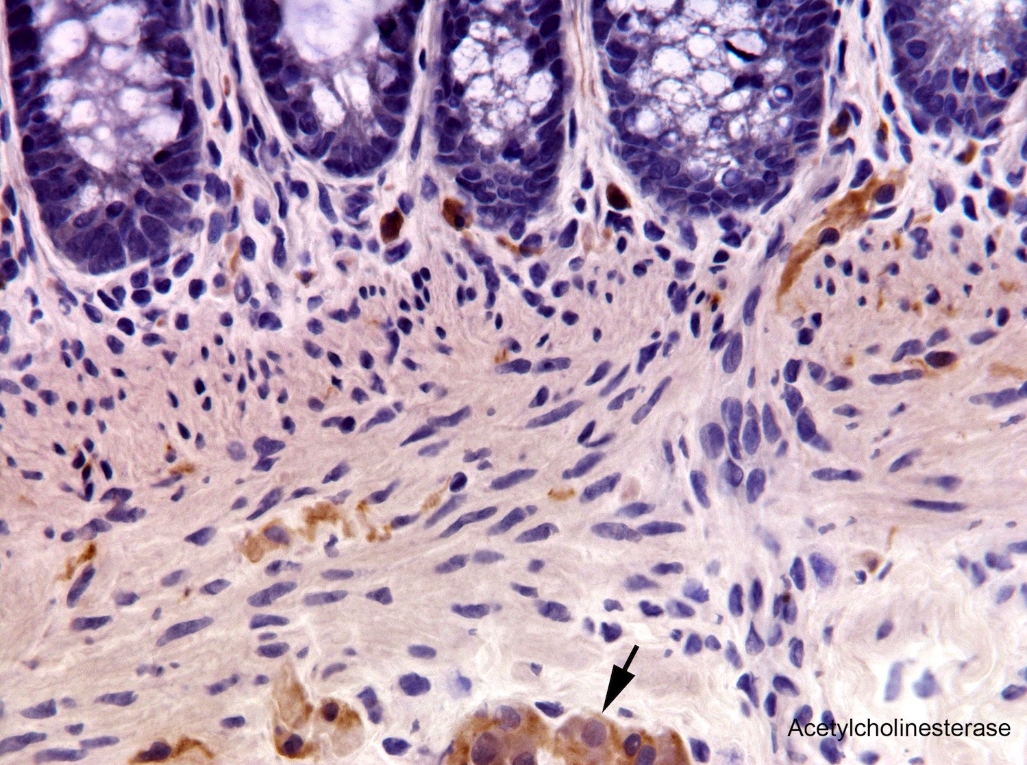

Acetylcholinesterase histochemistry: ganglionic biopsy /
aganglionic biopsy

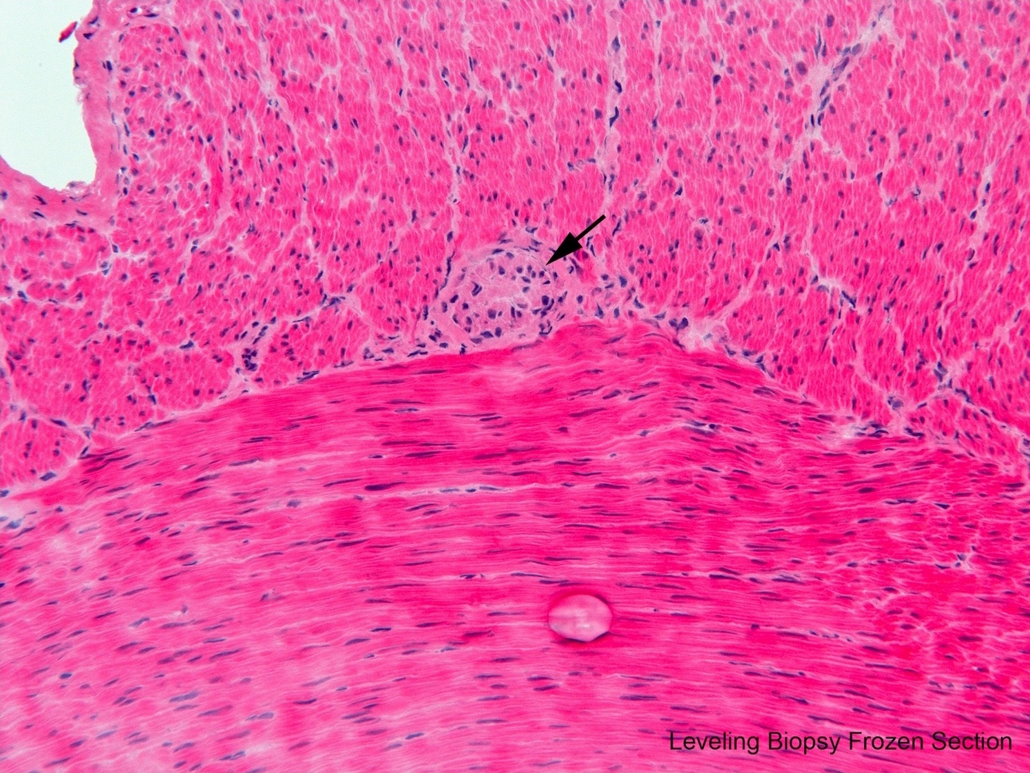
Leveling frozen section biopsy: ganglion cells / aganglionic segment
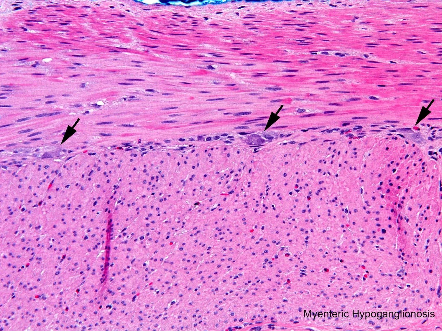

Myenteric hypoganglionosis
Images hosted on other servers:

CT findings in colonic histoplasmosis
Contributed by Wei Chen, M.D., Ph.D.
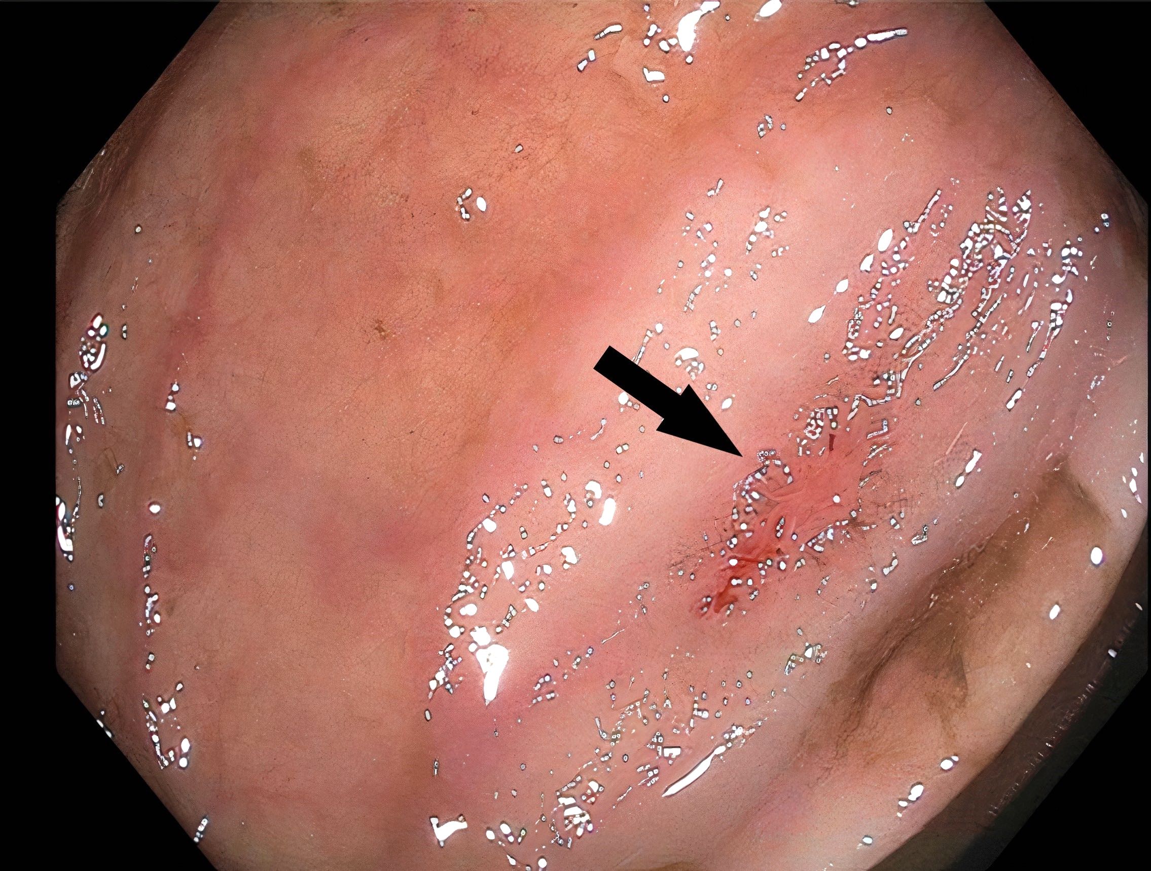
Colonoscopy showing GI histoplasmosis
Images hosted on other servers:

Ulcer in the sigmoid colon
Contributed by Wei Chen, M.D., Ph.D.
Colon with mucosal and submucosal inflammation
Submucosa with numerous macrophages
Macrophage containing yeast
GMS stain
PAS stain

Mucicarmine stain
Fontana-Masson silver stain
Histoplasmosis colitis
Colonoscopy in histoplasmosis
Images hosted on other servers:

HIV associated damage to the GI tract
Images hosted on other servers:

HIV associated - various images

Colonic ulceration

Healing colonic ulceration
Contributed by Nalini Bansal, M.D.
Cryptosporidiosis
Strongyloides
Amoeboma
Herpes
Images hosted on other servers:

Candida (esophagus)

Candidal pseudohyphae and spores (PASD stain)

CMV gastritis

CMV vasculitis

Cryptococcosis (GMS stain)


Cryptosporidiosis

Giardiasis

Histoplasmosis (GMS stain)

HIV enteropathy

HSV esophagitis

HSV cytopathic effect


Kaposi sarcoma

Mycobacterium avium-intercellulare (acid fast stain)
Images hosted on other servers:

Colonoscopy showing deep ulcers in the sigmoid colon
Contributed by Zarrin Hossein-Zadeh, M.D. and Raul S. Gonzalez, M.D.

Pathognomonic Cowdry type A and type B inclusions
Positive HSV antibodies
Images hosted on other servers:

Hyperplastic polyp at endoscopy
Contributed by Adrian C. Bateman, M.B.B.S., M.D.
Microvesicular variant
Sawtooth glands
Surface tufting
Nuclear changes at crypt bases
Goblet cell rich variant
Tapered crypts at base
Contributed by Lei Sun, M.D. and Feng Yin, M.D., Ph.D.
CAC, conventional type
CAC, signet ring type
CAC, mucinous type
Contributed by Ashwini Kumar Esnakula, M.D., M.S.
Colonoscopic findings
Contributed by Ashwini Kumar Esnakula, M.D., M.S.
Prominent crypt epithelial apoptosis and apoptotic bodies
Prominent crypt epithelial apoptosis
Cryptitis and crypt epithelial apoptosis
Cryptitis and crypt abscess
Cryptitis and lamina propria mixed inflammation
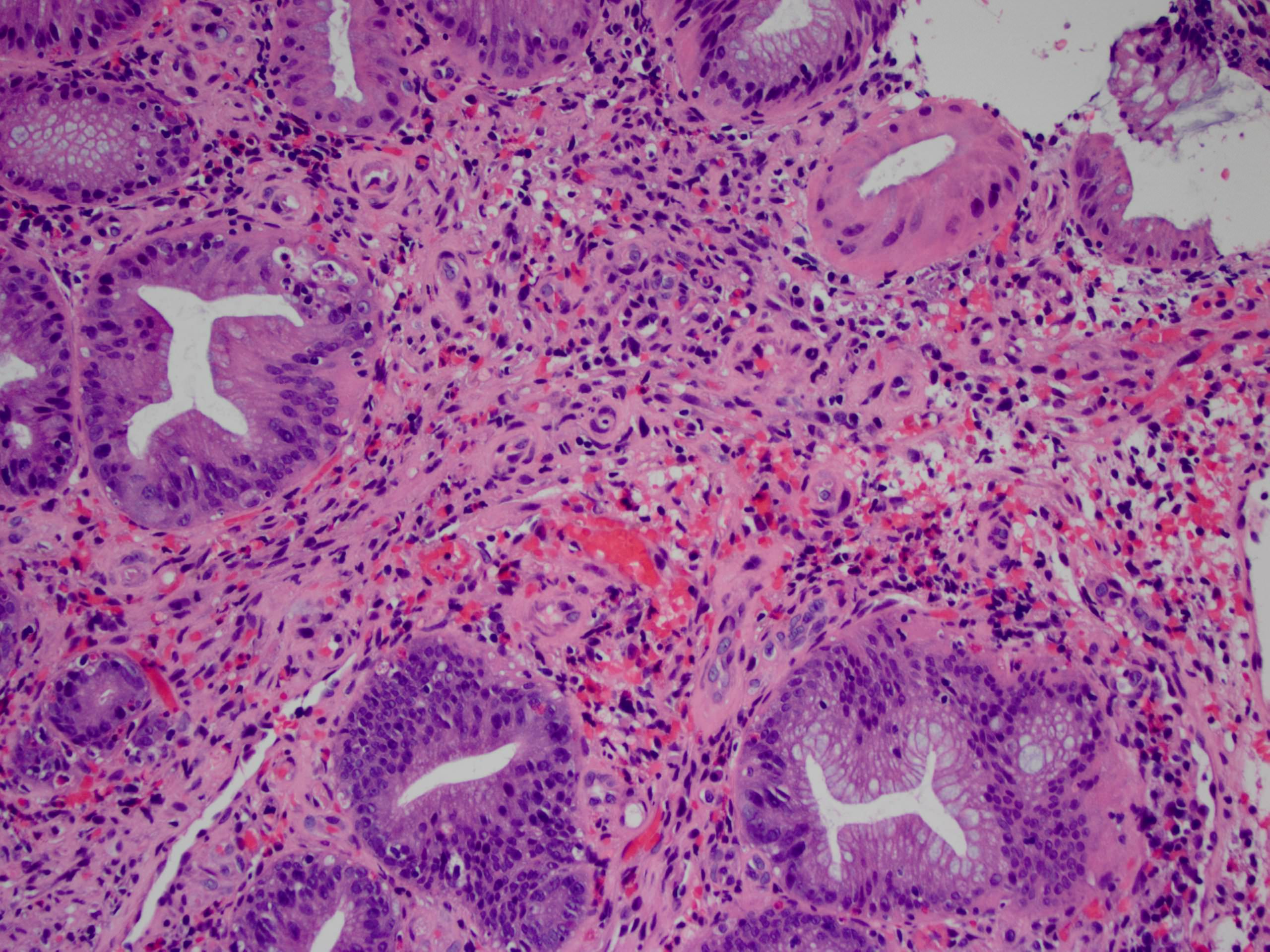
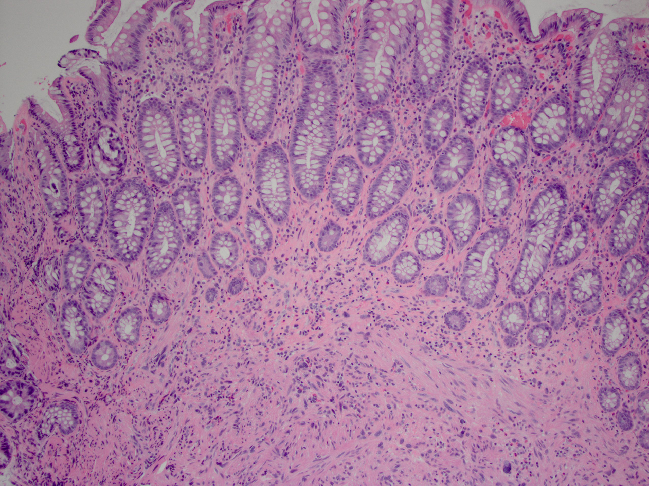
Contributed by Raul S. Gonzalez, M.D.
Biopsy: mucosal
proliferation
of thick walled
vessels
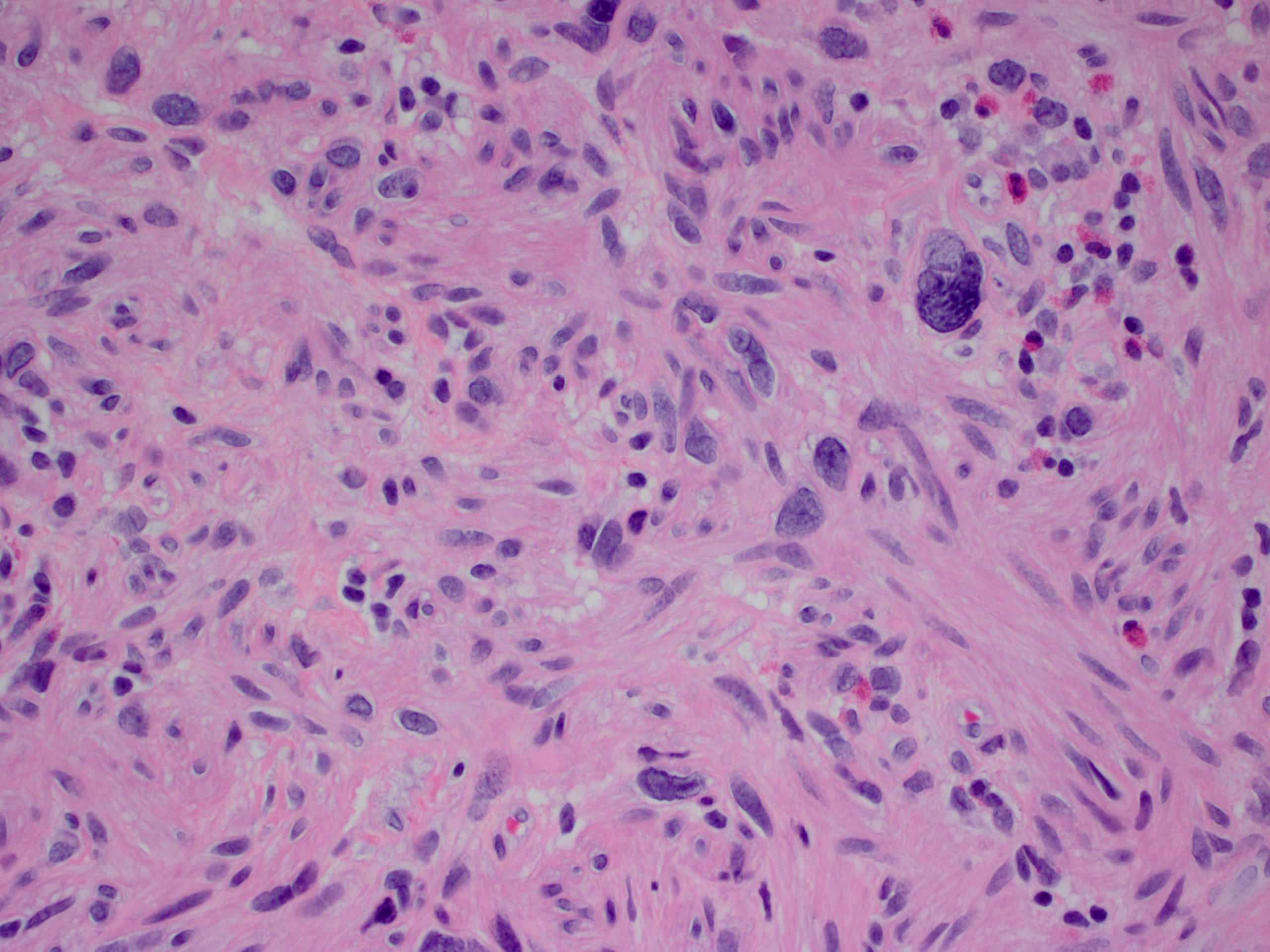
Resection:
thickened
mural veins

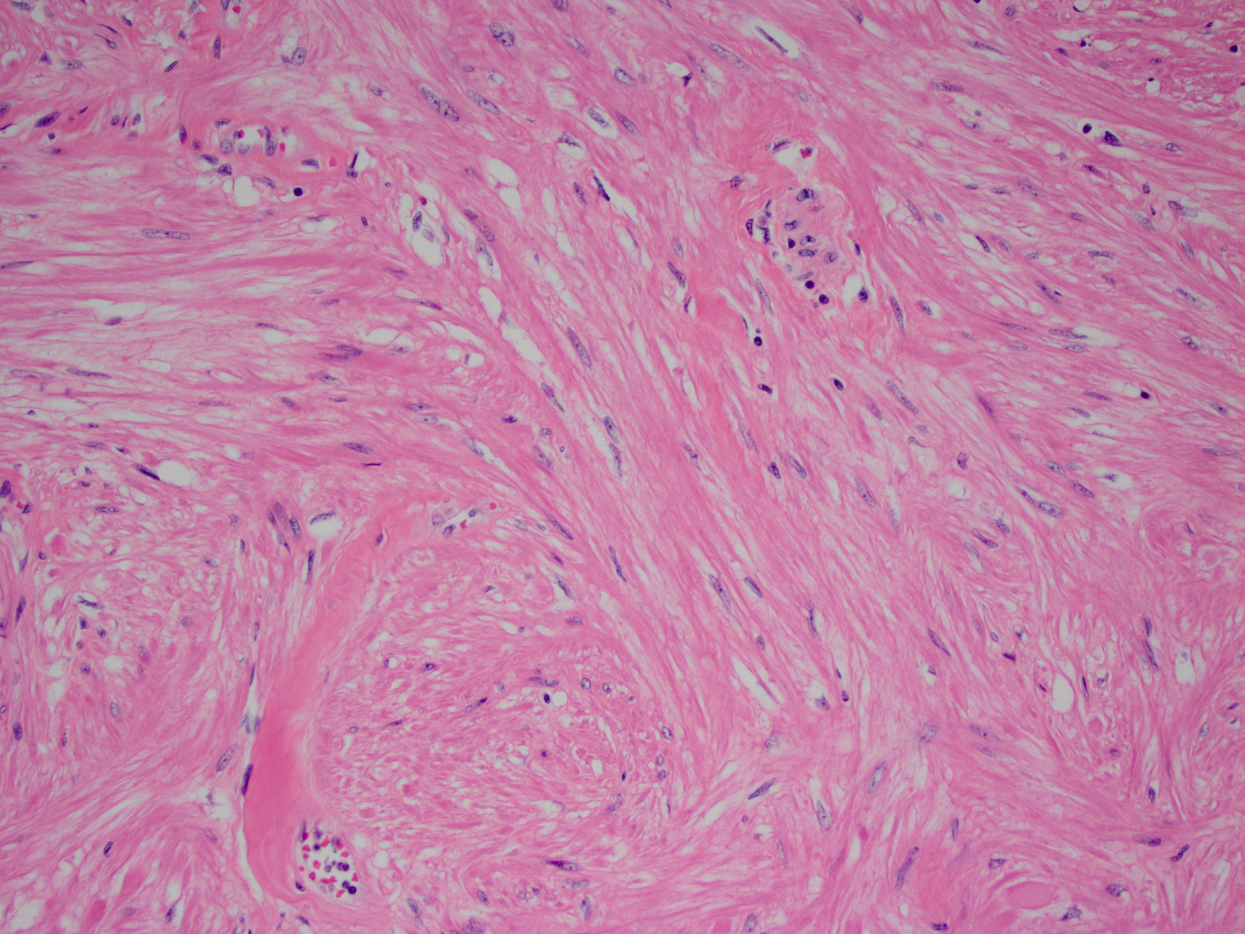
2 thickened veins and 1 unaffected artery

Elastin stain
highlights
internal elastic
lamina of artery
Images hosted on other servers:

Retroperitoneal mass enveloping aorta
Contributed by Raul S. Gonzalez, M.D.
Fibrosis and inflammation
Images hosted on other servers:

Fibrosis involving ureter
Images hosted on other servers:


Necrotic segments of colon
Contributed by Raul S. Gonzalez, M.D.
Colon infarct
Histopathology colon - hemorrhagic infarct
Images hosted on other servers:

Central calcified oval mass in the pelvis

Pelvic mass
Images hosted on other servers:

Egg shaped mass

Giant loose peritoneal body

Giant loose body attached to omentum

Round pelvic mass

Central calcifications and a distinct fat plane

Macrograph of giant loose body
Contributed by Raul S. Gonzalez, M.D.
Rounded contour
Central fat necrosis
Circumferential fibrous tissue
Contributed by John D. Paulsen, M.D. and Alexandros D. Polydorides, M.D., Ph.D.
IBD, indeterminate type
IBD, indeterminate type, favor Crohn's disease
Mimic: ulcerative colitis with cecal patch
Mimic: superficial
(ulcerative
colitis-like)
Crohn's disease
Contributed by John D. Paulsen, M.D. and Alexandros D. Polydorides, M.D., Ph.D.
Transmural chronic inflammation
Granuloma, pericolonic lymph node
Mimic: cryptolytic granuloma, ulcerative colitis
Mimic: superficial (ulcerative colitis-like) Crohn's disease
Mimic: fulminant ulcerative colitis
Images hosted on other servers:

Multiple colonic polyps
Contributed by Andrea L. Wiens, D.O. and Janet E. Roepke, M.D., Ph.D.
Inflammatory polyp of colon secondary to mucosal prolapse
Contributed by M.J. Fernández-Aceñero, M.D., Ph.D.
Endoscopic lesion
Contributed by Taofic Mounajjed, M.D.
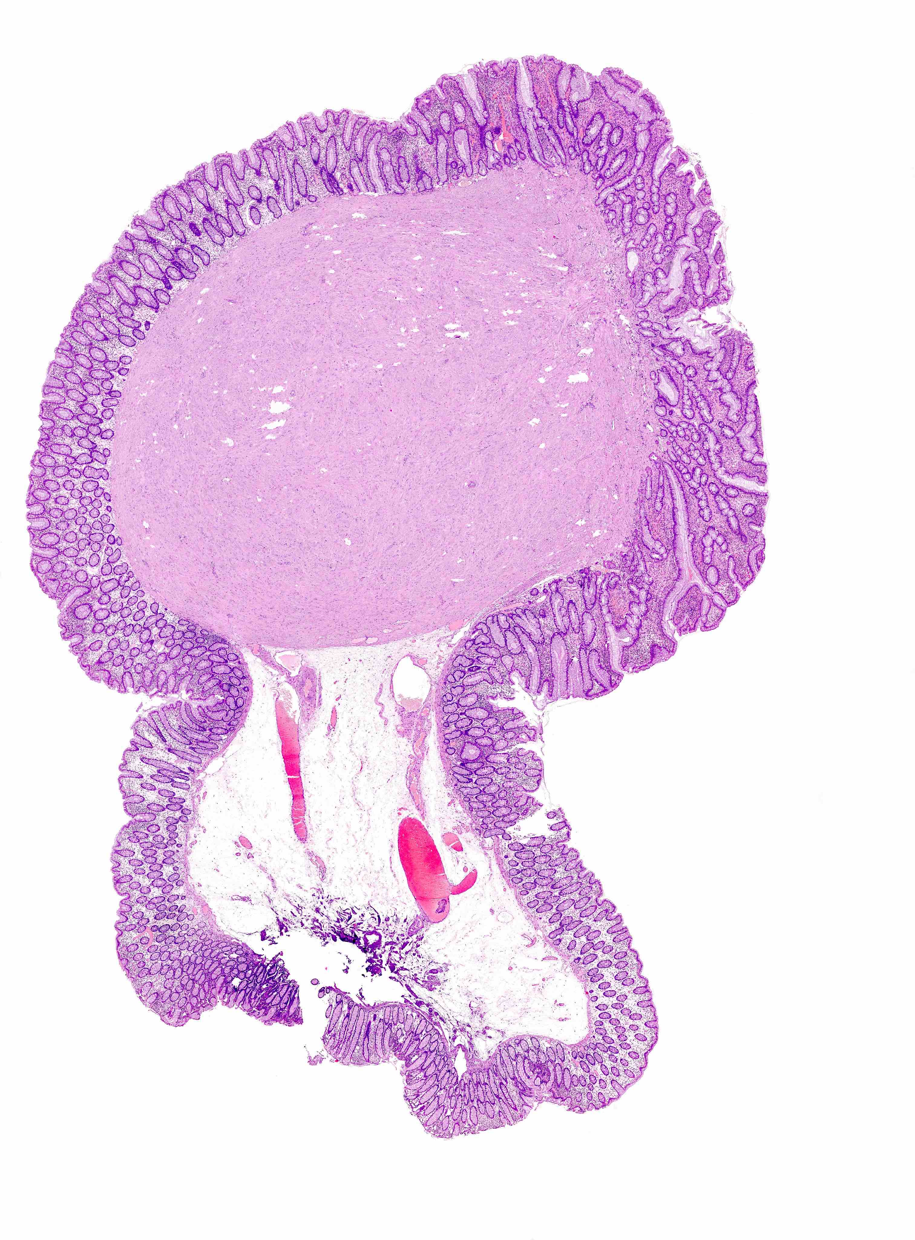
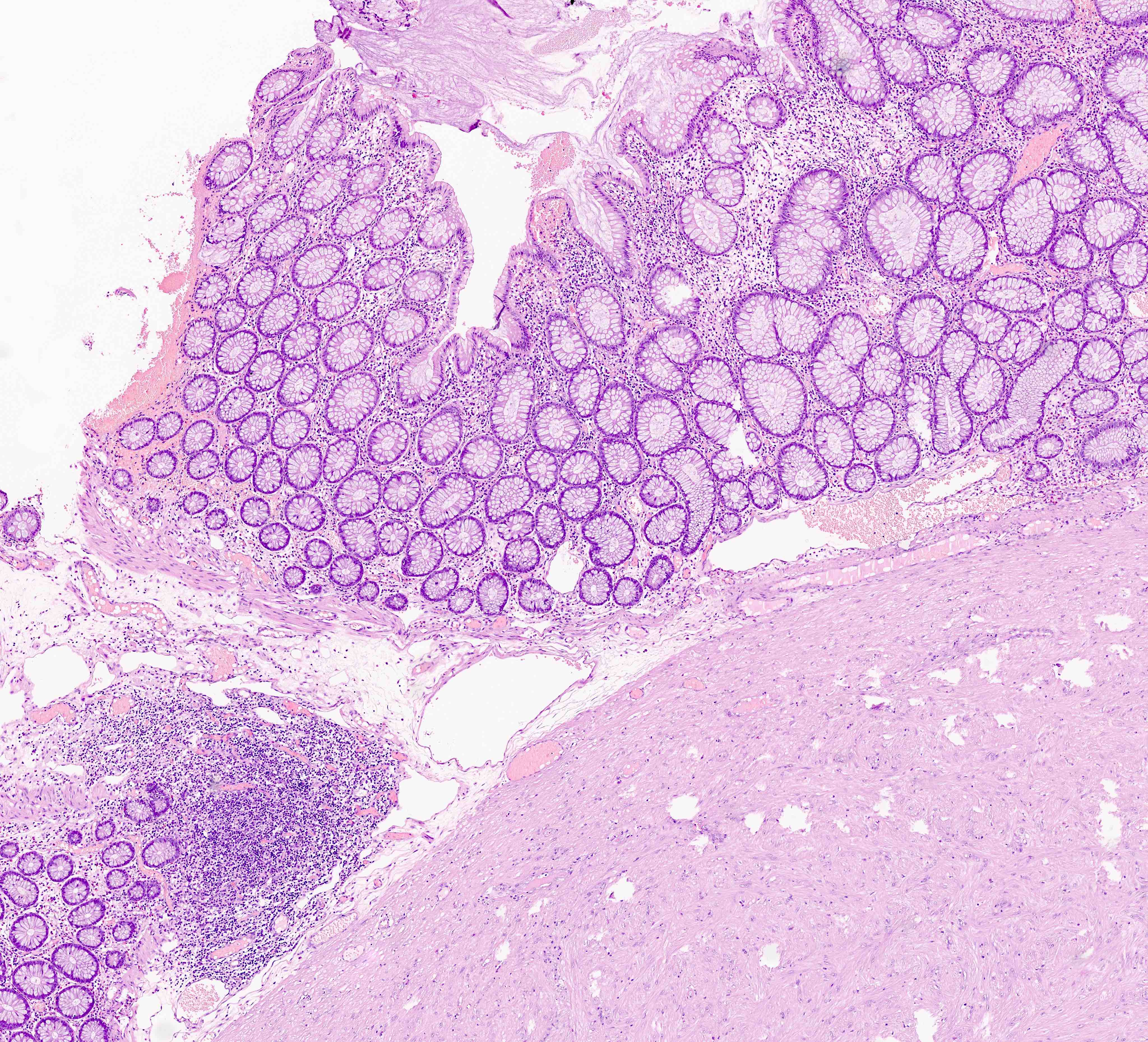
Pedunculated cecal polyp
Cut surface with white color
Contributed by M.J. Fernández-Aceñero, M.D., Ph.D.
Typical features

Large bowel lesion
Typical growth pattern
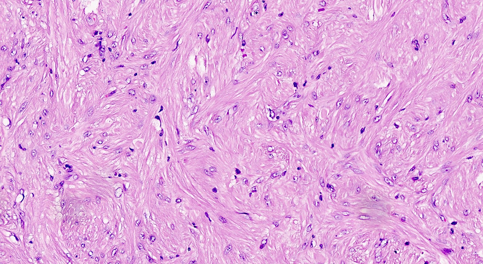
Edema
Myxoid change

Stain for collagen
Mitotic activity
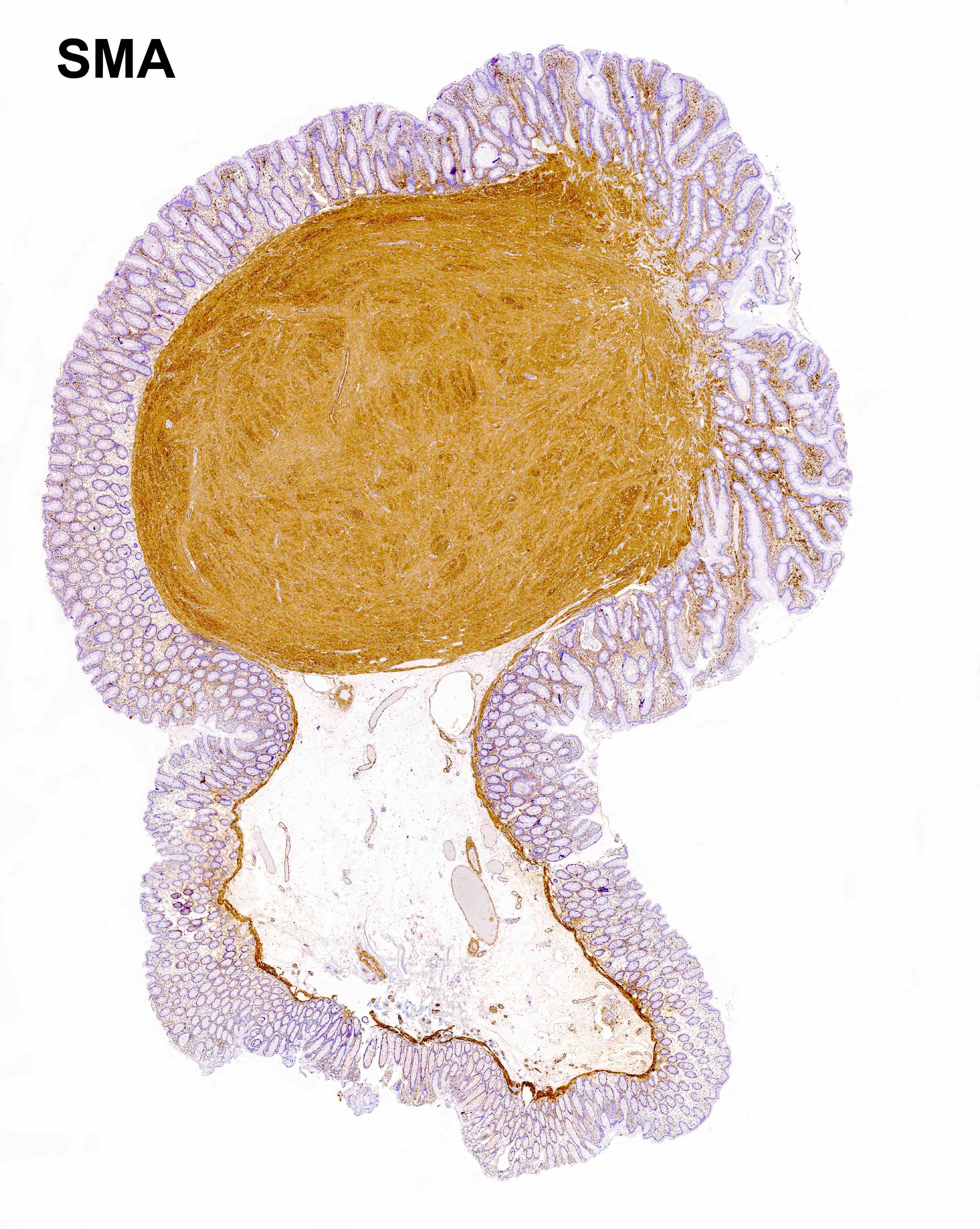
CD34
SMA
Images hosted on other servers:

Polypoid lesion, on imaging
Images hosted on other servers:

Endoscopic view of polypoid lesion
Images hosted on other servers:

2 cm polypoid lesion with central depression

3.9 cm fungating yellow mass


2 year old boy with colonic mass causing anemia
Contributed by Raul S. Gonzalez, M.D.
Not necessarily colon
Contributed by Michael Feely, D.O.
Epithelioid inflammatory myofibroblastic sarcoma
Images hosted on other servers:

Various images (H&E and IHC)

Spindle cells
in hyaline
stroma, with
inflammation

Positive
for vimentin
and smooth
muscle actin


Inflammatory cells and spindle cells
Images hosted on other servers:

Inflammatory pseudopolyps in ulcerative colitis
Contributed by Andrew L.J. Dunn, M.D.
Dense inflammation in lamina propria
Surface mucosal ulceration
Surface erosion with granulation tissue
Cryptitis
Inflammatory polyp on colonoscopy
Images hosted on other servers:

Various images
Contributed by Aaron R. Huber, D.O. and @RaulSGonzalezMD on Twitter

Basophilic fringe

Silver stain

Treponema IHC
Intestinal spirochetosis
Images hosted on other servers:

Spirochetes attach end on to the colonic epithelium
Images hosted on other servers:

Mass in colonoscopy

Subpeduculated polyp
Images hosted on other servers:

Resected intramucosal carcinoma
Contributed by Raul S. Gonzalez, M.D.
Intramucosal carcinoma
Images hosted on other servers:

Intramucosal adenocarcinoma
Image hosted on other servers:

Ipilimumab associated colitis
on colonoscopy
Image hosted on other servers:

Cecal perforation due to severe colitis
Contributed by Raul S. Gonzalez, M.D. and @SueEPig on Twitter
Acute inflammation with focal cryptitis
Broad mucosal ulceration
Increased apoptosis and lymphocytosis
Lymphoplasmacytic expansion of lamina propria

Ipilimumab associated colitis
Ipilimumab associated colitis
Images hosted on other servers:

Colonoscopic findings

Endoscopic images
Contributed by Gagandeep Kaur, M.D. and Monika Vyas, M.D.
Ischemic bowel
Ischemic bowel with pseudomembranes
Gangrenous bowel
Contributed by Gagandeep Kaur, M.D. and Monika Vyas, M.D.

Acute ischemic changes
Ischemic changes
Images hosted on other servers:


Ultrasonography and color Doppler sonography
Images hosted on other servers:

Endoscopic images
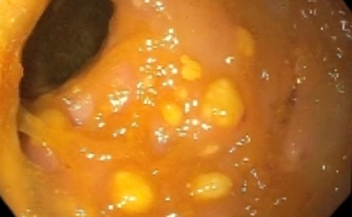
Contributed by Nalini Bansal, M.D.
Multiple polyps
Images hosted on other servers:

Macroscopic appearance
Contributed by Lewis Hassell, M.D., Nalini Bansal, M.D. and Christopher Hartley, M.D.
Juvenile polyp from stomach
No dysplasia
Ulcerated

Dilated colonic glands
Dysplasia
Mild frond-like growth pattern
Histopathology of juvenile polyps
Endoscopy of juvenile polyps
Images hosted on other servers:

Bowel resection
Images hosted on other servers:

Prominent lamina
propria with
edema and
inflammatory cells

Dense stroma
Colonoscopy of juvenile polyposis
Images hosted on other servers:

Kaposi sarcoma
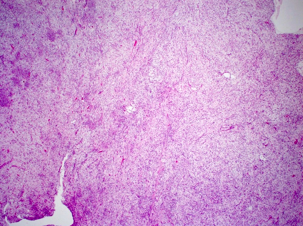
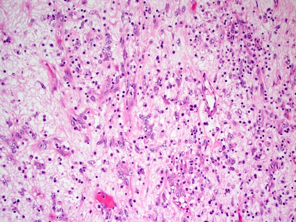
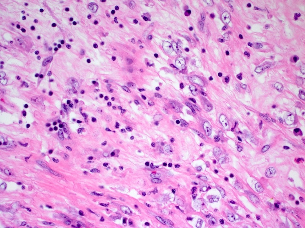
Contributed by Raul S. Gonzalez, M.D.
H&E, intermediate power
H&E, high power
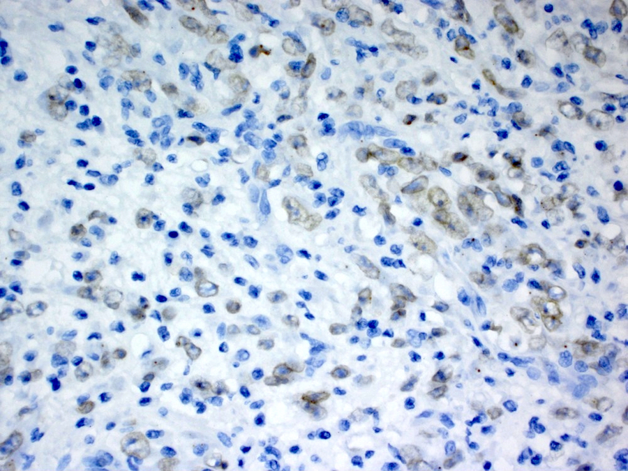
Images hosted on other servers:

Low power


High power

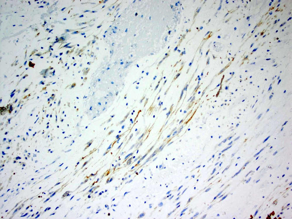
CD34
Images hosted on other servers:

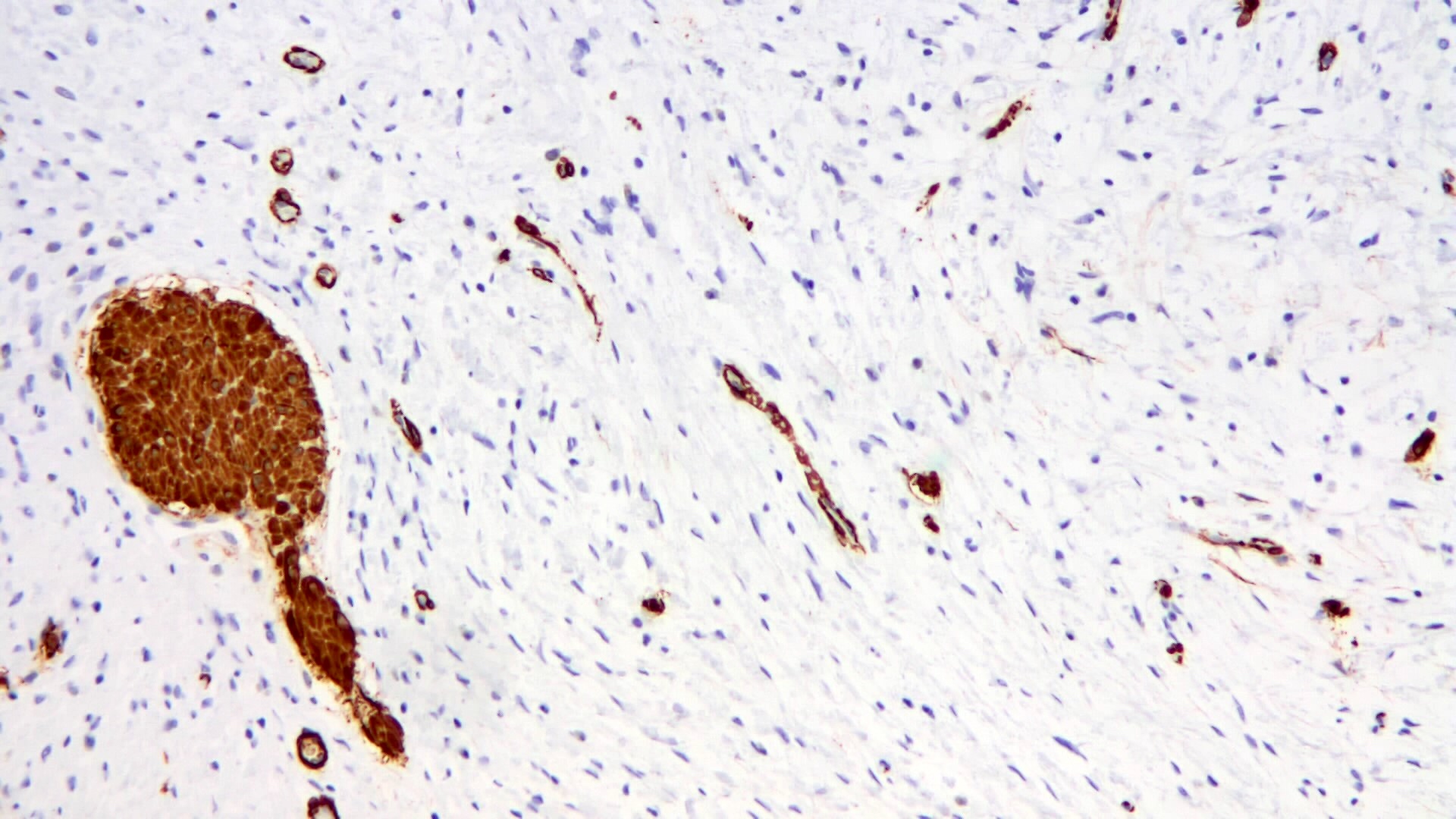
Leiomyoma on colonoscopy

Endoscopic removal of a leiomyoma
Contributed by Raul S. Gonzalez, M.D., @Andrew_Fltv and @ThatGlassTho on Twitter


Leiomyoma
Symplastic leiomyoma
Leiomyoma

Leiomyoma
Leiomyoma
Contributed by Raul S. Gonzalez, M.D.
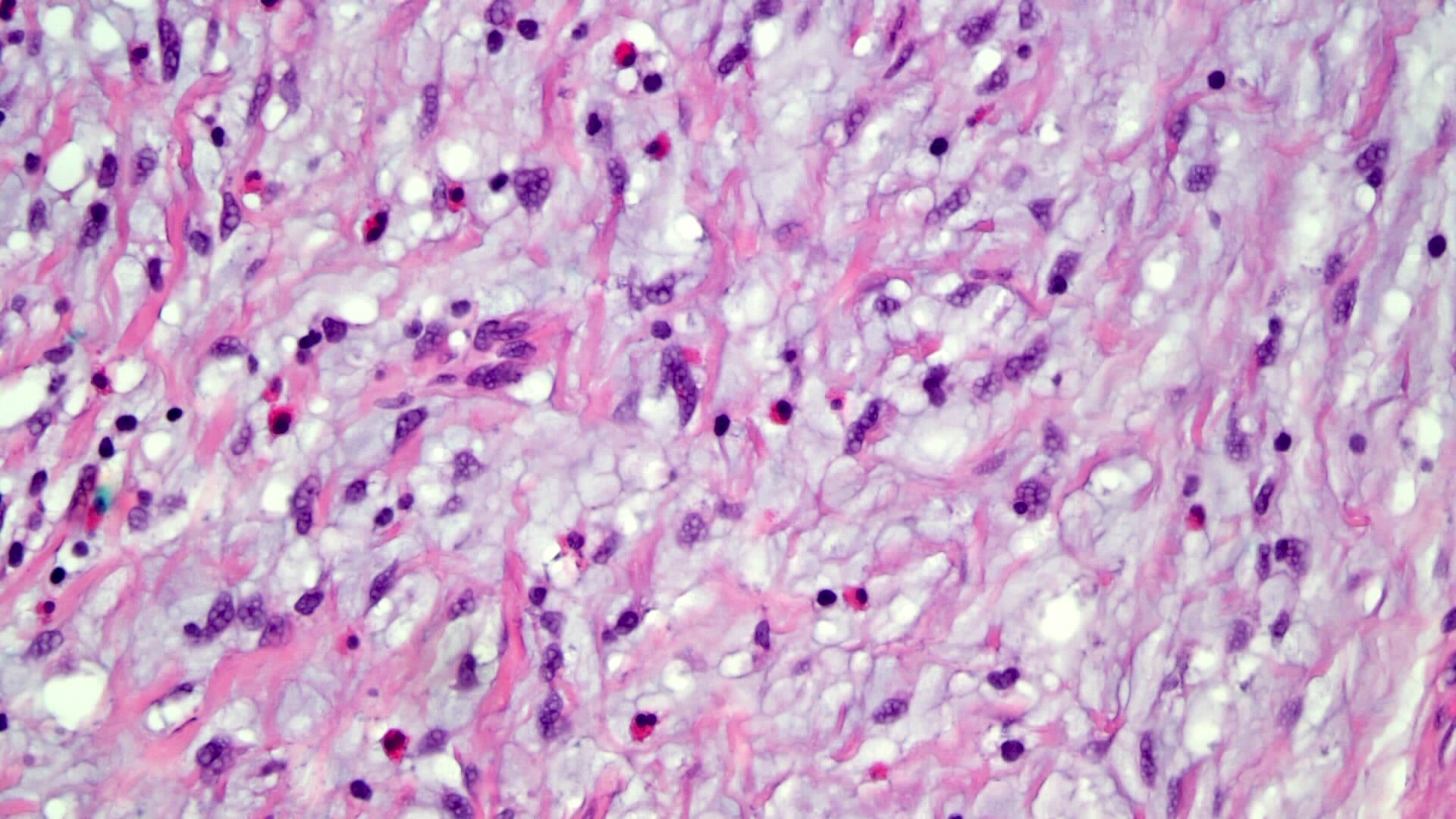
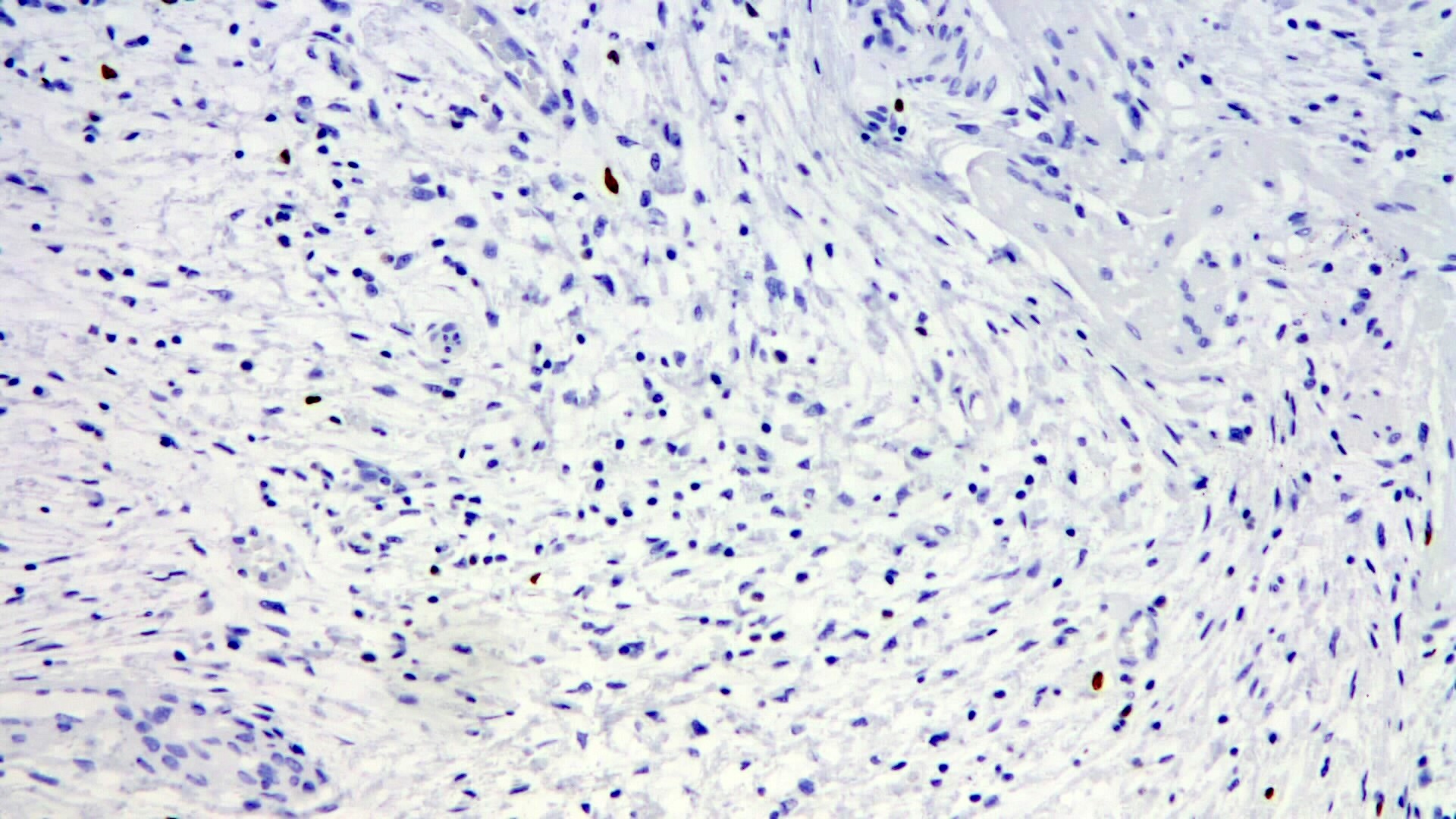
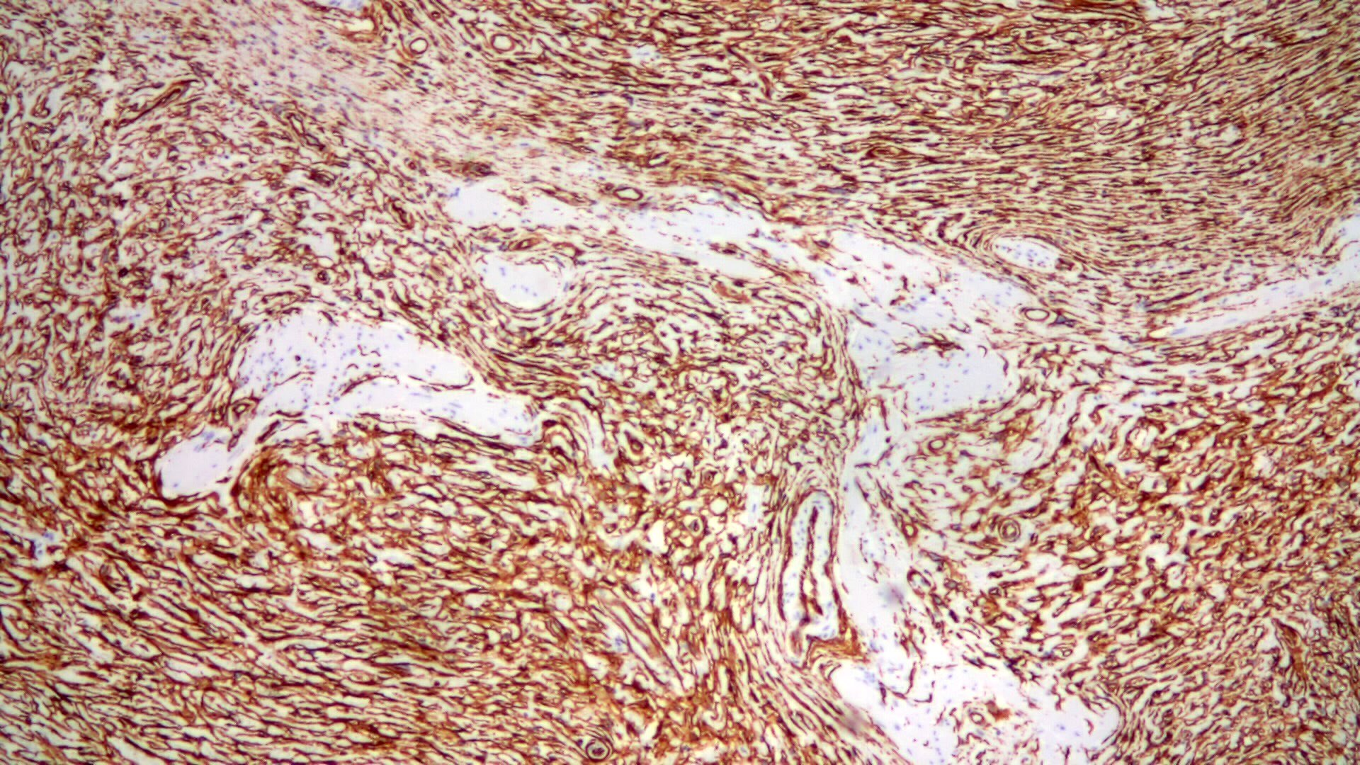
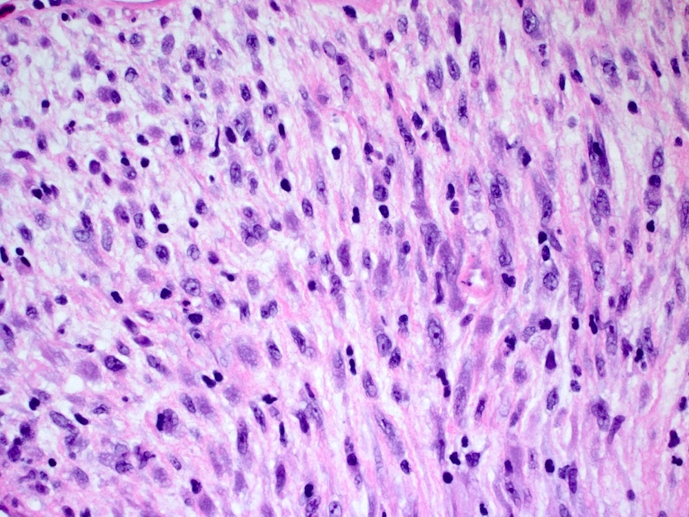
Colonic leiomyosarcoma
Nuclear atypia
Numerous mitotic figures
Images hosted on other servers:


Well differentiated tumor

Moderately differentiated

Poorly differentiated
Contributed by Maryam Kherad Pezhouh, M.D., M.Sc.
Fresh ORISE in the submucosa

Fresh ORISE in a background of red blood cells
Several months after injection of lifting agent

Foreign body reaction to lifting agent
Lifting agent granuloma
Lifting agent is negative for Congo red
Contributed by Catherine E. Hagen, M.D. (Case #507)
Lifting agent granuloma
Images hosted on other servers:

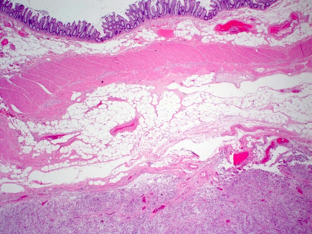
Large pedunculated submucosal tumor

Cut section of tumor
Contributed by Raul S. Gonzalez, M.D.
Intramucosal lipoma
Images hosted on other servers:

Lipoma with overlying sessile serrated adenoma
Contributed by Raul S. Gonzalez, M.D.

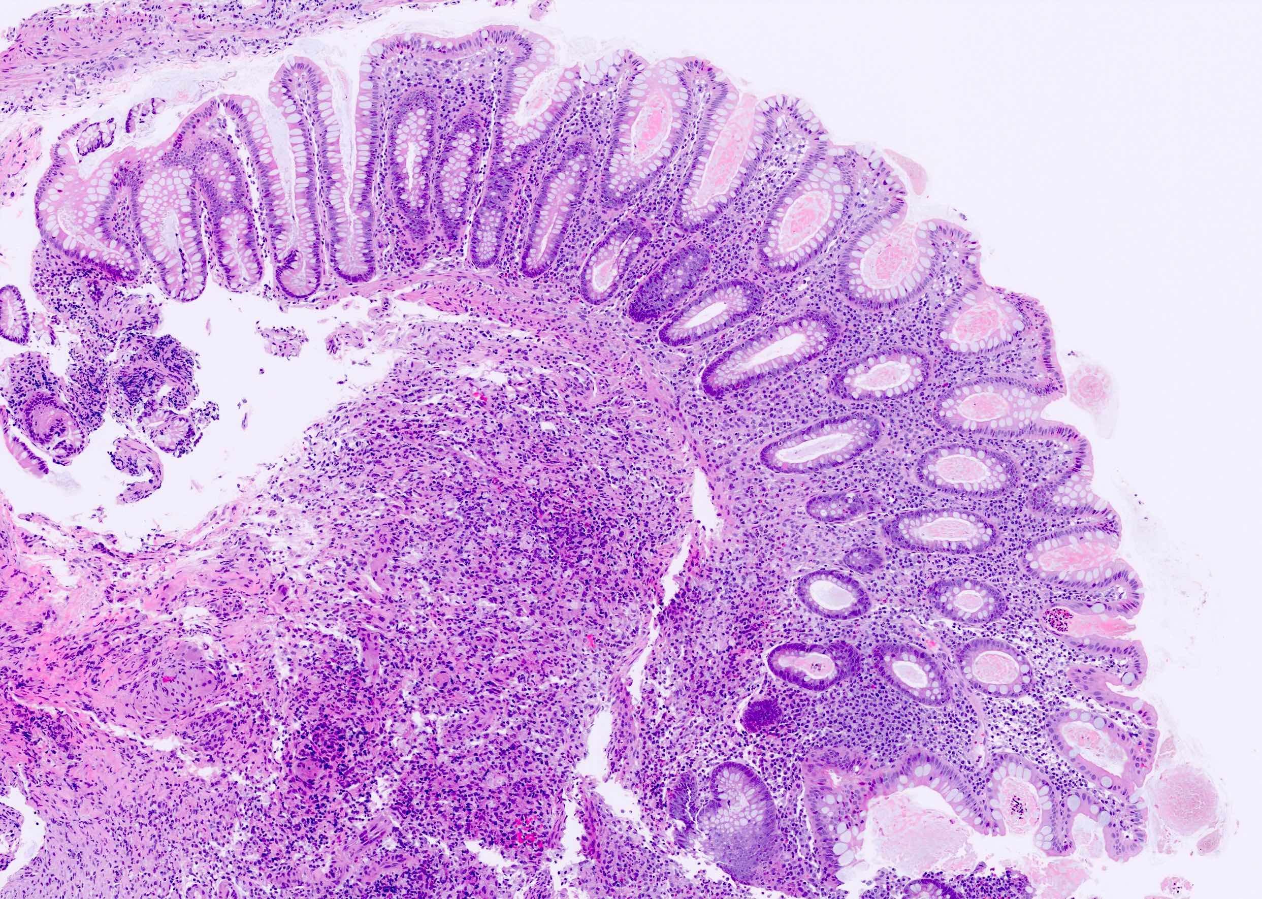
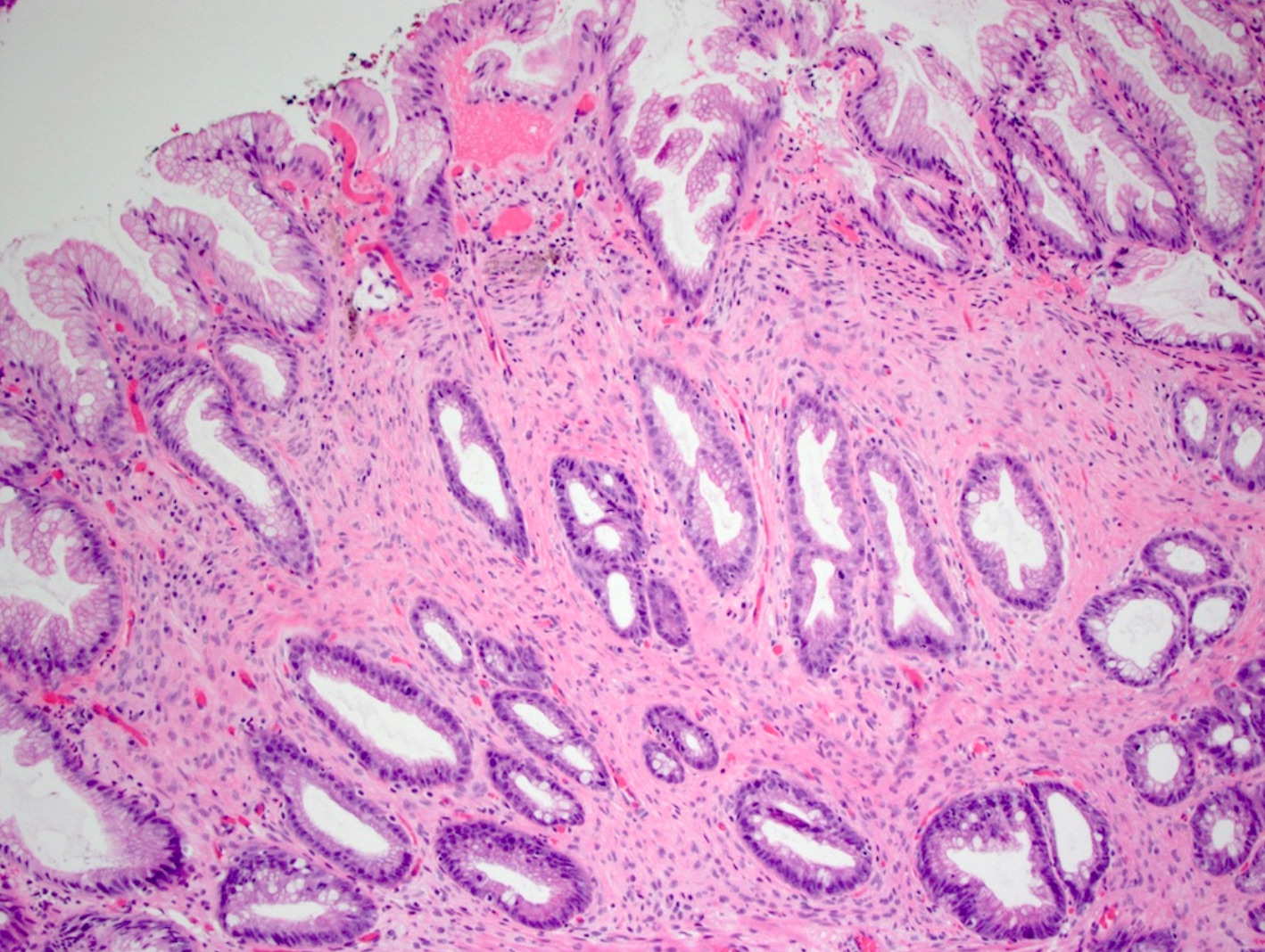
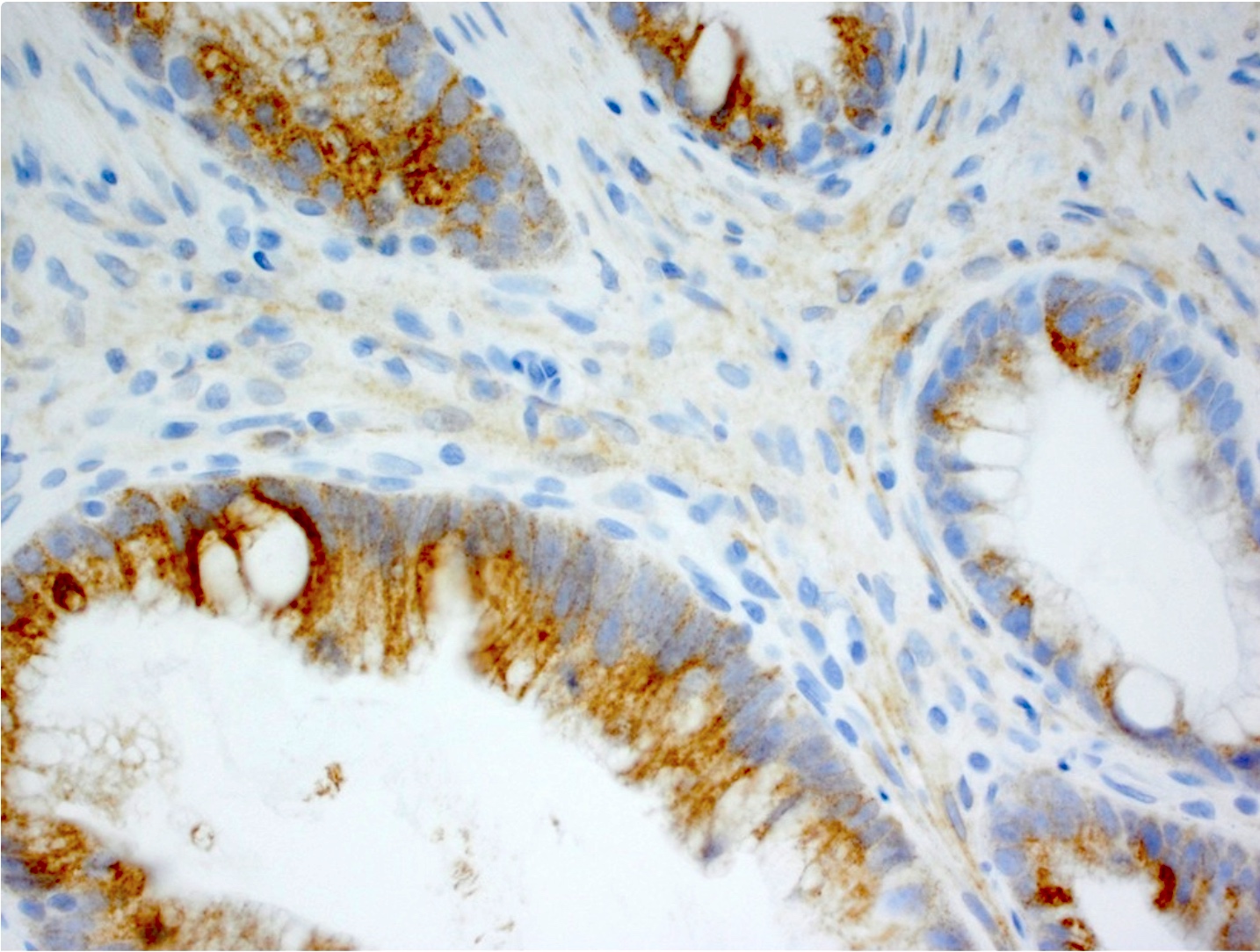
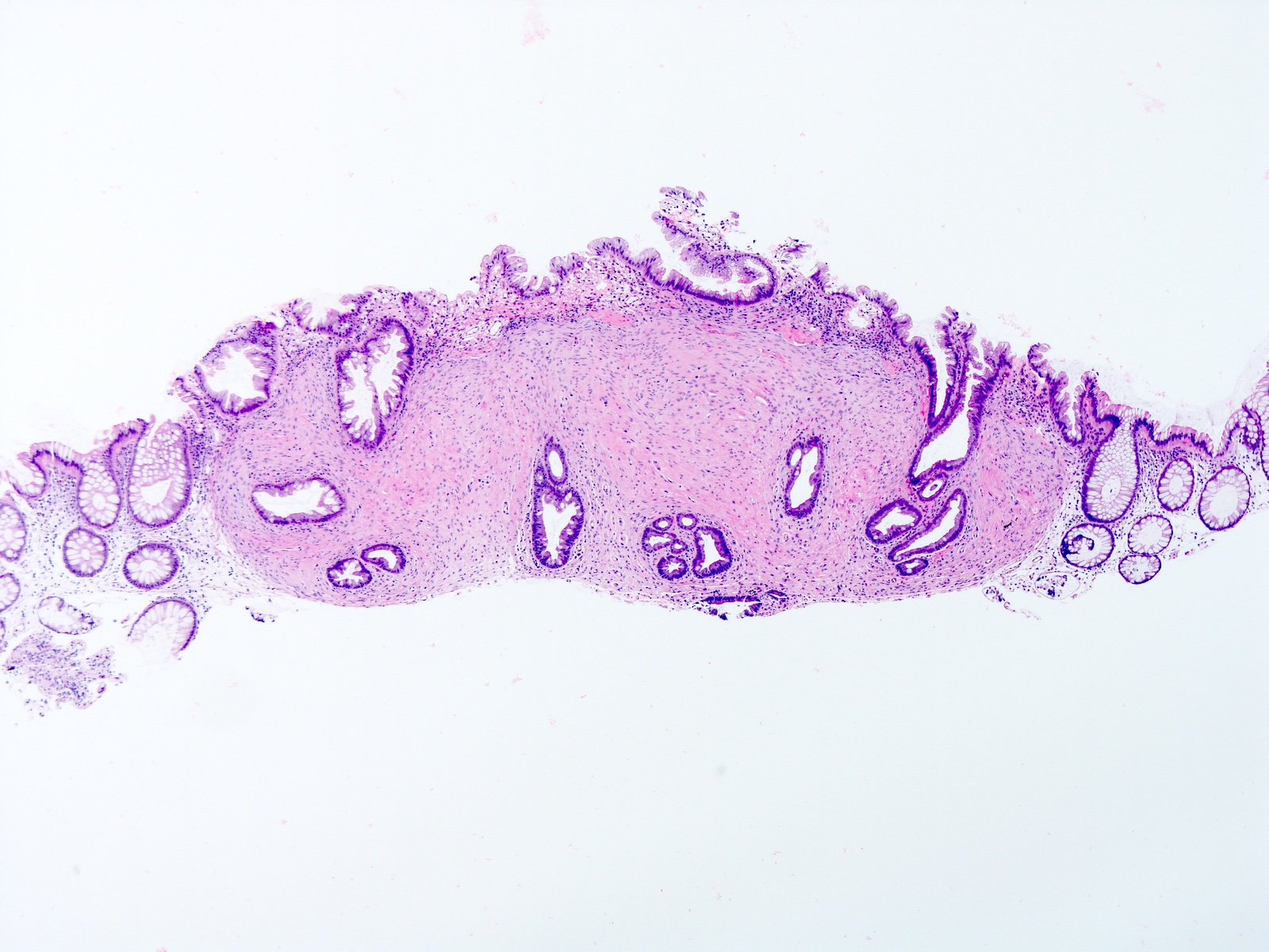
Low grade tubuloglandular adenocarcinoma in ulcerative colitis patient

Background colitis
Contributed by Martha M. Yearsley, M.D.
Intact crypt architecture
Lamina propria expansion
Mixed lamina propria inflammatory infiltrate
Increased surface intraepithelial lymphocytes
Crypt intraepithelial lymphocytes
Surface epithelial damage
Lymphocytic colitis and celiac disease
Microscopic colitis
Contributed by Arvind Rishi, M.D., M.B.B.S.
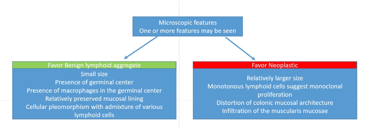
Histological algorithmic approach
Images hosted on other servers:

Endoscopic appearance
Contributed by Arvind Rishi, M.D., M.B.B.S.
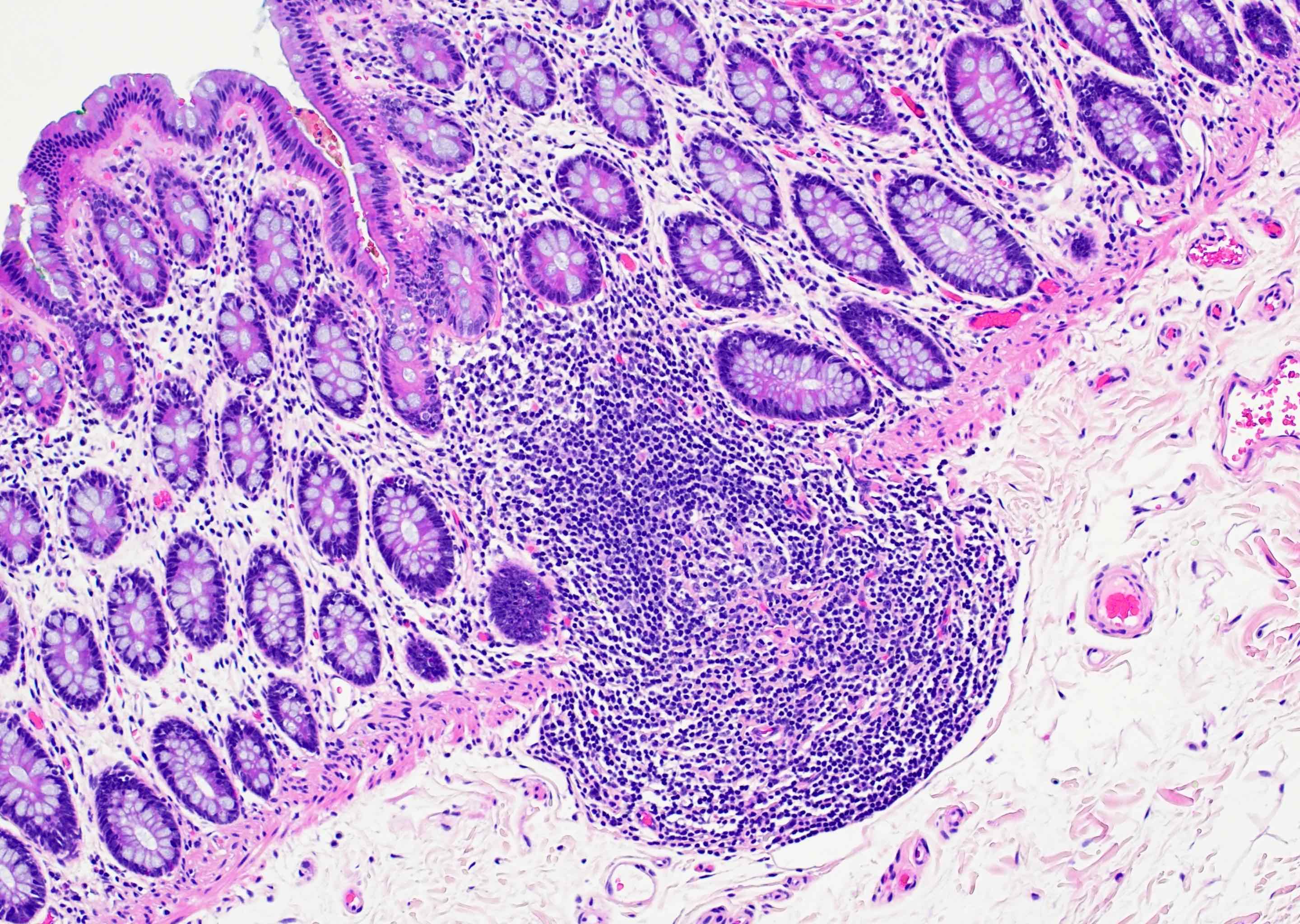
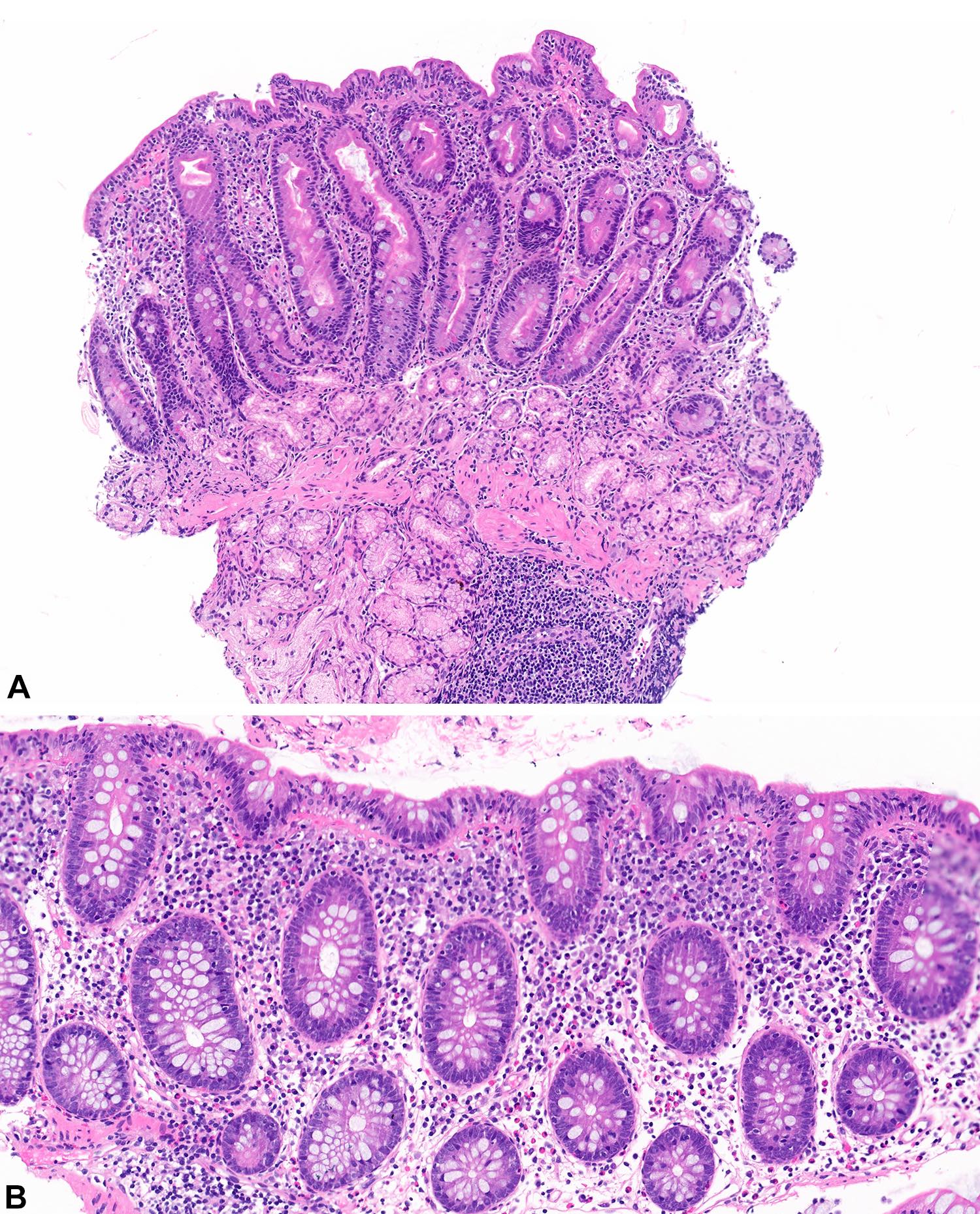
Lymphoid aggregate with germinal center
Prominent Peyer patches
Polarized germinal center
Tingible body macrophages
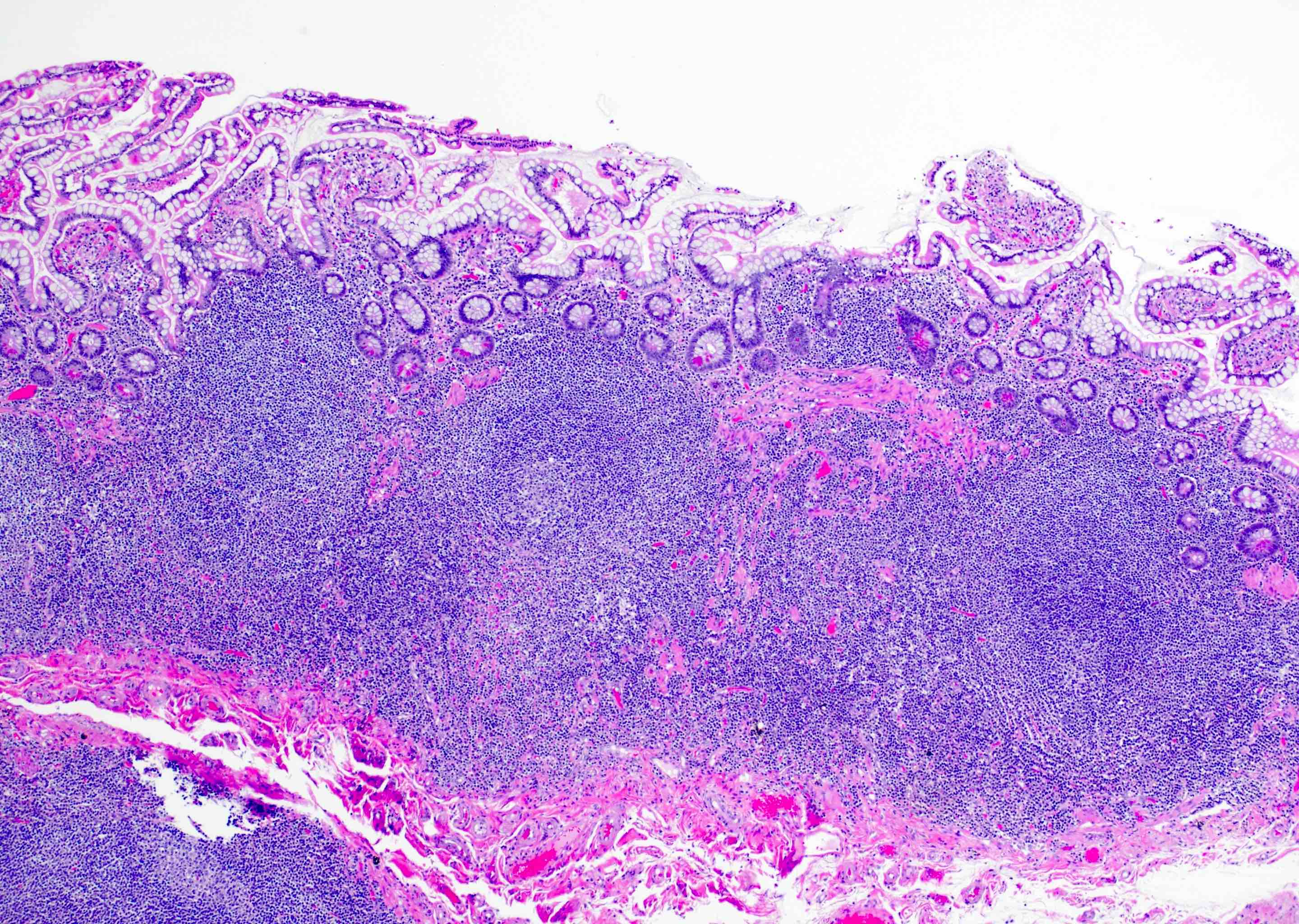
Superficial lymphoid aggregate
Small bowel polyps
Surface reactive changes, rectum
Tingible body macrophages
Incidental
hyperplastic polyp
with lymphoid
aggregate
Images hosted on other servers:


Michaelis-Gutmann bodies

Liver transplant recipient

Adjacent to adenocarcinoma
Images hosted on other servers:

Lymphomatoid polyposis
Images hosted on other servers:

80 year old woman
Contributed by M.J. Fernández-Aceñero, M.D., Ph.D.
Slight inflammatory infiltration
Eosinophils in the infiltrate
KIT expression
Increased mast cells
Images hosted on other servers:
Fleshy, ulcerated lesion
Contributed by Raul S. Gonzalez, M.D.

Medullary carcinoma
Contributed by Raul S. Gonzalez, M.D. and Yuri Tachibana, M.D.
Melanosis coli

Pigment in lamina propria
Melanosis coli
Images hosted on other servers:

Soft tissue mass
due to squamous
cell carcinoma
primary of lung
Contributed by Raul S. Gonzalez, M.D.
Metastatic breast carcinoma
Metastatic ovarian clear cell carcinoma
Images hosted on other servers:

Metastatic melaonoma to sigmoid colon

Poorly differentiated
squamous cell
carcinoma metastatic
from lung
Contributed by Raul S. Gonzalez, M.D.
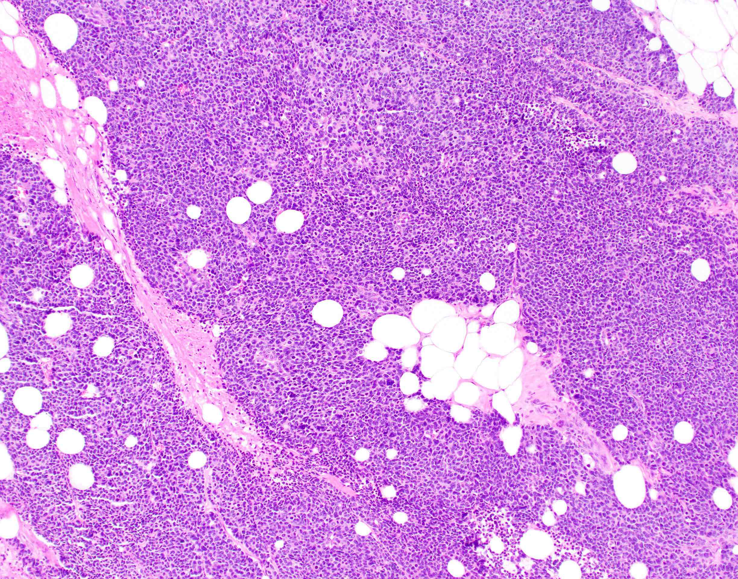
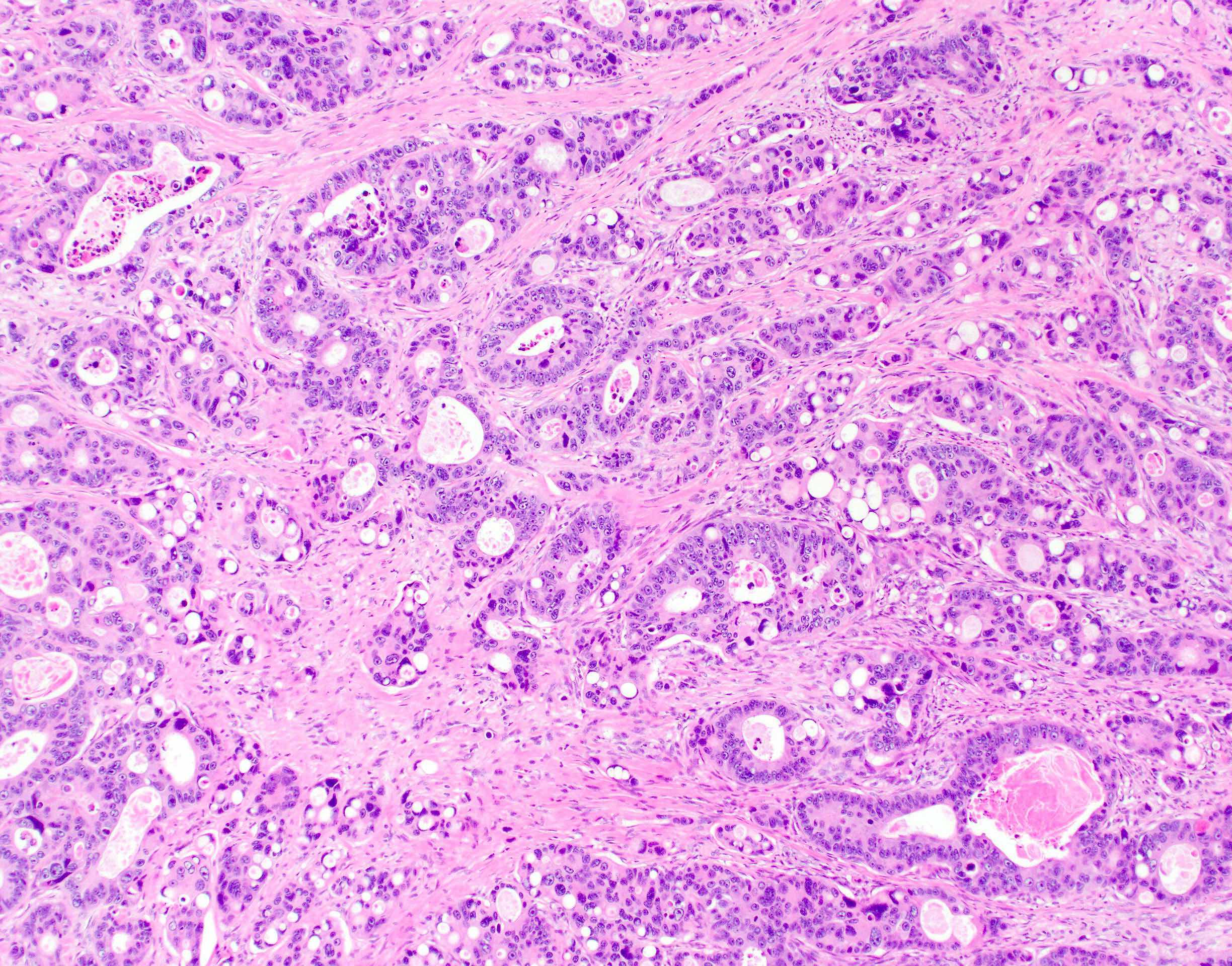
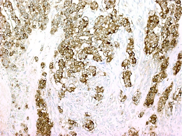
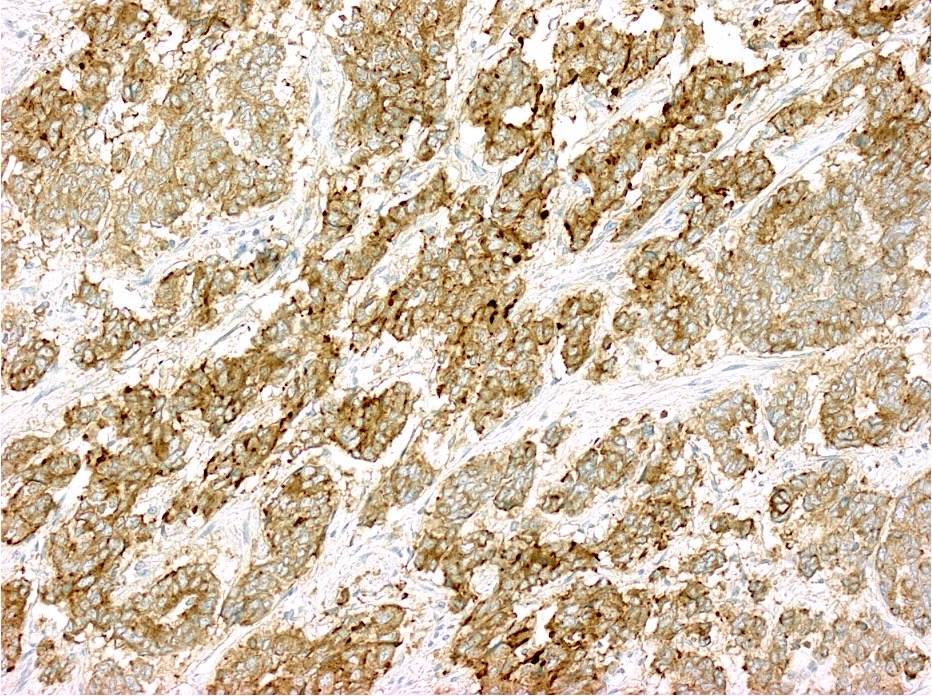
Micropapillary carcinoma


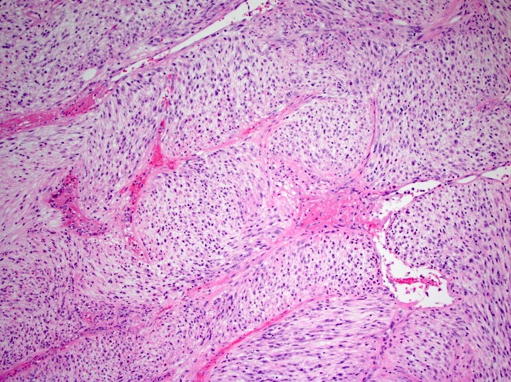
Contributed by Phoenix D. Bell, M.D.
MMR deficient adenocarcinoma
MMR deficient mucinous adenocarcinoma

MMR deficient medullary carcinoma

MMR deficient mucinous adenocarcinoma
Contributed by Phoenix D. Bell, M.D.
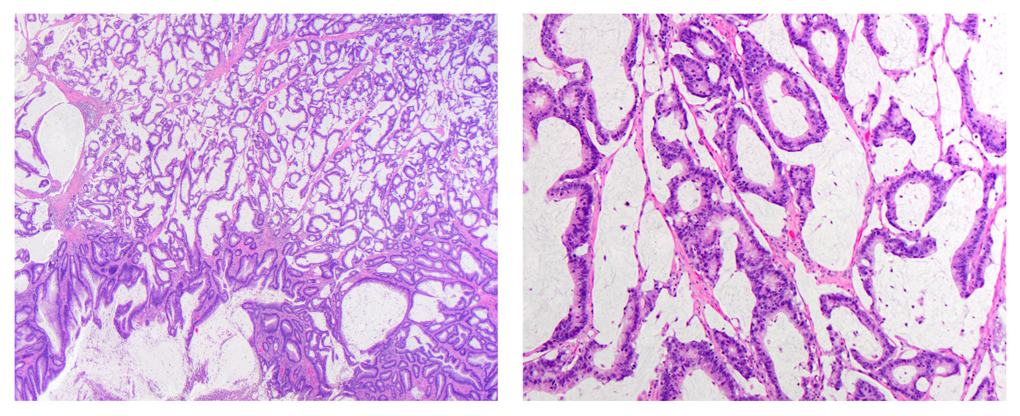
Colon adenocarcinoma biopsy (low power)
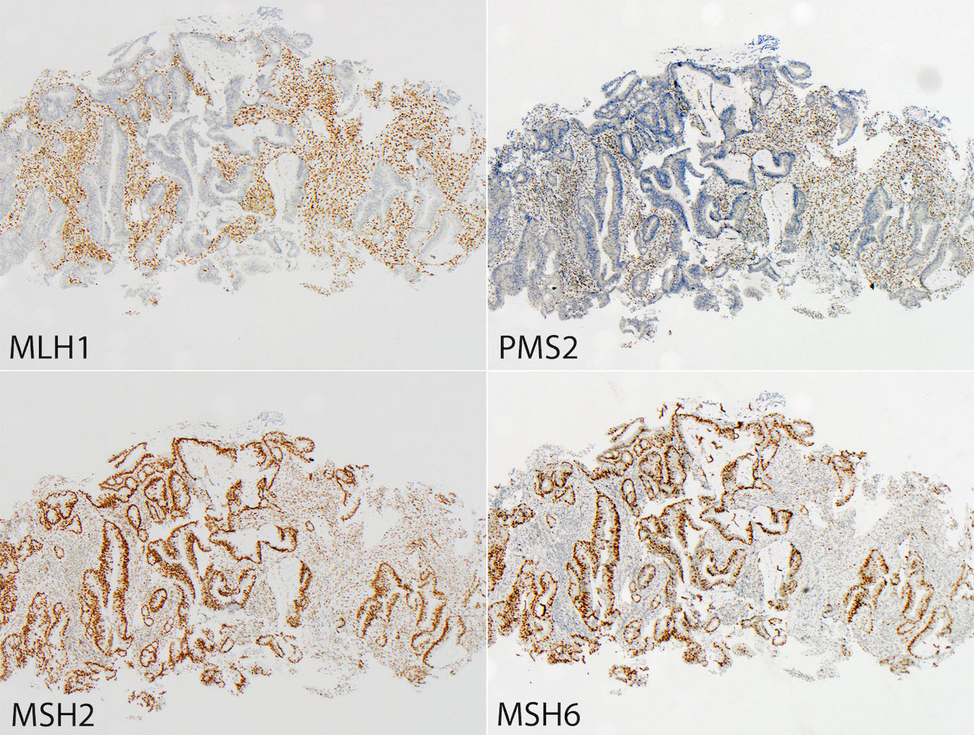
MMR deficient colorectal cancer (low power)
Colon adenocarcinoma resection

MMR deficient colorectal cancer (medium power)
MMR deficient medullary carcinoma
MMR deficient mucinous adenocarcinoma
Contributed by Phoenix D. Bell, M.D.

Microsatellite instability analysis
Images hosted on other servers:

Treatment modalities in retrospective study
Images hosted on other servers:

CT with colon tumor

CT: liver metastasis, intraperitoneal fluid
Images hosted on other servers:

Tumor causing intussusception

Colonoscopy
Images hosted on other servers:

Multiple polypoid masses
Contributed by Lewis A. Hassell, M.D. and Catherine E. Hagen, M.D.
Mixed acinar cell neuroendocrine carcinoma
Acinar cell carcinoma component
Neuroendocrine carcinoma component
Mixed adenocarcinoma neuroendocrine carcinoma
Small cell carcinoma
Adenocarcinoma
Chromogranin IHC
Synaptophysin IHC
Trypsin IHC
Mixed neuroendocrine and nonneuroendocrine carcinoma of the esophagus
Mixed neuroendocrine nonneuroendocrine neoplasms
Images hosted on other servers:

Causing intussusception

Cecal tumor

Arising in villous adenoma
Contributed by Raul S. Gonzalez, M.D.
Abundant extracellular mucin
Epithelial cells within mucin
Images hosted on other servers:

Rectal polyps on colonoscopy
Contributed by Raul S. Gonzalez, M.D.
Lesion confined to mucosa
Bland nuclei with occasional enlargements
S100+
Images hosted on other servers:

Low power view of hematoxylin and eosin

S100 positivity

Histologic findings
of mucosal
Schwann cell
hamartomas
Images hosted on other servers:

MSH2+
Significant loss of MSH6

Colon adenoma and colonic mucosa
Contributed by Raul S. Gonzalez, M.D.
Adenoma associated with MUTYH mutation
Images hosted on other servers:


Endoscopic appearance
Contributed by Catherine E. Hagen, M.D.
Architectural distortion
Injured crypt
Apoptosis
Inflammation
Images hosted on other servers:

In situ

Thickened wall, mucosal ulceration, cyst-like spaces

Transverse section shows cyst-like spaces in wall

Alimentary tract

Small intestine
Images hosted on other servers:

Hemorrhagic necrosis

Mucosal necrosis and cyst-like spaces in submucosa

Submucosal cyst-like spaces

Lined by flattened cells

Subserosal lymph node with cyst-like spaces
Images hosted on other servers:

Ulcer post NSAID therapy
Images hosted on other servers:


Multiple defects and gastrochisis
Contributed by Raul S. Gonzalez, M.D. and Christopher Hartley, M.D.
Medium power

High power
Weak positivity on EMA
Perineurioma
Images hosted on other servers:
Multiple gastric polyps on endoscopy

Characteristic mucocutaneous pigmentation
Contributed by Michael Feely, D.O. and Christopher Hartley, M.D.


Colonic Peutz-Jeghers polyps

Duodenal Peutz-Jeghers polyps
Peutz-Jeghers polyp
Contributed by Raul S. Gonzalez, M.D.
Kayexalate and sevelamer in a rectal ulcer
Images hosted on other servers:

Incidental luminal polystyrene
sulphonate resin particles in
jejunal diverticular tissue

Particles at site of colonic
necrosis (direct Schiff stain
with light counterstain)

Particles at site of aspiration
pneumonia (Ziehl-Neelsen stain
with light counterstain)
Images hosted on other servers:

Multiple subserosal bubbles

Diffuse hemorrhage


Pneumatosis intestinalis

Pneumatosis intestinalis and focal mucosal necrosis
Contributed by Raul S. Gonzalez, M.D.
Mural pneumatosis
Mucosal pneumatosis
Pneumatosis intestinalis
Understanding the cancer predisposing syndrome caused by defective POLE and POLD1
Images hosted on other servers:

Rectal mass

Noncontrast CT scans of abdomen
Contributed by Raul S. Gonzalez, M.D., Aaron Huber, D.O. and Diana Agostini-Vulaj, D.O. (Case #531)
Large cell neuroendocrine carcinoma


Synaptophysin

Chromogranin

INSM1
Contributed by Raul S. Gonzalez, M.D.


Posttreatment changes of rectal carcinoma
Contributed by Subramanya Sakaleshpura Mallikarjunappa, M.B.B.S., M.D.
Linear radiolucencies in left lumbar region
Extensive submucosal edema
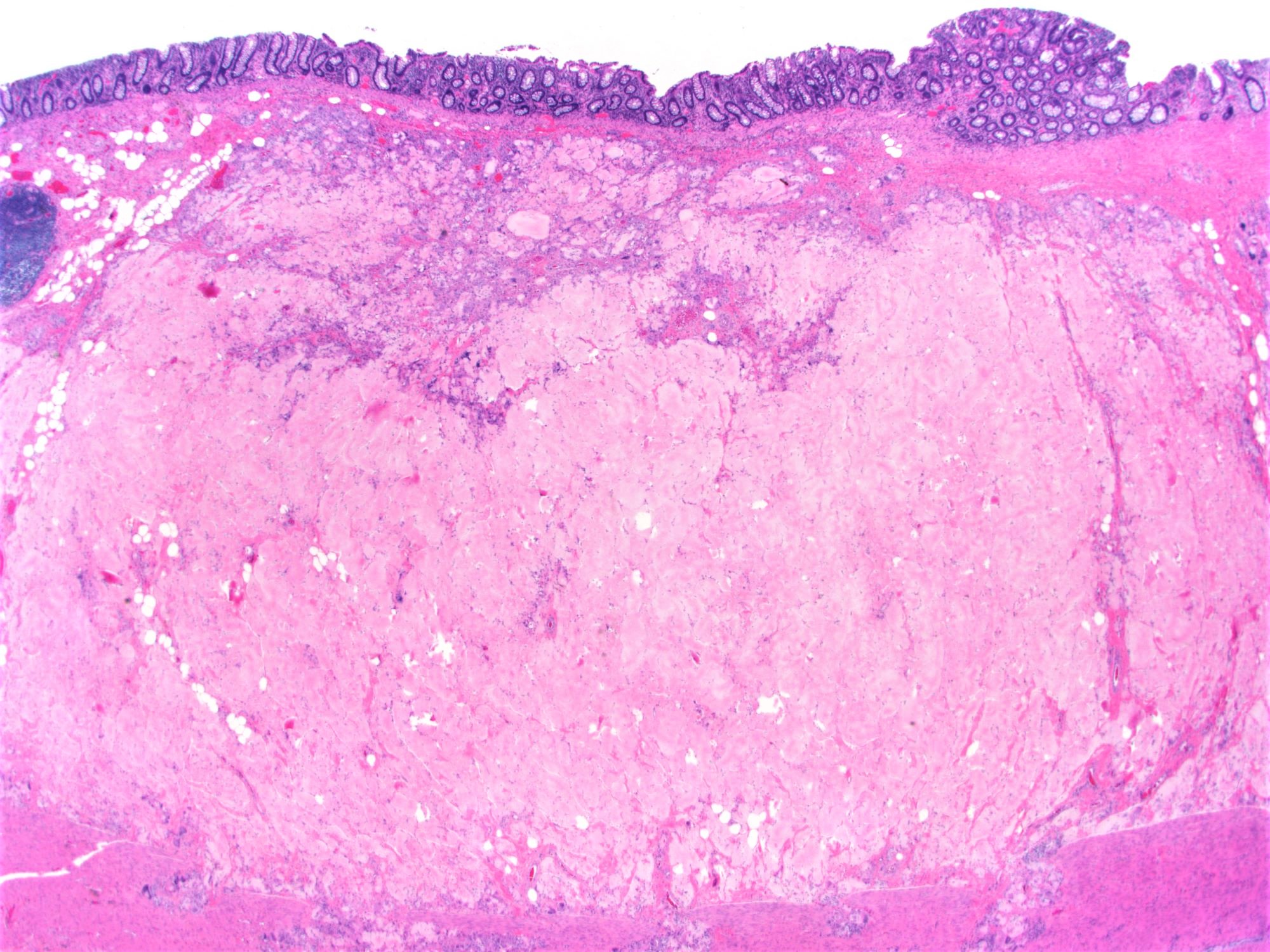
Images hosted on other servers:

White to yellow plaques and nodules on the colonic mucosa


Hyperemic mucosa and green-yellow exudate


Mucosal denudation and exudate
Contributed by Raul S. Gonzalez, M.D. and Subramanya Sakaleshpura Mallikarjunappa, M.B.B.S., M.D.
Volcano shaped eruption
Pseudomembrane
Withered crypts
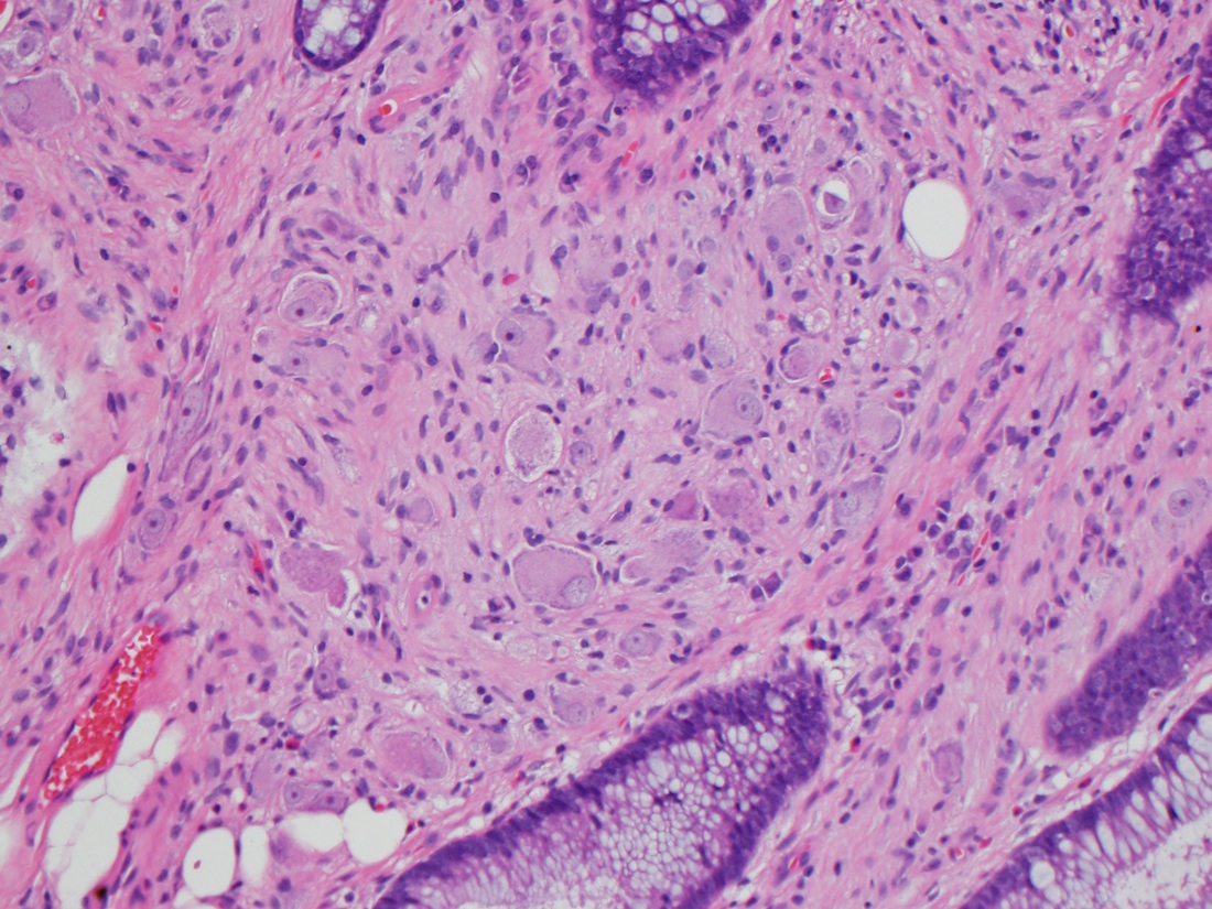
Pseudo-signet ring change
Volcano shaped eruption
Pseudomembrane
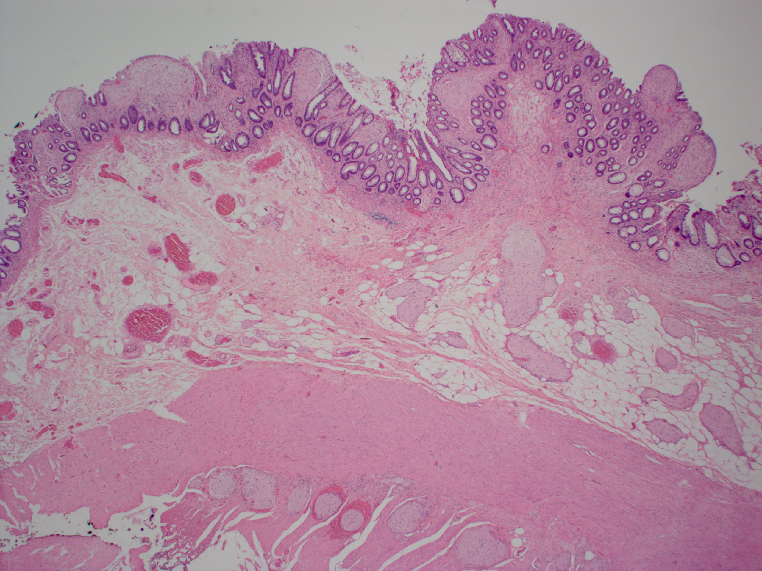
Contributed by Raul S. Gonzalez, M.D.

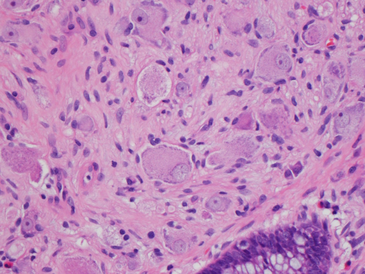
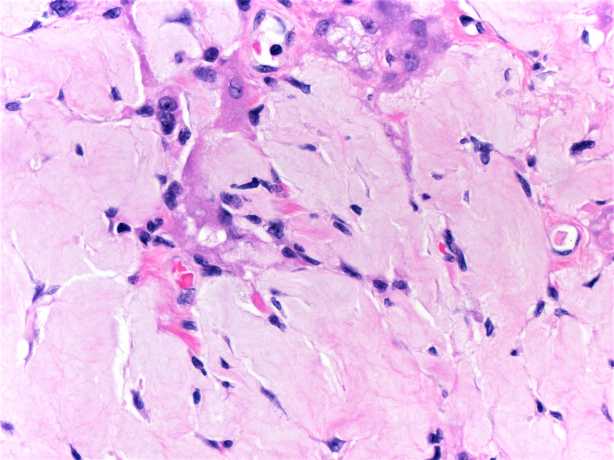
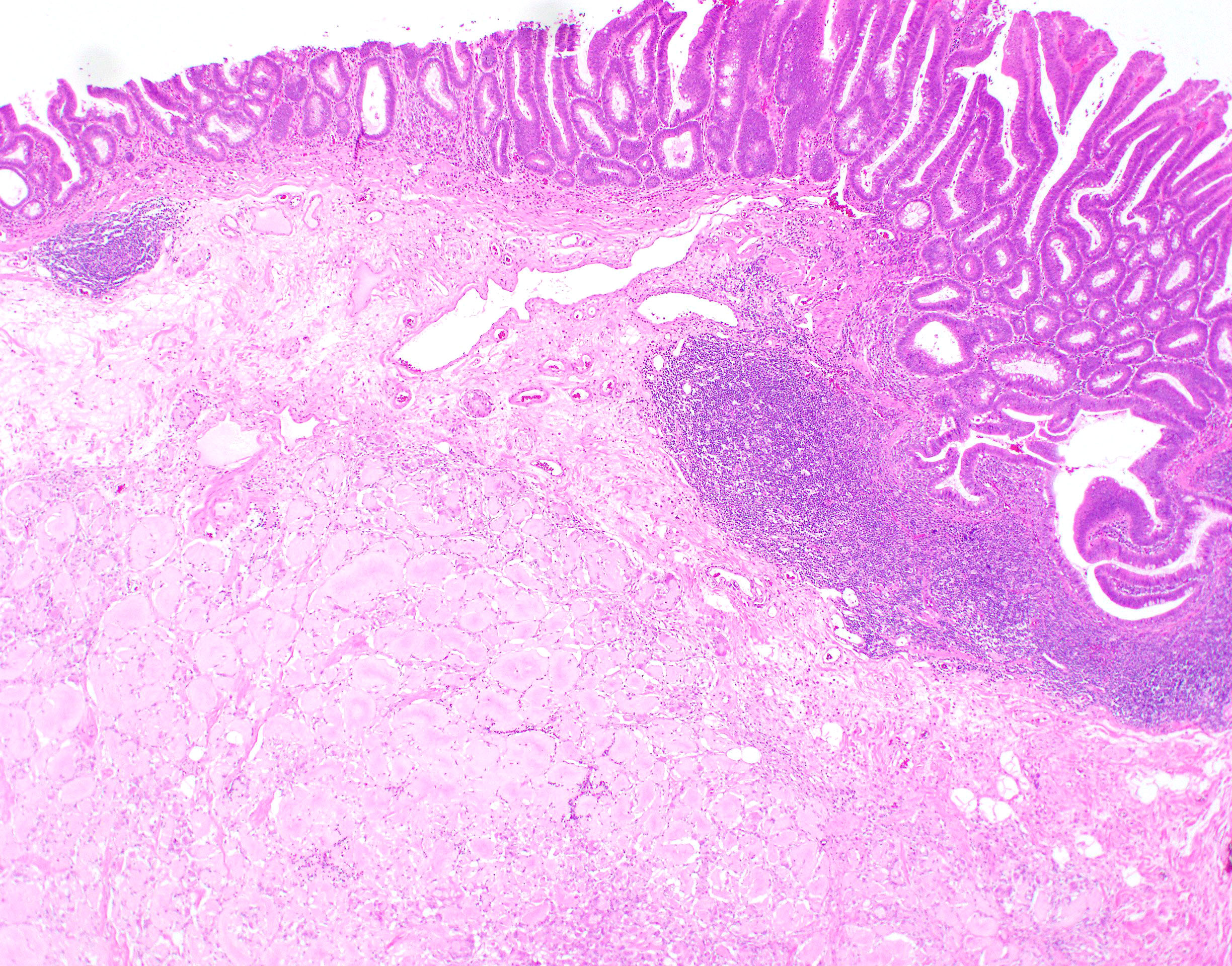
Serosal hyaline
predominant
pulse granulomas
Pulse material
with numerous
foreign body
giant cells
Cellular predominant pulse granuloma
Scant pulse material embedded in colon wall
Contributed by @RaulSGonzalezMD on Twitter
Pulse granuloma

Pulse granuloma
Pulse granuloma
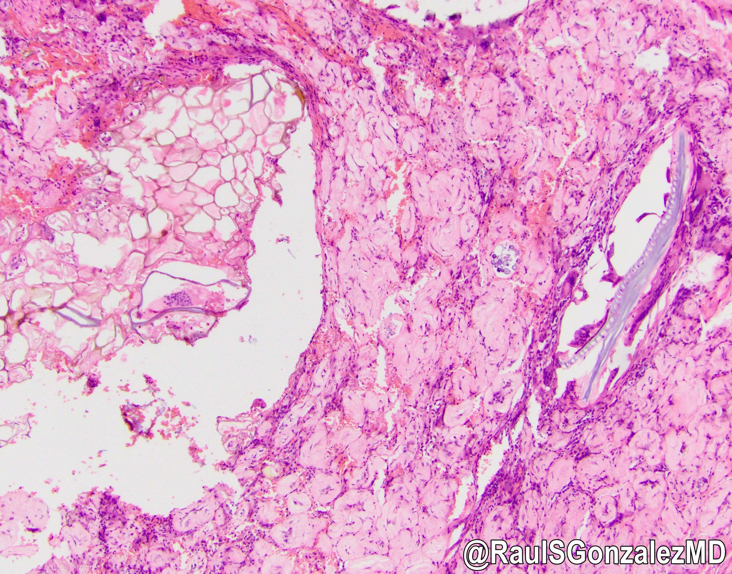
Images hosted on other servers:

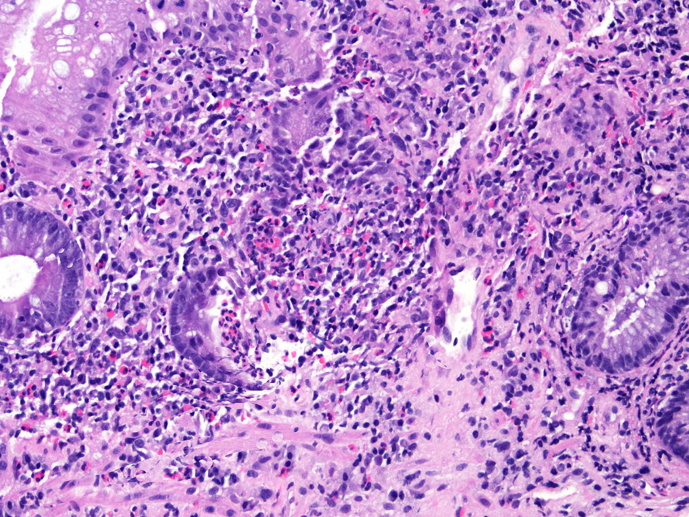
Jejunal pyogenic granuloma

Upper duodenum findings

Endoscopic duodenal mucosal resection
Images hosted on other servers:
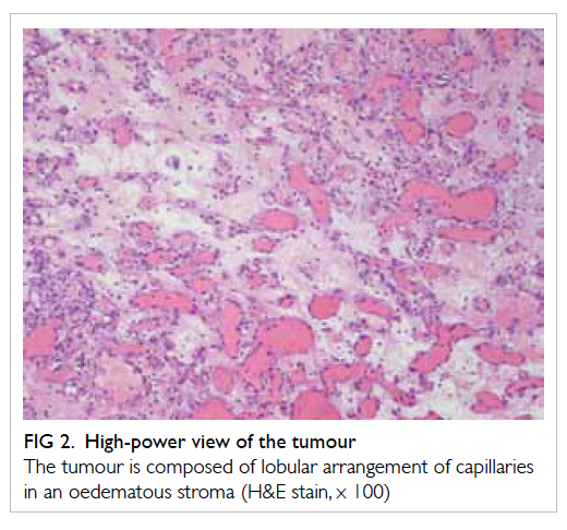
High power H&E
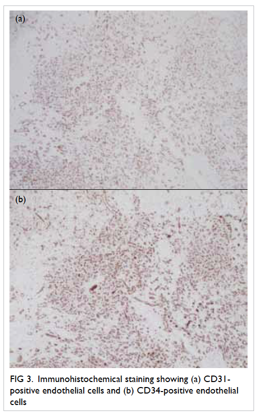
IHC for CD31 and CD34

Duodenum-edematous granulation tissue

Jejunum postsurgical pyogenic granuloma

Jejunum-CD34
Images hosted on other servers:

Edematous rectal mucosa

Rectal mucosa
Contributed by Maryam Kherad Pezhouh, M.D., M.Sc.

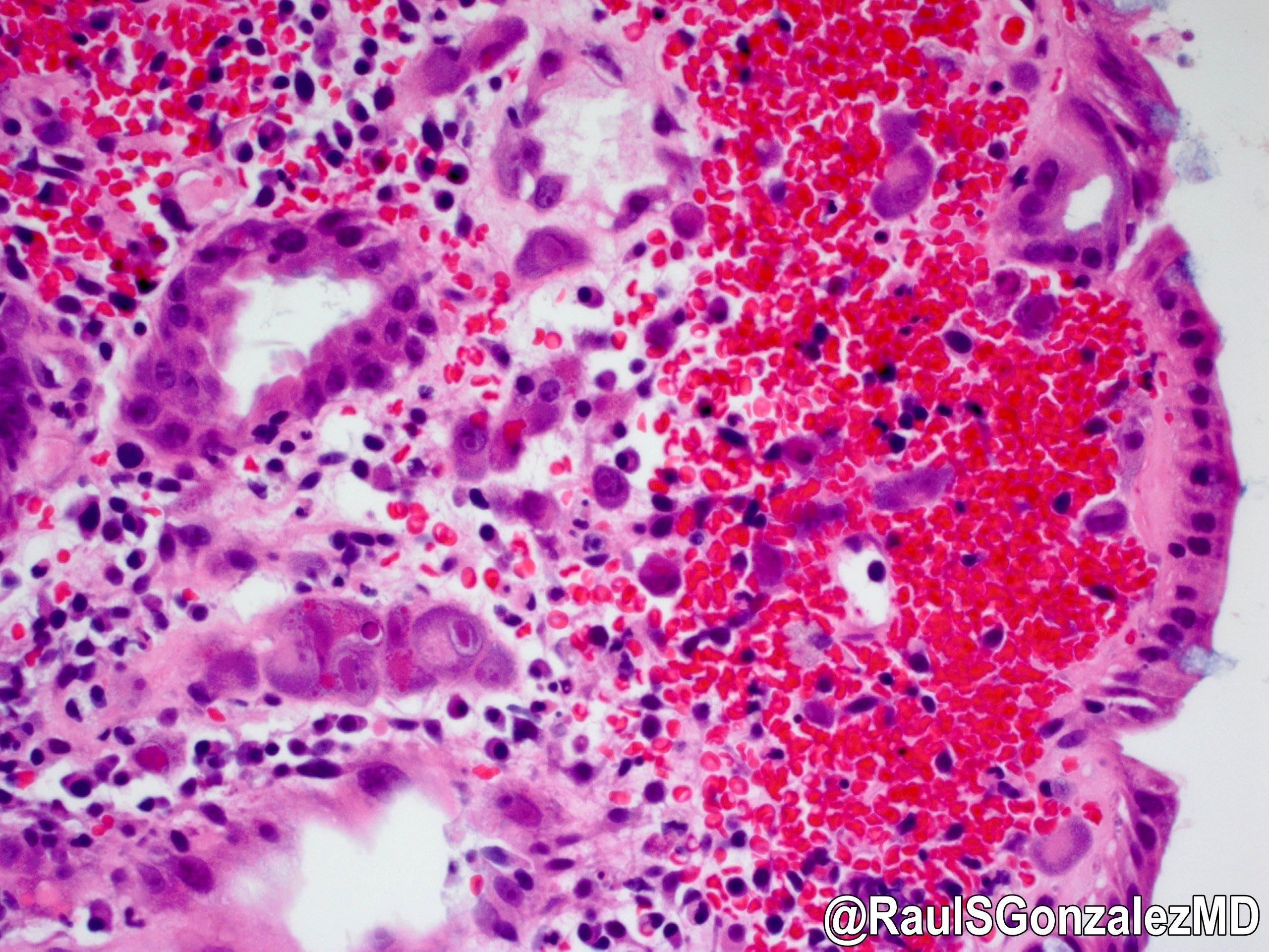
Acute radiation associated injury
Chronic radiation associated injury
Chronic radiation associated injury
Images hosted on other servers:

Protruding lesion on back wall of gastric cardia
Images hosted on other servers:

Cut surface of lesion

Excised specimen in cut section

Fibrotic nodules in pouch of Douglas
Contributed by Raul S. Gonzalez, M.D.
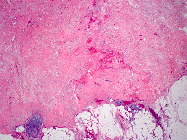
Low power
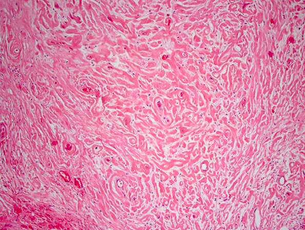
Intermediate power
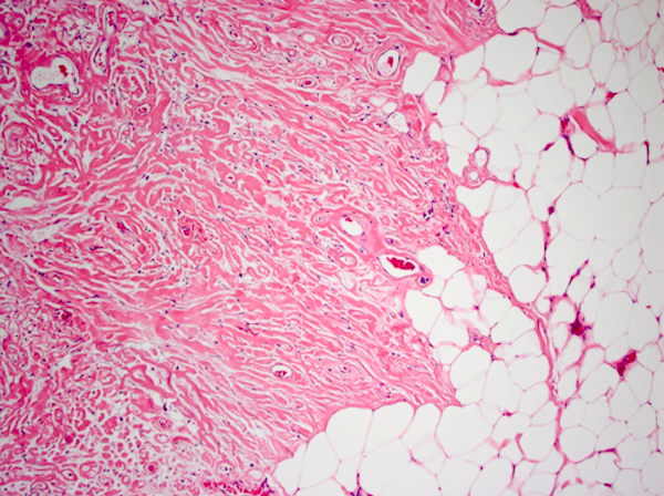
Interaction with adjacent fat
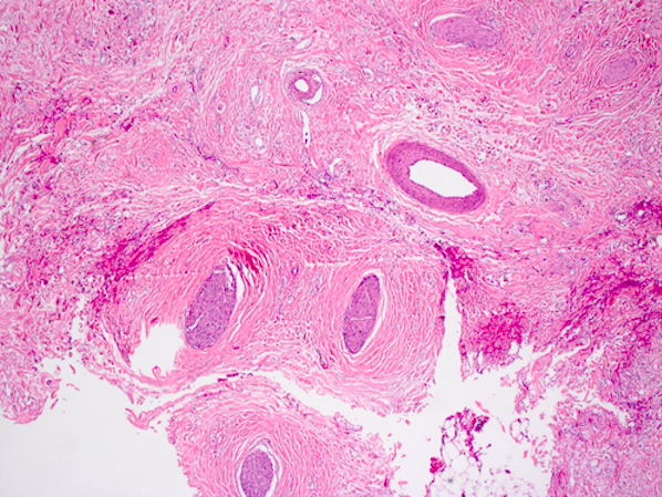
Entrapped nerves
Images hosted on other servers:
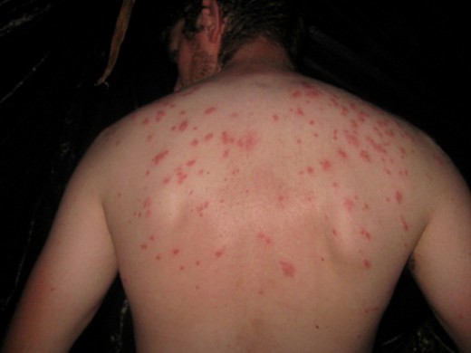
Typhoid rash
Contributed by Elliot Weisenberg, M.D.
Acute self limited colitis
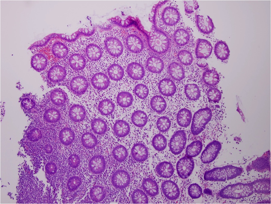
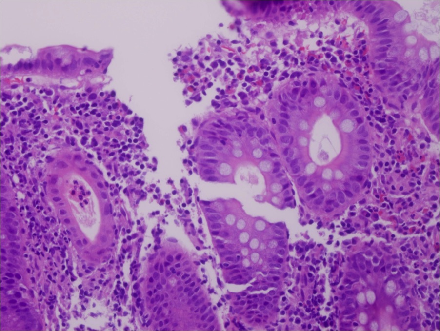
Images hosted on other servers:

Typhoid nodules
Contributed by M.J. Fernández-Aceñero, M.D., Ph.D.

Slight mucosal changes in sapovirus infection
Images hosted on other servers:


Typical morphology of sapovirus
Images hosted on other servers:

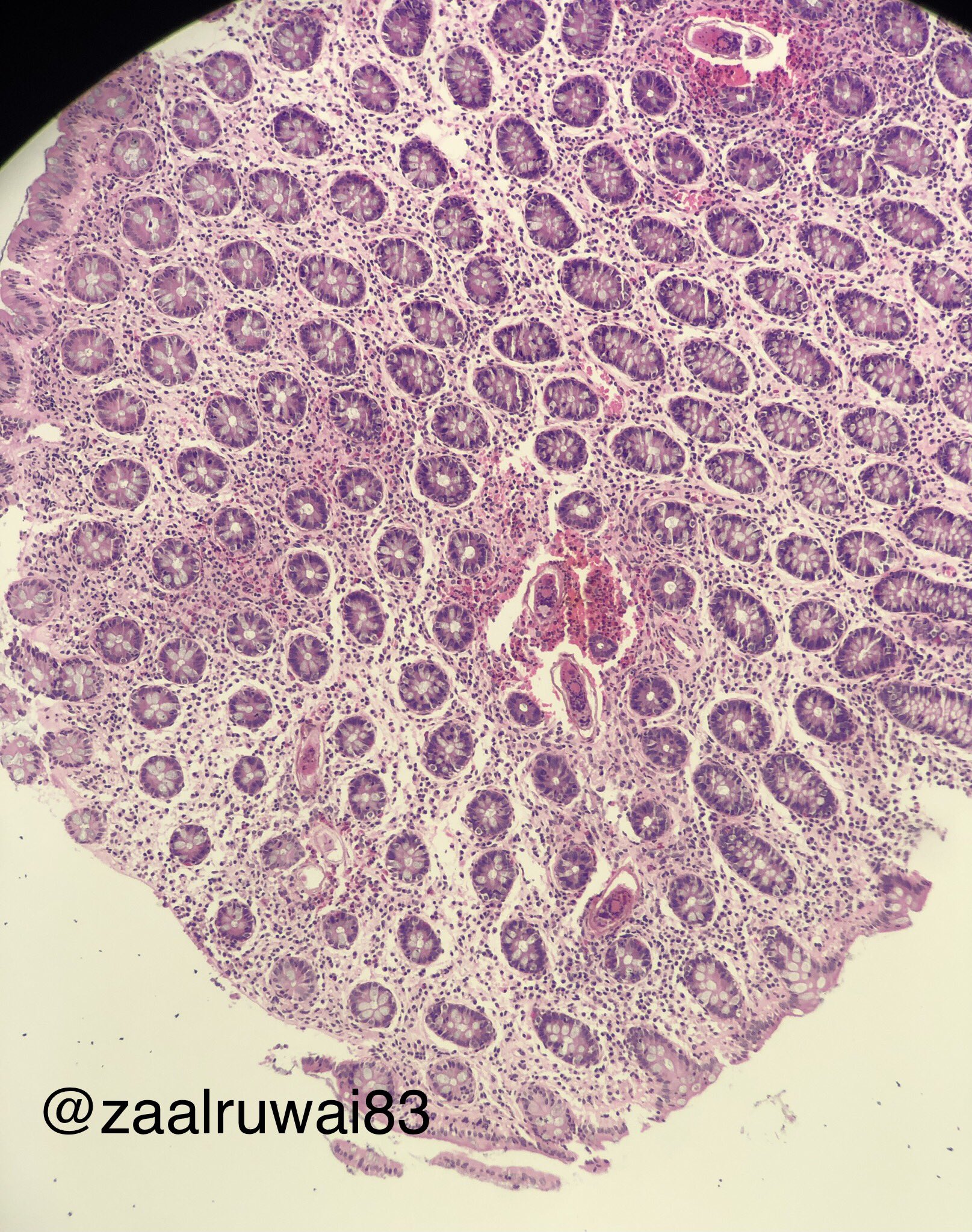
USG showing pipestem fibrosis

Large pedunculated polyp
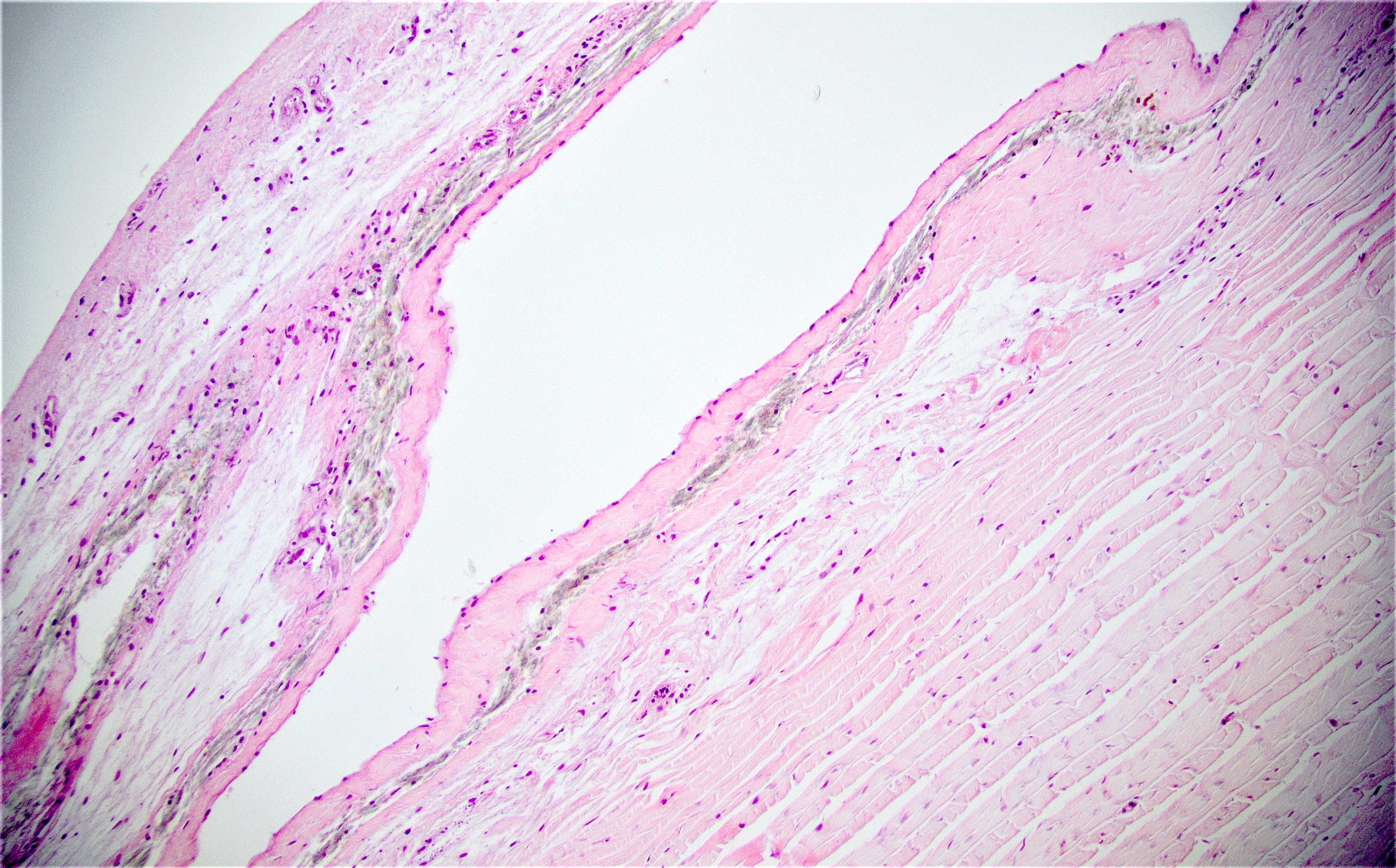
Contributed by Nalini Bansal Gupta, M.D., Lisa Cerilli, M.D. and @zaalruwai83 on Twitter
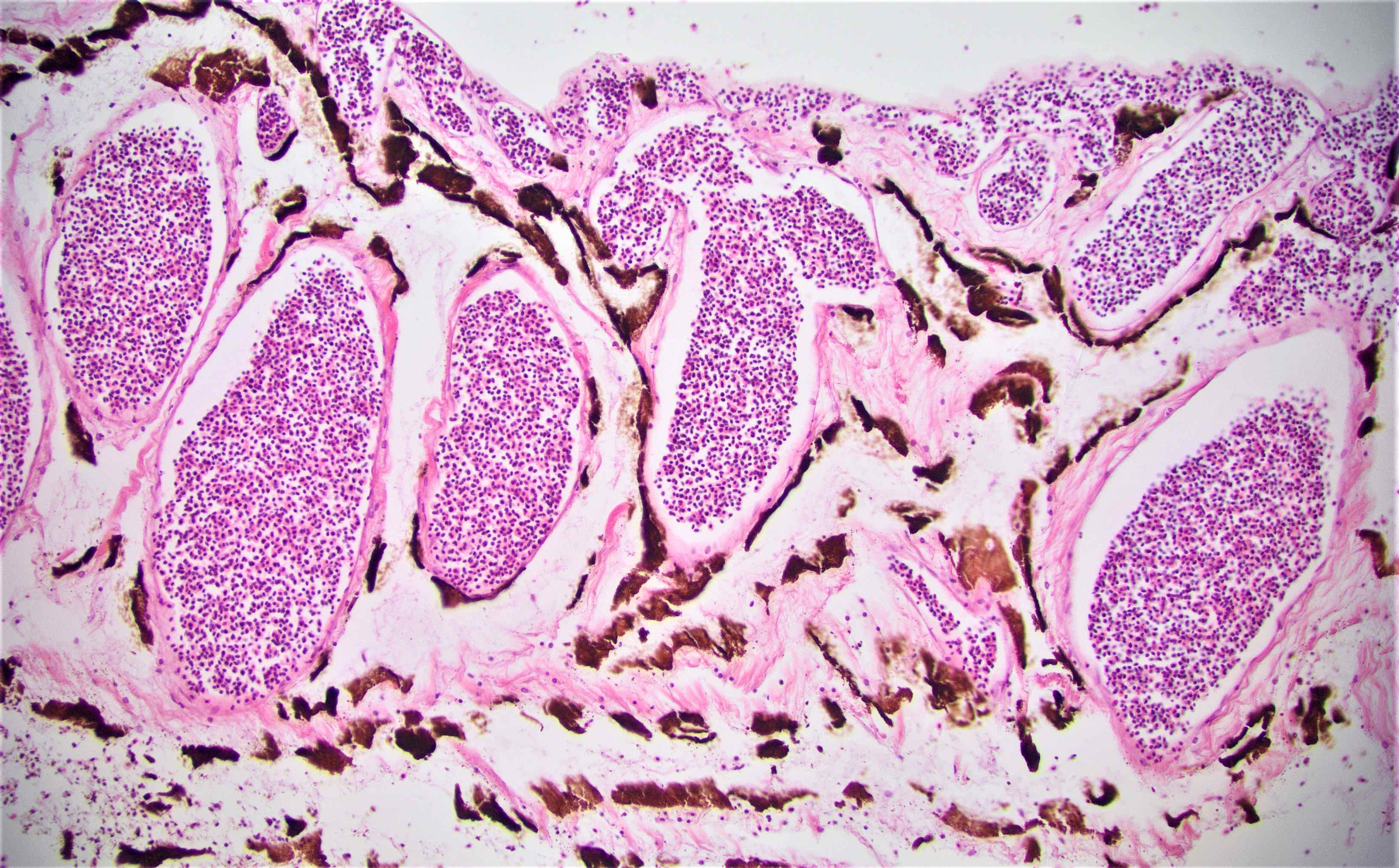
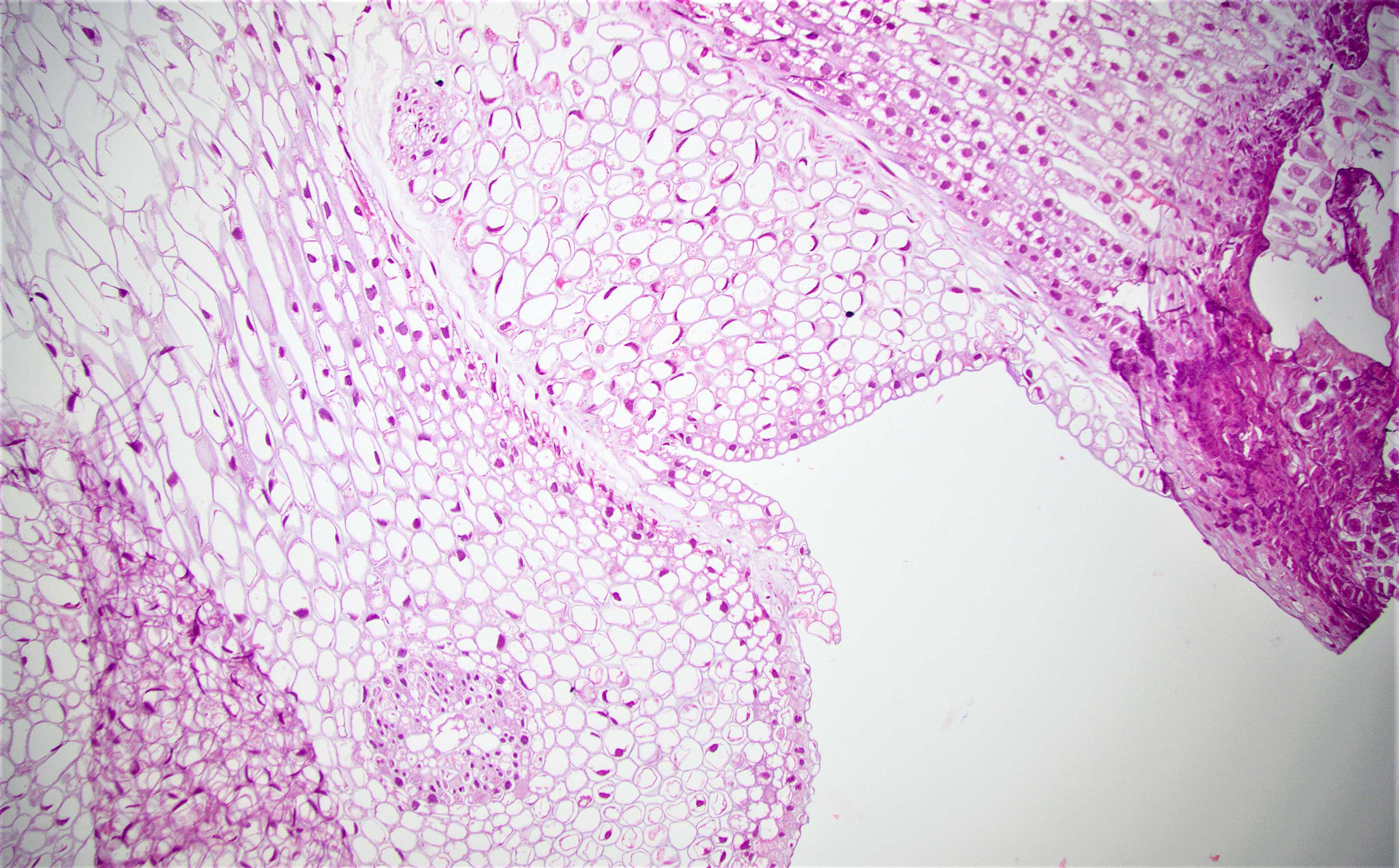
Calcified eggs of schistosomiasis
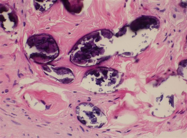
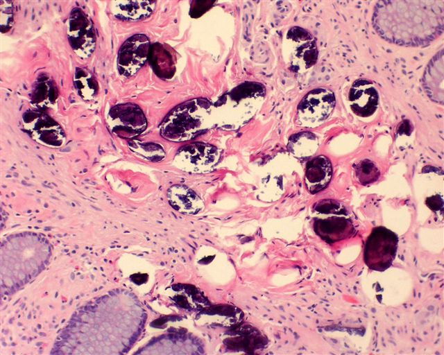
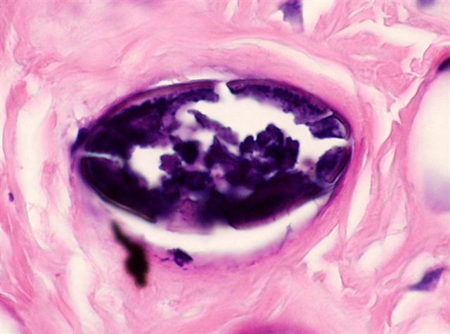
Within colonic mucosa
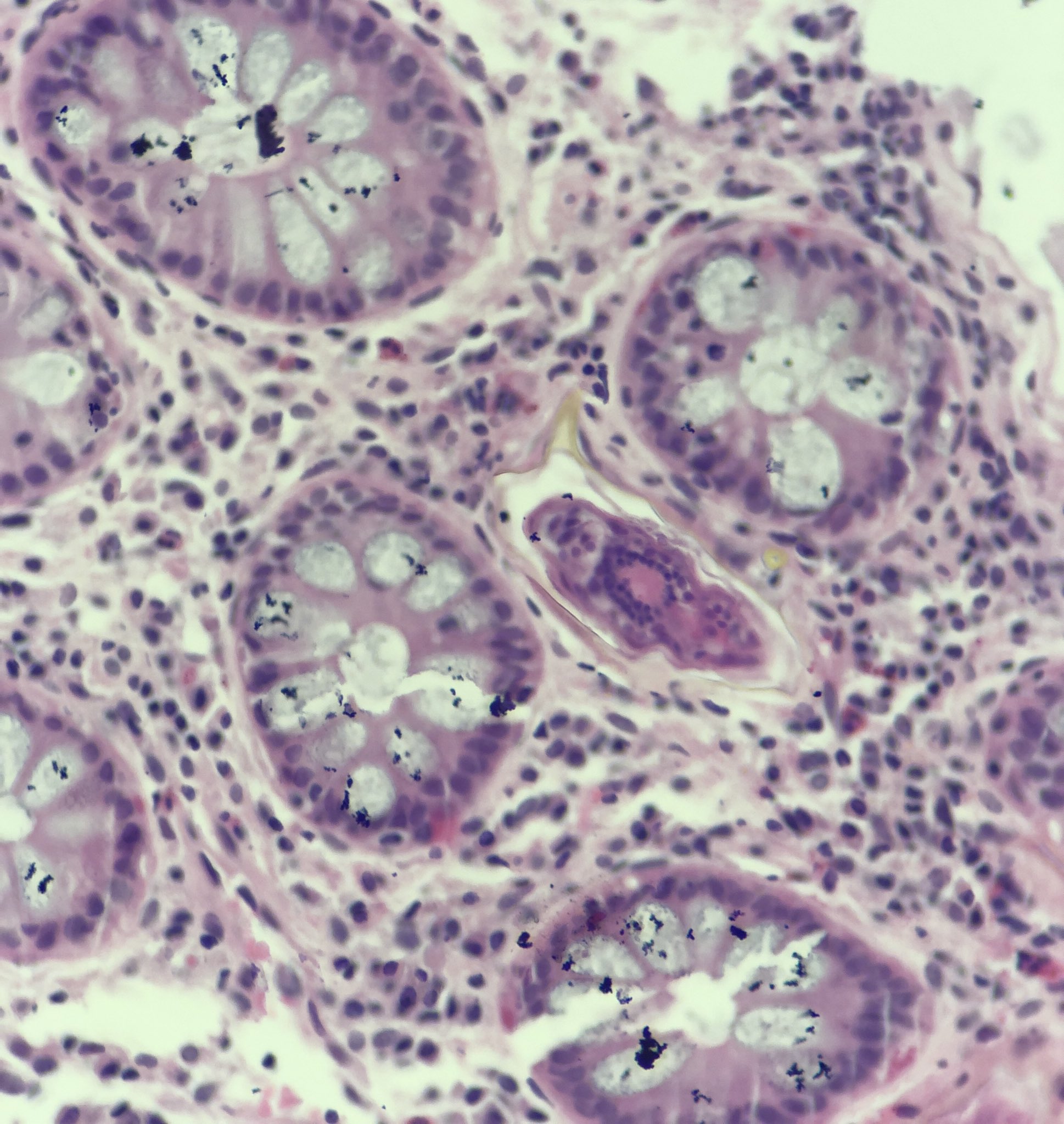
Schistosomiasis
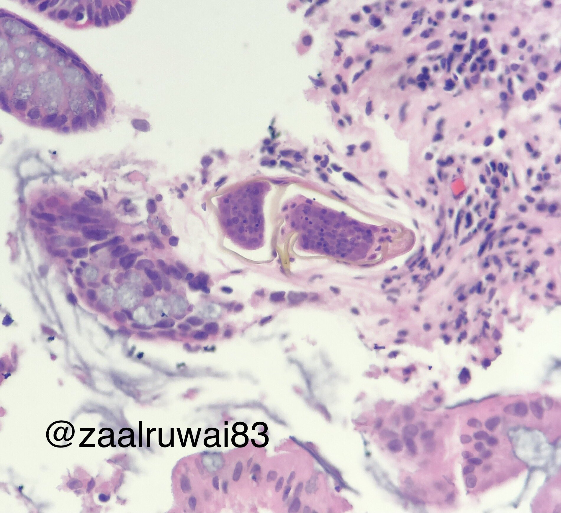
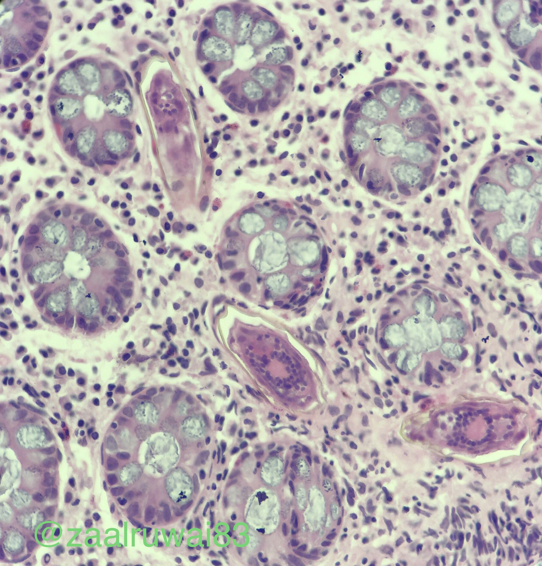
Images hosted on other servers:

Several eggs noted in stroma of polyp

Eggs
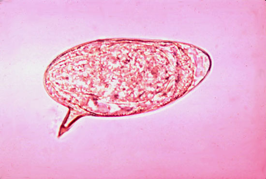
S. mansoni eggs
S. japonicum:


Eggs
Within tissue - species not specified:
intramucosal calcified bodies
Calcified bodies often clustered

Granuloma surrounding egg

Occasional forms
suggestive of spines
or hooks

Ziehl-Neelsen staining