
Images hosted on other servers:

Possible pathogenesis of achalasia cardia
Images hosted on other servers:

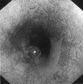
CXR shows achalasia
Images hosted on other servers:

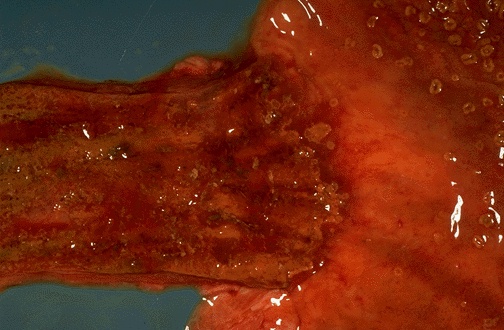
Dilated esophagus
Contributed by Avani Pendse, M.D., Ph.D.
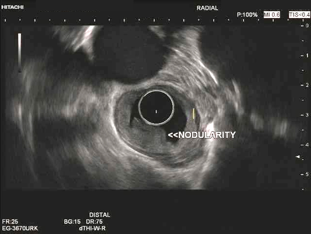
Endoscopic ultrasound
Contributed by Avani Pendse, M.D., Ph.D.
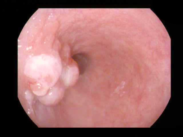
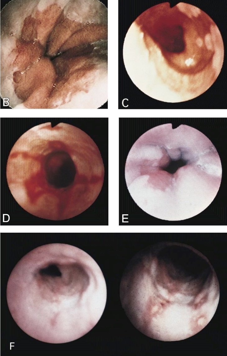
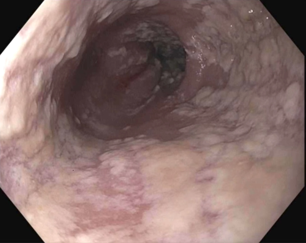
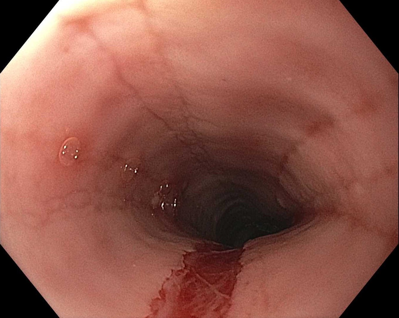
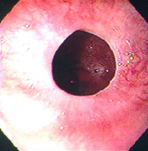
Upper endoscopy
Contributed by Avani Pendse, M.D.
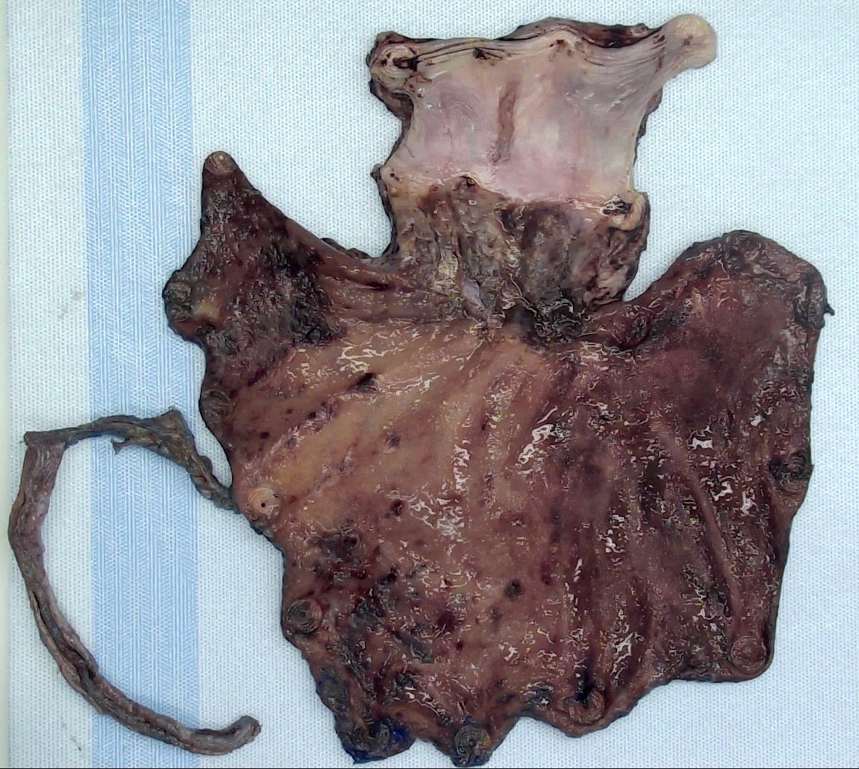
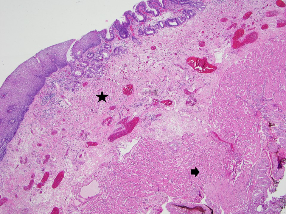
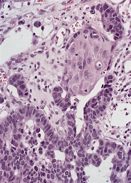
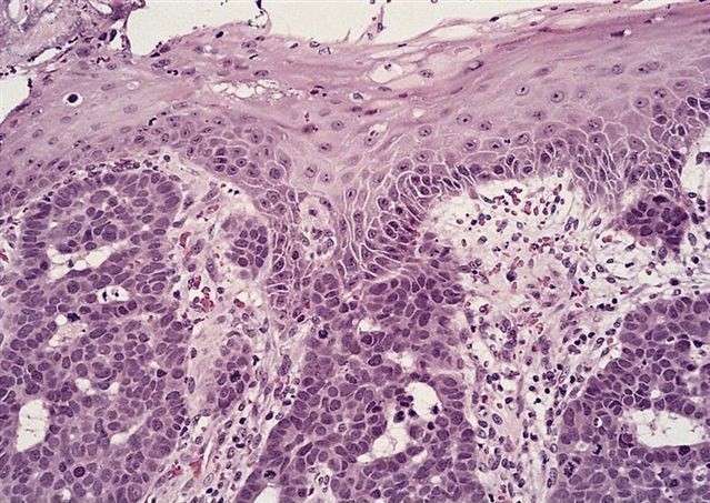
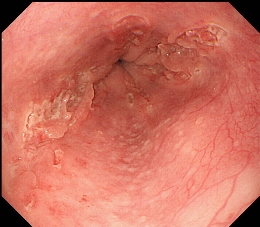
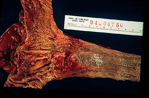
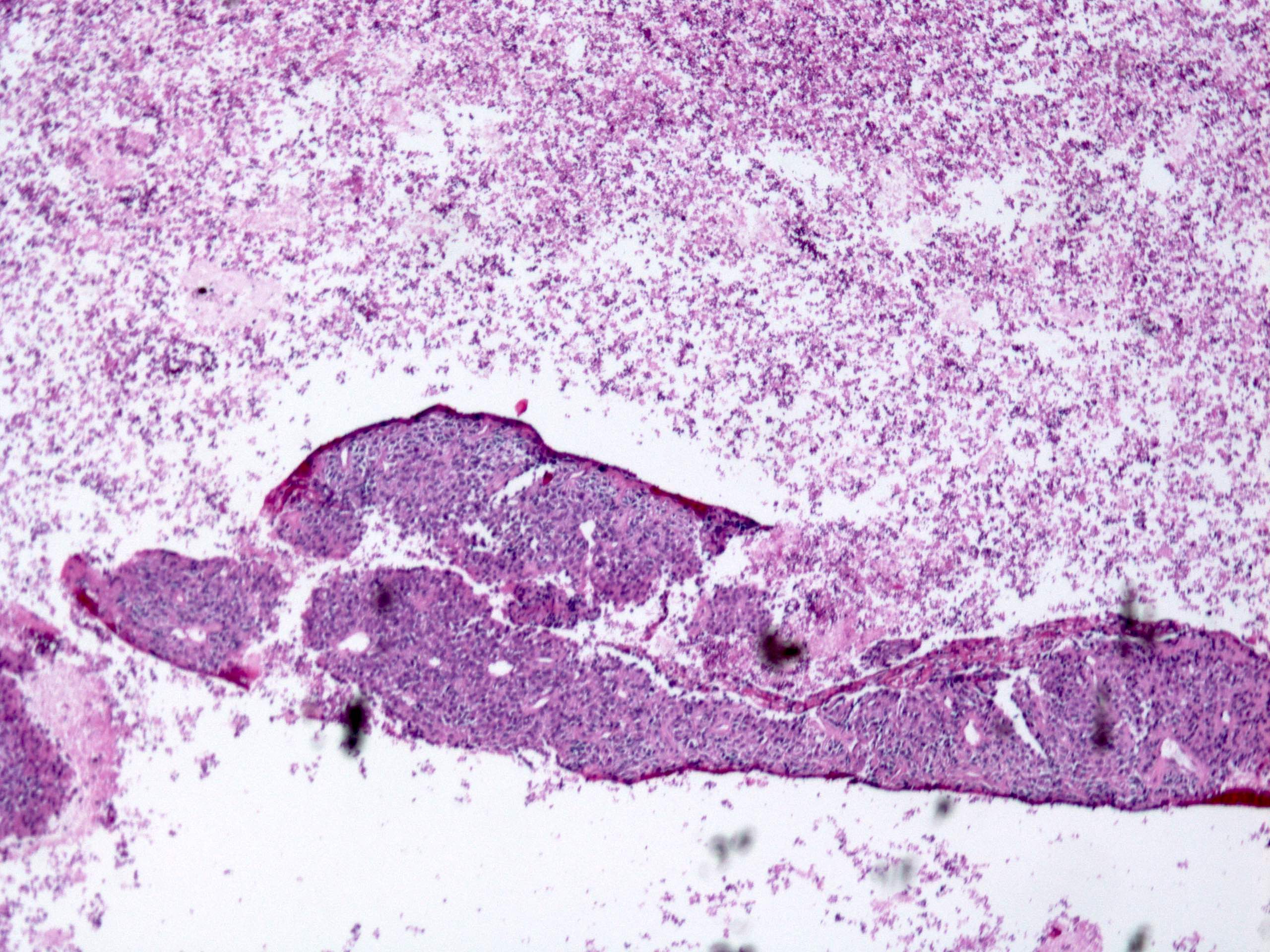
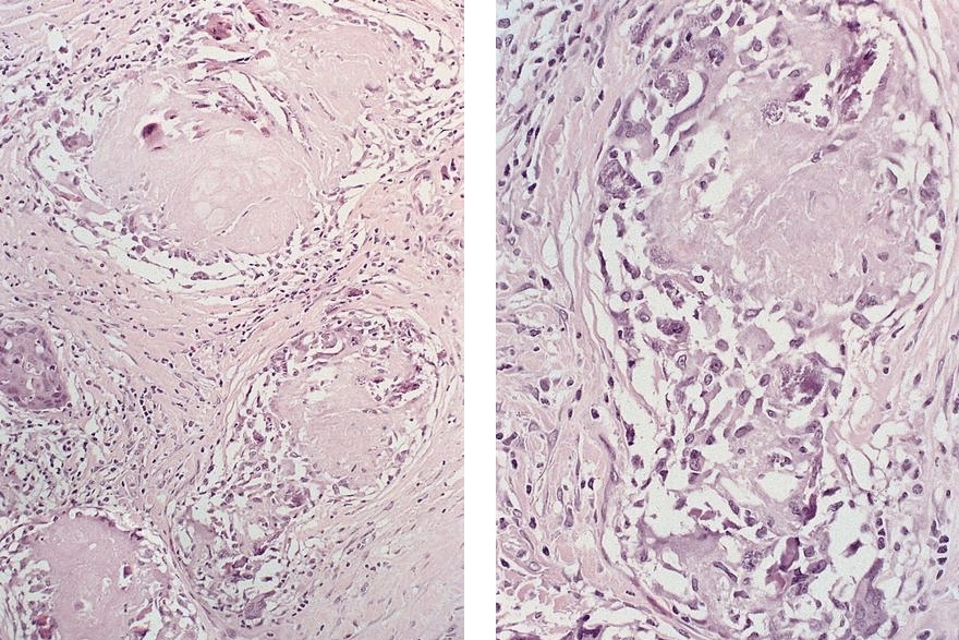
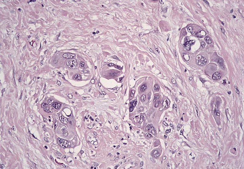
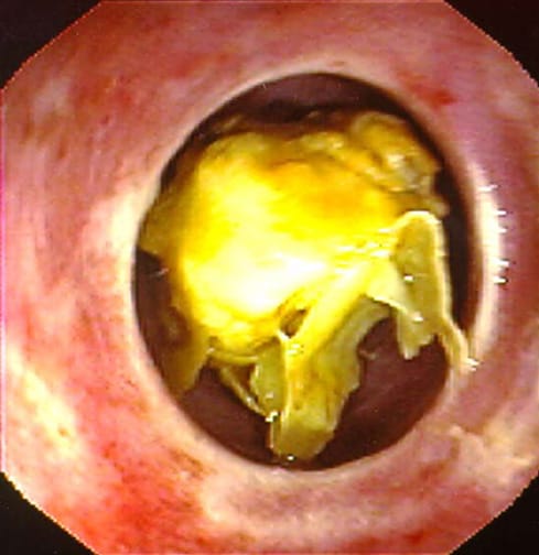
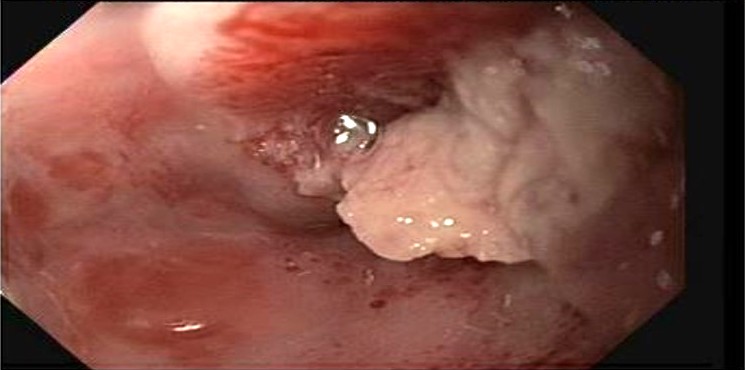
Distal esophagus and GE junction tumor
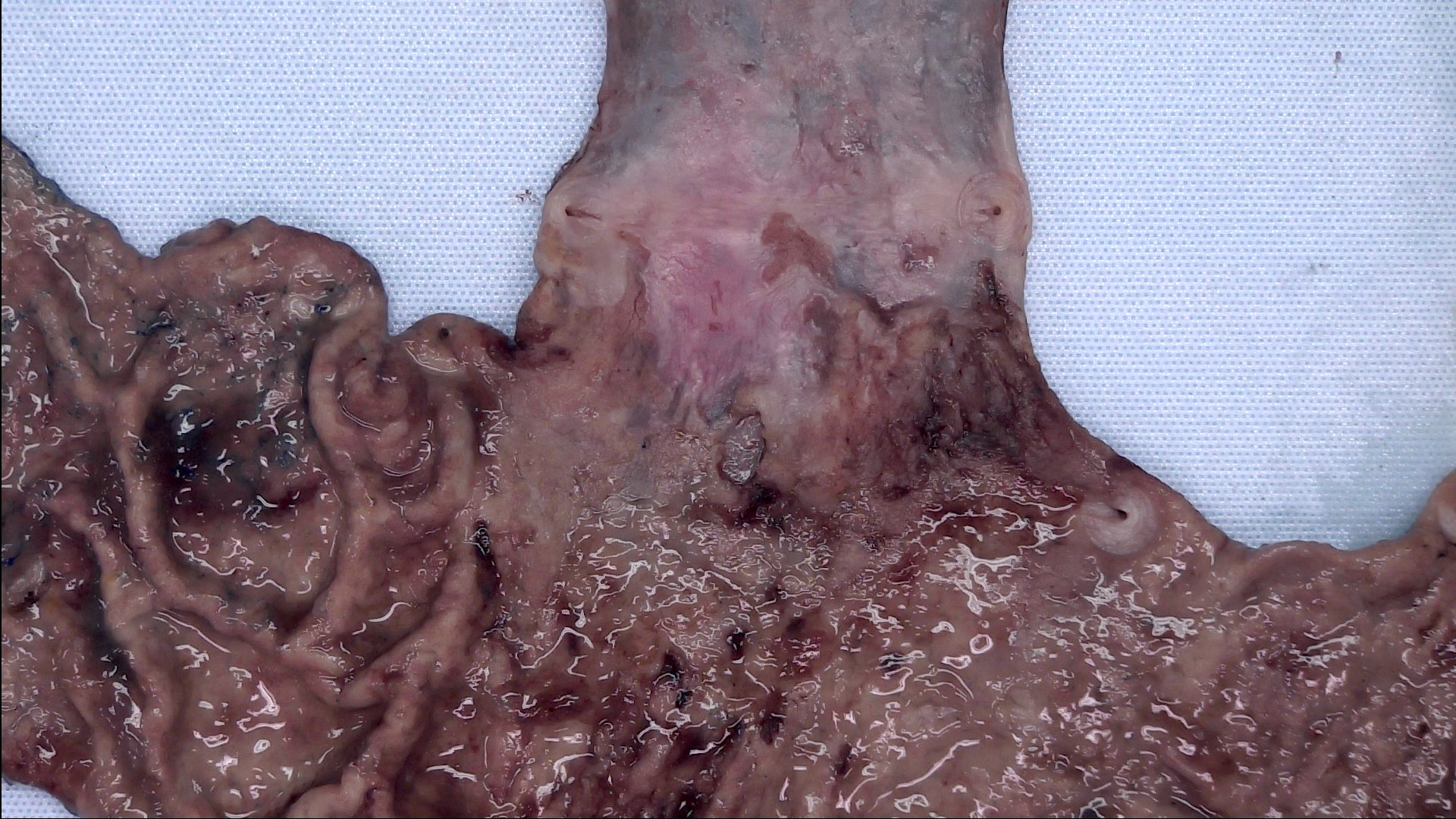
Ulcer at GE junction
Treated tumor
Contributed by Avani Pendse, M.D., Ph.D.
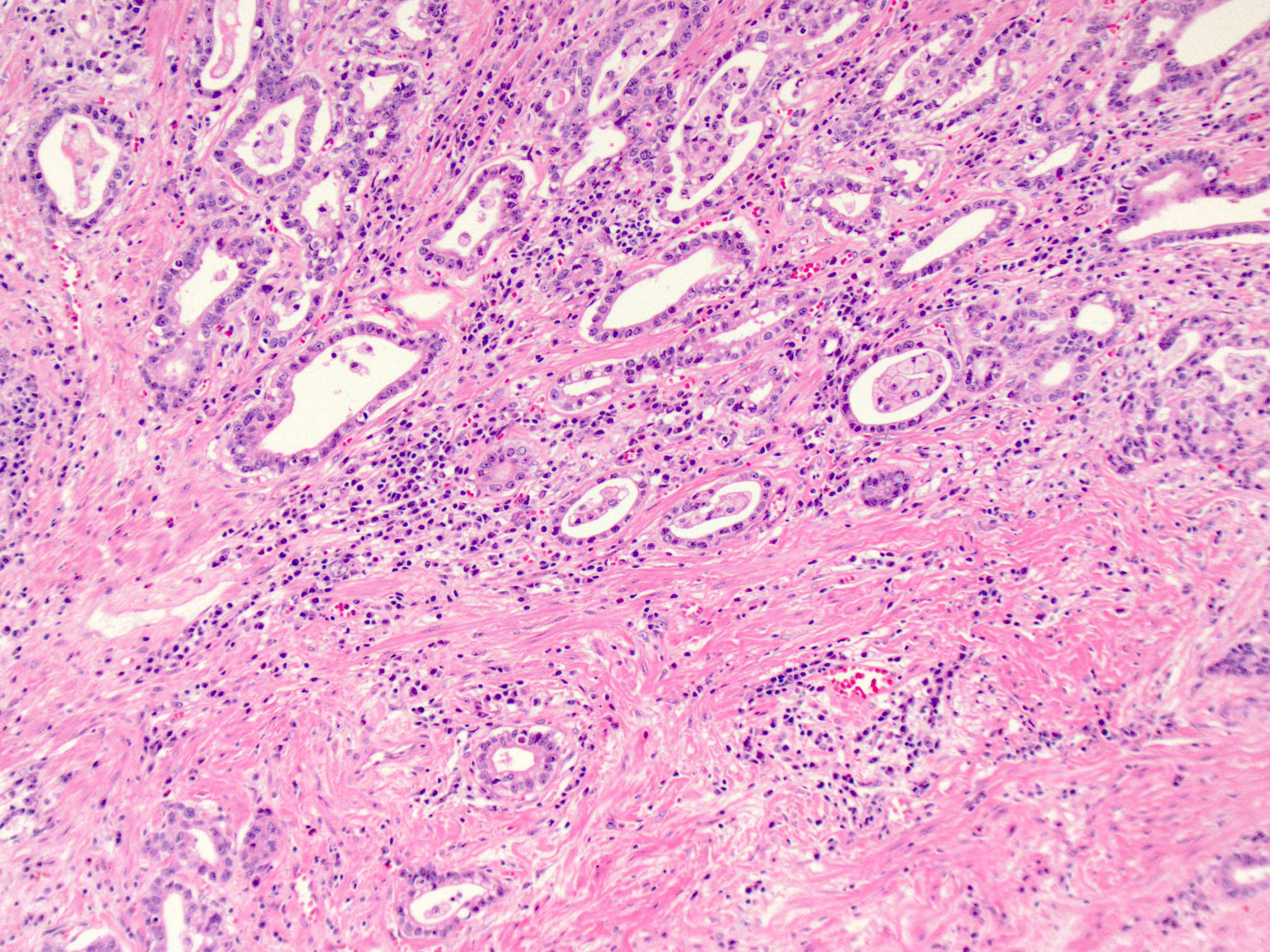
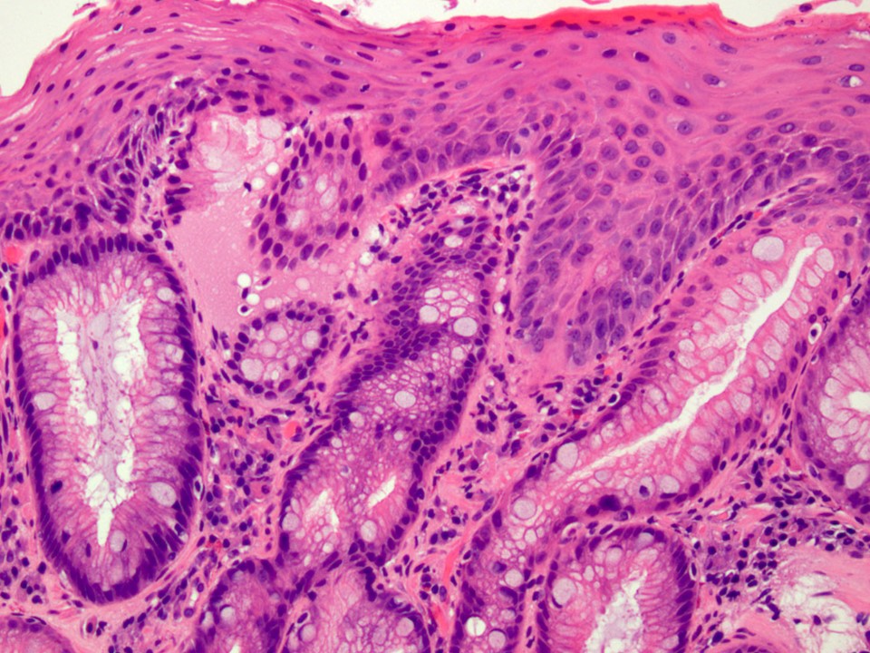
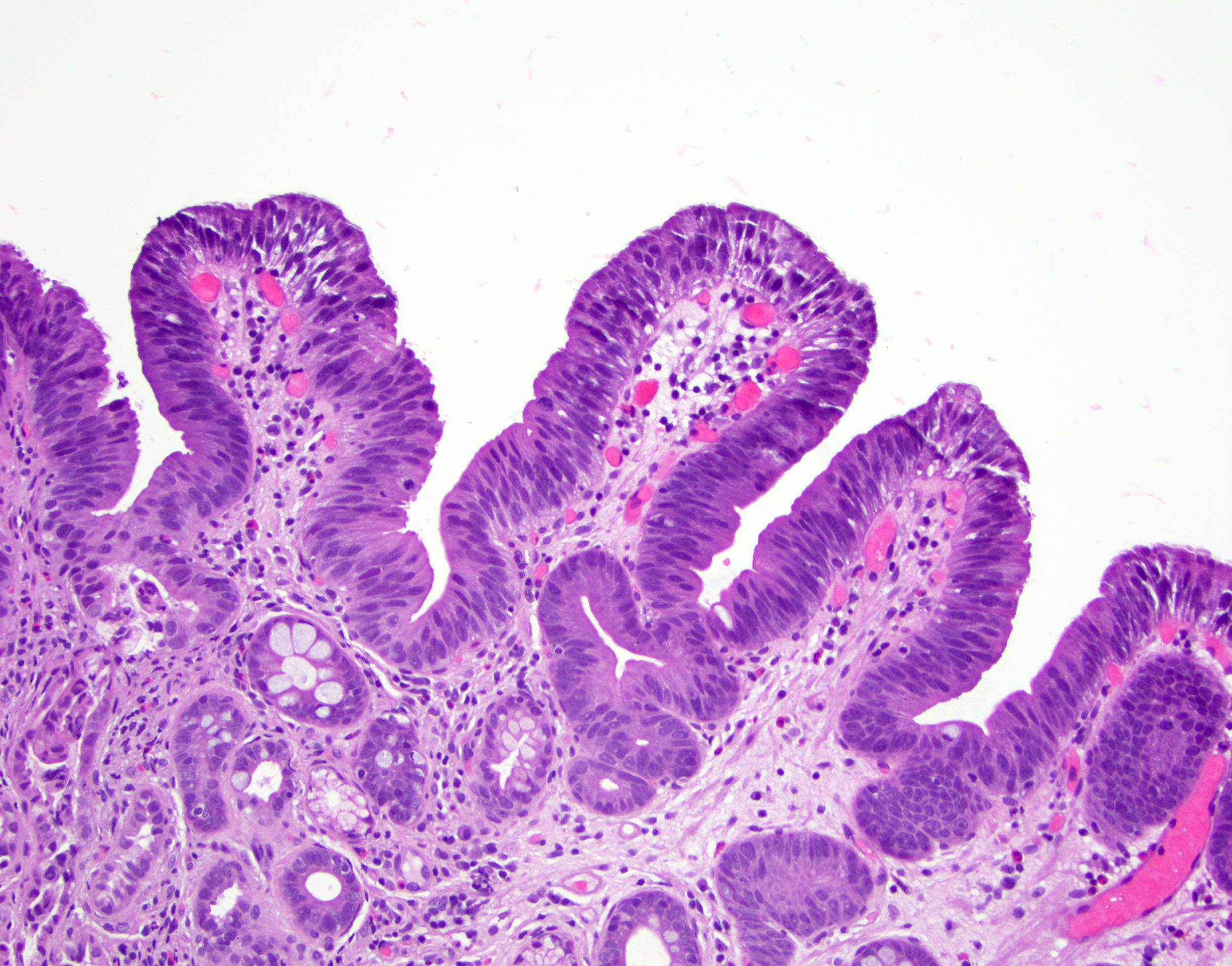
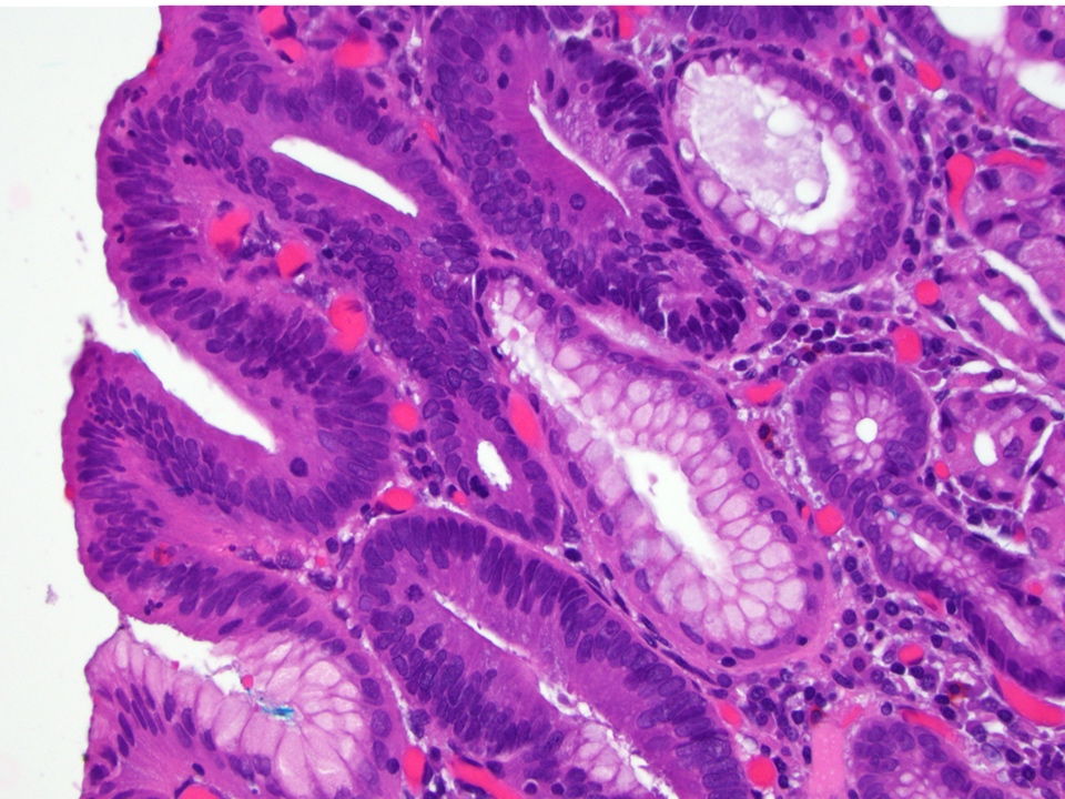
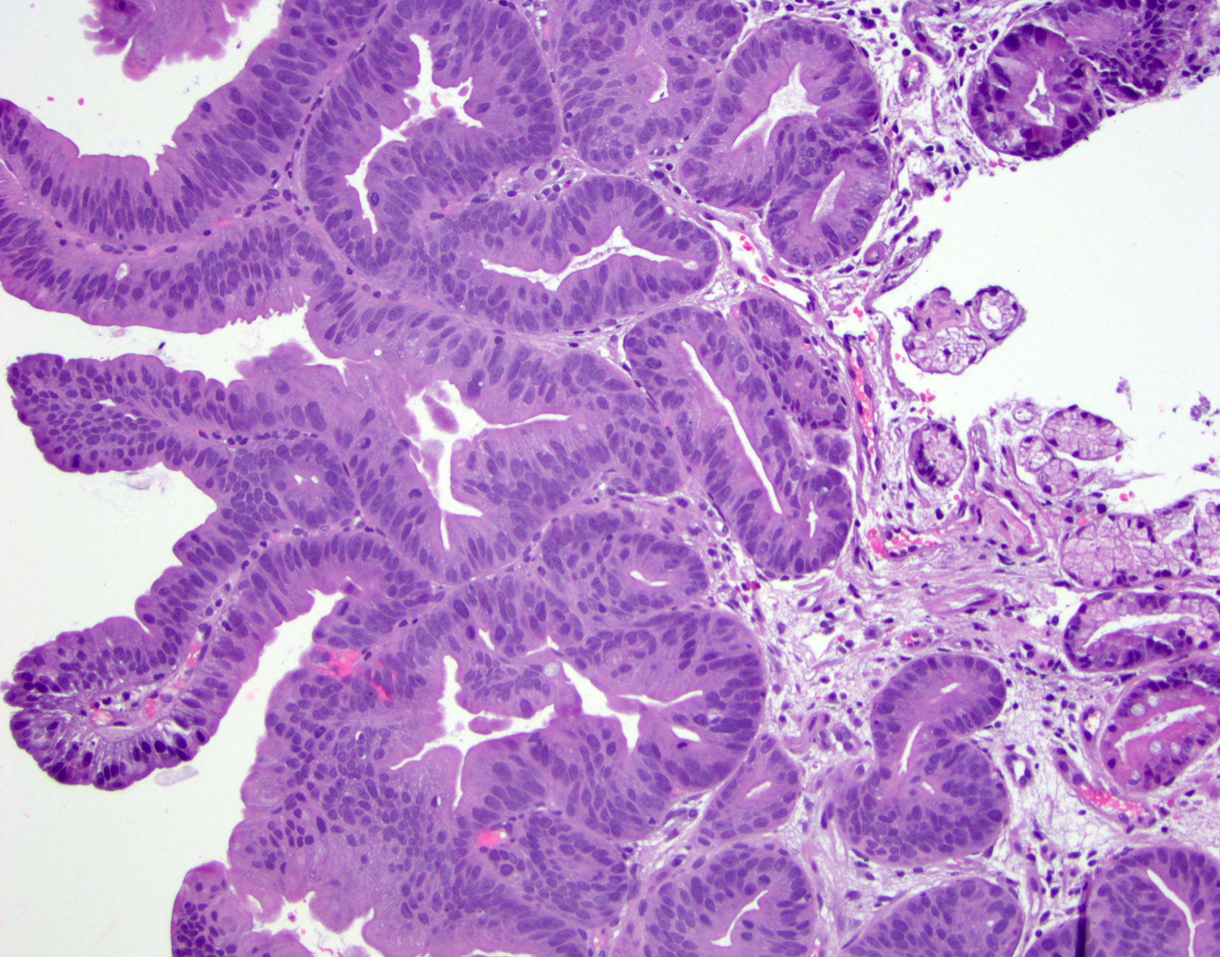
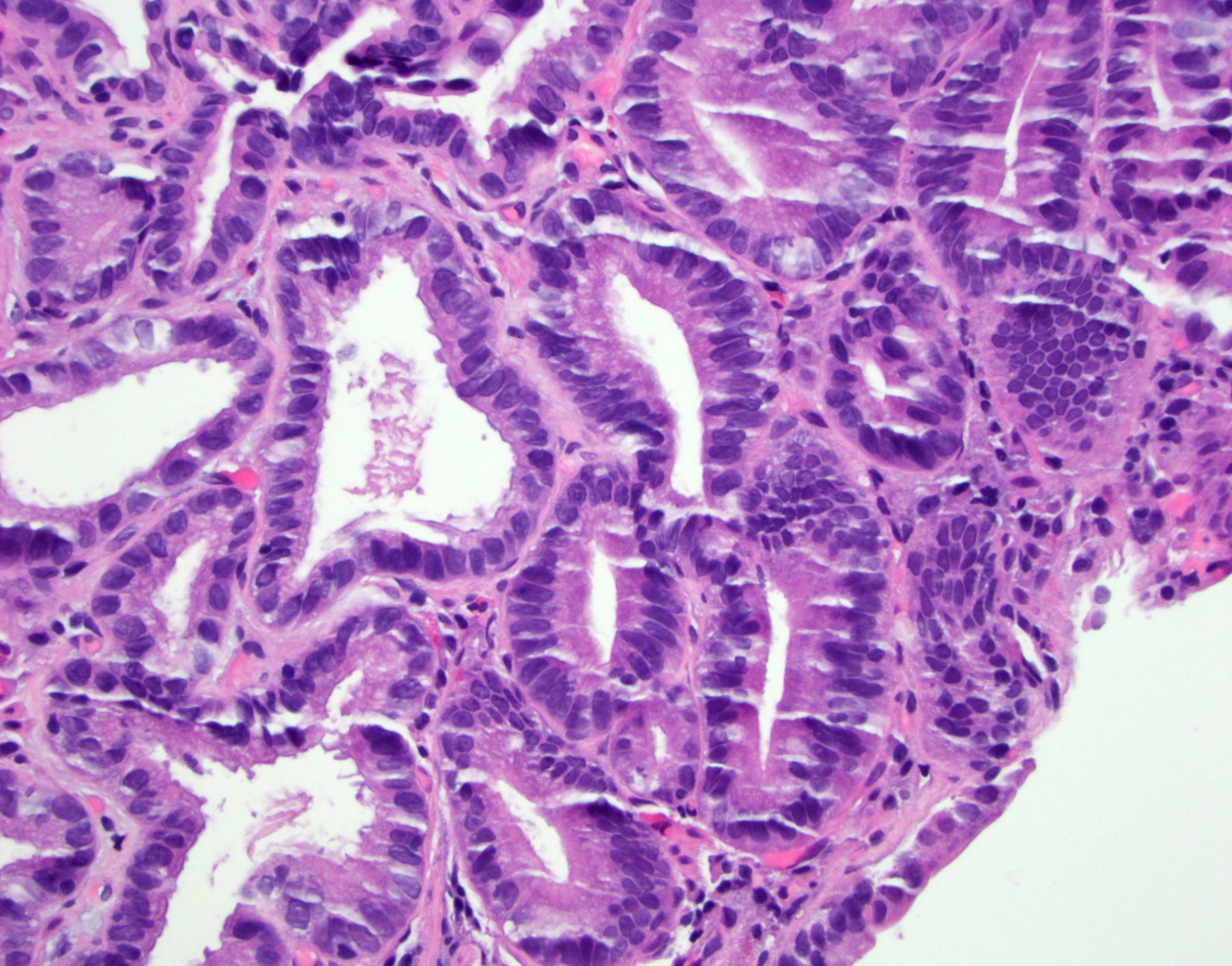
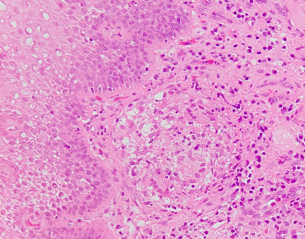
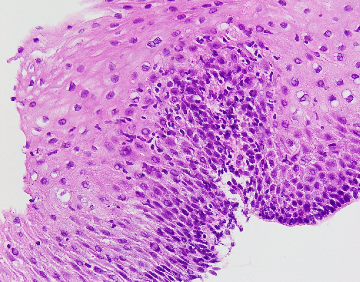
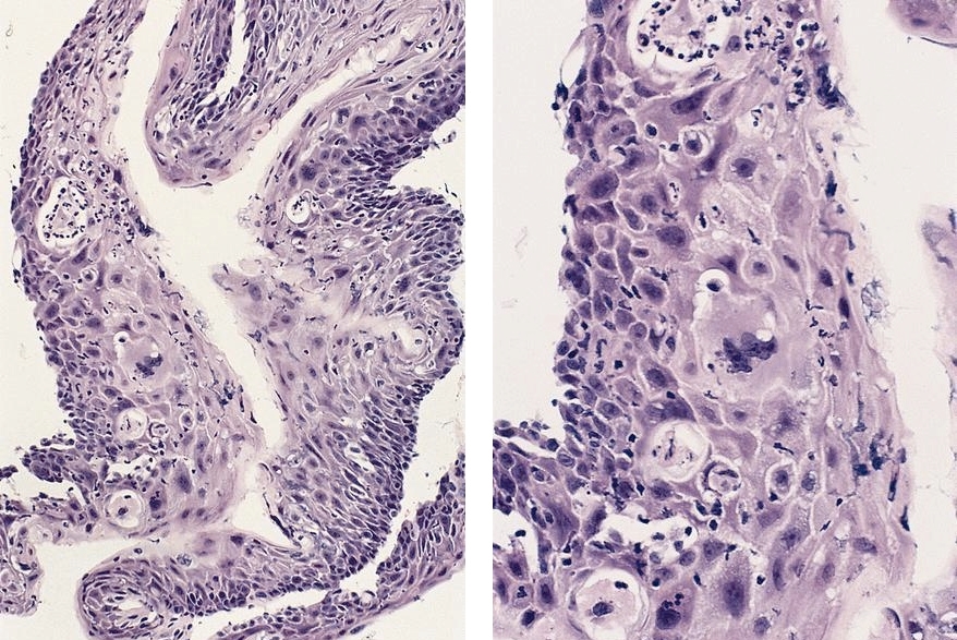
Tubular pattern
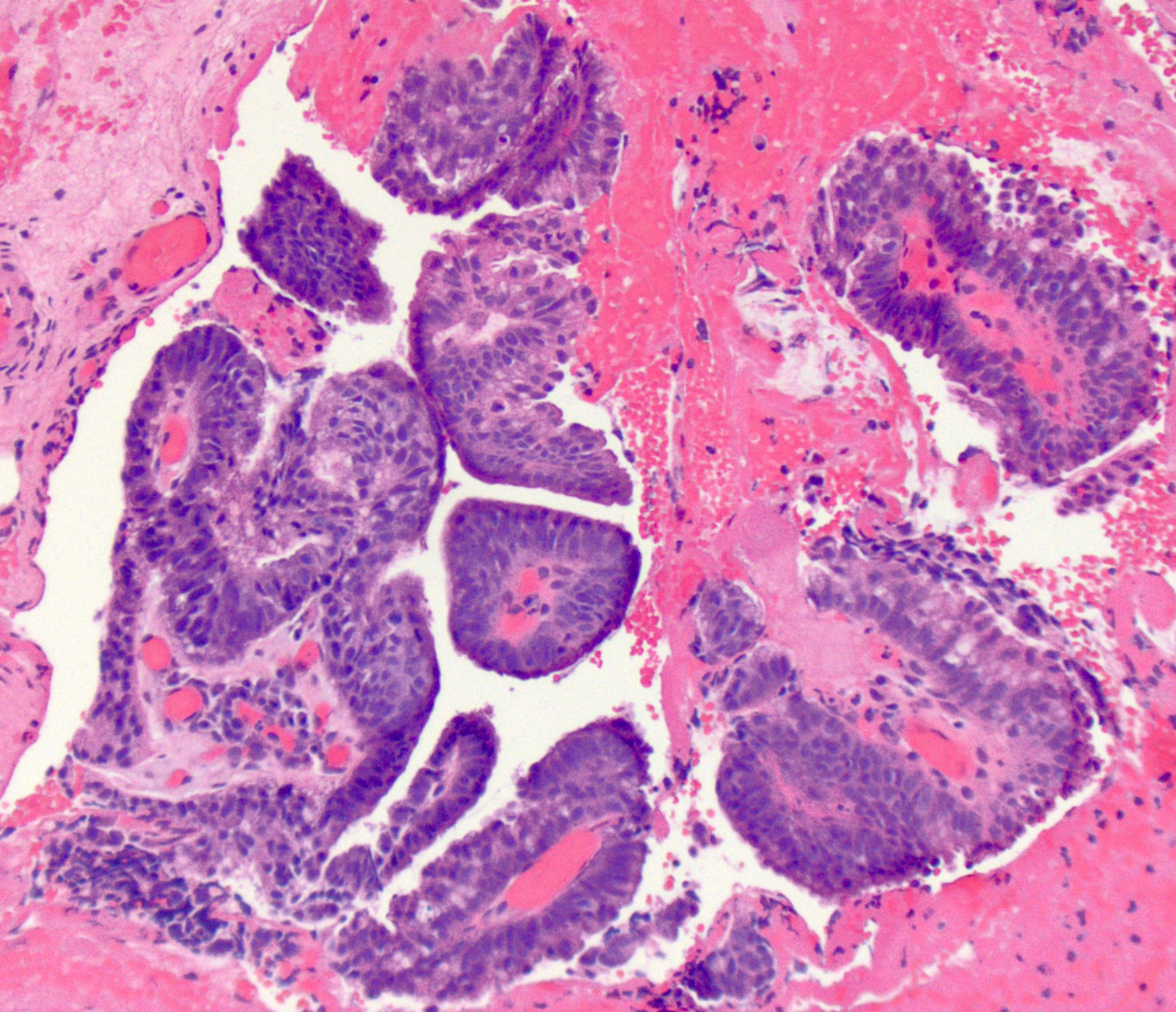
Papillary pattern
Mucinous pattern
Signet ring cell pattern
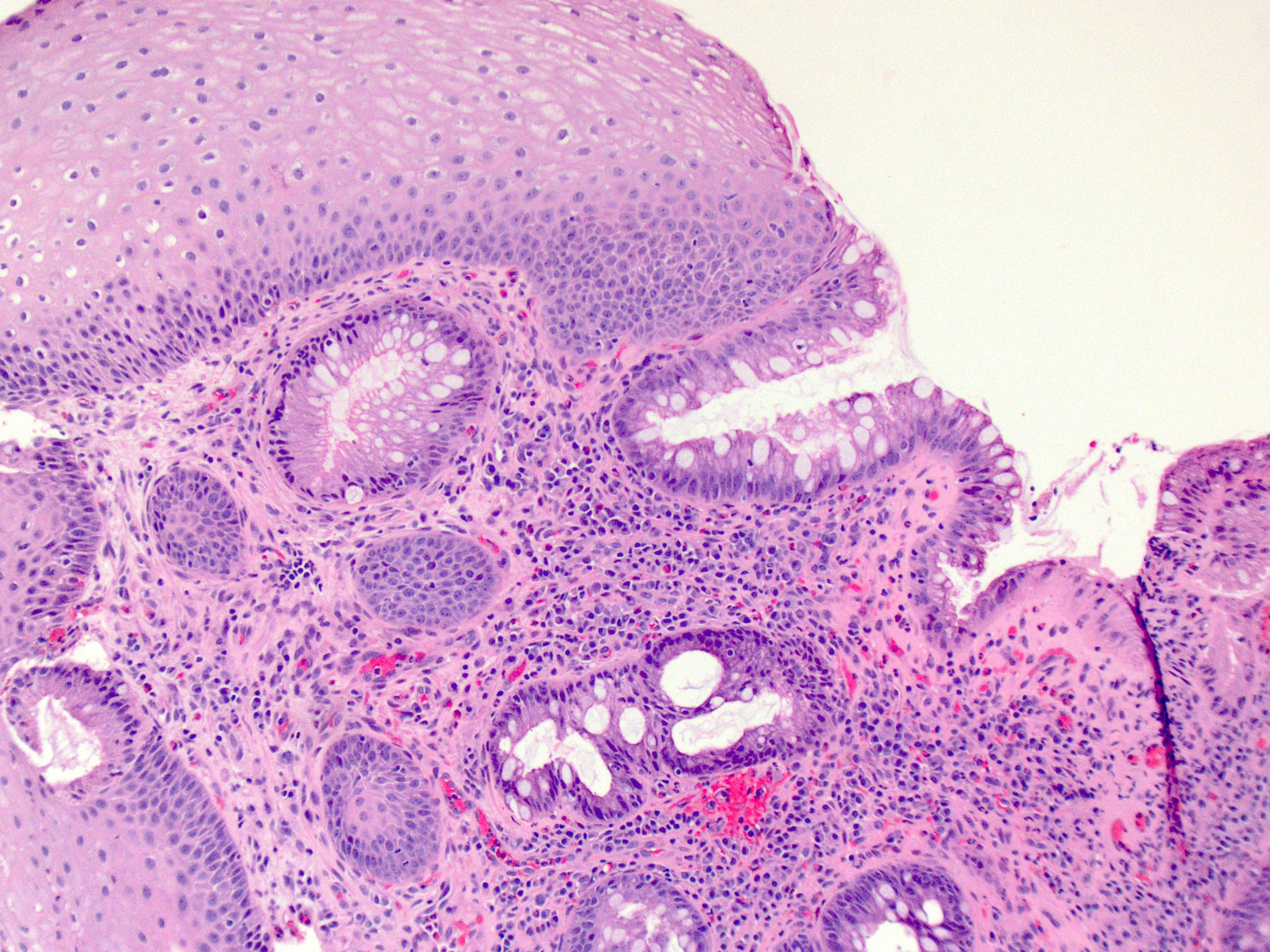
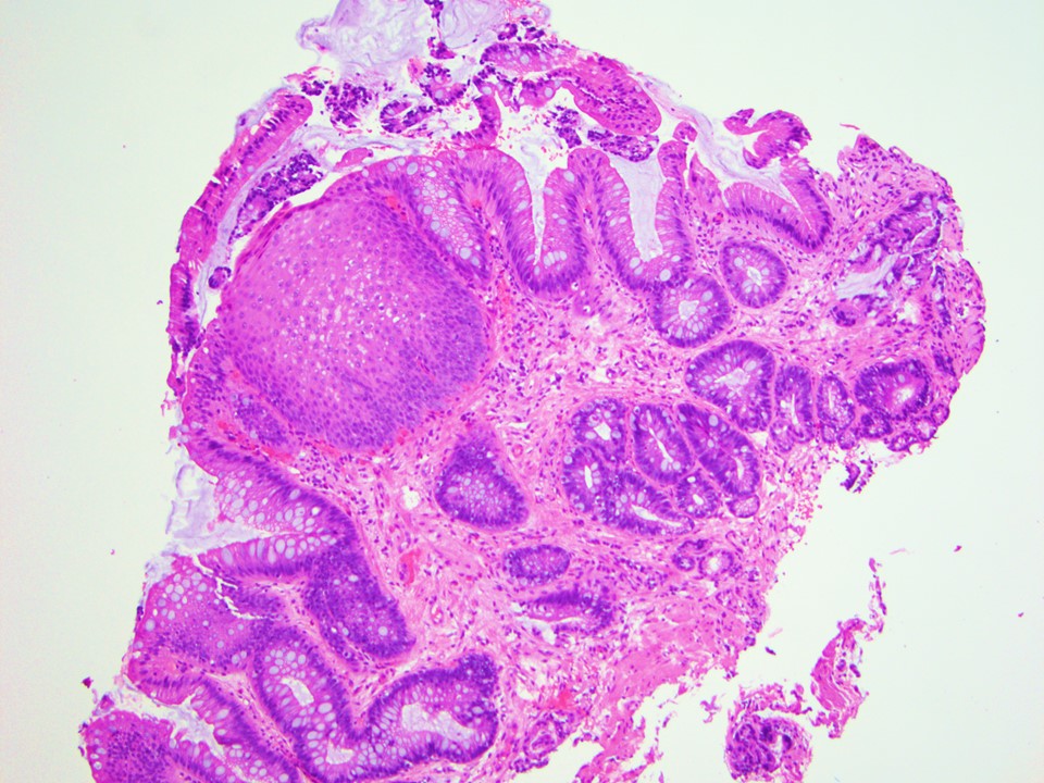
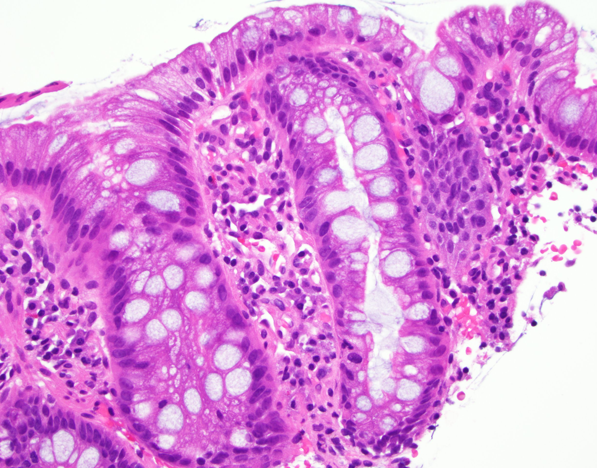
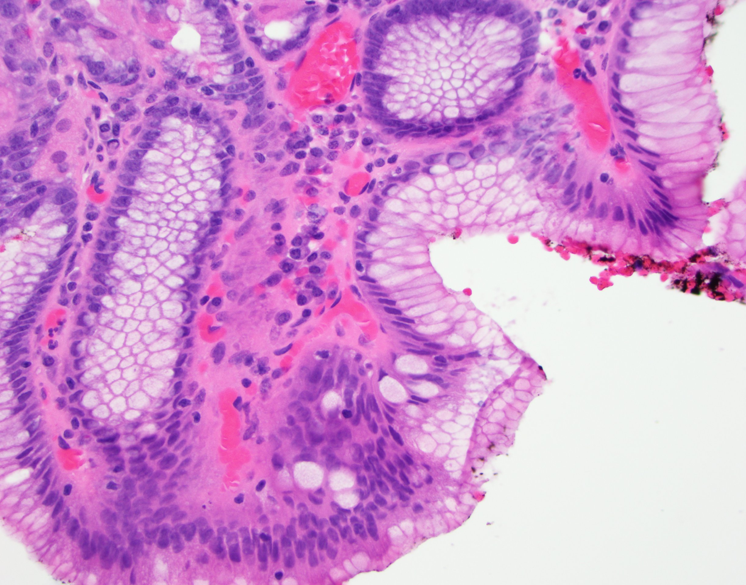
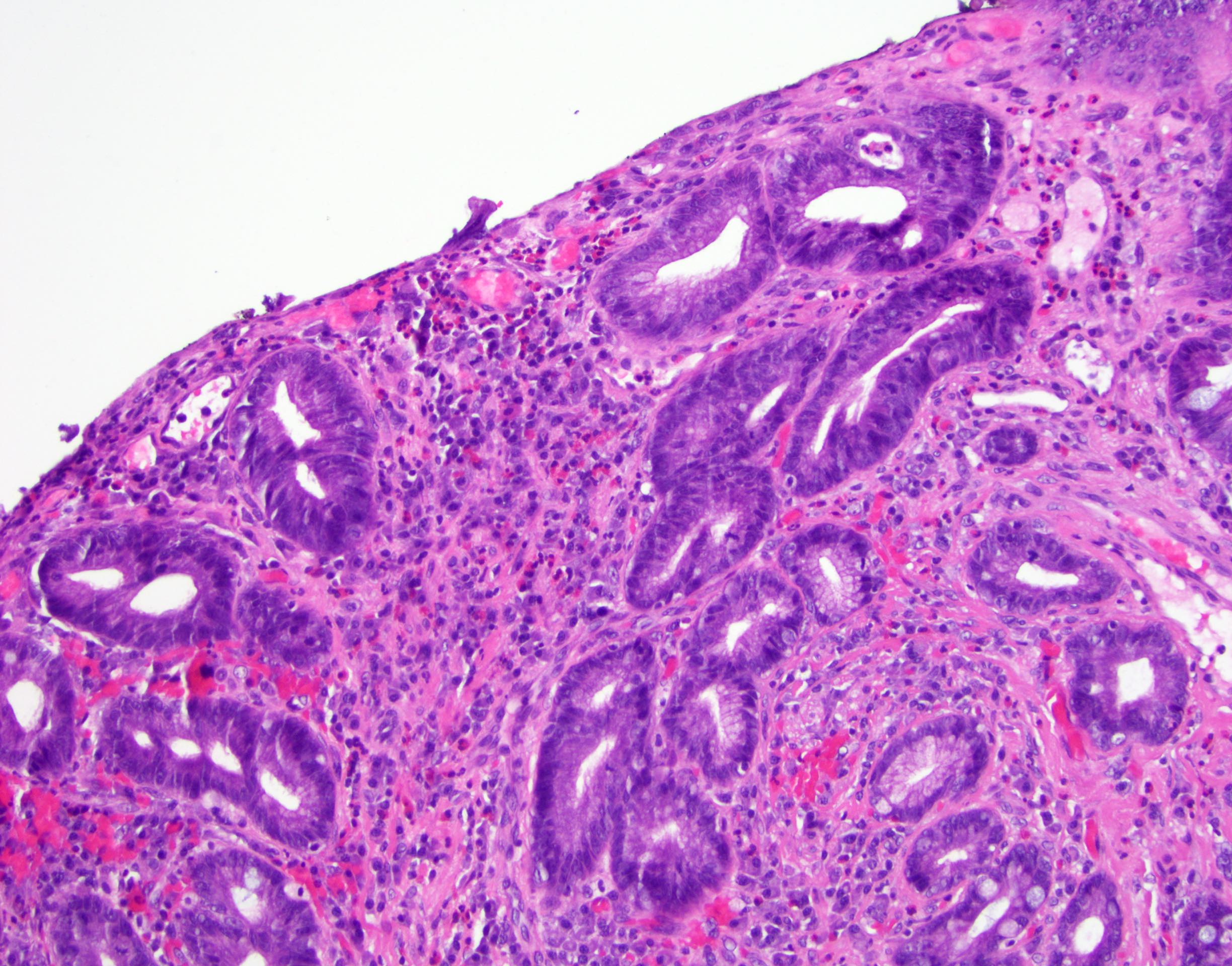
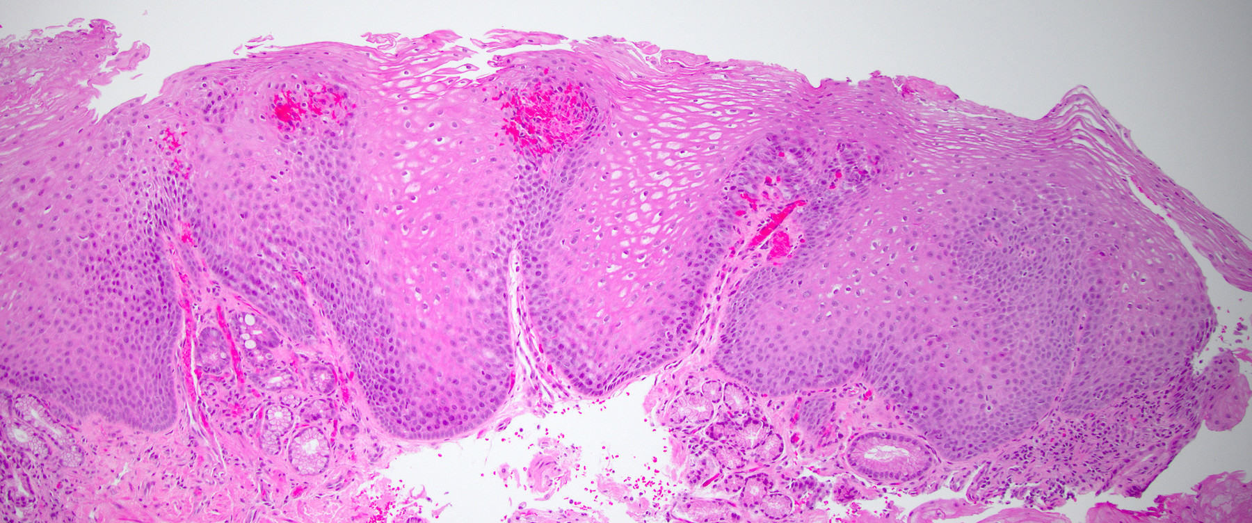
Barrett esophagus
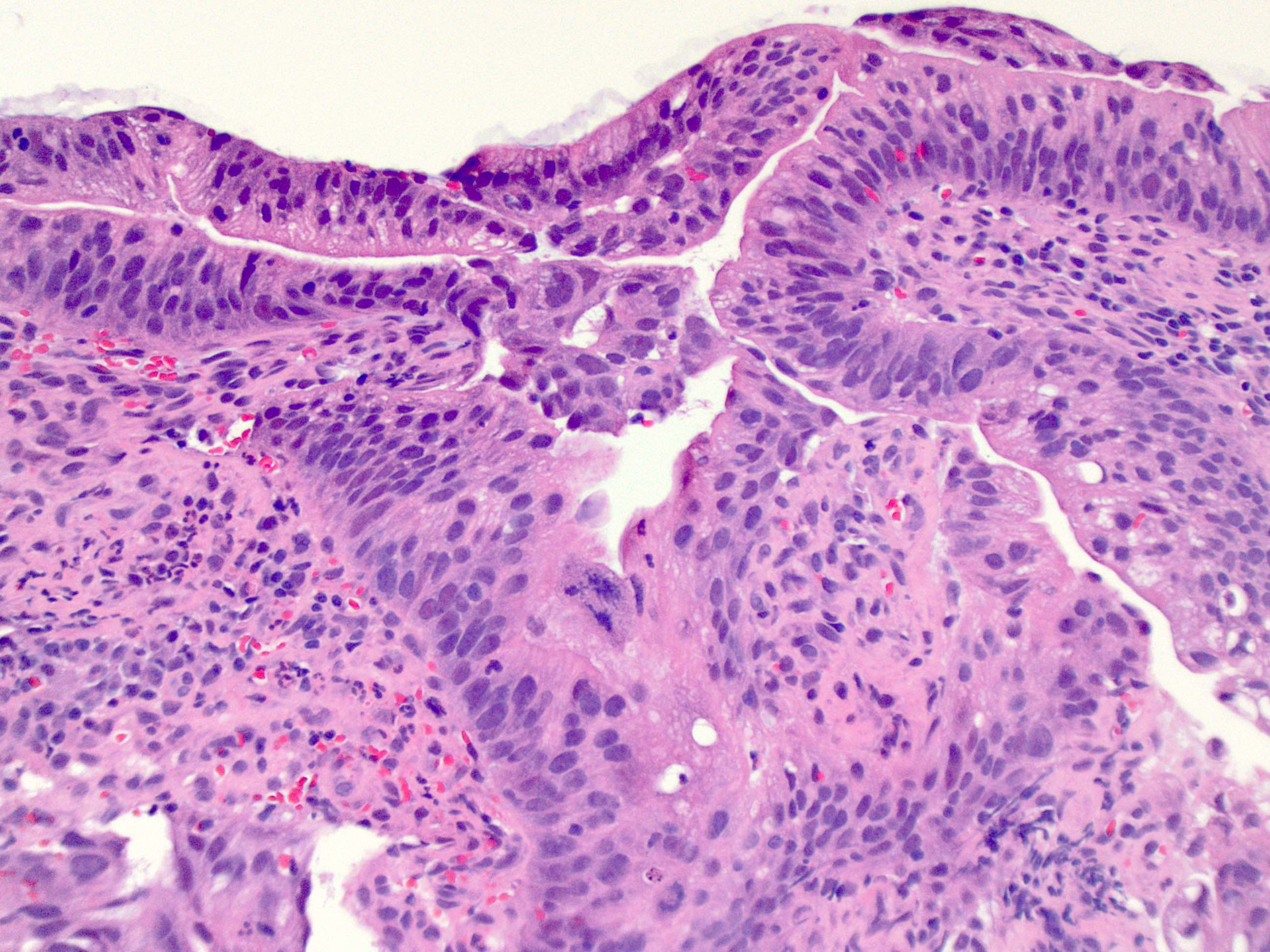
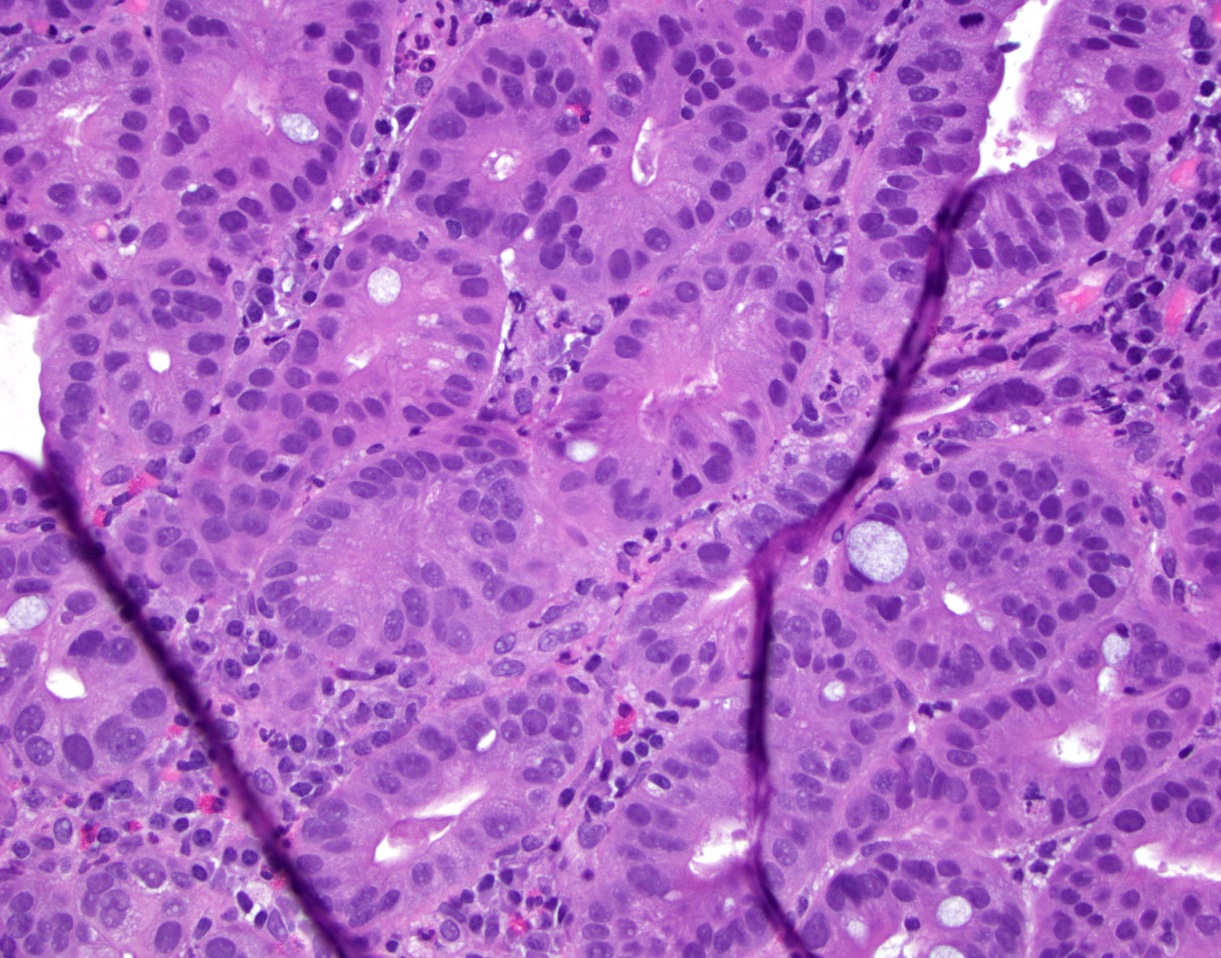
Low grade dysplasia

High grade dysplasia
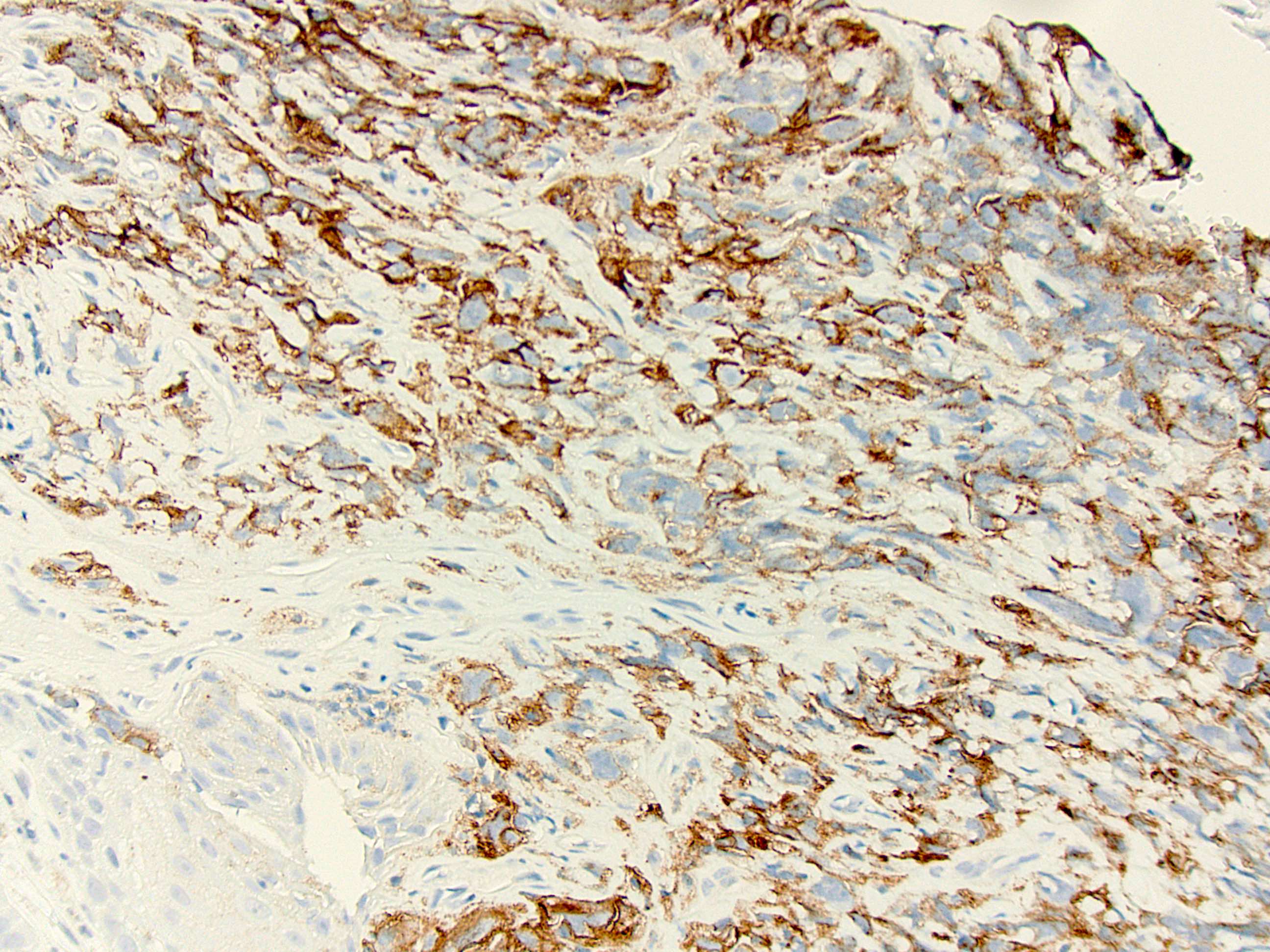
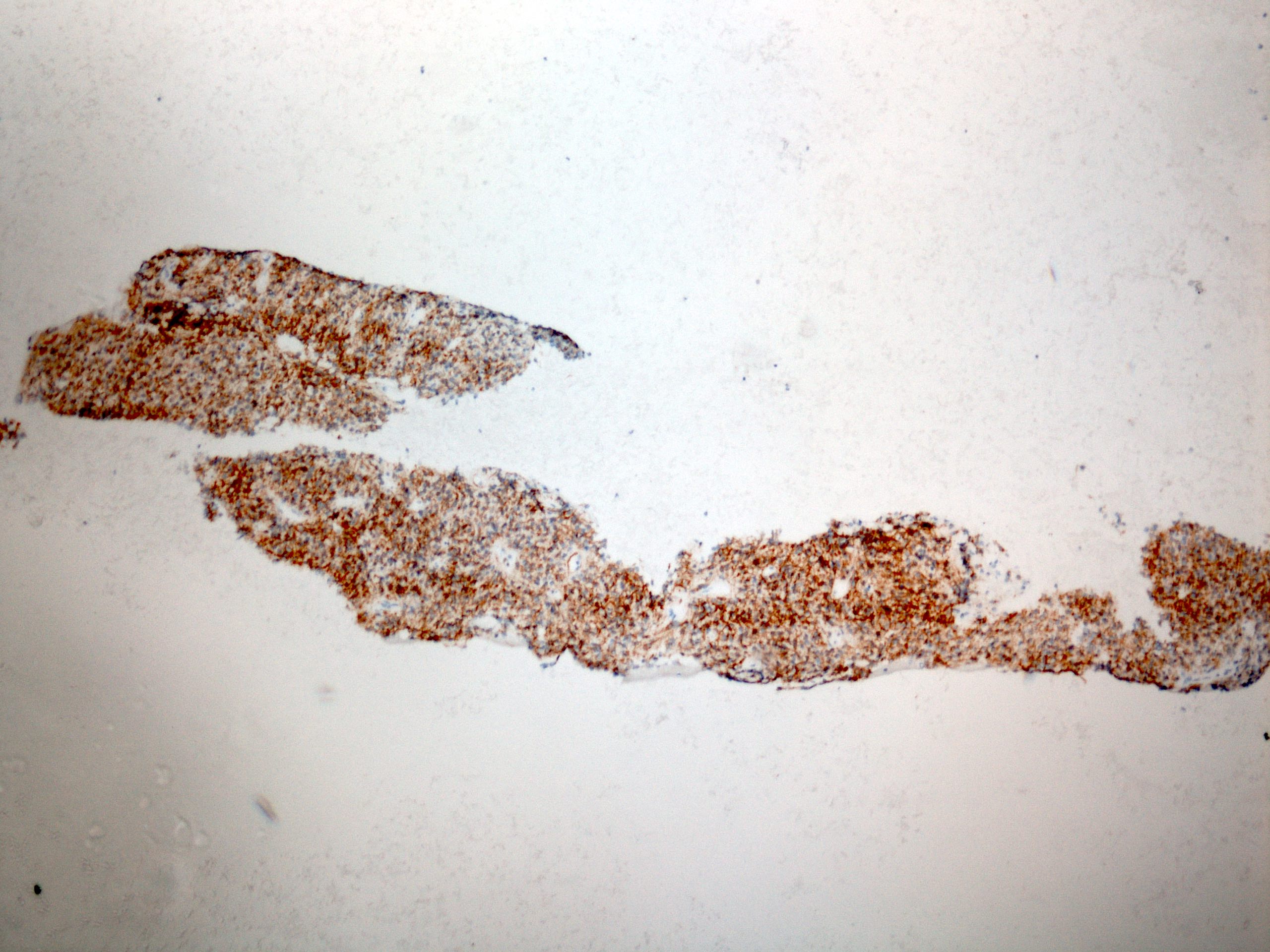
PDL1 positivity
Contributed by Avani Pendse, M.D., Ph.D.

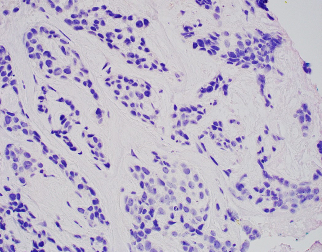
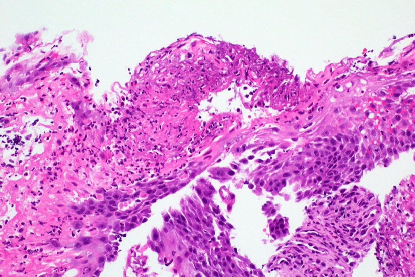
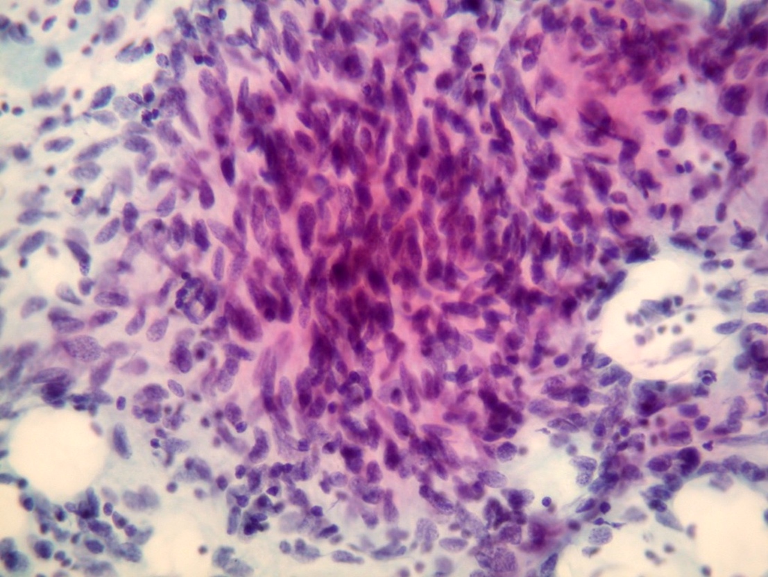
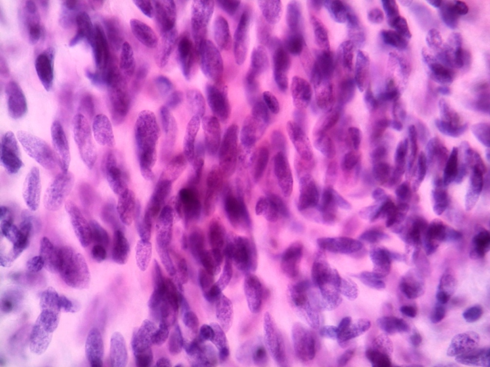
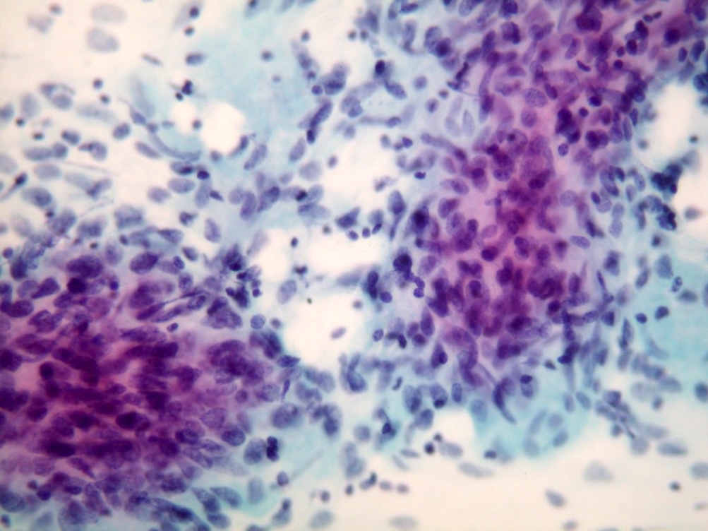
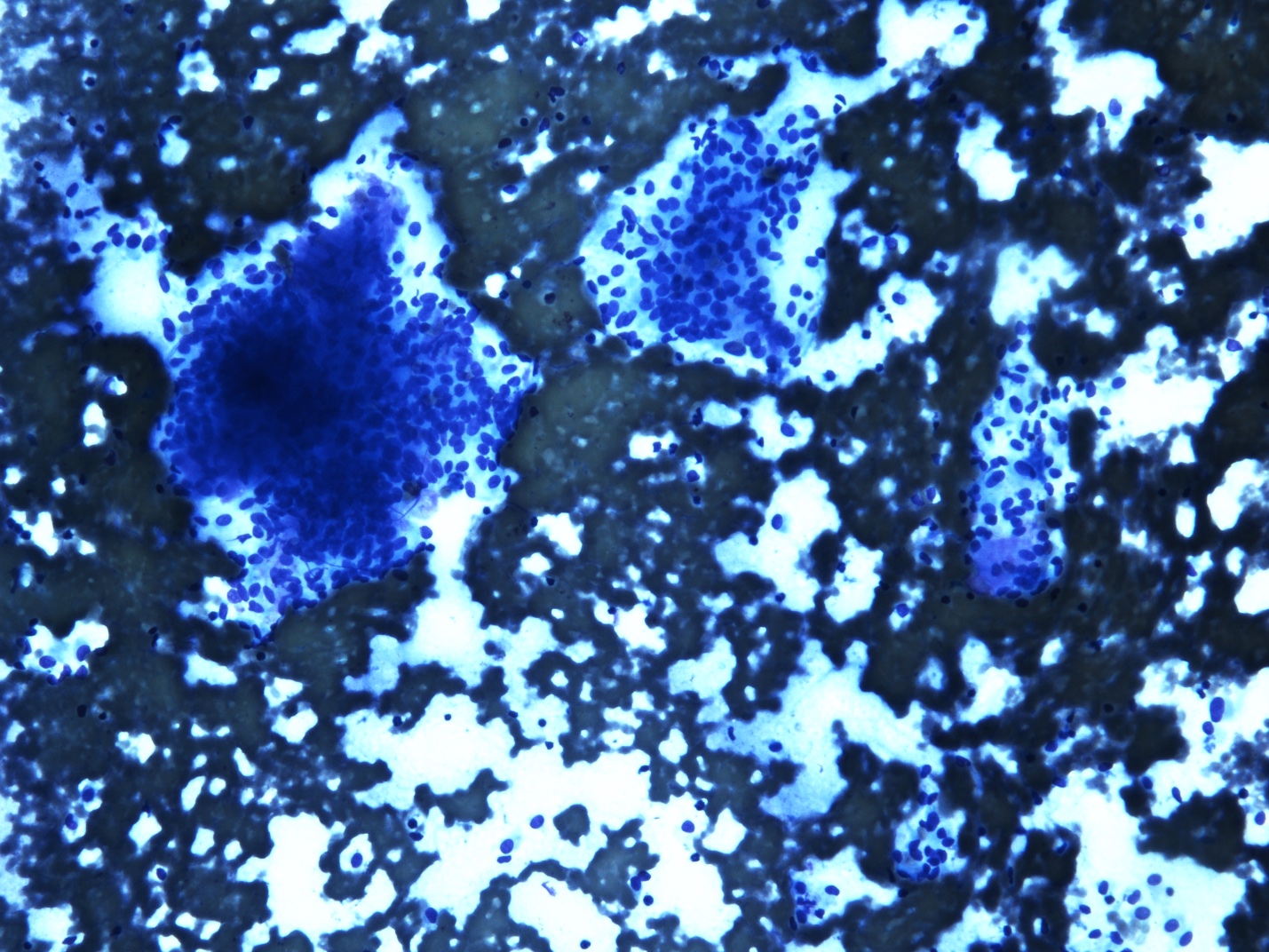
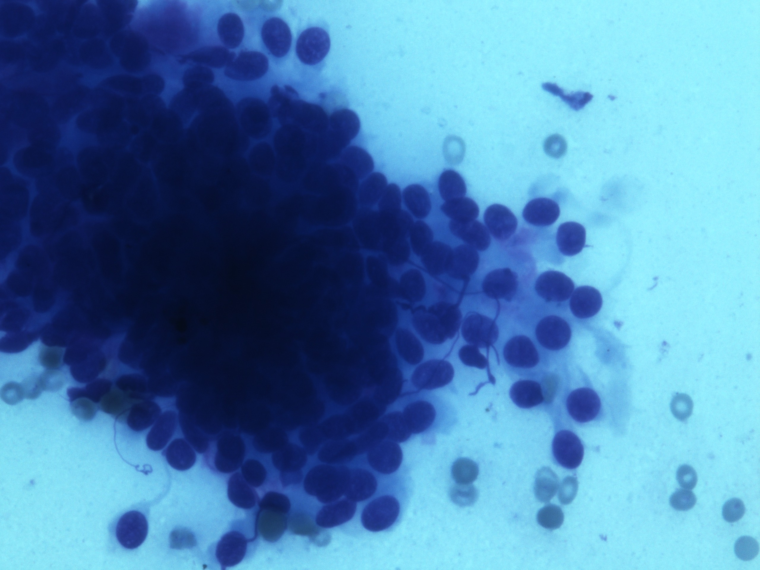
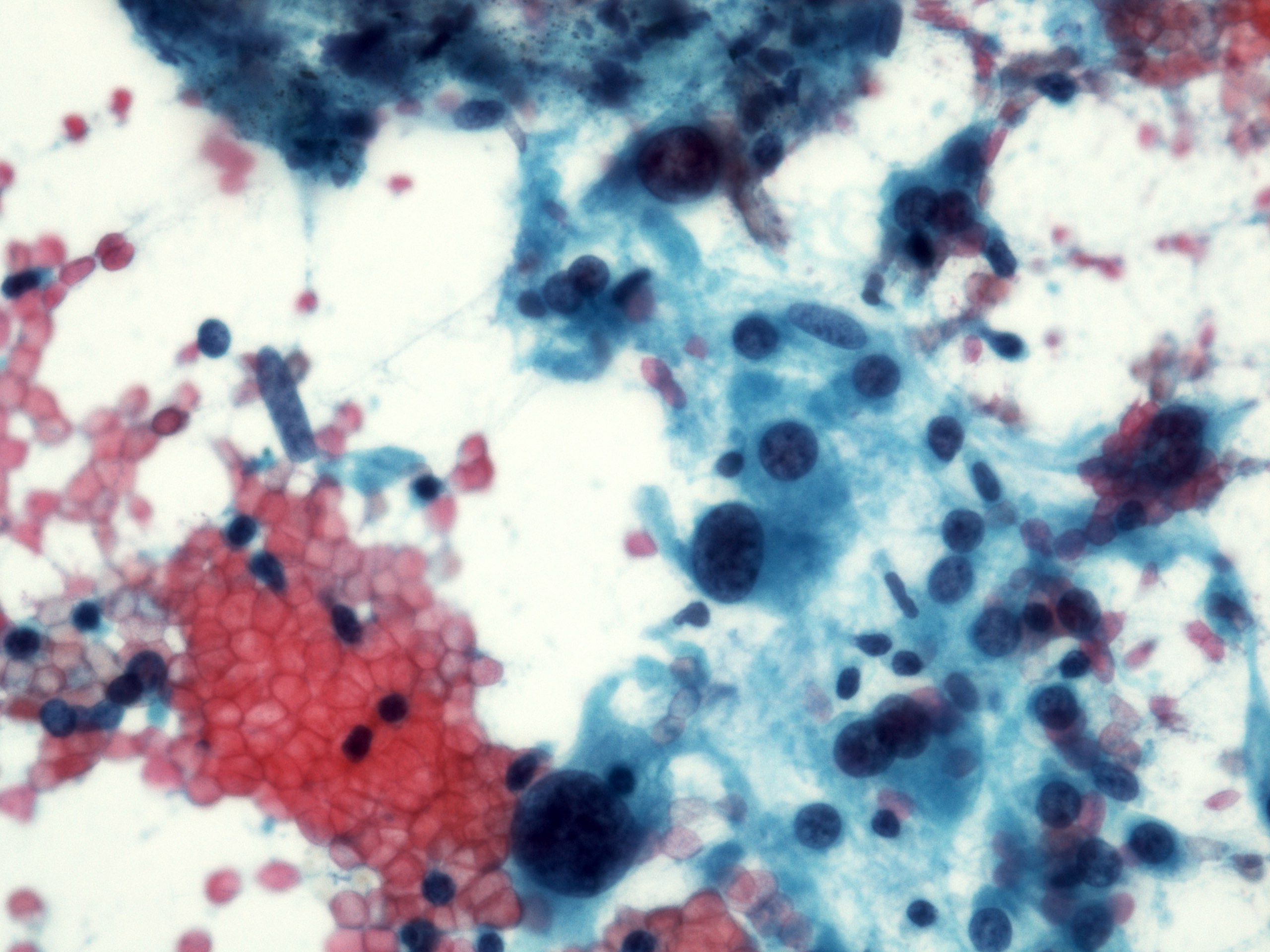
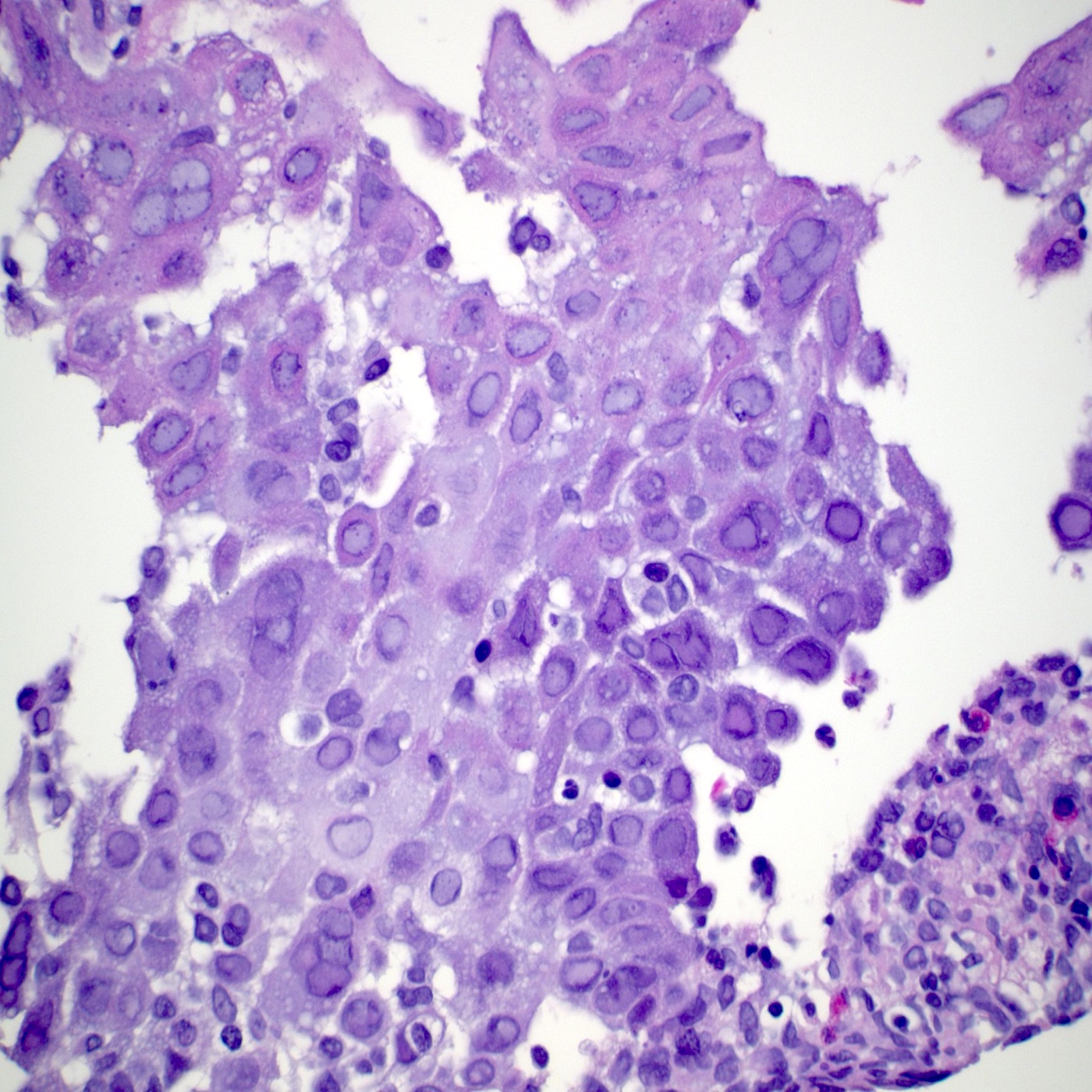
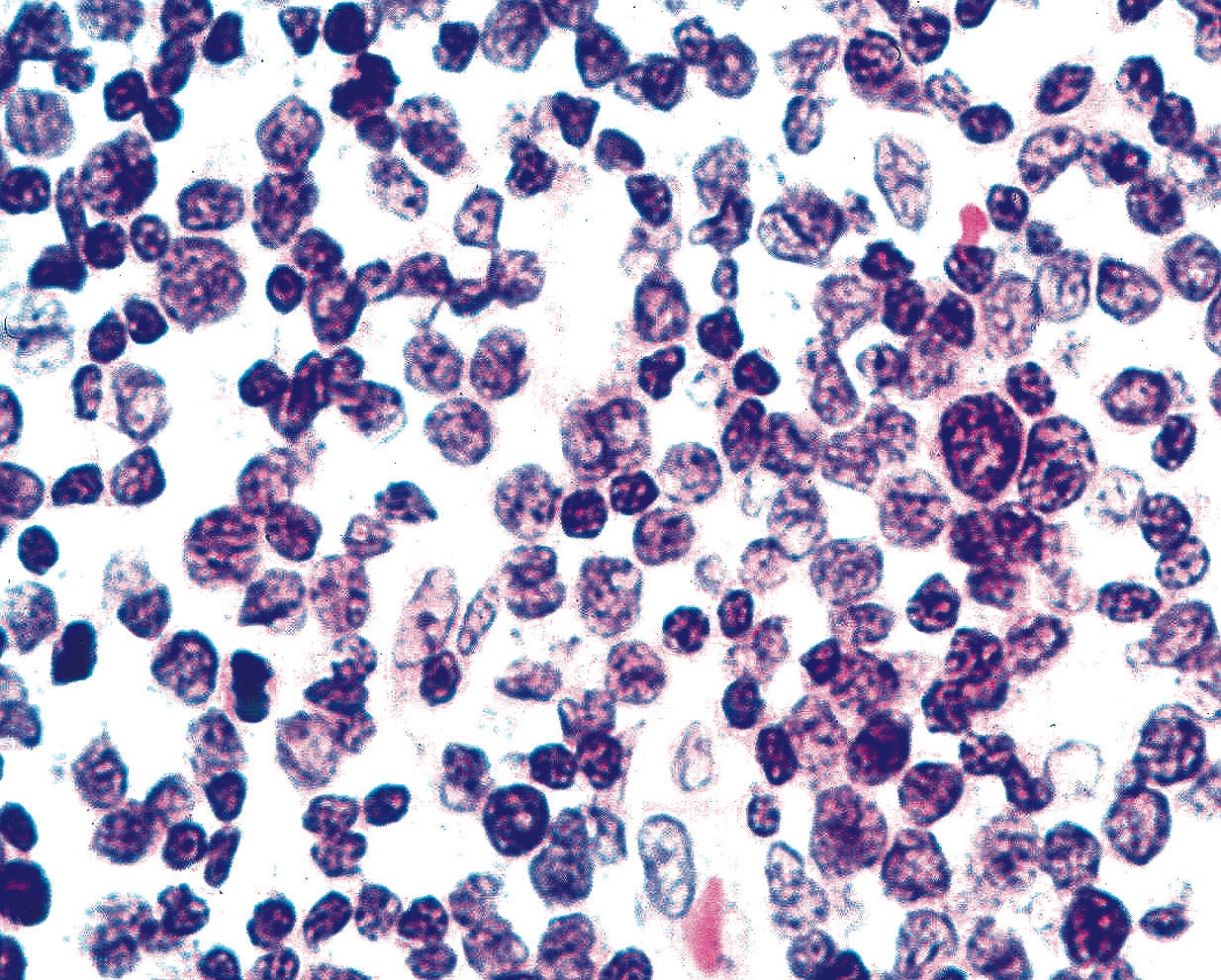
Hypercellular smear
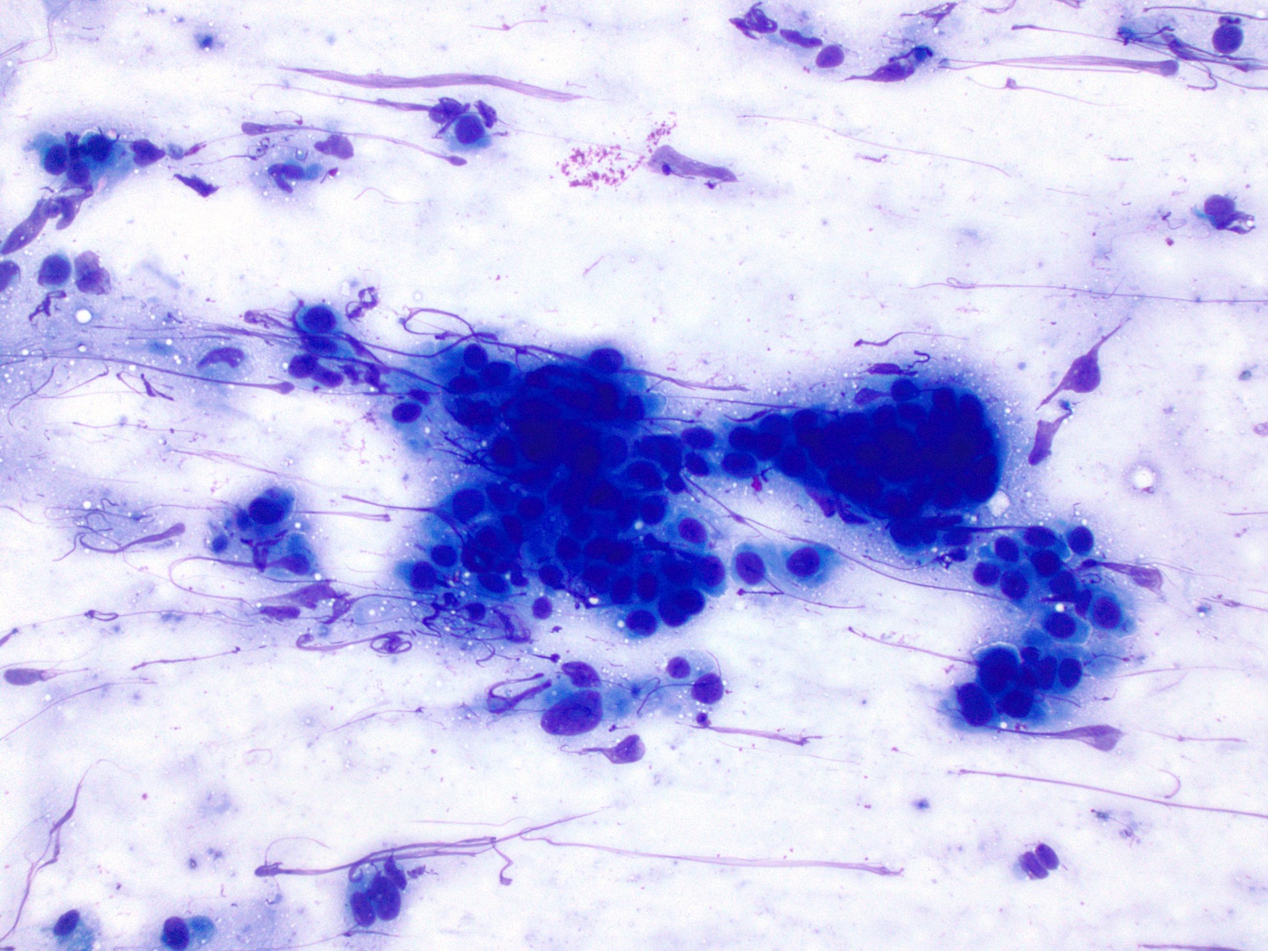
Malignant cells on Diff-Quik
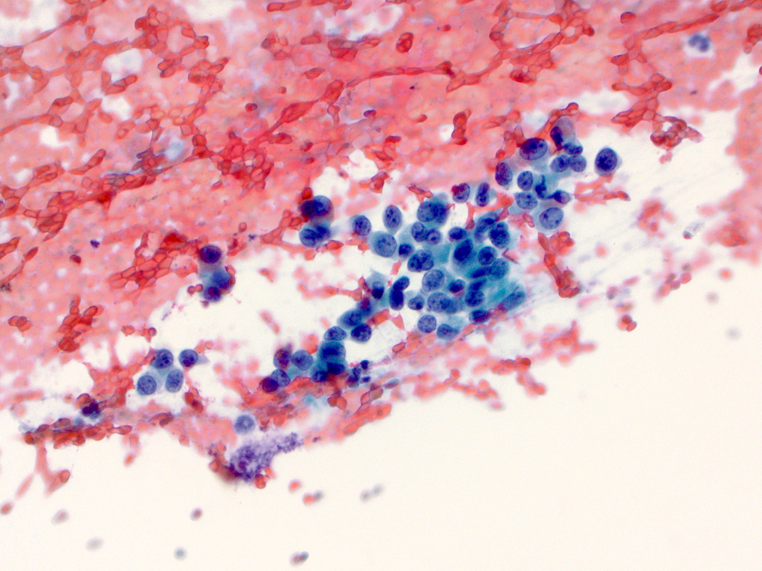
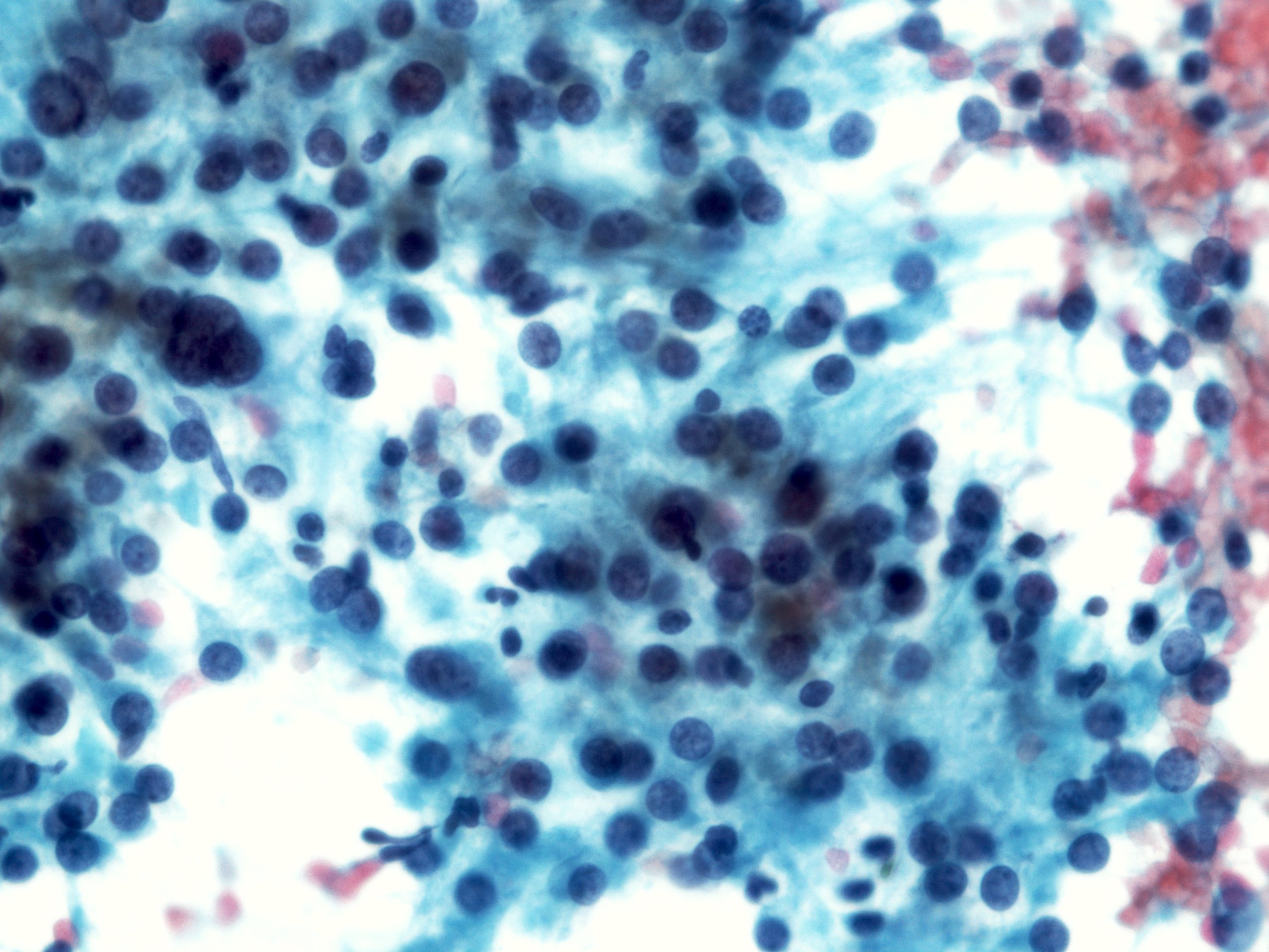
Malignant cells on Pap
Contributed by Dr. Mark R. Wick and @RaulSGonzalezMD on Twitter
 https://bit.ly/3zqGbca #pathology #gipath #PathTwitter #PathOutPic"
Contributed by @RaulSGonzalezMD on Twitter (see original post here)">
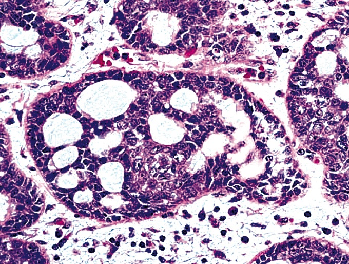
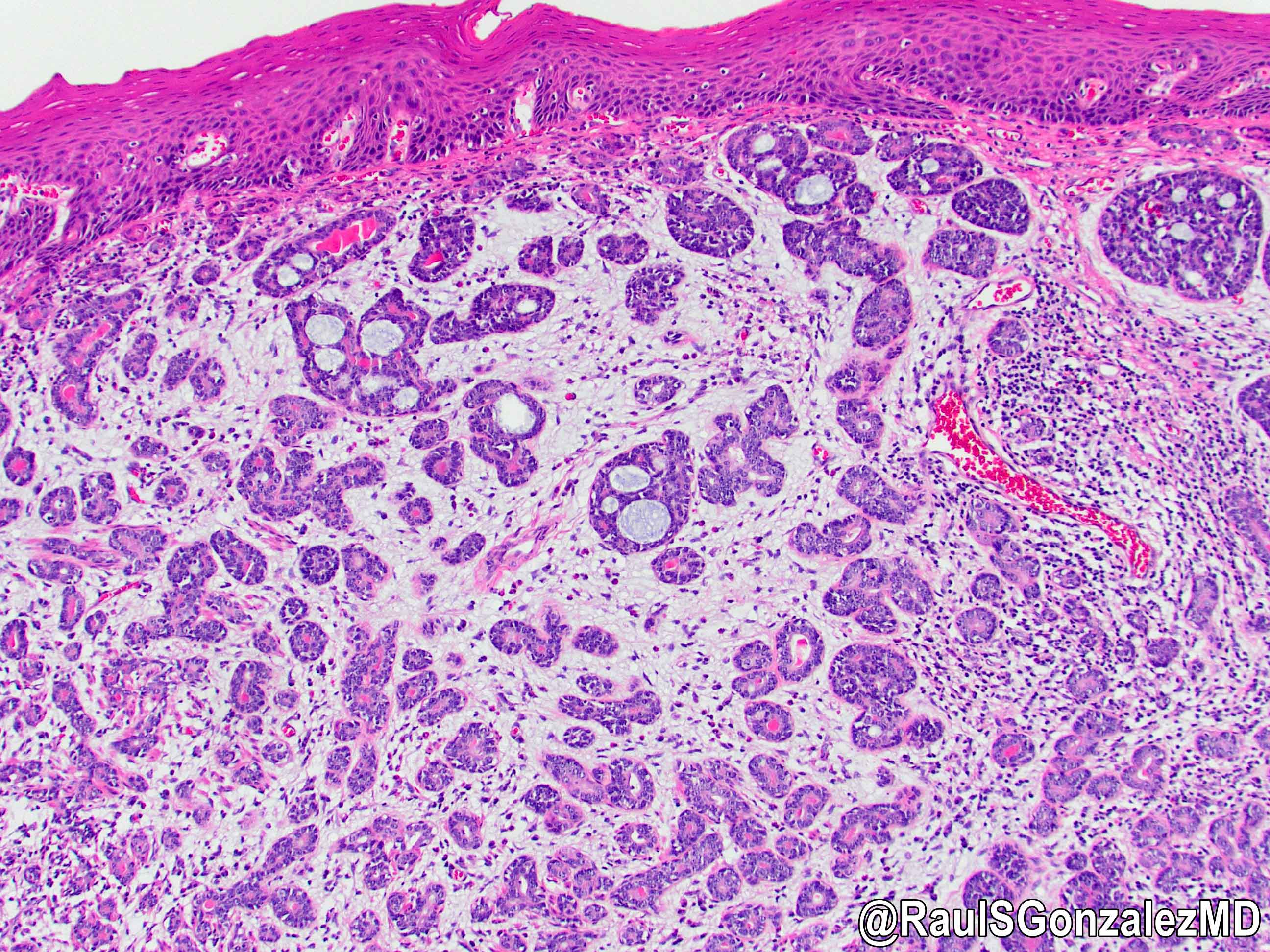
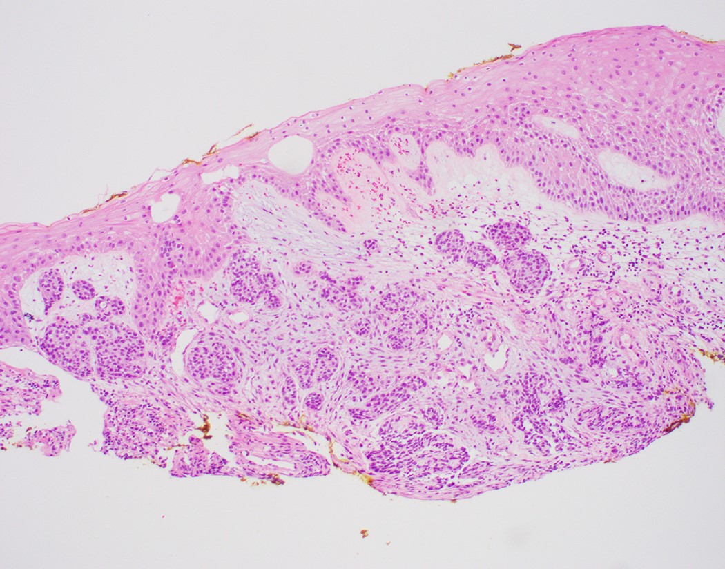
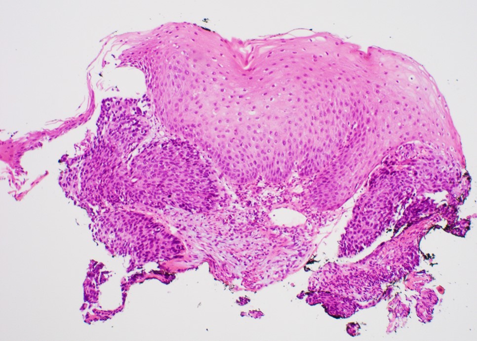
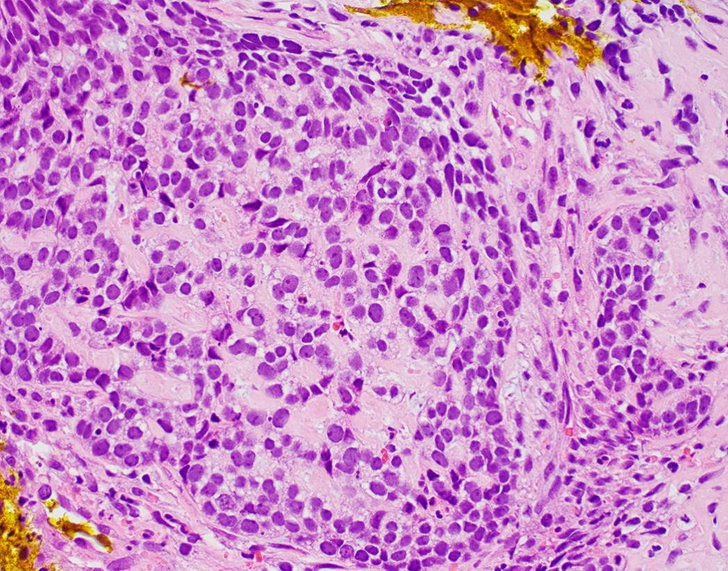
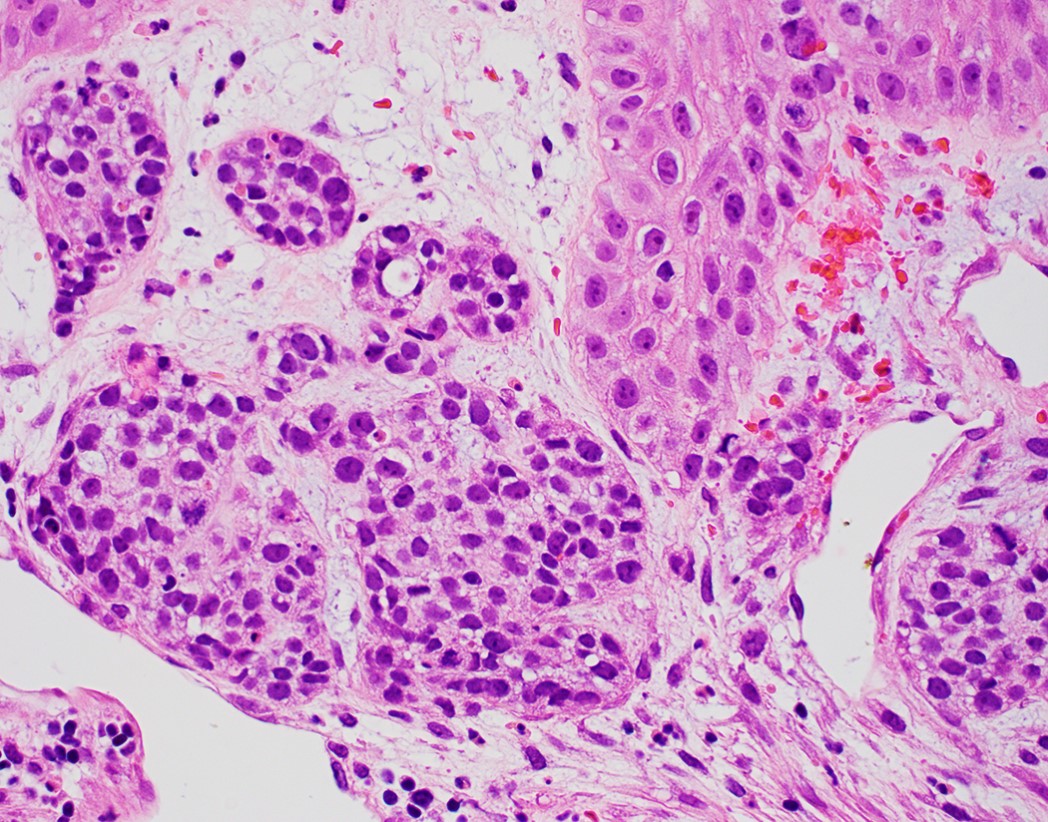
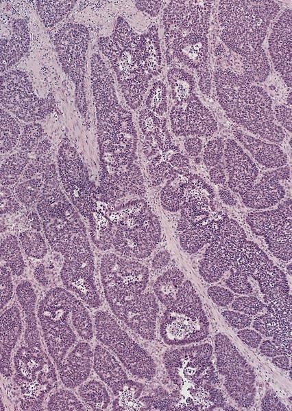
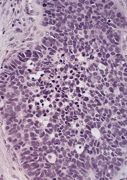
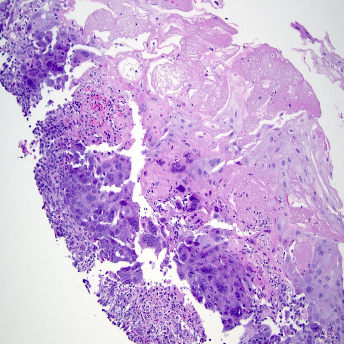
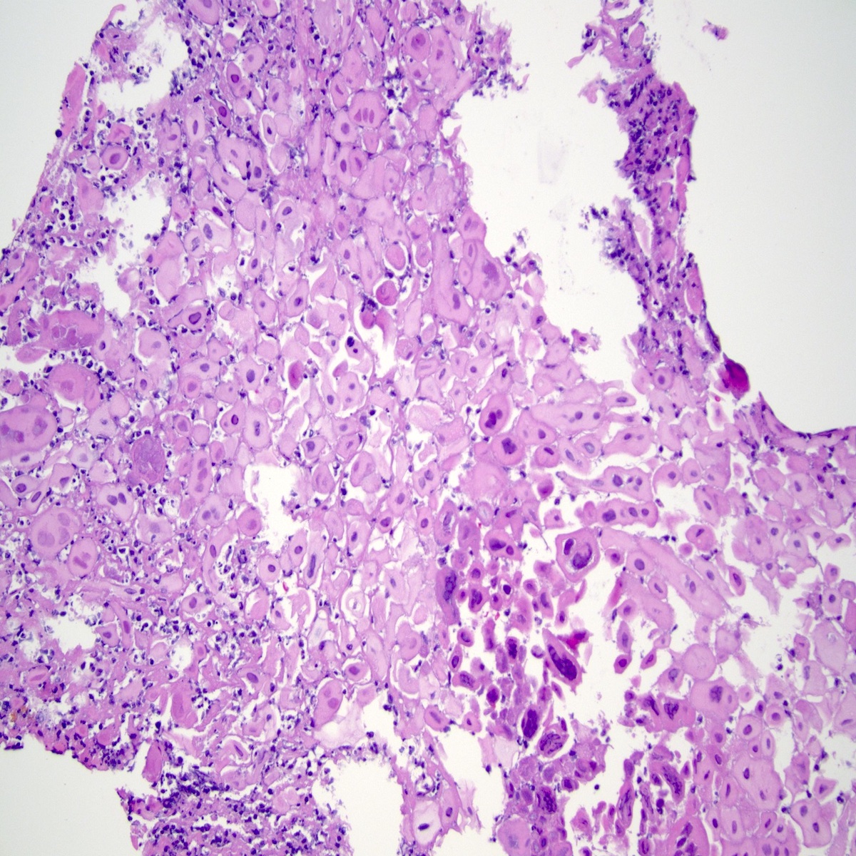
Adenoid cystic carcinoma
Contributed by @RaulSGonzalezMD on Twitter (see original post here)">
 https://bit.ly/3zqGbca #pathology #gipath #PathTwitter #PathOutPic"
Contributed by @RaulSGonzalezMD on Twitter (see original post here)">
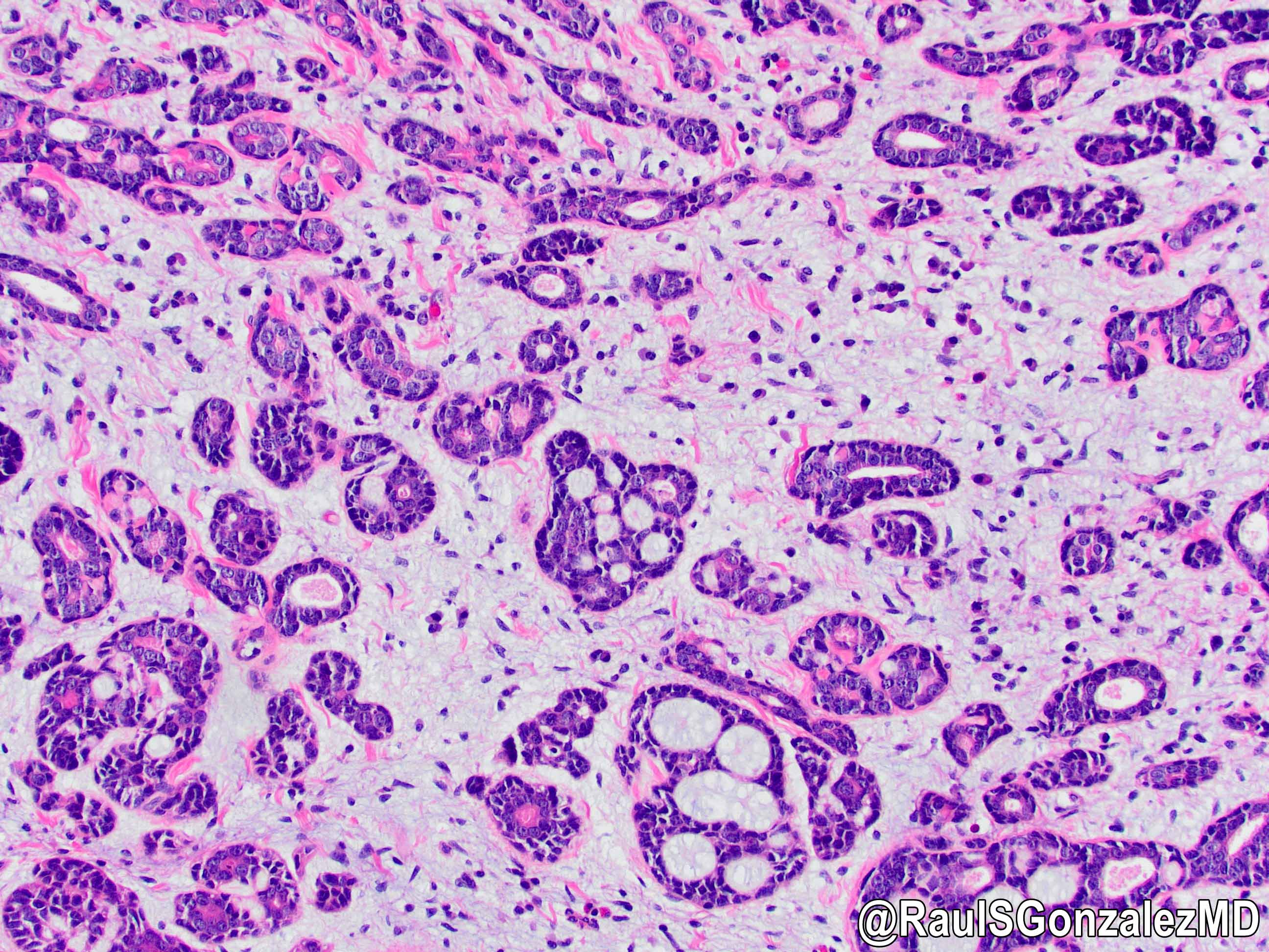
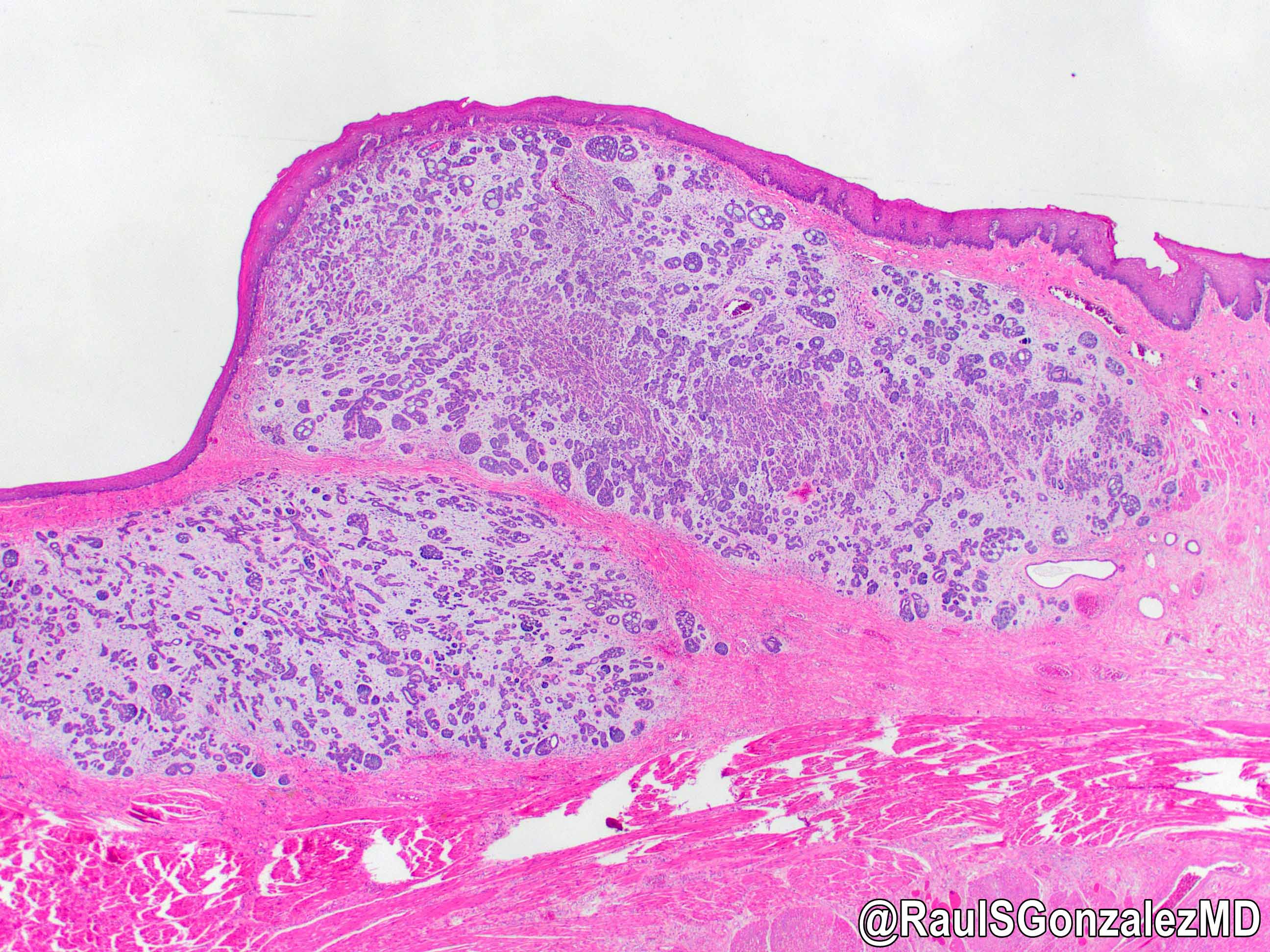
Adenoid cystic carcinoma
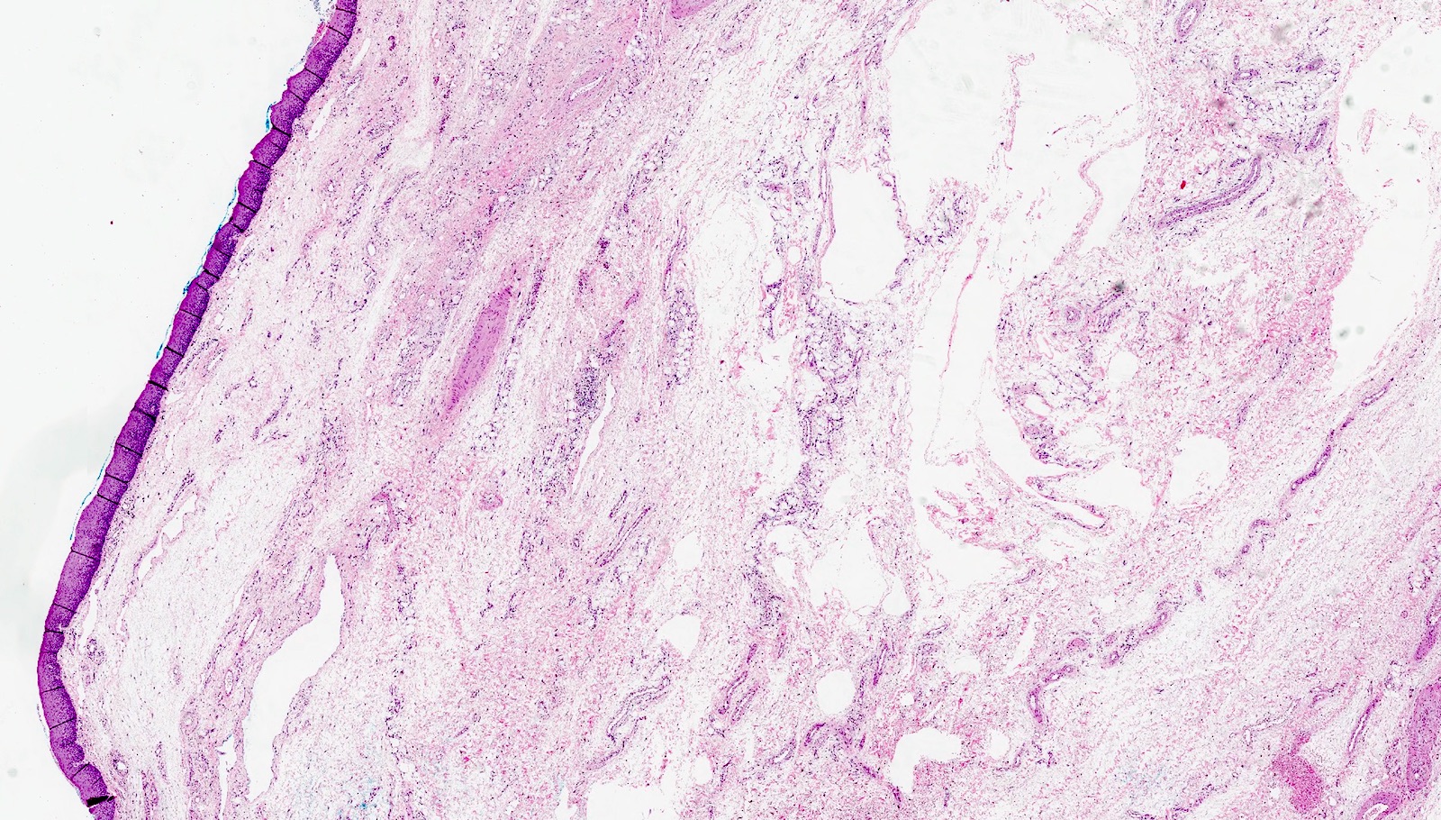
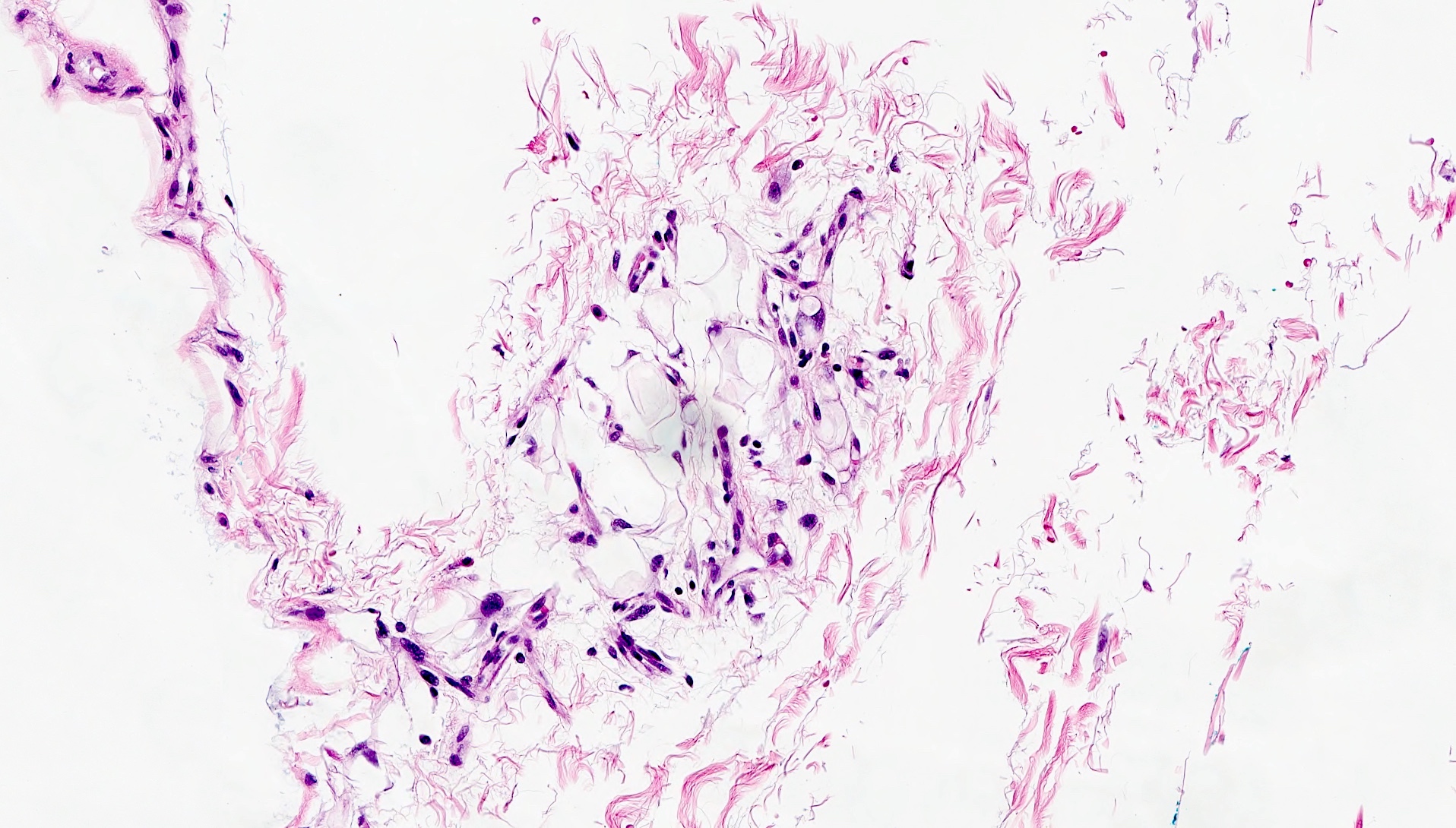
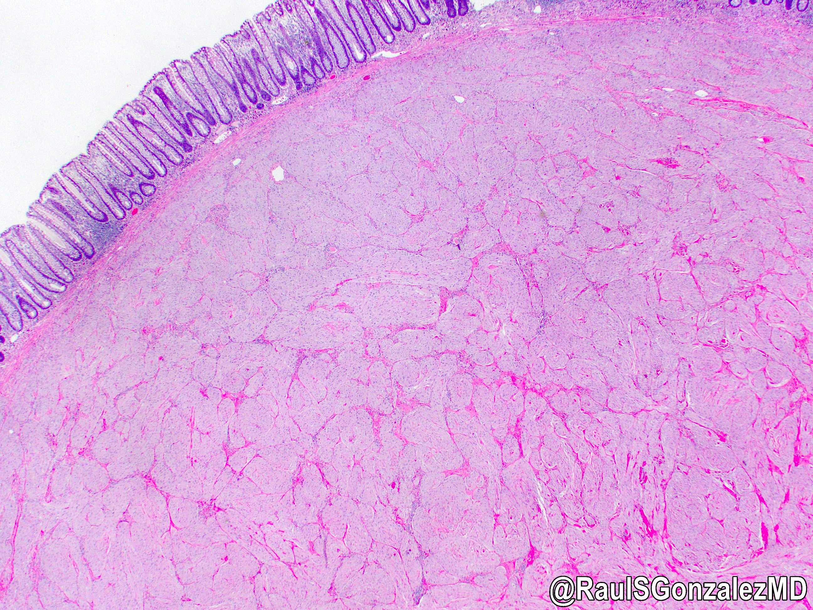
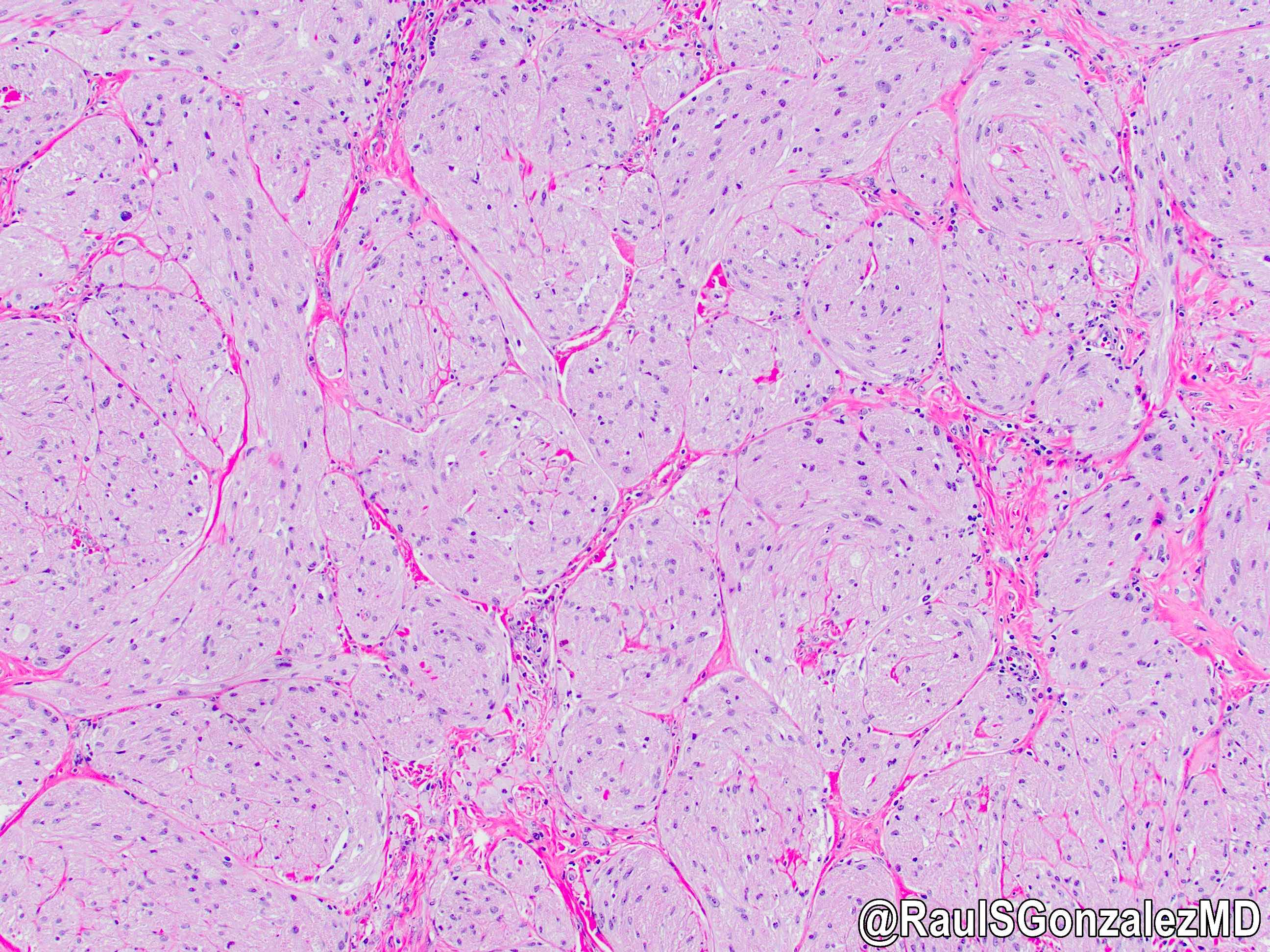
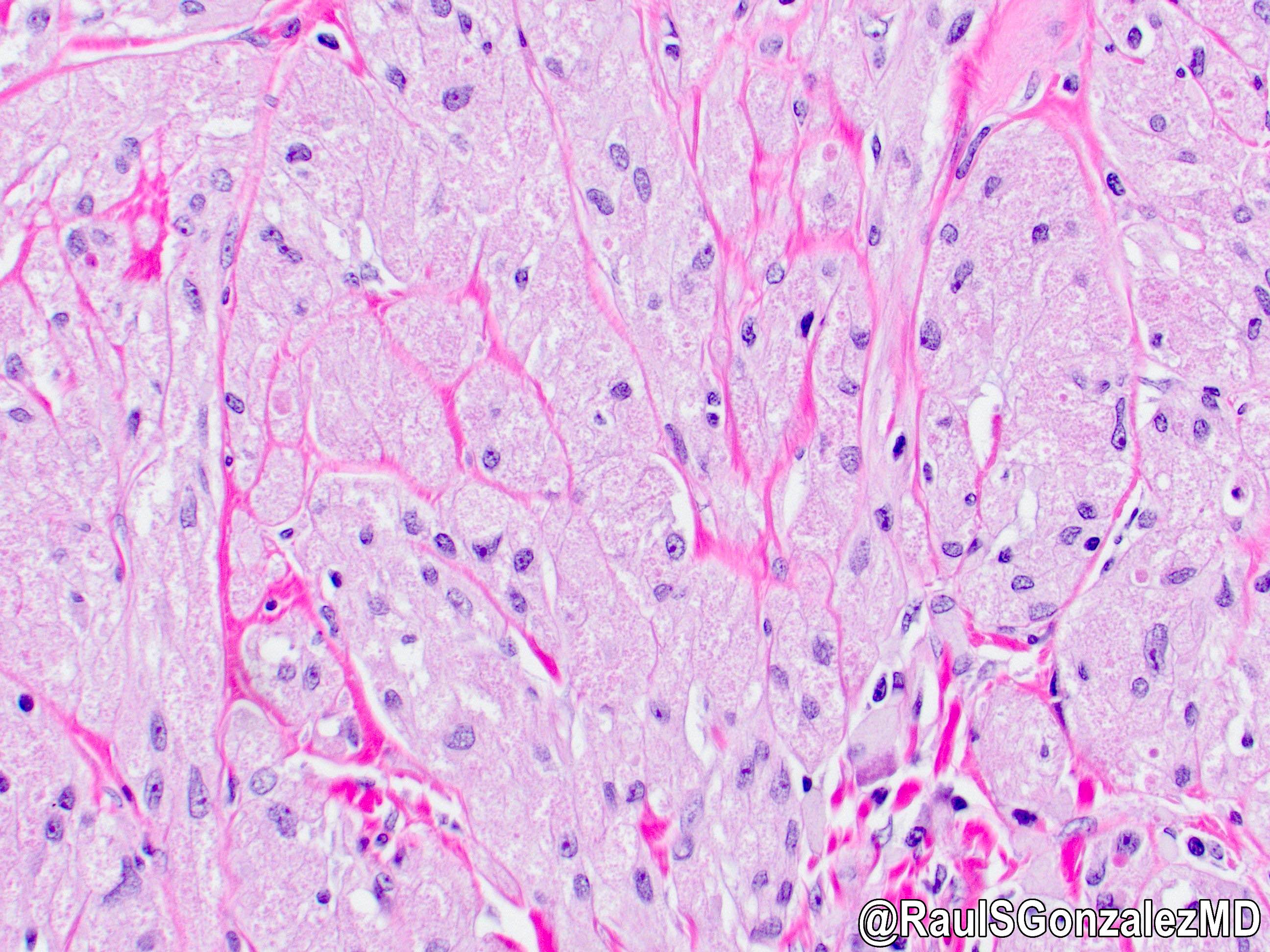
AFIP images
Mucosa and submucosa
Images hosted on other servers:

4 weeks - relation of gut to yolk sac
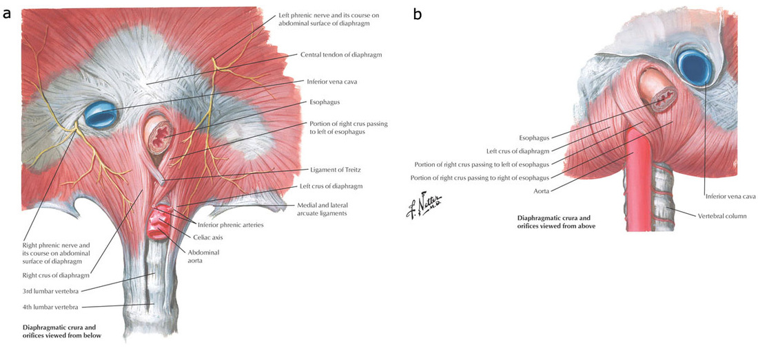
Diaphragmatic crura

Arterial blood supply
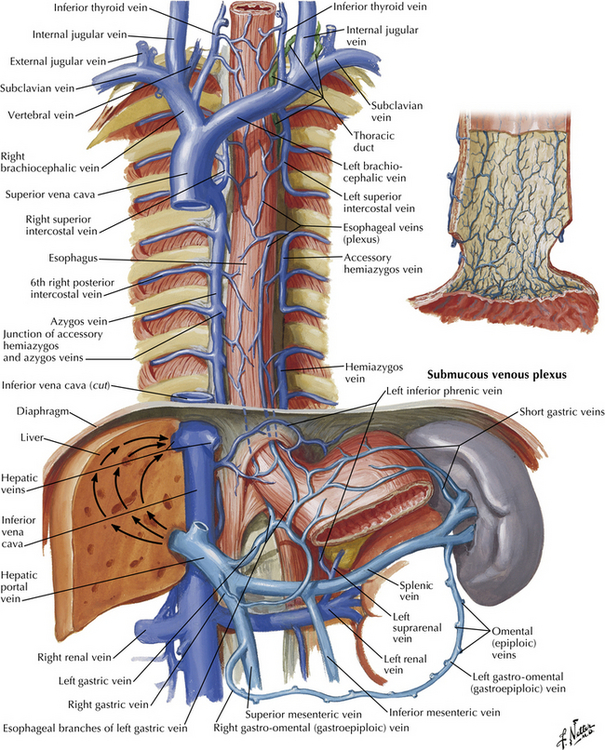
Venous drainage
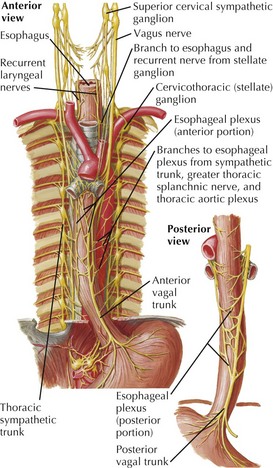
Nerves

Lymphatic drainage

Cervical region
AFIP images

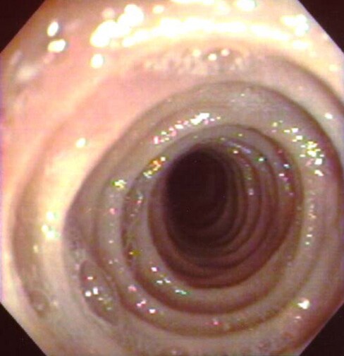
Z line - endoscopic appearance
Images hosted on other servers:

Gastroesophageal junction
Upper left: small intestine
bottom left: esophagus
center: stomach
top right: pancreas; right: liver
Top: spine
lower left: trachea amd esophagus
center: lung, glandular stage
Images hosted on other servers:

BE biopsy protocol

BE Prague classification

Prague criteria
AFIP images

Normal and BE
Images hosted on other servers:

Salmon colored mucosa

Endoscopy: mucosal erythema of lower esophagus
Contributed by Dipti M. Karamchandani, M.D.
BE
Goblet cells
Pseudogoblet cells

Four lines

Hybrid glands
Squamous overgrowth

Baseline atypia
Muscularis mucosae duplication
Images hosted on other servers:

Nodule on endoscopy

Endoscopic mucosal resection

Narrow band imaging

Endoscopic treatment of Barrett high grade dysplasia

Wide area transepithelial sampling
Images hosted on other servers:

High grade dysplasia
Contributed by Dipti M. Karamchandani, M.D.

Indefinite for dysplasia
Indefinite for dysplasia with surface erosion
Low grade dysplasia
Low grade dysplasia higher power
Low grade dysplasia demarcation
High grade dysplasia
High grade dysplasia higher power

High grade dysplasia intraluminal necrotic debris

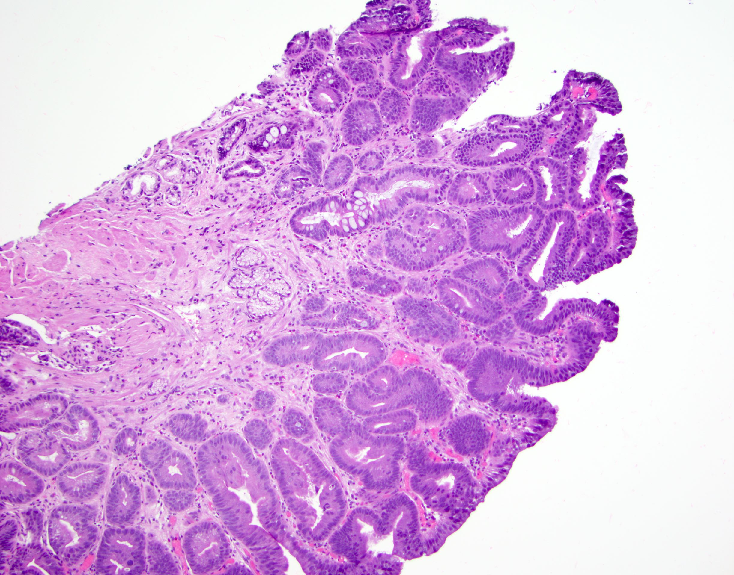
Gastric foveolar type dysplasia
Gastric foveolar type dysplasia, higher power
Update on recently developed quality metrics
Contributed by Jinping Lai, M.D., Ph.D.
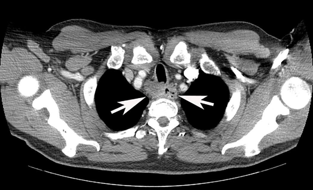
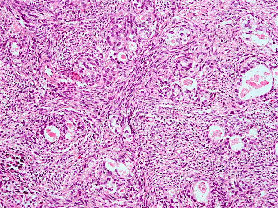
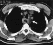
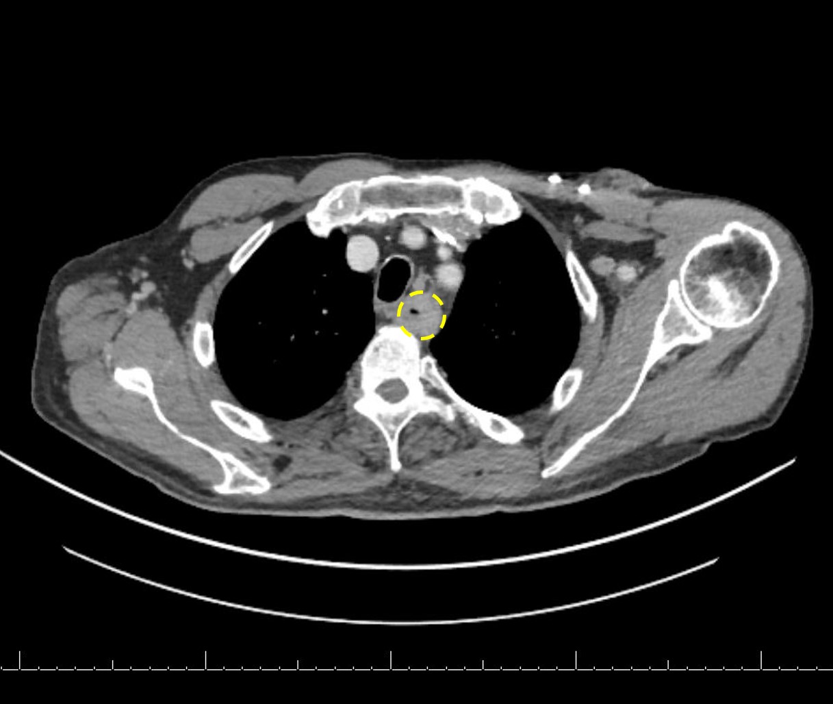
CT scan
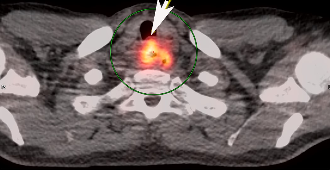
PET / CT
Contributed by Jinping Lai, M.D., Ph.D.

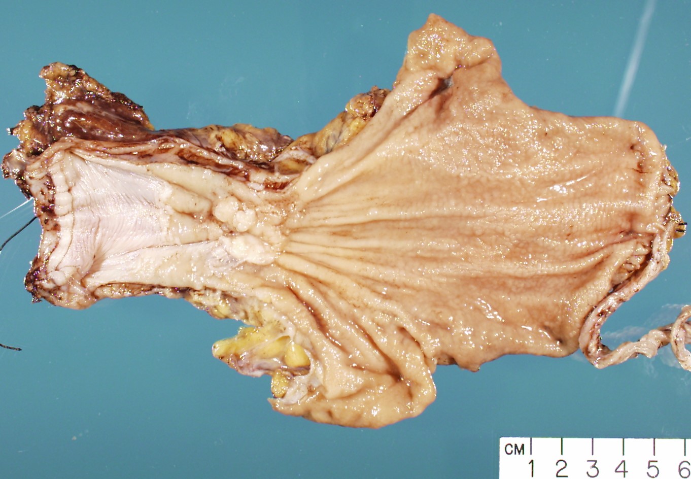
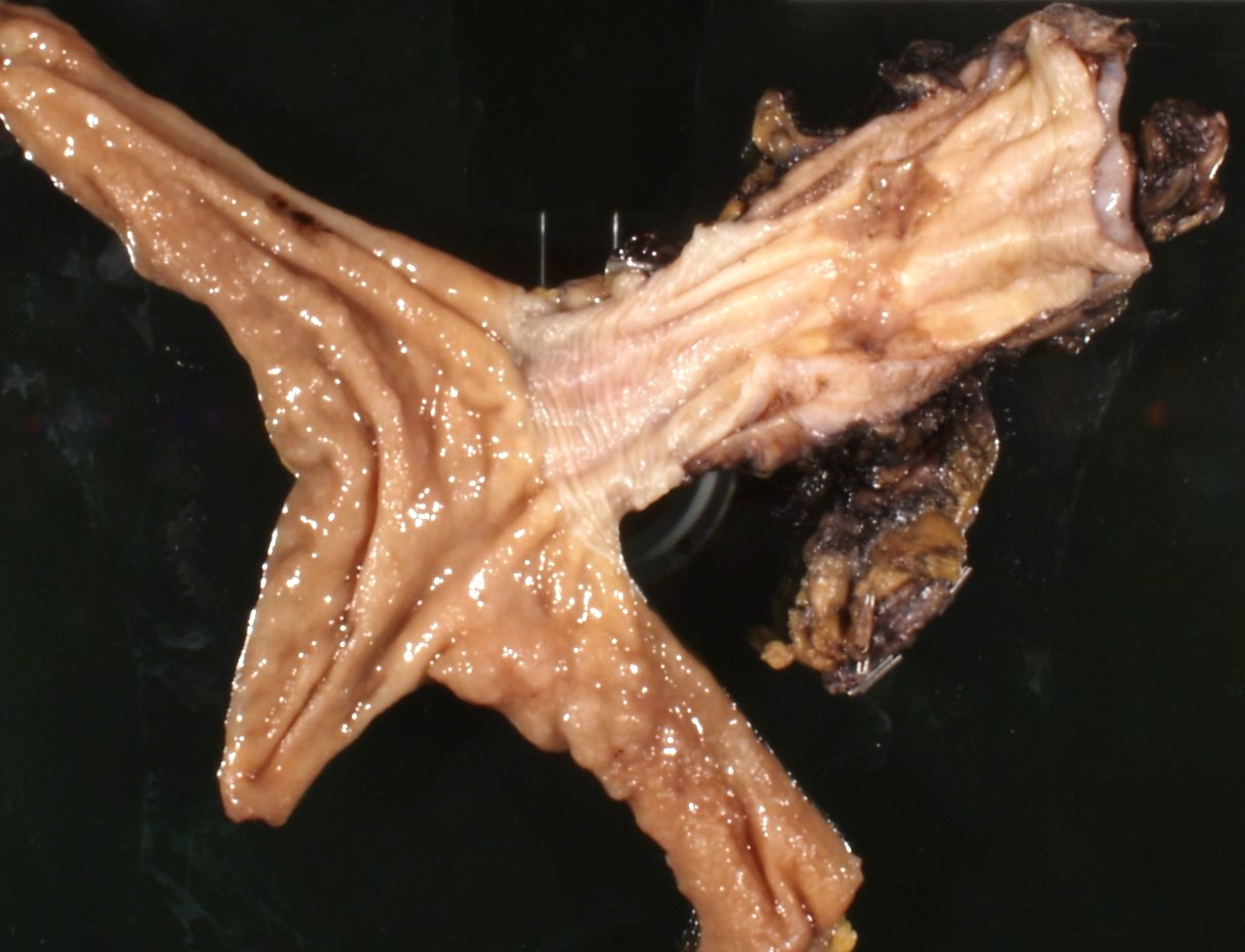
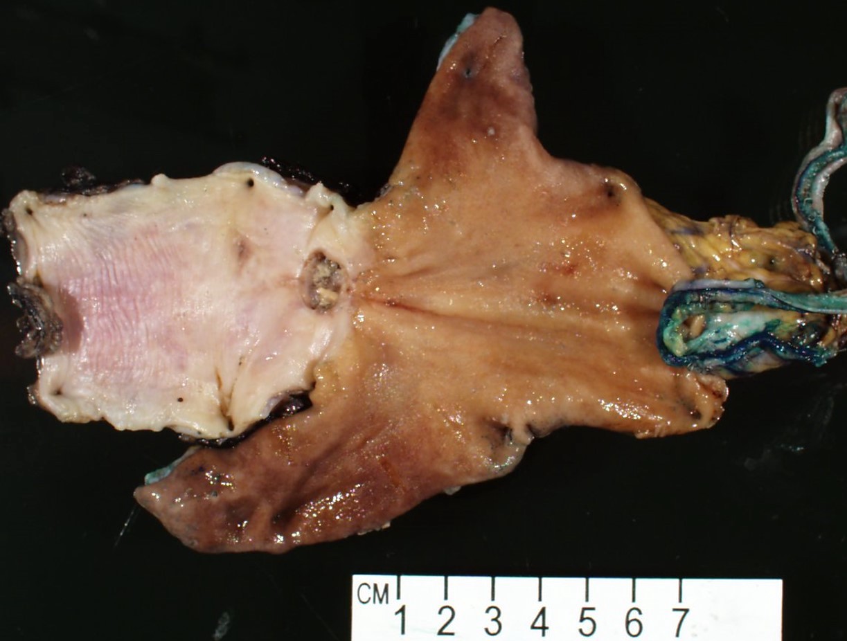
Subepithelial mass
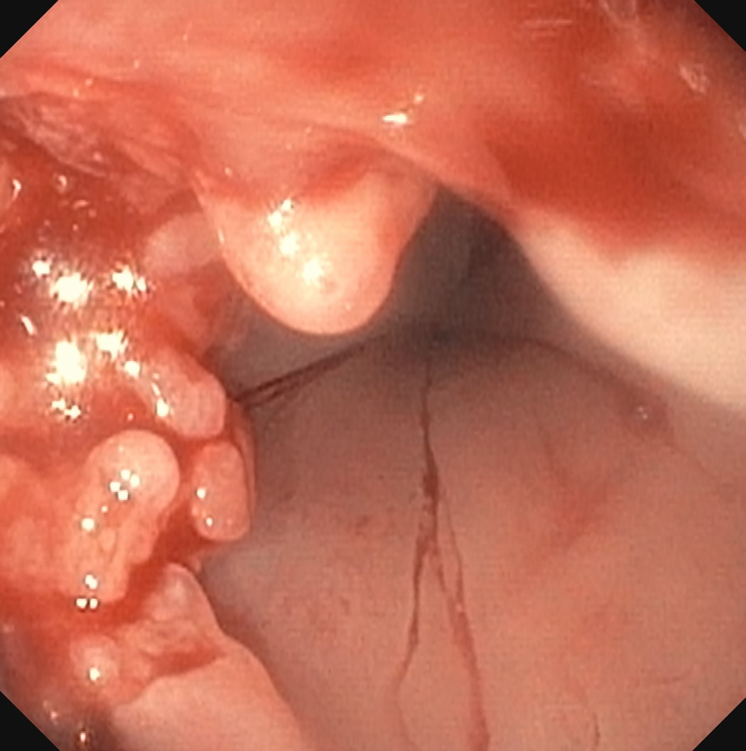
Fungating masses
Images hosted on other servers:

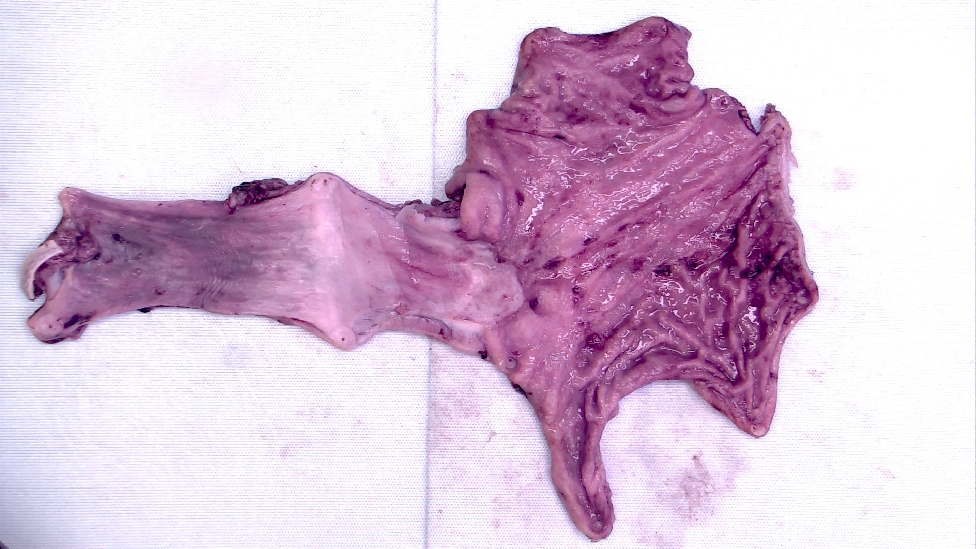
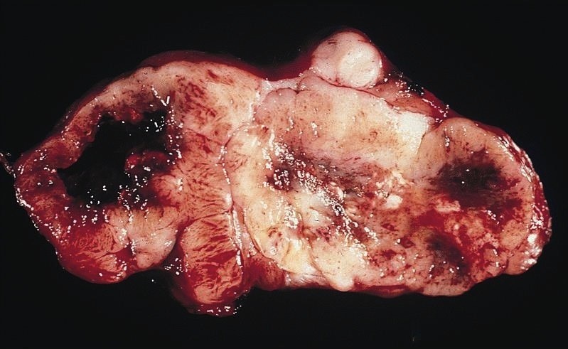
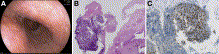
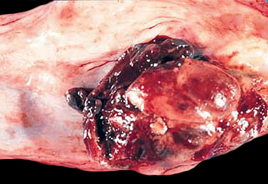
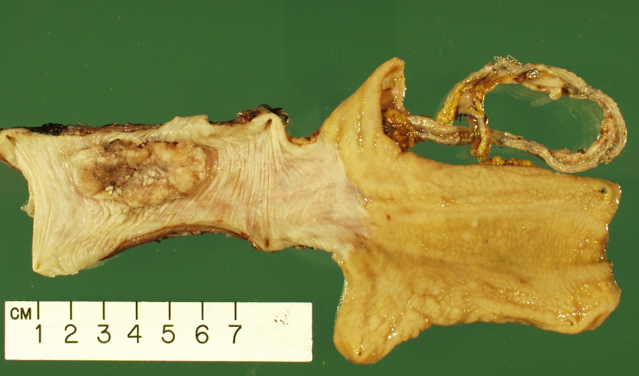
Mid esophageal mass gross examination
Contributed by Jinping Lai, M.D., Ph.D. and AFIP images
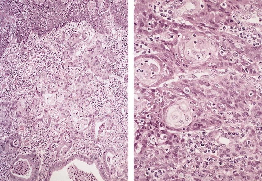
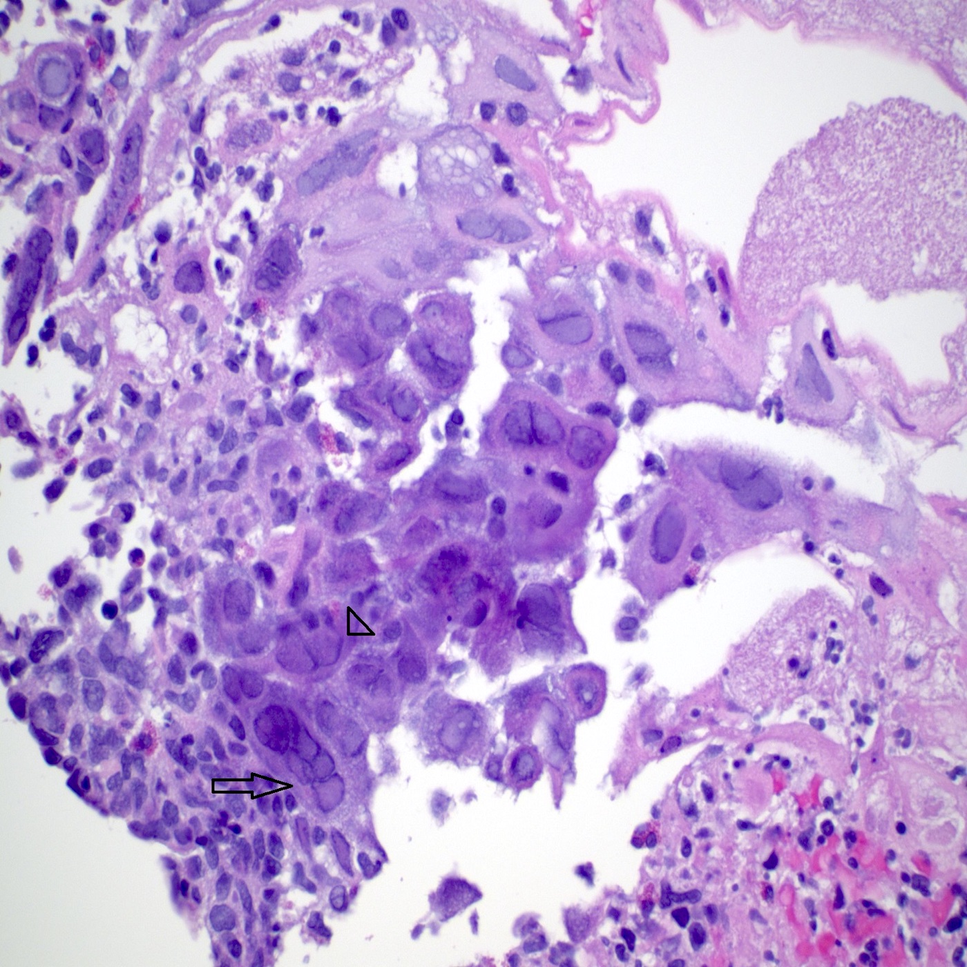
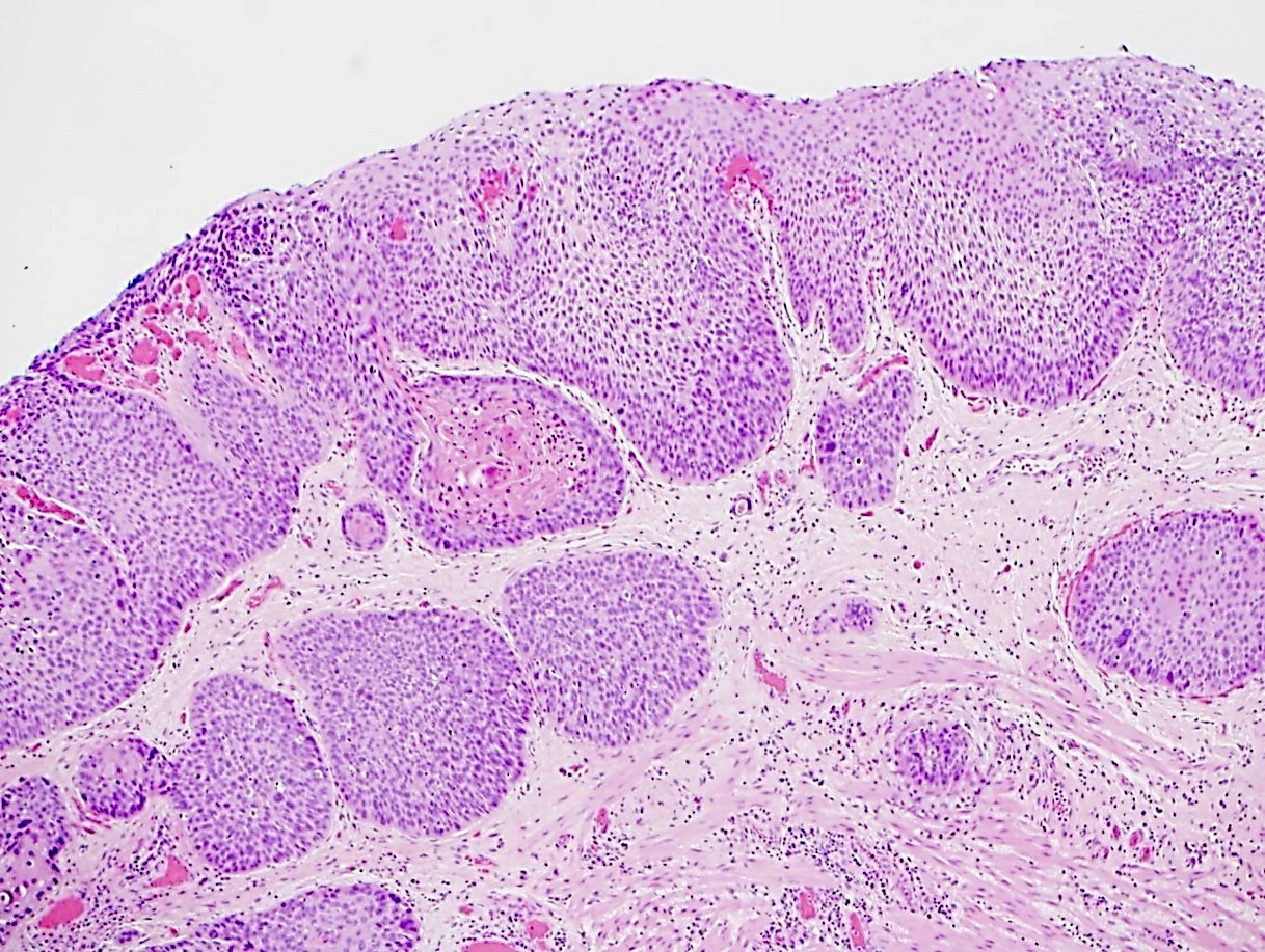
Nested arrangements
Tumor cells touch the epithelium
Small blue tumor cells
Mitosis and apoptosis
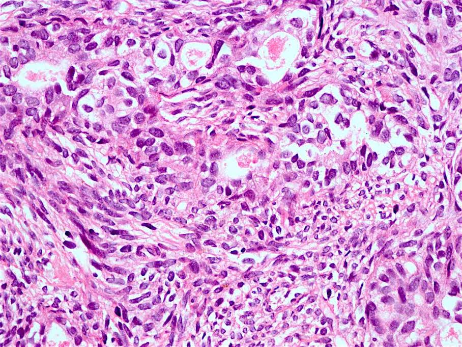
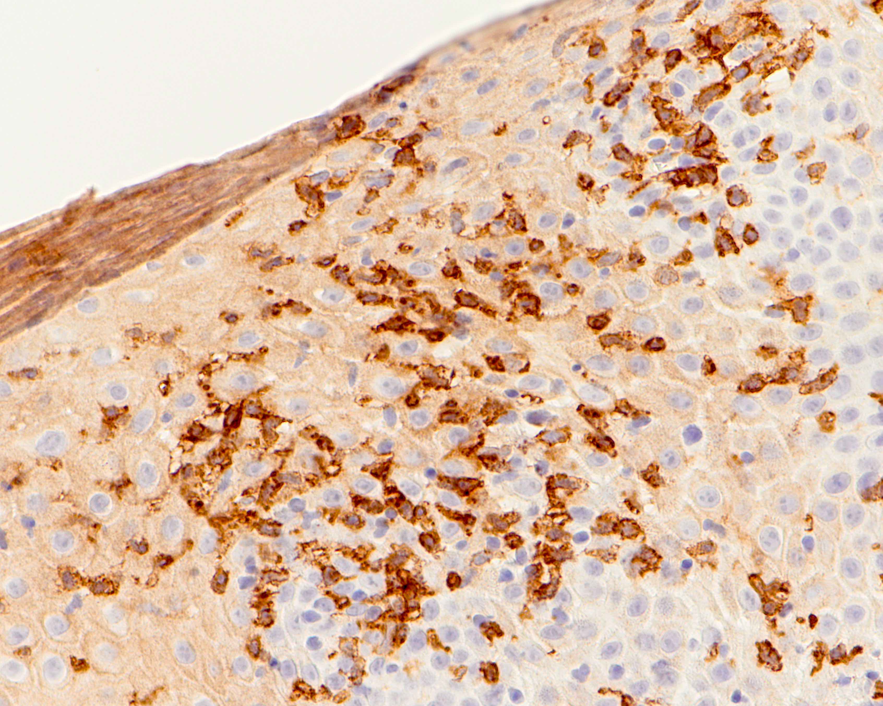
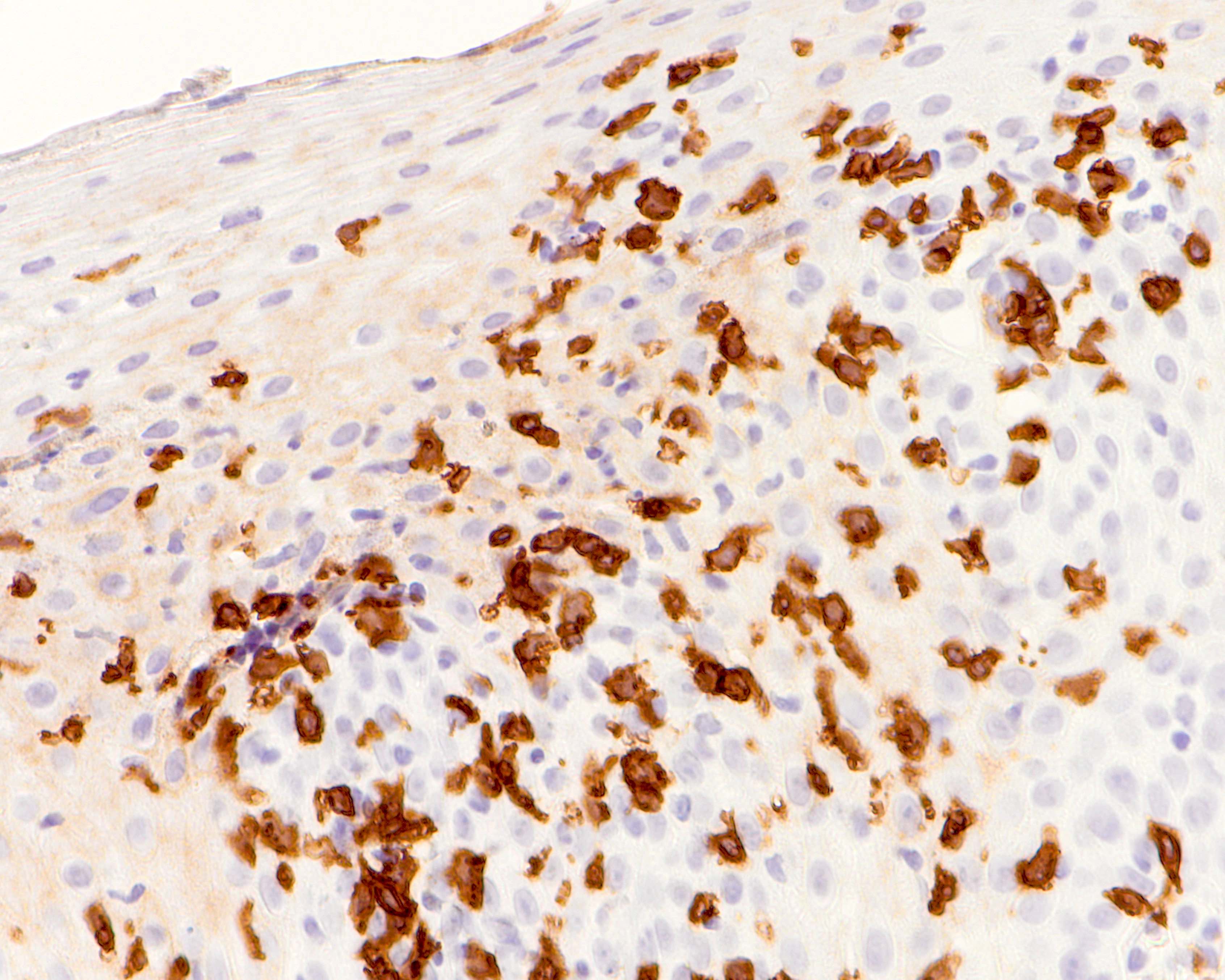
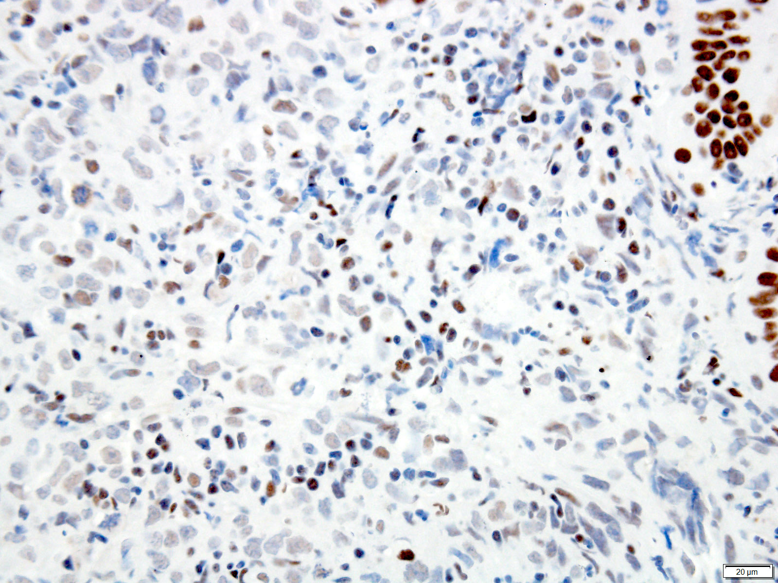
p16
p40
p63
p53
CDX2
Synaptophysin
Chromogranin
CK7
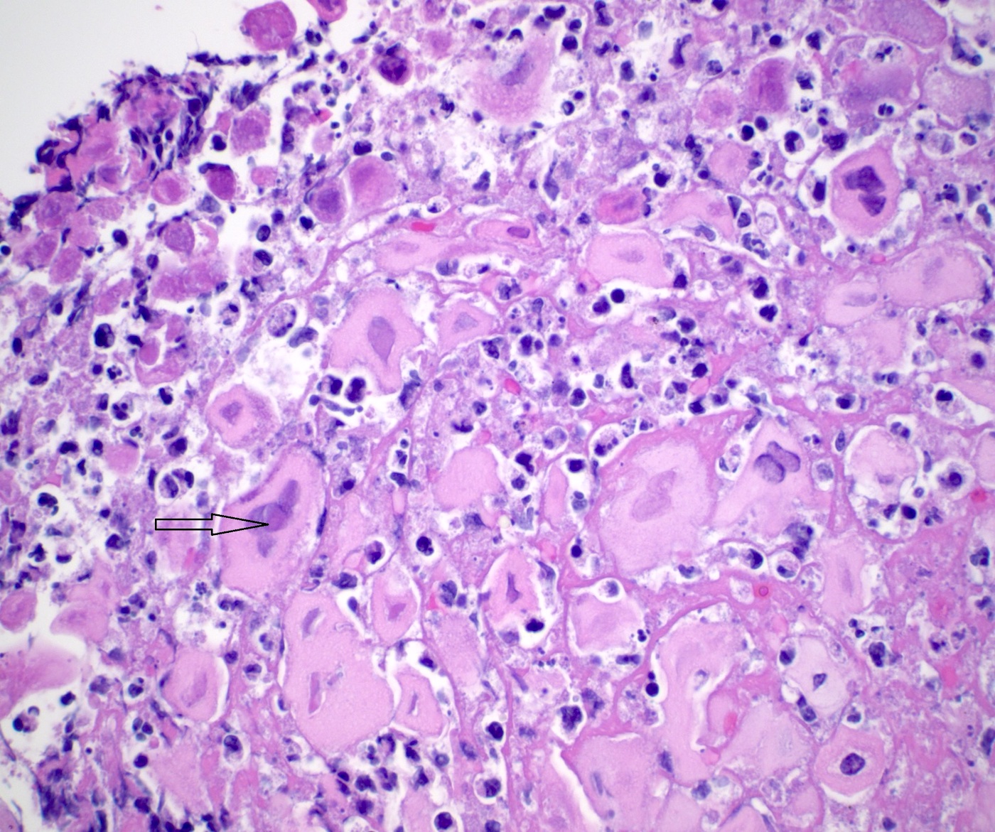
Small dark tumor cells
Peripheral basal type cells
Focus of squamous differentiation
Strands of tumor cells touch epithelium
Images hosted on other servers:

Longitudinally oriented plaque-like lesions
Contributed by Divya Salibindla, M.D.
Yellow-white mucosal plaques
Images hosted on other servers:

Focal erosion
Tan-yellow
plaques with
mucosal erythema
Contributed by Divya Sharma, M.D., Andrey Bychkov, M.D., Ph.D. and Jijgee Munkhdelger, M.D., Ph.D.
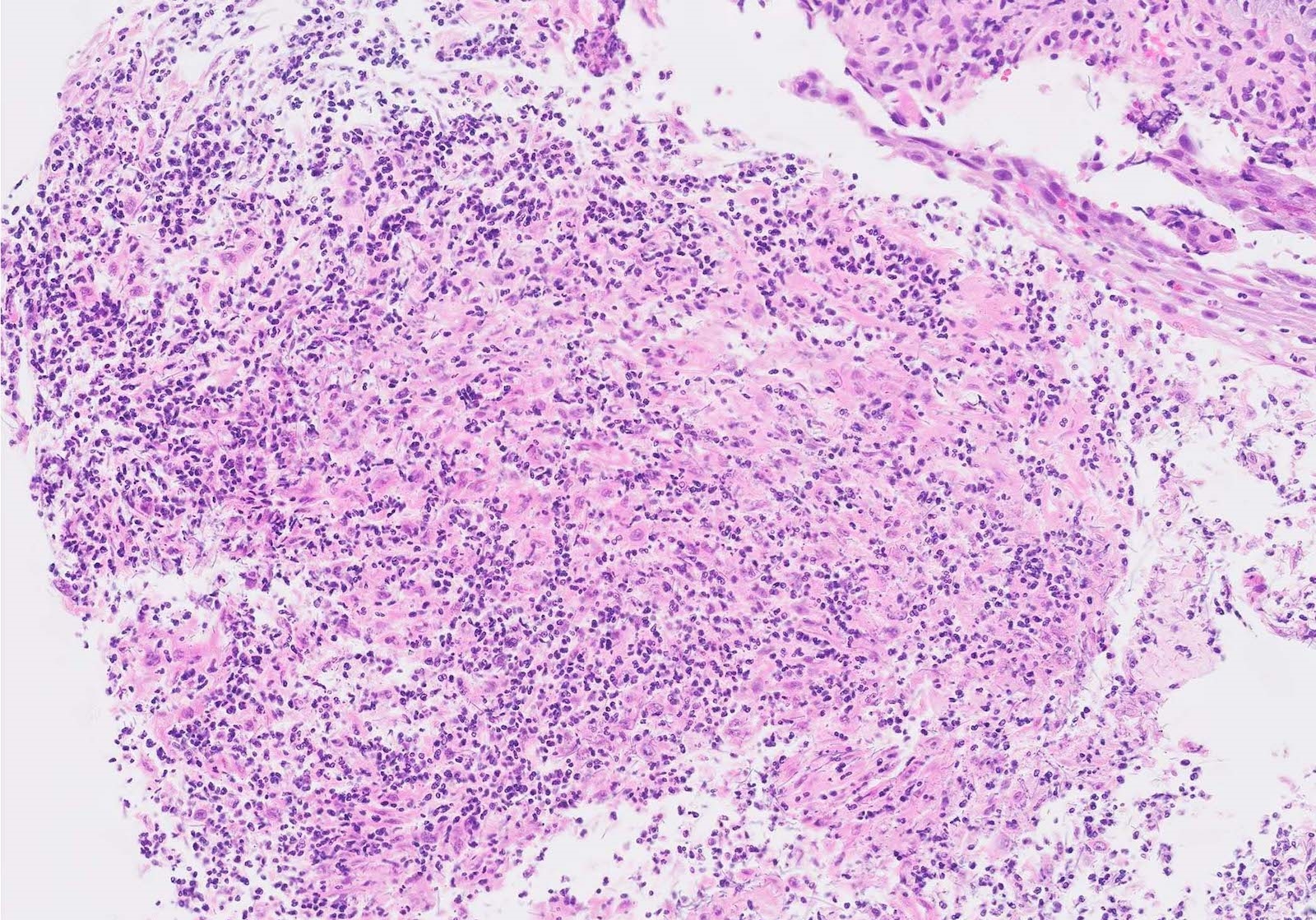
Ulcerative esophagitis
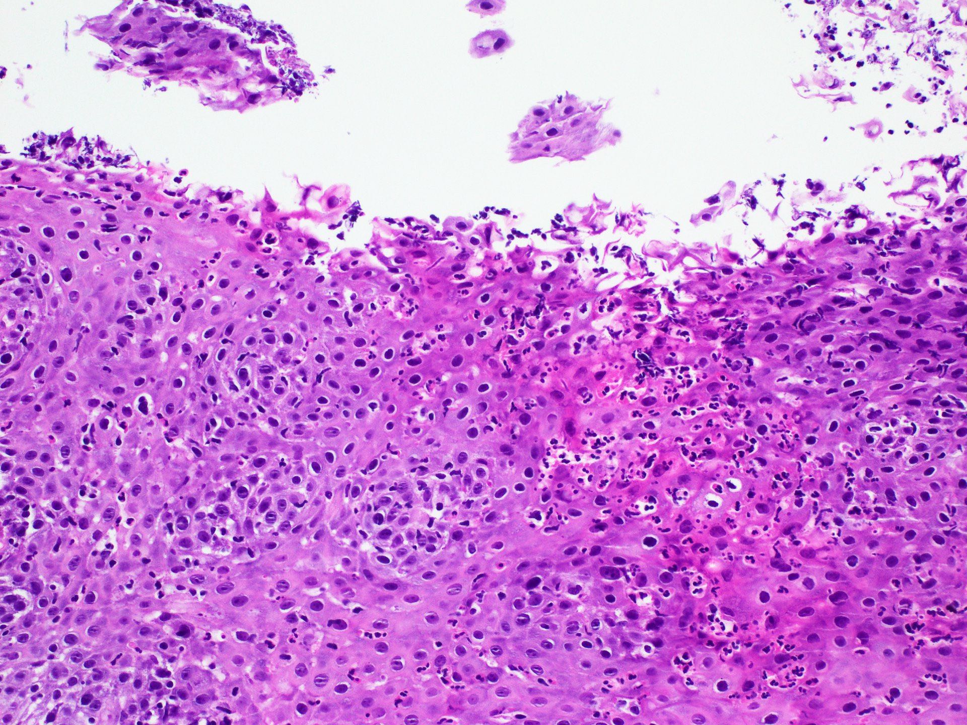
Prominent acute inflammatory infiltrate
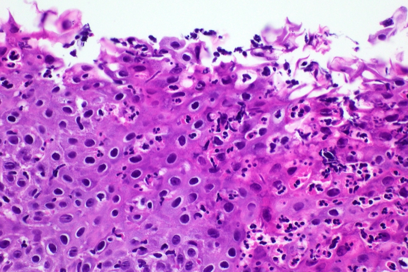
Esophageal biopsy
Acute inflammation
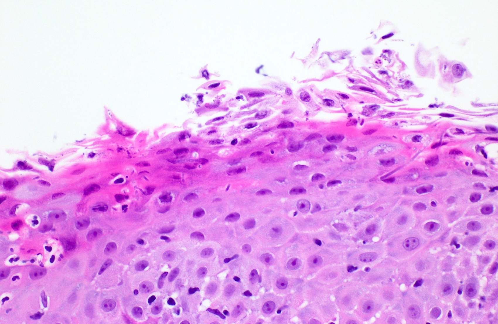
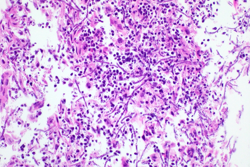
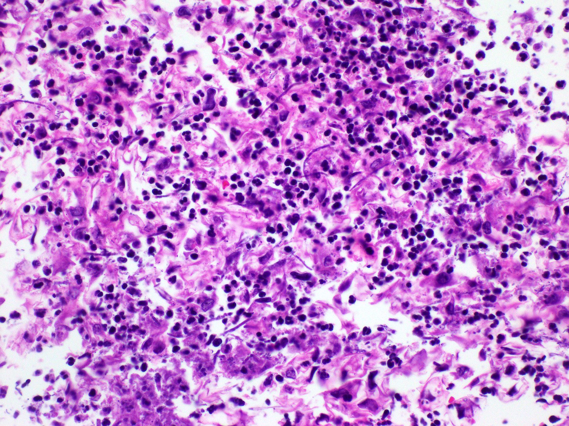
Pseudohyphae within the epithelium
Candida inflammatory exudate
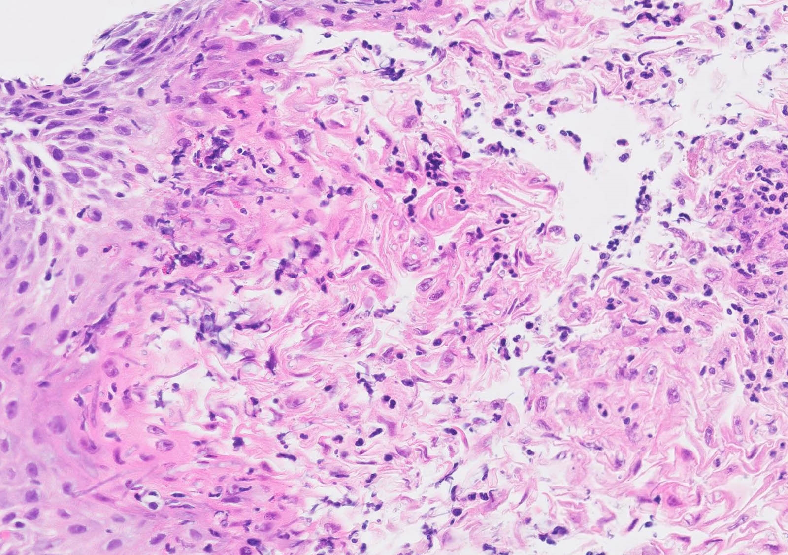
Yeast forms and pseudohyphae
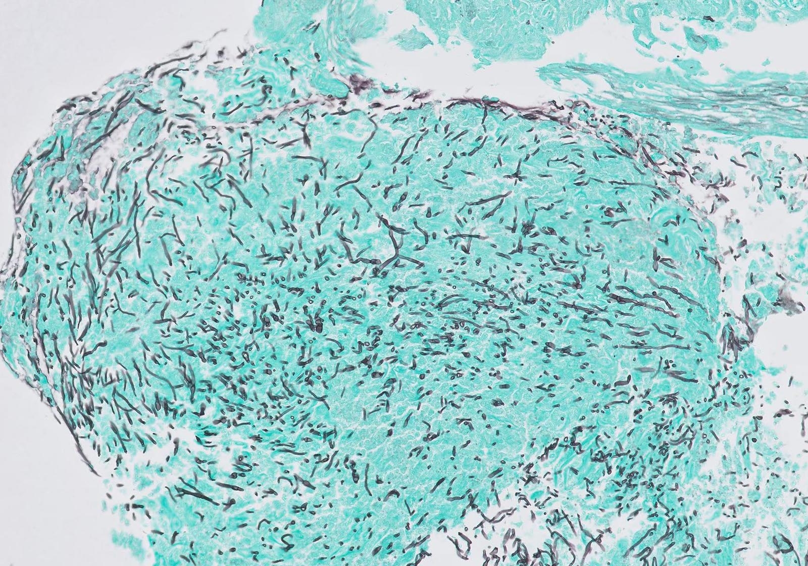
Fungi on GMS

GMS positive fungi
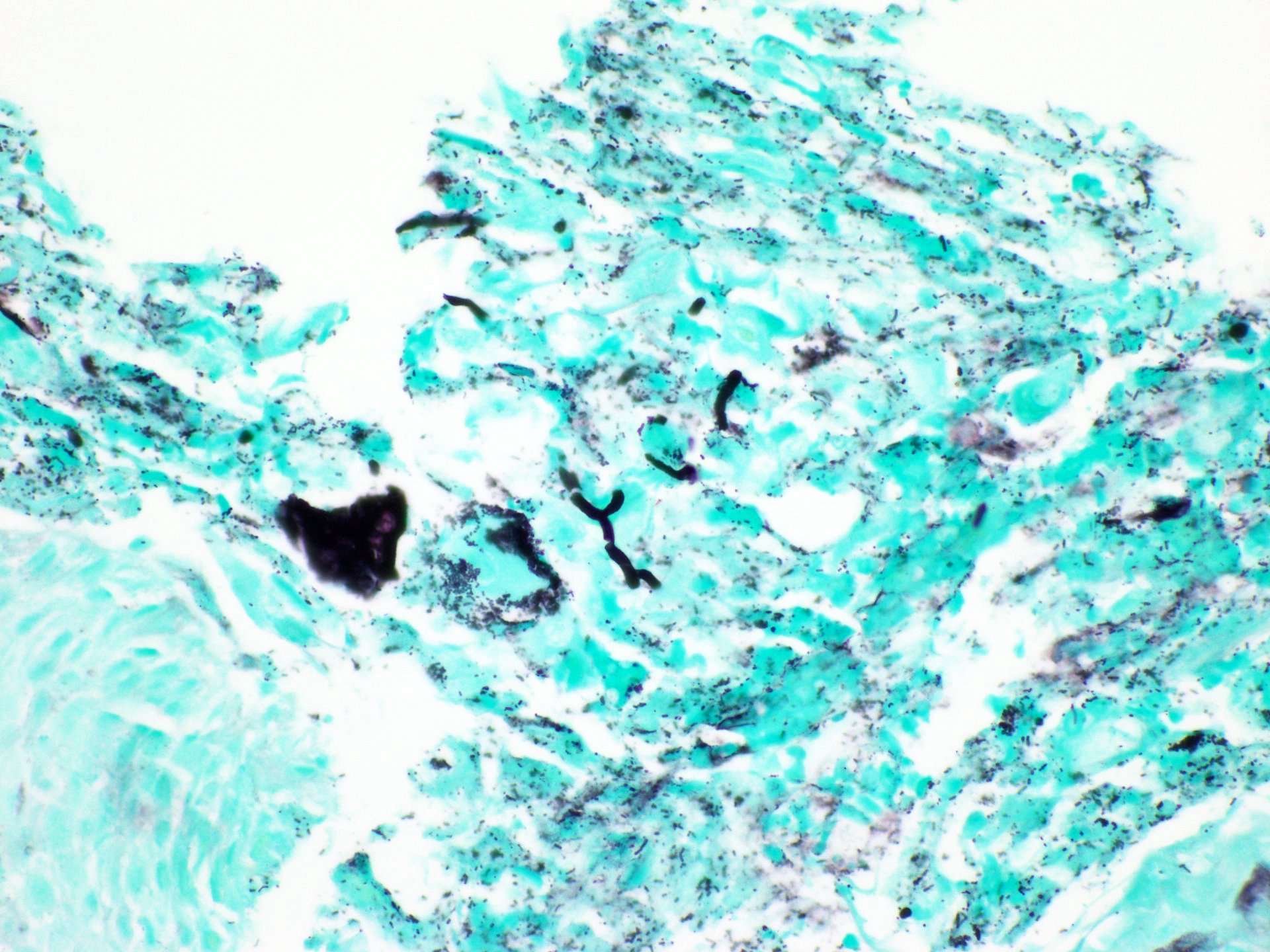
GMS stain
Contributed by Divya Salibindla, M.D.
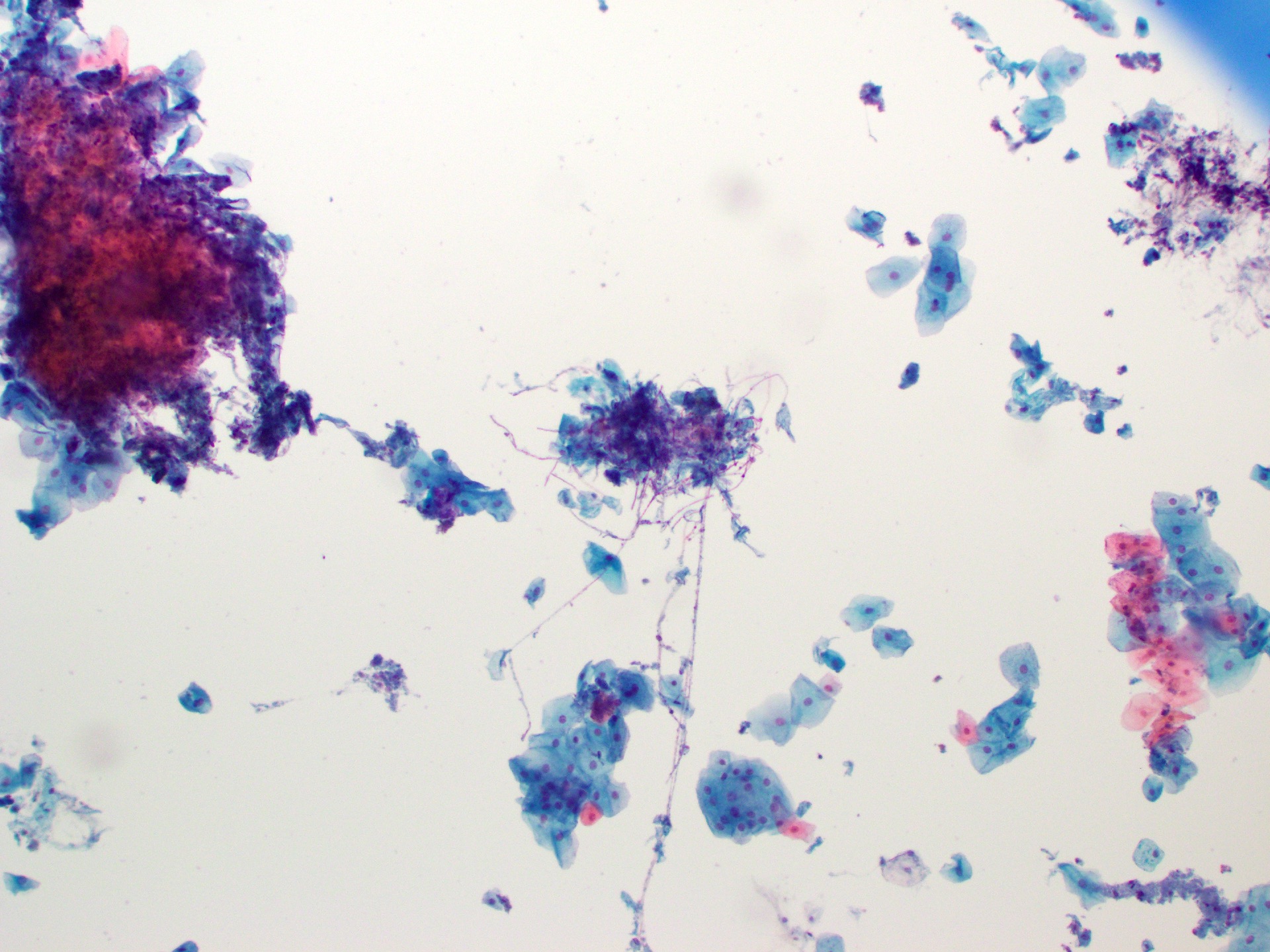
Esophageal brushing
Candida esophagitis
Contributed by Kathryn A. Peterson, M.D., M.Sci.
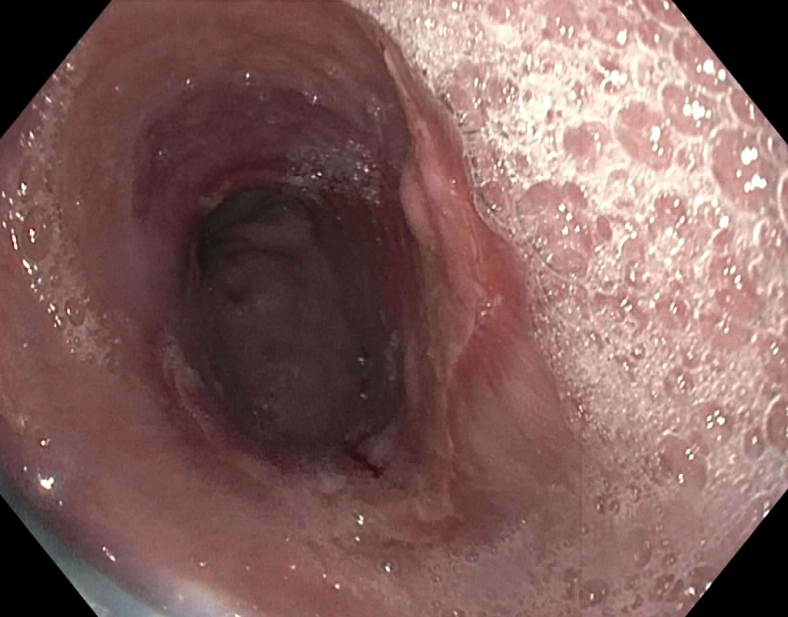
Ulcerated esophageal mucosa
Contributed by Gillian L. Hale, M.D., M.P.H.

Inflammation and CMV inclusions
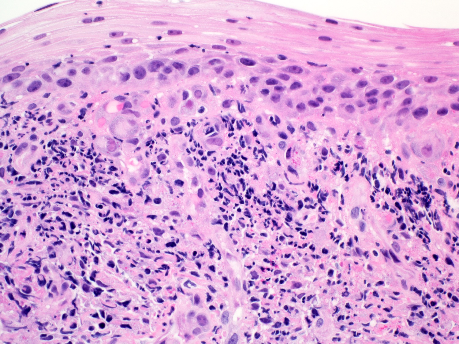
Numerous viropathic inclusions
Infected endothelial cells
Granular inclusions
CMV IHC
Images hosted on other servers:

Spheroid virions
Contributed by Yoko Tateishi, M.D., Ph.D.
Esophageal ulcers
Contributed by Yoko Tateishi, M.D., Ph.D.
Epithelioid granuloma(s)
Increased intraepithelial lymphocytosis
Images hosted on other servers:


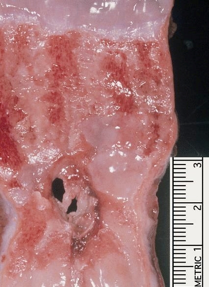
Epiphrenic diverticula
Images hosted on other servers:

Epiphrenic diverticulum
Contributed by Pankaj Vohra, M.D.

Furrows
Tear
Contributed by Ryan C. Braunberger, M.D. and Joshua A. Hanson, M.D.
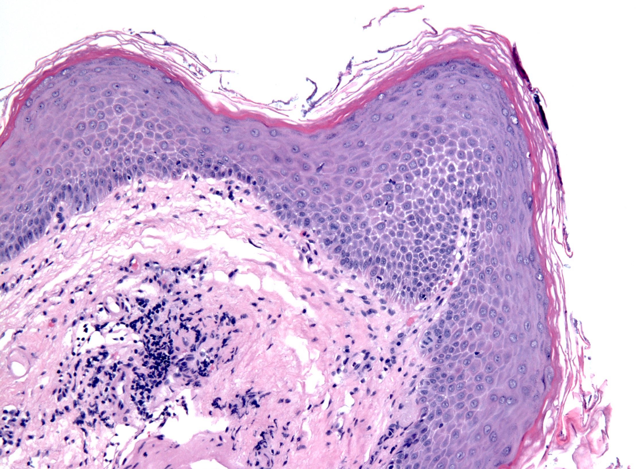
Extreme basal cell hyperplasia
Surface concentration of eosinophils
Eosinophilic microabscess
Eosinophil degranulation
Endoscopic features of eosinophilic esophagitis
Images hosted on other servers:

Scaly white plaque
Contributed by Yukihiro Nakanishi, M.D., Ph.D.
Orthokeratosis /
hyperorthokeratosis
Prominent granular layer
Contributed by Elliot Weisenberg, M.D.
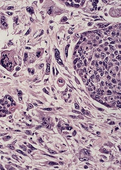
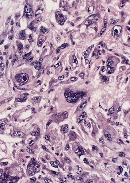
Adenocarcinoma
Squamous cell carcinoma
Contributed by Elliot Weisenberg, M.D.

Adenocarcinoma
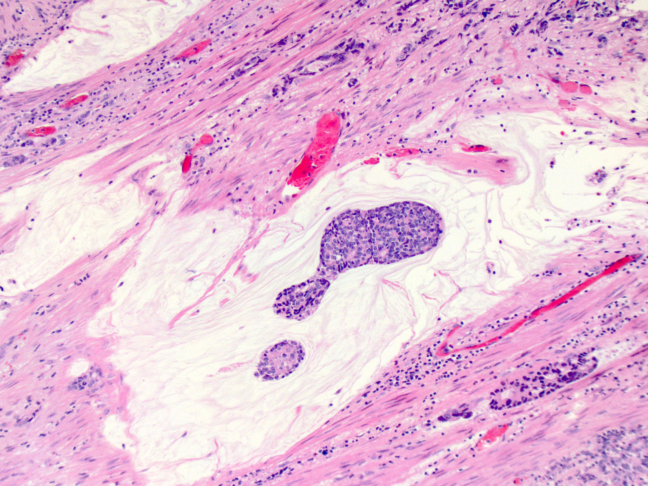
Adenocarcinoma in lymphatics
Squamous cell carcinoma in situ
Images hosted on other servers:

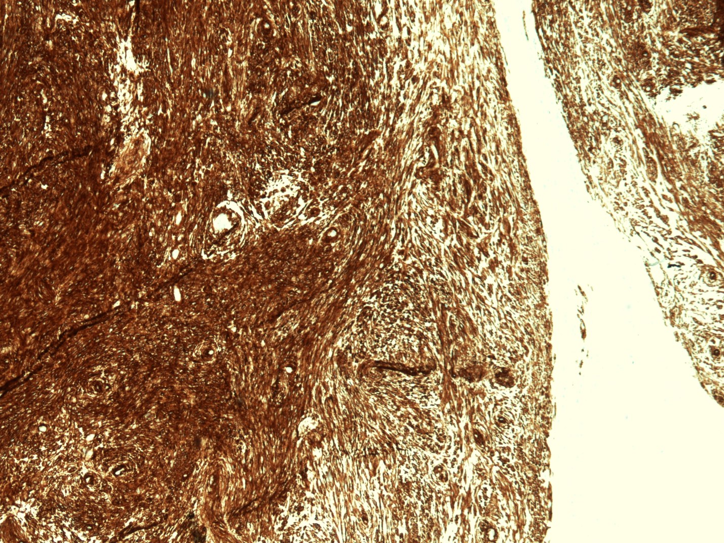
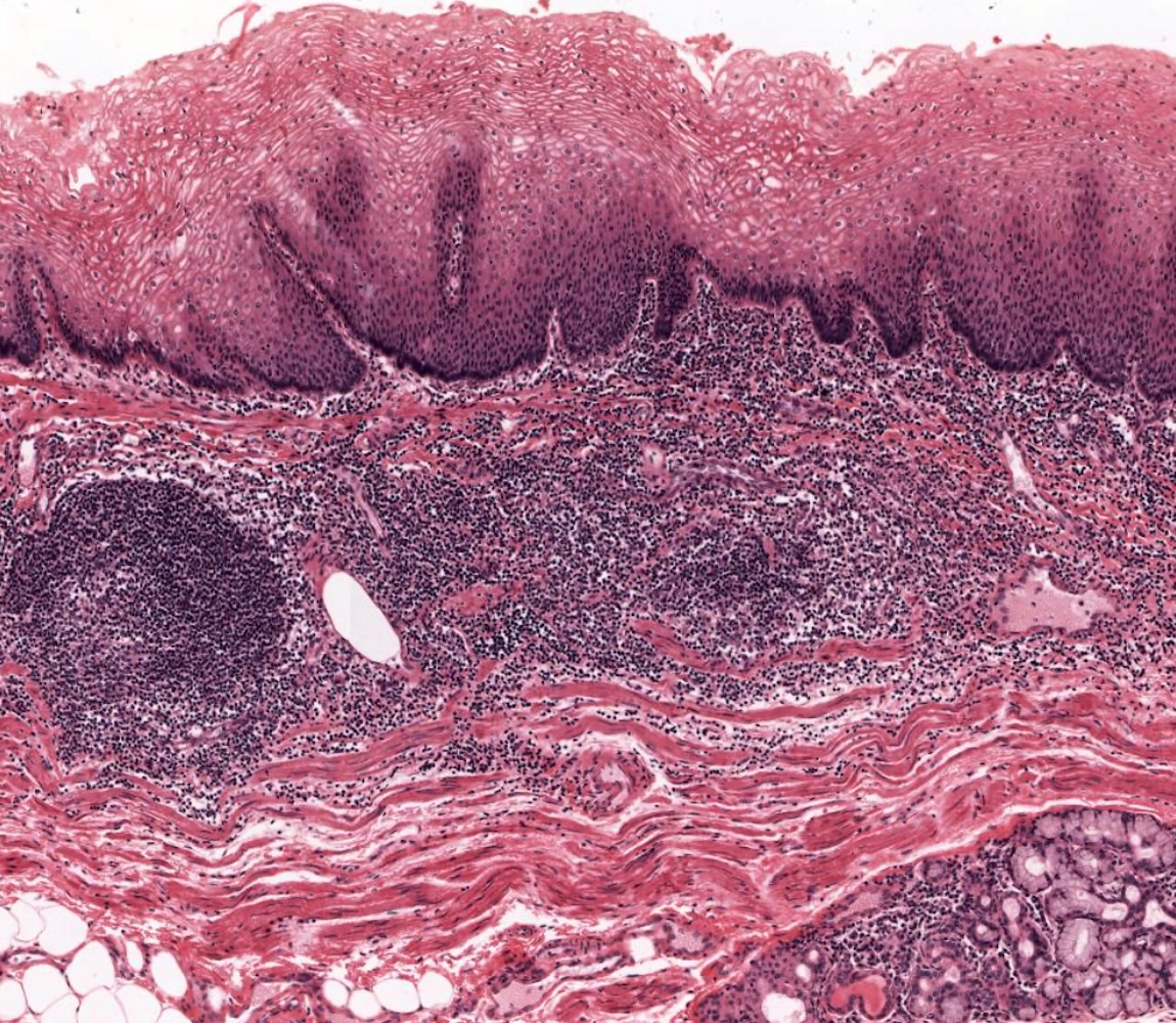
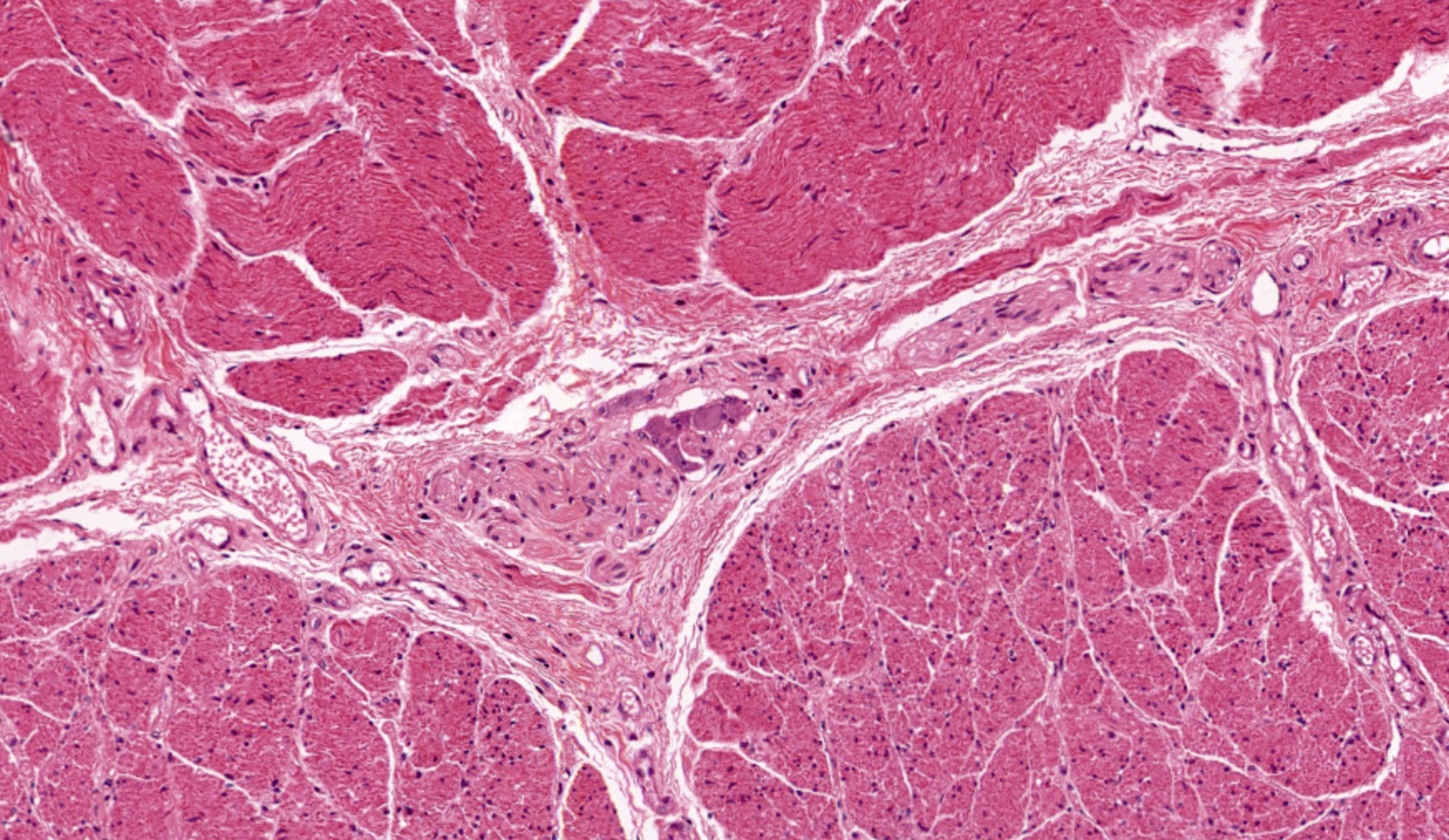
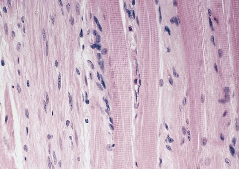
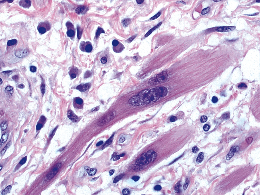
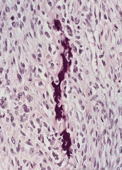
Distal and proximal oesophagus

Severe atrophy of circular muscle

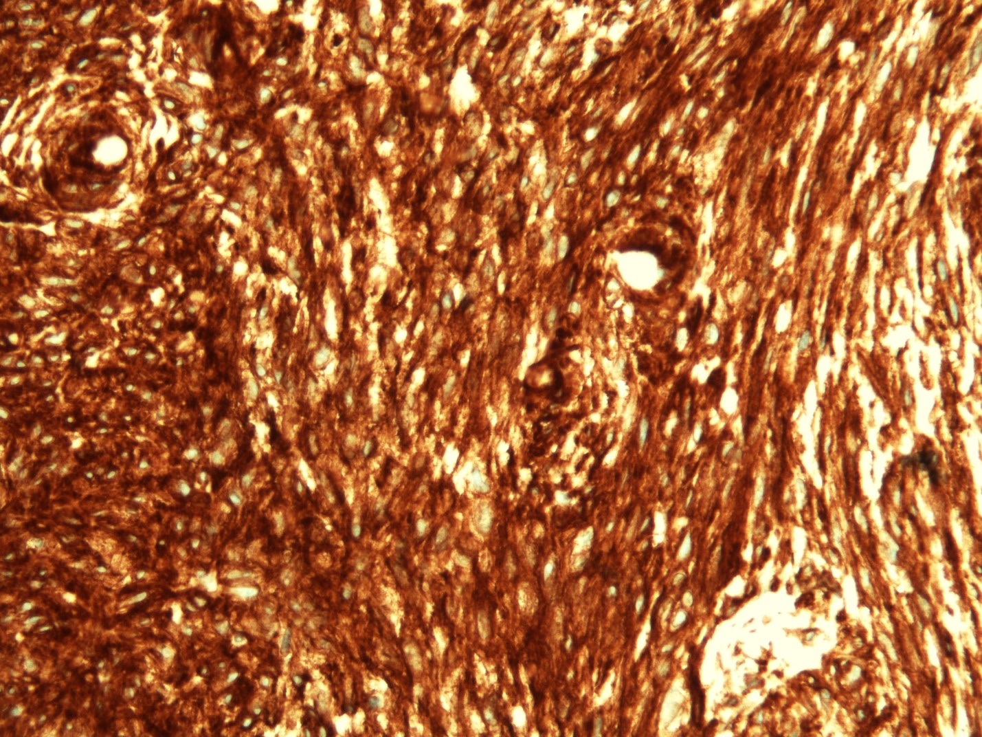
Myenteric plexus

Interstitial cells of Cajal

Barrett esophagus
Images hosted on other servers:

Collagen between
degenerate muscle
fiber (left) and necrotic
muscle fiber (right)

Muscle fibers (M)
separated by collagenous
fibers with widened
intermyofibrillar space
Images hosted on other servers:

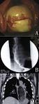
Dilated thoracic esophagus

Gastrographin esophagography
Images hosted on other servers:

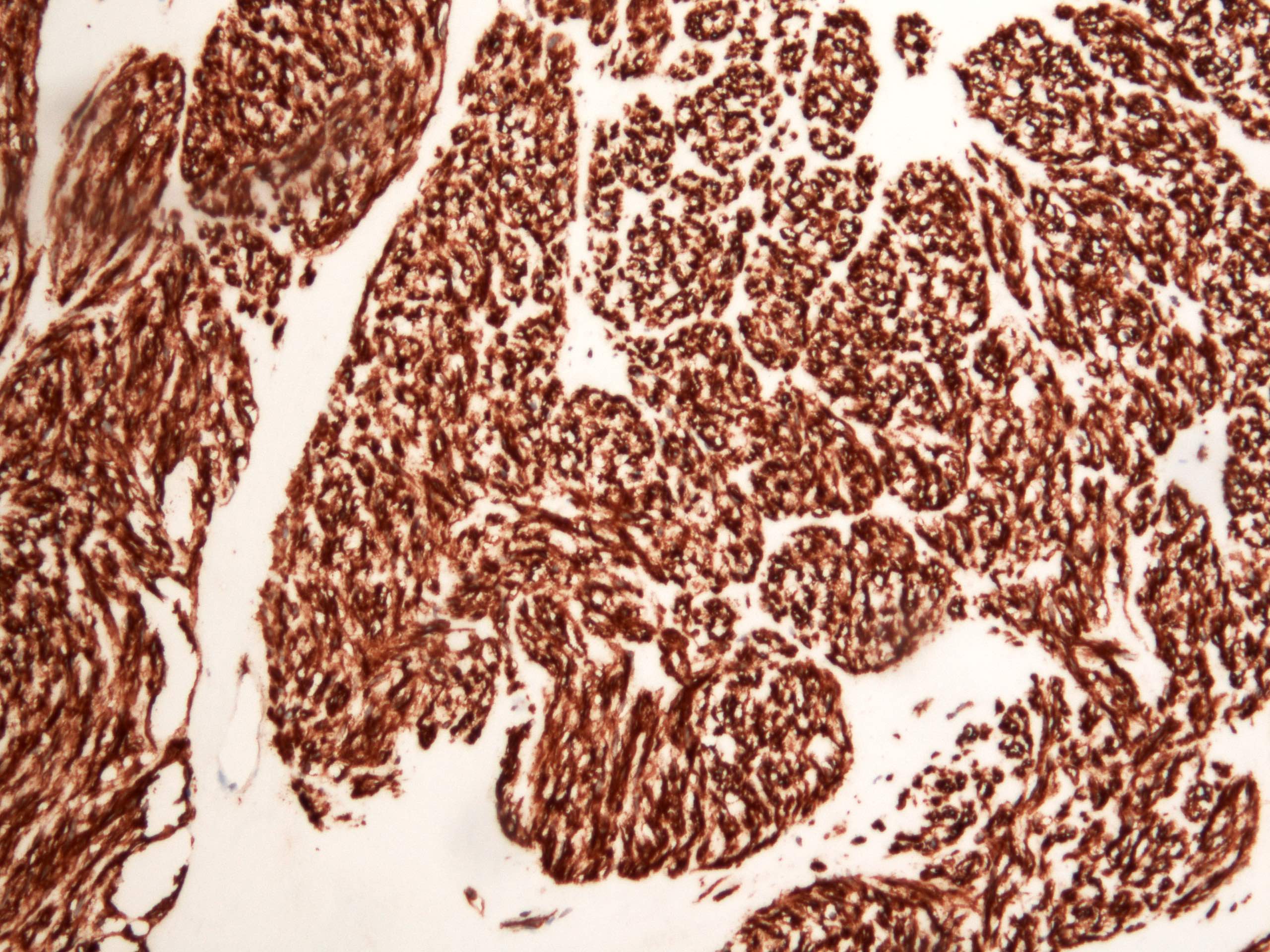
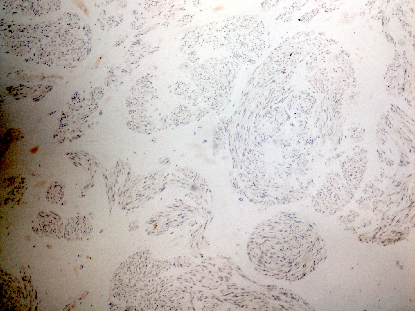
GIST

Leiomyosarcoma
Liposarcoma
AFIP images
Sarcoma
Images hosted on other servers:

GIST
Liposarcoma
Contributed by AFIP and Dr. Zafar Ali
Sarcoma, not otherwise specified
36 year old man with mass just below upper esophageal sphincter, synovial sarcoma (biphasic)

CD99
EMA

TLE1
Images hosted on other servers:
Leiomyosarcoma

Liposarcoma

Osteosarcoma
Contributed by Divya Sharma, M.D.

Tissue paper esophagus
Images hosted on other servers:

Vertical white sloughing mucosal strips

Sloughing of large
esophageal mucosa
fragments

Endoscopy showing EDS

Vertical fissures in
distal esophagus with
sloughing of mucosa
Contributed by Divya Sharma, M.D. and @Rasamh86 on Twitter
2 tone esophagus
Prominent parakeratosis
Intraepithelial splitting
Dense parakeratosis

Intraepithelial cystic degeneration

Inflammatory infiltrate

Esophagitis dissecans superficialis
Sloughing esophagitis
Images hosted on other servers:
Sloughing of large
esophageal mucosa
fragments
Endoscopy showing EDS
Vertical fissures in
distal esophagus with
sloughing of mucosa


Contributed by Israh Akhtar, M.D.
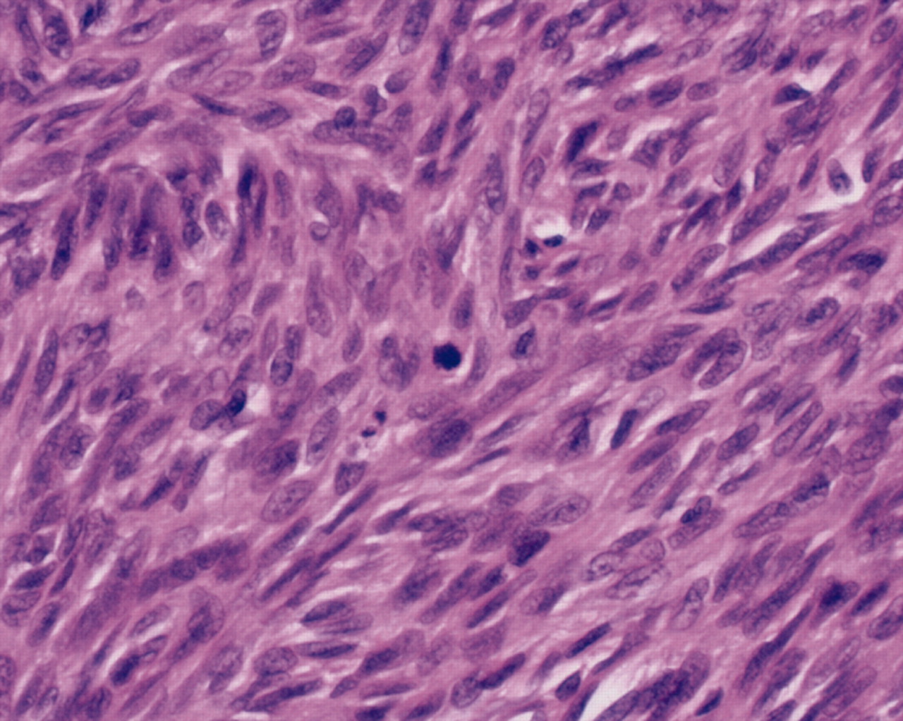
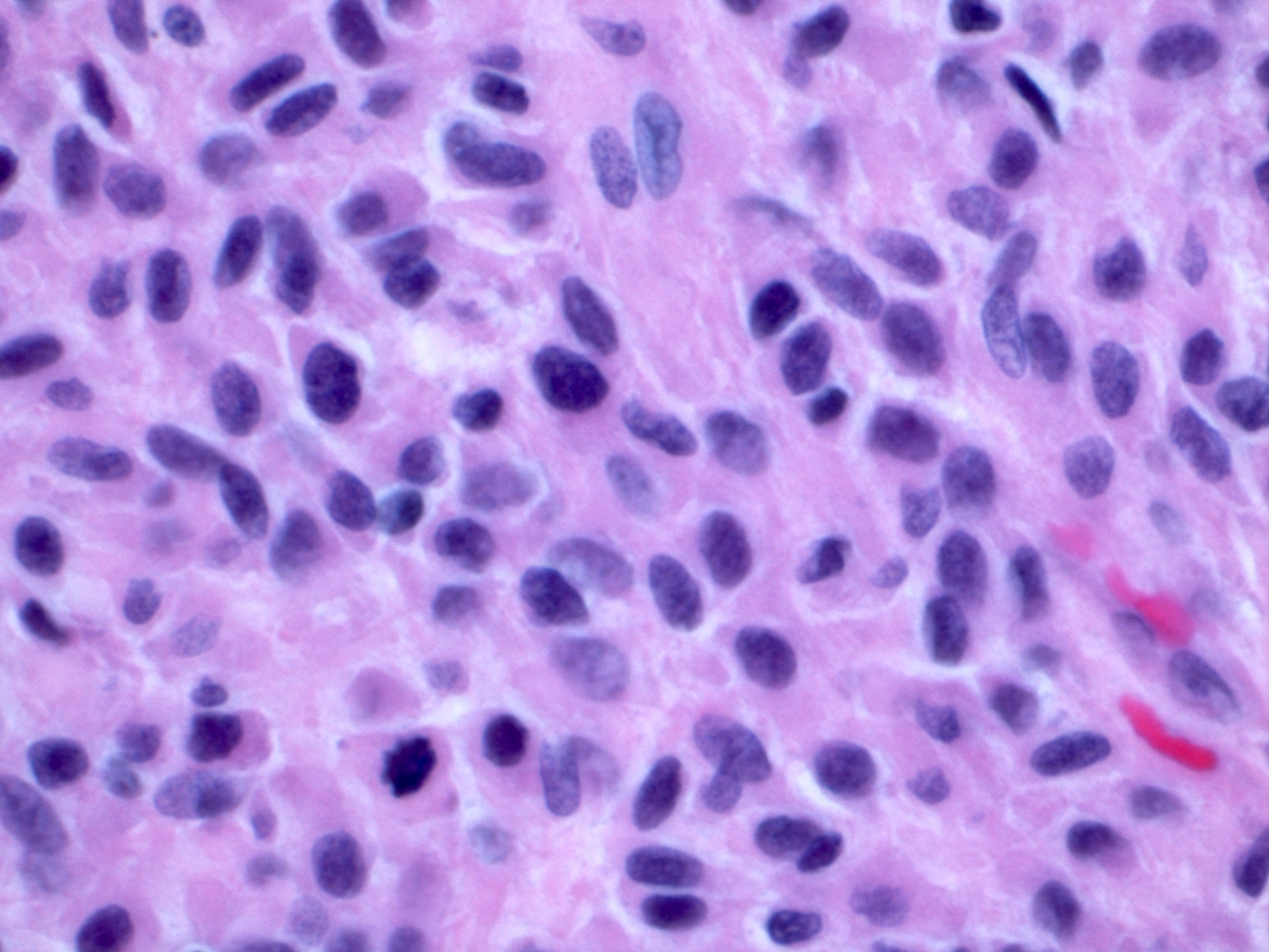
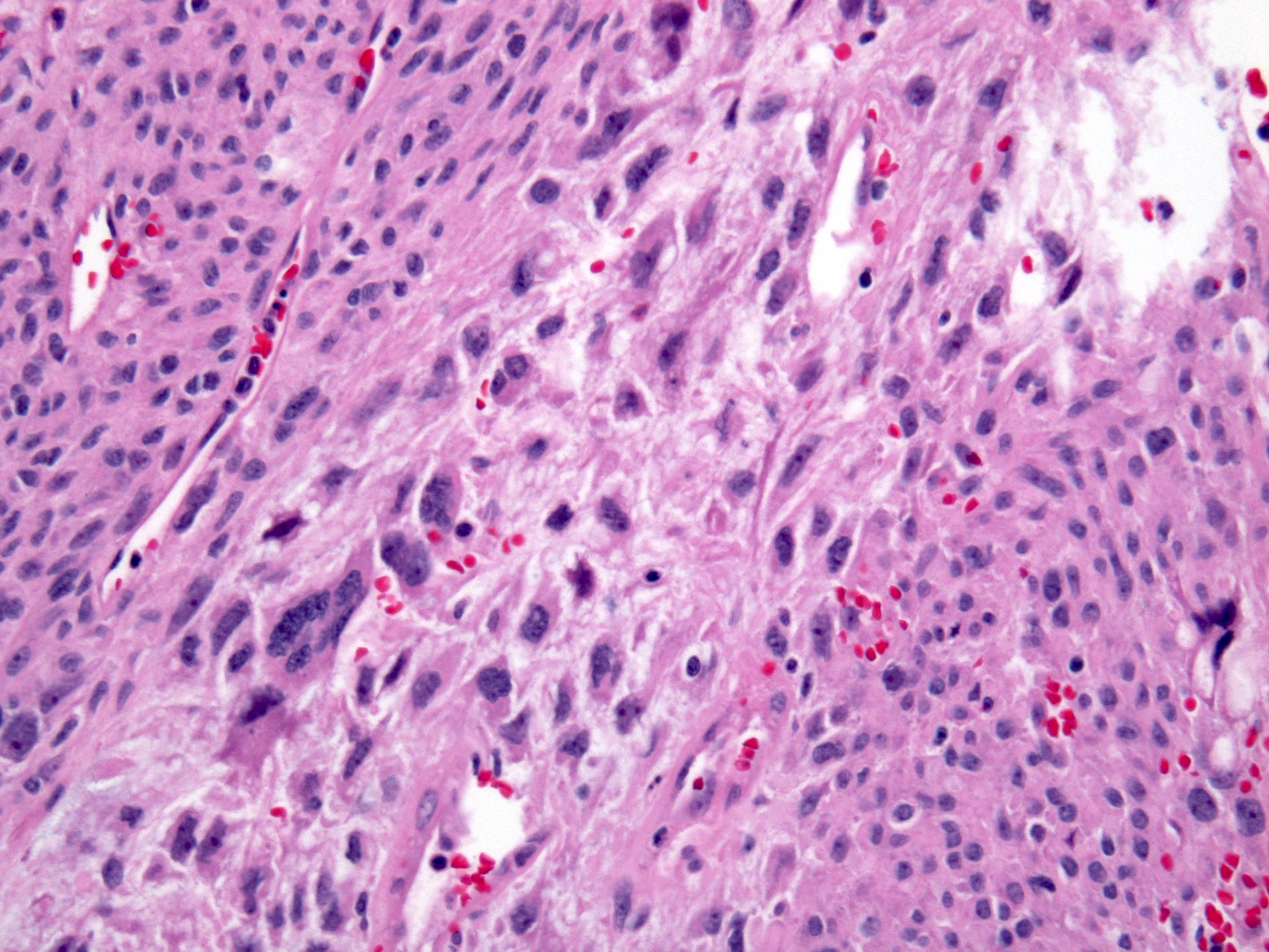
Intramural mass
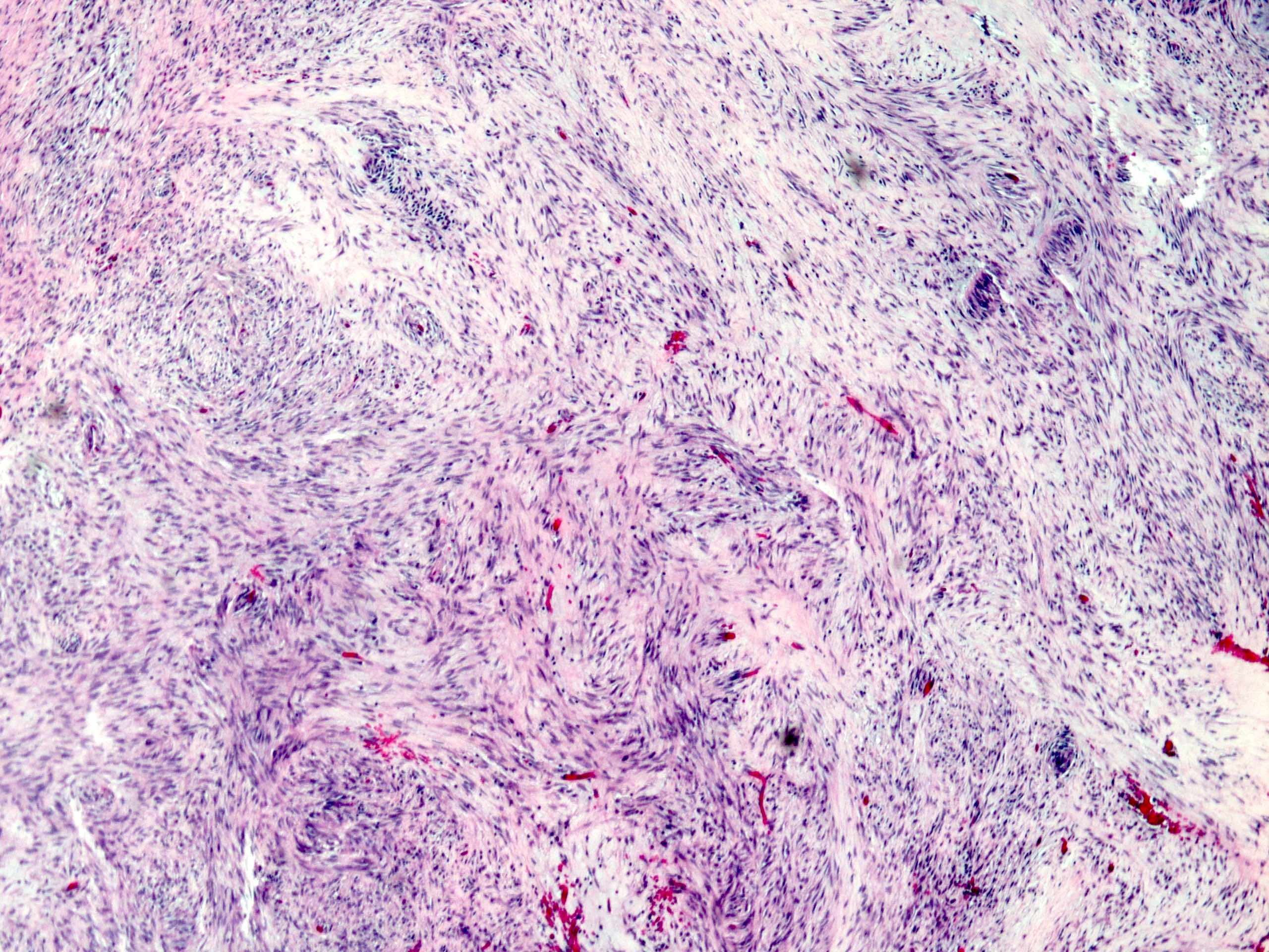
Fascicles of spindle cells
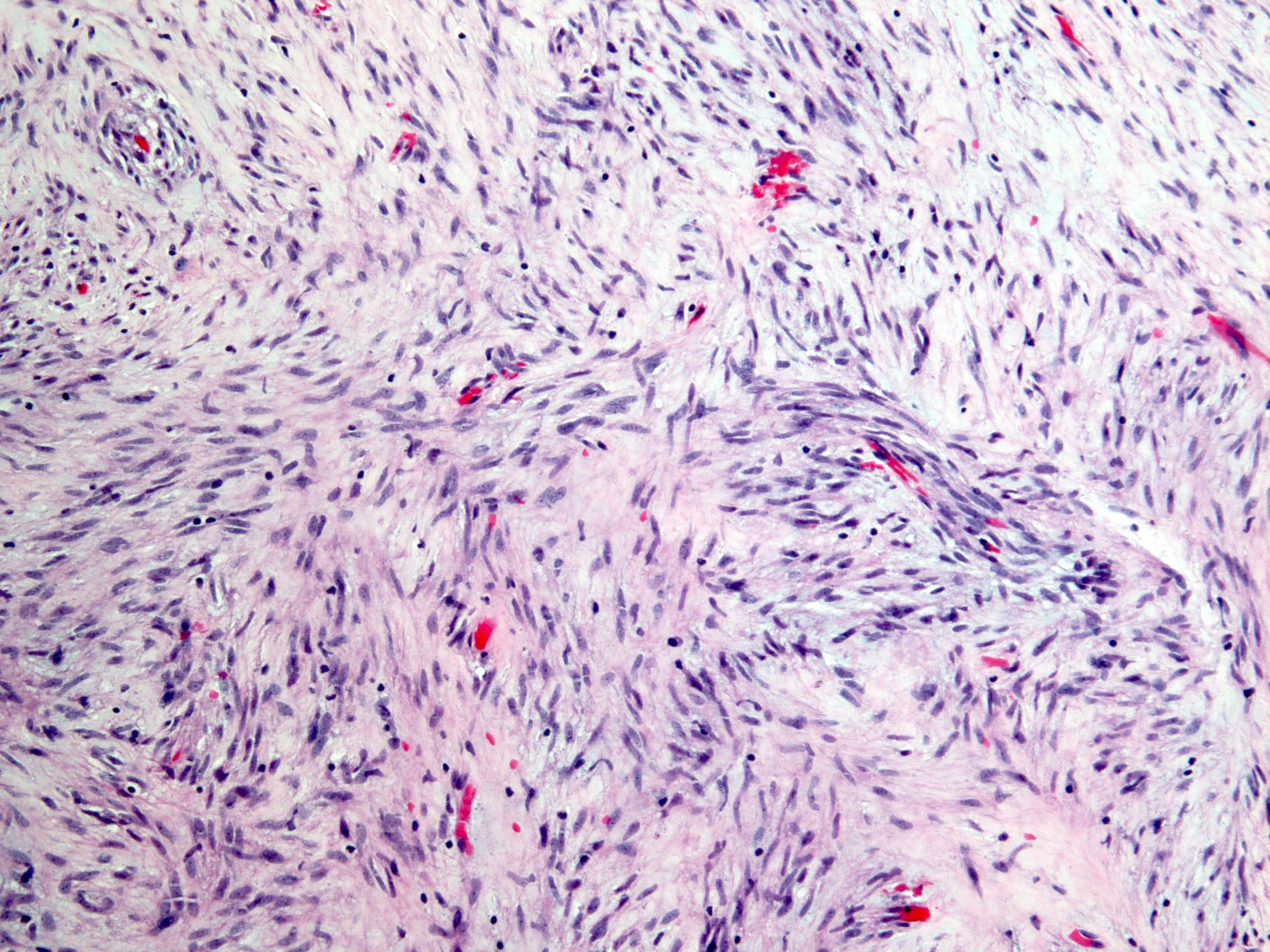
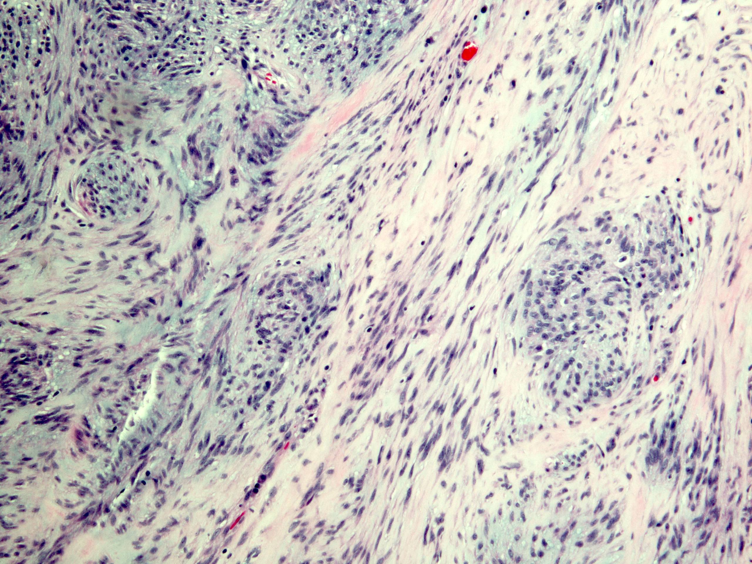
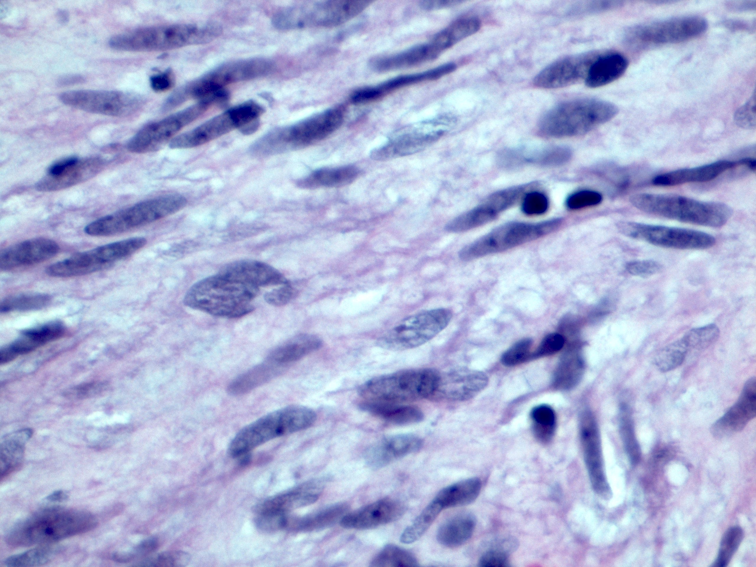
Spindle cells
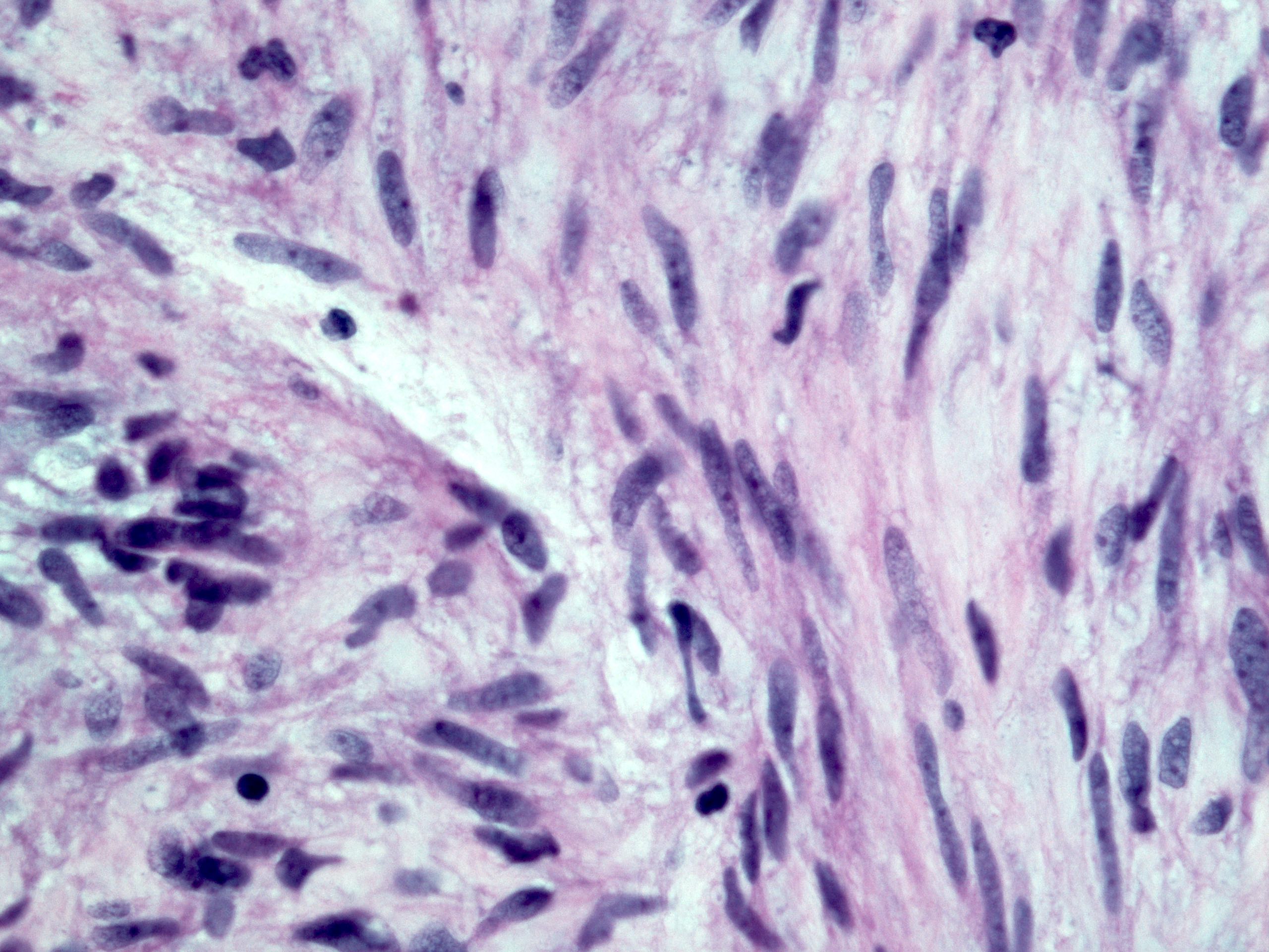
Nuclear details
Mast cells
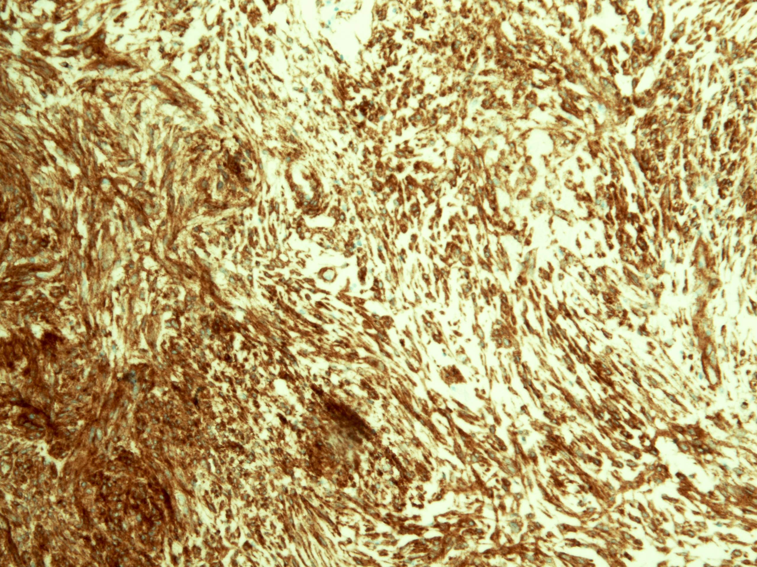
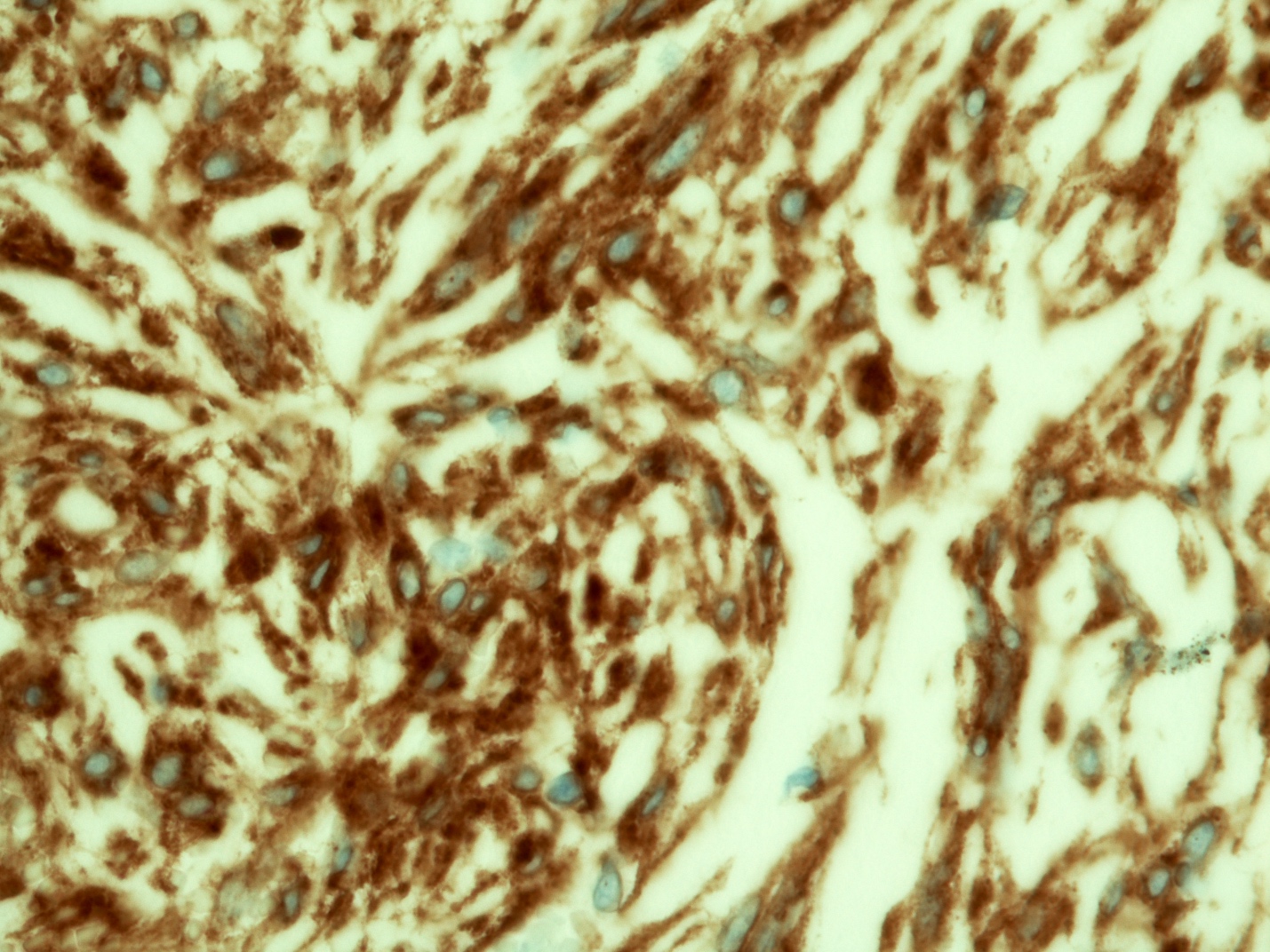
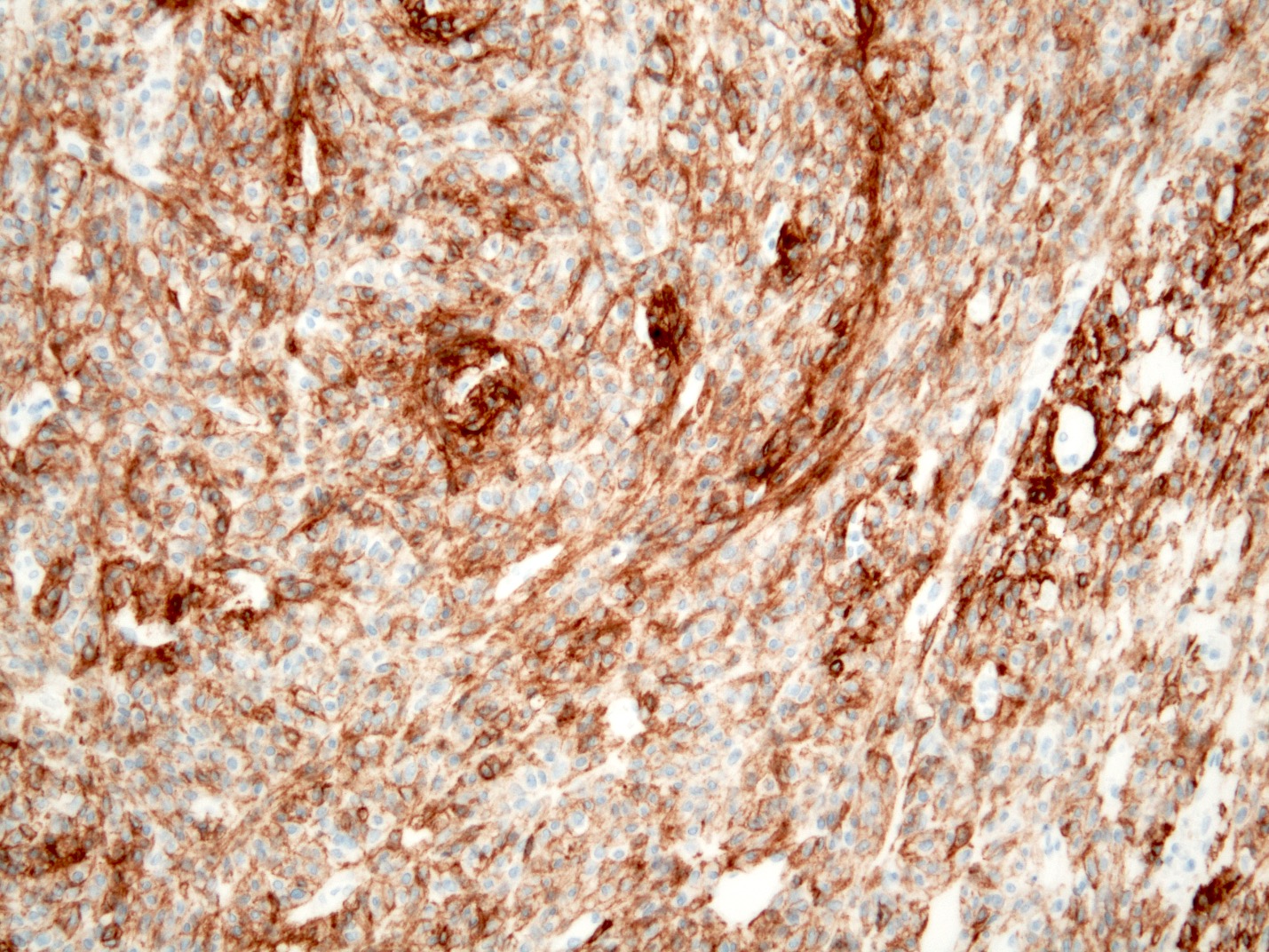
KIT
DOG1
CD34
Desmin
Ki67
Posttherapy
Epithelioid GIST
Mitosis
High grade GIST
KIT
Contributed by Israh Akhtar, M.D.
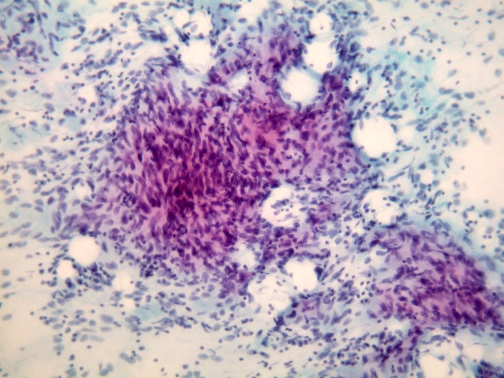
High cellularity
Spindle cells
Fascicular pattern
Spindle cells
Epithelioid GIST
Epithelioid cells
Cell block
DOG1
High grade GIST
Images hosted on other servers:

Cytoplasmic vesicles in high risk GIST
Images hosted on other servers:

Sausage shaped mass
Contributed by Yukihiro Nakanishi, M.D., Ph.D.
Fibroadipose tissue with slightly enlarged, hyperchromatic stromal cells
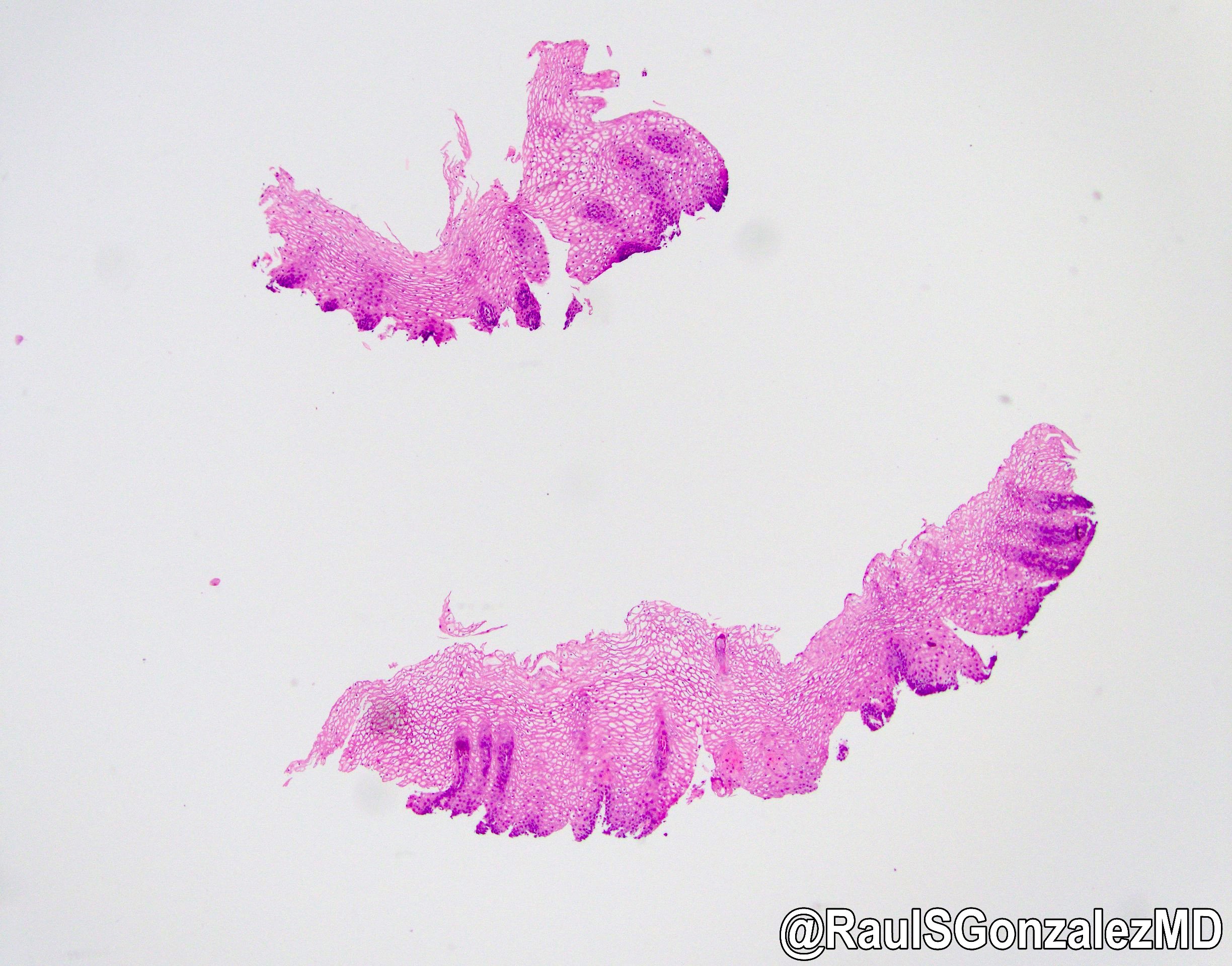
Images hosted on other servers:

Discrete, rounded nodules and plaques
AFIP images
Numerous white islands

Slightly elevated longitudinal plaque
Contributed by Aaron R. Huber, D.O. and @RaulSGonzalezMD on Twitter
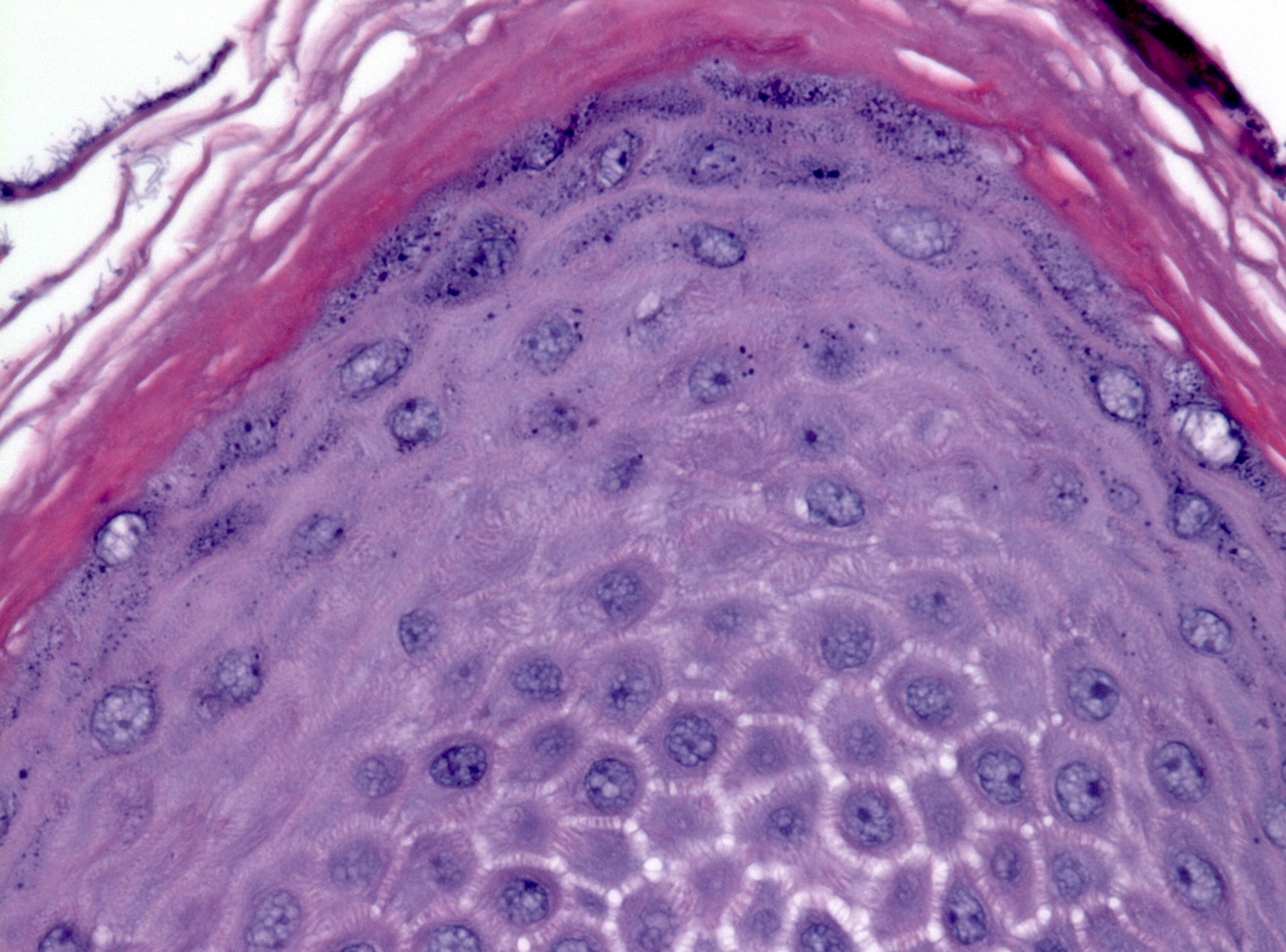
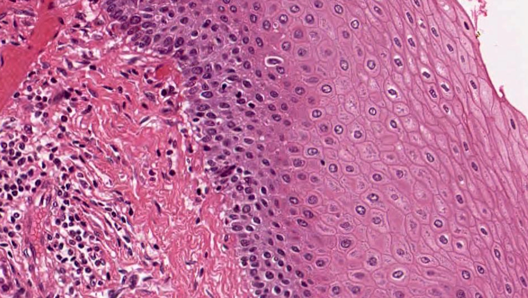
Esophageal squamous mucosa with abundant intracytoplasmic glycogen
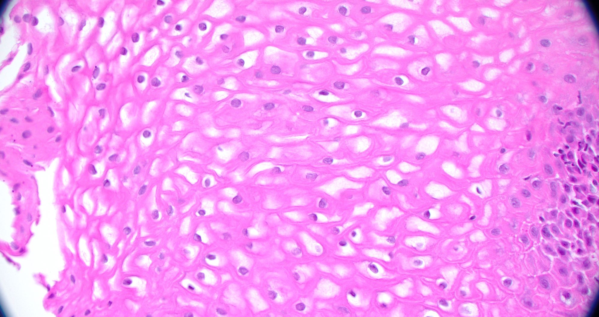
Clear cytoplasm of squamous epithelial cells
Intracytoplasmic glycogen highlighted with PAS
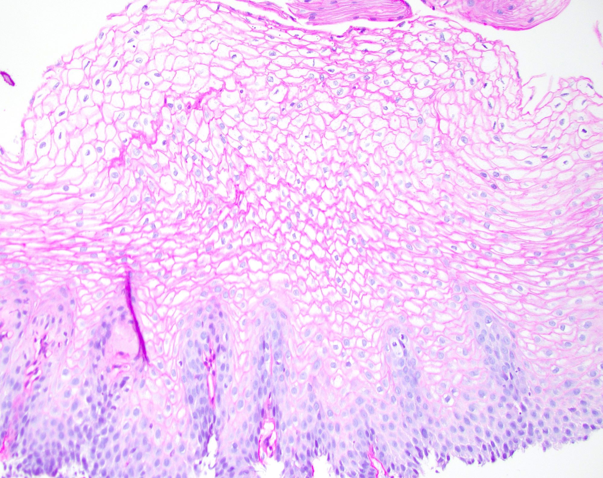
Intracytoplasmic glycogen that is diastase sensitive
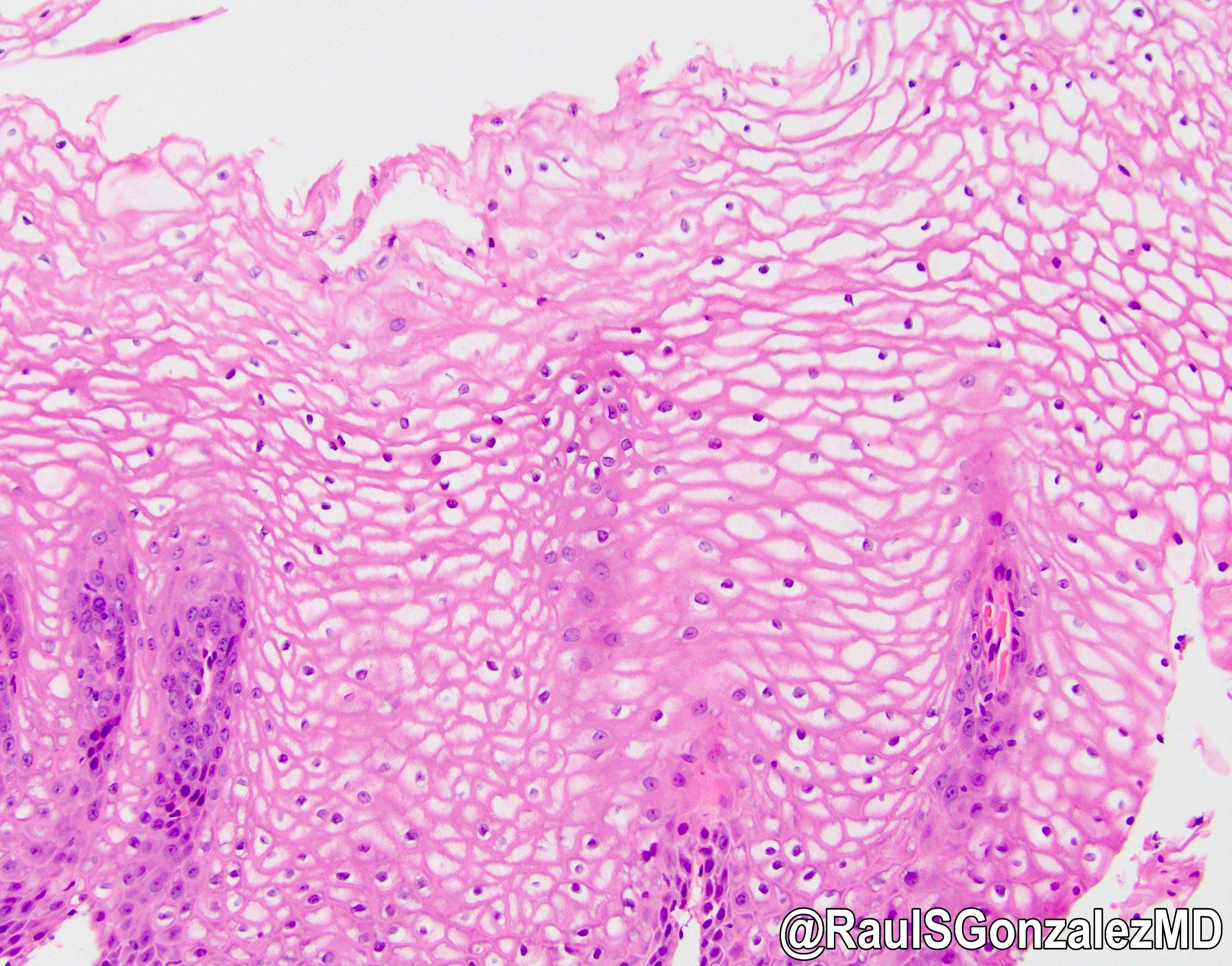
Glycogenic acanthosis
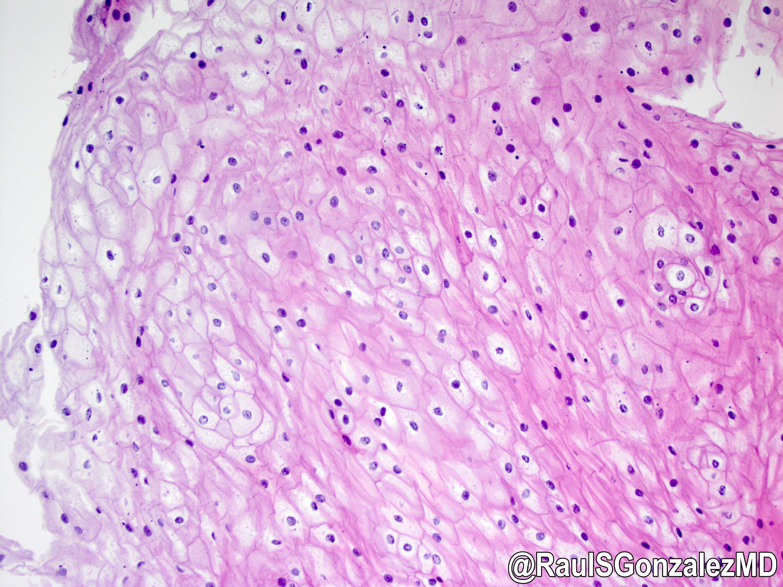
Glycogenic acanthosis
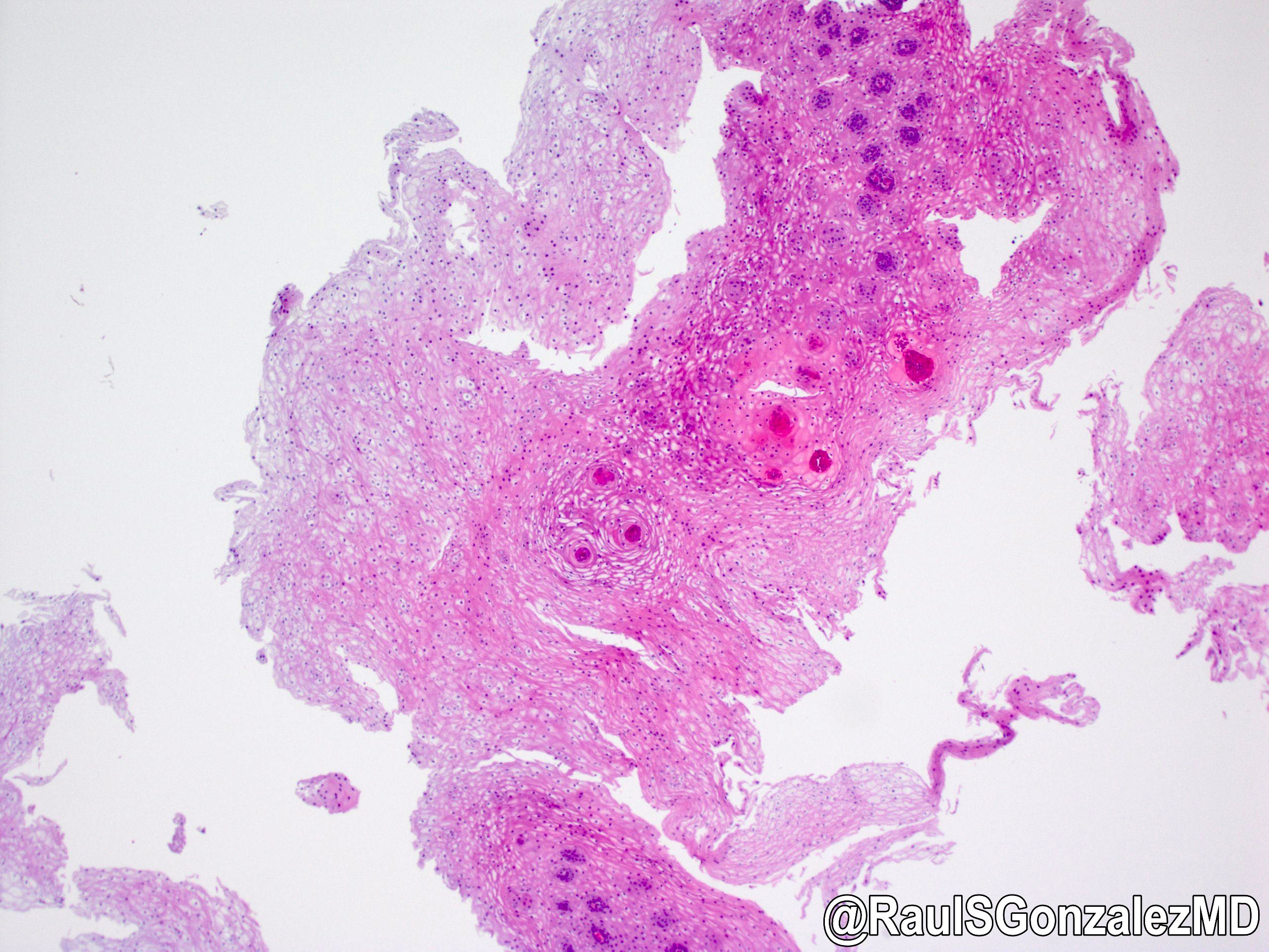
Images hosted on other servers:


With lymphocytic infiltrate
Contributed by Dr. Oleksandr Grygoruk, AFIP and @RaulSGonzalezMD on Twitter
Abrikossoff esophagus
Tumor fills lamina propria
Contributed by @RaulSGonzalezMD on Twitter (see original post here)">
 shorturl.at/cdglN #pathology #gipath #PathTwitter #PathOutPic"
Contributed by @RaulSGonzalezMD on Twitter (see original post here)">
 shorturl.at/cdglN #pathology #gipath #PathTwitter #PathOutPic"
Contributed by @RaulSGonzalezMD on Twitter (see original post here)">
Granular cell tumor
Images hosted on other servers:

Sections to obtain from esophagectomy specimen
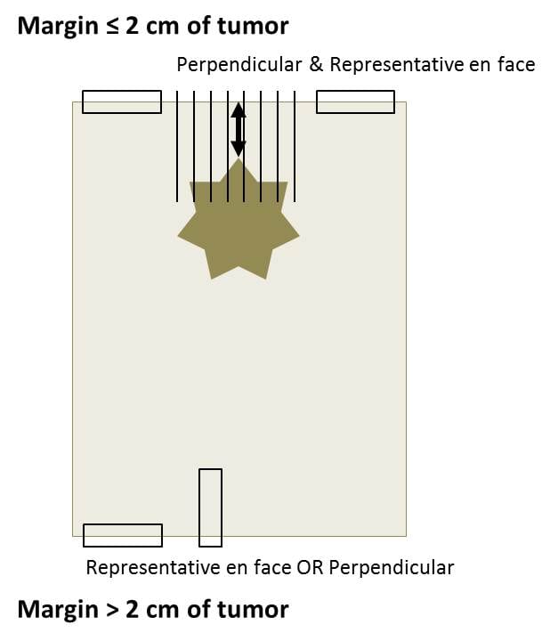
How to obtain sections from margins
Images hosted on other servers:

Esophagectomy with proximal portion of stomach

Esophagectomy specimen status posttreatment

Esophageal endoscopic mucosal resection
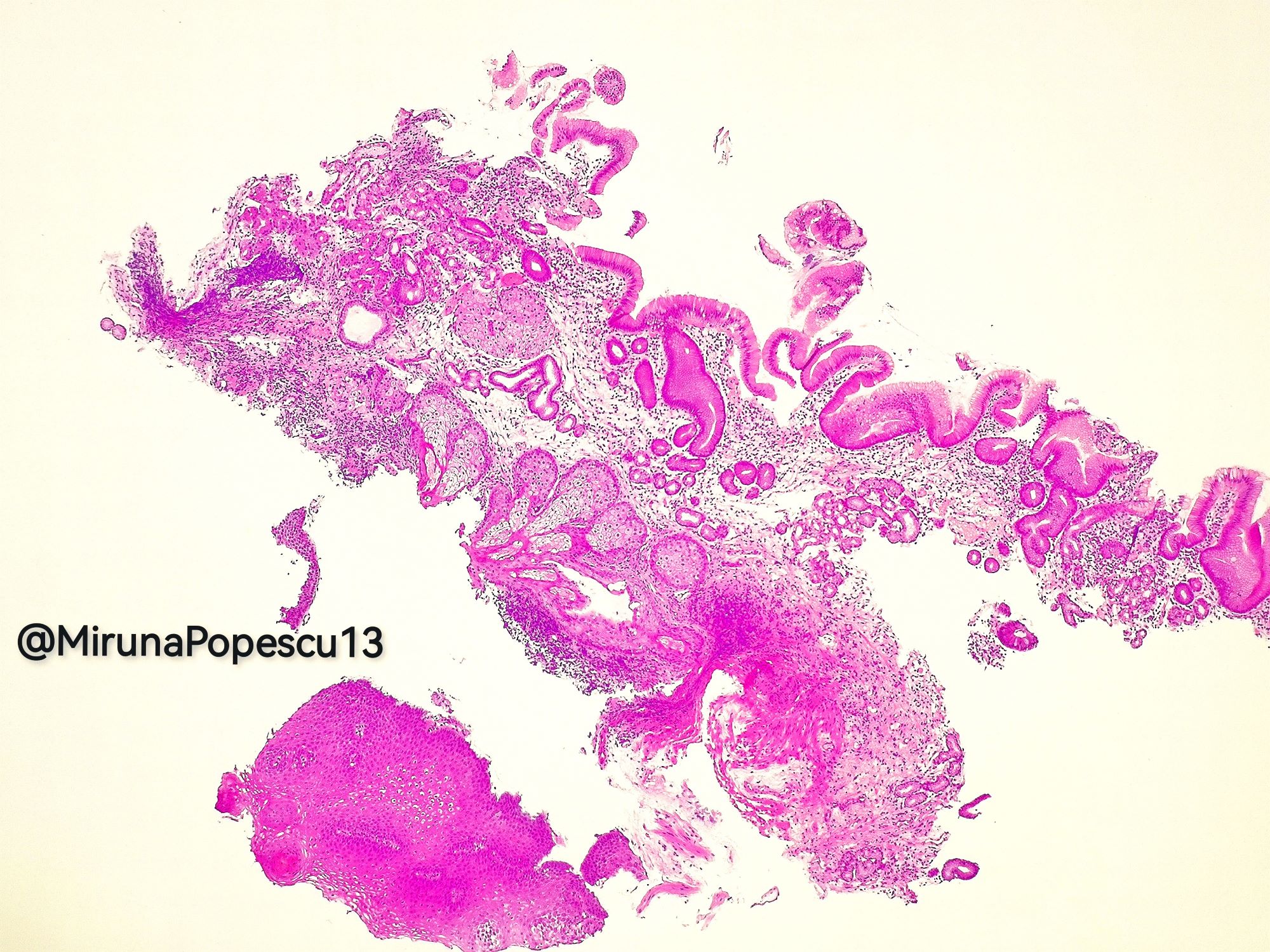
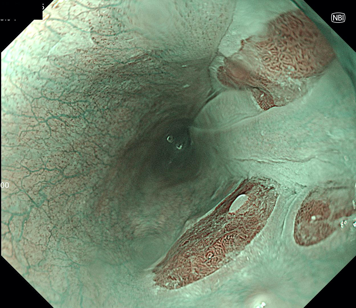
Images hosted on other servers:


Endoscopic images
Contributed by Yukihiro Nakanishi M.D., Ph.D. and @MirunaPopescu13 on Twitter
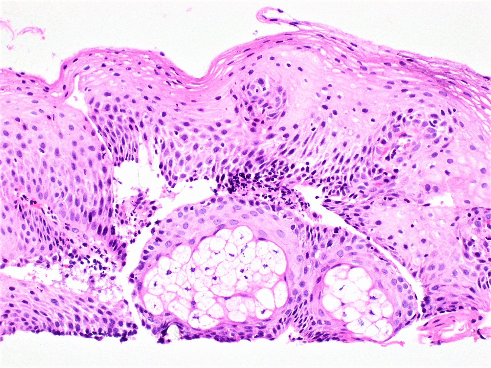
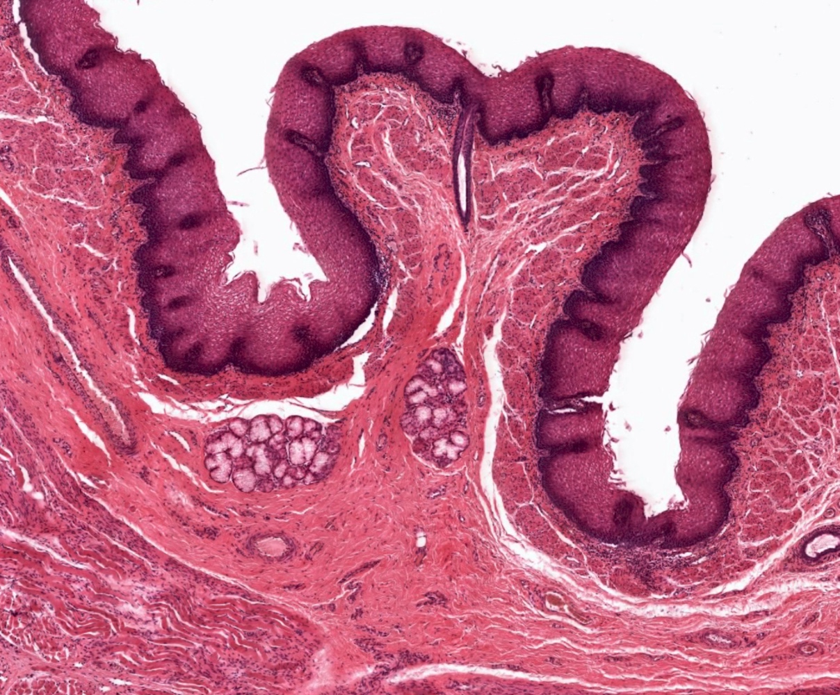
Intermediate power
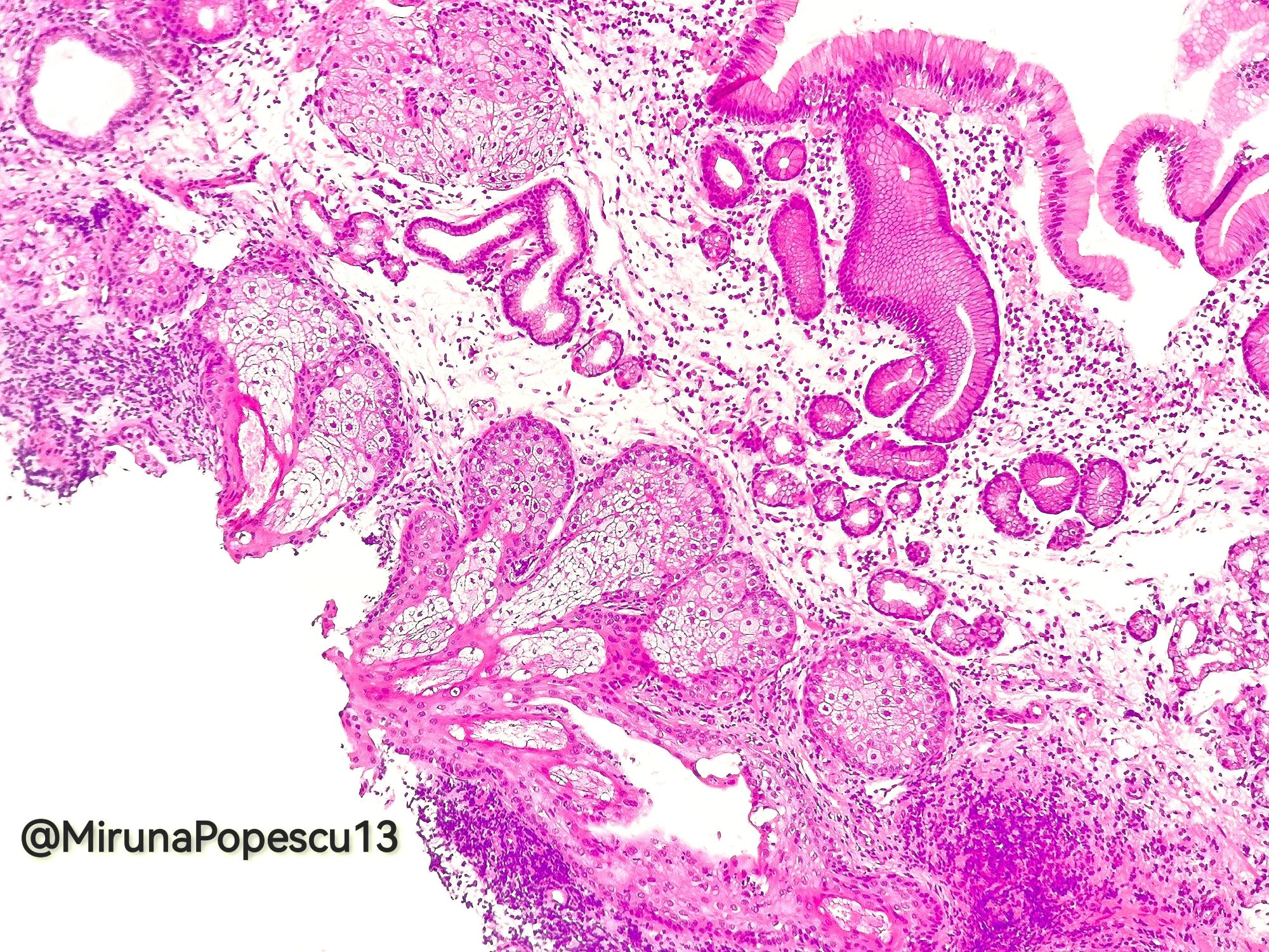
Heterotopic / ectopic sebaceous glands
Contributed by Yoko Tateishi, M.D., Ph.D.

Red patches
Multiple patches
Contributed by Yoko Tateishi, M.D., Ph.D.

Red patch
Contributed by Yoko Tateishi, M.D., Ph.D.
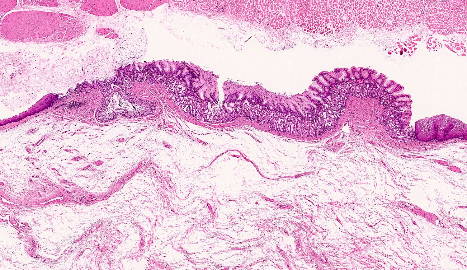
Isolated gastric mucosa
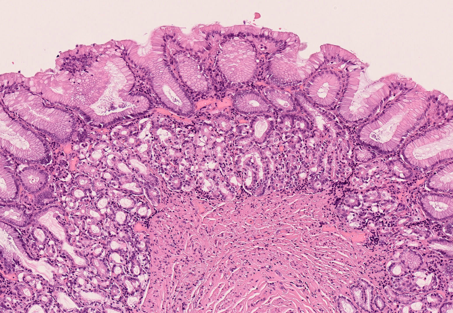
Oxyntic / fundic type gastric mucosa
Images hosted on other servers:

Esophageal layering schematic

GI layering

Esophageal sphincters
Images hosted on other servers:

Micro CT of human esophagus
Images hosted on other servers:

Human cadaveric esophagus
Contributed by Nicole Stringham, Ph.D. (sources: University of Michigan virtual slide box and Duke University virtual slide collection) and AFIP
GI layering, middle third
Epithelium and lamina propria
Mucosal lymph nodule, submucosal gland
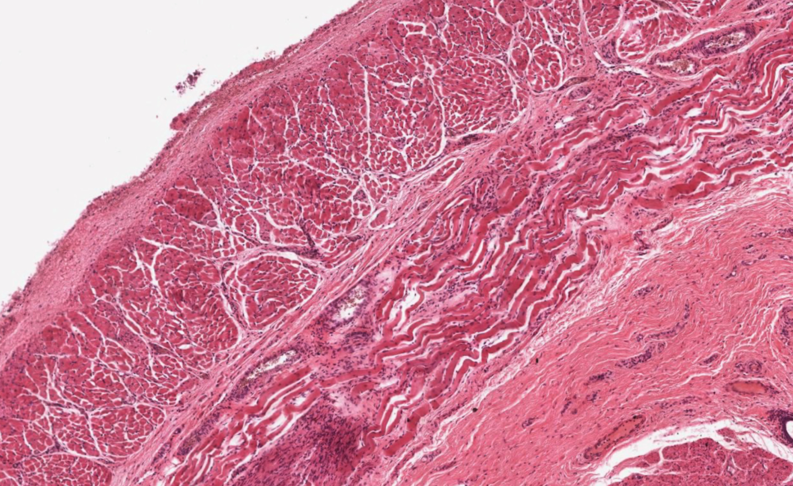
Muscularis externa
Myenteric plexus
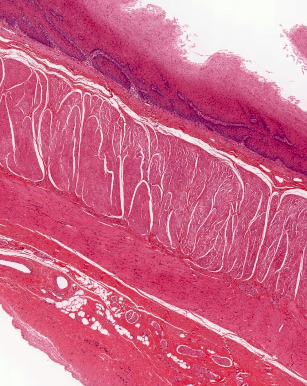
Lower third
Smooth and skeletal muscle
Images hosted on other servers:

Brushing specimen of normal esophagus
Images hosted on other servers:

TEM of epithelial Langerhans cells

SEM of muscularis mucosae
Normal esophagus histology

Images hosted on other servers:

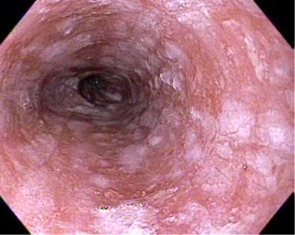
Endoscopic features

Diffuse mucosal erosions and ulcerations
Images hosted on other servers:


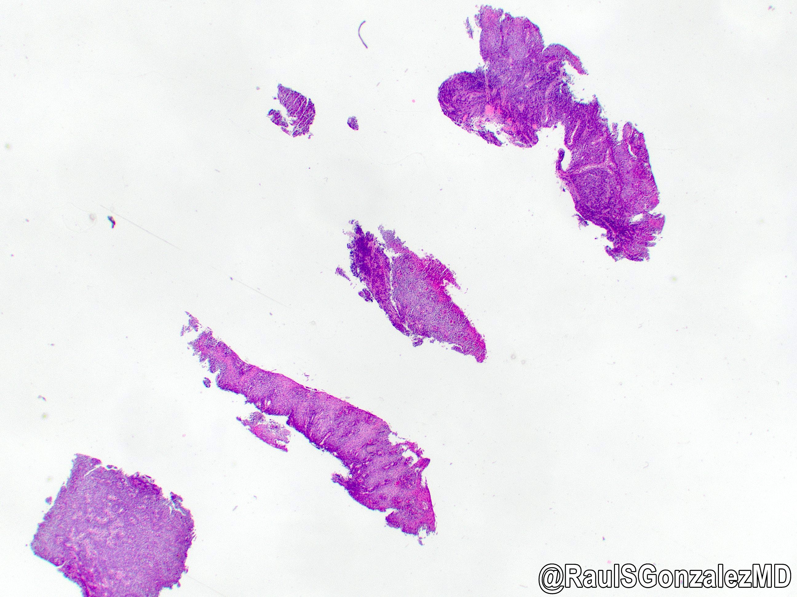
Punched out ulcers
Contributed by James Mueller, M.D., Ph.D.
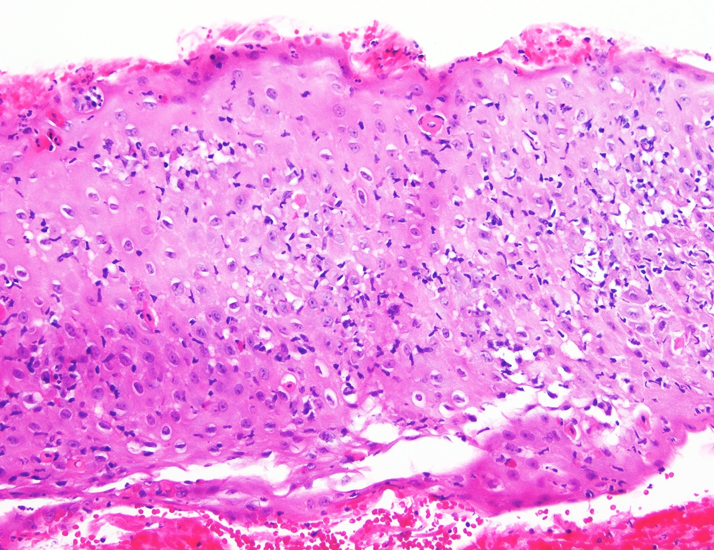
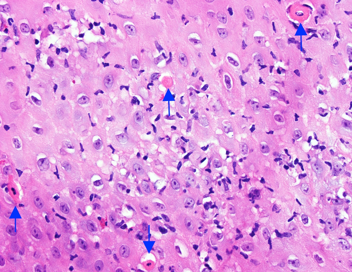
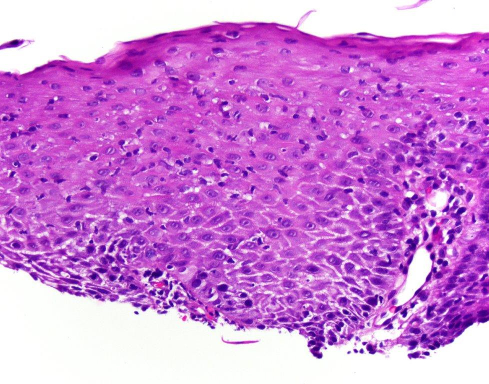
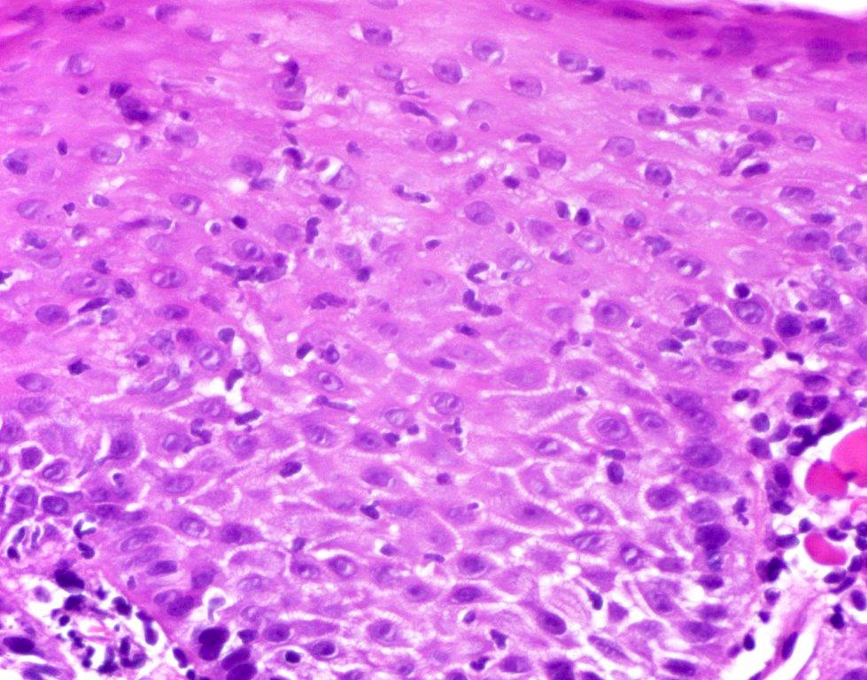
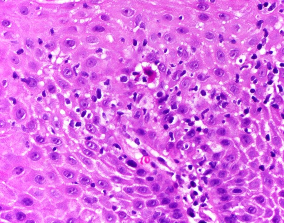
Esophageal biopsy

Prominent acute inflammation
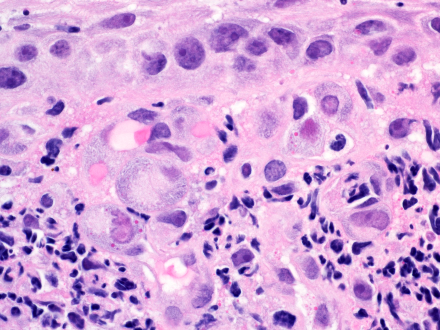
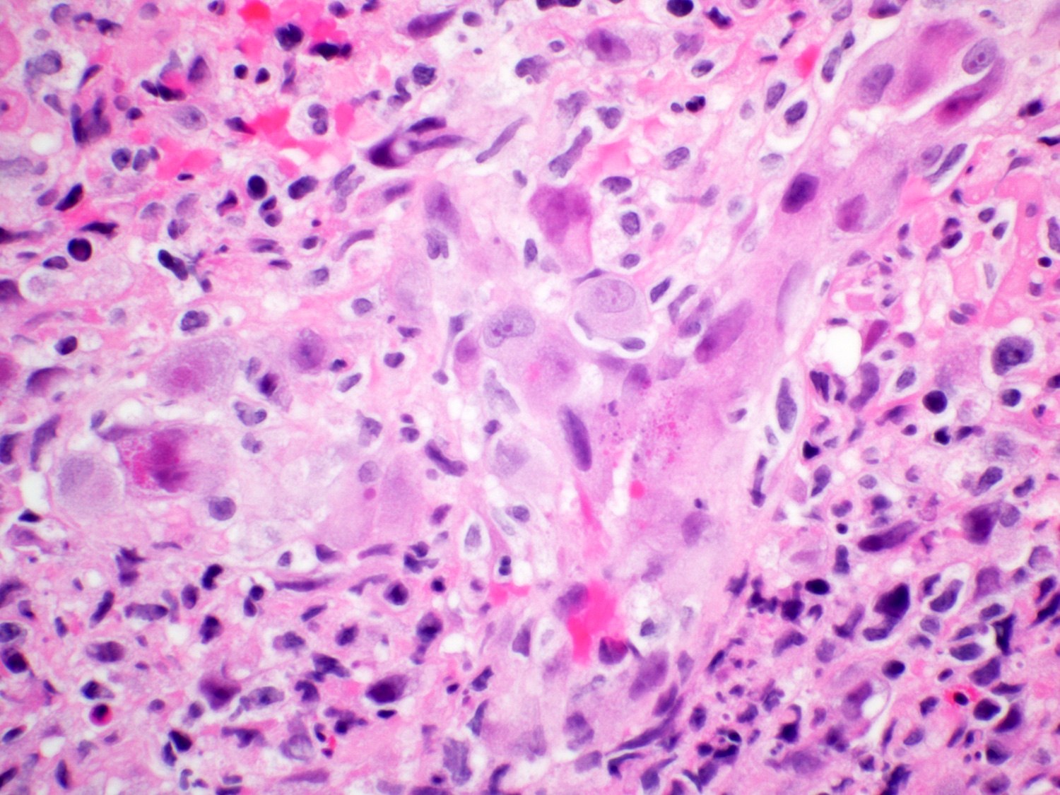
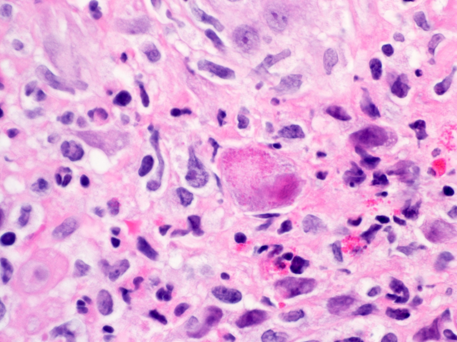
Cowdry A inclusion
3 Ms

Multinucleation
Glassy chromatin
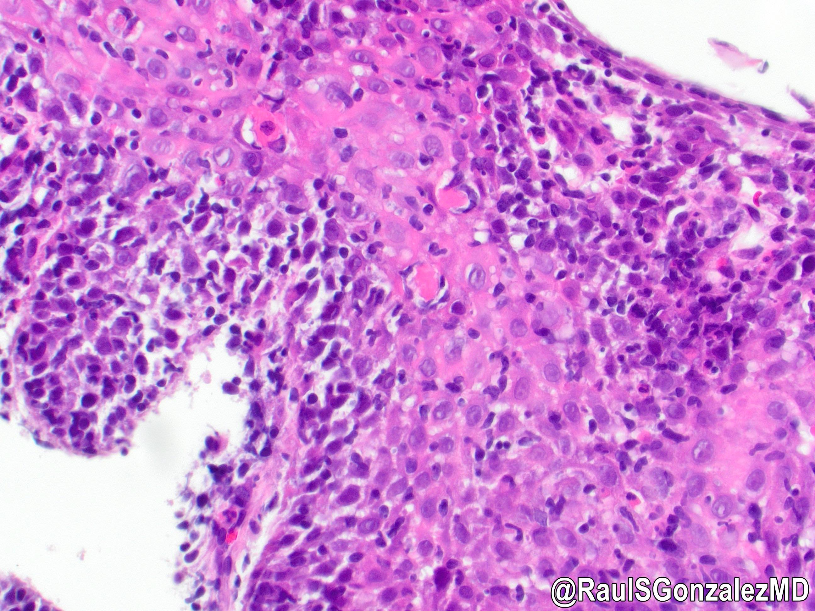
Edge of the ulcer
Viral cytopathic effect

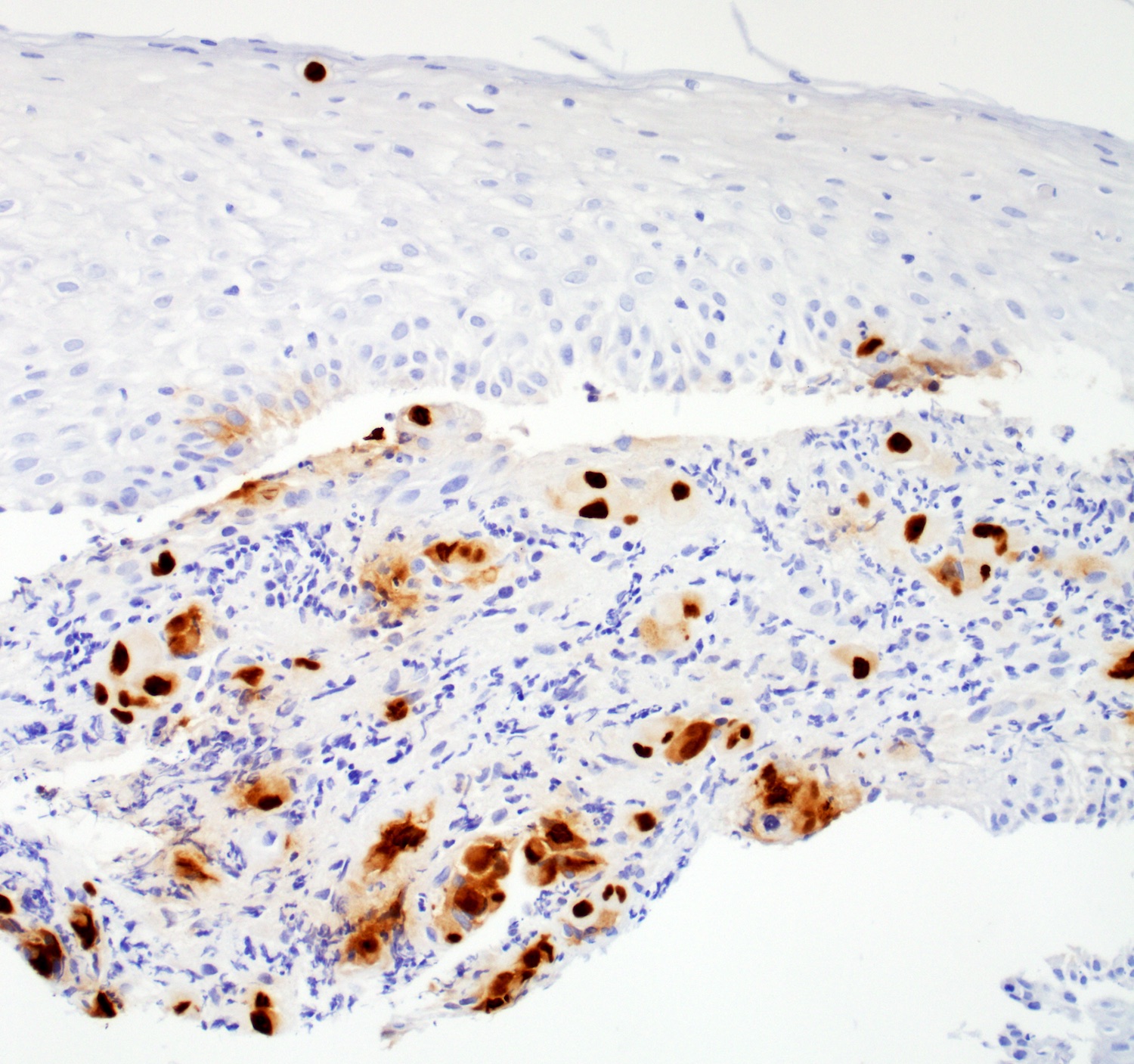
HSV IHC stain
Overview of HSV esophagitis
Gross and histopathologic findings of HSV esophagitis
Images hosted on other servers:

Endoscopic examination
AFIP images
Bulging, white, whorled cut surface
Images hosted on other servers:


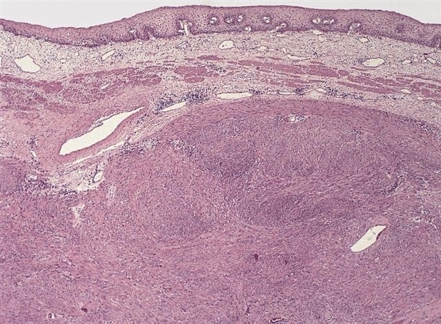
Submucosal tumor
With squamous cell carcinoma
AFIP images

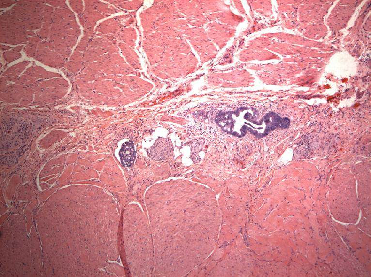
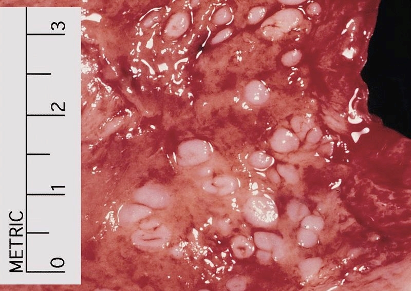
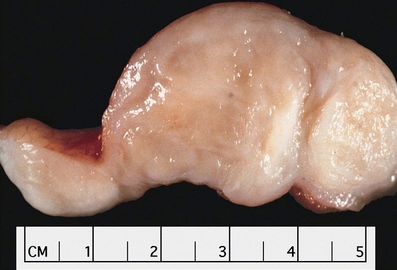
Cluster of seedling leiomyomas
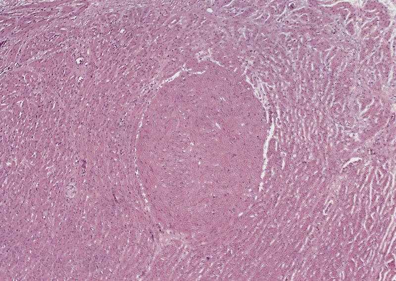
Seedling leiomyoma of muscularis propria
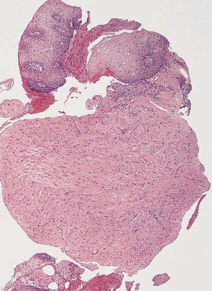
Leiomyoma with overlying squamous epithelium
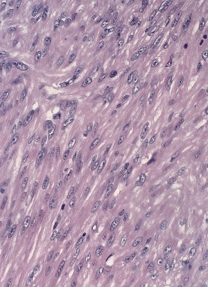
Mature, hypertrophied smooth muscle cells

Multiple seedling leiomyomas
Leiomyoma of muscularis propria

Fascicular growth pattern
Images hosted on other servers:

Squamous cell carcinoma and leiomyoma
Images hosted on other servers:


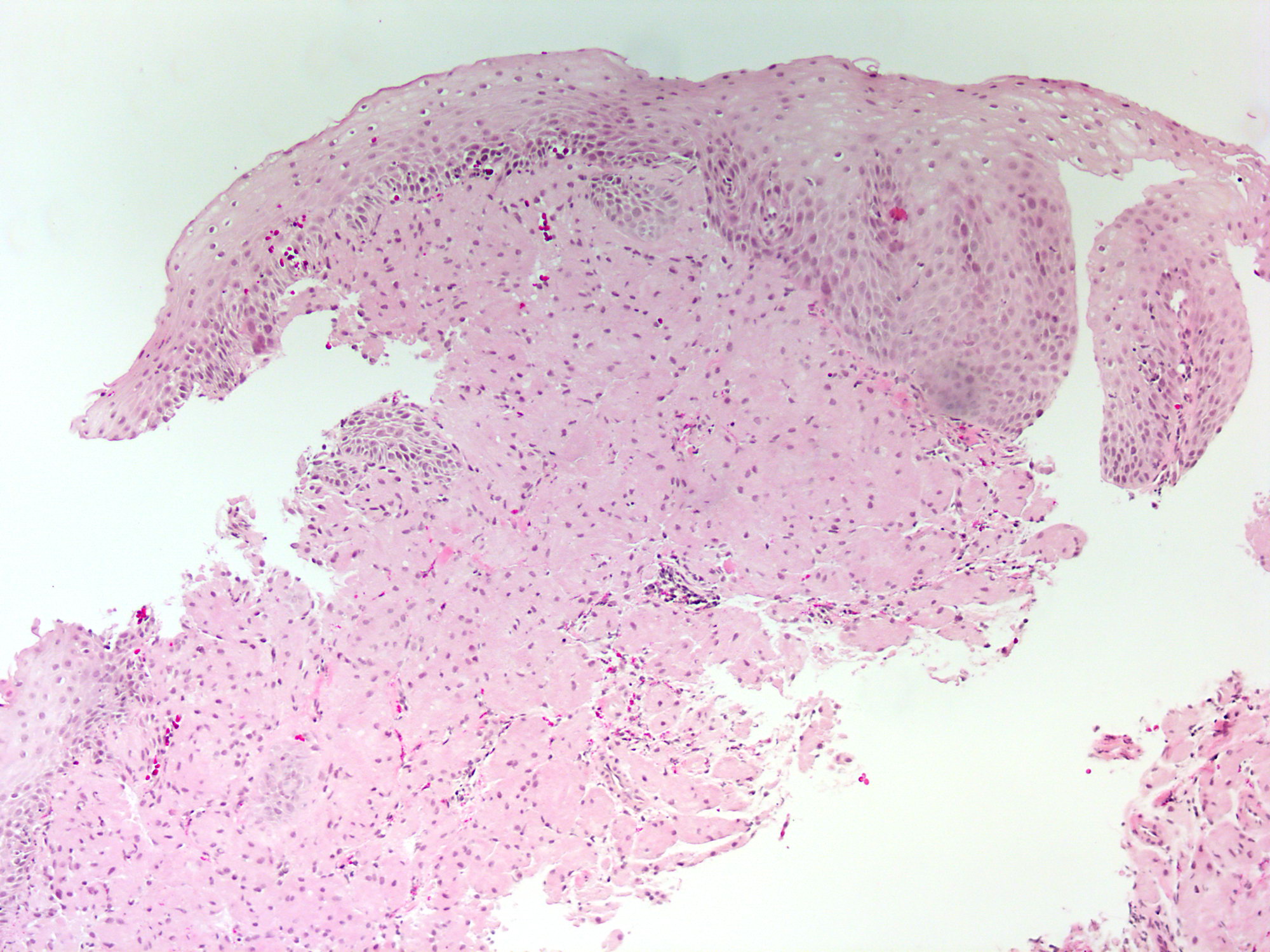
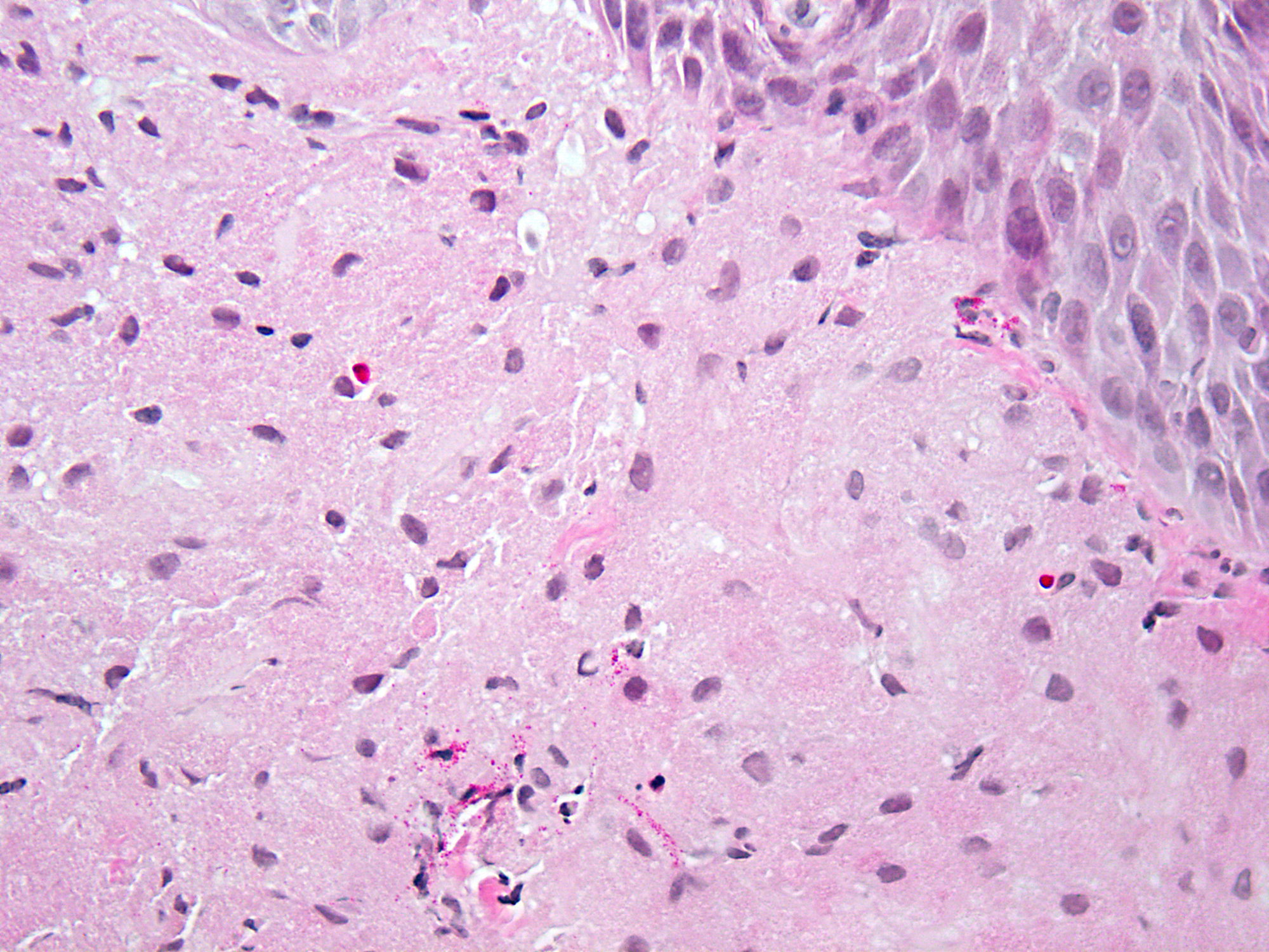
Groups of spindled cells with low cellularity
Contributed by David Matthew Saulino, D.O. and David Hernandez Gonzalo, M.D.
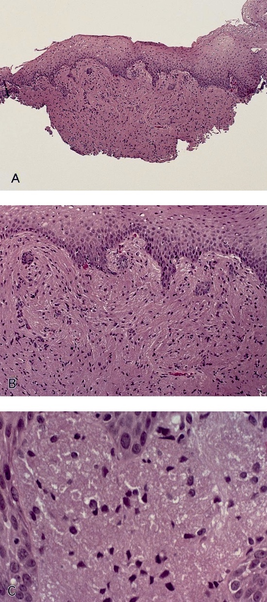
Grade D esophagitis: mucosal
breaks involving at least 75%
of the esophageal circumference
Contributed by David Matthew Saulino, D.O. and David Hernandez Gonzalo, M.D.
Lymphocytes and dyskeratosis
Civatte bodies
Contributed by Omar Aljuboori, M.B.B.S.
Diffuse changes
Contributed by Monica T. Garcia-Buitrago, M.D., Omar Aljuboori, M.B.B.S. and @RaulSGonzalezMD on Twitter
Intraepithelial lymphocytosis
Damaged esophageal epithelium

Lymphocytic esophagitis

Lymphocytic esophagitis

CD3 stain
CD4 stain
CD8 stain
Images hosted on other servers:
Strictures:

Marked 3 cm narrowing

Irregular outline and multiple filling defects

Luminal narrowing
Esophageal thickening ± mass lesions:
4 x 4 cm esophageal mass

Thickening of the esophageal wall

Thickening the aortic arch to the gastrointestinal junction
Endoscopic ultrasound:

Transmural thickening of the esophageal wall

Hypoechoic thickening
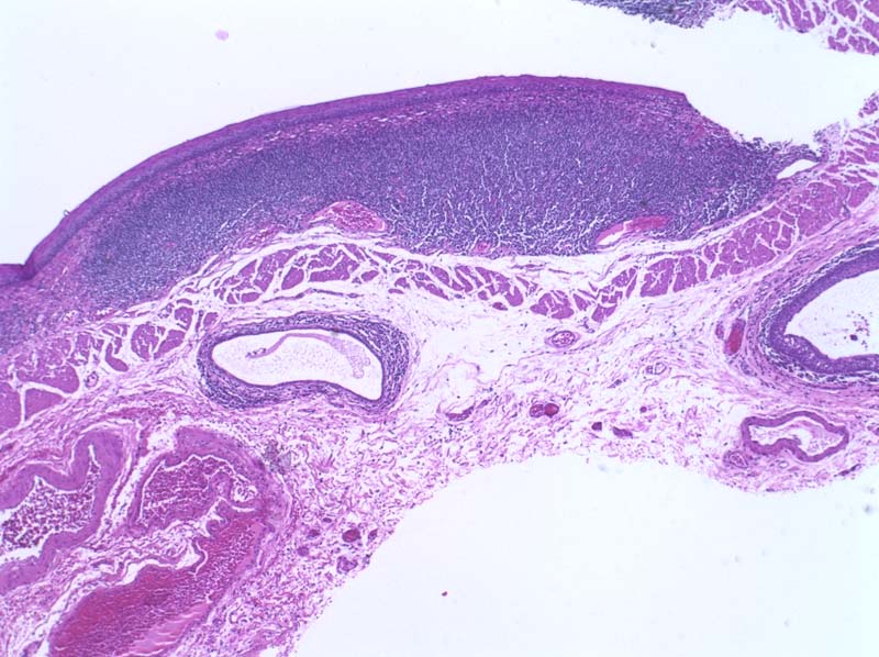
Primary esophageal Hodgkin lymphoma:

Submucosal nodules

Circumferential thickening

Aneurysmal dilatation

Nodular thickening of esophageal wall

Fistula to lobectomy
Images hosted on other servers:
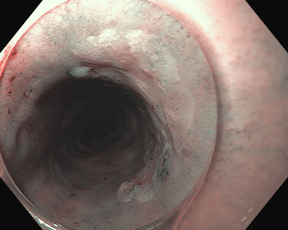
Endoscopy:

Multiple solid, firm nodular lesions

Ulcerated tumor

Large, rounded mass

Solid, firm nodular lesions

Multiple lymphomatous polyposis
Images hosted on other servers:

Ulcerated burgeoning masses

MALT: esophagus /
proximal stomach
are unremarkable
Images hosted on other servers:

Diffuse large B cell lymphoma
Mantle cell lymphoma

Mantle cell lymphoma


Mycosis fungoides
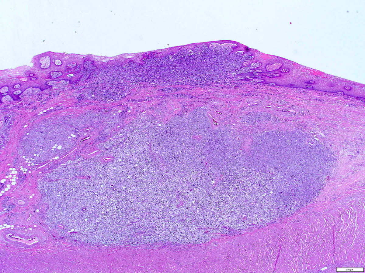
MALT lymphoma:

Dense submucosal lymphocytic infiltrate
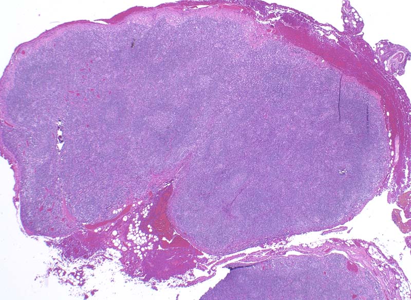
Vaguely nodular growth pattern
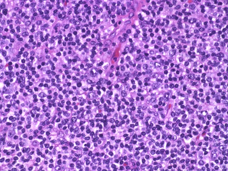
Monocytoid appearance

Germinal centers
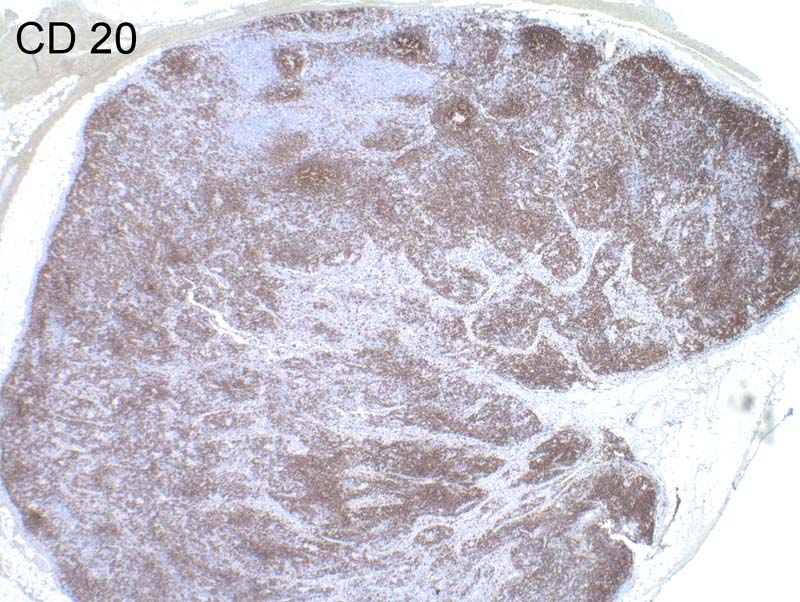
CD20+
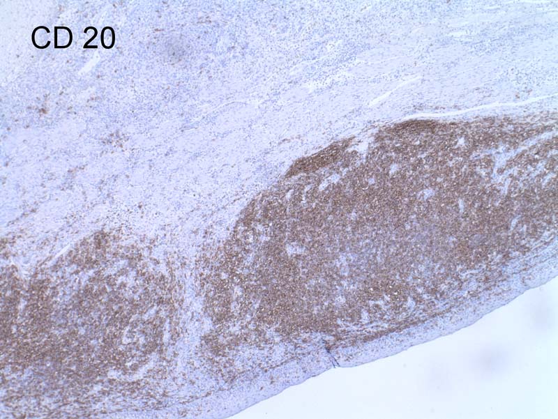
CD20+
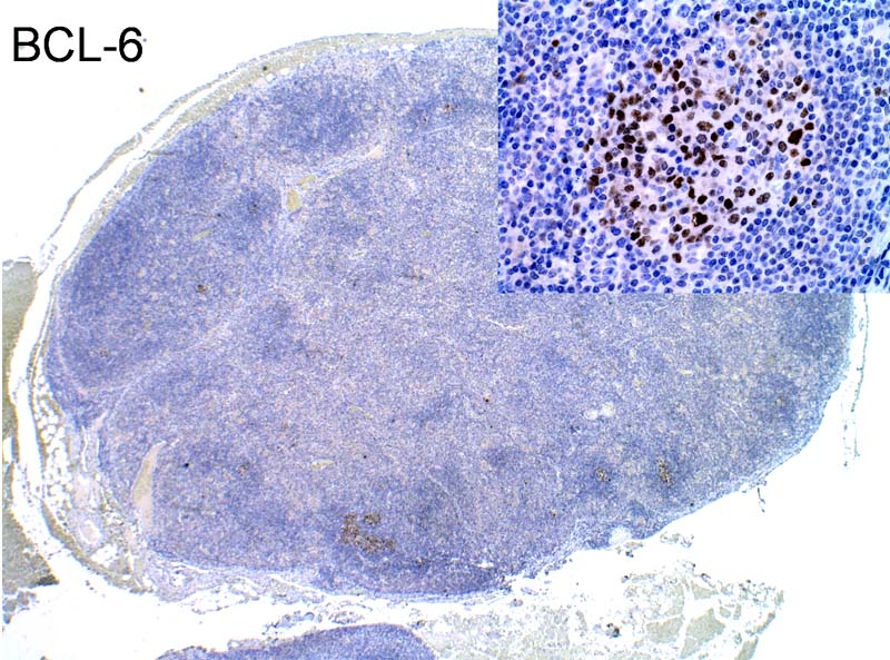
BCL6+ in germinal centers
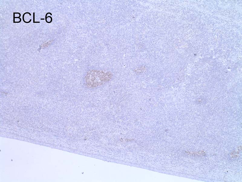
BCL6+
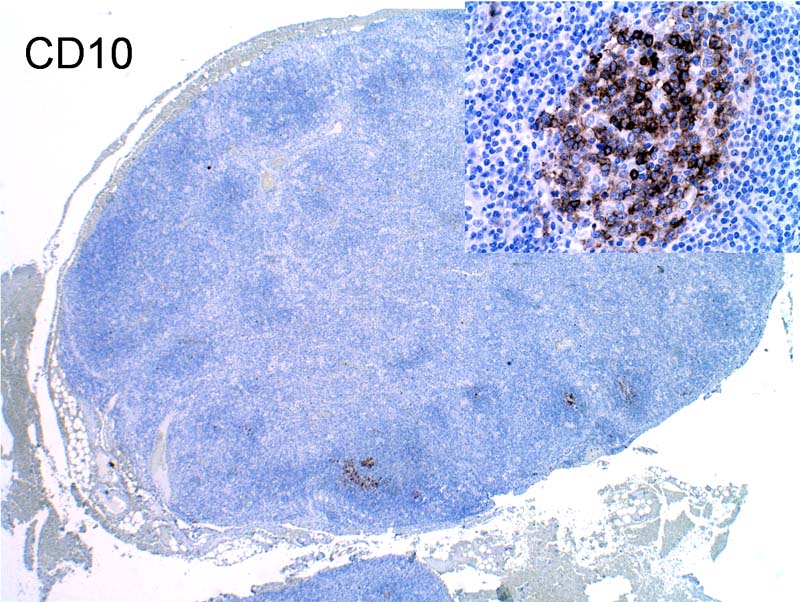
CD10

Ki67
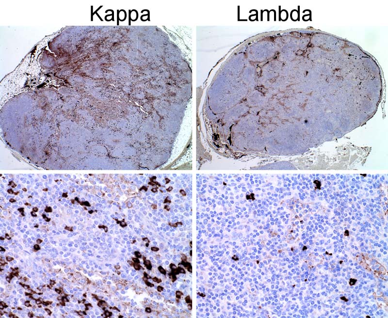
Kappa / lambda
Images hosted on other servers:

DLBCL

Burkitt lymphoma

MALT lymphoma
Contributed by Yukihiro Nakanishi, M.D., Ph.D.

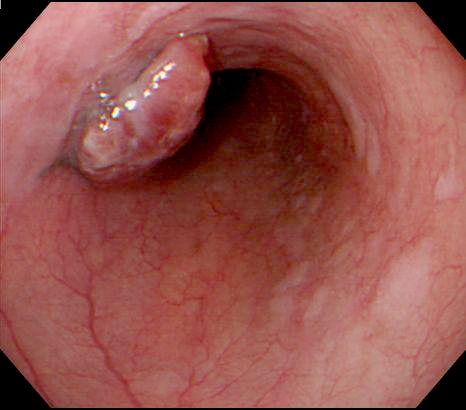
Elevated lesion
Contributed by Yukihiro Nakanishi, M.D., Ph.D.
Protruding mass
Contributed by Yukihiro Nakanishi, M.D., Ph.D.
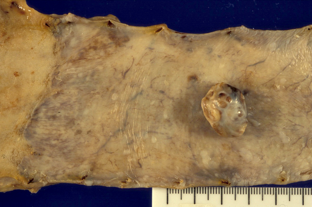
Protruding mass
Contributed by Yukihiro Nakanishi, M.D., Ph.D. and @RaulSGonzalezMD on Twitter
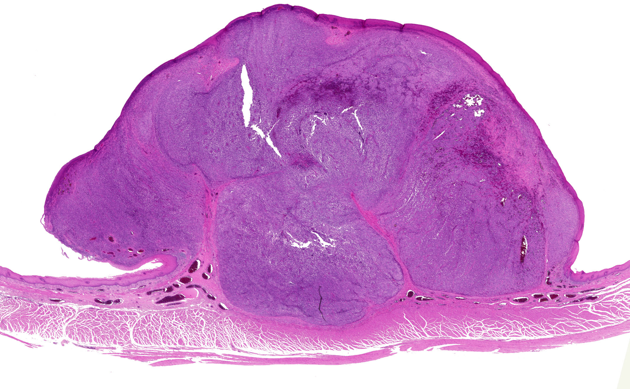
Protruding mass
Prominent nucleoli
Images hosted on other servers:

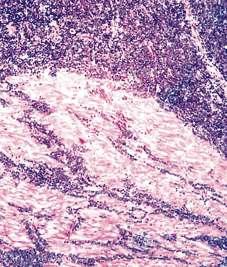
Endoscopic findings
AFIP images

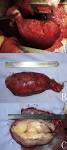
Bulky, ulcerated, infiltrative lesion
Images hosted on other servers:

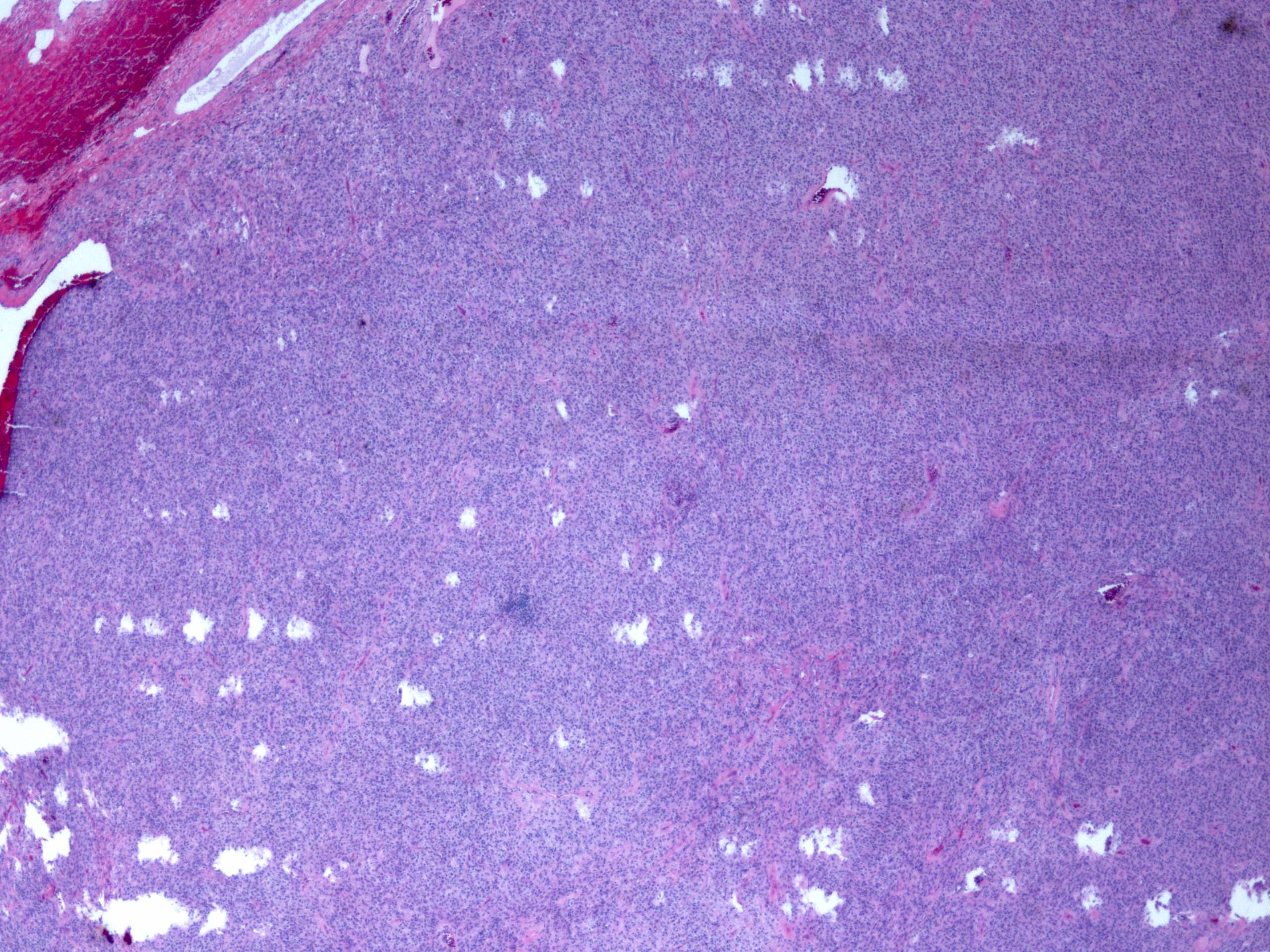
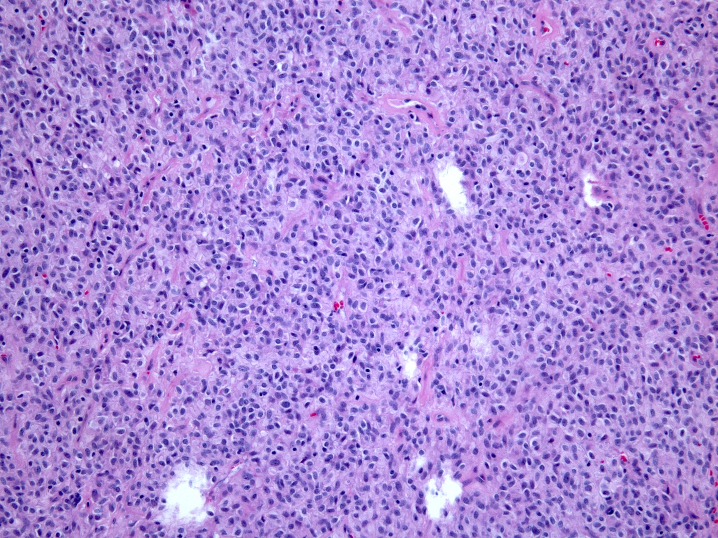
Small cell
carcinoma (upper),
squamous cell
carcinoma (lower)
Contributed by Gillian L. Hale, M.D., M.P.H., Mark R. Wick, M.D. and AFIP
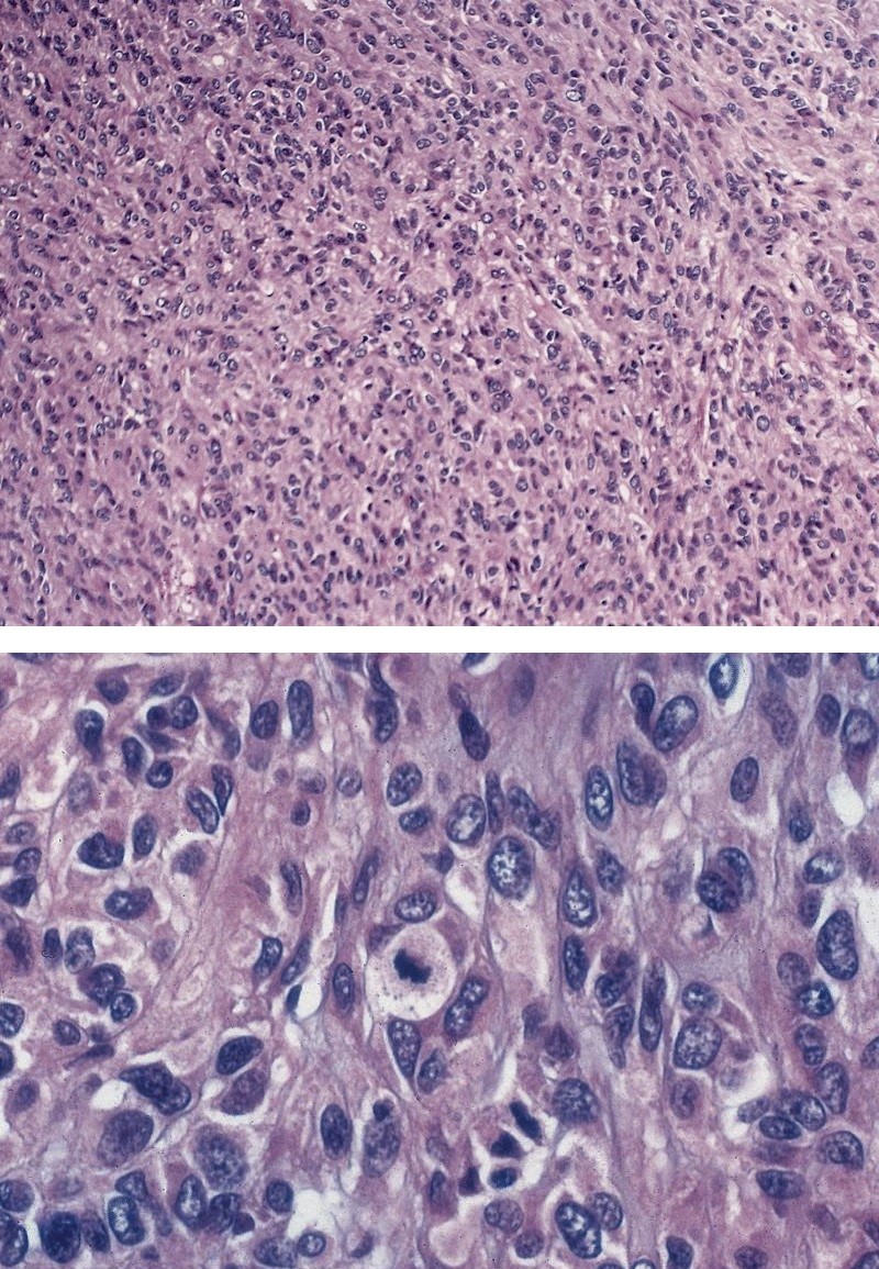
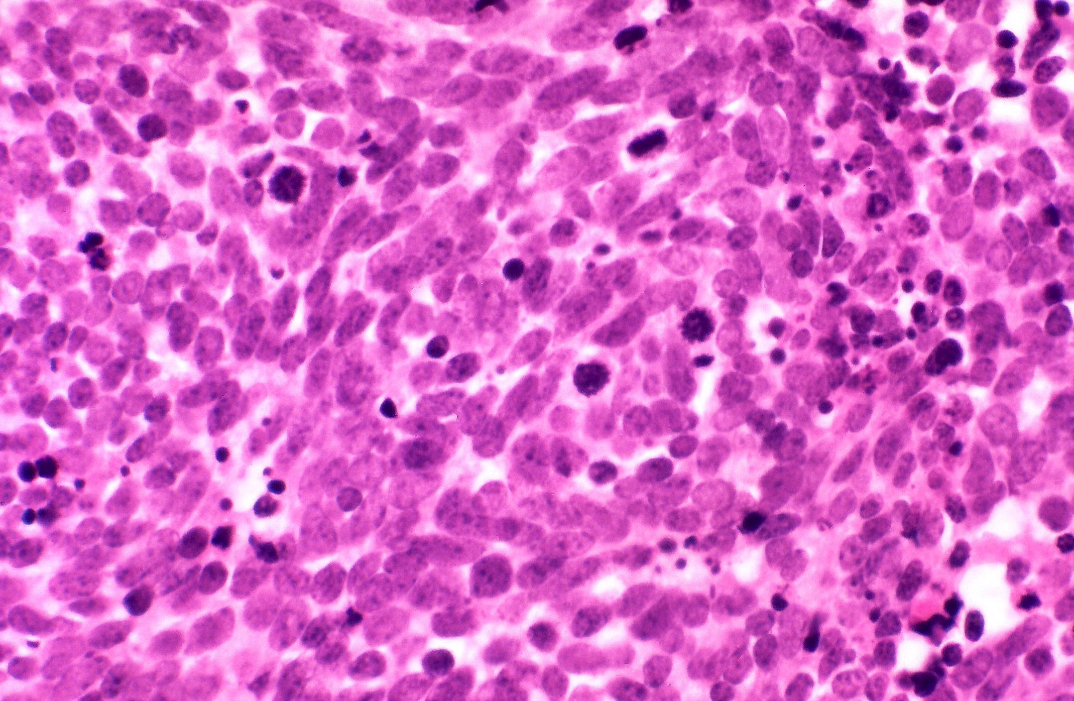
Sheets of tumor cells with central necrosis
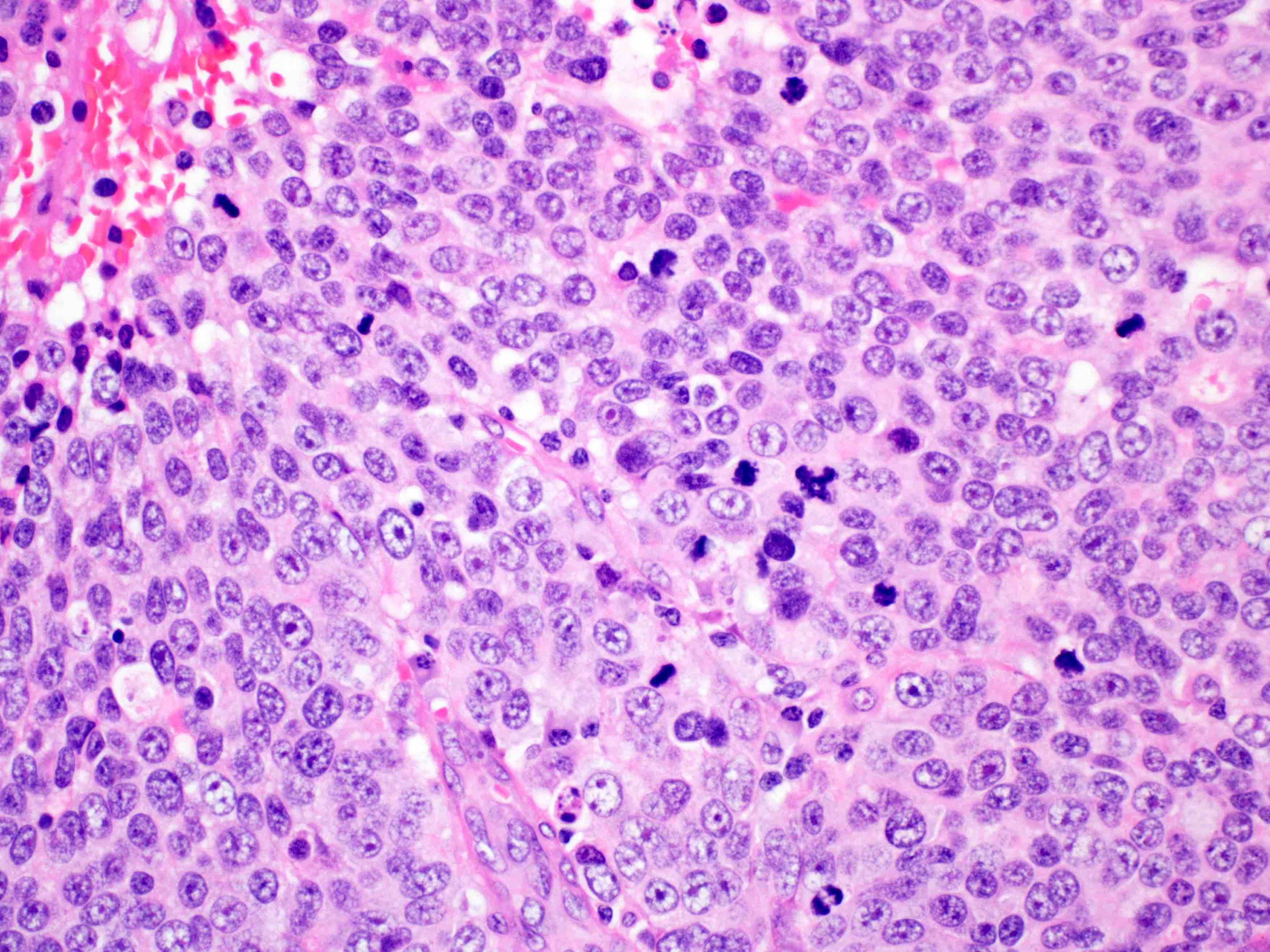
Large cells with
prominent nucleoli
and numerous
mitoses
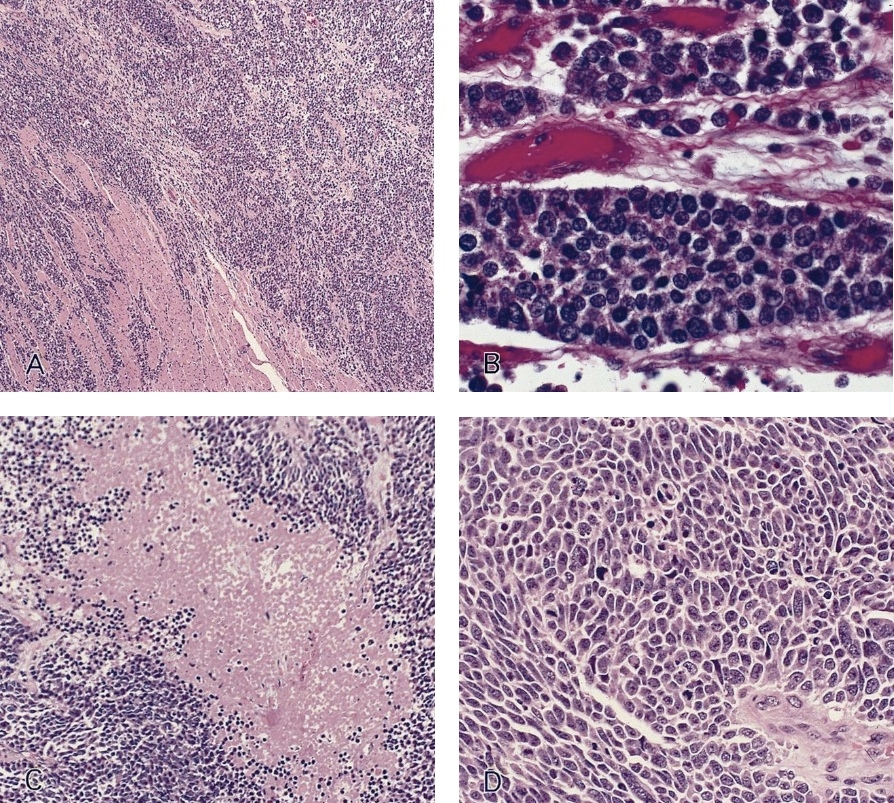

Diffusely infiltrating sheets of small cells
In muscularis
In situ component
Squamous cell differentiation
Pancytokeratin immunostain
Synaptophysin immunostain
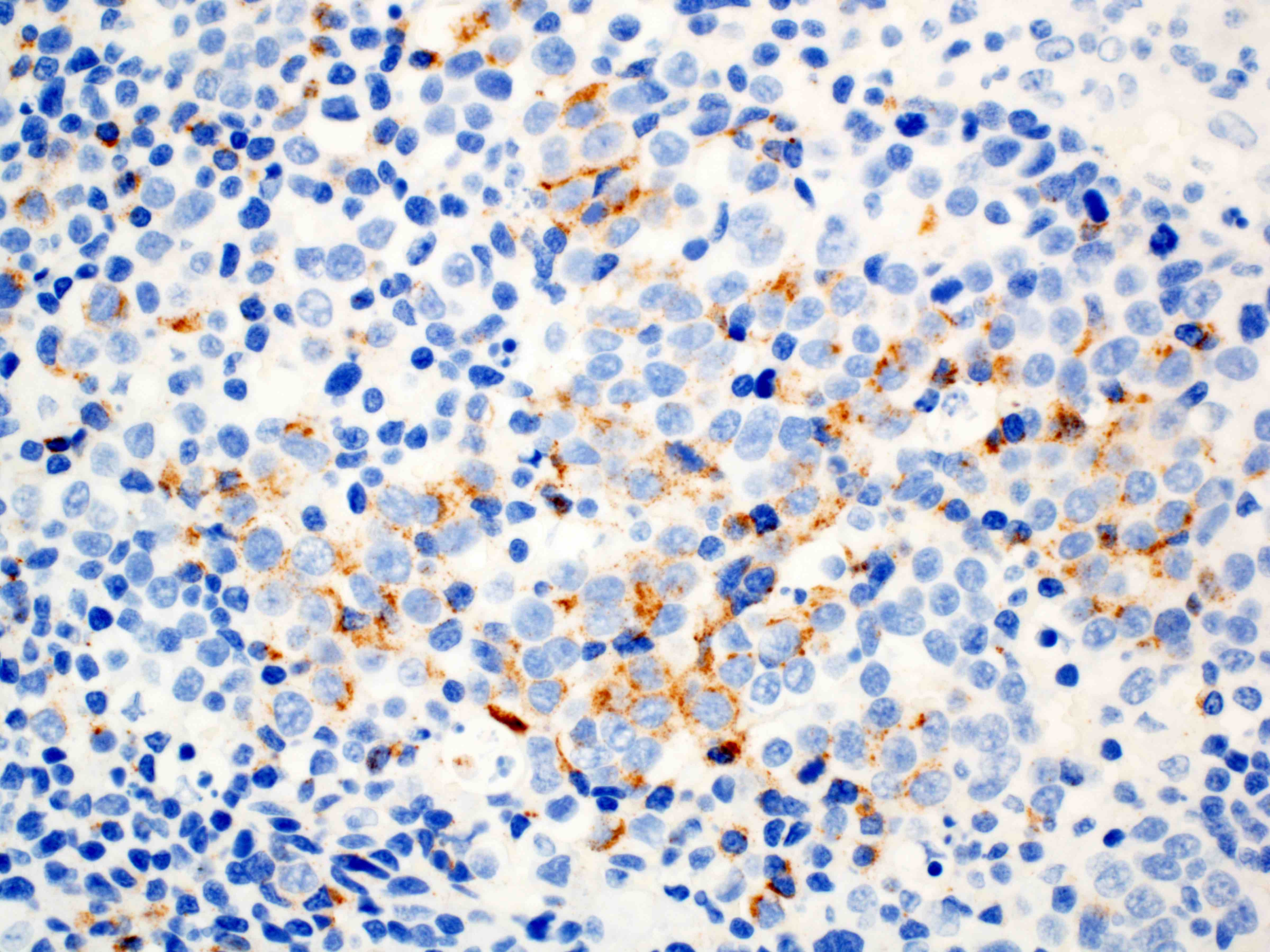
Chromogranin immunostain
Contributed by Yale Rosen, M.D.
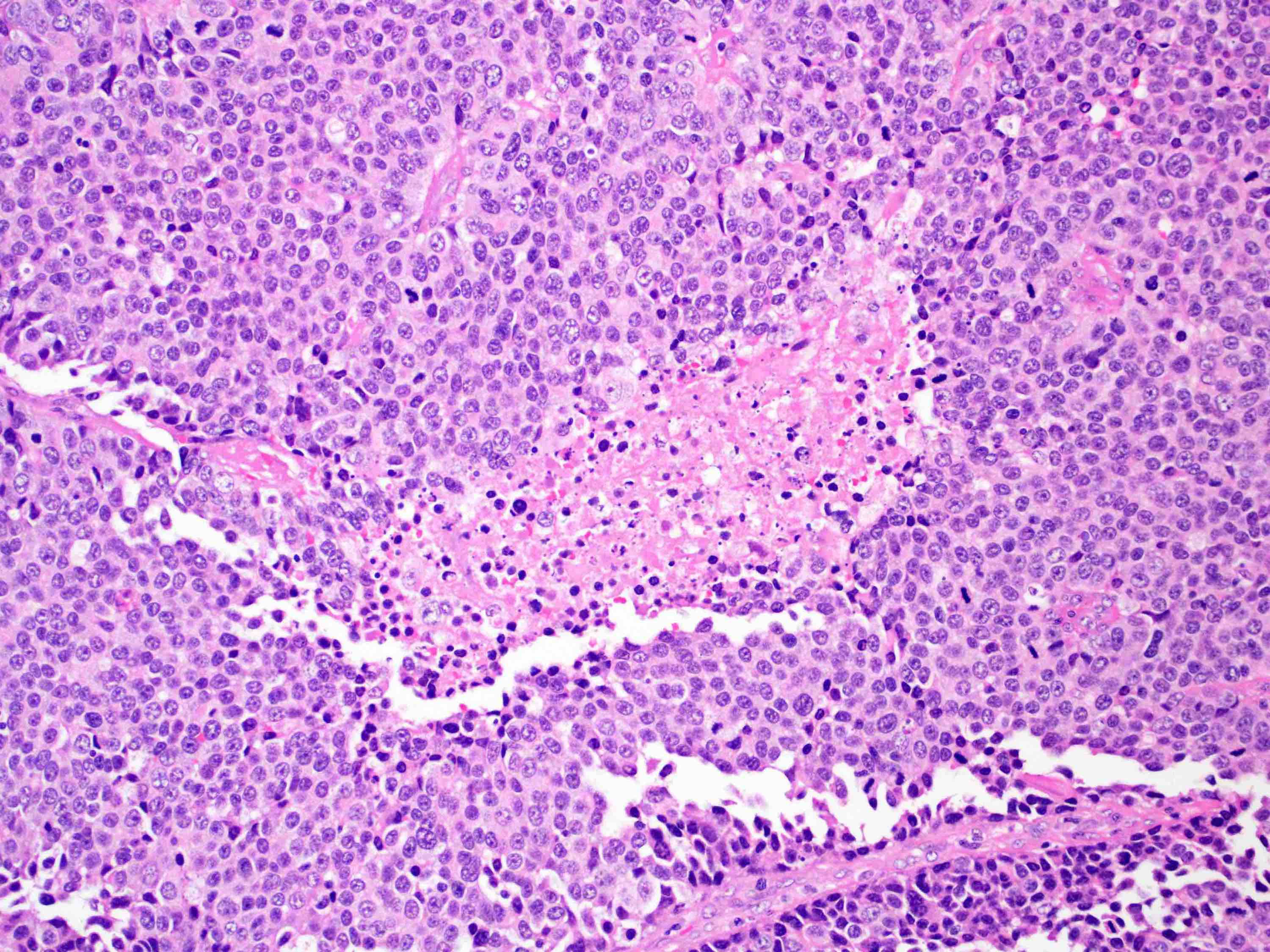
Small cell carcinoma
Images hosted on other servers:

Neurosecretory granules
AFIP images
Bulky, ulcerated, infiltrative lesion
Images hosted on other servers:
Small cell
carcinoma (upper),
squamous cell
carcinoma (lower)
Contributed by Mark R. Wick, M.D. and AFIP images

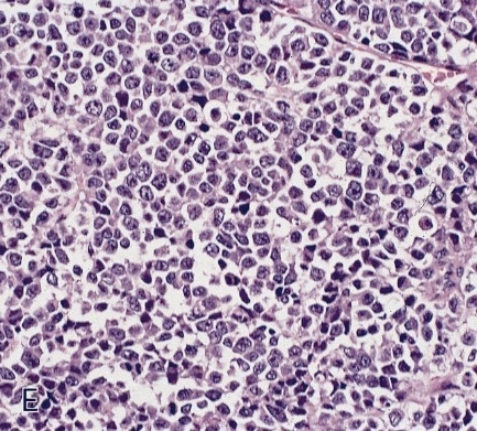
Small cell carcinoma of esophagus
In muscularis
Small cell neuroendocrine carcinoma:
Diffusely infiltrating sheets of small cells
In situ component
Squamous cell differentiation
Contributed by Mark R. Wick, M.D.
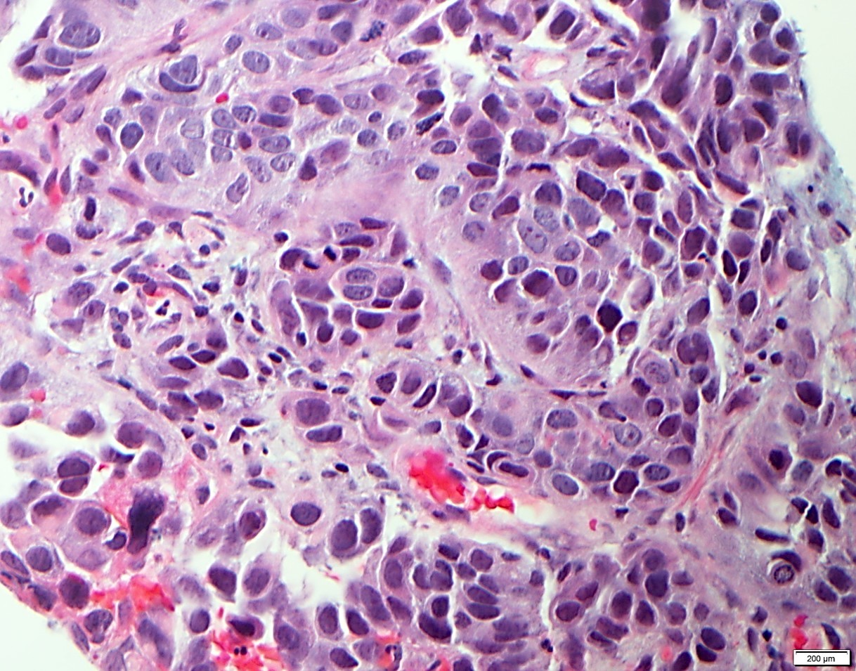
Neuroendocrine tumor
Images hosted on other servers:

Carcinoid tumor

Small cell carcinoma
Images hosted on other servers:
Neurosecretory granules
Contributed by Ankur Sangoi, M.D.
Kayexalate damage:


Various images
AFB stain
AFIP images
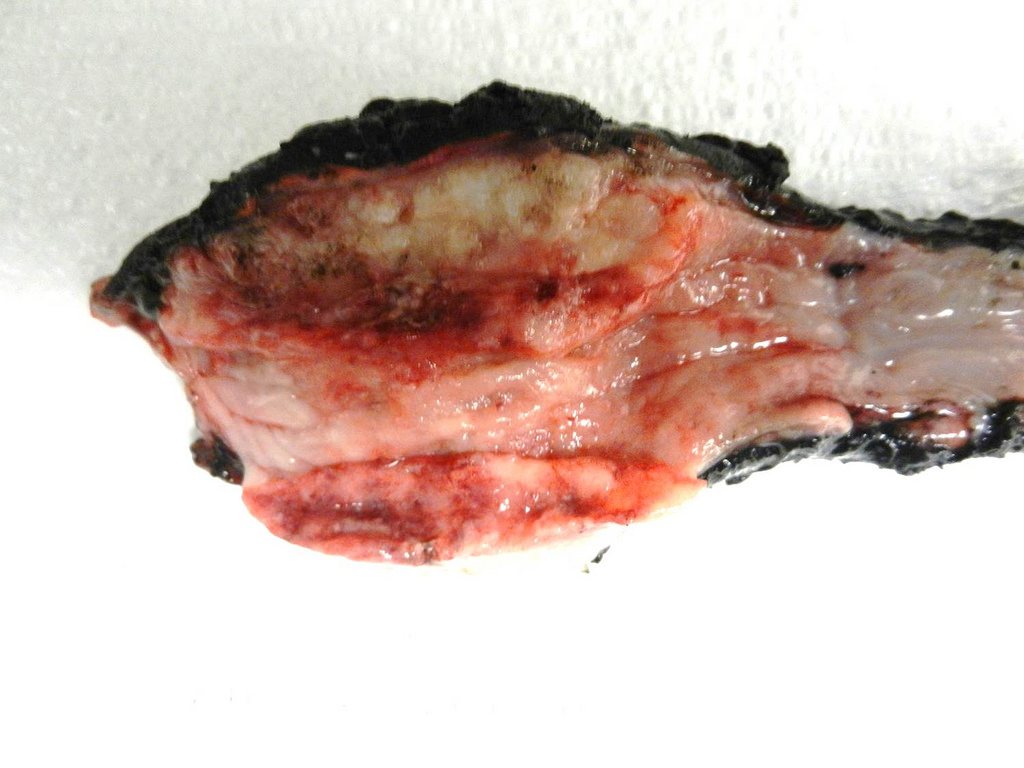
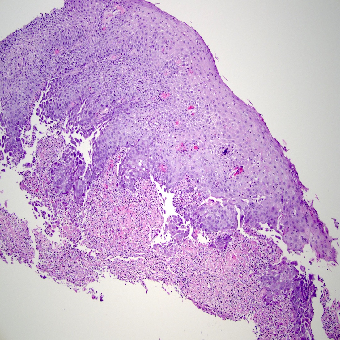
Esophageal squamous cell carcinoma
Radiation has destroyed tumor
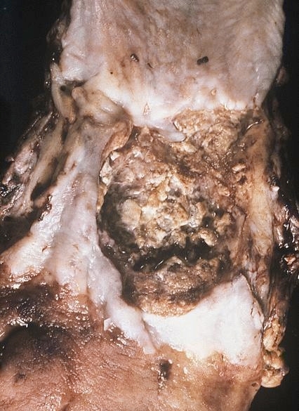
Deep ulcer with necrotic base
AFIP images
Normally oriented
epithelium is
thinner than
normal
Radiated esophageal squamous cell carcinoma
Irradiated squamous carcinoma
Images hosted on other servers:

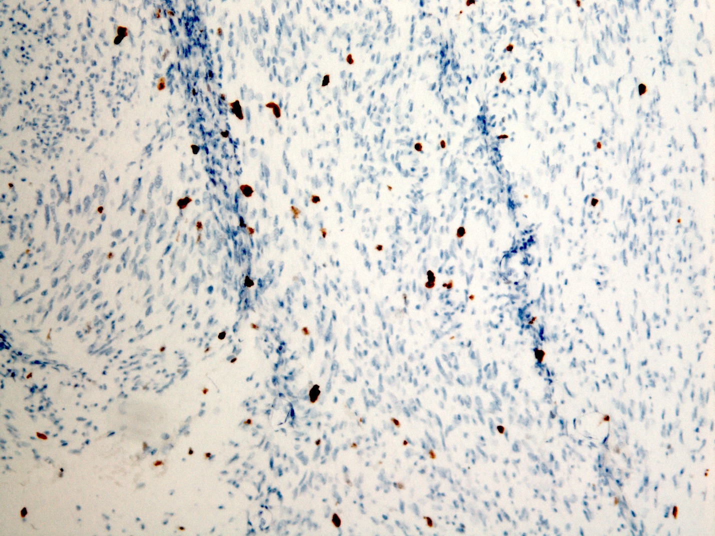
Neovascularization (highlighted with CD31)
Contributed by Aaron R. Huber, D.O.

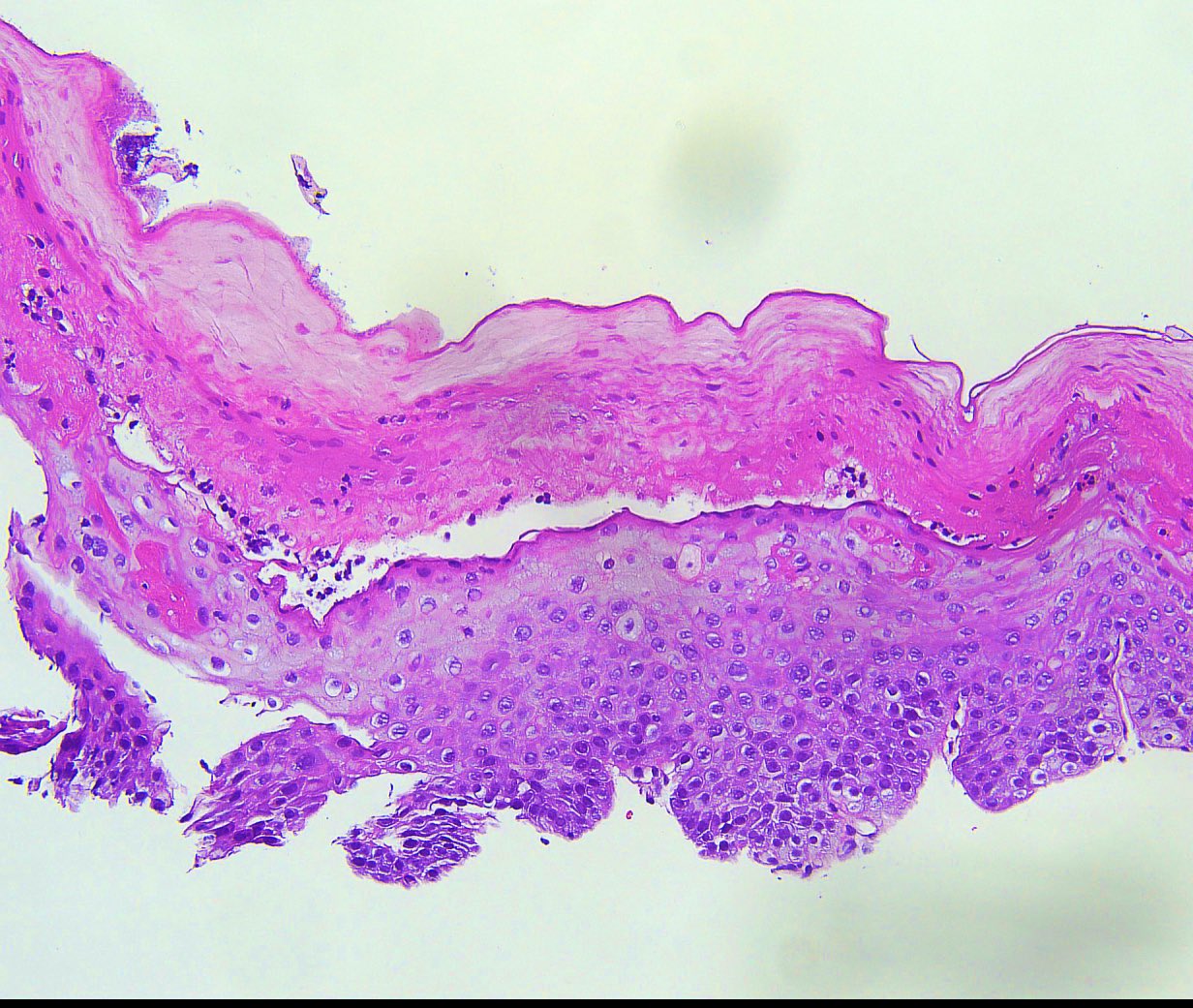
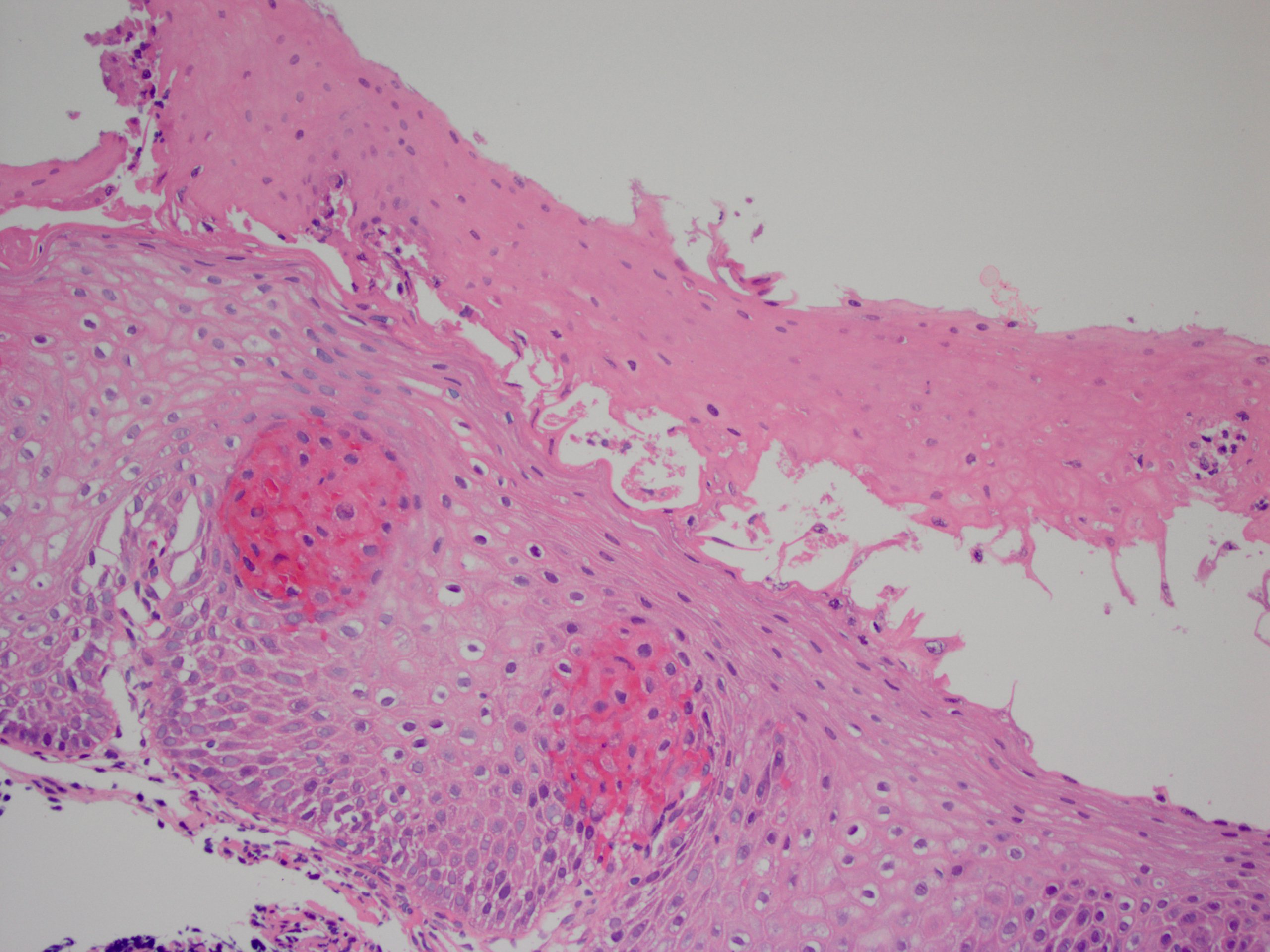
Squamous mucosa
Gastroesophageal junction
Images hosted on other servers:

Netter drawings of A and B rings
Images hosted on other servers:
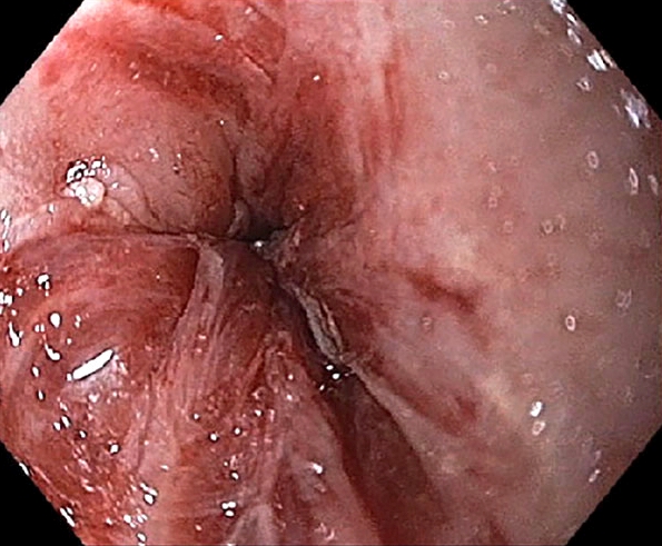
Endoscopy images:
With distal hiatal hernia
Meat (chicken) impaction
Mulitple rings


Schatzki ring
Contributed by Dr. Mark R. Wick
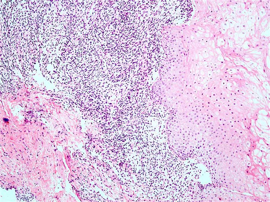
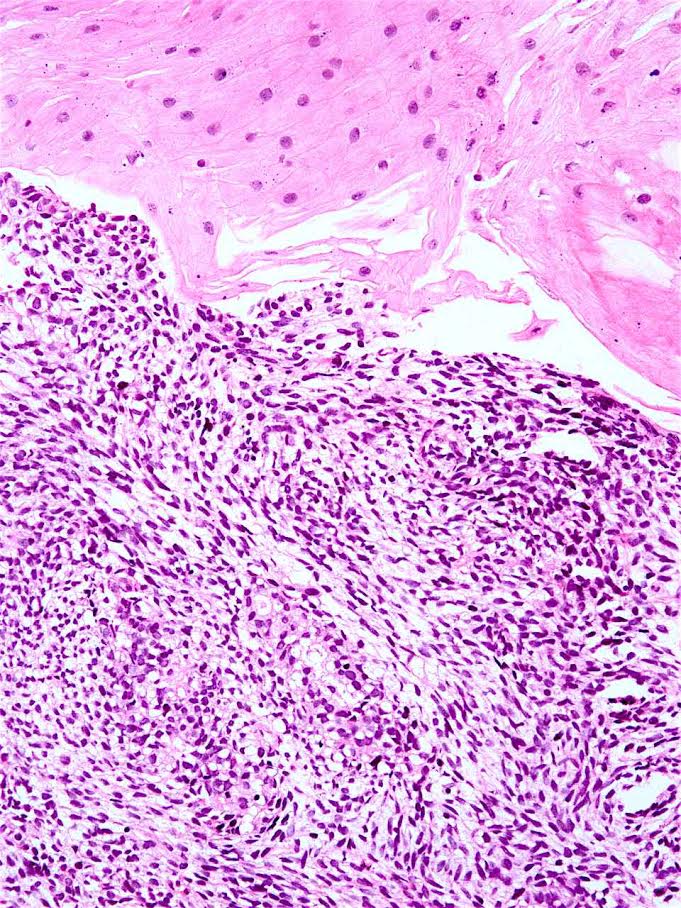
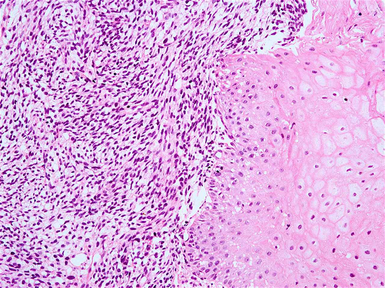
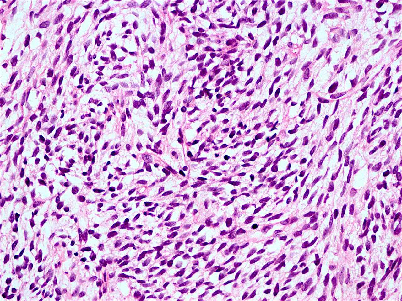
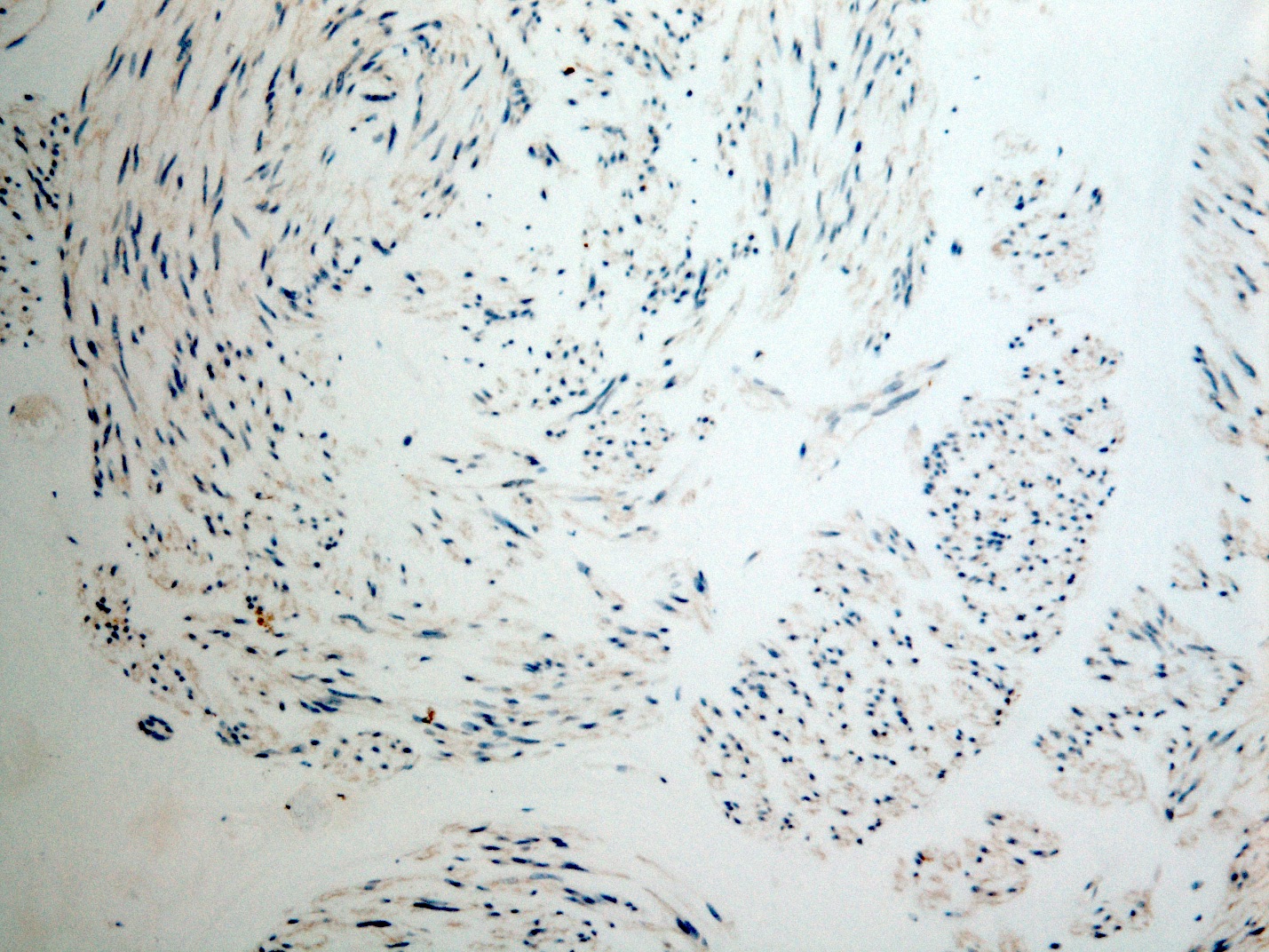
Rhabdomyosarcomatous differentiation
Contributed by Dr. Mark R. Wick

With rhabdomyosarcomatous differentiation
AFIP images

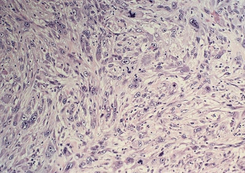
Pseudosarcomatous squamous cell carcinoma
Images hosted on other servers:

8th edition TNM categories
Contributed by Xiaoqin Liu, M.D., Ph.D. and Aaron R. Huber, D.O.

Obstructing lesion of esophagus
Eccentric esophageal wall thickening
Contributed by Xiaoqin Liu, M.D., Ph.D. and Aaron R. Huber, D.O.
Mucosal tearing and ulceration
Contributed by Xiaoqin Liu, M.D., Ph.D. and Aaron R. Huber, D.O.
Fungating / exophytic lesion
Polypoid lesion
Ulcerated lesion
Cavitary lesion
Contributed by Xiaoqin Liu, M.D., Ph.D. and Aaron R. Huber, D.O.
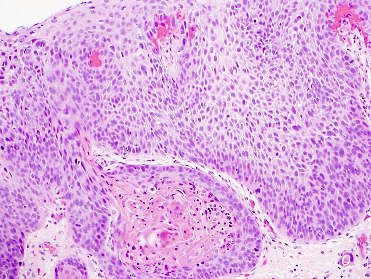
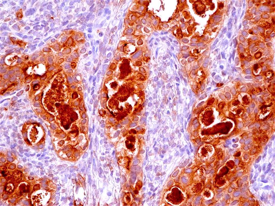
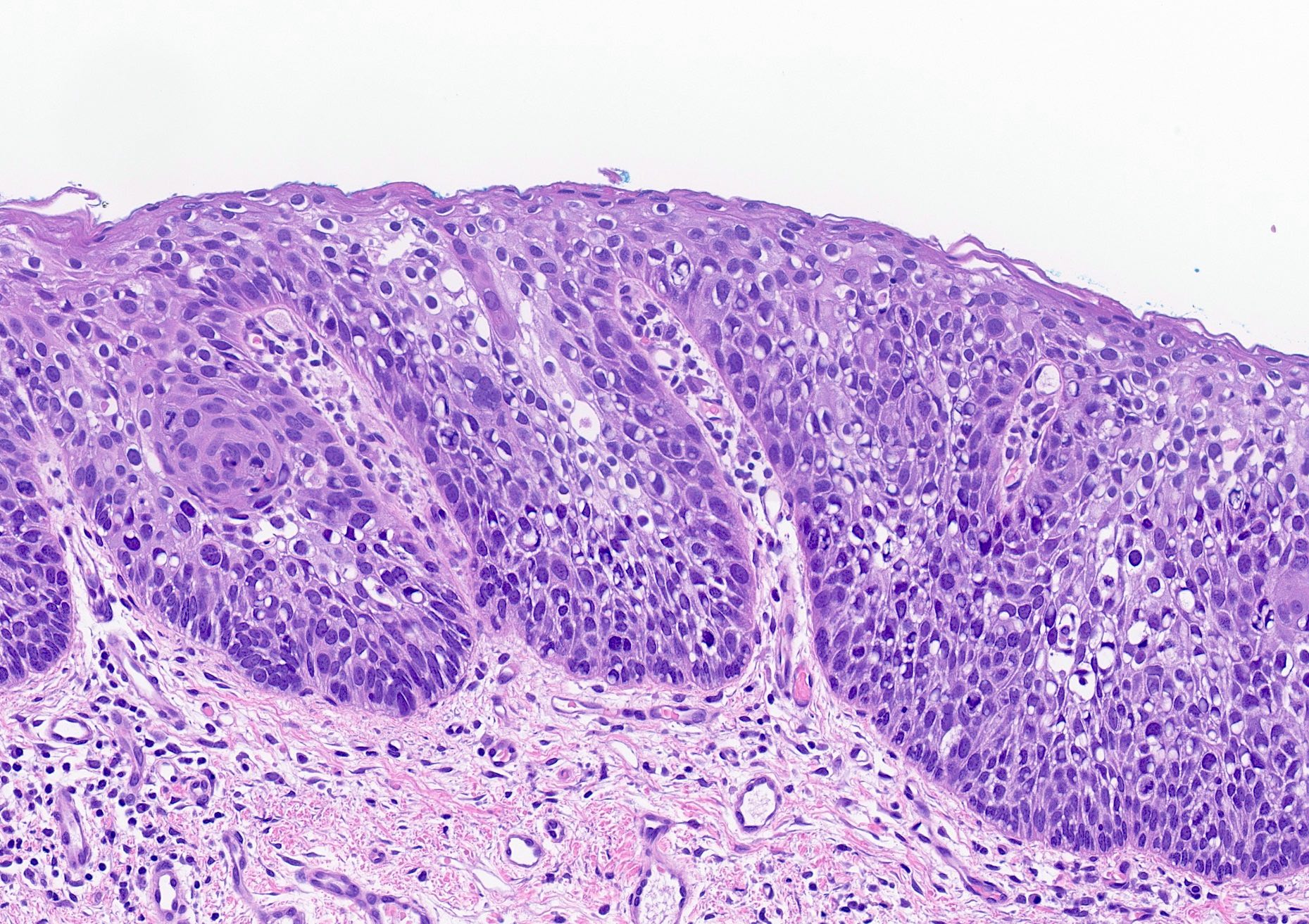
Well differentiated carcinoma
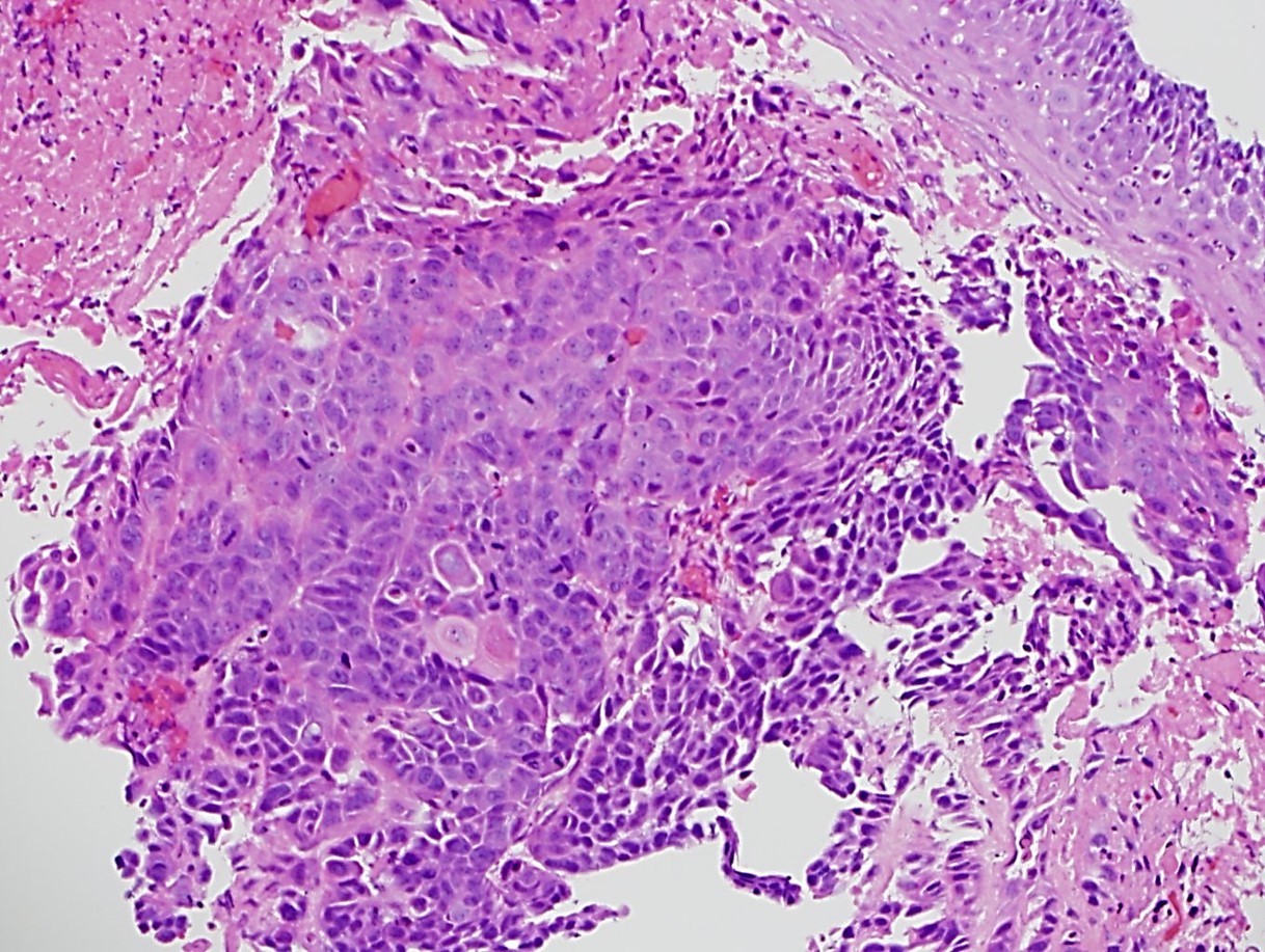
Moderately differentiated carcinoma
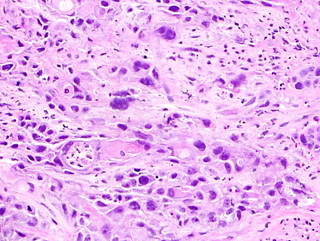
Poorly differentiated carcinoma
Carcinoma with pagetoid cells
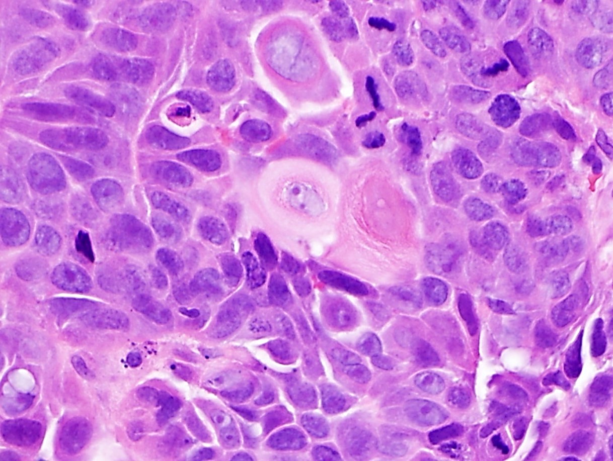
Keratinizing well differentiated carcinoma
Desmoplastic changes
Lymphovascular invasion
p40 stain
Kreyberg stain
Contributed by Saadiya Nazli, M.D. and Catherine E. Hagen, M.D.
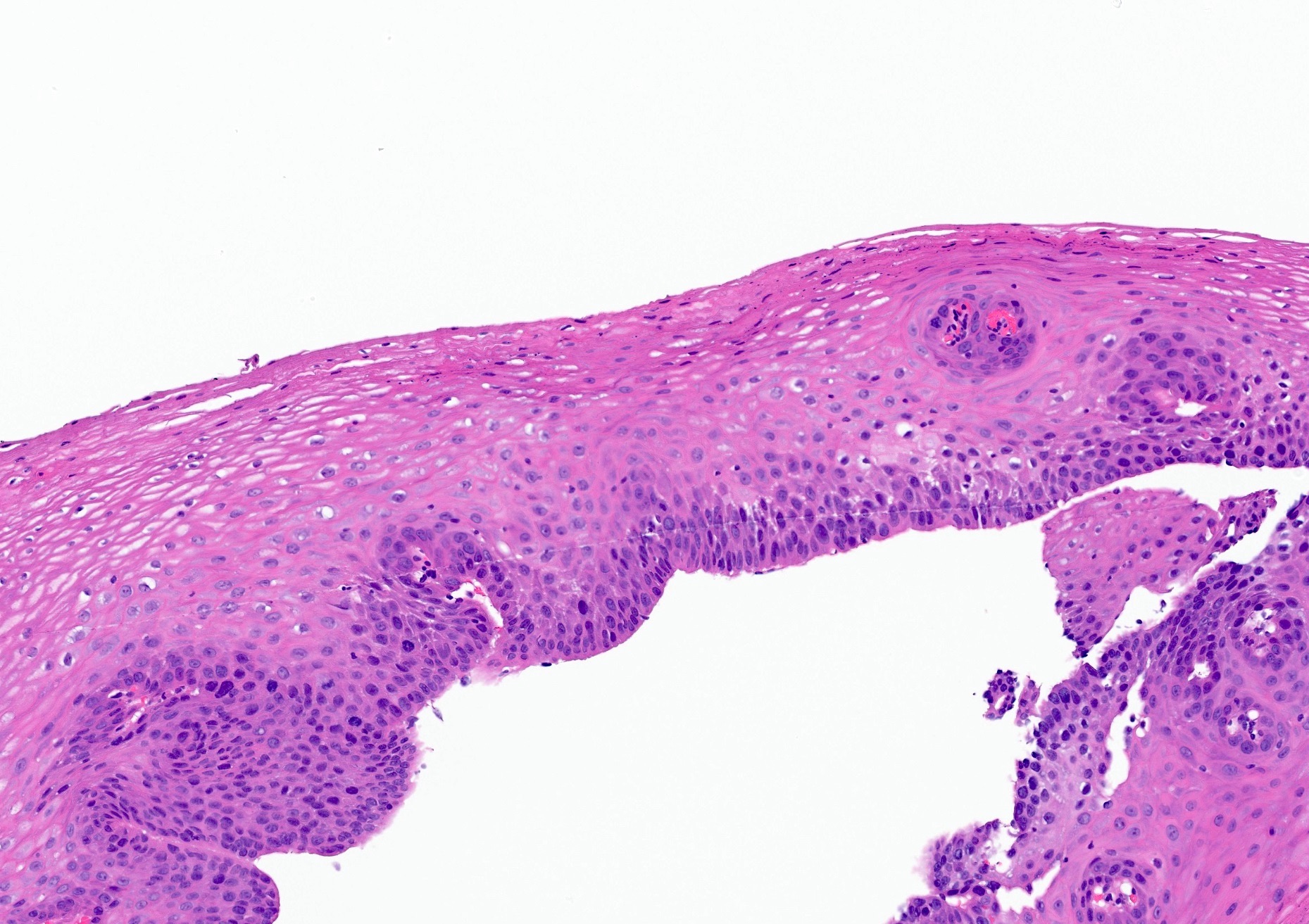
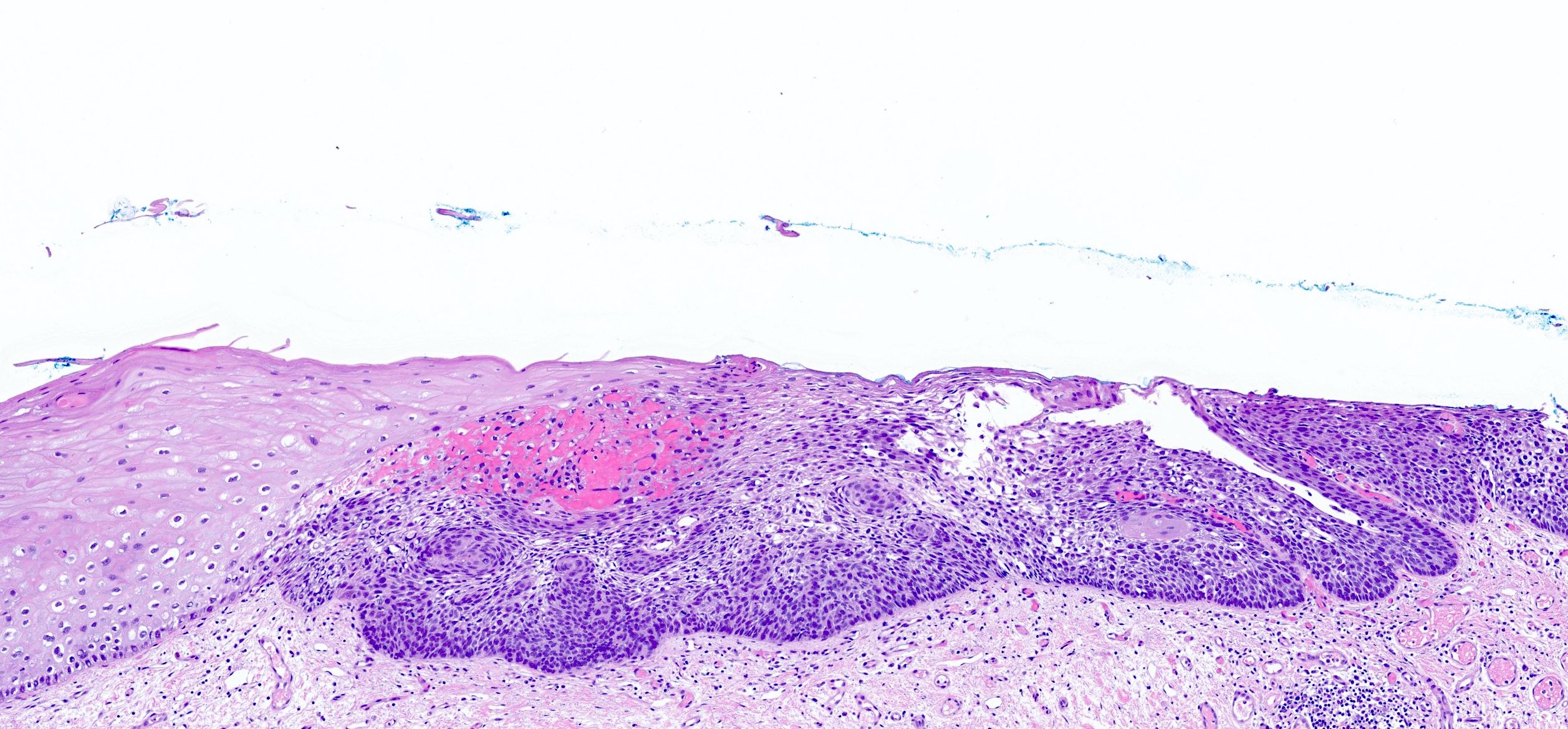
EGD area of nodularity
Images hosted on other servers:

Lugol iodine staining

Epidermoid metaplasia
Contributed by Saadiya Nazli, M.D. and Catherine E. Hagen, M.D.
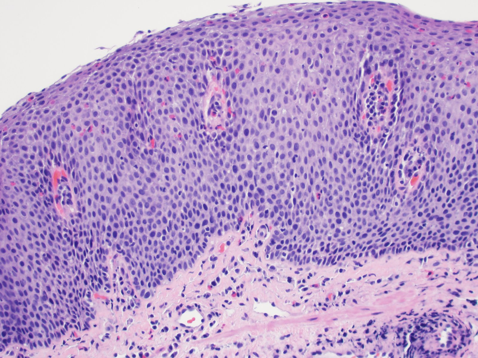
Low grade dysplasia
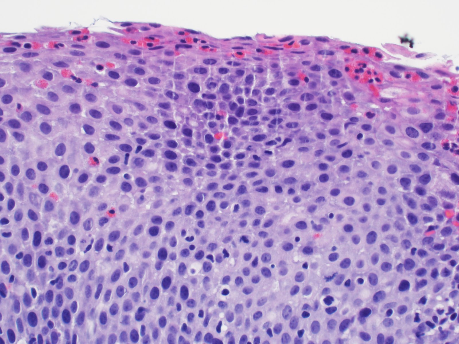
High grade dysplasia (HGD)
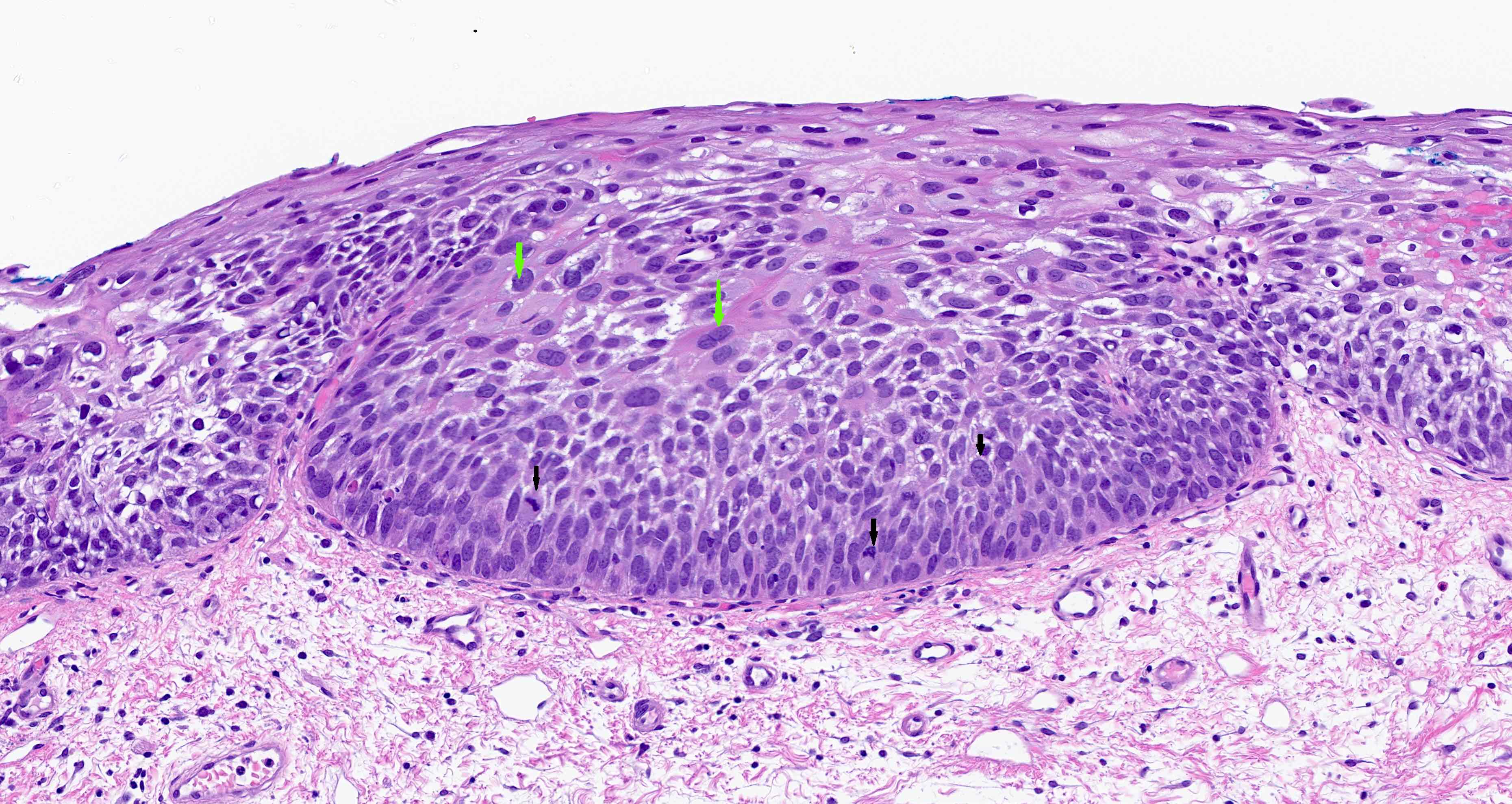
HGD, cytologic changes
HGD, abrupt transition
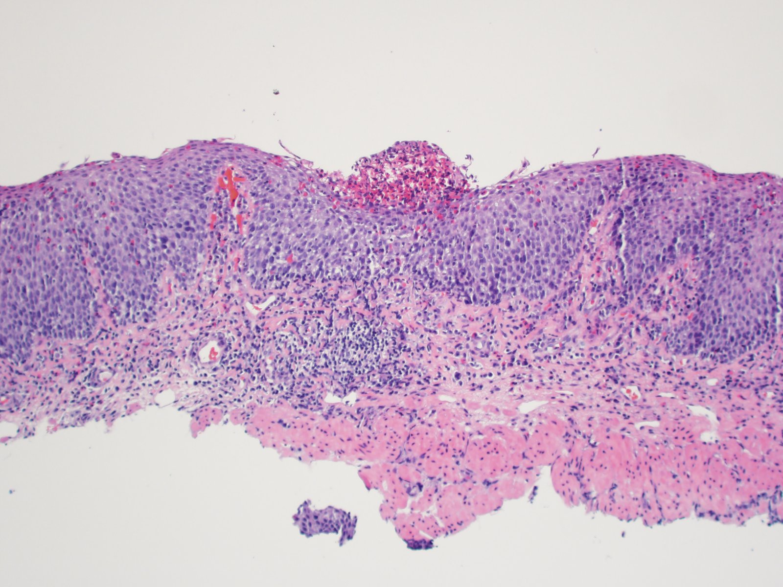
Epidermoid metaplasia and HGD

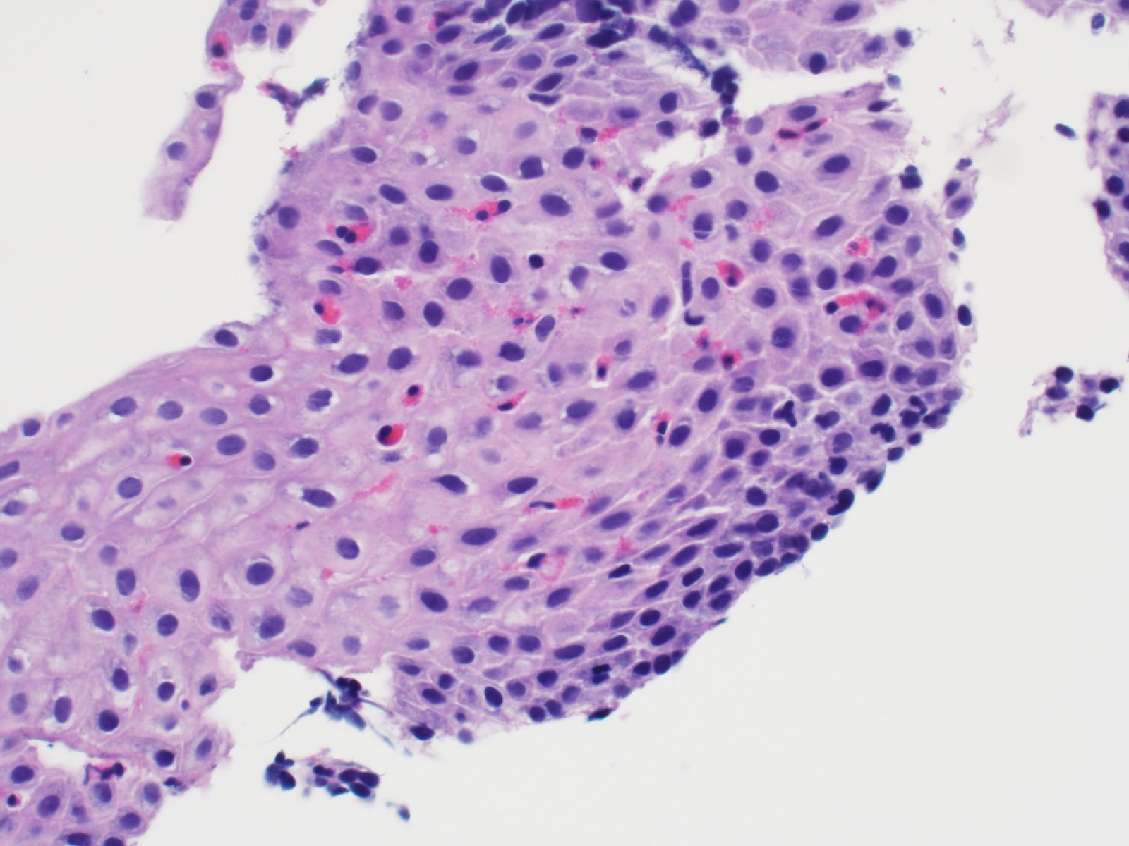
Epidermoid metaplasia
Invasive SCC
EMR
Squamous dysplasia screening and treatment
Images hosted on other servers:

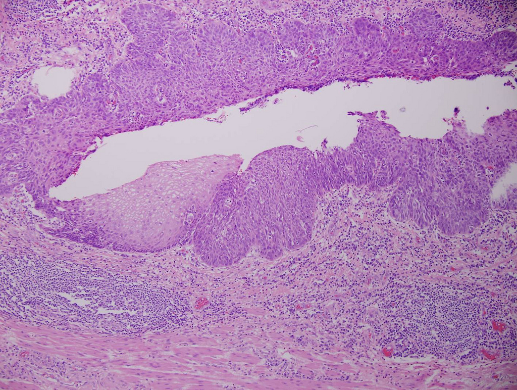
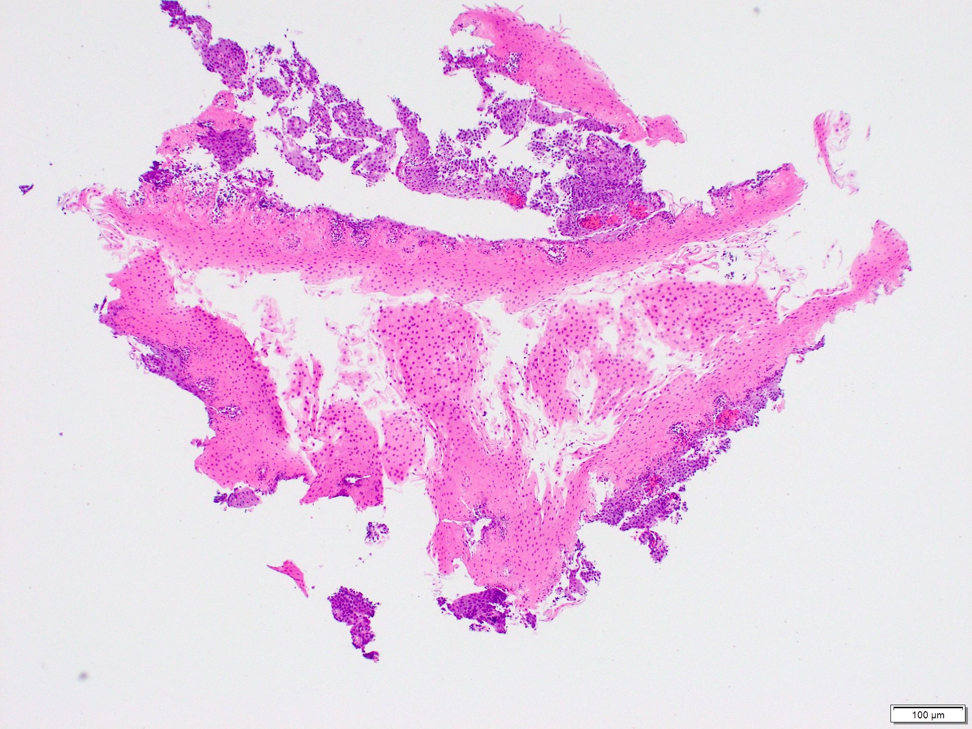
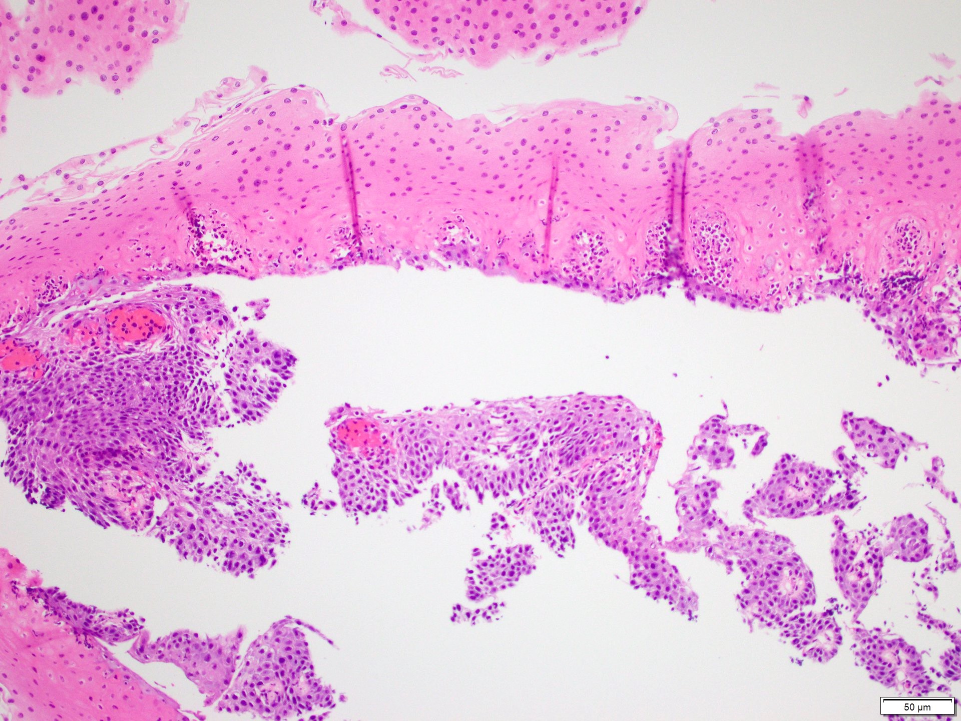
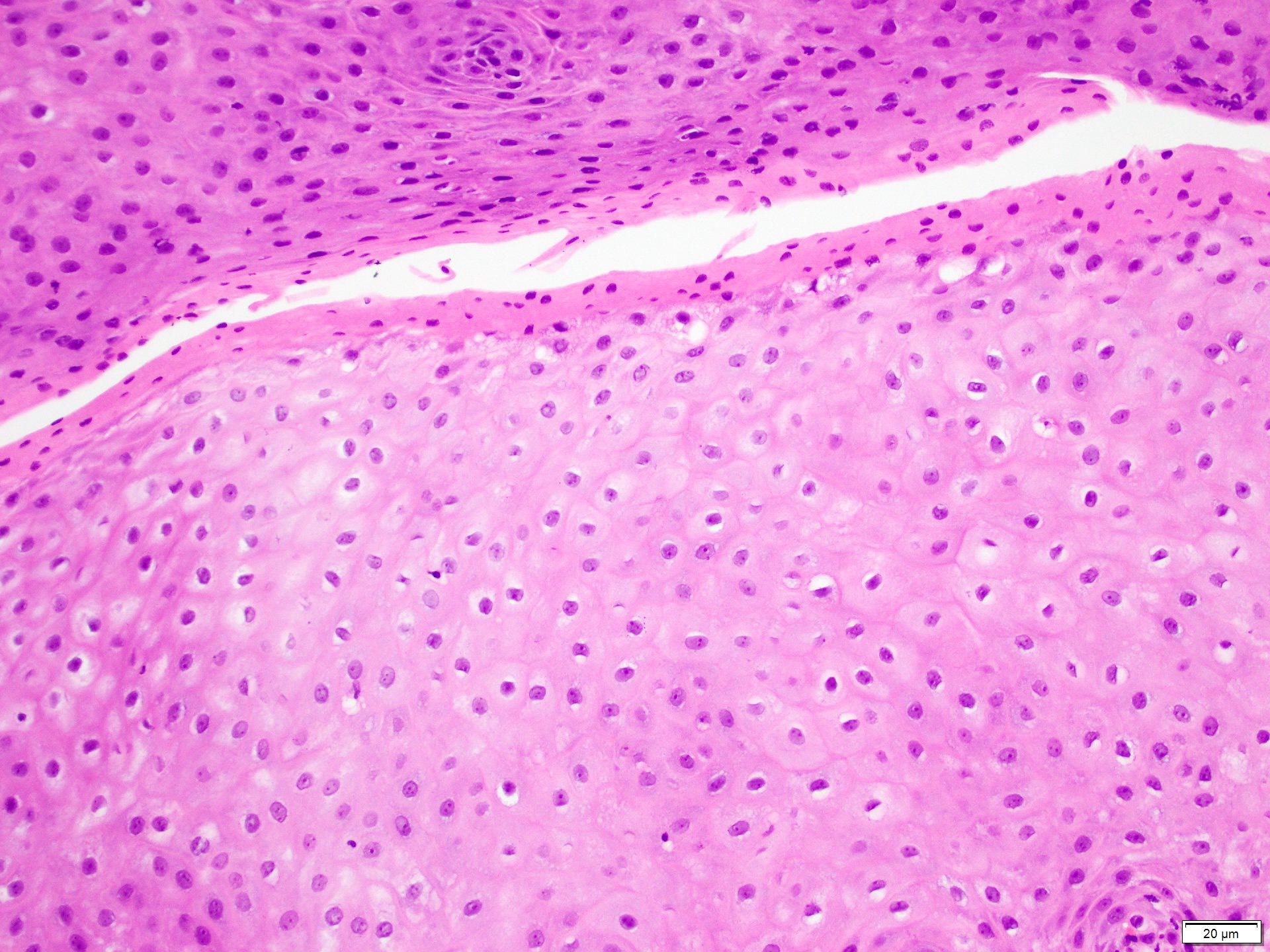
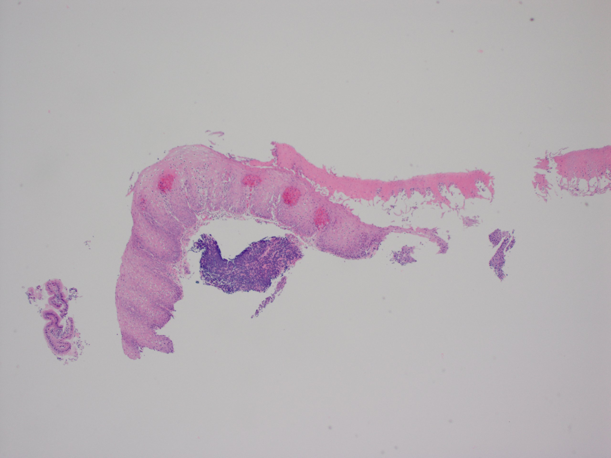
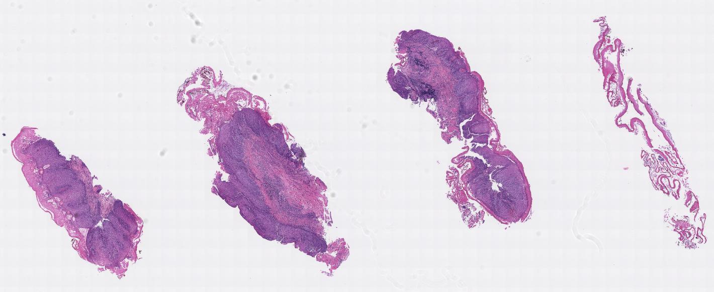
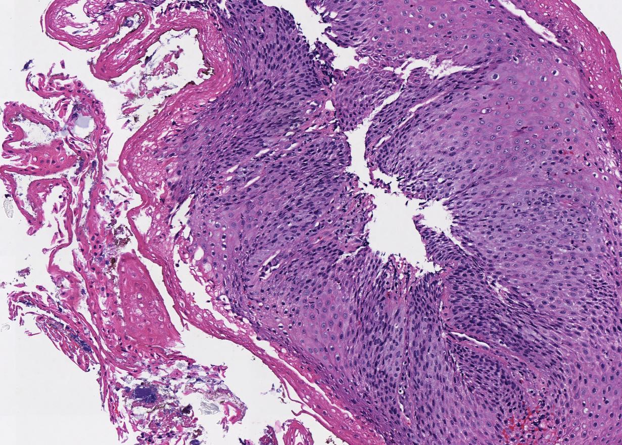
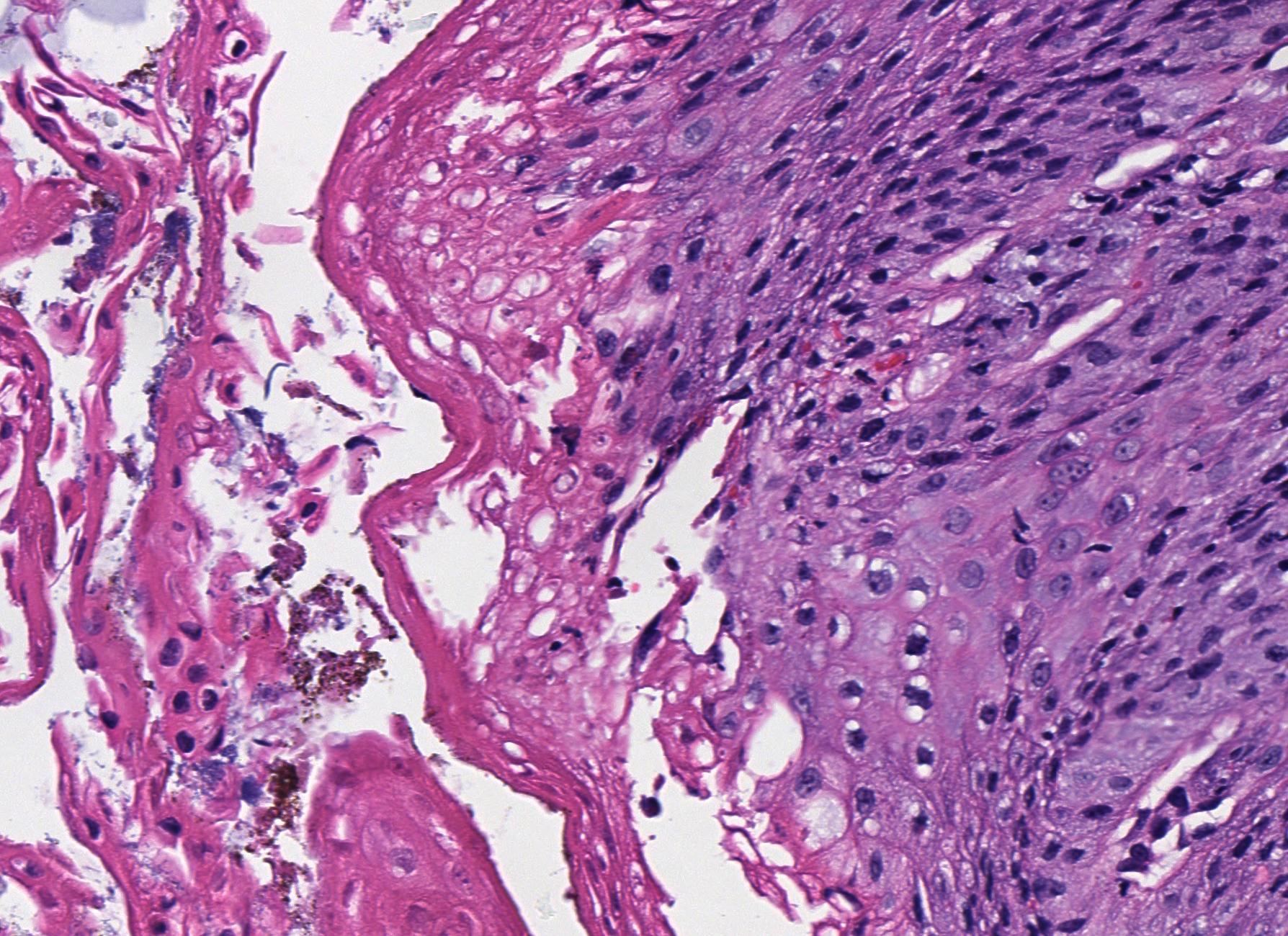
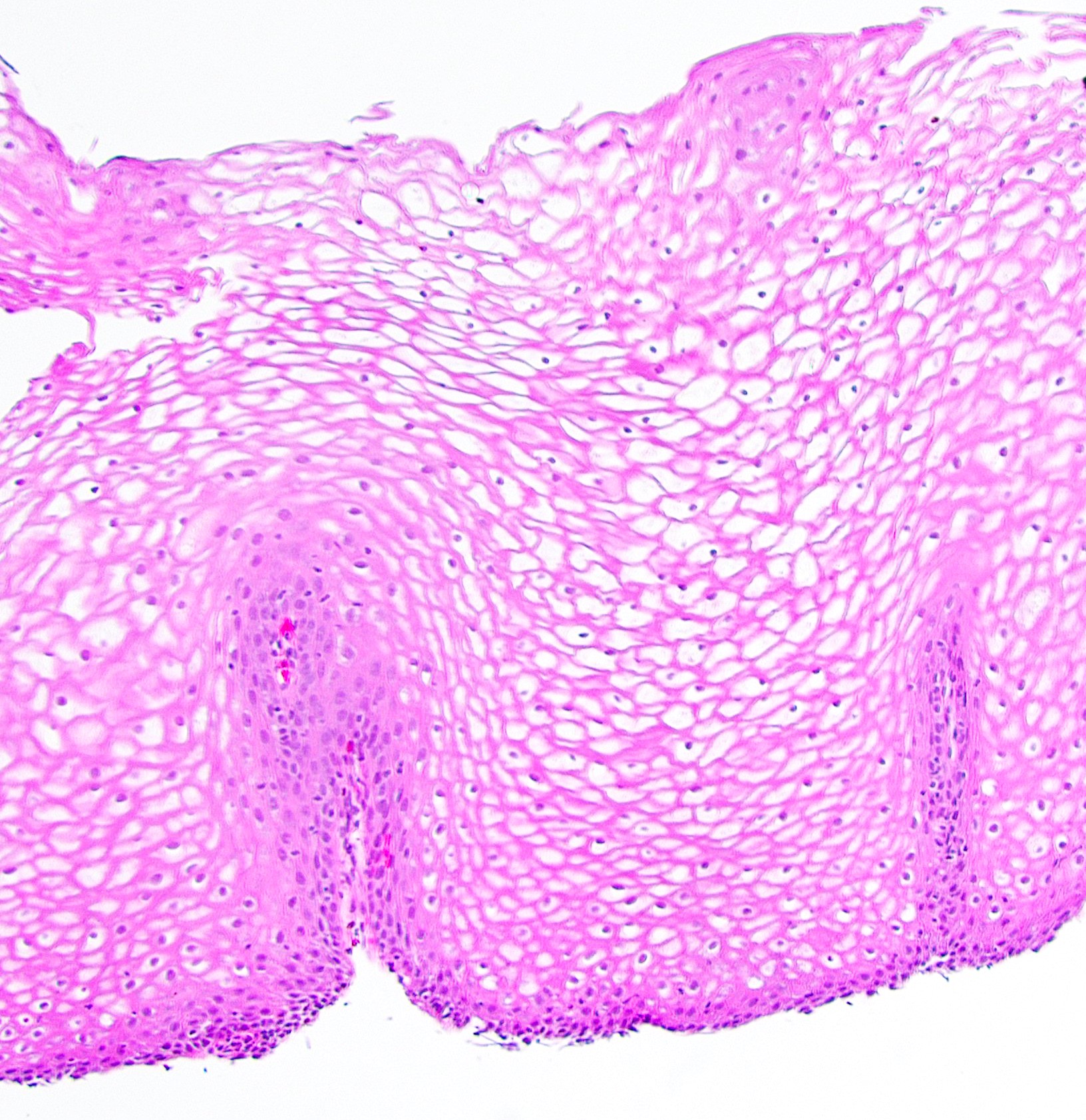
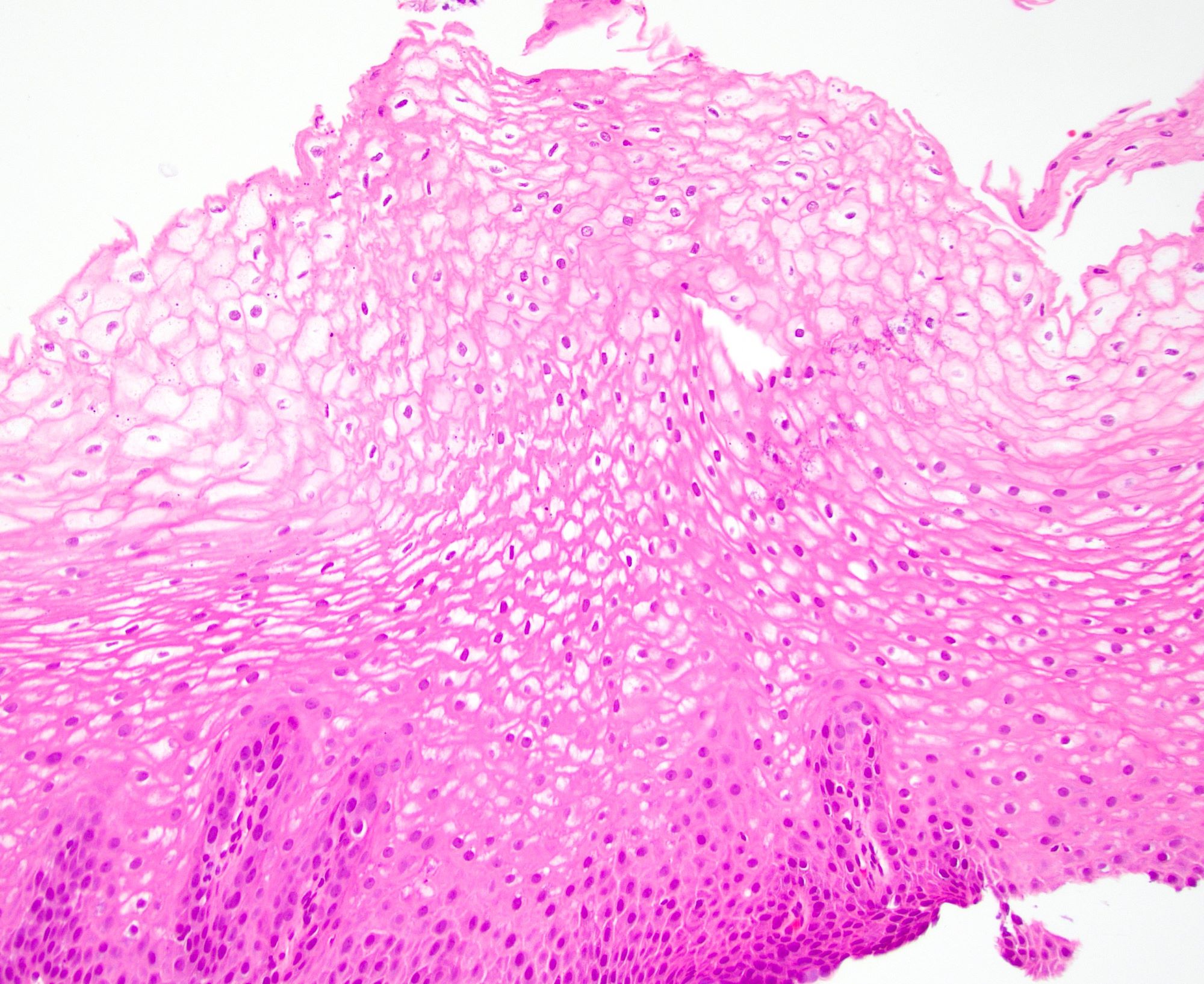
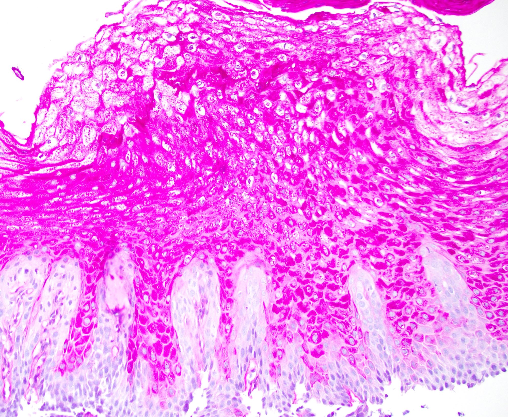
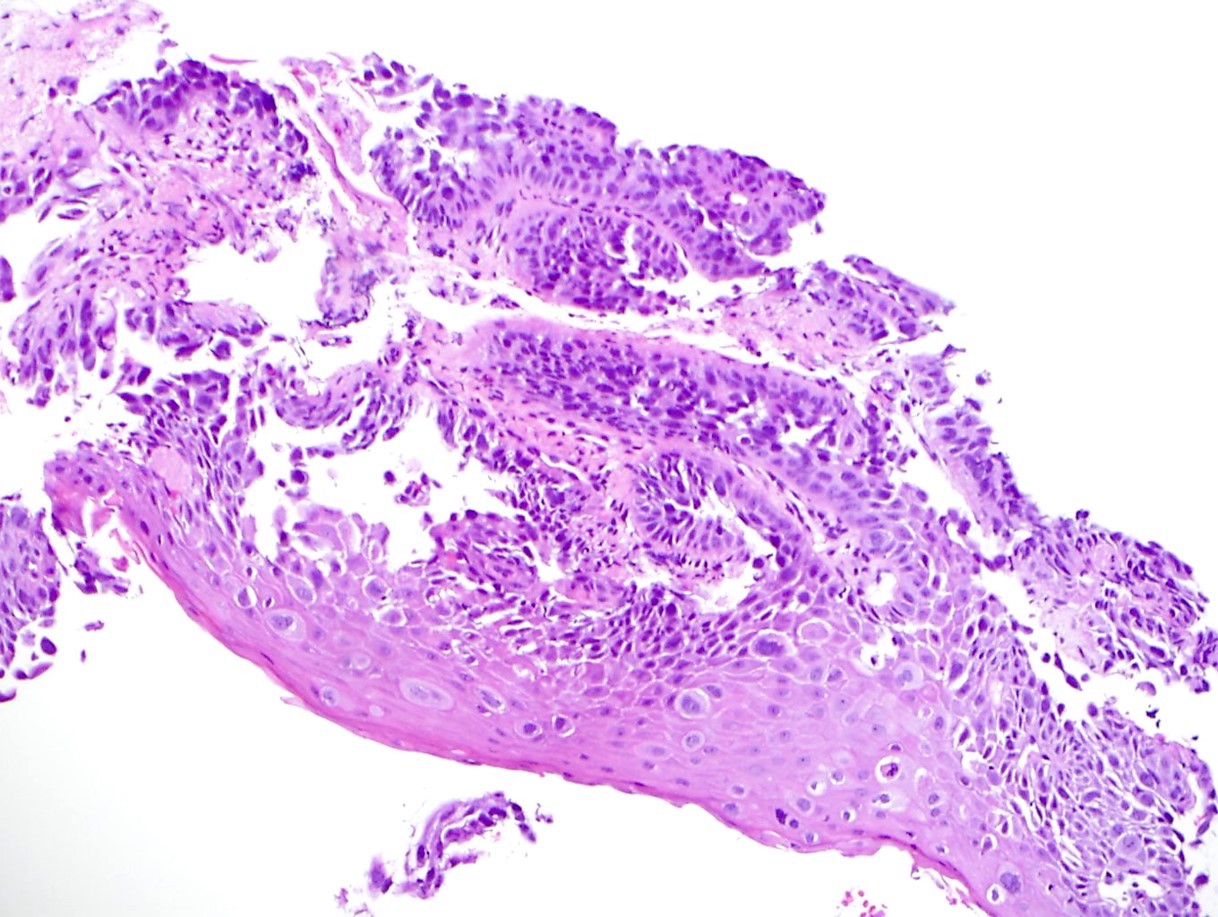
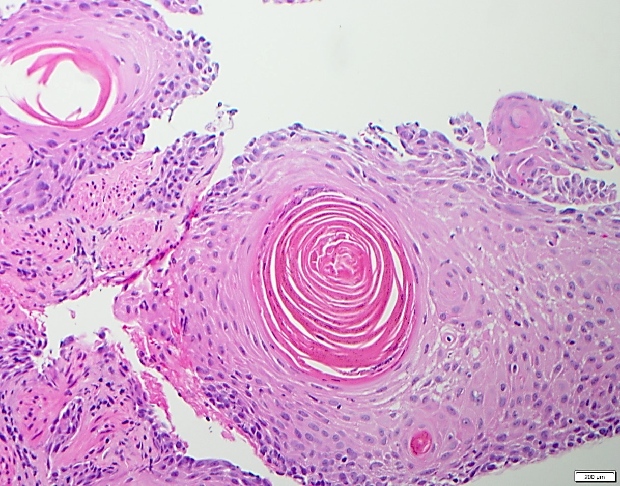
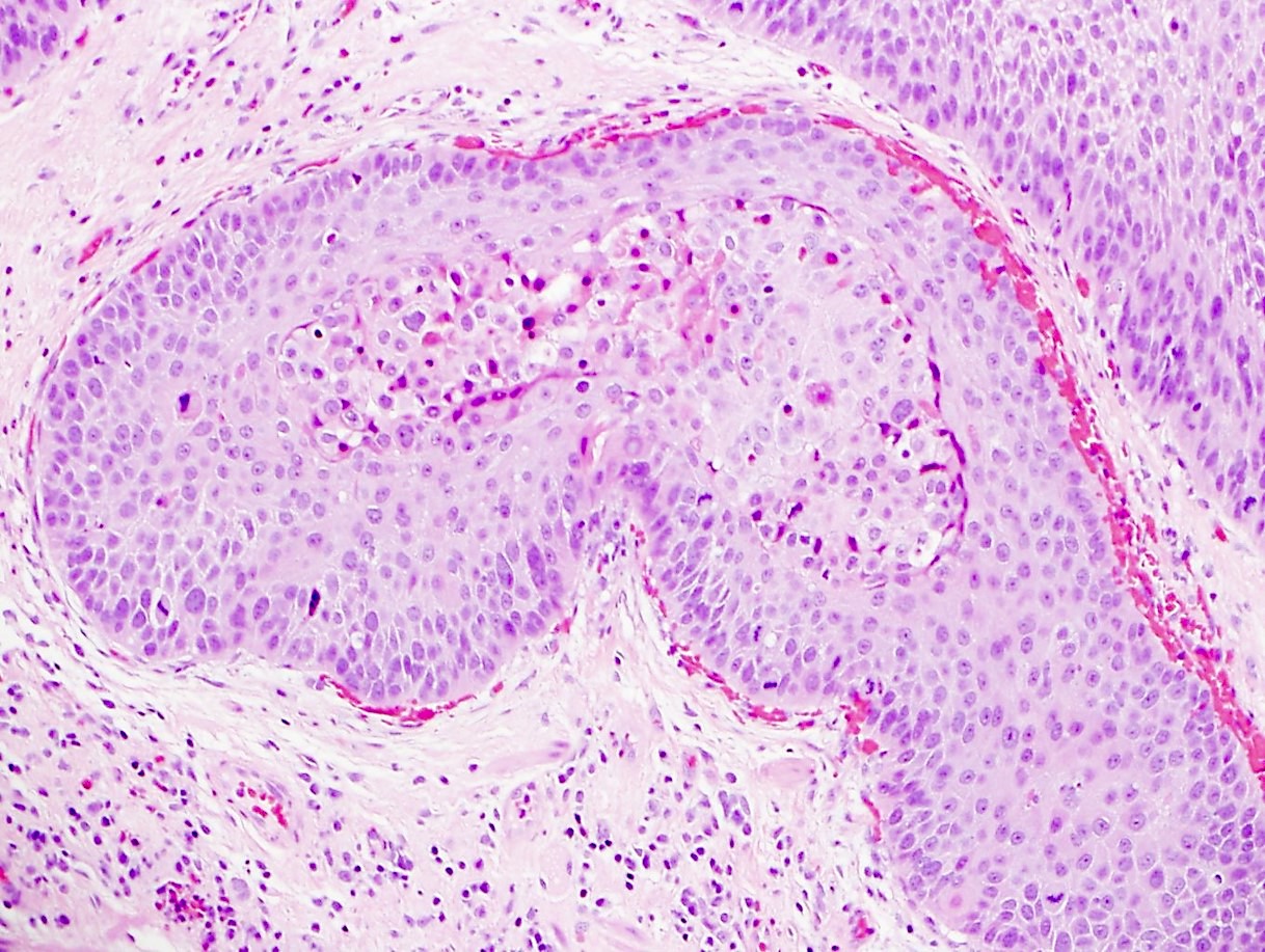
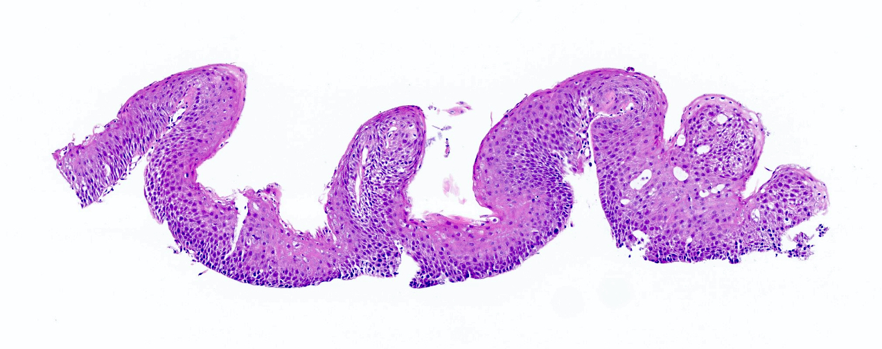
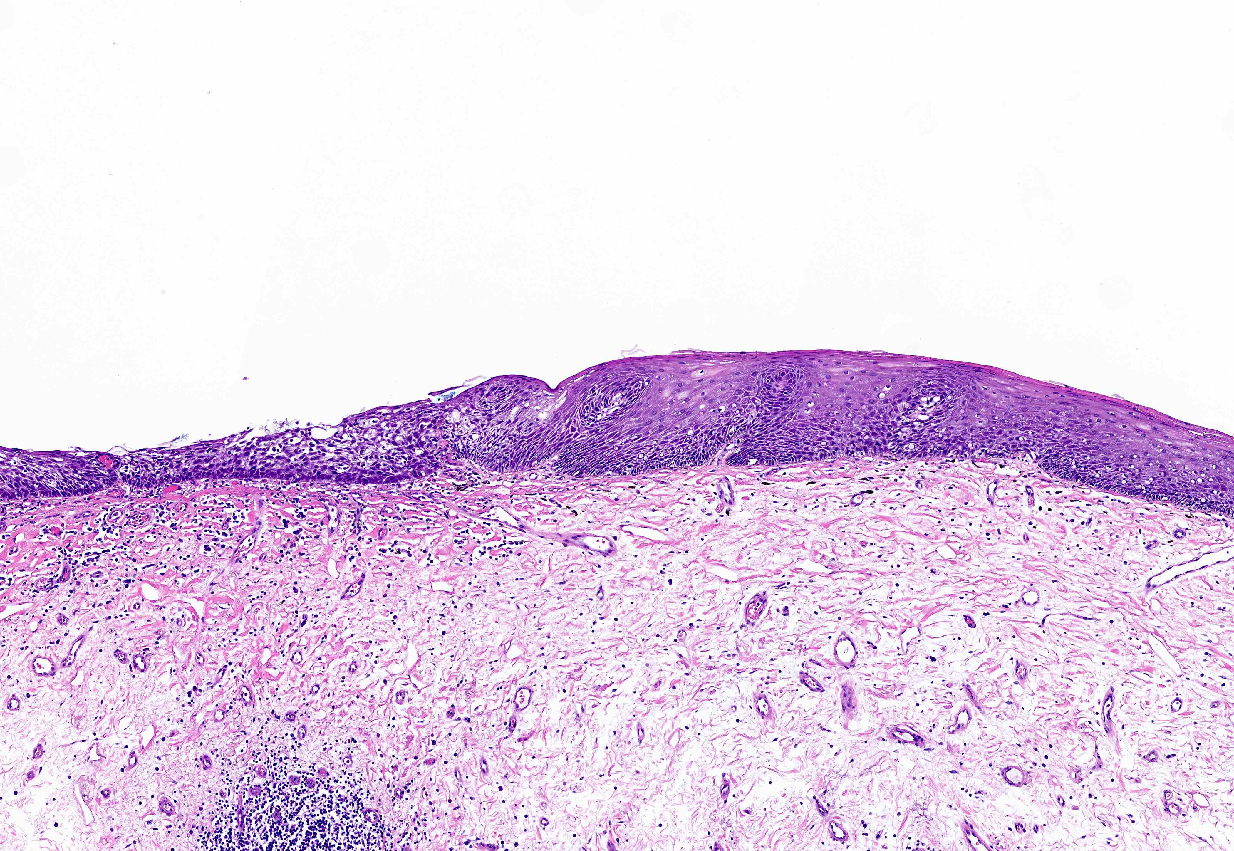
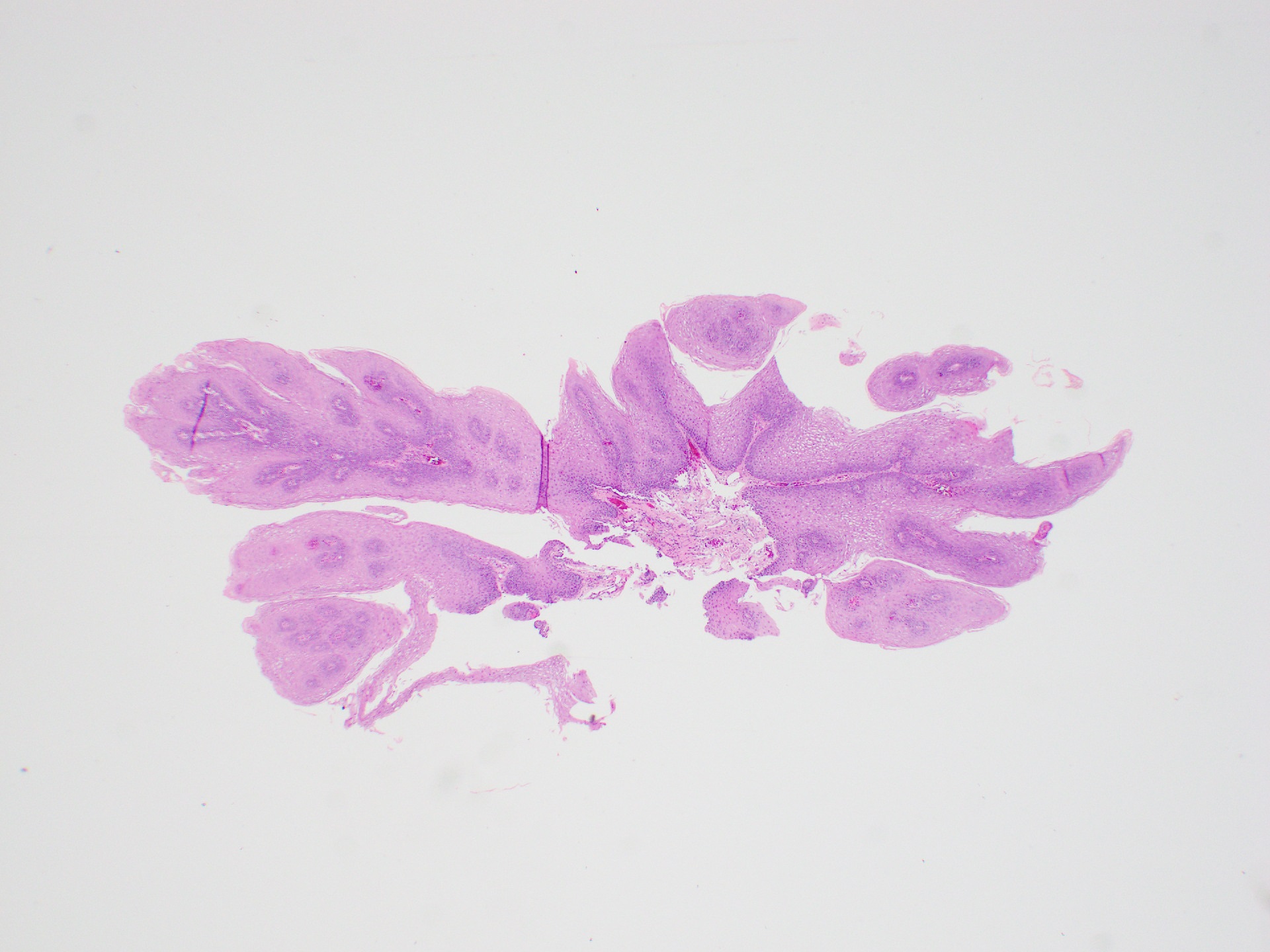
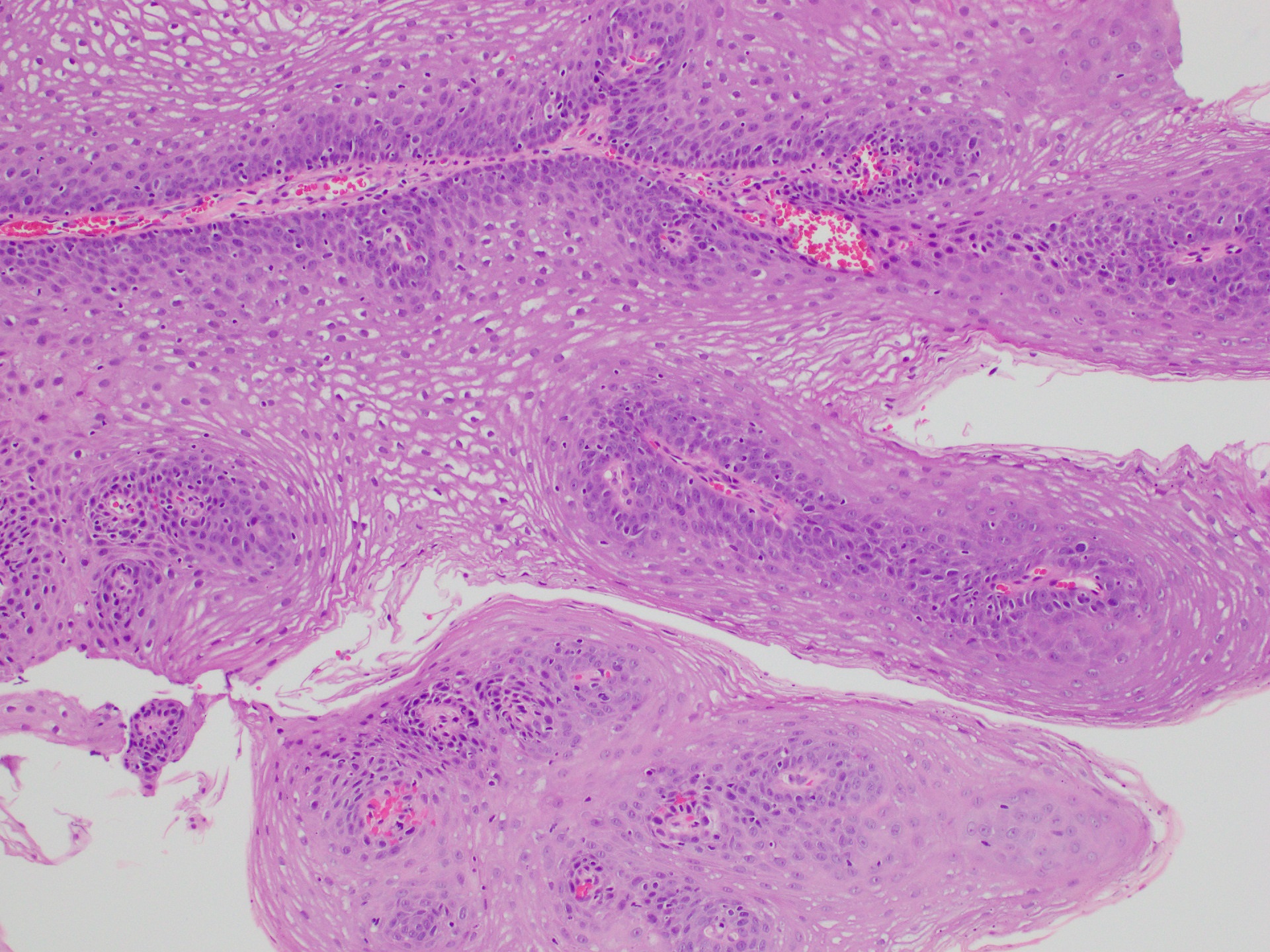
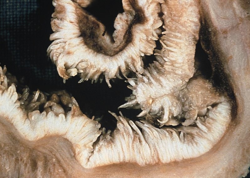
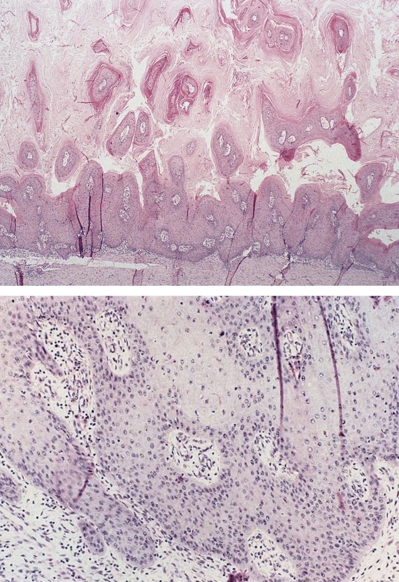
Squamous papilloma with warty surface

Squamous papillomatosis
Contributed by Yukihiro Nakanishi M.D., Ph.D.
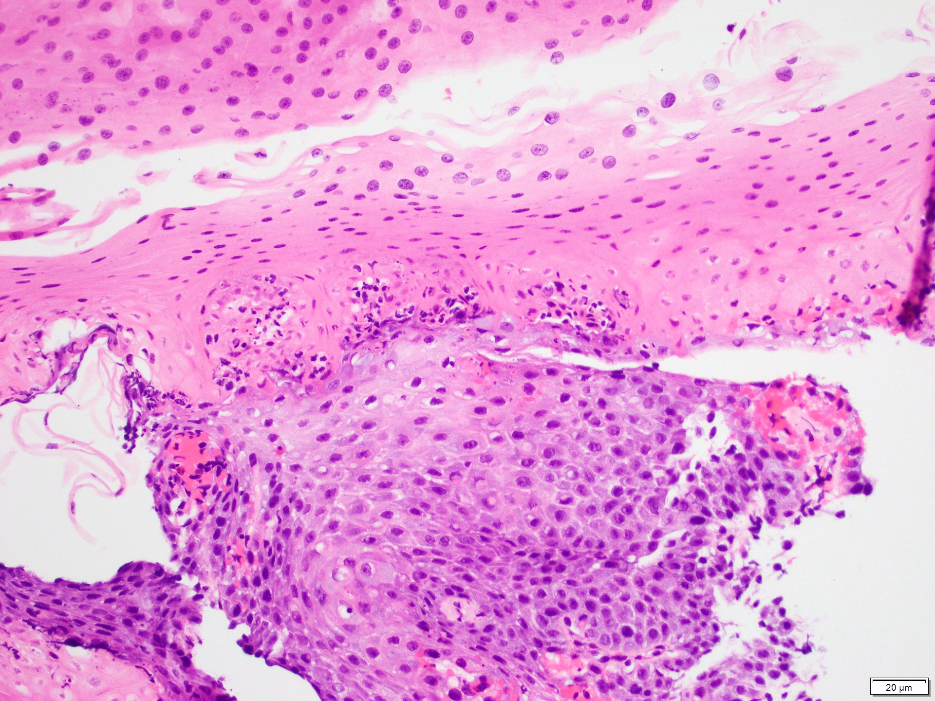
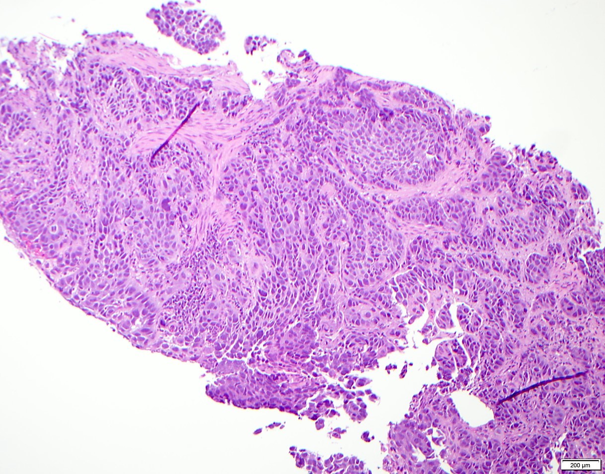
Esophageal polypoid lesion
Images hosted on other servers:
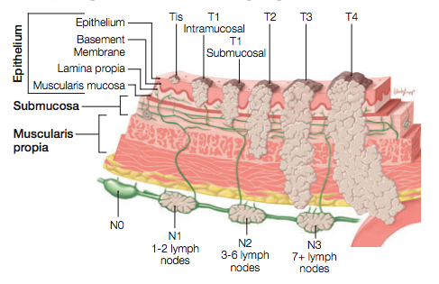
Illustration of T category and N category staging
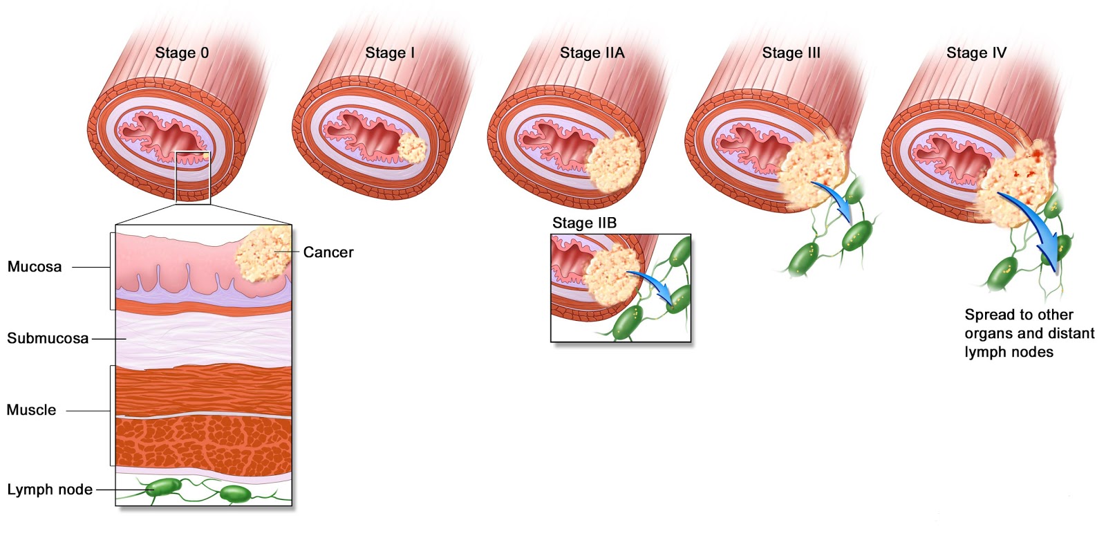
Stages of throat cancer

Various stages
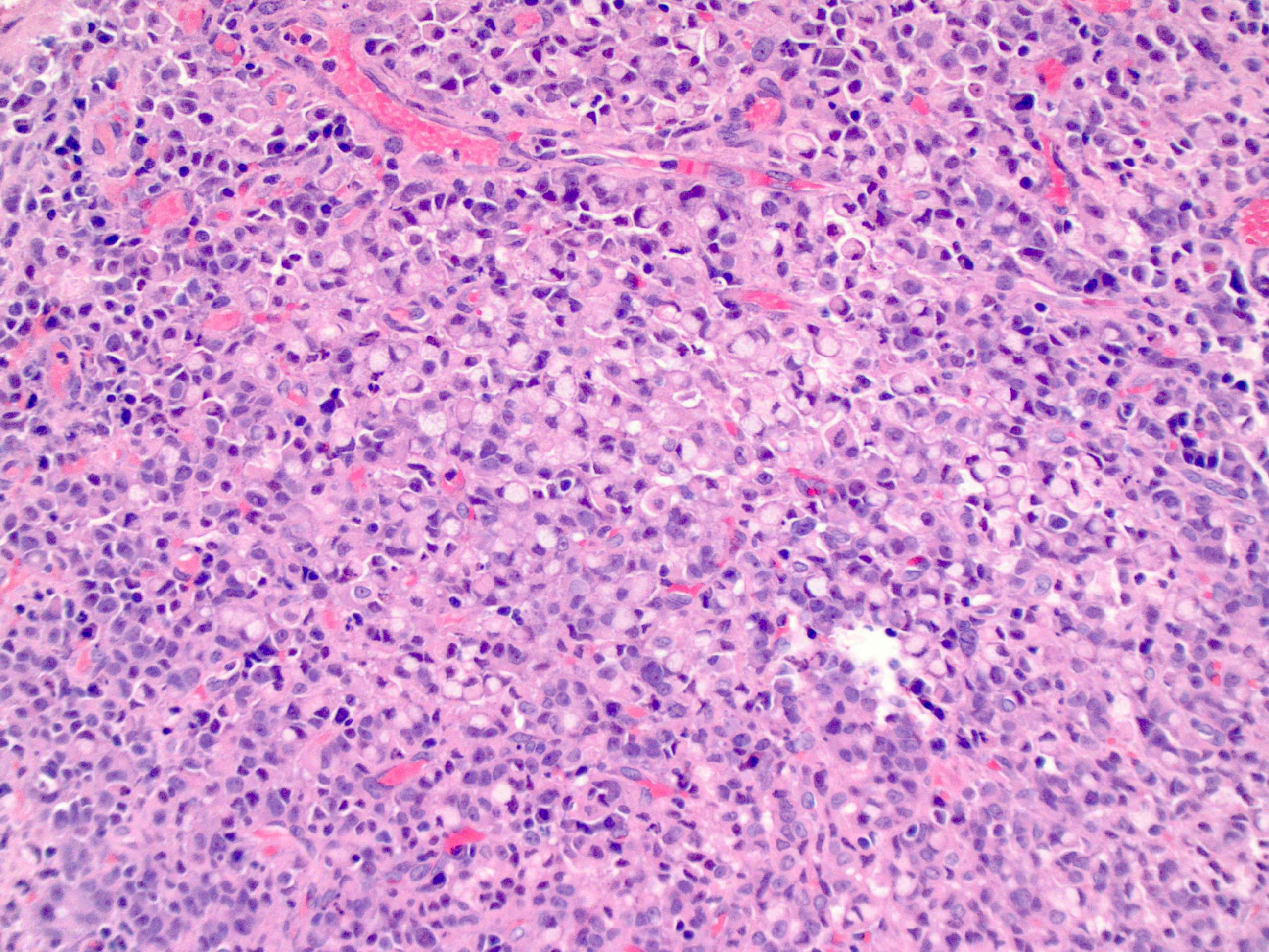
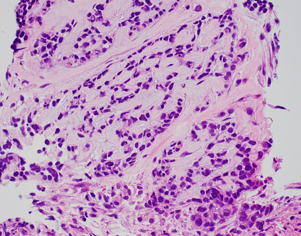
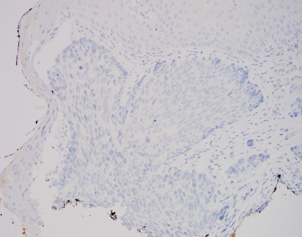
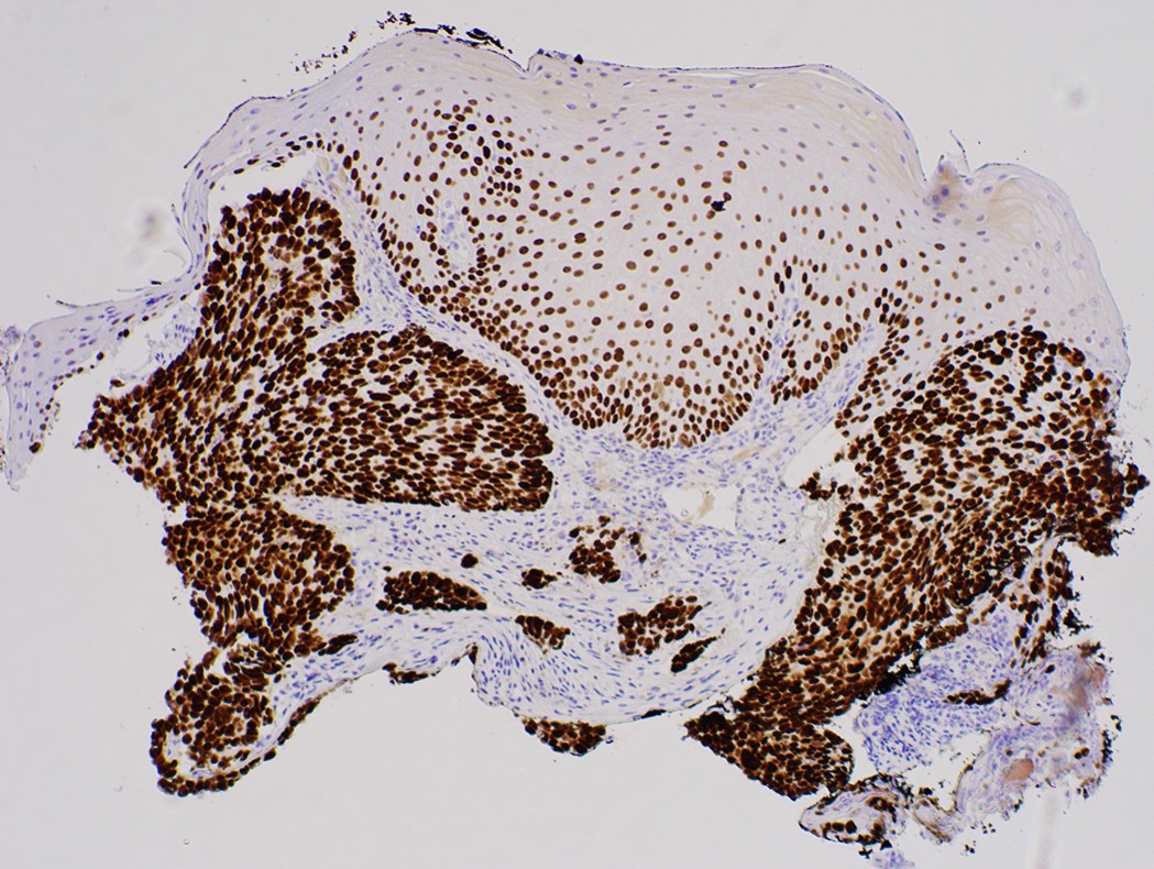
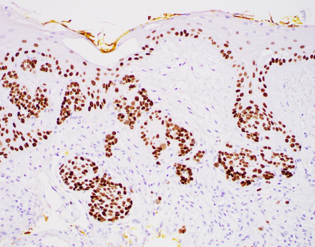
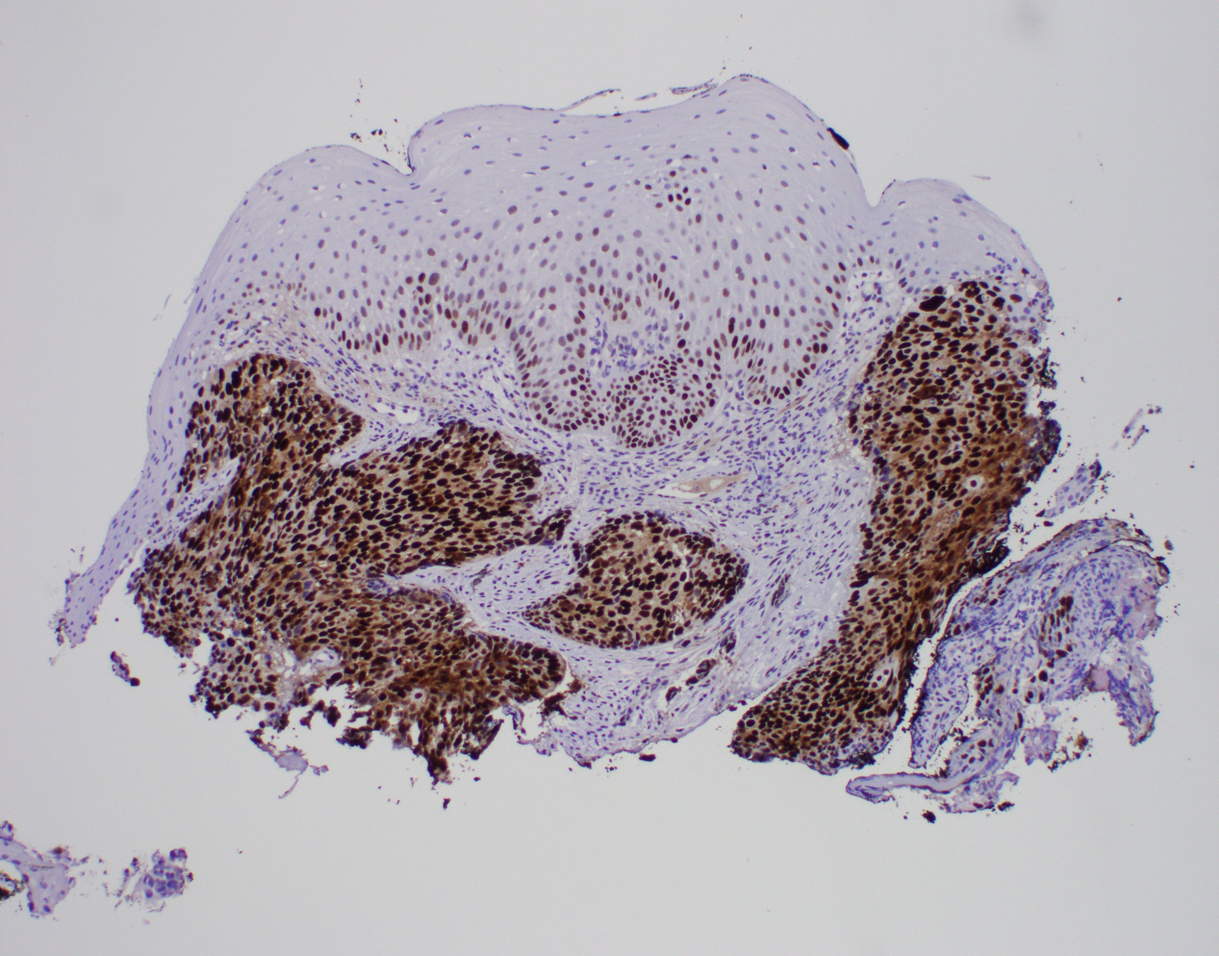
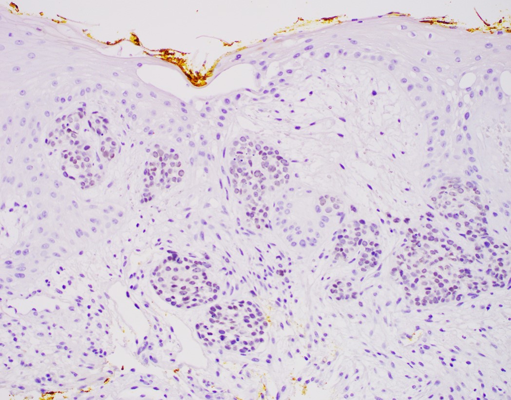
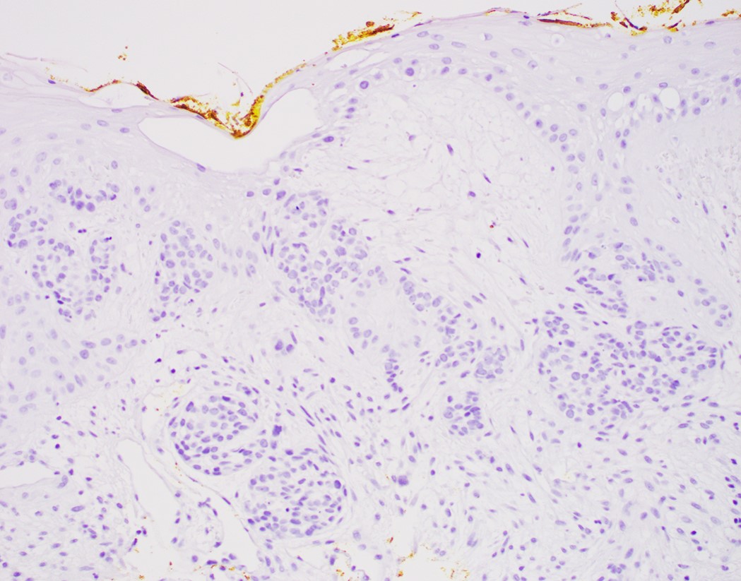
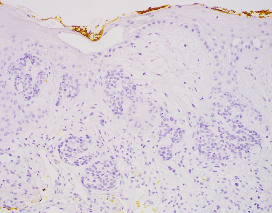
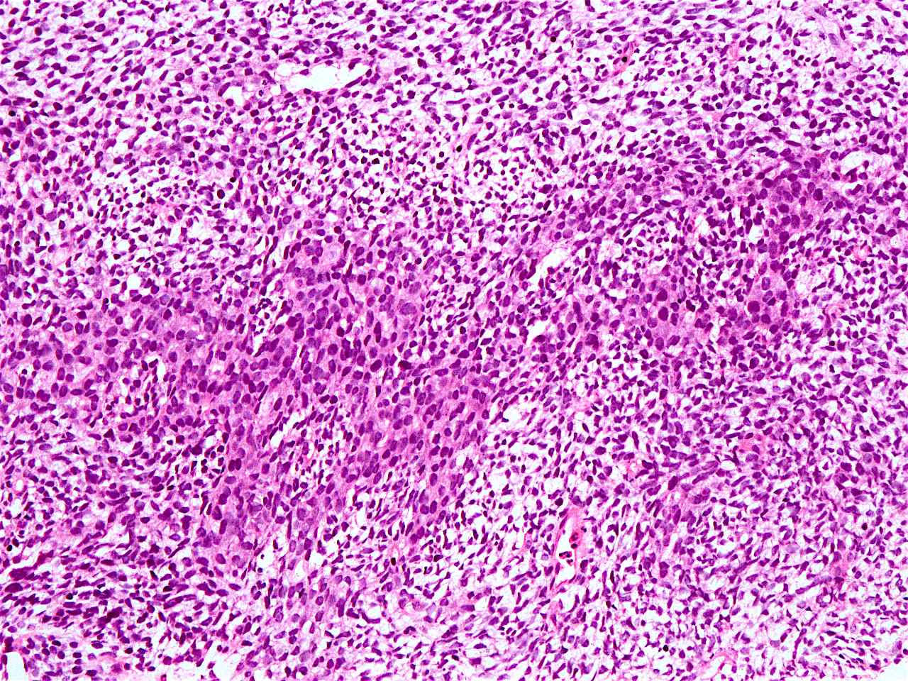
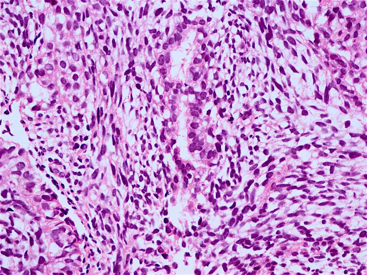
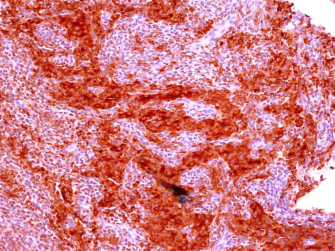
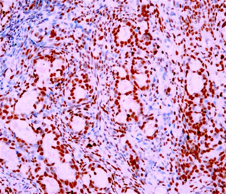
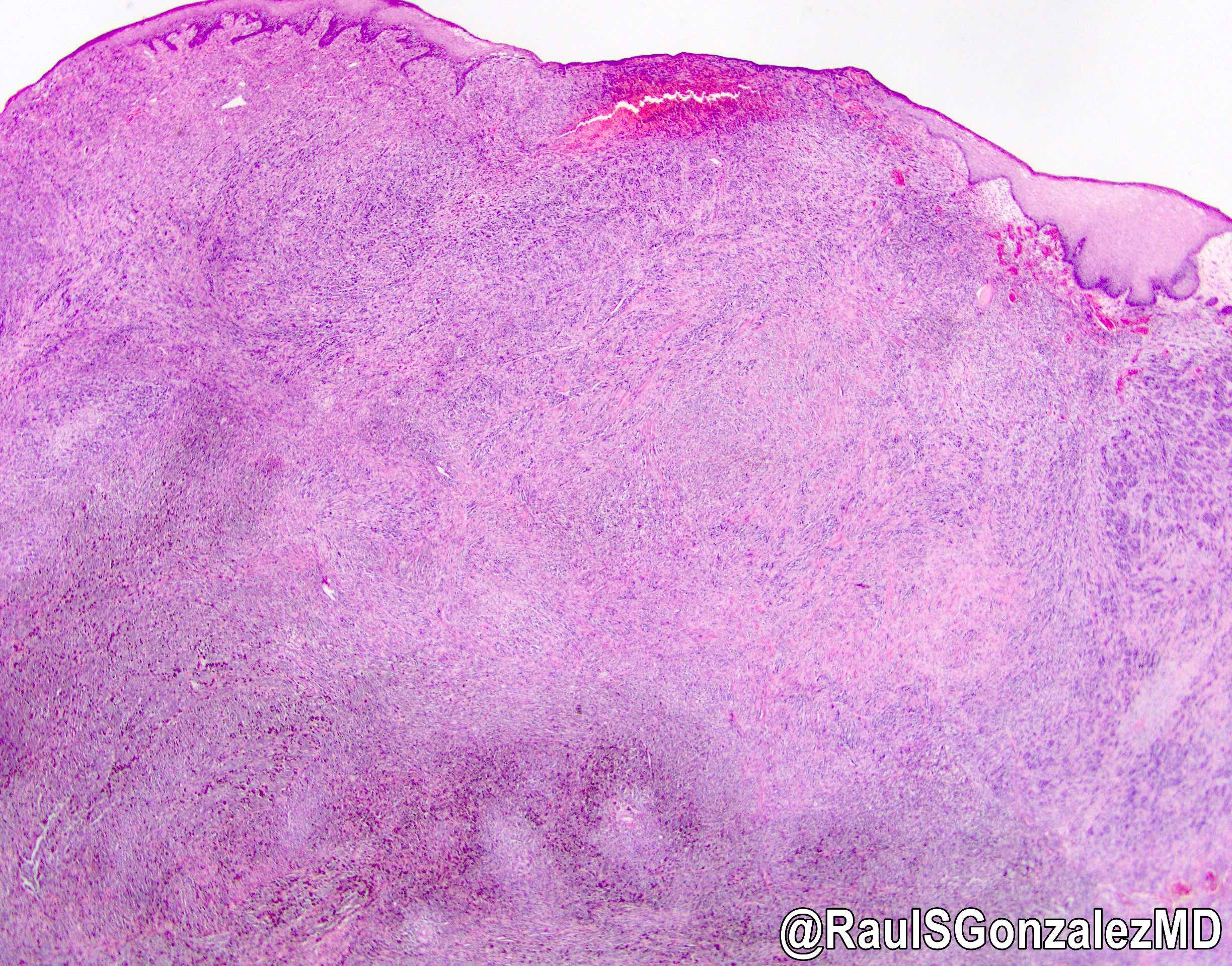
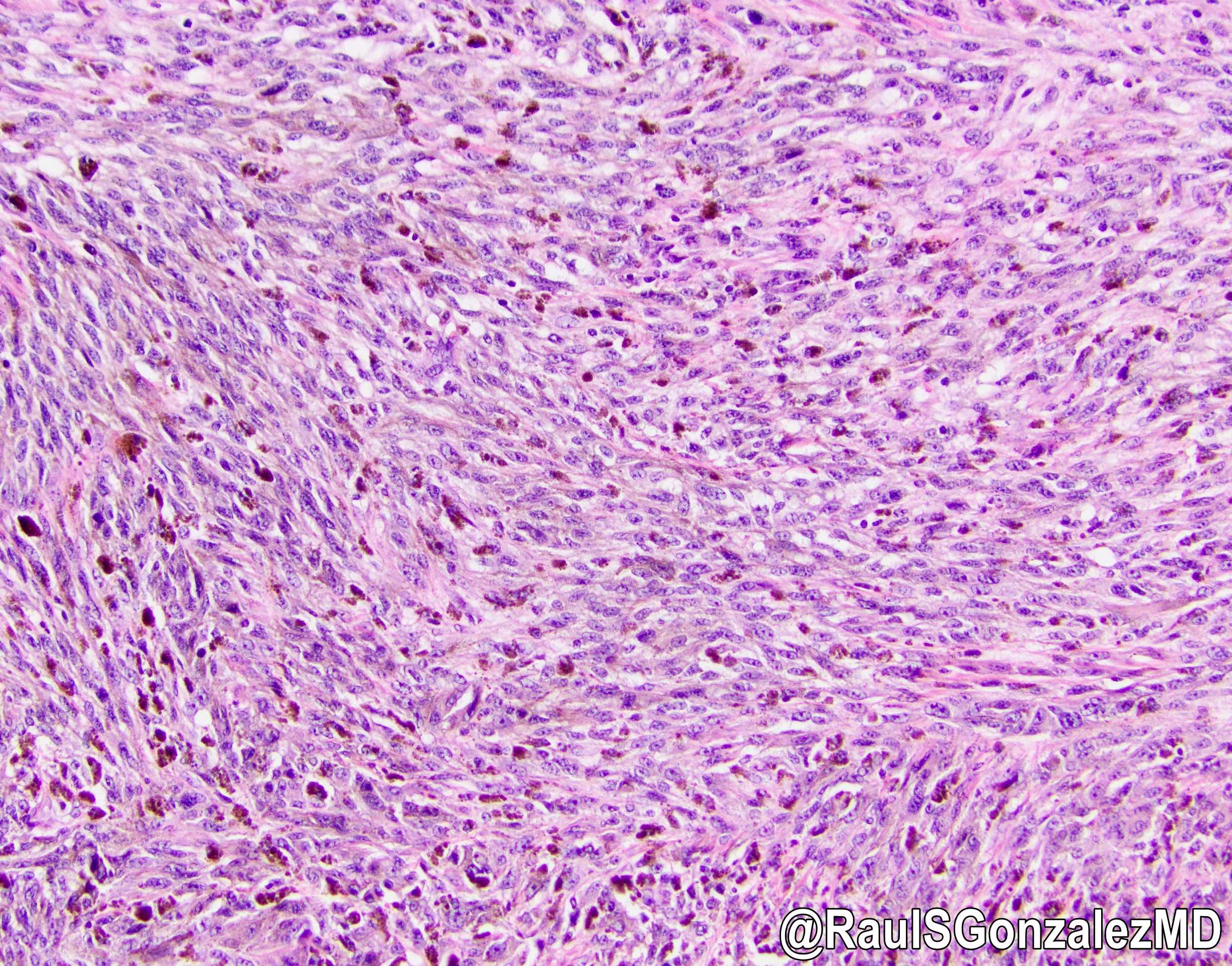
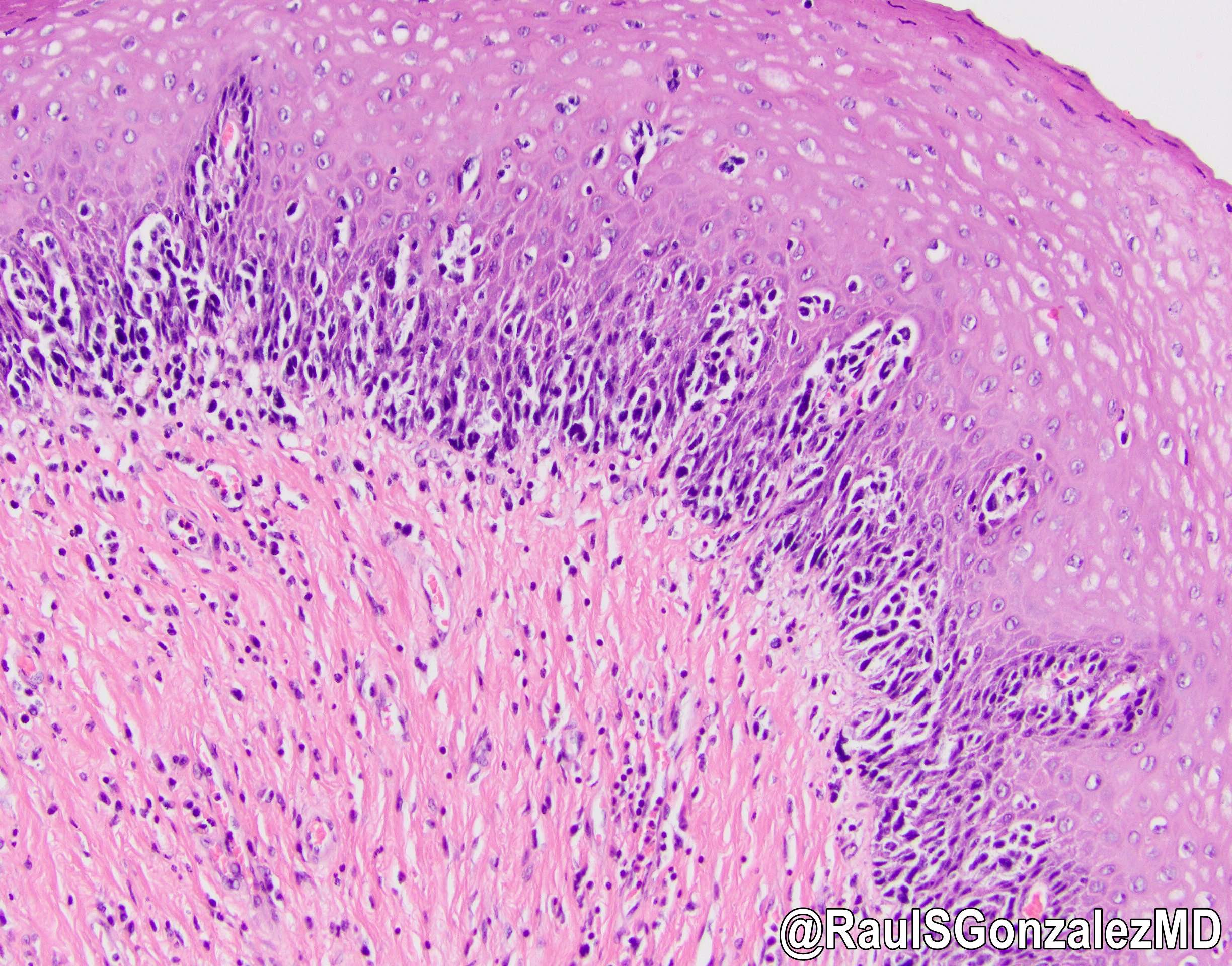
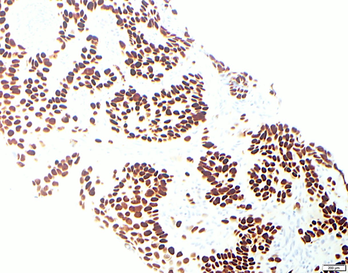
Contributed by Rondell P. Graham, M.B.B.S.
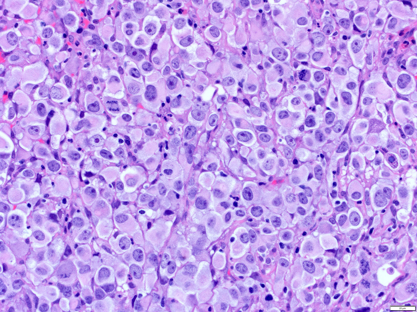
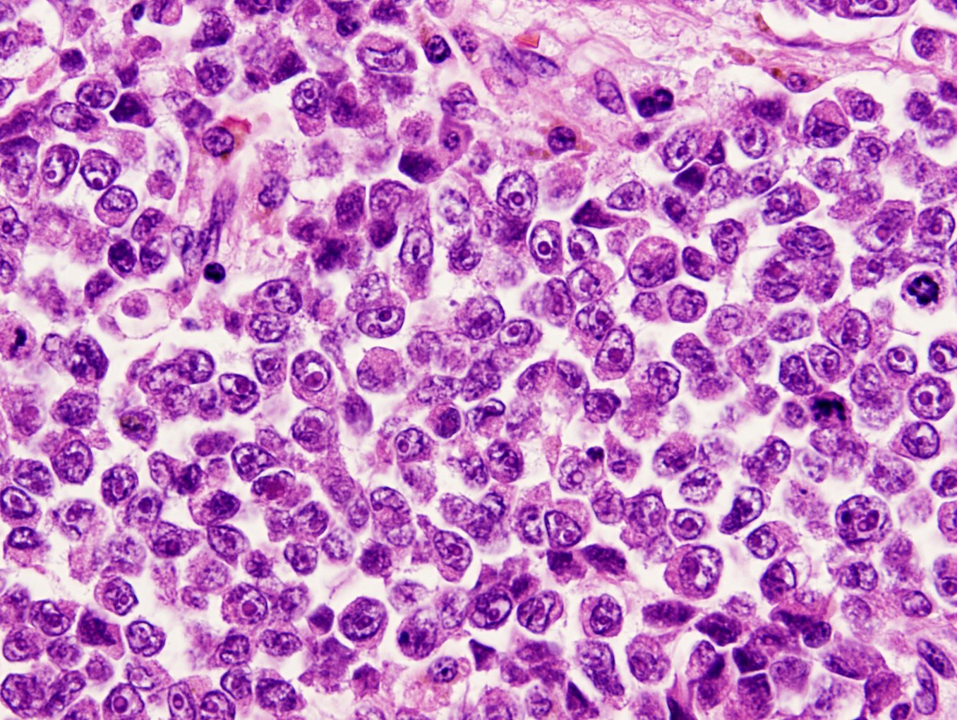
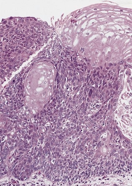
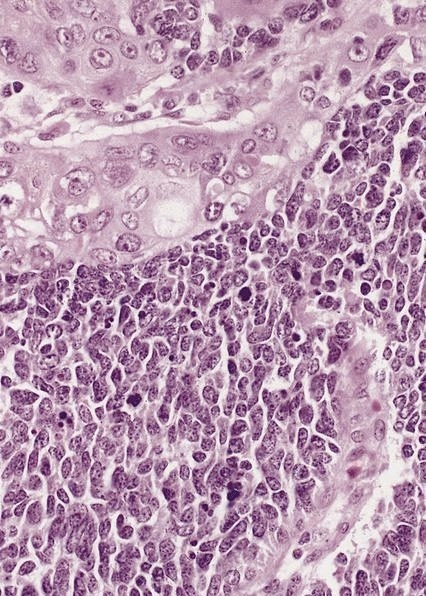
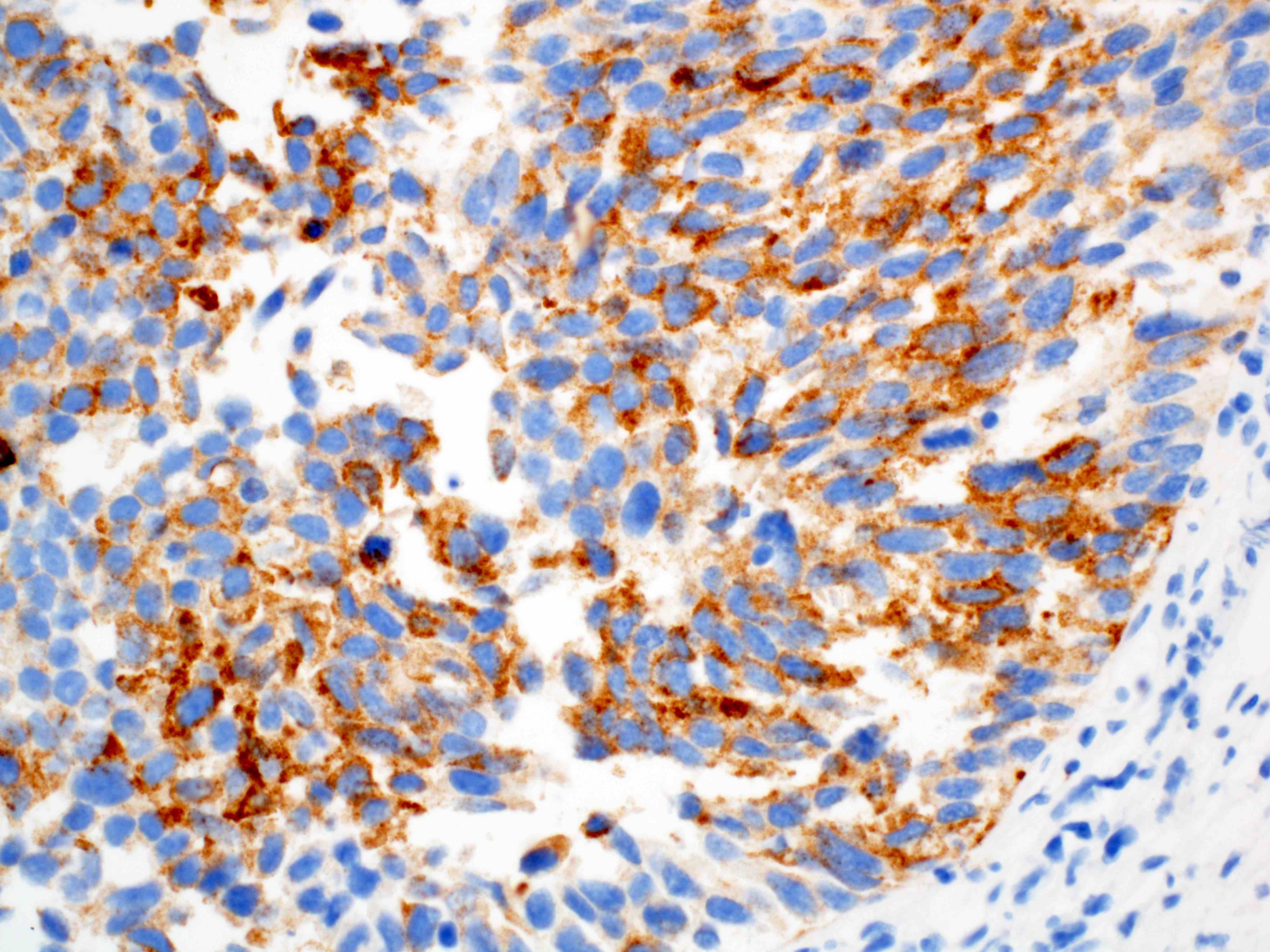
Sheets of cells
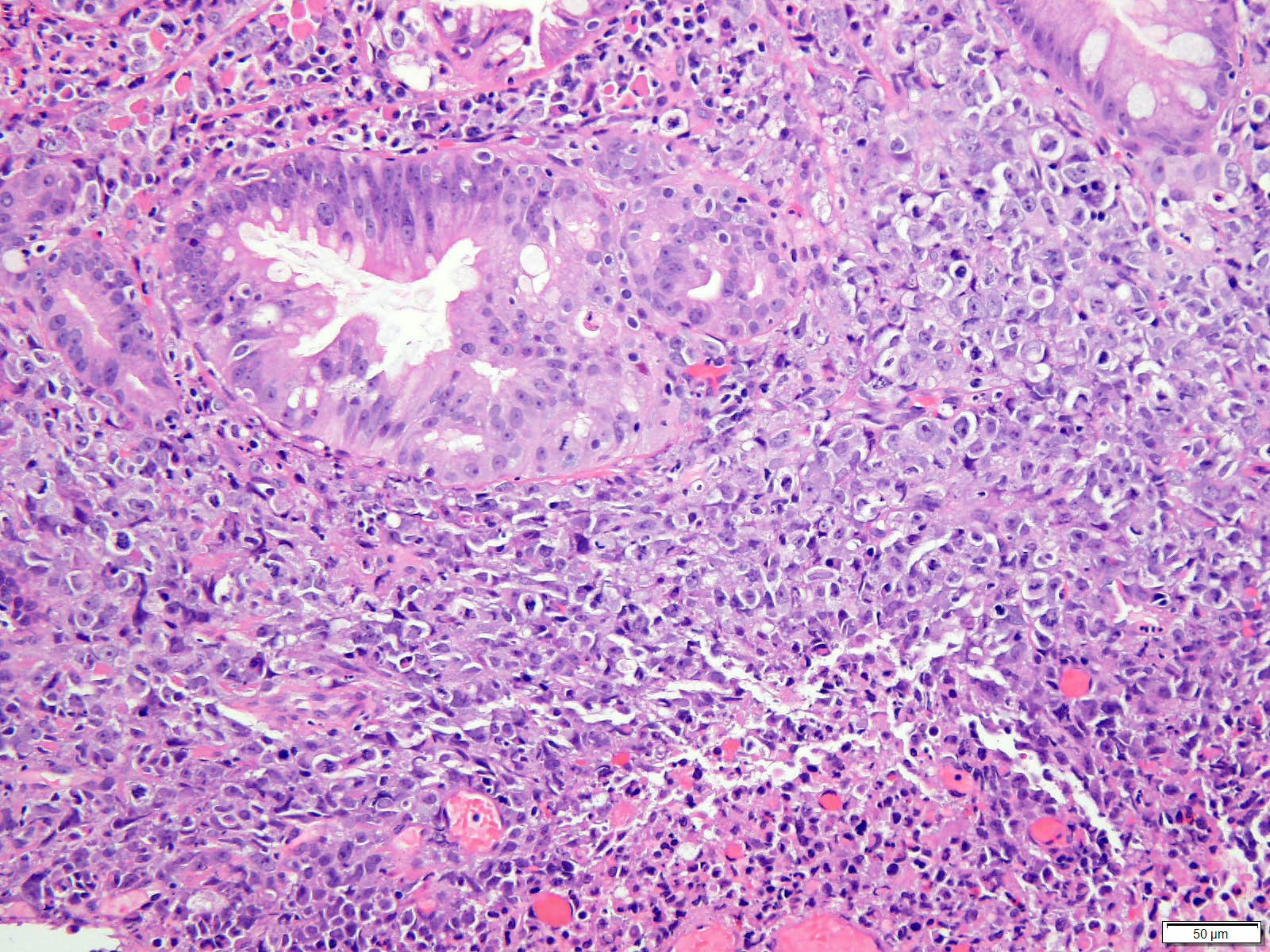
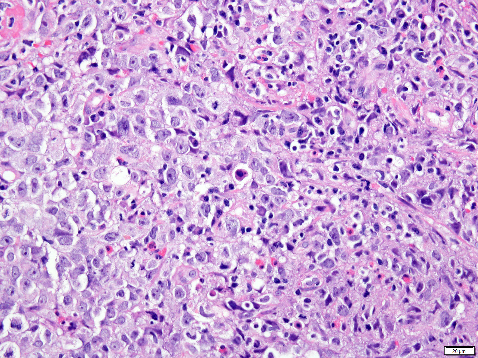
Low grade to high grade carcinoma
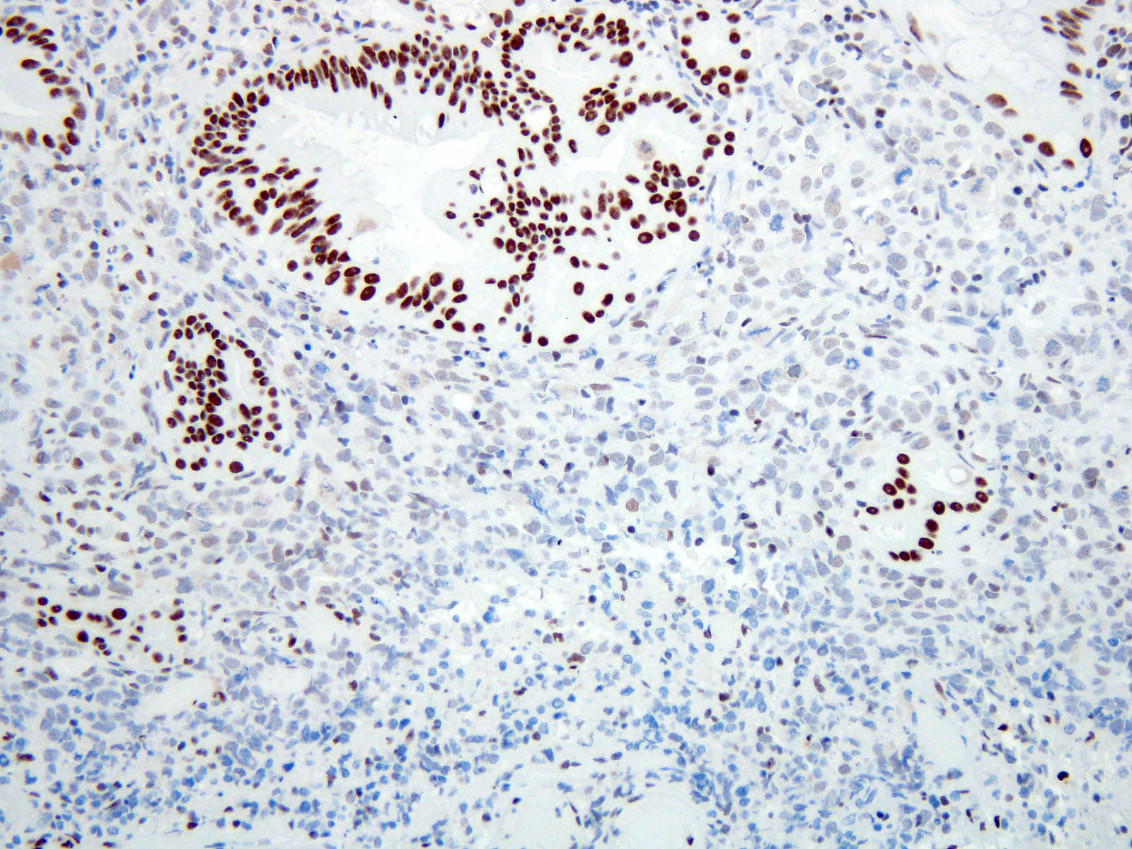
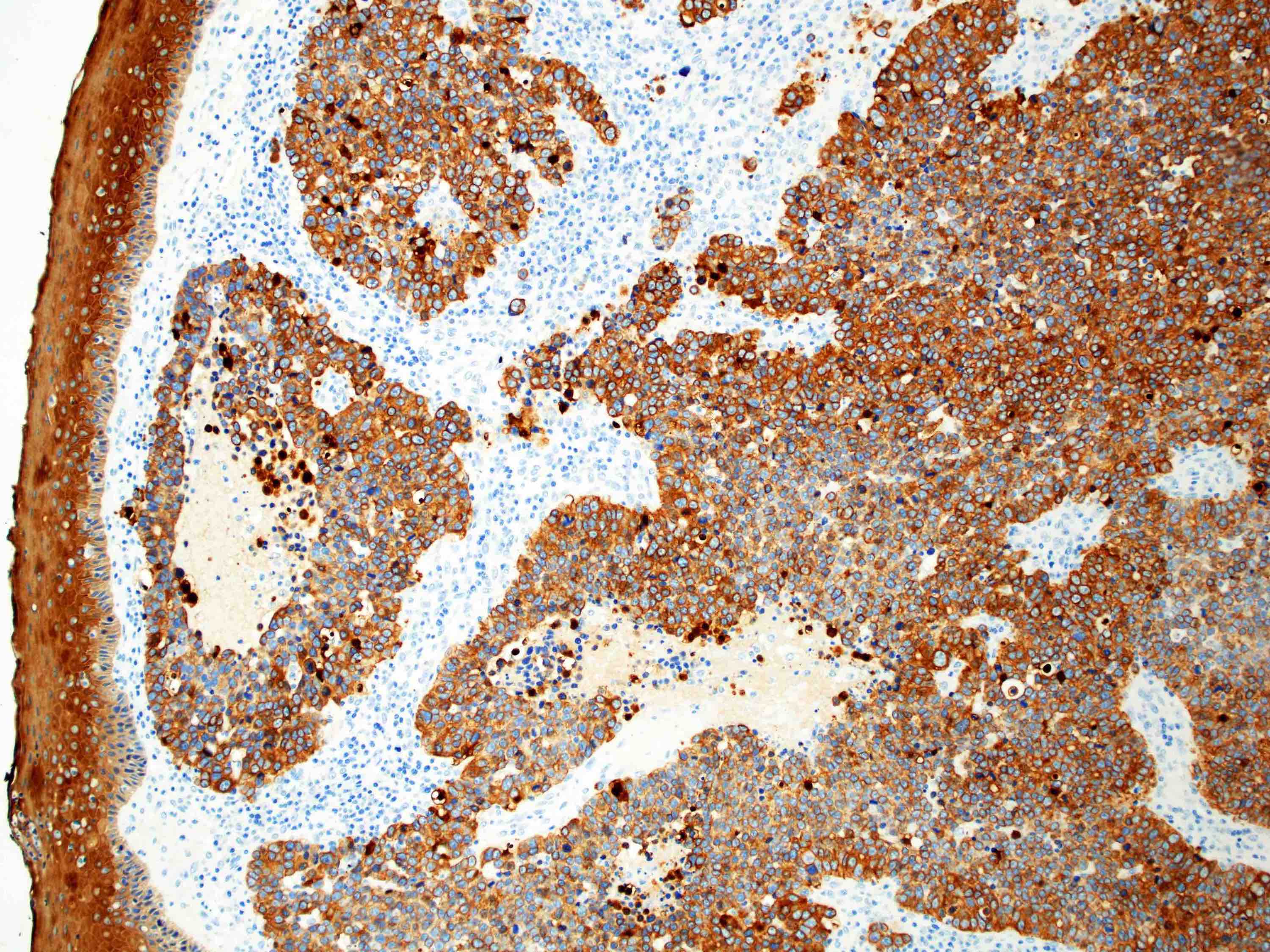
Loss of SMARCA4 (BRG1)
Images hosted on other servers:


Linear, dark blue, submucosal dilated veins


Partially thrombosed after sclerotherapy
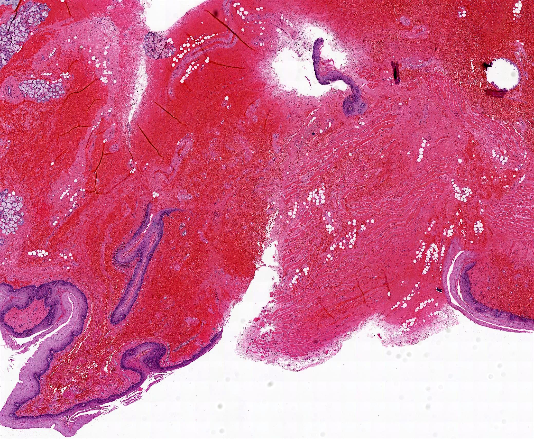
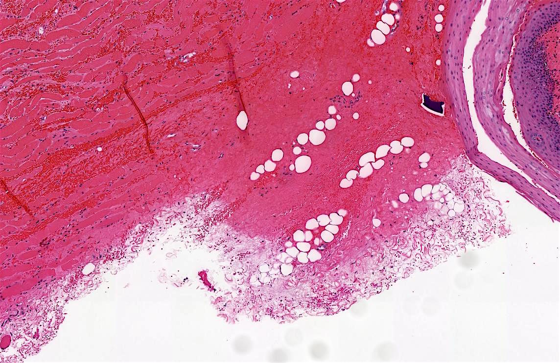
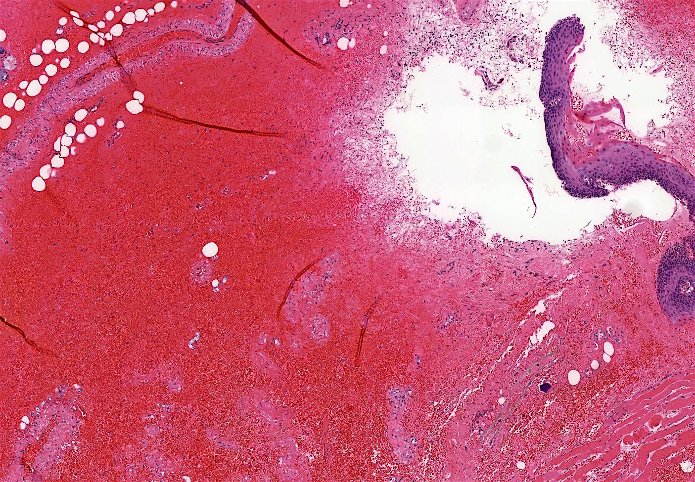
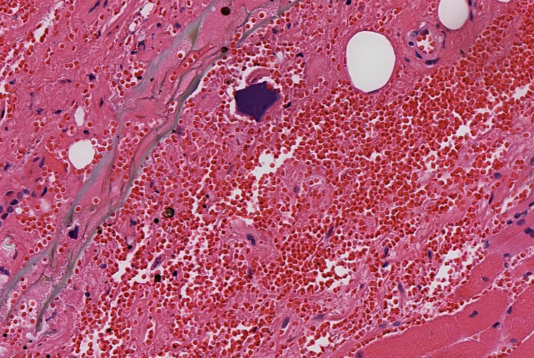
Images hosted on other servers:

Inflamed varix
Images hosted on other servers:

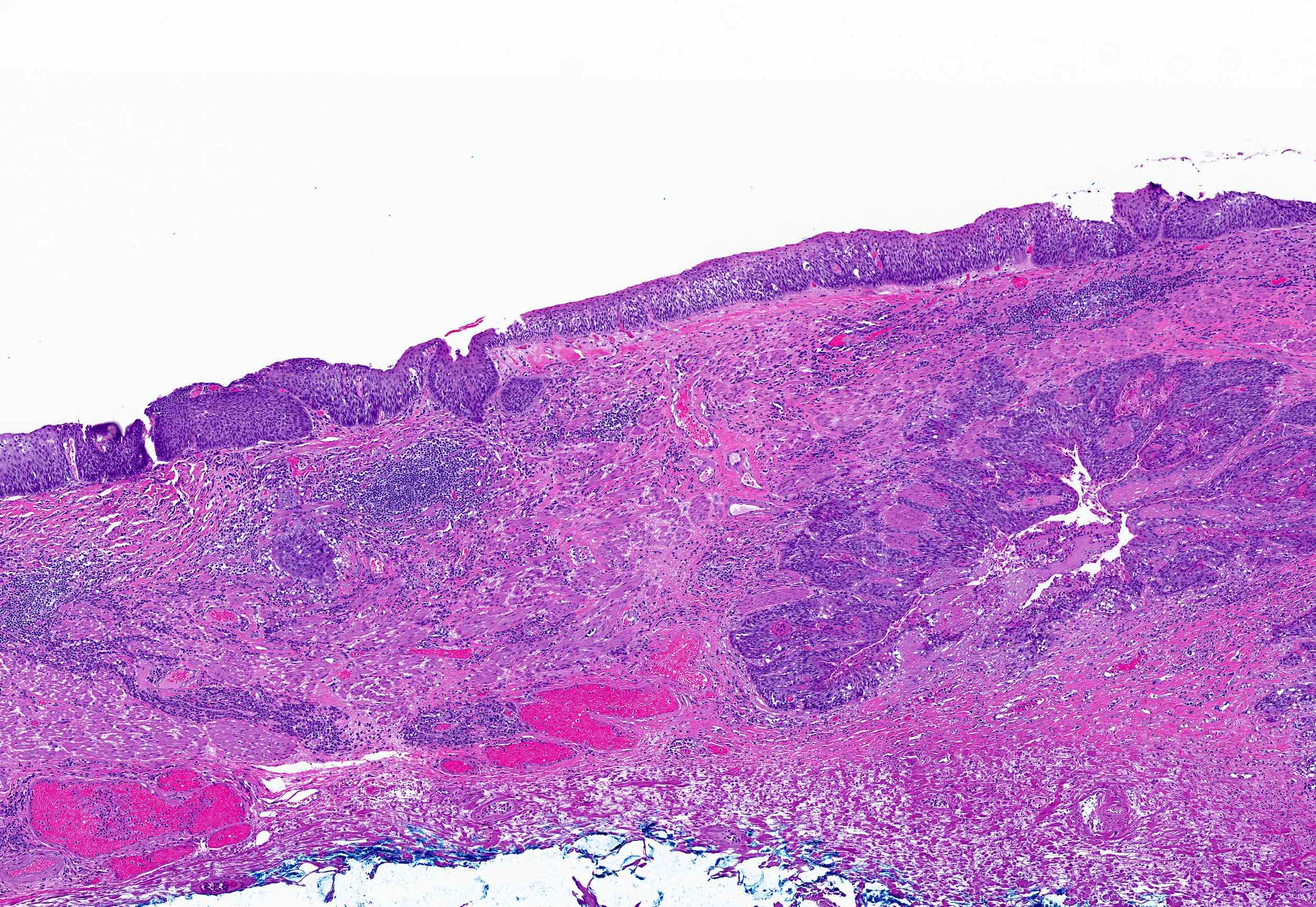
Mass penetrating the muscularis propria
Images hosted on other servers:

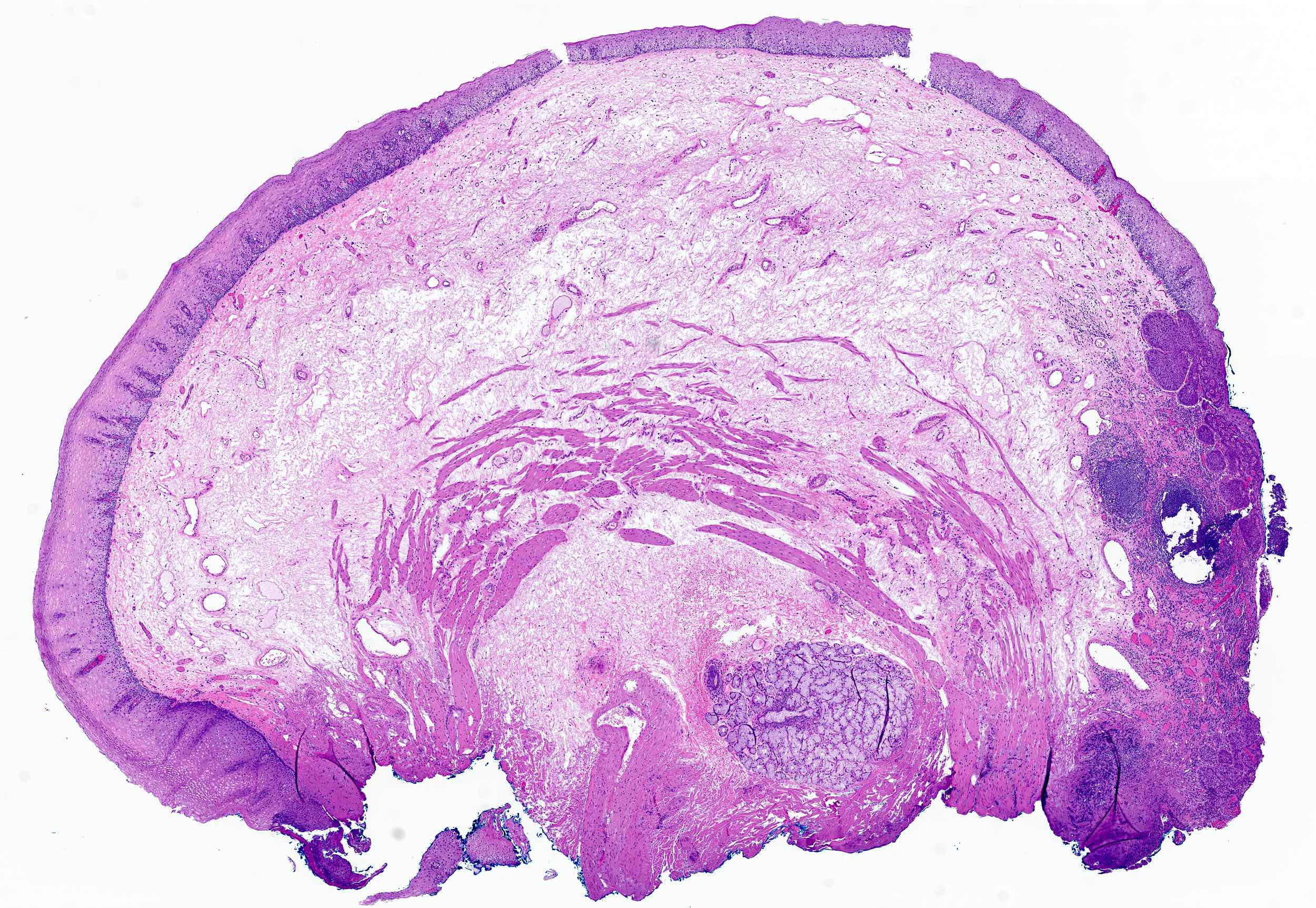
Large polypoid mass
AFIP images
Large plaque of carcinoma
AFIP images
Squamous spikes covered by keratin
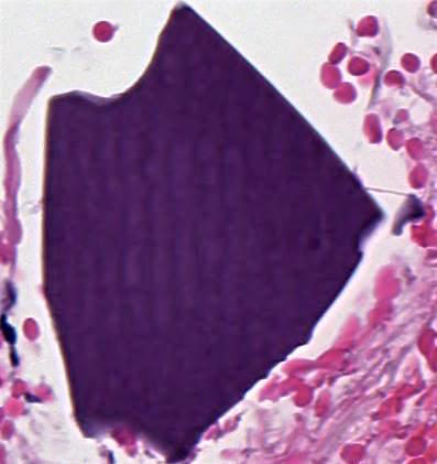
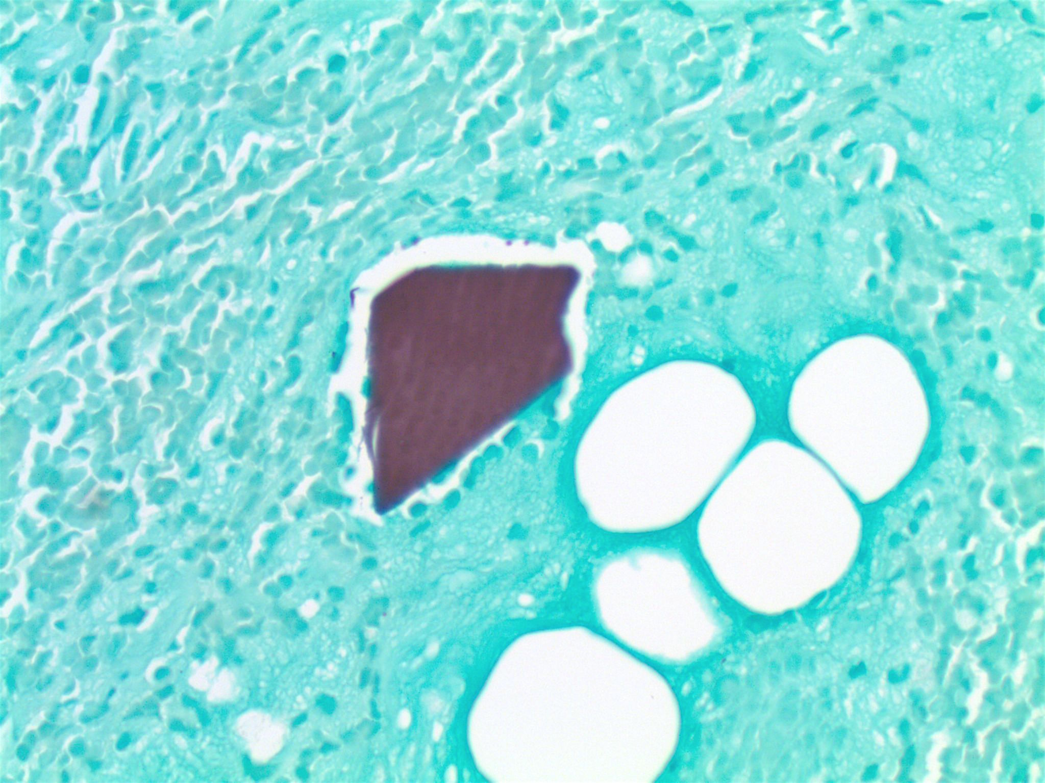
Images hosted on other servers:

Giant cell foreign body reaction
Images hosted on other servers:

Small bowel carcinoid
Images hosted on other servers:

Carcinoid tumor
Images hosted on other servers:

Neurosecretory granules
Greenson: 2019

IARC: 2019
Lamps: 2015
Montgomery: 2017
Montgomery: 2017
Odze: 2022
Saba: 2015
Sharma: 2015
Srivastava: 2023
Yantiss: 2021
Find related Pathology books: GI, liver
