
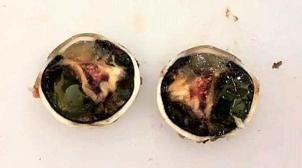
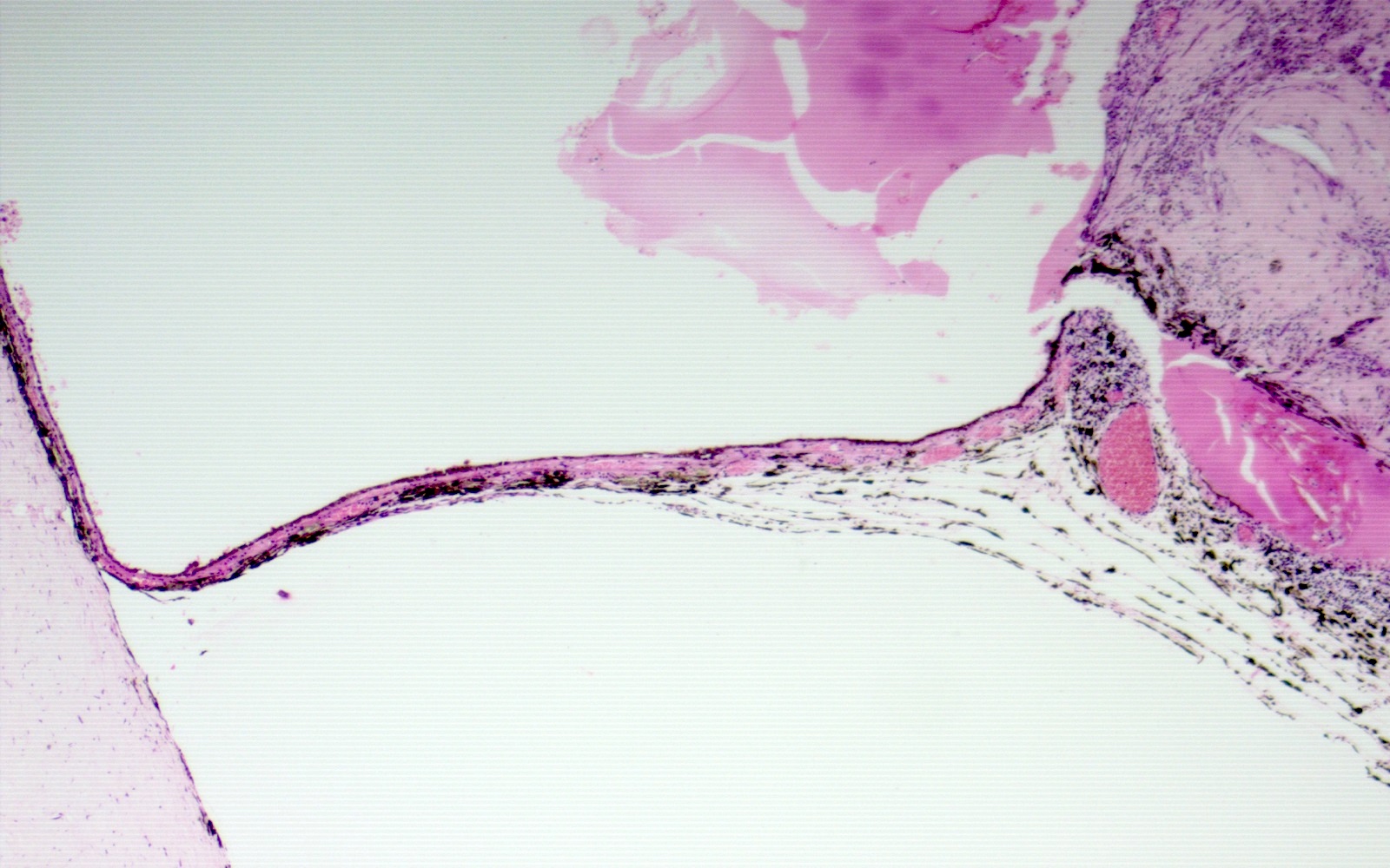
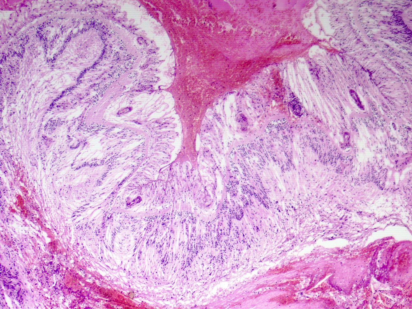
Images hosted on other servers:

Life cycle of Acanthamoeba
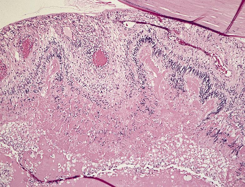
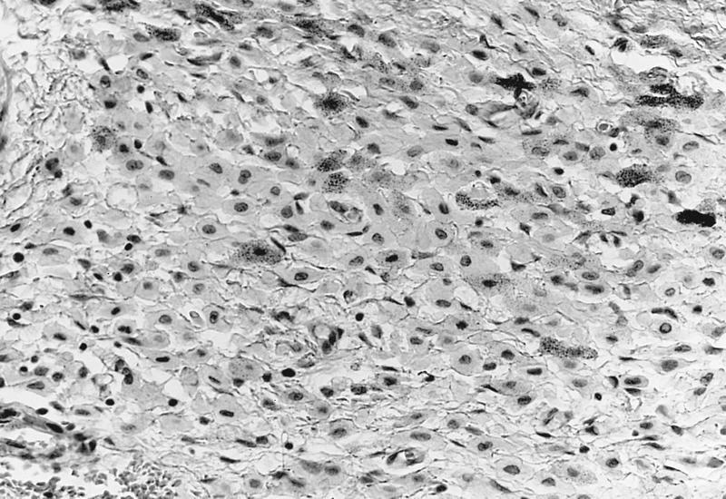
Contributed by Gabrielle Yeaney, M.D.
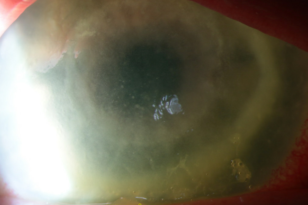

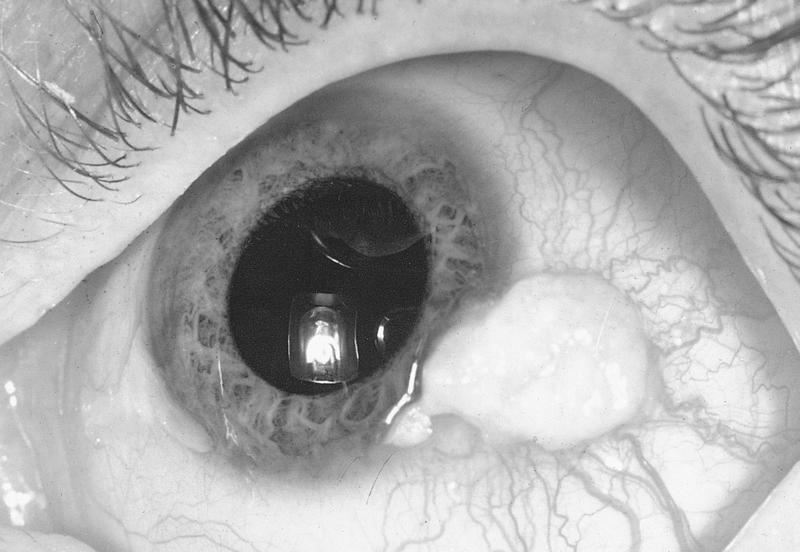
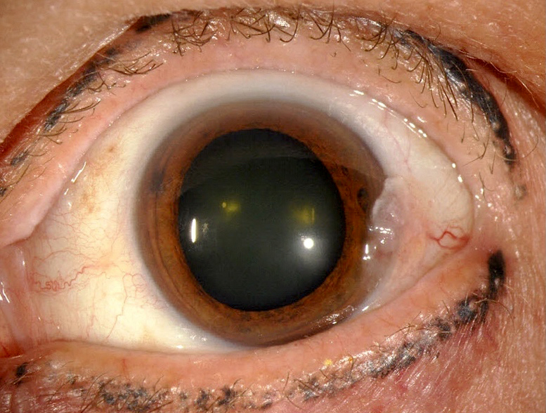
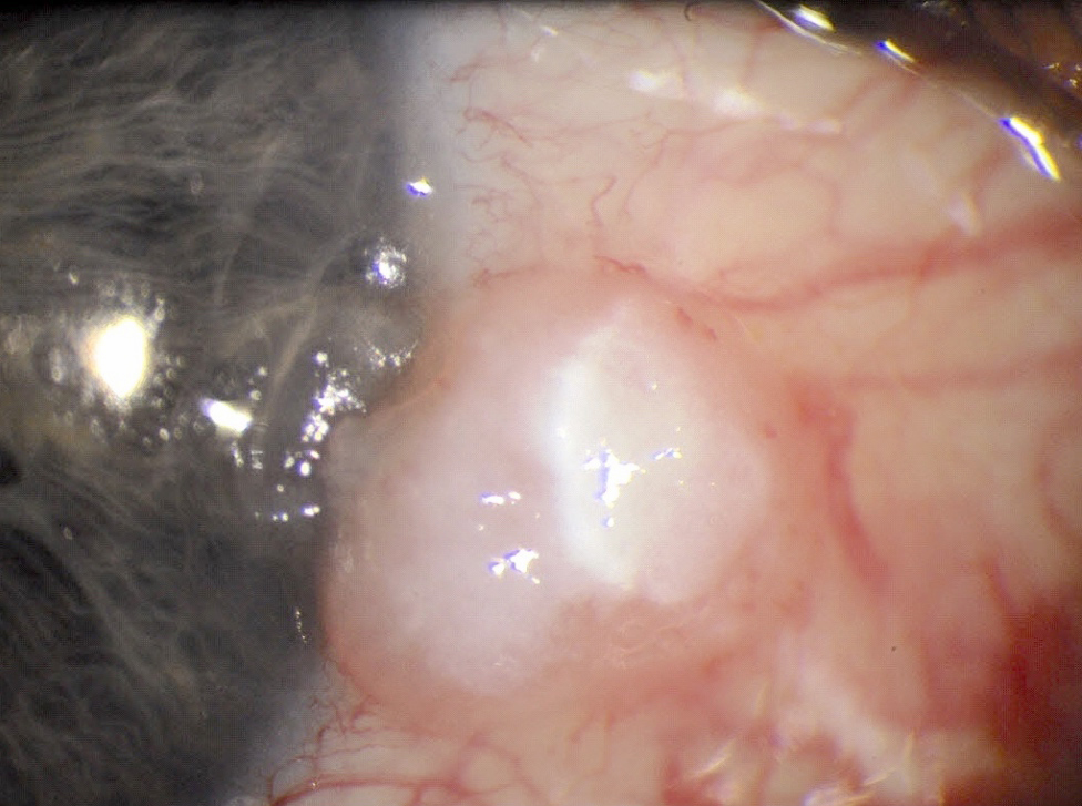
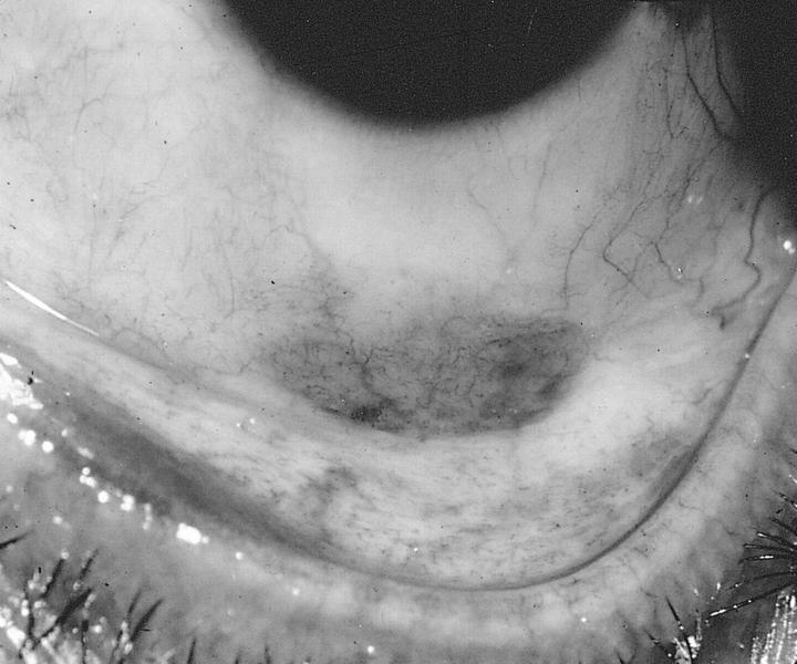
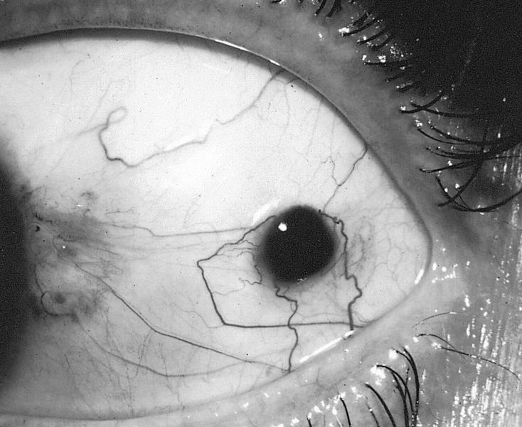
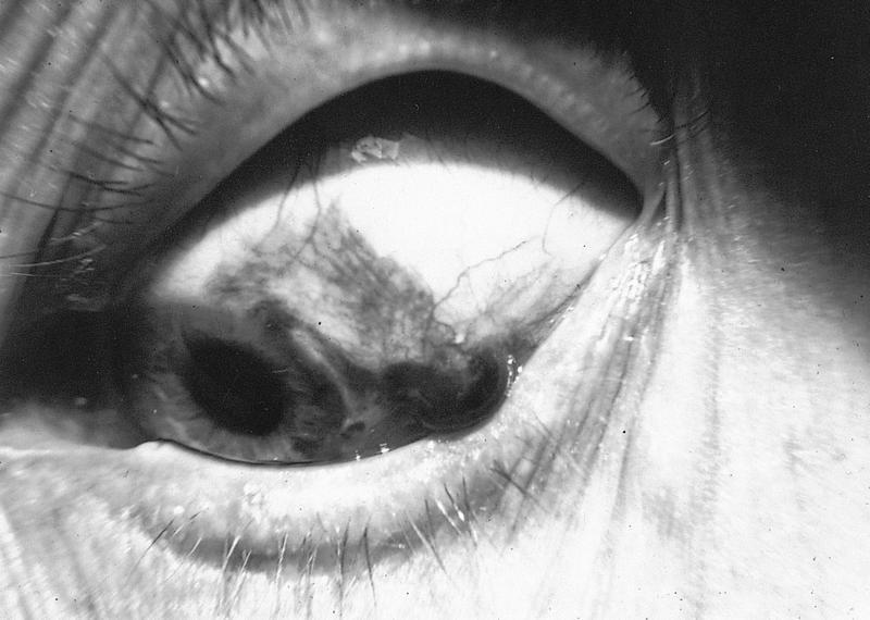
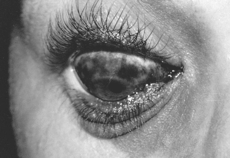
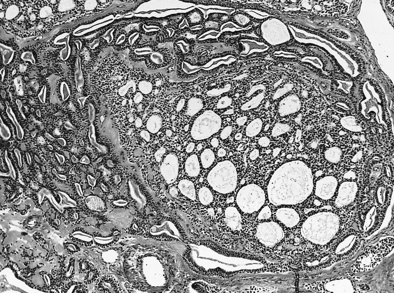
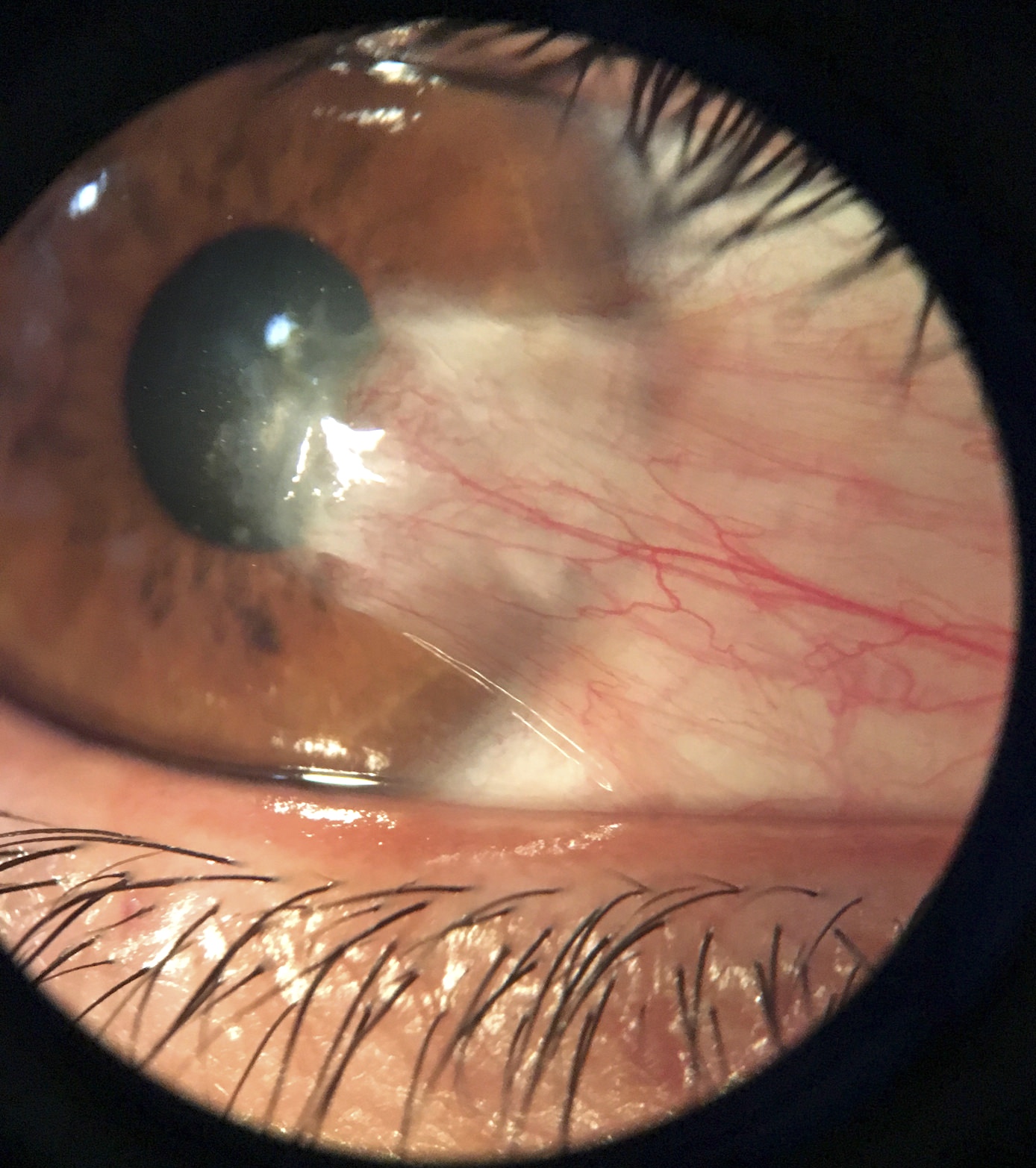
Acanthamoeba keratitis on slit lamp examination
Contributed by Gabrielle Yeaney, M.D.

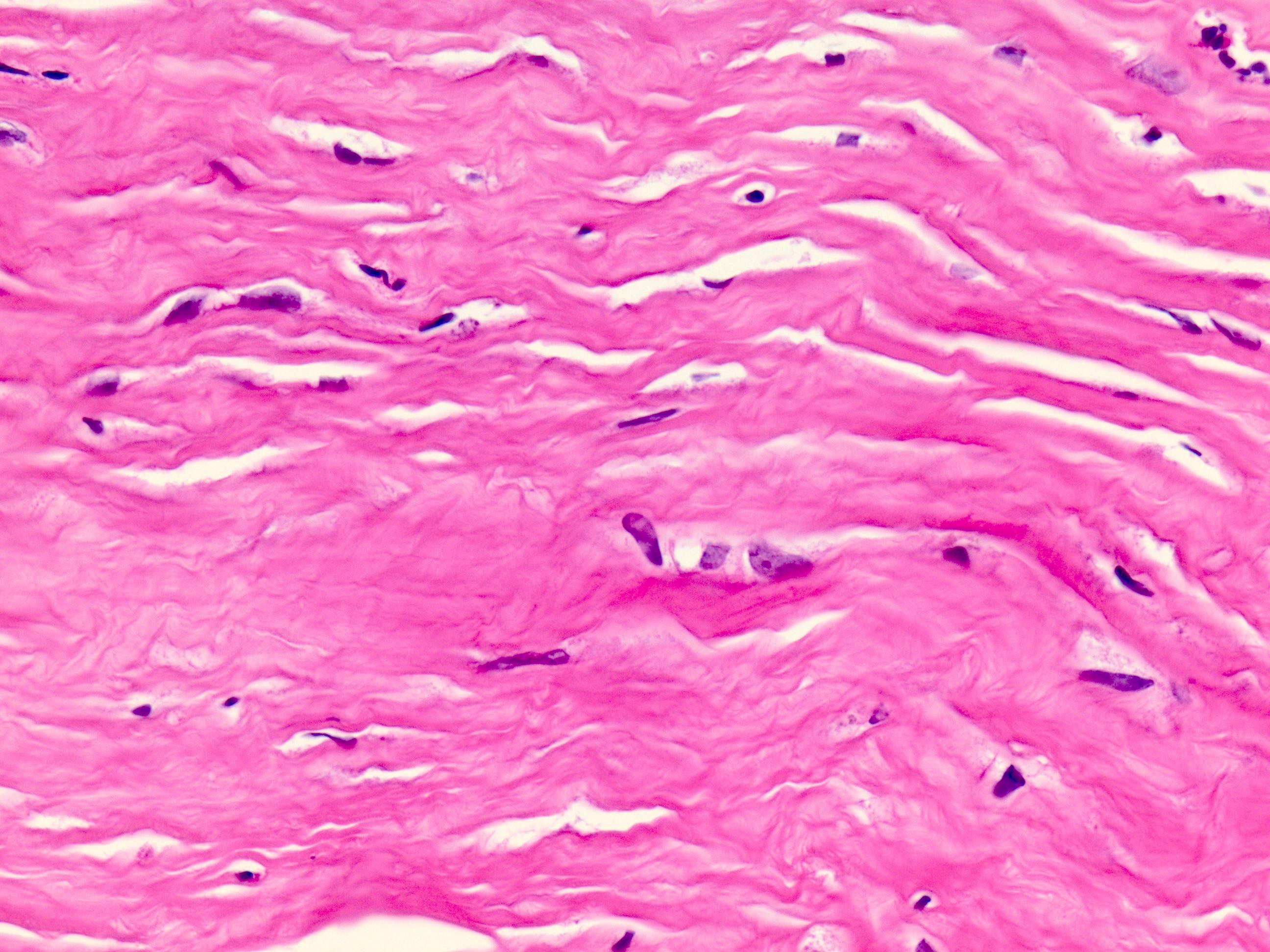
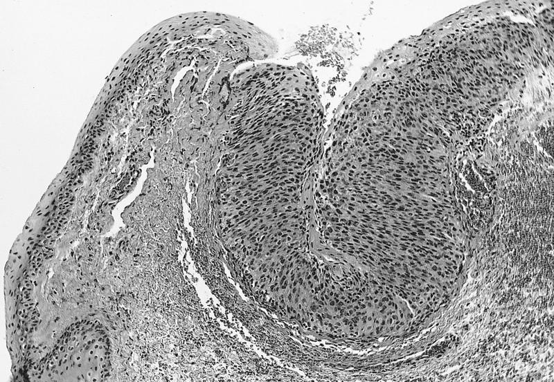
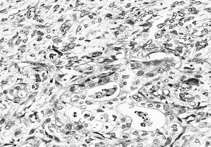
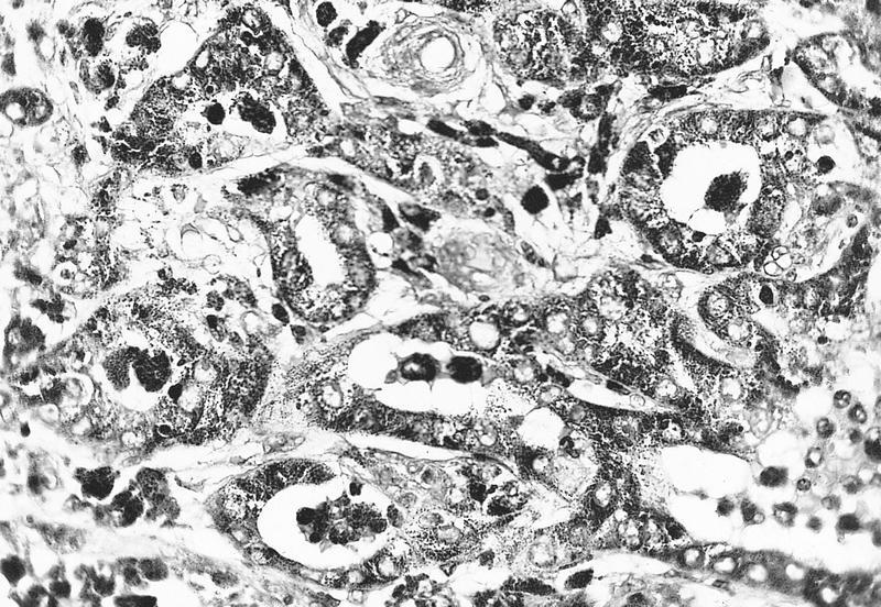
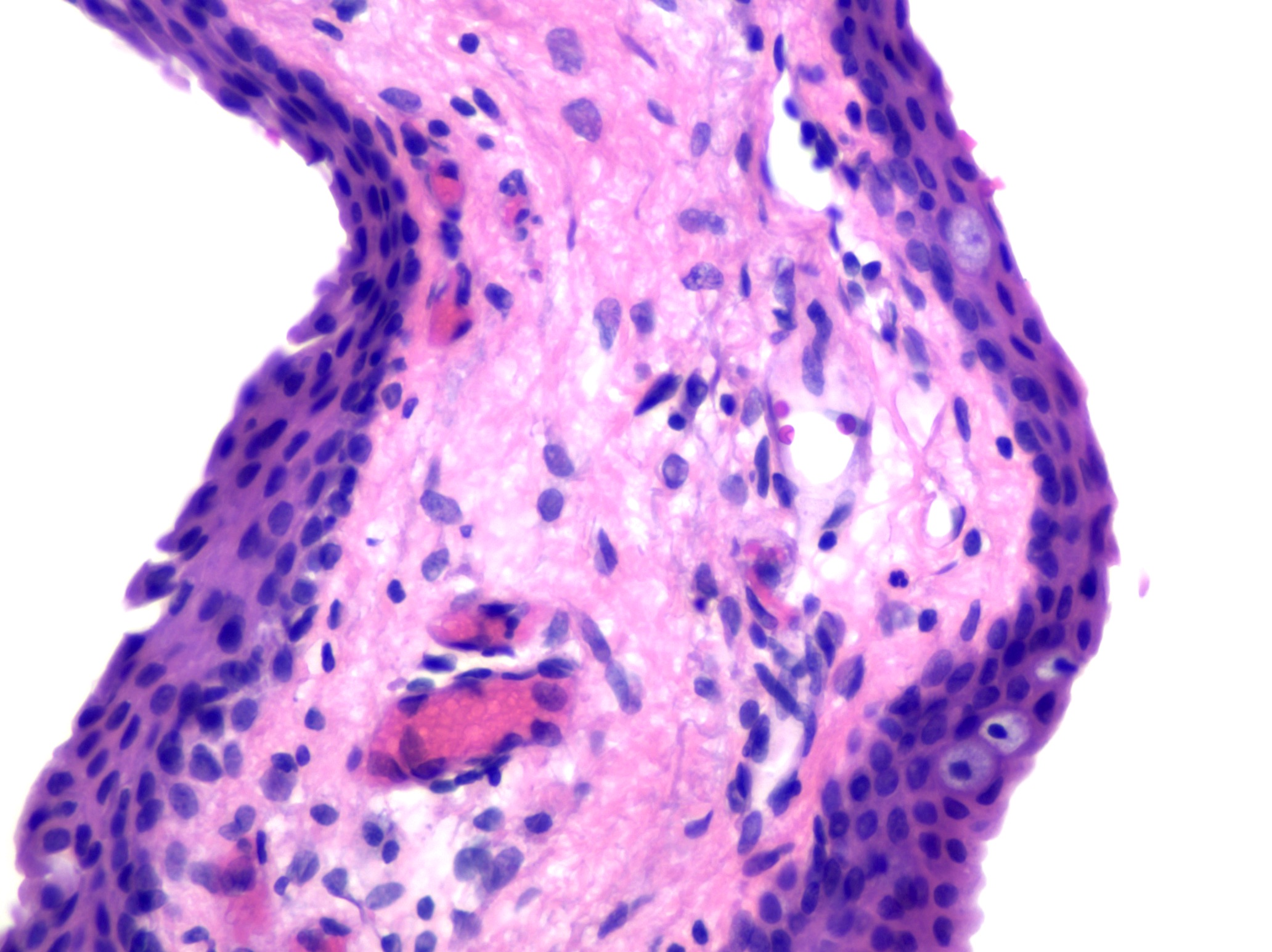
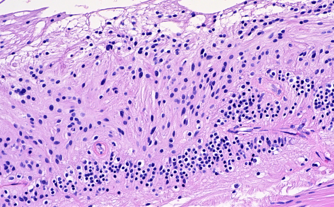
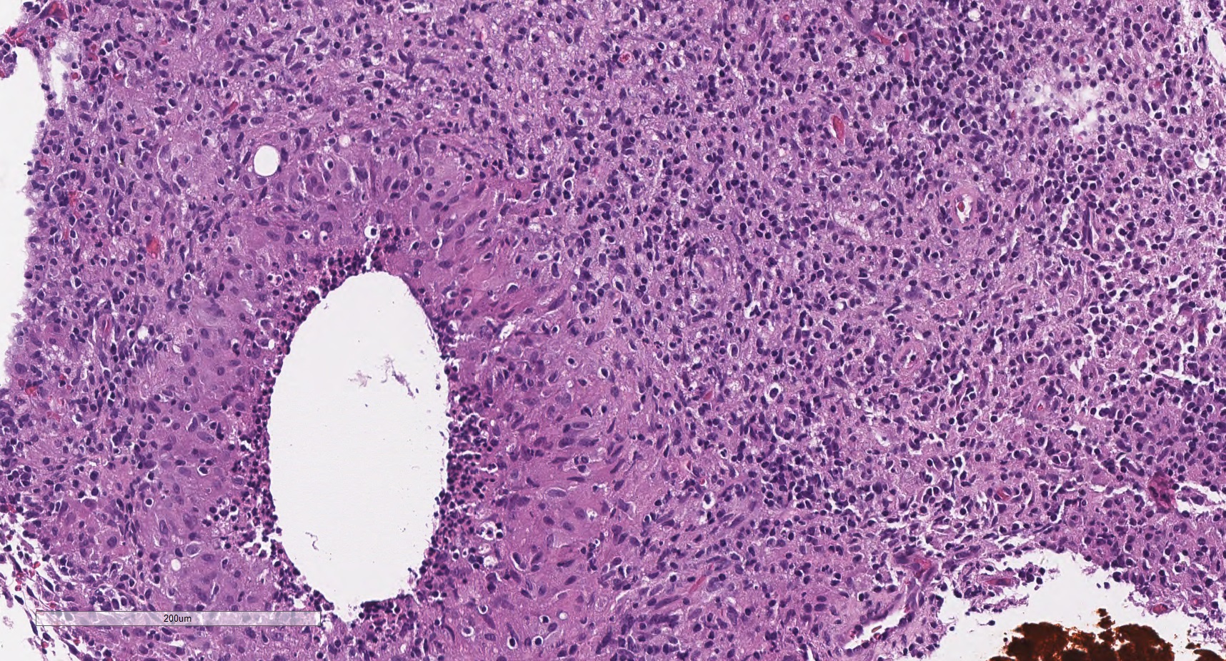
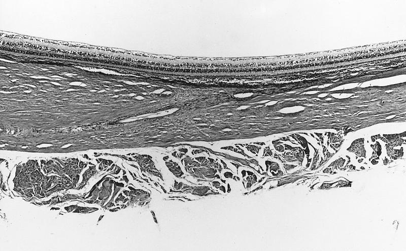
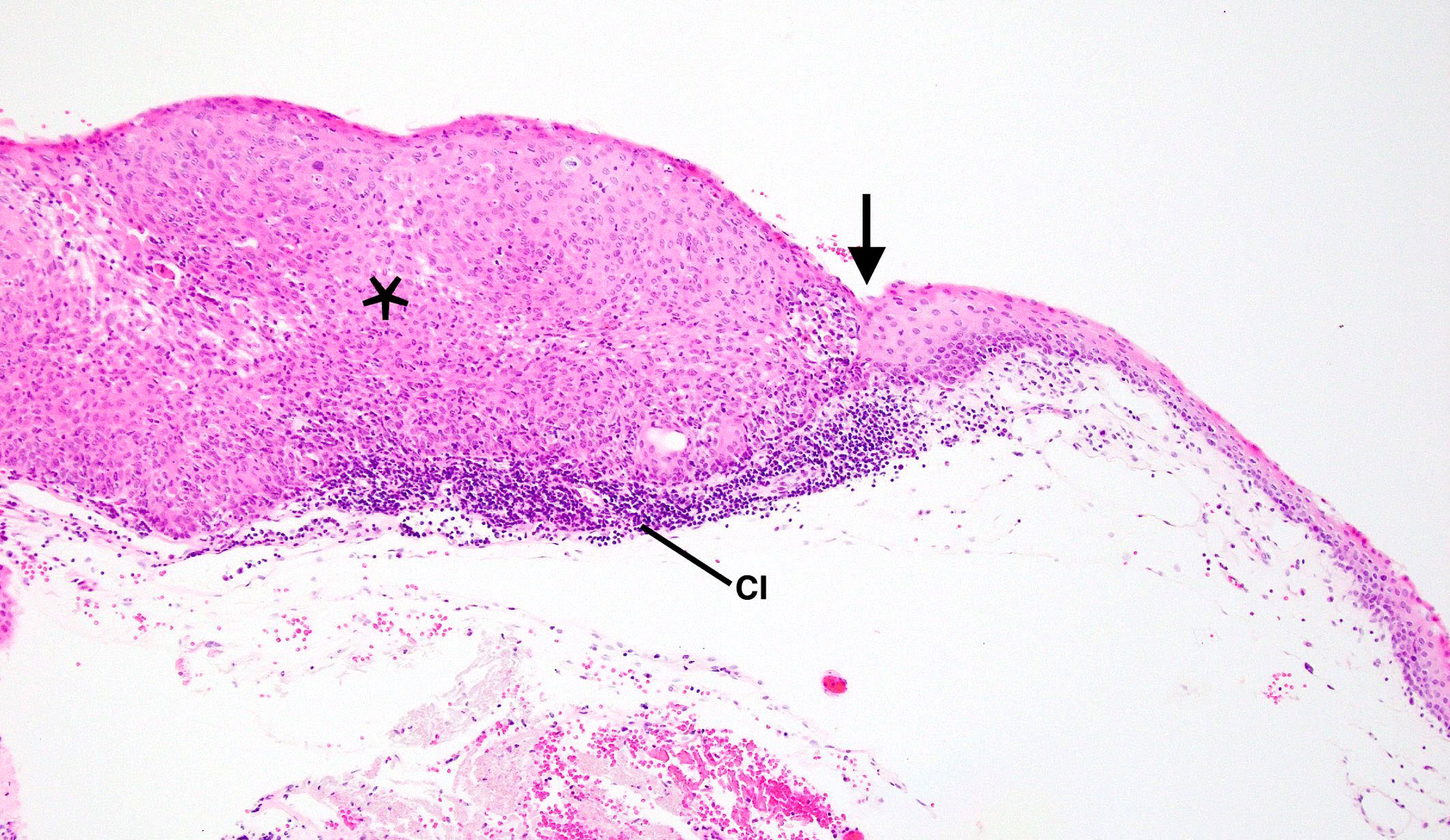
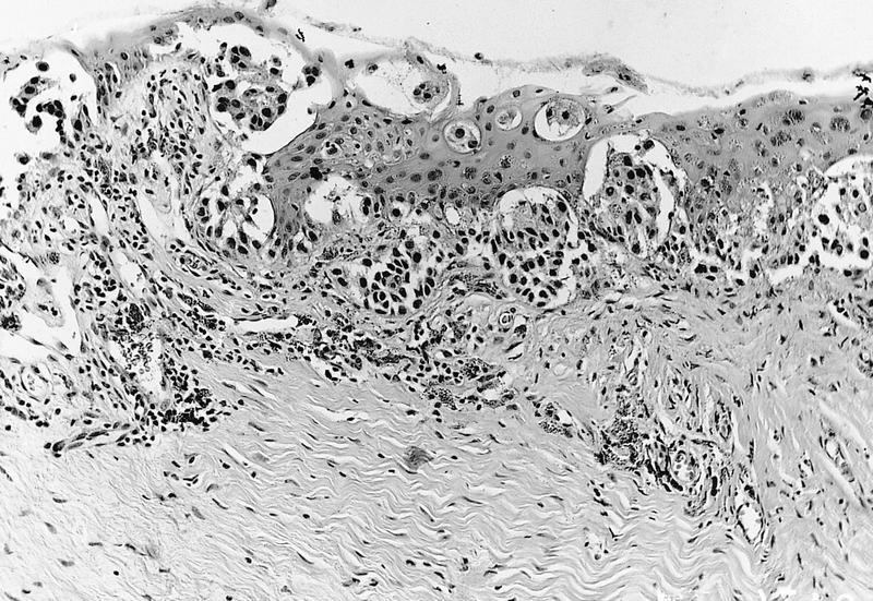
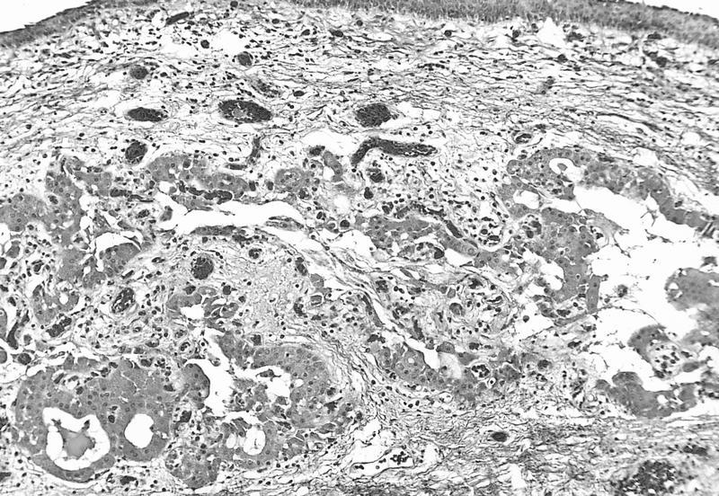
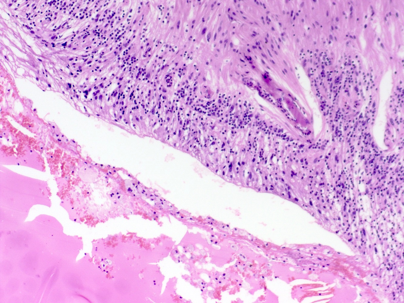
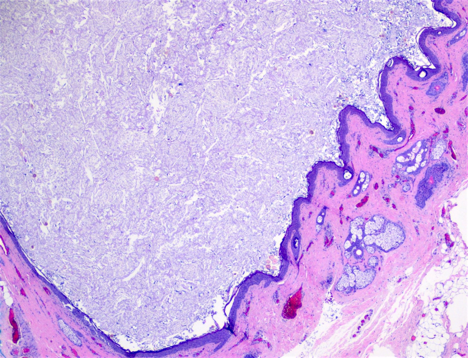
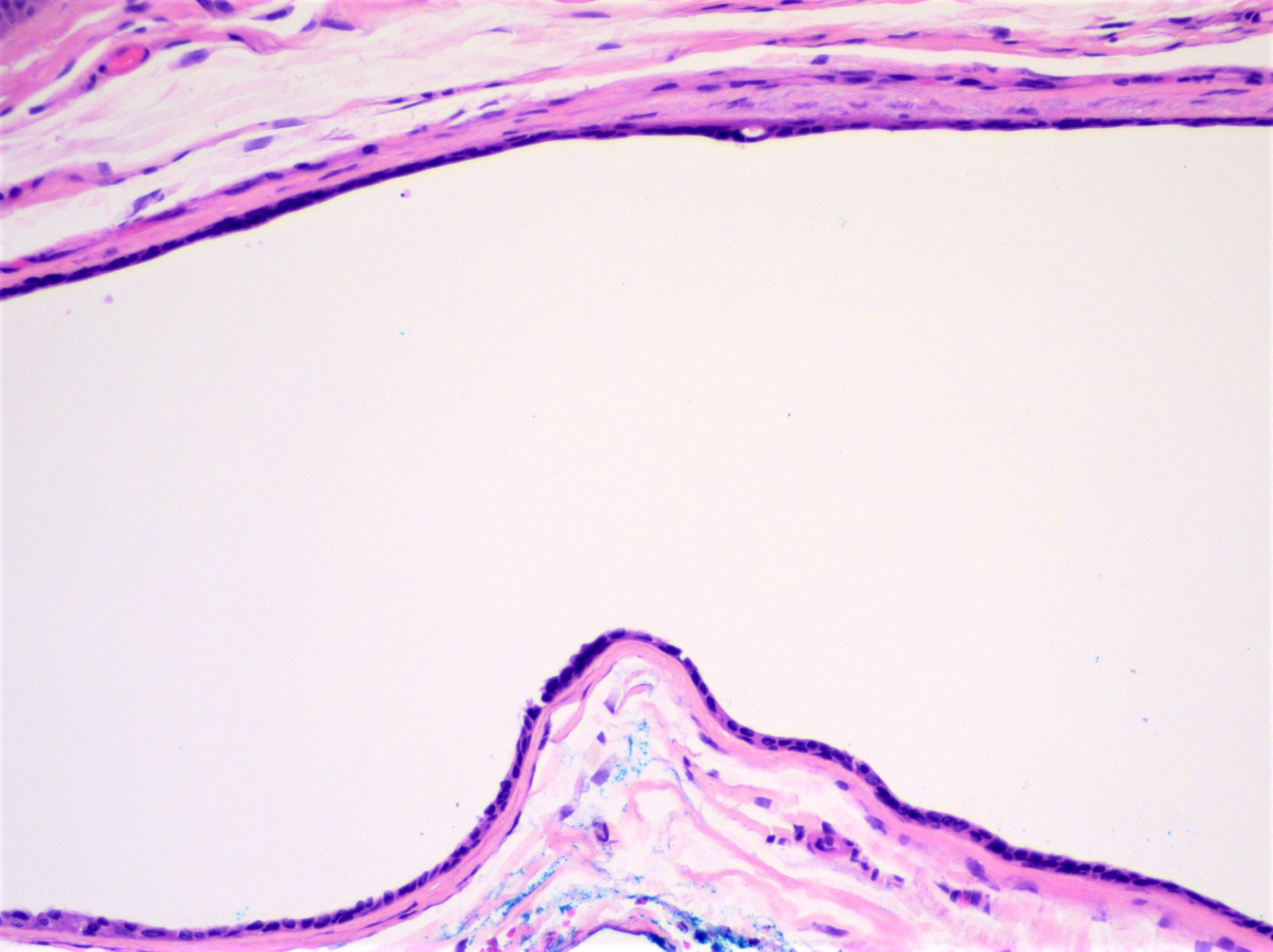
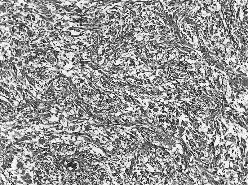
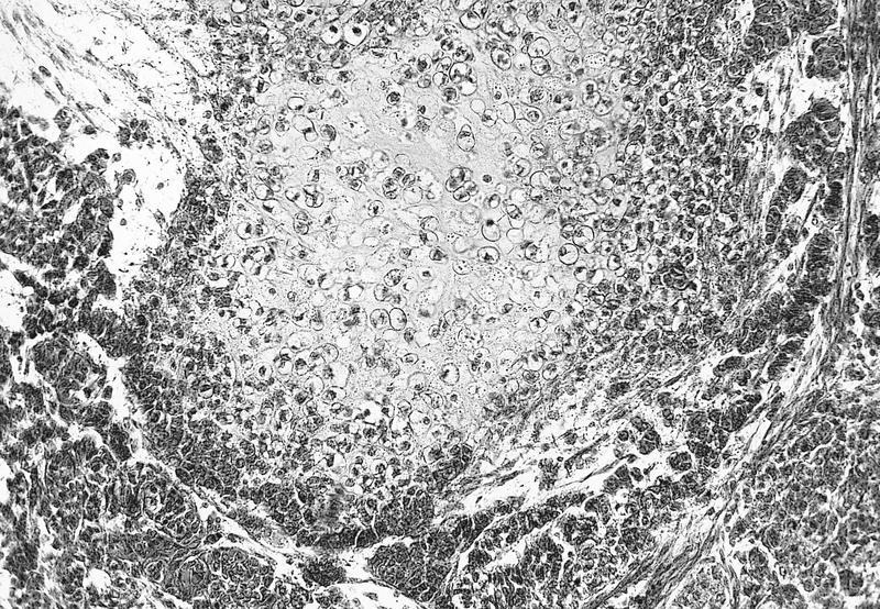
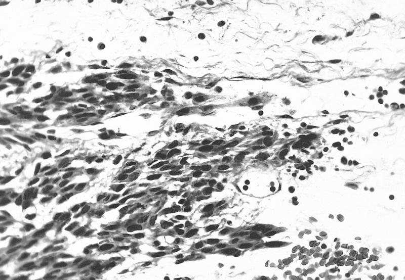
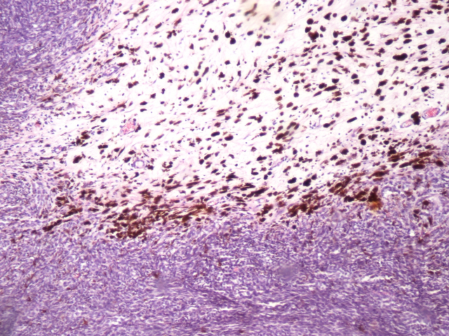
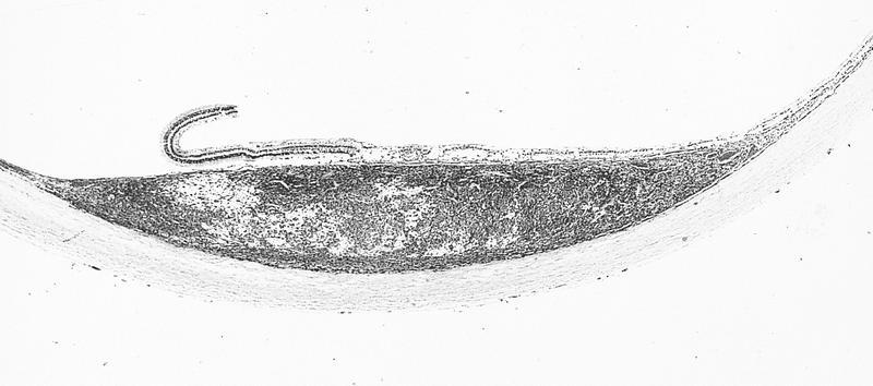
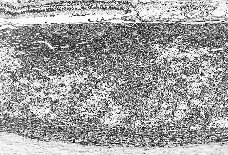
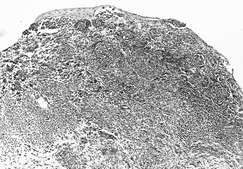
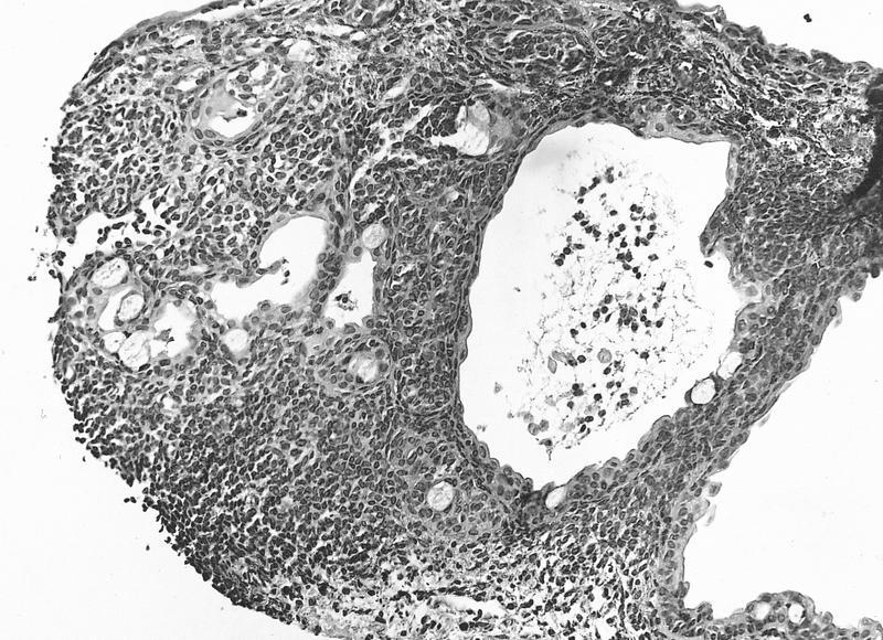
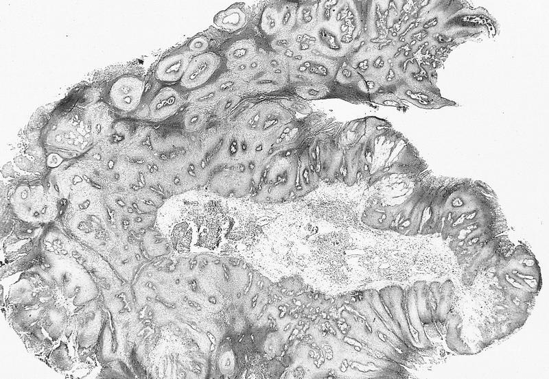
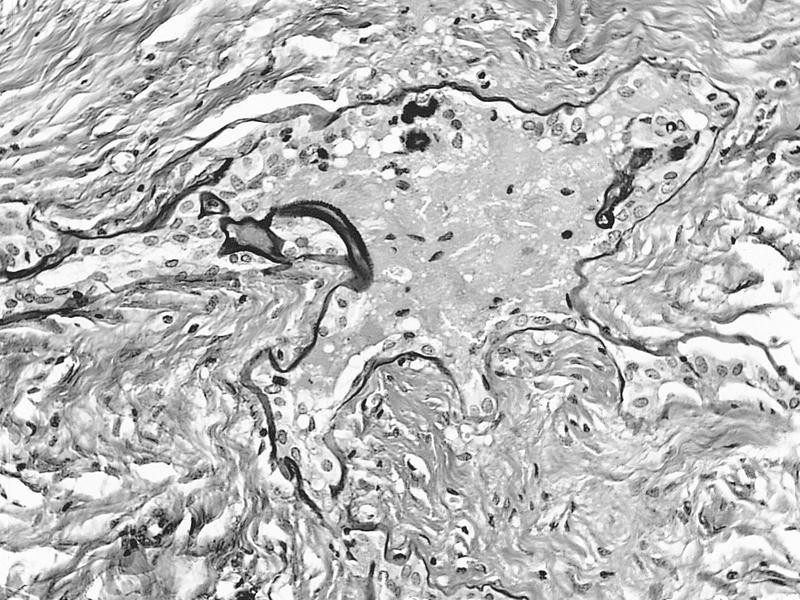
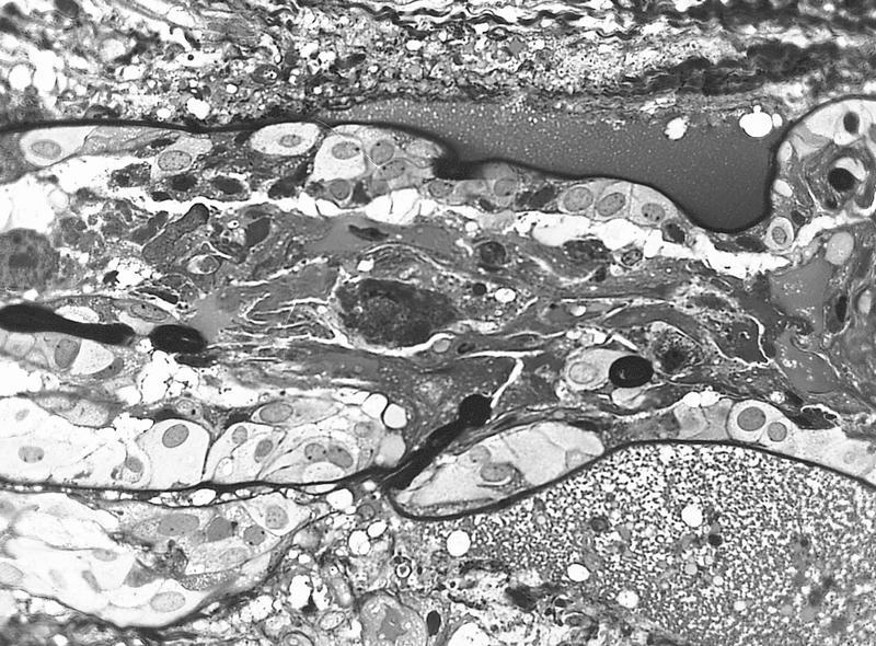
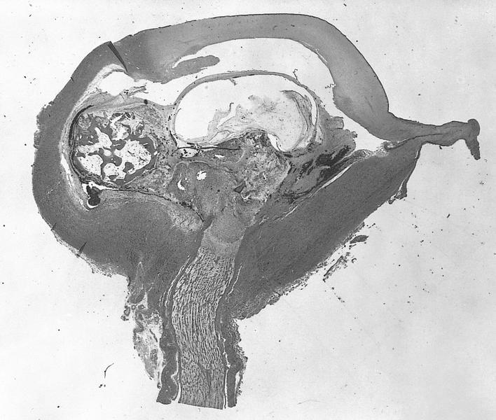
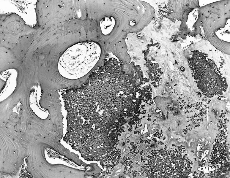
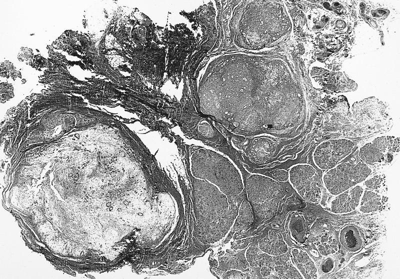
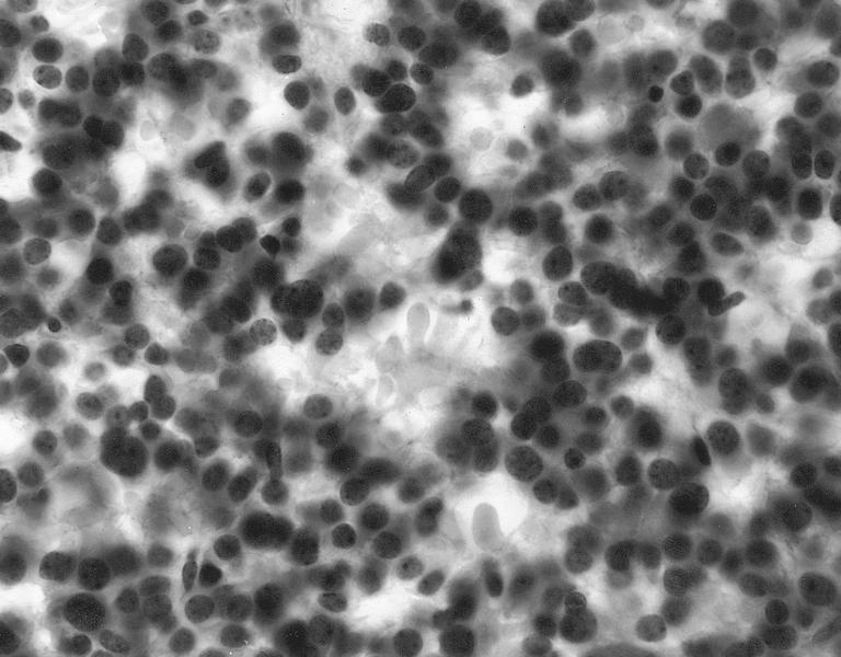
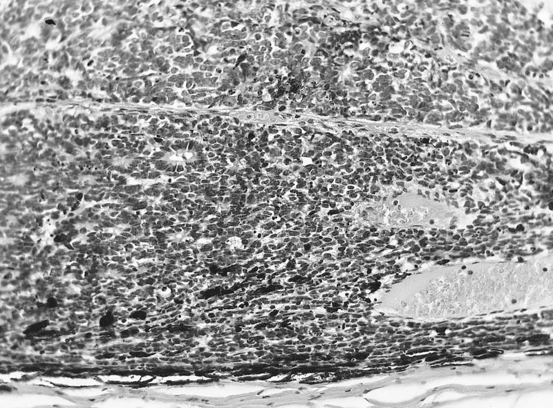
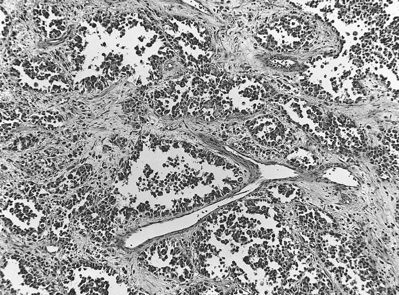
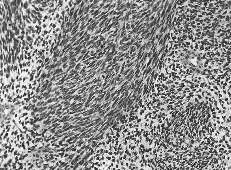
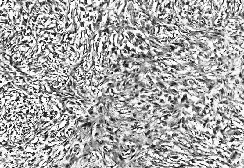
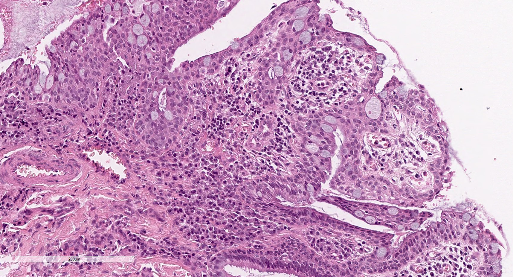
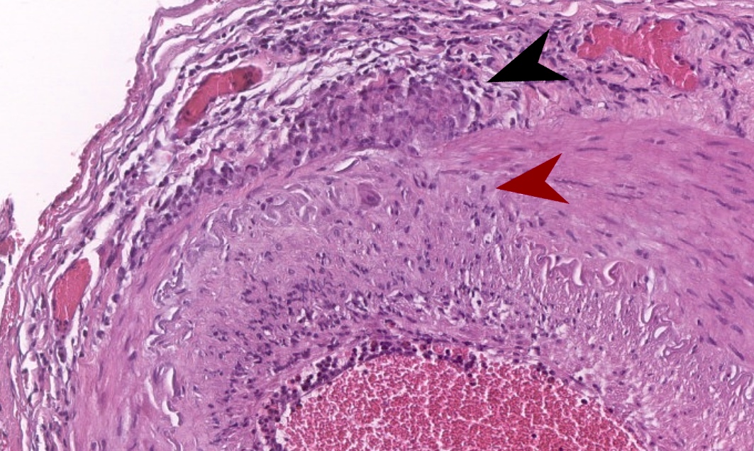
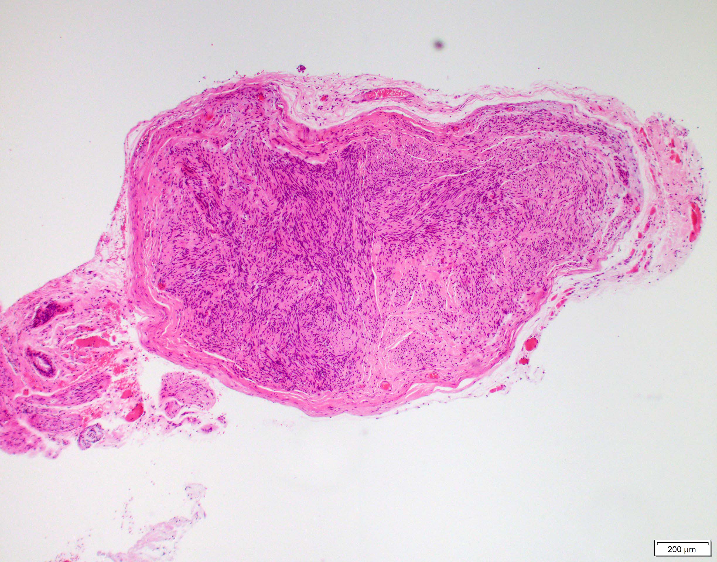
Stromal microabscess and necrosis

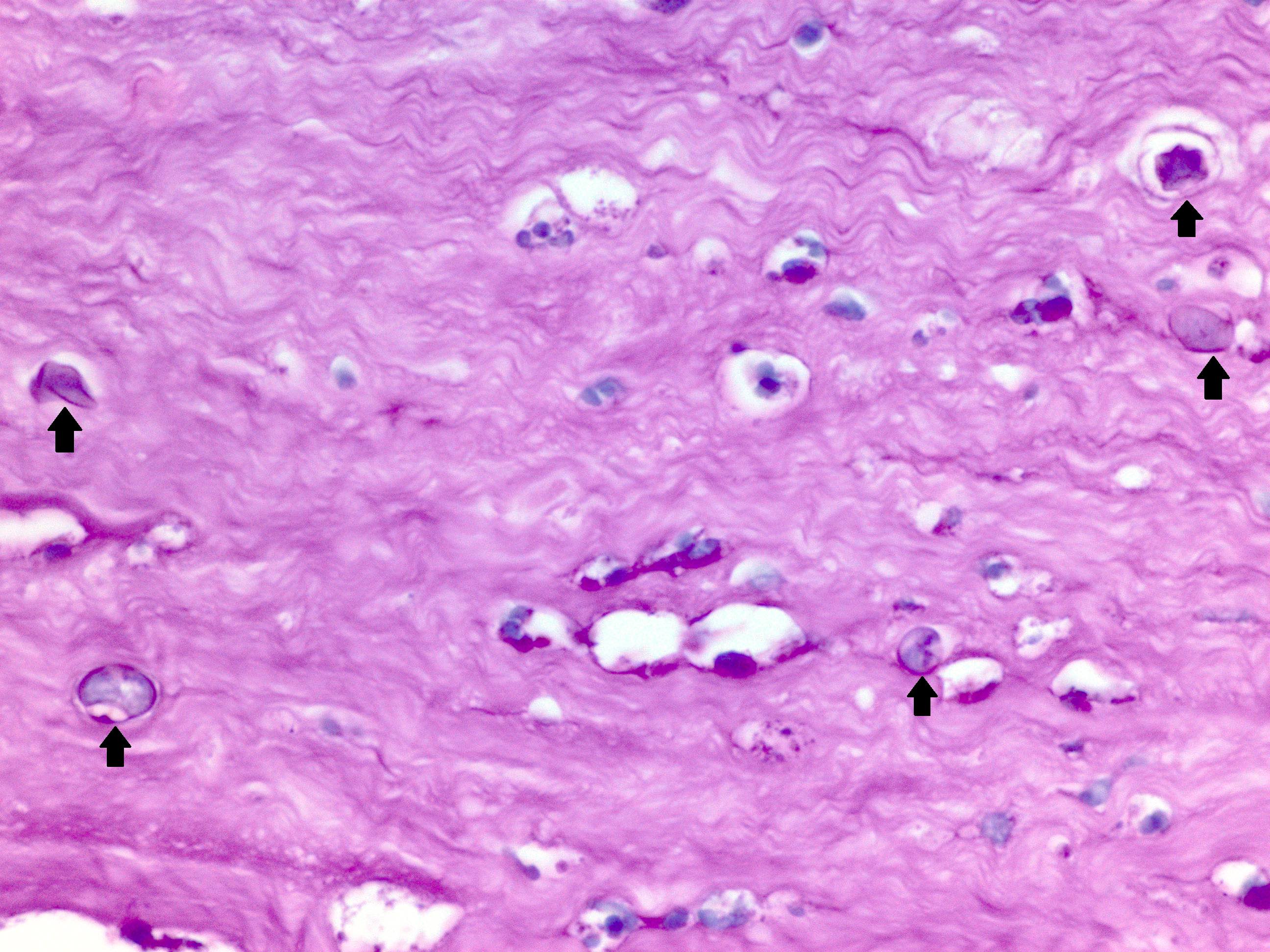
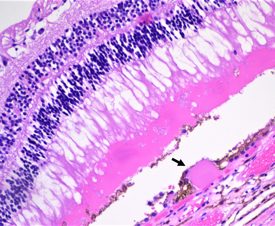
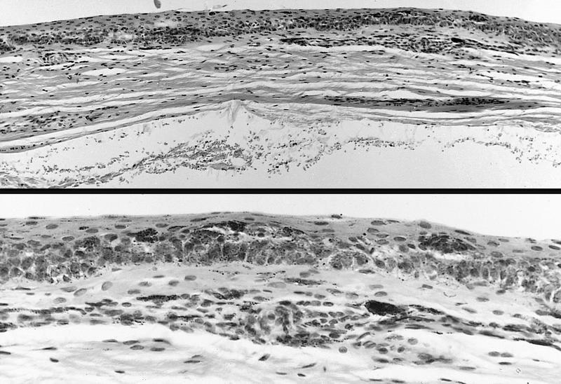
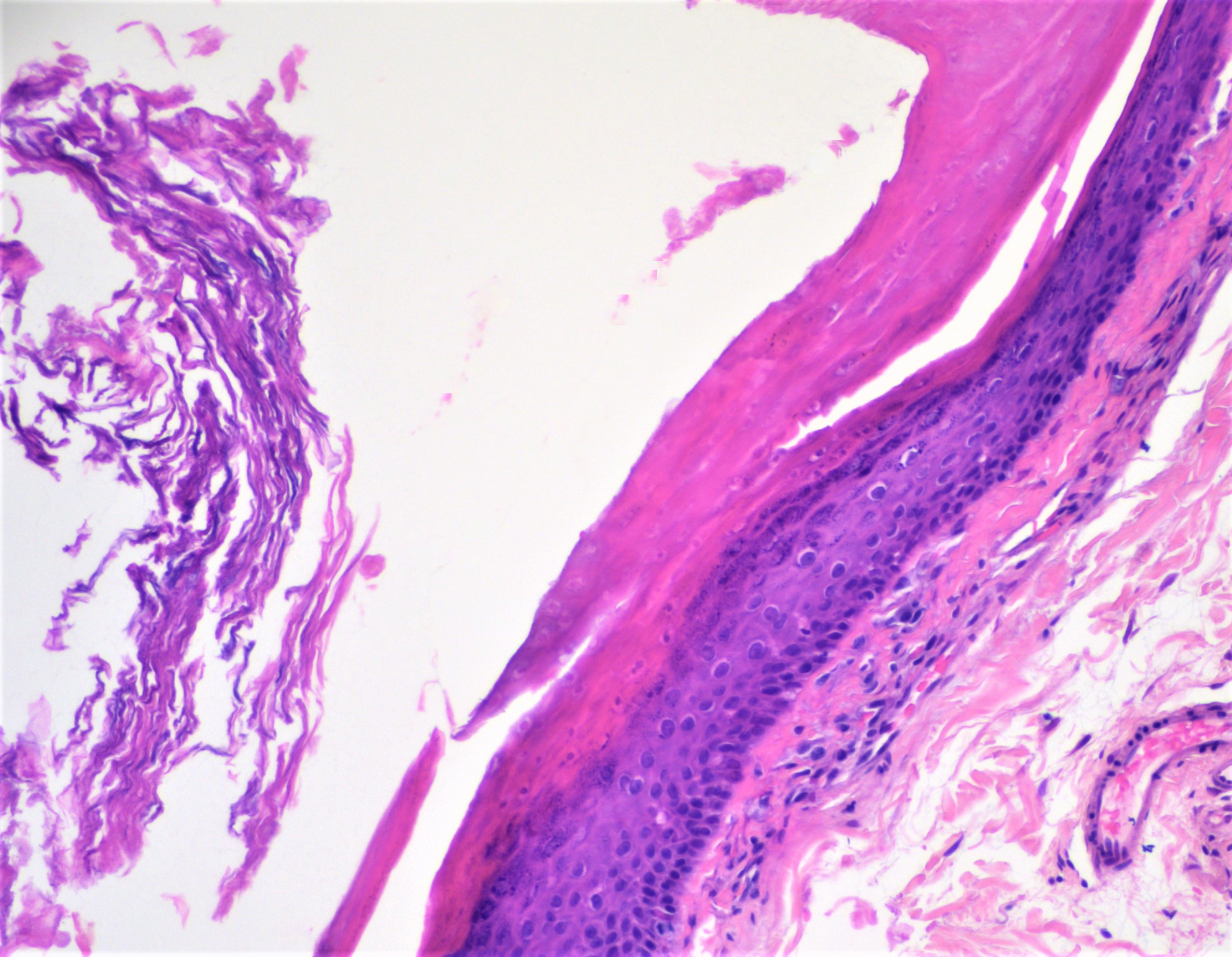
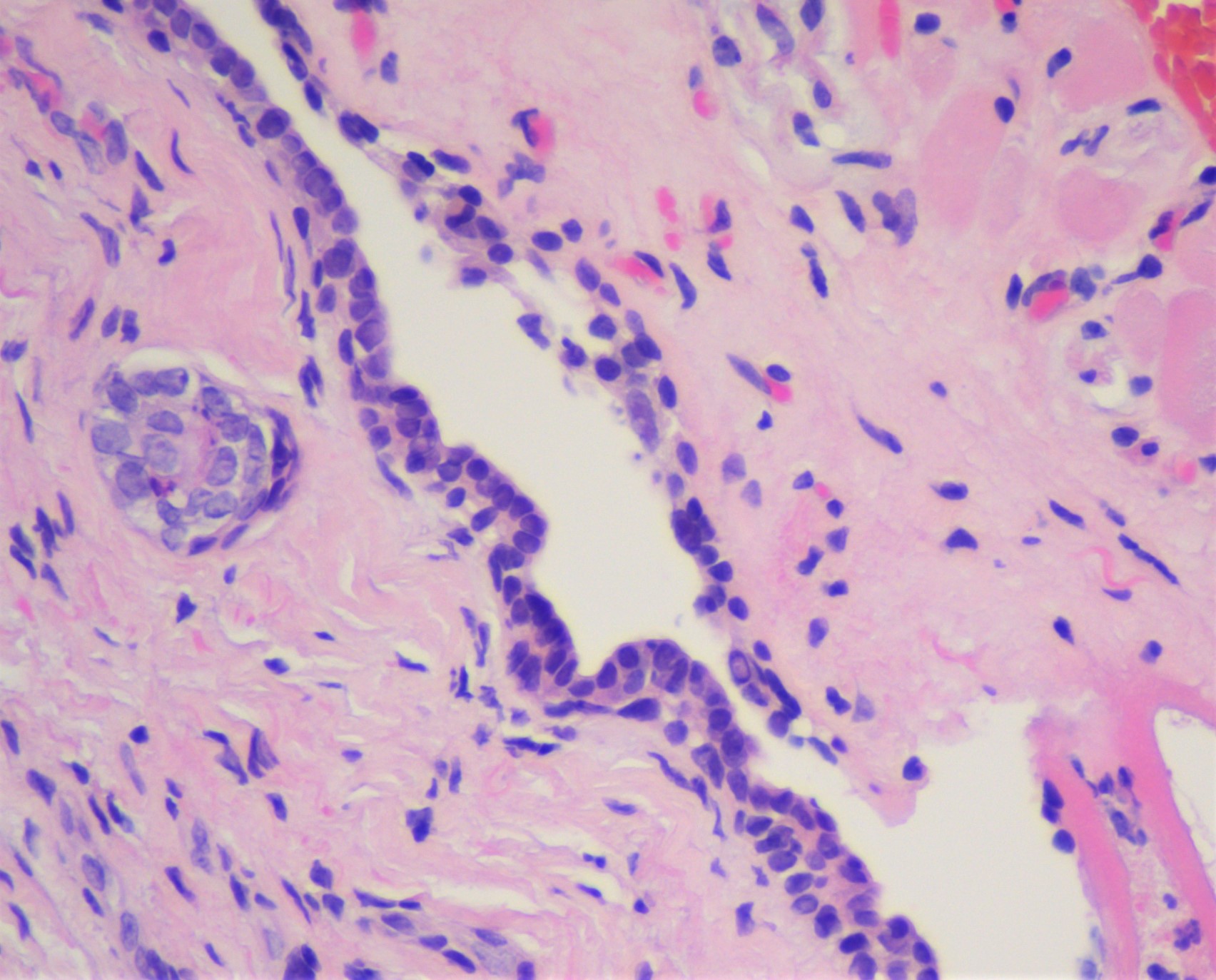
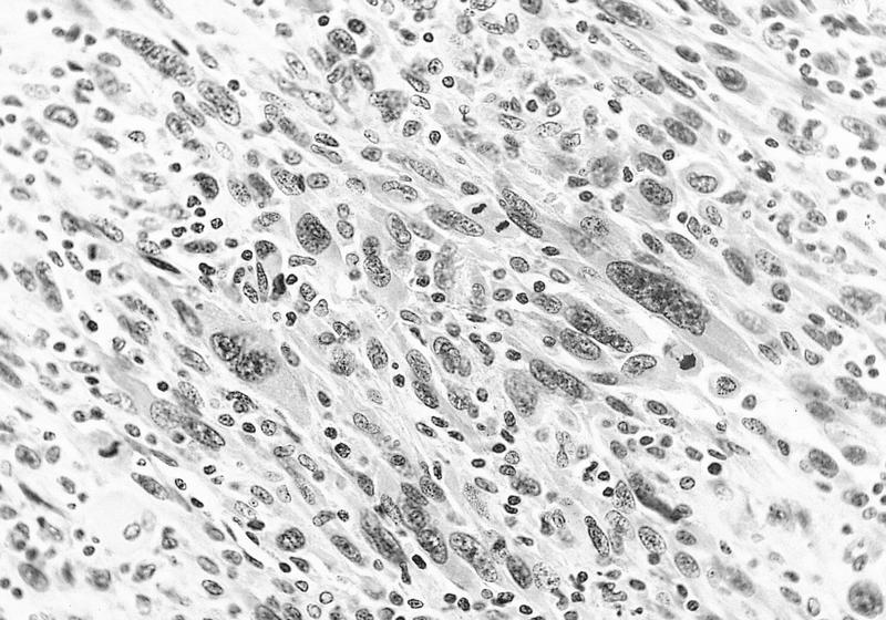
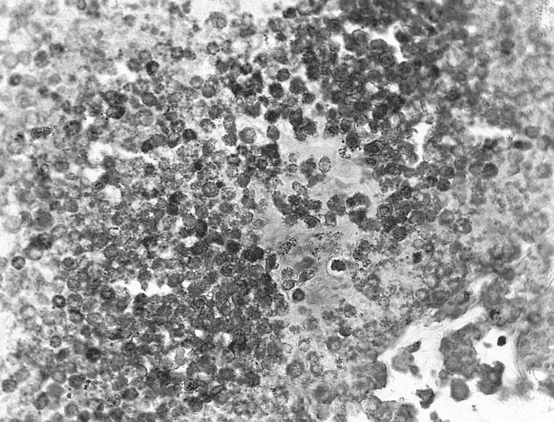
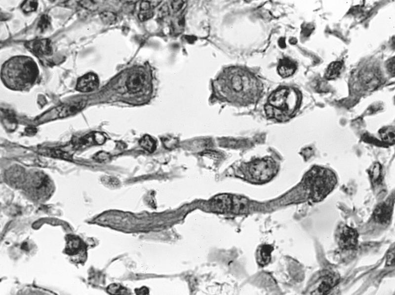
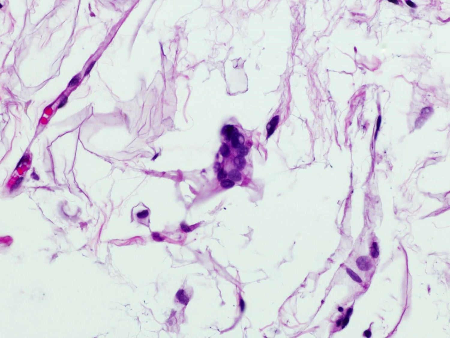
Multiple amoebic forms
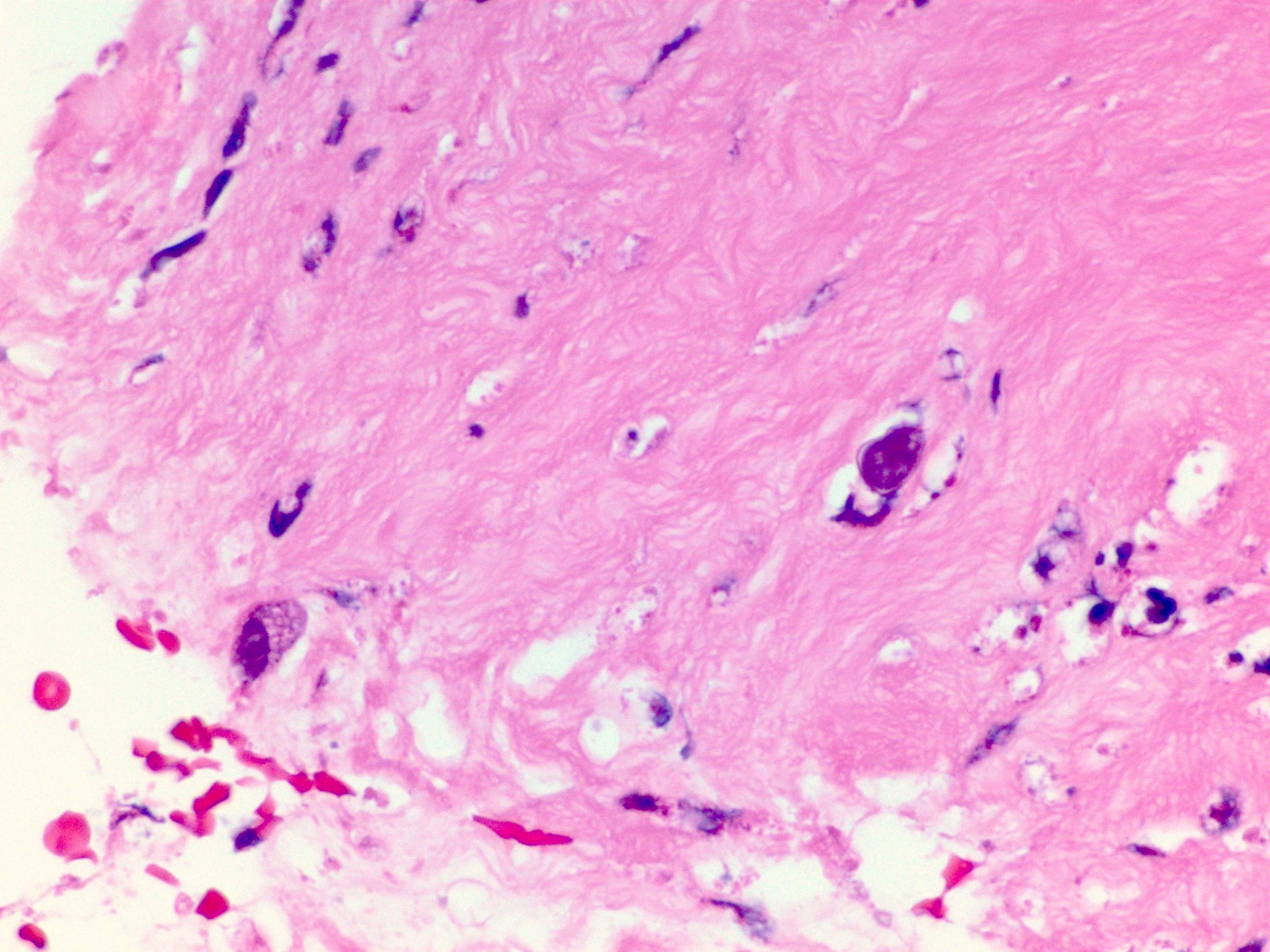
2 Acanthamoeba in stroma
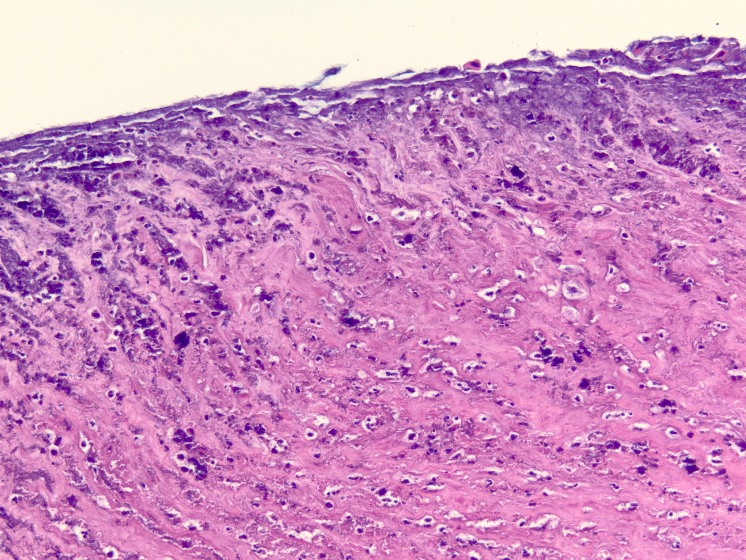
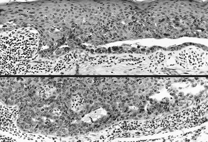
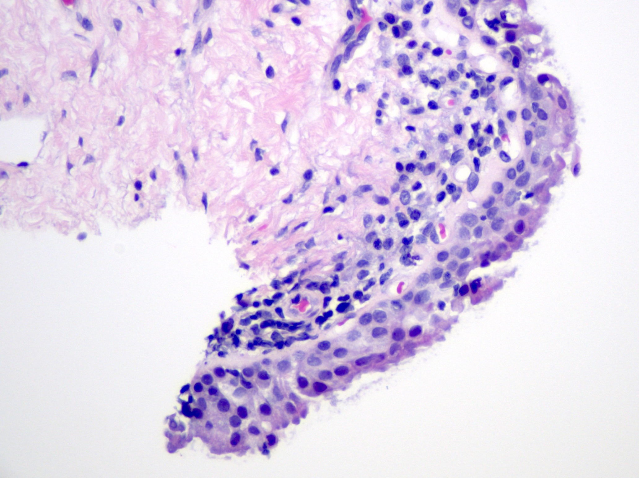
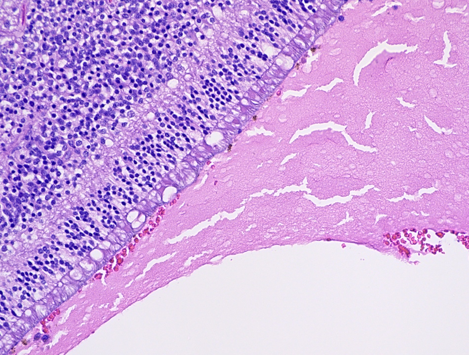
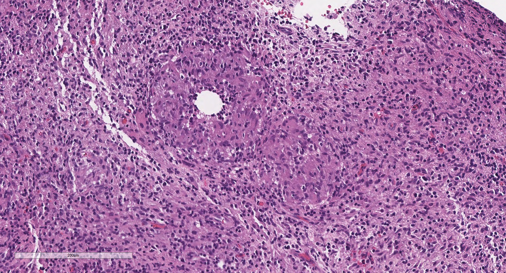
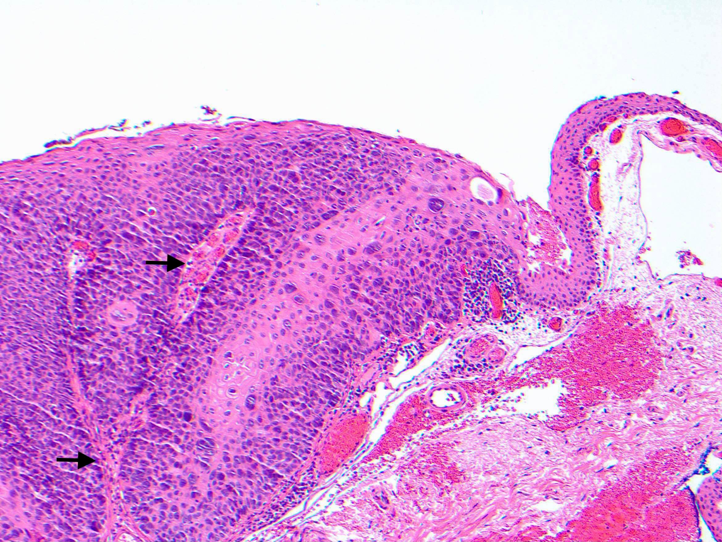
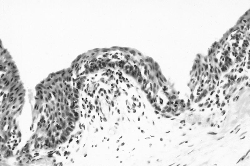
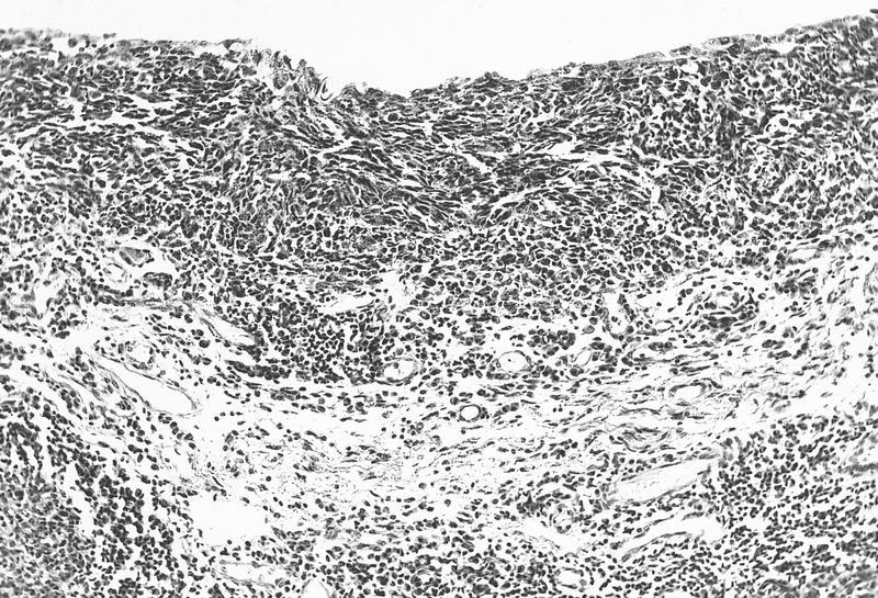
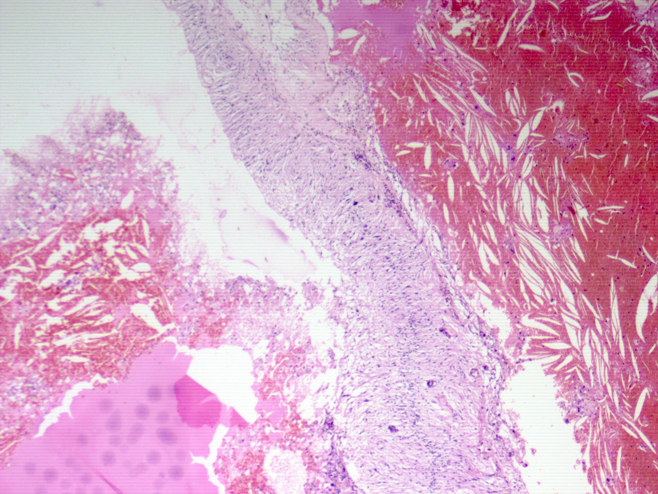
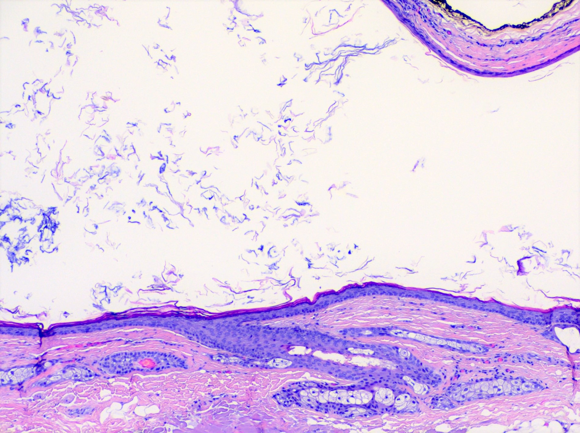
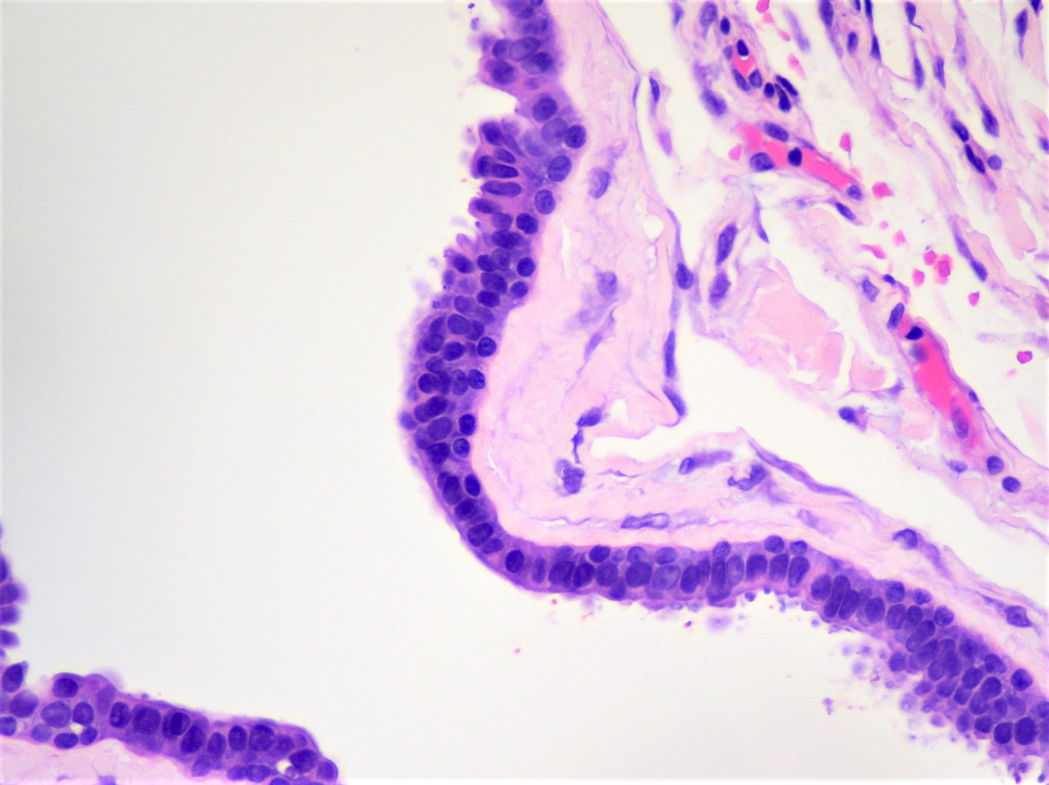
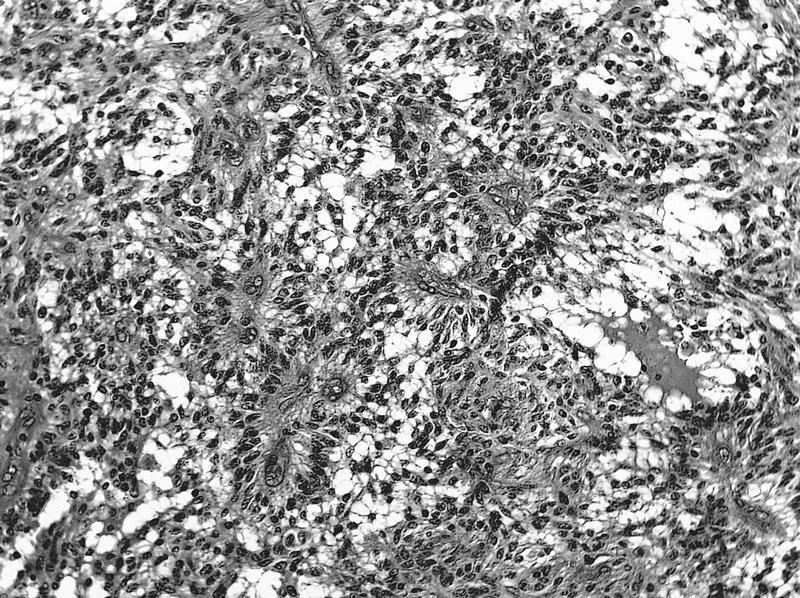
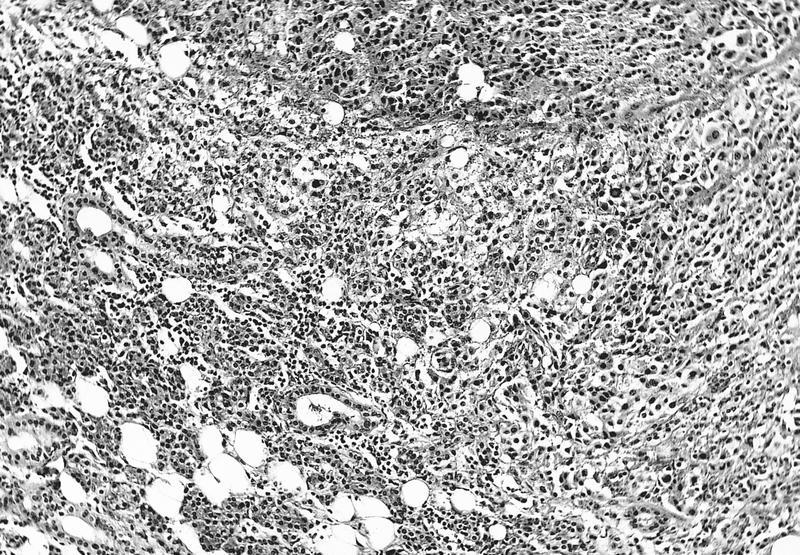
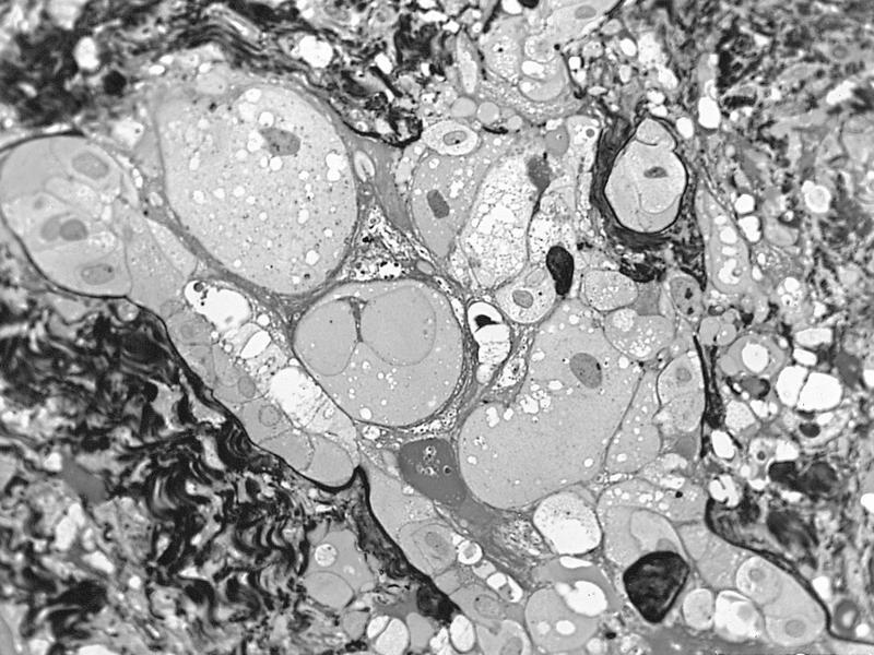
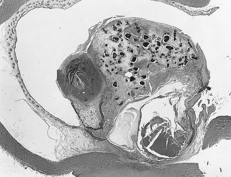
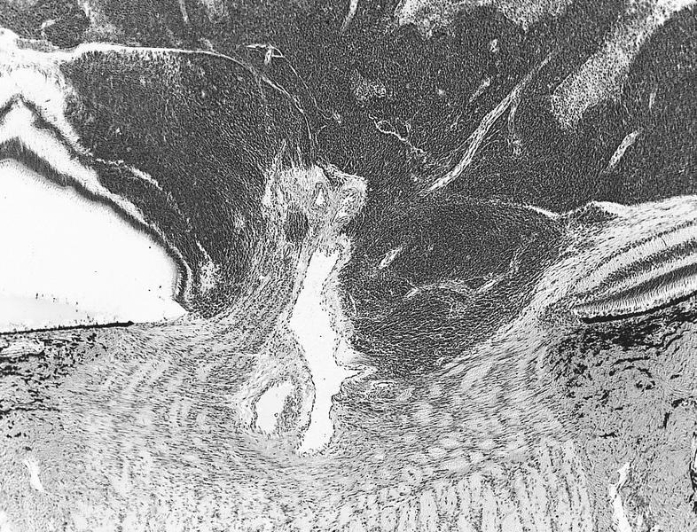
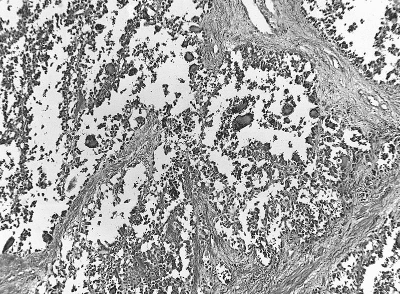
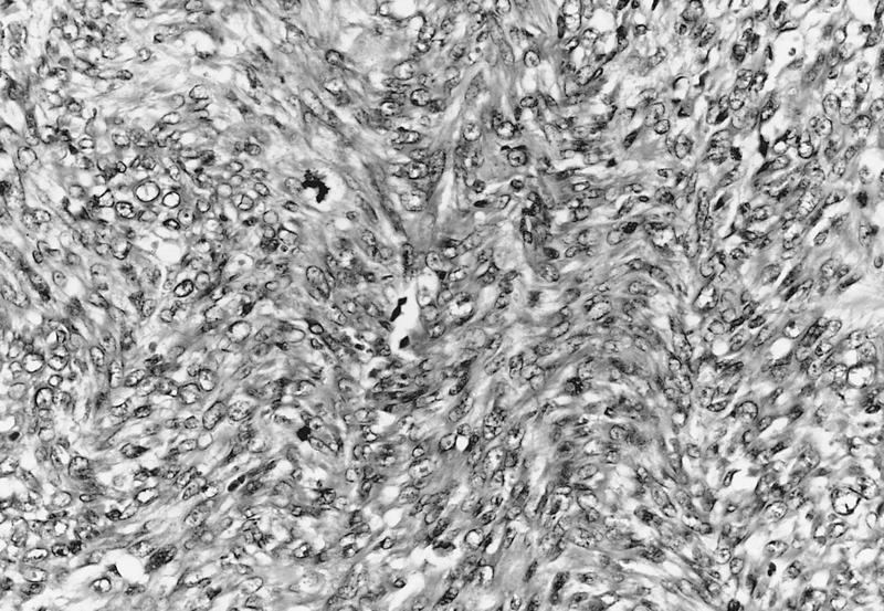
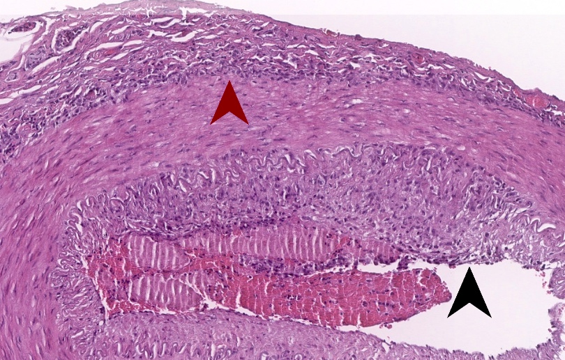
Ulceration and necrosis

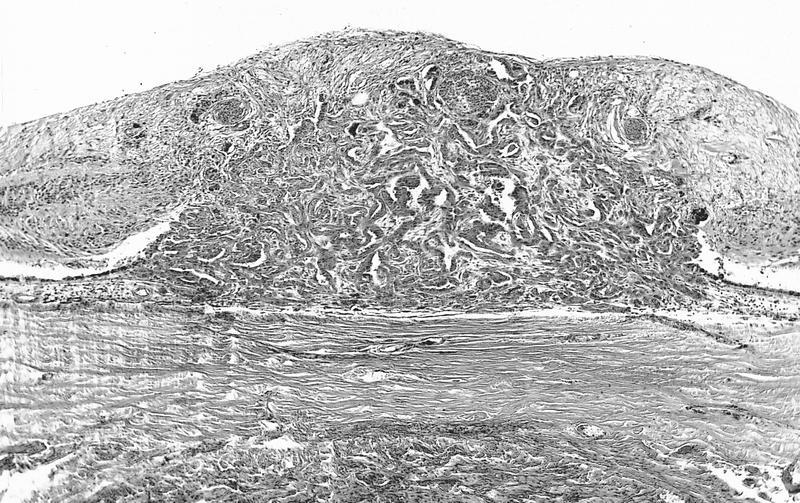
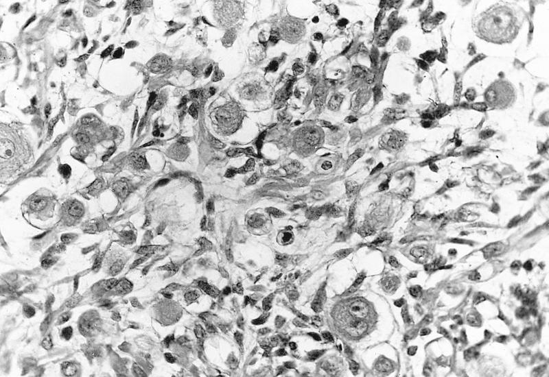
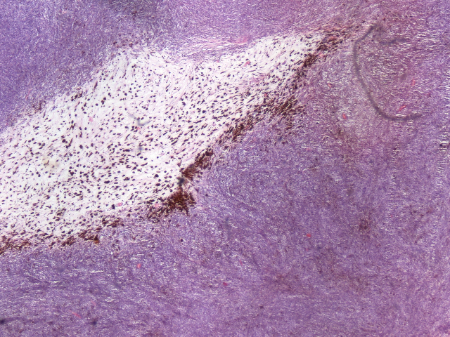
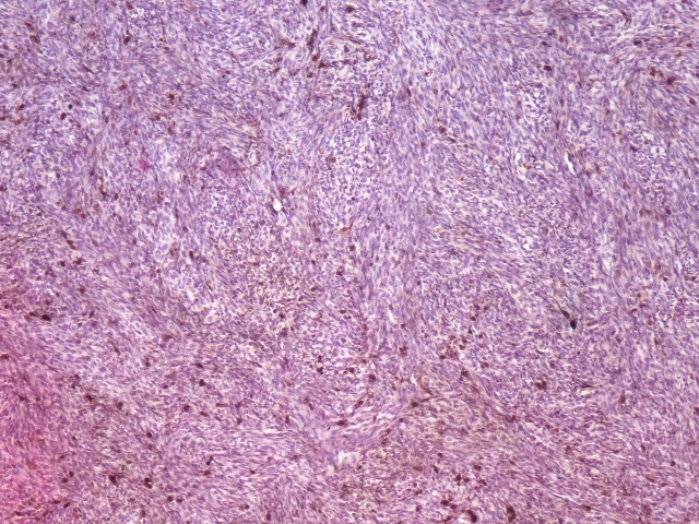
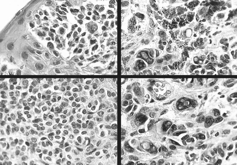
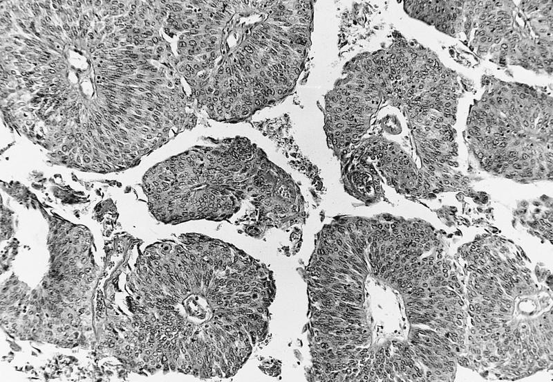
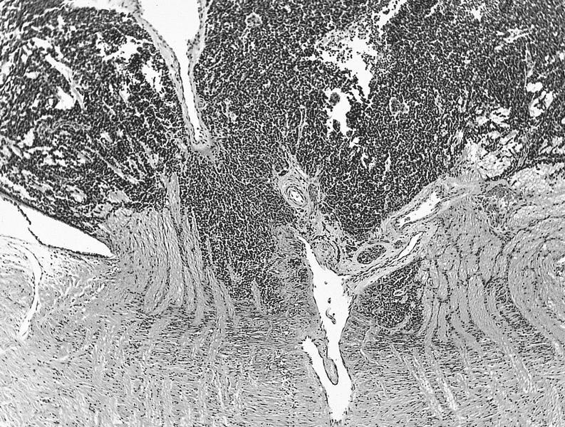
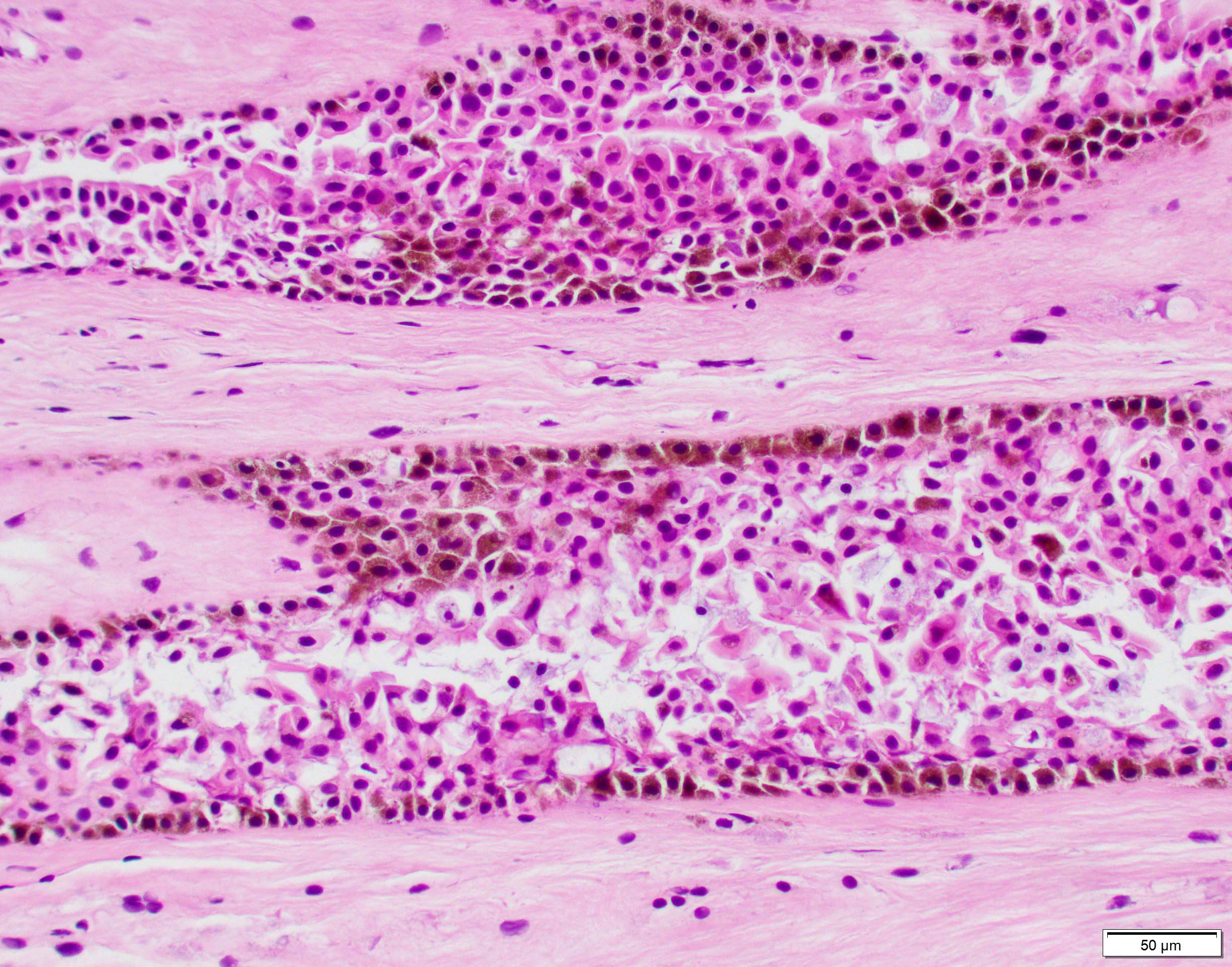
Granuloma with eosinophils
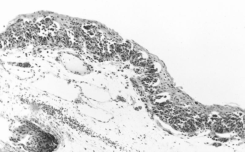
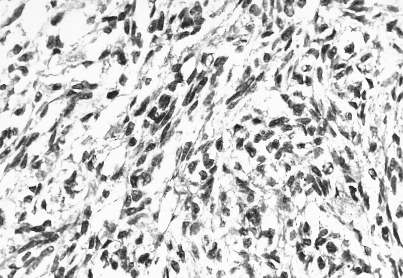
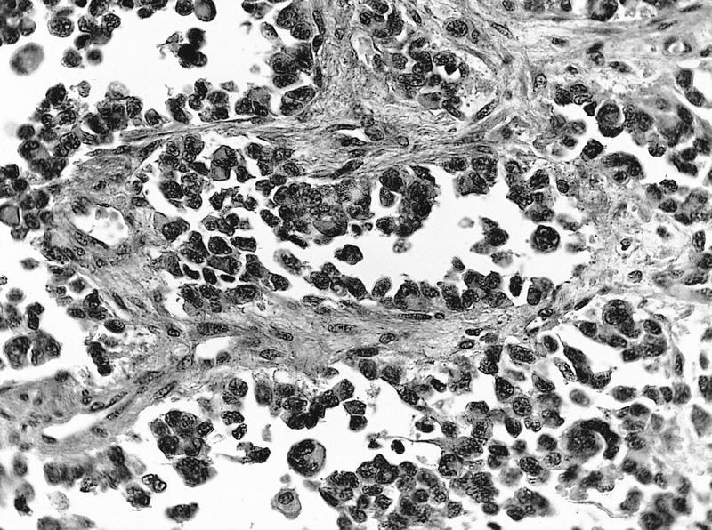
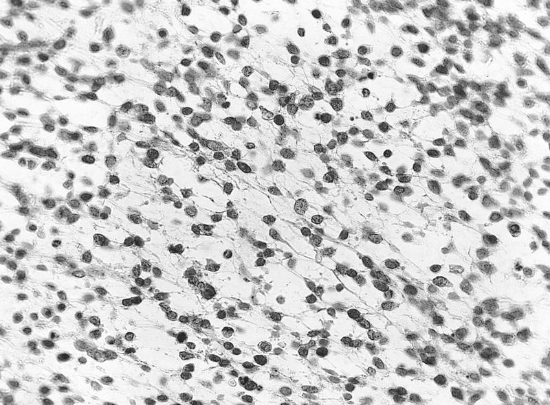
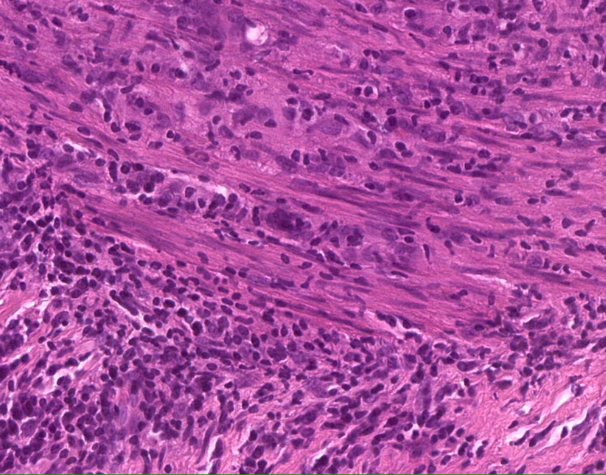
Contrast to reactive stromal keratocyte
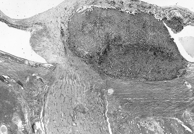
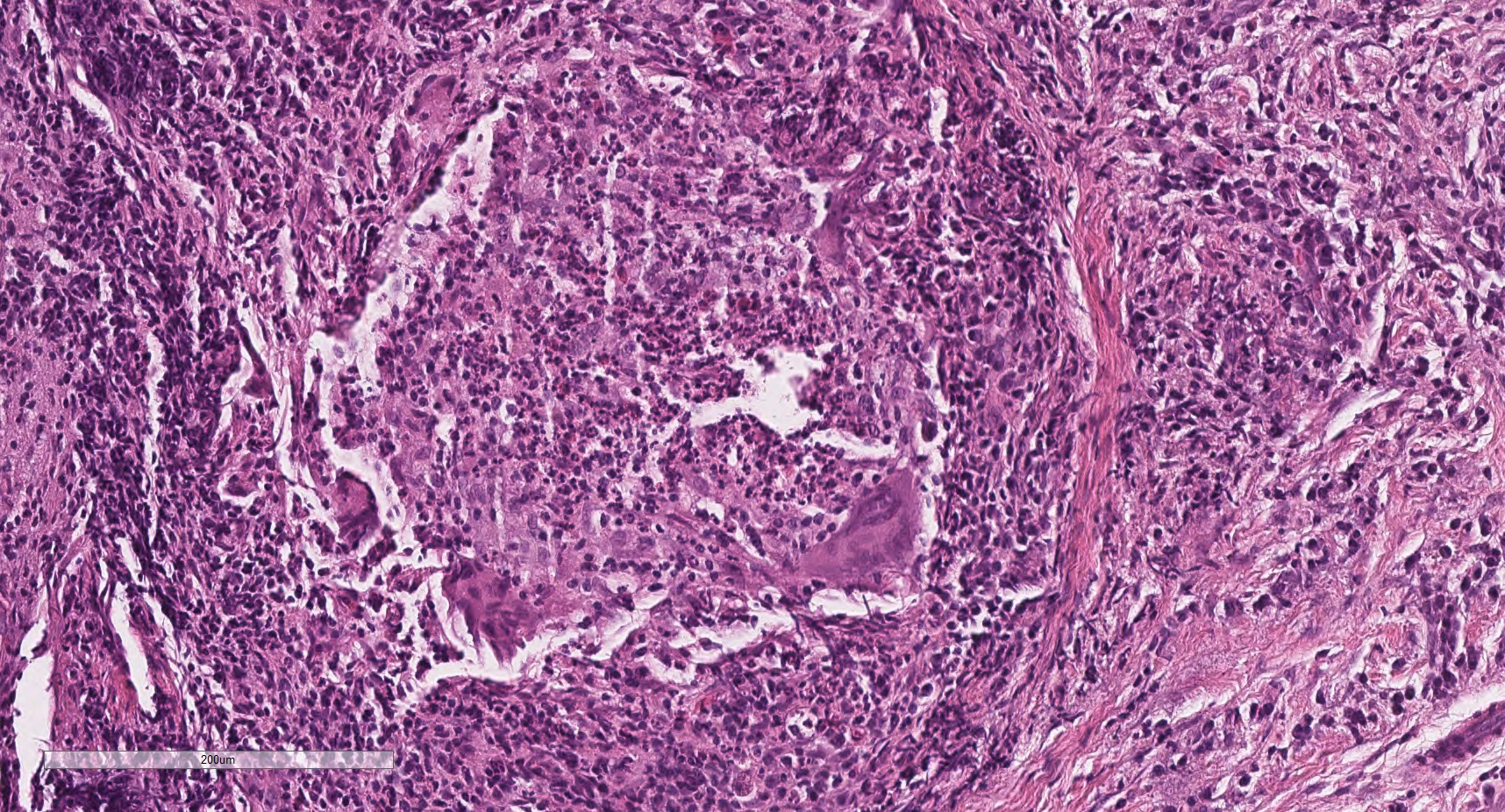
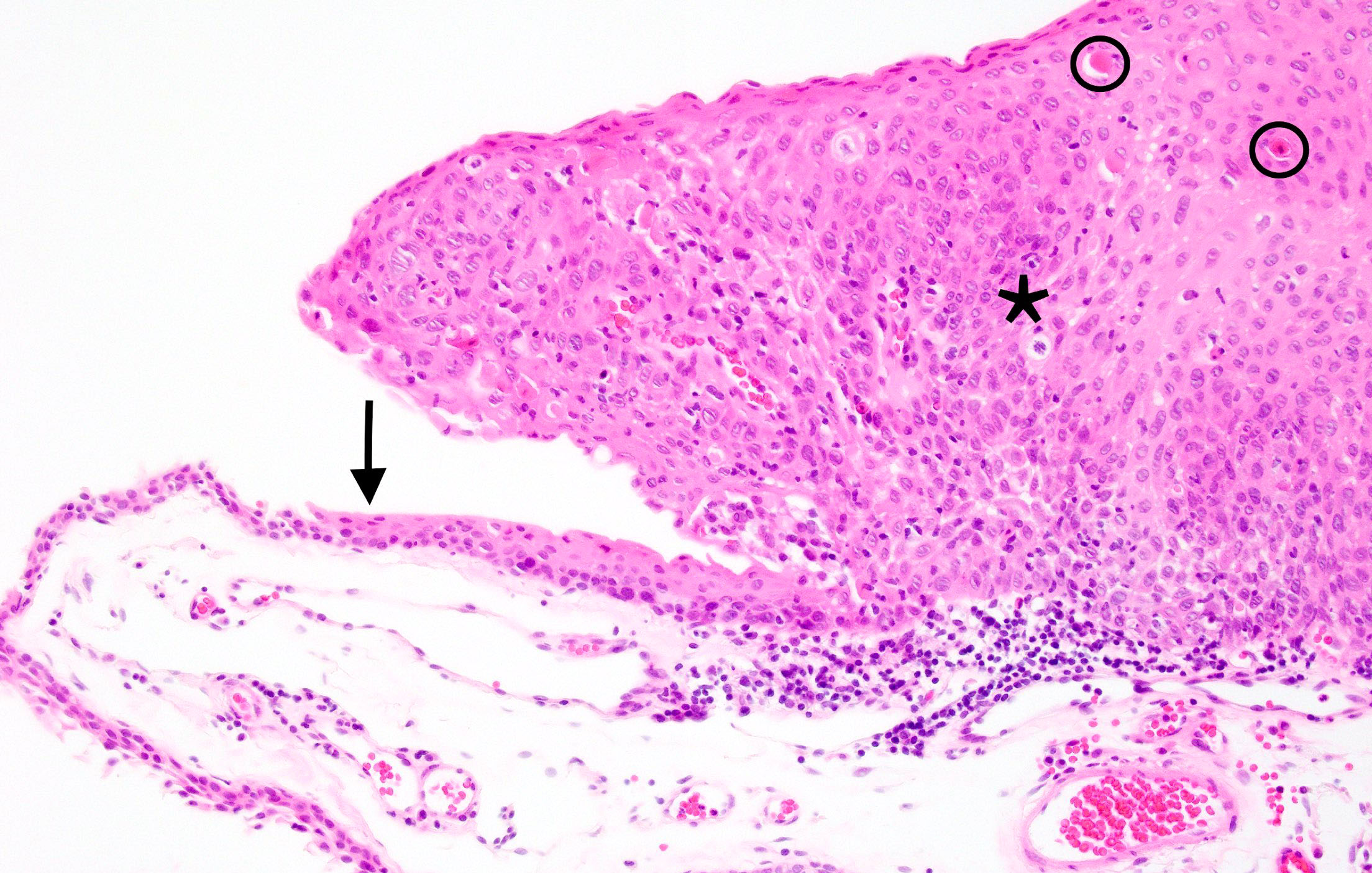
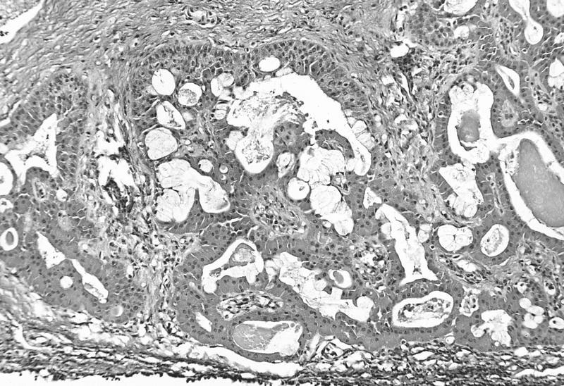
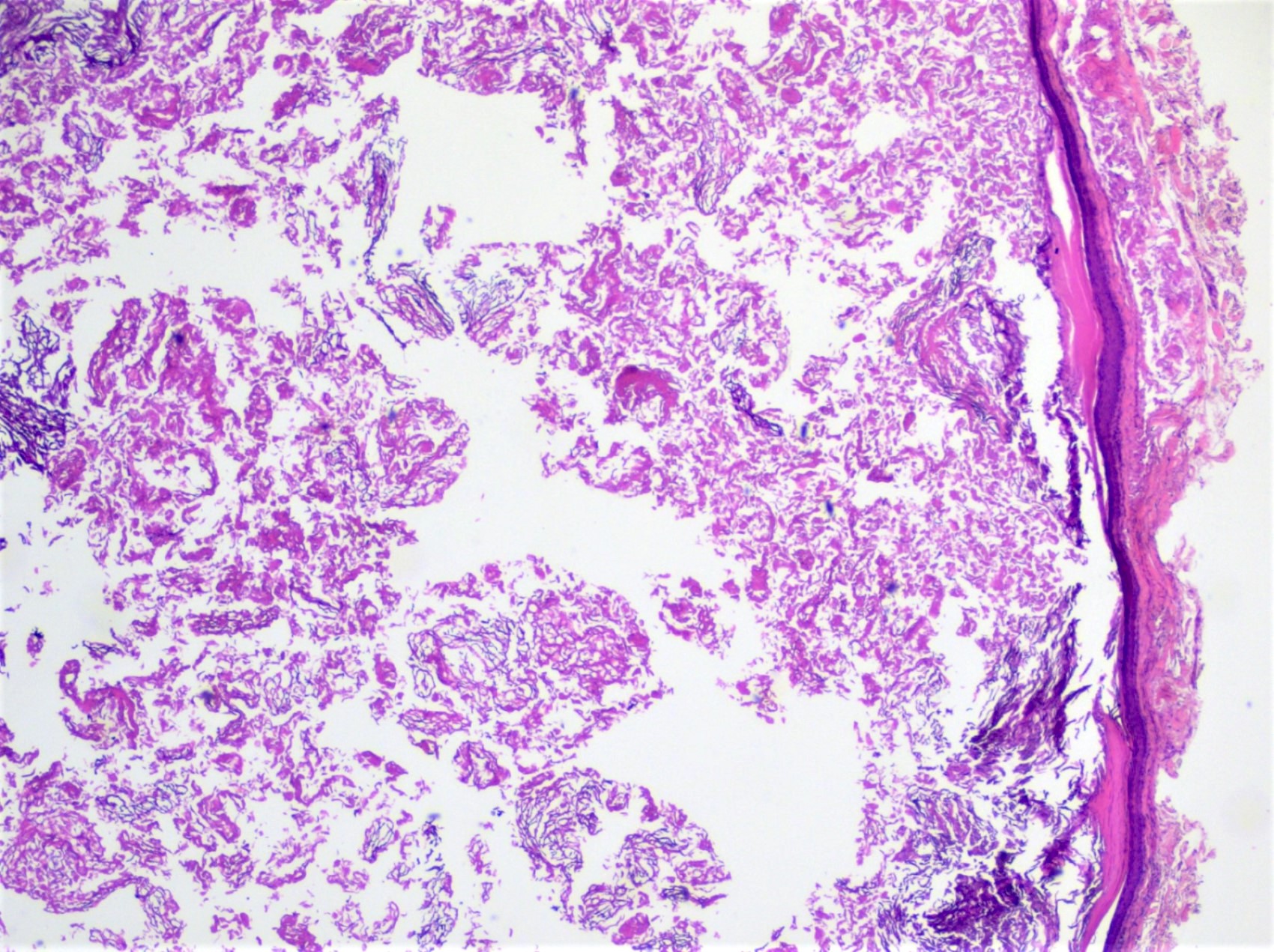
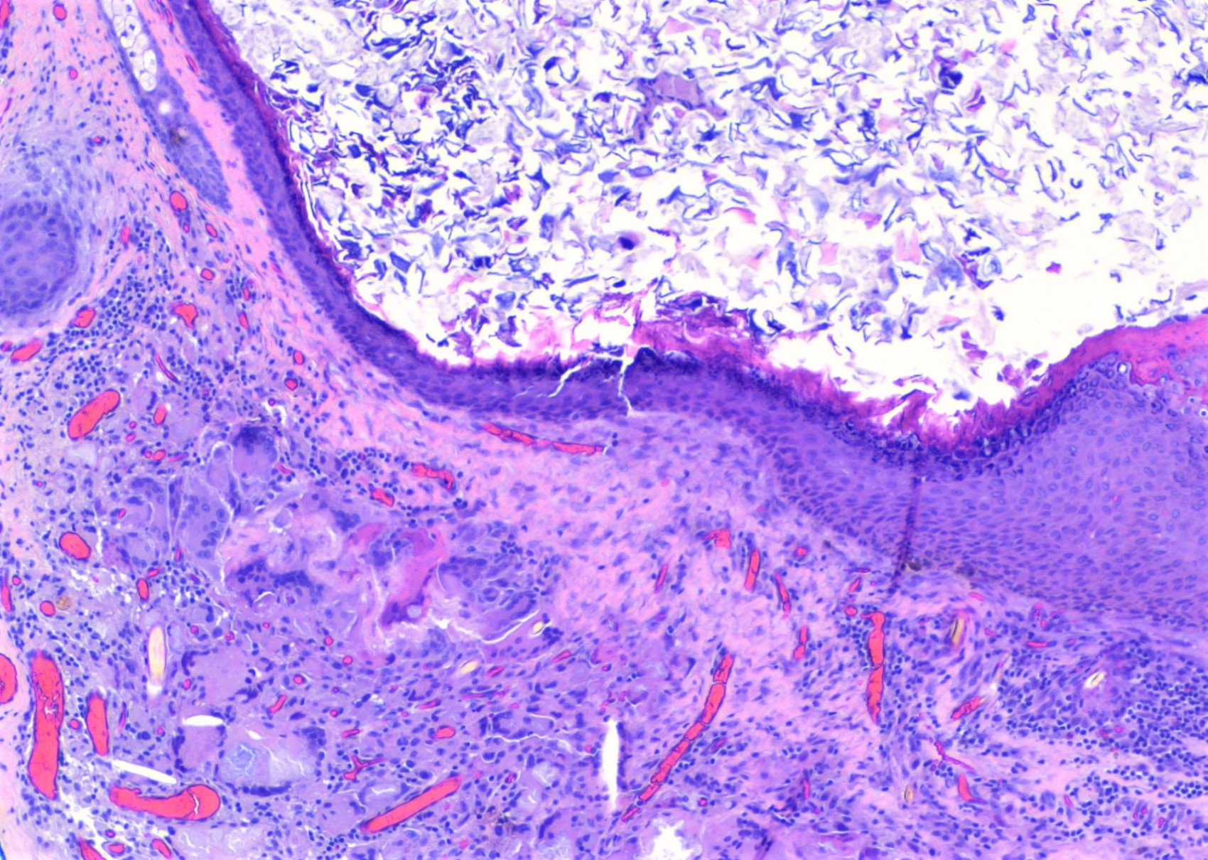
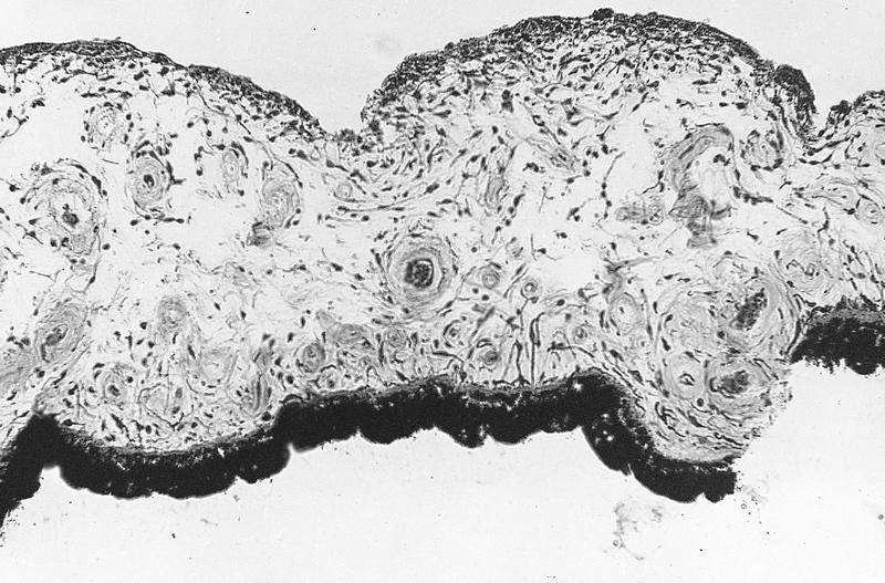
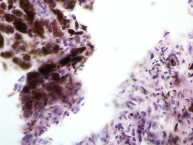
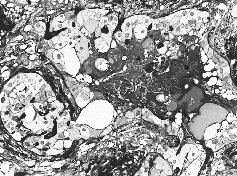
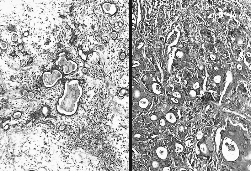
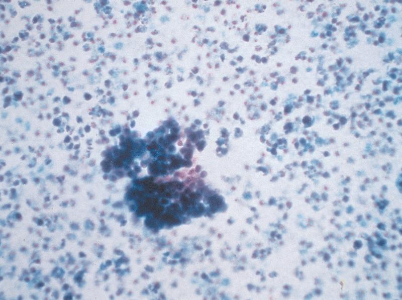
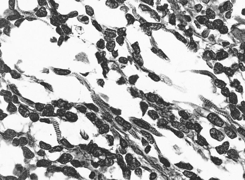
PAS with encysted amoebae
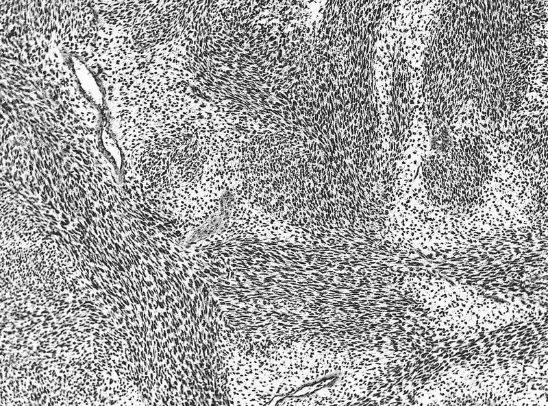
GMS with encysted amoebae
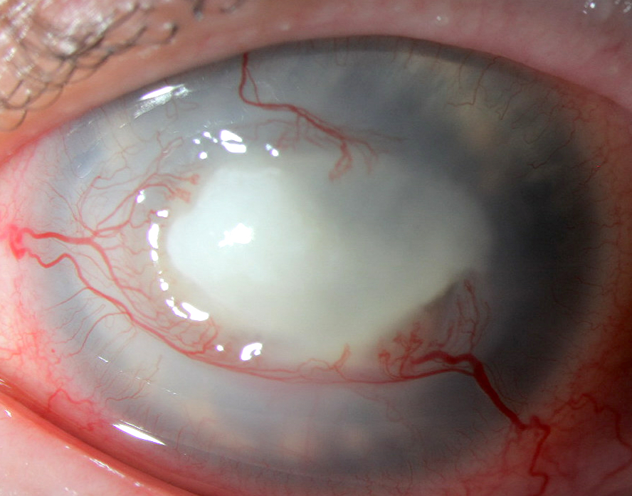
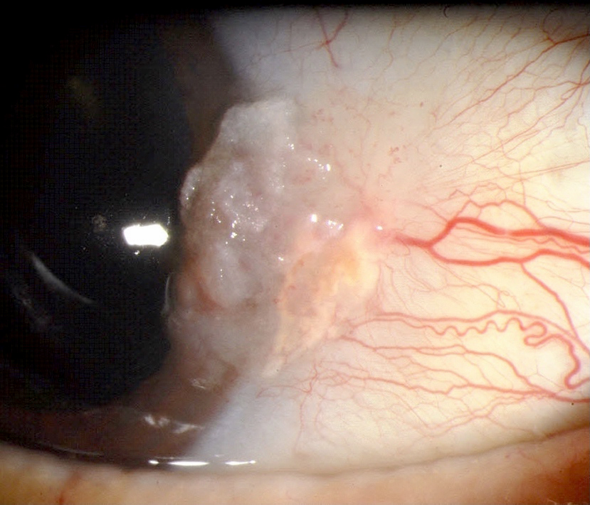
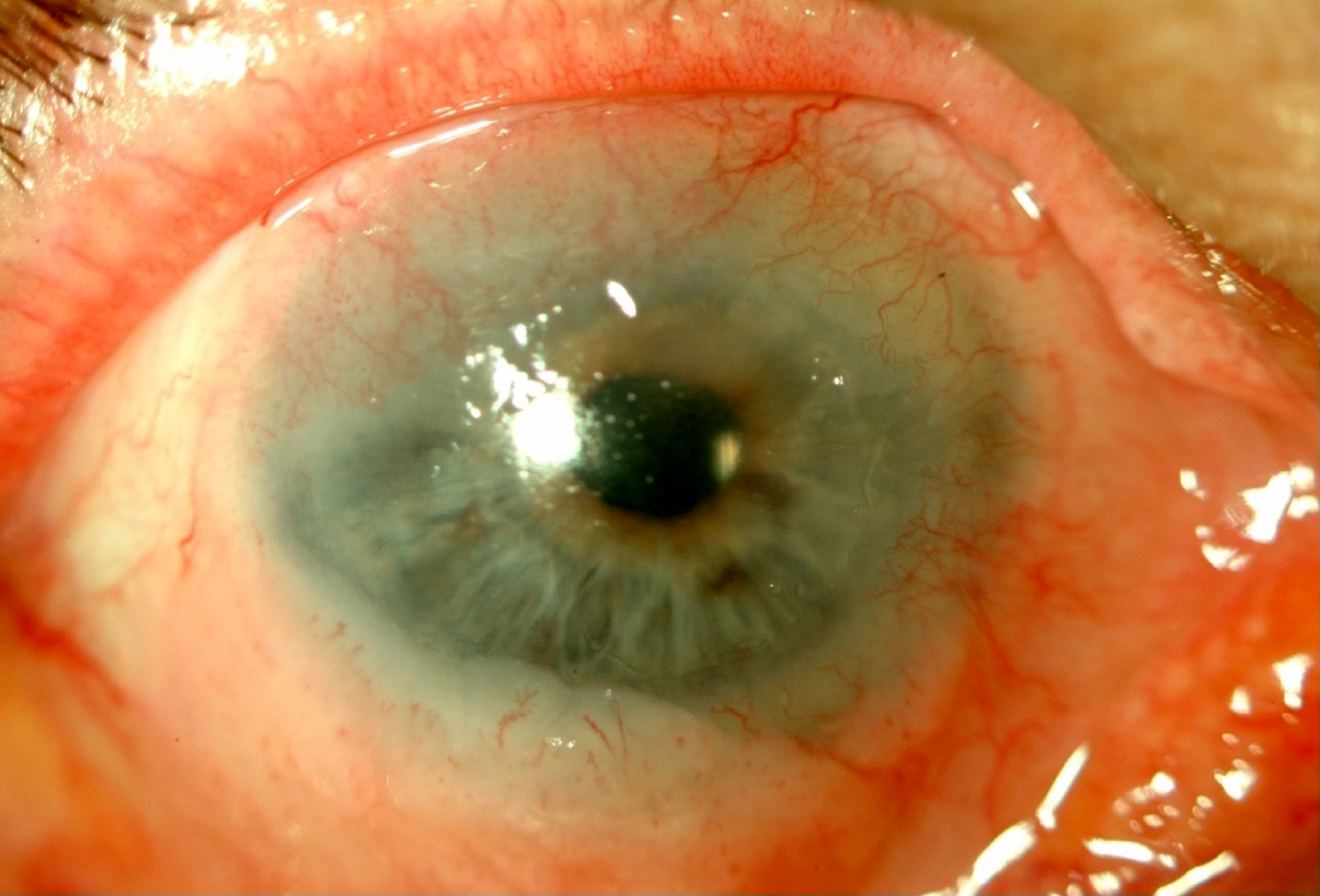
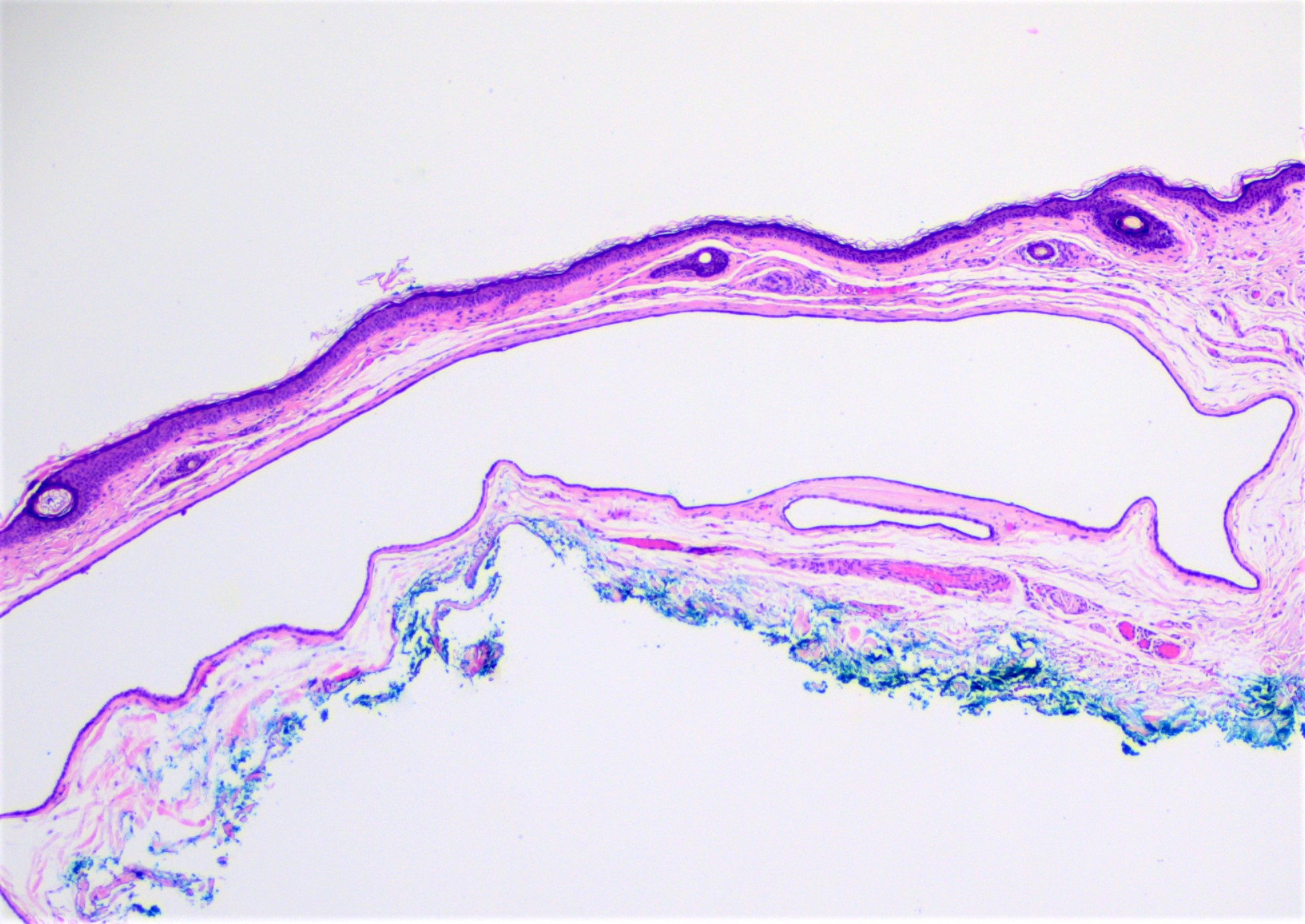
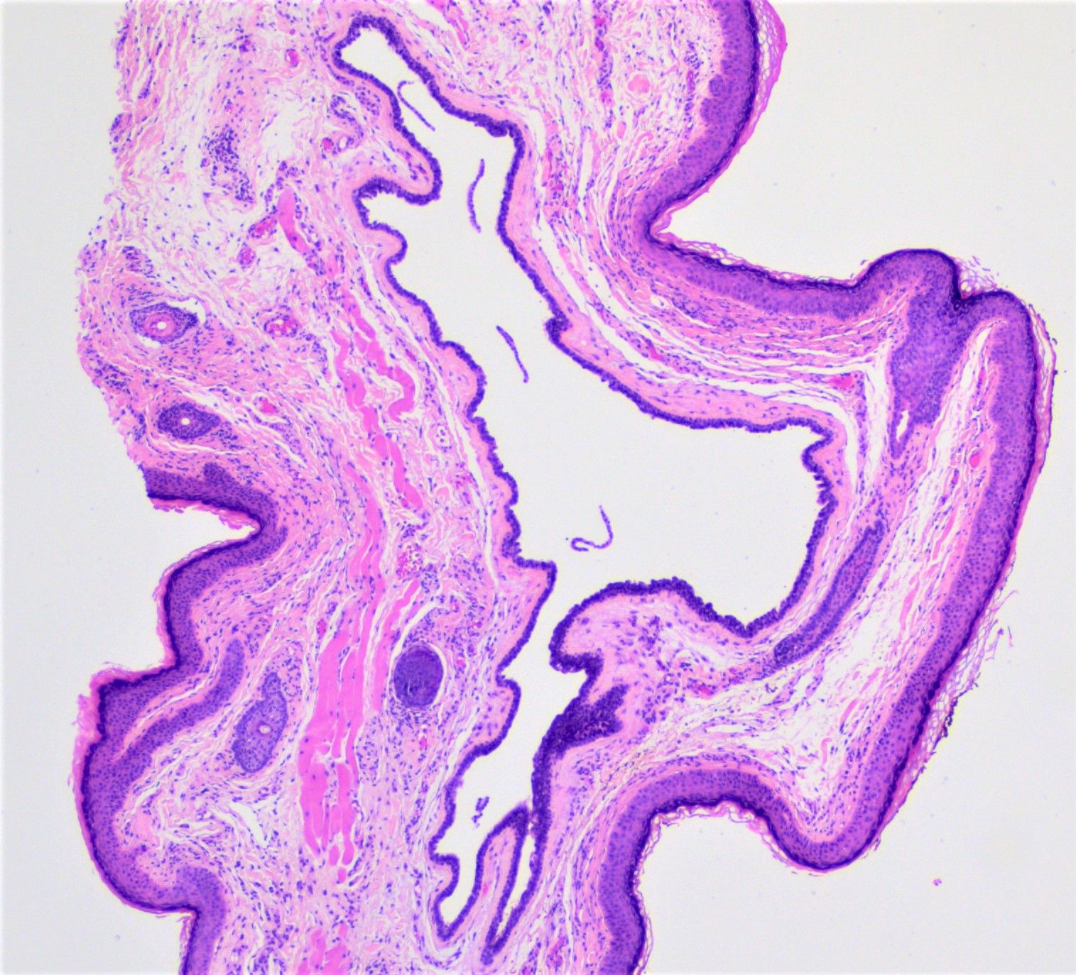
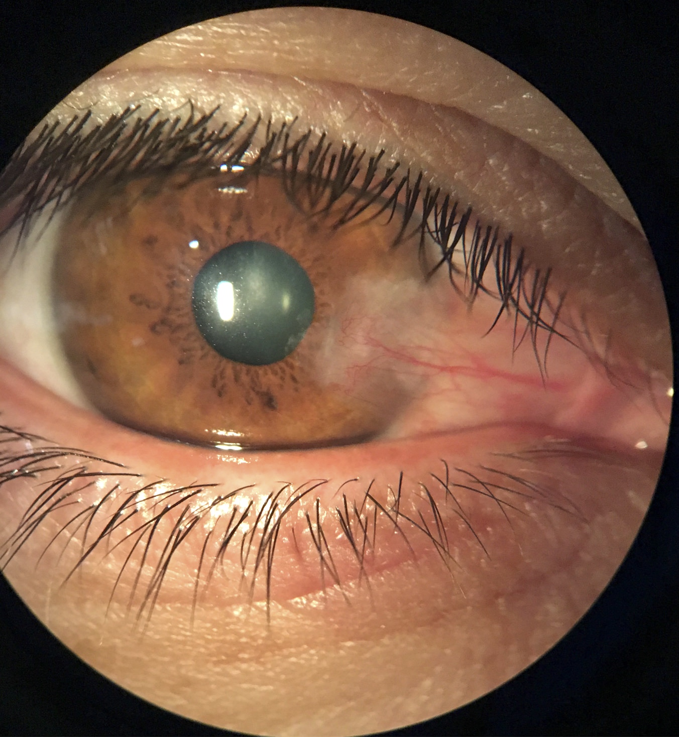
Clinical presentation of Acanthamoeba keratitis
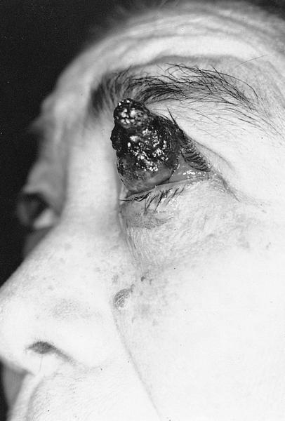
AFIP images
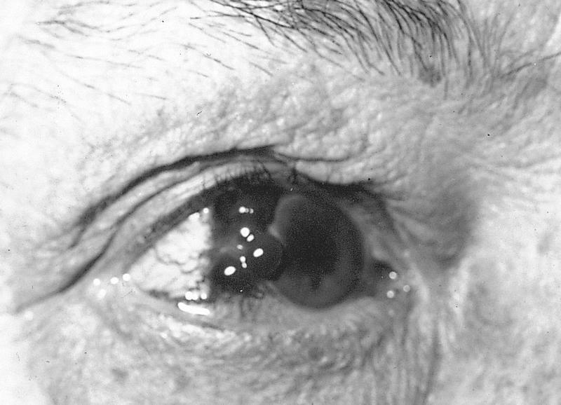
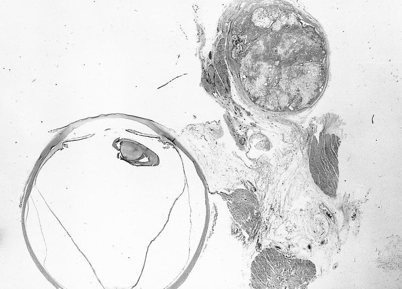
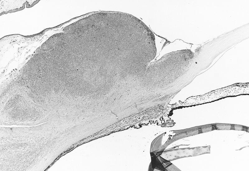
Elevated white limbal tumor
AFIP images
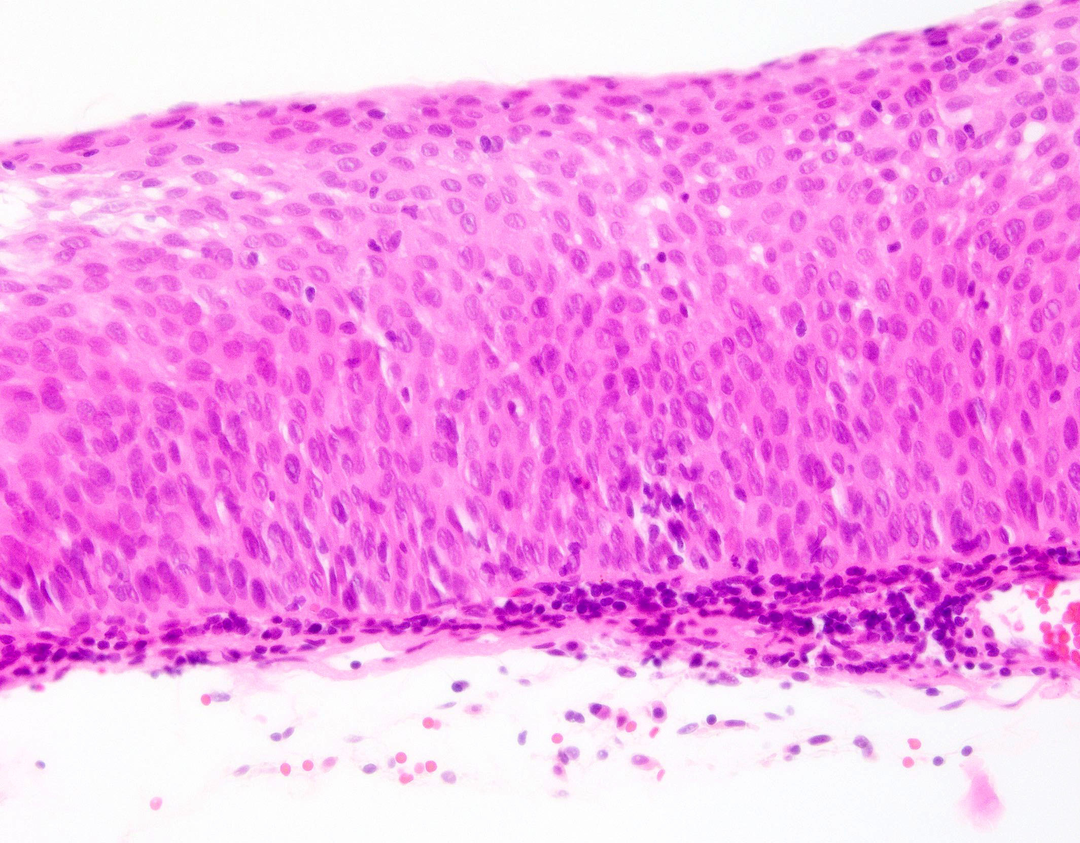
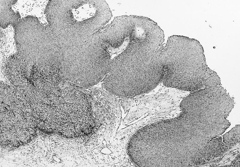
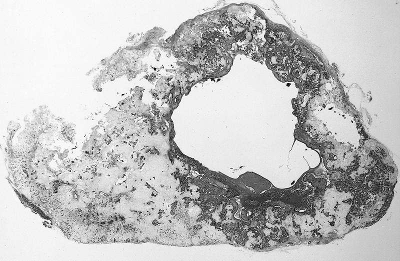
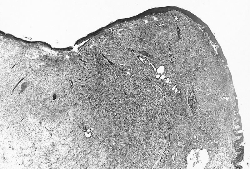
Sharply
demarcated
intraepithelial
lesion
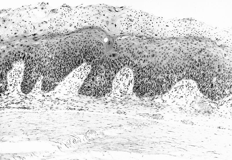
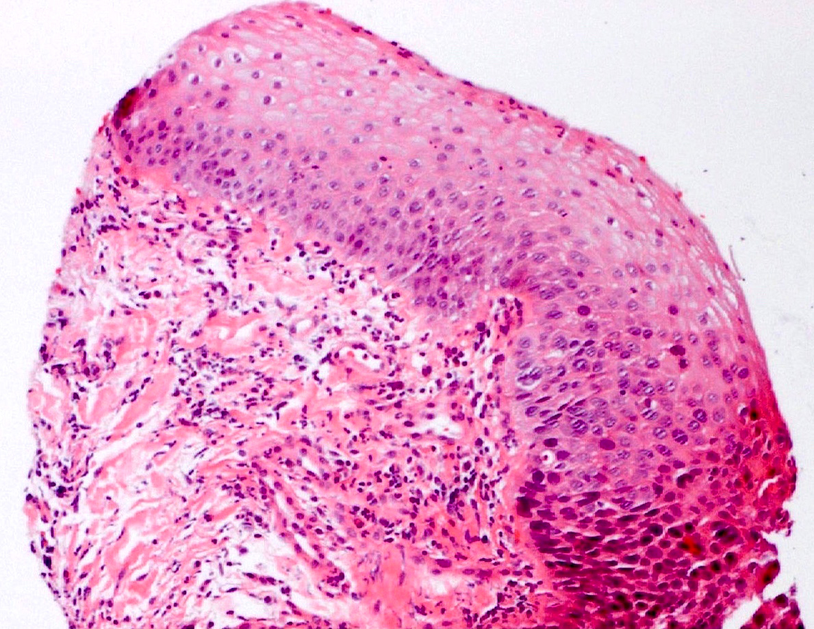
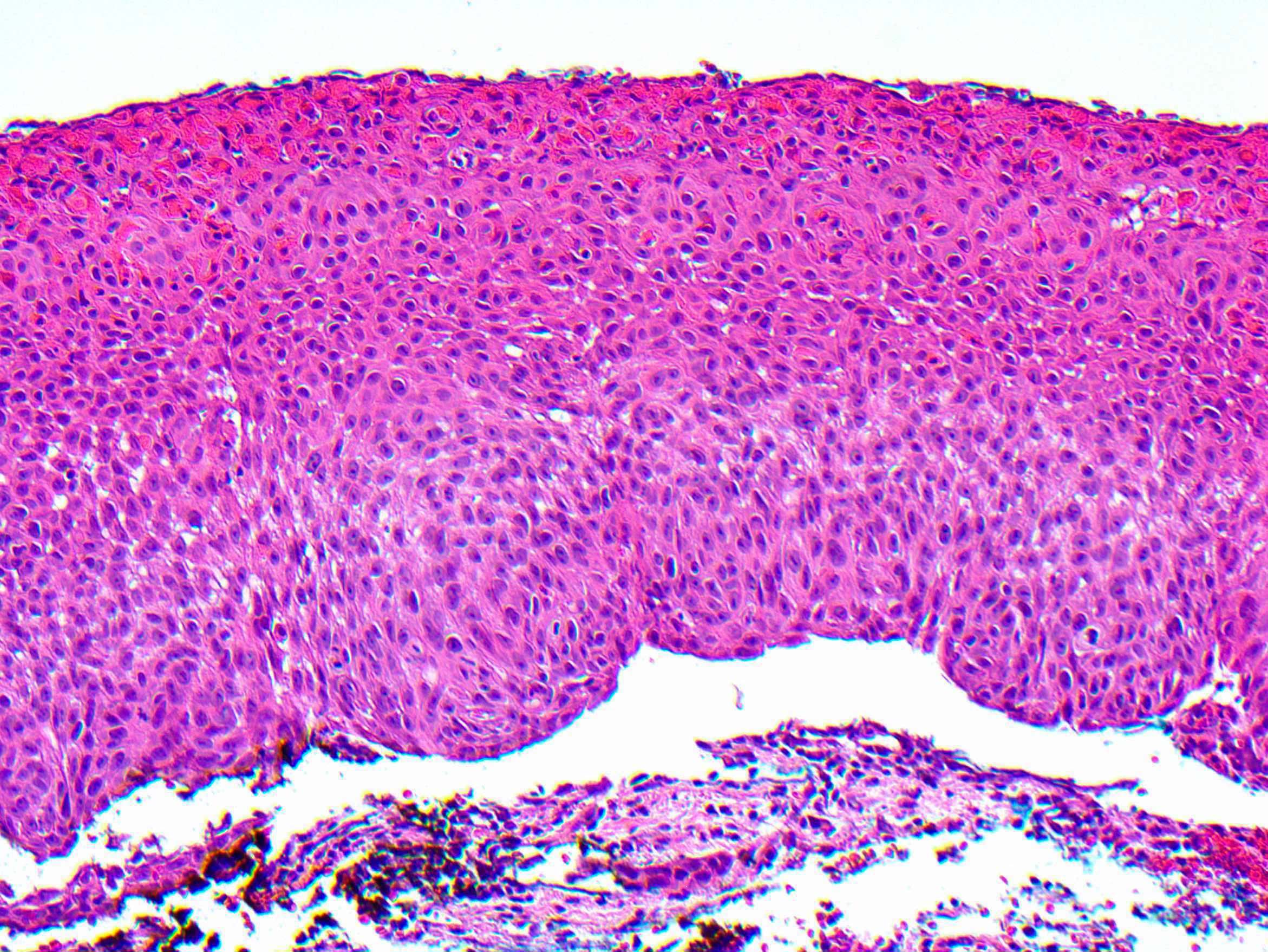
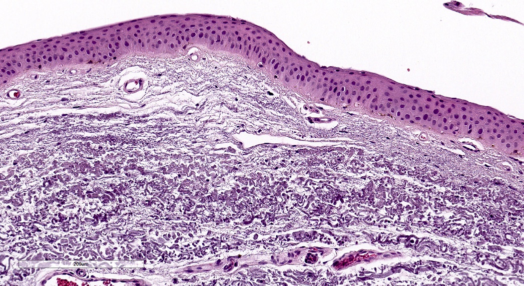
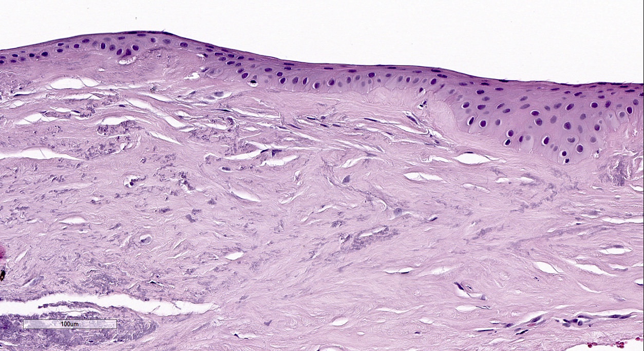
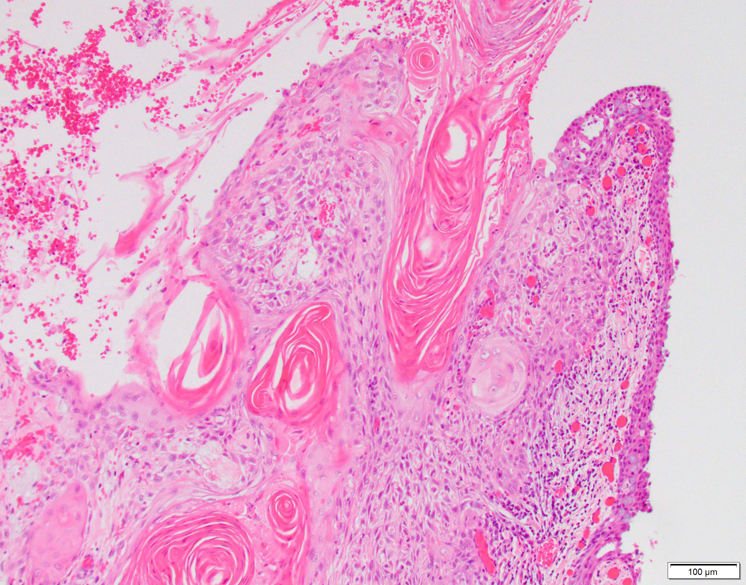
Intraepithelial corneal lesion with severe atypia and parakeratosis
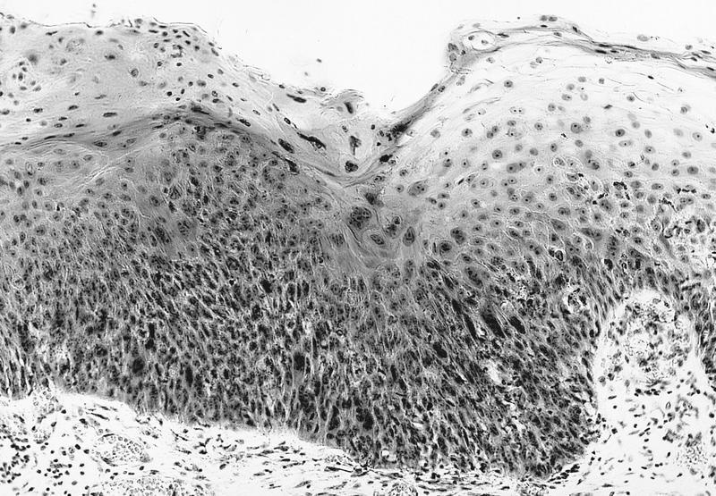
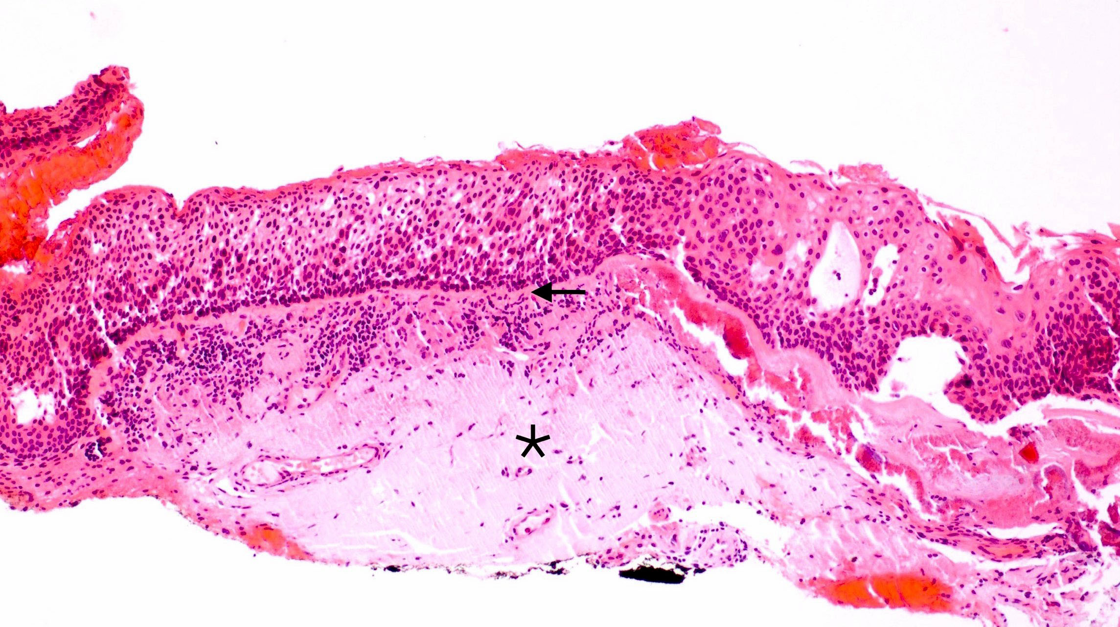
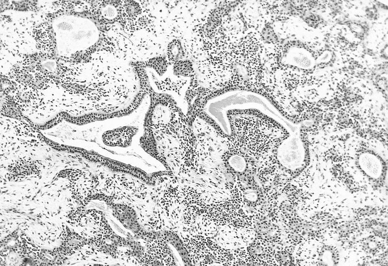
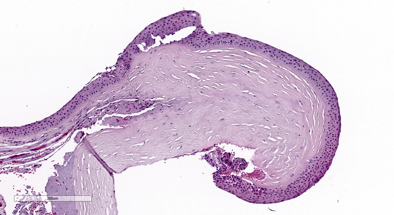
Acantholytic lesion with subepithelial lymphocytes

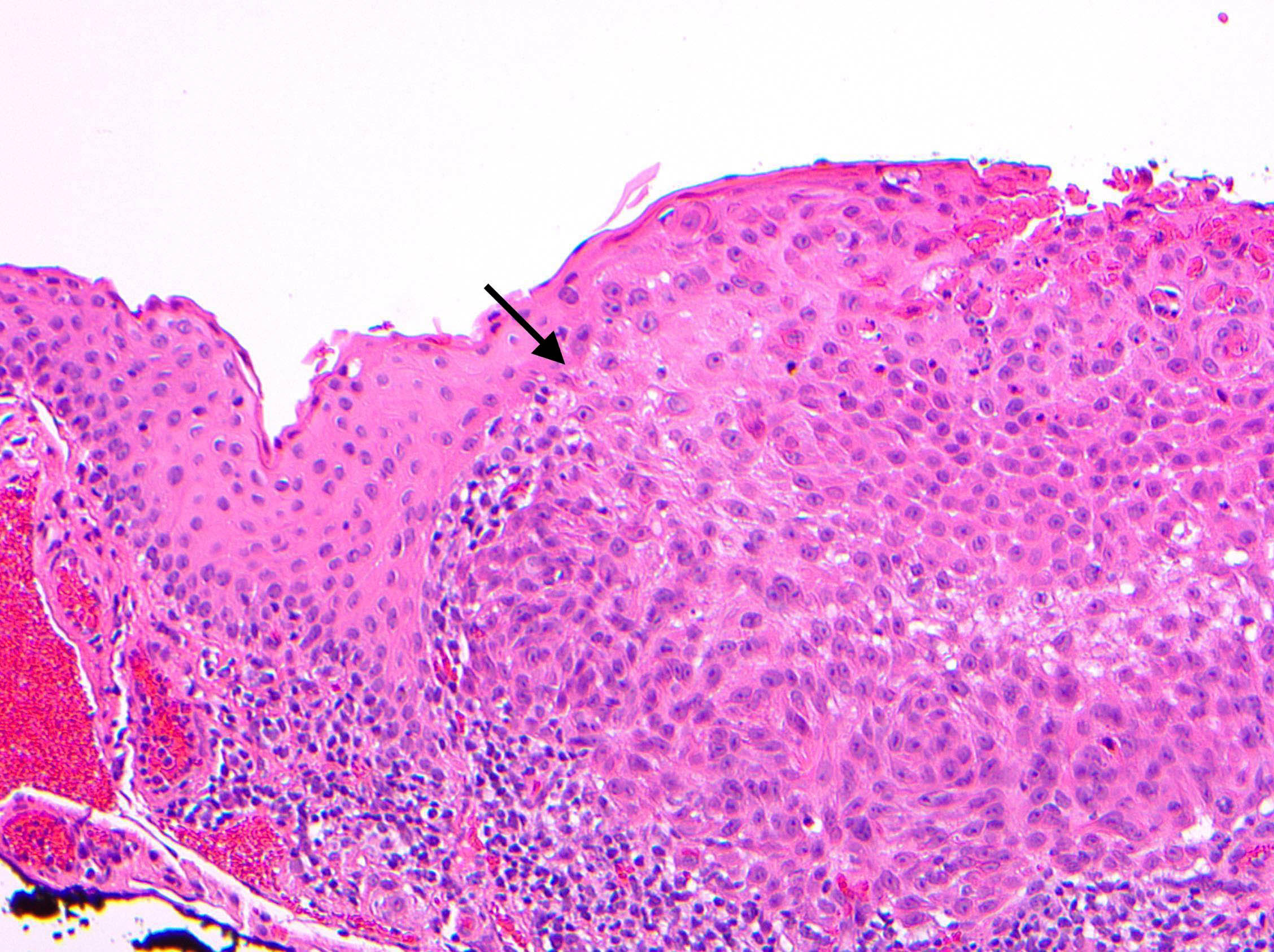
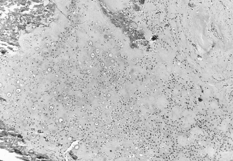
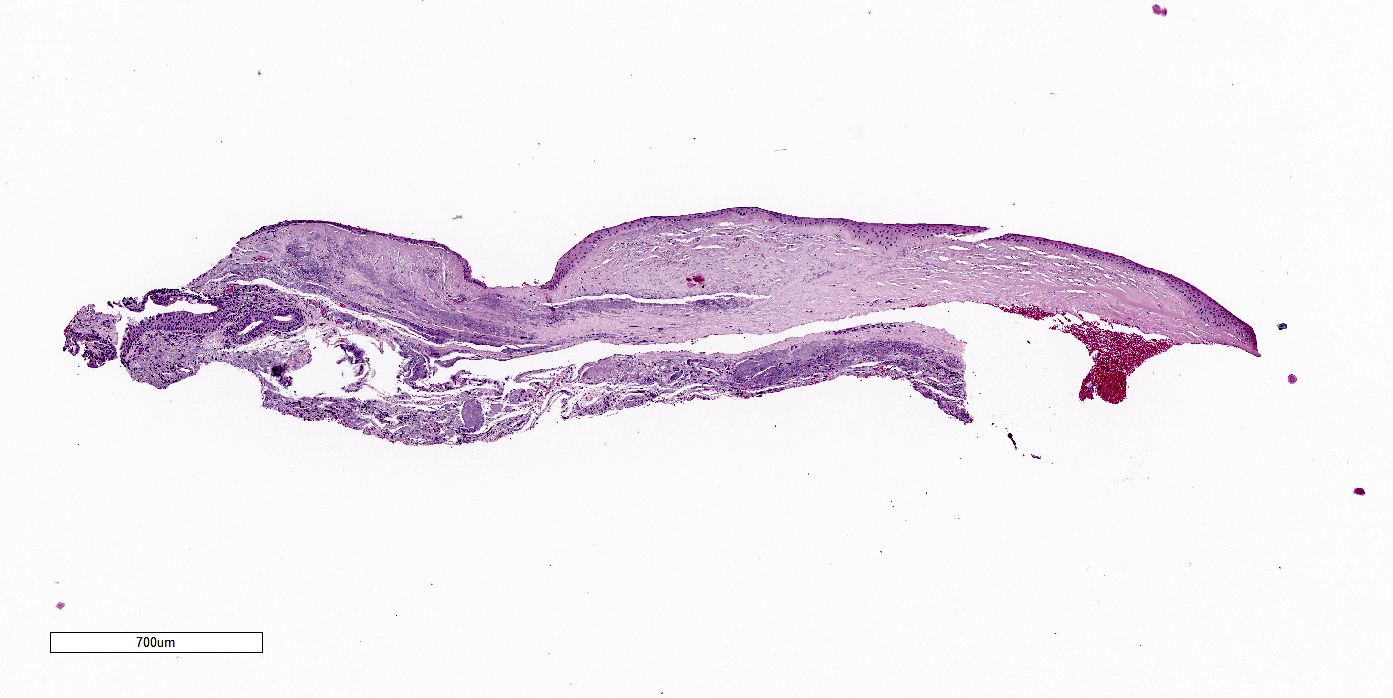
Atrophic lesion with atypia
Images hosted on other servers:

Mild keratinization with scattered melanophages

Several horn cysts in epithelium
Images hosted on other servers:


AFIP images
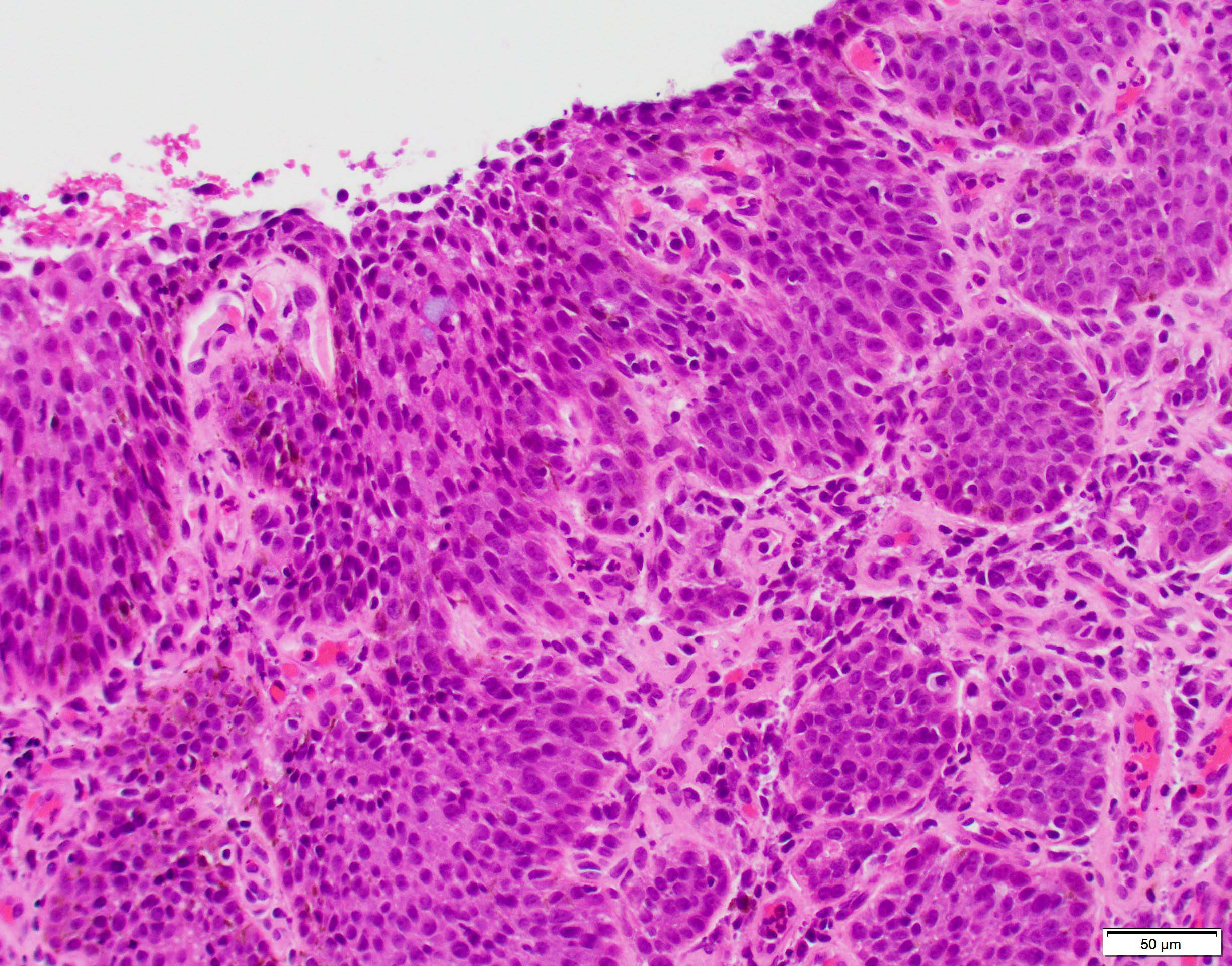
Anaplastic tumor
with glandular
formation and
mitotic activity
AFIP images
Pigmented tumor
with tubular and
papillary patterns
Variable pigmented tumor adjacent to optic nerve head
Pleomorphic and papillary tumor area with variable pigment
Heavily pigmented area of tubular tumor
AFIP images
Serrated borders indicating infiltrative growth
AFIP images
Infiltration of orbit
Basaloid pattern
Cribriform pattern
Cylindromatous or sclerosing pattern
Perineural invasion
Metastasis to lung
Images hosted on other servers:

Classic pattern
Images hosted on other servers:

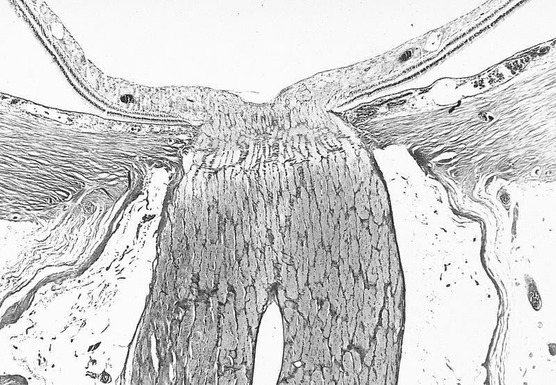
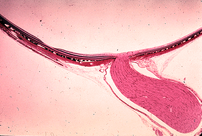
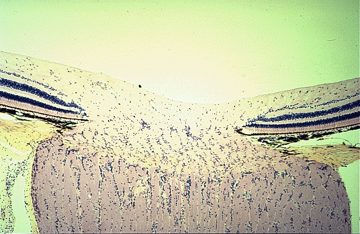
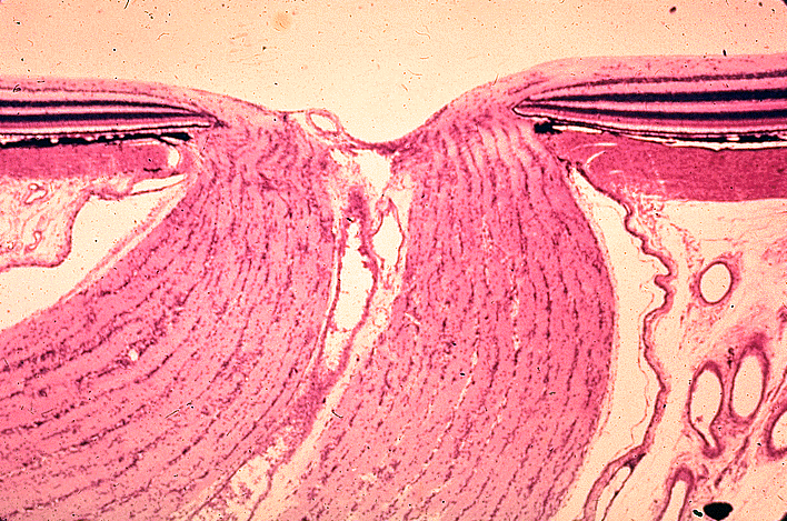
Ciliary body, iris and lens
Ciliary process zonule fibers
Contributed by Sena Zengin, M.D. and J. Stephen Nix, M.D.
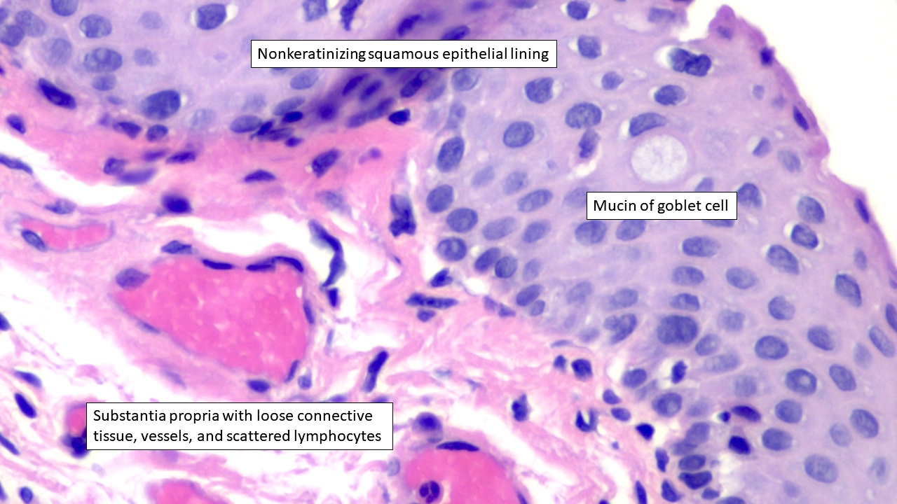
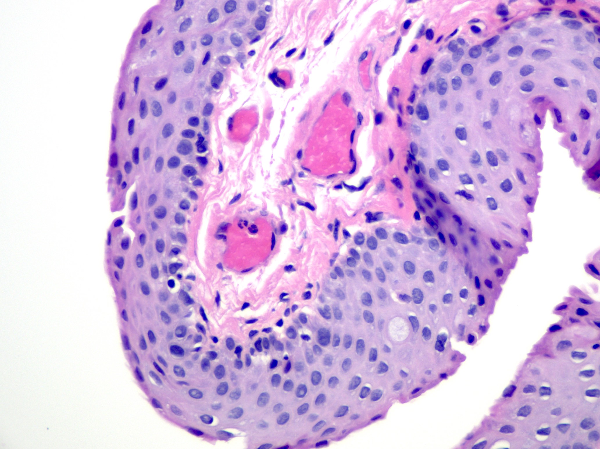
Conjunctiva
Contributed by J. Stephen Nix, M.D.
Nonkeratinizing squamous conjunctiva
Cuboidal and columnar appearance
Tarsal plate, Meibomian glands

Caruncle, chronic conjunctivitis
Pseudoglands of Henle
Pathology lecture: conjunctiva
Images hosted on other servers:
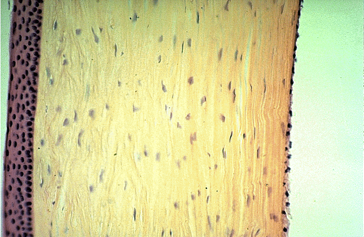
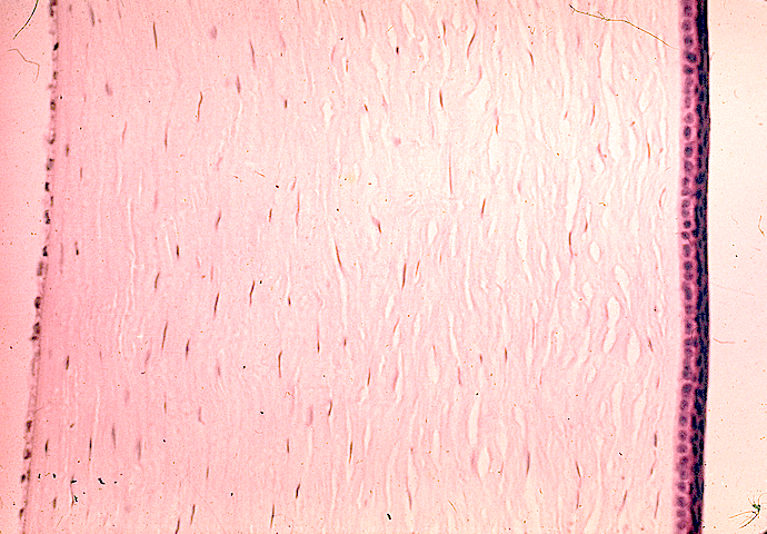
Full thickness
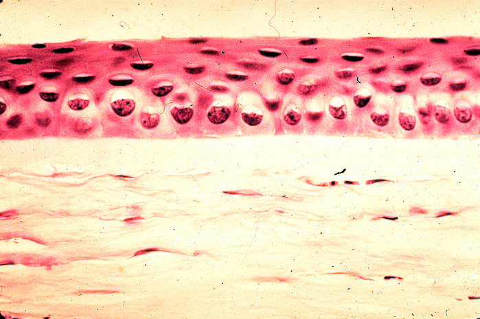
Epithelium and Bowman layer
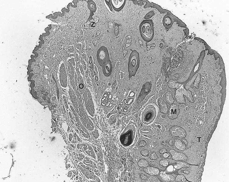
AFIP images
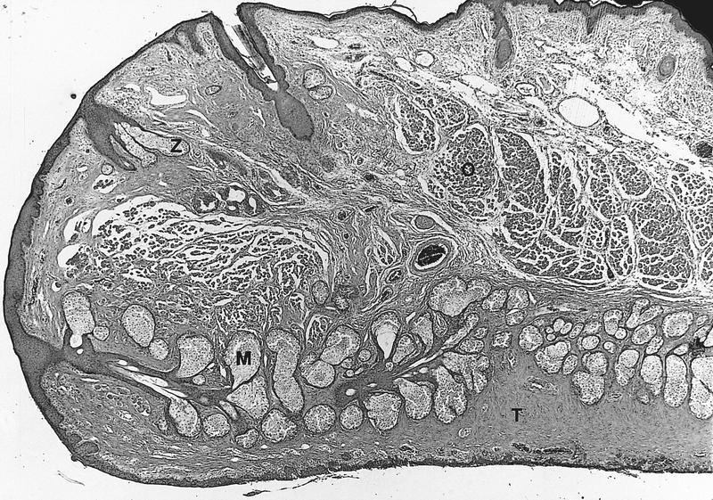
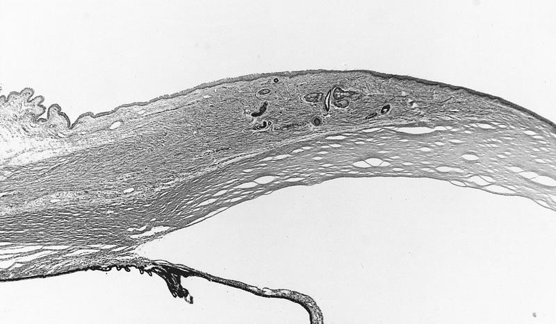
Normal upper eyelid
M: meibomian glands
O: orbicularis muscle
T: tarsal plate
Z: Zeis glands
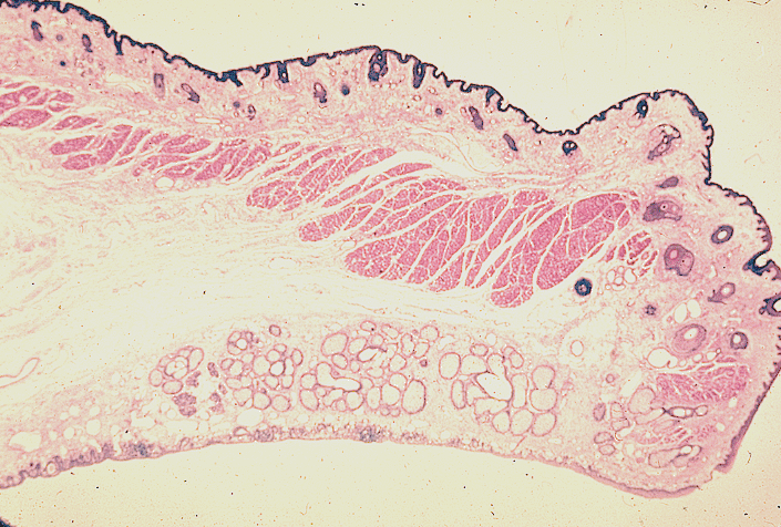
Normal lower eyelid:
smaller tarsal plate
than upper eyelid
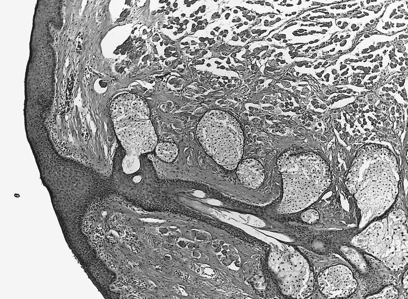
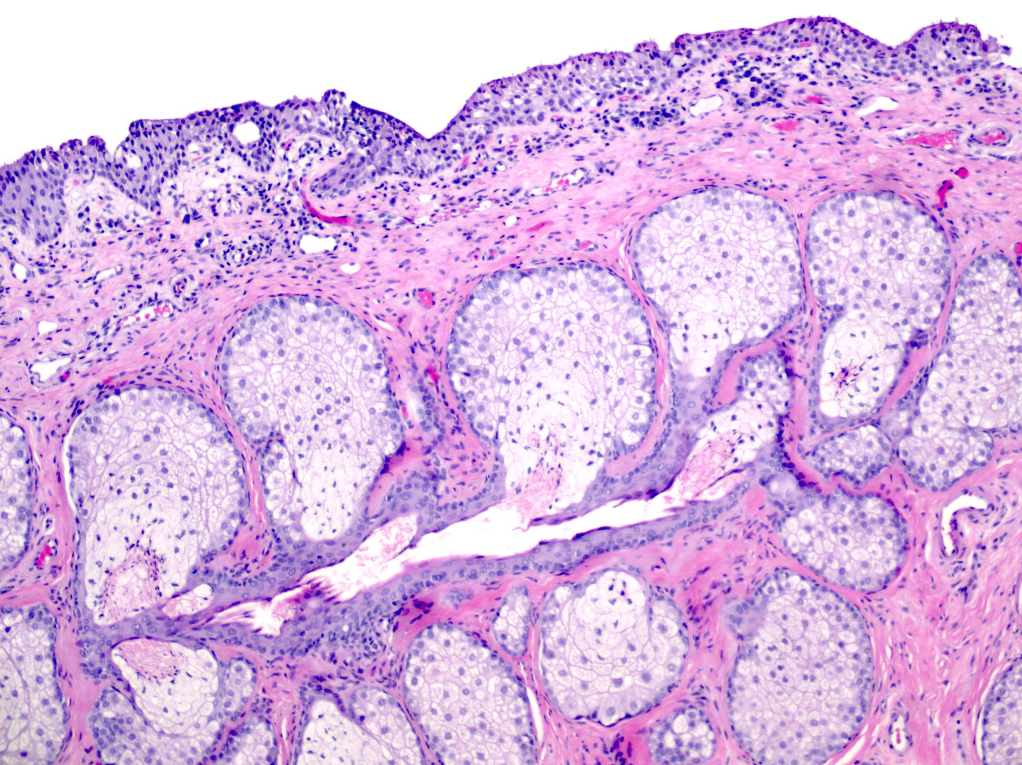
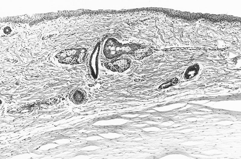
Normal Meibomian gland:
sebaceous lobules connect
to sebaceous duct, where
ductal epithelium forms
valve-like structures
Images hosted on other servers:
Eyelid cross section
AFIP images
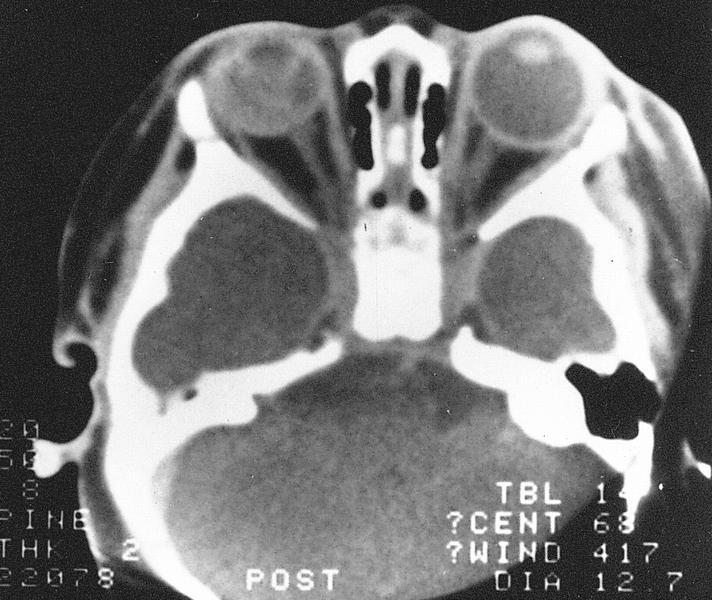
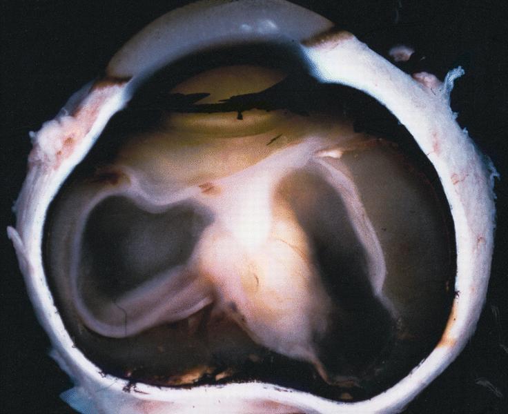
Normal eye and orbital contents
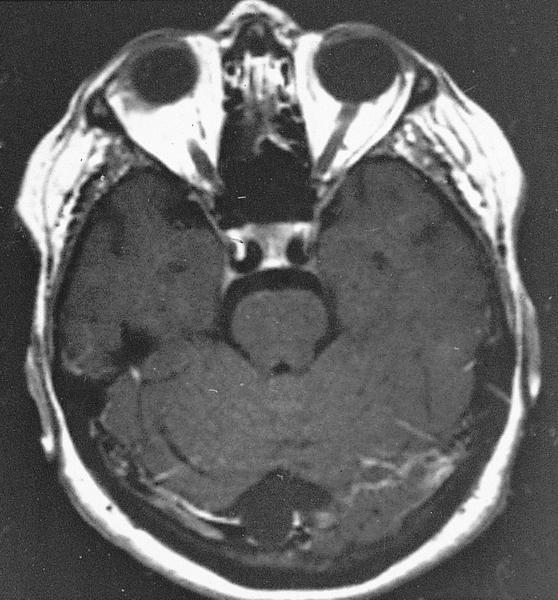
MR #1 (T1 weighted)
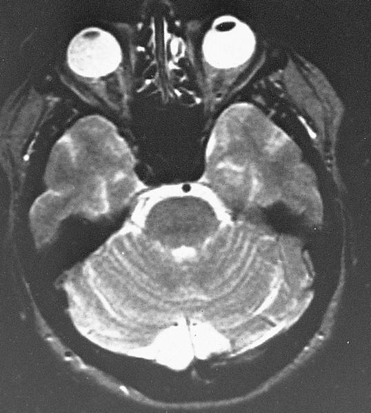
MR #2 (T2 weighted)
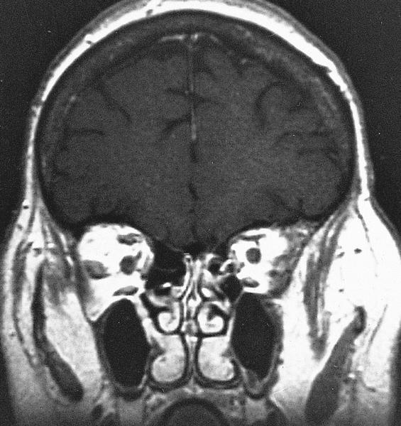
MR #3 (T1 weighted)
shows coronal section
of orbital contents
posterior to globe
AFIP images
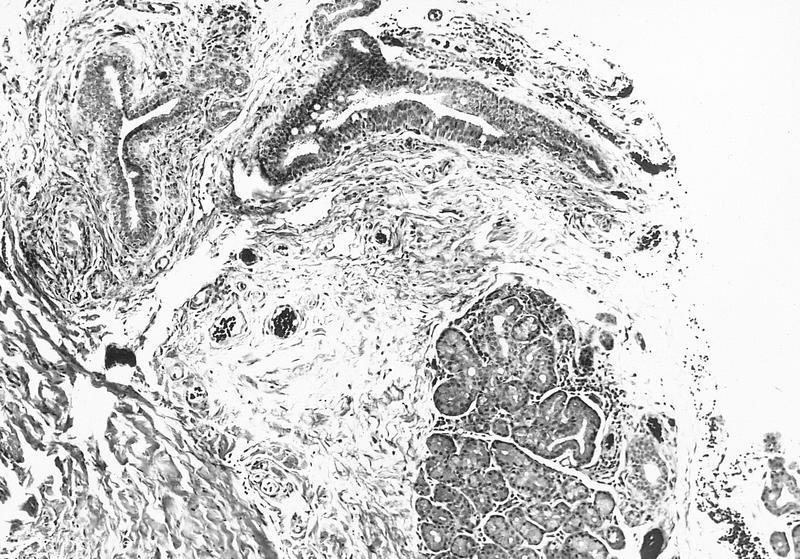
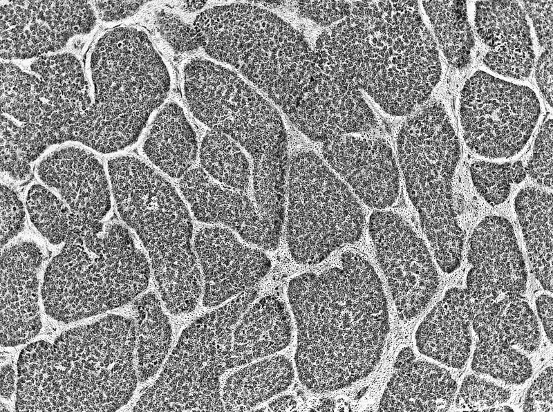
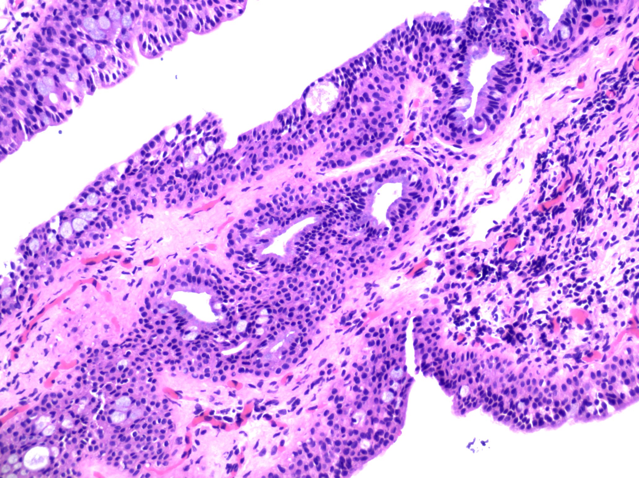
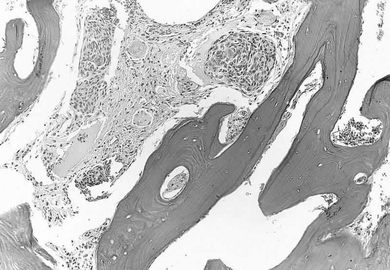
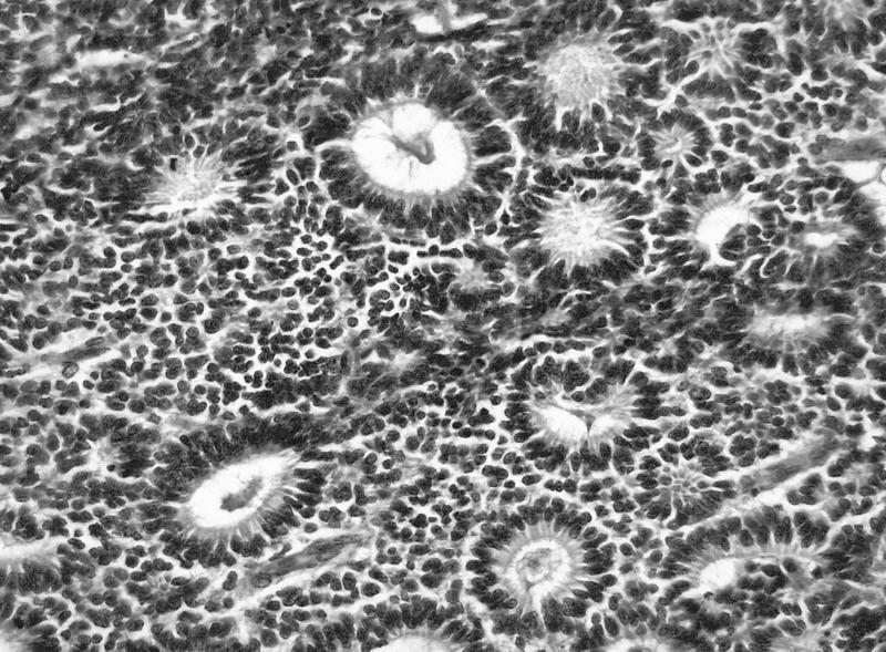
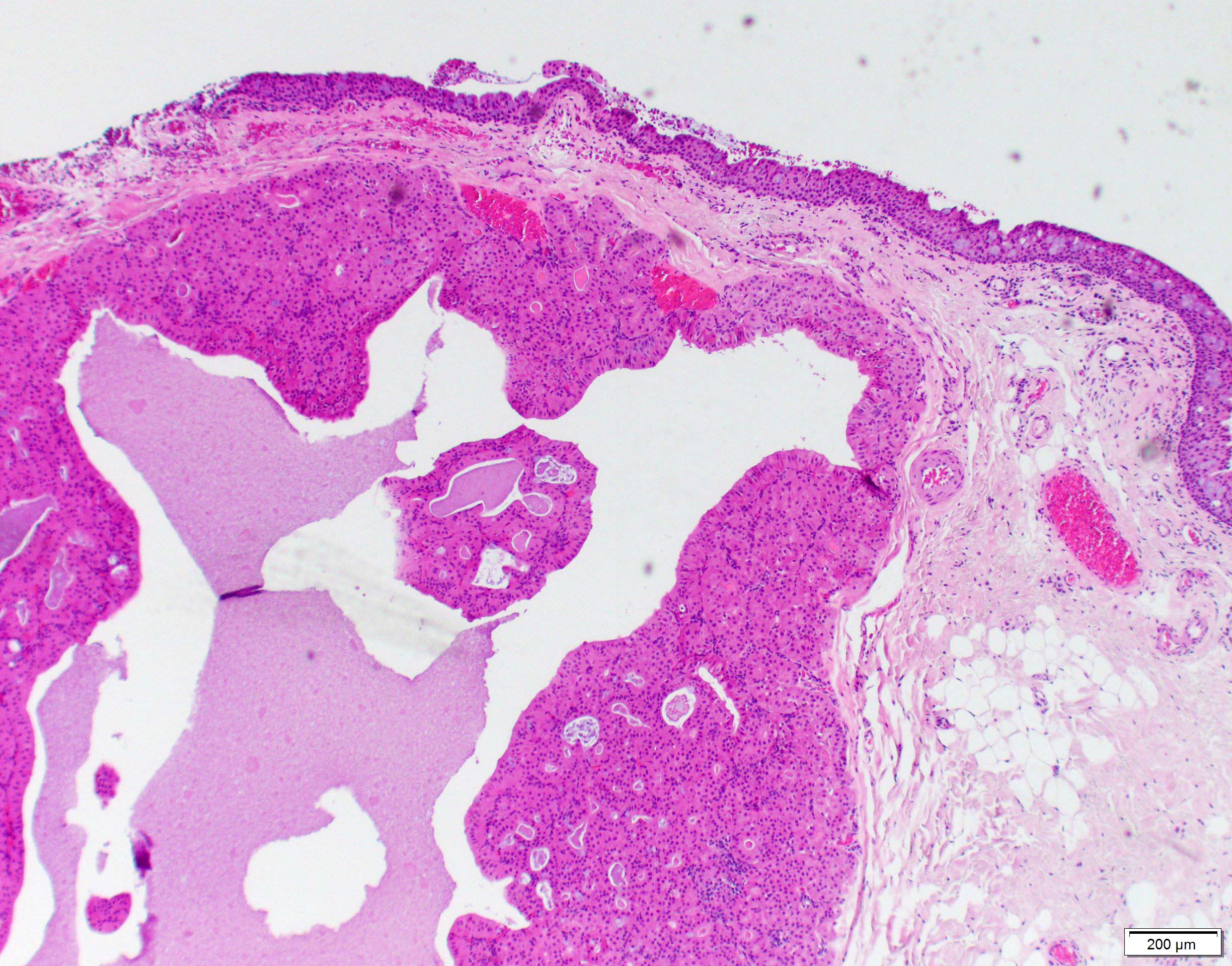
Glandular lobule next to ducts
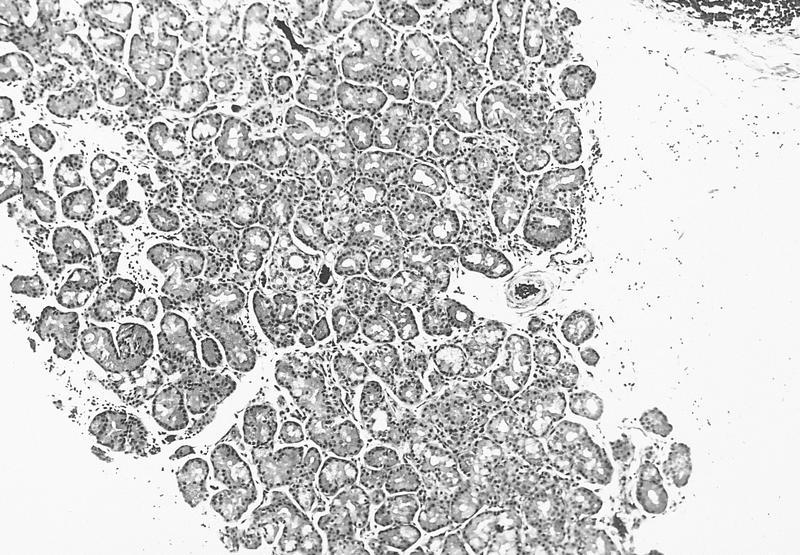
Lobule of acinic and mucinous cells
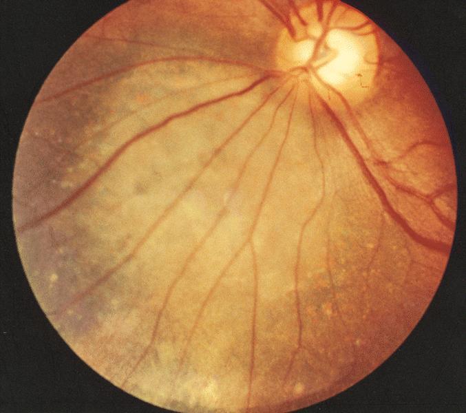
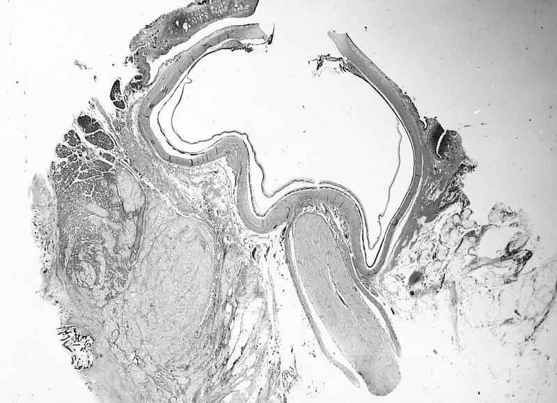
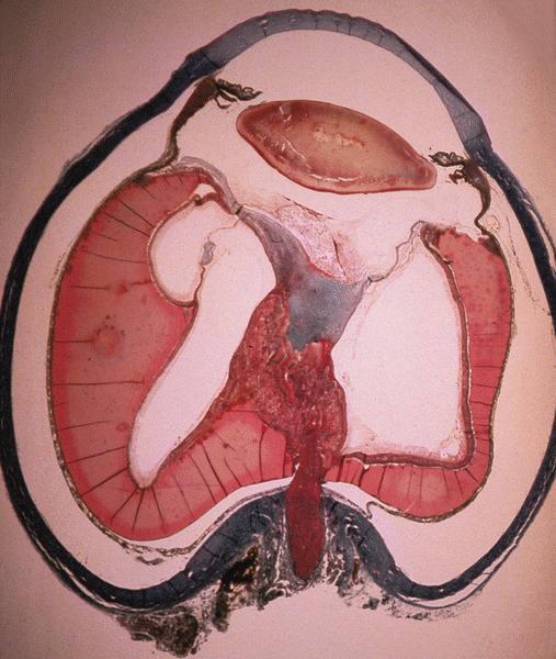
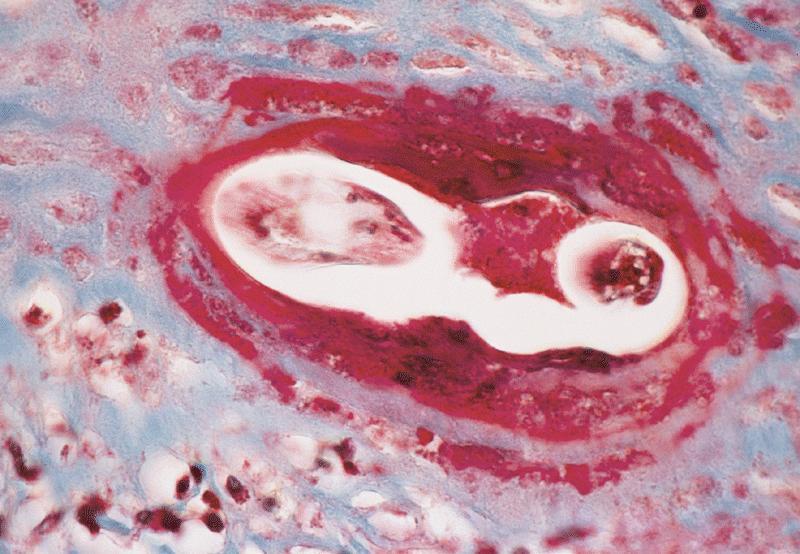
Intraocular and orbital portions of optic nerve
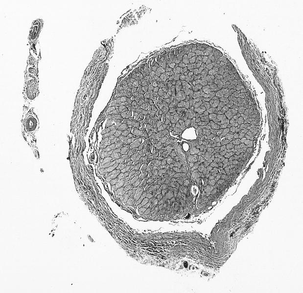
Cross section of optic nerve parenchyma and meninges
Images hosted on other servers:
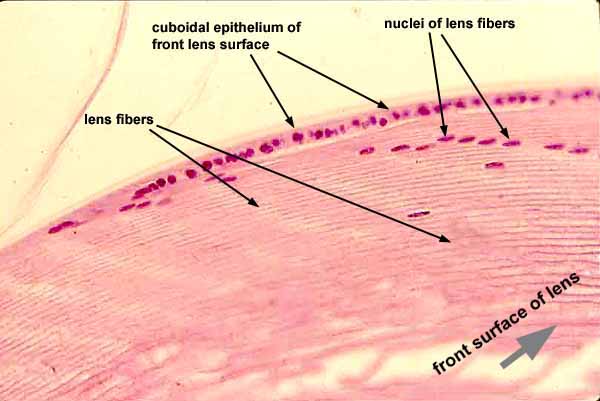
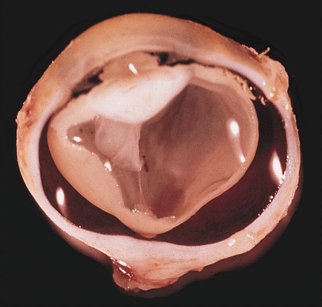
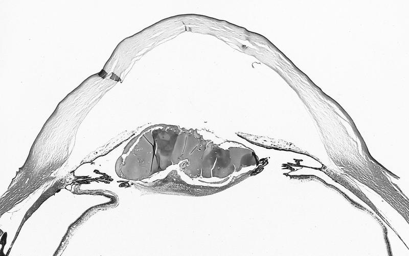
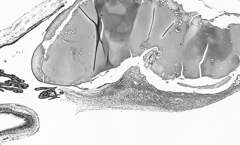
Lens
Optic nerve and fovea centralis
Contributed by J. Stephen Nix, M.D.
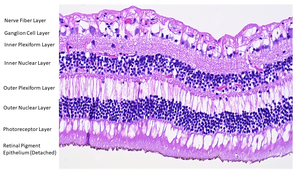
Retina anatomy
Contributed by J. Stephen Nix, M.D. and AFIP images

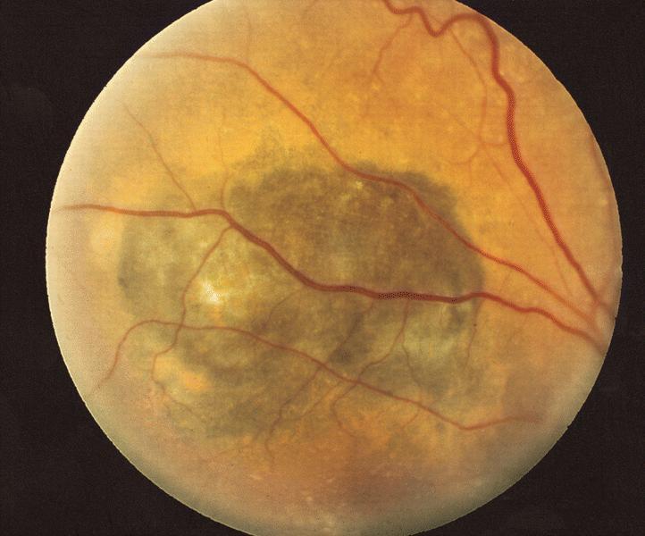
Macula
Choroid
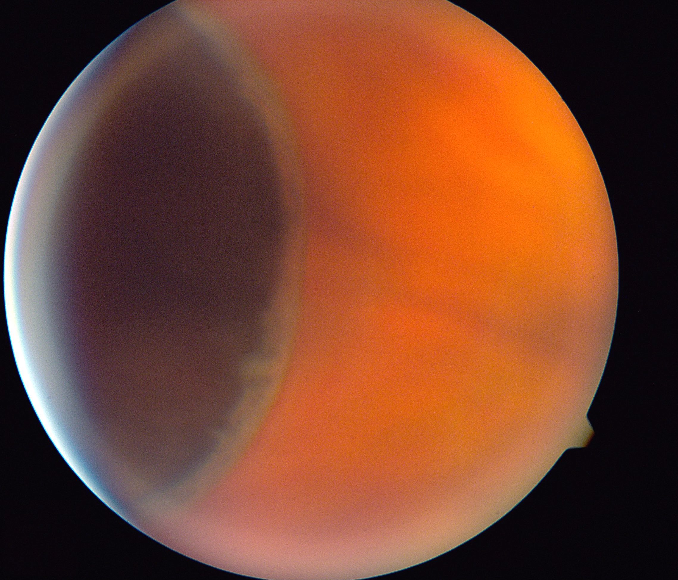
Hard drusen
Retinal gliosis
Serosanguinous retinal detachment
Fovea centralis
Images hosted on other servers:

Laugier-Hunziker syndrome

Dermoscopic examination of mucosal lesions

Parallel furrow pattern of pigmented macules
AFIP images
Dendritic melanocytes
Images hosted on other servers:

Hyperpigmentation
Images hosted on other servers:

Movable, diffuse, blue-black lesion
Images hosted on other servers:


Combined nevus: pigmented dendritic and spindled blue nevus cells in area adjacent to nevocytic foci

Various images

Conjunctival blue nevus

Composed of plump, heavily pigmented melanocytes

Focus of malignant transformation
Contributed by Eugenia Abusleme, M.D.
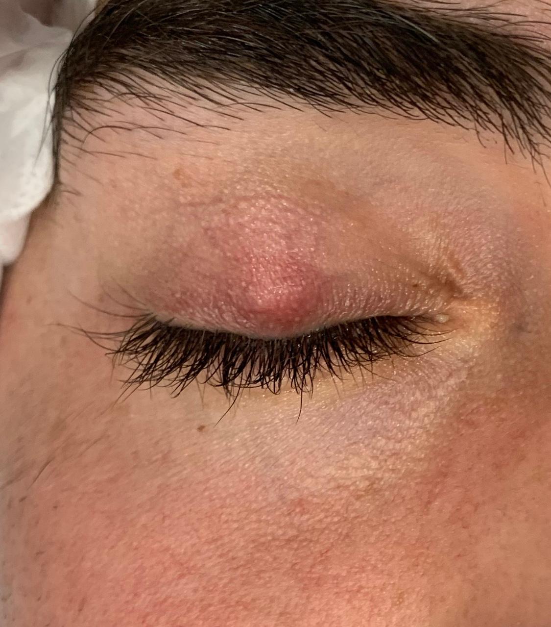
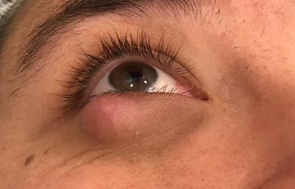
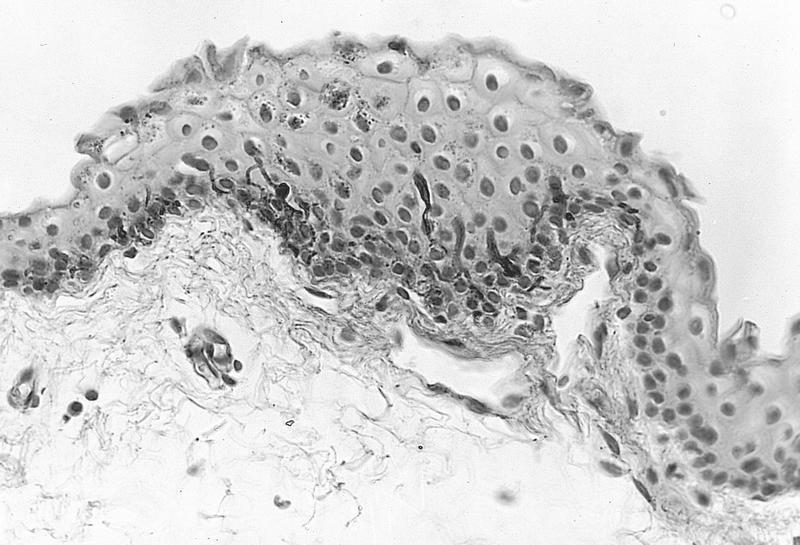
Chalazion
Contributed by Pablo Zoroquiain, M.D.
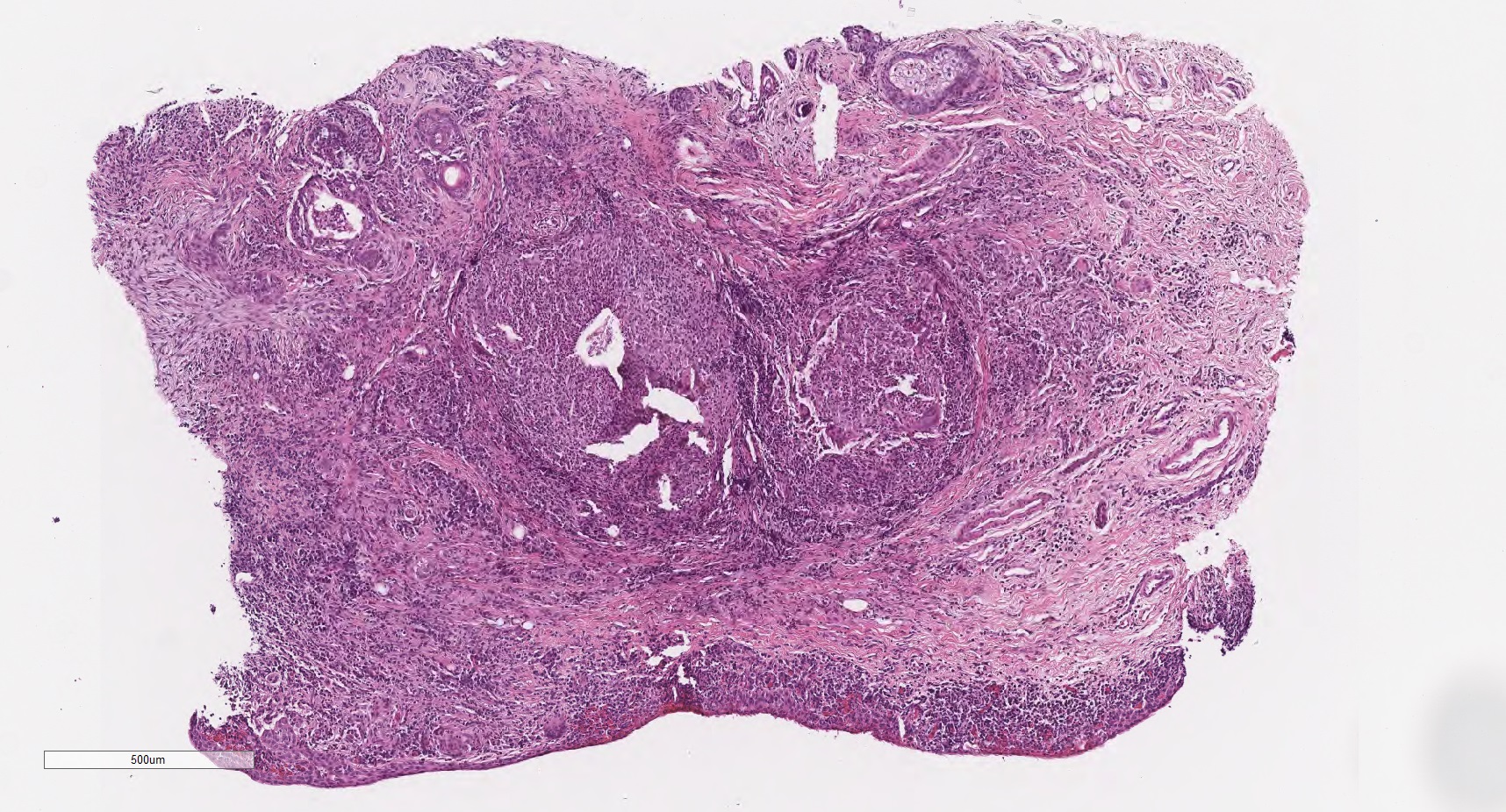
Chronic conjunctival inflammation
Large lipogranuloma
Small lipogranuloma
Neutrophilic granuloma
AFIP images
Vascularized pigmented stroma
Uveal tissue extends into sclera
AFIP images
Increased density within eye
AFIP images
Telangiectatic vessels
AFIP images
Total retinal detachment
AFIP images
Telangiectasia of vessels
Contributed by Pia Mendoza, M.D. and Frederick A. Jakobiec, M.D., D.Sc.
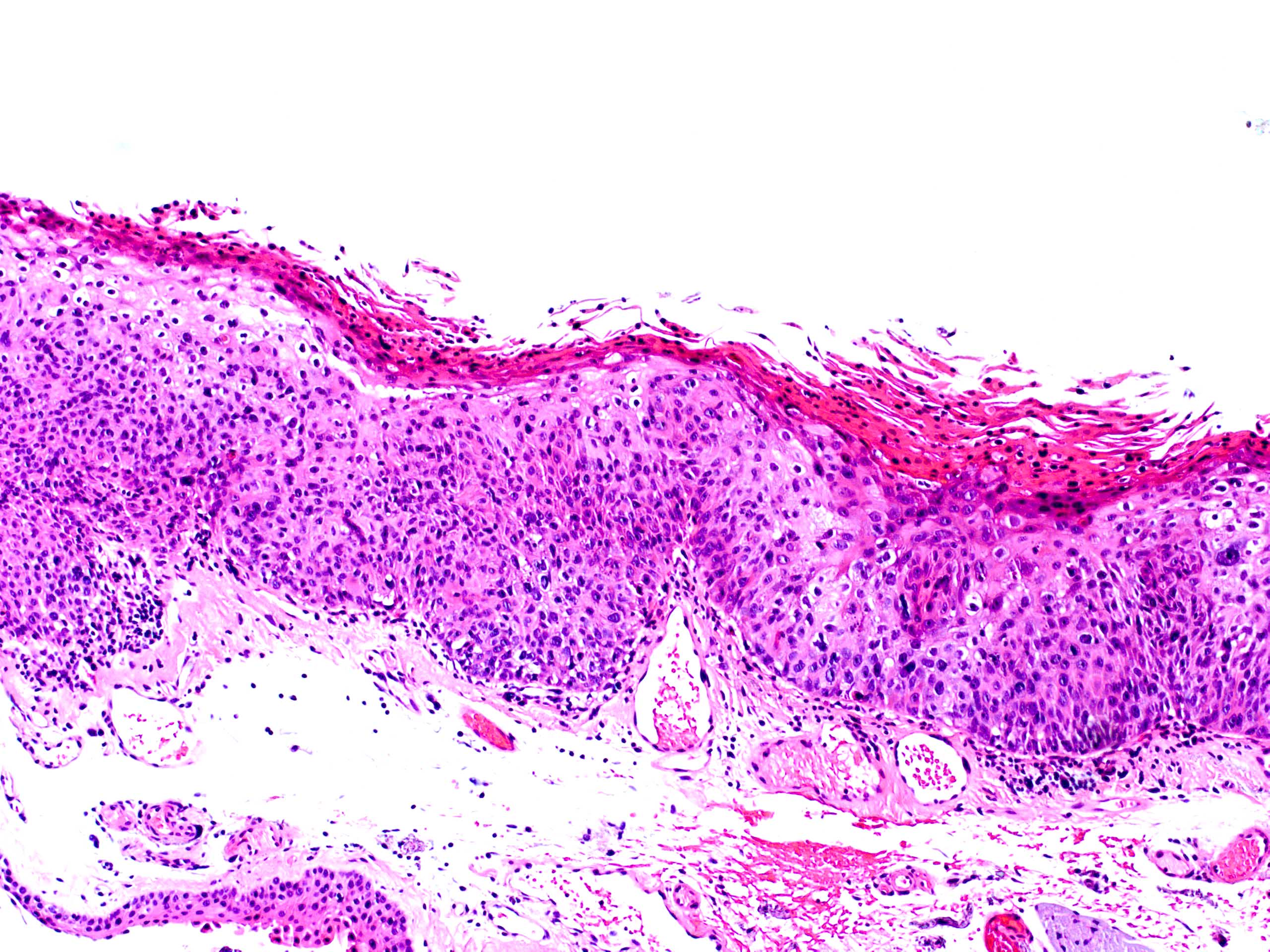
Conjunctival lesion in the limbus extending to the cornea
Limbal leukoplakic appearing lesion with feeder vessels
Diffuse lesion
Pigmented carcinoma in situ
Papilliform lesion at the limbus
Contributed by Pia Mendoza, M.D.
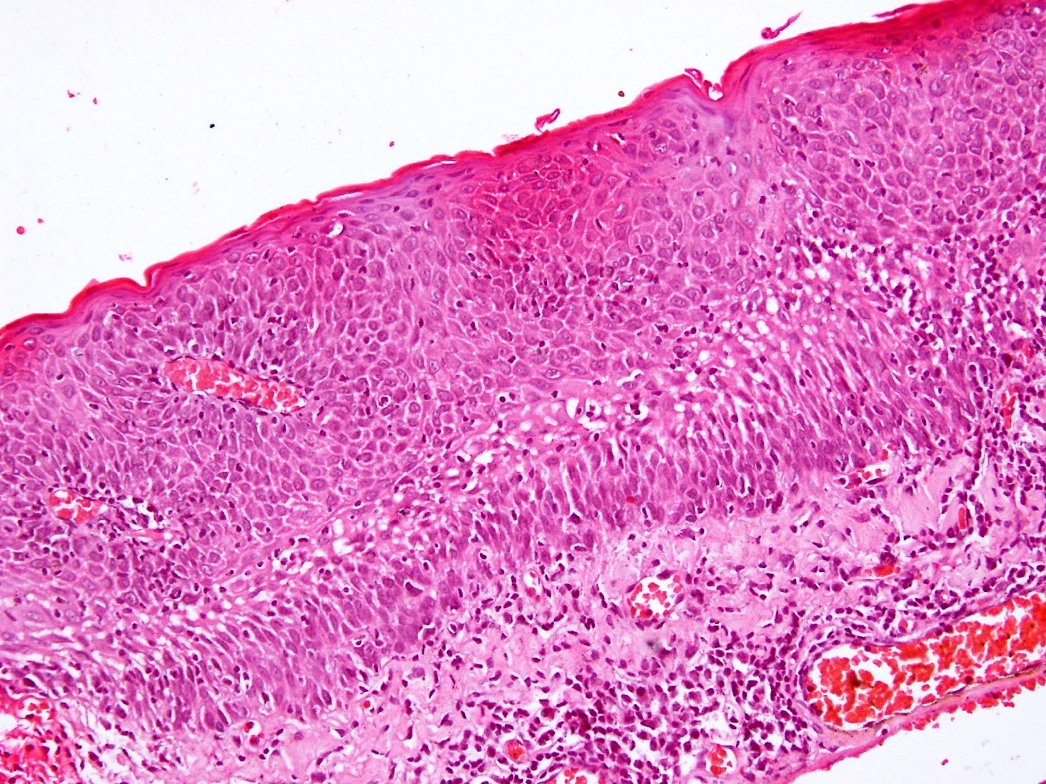
Grade 3 / severe dysplasia
Grade 2 / moderate dysplasia
Carcinoma in situ
Actinic keratosis carcinoma in situ
Contributed by Frederick A. Jakobiec, M.D., D.Sc., Deepali Jain, M.D. and Hans Grossniklaus, M.D.
Grade 3 / severe dysplasia
Carcinoma in situ
Conjunctival intraepithelial neoplasia
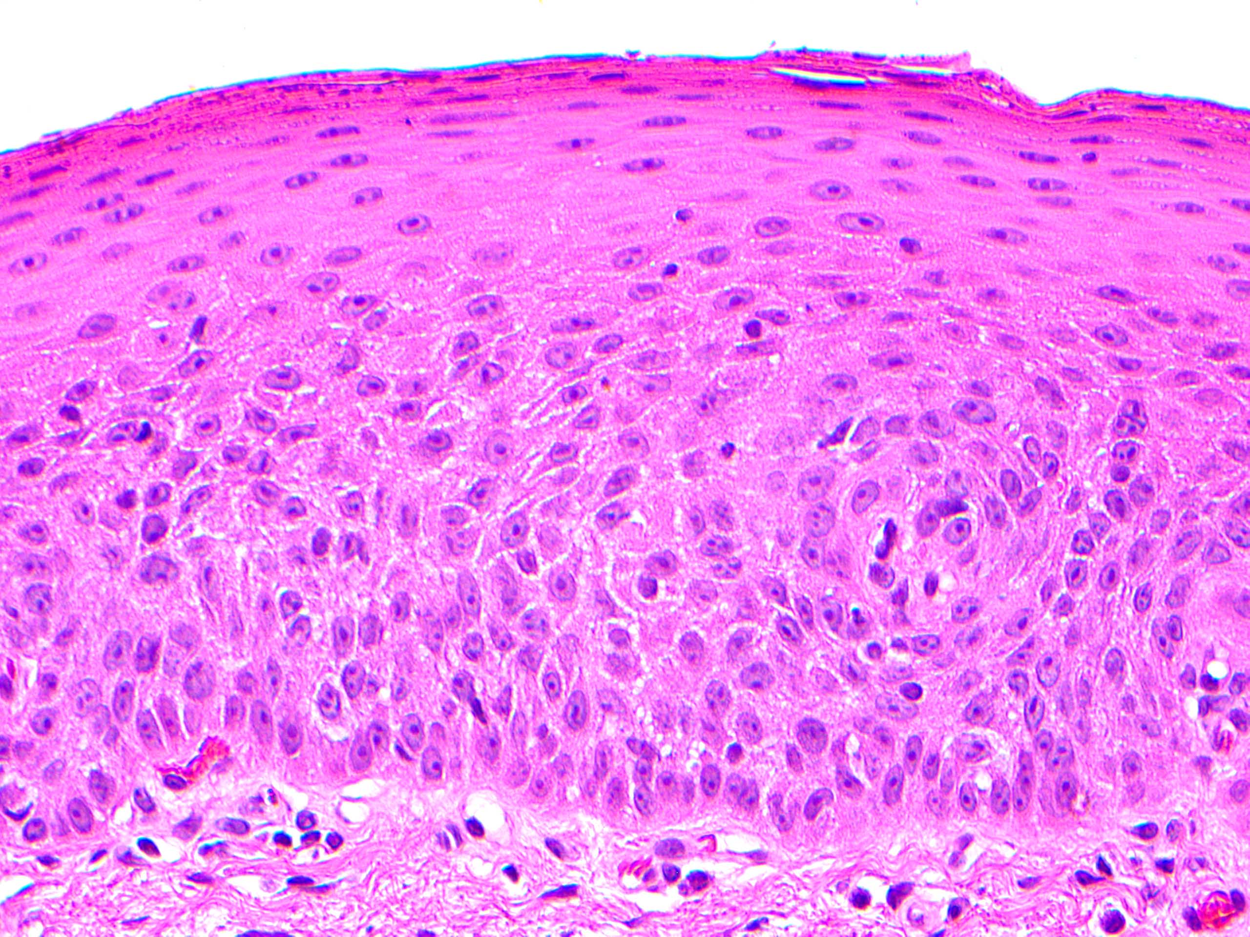
Grade 2 / moderate dysplasia
Grade 3 / severe dysplasia with surface keratinization
AFIP images
Flat diffuse pigmentation
AFIP images
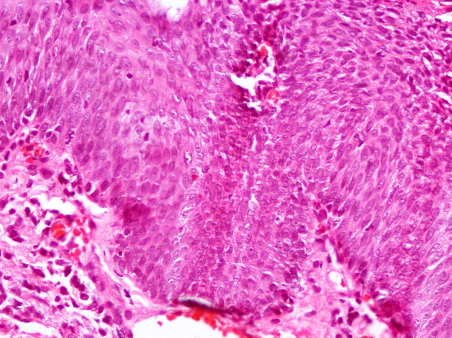
Without atypia:
Melanocytic hyperplasia is confined to basilar layer
With atypia:
Minimal atypia
Moderate atypia
Severe atypia
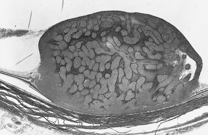
AFIP images
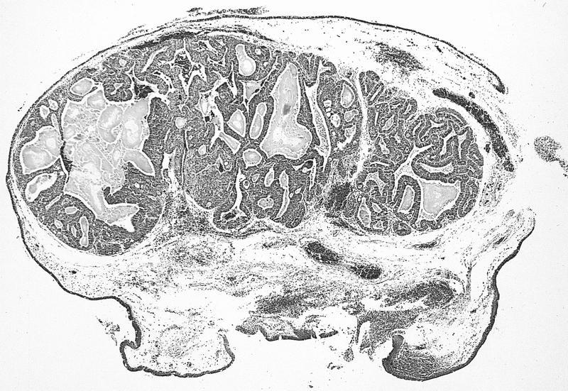
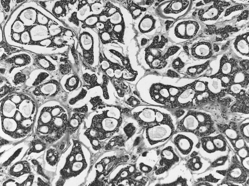
Well circumscribed lesion
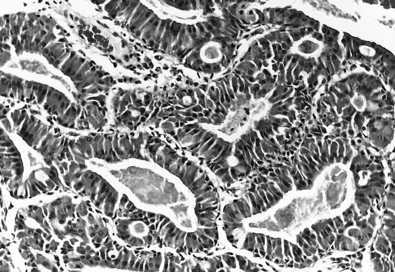
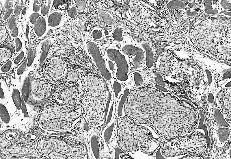
Tall columnar acidophilic cells lining glandular spaces
Subepithelial glandular tumor
Glandular spaces are lined by oncocytes and goblet cells
Images hosted on other servers:

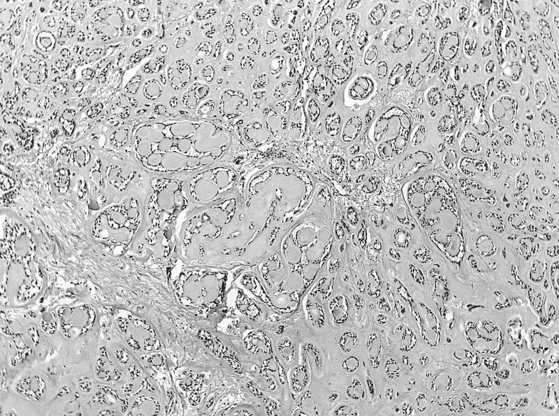
Adnexal lacrimal glands

Caruncle tumor

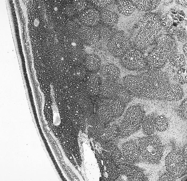
Cystic areas contain eosinophilic secretions

Light and dark cells with acinar formation on left side

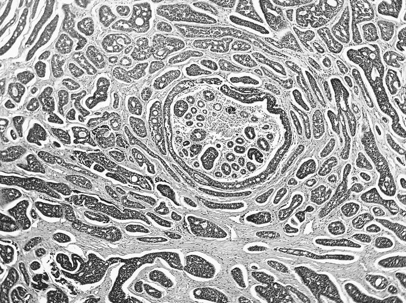
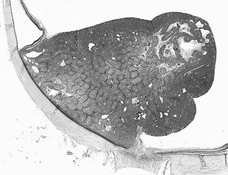
Well circumscribed lesion

Large polyhedral cells: abundant
eosinophilic granular cytoplasm
and small nuclei with
single prominent nucleoli
Images hosted on other servers:


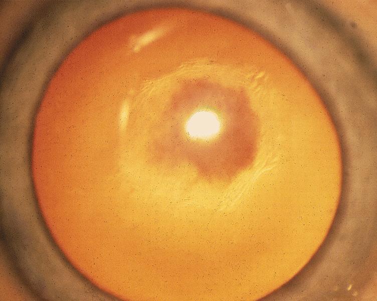
Lattice dystrophy
Images hosted on other servers:

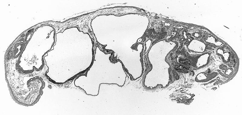
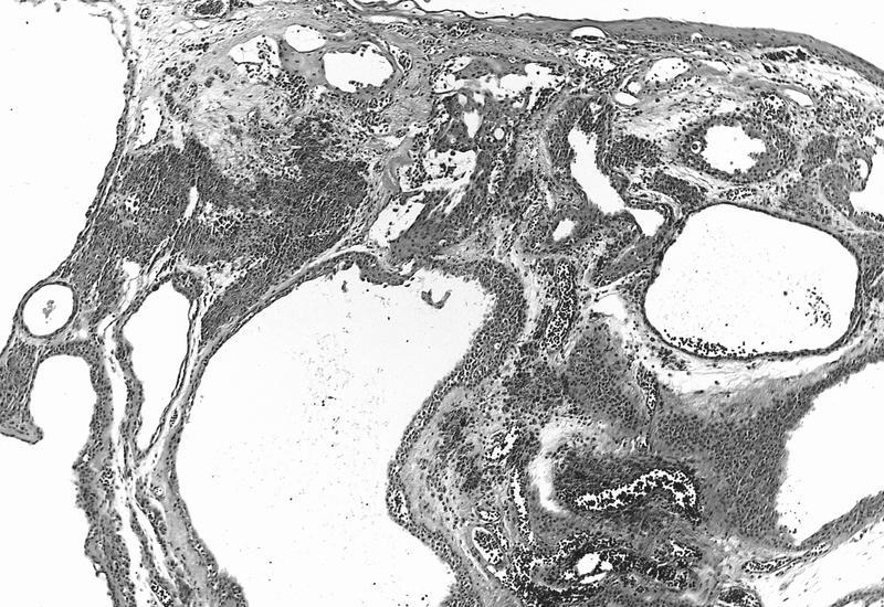
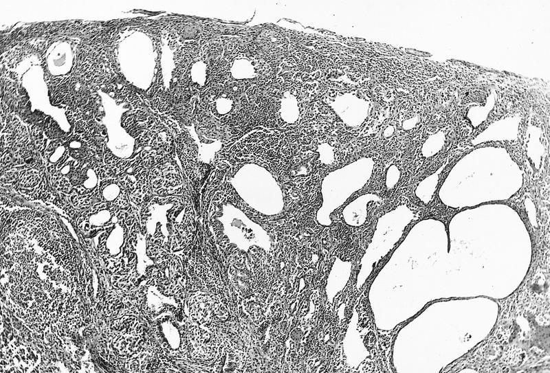
Multiloculated conjunctival cyst
AFIP images
Nasally located cystic lesion
AFIP images
Inflamed cyst lined by conjunctival type epithelium
Inflamed cyst lined by skin
Contributed by Anita Kumari, M.D.
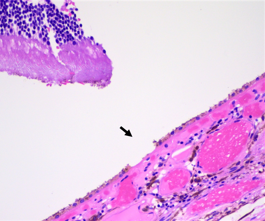
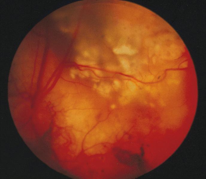
Retinal detachment
Contributed by Anita Kumari, M.D.

Microscopic images of a case of retinal detachment
Images hosted on other servers:

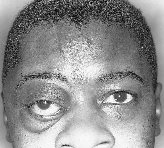
Upper lid retraction and exophthalmos

Severe restriction of the inferior rectus and upgaze
Images hosted on other servers:

Complex choristoma (figure 1b)
Images hosted on other servers:


Lipodermoid
AFIP images
Limbal dermoid:
Subepithelial fibrous tissue resembling
reticular dermis with skin appendages
Complex choristoma:
Lacrimal gland tissue within a limbal dermoid
Island of cartilage within a limbal dermoid
Images hosted on other servers:
Complex choristoma:

With ectopic lacrimal gland tissue (figure 2d)
Osseous choristoma:

Bone within connective tissue capsule

Bone contains marrow
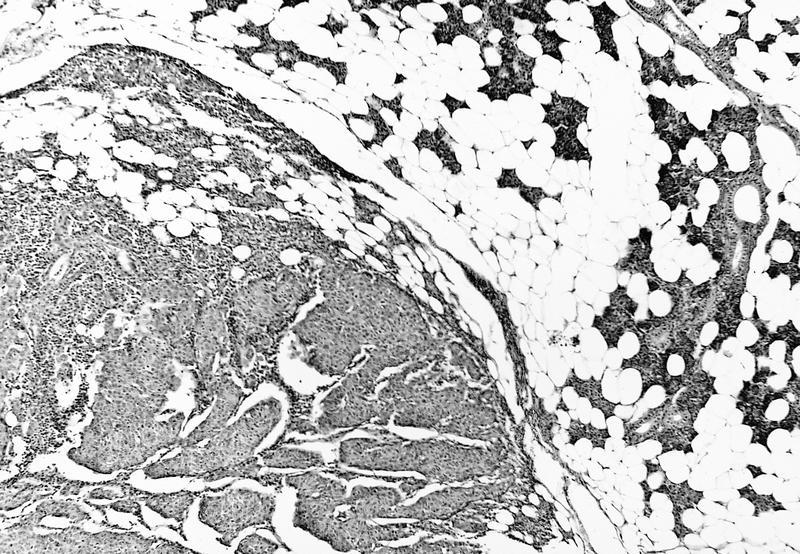
Dermolipoma:

Abundant adipose tissue
Images hosted on other servers:

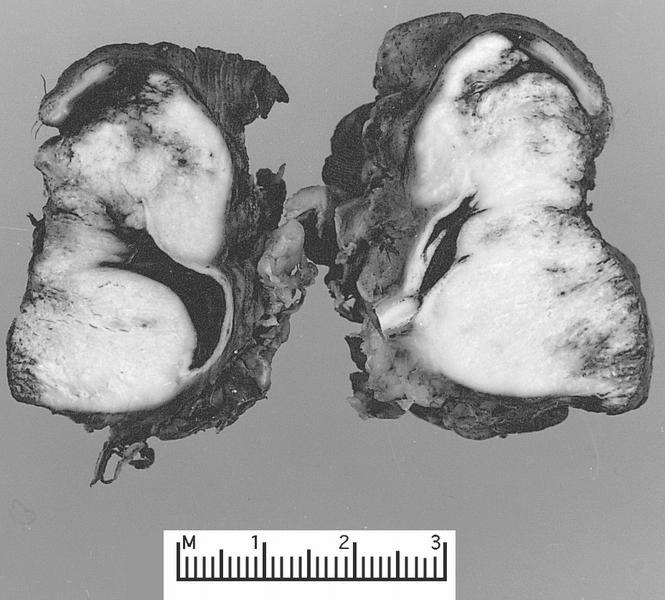
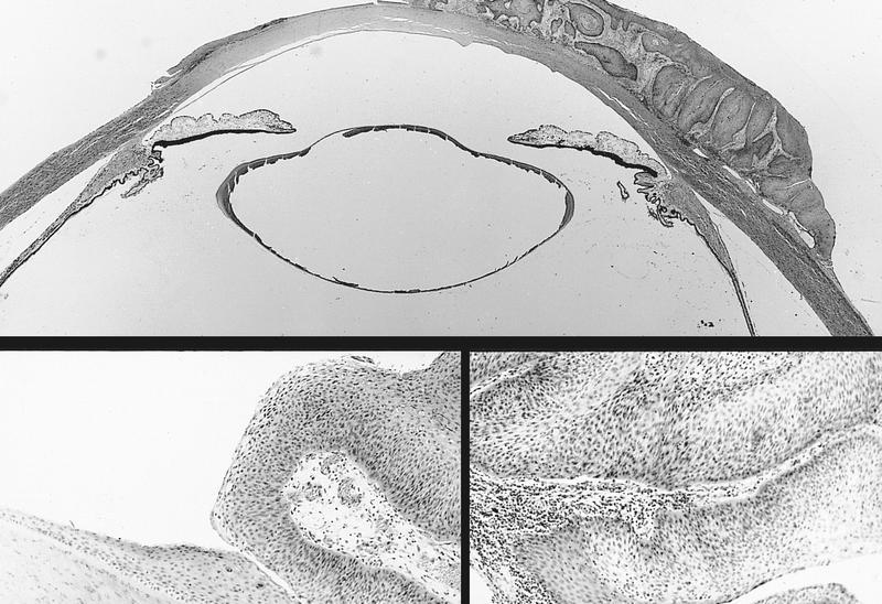
Epidermal cyst


Dermoid cyst


Hidrocystoma
Contributed by J. Stephen Nix, M.D.
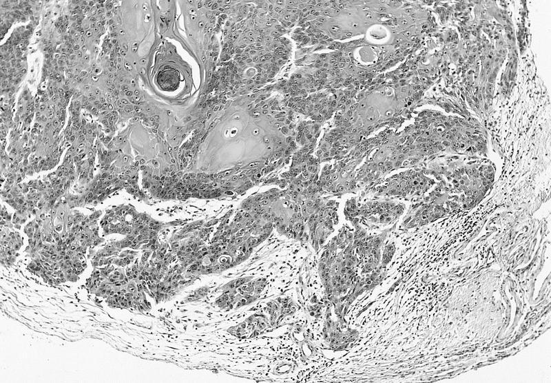
Epidermal cyst, keratinaceous contents
Epidermal cyst, granular layer
Dermoid cyst, keratinaceous contents
Dermoid cyst, adnexal structures
Dermoid cyst with rupture
Eccrine hidrocystoma
Eccrine hidrocystoma, cuboidal bilayer
Eccrine hidrocystoma, attenuated lining
Apocrine hidrocystoma
Apocrine hidrocystoma, inner cyst lining
Hidrocystoma: 5 minute pathology pearls by Dr. Jared Gardner
Dermoid cyst: 5 minute pathology pearls by Dr. Jared Gardner
AFIP images
Iris and ciliary body with open anterior chamber angle
AFIP images
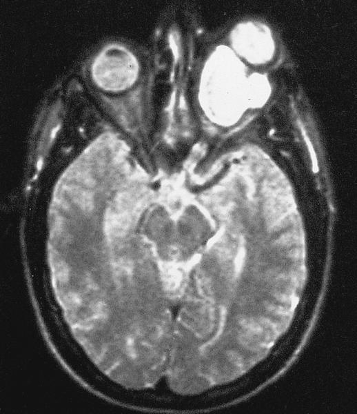
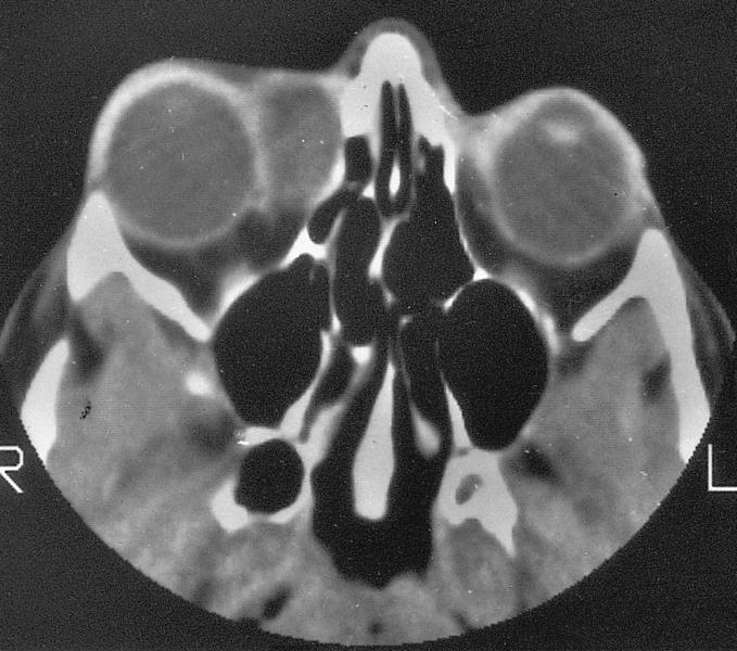
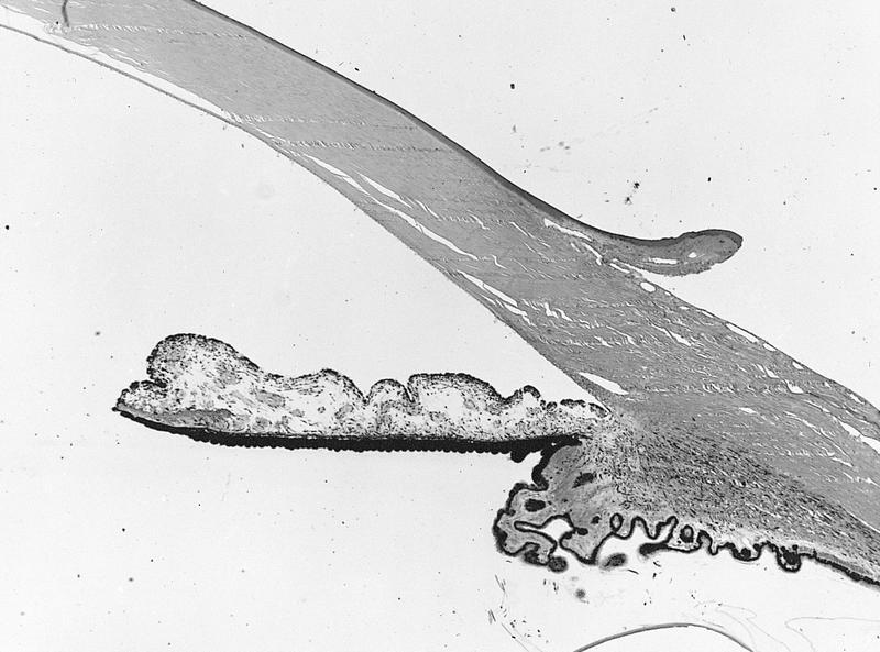
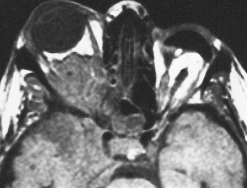
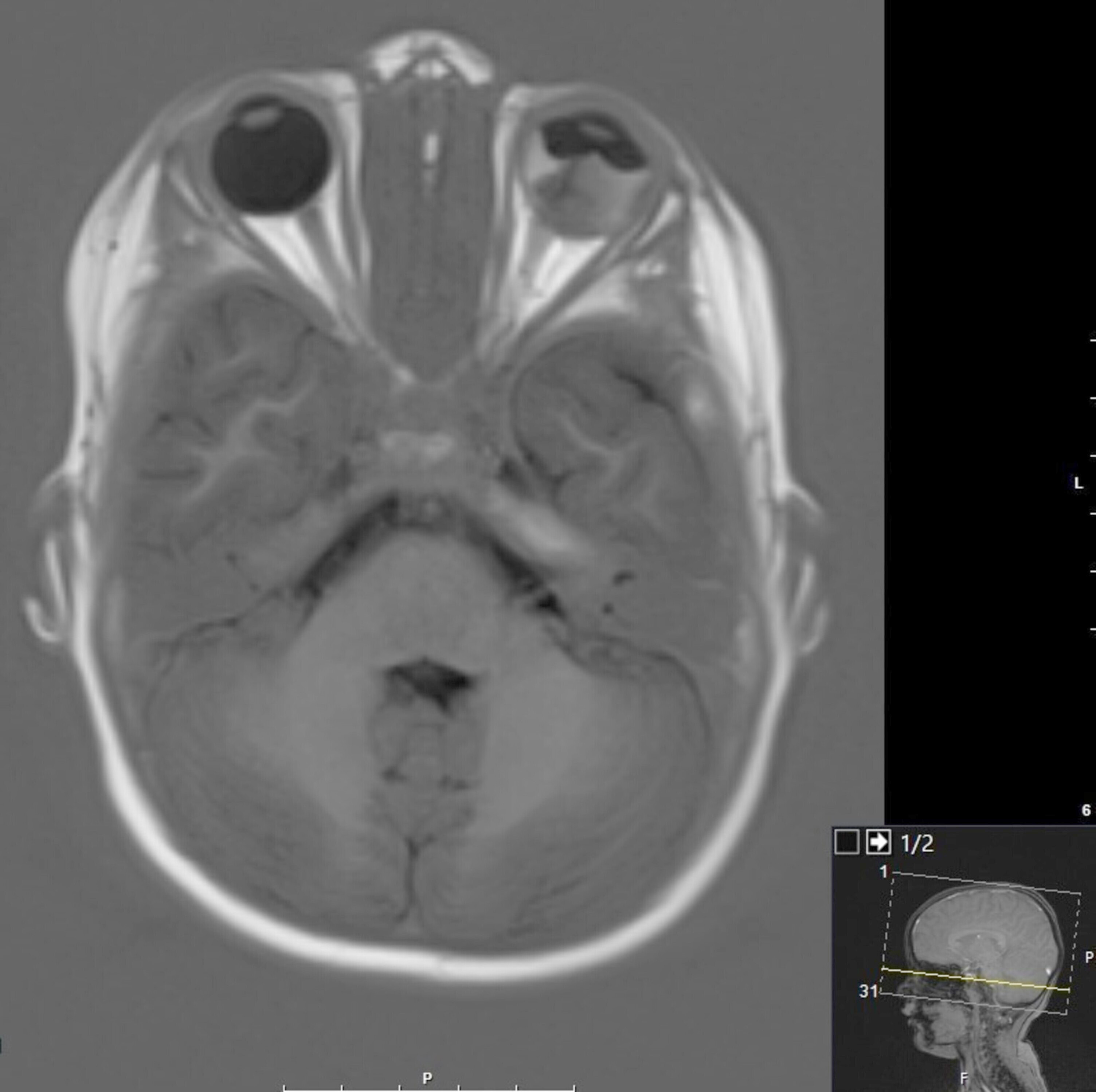
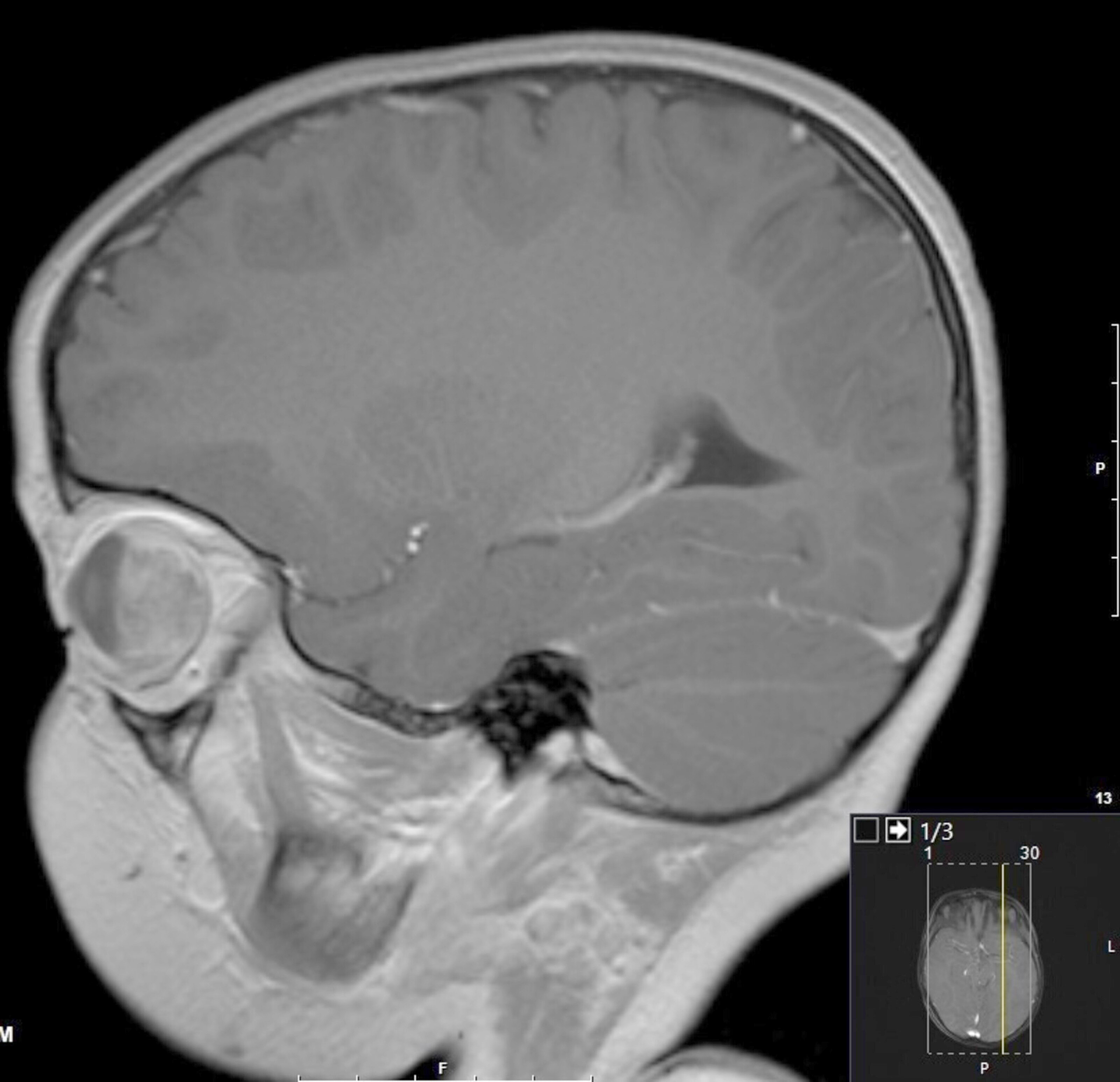
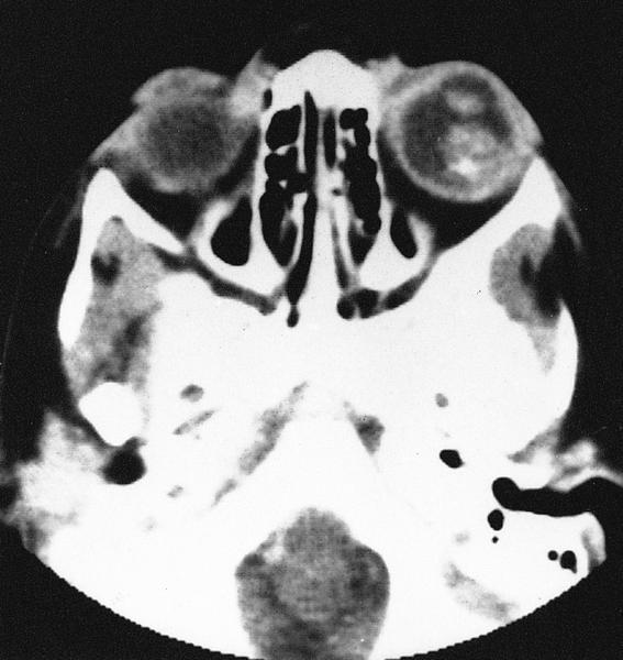
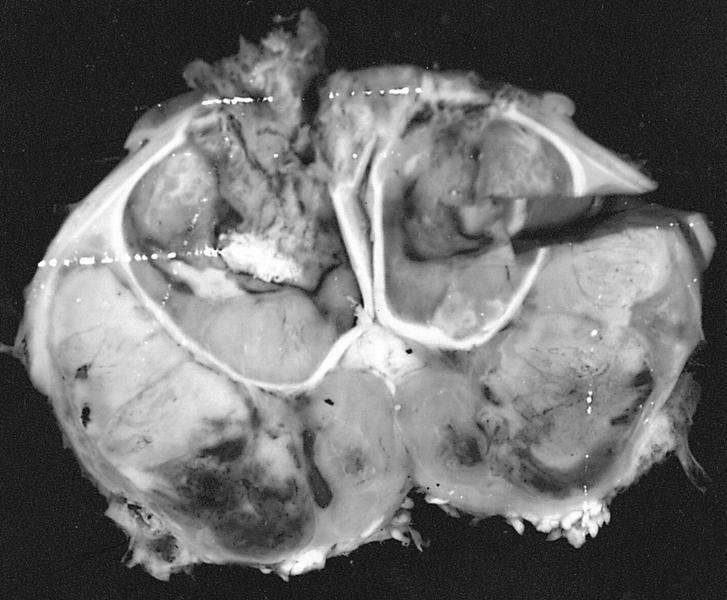
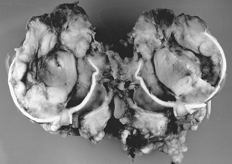
MR of large retrobulbar optic nerve tumor
AFIP images
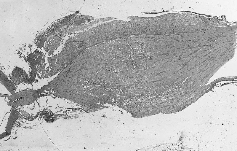
Pilocytic astrocytoma
Intraparenchymal tumor

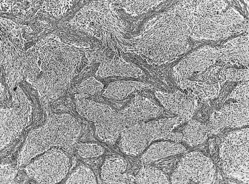
Numerous Rosenthal fibers
Astrocytes infiltrate meninges
High grade astrocytoma
Images hosted on other servers:

Elongated or hair-like appearance

Rosenthal fibres
Images hosted on other servers:

Chronic GVHD

Excessive fibrosis

Basal epithelial cell invasion

Epithelial mesenchymal transition

Epithelial mesenchymal transition
Images hosted on other servers:

Disrupted basal lamina and epithelial cells

Myoepithelial cell invasion
Images hosted on other servers:

Sarcoidosis


Churg-Strauss syndrome

Due to fibers from teddy bear

Cicatrising
Images hosted on other servers:

Segregation of HBID duplication
Images hosted on other servers:

Various images

Nerve growth factor and eosinophils

IHC for NGF and eosinophils
AFIP images
Iris and ciliary body with open anterior chamber angle
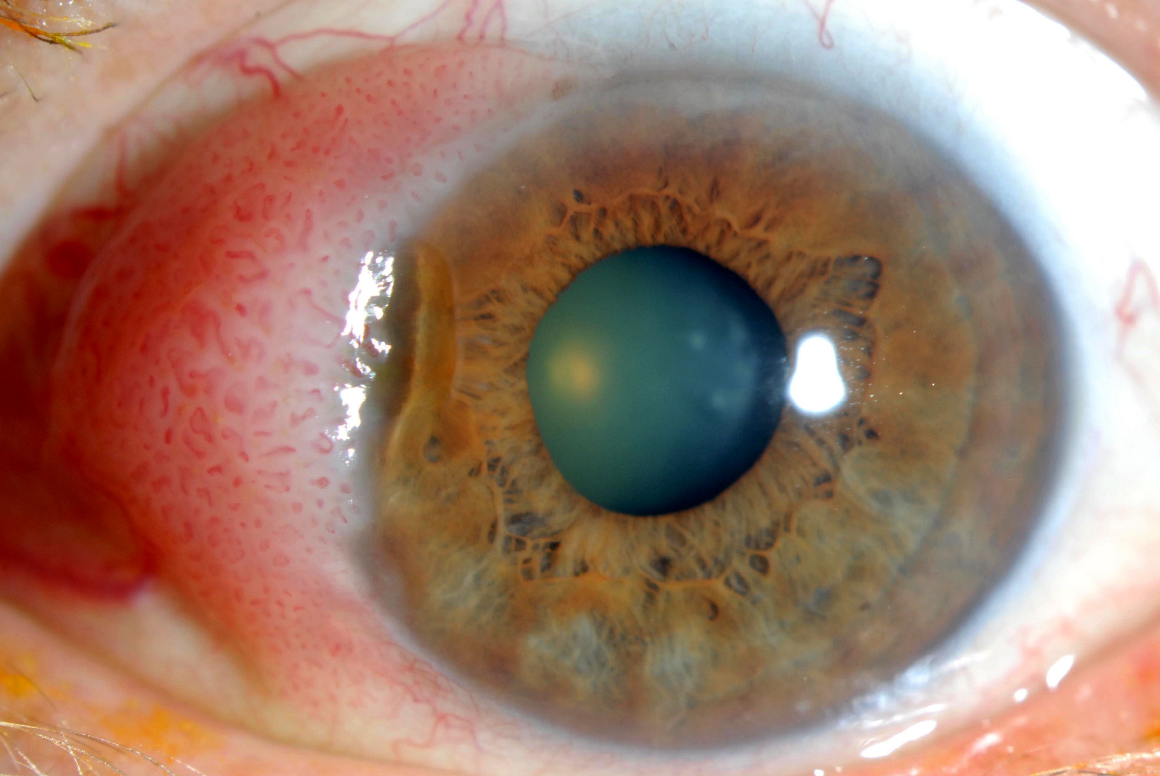
Normal iris
Images hosted on other servers:
Iris and lens
Case #368

CD34

CD31

HHV8
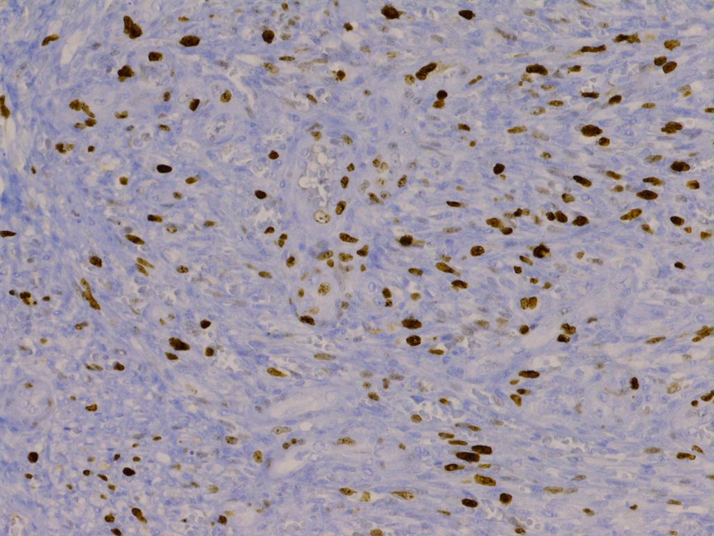
Ki67
Images hosted on other servers:
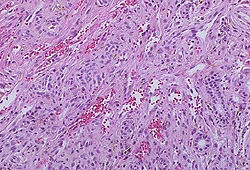
Kaposi's sarcoma
Images hosted on other servers:

Pathogenesis
Contributed by Hisham Adel Saad, M.D.
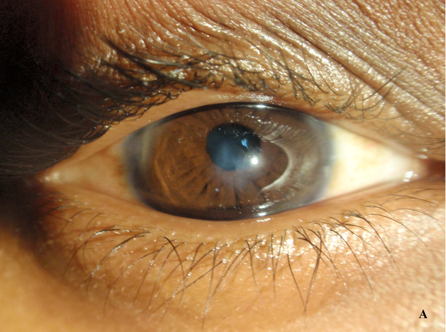
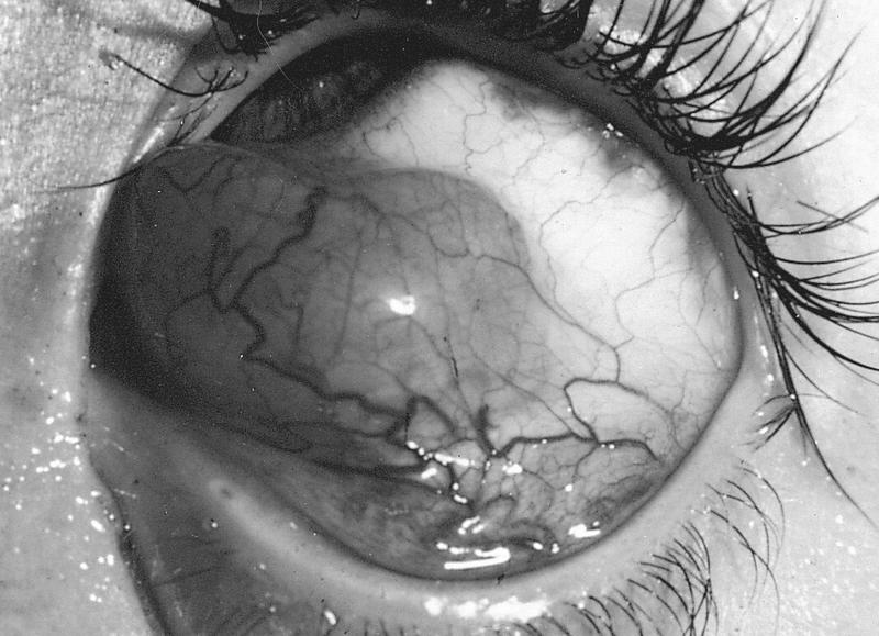
Apical scarring
Evident Munson sign

Axial curvature map
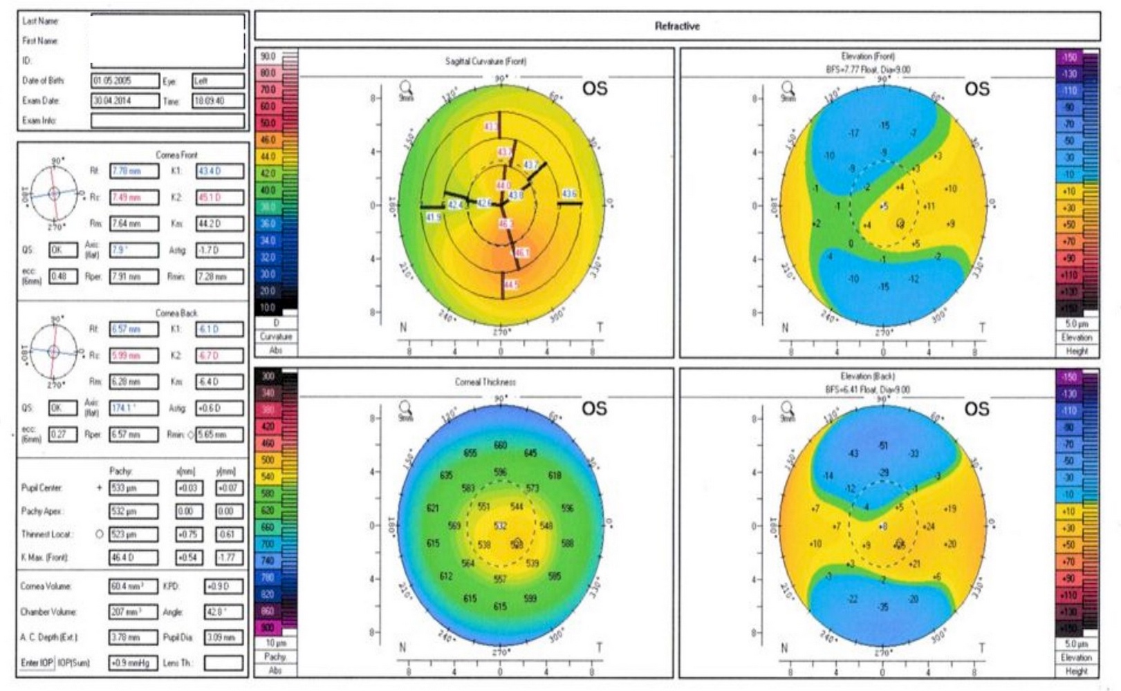
Corneal topography in keratoconus
Images hosted on other servers:

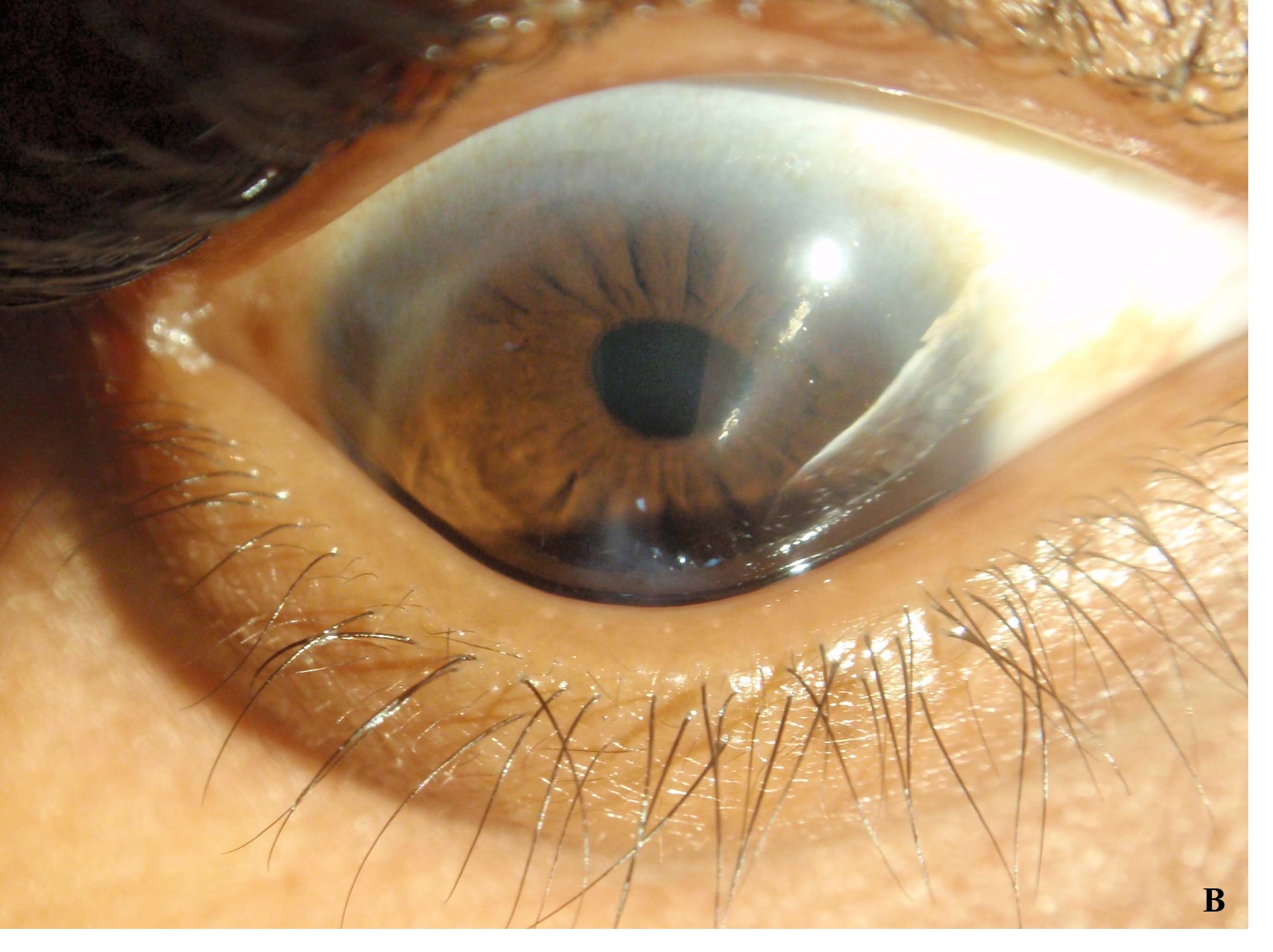
Vogt striae
Contributed by Eiman Adel Hasby Saad, M.D.
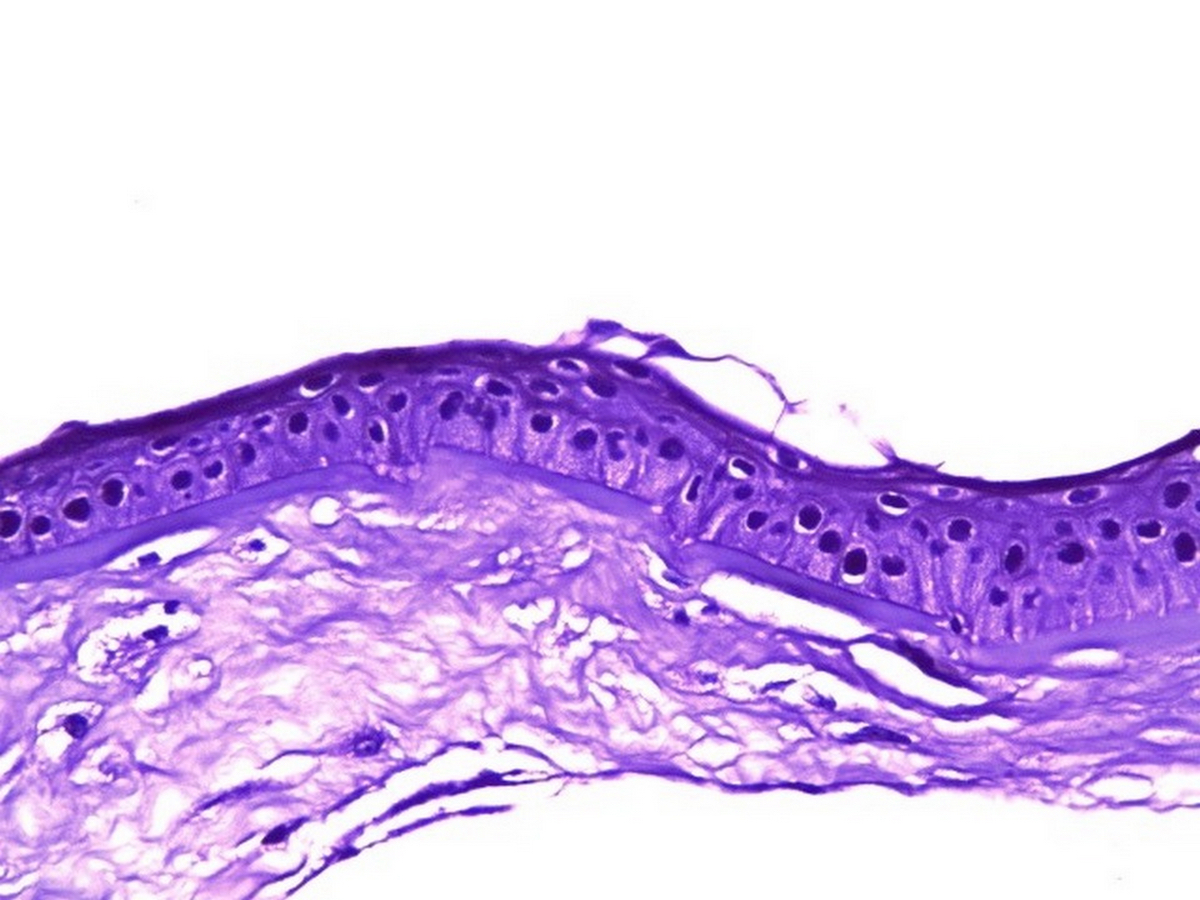
Thin stroma in keratoconus
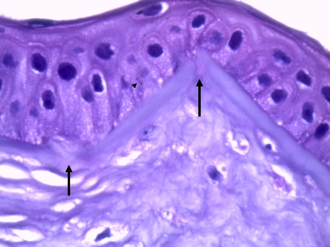
Bowman breaks in keratoconus
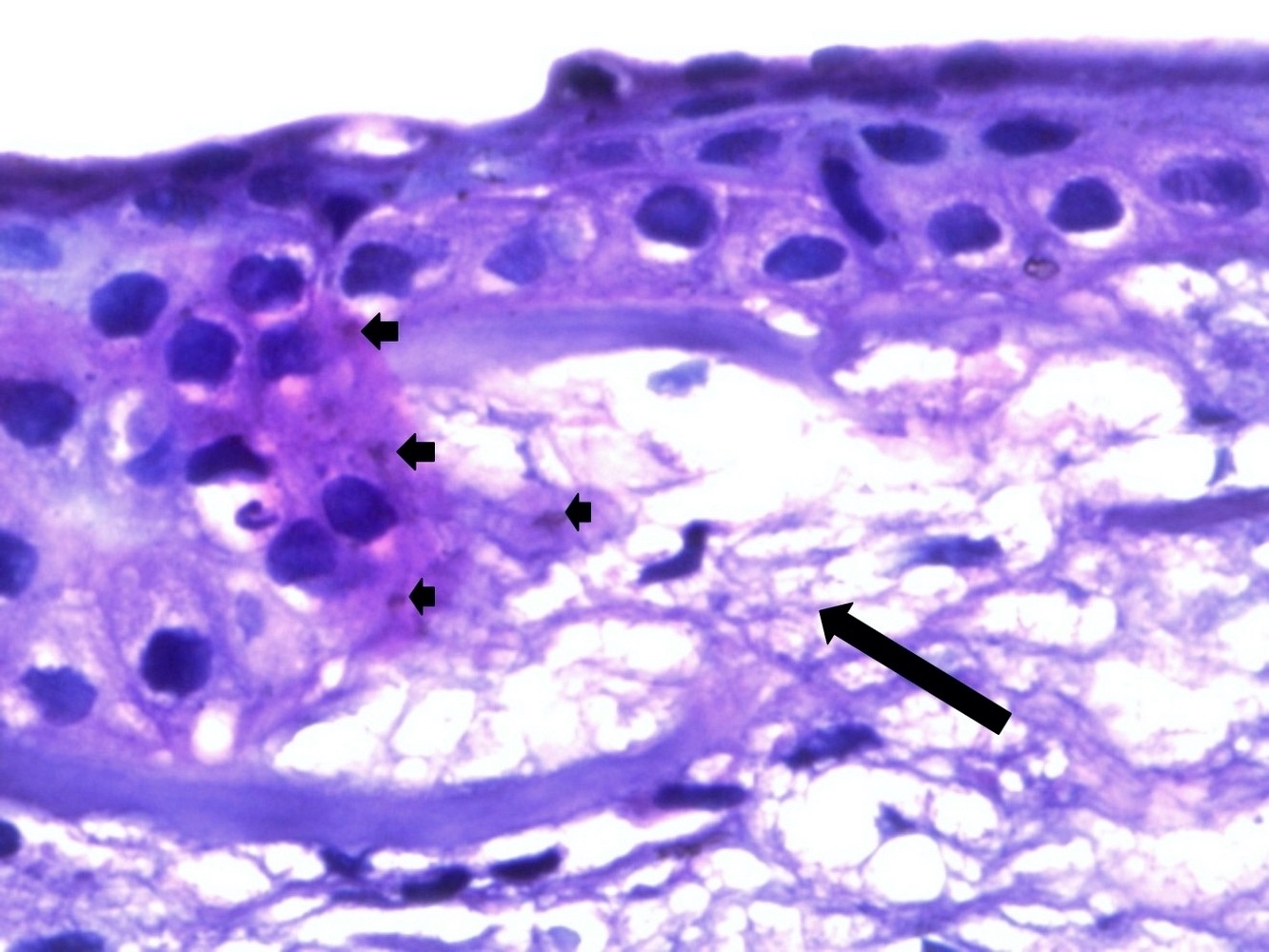
Stromal distortion in keratoconus
Images hosted on other servers:

Plaque like ulceration

Elevated hard mass
Images hosted on other servers:

Eosinophilic deposits surround islands of epithelial cells


Eosinophilic hyalinized tissue with inflammation

Histopathology of conjunctival biopsies

Inflamed granulation tissue


Acute and chronic inflammation
Images hosted on other servers:

Fibrillar deposits
Images hosted on other servers:
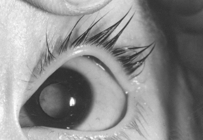
Lisch nodules
AFIP images
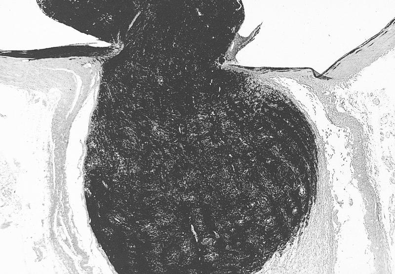
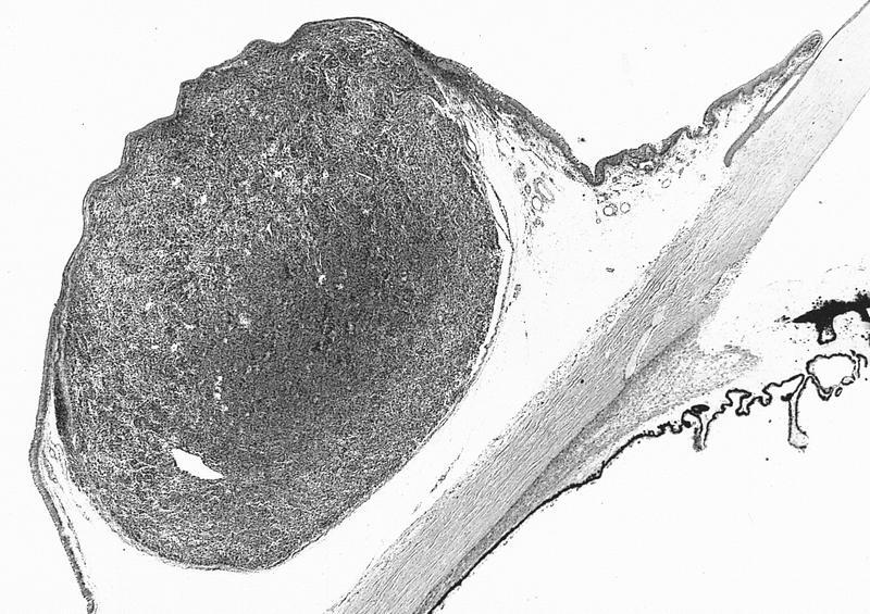
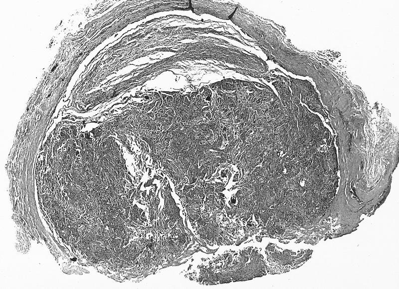
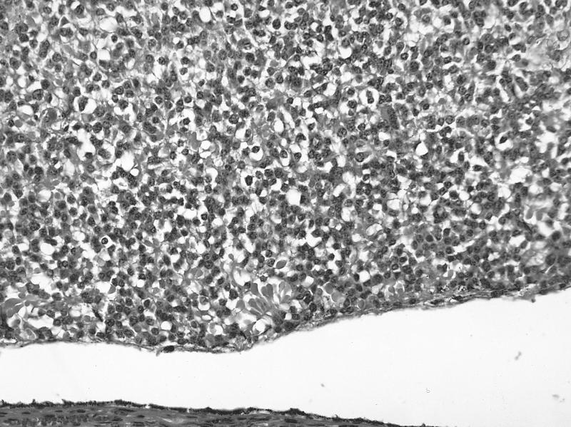
Homogeneous tumor
AFIP images
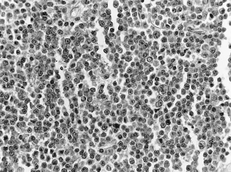
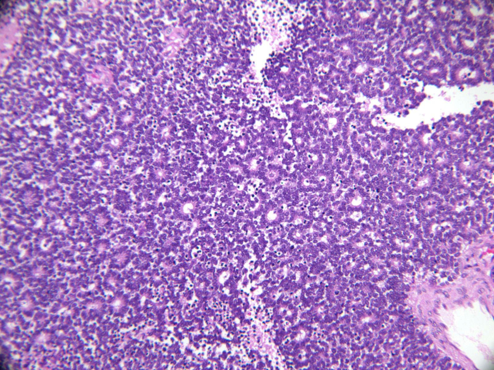
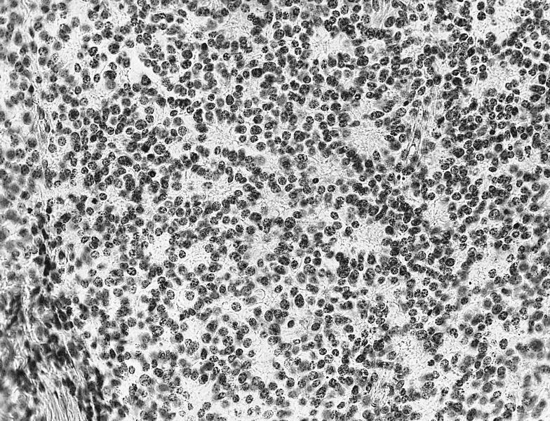
Burkitt lymphoma: bilateral
Lymphoplasmacytic lymphoma: bilateral
AFIP images
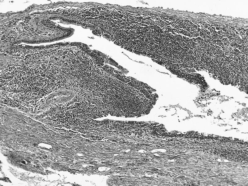
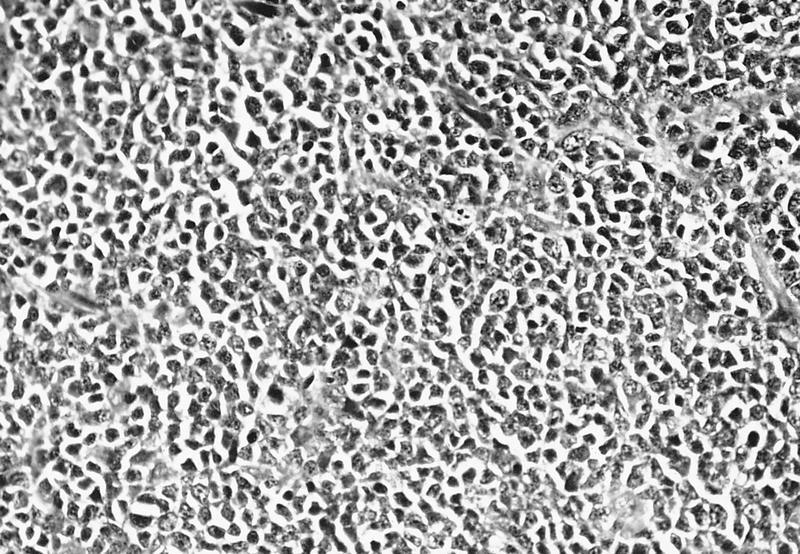
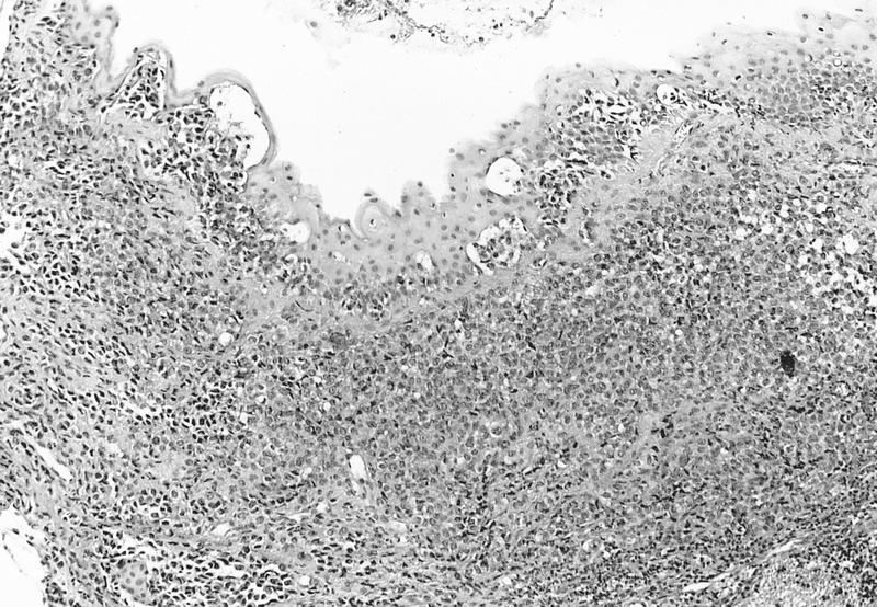
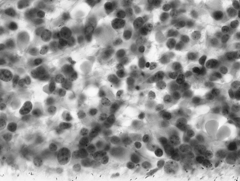
Low grade tumor
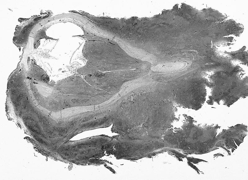
Well differentiated lymphoma
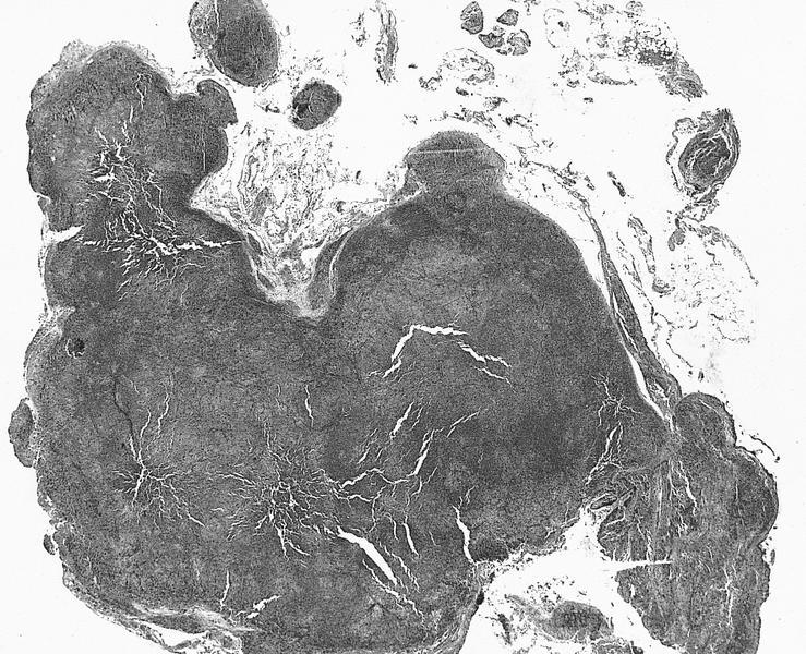
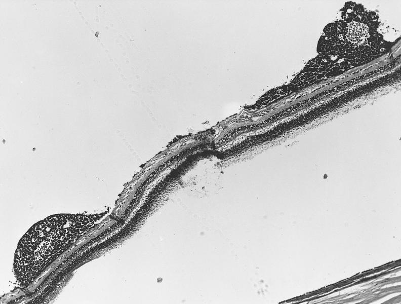
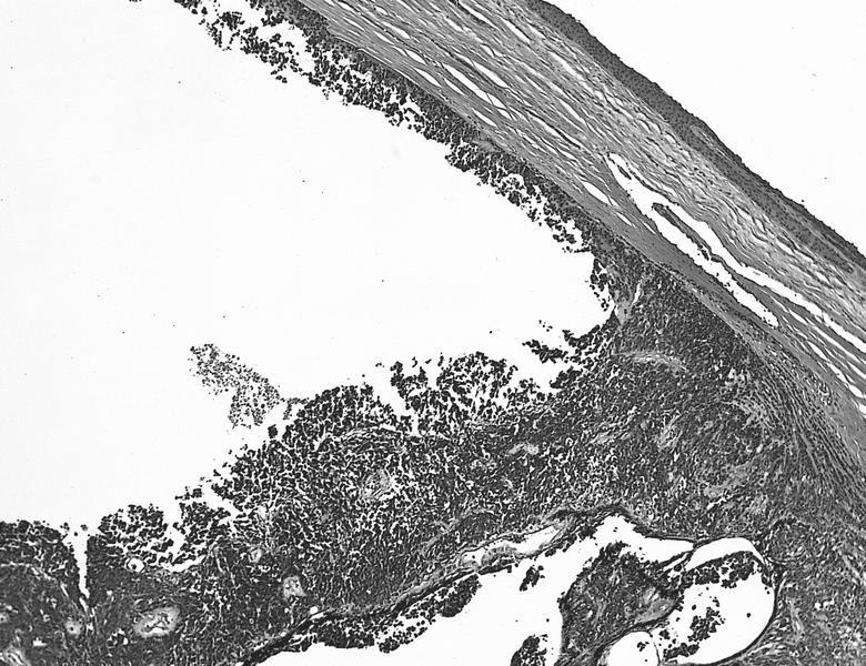
Irregularly shaped mass
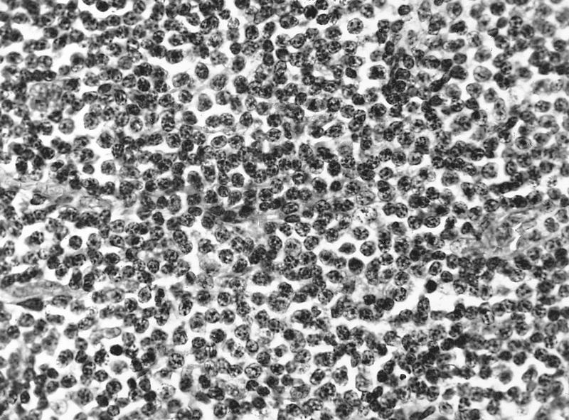
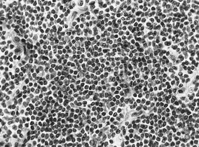
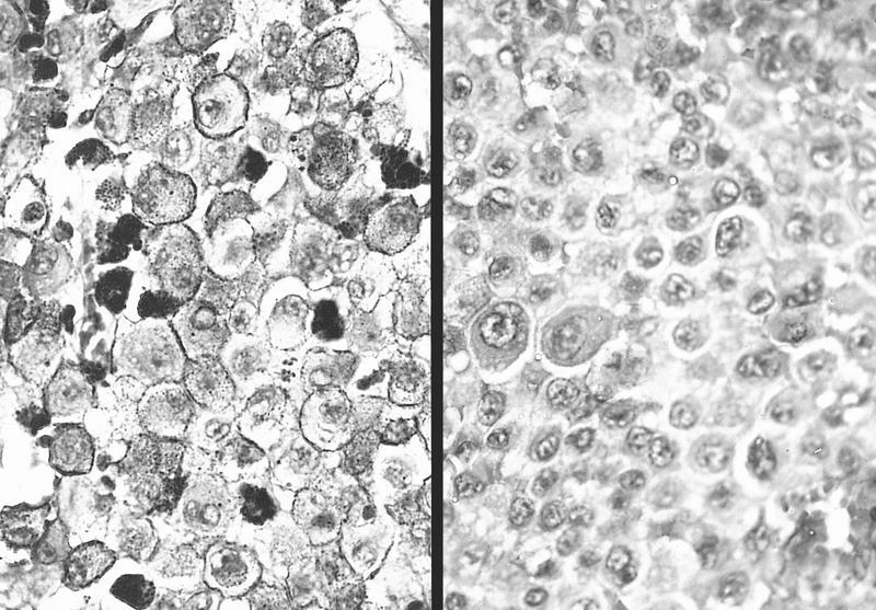
Uniform population of well differentiated lymphocytes
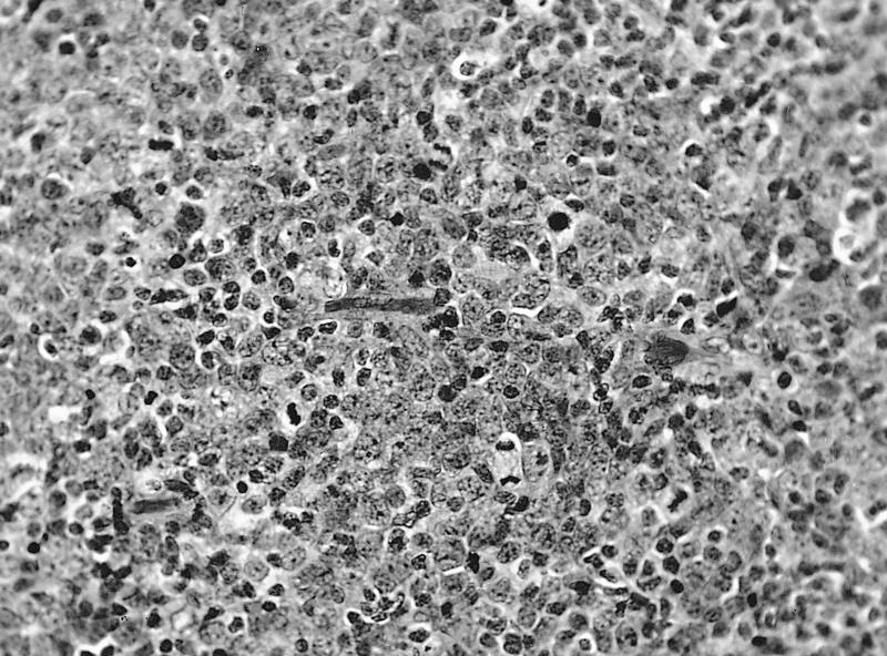
Small cleaved cells
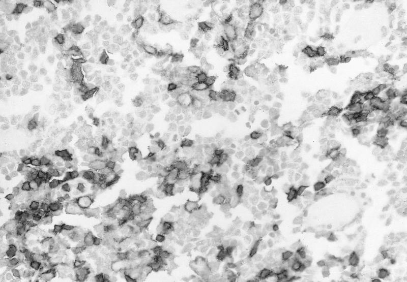
Small population of T cells

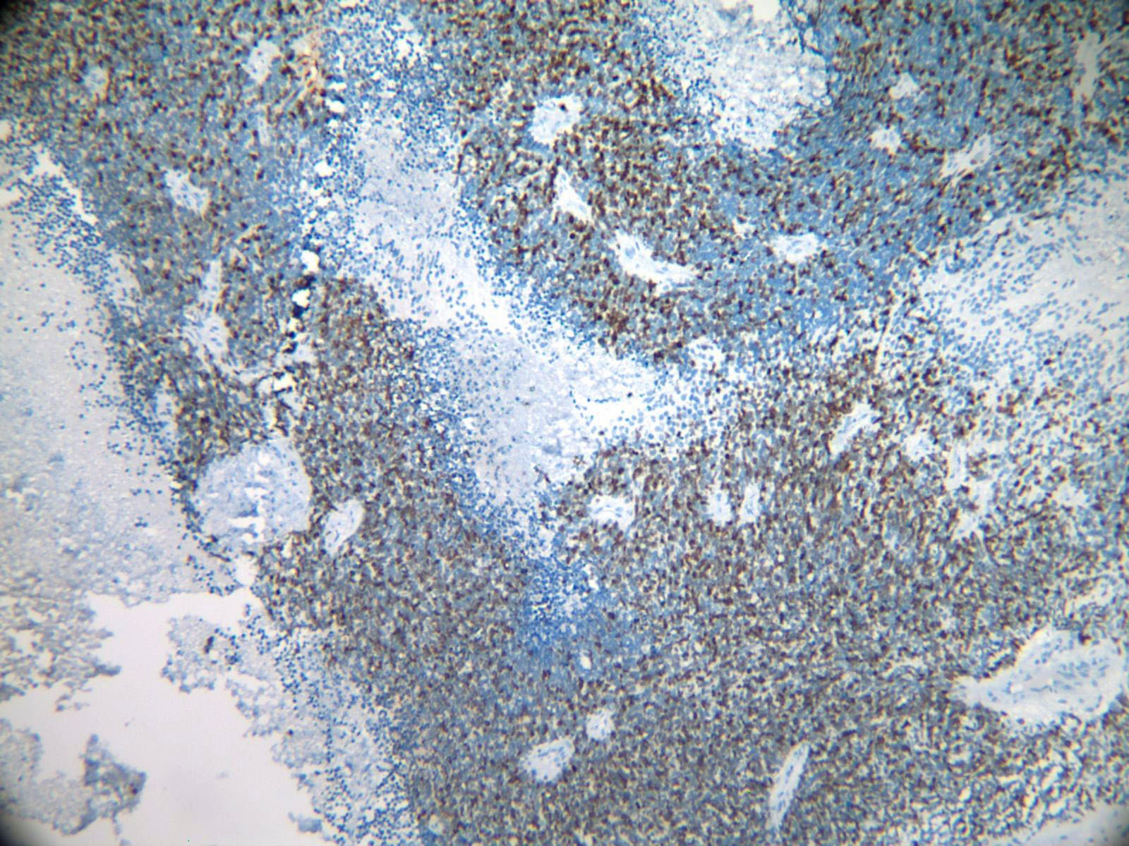
Numerous CD20+ B cells
Burkitt lymphoma
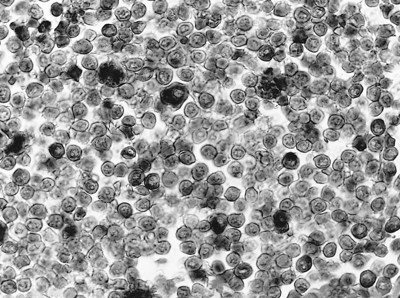
Granulocytic leukemia - Leder stain positive
Lymphoplasmacytic lymphoma - lymphoid cells, some with plasmacytic differentiation
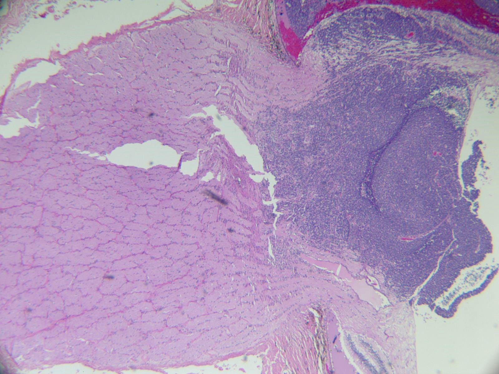
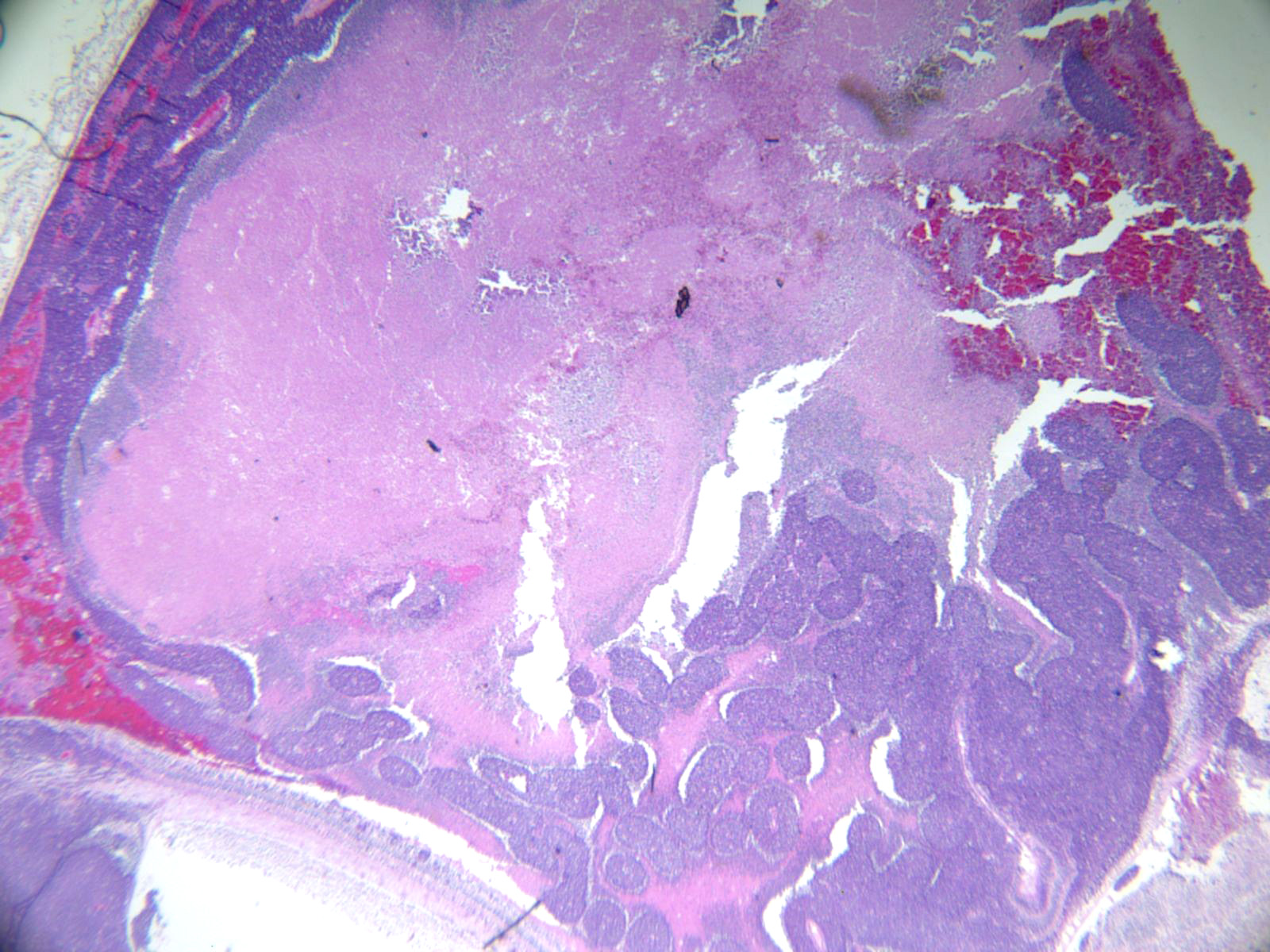
AFIP images
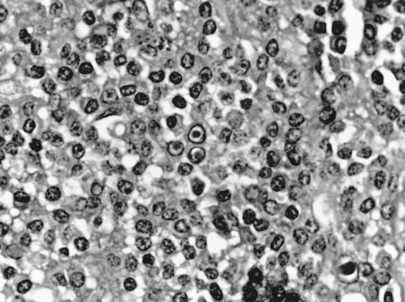
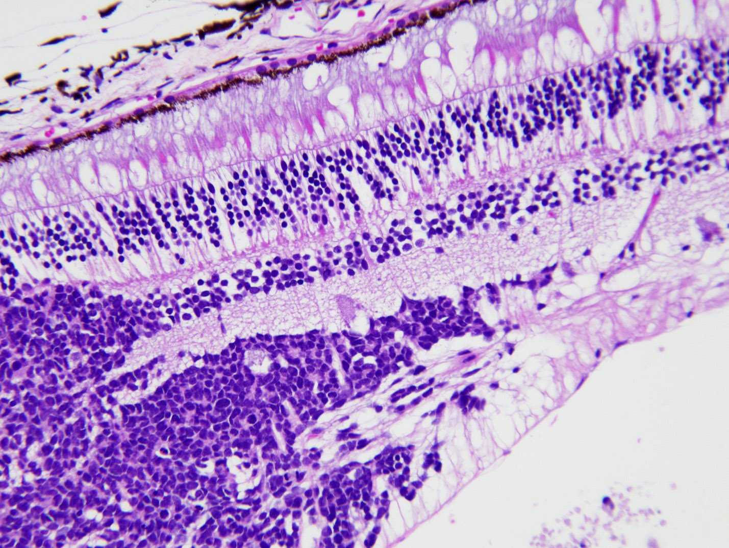
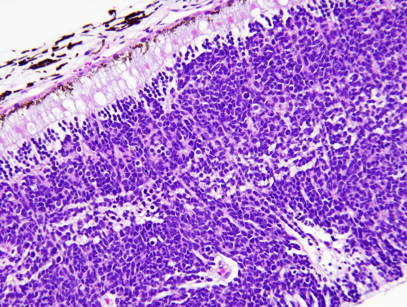
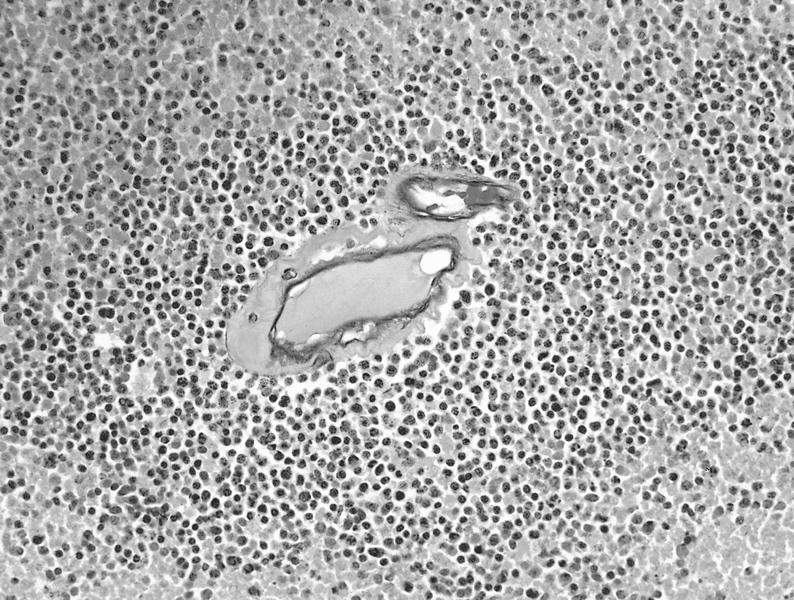
Network of cords of neuroepithelial cells
Less differentiated cells
Malignant teratoid medulloepithelioma
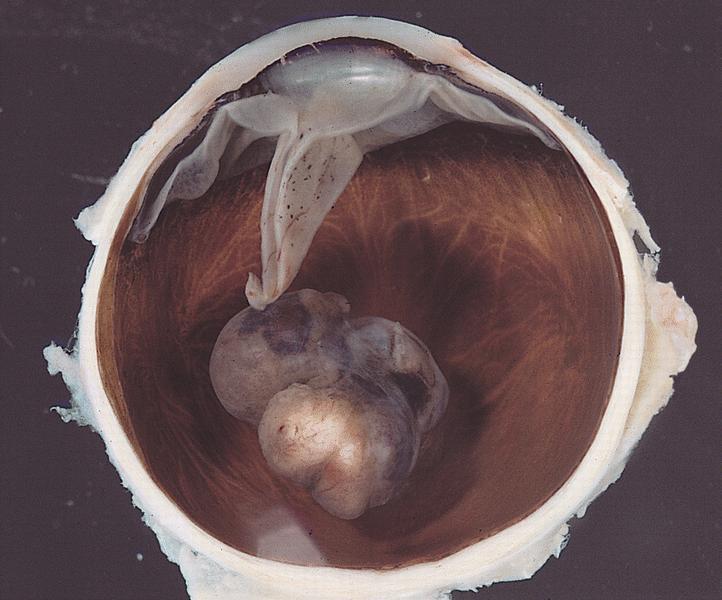
AFIP images
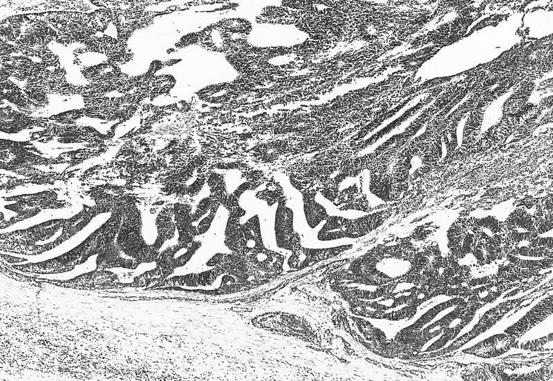
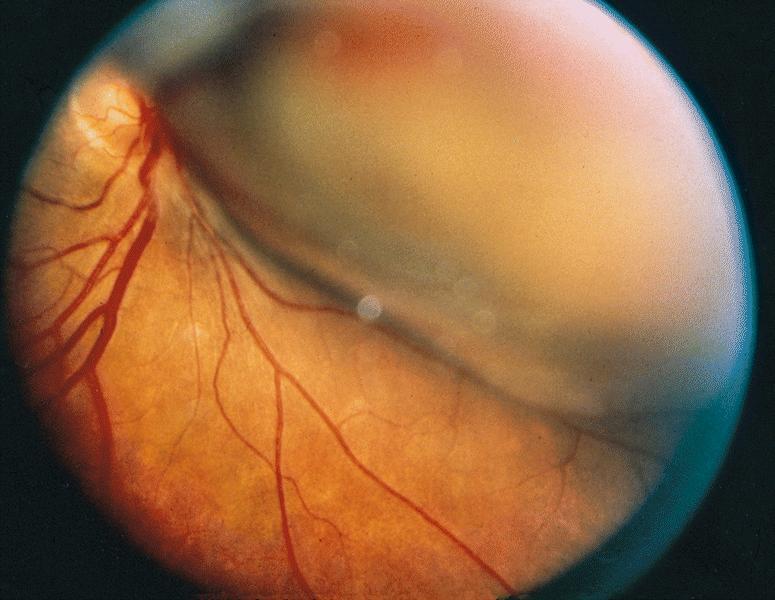
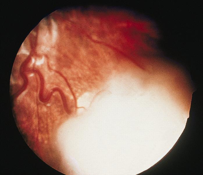
Tumor is centered in inferior temporal quadrant
AFIP images
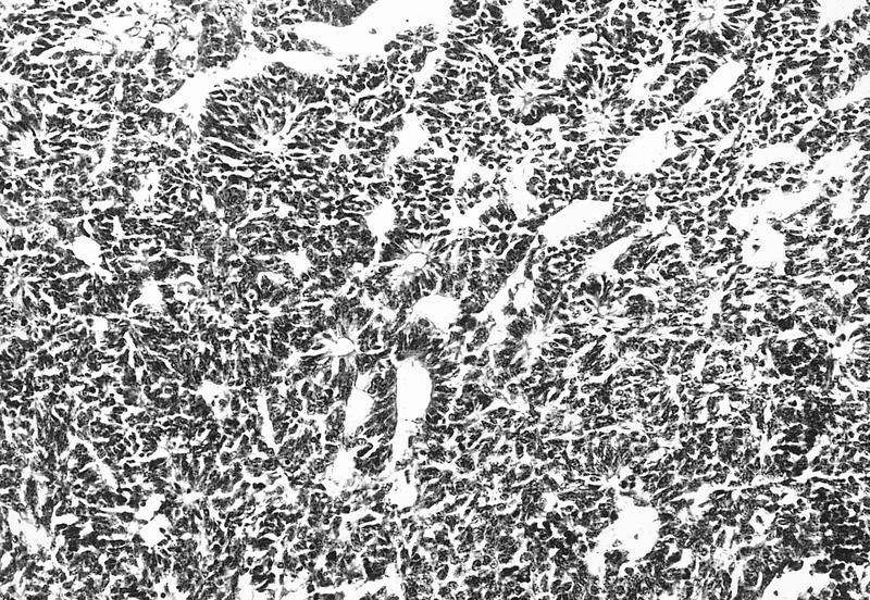
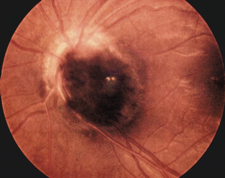
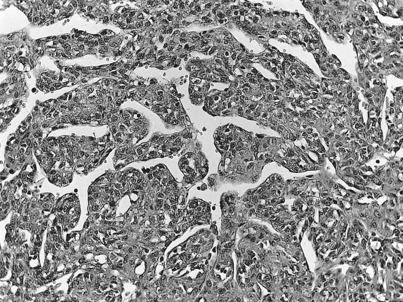
Heavily pigmented tumor
Bleached preparation
AFIP images
Pigmented lesion
adjacent to primary
acquired melanosis
Elevated melanotic nodule
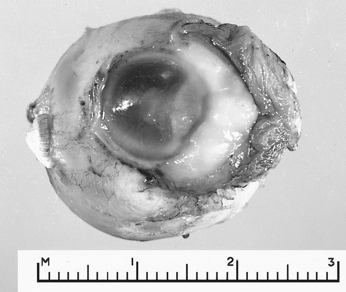
Large neglected melanoma
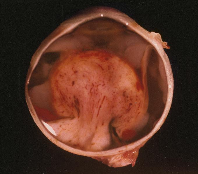
AFIP images
Large heavily pigmented nodule covers cornea
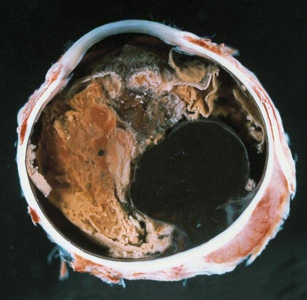
AFIP images
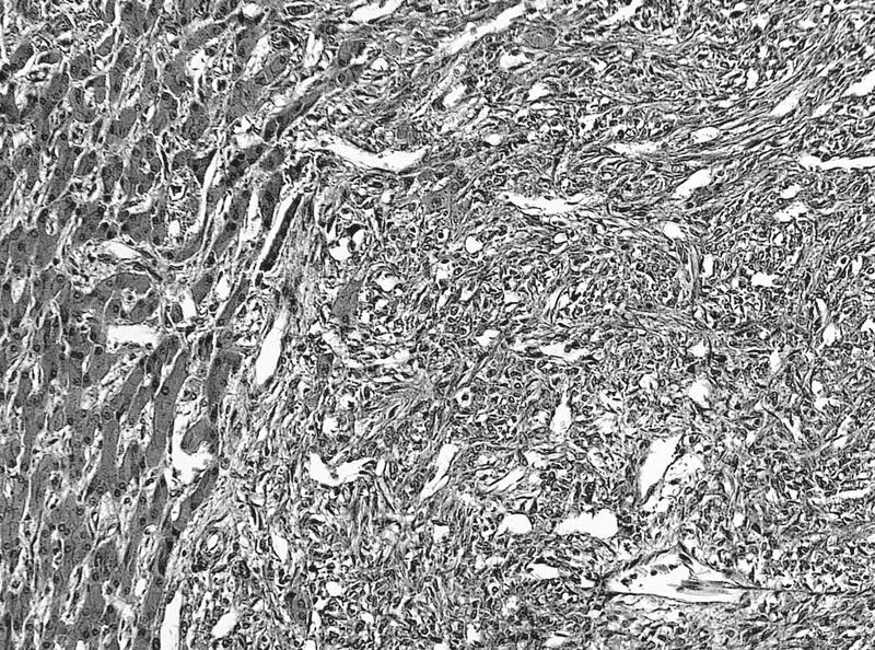
Nodular tumor at limbus
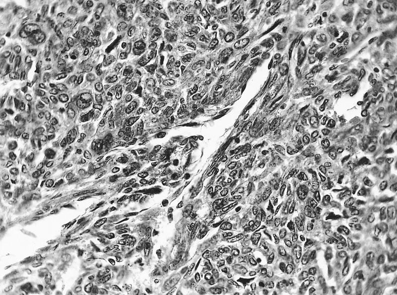
Anaplastic melanocytes within epithelial nests
Pigmented epithelioid cells with prominent nucleoli
Pleomorphic tumor cells
Malignant spindled melanocytes
Malignant epithelioid melanocytes
Contributed by John Irlam, D.O.
Ocular melanoma - site unspecified
Images hosted on other servers:

FNA of parotid metastasis
Contributed by Arun Singh, M.D.

B scan
Contributed by Lynn R. Schoenfield, M.D. and AFIP images
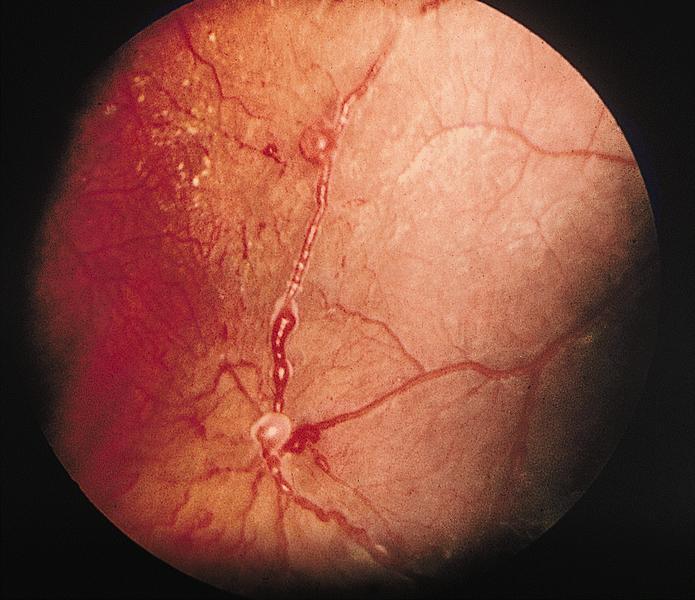
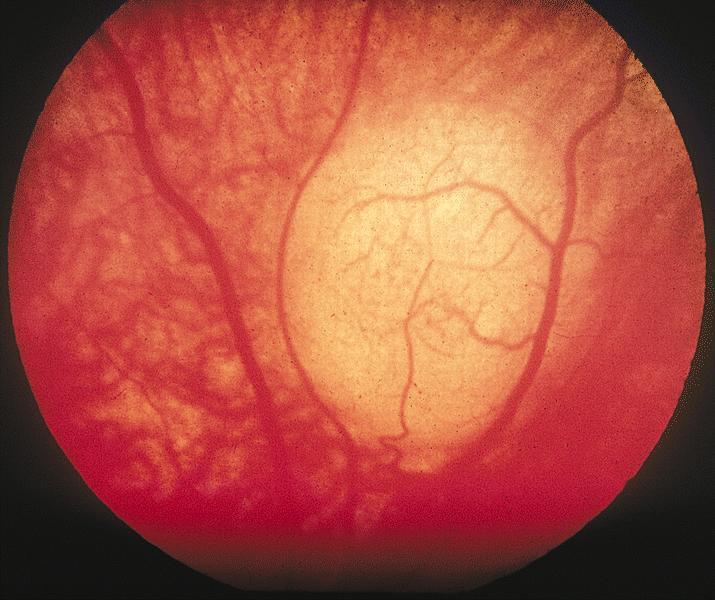
Fundoscopy
Ciliary body tumor
Orange pigment
Vessels over tumor are out of focus
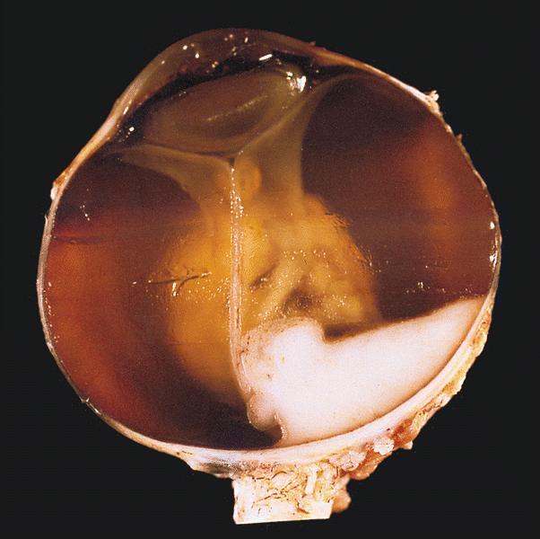
Contributed by Lynn R. Schoenfield, M.D. and AFIP images
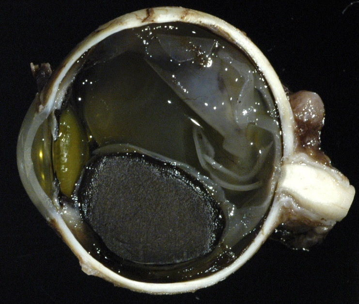
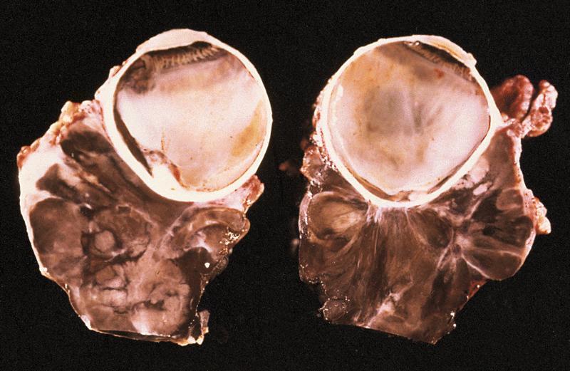
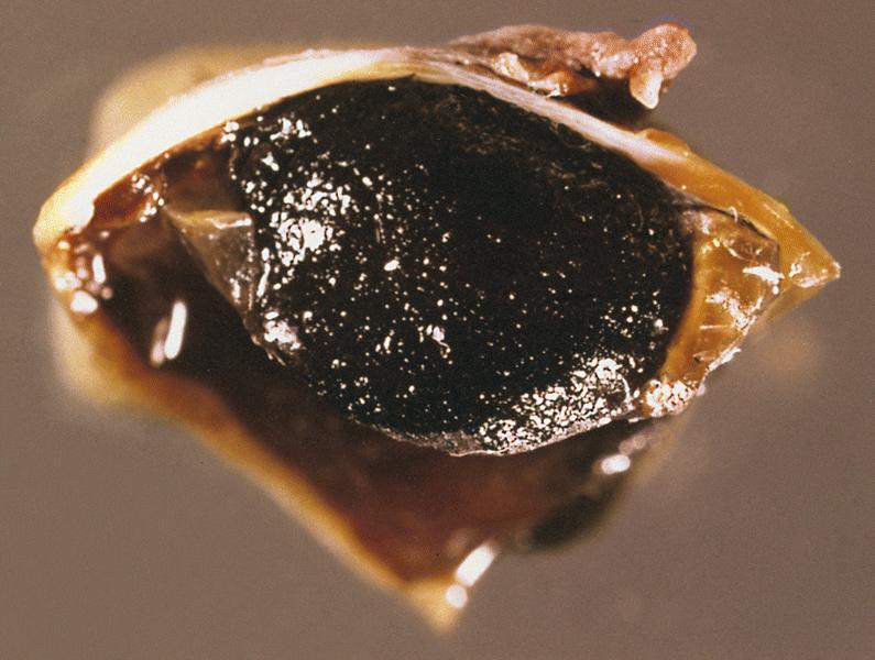
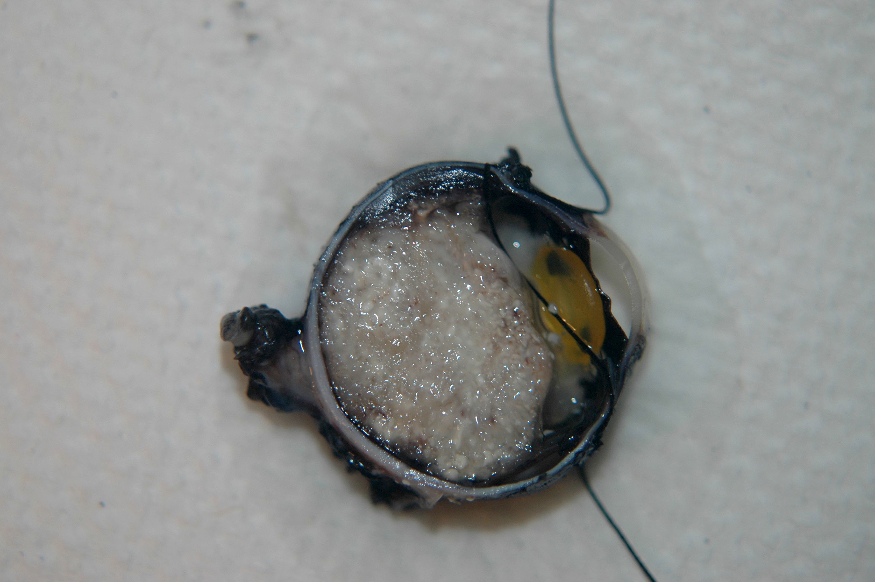
Uveal melanoma
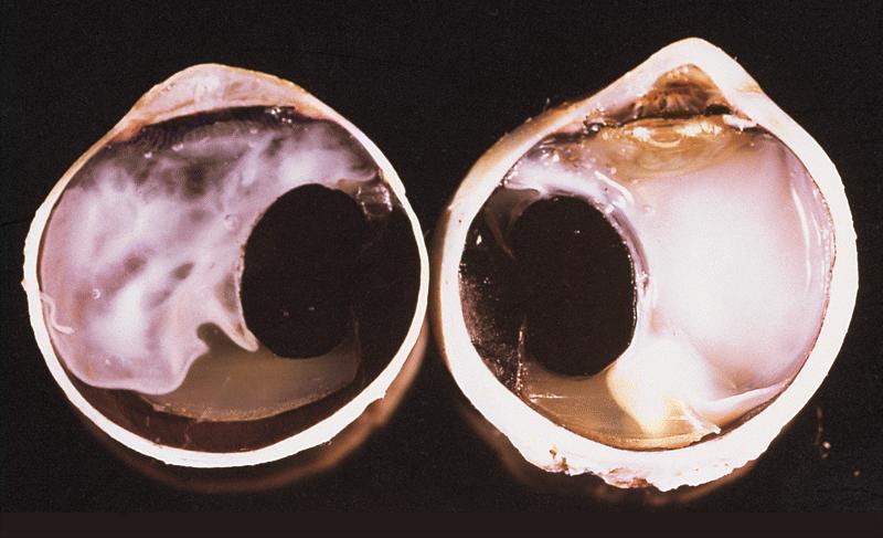
Heavily pigmented choroidal tumor
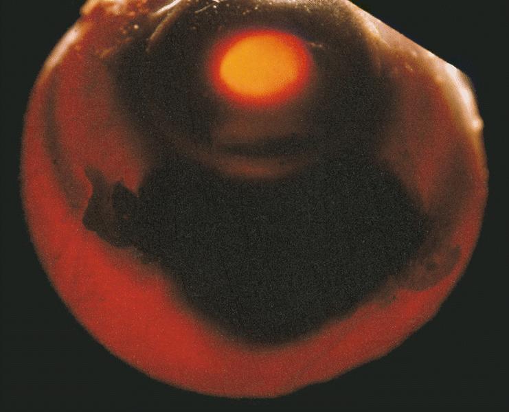
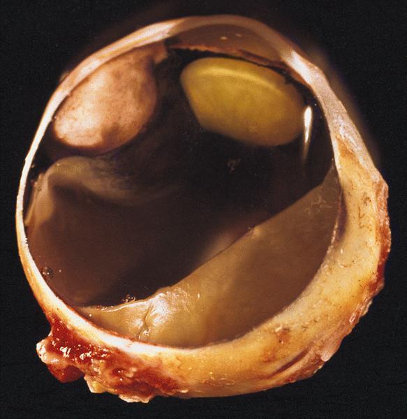
Large transillumination defect
Ruptured Bruch membrane
Subretinal hemorrhage and total retinal detachment
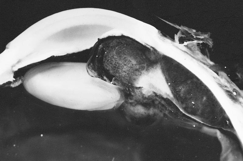
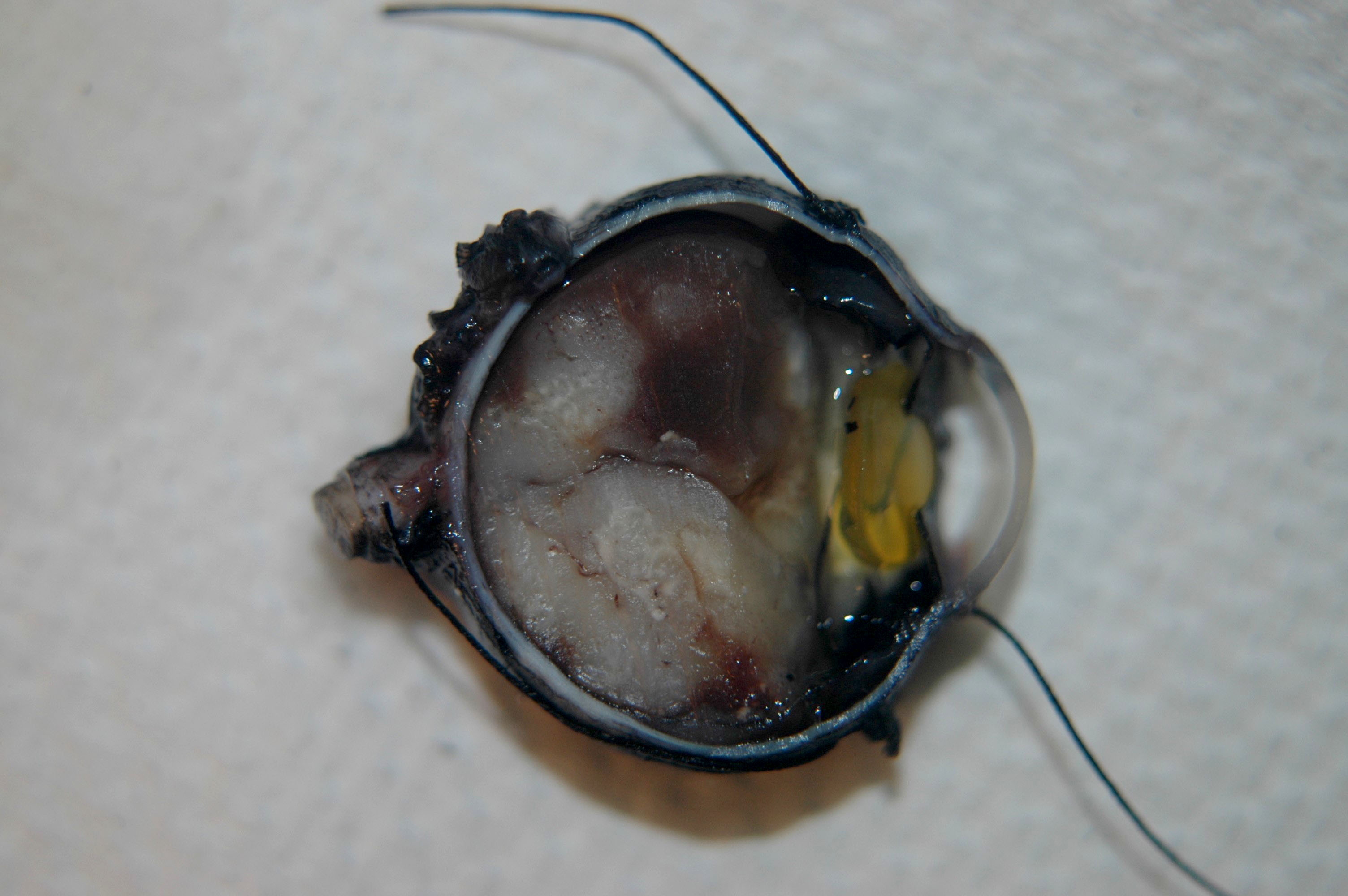
Partially organized hemorrhage
Amelanotic tumor with retinal detachment
Extraocular extension
Iridocyclectomy
Tumor of ciliary body
Subluxation of lens
Extraocular extension
Vortex vein invasion
Contributed by Lynn R. Schoenfield, M.D.

Dome shaped melanoma


Epithelioid cells

Spindle B cells

PAS

HMB45+

MelanA+

MITF+
AFIP images
Congenital diffuse anterior scleral pigmentation
Congenital diffuse posterial scleral pigmentation
AFIP images
Diffuse uveal and scleral pigmentation
Diffuse proliferation
AFIP images
Large tumor of orbital apex
AFIP images
Tumor arising adjacent to sphenoid wing
AFIP images
Tumor growth within dura
Tumor of superior lateral orbit
Invasion of sphenoid bone
Islands of meningothelial cells
Small nests with a whorled pattern
AFIP images
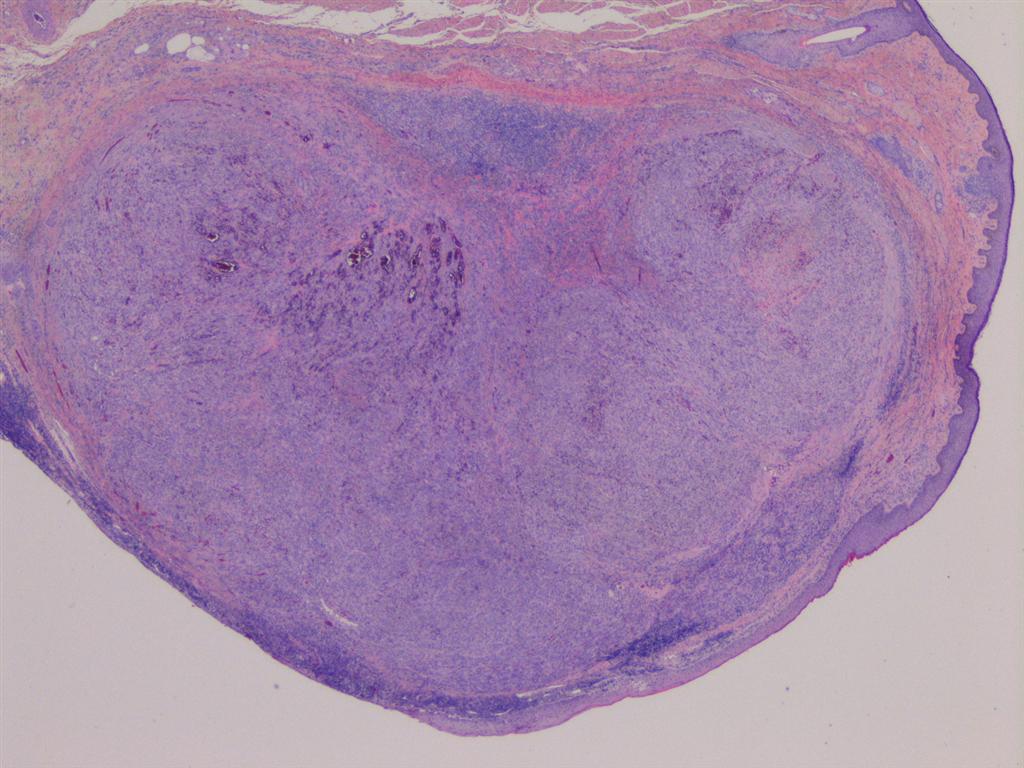
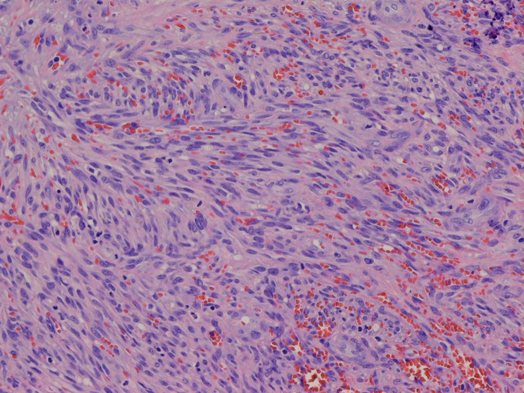
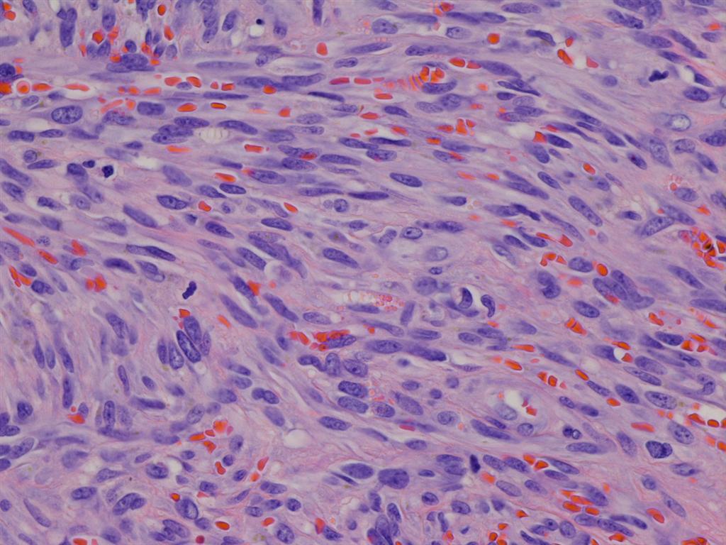
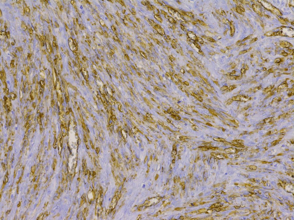
Superficial tumor of upper eyelid
AFIP images
Thickened choroid
Spindled and small polyhedral shaped melanocytes
Dysplastic nevus syndrome
Nevus cells have benign cytologic features
Images hosted on other servers:

Balloon cell nevus

Variations in pigmentation

Variations in size

Variations in location

Variations in associated clinical features

Variations in clinical appearance

Change in nevus appearance over time
AFIP images
Compound nevus: variability in cytology
Atypical compound
nevus melanocytes
in junctional area
Cystic compound nevus:
Multiple large cysts
Nevoid cells
Sheets of melanocytes
Diffuse proliferation of melanocytes
Images hosted on other servers:

Benign nevus (B), amelanotic nevus (C)

Conjunctival nevi

Epithelioid cell (clonal or inverted) nevi

Granular cell nevus
Balloon cell:


Various images

Balloon cell
compound nevus
composed of
large clear cells
AFIP images
Congenital subepithelial melanosis
Images hosted on other servers:

Cicatrizing conjunctivitis

Slit lamp photograph showing bullae
Images hosted on other servers:

Inflamed conjunctiva

Subepidermal bullous dermatosis
AFIP images
Well circumscribed tumor of left lacrimal sac
AFIP images
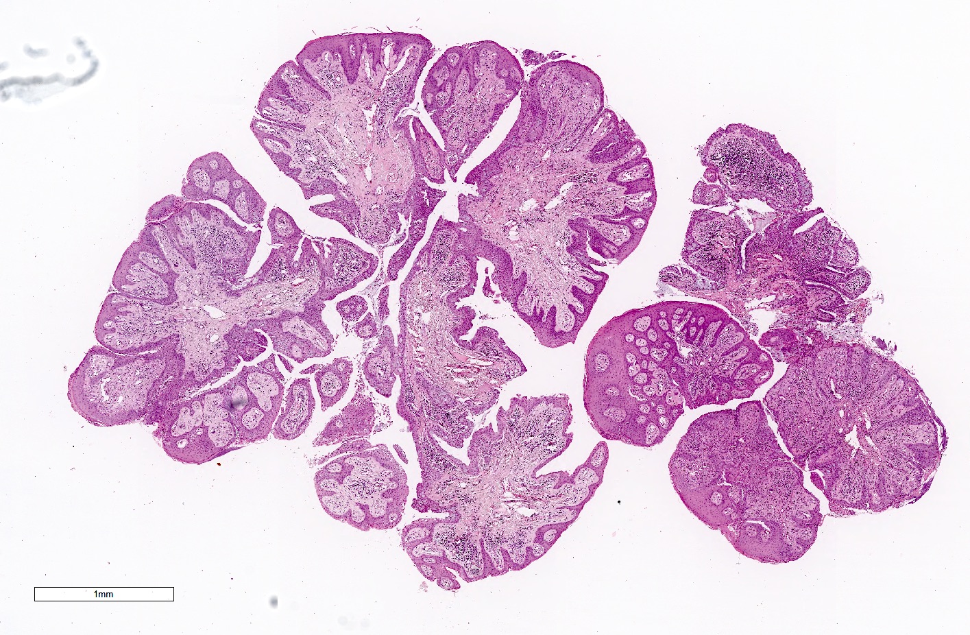
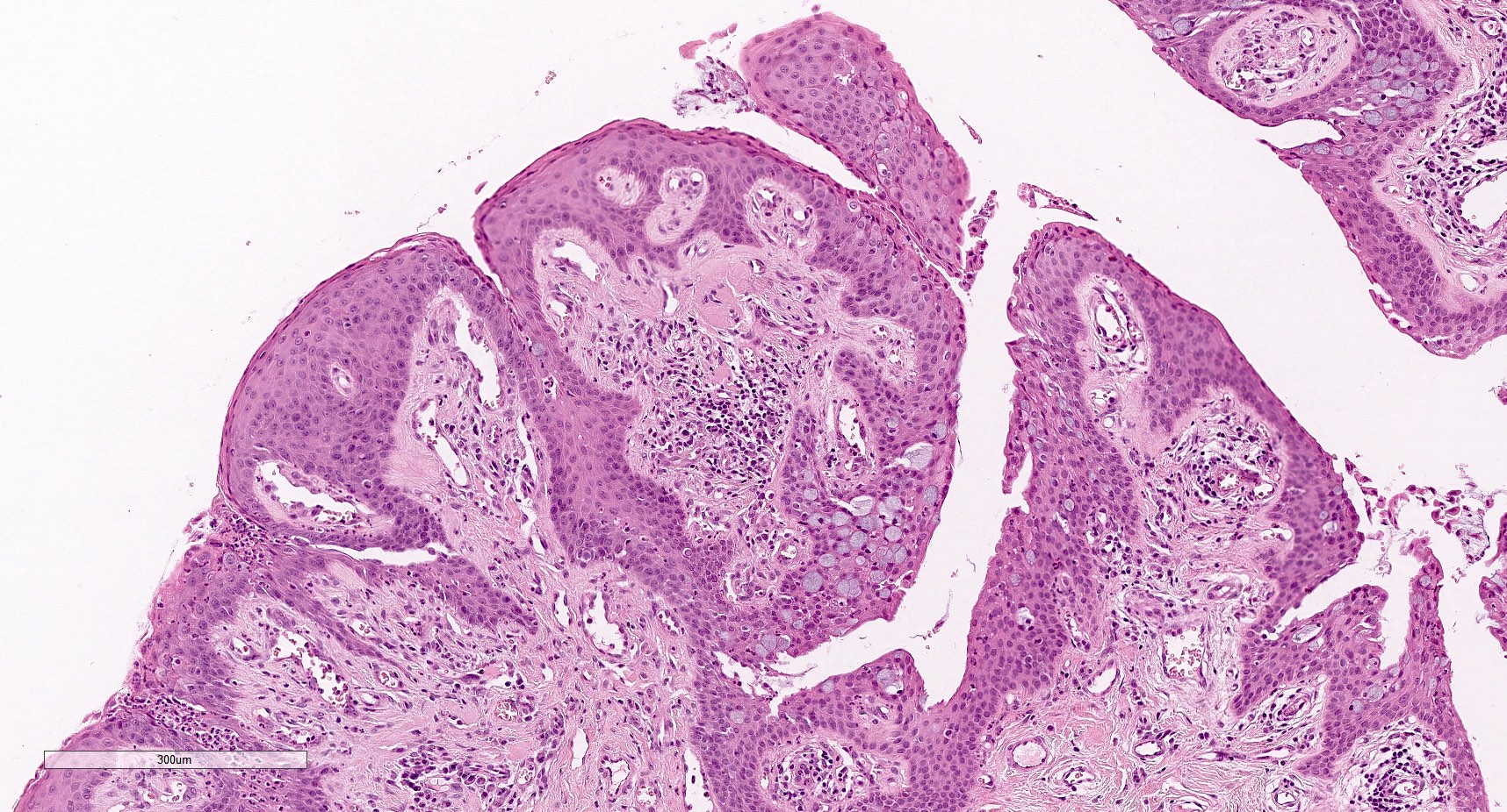
Well circumscribed papillary tumor
Well differentiated squamous epithelium
Fronds covered
by neoplastic
transitional
epithelium
Papilloma with malignant transformation
AFIP images
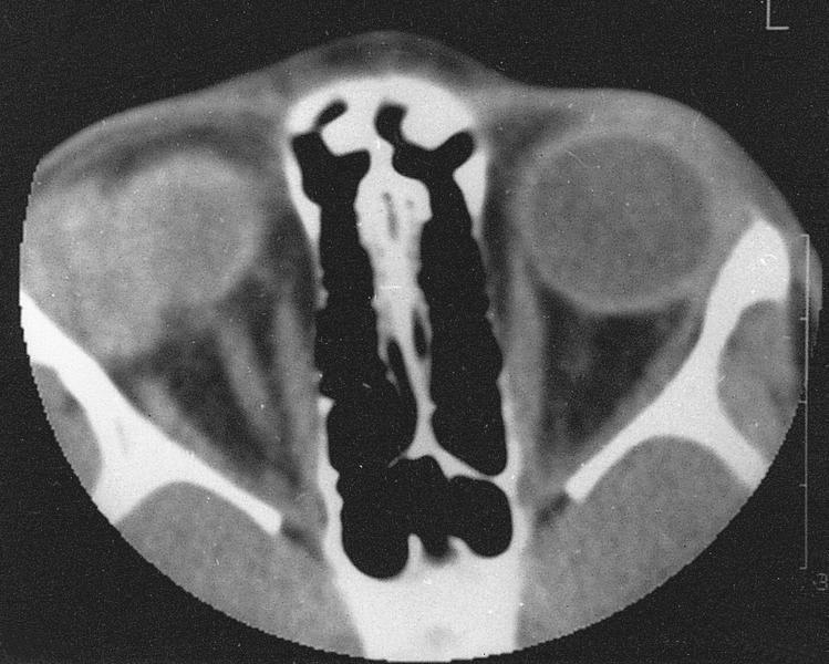
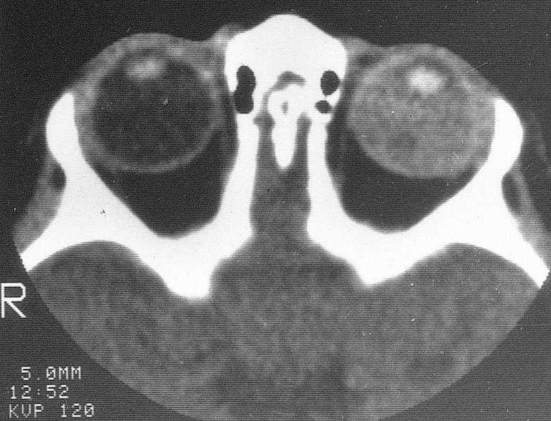
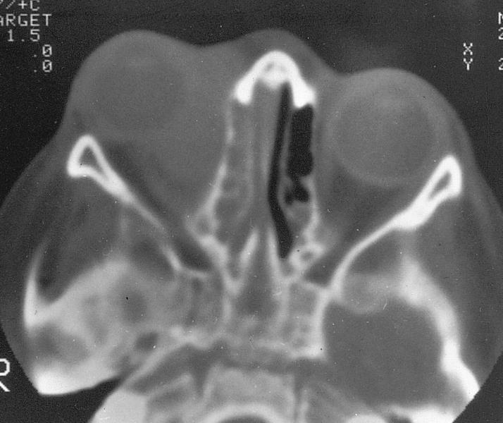
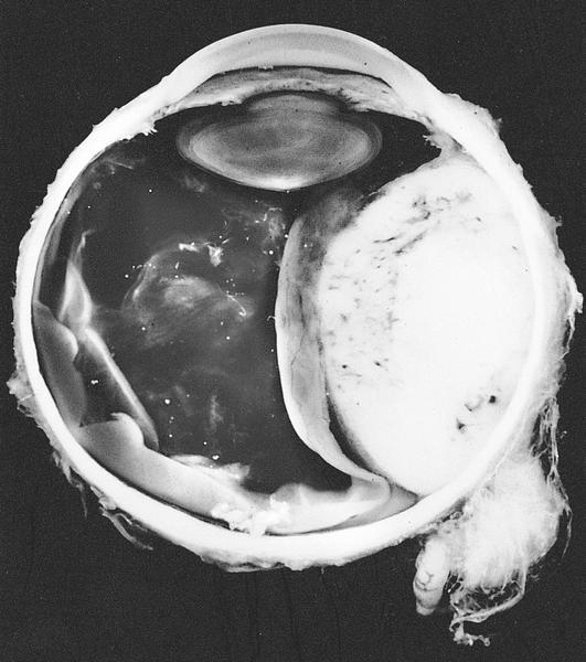
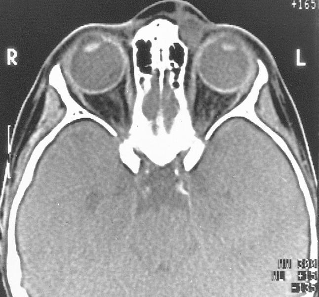
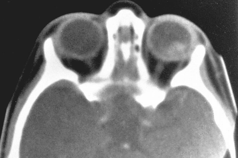
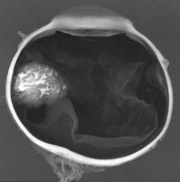
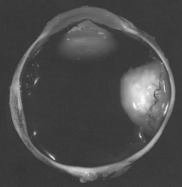
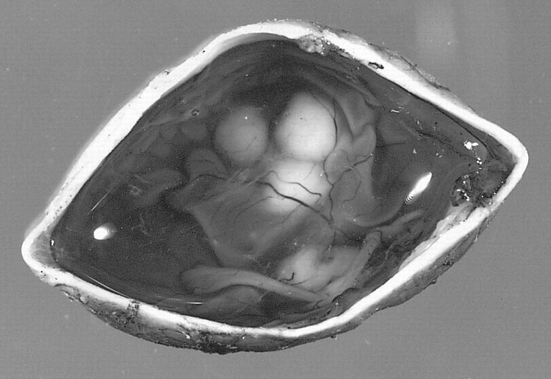
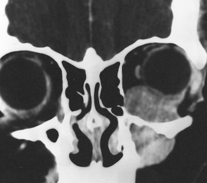
CT scan: microphthalmic eye
AFIP images
Mild microphthalmos
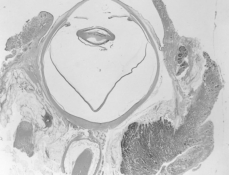
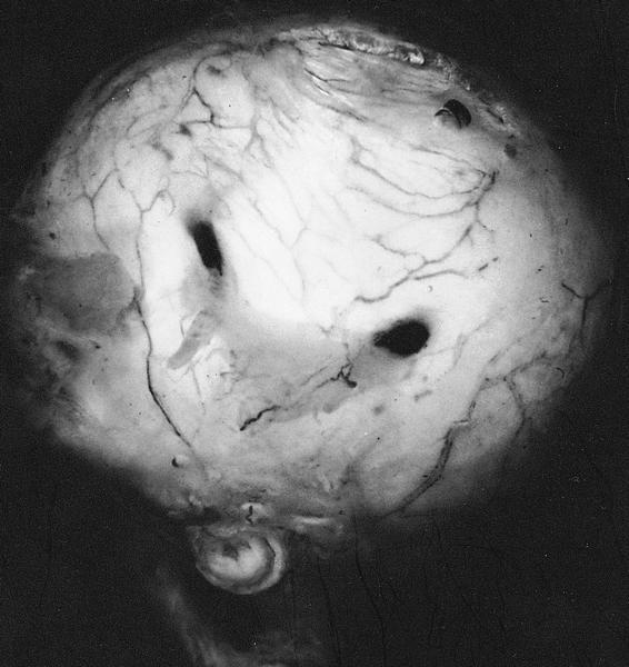
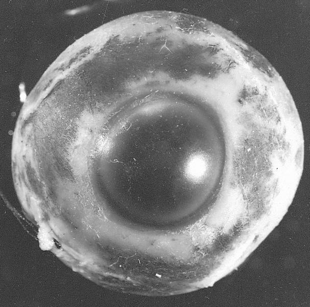
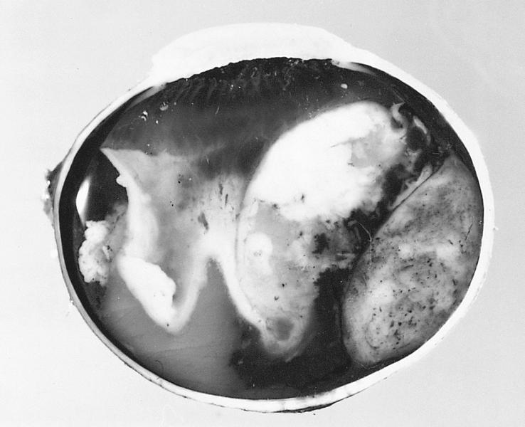
AFIP images
Wrinkling and rupture of posterior lens capsule
AFIP images
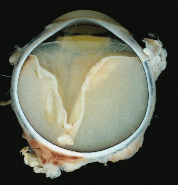
Retrolental mass
Retrolental fibrovascular mass
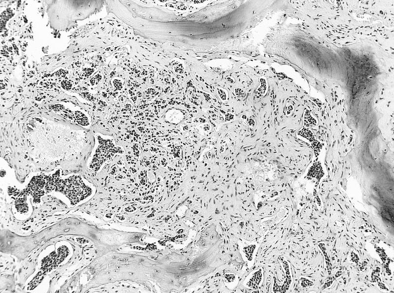
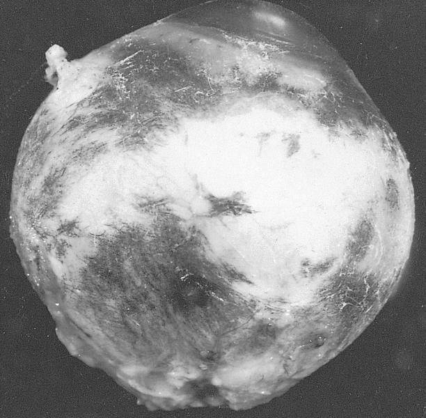
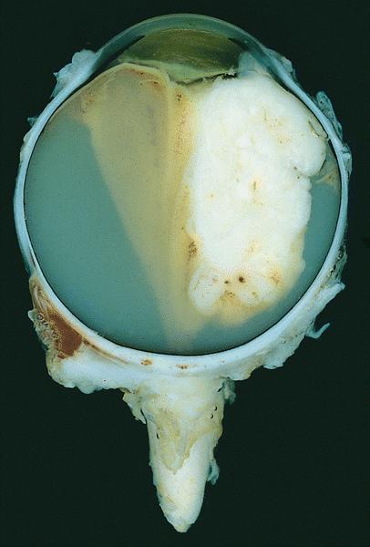
AFIP images
Islands of lens epithelial tissue
Proliferation of swollen epithelial cells
PAS+ lenticular tissue
PAS+ thick basement membrane
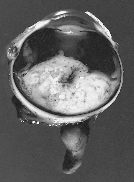
AFIP images
Regressed retinoblastoma
Disorganized intraocular contents with ossification and calcification
Fossilized tumor cells
Massive retinal gliosis
Images hosted on other servers:

Lamina propria shows
basophilic elastosis
and hyalinization
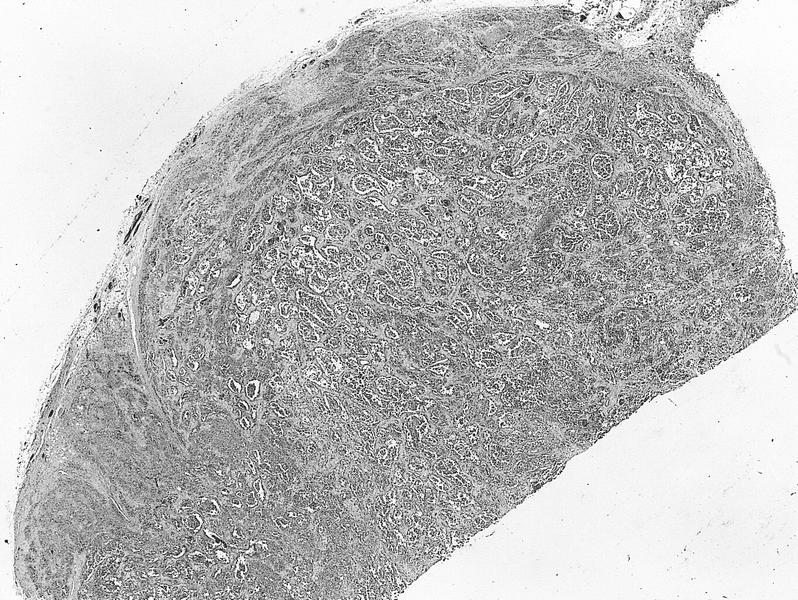
AFIP images
Well circumscribed
pseudoencapsulated
lacrimal gland
tumor
Islands of epithelial cells, myxoid stroma and cysts
Double layer of epithelial cells within a myxoid stroma
Chondroid differentiation
With carcinoma
Large round nodule of pleomorphic adenoma
Benign ducts within
myxoid stroma;
adenocarcinoma
With adenoid cystic carcinoma
Contributed by Arturo Grau, M.D., M.Sc.
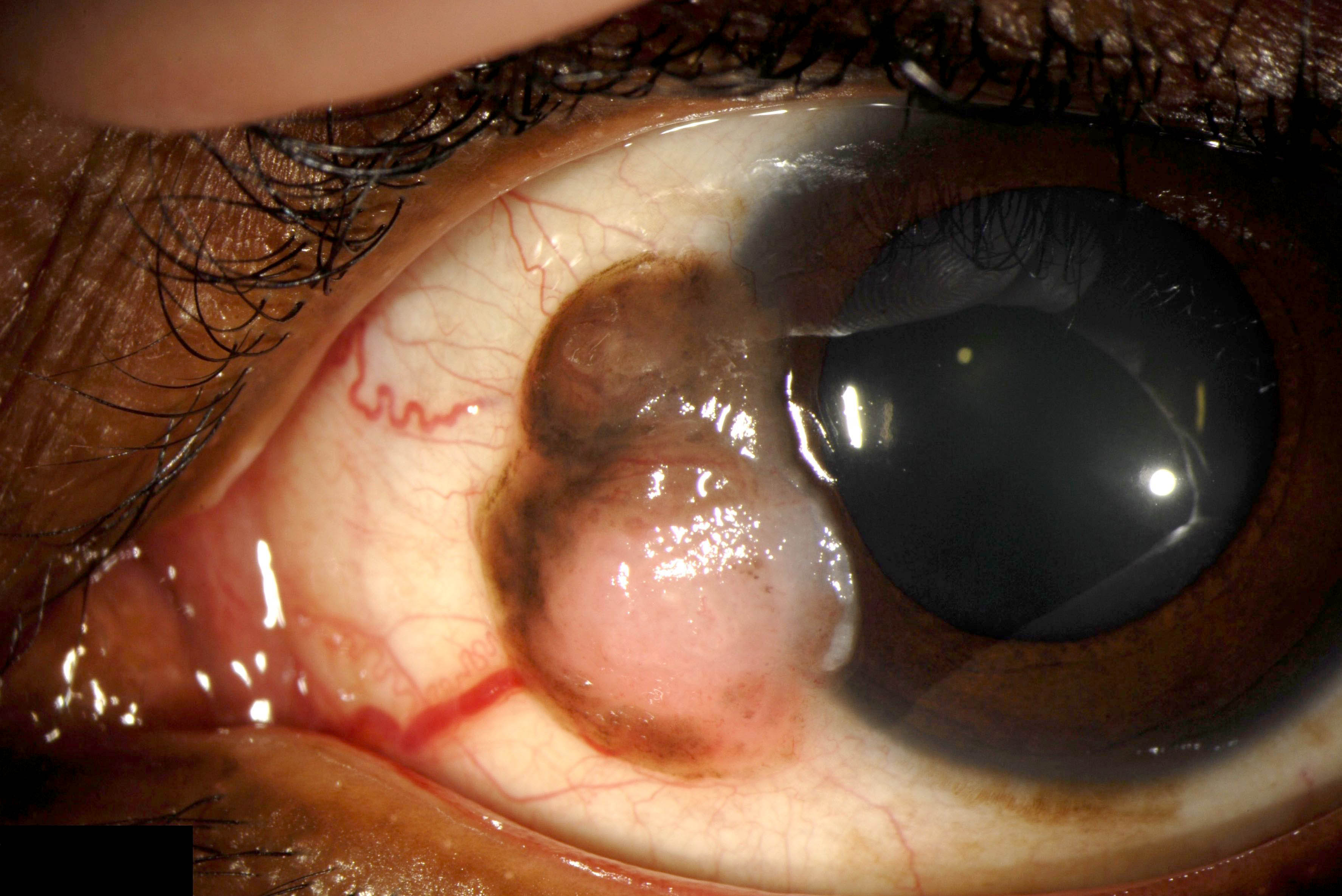
Corneolimbal fleshy lesion
Fibrovascular pattern
Contributed by Pablo Zoroquiain, M.D.
Elastotic degeneration
Corneoconjunctival
junction
With cornea and conjunctiva
Elastotic infiltrate
Contributed by Marta Couce, M.D., Ph.D. and AFIP images
MRI, intraocular mass, exophytic
MRI sagittal, heterogeneous signal
Ovoid retinal tumor
Focal calcification
AFIP images
Leukocoria
Strabismus without leukocoria
Focal opacified areas
Obscured retinal vessels
Retinal tumor invades subretinal space
Images hosted on other servers:

Ultrasound B scan / RetCam

Fundoscopic image demonstrating tumor
Contributed by Marta Couce, M.D., Ph.D. and AFIP images
Chalky tumor posterior chamber
Exophytic growth, focal hemorrhage
Massive extraocular extension
Destruction of anterior segment
Large areas of necrosis
Calcification
3 small retinal tumors
Choroidal invasion
Total exudative retinal detachment
Optic nerve enlargement
Contributed by Marta Couce, M.D., Ph.D.
Retinoblastoma infiltrating normal retina
Retinoblastoma replacing neurosensory retina
Optic nerve infiltration
Extensive tumor necrosis
Flexner-Wintersteiner rosettes
Ki67
AFIP images
Nodules of tumor cells
Anterior chamber involvement
Viable tumor cells
Adjacent endophytic tumor

Sleeve pattern and rosettes
Flexner-Wintersteiner rosettes
Homer Wright rosettes

Poorly differentiated tumor cells
Azzopardi phenomenon
Spindle shaped glia
Benign cytology
Benign cytology
Invasion of optic nerve head
Invasion to lamina cribrosa
Invasion of optic nerve parenchyma posterior
Invasion of full thickness of choroid
AFIP images
Clumps and individual tumor cells
Images hosted on other servers:

Cluster and
rosette formation /
PAS positive
granules
AFIP images
Bulbous process of fleurette
AFIP images
Uniform cells without necrosis
Small, round, hyperchromatic nuclei
AFIP images
CT shows large tumor of orbital floor invading maxillary sinus
AFIP images
Botryoid tumor beneath the conjunctiva
AFIP images
Alveolar pattern
Alveolar spaces contain anaplastic cells
Alveolar pattern and large anaplastic giant cells
Rhabdomyoblasts
Embryonal pattern
Bundles of spindle
cells with hyperchromatic
nuclei and numerous
mitotic figures
Cross striations
Myxoid pattern
Round and tadpole shaped rhabdomyoblasts
AFIP images

Long cytoplasmic
processes and atypical
nuclei (inset - muscle
specific actin positive)
AFIP images
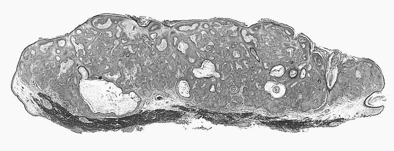

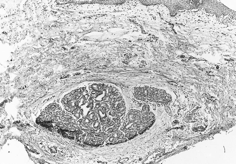
Plaque-like lesion with cords of basaloid cells and numerous horn cysts
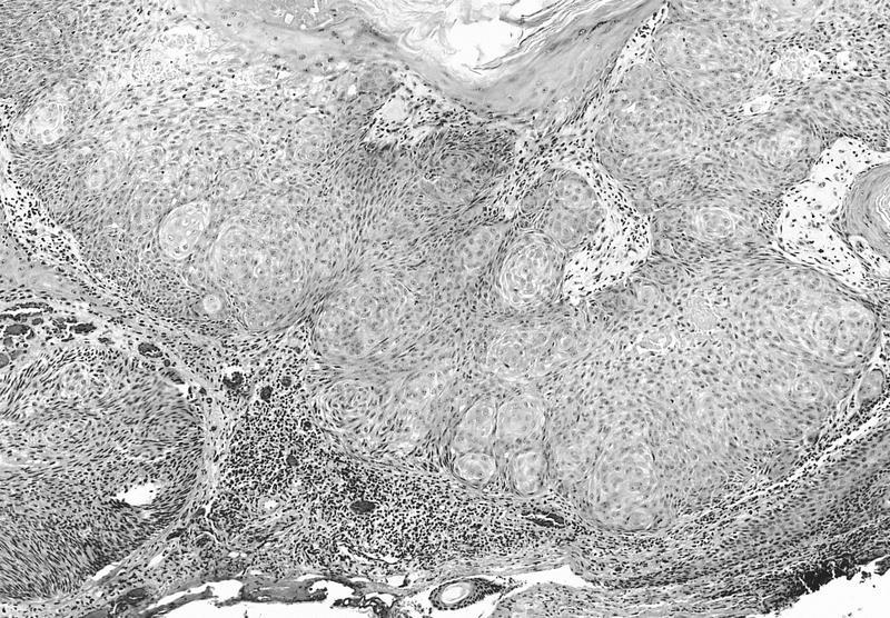
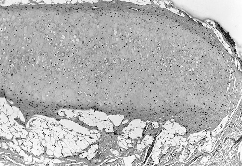
Acanthotic epithelium with massive hyperkeratosis forming a cutaneous horn
AFIP images
Small polyhedral cells surround vascular spaces
Vascular channels have a staghorn pattern
Cells are polyhedral and spindled, with mild atypia
Metastatic liver nodule
Images hosted on other servers:

Nodular tumor with prominent vascularity at limbus
AFIP images
Large limbal tumor invades anterior chamber
Tumor has destroyed eye and invaded orbit
AFIP images
Early invasion with corneal involvement
Well differentiated tumor with deep invasion
Metastasis to preauricular node and parotid gland
Sarcomatous pattern
Deep scleral and corneal invasion
Images hosted on other servers:

Thick layer of parakeratosis and microinvasion

Lobules of invasive keratinizing carcinoma

Papillomatous pattern
AFIP images
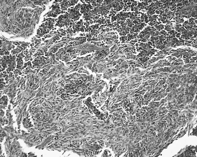
Squamous cell carcinoma - spindle cell type
Atrophic epidermis over a highly infiltrative tumor
Spindled cells resemble sarcoma
Images hosted on other servers:

Pedunculated red-orange mass of caruncle

Pedunculated vascular lesion with smooth surface

Sessile lesion
Contributed by Pablo Zoroquiain, M.D.
Finger-like projections of the neoplastic process
Acanthotic epithelium with fibrovascular cores
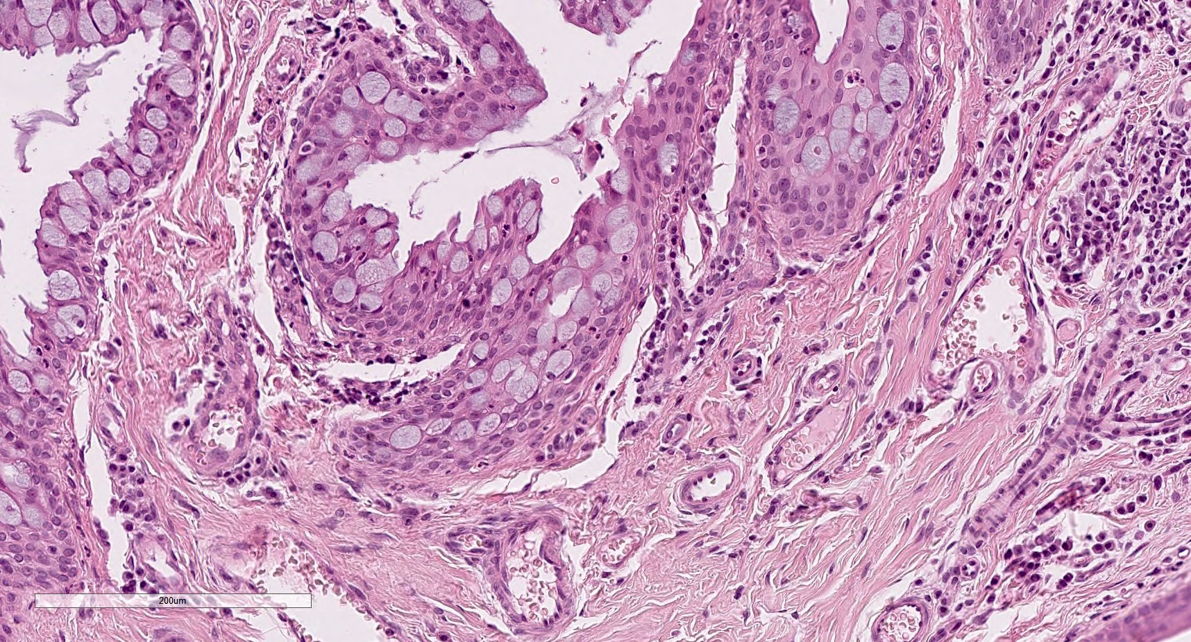
Goblet cells present
Base of the tumor

AFIP images
Gross extraocular extension
Contributed by Martin Hyrcza, M.D., Ph.D., M.Sc.
Scleral invasion

Extrascleral extension
Images hosted on other servers:

Pale yellow lesion
Images hosted on other servers:

Cyst lined by
squamous epithelium
with sebaceous glands
in cyst wall
Images hosted on other servers:

Prolapsed orbital fat
Contributed by Ivy John, M.D.

Herniated orbital fat
Images hosted on other servers:

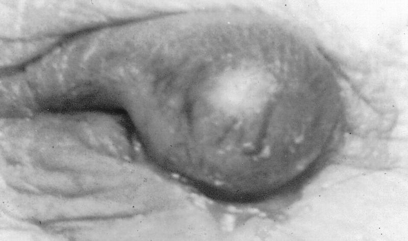
Halo sign
Images hosted on other servers:

Visible temporal artery
Contributed by José Tomás Peña, M.D., Pablo Zoroquiain, M.D. and @MirunaPopescu13 on Twitter

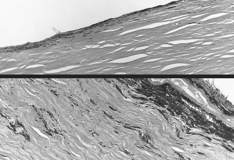
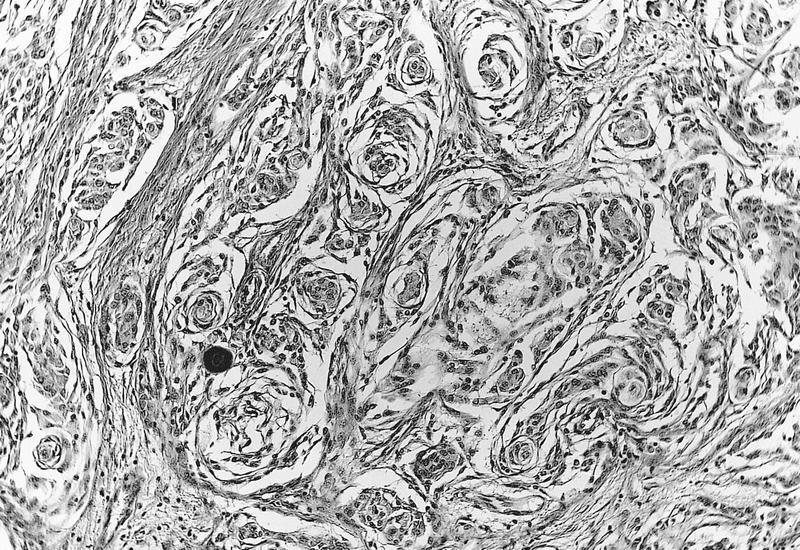
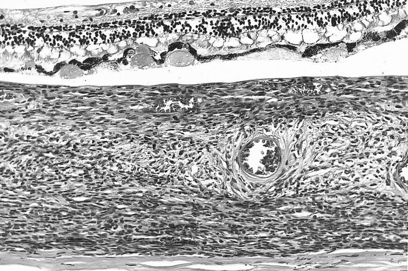
Segmental inflammation
Intimal hyperplasia

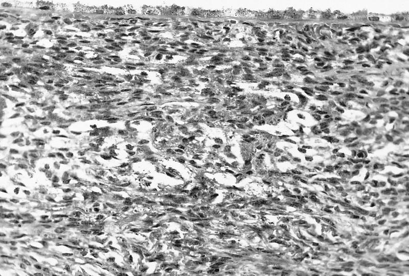
Transmural inflammation

Giant cells
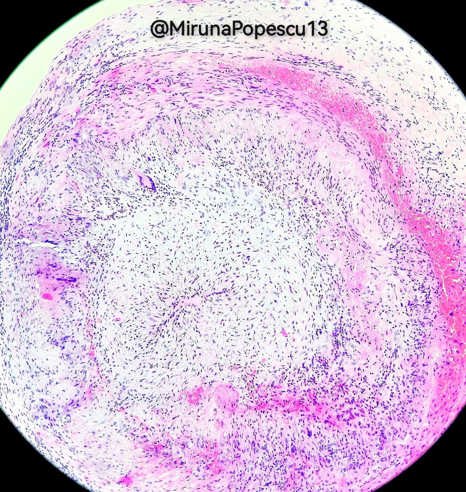
Temporal arteritis

Loss of elastic fibers

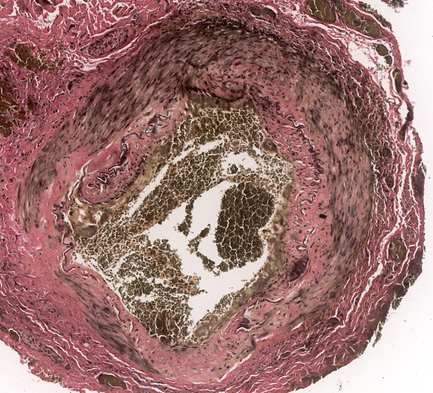
Loss of elastic fibers
Vasculitis Foundation giant cell arteritis webinar
Giant cell arteritis
by Nicholas J. Volpe,
European Society of Ophthalmology
Temporal artery biopsy technique
Giant cell arteritis (temporal arteritis) pathology by Jerad Gardner, M.D.
Images hosted on other servers:

WHO grading

Presence of five or more follicles

Thickening of the tarsal conjunctiva

Scarring in the tarsal conjunctiva

Evidence of recent removal of inturned eyelashes

Corneal opacity over the pupil
Images hosted on other servers:
Chlamydia inclusions
AFIP images
White sclerotic vitreous mass
AFIP images
Vitreous mass contains collagen (trichrome stain)
Acidophilic material and scattered eosinophils
Contributed by Marta Couce, M.D., Ph.D.


Conjunctival oncocytoma
Conjunctival schwannoma


Cyst of the conjunctiva

Subepithelial conjunctival nevus

Compound conjunctival nevus
Benign epithelial melanosis of the conjunctiva
Conjunctival squamous cell carcinoma
Adenosquamous carcinoma

Conjunctival oncocytoma
Contributed by Gabrielle Yeaney, M.D.
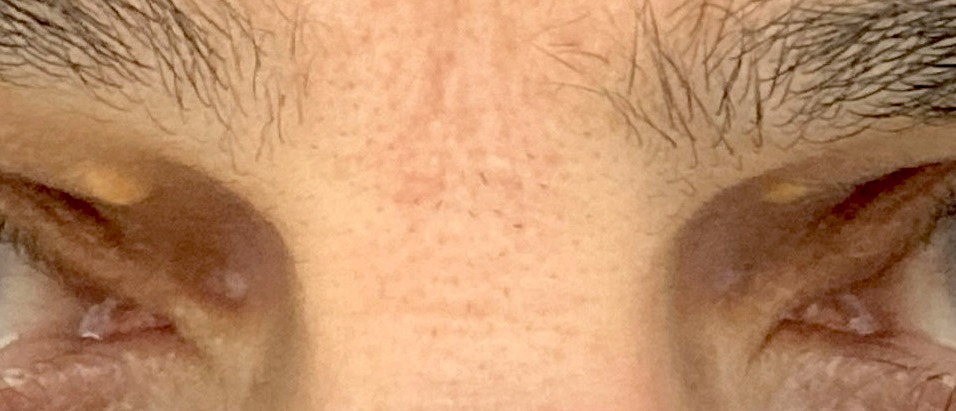
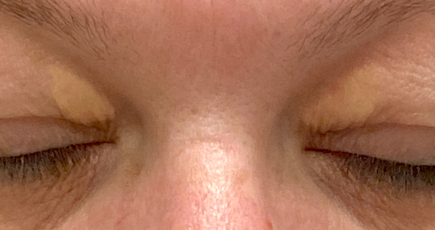
Bilateral eyelids
Contributed by Gabrielle Yeaney, M.D.

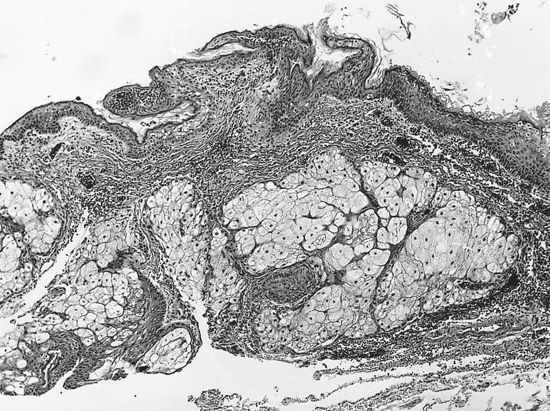
Foamy cells superficial dermis

Lipid laden histiocytes

Extension around neurovascular bundle

Touton multinucleated cells
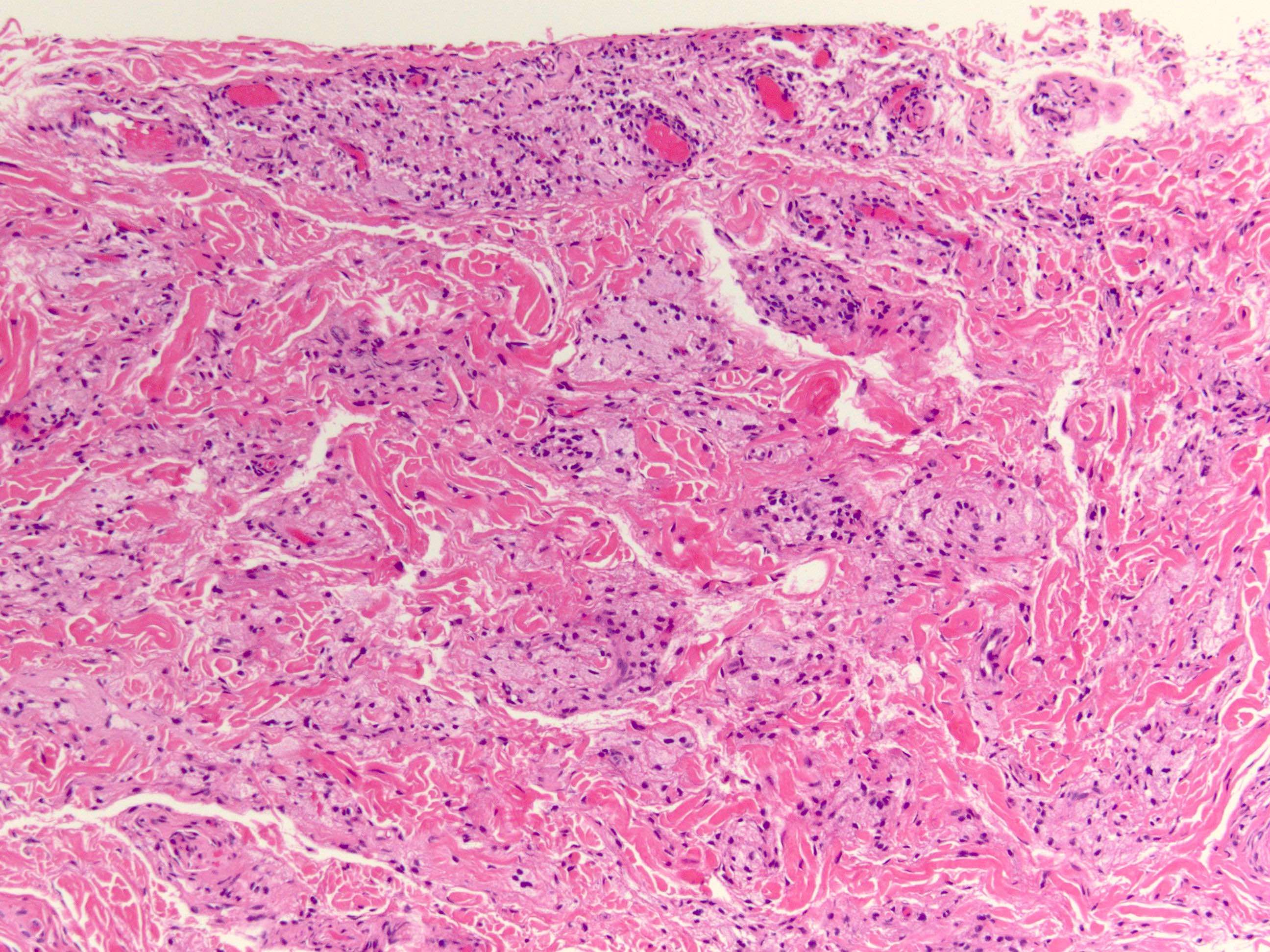
Perivascular clusters
Xanthelasma (xanthoma of eyelid)
Eagle: 2016
Folberg: 2020
Font: 2006
Heegaard : 2015
IARC: 2018
Shields: 2015
Thompson: 2022
Find related Pathology books: eye, head & neck/endocrine
