


Images hosted on other servers:

Fallopian tube anatomy

Fallopian tube anatomy and ligaments

Fallopian tube blood supply
Contributed by Brandon D. Metcalf, M.D.
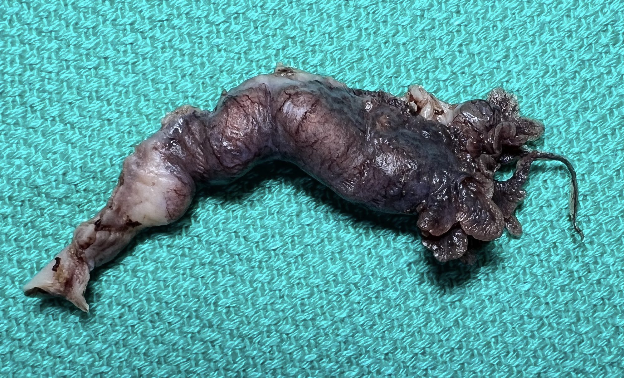
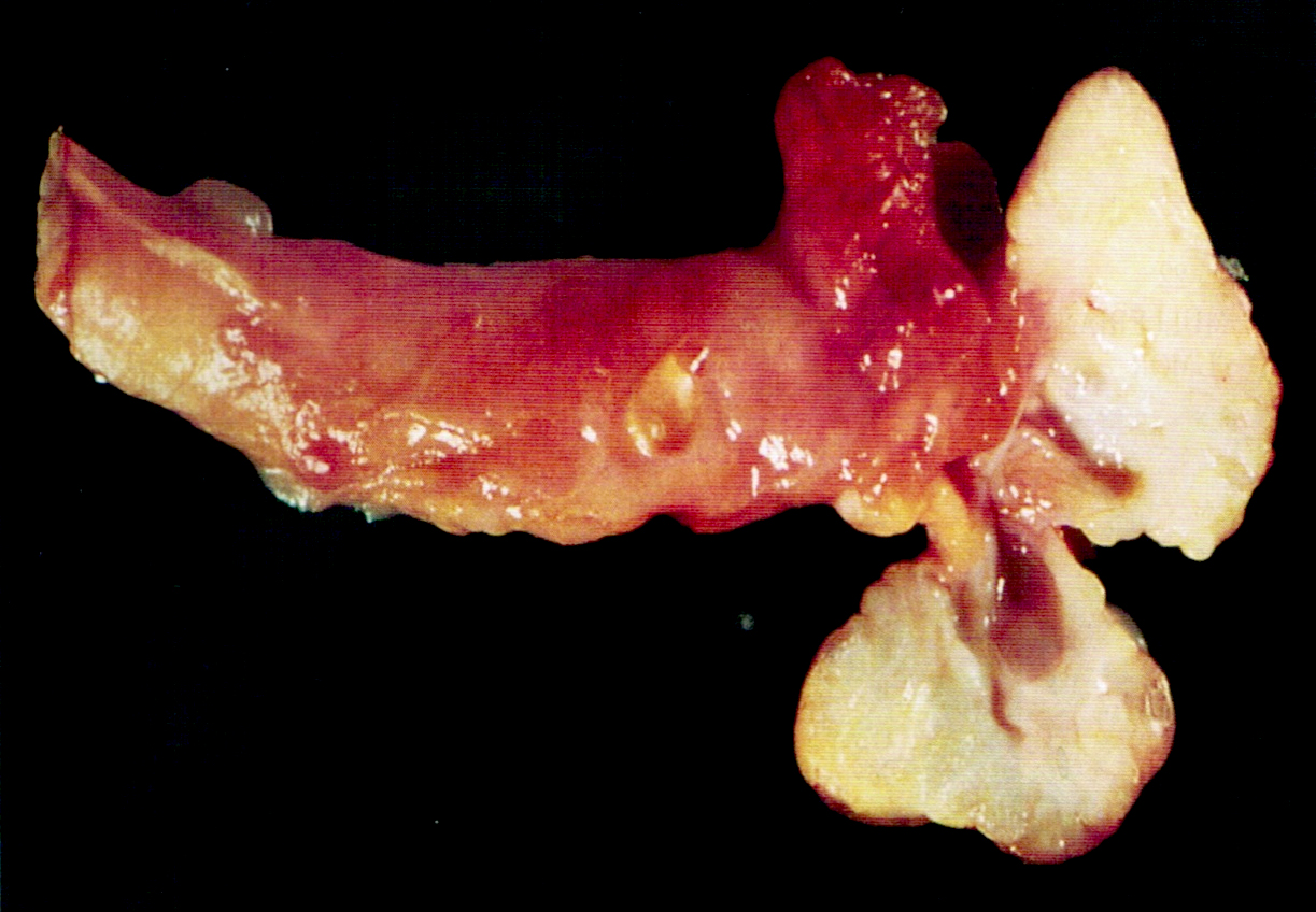
Salpingectomy specimen
Contributed by Brandon D. Metcalf, M.D. and Nicole D. Riddle, M.D.
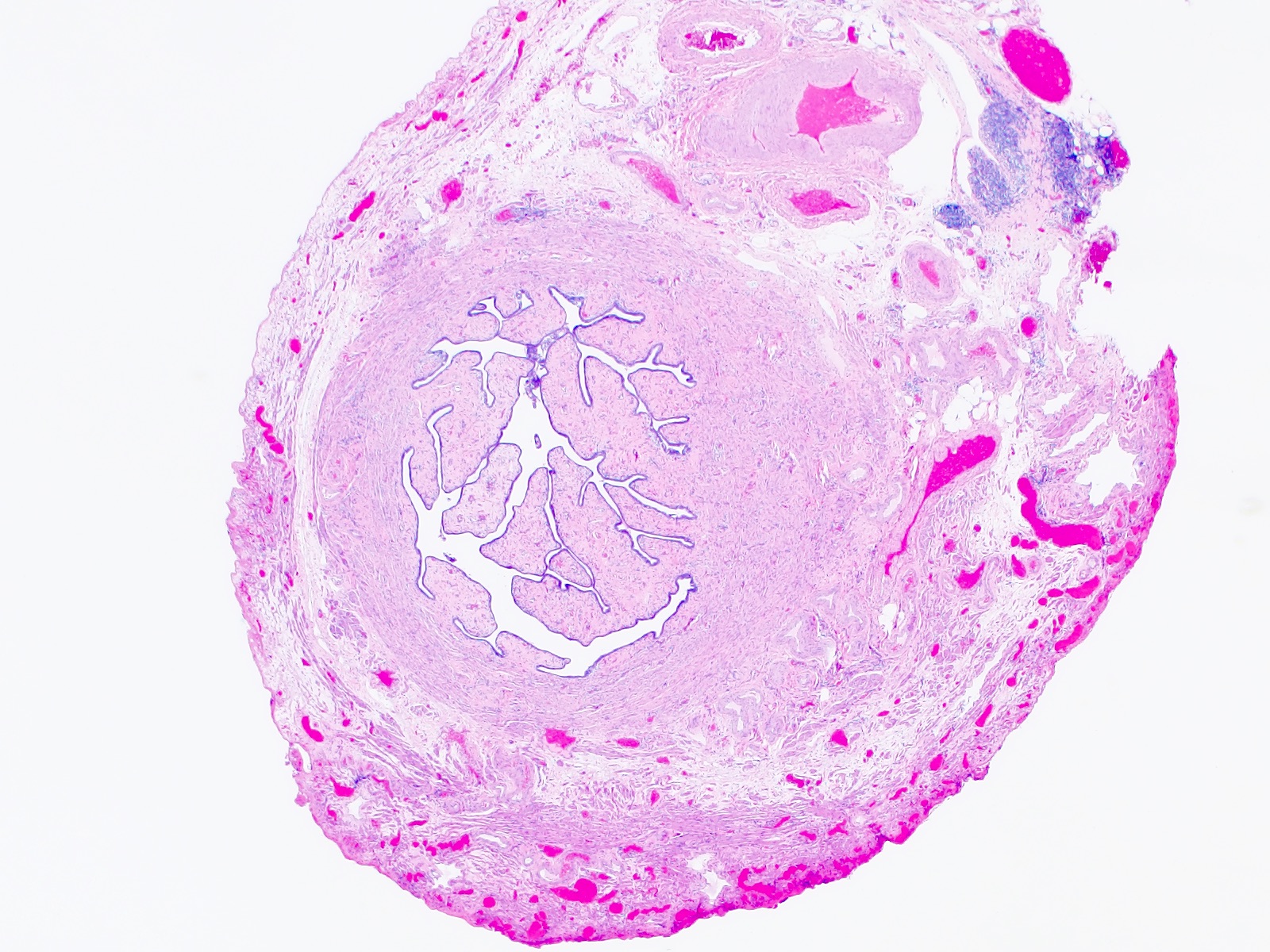
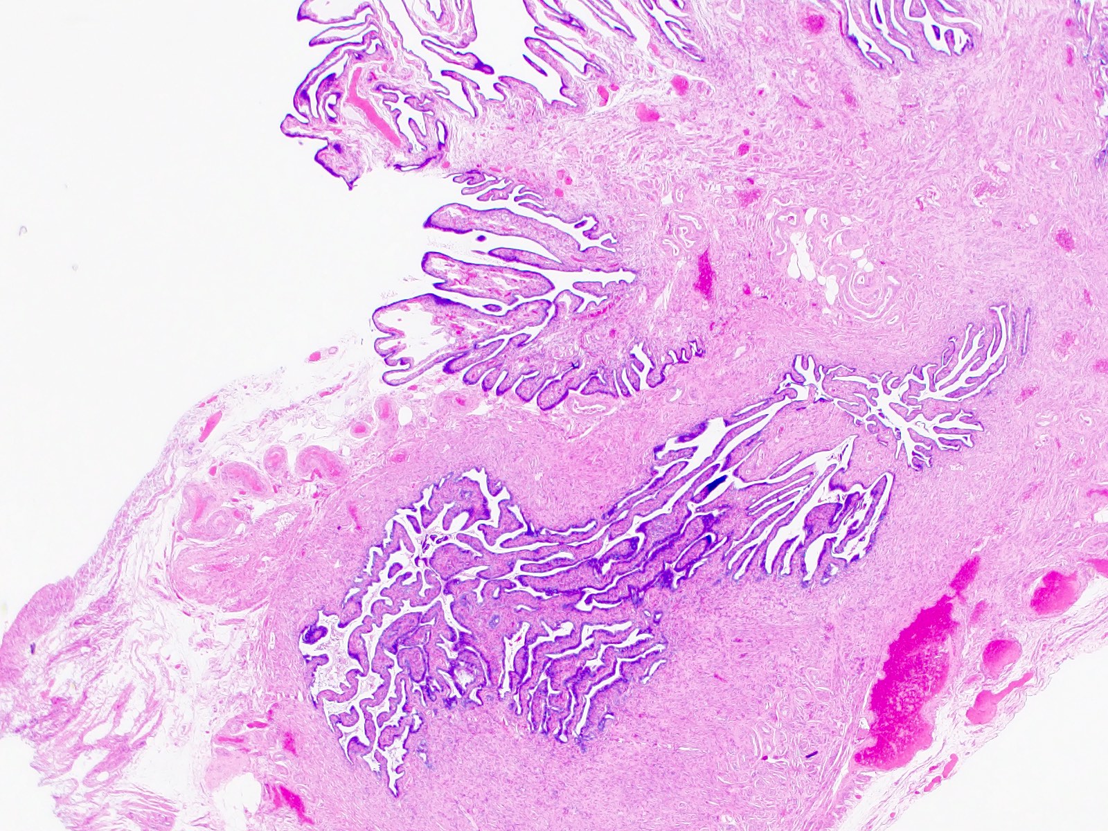
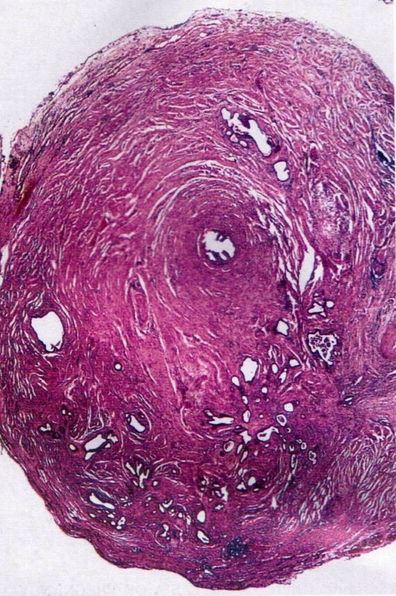
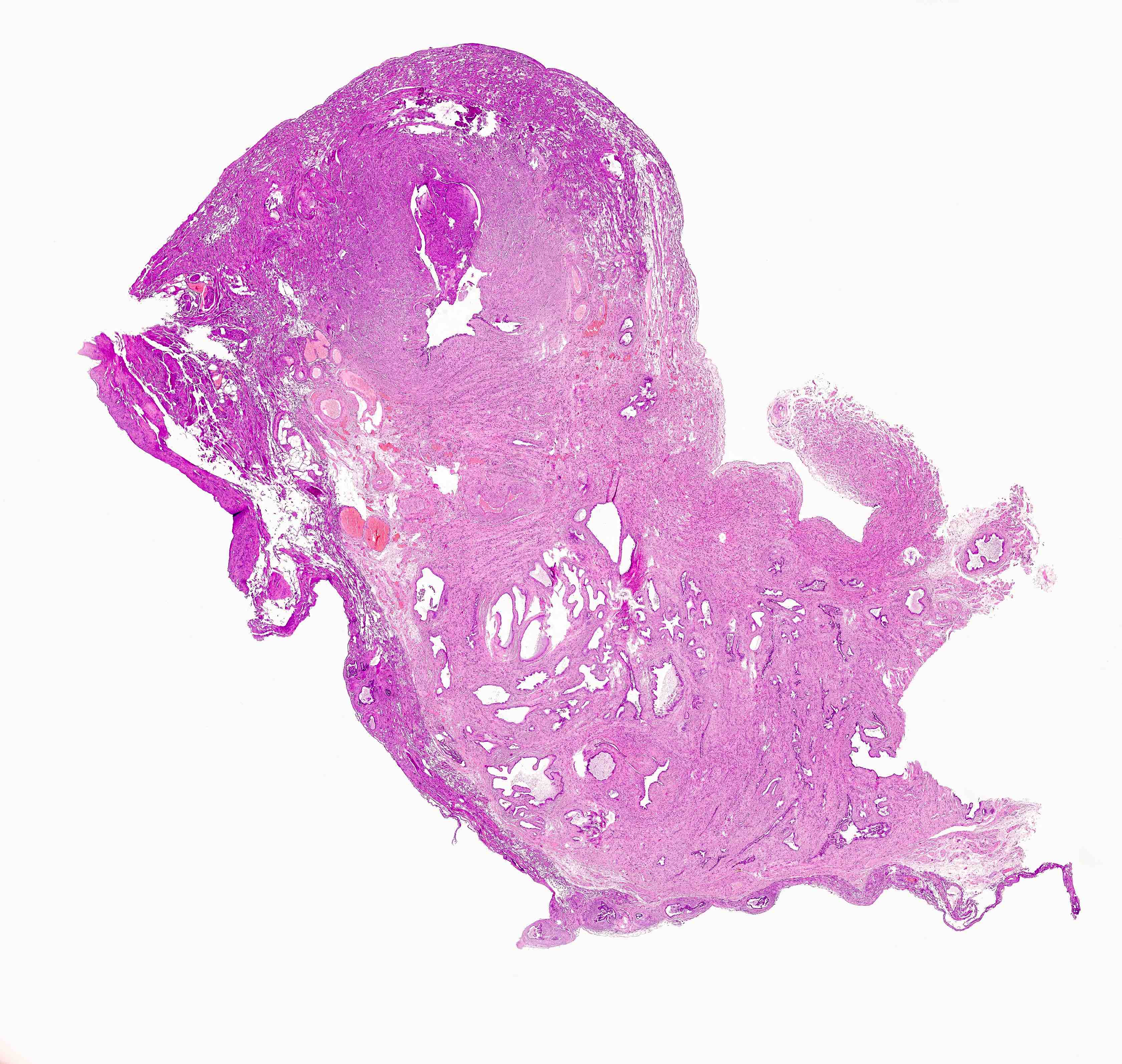
Fallopian tube cross section, postmenopausal
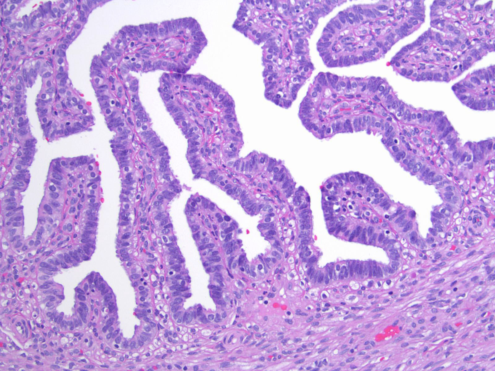
Mucosal plicae
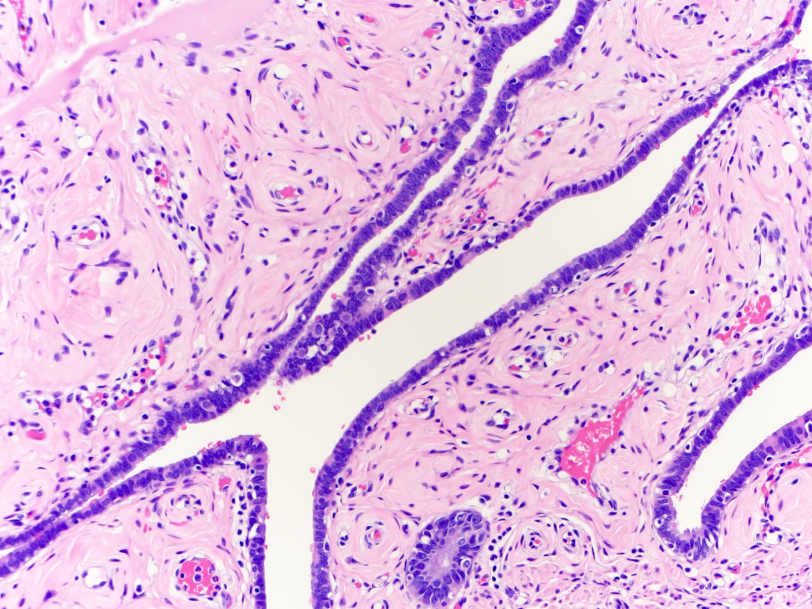
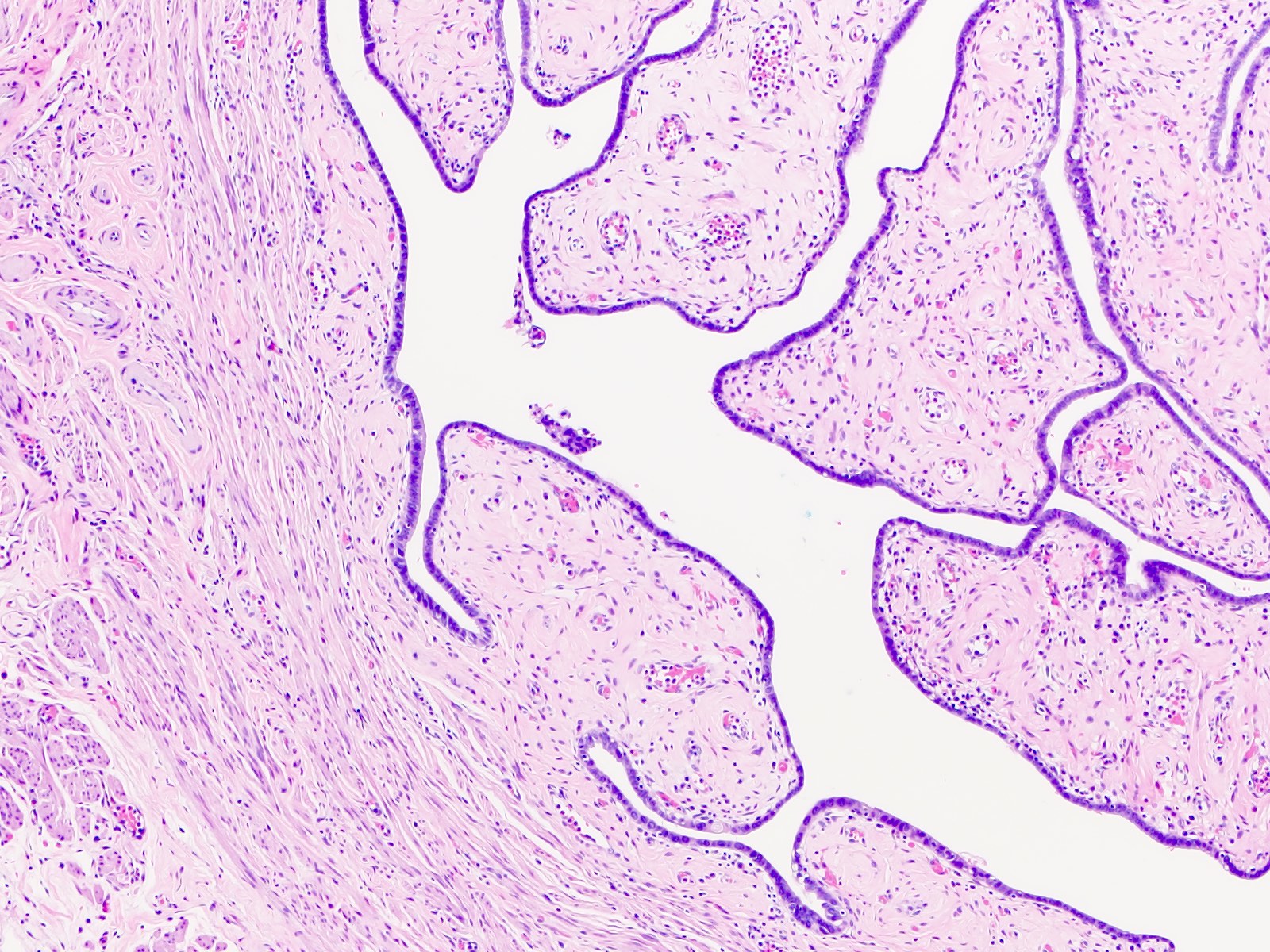
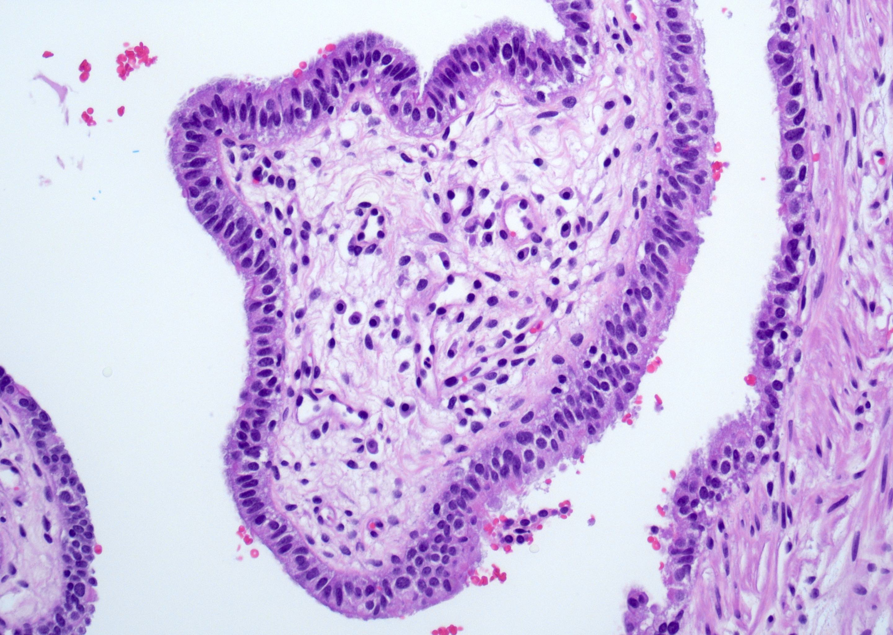
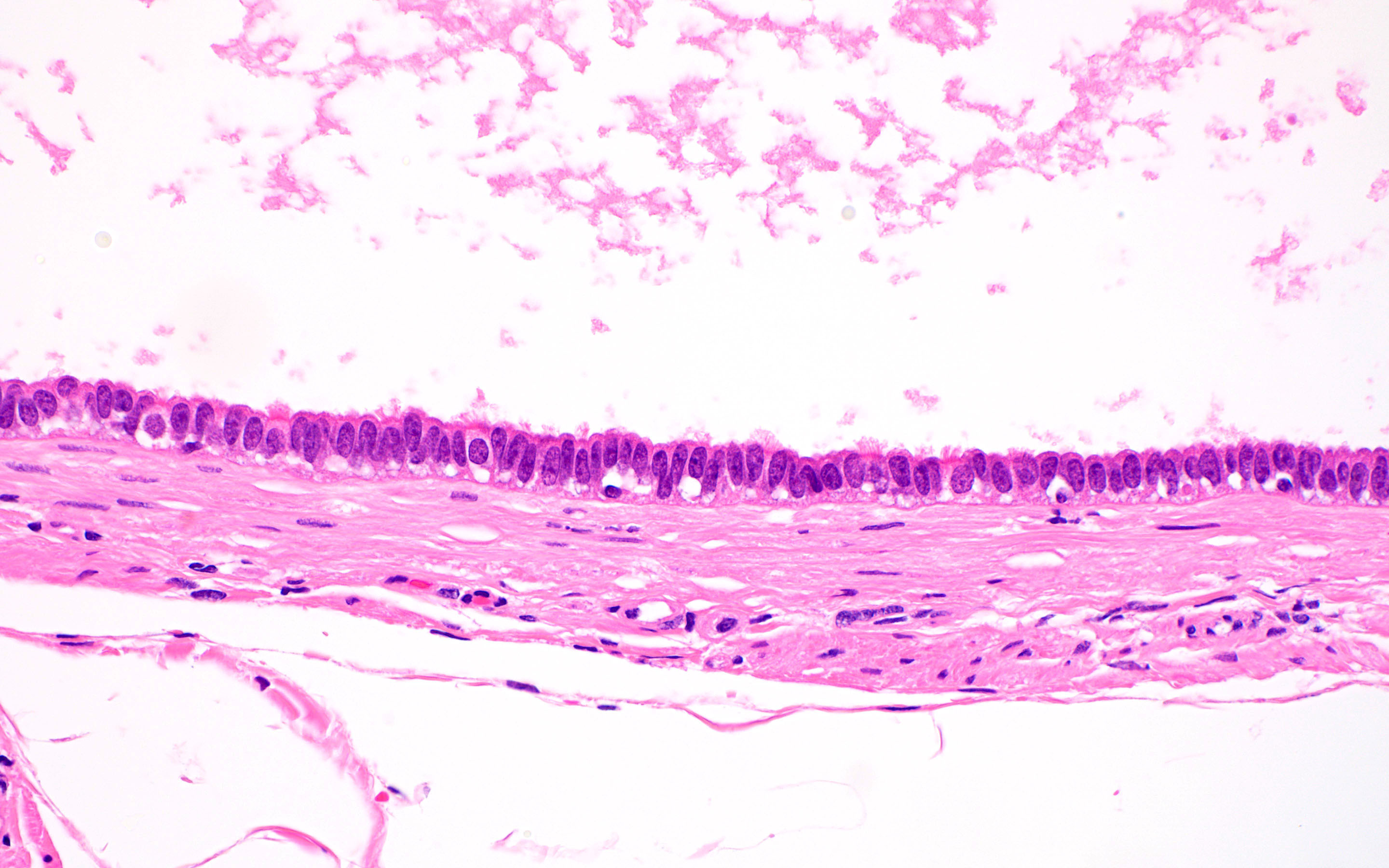
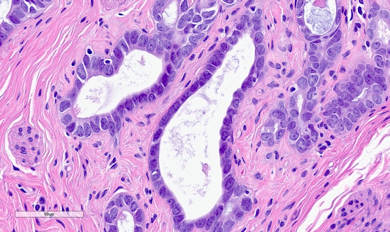
Fallopian tube epithelium
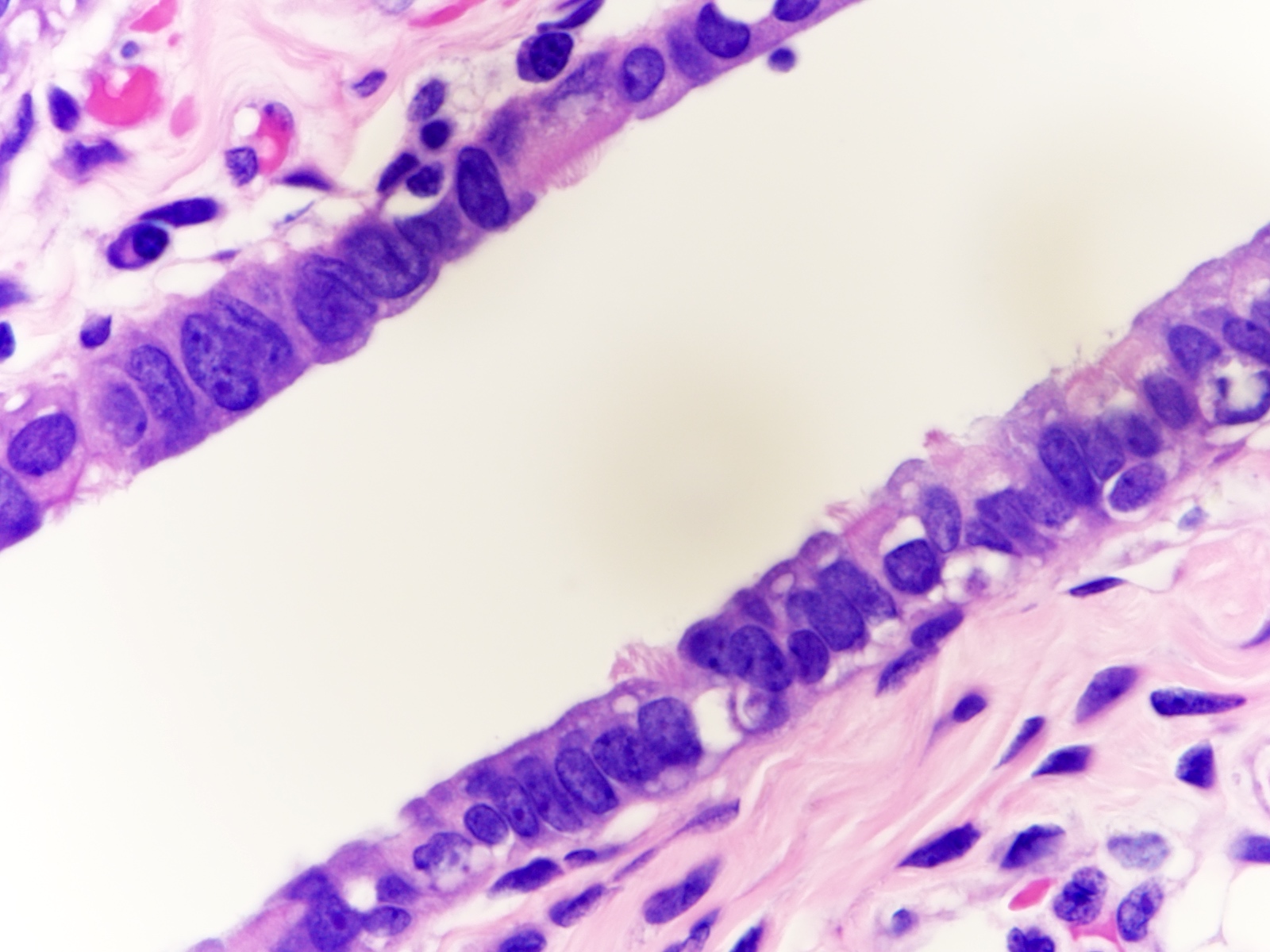
Simple columnar epithelium
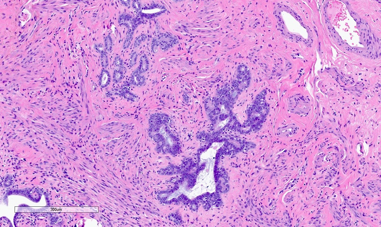
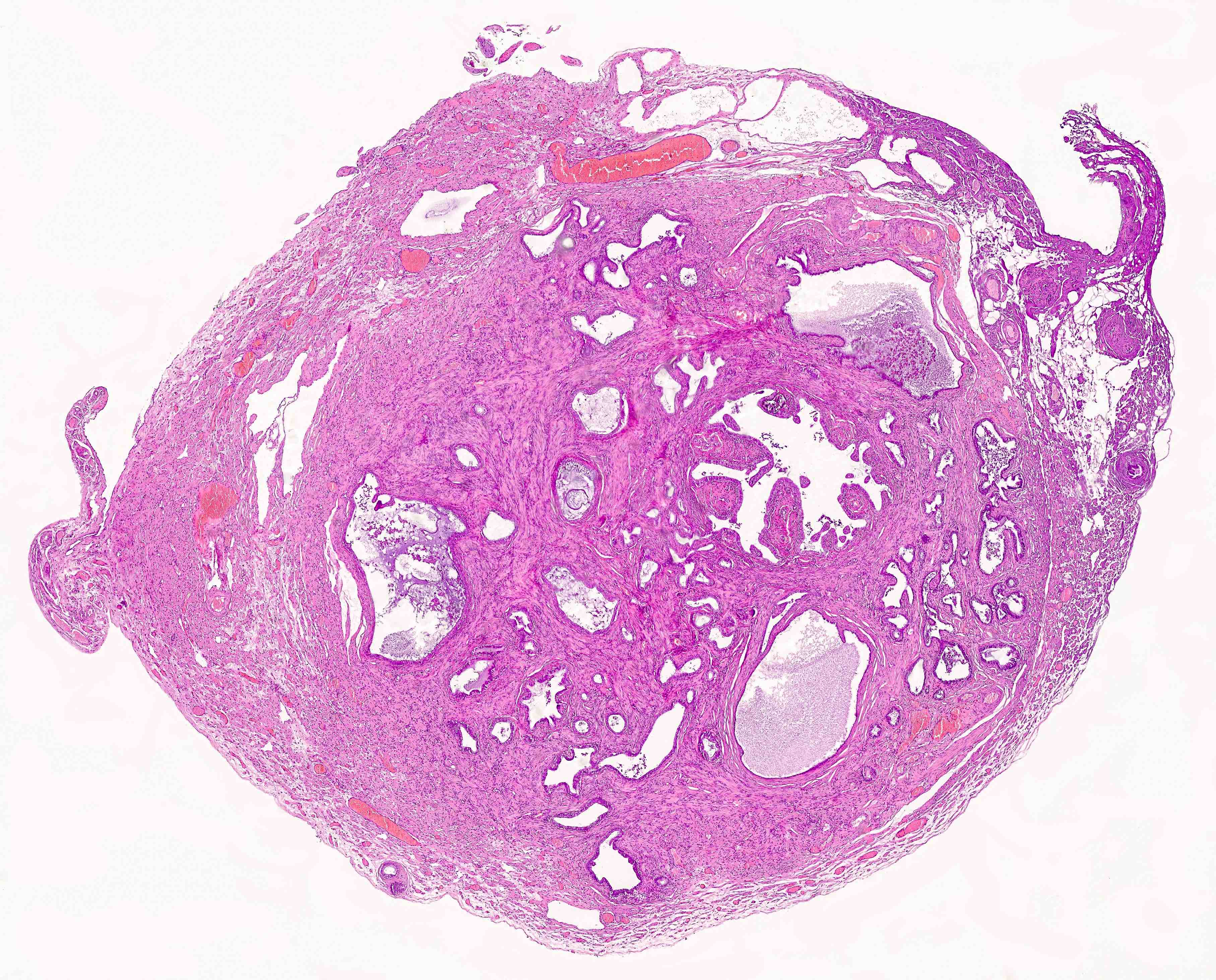
Fallopian tube, premenopausal
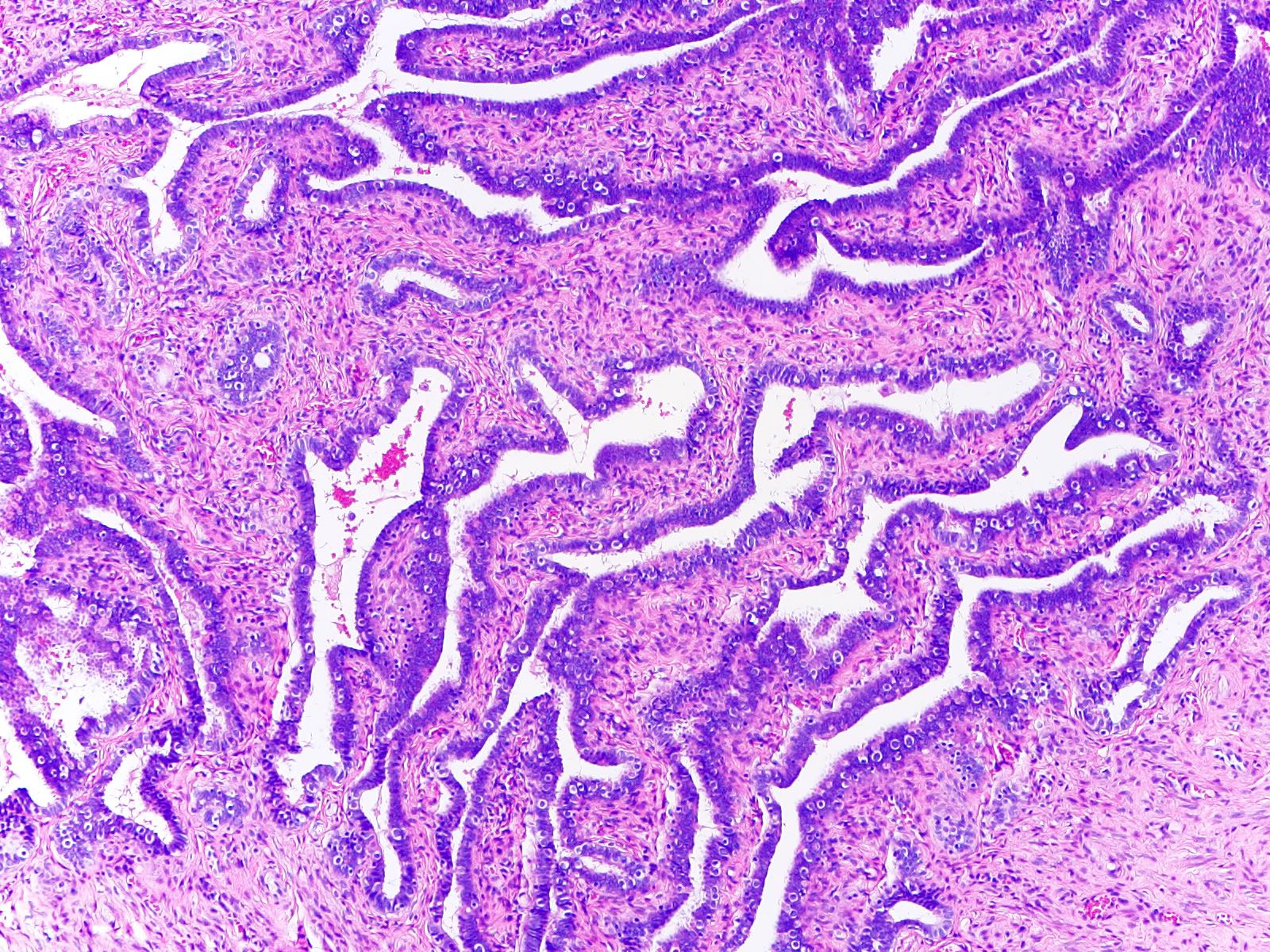
Mucosal plicae
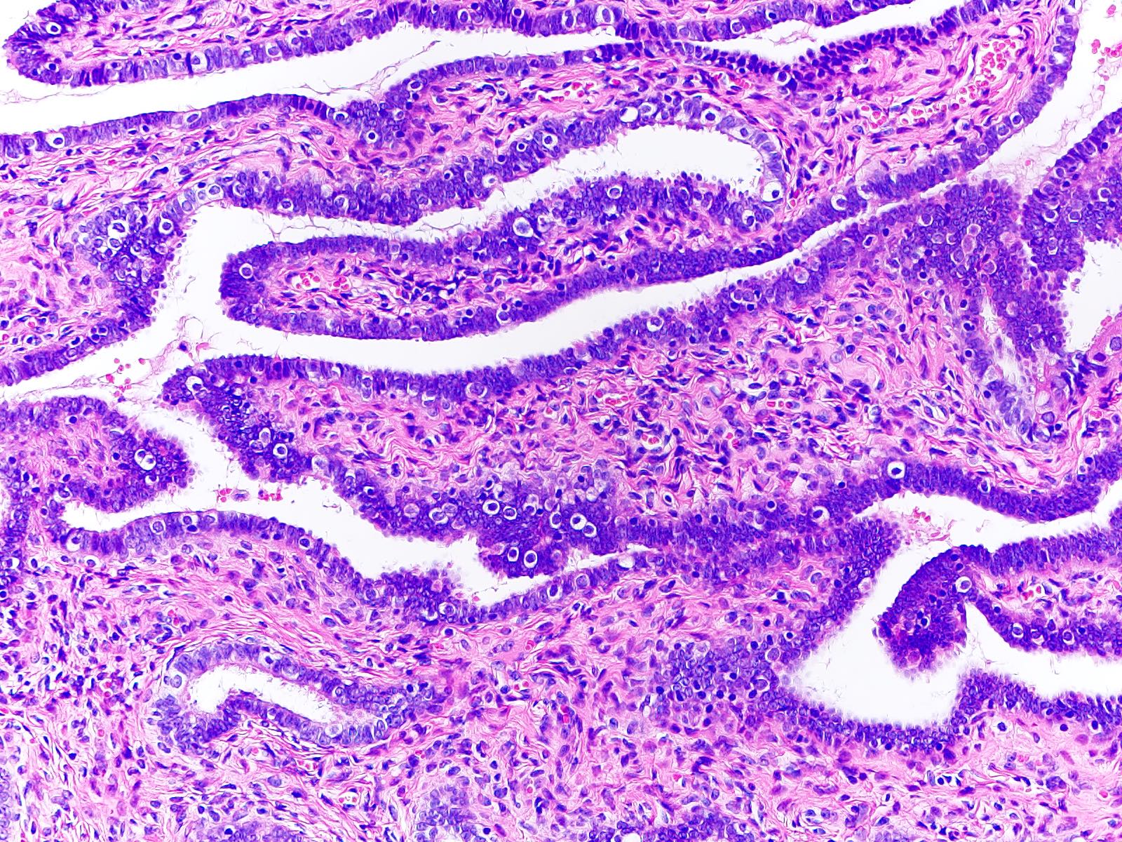
Fallopian tube epithelium
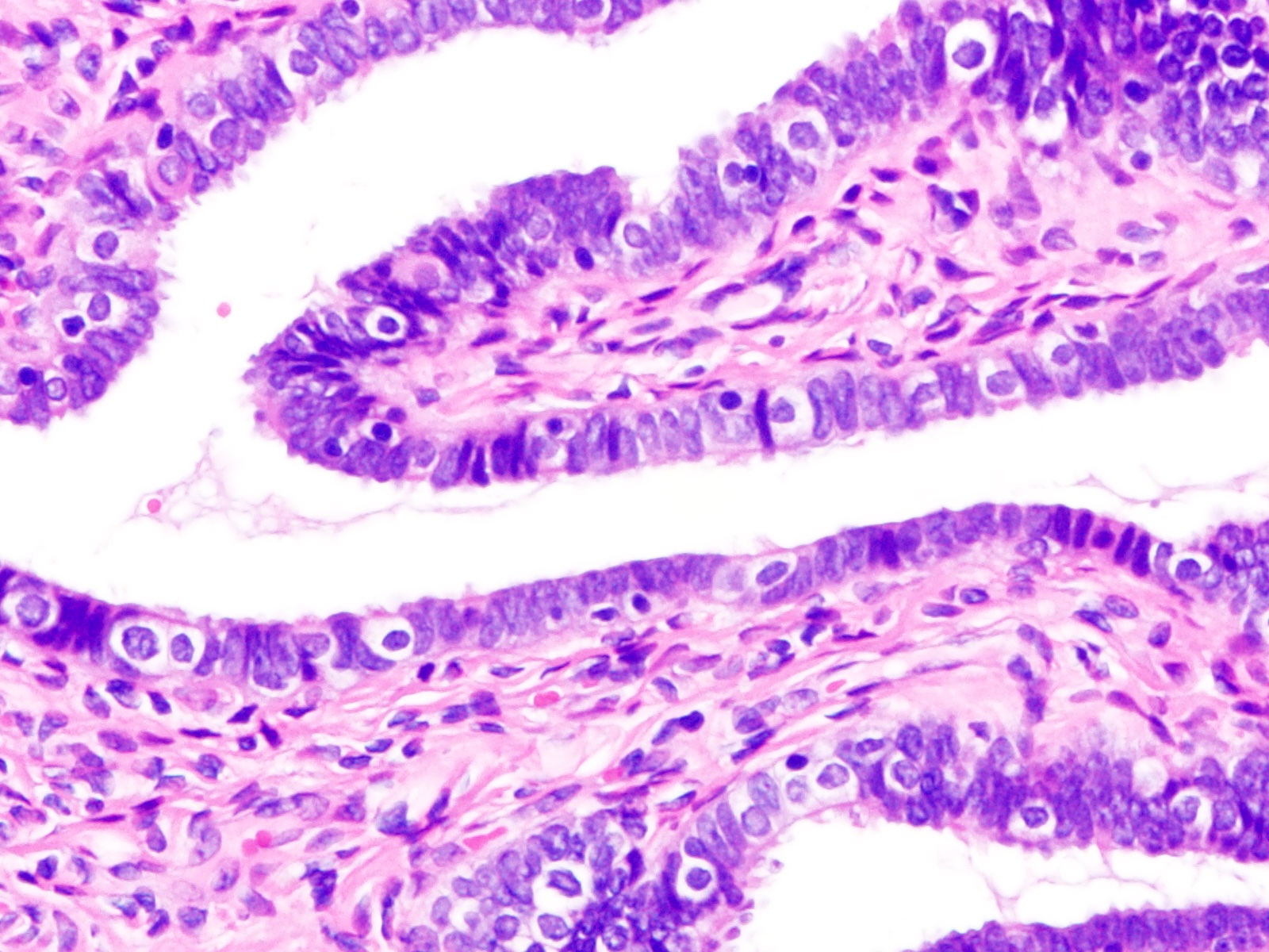
Simple columnar epithelium
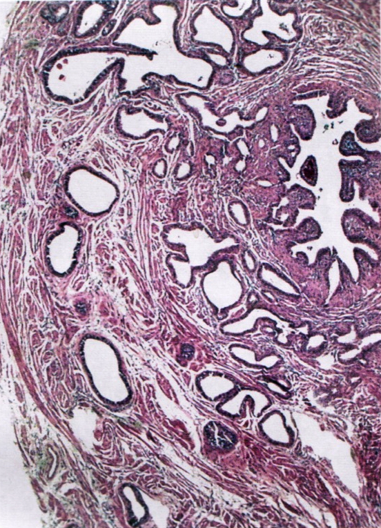
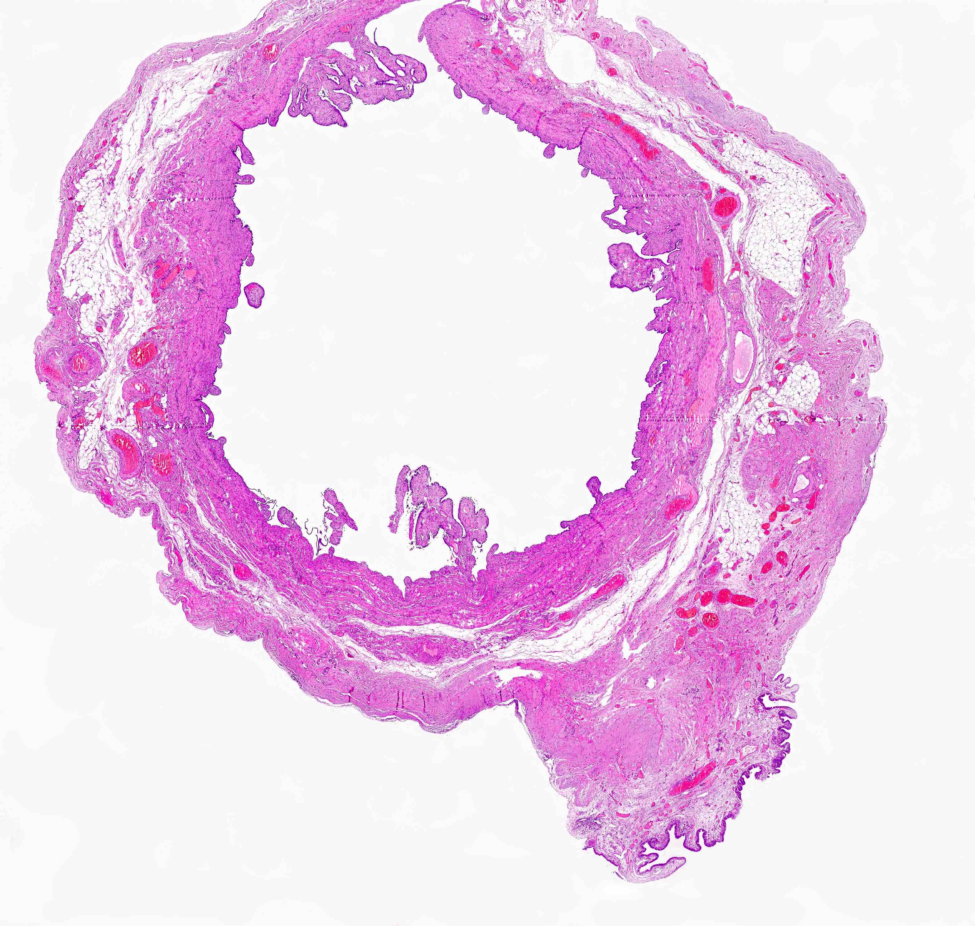
Fimbriae are not thickened
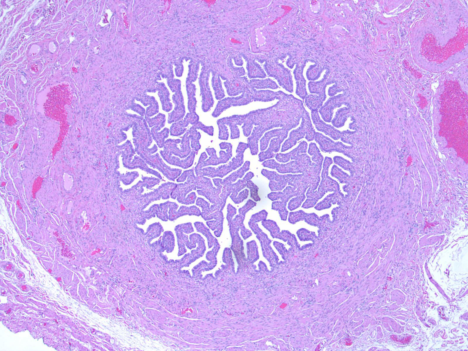
Lumen filled with plicae
Simple columnar epithelium
Images hosted on other servers:

Cell clusters

Honeycomb / monolayered sheets
Images hosted on other servers:

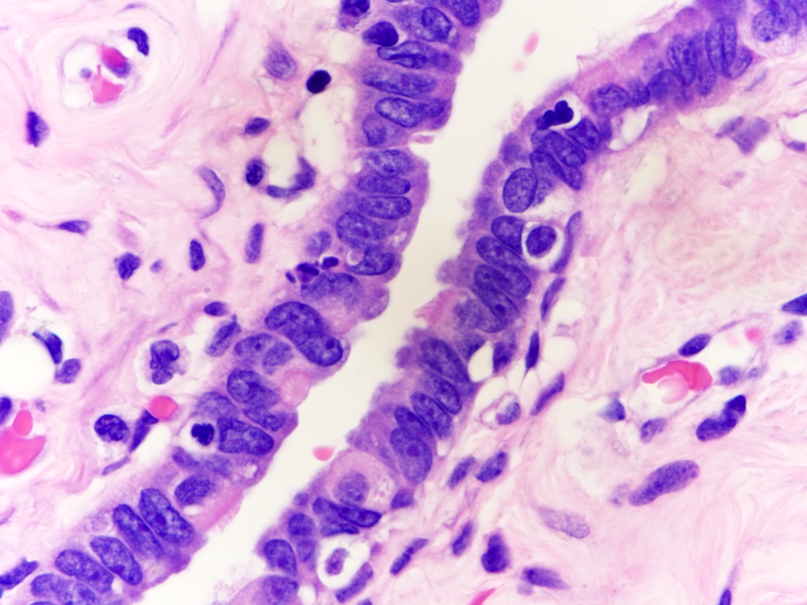
Primary cilia

Secretory and ciliated cells
Shotgun histology: fallopian tube
UB medical: oviduct histology
Images hosted on other servers:
Fallopian tube
Images hosted on other servers:

Uterine attachment on upper left, fibriae on upper right
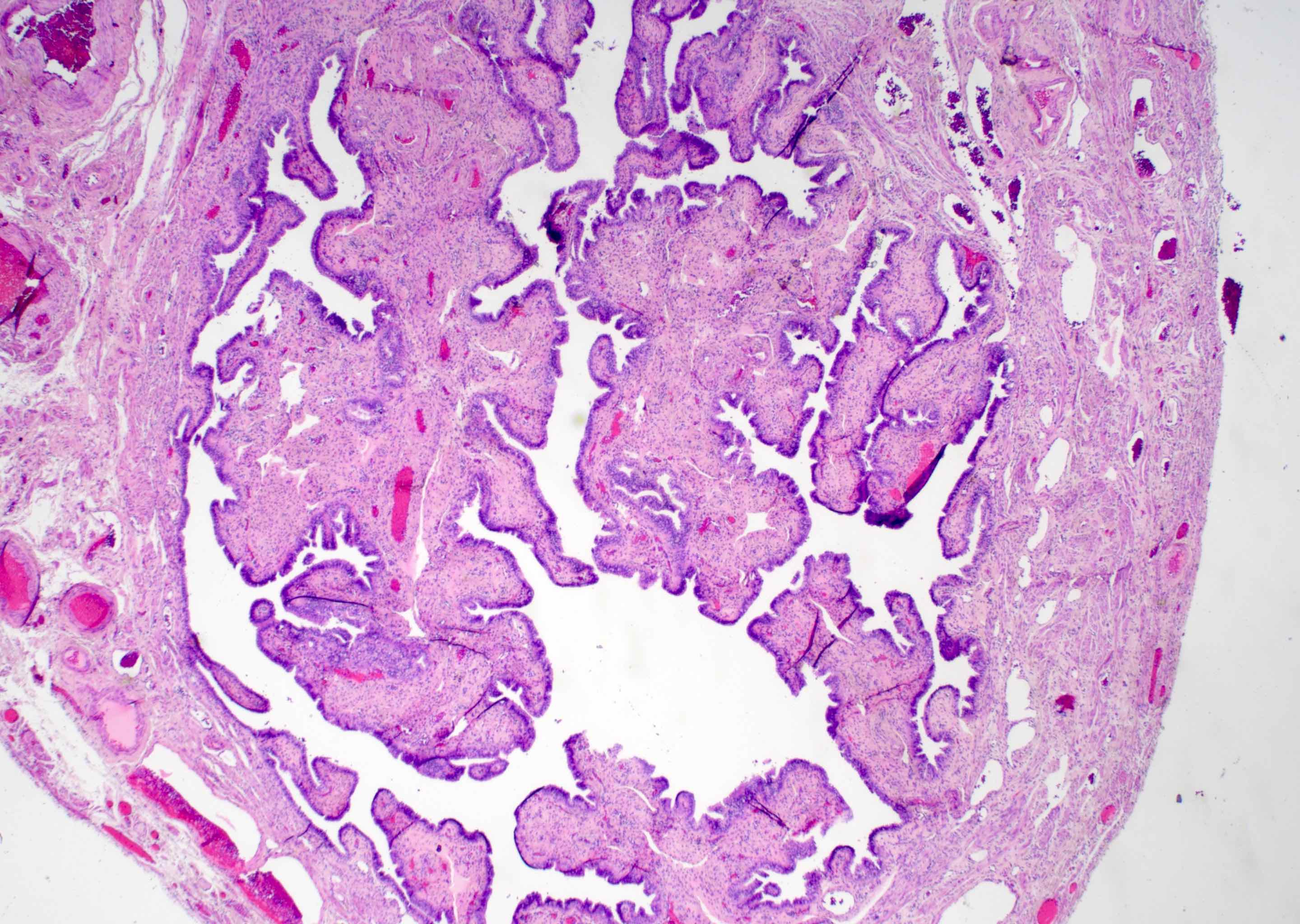
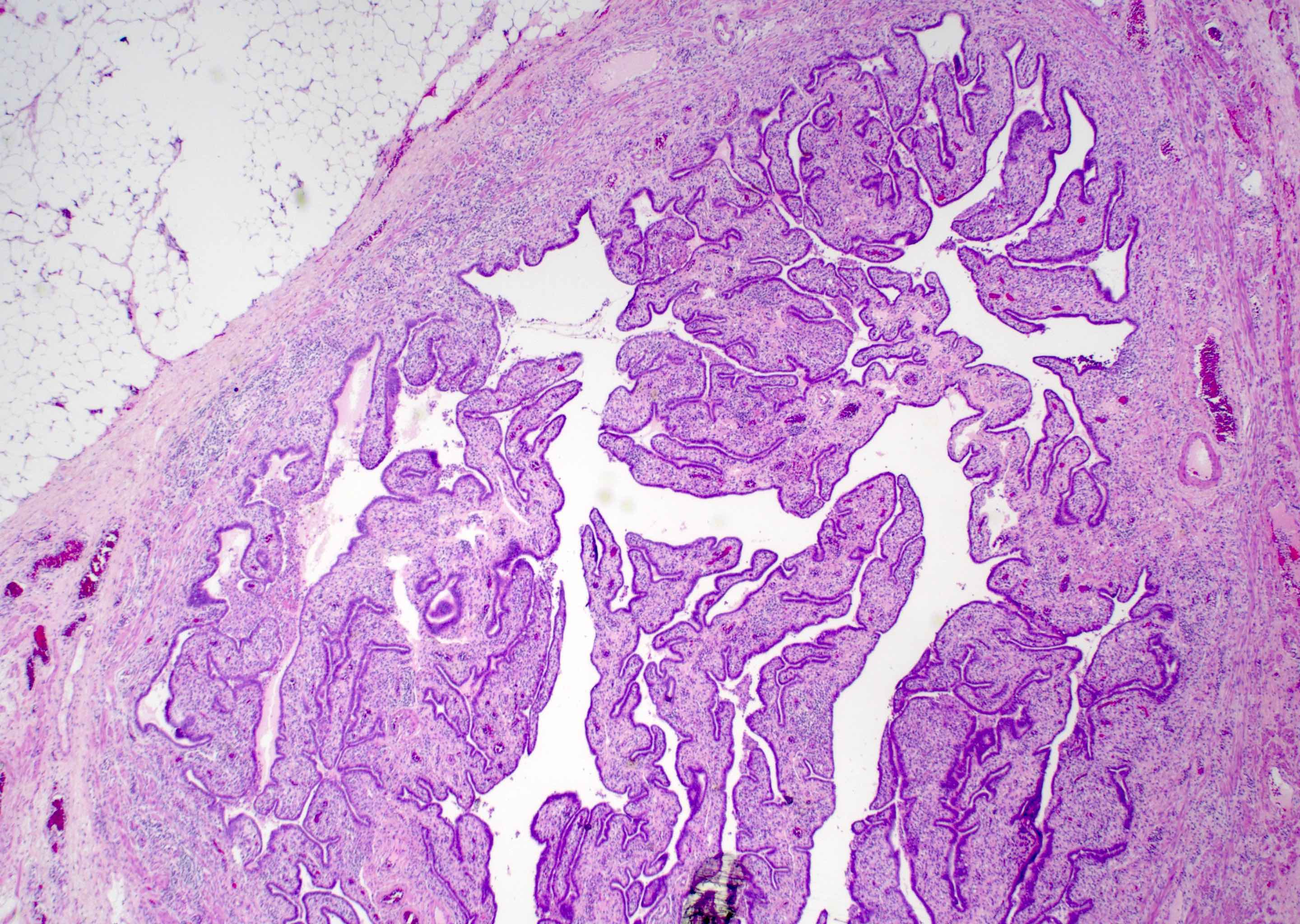
Contributed by Nicole D. Riddle, M.D.
Lumenal center filled with plica
Fimbriae are not thickened
Simple columnar epithelium
AFIP images

Lumen
Tubal epithelium
AFIP images
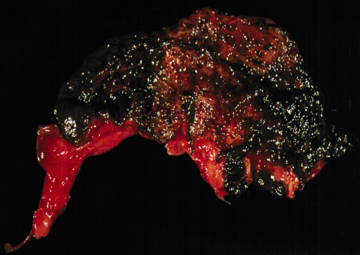
Discolored mucosa
Contributed by Shabnam Zarei, M.D. and Nicole D. Riddle, M.D.
Short thick plicae
Inflammatory cells

Short thick plicae and inflammation
Mucosal inflammation

Stromal fibrosis
Thickened plicae and foamy histiocytes

Foamy histiocytes
Fused and slightly thickened plicae
Images hosted on other servers:

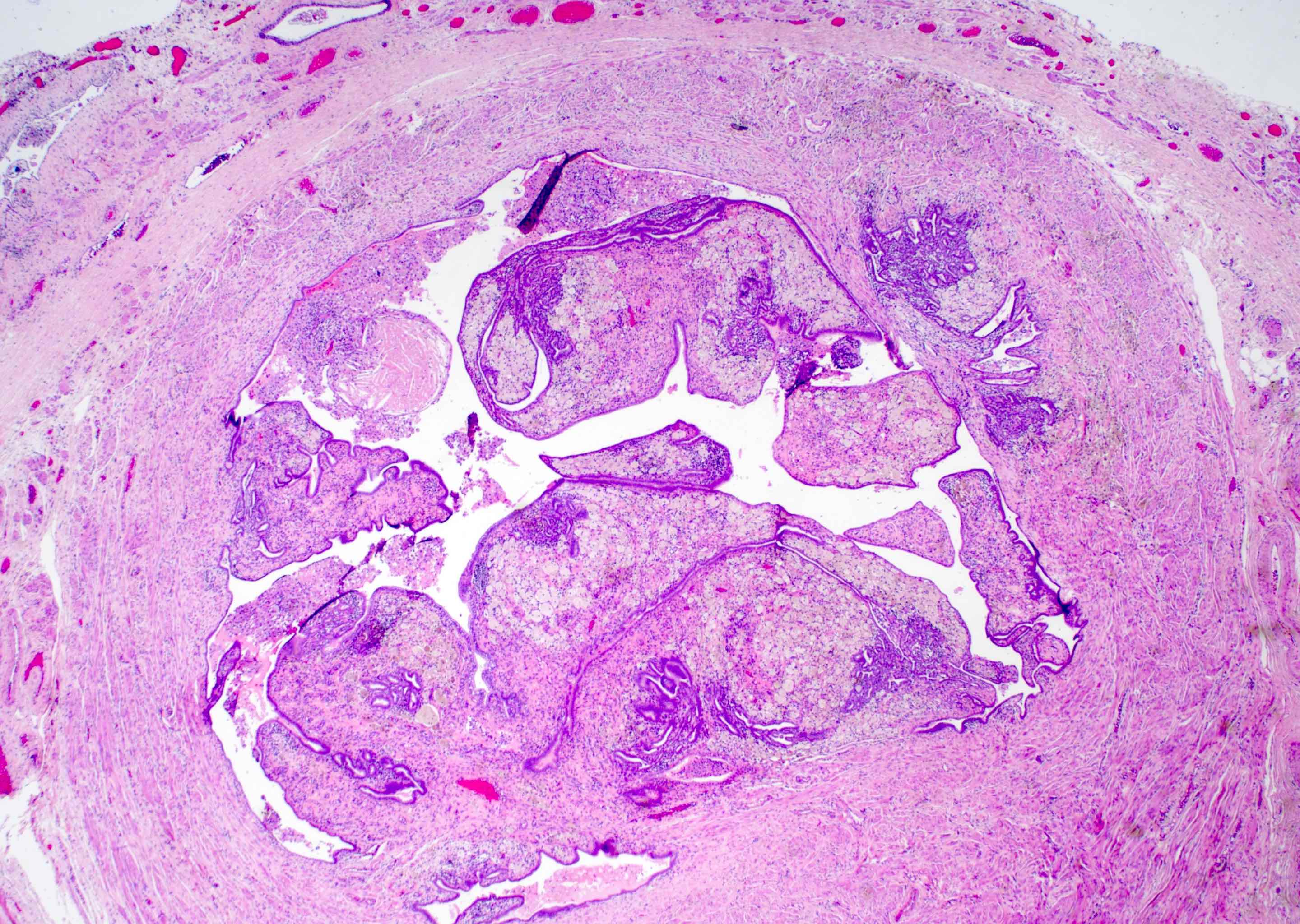
Torsed paraovarian cyst before detorsion

Torsed paraovarian cyst after detorsion

Large paraovarian cyst
Contributed by Lucy Ma, M.D.
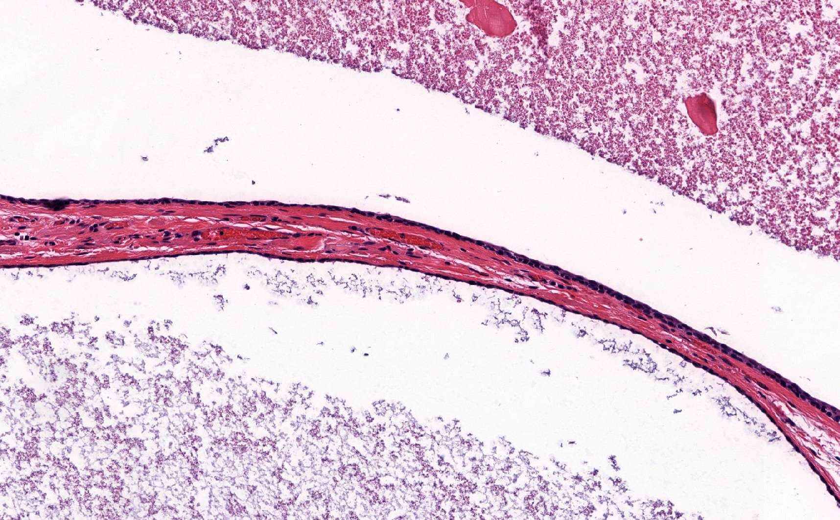
Simple cysts adjacent to Walthard nests
Paratubal cysts adjacent to fallopian tube
Ciliated epithelium
Contributed by Hao Chen, M.D., Ph.D.
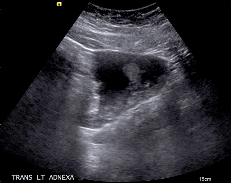
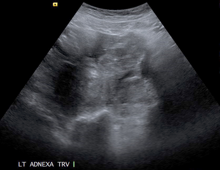
Left adnexal mass
Contributed by Hao Chen, M.D., Ph.D. and AFIP
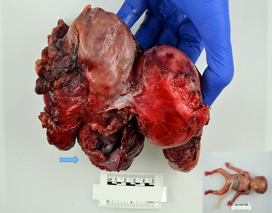
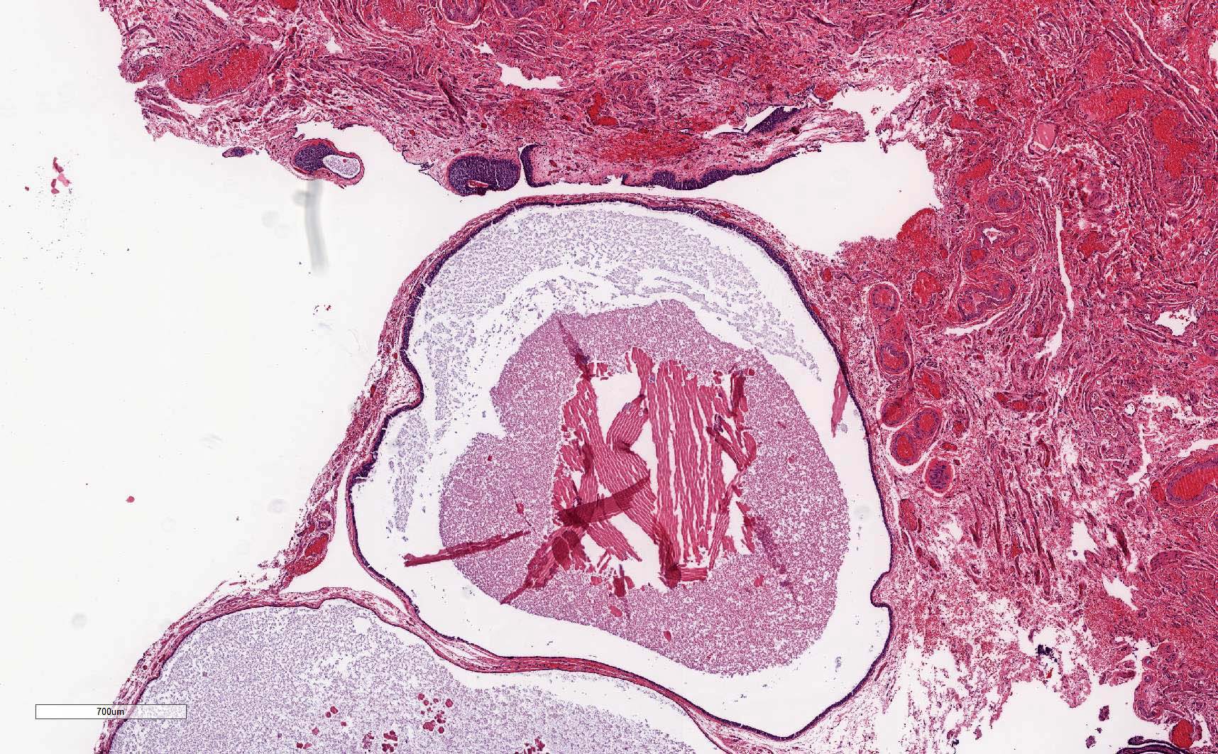
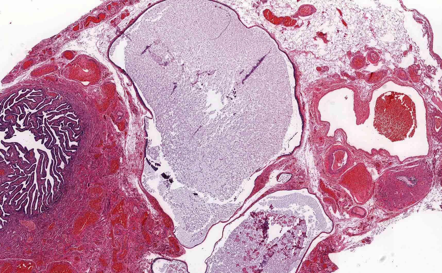
Placenta and fetus
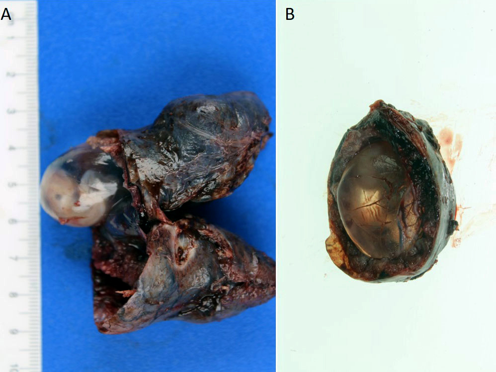
Gestational sac and embryo
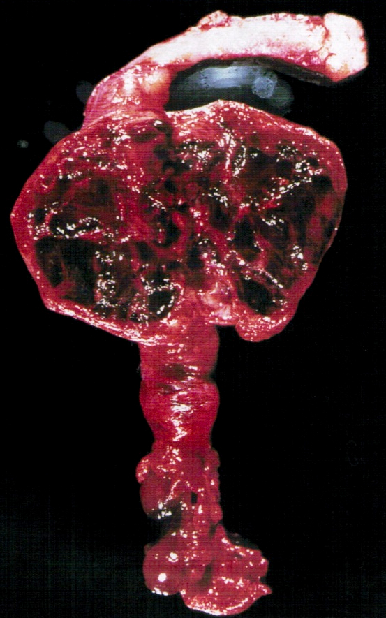
Ectopic pregnancy
Images hosted on other servers

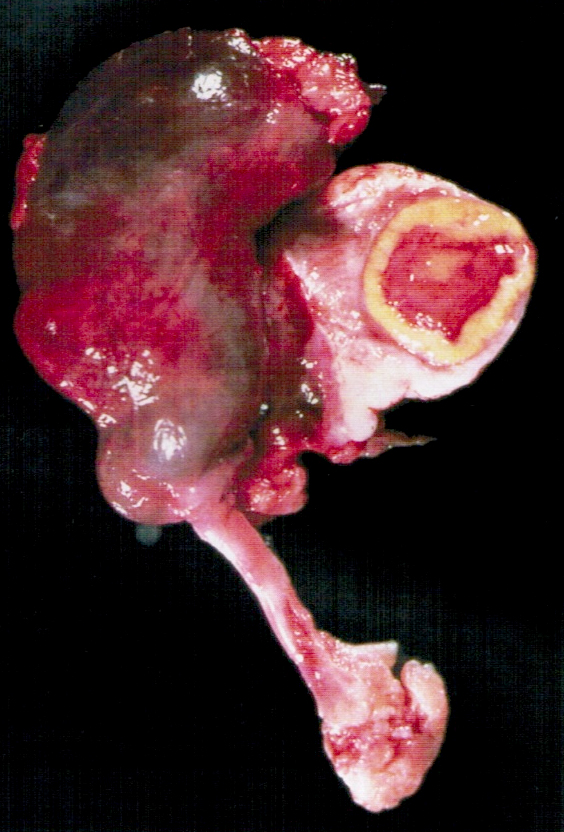
Hemorrhage and placental tissue with fetal part

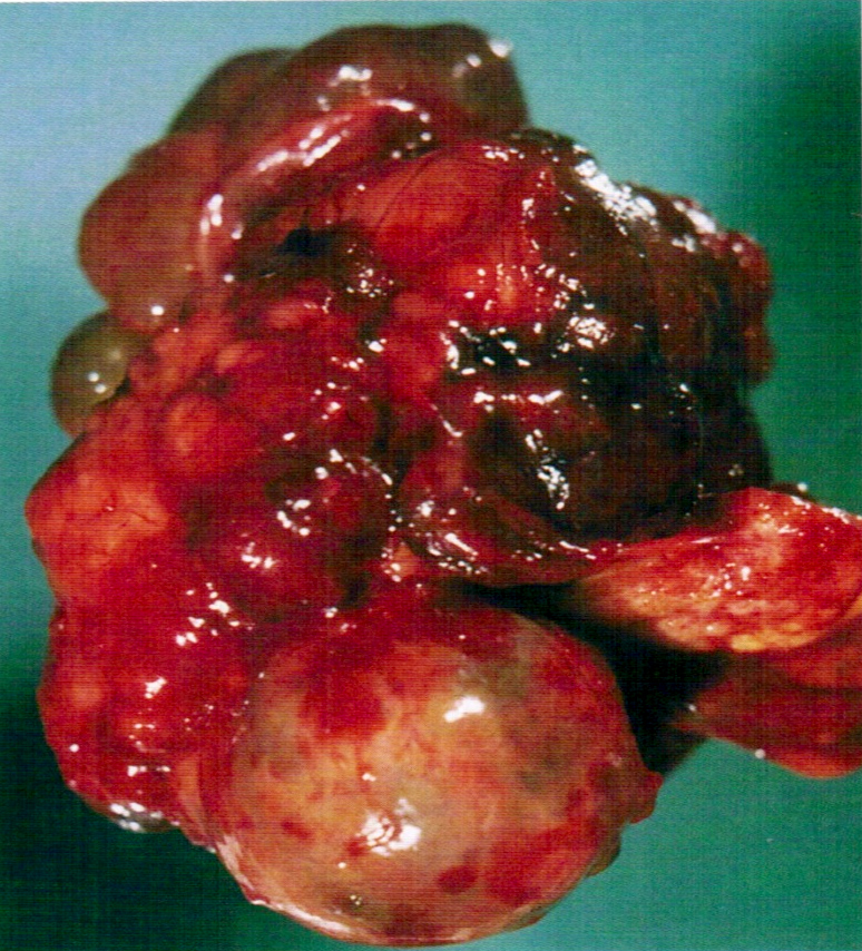
Rupture and hemoperitoneum
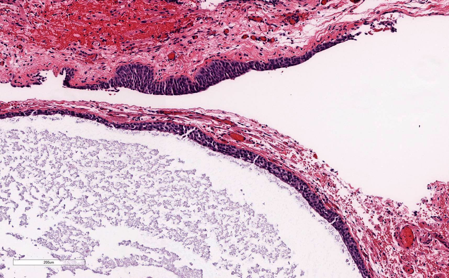
Contributed by Hao Chen, M.D., Ph.D.
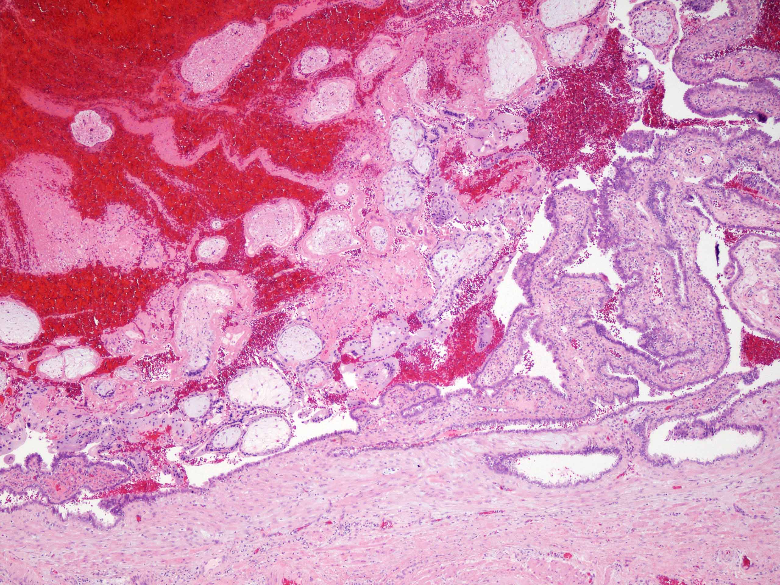
Intraluminal chorionic villi

Chorionic villi and blood
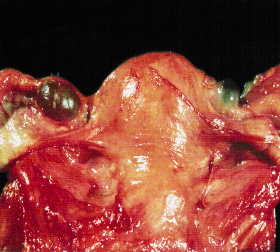
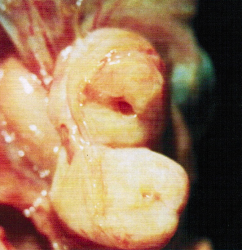
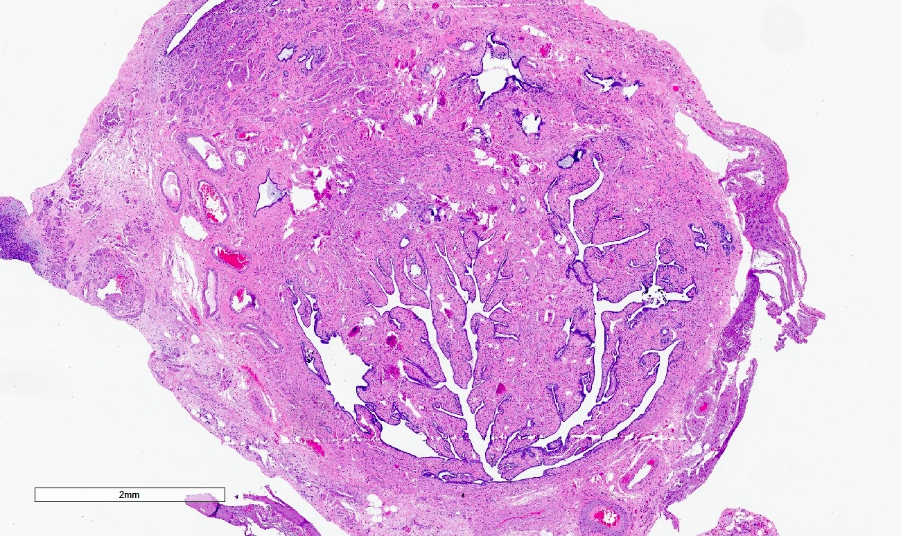
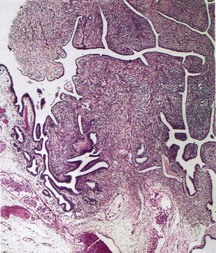
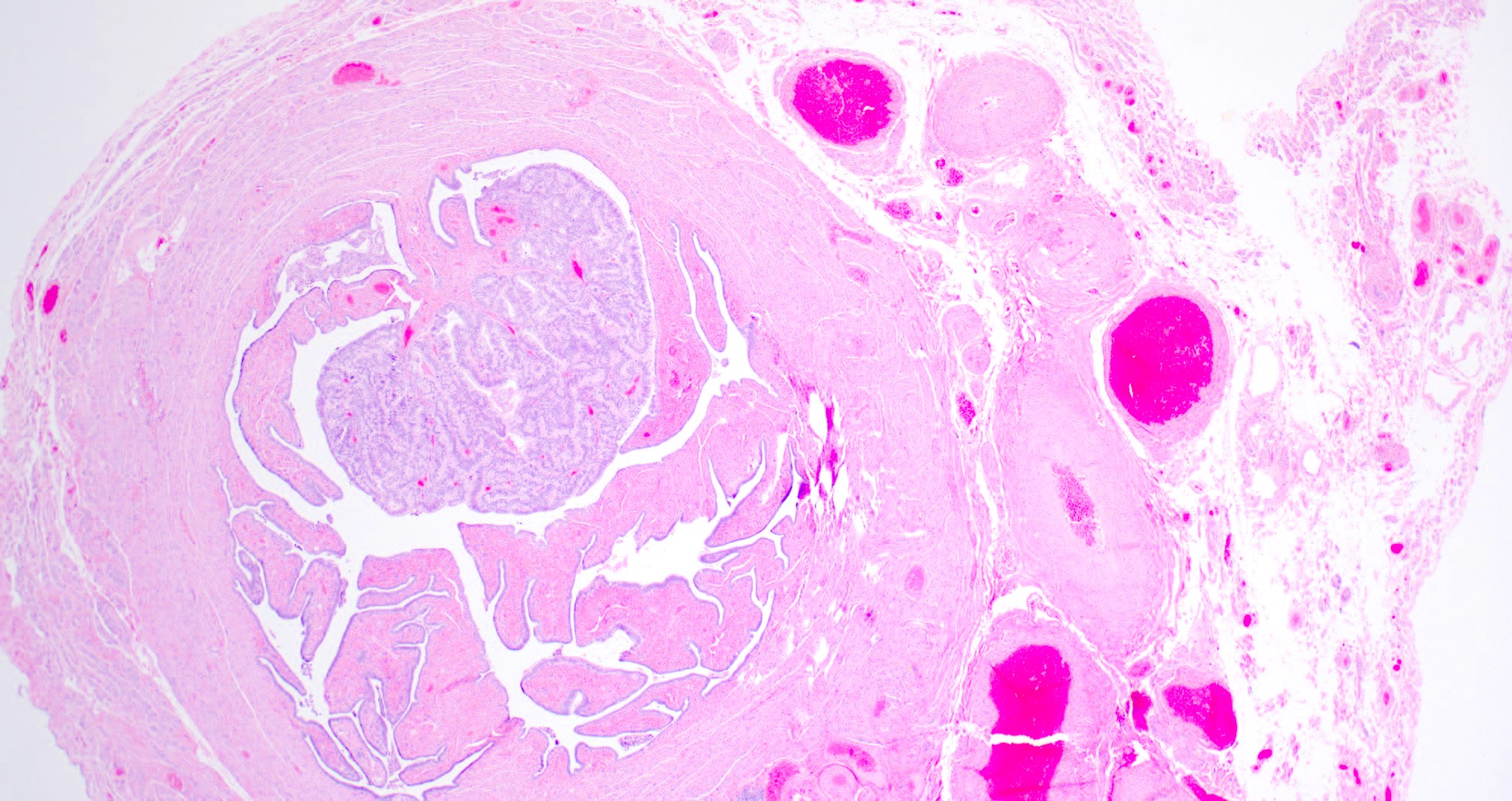
AFIP images
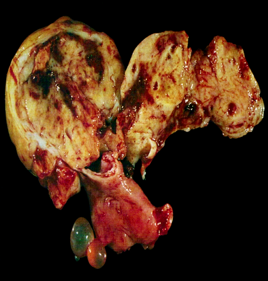
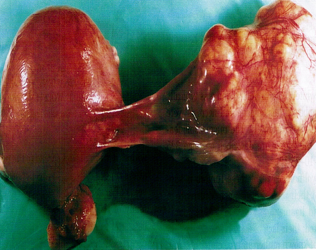
Diffusely enlarged tube
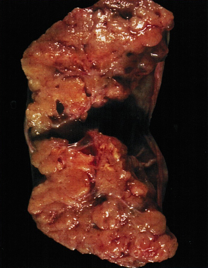
Papillary tumor attached to mucosa
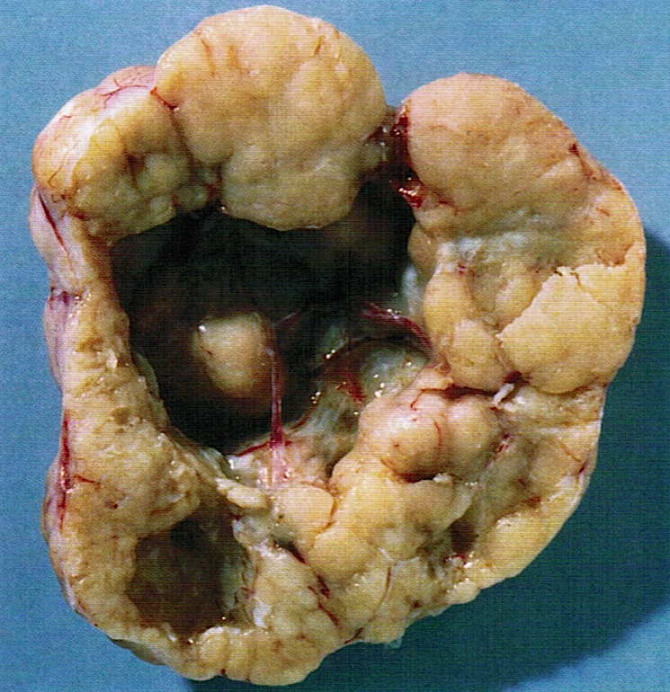
Lumen distended by solid tumor
Distended, opened tube
AFIP images
Tumor arising in endometriosis
Tumor simulating Wolffian adnexal tumor
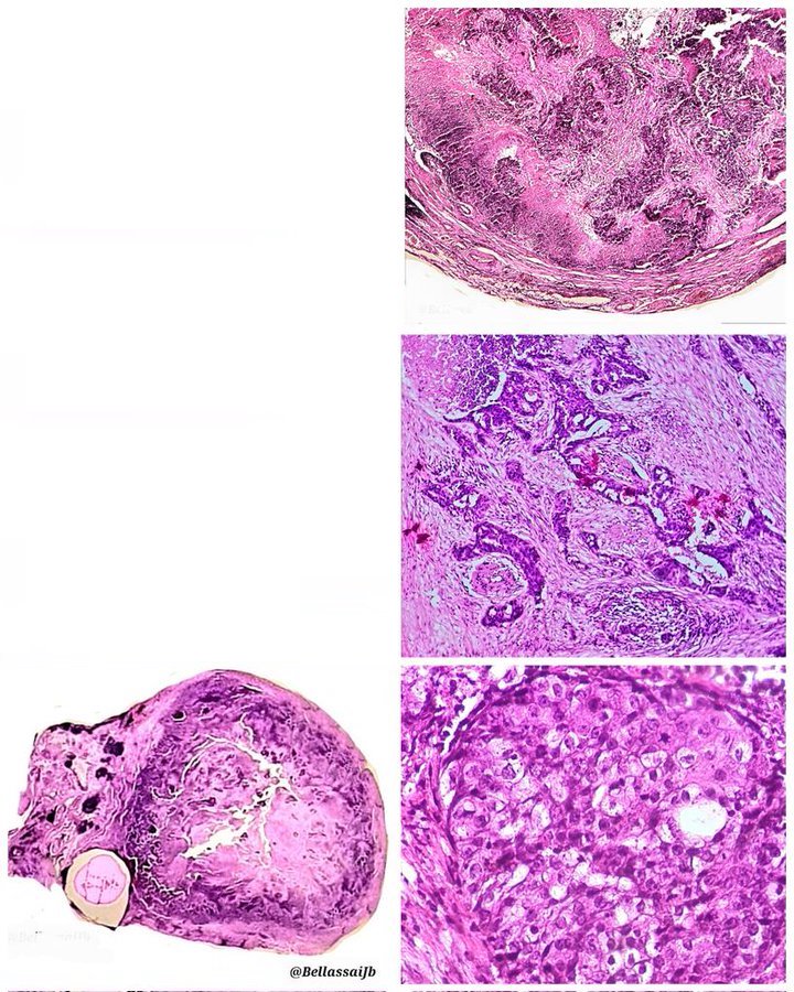
Contributed by AFIP and @BellassaiJb on Twitter
Diffusely enlarged
Papillary tumor attached to mucosa
Distended lumen
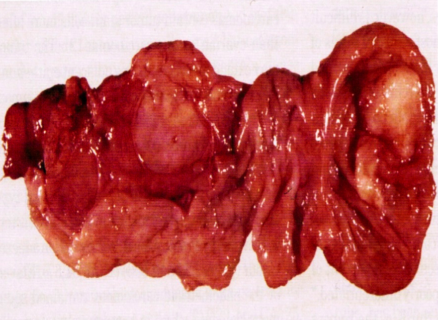
Solid nodule of tumor

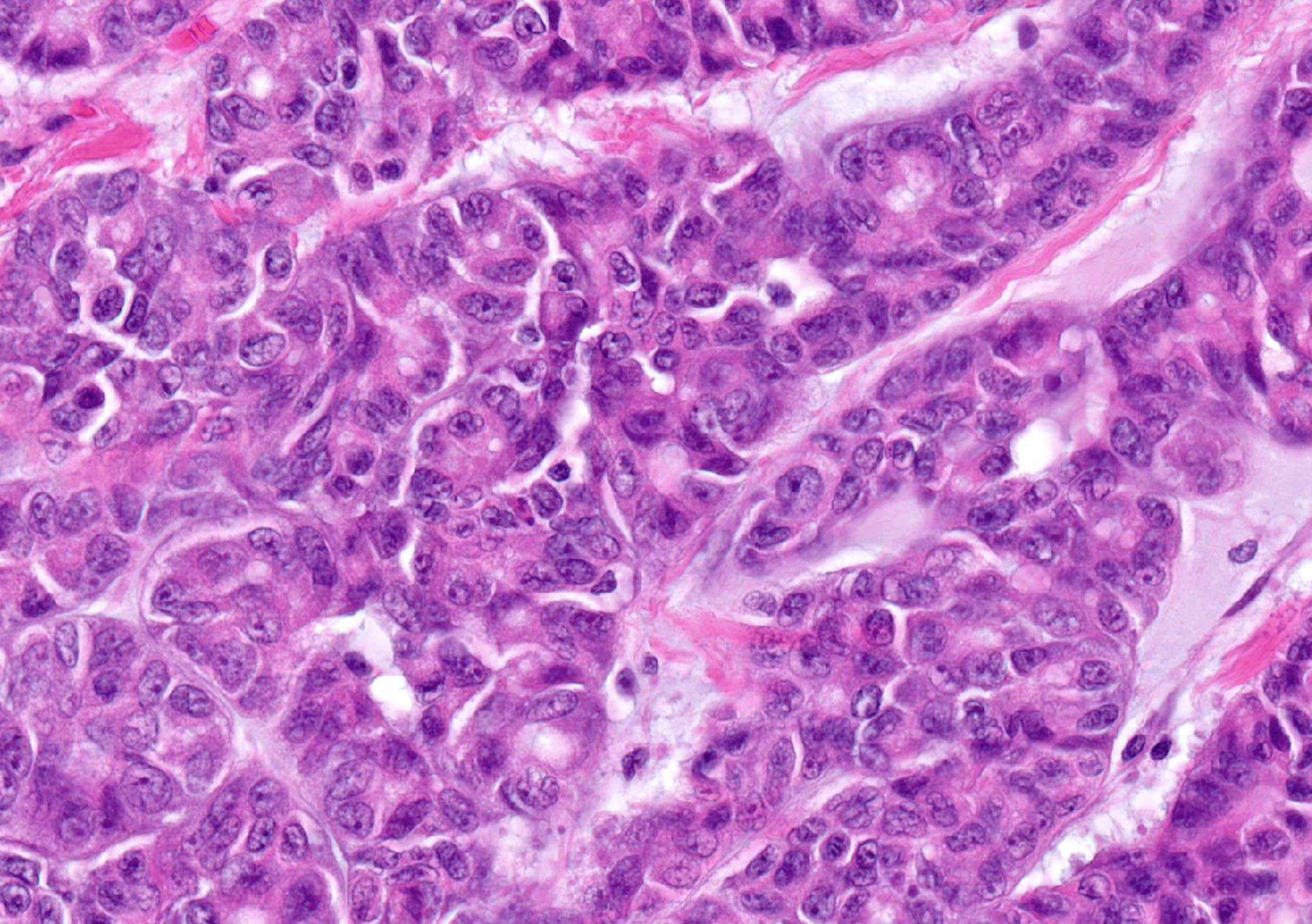
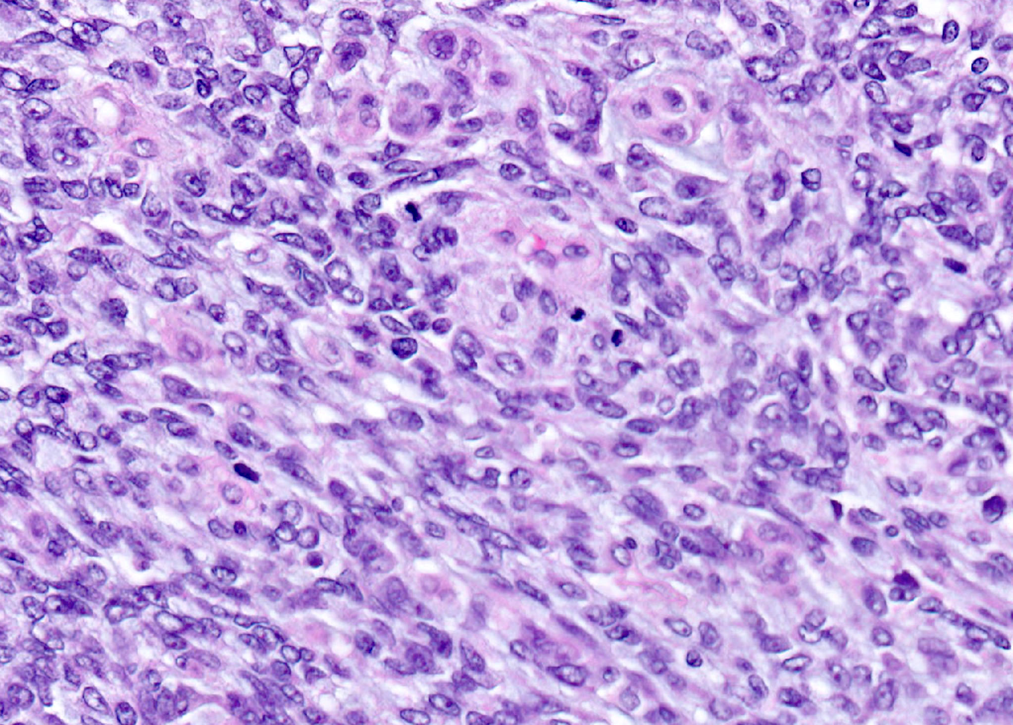
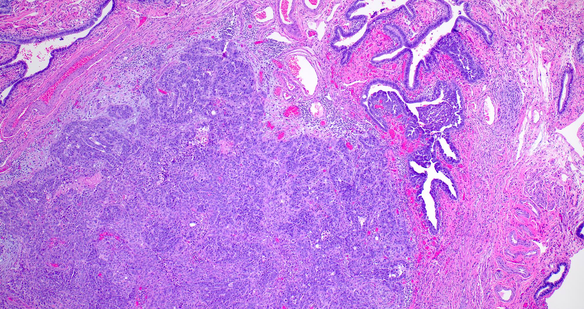
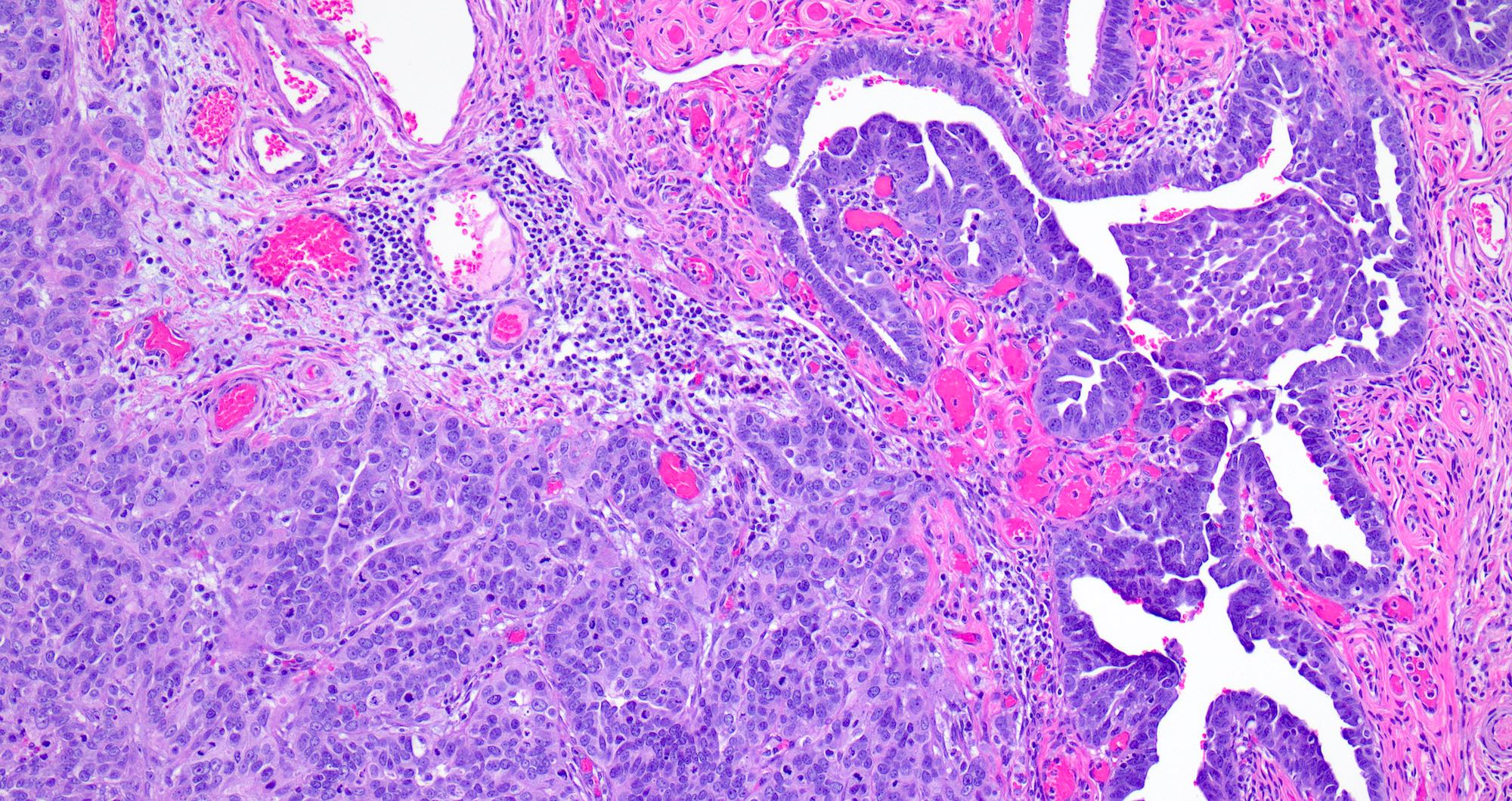
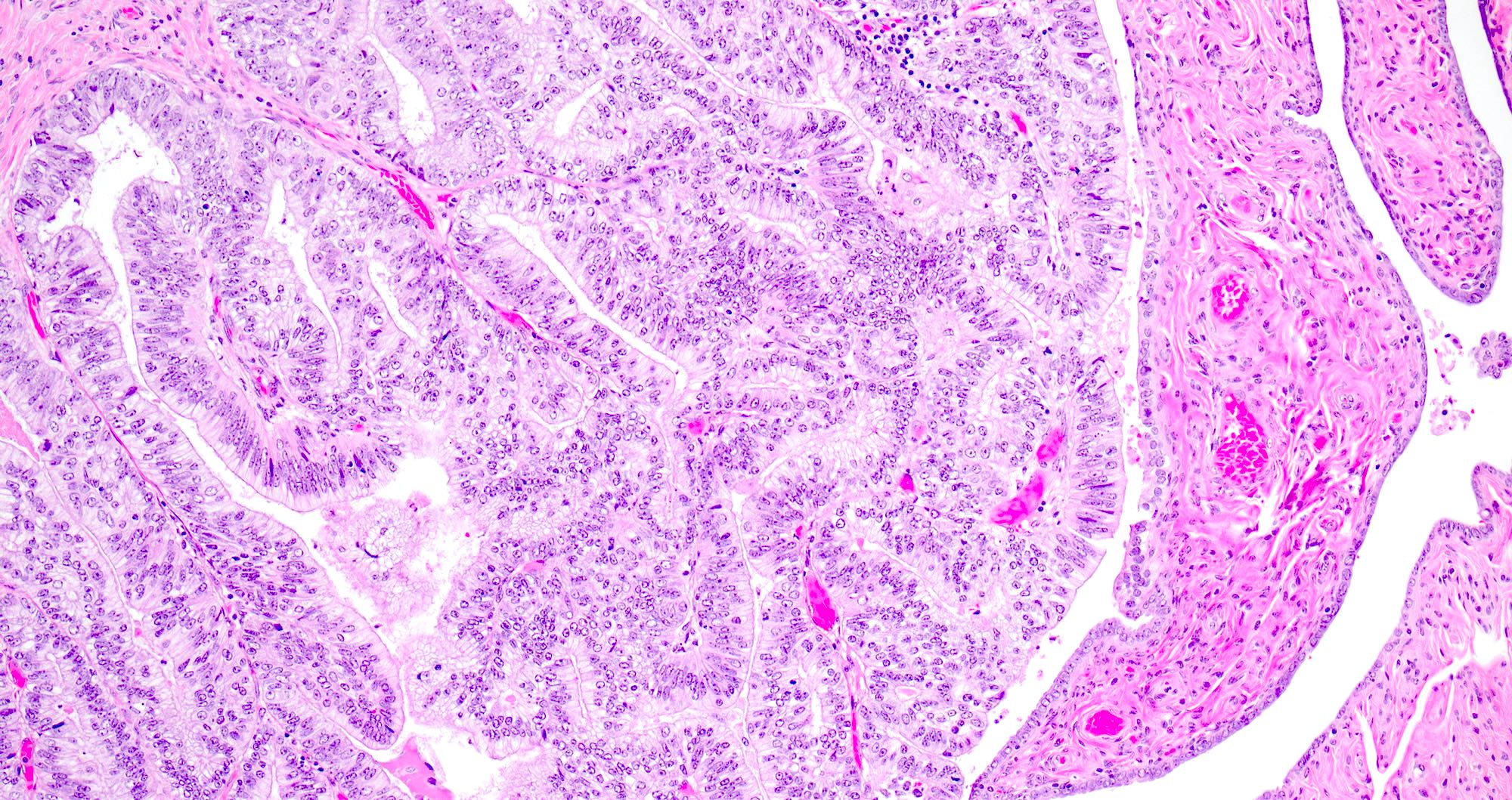
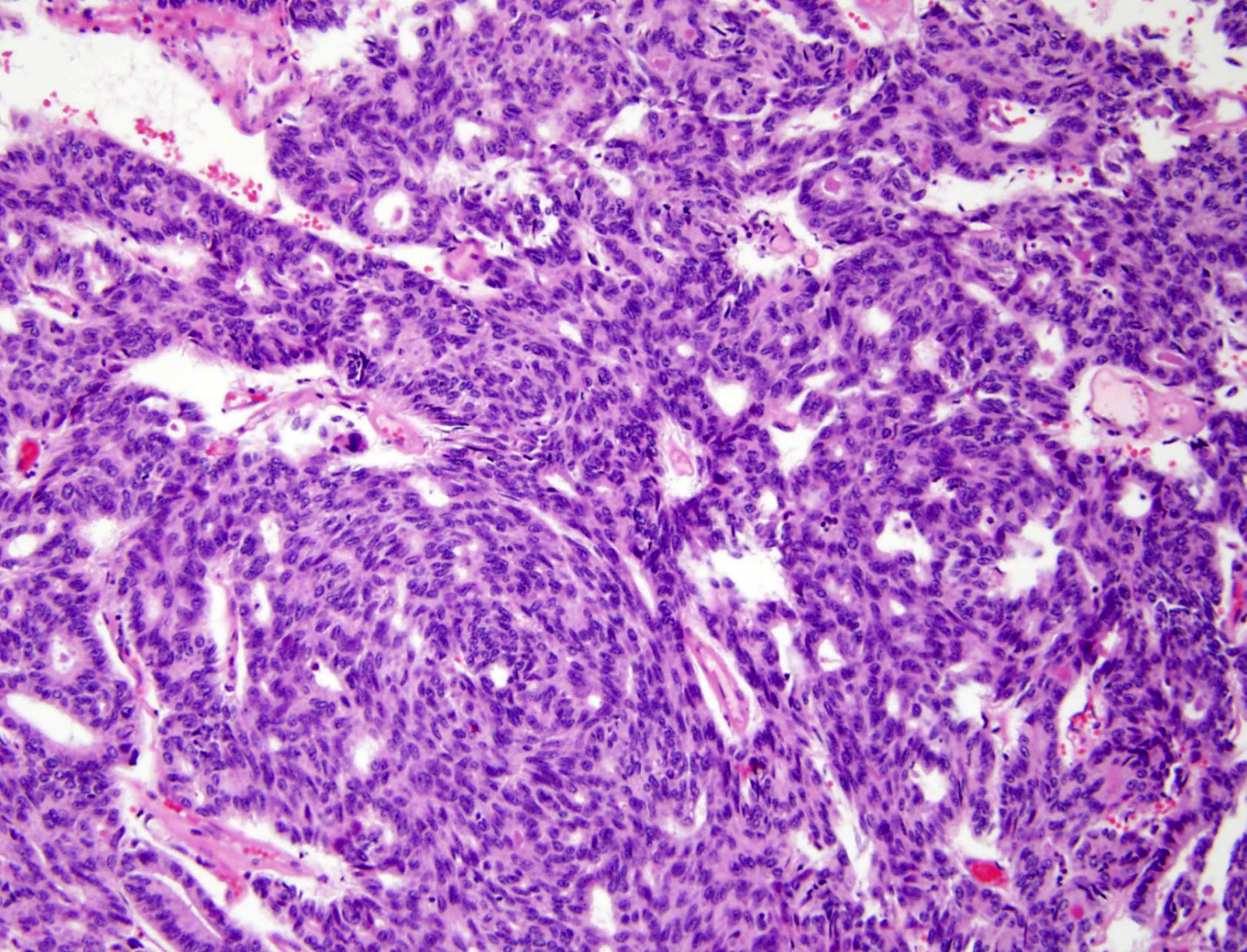
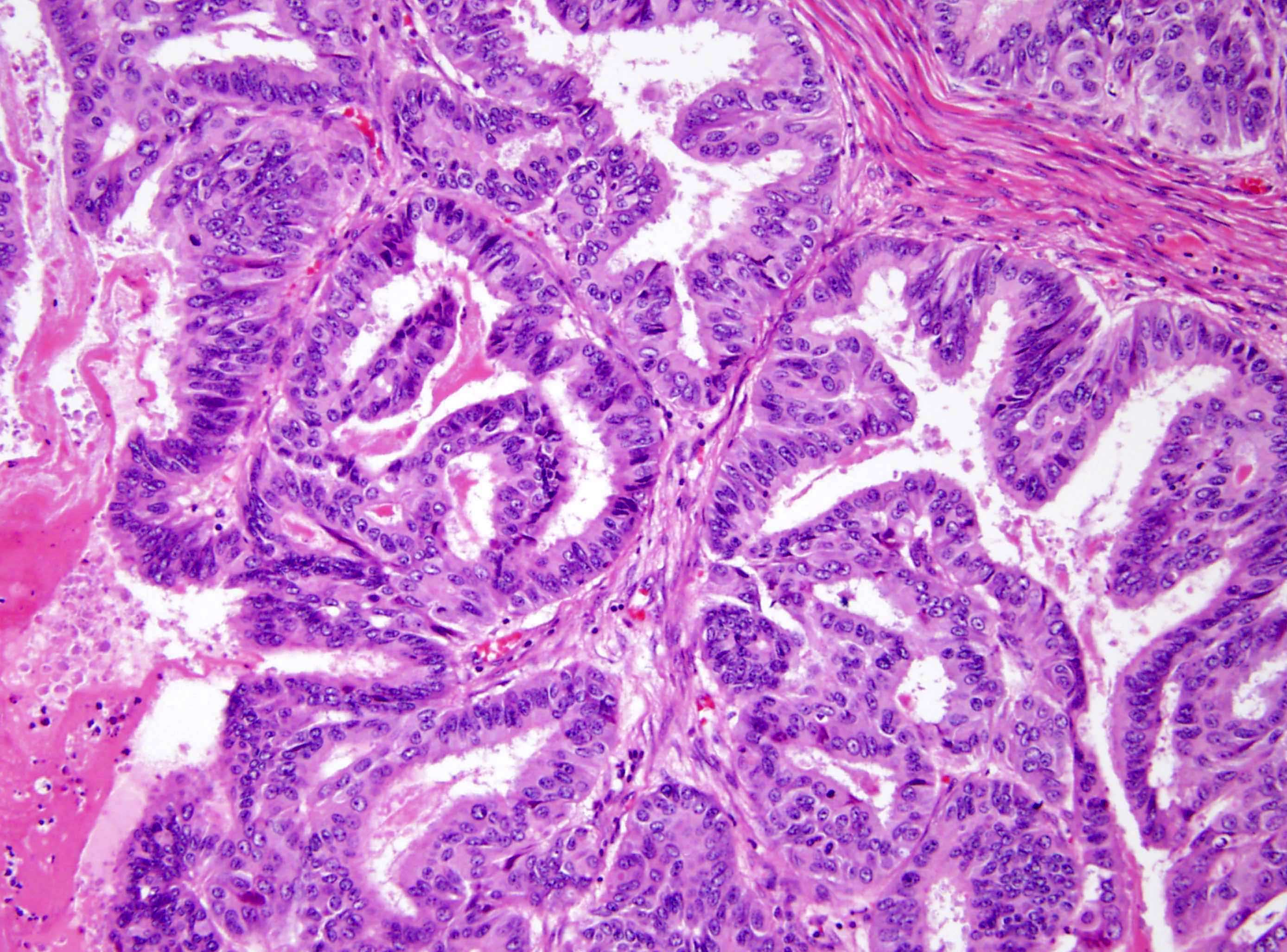
High grade serous carcinoma
Contributed by AFIP and @BellassaiJb on Twitter
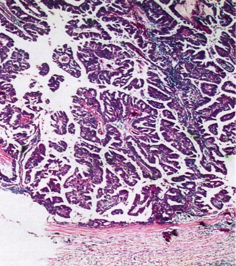
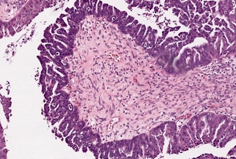
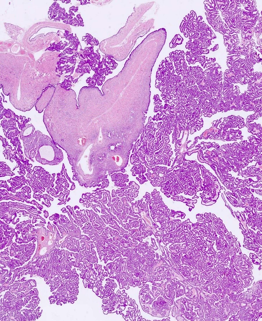
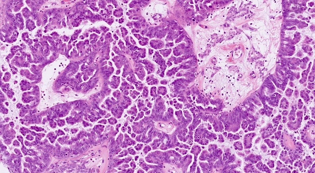
Lumen filled with fine papillae
Desmoplastic stromal response
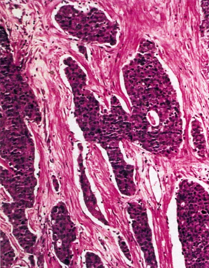
Undifferentiated component in tubal wall
High grade serous carcinoma
Contributed by Jian-Hua Qiao, M.D.
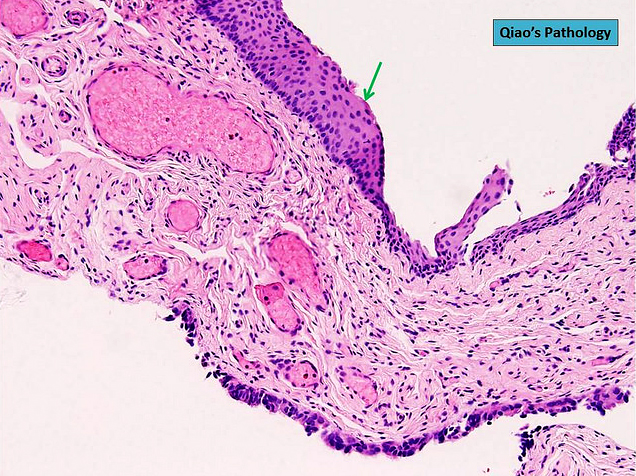
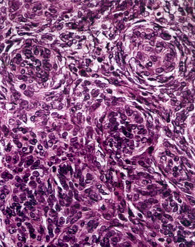
Extensive transitional cell metaplasia
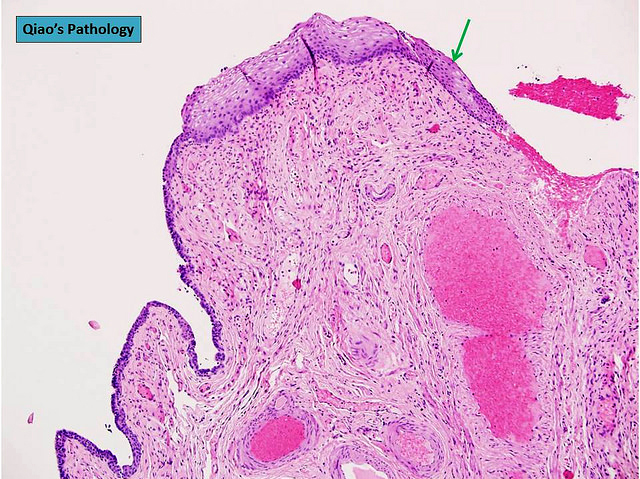
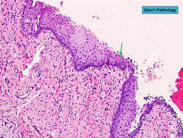
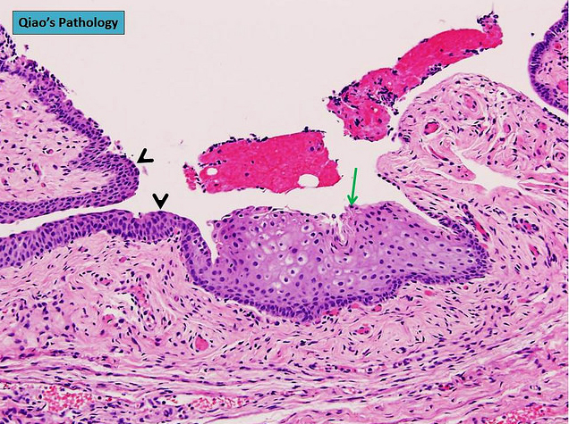
Focal transitional cell metaplasia
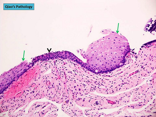
Multifocal transitional cell metaplasia
AFIP images
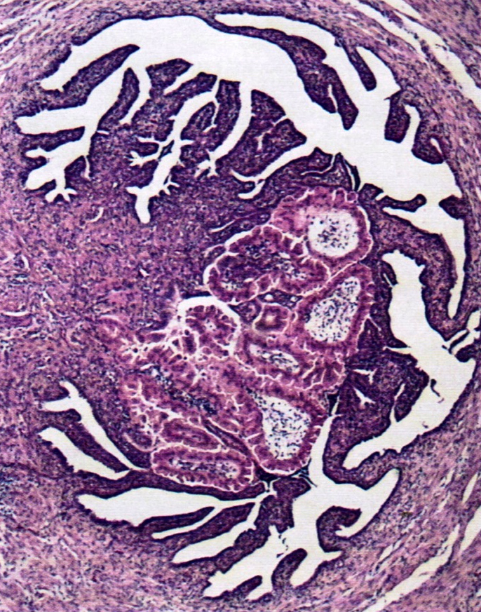

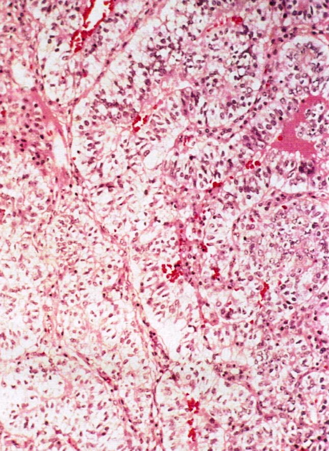
Metaplastic papillary tumor
AFIP images
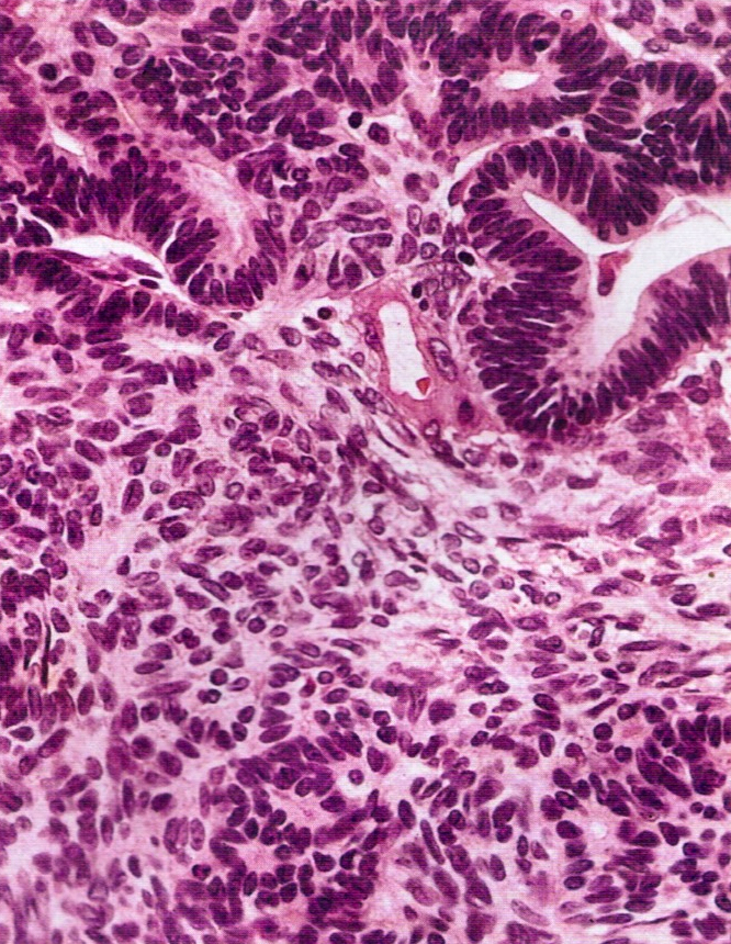
Simulating serous papillary tumor
Bland, cuboidal epithelial cells
Images hosted on other servers:

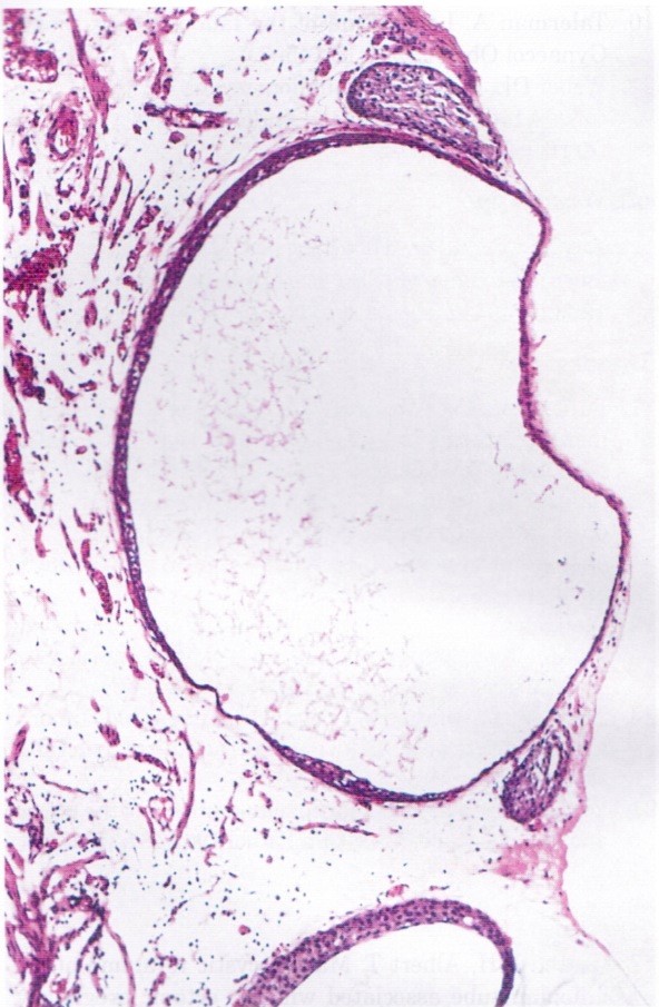
Benign appearing cyst
Contributed by Stephanie L. Skala, M.D.
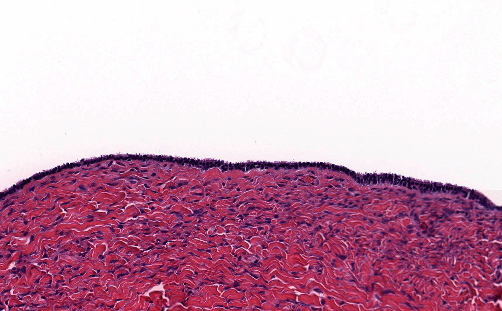
Simple cyst near tube
Simple cyst adjacent to fallopian tube
Bland, ciliated, tubal type epithelium
Images hosted on other servers:

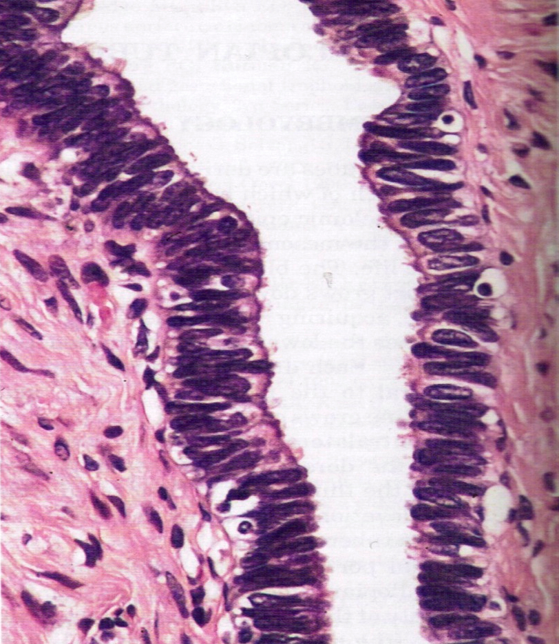
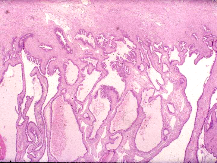
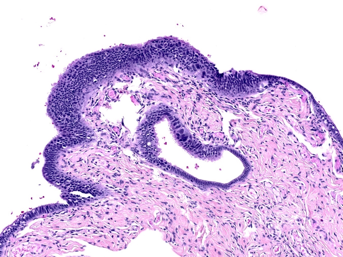
Fallopian tubal epithelium thrown into papillary folds

Fallopian tube lining
of pseudostratified
ciliated columnar
epithelium
Images hosted on other servers:

Nodular swelling of the isthmus

Multiple nodular diveriticula

Small outpouching or diverticula
AFIP images
Dark, nodular swelling
Marked thickening
Images hosted on other servers:

Nodules at isthmus
Contributed by Jessica L. Bentz, M.D., AFIP and @Andrew_Fltv on Twitter
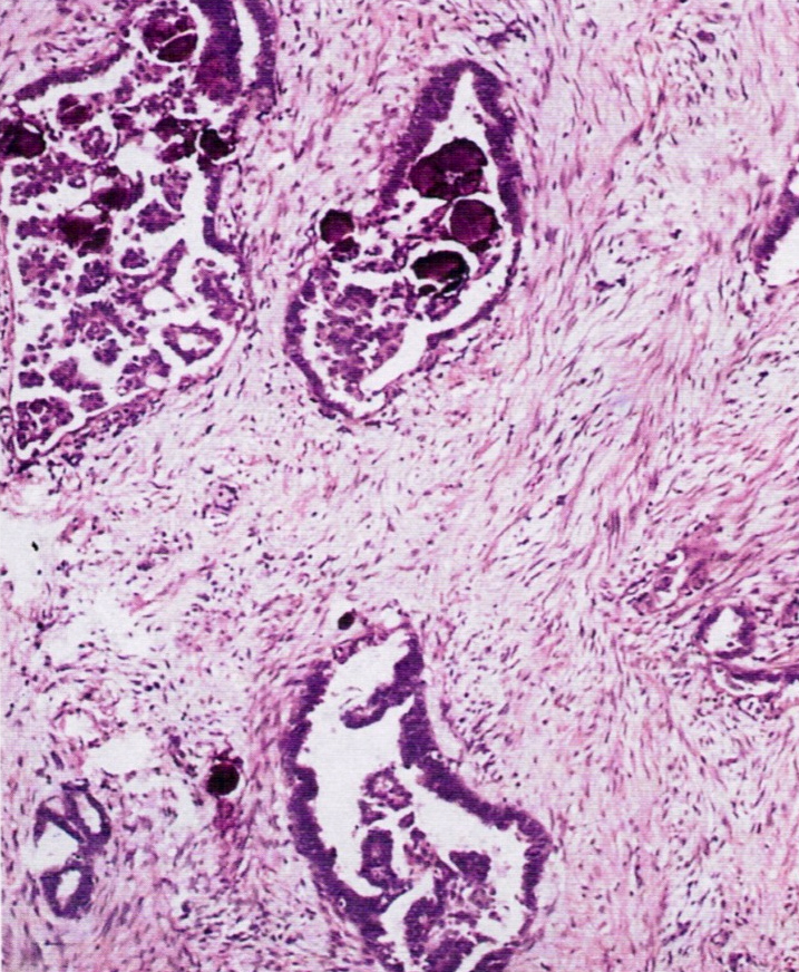
Cystically dilated glands in smooth muscle
Bland nuclear features
Gland-like structures
Numerous gland-like structures
Salpingitis isthmica nodosa
AFIP images
Mostly fibromatous surface
AFIP images
Resembling ovarian fibroma

Endometrioid adenofibroma
Images hosted on other servers:

Ultrasound / CT paratubal mass
Images hosted on other servers:

Right fallopian tube cyst
Images hosted on other servers:

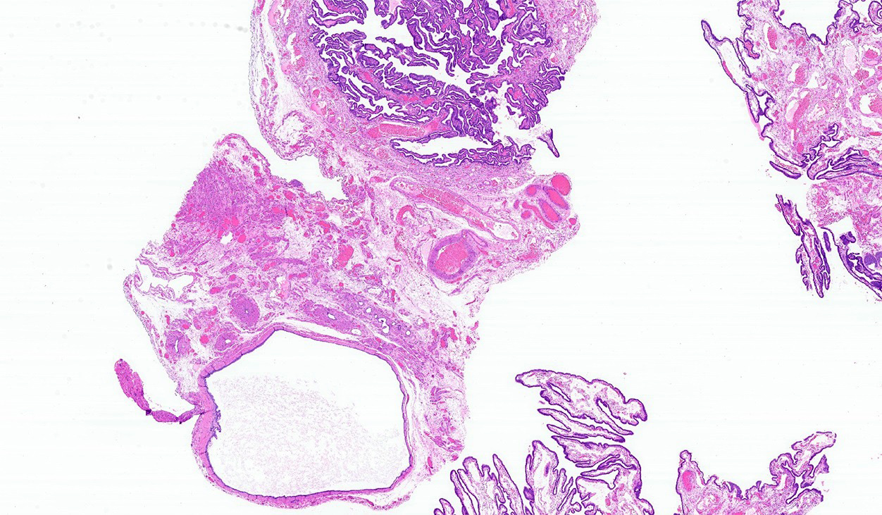
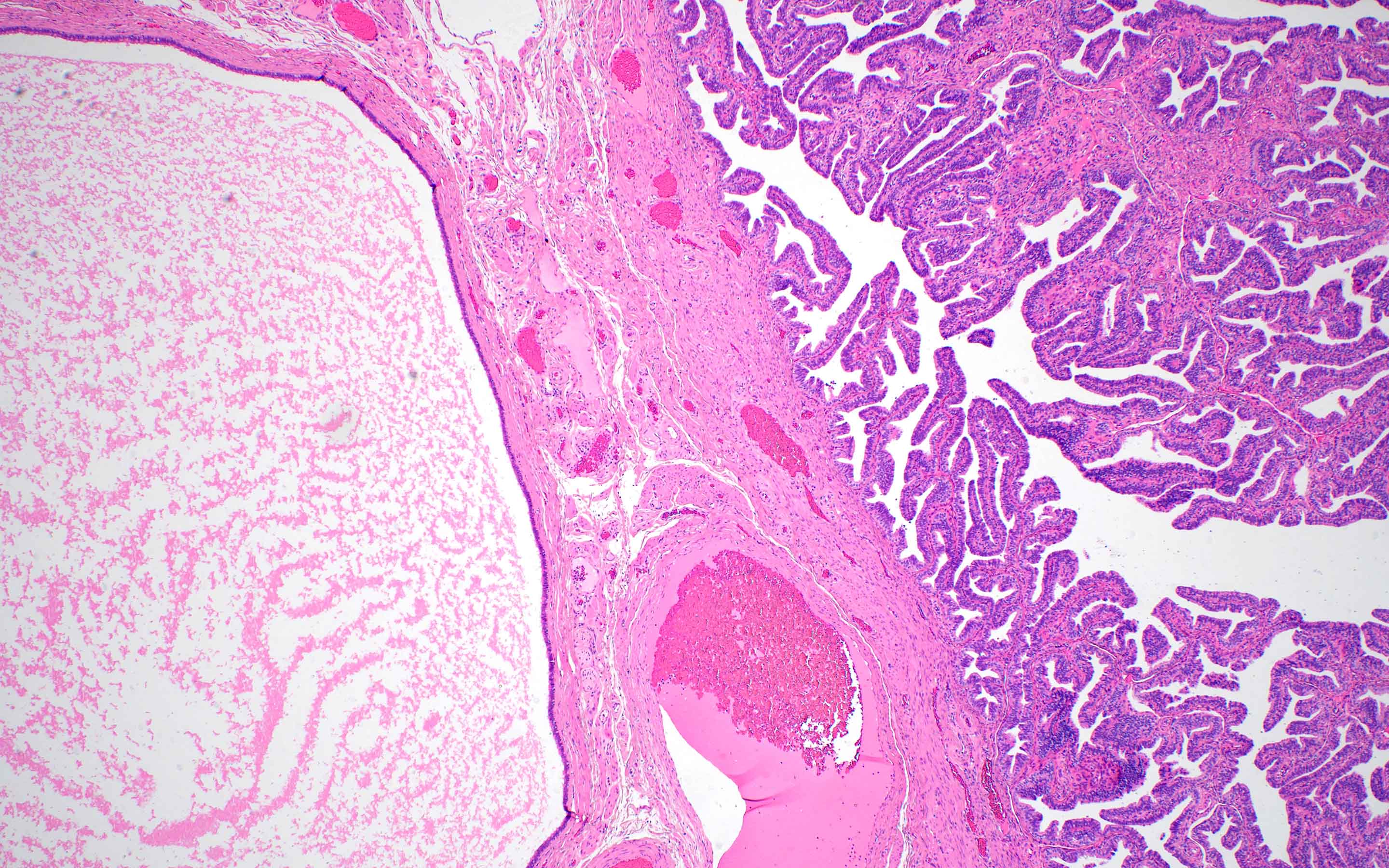
Fallopian tube cystic mass
Contributed by Kamaljeet Singh, M.D. and Anna Måsbäck, M.D., Ph.D.

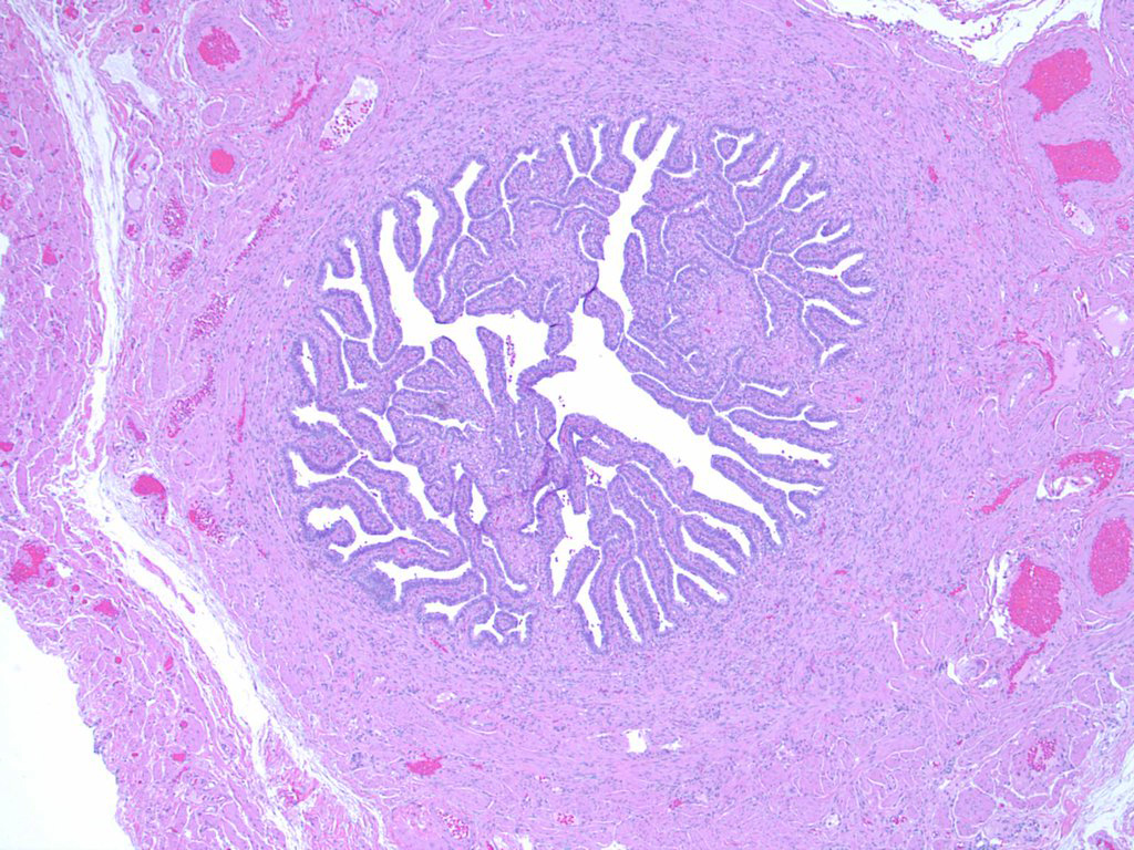
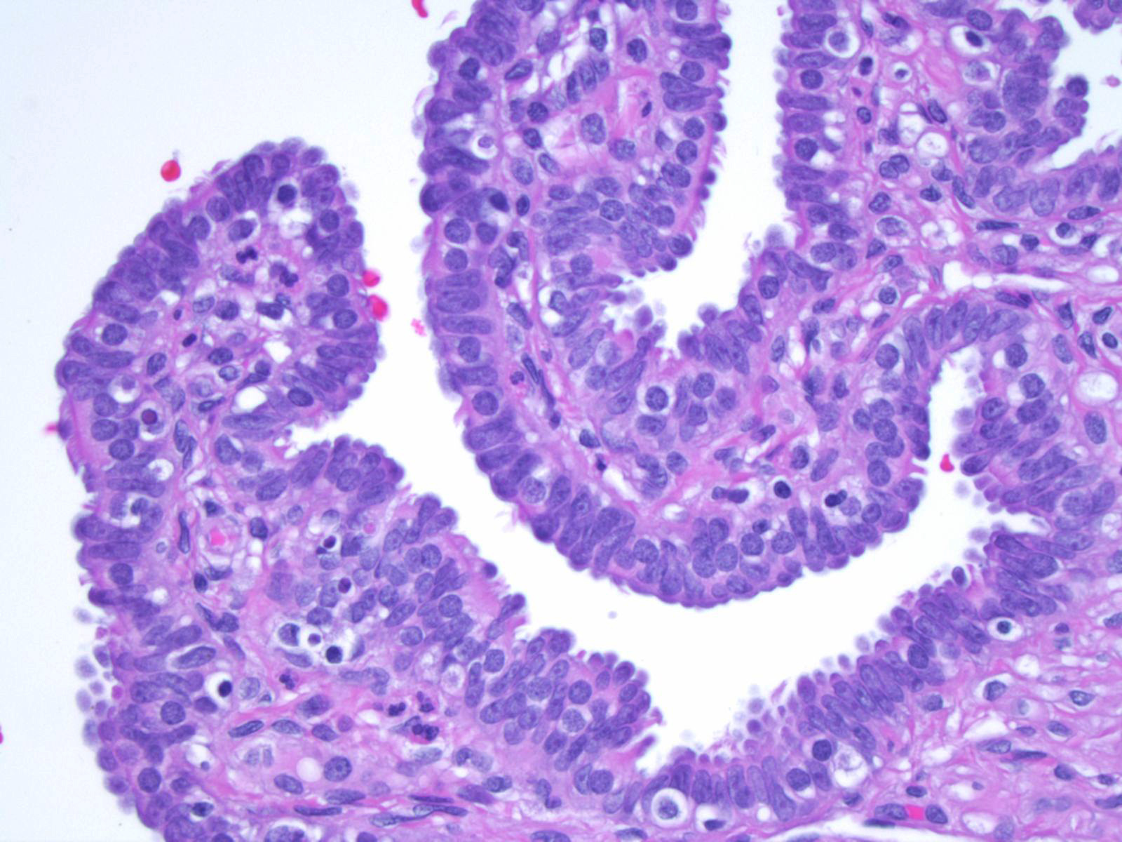
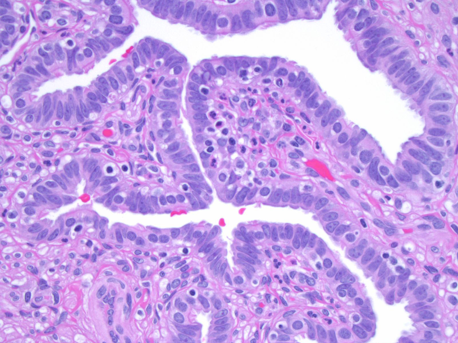
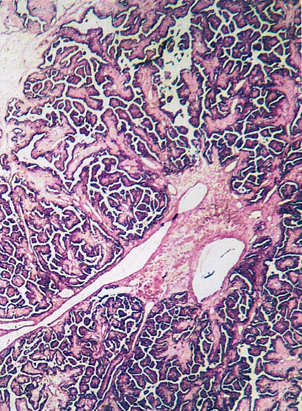
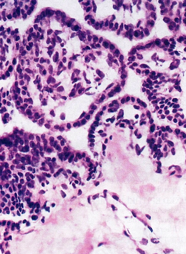
Papillary branching
Epithelial hobnailing
Variably sized papillae

Ciliated cells with hobnailing
Arborizing papillae
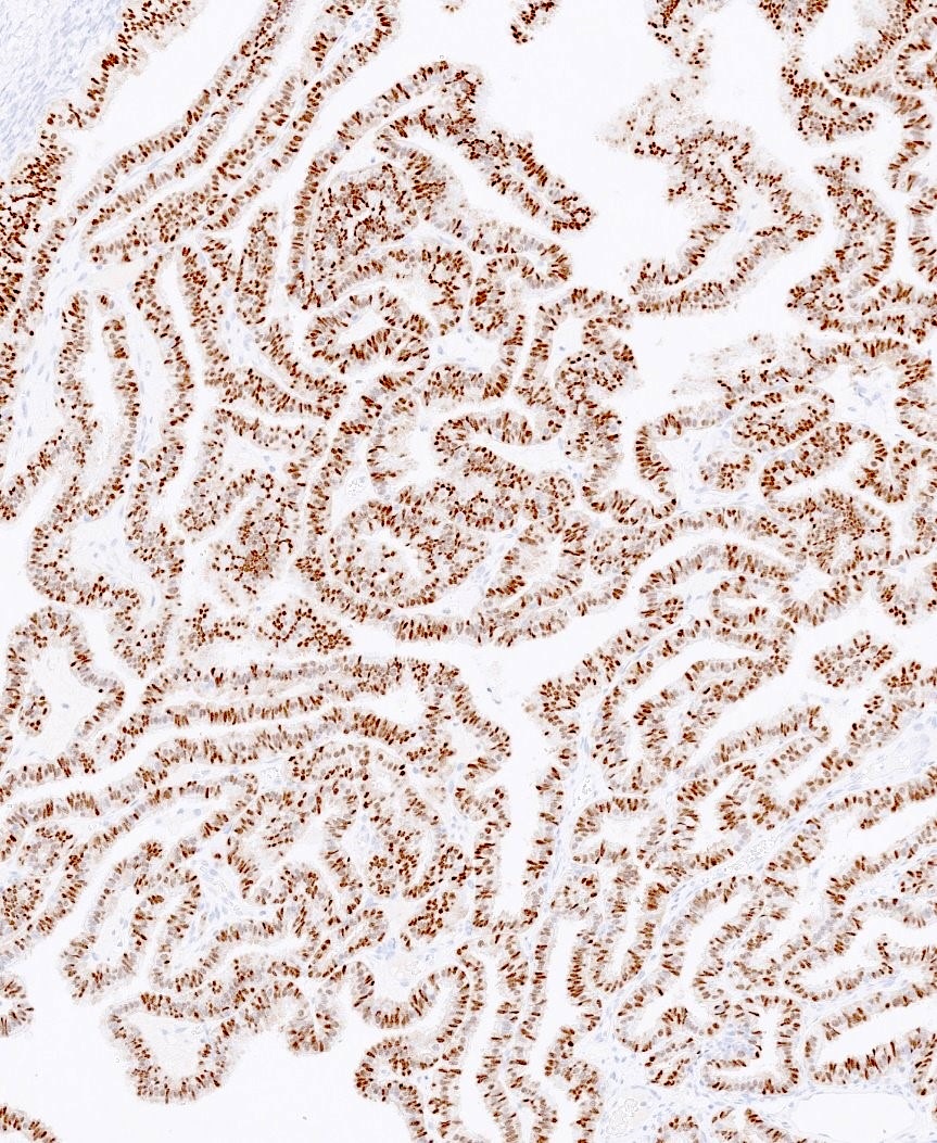
PAX8
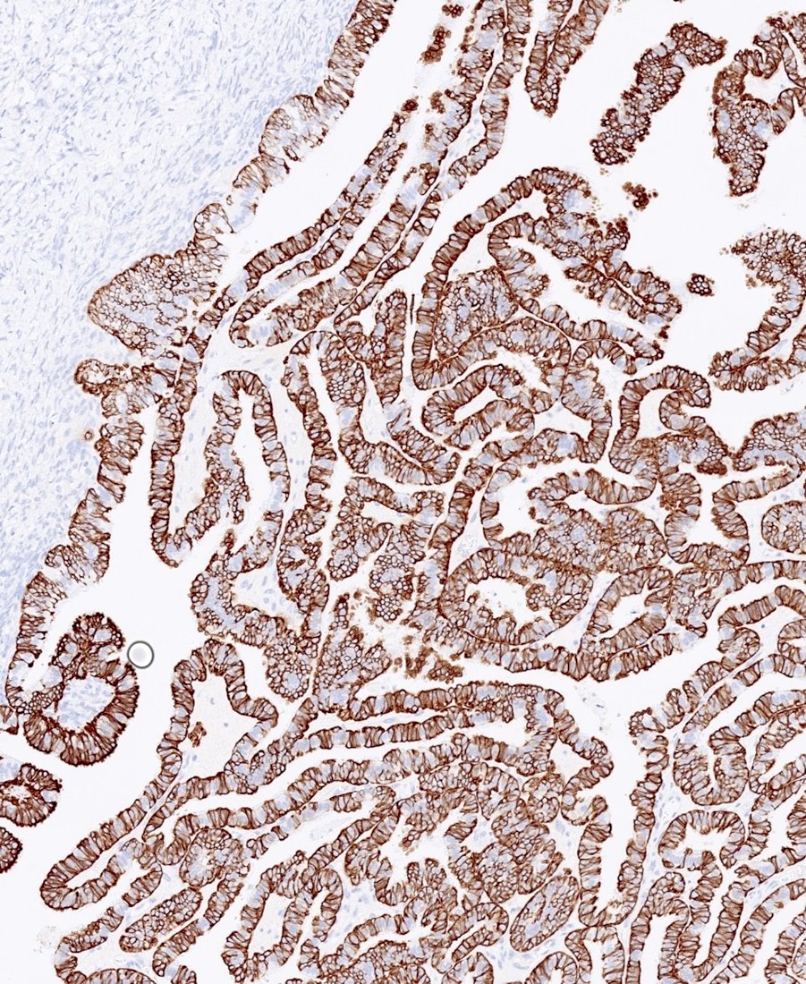
CK7
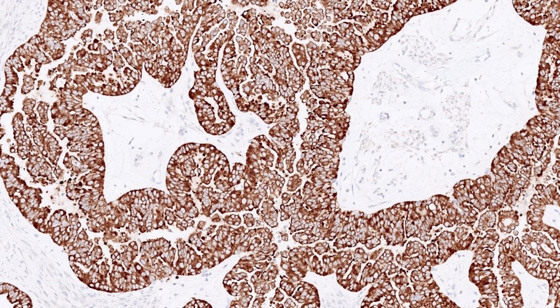
BRAF
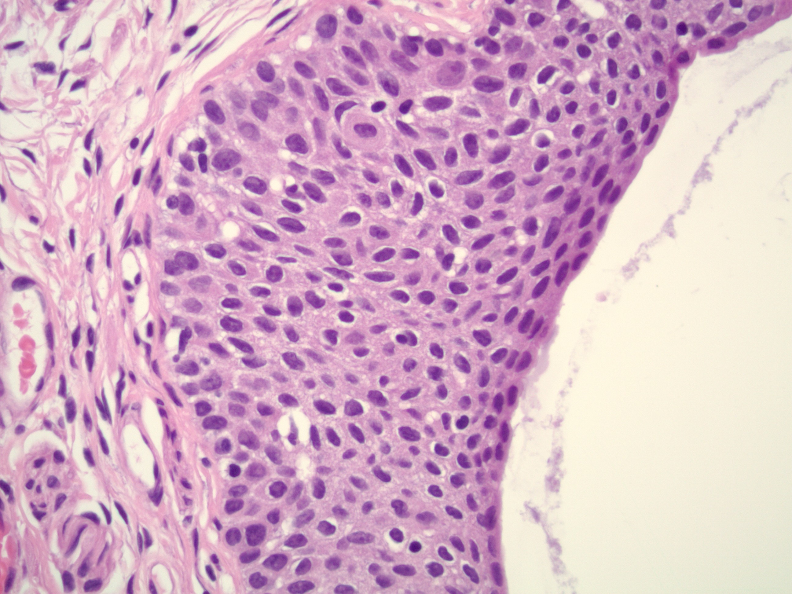
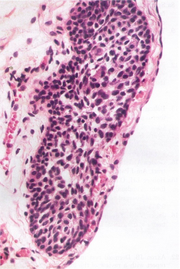
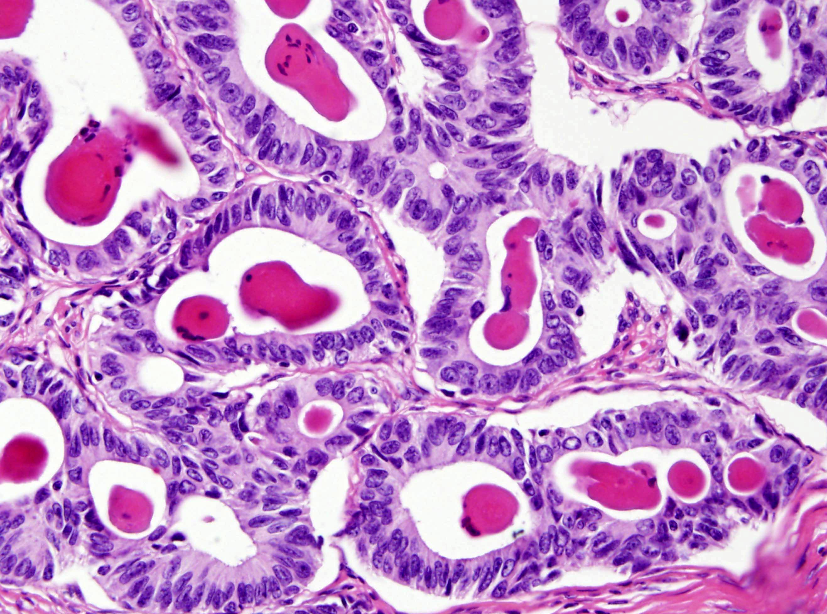
Contributed by Aysha Mubeen, M.D.

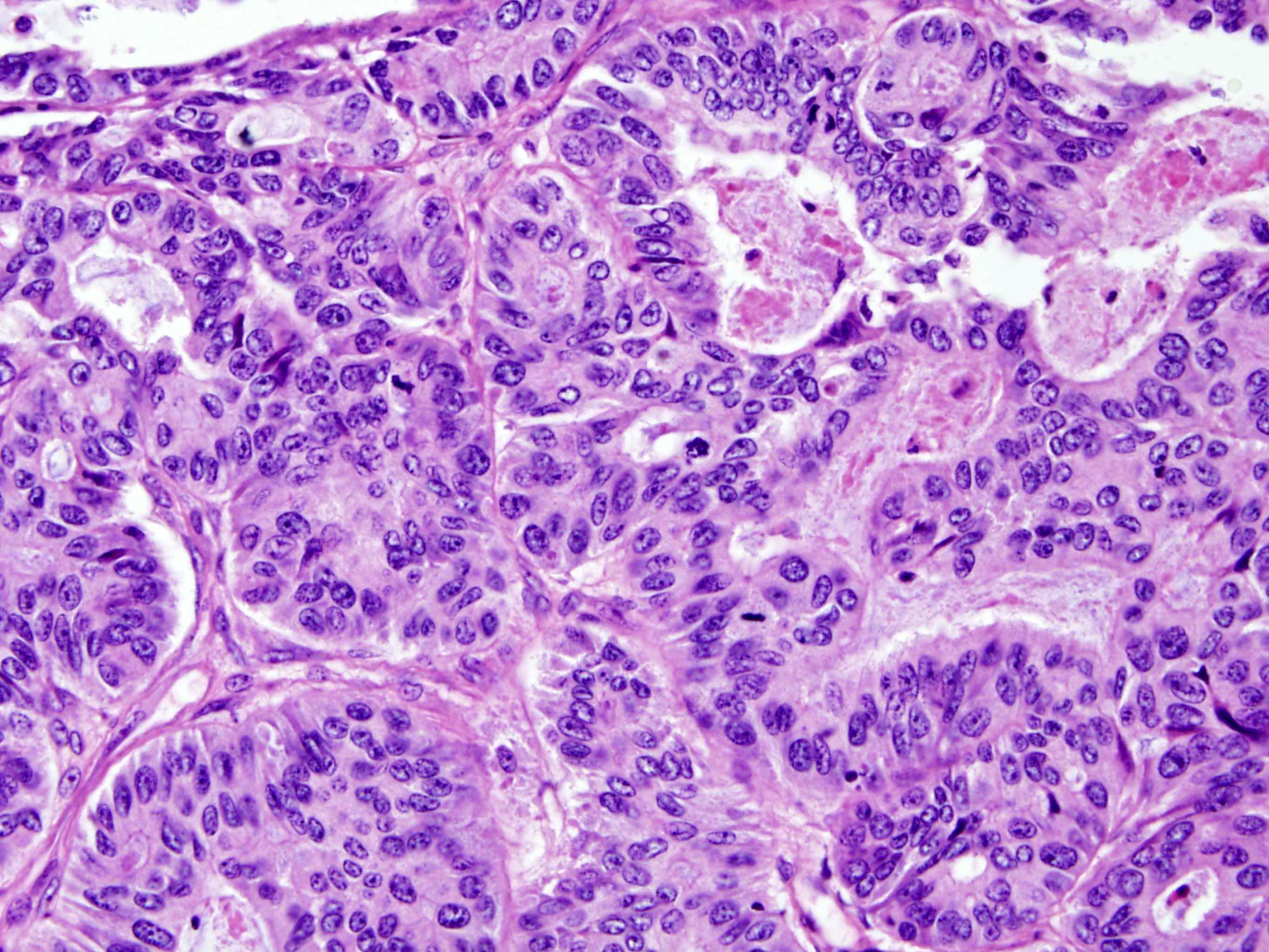
Basophilic appearance
Architectural and cytological atypia
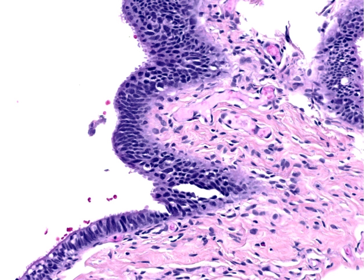
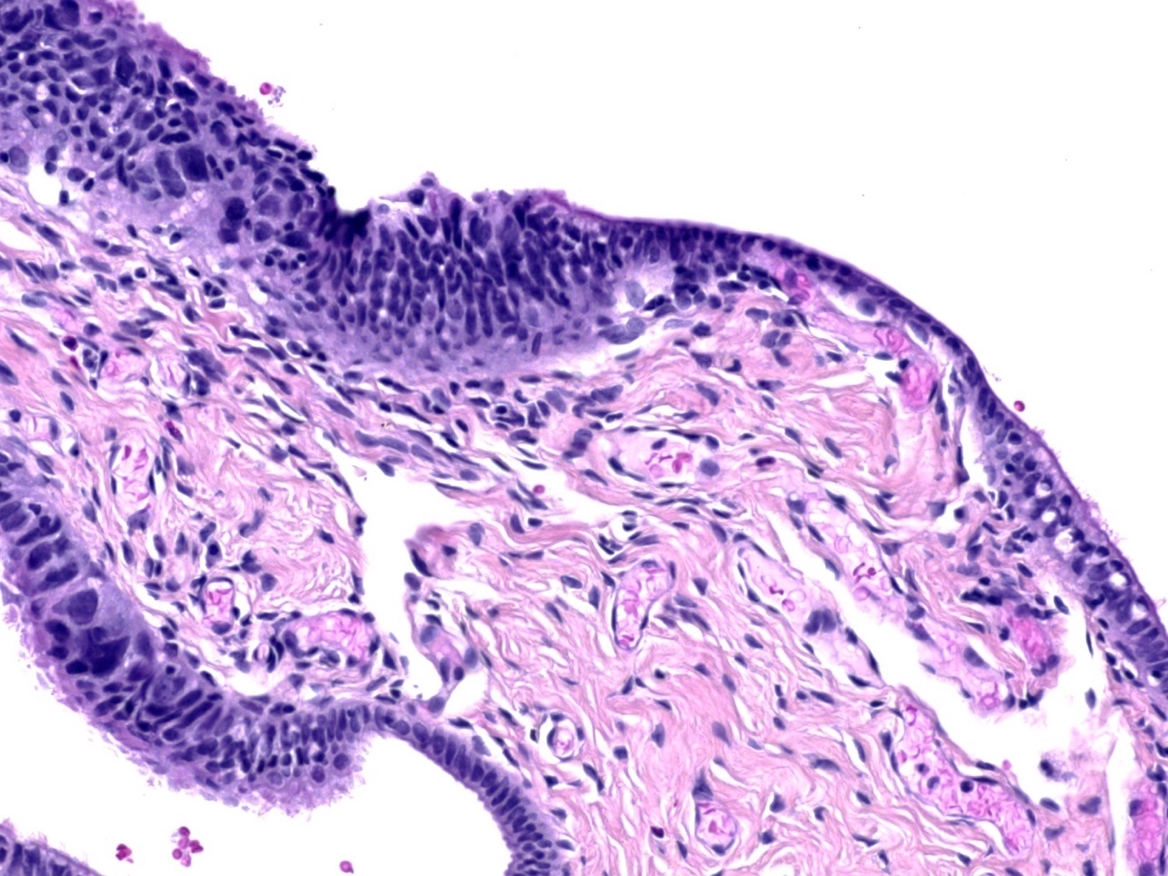
STIC in contrast with adjacent normal tubal epithelium
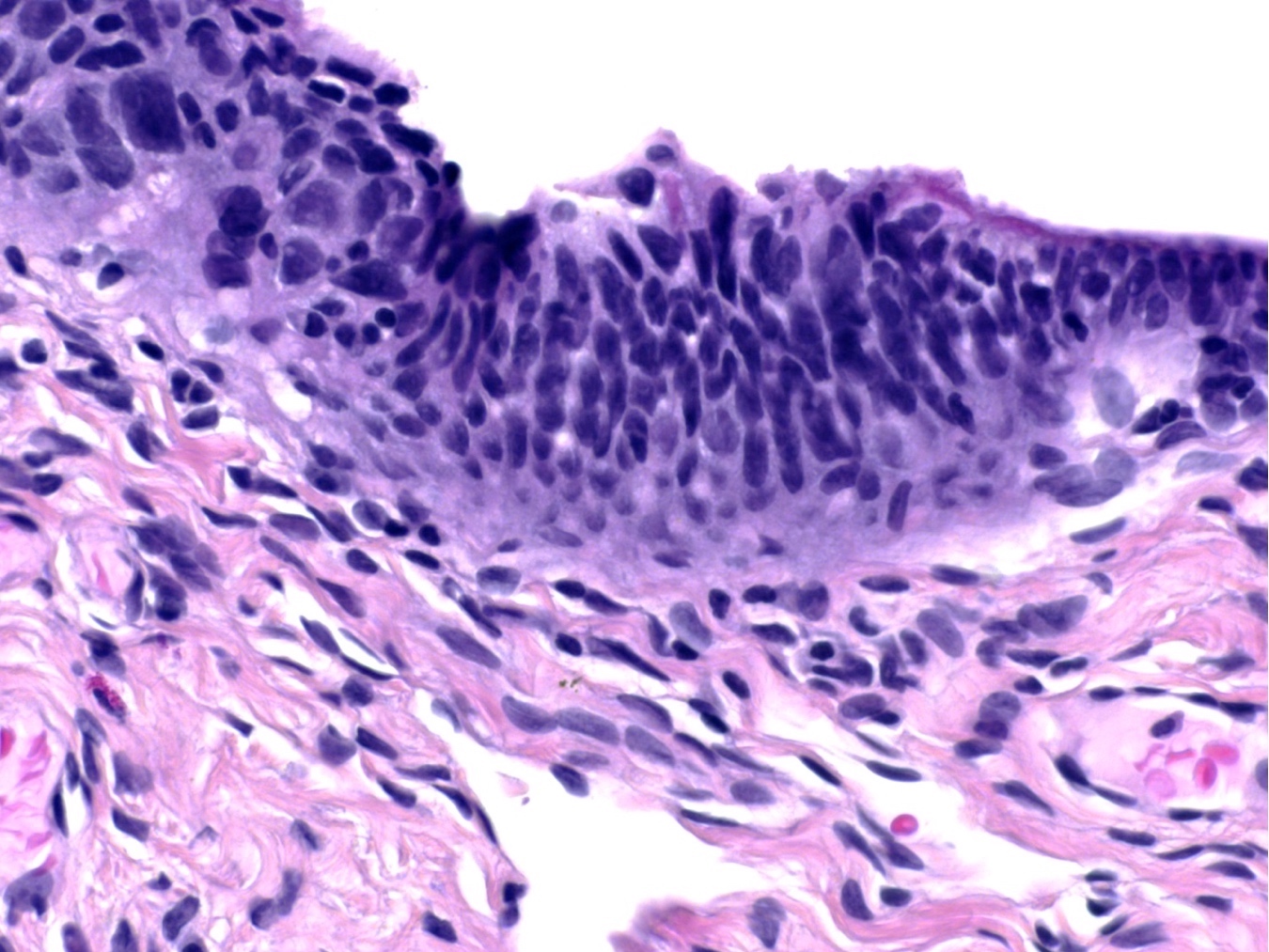
Prominent atypia and multilayering
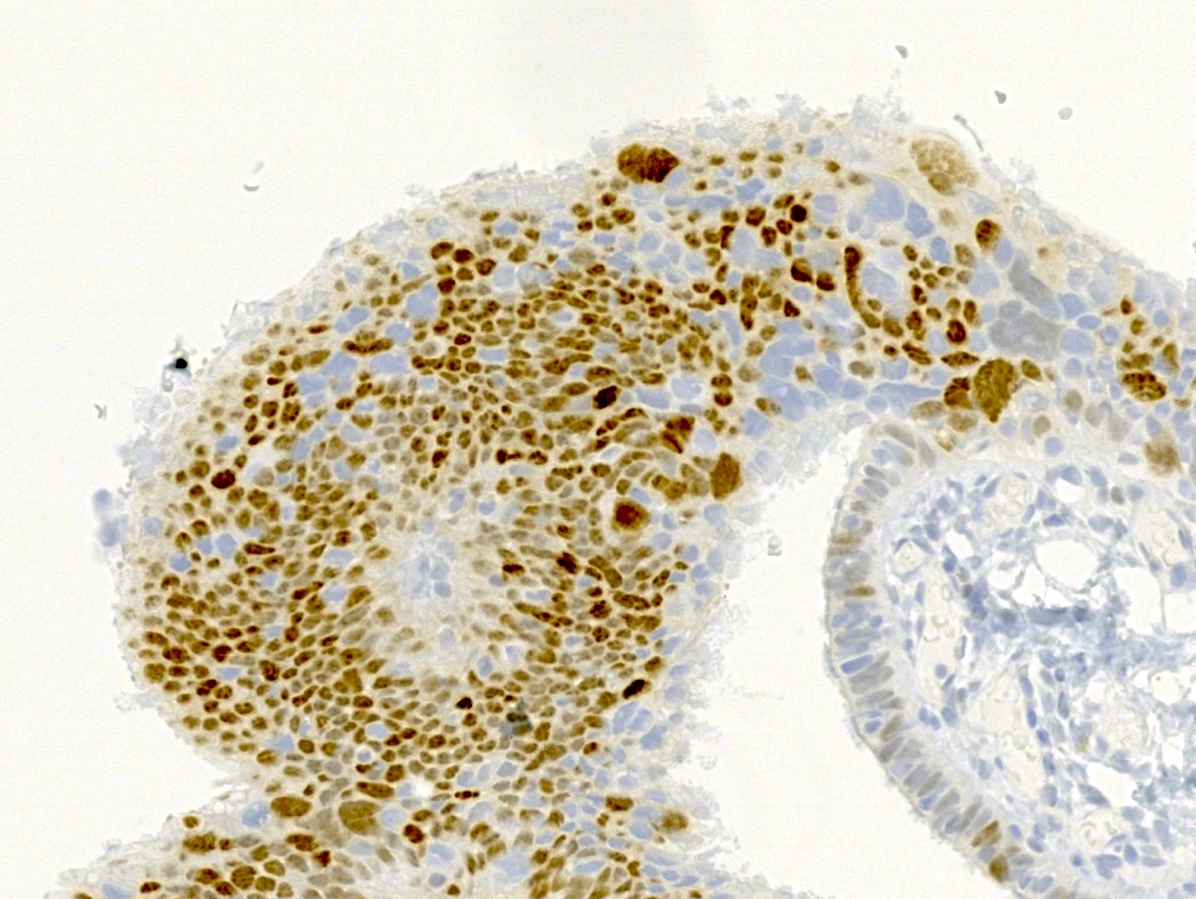
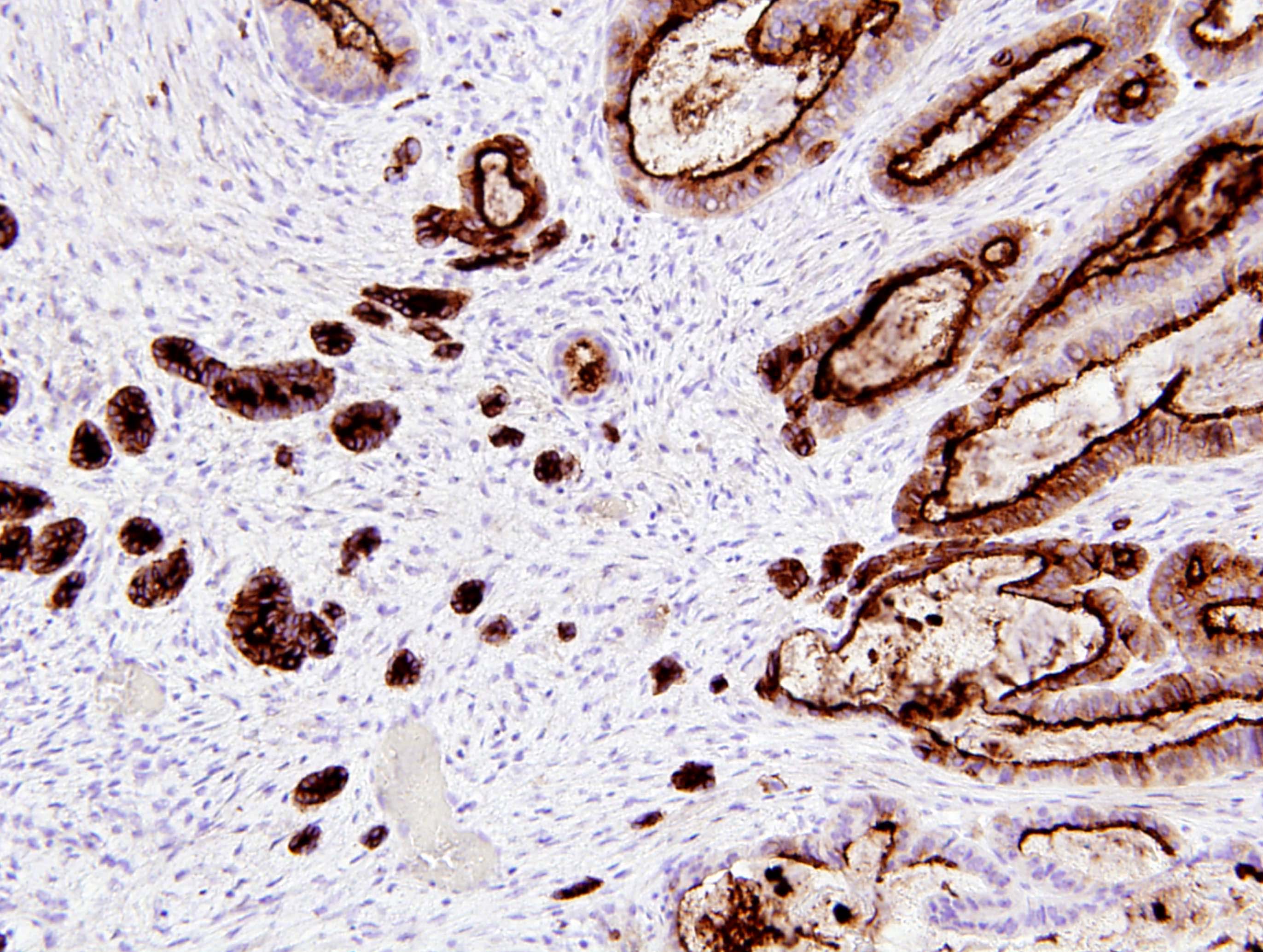
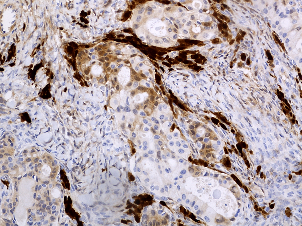
p53
Images hosted on other servers:

Laparoscopic findings
Contributed by Jennifer A. Bennett, M.D.
Paratubal location
Cord and trabeculae architecture
Cystic architecture
Broad columns
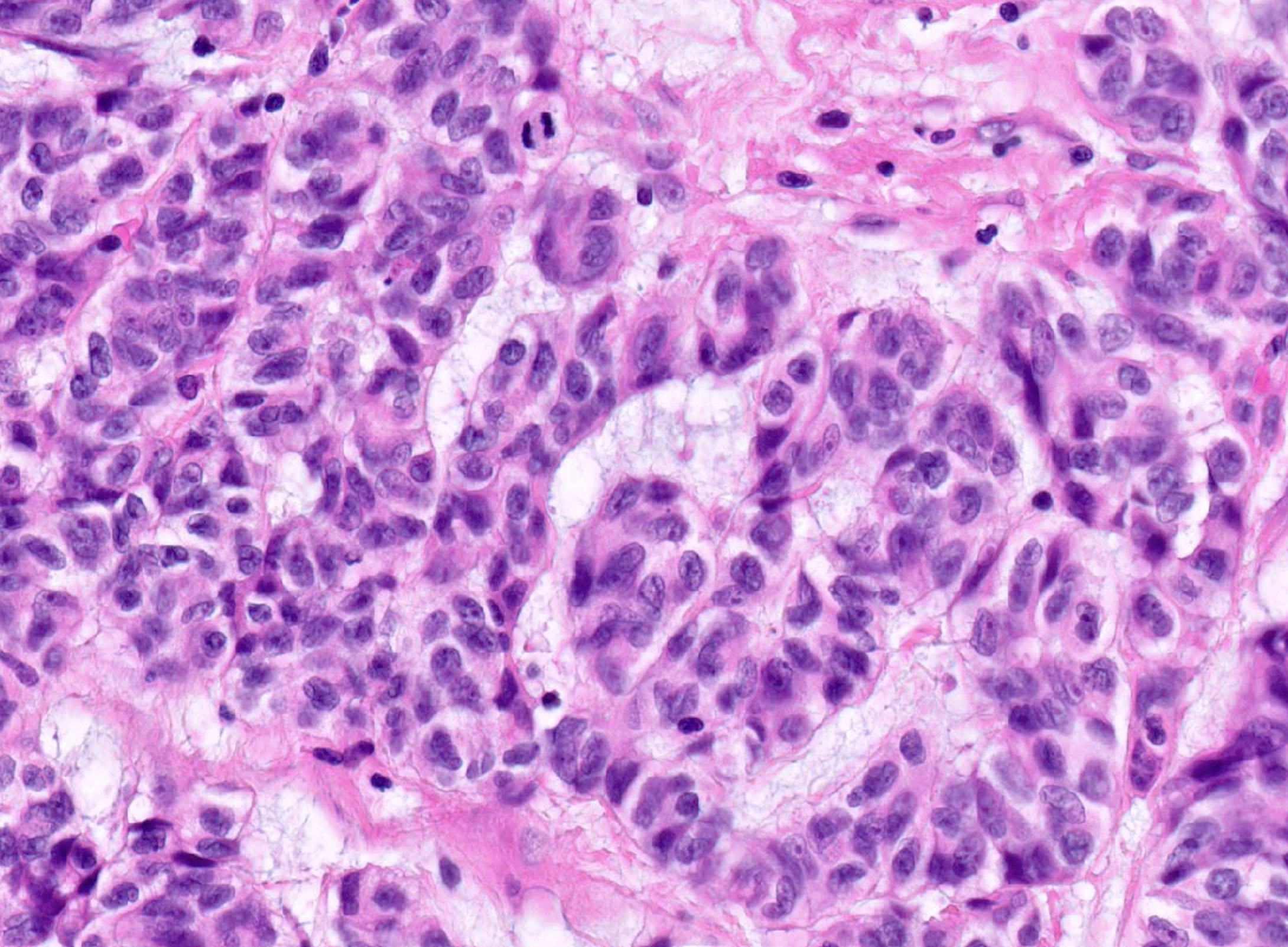
Nuclear features
Mitosis
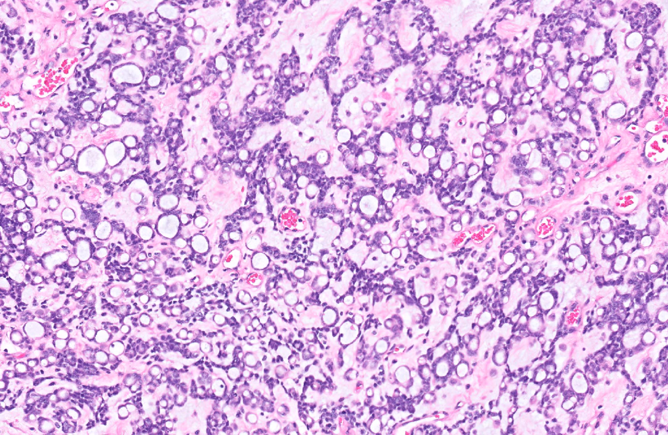
Signet ring-like cells
Pathology of fallopian tube
AFIP images
Hyperplasia of tubal epithelium of unknown cause
Clear cell hyperplasia of pregnancy
Images hosted on other servers:

Tubo-ovarian abscess from Neisseria gonorrheae
Images hosted on other servers:

Neisseria gonorrheae

H&E
AFIP images
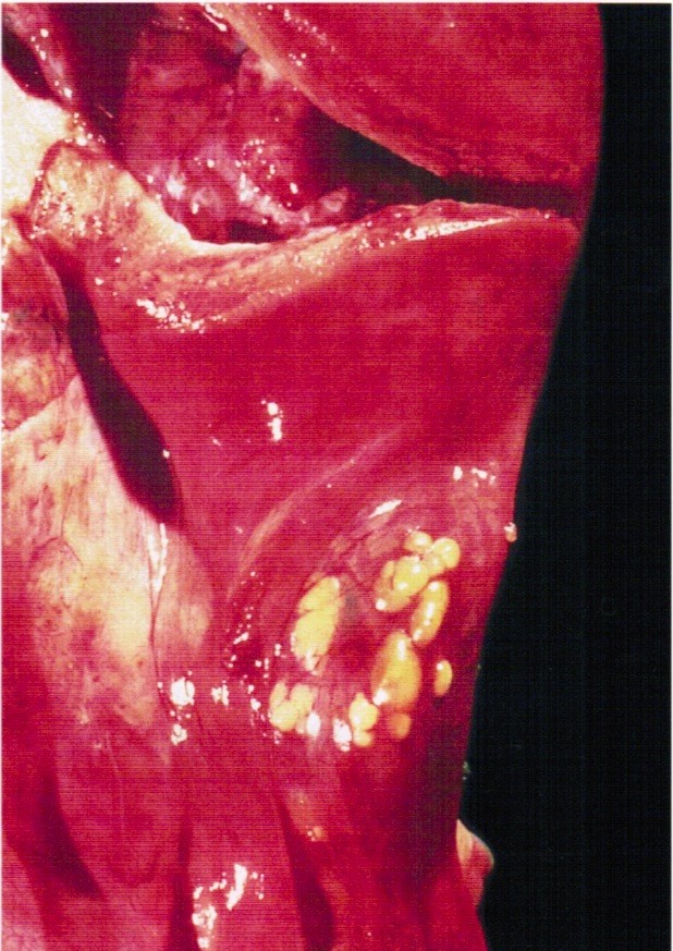
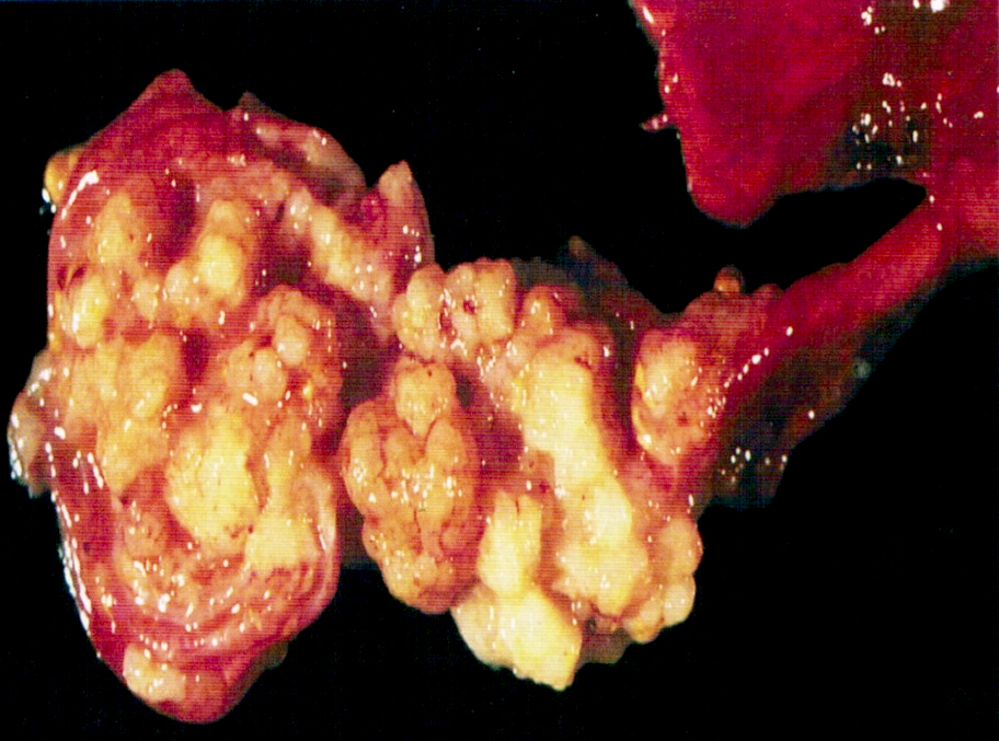
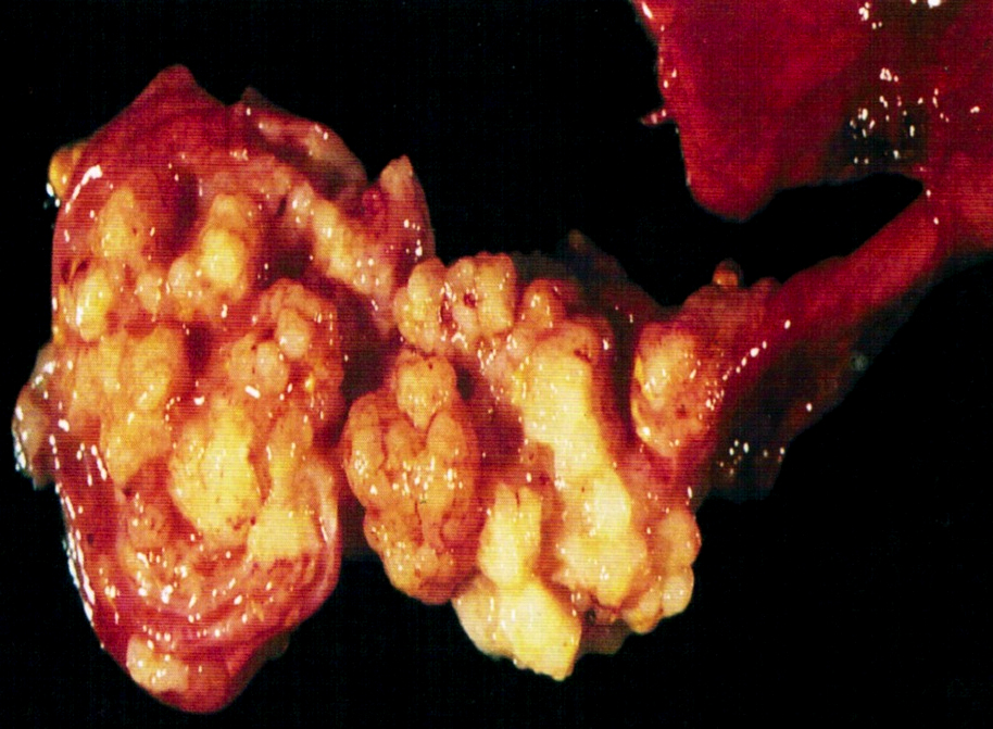
Several small yellow nodules
Contributed by Shabnam Zarei, M.D. and AFIP images
Transitional type epithelium
Cystic nests
Bland appearing transitional cells
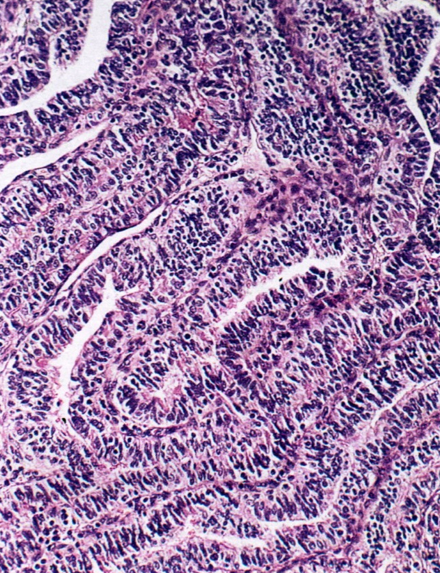
Contributed by Gulisa Turashvili, M.D., Ph.D.
STIC associated with invasion
Endometrioid adenocarcinoma

Salpingitis isthmica nodosa
Images hosted on other servers:

CT and MRI: large mass

MRI: mass near ovary
Images hosted on other servers:

Adnexal mass
AFIP images
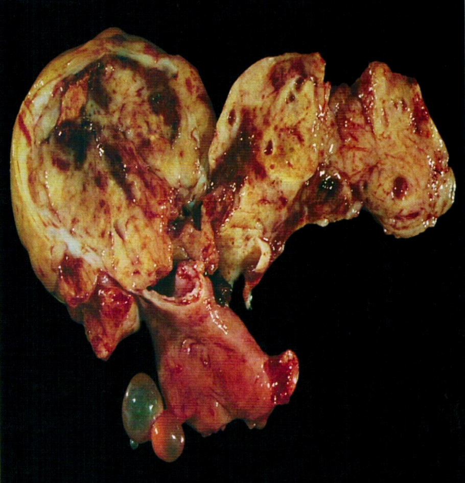
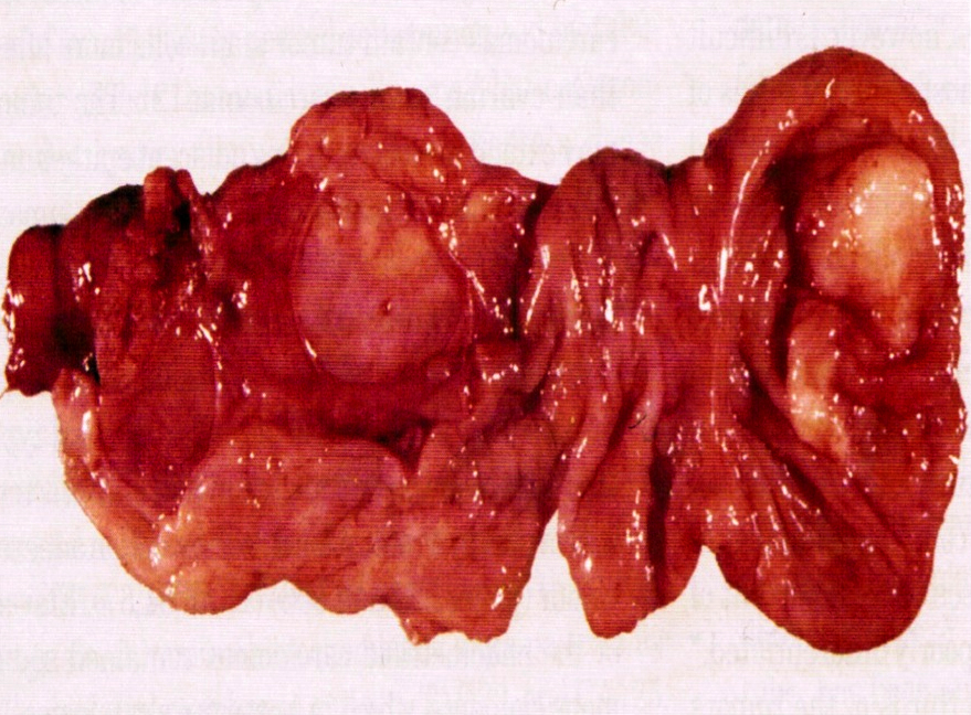
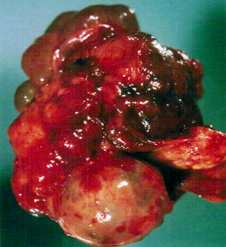
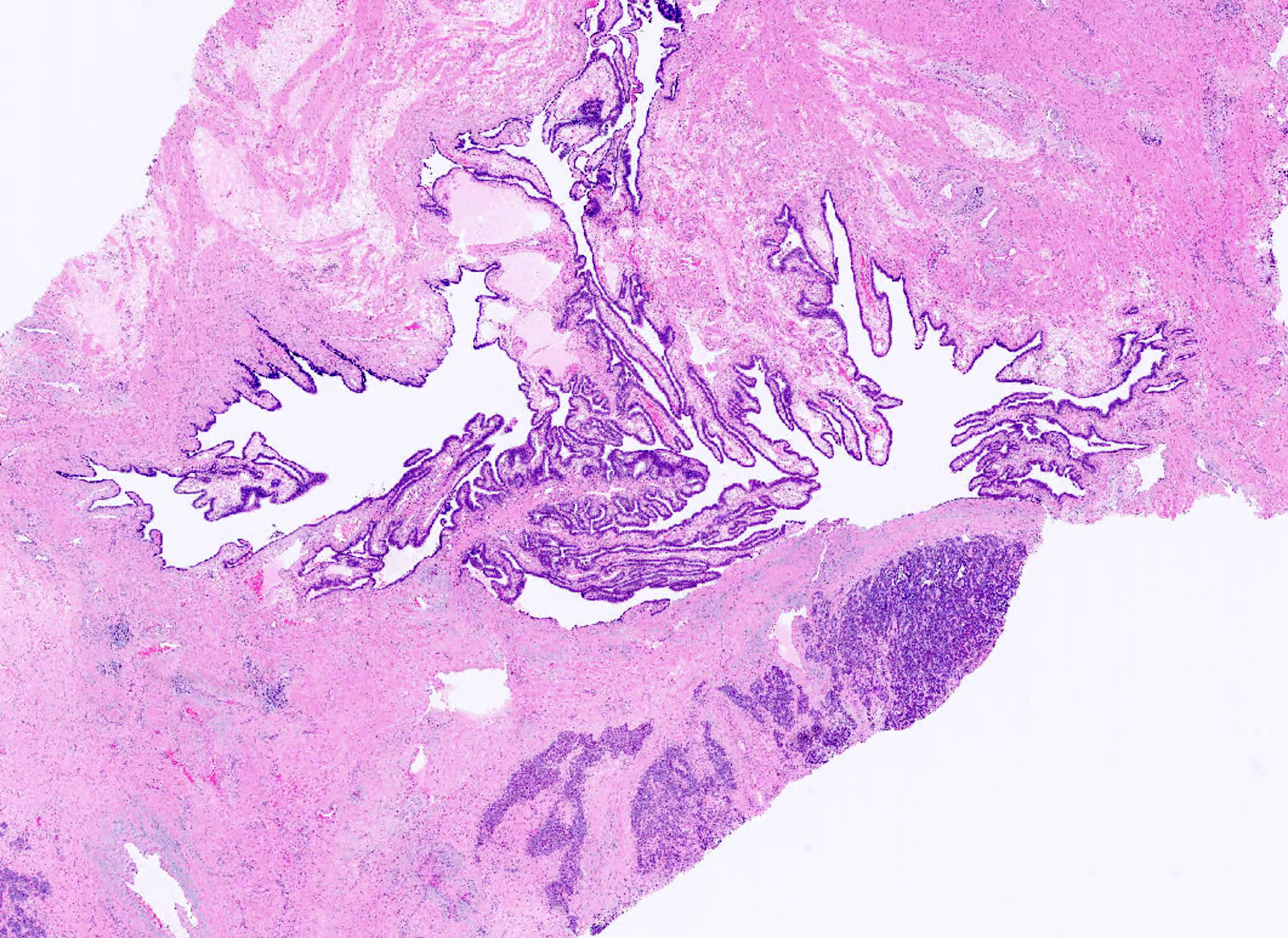
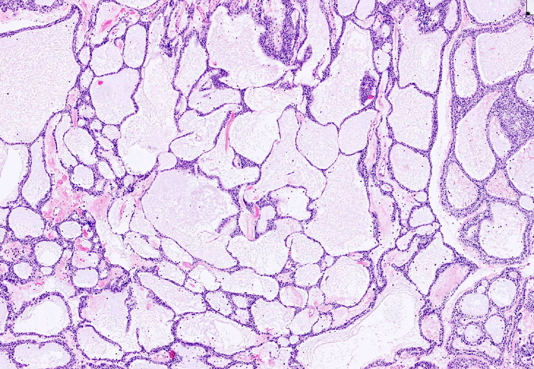
Broad ligament tumor
Solid and cystic cut surface
Lobulated cut surface
Images hosted on other servers:

Large peritoneal mass

Mass arising from the fimbriae
Contributed by Elena Lucas, M.D. and Wenxin Zheng, M.D.
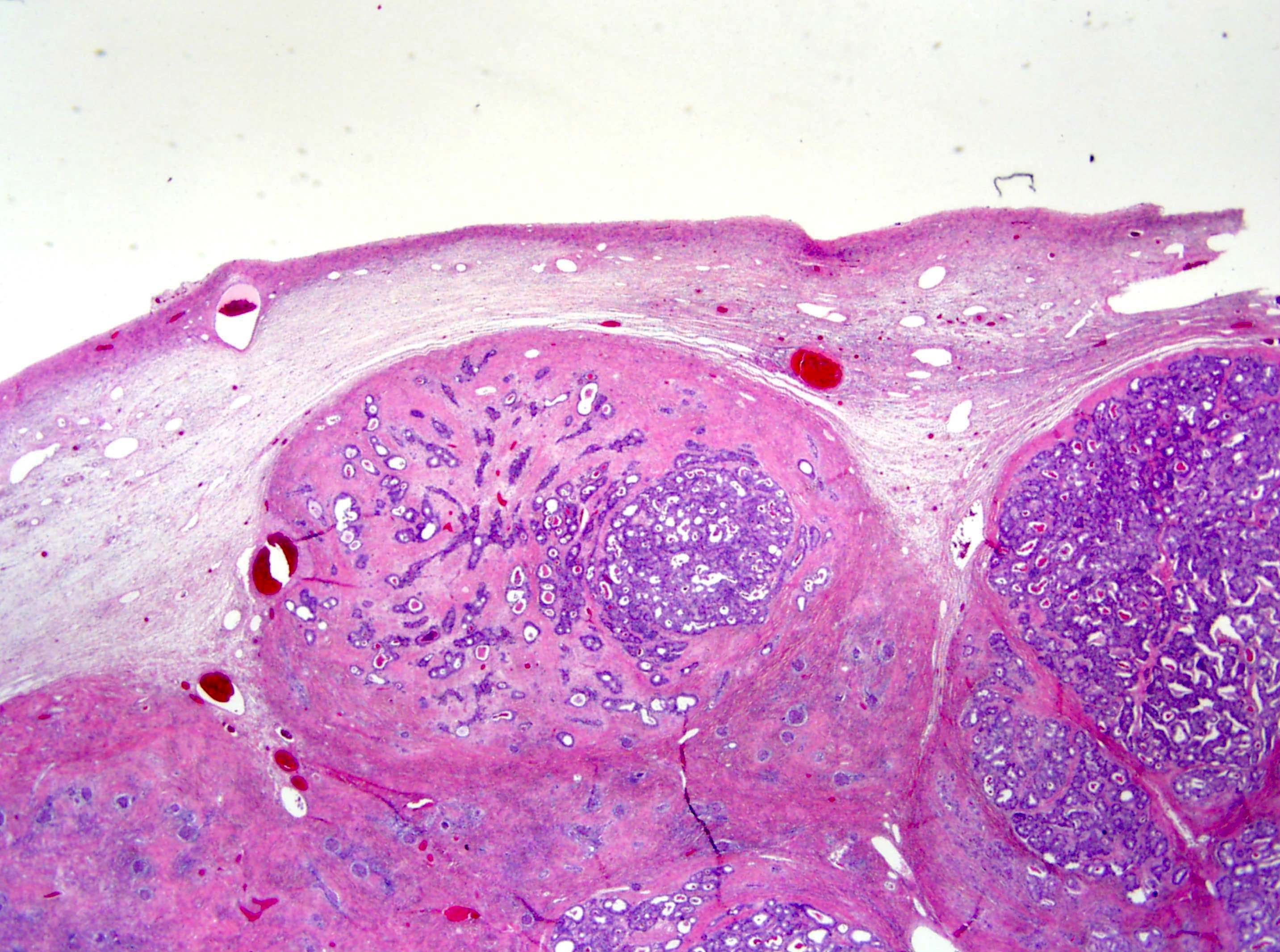
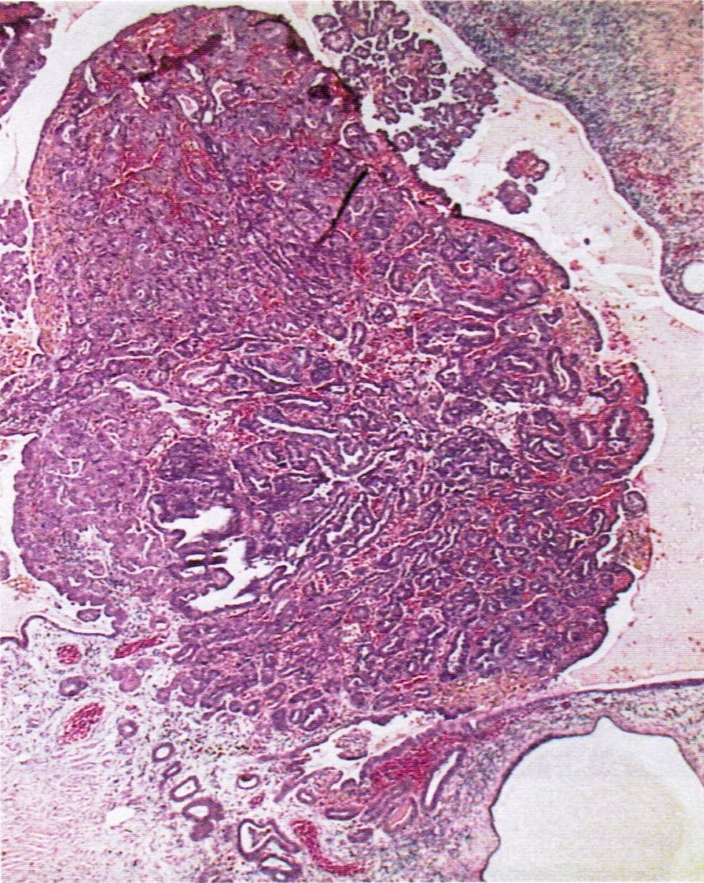
Nodular architecture
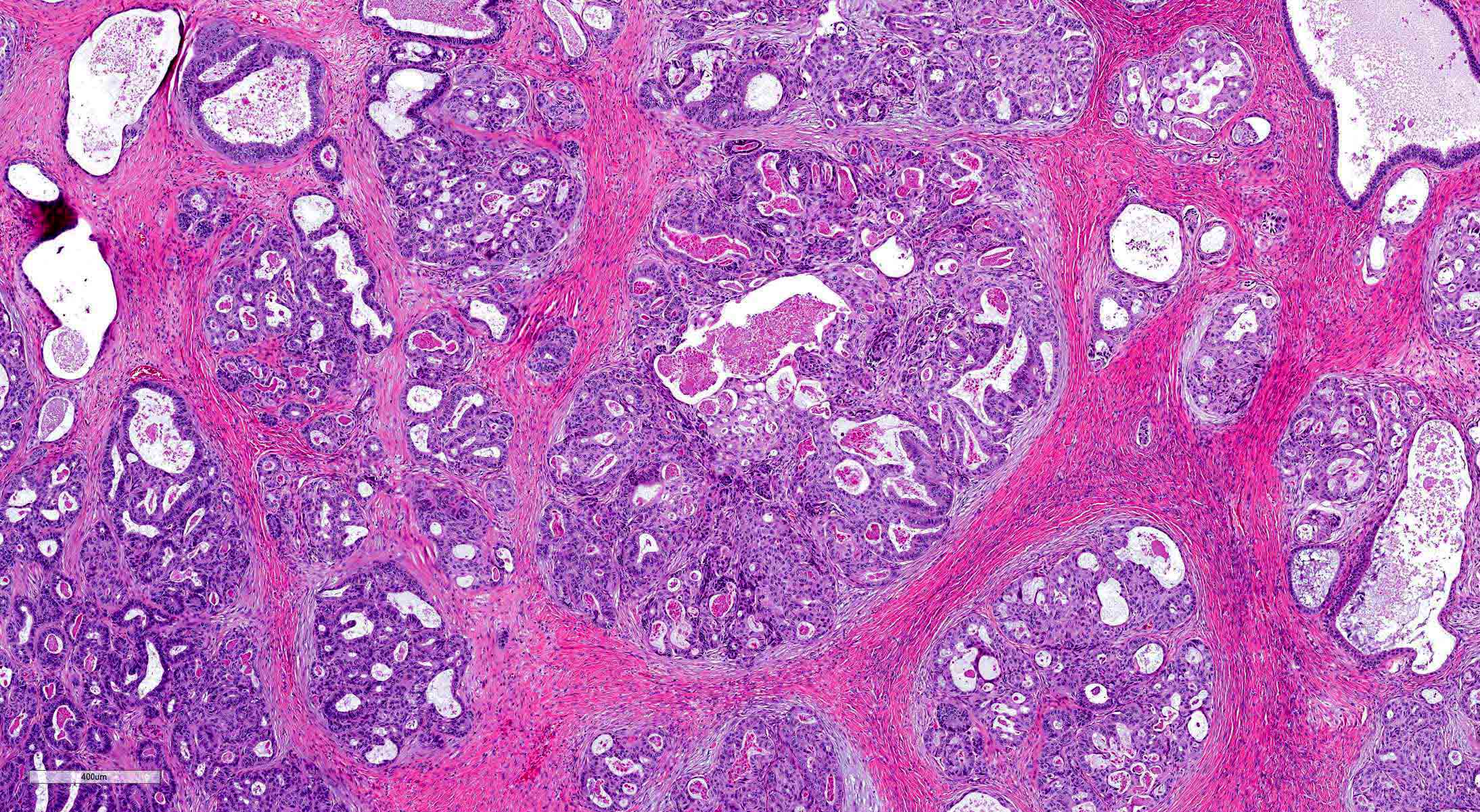
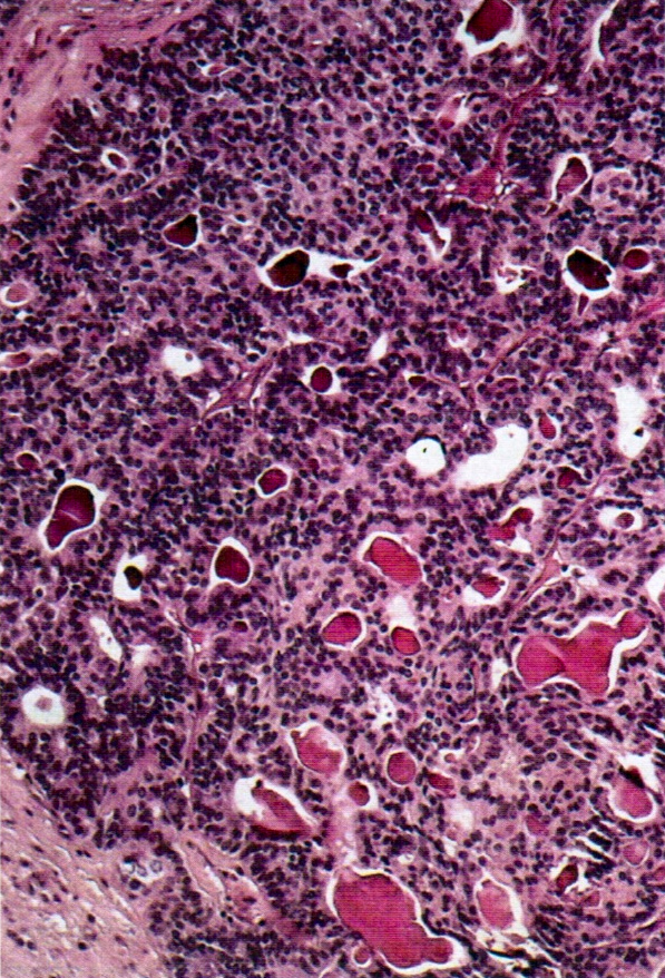
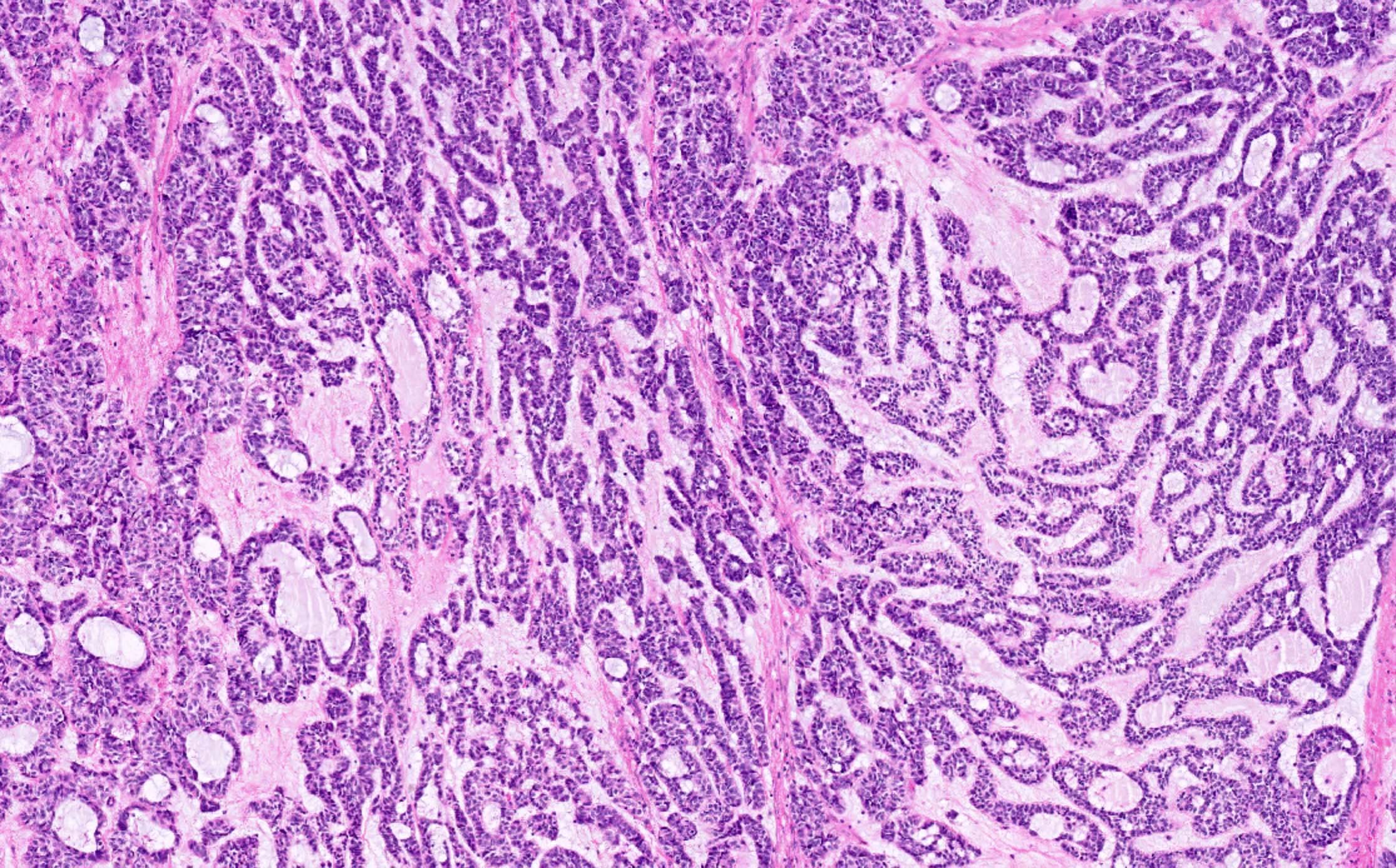
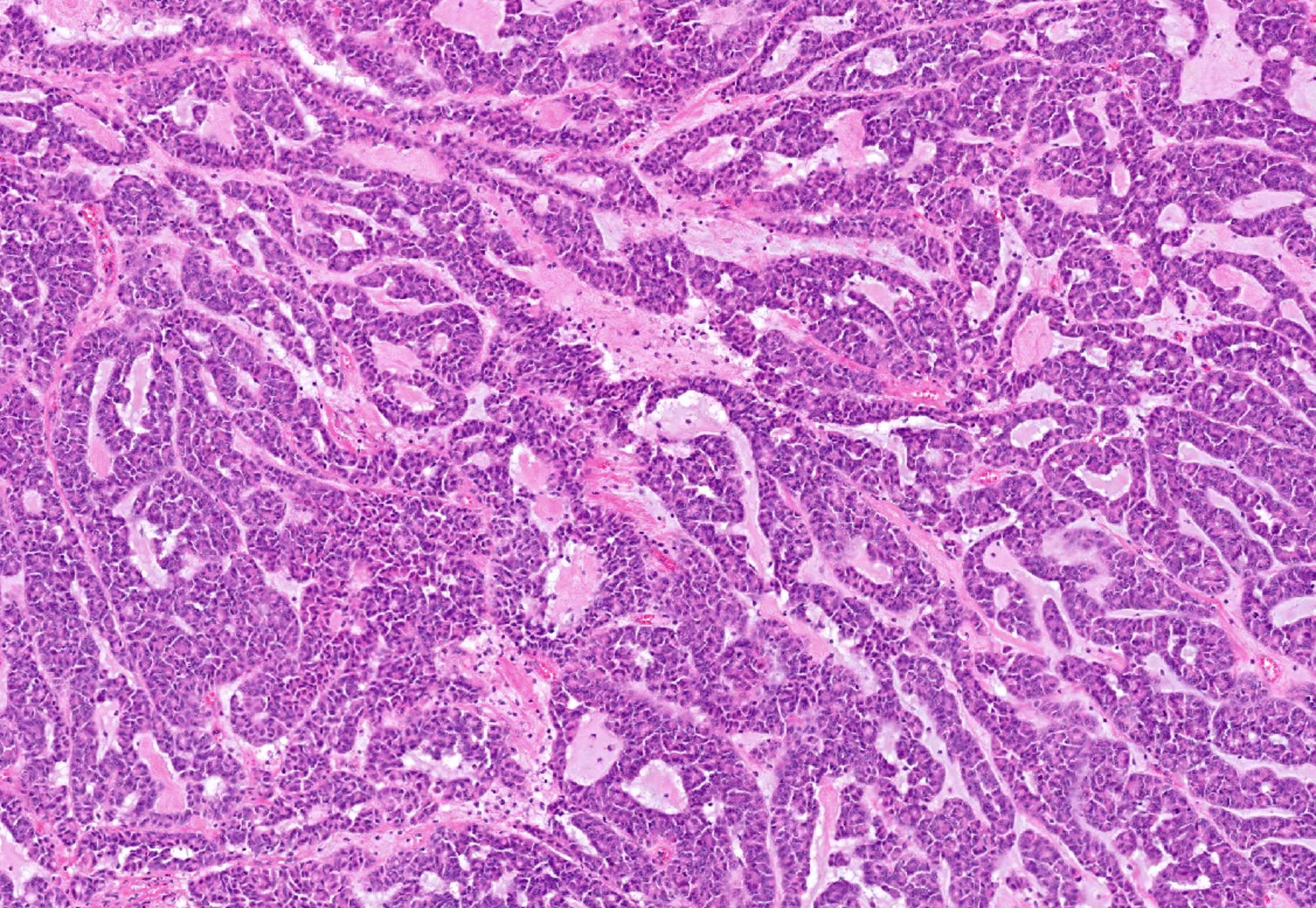
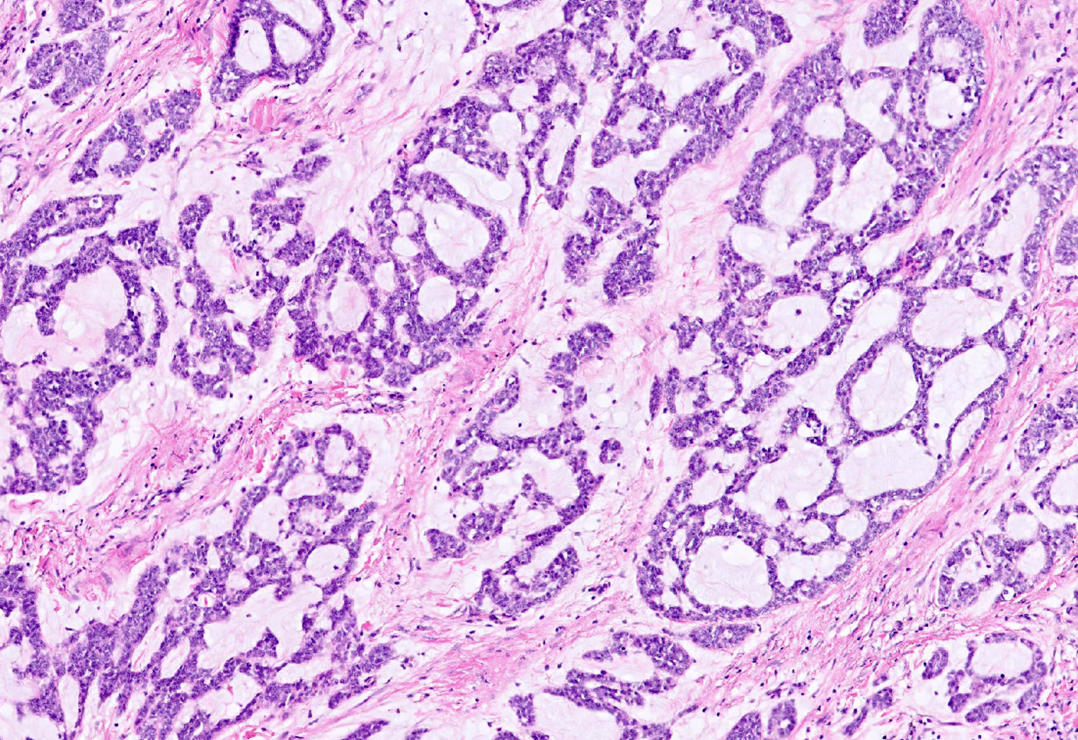
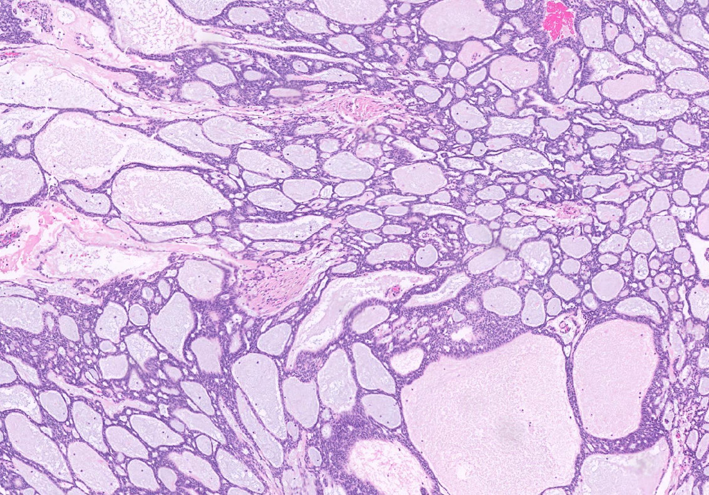
FATWO showing various patterns
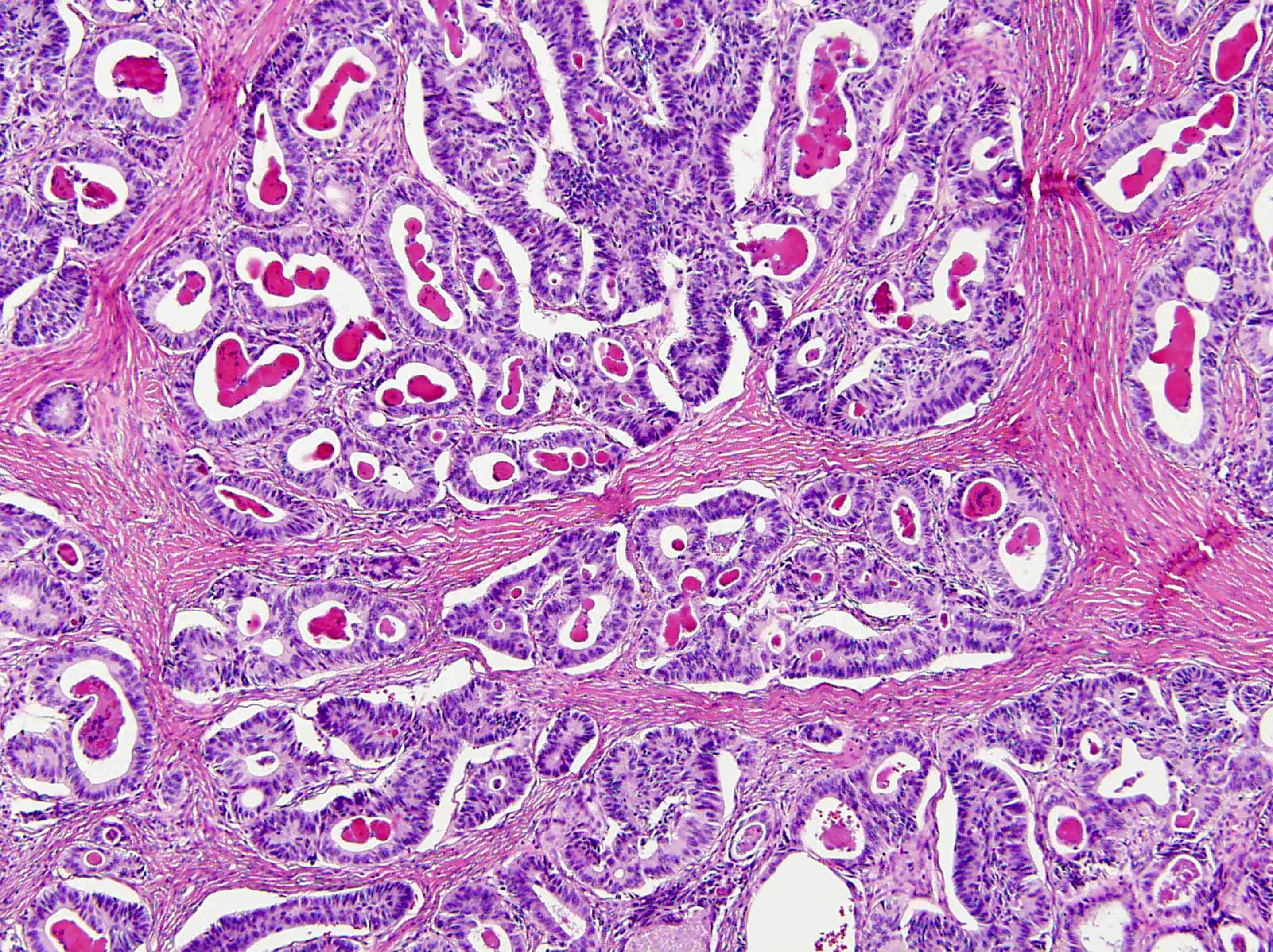
Tubular growth pattern
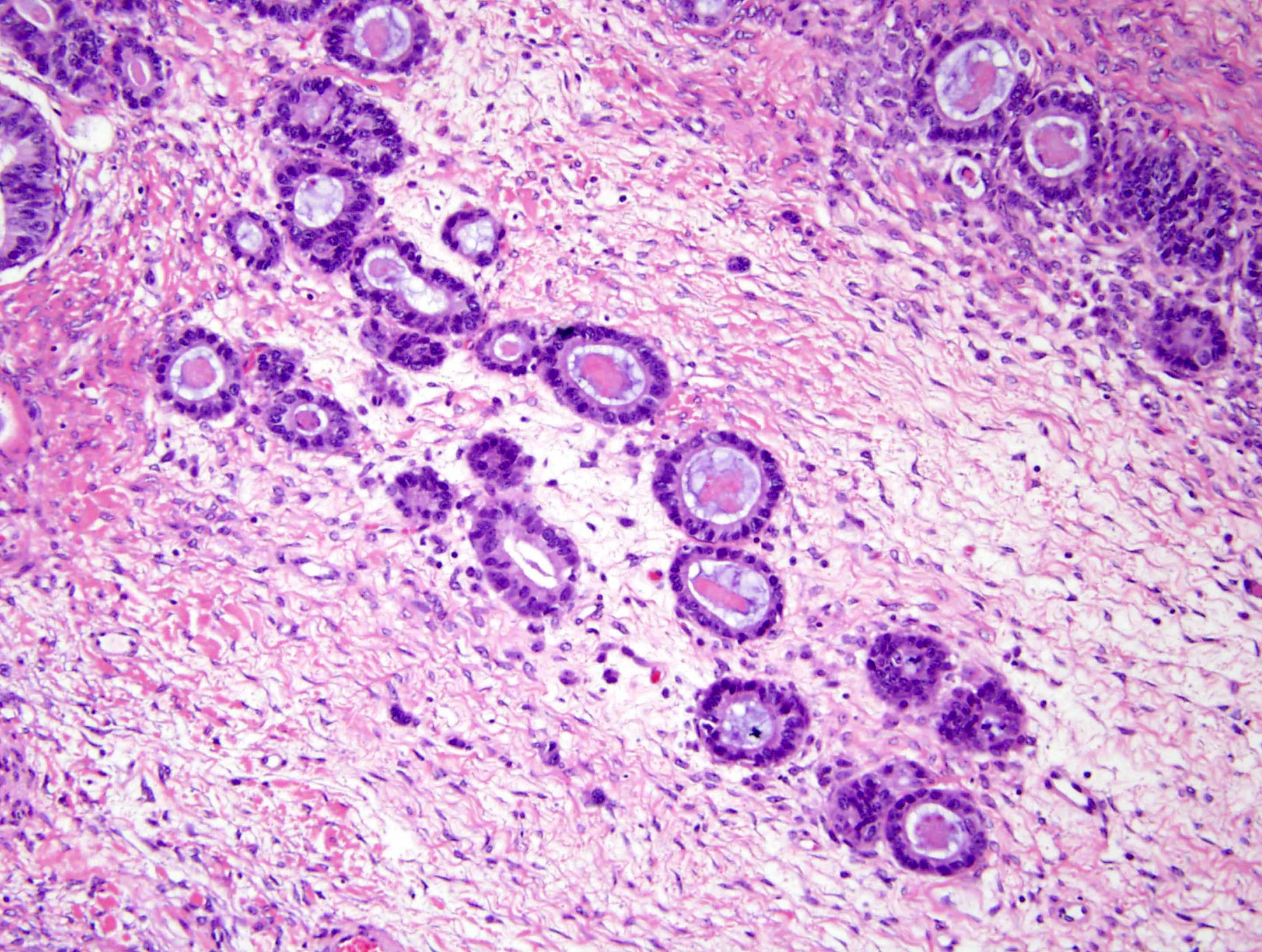
Tubules infiltrating stroma
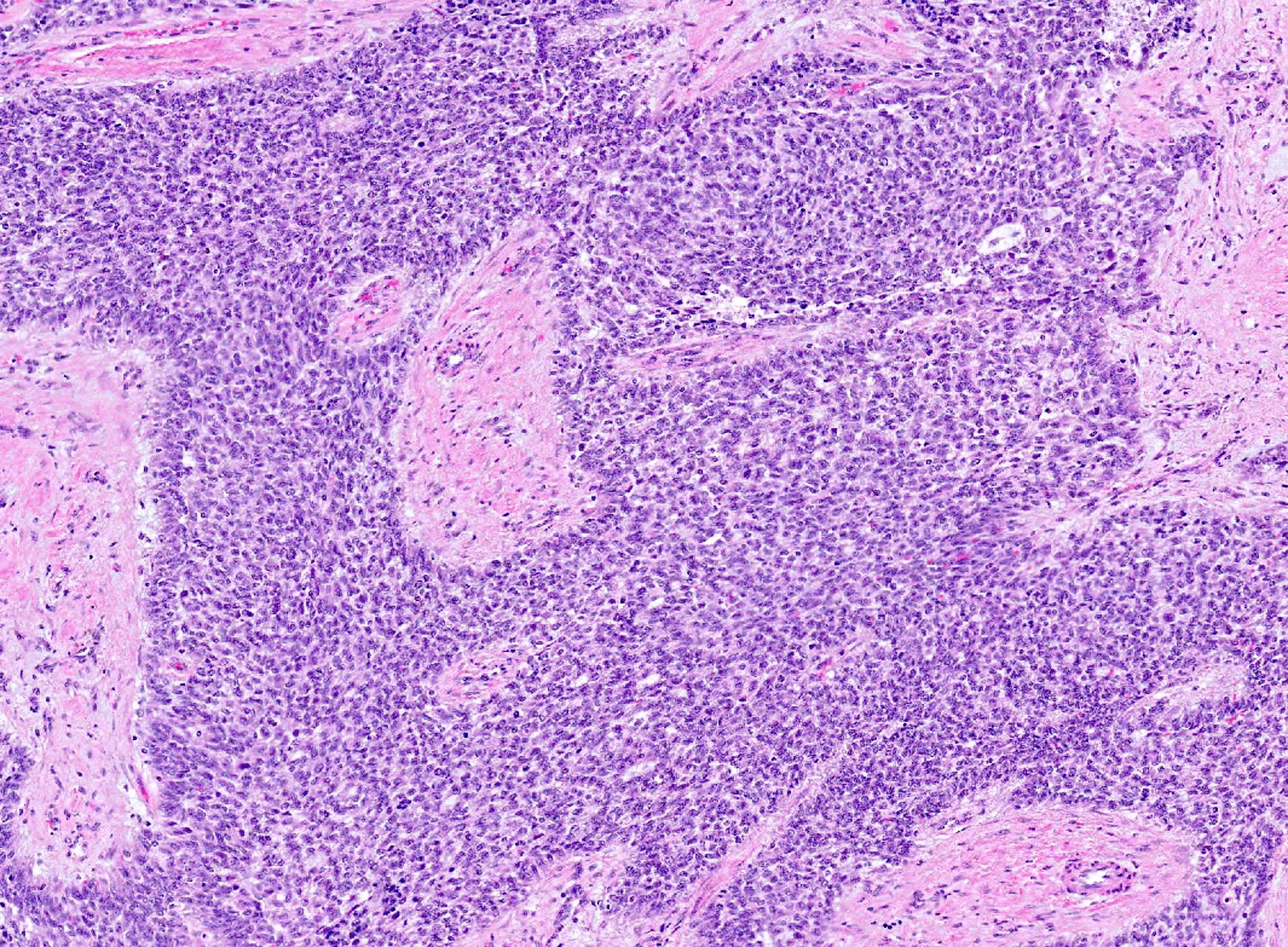
Tubular and solid pattern
Resembling endometrioid carcinoma
Tubules with eosinophilic secretions
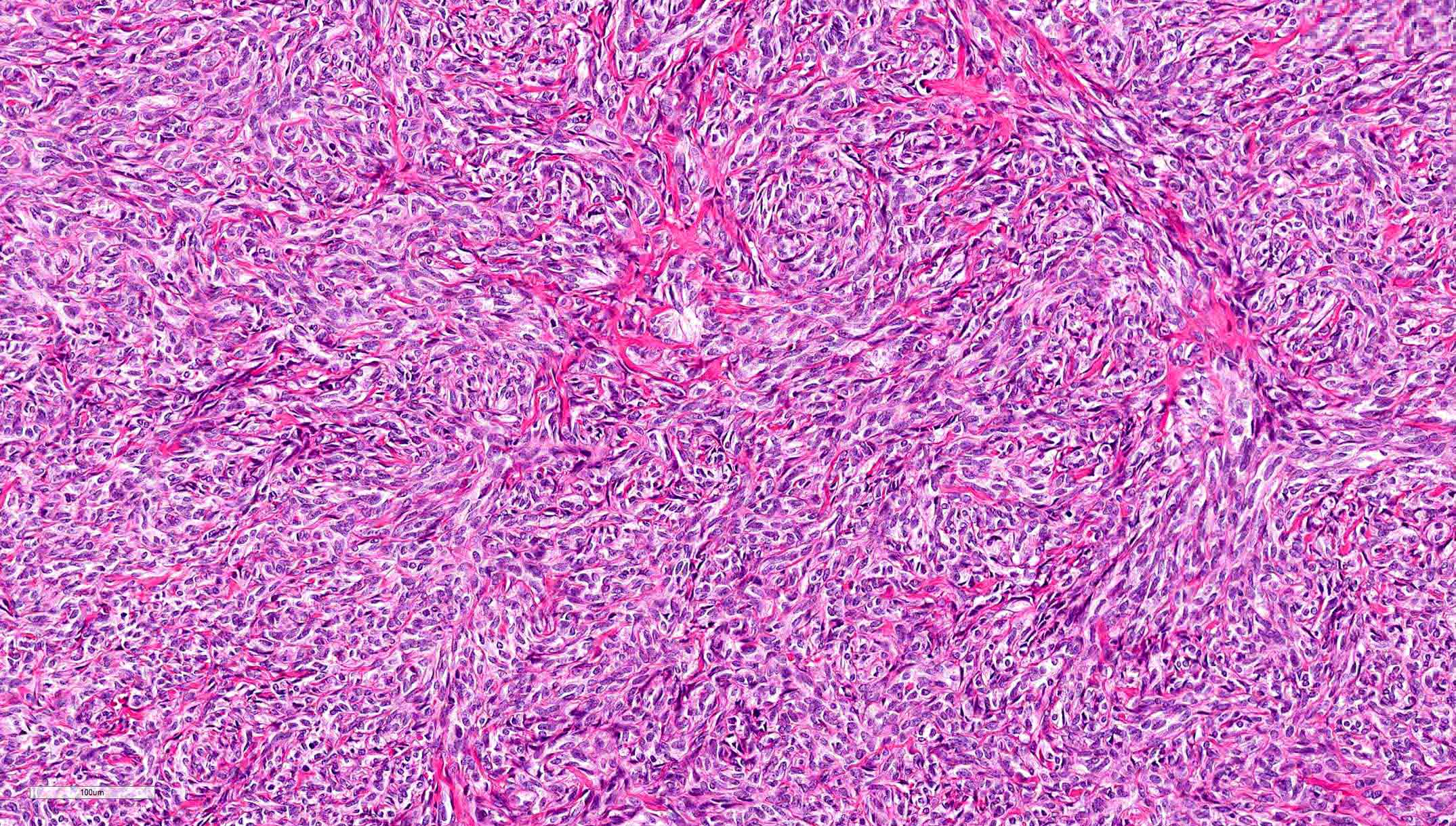
Solid growth pattern
Malignant FATWO
CD10
Calretinin
Carlson: 2023

Clement: 2019
Crum: 2015
Heller: 2015
IARC: 2014

IARC: 2020

Nucci: 2020
Nucci: 2023
Vang: 2017
Find related Pathology books: gynecologic
