
Contributed by Jennifer Chapman, M.D.
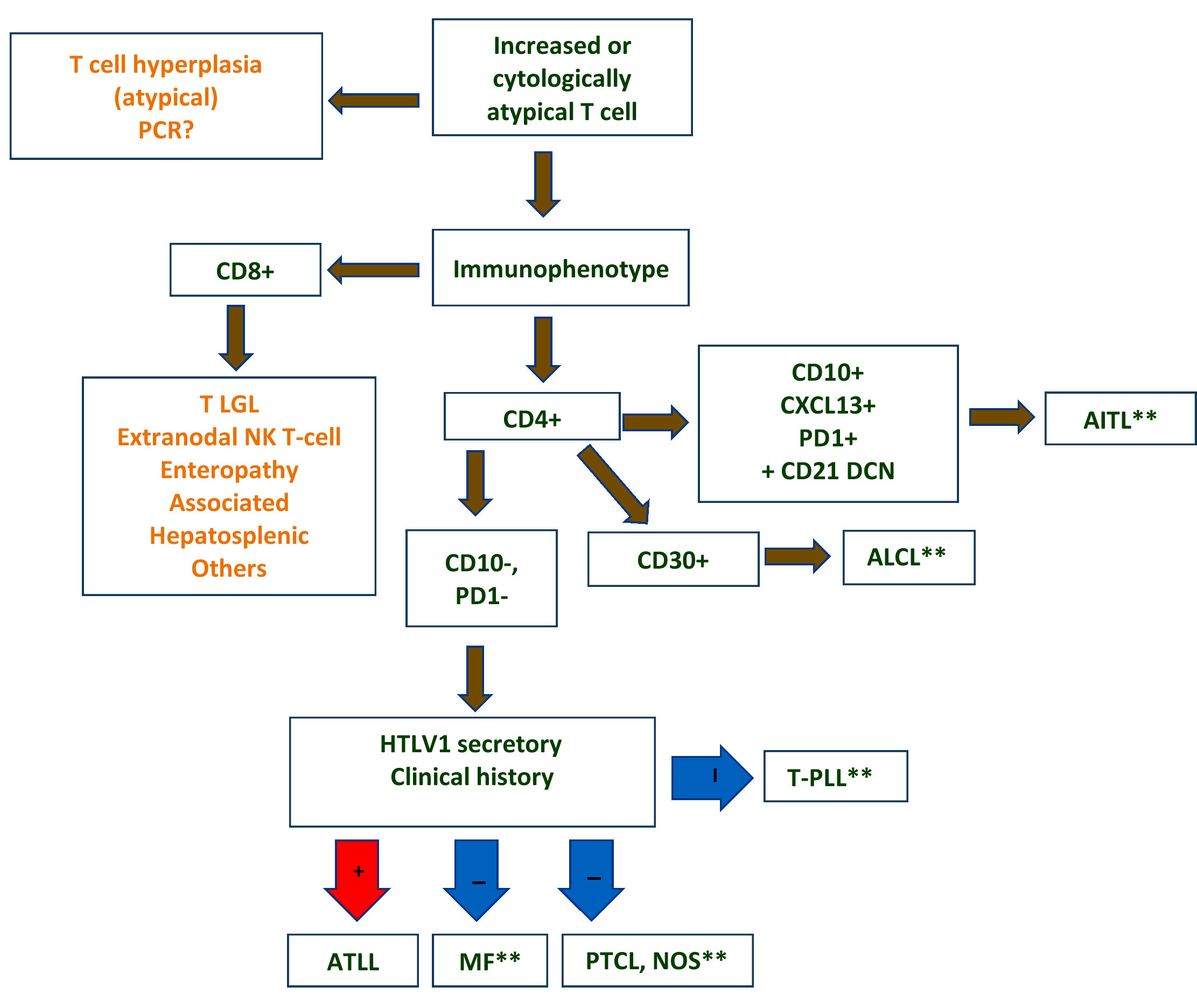
Differential diagnostic algorithm
Images hosted on other servers:

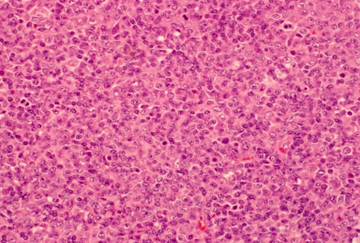
Nodules
Contributed by Jennifer Chapman, M.D.
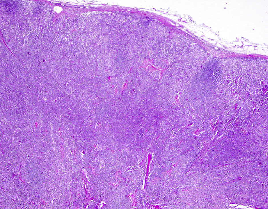
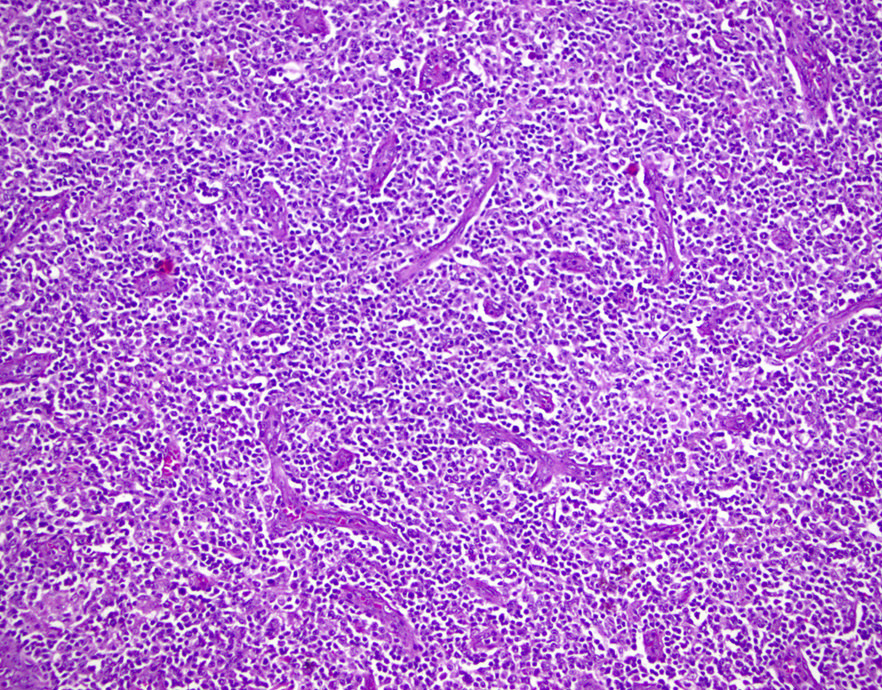
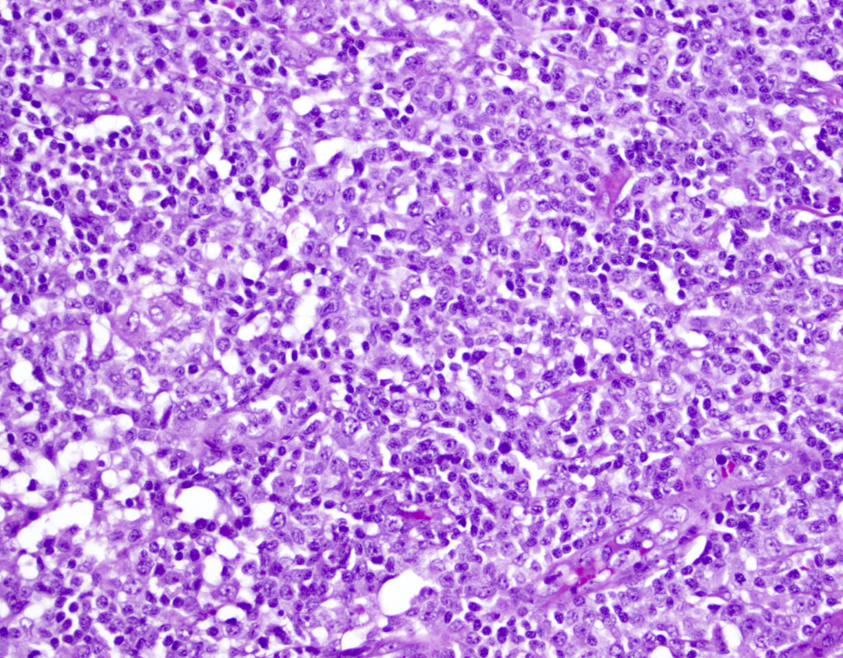
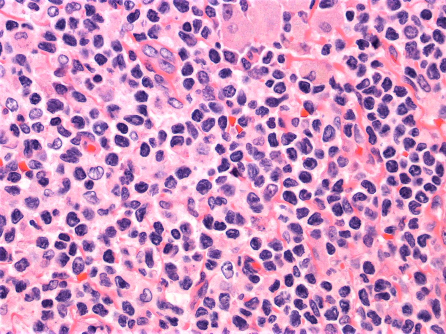
ATLL mimicking angioimmunoblastic T cell lymphoma
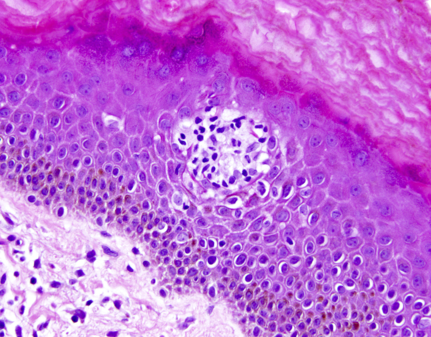
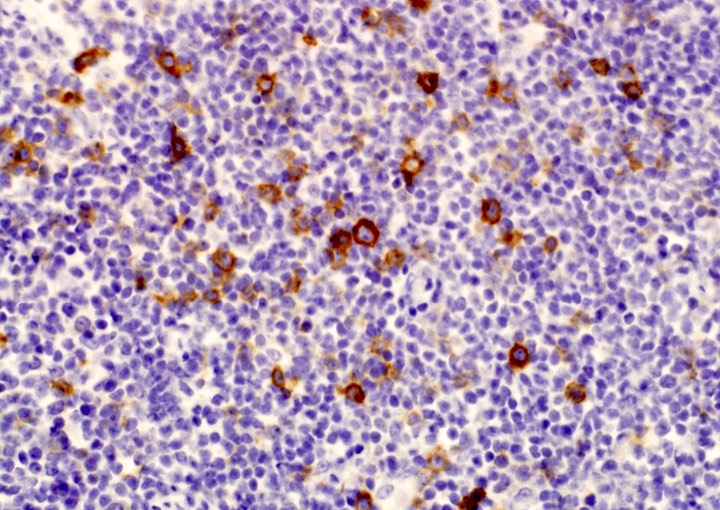
ATLL with Hodgkin-like cells mimicking classical Hodgkin lymphoma
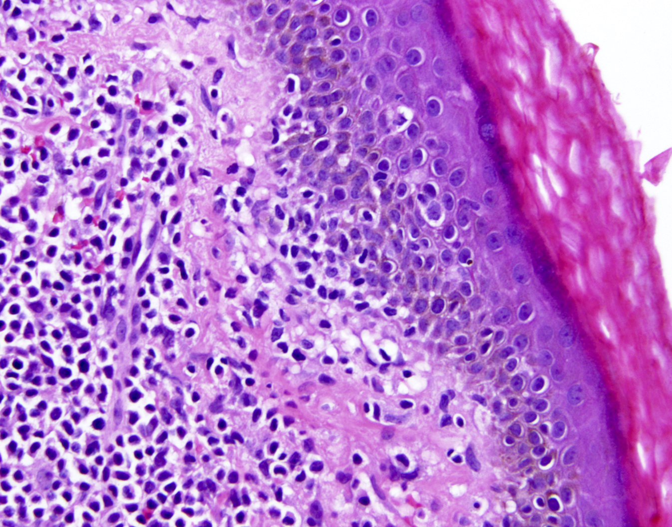
ATLL mimicking mycosis fungoides
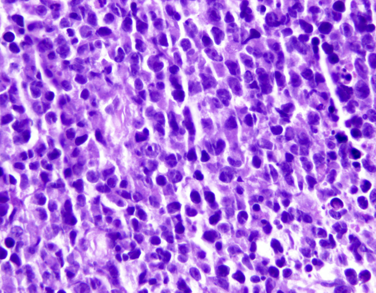

ATLL, lymphomatous type, mimicking anaplastic large cell lymphoma
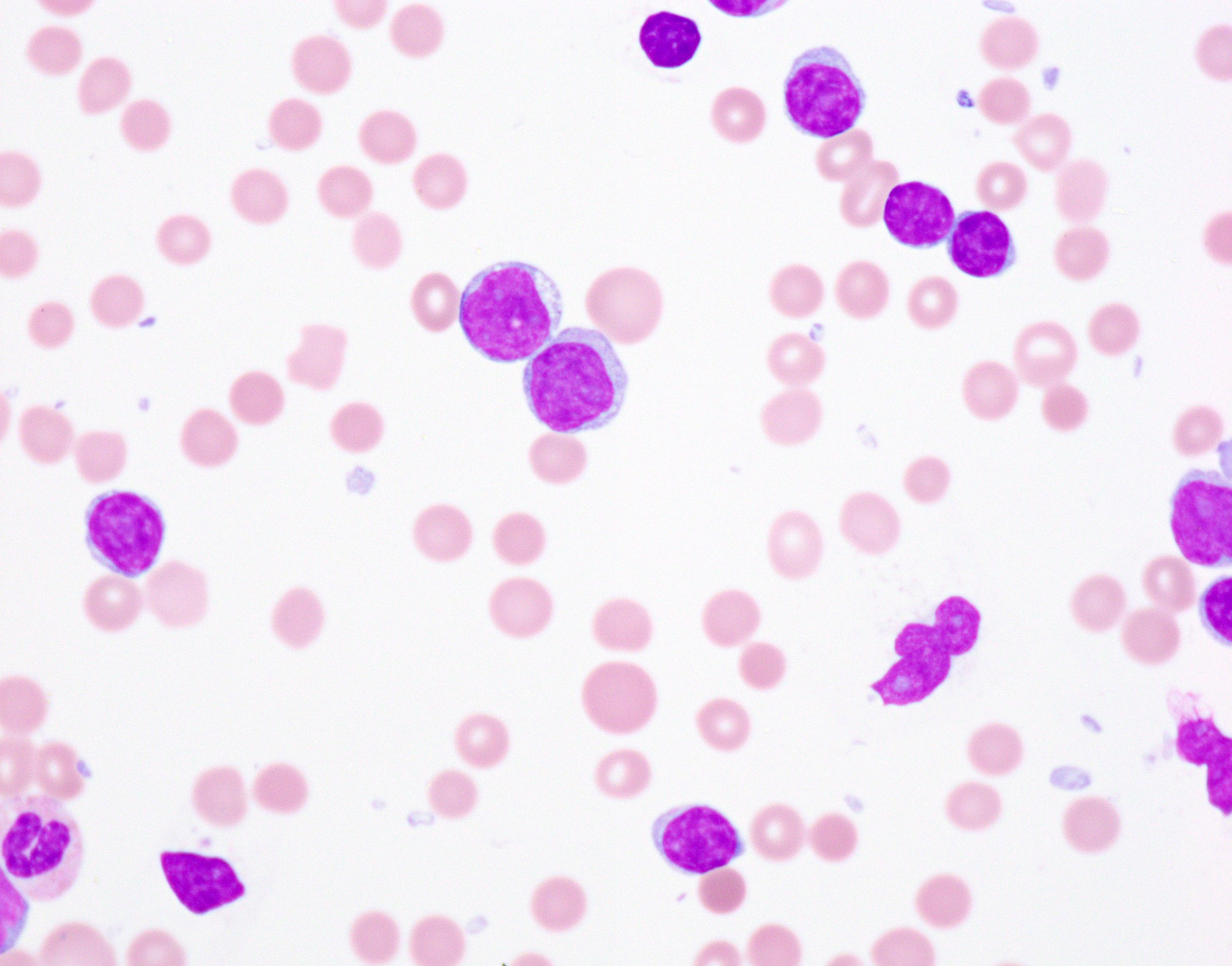
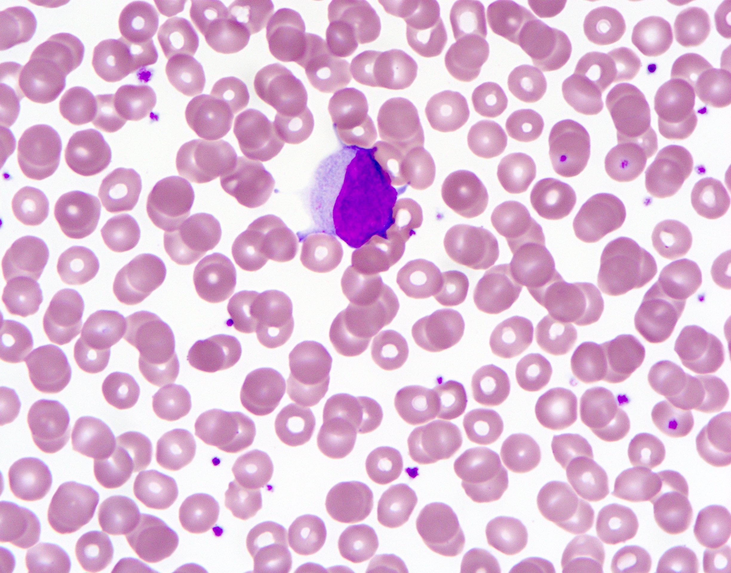
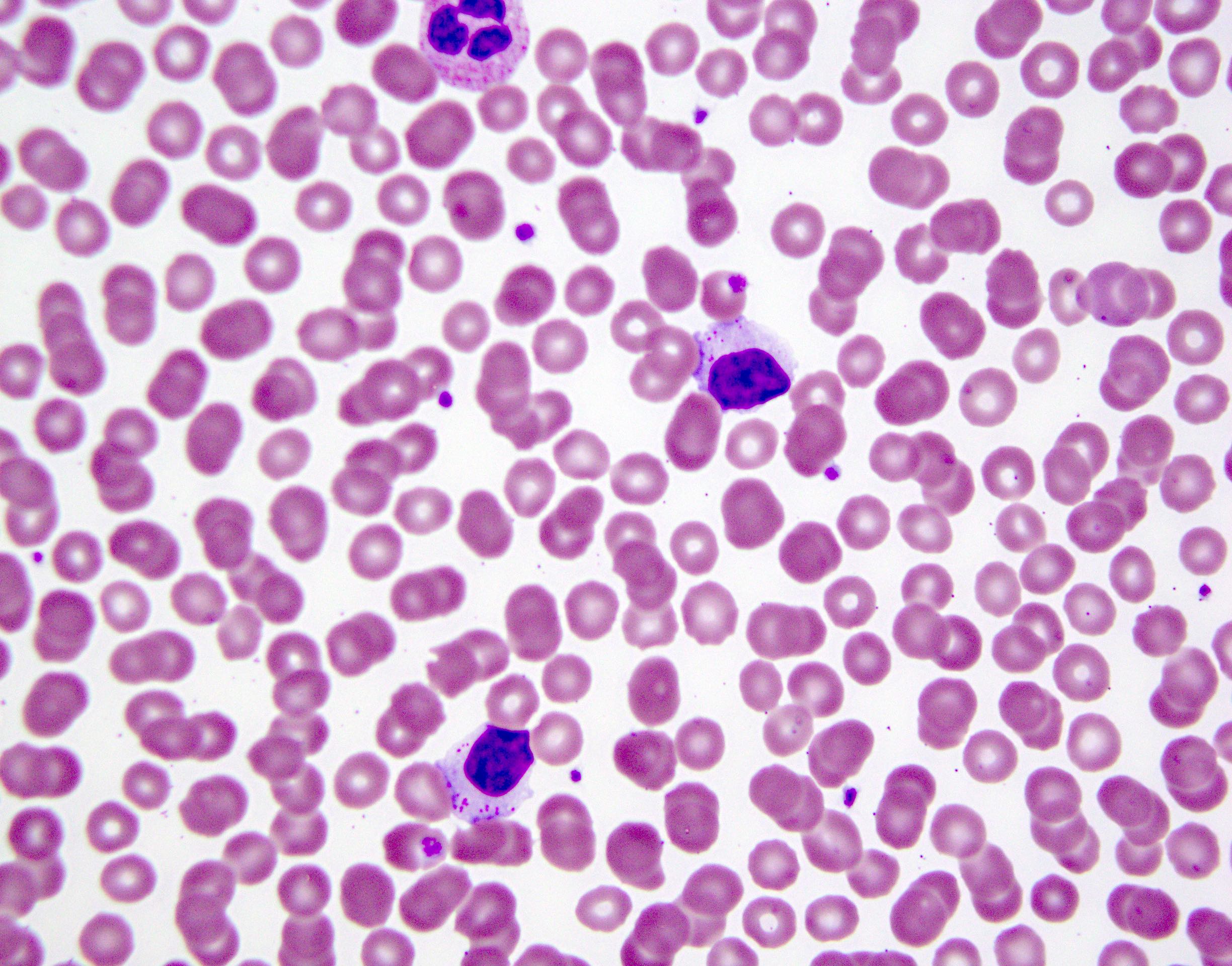
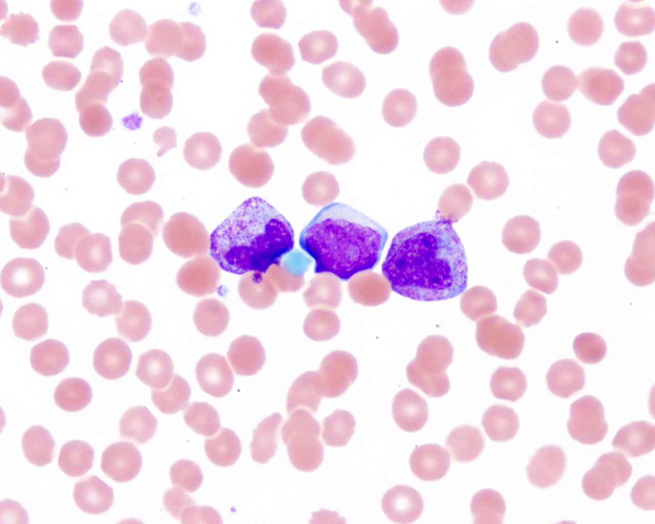
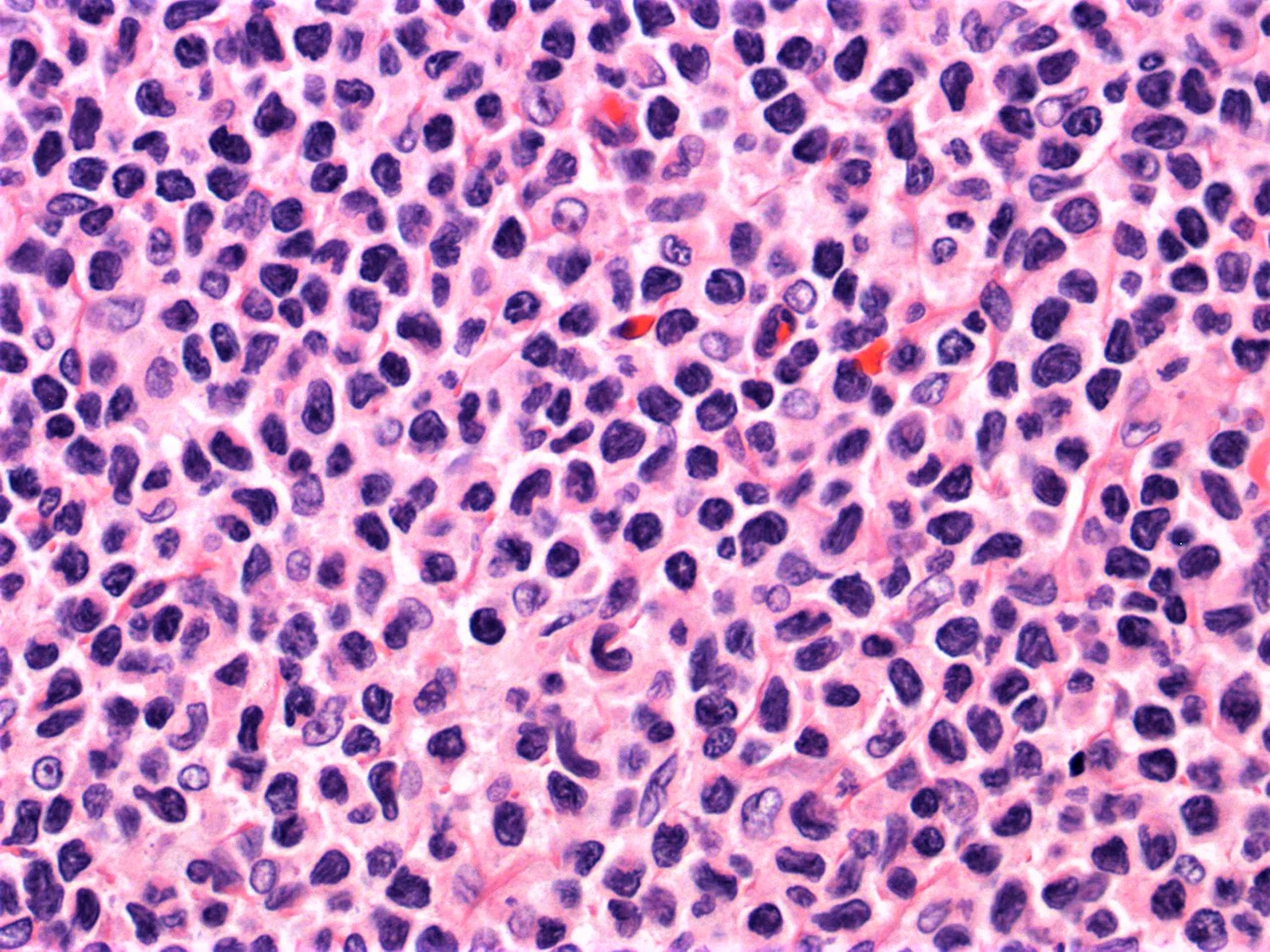
Contributed by Jennifer Chapman, M.D.
ATLL, acute variant,
mimicking
T prolymphocytic
lymphoma
Images hosted on other servers:

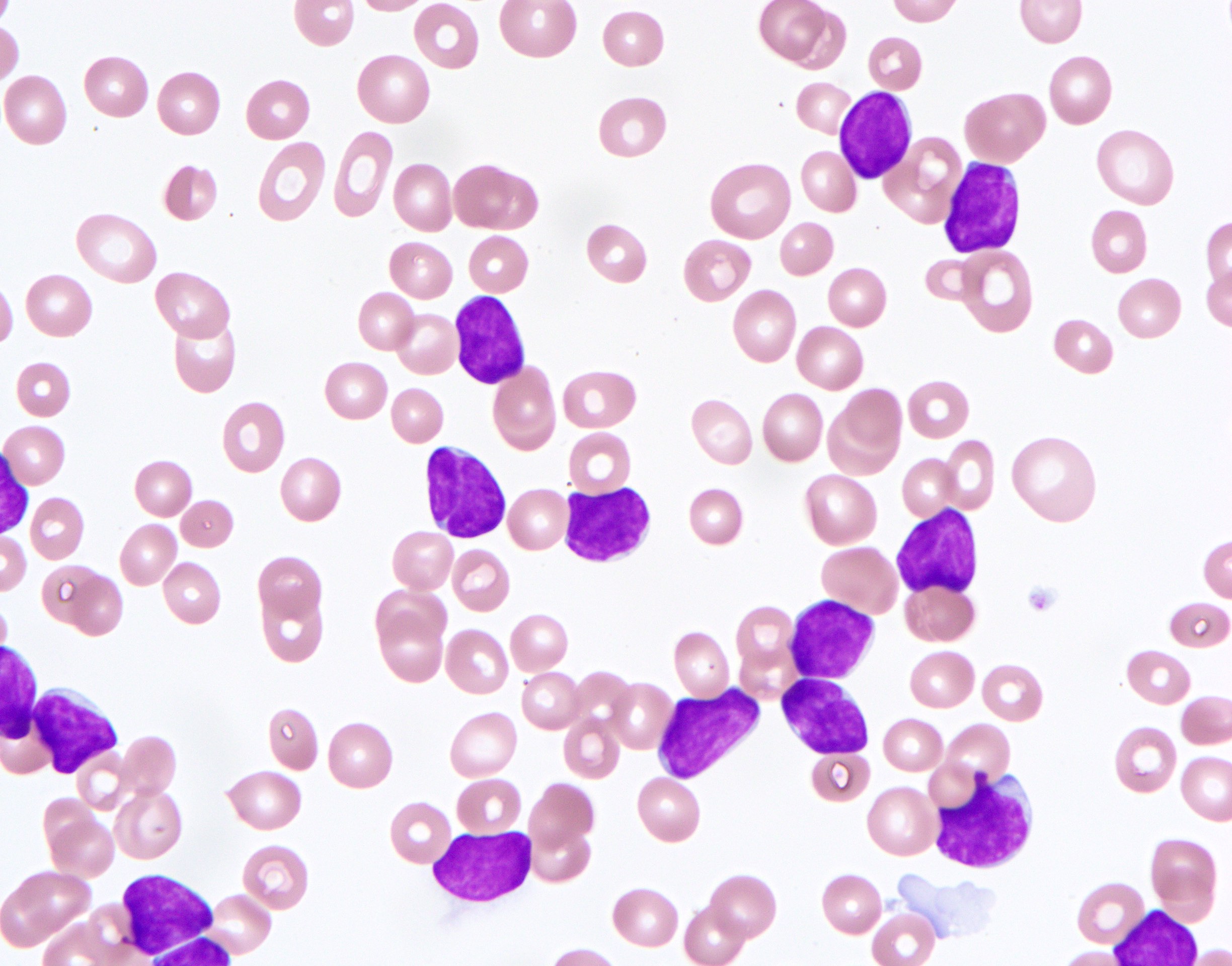
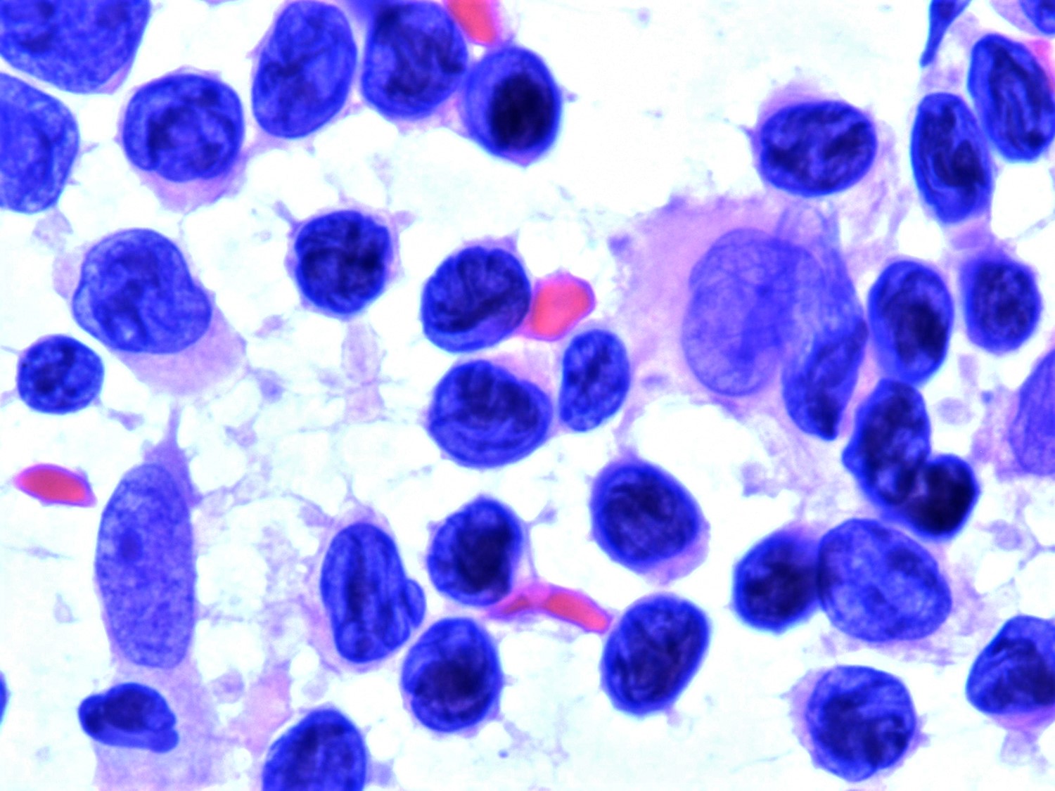
Flower cells

CLL-like morphology
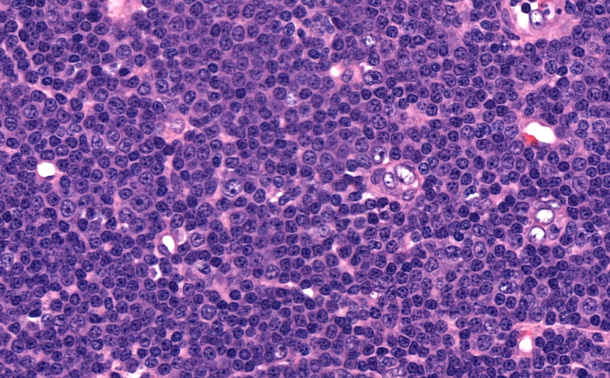
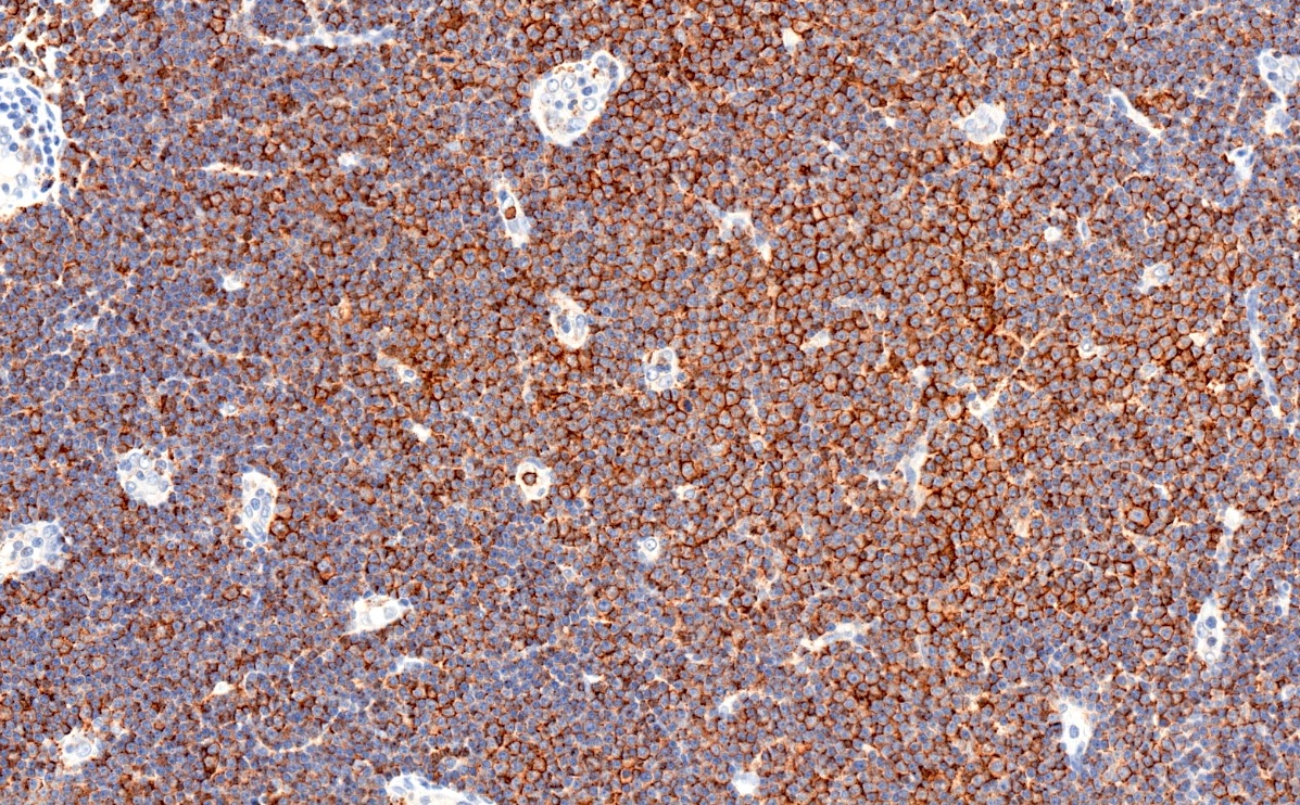
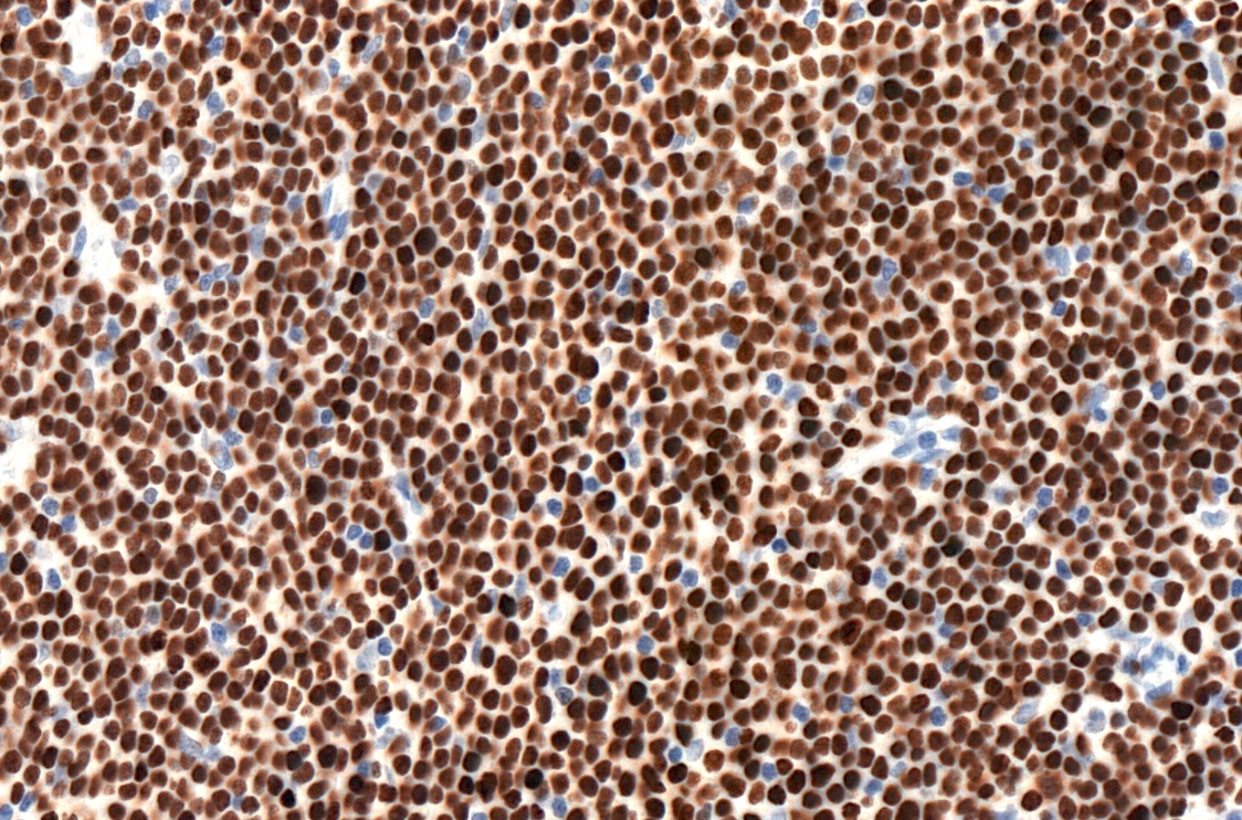
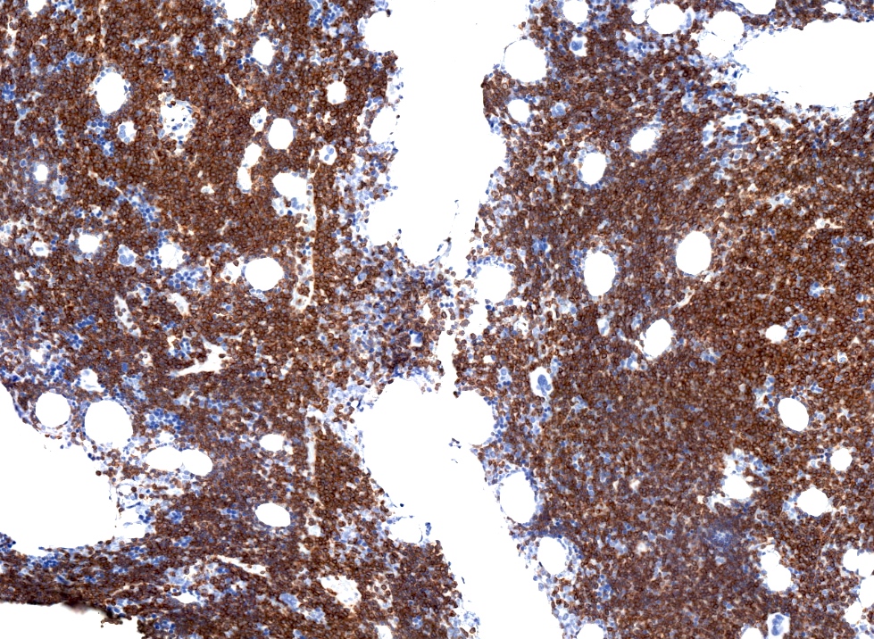
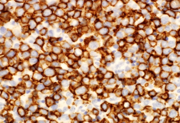
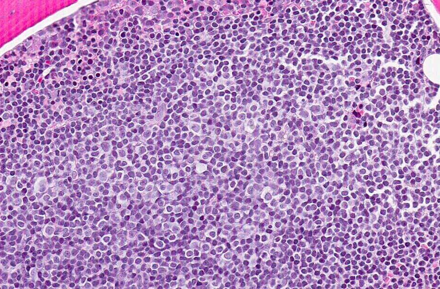
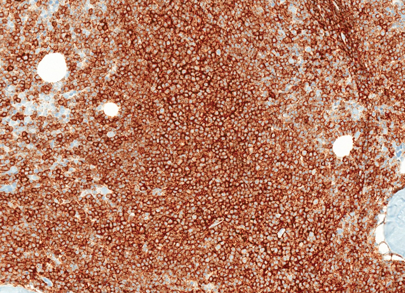
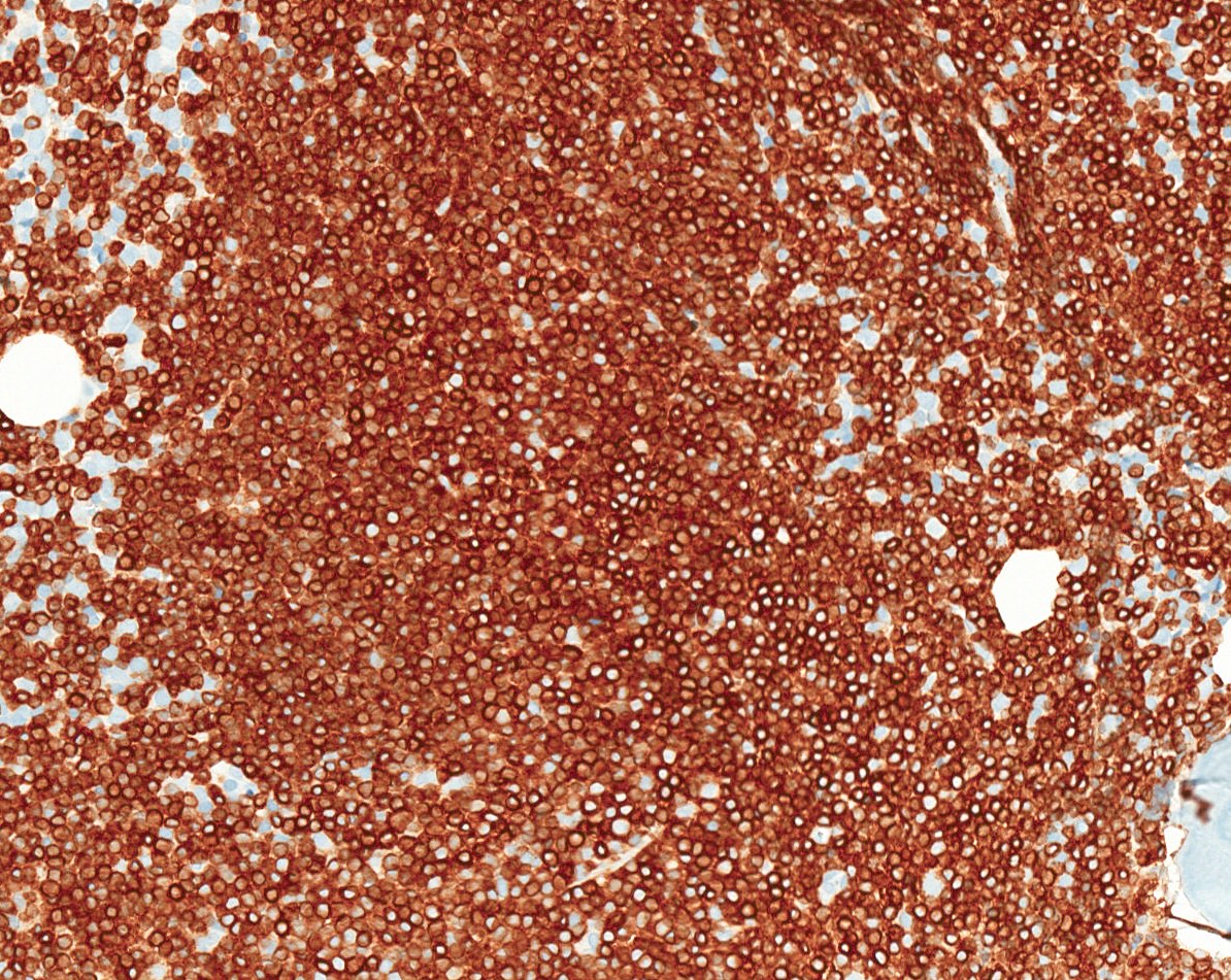
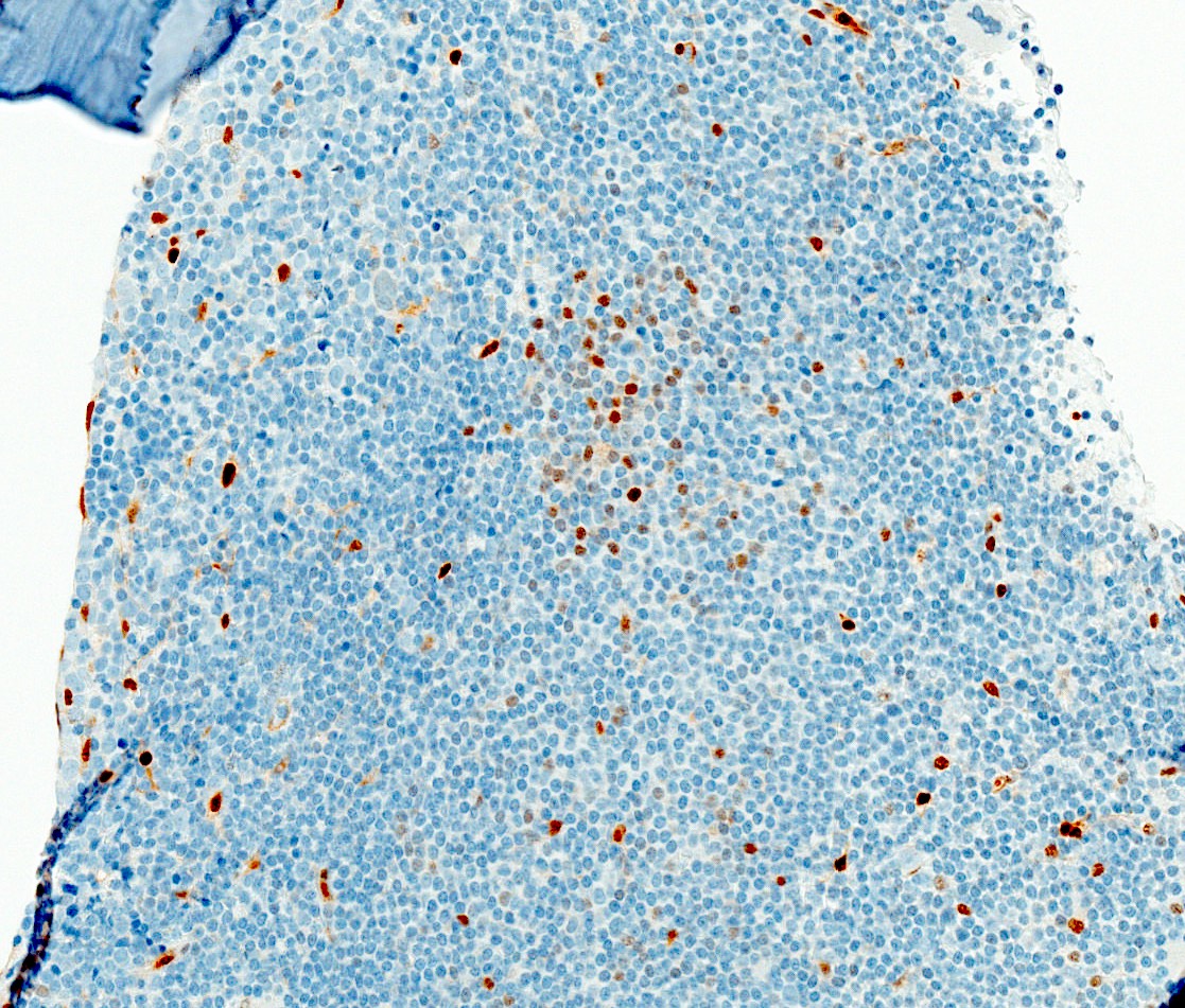
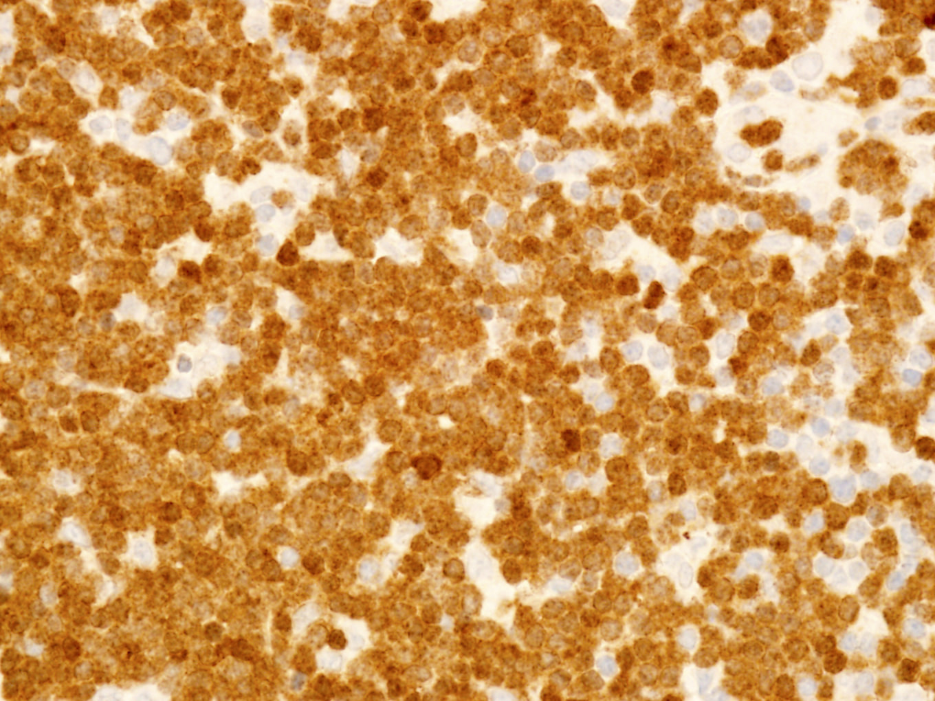
Contributed by Jennifer Chapman, M.D.
CD45
CD4

CD56
CD7
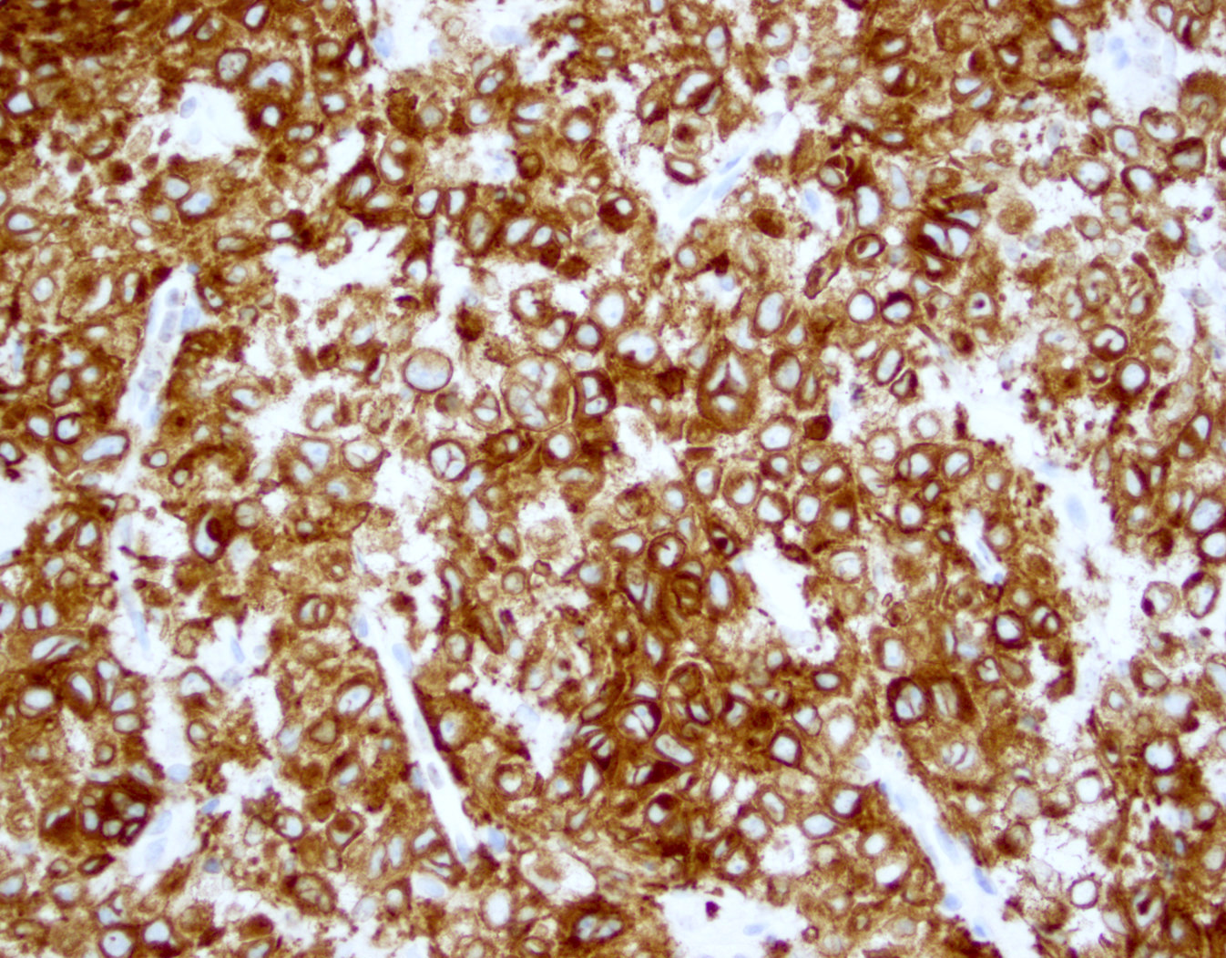
CD25
Images hosted on other servers:

CD3+, CD4+, CD25+, CD7-, CD8-
Images hosted on other servers:

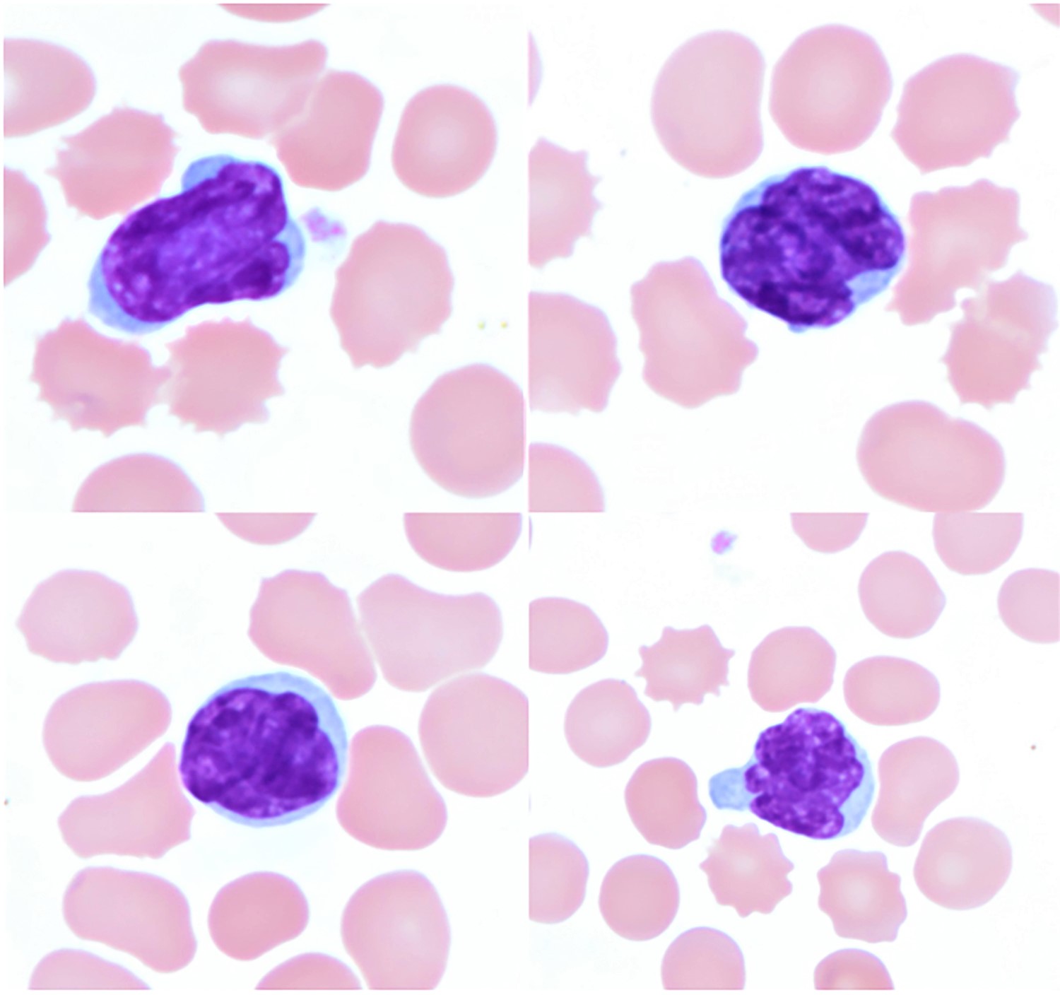
ATLL cell
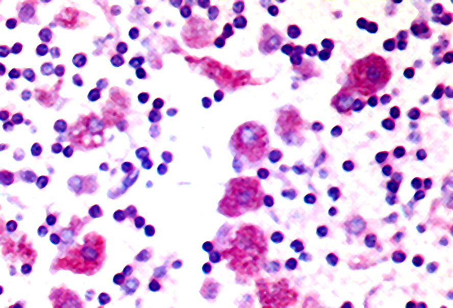
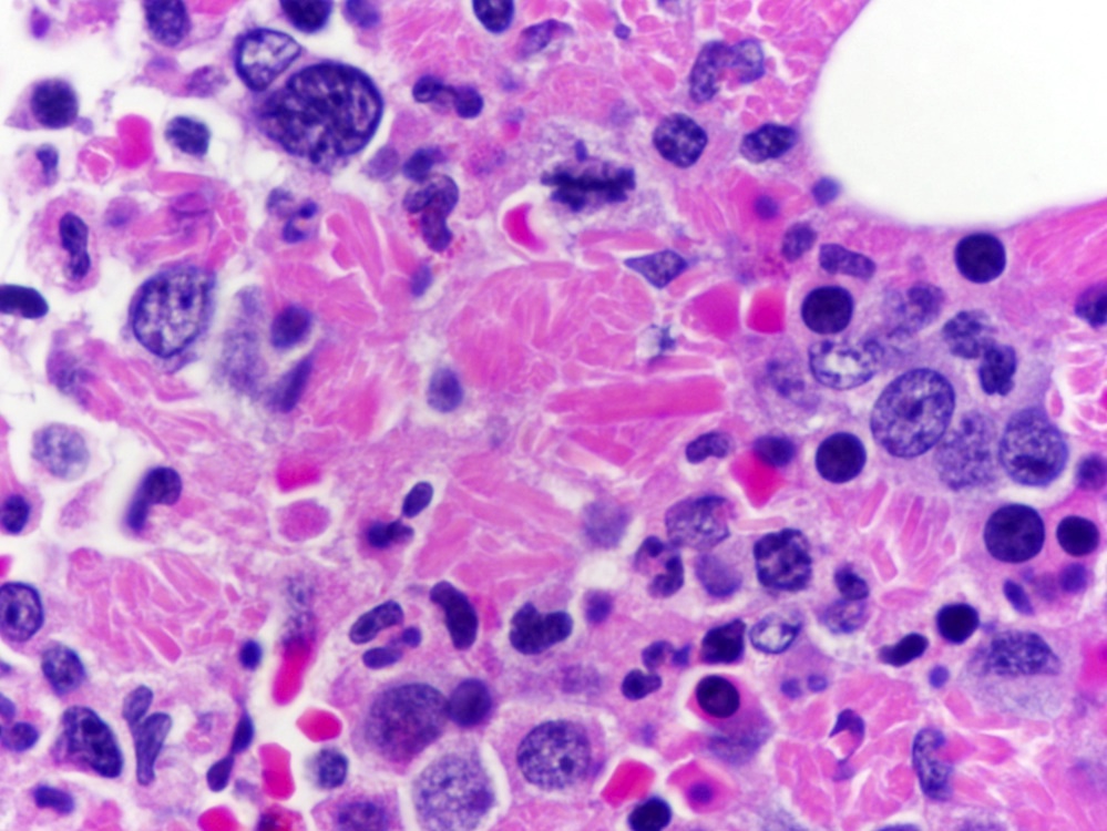
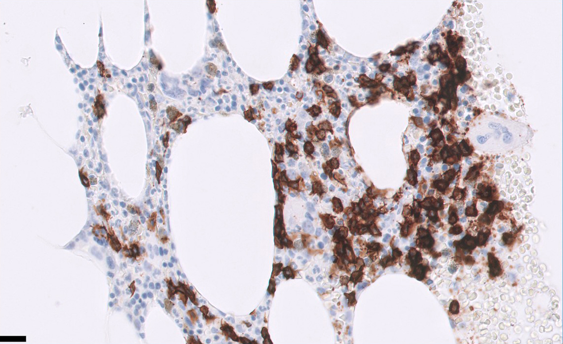
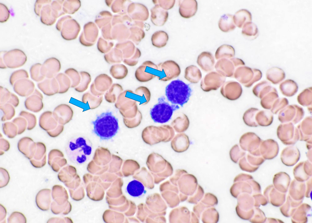
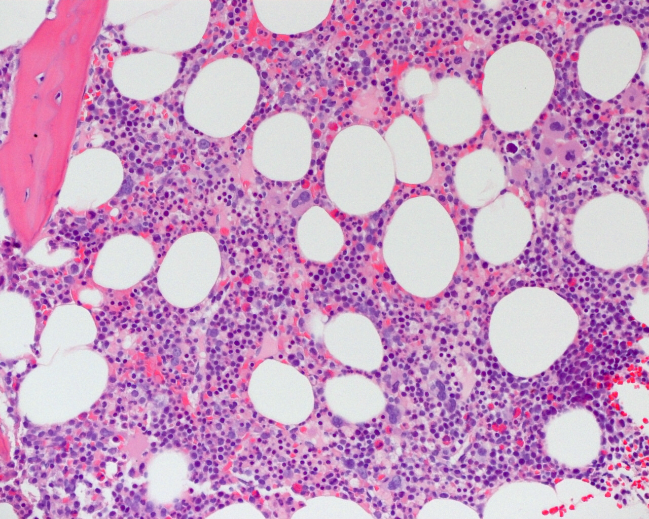
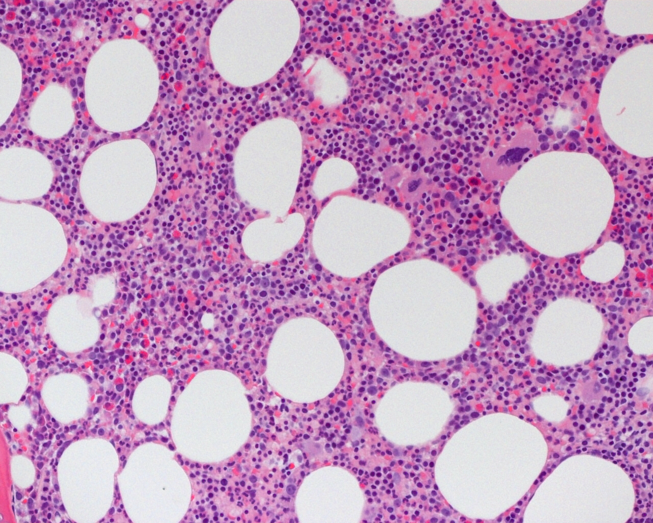
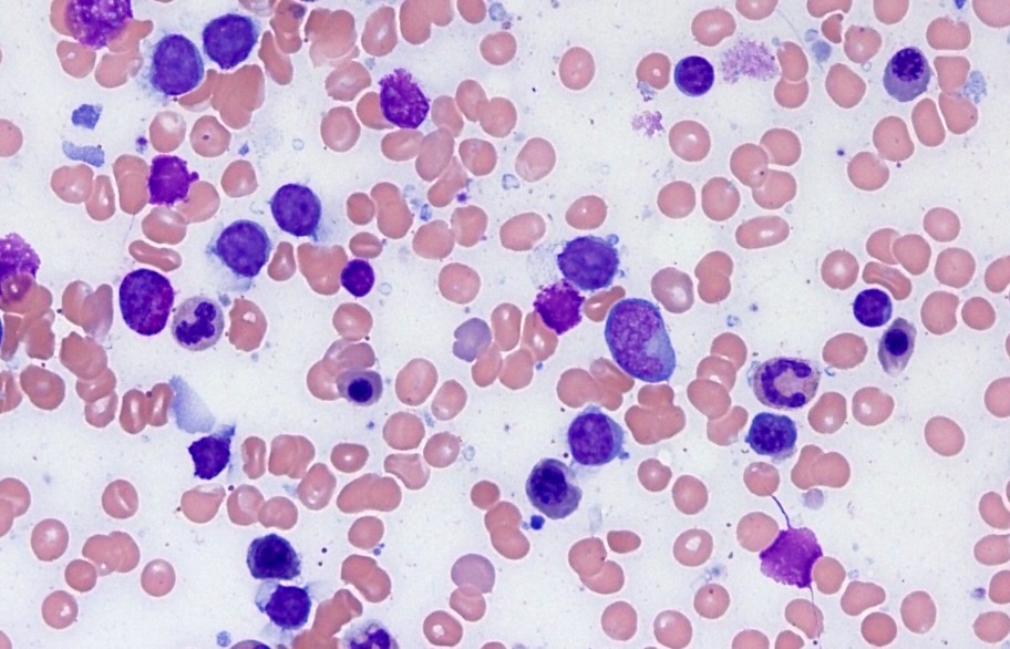
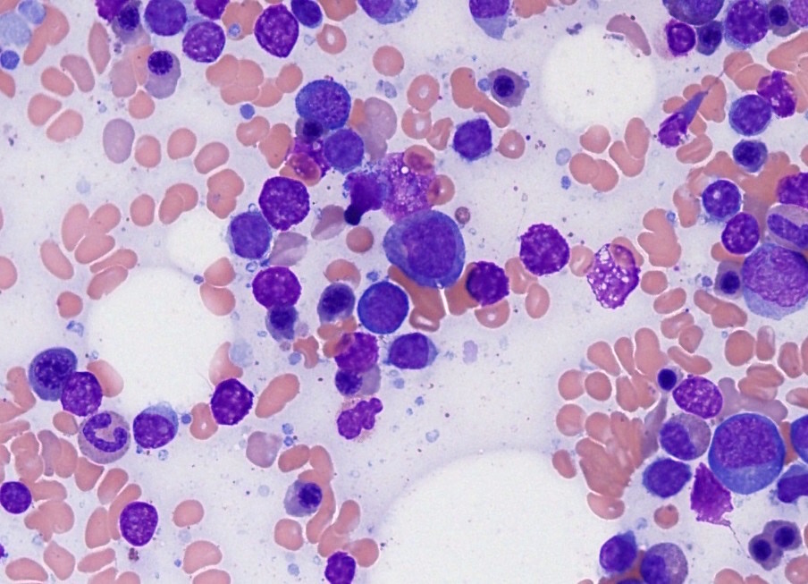
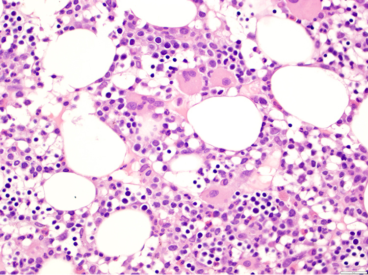
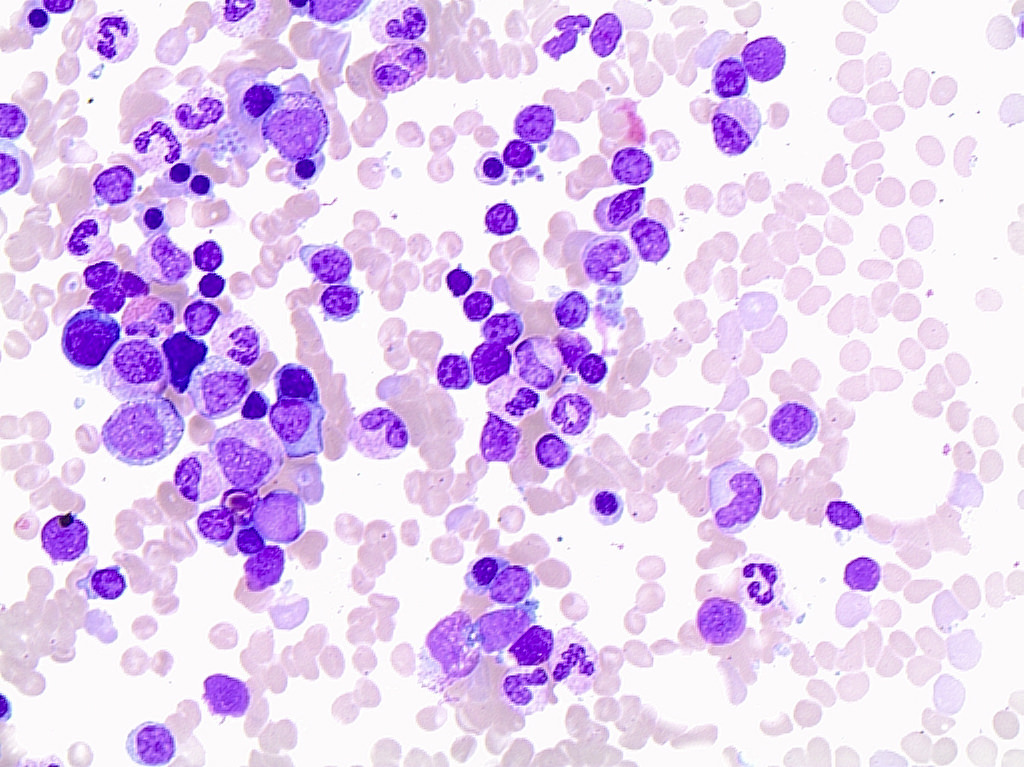
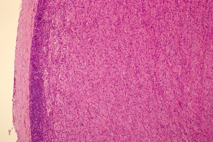
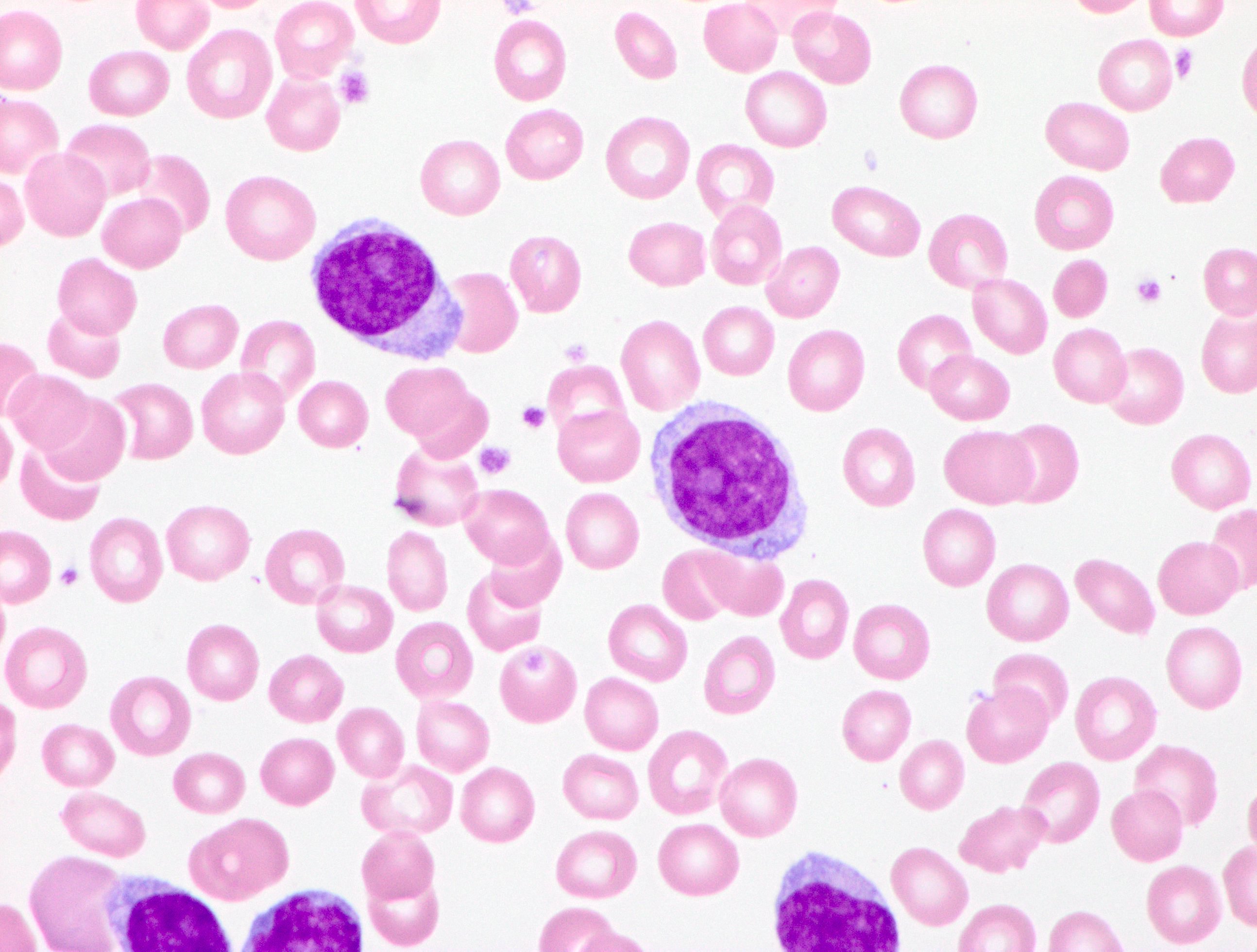
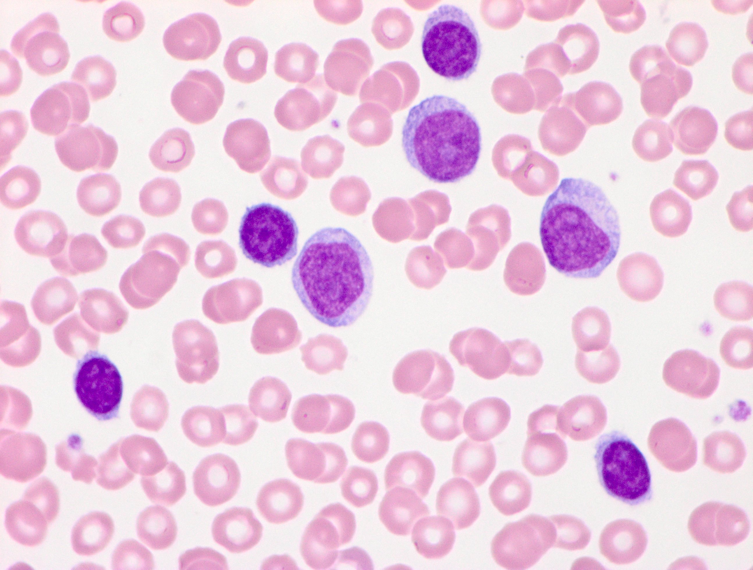
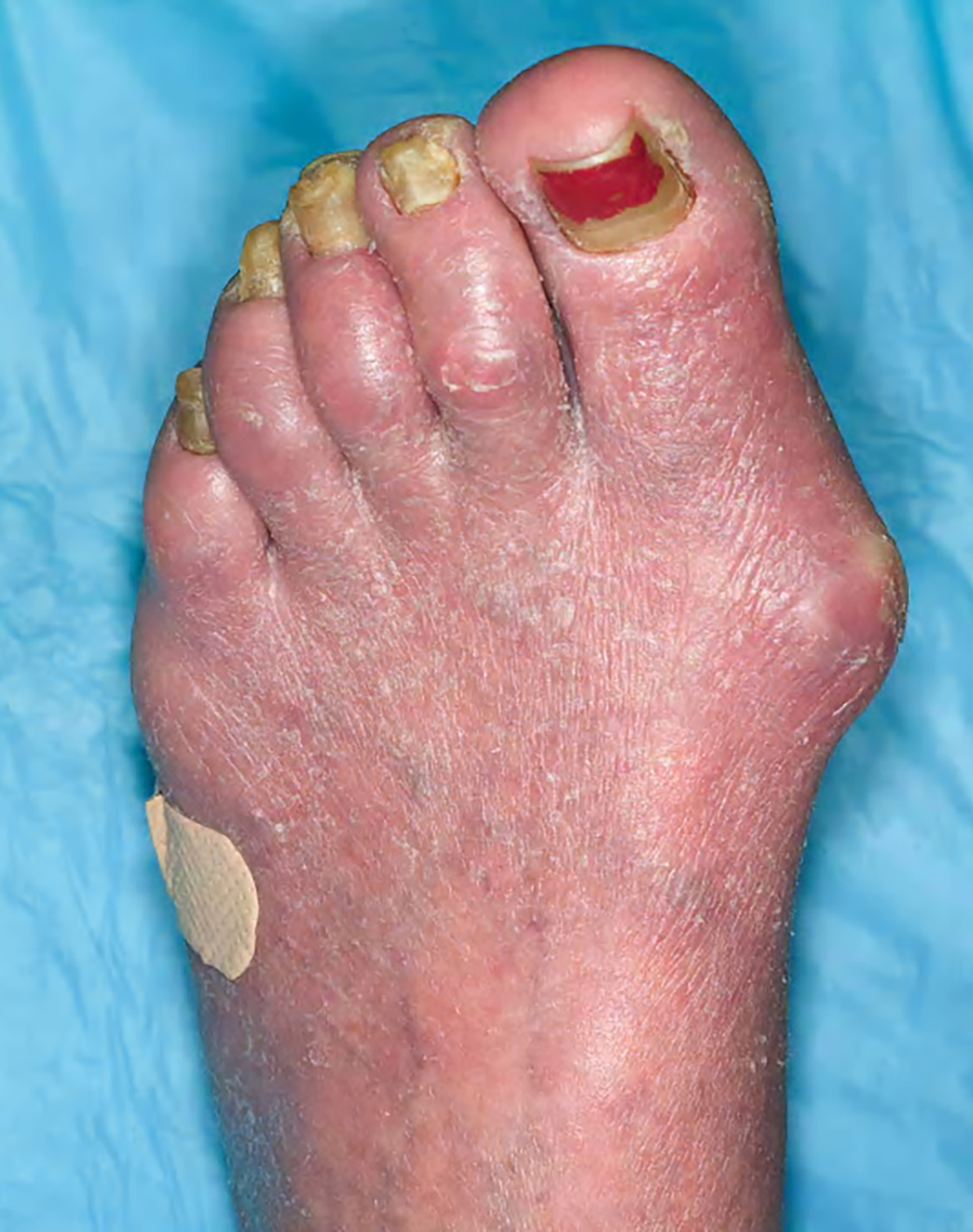
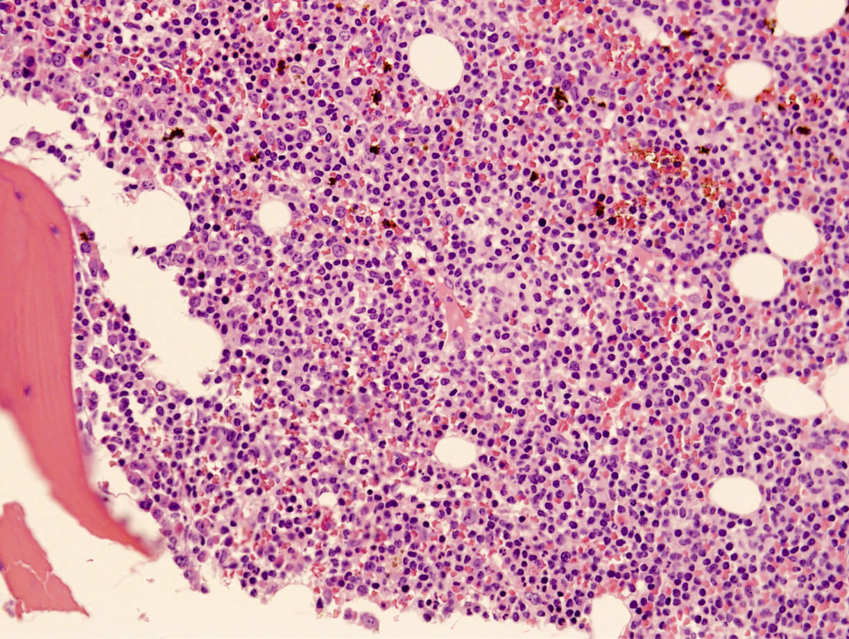
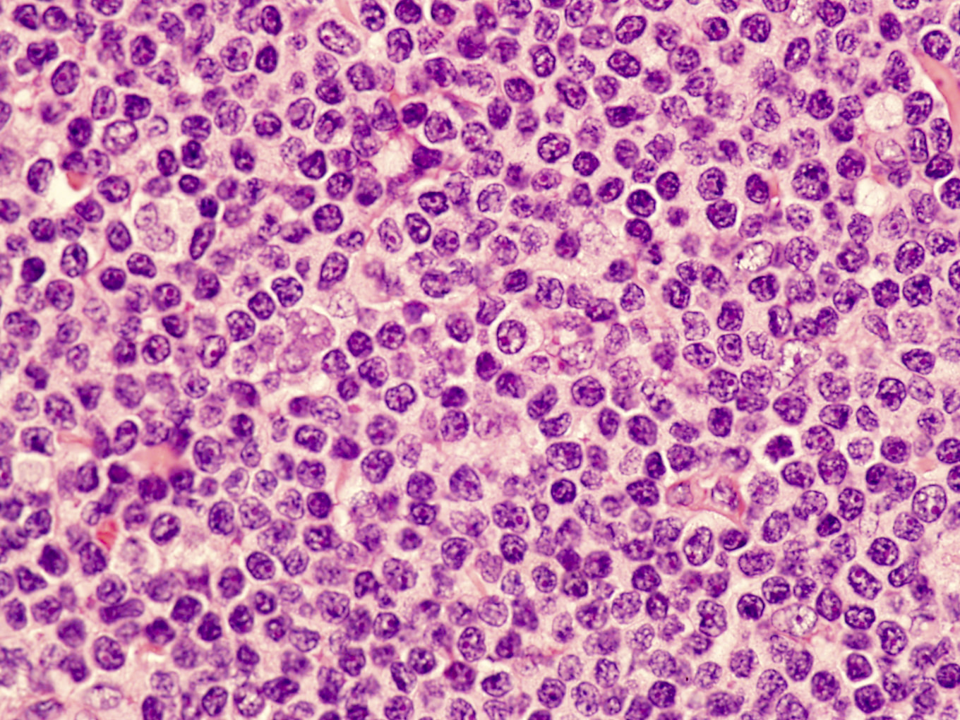
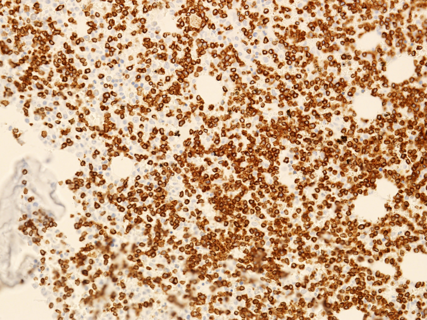
Contributed by Siba El Hussein, M.D. and Joseph Khoury, M.D.
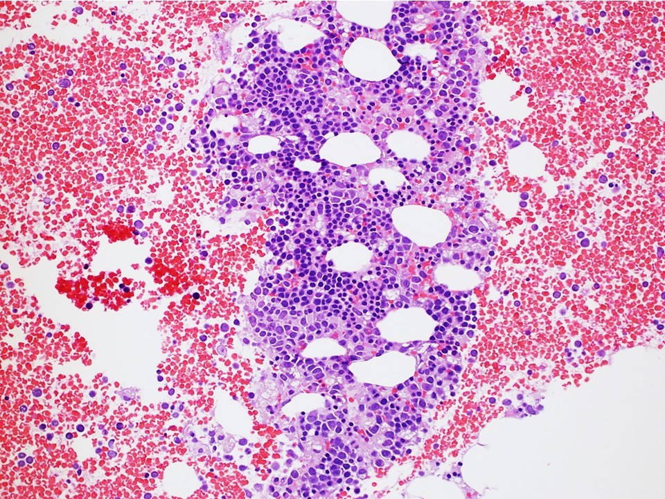
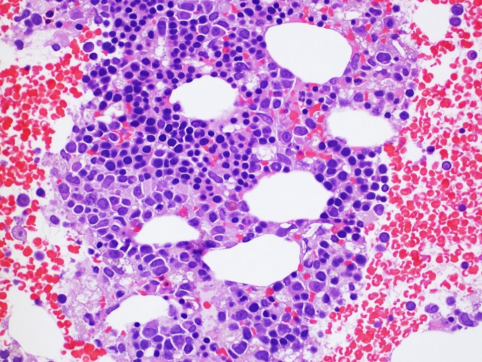
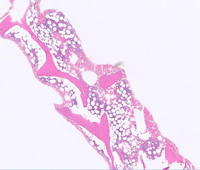
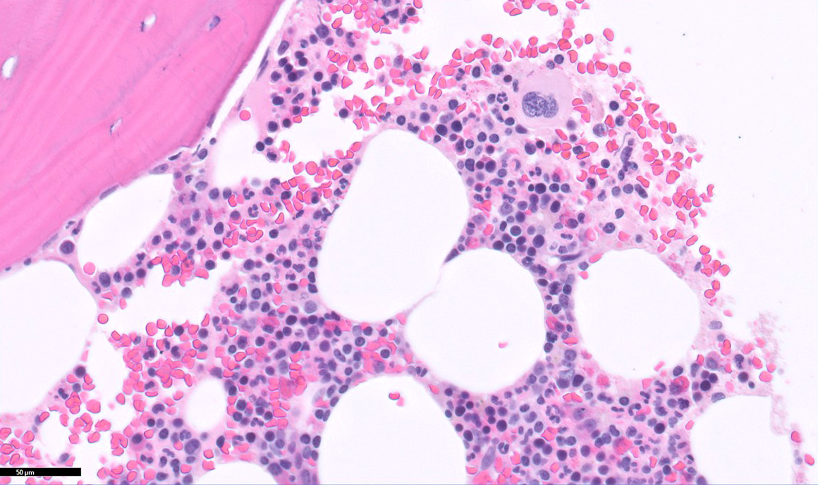
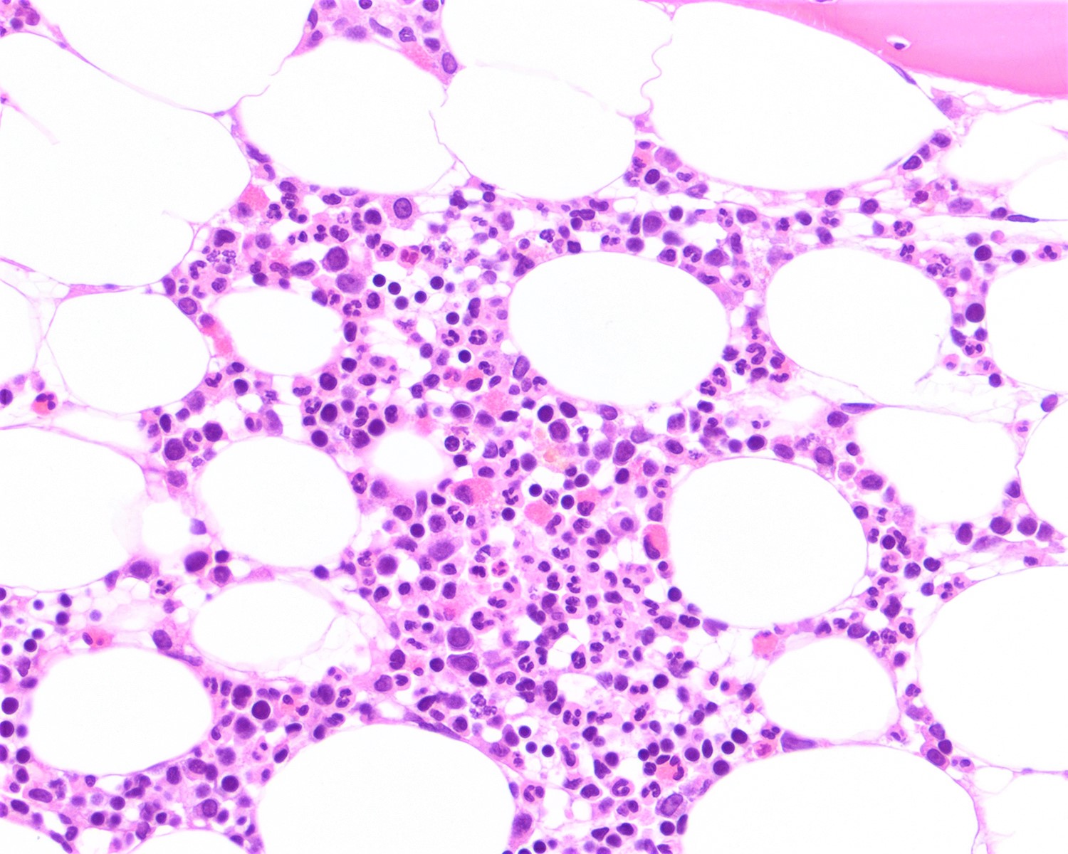
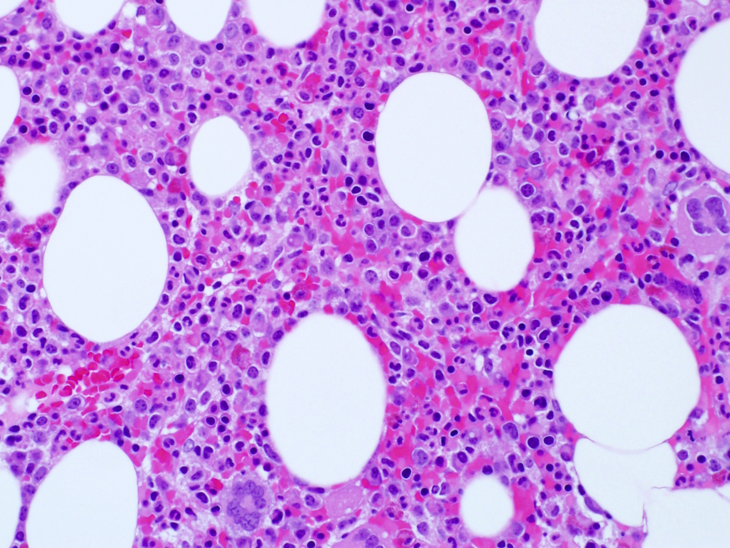
Neoplastic cells involving the bone marrow
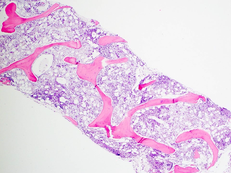
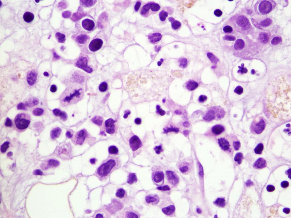
Neoplastic cells involving the bone marrow
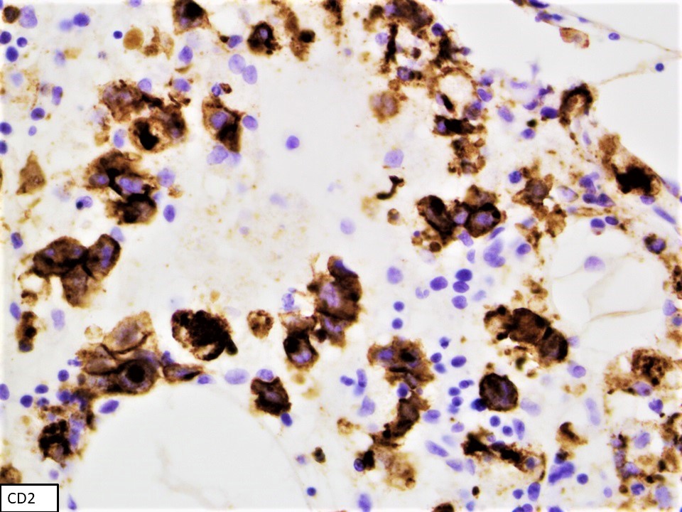
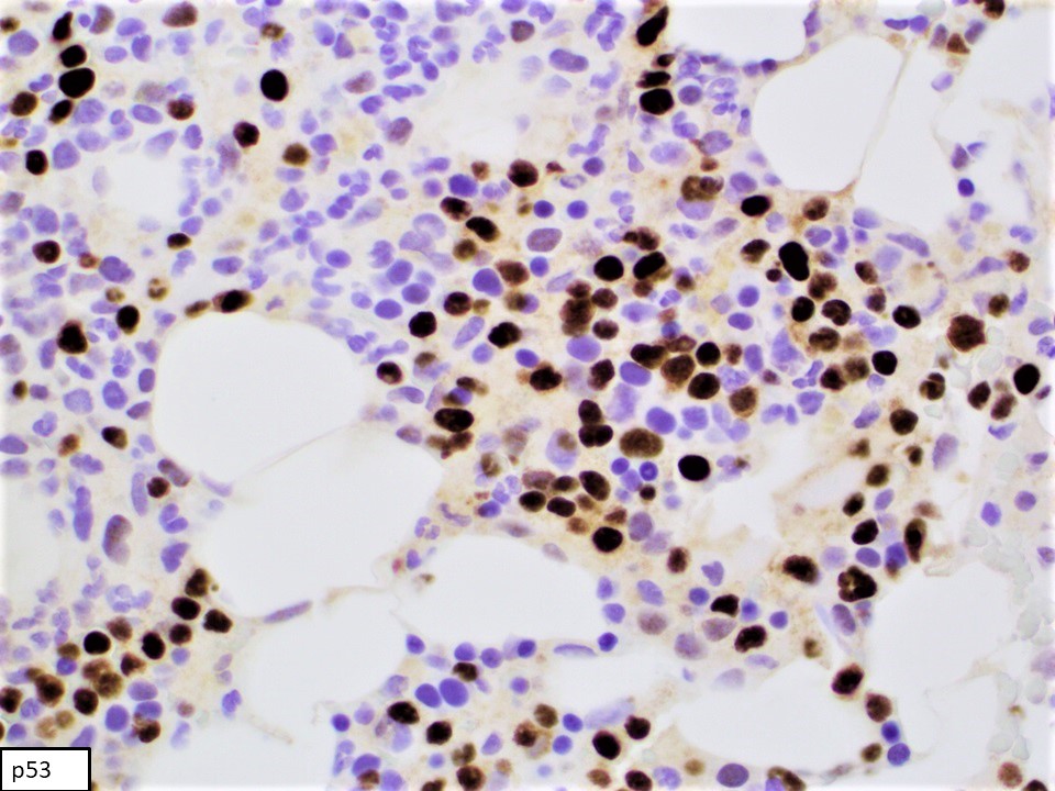
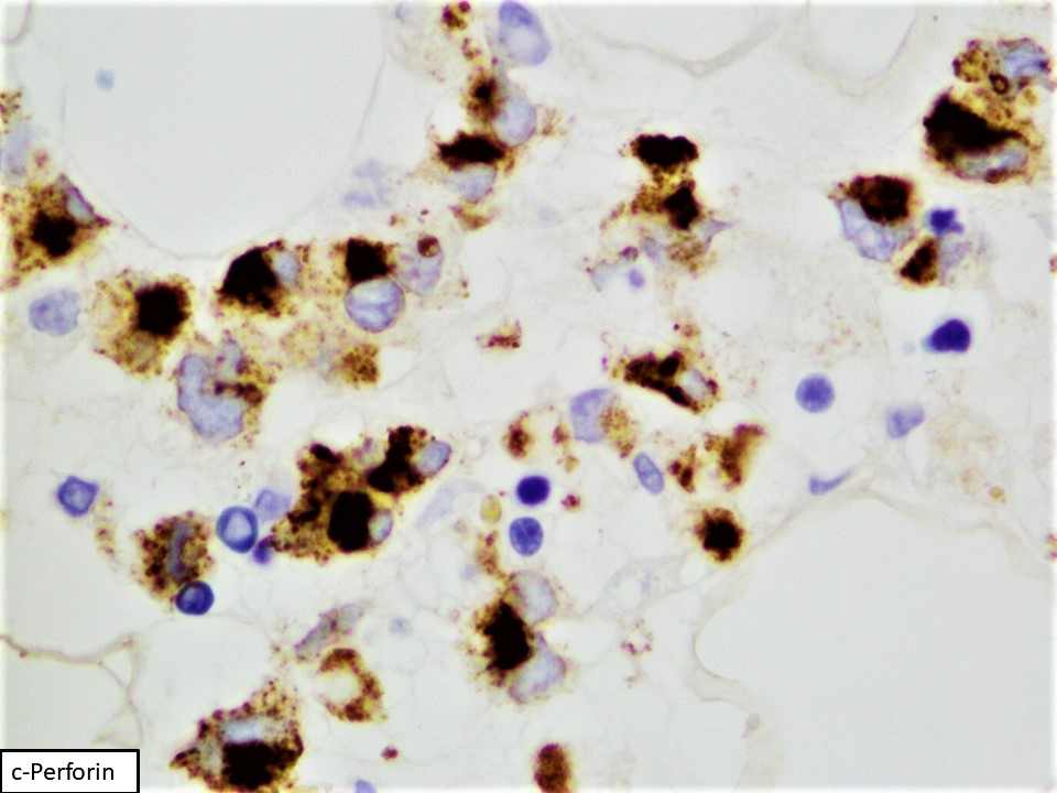
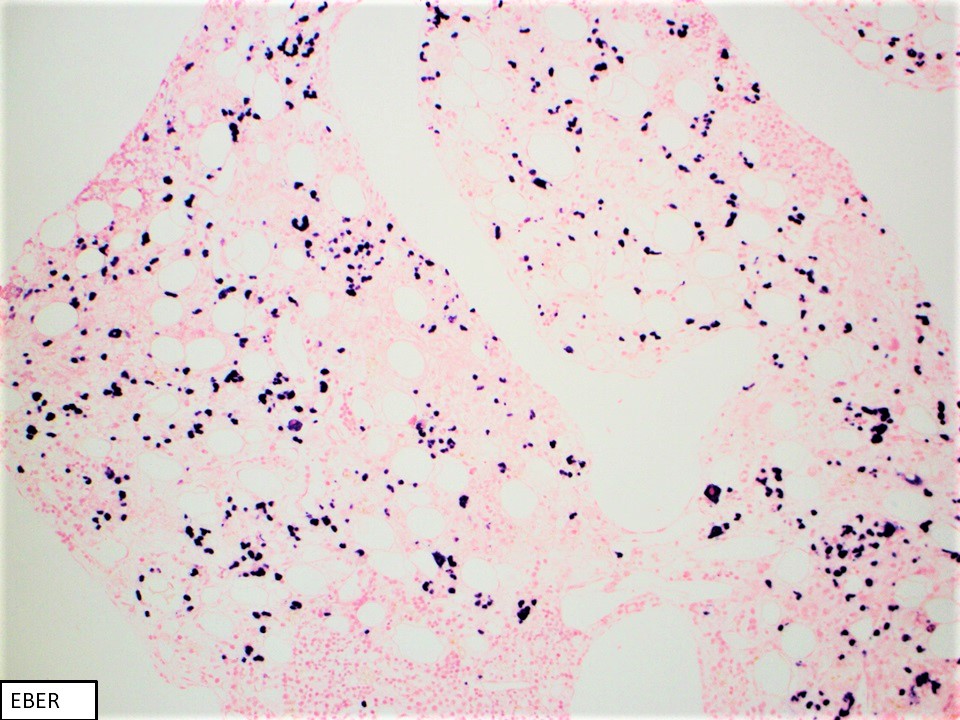
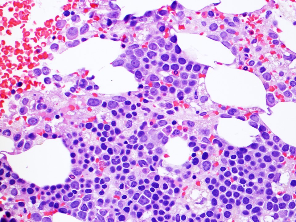
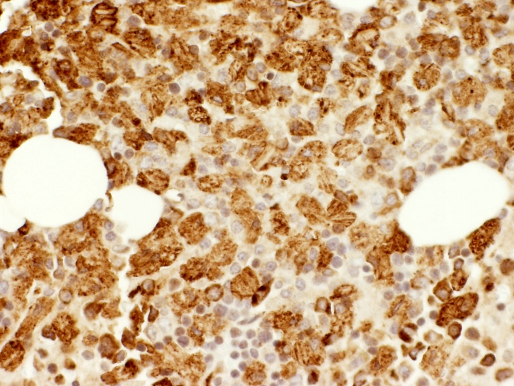
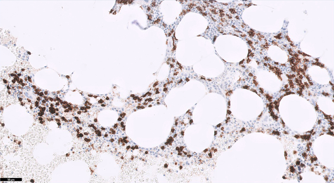
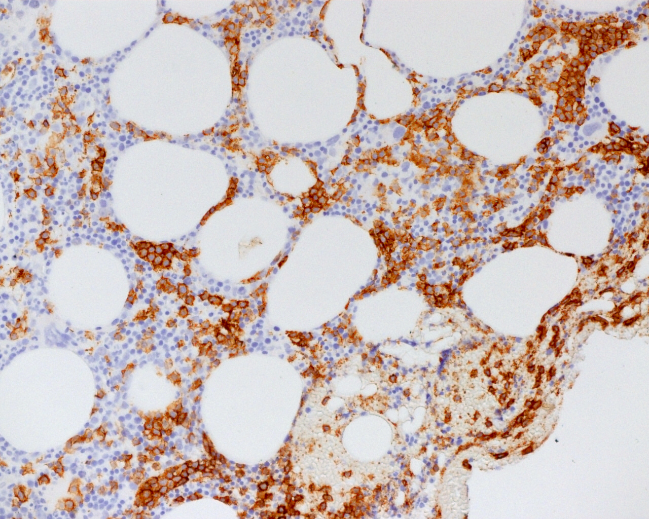
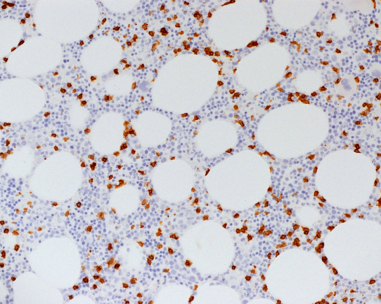
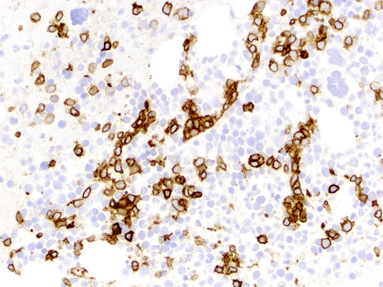
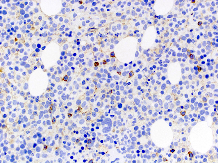
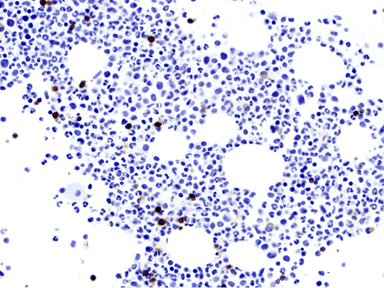
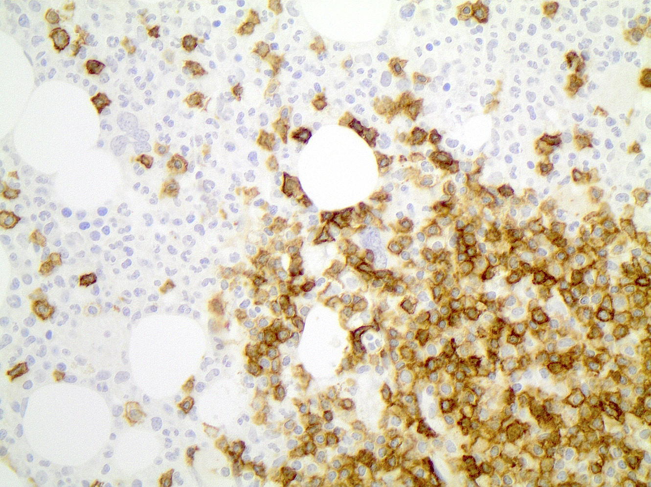
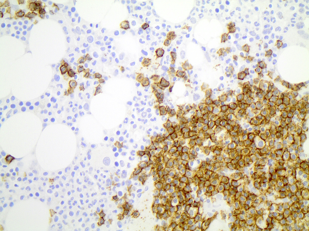
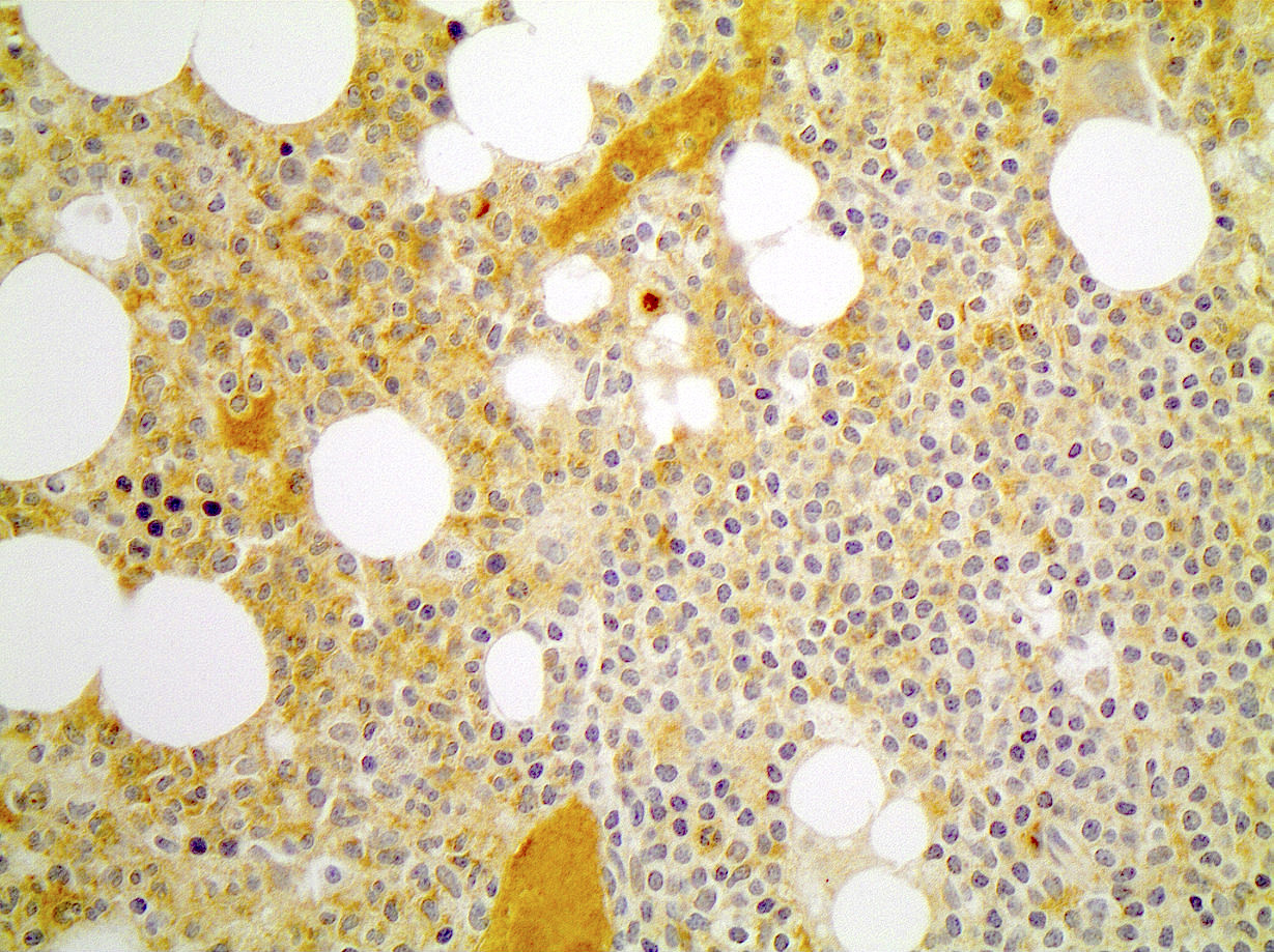
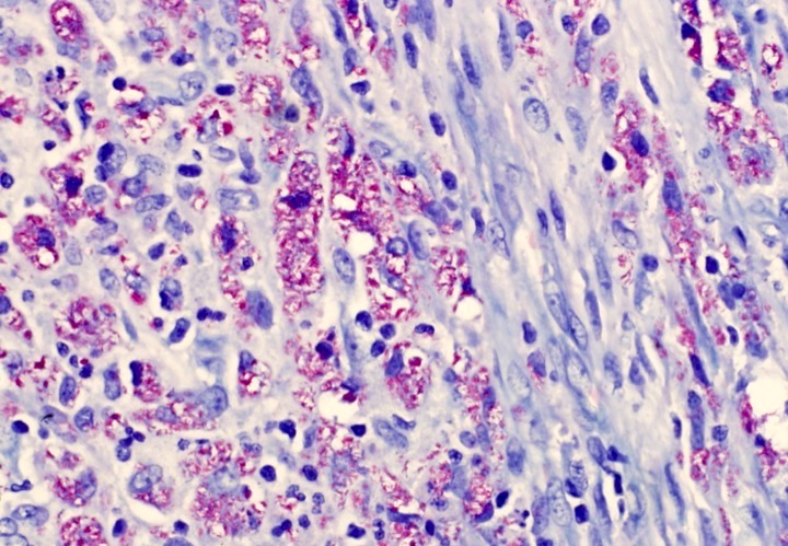
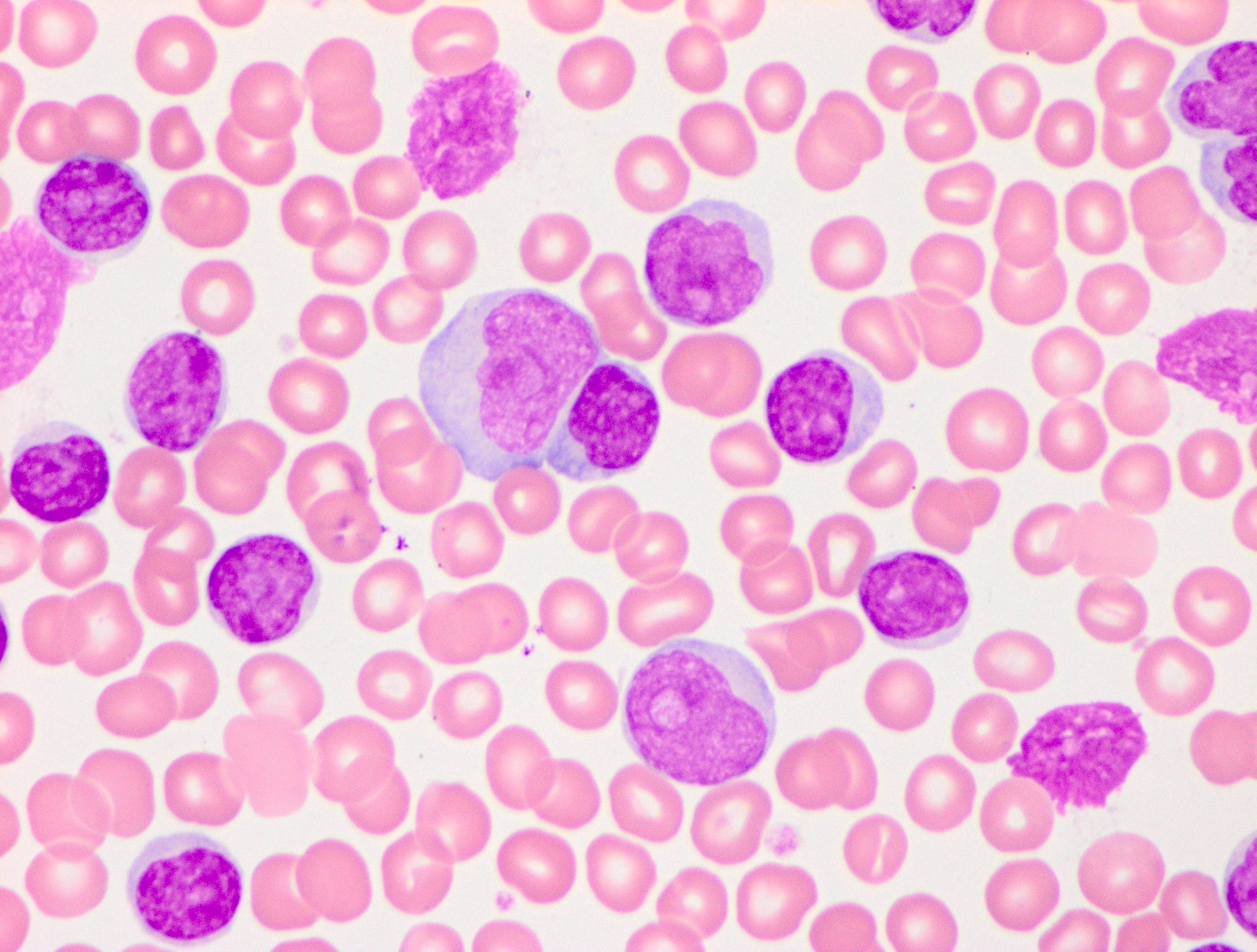
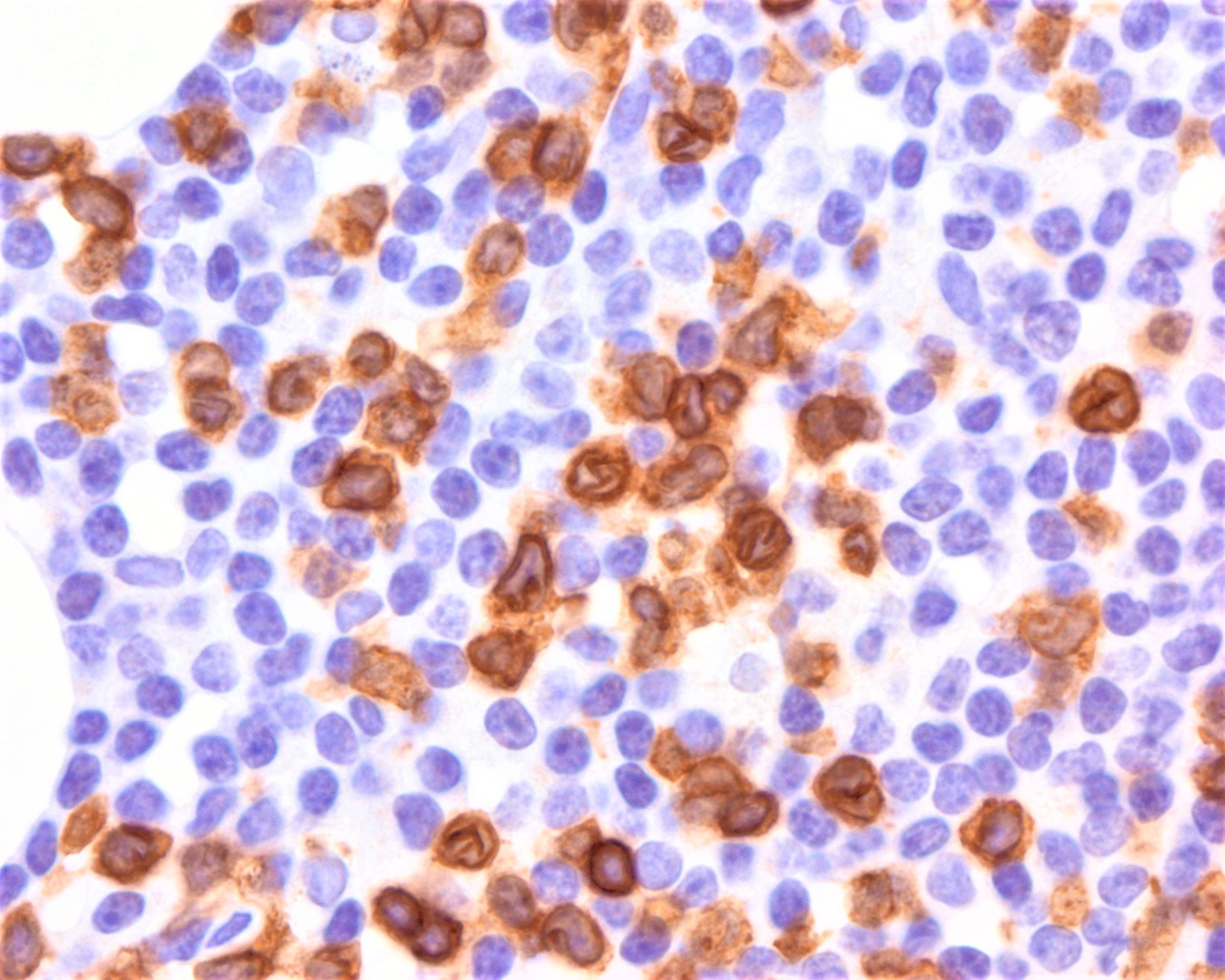
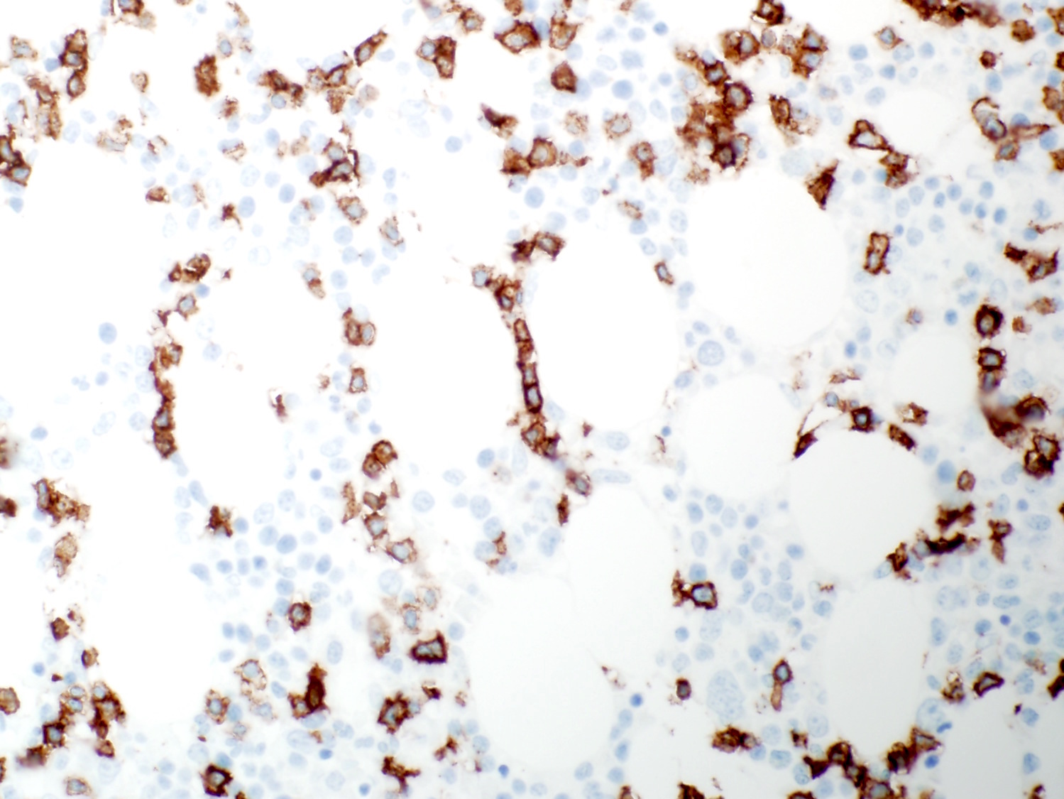
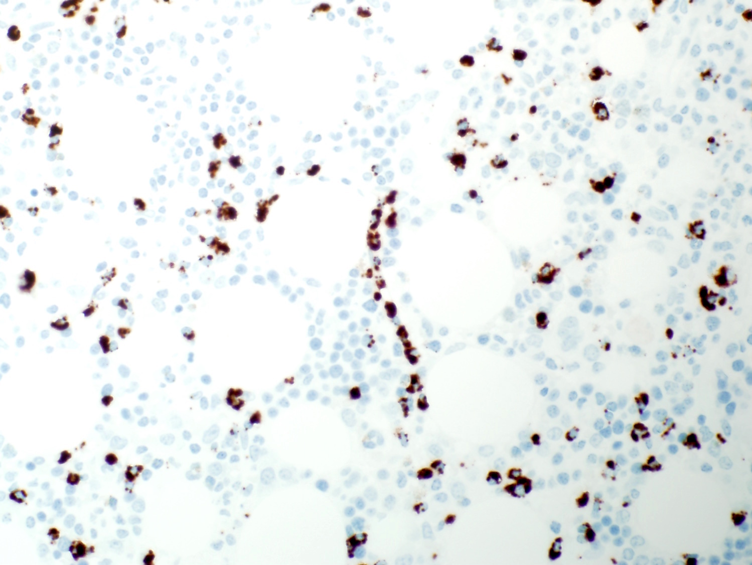
Immunohistochemical stains
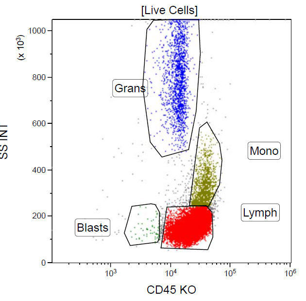
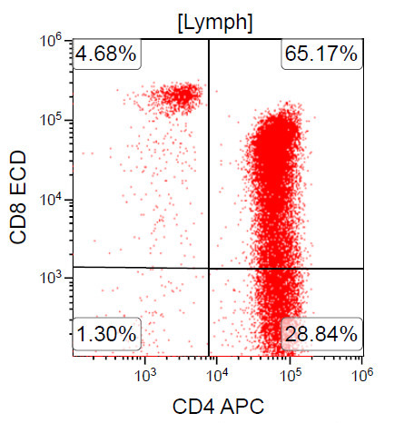
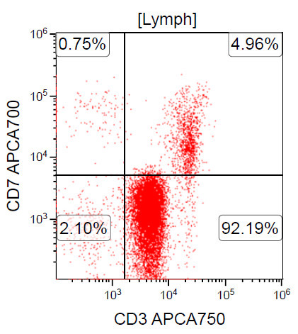
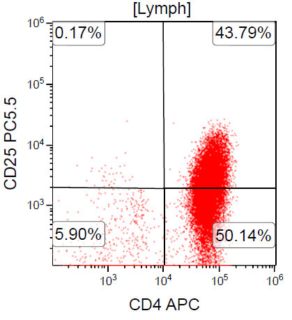
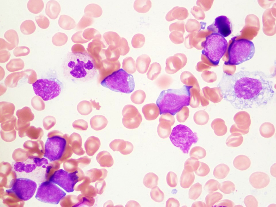
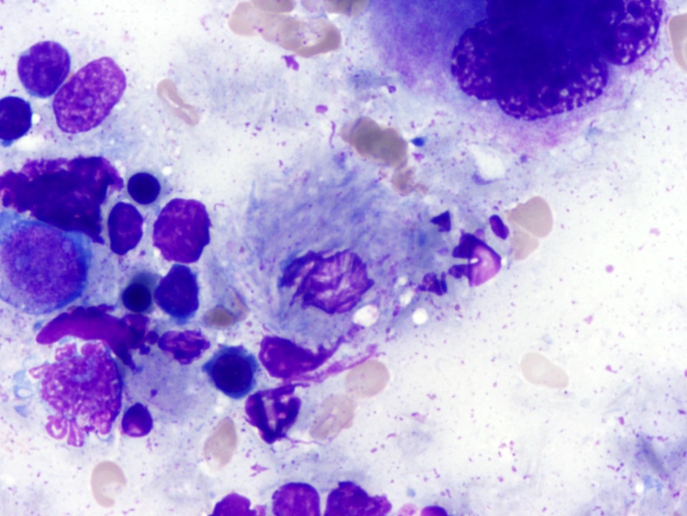
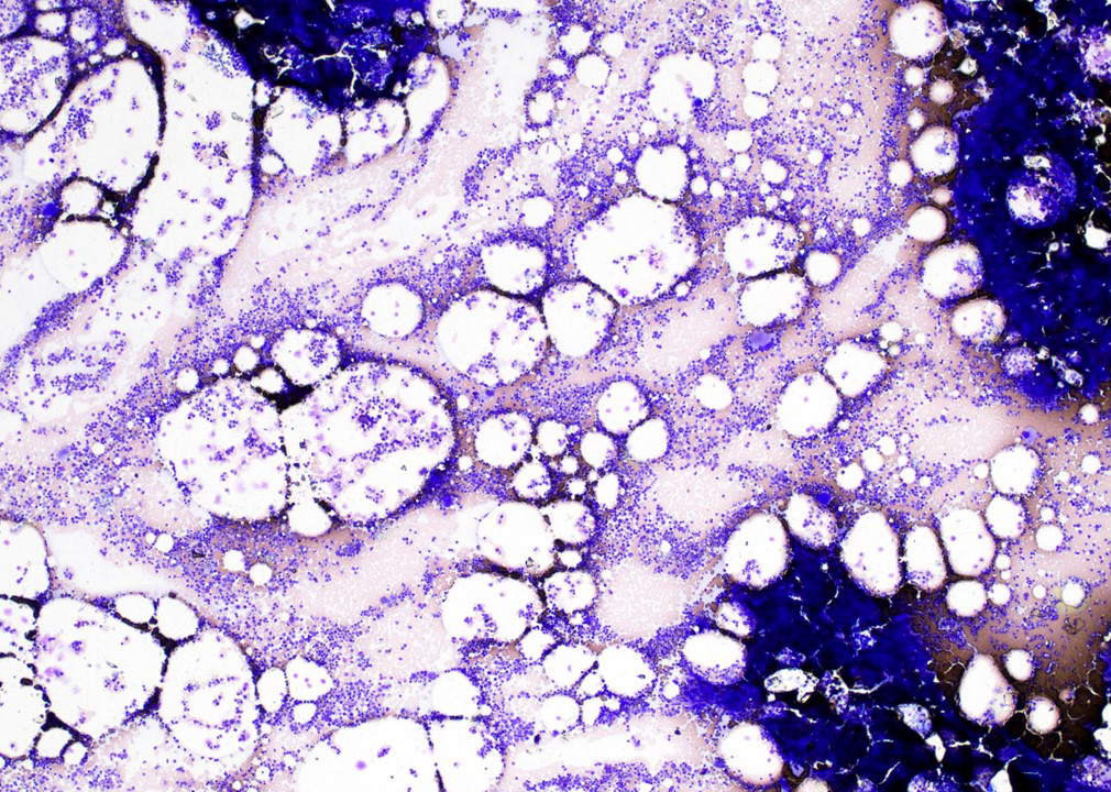
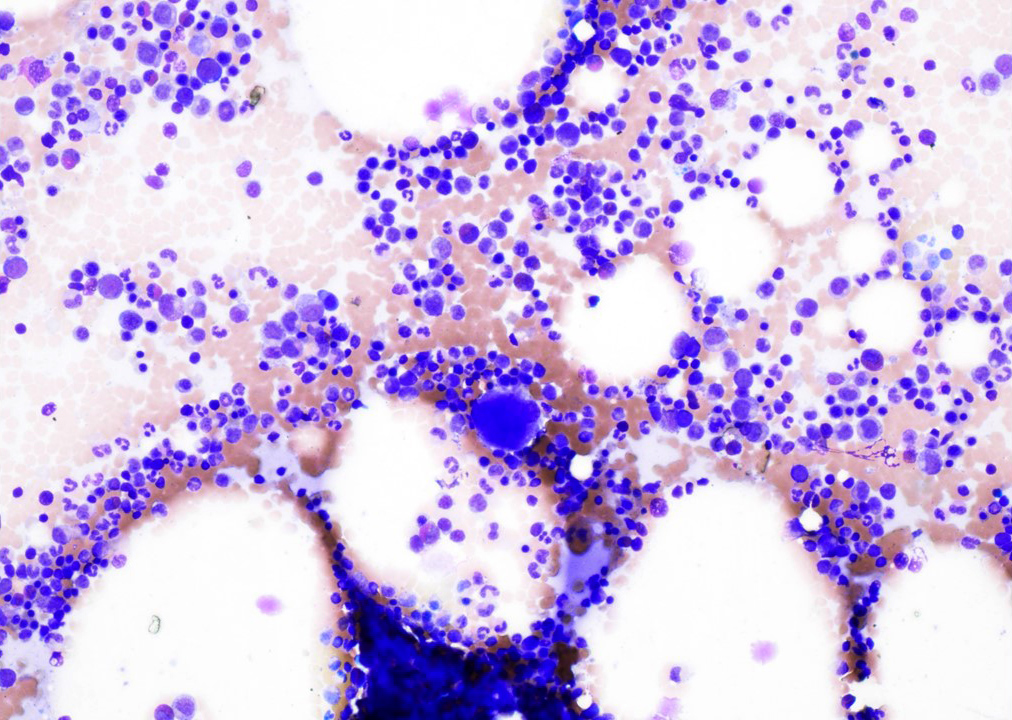
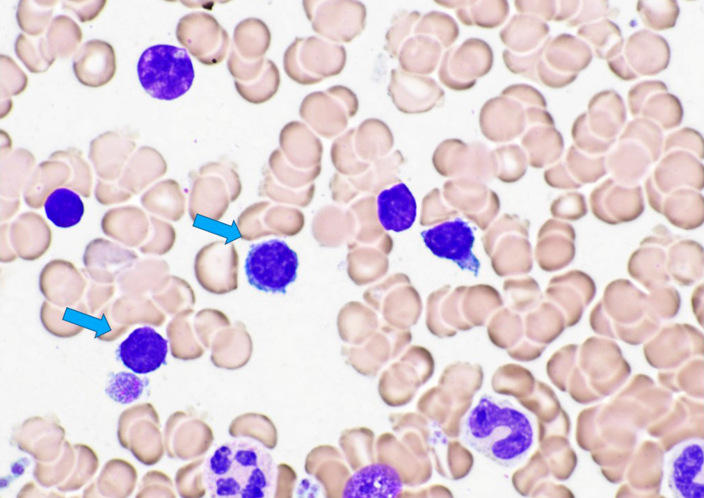
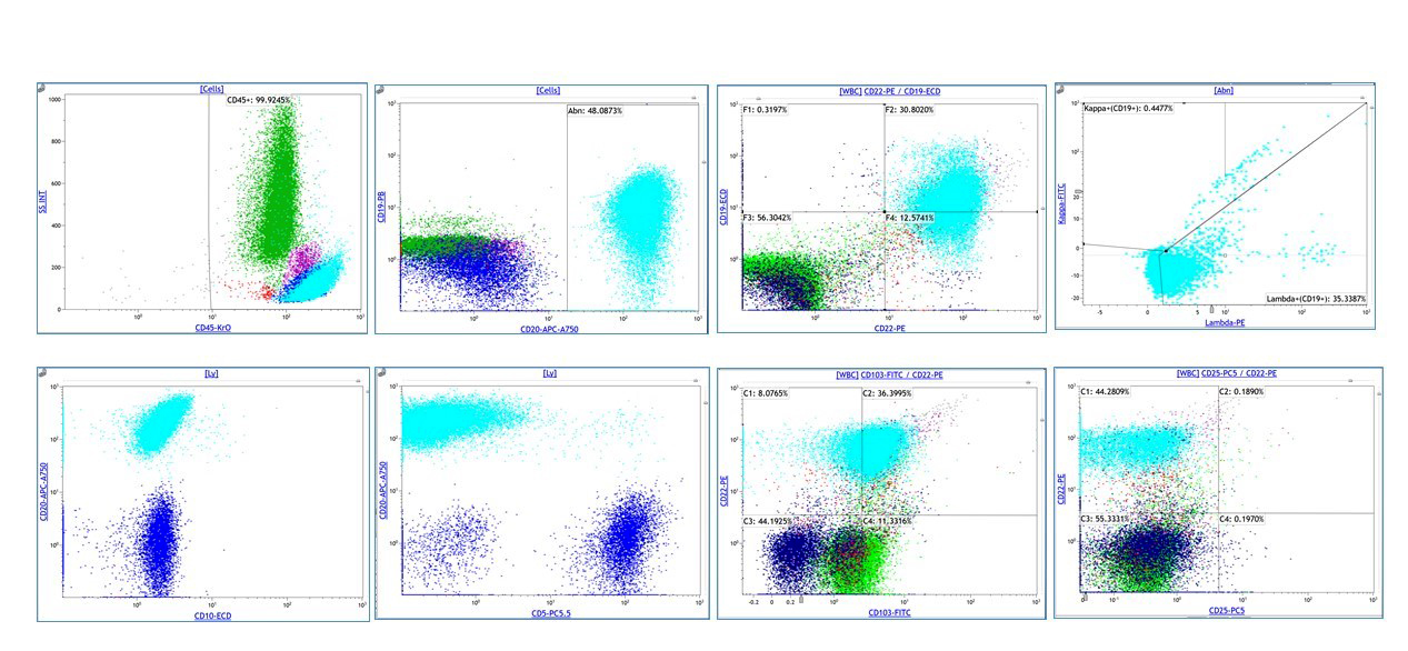
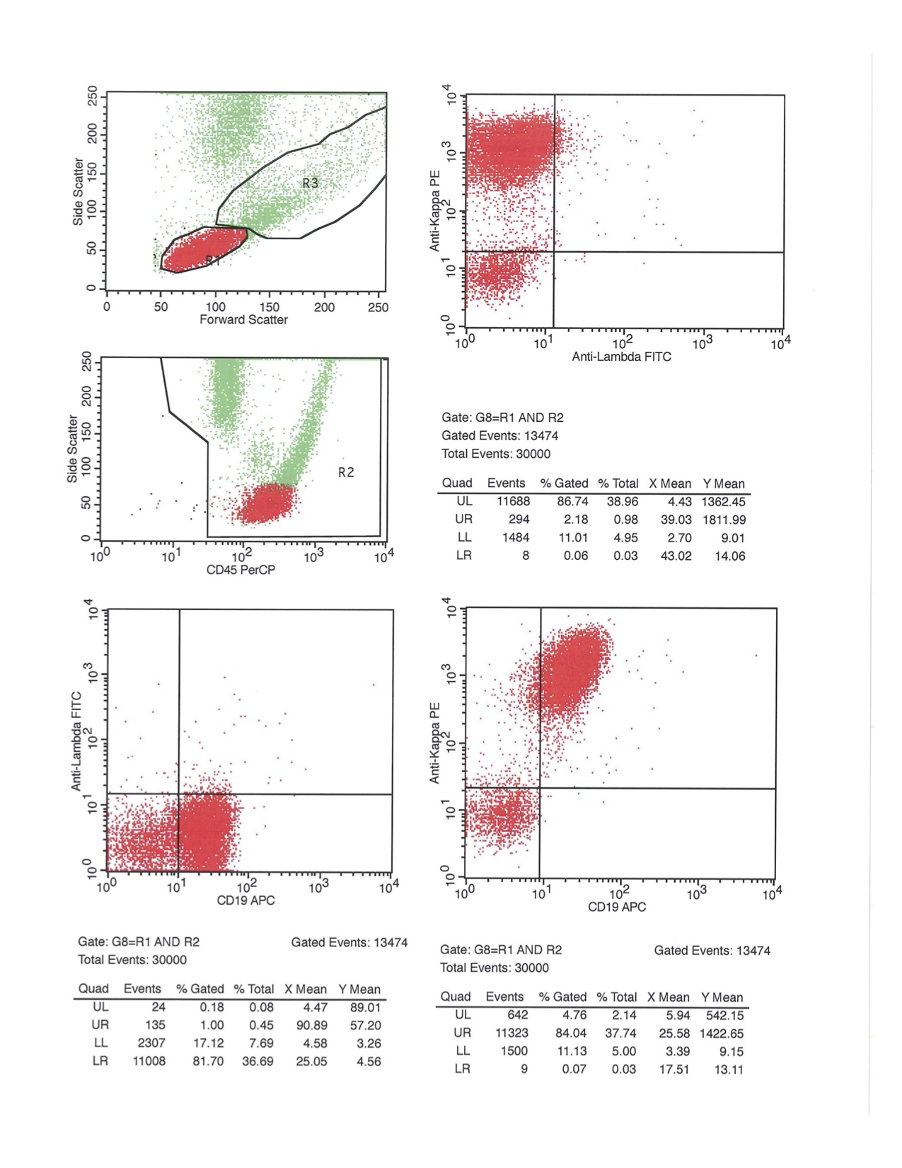
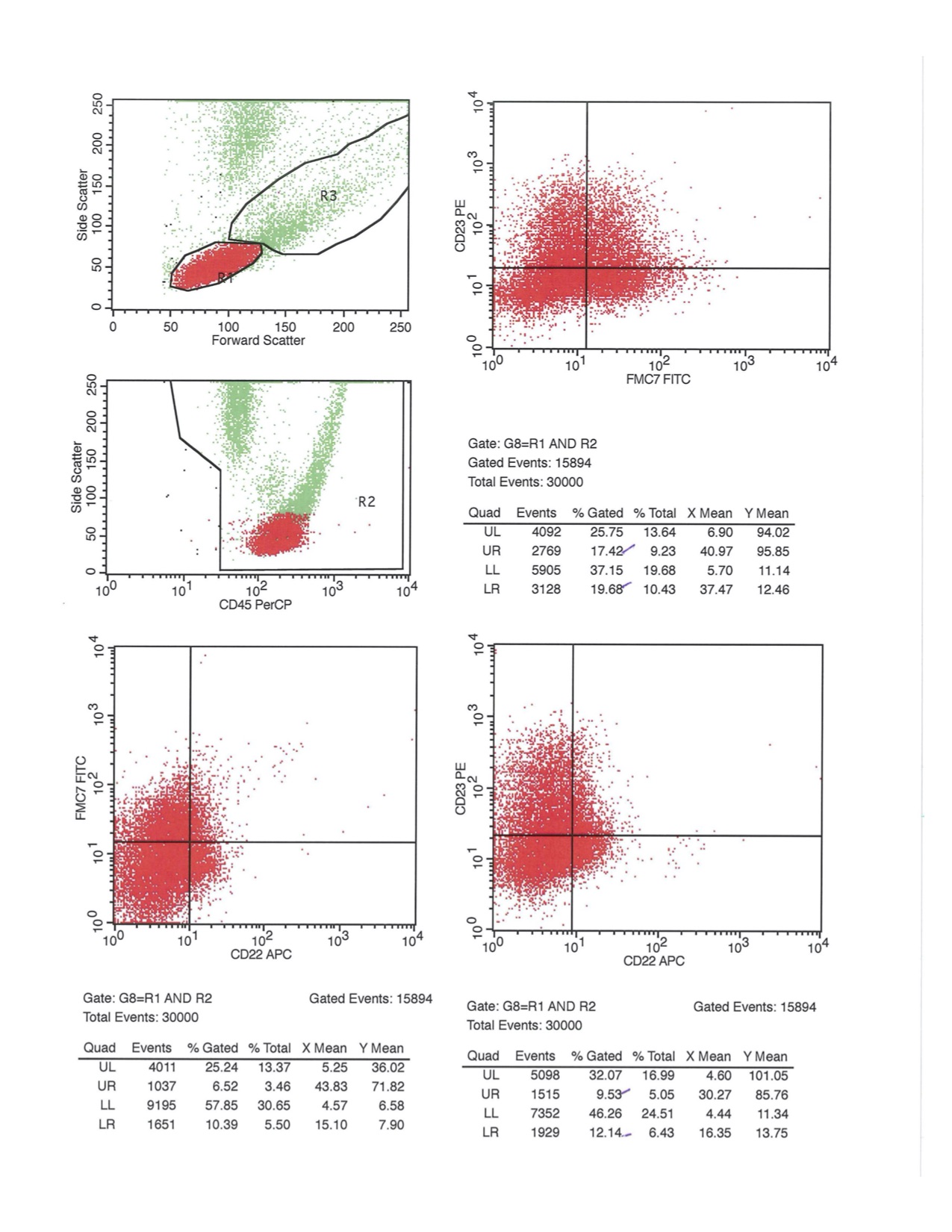
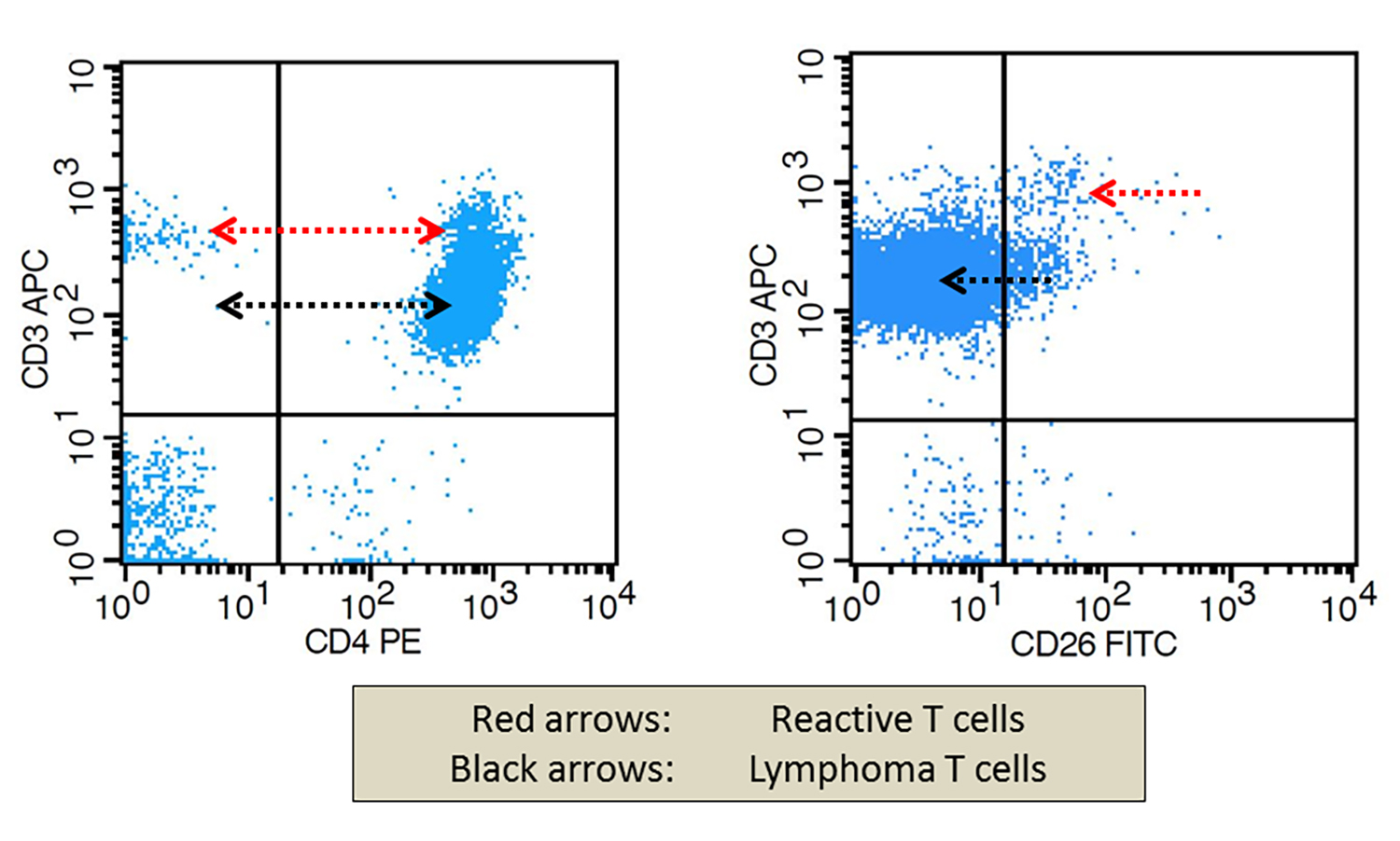
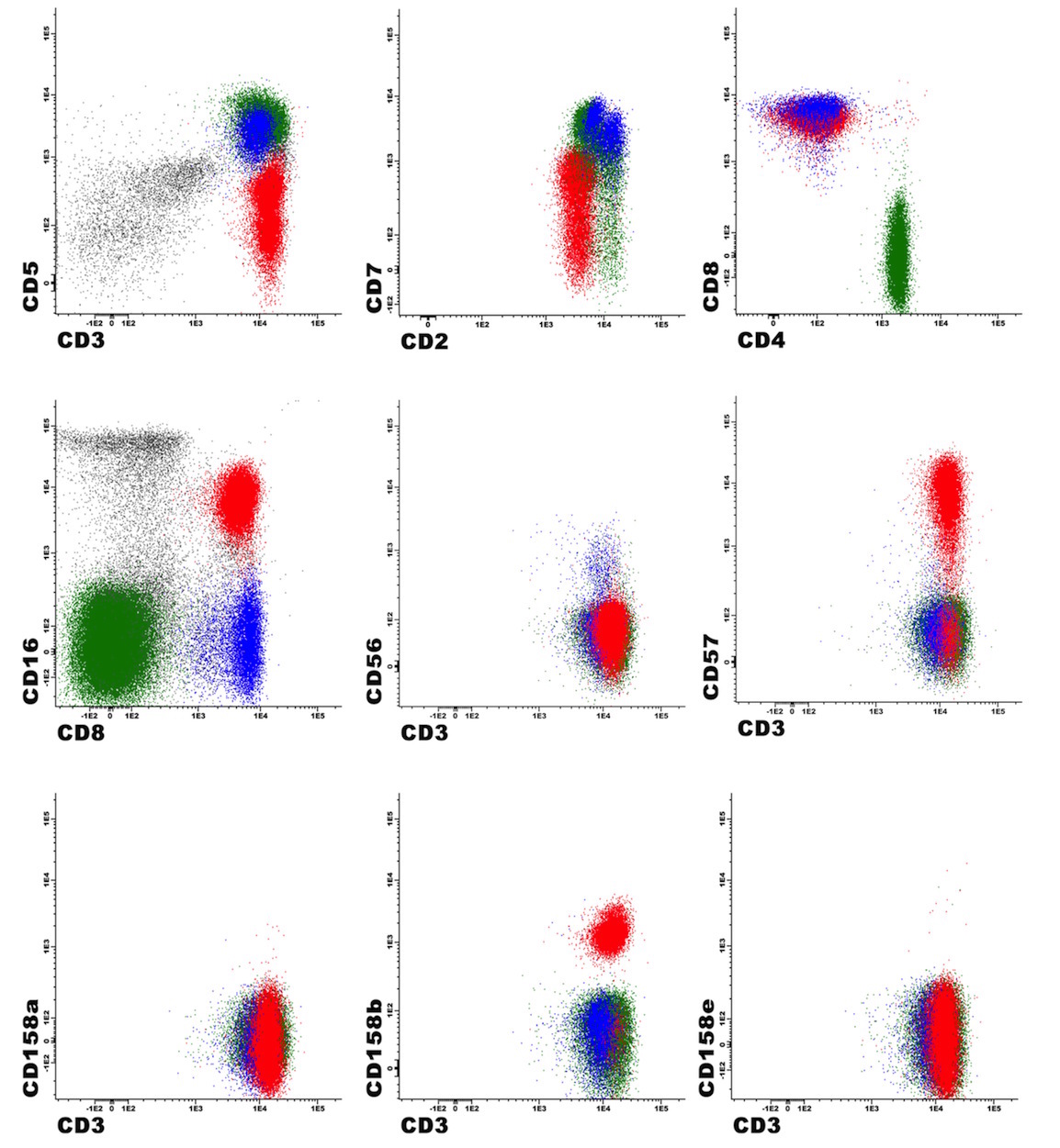
Contributed by Siba El Hussein, M.D. and Joseph Khoury, M.D.
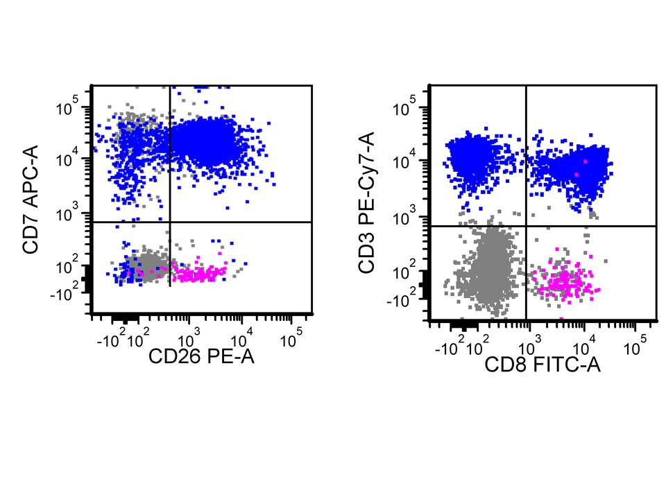
Flow cytometry characteristic


Images hosted on other servers:

Hb Bart’s, midpregnancy sonographic features


Contributed by Patricia Tsang, M.D.


Case of the Month #486
Images hosted on other servers:


HGA: inclusions in granulocytes

Left: HGA (HGE)
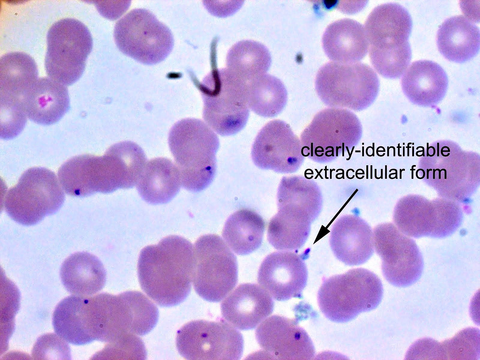
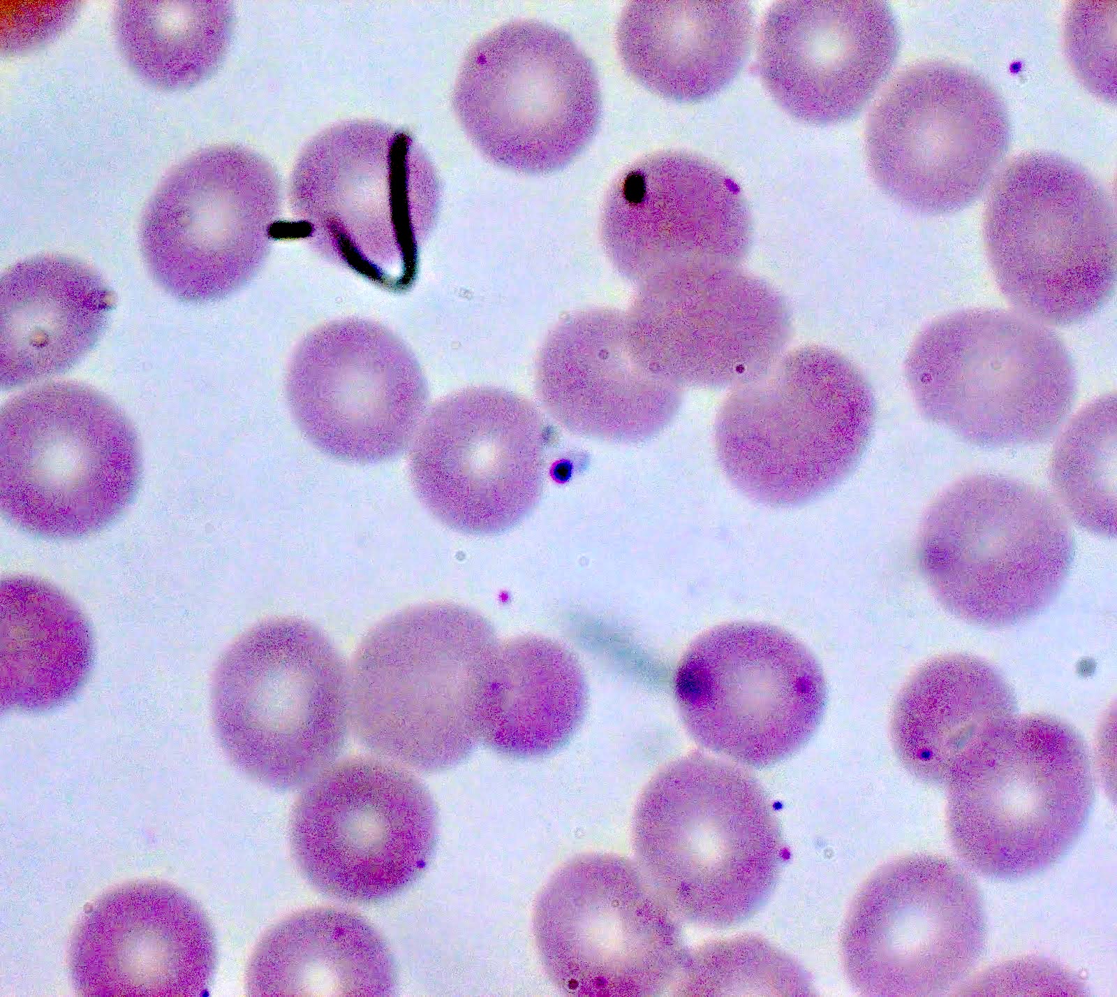
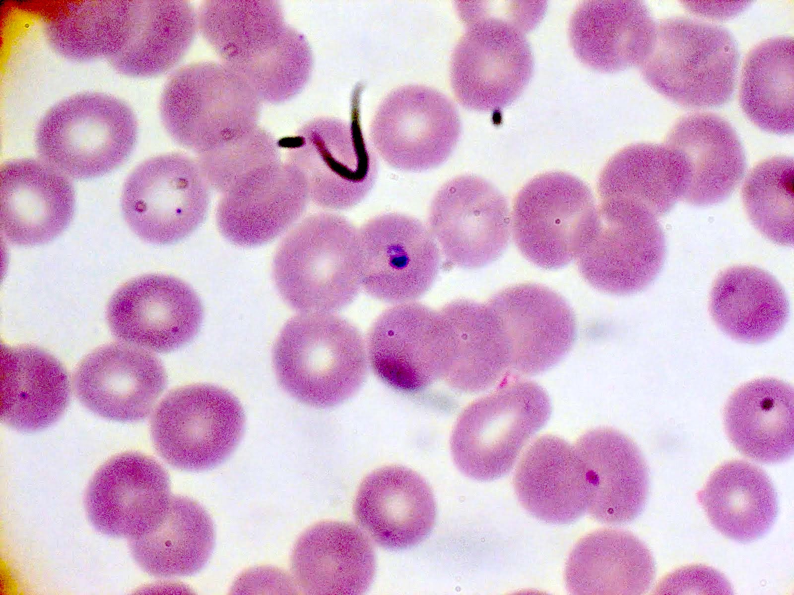
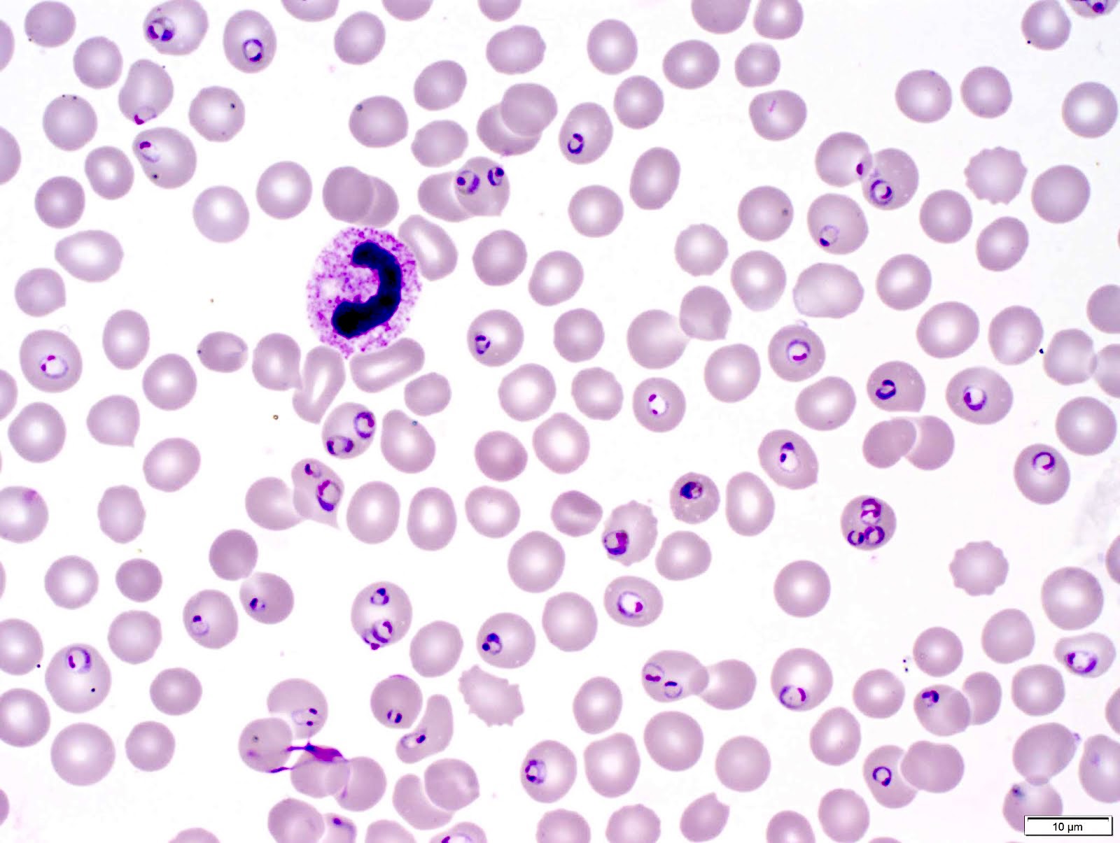
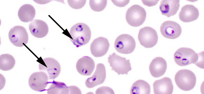
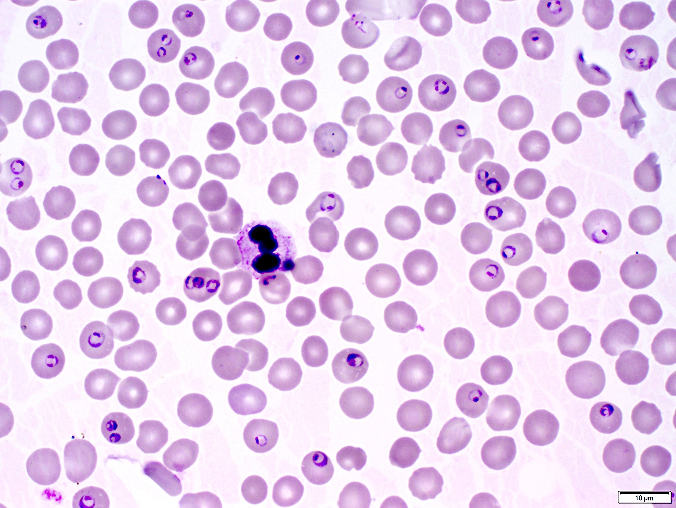
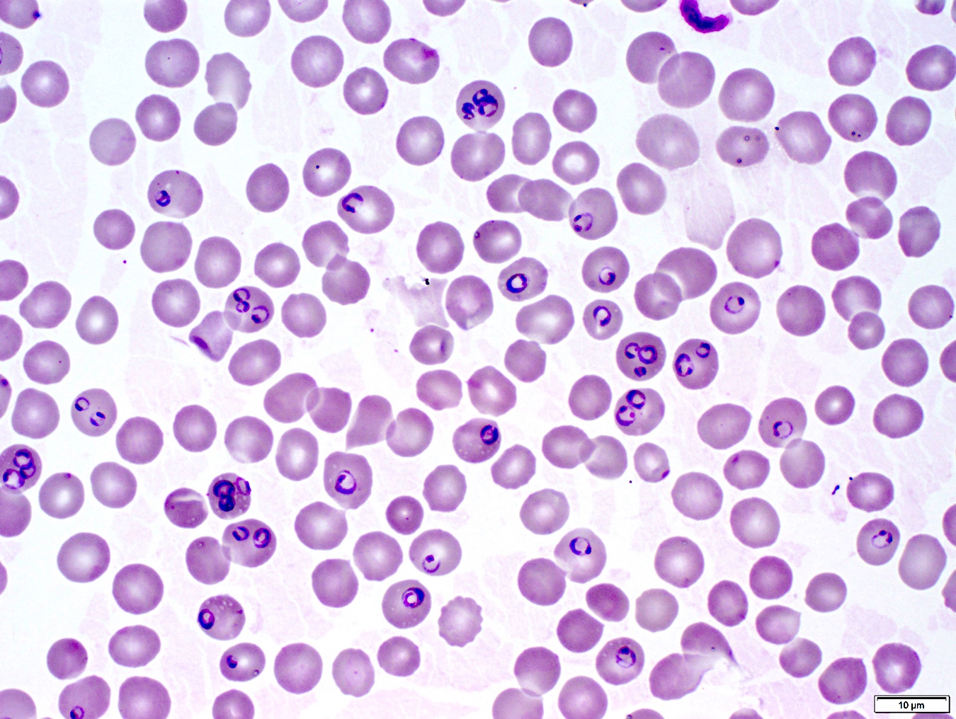
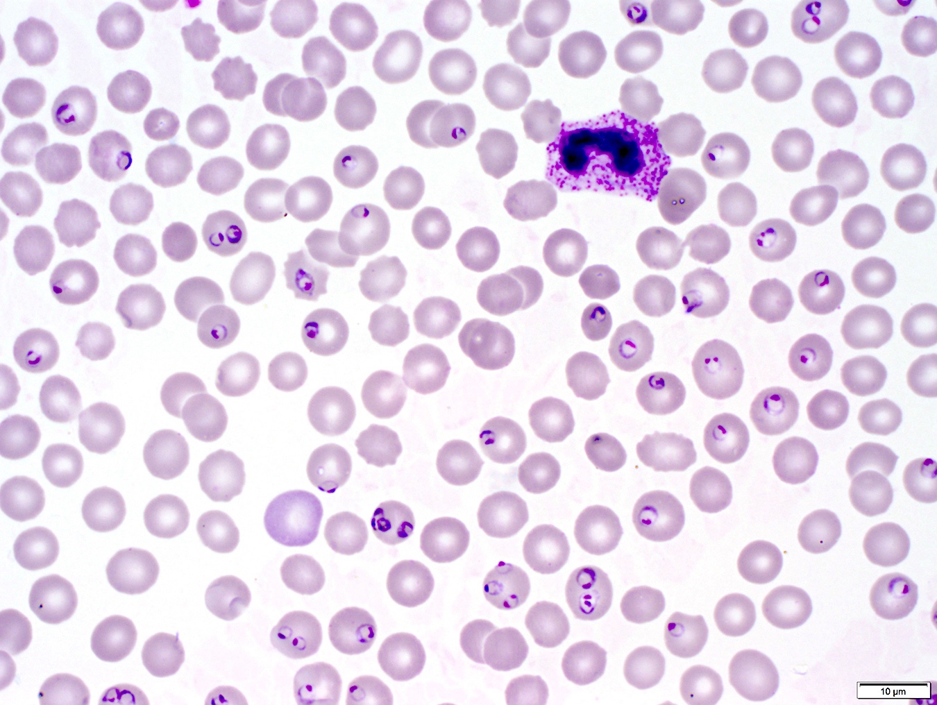
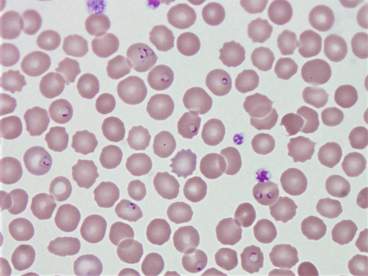
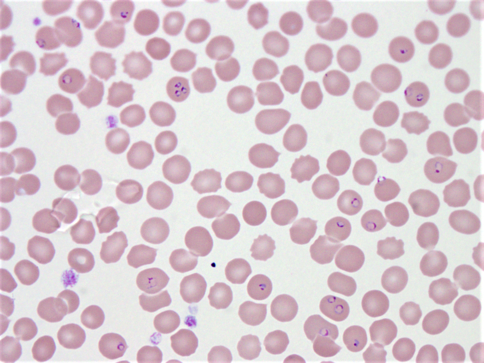
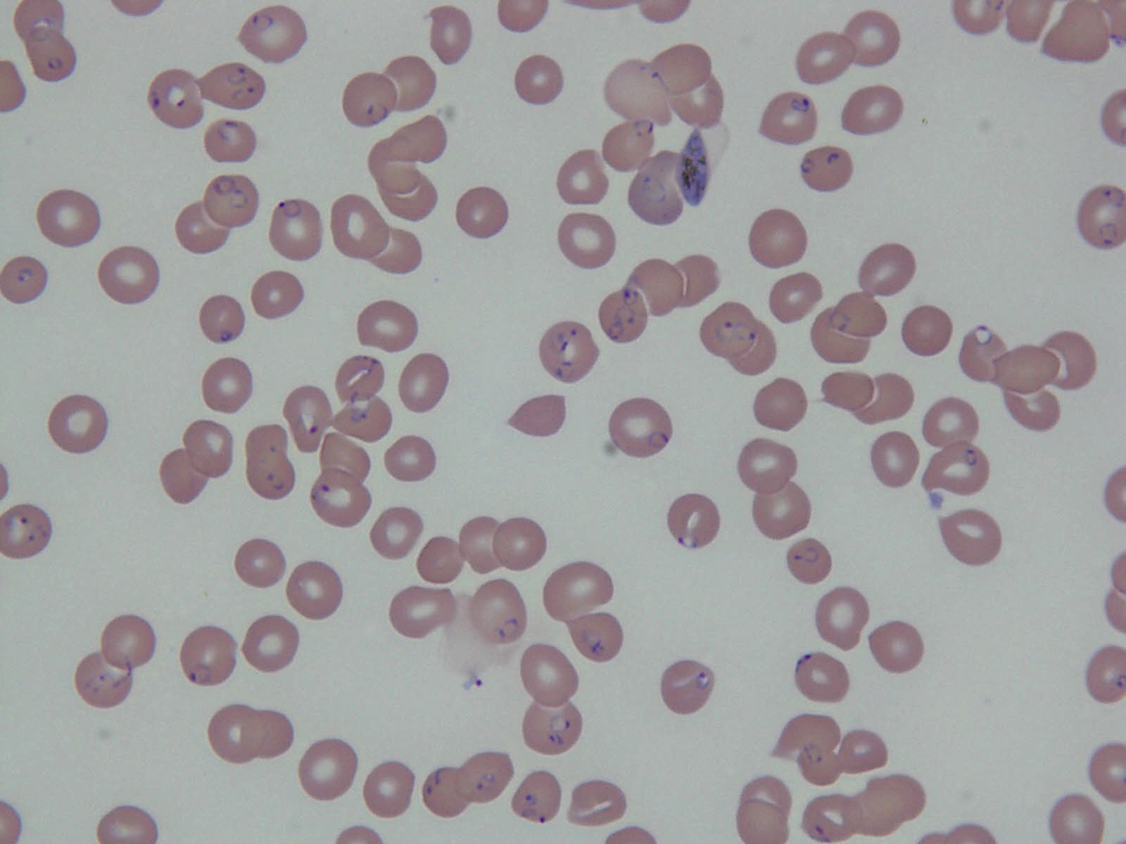
Contributed by Erika Wheeler, M.D. and Bobbi Pritt, M.D.
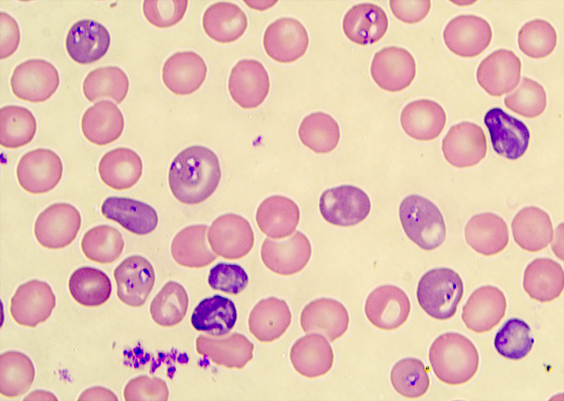
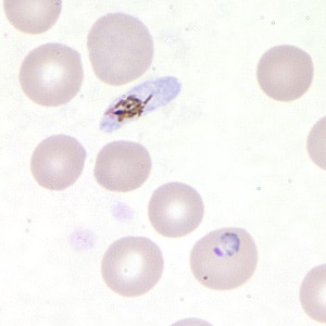
Intracellular and extracellular Babesia ring forms
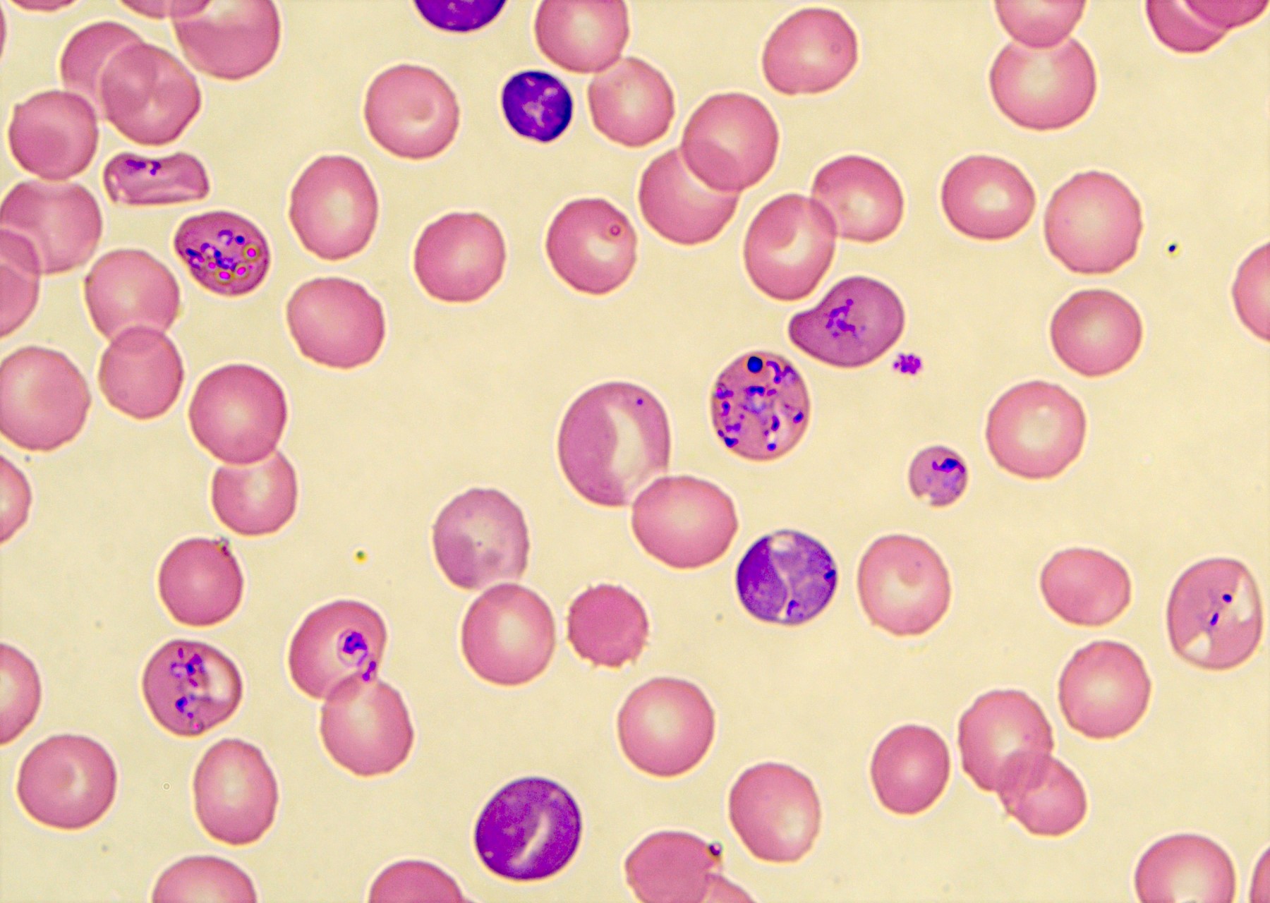
Multiple Babesia ring forms

Giemsa stained thick and thin blood films
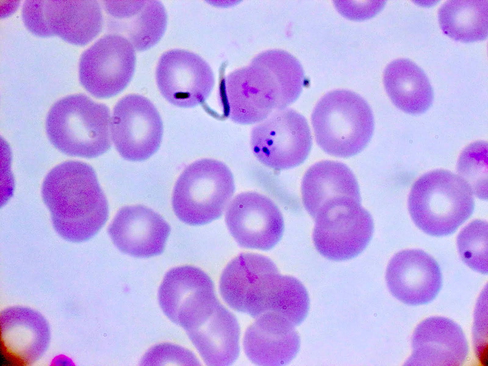
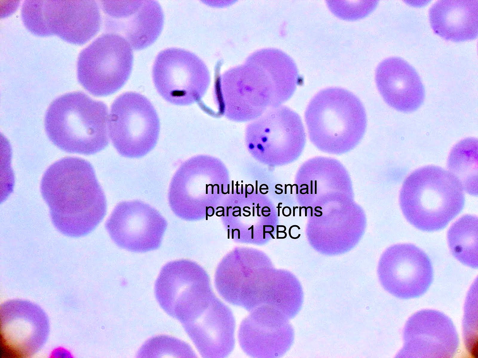
Giemsa stained thick and thin blood films
Images hosted on other servers:

Tetrad form
Images hosted on other servers:

CT scan
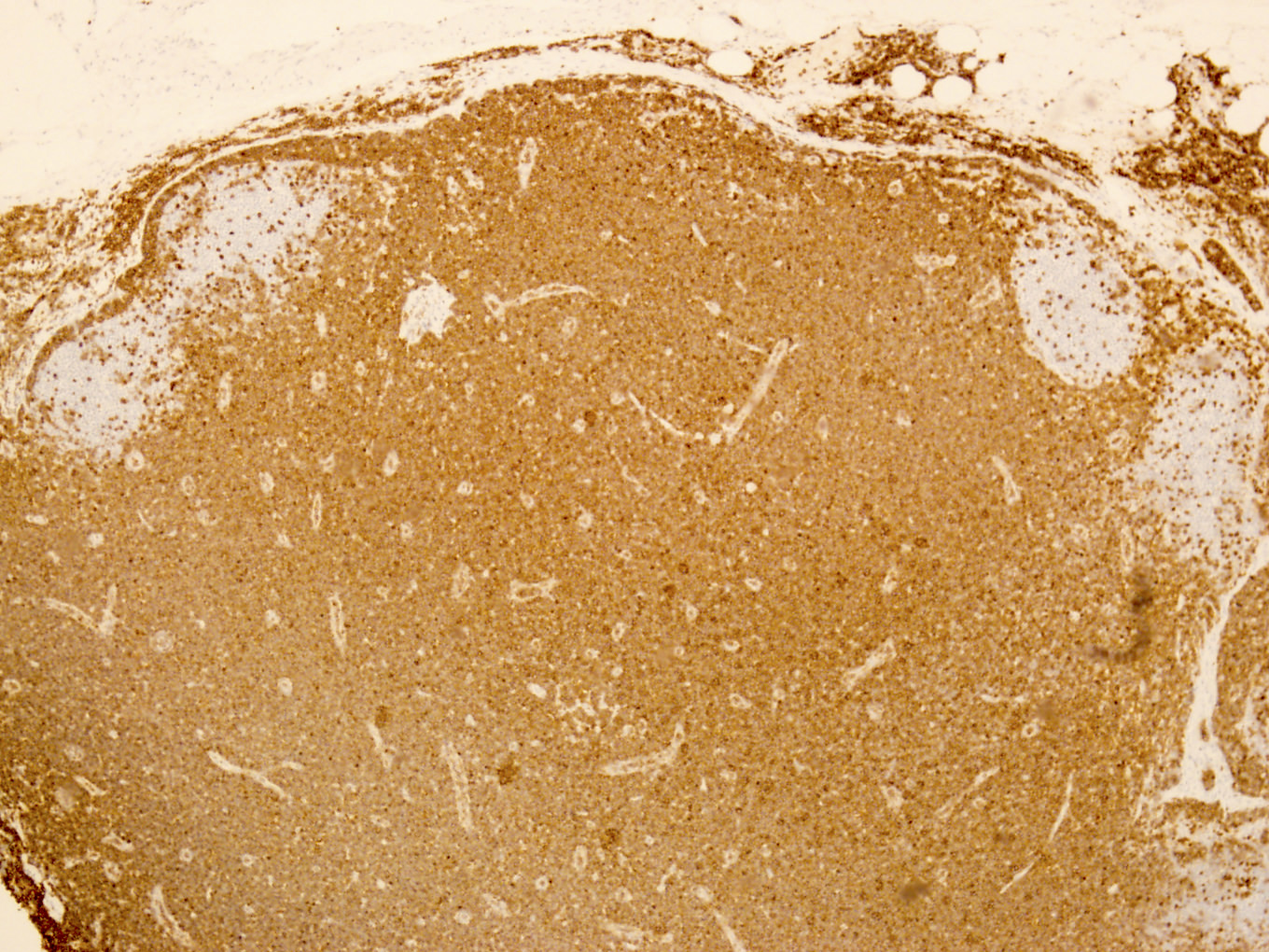
Contributed by Béla Kajtár, M.D., Ph.D.
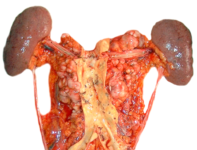
Paraaortic lymph nodes
Images hosted on other servers:

Splenic involvement
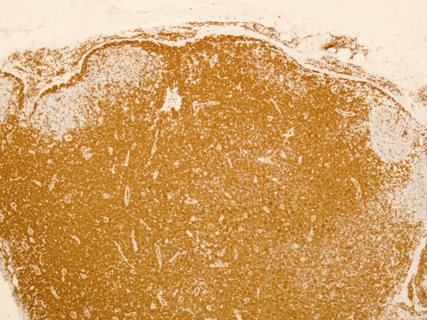
Contributed by Béla Kajtár, M.D., Ph.D.
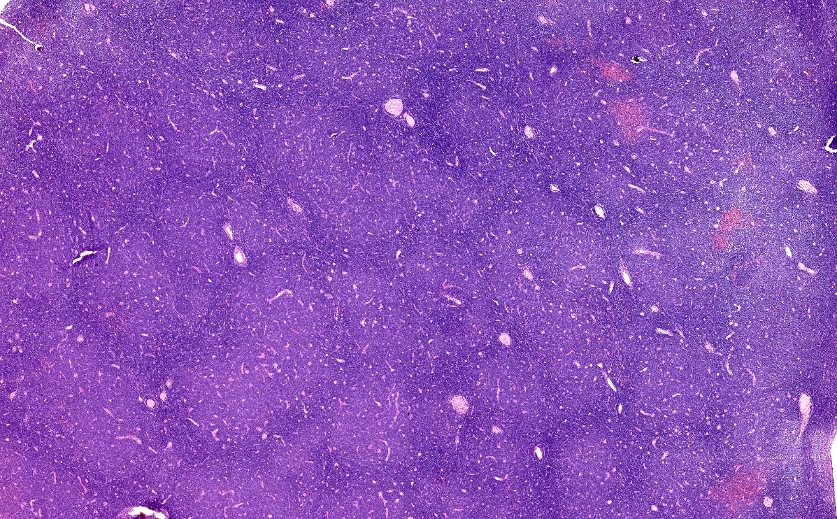
Proliferation centers
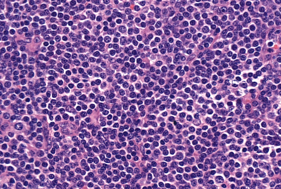
Diffuse lymphocytic infiltrate
Proliferation center
CD20
LEF1
Bone marrow involvement
Contributed by Béla Kajtár, M.D., Ph.D.
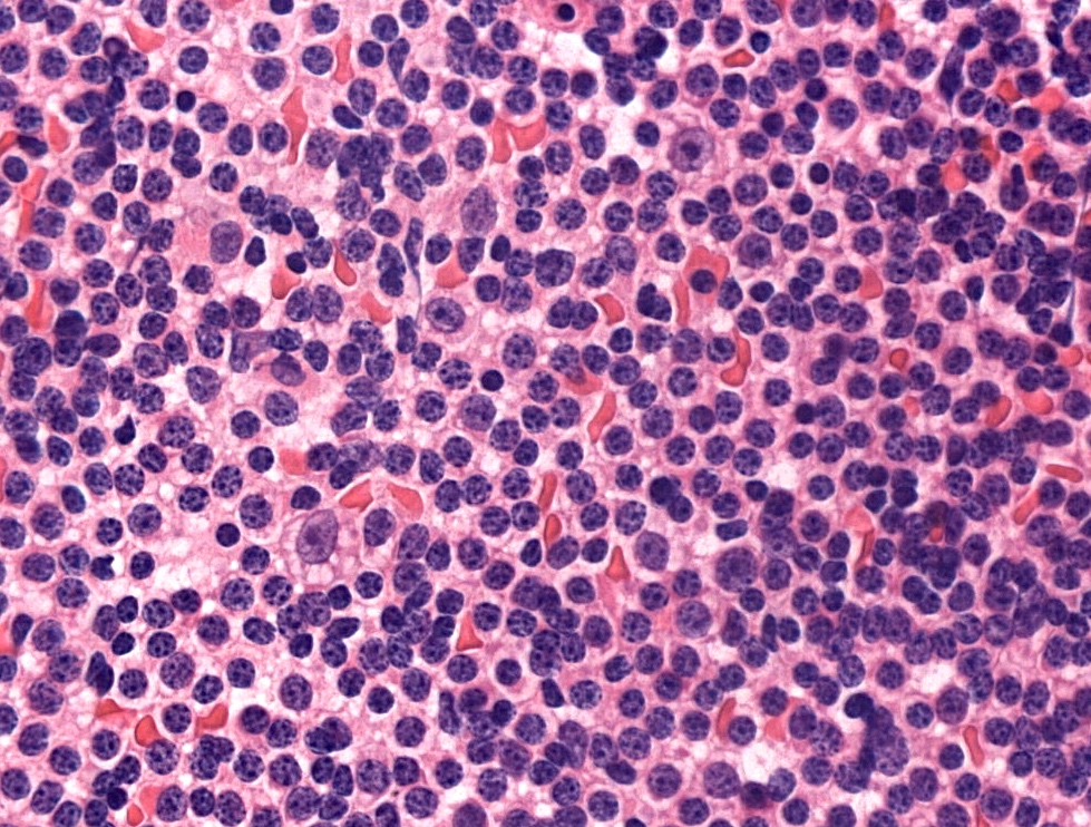
Touch prep SLL
Contributed by Béla Kajtár, M.D., Ph.D.
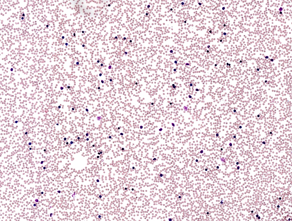
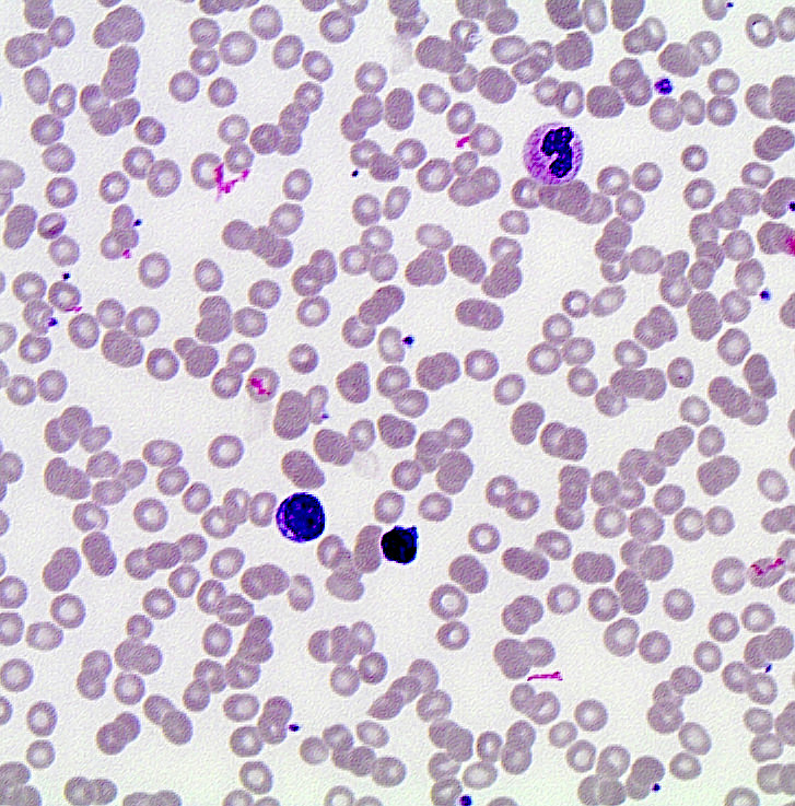
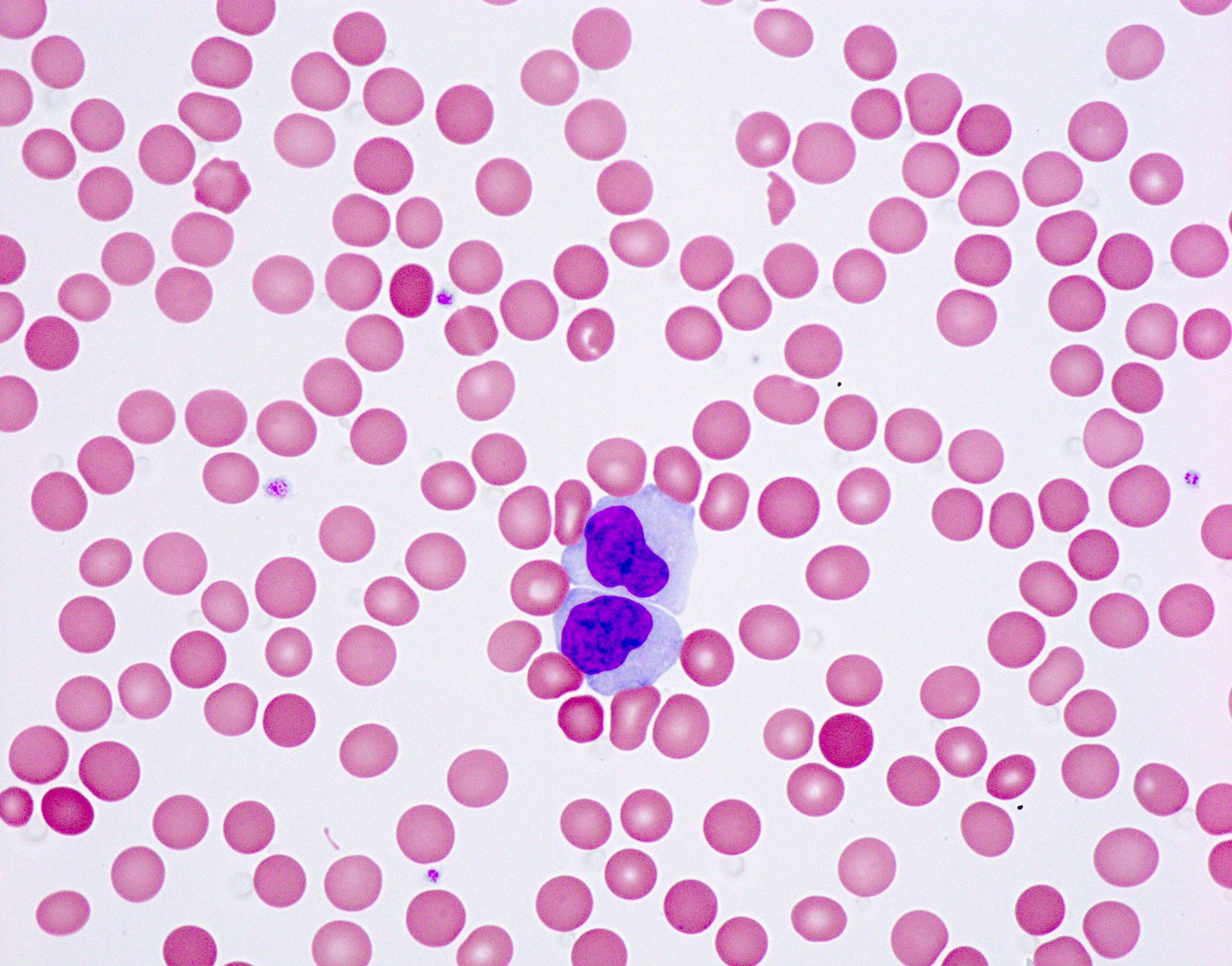
CLL in peripheral blood
Contributed by Béla Kajtár, M.D., Ph.D.
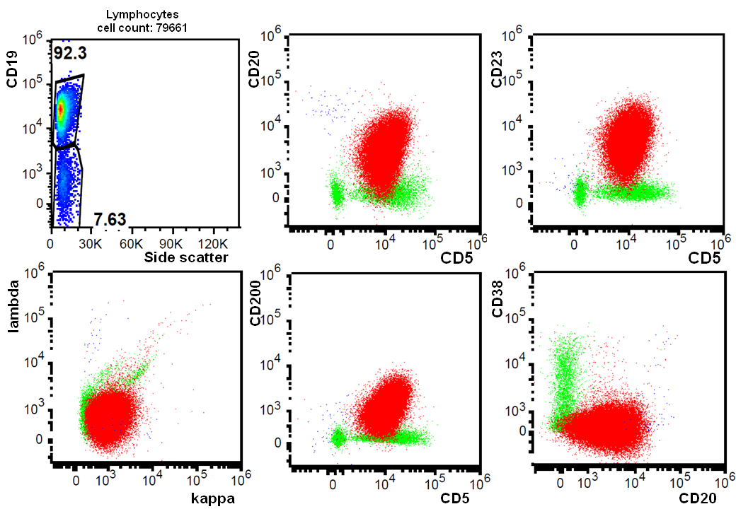
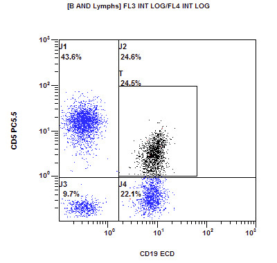
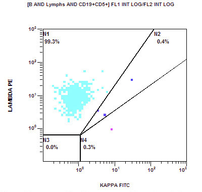
CLL, scatter plot
Contributed by Béla Kajtár, M.D., Ph.D.
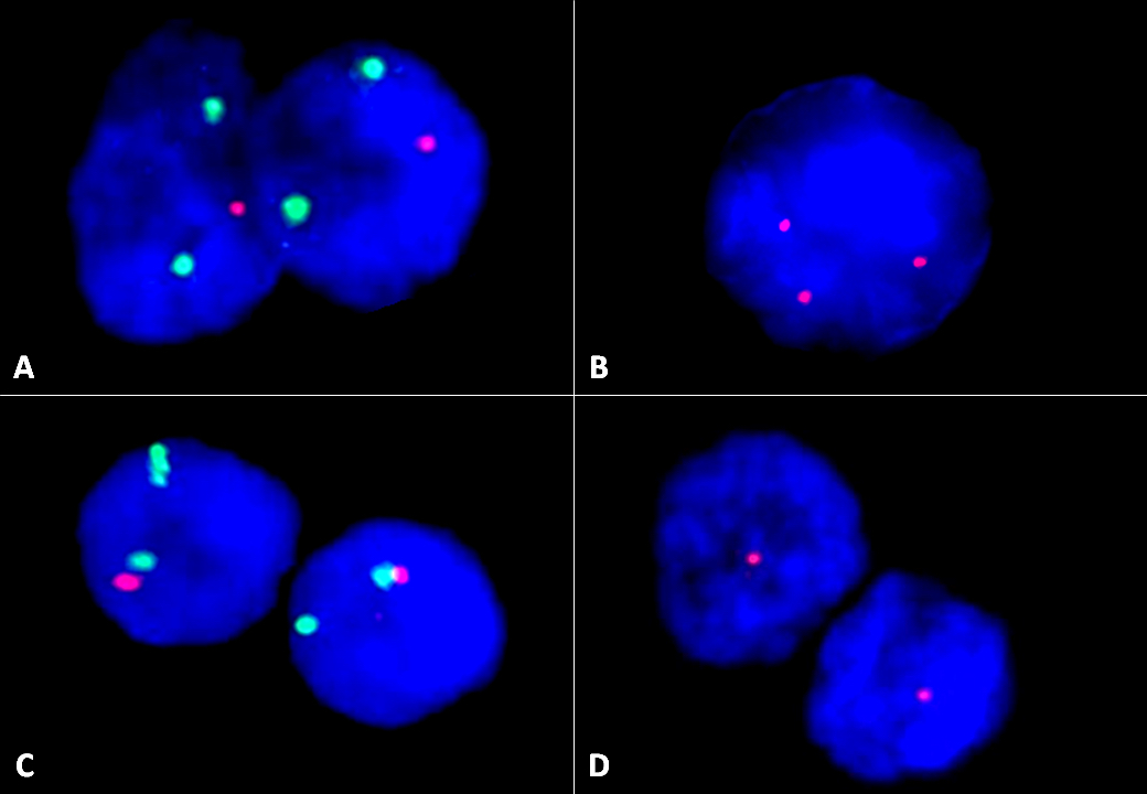
FISH images
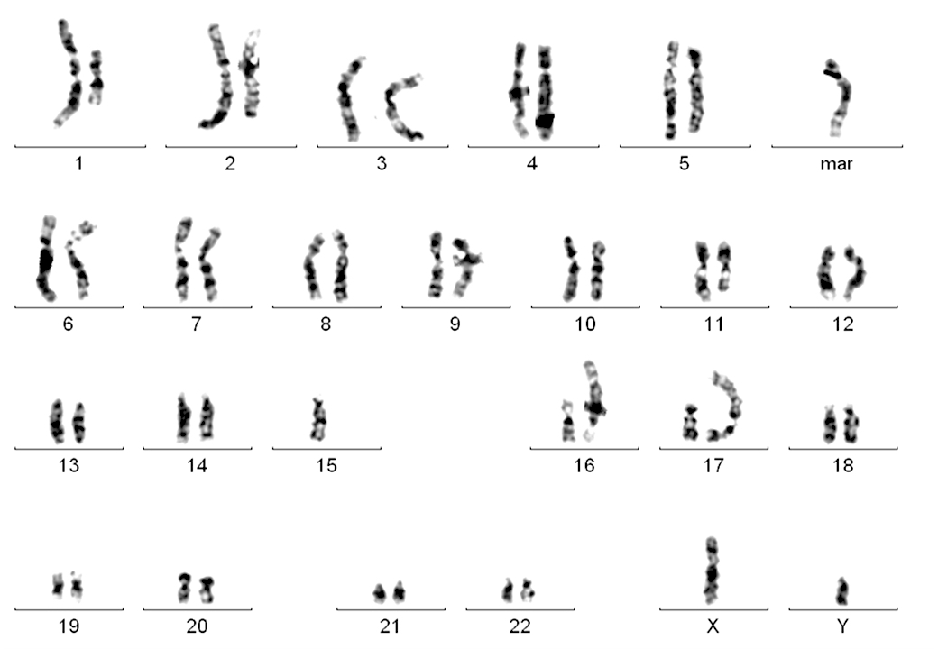
Karyogram
Images hosted on other servers:


Skin lesions

Gingivitis, periodontitis
Images hosted on other servers:

Clinical and histological profile

IL-17 signature

Skin biopsy
Contributed by Dr. Mark R. Wick
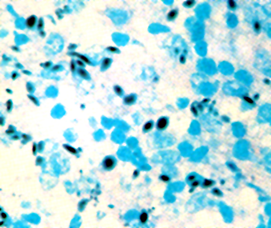
Childhood disease, AFB stain
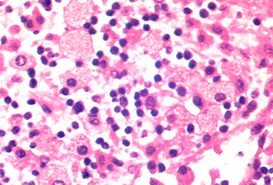
Childhood disease
Childhood disease, PAS stain
Contributed by David Lynch, M.D.
Bone marrow core and CD68
Bone marrow aspirate
Images hosted on other servers:

Megaloblastic erythroid precursors
Images hosted on other servers:

Hypersegmented neutrophils, macro ovalocytes

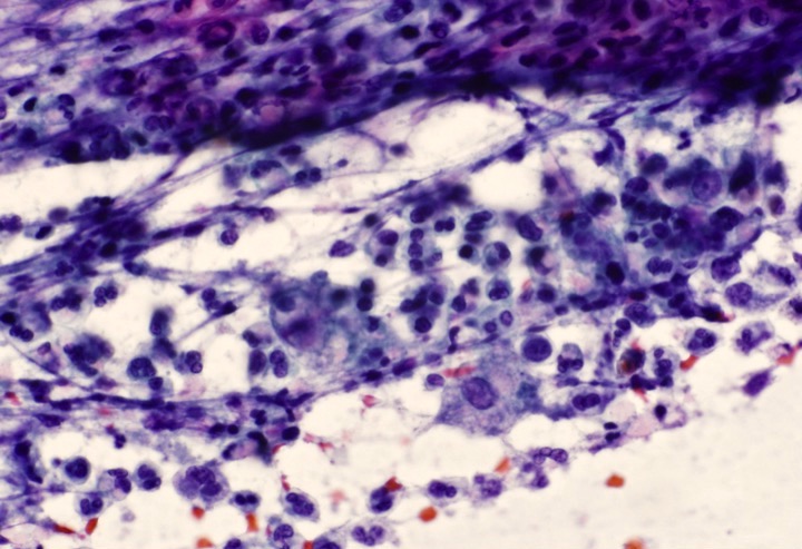
Contributed by Pallavi Khattar, M.D. and Case #437
Bone marrow biopsy
CD20 immunostain
Bone marrow aspirate
Bone marrow aspirate
Bone marrow biopsy
Bone marrow aspirate
Bone marrow aspirate
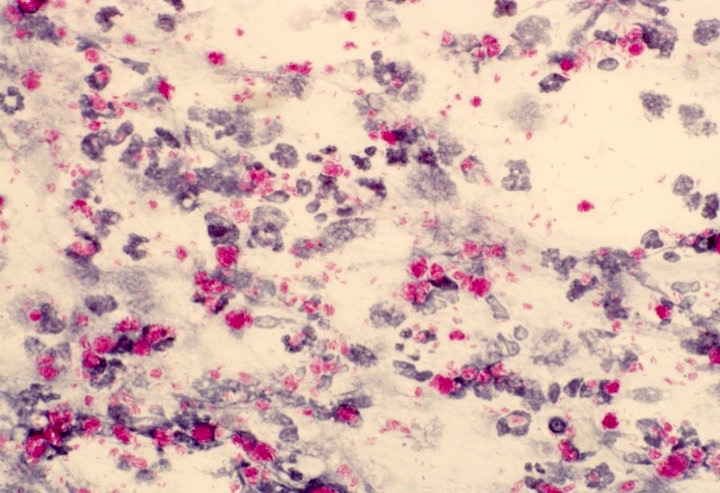
CD20 immunostain
CD3 immunostain

DBA-44 immunostain
Contributed by Daniel D. Mais, M.D.

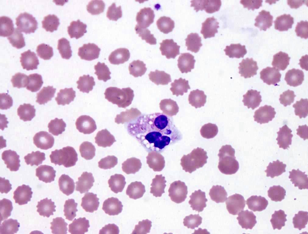
Peripheral blood smear (Wright stain)

Hemoglobin electrophoresis
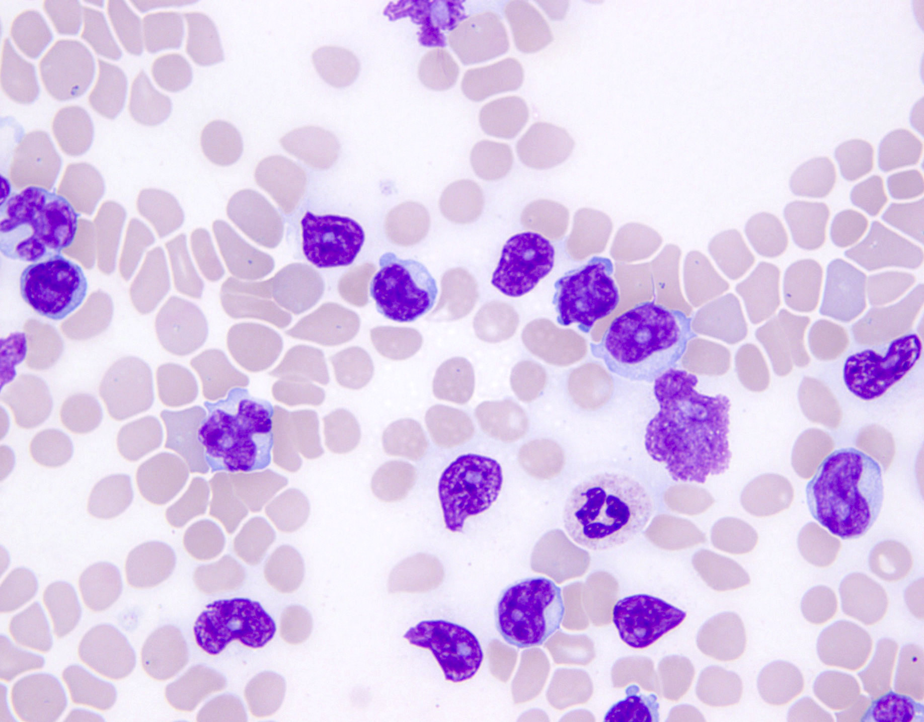
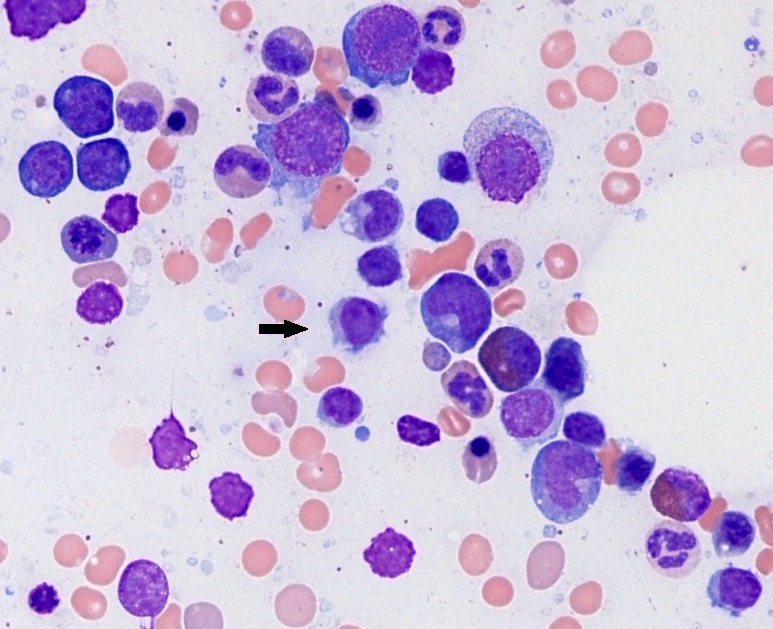
Contributed by Roberto N. Miranda, M.D.

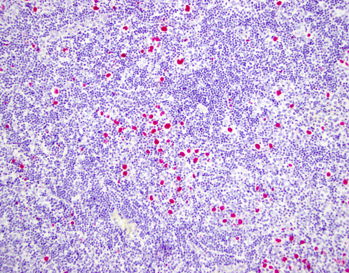
Bone marrow infiltration
Bone marrow with dyspoietic changes
CD3 positive
CD4 negative

CD8 negative
TCR βF1 negative
TCRγδ positive
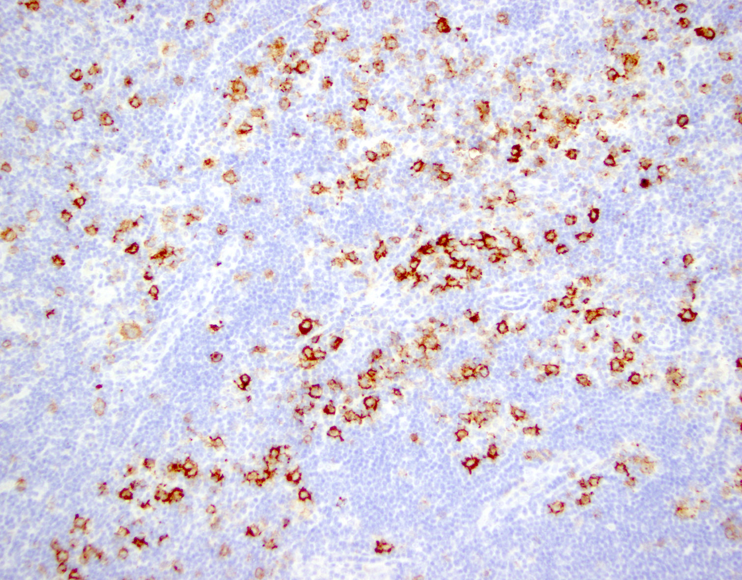
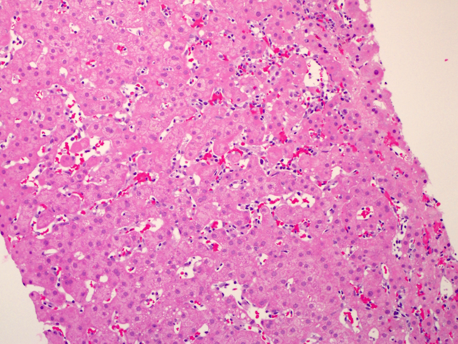
Liver infiltration
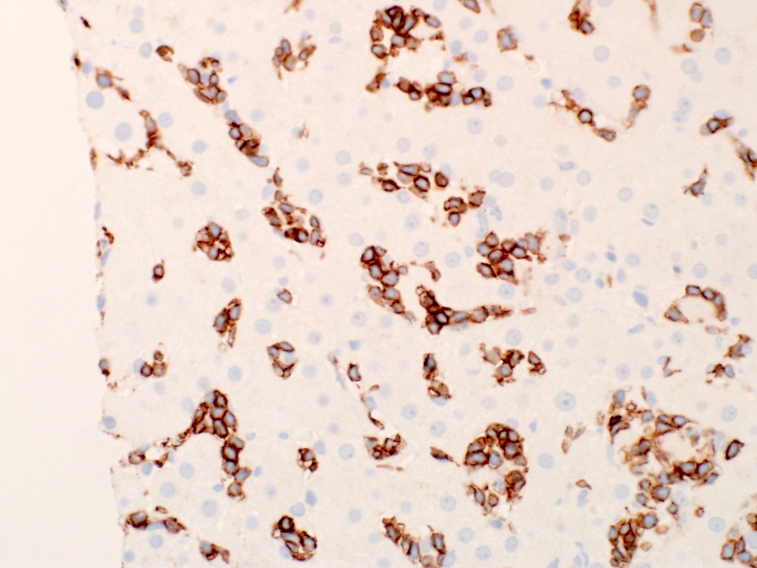
HSTCL: CD3 positive
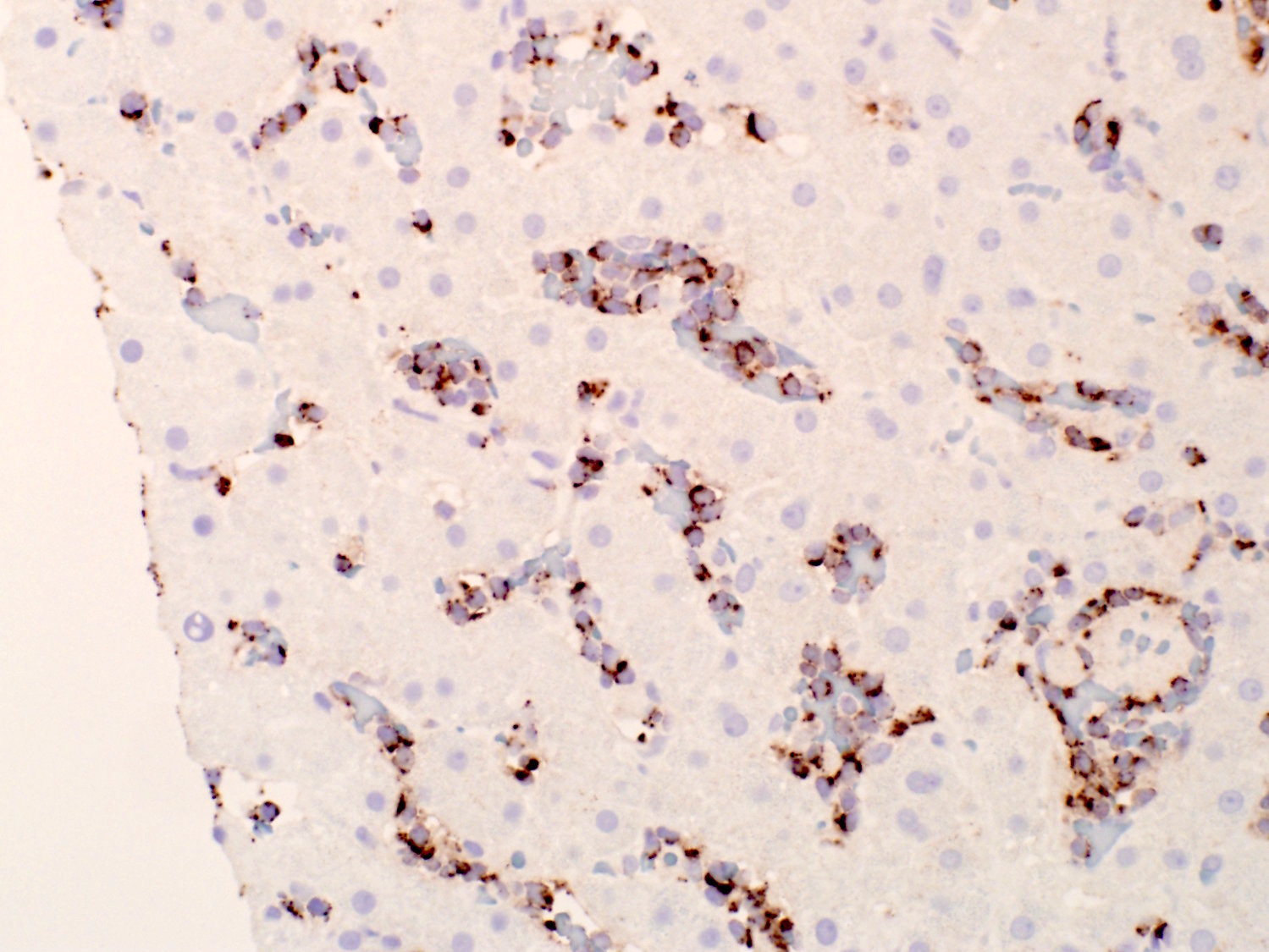
HSTCL: CD4 negative

CD8 positive
TIA1 positive
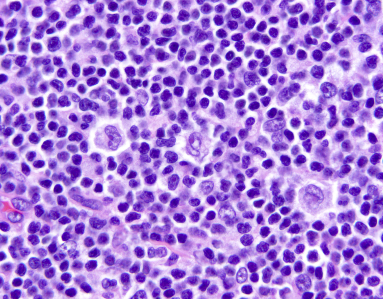
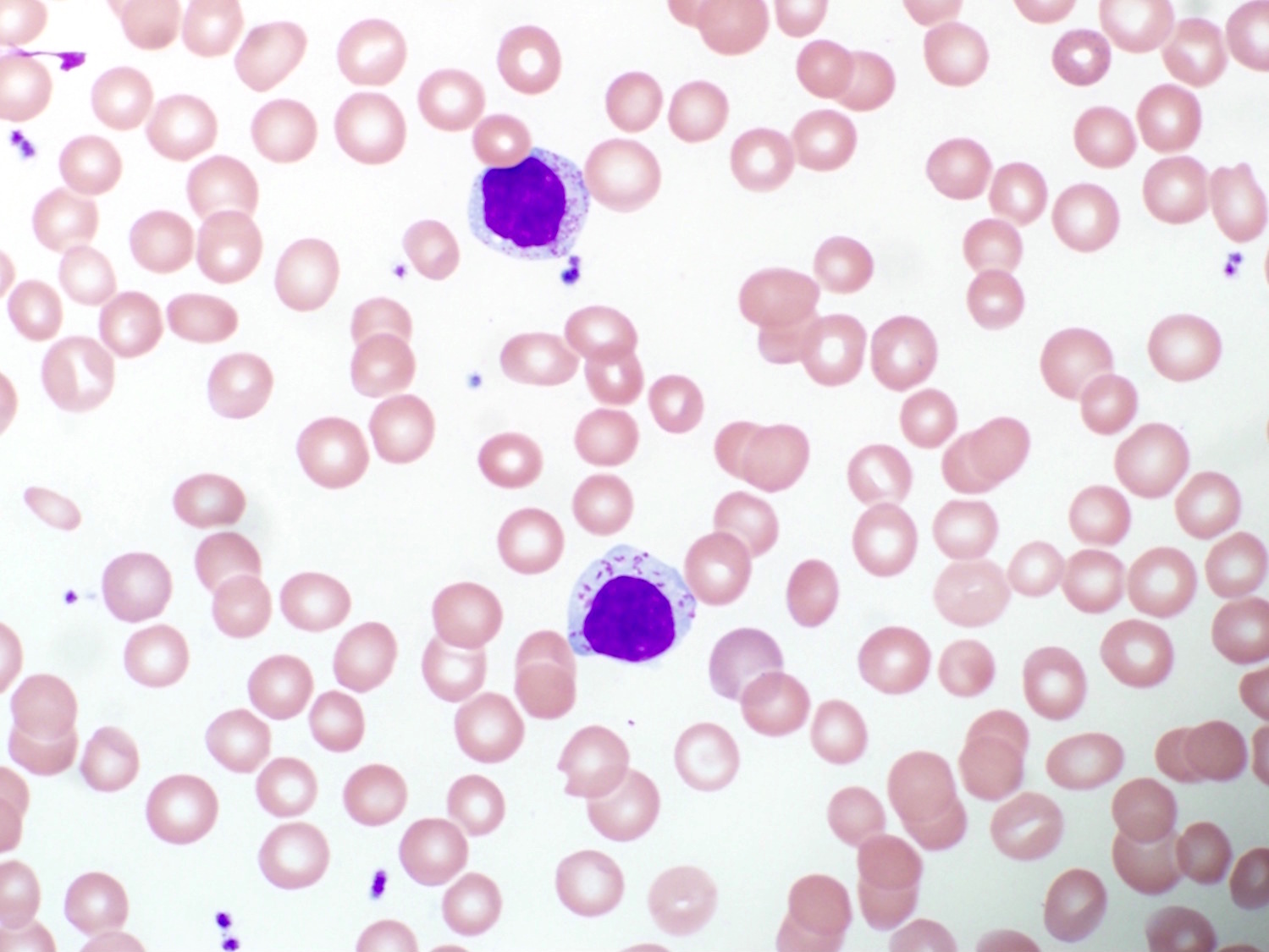
Small size morphology
Intermediate size morphology
Blastic morphology
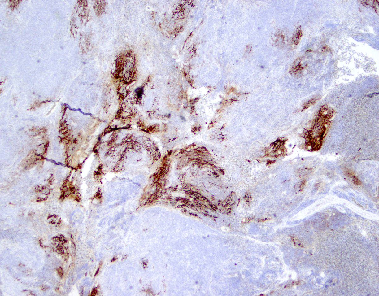
Contributed by Roberto N. Miranda, M.D.
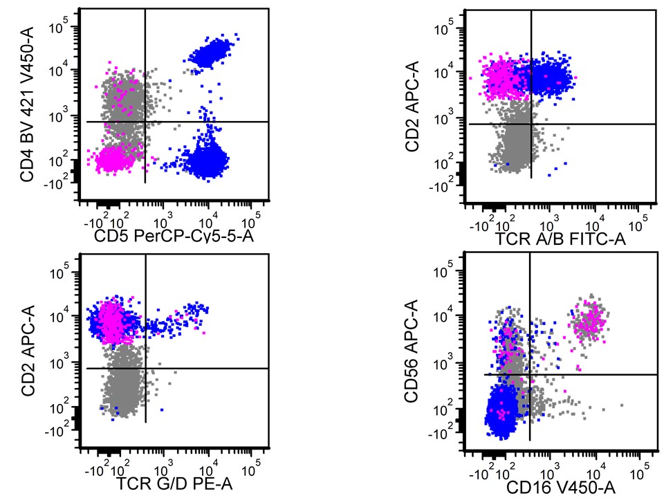
Positivity for CD2, TCRγδ and CD56


Contributed by Fnu Raja, M.B.B.S.
Microcytic and hypochromic RBCs
Iron deficiency anemia
Contributed by Nicholas Nowacki, M.D.
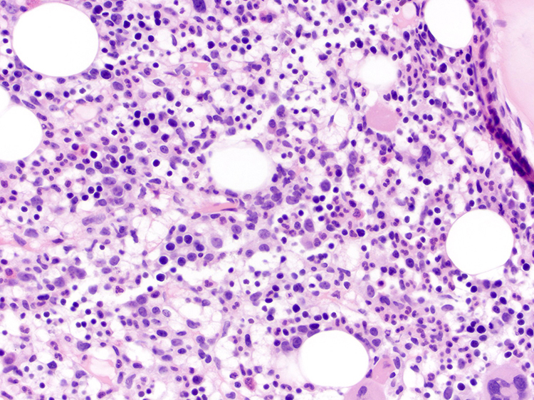
Lymphoid infiltrate

Small lymphoma cells
Marrow aspirate

CD3 highlights T cells
CD5 positive
CD20 positive

Cyclin D1 positive
SOX11 negative
Contributed by Nicholas Nowacki, M.D.
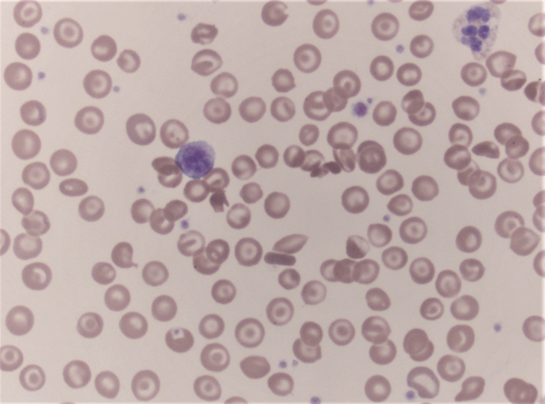
Peripheral blood
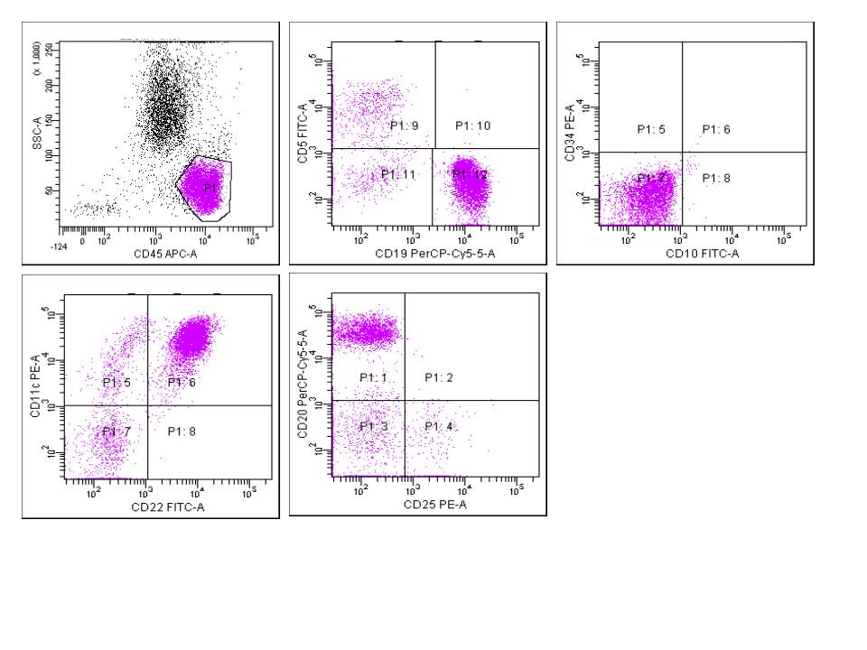
Contributed by Nicholas Nowacki, M.D.
Aberrant CD5 coexpression

CD20 moderate expression
Surface light chain restriction
Images hosted on other servers:

Characteristic immunophenotype


Immunophenotypic comparison
Table 1: B cell lymphoproliferative disorders and markers
| CD45RA | CD19 | CD20 | CD79a | CD5 | CD10 | CD23 | Cyclin D1 | SOX11 | LEF1 | CD200 | CD103 | CD11c | CD25 | |
| Follicular lymphoma | + | + | + | + | - | + | - | - | -/+ | Unknown | - | - | -/+ | -/+ |
| Chronic lymphocytic leukemia / small lymphocytic lymphoma | + | + | + | + | + | - | + | - | - | +/- | + | - | -/+ | -/+ |
| Diffuse large B cell lymphoma | + | + | + | + | - | +/- | - | - | -/+ | -/+ | +/- | -/+ | -/+ | -/+ |
| Mantle cell lymphoma | + | + | + | + | + | - | - | + | + | - | - | - | - | -/+ |
| Marginal zone lymphoma | + | + | + | + | - | - | - | - | -/+ | - | - | - | -/+ | - |
| Lymphoplasmacytic lymphoma | + | + | + | + | - | - | - | - | Unknown | - | +/- | - | +/- | +/- |
| Hairy cell leukemia | + | + | + | + | - | - | - | + | +/- | - | + | +/- | + | + |
| +: positive; -: negative; +/-: majority positive; -/+: majority negative | ||||||||||||||
Table 2: T cell lymphoproliferative disorders and markers
| CD4 / CD8 | Pan-T cell markers (CD2, CD3, CD5, CD7) | CD30 | T cell receptor | |
| T cell prolymphocytic leukemia | CD4+ / CD8- > CD4+ / CD8+ | All expressed | - | Alpha / beta |
| Sézary syndrome | CD4 | CD7 loss | +/- | Alpha / beta |
| Adult T cell leukemia / lymphoma | CD4 | CD7 loss | -/+ | Alpha / beta |
| T cell large granular lymphocytic / leukemia | CD8 | CD2, CD5 and CD7 variably loss | - | Alpha / beta |
| +: positive; -: negative; +/-: majority positive; -/+: majority negative | ||||
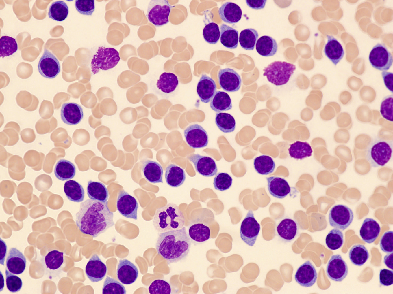
Contributed by Jingwei Li, M.D., Ph.D., Olga Pozdnyakova, M.D., Ph.D. and Karry Charest, MT (ASCP)
Clumped chromatin, smudge cells
Moderately clumped chromatin
Cleaved nuclei
Fine, hair-like cytoplasmic projections
Round to irregular nuclei
Irregular, cerebriform nuclear contour
Polylobed nuclei, flower cells
Eccentric, round nuclei
Large sized neoplastic cells
Abundant cytoplasm, azurophilic granules
Dispersed chromatin, distinct nucleoli
Irregular, convoluted nuclear contours
Images hosted on other servers:

Pre- and post-treatment

Alopecia and erythroderma
Contributed by Shamayel Mohammed, M.D.
Distinct cortex

Diffuse histiocytic infiltrate

Patient with SCID
Ziehl Neelsen+
EBV+
CD30+ immunoblasts
CD20+
Images hosted on other servers:

Parakeratosis and lymphohistocytic infiltrate

H&E, S100

CD30
Contributed by Shamayel Mohammed, M.D.
Ziehl-Neelsen+
Histiocytes
Contributed by Samantha Mack, M.D.

Donath-Landsteiner
assay interpretation
worksheet
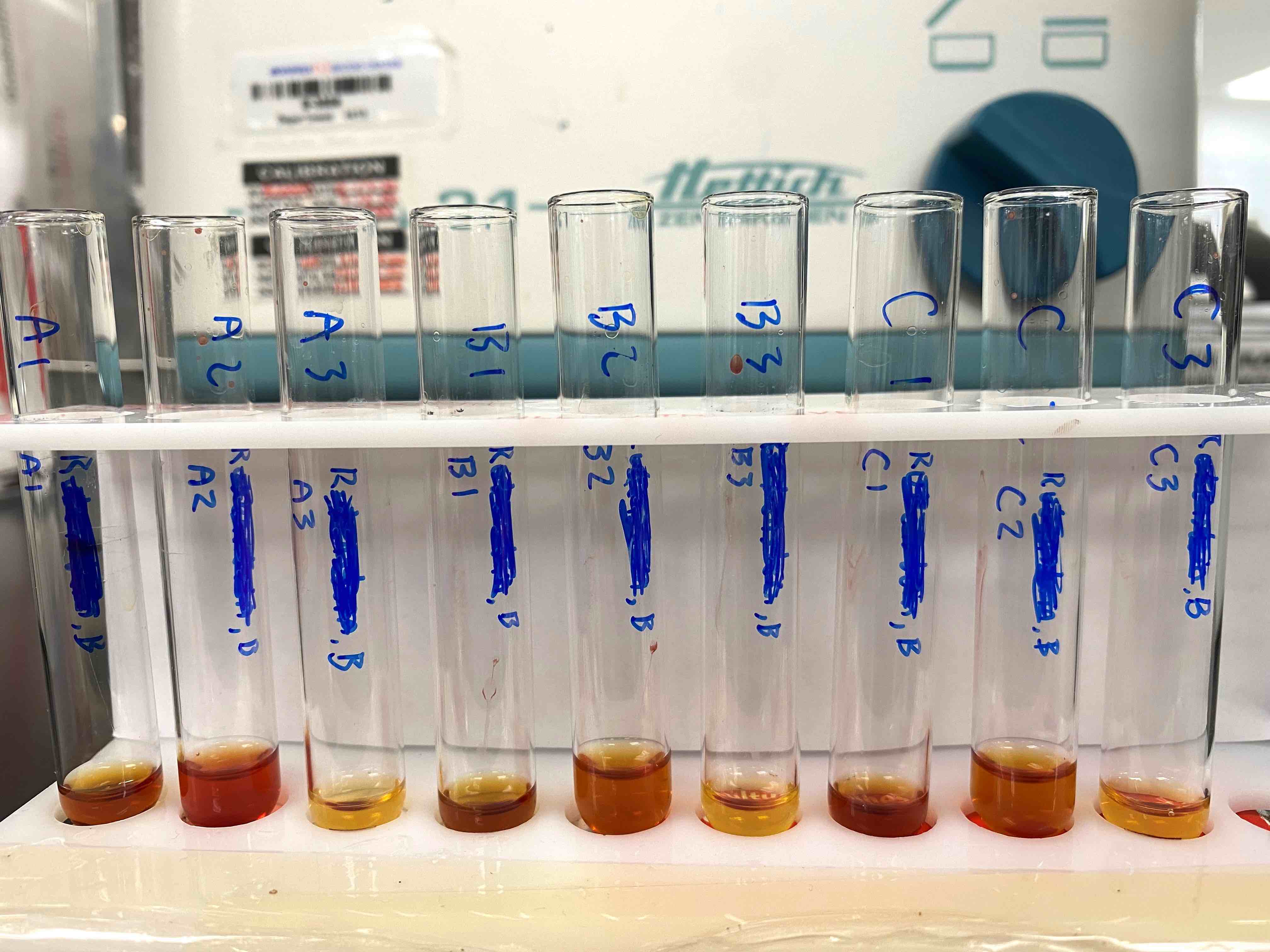
Donath-Landsteiner tube assay example
Images hosted on other servers:

Suggested pathophysiology of PNH

Suggested pathophysiology of hemolysis in PNH

Complement action in healthy subjects and PNH patients
Contributed by Saja Asakrah, M.D.

High sensitivity PNH flow cytometry
Images hosted on other servers:

Fever paroxysm

Life cycle
Images hosted on other servers:

Adult of Anopheles freeborni
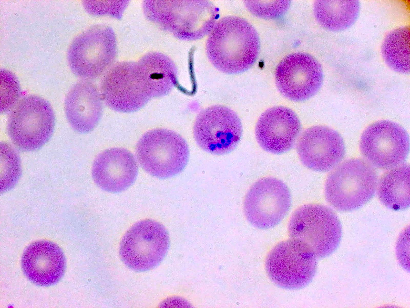
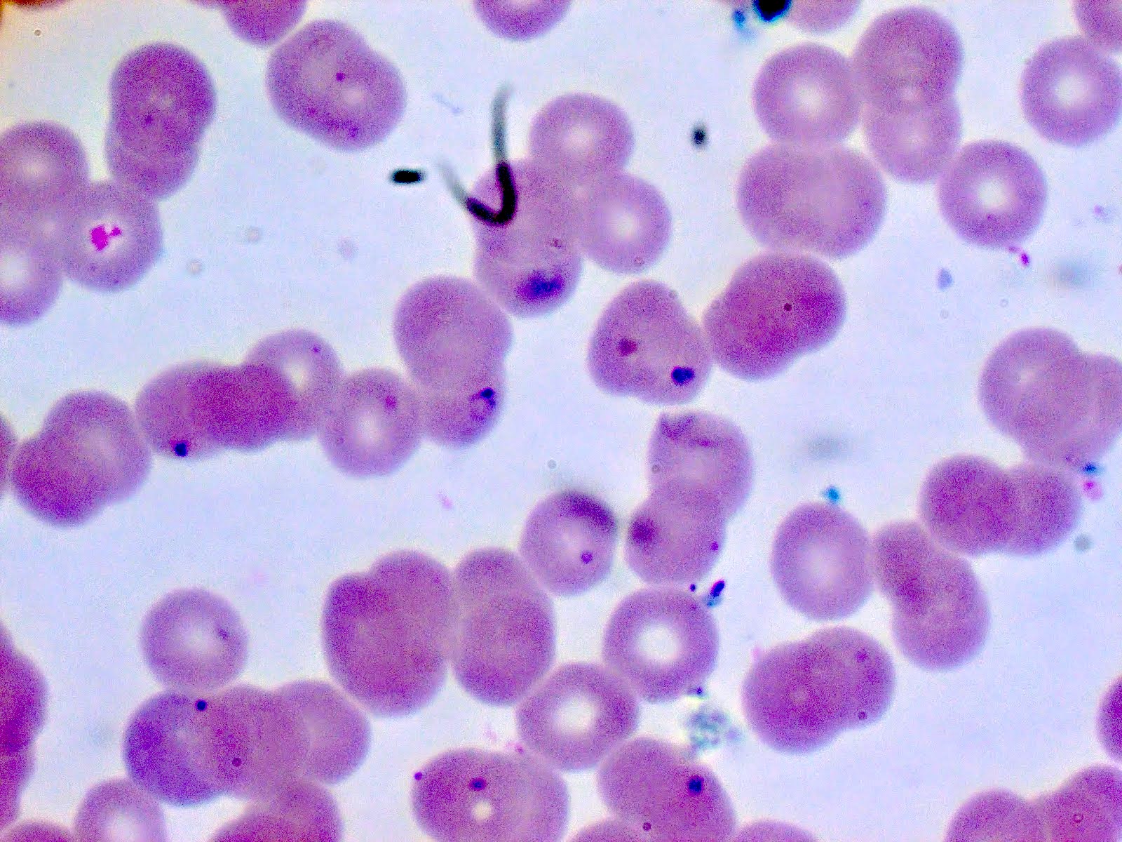
Contributed by Bobbi Pritt, M.D.
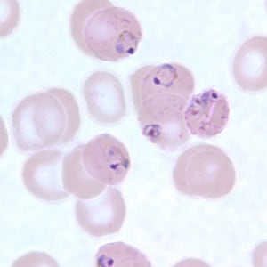
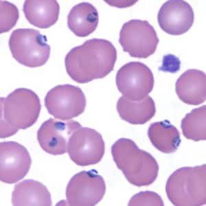
P. falciparum malaria, > 10% parasitemia, negative rapid antigen, Giemsa stained thin blood smear

Giemsa stained thick and thin peripheral blood films from a patient with malaria due to P. falciparum; > 20% parasitemia
Contributed by Patricia Tsang, M.D., M.B.A.
Plasmodium falciparum (blood smear)
Case #277
Blood smear
Images hosted on other servers:

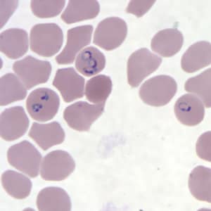
Ring form trophozoites of P. falciparum

Maurer clefts
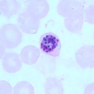
Older trophozoites of P. falciparum
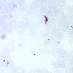

Gametocytes of P. falciparum
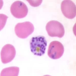
Schizonts of P. falciparum
Contributed by Richard K. Wood, M.D. and Dietrich Werner, M.D.
Bone marrow involvement
CD20 positivity in bone marrow
CD79a positivity in bone marrow
Cyclin D1 negativity in bone marrow
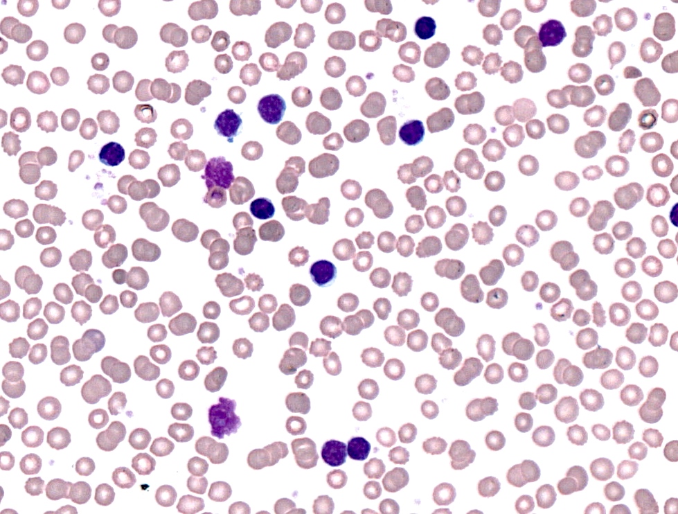
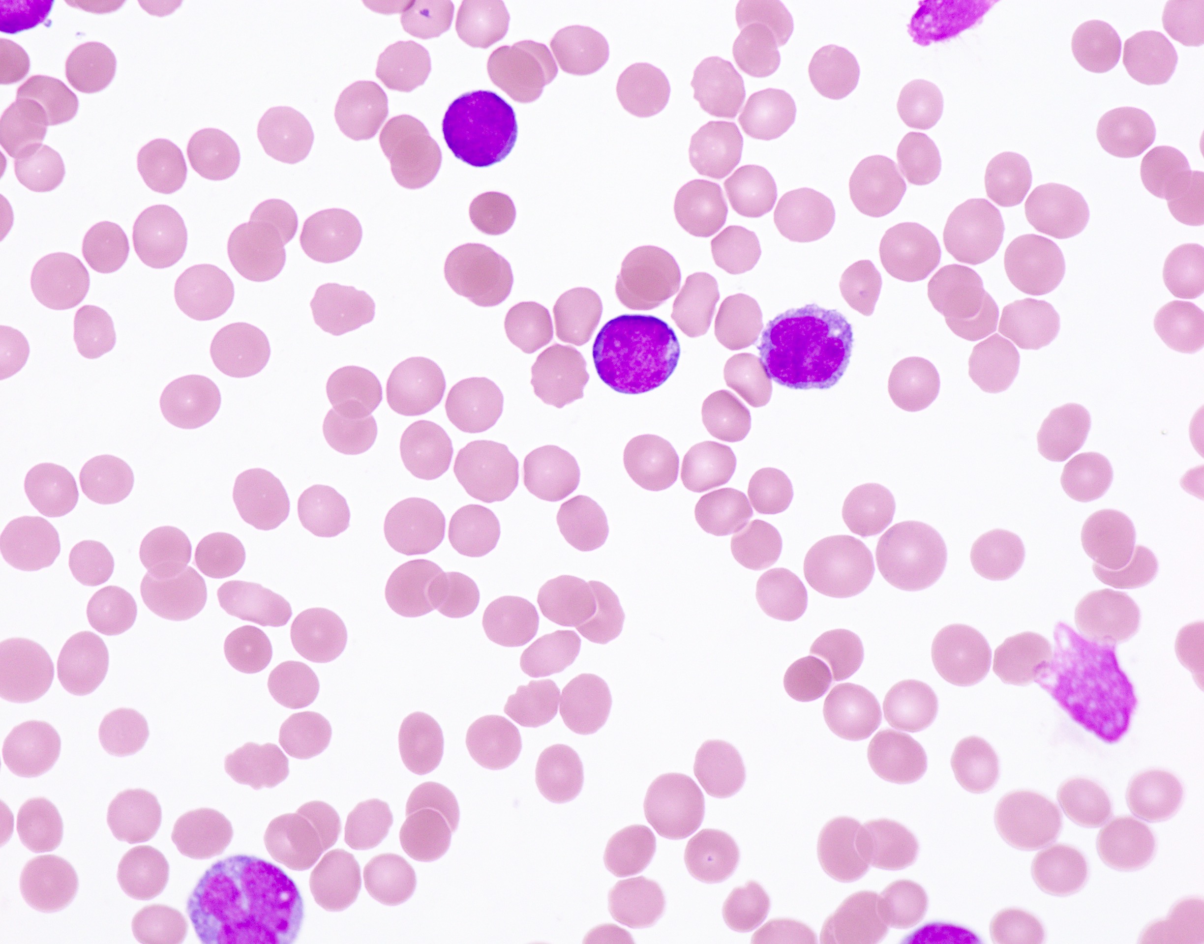
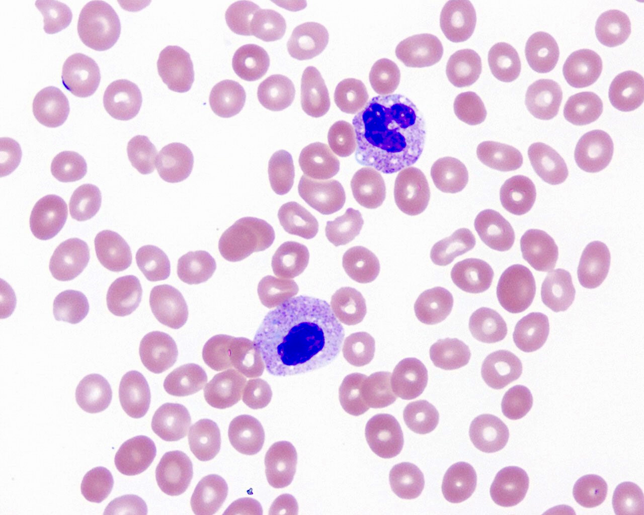
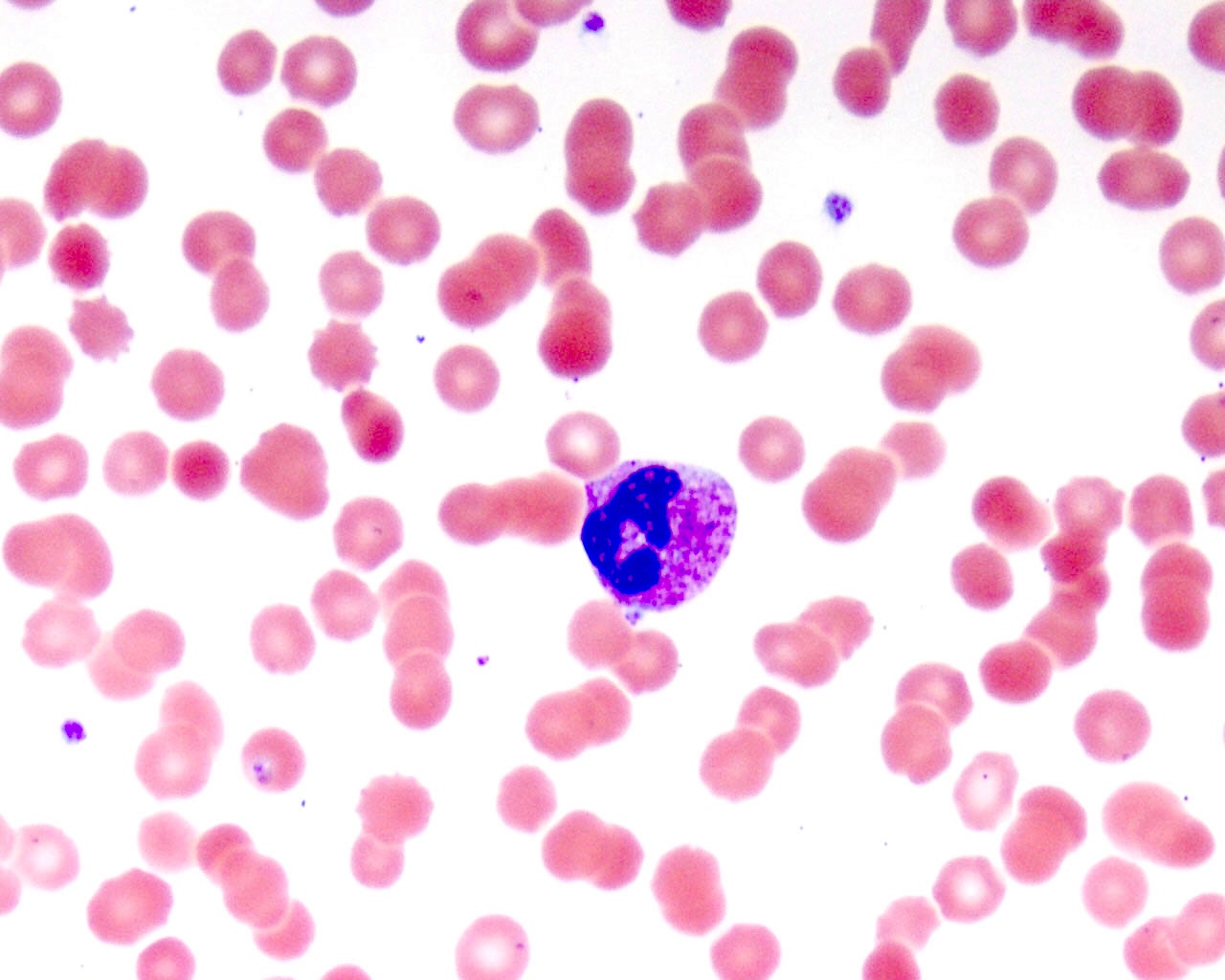
Contributed by Allam Shawwa, M.D.
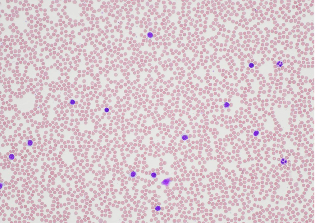
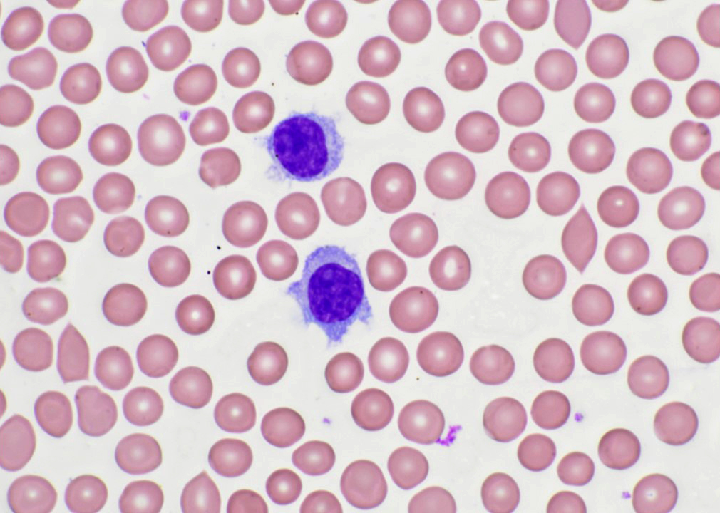
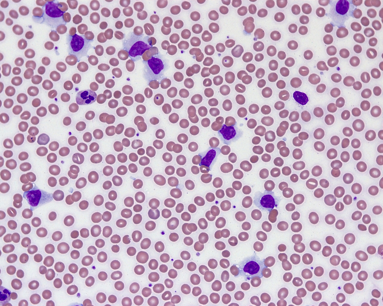
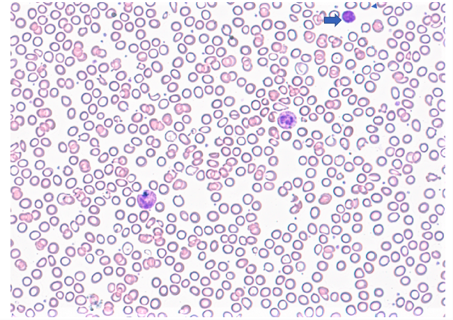
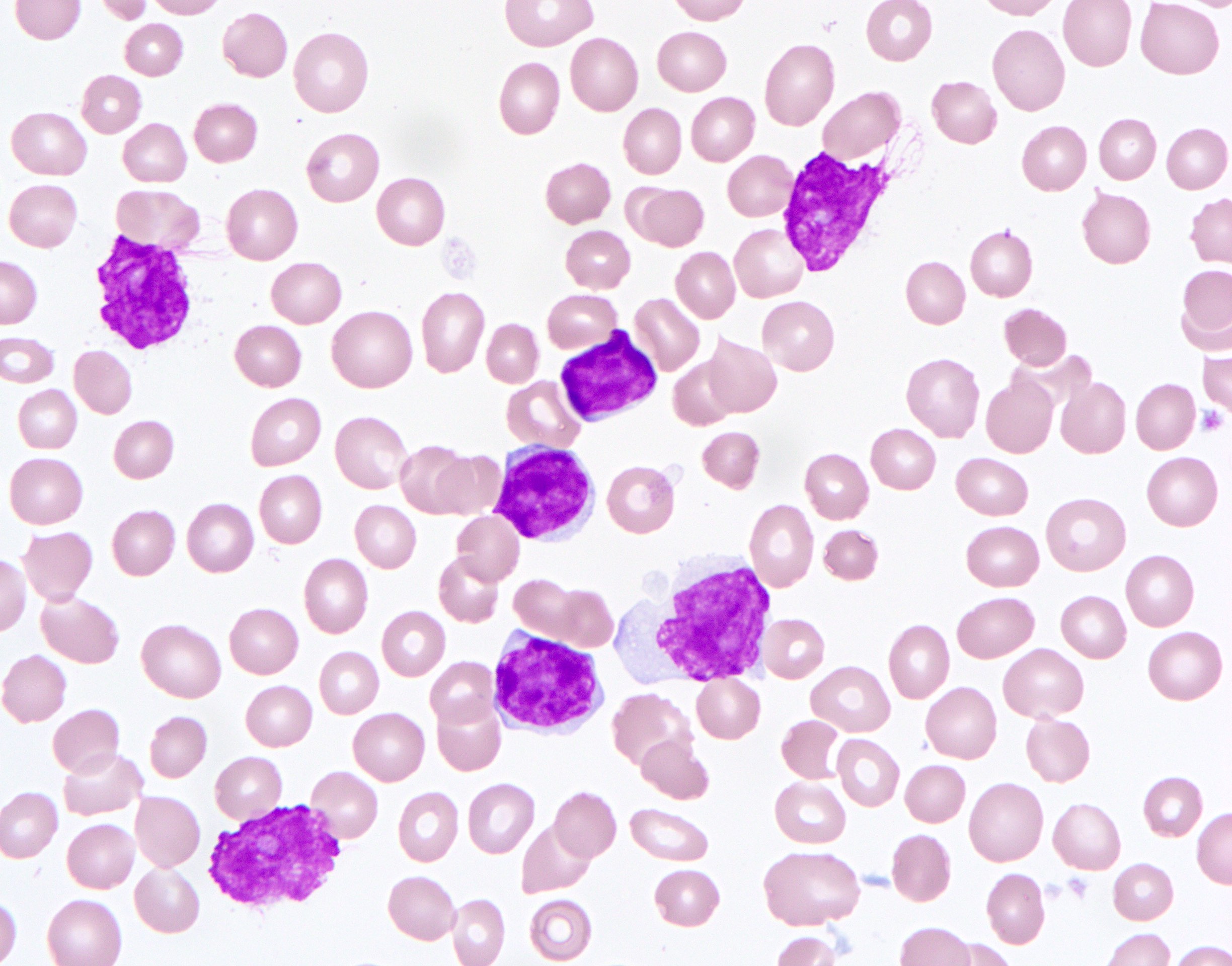
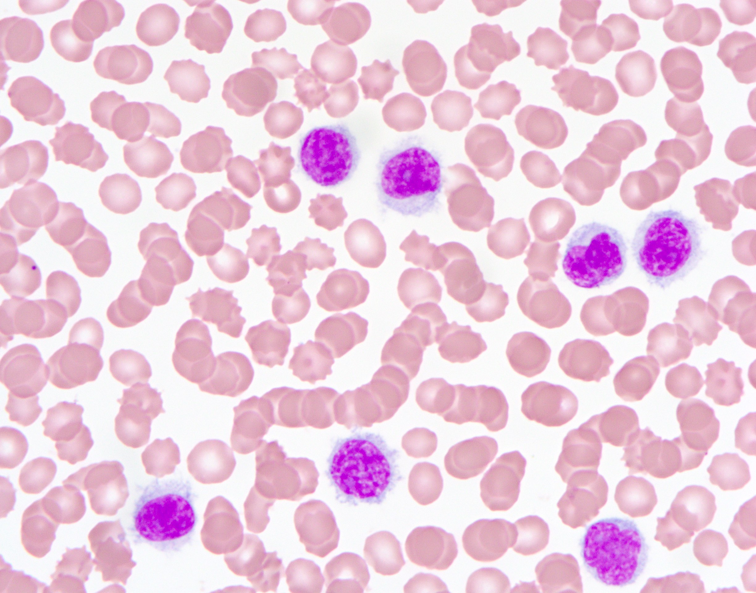
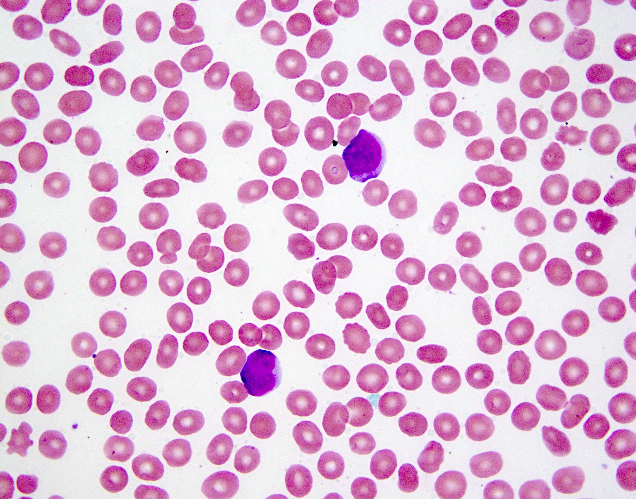
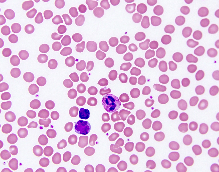
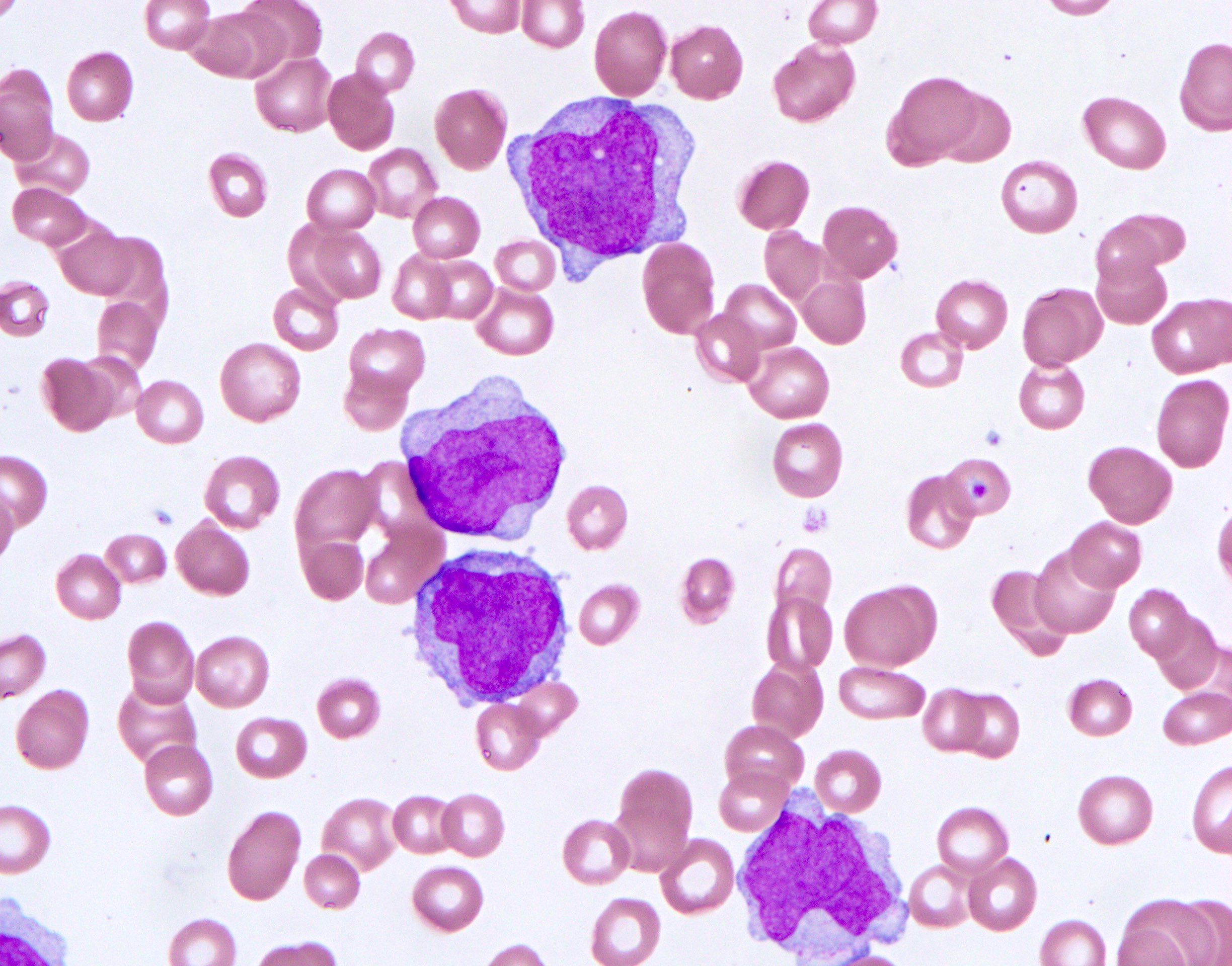
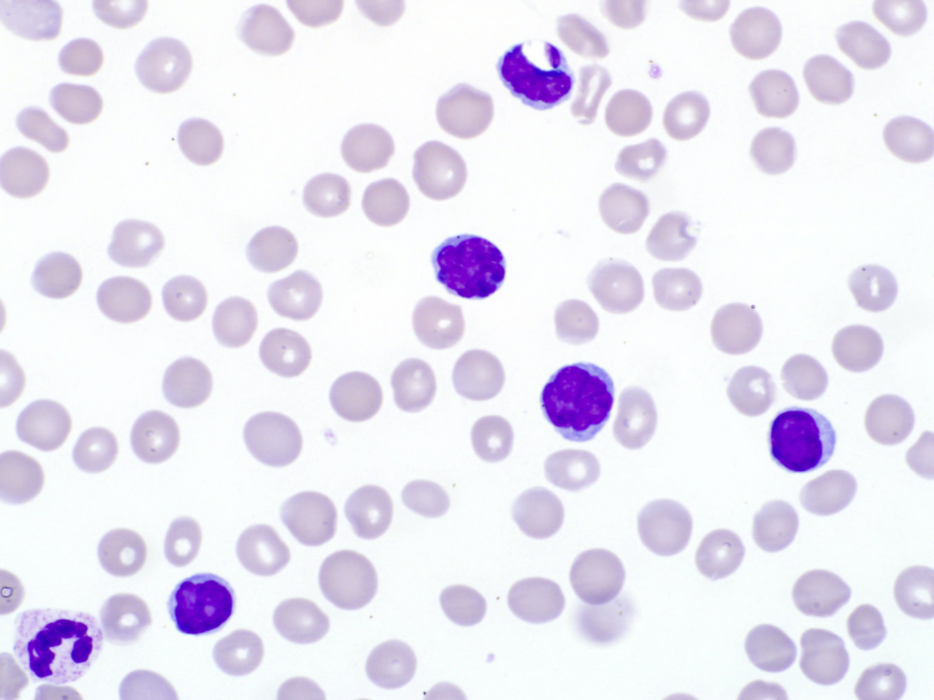
Prolymphocytes in peripheral blood

Prolymphocytes in peripheral blood

Images hosted on other servers:

Inclusions of nuclear chromatin remnants
Contributed by Linlin Wang, M.D., Ph.D. and Sonam Prakash, M.B.B.S.
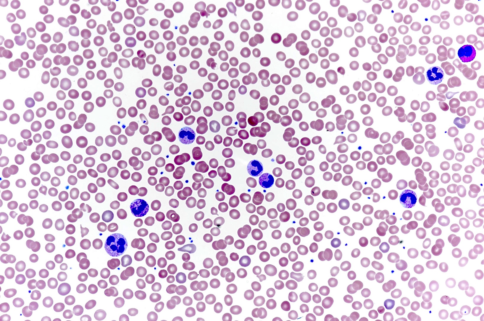
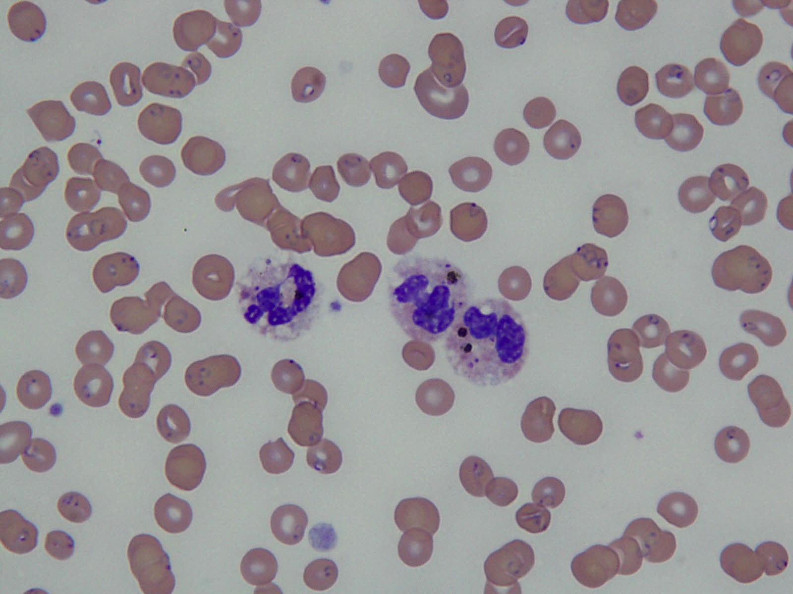
Peripheral neutrophilia
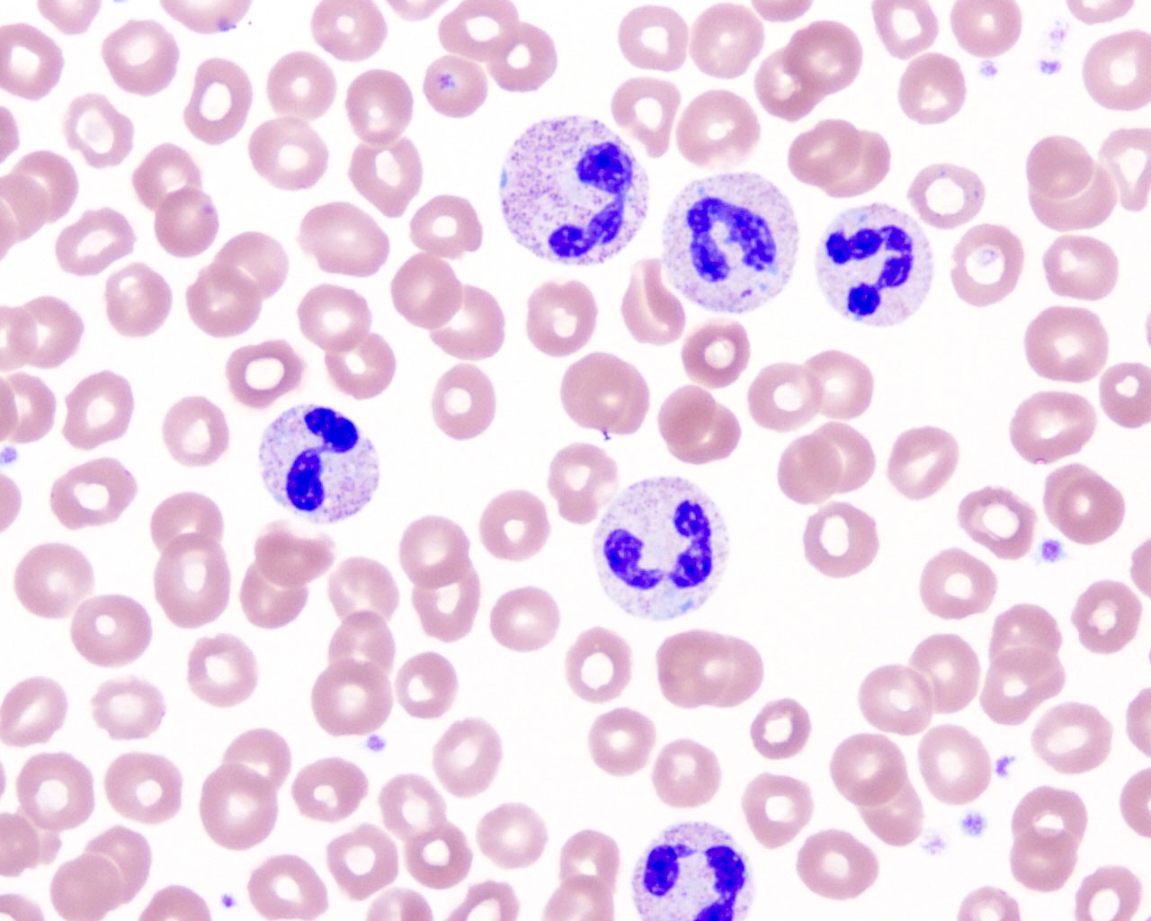
Toxic granulation and Döhle bodies
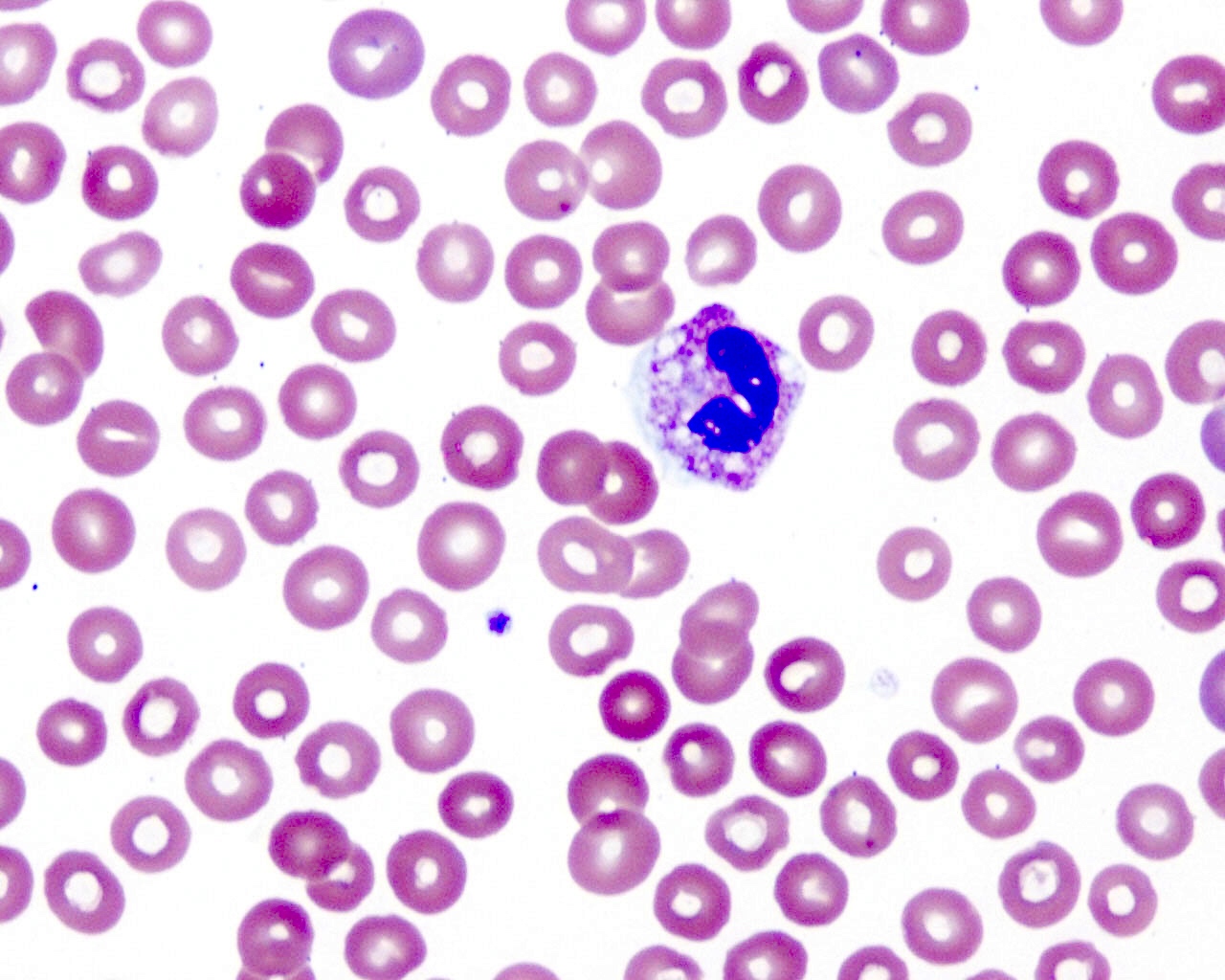
Cytoplasmic vacuoles in neutrophil
Immature granulocytes and rare blast
Pseudo-Pelger-Huët neutrophils

Large neutrophils
Toxic granulation (red granules)
Contributed by Roberto N. Miranda, M.D.
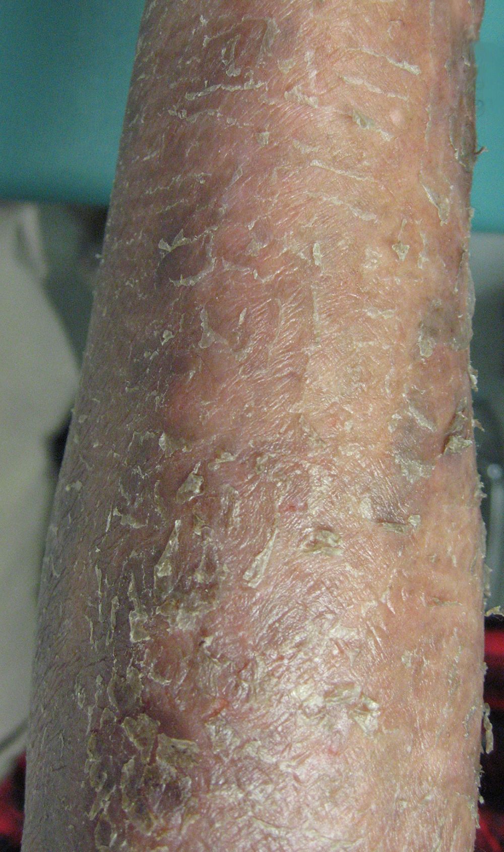
Prominent exfoliative lesion
Prominent erythroderma
Contributed by Roberto N. Miranda, M.D.
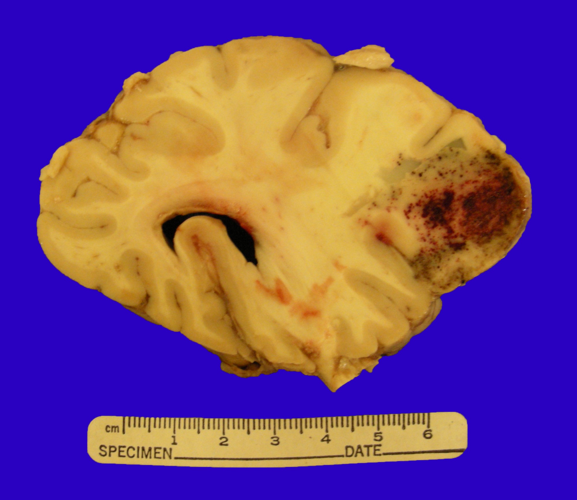
Central nervous system (CNS) involvement
Contributed by Roberto N. Miranda, M.D.
Bone marrow involvement
CD3 positivity
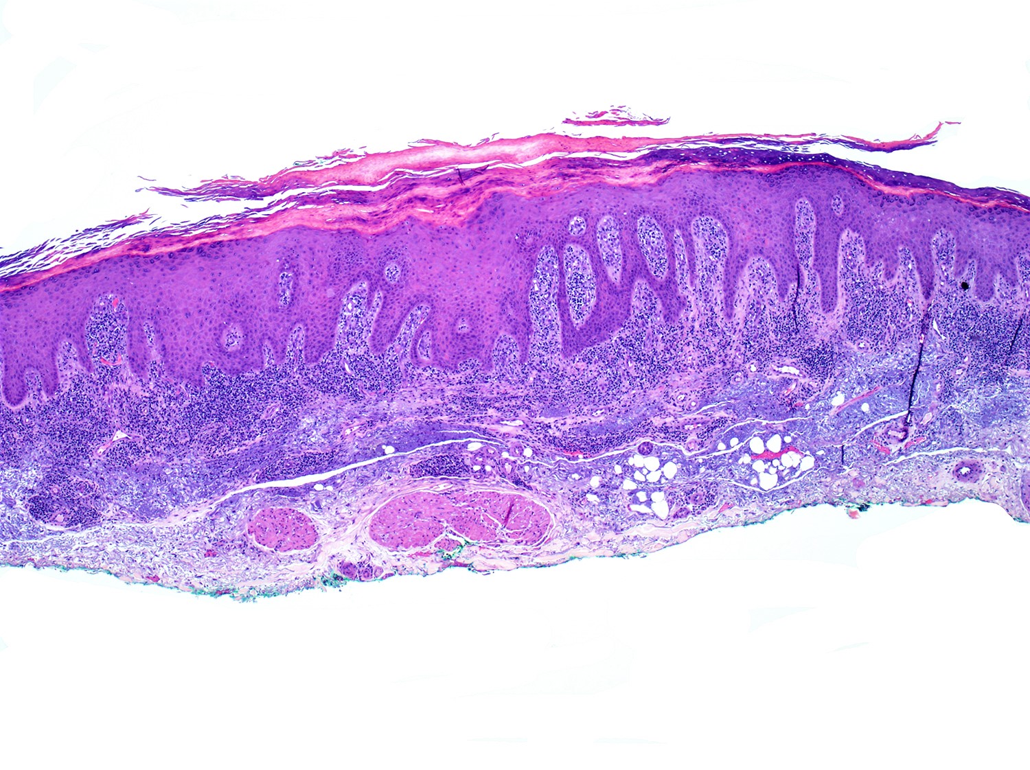
Epidermotropic and lichenoid lymphoid infiltrate
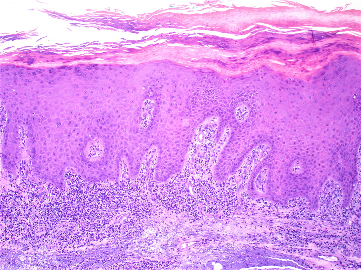
Epidermotropic infiltrate
Cerebriform cytology
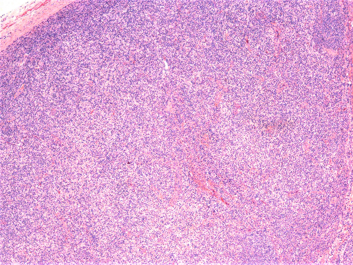
Lymph node involvement
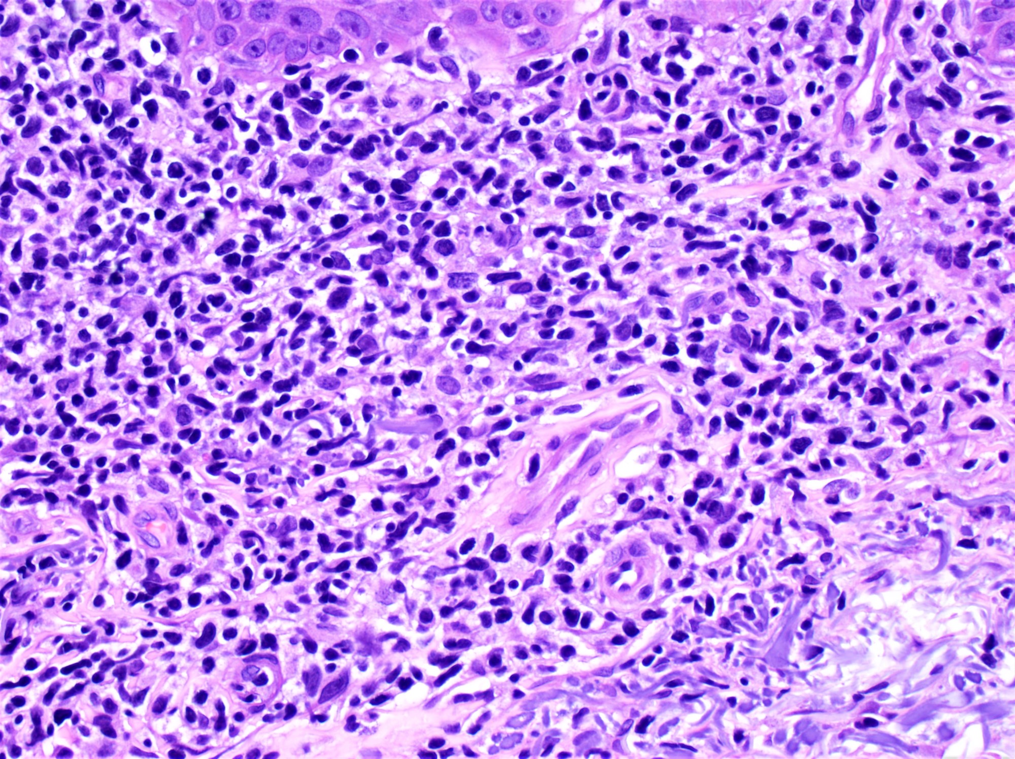
Cytological atypia
Contributed by Roberto N. Miranda, M.D.
Lymph node touch print
Contributed by Roberto N. Miranda, M.D.
Cerebriform lymphocytes in peripheral blood smear
Contributed by Roberto N. Miranda, M.D.
Flow cytometry in SS
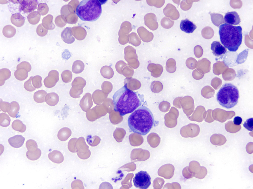
Contributed by Min Shi, M.D., Ph.D. and Dragan Jevremovic, M.D., Ph.D.
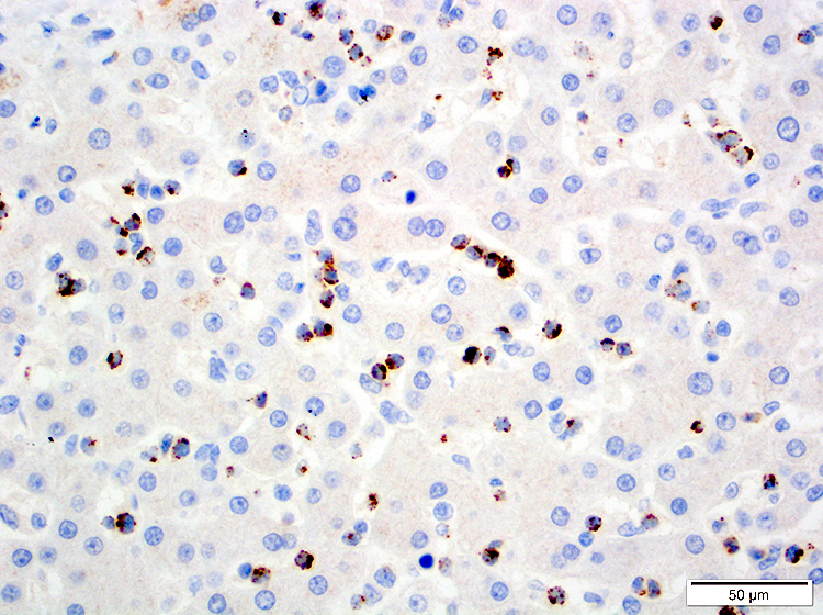
T LGLL bone marrow involvement

Intrasinusoidal CD3+ T LGLL
Intrasinusoidal CD8+ T LGLL
Intrasinusoidal granzyme B+ T LGLL

Intrasinusoidal TIA1+ T LGLL
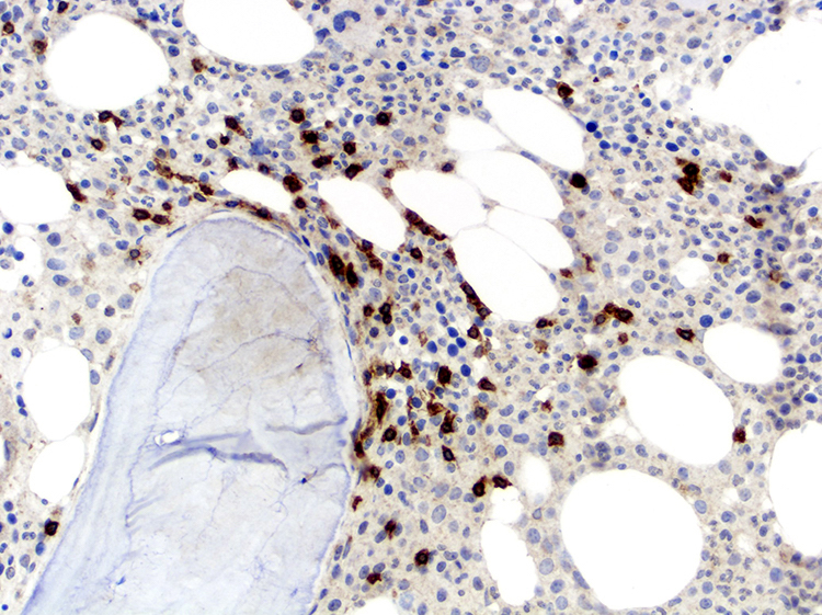
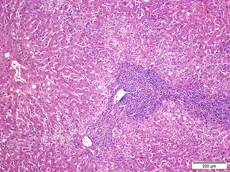
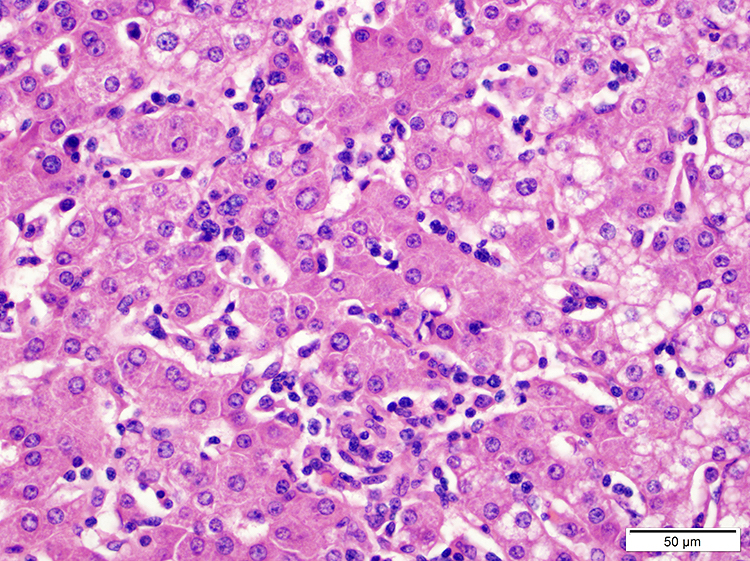
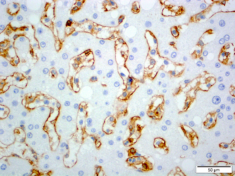
T LGLL liver involvement
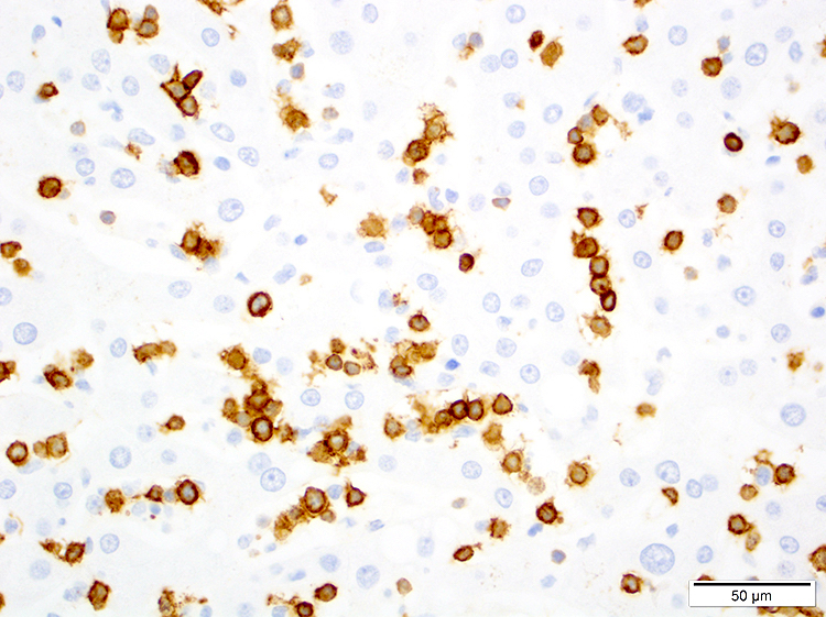
CD8+ T LGLL in liver

CD5- T LGLL in liver
TIA1+ T LGLL in liver
Contributed by Min Shi, M.D., Ph.D. and Dragan Jevremovic, M.D., Ph.D.
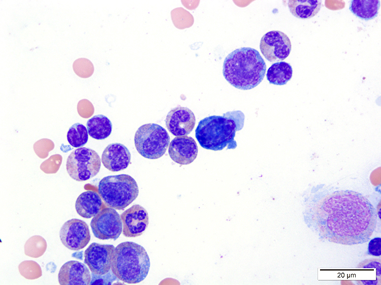
T LGLL in peripheral blood
Contributed by Min Shi, M.D., Ph.D. and Dragan Jevremovic, M.D., Ph.D.
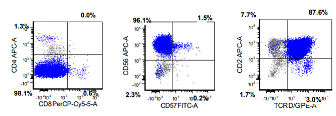
T LGLL flow cytometry
Contributed by Min Shi, M.D., Ph.D. and Dragan Jevremovic, M.D., Ph.D.
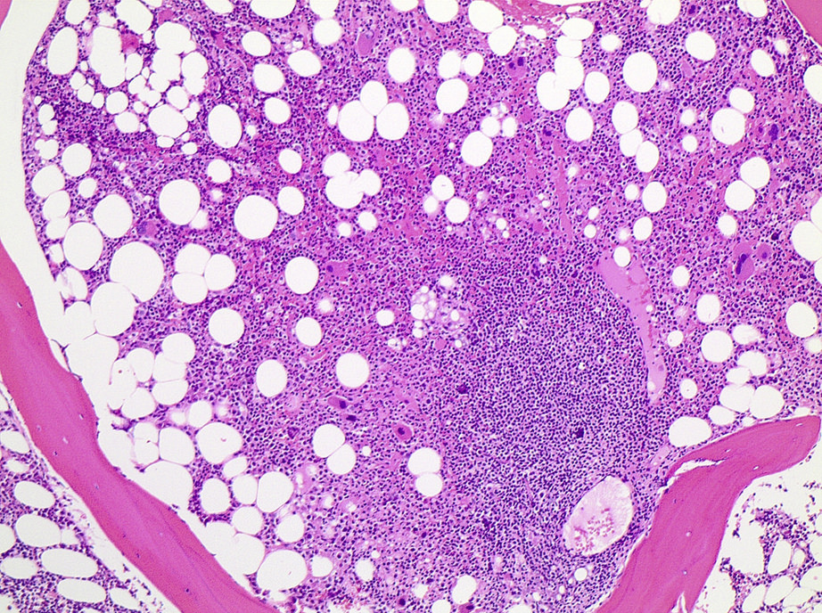
Diffuse bone marrow involvement
Splenic red pulp infiltrate
Monotonous, paracortical lymphoid infiltrate
Intermediate size, atypical lymphocyte
CD3 positivity in bone marrow
Monotonous T cell infiltrate
TCL1A positivity
Uniform strong TCL1A staining
Contributed by Min Shi, M.D., Ph.D. and Dragan Jevremovic, M.D., Ph.D.
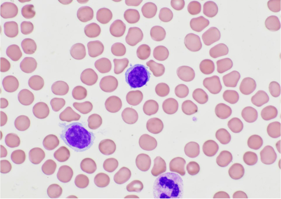
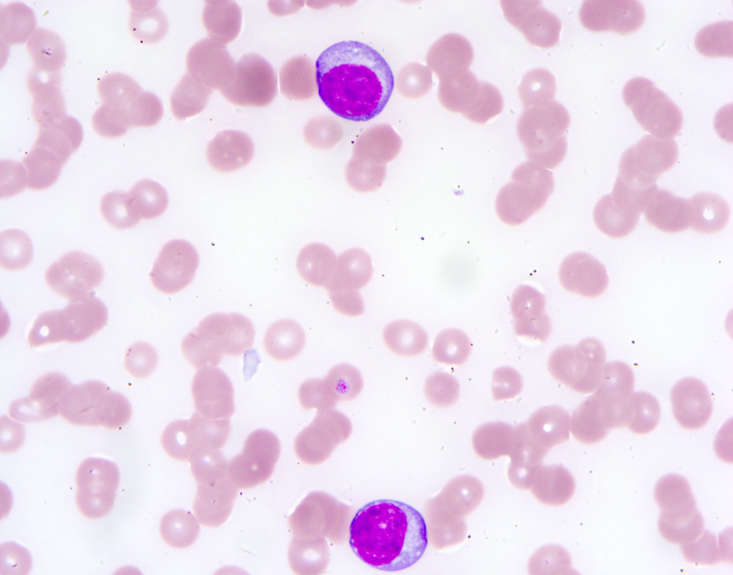
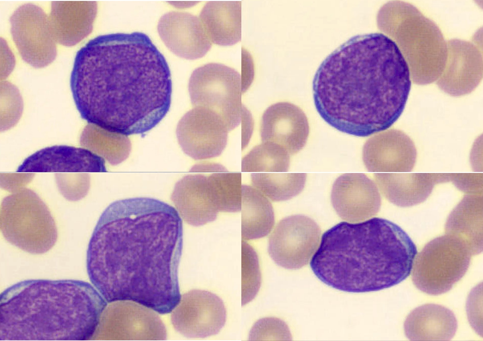
Cerebriform variant
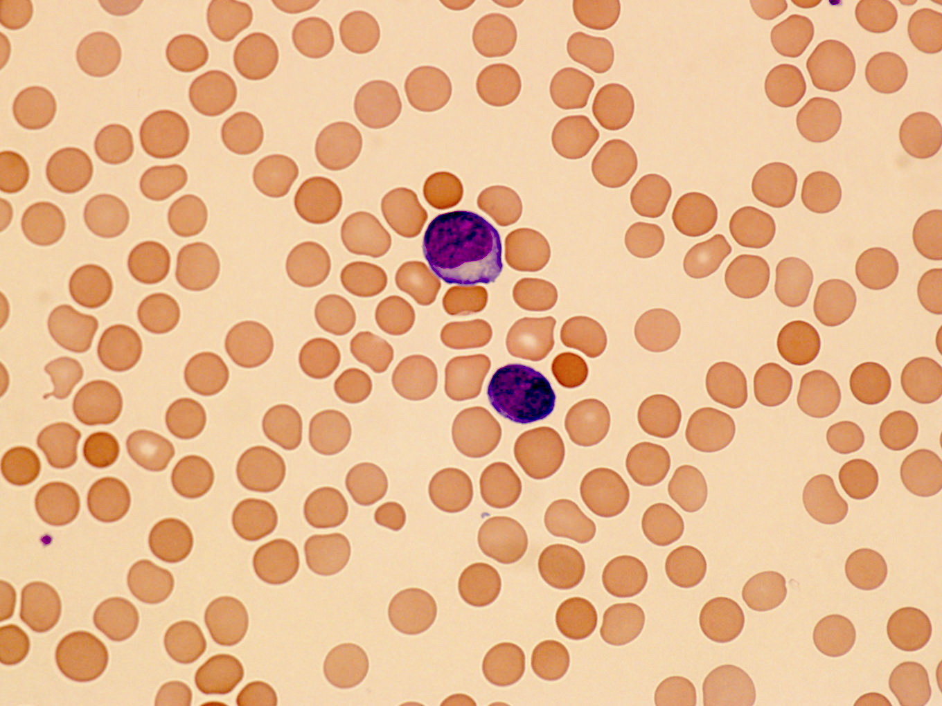
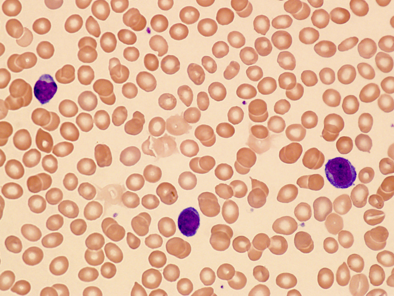
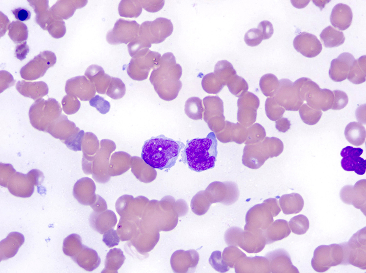
Prolymphocytes in peripheral blood
Small cell variant
Contributed by Min Shi, M.D., Ph.D. and Dragan Jevremovic, M.D., Ph.D.

T PLL flow cytometry
Contributed by Min Shi, M.D., Ph.D. and Dragan Jevremovic, M.D., Ph.D.
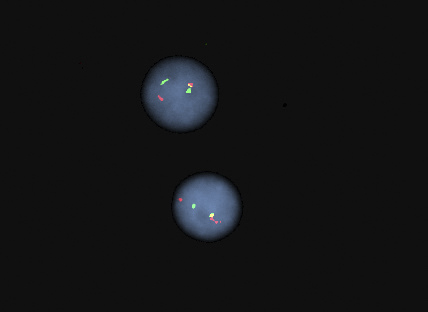
FISH for TCL1A separation
Images hosted on other servers:

Hyperpigmentation before treatment
Images hosted on other servers:

Atrophic gastritis with intestinal metaplasia
Images hosted on other servers:

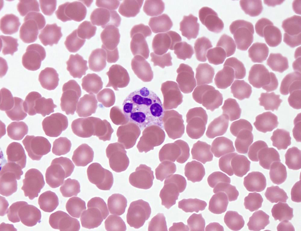
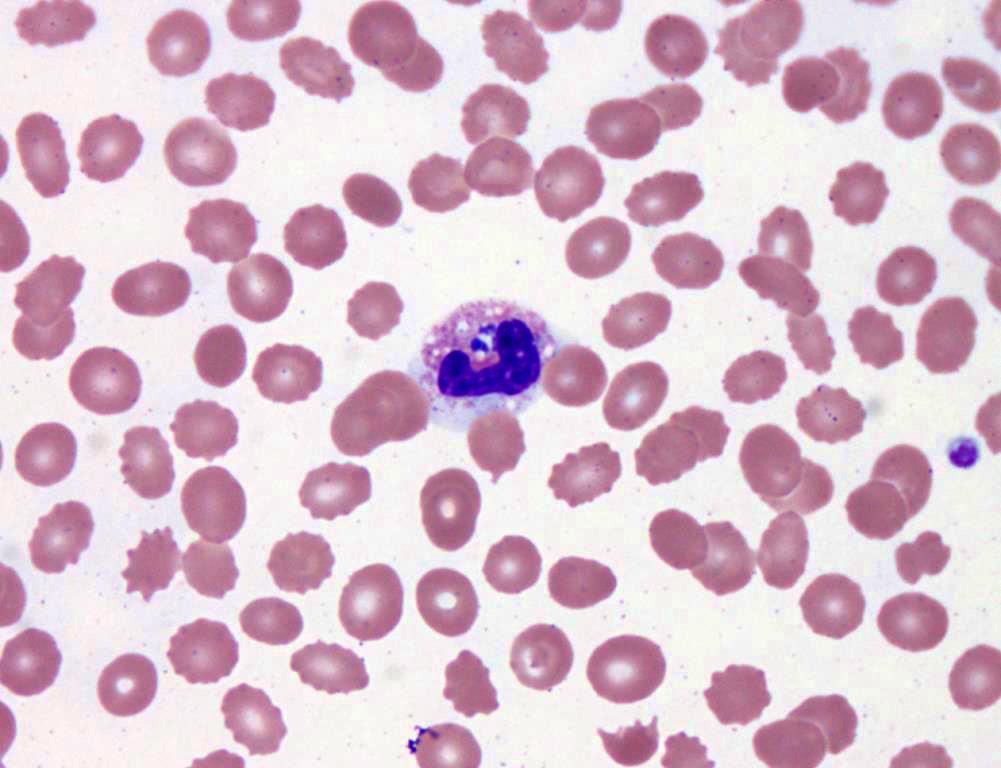
Hypersegmented neutrophil

Macrocytes, macro-ovalocytes,
nucleated RBCs and hypersegmented neutrophils
Glassy: 2018
Hoyer: 2003
Kaushansky: 2021
Keohane: 2015
Medeiros: 2017
Sallman: 2016
Turgeon: 2017
Find related Pathology books: hematopathology
