

Images hosted on other servers:

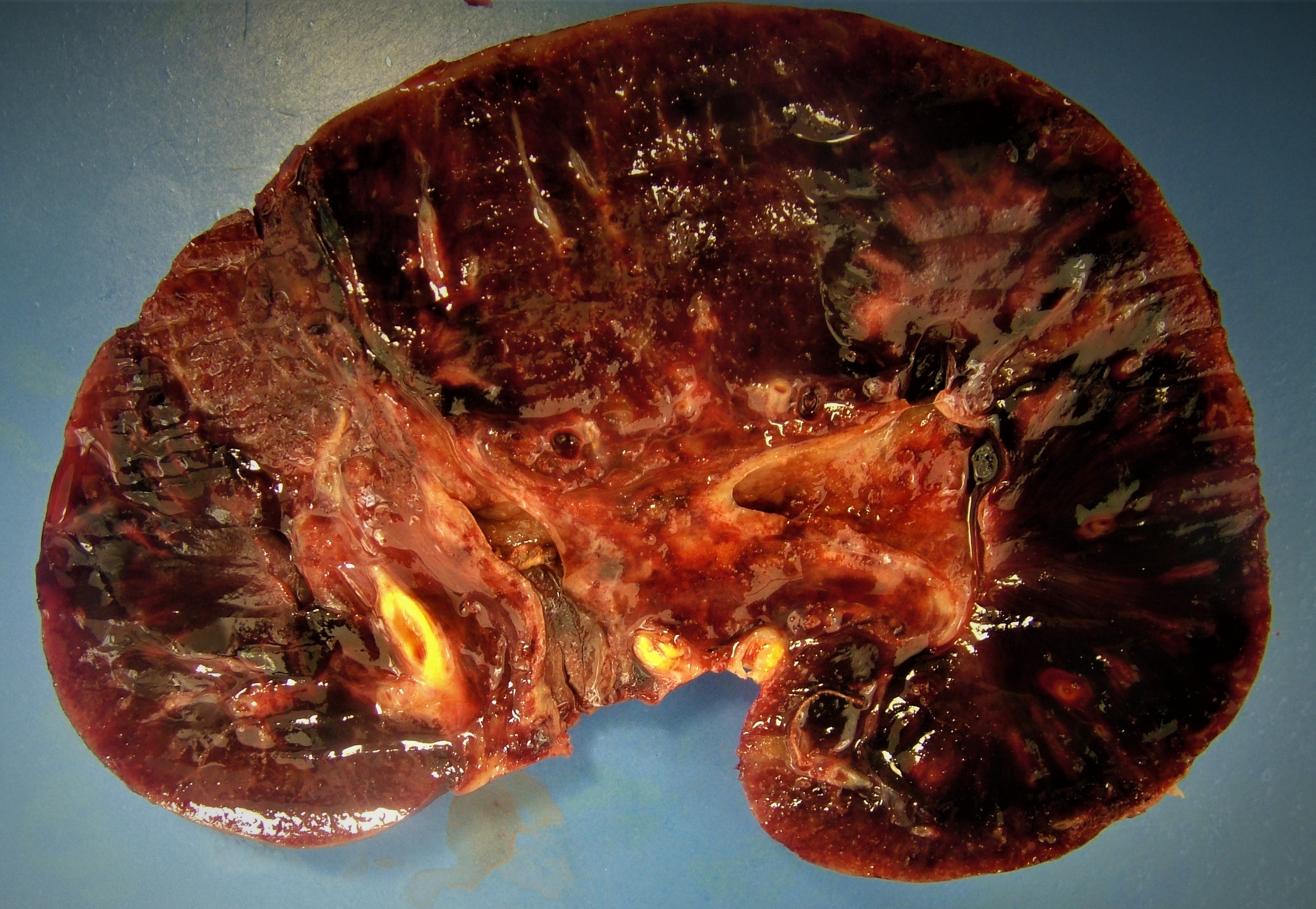
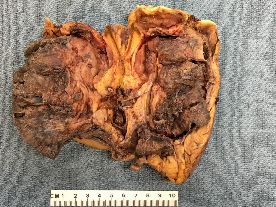
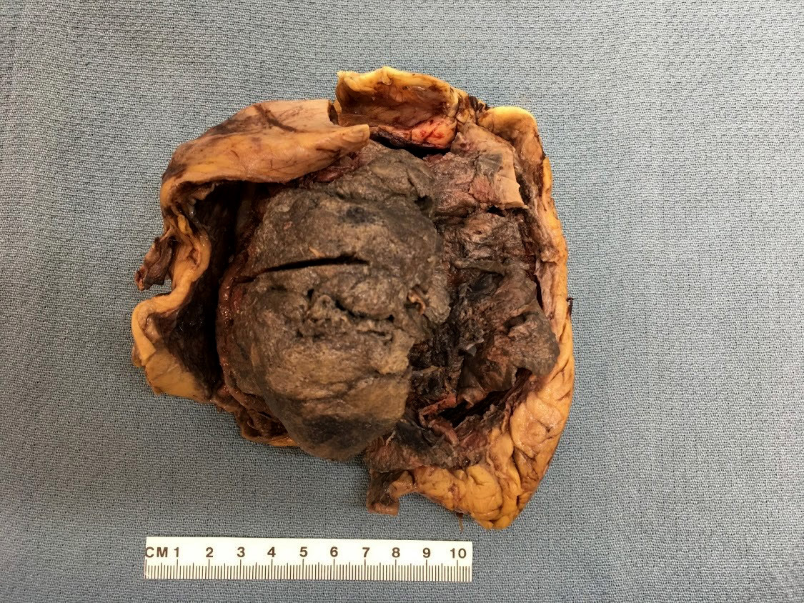
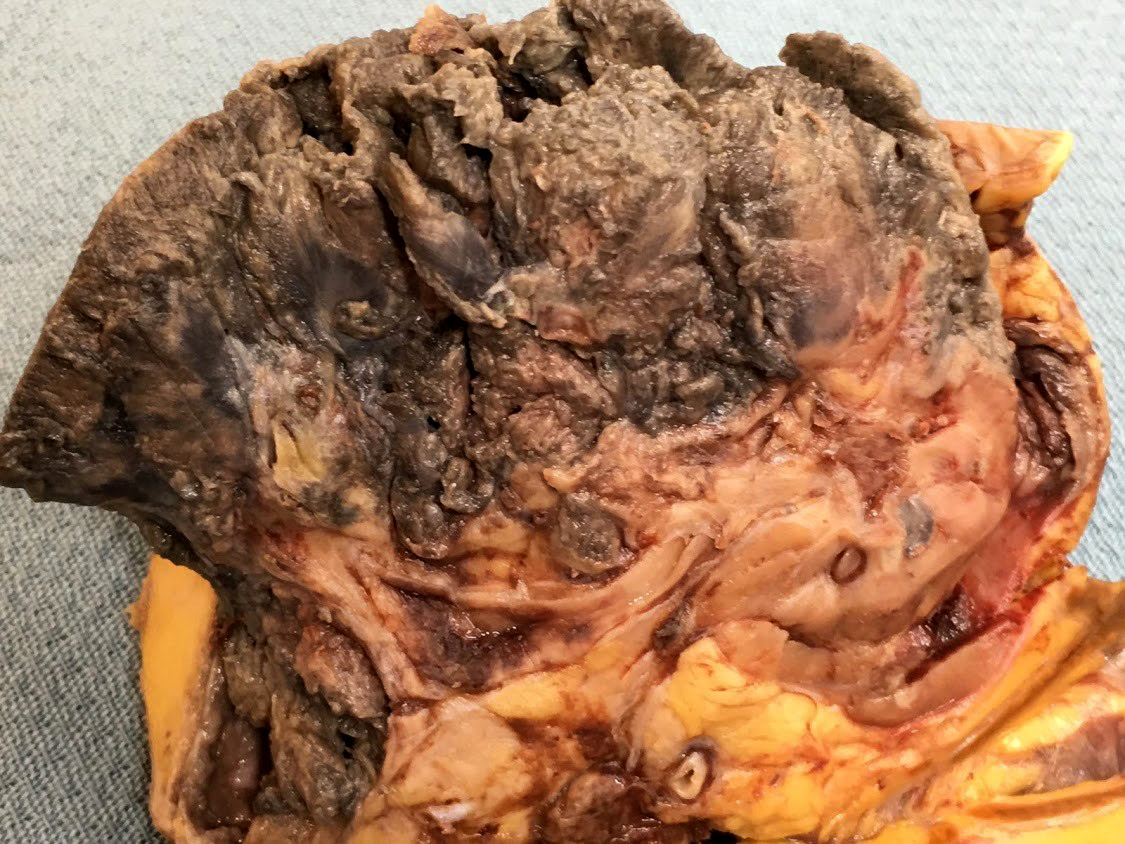
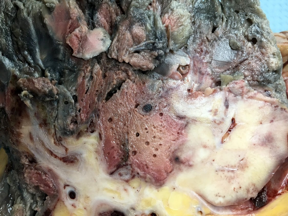
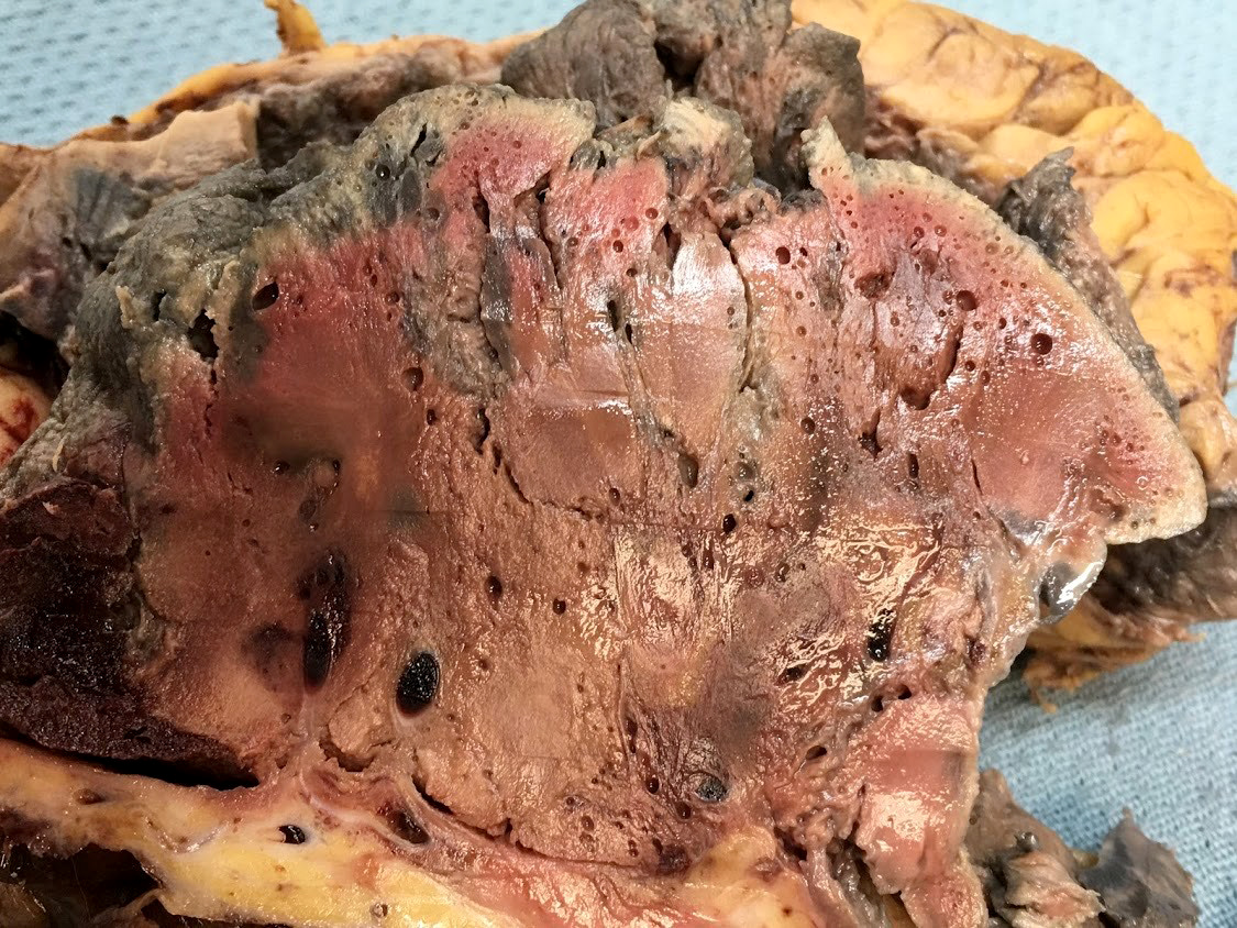
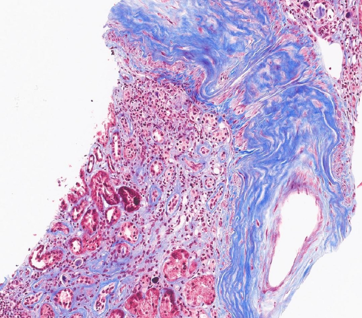
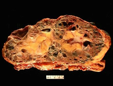
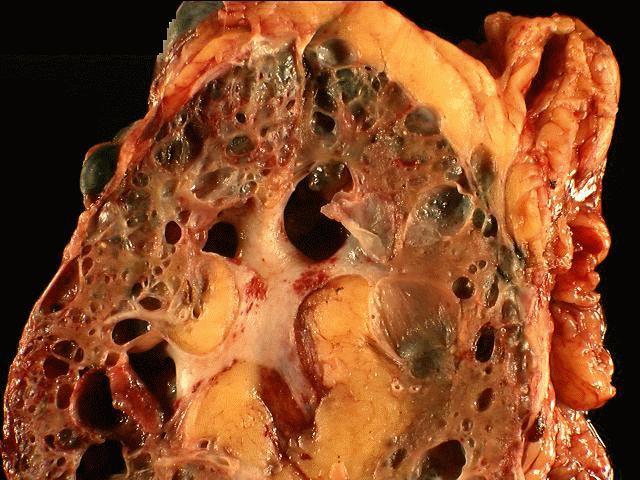
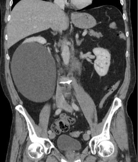
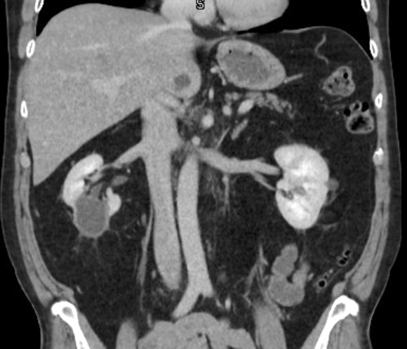
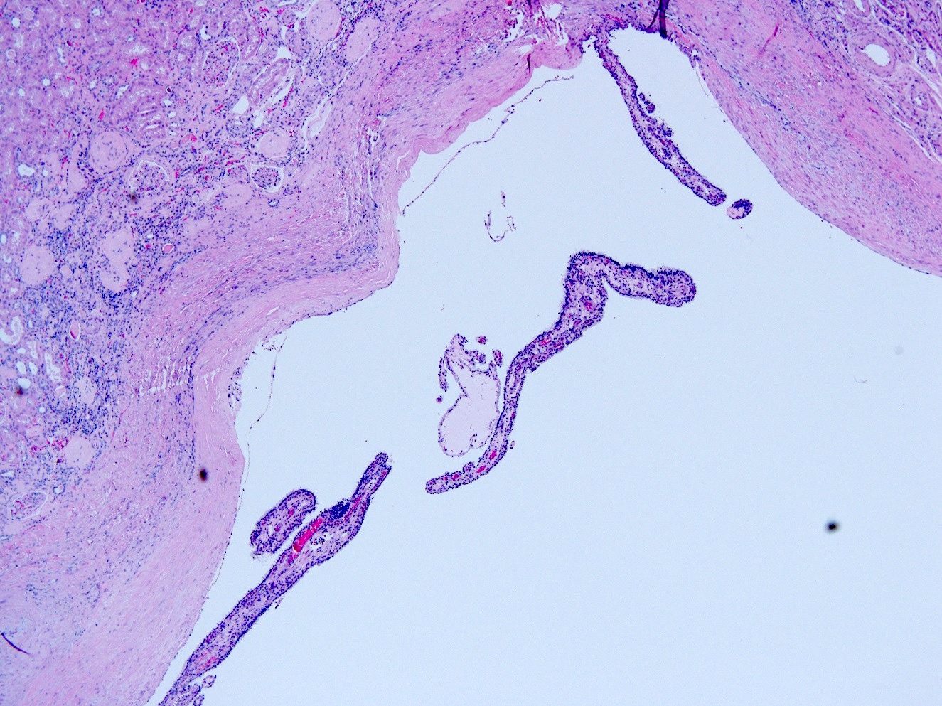
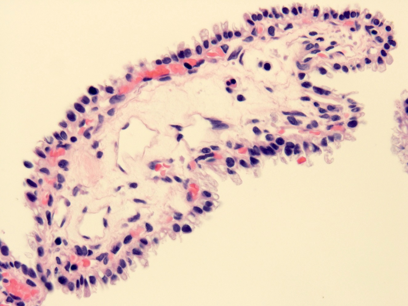
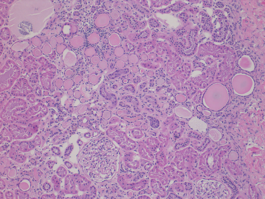
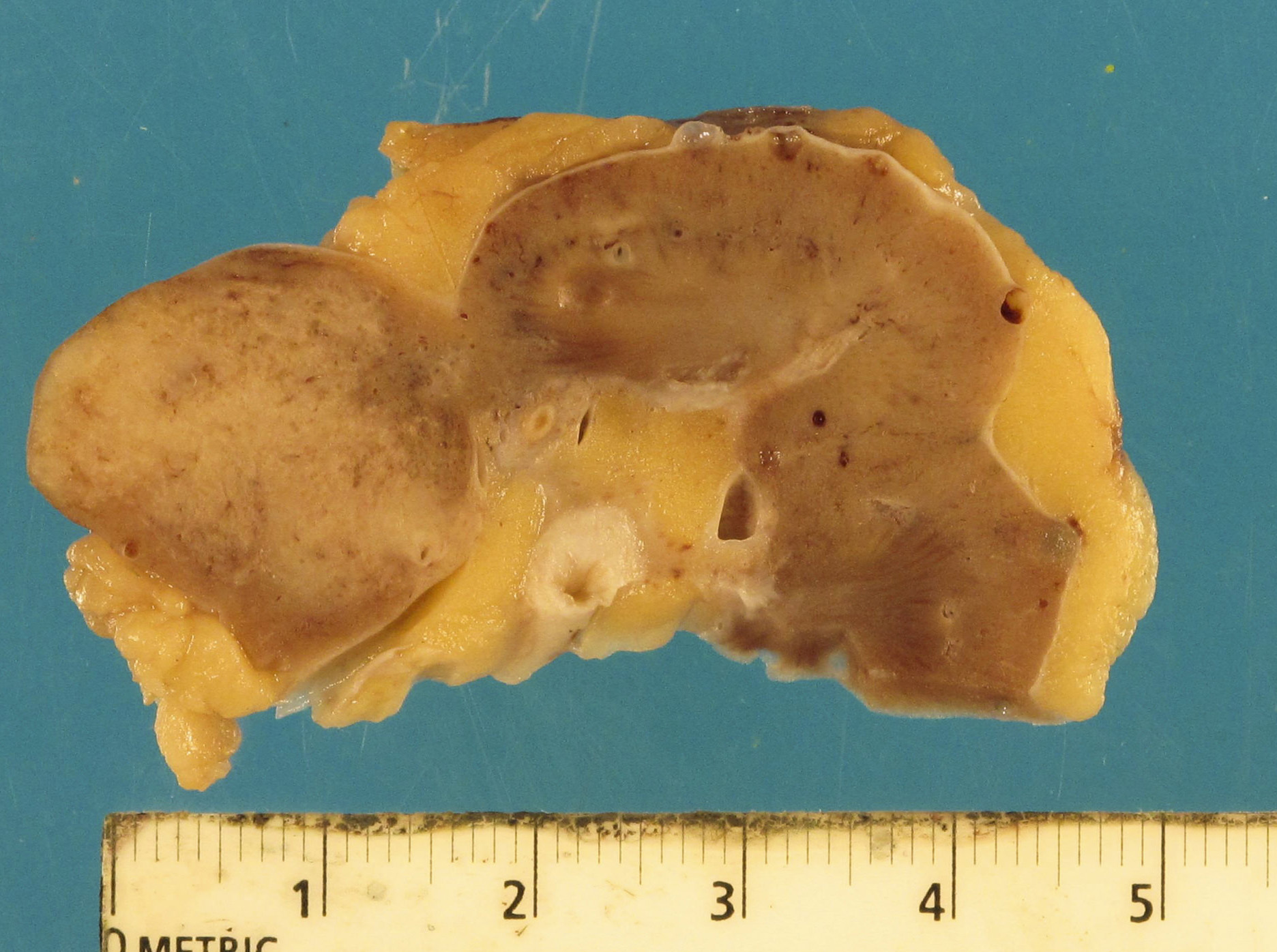
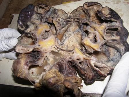
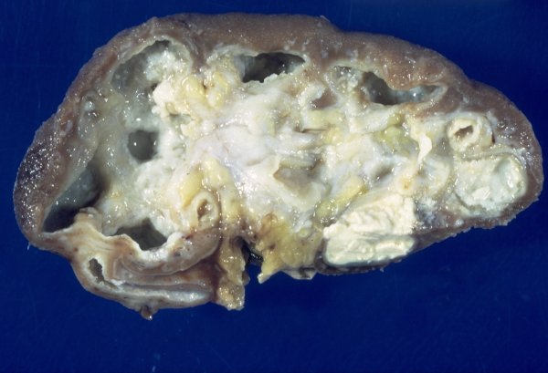
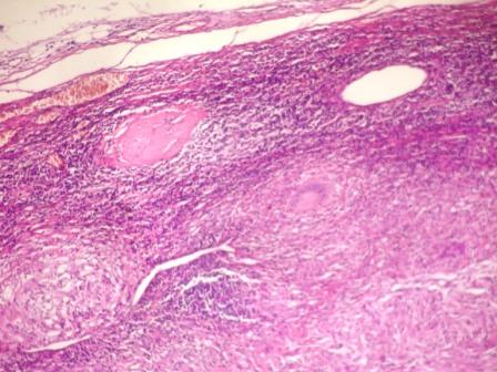
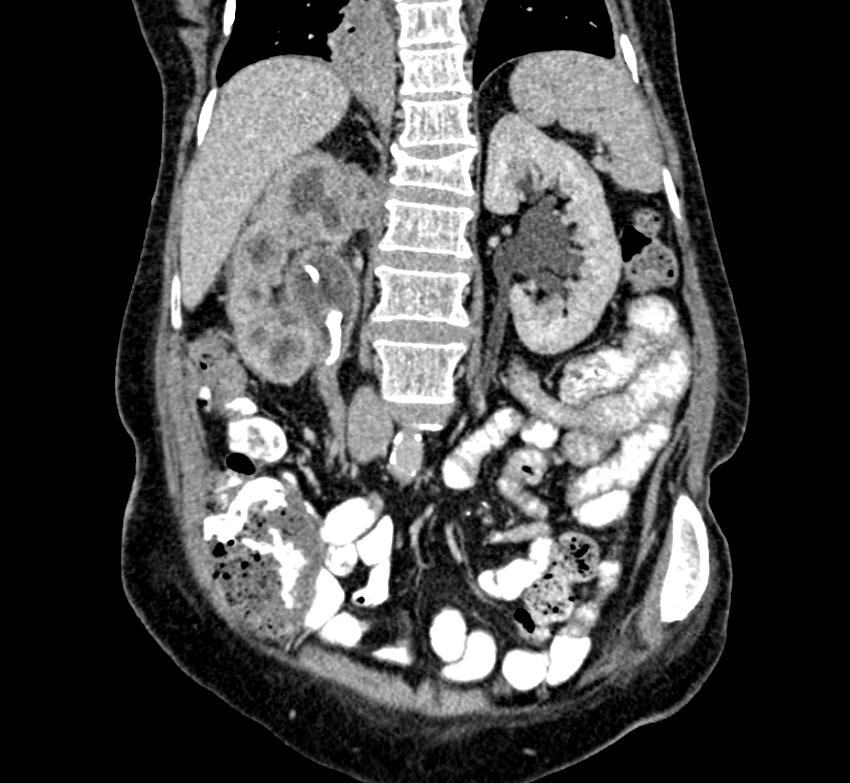
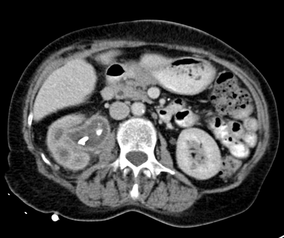
Left: acquired cystic disease; right: with renal cell carcinoma

Renal transplant 12 years prior

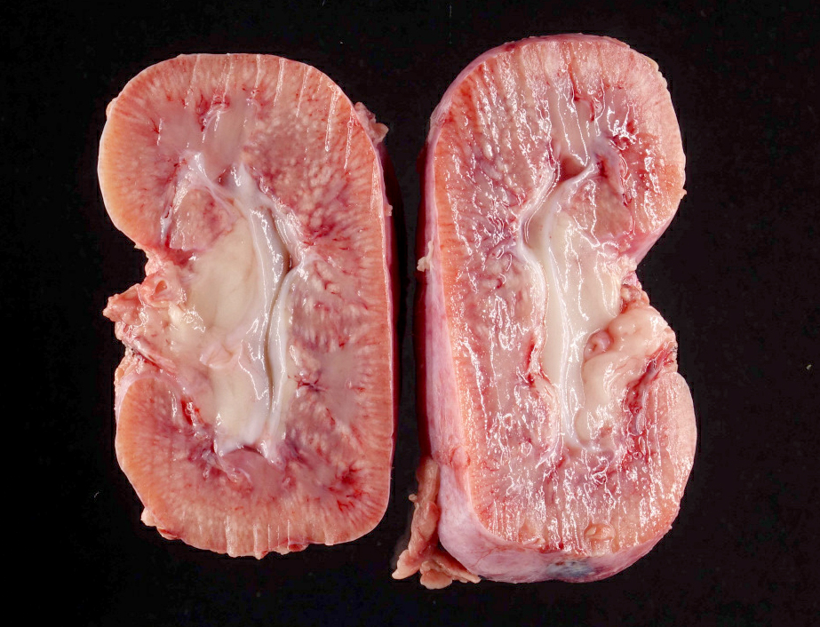
With central scar
Images hosted on other servers:

Mechanisms of donor specific ABMR
Contributed by Arzu Sağlam, M.D.
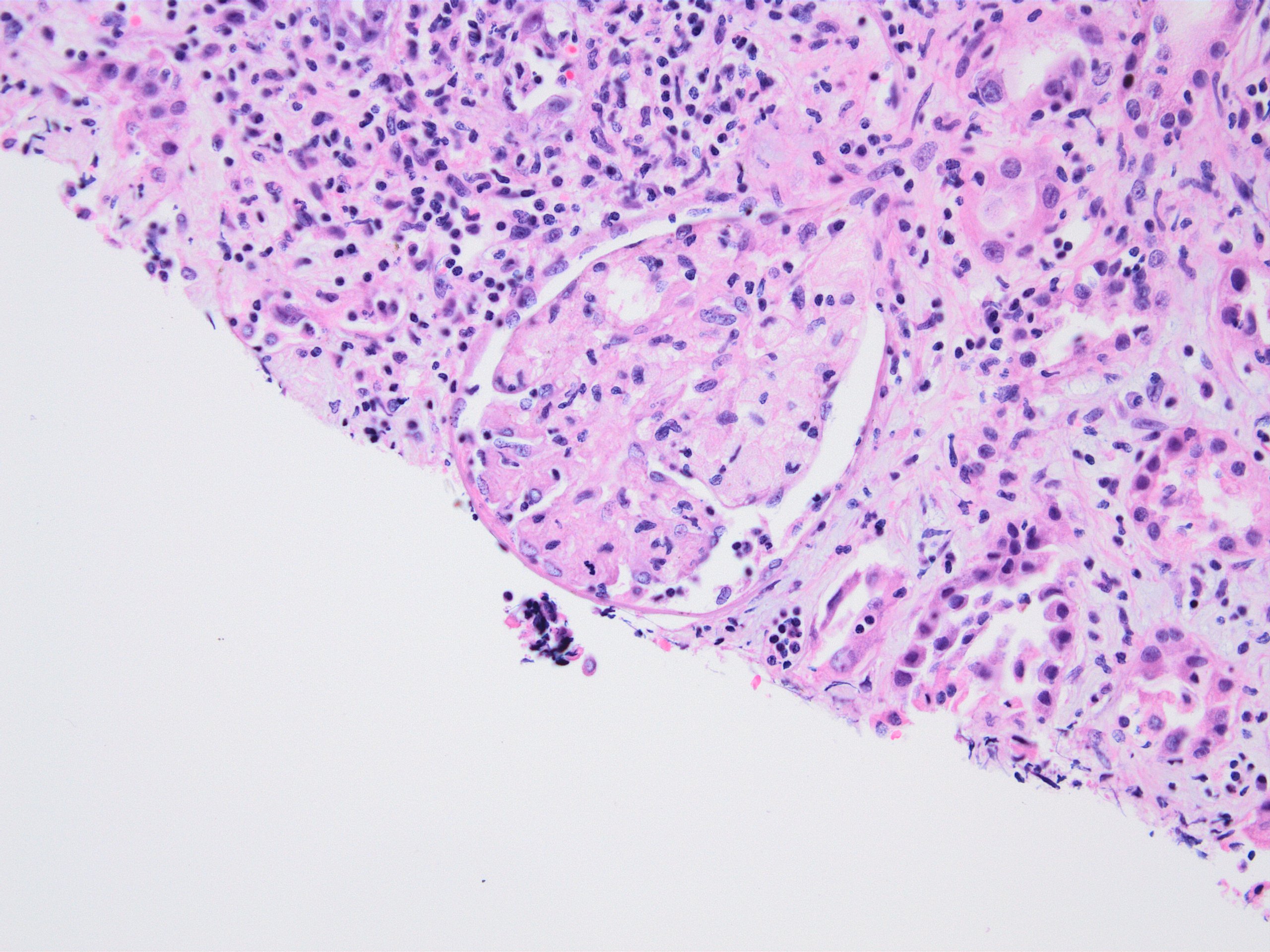
Active antibody mediated rejection
Contributed by Arzu Sağlam, M.D.
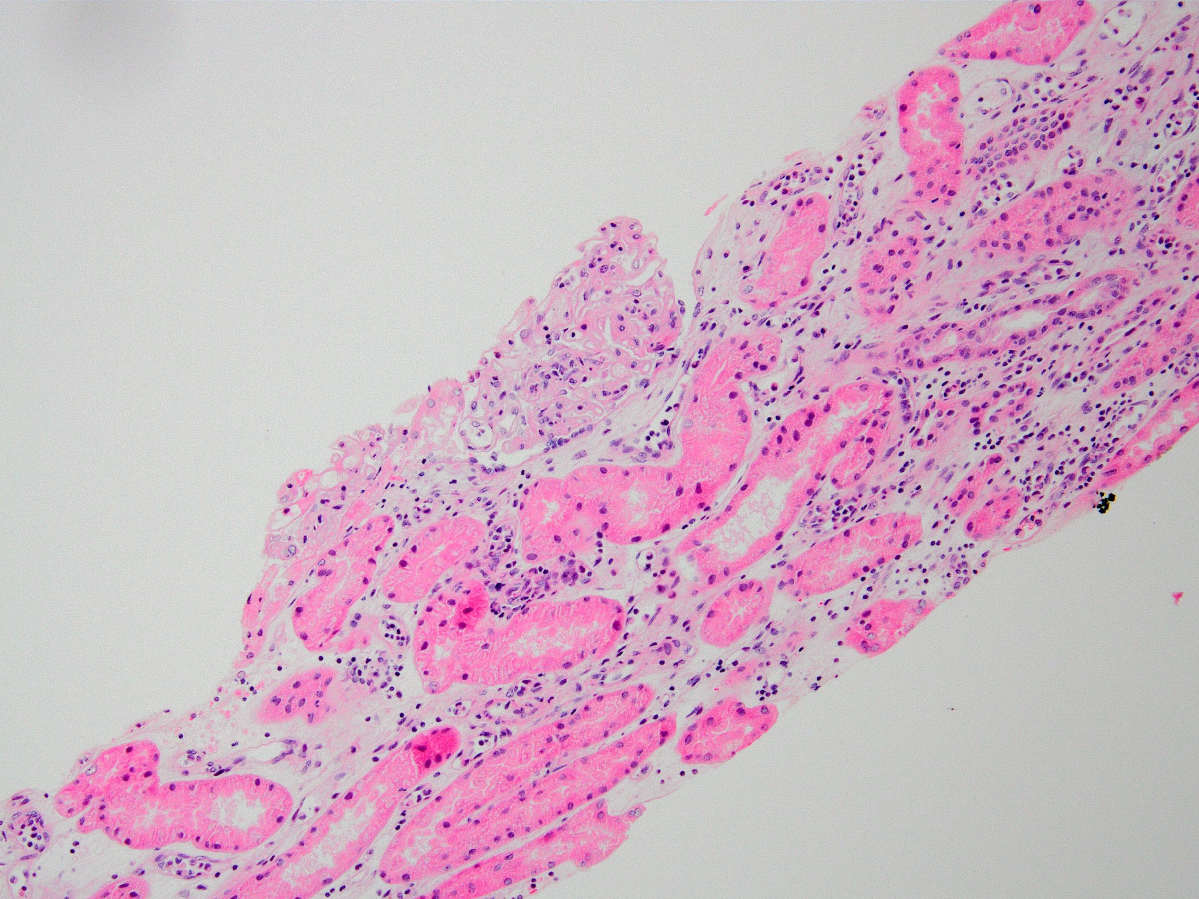
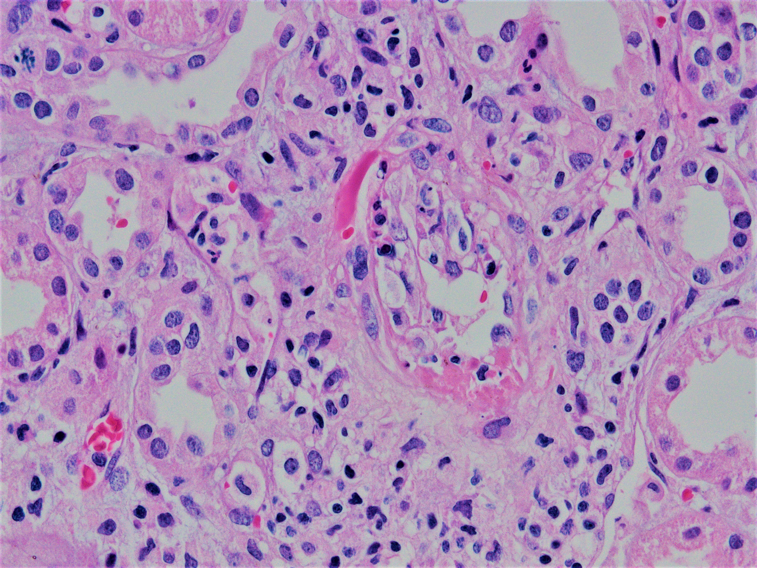
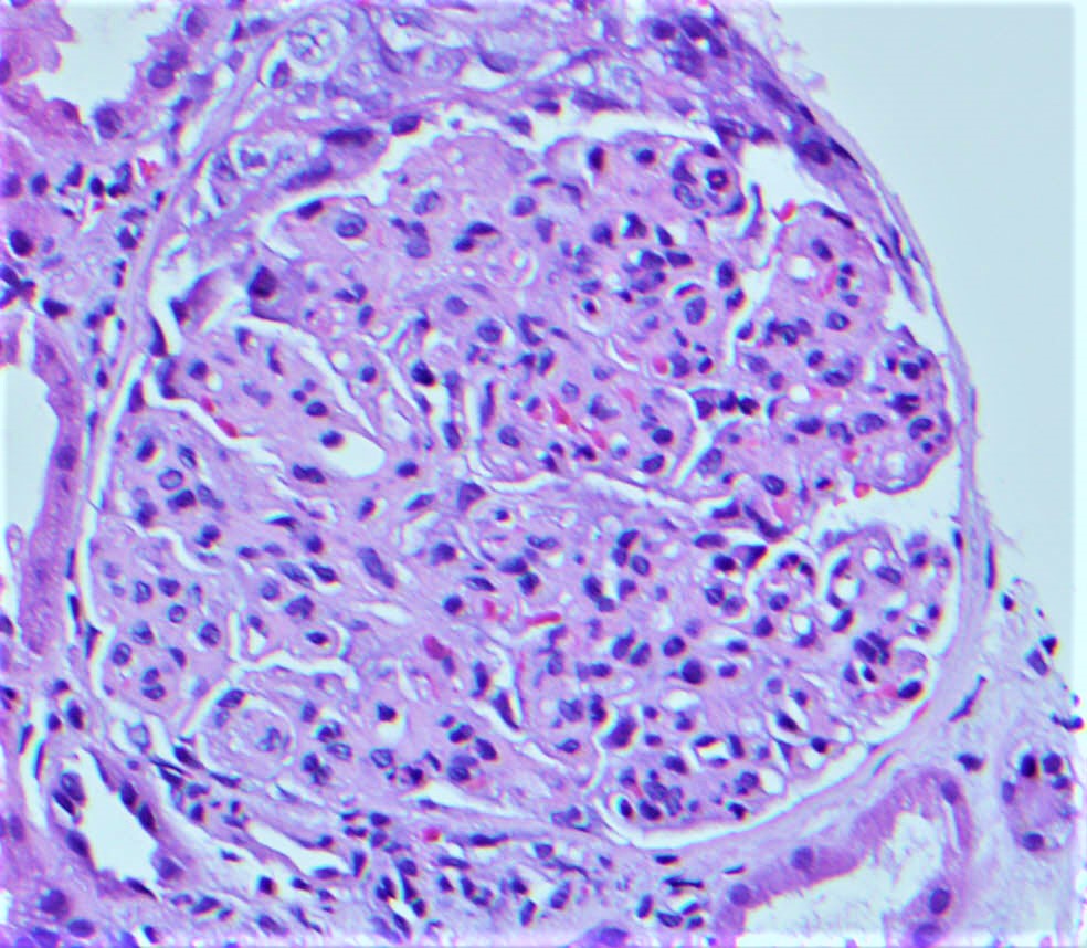
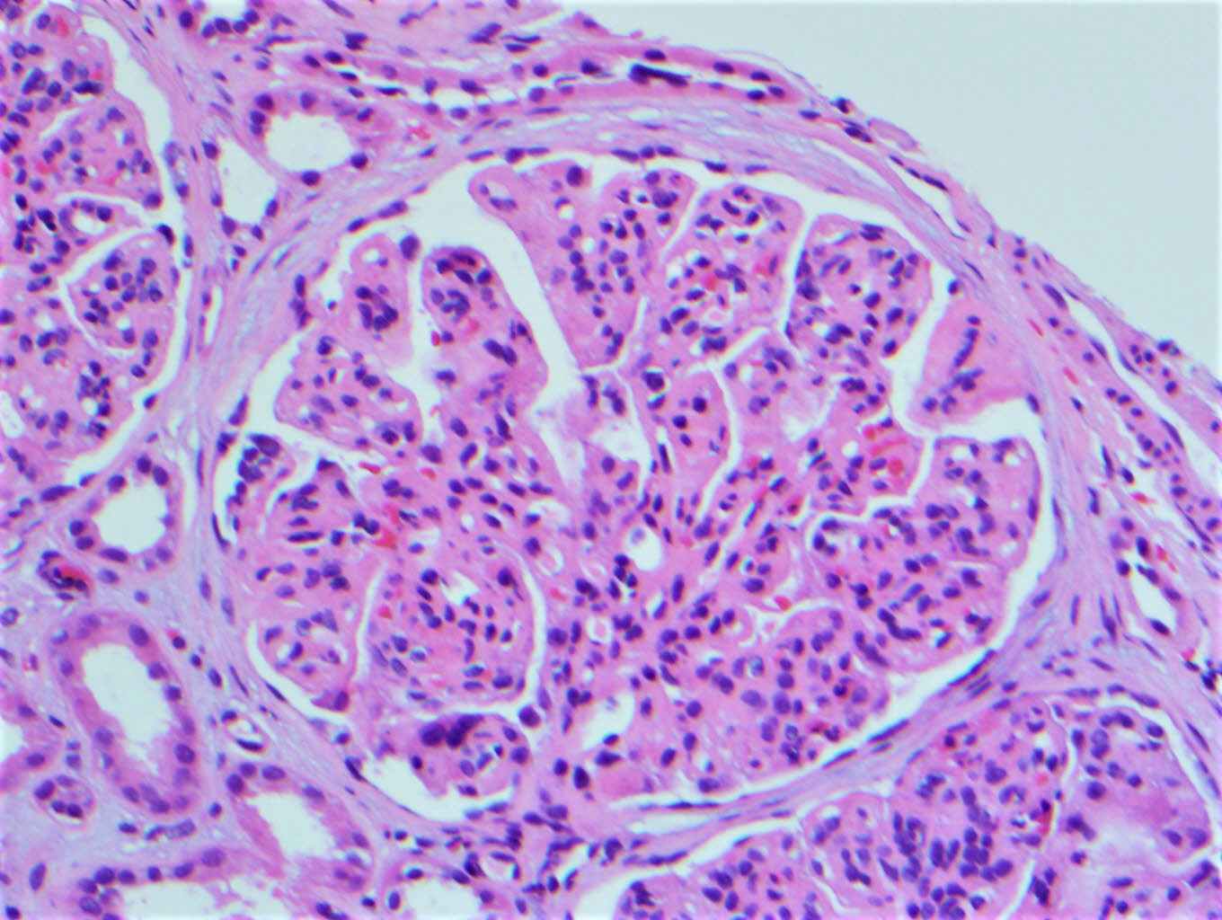
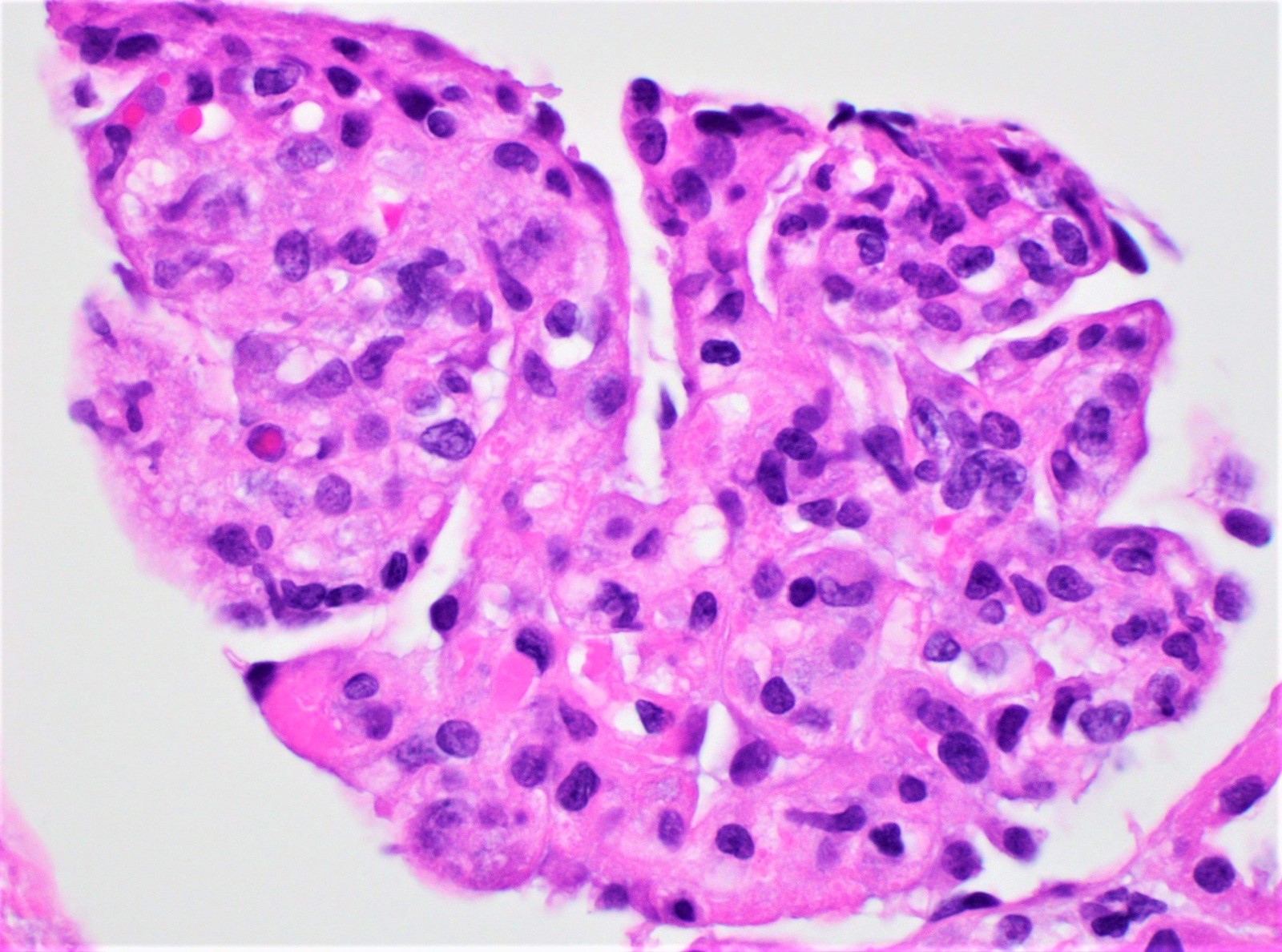
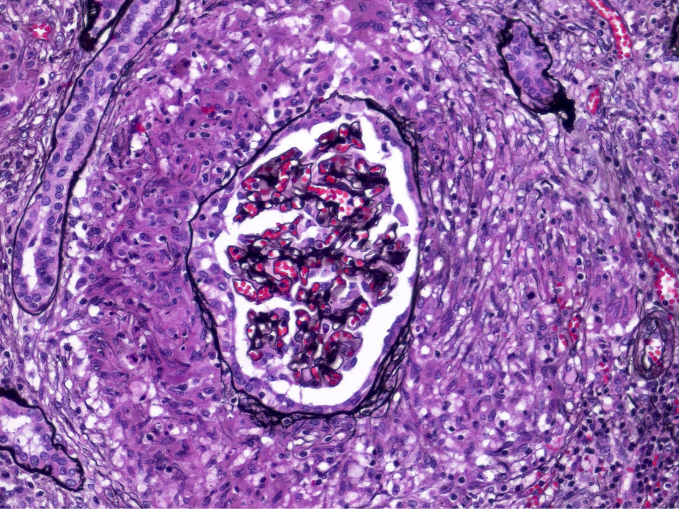
Peritubular capillaritis and glomerulitis
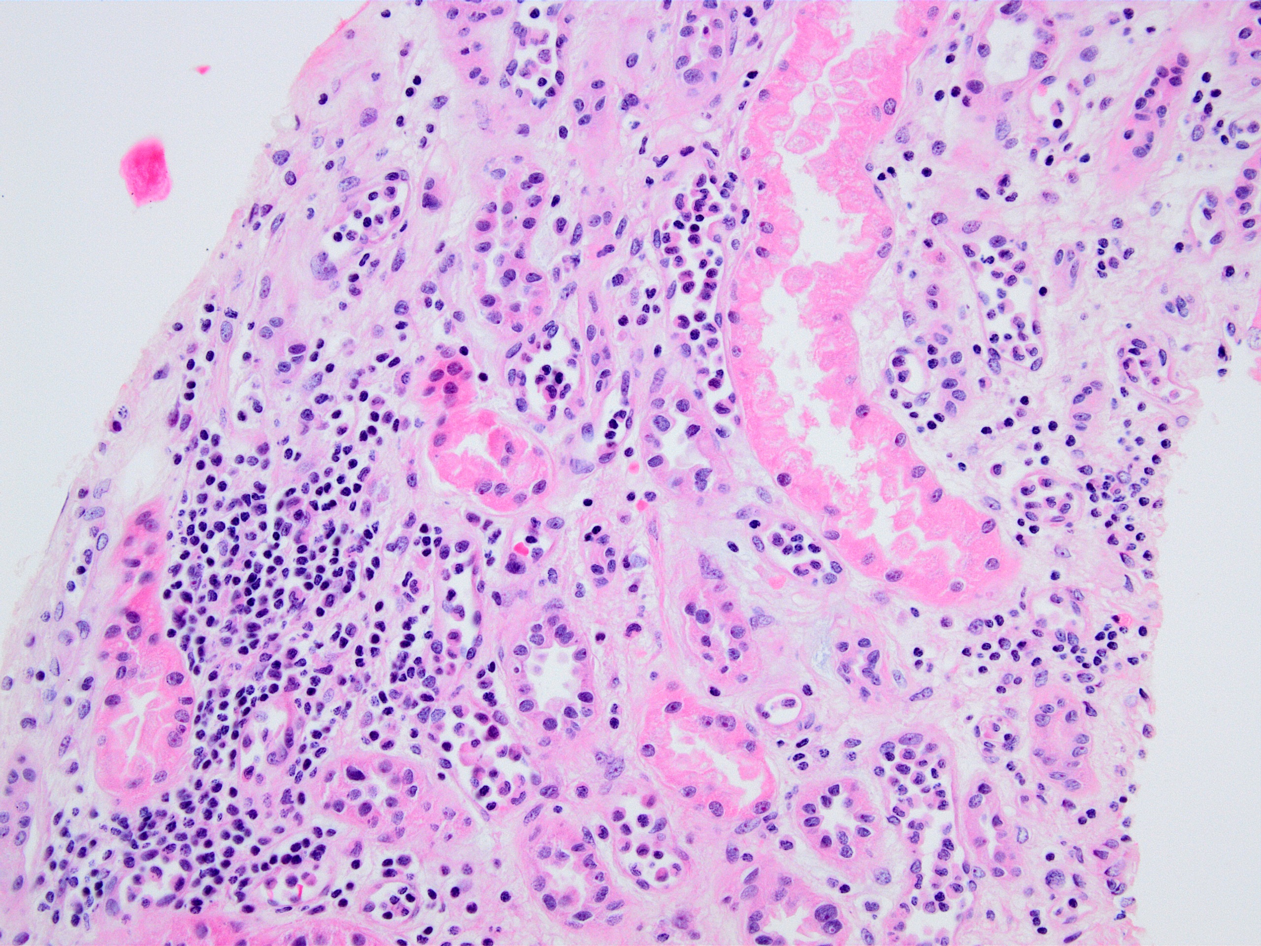
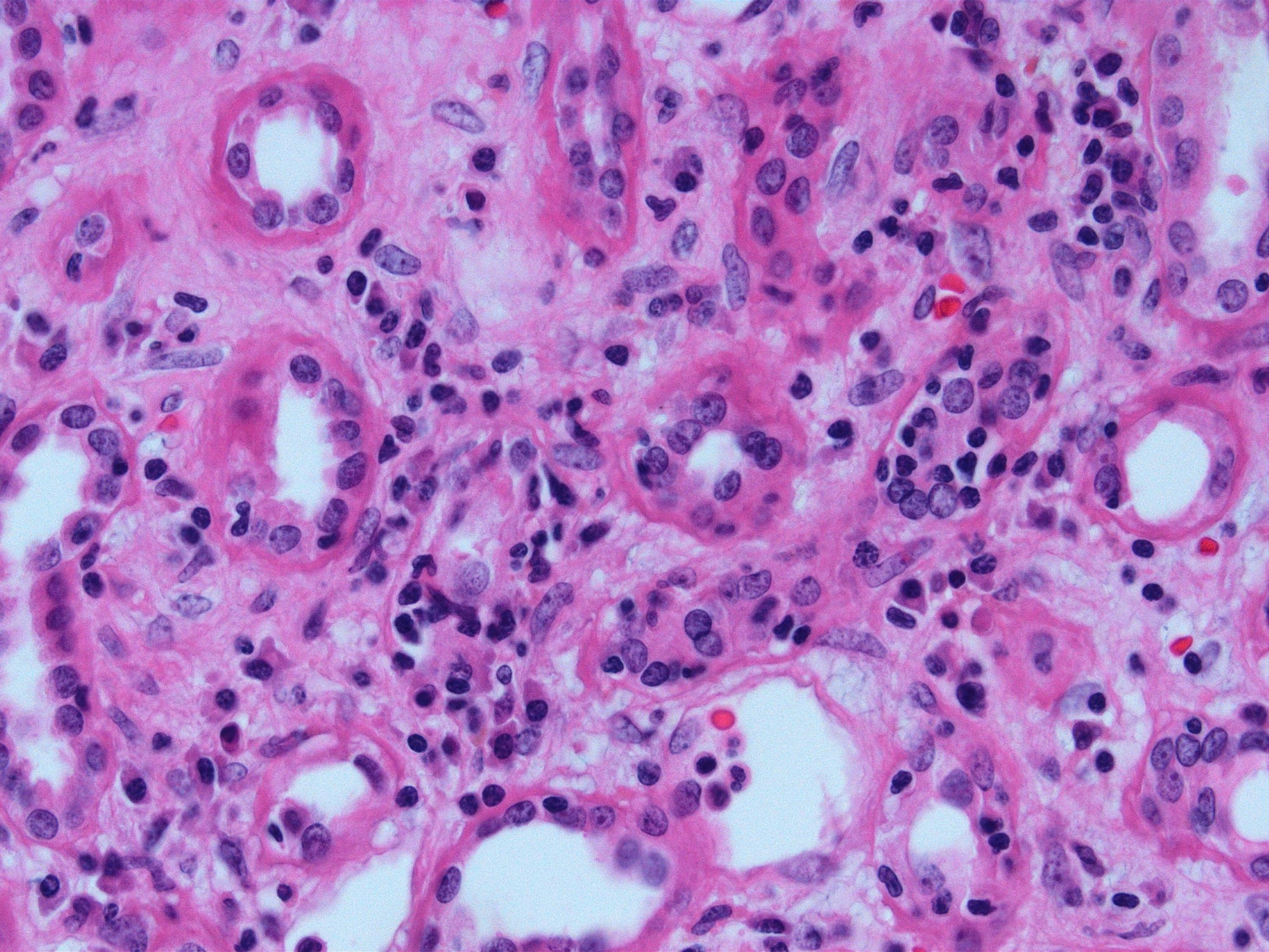
Peritubular capillaritis

Severe peritubullary capillaritis (ptc3)
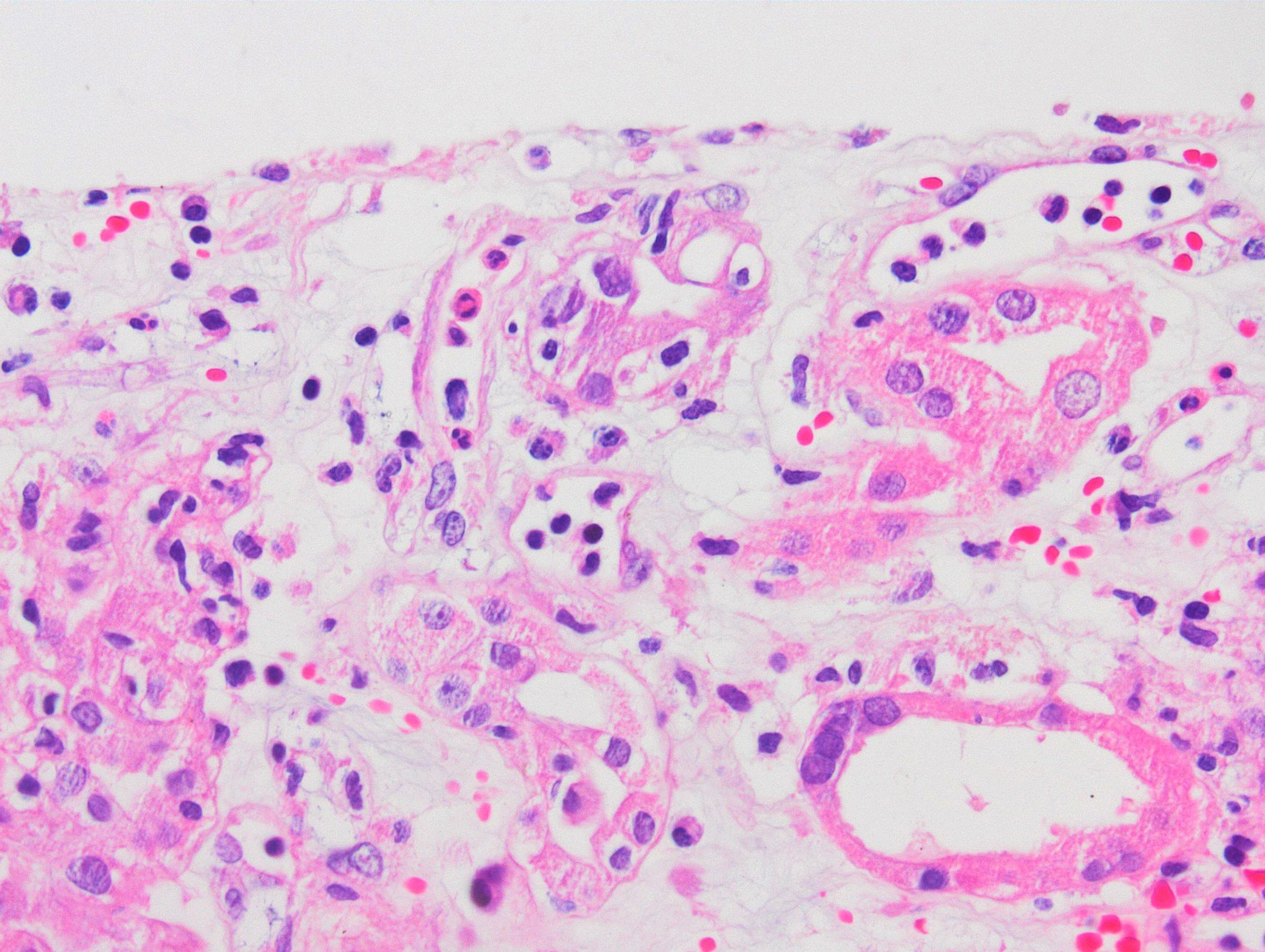
Peritubullary capillaritis, neutrophils
Peritubullary capillaritis
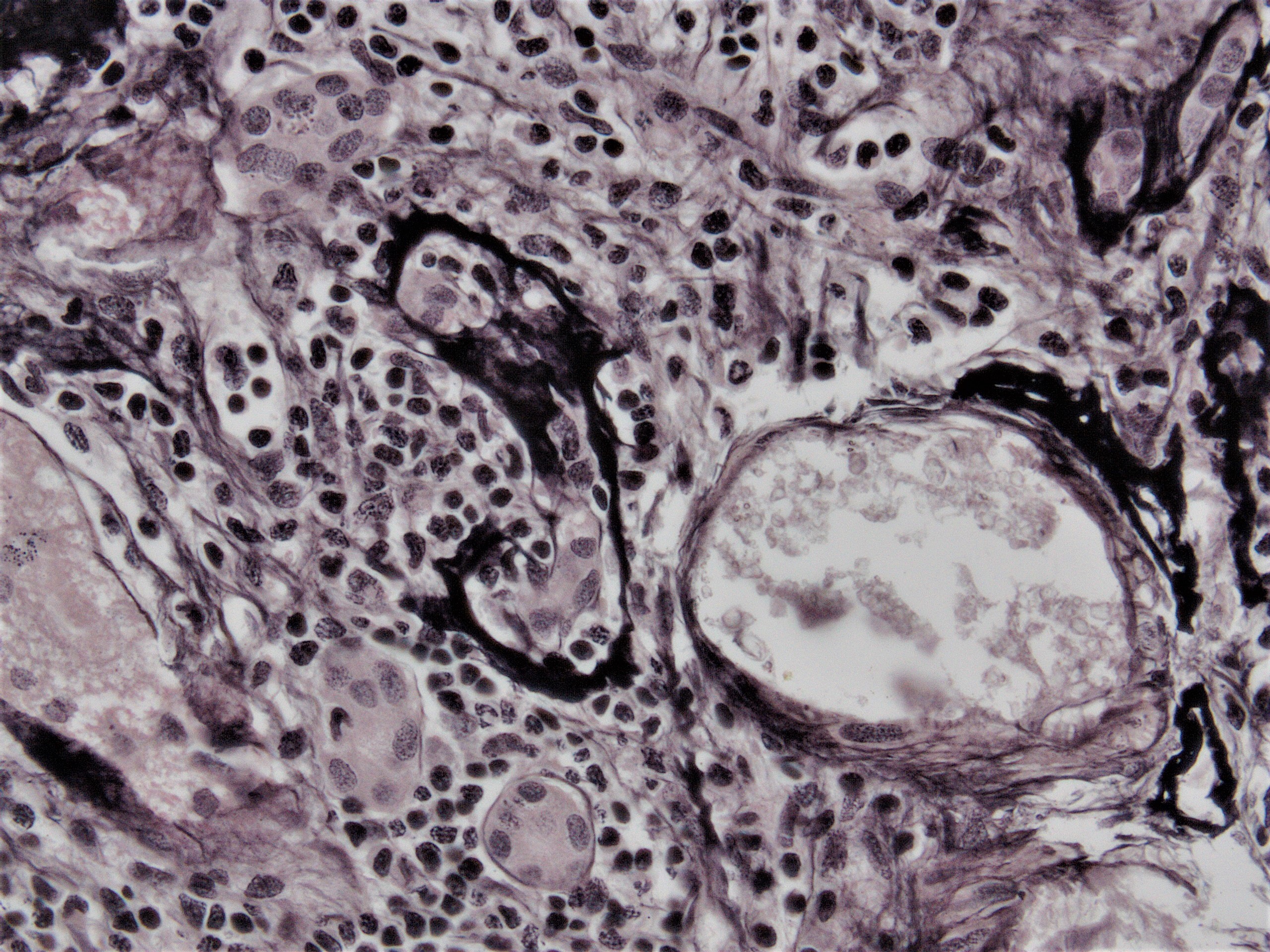
Glomerulitis
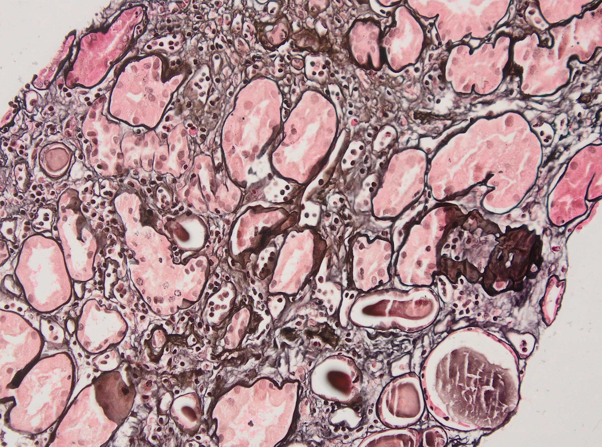
Peritubullary capillaritis, JMS stain
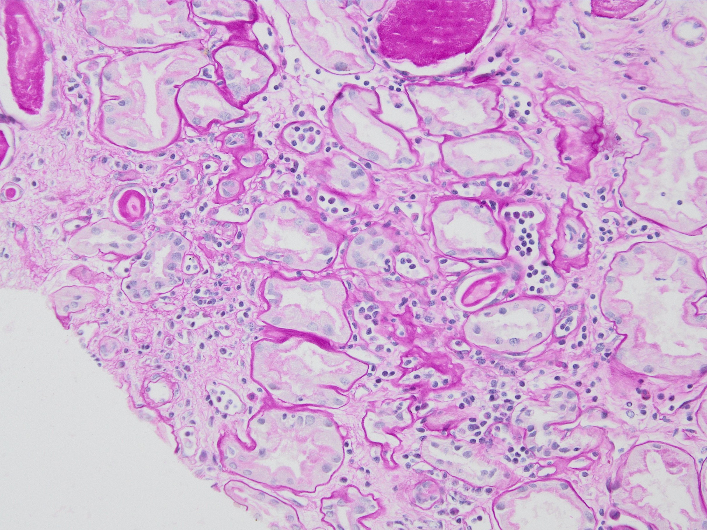
Peritubullary capillaritis, PAS stain
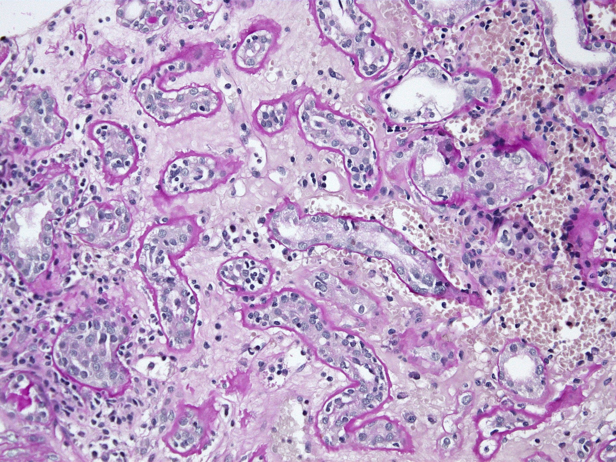
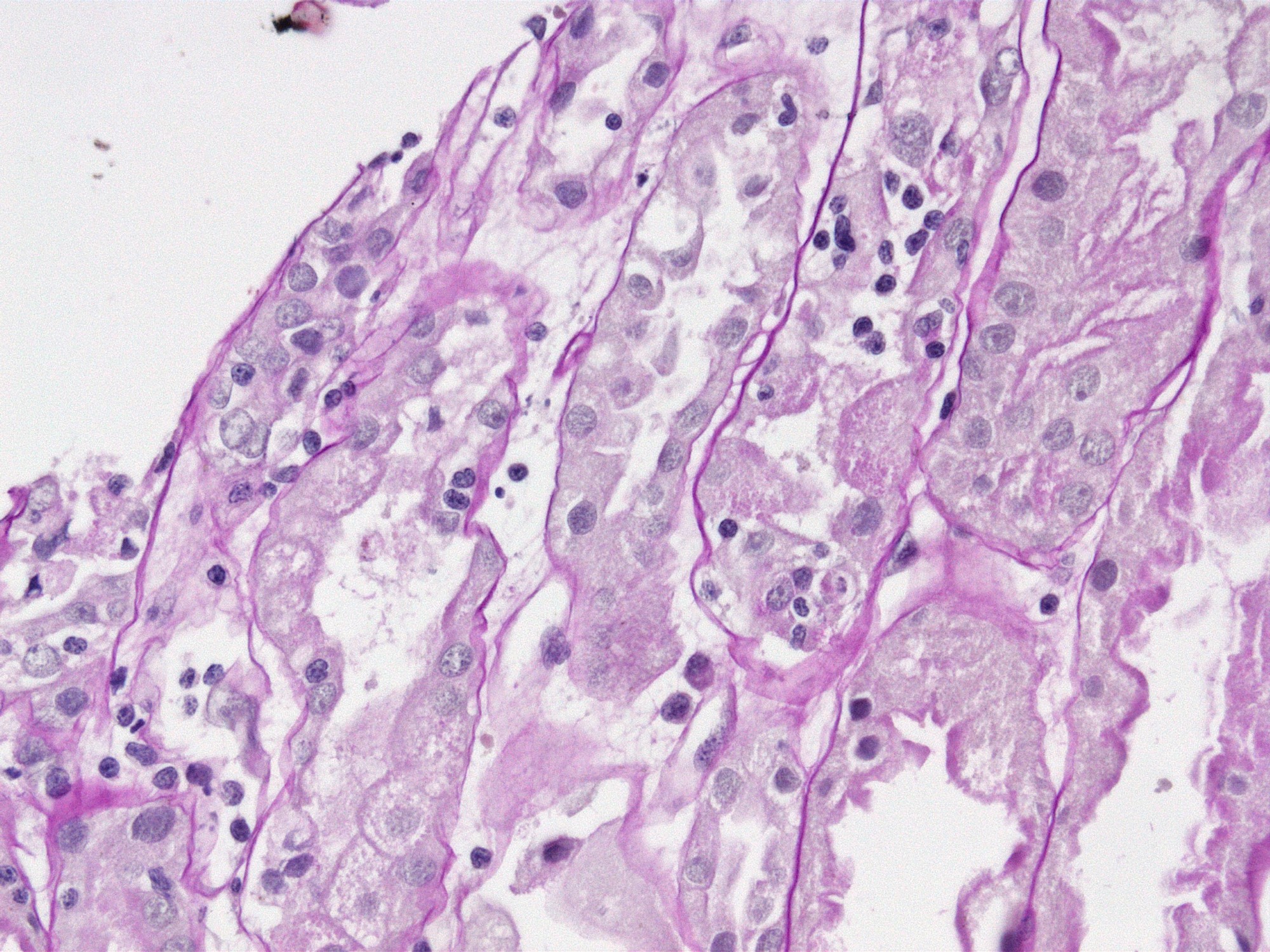
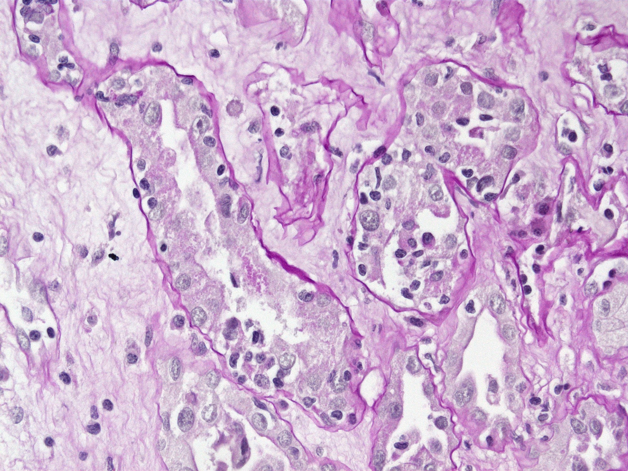
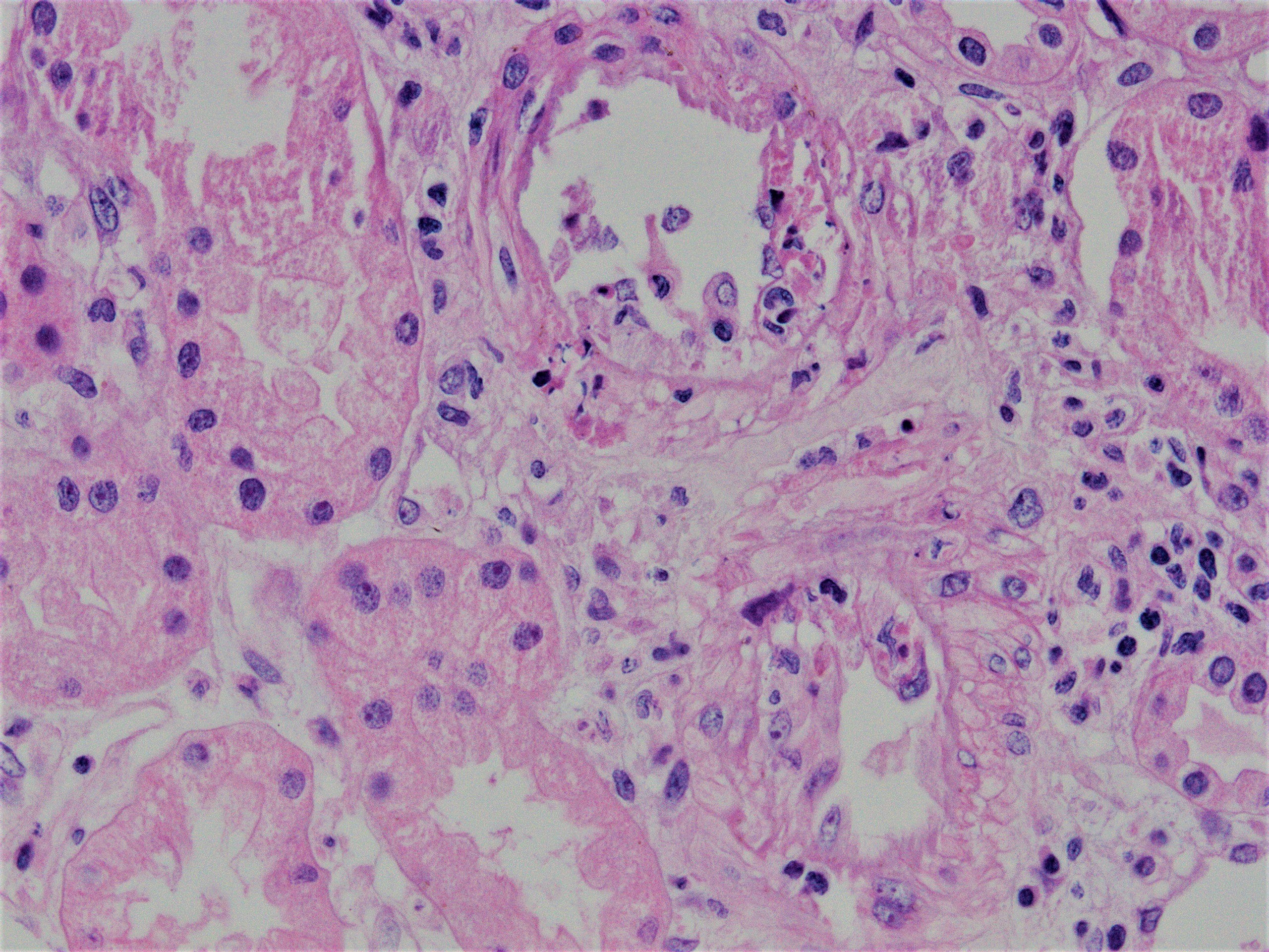
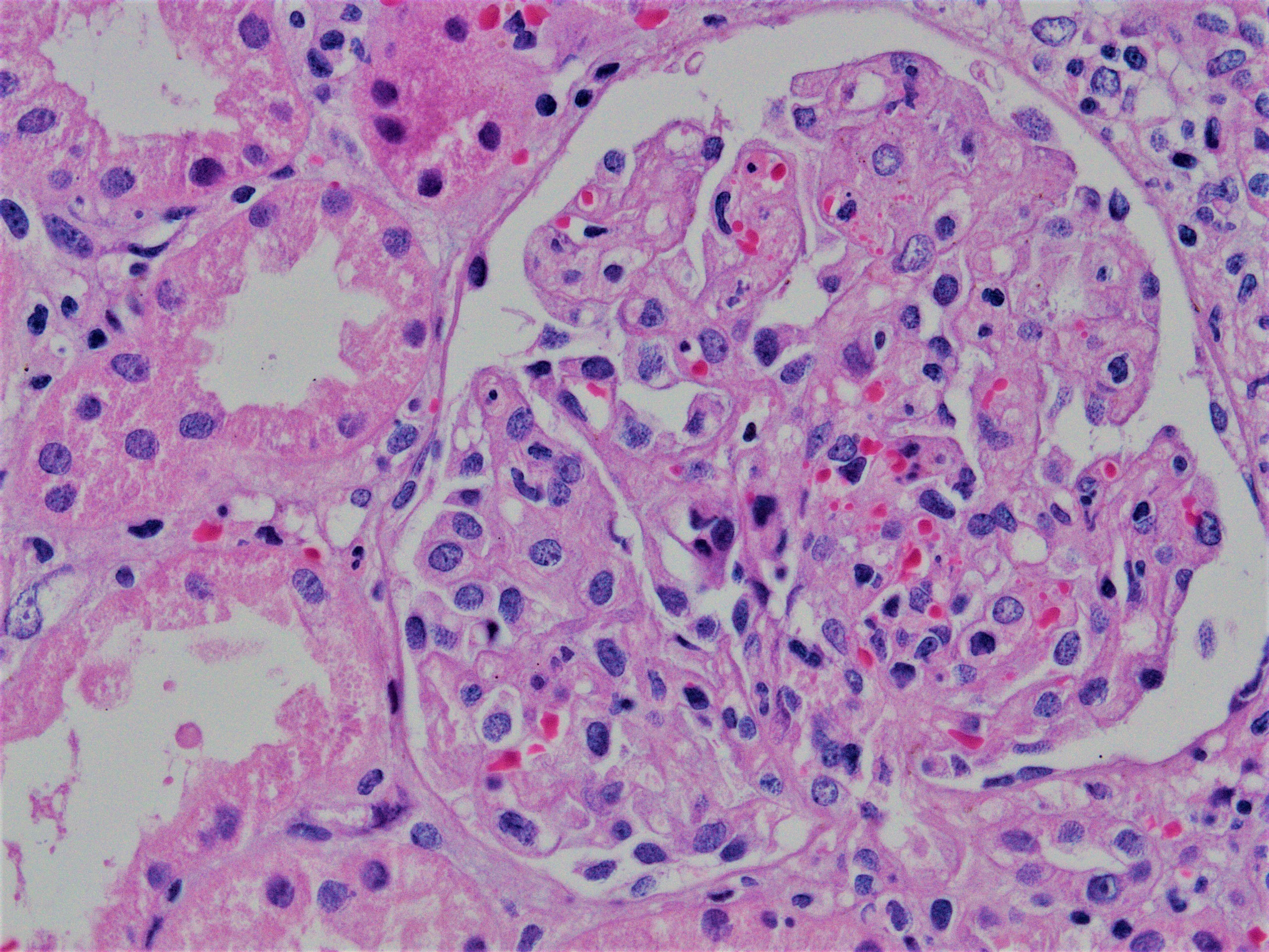
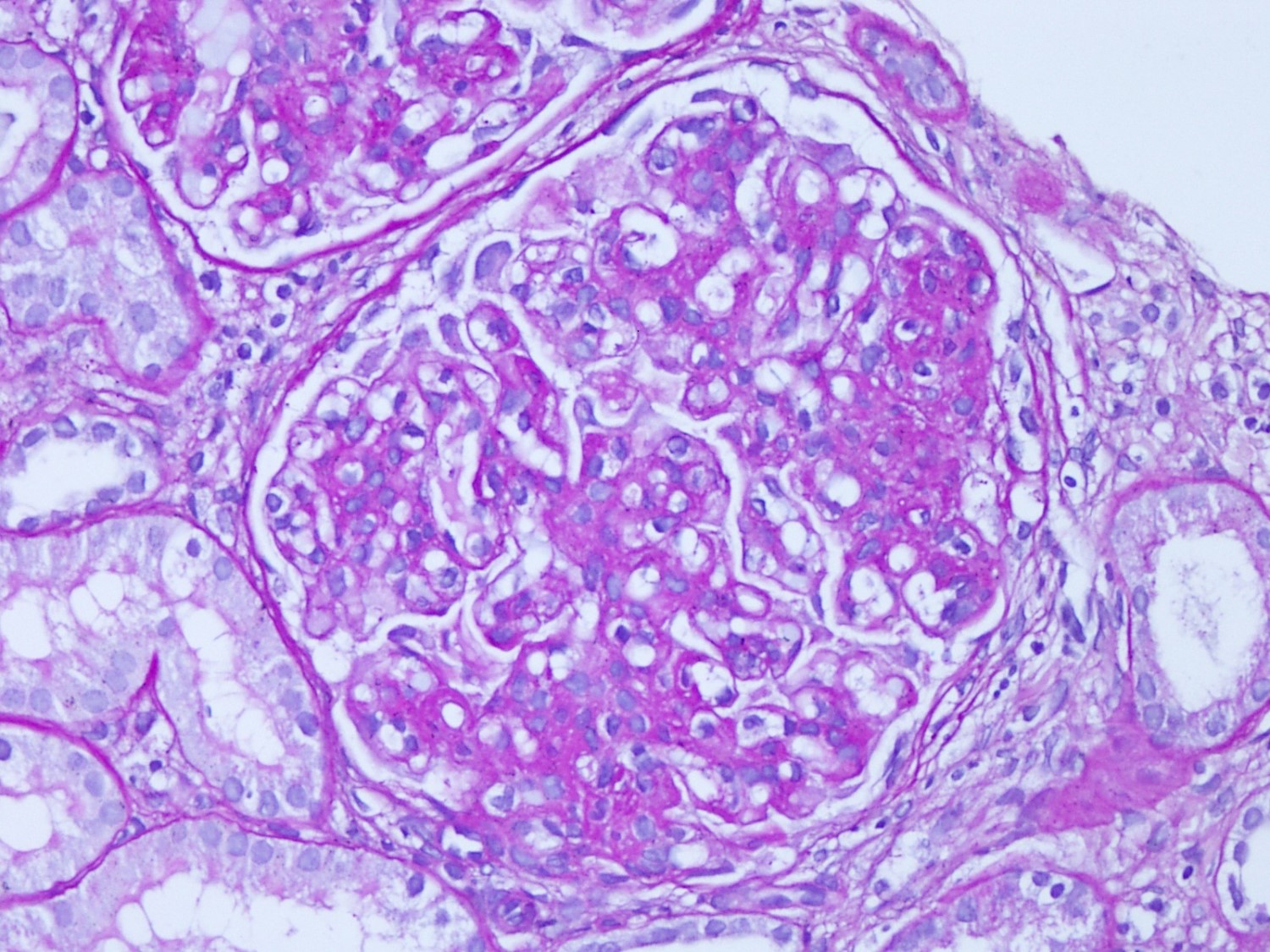
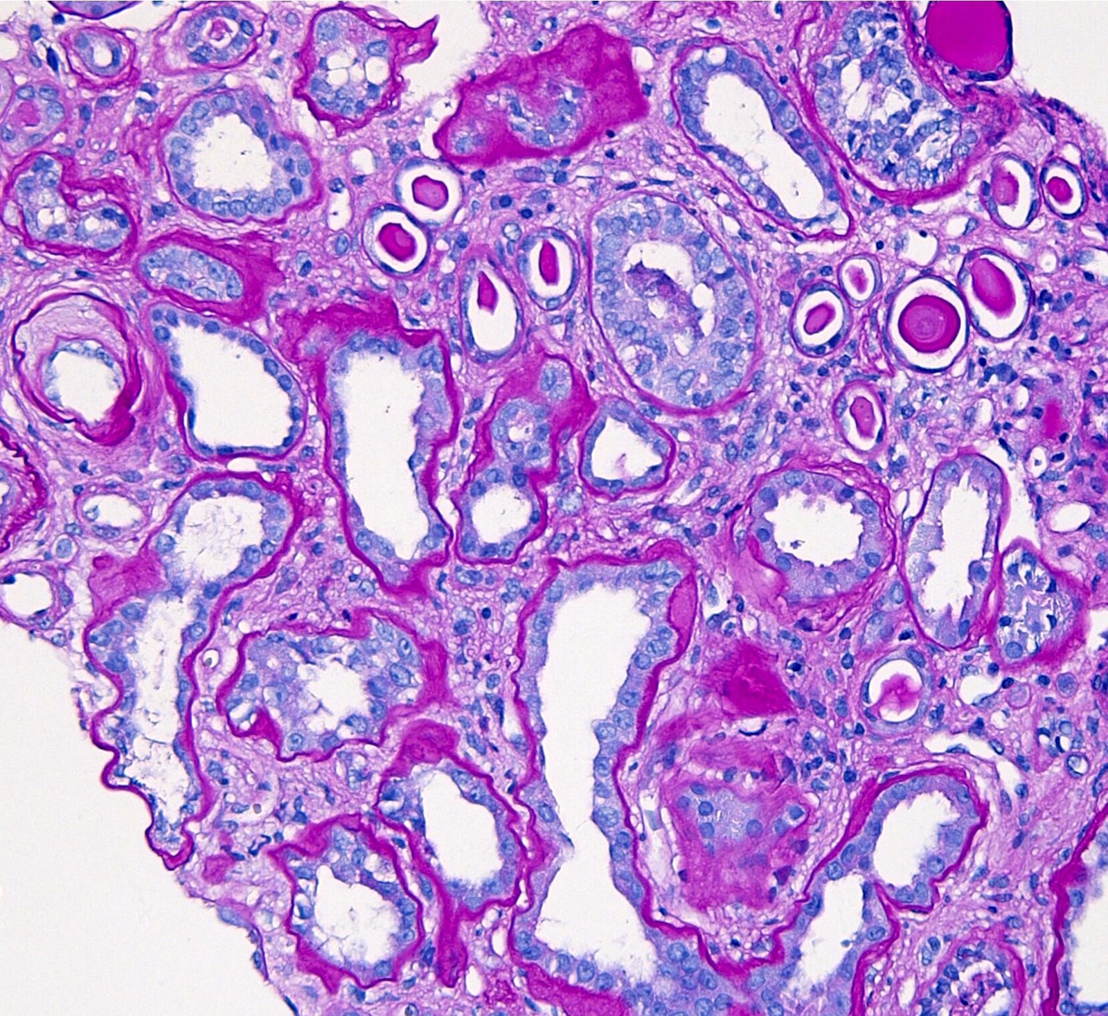
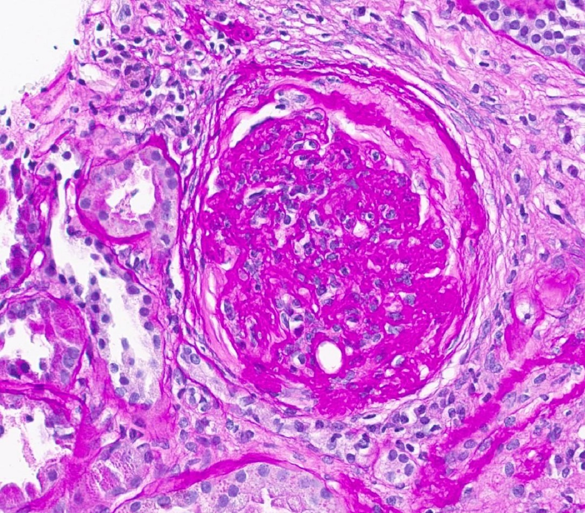
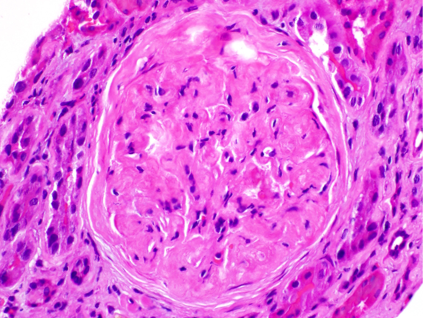
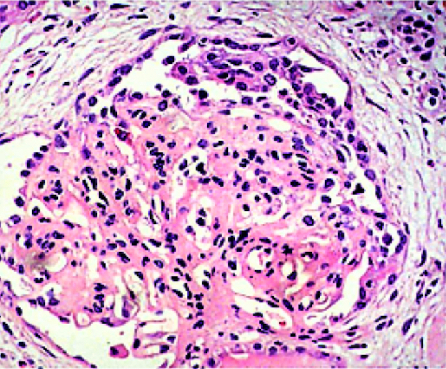
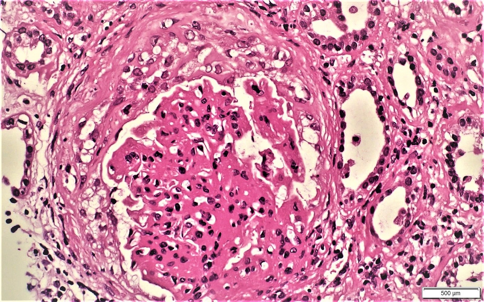
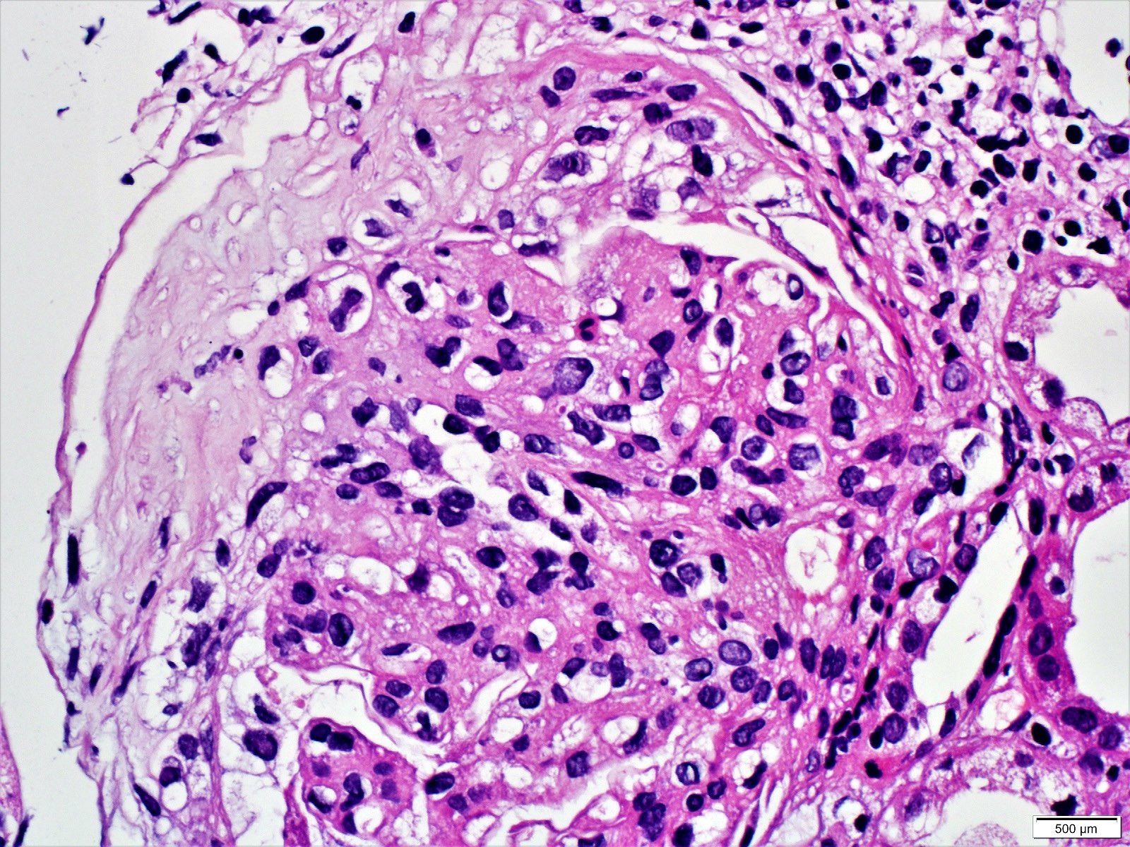
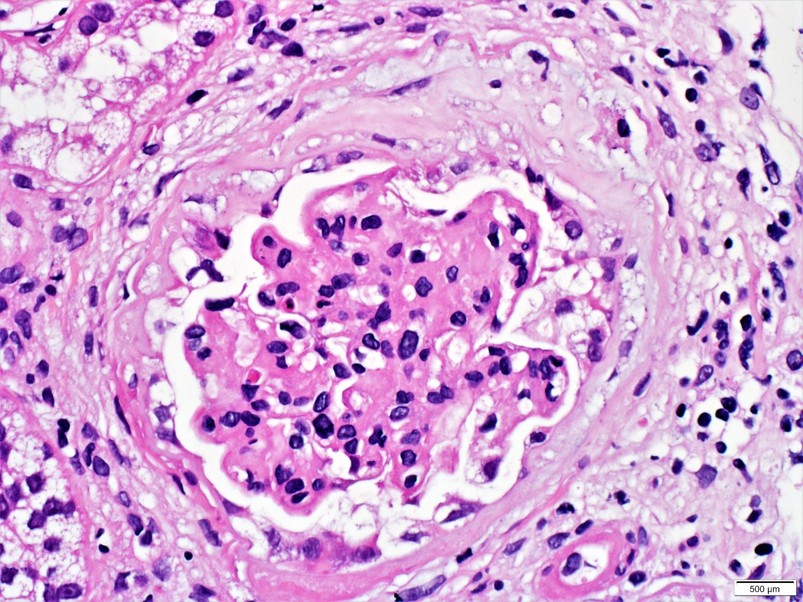
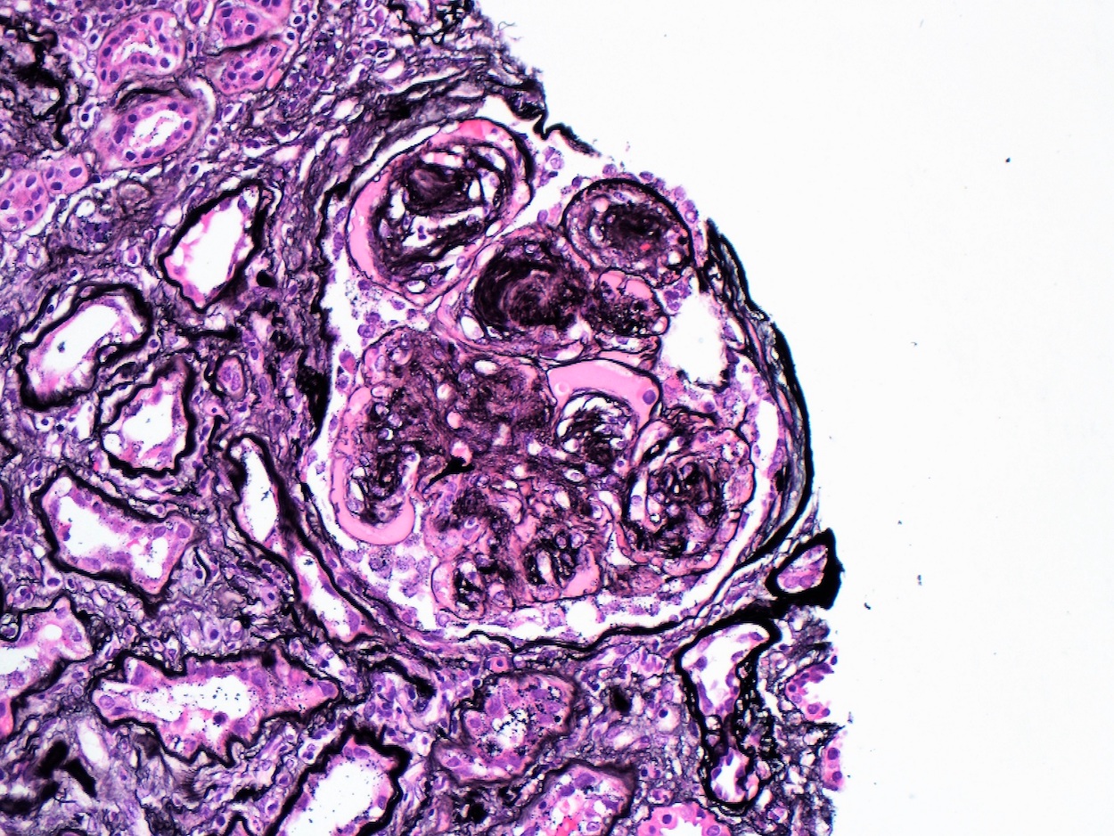
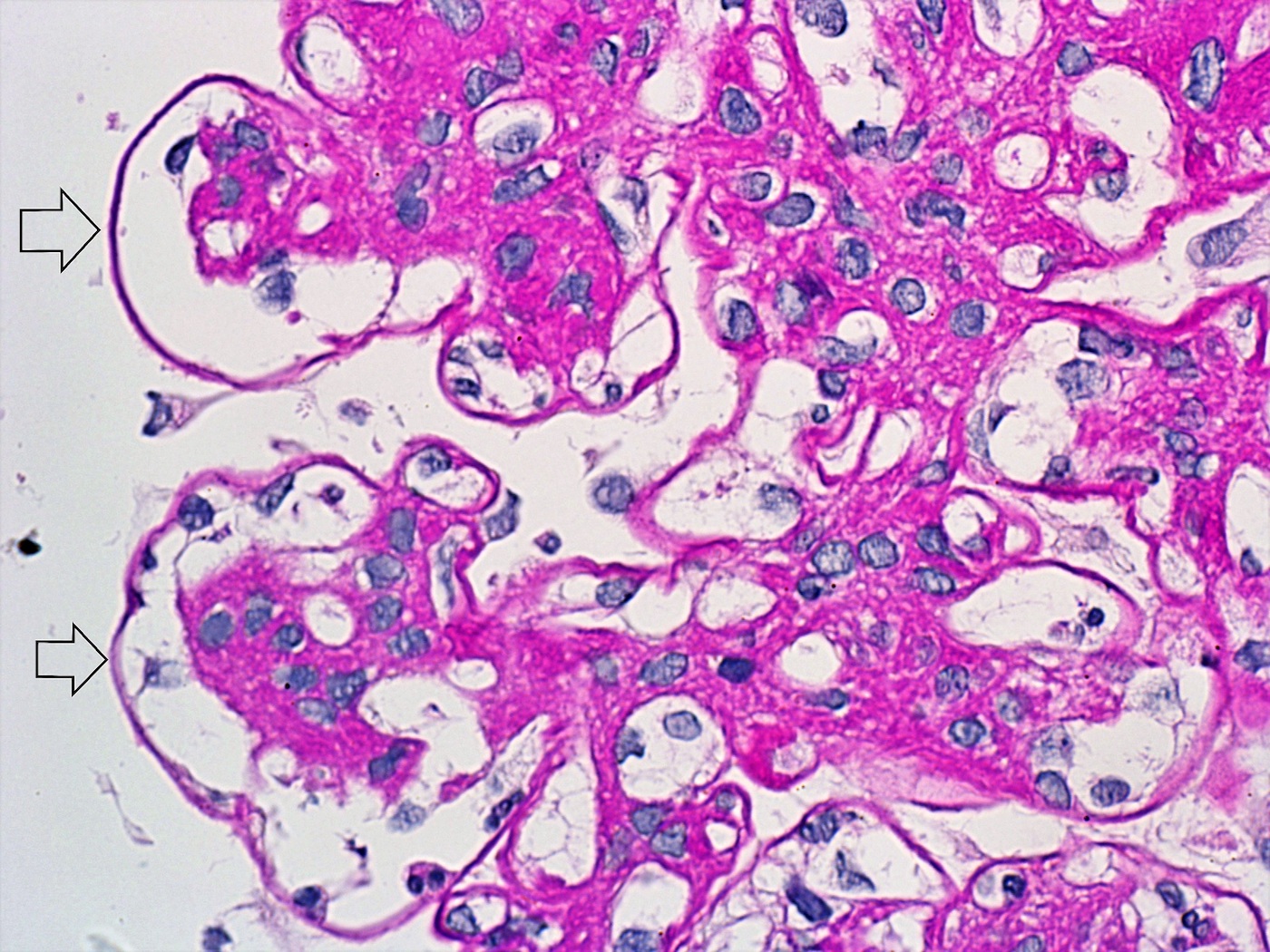
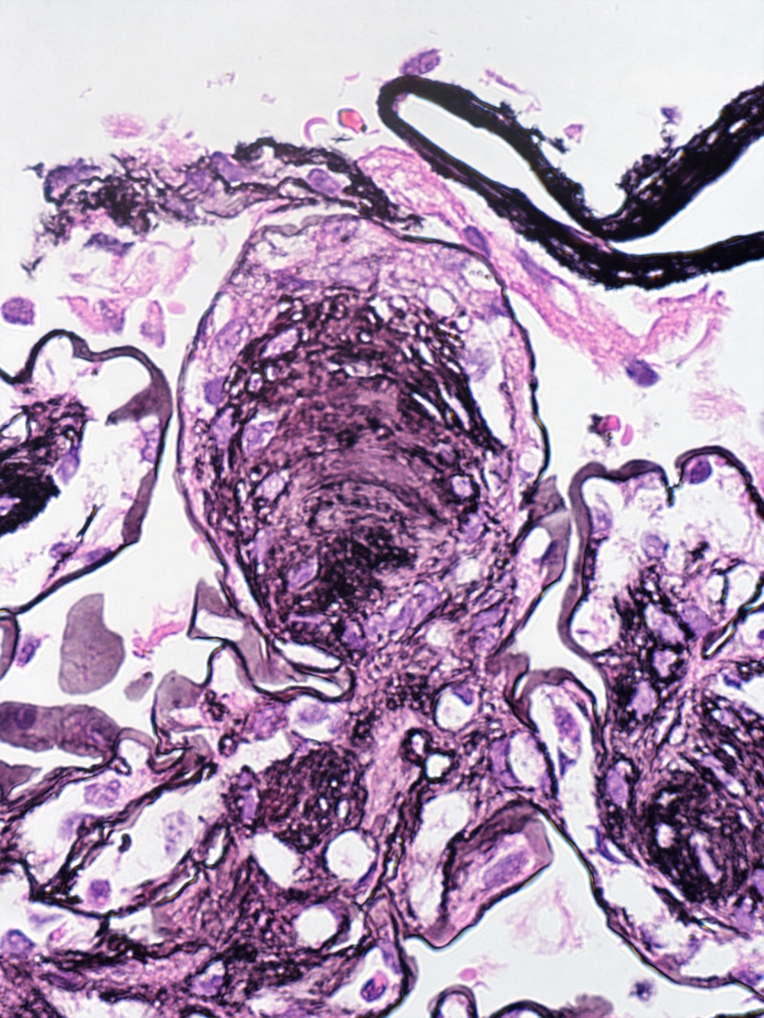
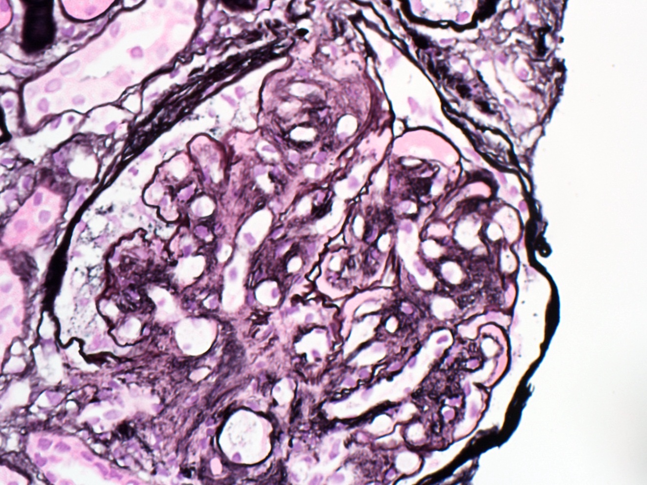
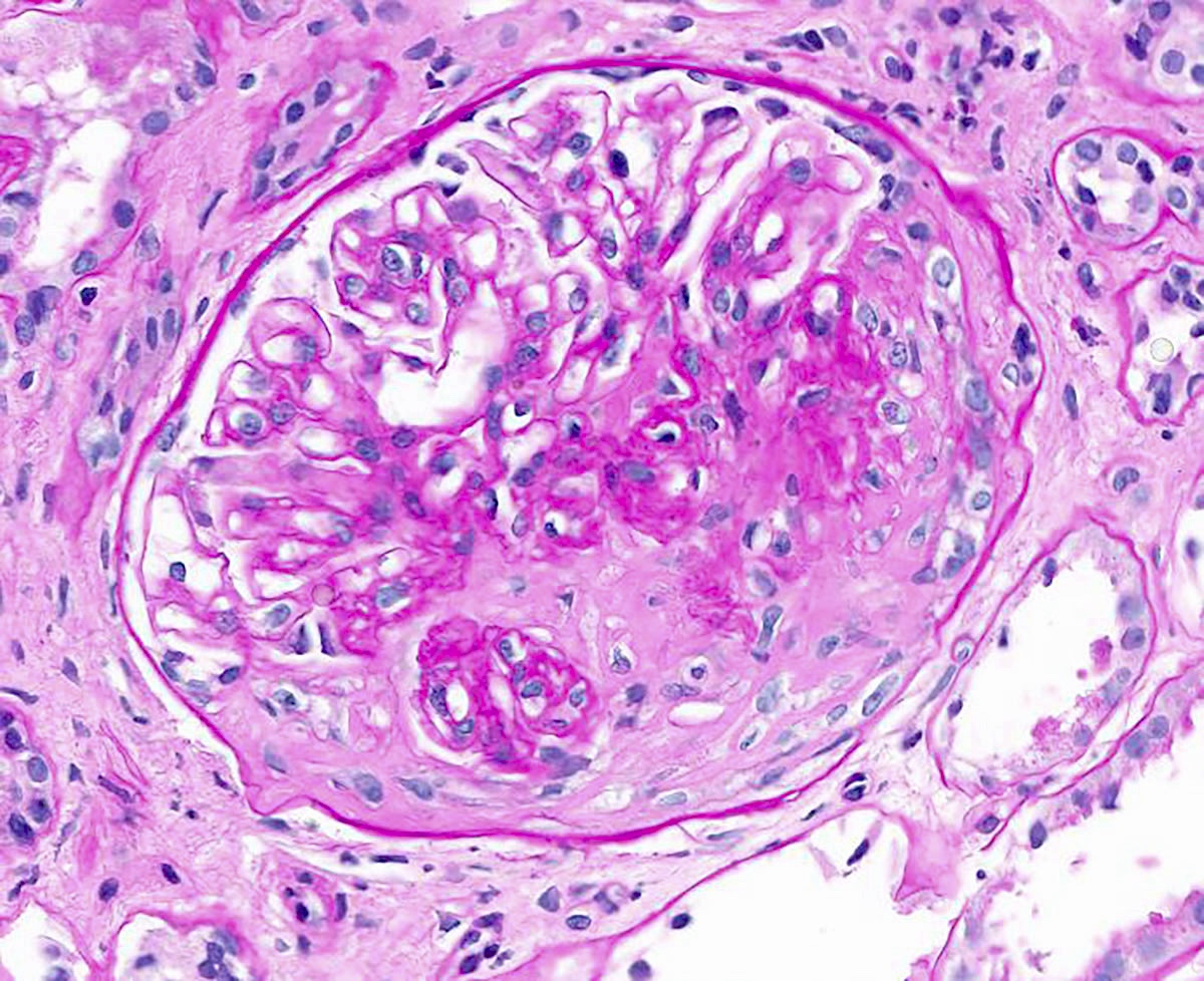
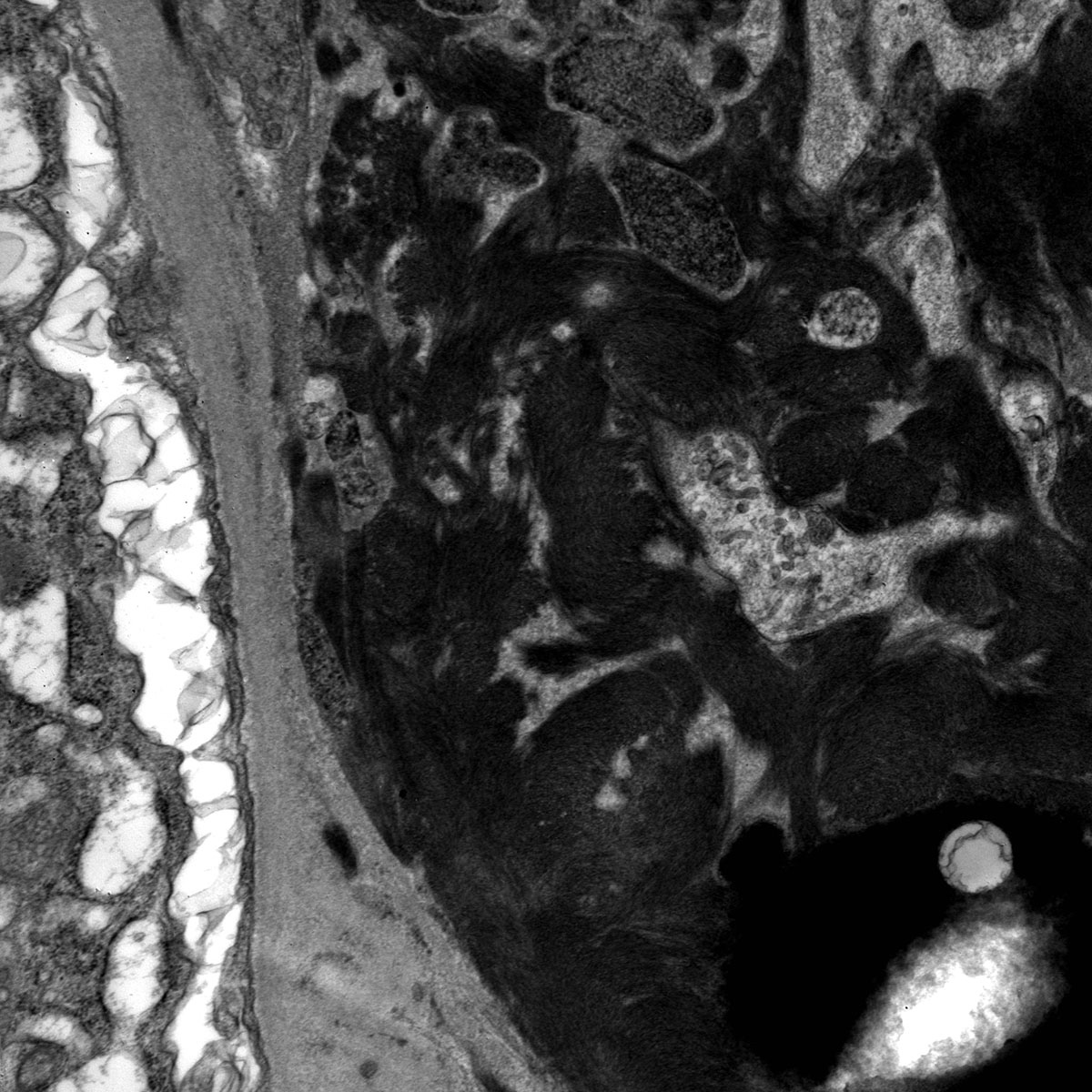
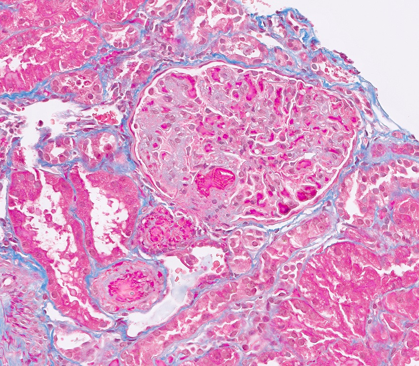
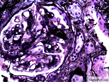
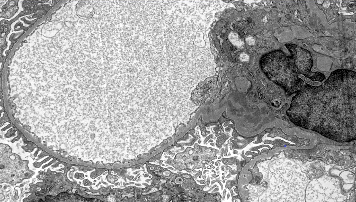
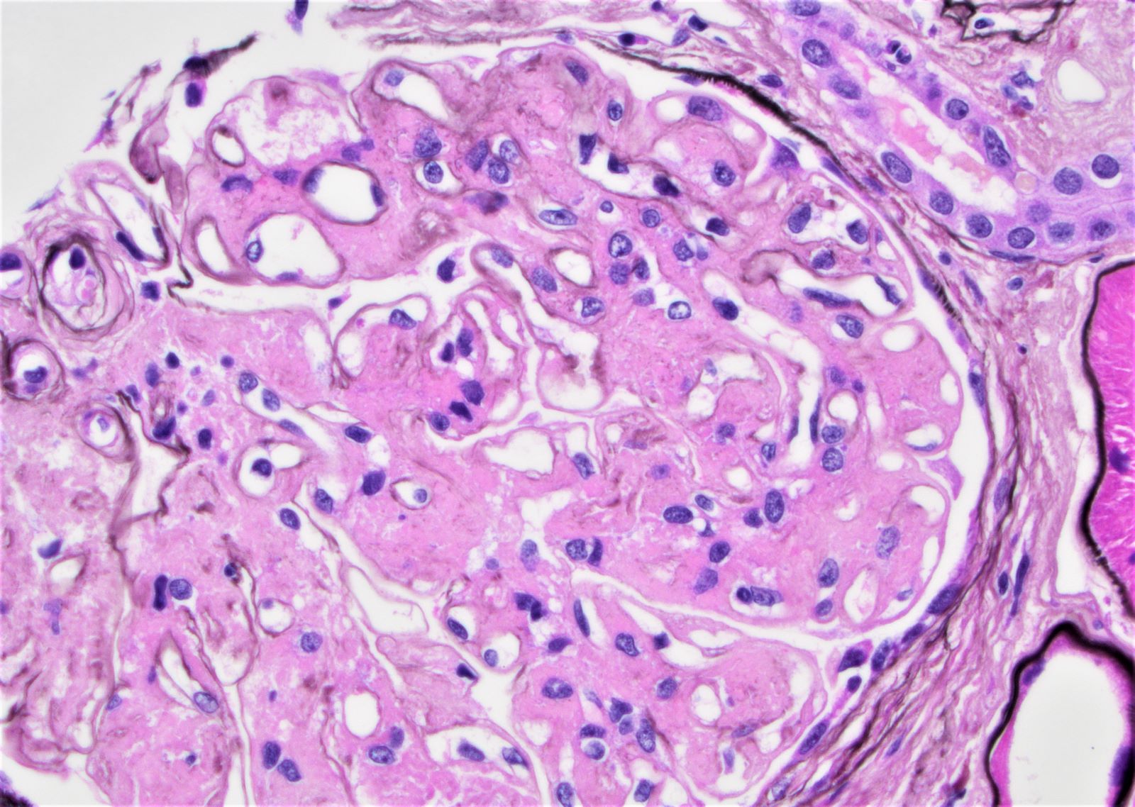
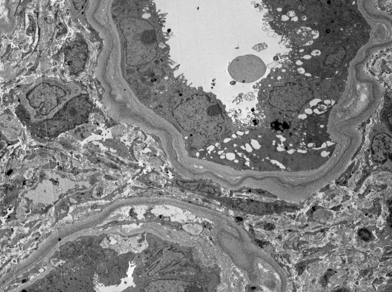
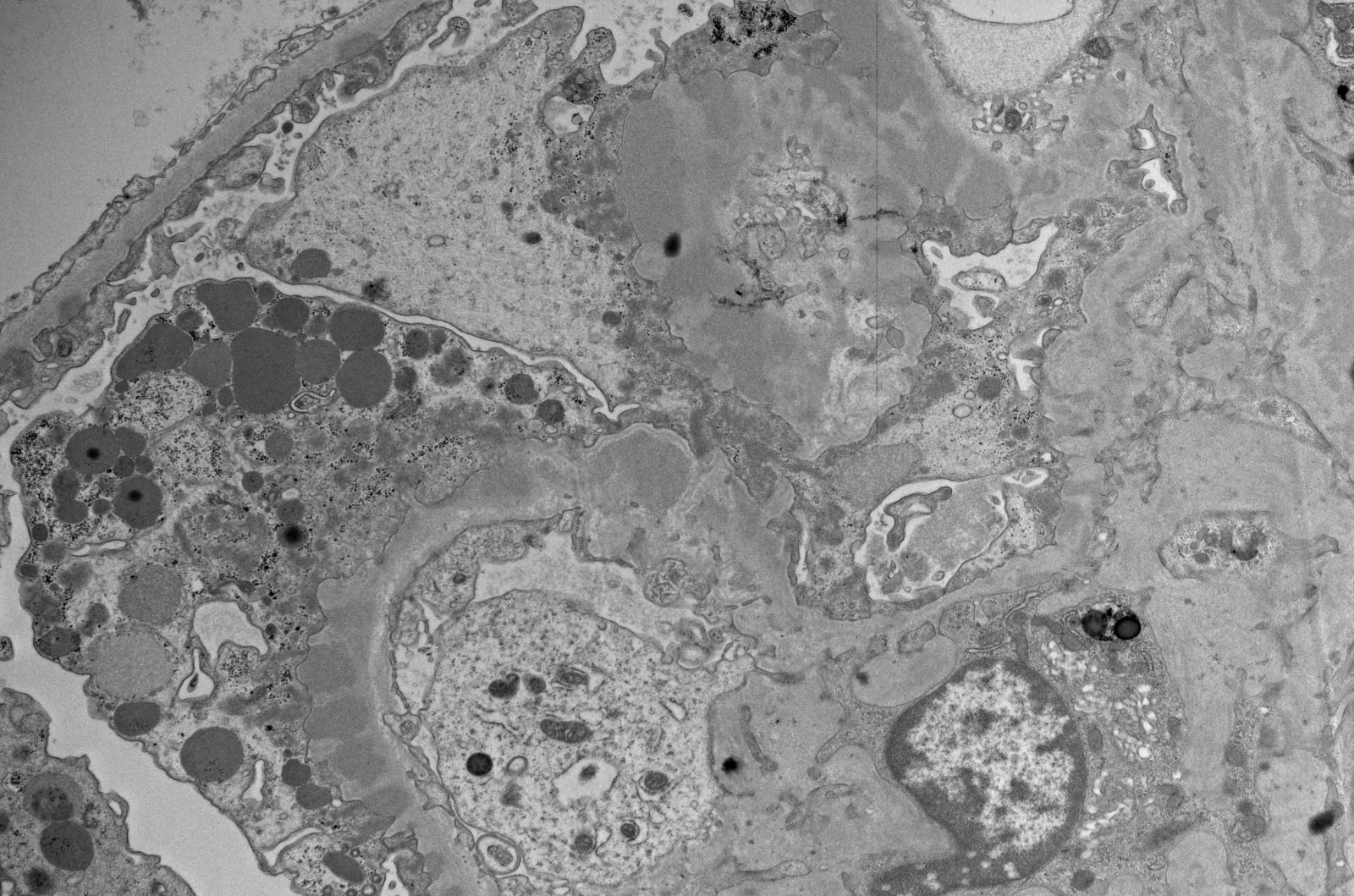
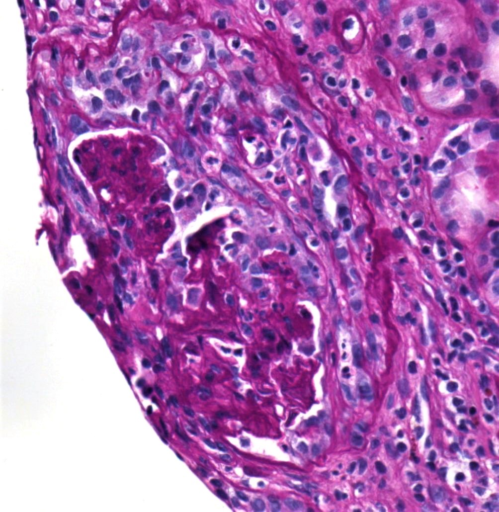
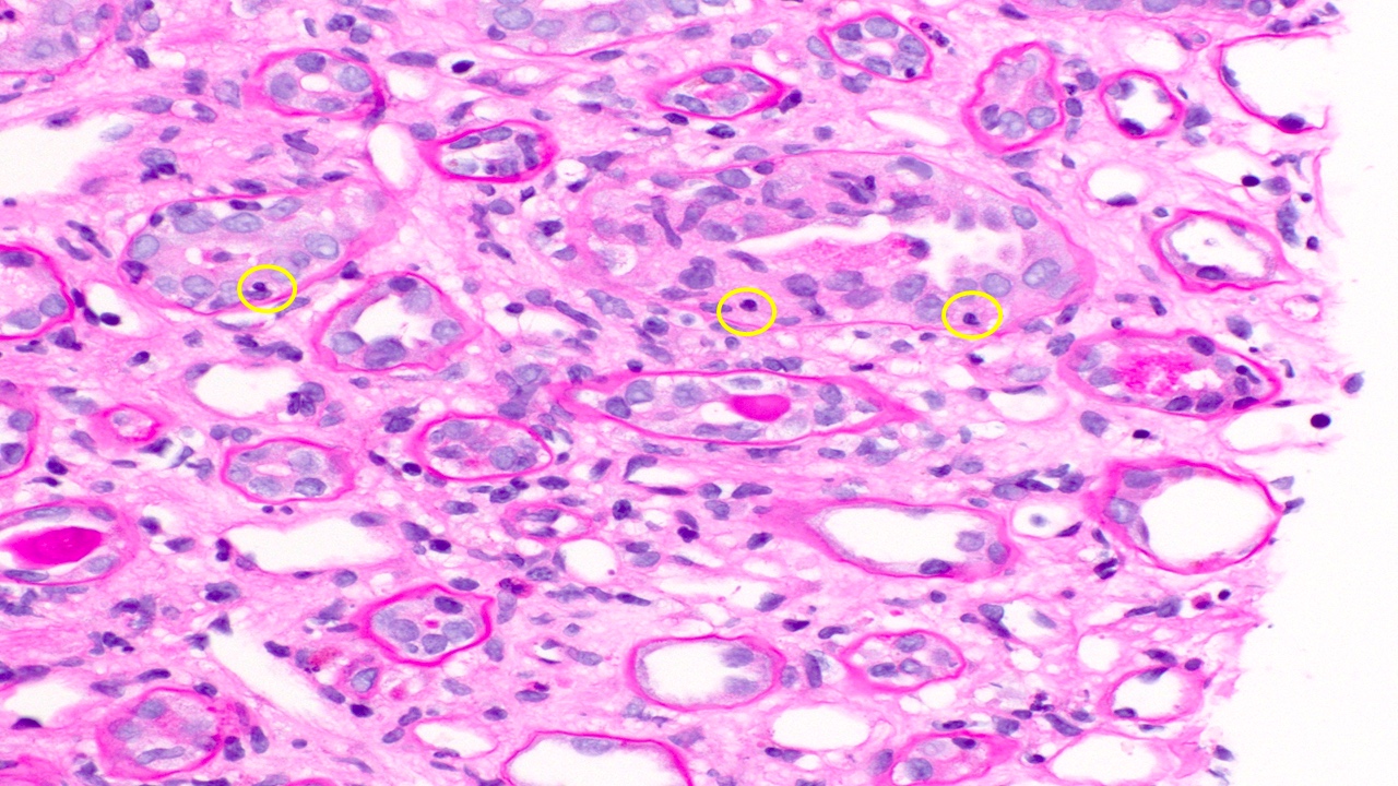
Transplant glomerulopathy
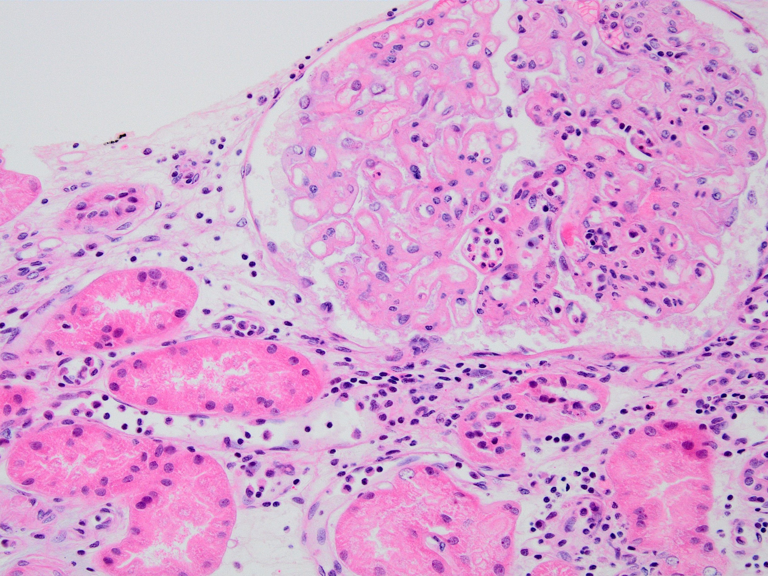
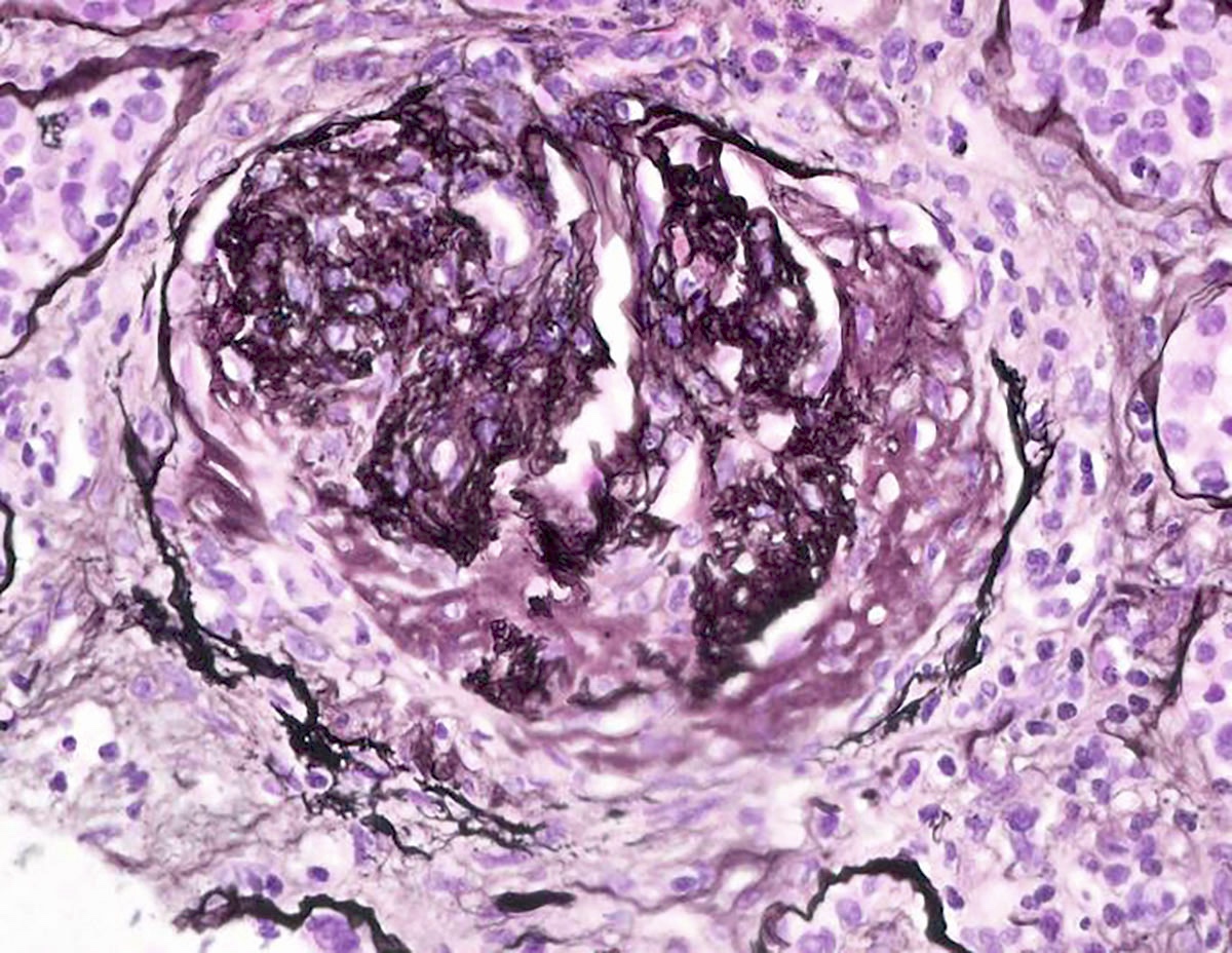
Chronic active ABMR
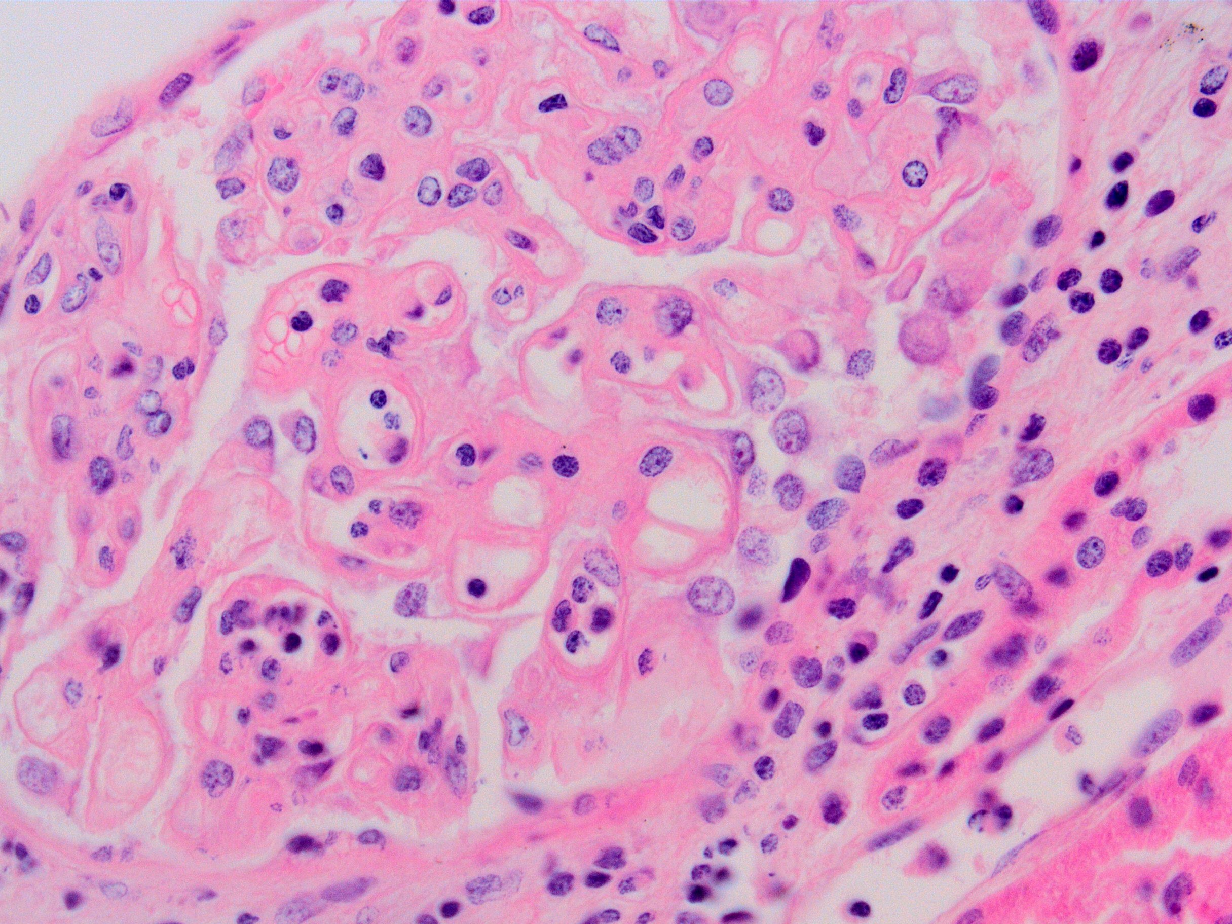
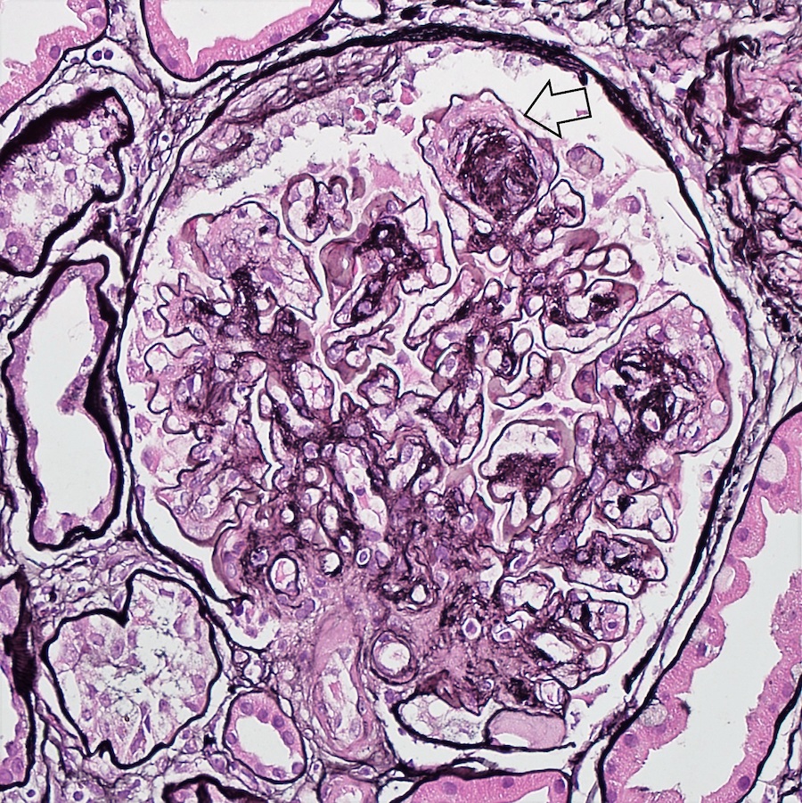
Transplant glomerulopathy and glomerulitis
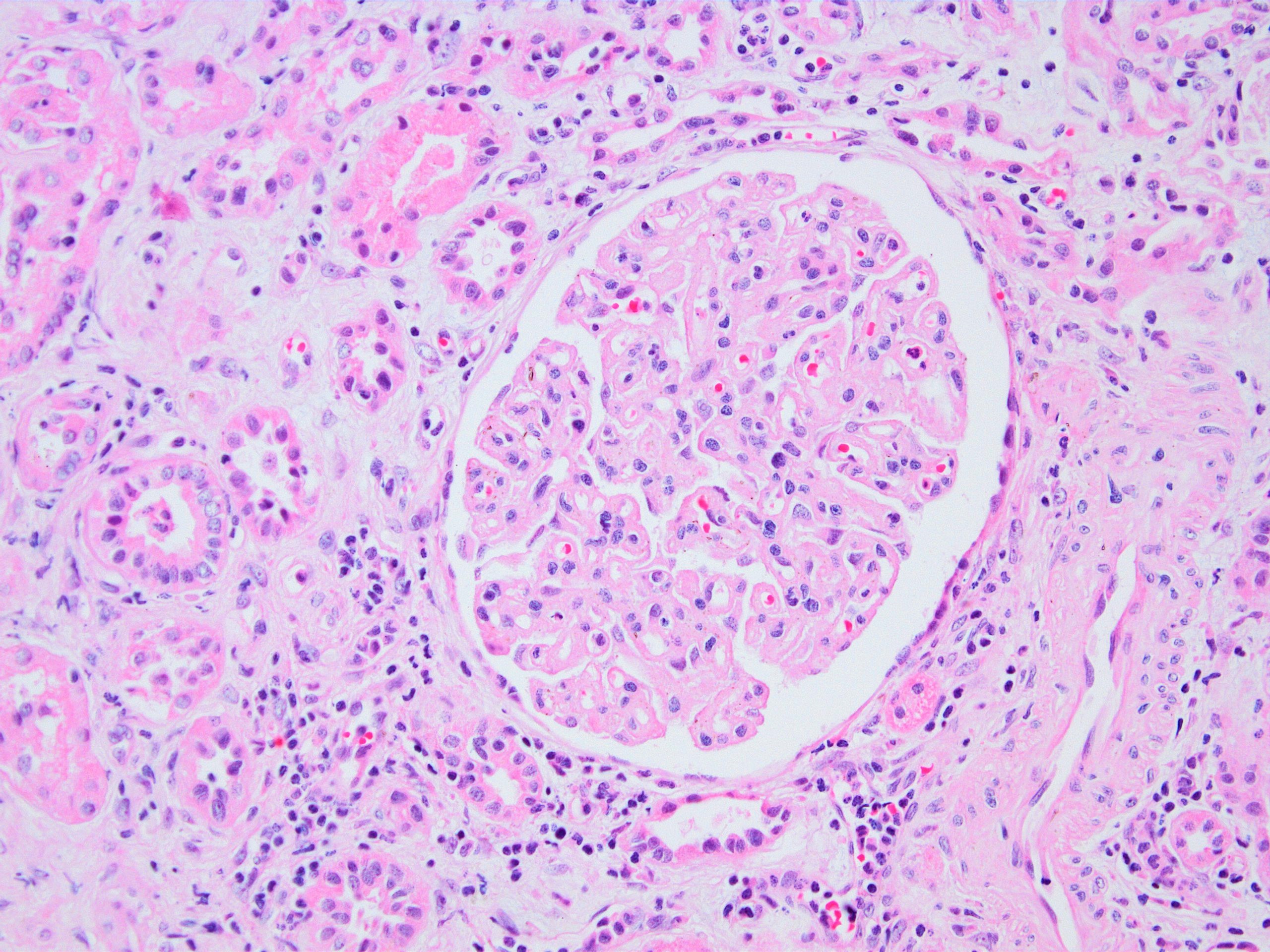
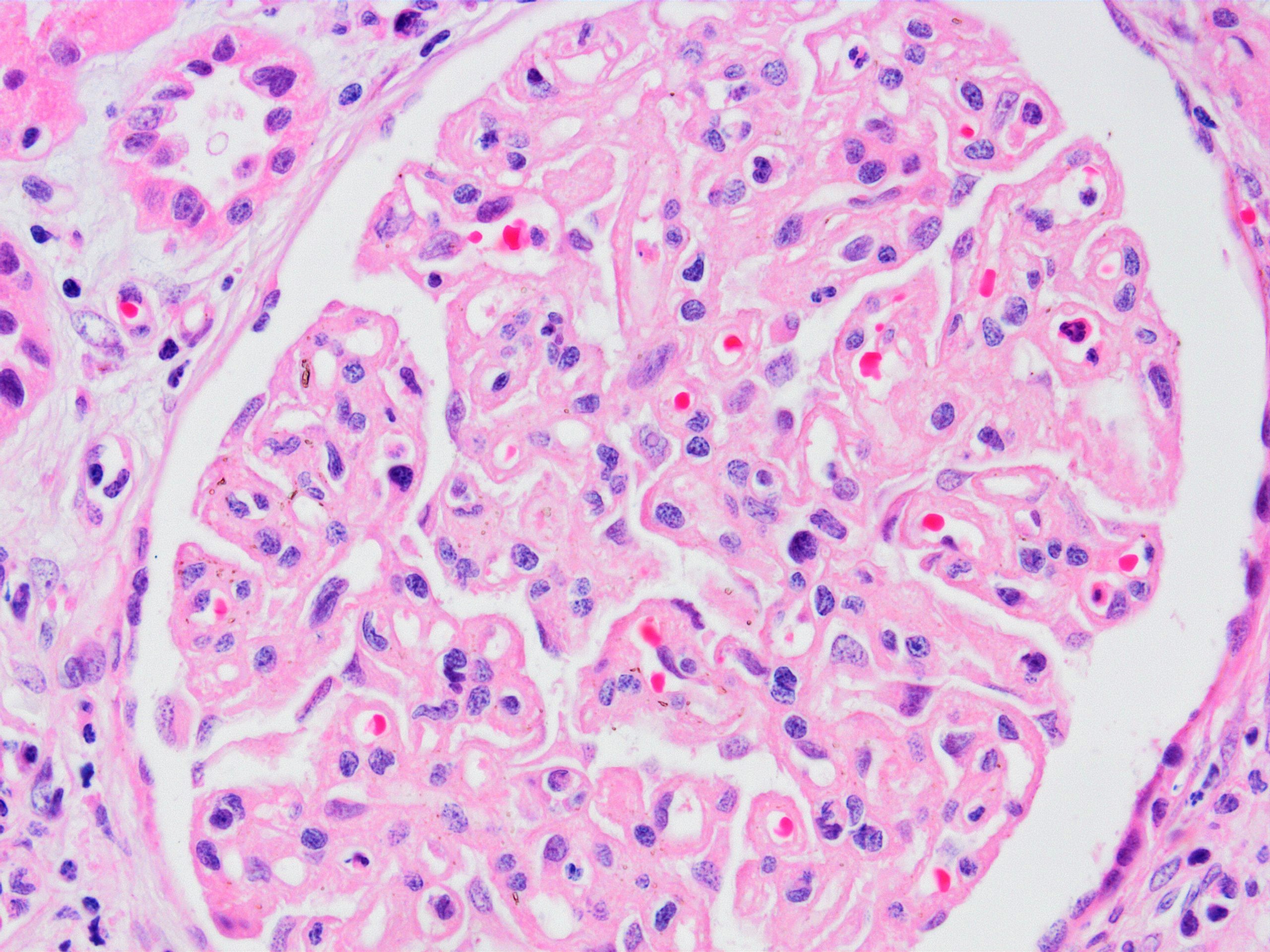
Transplant glomerulopathy
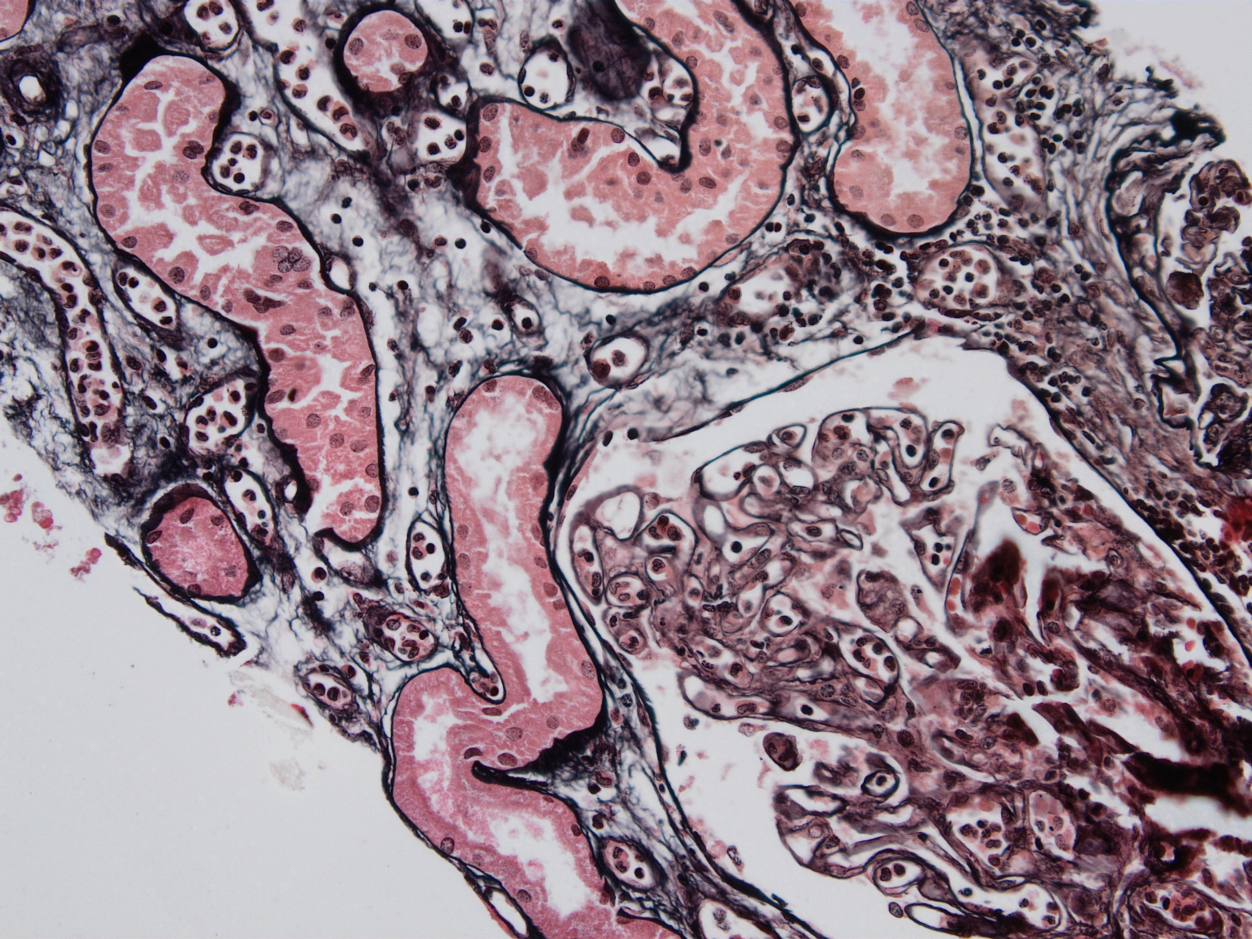
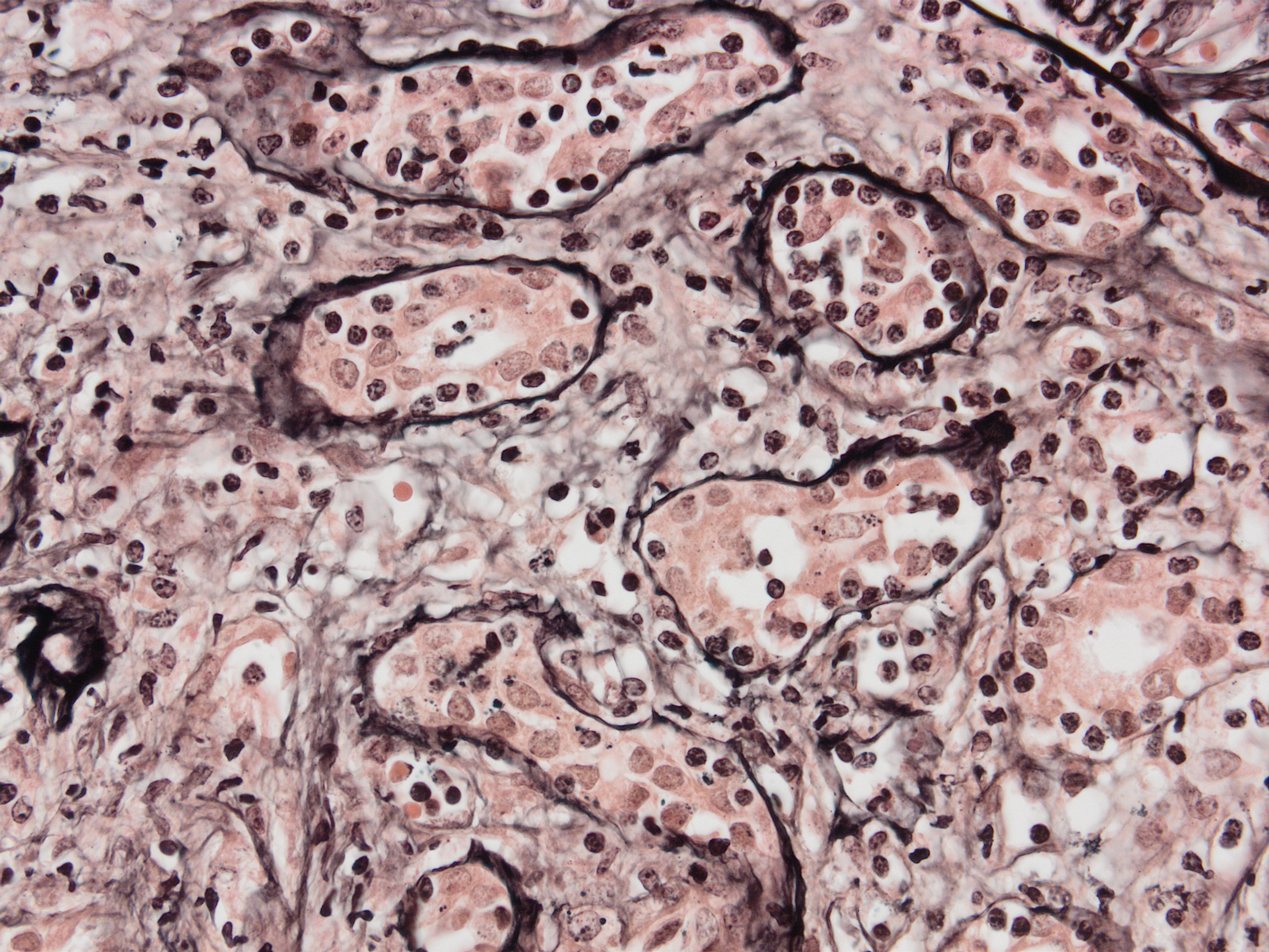
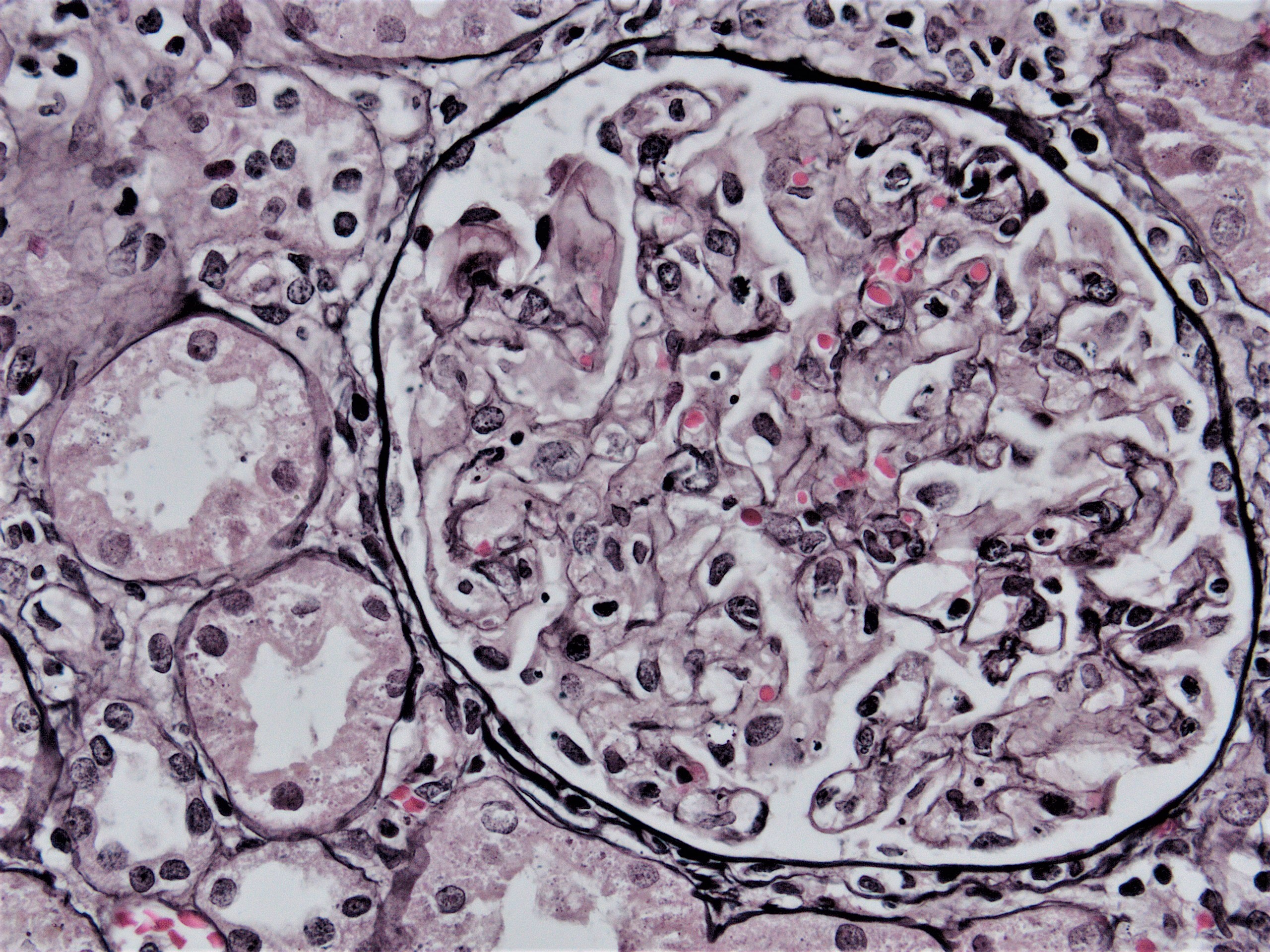
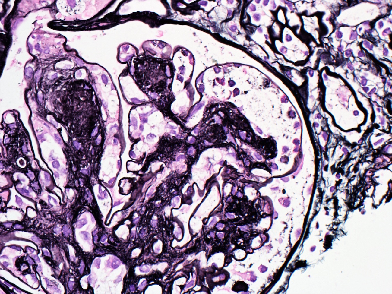
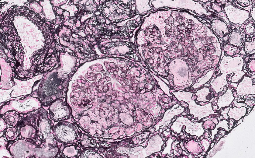
Chronic active ABMR, JMS stain
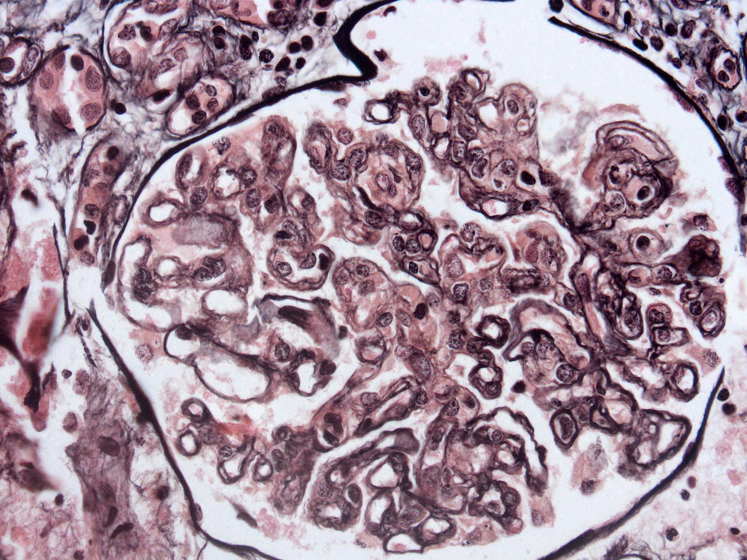
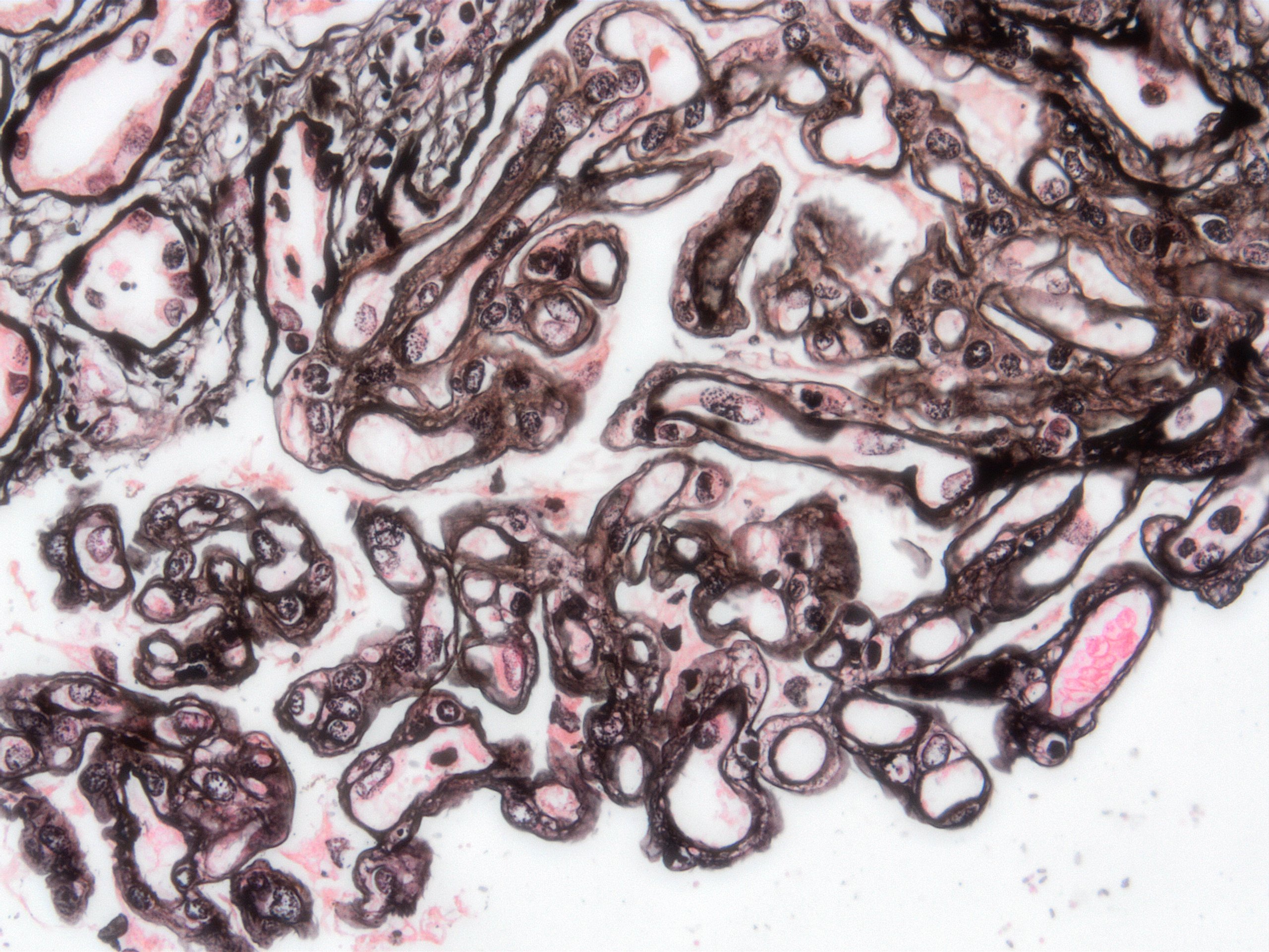
Transplant glomerulopathy, JMS stain
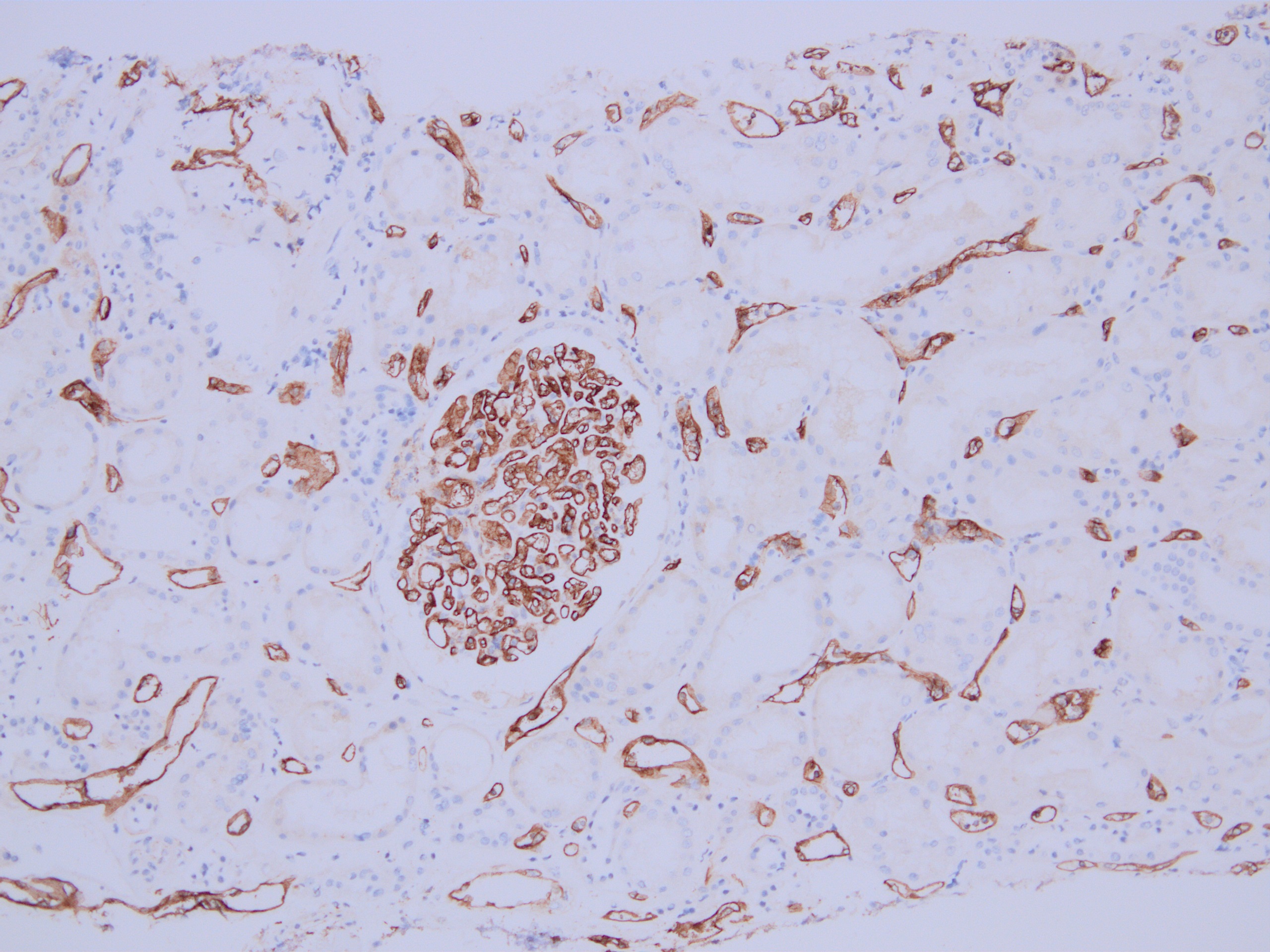
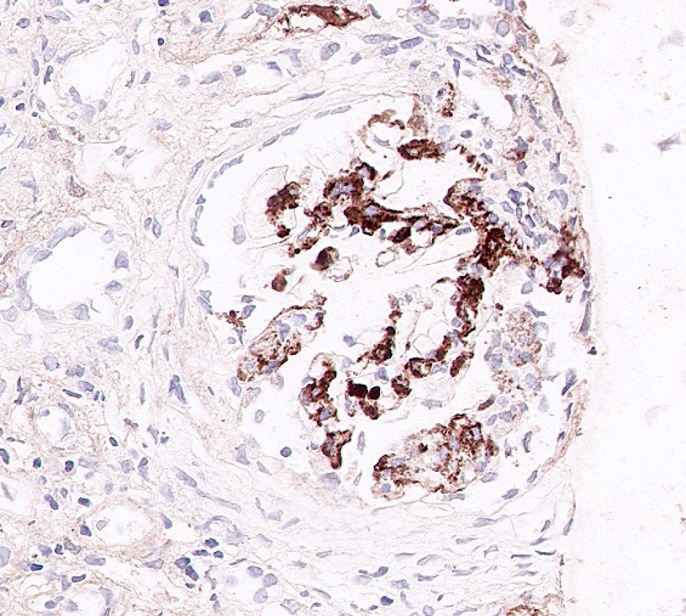
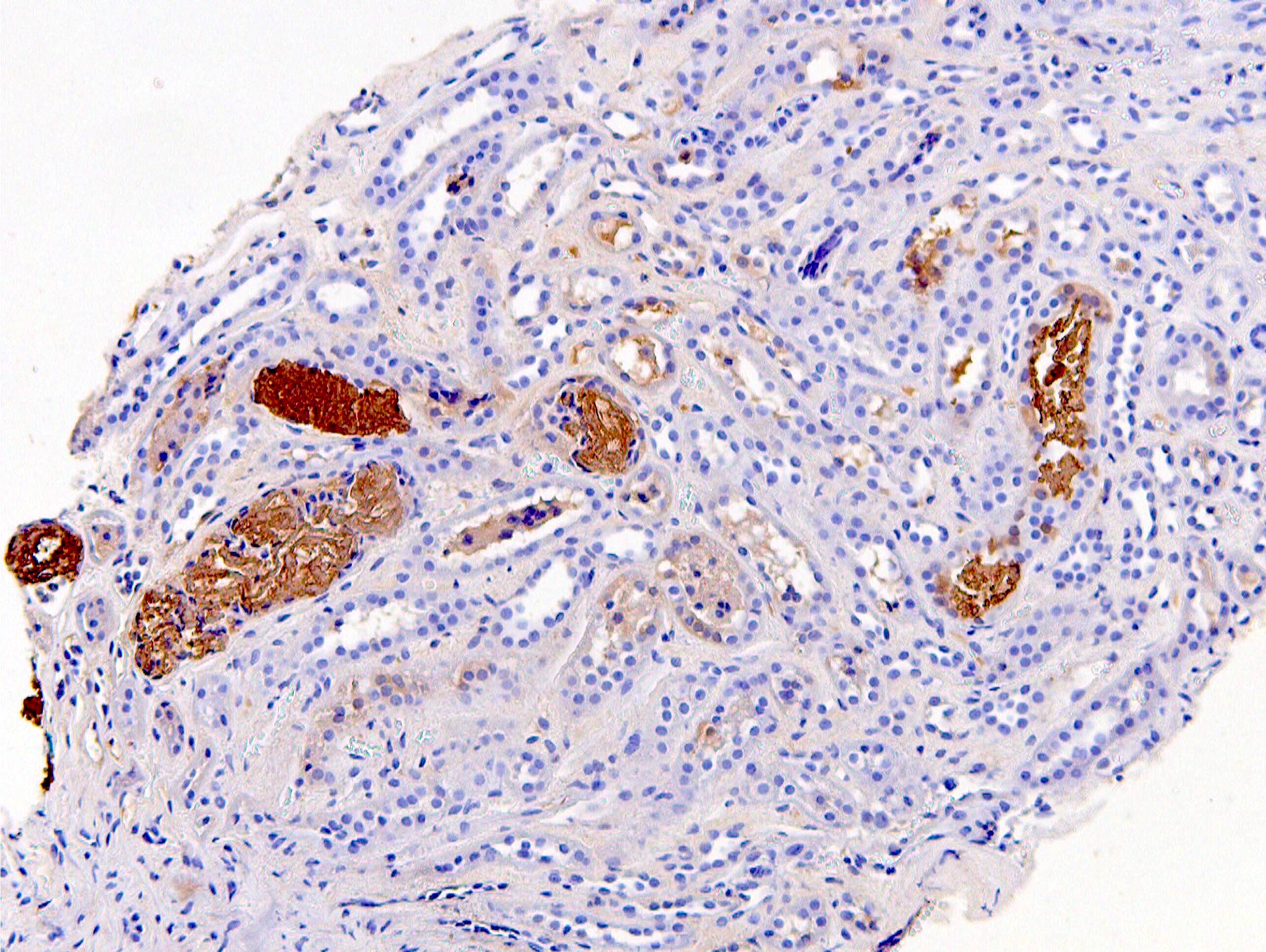
C4d staining, IHC
Contributed by Arzu Sağlam, M.D.
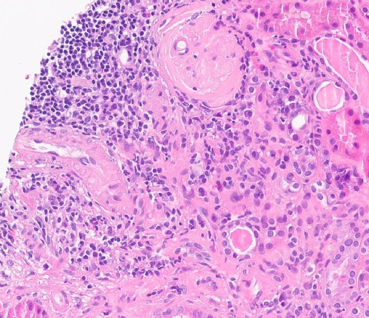
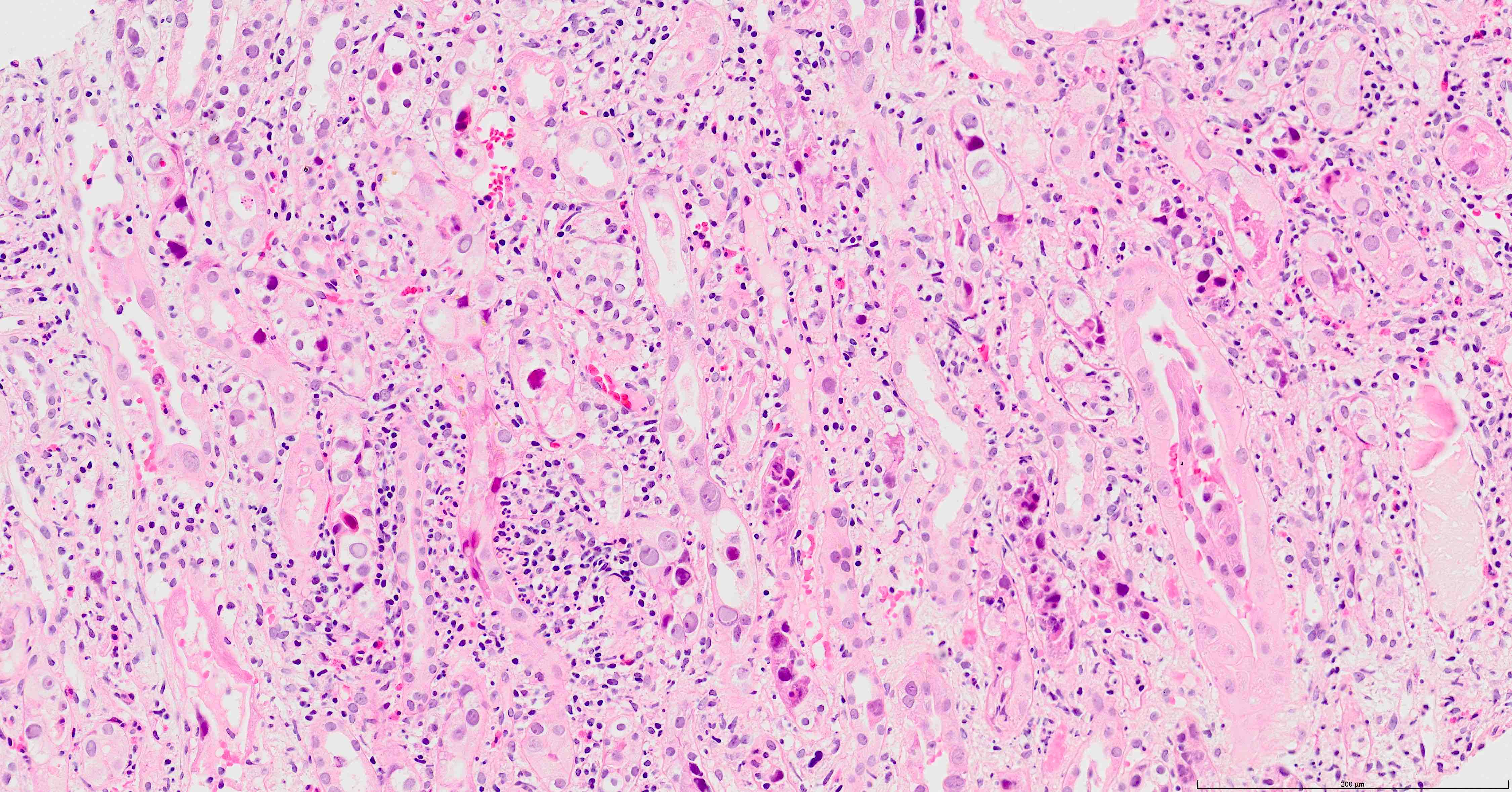
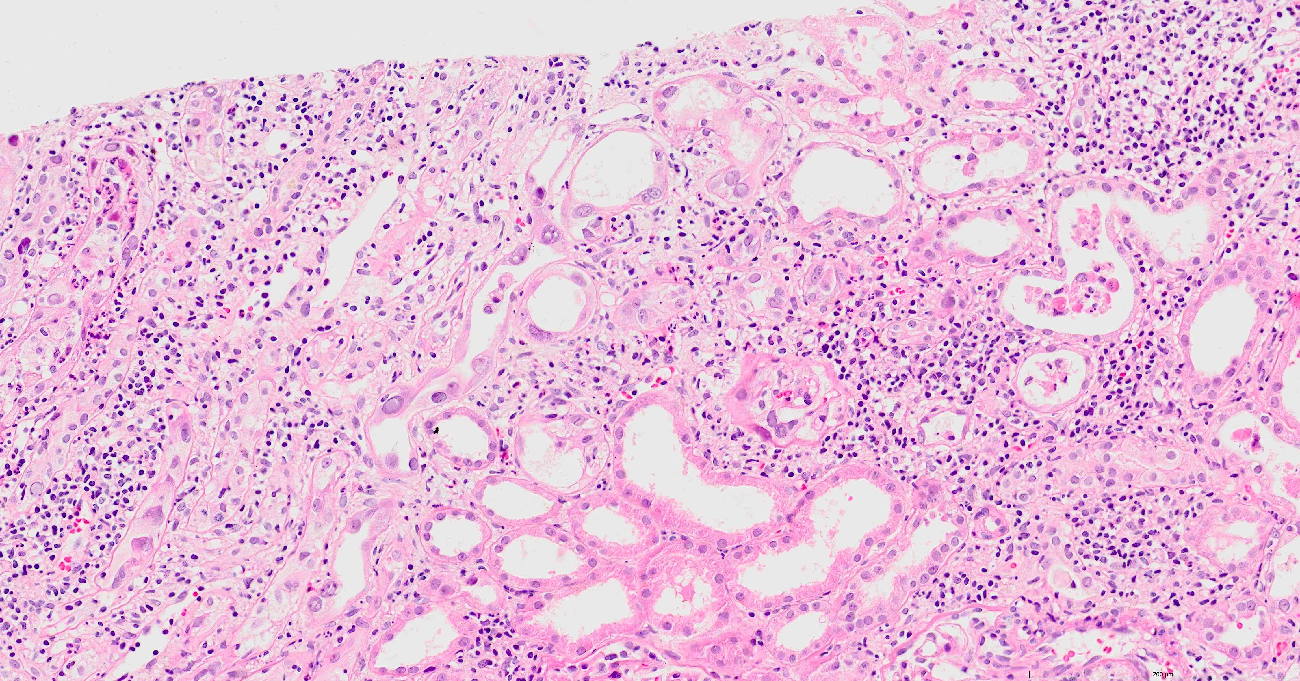
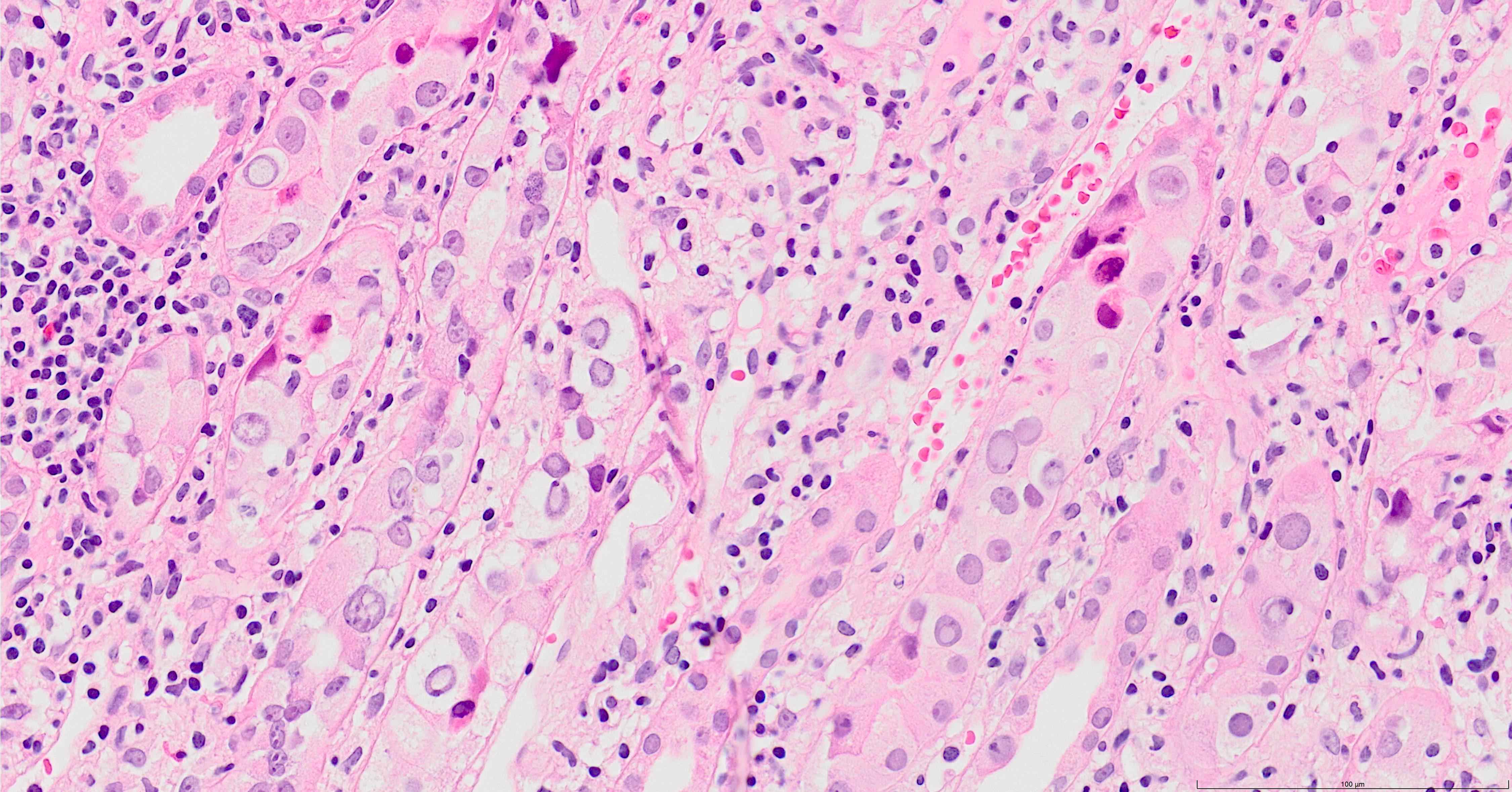
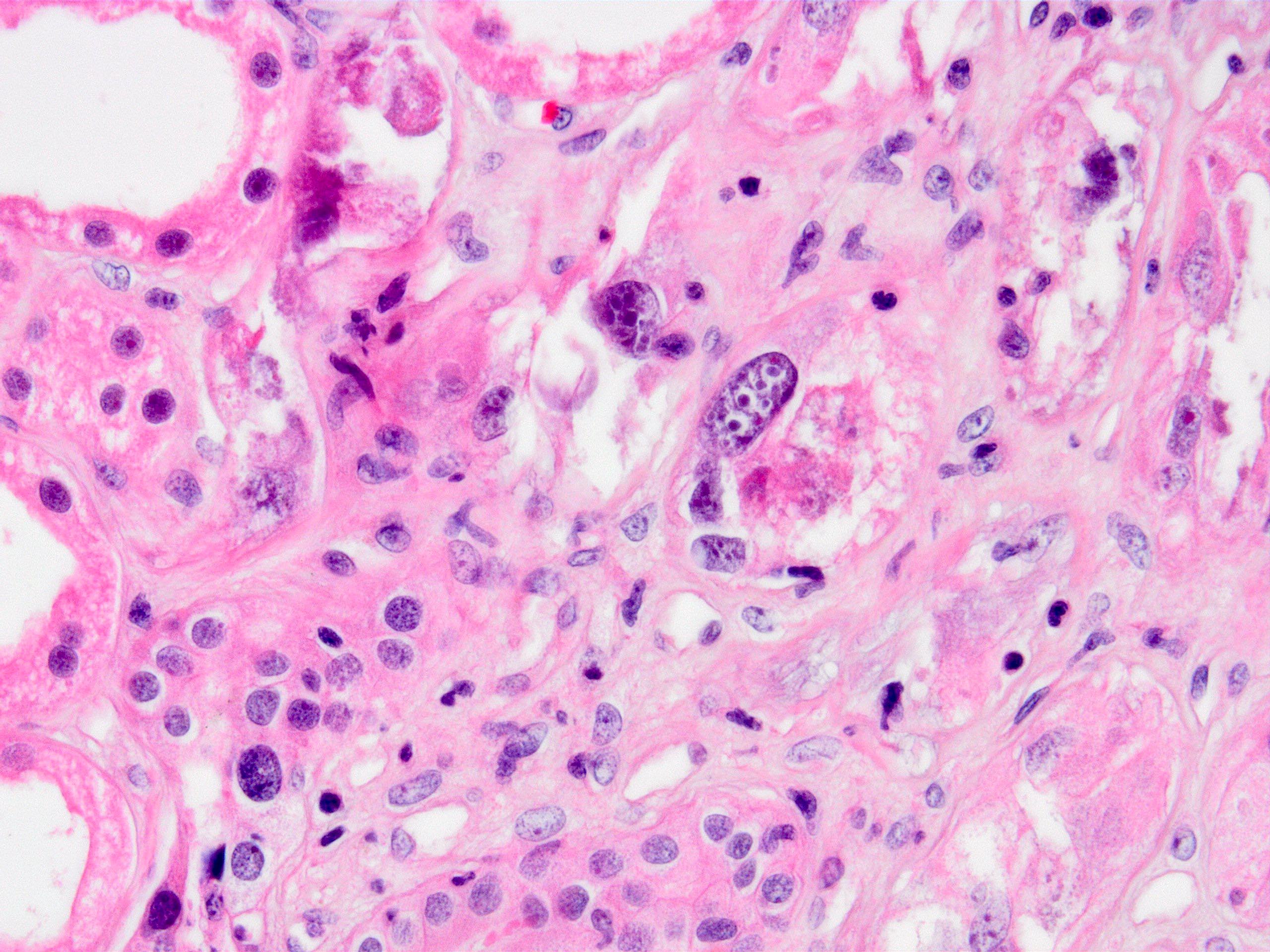
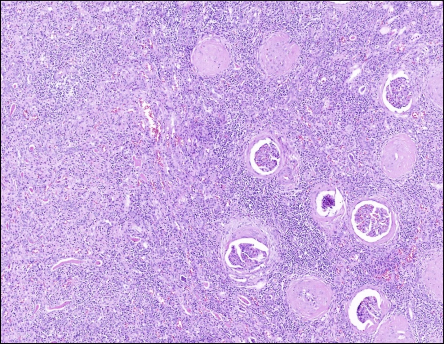
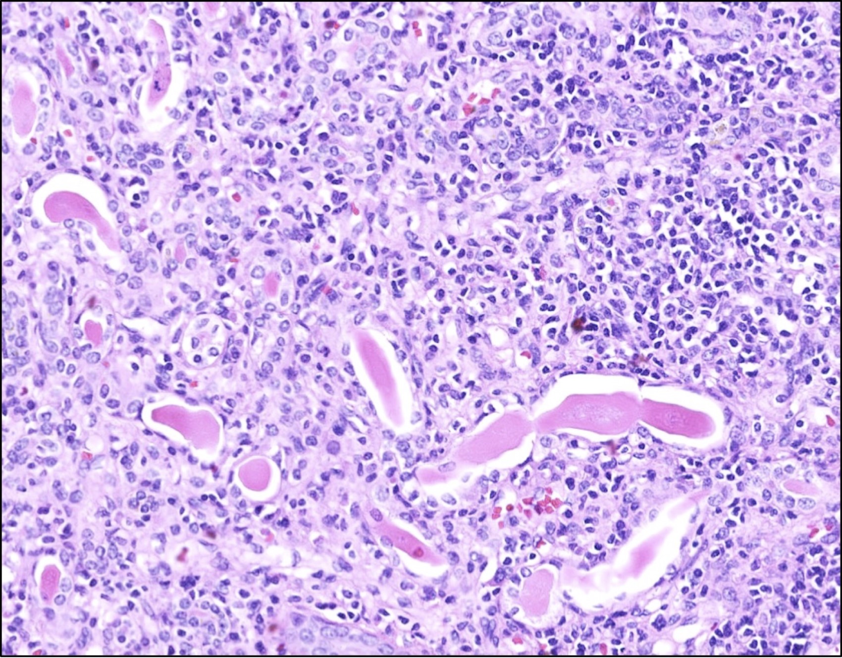
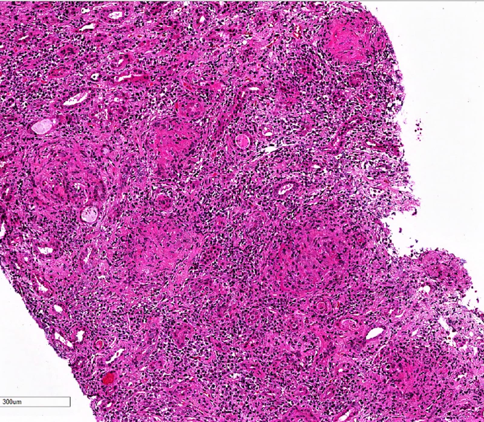
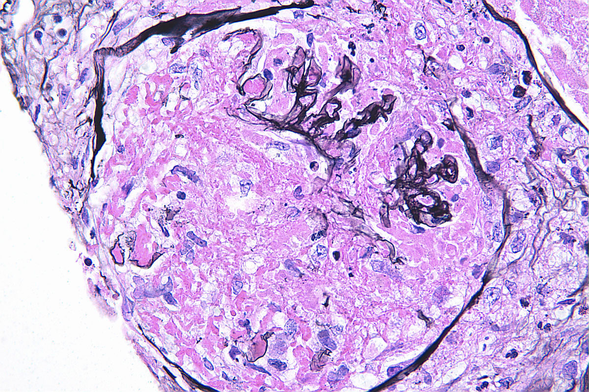
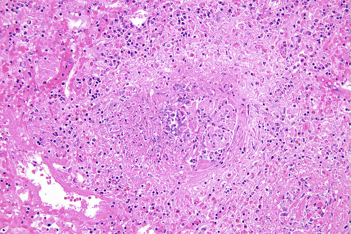
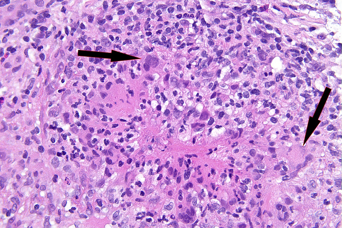
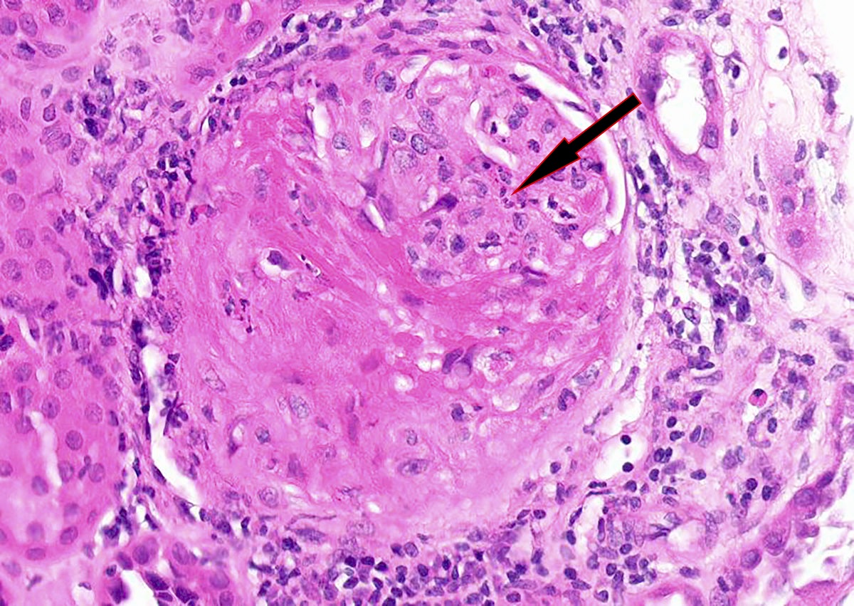
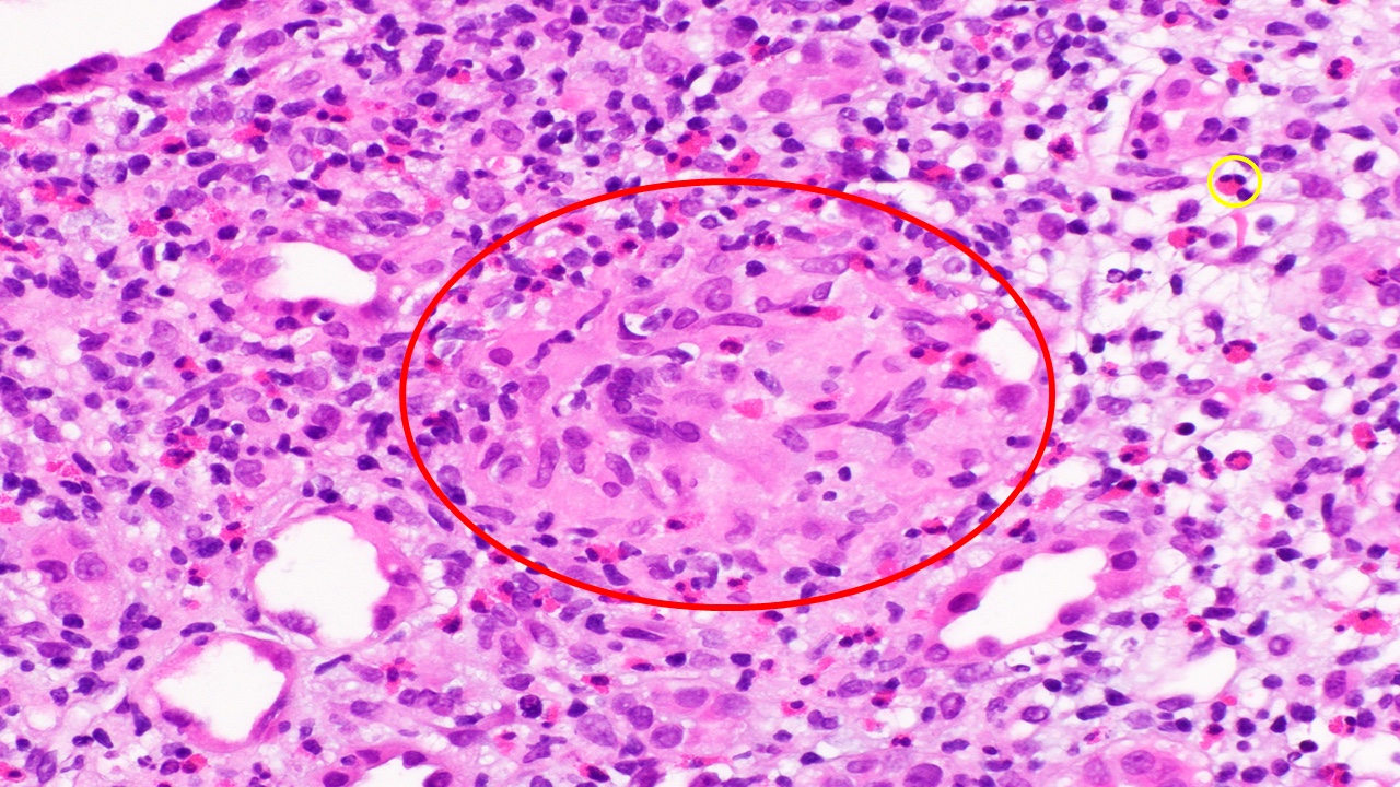
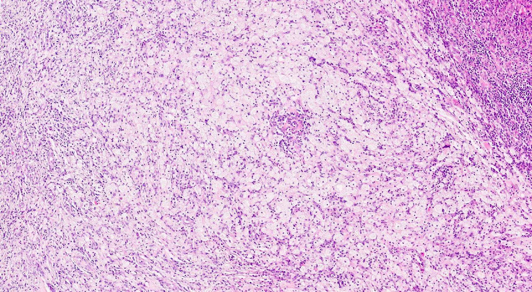
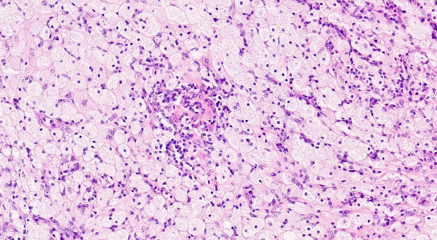
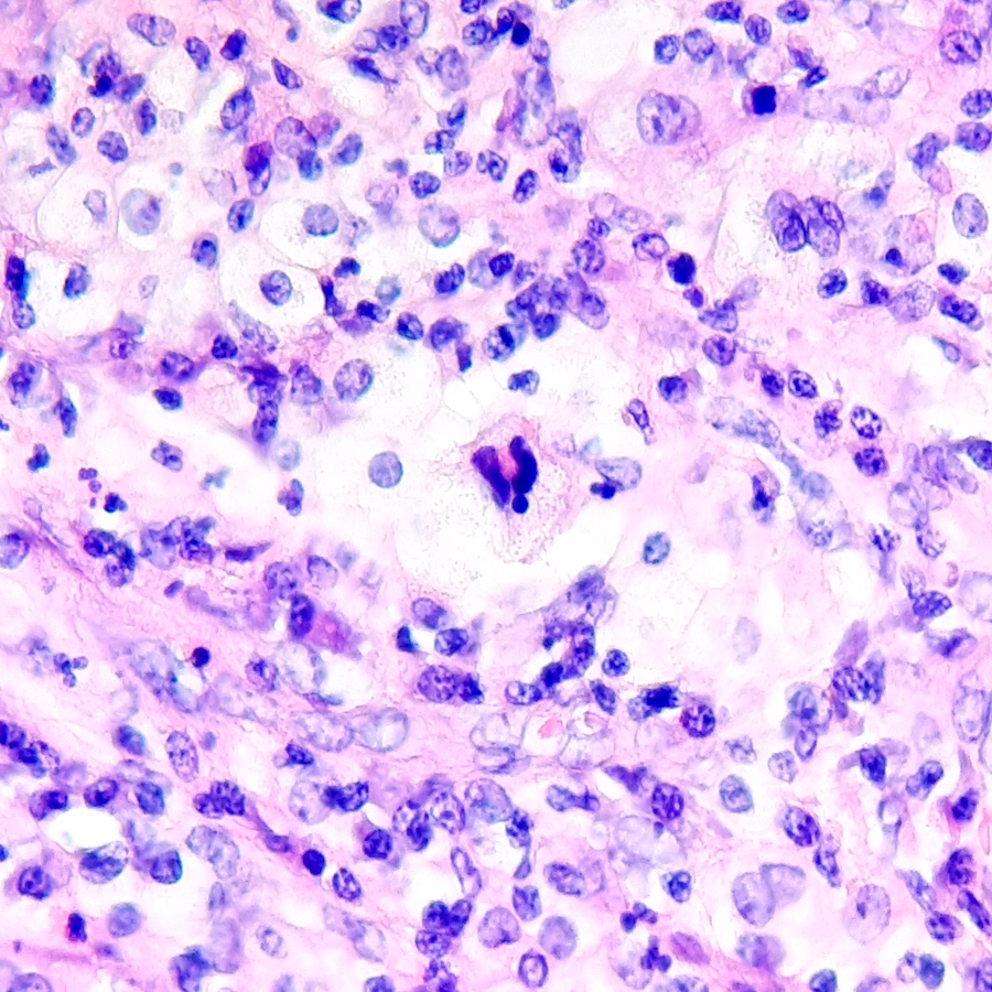
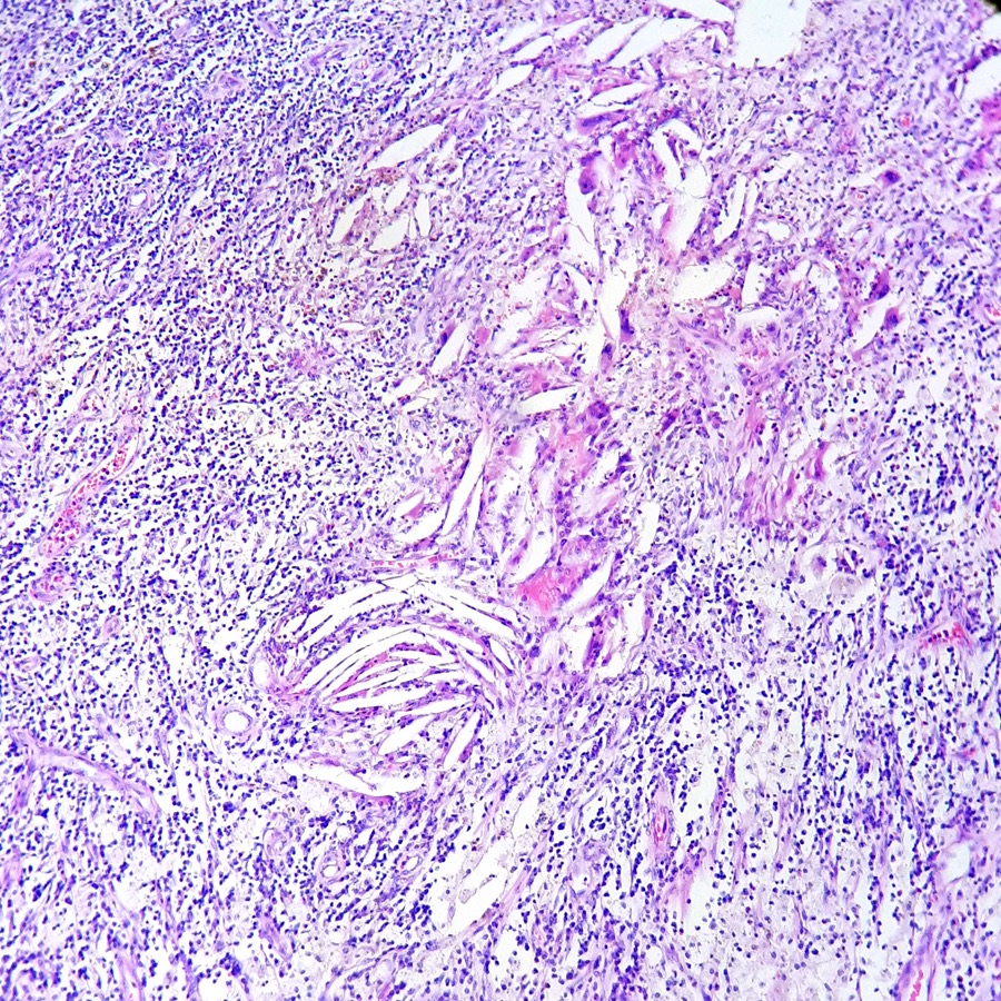
Chronic active TCMR, allograft nephrectomy
Contributed by Arzu Sağlam, M.D.

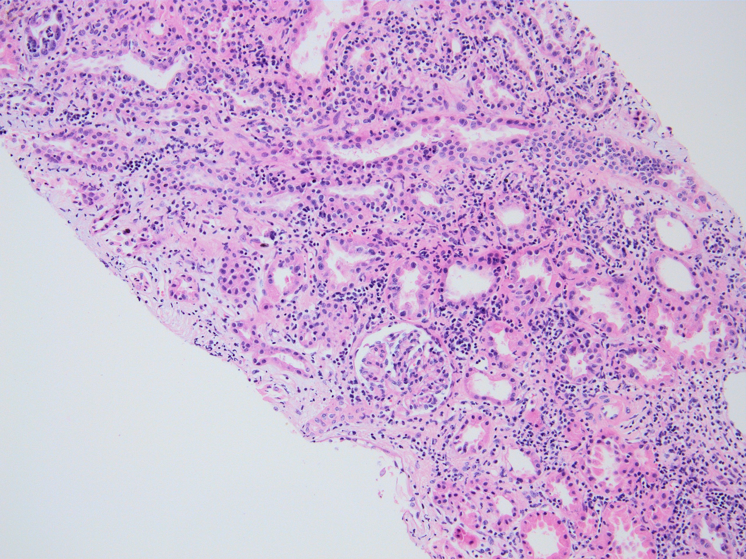
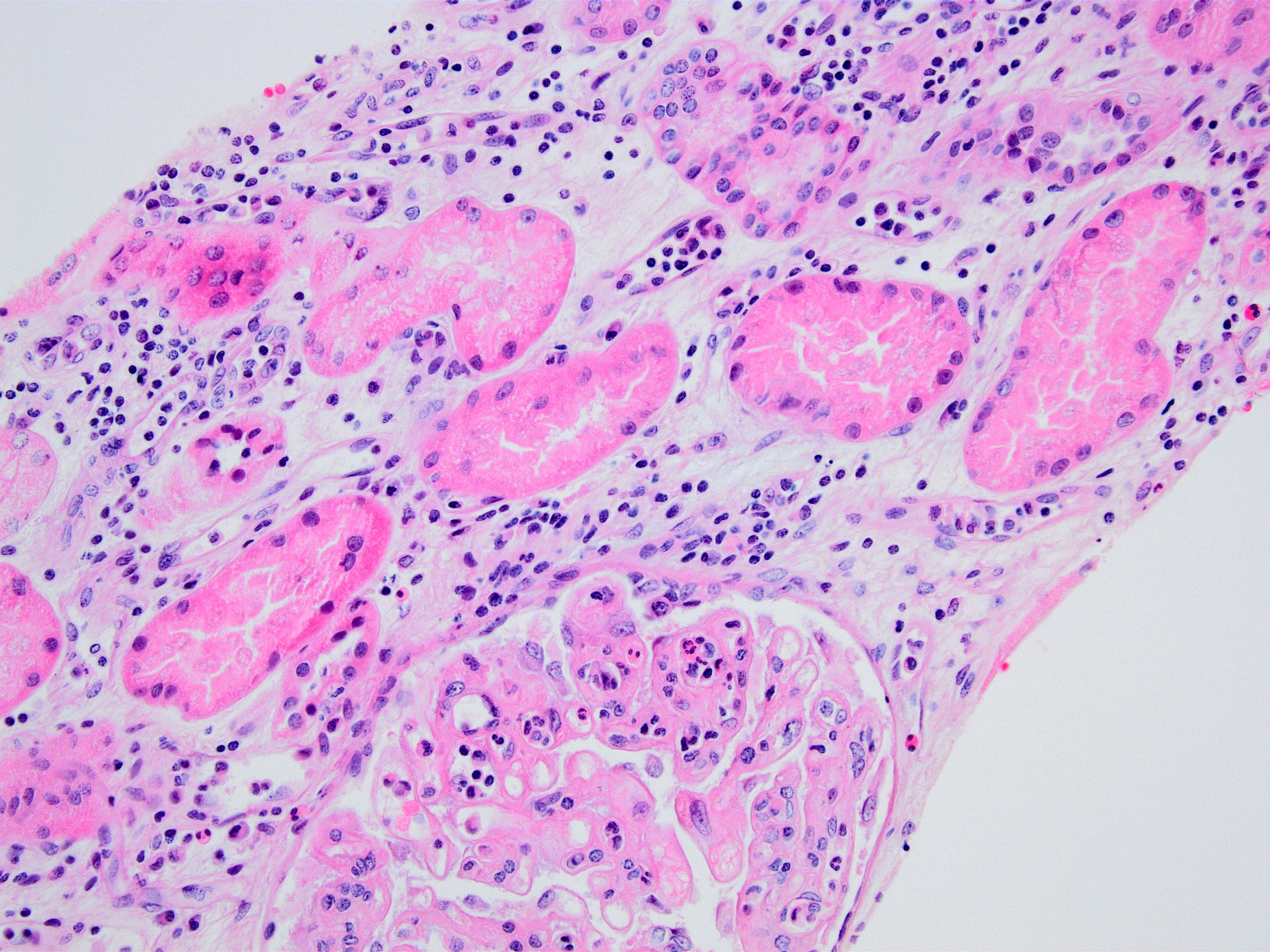
Interstitial inflammation
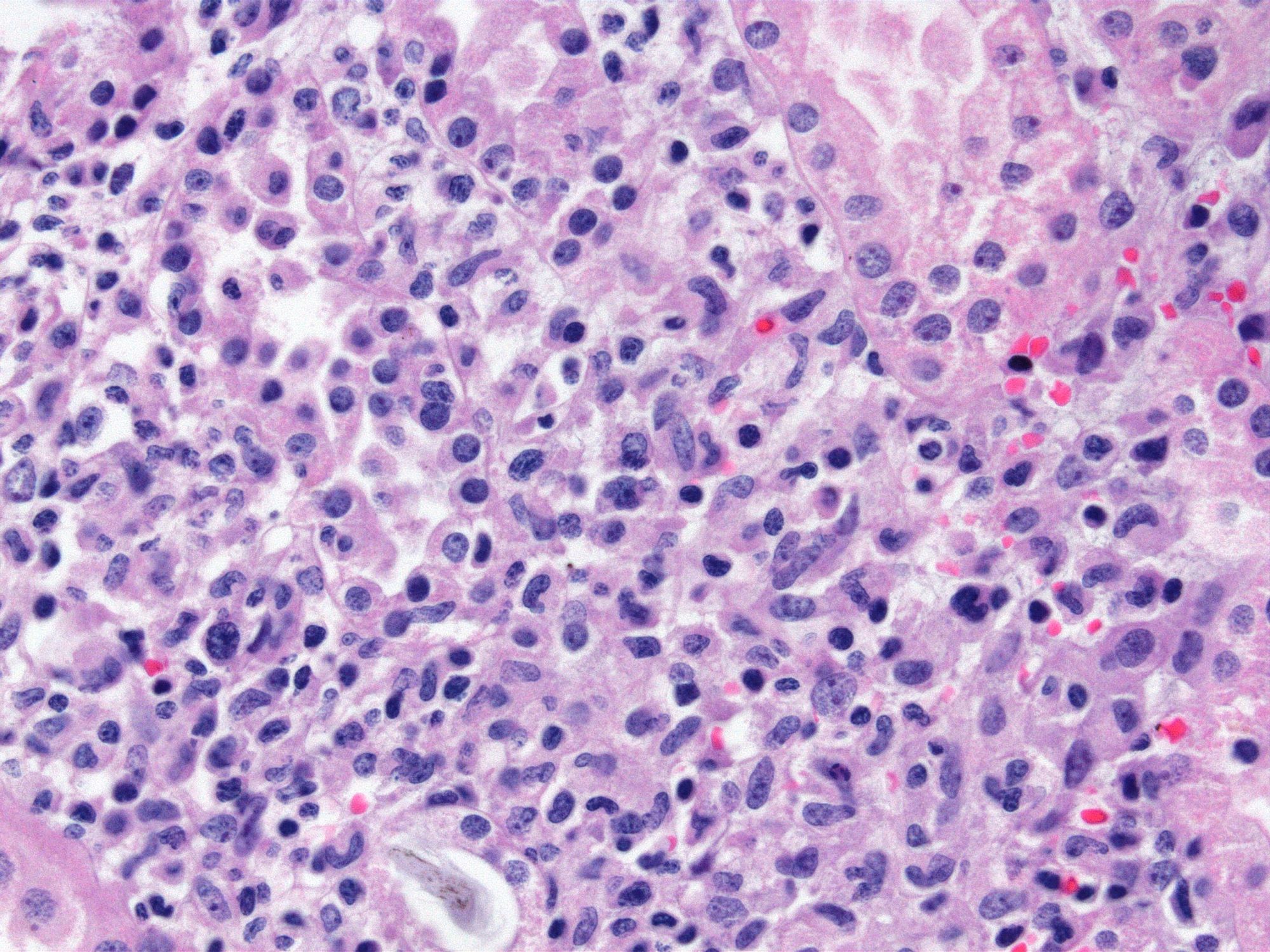
Interstitial inflammation - macrophage predominant
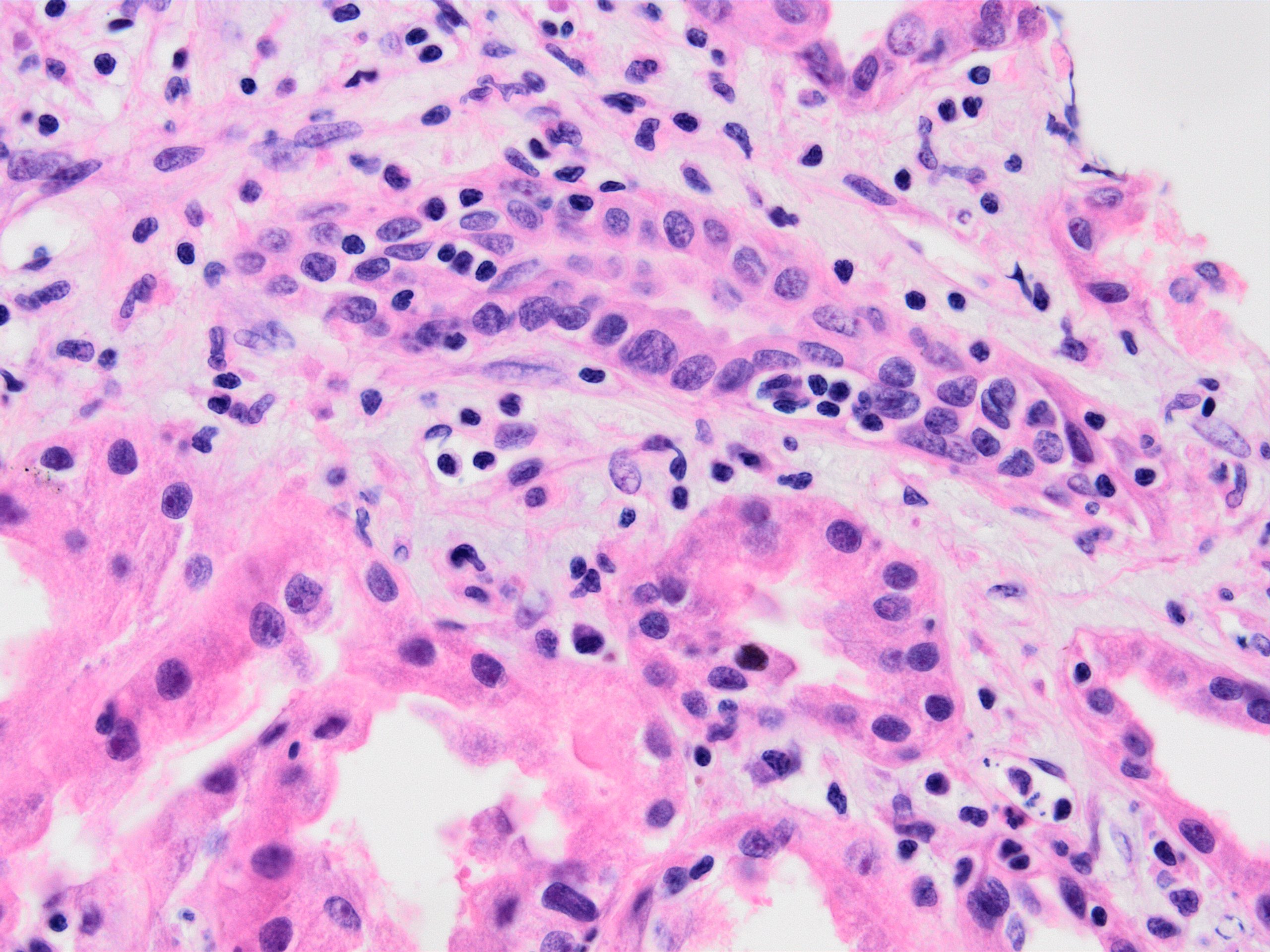
Tubulitis
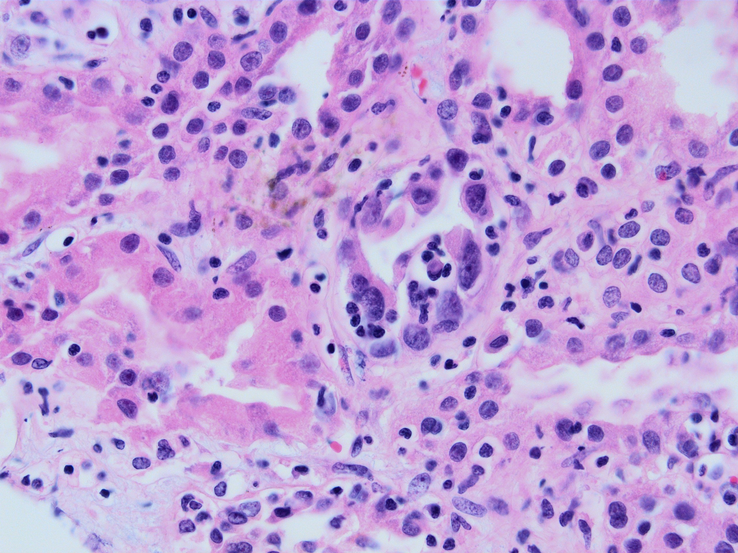
Severe tubulitis
Tubulitis, PAS stain

Tubulitis, JMS stain
Intimal edema and lymphocyte margination
Interstitial hemorrhage
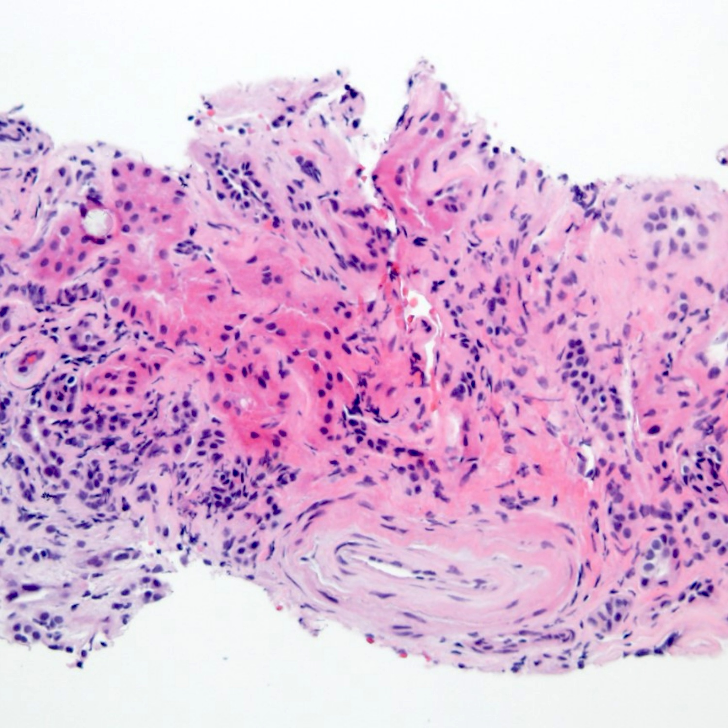
Intimal arteritis

Intimal arteritis, JMS stain
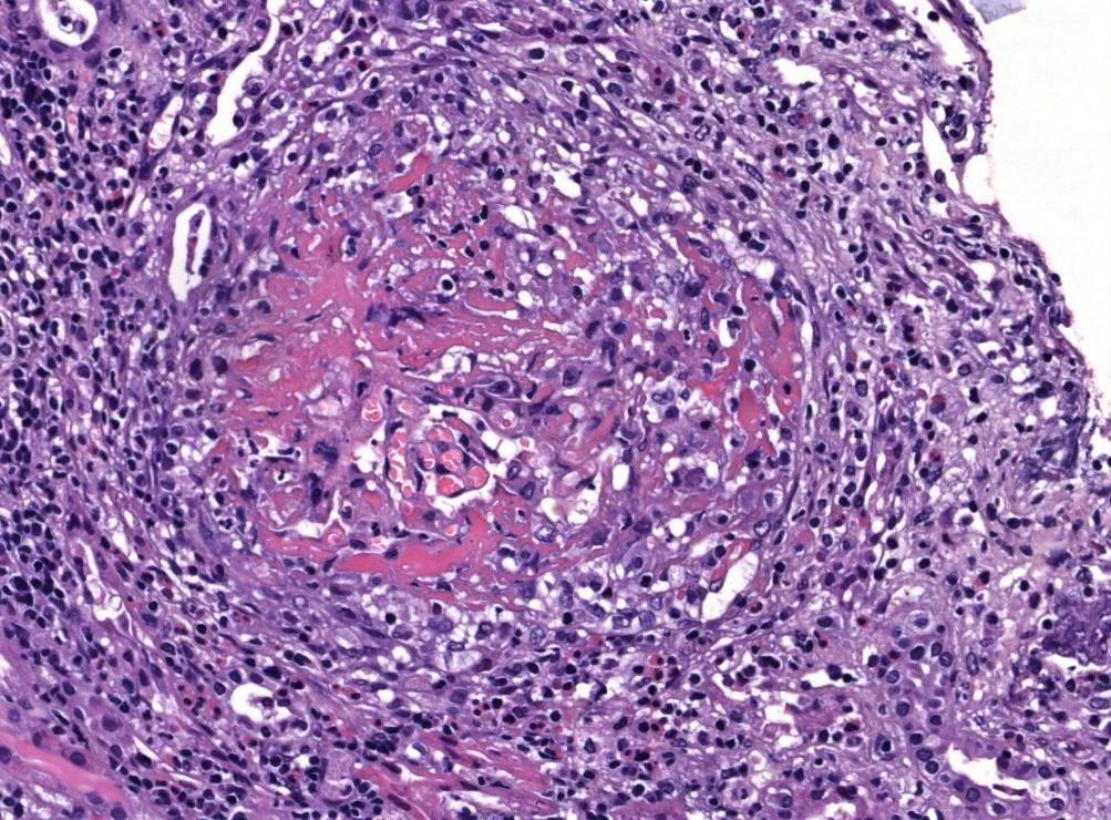
Transmural arteritis
Transmural arteritis and fibrinoid necrosis
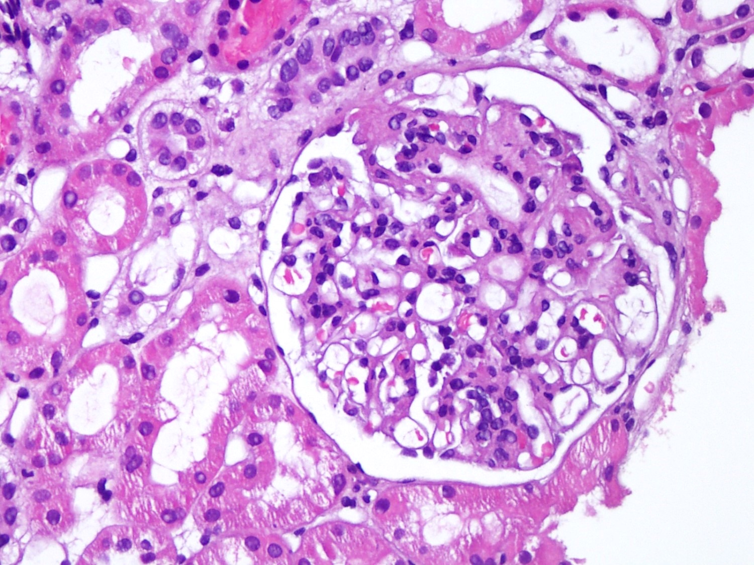
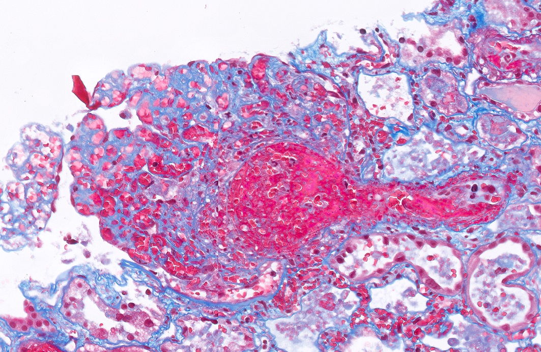
Acute allograft glomerulopathy
Acute allograft glomerulopathy JMS stain
Severe acute allograft glomerulopathy

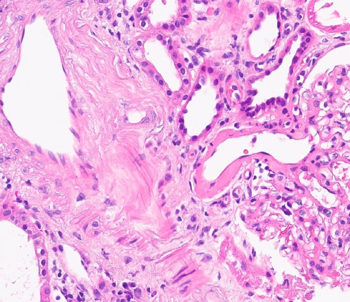
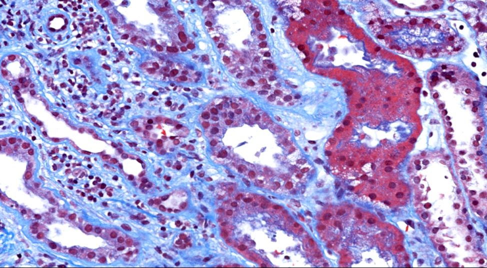
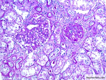
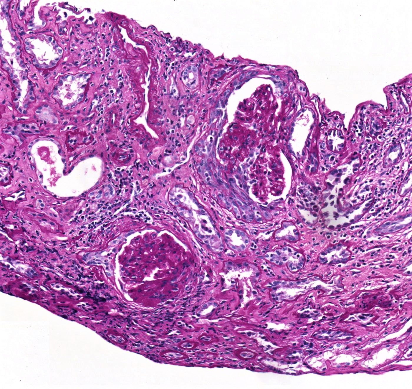
i-IFTA
Tubulitis of atrophic tubuli
Tubulitis of atrophic tubuli, JMS stain

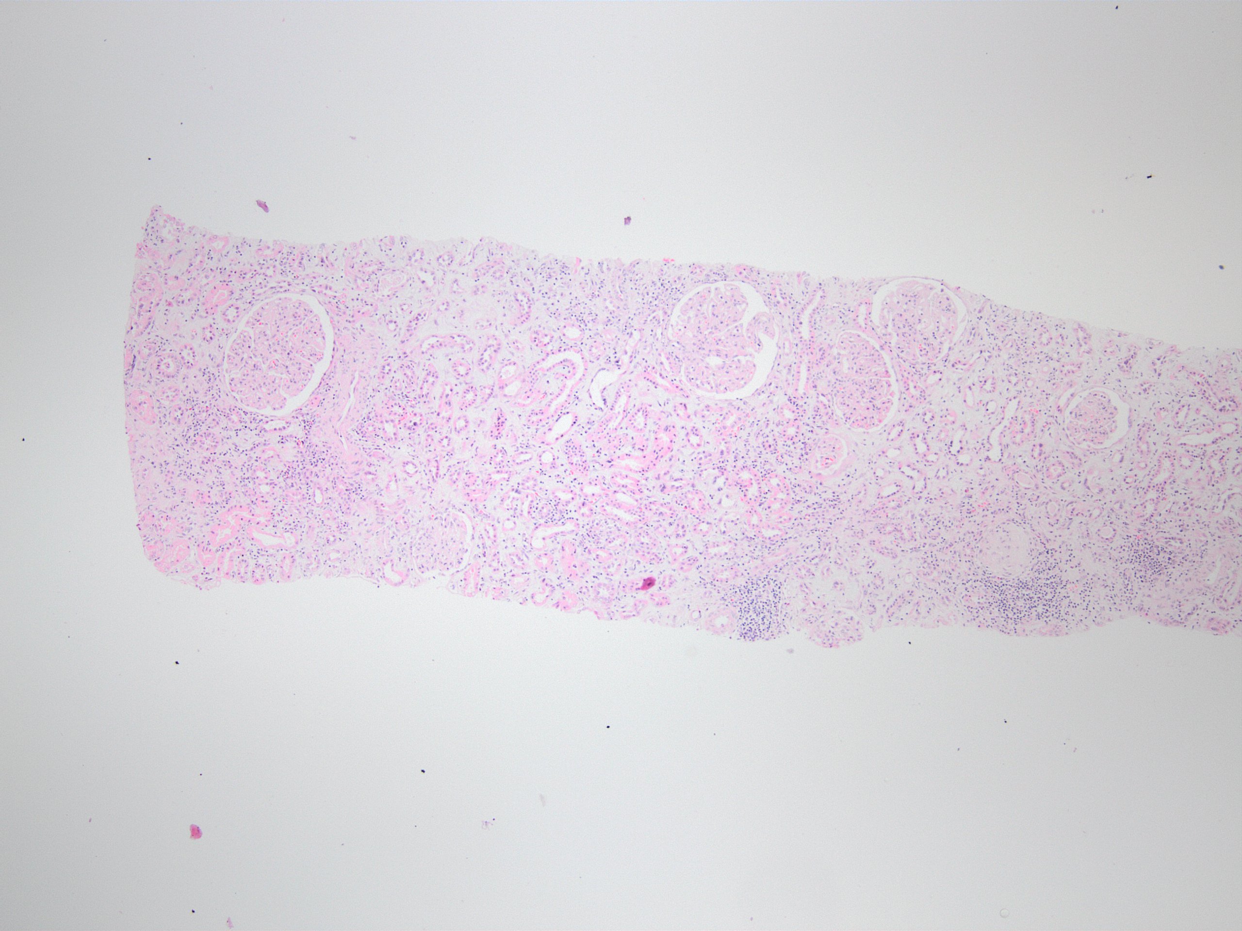
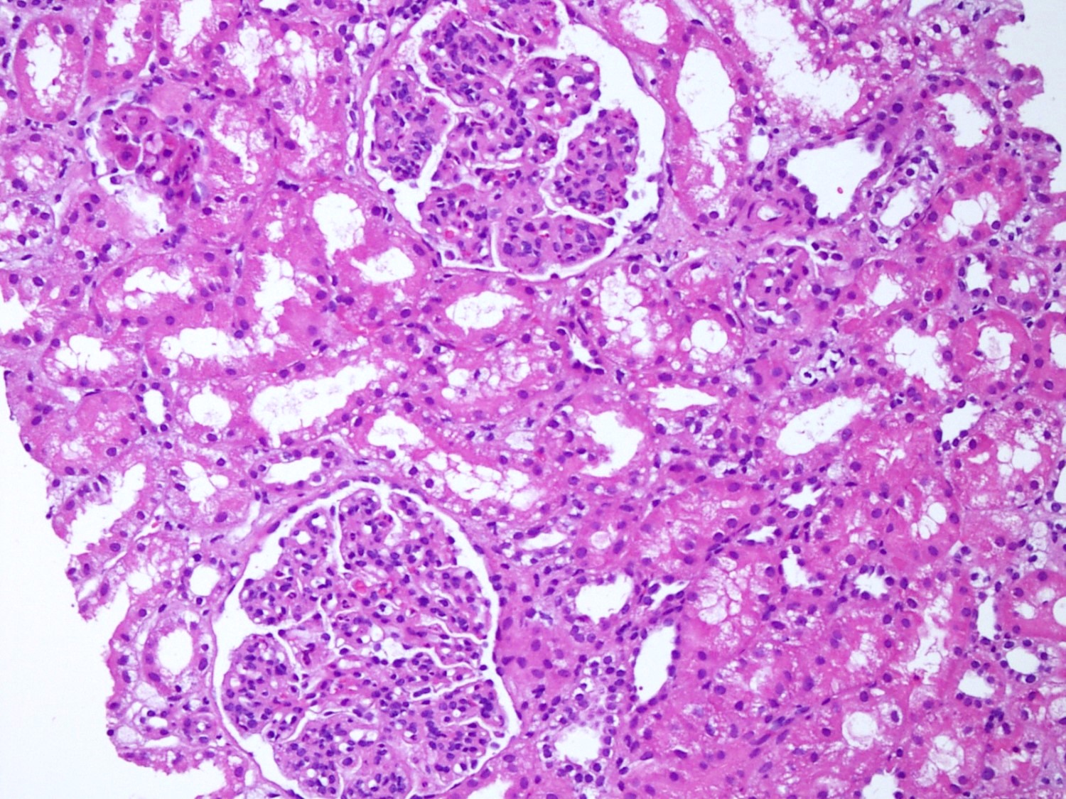
Preserved renal parenchyma
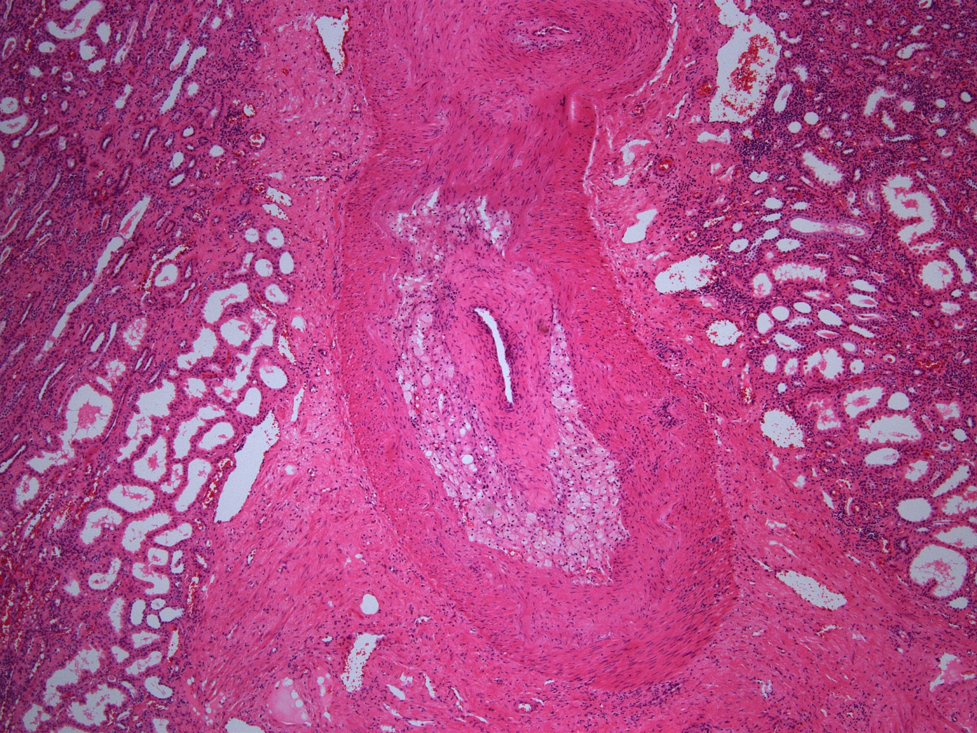
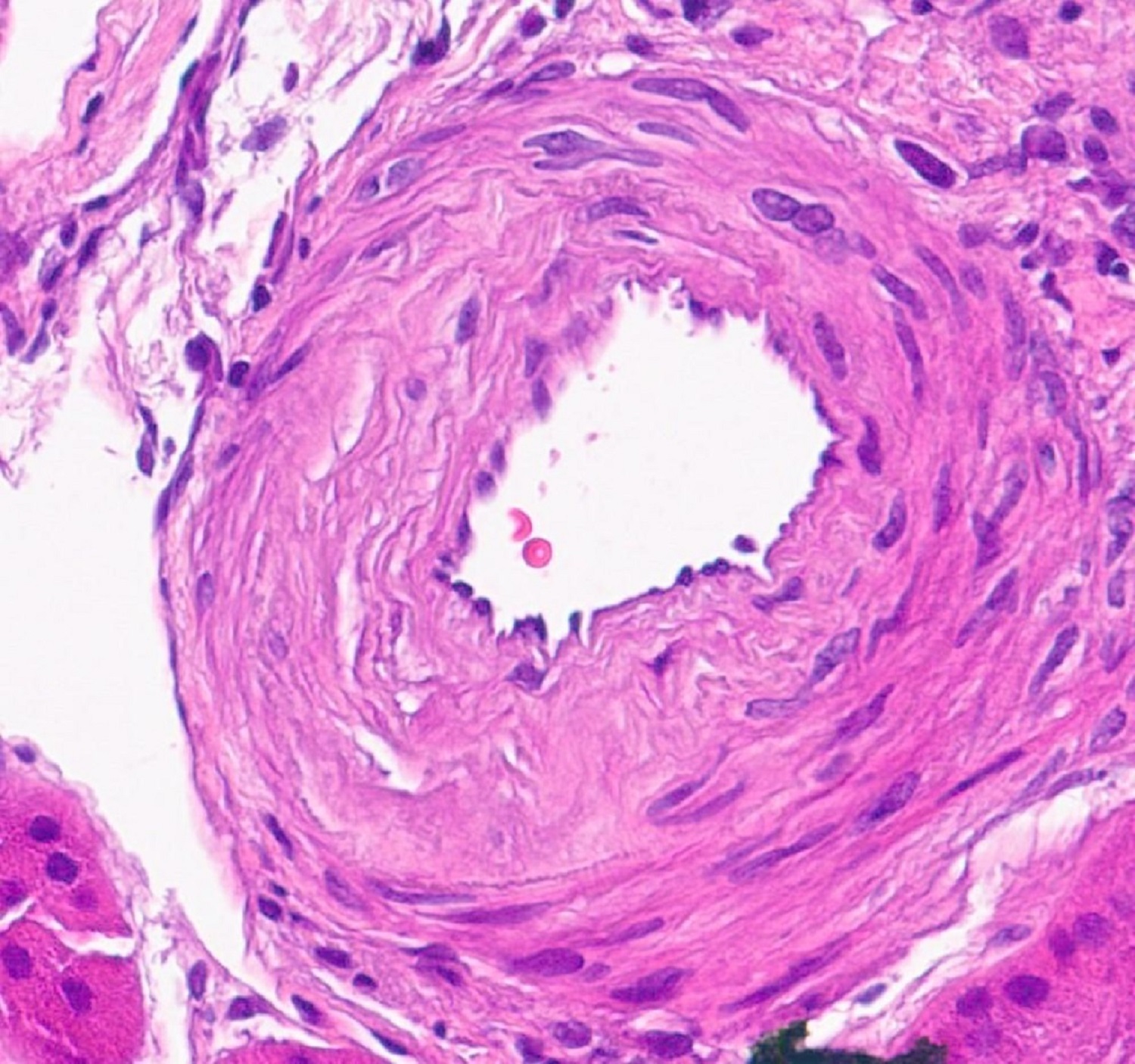
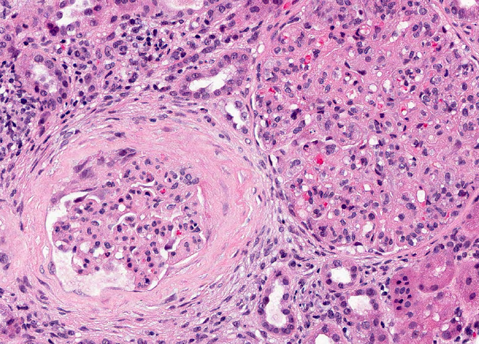
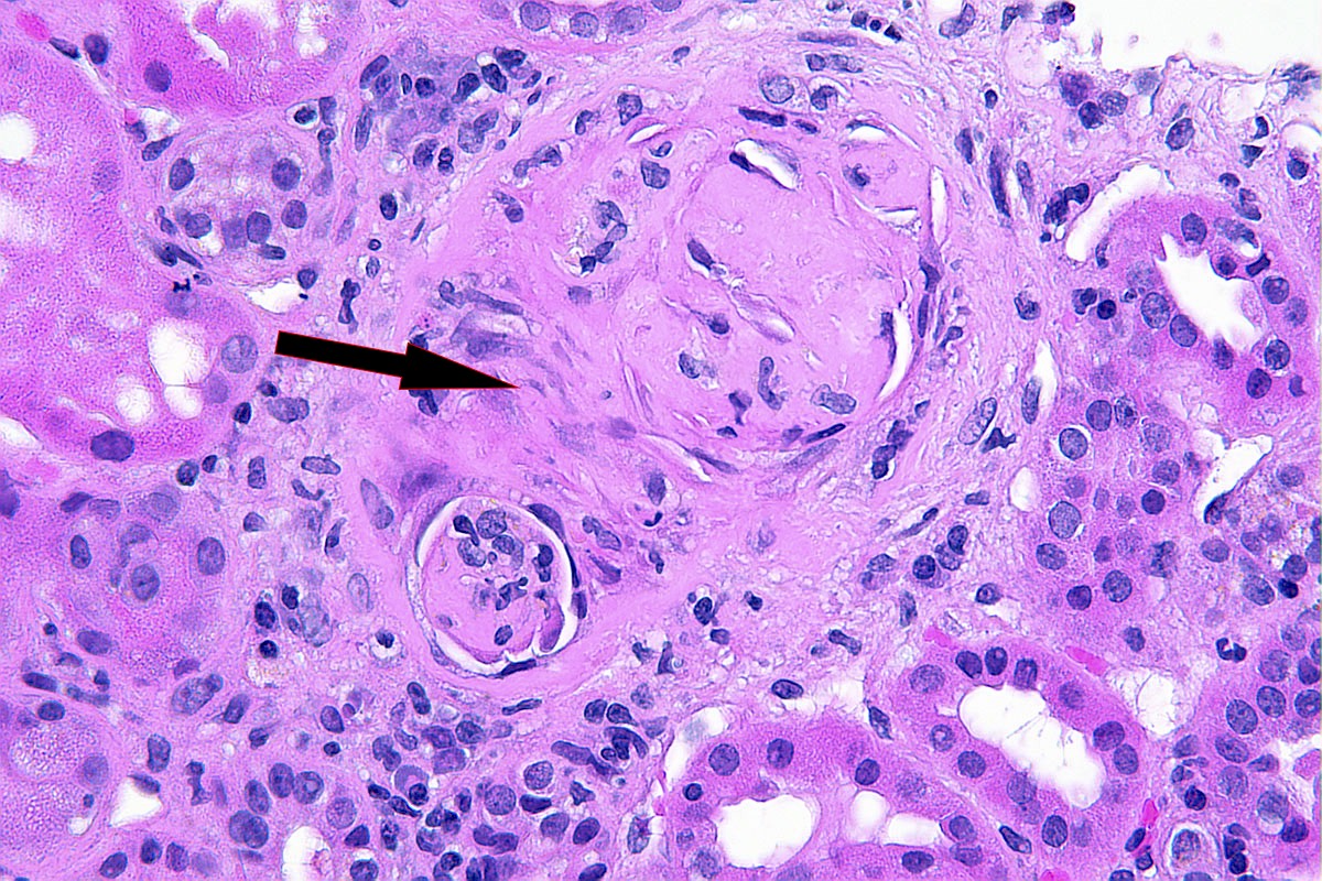
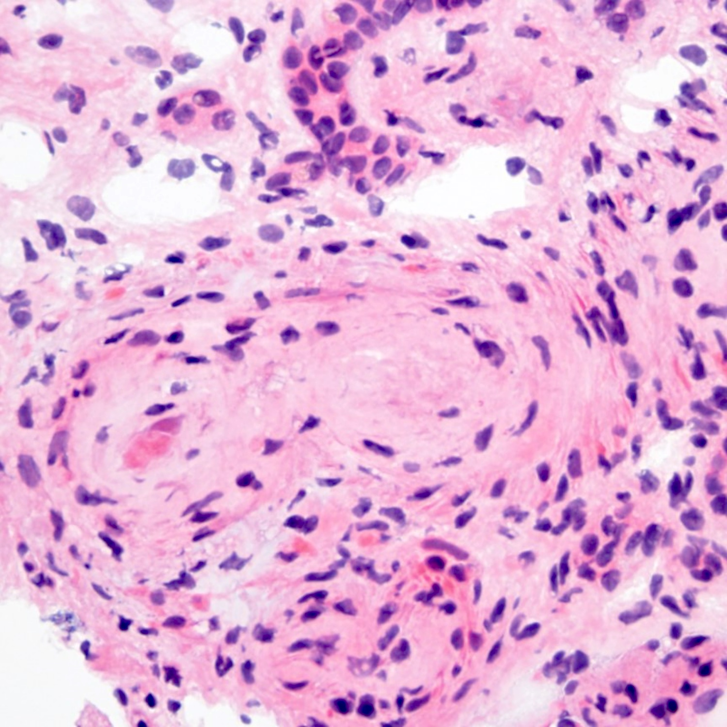
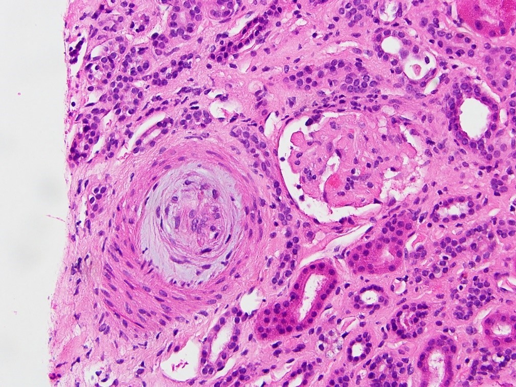
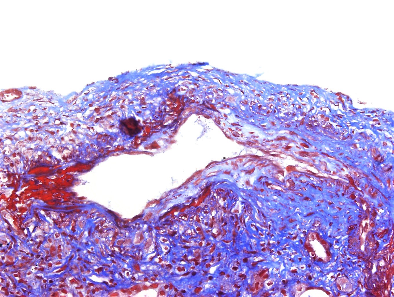
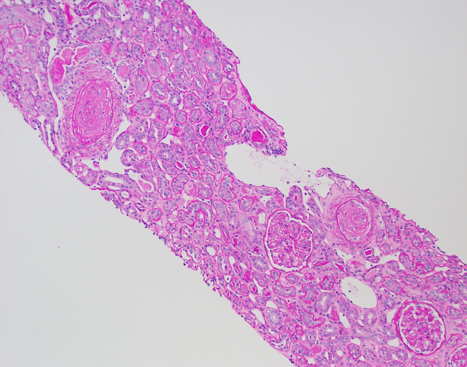
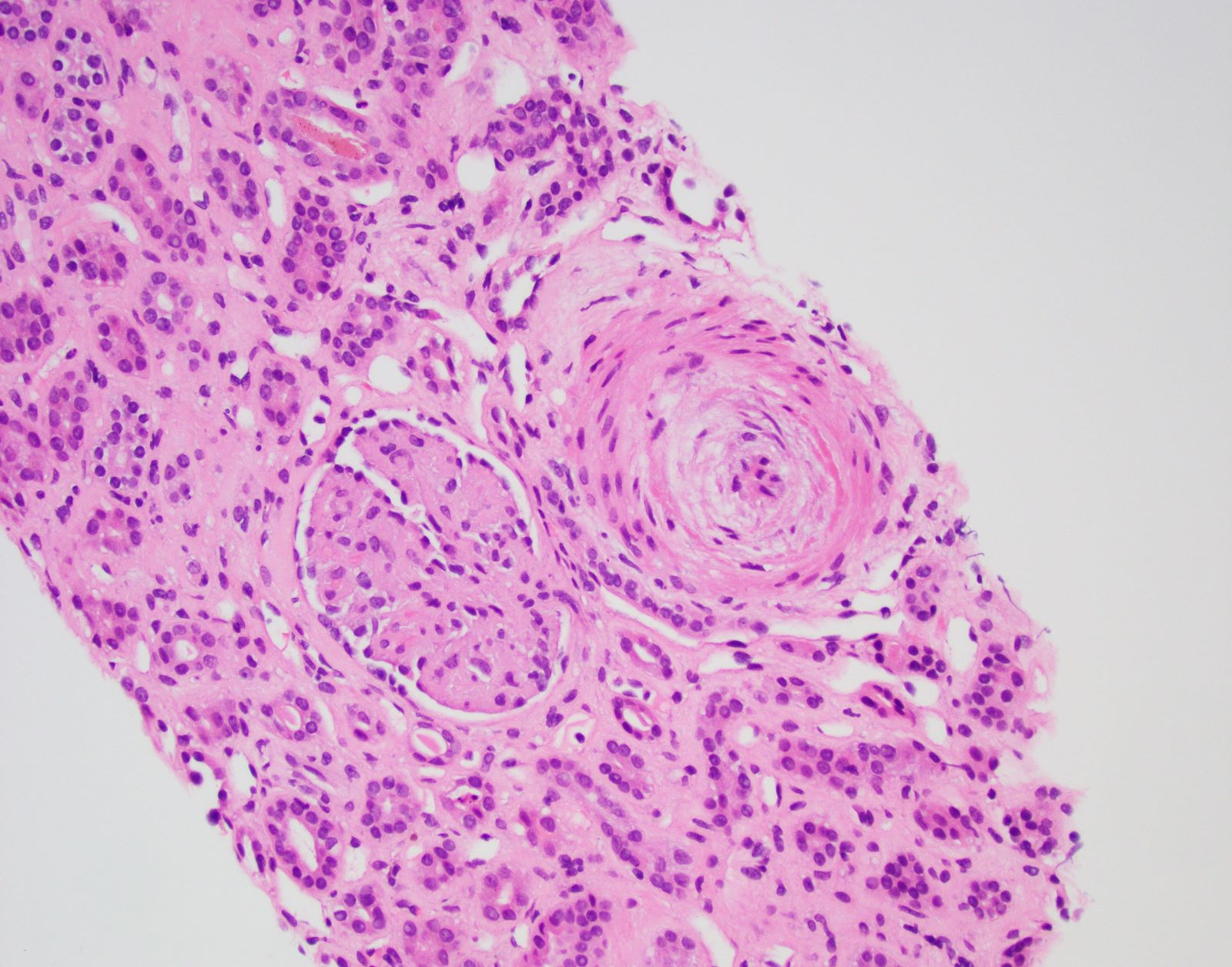
Transplant arteriopathy
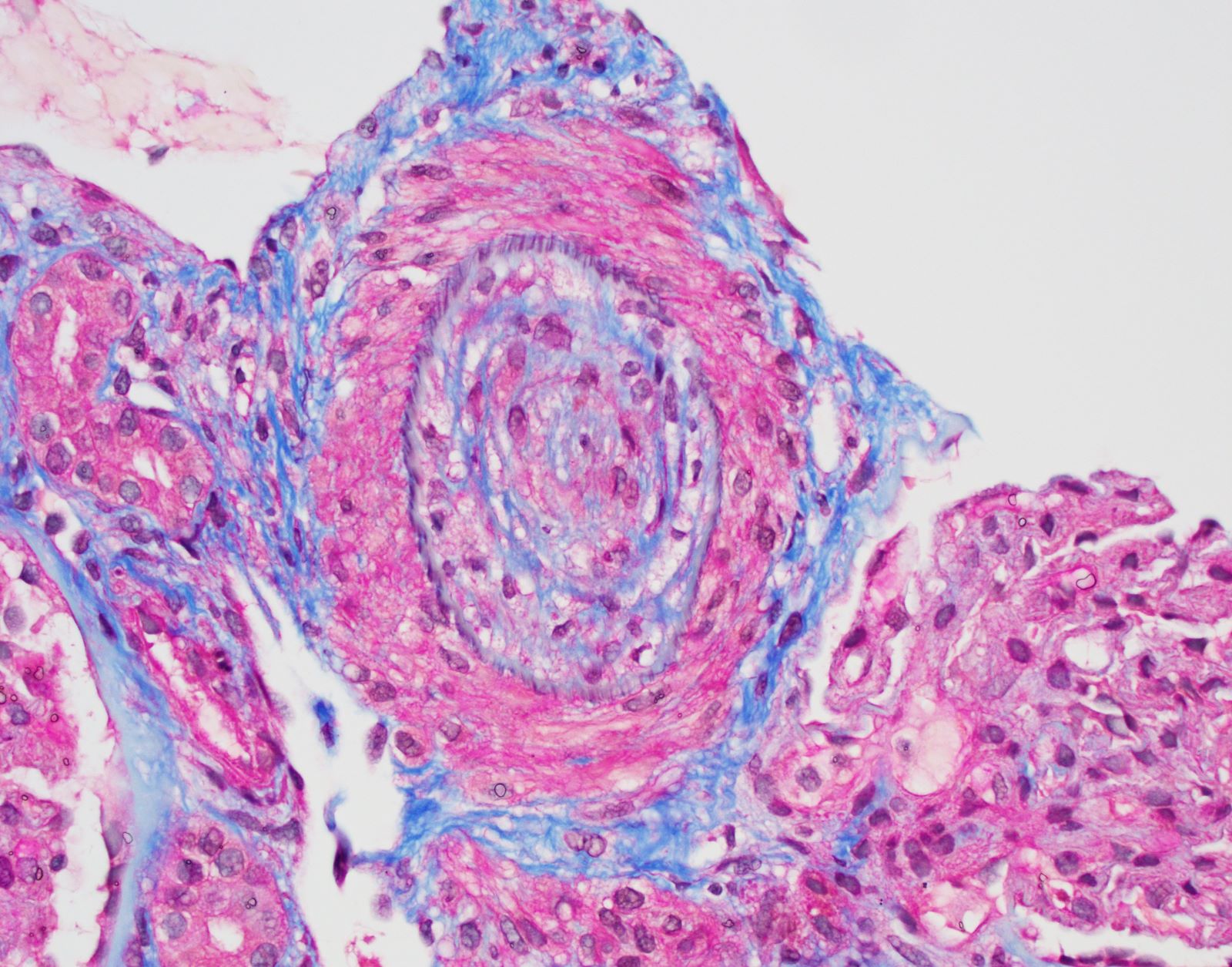
Transplant arteriopathy and IFTA, MTC stain
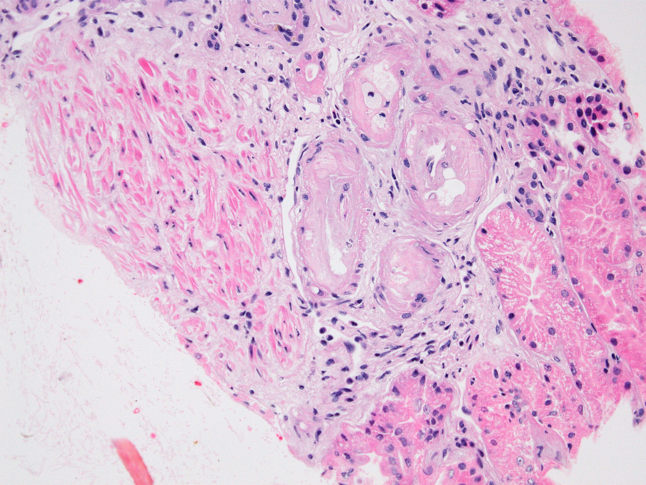
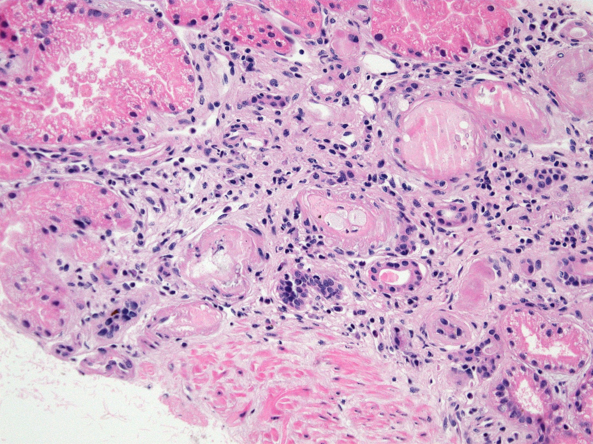
Intimal foam cells in small artery
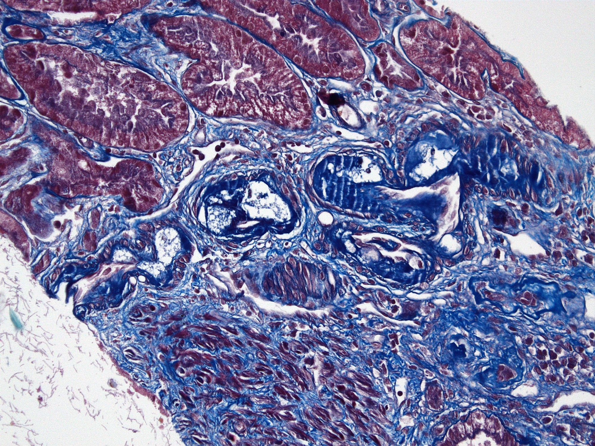
Intimal foam cells and fibrosis in small artery, MTC
Images hosted on other servers:

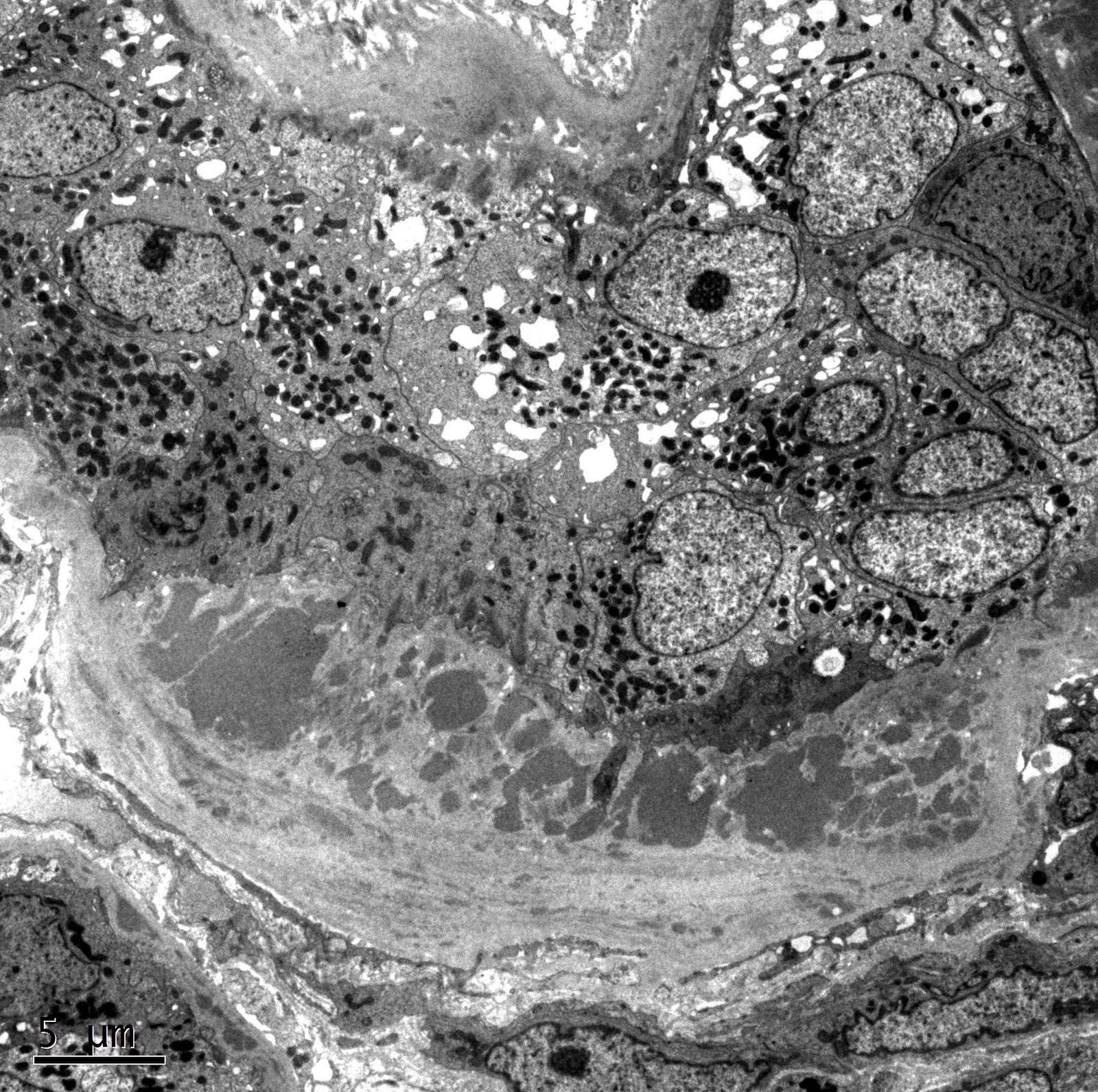
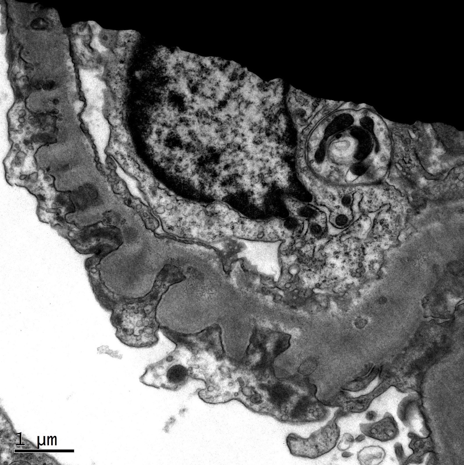
Diagram of electron and light microscopy
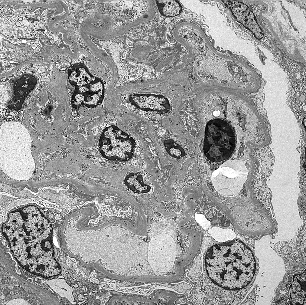
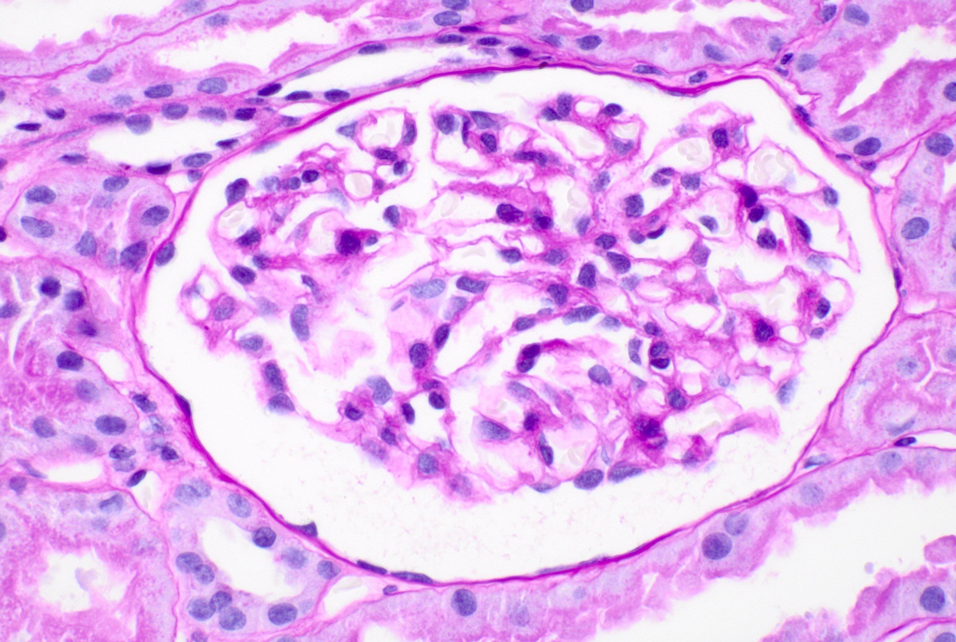
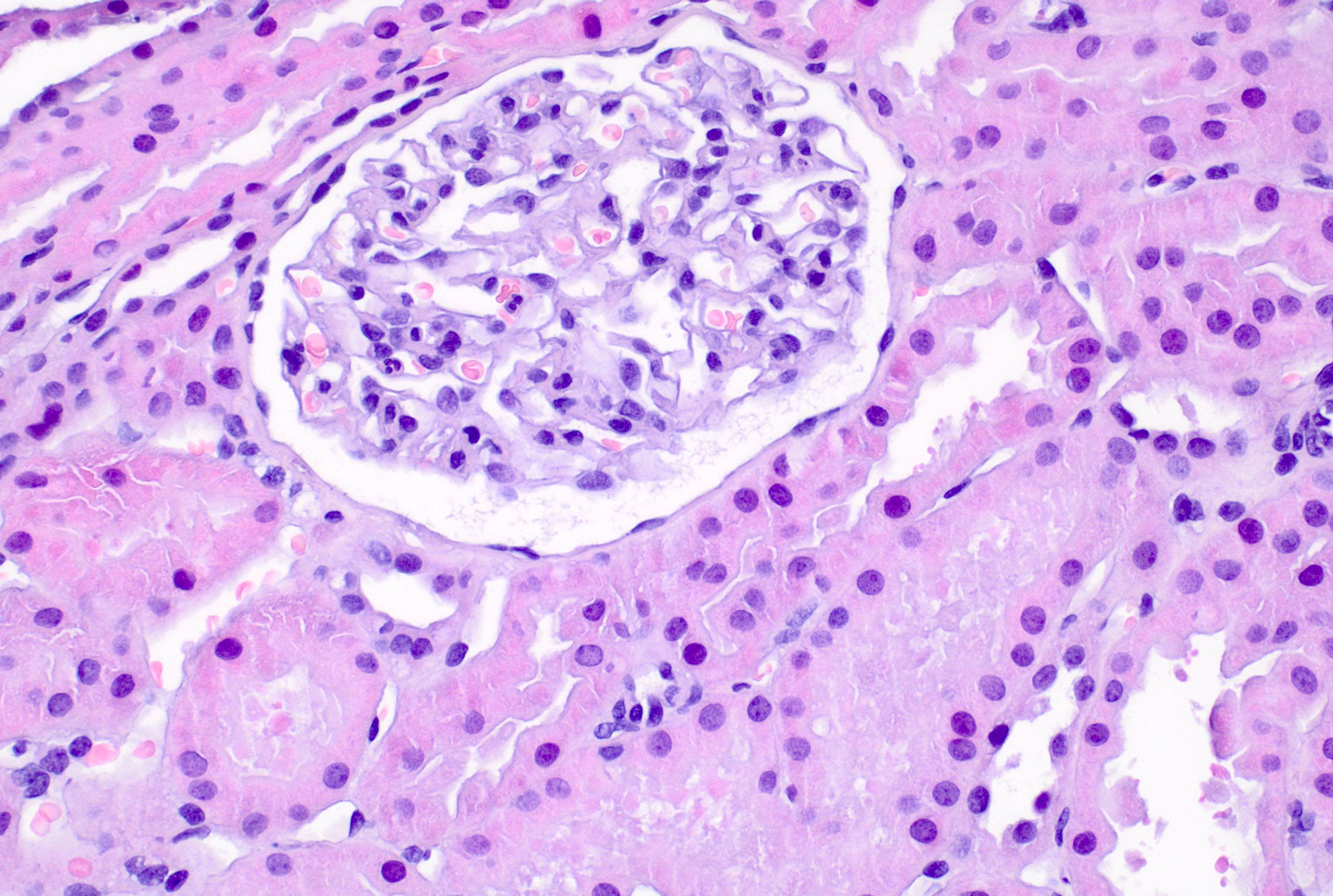
Contributed by Aisha Memon, M.B.B.S.
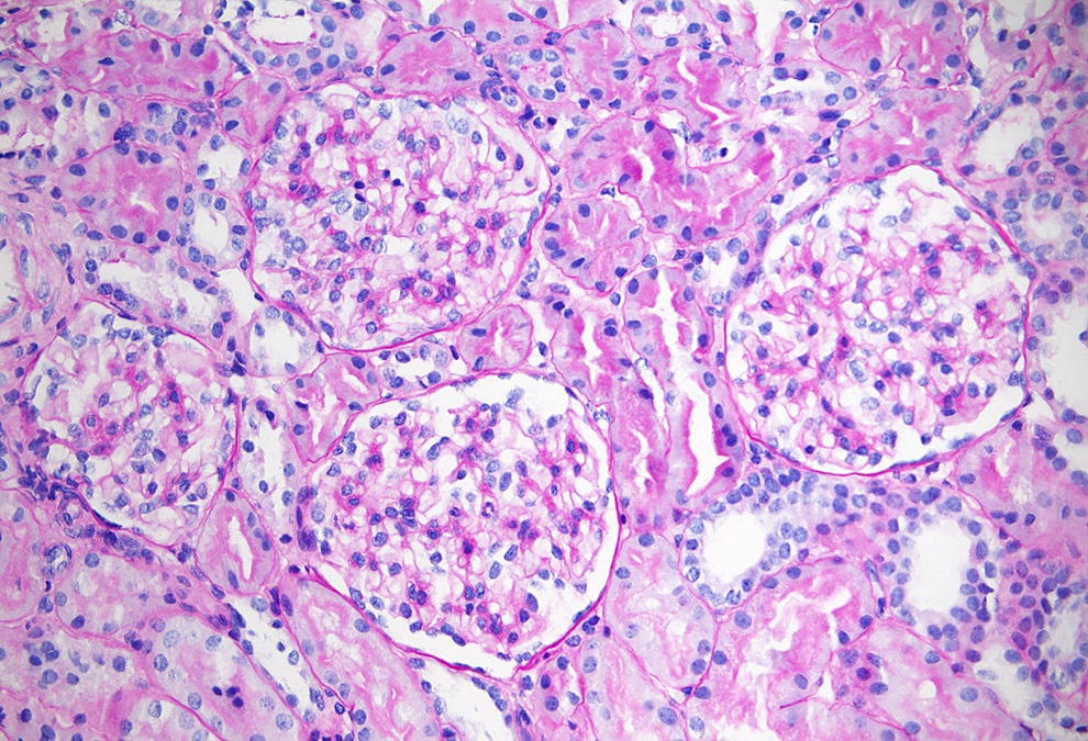
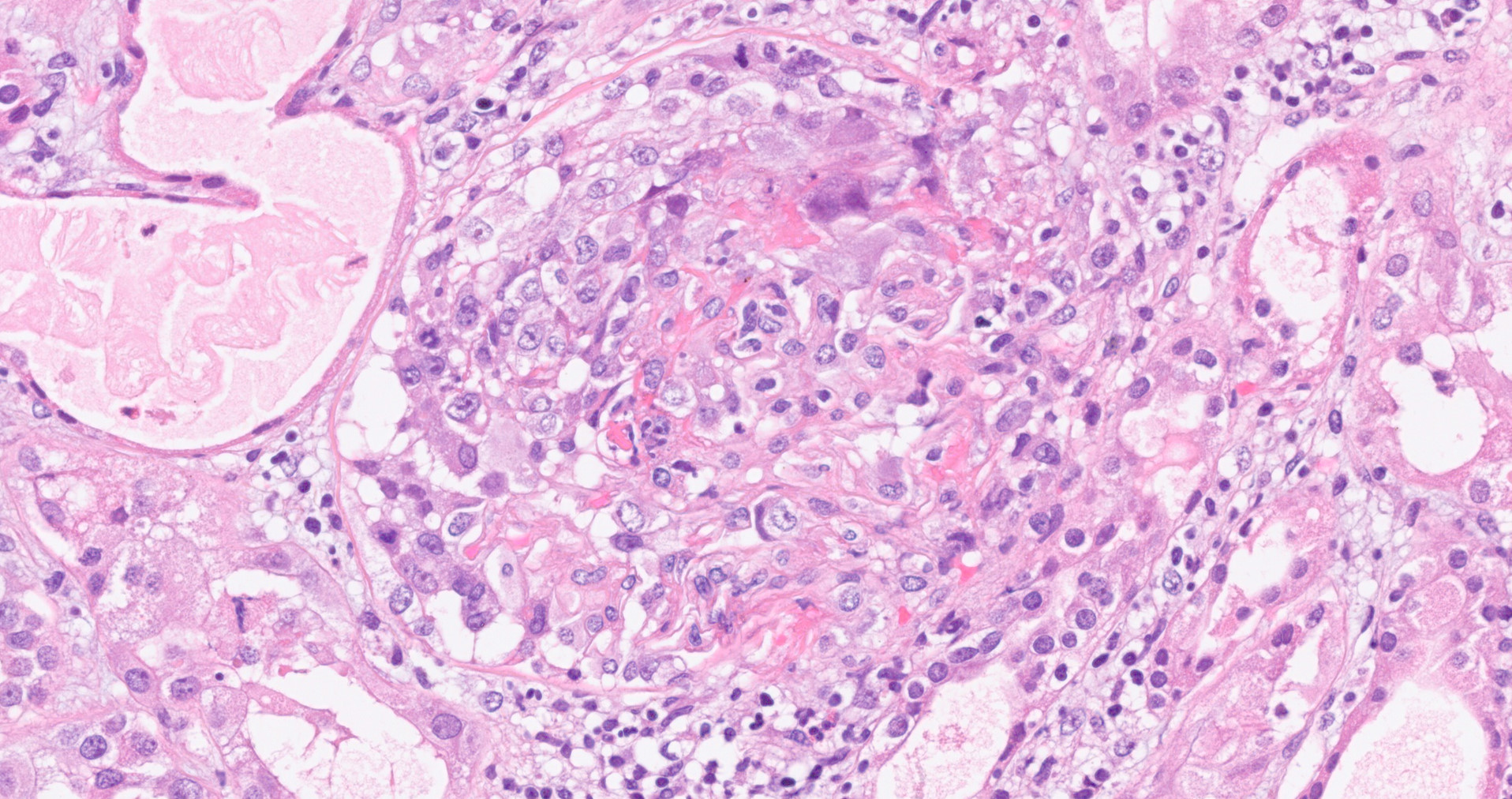
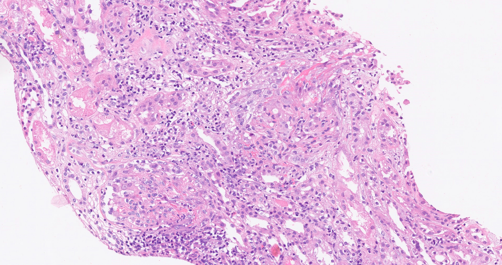
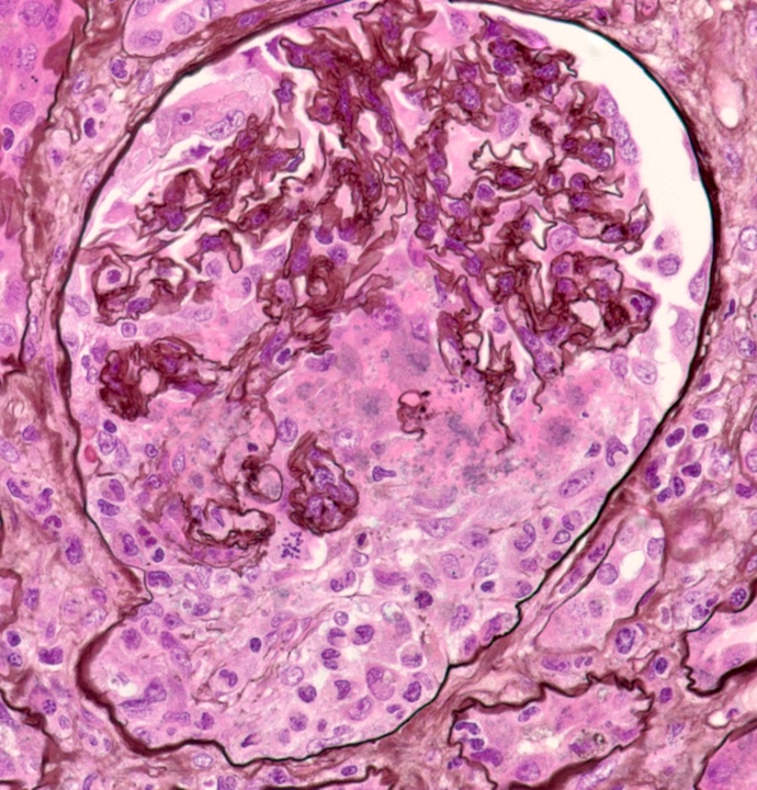
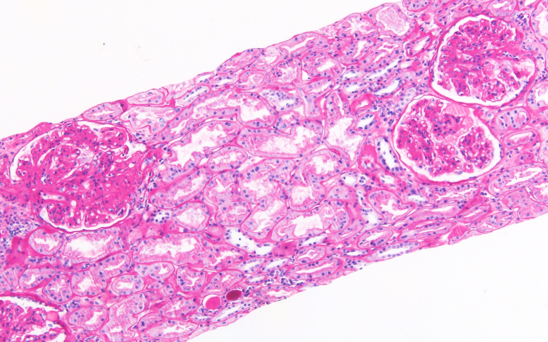
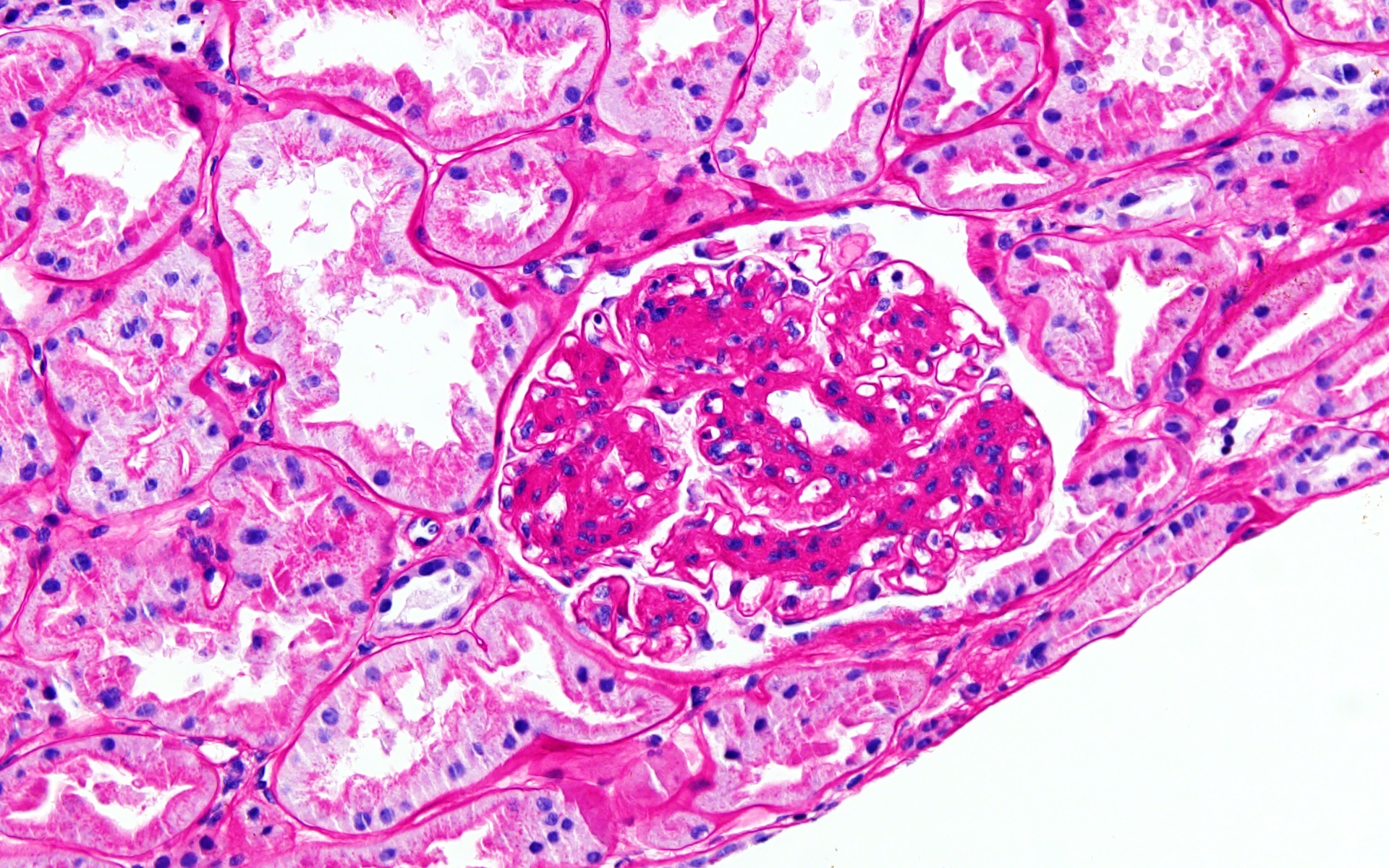
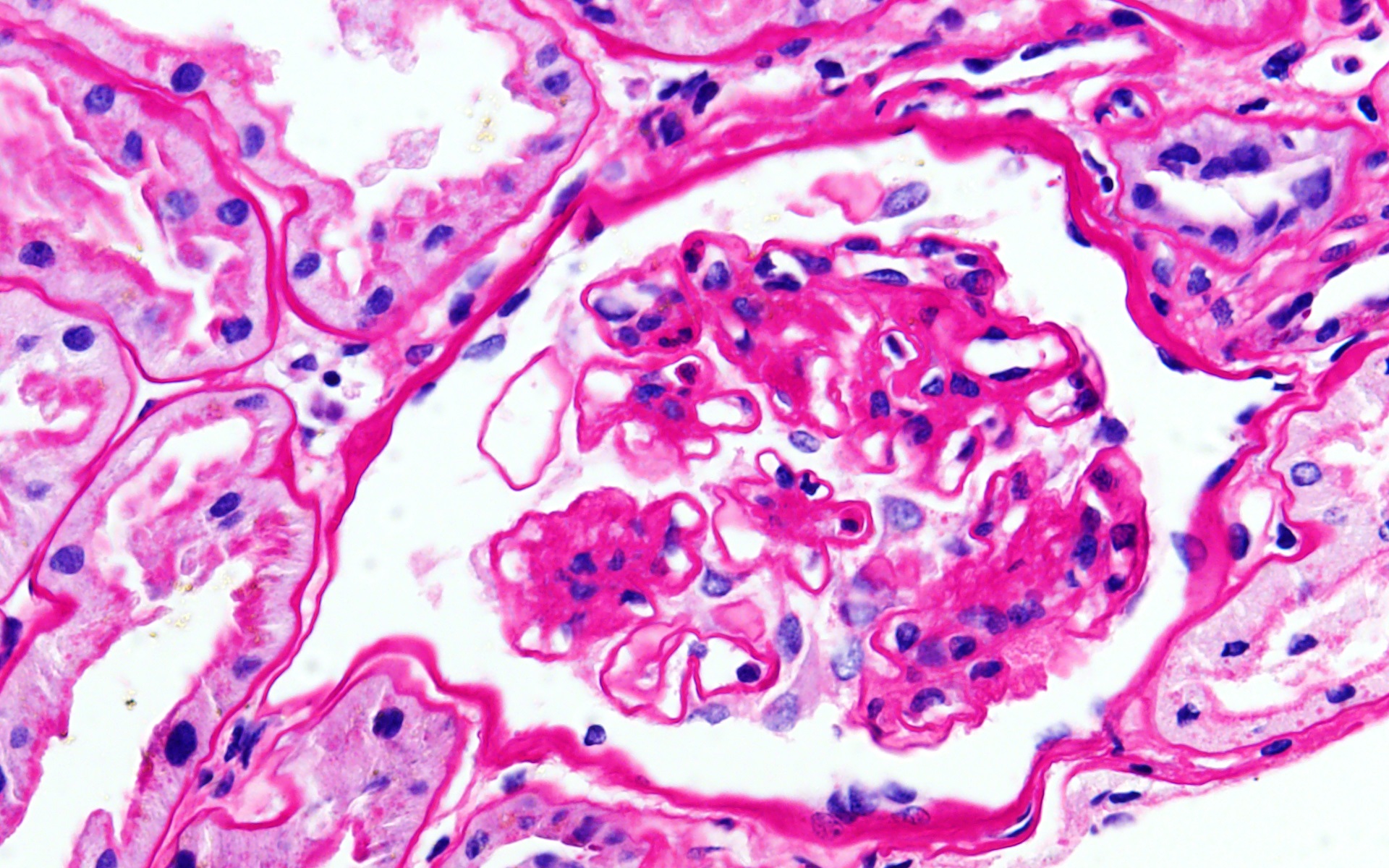
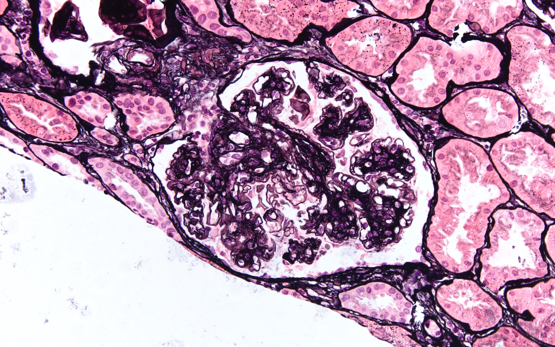
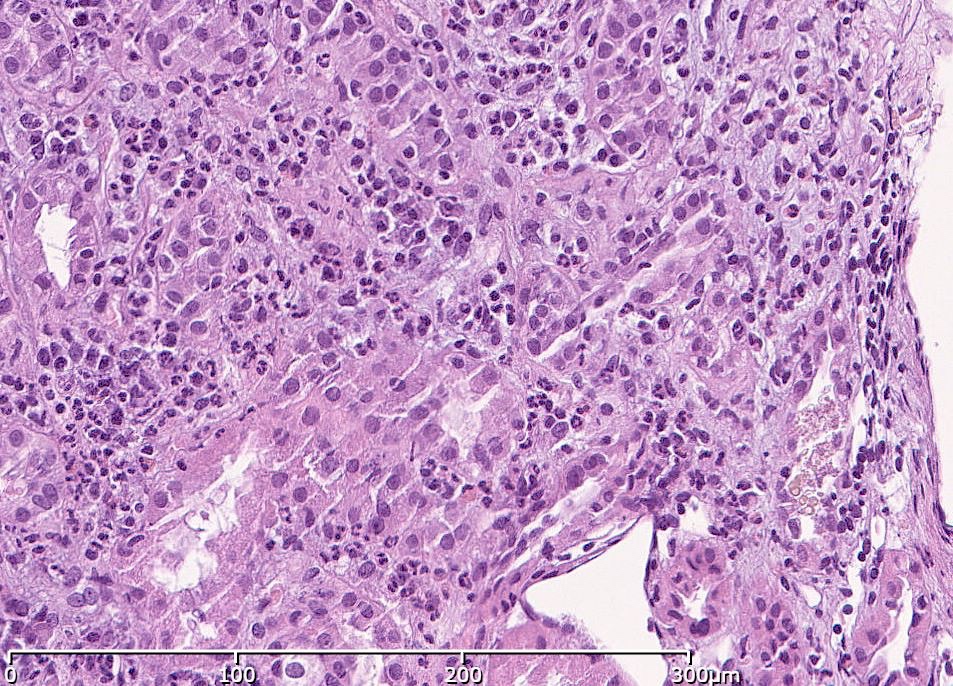
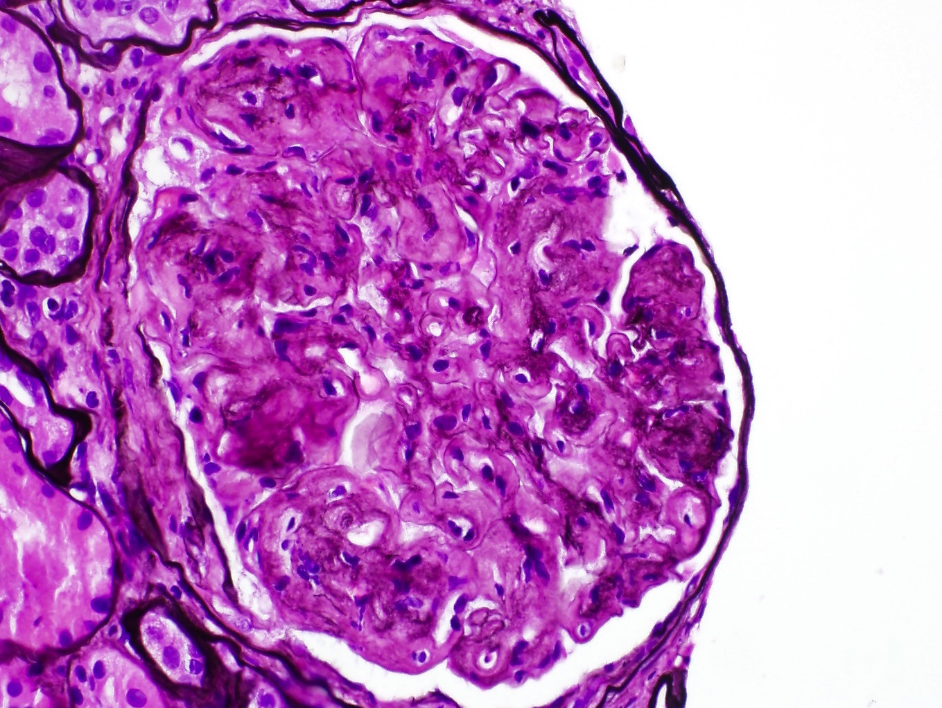
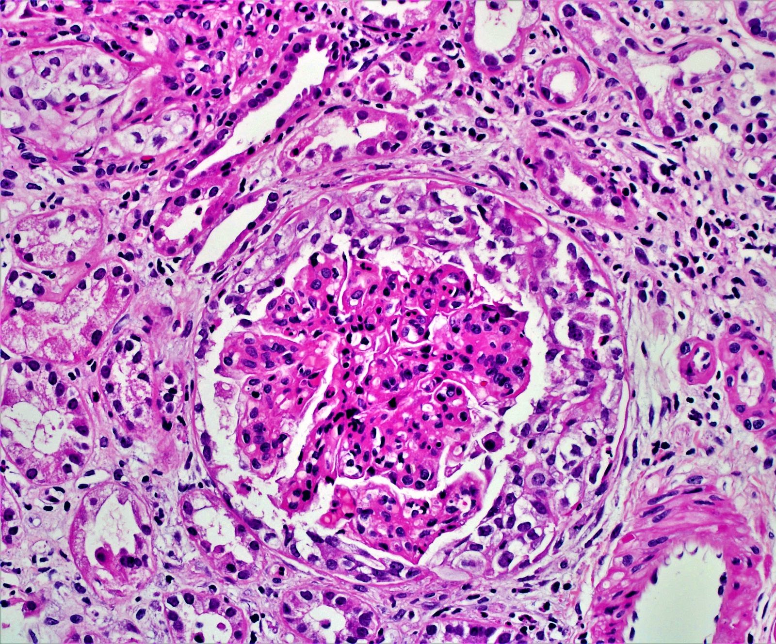
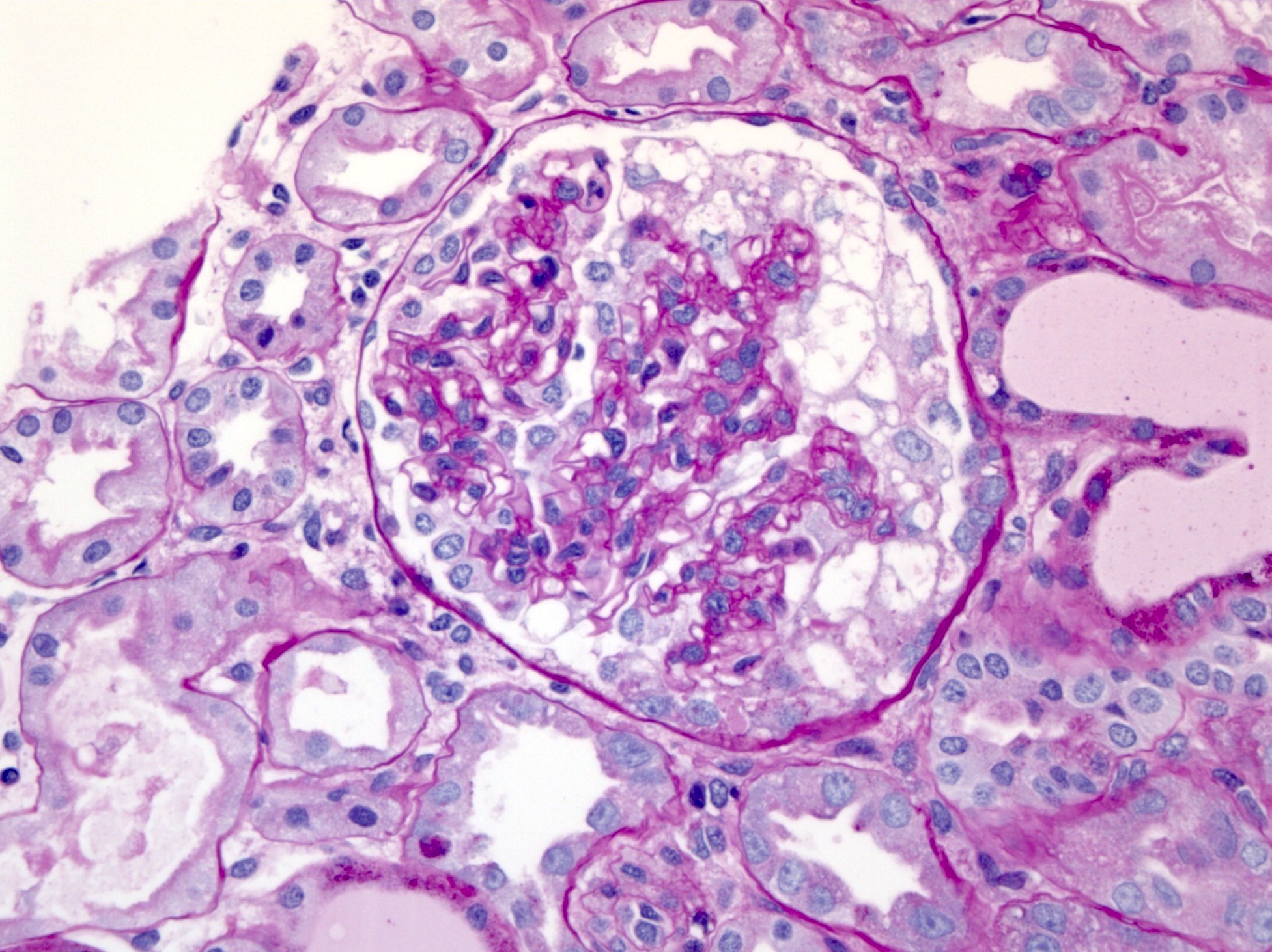
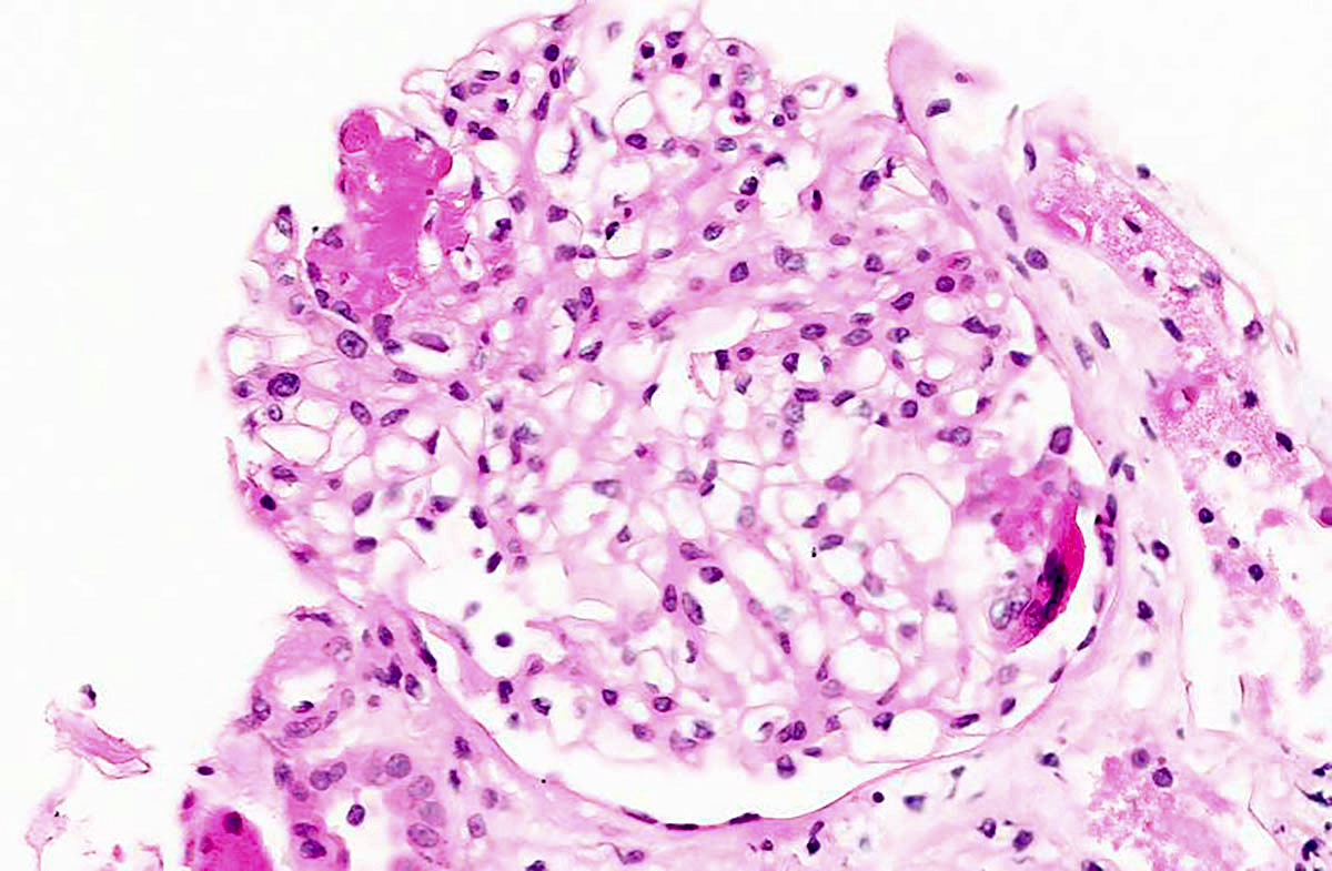
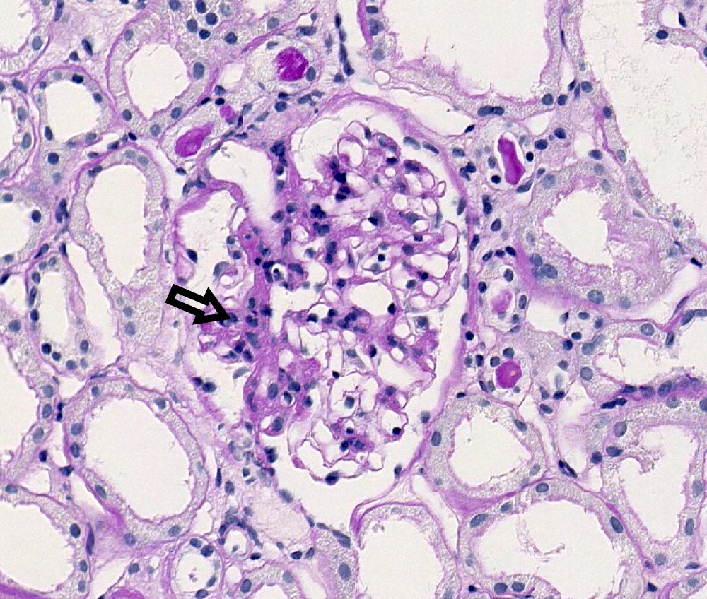
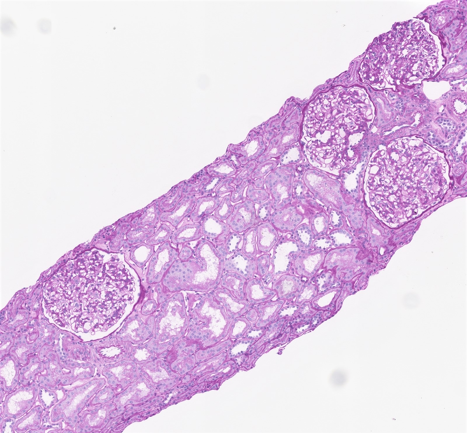
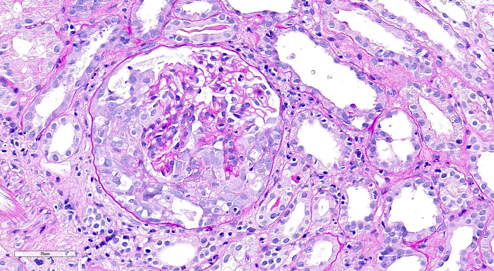
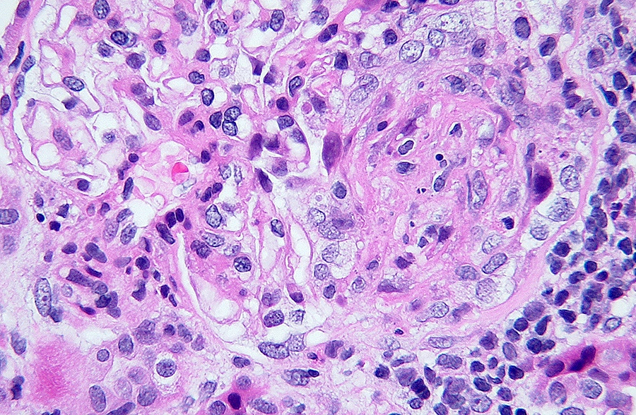
Diffuse global proliferative appearance

Global proliferative appearance
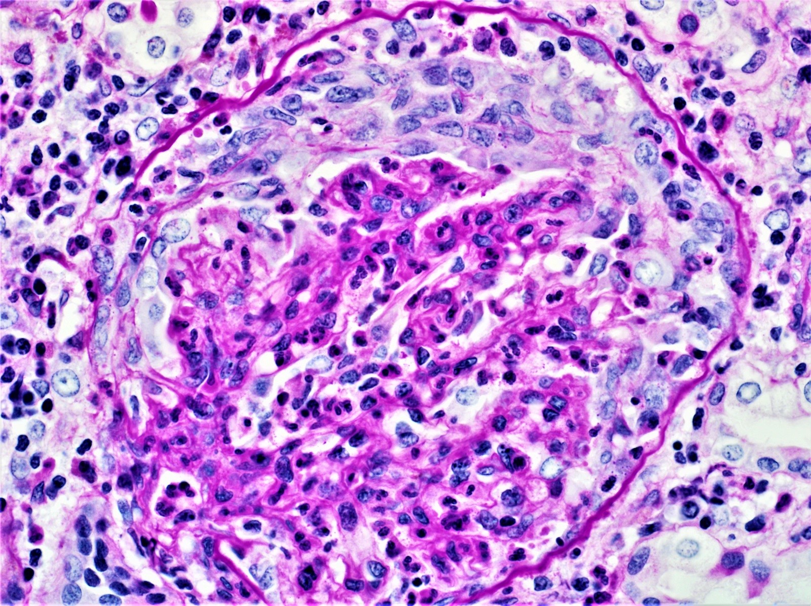
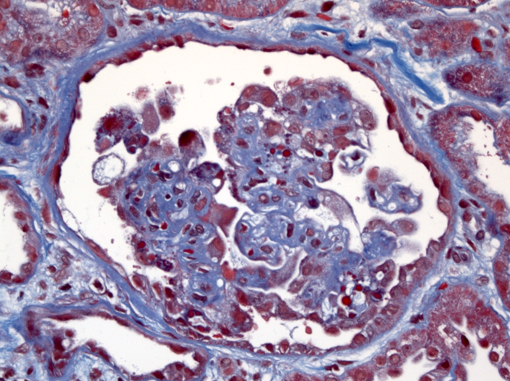
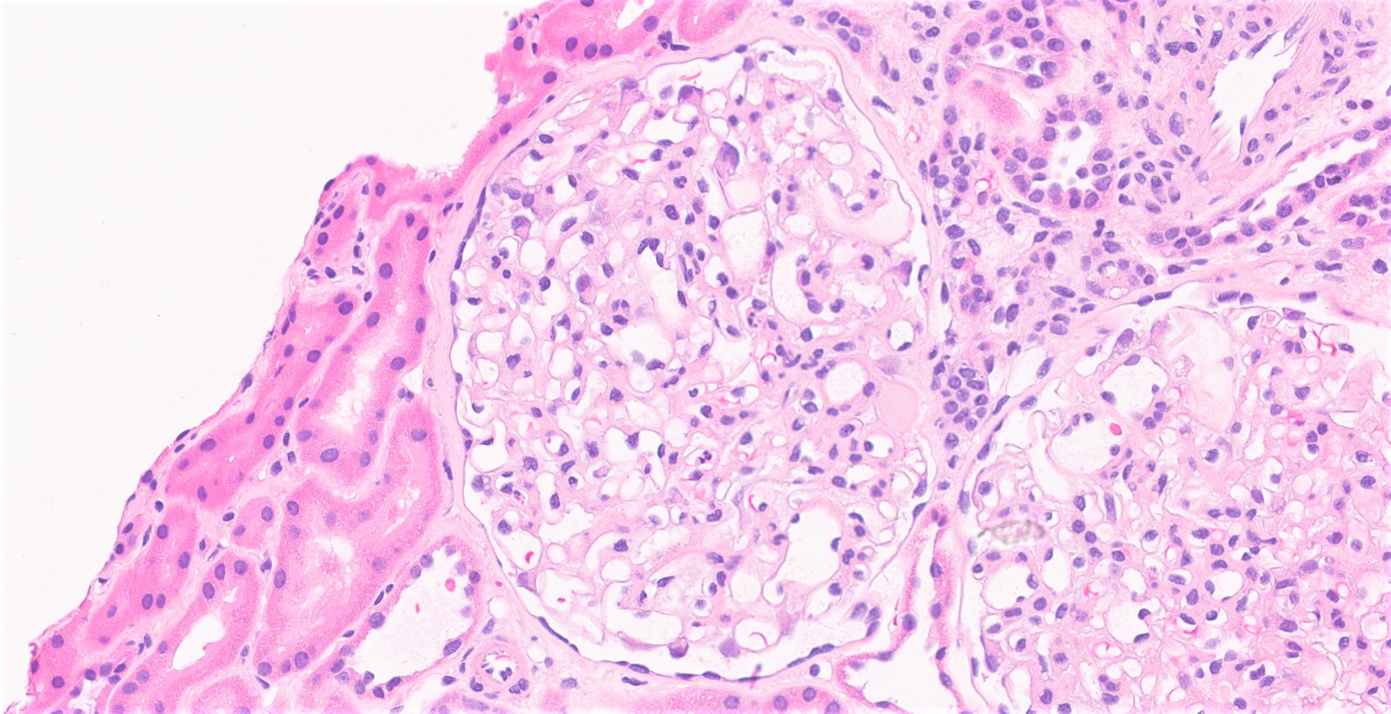
Cellular crescent formation
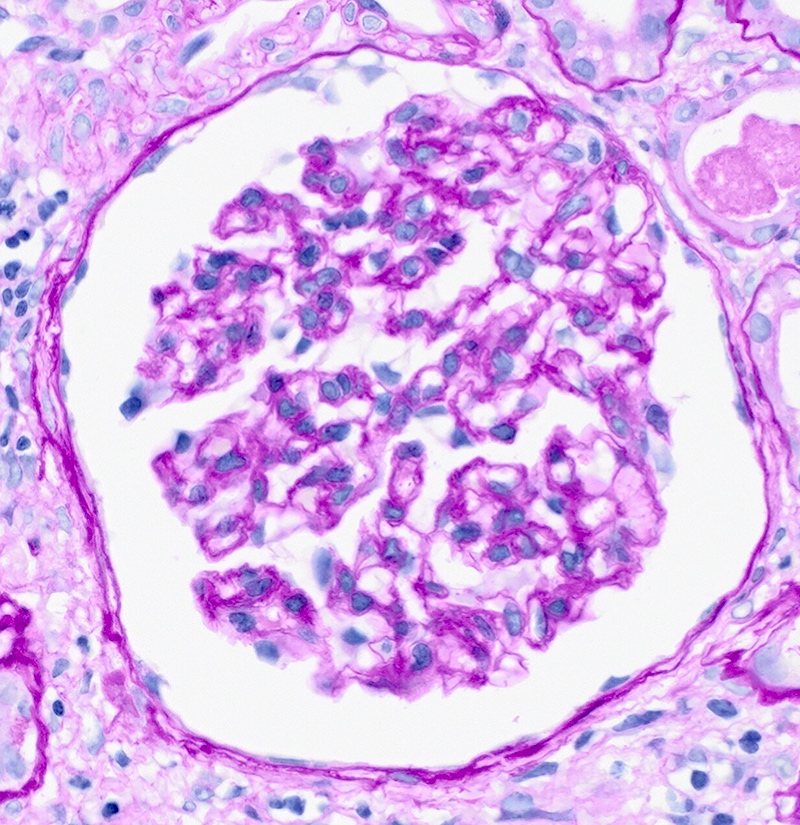
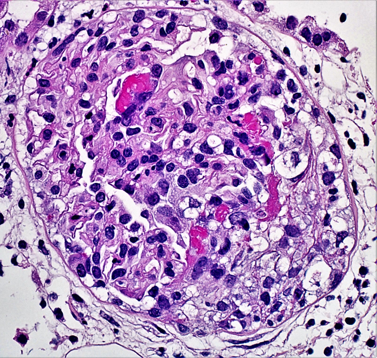
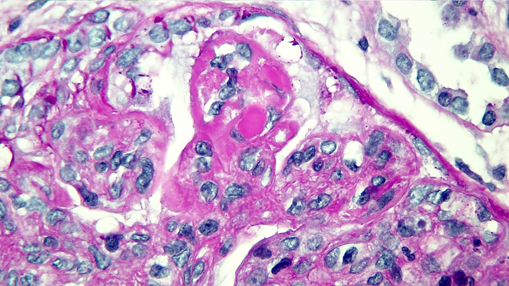
Mesangial hypercellularity
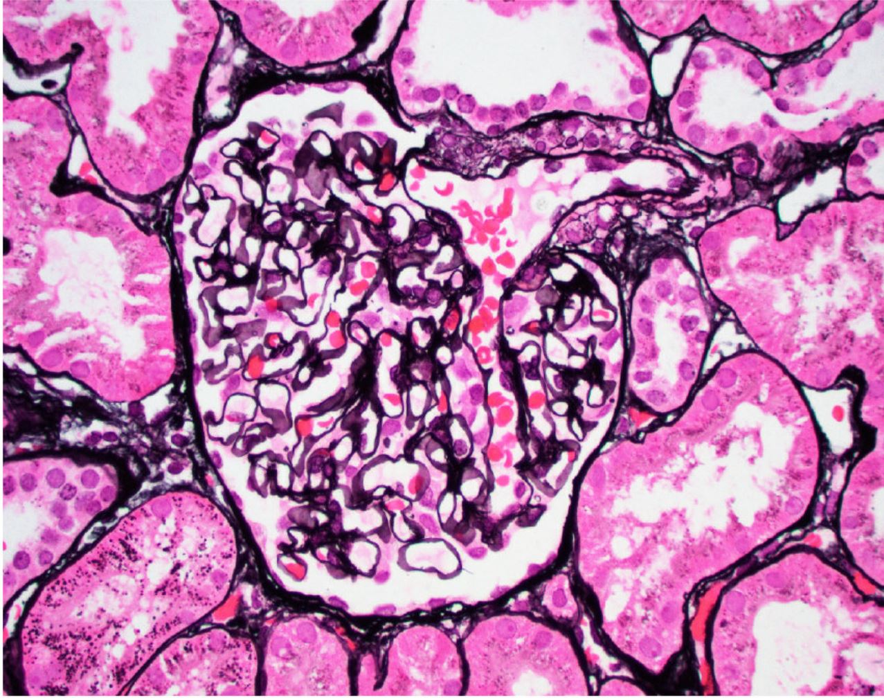
Mesangial hypercellularity on periodic acid-Schiff
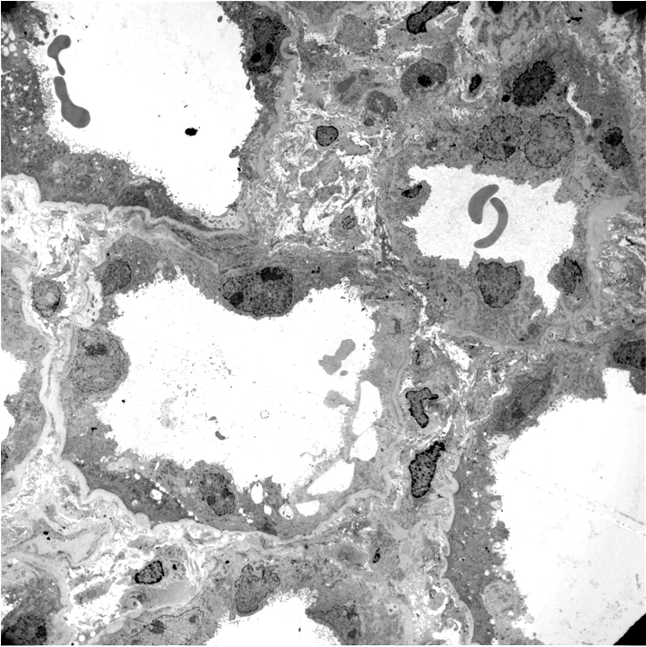
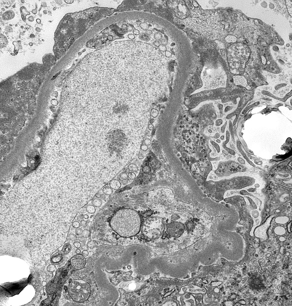
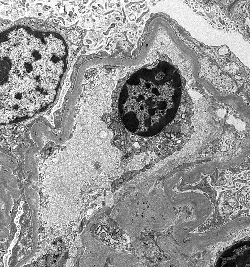
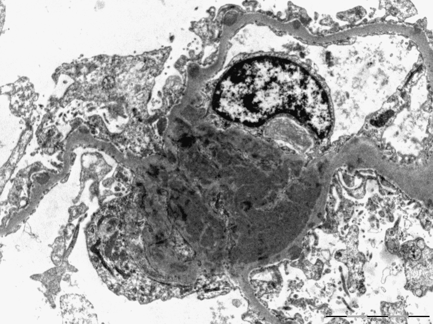
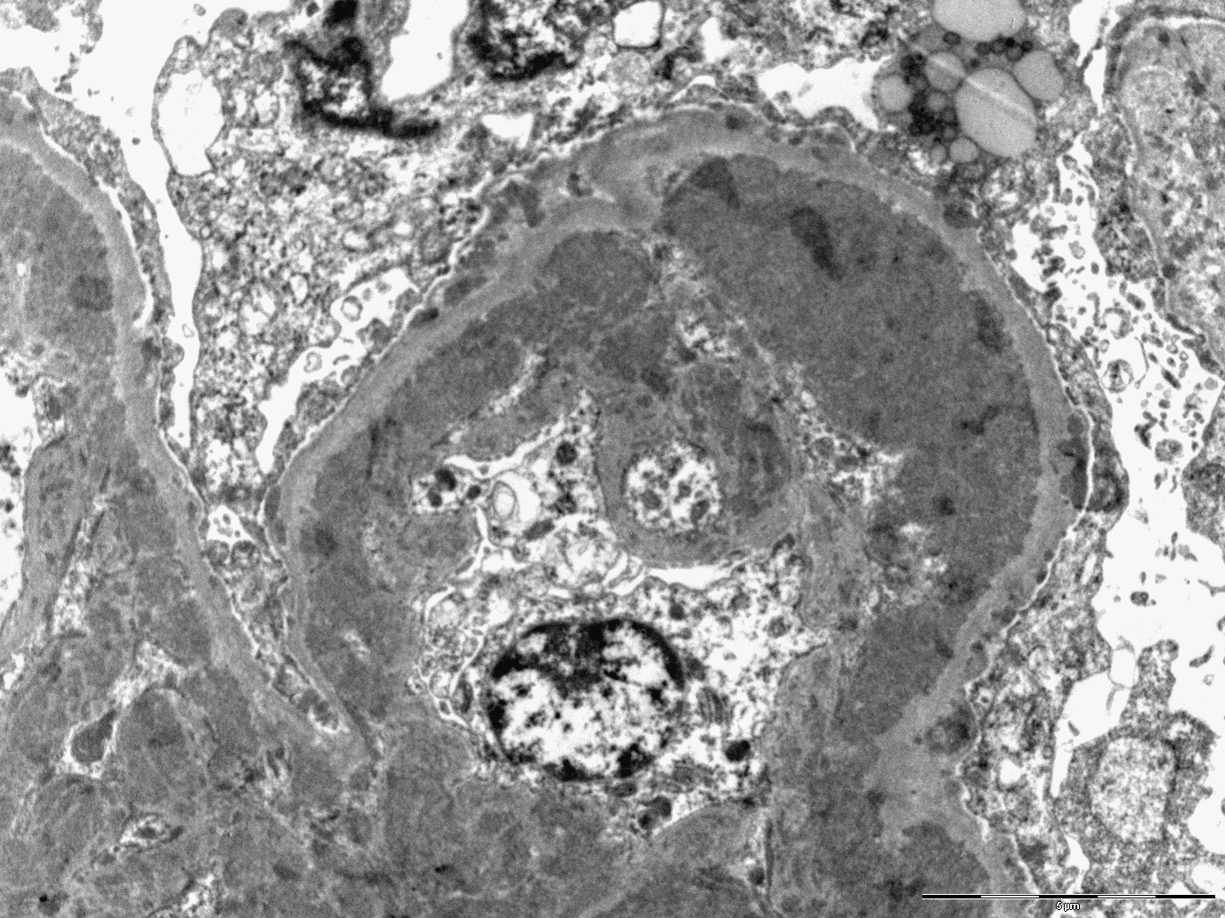
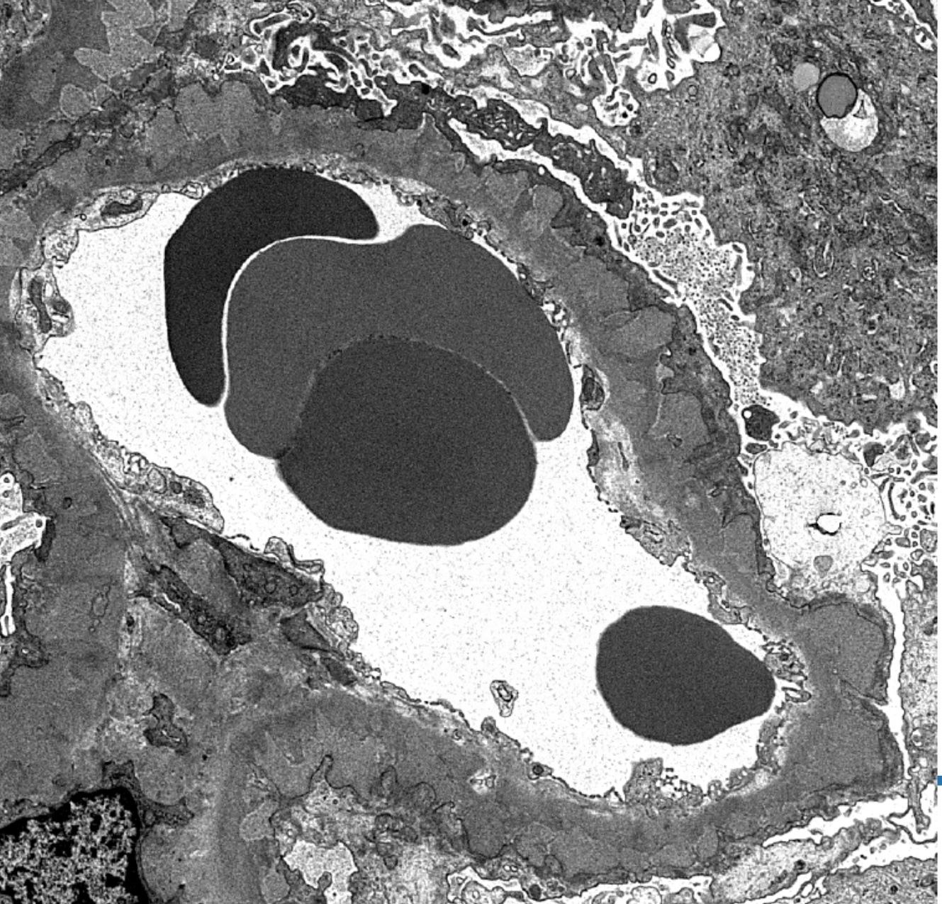
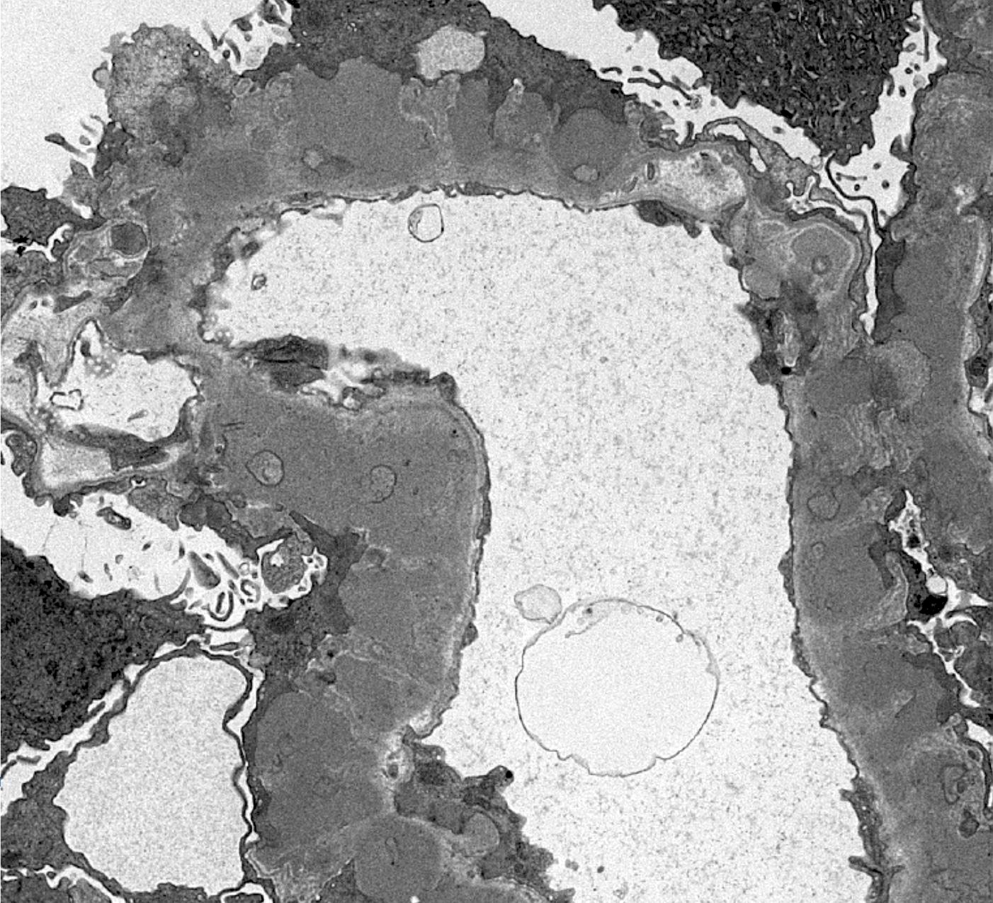
Contributed by Jonathan E. Zuckerman, M.D., Ph.D.
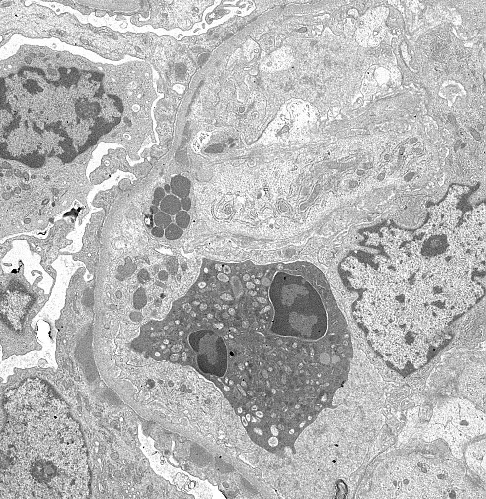
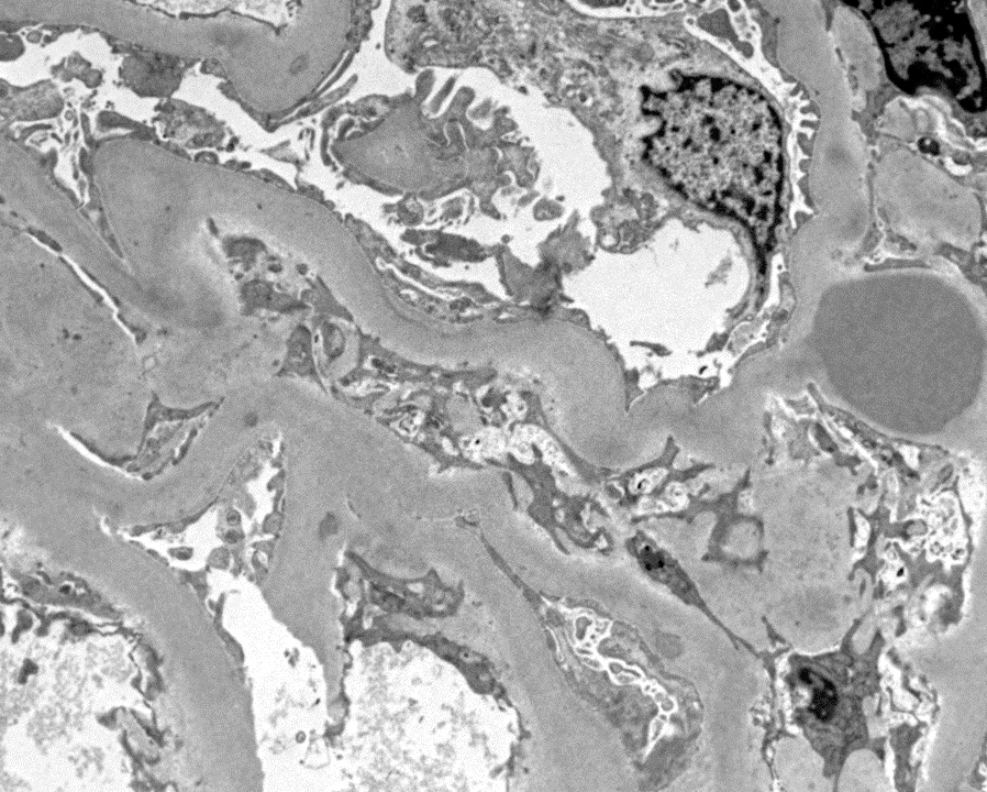
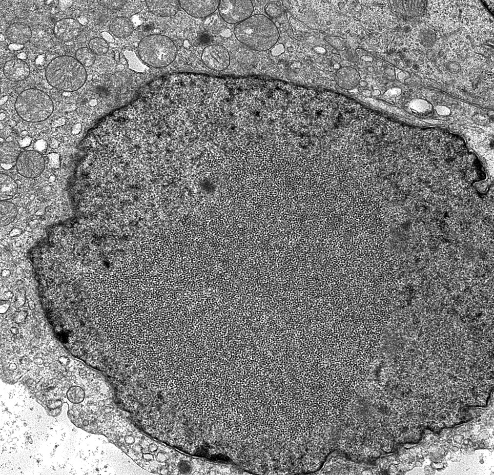
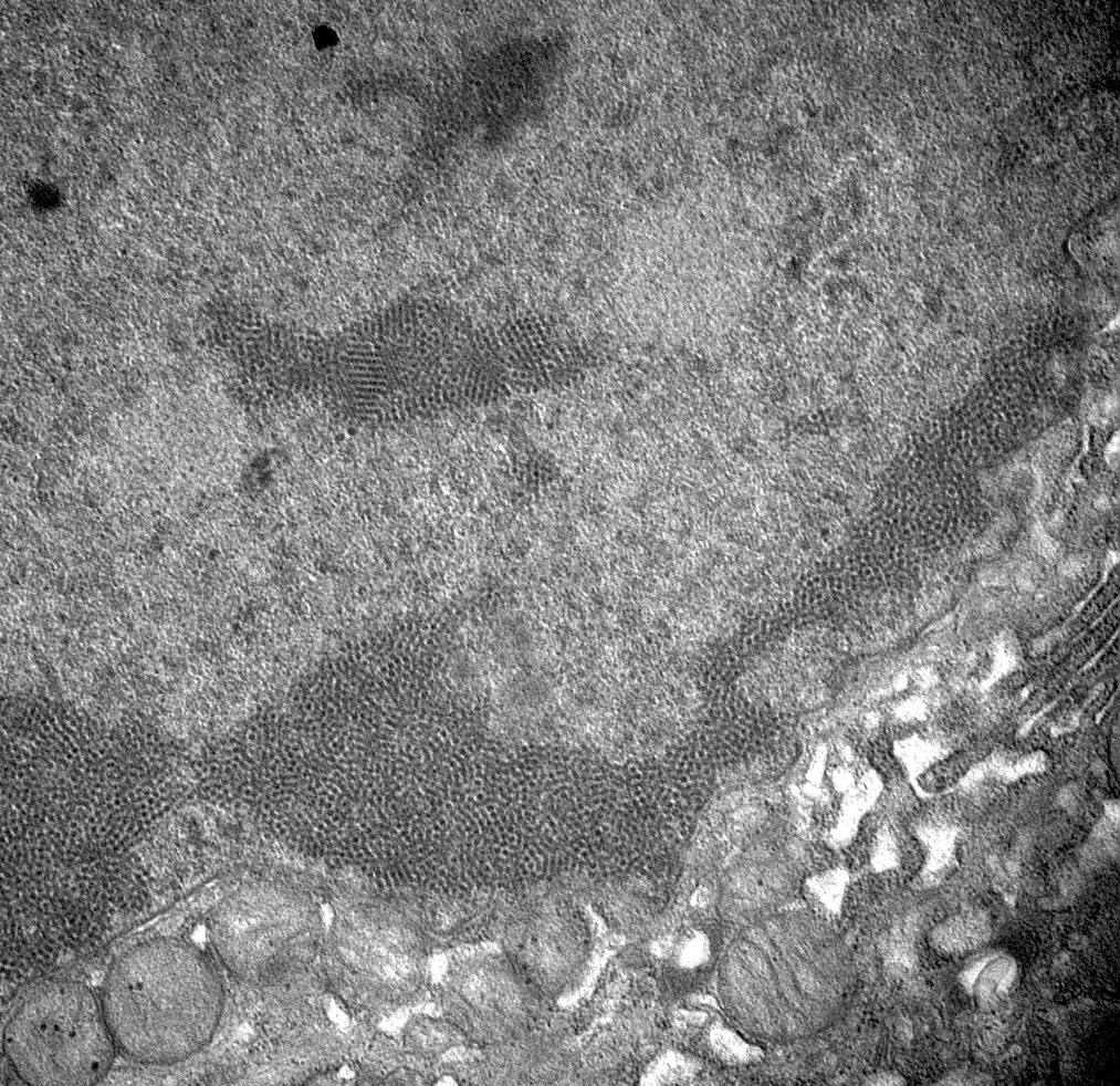
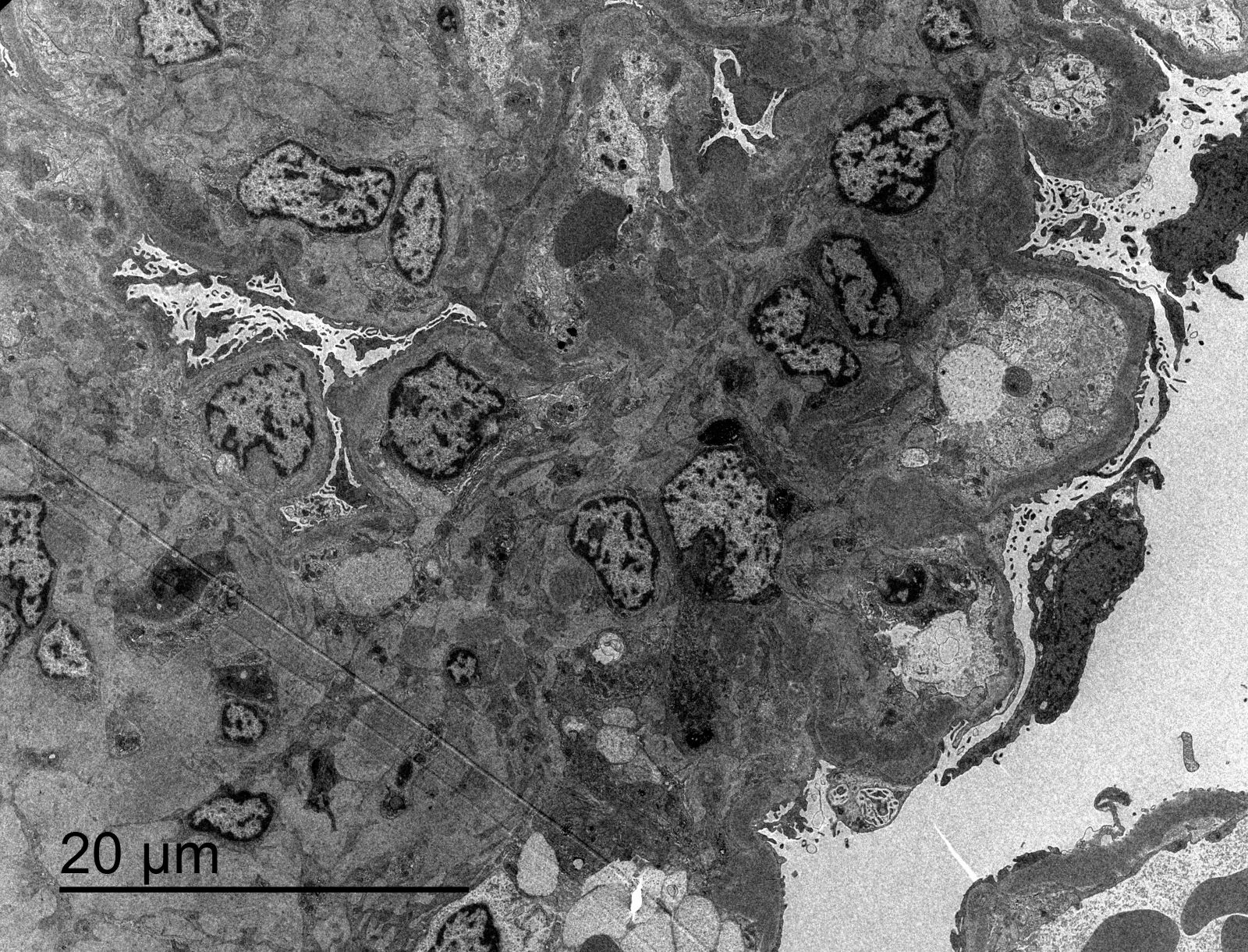
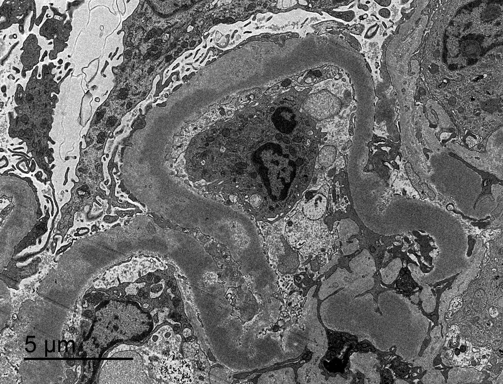
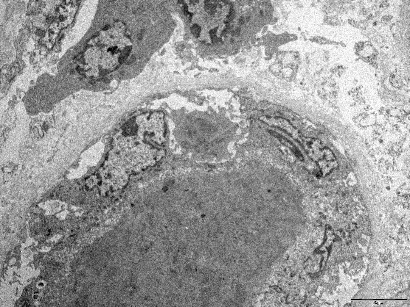
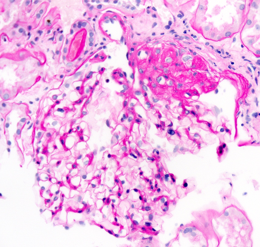
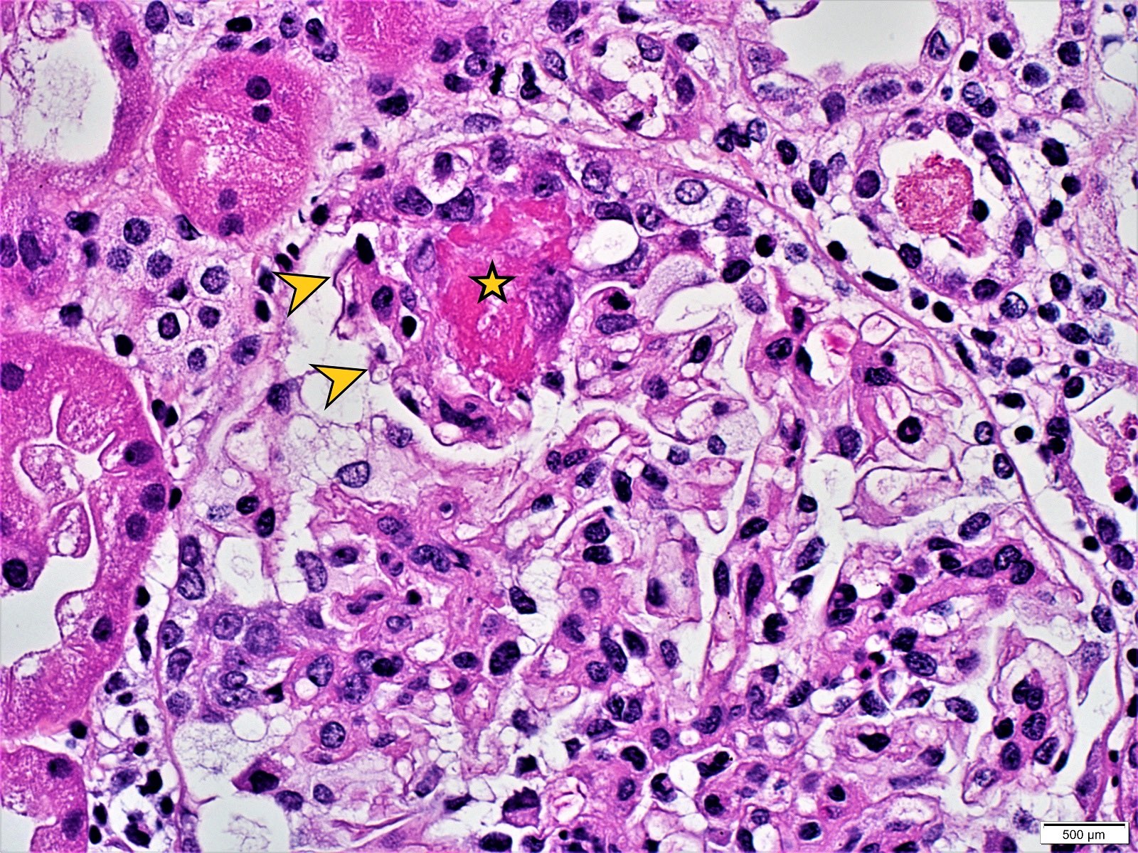
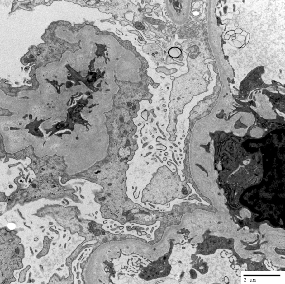
Endocapillary neutrophil
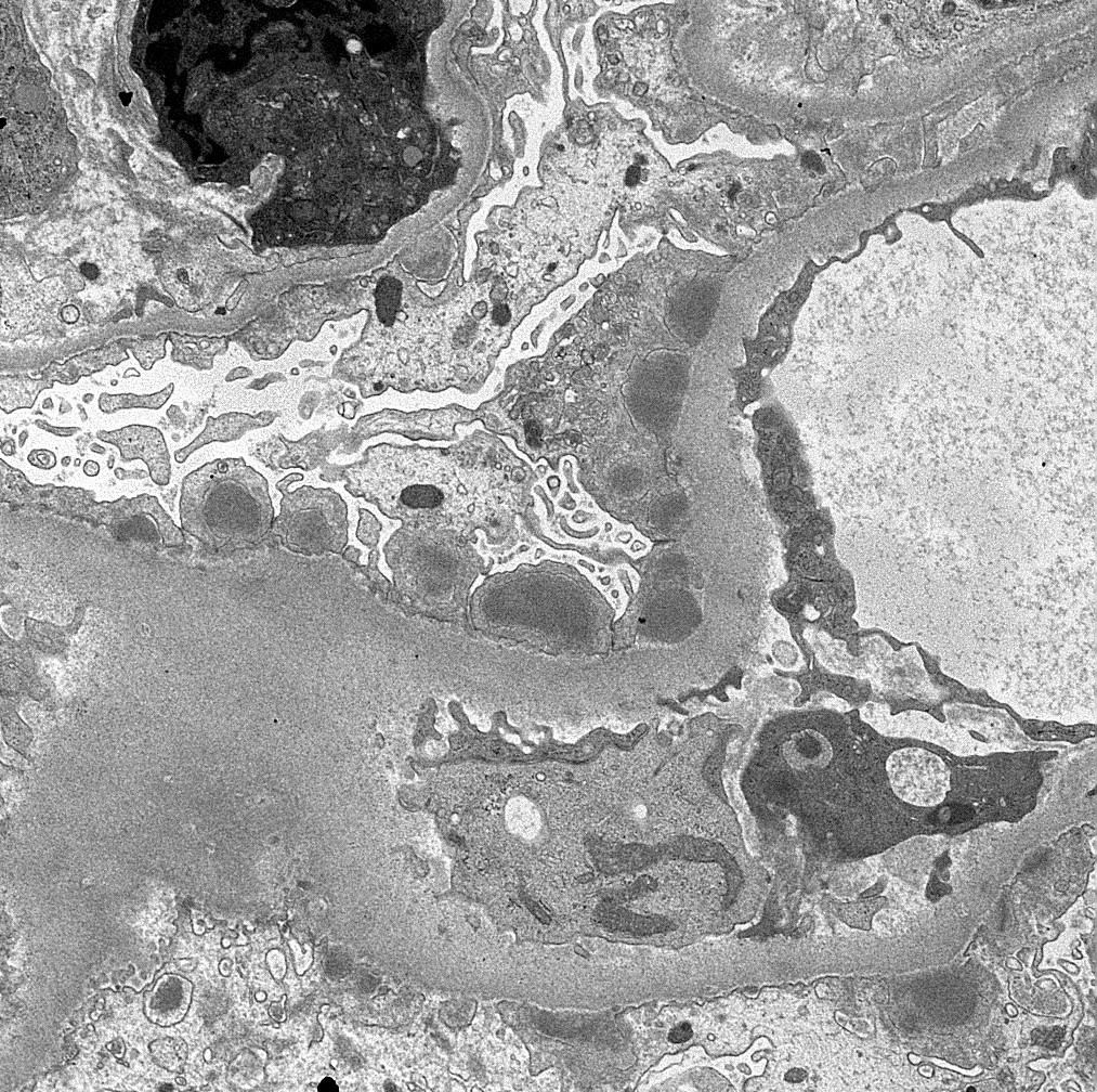
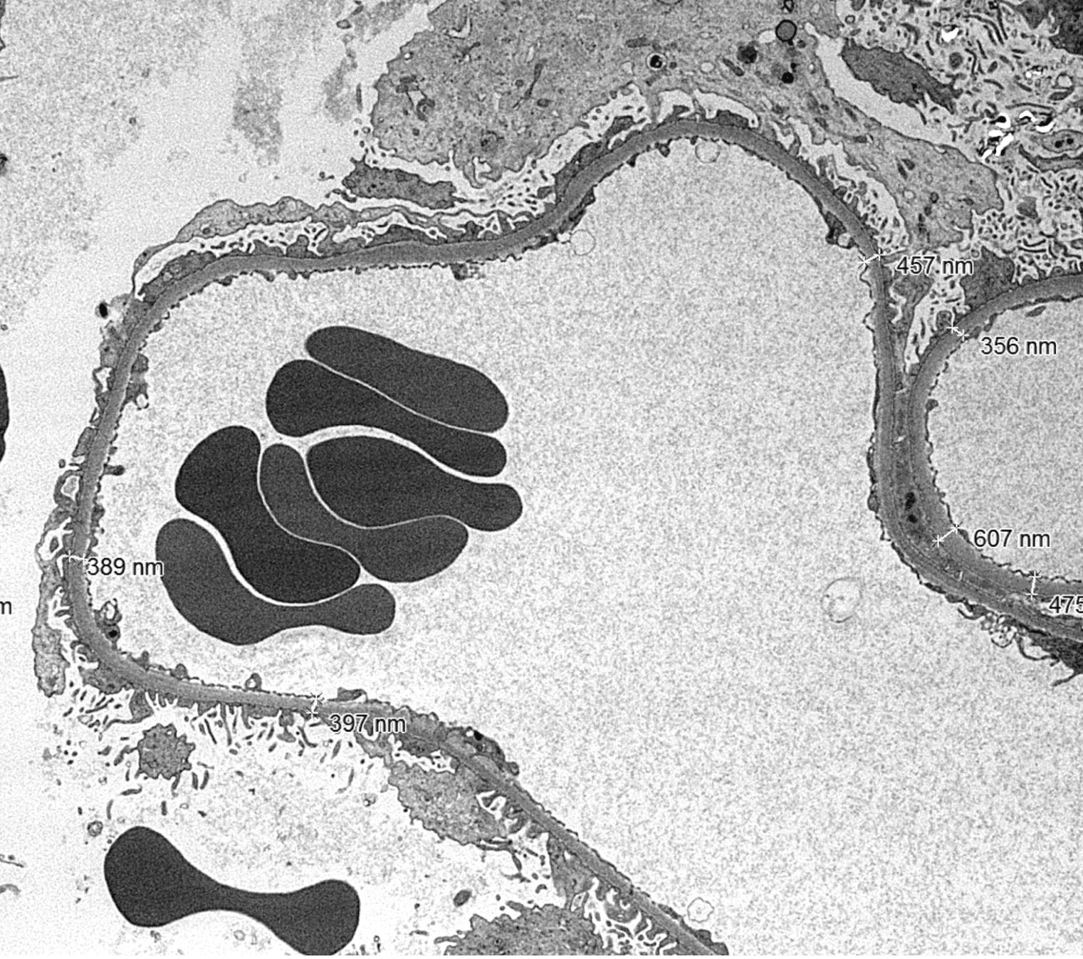
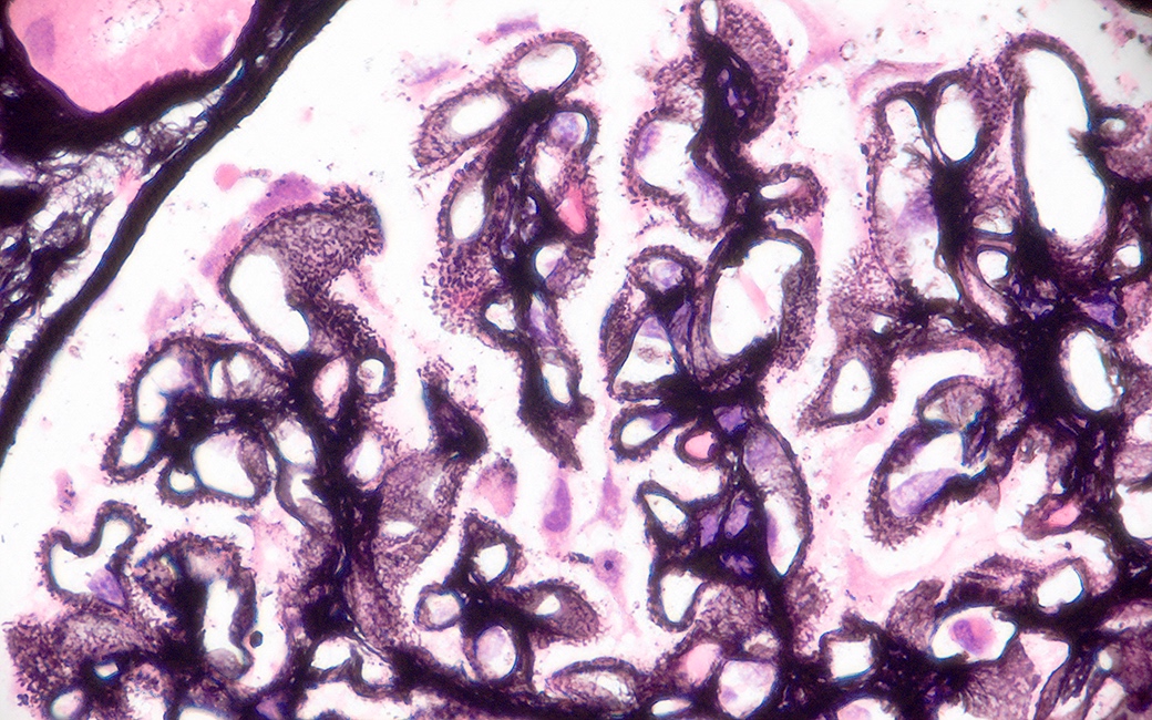
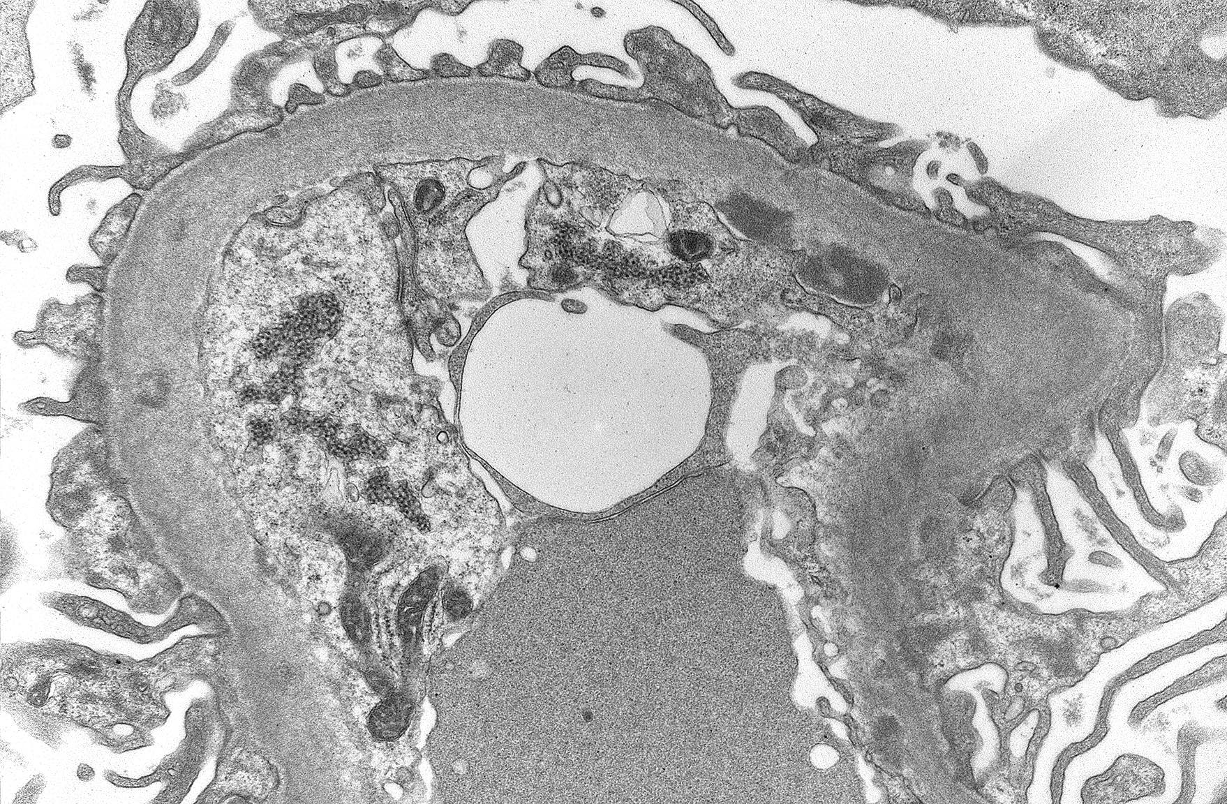
Subepithelial hump-like deposits

Mesangial deposits

Mesangial notch / hinge region deposit
Images hosted on other servers:


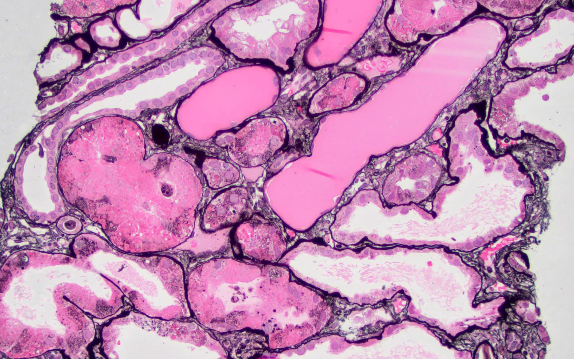
Subepithelial humps
Images hosted on other servers:

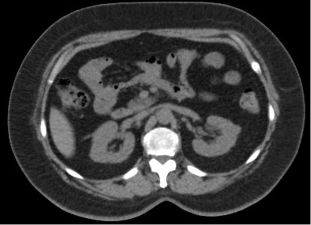
(a) CT: gas in
psoas muscle
(b) disappearance of
gas after treatment

CT: gas in renal parenchyma, and pararenal space

Ultrasound: "dirty" acoustic shadows (gas)

CT: gas in kidney and IVC
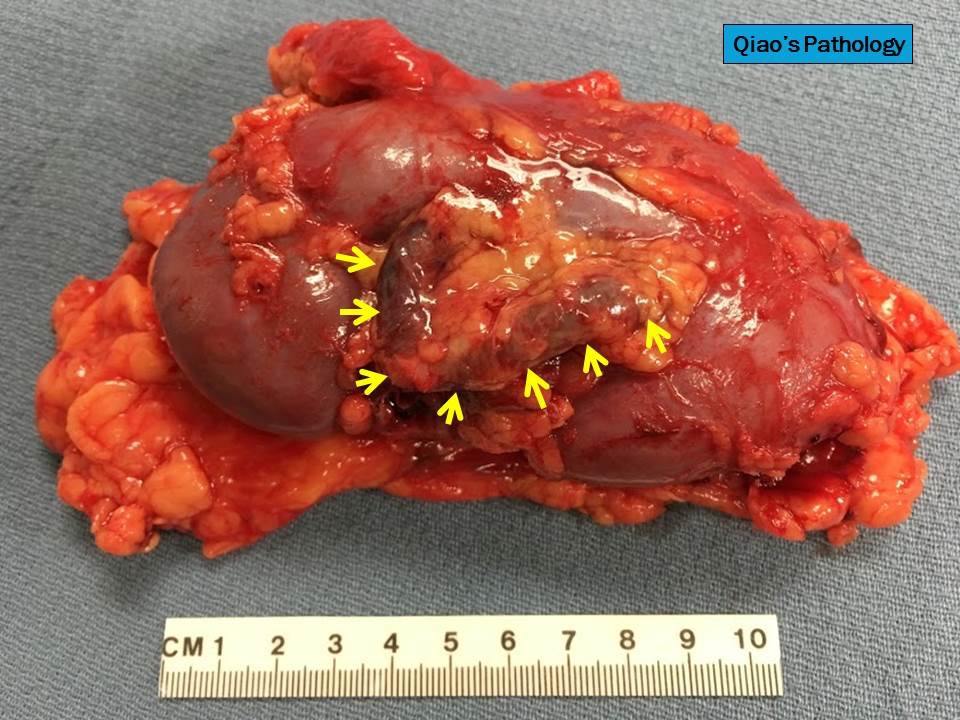
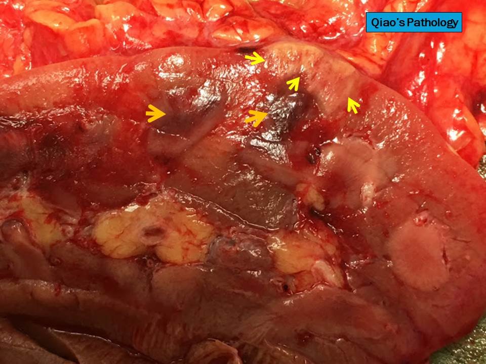
Contributed by Jian-Hua Qiao, M.D.
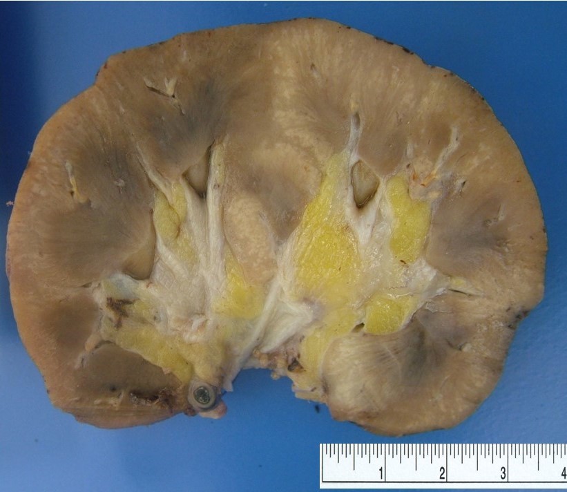
Bisected kidney
Detached renal capsule
Close view
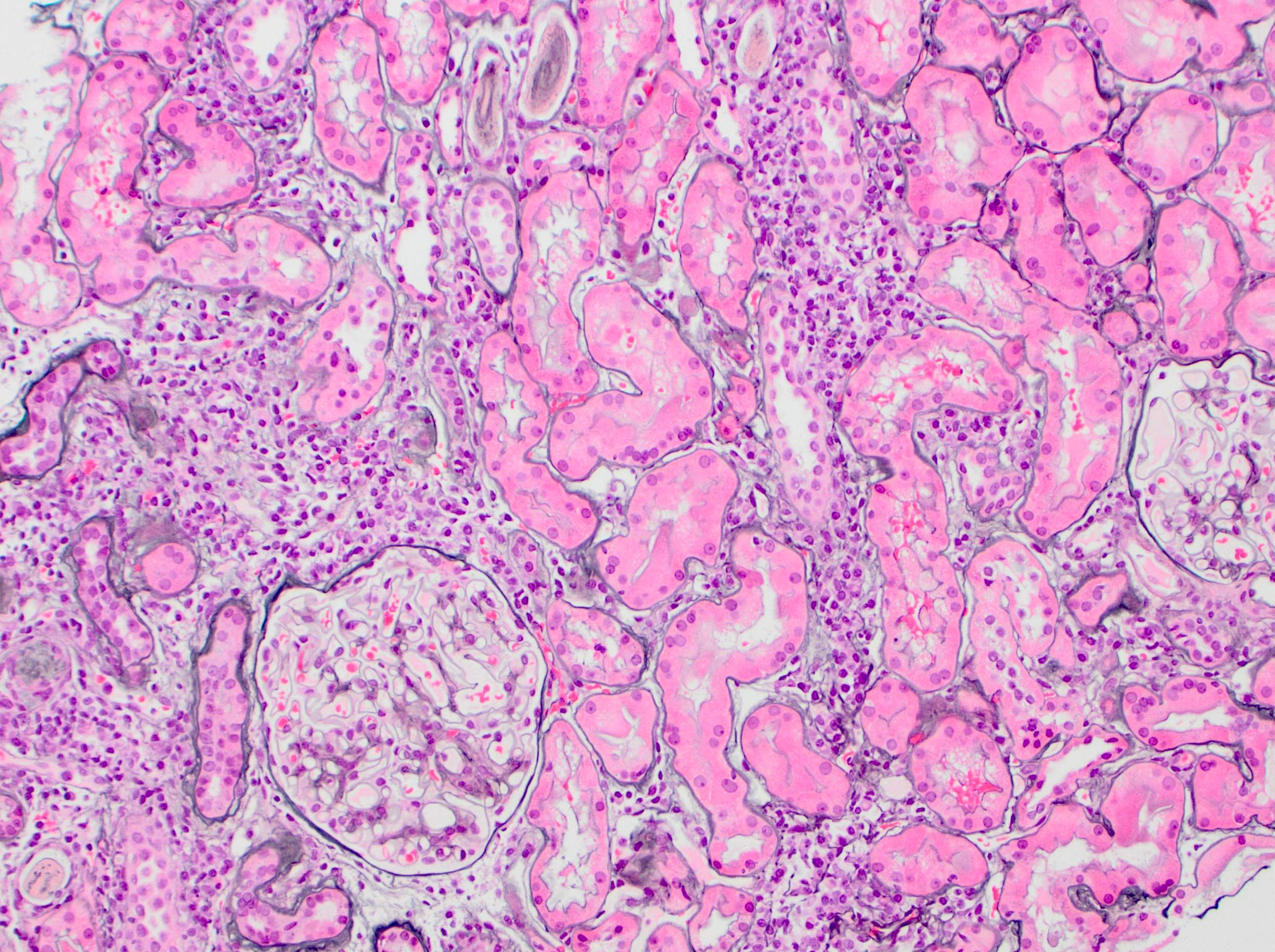
Gas filled cysts
Images hosted on other servers:

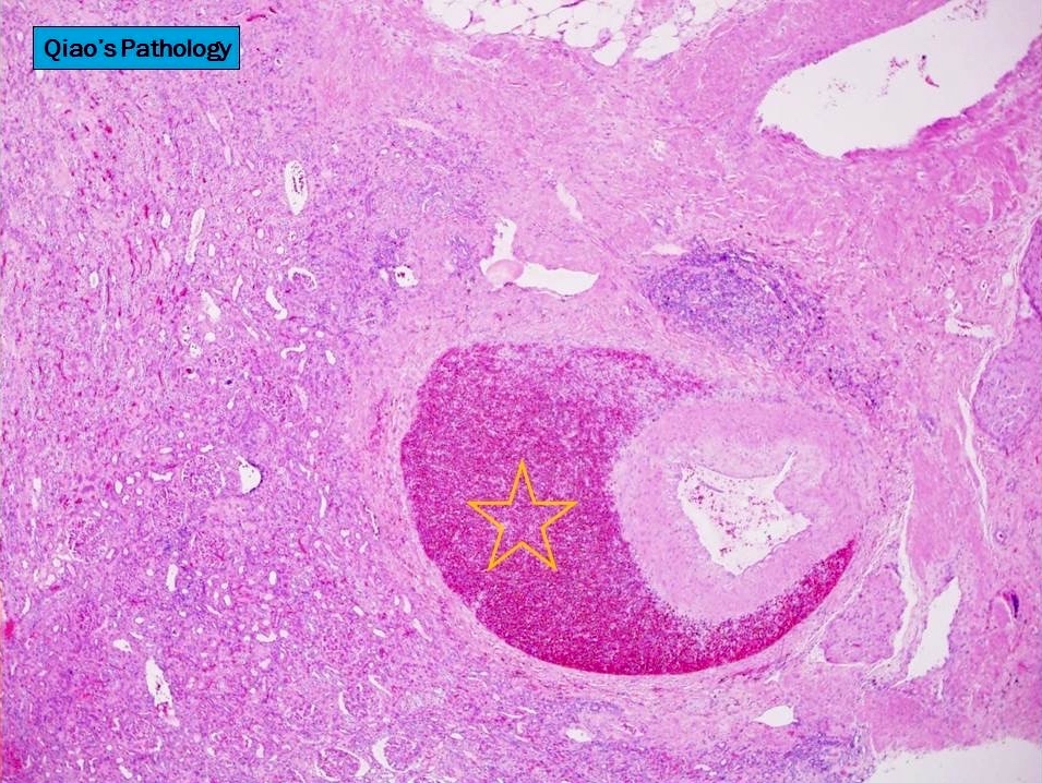
Areas of papillary necrosis
Contributed by Jian-Hua Qiao, M.D.
Acute inflammation
Neutrophil casts
Abscess
Necrotic renal cortex
Gas filled cysts
Images hosted on other servers:

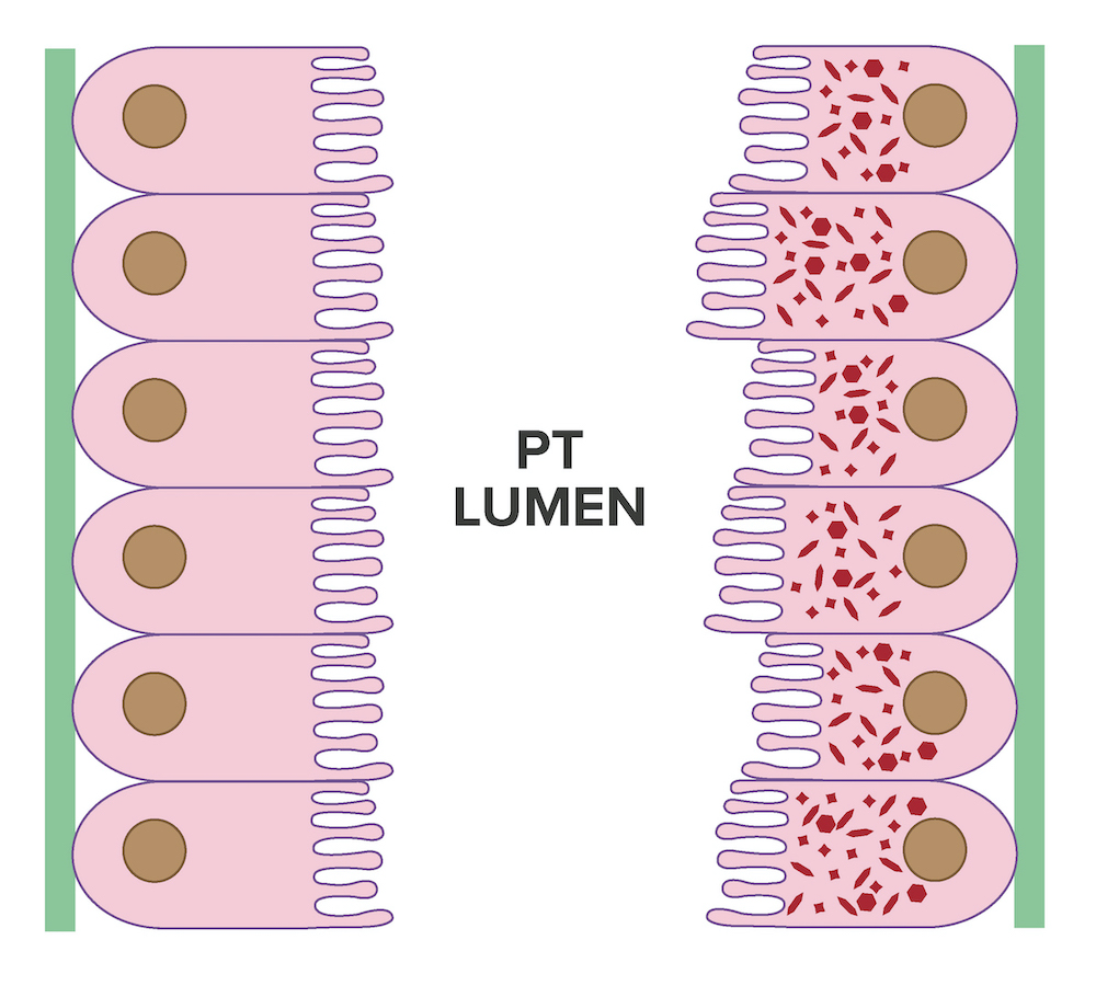
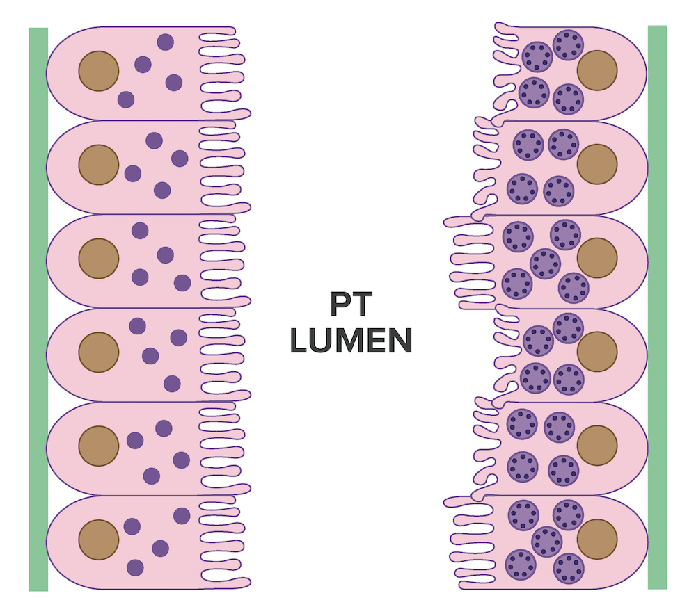
Inflammation and different stages of AKI

Pathophysiology of hypoperfusion related AKI
Contributed by Dale Davis, M.D., M.A.
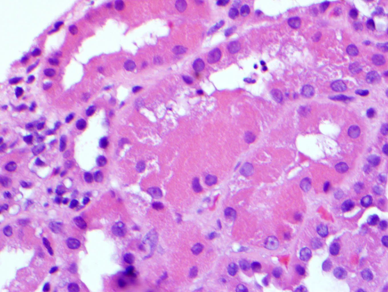
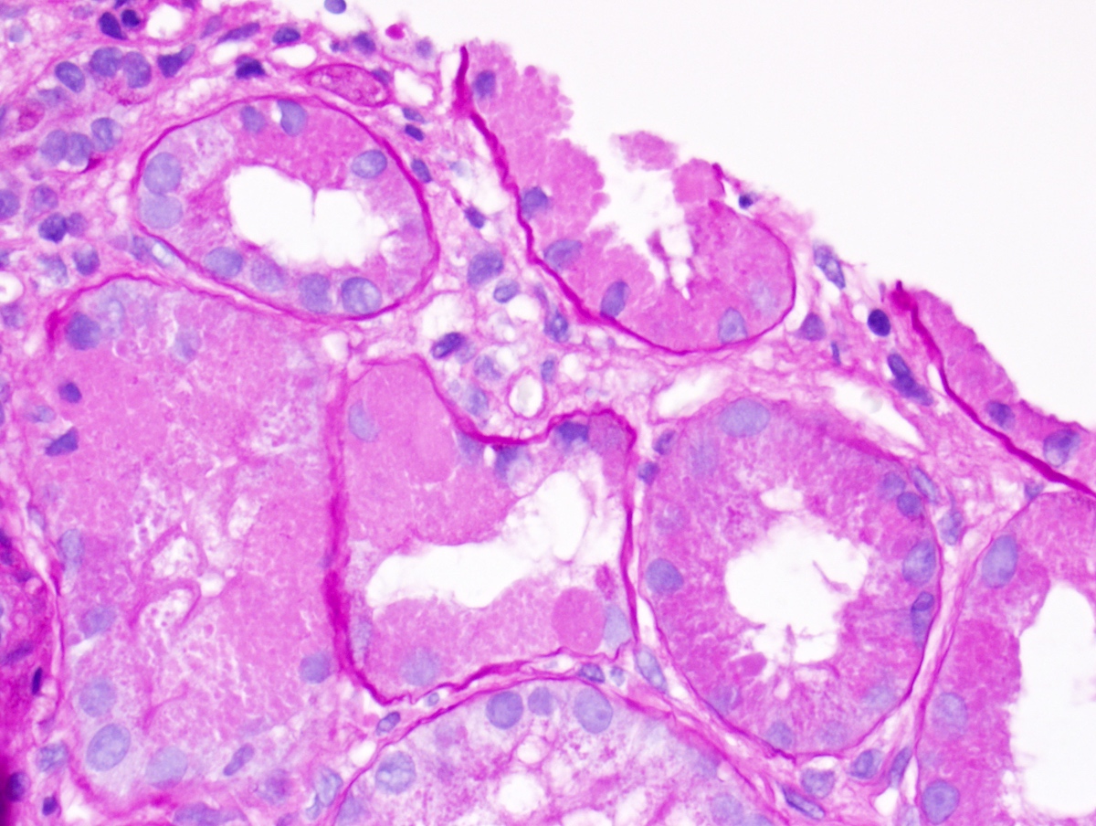
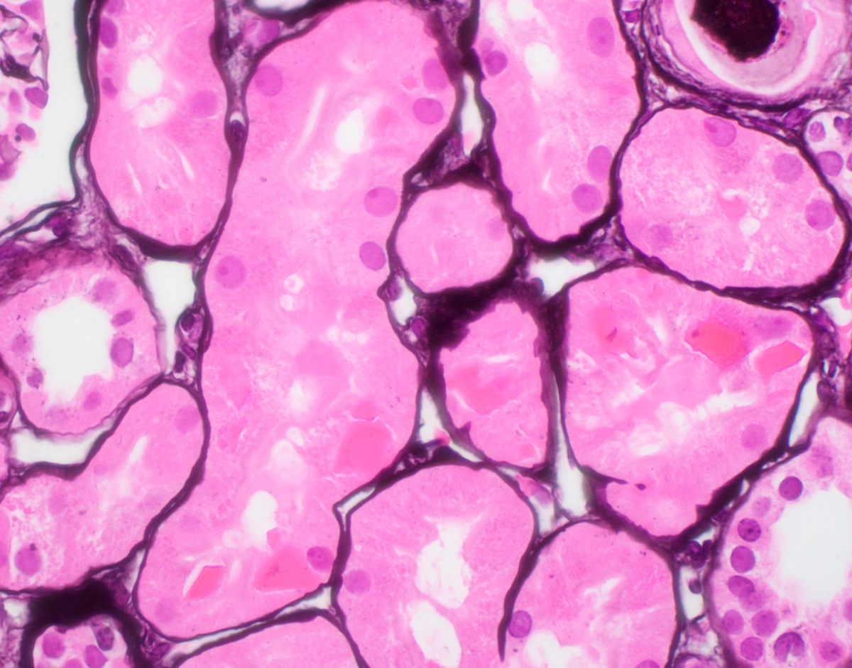
Attenuated tubules with casts
Cell sloughing
Cytoplasmic blebbing
Loss of brush border
Frank necrosis
Contributed by Colleen Ford
Lumen with cellular debris
Aberration of basement membrane
Overview of intrarenal acute kidney injury
Images hosted on other servers:

Various images

Adenovirus immunoperoxidase stain
Contributed by Alexei Mikhailov, M.D., Ph.D.
Early Alport syndrome
Early chronic changes
Focal segmental glomerular scarring
Foam cells, Alport syndrome
Contributed by Alexei Mikhailov, M.D., Ph.D.
Microvillus transformation
Characteristic changes
More advanced changes
Thin basement membrane disease
Alport-like changes
Images hosted on other servers:

Various images
Images hosted on other servers:

Vertical section


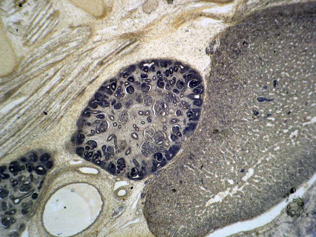
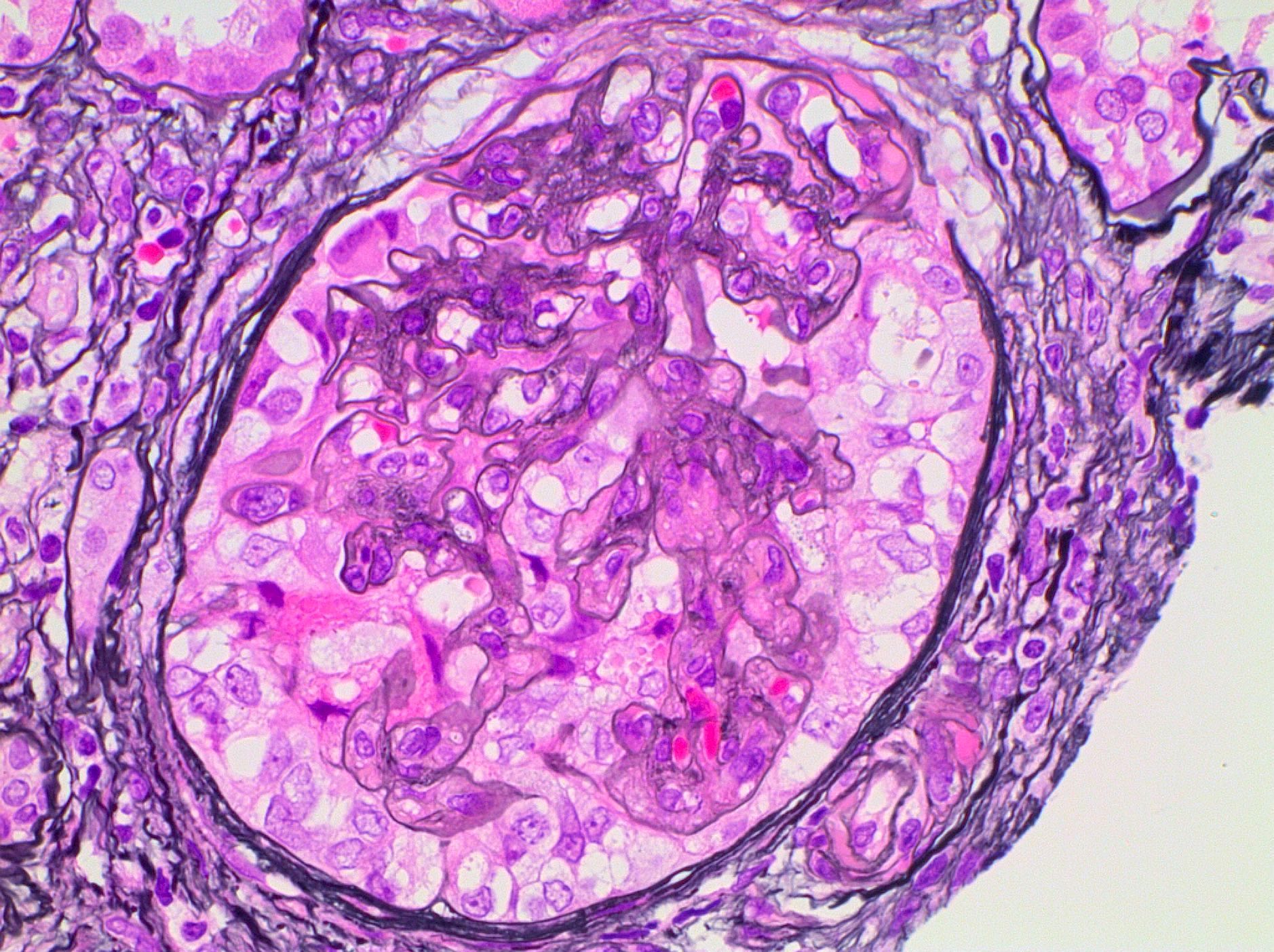
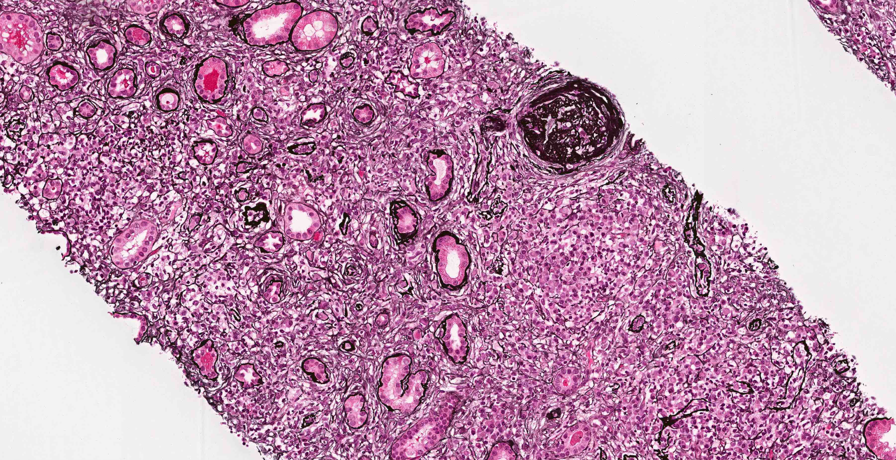
Normal glomerulus

Vessels surrounding glomeruli and tubules


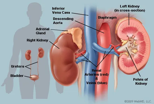
Relation to other organs

Juxtaglomerular apparatus

Renal column

Vessels supplying tubule

Vessels surrounding glomeruli and tubules
Images hosted on other servers:

Fetal kidneys at 25 weeks gestation

Infant kidney with fetal lobulations


Normal adult kidney
Contributed by Grigory Demyashkin, M.D., Ph.D.
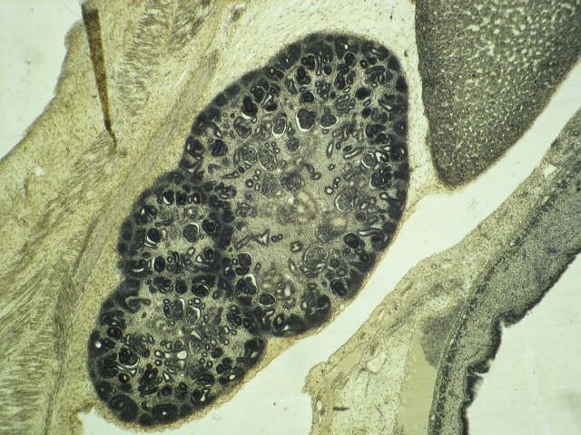
6 - 8 week embryo:
Metanephros; adrenal gland medulla
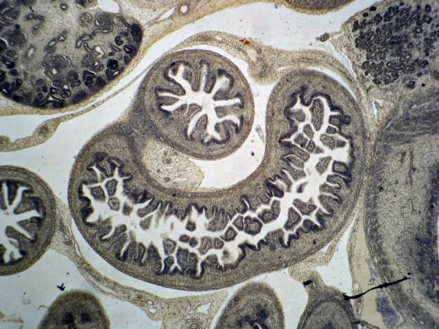
Metanephros; colon; pancreas

Small intestine; stomach;
gonad and kidney primary
(mesonephros); symphysis
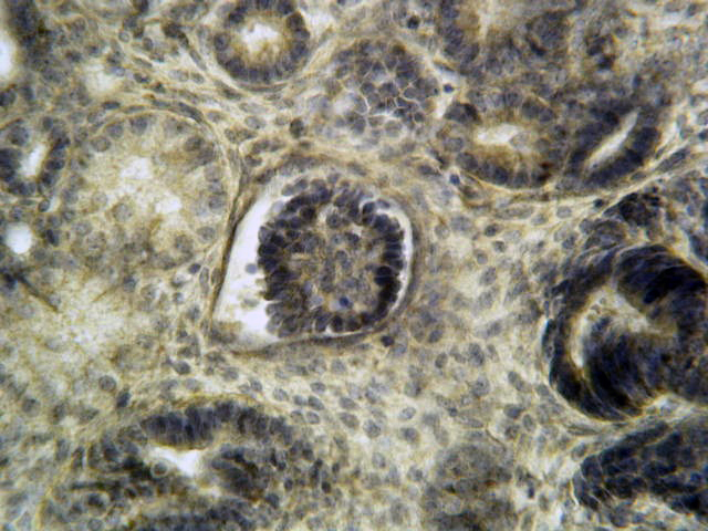
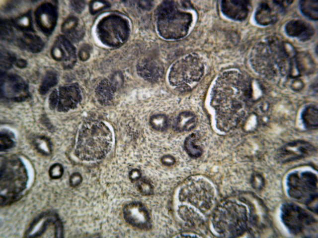
Final kidney (metanephros), renal corpuscle
Final kidney (metanephros), renal corpuscle, collecting ducts
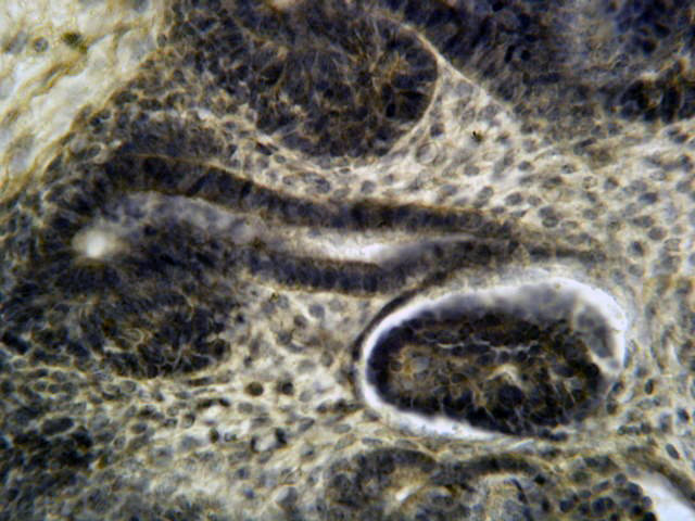
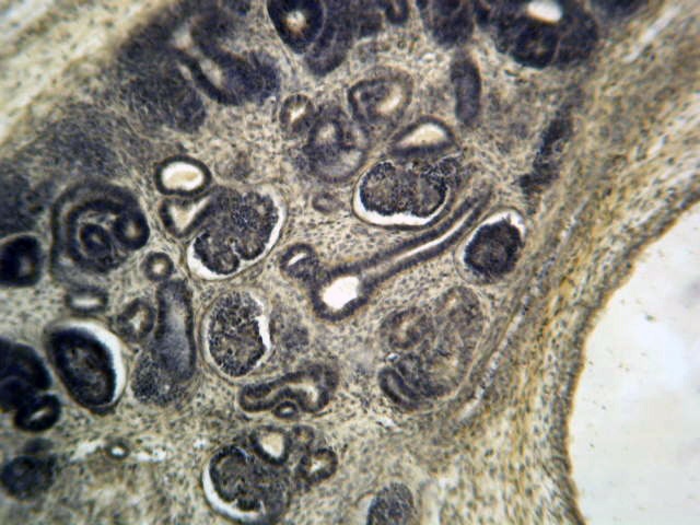
Metanephros; adrenal gland medulla
Final kidney (metanephros)

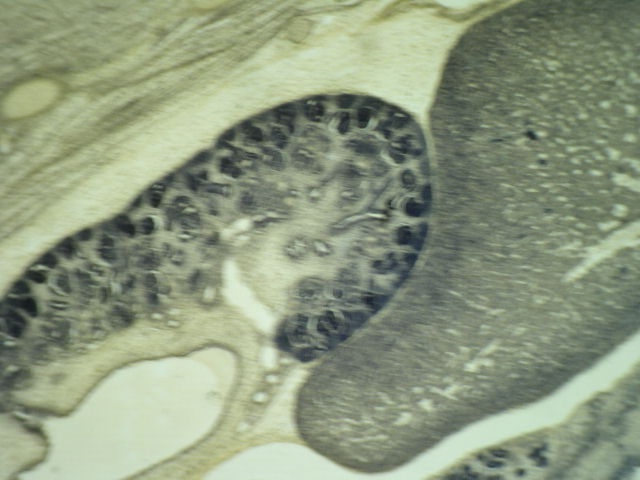
Final kidney (metanephros);
liver; gonad & kidney
primary (mesonephros);
symphysis
Final kidney (metanephros)
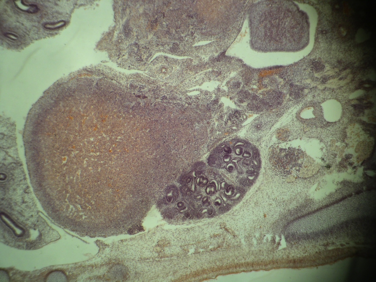
Adrenal gland, medulla & final kidney (metanephros)
Images hosted on other servers:

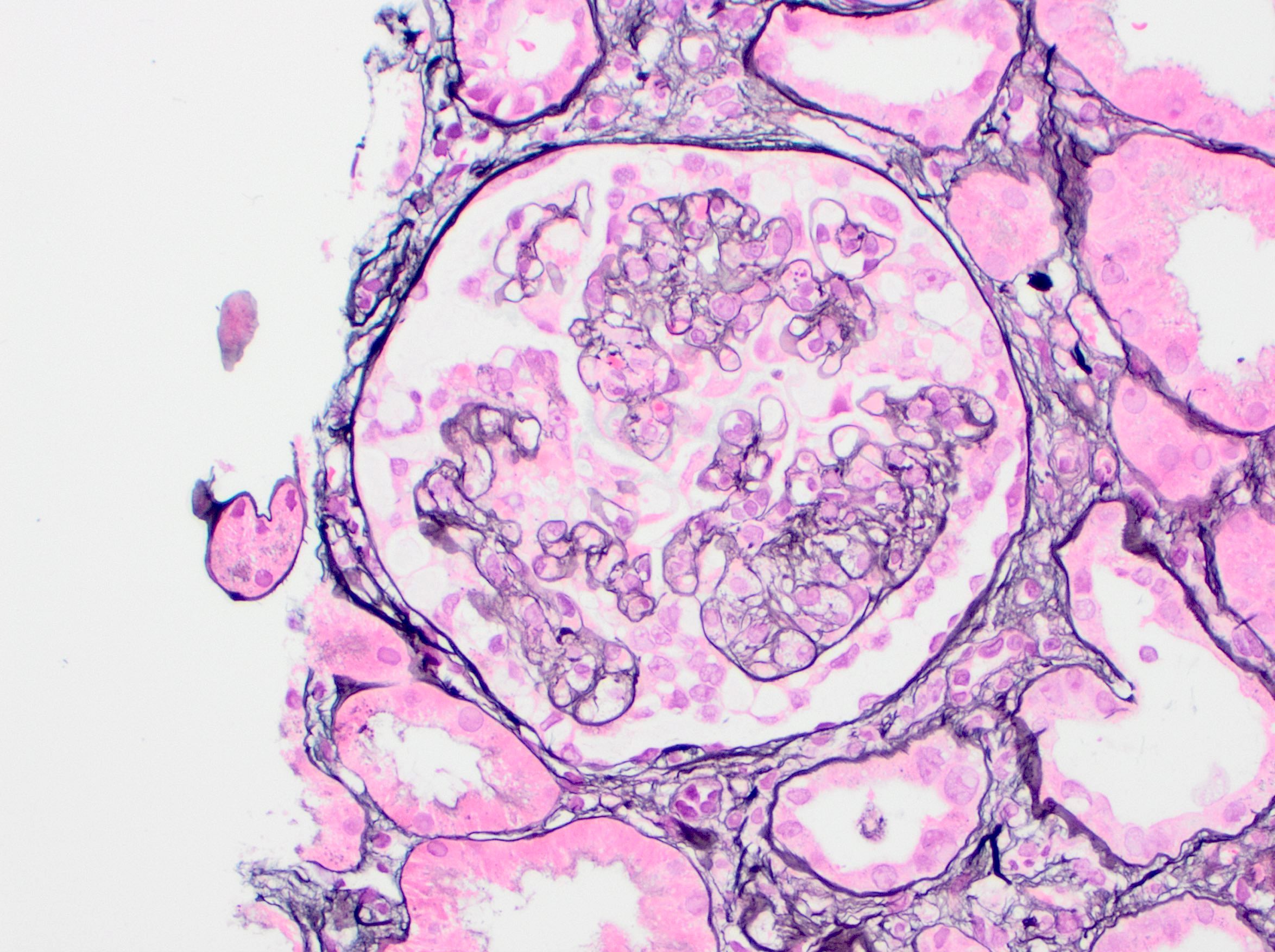
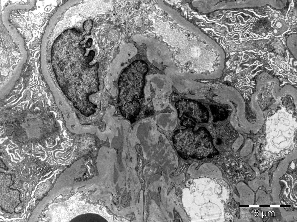
Glomerular capillary loops are thin and delicate

Basement membranes
of glomerular
capillary loops and
tubular epithelium
Collecting duct: brush border

Juxtaglomerular apparatus and macula densa
Medullary rays
Contributed by Alexander J. Gallan, M.D.
Tubular injury
Contributed by Alexander J. Gallan, M.D.
TBM deposits
Segmental glomerular subepithelial deposits
Contributed by Ana Belén Larqué, M.D., Ph.D.
Cellular crescents
Fibrinoid necrosis

Disruption of Bowman capsule
Images hosted on other servers:

Extracapillary crescent
GBM, Alport and anti-GBM
Contributed by Husam Jum’ah, M.D.
Renal cortical surface with granularity
Images hosted on other servers:

Characteristic granular appearance
Contributed by Santhi Ganesan, M.D.
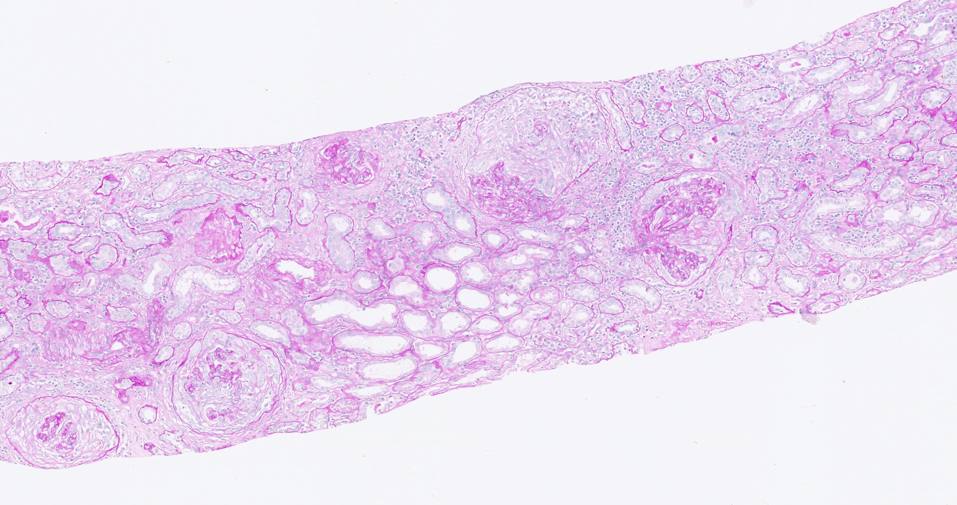
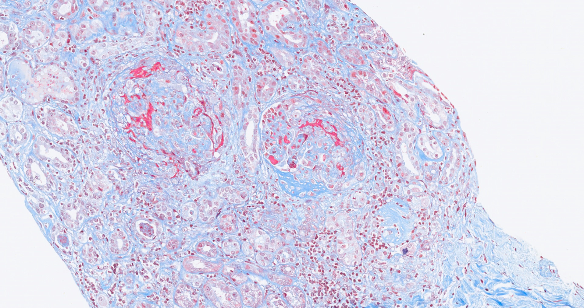
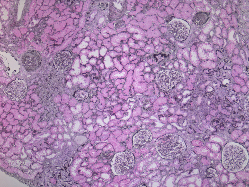
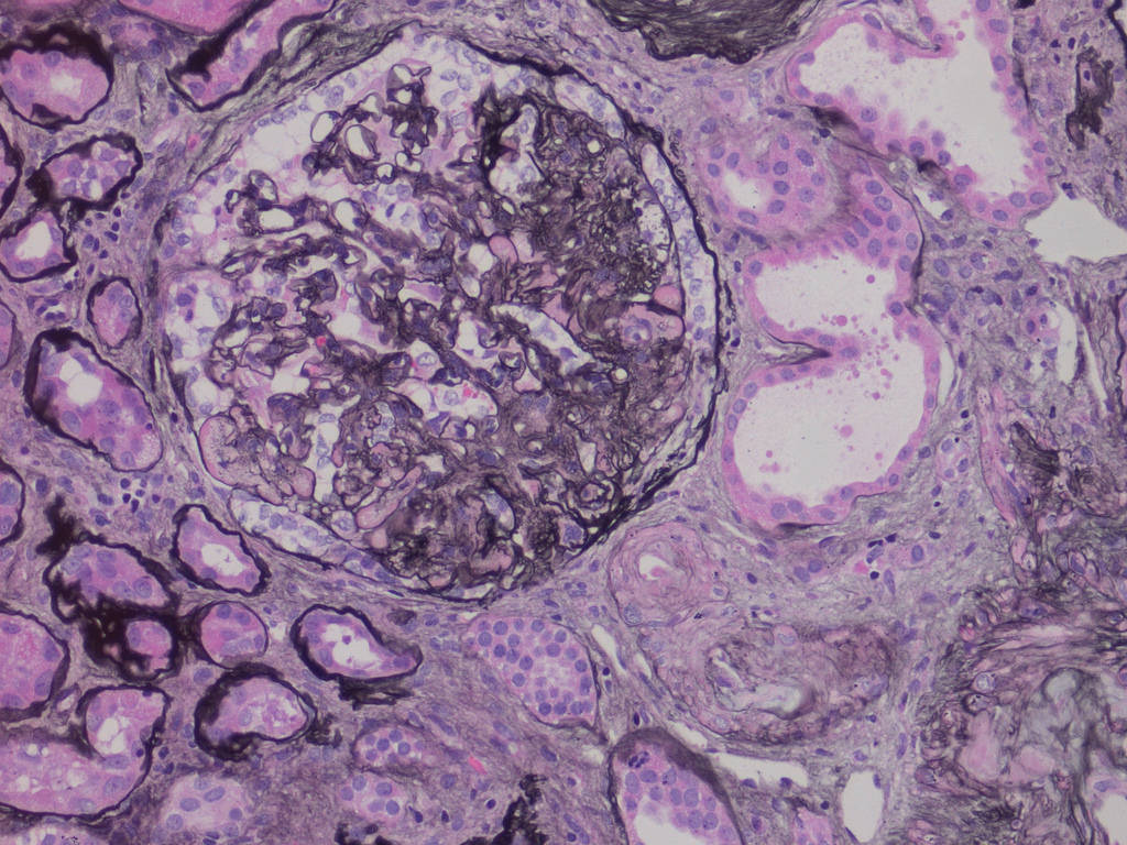
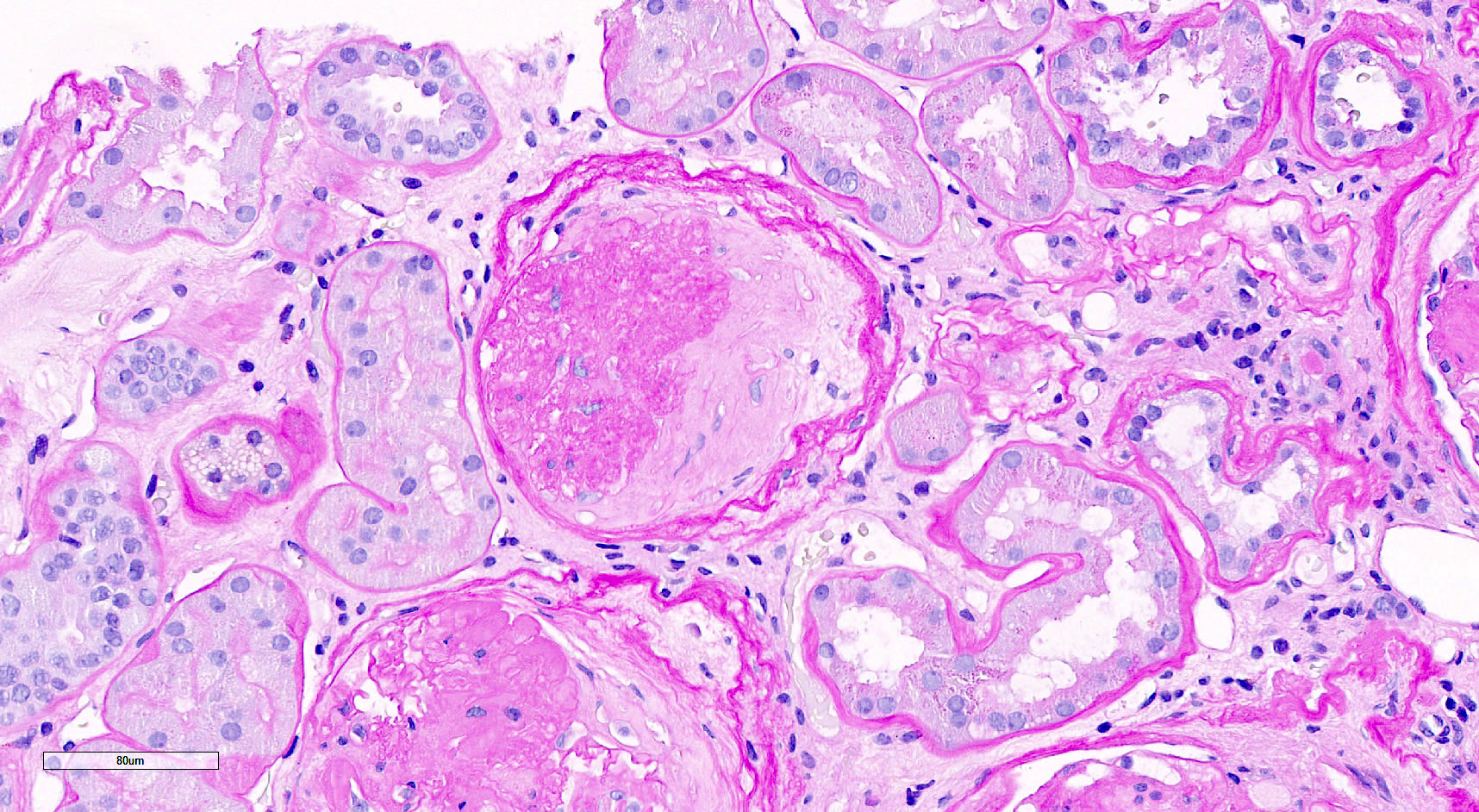
Global
glomerulosclerosis
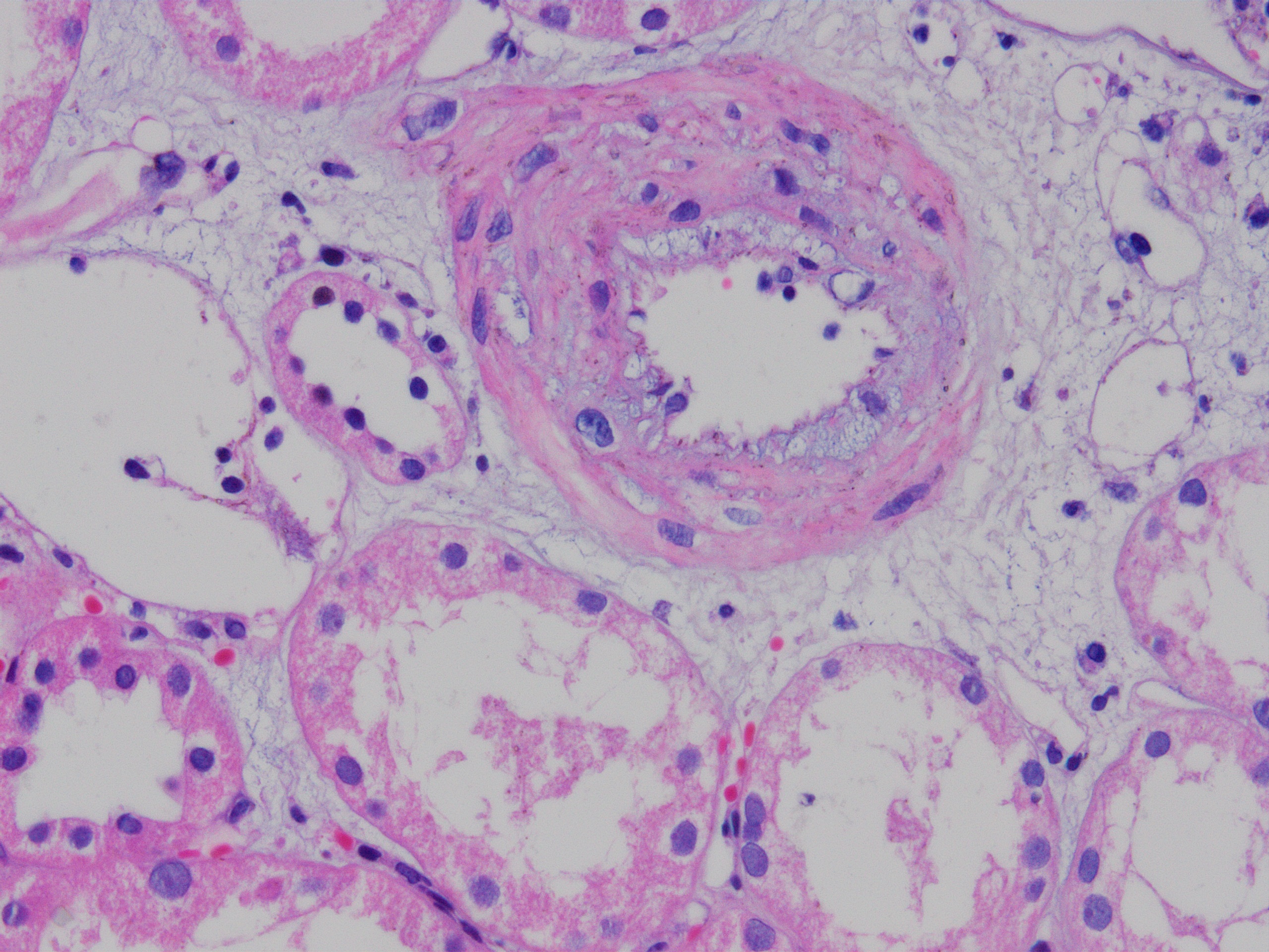
Arterial medial thickening
Hyaline vascular changes
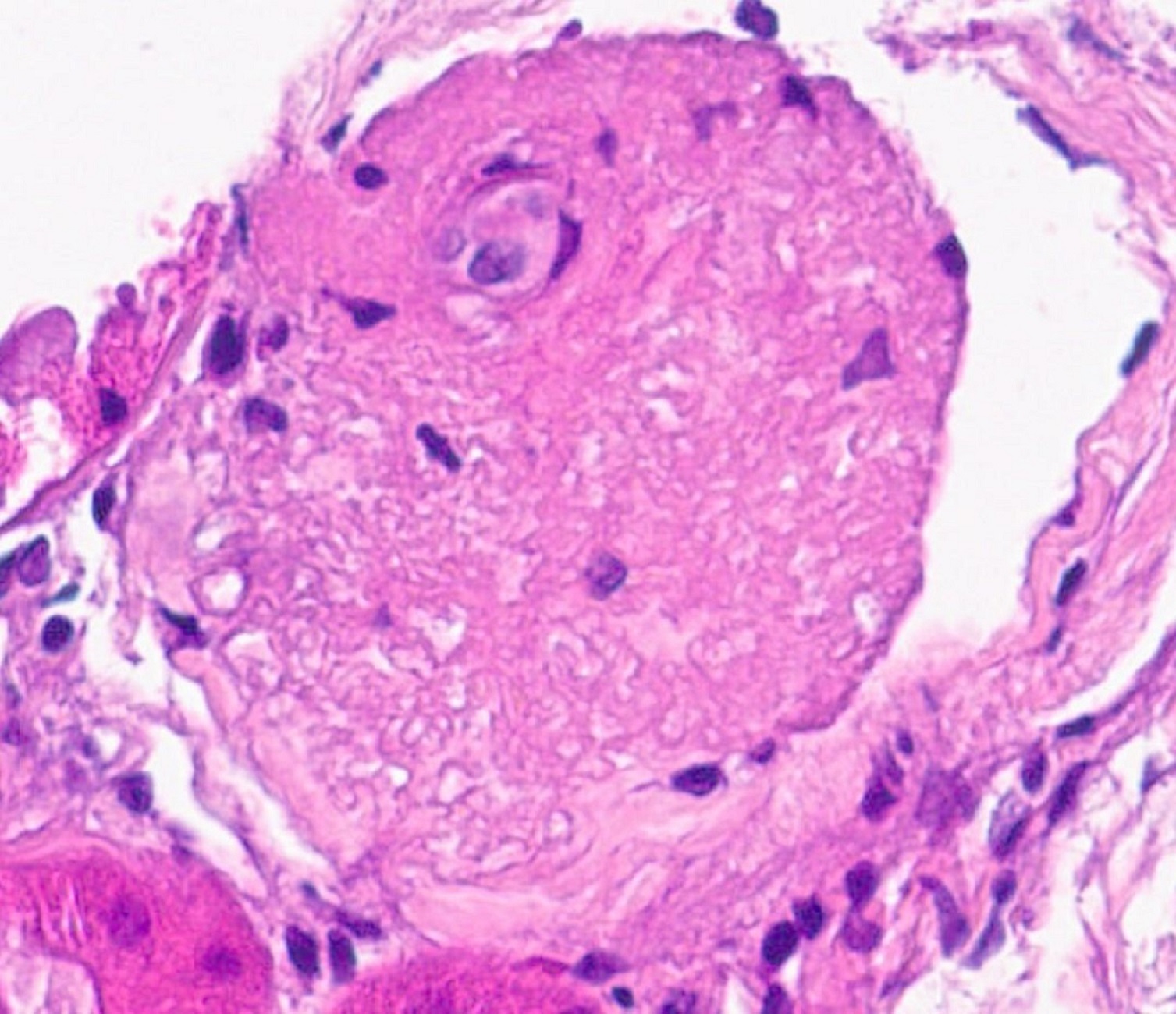
Obsolescent glomerulus
Global
glomerulosclerosis
PAS
Subintimal fibrosis trichrome
Images hosted on other servers:

Angiogram
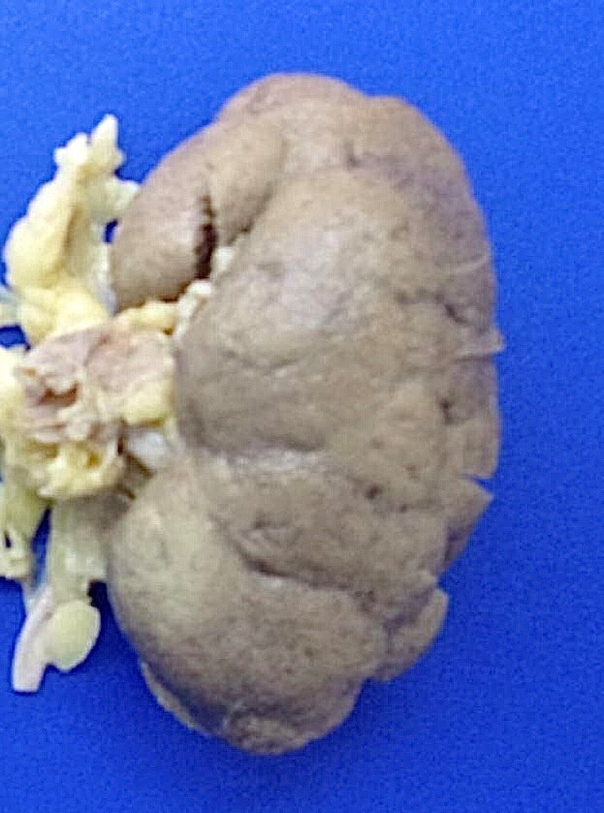
Contributed by Debra L. Zynger, M.D.
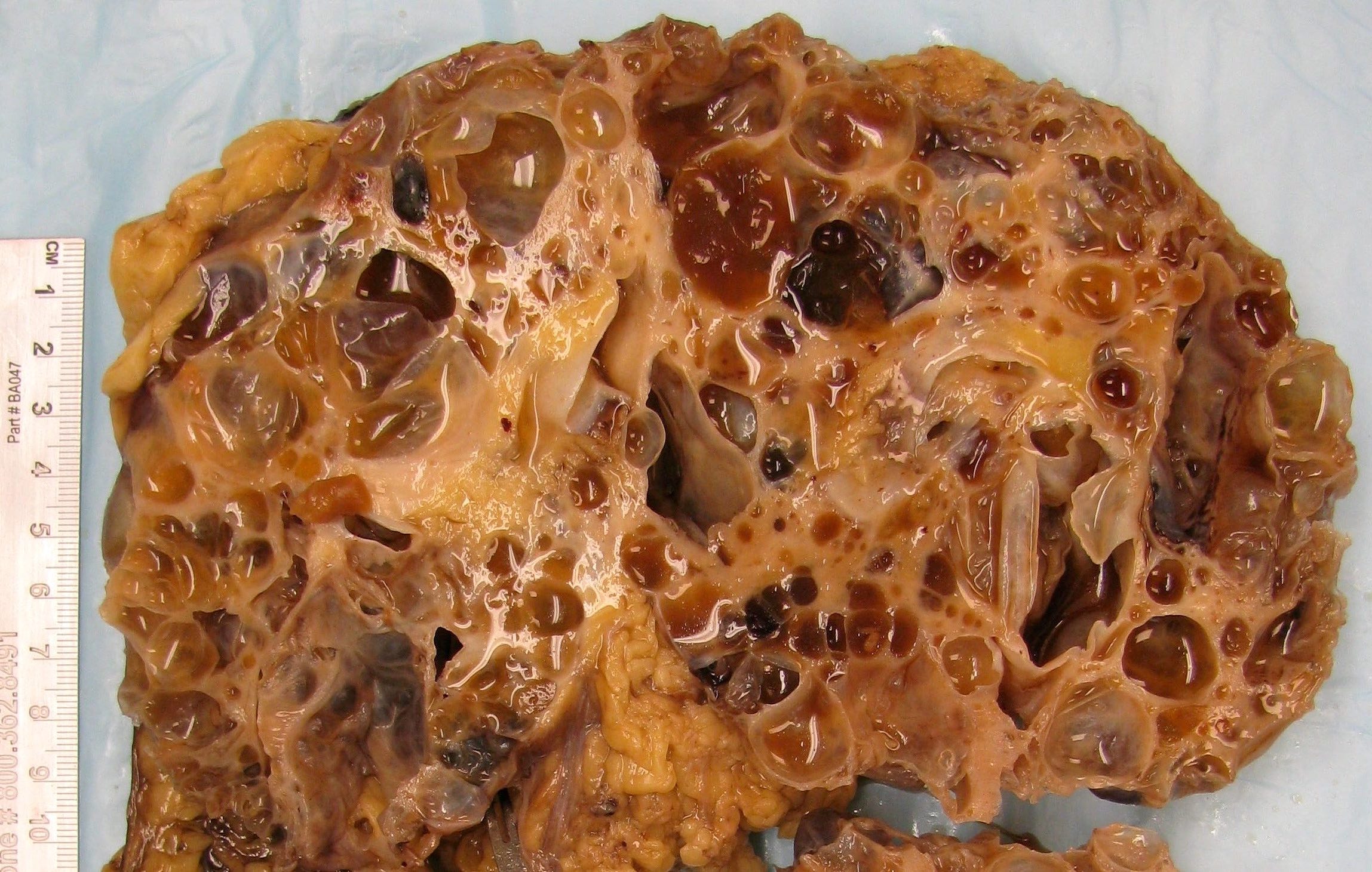
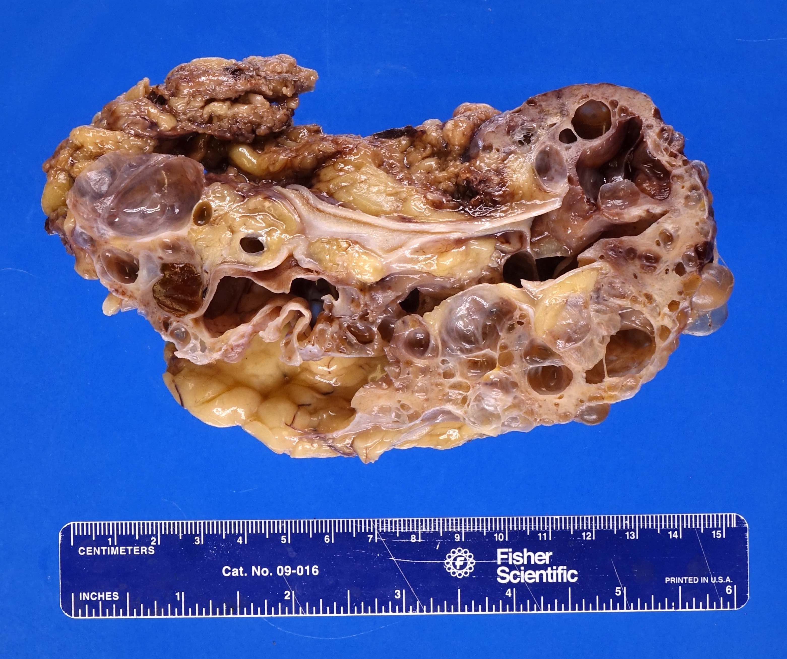
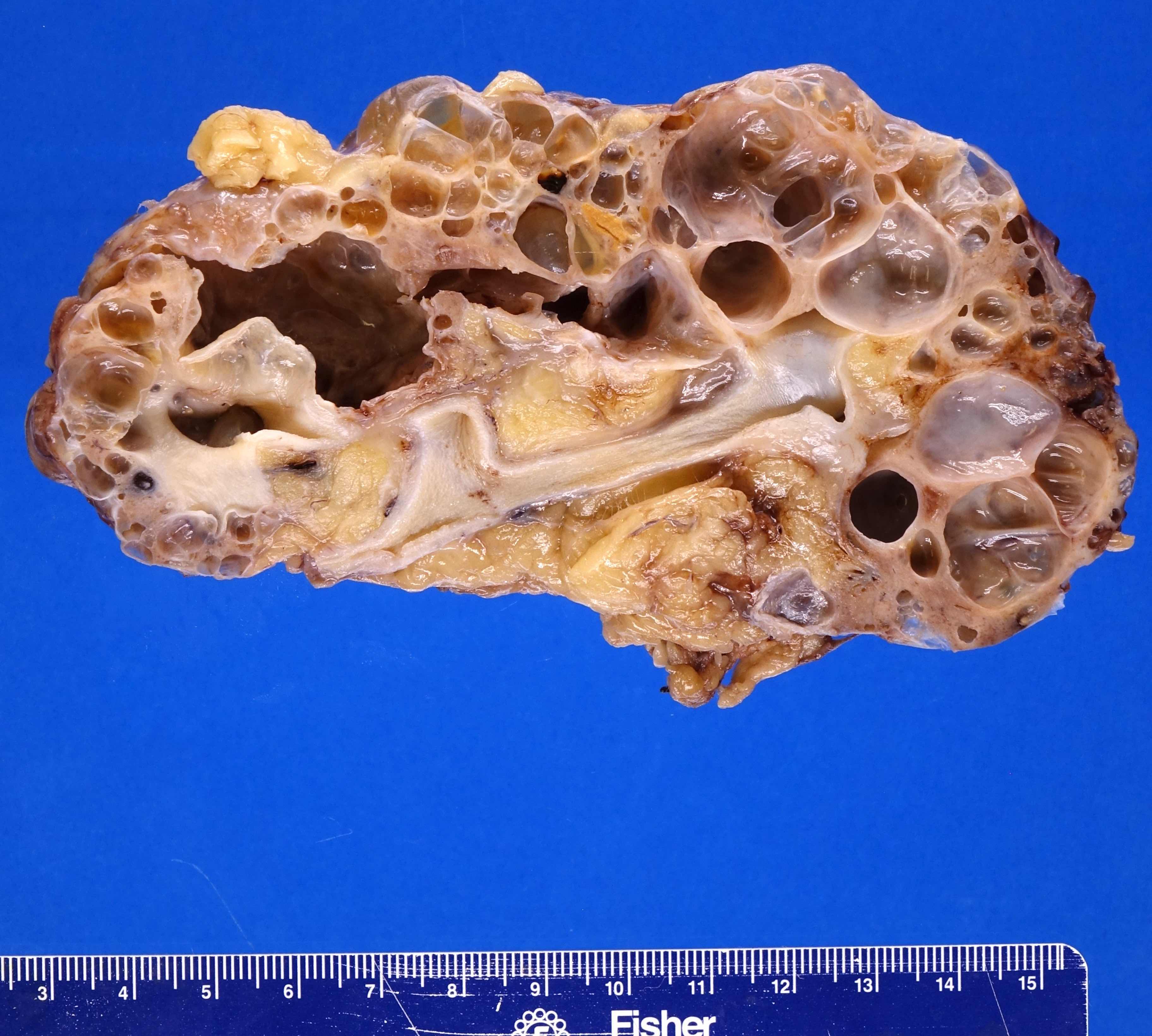
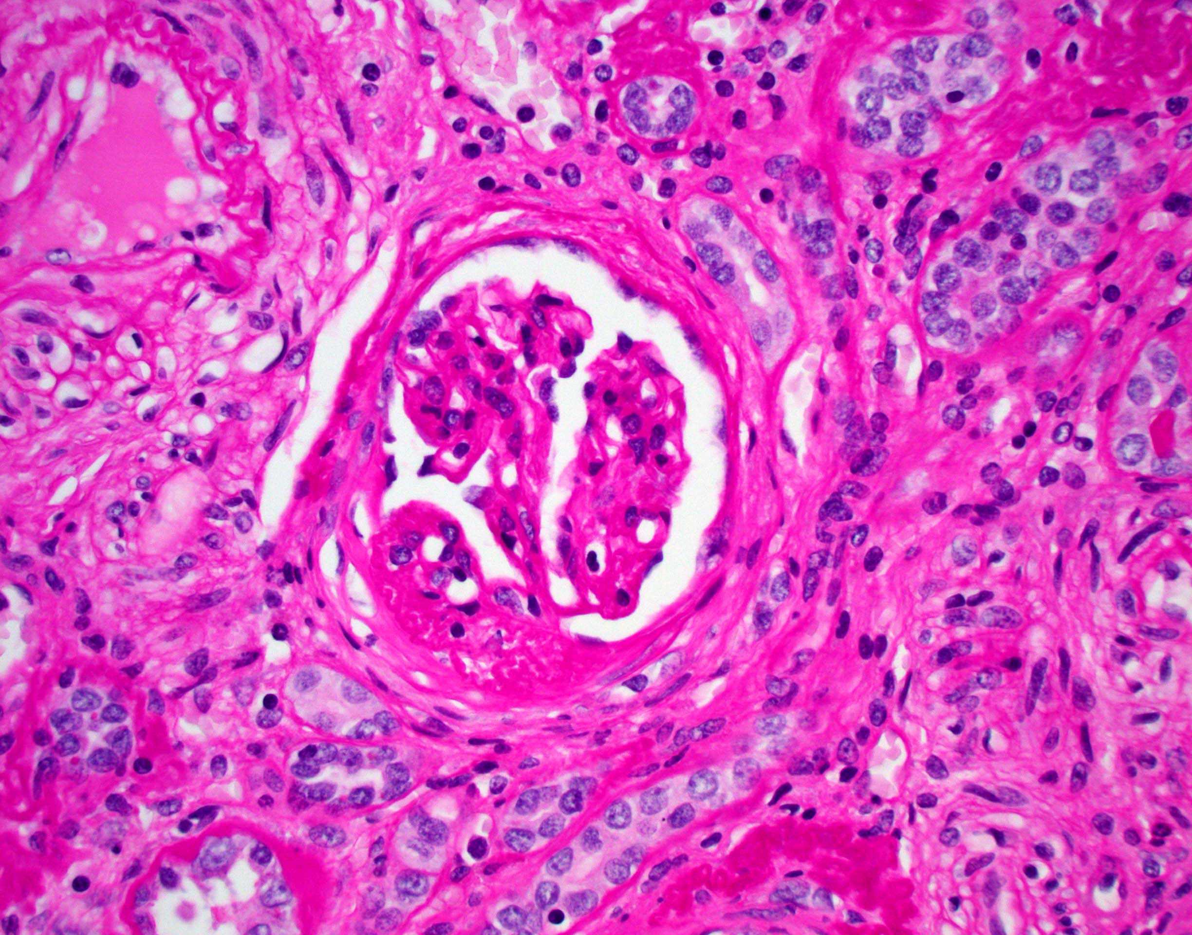
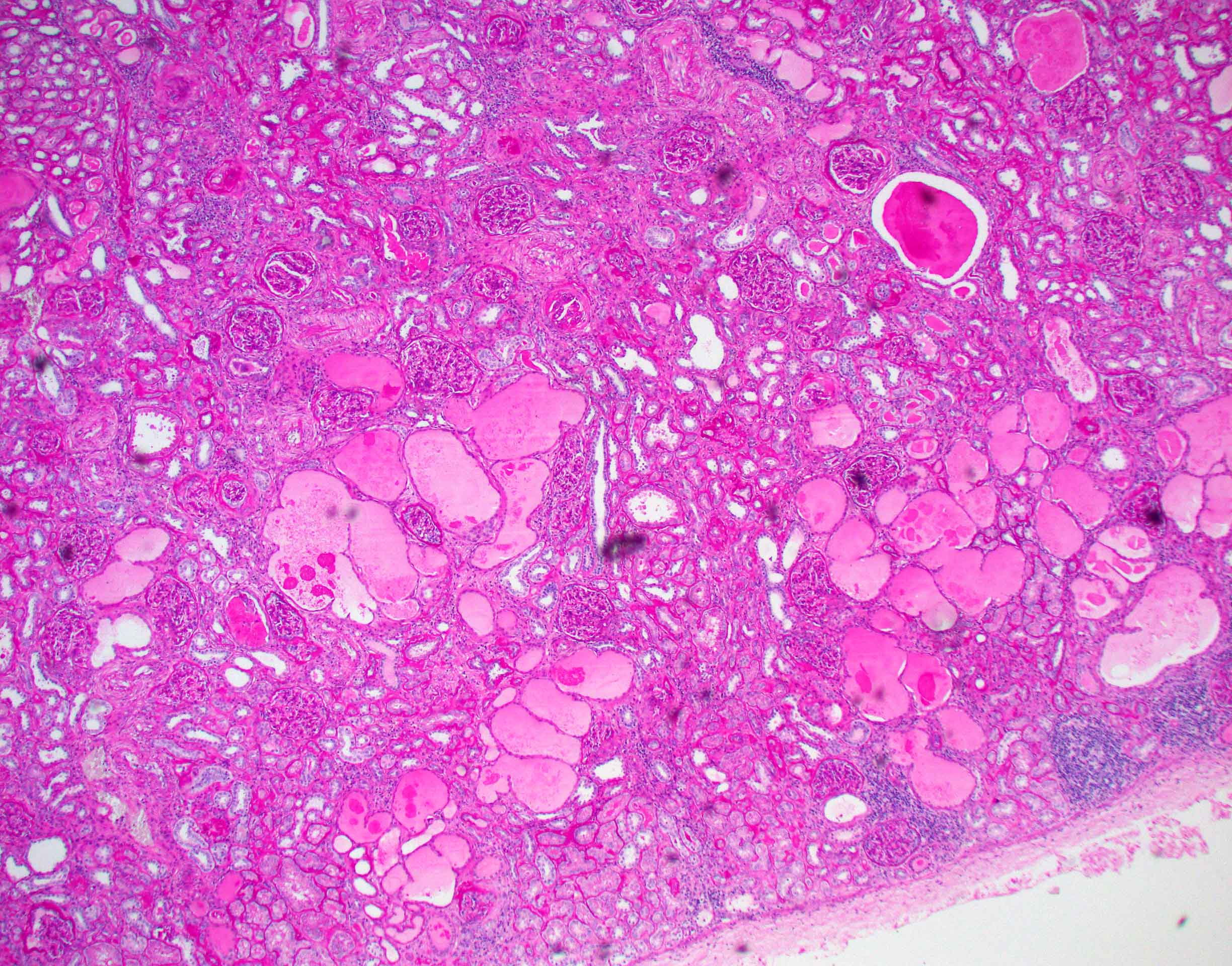
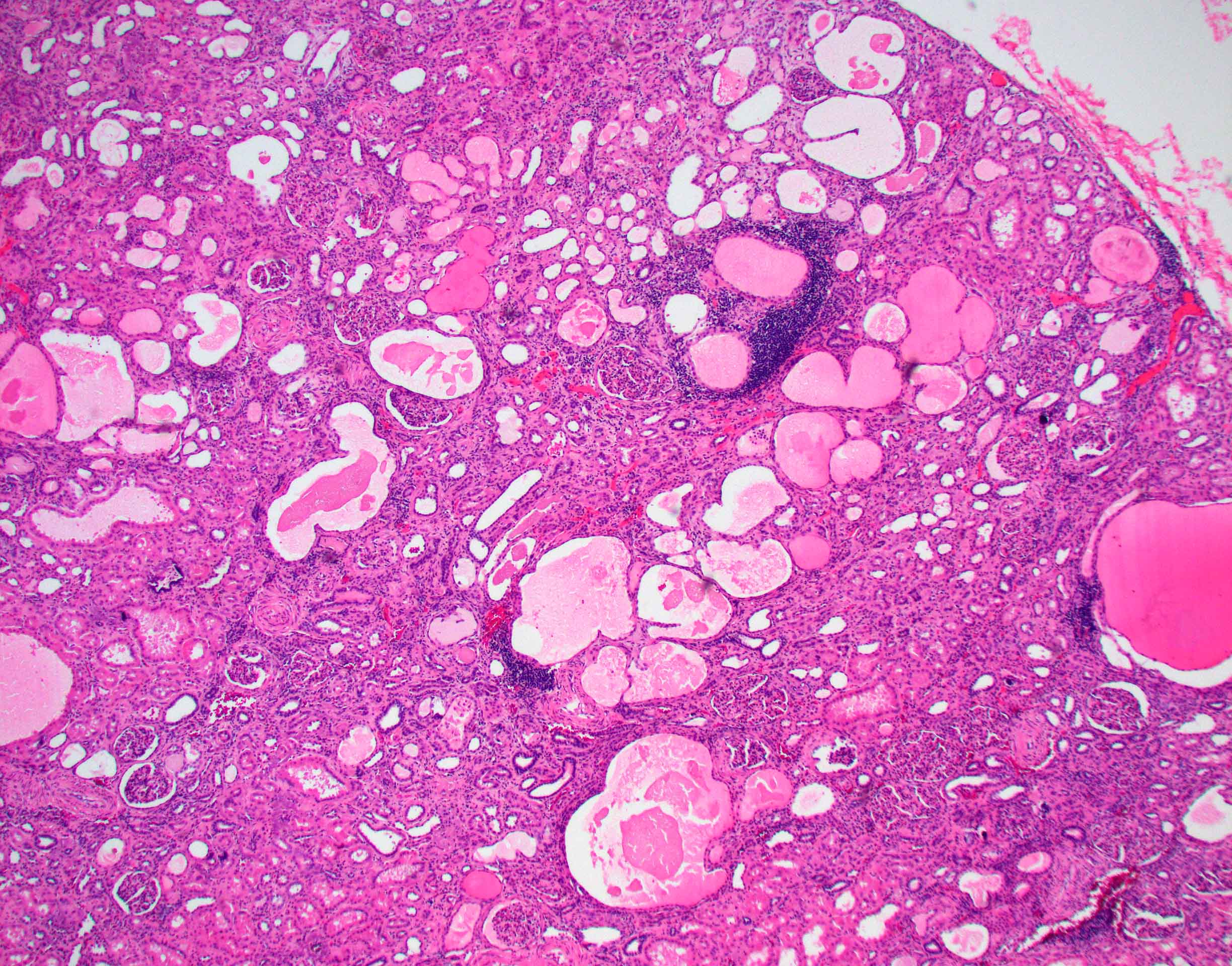
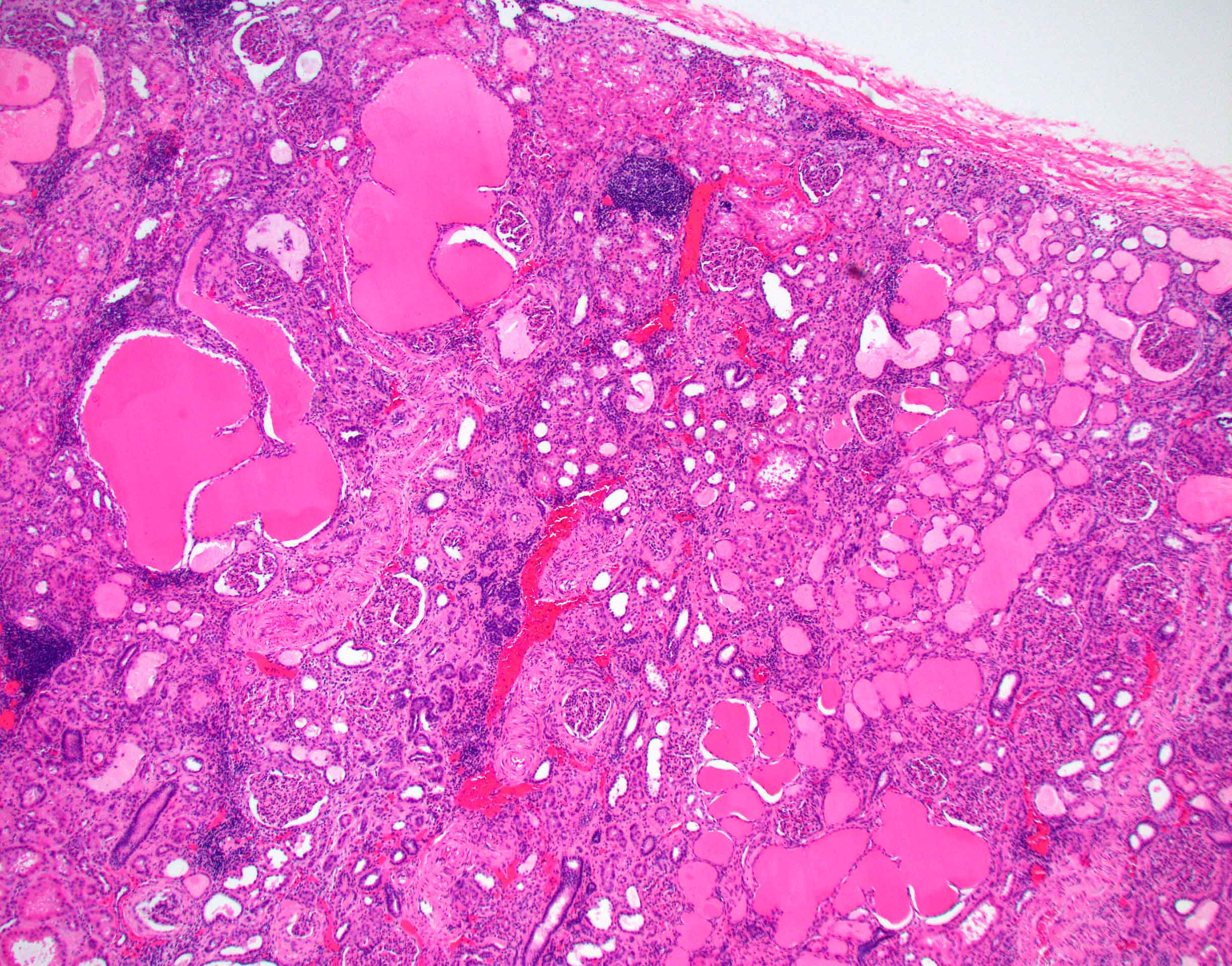
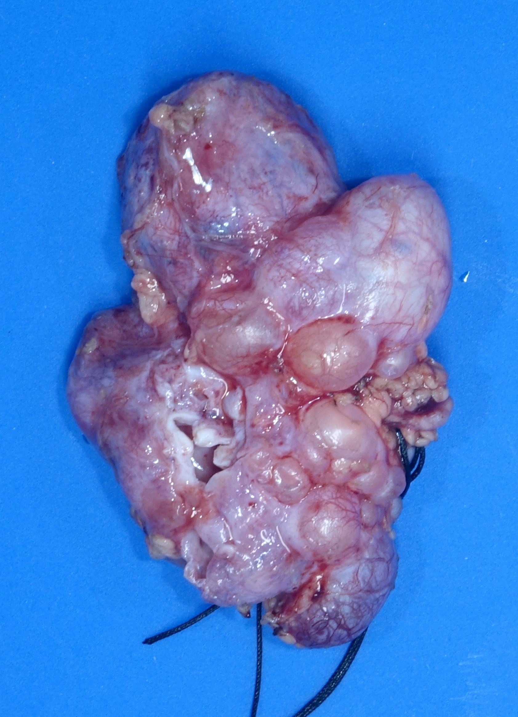
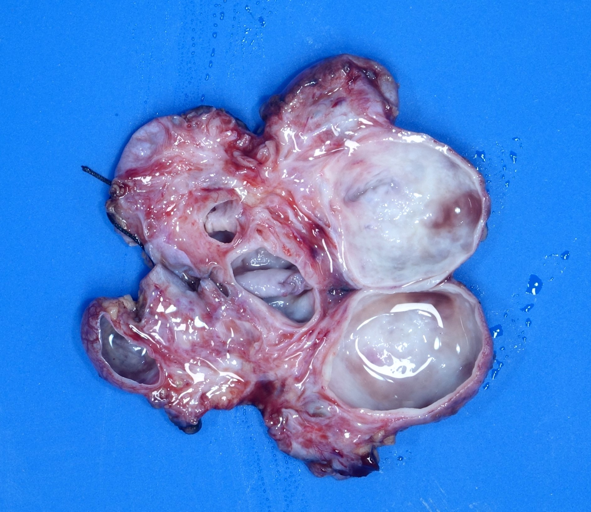
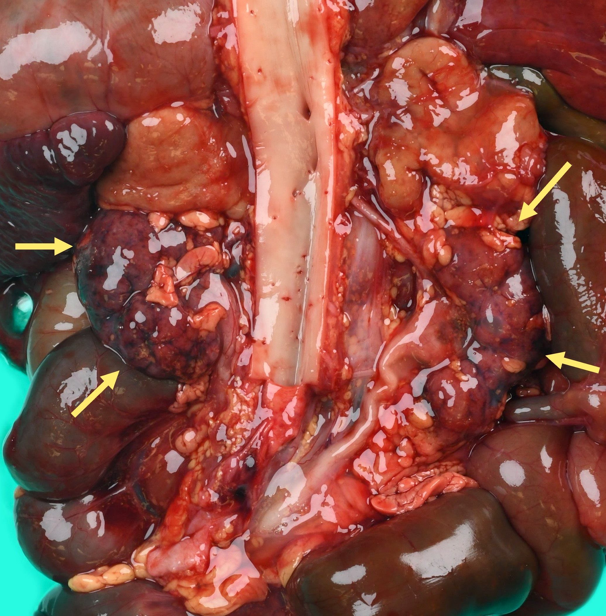
Numerous cysts of varying sizes
Images hosted on other servers:


Enlarged kidney with variably sized cysts

With transplanted kidney

Compared to normal kidney

Hemorrhagic infarct with rupture
Images hosted on other servers:


Cysts of varying sizes; glomerular cyst present at birth (rare)
Images hosted on other servers:


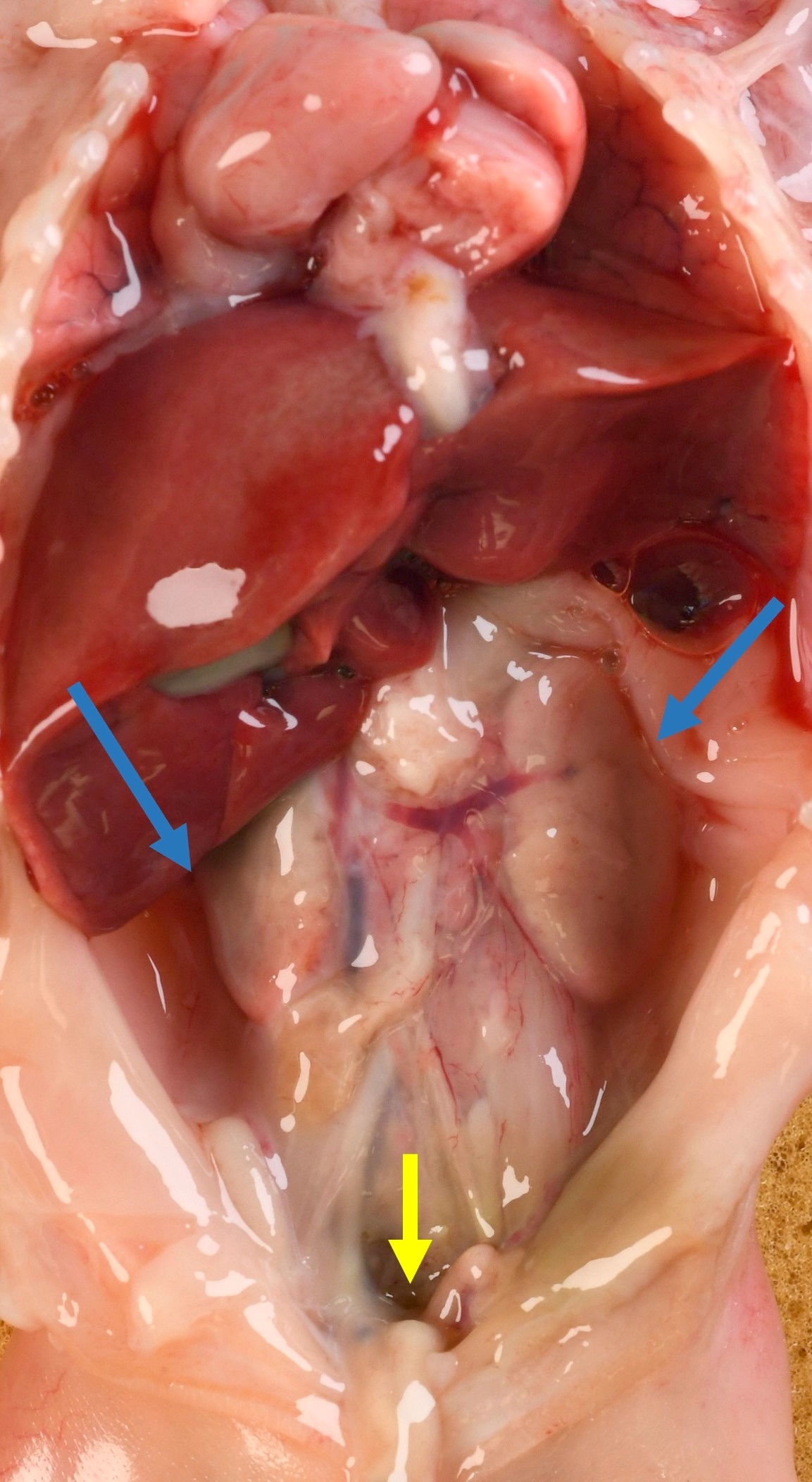
Markedly enlarged kidneys

Enlarged kidneys with persistent fetal lobation

Uniform distribution of small cysts

Small cysts in cortex and medulla

Renal surface shows small cysts

Spongy cut surface
Images hosted on other servers:

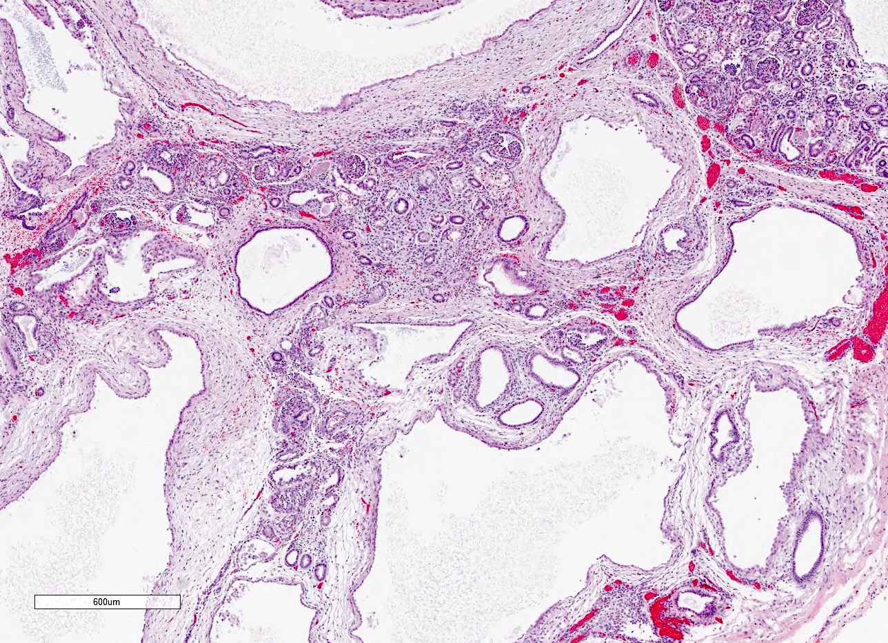
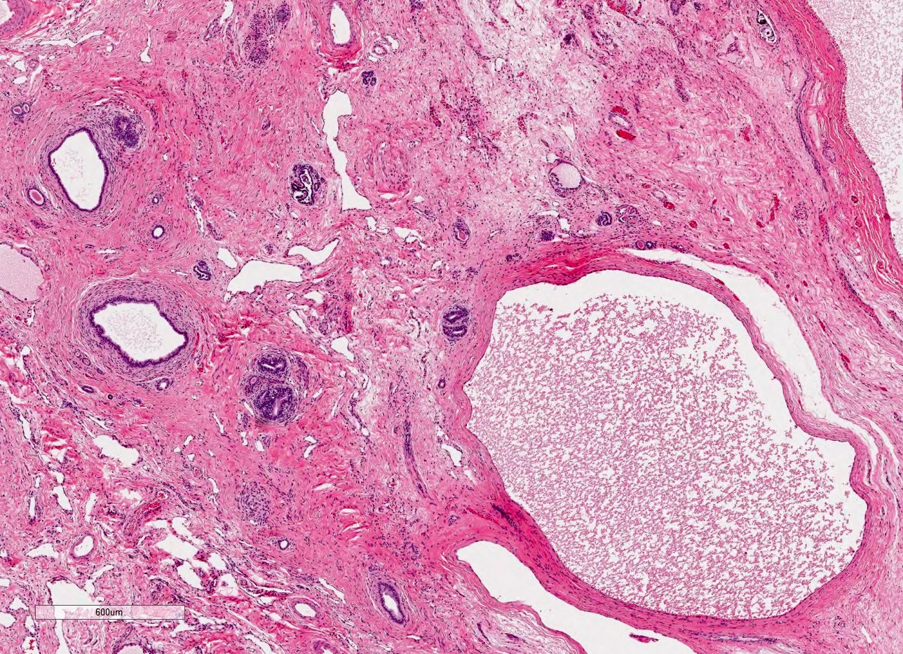
Radial arrangement of cysts with only rare glomeruli

Dilated collecting ducts in cortex and medulla

Immunostains

Associated congenital hepatic fibrosis
Contributed by Vanderlene Liu Kung, M.D., Ph.D.
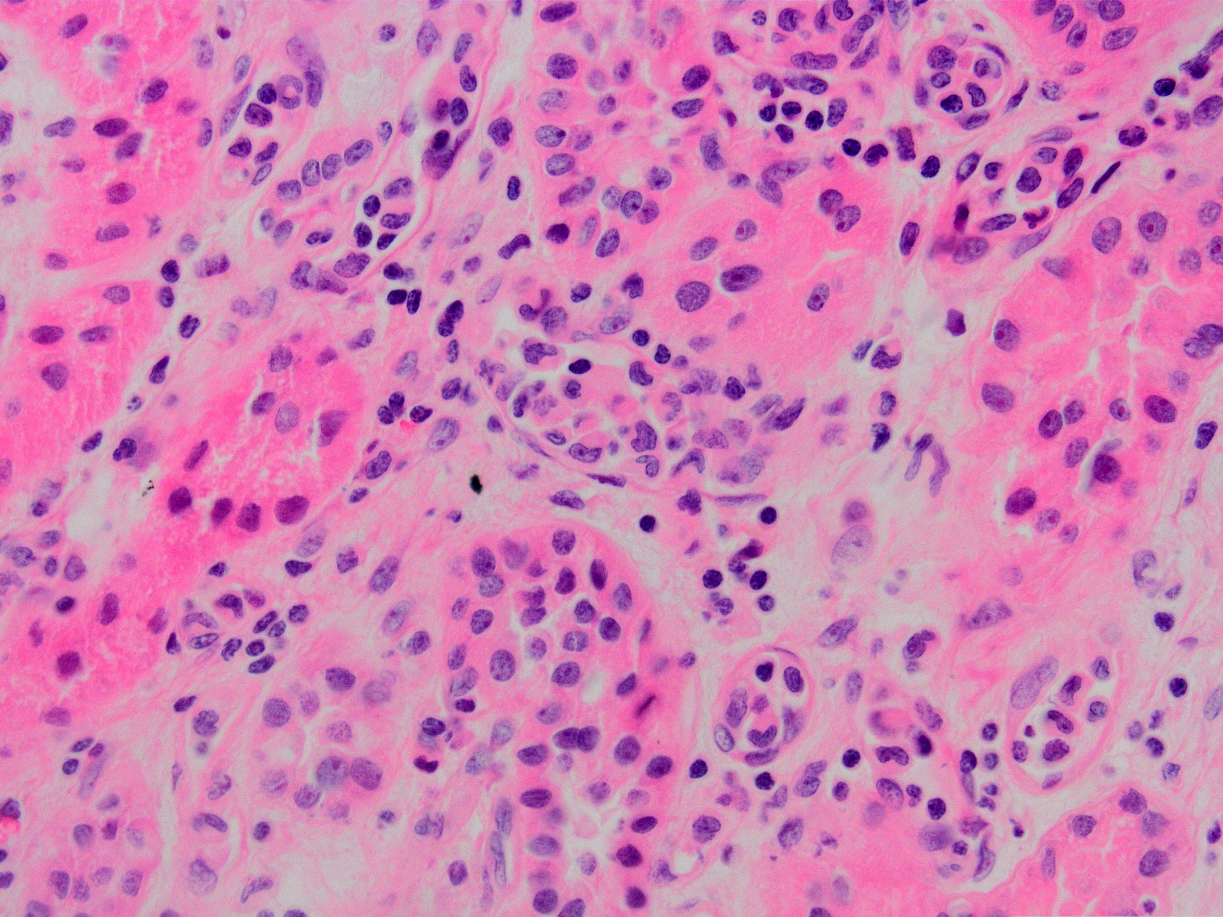
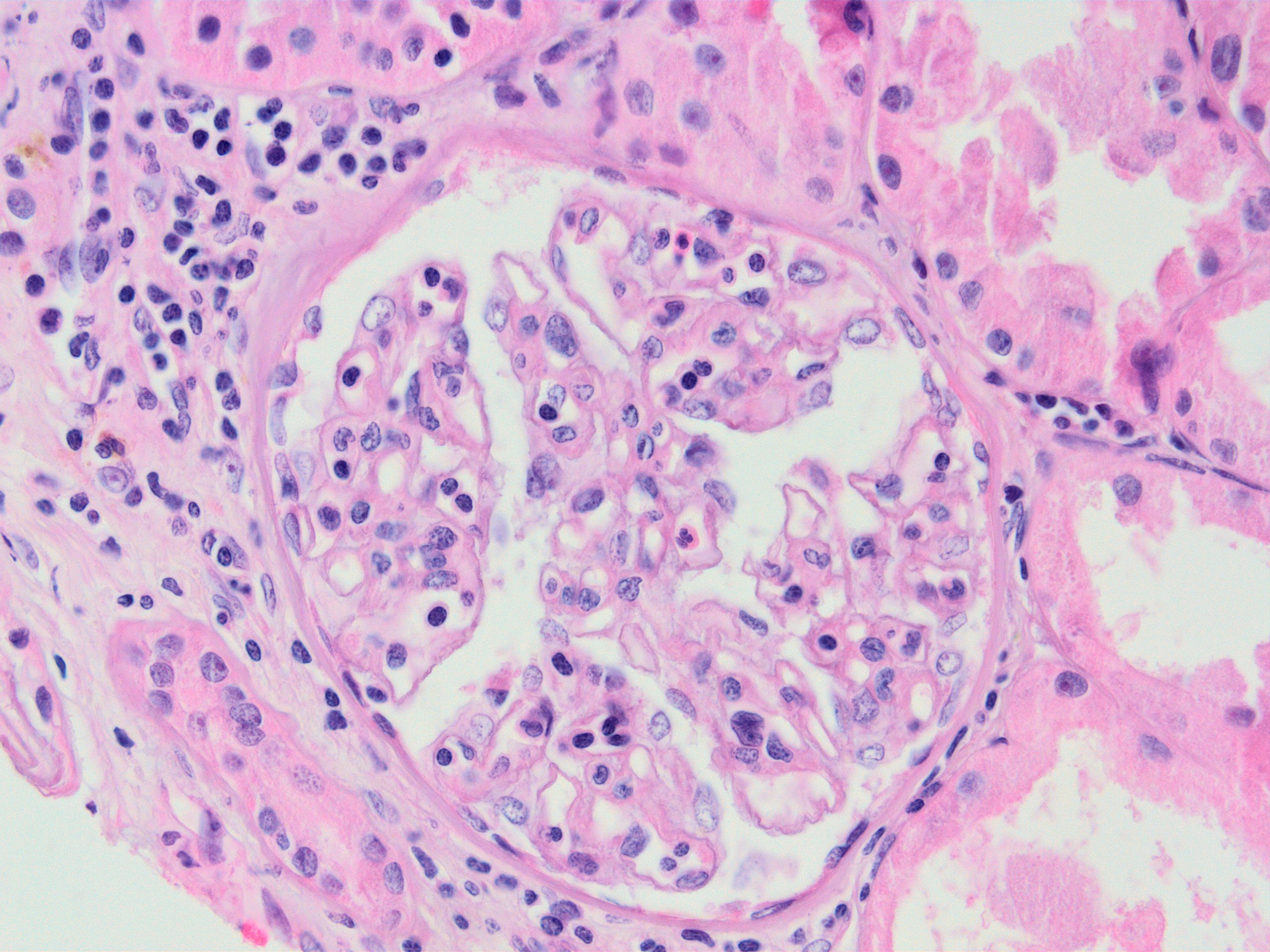
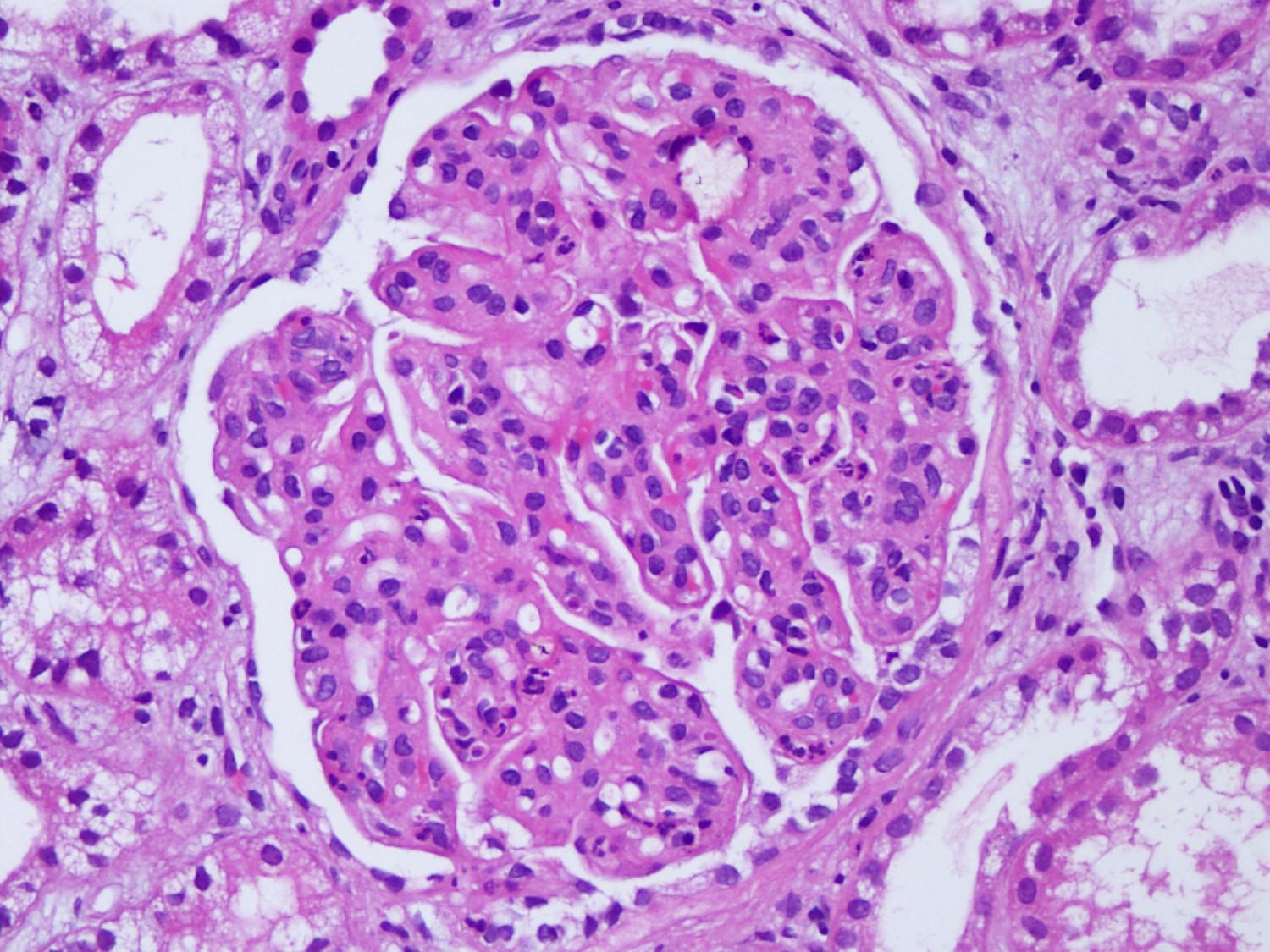
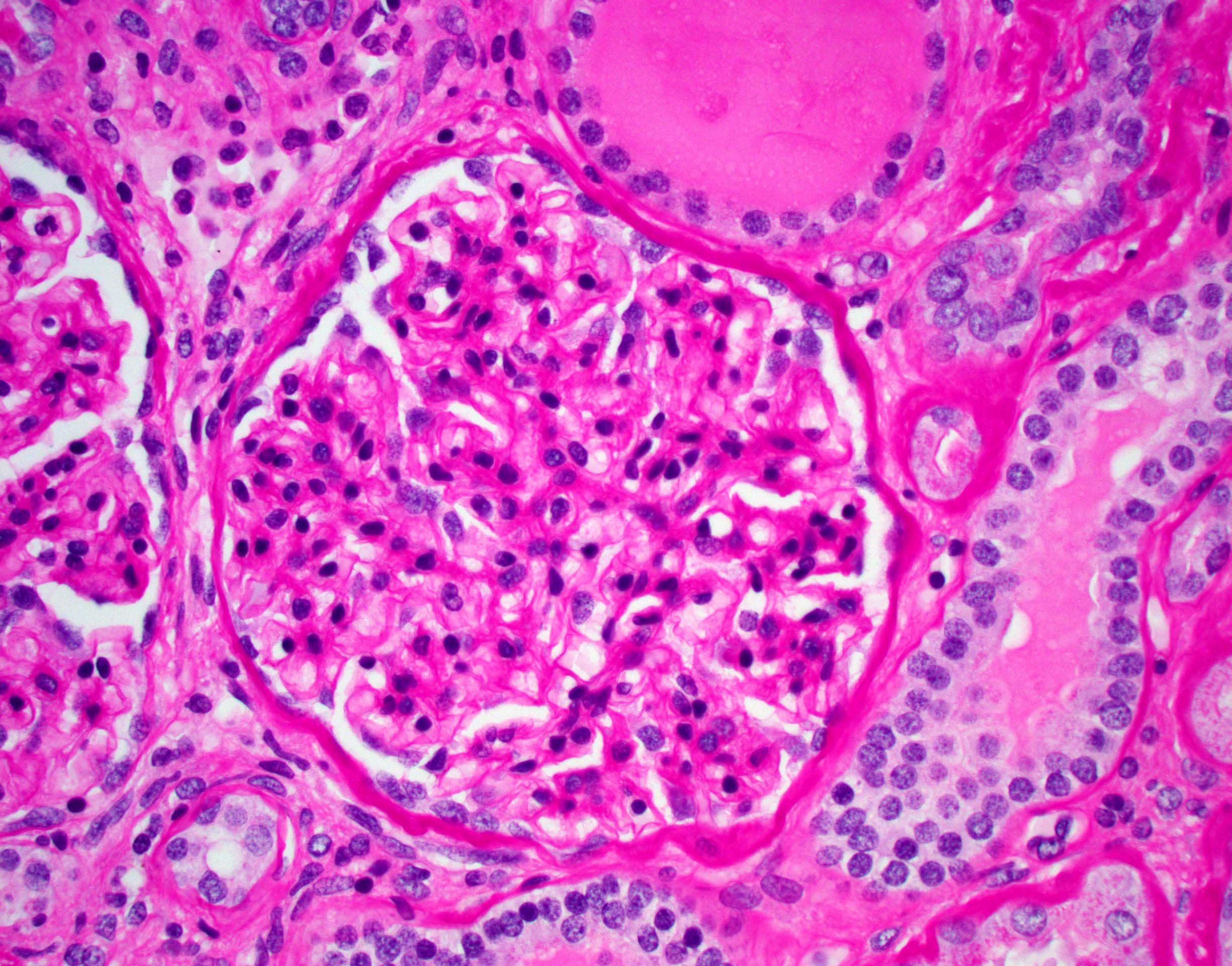
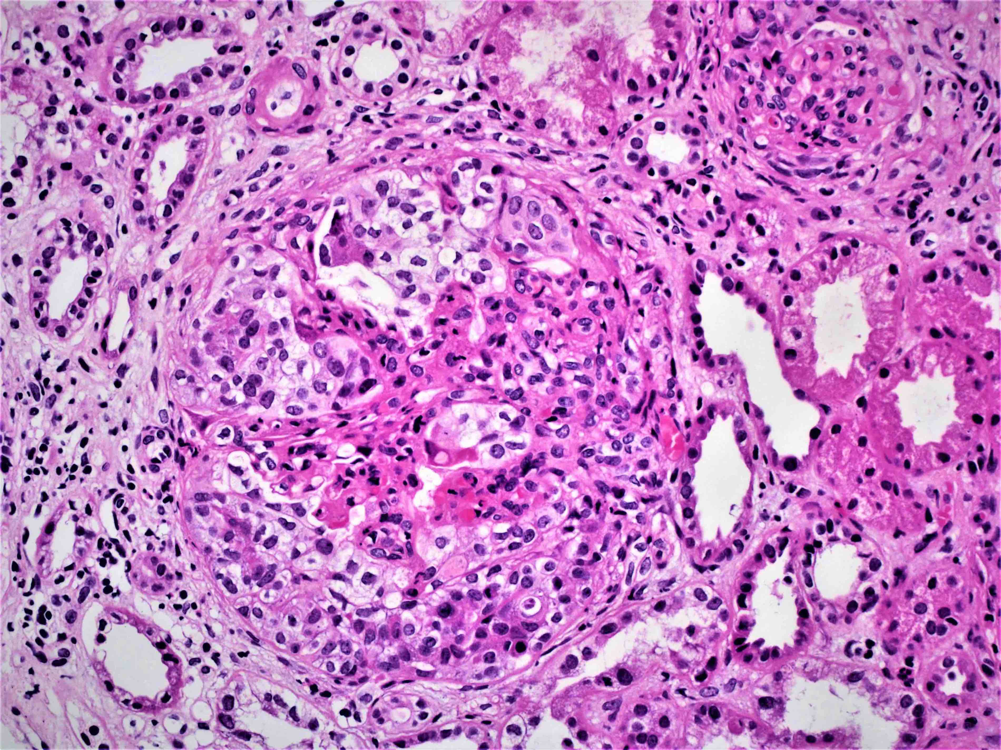
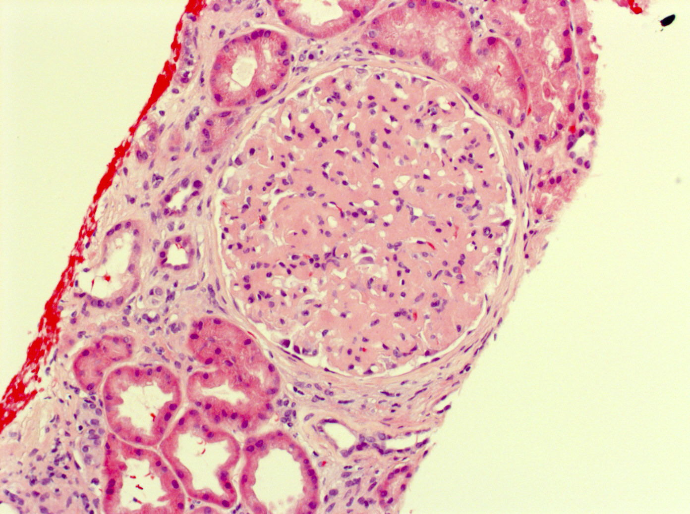
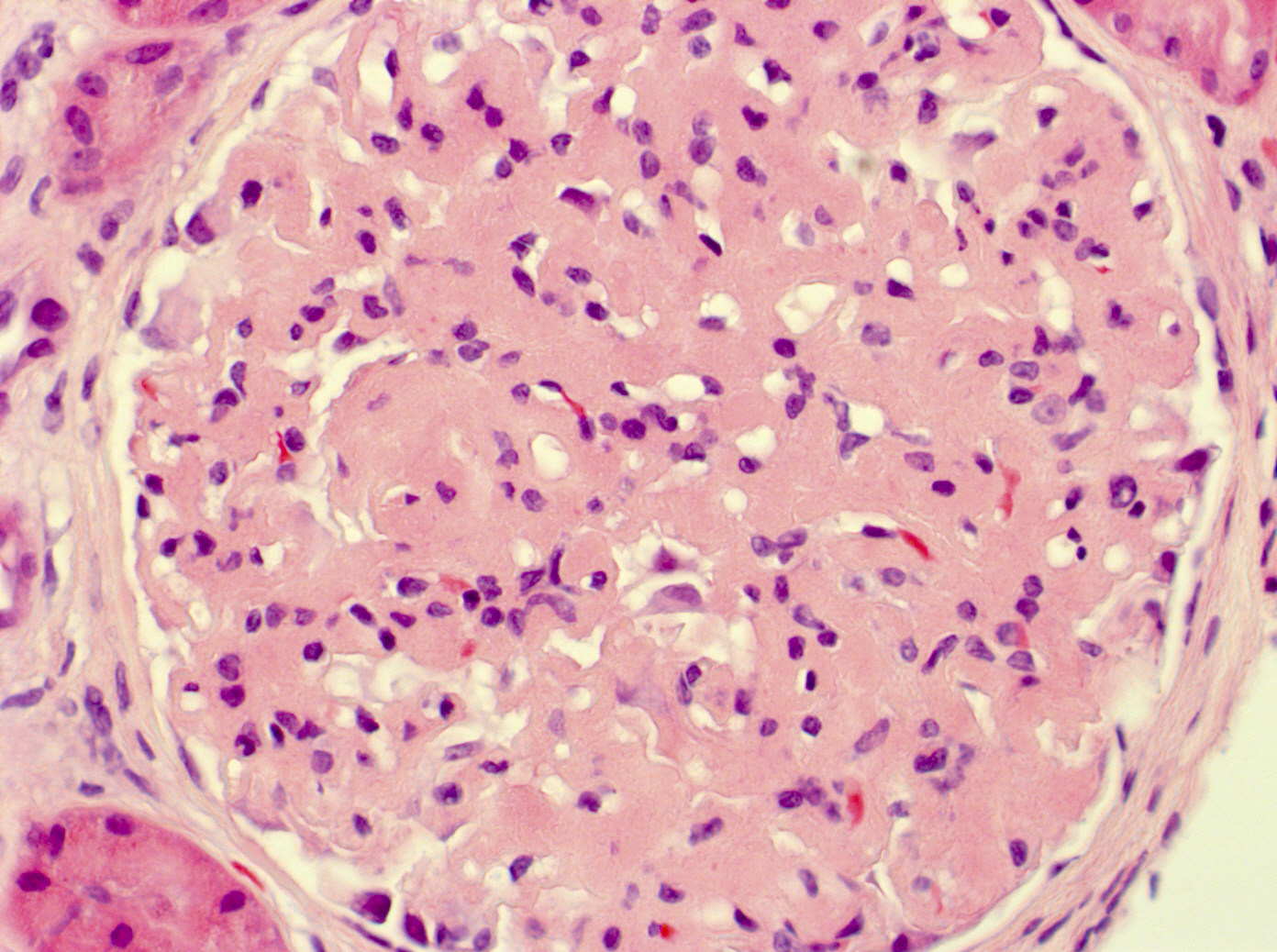
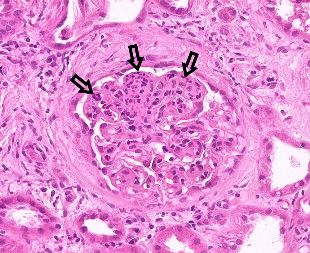
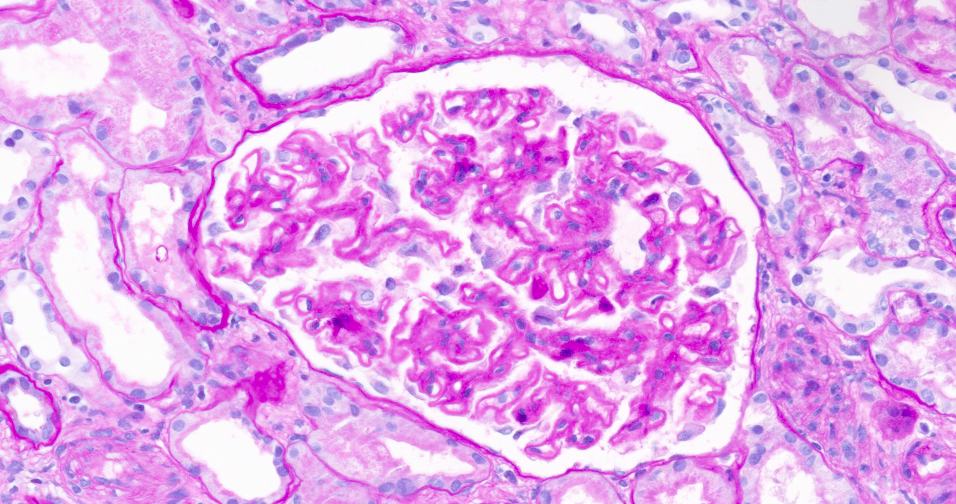
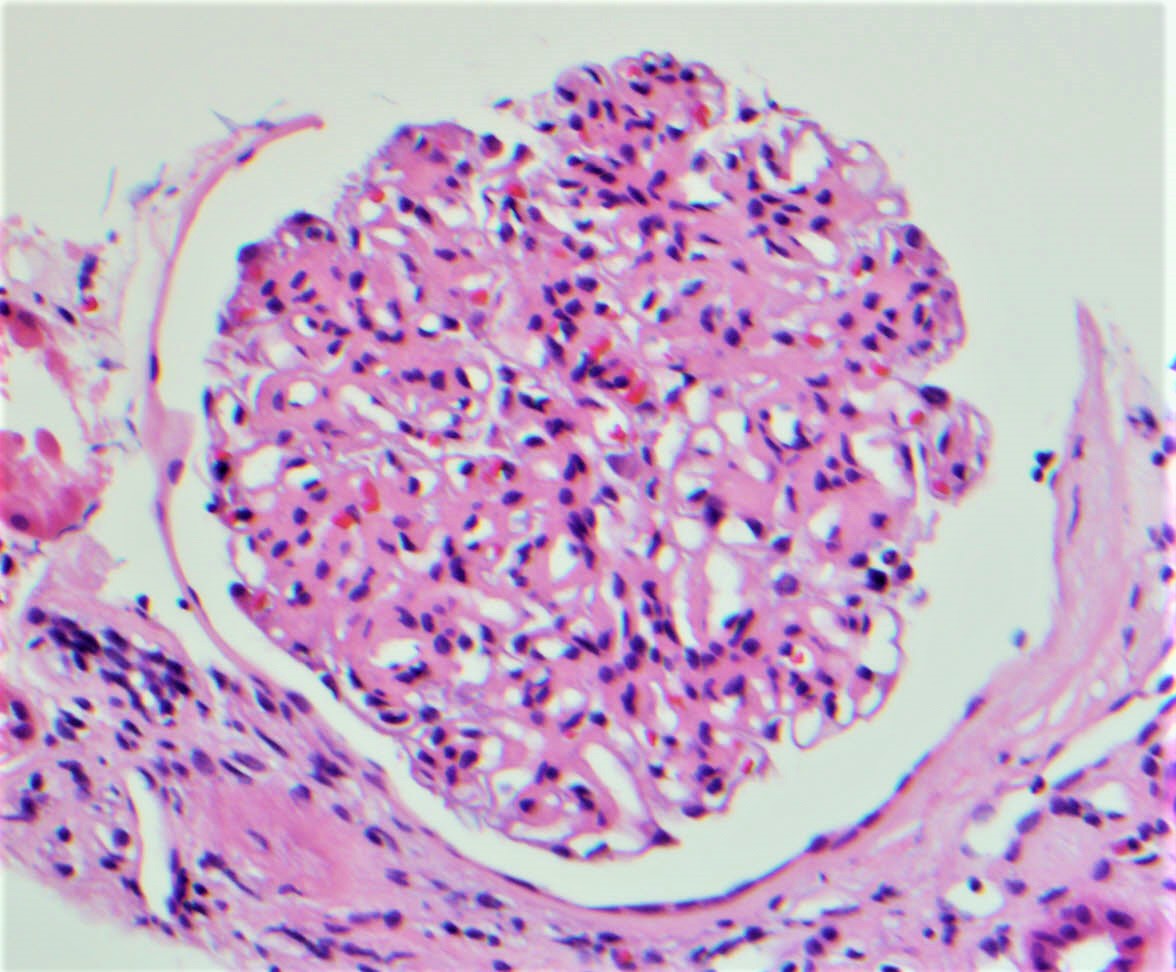
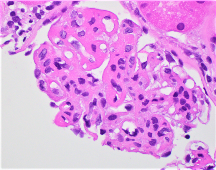
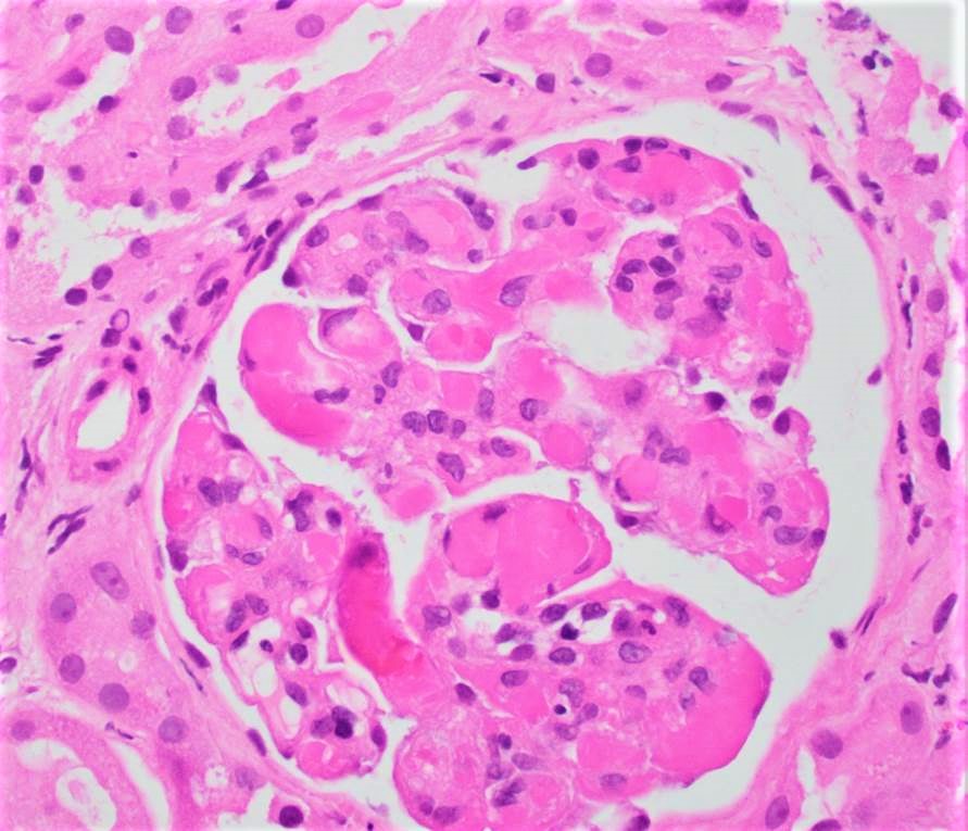
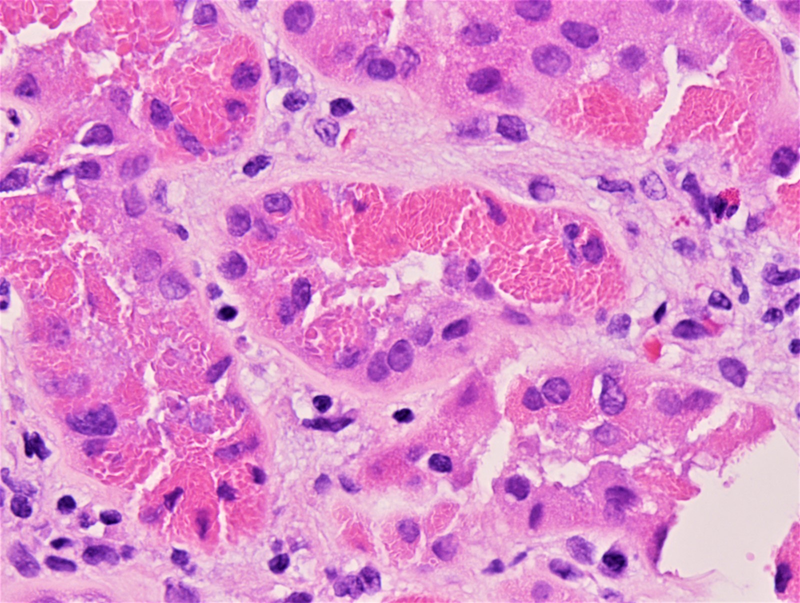
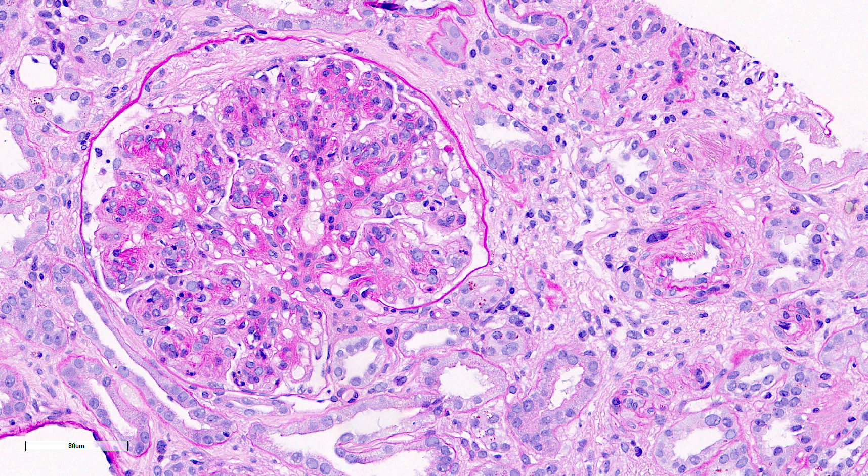
Glomerular capillary luminal neutrophils
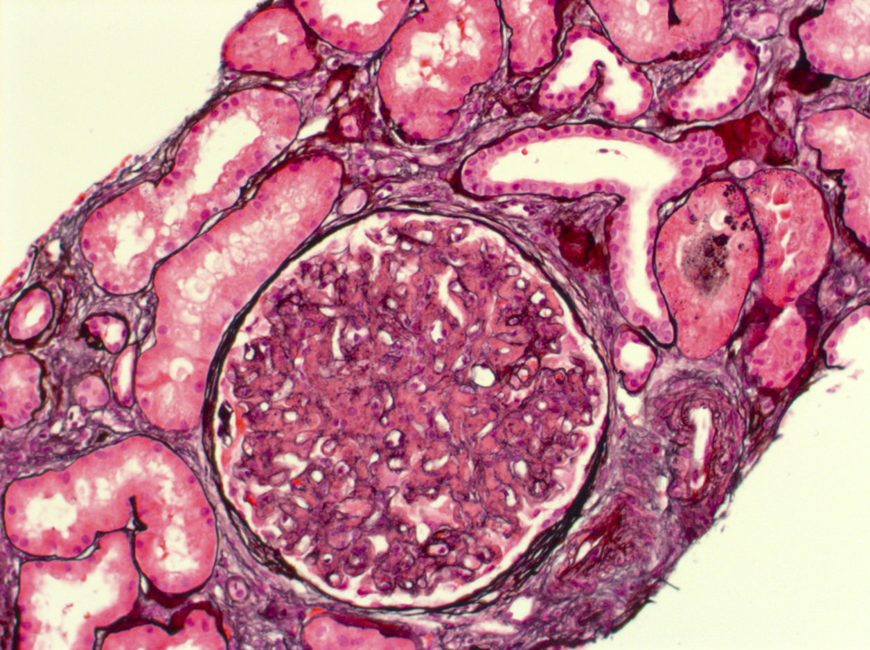
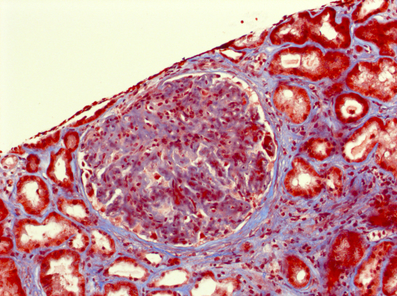
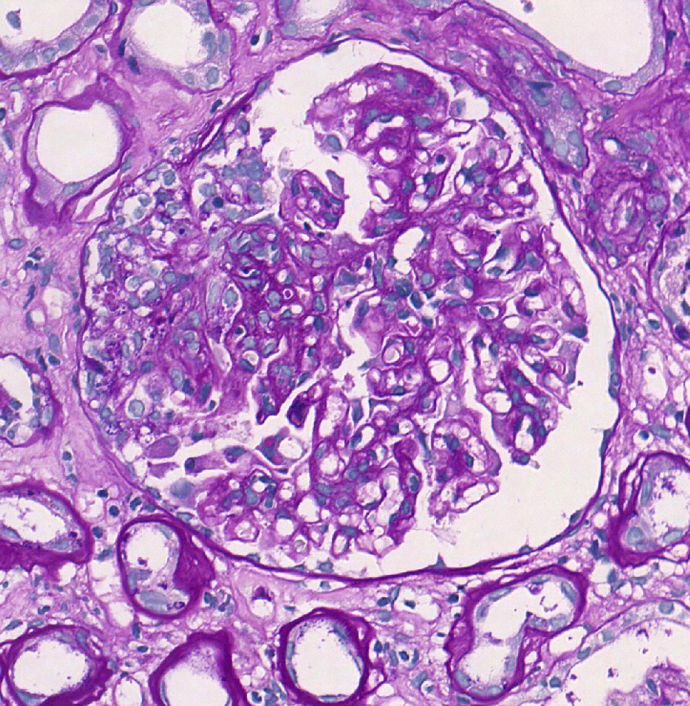
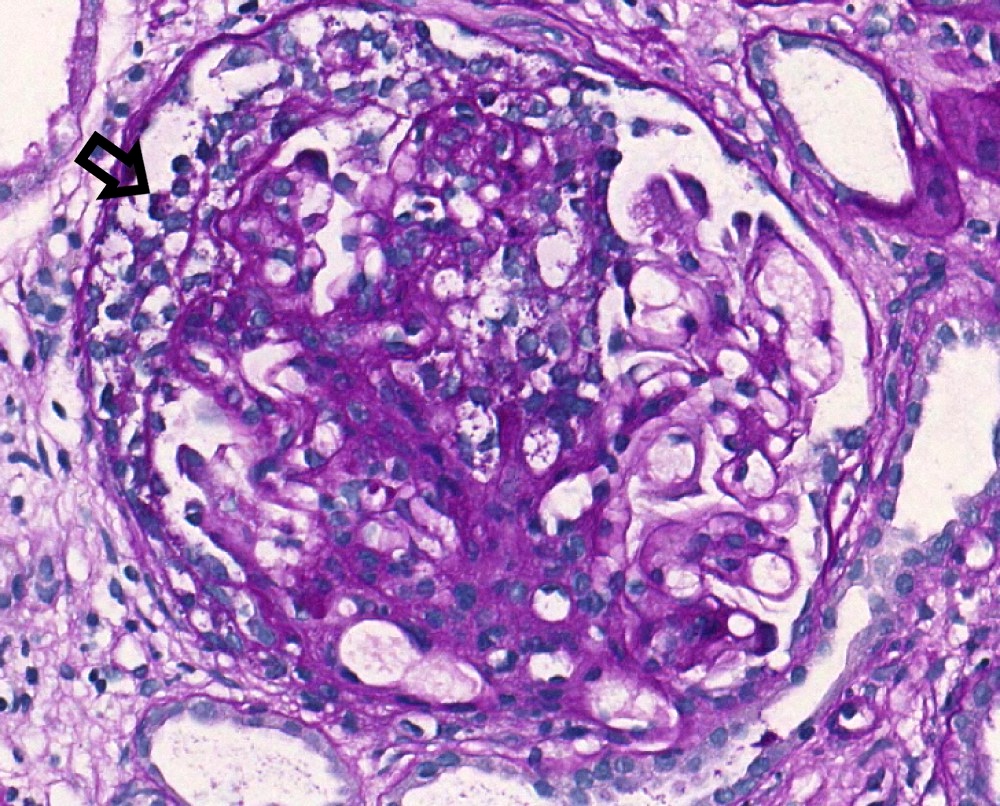
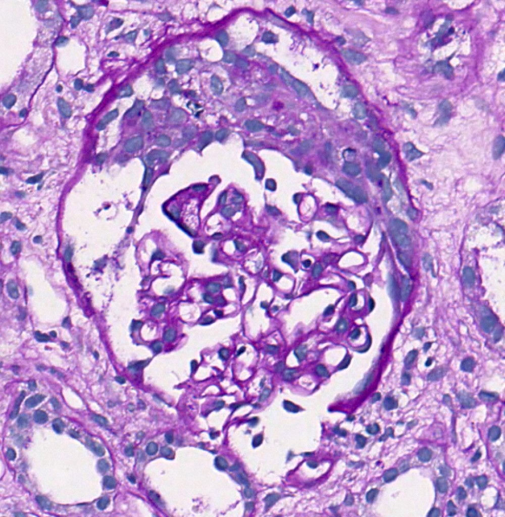
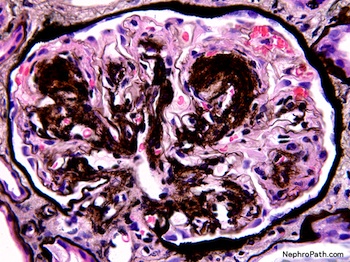
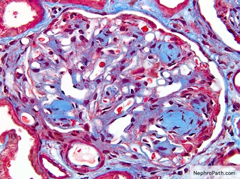
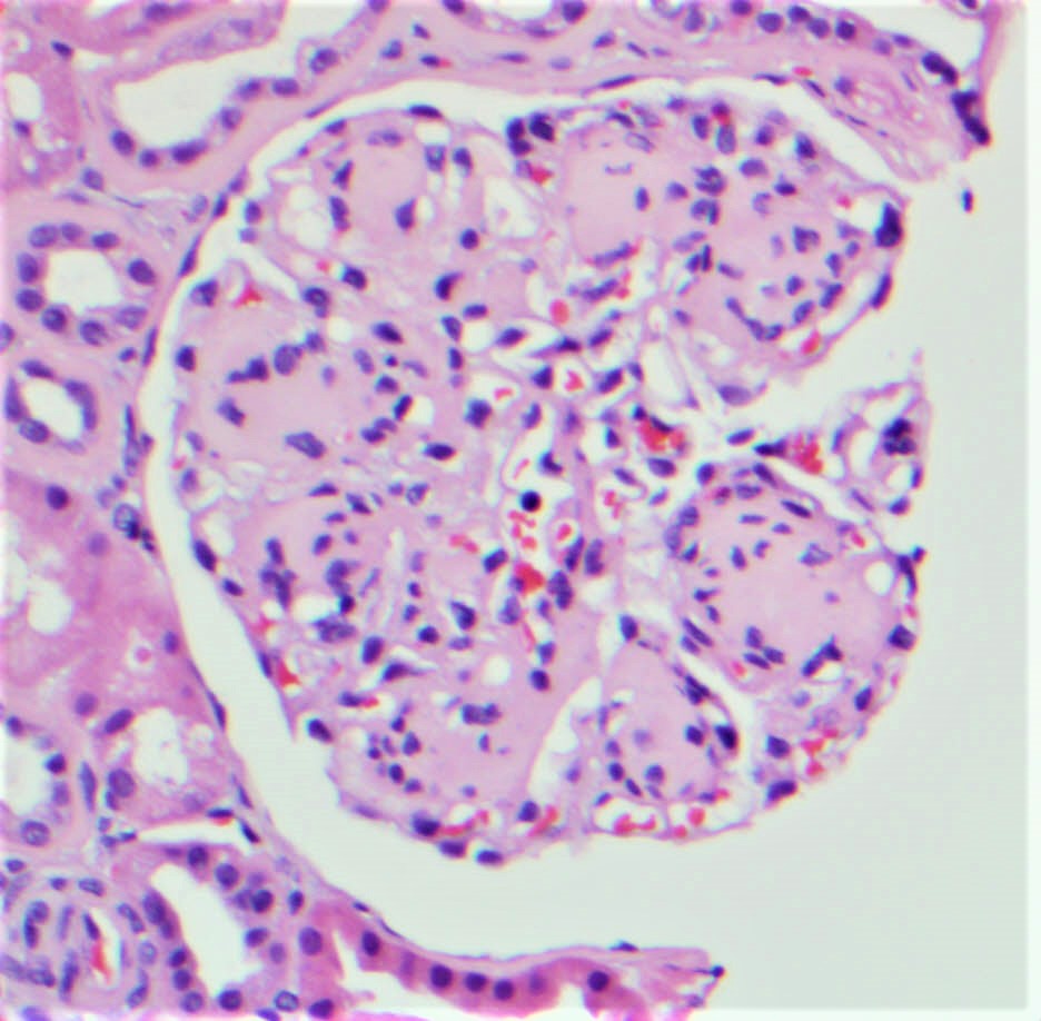
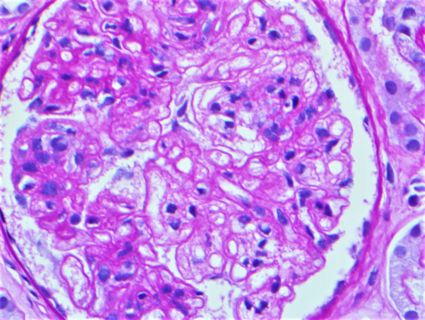
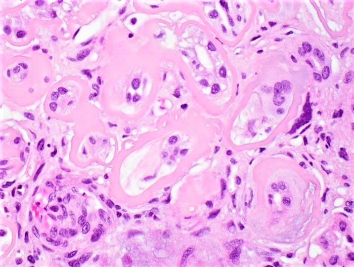
Diabetic
glomerulosclerosis
with exudative GN
Cellular crescent
Contributed by Vanderlene Liu Kung, M.D., Ph.D.
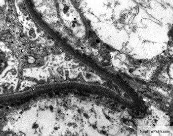
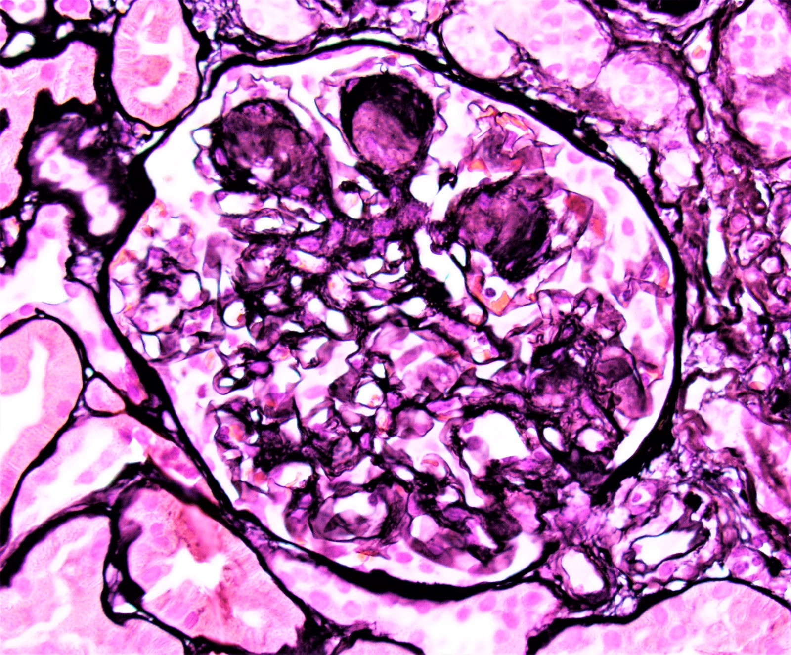
Intramembranous / subepithelial deposits
Contributed by Deepak K. Pruthi, M.D., M.Sci.-T.S.
CT abdomen

Contrast enhanced
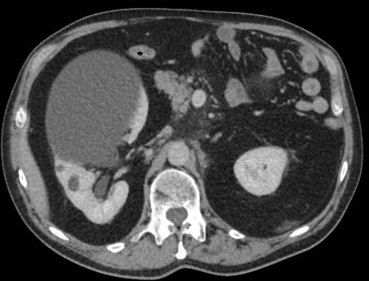
Axial CT
Coronal CT
Postsclerotherapy
Images hosted on other servers:

Simple cyst of upper pole

Large cyst

Multiple, smooth renal cysts filled with serous fluid

Solitary, smooth
kidney cyst
(lower pole) filled
with serous fluid

Large cortical cysts with dark red blood
Contributed by Rana Chakrabarti, M.D., M.P.H.

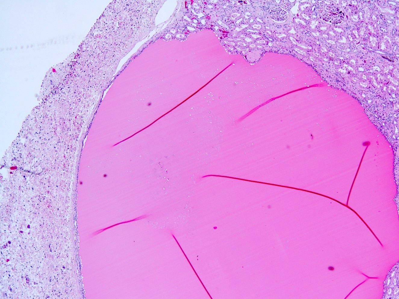
Subcapsular cortical cyst
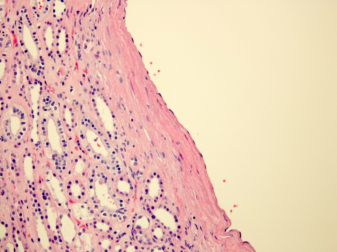
Flattened cyst wall epithelium
Papillary excrescence
Contributed by Jonathan E. Zuckerman, M.D., Ph.D.
Bile stained kidney
Contributed by Jonathan E. Zuckerman, M.D., Ph.D.
Bile casts in collecting ducts
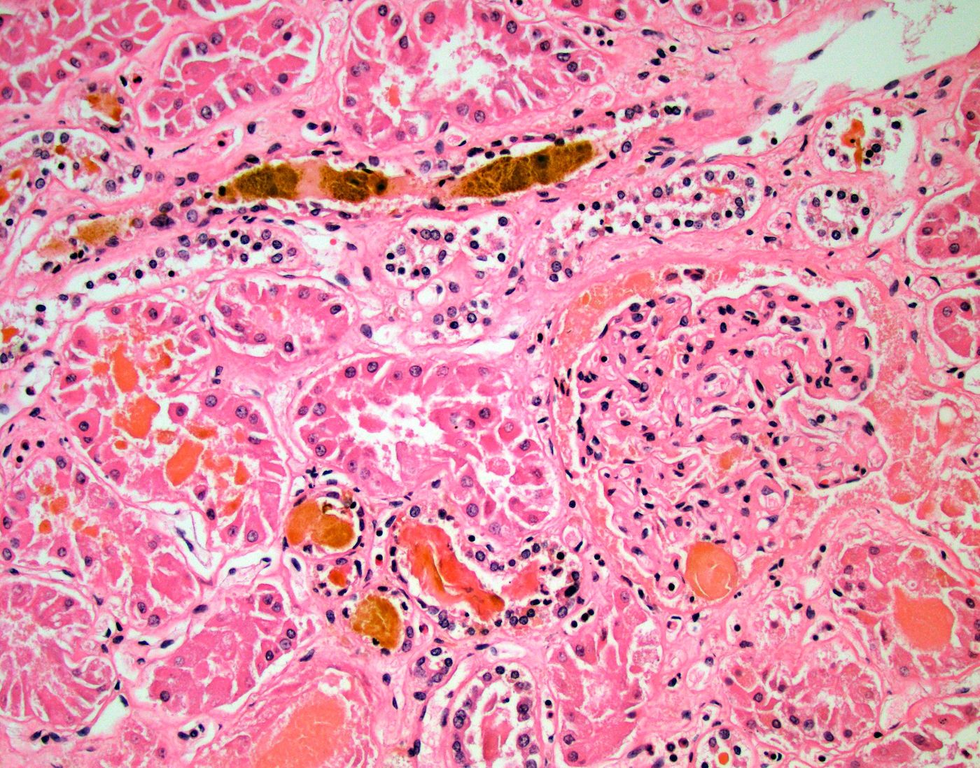
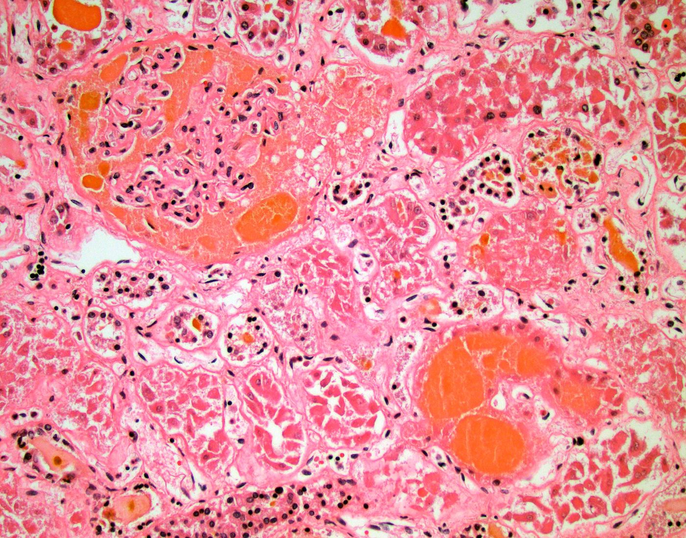
Bile casts in proximal tubules

Hall bile stain
Images hosted on other servers:

Bile case
Images hosted on other servers:

Dividing up core tissue
if no dissecting
microscope is present
Images hosted on other servers:

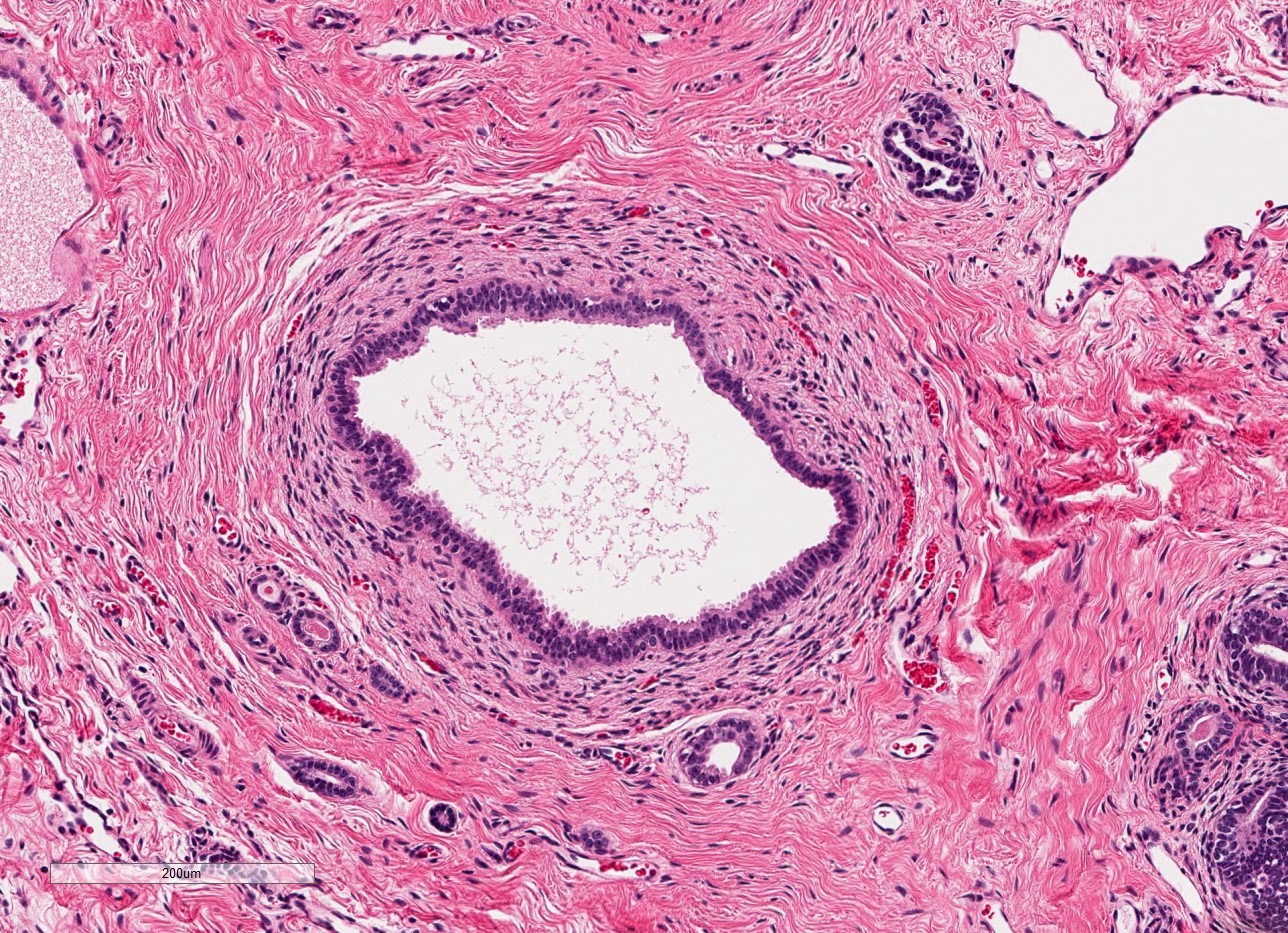
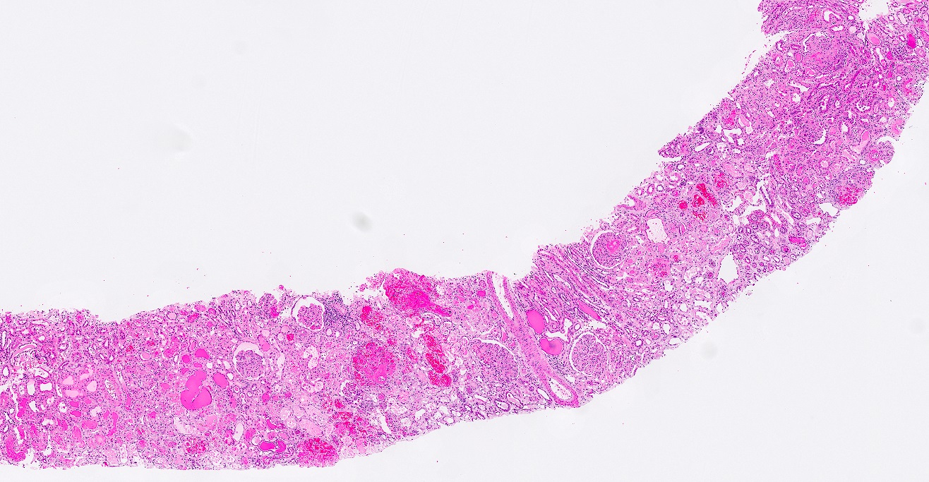
A: renal cortex with
round red glomeruli;
B: renal medulla
without glomeruli


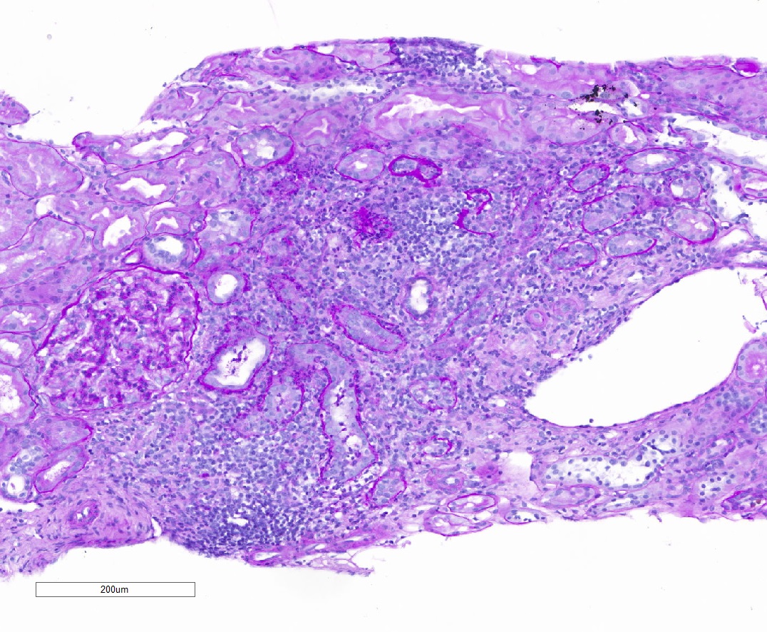
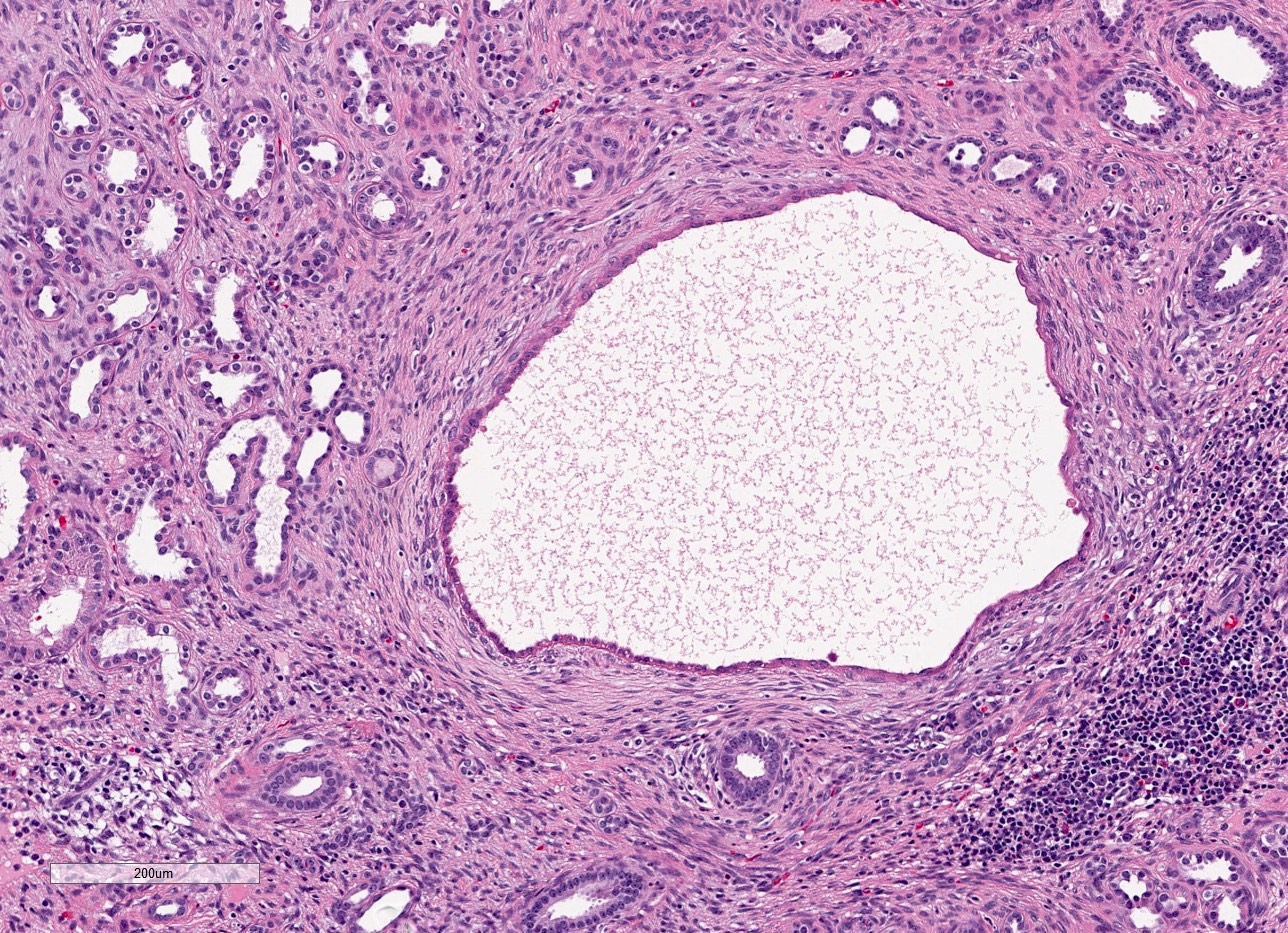
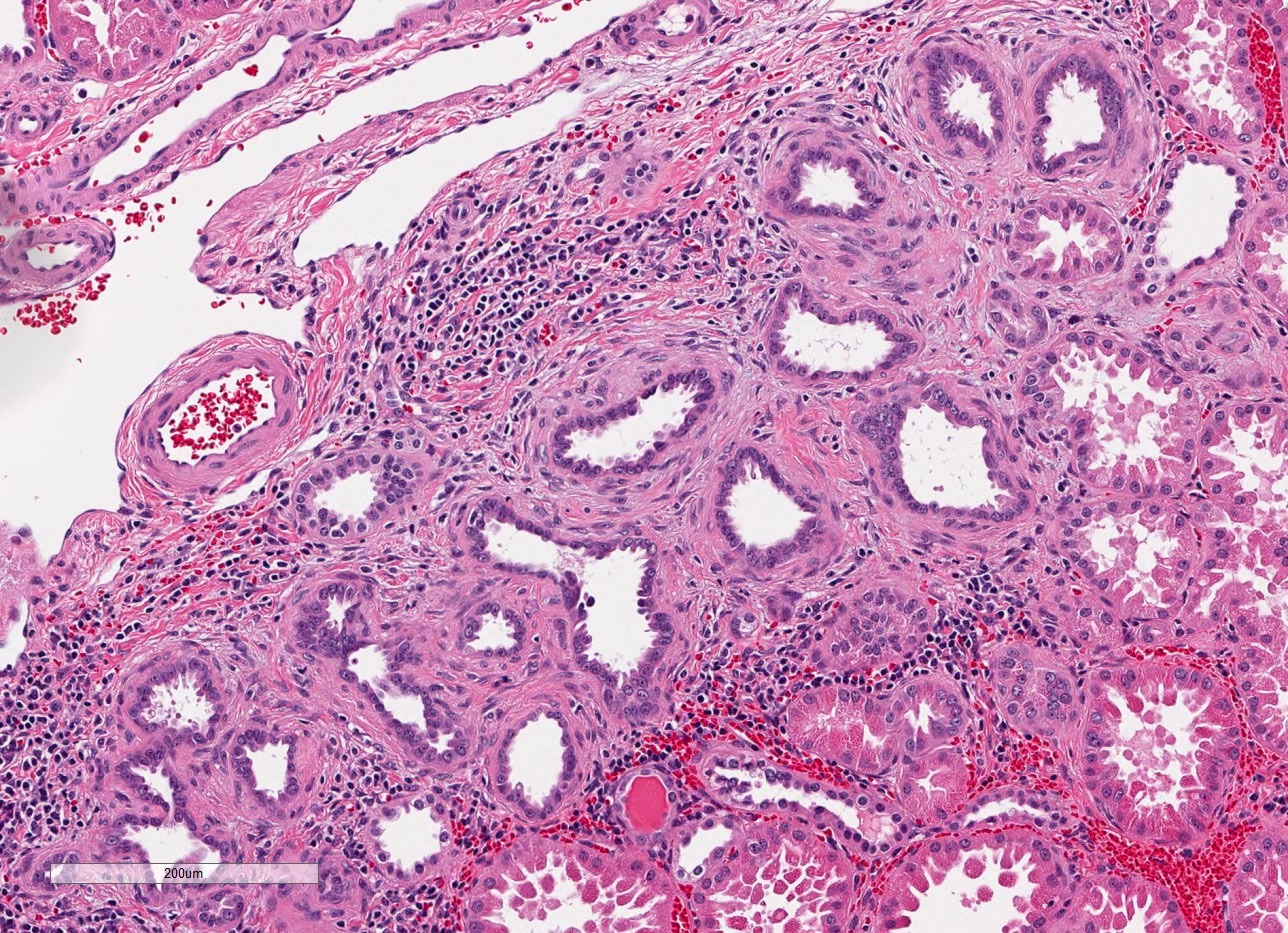
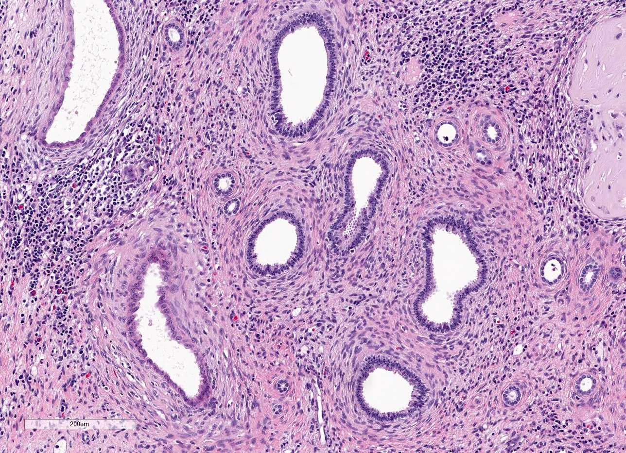
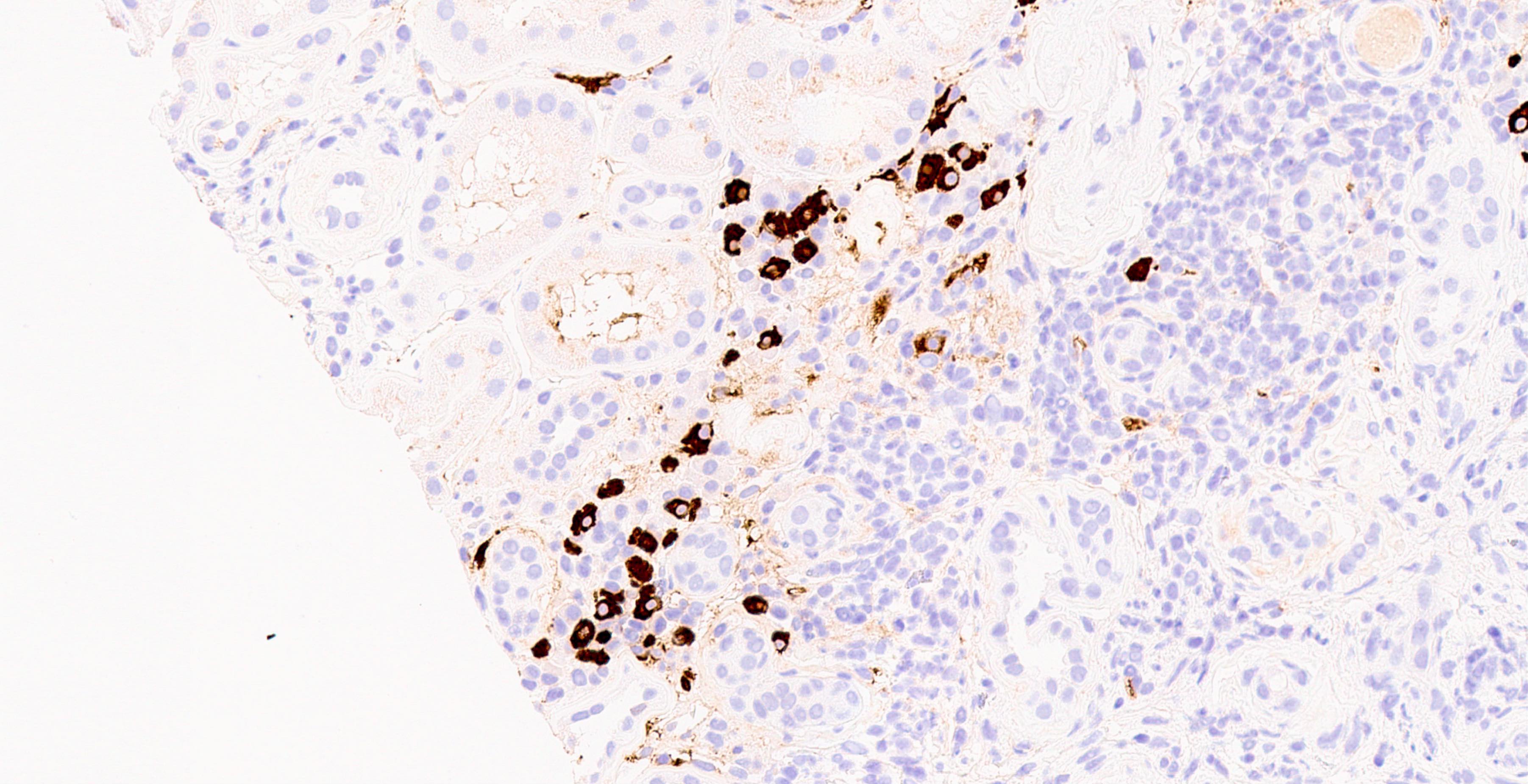
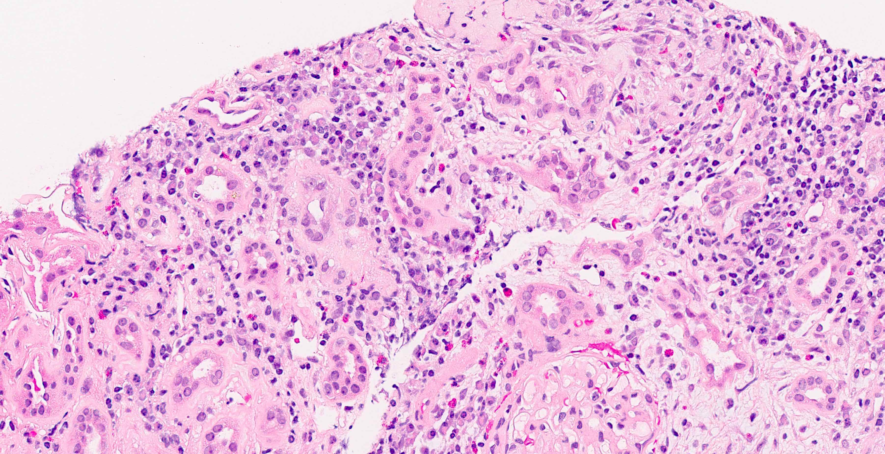
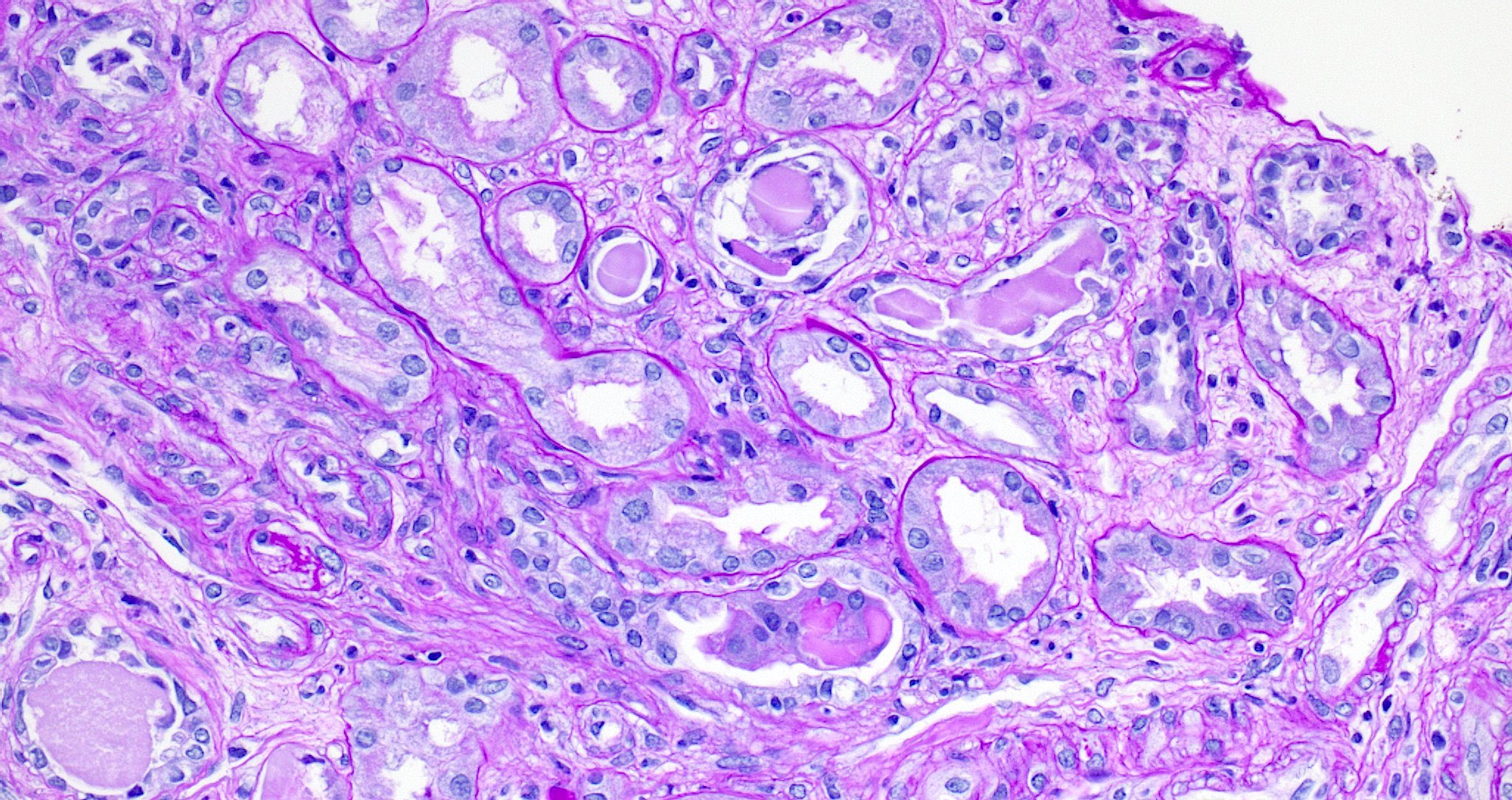
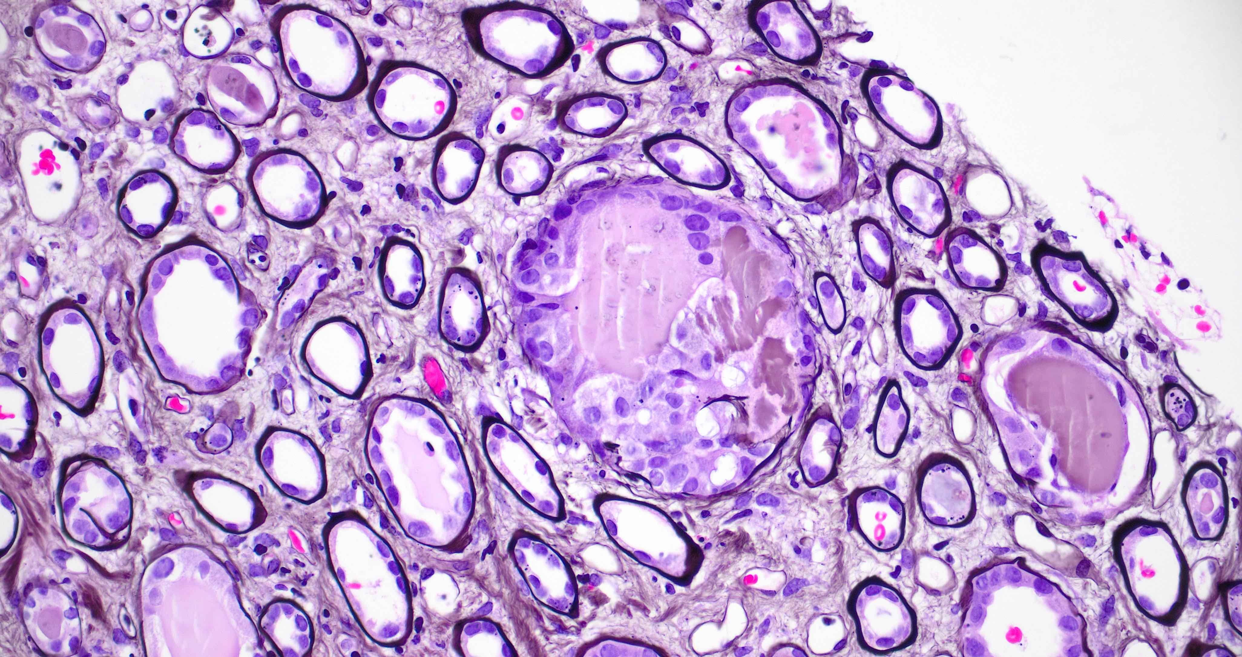
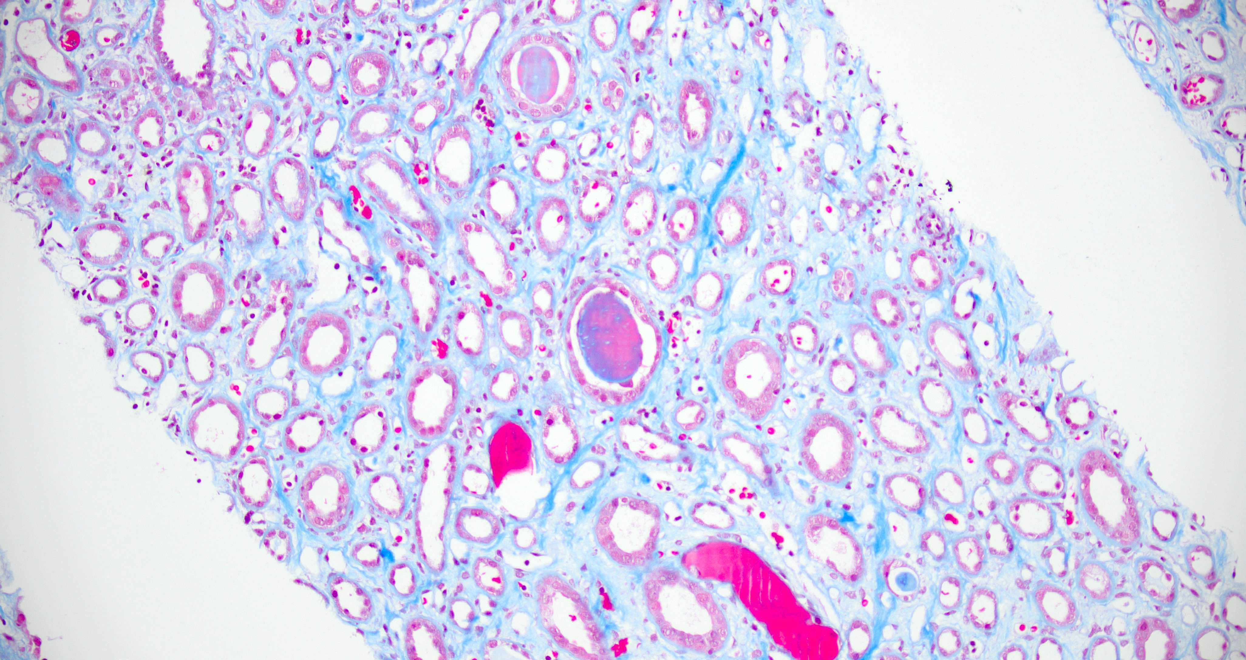
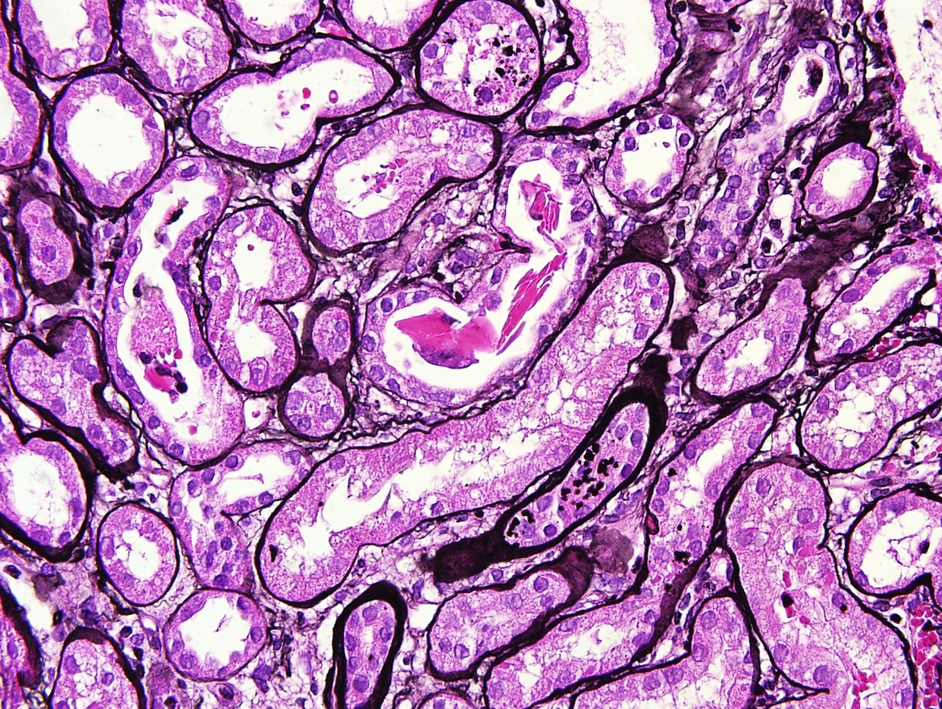
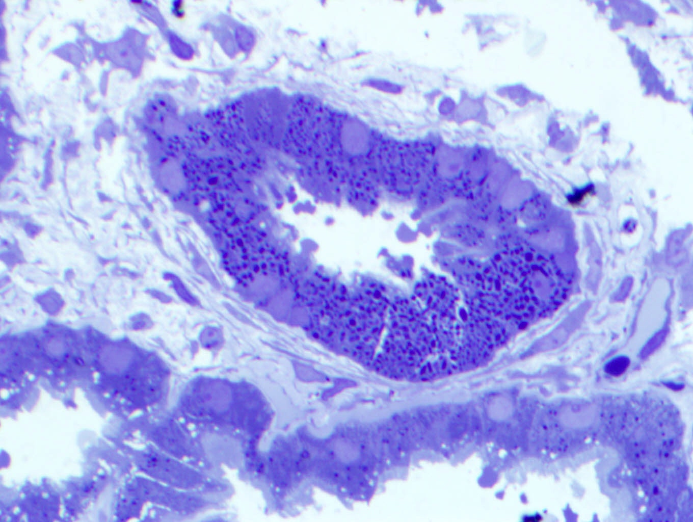
Contributed by Arzu Sağlam, M.D.
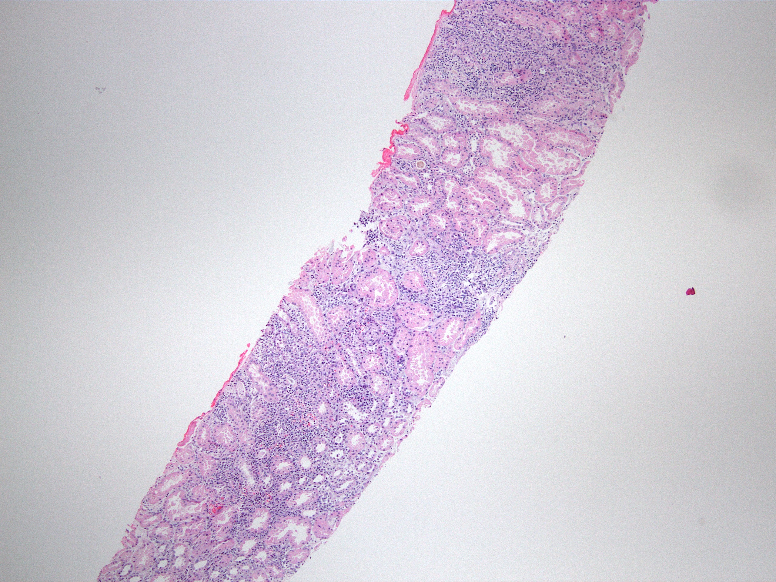
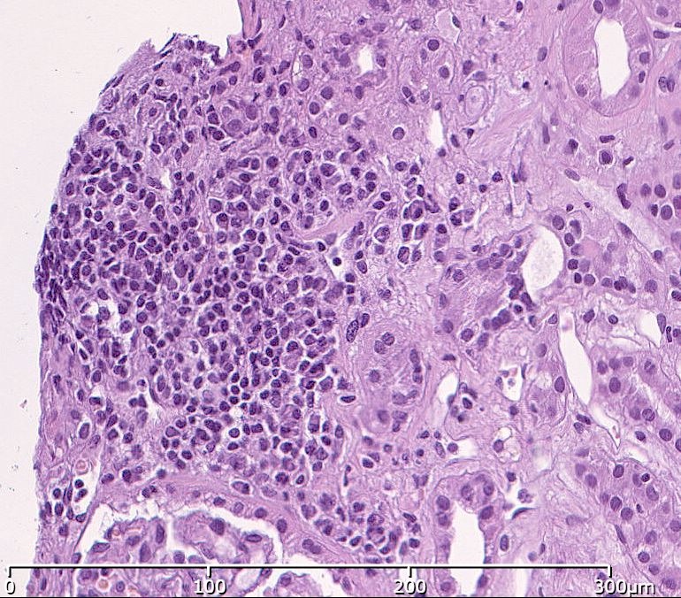
Well demarcated inflammation
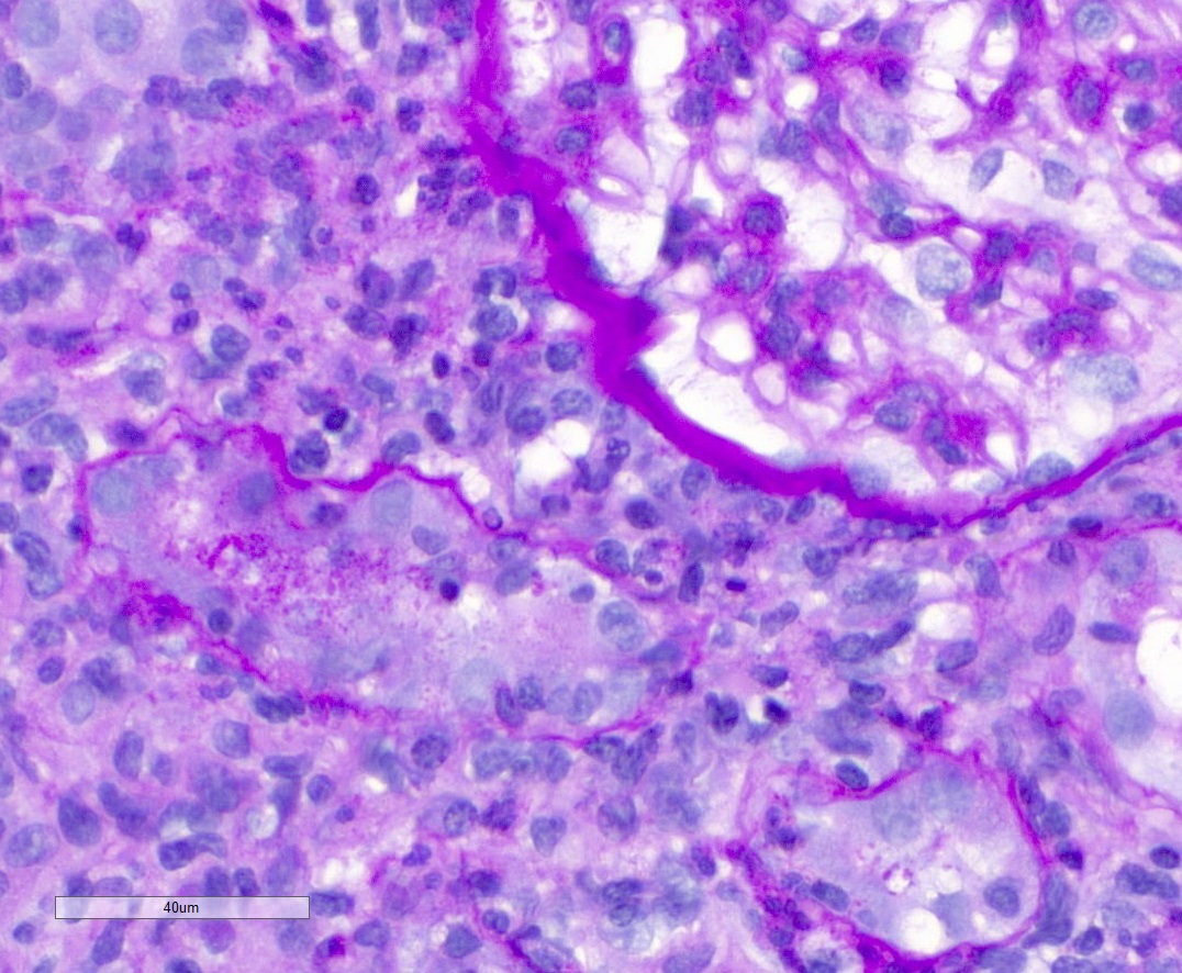
Tubulitis and intranuclear inclusions

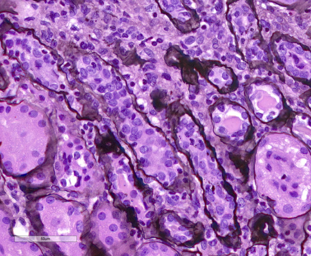
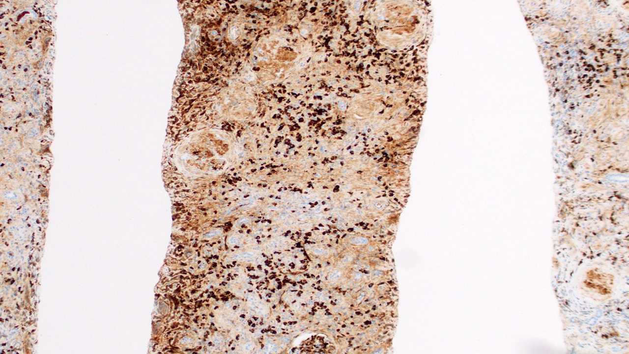
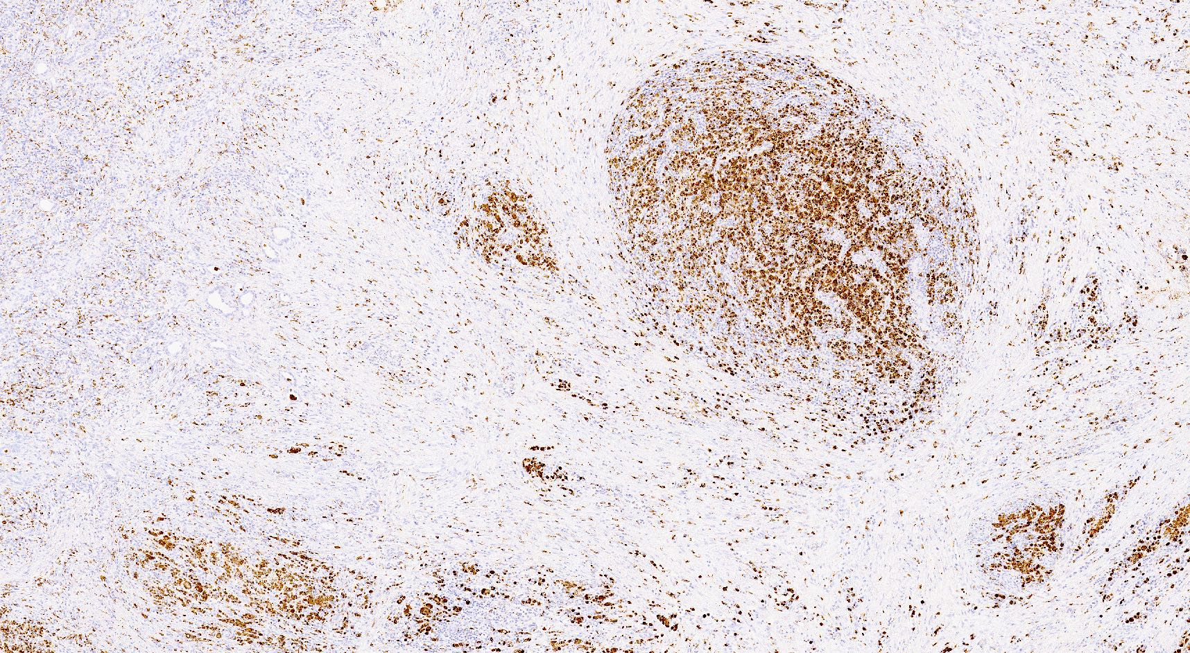
Polyomavirus score: pvl 3
Luminal casts and cytopathic changes
Cytopathic changes
Intranuclear basophilic granules
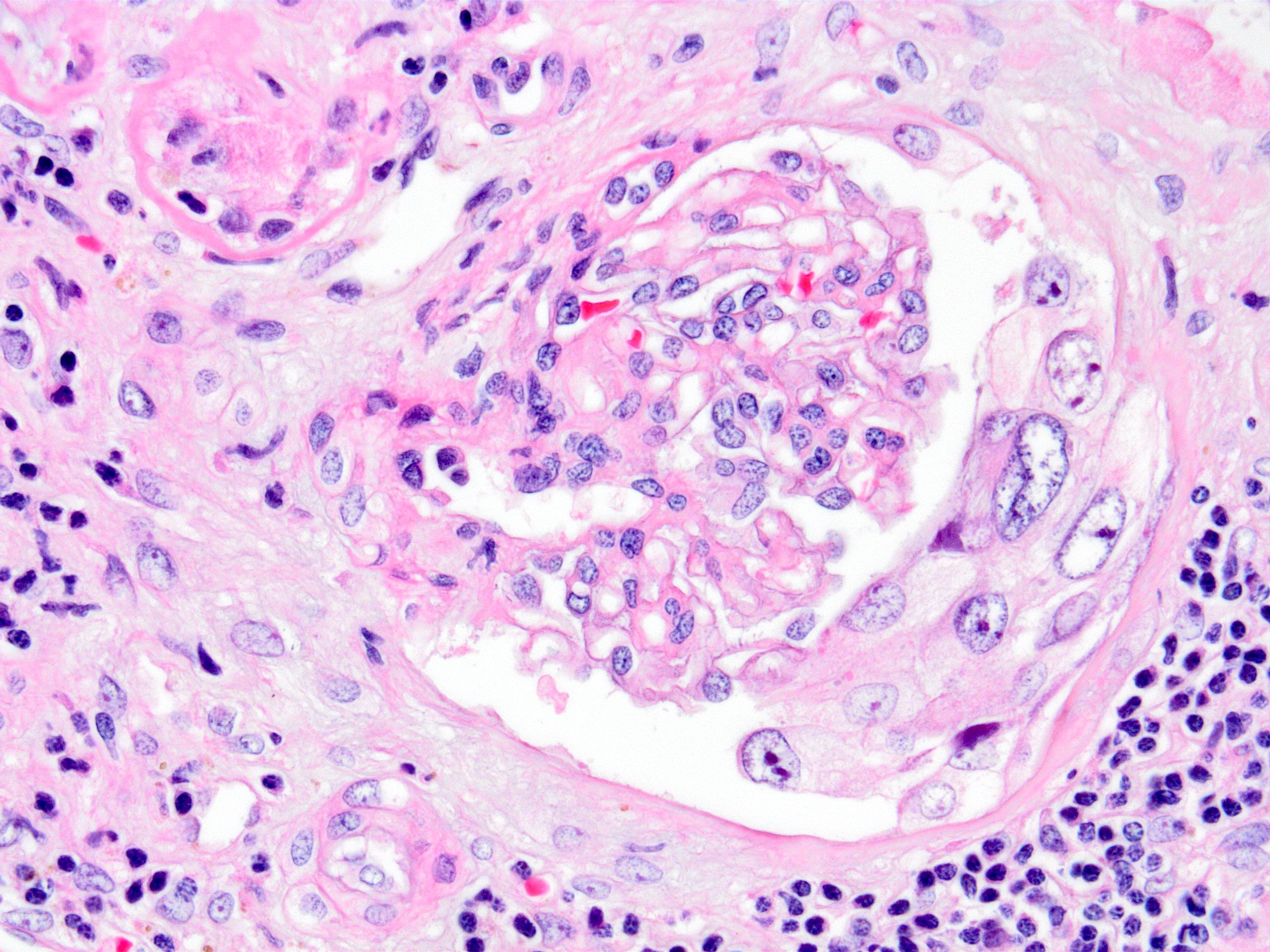
Glomerular cytopathy
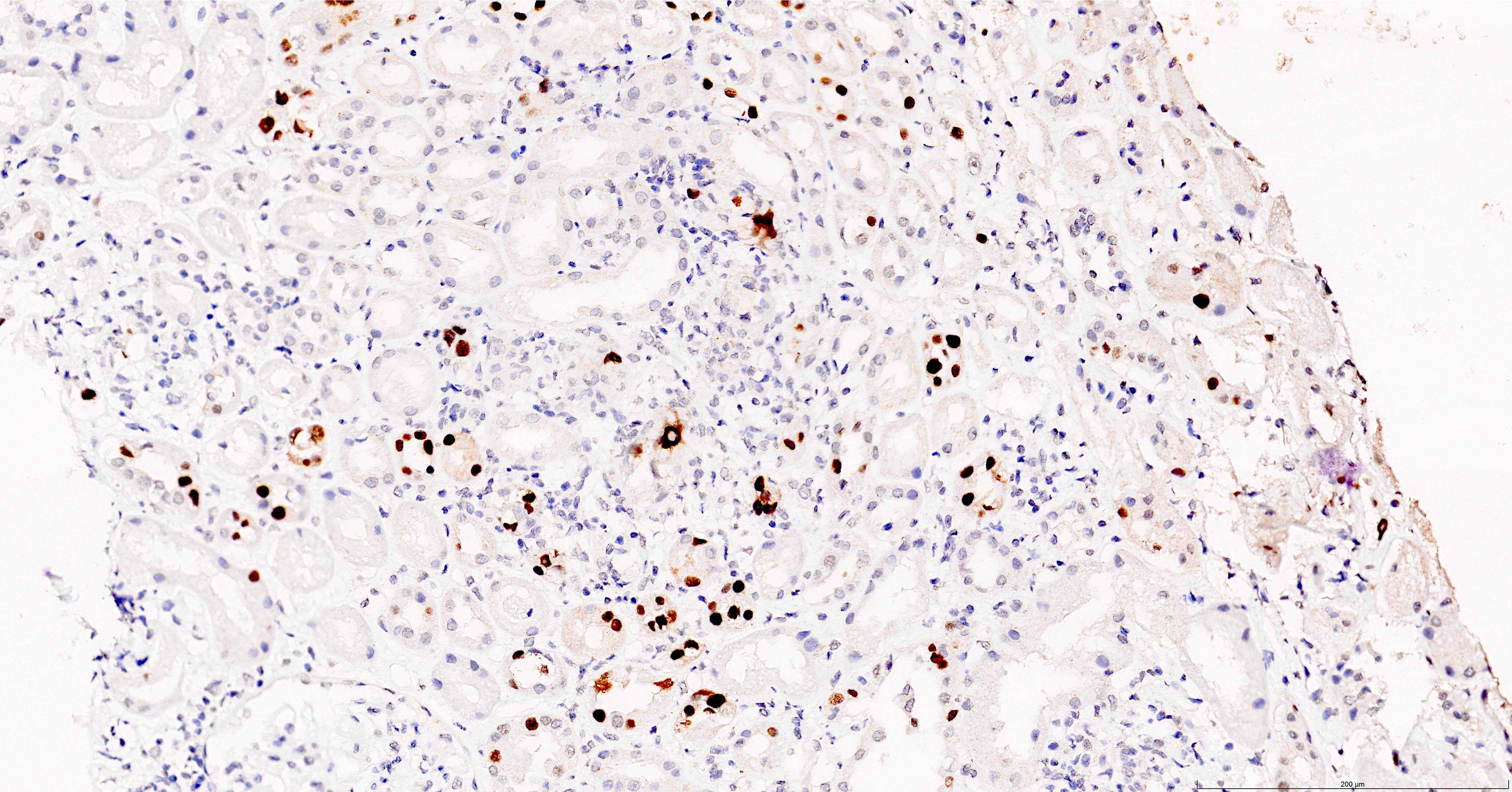
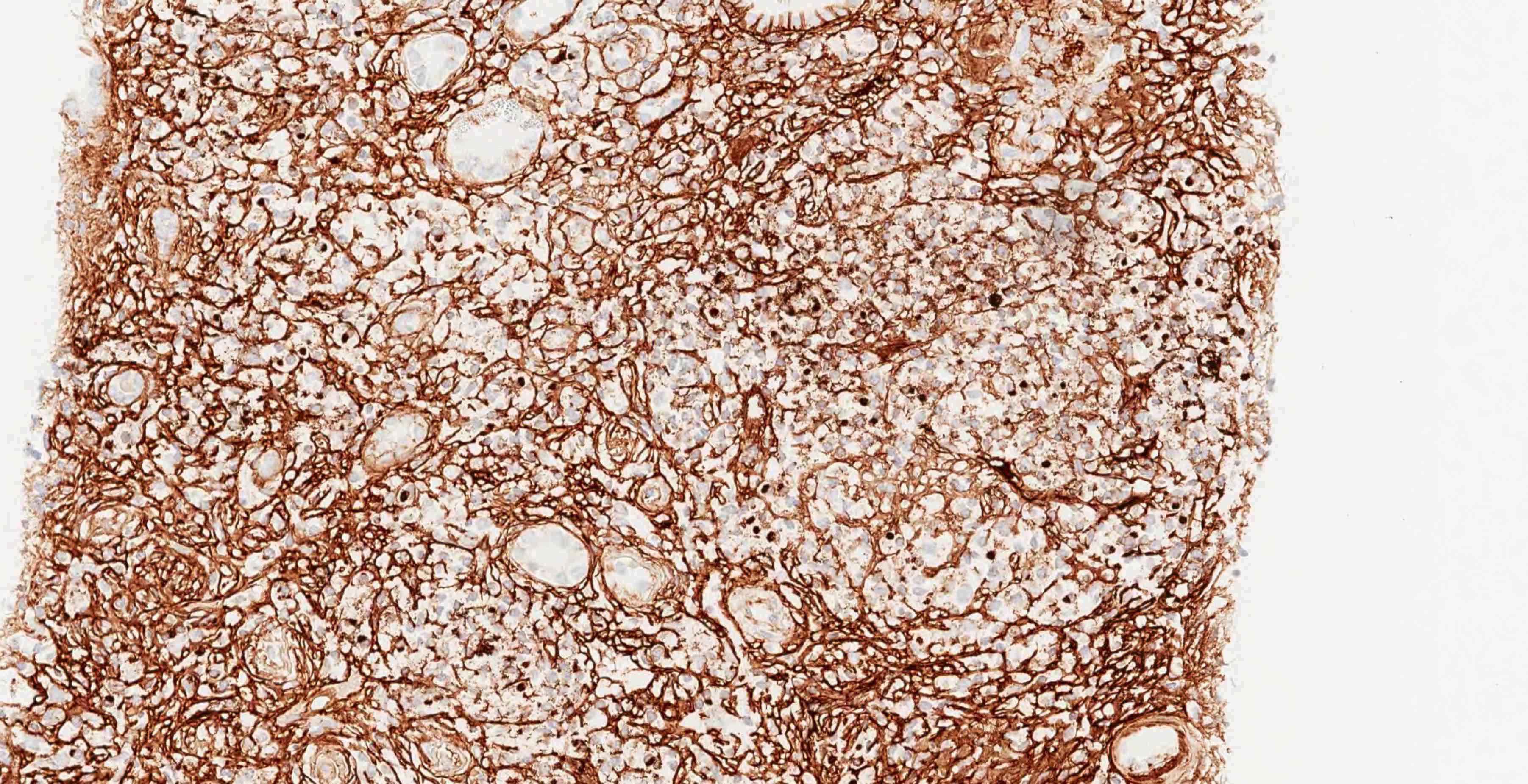
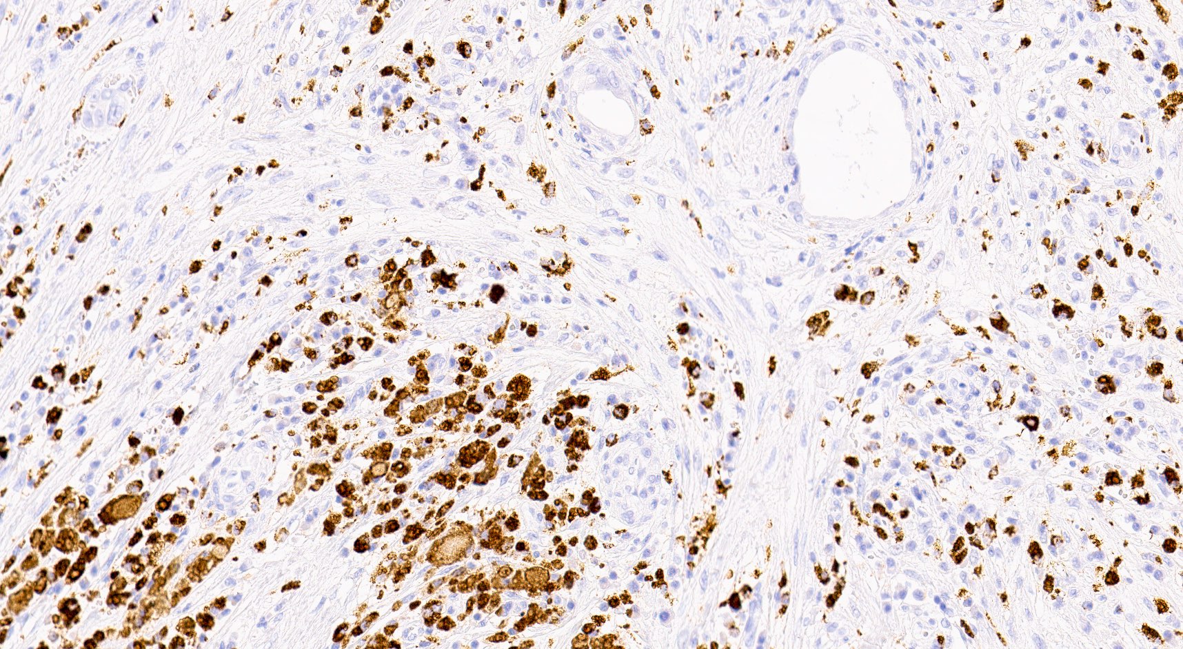
Anti-SV40 antibody, IHC
Contributed by Çisel Aydιn Meriçöz, M.D.
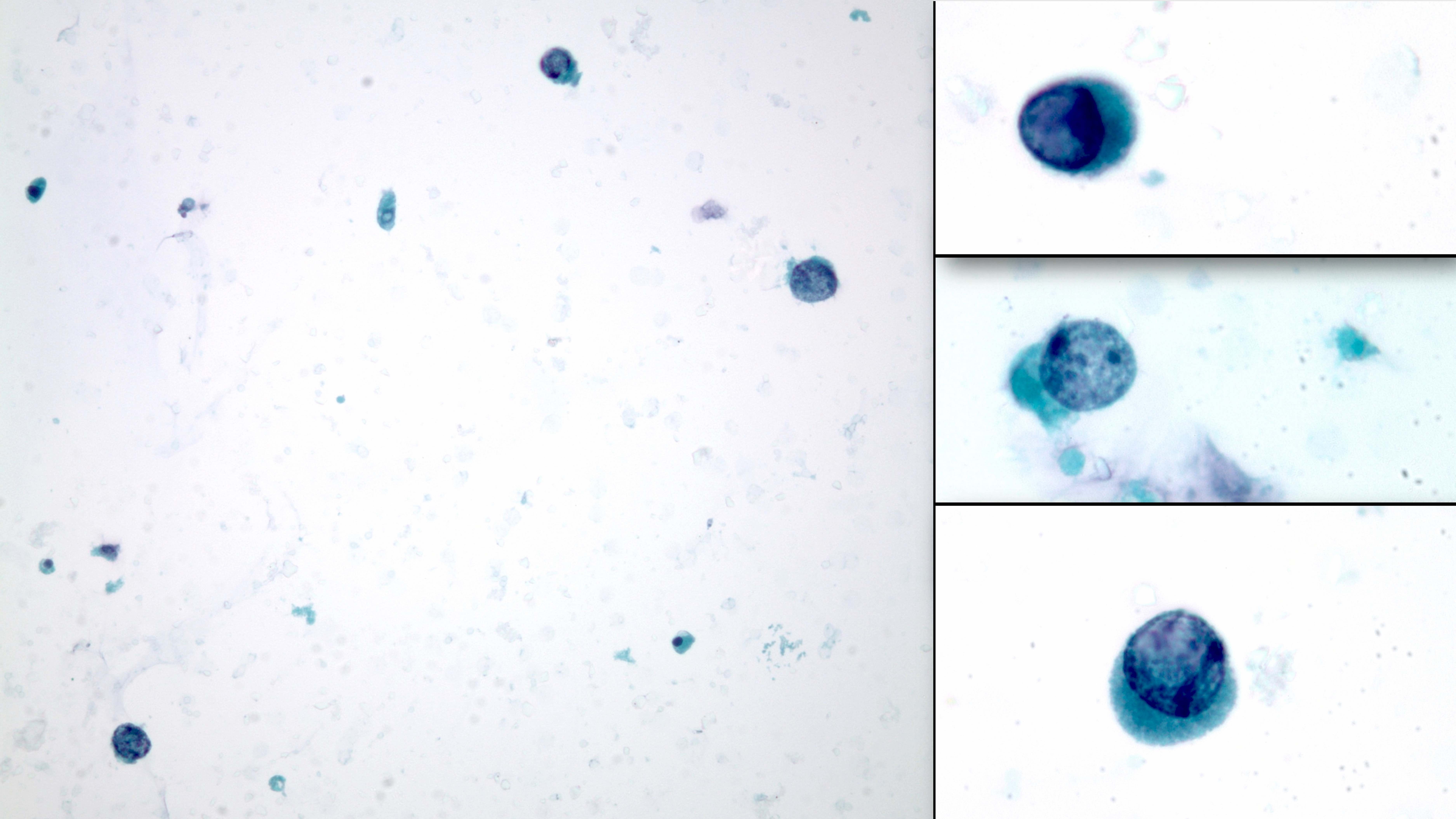
Decoy cells in urine cytology
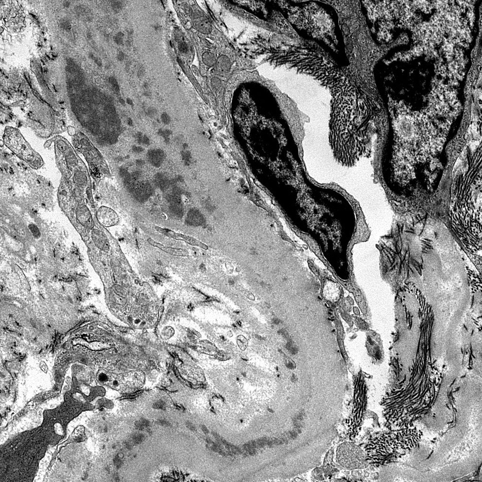
Contributed by Jonathan E. Zuckerman, M.D., Ph.D.
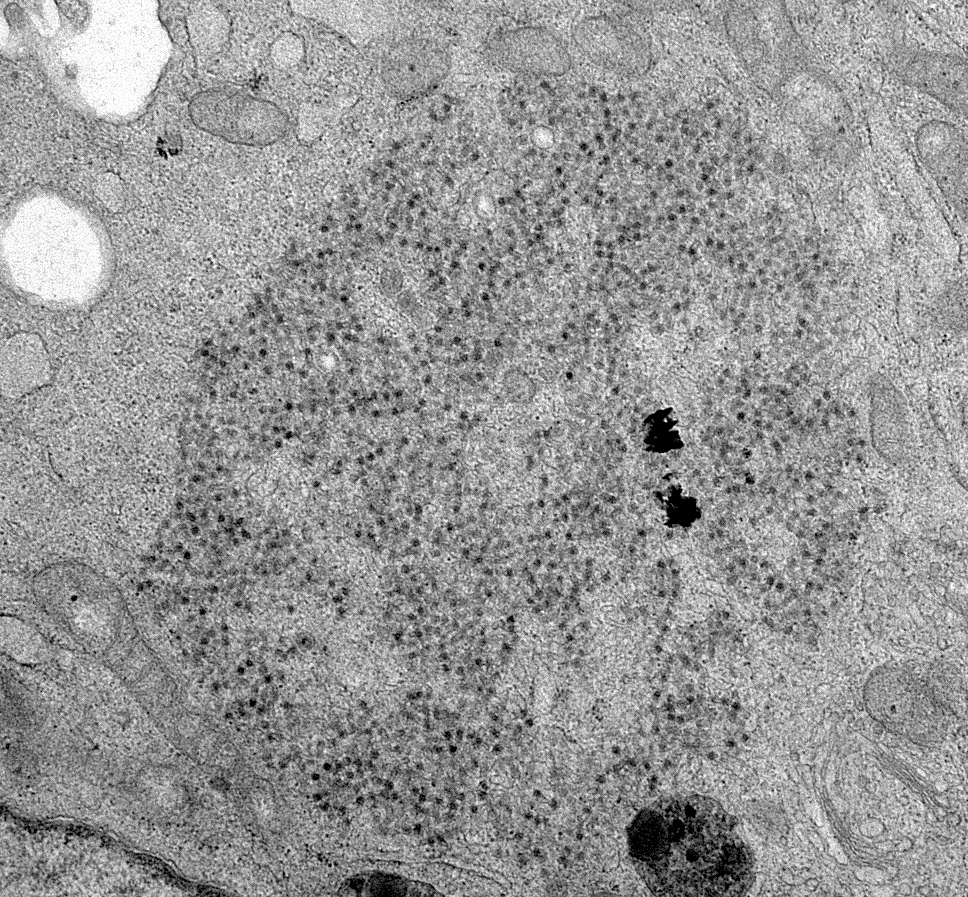
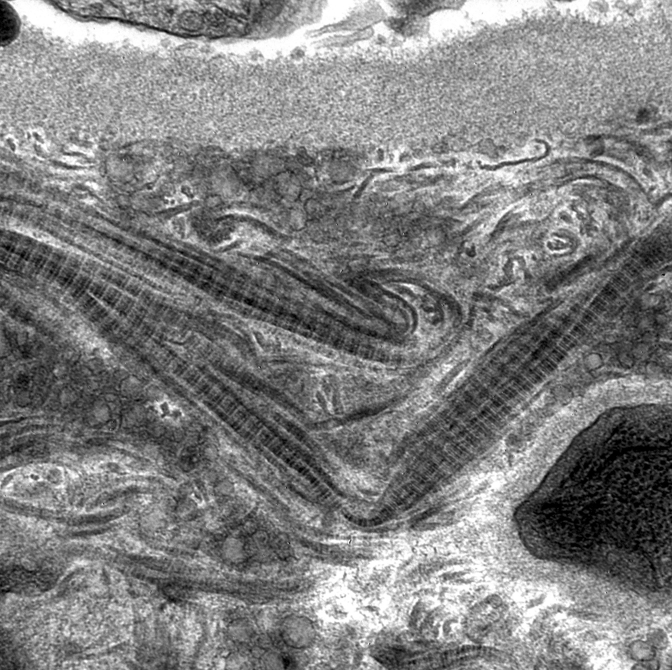
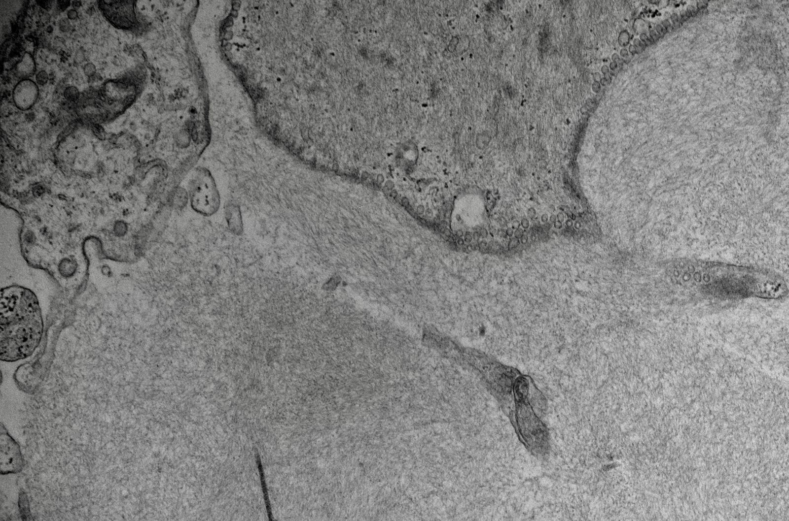
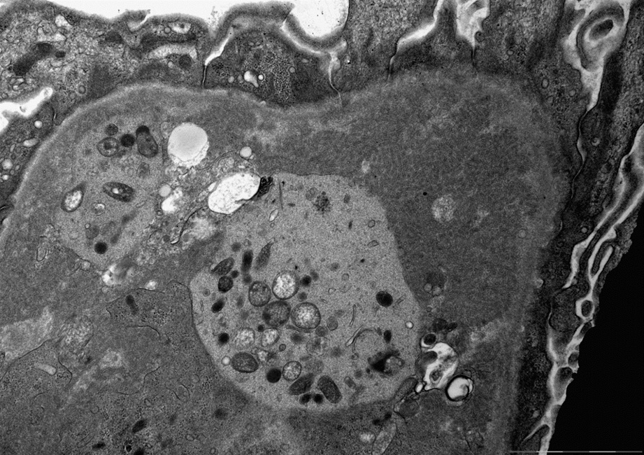
Viral particles
Nuclear viral particles
Images hosted on other servers:

Polyomavirus Haufen

Polyomavirus Haufen and CISH staining

Tubular basement membrane deposits
Histomorphological characteristics of BK virus associated nephropathy in a transplant patient
by Dr. Arzu Sağlam
Contributed by Ana Belén Larqué, M.D., Ph.D.
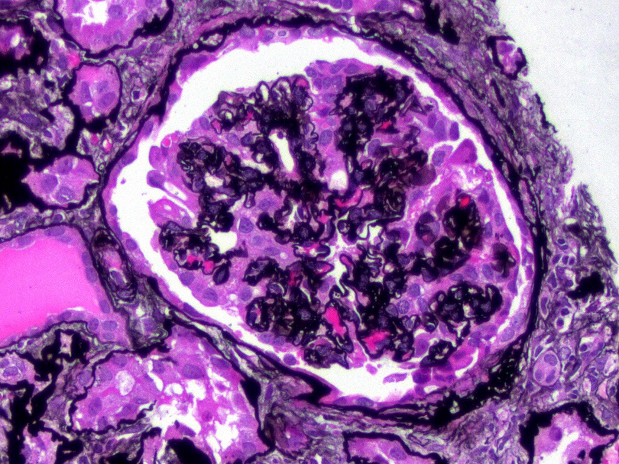
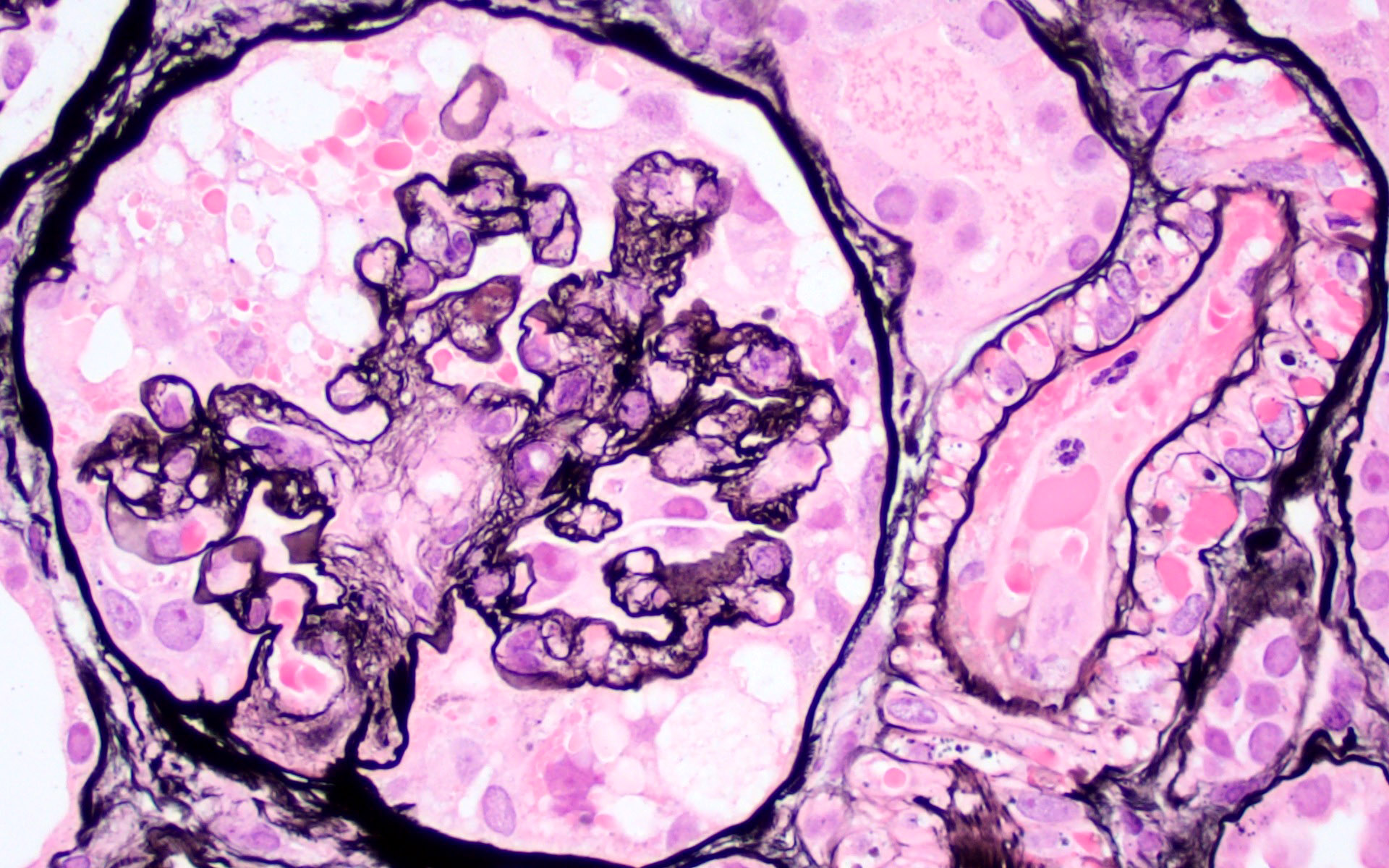
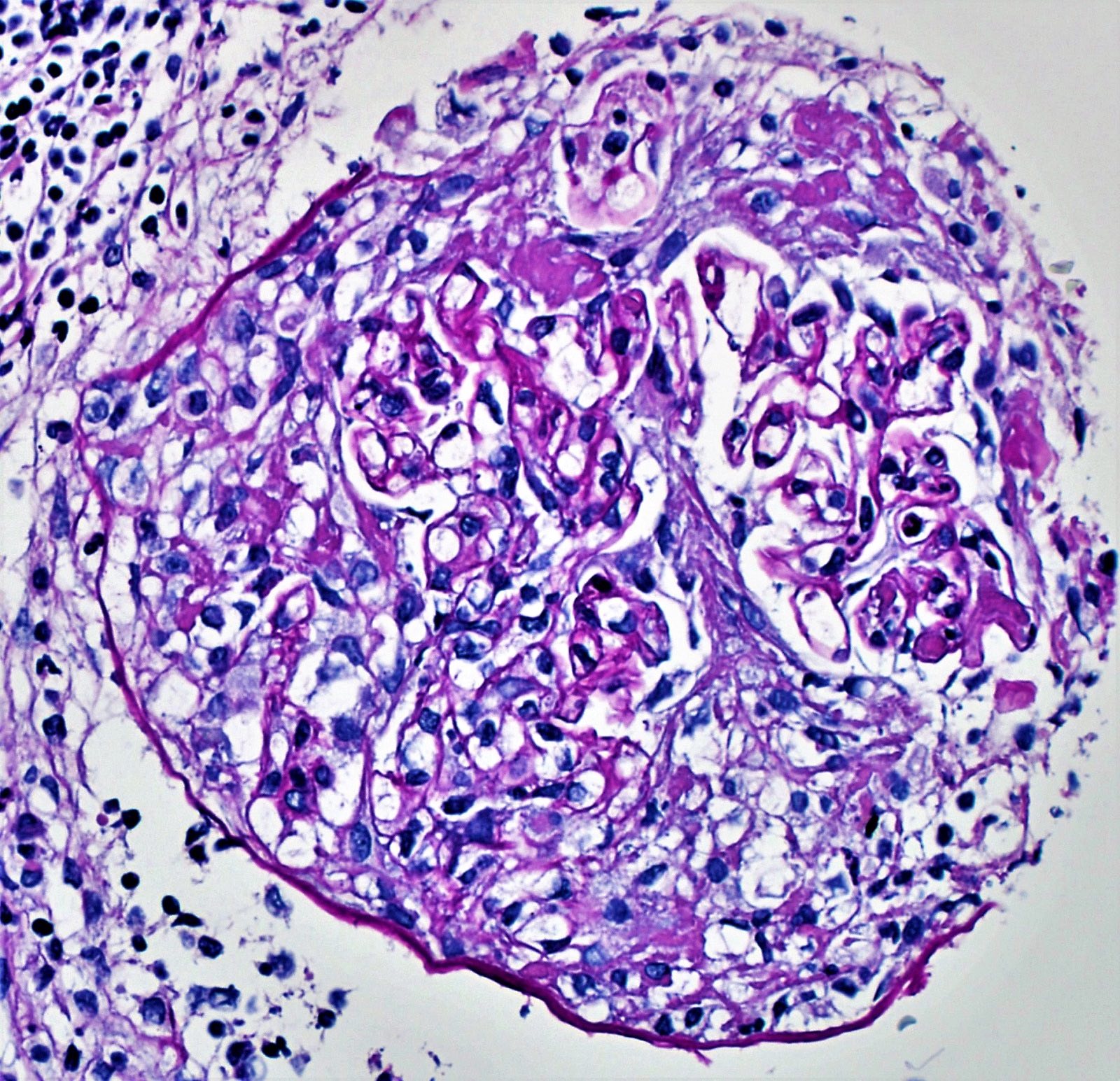
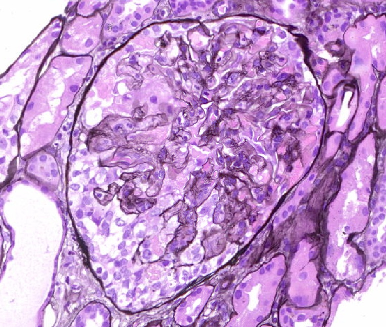
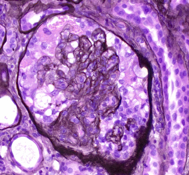
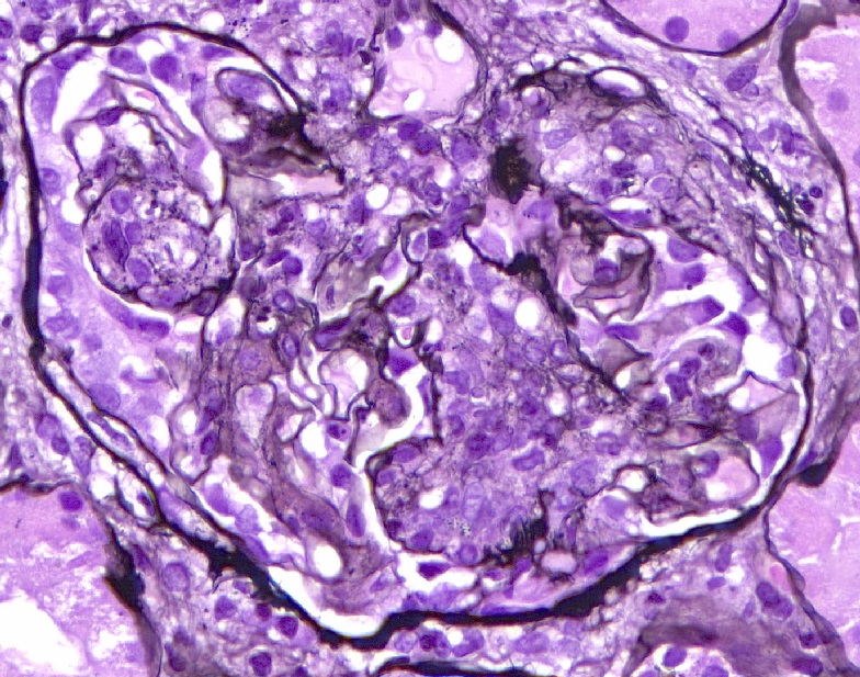
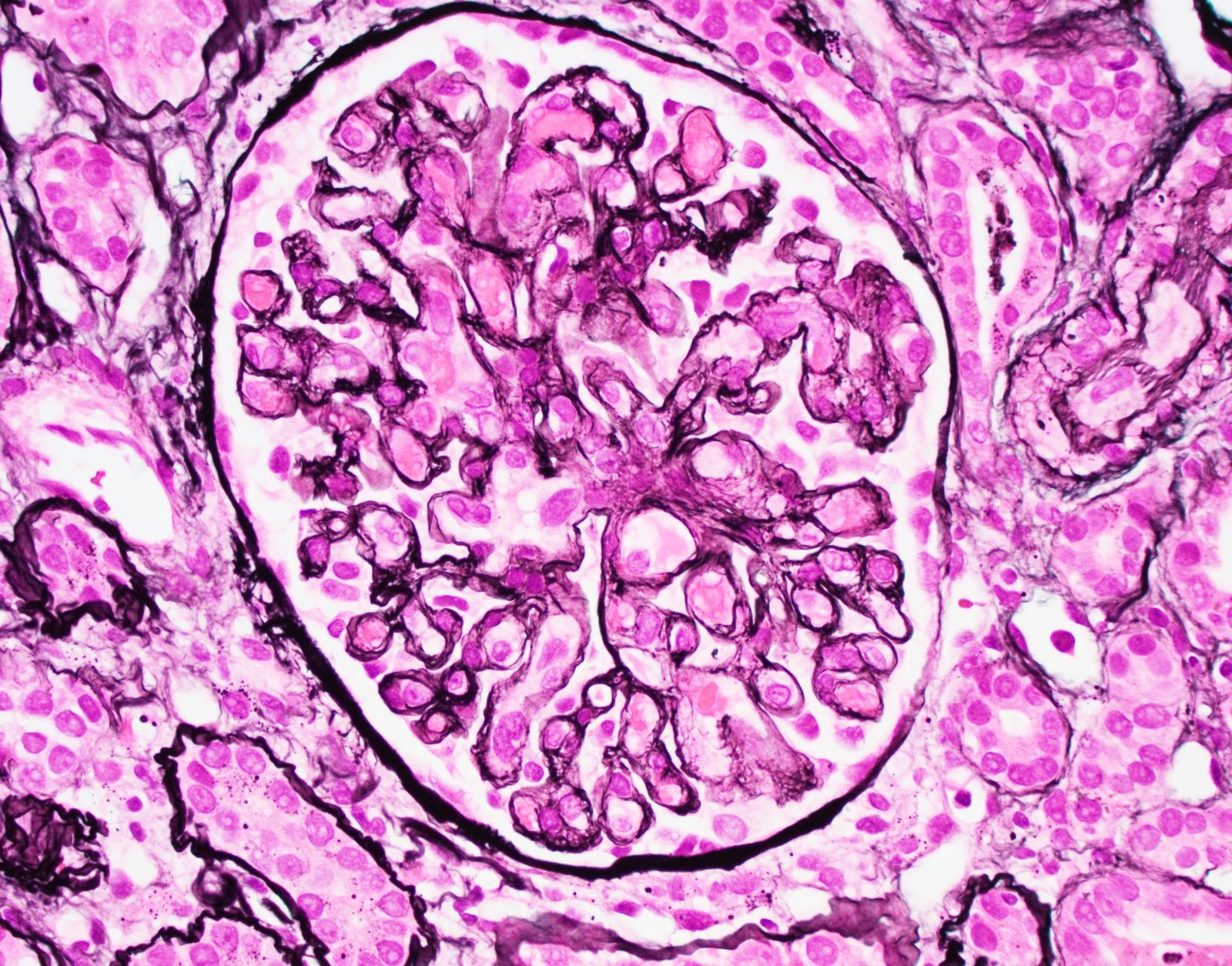
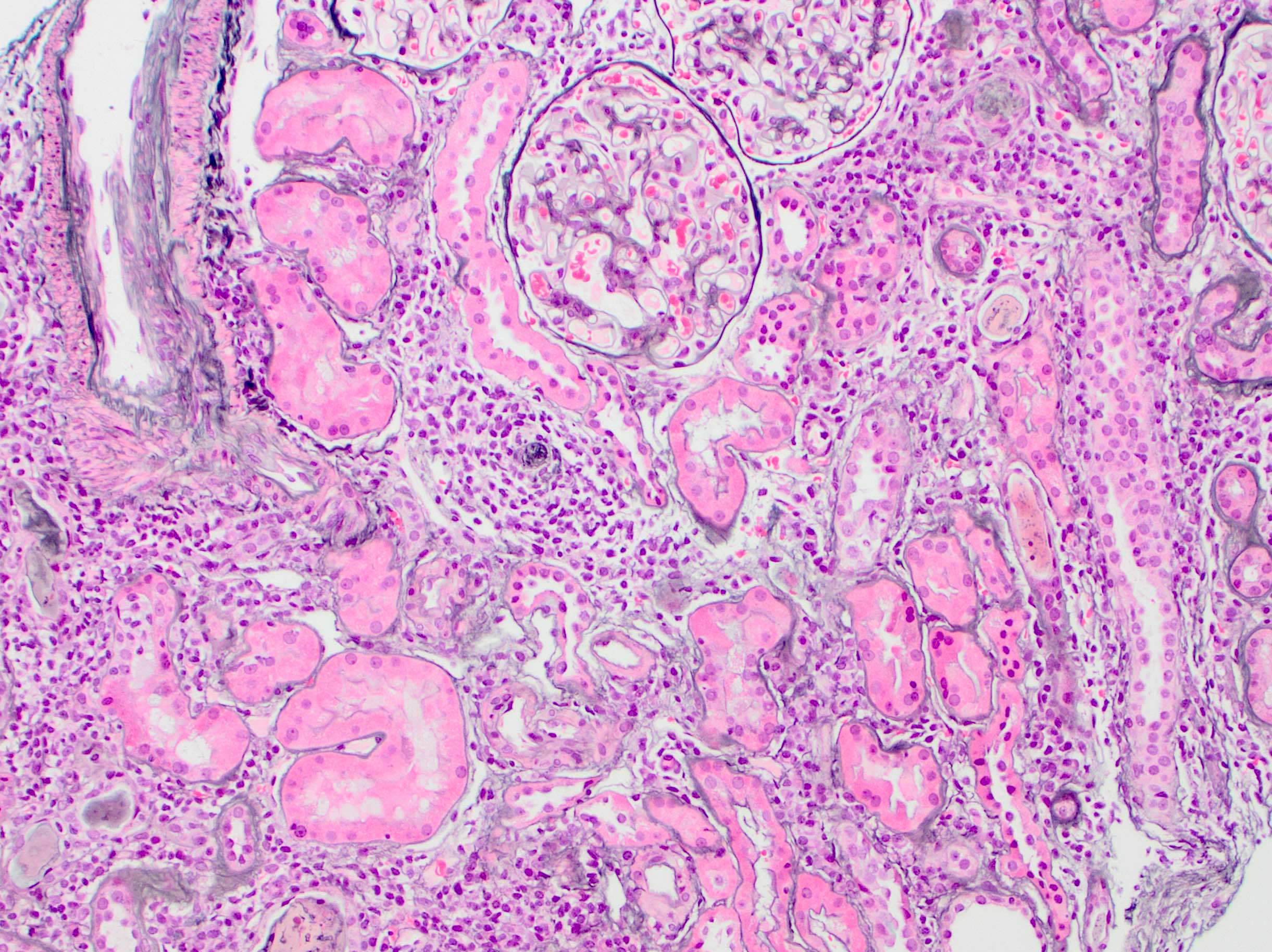
Glomeruli with membranoproliferative pattern
Glomerulus with mesangial pattern
Glomerular basement membrane thickening
Double contour capillary walls
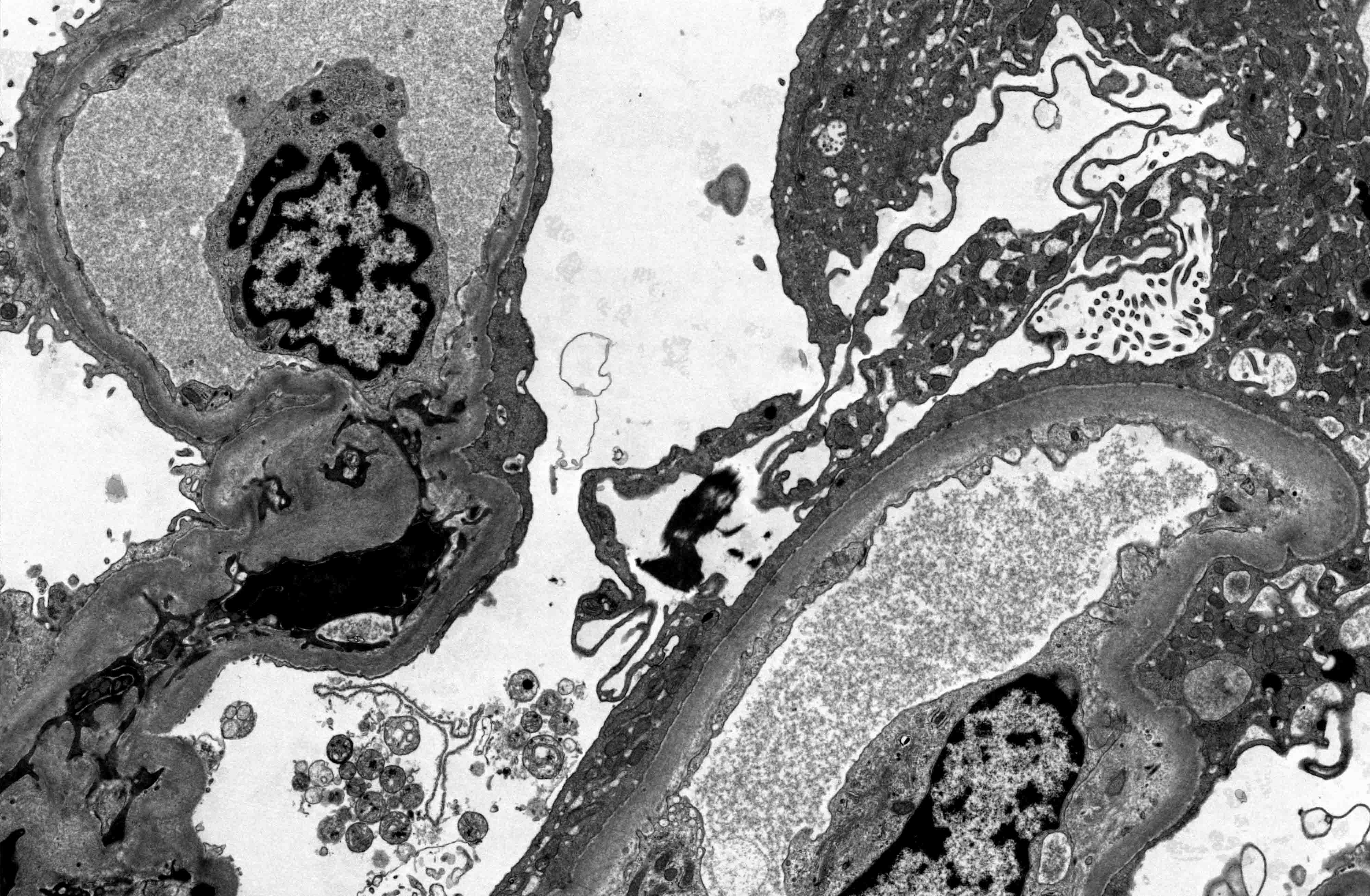
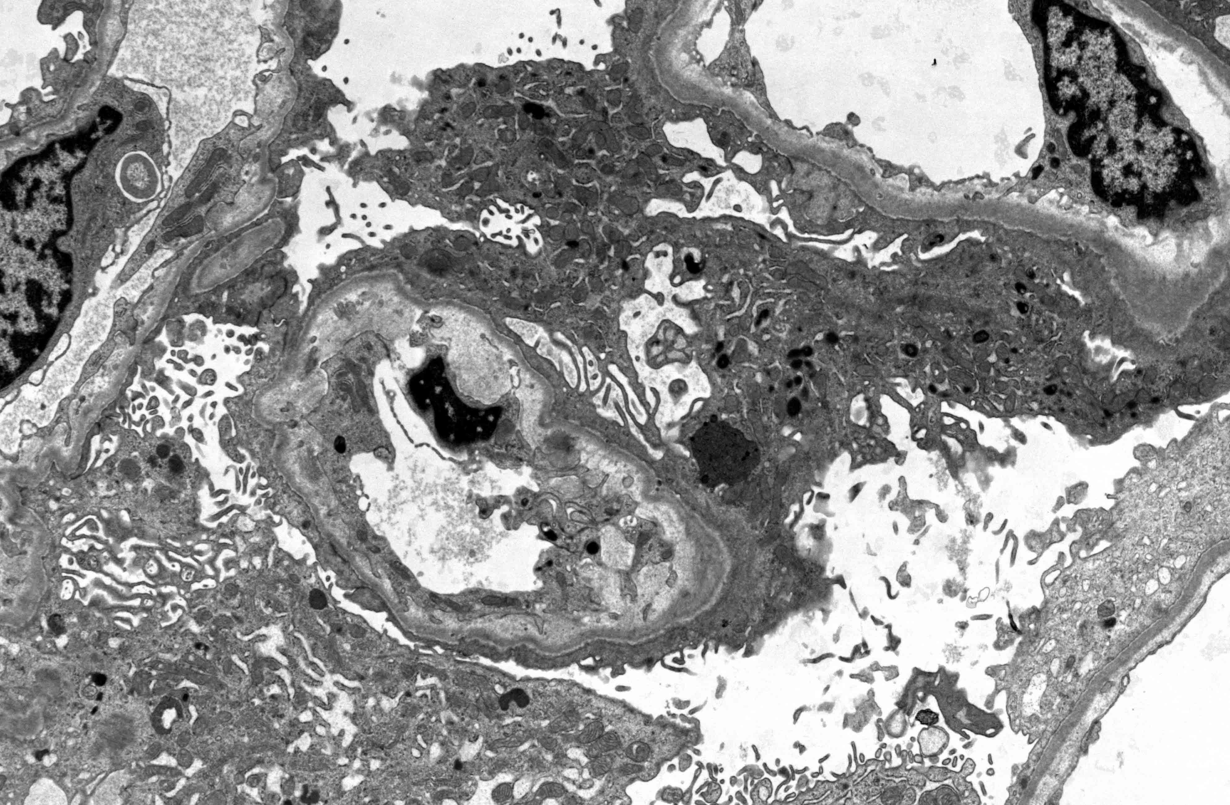
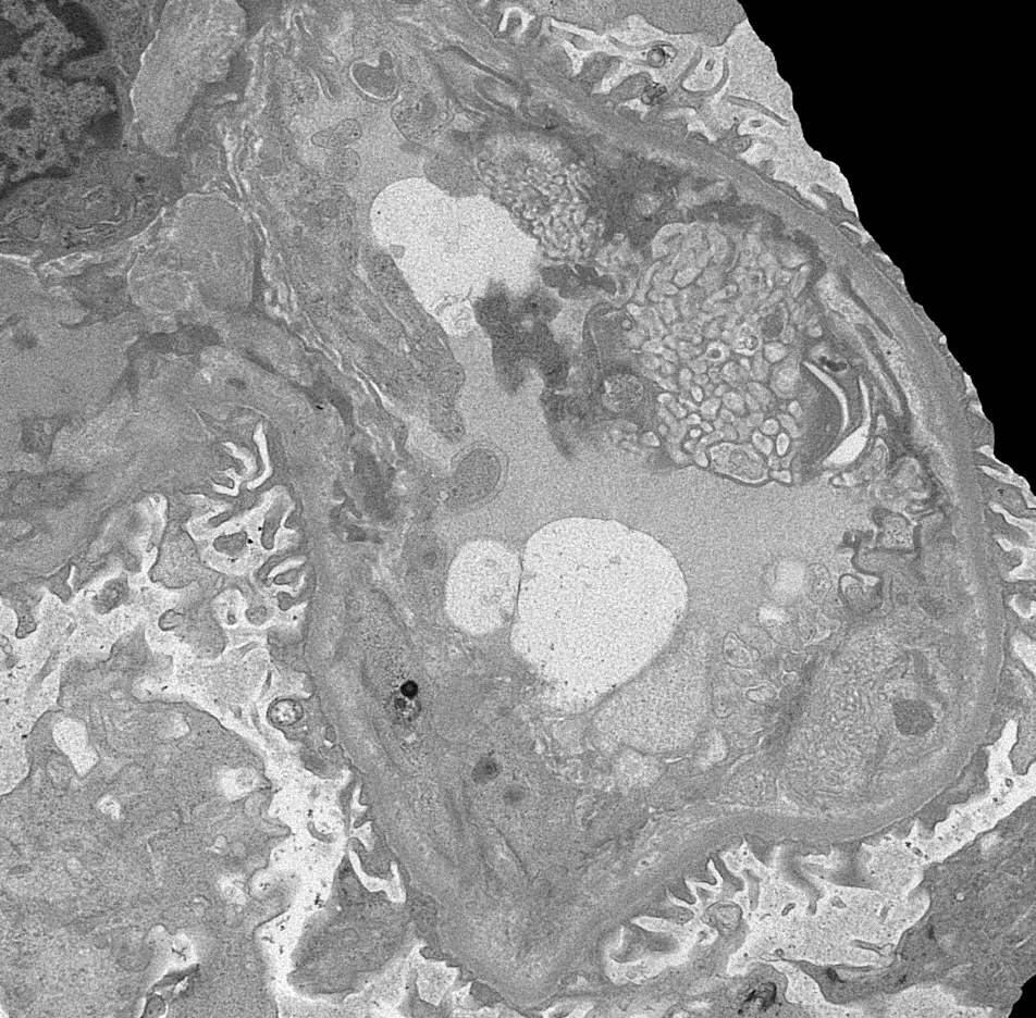
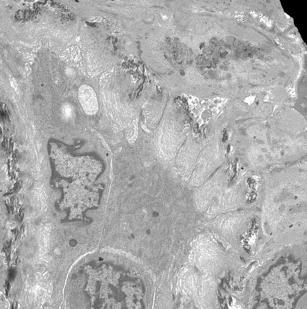
Contributed by Ana Belén Larqué, M.D., Ph.D.

Linear electron dense deposits
Membranoproliferative GN
Images hosted on other servers:

Various images


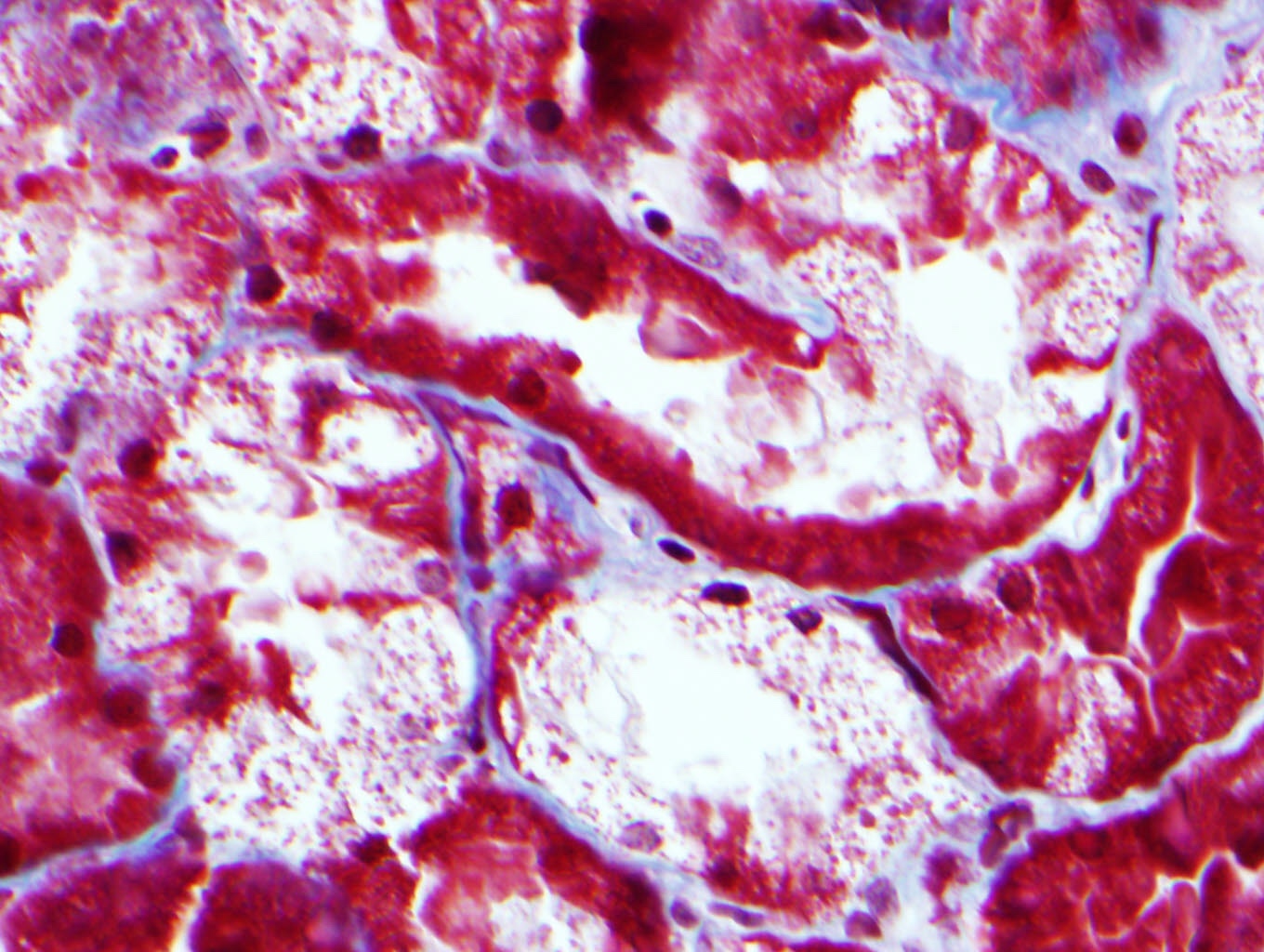
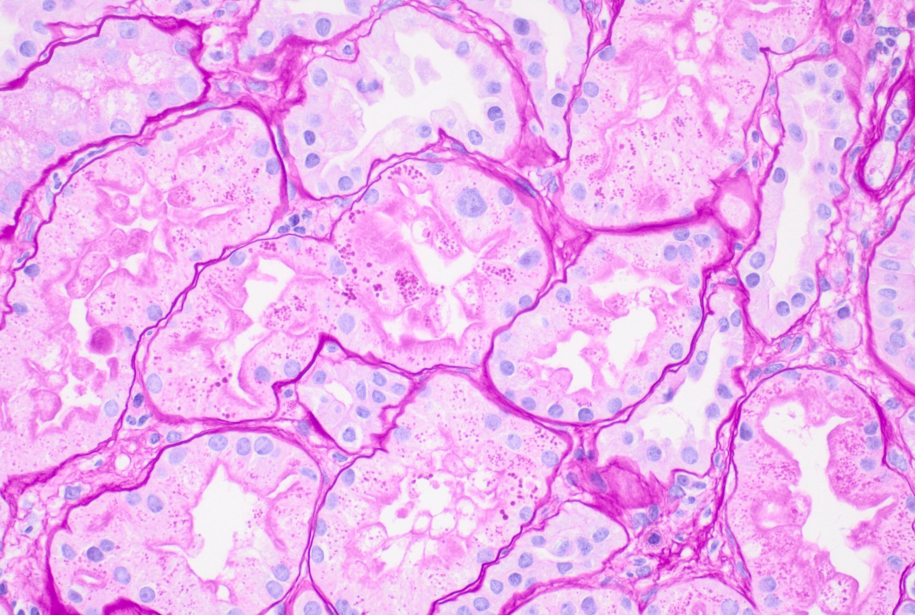
Allograft treated with cyclosporine A

Classic features of
calcineurin inhibitor
toxicity (cyclosporine
and tacrolimus)
Images hosted on other servers:

Recommendations
for management

Mechanisms of immune checkpoint blockade

Treatment, recovery, rechallenge, recurrence rates
Contributed by Vincenzo L’Imperio, M.D. and Giorgio Cazzaniga, M.D.

Neutrophilic tubulitis
Interstitial inflammatory infiltrate
Tubulitis
Plasma cell rich infiltrate
Neutrophil rich infiltrate
Contributed by Vincenzo L’Imperio, M.D. and Giorgio Cazzaniga, M.D.
Tubulitis
Contributed by Alexei Mikhailov, M.D., Ph.D.

IFTA with lymphocytic inflammation

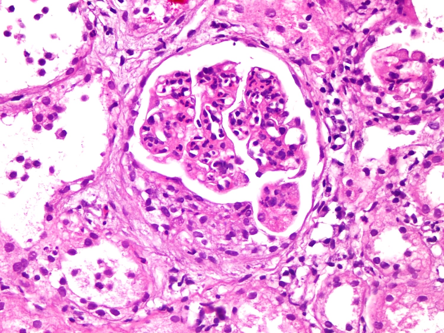
Glomerulus in CKDu
Ischemic glomerulus in CKDu

Glomerular obsolescence in MeN

MeN interstitial inflammatory infiltrate
Perihilar FSGS in CKDu
Contributed by Alexei Mikhailov, M.D., Ph.D.

MeN glomerular capillary
MeN podocyte effacement
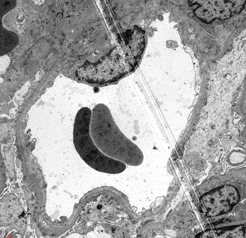
Contributed by Saman Karimi, M.D., M.S., Suman Setty, M.B.B.S., Ph.D. and Vijay Shankar, M.D.
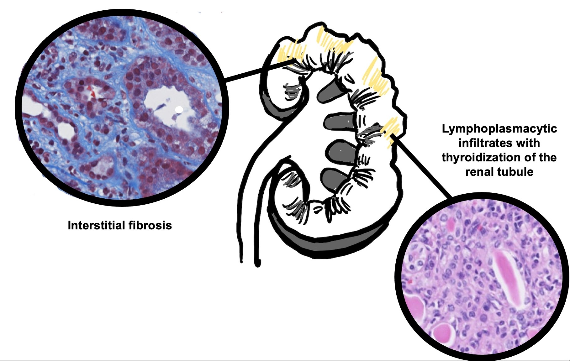
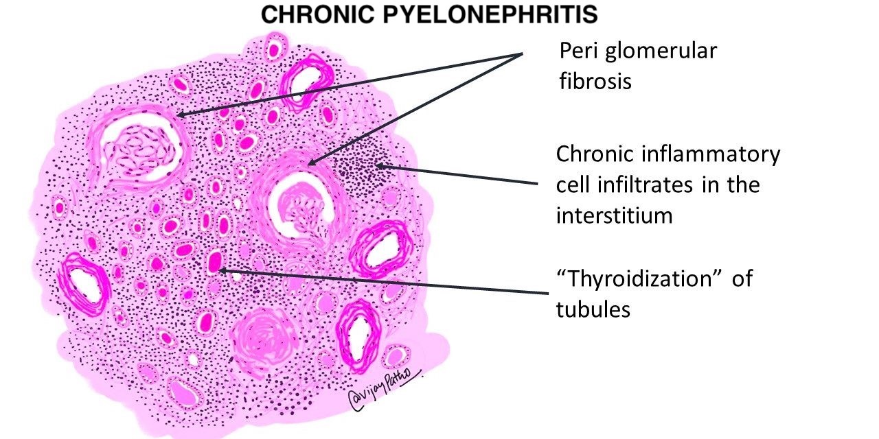
Schematic of chronic pyelonephritis
Contributed by Saman Karimi, M.D., M.S. and Suman Setty, M.B.B.S., Ph.D.
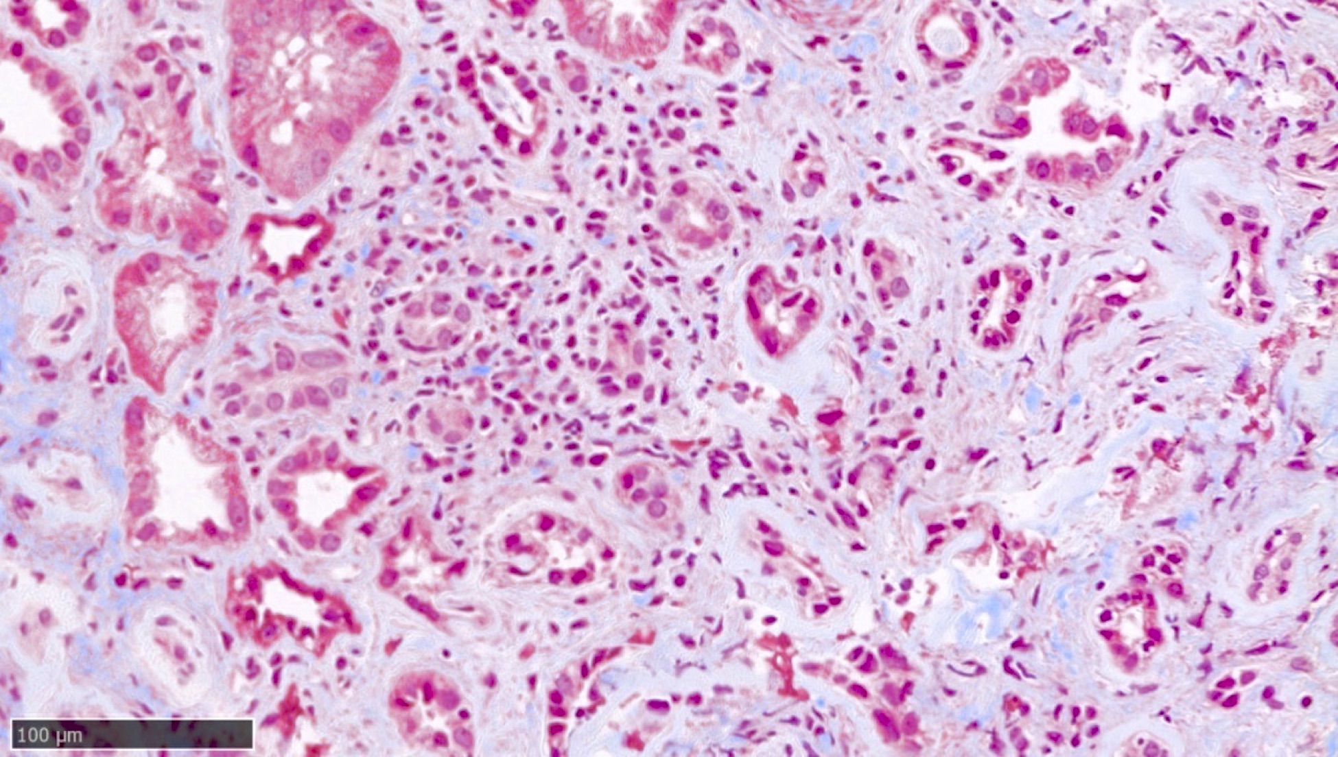
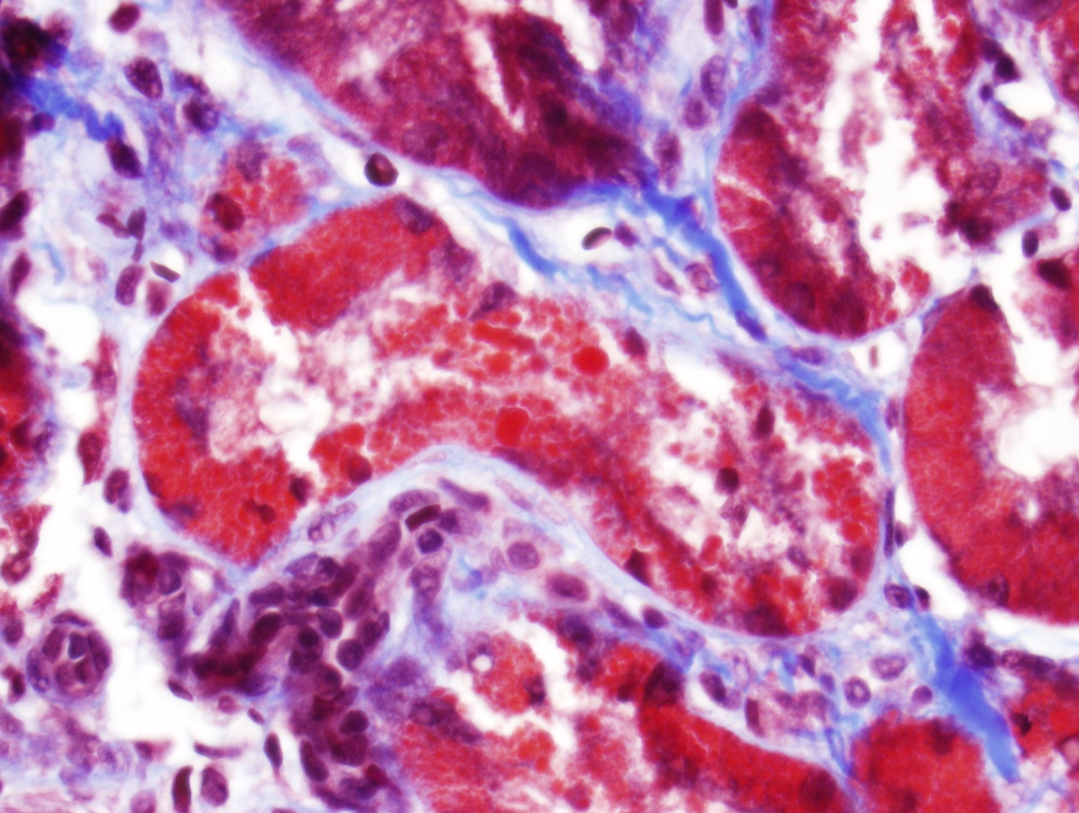
Interstitial chronic
inflammation,
tubular atrophy
and fibrosis
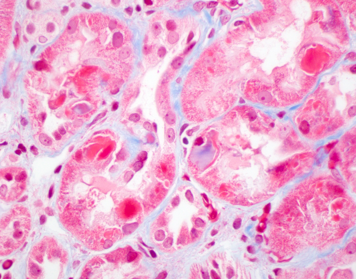
Interstitial chronic
inflammation and
periglomerular fibrosis
Renal tubular atrophy with luminal casts
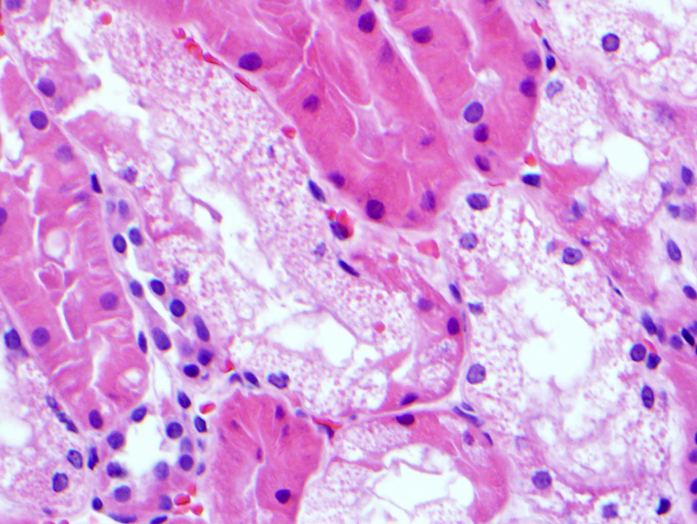
Dense interstitial fibrosis
Overview of chronic pyelonephritis
Histological features of chronic pyelonephritis
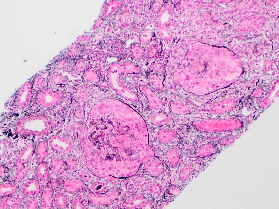
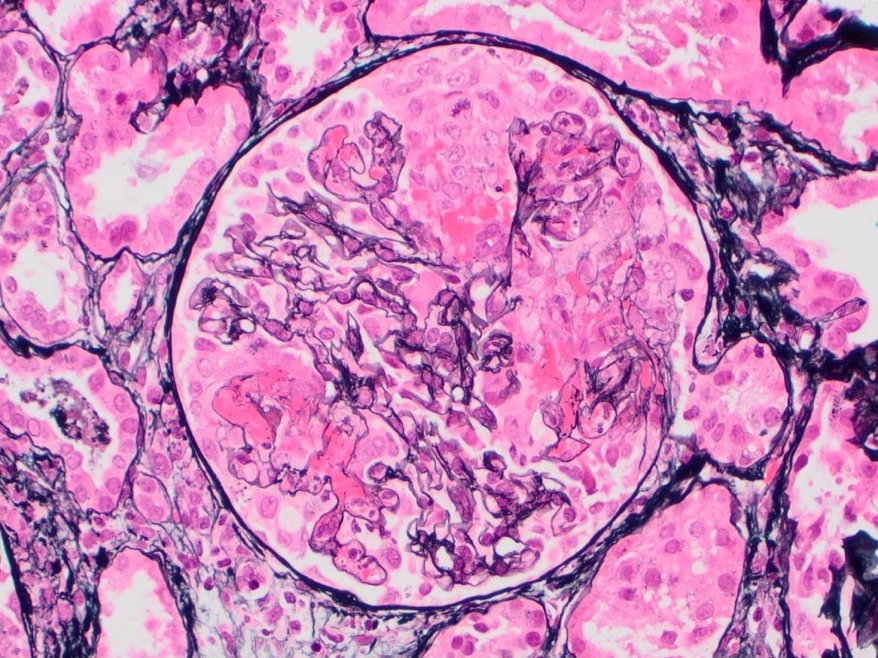
Contributed by Joseph Grande, M.D., Ph.D.
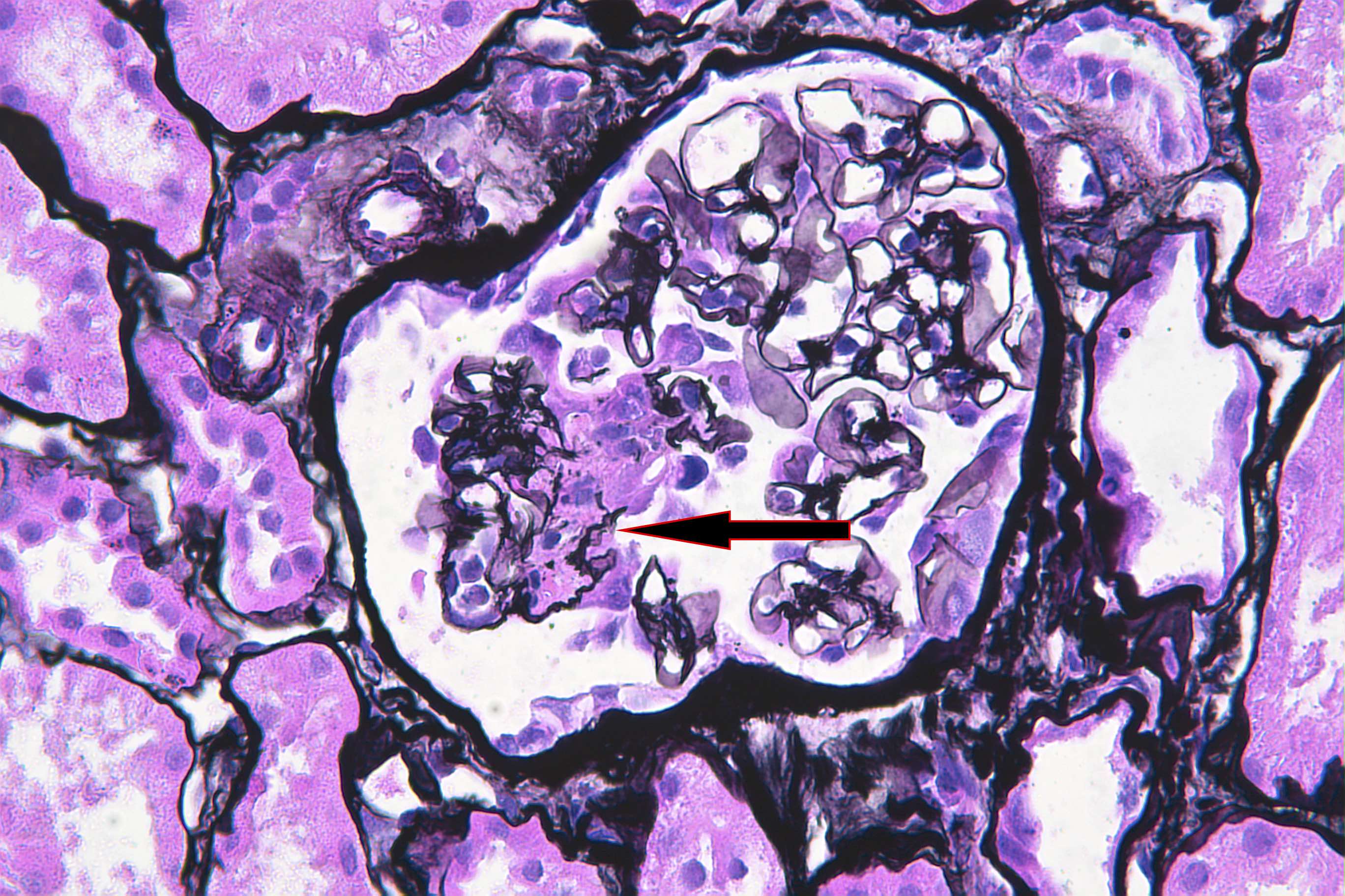
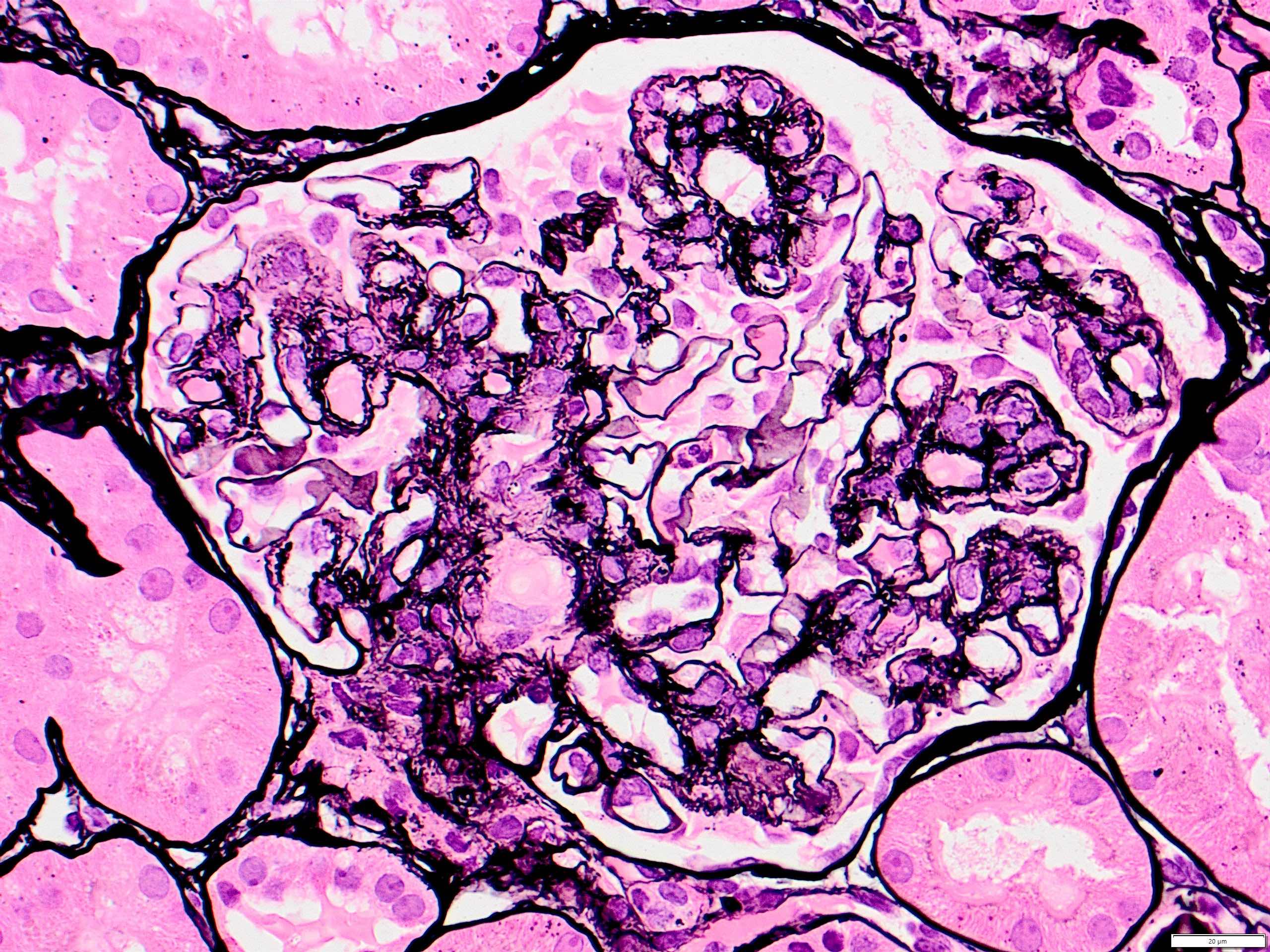
Lobular mesangial expansion
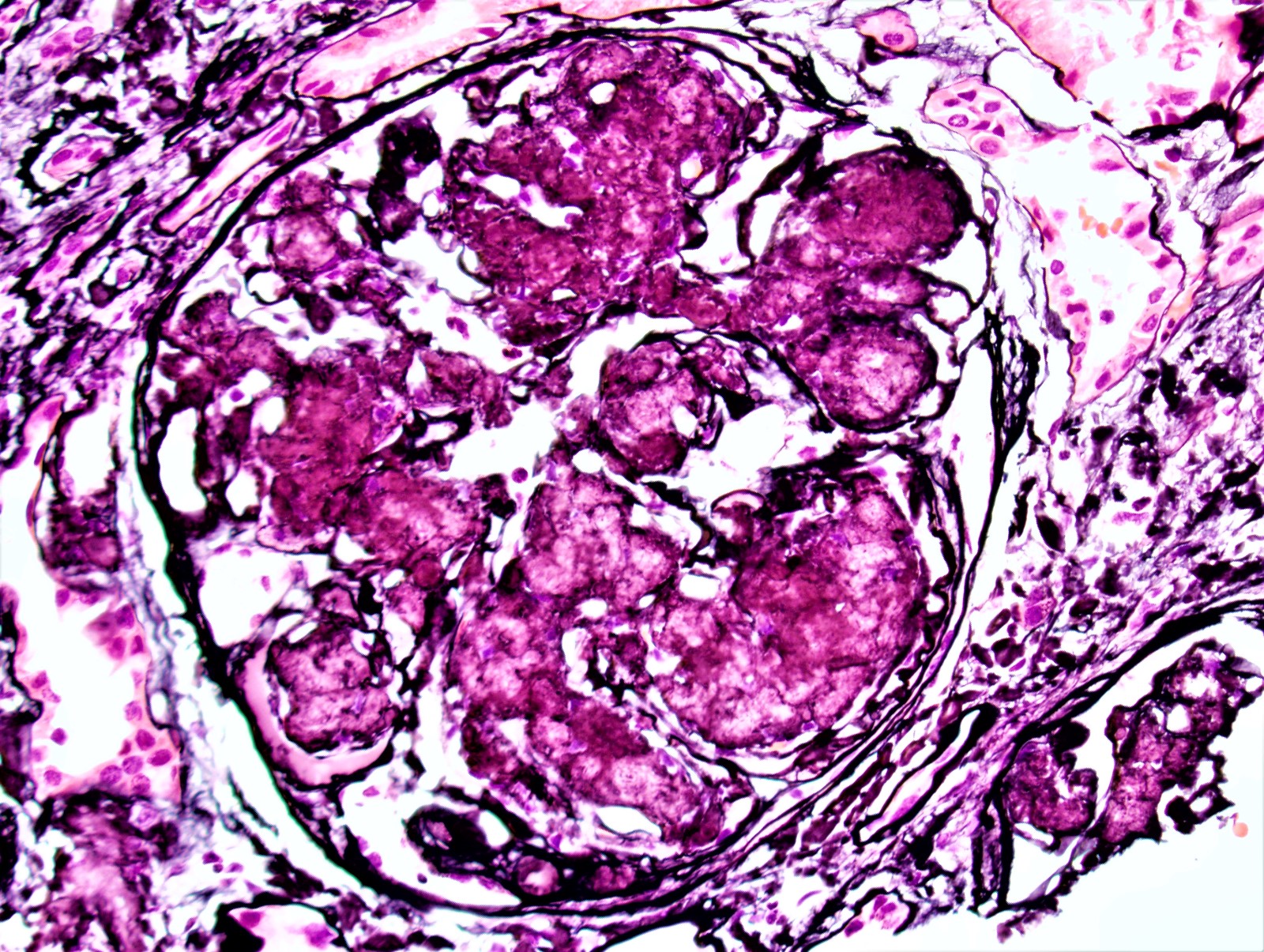
Silver negative material deposits

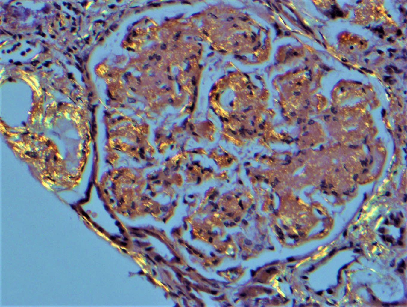
PAS weak / negative deposits

Trichrome stains deposits blue
Images hosted on other servers:

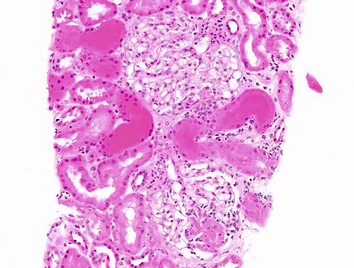
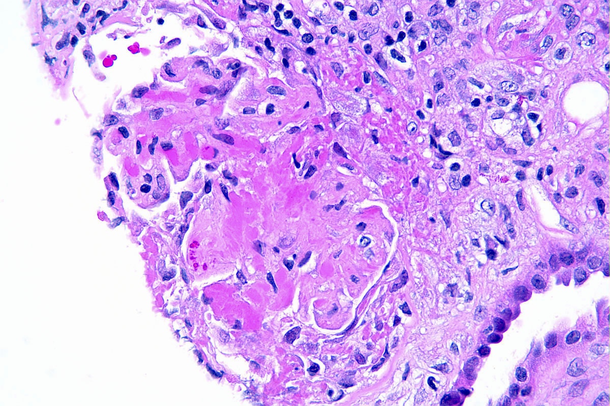
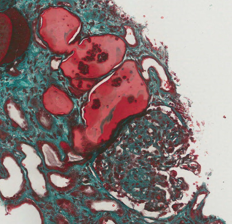
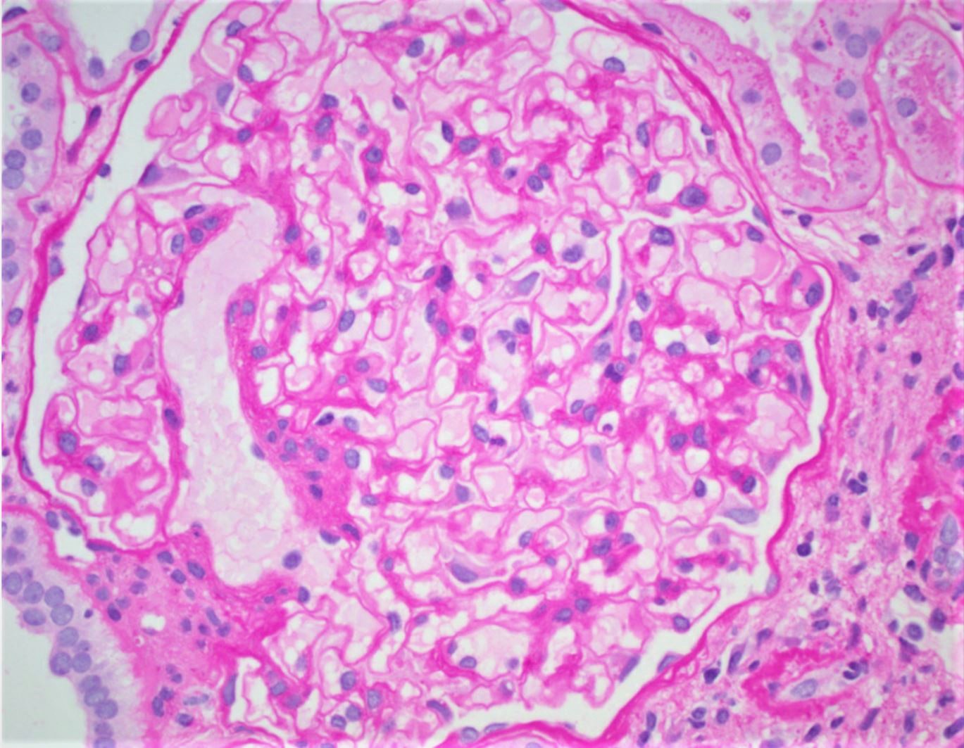
Enlarged glomerulus with hyaline cap deposits

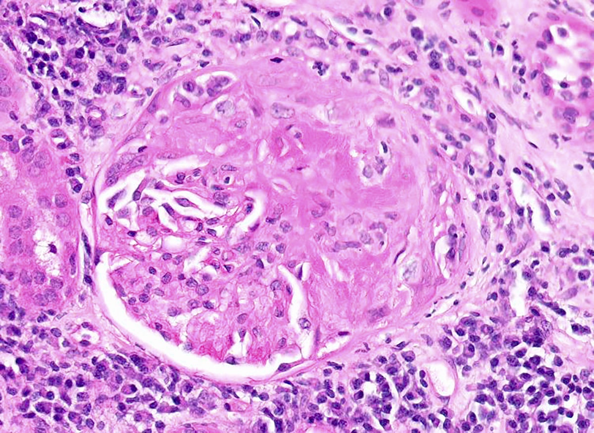
Lobular appearance

With Hodgkin's lymphoma

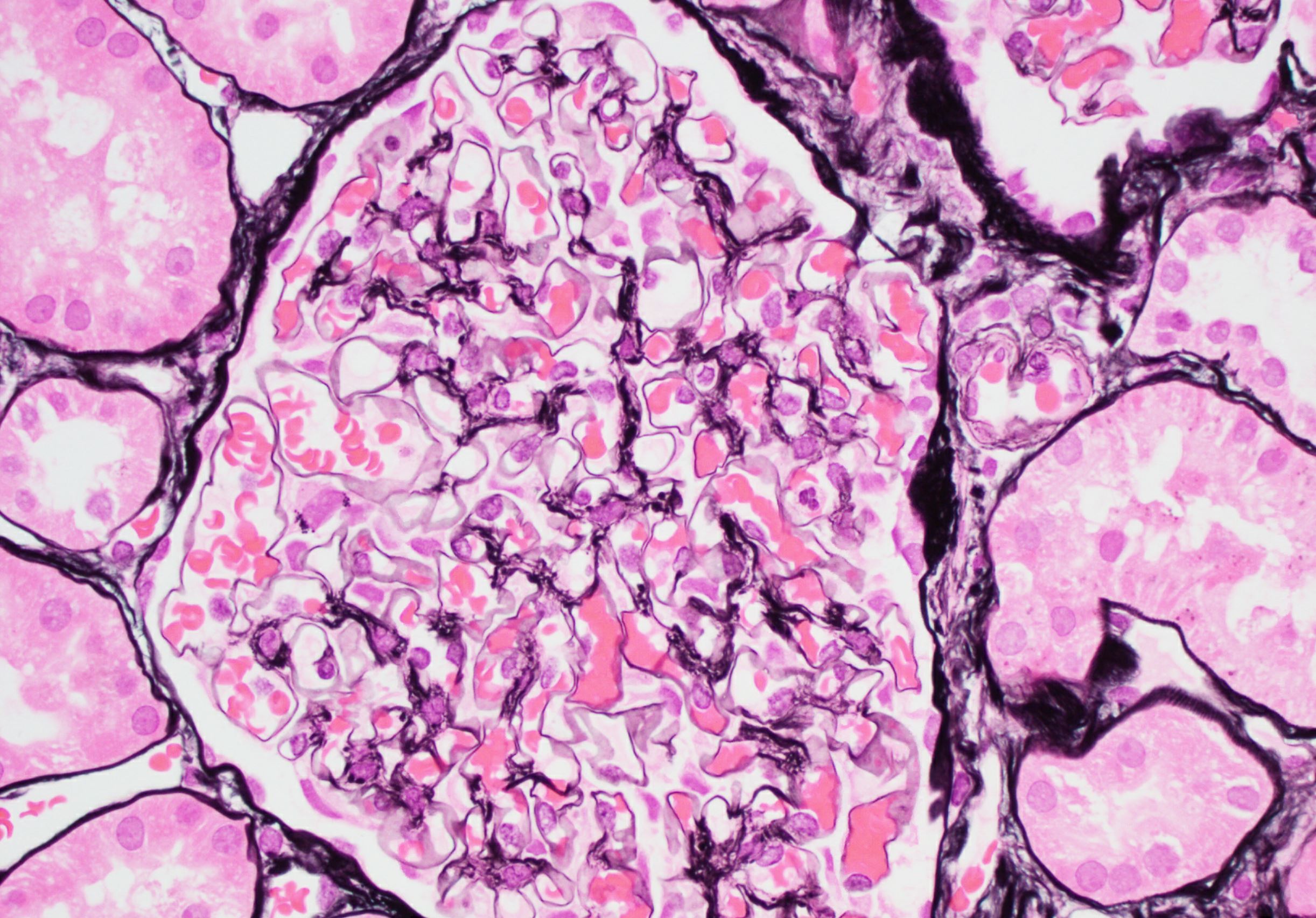
Silver stain

Congo red stain
Contributed by Joseph Grande, M.D., Ph.D.

Extensive mesangial deposits
Higher power of subendothelial deposit
Images hosted on other servers:

Abundant subendothelial deposits of large fibers
Images hosted on other servers:

Renal hypoplasia
Images hosted on other servers:

Agenesis - bilateral

Incomplete duplication


Horseshoe kidney
Images hosted on other servers:

Oligomeganephronic renal hypoplasia
Images hosted on other servers:

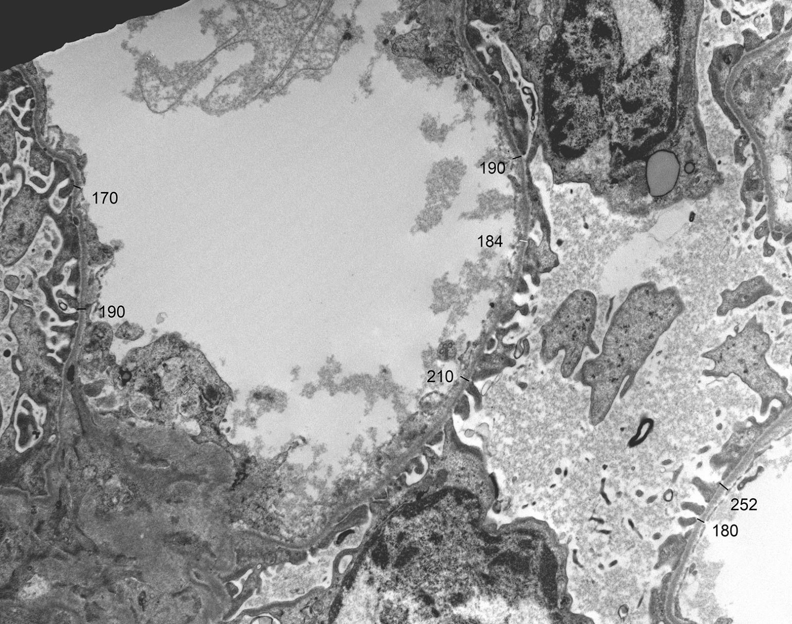
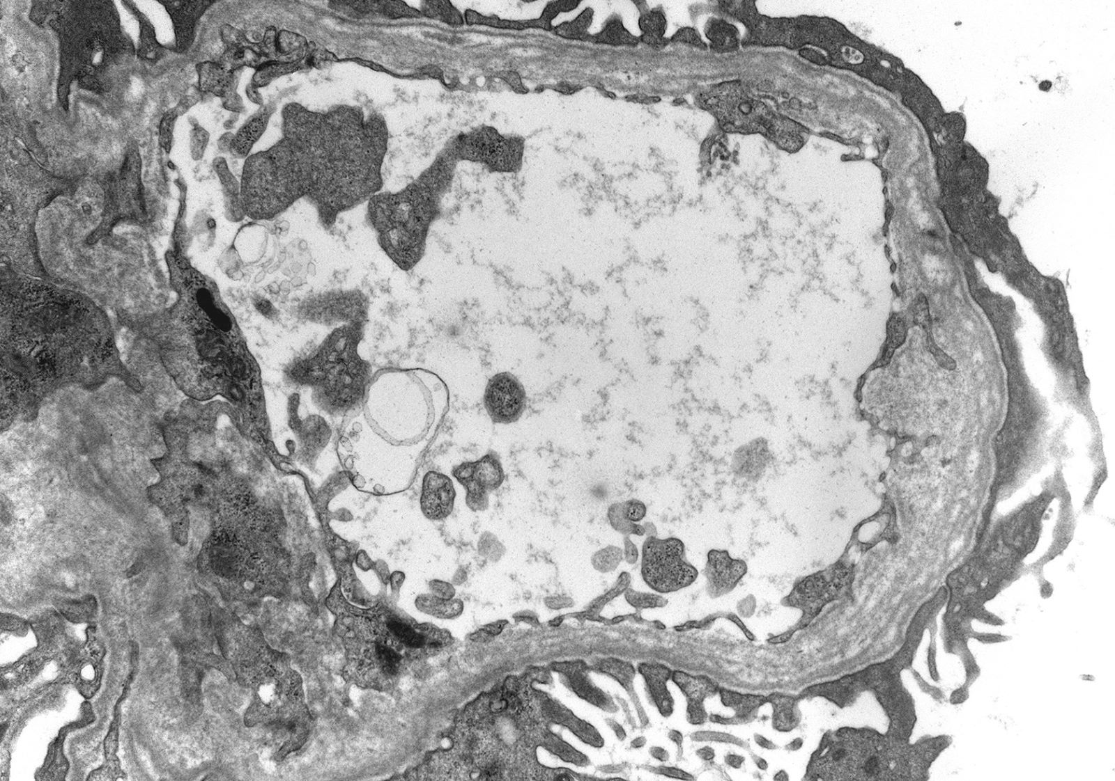
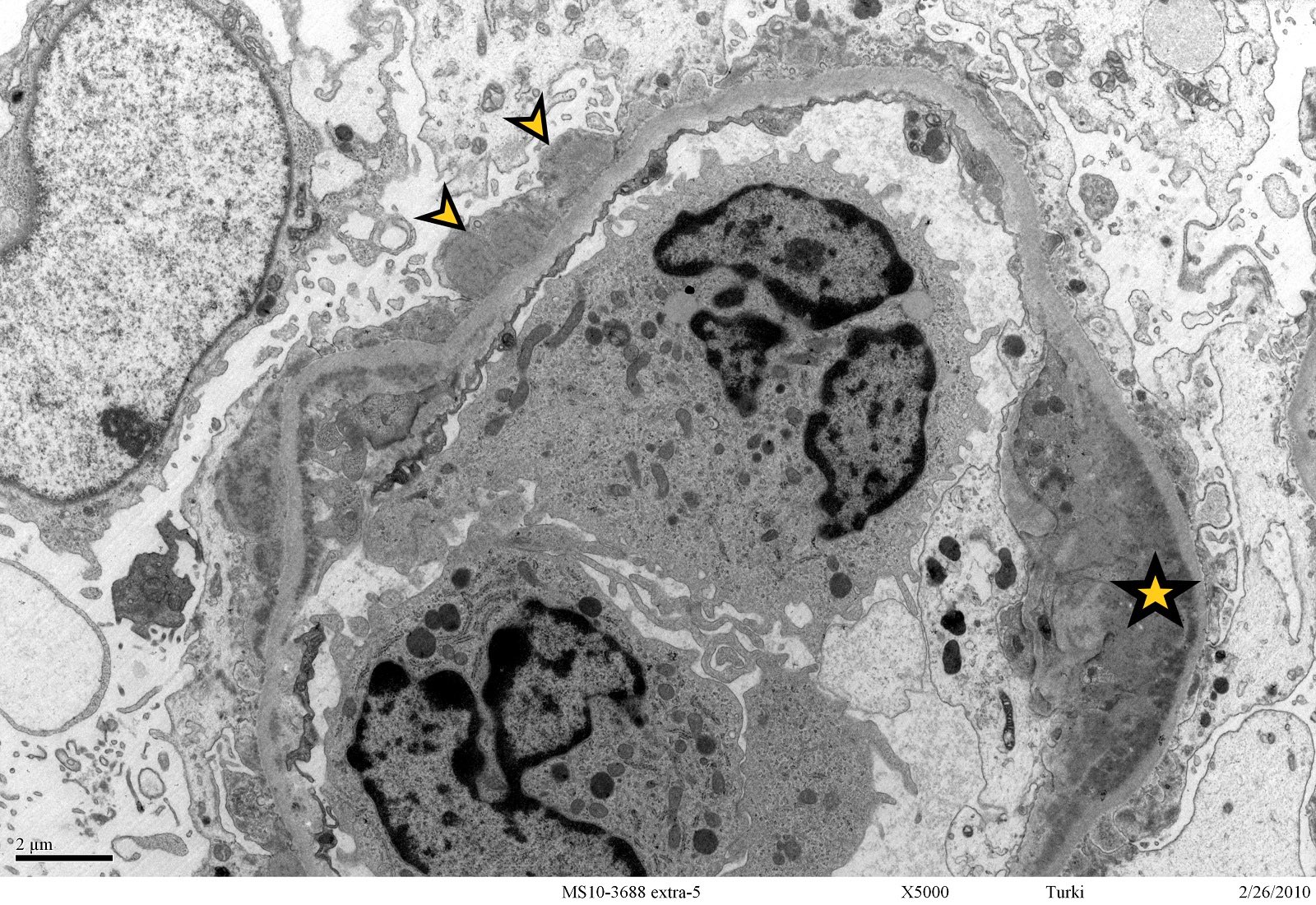
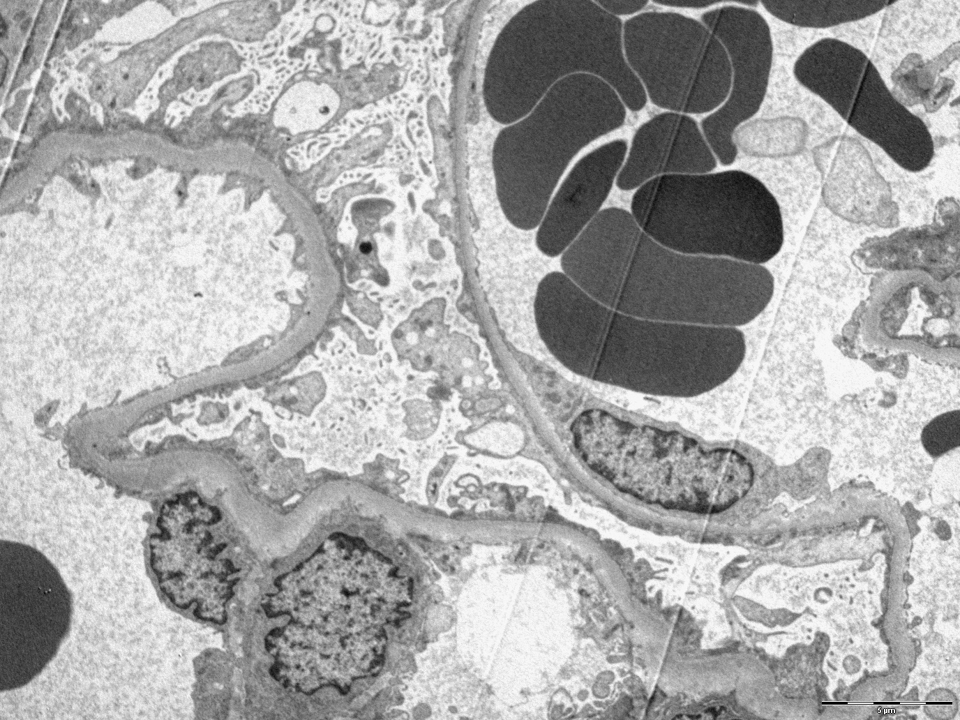
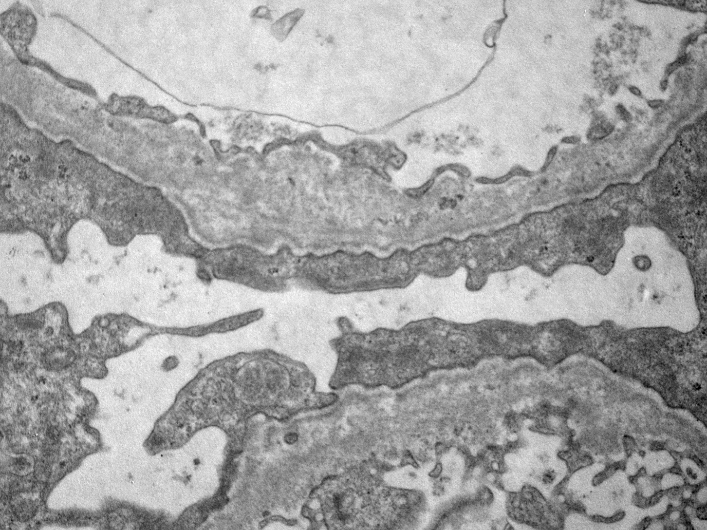
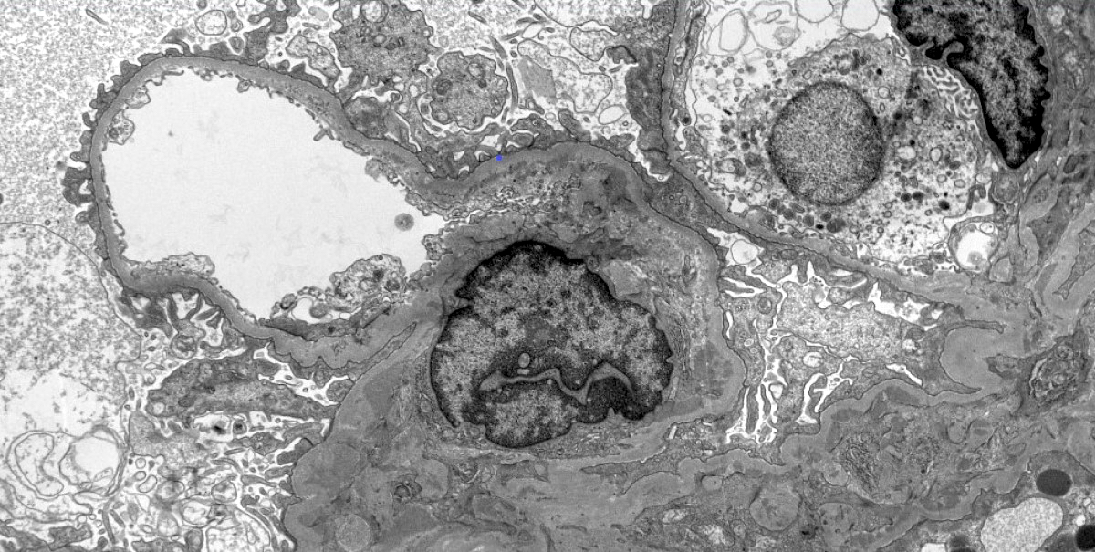
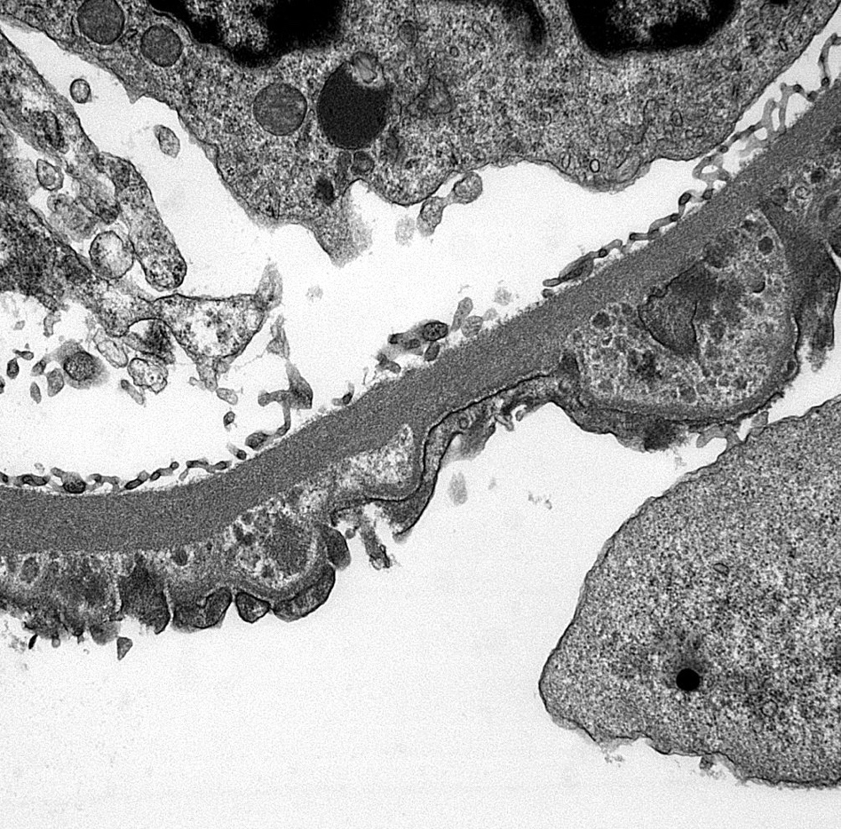
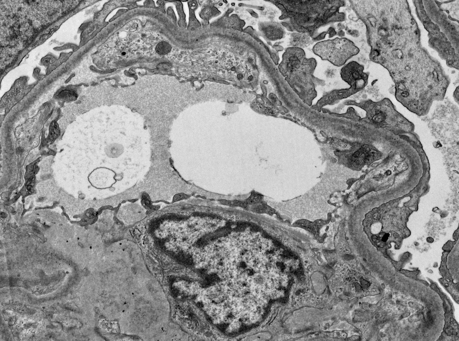
Normal glomerular capillary and capillary wall
Images hosted on other servers:


Enlarged and hyperechogenic kidneys by ultrasound
Contributed by Chunlai Zuo, M.D., M.S.
Enlarged pale kidney
Contributed by Chunlai Zuo, M.D., M.S. and Jonathan E. Zuckerman, M.D., Ph.D.
Mesangial hypercellularity
Segmental glomerulosclerosis
Dilated proximal and distal tubes
Cystic change and interstitial fibrosis
Contributed by Jonathan E. Zuckerman, M.D., Ph.D.
Diffuse foot processes effacement
Loss of slit diaphrams
Mesangial hypercellularity
Endothelial cell blebs
Contributed by Shreeram Akilesh, M.D., Ph.D.
Acute tubular injury
Collapsing glomerulopathy
Collapsing glomerulopathy and TMA
Contributed by Shreeram Akilesh, M.D., Ph.D.

Podocyte foot process effacement
Effacement and TMA
Contributed by Khaled A. Murshed, M.D., Noheir M. Taha, M.B.B.Ch., M.D. and Mohammed Akhtar, M.D.
Cellular crescent
Cellular crescent high magnification
Fibrin and PEC proliferation
Cellular crescent with fibrin
Fibrinoid necrosis
Capillary wall defect high magnification
Bowman capsule defect
Circular crescent
Monolayer PEC proliferation
Early crescent
Fibrocellular crescent
Fibrocellular fibrous crescent
Fibrous crescent
Anti-GBM disease

ANCA associated GN
Contributed by Khaled A. Murshed, M.D., Noheir M. Taha, M.B.B.Ch., M.D. and Mohammed Akhtar, M.D.
Electron dense deposits

Pauci-immune
Images hosted on other servers:


Cryoglobulin related glomerulonephritis
Images hosted on other servers:

Various images
Contributed by Khaled A. Murshed, M.D., Noheir M. Taha, M.B.B.Ch., M.D. and Mohammed Akhtar, M.D.

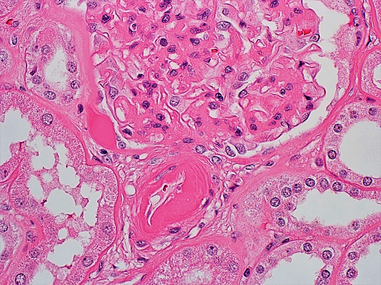
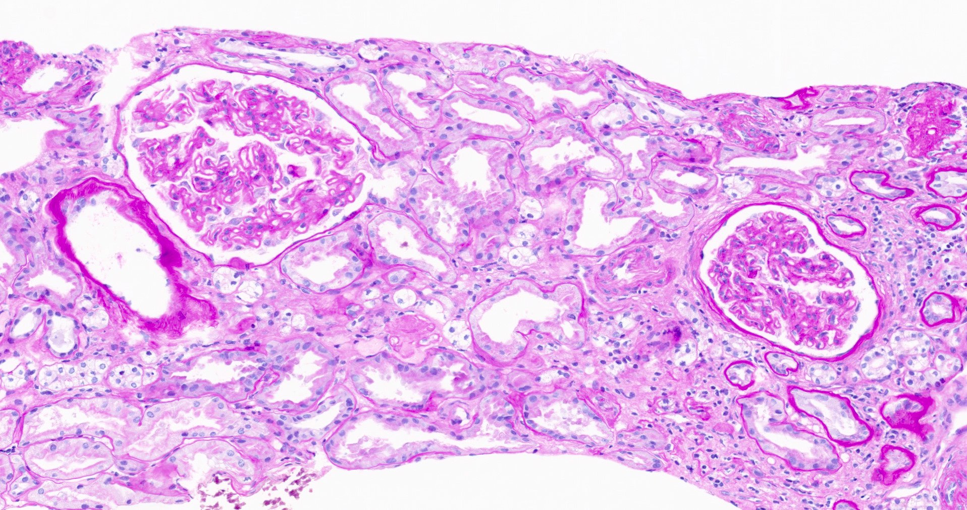
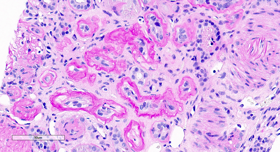
Afferent and efferent hyalinosis, PAS
Capsular drop
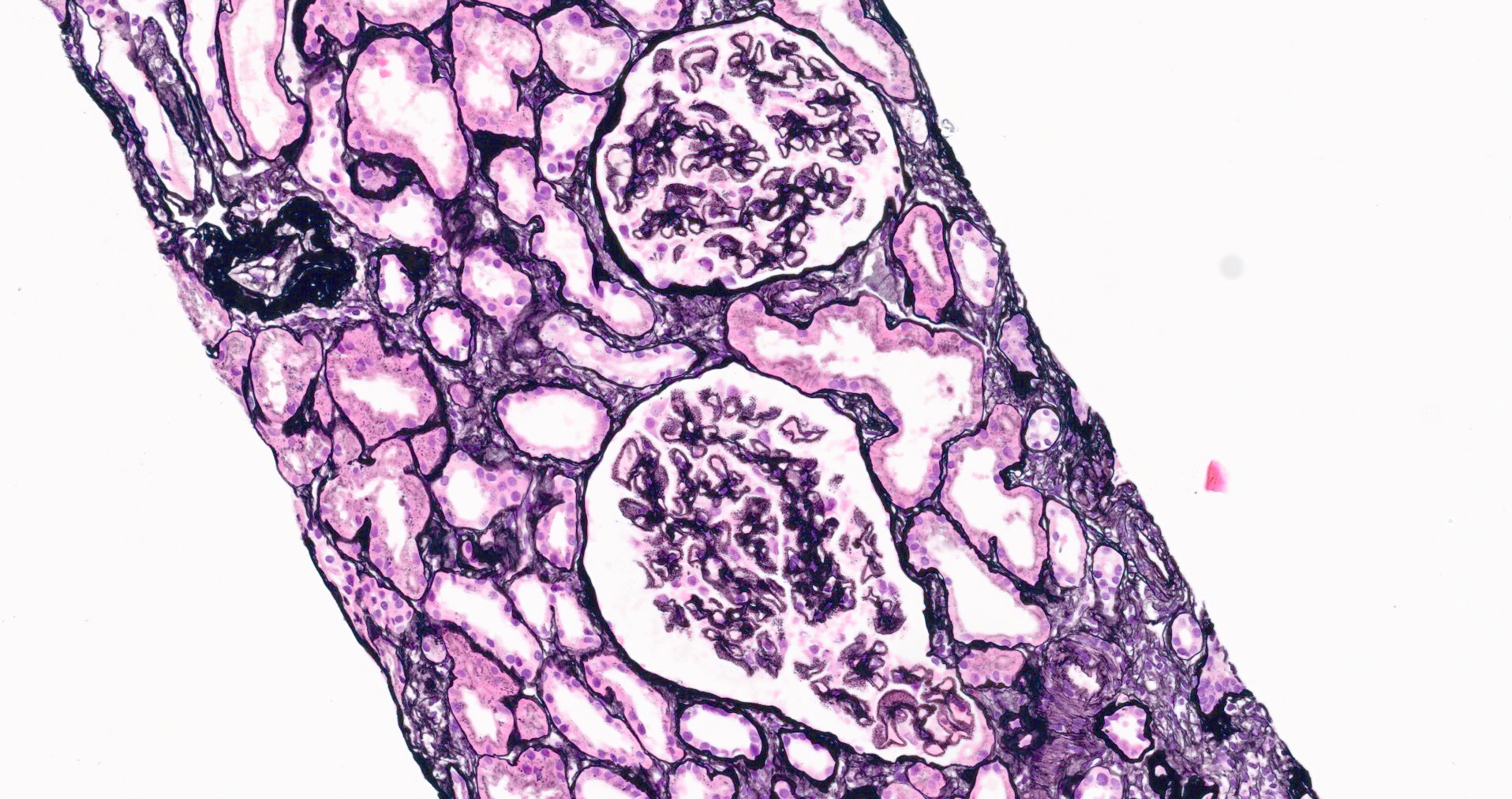
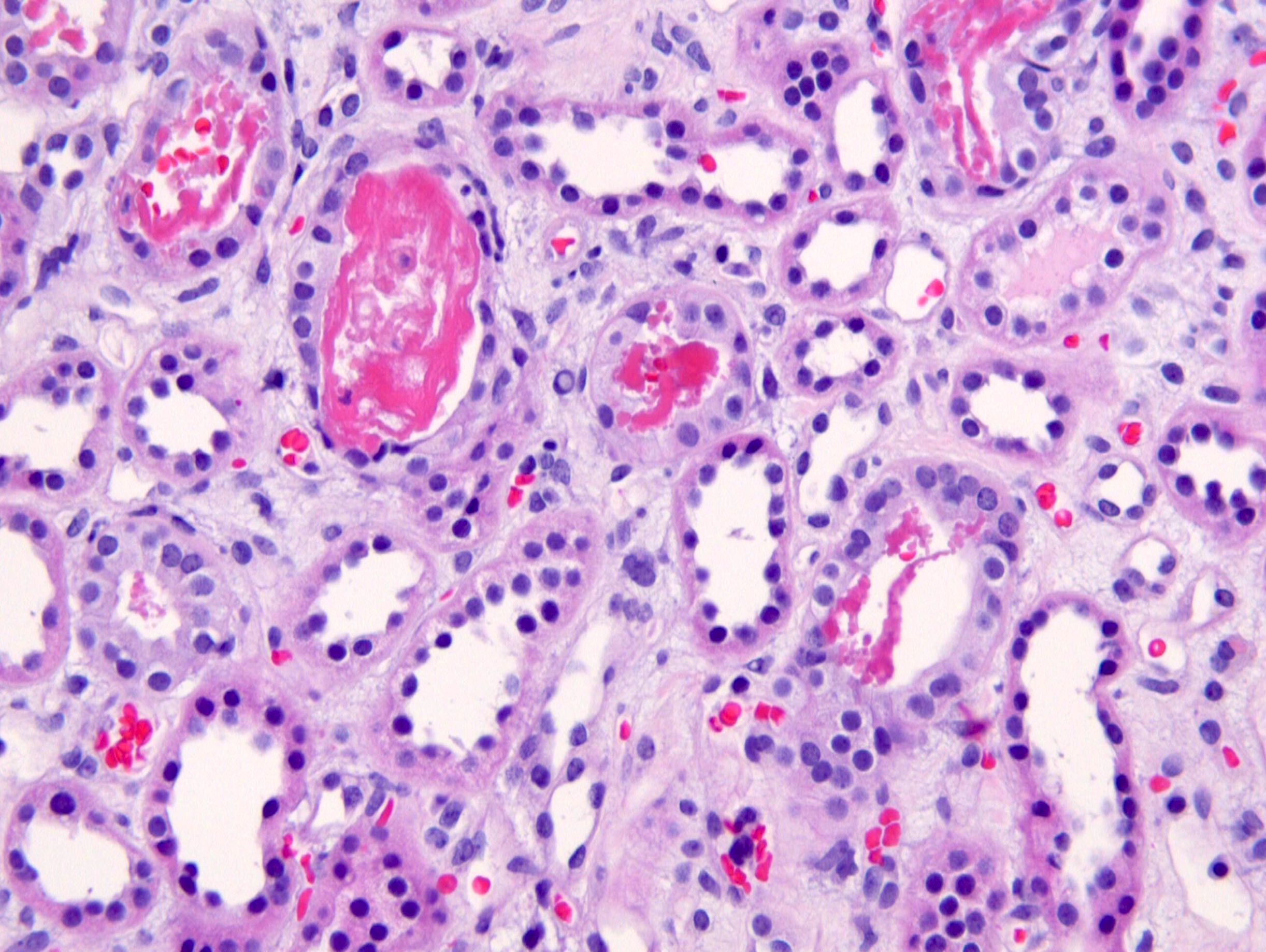
Diffuse and nodular mesangial expansion

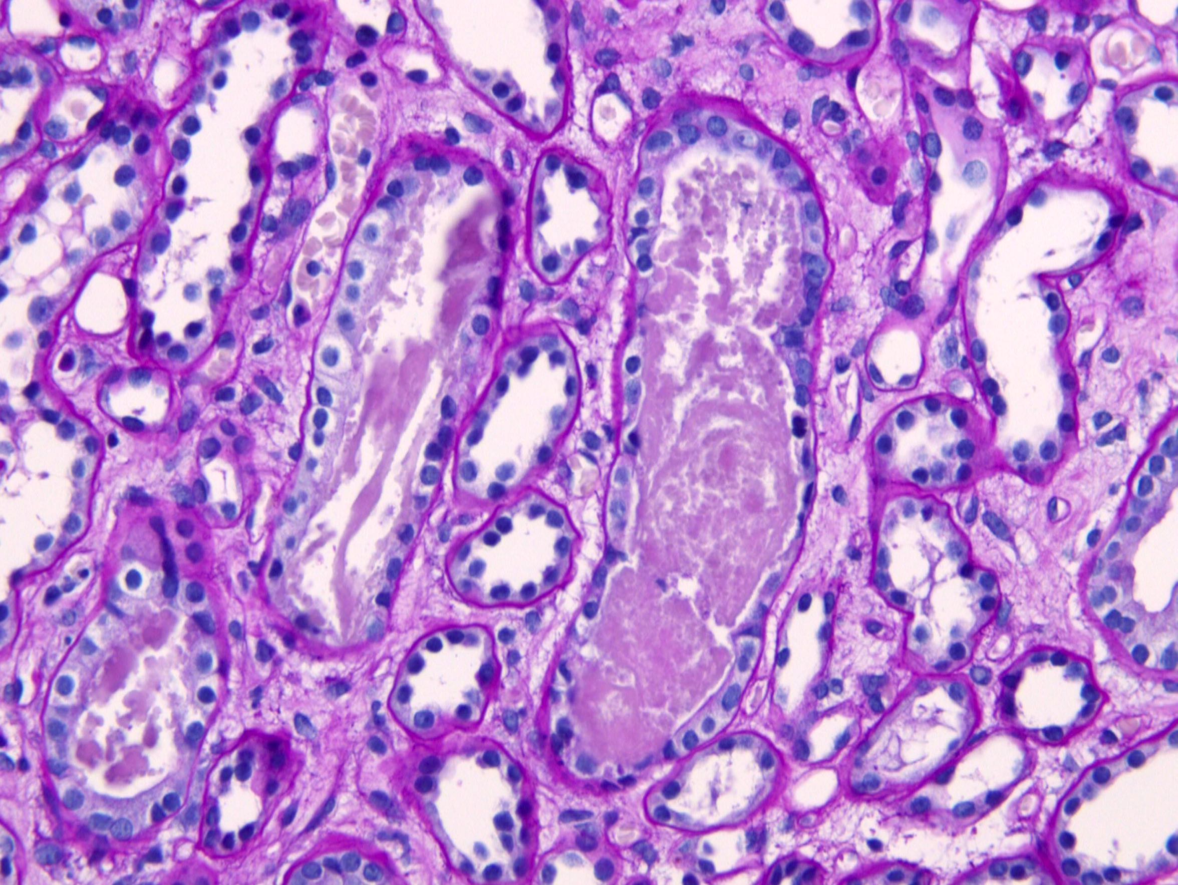
Diffuse mesangial expansion, PAS
Lipohyaline cap
Lipohyaline cap, PAS
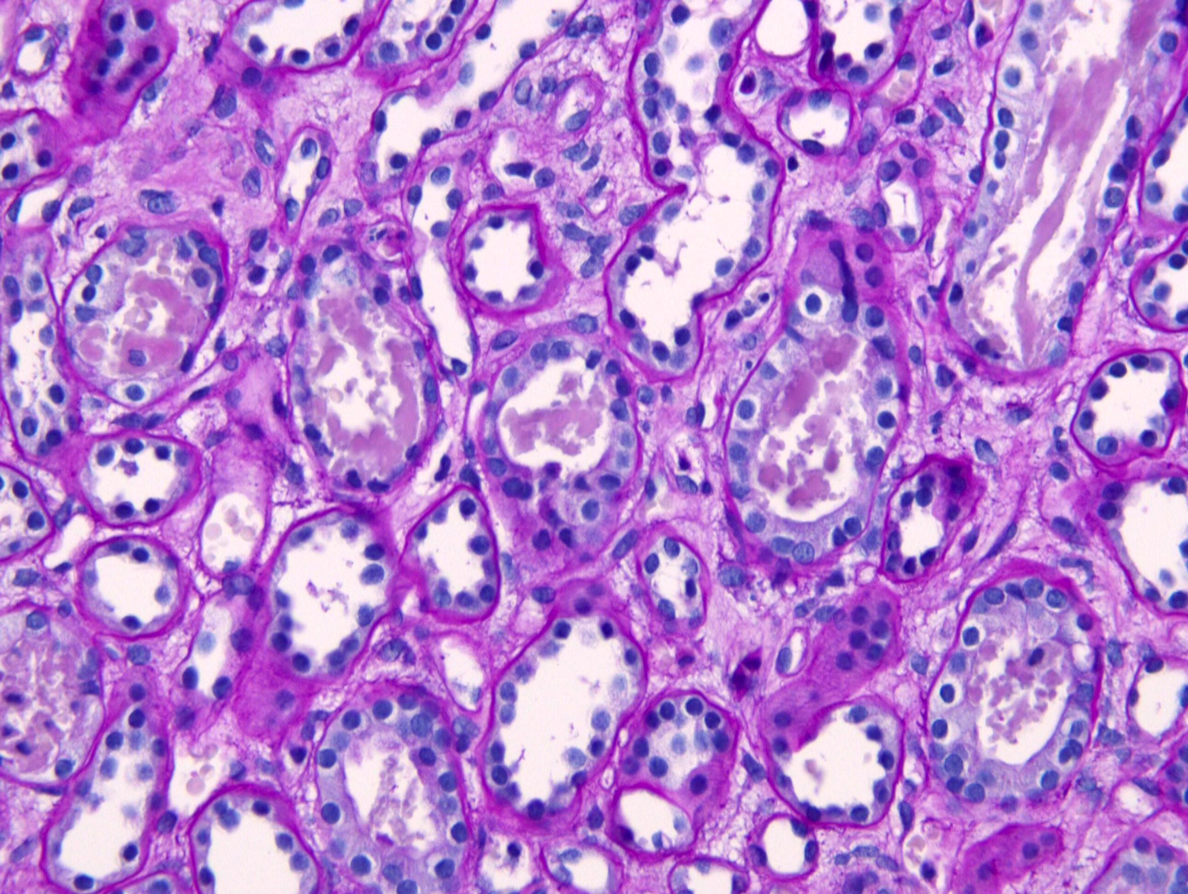
Lipohyaline caps and nodules, PAS
Lipohyaline caps and nodules, silver
Microaneurysm, PAS
Microaneurysm, silver
Microaneurysm with nodule, silver
Microaneurysm with large nodule, silver
Nodular lesions, PAS
Nodular lesions, PAS
Segmental sclerosis
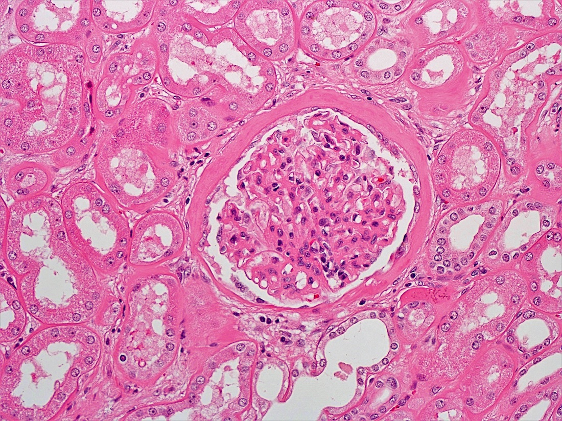
Thickening of Bowman capsule
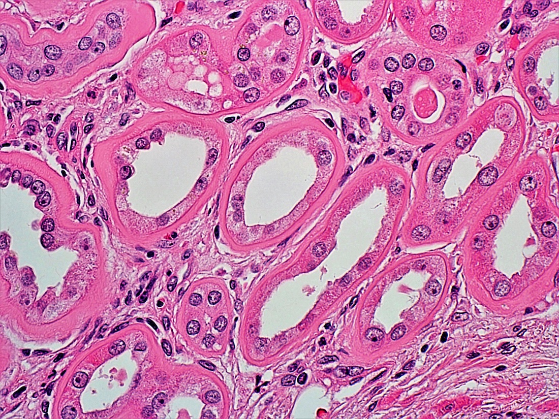
Thickening of basement membrane
2 insudative lesions
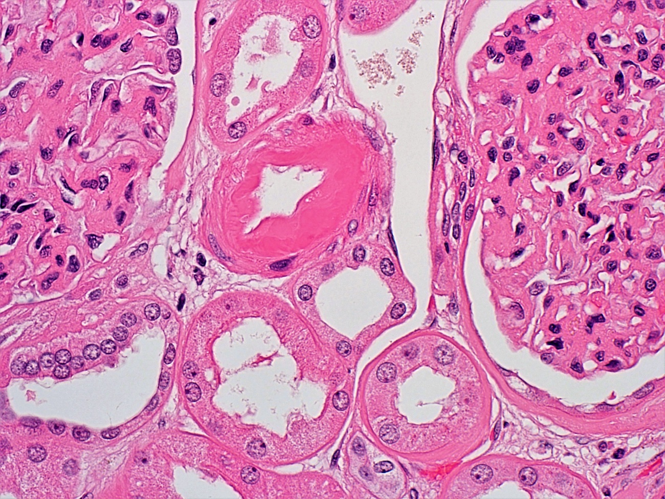
Vascular hyalinosis
Contributed by Khaled A. Murshed, M.D., Noheir M. Taha, M.B.B.Ch., M.D. and Mohammed Akhtar, M.D.
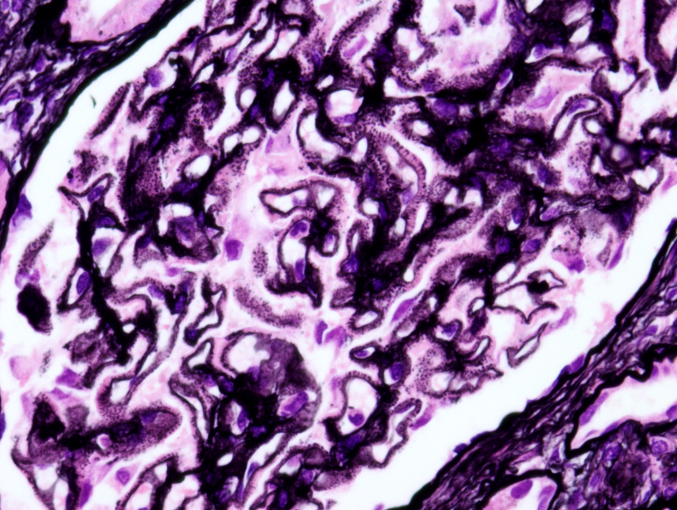
Low magnification

Mesangium and glomerular basement membrane
Thickening of glomerular basement membrane
Diabetic nephropathy
Images hosted on other servers:

Various images
Images hosted on other servers:

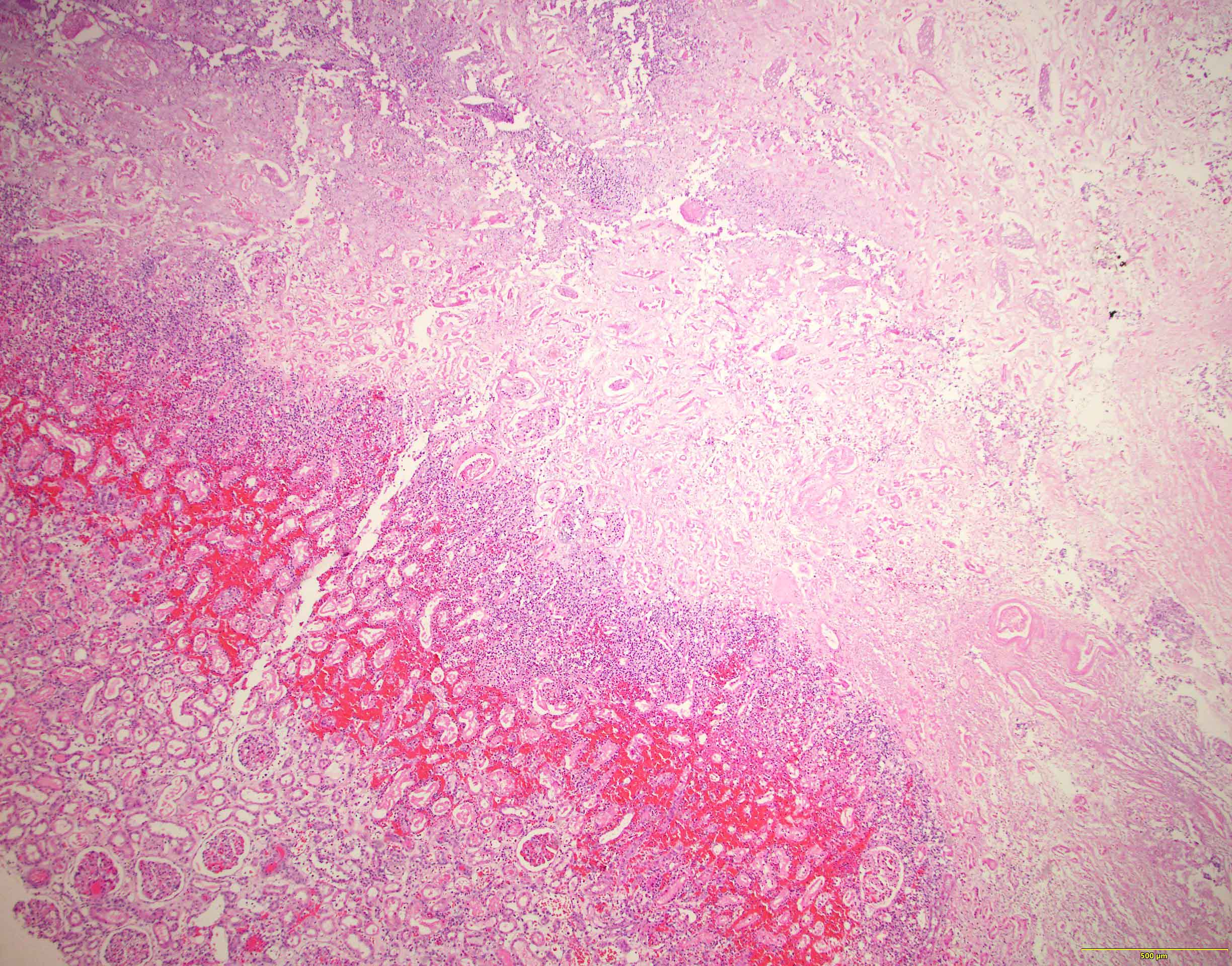
Extensive focal cortical necrosis
with tubular necrosis and fibrin
thrombi in the glomerulus

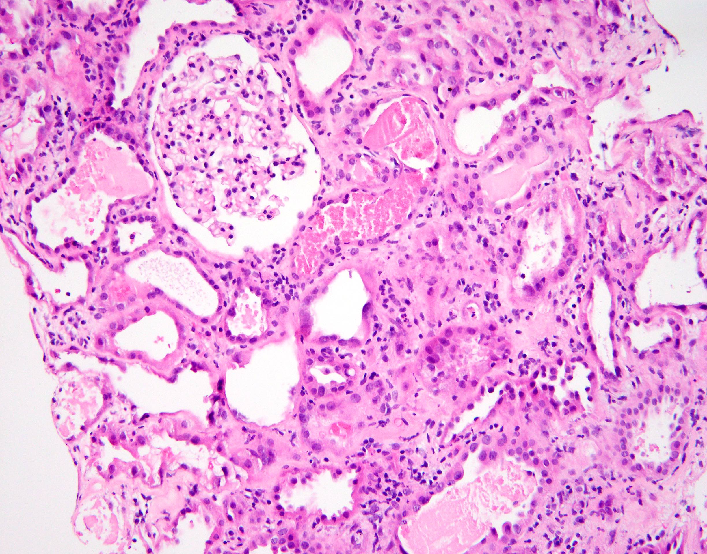
Necrotic areas having ghost
outlines of glomeruli and tubules
with loss of cellular details
Histopathology kidney: diffuse cortical necrosis
Images hosted on other servers:


Mesangial sclerosis


WT1 associated
Contributed by Nicole K. Andeen, M.D.
Case 1:

Normal glomerulus (FS)

Normal glomerulus
and adjacent intact
tubular parenchyma (FS)
Focal global GS in
subcapsular region
(H&E perm from FS)
Patches of tubular atrophy
& associated nonspecific
interstitial inflammation
(H&E perm from FS)
Low power with
no global GS (FS)
Low power with focal global GS
predominantly in subcapsular
region and patchy tubular atrophy
and IF (H&E perm from FS)
Case 2:
Focal global GS, ischemic glomeruli
with periglomerular fibrosis, adjacent
tubular atrophy, IF and nonspecific
interstitial inflammatory infiltrate (FS)

Focal global GS, adjacent
tubular atrophy and IF (FS)
Focal global and focal
and segmental GS, tubular
atrophy and IF (perm,
Jones silver stain)
Focal segmental GS, adjacent
arteriolar hyalinosis, tubular atrophy
and IF (perm, Jones silver stain)
Note: FS = frozen section, GS = glomerulosclerosis, IF = interstitial fibrosis, perm = permanent section
Images hosted on other servers:


Multicystic renal dysplasia


Multicystic renal dysplasia


Unilateral renal agenesis
Contributed by Robyn C. Reed, M.D., Ph.D.
Distorted shape, numerous cysts
Cysts, minimal remnant parenchyma
Hypoplastic kidneys at autopsy
Bilateral agenesis in situ
Images hosted on other servers:


Multiple cysts of various sizes


Multiple cysts of various sizes

With small bladder

Cysts are smooth
lined, no normal
kidney tissue
is apparent

With ipsilateral hypoplasia of ureter

Due to congenital urethral stenosis
Contributed by Robyn C. Reed, M.D., Ph.D.
Cysts in dysplasia
Cysts and primitive tubules
Tubule with mesenchymal collarette
Metaplastic cartilage
Cartilage and blastemal tissue
Primitive tubules
Development of renal dysplasia
Development of renal agenesis; explanation of oligohydramnios sequence
Contributed by Nicole K. Andeen, M.D.
Crescent

Arteritis

Medullary angiitis
Contributed by Nicole K. Andeen, M.D.

Fibrin tactoids
Images hosted on other servers:

Various images including EM

Global sclerosis,
segmental sclerosis
with moderate
interstitial fibrosis
Images hosted on other servers:

inclusion bodies in
cytoplasm of skin
fibroblasts
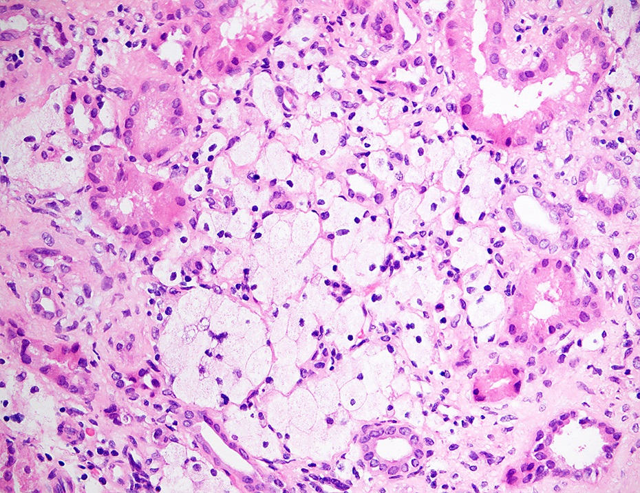
Contributed by Mandolin S. Ziadie, M.D. and Nicole K. Andeen, M.D.
Thick glomerular basement membrane and expanded mesangium
Jones methenamine silver
Trichrome

Periodic Acid-Schiff
DNAJB9
Images hosted on other servers:

Mesangial expansion
and glomerular
basement membrane
thickening

Variable glomerular findings
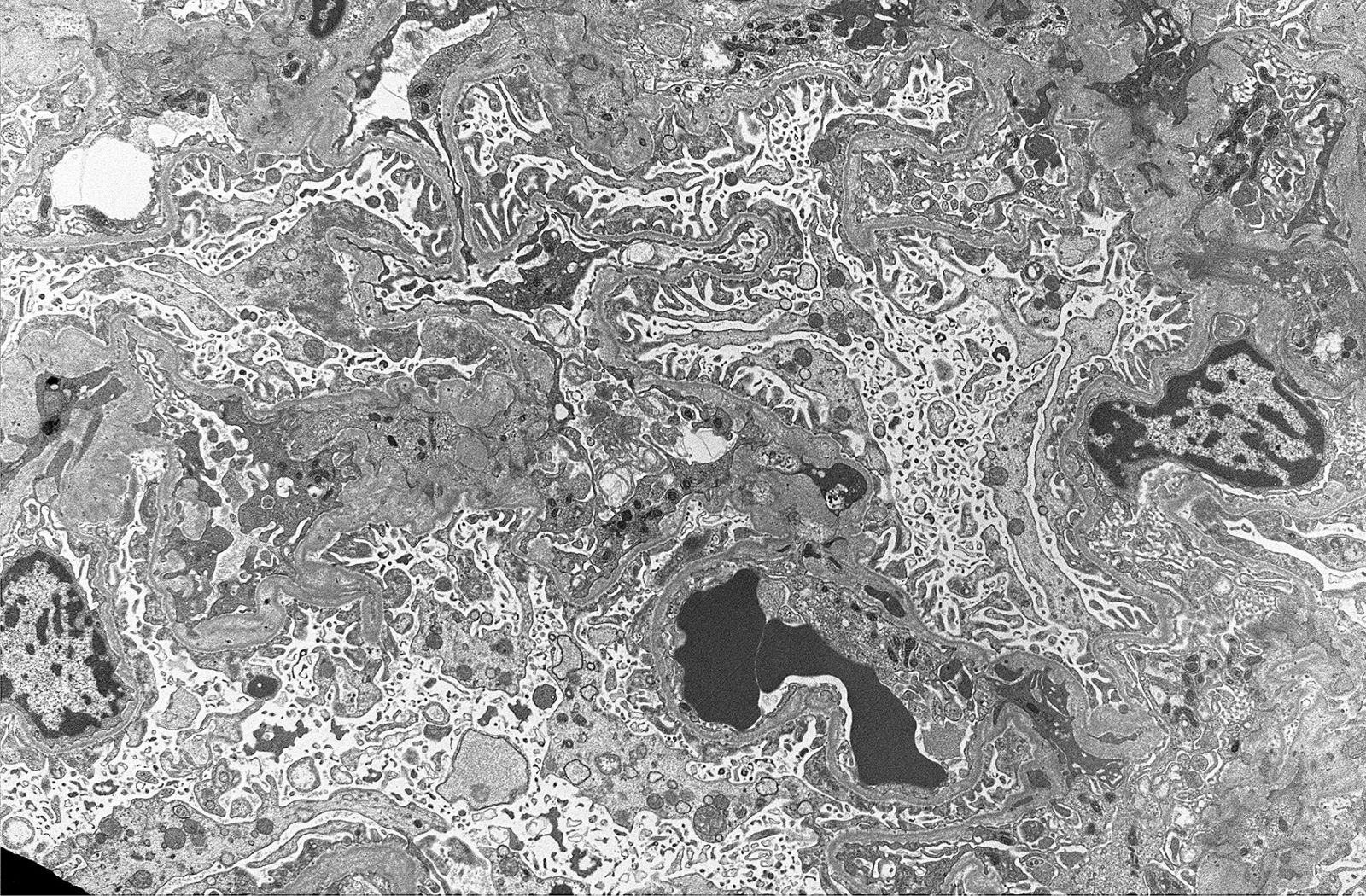
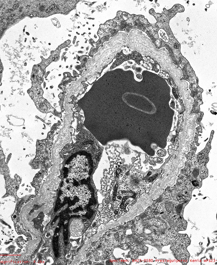
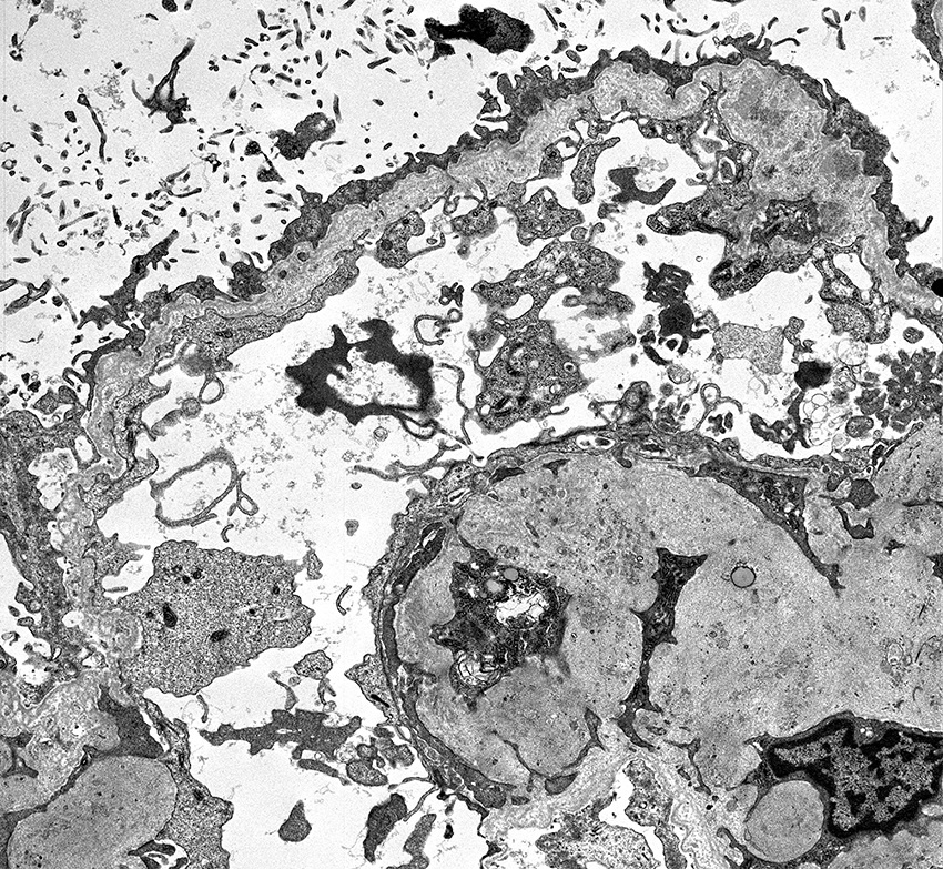
Contributed by Mandolin S. Ziadie, M.D.
Glomerular basement
membrane and
mesangial expansion

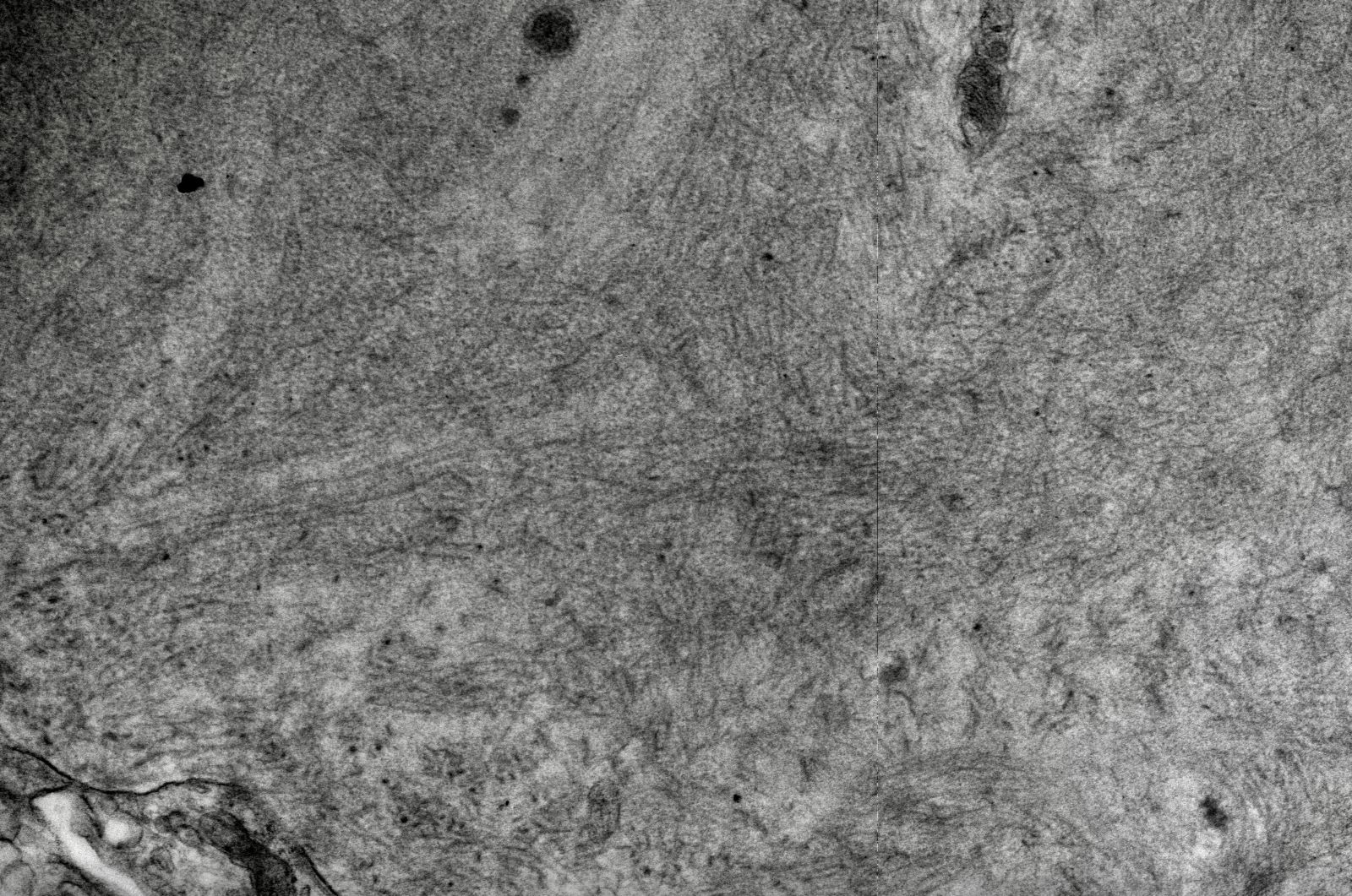
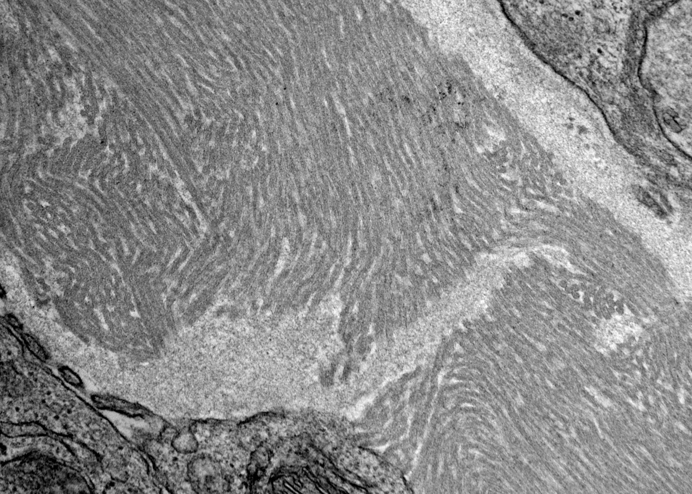
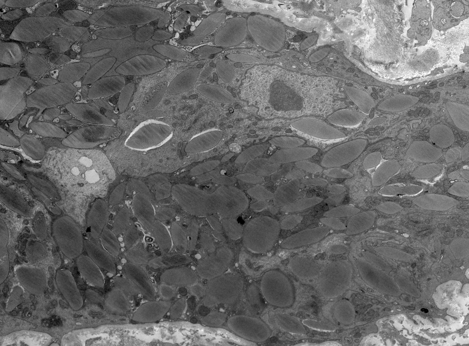
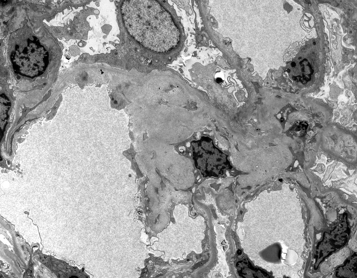
Randomly arranged fibrils
Images hosted on other servers:

Fibrillary deposits in mesangium

Randomly arranged fibrils
Images hosted on other servers:

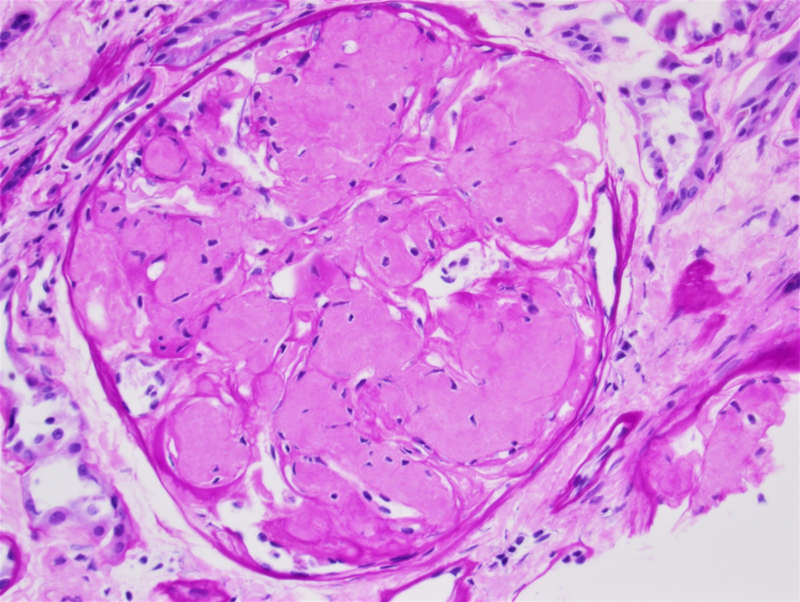
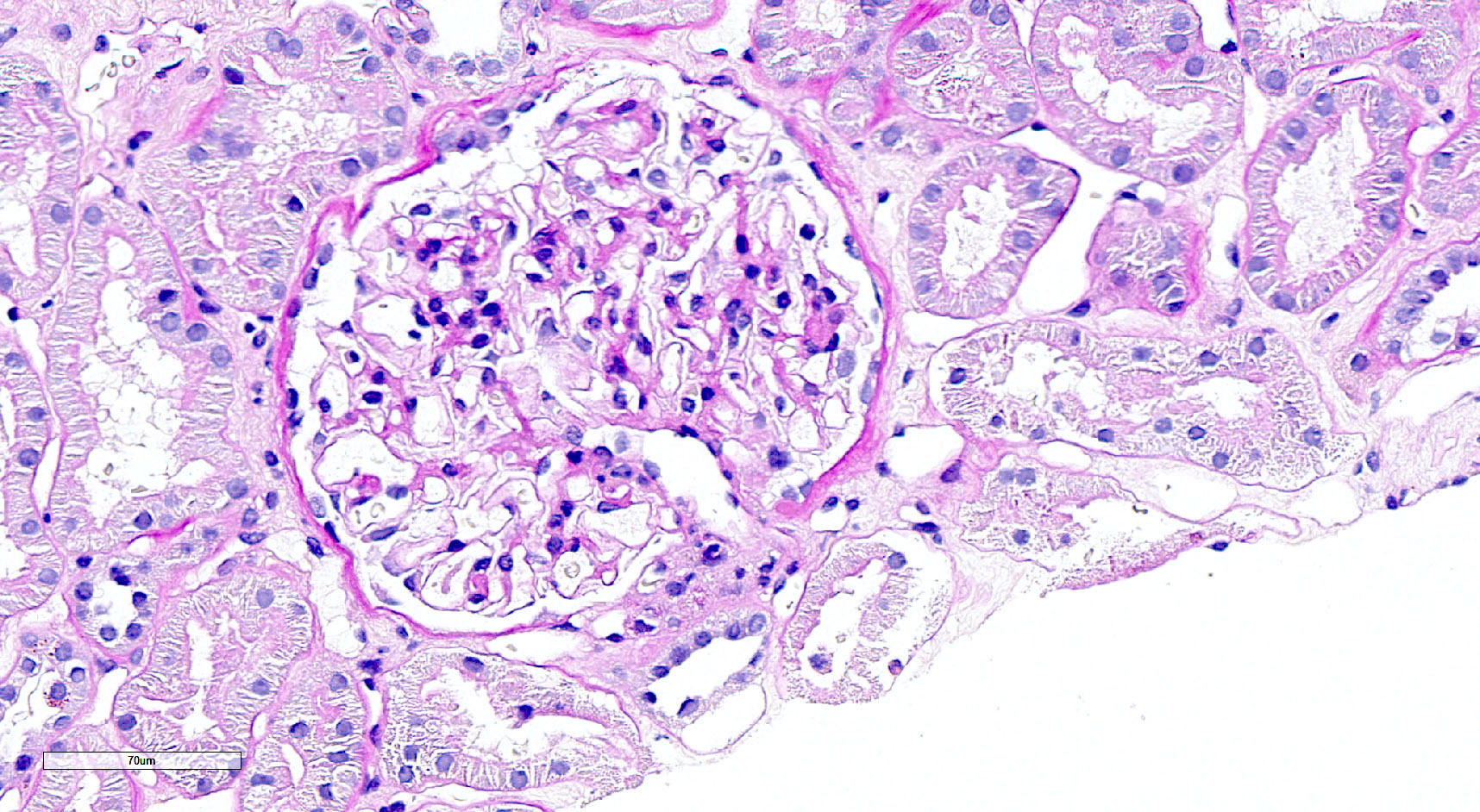
Eosinophilic material in mesangial regions

Immunoelectron microscopy with anti-fibronectin


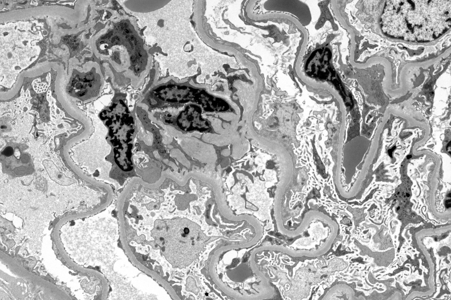
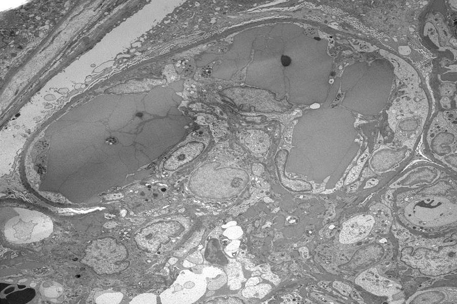
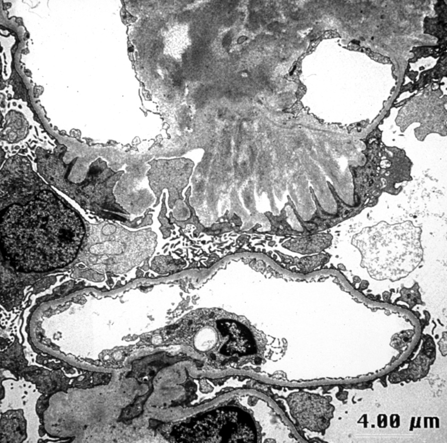
Glomeruli show massive deposits of a homogeneous structureless

Massive flooding of the glomerulus with fibronectin
Images hosted on other servers:


Enlargement of glomerular basement membrane and massive mesangial electron-dense deposits
Images hosted on other servers:

Primary and secondary FSGS

Sclerosis development in FSGS
Contributed by Vincenzo L’Imperio, M.D. and Giorgio Cazzaniga, M.D.
Segmental sclerosis with adhesion
Segmental sclerosis at the tubular pole

Segmental sclerosis with collapse
Perihilar segmental sclerosis with hyalinosis
Contributed by Vincenzo L’Imperio, M.D. and Giorgio Cazzaniga, M.D.
Complete foot process effacement
Foot process effacement and microvillous transformation
GBM lamellation
Contributed by Nicole K. Andeen, M.D.
Collapse of the glomerular tuft and prominence of the overlying epithelial cells, Jones stain
PAS
Trichrome
Images hosted on other servers:

Collapsed glomerulus

With sickle cell disease

Various images
Contributed by Nicole K. Andeen, M.D.
Podocyte foot process effacement
Contributed by Anjali A. Satoskar, M.D.
Segmental necrotizing lesion
Red blood cell (RBC) casts
Global necrotizing lesion
Global necrotizing lesion
Global necrotizing
lesion with
destroyed
Bowman capsule
Giant cells
Karyorrhectic nuclear debris

Necrotizing vasculitis
Fibrocellular crescent
Fibrous crescent
Contributed by Anjali A. Satoskar, M.D.
Fibrin strands on ultrastructural examination
Images hosted on other servers:

Various images
Contributed by Dalia Y. Ibrahim, M.D., M.Sc.

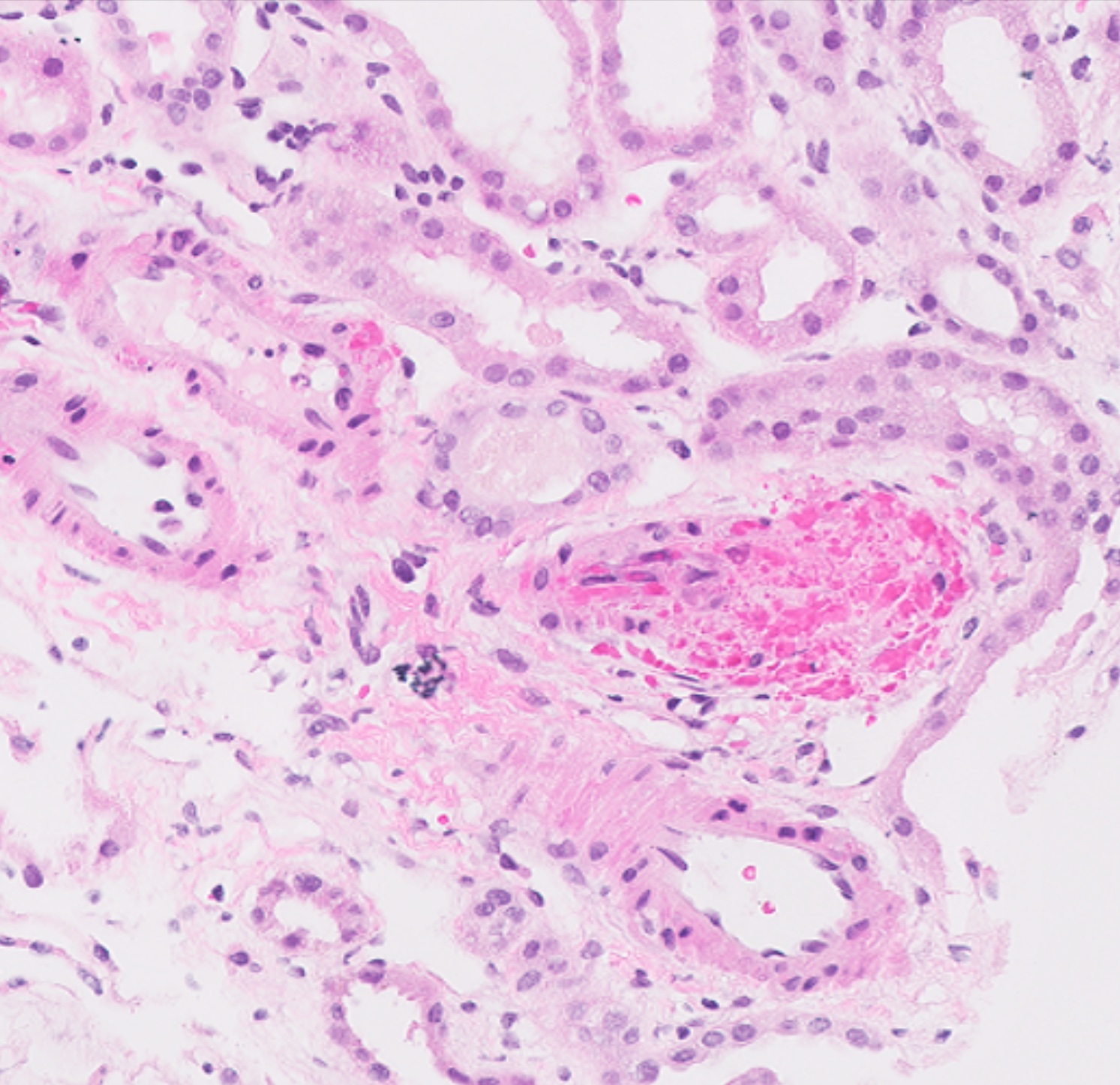
Bloodless glomerulus

Glomerulus with fibrin thrombi
Mucoid intimal thickening
Occluded artery with thrombus
Artery with thrombus and fragmented red blood cells
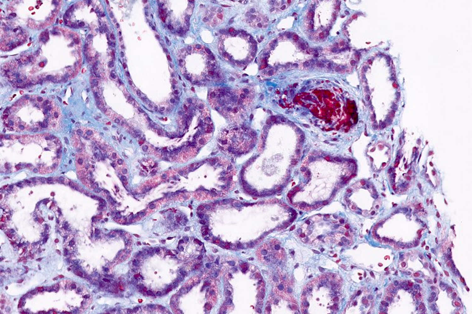
Occluded artery with thrombus, Trichrome stain
Contributed by Dalia Y. Ibrahim, M.D., M.Sc.
Subendothelial widening, ultrastructural examination
Images hosted on other servers:

Etiologies of TMA

Diagnostic algorithm for TMA
Contributed by Evelyn T. Bruner, M.D.
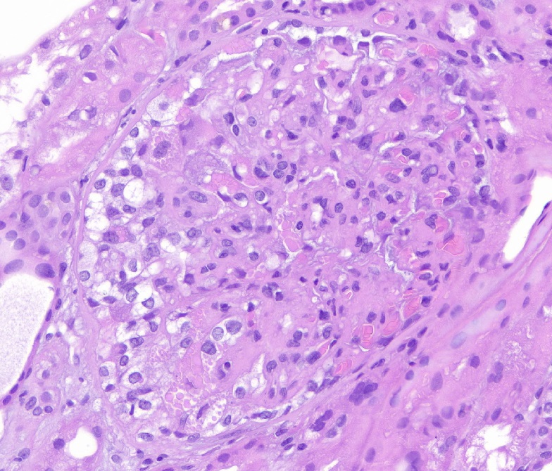
Prominent fibrin thrombi
Glomerulus with prominent fibrin thrombi
Glomerulus with endothelial swelling
Glomerulus with prominent fibrin thrombi
Artery with mucoid intimal thickening
Glomeruli with prominent fibrin thrombi
Contributed by Dr. Avinash Mane (Case #403)
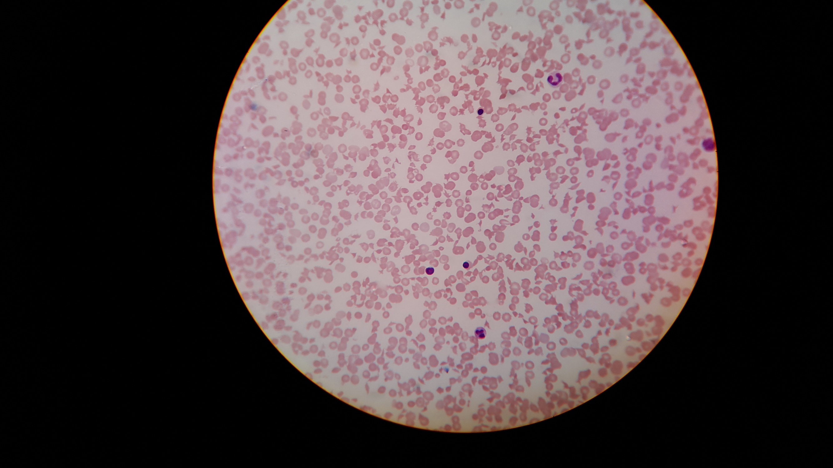
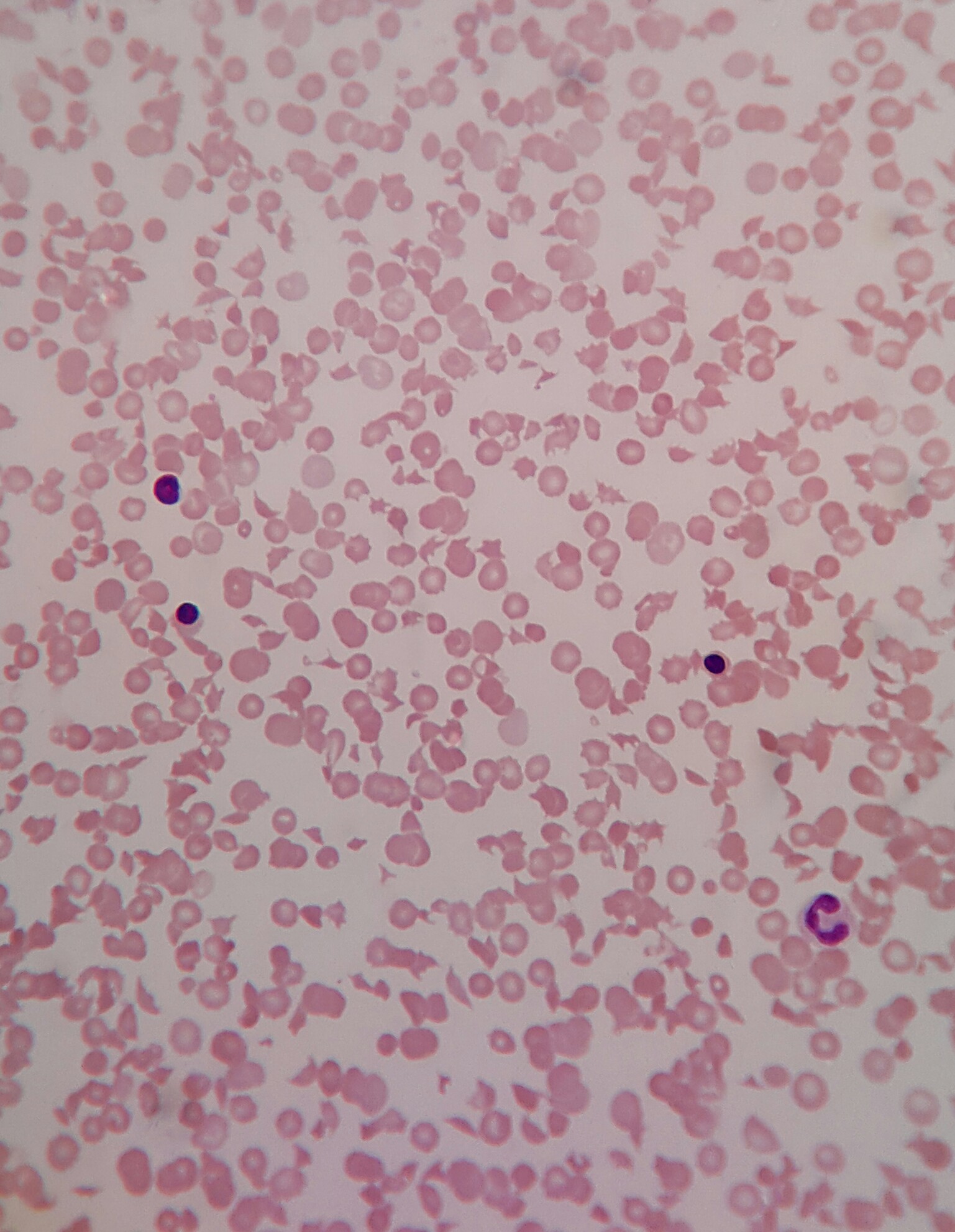
Numerous schistocytes and nucleated RBCs with extremely low platelets on peripheral smear
Contributed by Evelyn T. Bruner, M.D.
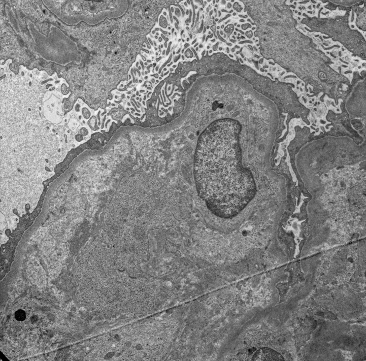
Endothelial swelling and lucency
Images hosted on other servers:


Various images
Images hosted on other servers:

Mechanisms of HIV associated renal disease
Images hosted on other servers:

Ultrasound features of HIVAN
Contributed by Vincenzo L’Imperio, M.D. and Giorgio Cazzaniga, M.D.
Collapsed glomeruli
Proliferative
glomerulonephritis
Tubular microcysts with sclerosed glomerulus
Tubular microcysts
Contributed by Vincenzo L’Imperio, M.D. and Giorgio Cazzaniga, M.D.
Tubuloreticular inclusion / aggregate (TRI)
Mesangial deposits
Subendothelial deposits
Images hosted on other servers:

Cortex is blue, edematous and congested
Images hosted on other servers:

Various images
Images hosted on other servers:


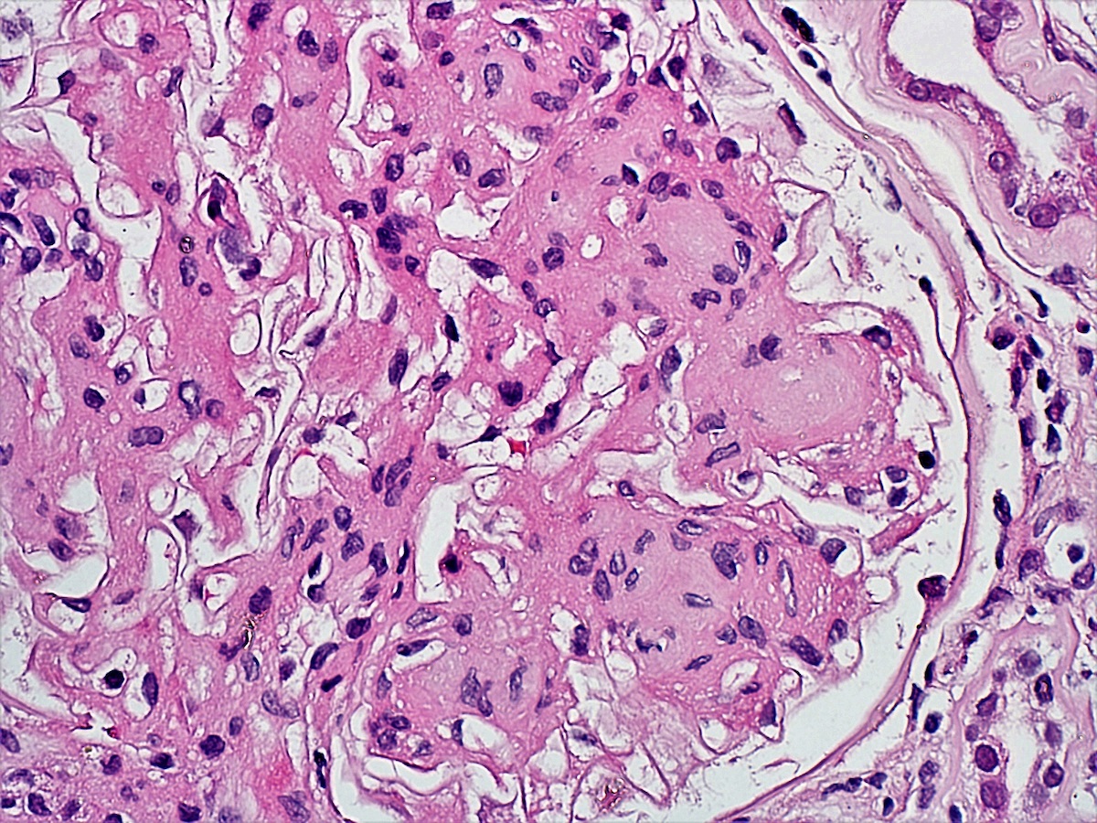
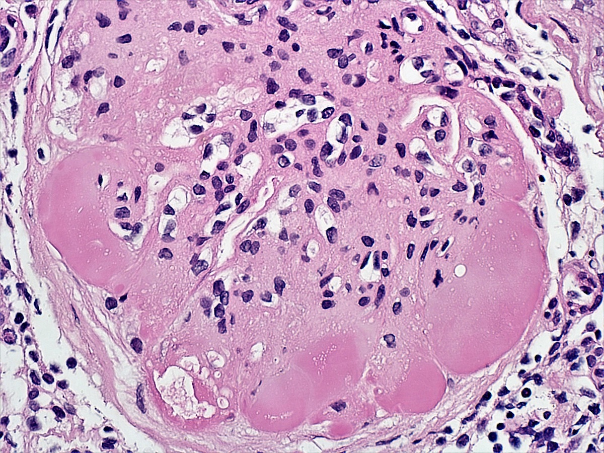
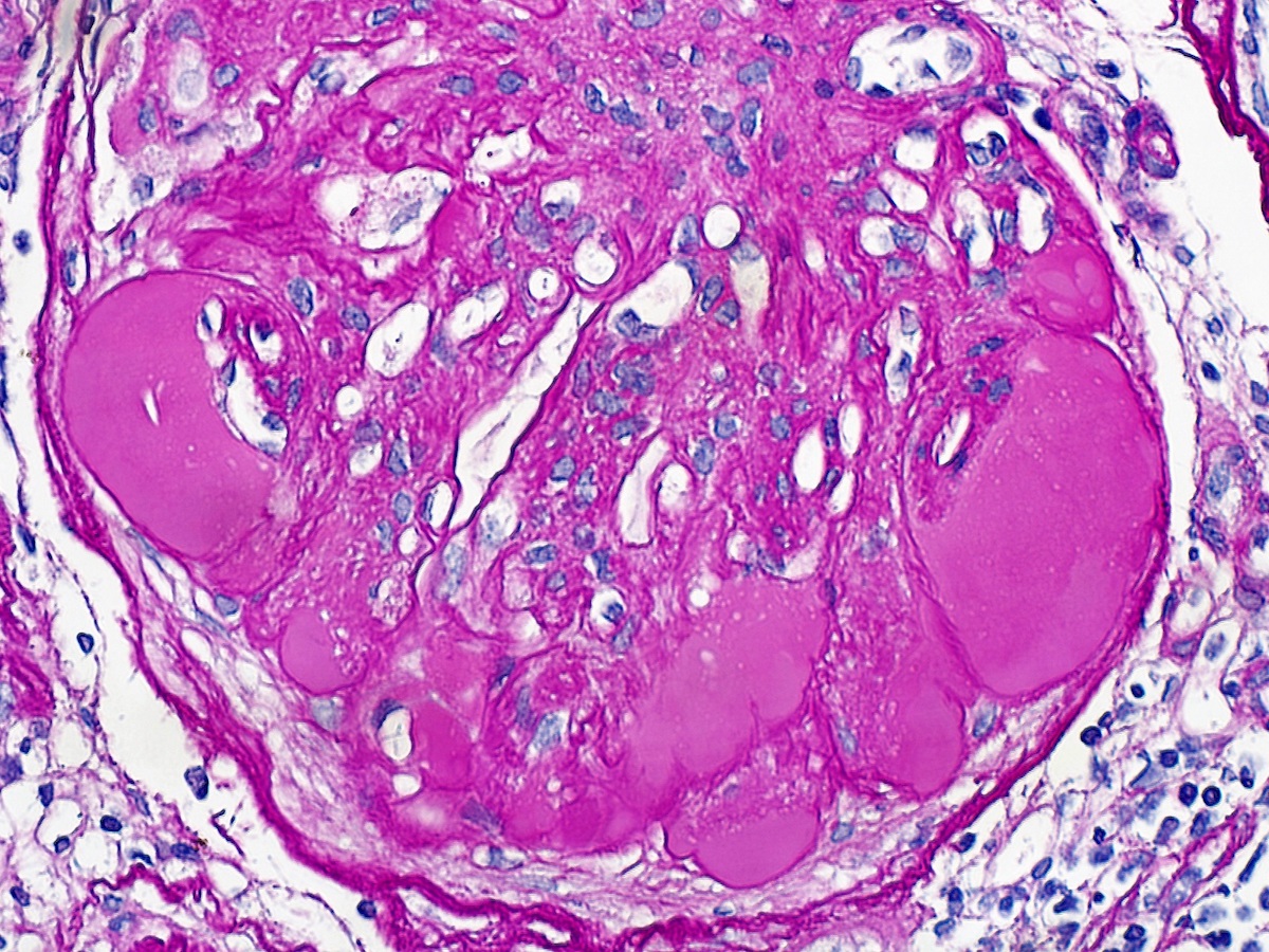
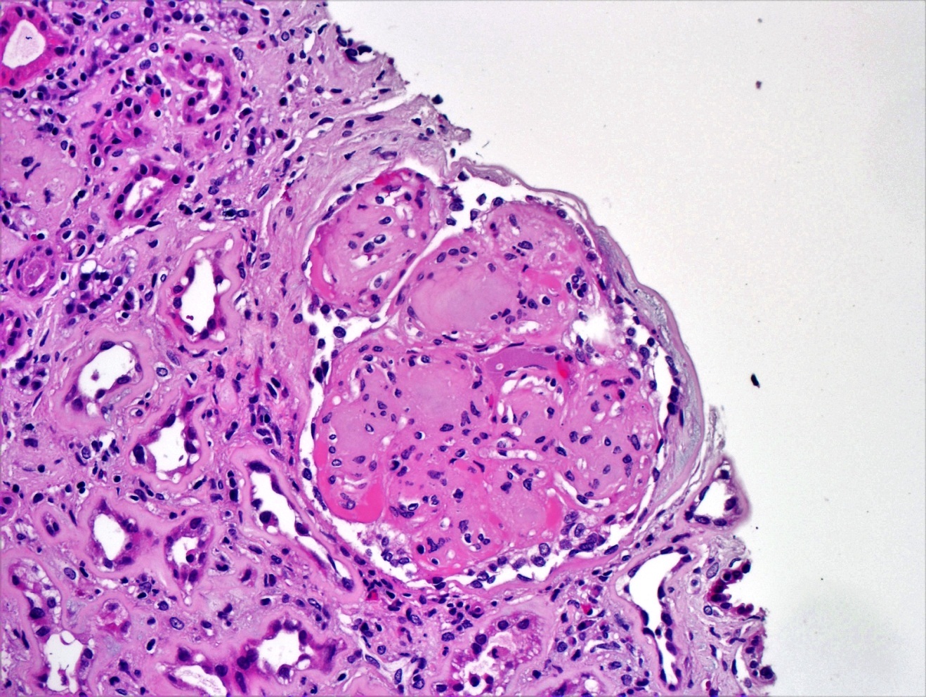
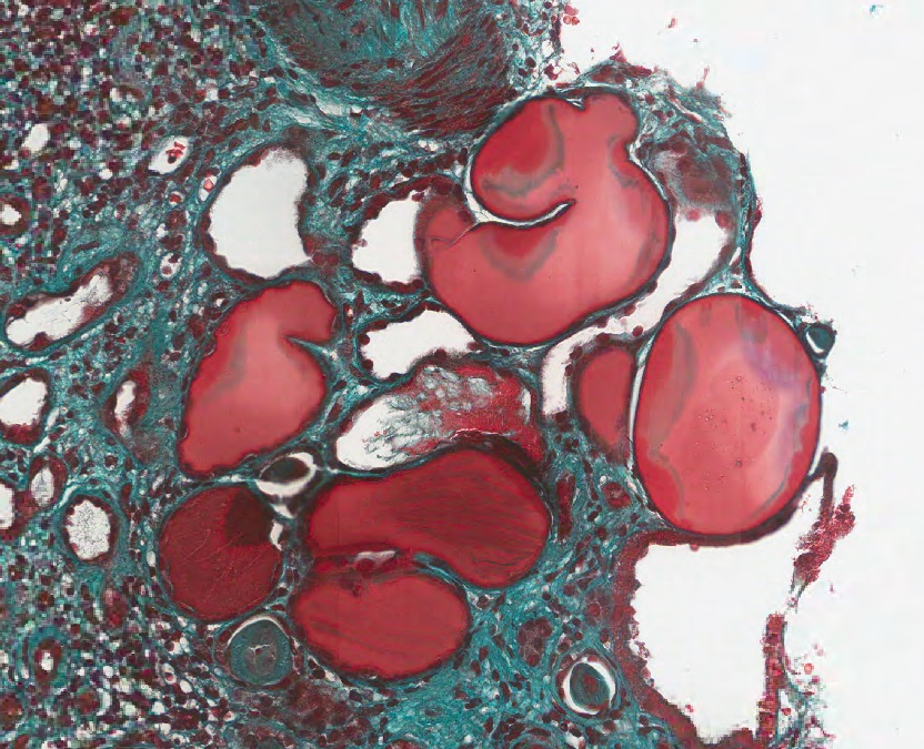
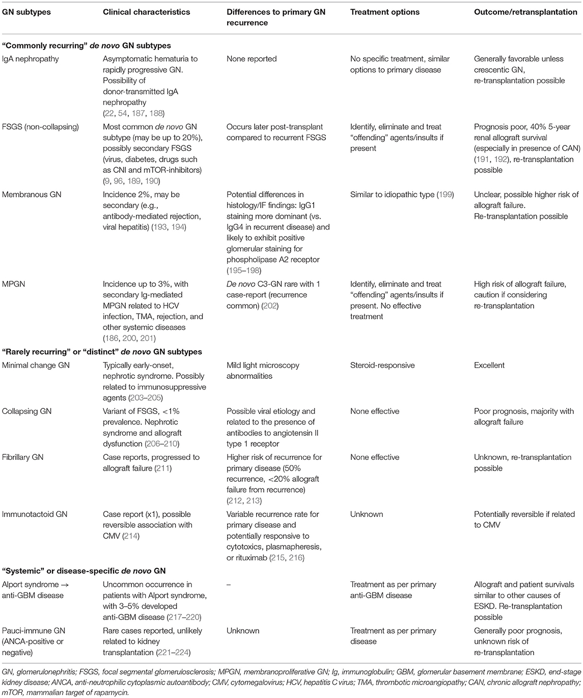
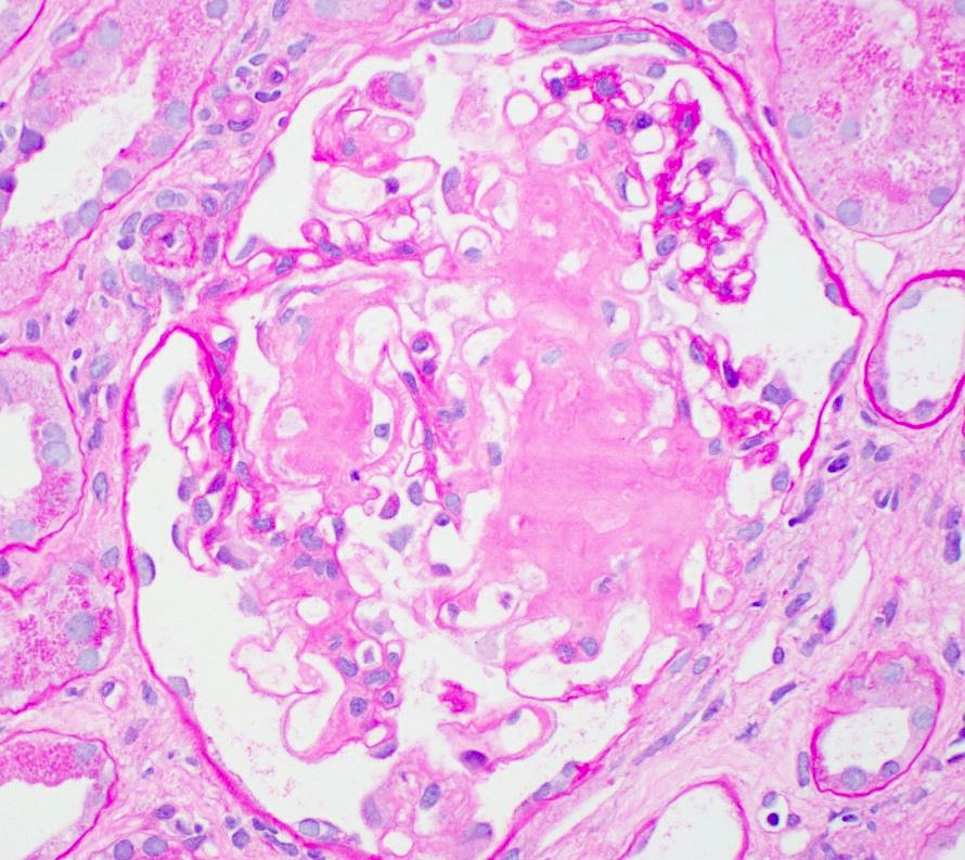
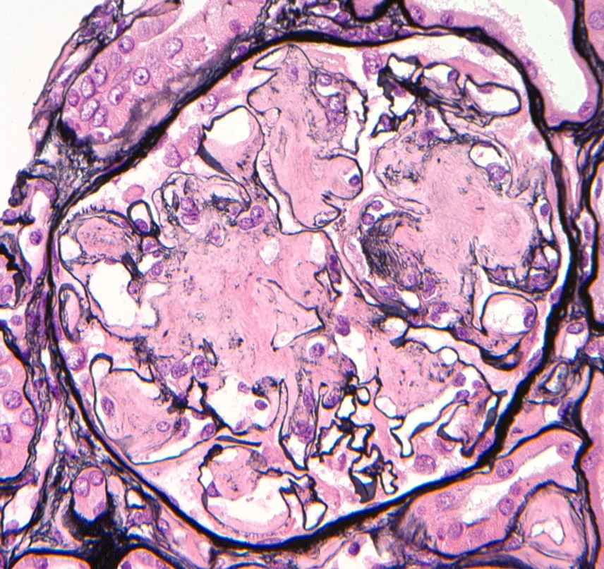
Idiopathic nodular glomerulosclerosis
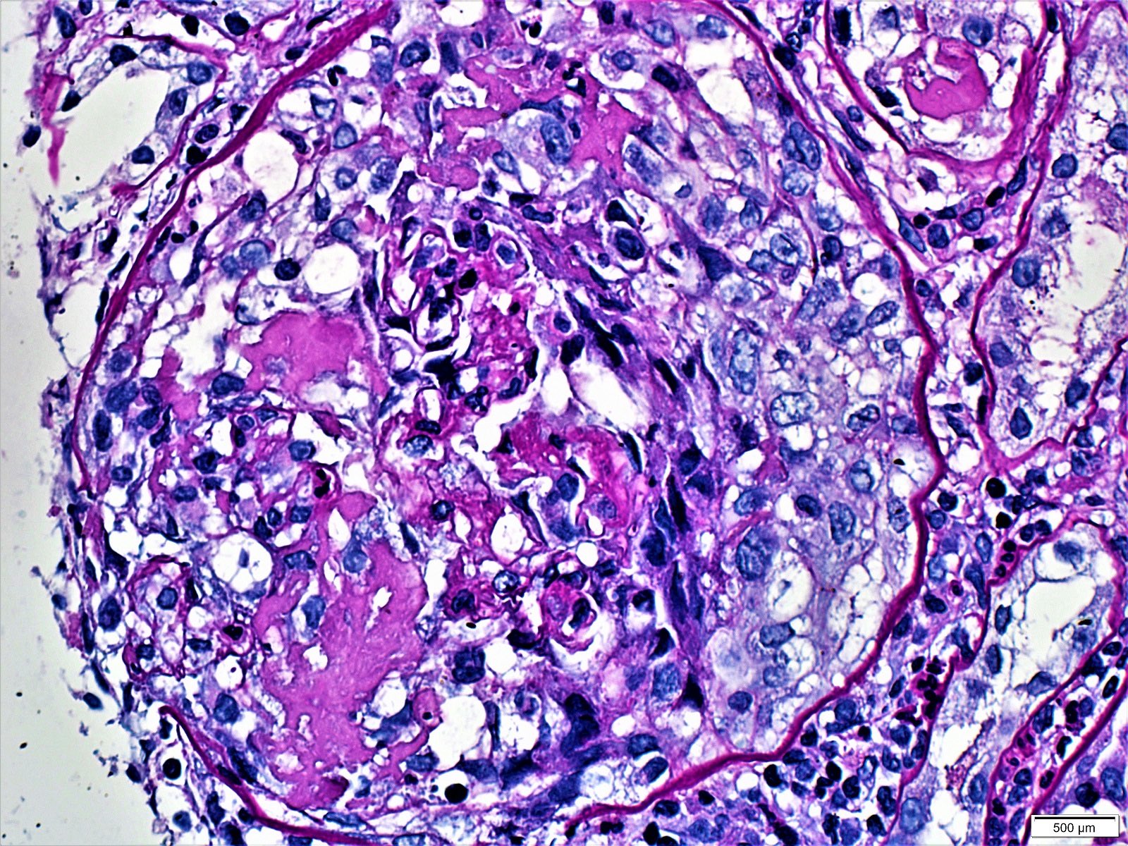
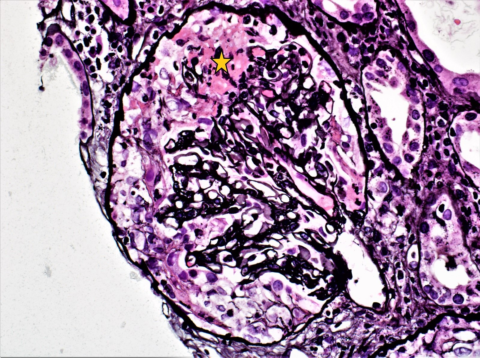
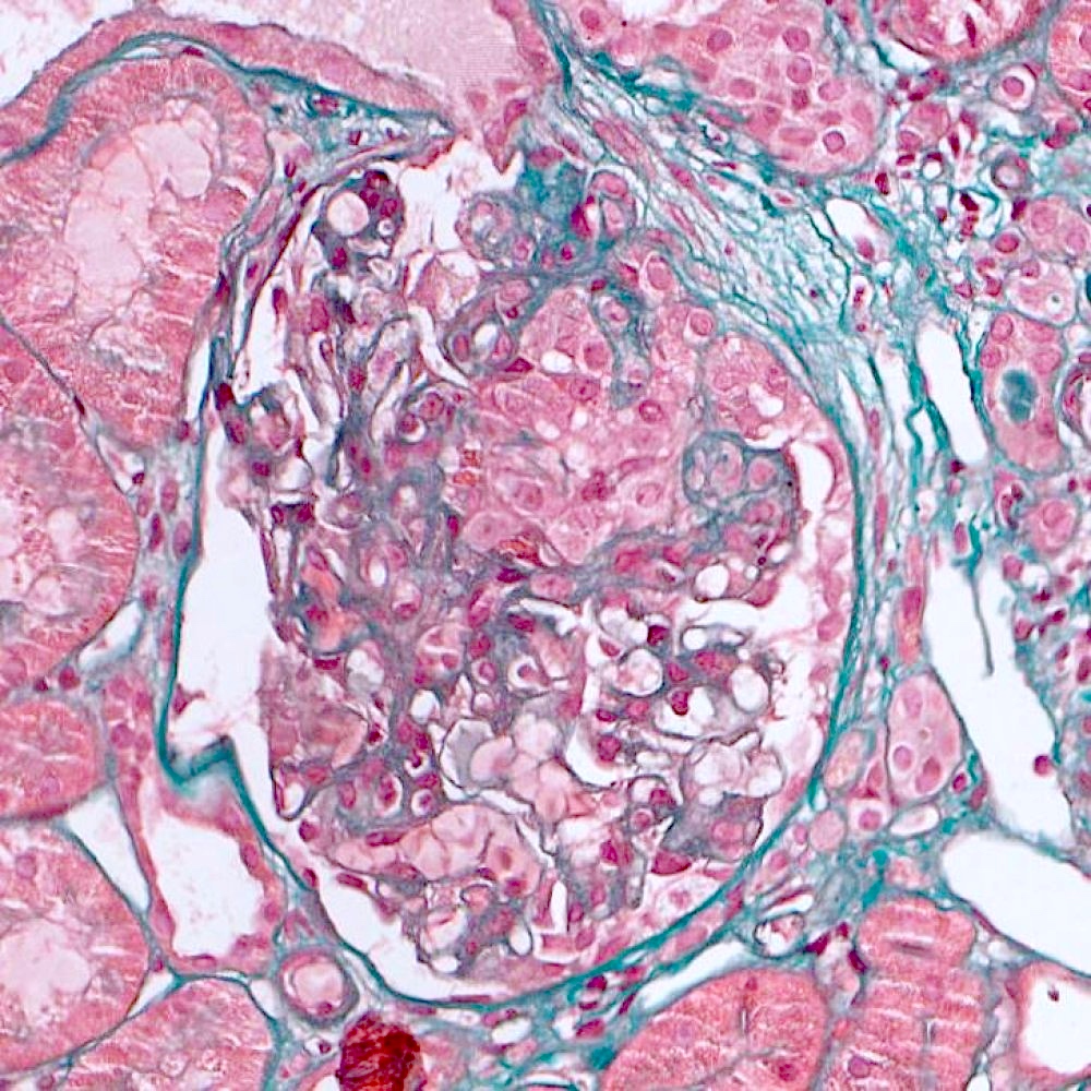
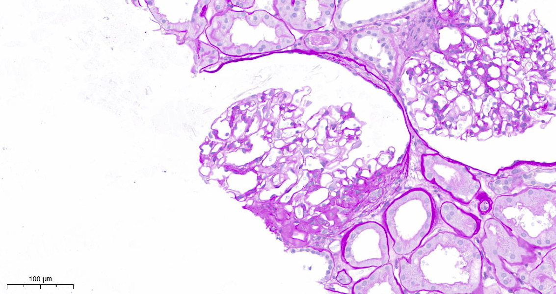
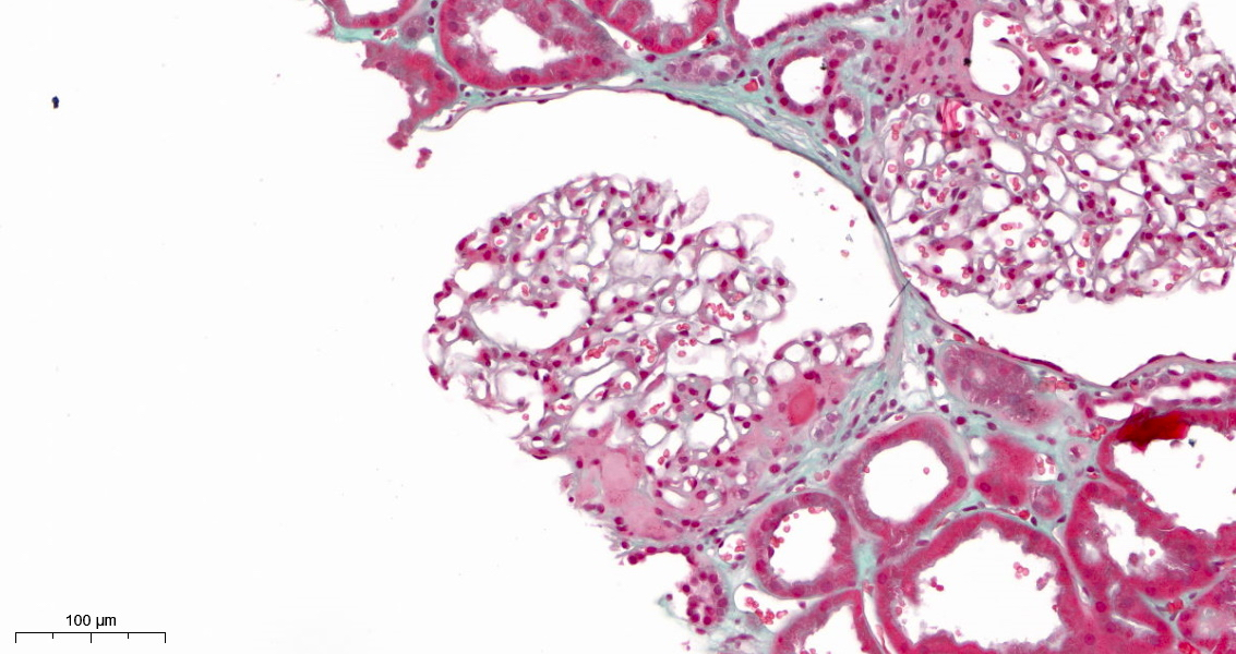
Contributed by Maria Fernanda Soares, M.D., Ph.D.
M1 lesion, PAS
E1 lesion
S1p lesion, PAS
Crescent, PAS

Red cell casts

Acute arterial TMA
Mesangial IgA

CD68 and E1 lesion
Contributed by Arkana
Mesangial expansion and loop adhesion, Jones silver
Mesangial expansion and global sclerosis, PAS

Mesangial expansion and loop adhesion, PAS
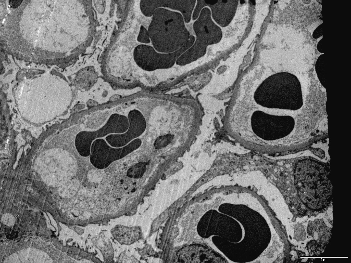
Contributed by Maria Fernanda Soares, M.D., Ph.D.
Mesangial deposits
Mesangial and paramesangial deposits
Contributed by Arkana

Mesangial deposits
Images hosted on other servers:

Renal mass

PET positive mass
Images hosted on other servers:

White renal mass
Contributed by Jonathan E. Zuckerman, M.D., Ph.D.

Tubulointerstitial effacement
Storiform fibrosis
Plasmacytic inflammation
Storiform fibrosis
Increased plasma cells
IgG plasma cells
IgG4 plasma cells
Plasma cells and eosinophils
Contributed by Jonathan E. Zuckerman, M.D., Ph.D.
Tubular basement membrane deposits
Subepithelial deposits
Mesangial deposits

Bowman capsule deposits
Images hosted on other servers:

Various images including EM
Images hosted on other servers:

Figures e - h


With fibrillary glomerulonephritis
Images hosted on other servers:


Biopsy findings

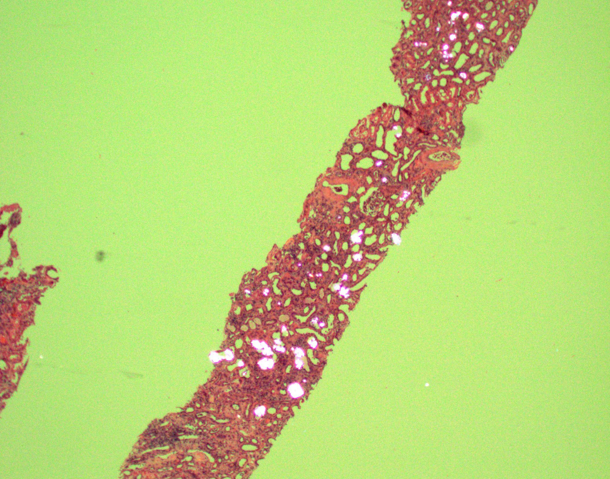
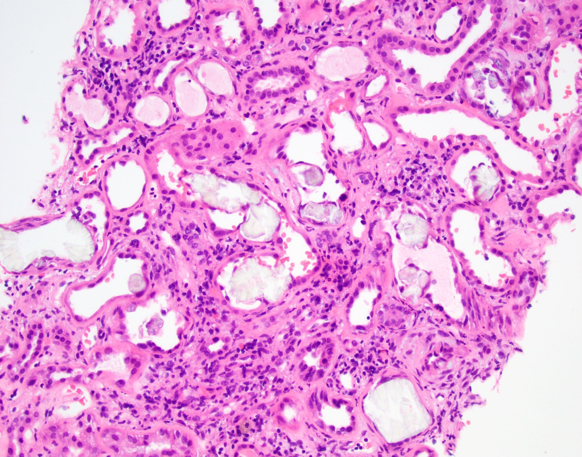
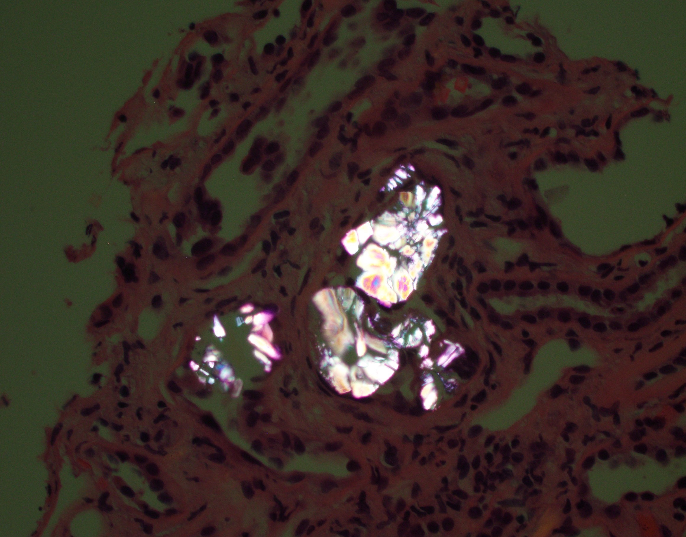
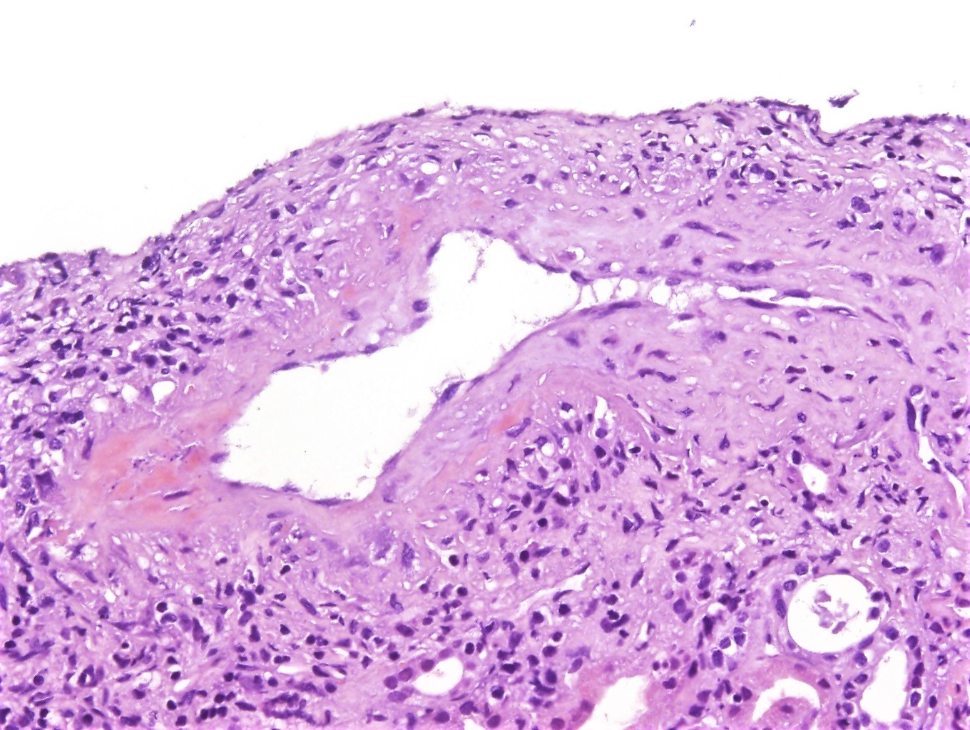
Crystal induced tubular injury

Indinavir crystals in urinary sediment
Images hosted on other servers:

Granulomatous giant
cell reaction with
crystals within lumen
Images hosted on other servers:


Wedge shaped hemorrhagic infarcts
Images hosted on other servers:


Various images
Images hosted on other servers:

Pathophysiology of cast nephropathy
Contributed by Ramya Krishna Velagapudi, M.D., M.H.A.
Pale appearance of casts, PAS
Fractured appearance of casts, PAS
Pale casts with giant cell reaction
Syncytial giant cell reaction
Metachromatic casts, trichrome stain
Crystalline appearance of casts
Contributed by Ramya Krishna Velagapudi, M.D., M.H.A.
Intratubular cast
Images hosted on other servers:

Intraluminal cast

Granular cast

Crystalline appearance of casts
Washington University in St. Louis: renal pathology teaching series
Contributed by NephroPath
Light chain deposits along GBM
sclerosis and
tubular injury
on silver stain
Mesangial nodular sclerosis on PAS
Mesangial nodular sclerosis on silver stain
Mesangial nodular sclerosis on trichrome stain
Images hosted on other servers:

Various images
Contributed by NephroPath
Light chain deposits along GBM

Light chain deposits along TBM
Contributed by Rajib K. Gupta, M.D.
LCPT, crystalline variant
LCPT, noncrystalline variant
Contributed by Rajib K. Gupta, M.D., Ashley Flowers, M.D. and Judy King, M.D., Ph.D.

Acute tubular injury / necrosis in LCPT
Crystals within proximal tubular epithelium
Fuchsinophilic crystals within proximal tubular epithelium
Noncrystalline variant of LCPT
Contributed by Rajib K. Gupta, M.D., Judy King, M.D., Ph.D., Ashley Flowers, M.D. and Xin Gu, M.D.
Intracytoplasmic crystals, crystalline variant of LCPT
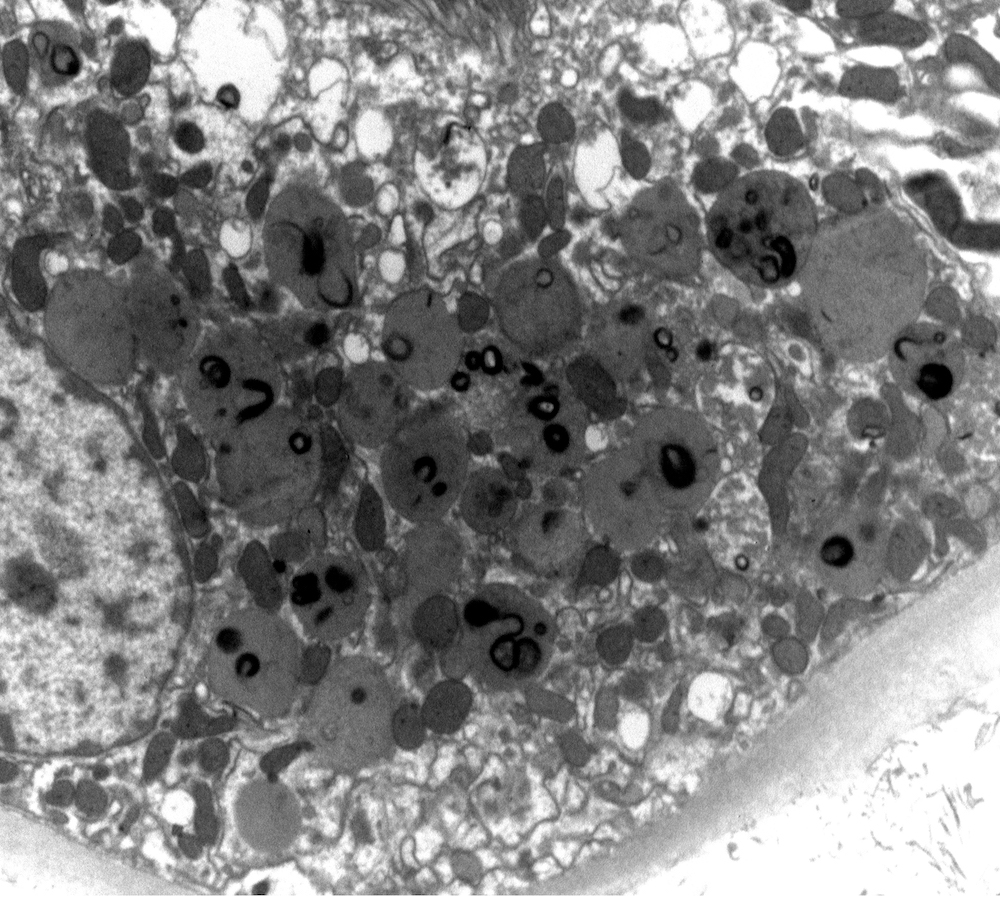
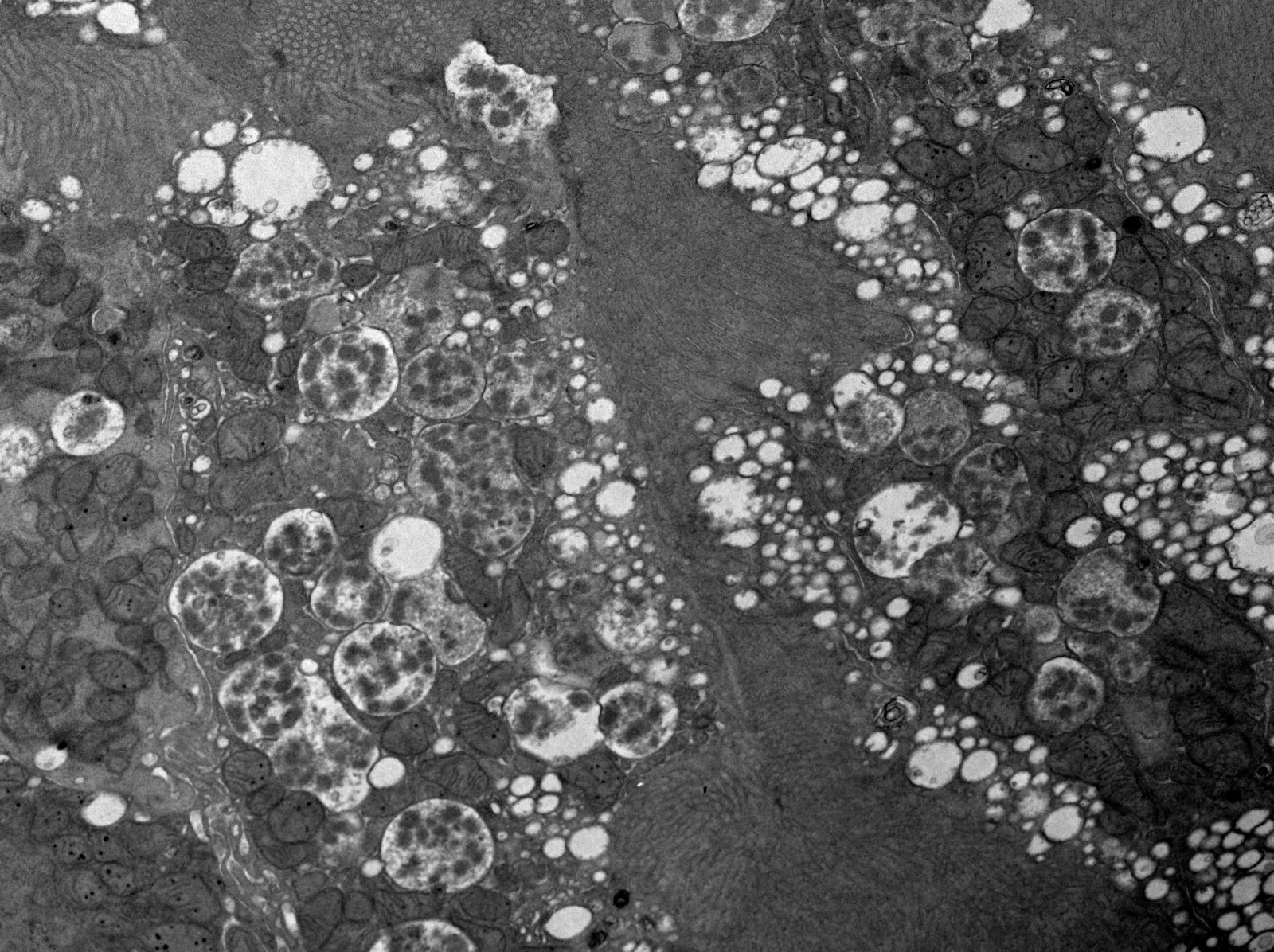
Abnormal lysosomes, noncrystalline LCPT


Abnormal lysosomes with mottling, noncrystalline LCPT
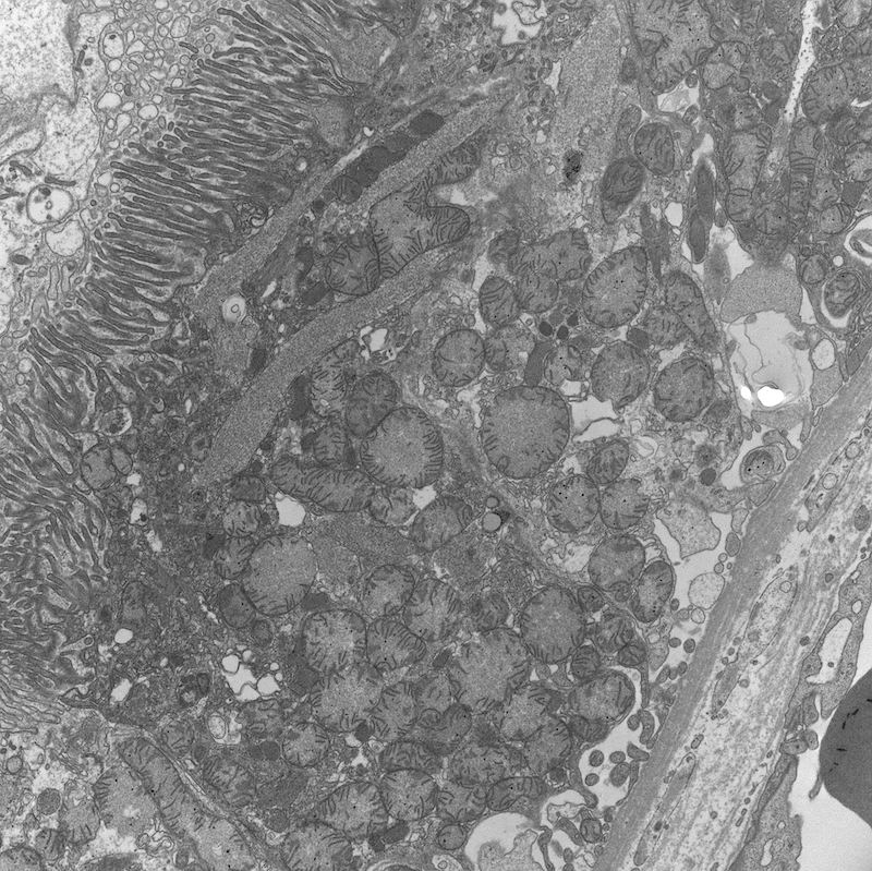
Abnormal mitochondria, noncrystalline LCPT
Images hosted on other servers:

Yellowish rubbery solid mass
Images hosted on other servers:

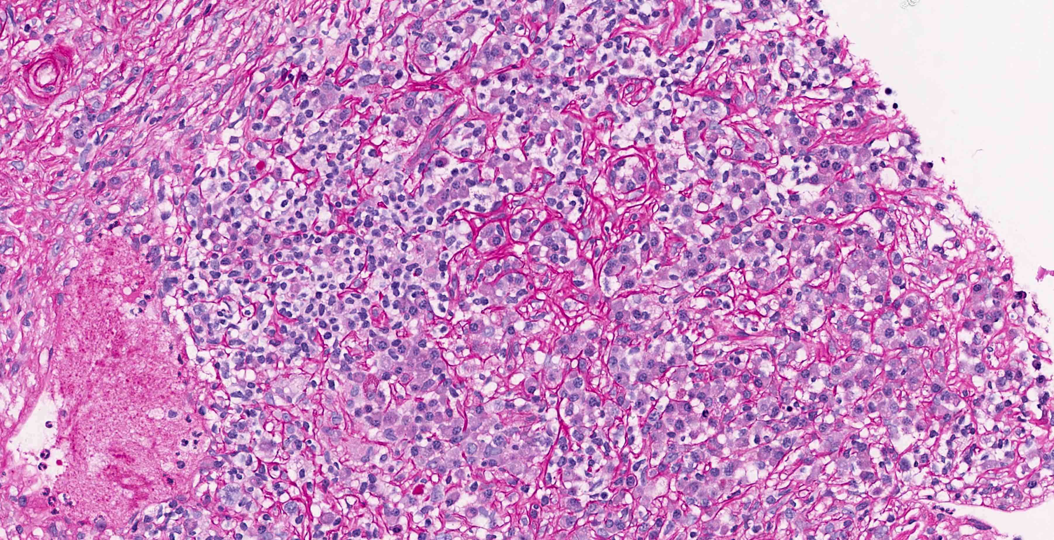
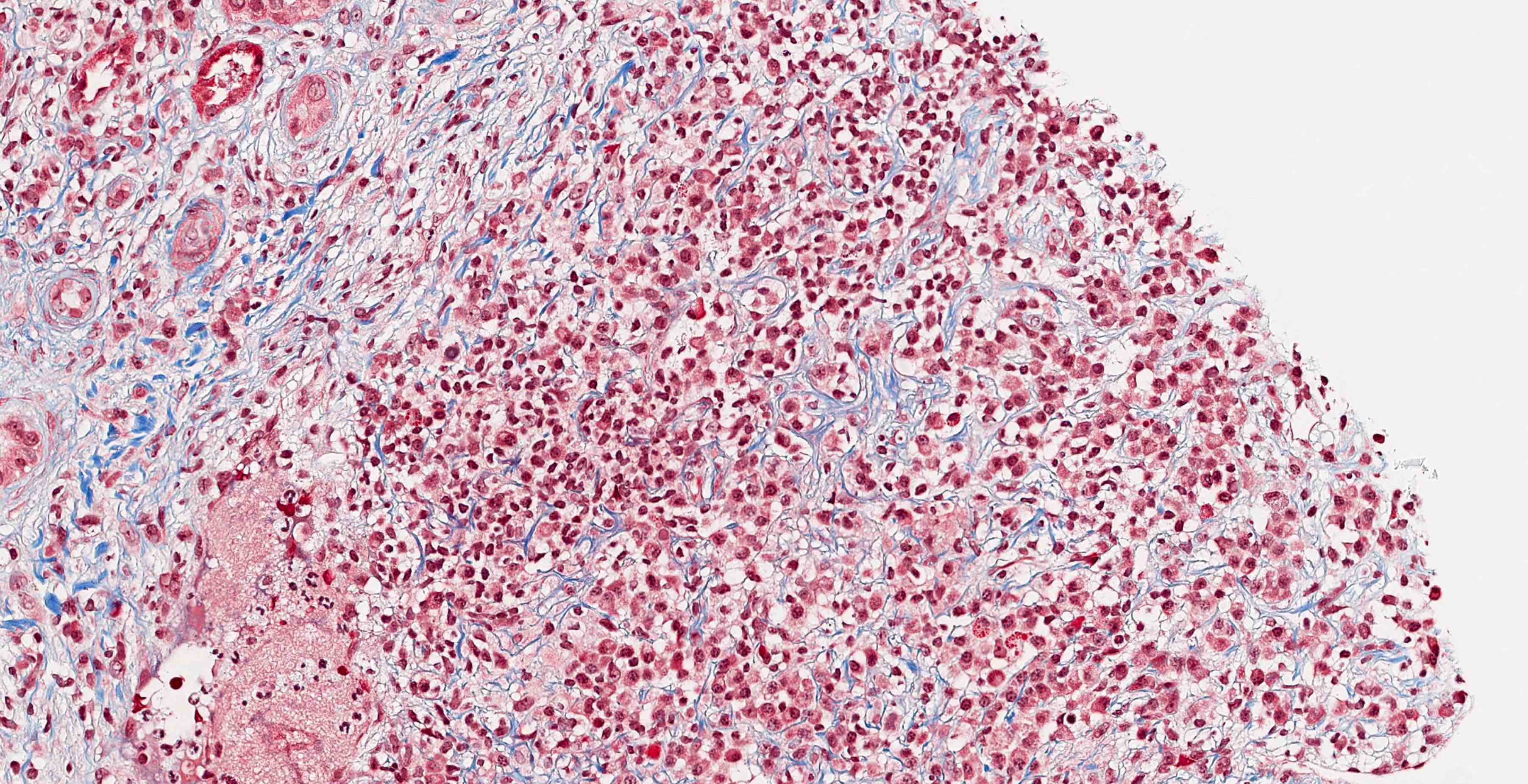
Michaelis-Gutmann bodies, high mag

Expansion of the interstitium

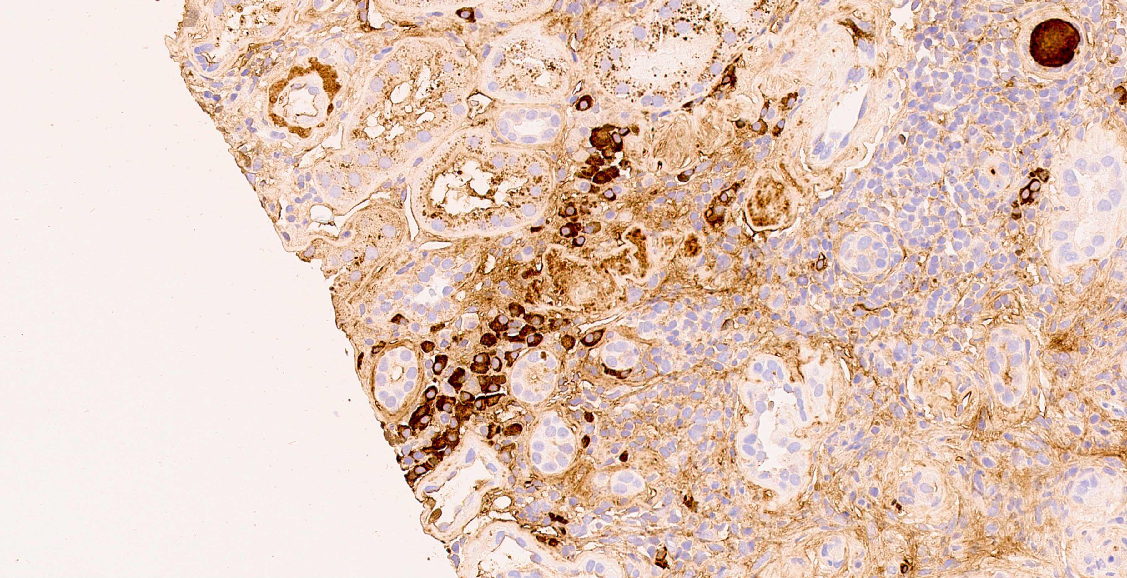
von Kossa stain

Infiltration of sheets

Histiocytes

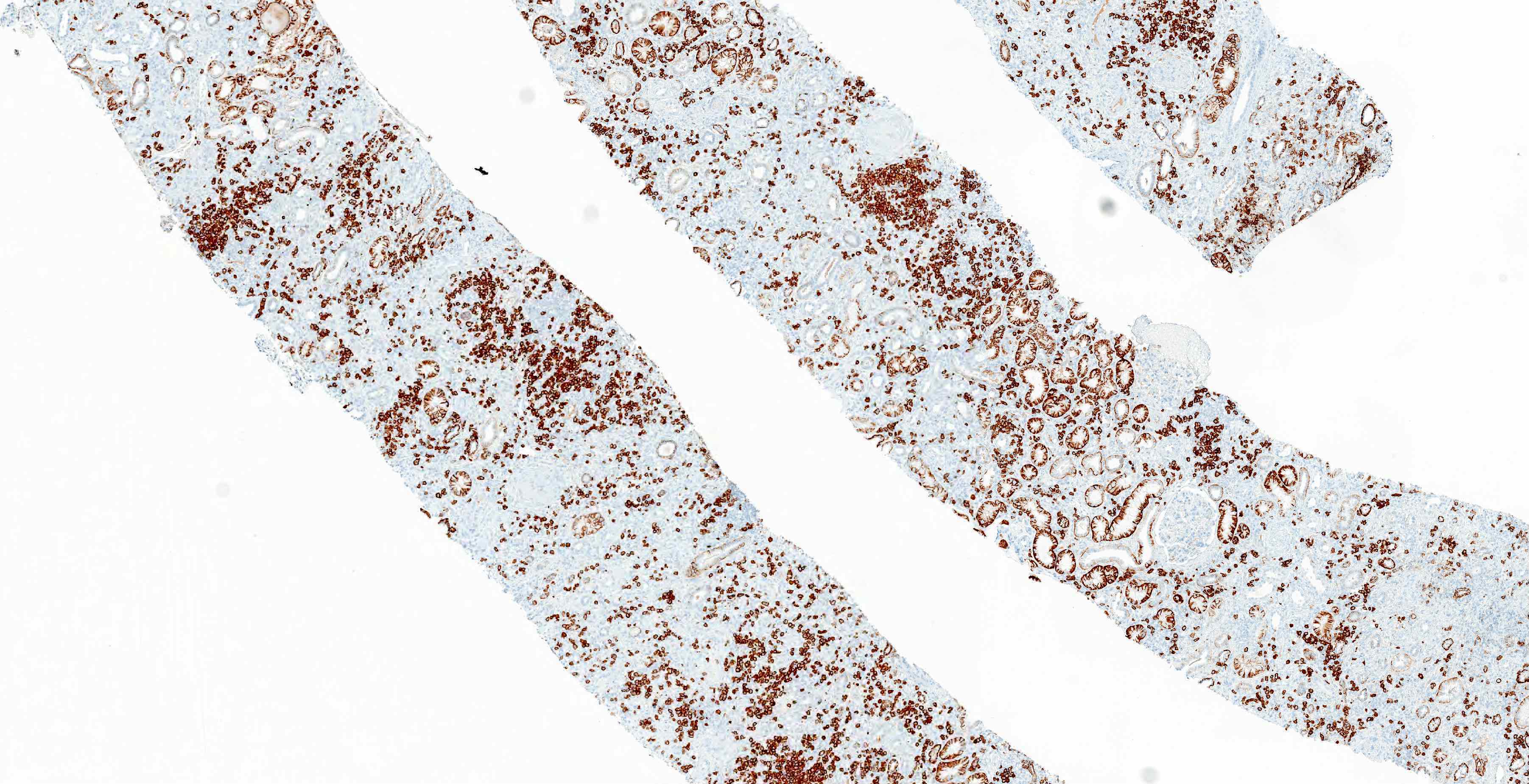
CD68+
Images hosted on other servers:

Tongue mass
Images hosted on other servers:

Various images
Images hosted on other servers:


Various images
Images hosted on other servers:

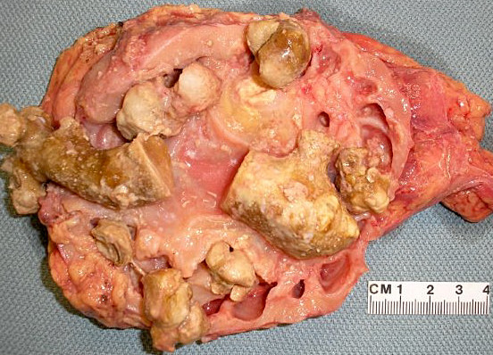
Cysts of inner medullary and papillary region

Extensive cysts communicating with each other

Papillae are interspersed due to variably sized cysts

Small cysts with stones, papillae on right is not affected

Numerous medullary cysts and isolated cortical cyst

Cysts at corticomedullary junction
Images hosted on other servers:

Cysts in papilla

With microlithiasis and chronic pyelonephritis

Cortical cyst, multiple cysts
at the corticomedullary
junction and atypically
located papillary cyst
Contributed by Ana Belén Larqué, M.D., Ph.D.
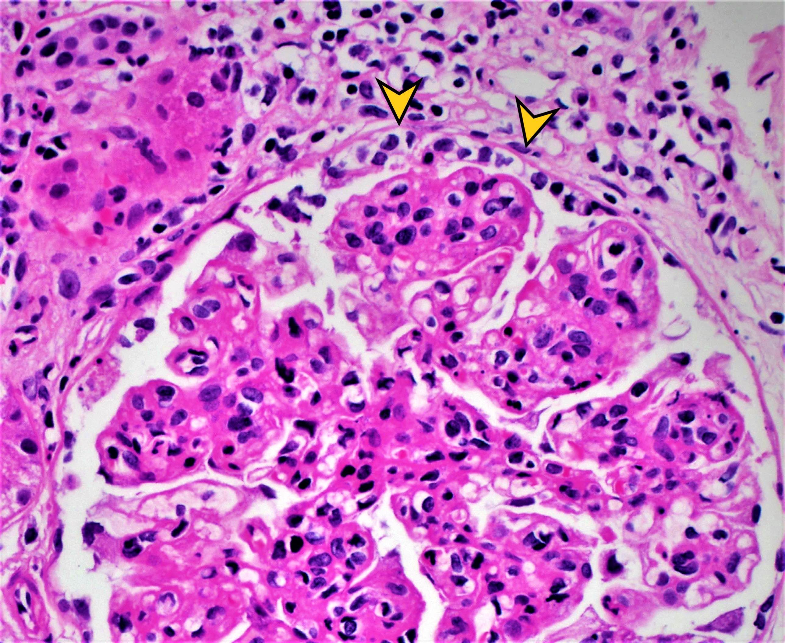
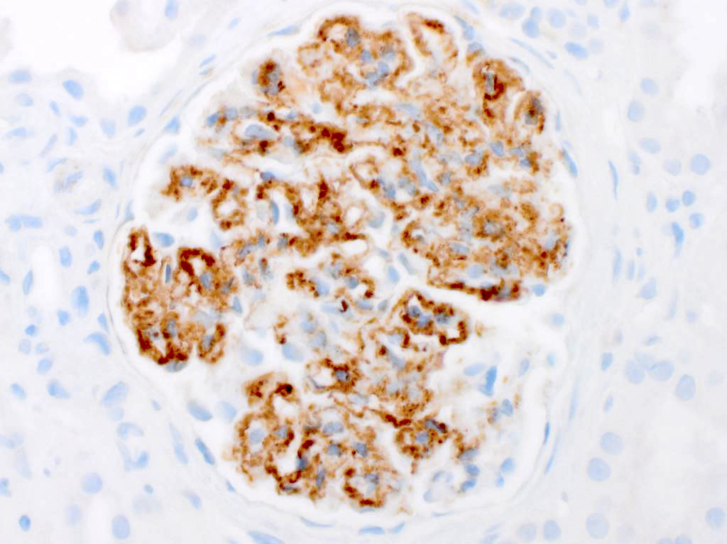
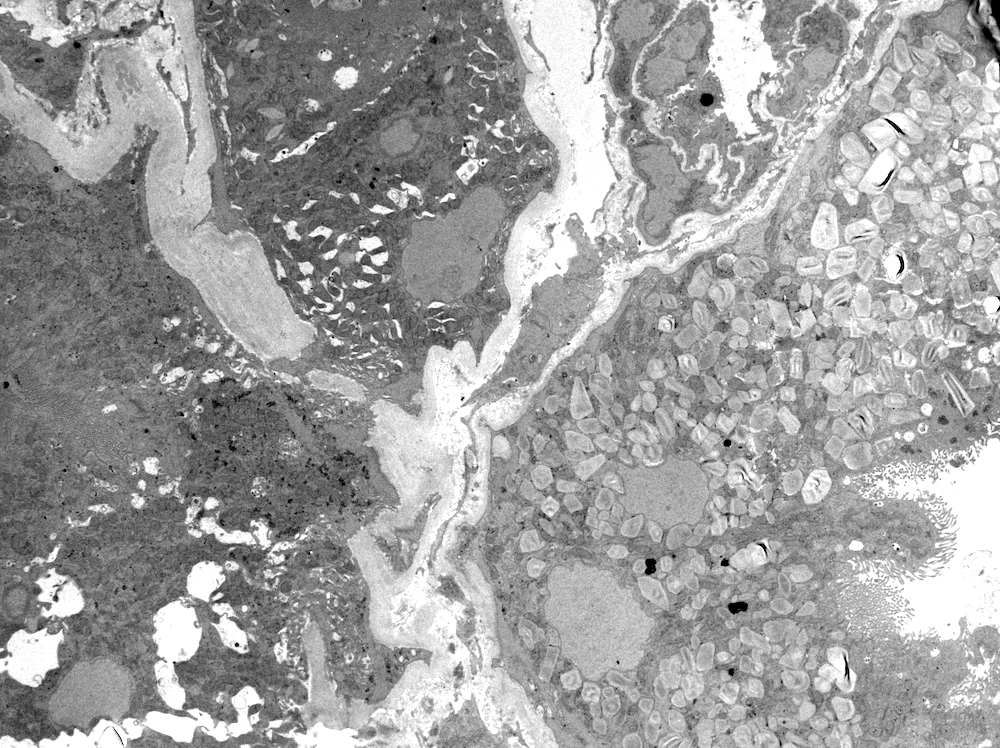
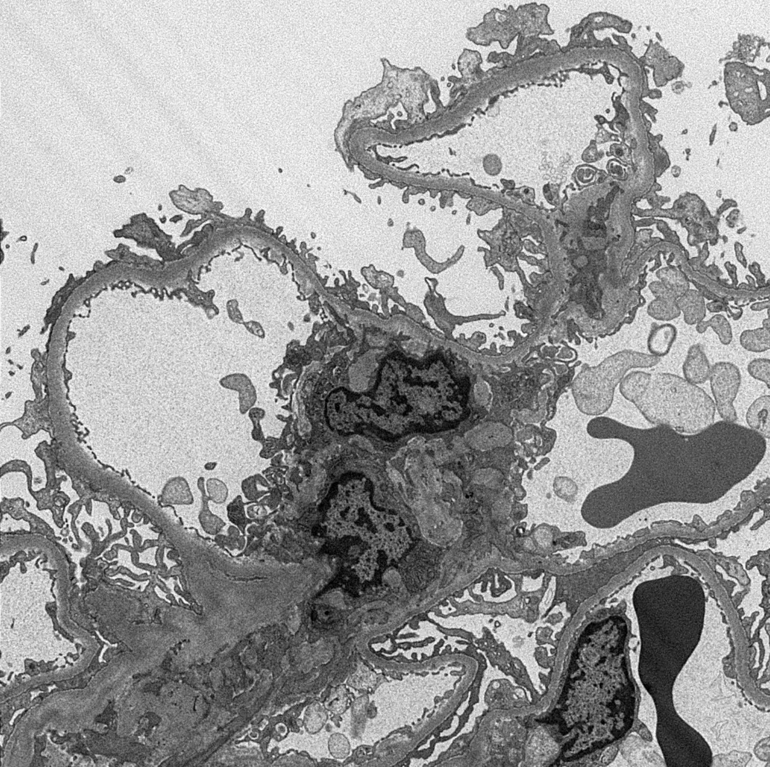
Glomerular basement membrane thickened
Subepithelial spikes

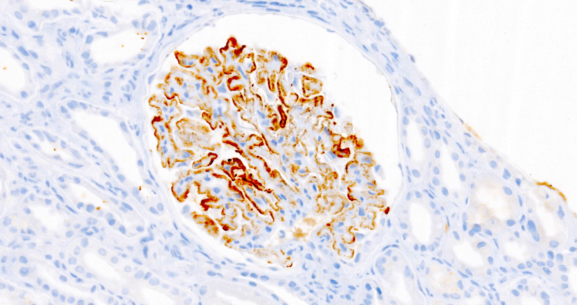
Granular staining for IgG4
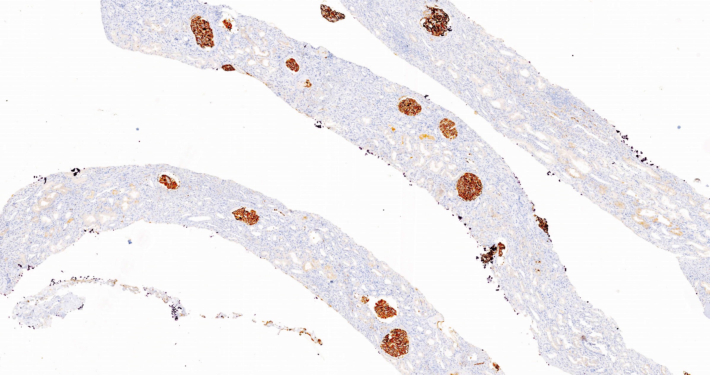
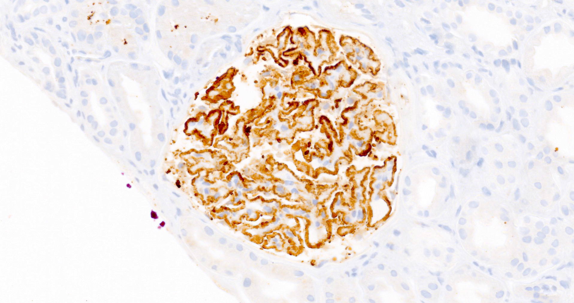
Granular staining for PLA2R
Contributed by Ana Belén Larqué, M.D., Ph.D. and Jonathan E. Zuckerman, M.D., Ph.D.
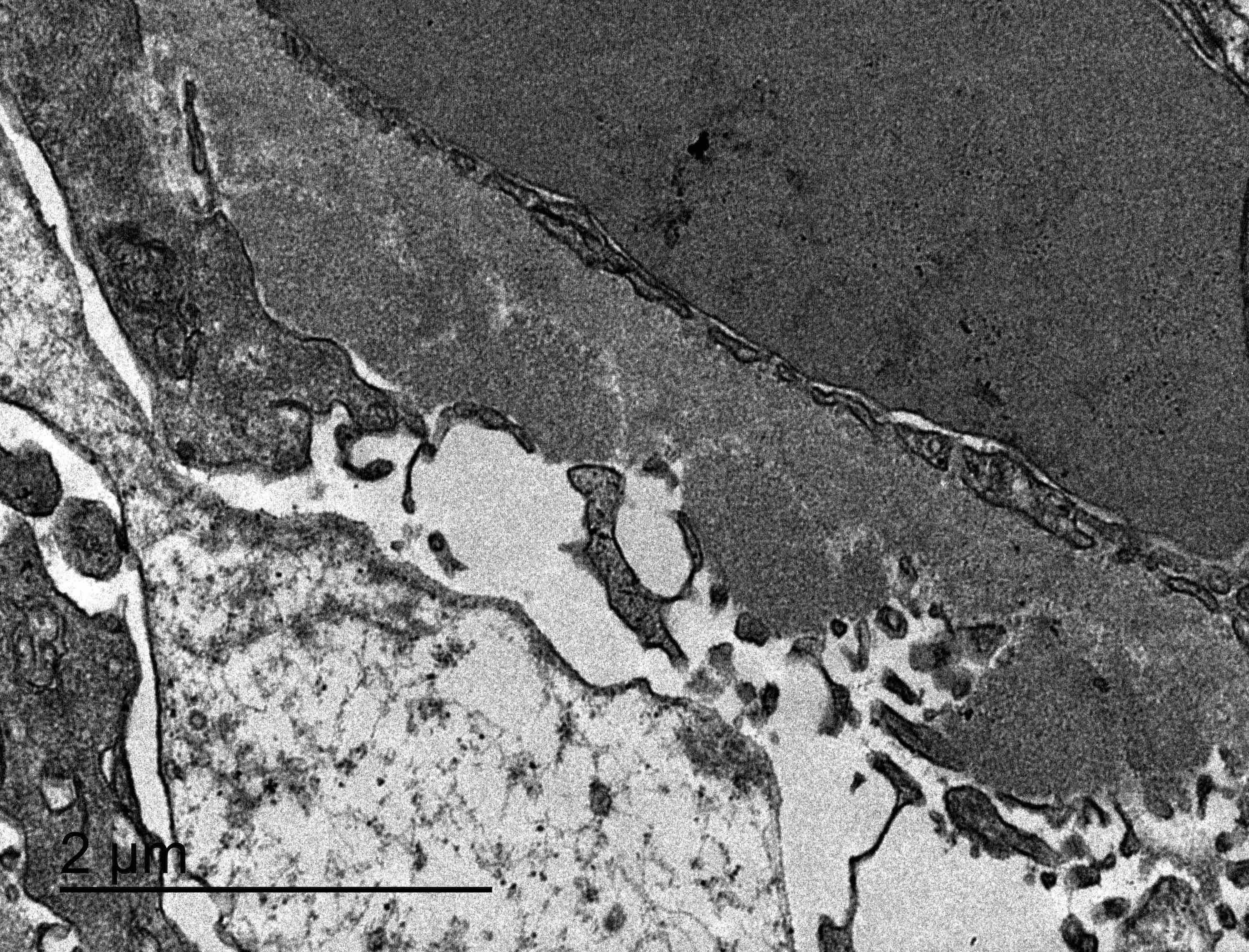
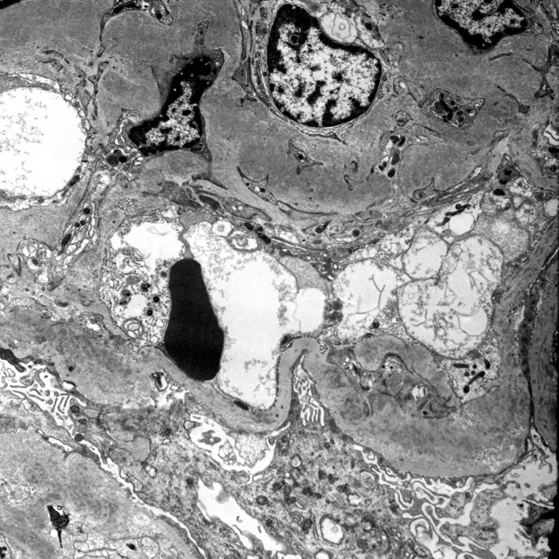
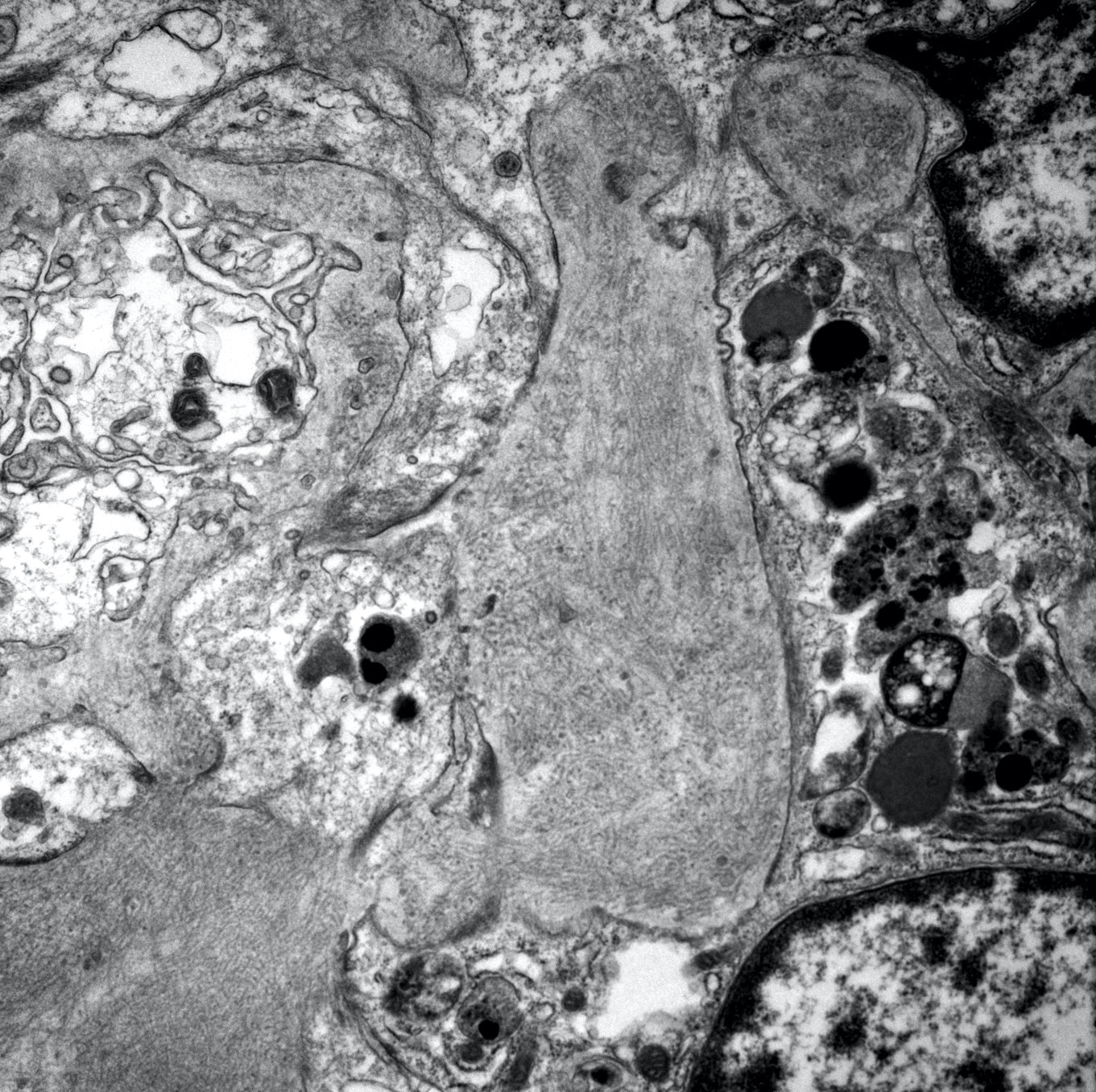
Subepithelial deposits
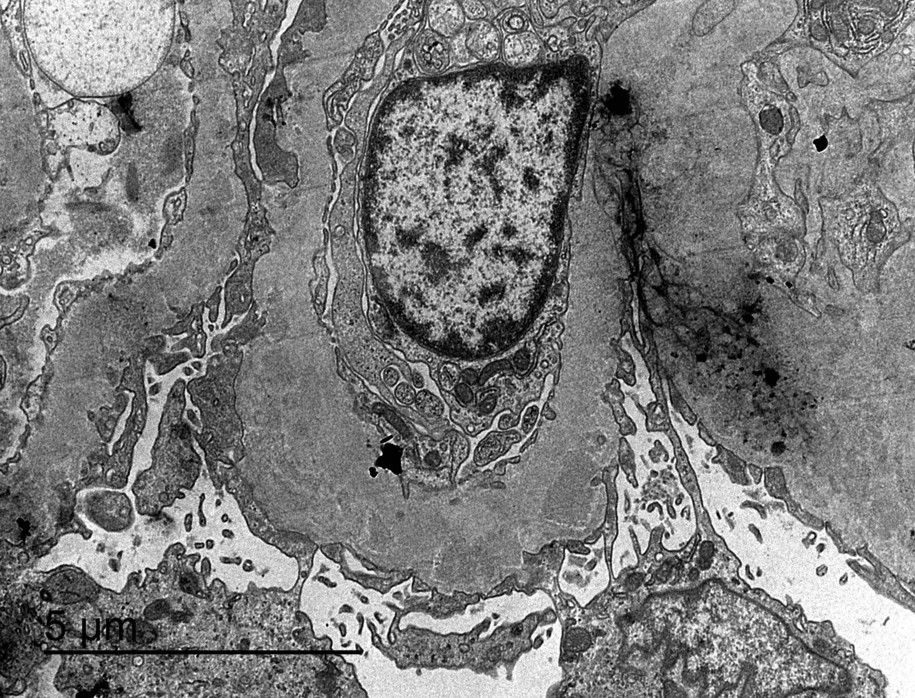
Intramembranous deposits


Membranous nephropathy, stage I
Membranous nephropathy, stage II
Membranous nephropathy, stage III


Membranous nephropathy, stage IV
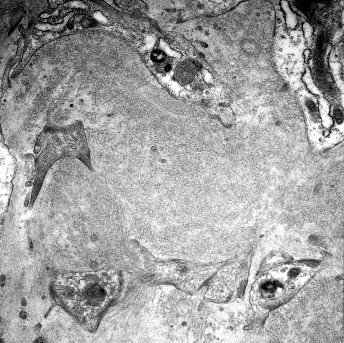
Images hosted on other servers:

Intramembranous deposits
Images hosted on other servers:

Lipoid nephrosis
Contributed by Ana Belén Larqué, M.D., Ph.D.
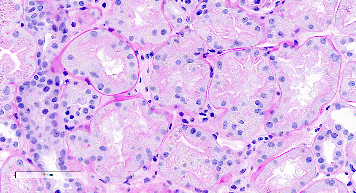
Normal glomerulus
Normal glomerulus and interstitium
Tubules with hyaline droplets

Aggregates of lipid laden macrophages
Contributed by Ana Belén Larqué, M.D., Ph.D. and Nicole K. Andeen, M.D.
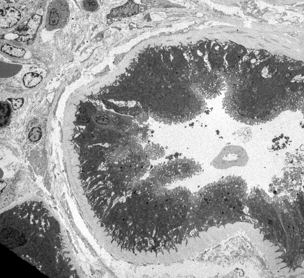
Foot process effacement
Hypertrophic podocytes
Diffuse podocyte foot process effacement
Images hosted on other servers:

Foot process effacement
Images hosted on other servers:

Common MGRS nephron lesions
Contributed by Rajib K. Gupta, M.D., Ashley Flowers, M.D. and Lois J. Arend, M.D., Ph.D.
Nodular expansion of mesangium, AL amyloid
Congo red under polarized light, AL amyloid

Mesangial matrix expansion (nonnodular), FGN
Membrano-proliferative pattern, ITGN
Nodular mesangial matrix expansion, LCDD (MIDD)
Membrano-proliferative pattern, PGNMID
Membrano-proliferative pattern, C3G (C3GN)
Membrano-proliferative pattern in type 1 CryoGN
Crystalglobulin induced nephropathy
Mesangiolysis in TMA with monoclonal gammopathy
Proximal tubules in LCPT, crystalline pattern
Tubules in MIDD
Contributed by Rajib K. Gupta, M.D., Ashley Flowers, M.D., Judy King, M.D., Ph.D. and Xin Gu, M.D.
Pale mesangial expansion due to amyloid fibrils
Mesangial expansion by slightly thicker fibrils, FGN
Microtubular deposits, ITGN
Powdery deposits, LCDD / MIDD
Glomerular deposits, C3-G (C3GN variant)
Microtubular deposits, type 1 CryoGN
Crystals, crystalline type LCPT
Abnormal lysosomes, noncrystalline LCPT
Electron dense deposits, PGNMID
Occlusive crystalline deposits, CIN
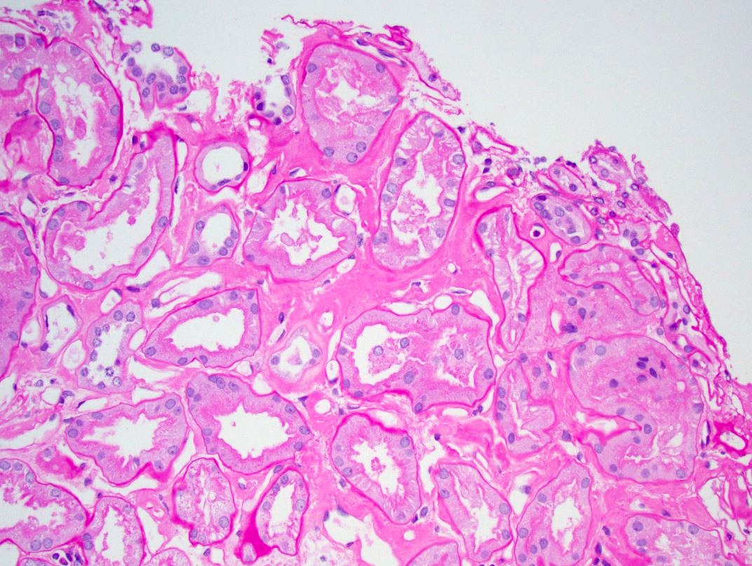
Contributed by Haiyan Zhang, M.D., Ph.D.
GBM double contours
Images hosted on other servers:

Various images
Images hosted on other servers:

Cysts at corticomedullary
junction, tubular cysts
and interstitial infiltrate
Images hosted on other servers:

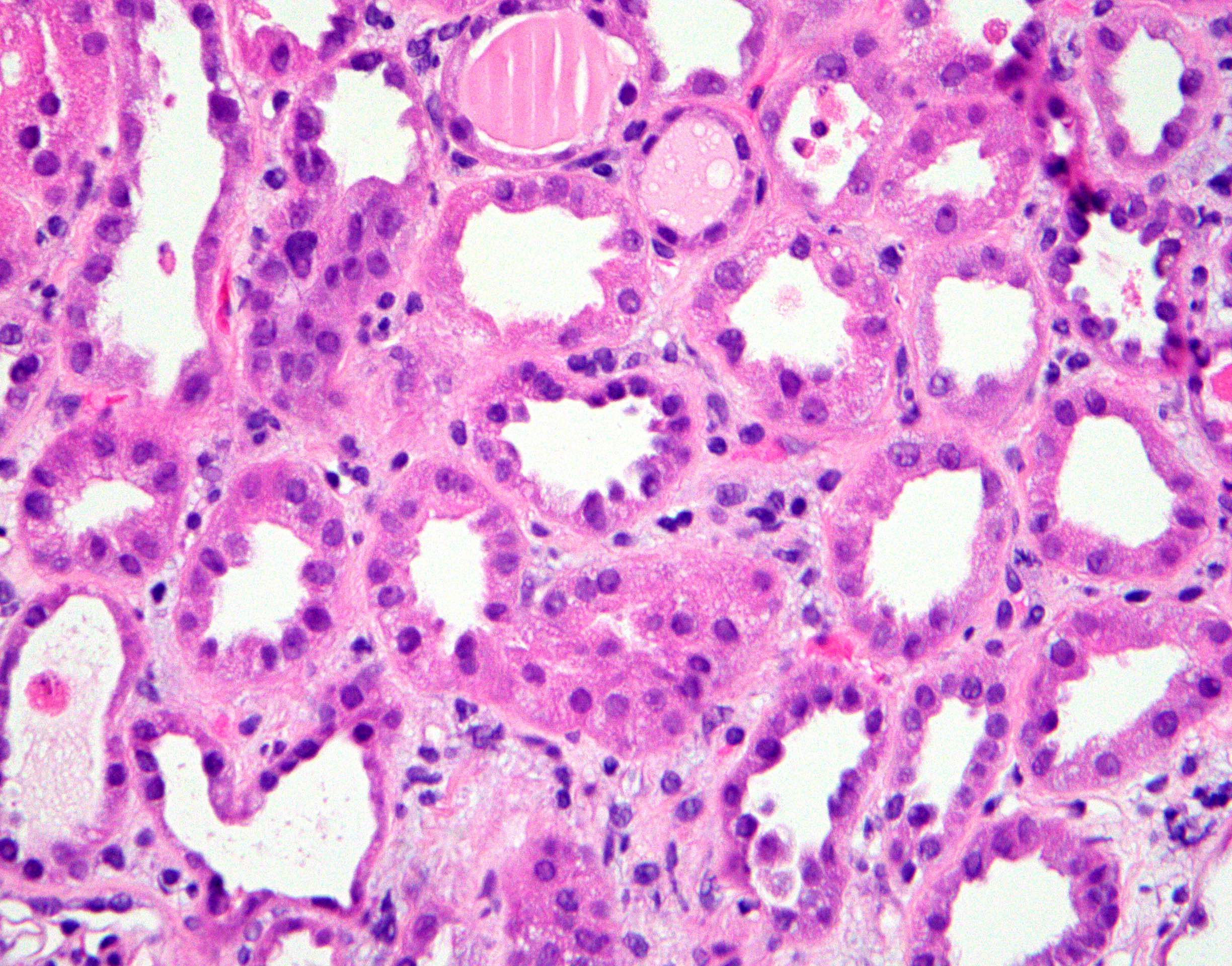
Tubules with thickened basal membrane (PAS)

Dilated tubules, thickened basal membrane (PAS)

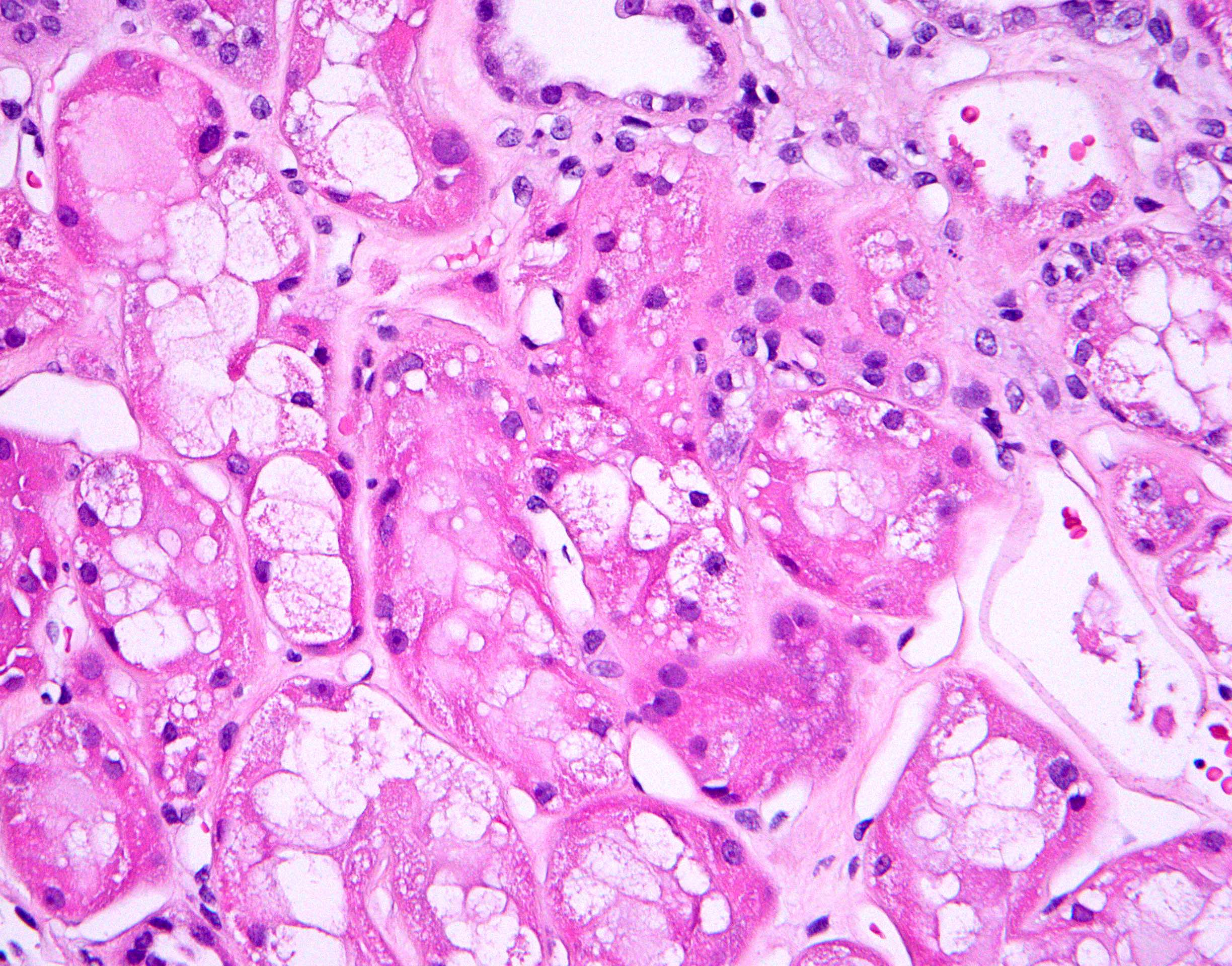
Deformed tubules
with thickened basal
membrane in fibrotic
stroma (PAS)

Dilated tubules with lymphocytic infiltrate (PAS)
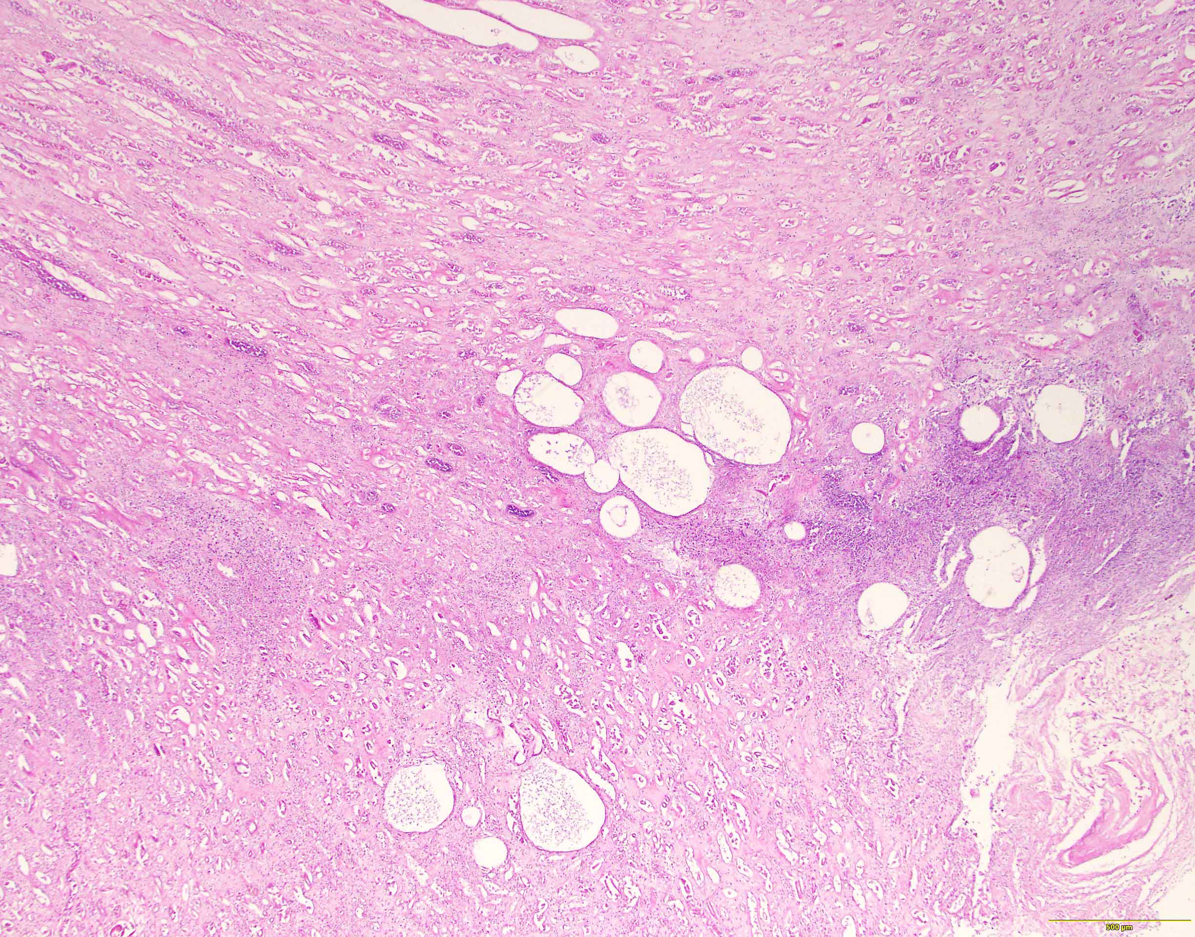
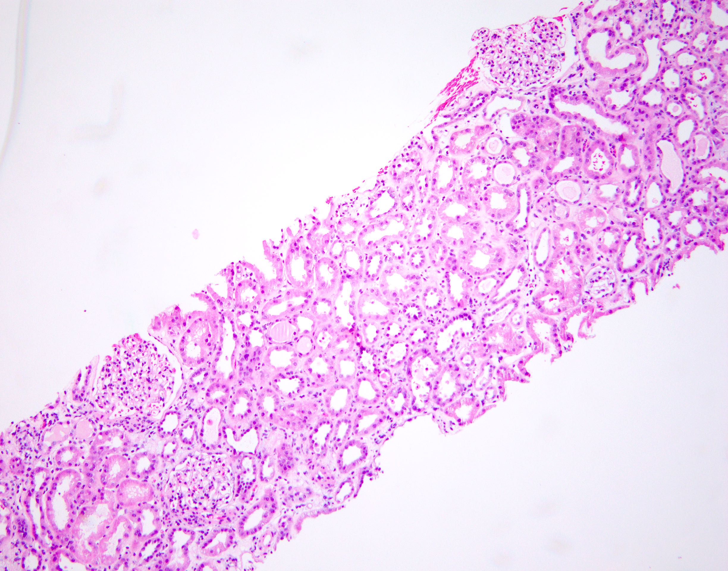
End stage renal disease:


Preserved glomeruli with individual glomerular cysts, tubulointerstitial fibrosis, hypertrophic tubules, lymphocytic infiltrate

Tubulointerstitial
fibrosis, tubular cysts
and lymphocytic
infiltrate (PAS)

Extensive obliteration of glomeruli
and sclerotic glomerli, interstitial
fibrosis, atrophic tubules and
chronic inflammation (PAS)
Images hosted on other servers:
Tubules:

Thickened tubular basement membrane

Grid-like
degeneration of
tubular basement
membrane

Atrophic tubules
with partly thickened,
partly missing
basal lamina

Basal membrane shows
thickening, splitting,
grid-like degeneration
or complete absence

Collapsed glomeruli
centrally, Bowman
capsule is filled with
amorphous material

Partially atrophic tubule with thickened basal membrane

Newly formed lamellar basal membrane

Cyst wall lined
with collecting duct
epithelium, basal
membrane is lost
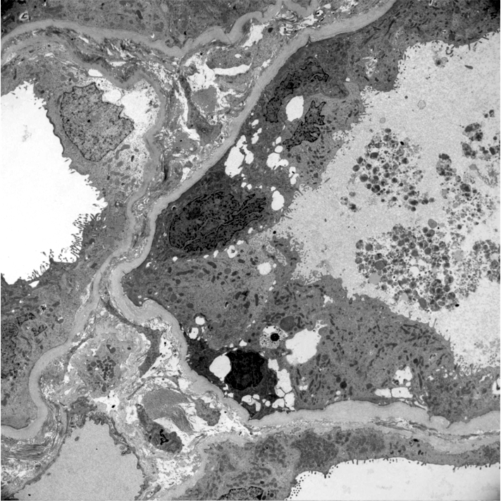
Glomeruli:

Thickened glomerular
basal membrane with
lacuna, periglomerular
fibrosis

Mild periglomerular fibrosis,
thickened tubules with
fragmented basal lamina,
lymphocytes
Contributed by Jonathan E. Zuckerman, M.D., Ph.D.
Glomerulomegaly

Perihilar focal segmental glomerulosclerosis
Glomerulomegaly
Glomerulomegaly, tubular hypertrophy
Perihilar focal segmental glomerulosclerosis
Normal glomerulus
Contributed by Jonathan E. Zuckerman, M.D., Ph.D.
Mild GBM thickening
Segmental foot process effacement
Images hosted on other servers:


Various images
Contributed by Jonathan E. Zuckerman, M.D., Ph.D.

Type 1 primary hyperoxaluria
Secondary oxalosis: calcium phosphate deposition

Secondary oxalosis: intraluminal calcium phosphate deposition

Fan shaped calcium oxalate deposits
Contributed by Jonathan E. Zuckerman, M.D., Ph.D.

Oxalate crystals
Contributed by Ana Belén Larqué, M.D, Ph.D.
Arterial wall with fibrinoid necrosis
Cellular crescent
Crescents
Segmental fibrinoid necrosis of glomerular tufts
Interstitial inflammation

Case #34 - HacettepePathology
Images hosted on other servers:


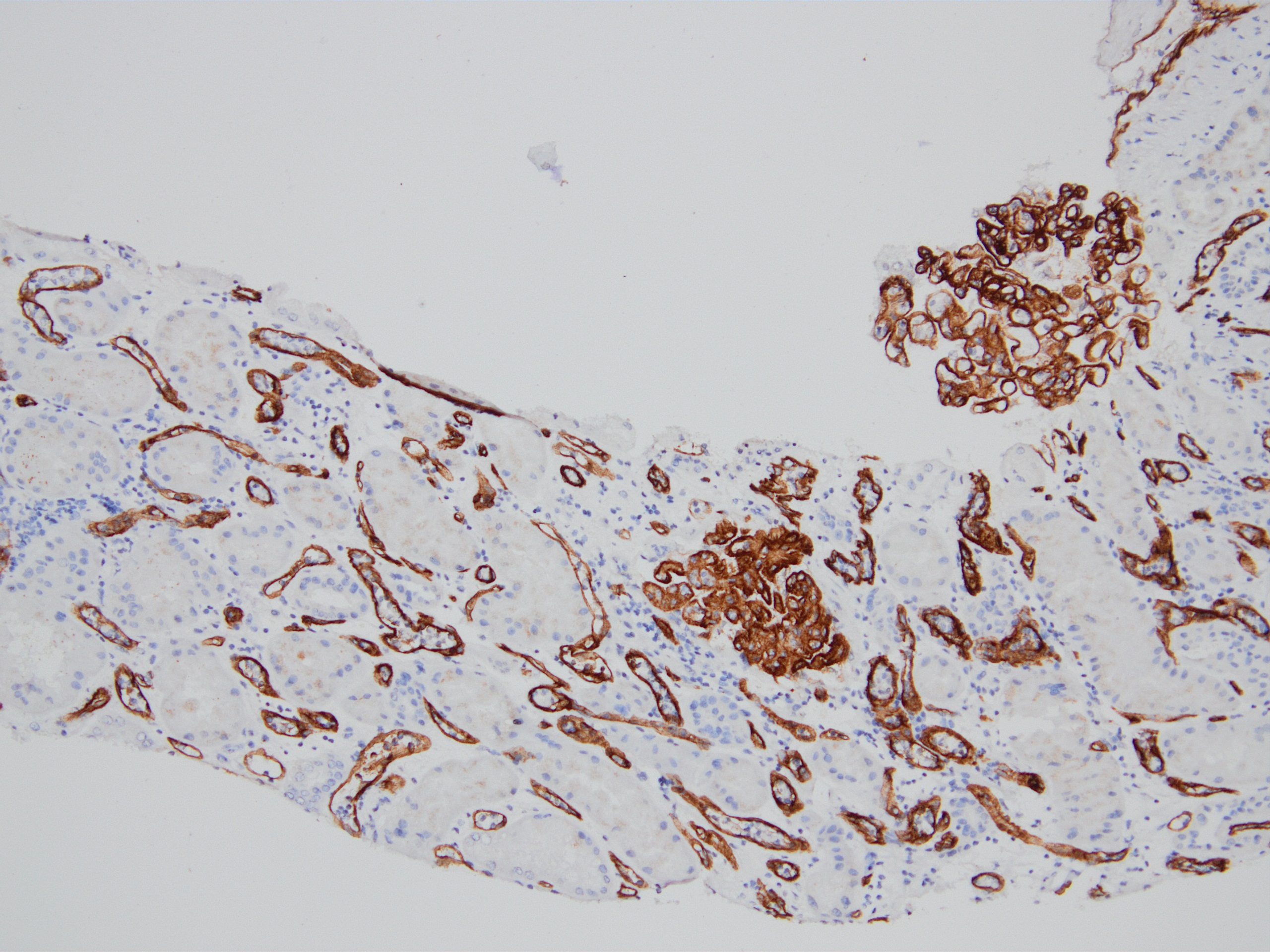
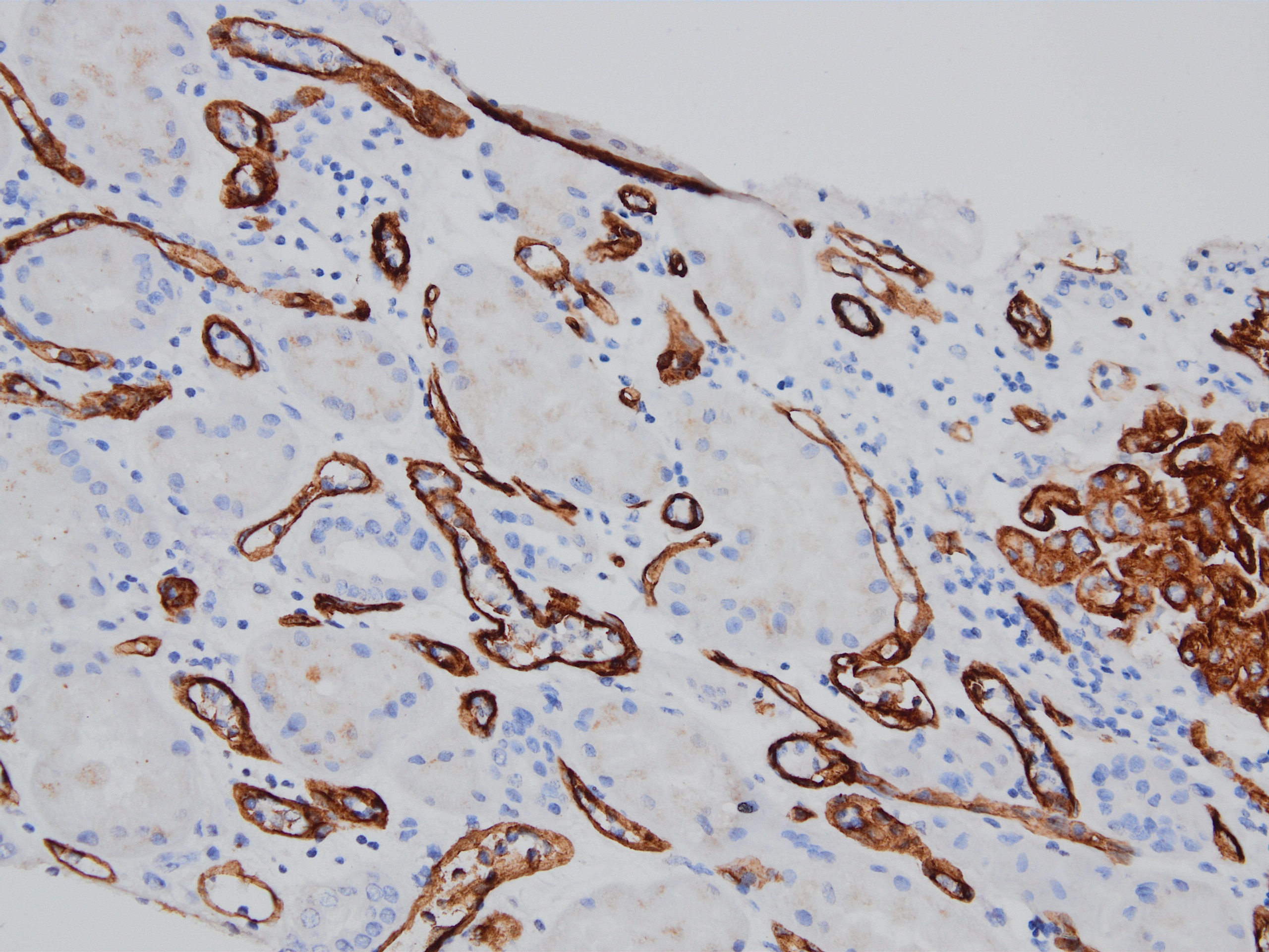
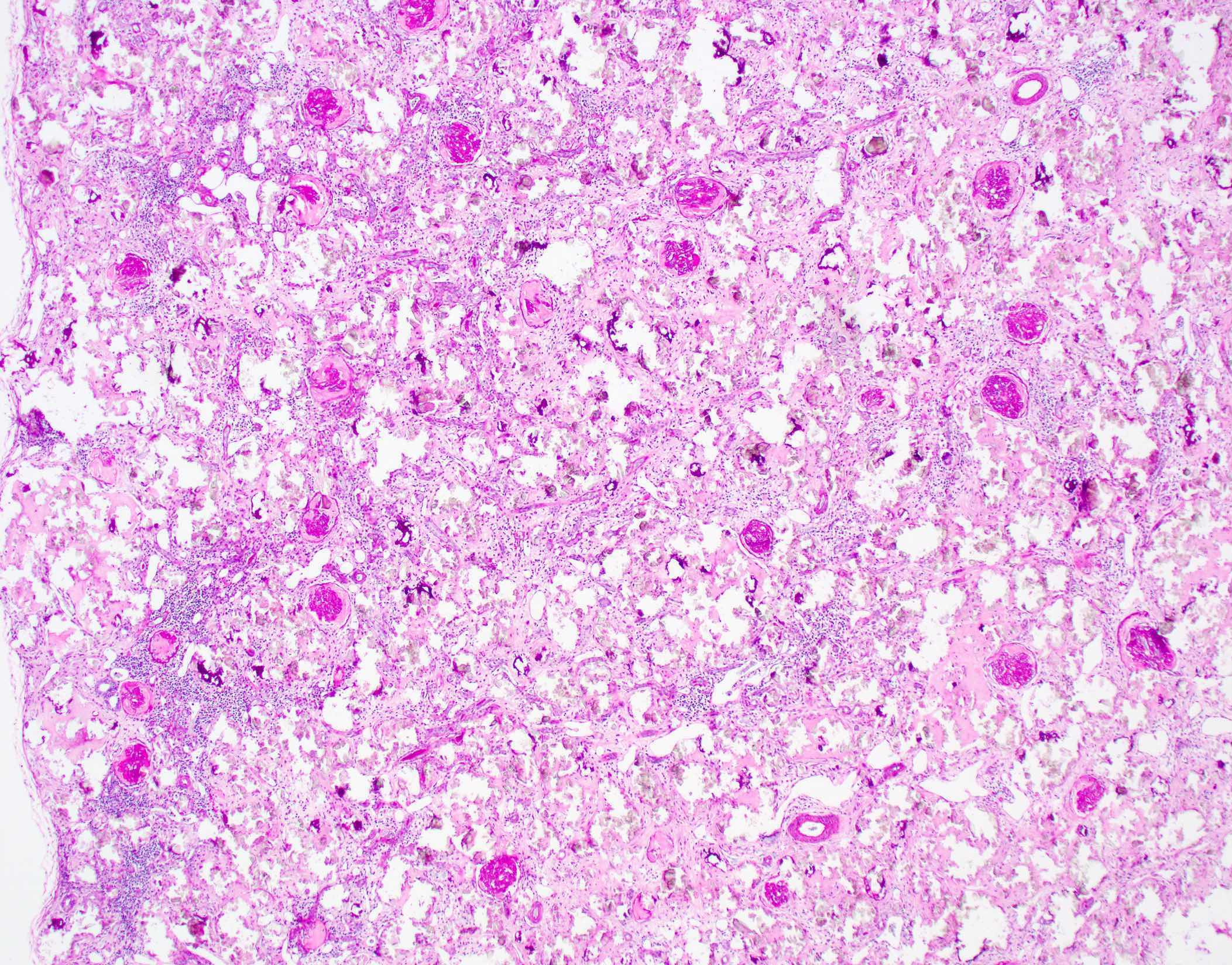
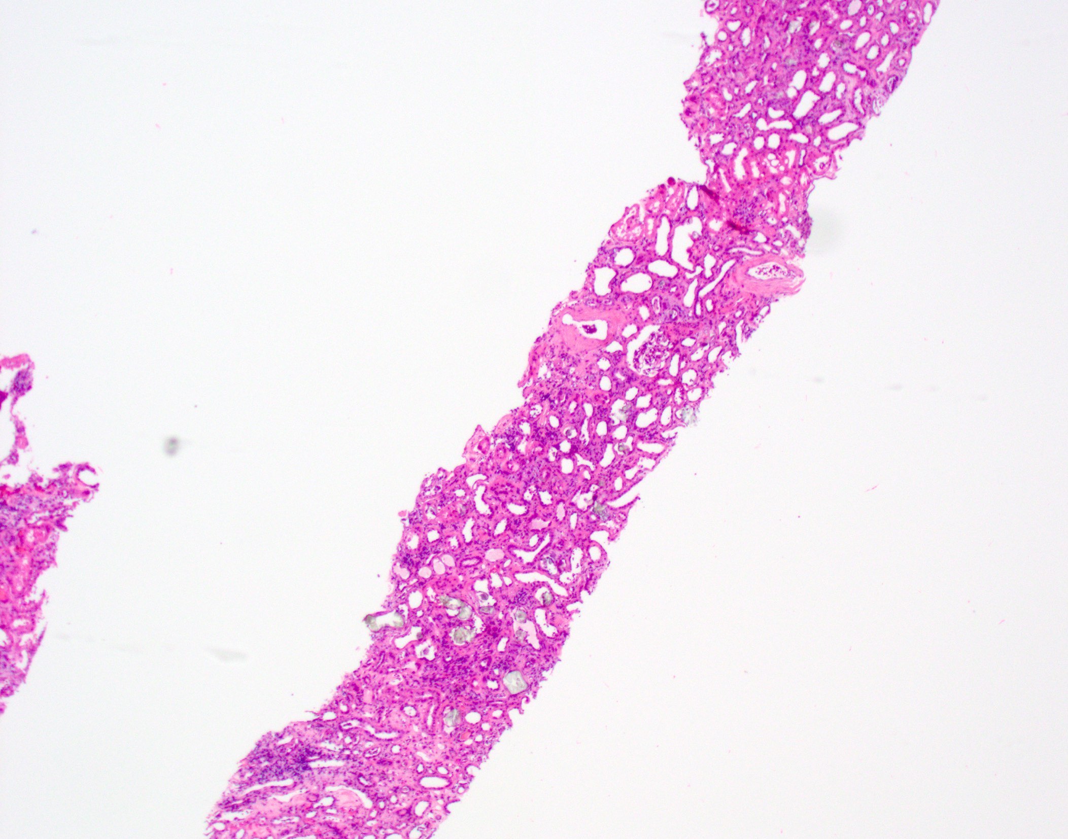
Pale amyloid deposits in enlarged kidney
Contributed by Anthony Sisk, D.O.

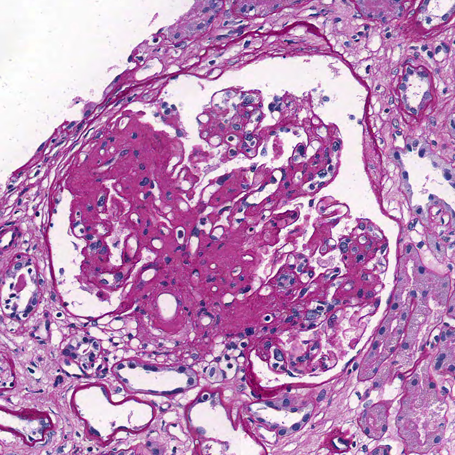
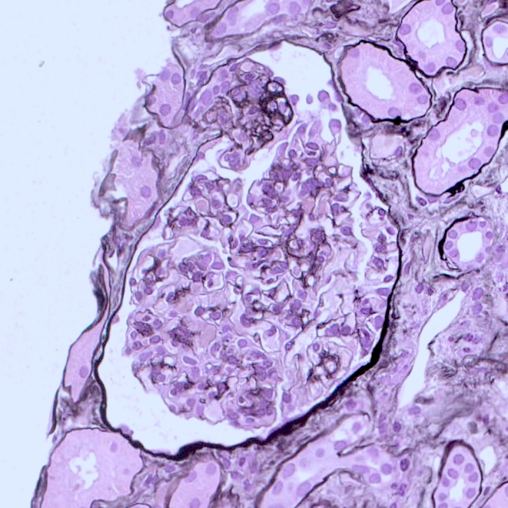
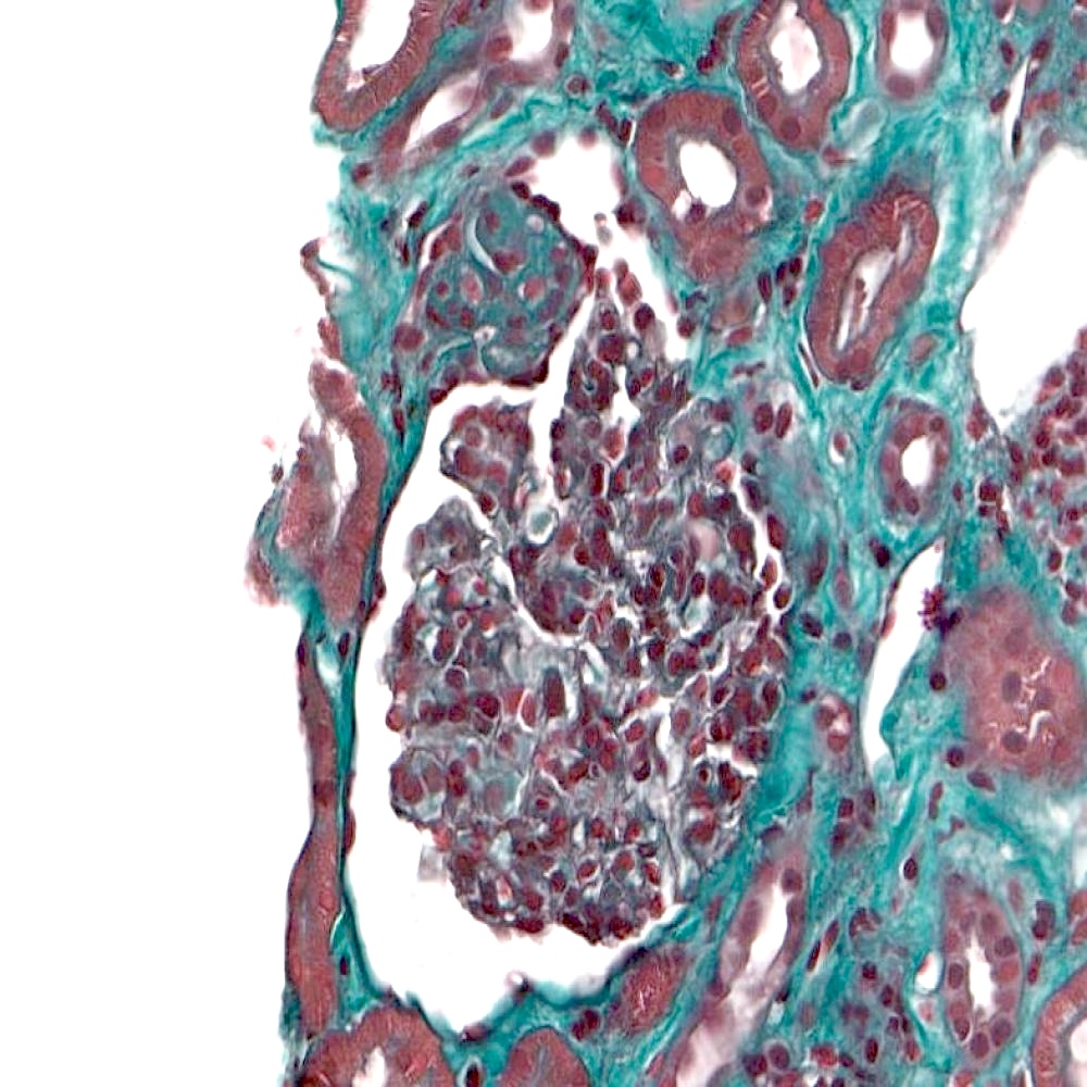
Glomerulus
PAS, glomerulus
Jones silver, glomerulus
Trichrome, glomerulus
PAS, tubulointerstitium
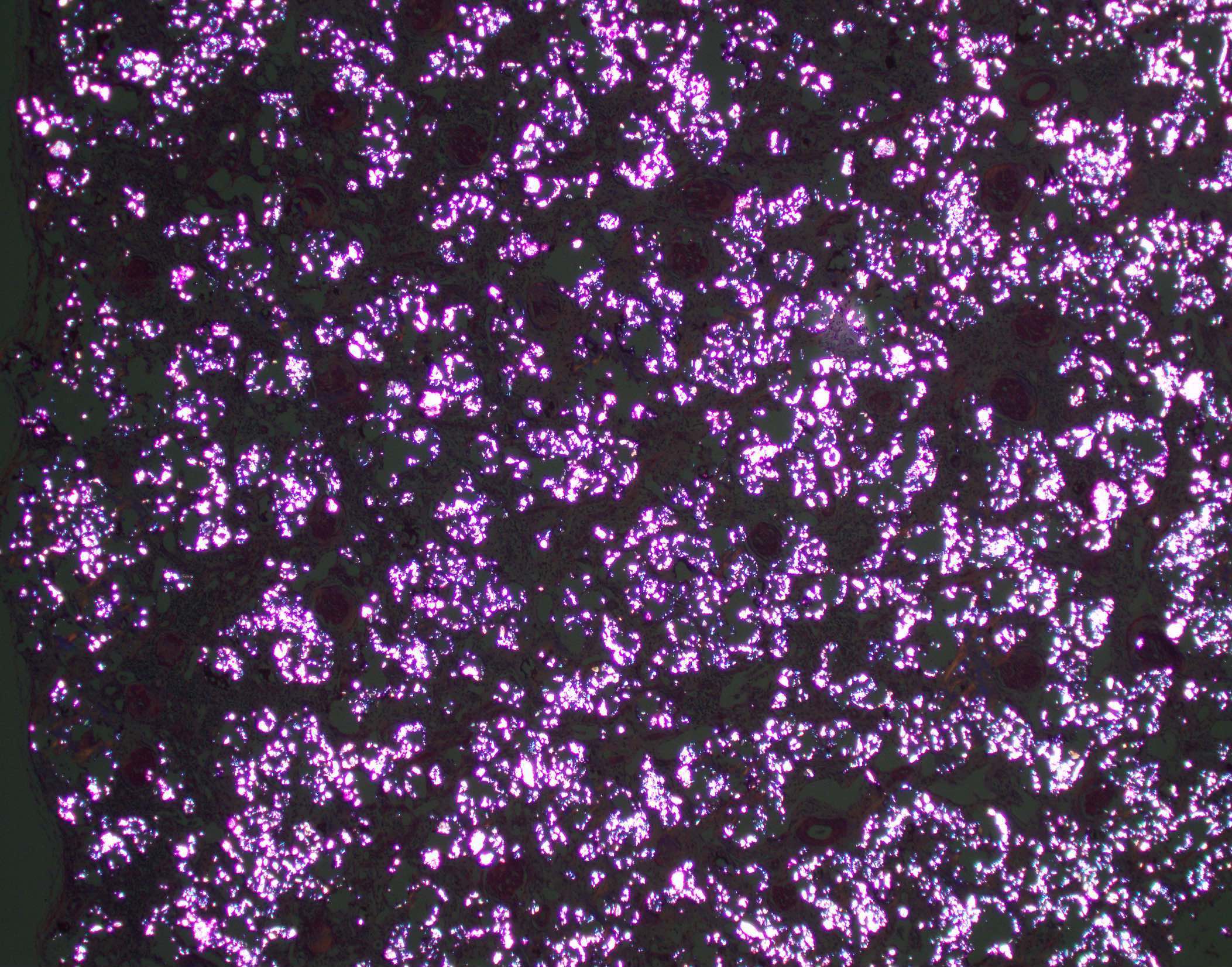
Congo red
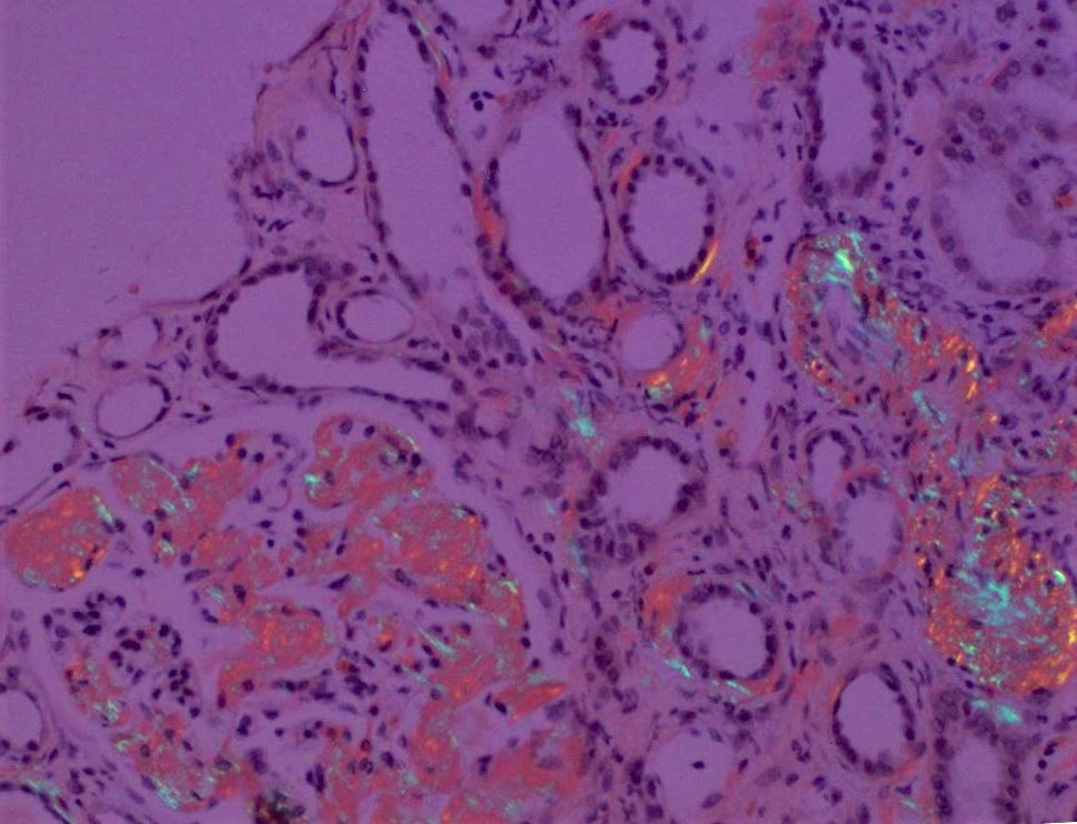
Polarization of Congo red
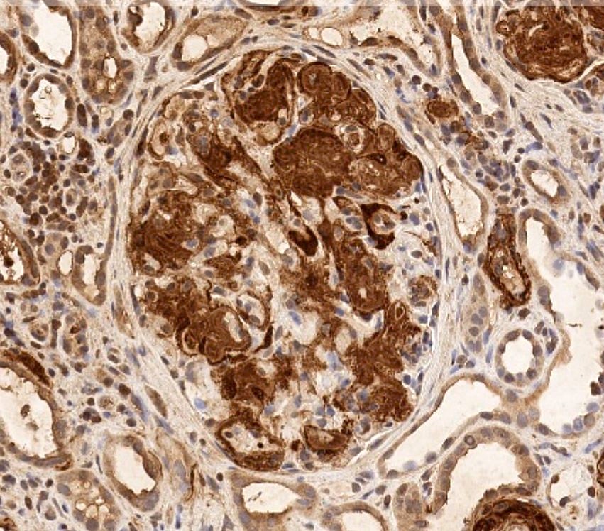
Amyloid A stain
Contributed by Anthony Sisk, D.O.
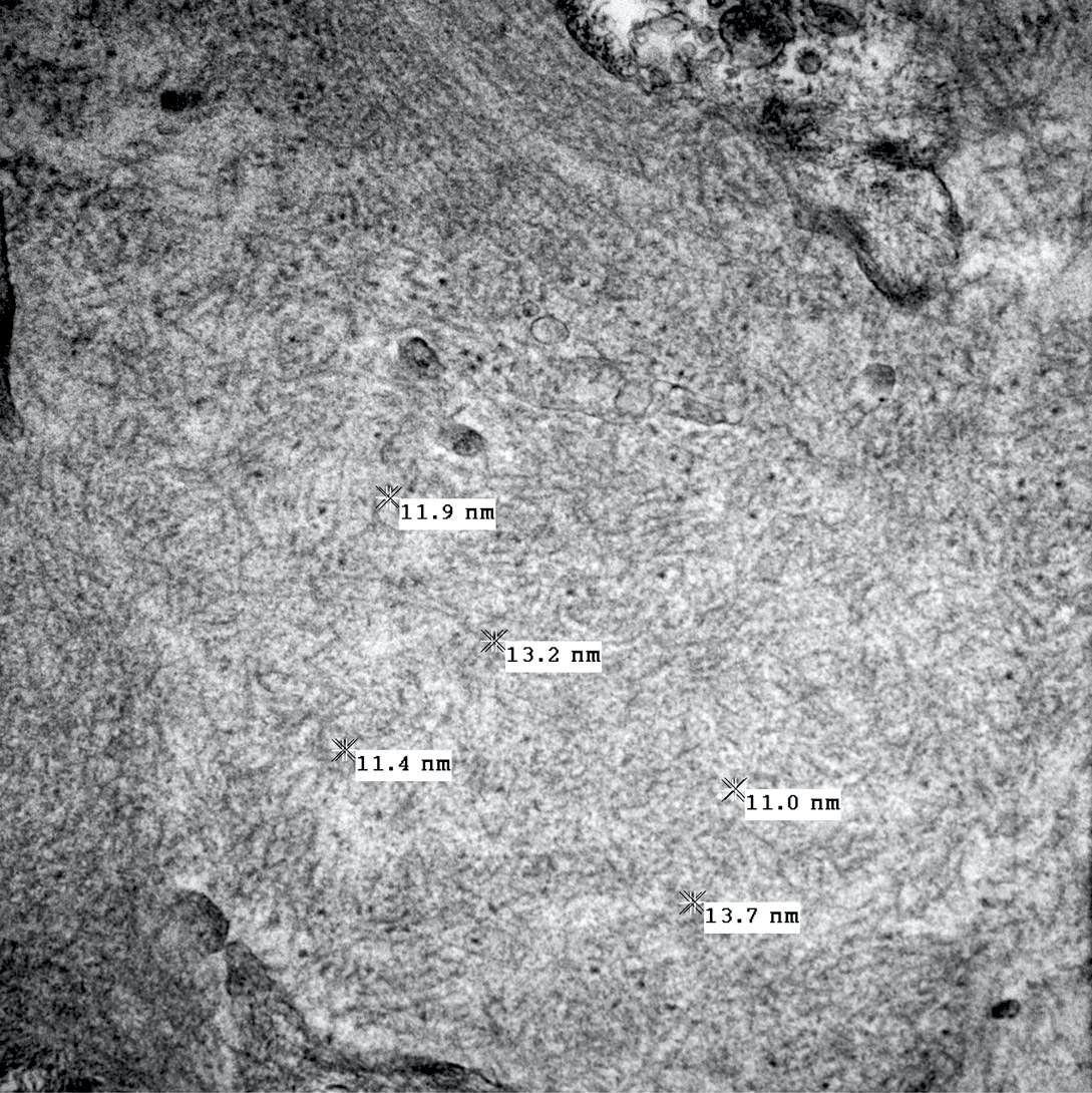
Glomerular involvement
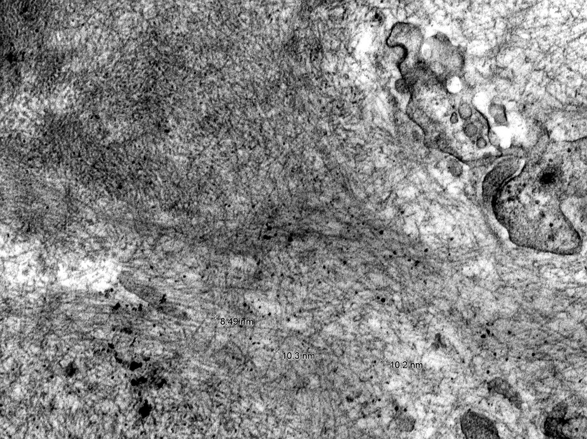
Fibril measurements
Spicule formation
Contributed by Saskia von Stillfried, M.D.
Normal glomerulus

Thick appearance of GBM

Mesangial hypercellularity
Membrano-proliferative glomerulonephritis

Focal segmental
glomerulosclerosis

Nodular glomerulosclerosis
Global glomerulosclerosis
Cellular crescent
Normal tubuli

Acute tubular injury
Interstitial fibrosis and tubular atrophy

Interstitial inflammation


Contributed by Mazdak Khalighi, M.D. and Nicole K. Andeen, M.D.


Acute tubular injury with pigmented, granular and focally stringy appearing casts
PAS

Myoglobin
Images hosted on other servers:

Various images


Granulomatous interstitial nephritis

nonspecific features,
which is common

Recurrence after renal transplant
Contributed by Christine M. Lee, M.D. and Jonathan E. Zuckerman, M.D., Ph.D.
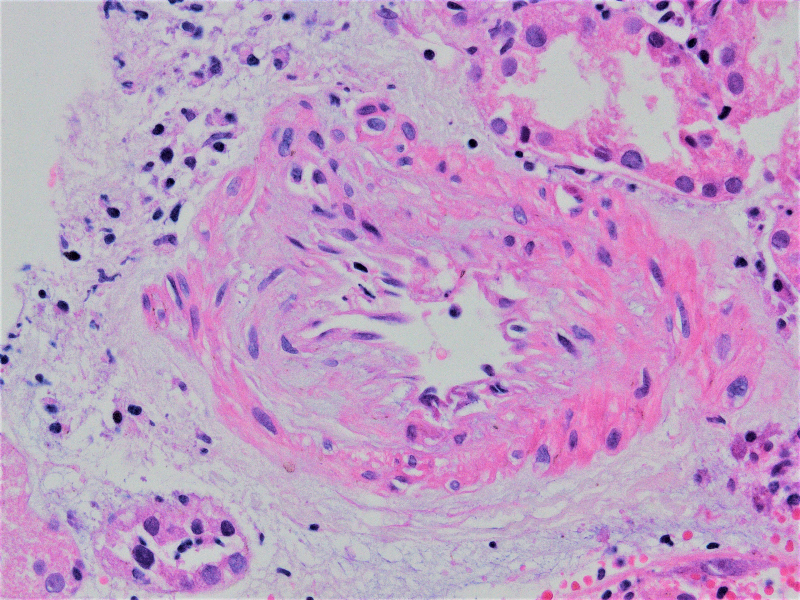
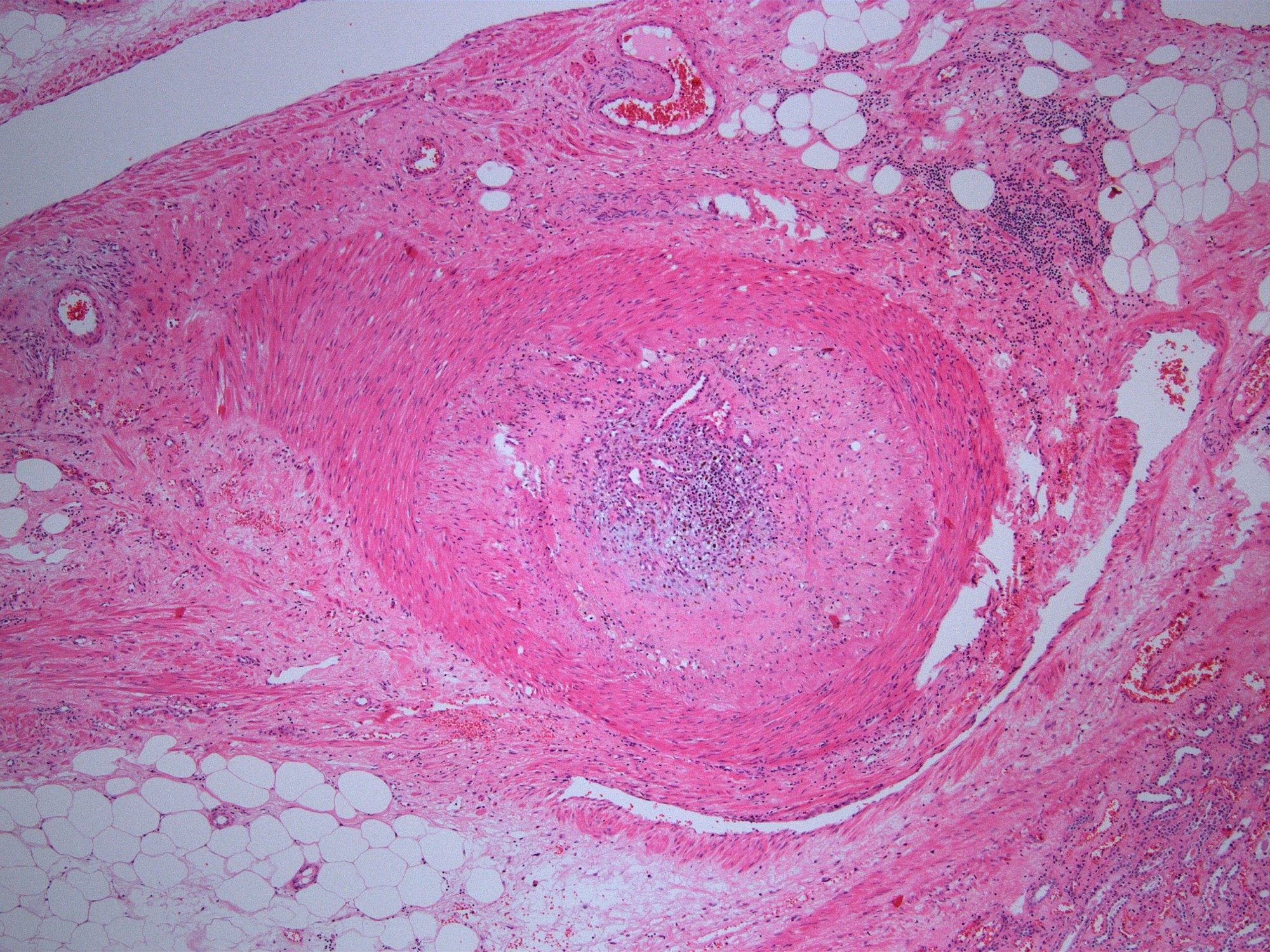
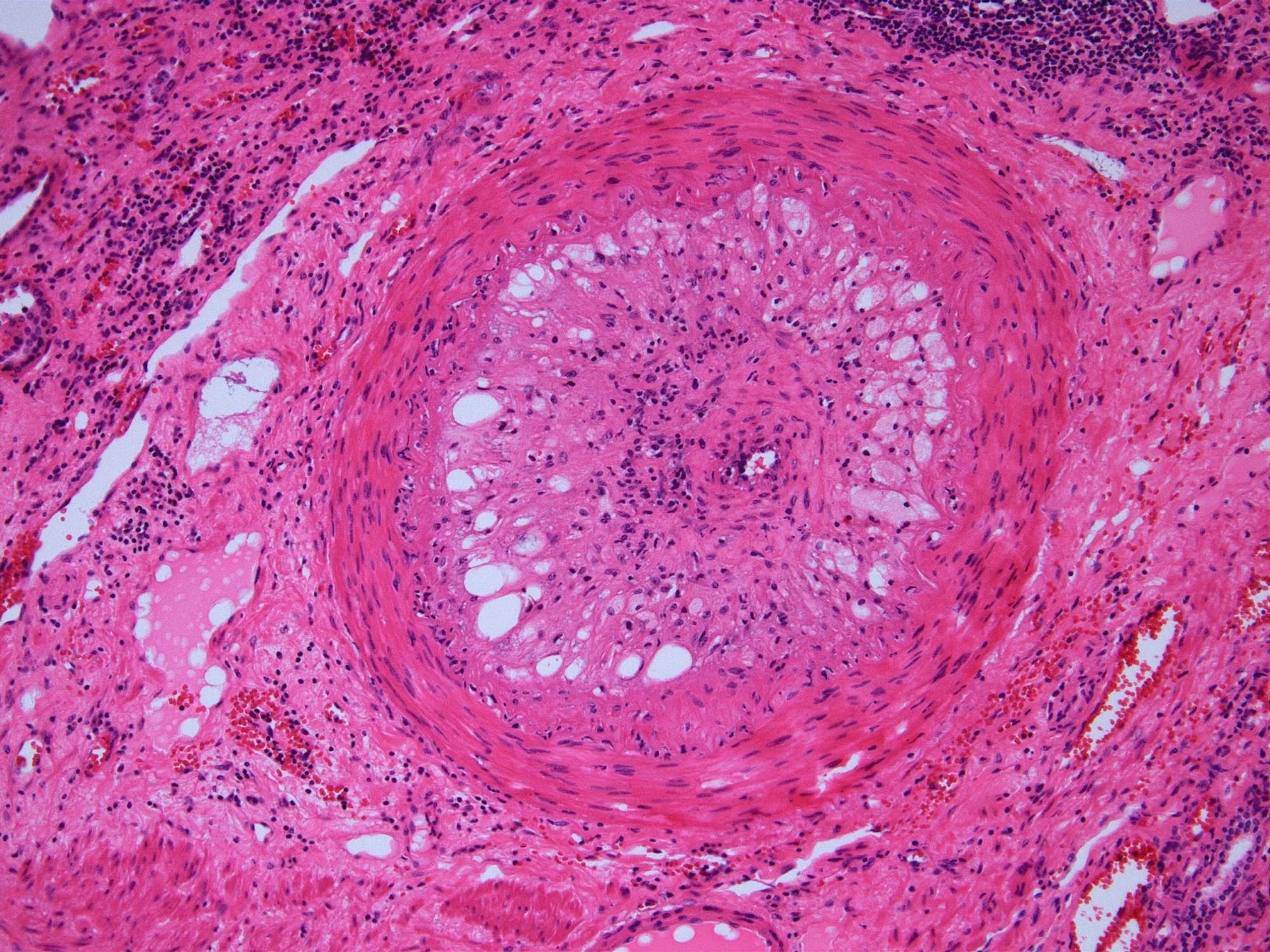
Onion skin change
Onion skin change in artery

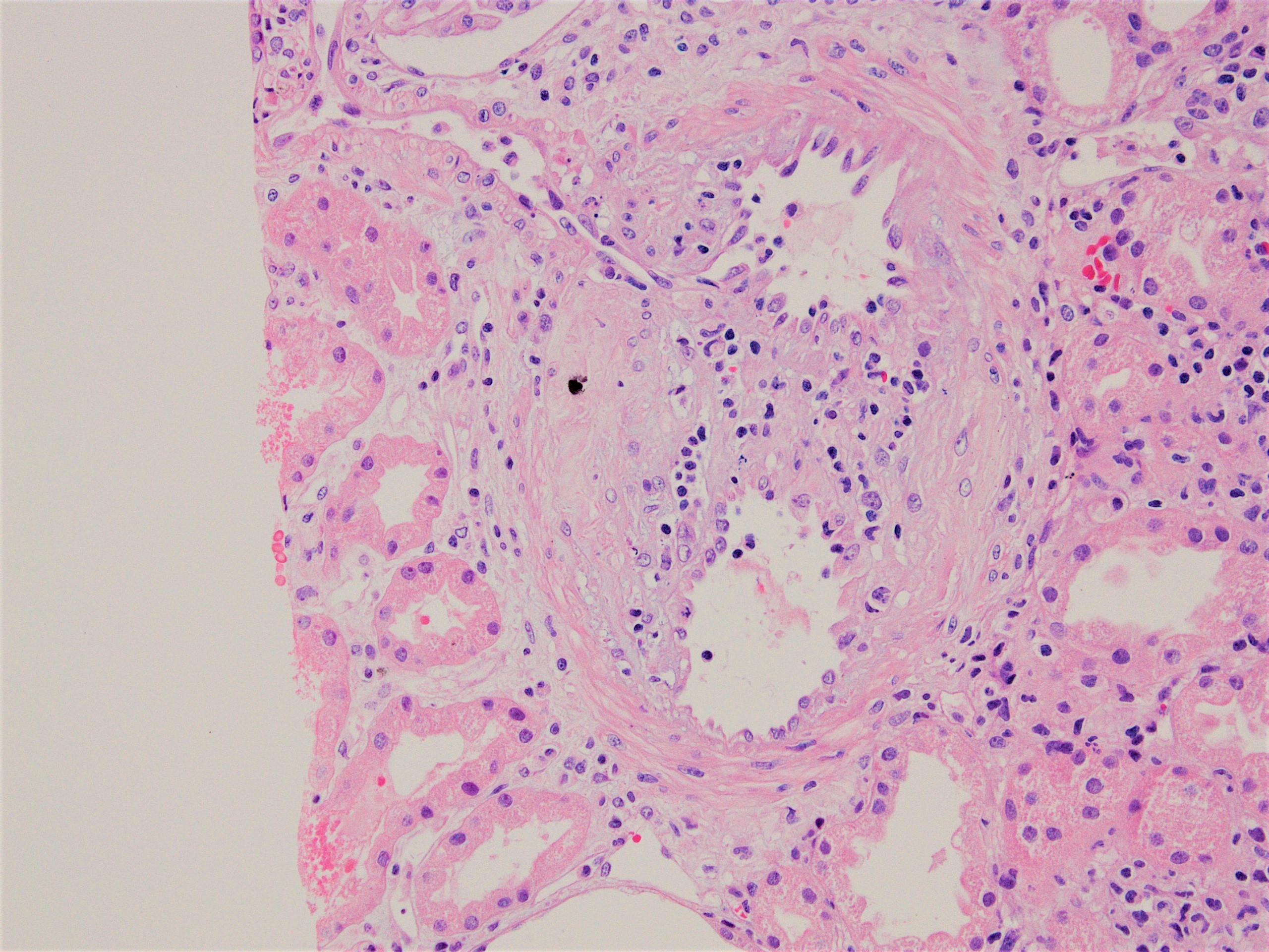
Mucoid intimal edema
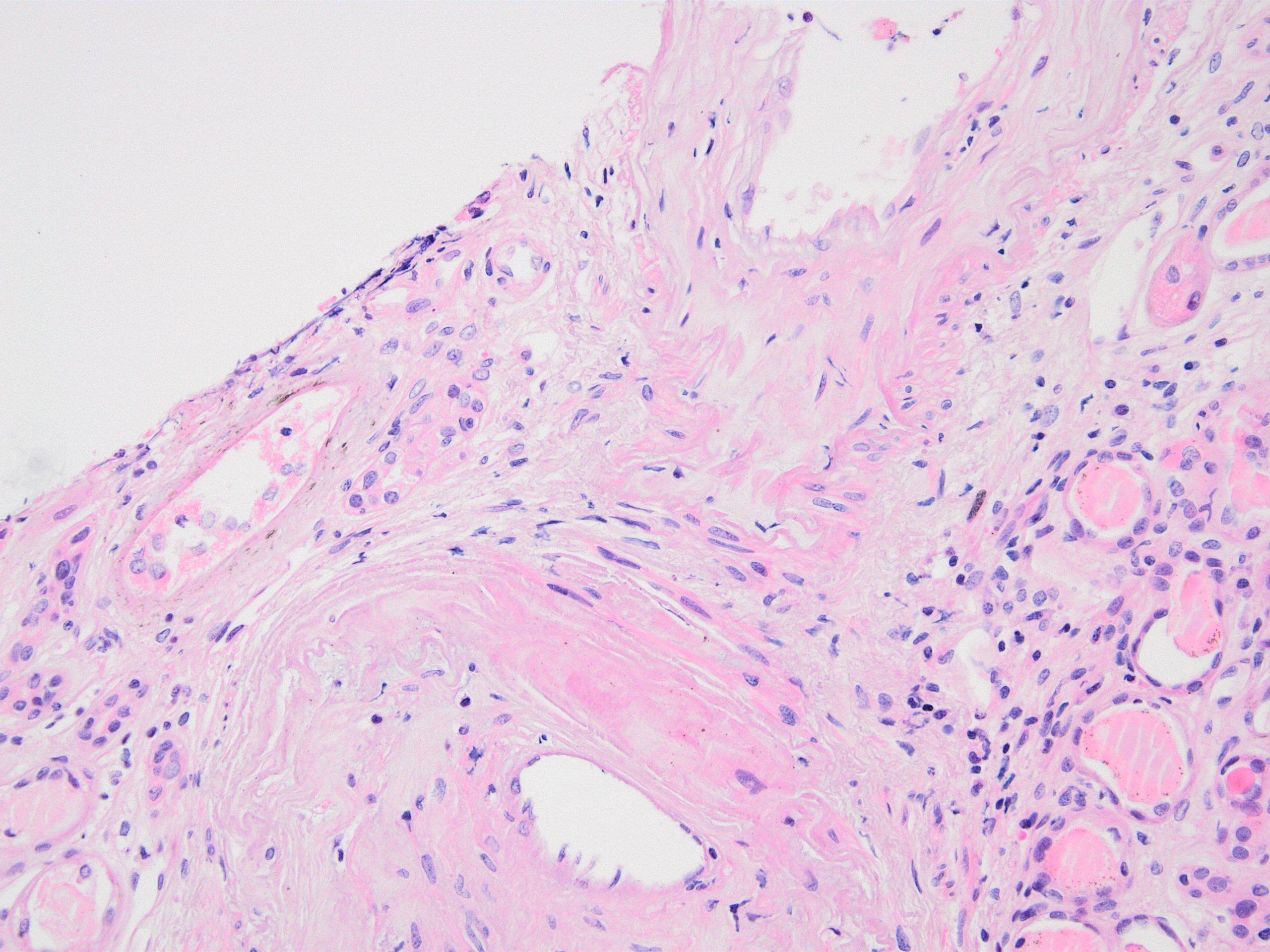
Double contours
Contributed by Christine M. Lee, M.D. and Jonathan E. Zuckerman, M.D., Ph.D.
Double contour
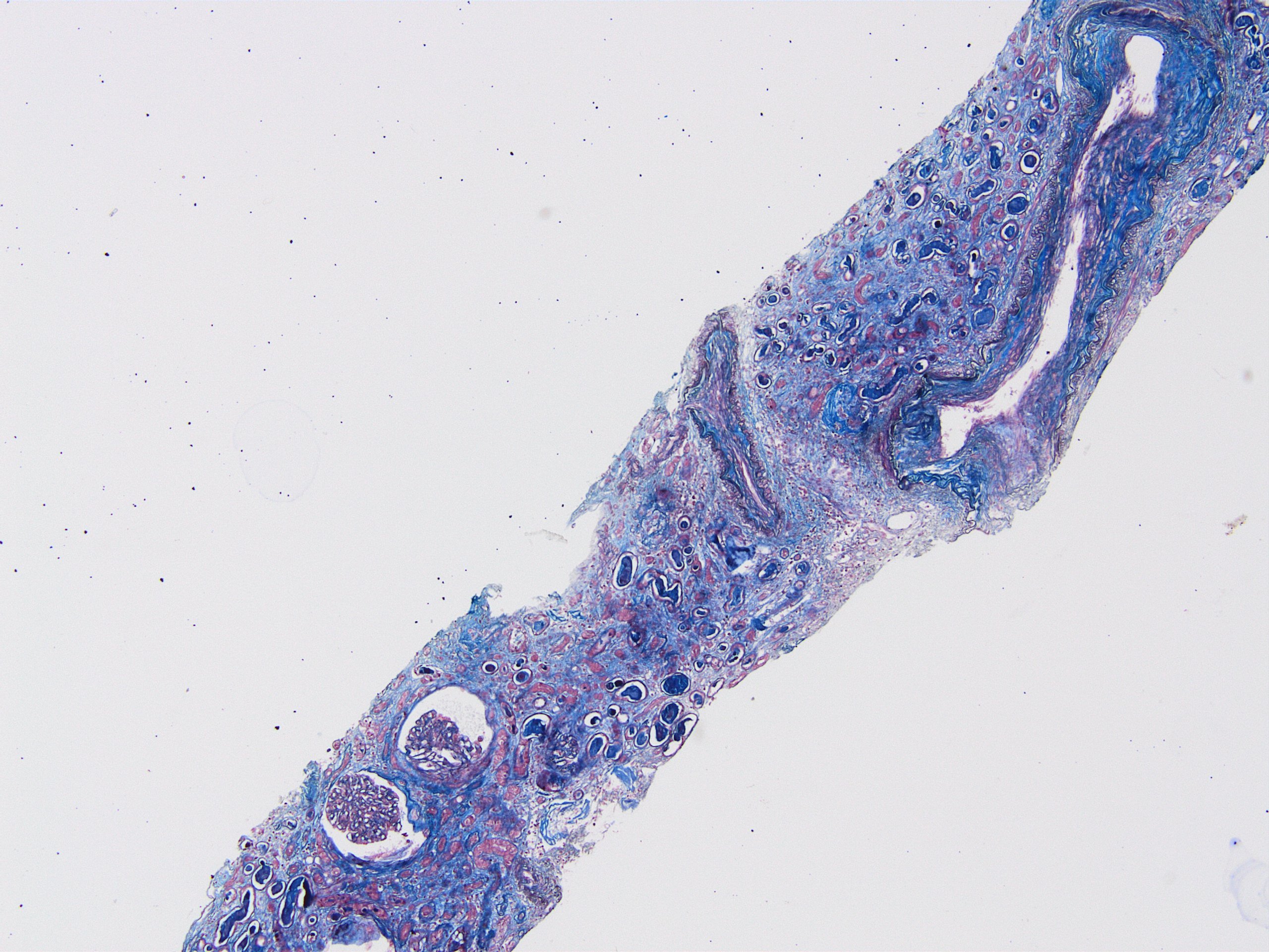
Artery remodeling
Contributed by Nicole K. Andeen, M.D.
Plasma cell rich tubulointerstitial nephritis (Jones stain)

Mesangial IgA deposition
Contributed by Nicole K. Andeen, M.D.

Mononuclear inflammatory infiltrate
Contributed by Jian-Hua Qiao, M.D.


Various images
Contributed by Jian-Hua Qiao, M.D.

Renal artery dissection
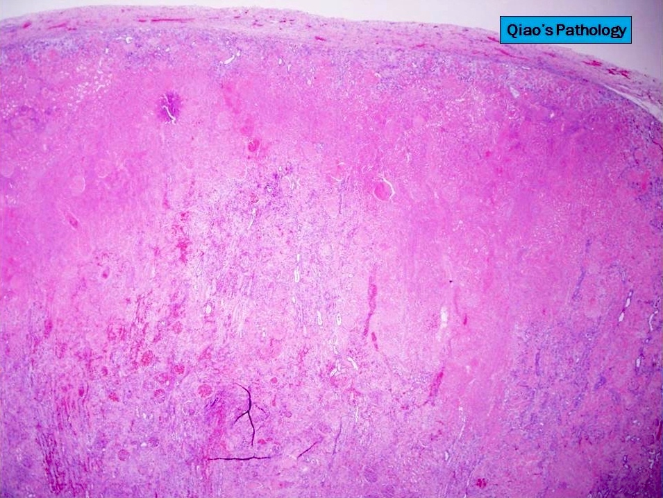


Renal cortex ischemic infarction

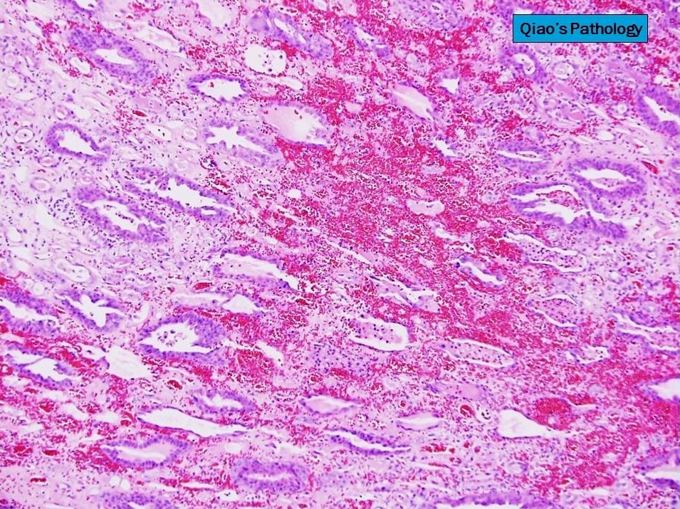
Renal papillae hemorrhagic infarction
Images hosted on other servers:

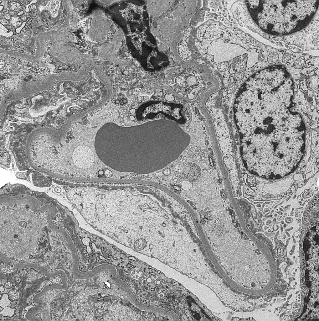
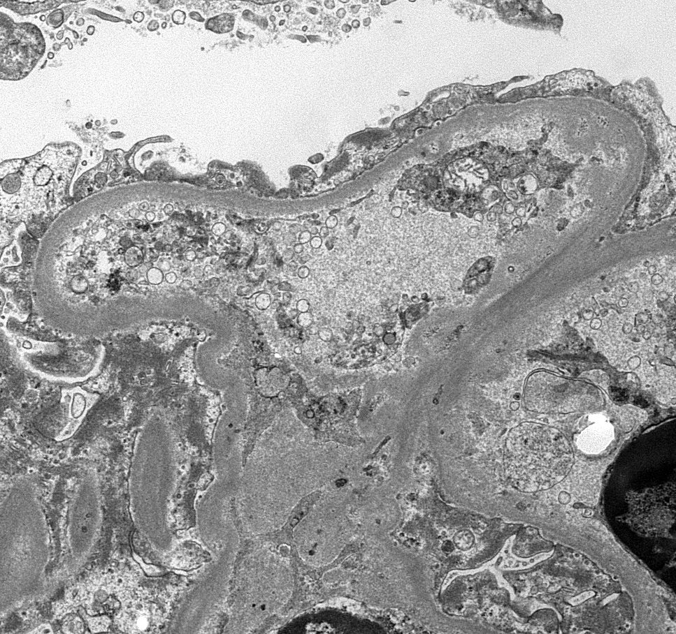
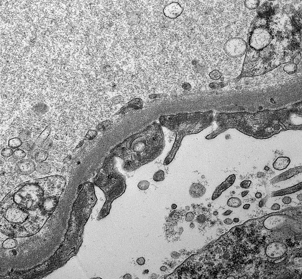
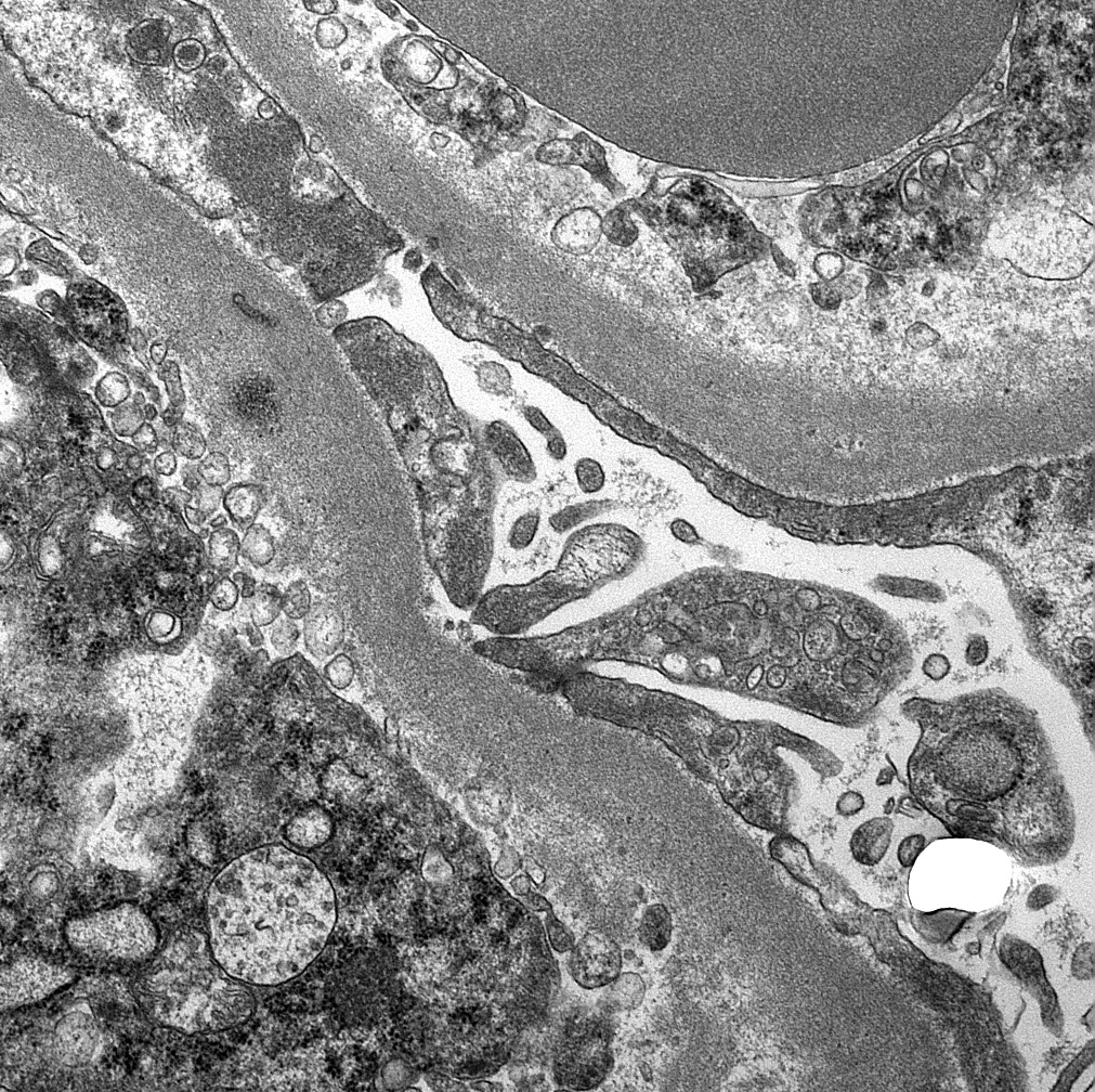
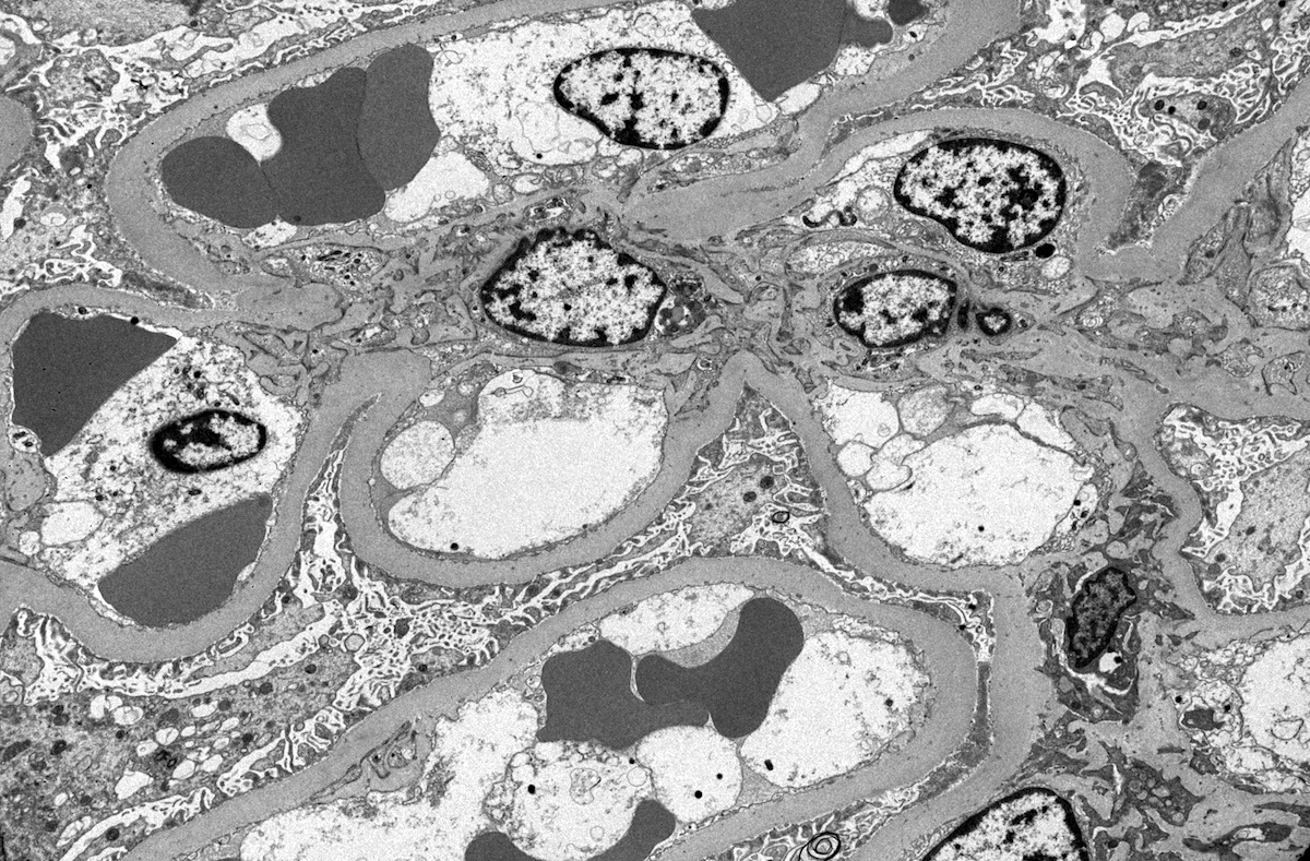
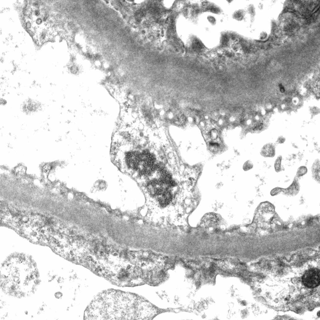
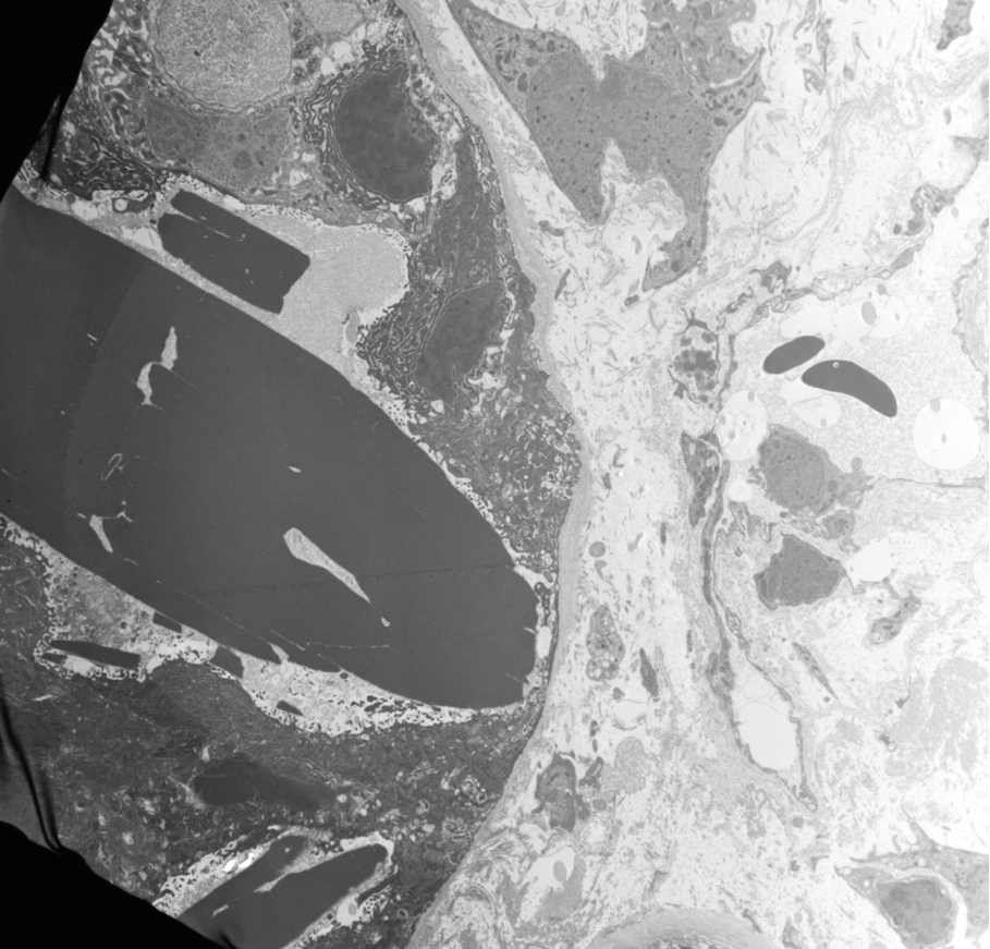
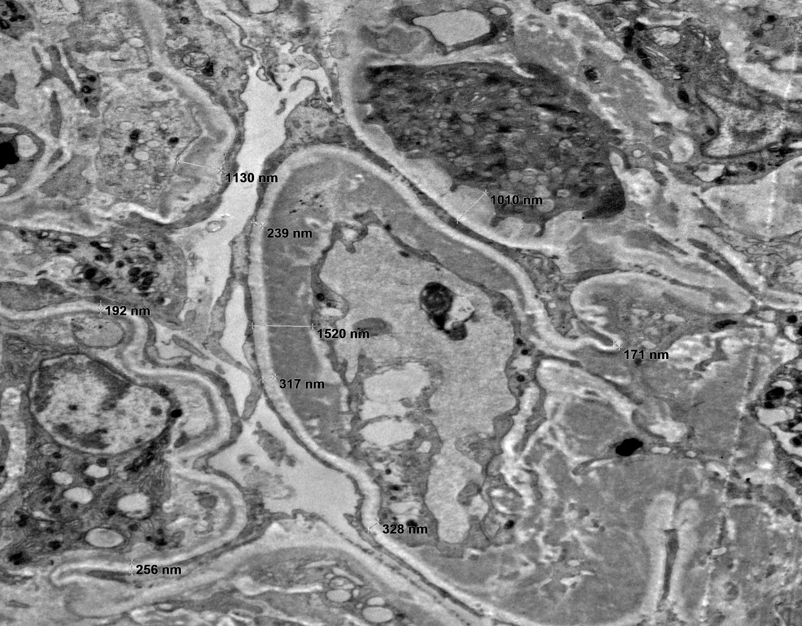
Ultrastructural features
of a single glomerular
capillary affected by
lupus nephritis
Images hosted on other servers:

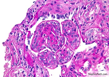
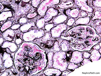
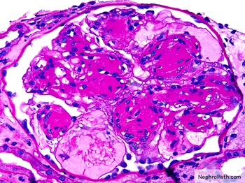
Diffuse proliferative lupus nephritis
Contributed by Alexei Mikhailov, M.D., Ph.D.
LN II: segmental mesangial hypercellularity
LN III: segmental fibrinoid necrosis
LN III: segment with karyorrhexis

LN IV: renal vasculitis

LN IV: endocapillary hypercellularity
LN IV: wire loops, hyaline thrombi

LN IV: vasculopathy

LN IV: torn capillaries

LN V: membranous alterations
LN V: microspikes
Contributed by Alexei Mikhailov, M.D., Ph.D.

LN IV: immune deposits
LN IV: tubuloreticular inclusions

LN IV: organized deposits
Images hosted on other servers:

Pathogenesis
Images hosted on other servers:


ECG gated double inversion recovery


Fast gradient echo


MR angiography
Contributed by Nicole K. Andeen, M.D.
White lesion
Contributed by Nicole K. Andeen, M.D.
Active vasculitis with giant cells
Tubulointerstitial inflammation
Jones
Elastic
Images hosted on other servers:

Diagram of Tamm-Horsfall protein secretion (green dots), forming a hyaline cast in the collecting duct
Images hosted on other servers:

Nephronophthisis: end stage renal disease

Lymphatics


Pyelonephritis - protein cylinder

Pyelonephritis immunostain
Images hosted on other servers:

In lymphatics
Images hosted on other servers:

Various images

no abnormalities
on H&E


Images hosted on other servers:

Diagnosis of
inherited interstitial
kidney disease
Images hosted on other servers:

Drug induced tubulointerstitial nephritis
Contributed by Sam T. Albadri, M.B.Ch.B., M.Sc.

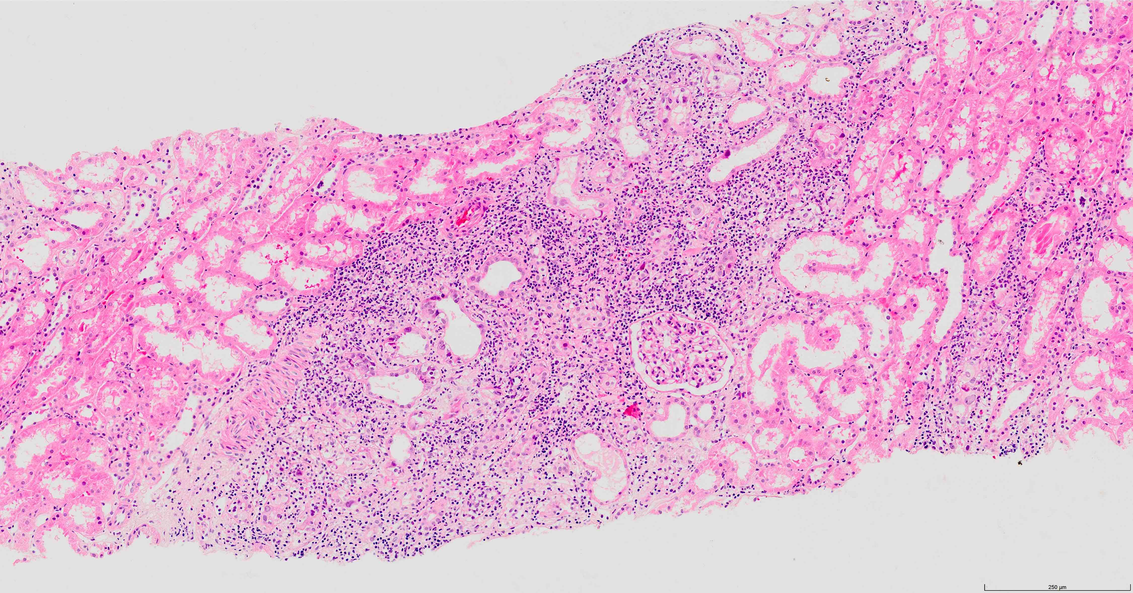
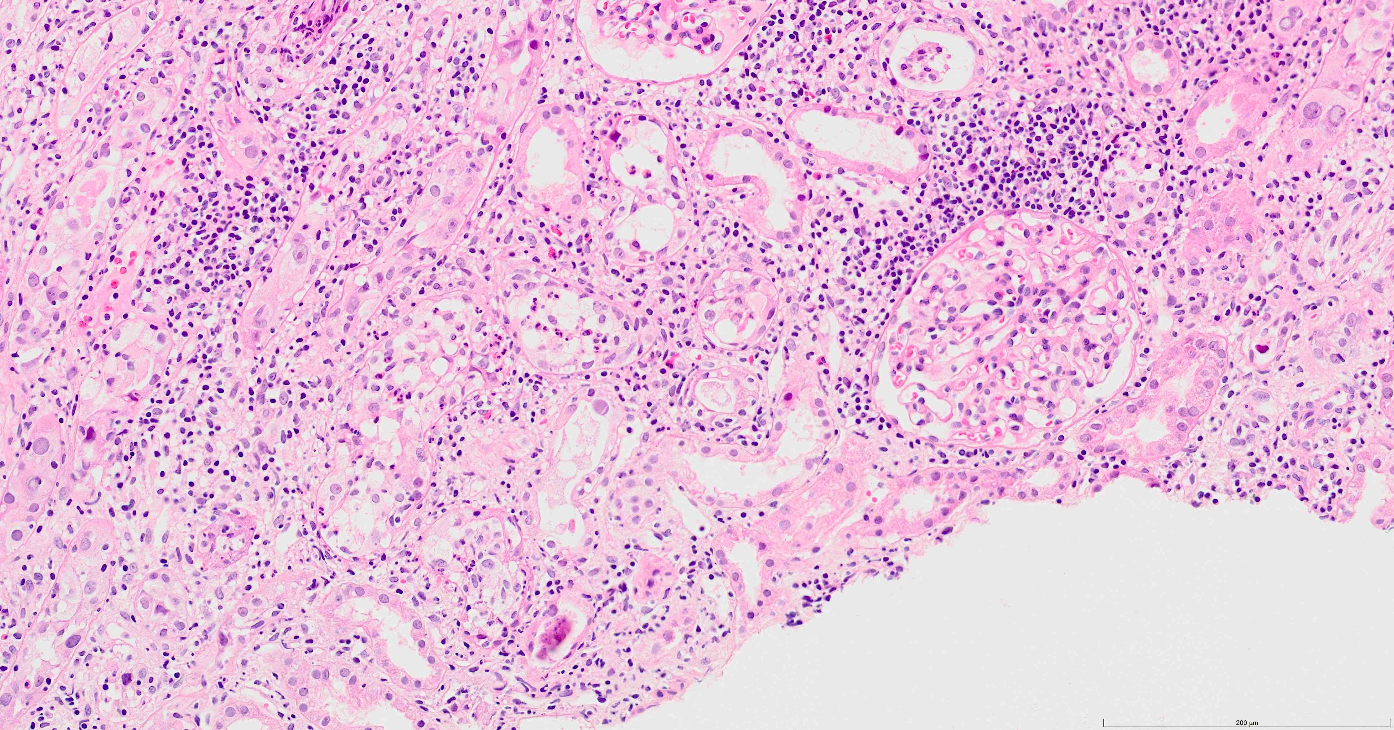
Interstitial inflammation
Tubulitis
Granulomatous interstitial nephritis
Eosinophilic tubulitis
IgG4 related kidney disease
IgG
IgG4 immunostain
Contributed by Sam T. Albadri, M.B.Ch.B., M.Sc.
Interstitial inflammation and tubulitis
Histopathology kidney - interstitial nephritis
Images hosted on other servers:


Chronic urate nephropathy
Images hosted on other servers:


Gouty nephropathy
Images hosted on other servers:

Passage of a calculus (stone)
Contributed by Jian-Hua Qiao, M.D.
Multiple kidney stones
Contributed by Alexandra Barabasch, M.D.
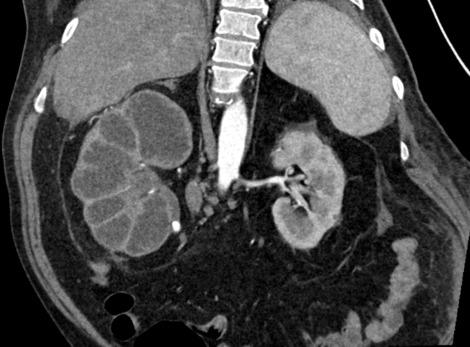

Renal parenchyma on CT
Dilated renal pelvis on CT
Contributed by Saskia von Stillfried, M.D.

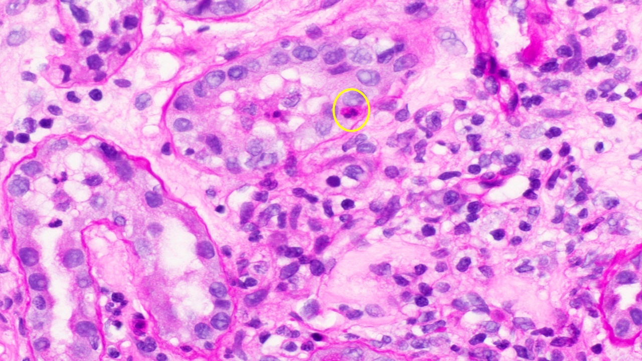
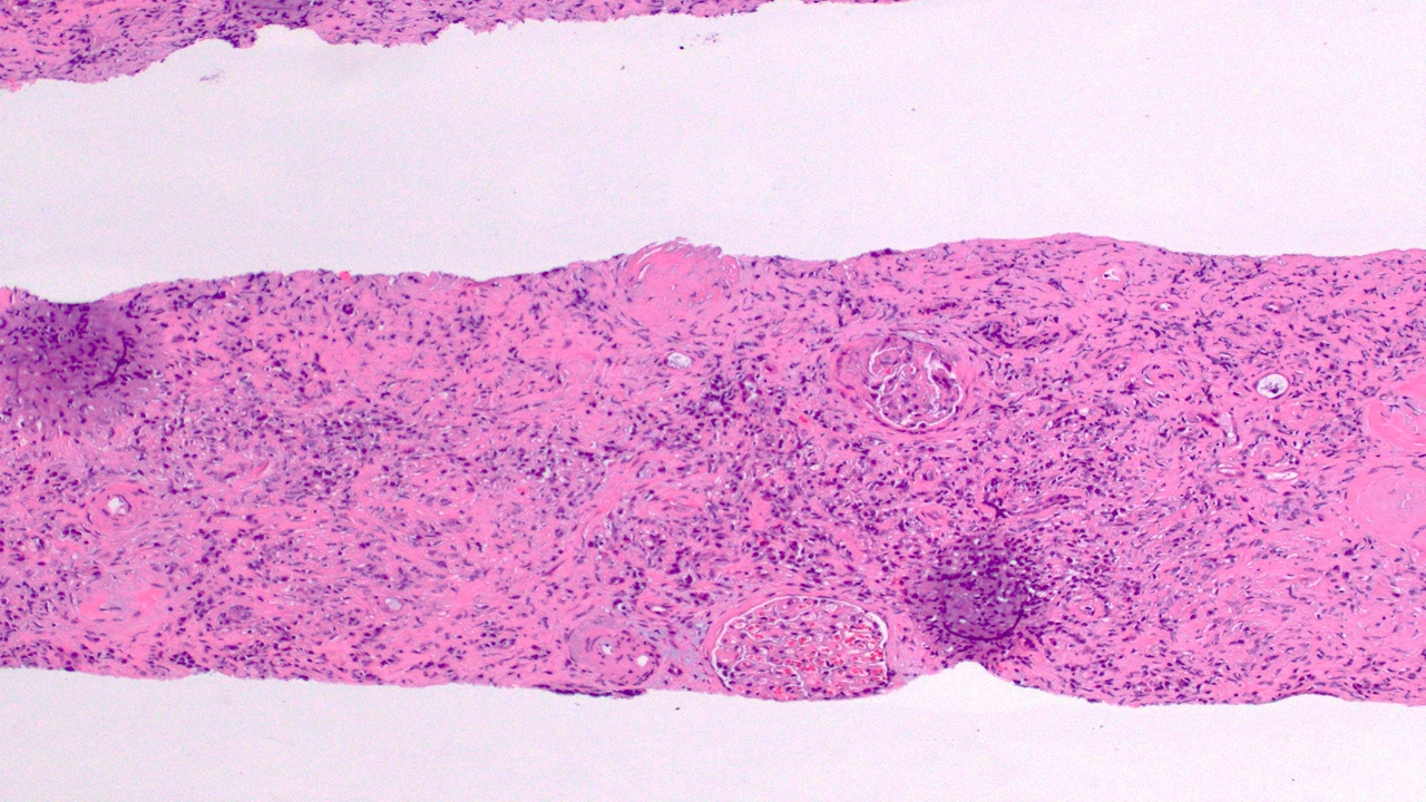
Macroscopic appearance
Contributed by Saskia von Stillfried, M.D.
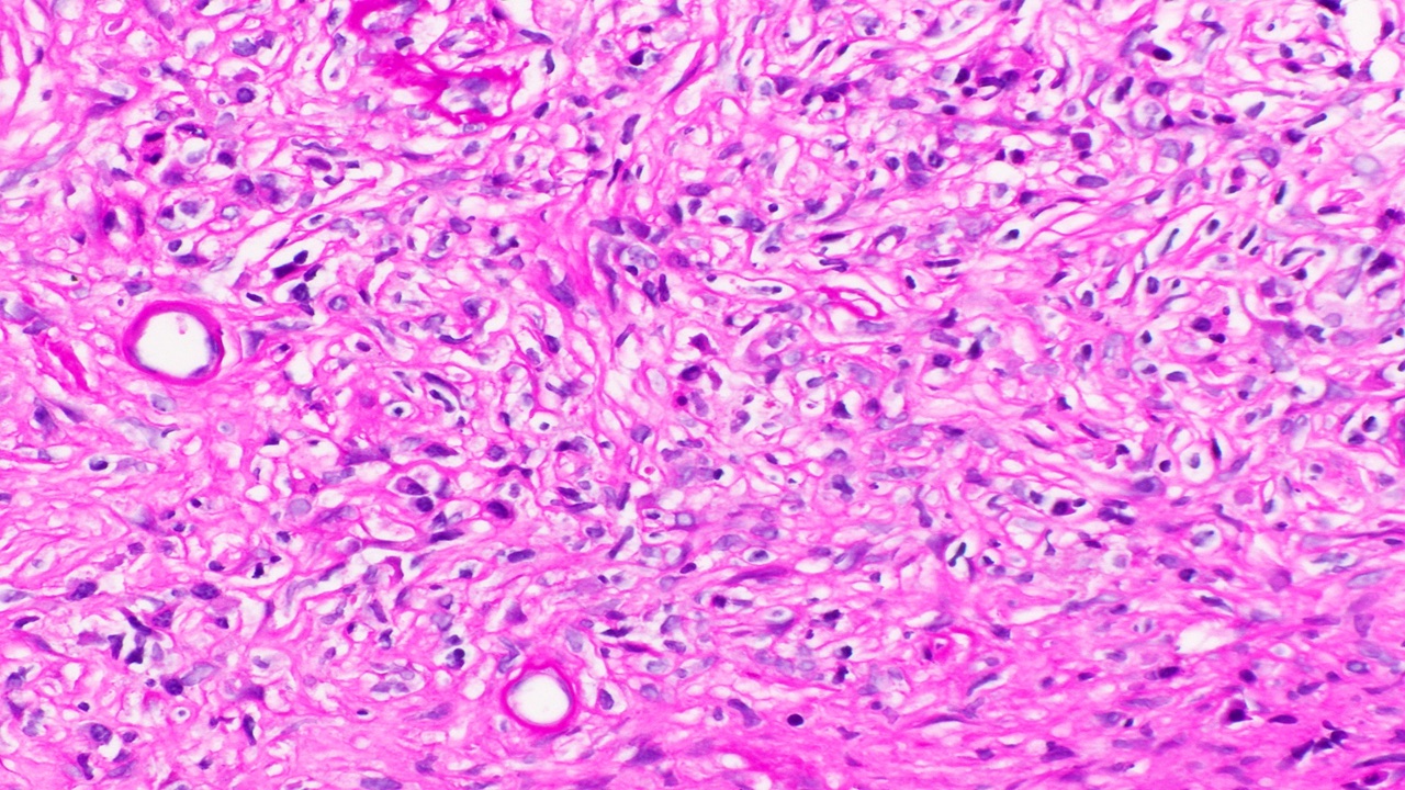
Zonal structure
Middle zone
Xanthoma cells
Touton giant cell
Cholesterol clefts
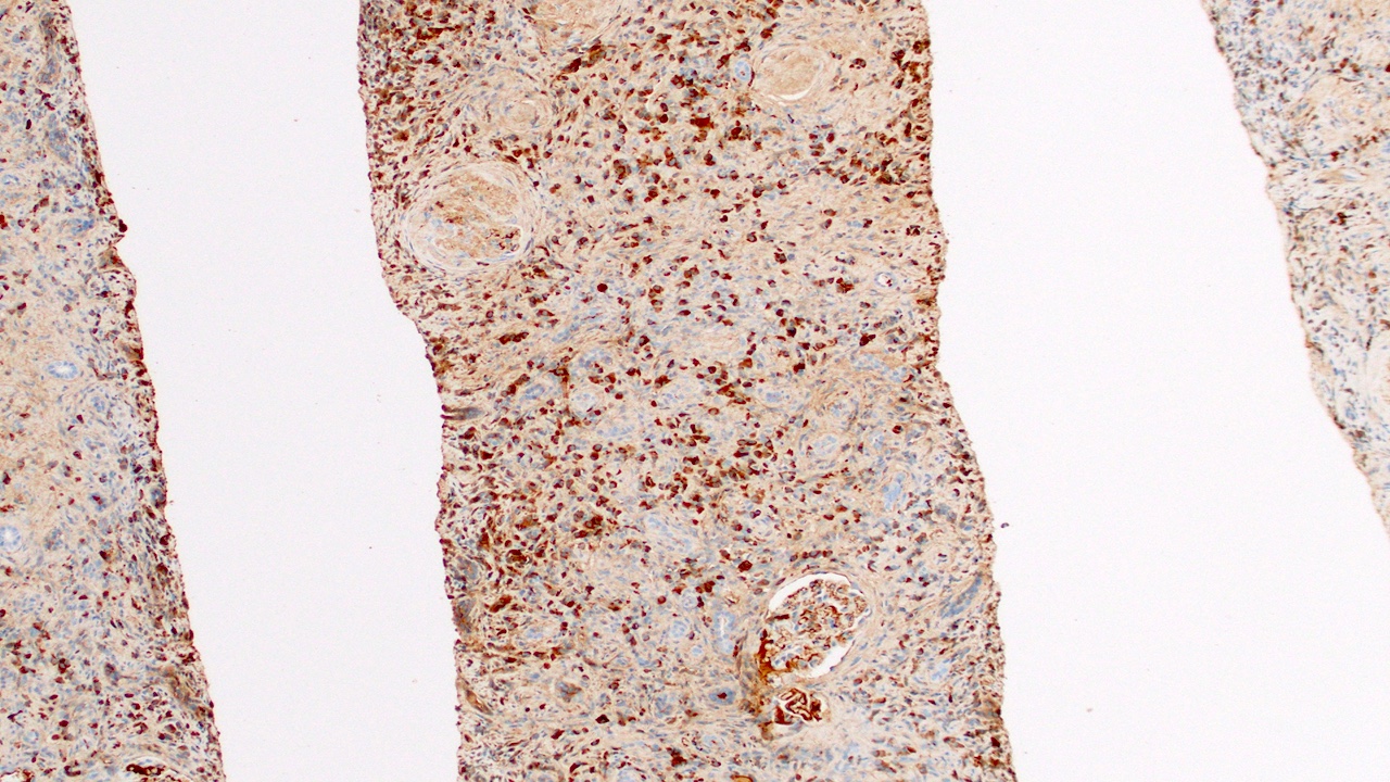
CD68+ macrophages (xanthoma cells)

Pankeratin positive tubuli and negative macrophages
Chang: 2018
D'Agati: 2005
Fatima: 2019
Husain: 2021
Jennette: 2015
Jiang: 2023
Satoskar: 2017
Scorecki: 2015
Zhou: 2017
Find related Pathology books: cardiovascular, cytopathology, hematopathology, renal, liver, lung, pediatric
