
Images hosted on other servers:

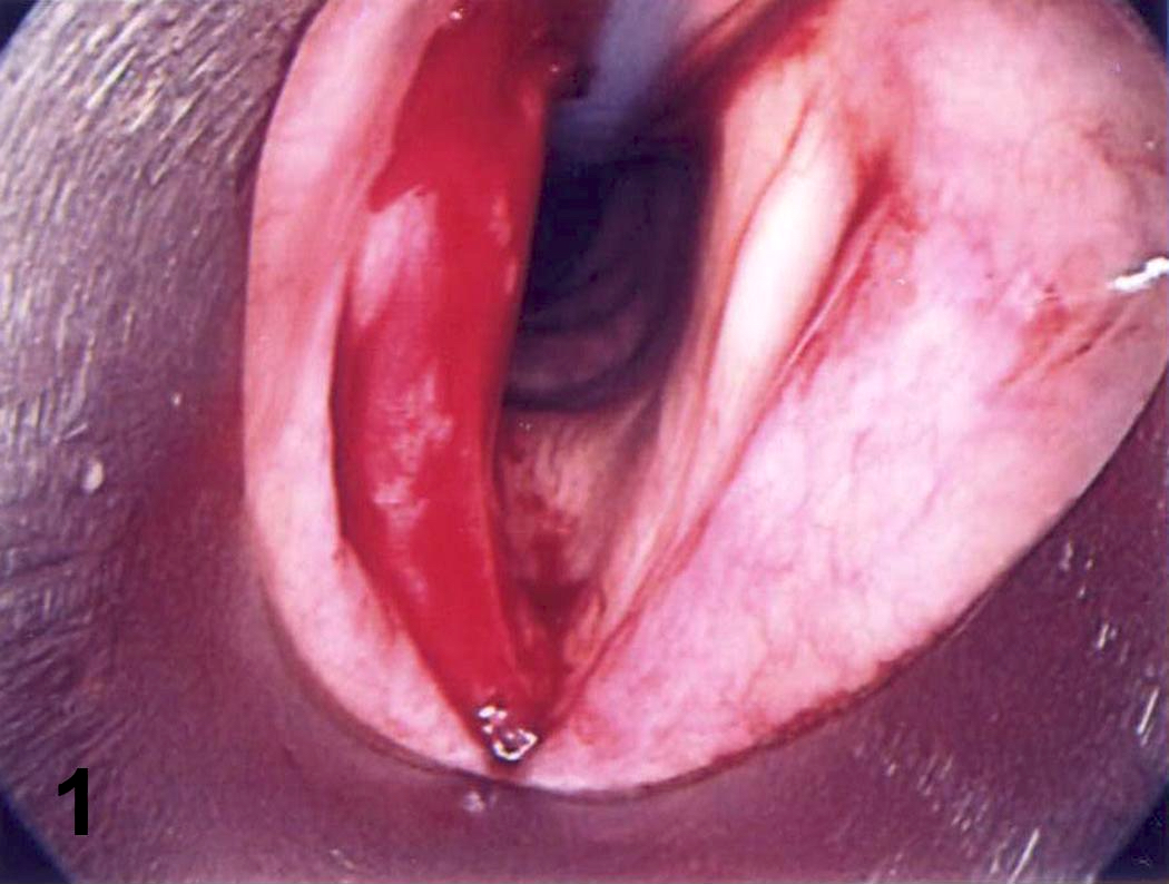
Bulging vocal cord mass
Contributed by Vancouver General Hospital
Characteristic histologic features
Involvement of the trachea
Images hosted on other servers:

Apolipoprotein AI and transthyretin amyloidosis
Images hosted on other servers:


Homogenous proteinous material with calcification under bronchus epithelium is Congo red+

Massive amyloid deposits

69 year old woman with seropositive erosive RA
Images hosted on other servers:

Sagittal section

Coronal section

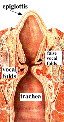
Entrance viewed from above

Laryngoscopic view

Laryngeal cartilages

Vocal cords and muscles

Muscles


Ligaments

Trachea and bronchi

Tracheal bifurcation

Transverse section
Images hosted on other servers:
True and false vocal cords
Images hosted on other servers:
True and false vocal cords

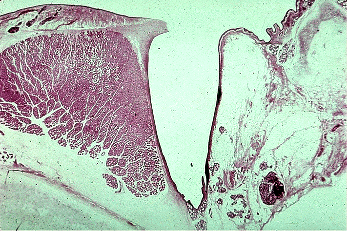
Transverse section of trachea
Contributed by Steven Catinchi-Jaime, M.D.
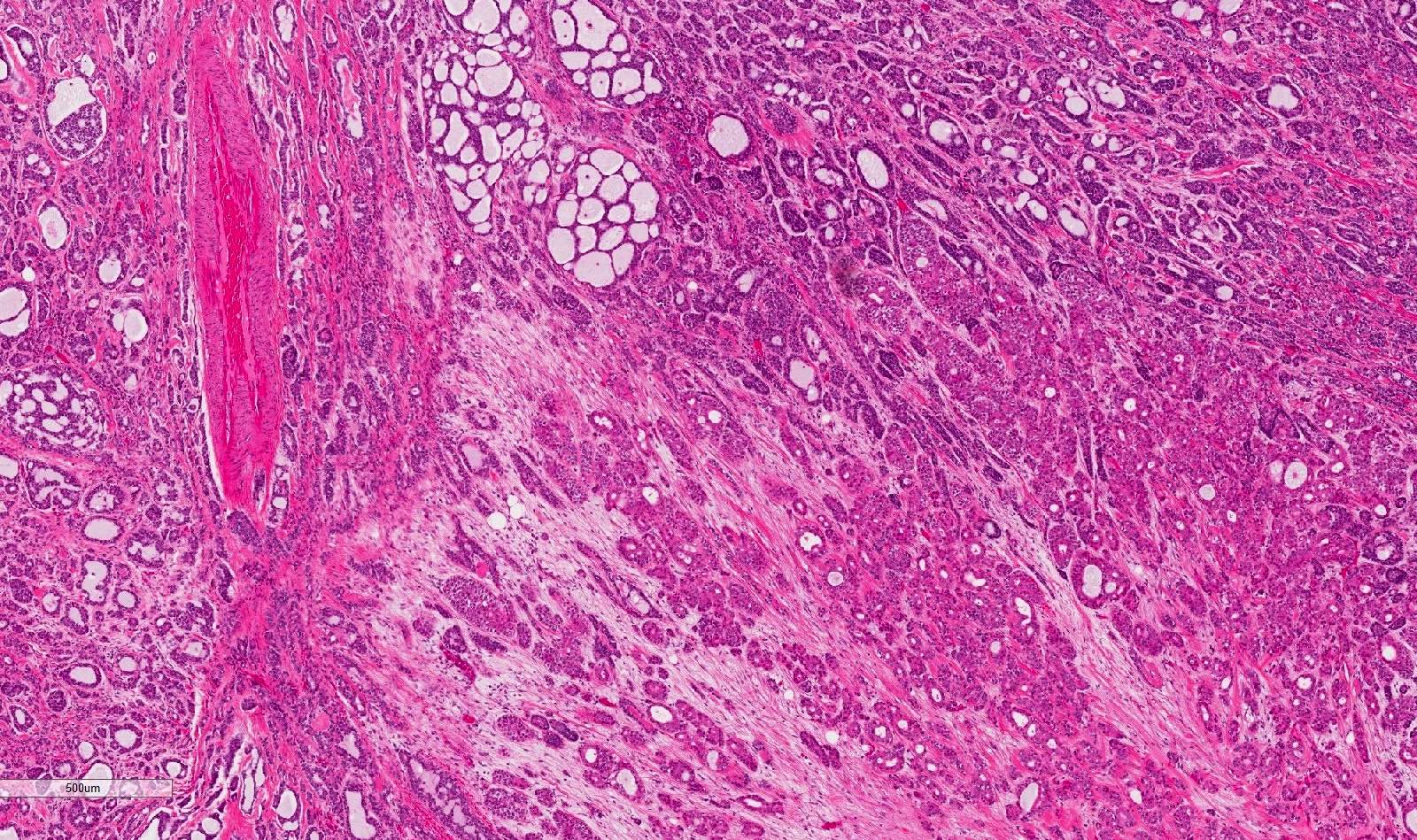
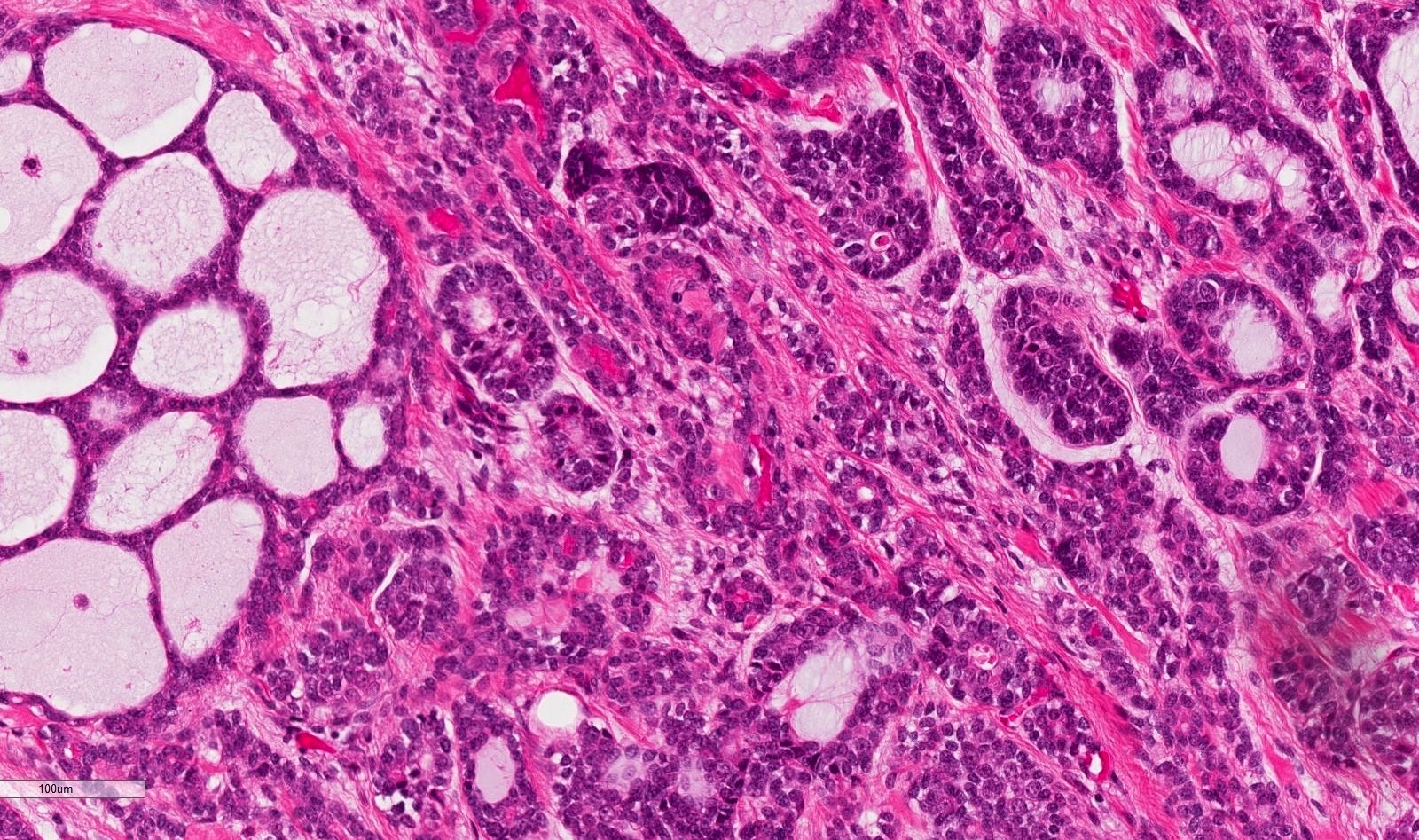
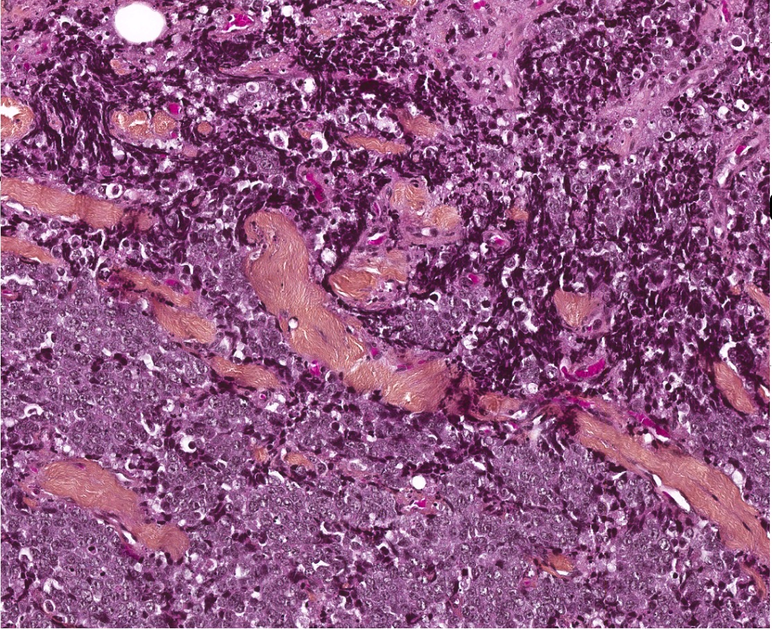
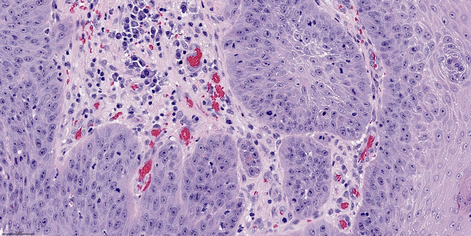
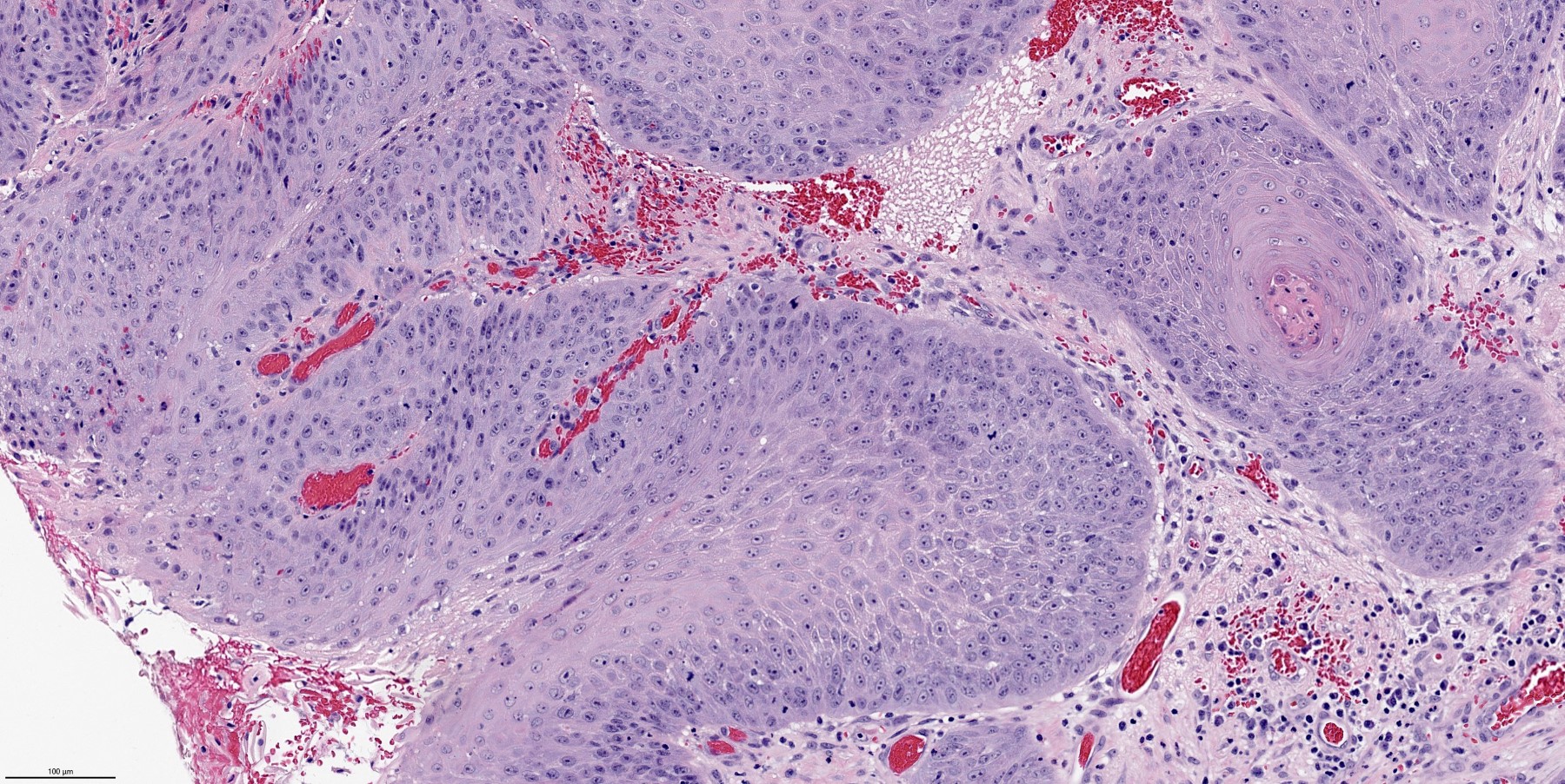
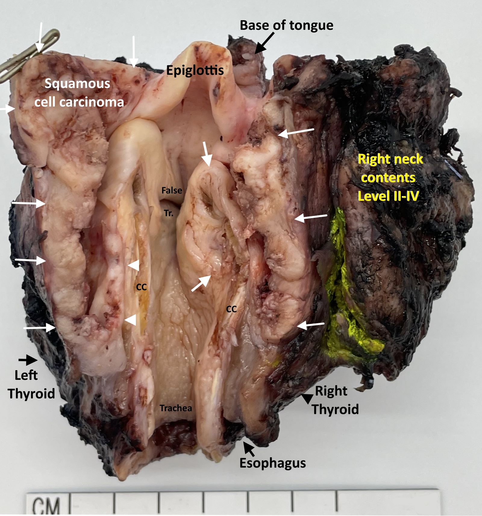
Malignant fibrous histiocytoma and squamous cell carcinoma arising in a vocal chord
Contributed by Steven Catinchi-Jaime, M.D.
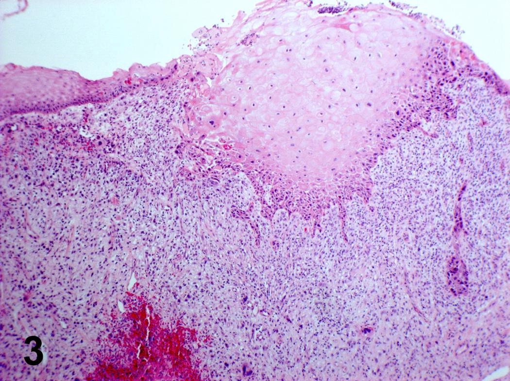
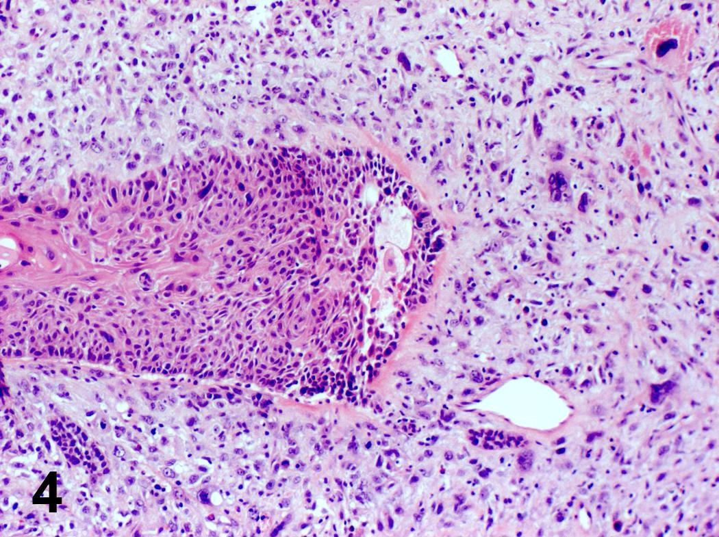
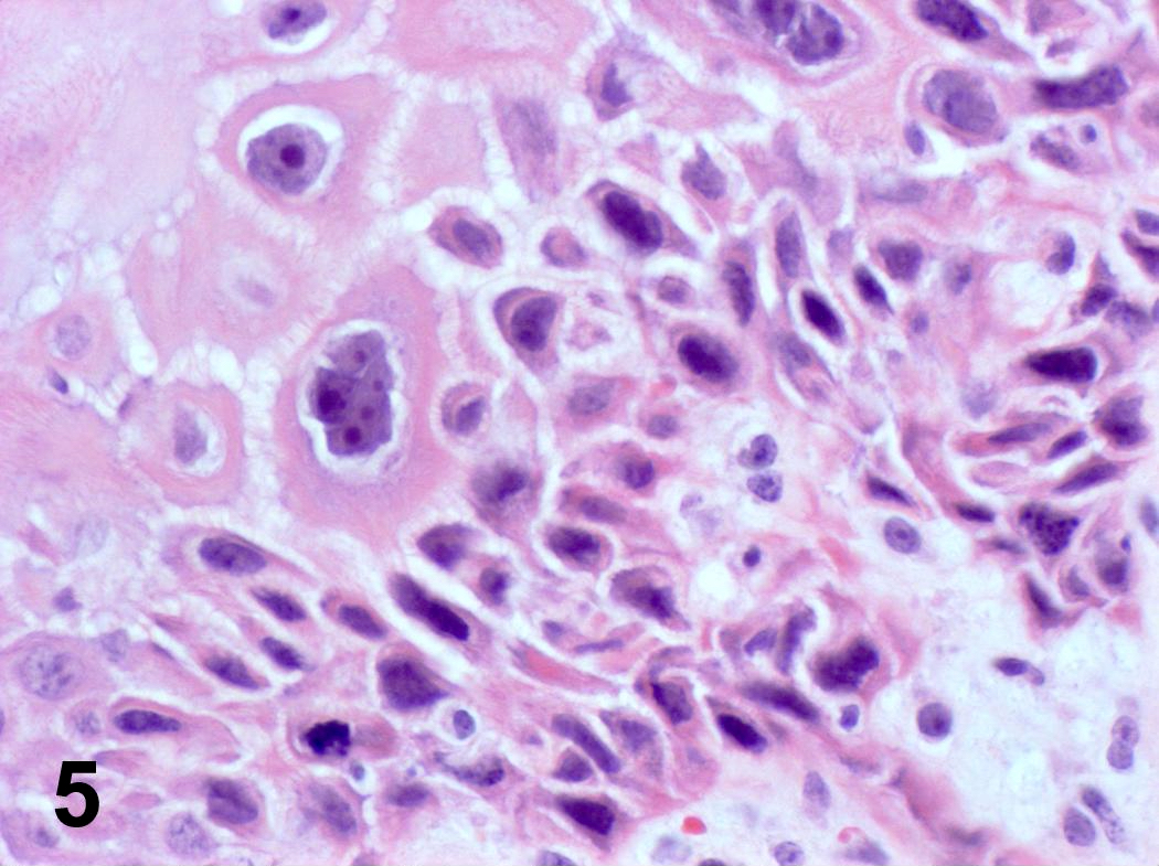
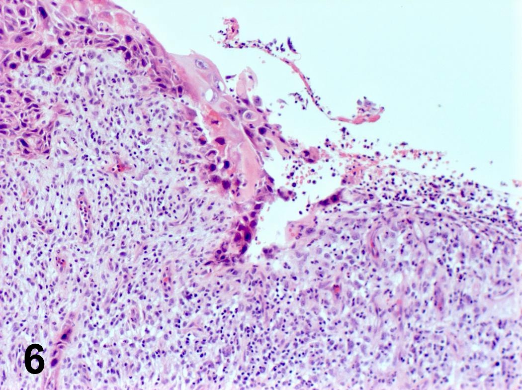
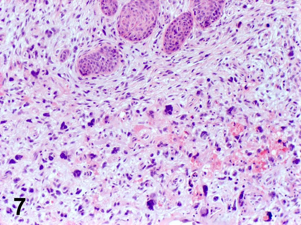
Malignant fibrous histiocytoma and squamous cell carcinoma arising in a vocal chord
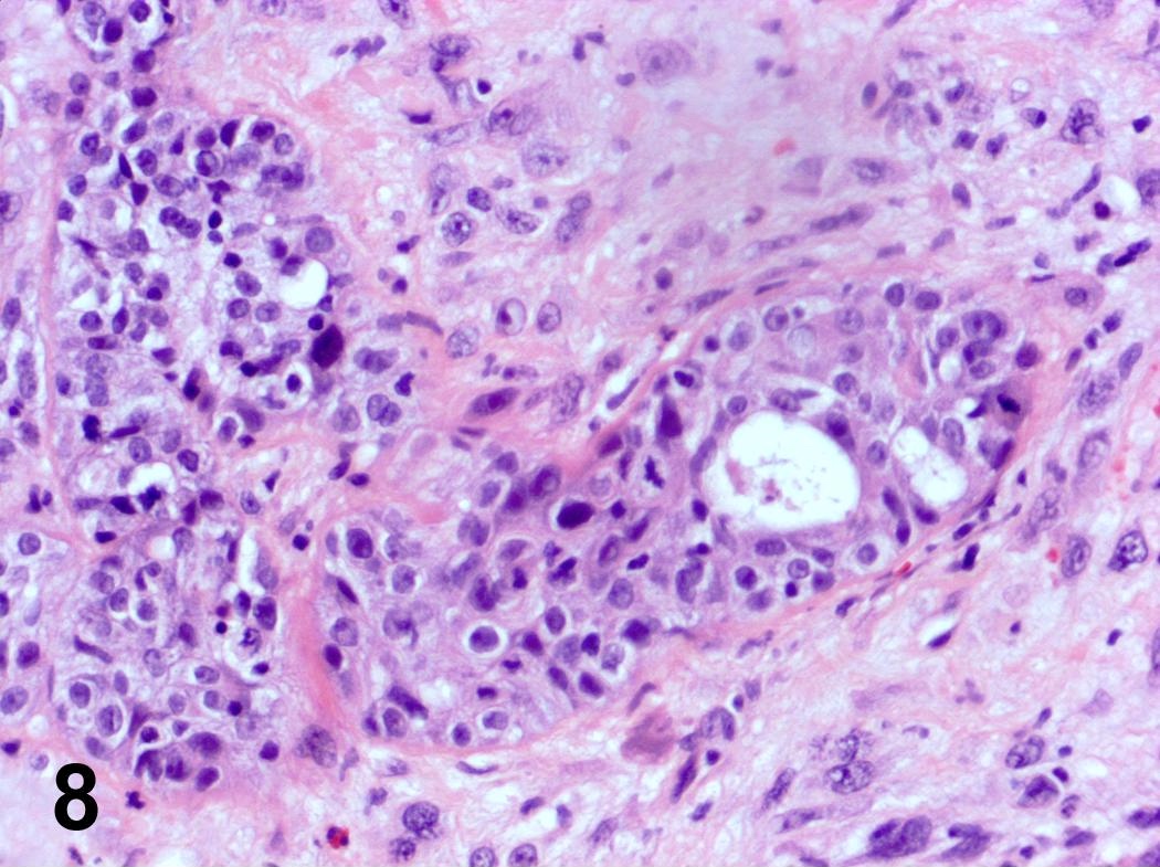
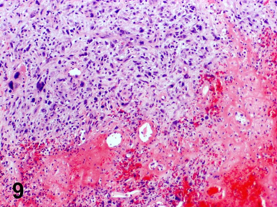
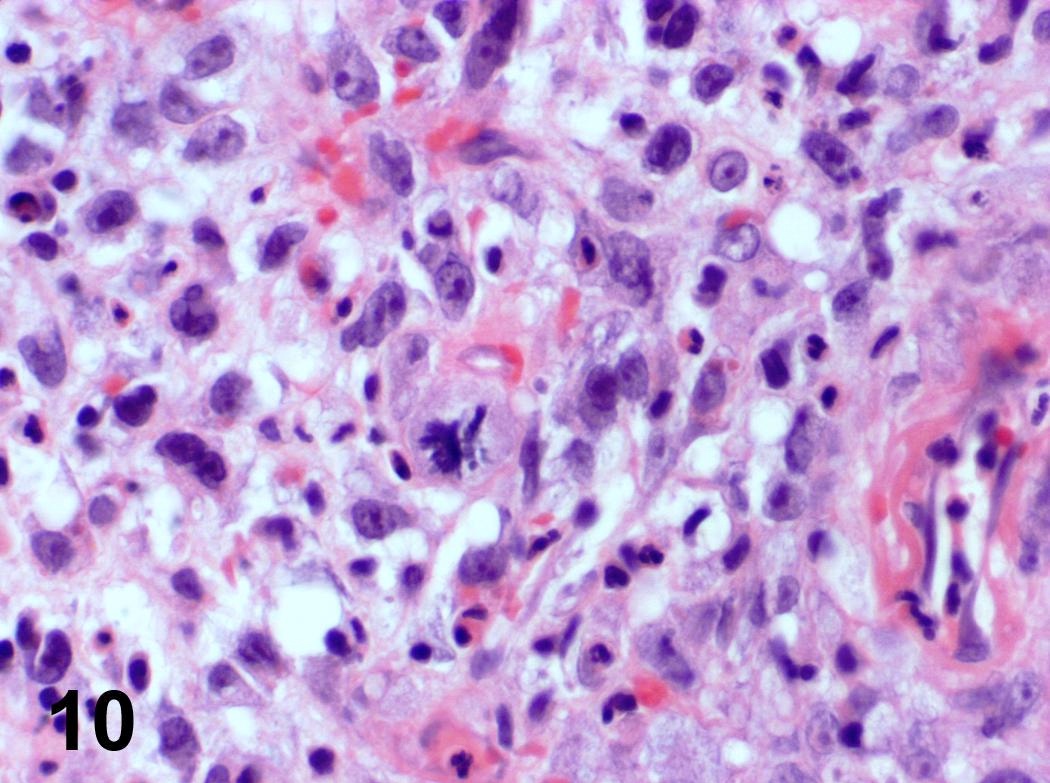
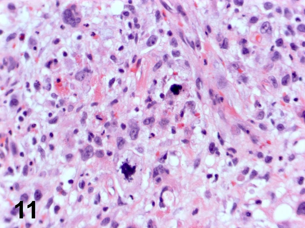
Malignant fibrous histiocytoma and squamous cell carcinoma arising in a vocal chord
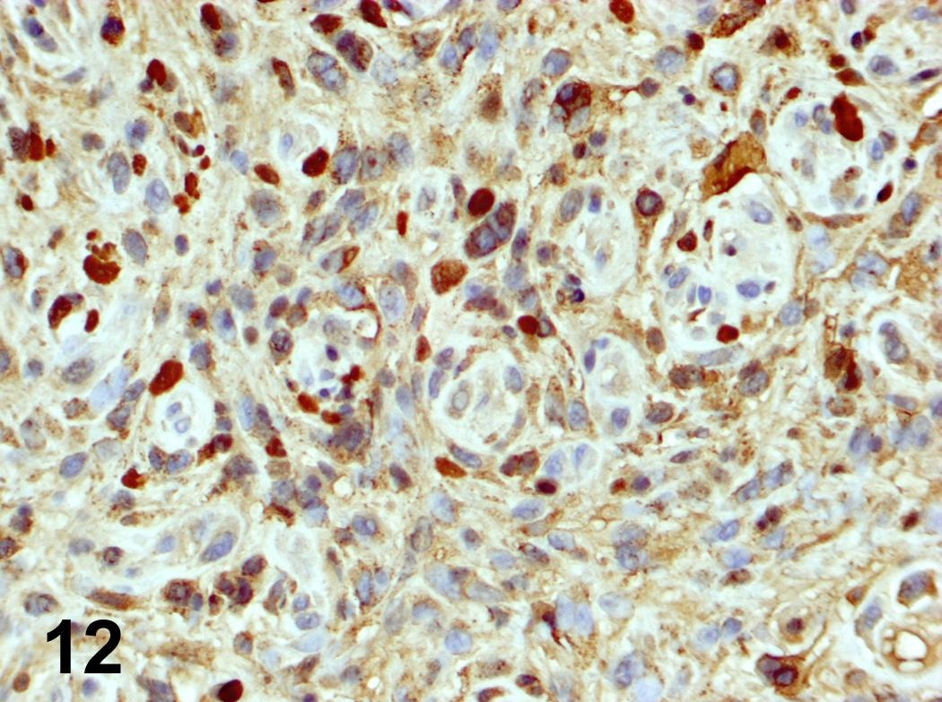
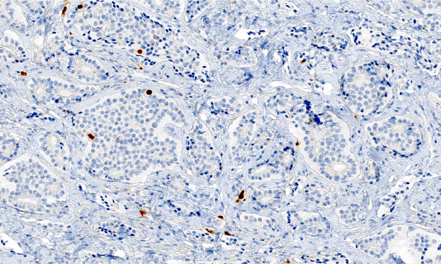
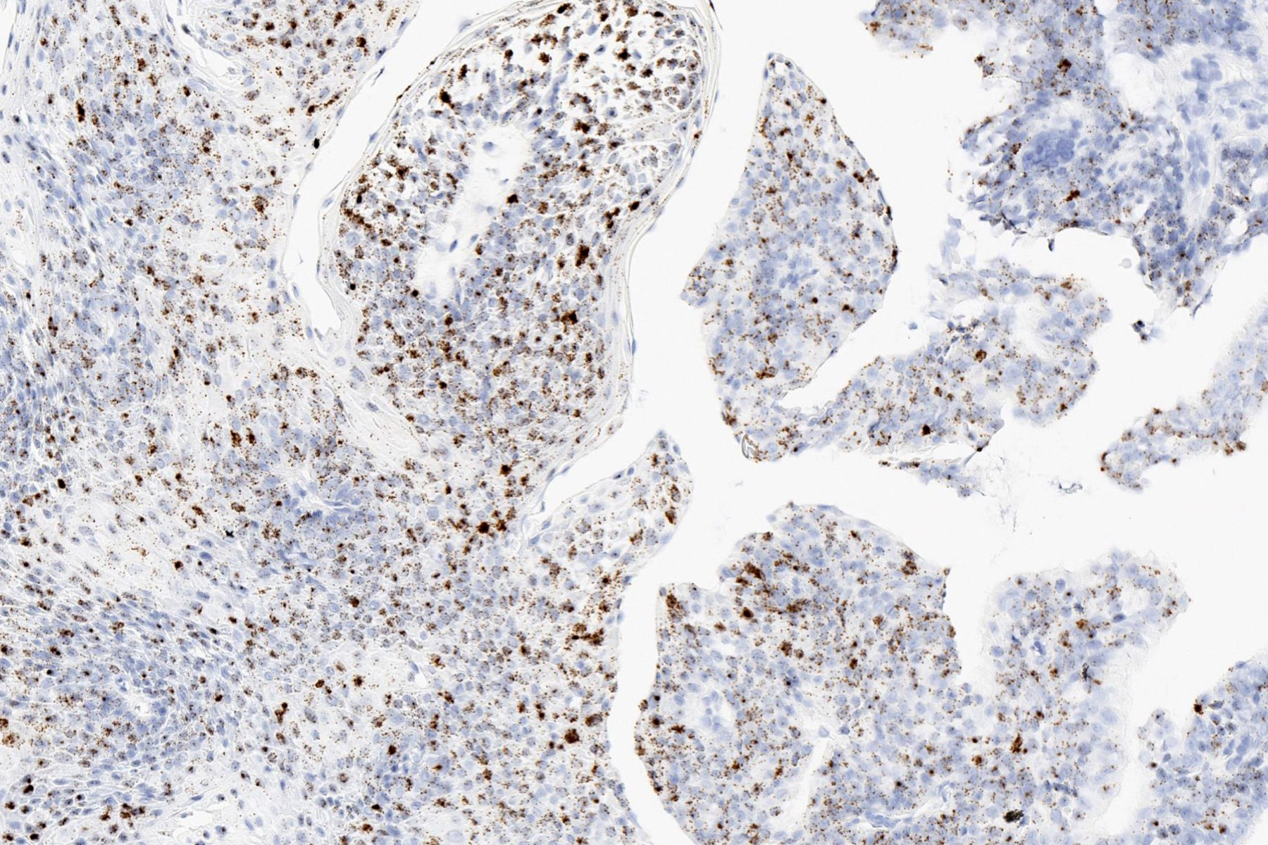
Alpha-1-antichymotrypsin
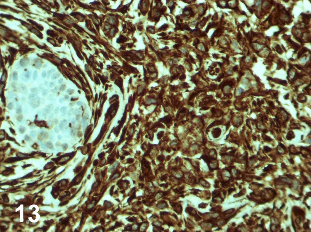
Vimentin
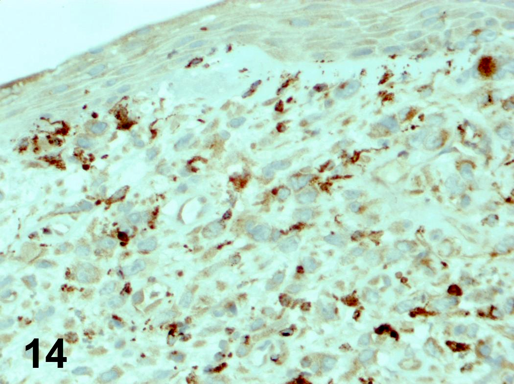
CD68
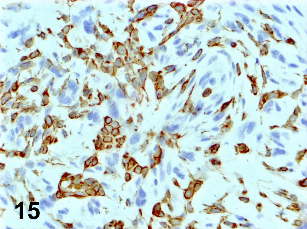
Cytokeratin AE1 / AE3
Images hosted on other servers:

Basaloid squamous cell carcinoma

Various images
Images hosted on other servers:

6 year old child's trachea
and major bronchi with mucosa
damaged by toxin produced by
Corynebacterium diphtheriae
Images hosted on other servers:

Coronal cross section of larynx showing saccule
Images hosted on other servers:

MRI
Images hosted on other servers:


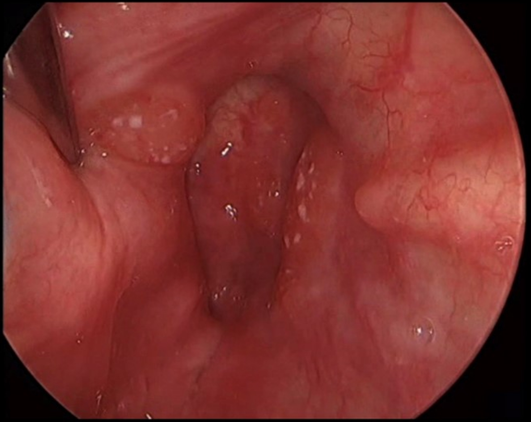
Spherical bulges covered by smooth epithelium
Images hosted on other servers:

Cyst in glottic region
Contributed by Arpita Jindal, M.D.
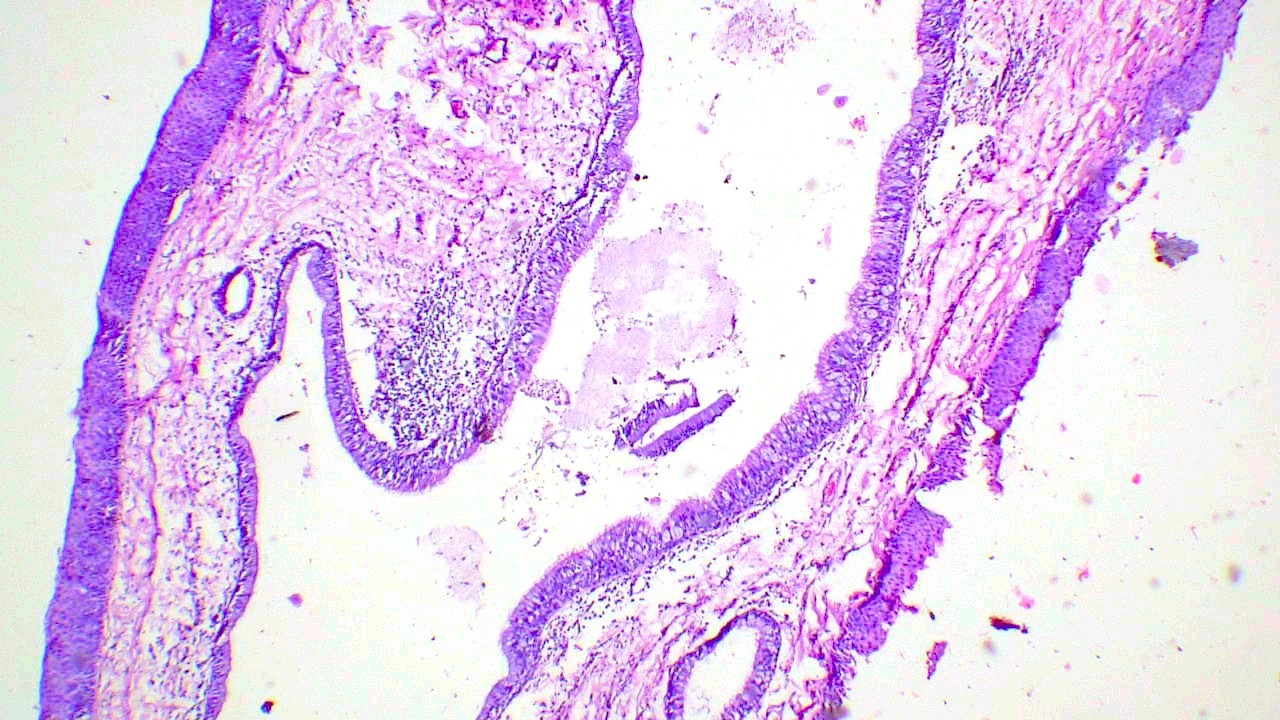
Pseudostratified columnar epithelium lined cyst

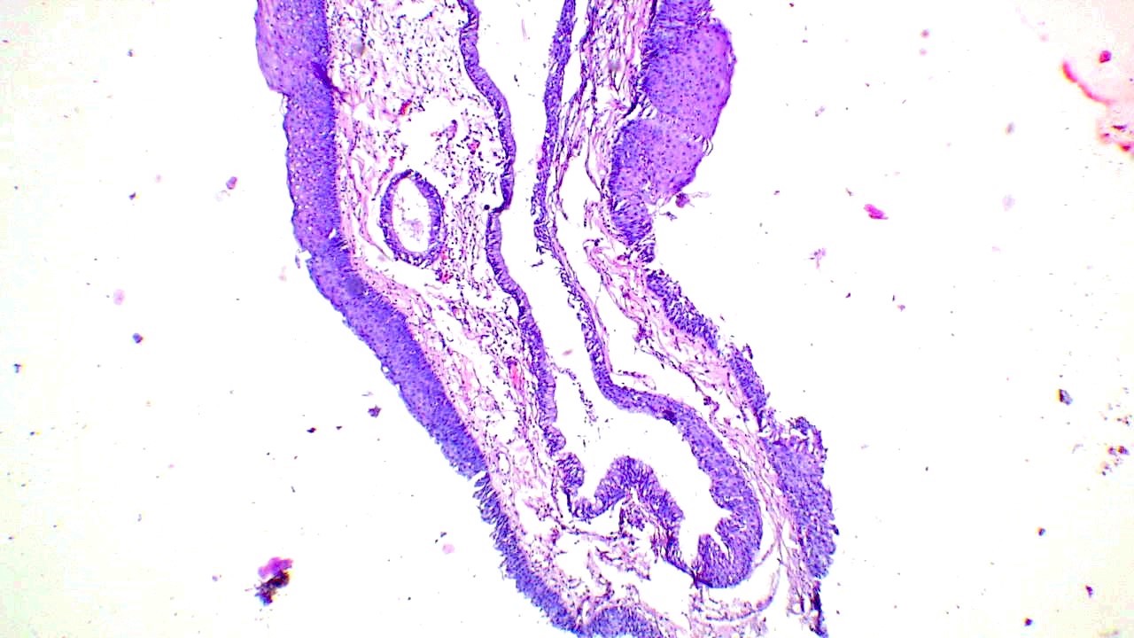
Cyst lining showing squamous metaplasia

Intraluminal infolding with focal lymphoid collection
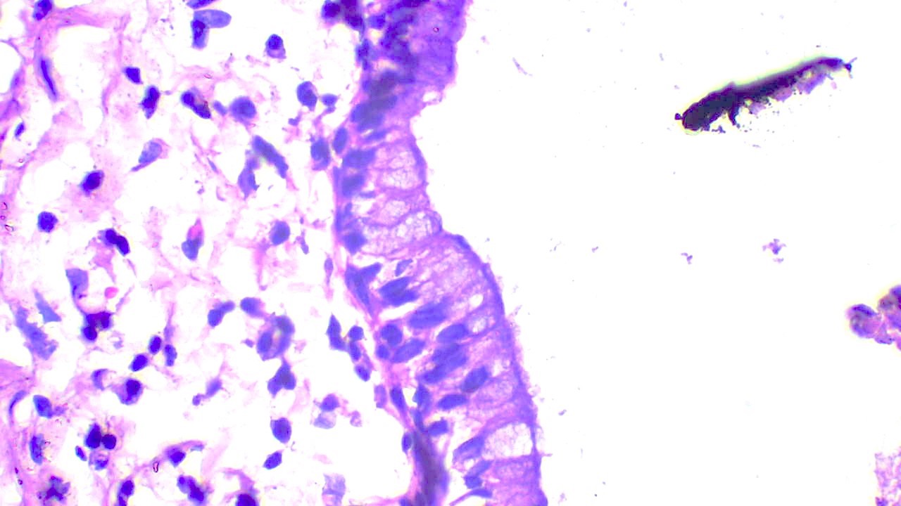
Columnar mucinous lining, basally located nuclei
Pseudostratified columnar epithelium with cilia
Images hosted on other servers:
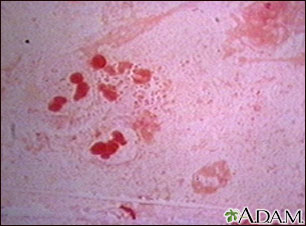
Acute laryngoepiglottitis: H. influenza (site unknown)
Table 1: WHO 2022 epithelial neuroendocrine neoplasms of the upper aerodigestive tract and salivary glands (Head Neck Pathol 2022;16:123)
| Neuroendocrine neoplasm | Tumor category | Diagnostic criteria |
| Well differentiated neuroendocrine neoplasm (neuroendocrine tumor, NET) | Well differentiated neuroendocrine tumor, grade 1 (NET, G1) | No necrosis and < 2 mitoses/2 mm²
Ki67 < 20% |
| Well differentiated neuroendocrine tumor, grade 2 (NET, G2) | Necrosis or 2 - 10 mitoses/2 mm²
Ki67 < 20% | |
| Well differentiated neuroendocrine tumor, grade 3 (NET, G3) | > 10 mitoses/2 mm²
Ki67 > 20% Absence of NEC cytomorphology | |
| Poorly differentiated neuroendocrine neoplasm (neuroendocrine carcinoma, NEC) | Small cell neuroendocrine carcinoma | > 10 mitoses/2 mm²
Ki67 > 20% (often > 70%) Small cell NEC cytomorphology |
| Large cell neuroendocrine carcinoma | > 10 mitoses/2 mm²
Ki67 > 20% (often > 50%) Large cell NEC cytomorphology |
Contributed by Nazim Benzerdjeb, M.D., Ph.D.
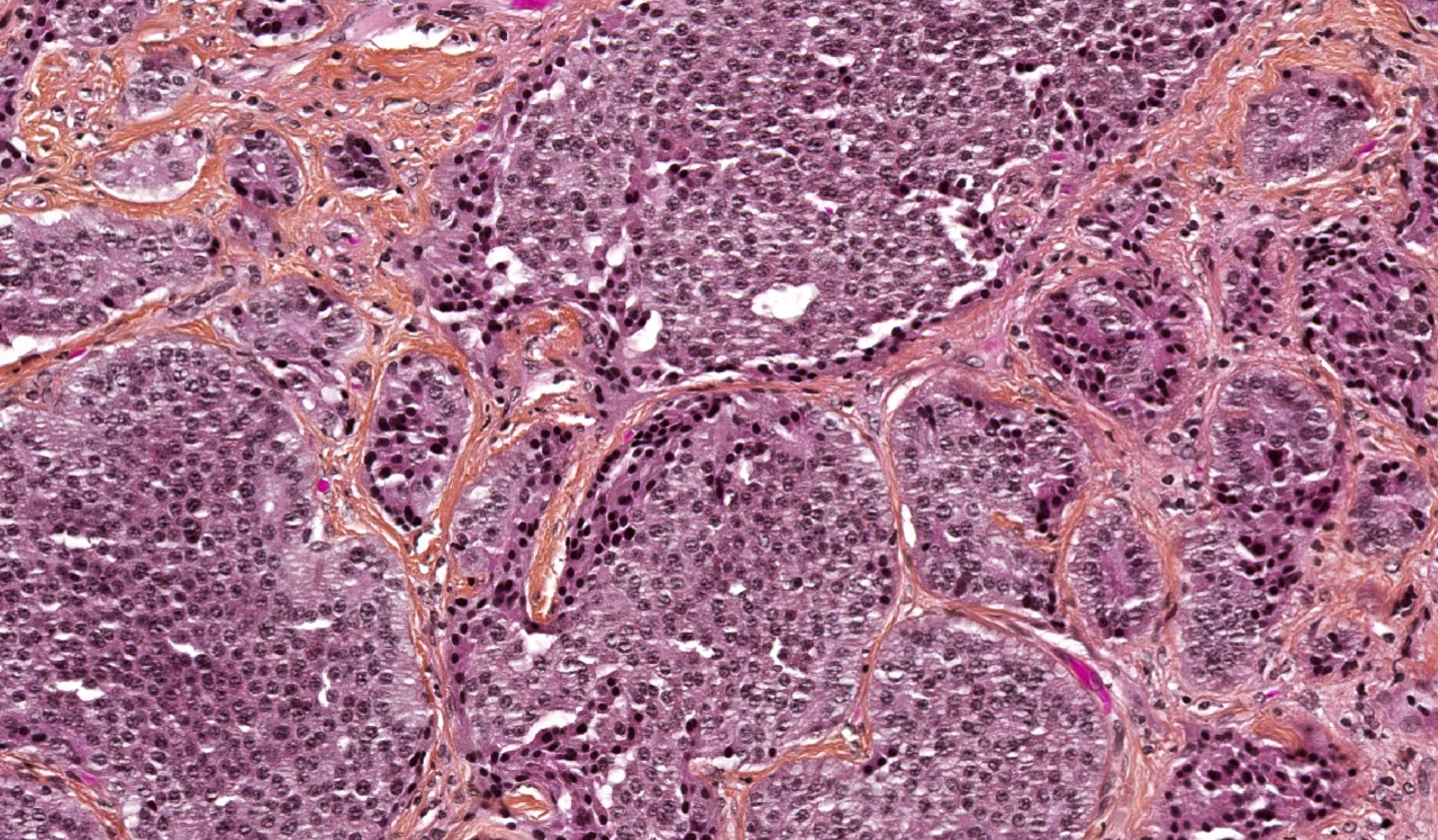
Well differentiated
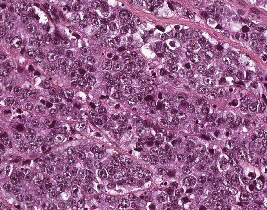
Large cell
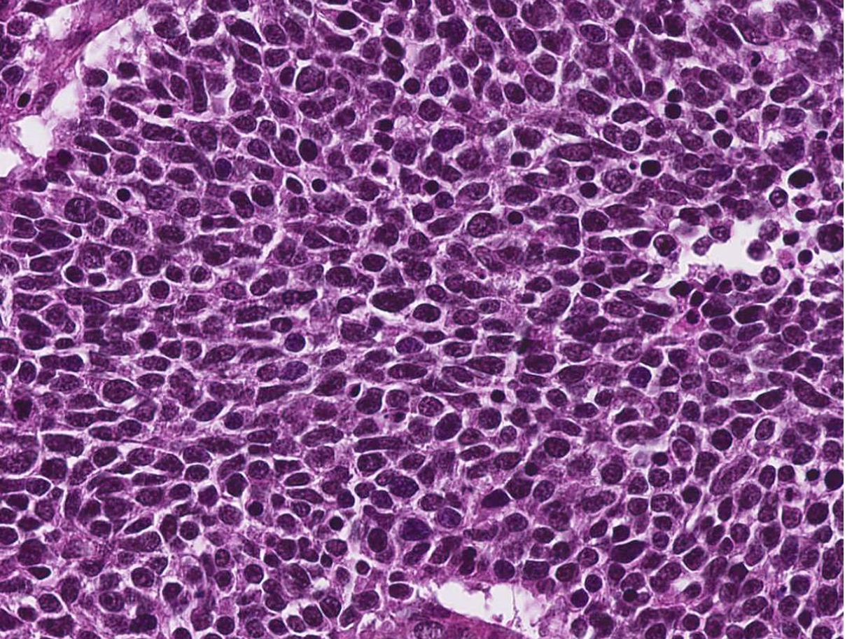
Small cell
Mixed pattern
Low Ki67
Images hosted on other servers:

CT of tracheal papilloma
Images hosted on other servers:

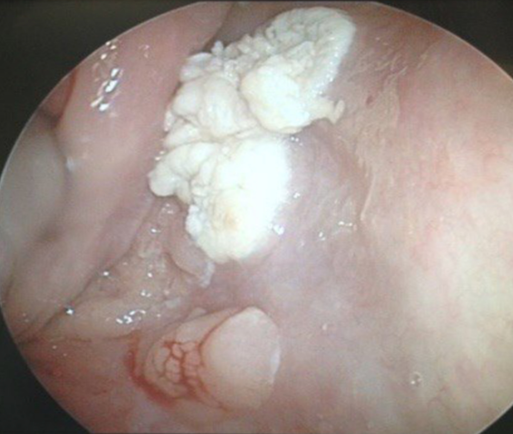
Laryngectomy for massive papillomas
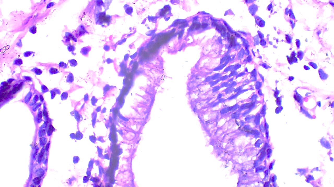
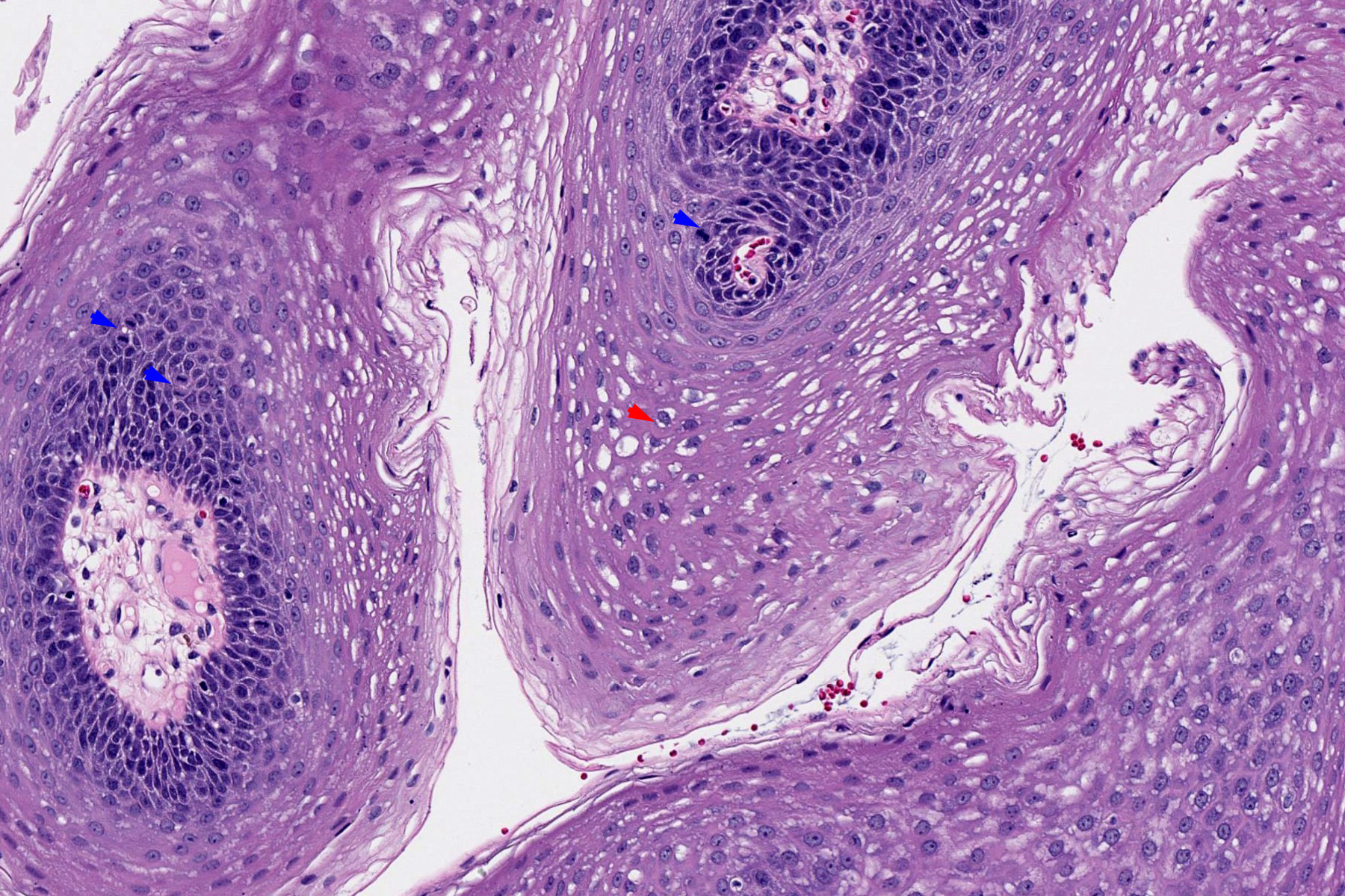
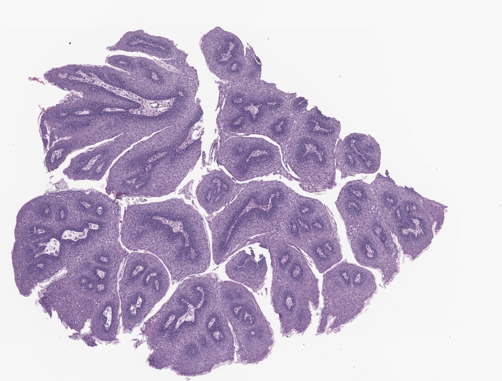
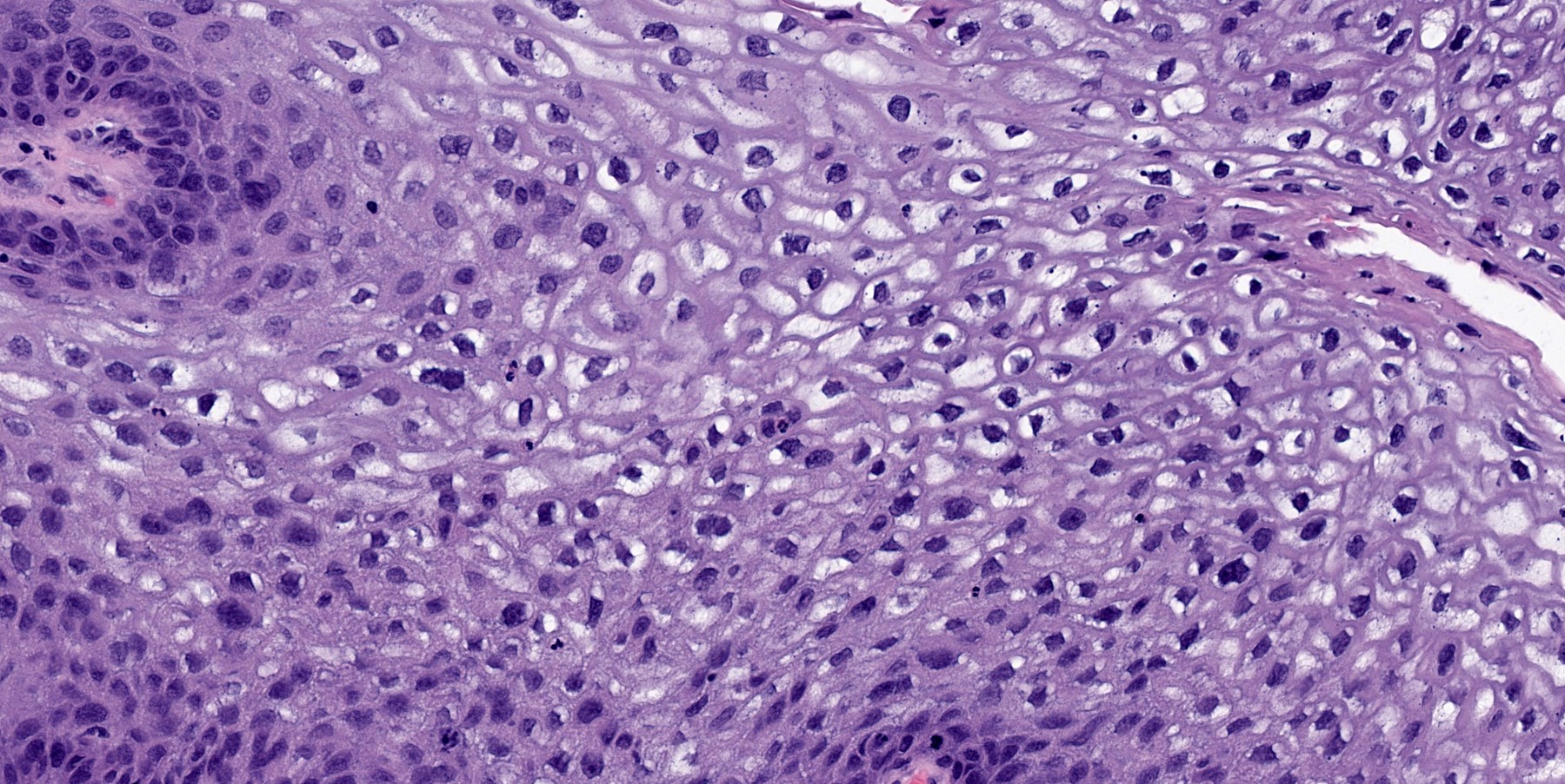
Contributed by Kavita Umrau, M.B.B.S. and Bin Xu, M.D., Ph.D.
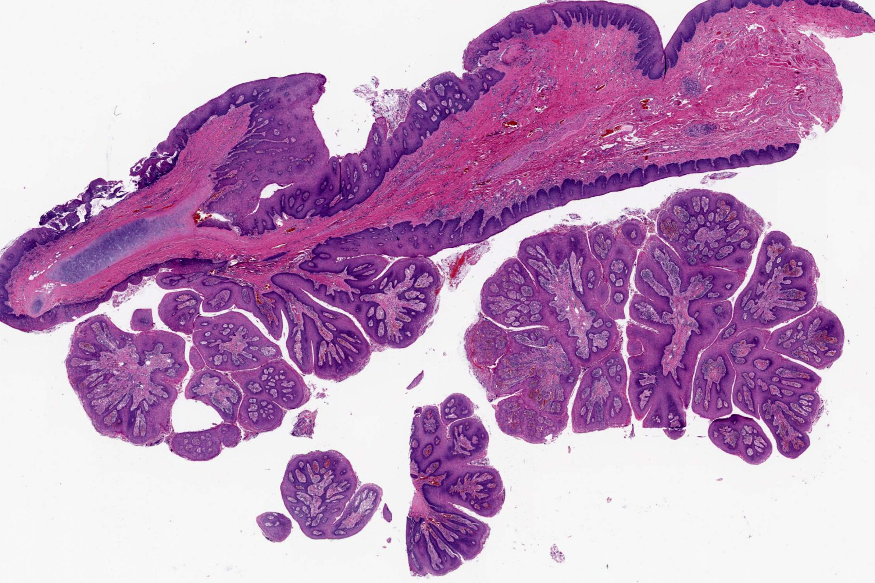
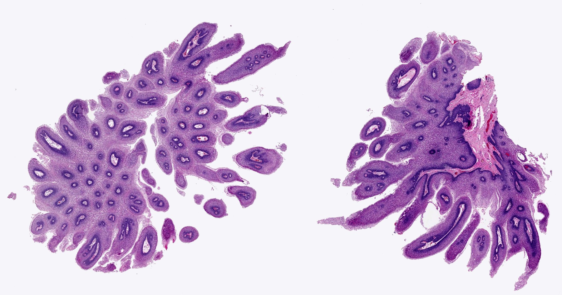
Branching finger-like papillae
Basal and parabasal mitosis
Basal and parabasal hyperplasia
Koilocytic changes
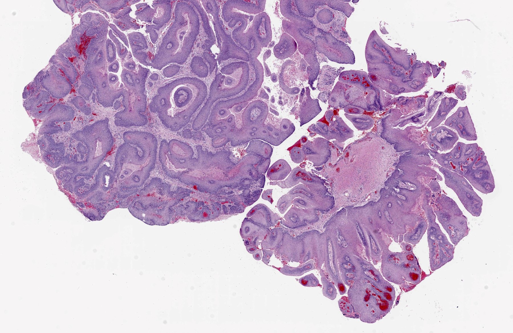
Malignant transformation
Low risk HPV
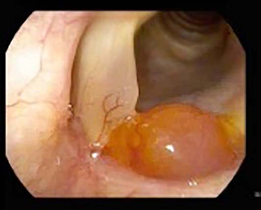
Contributed by Michael Elliott, M.B.B.S., M.Phil. and Carsten Palme, M.B.B.S.
Ulcerated hypopharyngeal lesion
Keratotic hypopharyngeal lesion
Polypoid ulcerated hypopharyngeal lesion
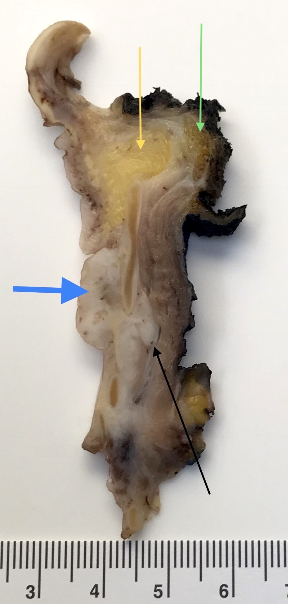
Contributed by Kelly Magliocca, D.D.S., M.P.H.
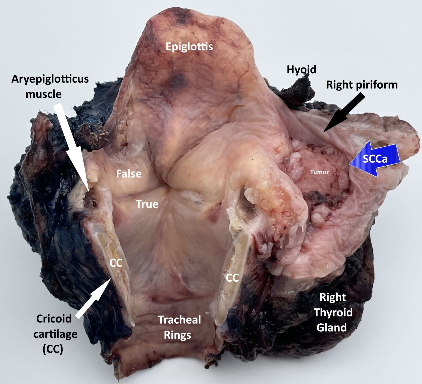
Laryngopharyngectomy, opened posteriorly (pT4a)
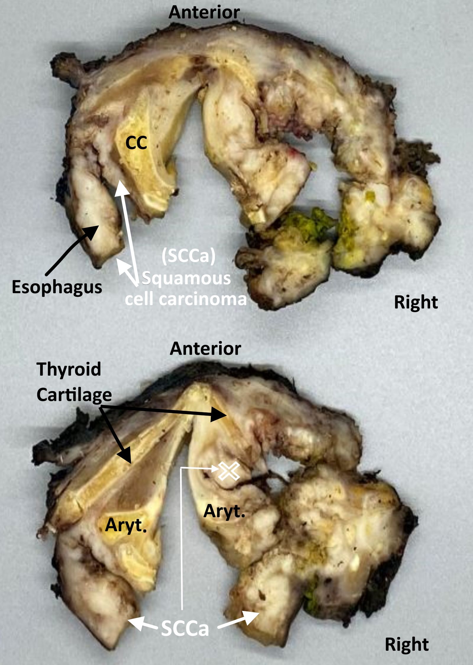
Laryngopharyngectomy,
sectioned (pT4a)


Contributed by Kyriakos Chatzopoulos, M.D., Ph.D.
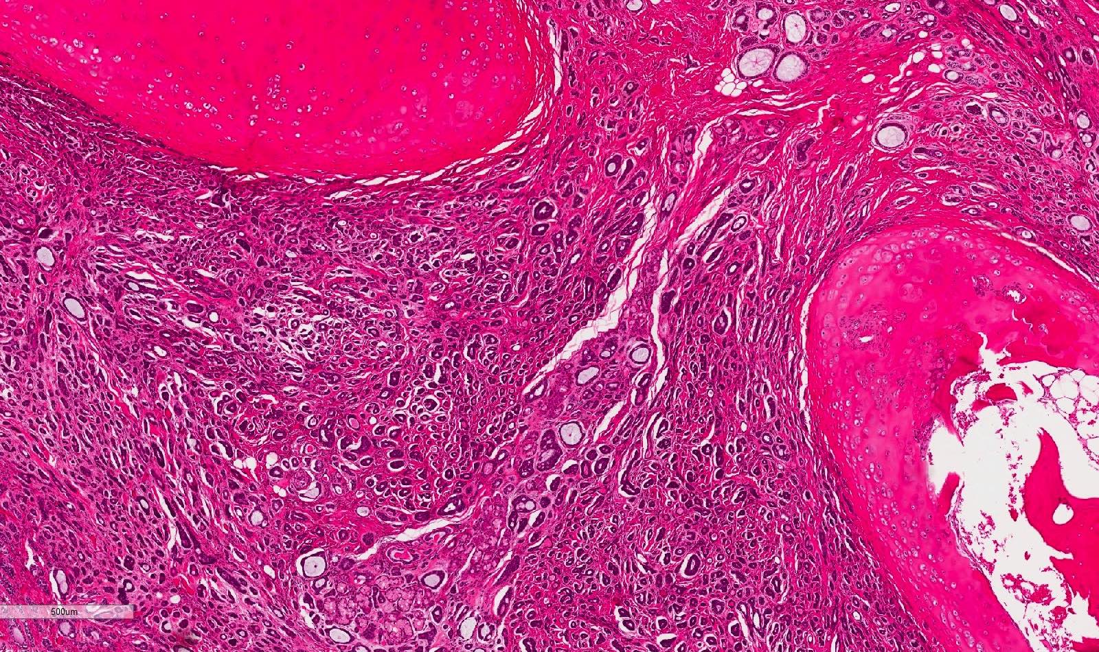
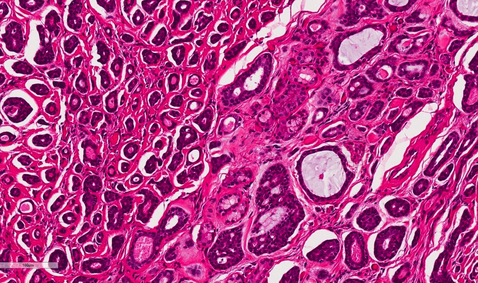
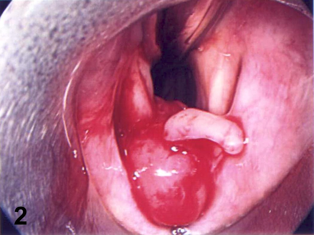
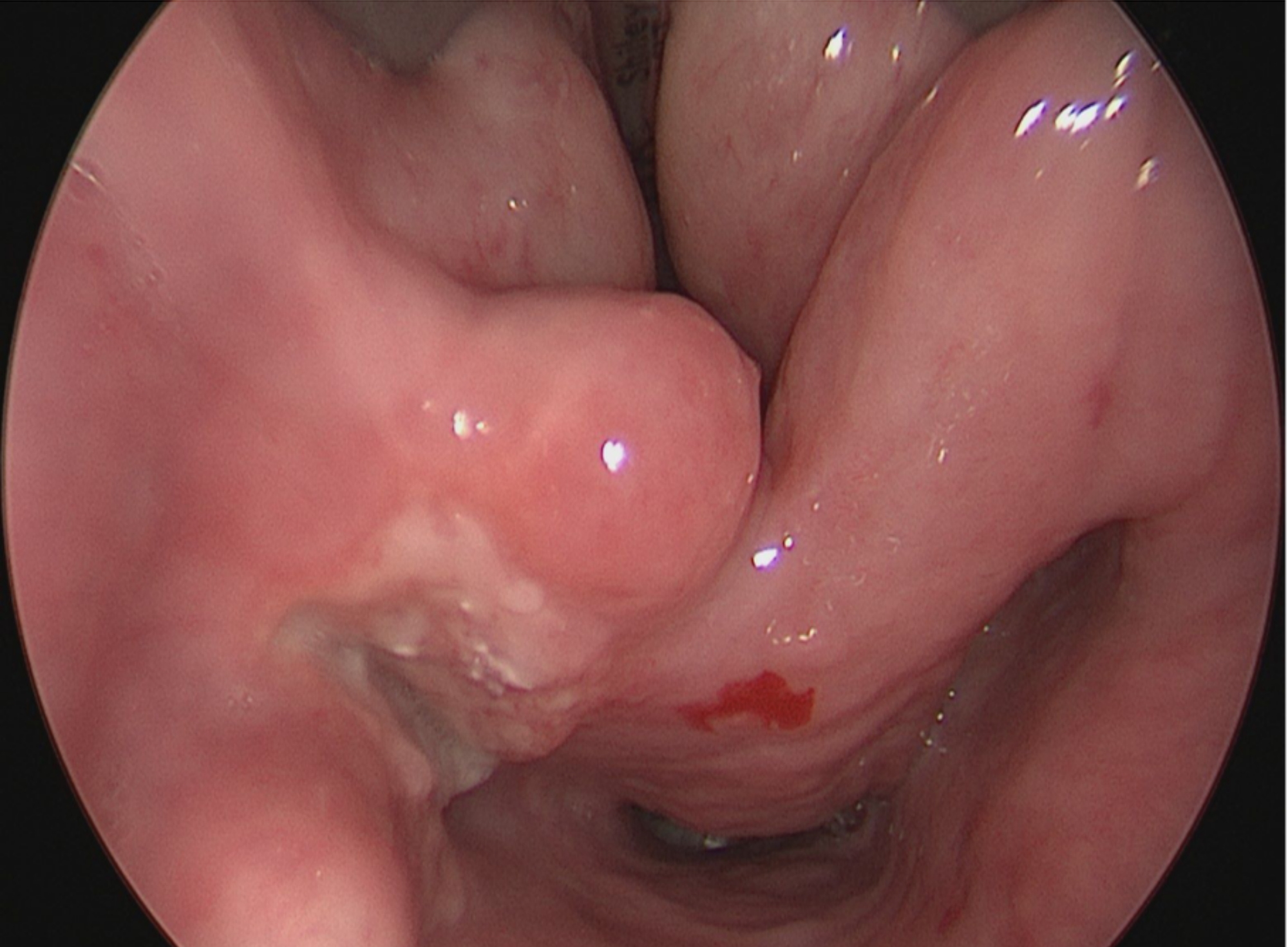
Vocal cord nodule
Contributed by Kyriakos Chatzopoulos, M.D., Ph.D.
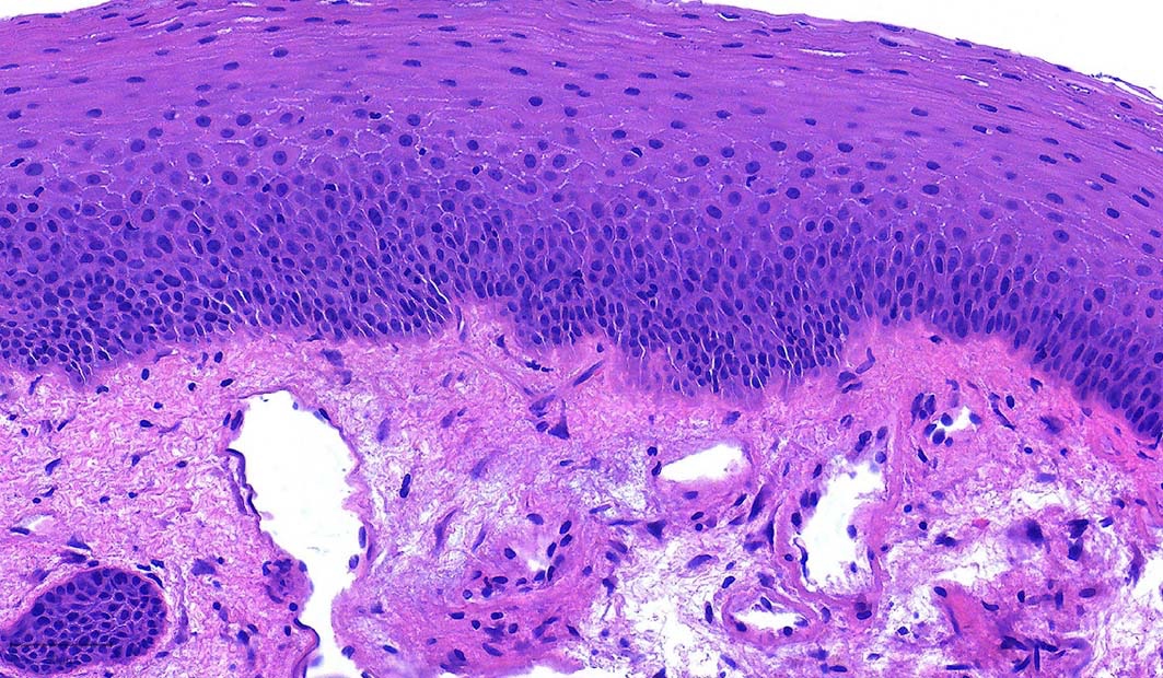
Hyperplastic squamous mucosa
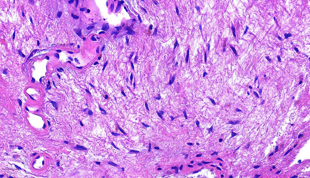
Fibroblastic myxoid stroma
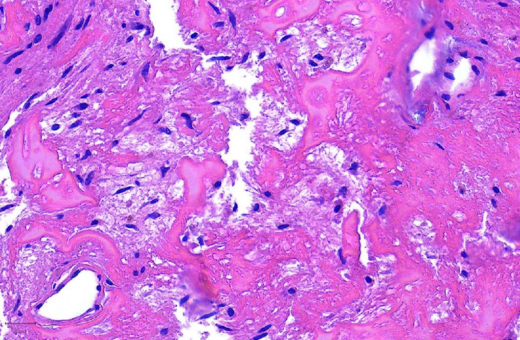
Stromal hyalinization
Franchi: 2020
Gnepp: 2021
Stelow: 2020
Thompson: 2022
Wenig: 2017
Wenig: 2024
Find related Pathology books: head & neck/endocrine
