
Images hosted on other servers:

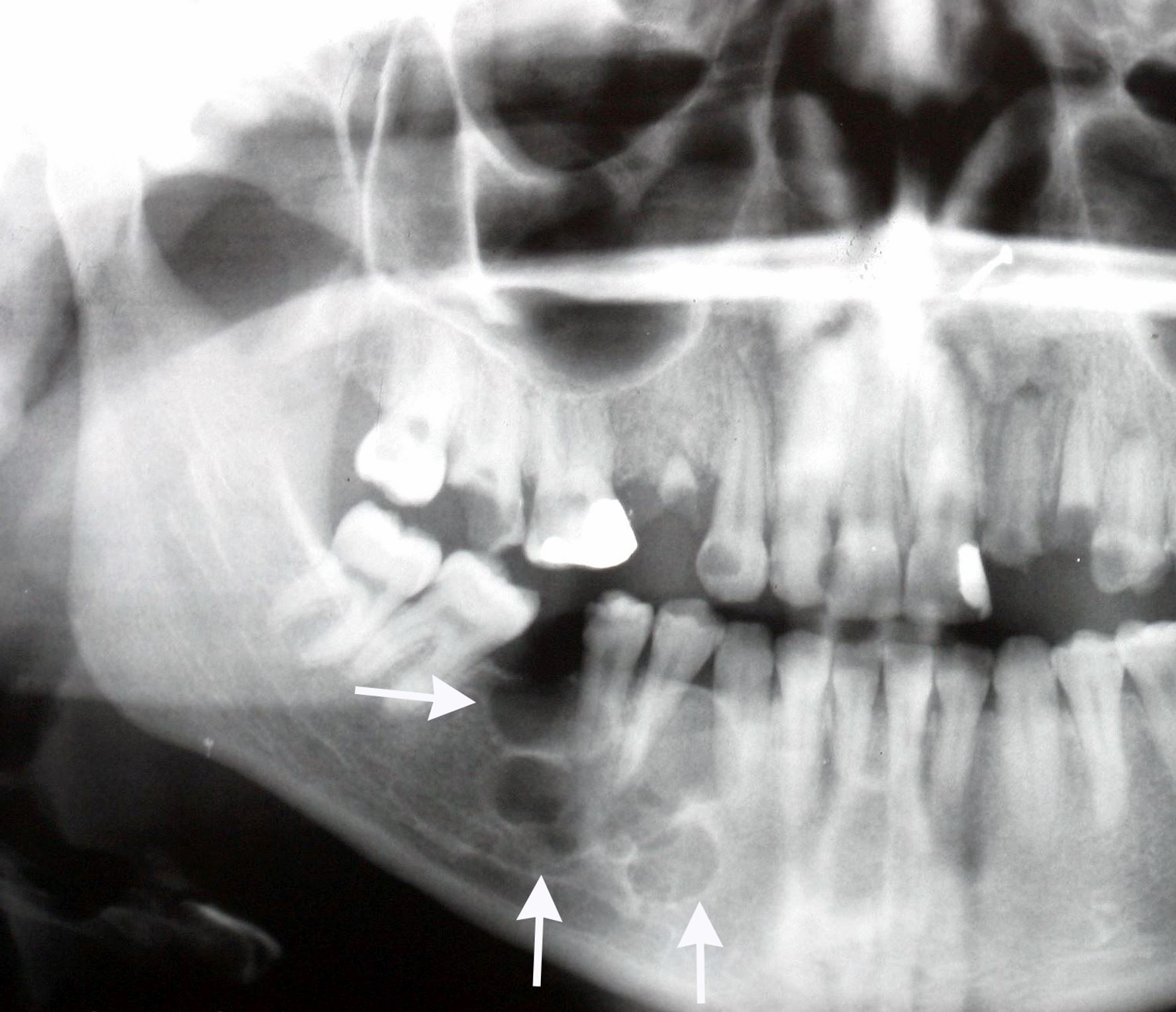
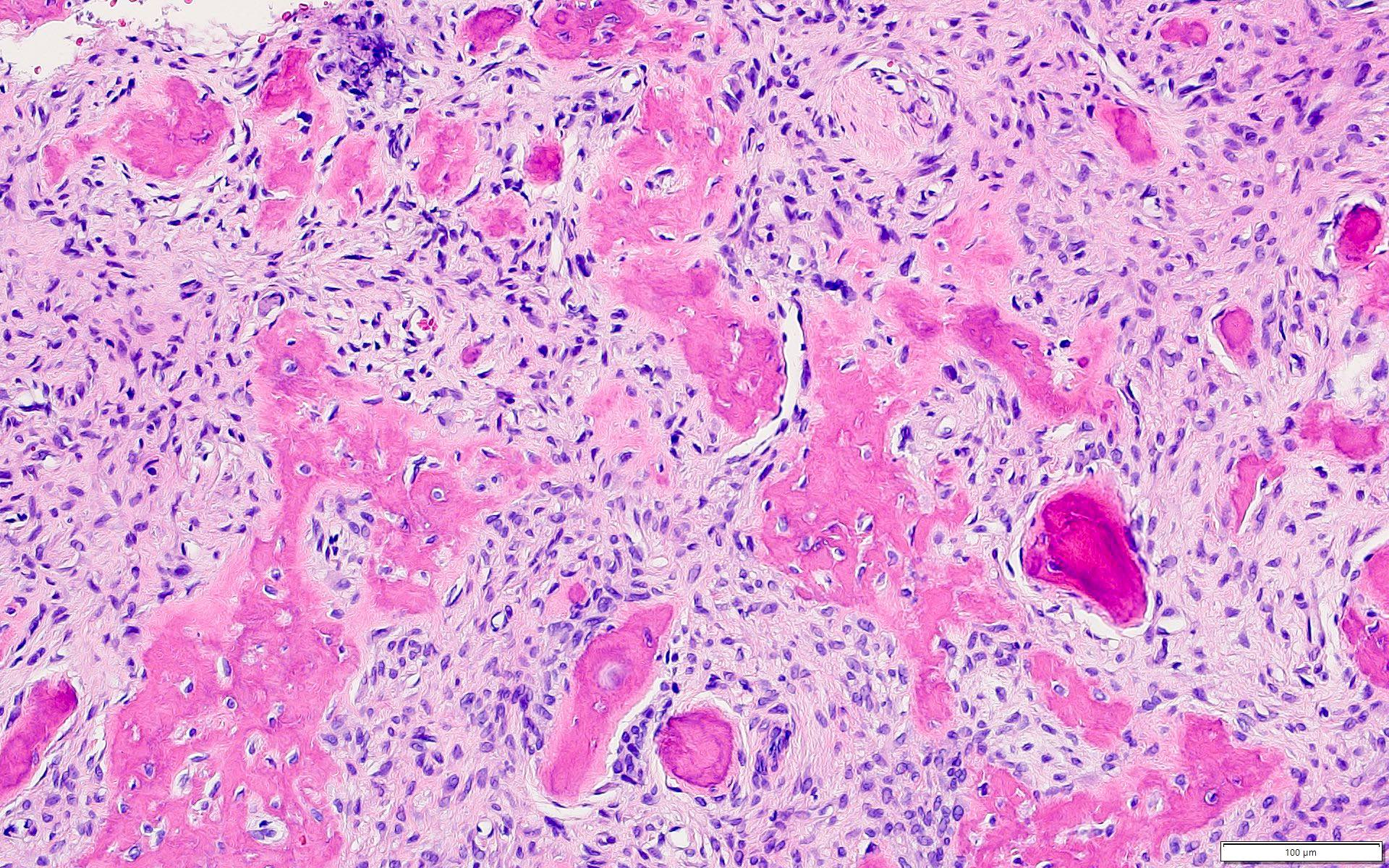
Osteomyelitis of mandible

Absorption of cortical bone

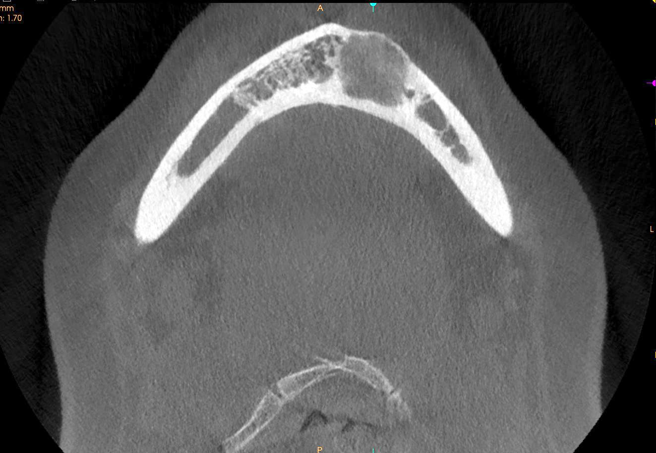
CT scan
Various images
Images hosted on other servers:

H&E and GMS
Images hosted on other servers:

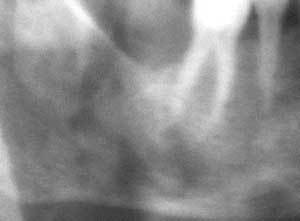
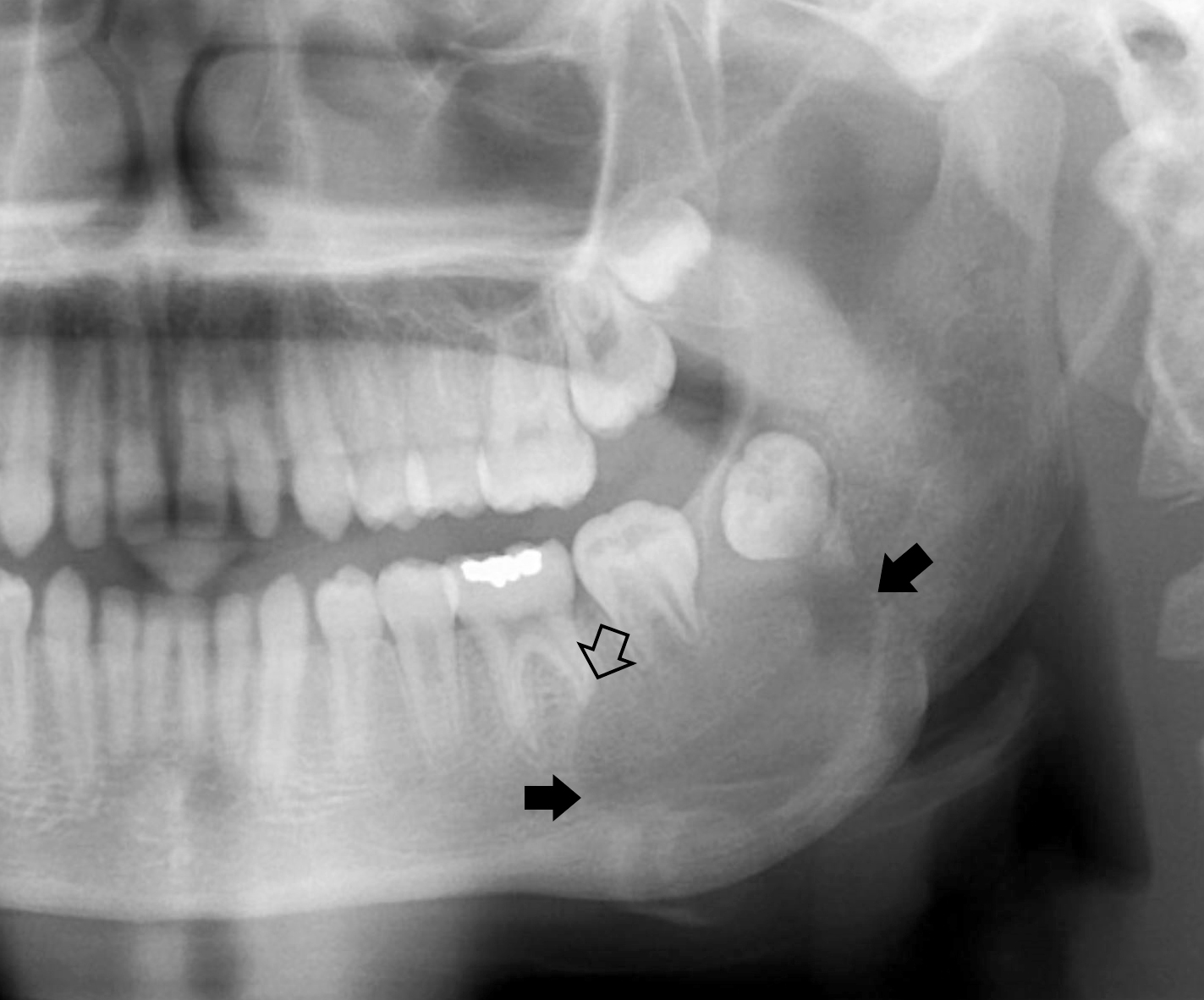
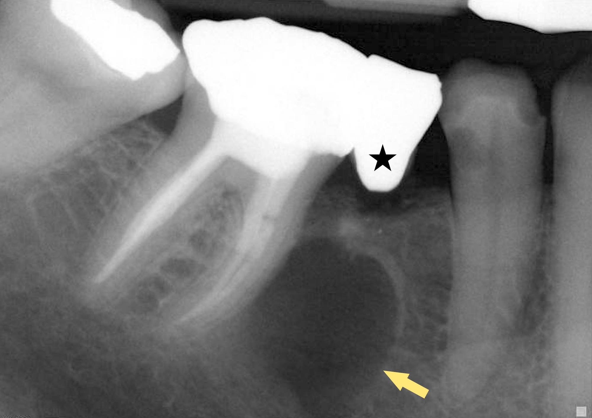
Diffuse periapical radiolucency
Contributed by Elizabeth Ann Bilodeau, D.M.D., M.D., M.S.Ed.

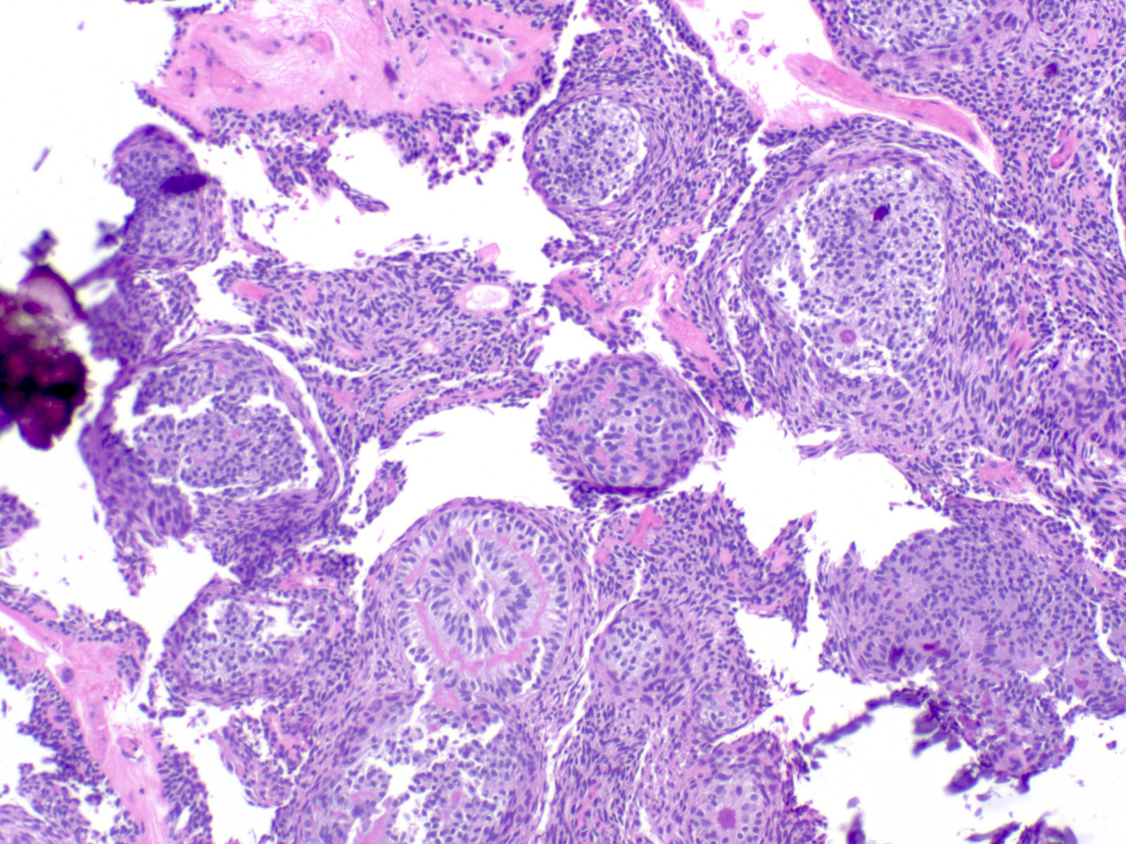
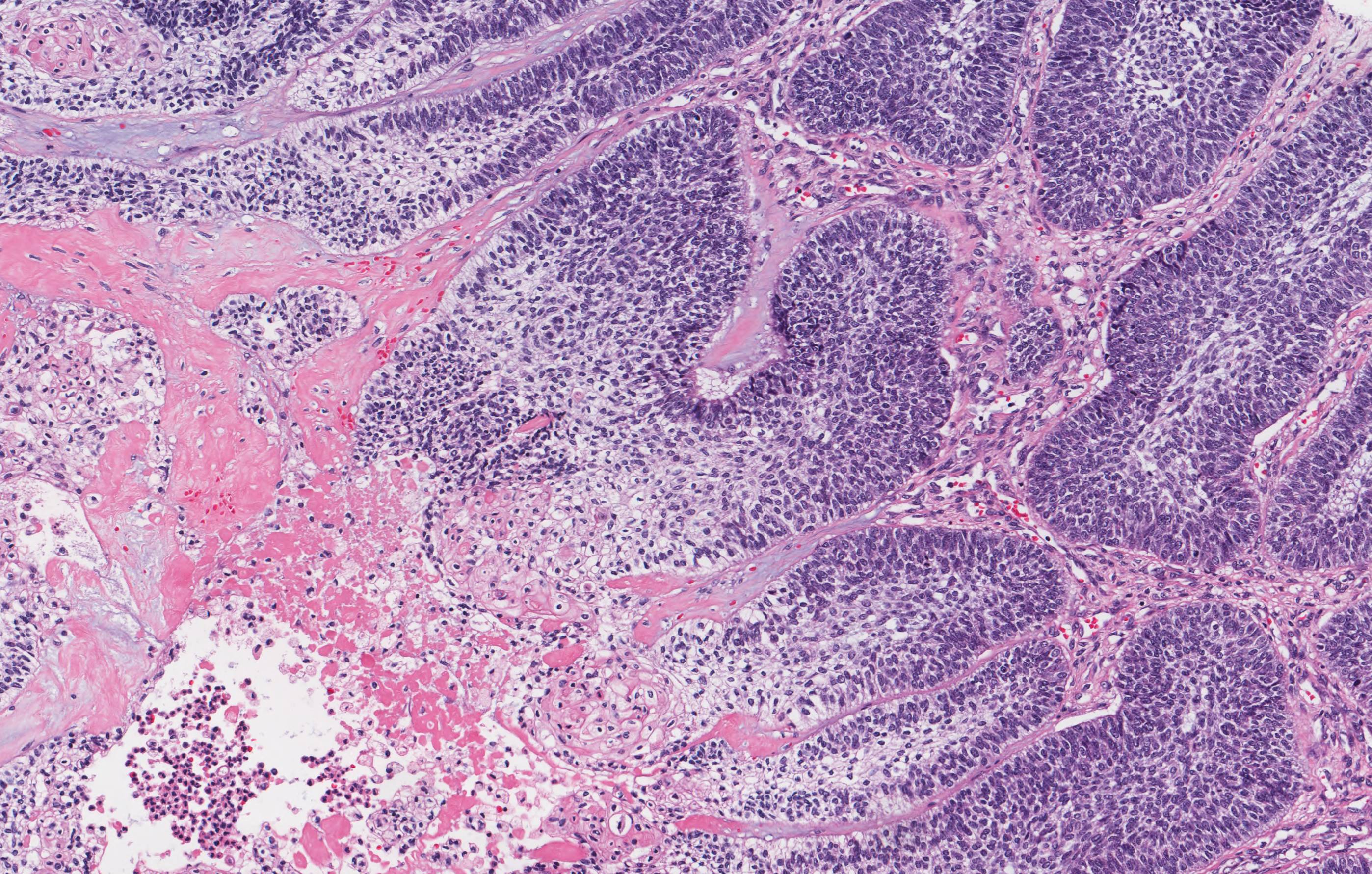
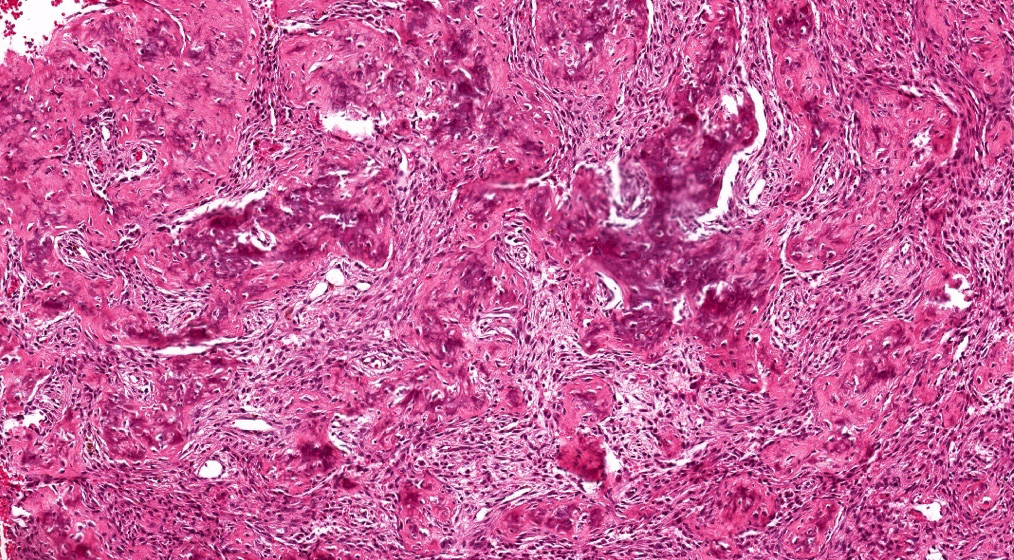
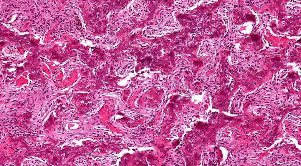
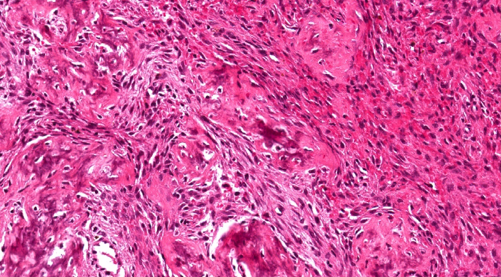
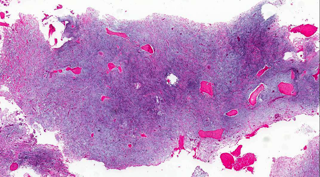
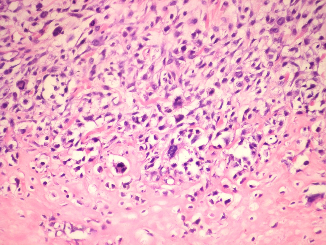
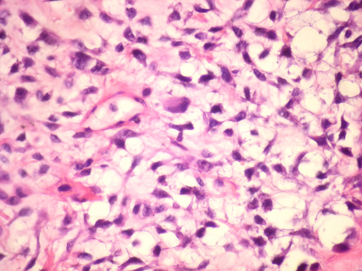
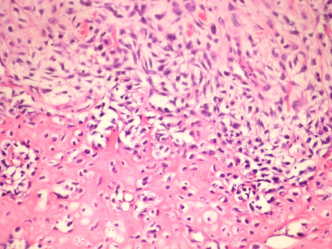
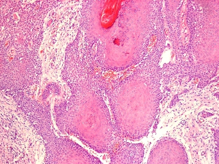
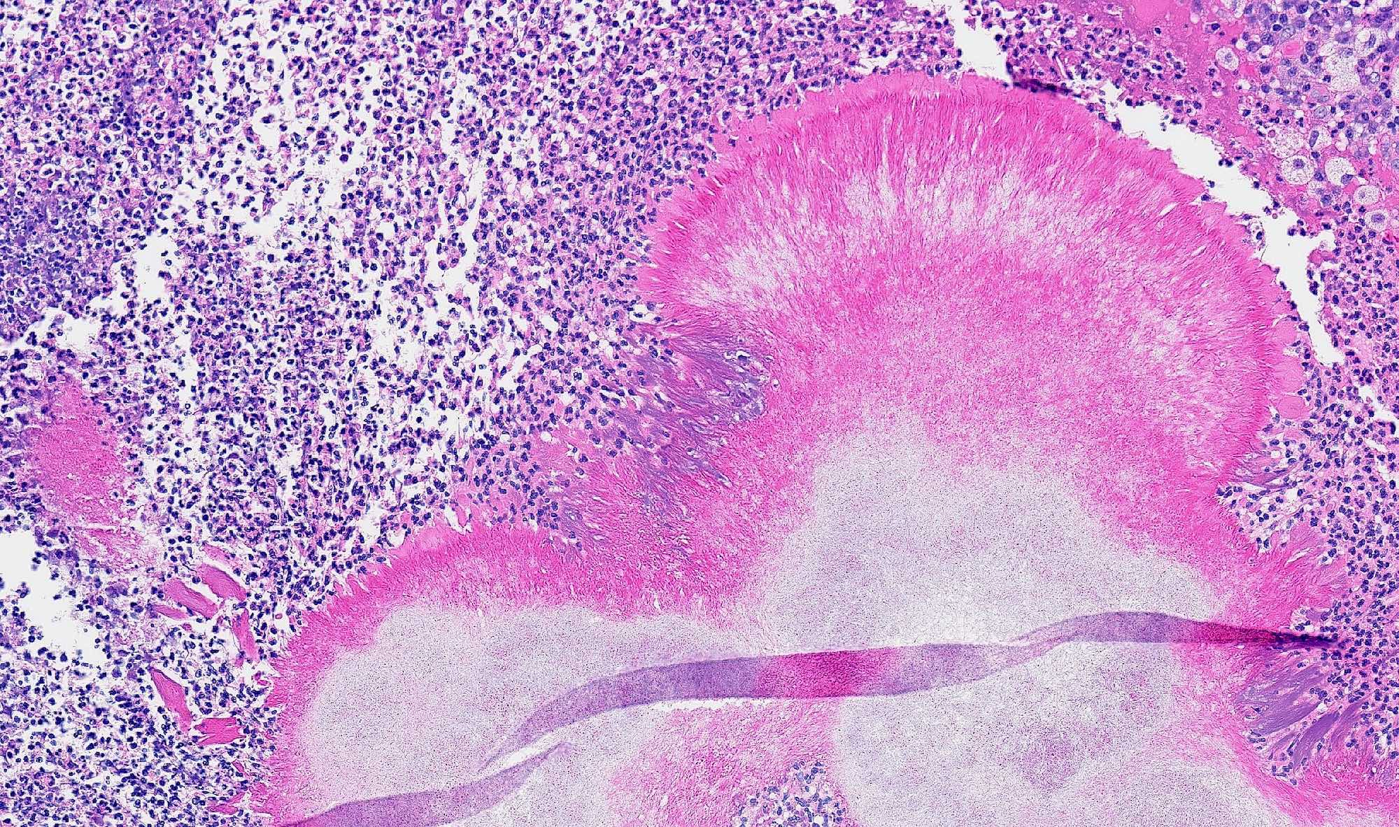
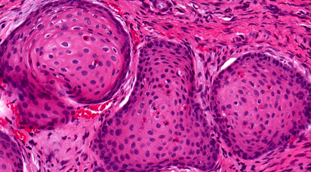
Epithelial whorls

Cribriform architecture and eosinophilic matrix
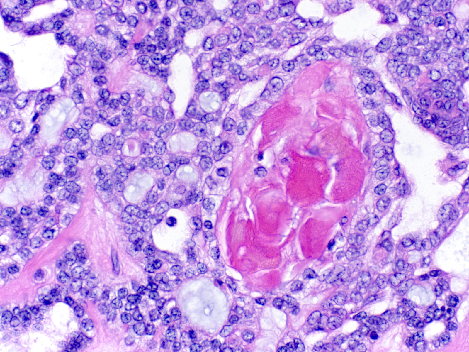
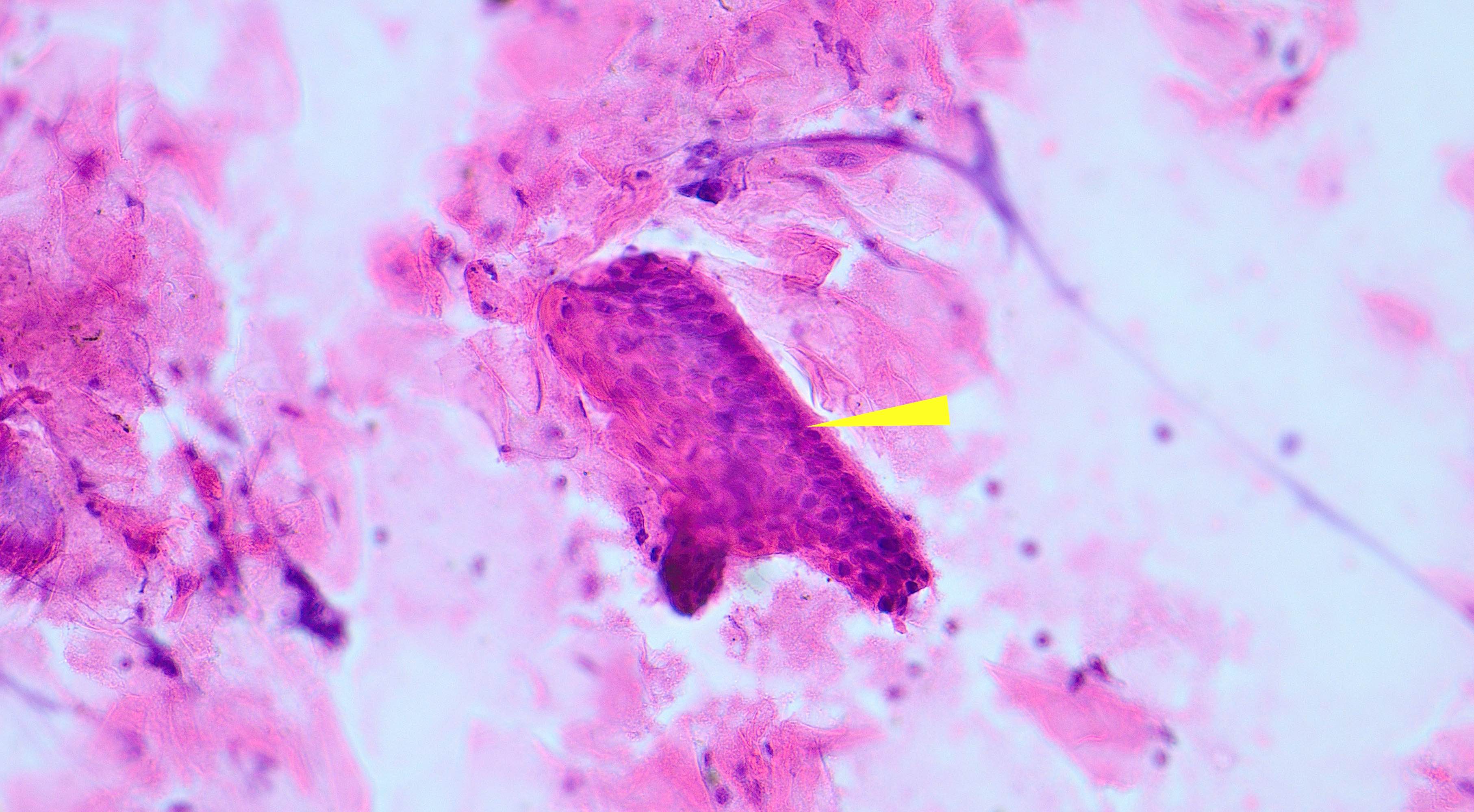
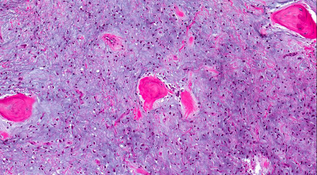
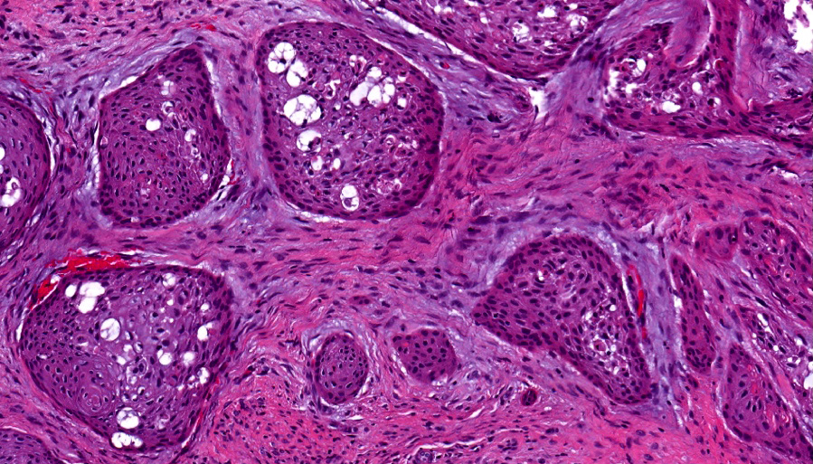
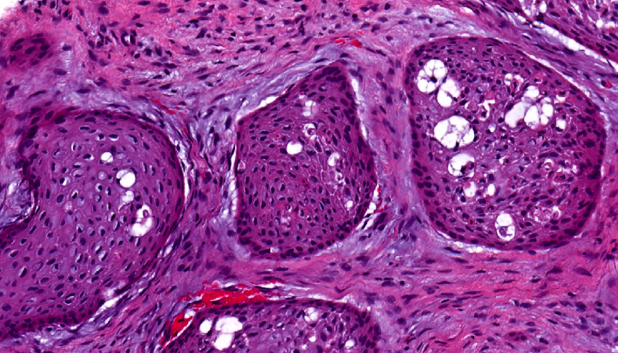
Focal ghost cell keratinization
Dentinoid deposits

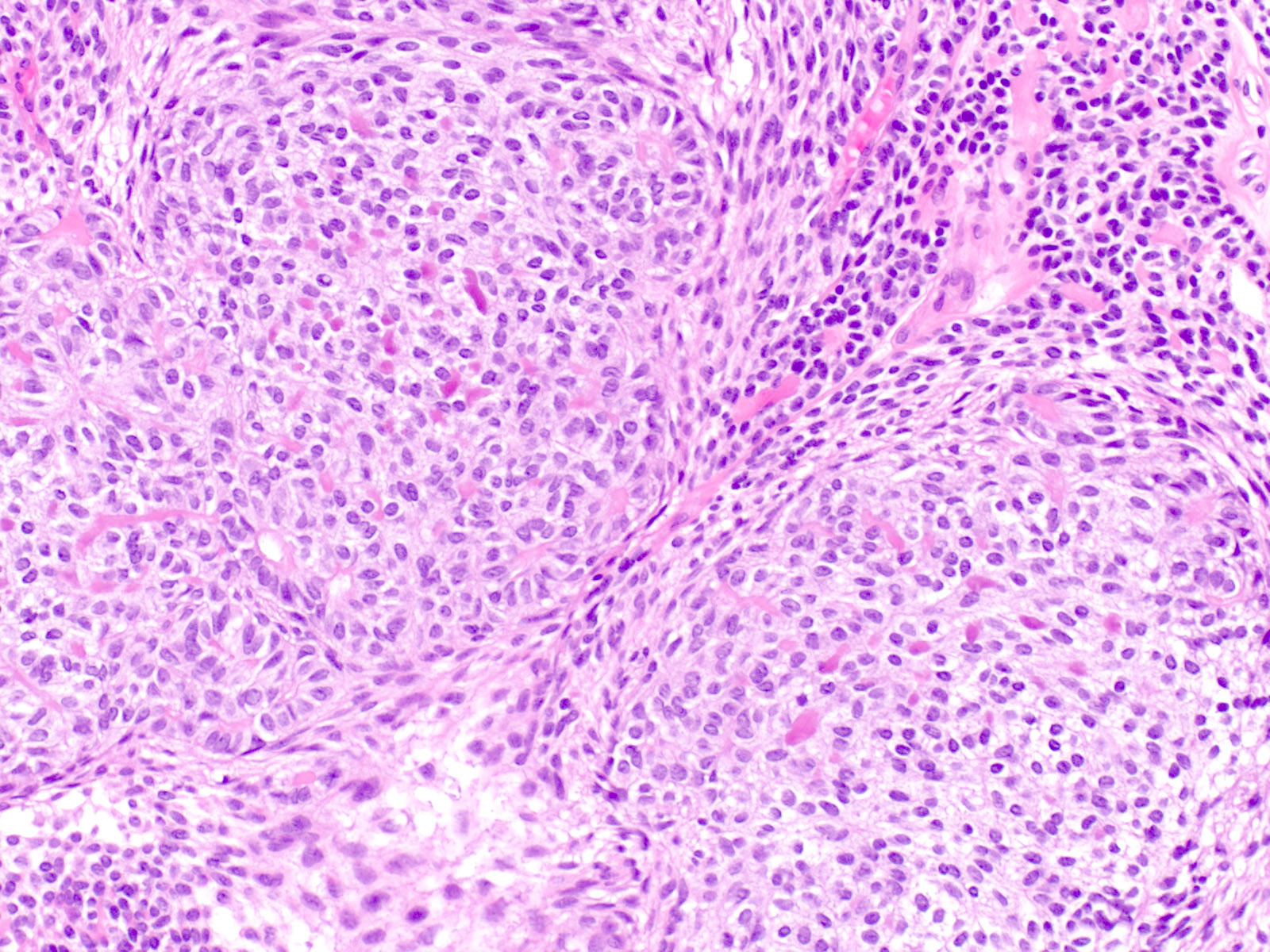
Increased mitotic activity

Duct-like structures and ghost cell keratinization
Images hosted on other servers:

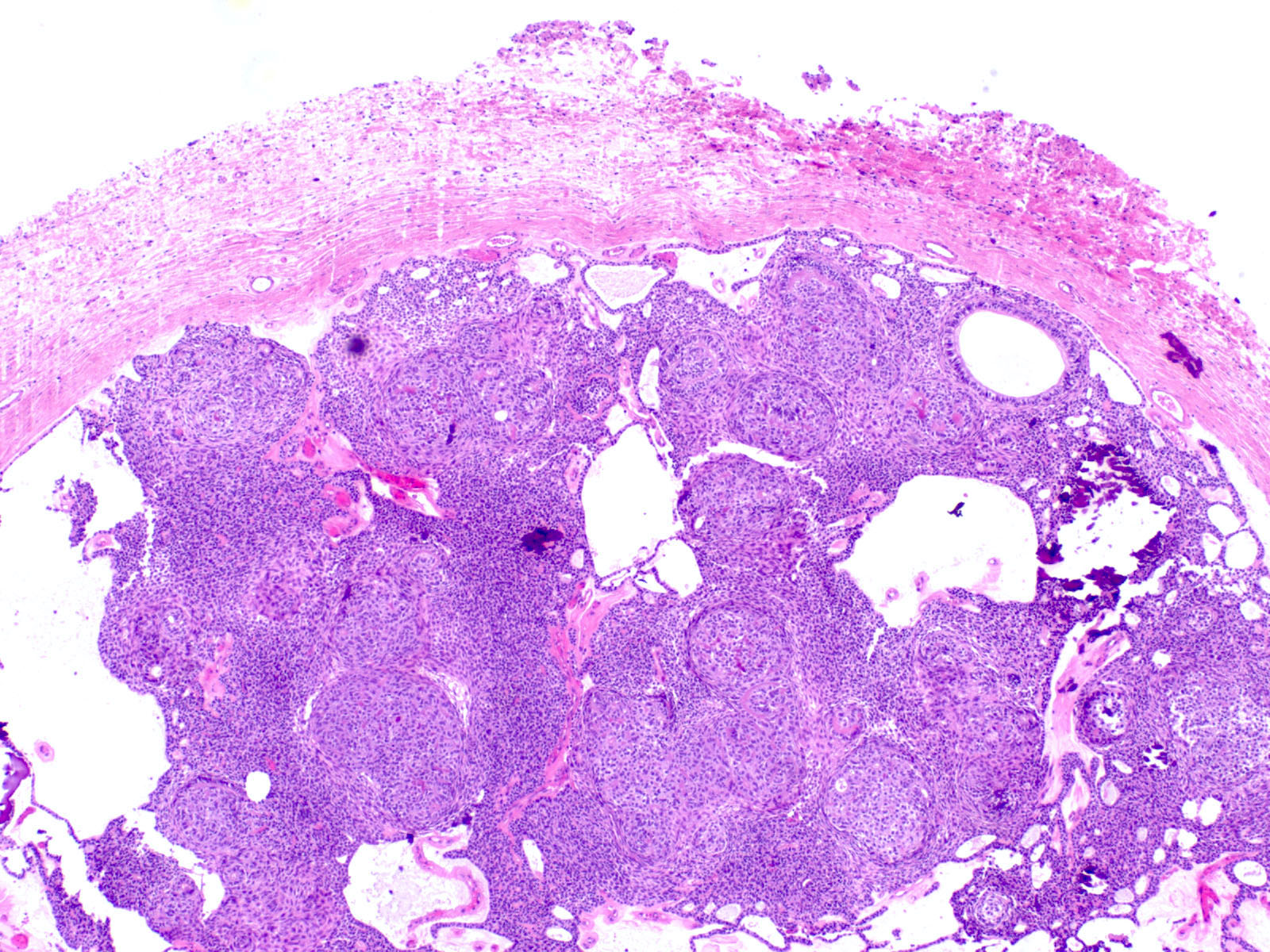
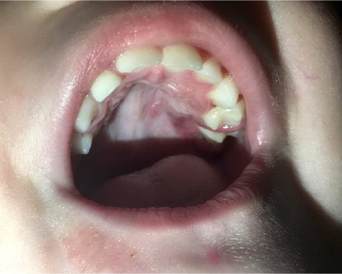
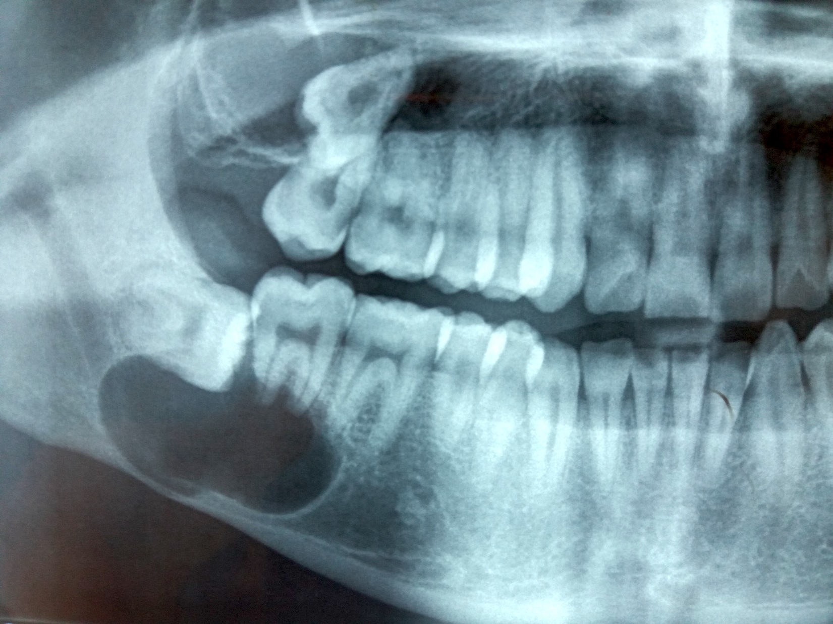
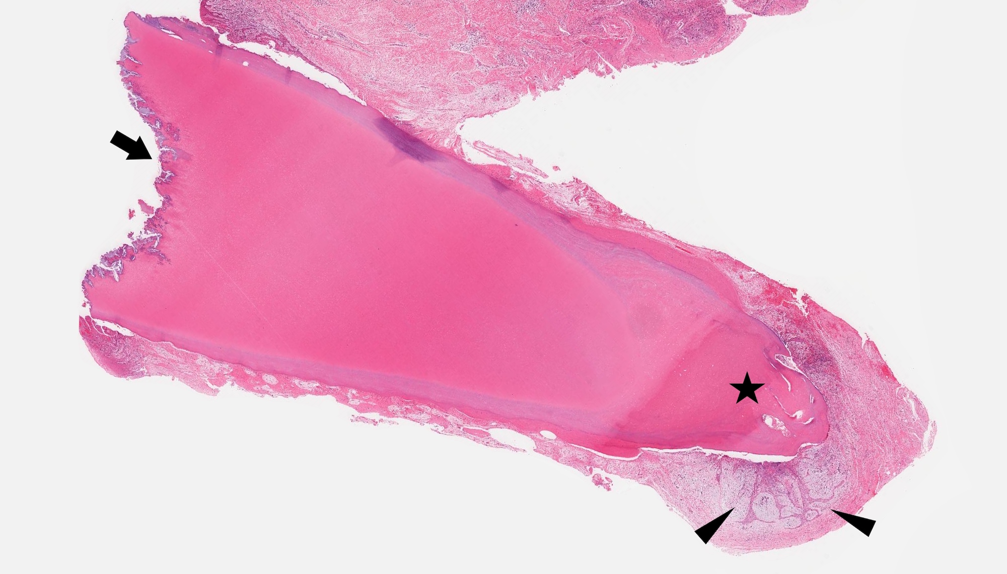
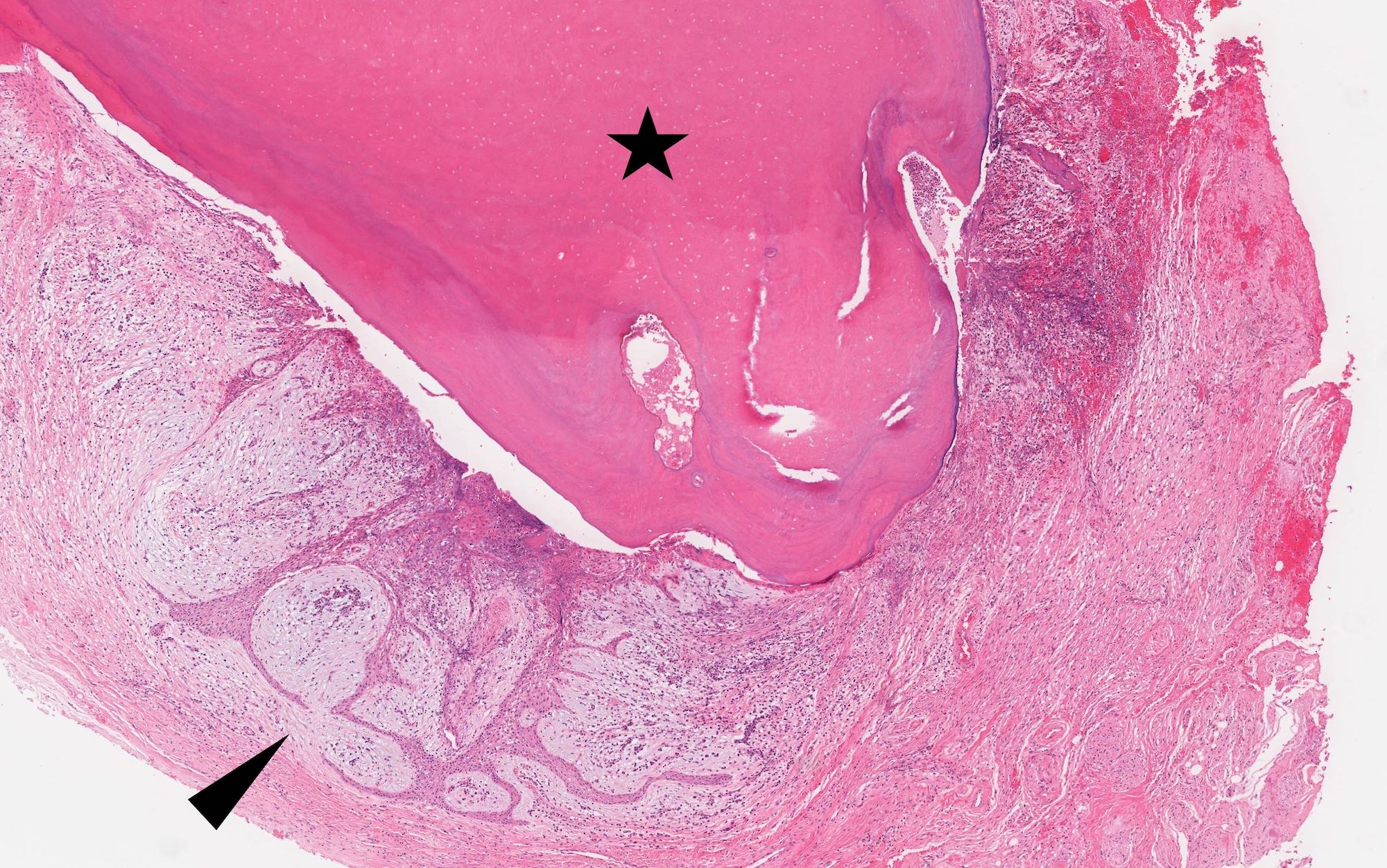
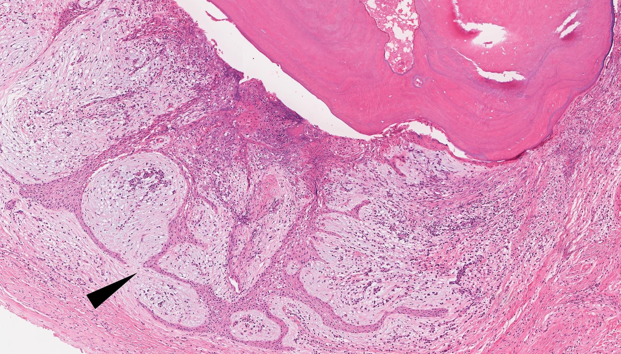
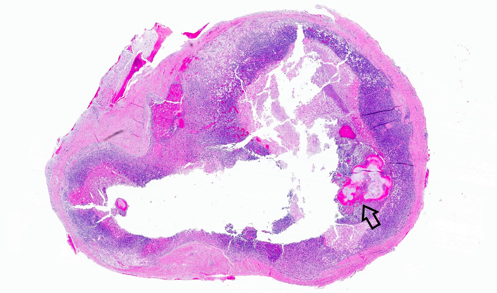
Radiolucency, engulfing tooth, extending beyond crown
Contributed by Elizabeth Ann Bilodeau, D.M.D., M.D., M.S.Ed. and Kelly Magliocca, D.D.S., M.P.H. (Case #490)
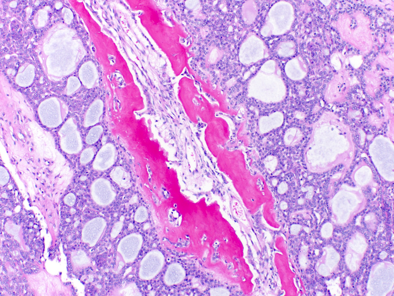
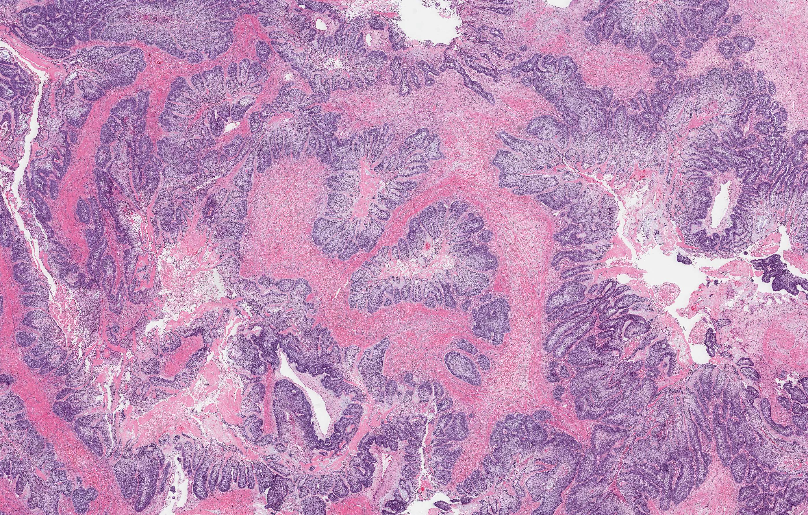
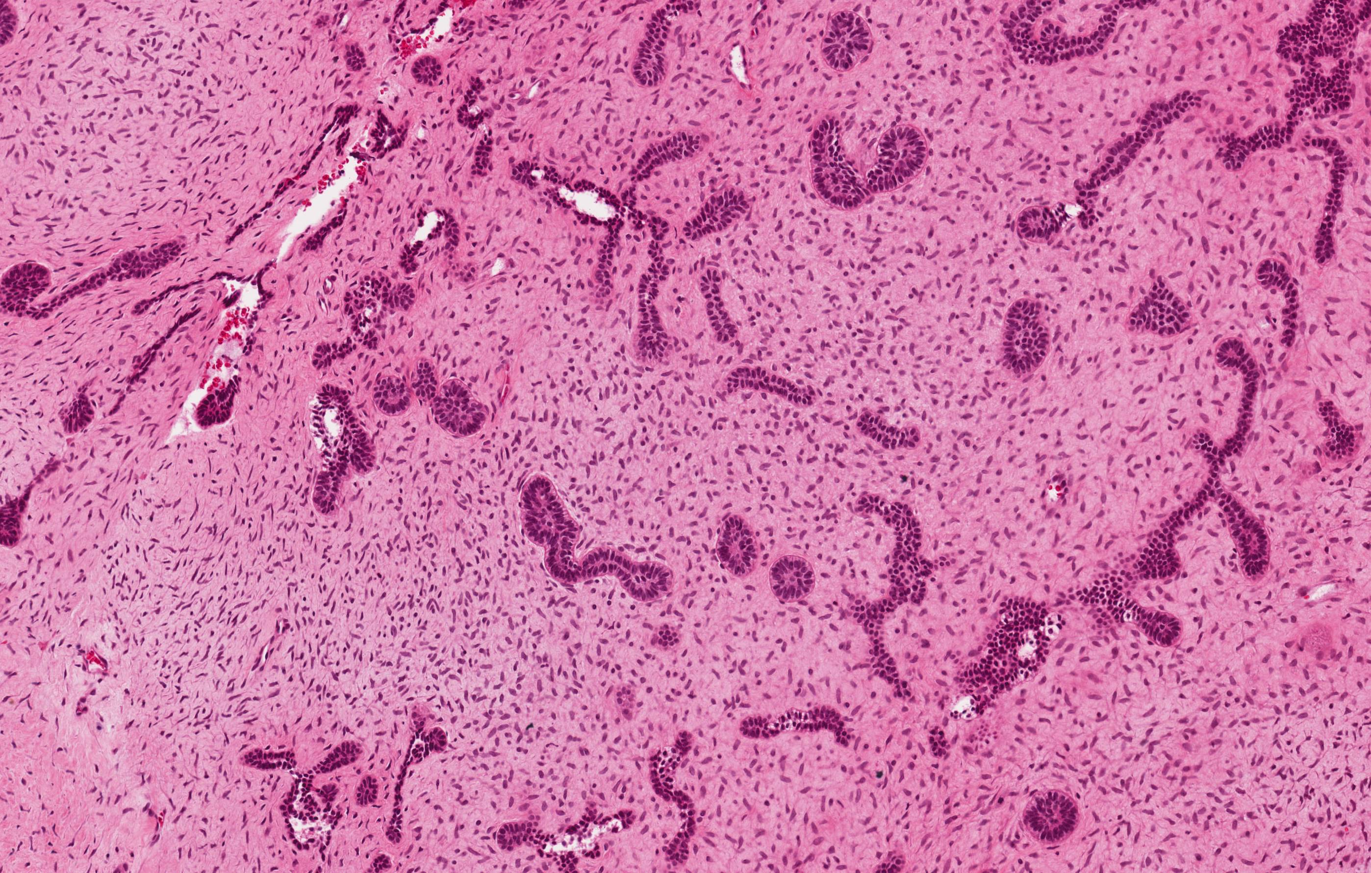
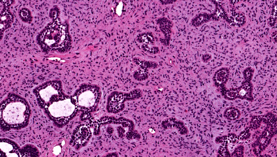
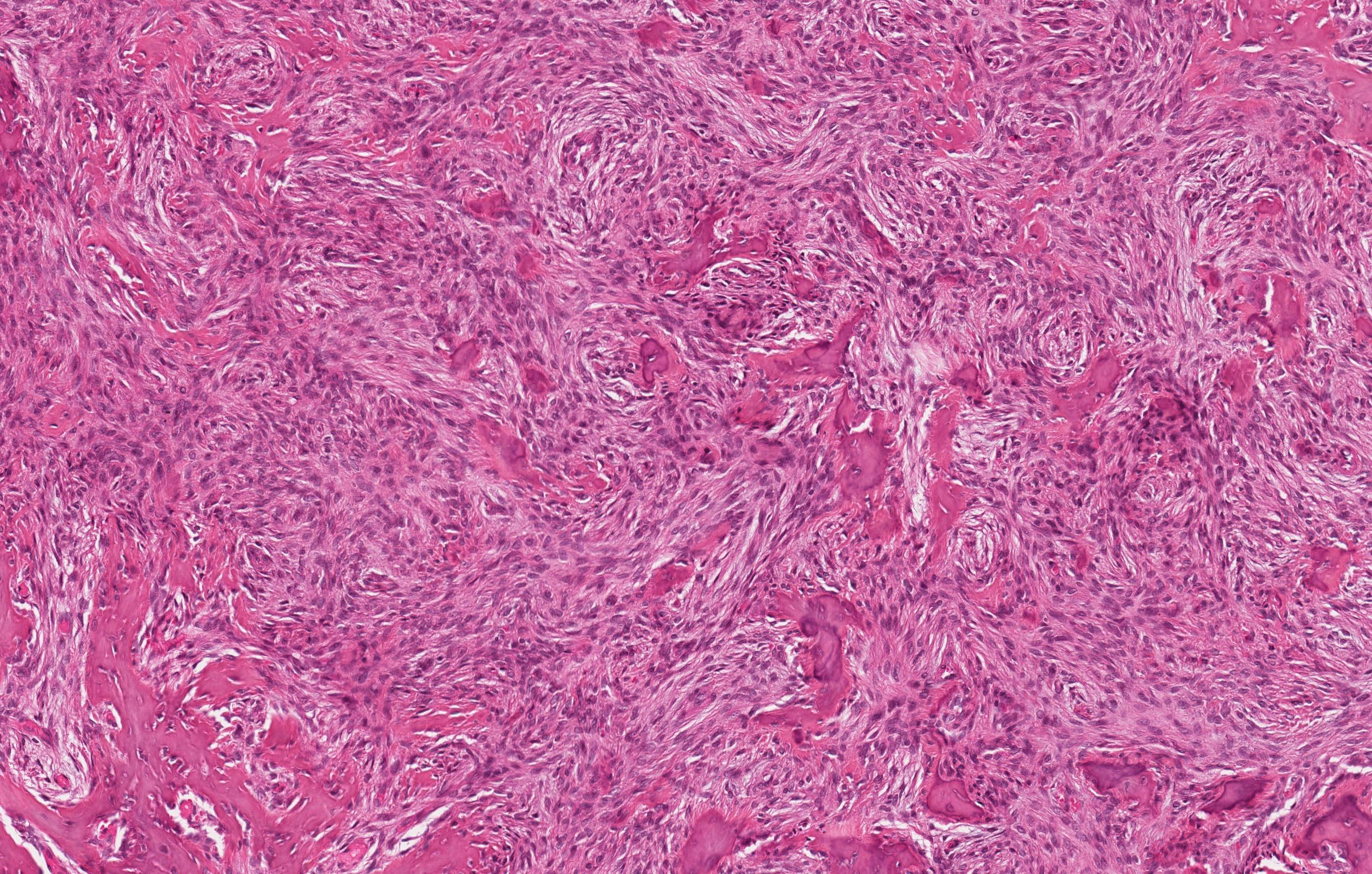
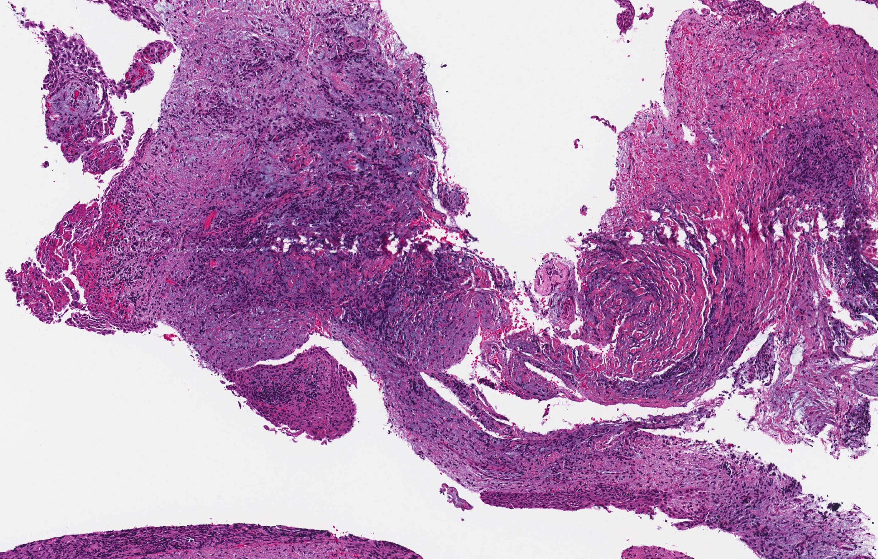
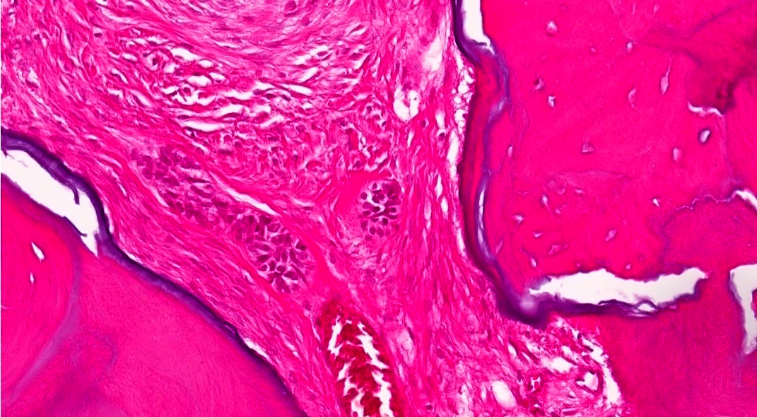
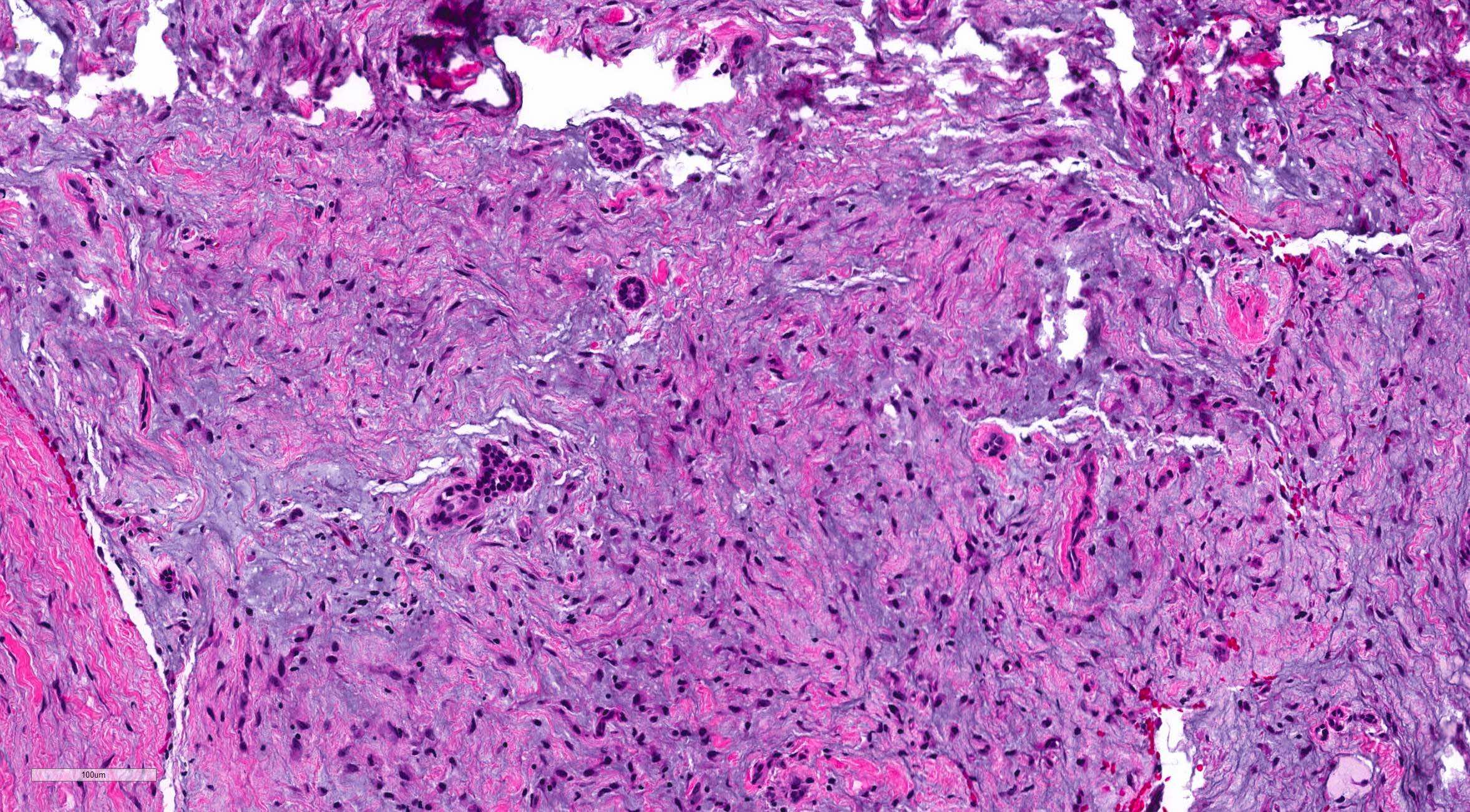
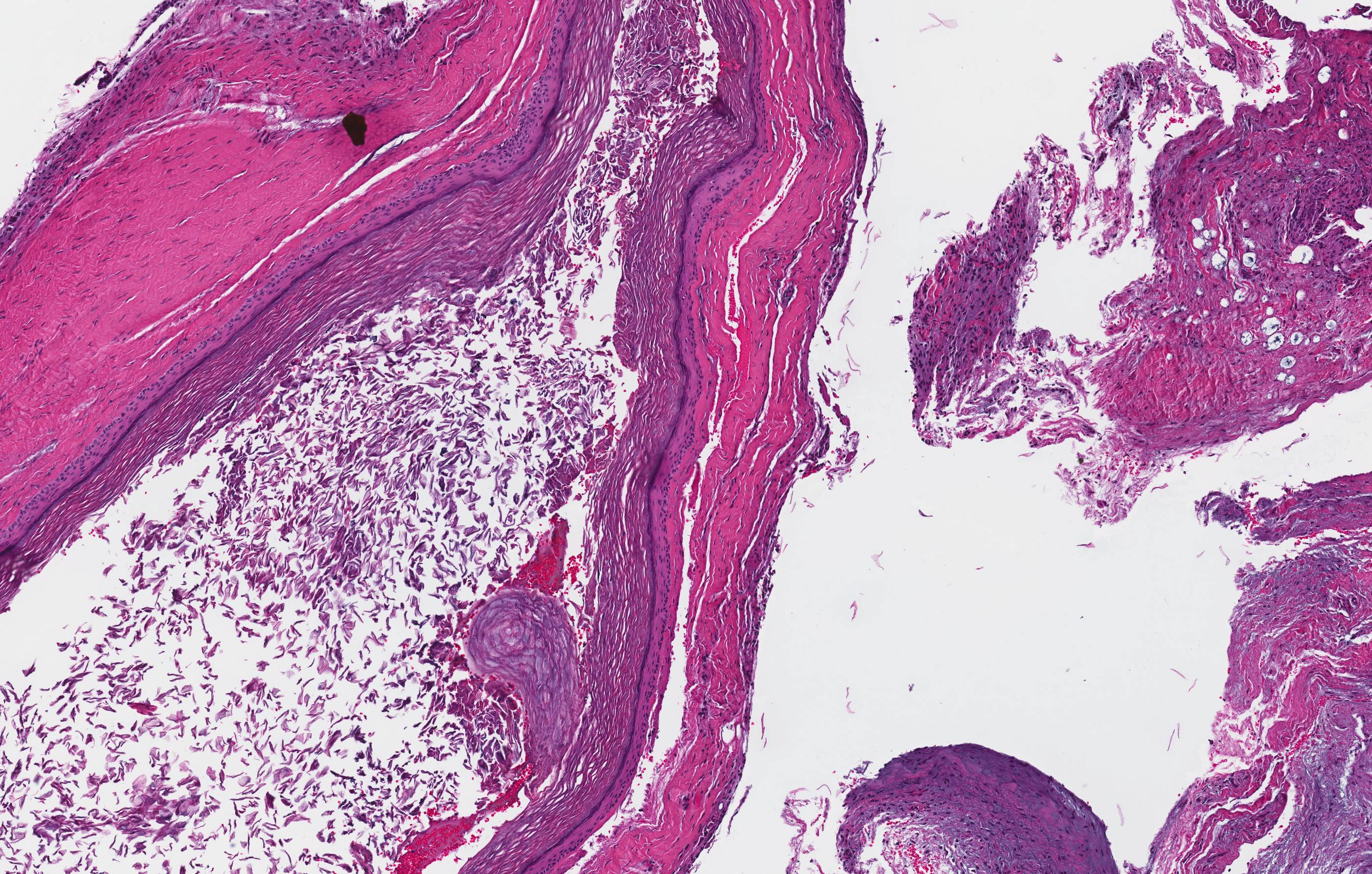
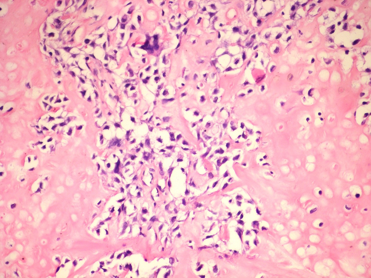
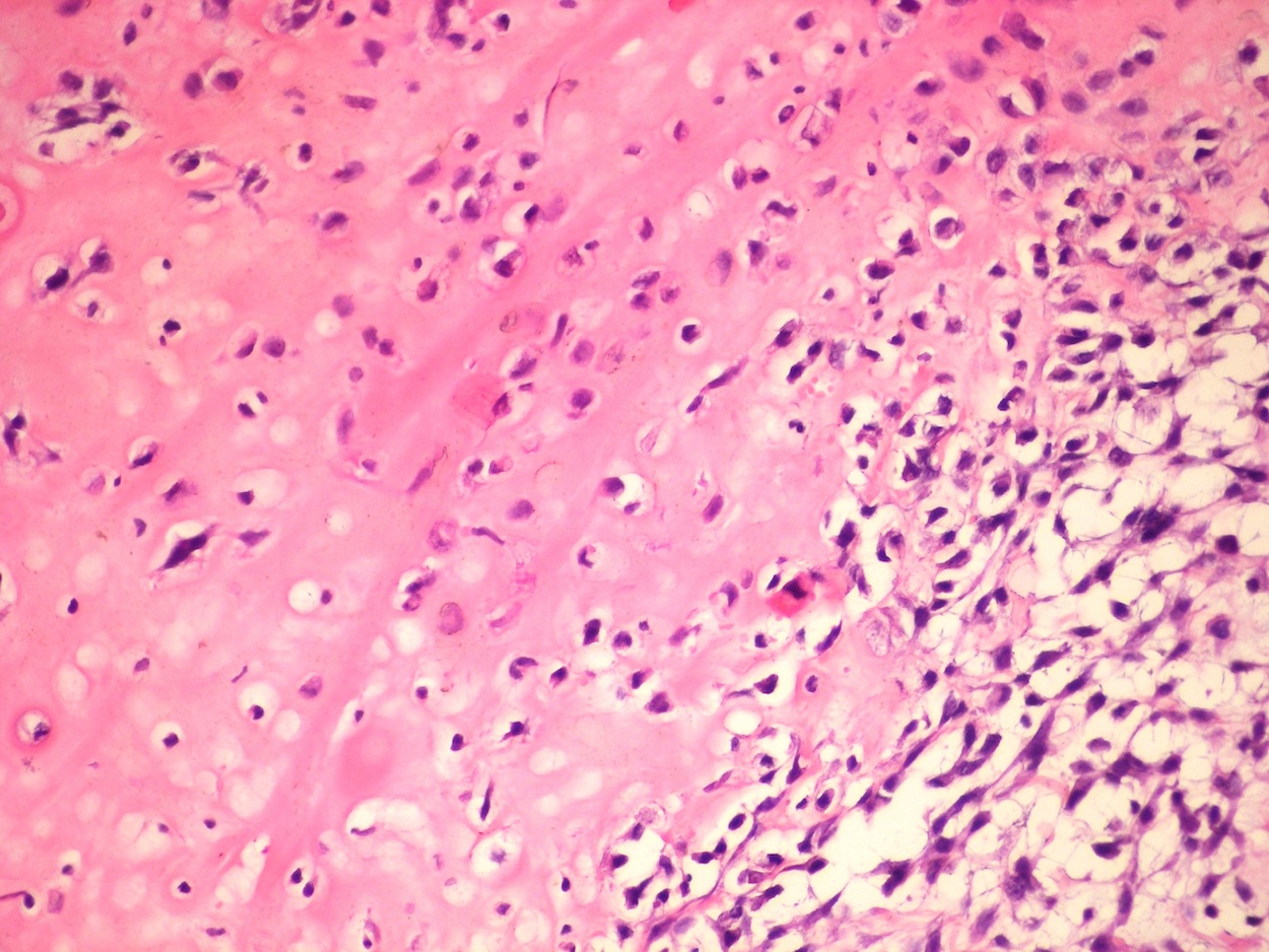
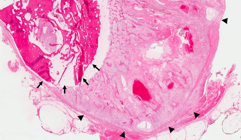
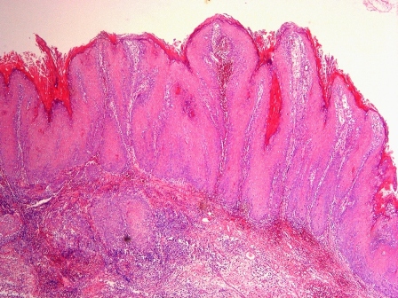
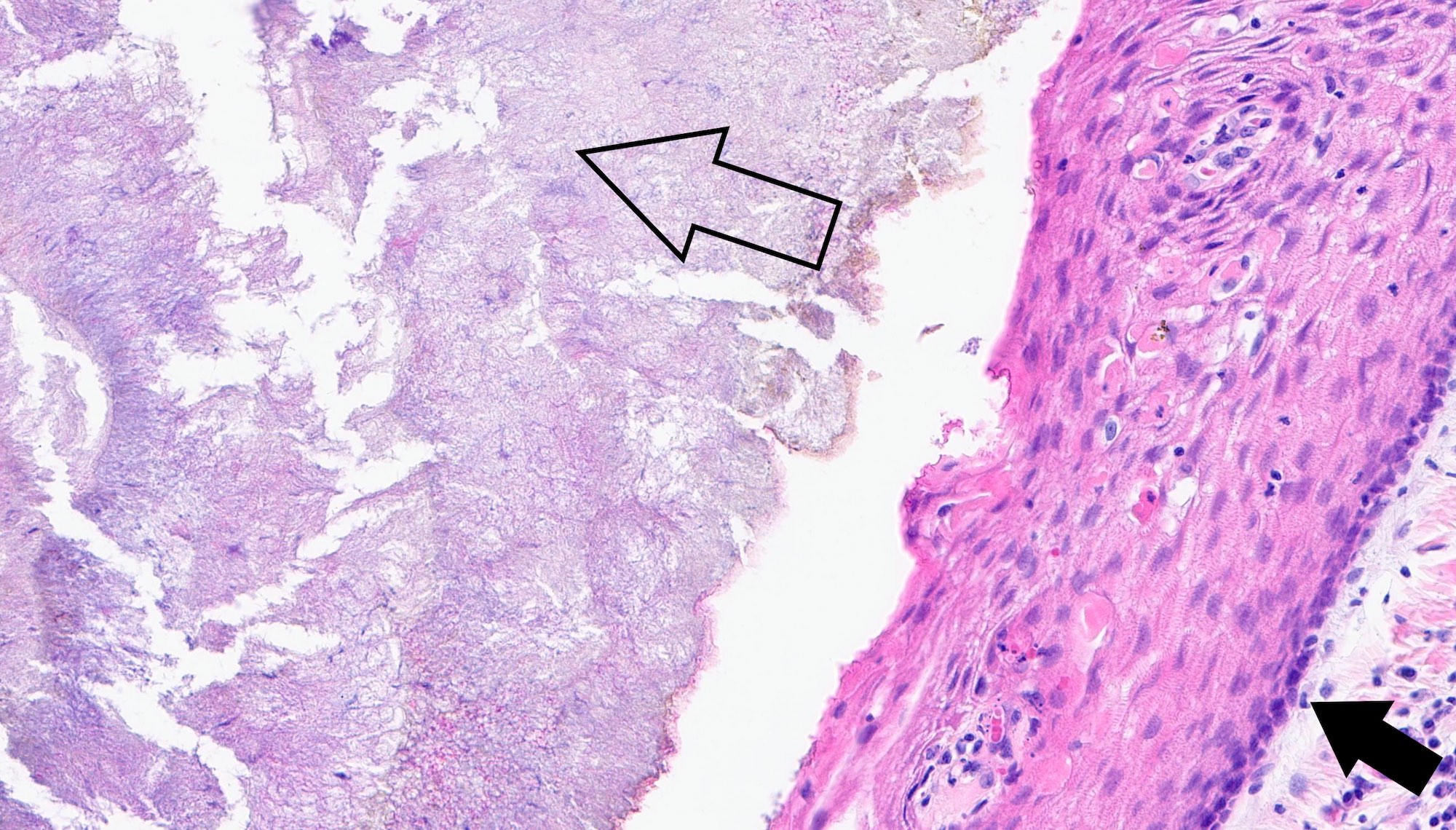
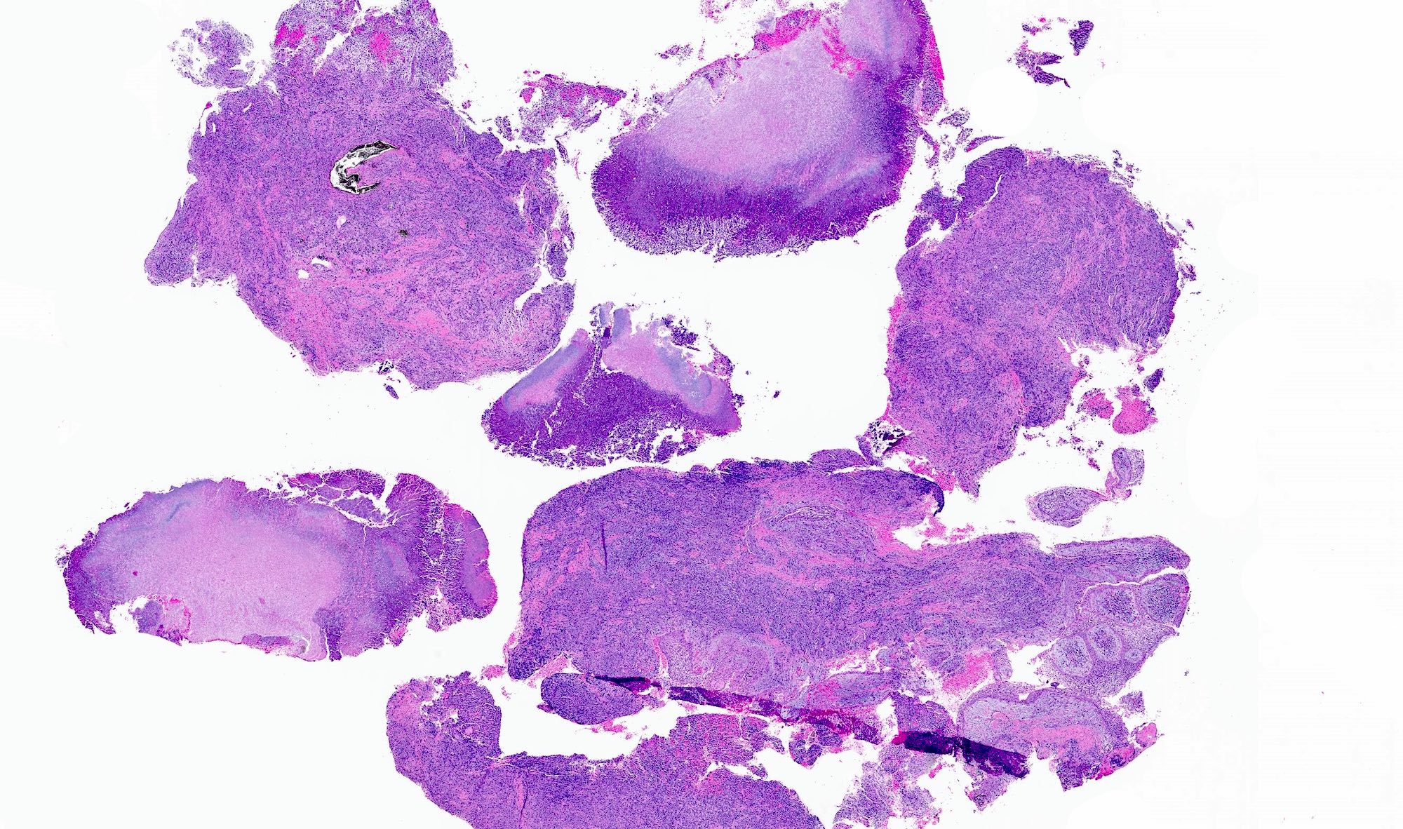
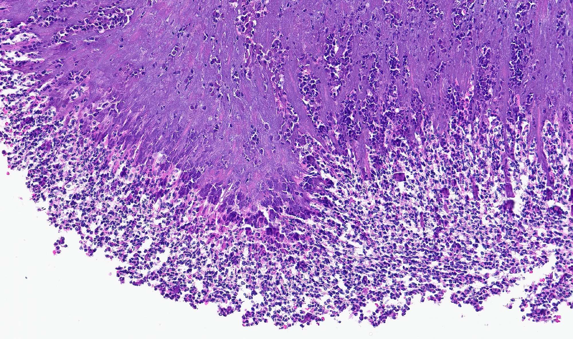
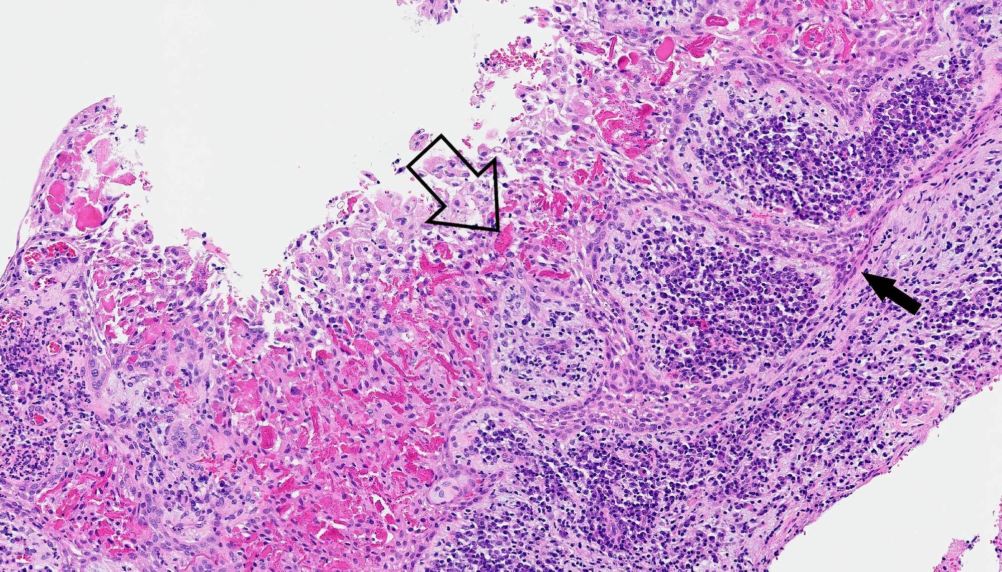
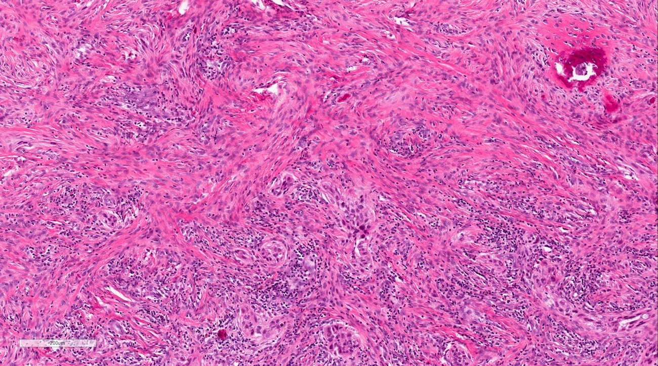
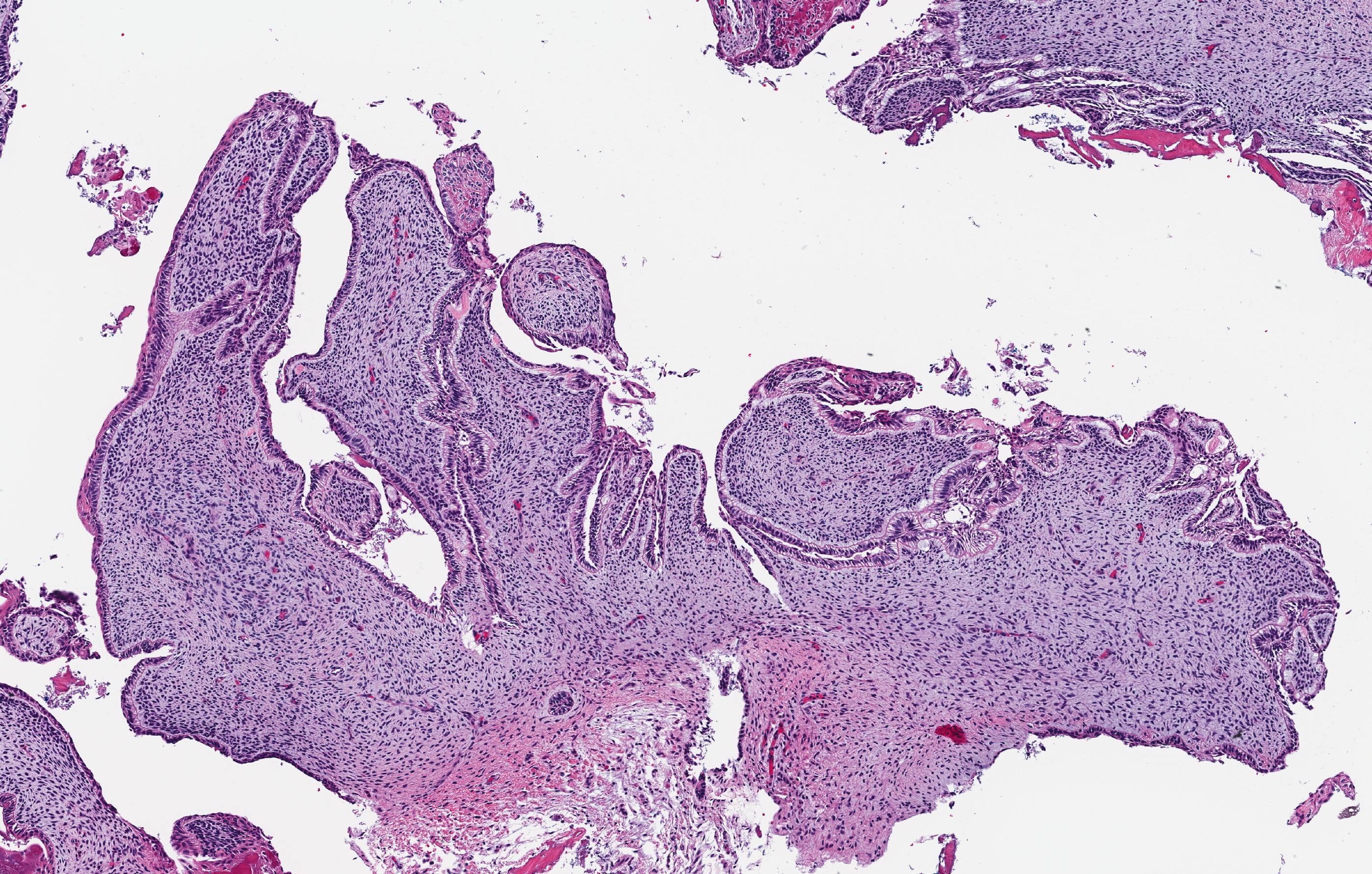
Thick fibrous capsule
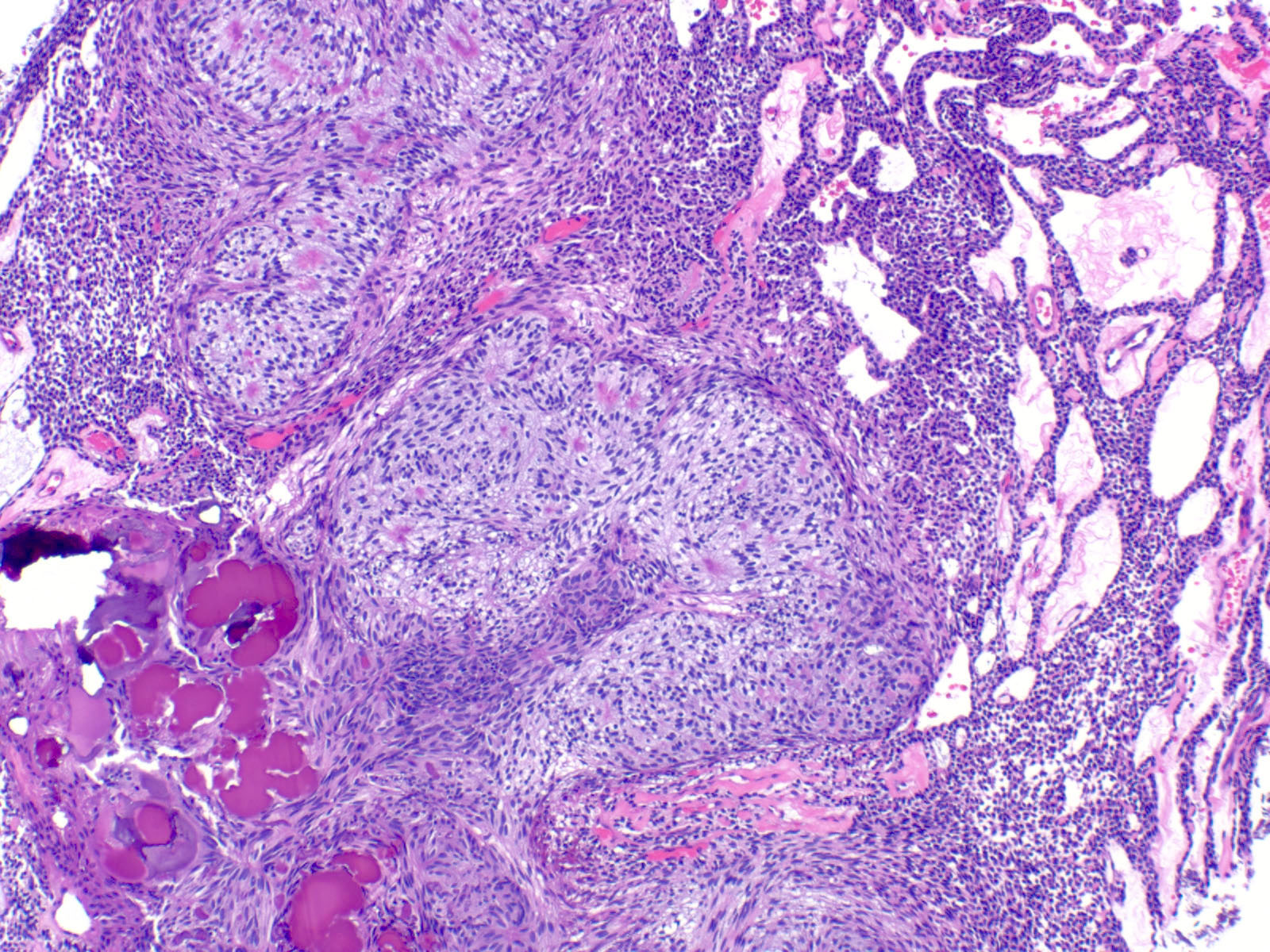
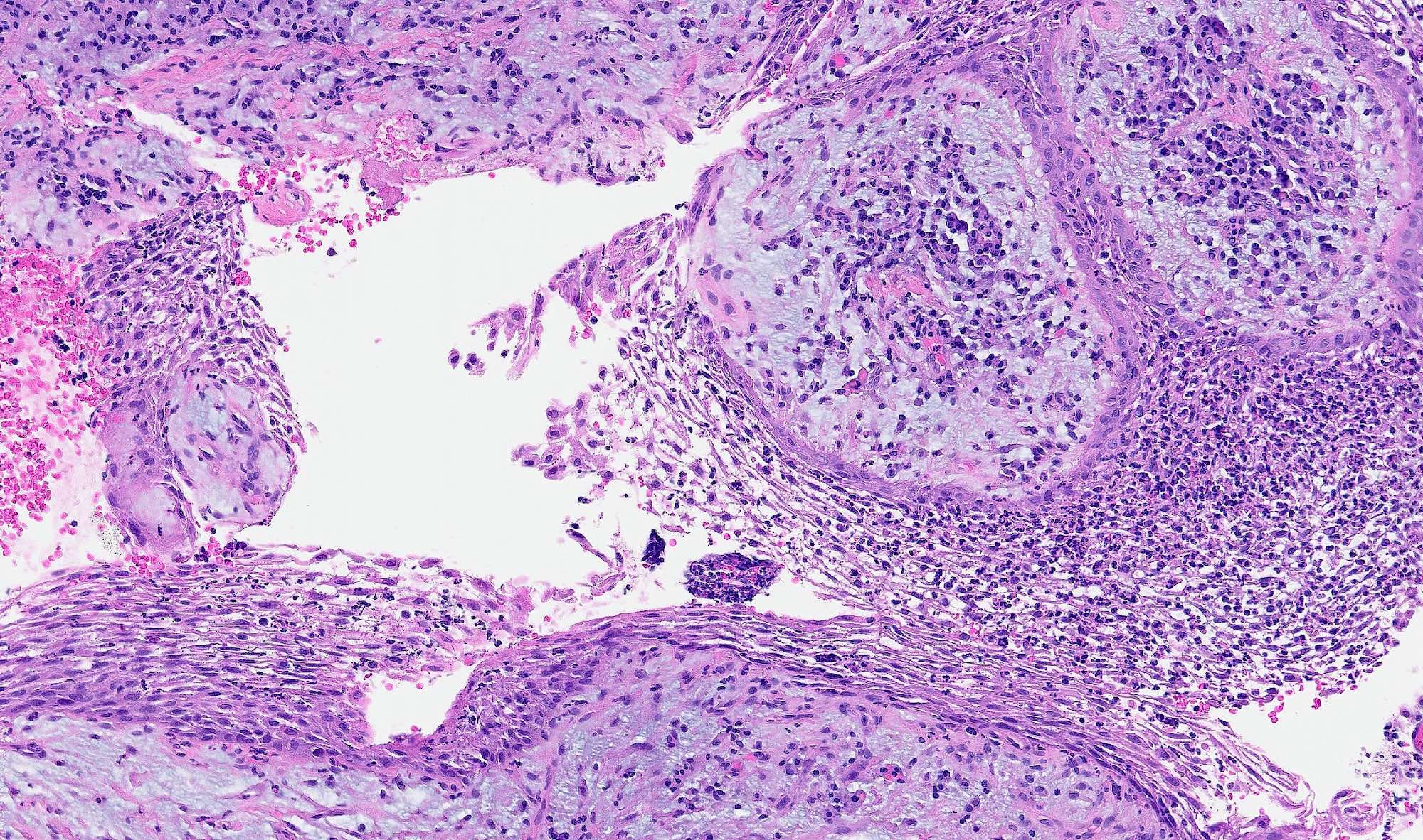
Cribriform areas
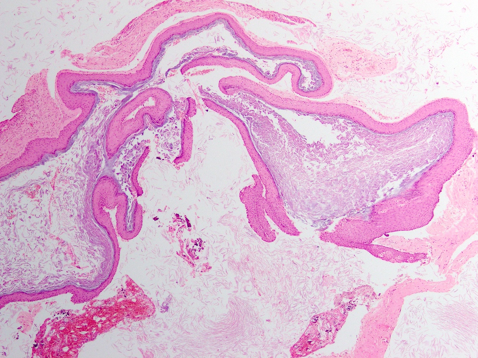
Rosette-like structure
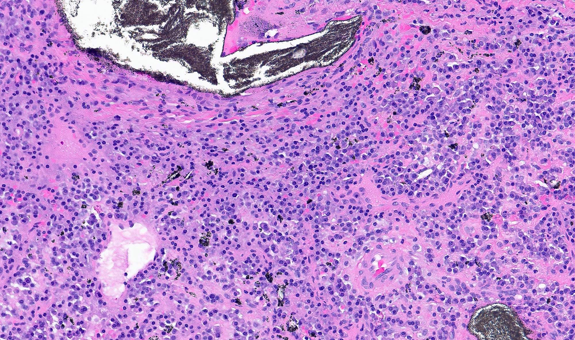
Cellular area with focal matrix deposition
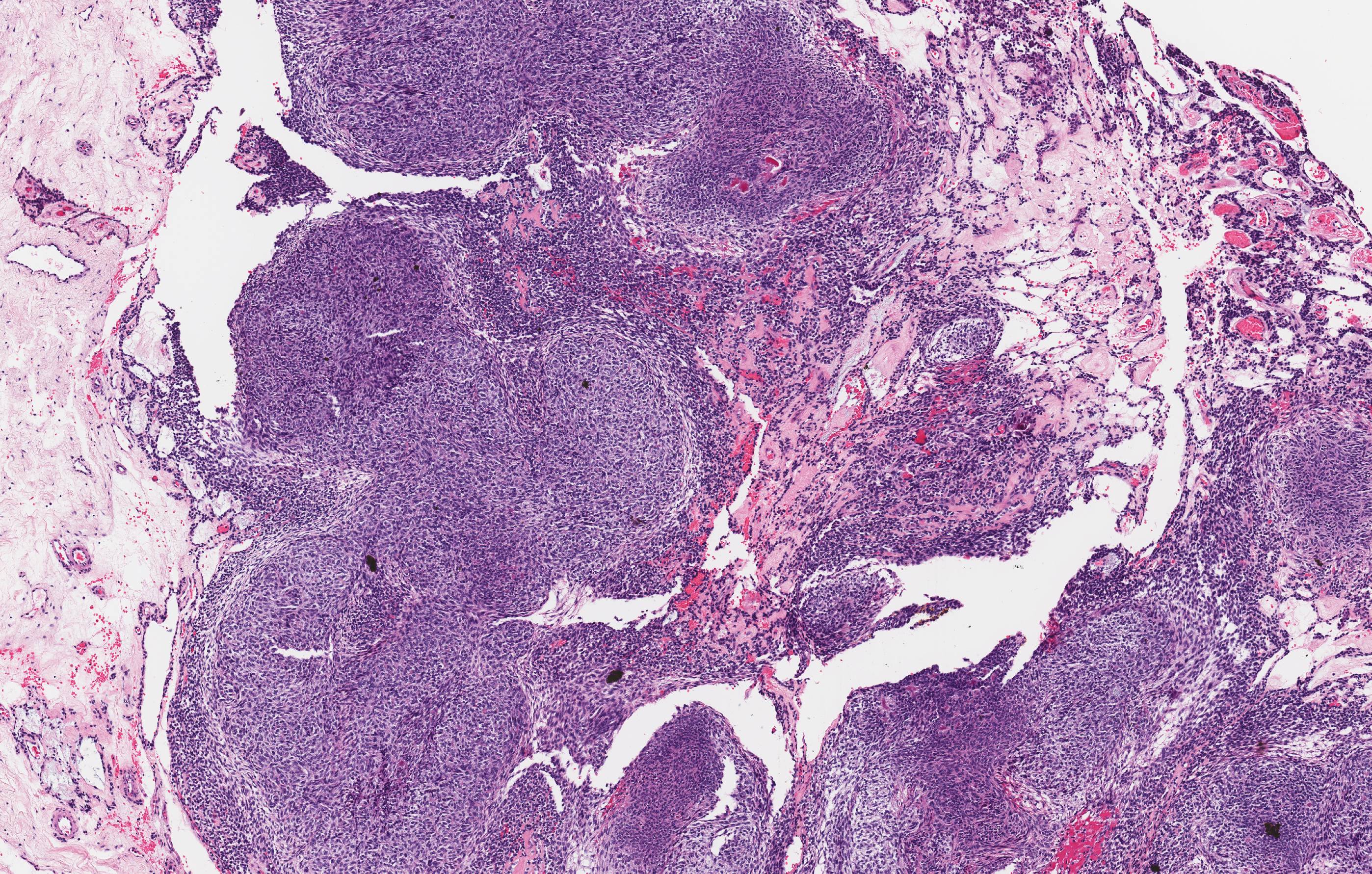
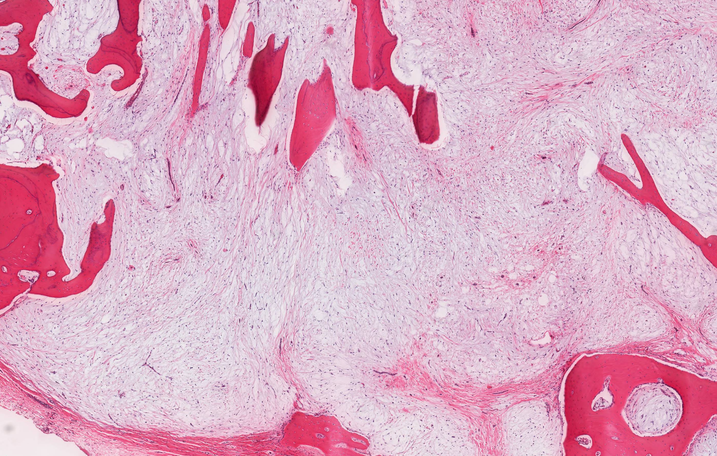
Nodular appearance

Thick capsule
Images hosted on other servers:

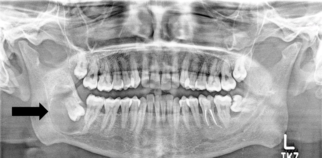
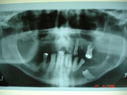
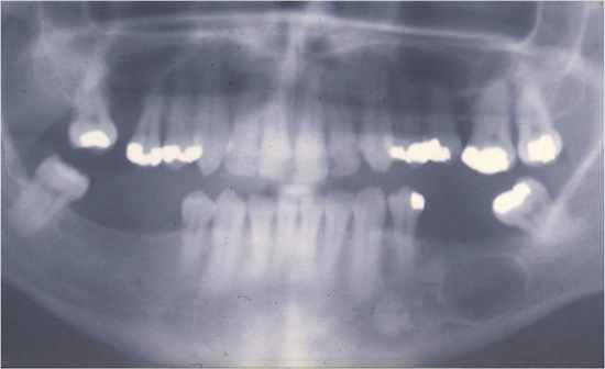
Orthopantomogram

Lucent lesion

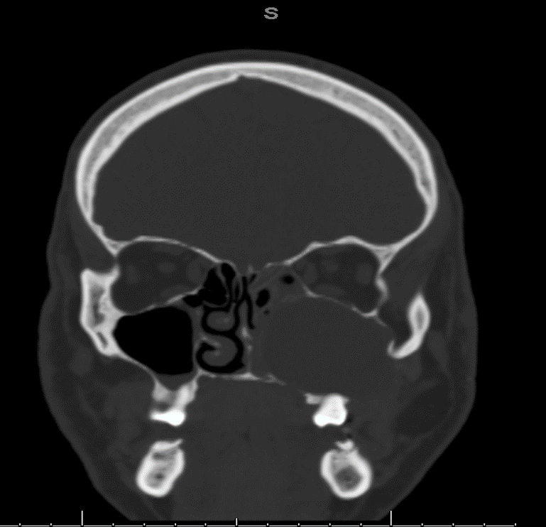
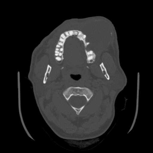
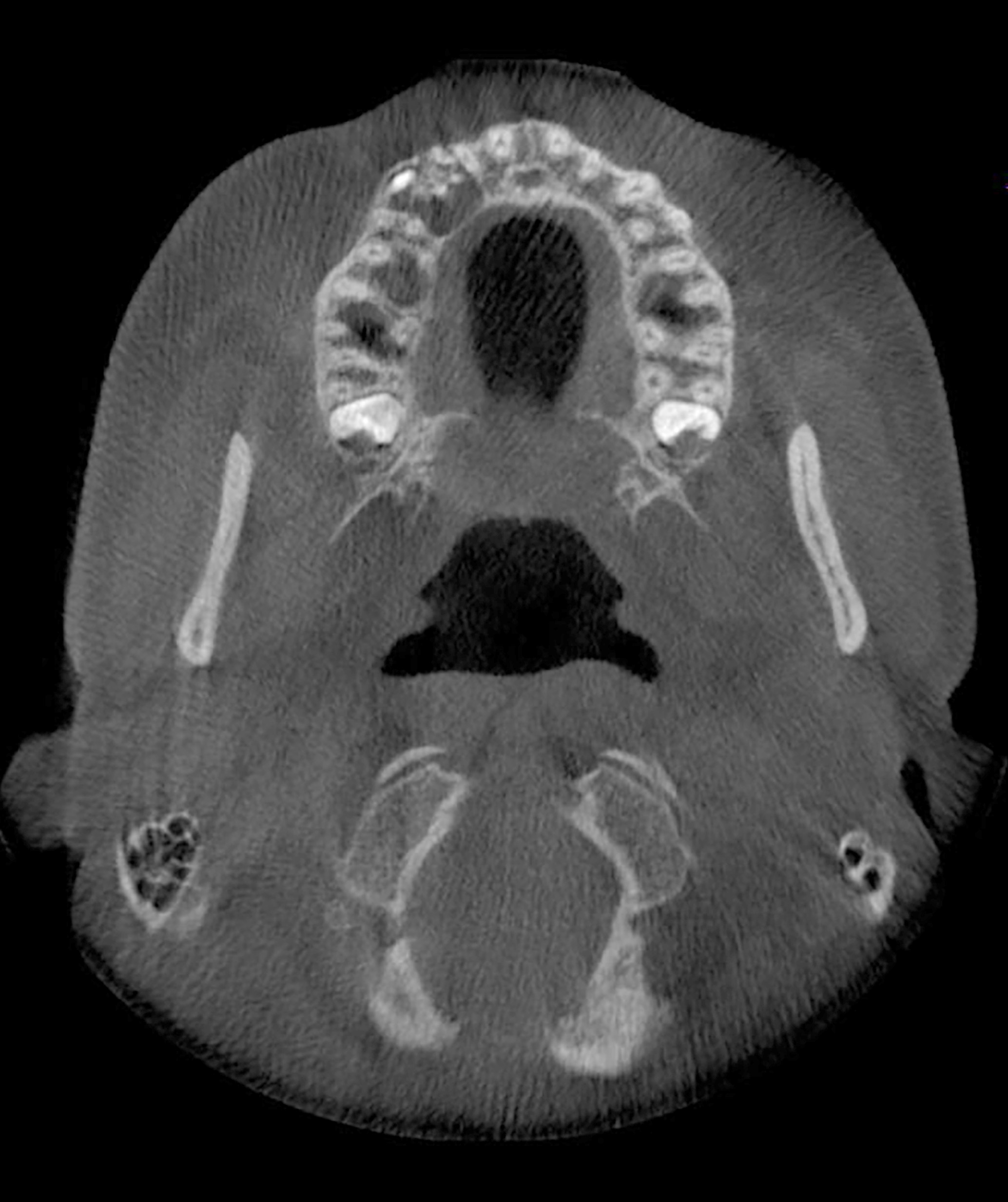
Axial non-contrast CT

Axial MRI
Images hosted on other servers:

Extraoral swelling

Pus discharge

Intra-oral swelling
Images hosted on other servers:

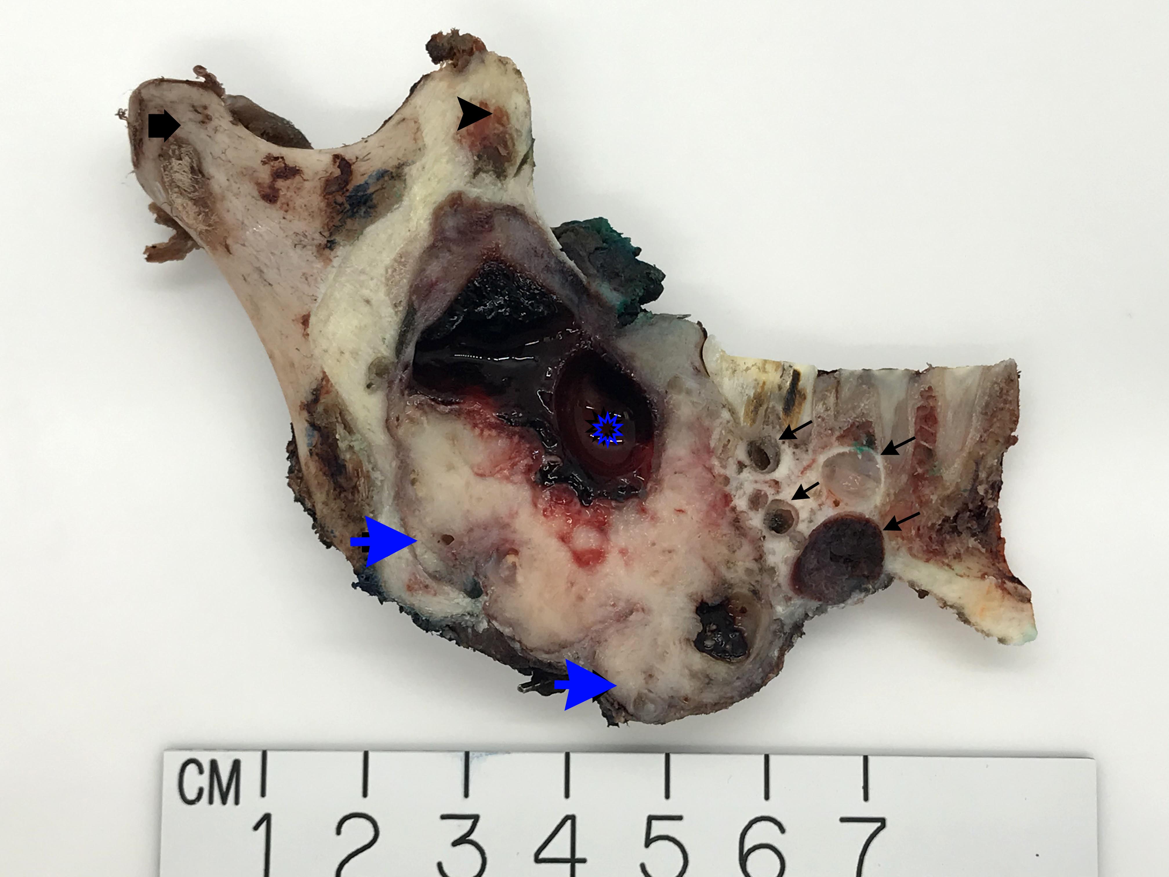
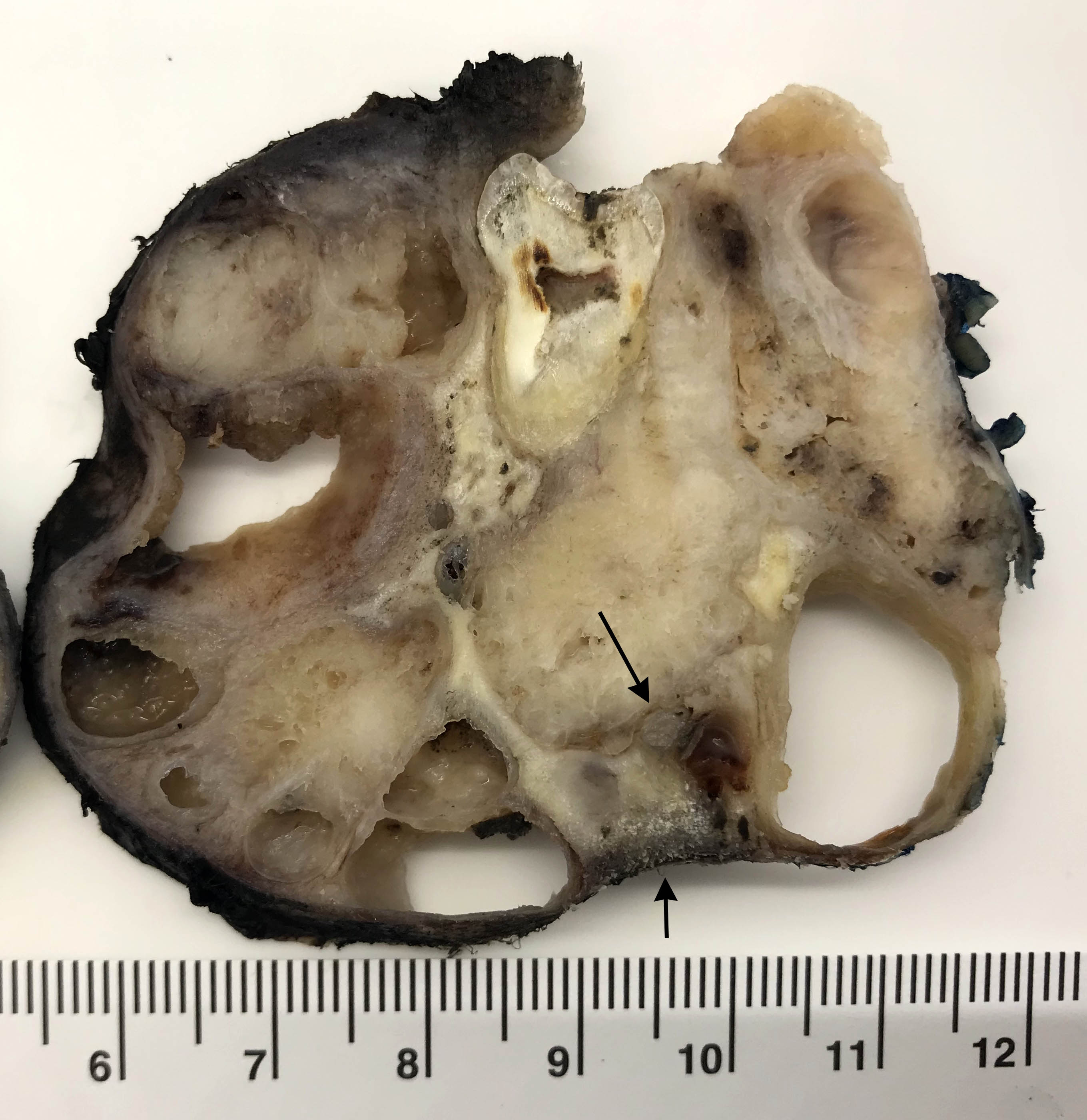
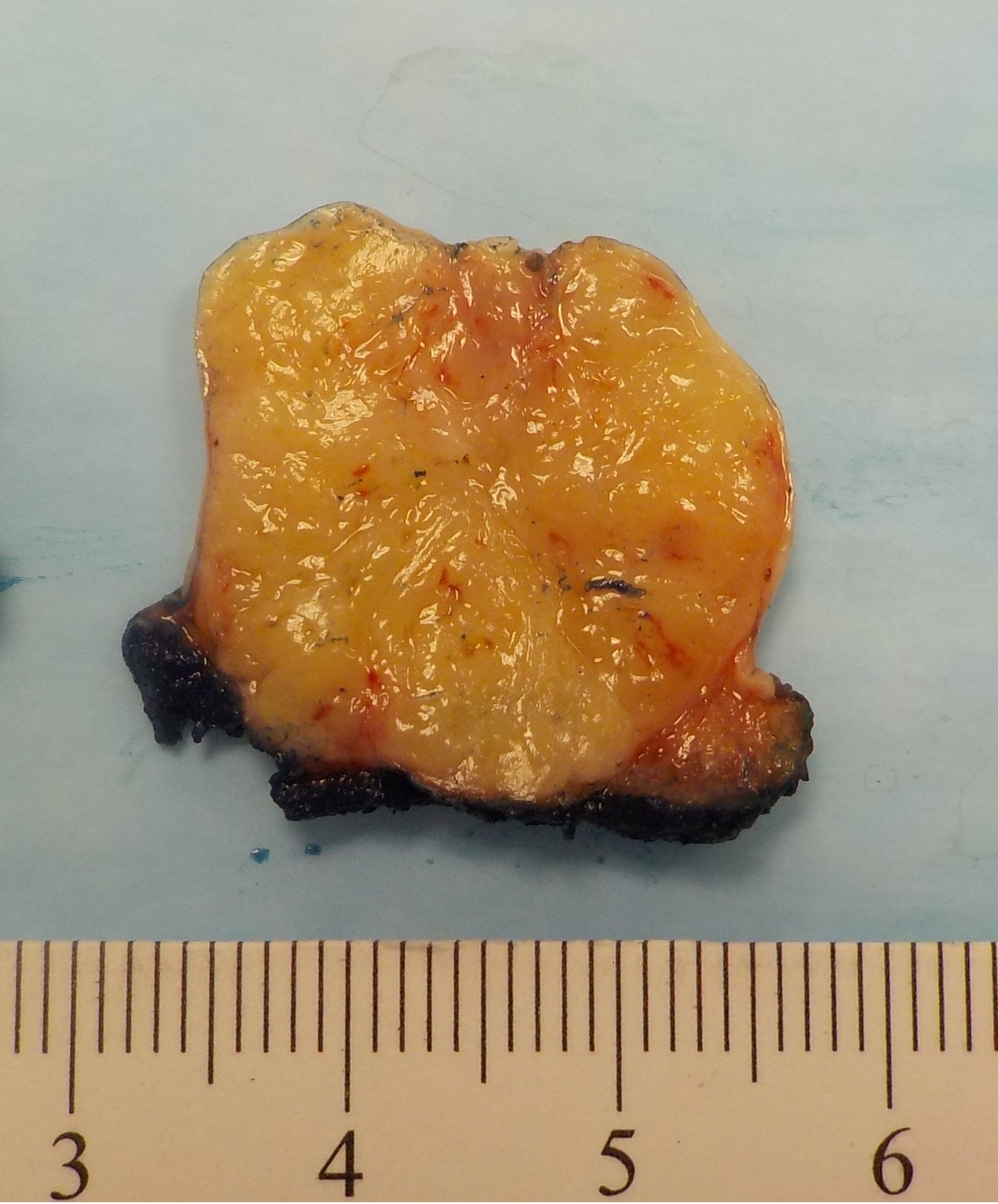
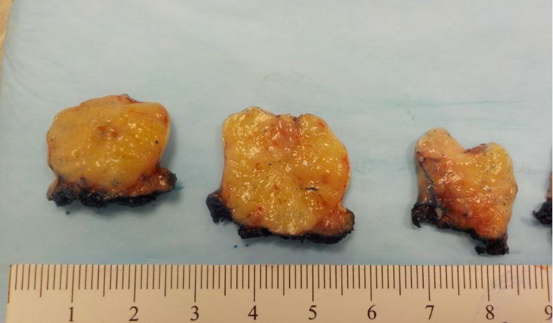
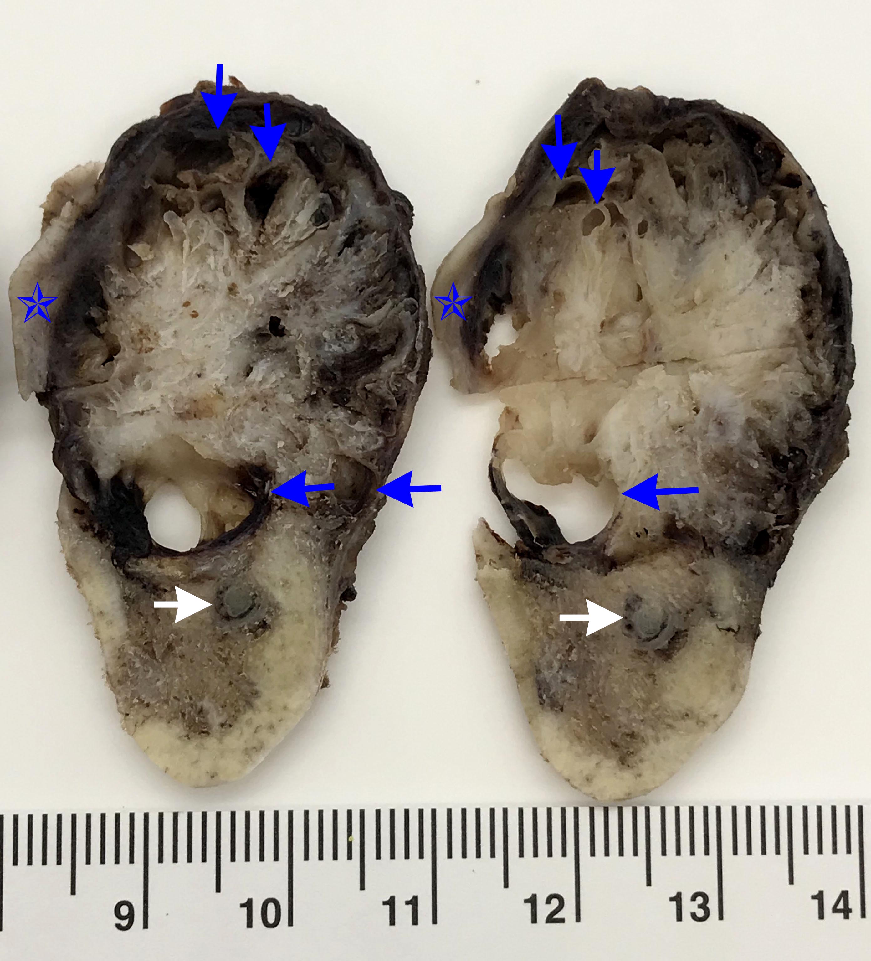
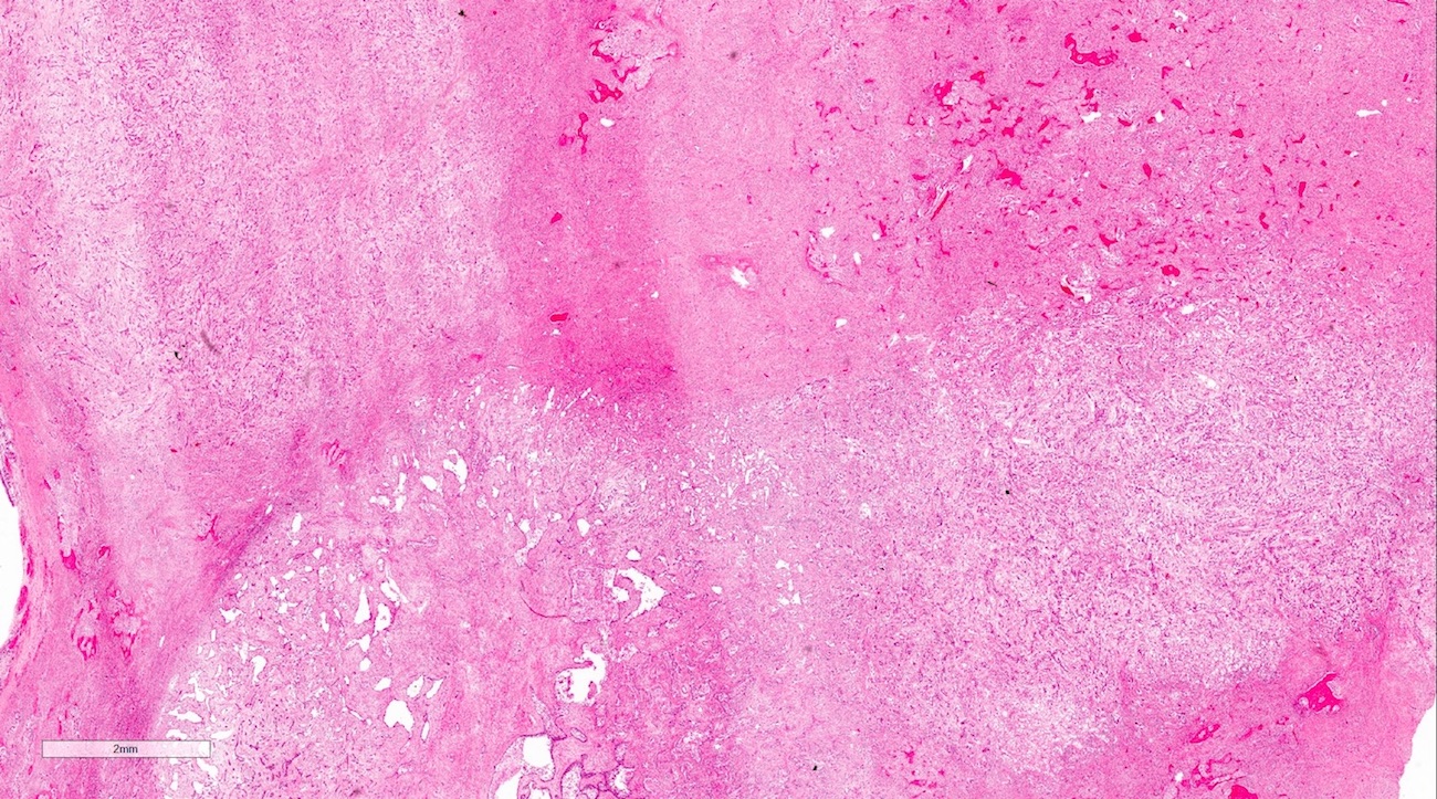
Resected specimen
Contributed by Kelly Magliocca, D.D.S., M.P.H.
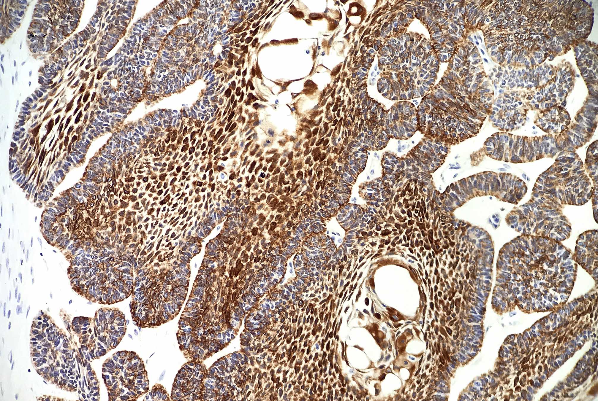
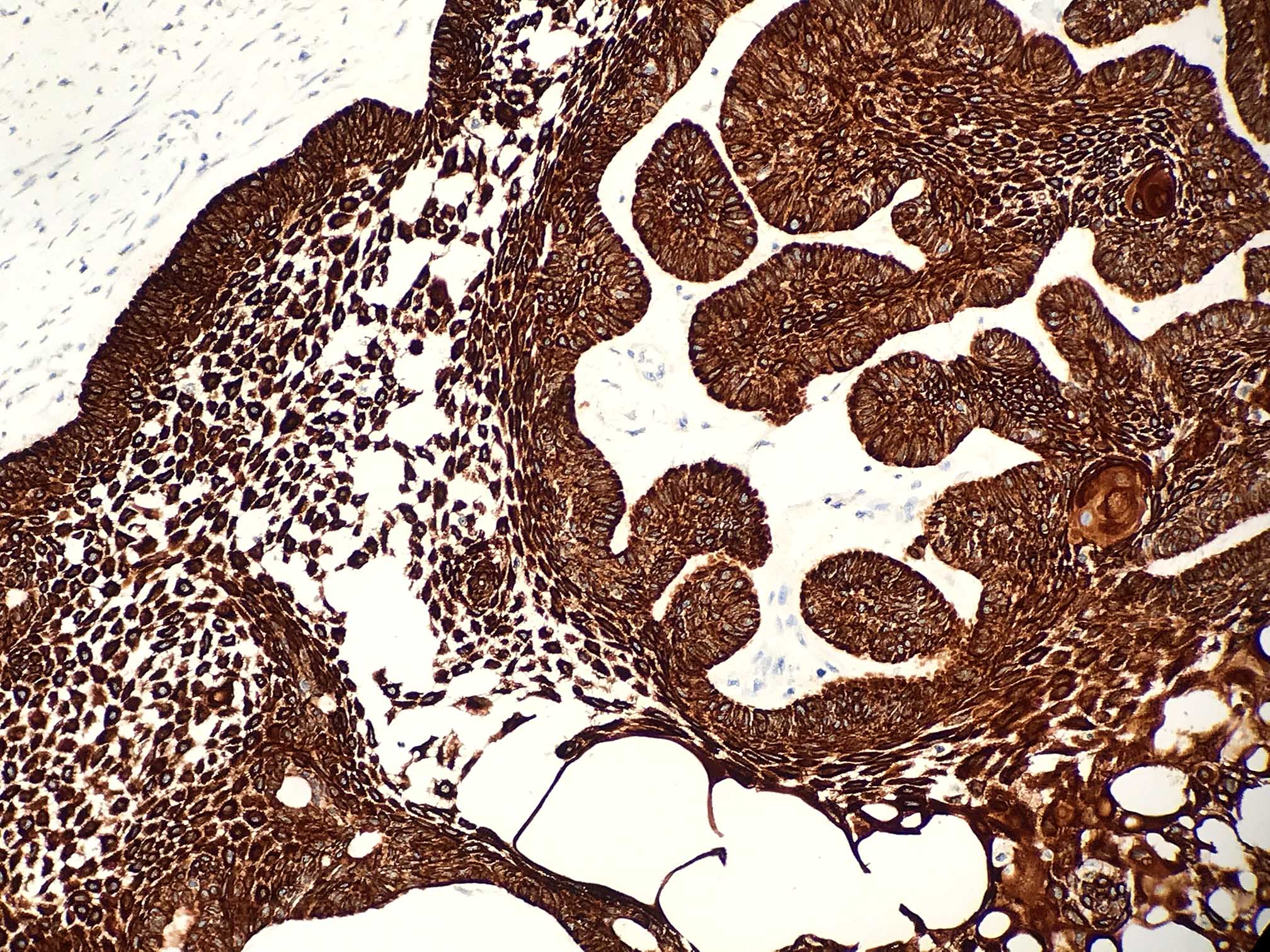
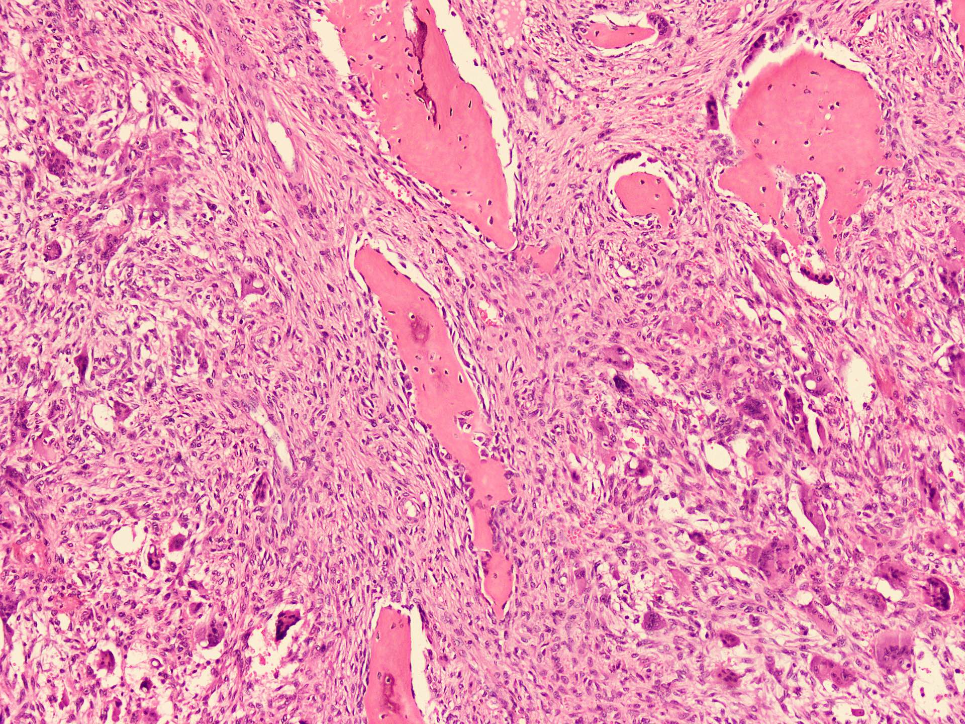
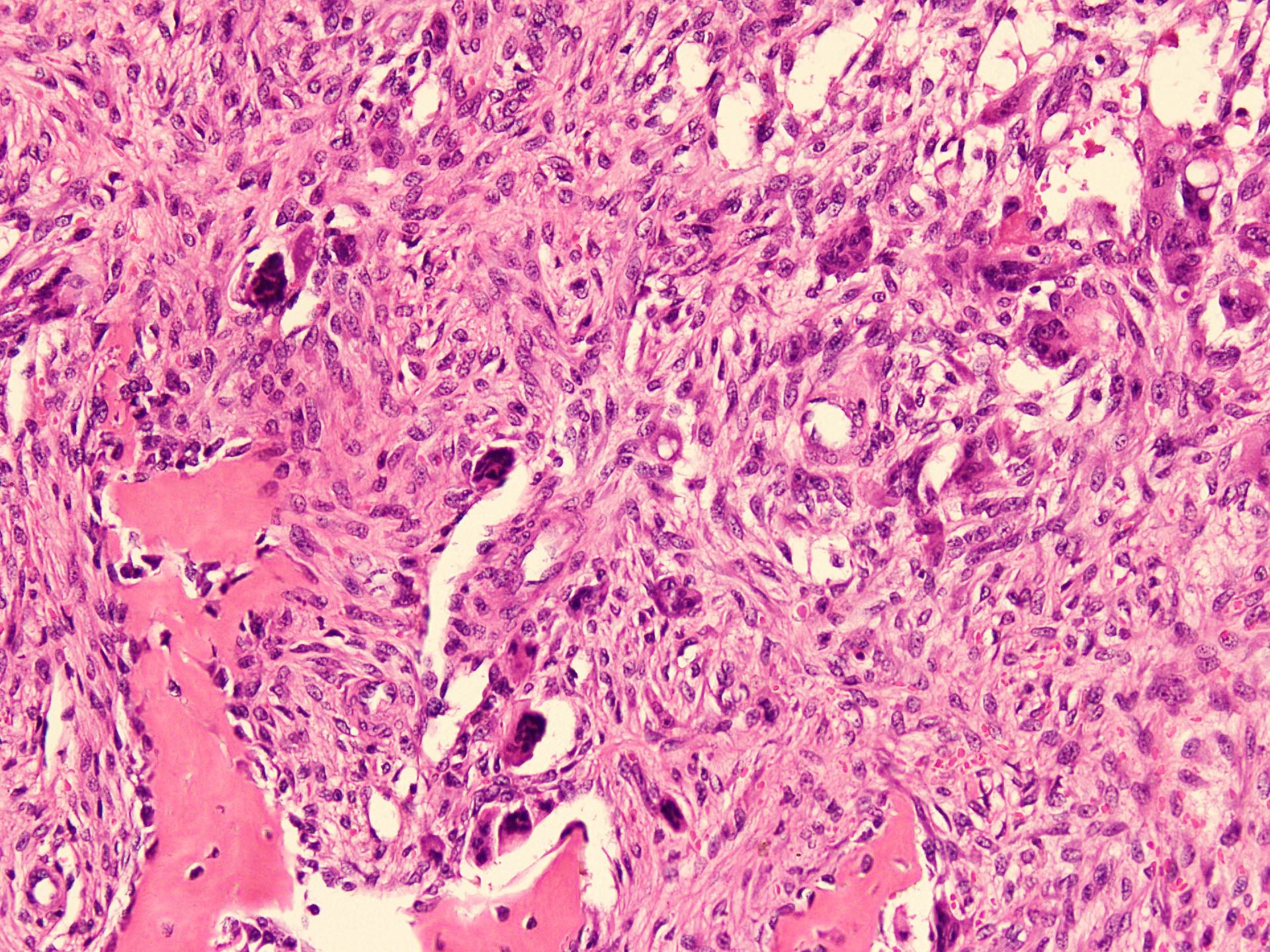
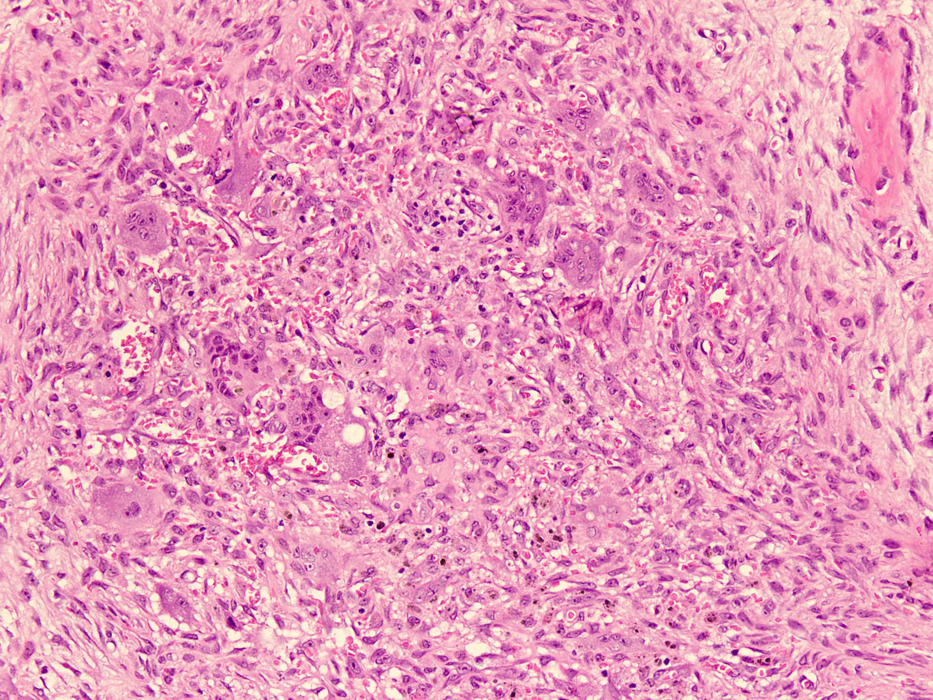
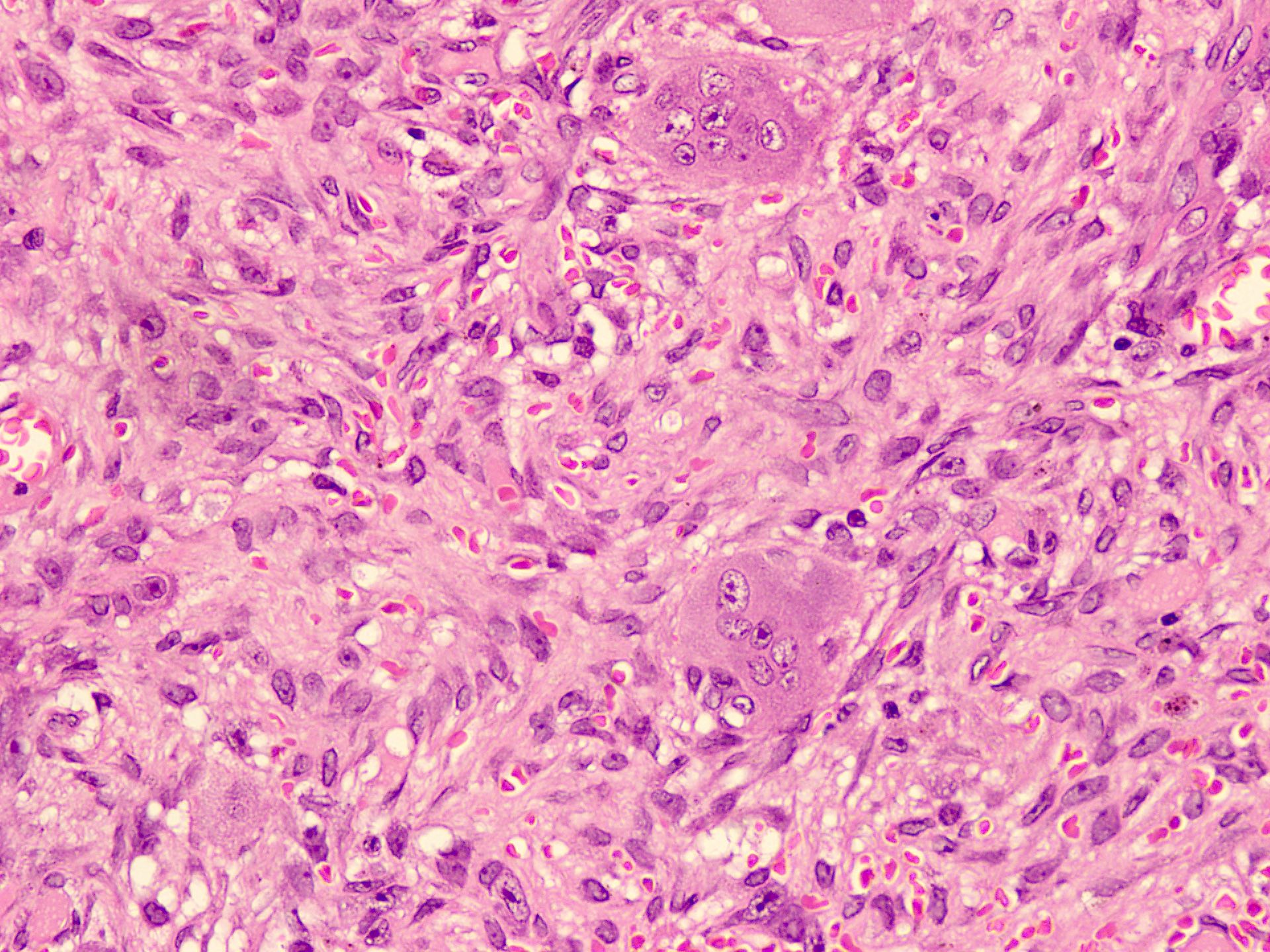
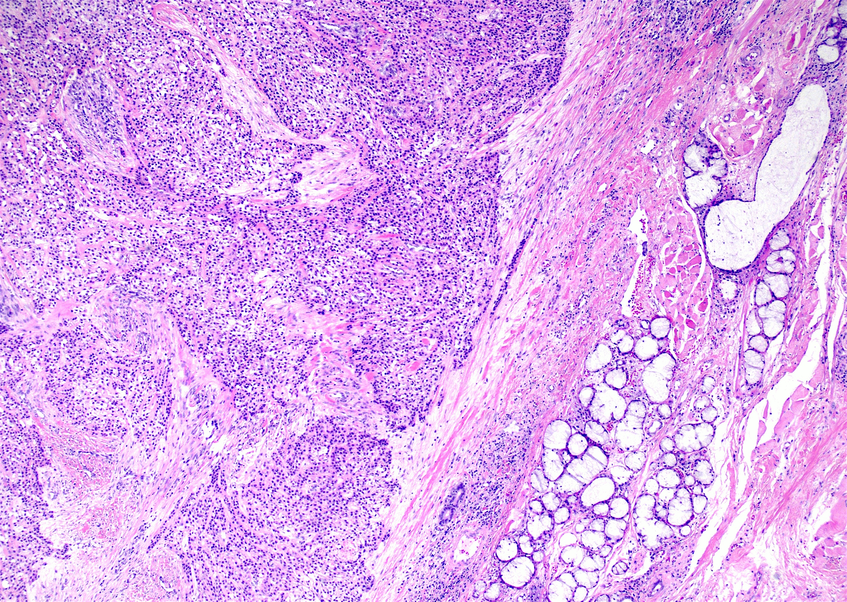
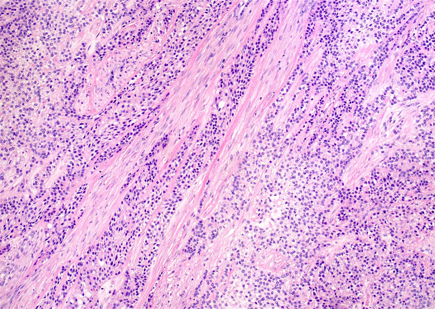
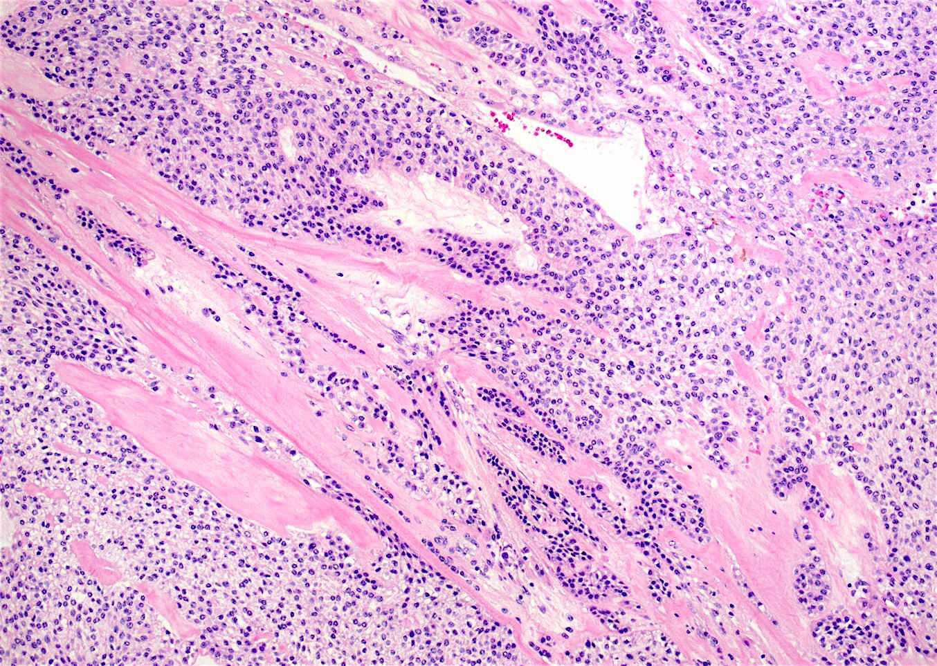
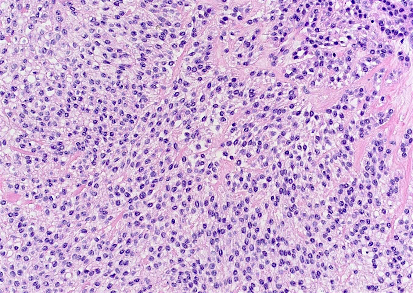
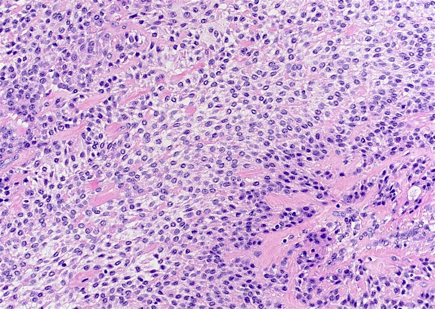
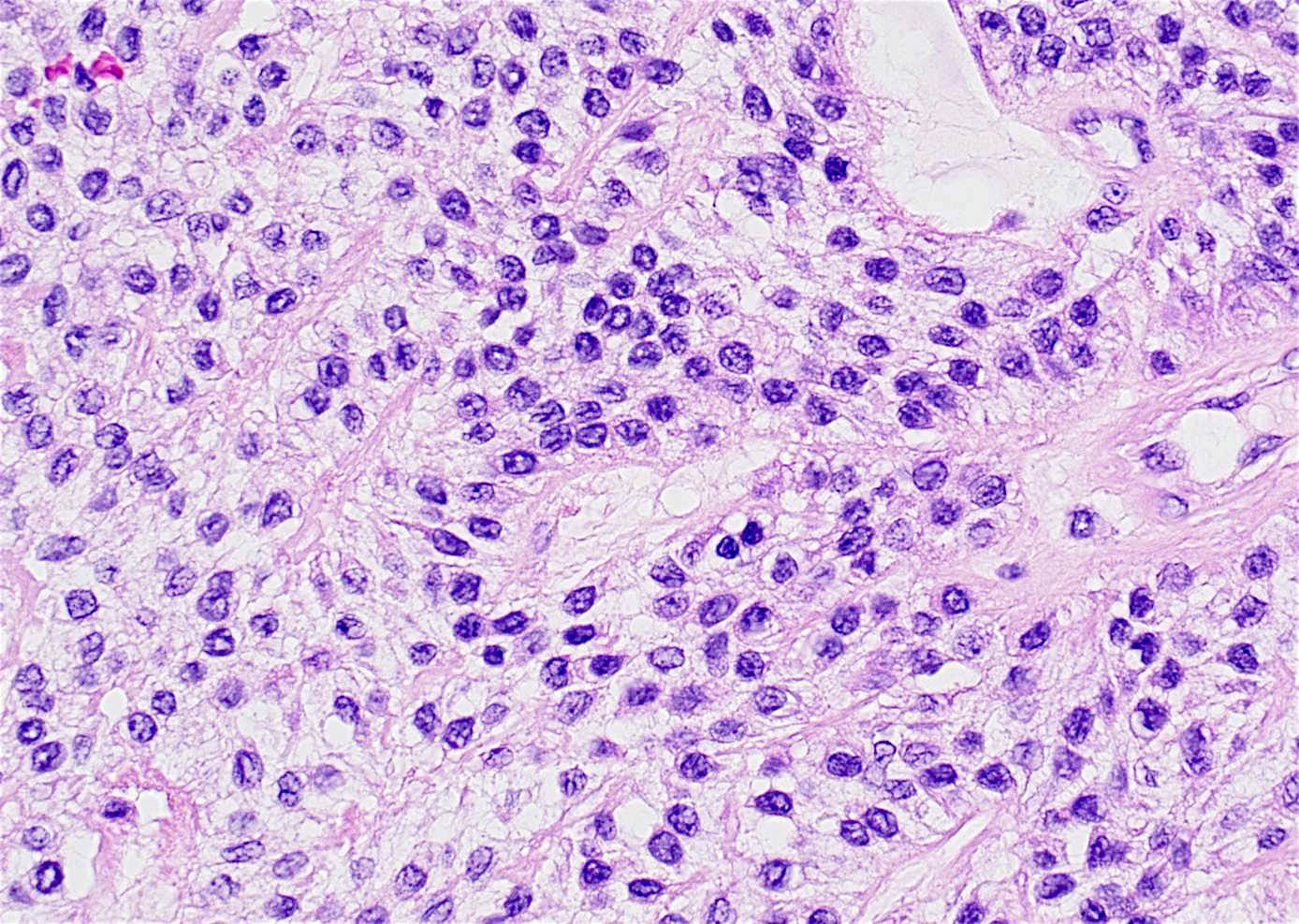
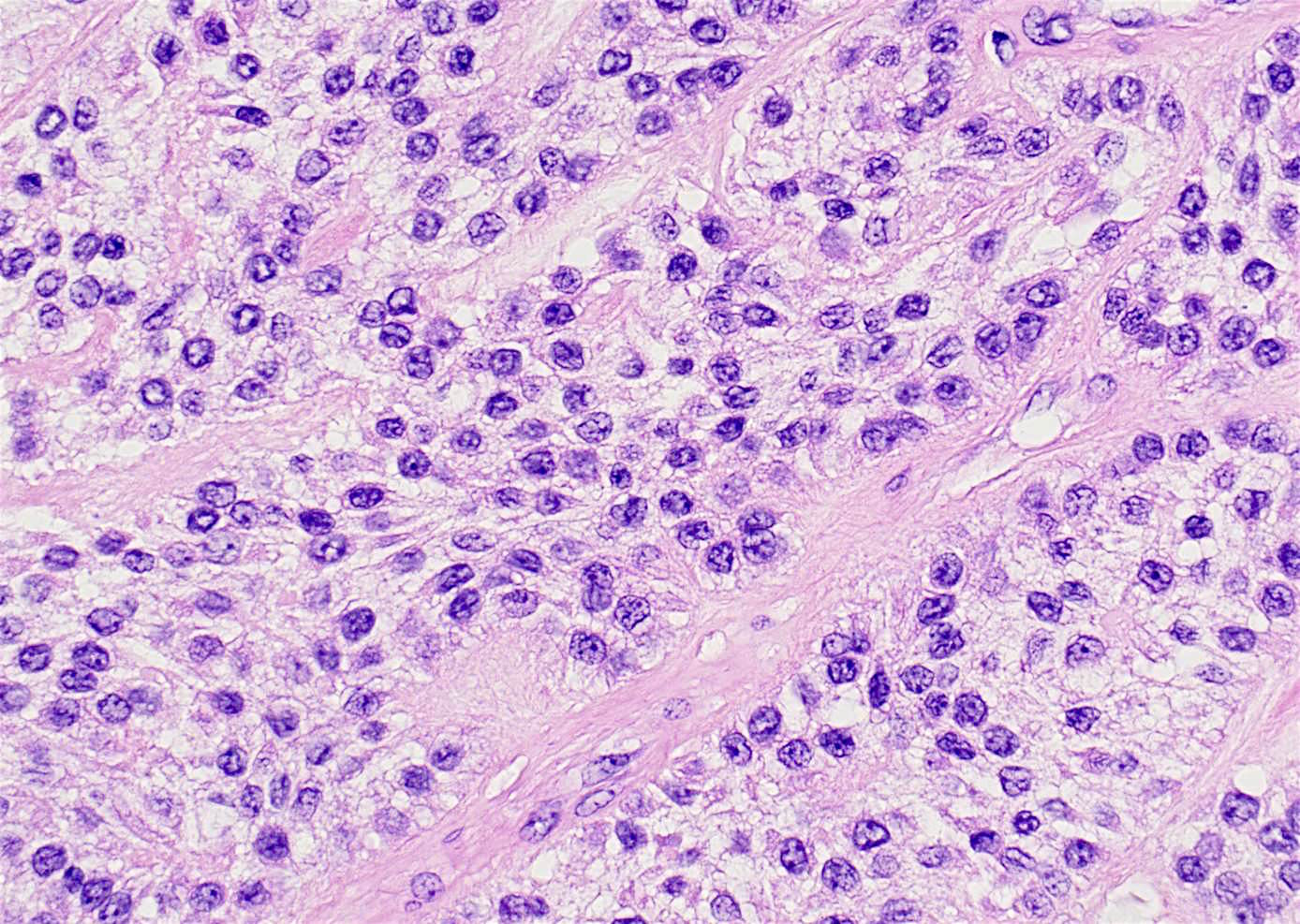
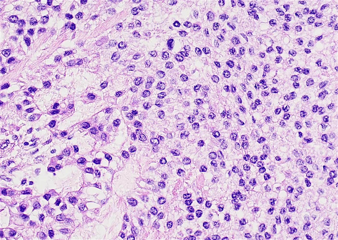
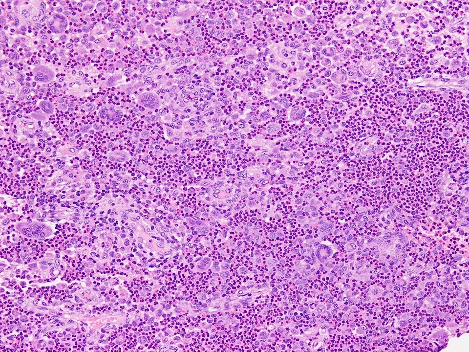
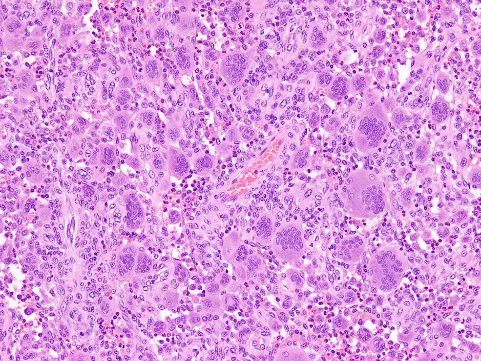
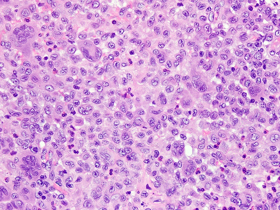
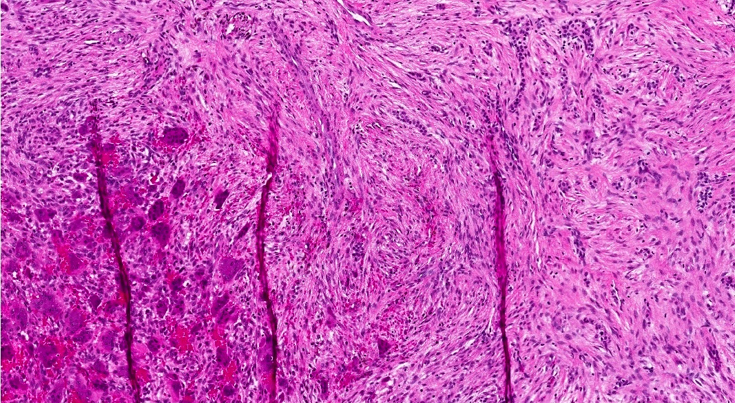
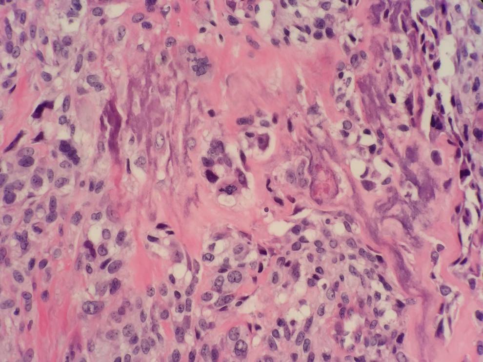
Ameloblastic carcinoma
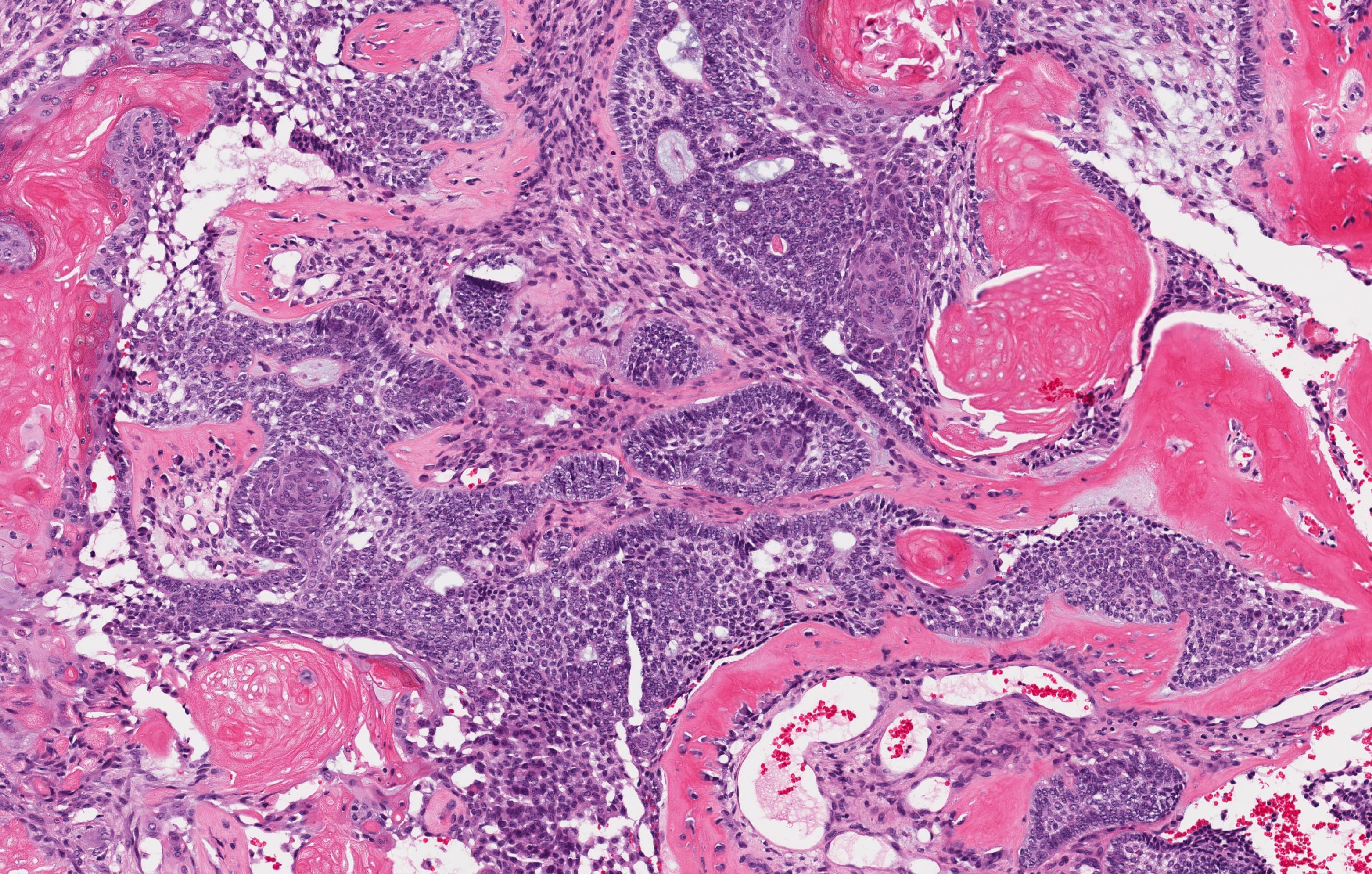
Images hosted on other servers:

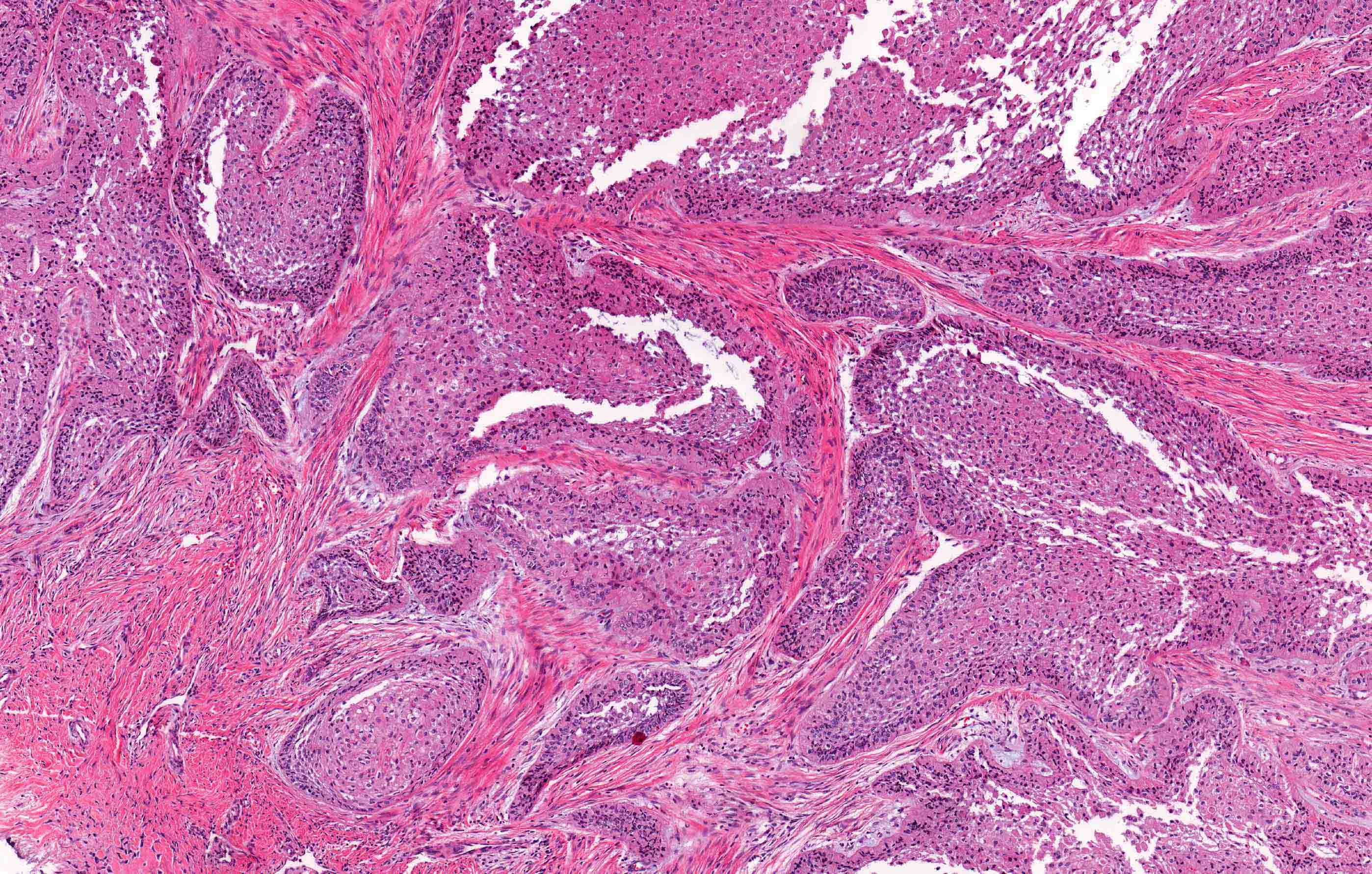
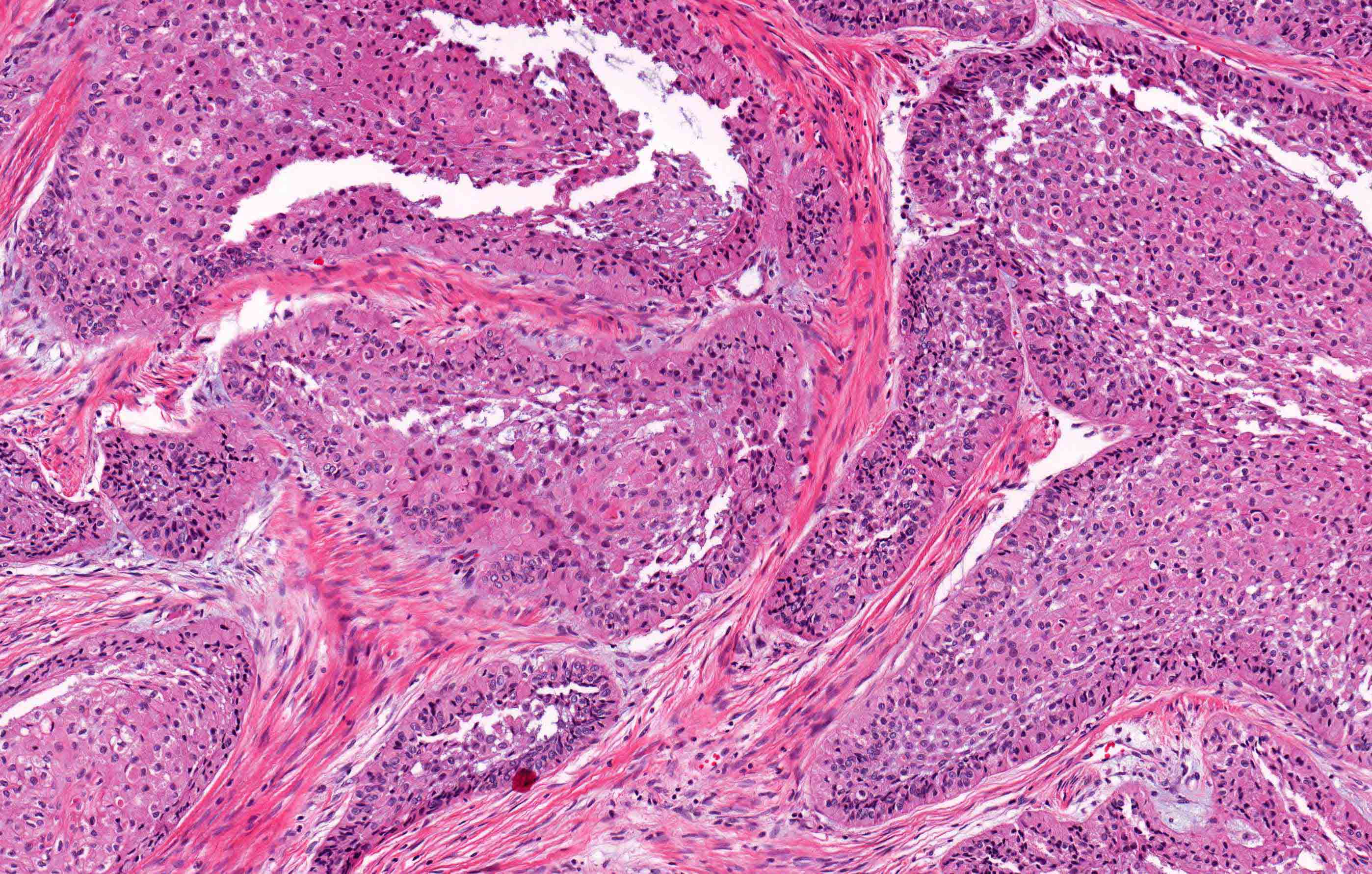
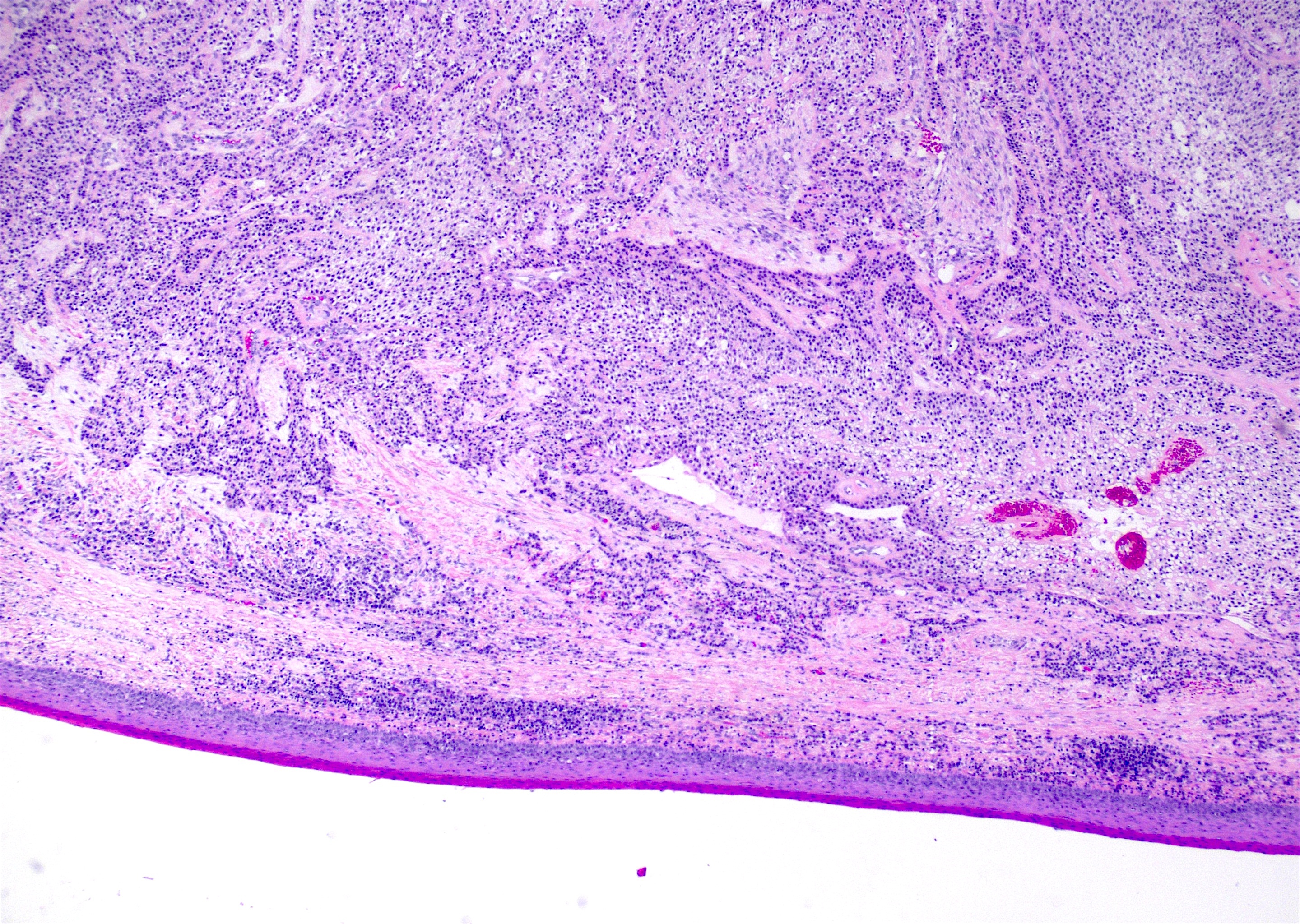
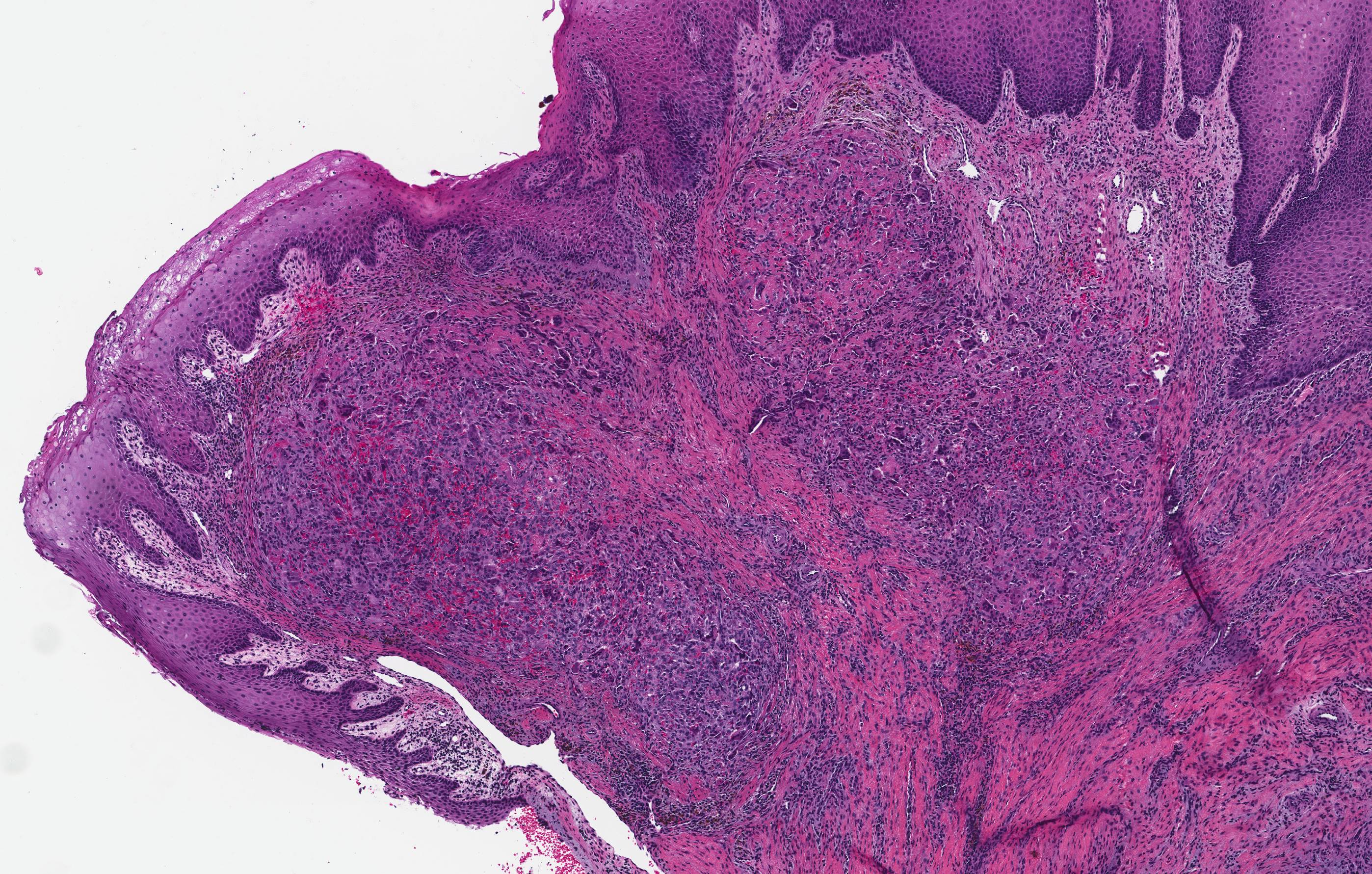
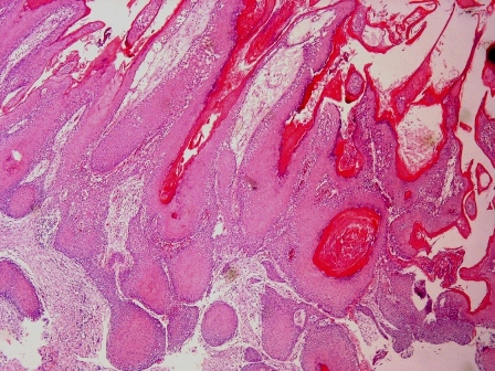
Low power

Higher power


Cytological atypia

Necrosis
Contributed by Kelly Magliocca, D.D.S., M.P.H.
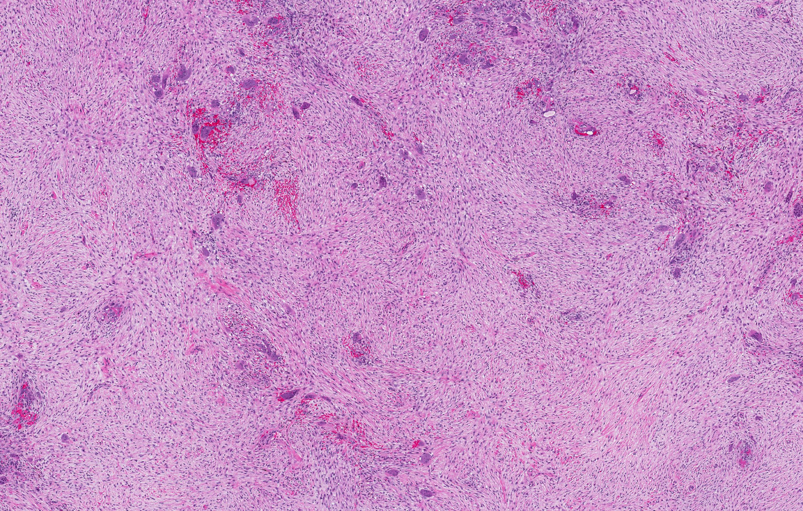
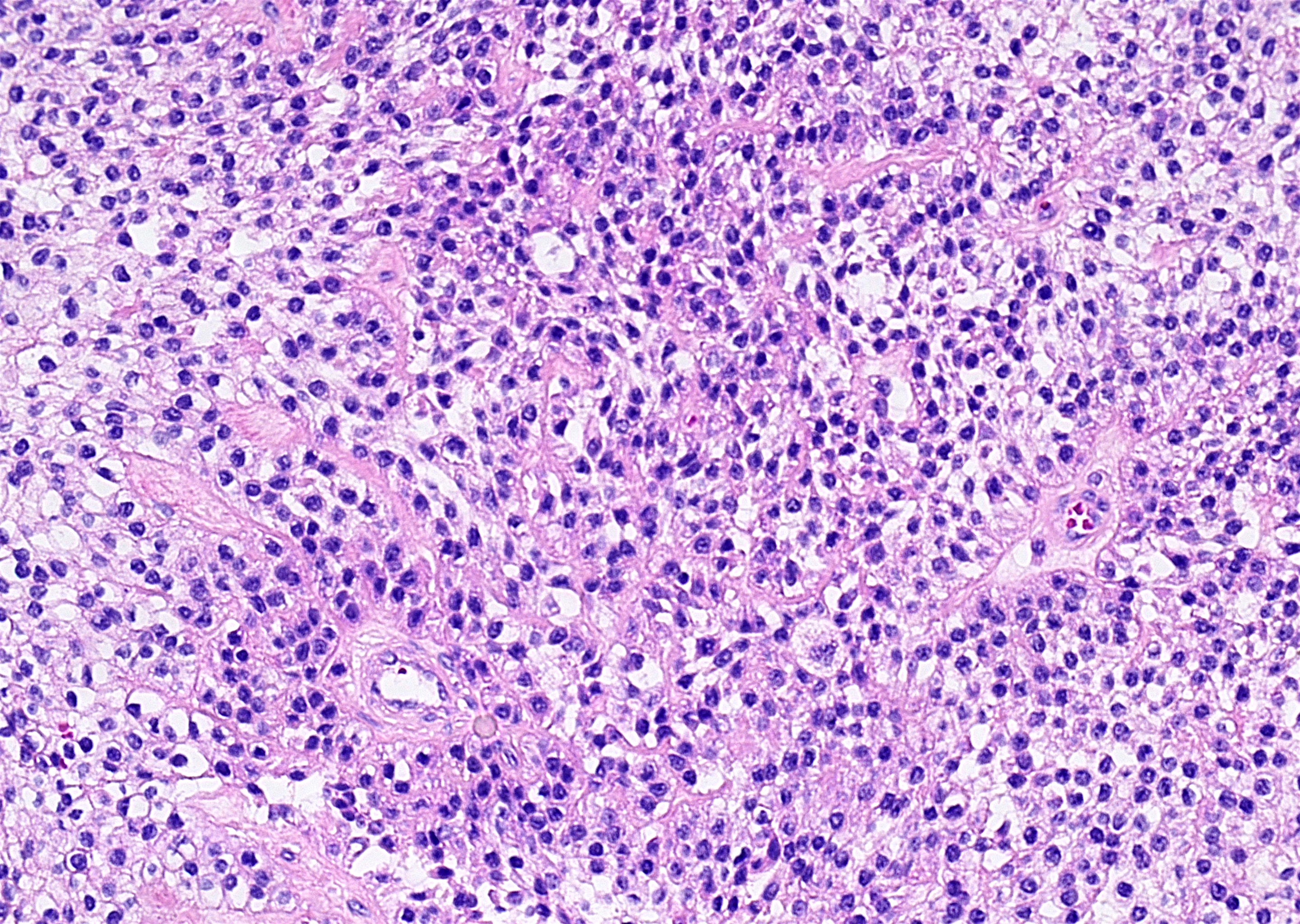
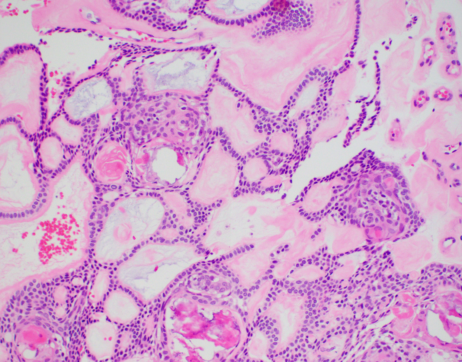
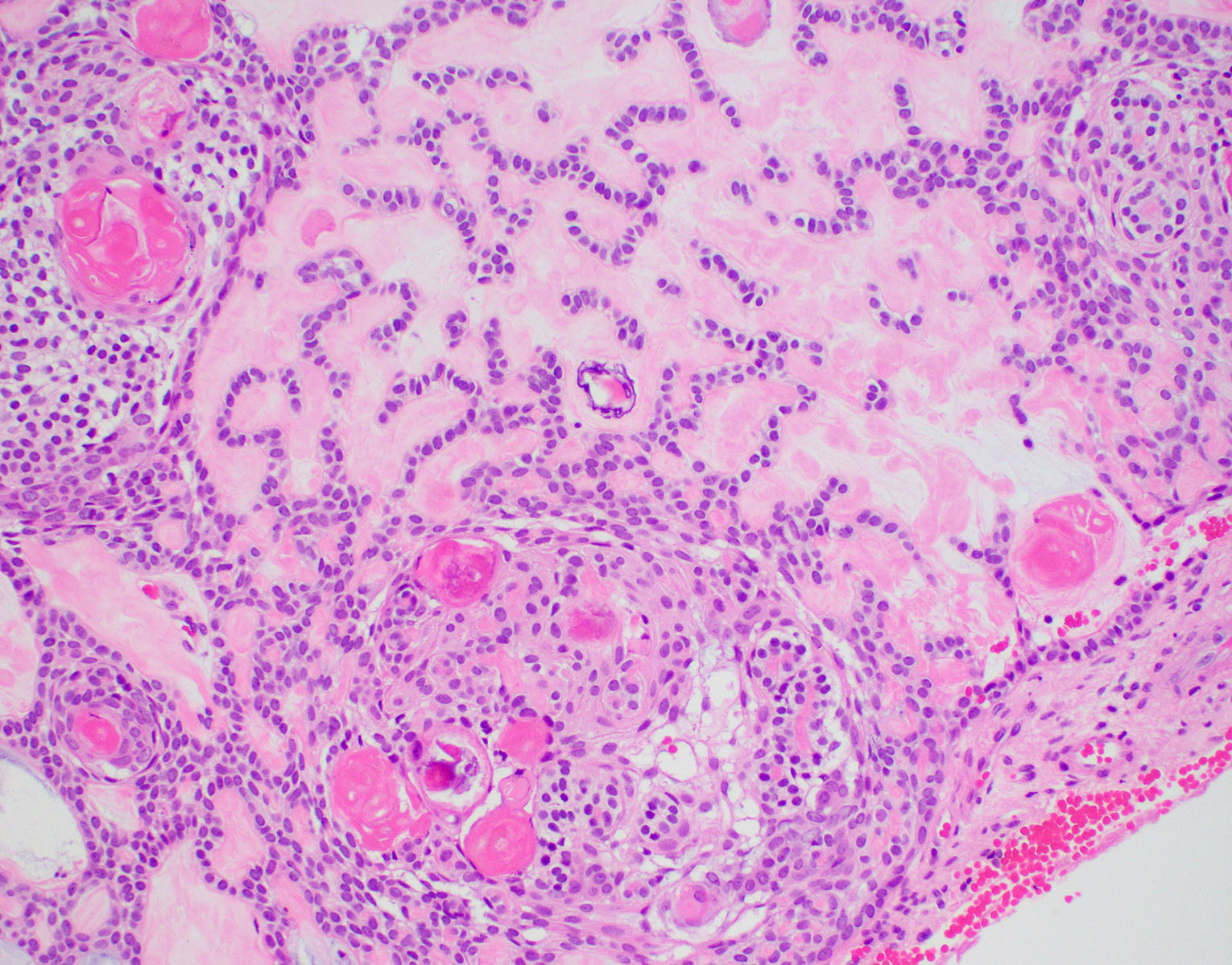
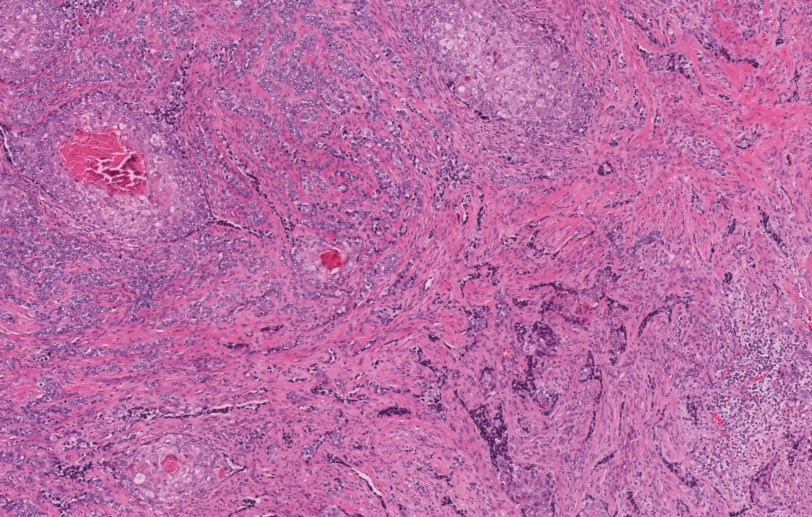
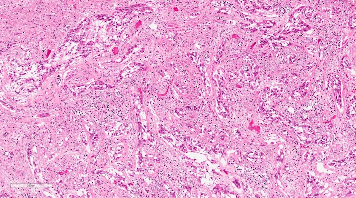
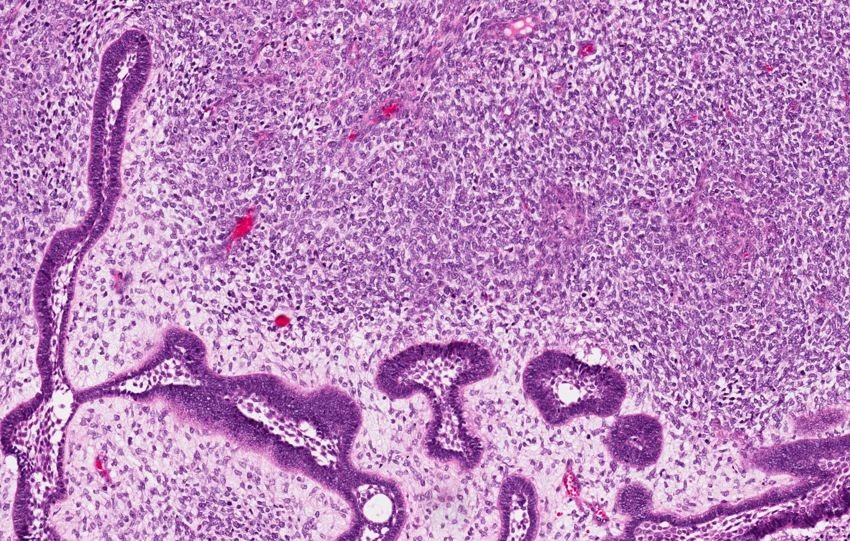
Ameloblastic fibroma
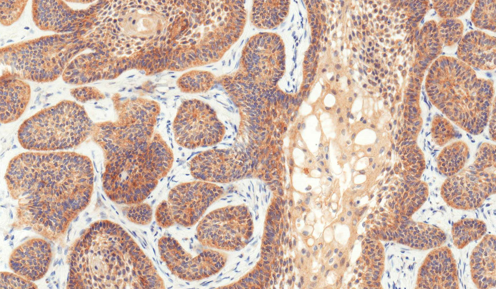
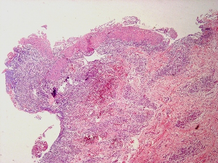
Odontogenic islands with cystic change, within immature mesenchymal stroma
AF at scanning magnification
Odontogenic islands
Odontogenic cords within immature mesenchymal stroma
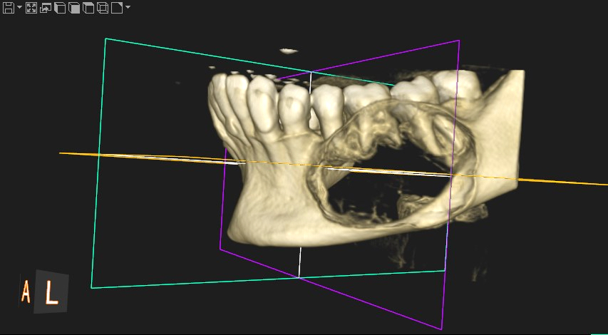
Contributed by Ashley Aiken, M.D., Stephanie Drew, D.D.S. and Shelly Abramowicz, D.M.D., M.P.H.

Case 1: coronal CT view
Case 2: sagittal CT view
Case 3: coronal CT view
Case 4:
panorex radiograph
Case 5:
panorex radiograph,
unicystic type
Images hosted on other servers:

Maxillary mass

Mandibular mass
Contributed by Kelly Magliocca, D.D.S., M.P.H. and Andy Balicki, P.A.
Case 1: mandibular granular cell ameloblastoma
Case 2: mandibular ameloblastoma
Case 3: mandibular ameloblastoma
Case 5: mandibular ameloblastoma, unicystic type
Contributed by Kelly Magliocca, D.D.S., M.P.H. and Anne C. McLean-Holden, D.M.D., M.S.
Case 1: granular cell change
Case 2: follicular growth

Case 2: plexiform growth
Case 3: follicular growth and cystic change
Case 3: follicular growth
Case 4: follicular growth and cystic change
Case 4: cystic tumor change

Case 4: follicular growth
Case 4: cystic change
Case 5: unicystic type, decalcified
Case 5: unicystic type, nondecalcified
BRAF V600E
CK19
CK5
Contributed by Abdulwahab Ewaz, M.D.
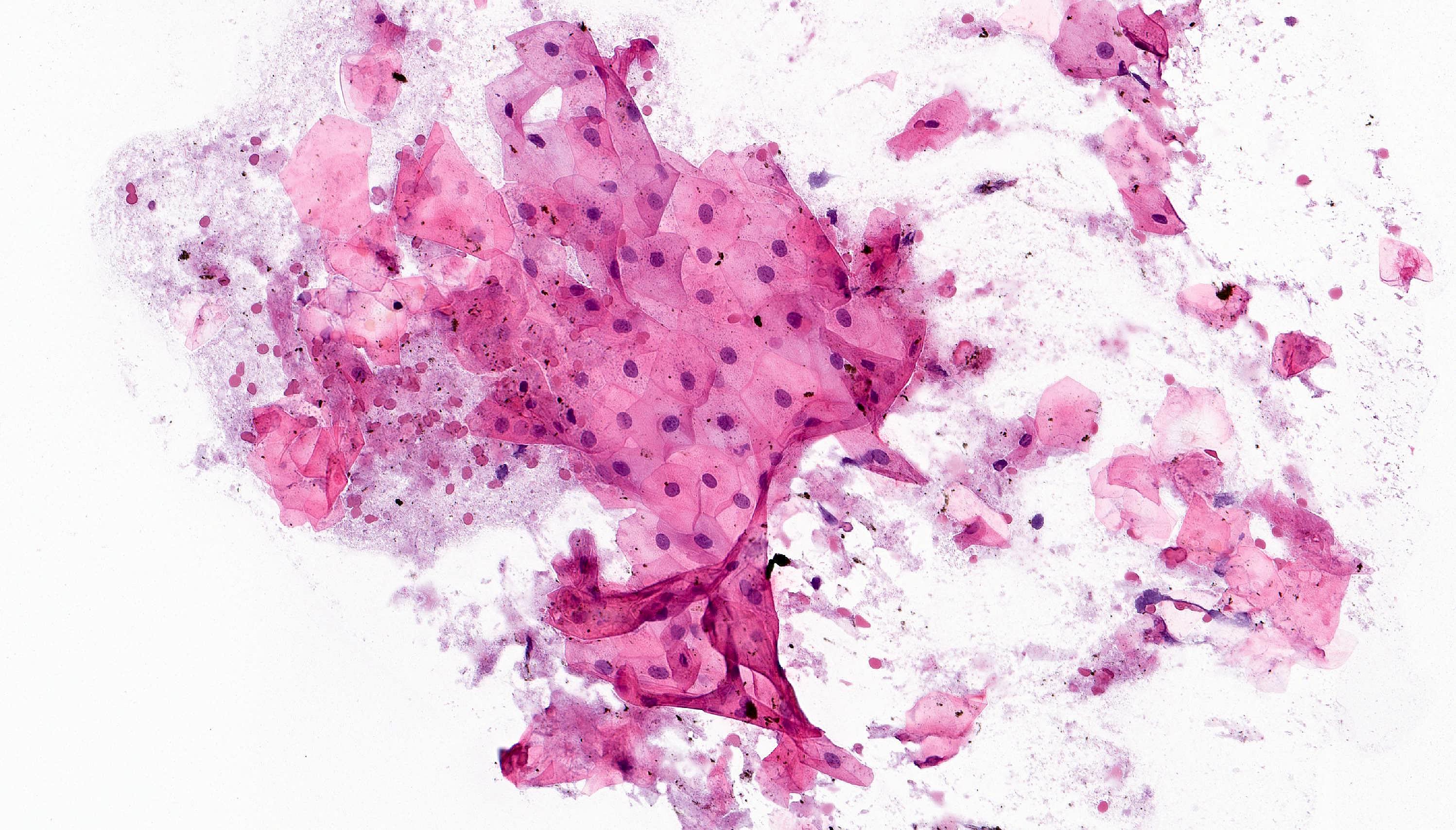
Pap stain
Diff-Quik
Overview of odontogenic tumors by Dr. Khurram
Images hosted on other servers:
Developing tooth (odontoblast and periodontal membrane)
Contributed by Kelly Magliocca, D.D.S., M.P.H.
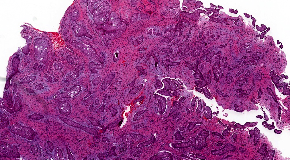
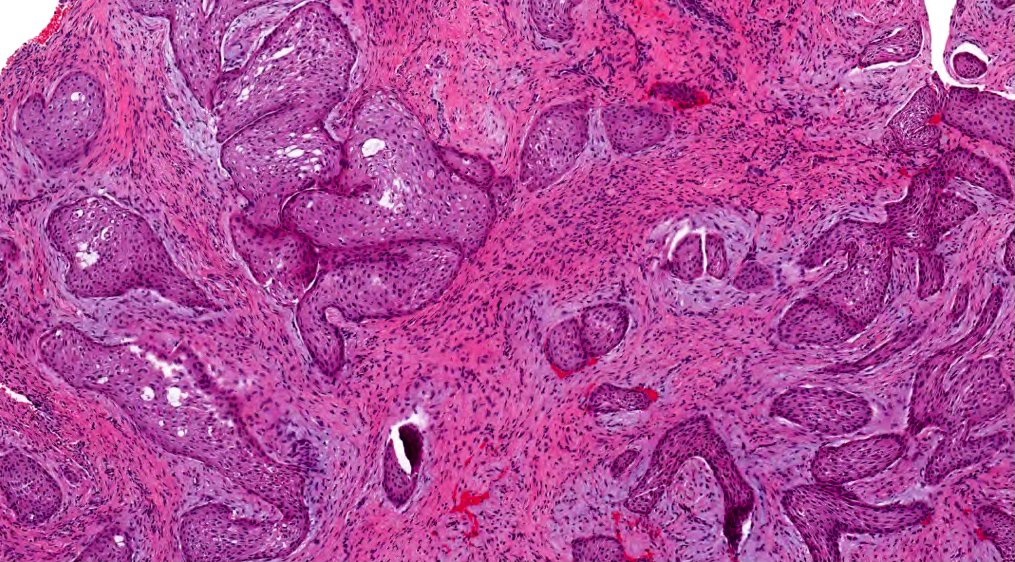
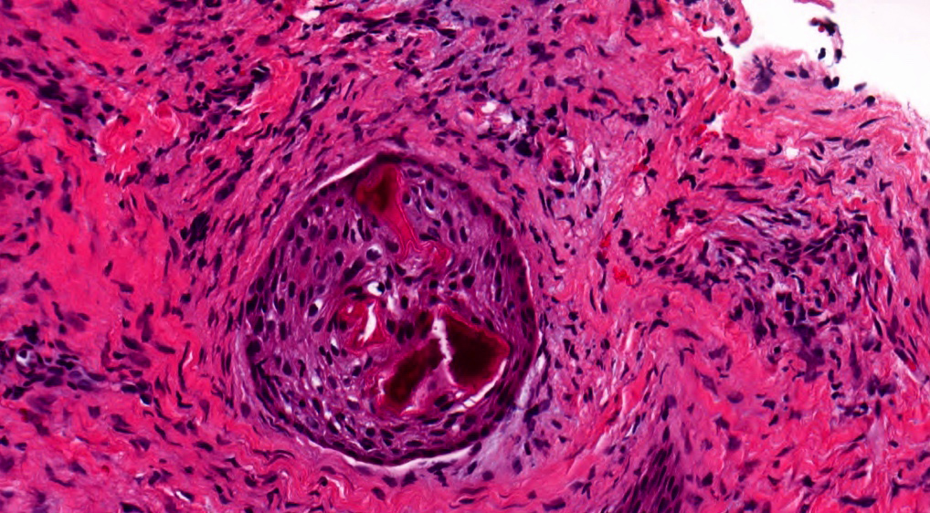
Calcifying epithelial odontogenic tumor
Contributed by Elizabeth Ann Bilodeau, D.M.D., M.D., M.S.Ed.
Coronal CT
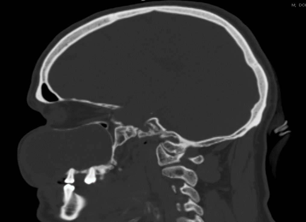
Sagittal CT
Axial CT
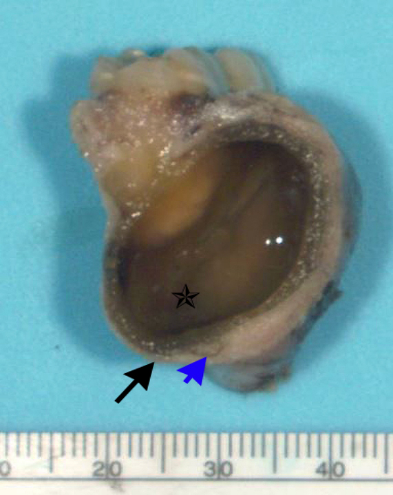
Contributed by Elizabeth Ann Bilodeau, D.M.D., M.D., M.S.Ed.

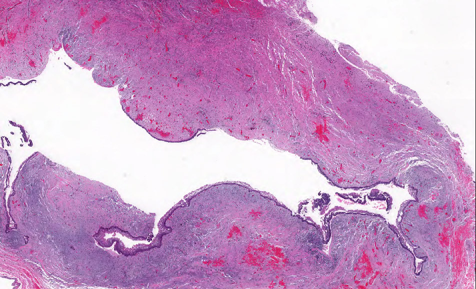
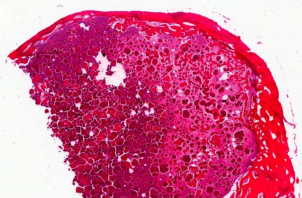
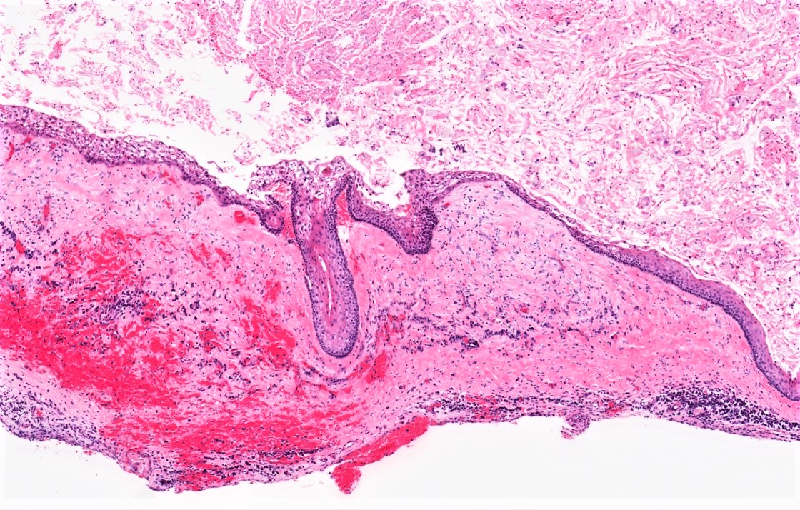
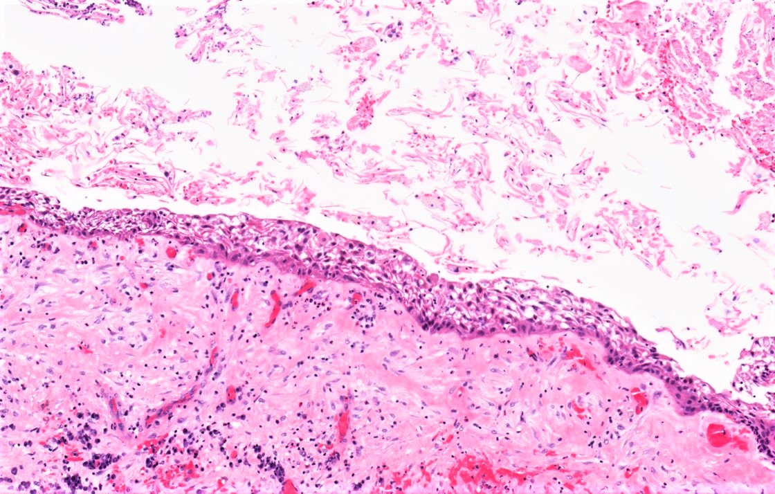
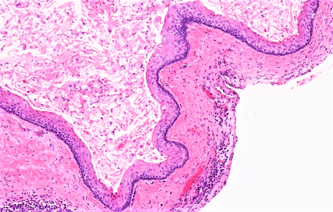
Opened and sectioned cyst


Contributed by Nadim M. Islam, D.D.S., B.D.S.
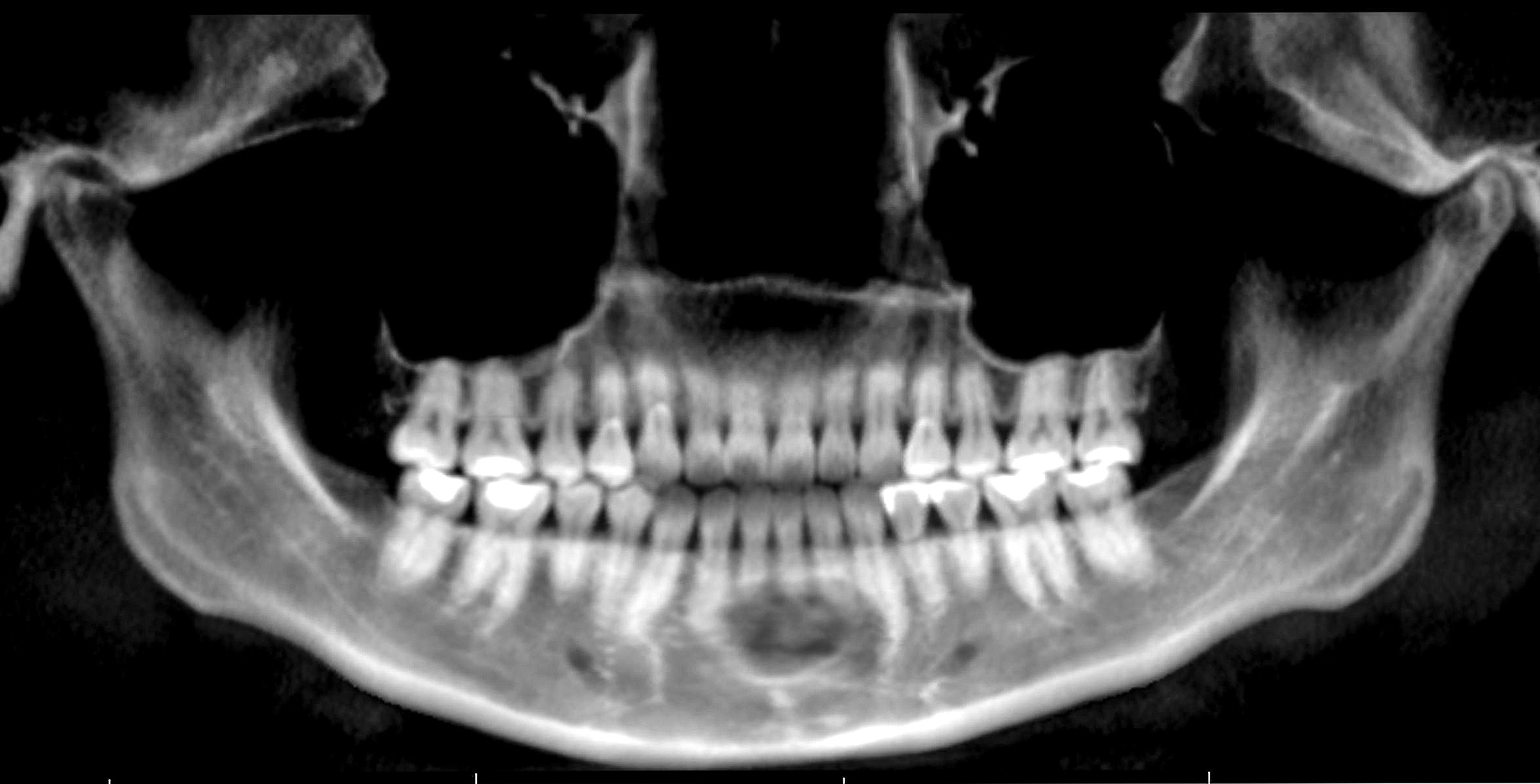
Classic radiolucent rim and central opacity
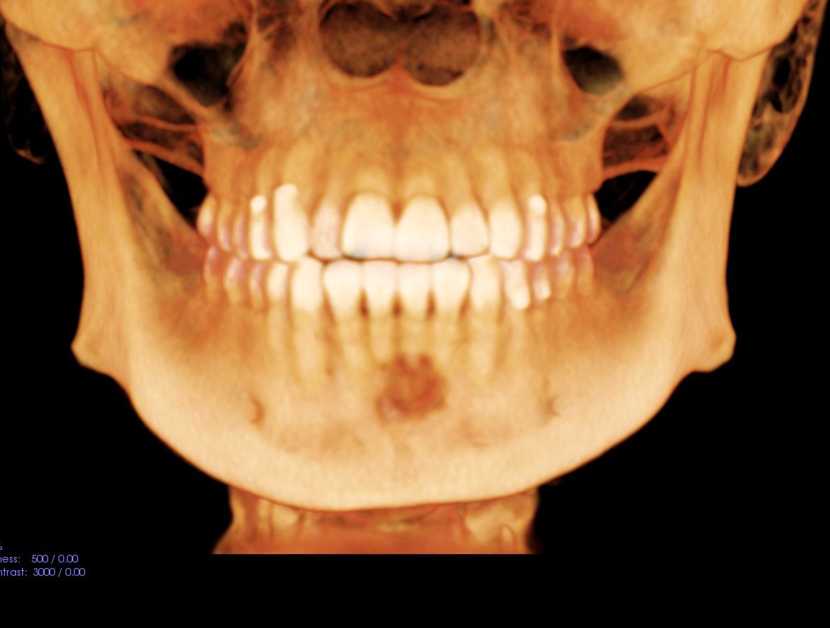
Multiquadrant mixed masses with expansion
Focal lucent rim with central opacity
Images hosted on other servers:

Excision
Images hosted on other servers:

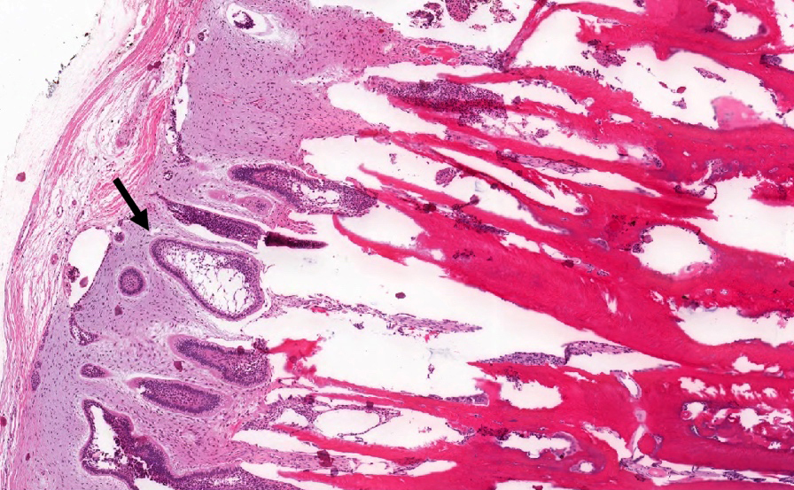
Tumor adjacent to tooth
Contributed by Kelly Magliocca, D.D.S., M.P.H. and Nadim M. Islam, D.D.S., B.D.S.
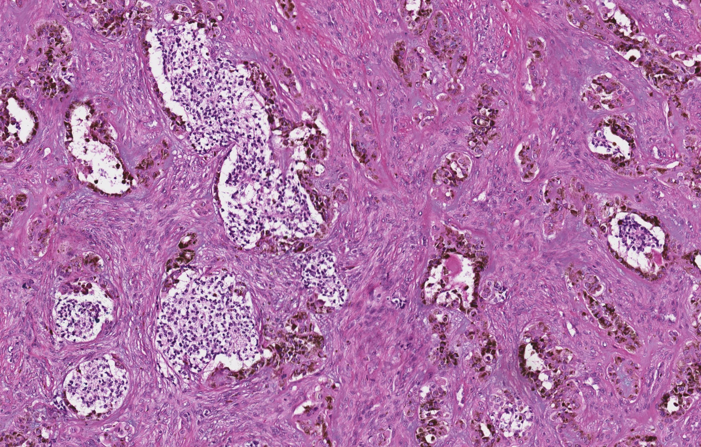
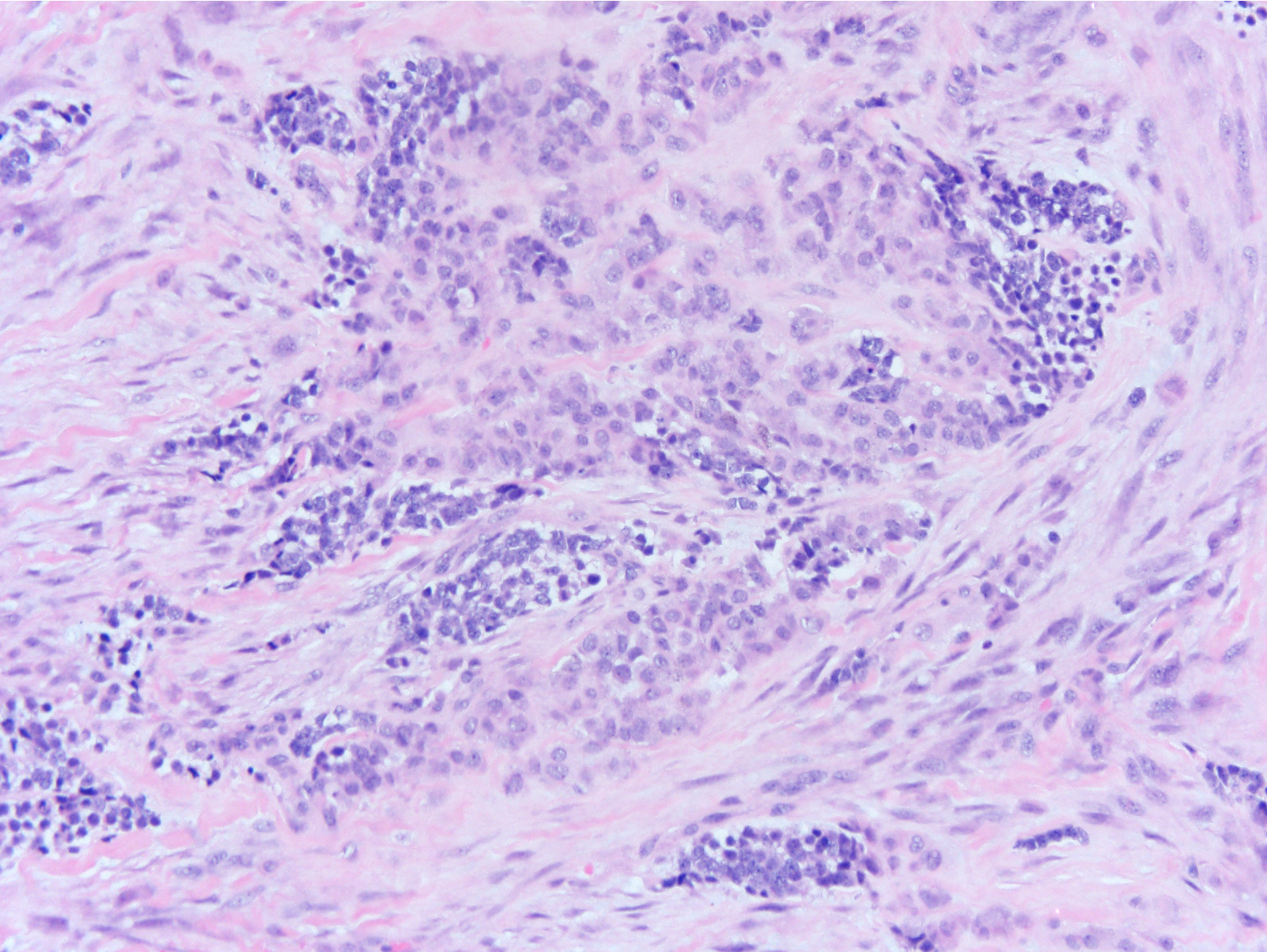
Cemento-osseous dysplasia
Cemental spherules and modestly cellular stroma
Spherules joining into ginger root-like formations
Lack of brush borders helps diagnosis
Contributed by Molly Housley Smith, D.M.D., Thomas W. Fonner, D.M.D., Carlton Taylor, D.M.D., Firas Katabi, D.D.S. and Nish Shah, D.M.D., M.D.
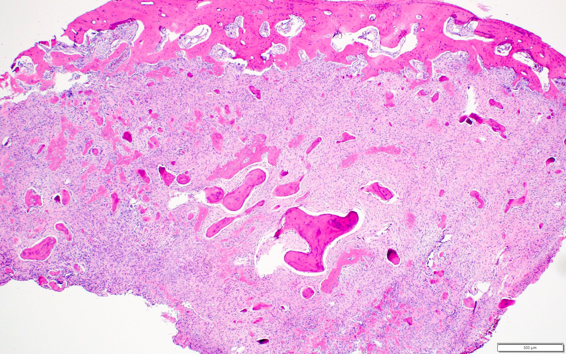
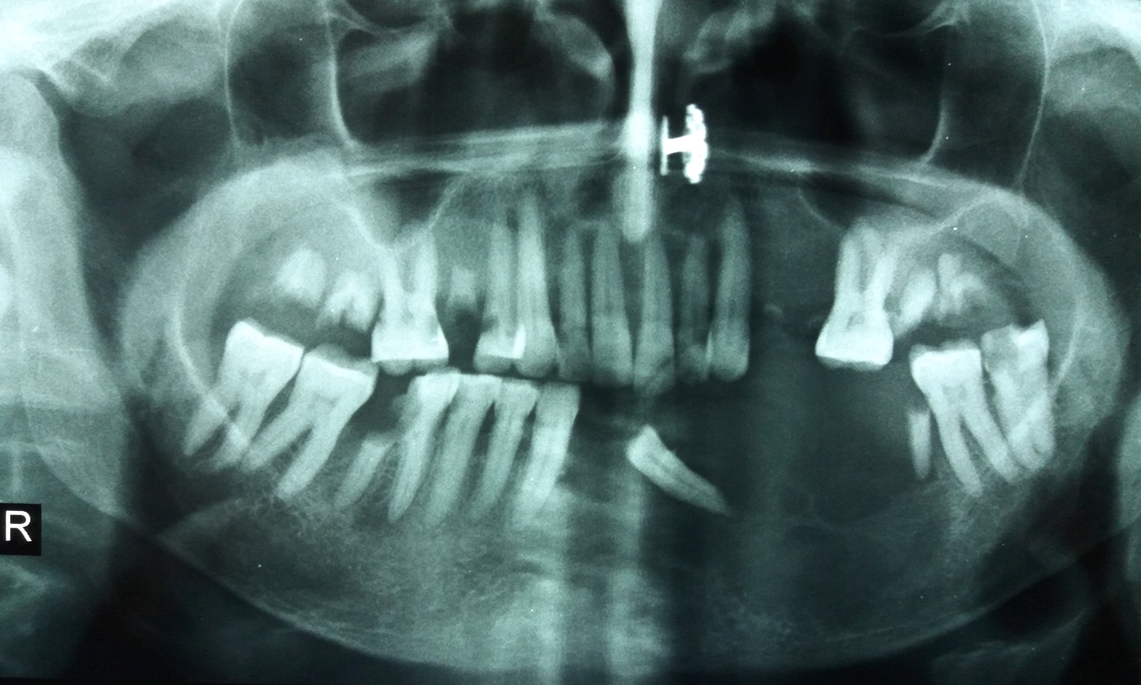
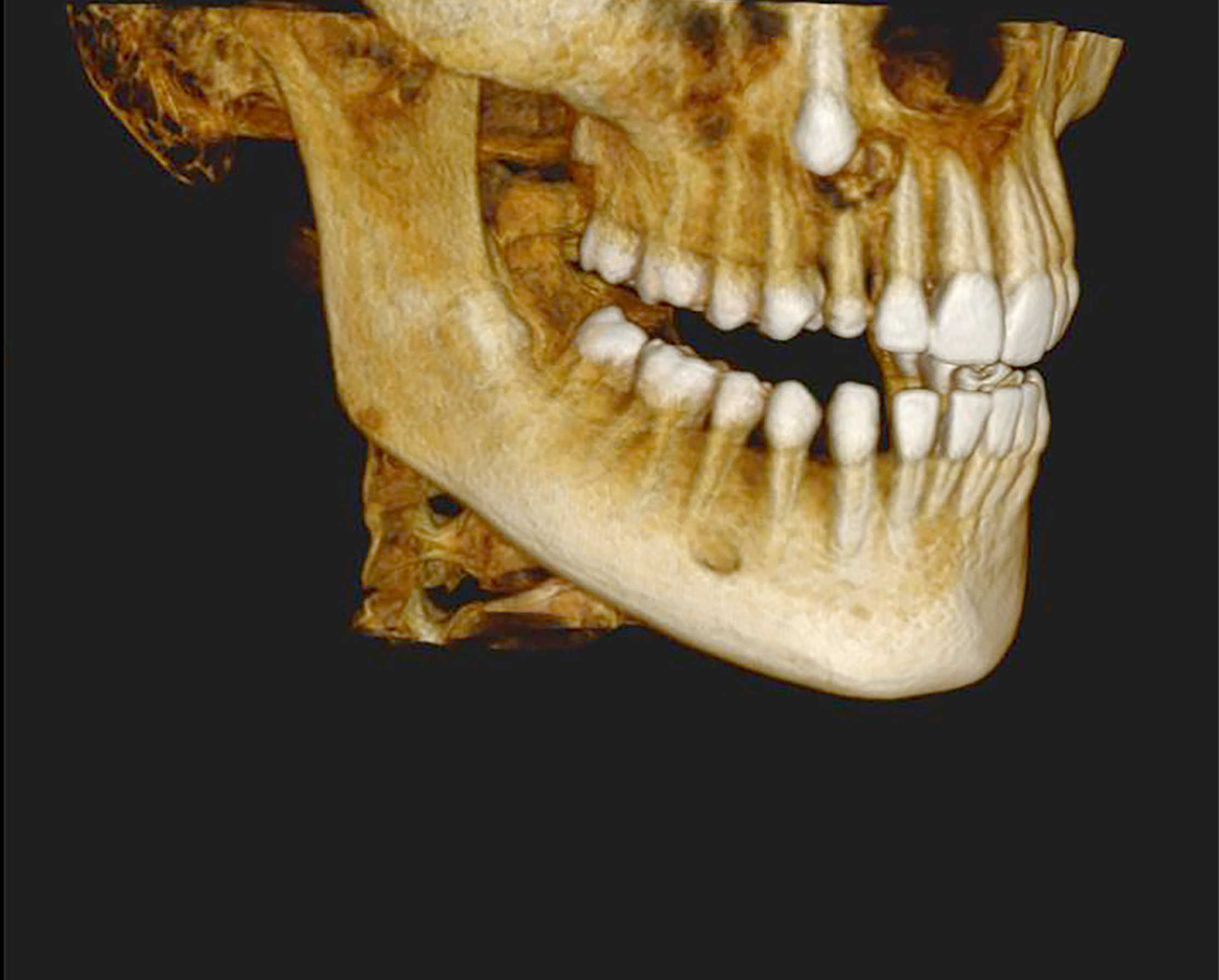
Radiolucency in anterior mandible
Expansile lesion of mandible
Cortical destruction
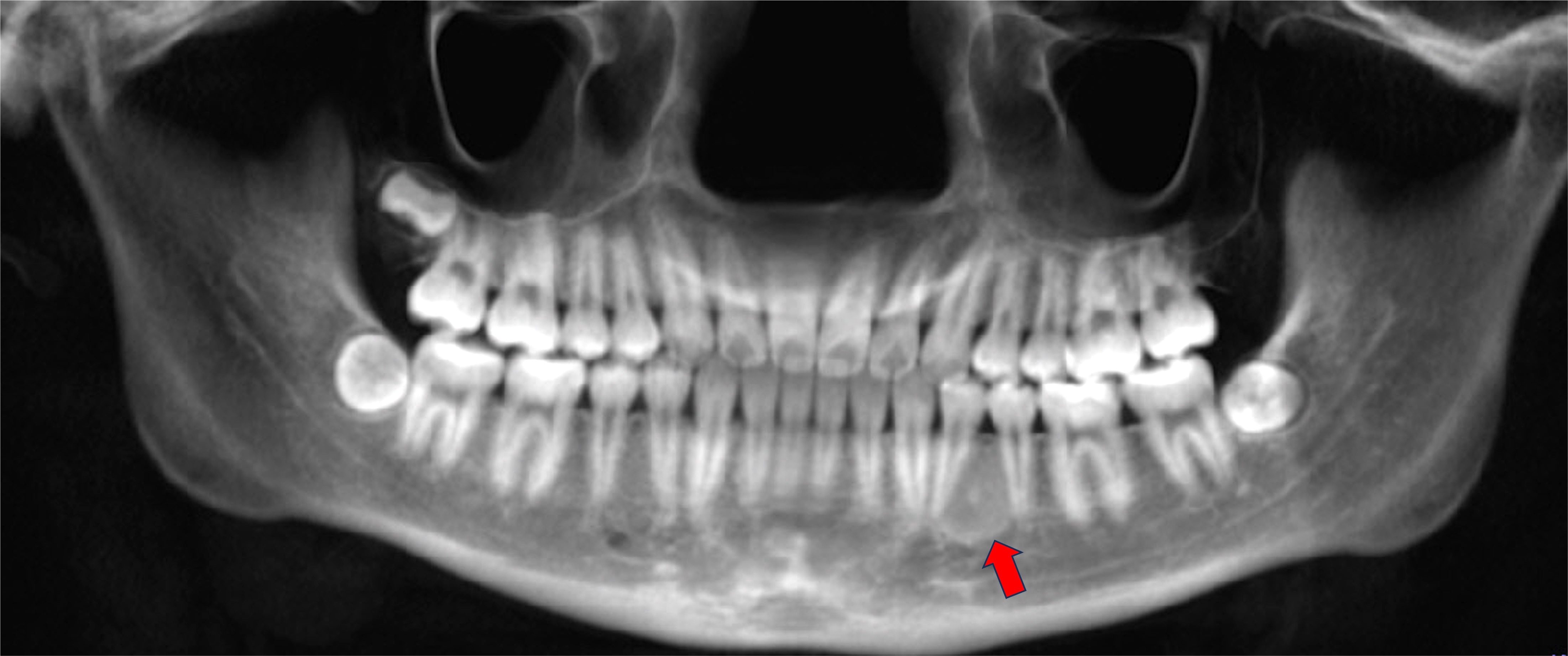
Root divergence
Images hosted on other servers:

Large solitary swelling obliterating buccal vestibule

Bony mass in maxilla

Distinction between lesion and healthy bone
Images hosted on other servers:

Excision
Contributed by Saira Javeed, M.B.B.S., M.Phil., Kelly Magliocca, D.D.S., M.P.H. and Molly Housley Smith, D.M.D.
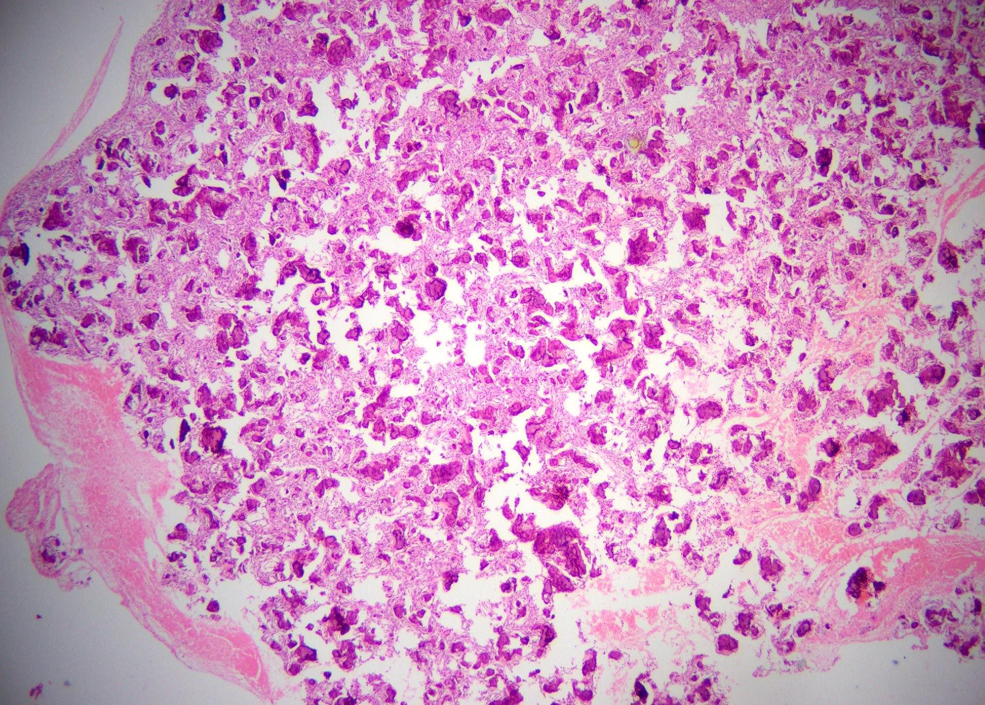
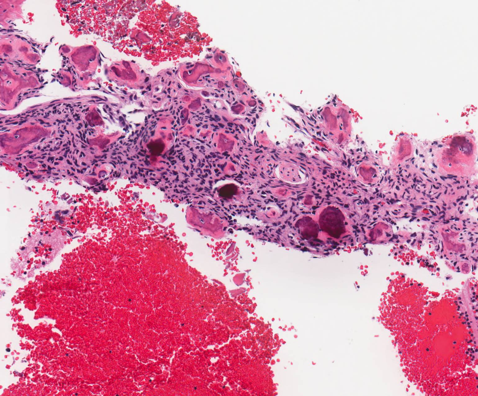
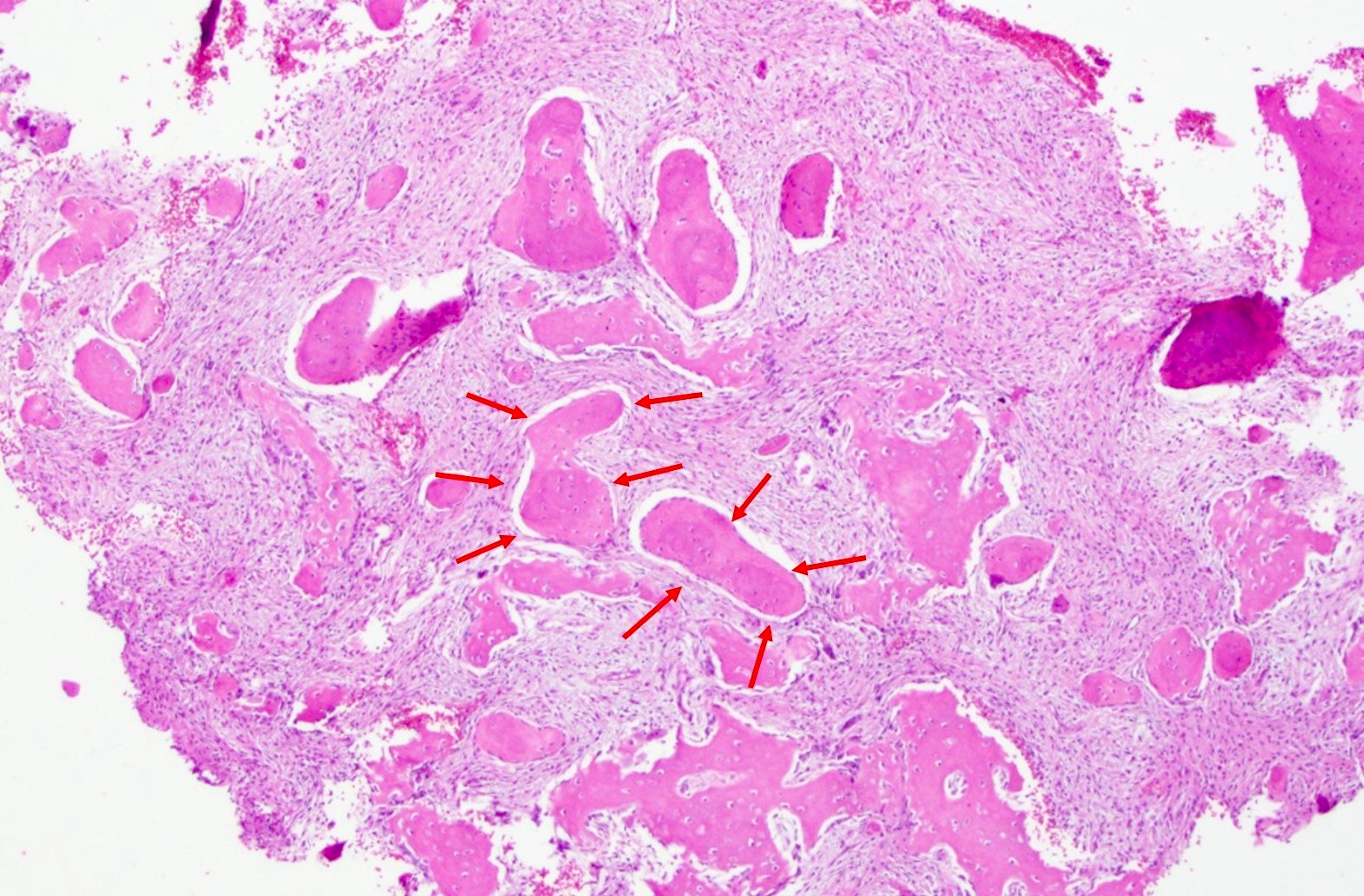
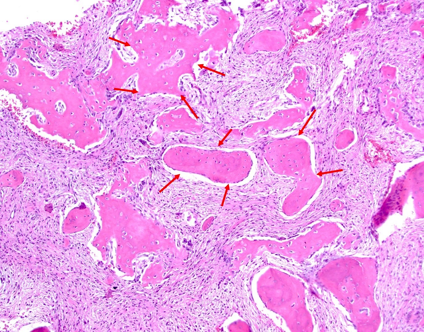
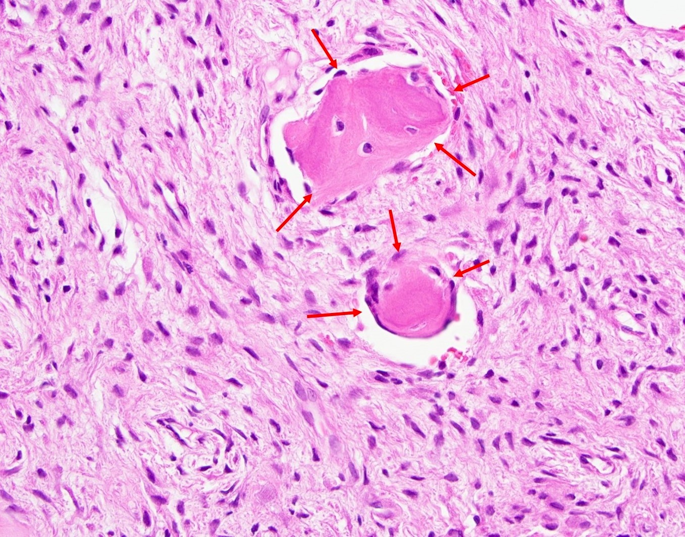
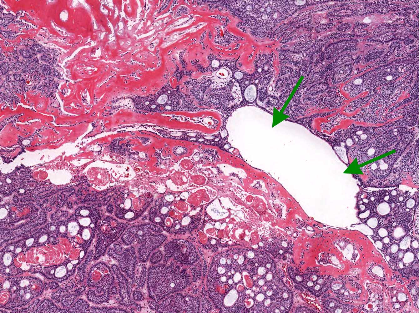
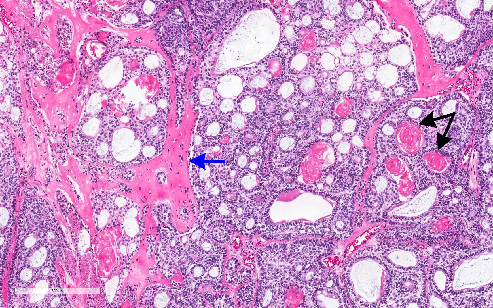
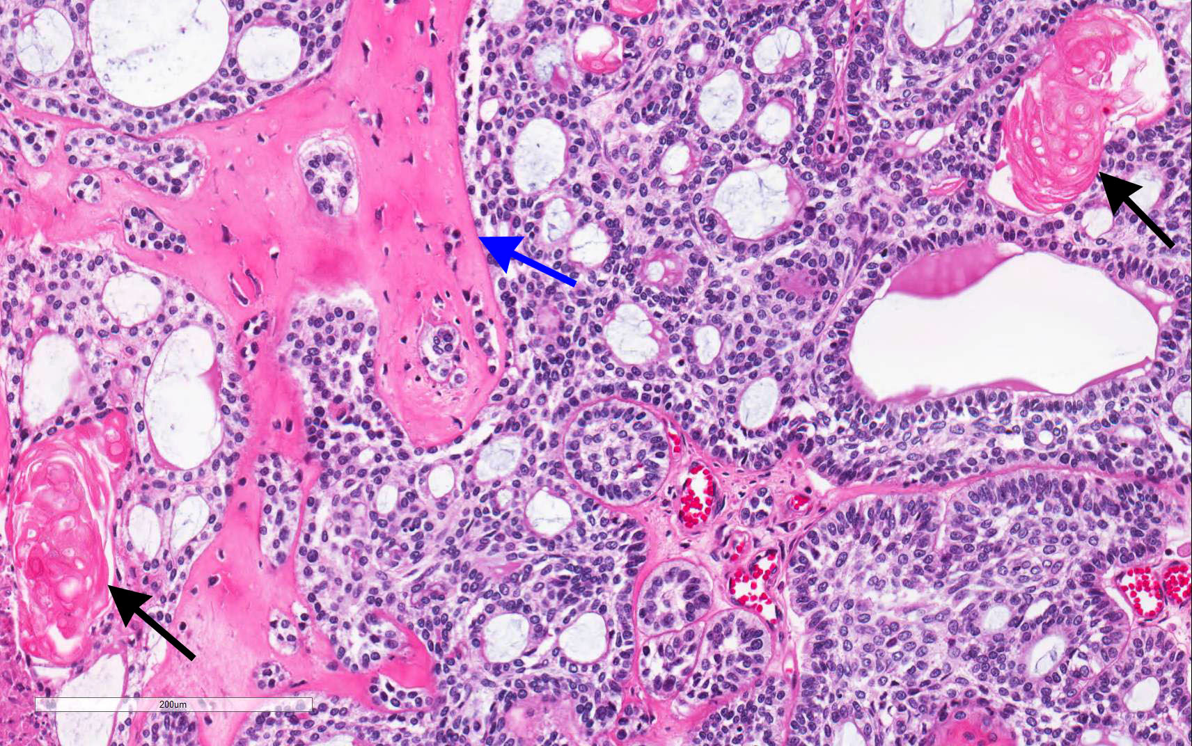
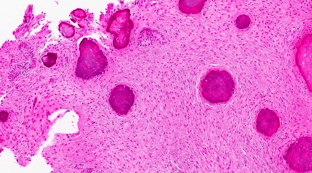
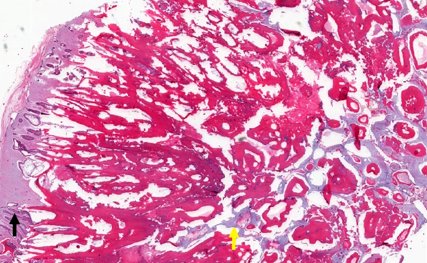
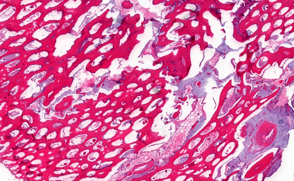
Well circumscribed solid tumor mass
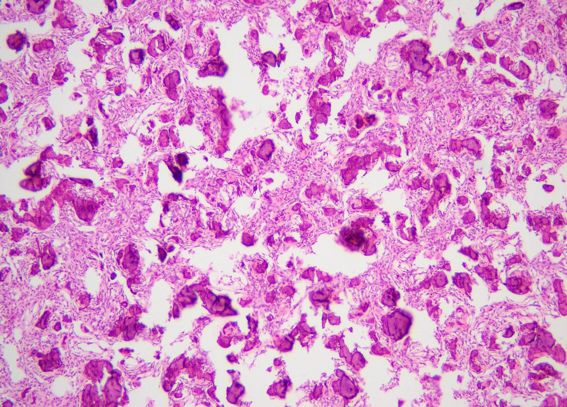
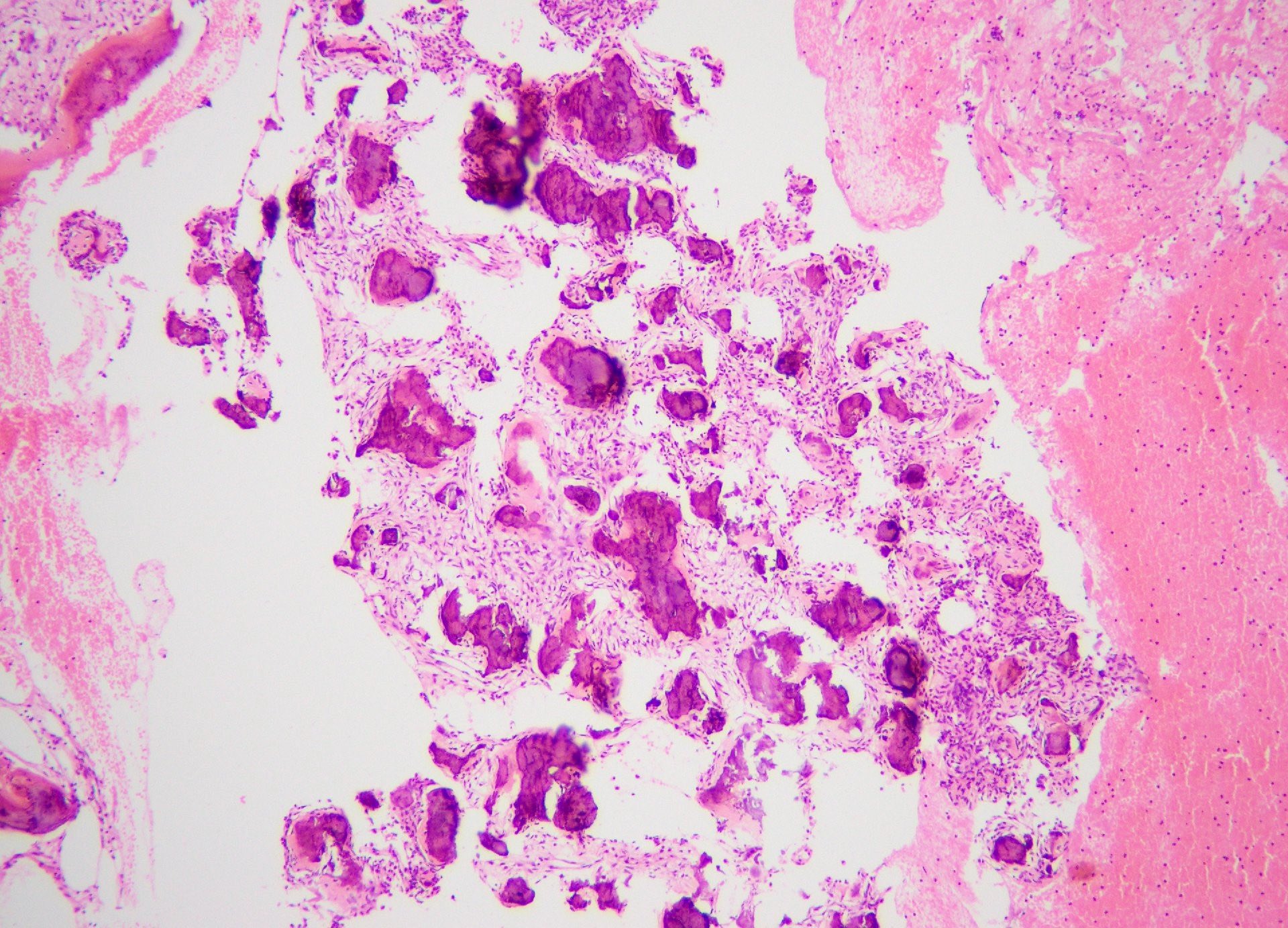
Rounded to spherical variable sized cementum-like ossicles
Benign fibro-osseous proliferation
Rounded mineralized droplets
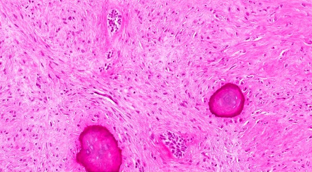
Fibro-osseous proliferation
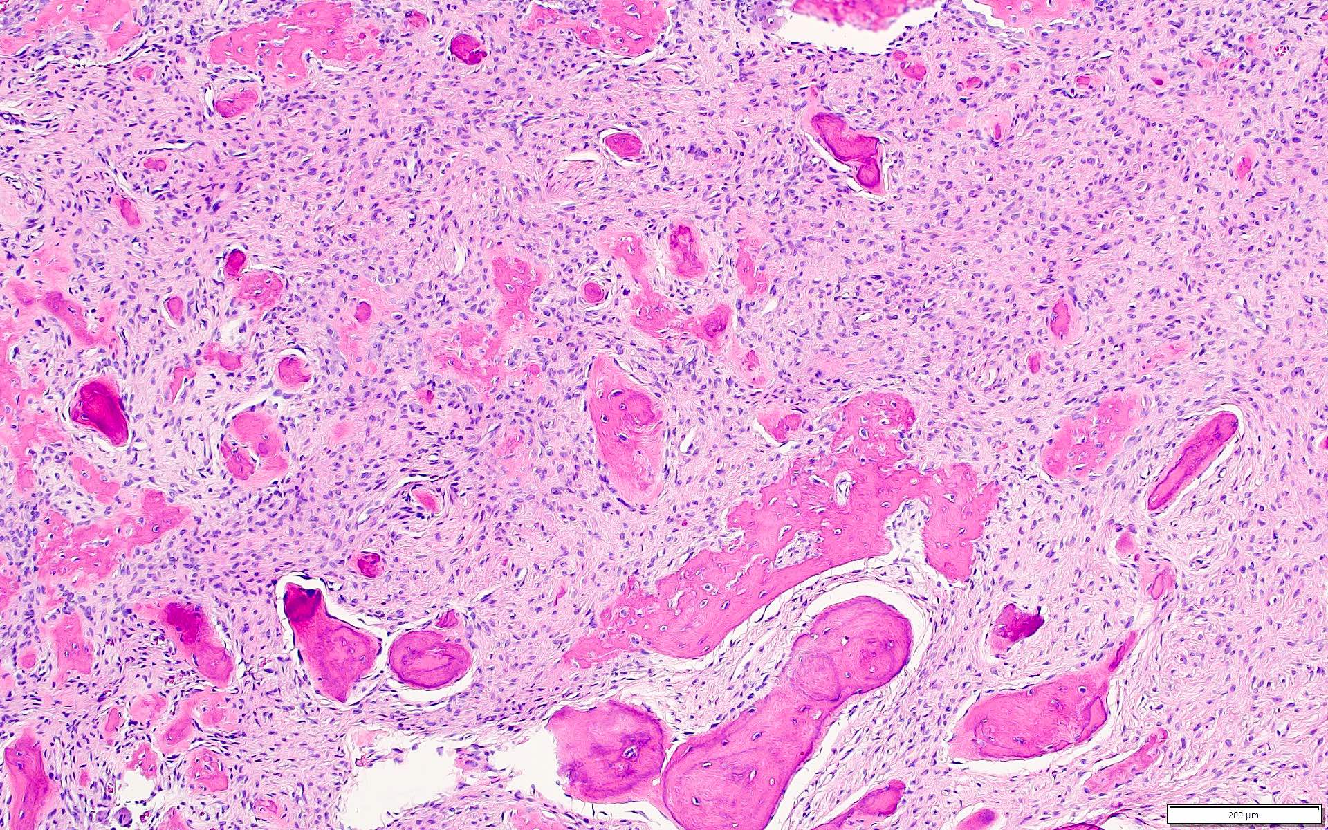
Woven bone
Brush borders
Images hosted on other servers:

MDM2 amplification by FISH

CDC73 mutations in sporadic tumors
Contributed by Kelly Magliocca, D.D.S., M.P.H.
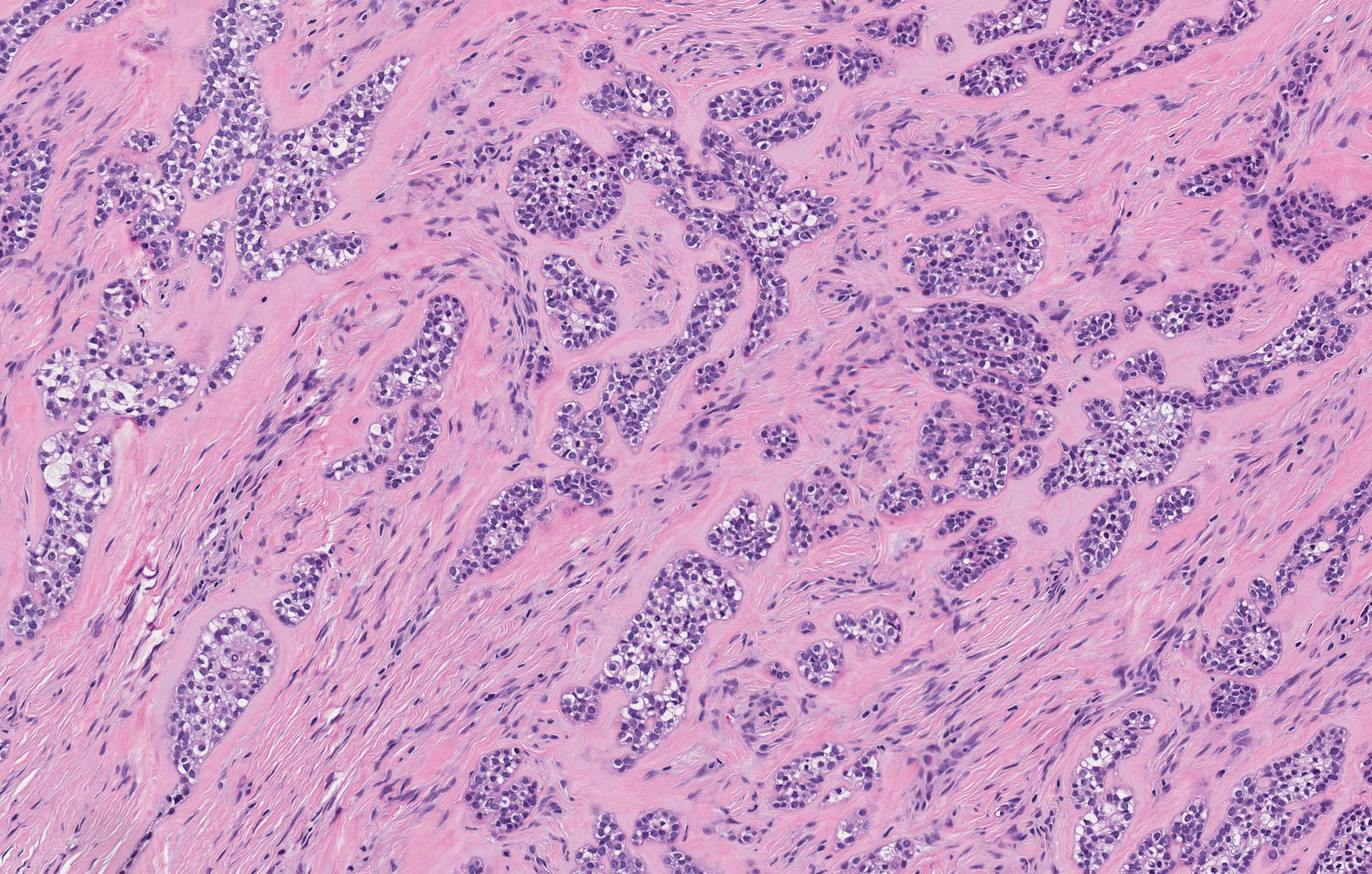
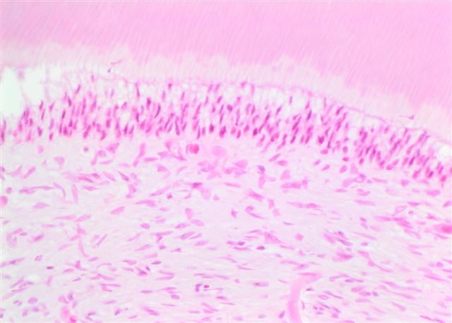
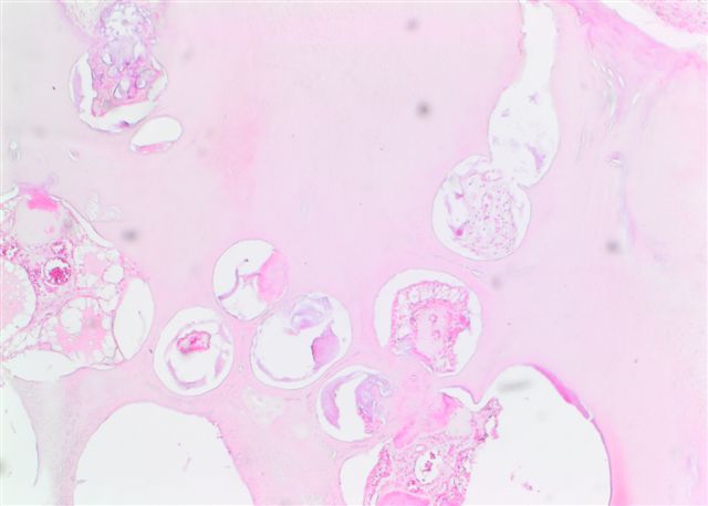
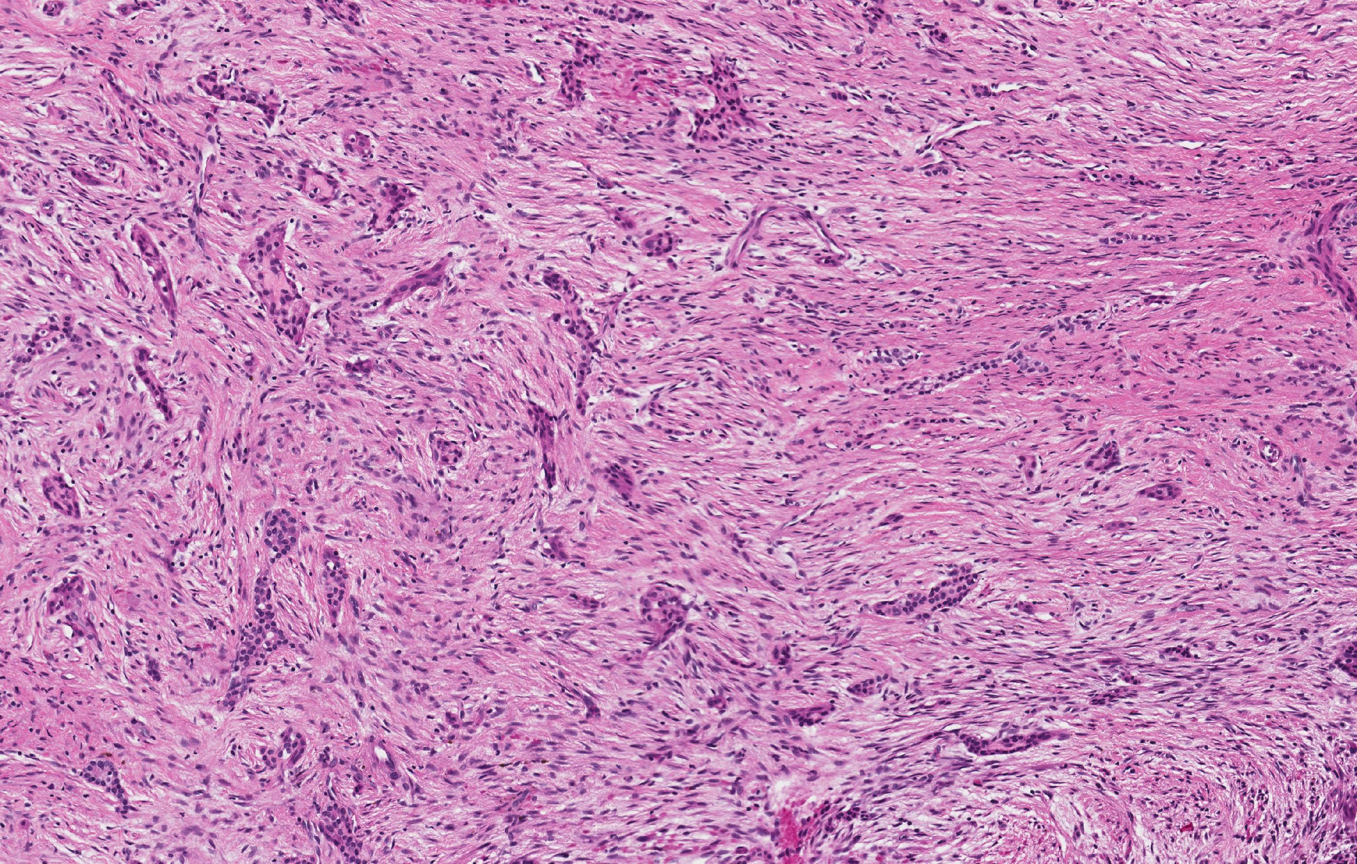
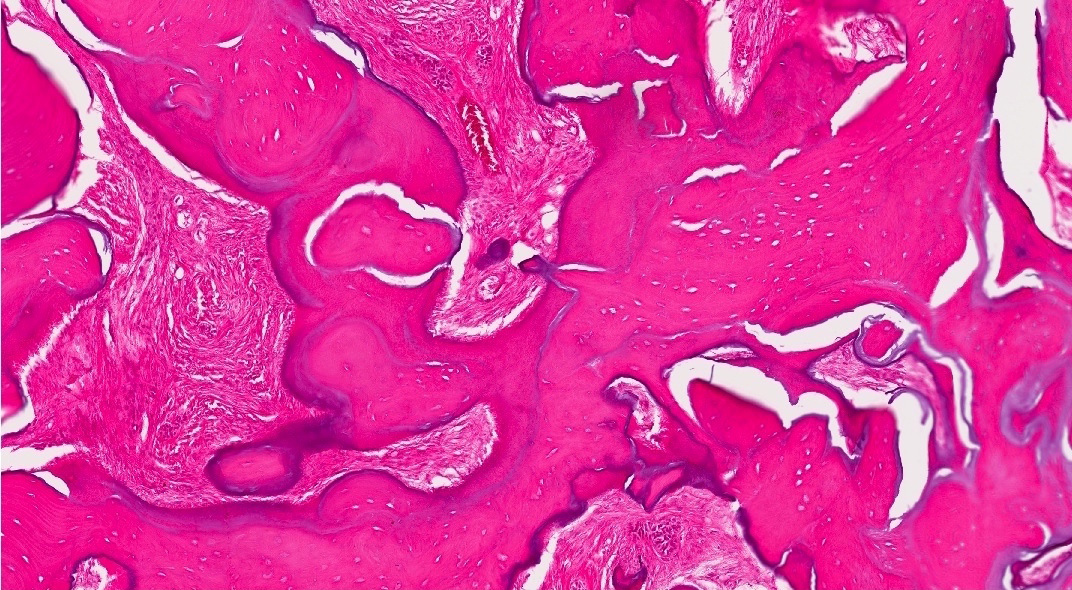
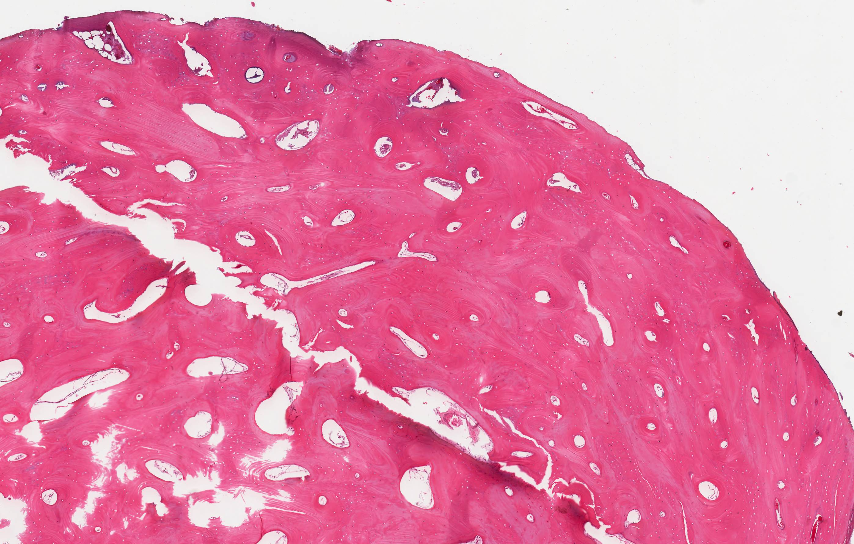
Cementoblastoma
Contributed by Nasir Ud Din, M.B.B.S.
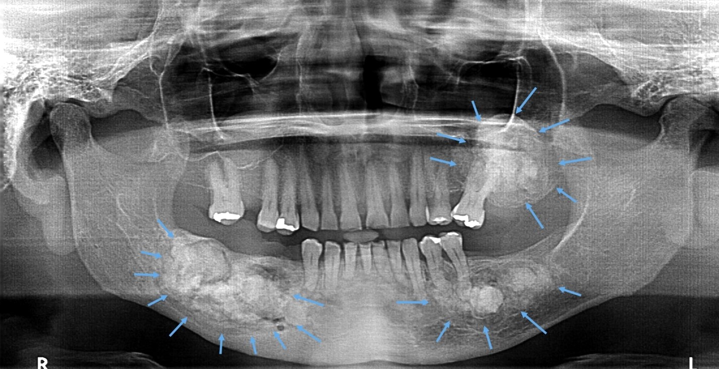
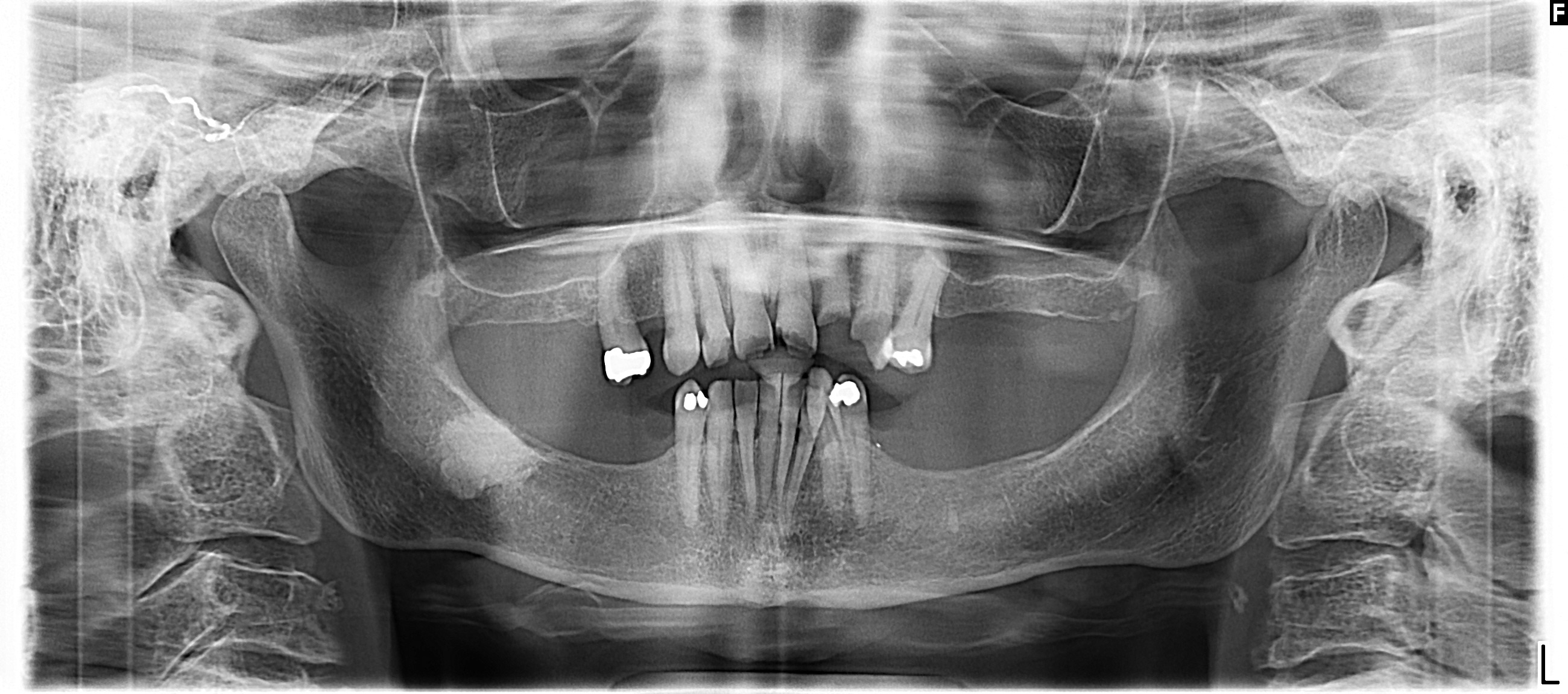
CGCG of
mandible,
premolar region
(orthopantomogram)
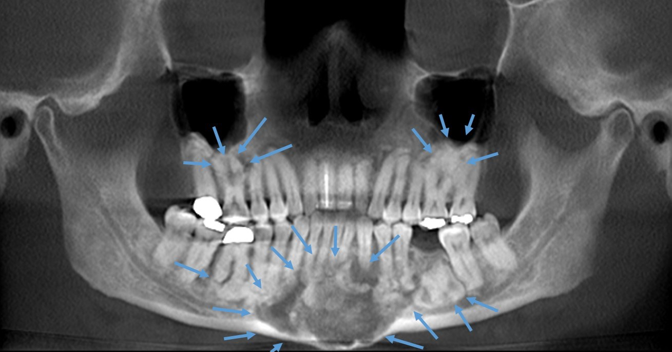
Mandibular body
CGCG
(orthopantomogram)
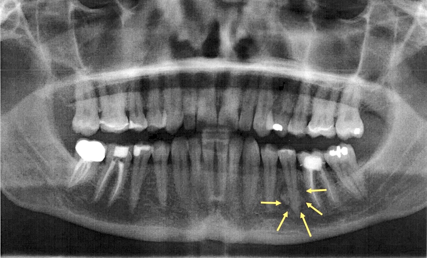
Left mandibular CGCG
CGCG of lower incisor region
Images hosted on other servers:

Right side submandibular swelling

Oval lesion involving left side mandible
Images hosted on other servers:

Fleshy, reddish brown, hemorrhagic appearance
Contributed by Nasir Ud Din, M.B.B.S.
Lobulated architecture
Lobulation
Periphery of lobule
Woven bone
Lobule center
Fibroblast and giant cells
Images hosted on other servers:

Giant cell with occasional
spindle cells and
hemorrhagic background
Central giant cell granuloma
Contributed by Kelly Magliocca, D.D.S., M.P.H.
Cherubism
Images hosted on other servers:

Treatment of CNO/CRMO
Images hosted on other servers:

Expansion of left ramus

"Bone-on-bone" pattern

Swelling of left masseter muscle

Uptake of radionucleotide
Images hosted on other servers:

Early and late stages of CRMO
Images hosted on other servers:

Echo, CT, MRI

Local findings

Mass

Operative findings

Postoperative

Contributed by Dr. Pooja Navale:
Images hosted on other servers:

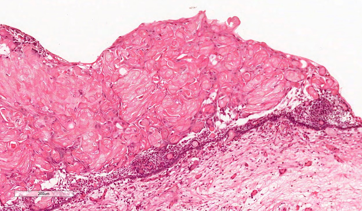
Stratified squamous epithelium

Mitotic figures

Islands of of epithelial cells

Hyalinized areas

Carmine stain

PAS

p63. CD10, PAS

P63+

Pancytokeratin+

AE1/AE3, S100, SMA

Mucicarmine-

SMA-

Calponin-

S100-
Images hosted on other servers:

Buccal cortical expansion
with ulceration

Intraoral ulcerated growth

Bone destruction in
symphysis region

Fig A: radiolucent lesion
in posterior segment

Ill defined
radiolucent lesion
Images hosted on other servers:

Composite resection
Contributed by Kelly Magliocca, D.D.S., M.P.H.
Clear cell odontogenic carcinoma
Images hosted on other servers:
Various H&E

Congo red stain

Tumor cells
Images hosted on other servers:

Interphase cells
Contributed by Molly Housley Smith, D.M.D. and John Kalmar, D.M.D., Ph.D.
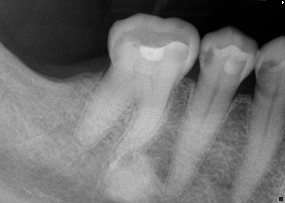
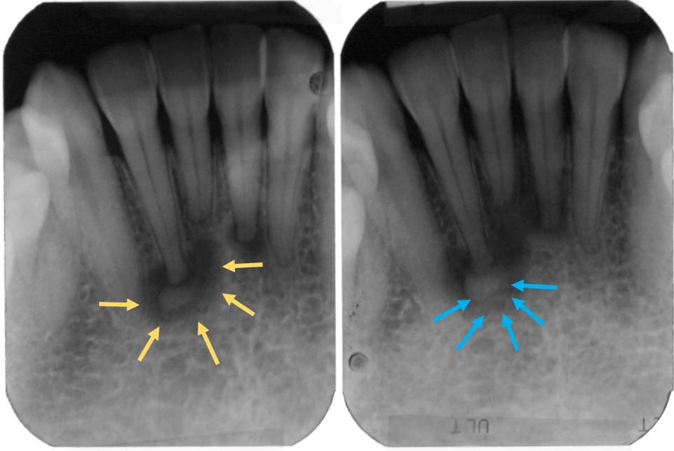
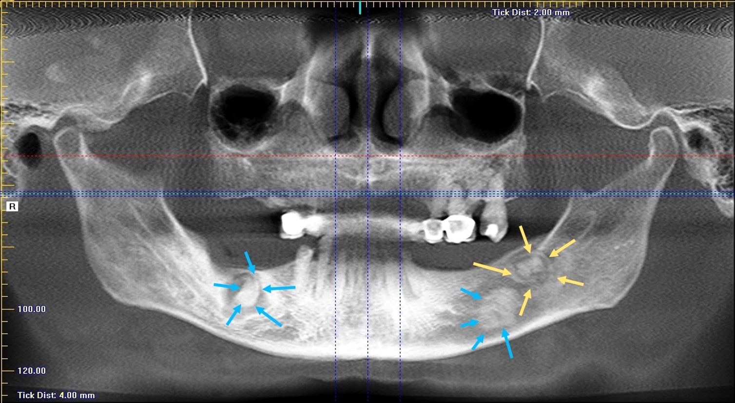
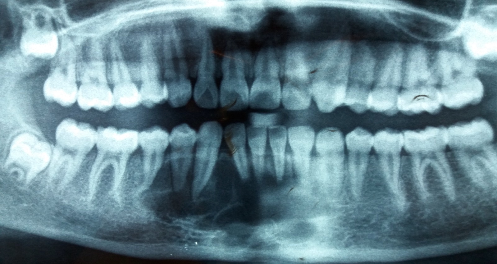
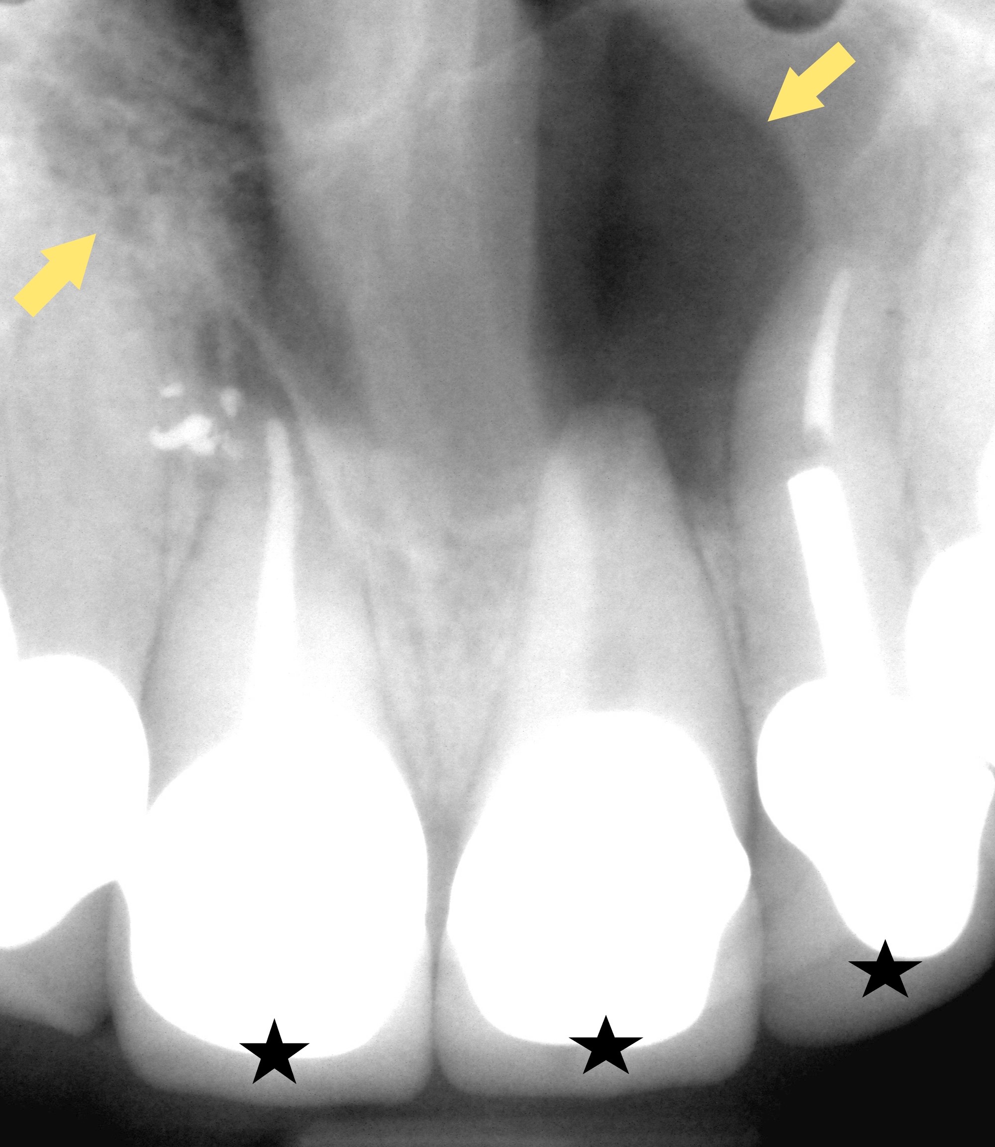
Apical to carious teeth
Residual opacity
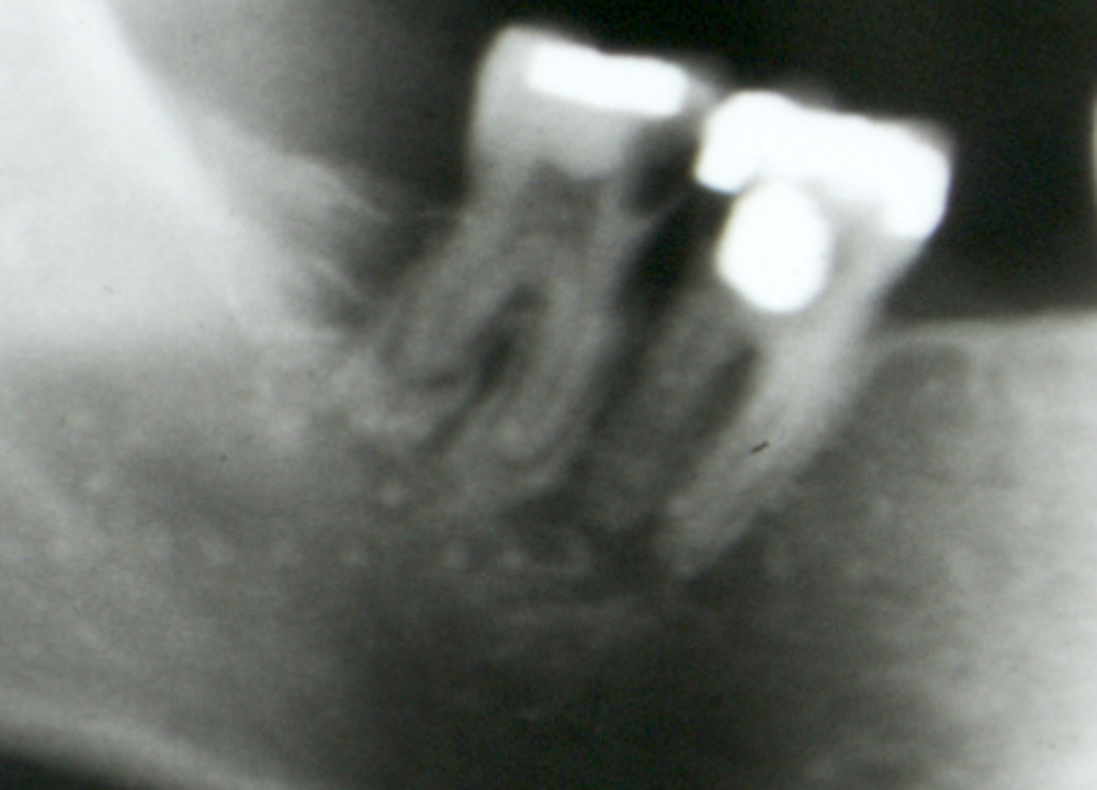
Panorex
Images hosted on other servers:

Periapical
Contributed by Molly Housley Smith, D.M.D.
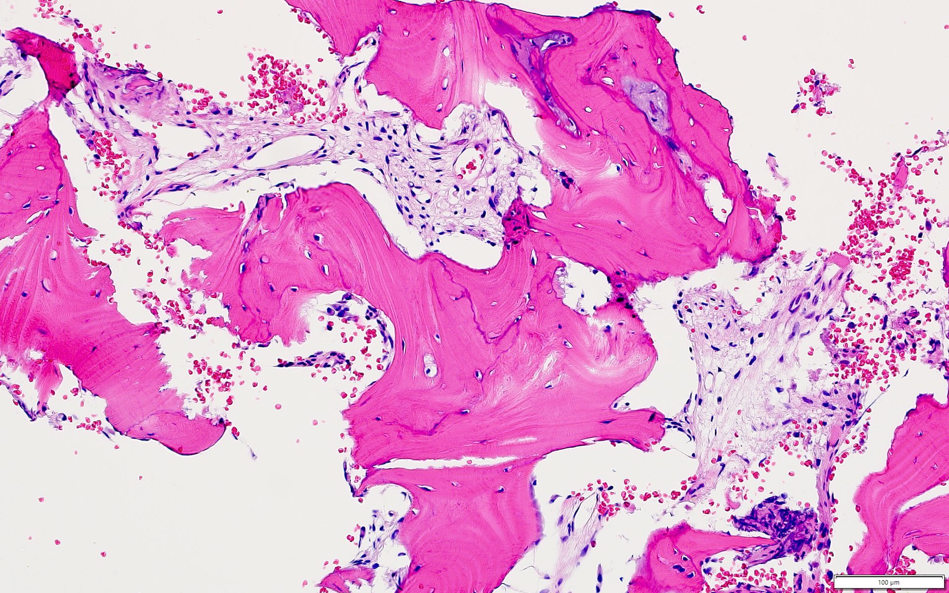
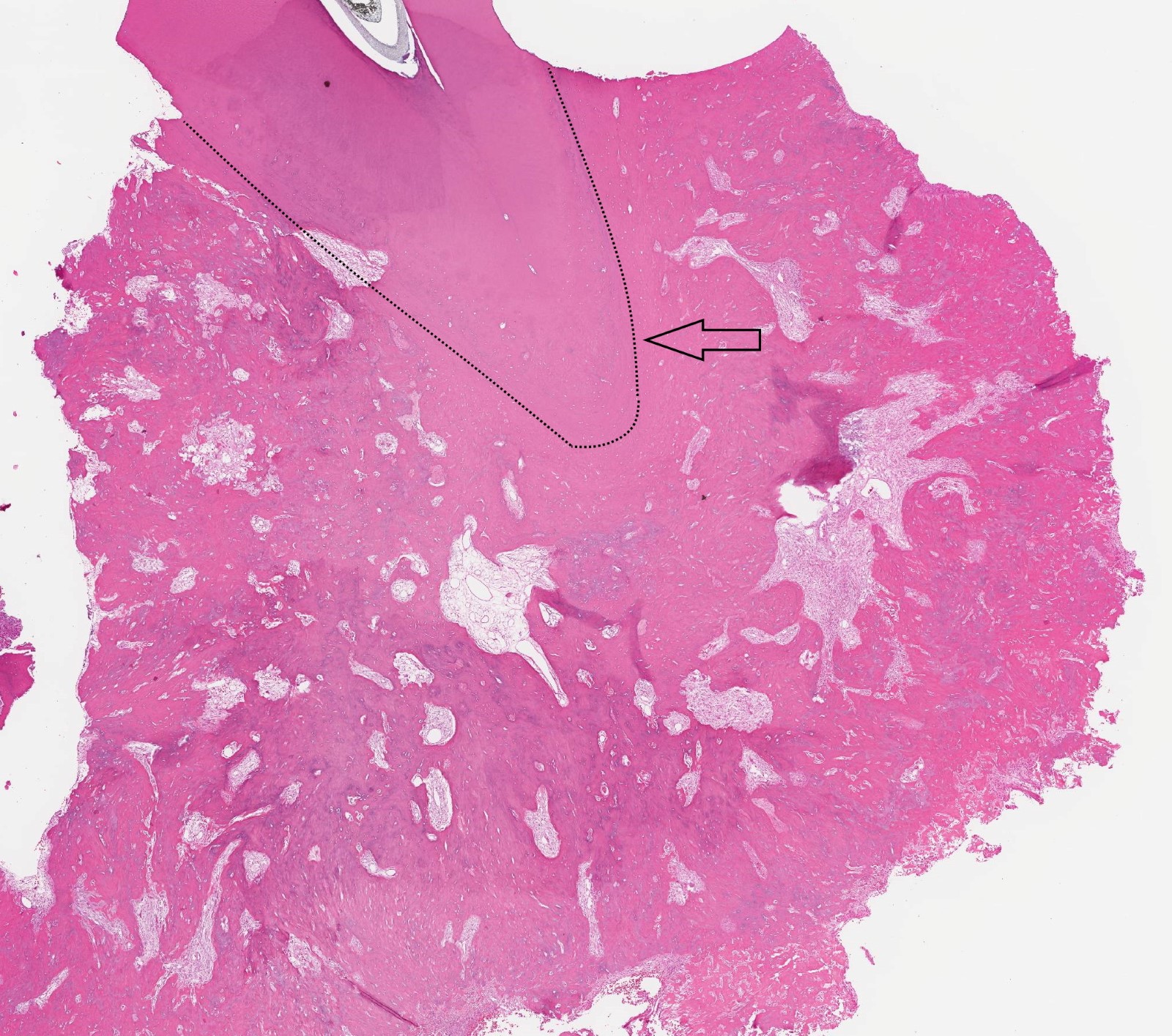
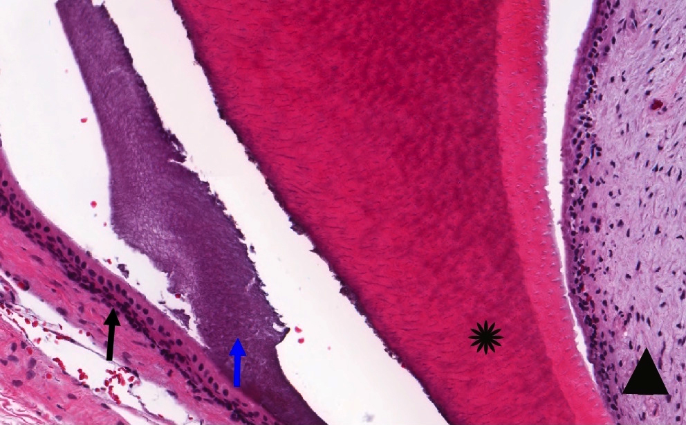
Scant fibrous stroma
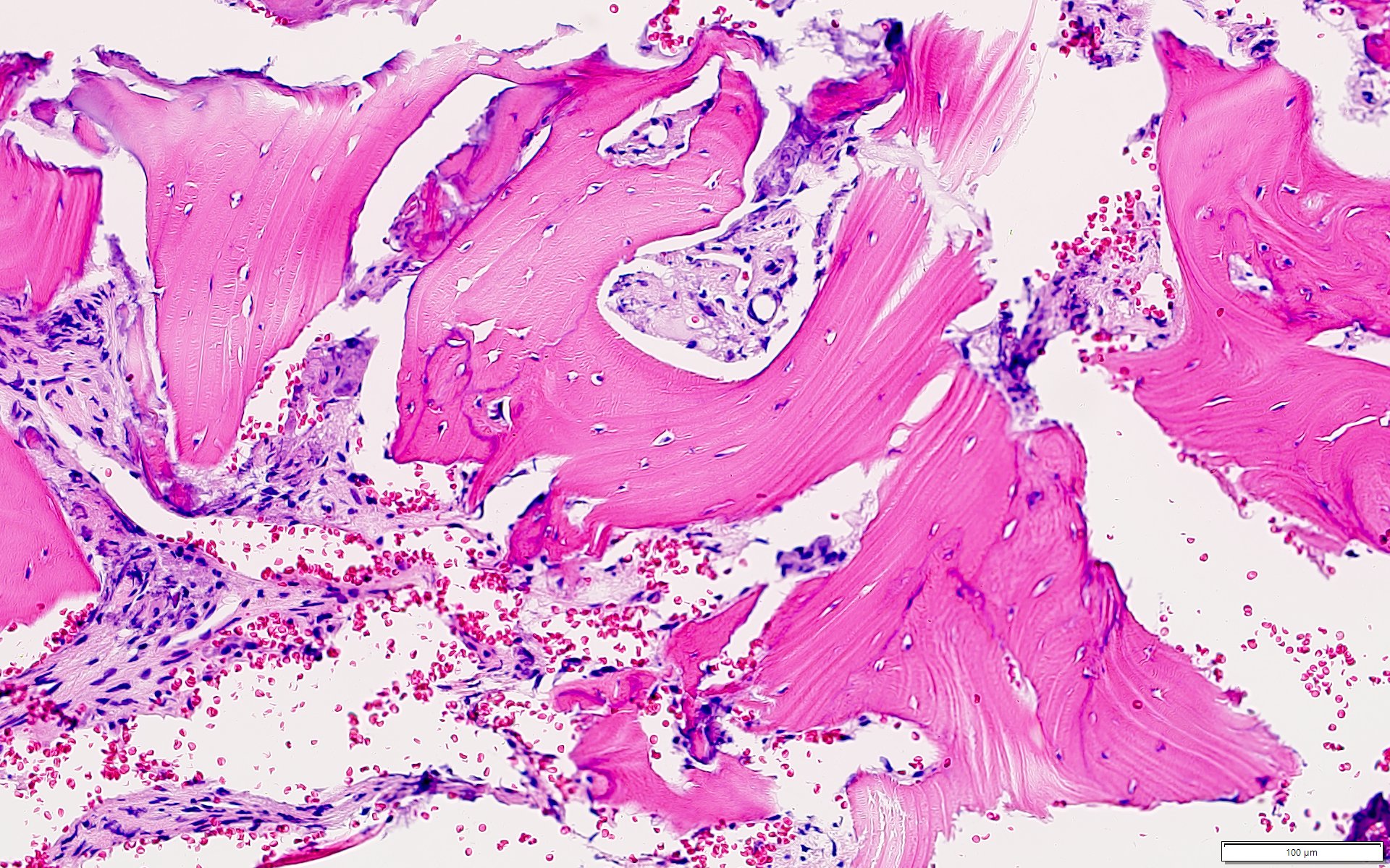
Nonspecific bone
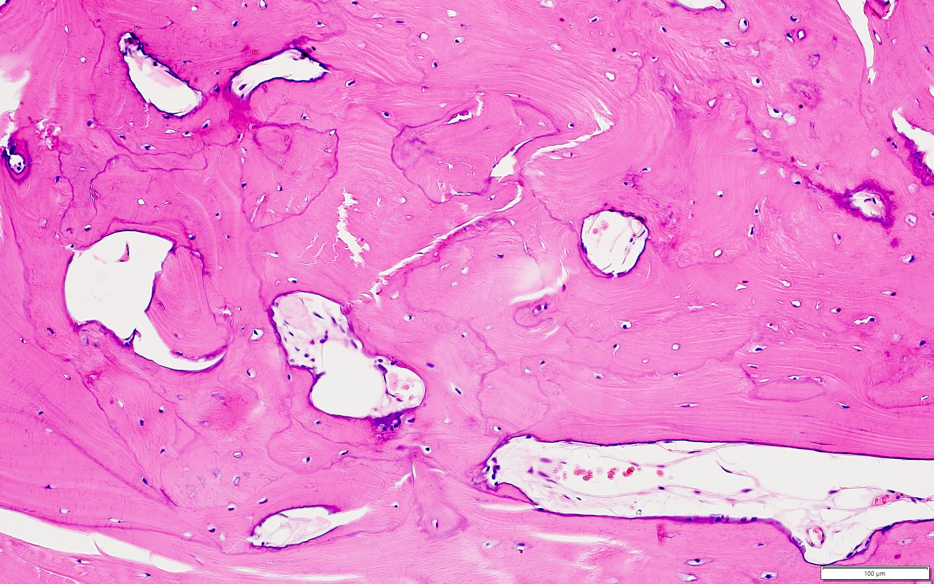
Remodeling bone
Prominent resting and reversal lines
Contributed by Kelly Magliocca, D.D.S., M.P.H.
Cropped panorex
Images hosted on other servers:

Central variety

Lateral variety

Multiple radiopaque masses with thin radiolucent rim

Expansion and thinning of the bony sinus wall


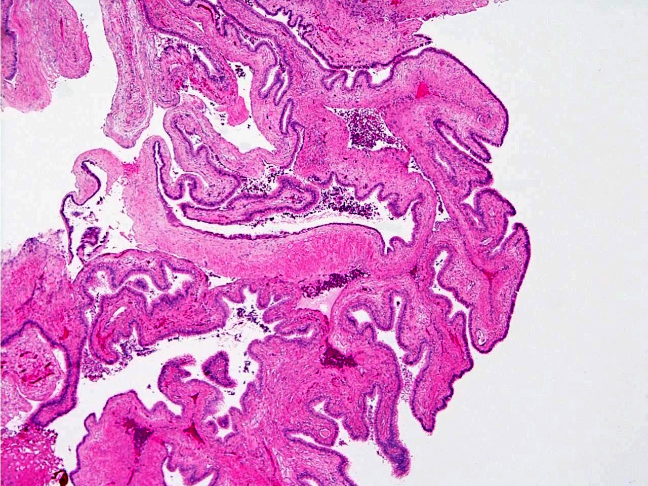
Images hosted on other servers:

Hard and soft tissues with impacted lateral incisor

Enucleated specimen with embedded canine

Cystic lesion

Tan colored thickenings in wall of cystic lesion

Maxillary right quadrant


Mandibular left quadrant

Extracted tooth and pathologic lining
Contributed by Kelly Magliocca, D.D.S., M.P.H.
Cyst and cholesterol granuloma

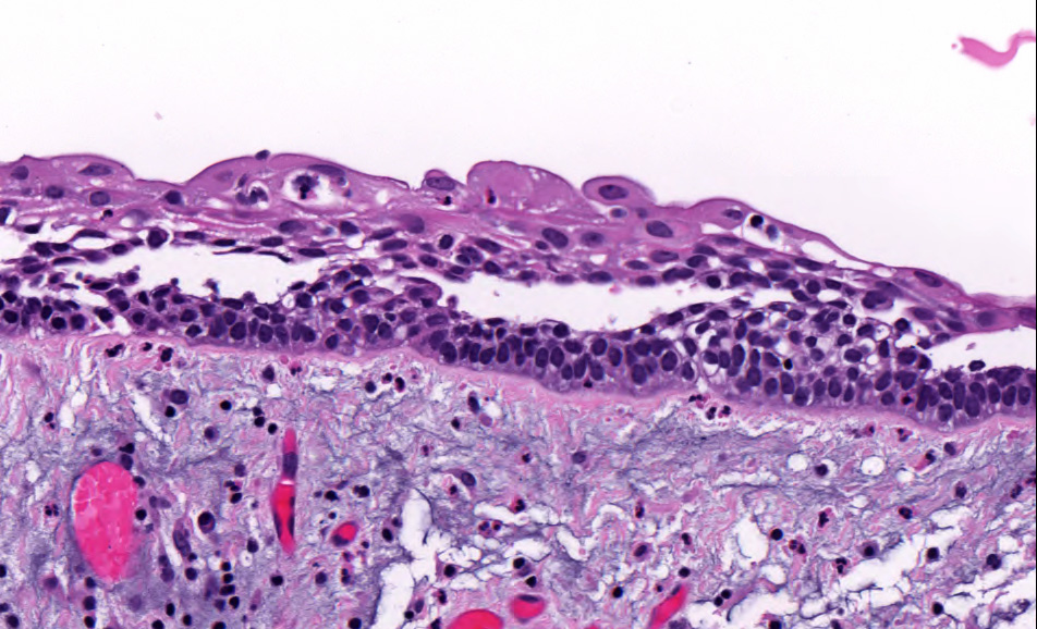
Stratified squamous epithelium
Cyst epithelium
Inflamed cyst

Variable cyst epithelial lining

Subepithelial mineralization
Contributed by Brenda L. Nelson, D.D.S., M.S. and Kelly Magliocca, D.D.S., M.P.H.
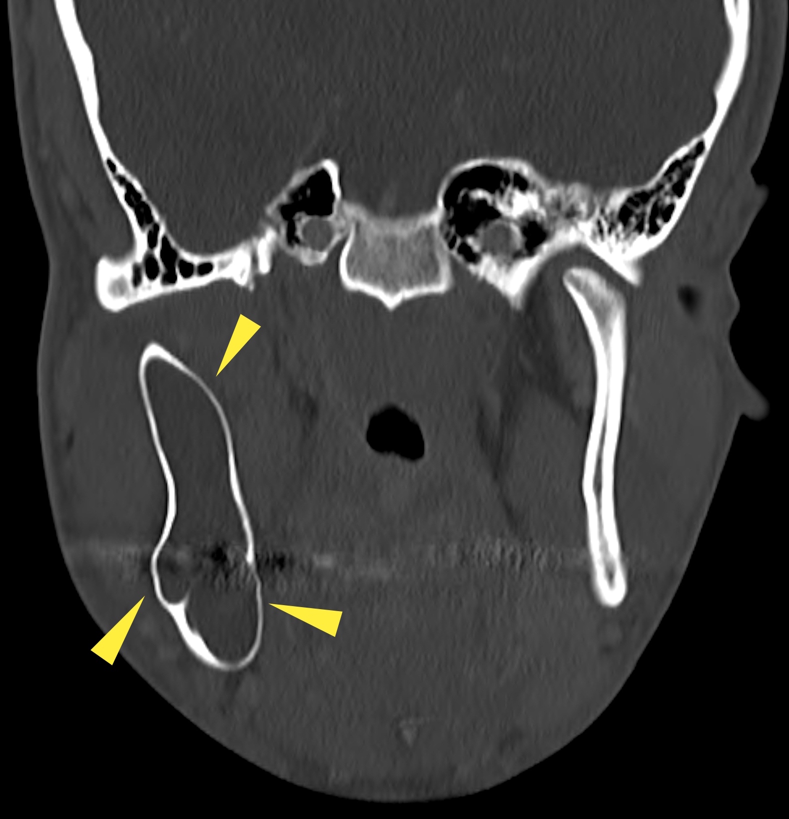
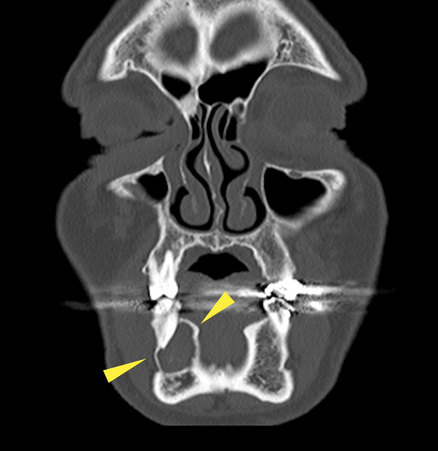
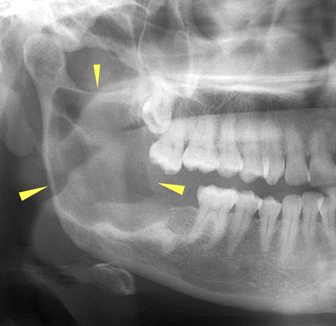
Unilocular lesion
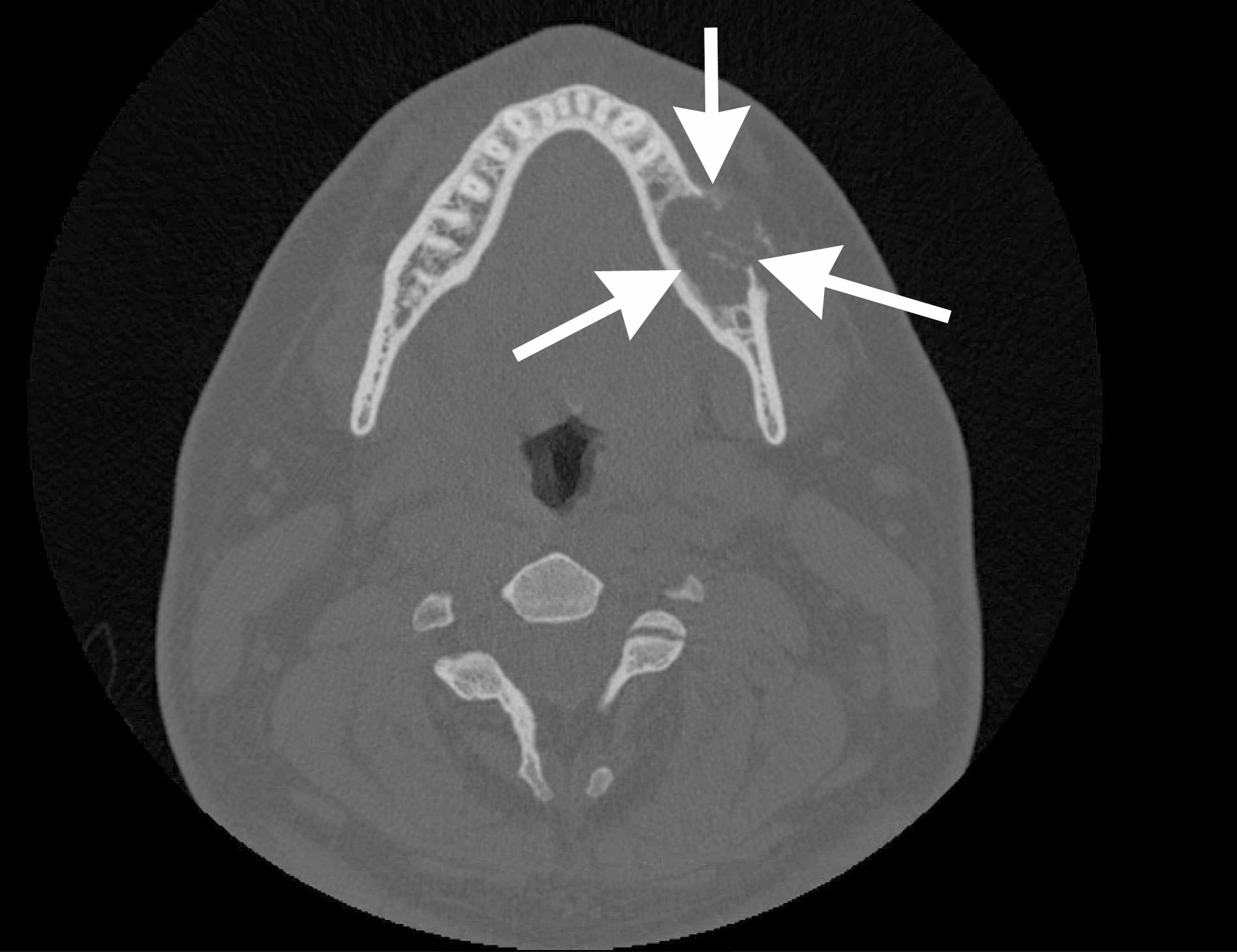
Axial CT
Contributed by Kelly Magliocca, D.D.S., M.P.H.
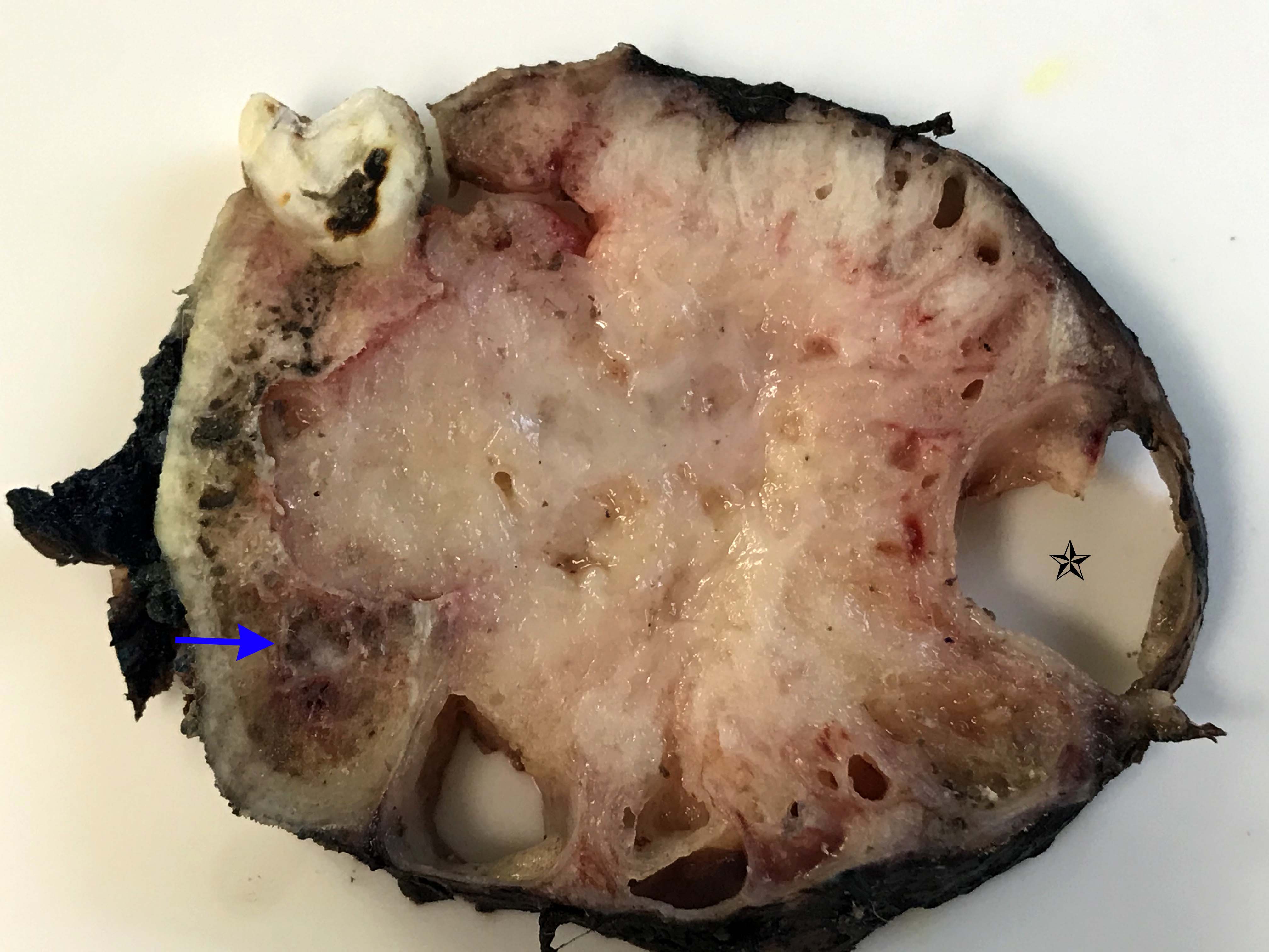
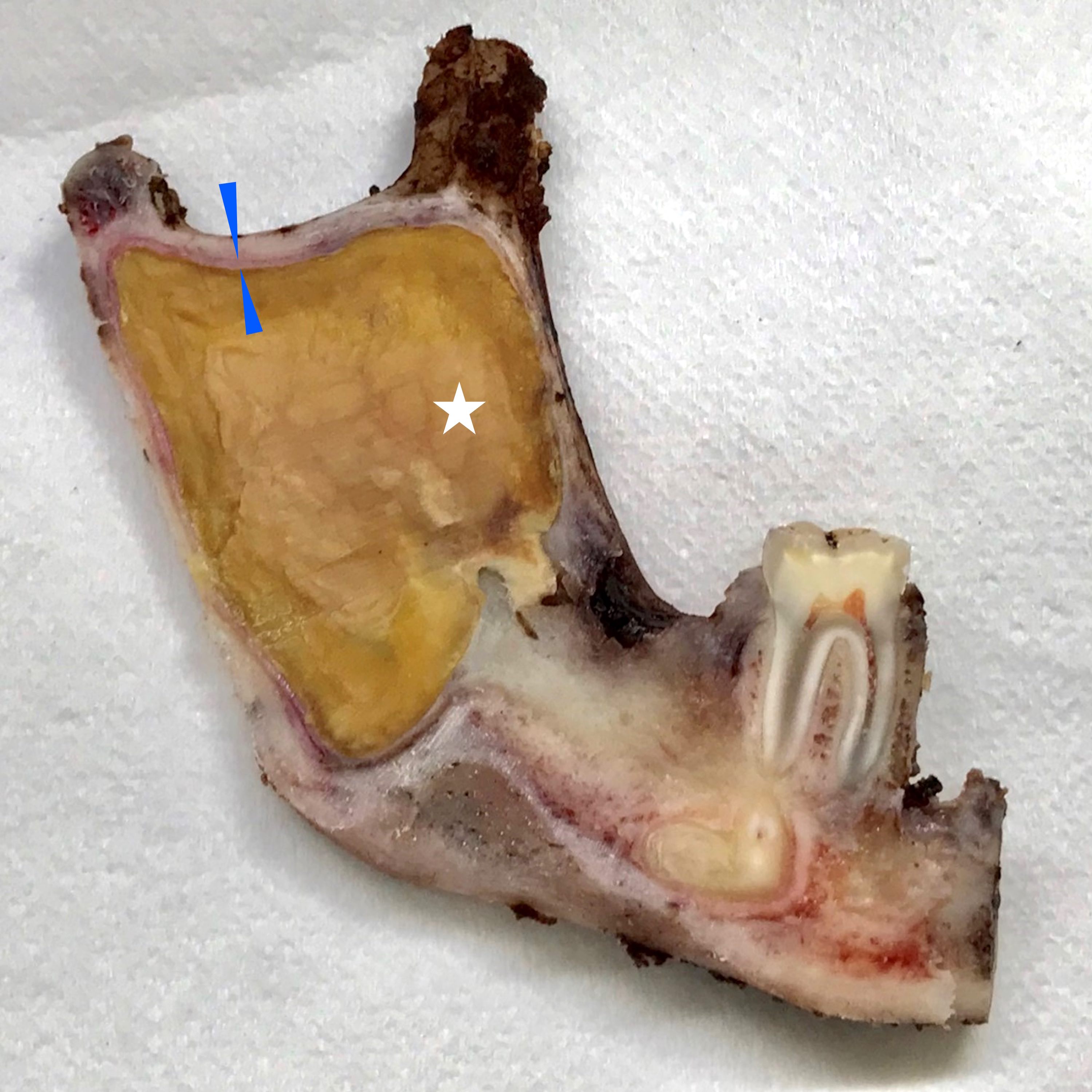
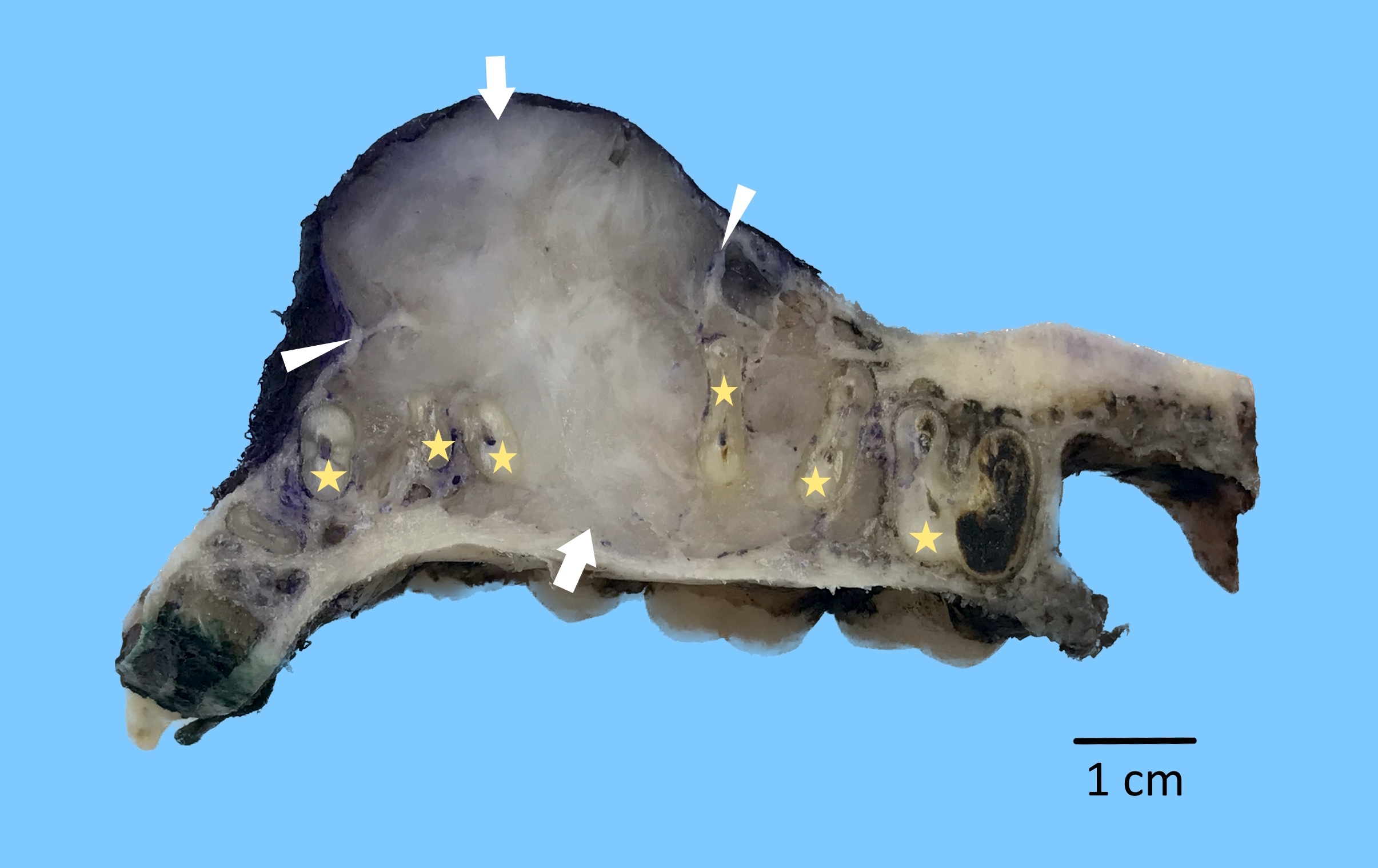
Cross sections of mandible tumor
Contributed by Kelly Magliocca, D.D.S., M.P.H., Brenda L. Nelson, D.D.S., M.S. and Anne McLean, D.M.D.
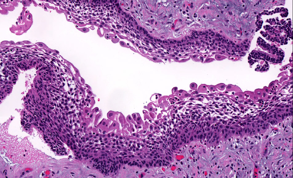
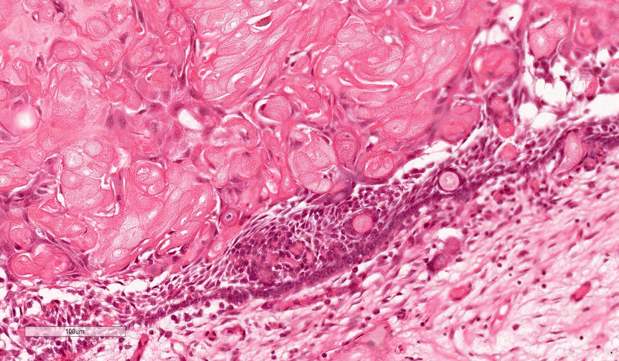
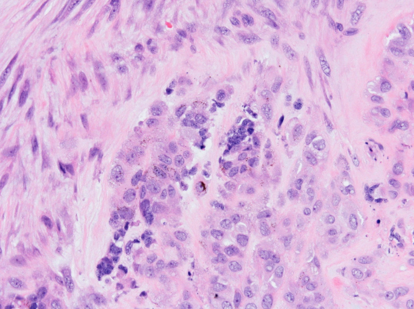
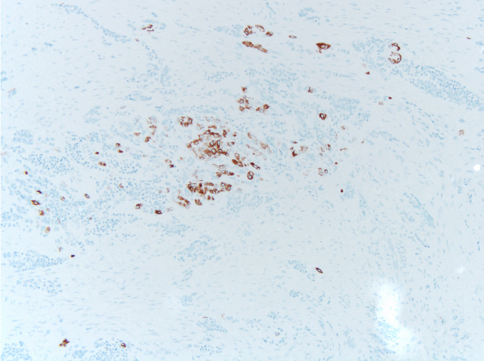
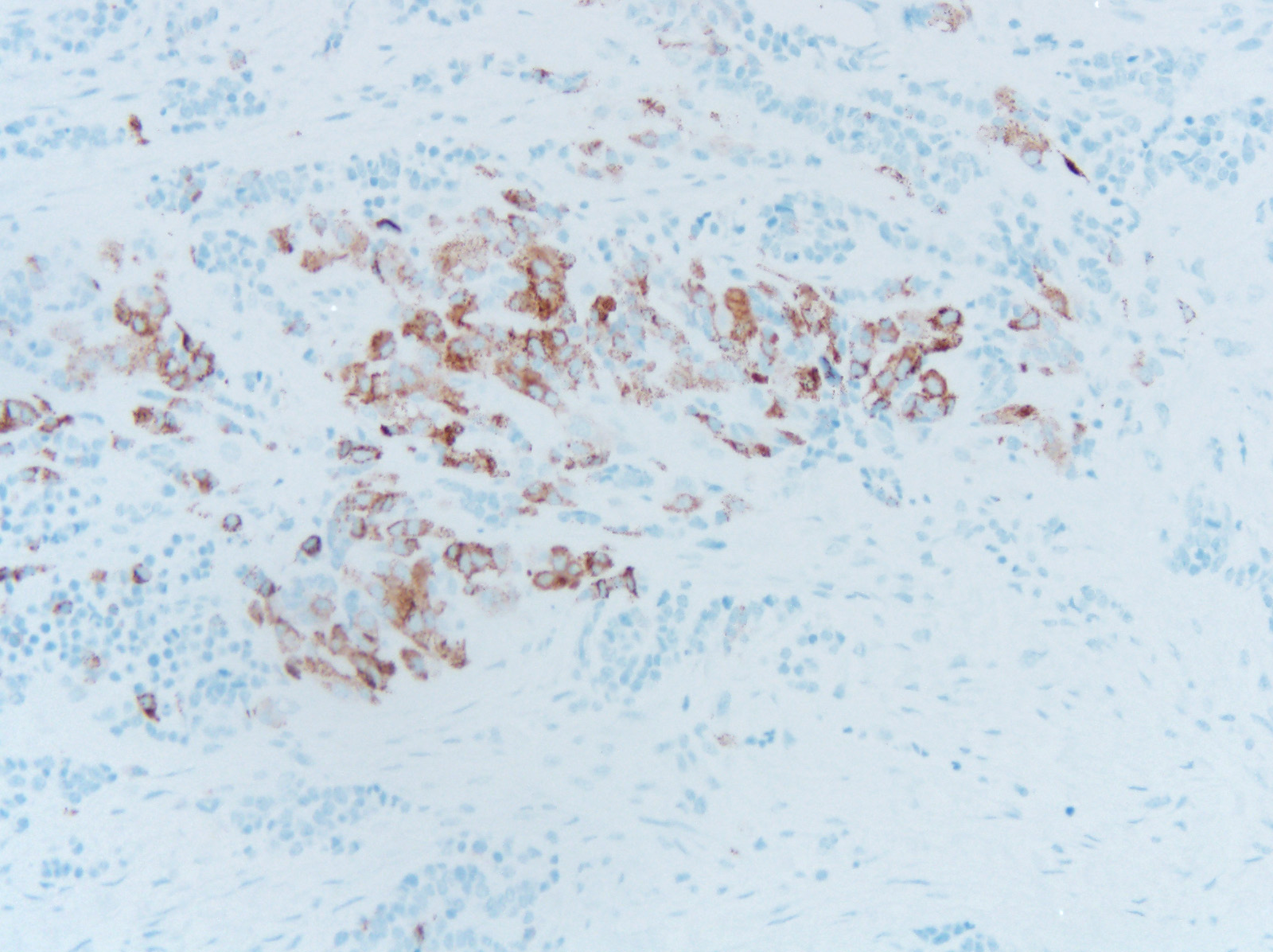
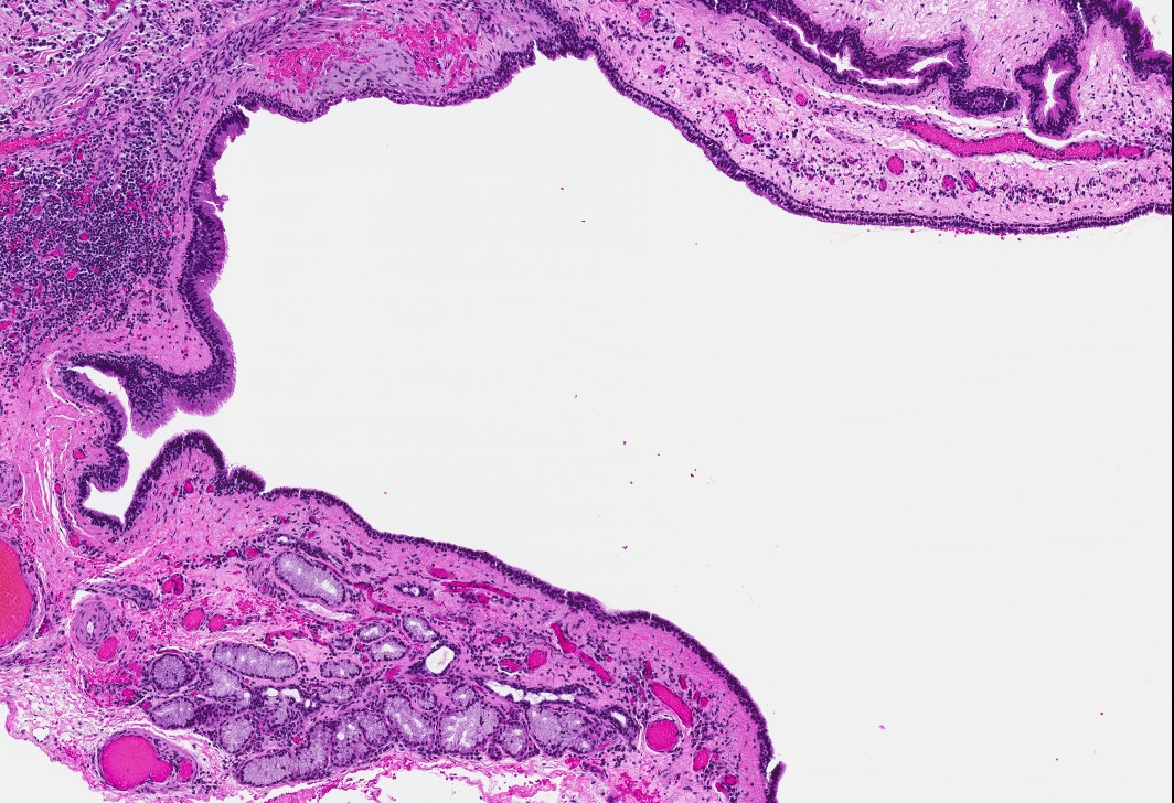
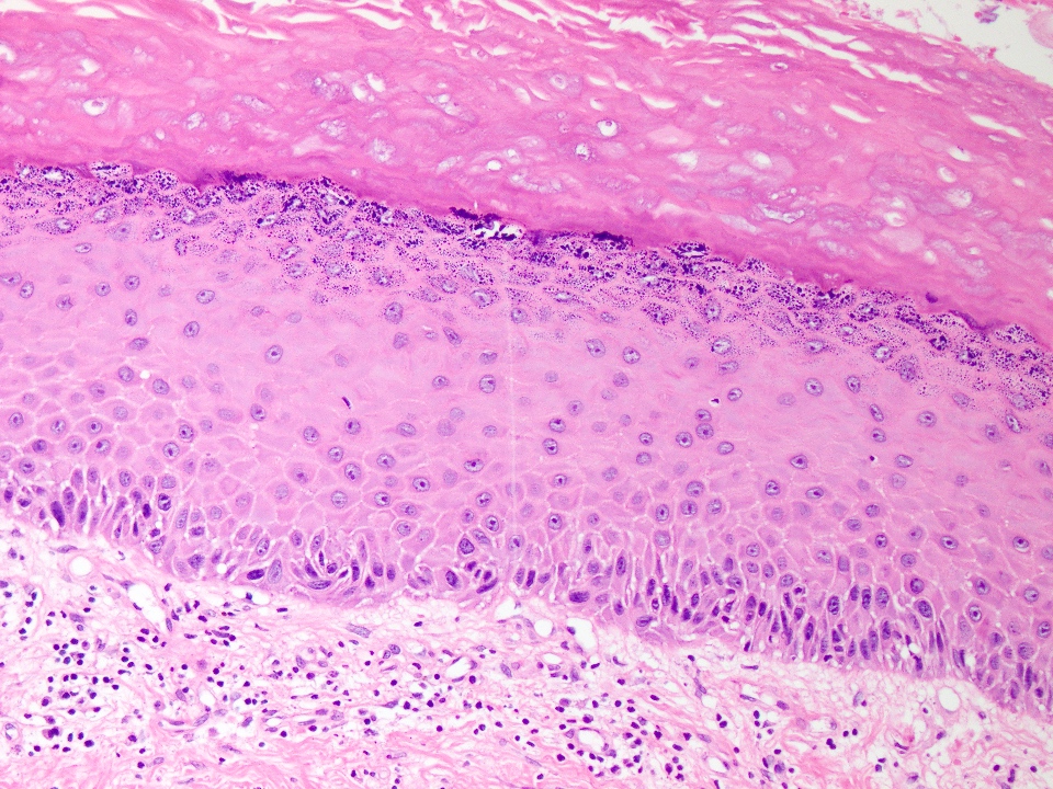
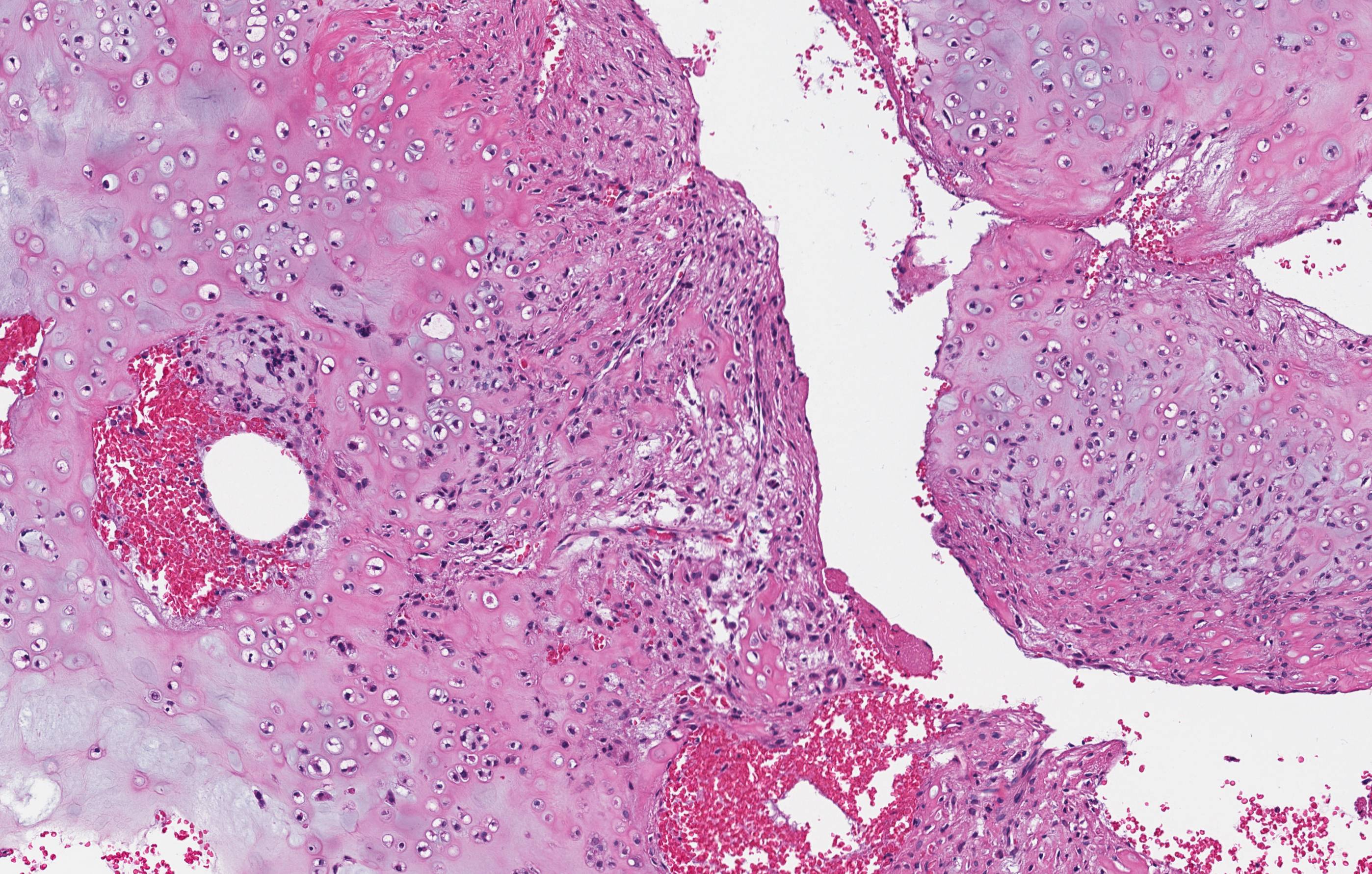
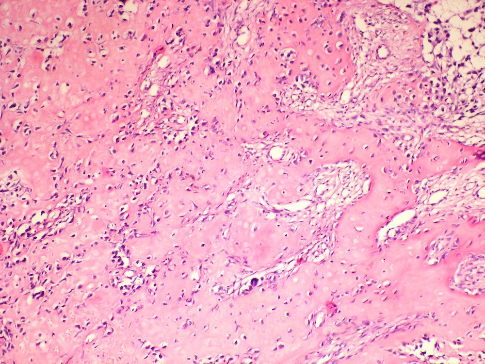
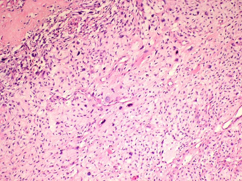
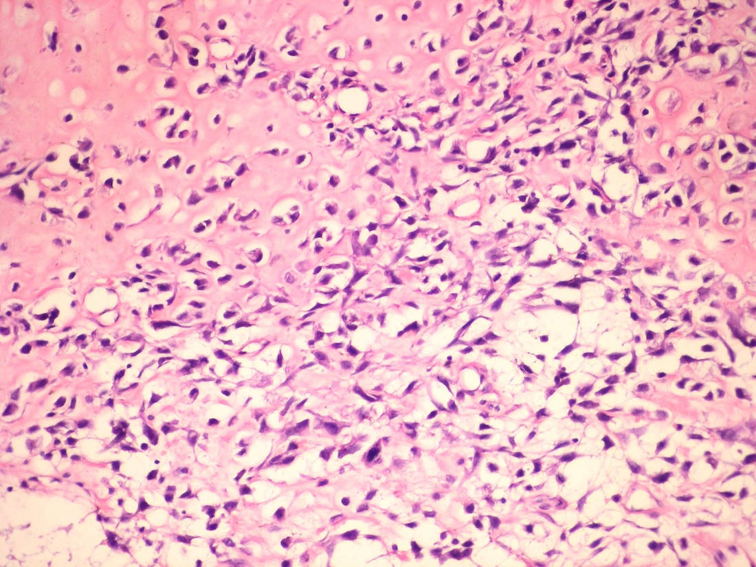
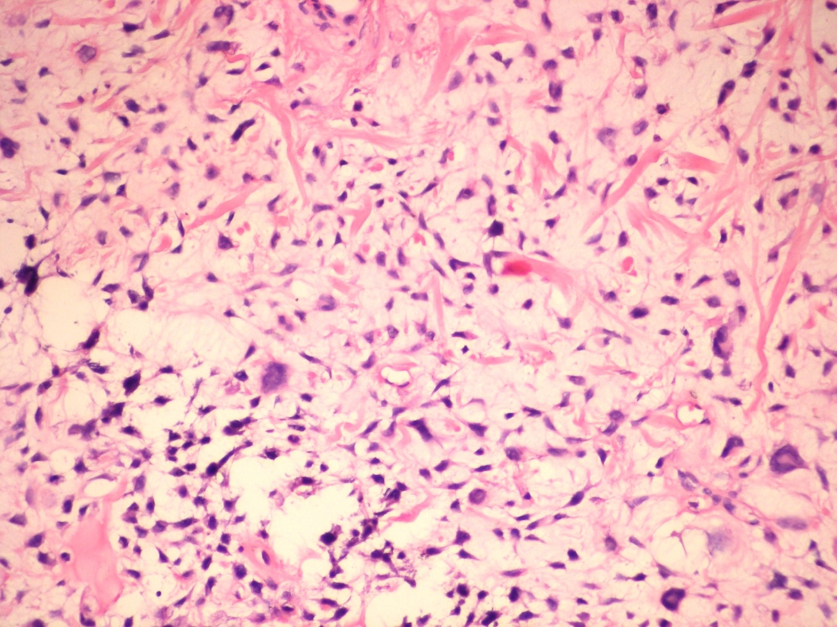
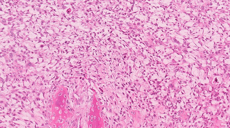
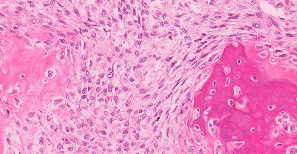
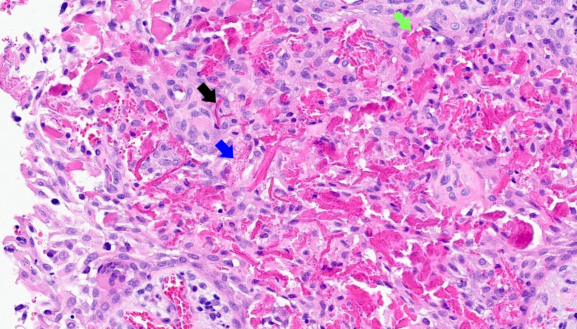
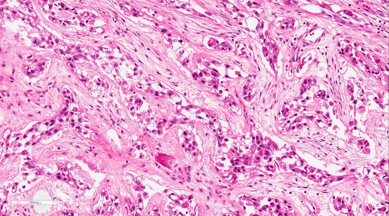
Dentinogenic ghost cell tumor
Calcifications and squamous morules
Ghost cells

Microcyst formation
Ghost cells and dentinoid
Beta catenin
Images hosted on other servers:

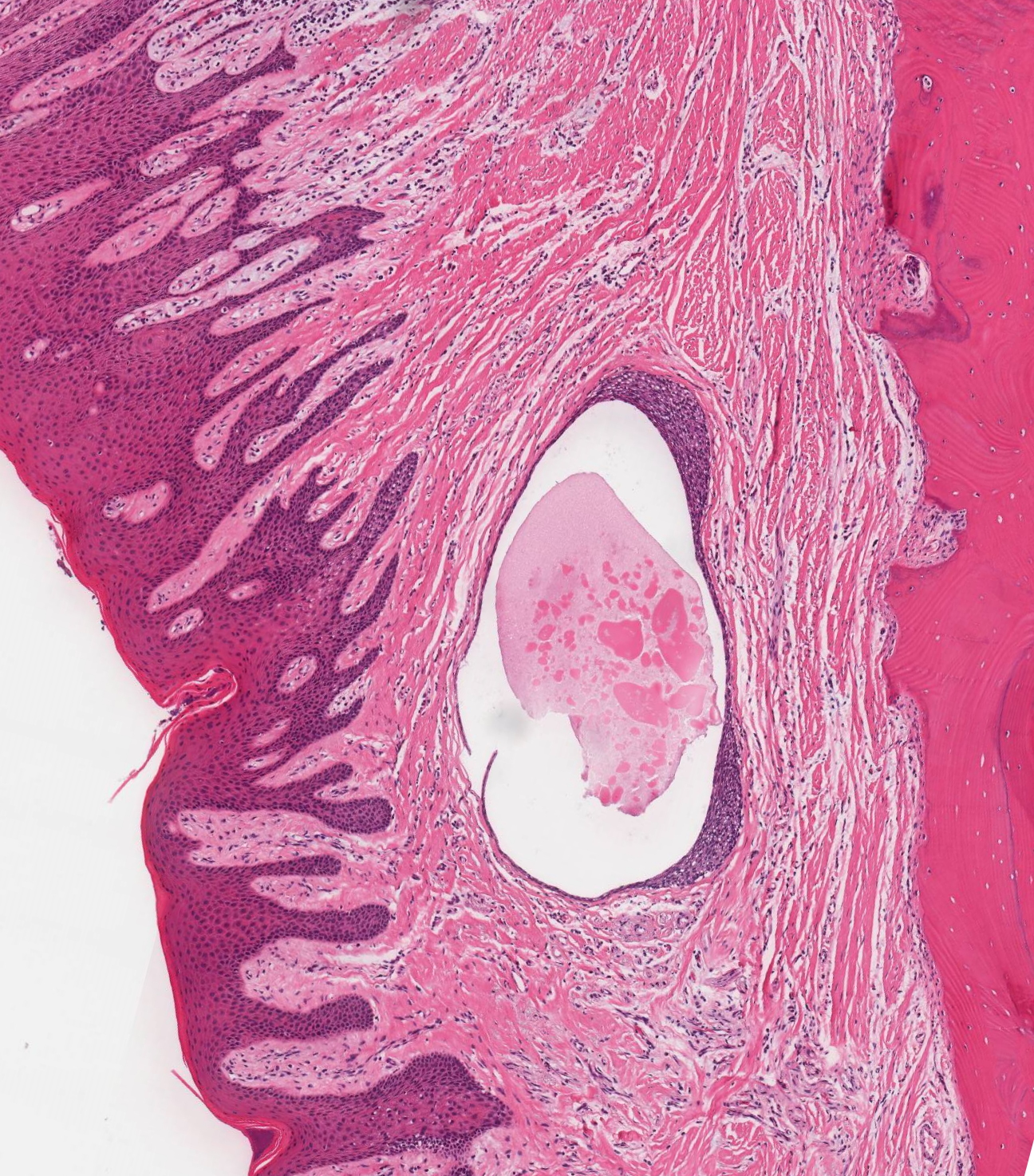
Large eruption cyst

Eruption cyst associated with #11

Exposure of #11 crown

Eruption cyst of #13

Eruption cyst (arrow) of #44

Eruption cyst of #21

Eruption cyst to left lower second molar
Images hosted on other servers:

Diffused swelling on the left side of face obliterating the nasolabial fold

Clinical and MRI examination

Axial section CT scan
Images hosted on other servers:

Ameloblastoma-like areas

Ghost cells

Cytologic atypia with increased mitotic rate


Ghost cells
Images hosted on other servers:


Gingival cyst of adult

Clinical presentation

Surgical exposure

Bone defect after excision
Images hosted on other servers:

Excised lesion
Contributed by Kelly Magliocca, D.D.S., M.P.H.
Gingival cyst
Images hosted on other servers:

Thin stratified squamous epithelium
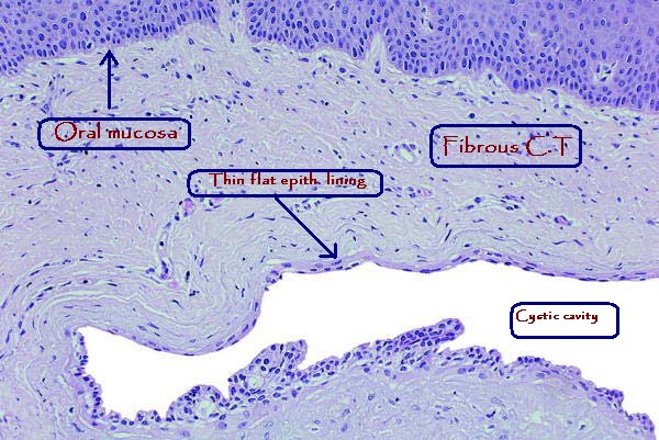
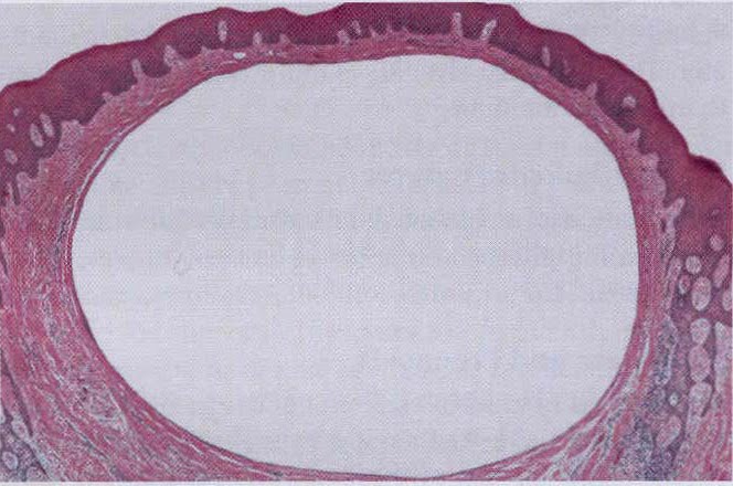
Gingival cyst histology
Images hosted on other servers:

Palatal cyst of newborn

Gingival cyst of newborn
Images hosted on other servers:

Large osteolytic lesion/s

Well defined unilocular radiolucent lesion

Well defined unilocular lesion

Multilocular radiolucency in anterior mandible

Large well defined radiolucency

Oval radiolucency in anterior maxilla

Well defined radiolucency

Large multilocular radiolucency

Complex lytic lesion of the anterior mandible

Destructive
multilocular
lesion of the
anterior mandible
Images hosted on other servers:

Swelling over left middle third of face

Intraoral swelling over left maxilla

Well defined swelling on right side of face

Lesion still attached to floor of maxillary sinus

Fluctuant swelling extending from 19 to 29 regions
Images hosted on other servers:

Excised lesion showing the cystic lining
Contributed by Kelly Magliocca, D.D.S., M.P.H.
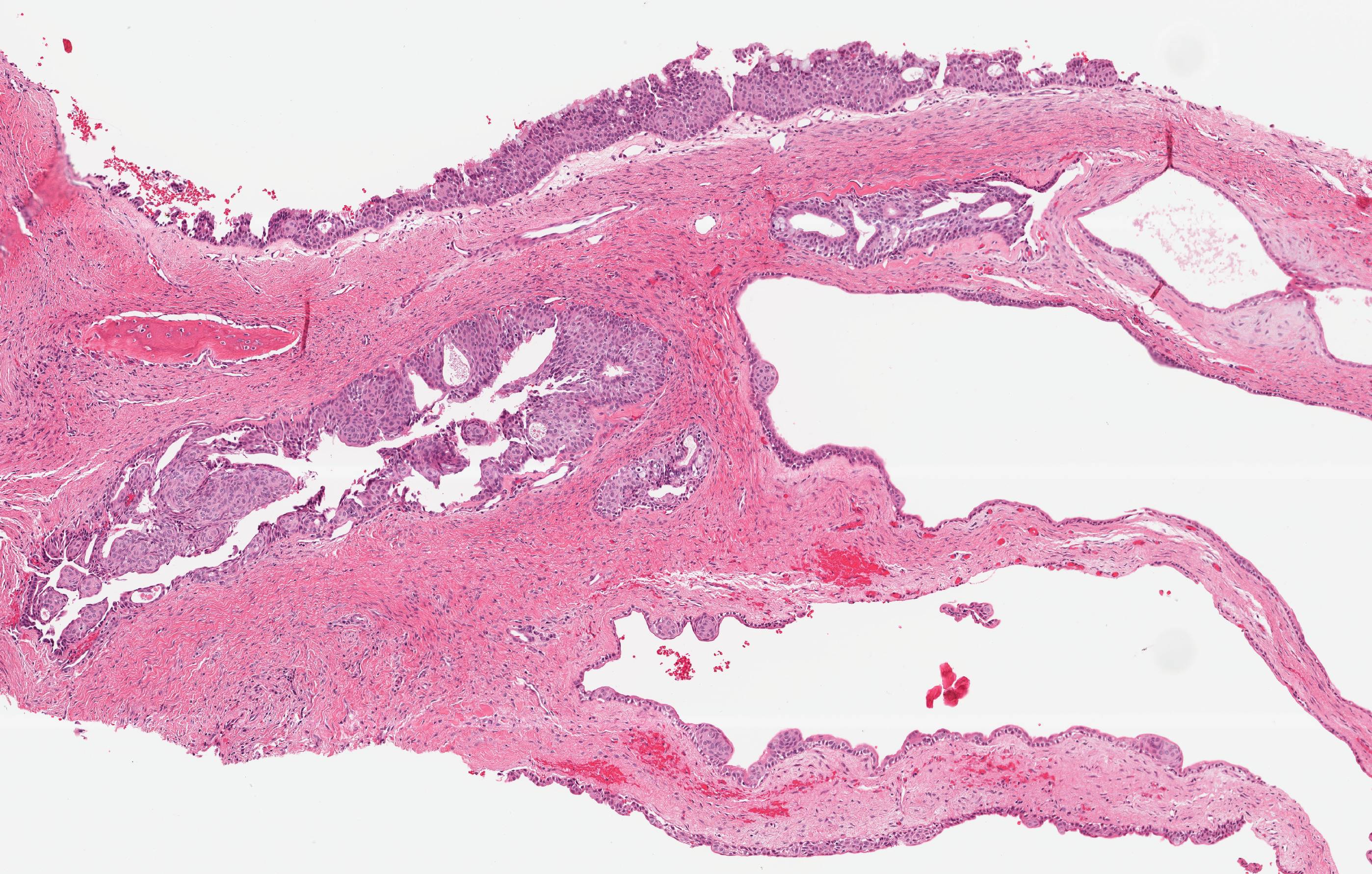
Glandular odontogenic cyst
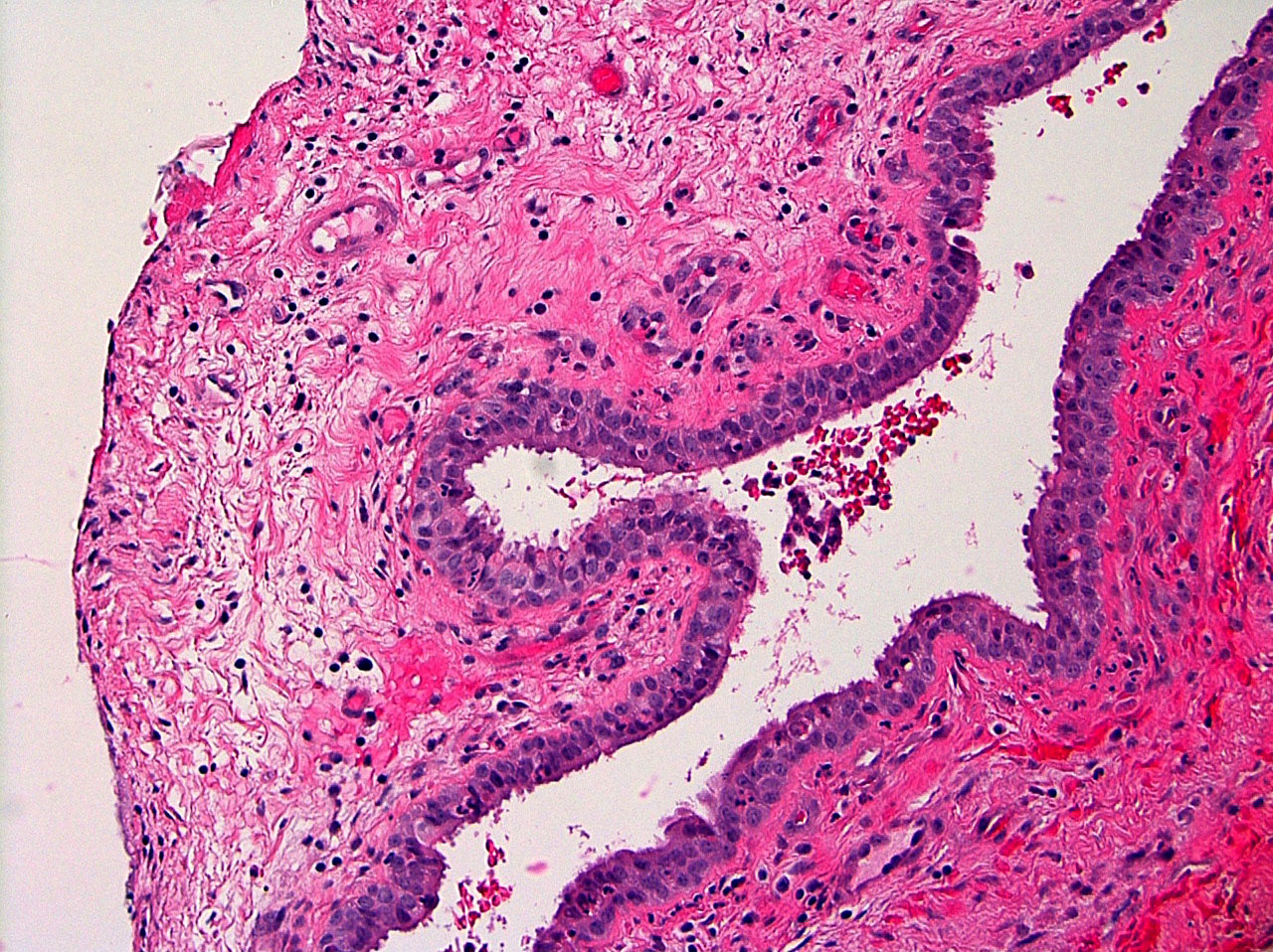
Images hosted on other servers:

Complex cyst lining

Cystic lining with connective tissue stroma

Pseudostratified squamous epithelium

Islands of epithelium

Mucous cells, epidermoid cells, and clear cells

Glandular cystic lining

Mucicarmine positive mucus cells


Mucus cells (mucicarmine positive)

Parakeratinized squamous epithelial lining

Eosinophilic cuboidal cells

Vacuolated area in lining of a dentigerous cyst

Mucoepidermoid carcinoma-like islands in cyst wall
Images hosted on other servers:

Well defined semilunar shaped radiolucency

Expansion of vestibular cortical bone

Radiolucency in distal follicular space of first permanent molar

Hypodense area in vestibular region

Axial CT

Expansion of vestibular bone

Radiolucency demarcated by fine radiopaque line

Well defined ovoid radiolucency

Buccal bifurcation cyst
Images hosted on other servers:

Intraoral preoperative view

Intraoperative view of enucleation
Contributed by Kelly Magliocca, D.D.S., M.P.H.
Inflammatory collateral cysts
Images hosted on other servers:


Various images
Images hosted on other servers:

Radiolucent lesion in the right maxilla

Large radiolucent area on right side of molar

Mixed radio-opaque and radiolucent lesion

CT scan

Radiolucent lesion with central radiopacities

Expansile lesion of the left frontal sinus
Images hosted on other servers:

Well defined swelling obliterating the vestibule

Bony hard swelling with vestibular obliteration

Swelling angle ramus area of the mandible
Contributed by Kelly R. Magliocca, D.D.S., M.P.H.
Juvenile trabecular ossifying fibroma
Images hosted on other servers:

Mineralized pieces in the form of trabeculae

Extensive osteoid production

connective tissue
with spherical
ossicles

Focally fused ossicles

Structures having
basophilic in center
and eosinophilic
in periphery
Images hosted on other servers:

Pathogenesis of LCH
Contributed by Nasir Ud Din, M.B.B.S.
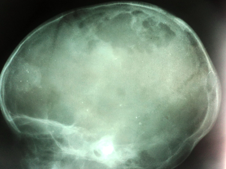
Plain Xray of skull
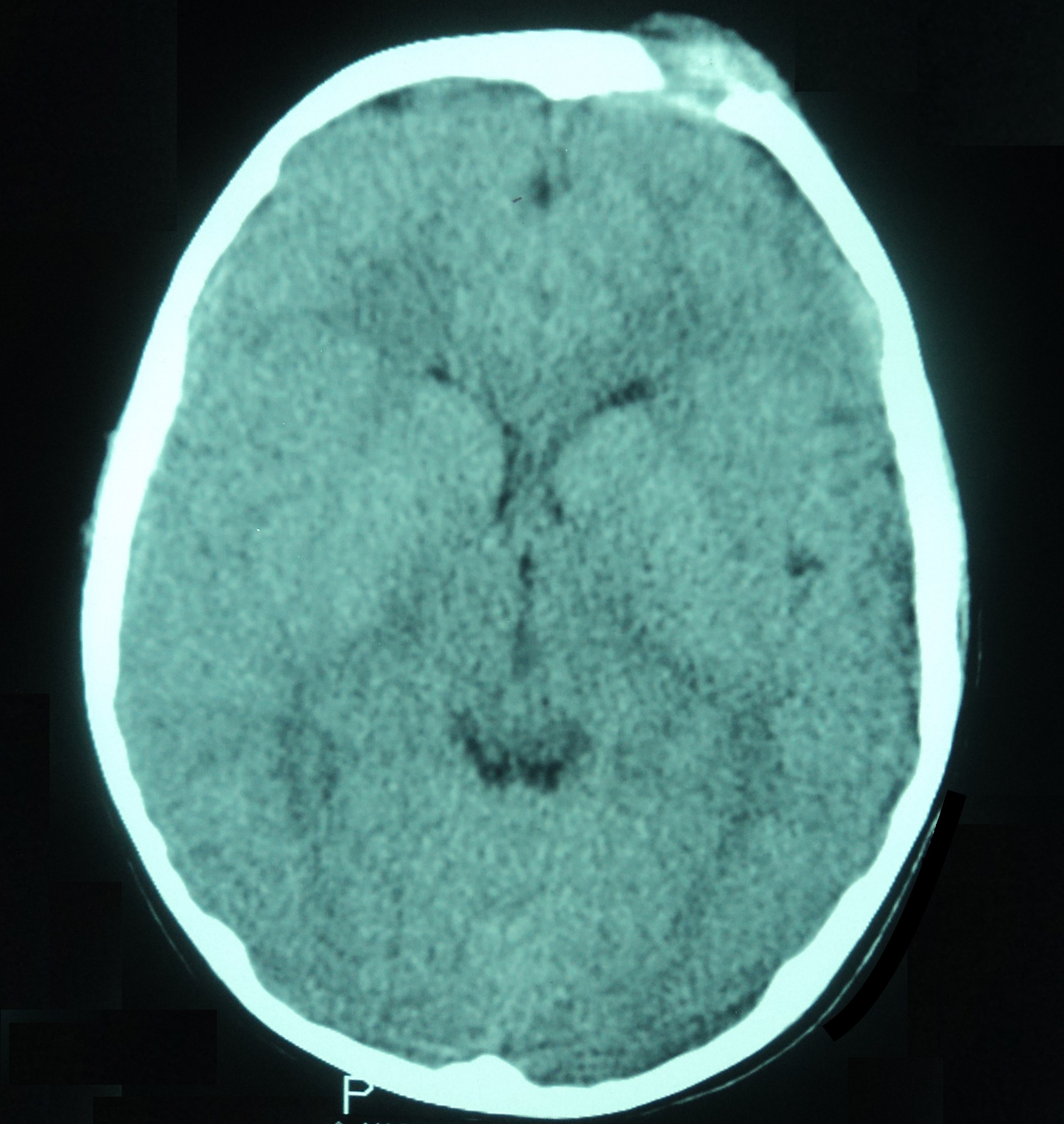
CT of brain without contrast
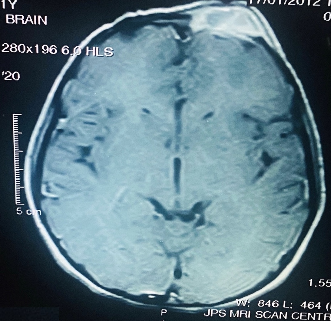
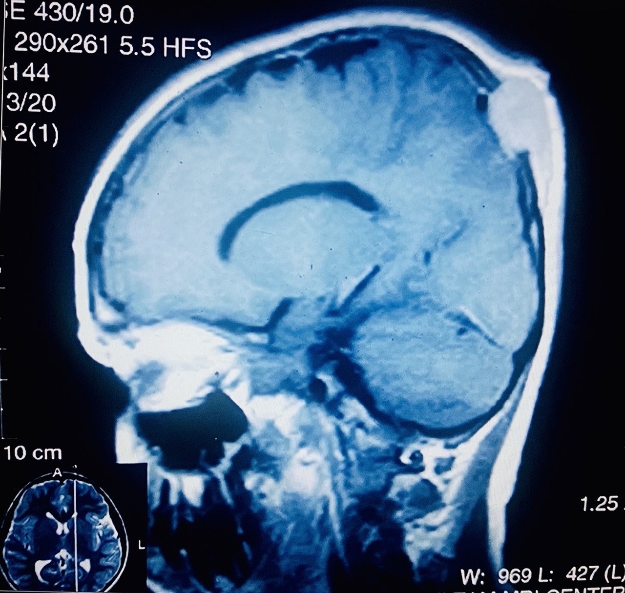
MRI of brain postcontrast
Images hosted on other servers:

Coronal CT / sagittal T1 weighted contrast enhanced MRI

MRI with contrast
Contributed by Molly Housley Smith, D.M.D.
Gingival swelling
Images hosted on other servers:

Orbital mass
Contributed by Nasir Ud Din, M.B.B.S.
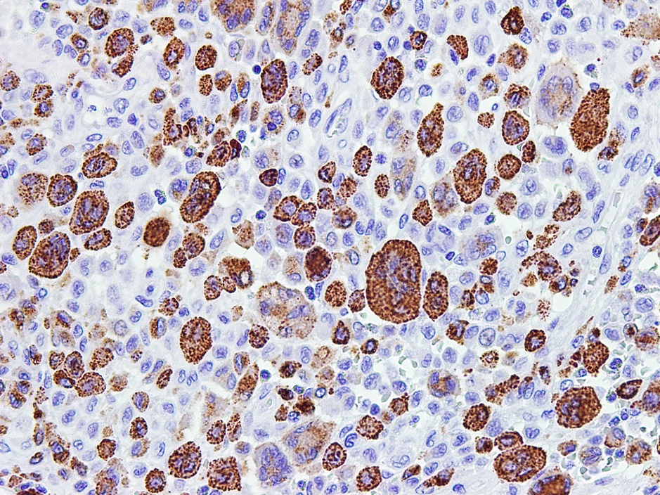
Eosinophil rich lesion
Multinucleated giant cells
Characteristic LCH cells
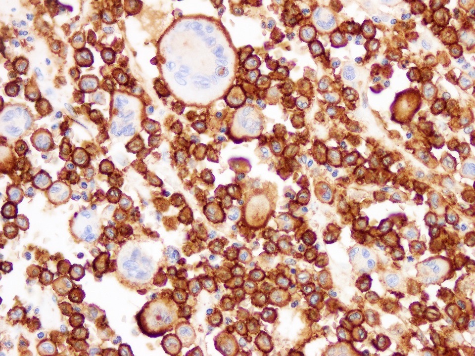
CD1a
CD68
Cyclin D1
Contributed by Mark R. Wick, M.D.
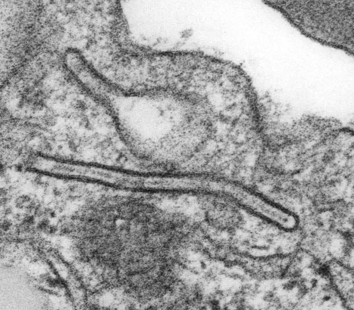
Birbeck granules
Images hosted on other servers:

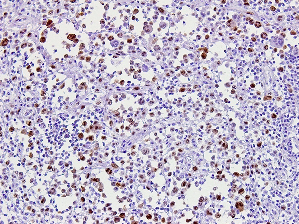
BRAF mutation analysis
Radiology and histopathology descriptions
Images hosted on other servers:

Tear dropshaped radiolucency

Orthopantamograph

Healing after 6 months

Radicular triangular radiolucency
Images hosted on other servers:

Intra oral swelling

Buccal cortical plate perforation
Contributed by Kelly Magliocca, D.D.S., M.P.H.
Lateral periodontal cyst
Images hosted on other servers:

Thick lining of a lateral periodontal cyst

Fragmented lining of a Botryoid odontogenic cyst

Typical luminal excrescence

Cystic lumen
Images hosted on other servers:

Panoramic

Primary NHL

Axial and coronal section

Coronal CT
Images hosted on other servers:

Intra-oral
Images hosted on PathOut server:
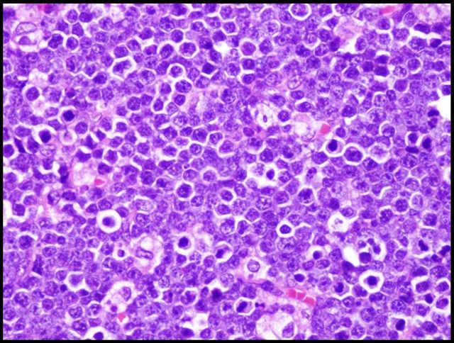
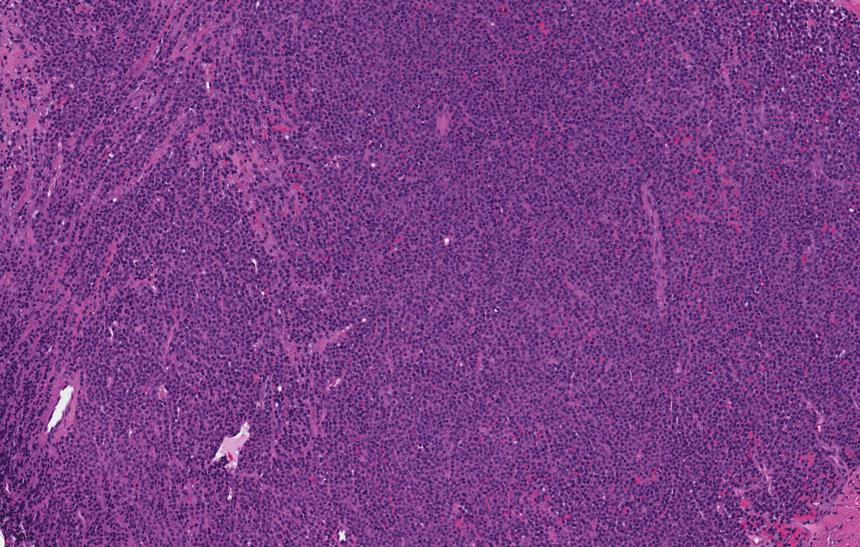
"Starry sky"
Images hosted on other servers:

Malignant lymphocytes

DLBCL: CD20, CD10, BCL6, CD3

Plasmablastic: CD20, CD138

Myeloma plasma cells
Images hosted on other servers:


Various cases

Osseous involvement

Due to prosthesis

After tooth extraction

Maxillary involvement

Osteolytic lesion in mandible
Images hosted on other servers:

and surgical defect
Images hosted on other servers:

Osteolytic pattern

Destruction of alveolar bone process

Mandibular alveolar bone exposed

Right maxillary exposed bone

Generalized sclerotic changes
Images hosted on other servers:

Presence of bone erosion

Heterogenously enhancing soft tissue mass lesion

Radiolucent expansile, solid lesion
Images hosted on other servers:

Swelling on maxillary alveolus

Upper vestibule, alveolar ridge and anterior hard palate
Images hosted on other servers:

Excised tumor
Contributed by Kelly Magliocca, D.D.S., M.P.H. and Karen Fritchie, M.D.
Melanotic neuroectodermal tumor of infancy
AE1 / AE3
HMB45
Images hosted on other servers:

Orthopantogram
(panoramic view)

CT

3D CT
Images hosted on other servers:

Well defined solitary swelling on right side of mandible

Swelling in lower third of face


Intra-oral view
Images hosted on other servers:


Lung metastases

Breast metastasis

Renal cell carcinoma metastasis

Thyroid metastasis
Images hosted on other servers:

Heart shaped radiolucent lesion

Expansile maxillary alveolus

Well defined round radiolucent area

Well defined radiolucency

Homogeneous high intensity area

Saggital / axial CT images

Axial sections of CT
Images hosted on other servers:

Intraoral swelling

Intraoperative view, cystic area
Images hosted on other servers:

Surgical specimen
Contributed by Kelly Magliocca, D.D.S., M.P.H.
Nasopalatine duct cyst
Images hosted on other servers:


Stratified squamous and pseudostratified columnar epithelium

Flattened cuboidal epithelium

Fibrous cyst wall with nerves
Images hosted on other servers:


Cystic lesion

Diffuse foci of radiopacities
Images hosted on other servers:

Surgical specimens
Contributed by David Cohen, M.B.B.Ch., M.D. (Case #367)
Case of the Week #367
Images hosted on other servers:


Various images

Odontogenic epithelial cells

Irregular masses of dysplastic dentin
Eruption of teeth
Tooth development
Tooth development
Images hosted on other servers:

IOPA showing irregular radiopaque masses

Multilocular radiolucency near 21, 22, 63, 24 and 25
Images hosted on other servers:


Gingival masses
Images hosted on other servers:

Surgical intervention of odontogenic fibroma
Contributed by Kelly Magliocca, D.D.S., M.P.H.
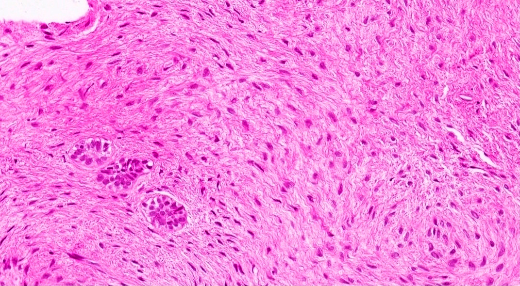
Odontogenic fibroma
Case 1: Odontogenic fibroma with extensive calcification

Case 2: Odontogenic fibroma with giant cell reaction / giant cell granuloma

Case 3: Odontogenic fibroma
Images hosted on other servers:

Odontogenic fibroma of the maxilla

COF and dentigerous cyst in the maxilla


Islands of odontogenic epithelium within a cellular fibrous stroma

Features of peripheral odontogenic fibroma

Fig. 7 Odontogenic fibroma with central giant cell lesion

Fig. 8 Ossifying variant of COF

Fig. 9 Amyloid variant of COF
Contributed by Kelly Magliocca, D.D.S., M.P.H.
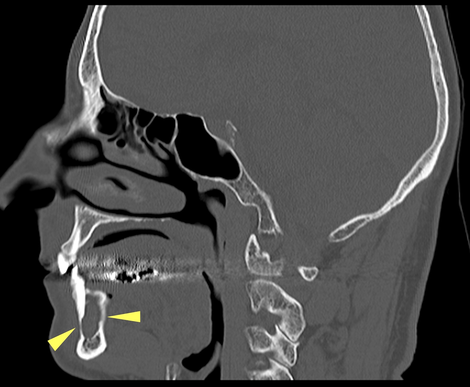
Sagittal CT anterior mandible
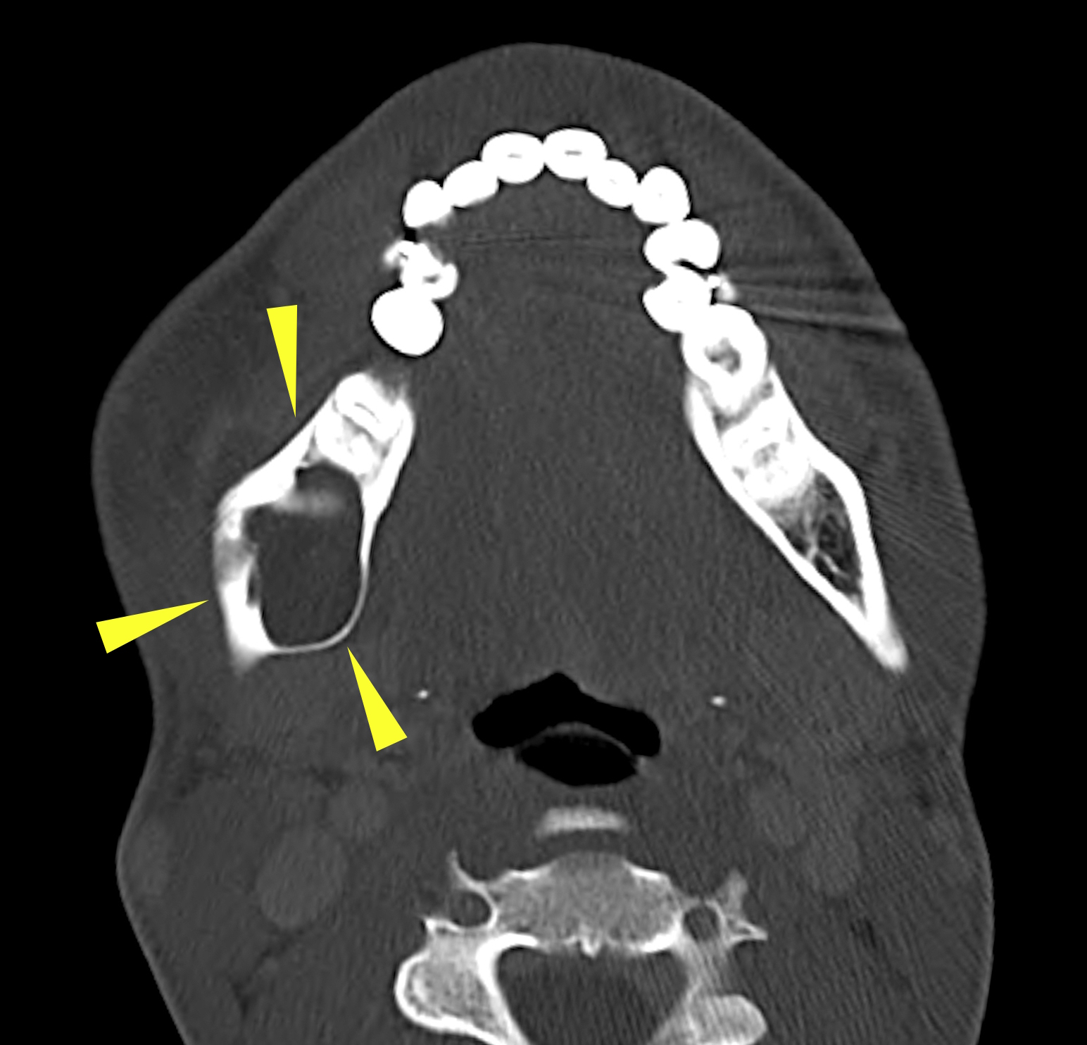
Axial CT left mandible
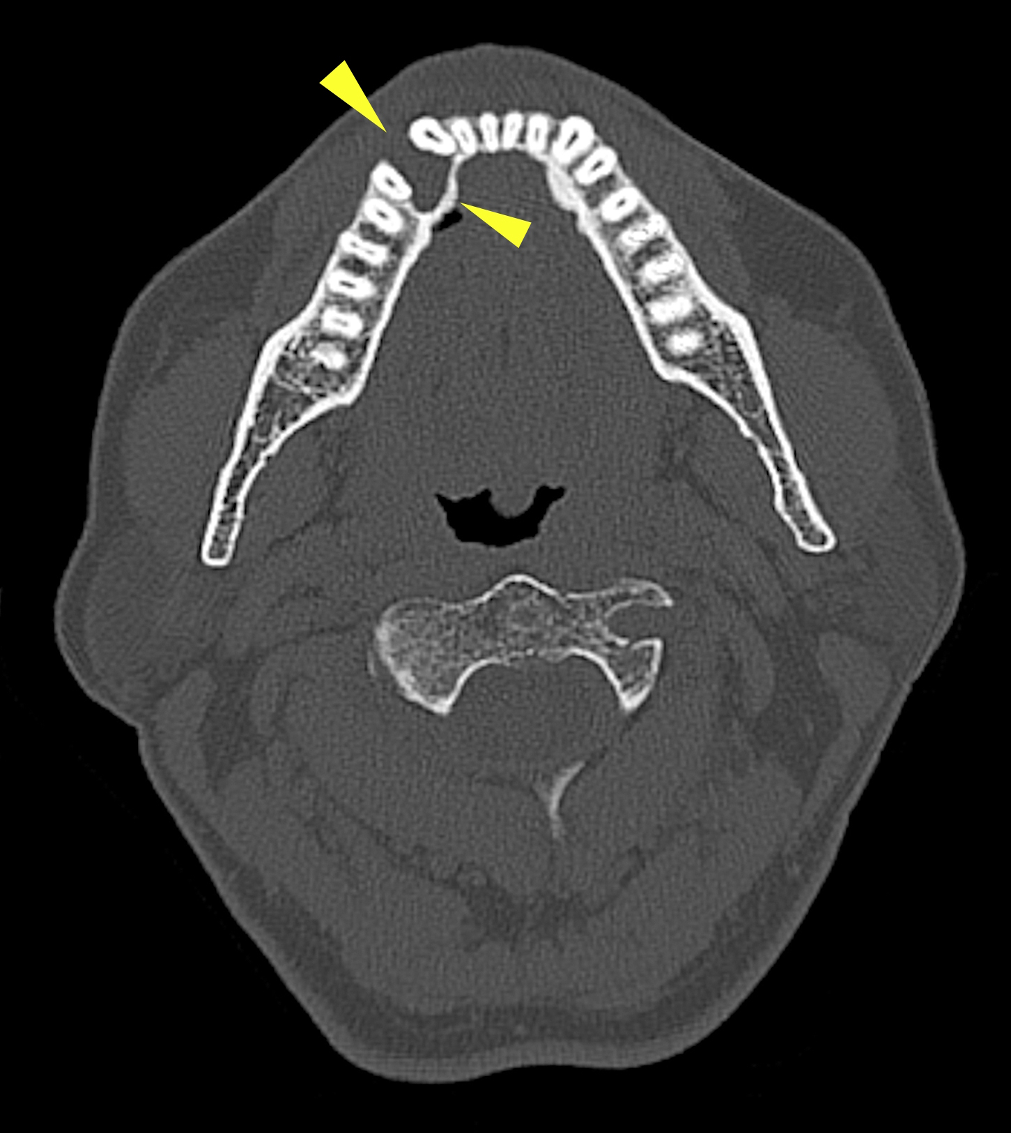
Axial CT anterior mandible
Coronal CT posterior mandible
Coronal CT mandibular body
Cropped panorex left ramus

Cropped panorex body mandible
Images hosted on other servers:

Well circumscribed mass

Intraoral finding

Less common maxillary lesion
Contributed by Kelly Magliocca, D.D.S., M.P.H.
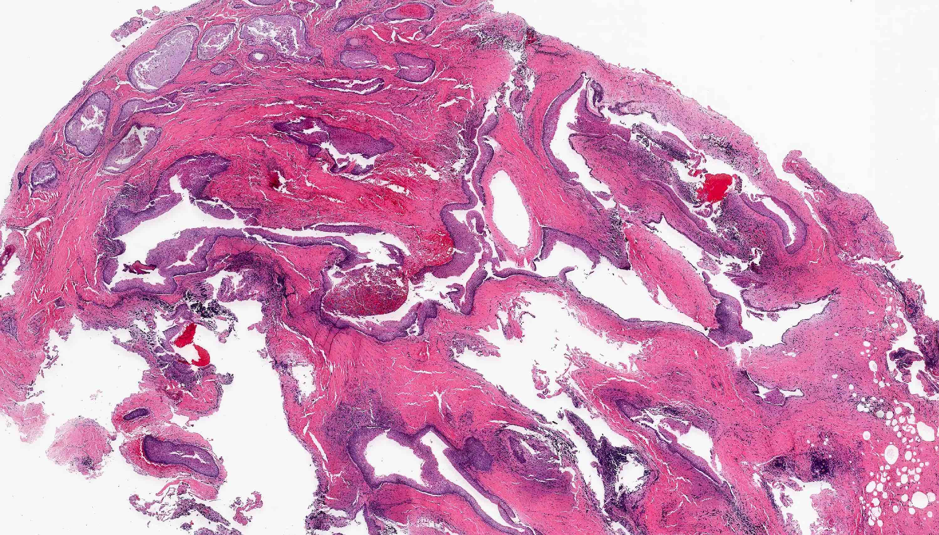
Mandibular resection
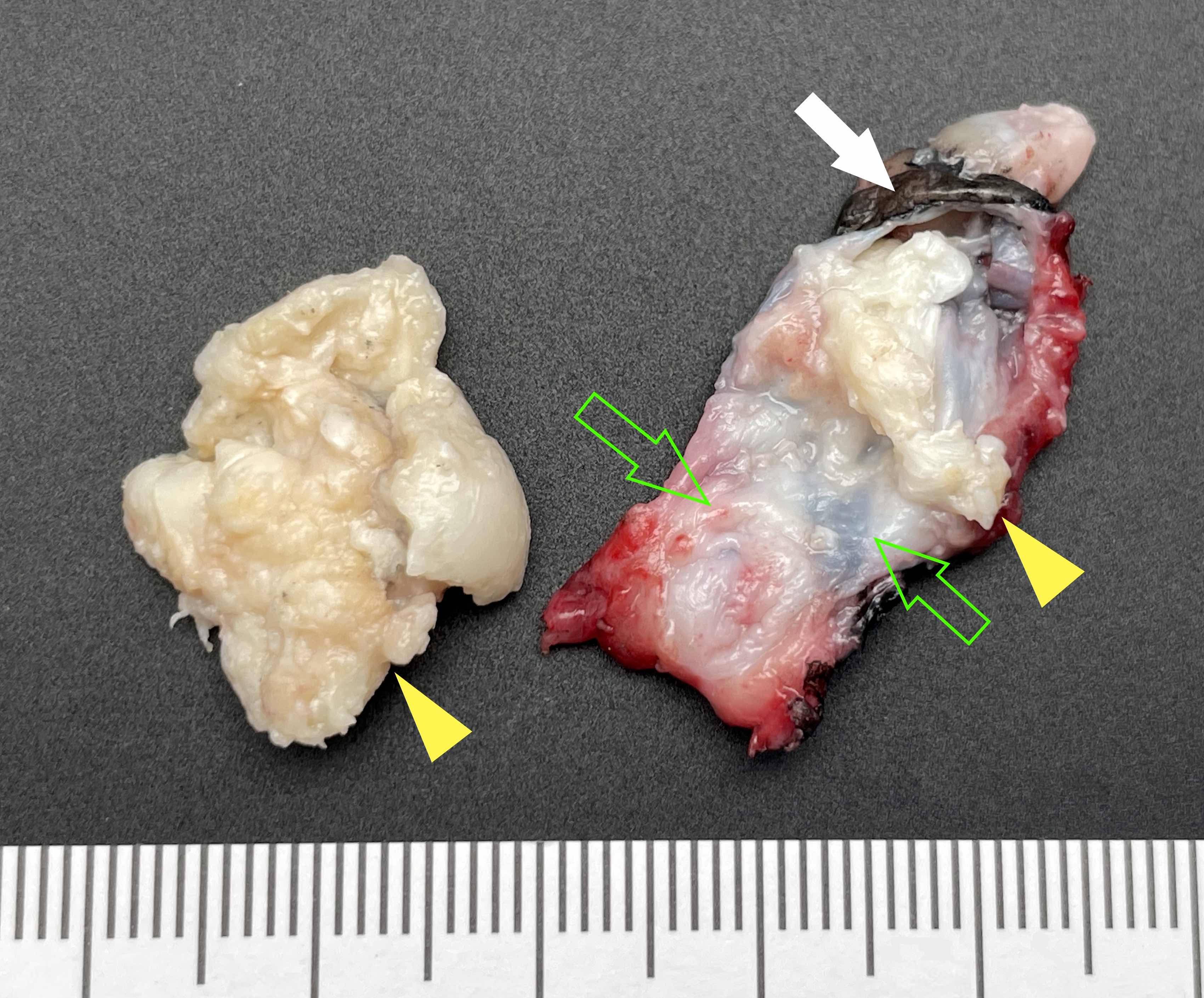
Enucleation of mandibular lesion
Images hosted on other servers:

Cystic lesion with thin smooth wall

Massive KCOT

Impacted maxillary third molar
Contributed by Kelly Magliocca, D.D.S., M.P.H. and Case #503
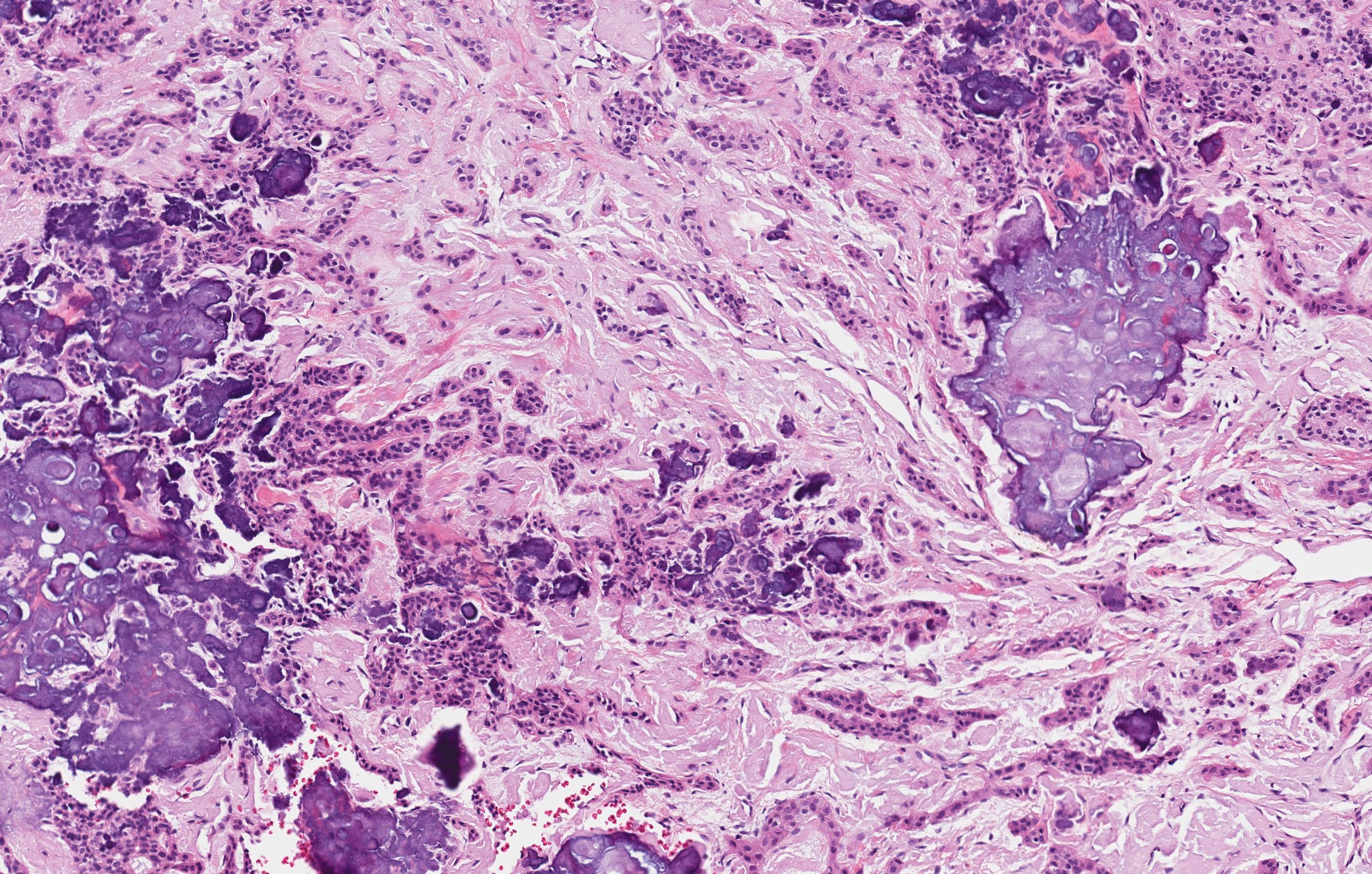
Solid OKC
Macrocystic OKC
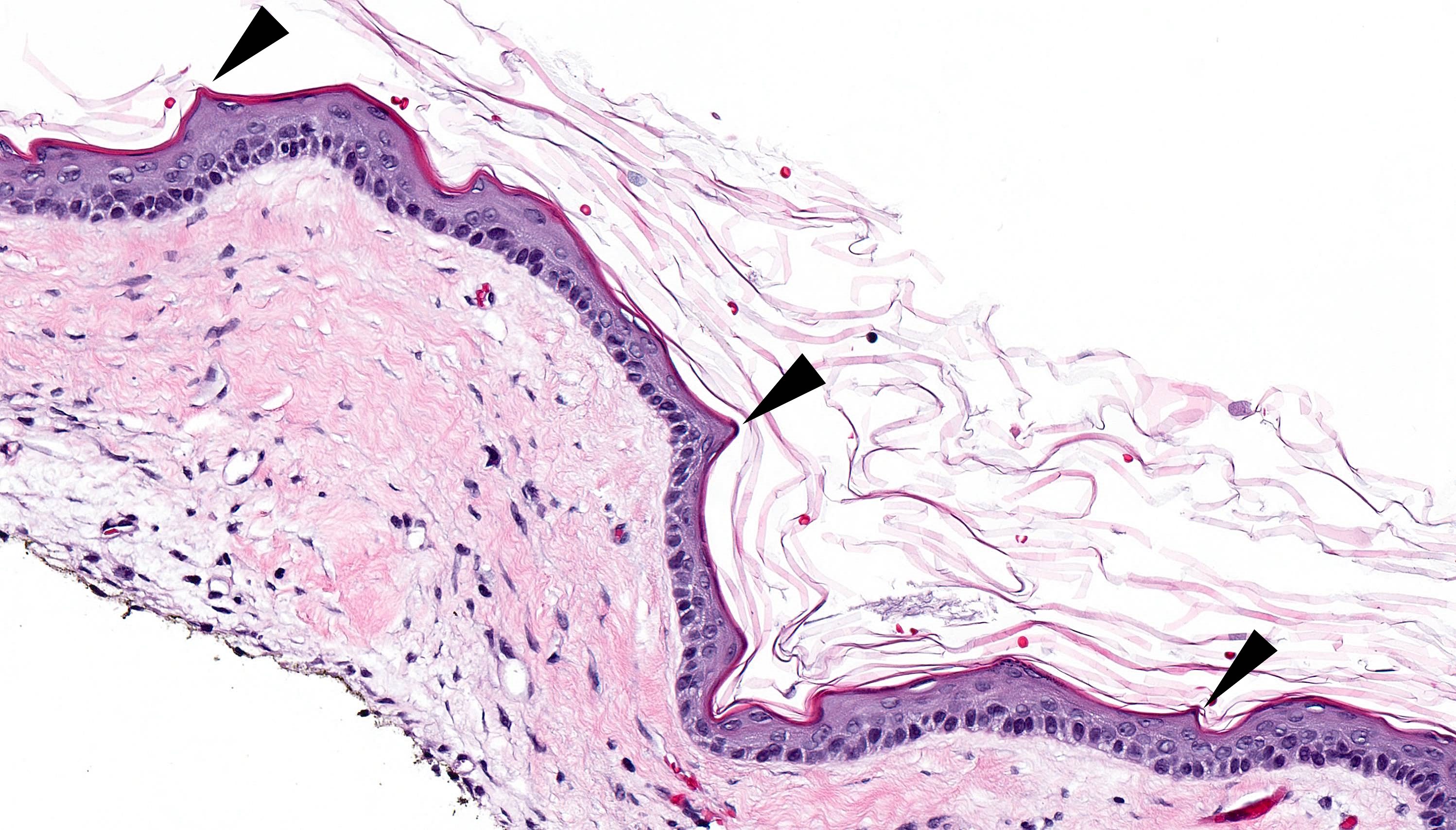
Peaks at surface layer
Smooth surface layer
Corrugated surface layer
Inflamed attenuated epithelium
Basal layer hyperchromatism

Squamous lined cyst
Areas of inflammation / irritation
Classic microscopic features
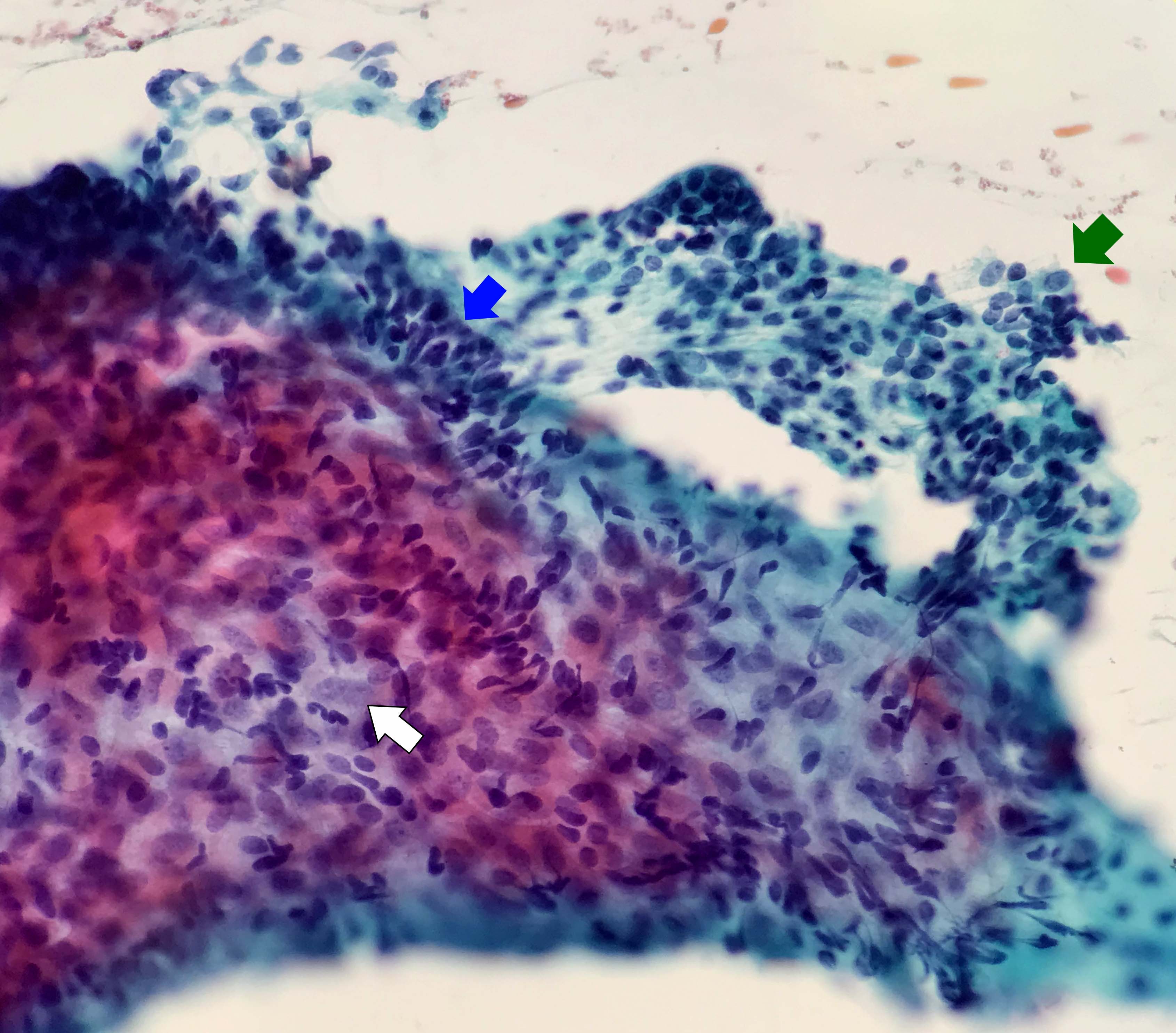
Contributed by Kartik Viswanathan, M.D., Ph.D. and Kelly Magliocca, D.D.S., M.P.H.
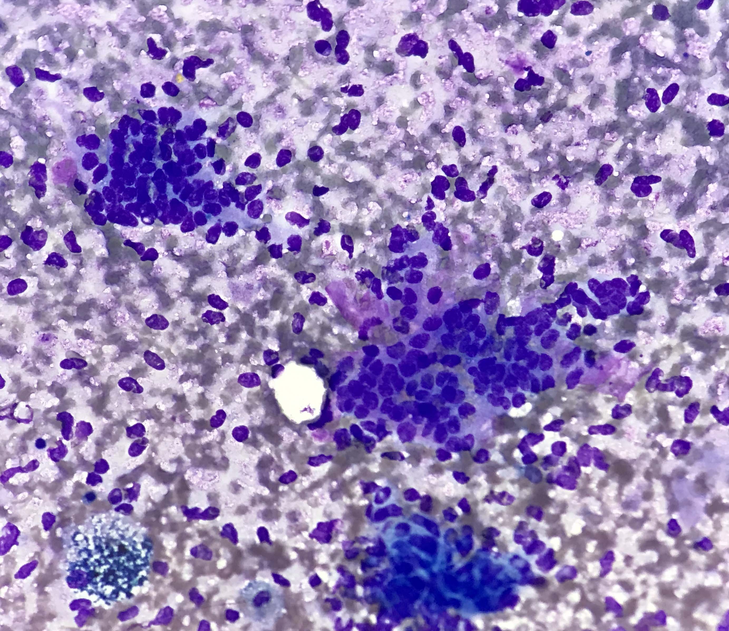
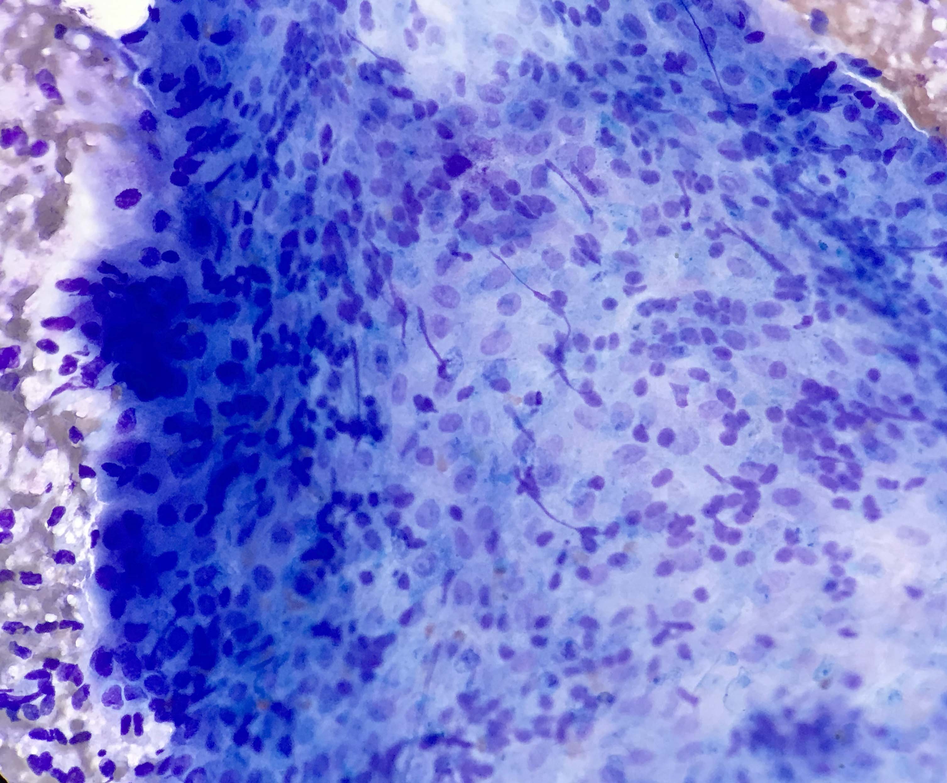
Touch prep
Imprint touch prep
Odontogenic keratocyst - histopathology
Images hosted on other servers:

Multilocular, radiolucent lesion

Right mandibular lateral occlusal view

Multilocular radiolucent osteolytic lesion

CT scan of mandible
Images hosted on other servers:

Swelling on the right side

Mandibular right quadrant

Gray-white appearance
Contributed by Kelly Magliocca, D.D.S., M.P.H.
Mandibular myxoma resection
Images hosted on other servers:

Jelly-like appearance
Contributed by Abberly Lott Limbach, M.D. and Kelly Magliocca, D.D.S., M.P.H.
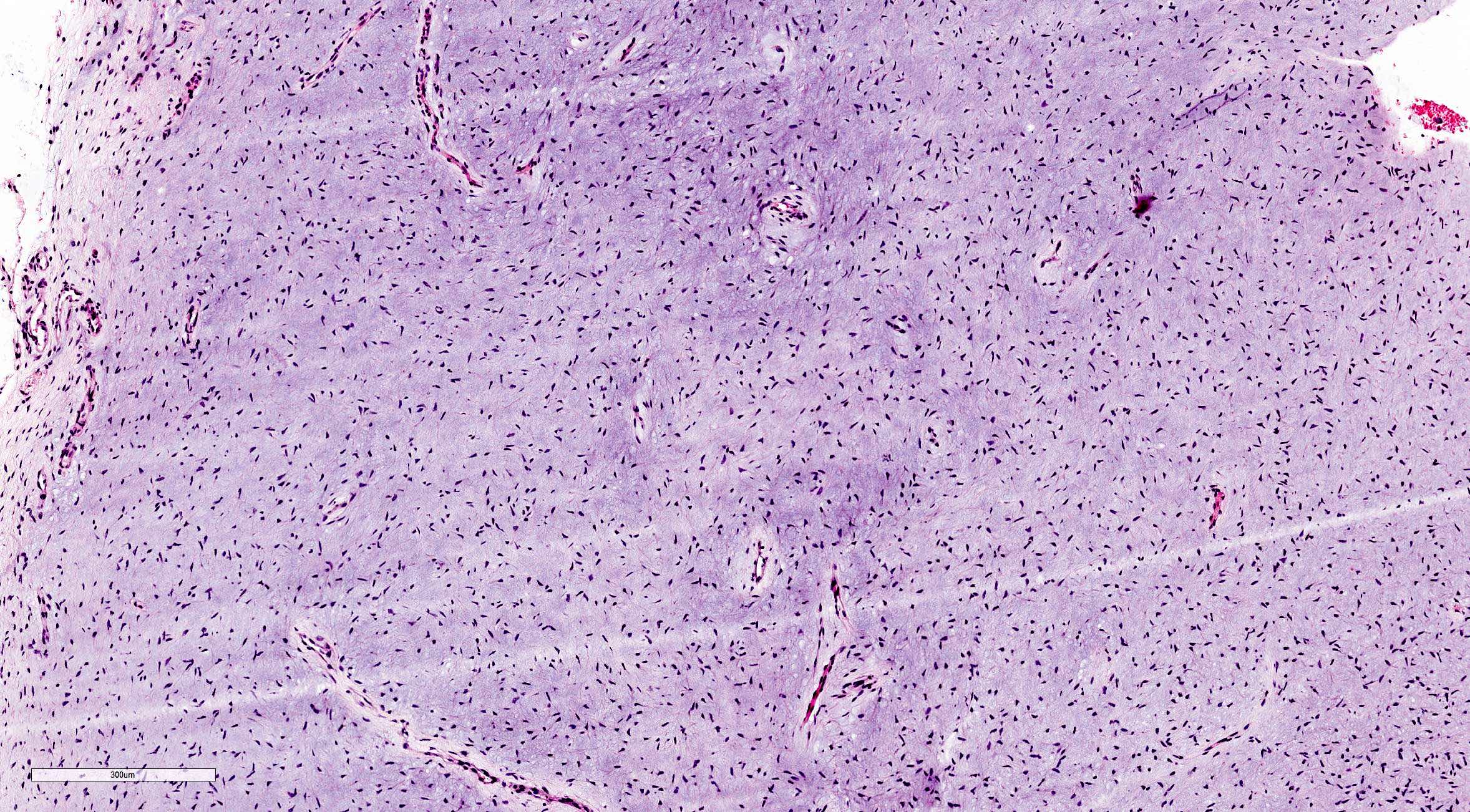
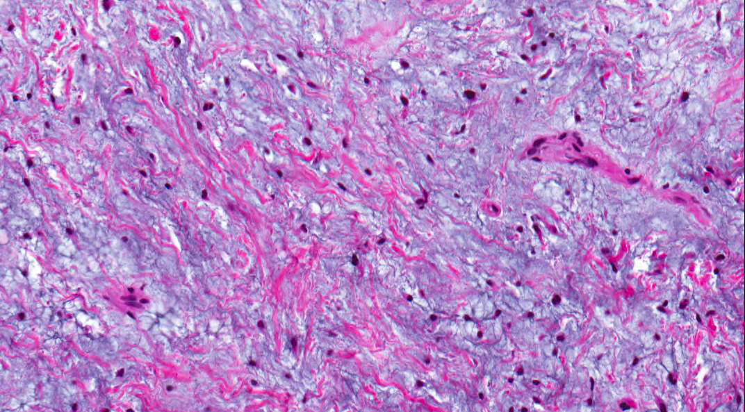
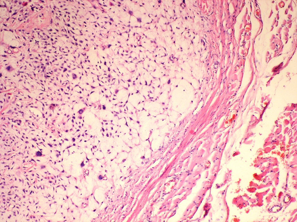
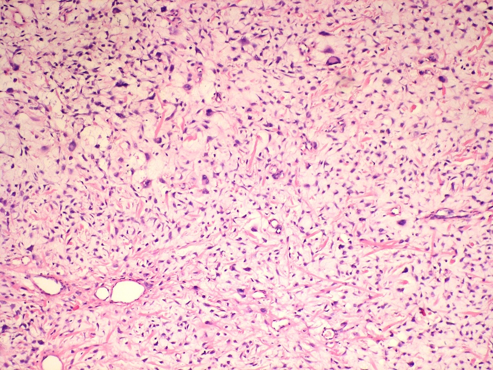
Myxoid background with bland spindle cells
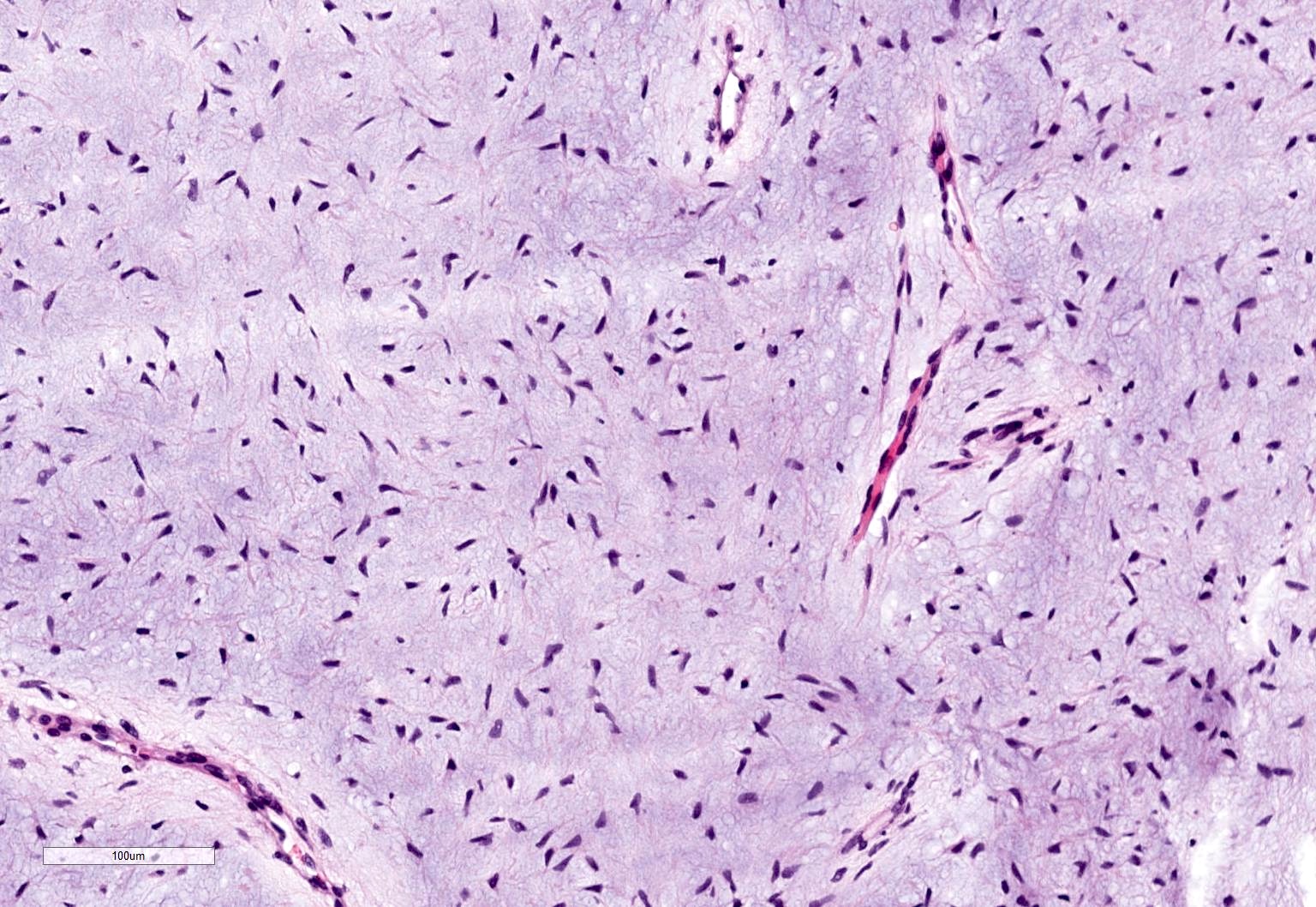
Bland spindle cells in myxoid stroma
Inactive odontogenic rests
Maxilla neoplasm
Mandibular lesion
Mandibular lesion
Images hosted on other servers:

Ill-defined radiolucent lesion


Panoramic radiographs

Multilocular radiolucent lesion

CT

Isodense area
Images hosted on other servers:

Gross swelling

Swelling, lower left mandible

Swelling without ulceration

Massive intraoral extension
Images hosted on other servers:

Left half of mandible

Cross-section
Images hosted on other servers:

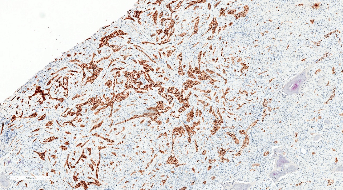
Benign odontogenic epithelium

Odontogenic epithelium

Low grade malignant neoplasm

Small nests, islands

Epithelial dysplasia

AE1/AE3, vimentin, Ki67

PCNA, p53

Ki67, p53

Pan-cytokeratin, vimentin, Ki67
Images hosted on other servers:

Lesion in left side of the mandible

Compound odontoma and unerupted 32

Well defined multiple radiopacities

Radio opaque lesion surrounded by radiolucent zone
Images hosted on other servers:

Peripheral odontoma

Surgical site showing denticles

Mass in upper left maxillary region
Images hosted on other servers:

Enucleated nine denticles
Contributed by Kelly Magliocca, D.D.S., M.P.H.
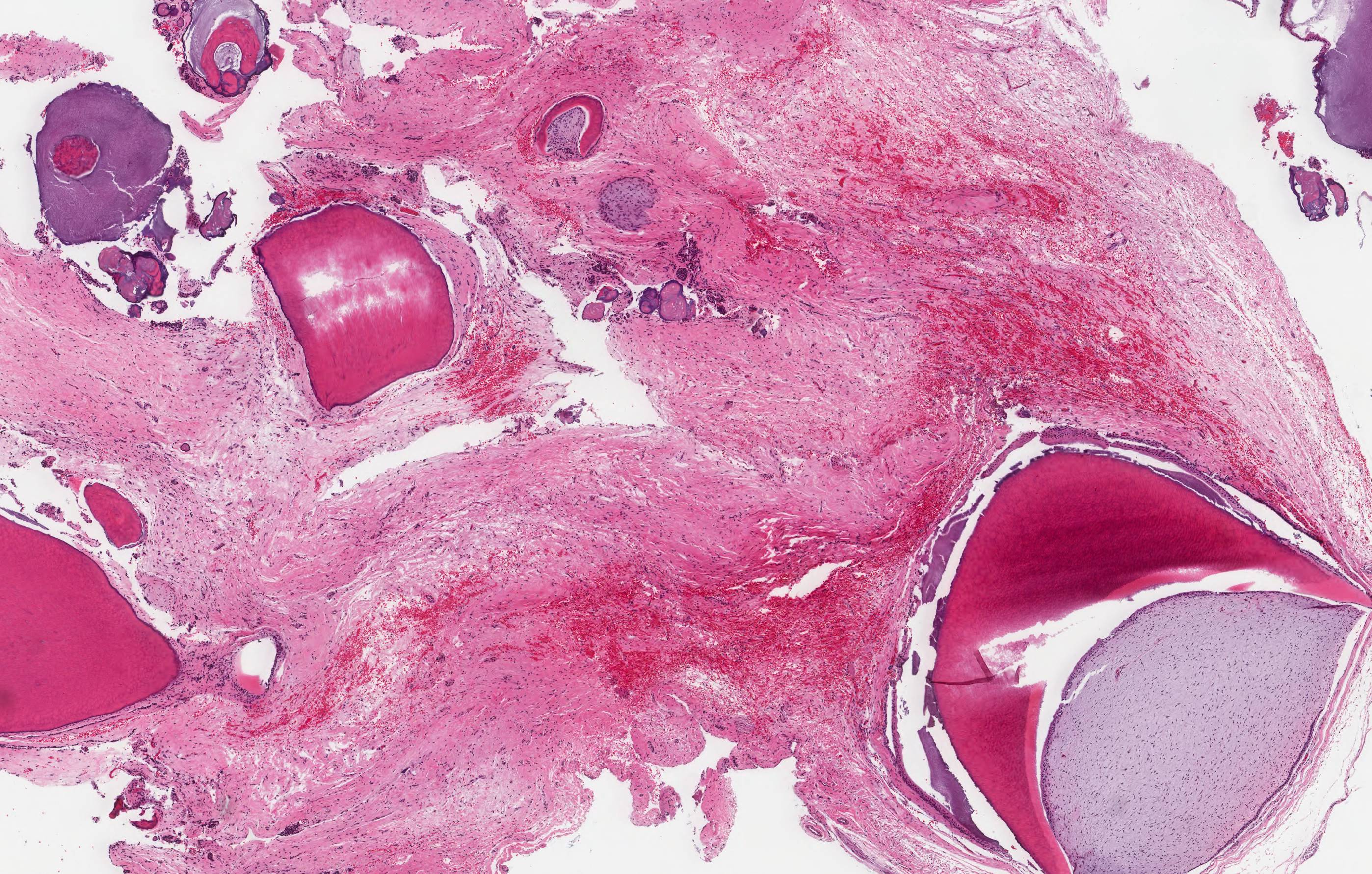
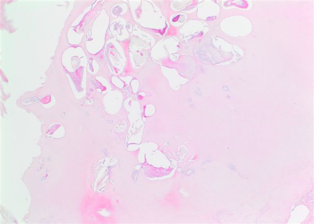
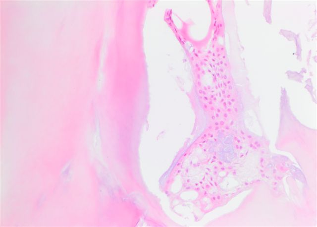
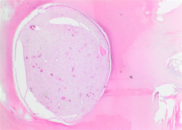
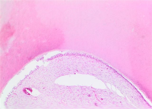
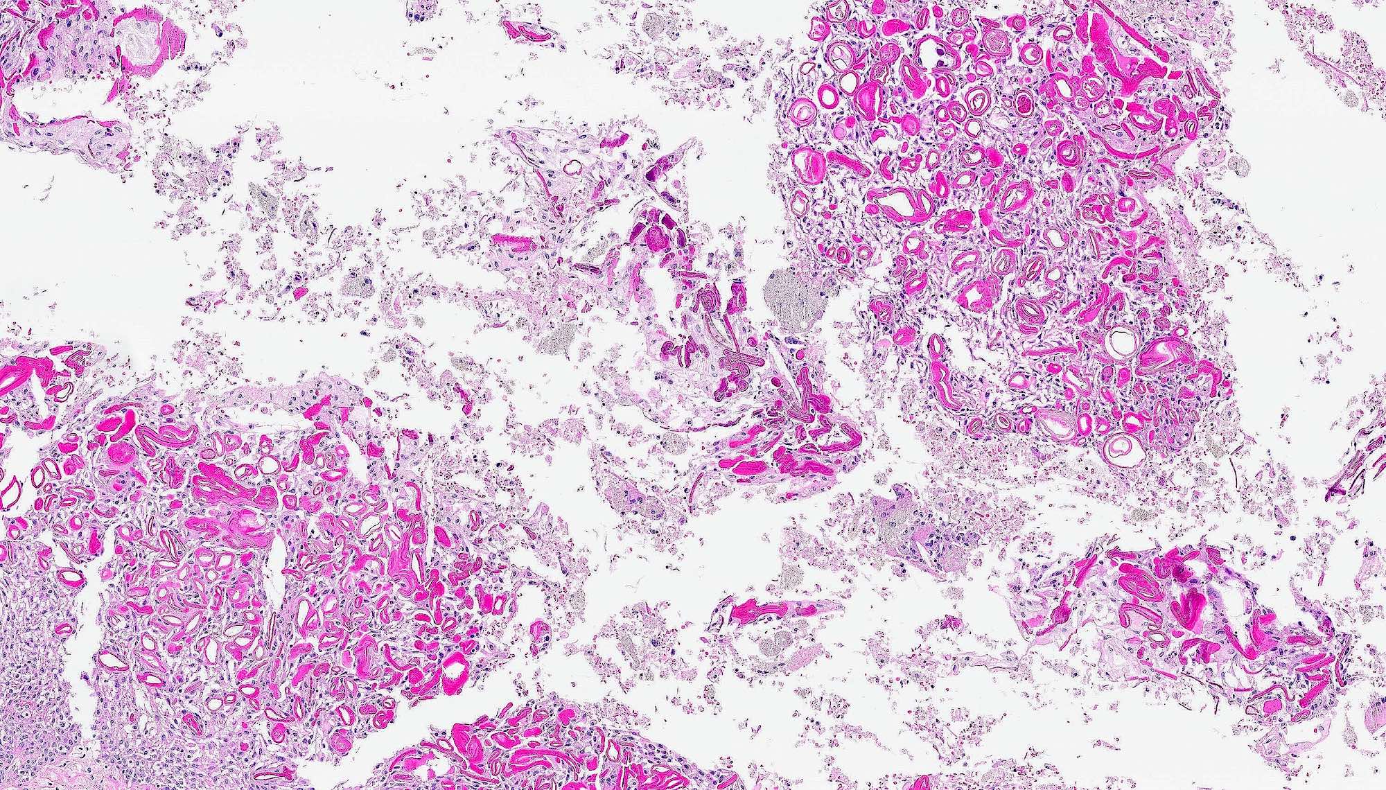
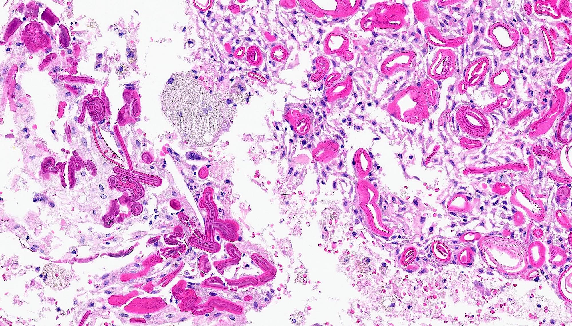
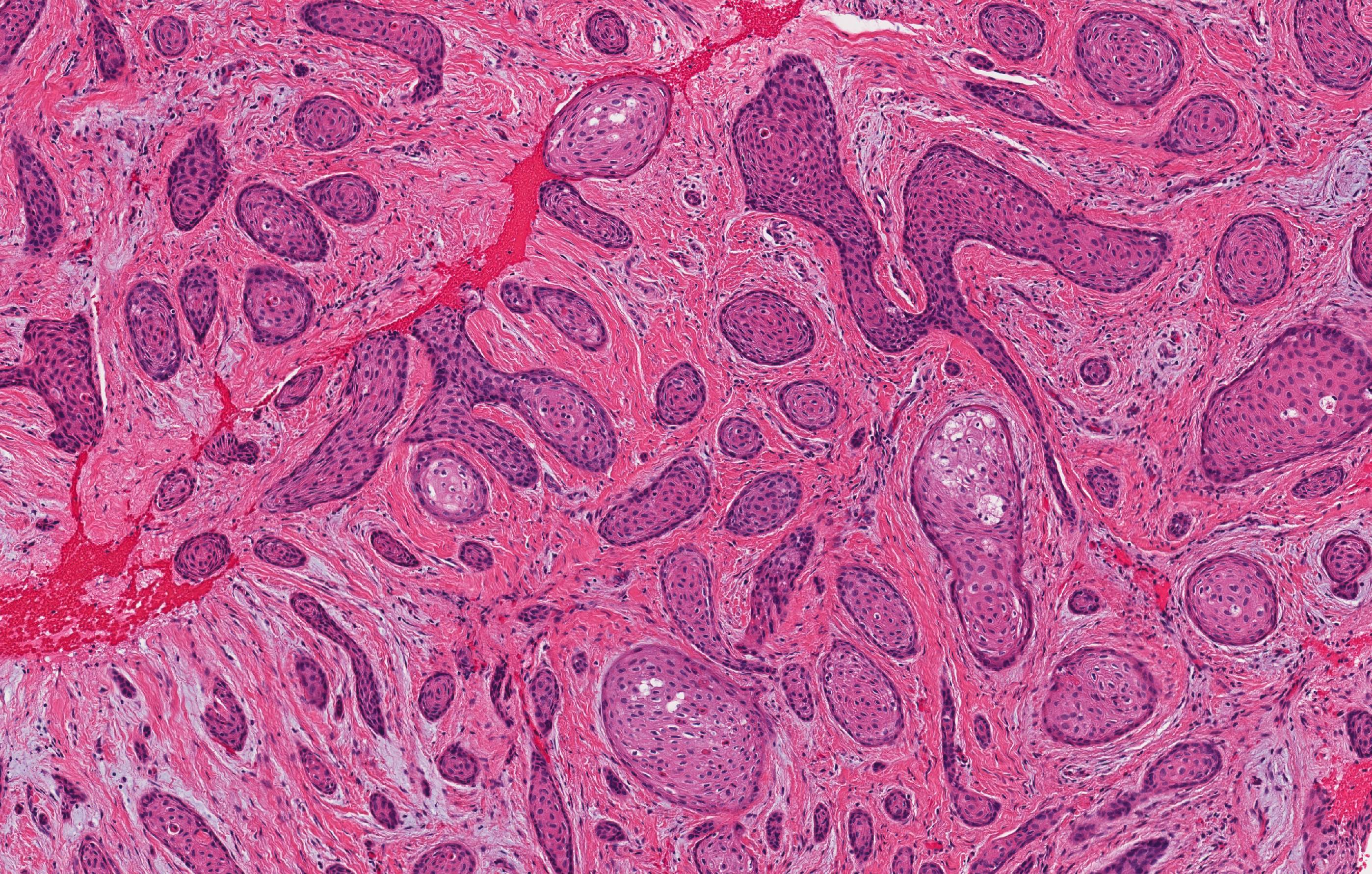
Odontoma
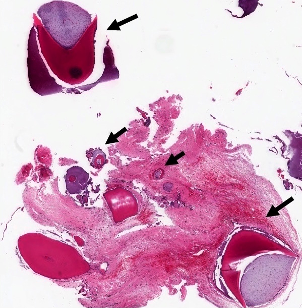
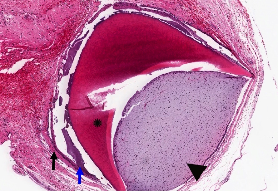
Compound odontoma

Complex odontoma
Contributed by Nasir Ud Din, M.B.B.S.
Right mandible
Images hosted on other servers:

Extraoral swelling

Obliterated buccal sulcus
Images hosted on other servers:

Cyst lining envelops crown of tooth
Contributed by Kelly Magliocca, D.D.S., M.P.H. and Nasir Ud Din, M.B.B.S.
Orthokeratinized odontogenic cyst
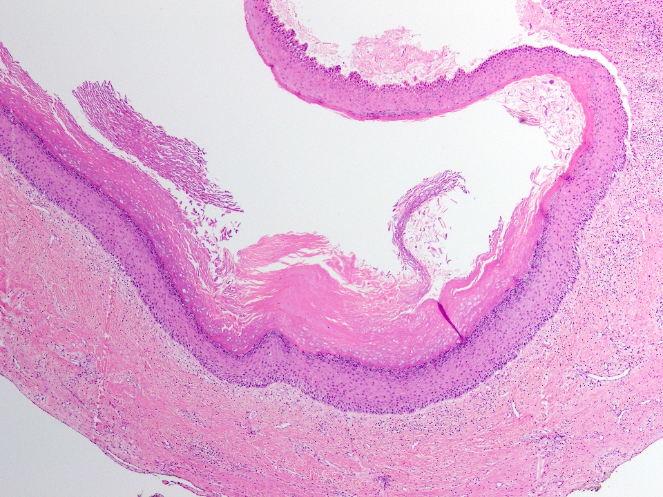
Cyst wall fragments
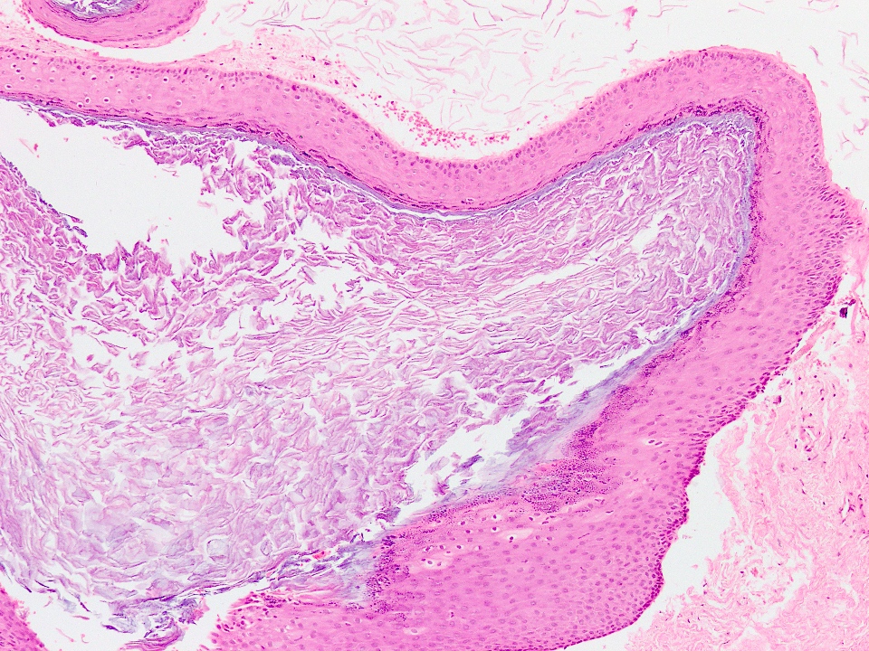
Cyst wall lumen
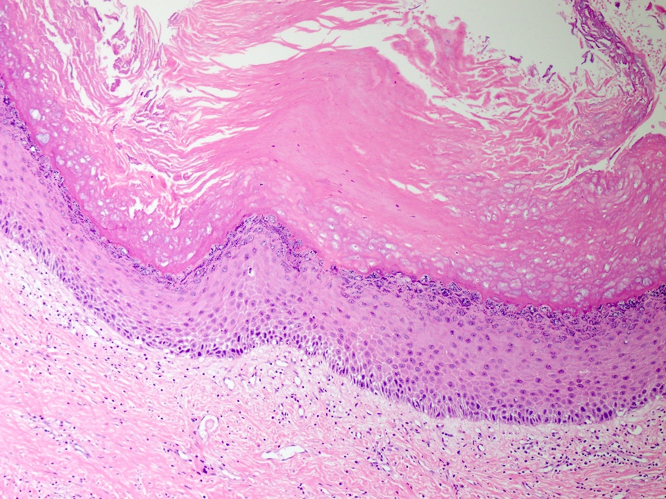
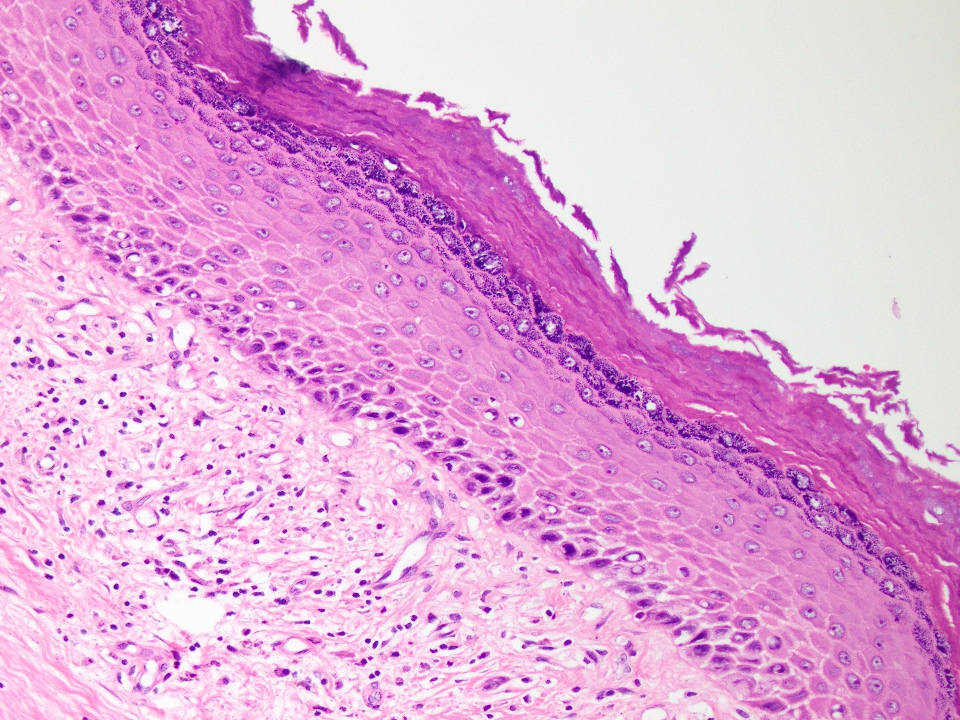
Cyst lining
Cyst lining
Images hosted on other servers:

Osteoradionecrosis
Images hosted on other servers:

Mandibular osteonecrosis

Swelling and extension over left zygomatic region

premaxillary
region with upper
lip protrusion

Computed tomography axial section

Axial CT scan of maxilla showing expansive infiltrative lesion

Osteosarcoma involving right ramus mandibula

Lytic and destructive
lesion in the left maxillary
tuber and molar tooth
area with root resorption
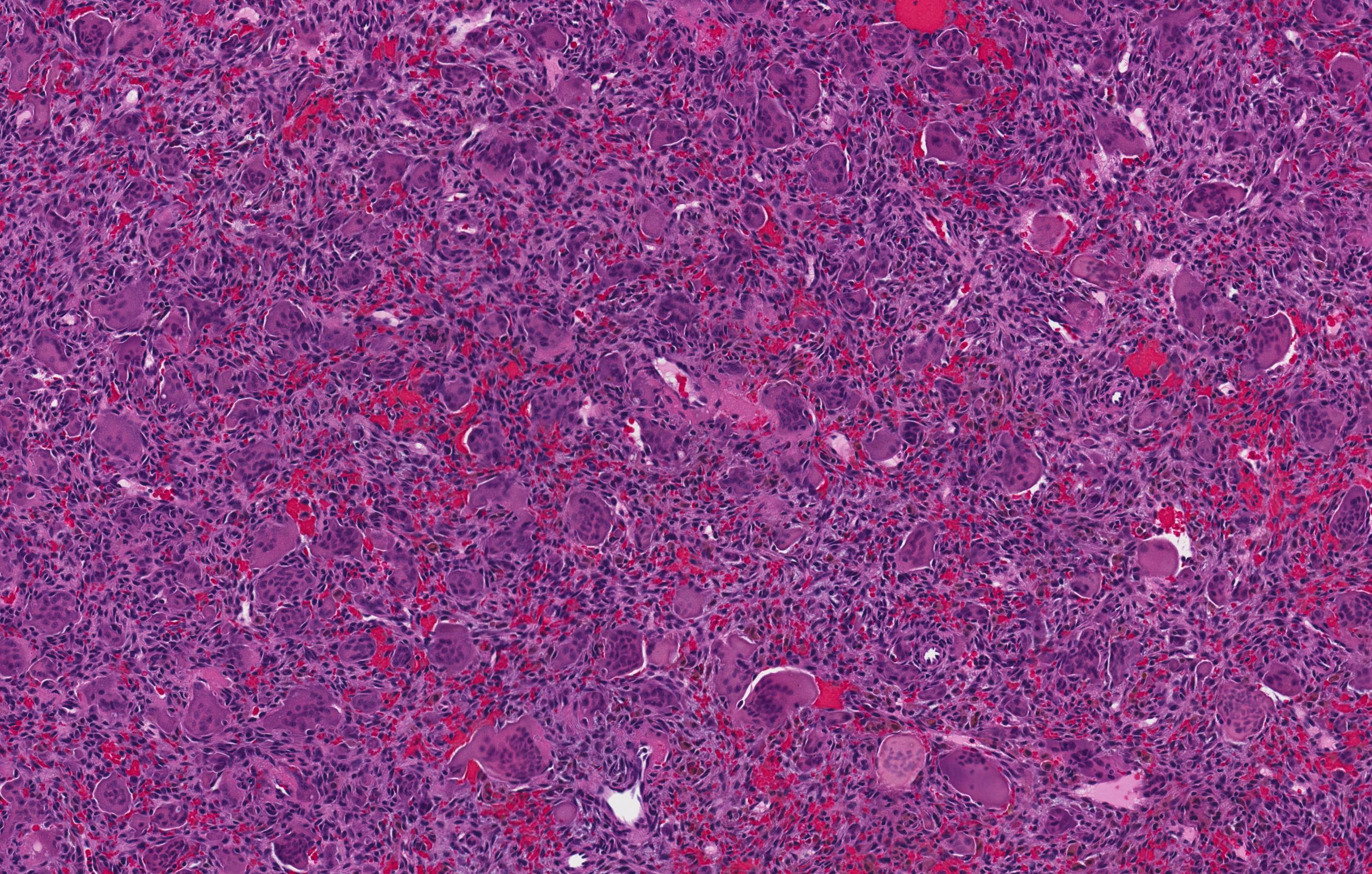
Contributed by Kelly Magliocca, D.D.S., M.P.H.
Osteosarcoma, NOS
Case of the Week #350: chondroblastic osteosarcoma of maxilla


Contributed by Dr. Kelly Magliocca and Dr. Anthony Martinez:
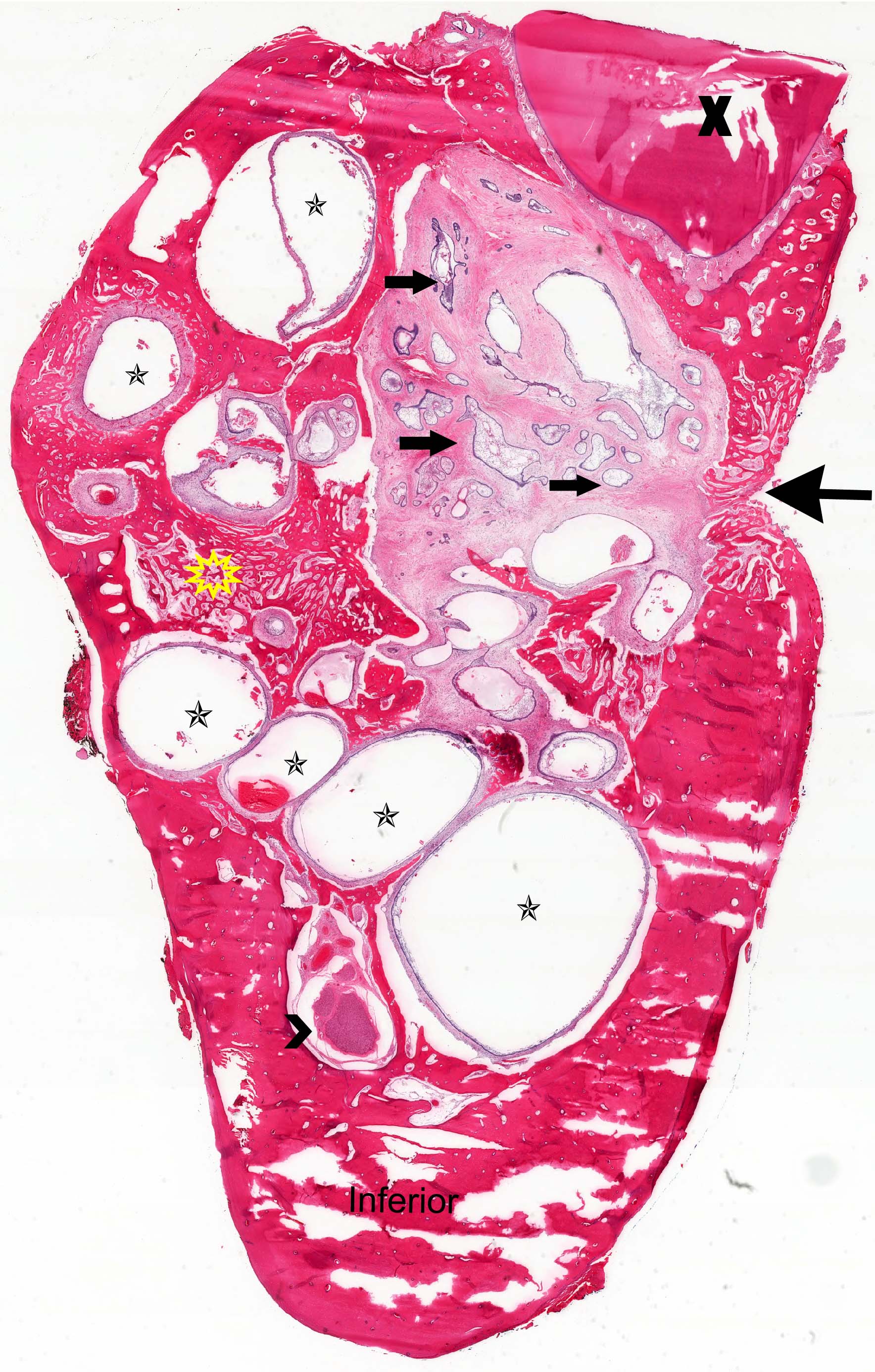
Arrows = inferior
border of
the mandible;
Arrowheads =
osteosarcoma

4× (same as first image, without arrows)

40×
100×
200×
400×
Images hosted on other servers:

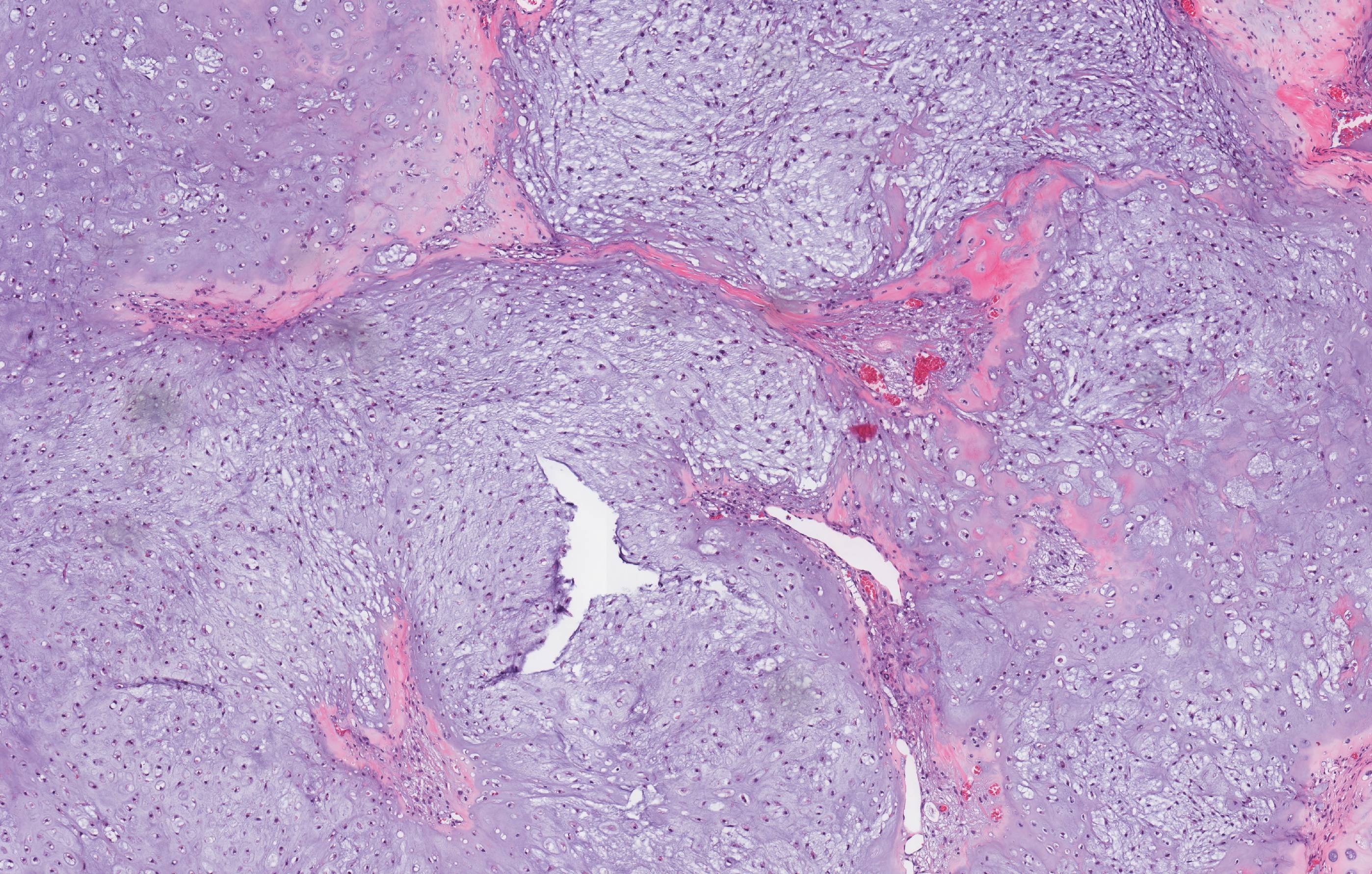
Chondroid tissue with areas of osseous differentiation

hyperchromatic
and pleomorphic
nuclei

Photomicrograph showing tumor osteoid areas

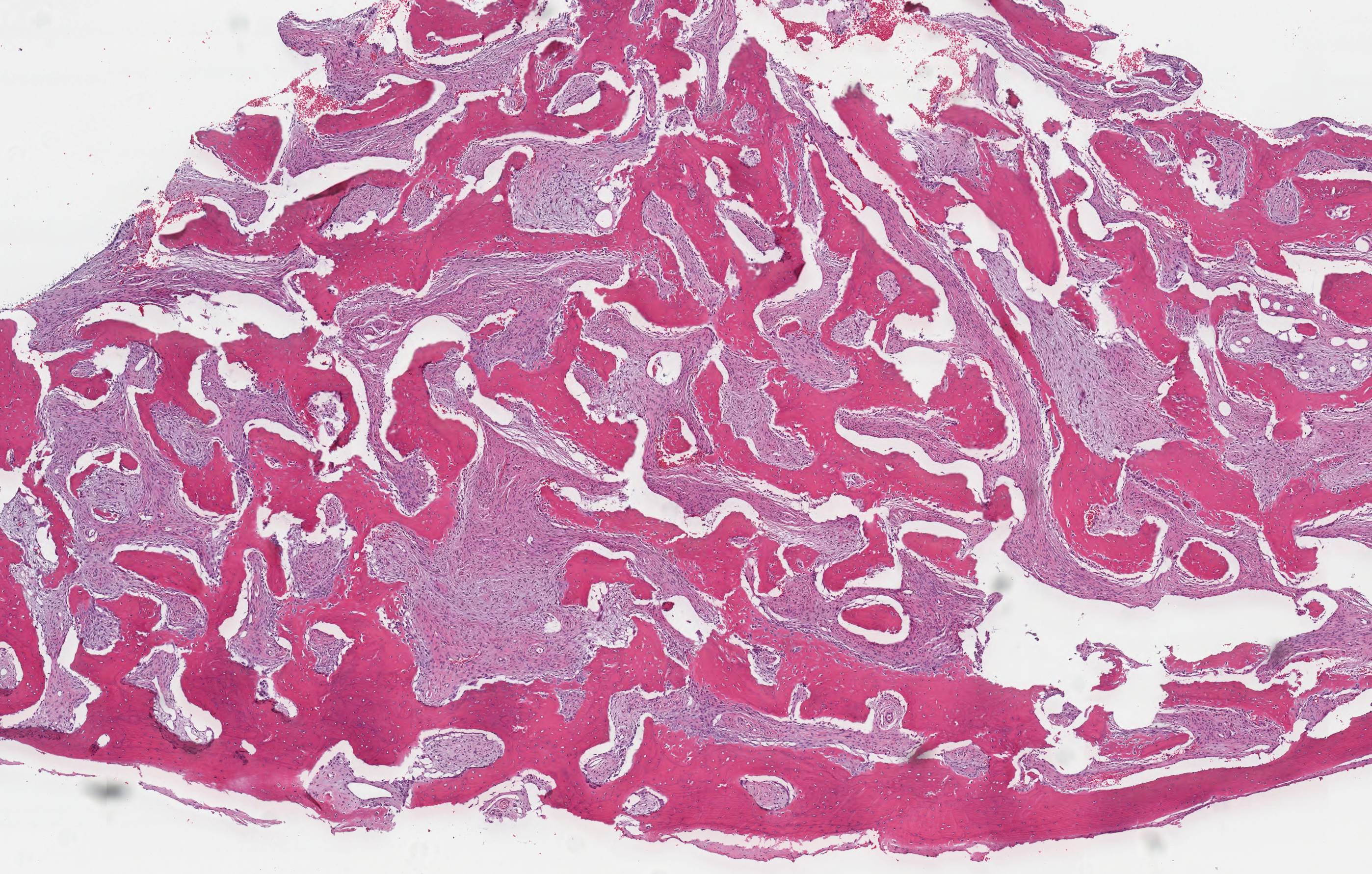
a) Large osteosarcoma involv-
ing mandible ramus and body
b) Classic chondroblastic
osteosarcoma of the jaw


Various images
Images hosted on other servers:

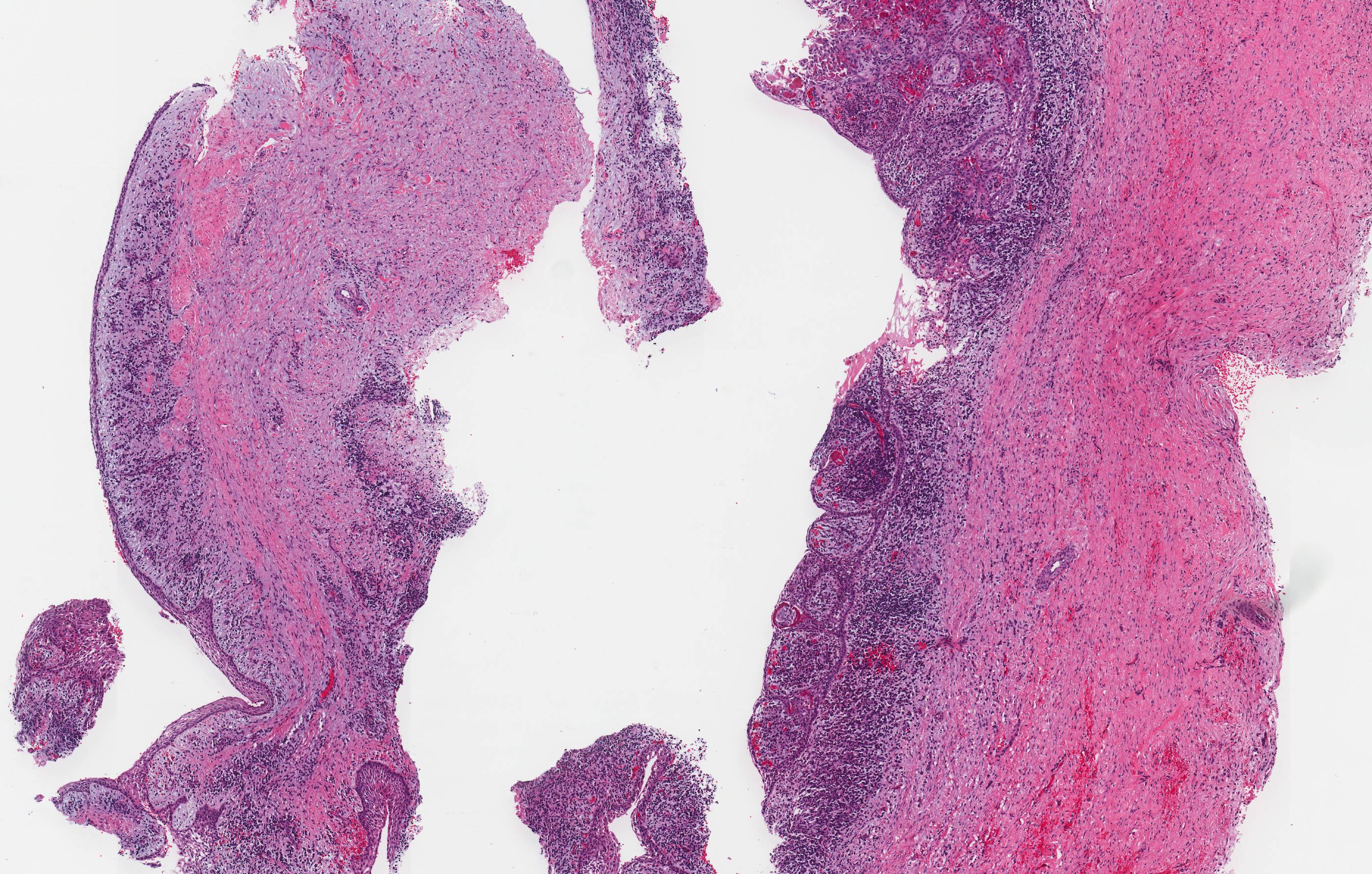
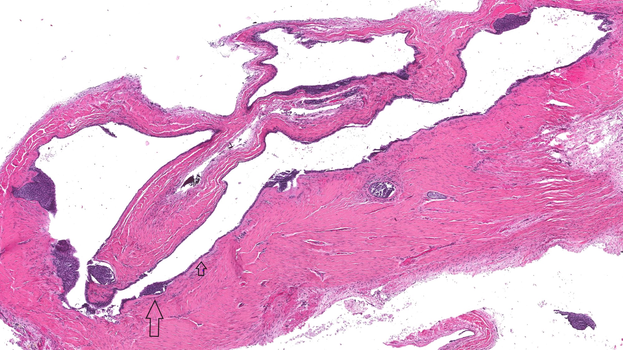
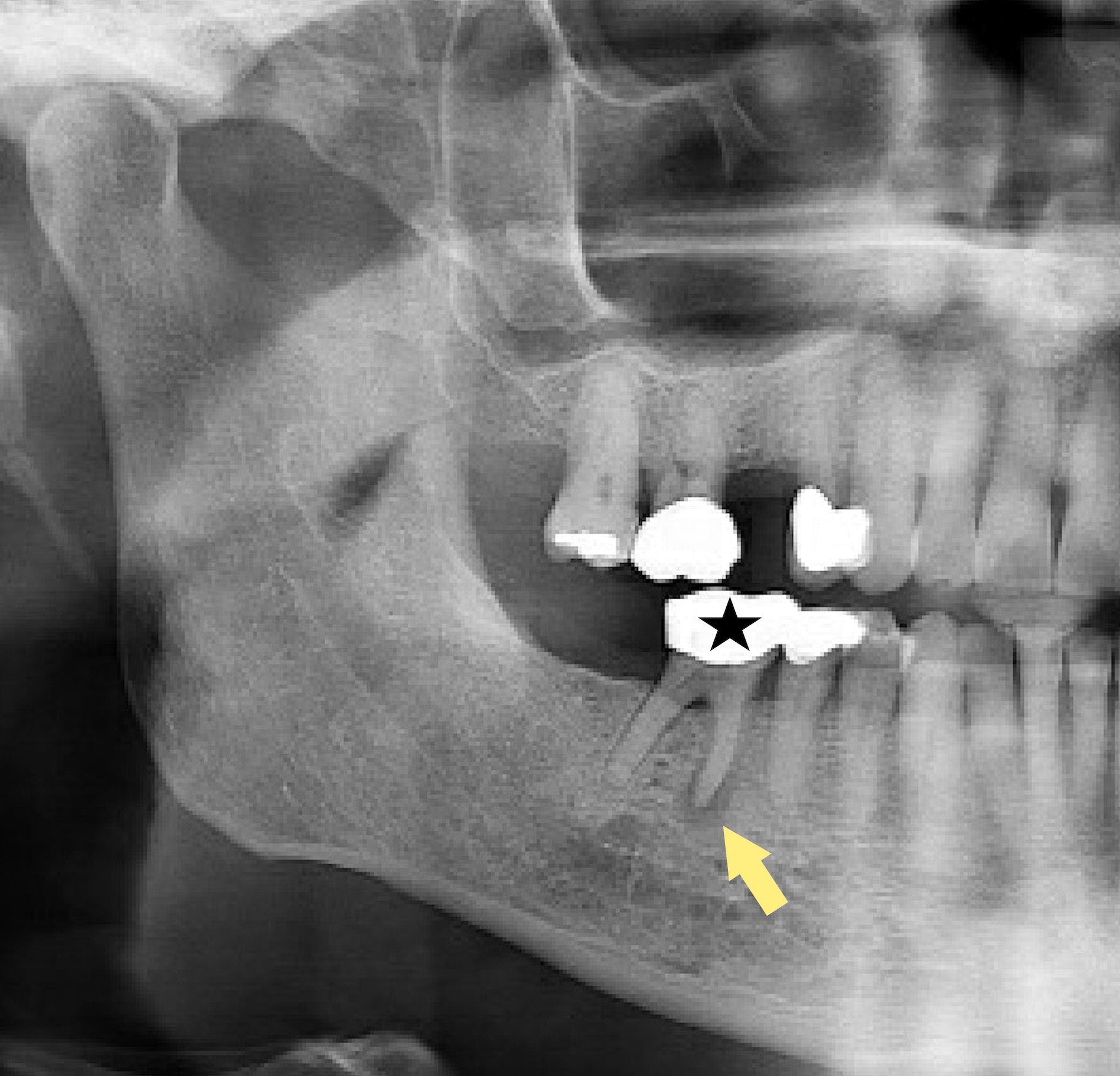
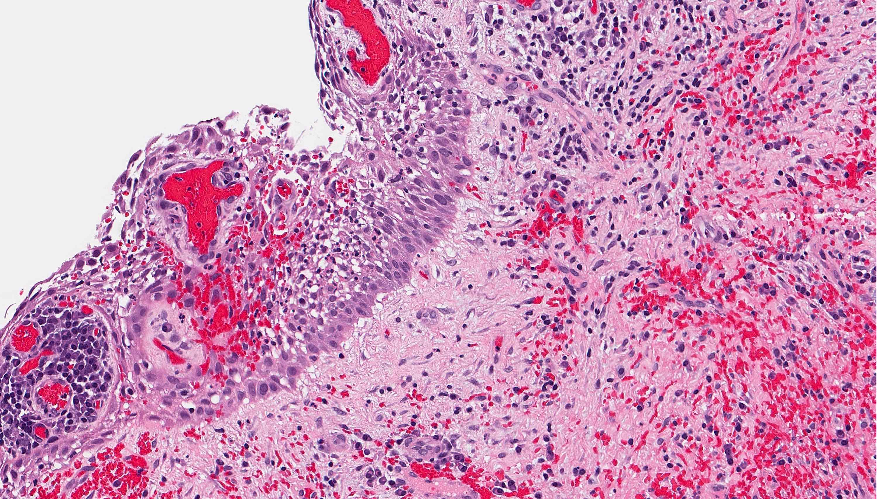
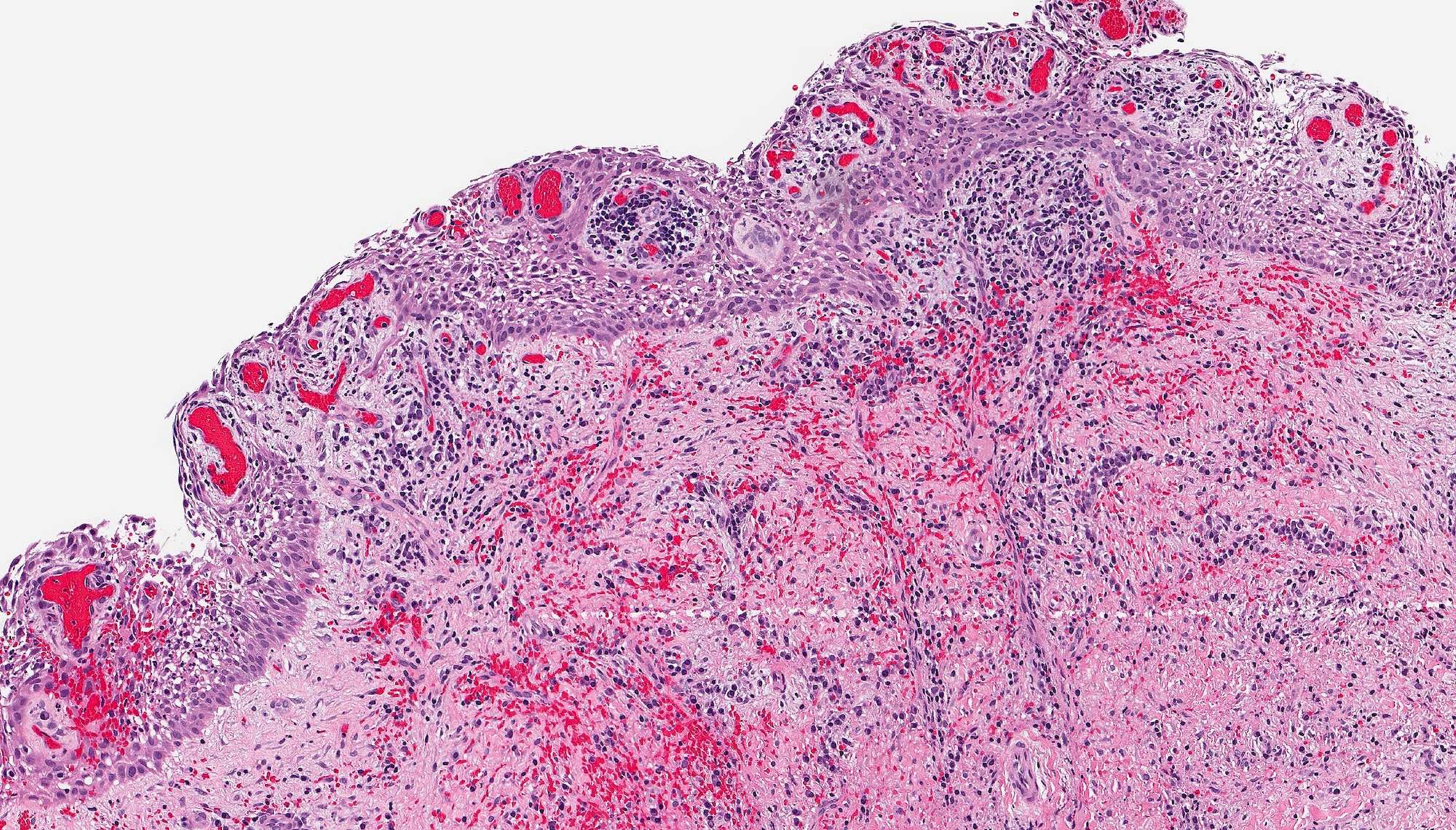
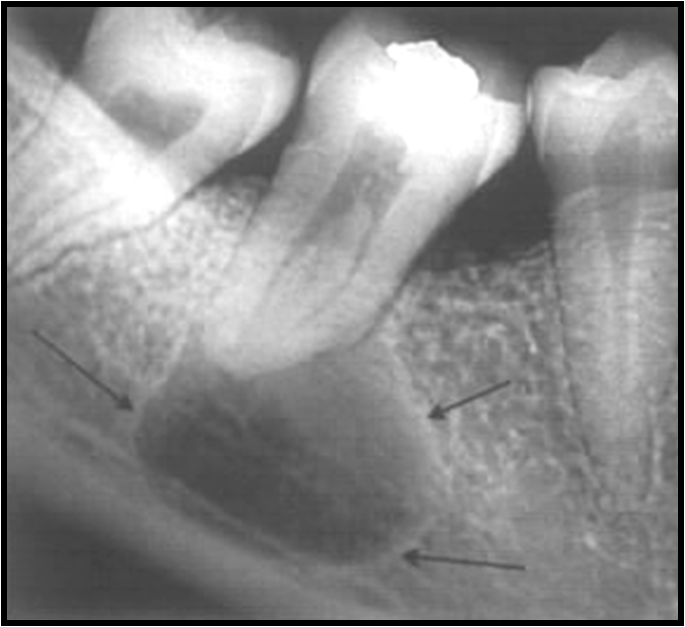
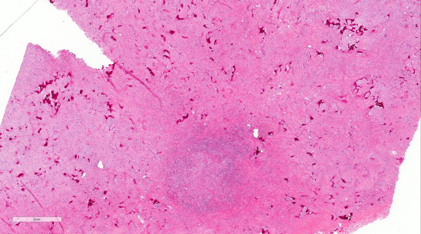
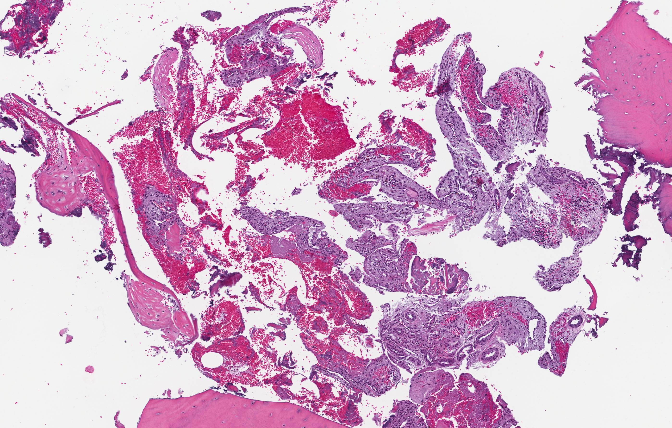
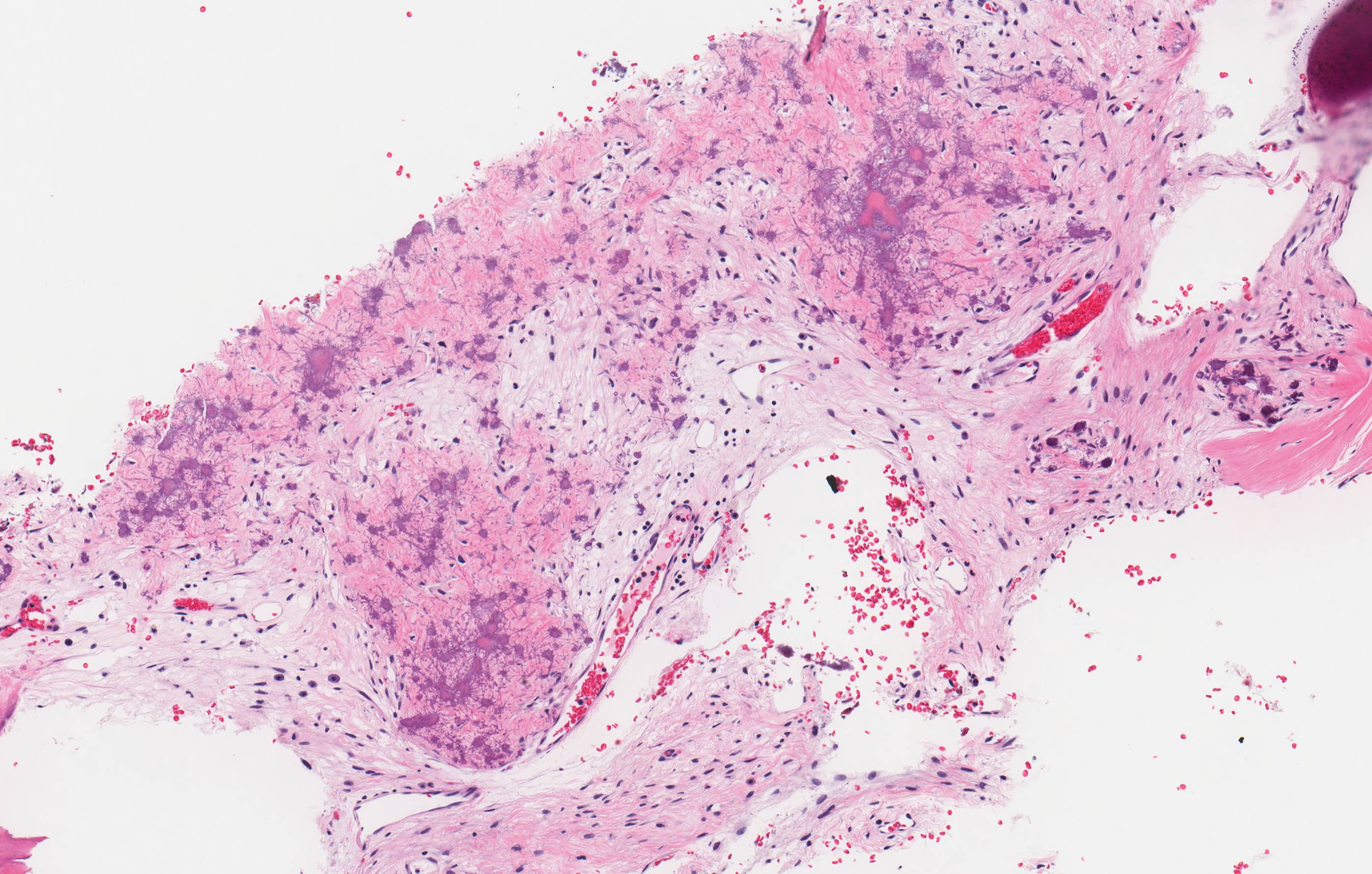
(B) Periapical granuloma
Images hosted on other servers:

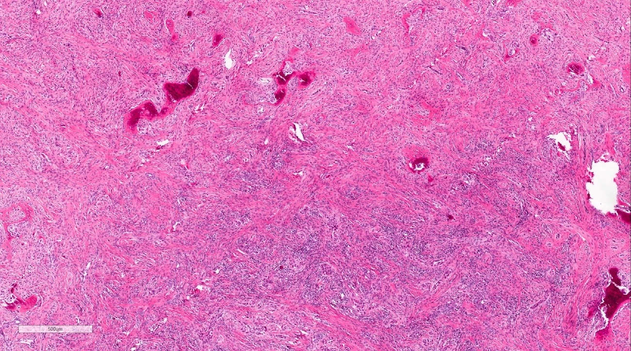
Early periapical granuloma

Intermediate periapical granuloma

Late periapical granuloma
Contributed by Kelly Magliocca, D.D.S., M.P.H.
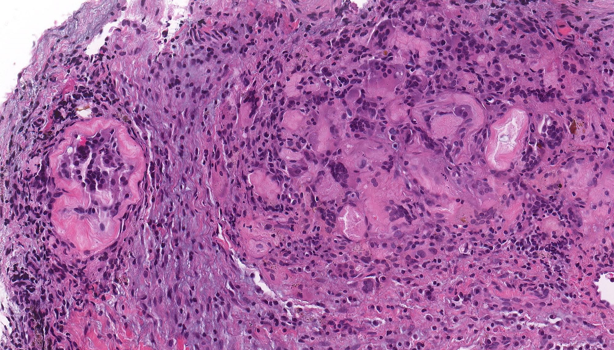
Peripheral giant cell granuloma
Images hosted on PathOut server:
Lytic and cystic lesion involving right mandible
Contributed by Kelly Magliocca, D.D.S., M.P.H.
Primary intraosseous carcinoma, NOS
Images hosted on PathOut server:

Verrucous carcinoma
Cyst lining with chronic inflammation
Contributed by Kelly Magliocca, D.D.S., M.P.H.
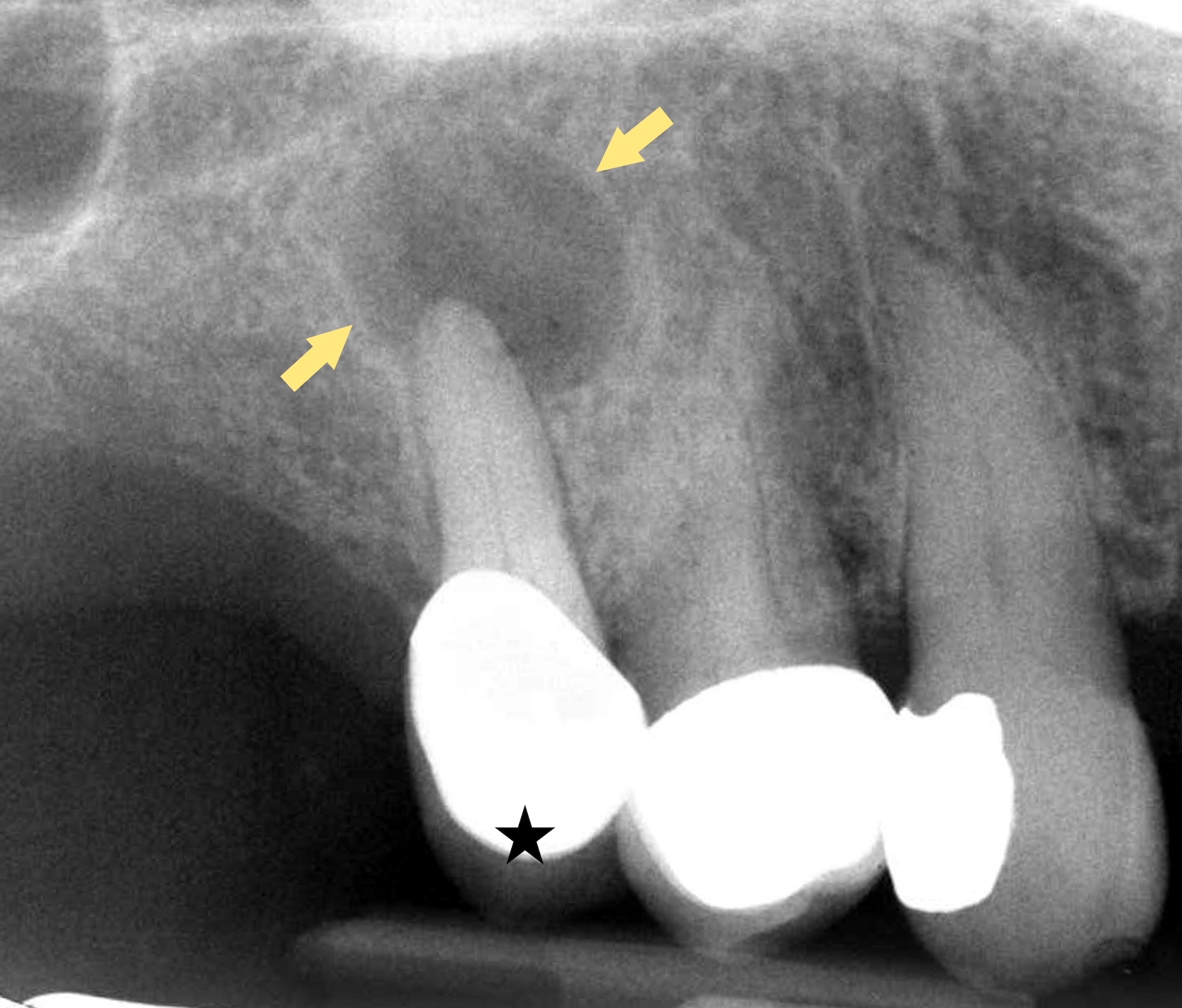
Anterior maxillary lesion
Maxillary cyst
Panorex molar root tips
Mesial root of molar
Anterior maxilla root canal
Anterior mandible root canal
Posterior maxilla intraoral
Posterior mandible residual lesion
Images hosted on other servers:

Palatal swelling

Lateral mandibular lesion
Contributed by Kelly Magliocca, D.D.S., M.P.H.
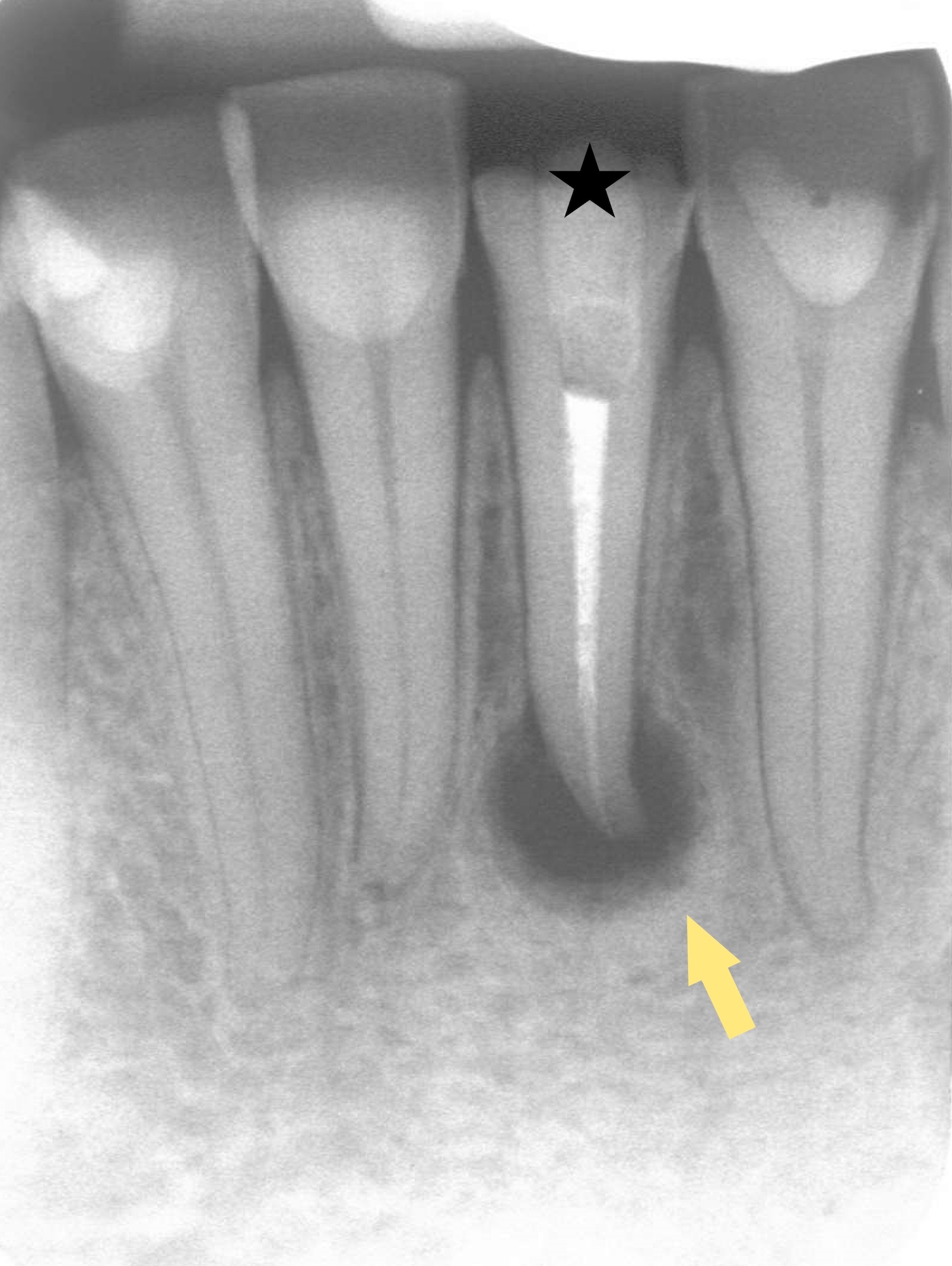
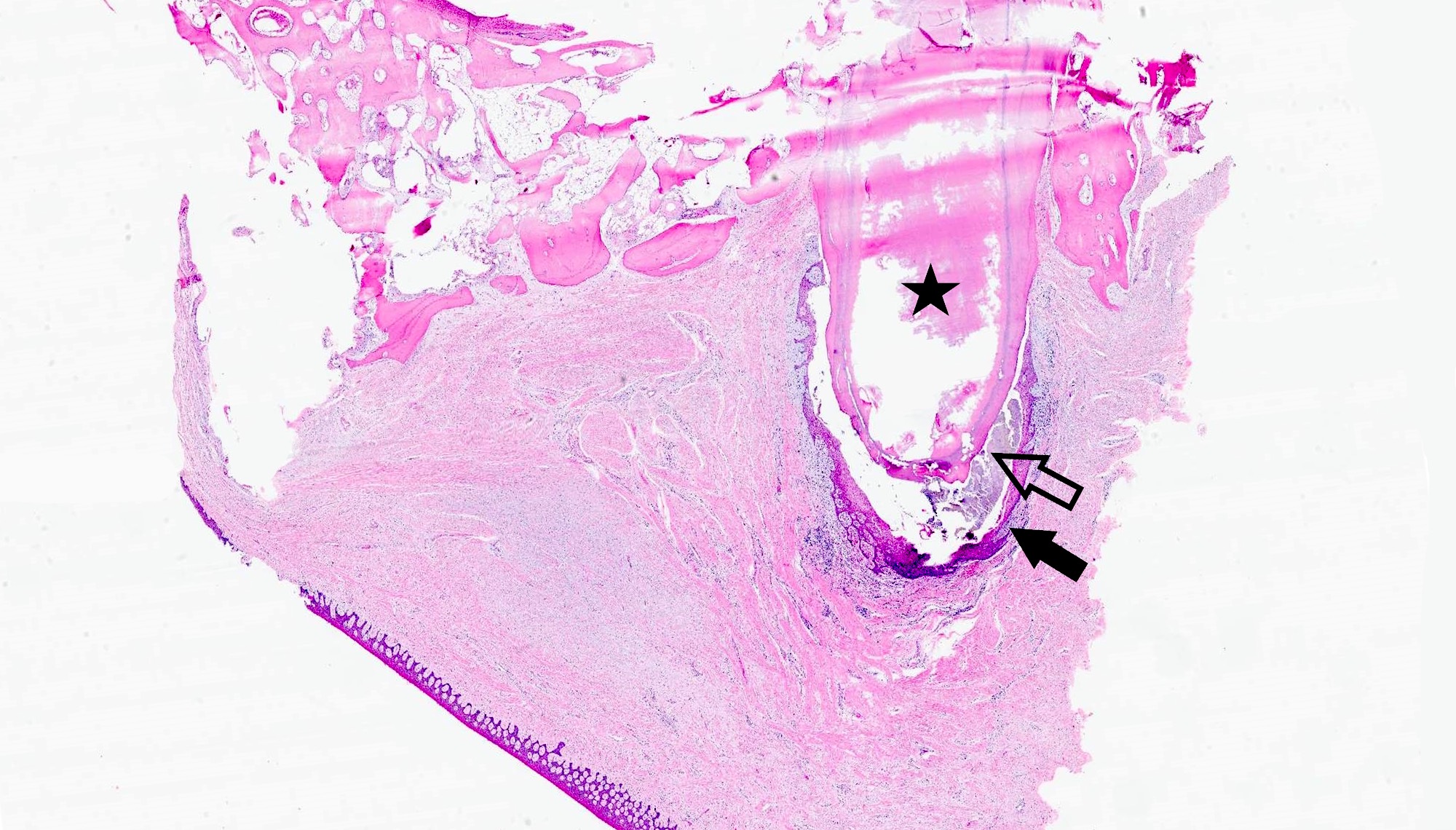
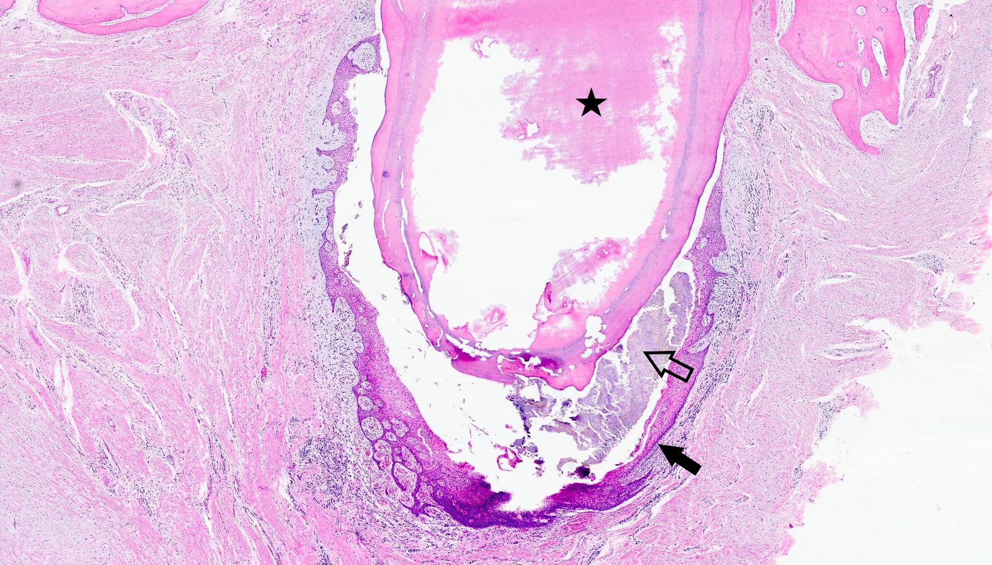
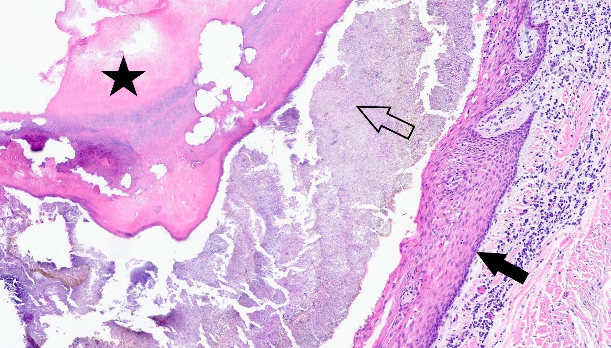
Necrotic tooth root
Root apex
Cyst to apex
Apex with abscess
Cyst epithelium
Cystic strips
Nonkeratinized cyst epithelium
Arcades
Posterior maxillary cyst
Root, bacteria and cyst
Bacteria present
Partial keratinization
Prior root canal treatment
Epithelial arcades
Granular foreign material
Splendore-Hoeppli phenomenon
Cystic sac
Bacteria within lumen
Rushton bodies in epithelium
Amorphous Rushton bodies
Hairpin / polycyclic Rushton bodies
Refractile appearance
Pulse granuloma in wall

Squamous epithelial lined cyst
Images hosted on other servers:

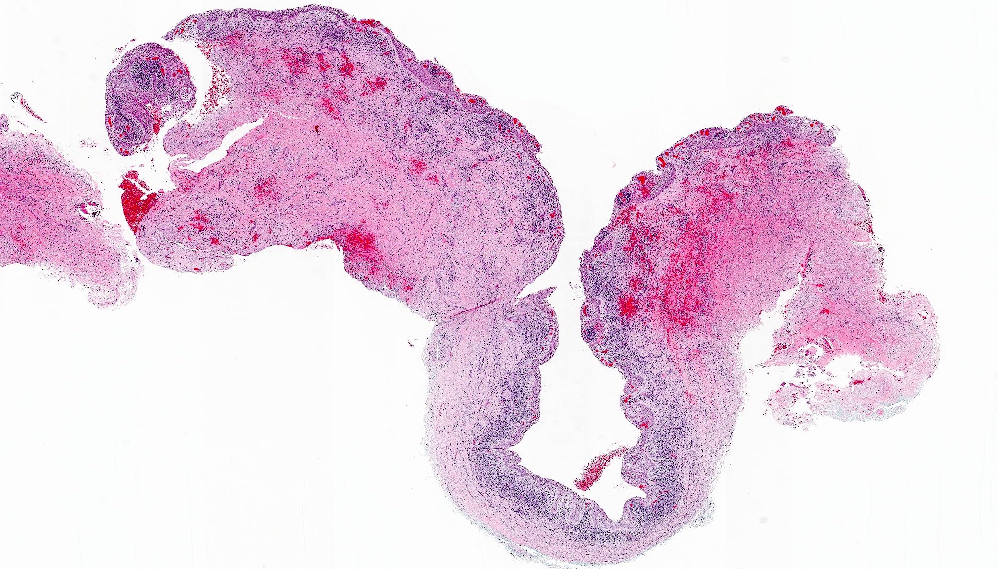
Radicular cyst
Radicular cyst, root partially reabsorbed

Upper maxilla

Periapical cyst involving maxillary anteriors

Progressive involution of periapical lesion
Images hosted on other servers:
Palatal swelling
Lateral mandibular lesion
Contributed by Kelly Magliocca, D.D.S., M.P.H.
Radicular cyst
Images hosted on other servers:

H&E

Cyst

H&E
Images hosted on other servers:
Residual jaw cyst
Images hosted on other servers:

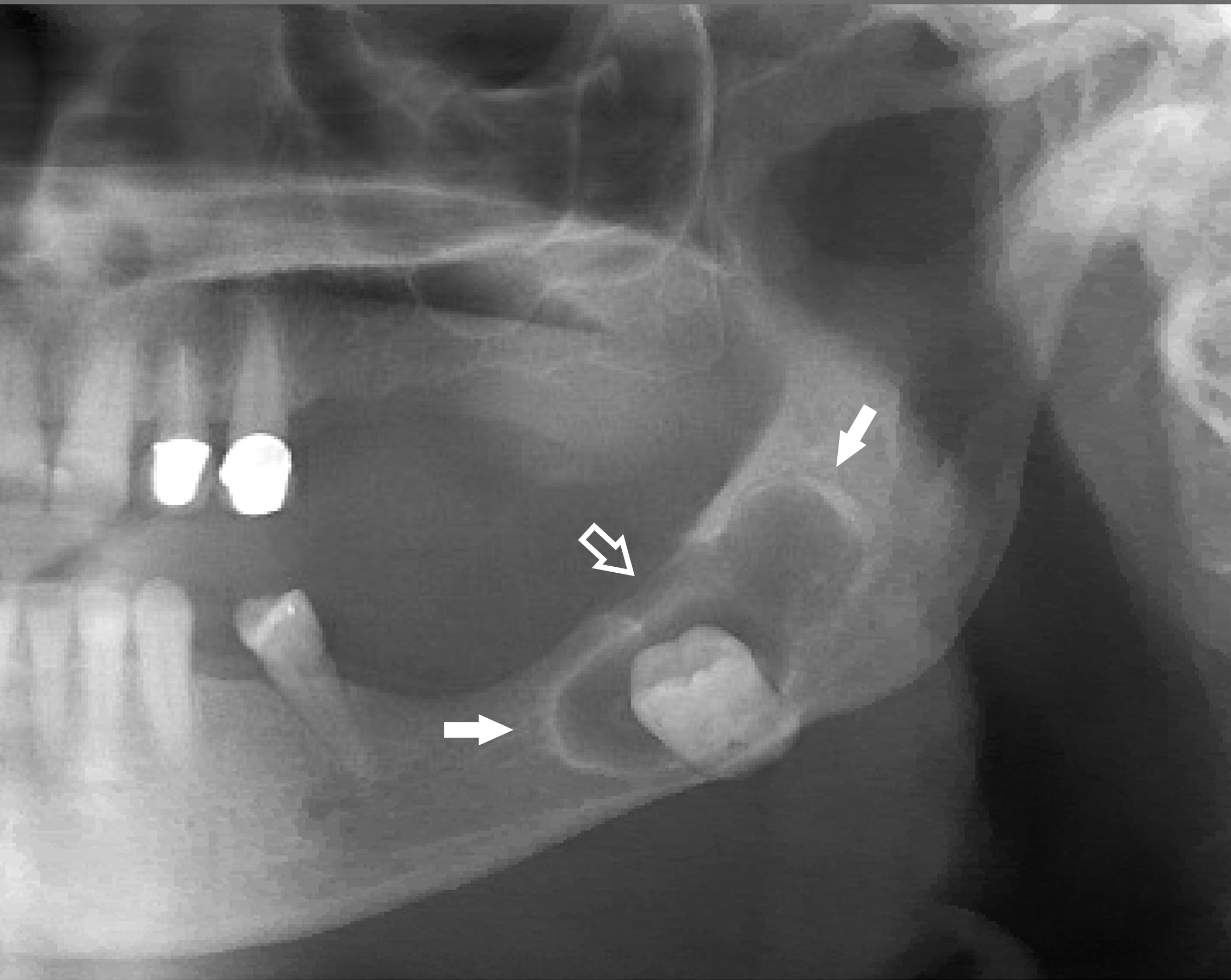
Infiltrative ground glass radiolucency
Images hosted on other servers:


Anterior mandible exophytic mass
Images hosted on other servers:

Mandible tumor involving skin
Contributed by Nathan Lee, D.M.D.
Architectural invasion
Bland cytology
Hypercellular tumor
Invasive clear cells
Hypercellular clear cell variant
Bland clear cells
Pankeratin

CK7

CK20

p63
Images hosted on other servers:

Extracion of tooth 46

Mandibular swelling
Contributed by Kelly Magliocca, D.D.S., M.P.H.
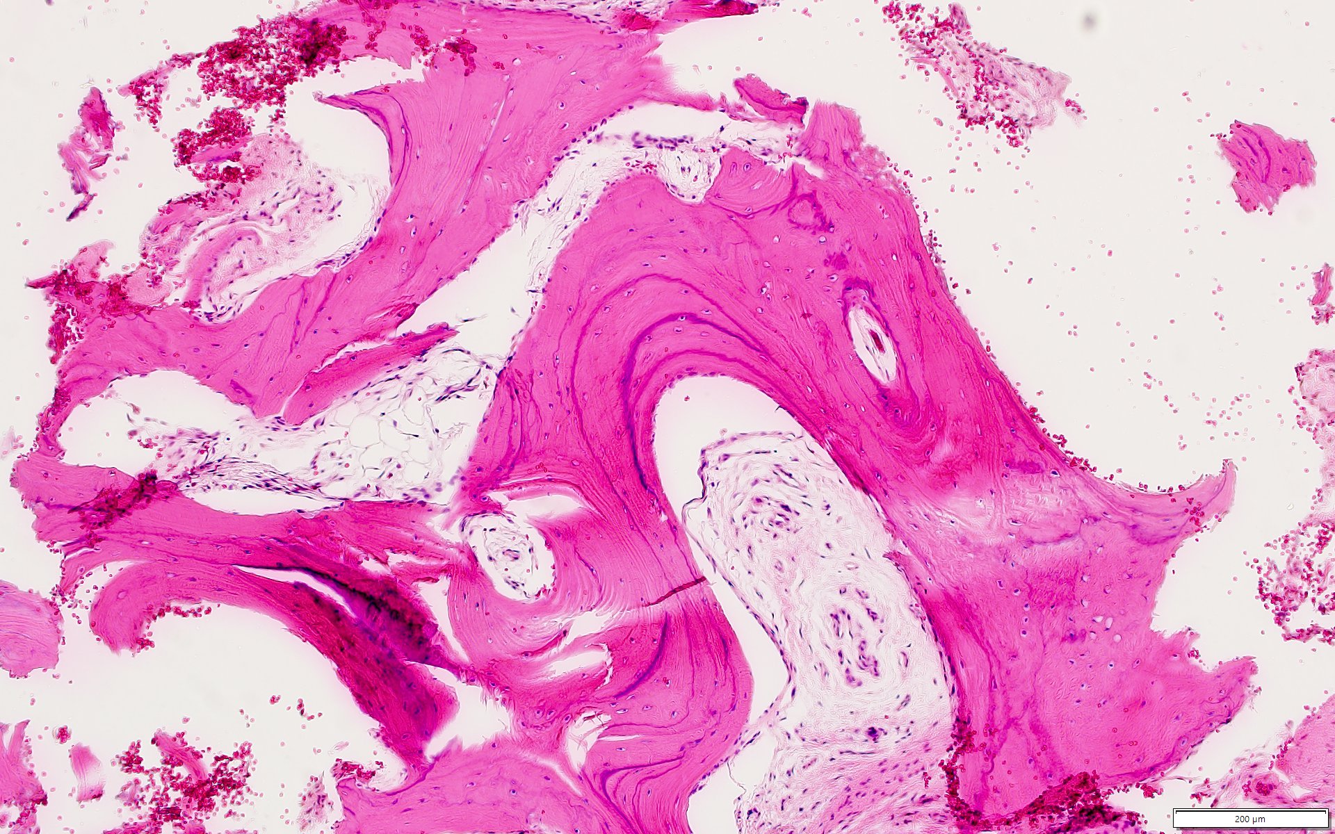
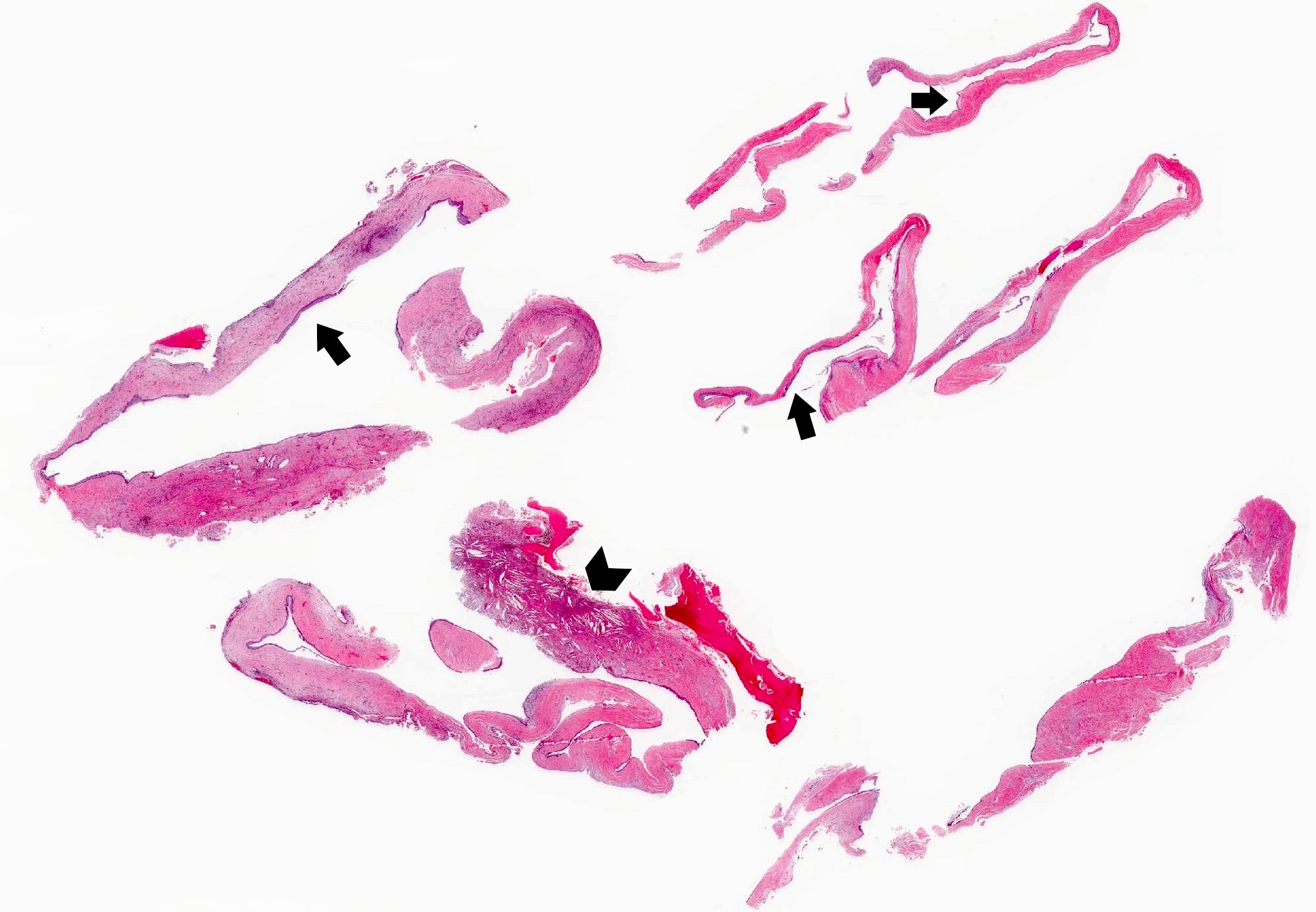
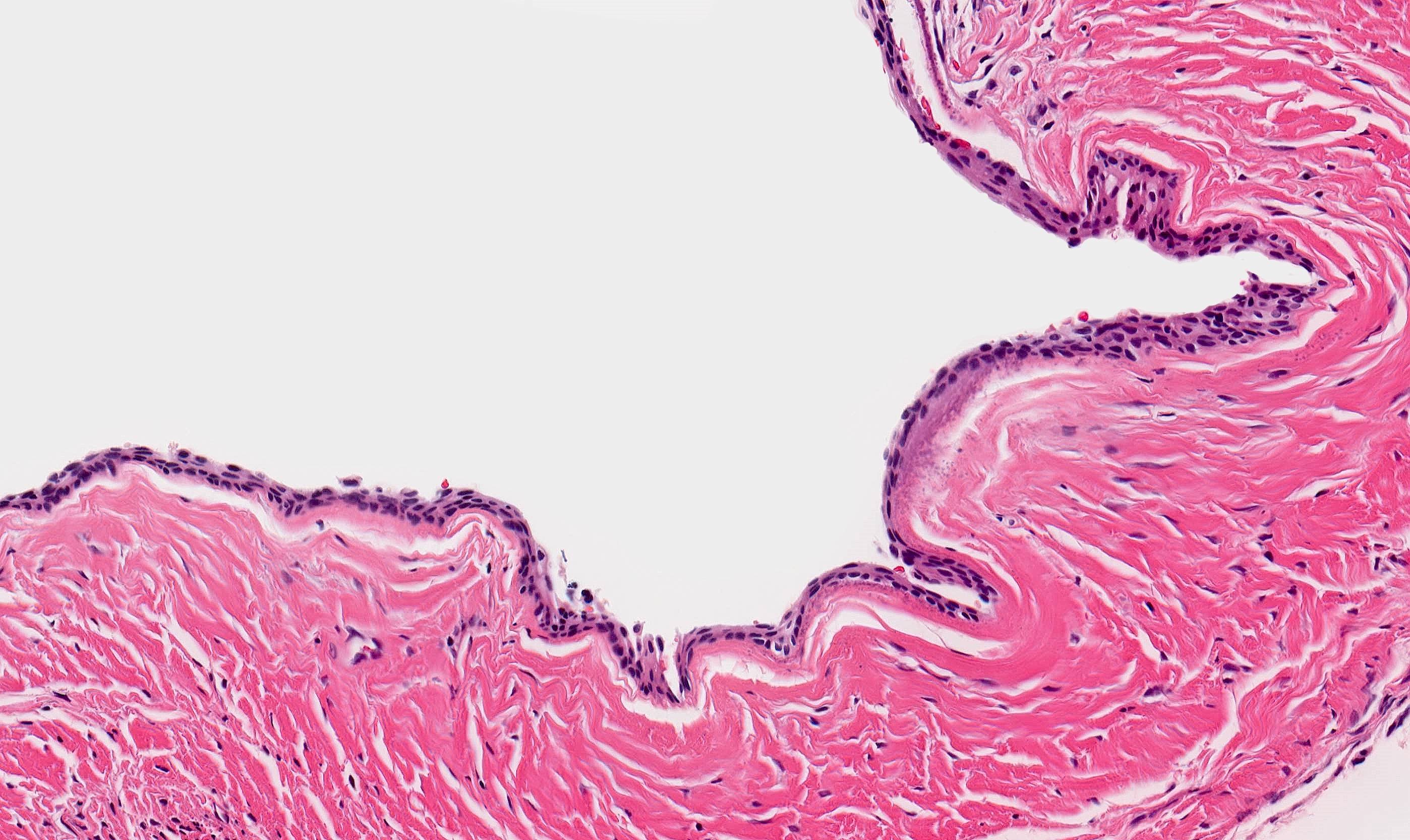
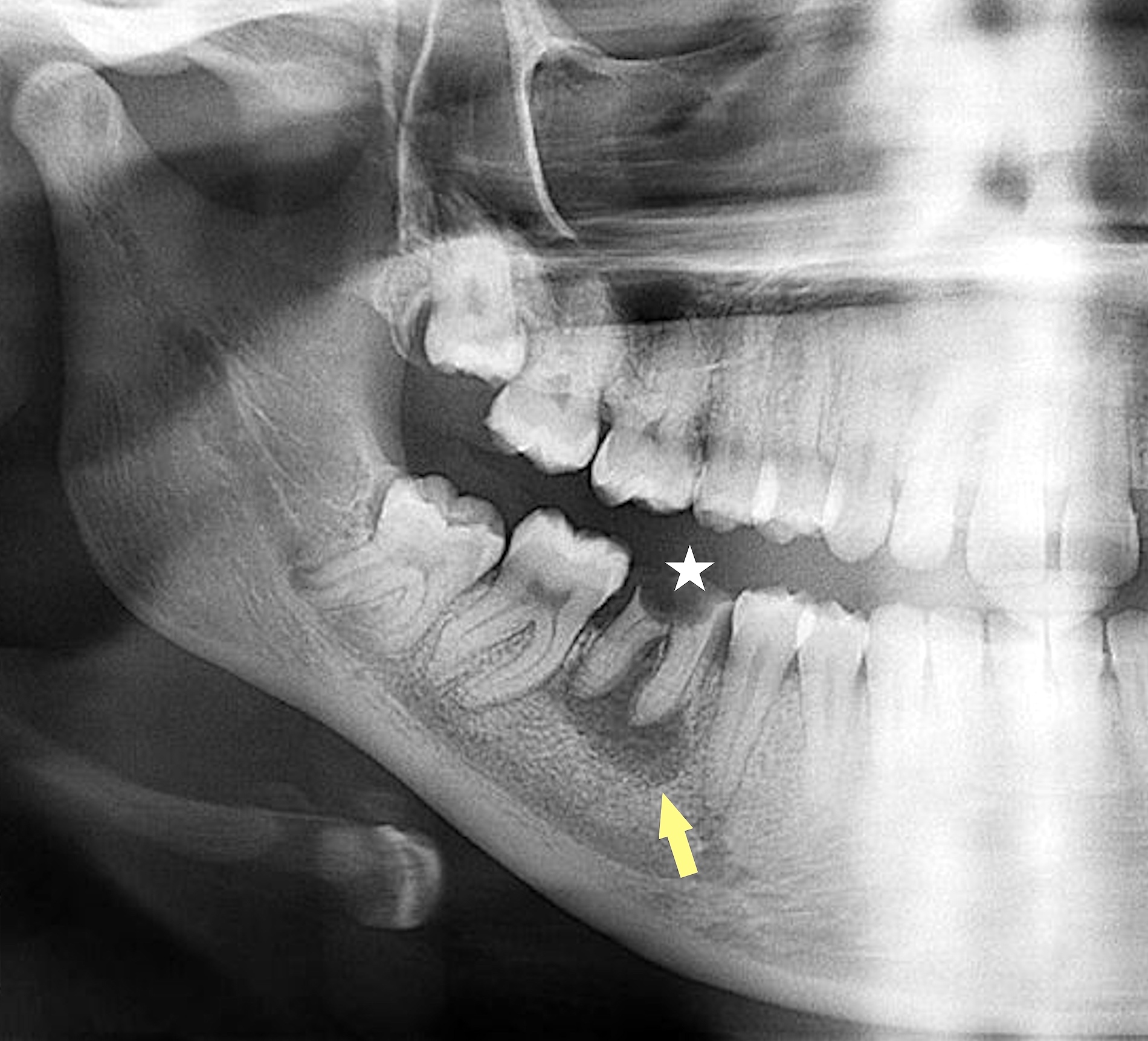
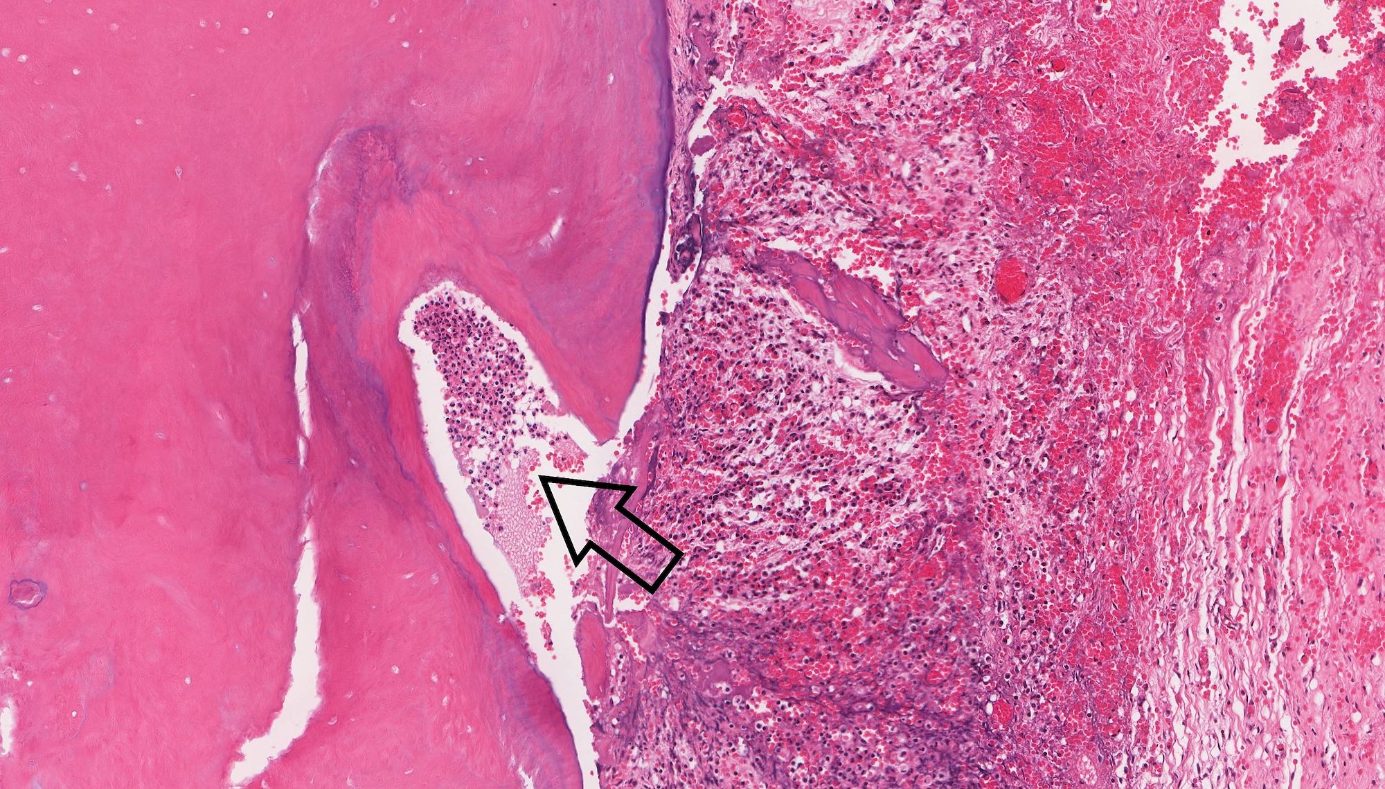
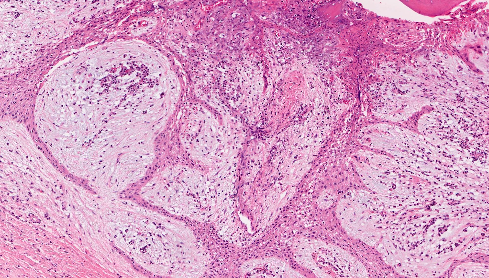
Simple bone cyst
Contributed by Kelly Magliocca, D.D.S., M.P.H.
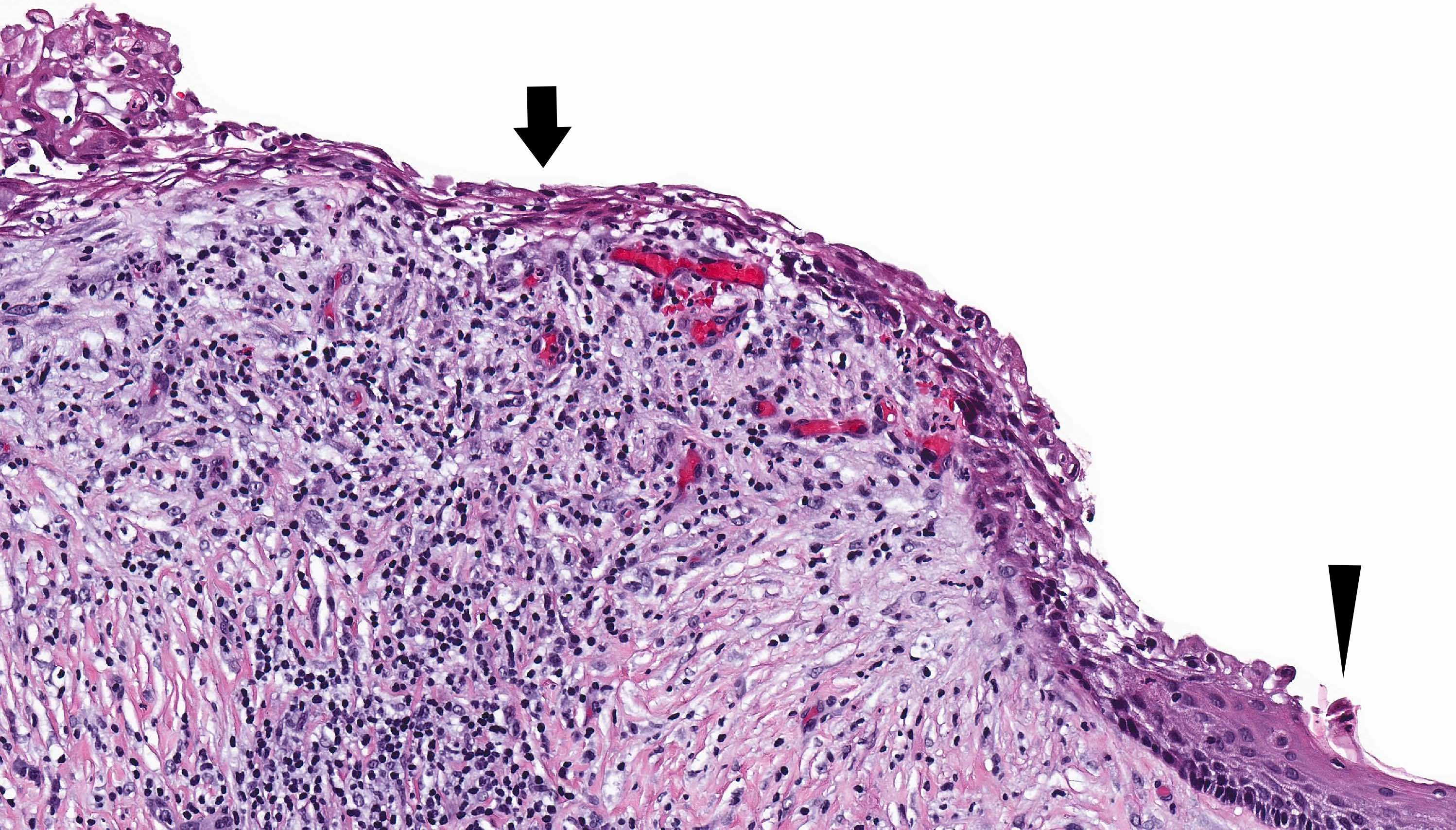
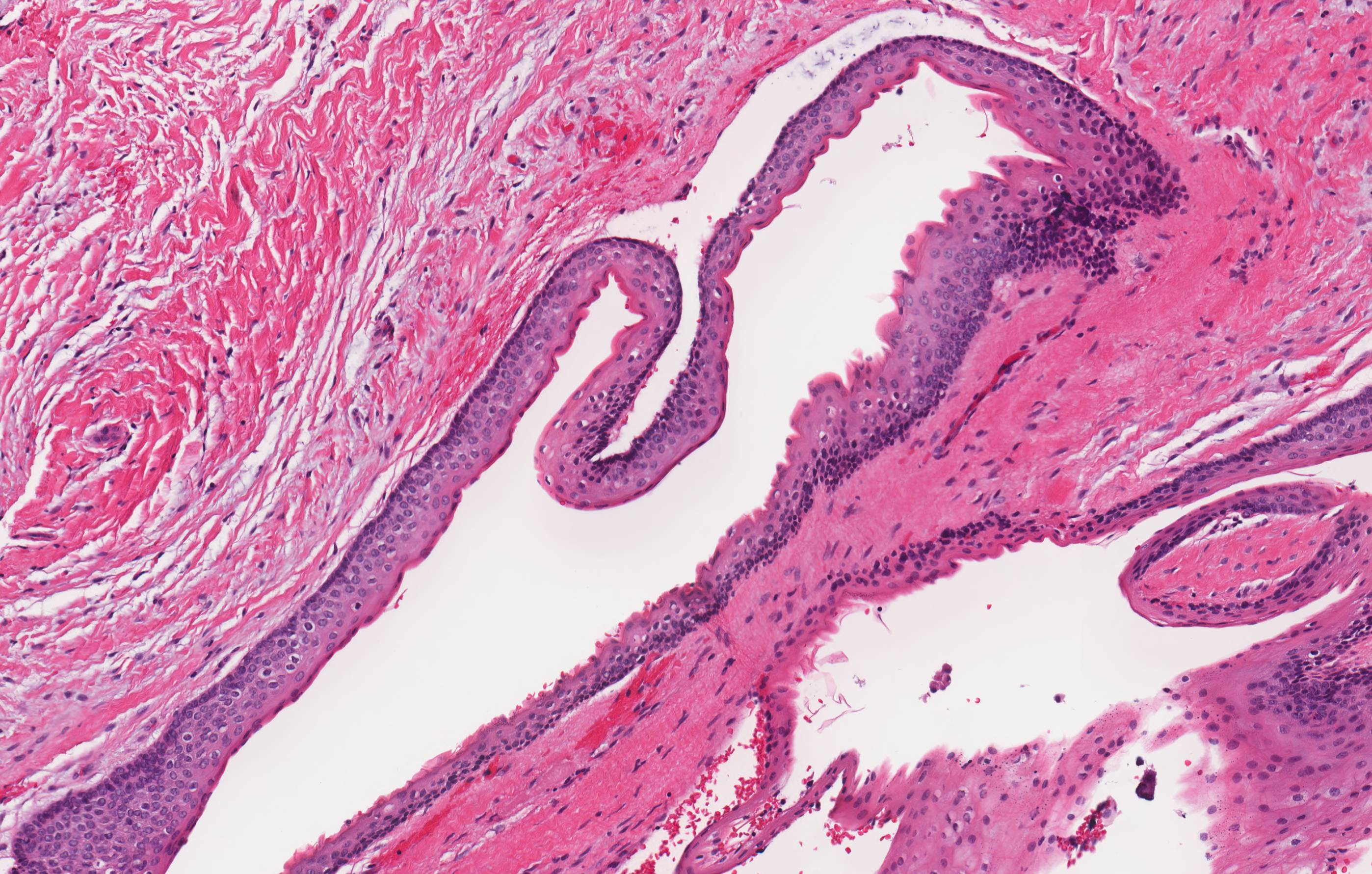
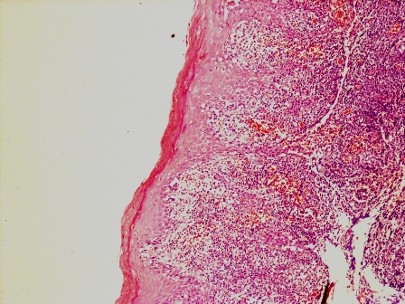
Squamous odontogenic tumor
SOT at scanning magnification
Benign islands of squamous epithelium within fibrous stroma
Epithelial vacuolization of squamous islands
Intraepithelial calcifications
Rounded contours of nests
Contributed by Anthony Chi, M.D., Ph.D.
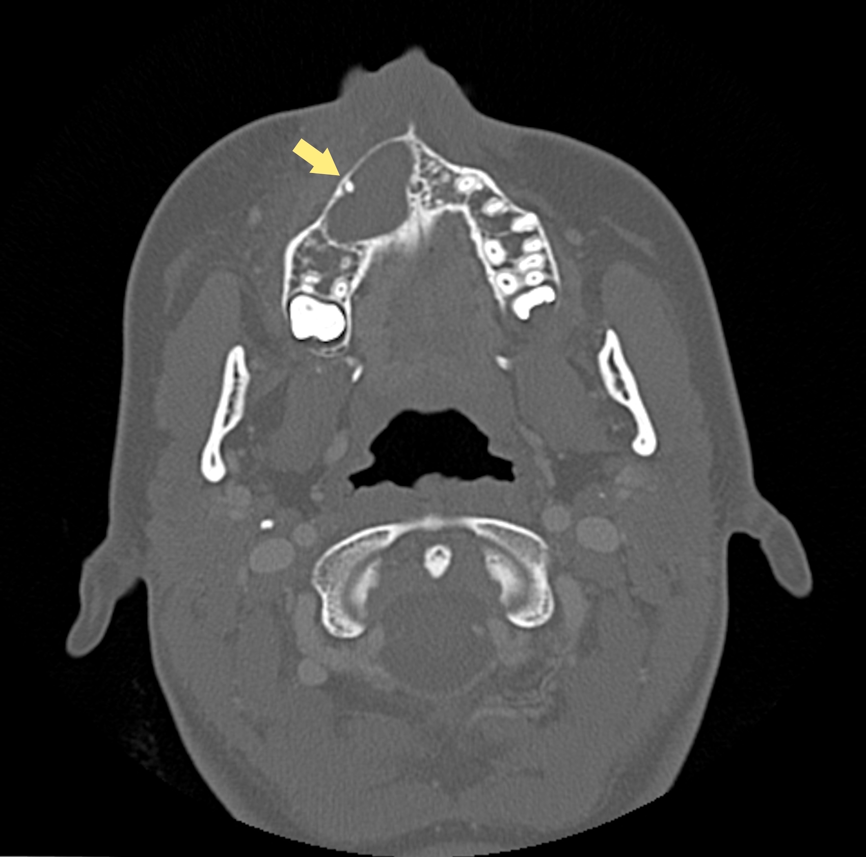
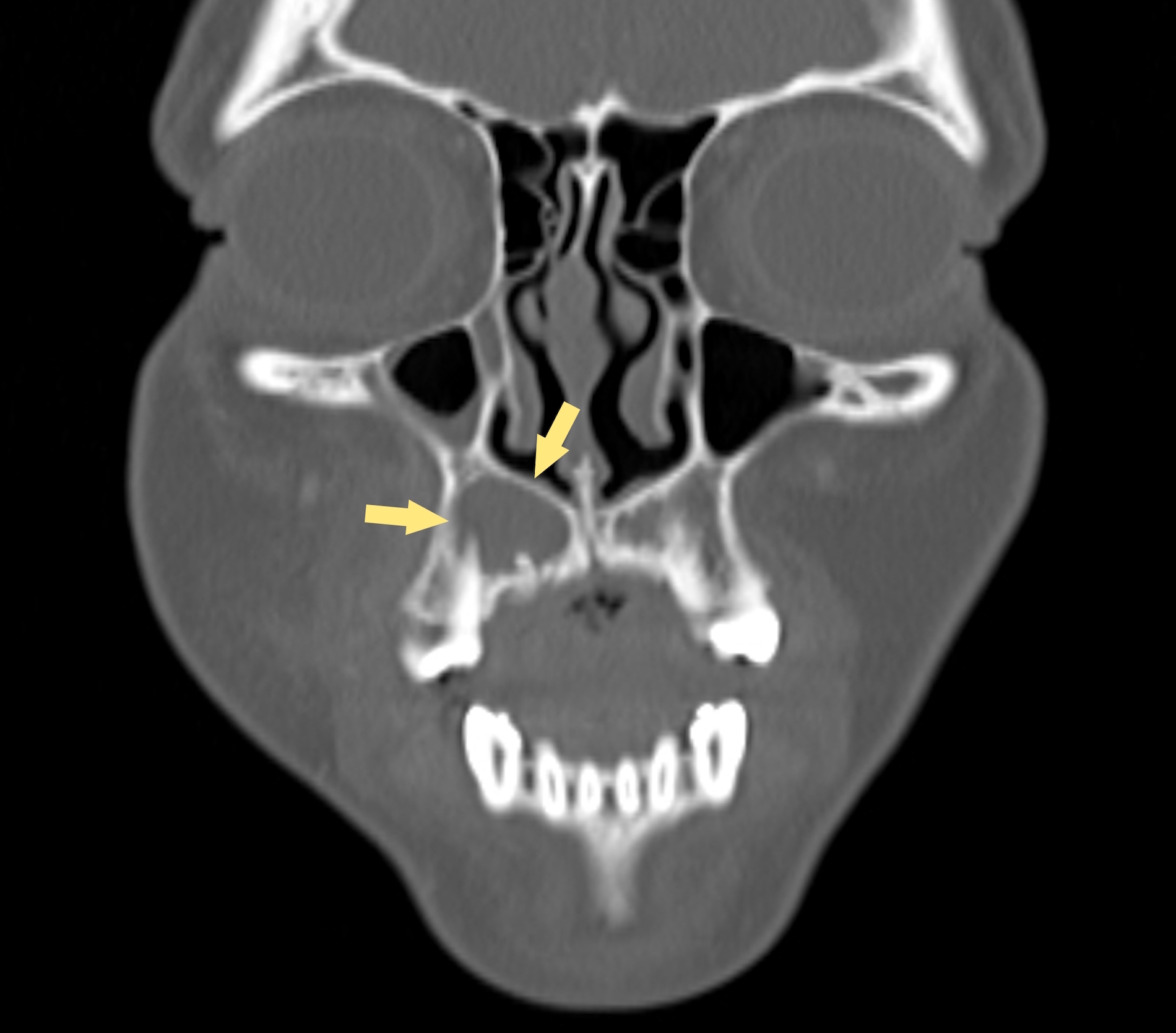
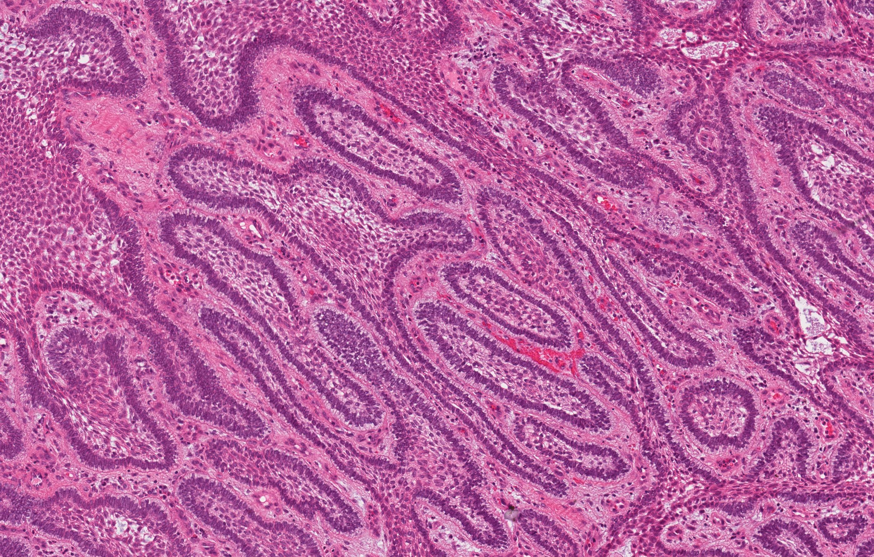
Cystic lesion in maxilla, intraosseous
Columnar epithelial lining
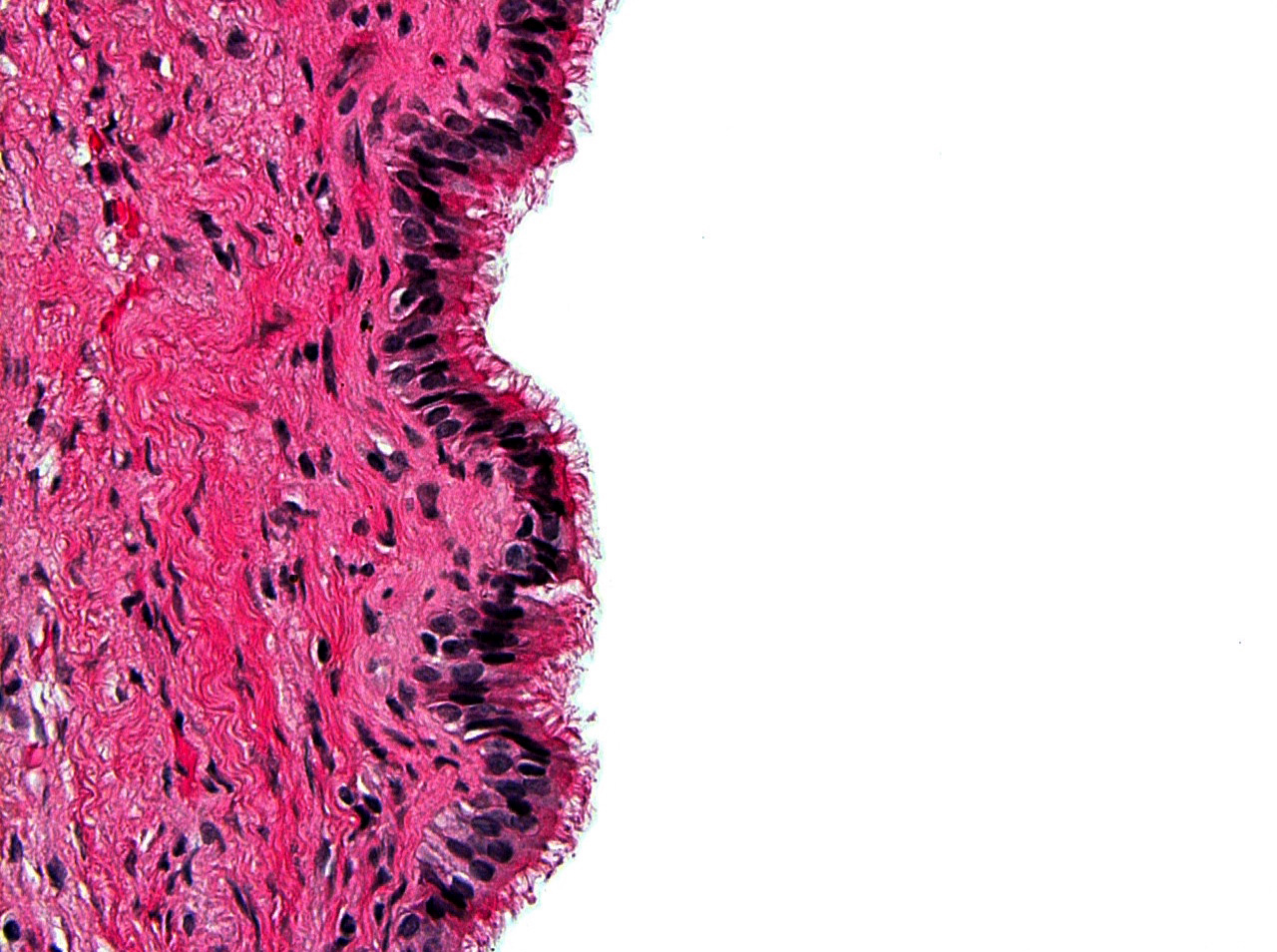
Prominent cilia
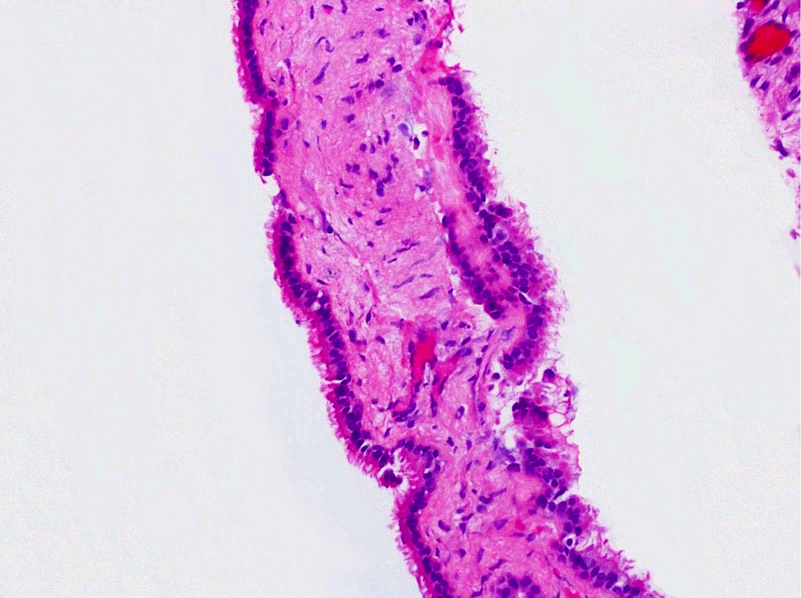
Cyst with thin wall in maxilla, intraosseous
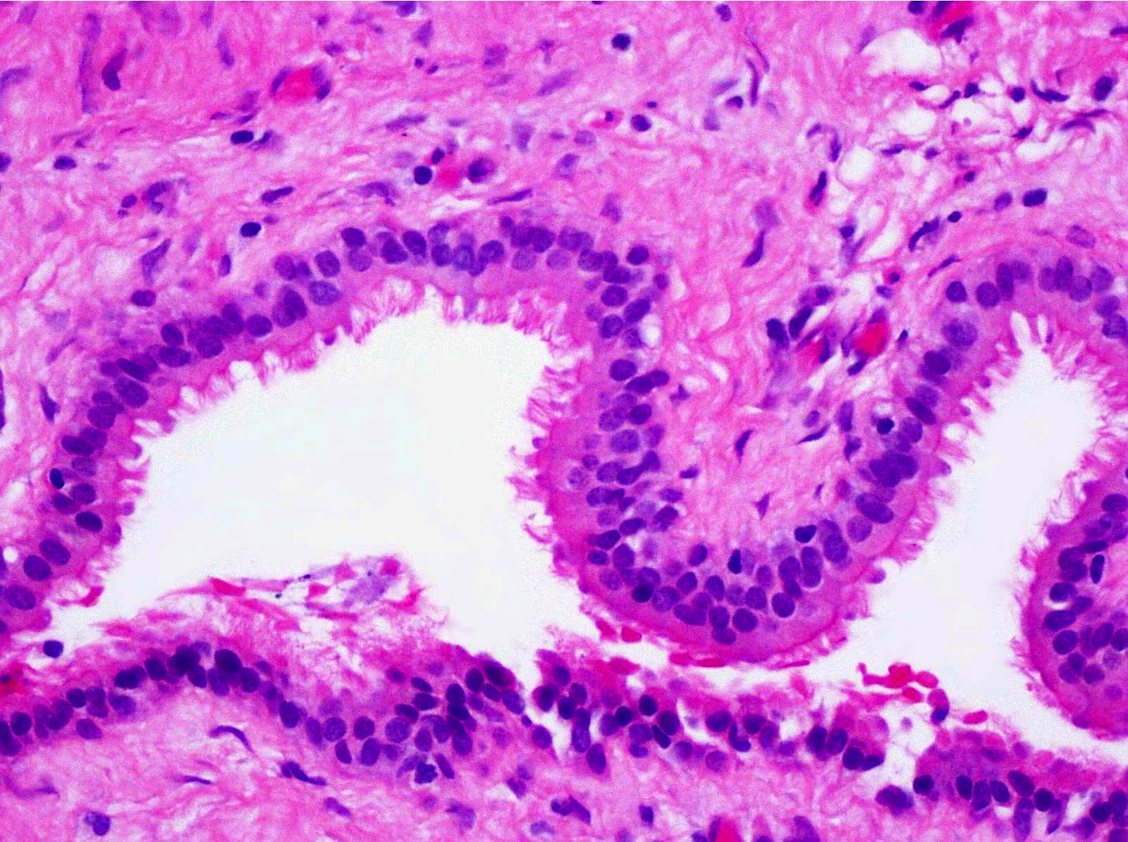
Ciliated cyst lining

No cytologic atypia
Contributed by Kelly Magliocca, D.D.S., M.P.H.
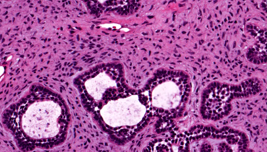
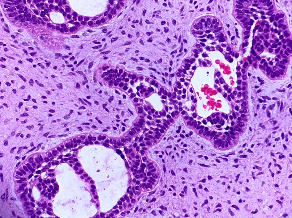
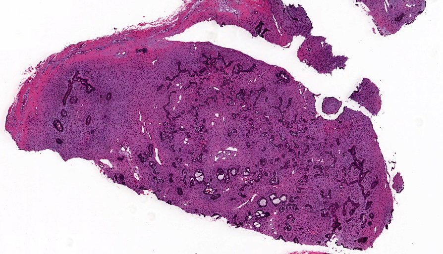
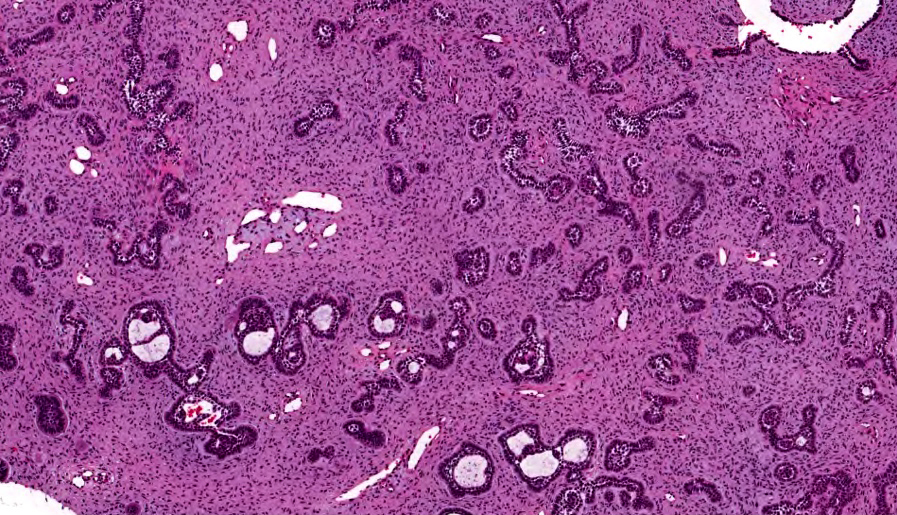
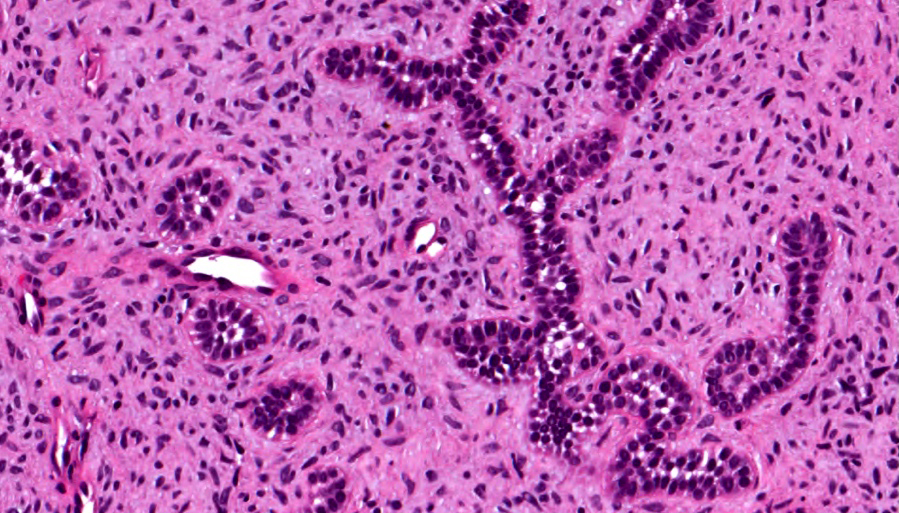
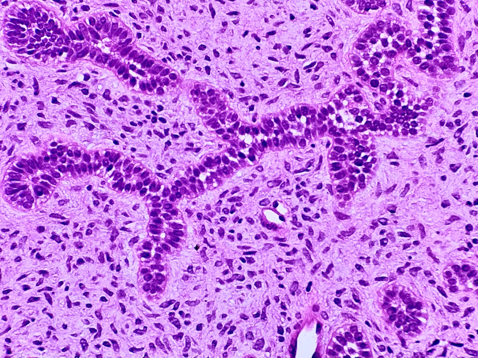
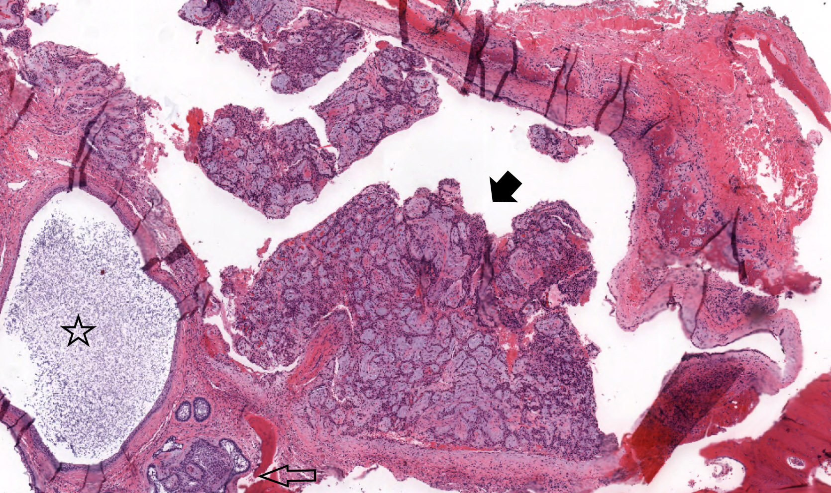
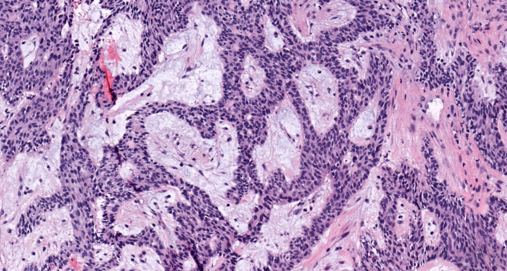
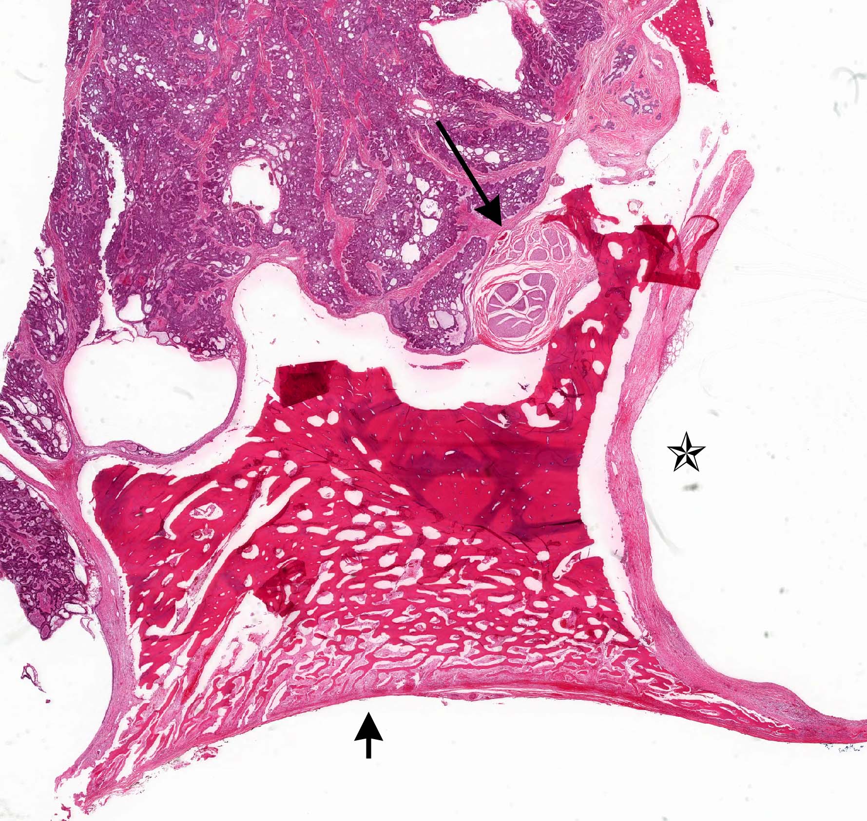
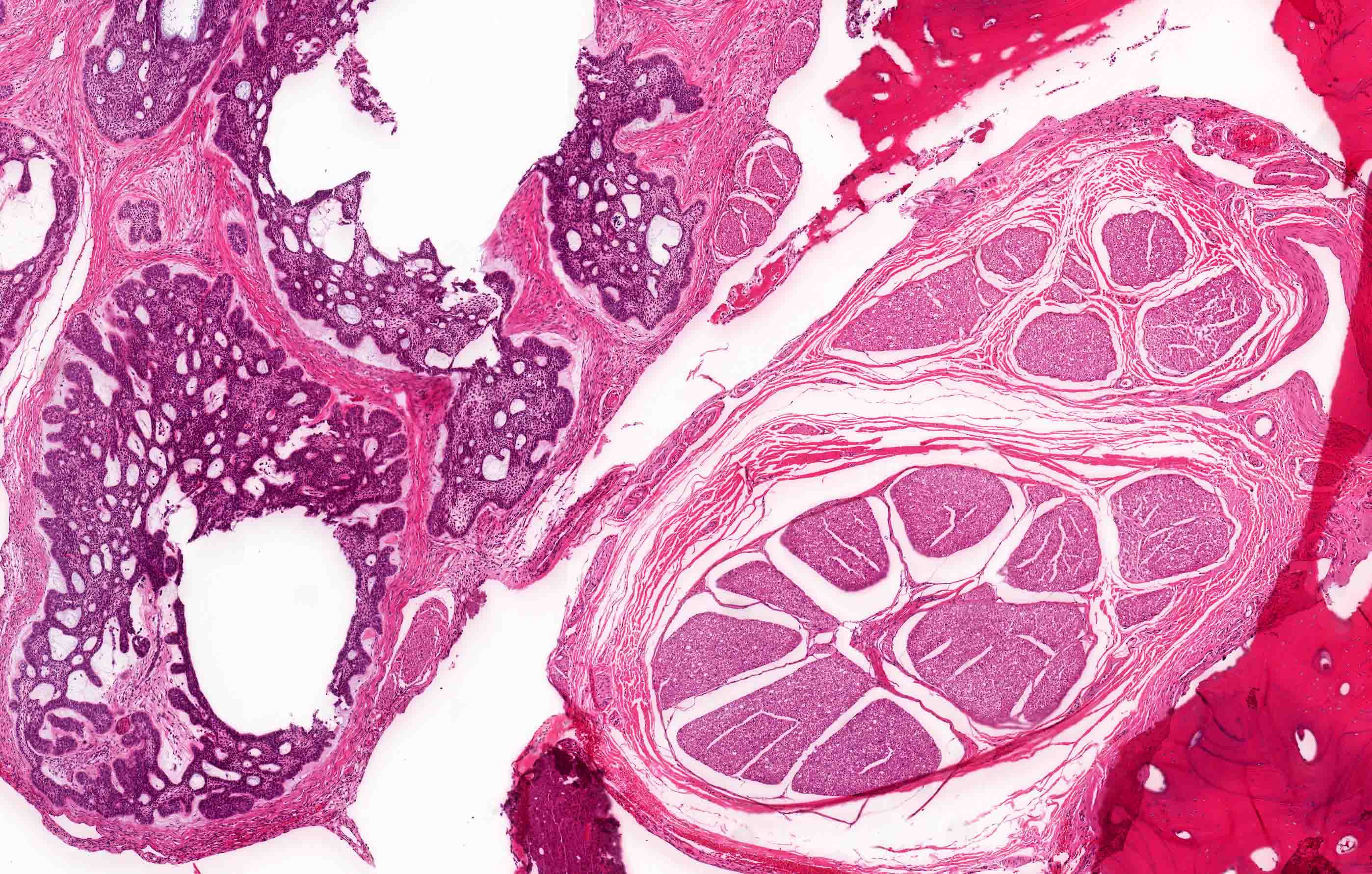
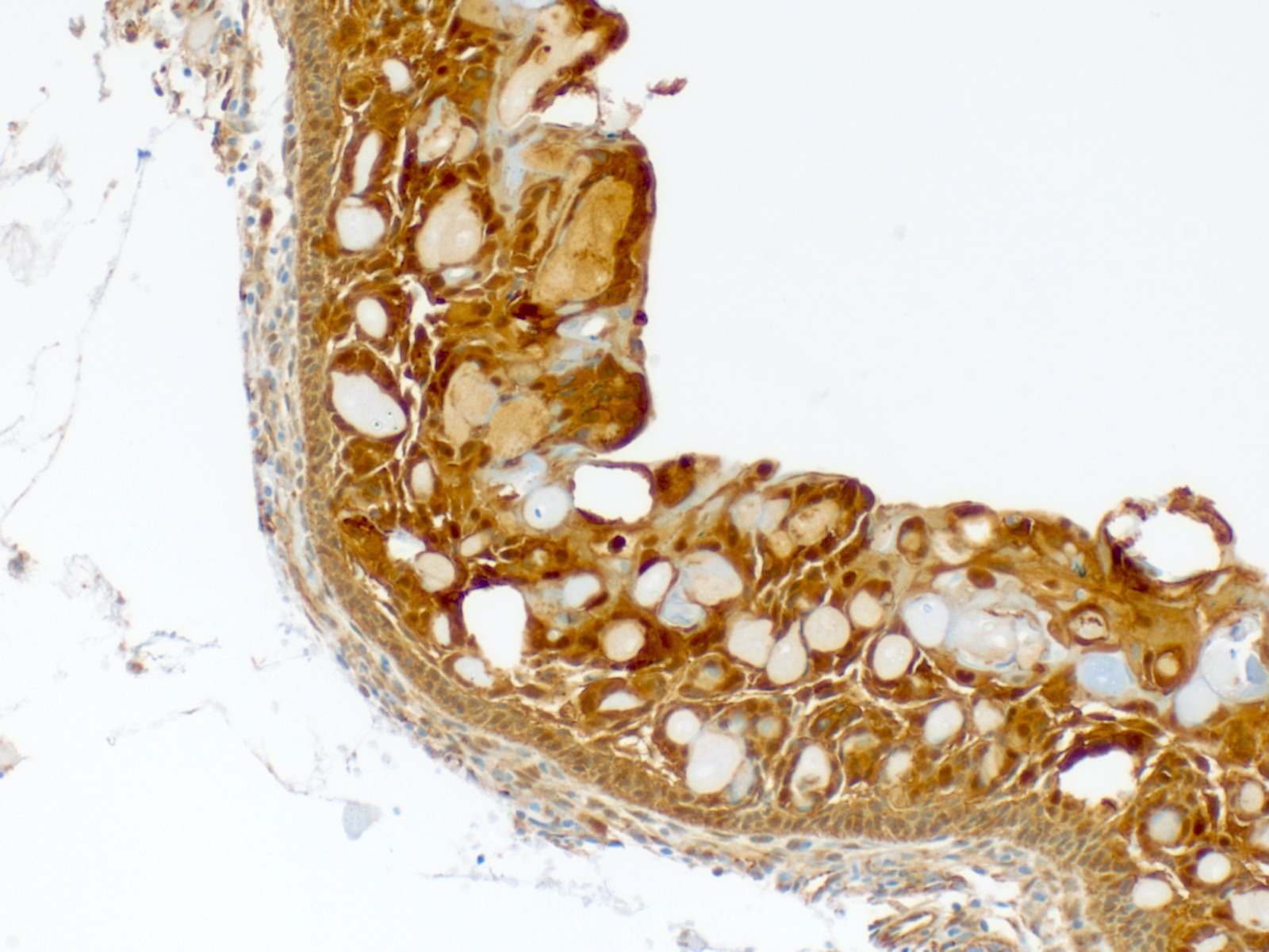
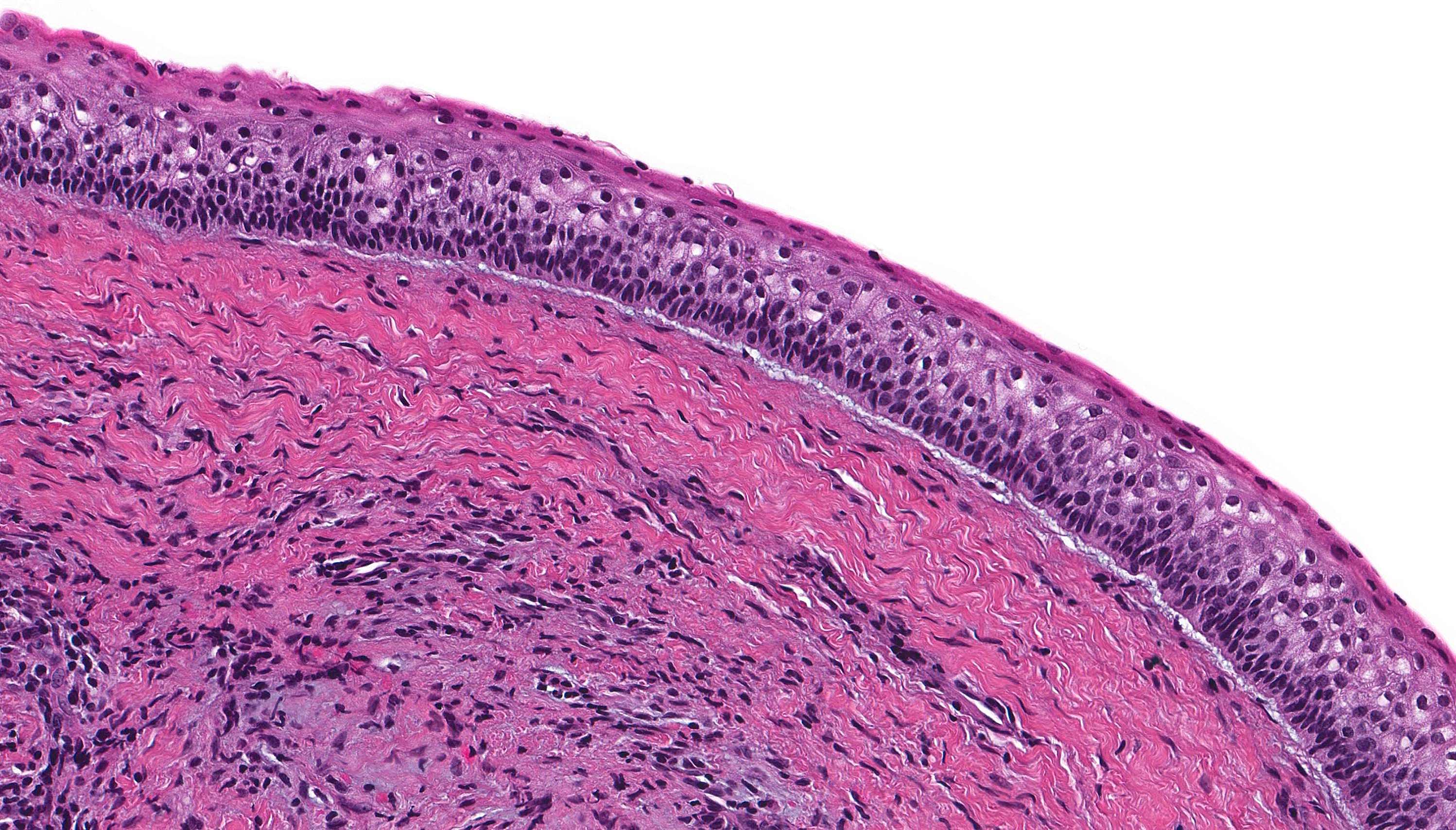
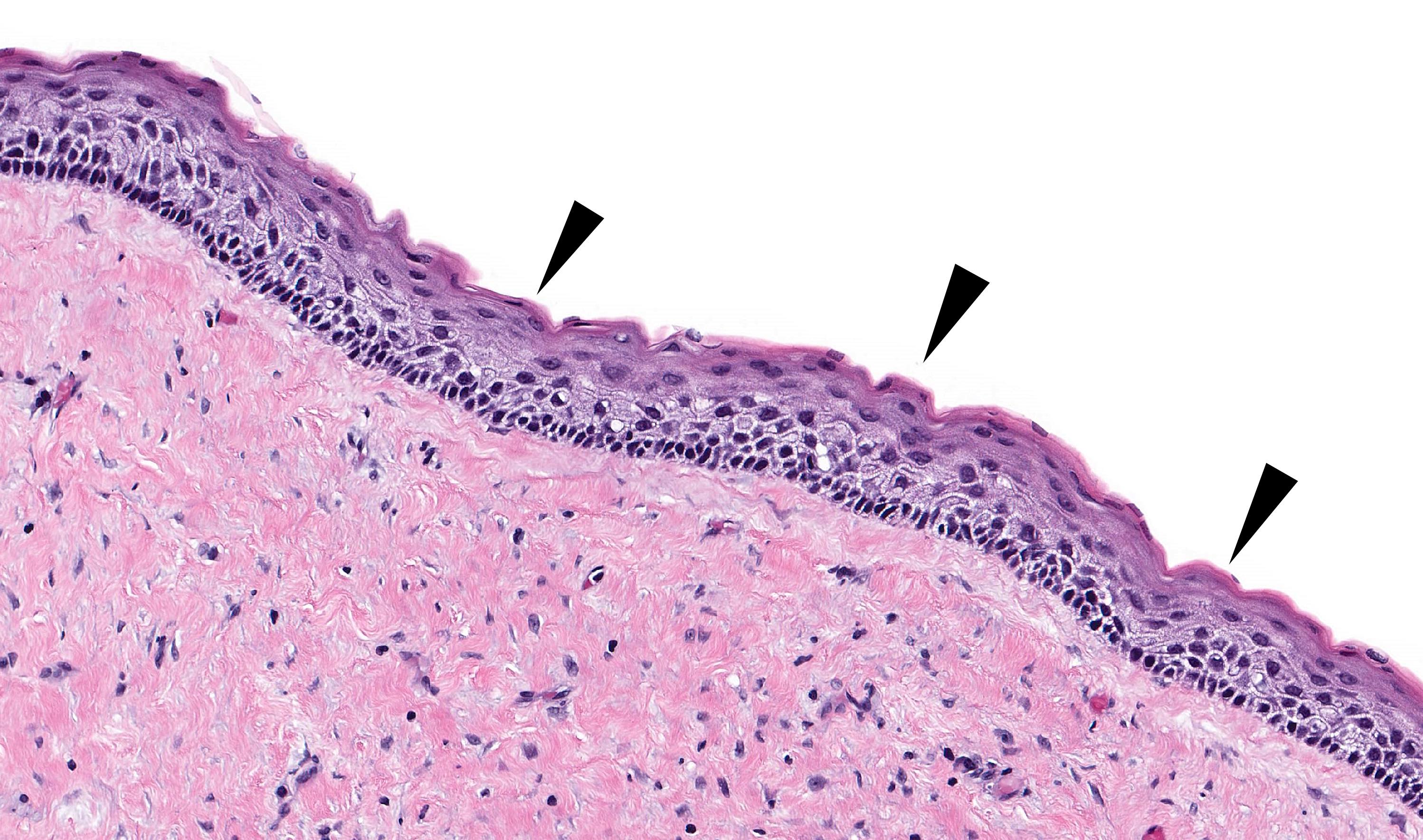
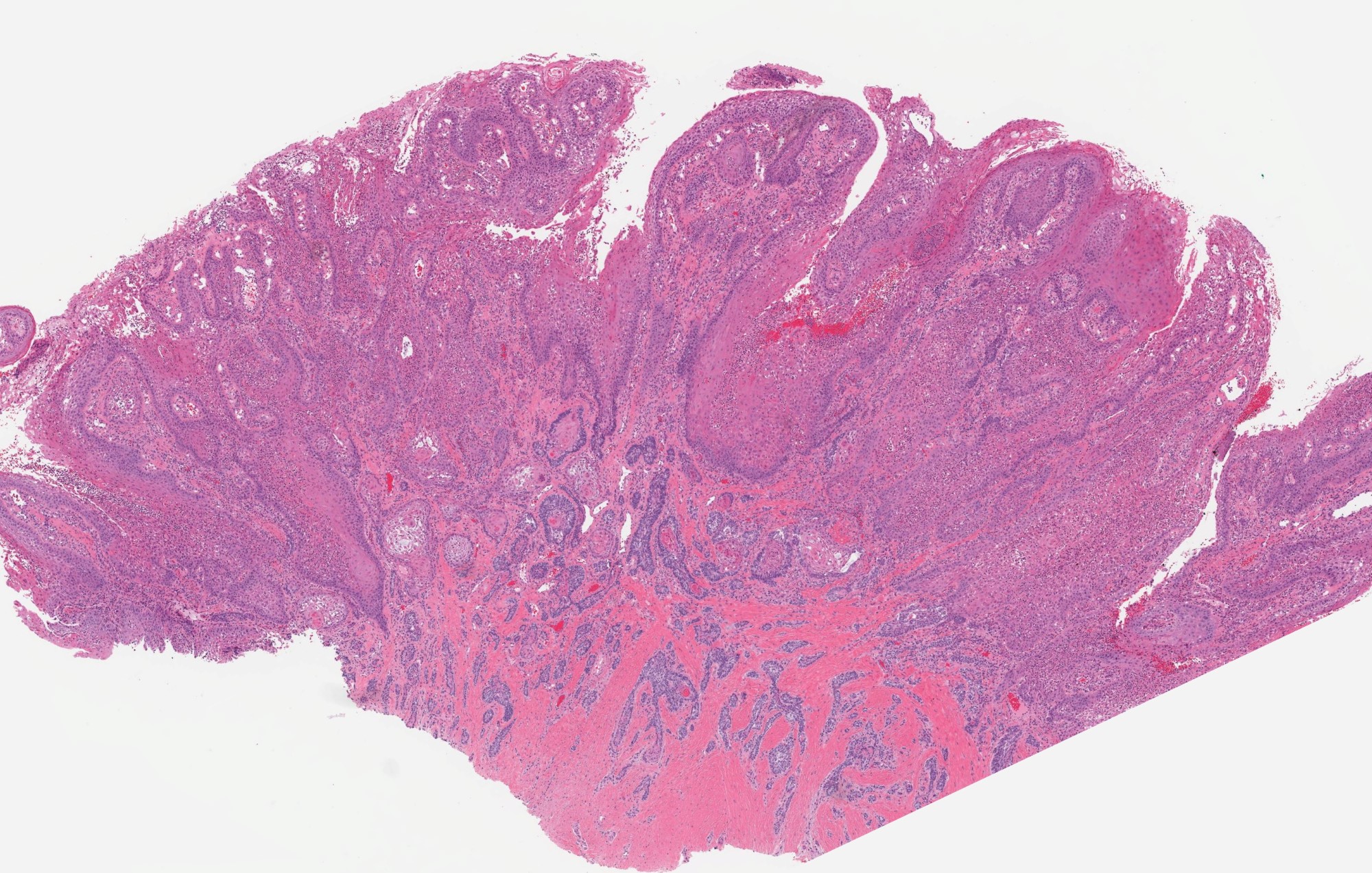
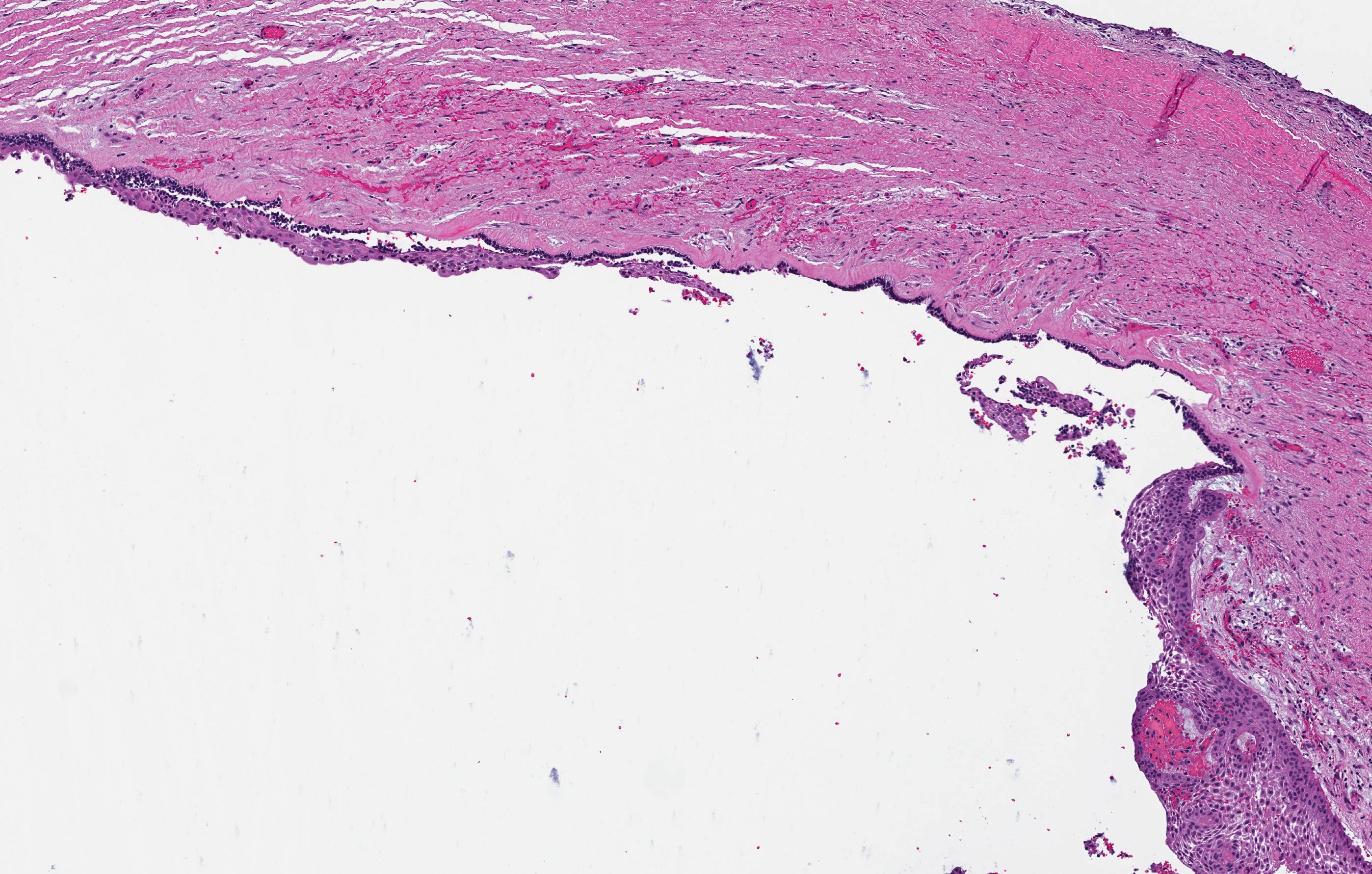
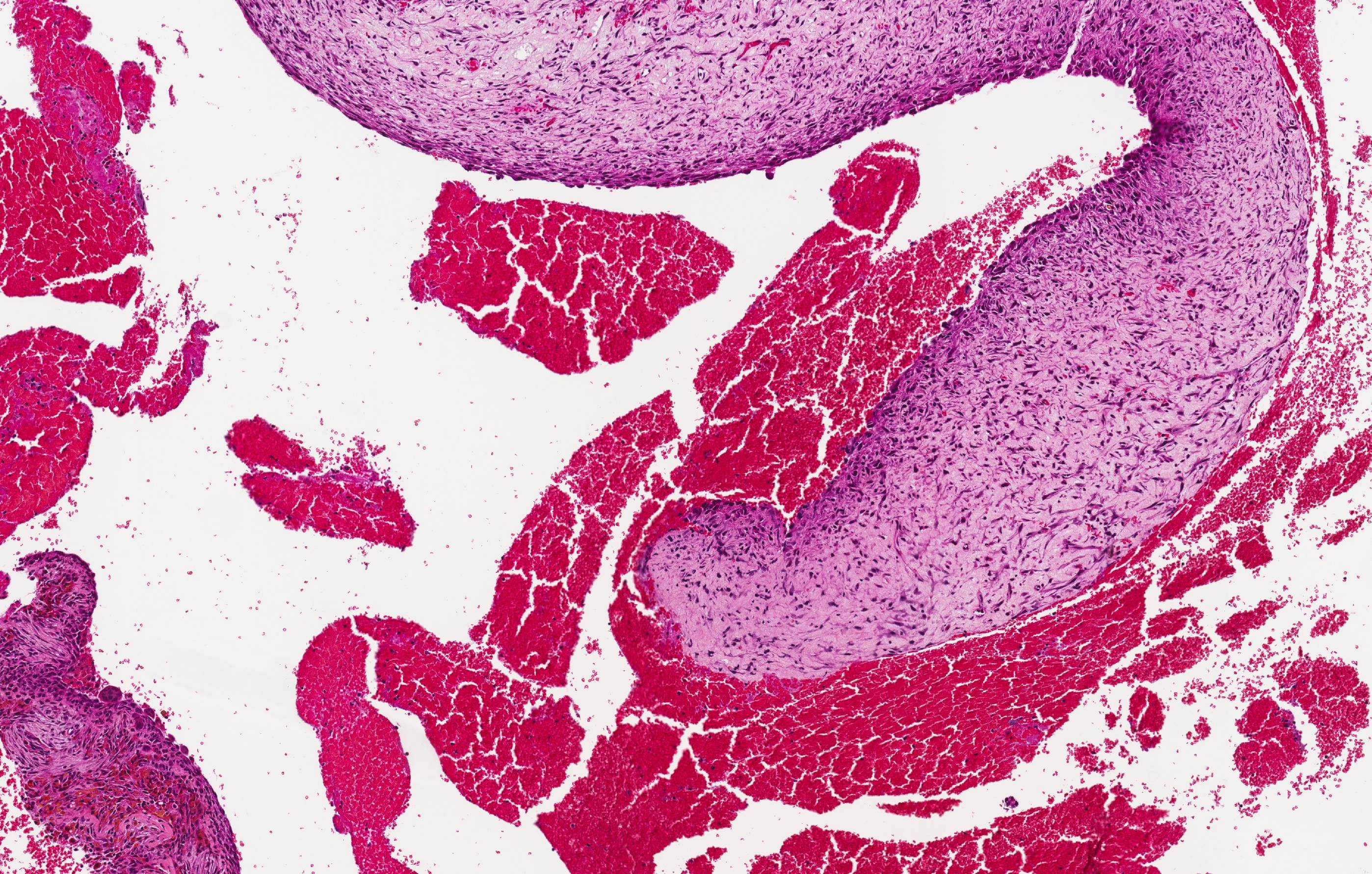
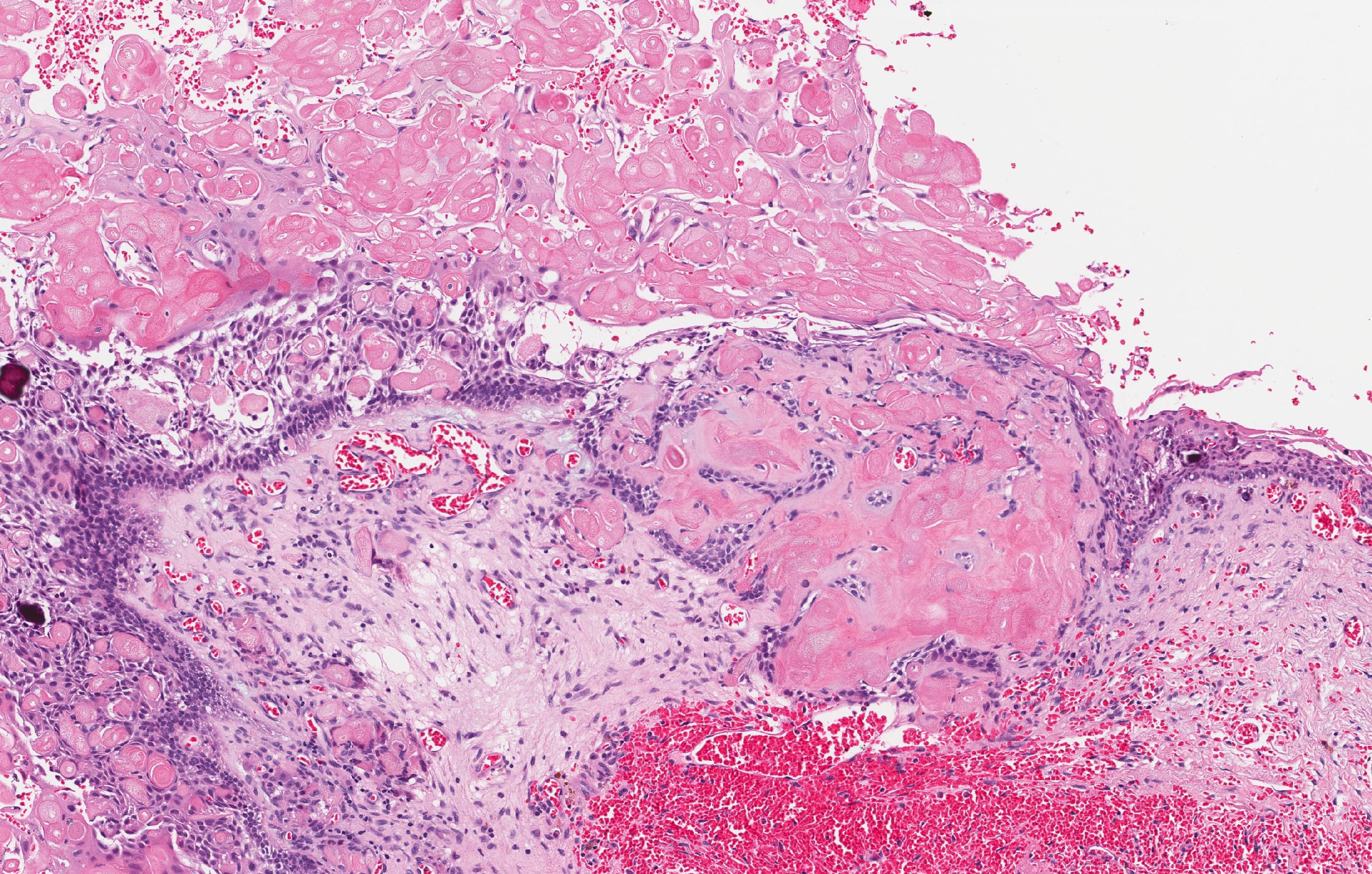
Adenomatoid odontogenic tumor
Ameloblastic carcinoma
Ameloblastic fibroma
Ameloblastoma
Ameloblastoma, extraosseous / peripheral type
Ameloblastoma, unicystic type
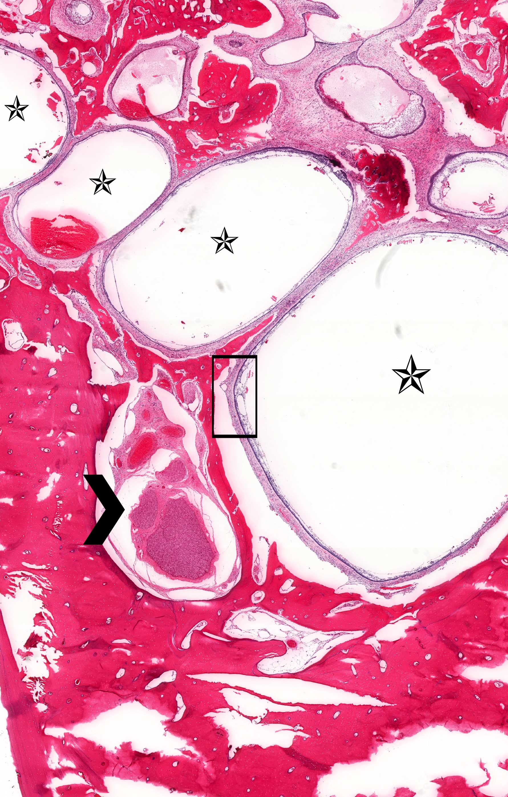
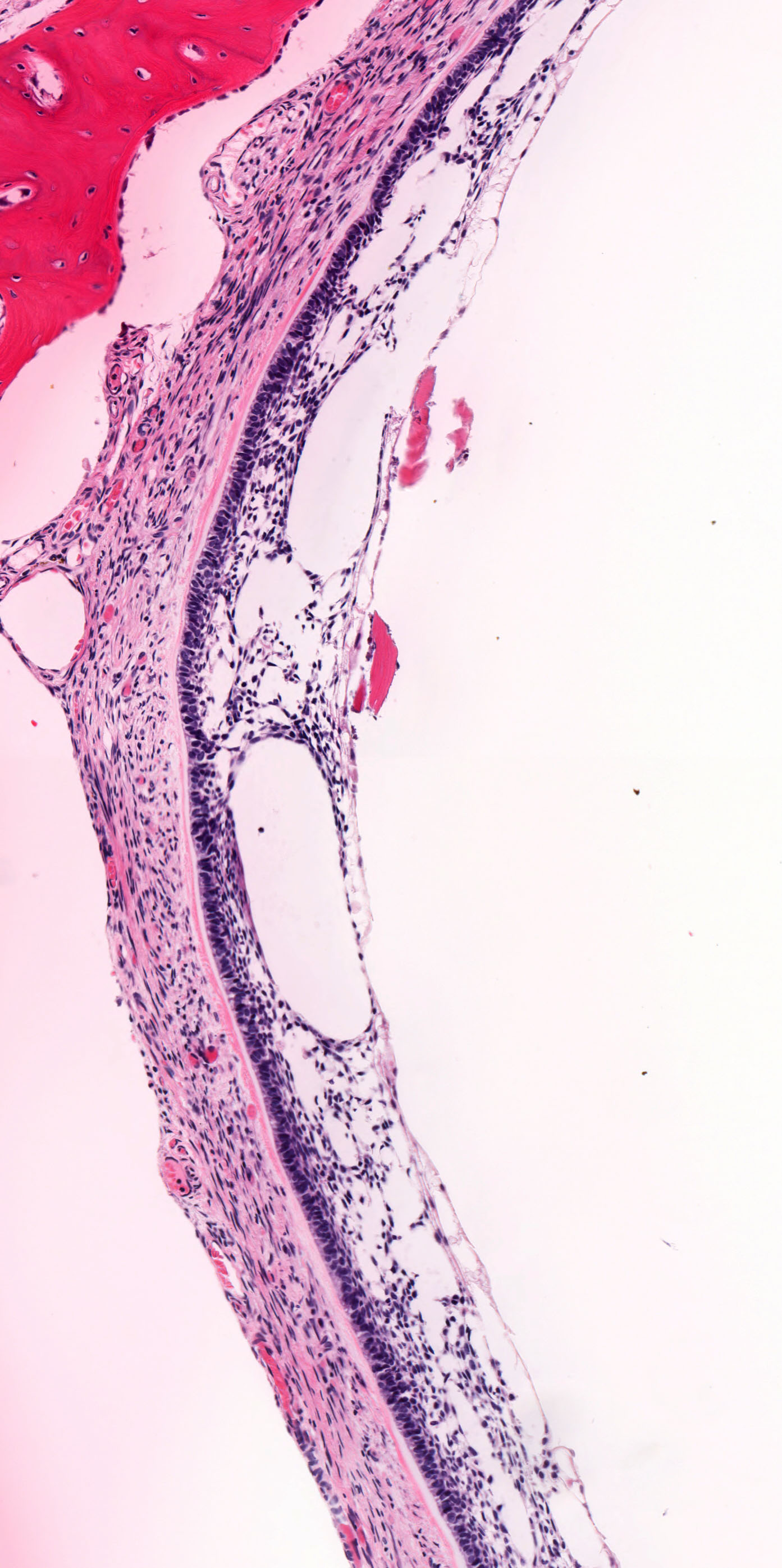
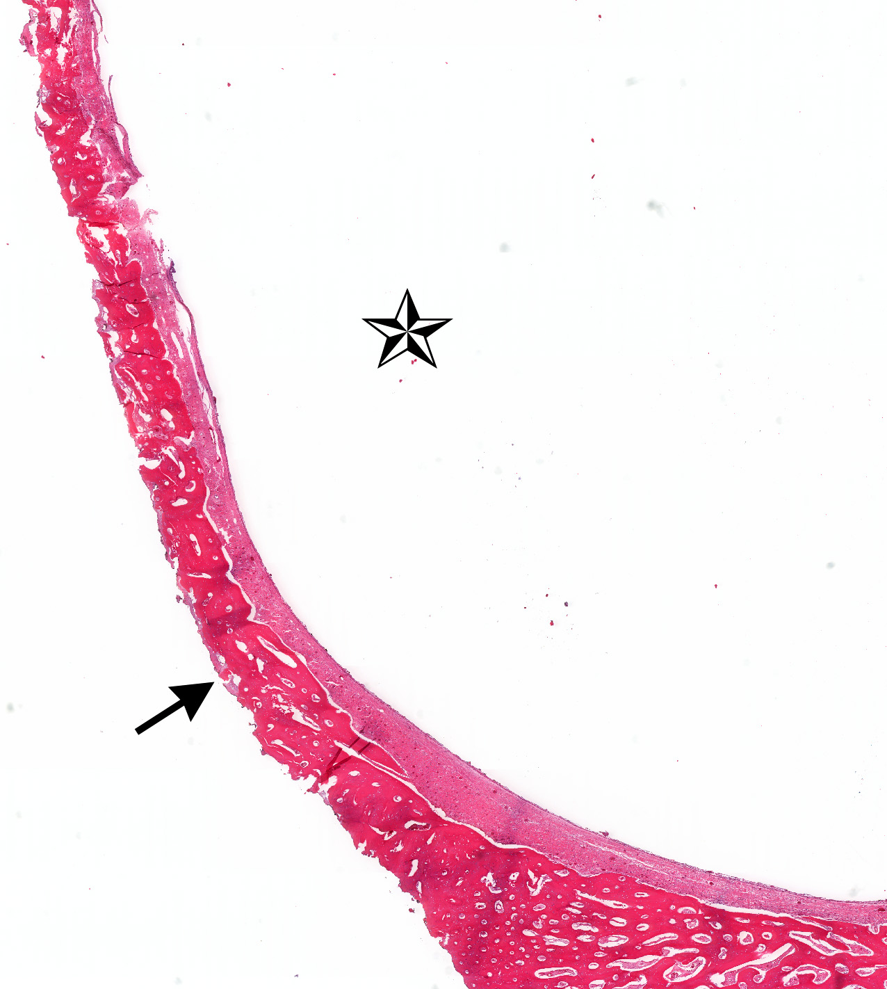
Aneurysmal bone cyst
Calcifying epithelial odontogenic tumor
Calcifying odontogenic cyst
Cemento-osseous dysplasia
Cementoblastoma
Central giant cell granuloma
Cherubism
Chondrosarcoma
Clear cell odontogenic carcinoma
Dentigerous cyst
Dentinogenic ghost cell tumor
Desmoplastic fibroma
Fibrous dysplasia
Gingival cyst
Glandular odontogenic cyst
Inflammatory collateral cysts
Lateral periodontal cyst
Melanotic neuroectodermal tumor of infancy
Nasopalatine duct cyst
Odontogenic fibroma
Odontogenic keratocyst
Odontogenic myxoma / myxofibroma
Odontogenic sarcoma
Odontoma
Orthokeratinized odontogenic cyst
Ossifying fibroma
Osteoma
Osteosarcoma, NOS
Peripheral giant cell granuloma
Primary intraosseous carcinoma, NOS
Primordial odontogenic tumor
Radicular cyst
Simple bone cyst
Solitary plasmacytoma of bone
Squamous odontogenic tumor
Gnepp: 2021
IARC: 2024
Neville: 2023
Speight: 2022
Stelow: 2020
Thompson: 2022
Wenig: 2017
Wenig: 2024
Find related Pathology books: head & neck/endocrine
