


Images hosted on other servers:

Mediastinum anatomy, sagittal

Mediastinum anatomy, transverse

Histological structure of thymus

Thymus, hyperplasia, thymoma
Images hosted on other servers:

Morphology of thymus

Adult persistent thymus, cadaver

Normal thymus, 29 weeks gestation
Contributed by Nicole Stringham, Ph.D. (source: University of Michigan virtual slide box and Duke University virtual slide collection)
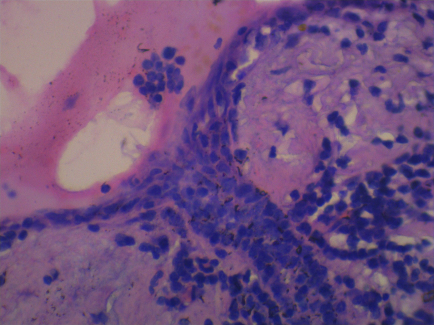
Neonatal thymus, lobule
Neonatal thymus, cortex / medulla
Neonatal thymus, Hassall corpuscle
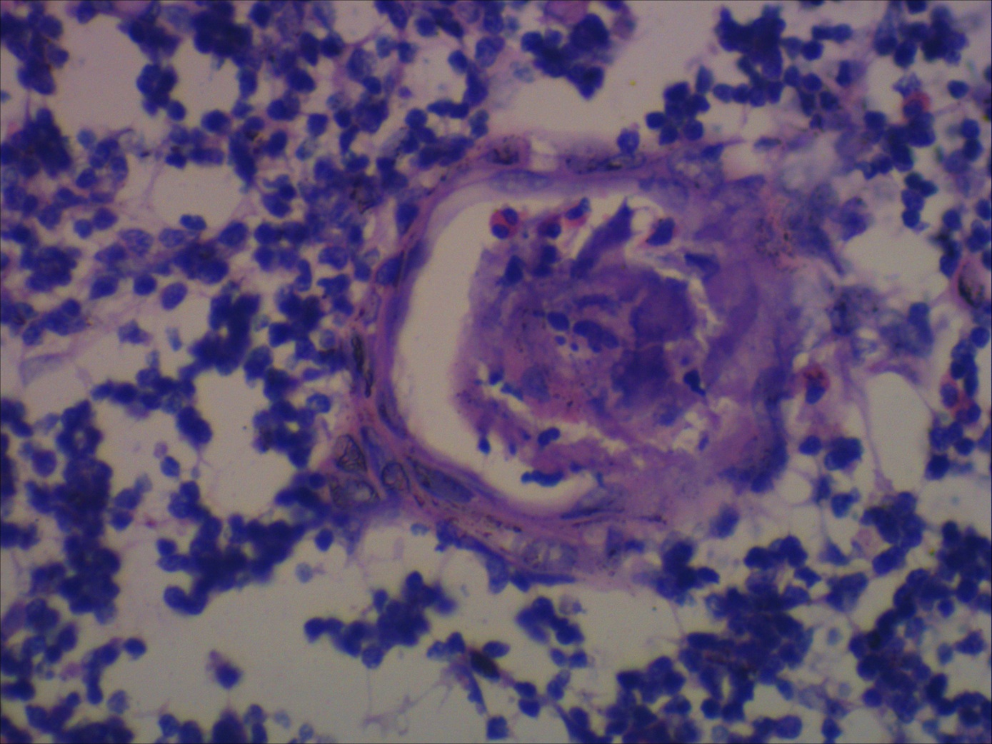
Adult thymus, involuted
Adult thymus, medulla
Adult thymus, Hassall corpuscle
Images hosted on other servers:

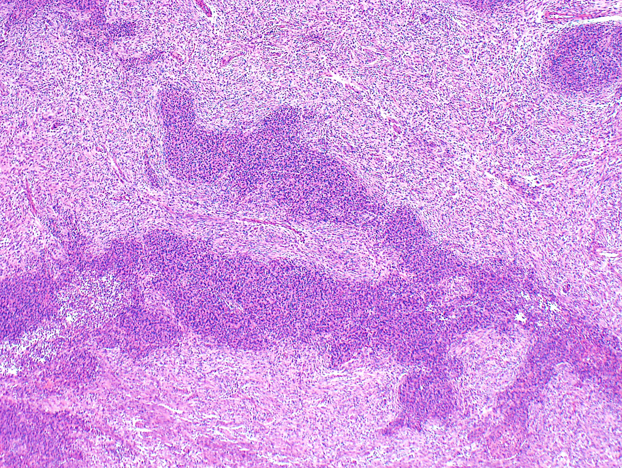
Thymic hyperplasia
Mediastinum
Histology of the thymus
Images hosted on other servers:

Fluid filled mediastinal mass

Mediastinal mass with high signal intensity on T1 and T2

Mediastinal bronchogenic cyst

Well circumscribed
intrapulmonary
bronchogenic cyst

Esophageal bronchogenic cyst
Images hosted on other servers:

Submucosal esophageal bronchogenic cyst

Polypoid endobronchial bronchogenic cyst
Images hosted on other servers:

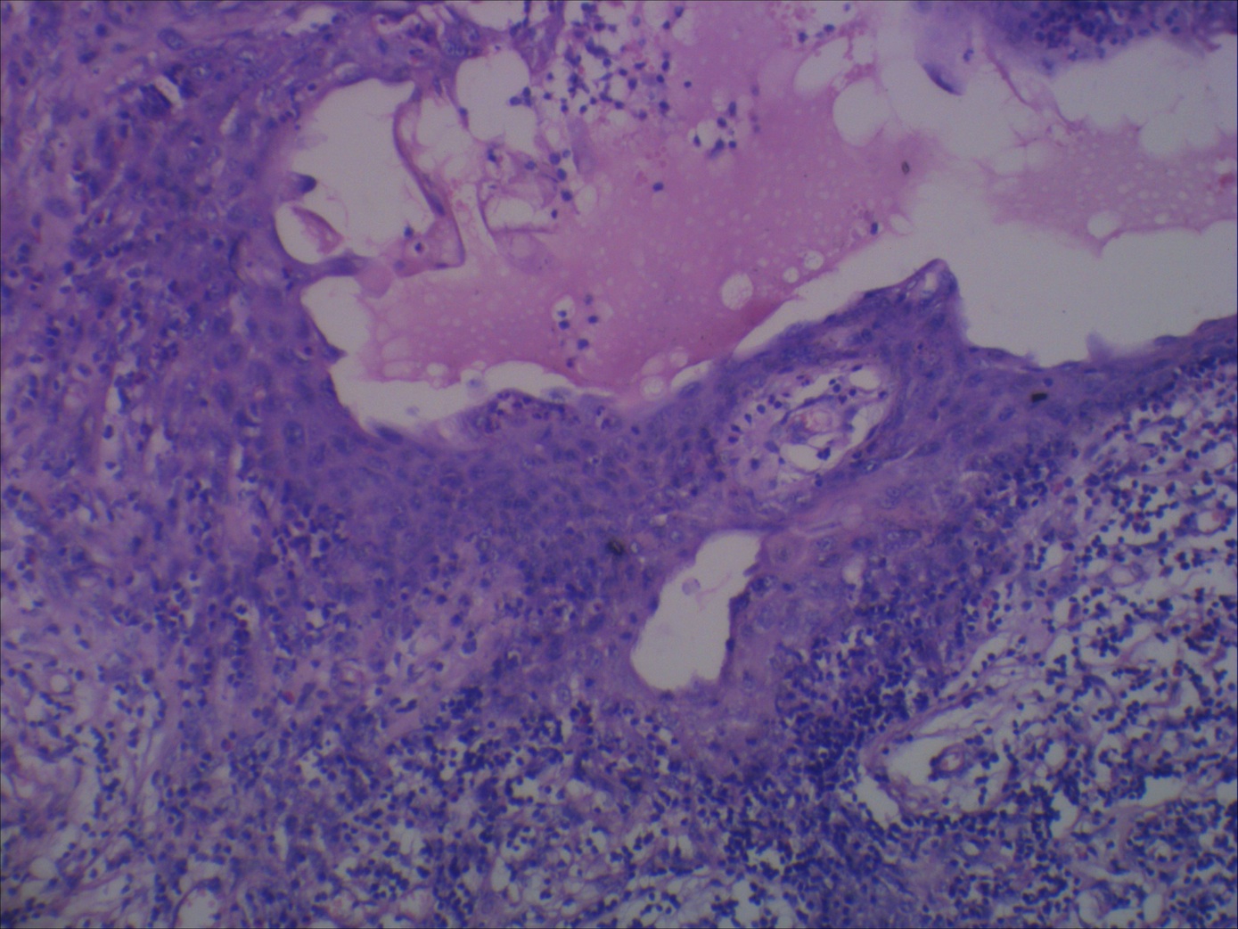
Unilocular cyst with mucoid material
Contributed by Laurence M. Briski, M.D.
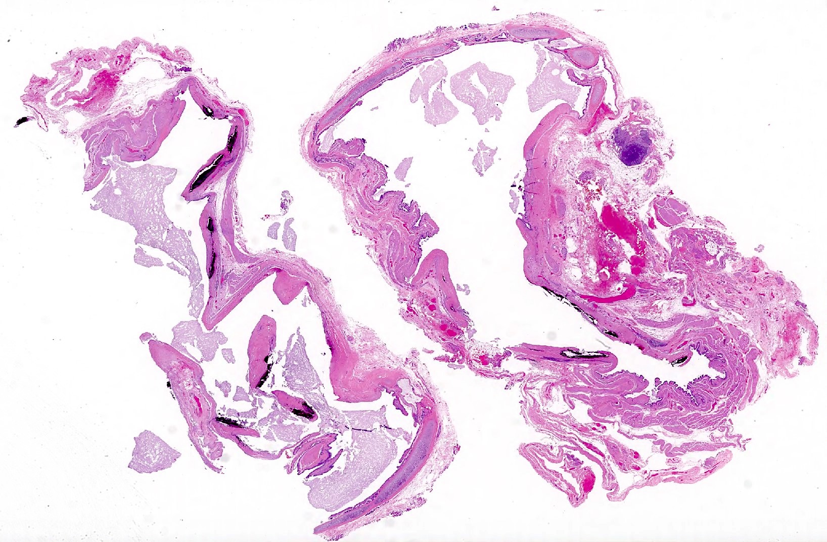
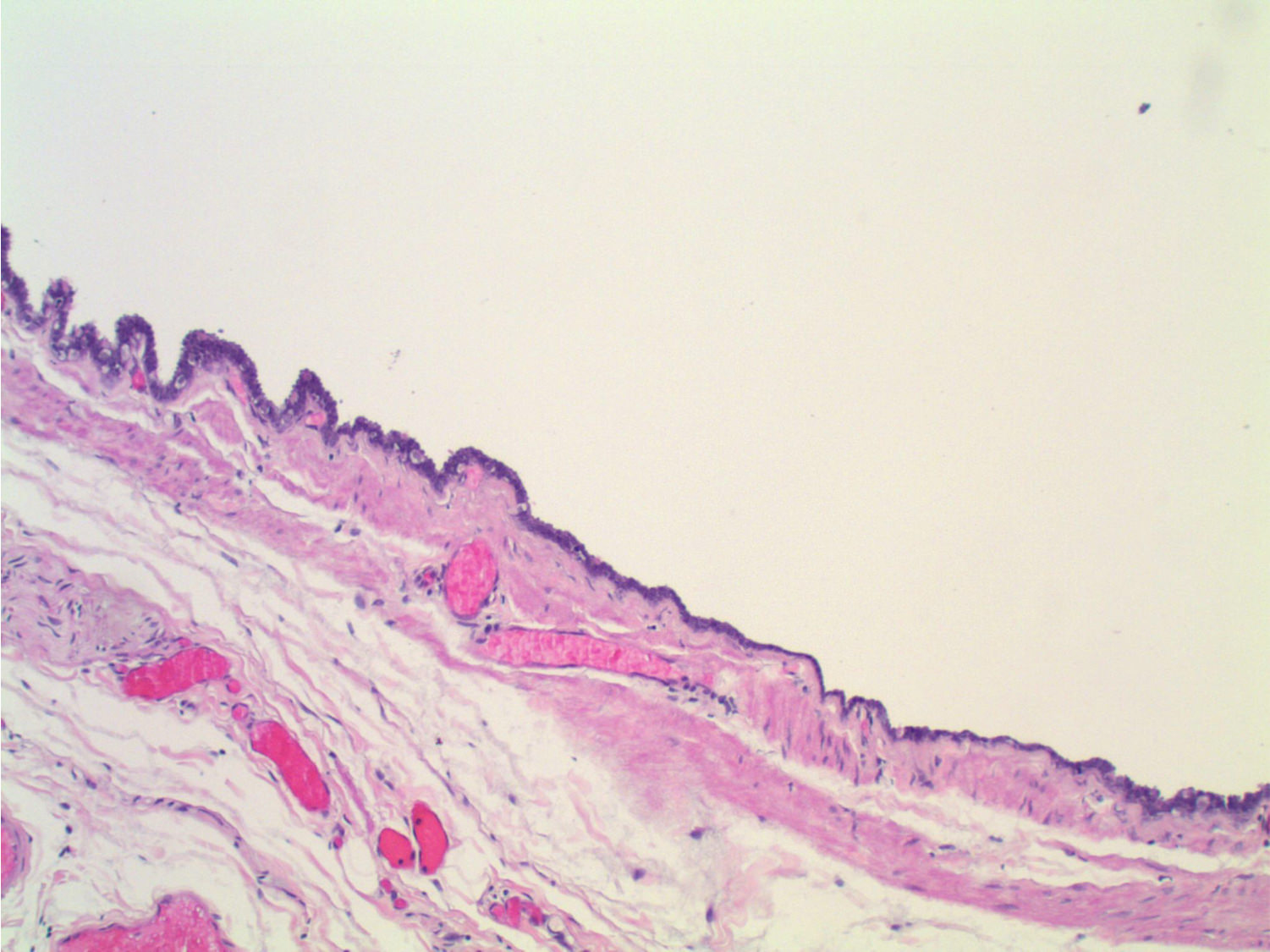
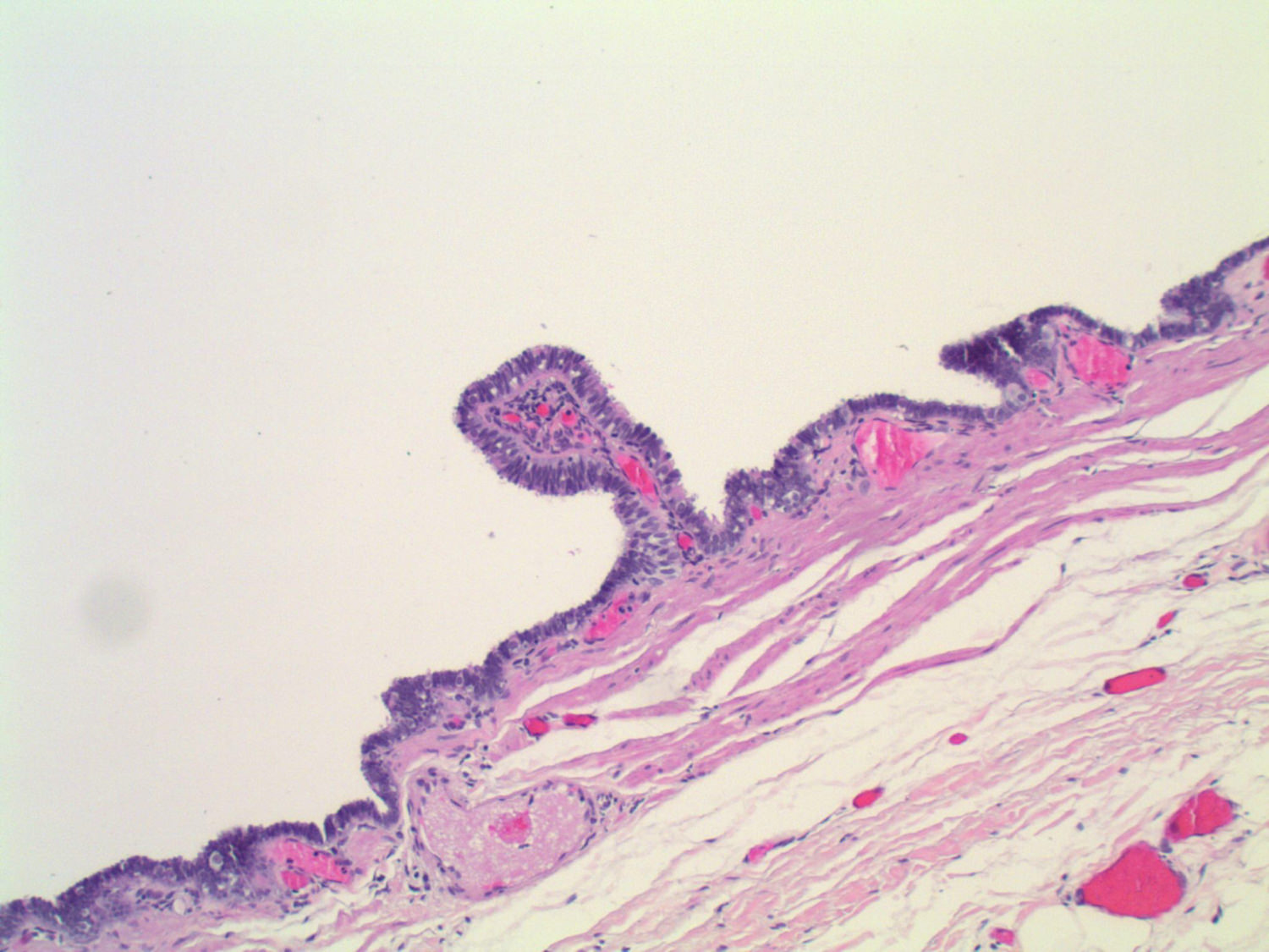
Unilocular cyst

Recapitulation of bronchial structure
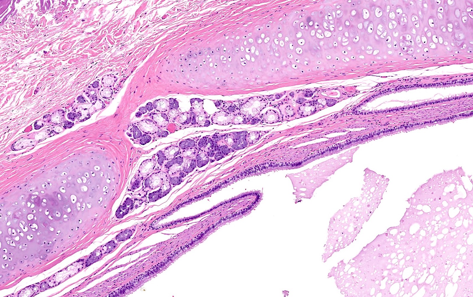
Cyst wall components

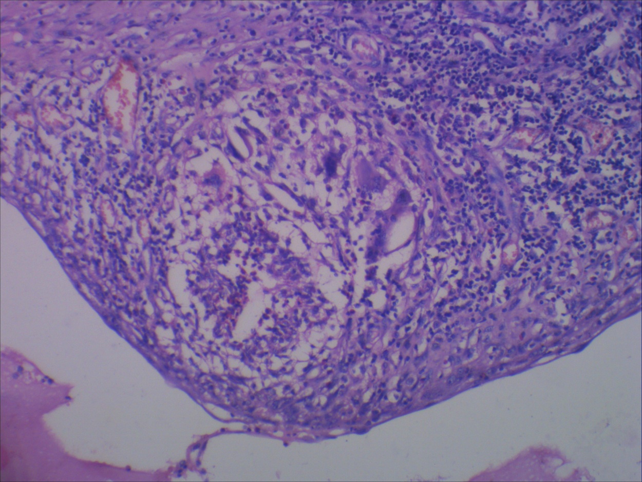
Foci of smooth muscle
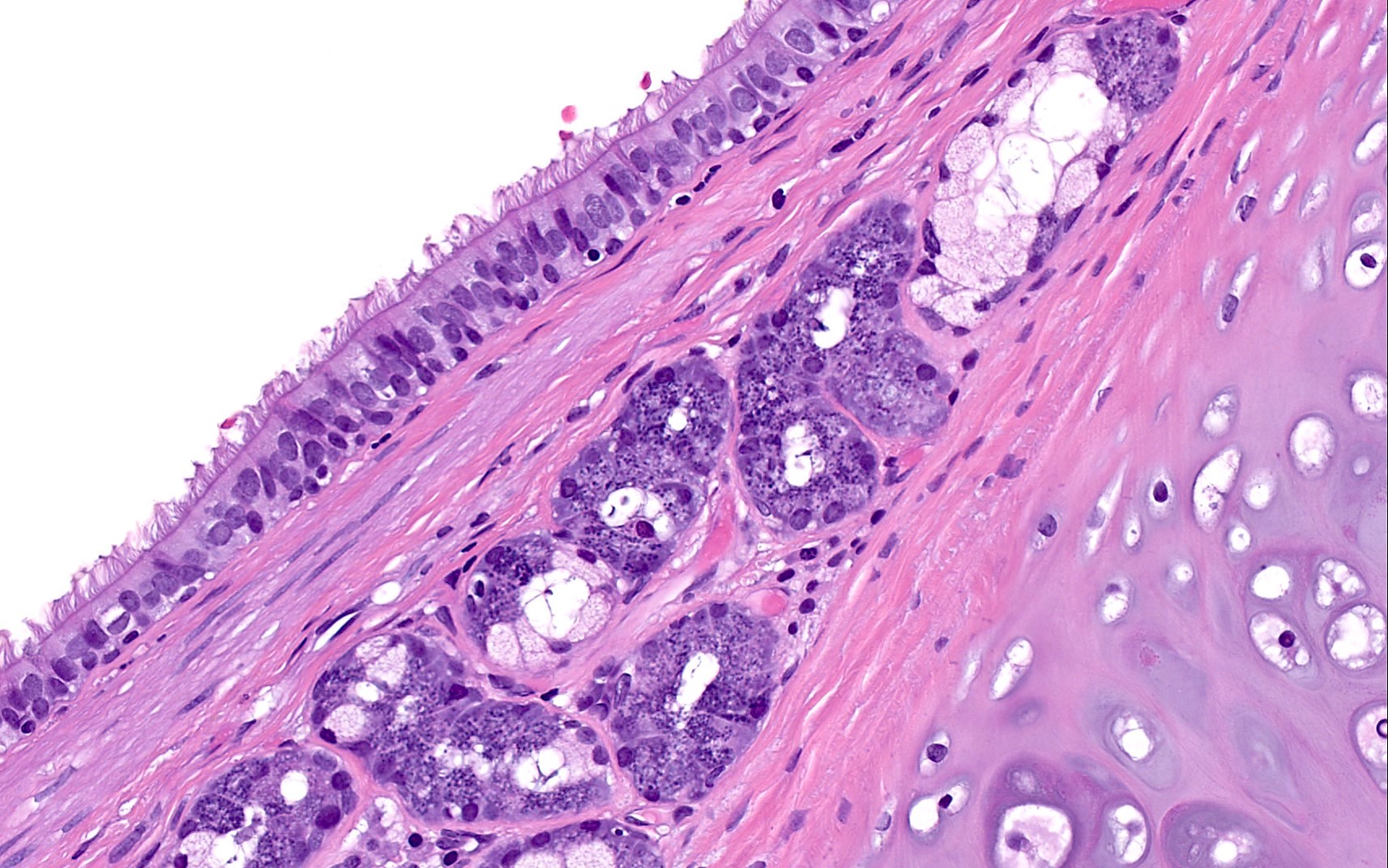
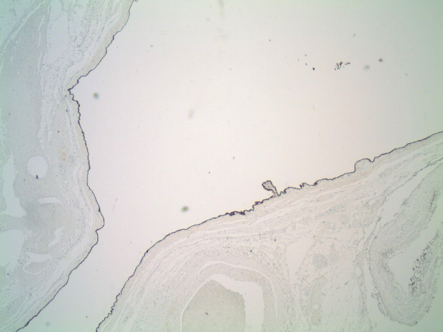
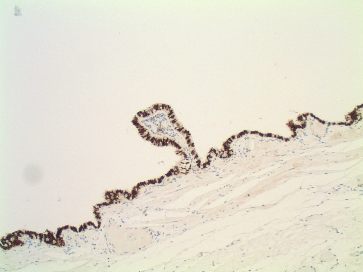
Ciliated respiratory epithelium
Images hosted on other servers:

Cystic contents (Diff-Quik stain)
Images hosted on other servers:

Features of 4 patients
(tables 1 and 3)
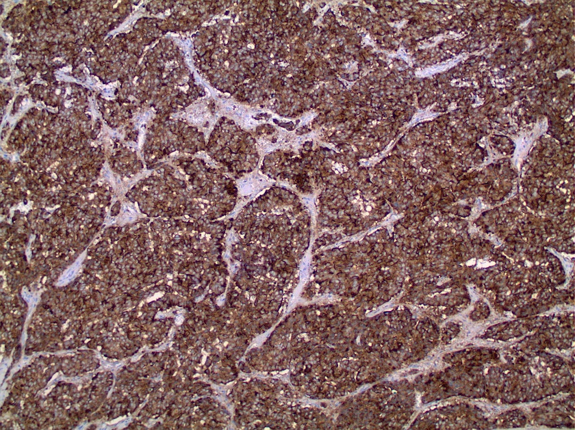
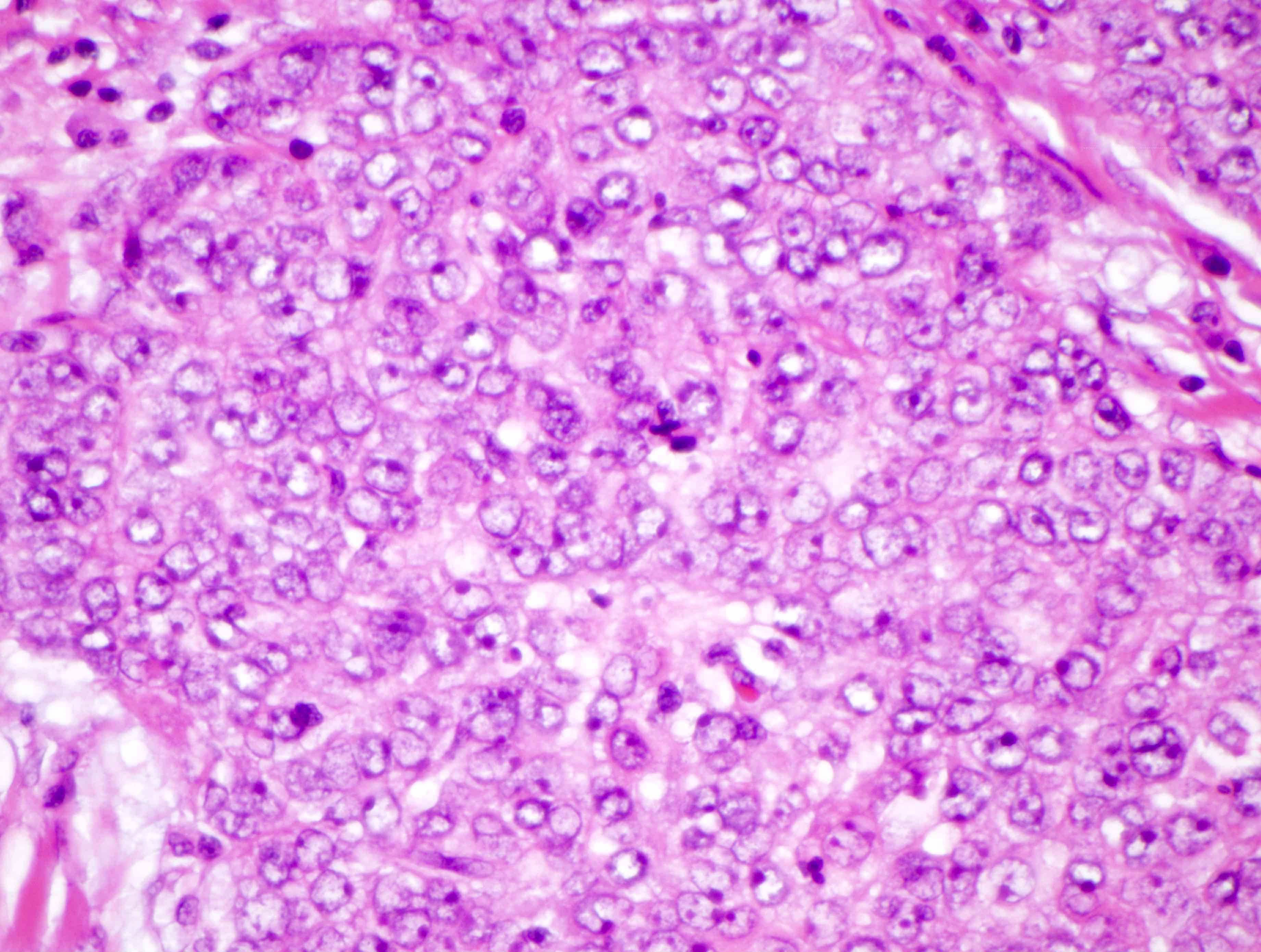
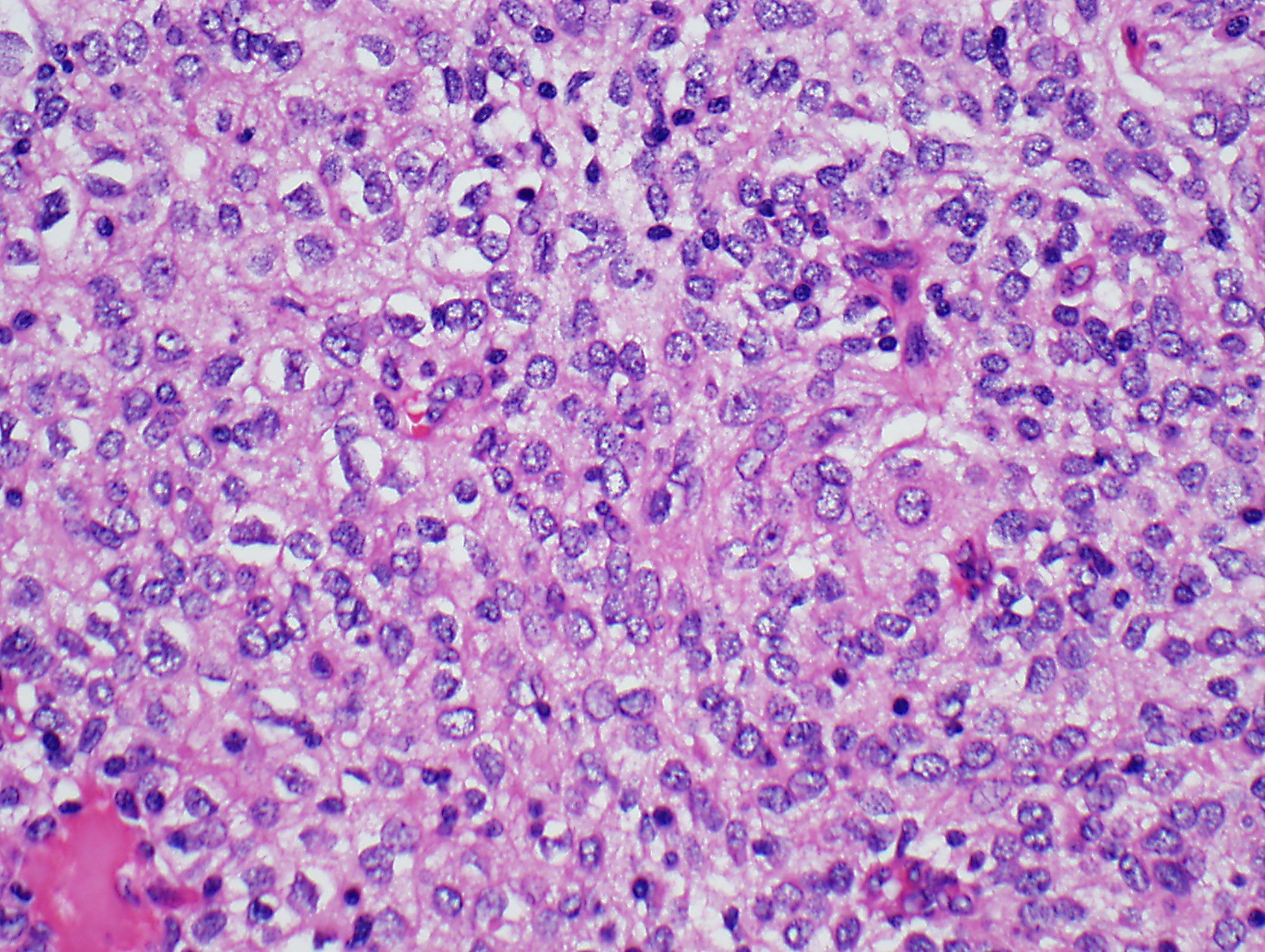
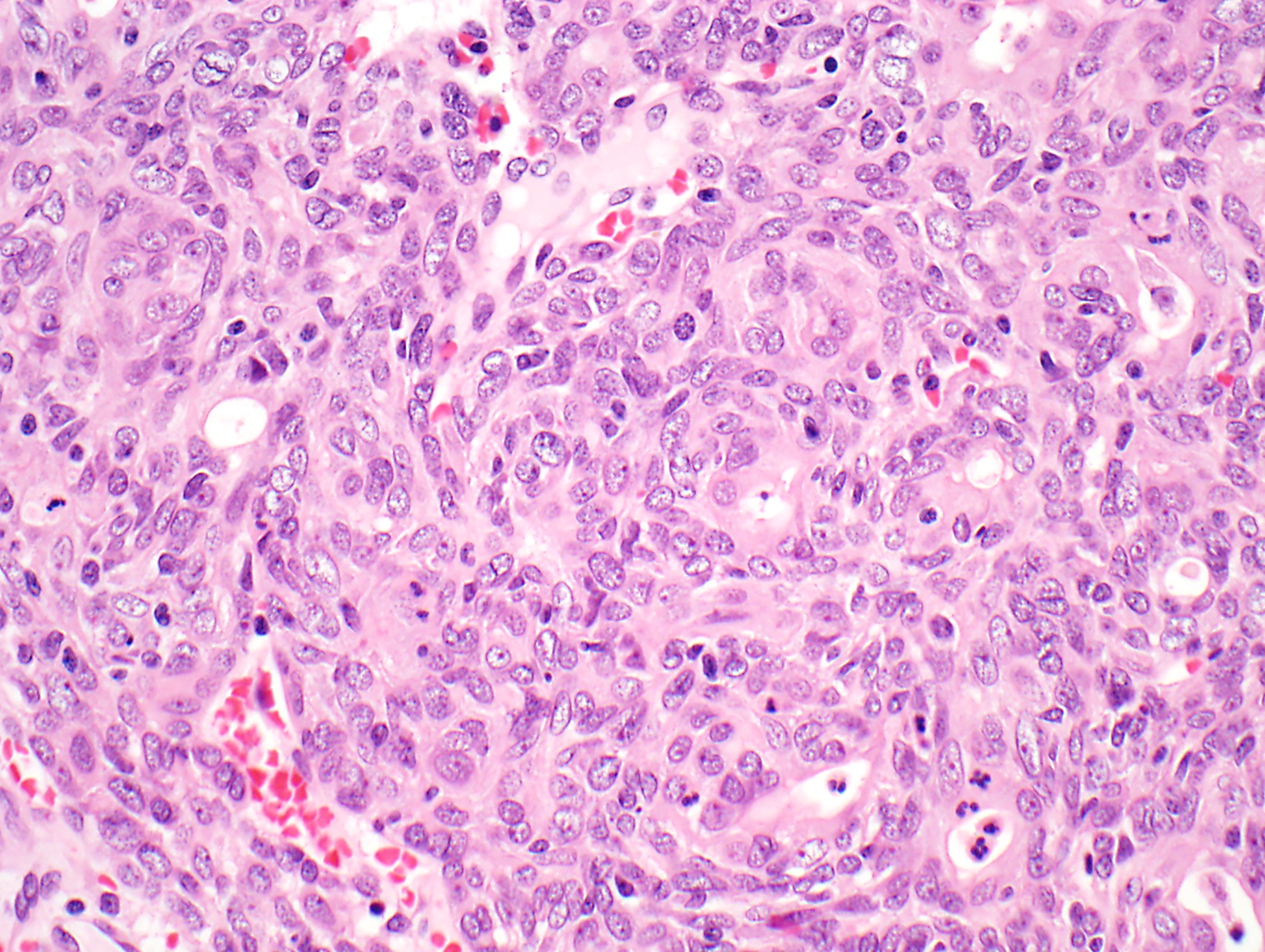
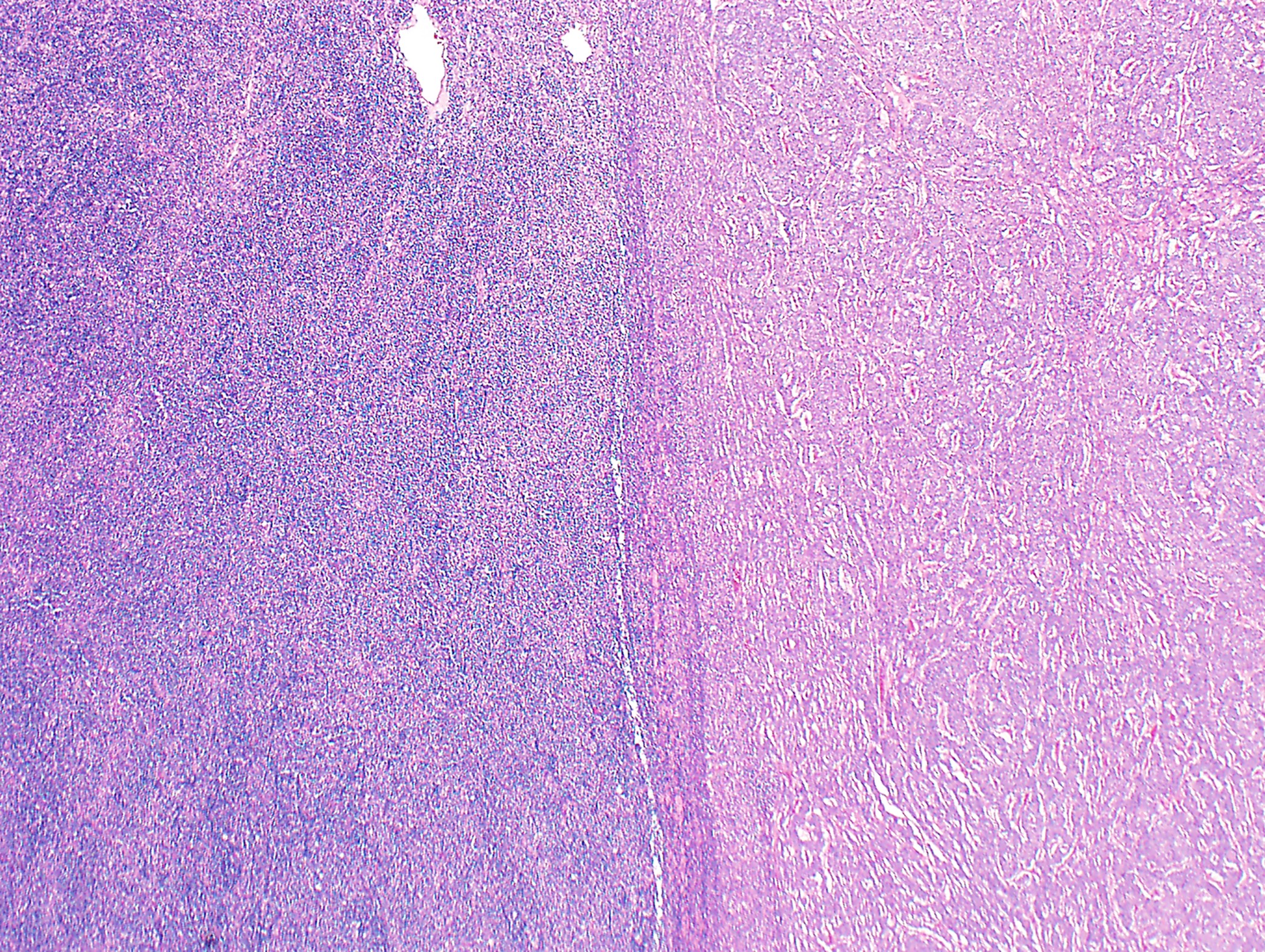
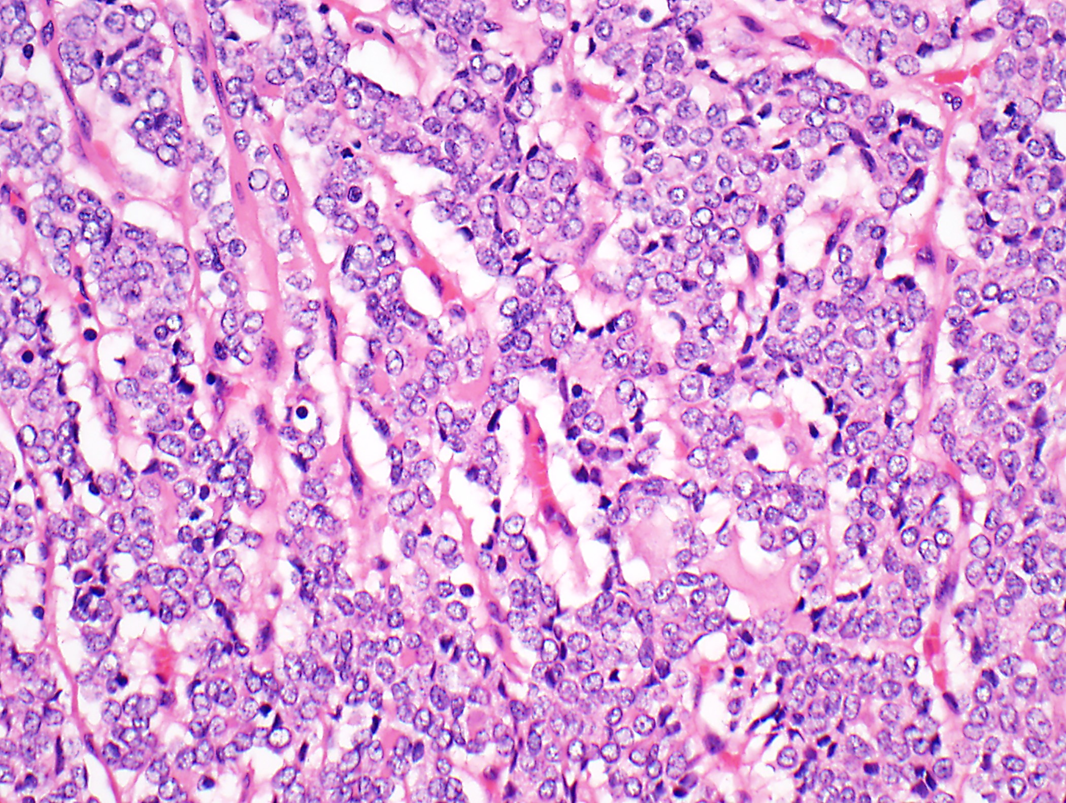
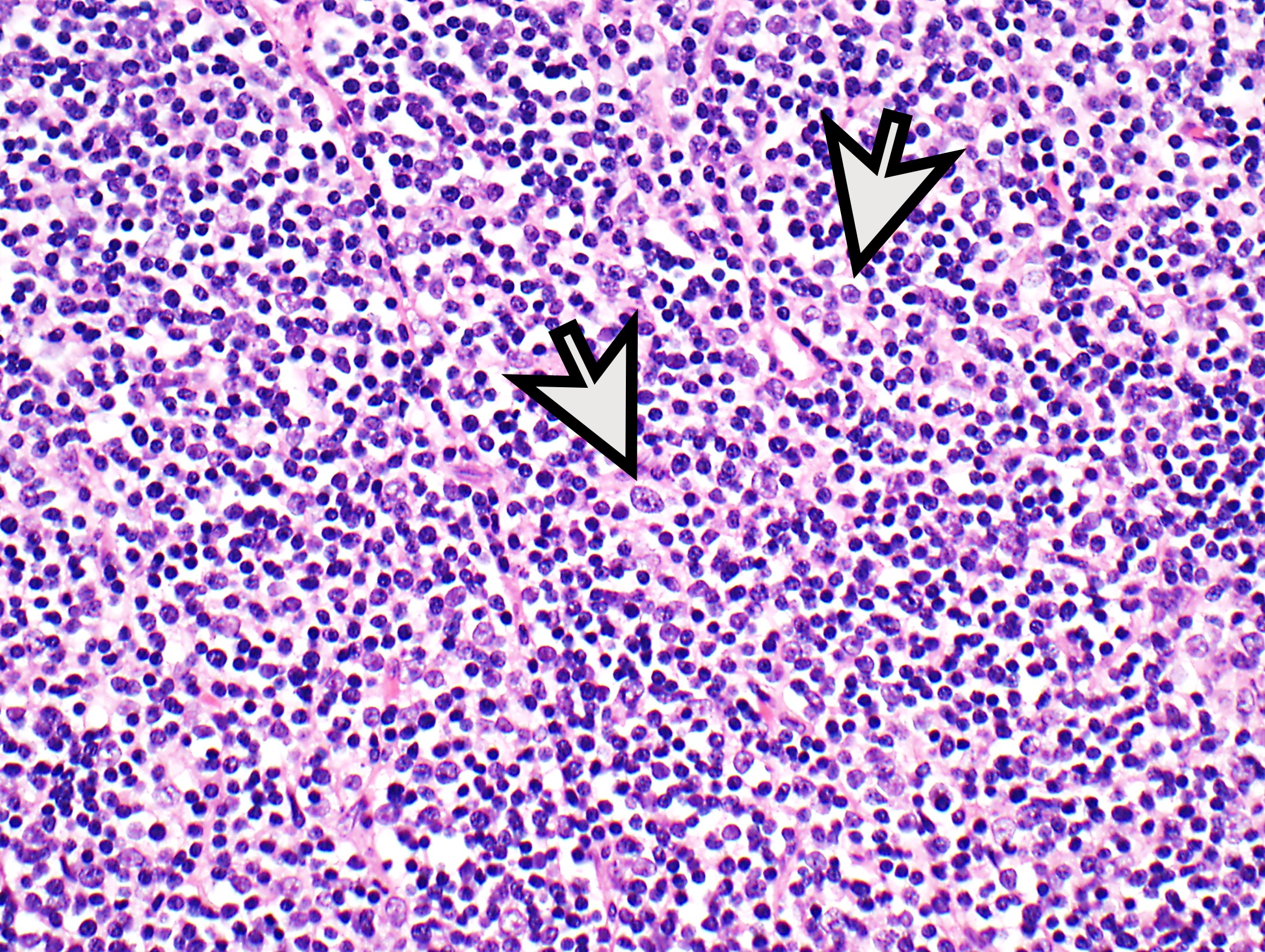
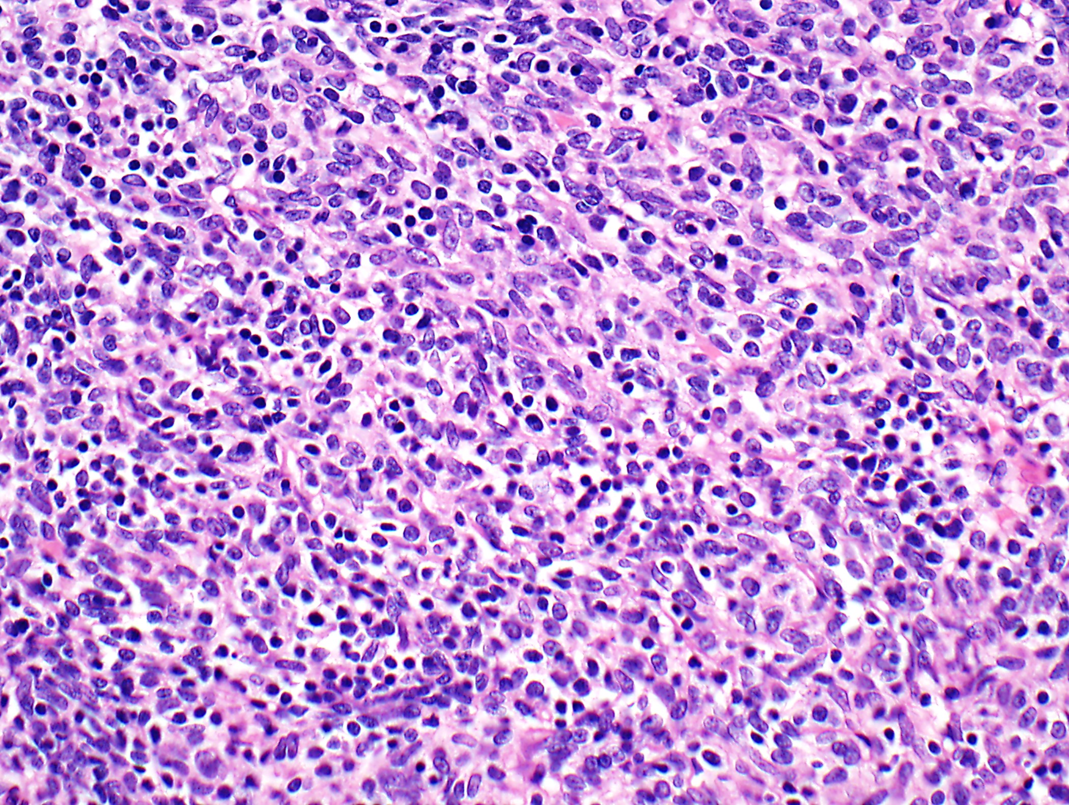
Case #278

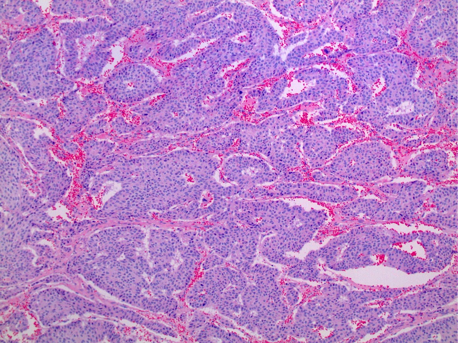
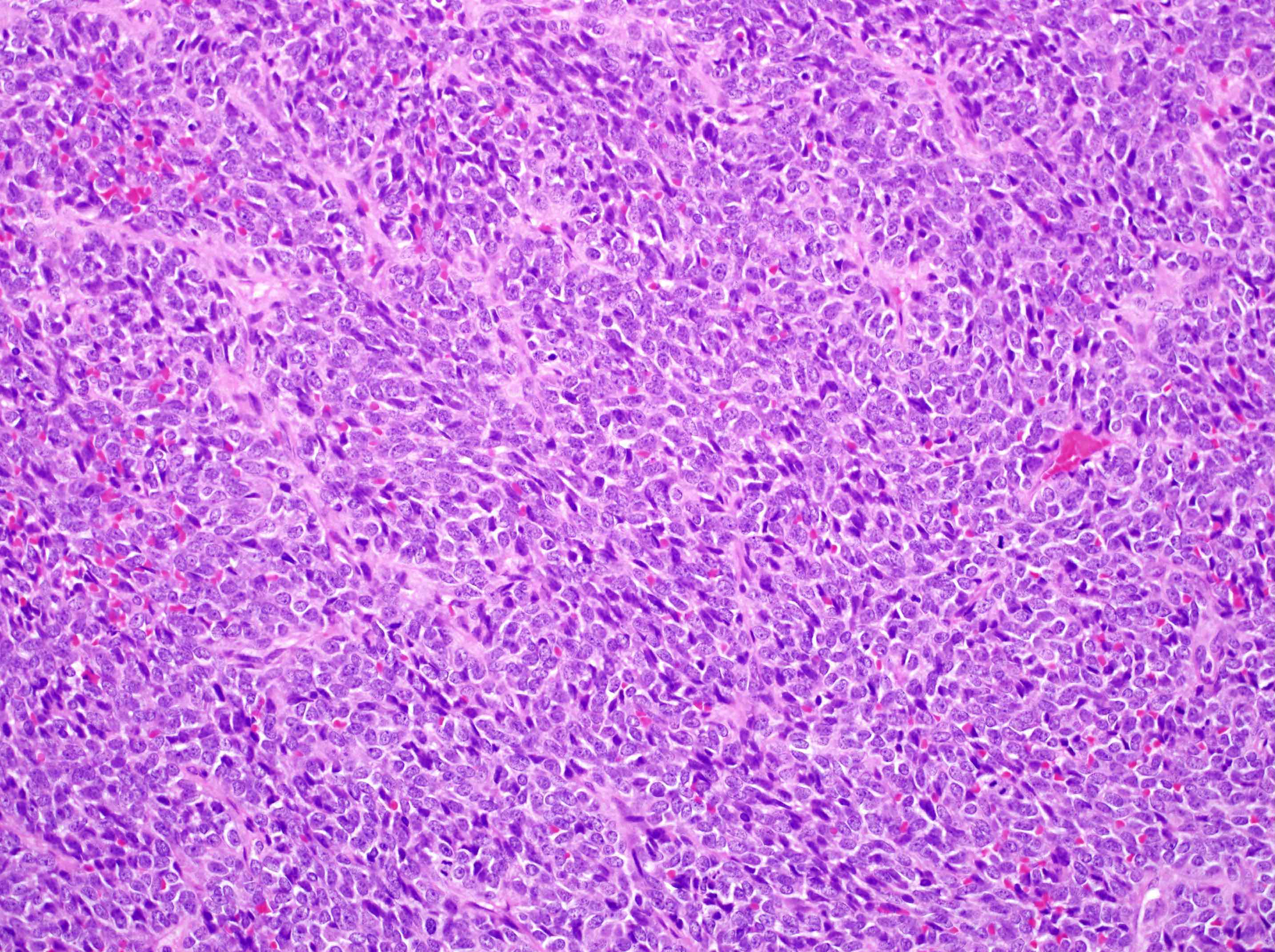
Atypical carcinoid

Atypical carcinoid -
AE1 / AE3
Atypical carcinoid - synaptophysin
Images hosted on other servers:

Figures a - d

Atypical carcinoid

Various stains
Gross, micro, IHC and serum markers of germ cell tumors
| Tumor type | Gross features | Microscopic features | Serum markers | Pankeratin | SALL4 | OCT3/4 | CD30 | Glypican 3 | Beta hCG | CD117 |
| Teratoma | Solid combined with cystic, Rokitansky protuberans contains shiny cartilage, hair and teeth, etc. | Derivatives of all 3 germinal layers | Beta hCG: normal LDH: normal AFP: normal | Positive in epithelial elements | Negative in mature components | Negative in mature components | Negative | Negative | Negative | Negative |
| Embryonal carcinoma | Gray-white with areas of hemorrhage and necrosis | Sheets and aggregates of highly pleomorphic cells | Beta hCG: normal LDH: raised AFP: raised | Positive | Positive | Positive | Positive | Negative | Negative | Negative |
| Yolk sac tumor | Gray-white with variable hemorrhage | Numerous growth patterns, Schiller-Duval bodies, eosinophlic globules | Beta hCG: normal LDH: normal AFP: raised | Positive | Positive | Negative | Negative | Positive | Positive in syncytio-trophoblasts | Negative |
| Chorio-carcinoma | Markedly hemorrhagic | Trimorphic population of trophoblasts with extensive hemorrhage | Beta hCG: raised LDH: normal AFP: normal | Positive | Negative | Negative | Negative | Negative | Positive in syncytio-trophoblasts | Negative |
| Seminoma | Gray-white, shiny cut surface | Islands of pleomorphic cells with prominent nucleoli, separated by thin fibrous septa having lymphocytes | Beta hCG: raised LDH: raised AFP: normal | Negative | Positive | Positive | Negative | Negative | Negative | Positive |
Contributed by David Suster, M.D.
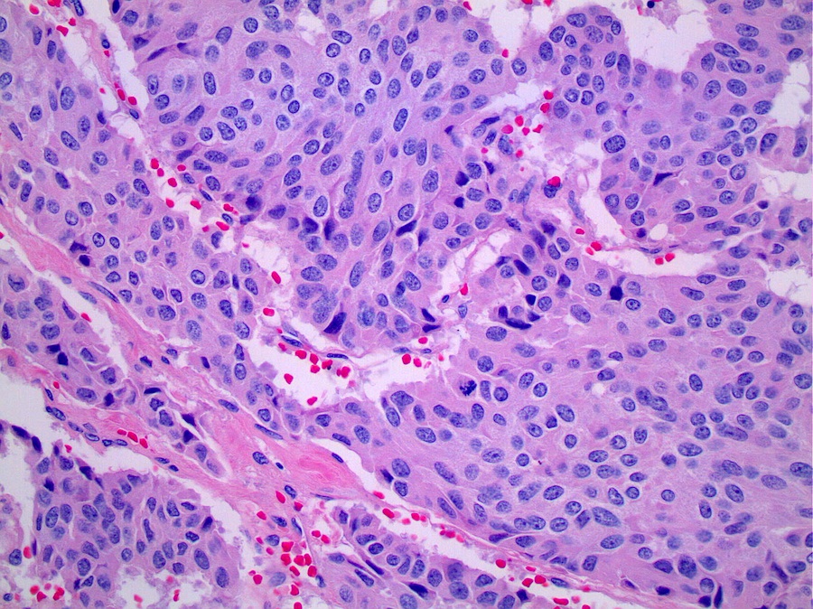
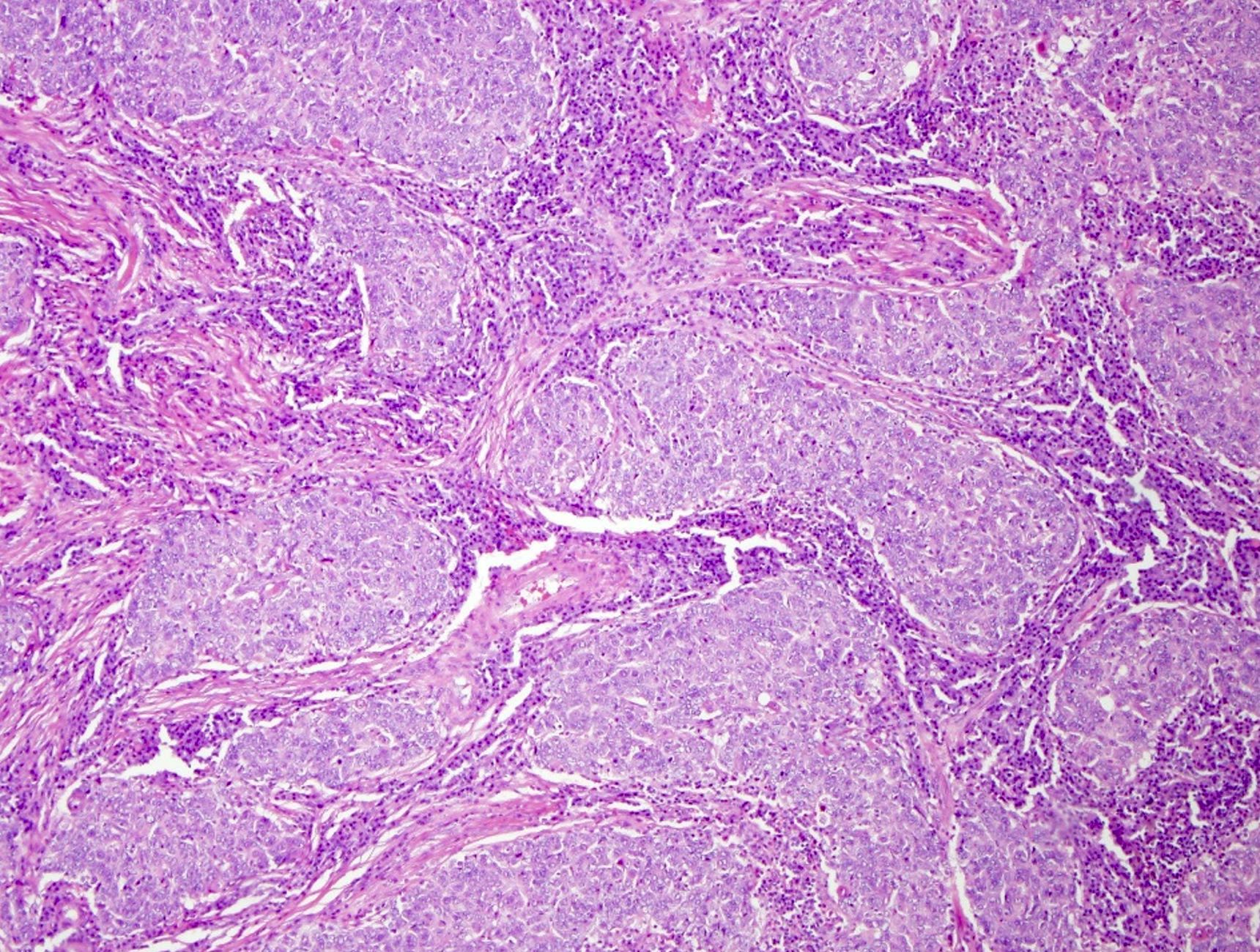
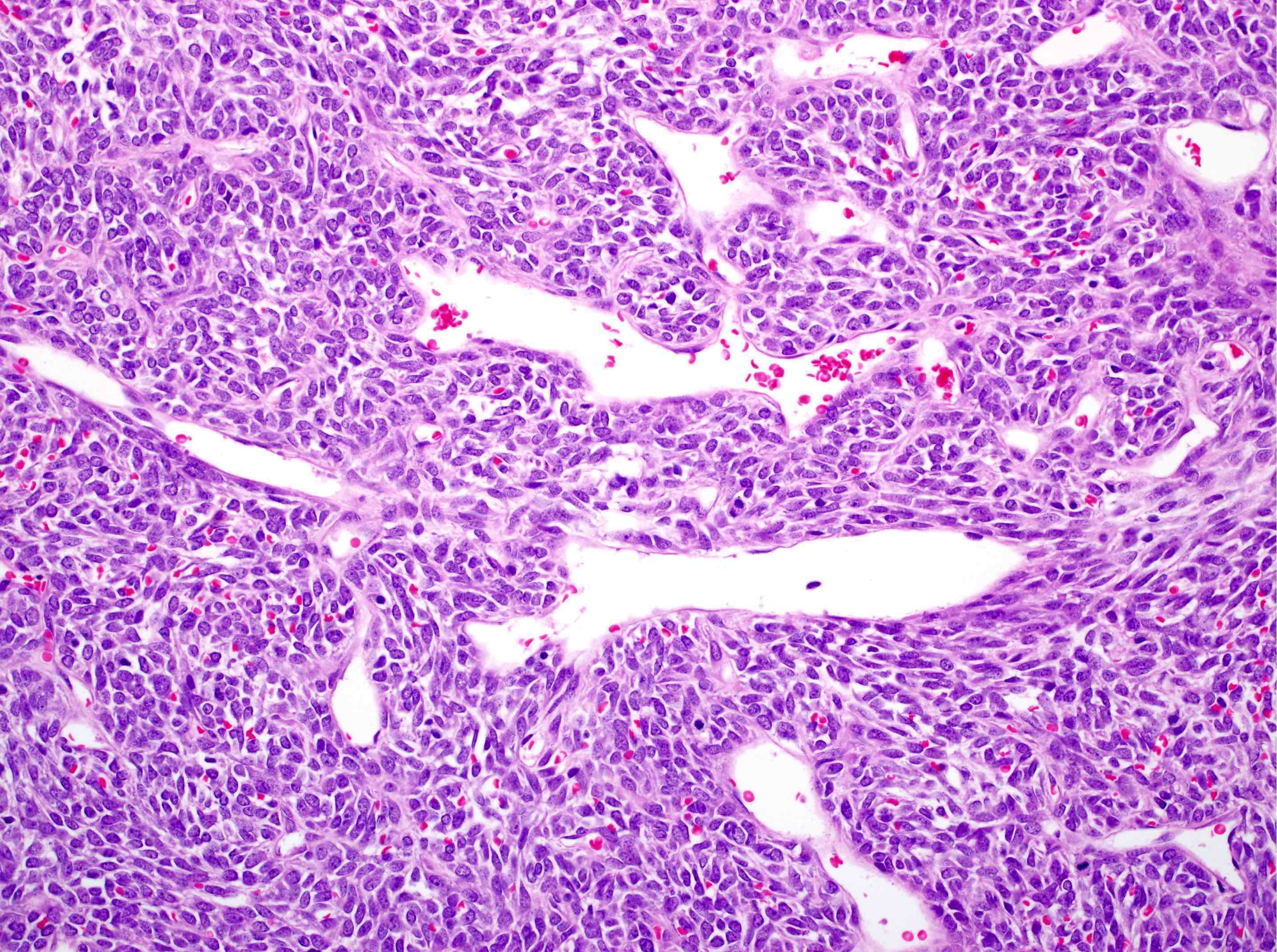
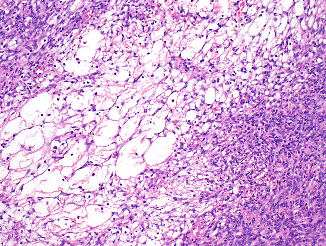
Sheets of large round to oval tumor cells
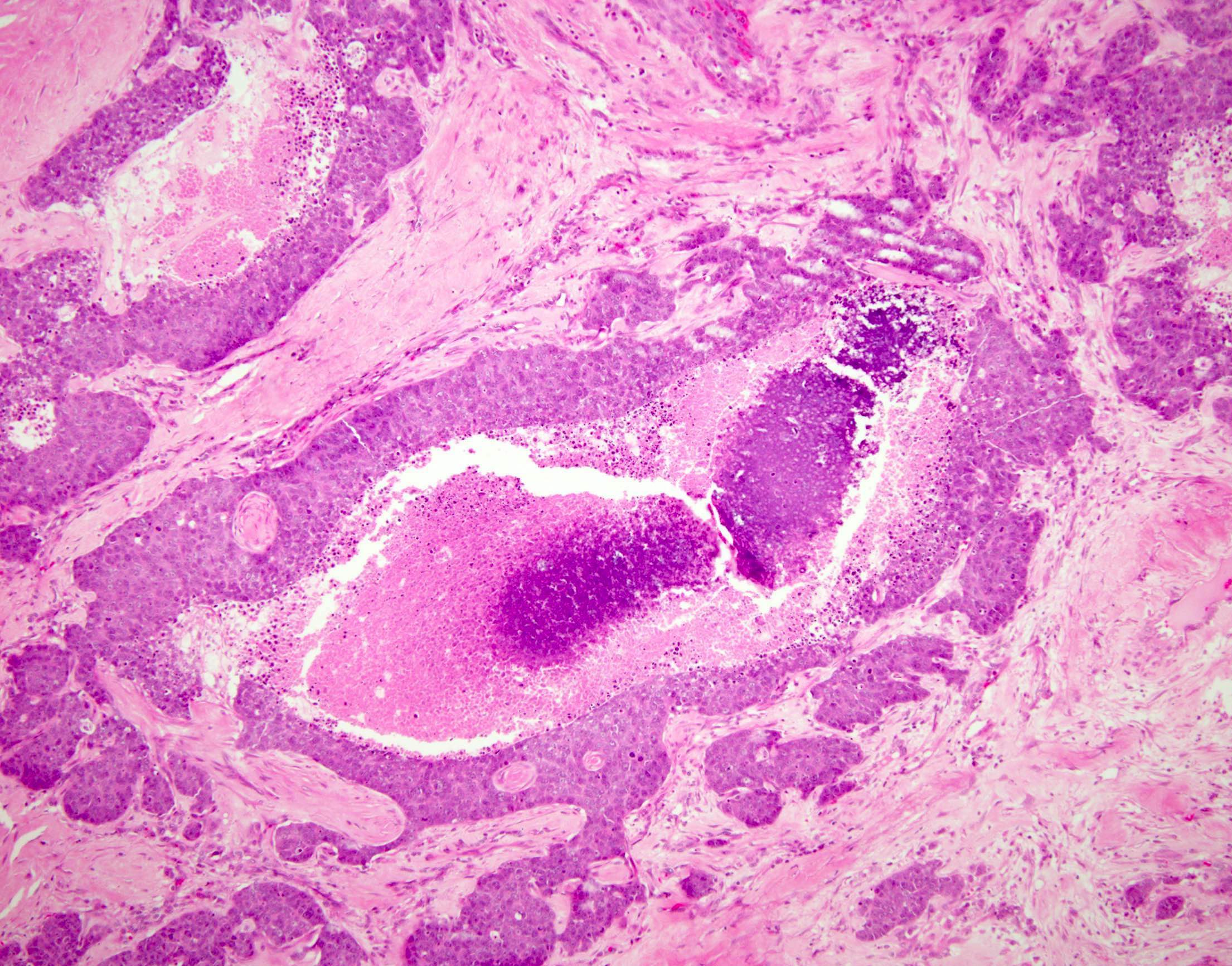
Anastomosing cords, islands, comedonecrosis
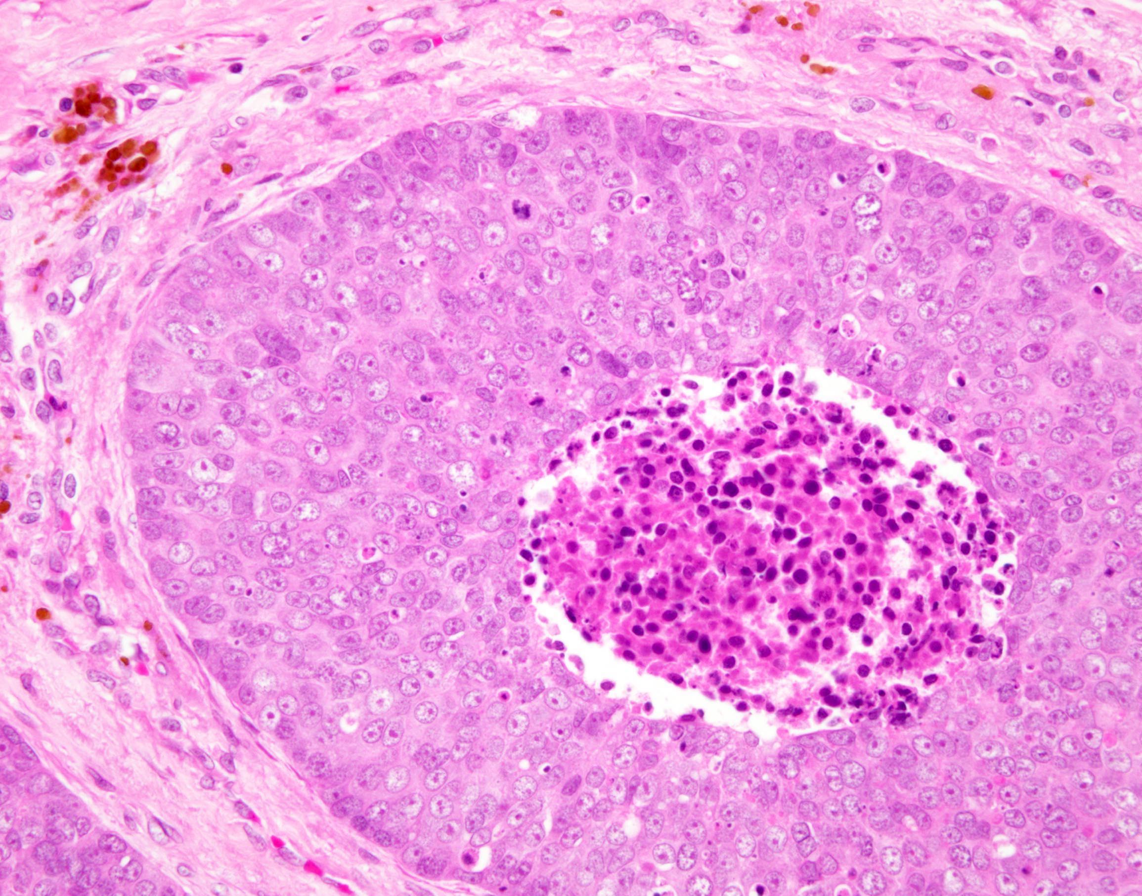
Comedonecrosis
Lymphoepithelioma-like pattern

Desmoplastic pattern
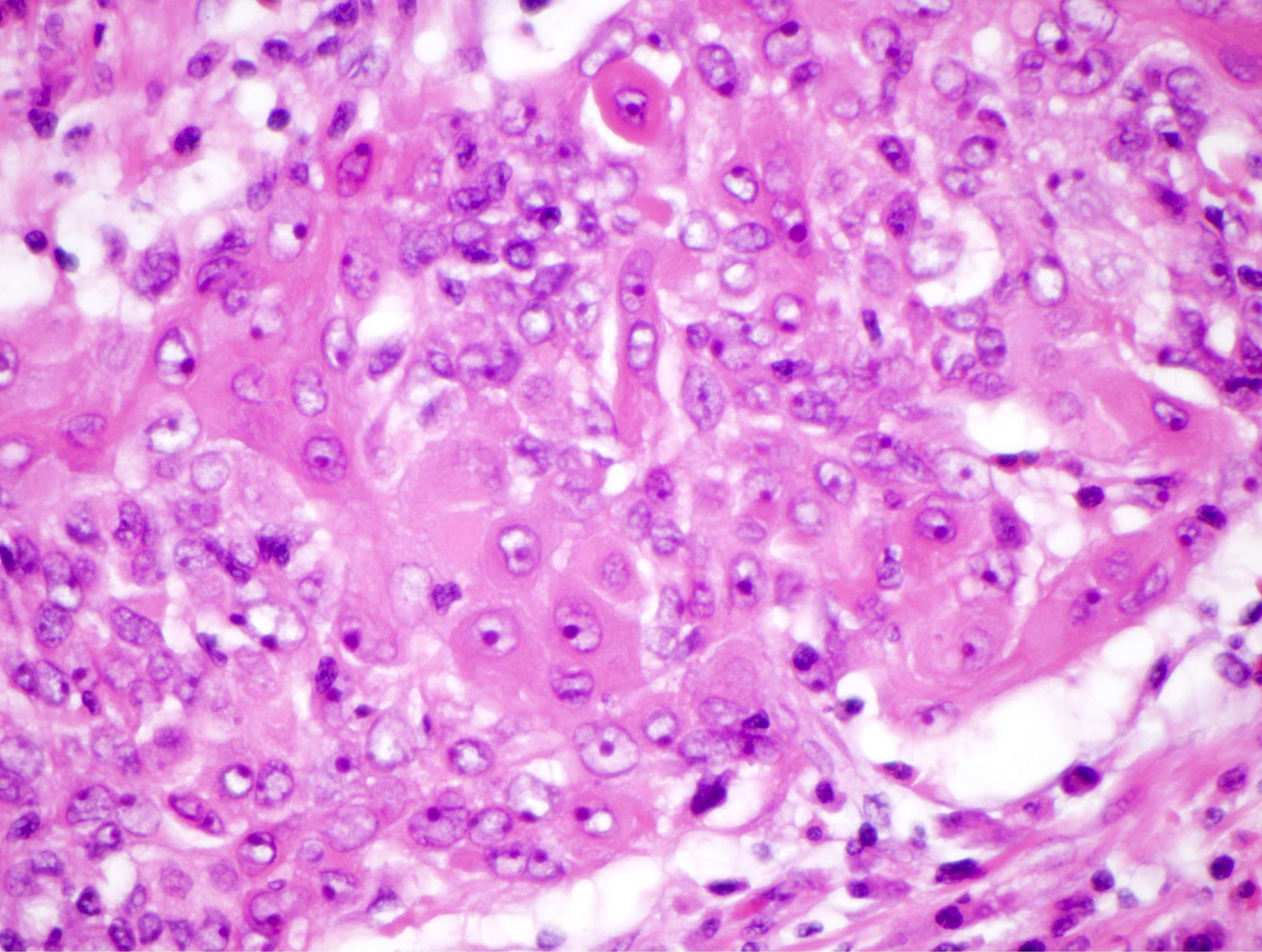
Focal abrupt keratinization
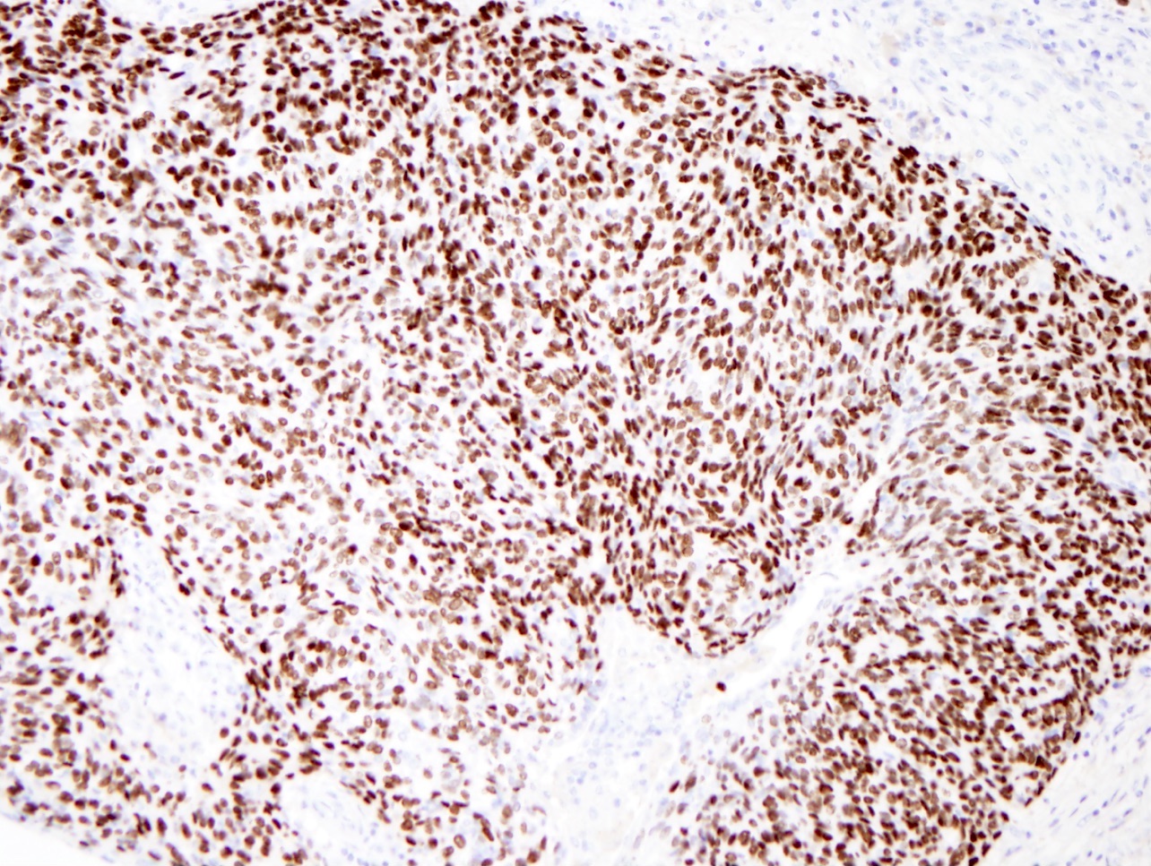
p63
Contributed by David Suster, M.D.
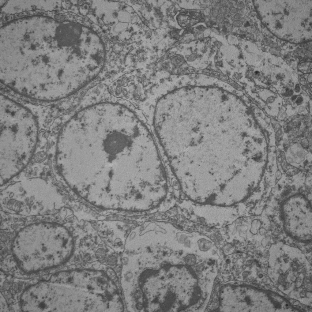
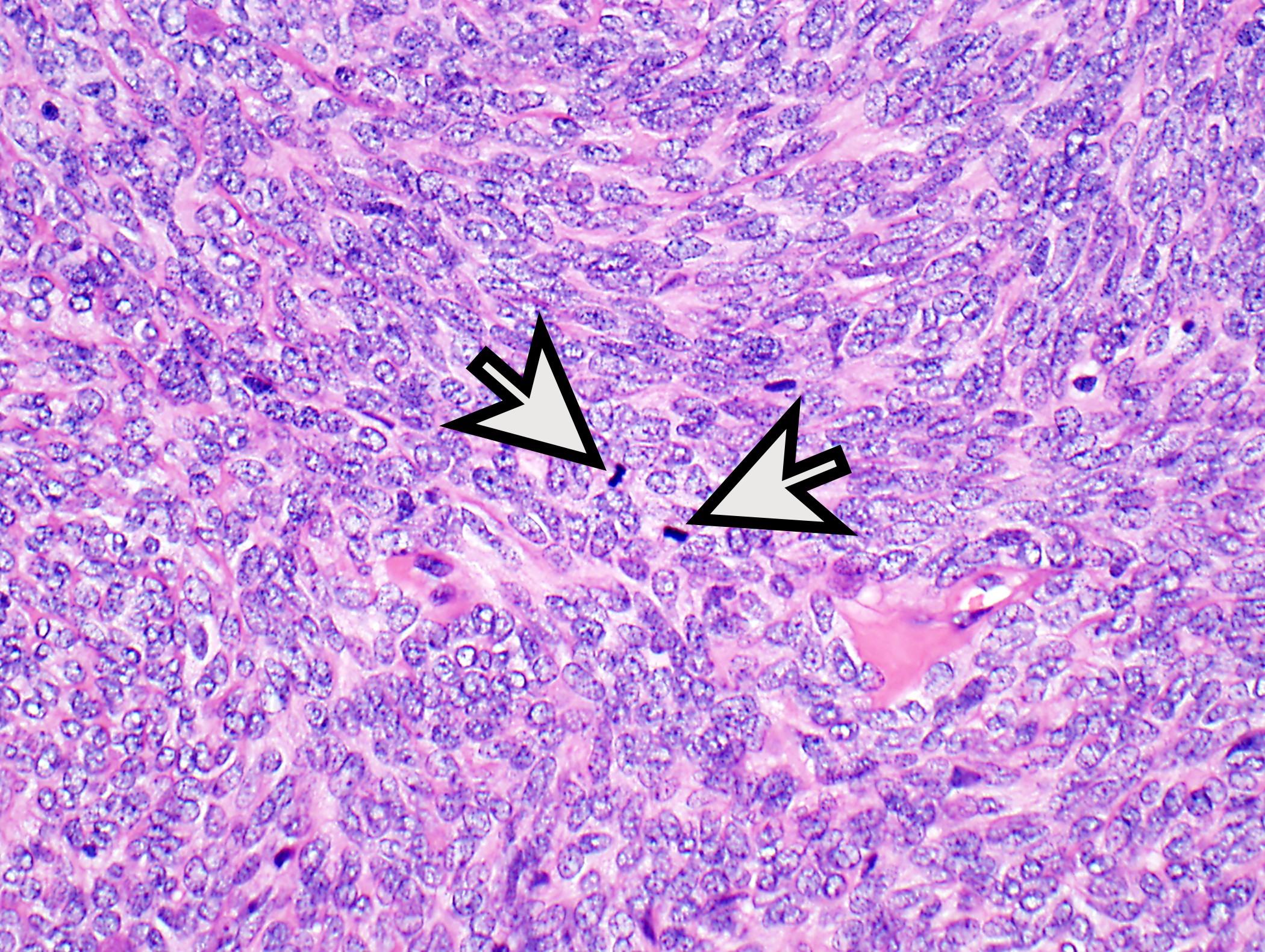
Large, round tumor cell

2 desmosomal attachments


Images hosted on other servers:

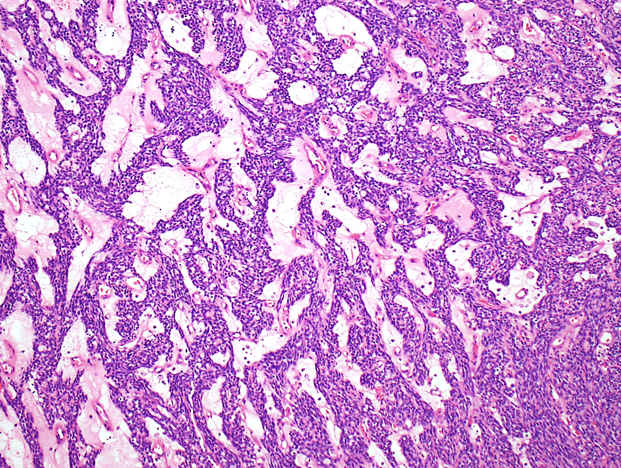
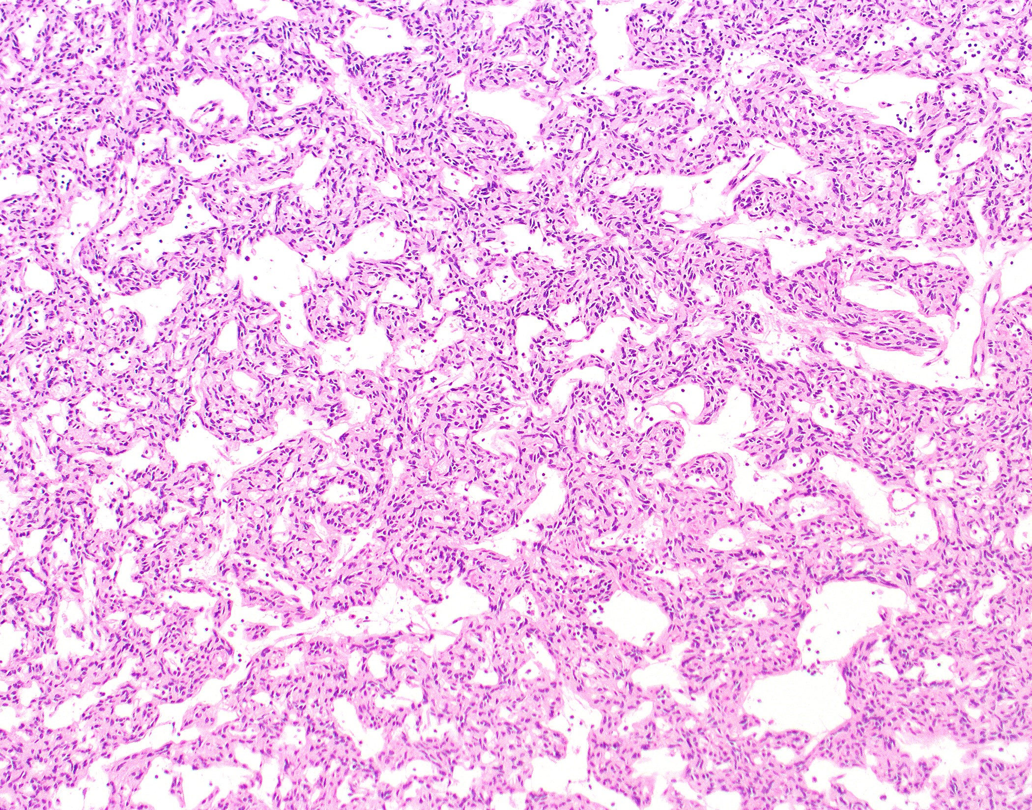
Nests, ribbons and festoons

Various stains
Contributed by Jennifer M. Boland, M.D.
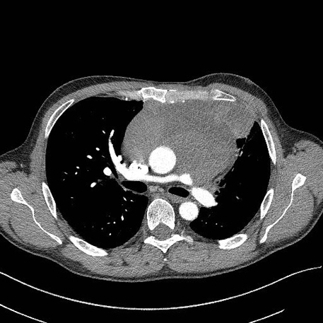
CT scan
Contributed by Jennifer M. Boland, M.D.
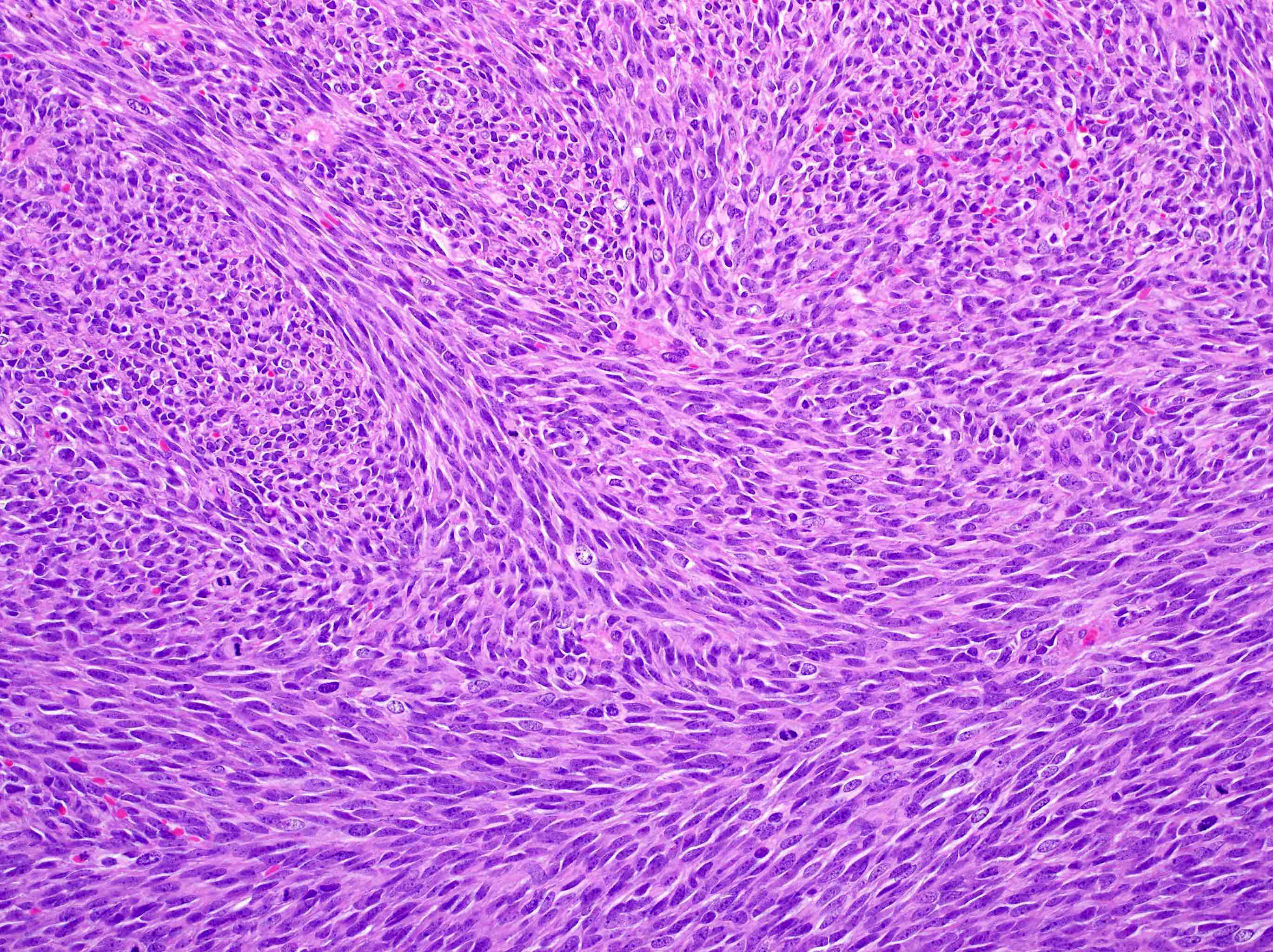
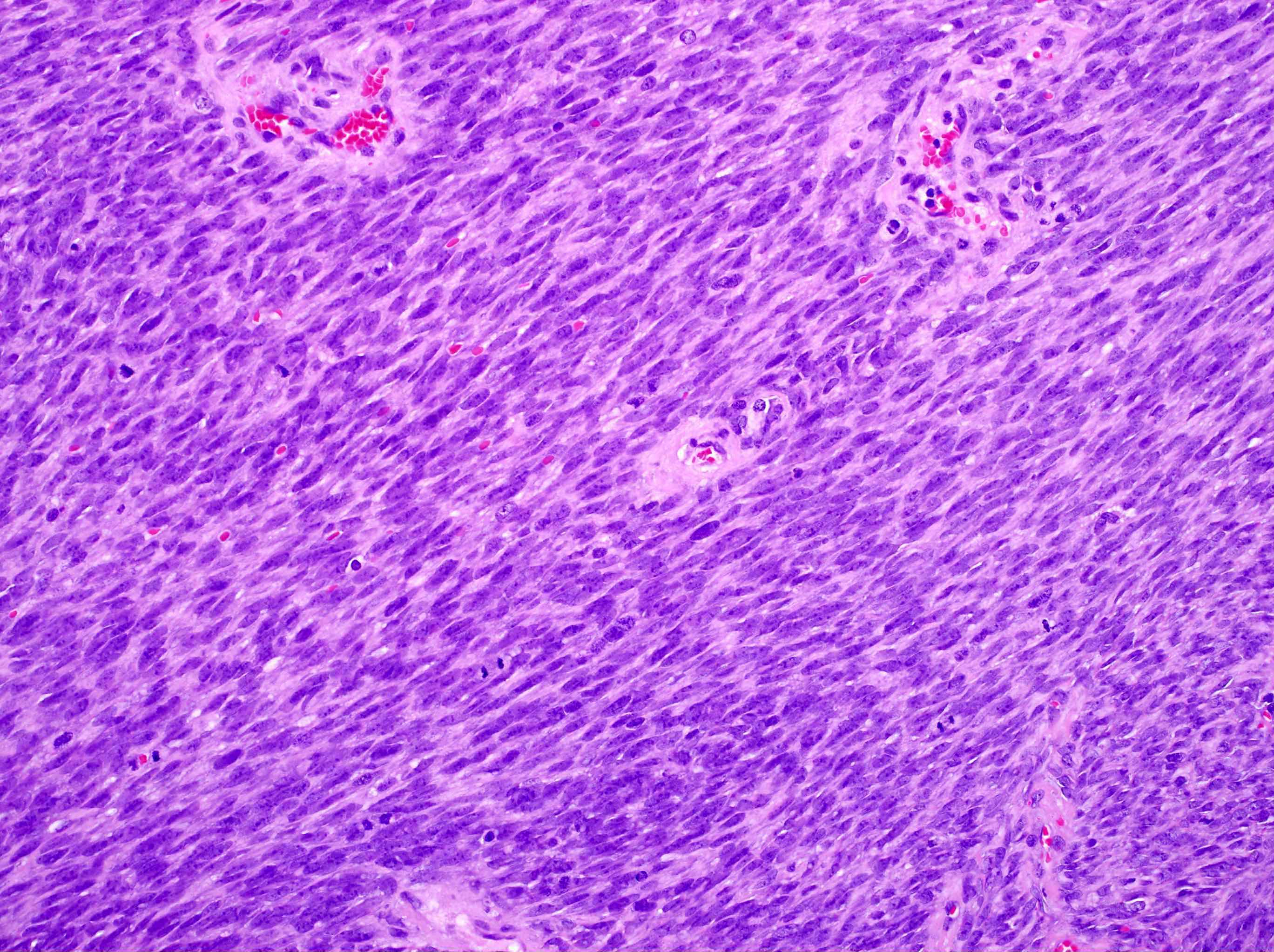
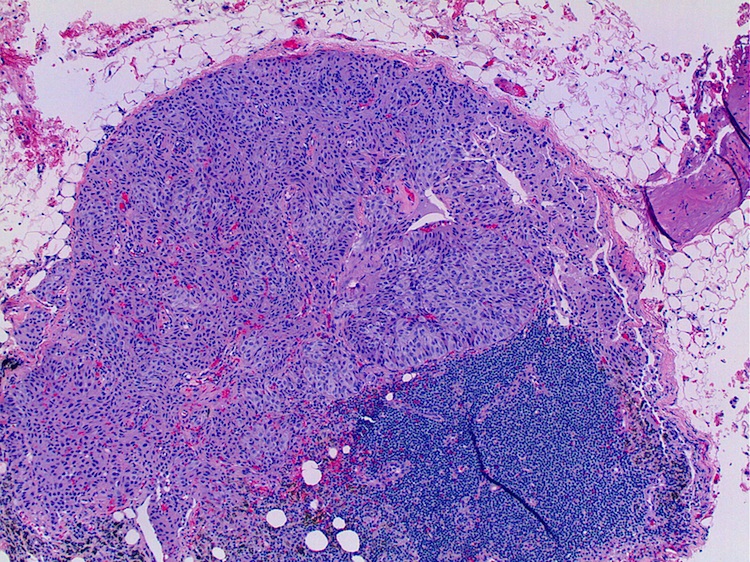
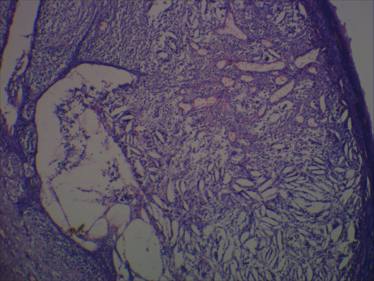
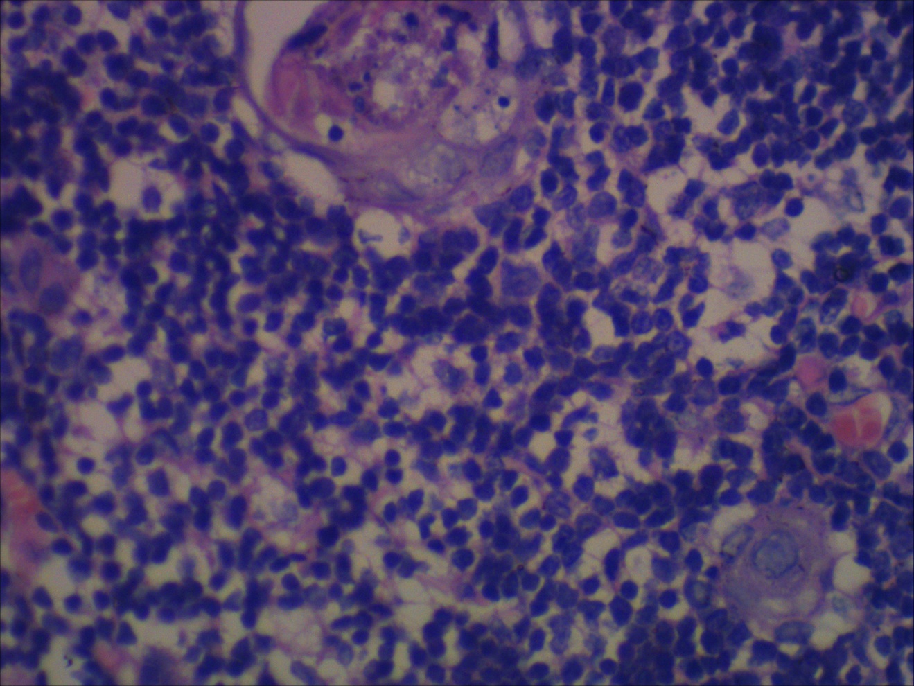
Monophasic synovial sarcoma
EMA

TLE1
Biphasic synovial sarcoma


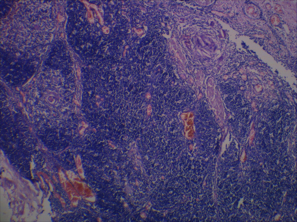
Poorly differentiated synovial sarcoma
Contributed by Jennifer M. Boland, M.D.
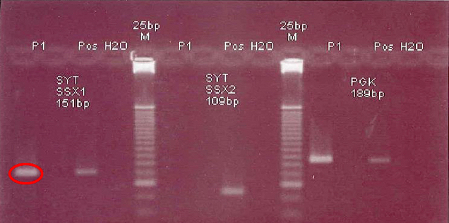
SS18(SYT)-SSX1 fusion
Images hosted on other servers:

Mature cystic teratoma

Mature teratoma


Case #333


Various images
Images hosted on other servers:

Bland squamoid epithelium

Papillary outpouchings

Cholesterol clefts
Images hosted on other servers:

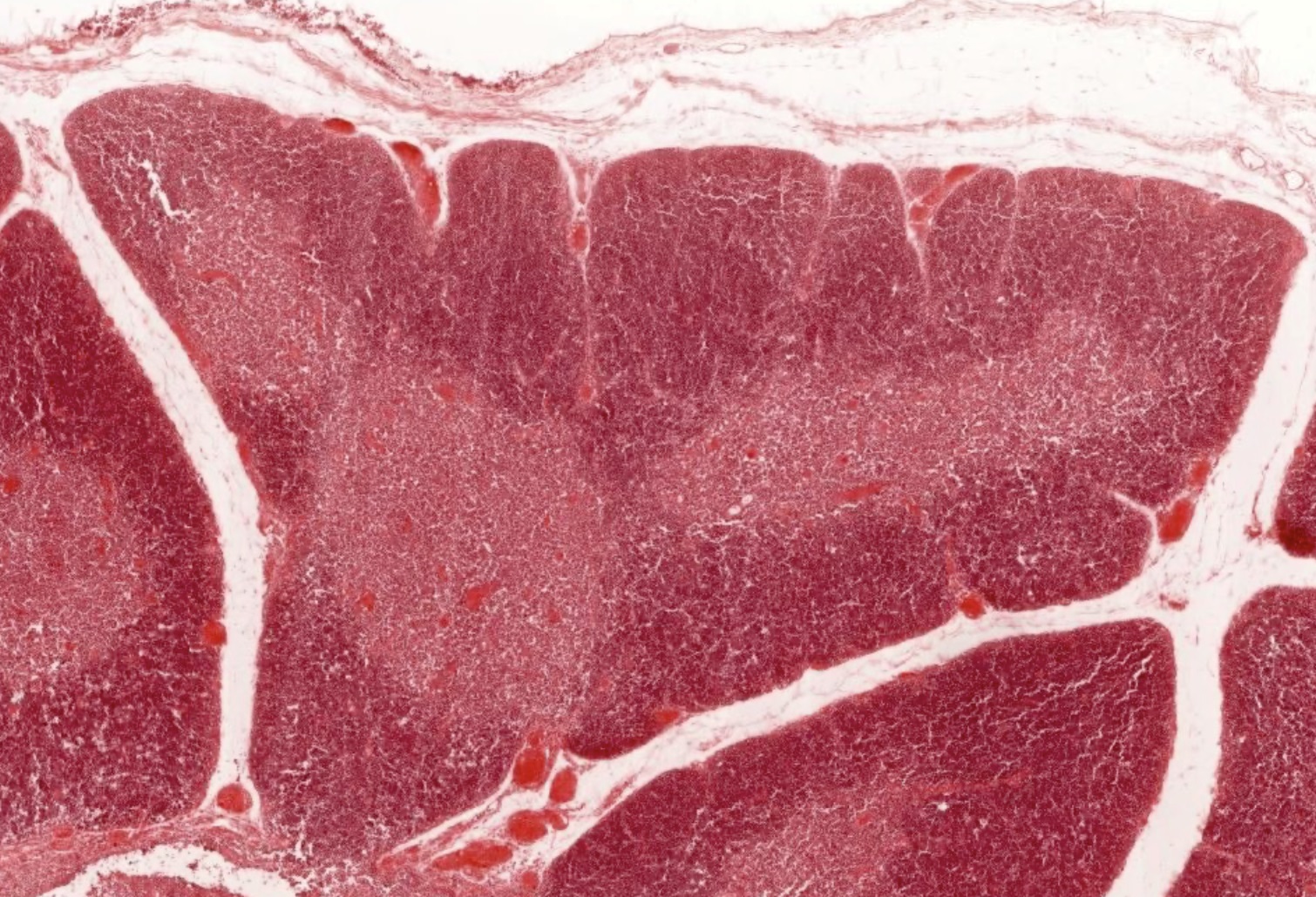
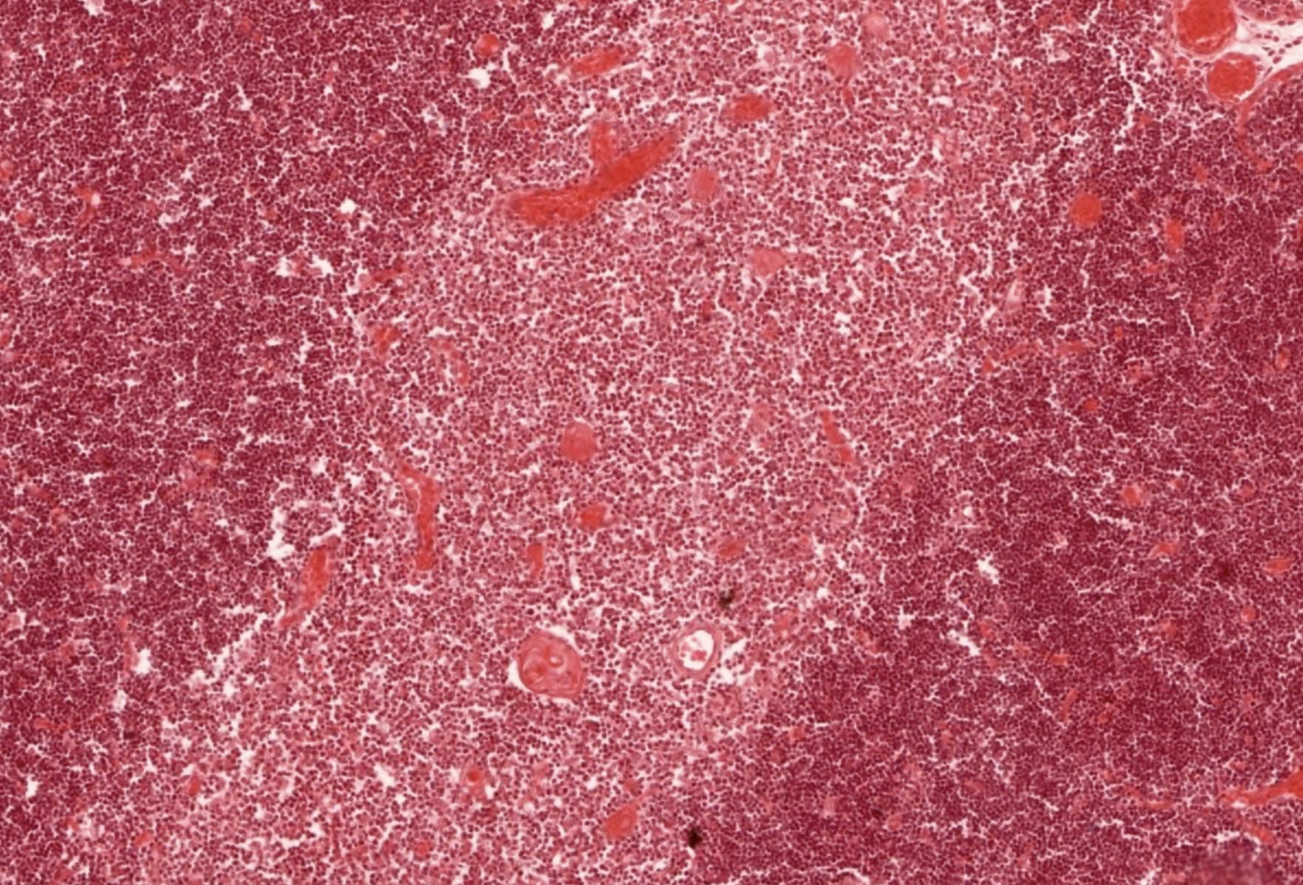
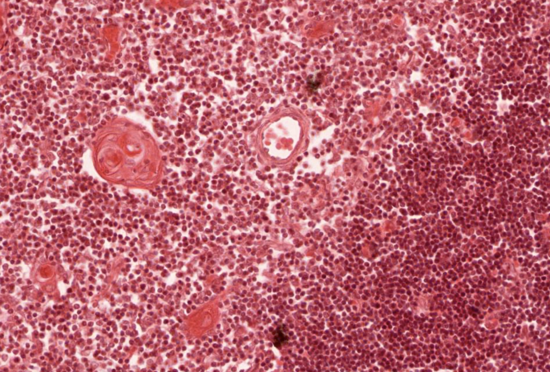
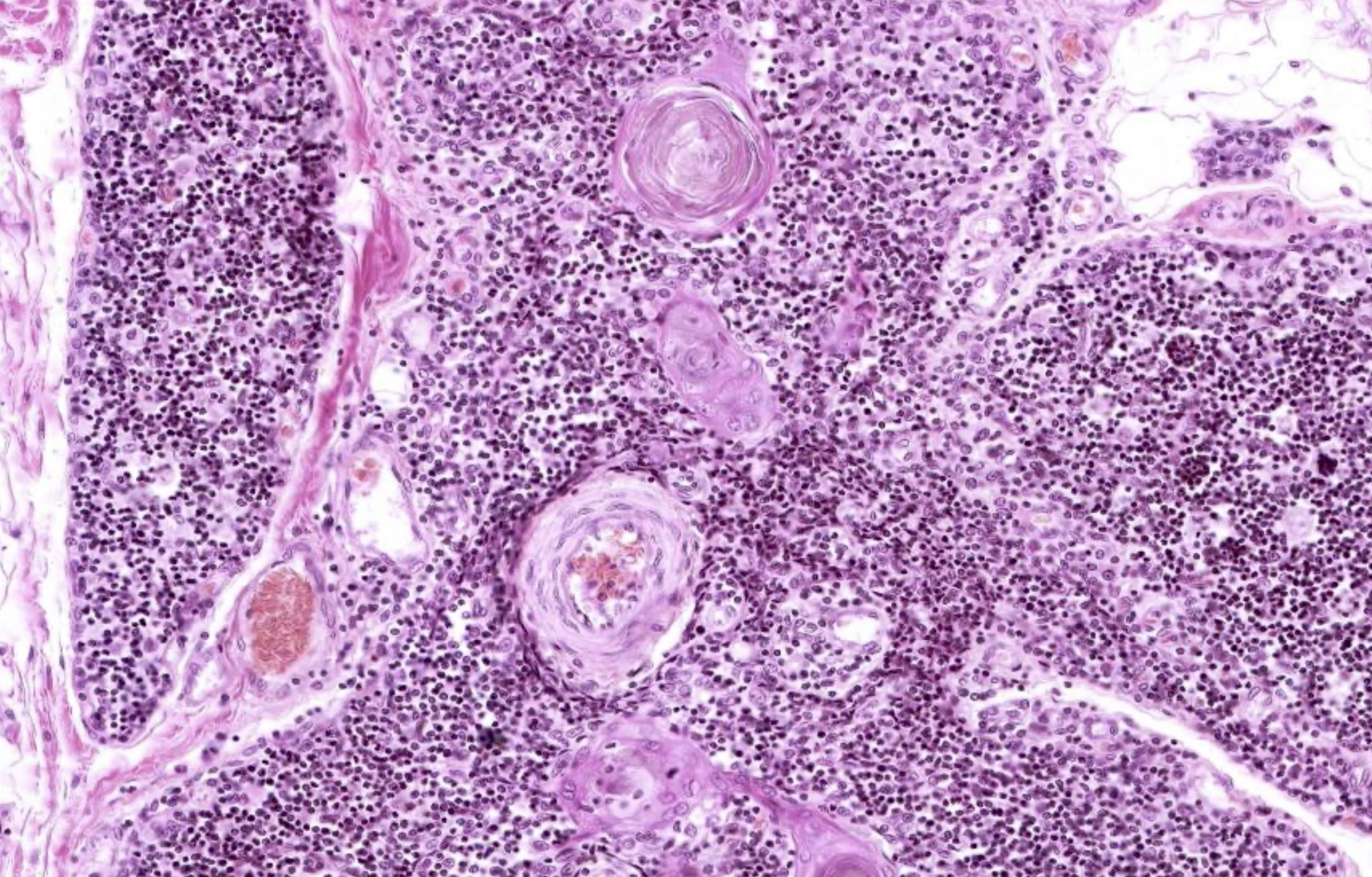
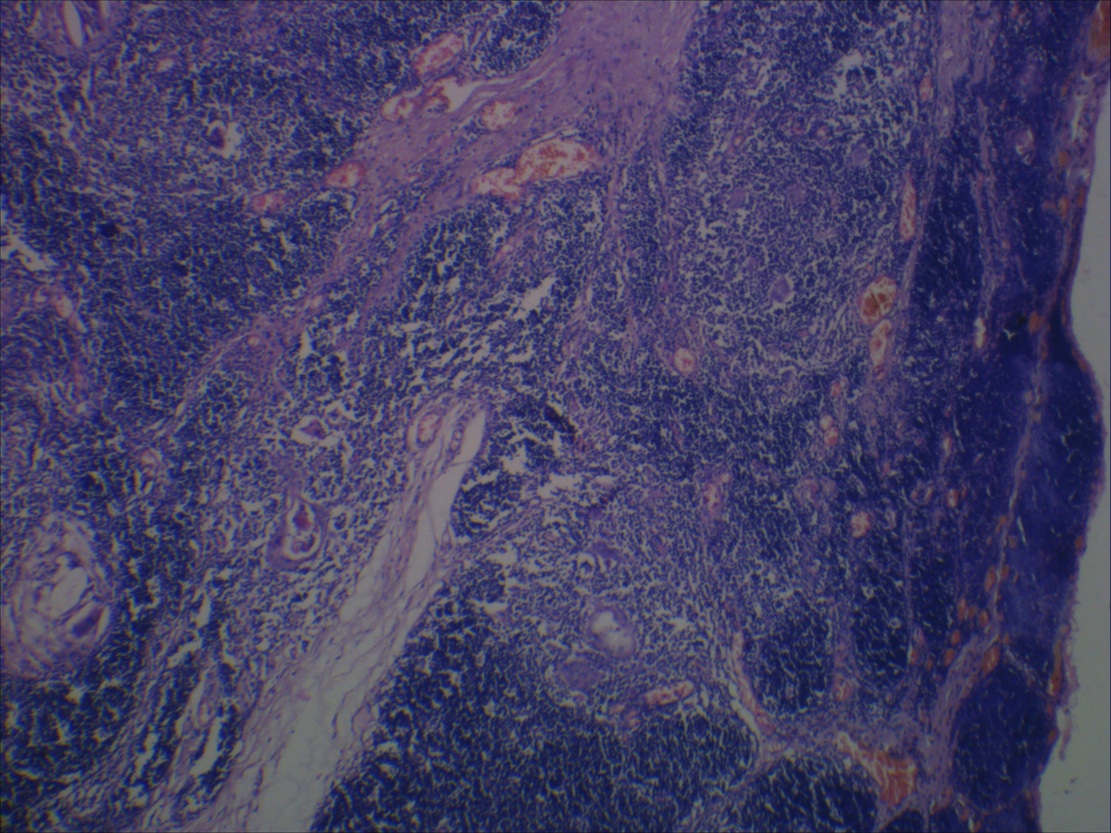
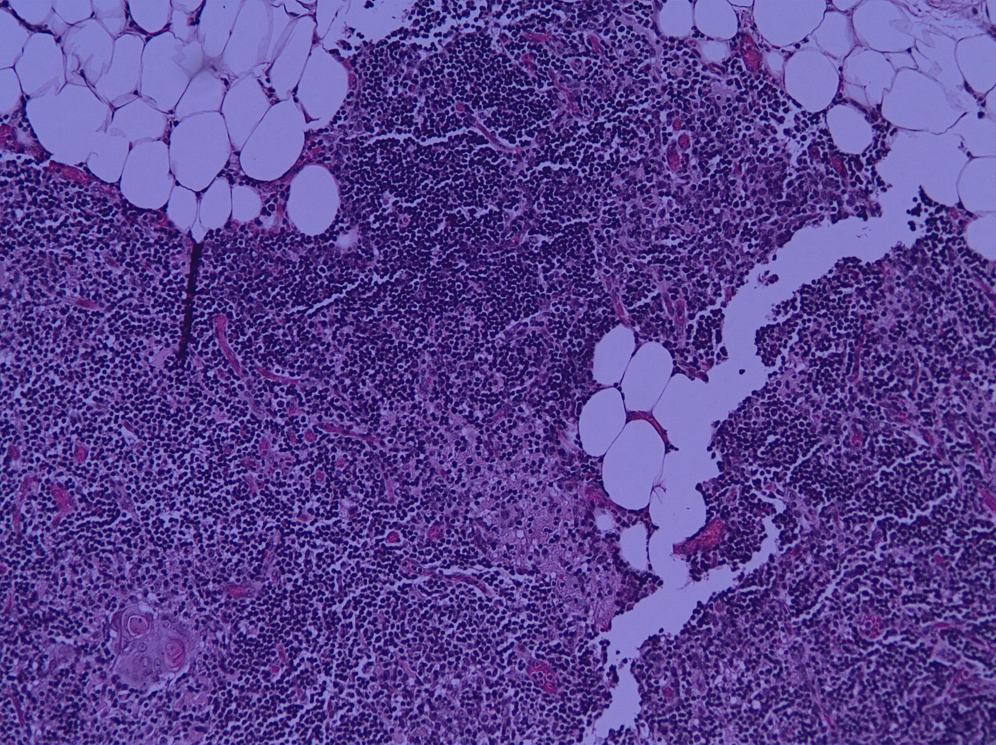
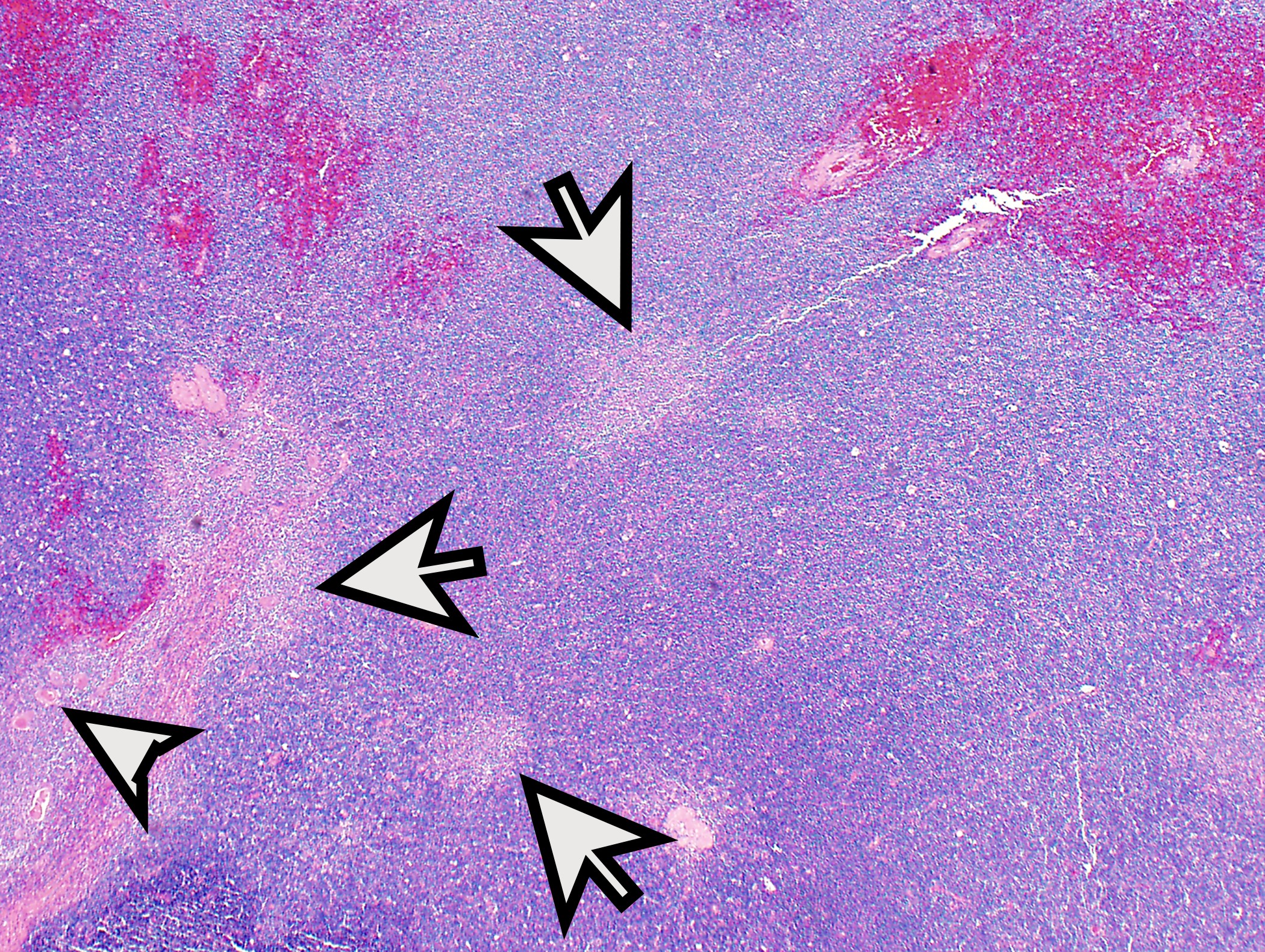
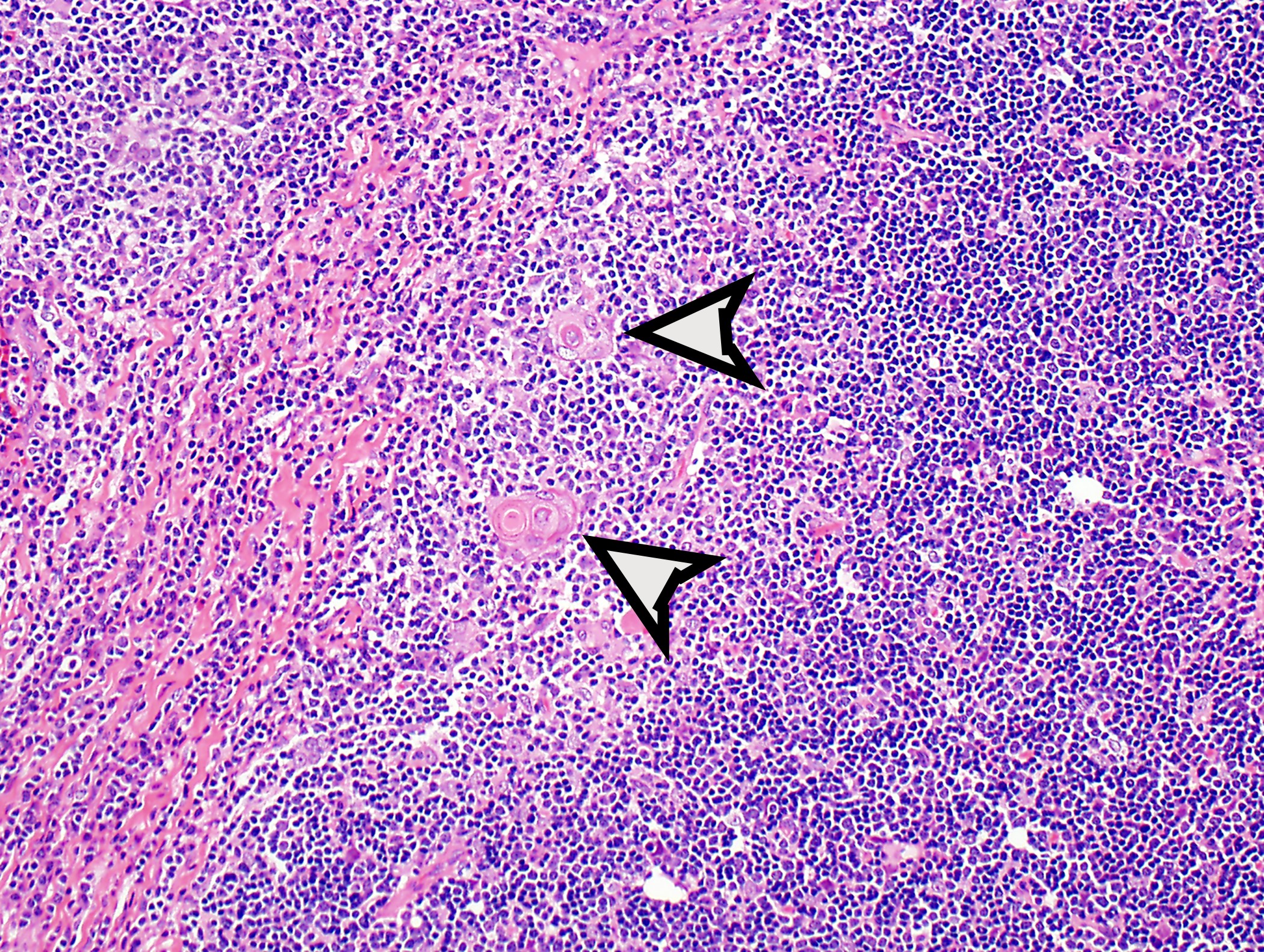
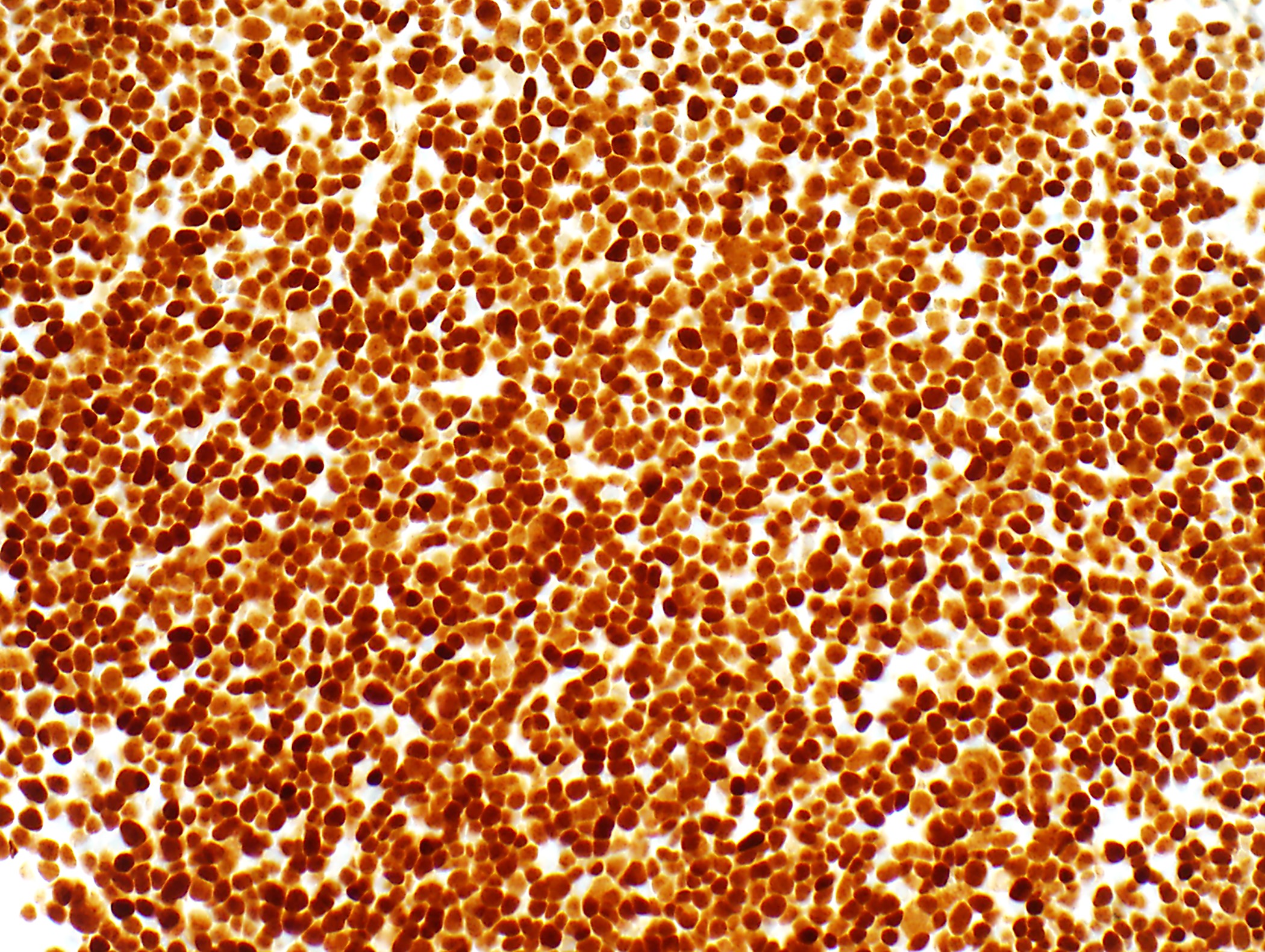
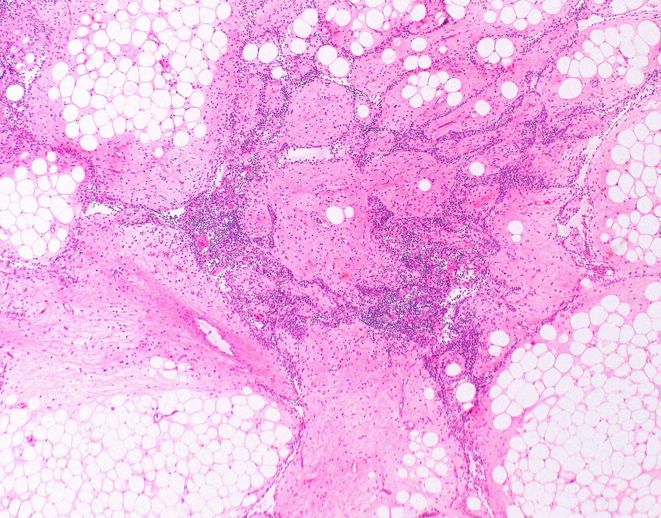
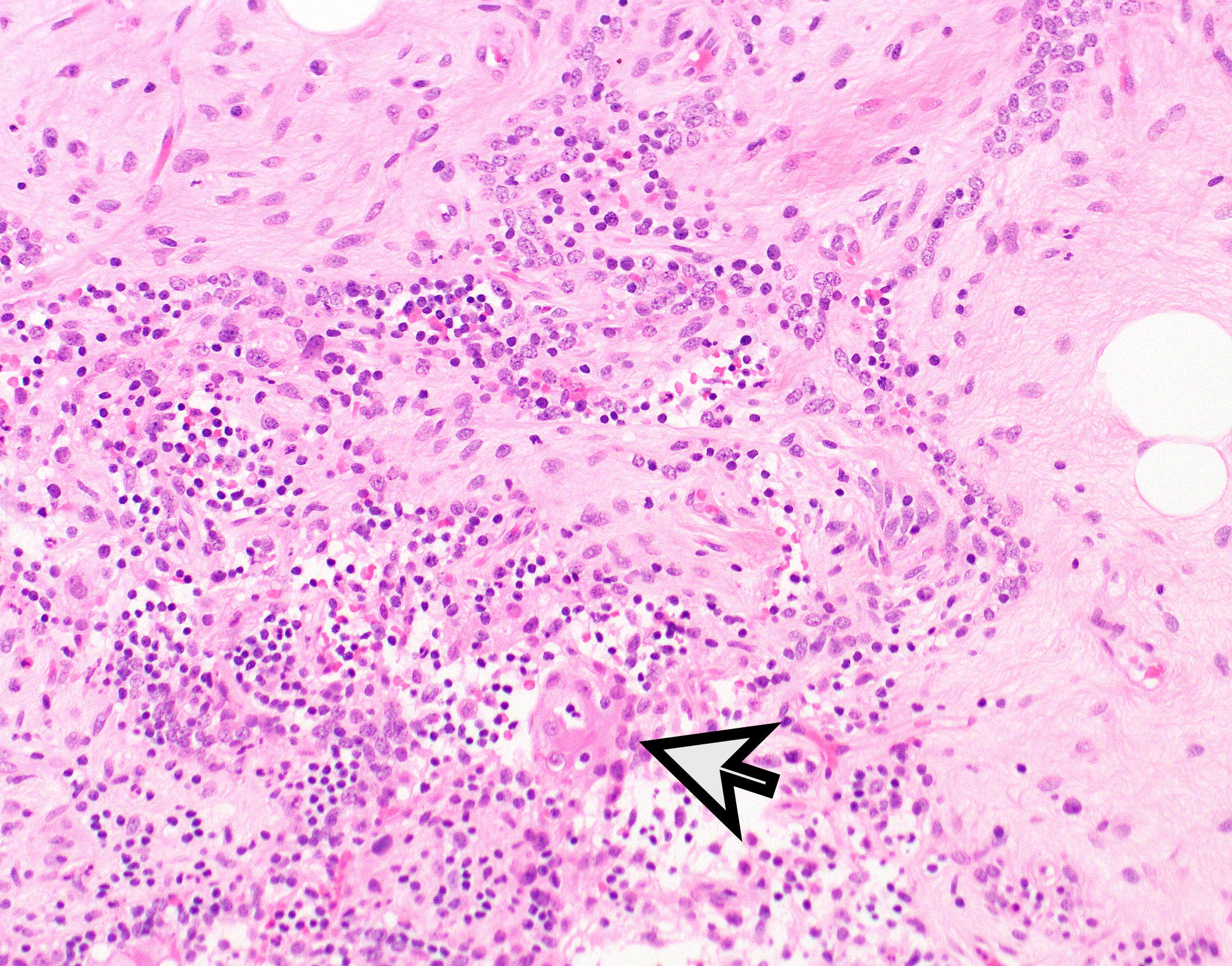
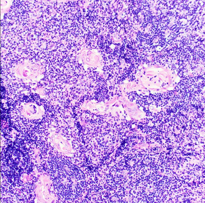
Incidental thymic follicular hyperplasia
Images hosted on other servers:

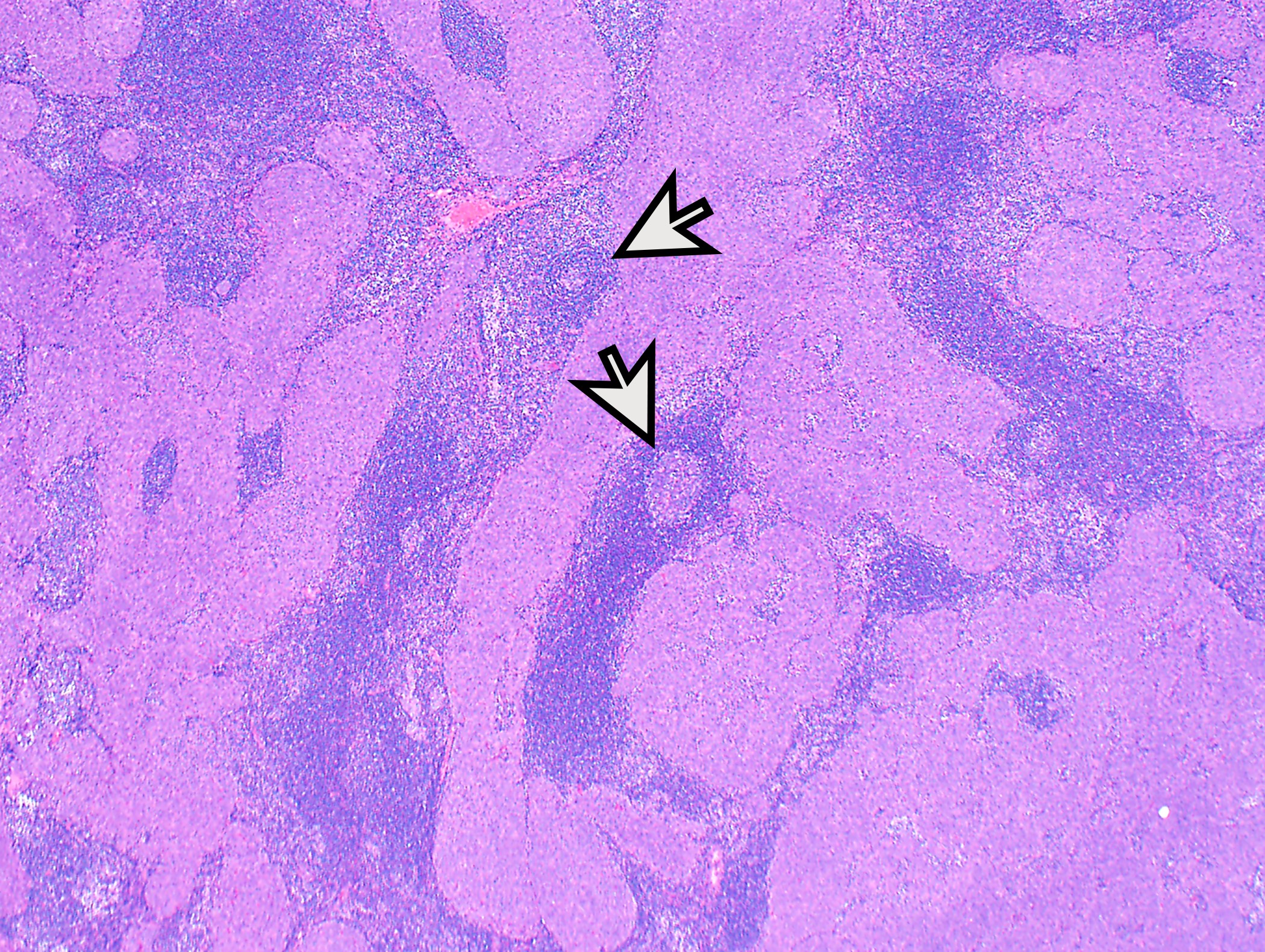
Pathogenesis associated with rheumatoid arthritis
Contributed by Azadeh Esmaeili, M.D. and Sarmad H. Jassim, M.D.
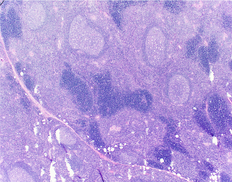
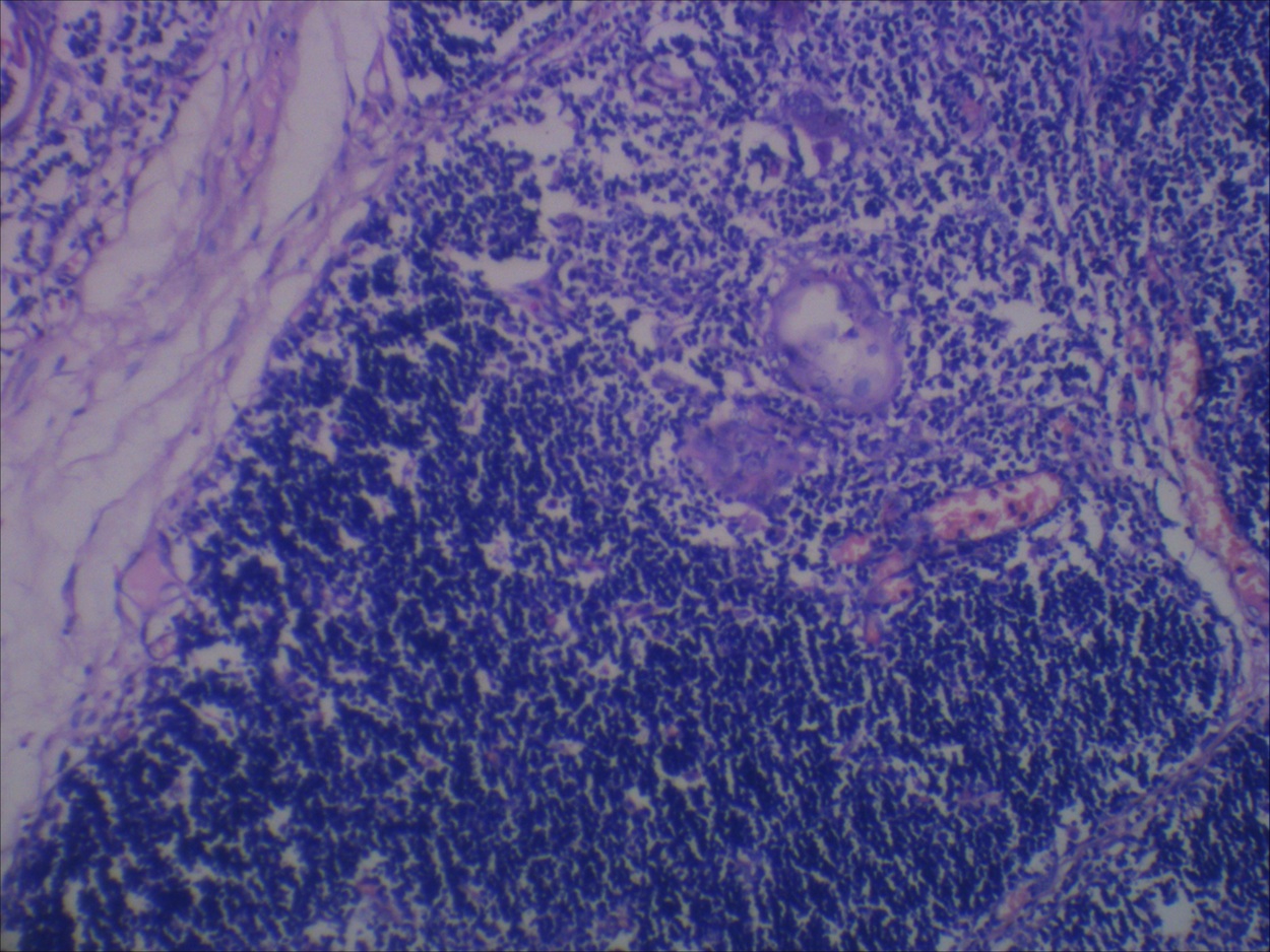
Increased lymphoid follicles
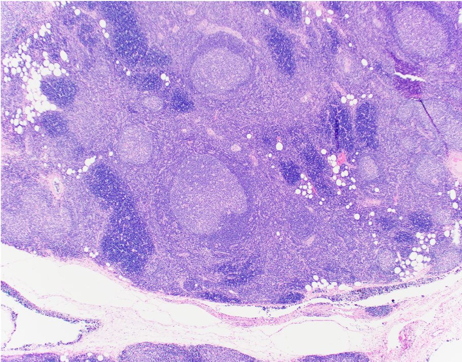
Germinal centers of different sizes
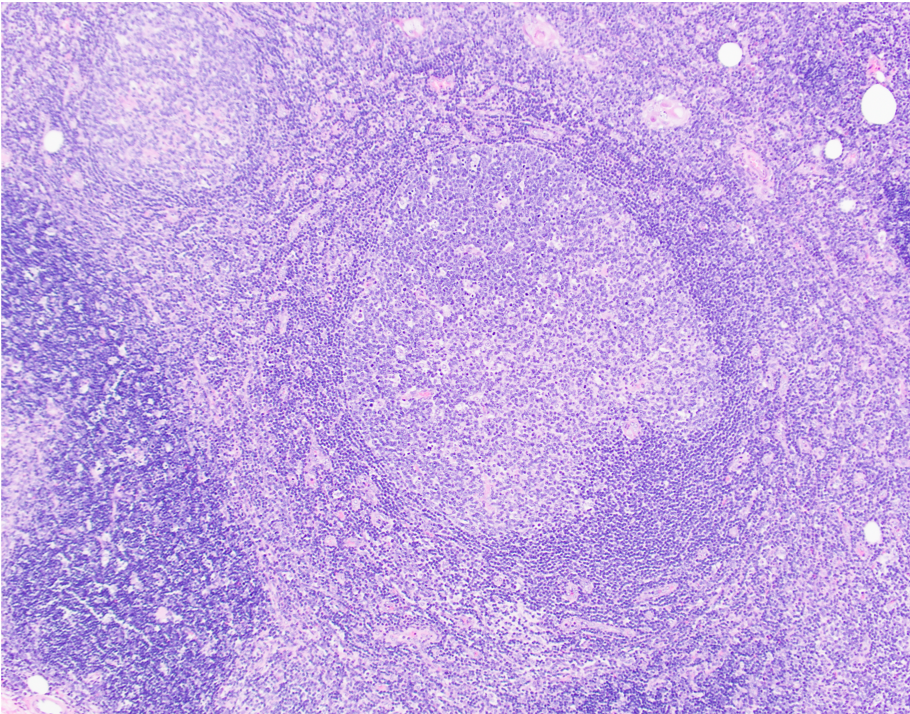
Germinal centers with tingible body macrophages
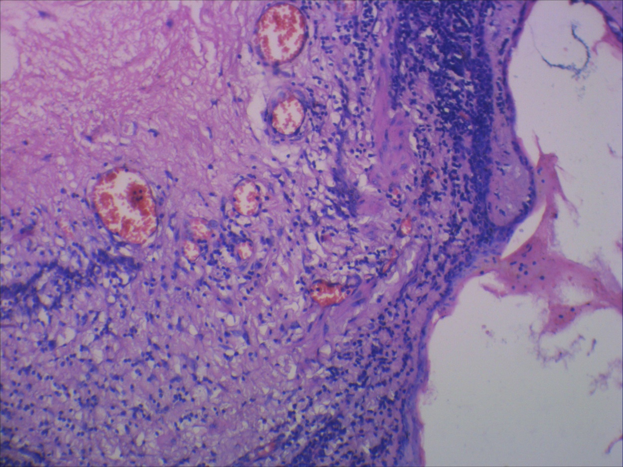
Frequent Hassall corpuscles
Images hosted on other servers:

Proposed stage T1, tumor limited to thymic gland

Proposed stage T2, tumor invades nearby structures

Proposed stage T3, direct (continuous) extrathoracic tumor extension beyond thoracic inlet or below diaphragm

Images hosted on other servers:
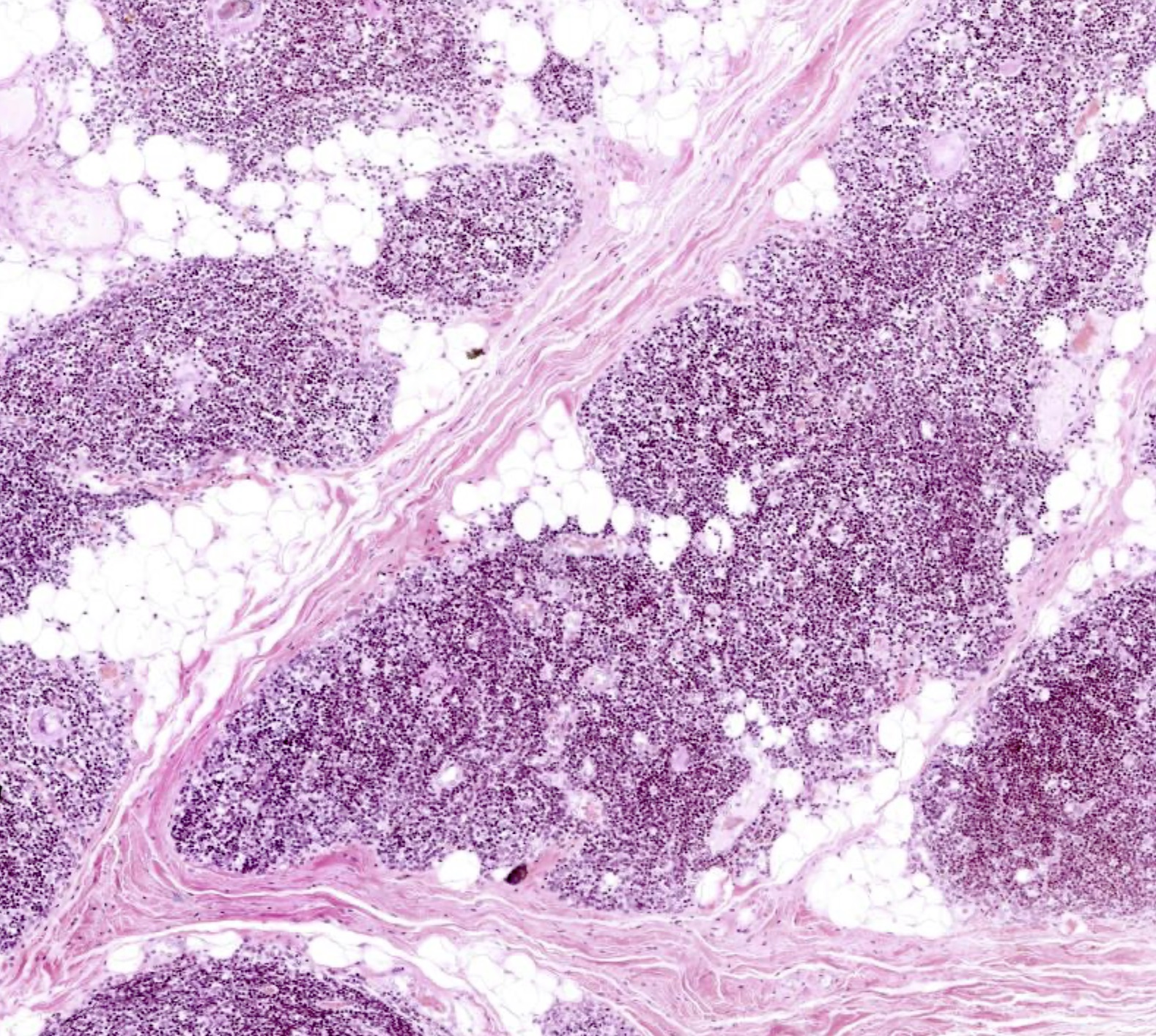
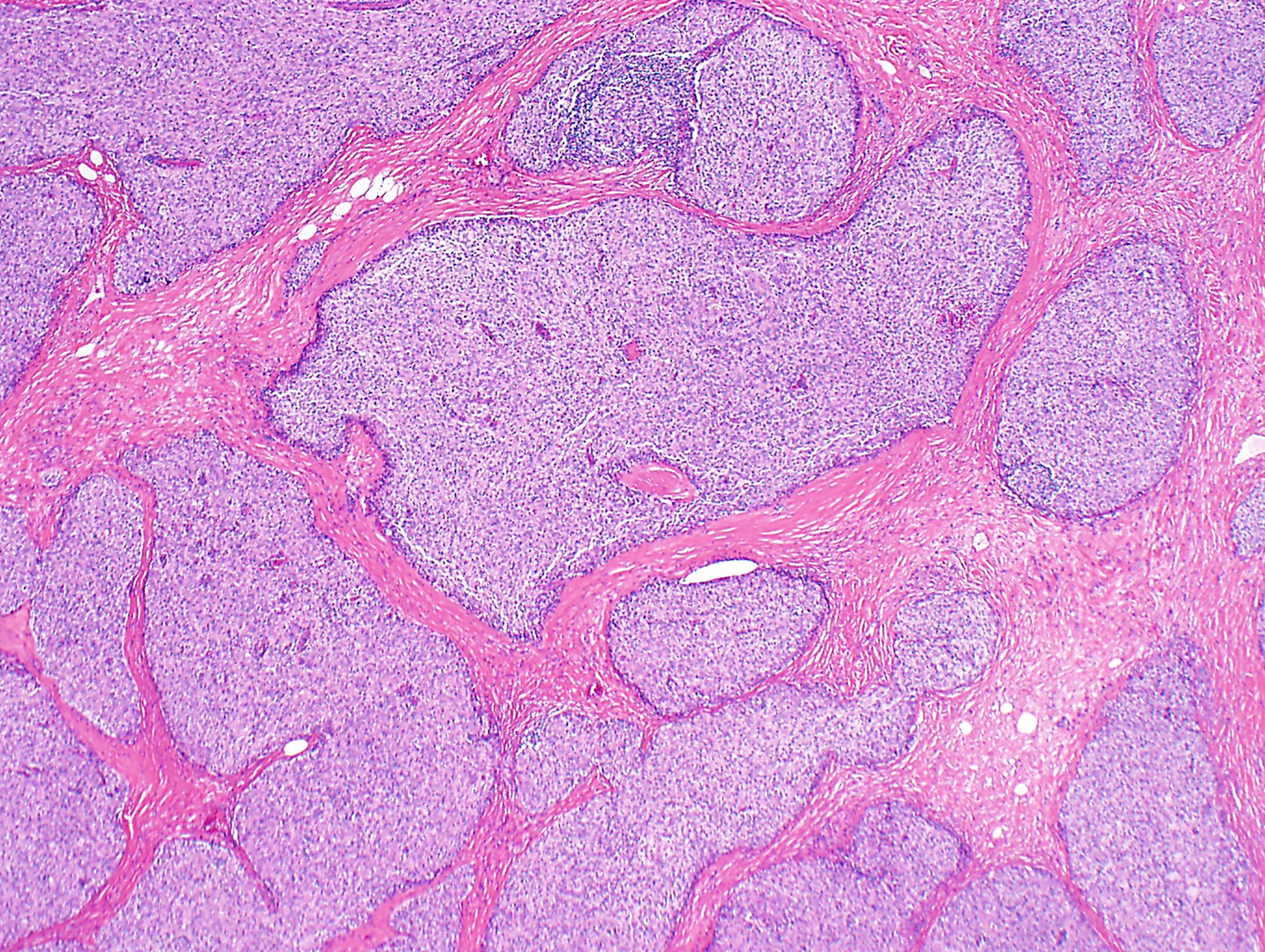
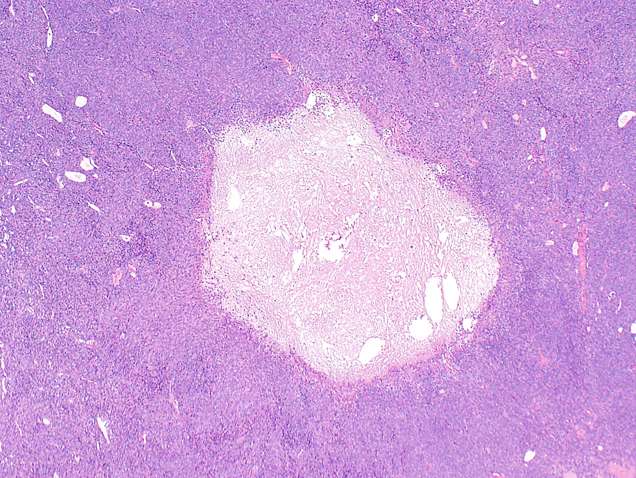
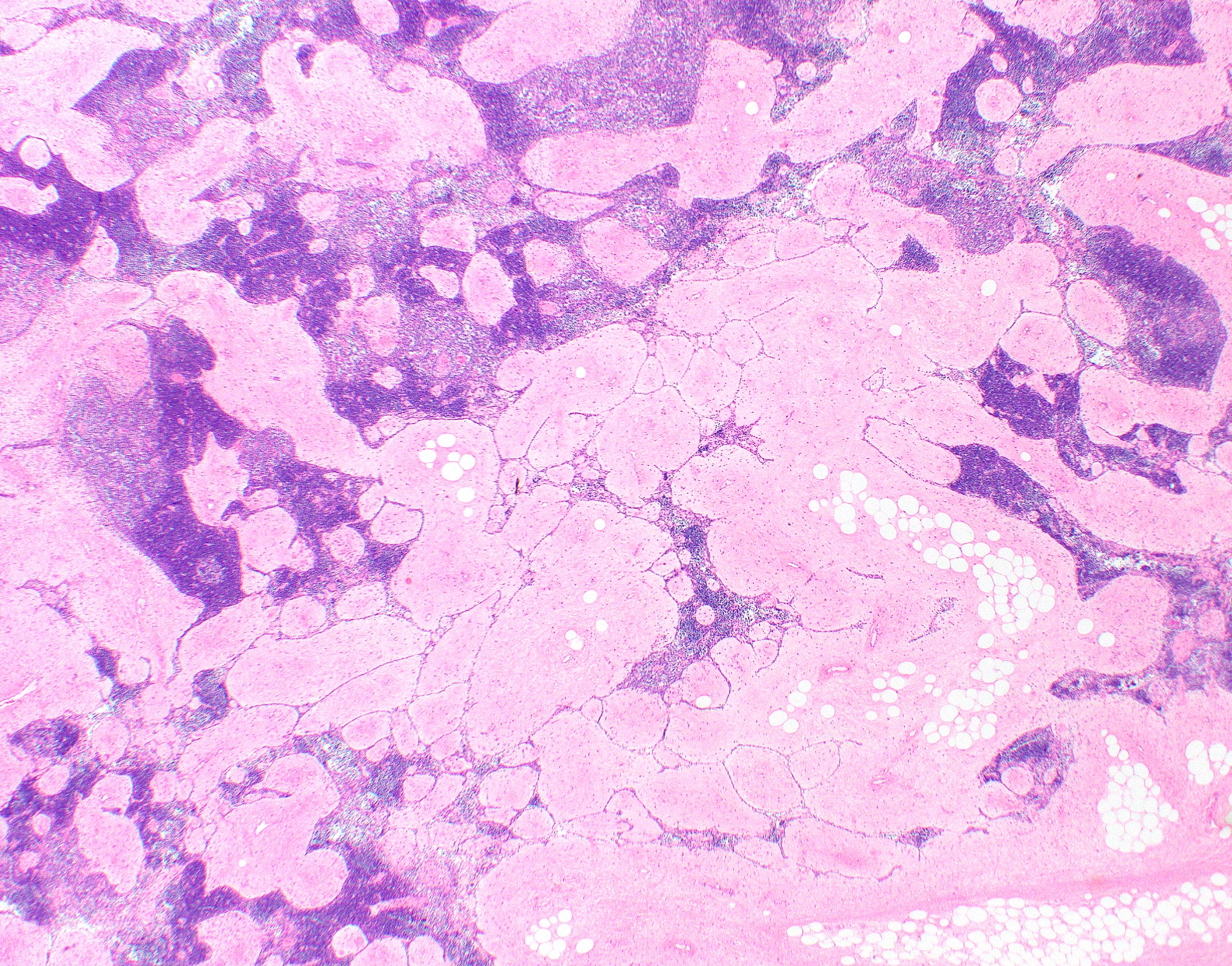
Mature adipose tissue admixed with unremarkable thymic tissue
| Stage | T | N | M | Tumor Extension |
| I | 1a 1b | 0 | 0 | Encapsulated or extending into mediastinal fat, no involvement of mediastinal pleura Invasion of mediastinal pleura |
| II | 2 | 0 | 0 | Invasion of pericardium |
| IIIA | 3 | 0 | 0 | Invasion into lung, brachiocephalic vein, superior vena cava, chest wall, phrenic nerve or extrapericardial pulmonary artery or veins |
| IIIB | 4 | 0 | 0 | Invasion into aorta (ascending, arch, or descending), arch vessels, intrapericardial pulmonary artery, myocardium, trachea, esophagus |
| IVA | Any | 1 0, 1 | 0 1a | Anterior (perithymic) lymph nodes Separate pleural / pericardial nodule(s) (implants) |
| IVB | Any | 2 Any | 0, 1a 1b | Deep intrathoracic / cervical lymph nodes Pulmonary intraparenchymal nodule, distant organ metastasis |
Recommended reporting of thymic epithelial tumors with multiple components (2021 WHO)
| Thymoma with > 1 histologic pattern (except AB thymoma) | List all thymoma subtypes, including percentages in 10% increments; start with predominant component |
| Thymoma and thymic carcinoma | List all components, including % in 10% increments; start with thymic carcinoma component independent of extent |
| Thymoma and thymic carcinoid tumor | List all components, including % in 10% increments; start with thymic carcinoid tumor component independent of extent |
Contributed by Anja C. Roden, M.D.
WHO type A thymoma
Contributed by Anja C. Roden, M.D.


Gross specimen of thymoma
Contributed by Anja C. Roden, M.D.
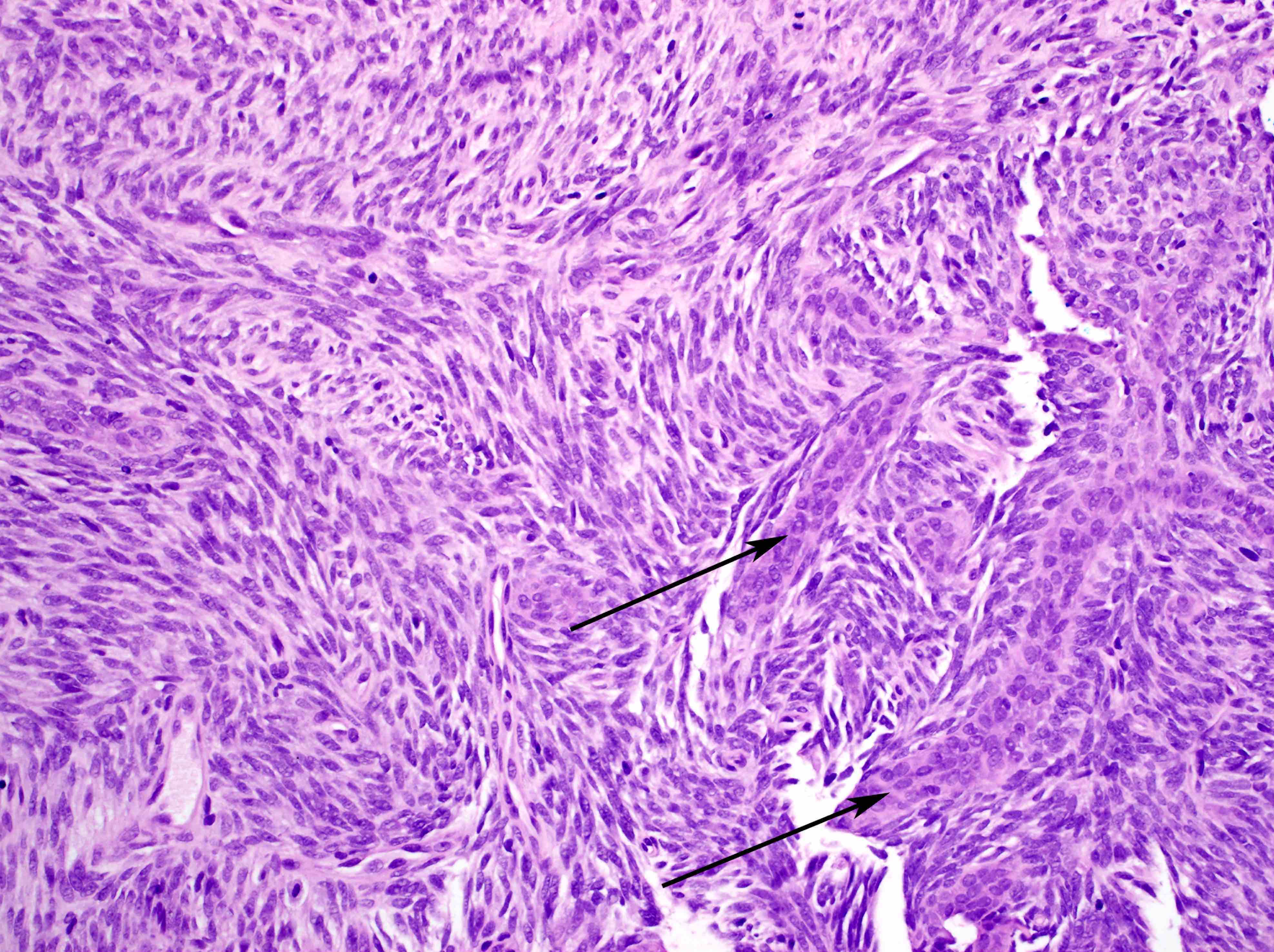
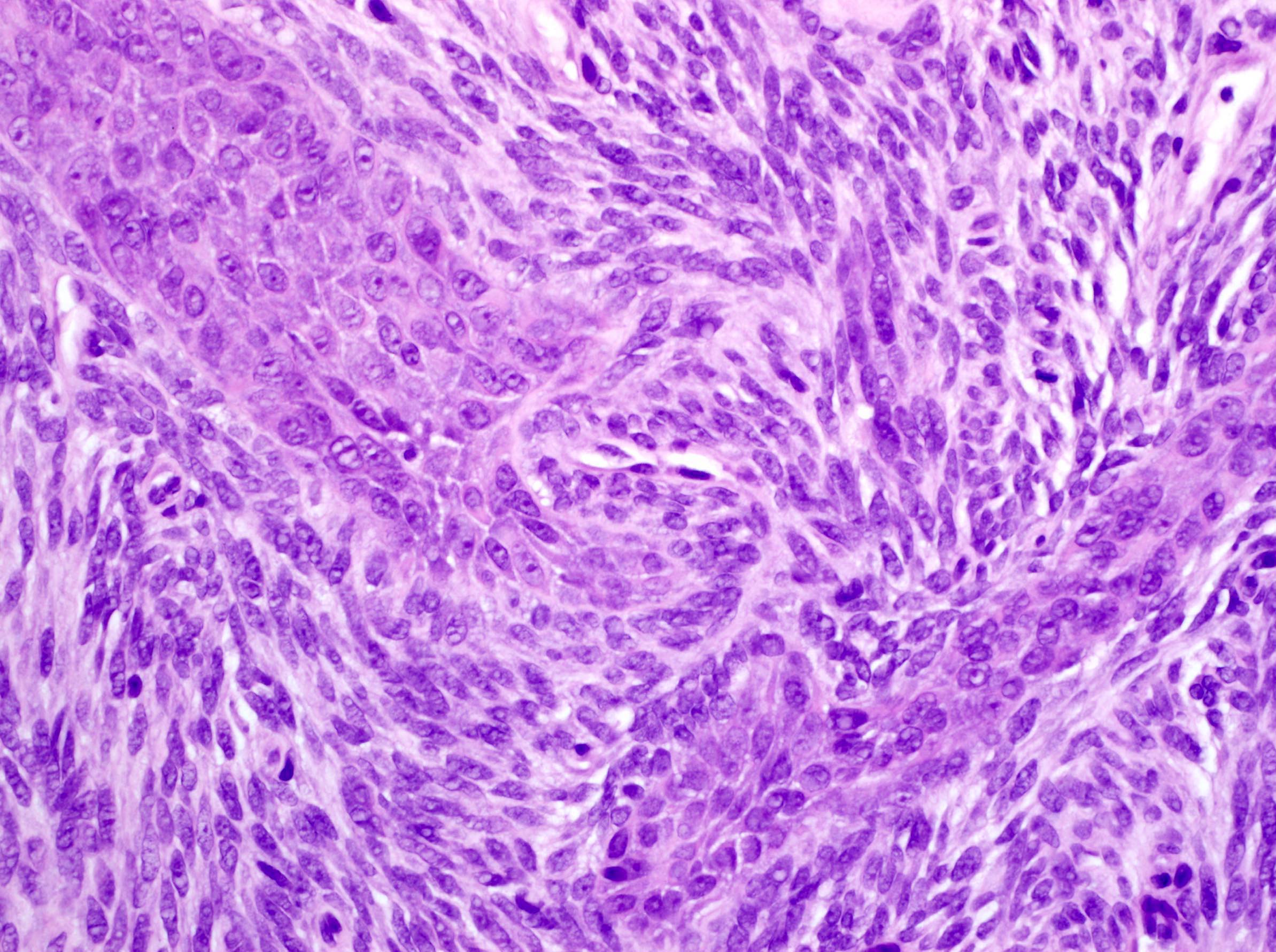
WHO type B3 thymoma
WHO type A thymomas, various patterns and atypical type A variant
WHO type A thymomas, various patterns and atypical type A variant
WHO type AB thymoma
WHO type AB thymoma
WHO type B1 thymoma
WHO type B2 thymoma
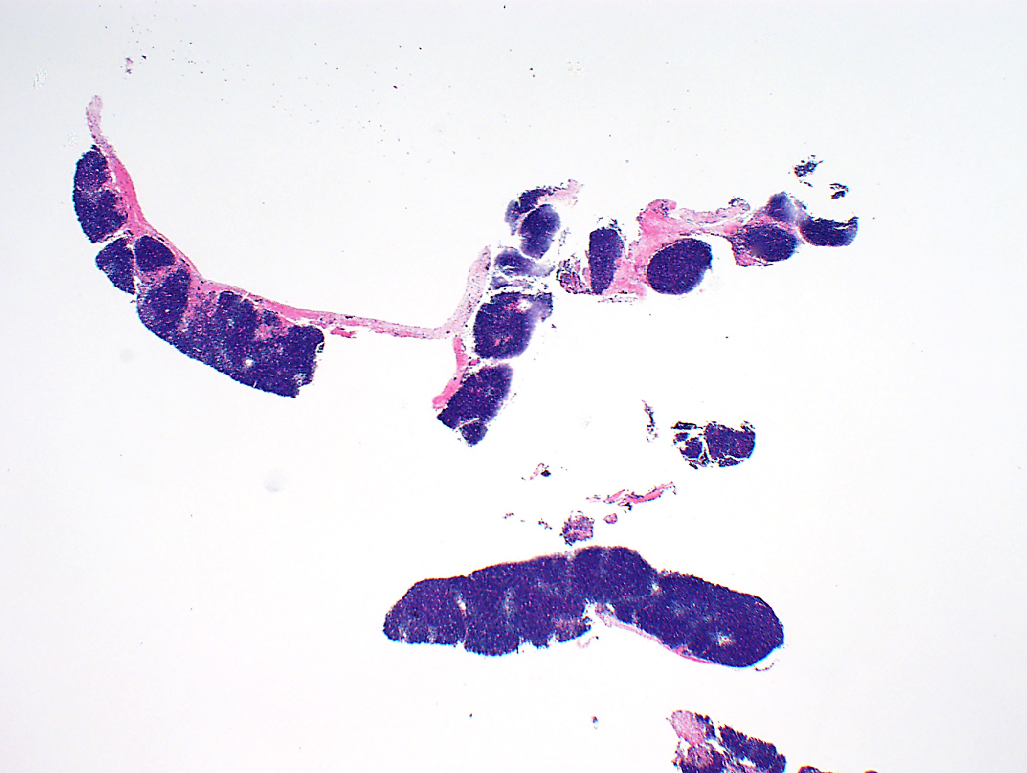
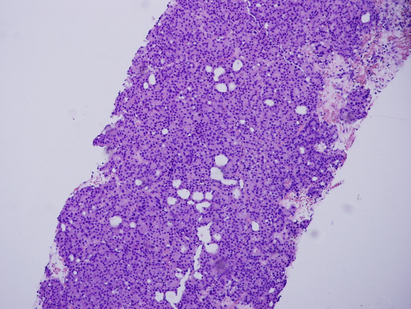
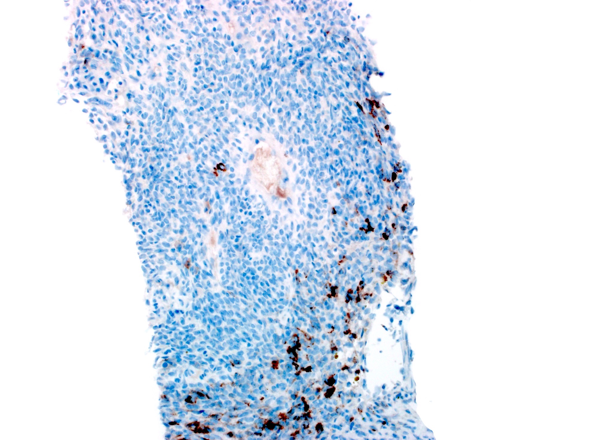
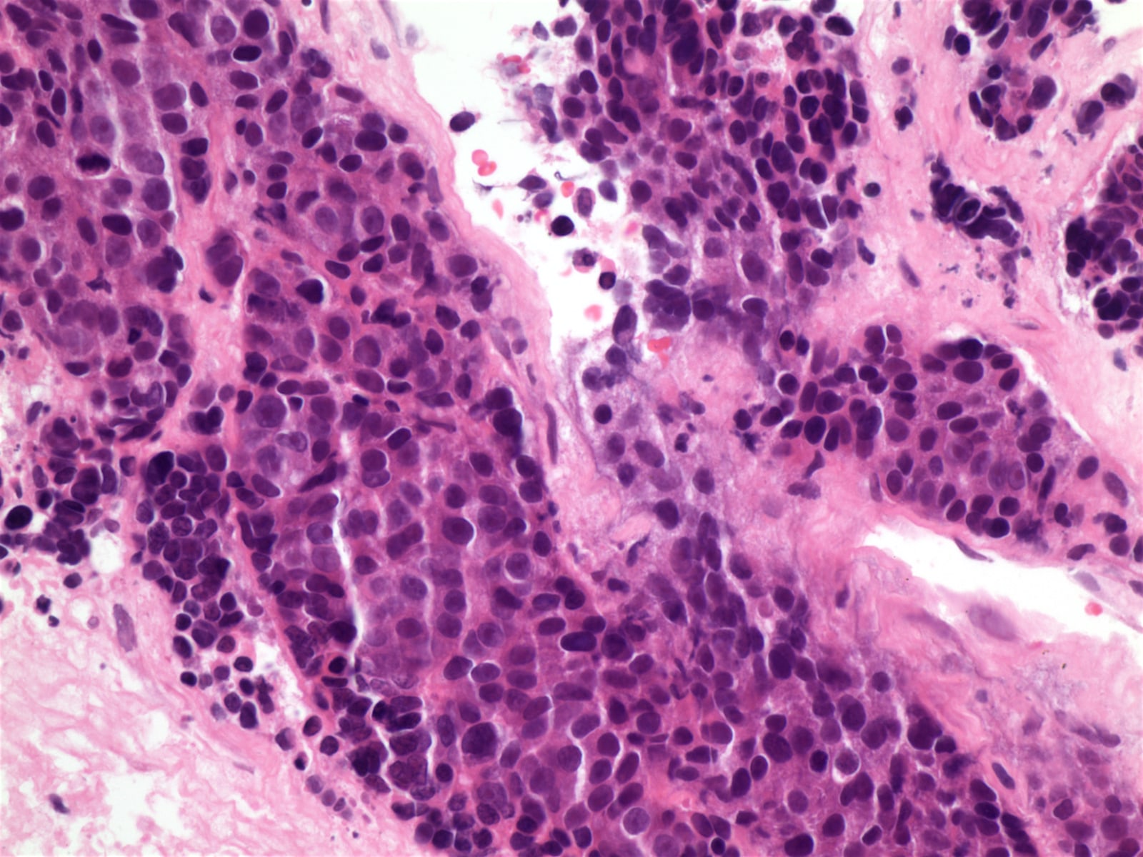
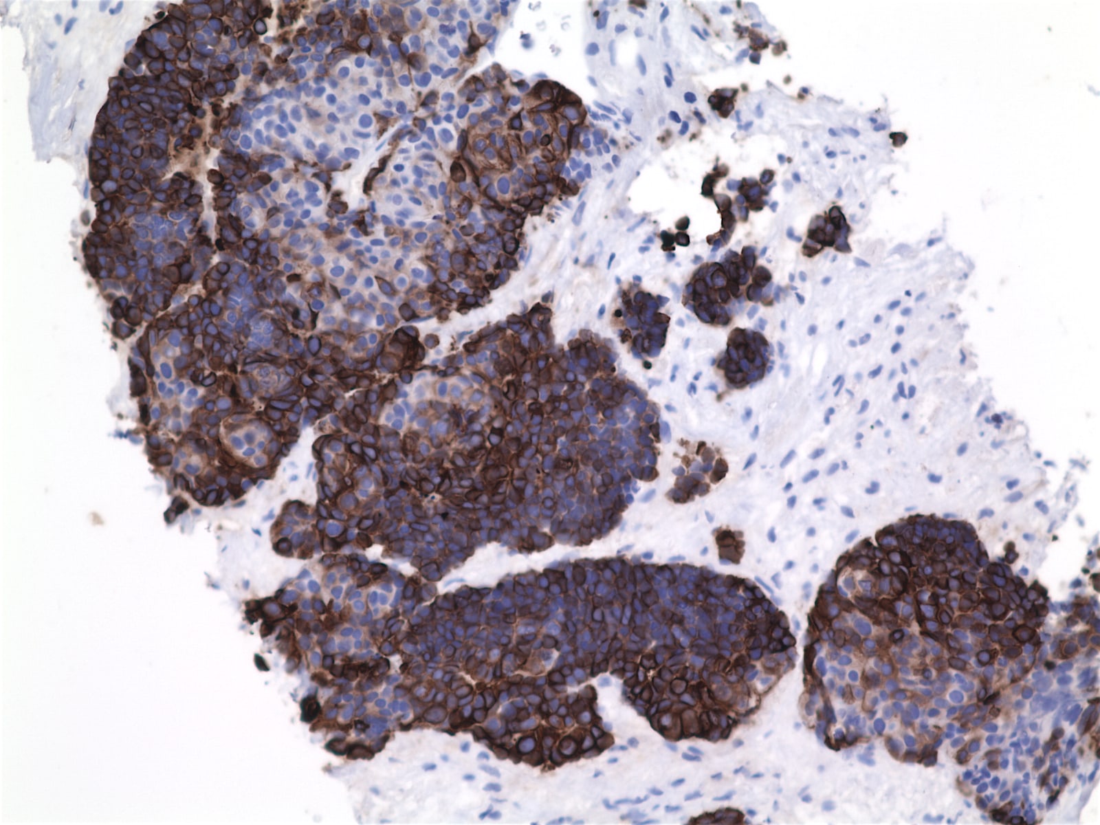
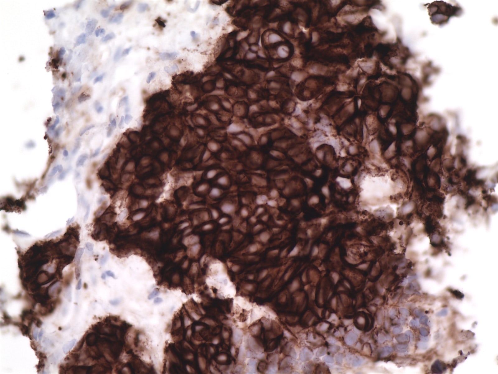
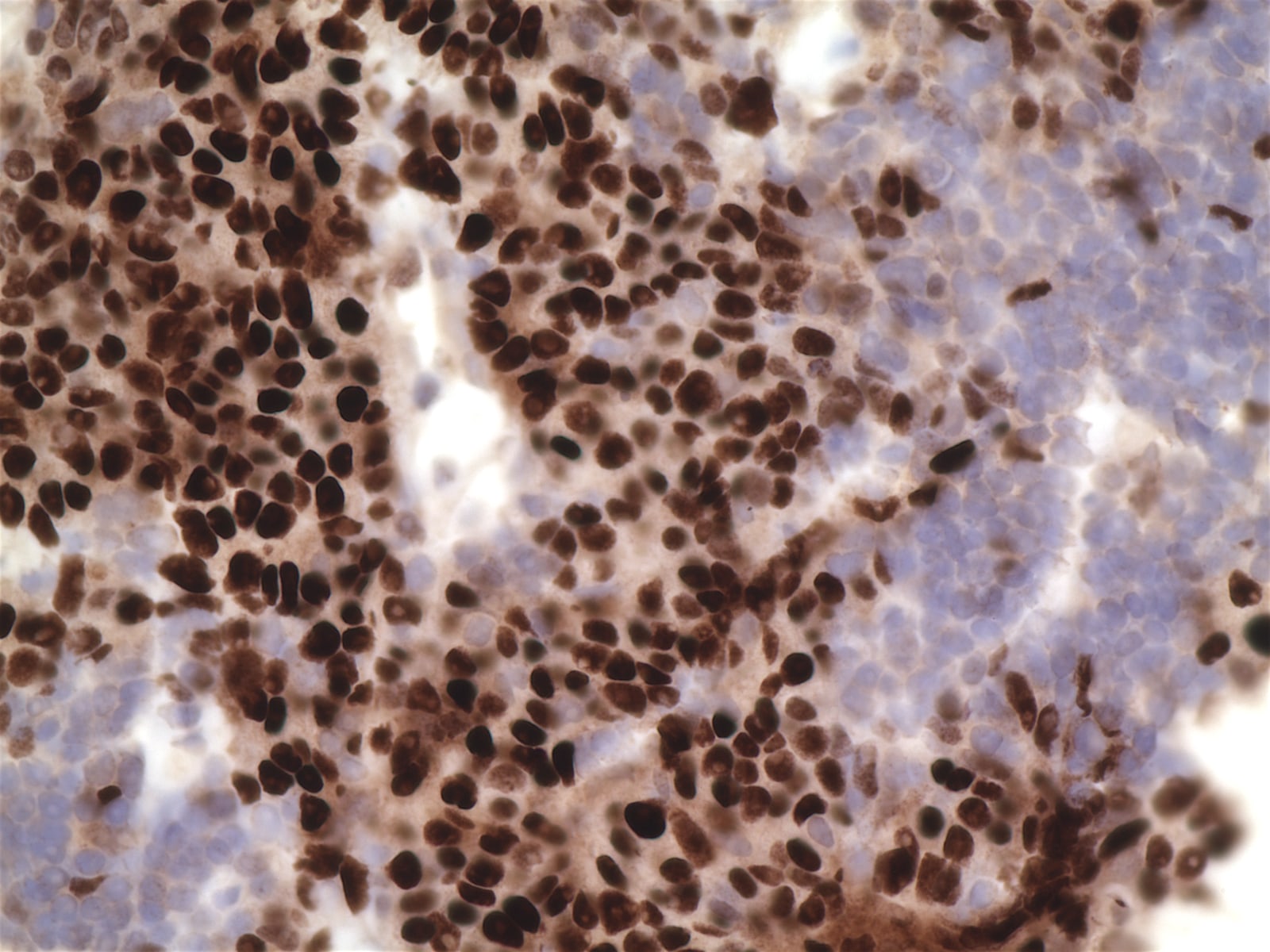
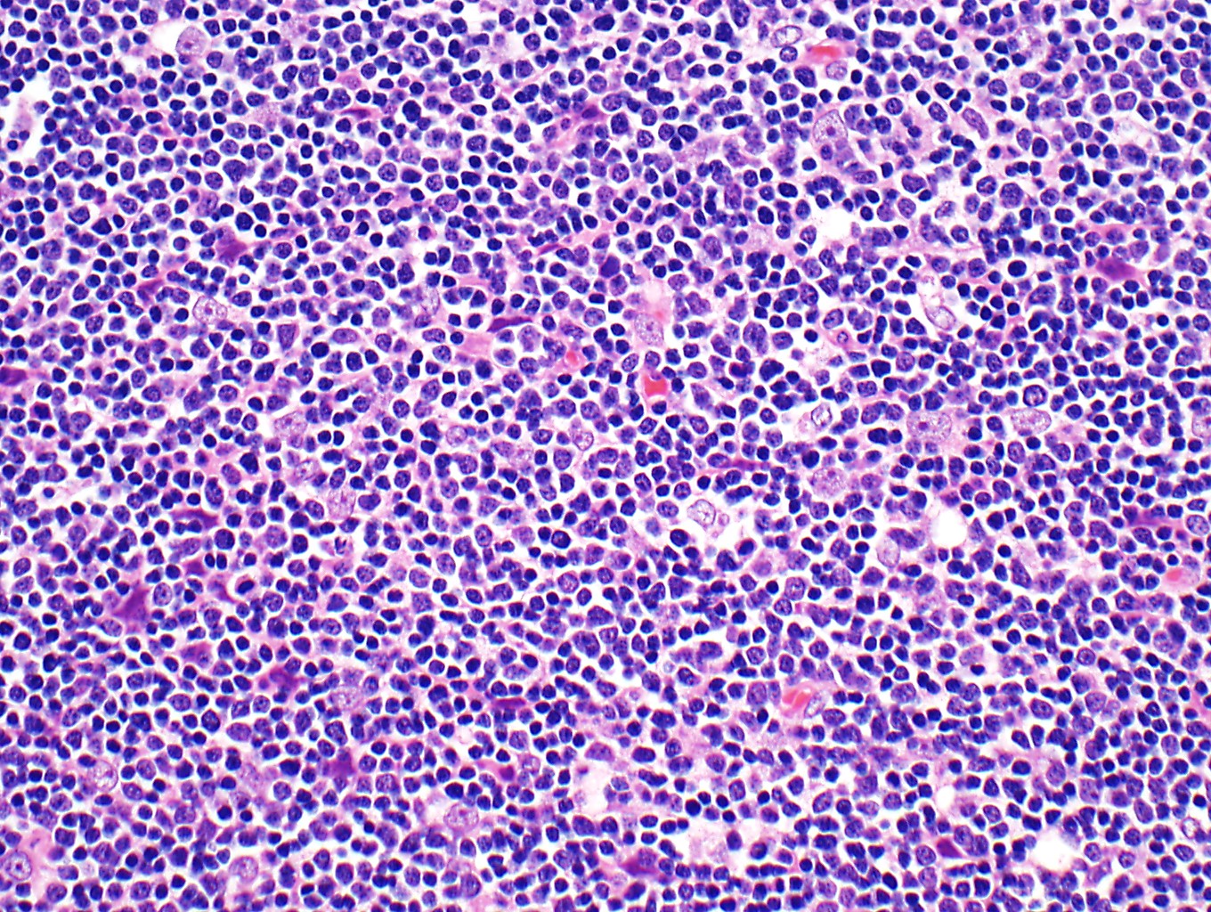
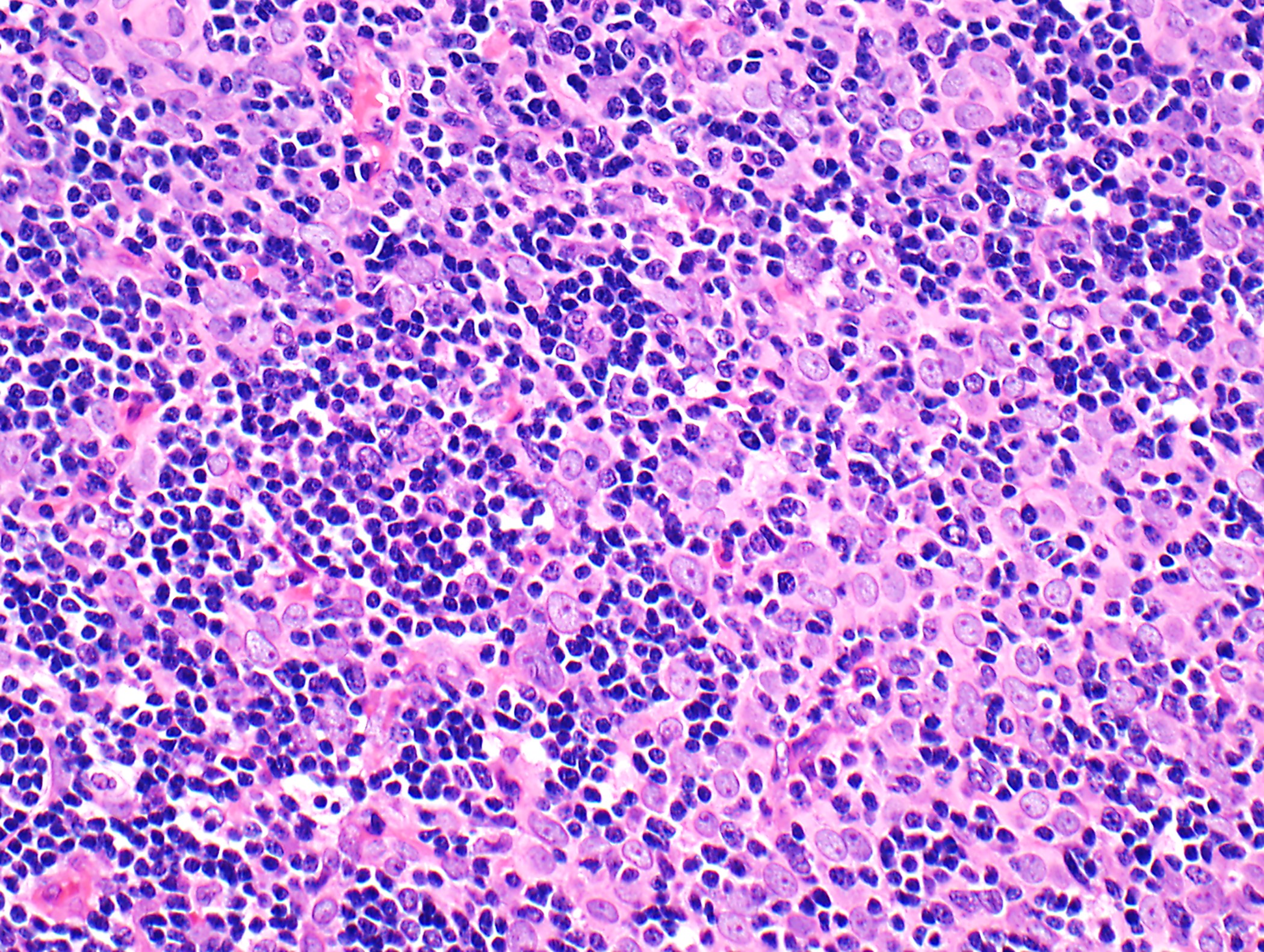
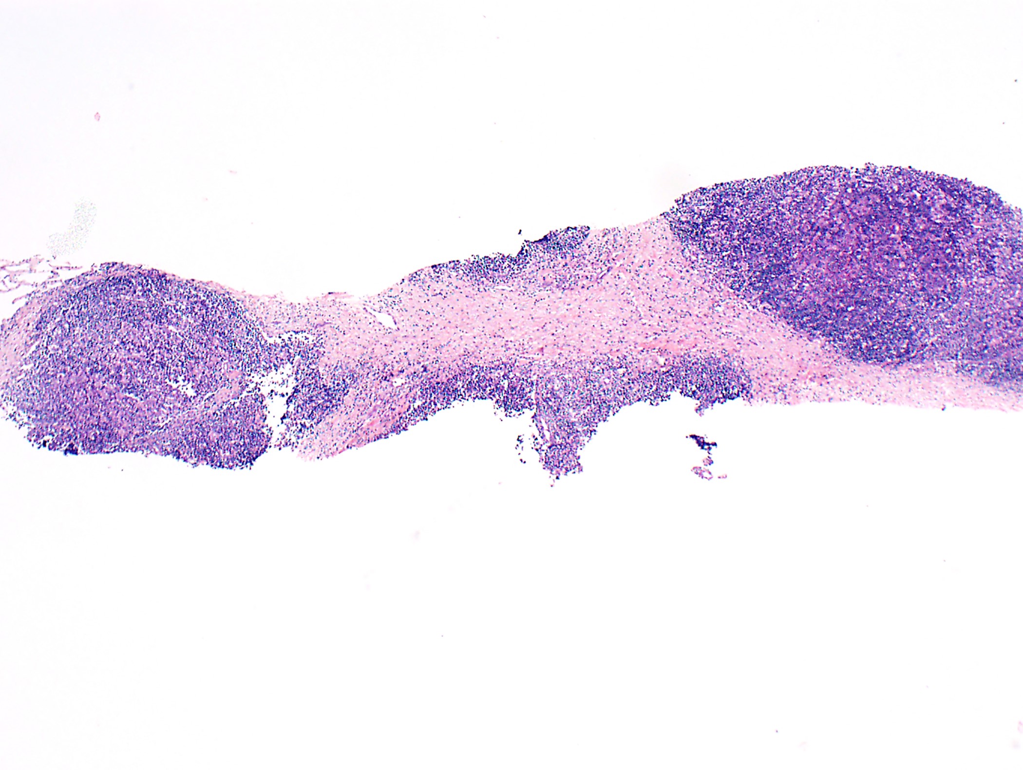
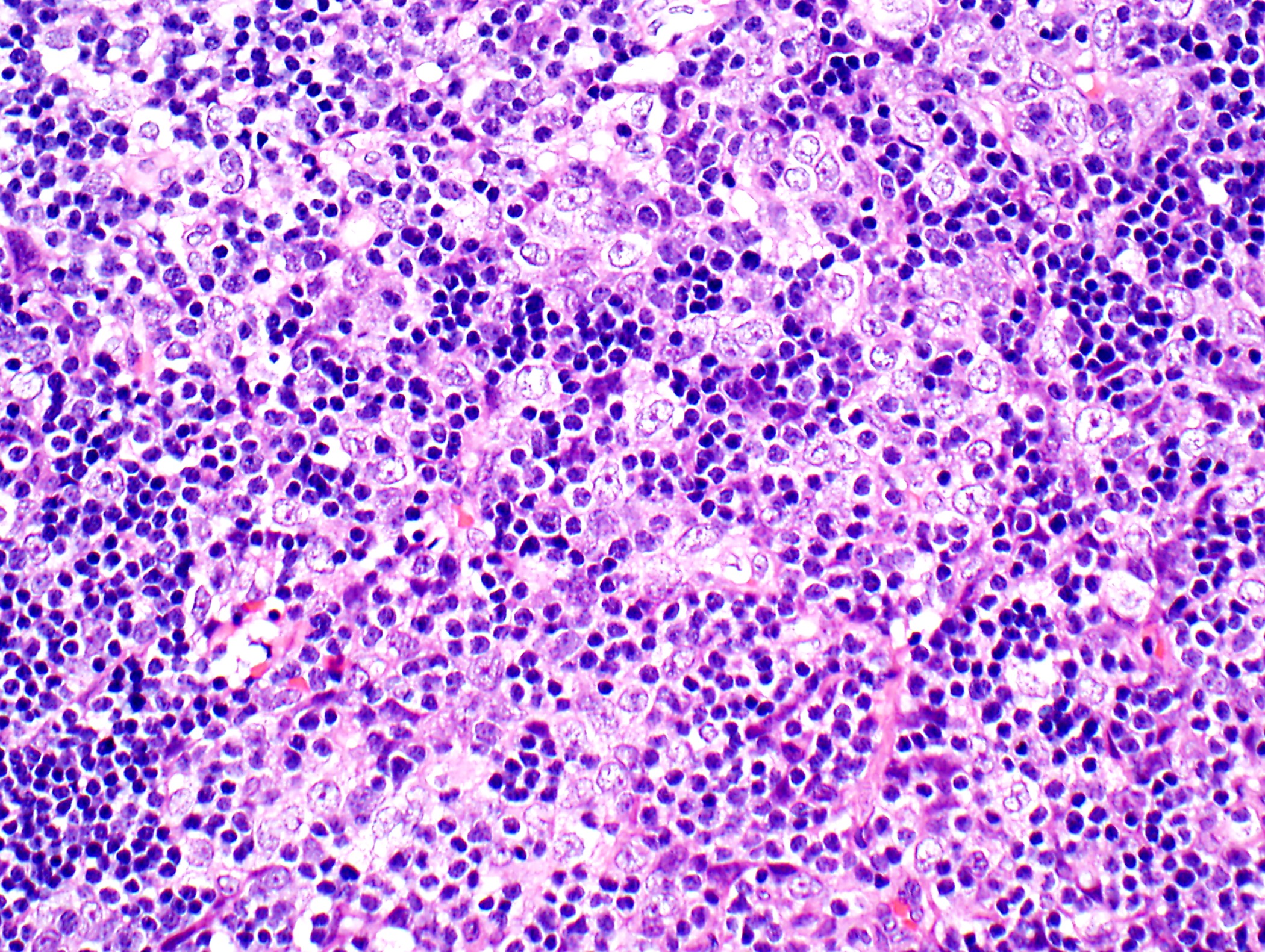
Thymoma: core needle biopsy
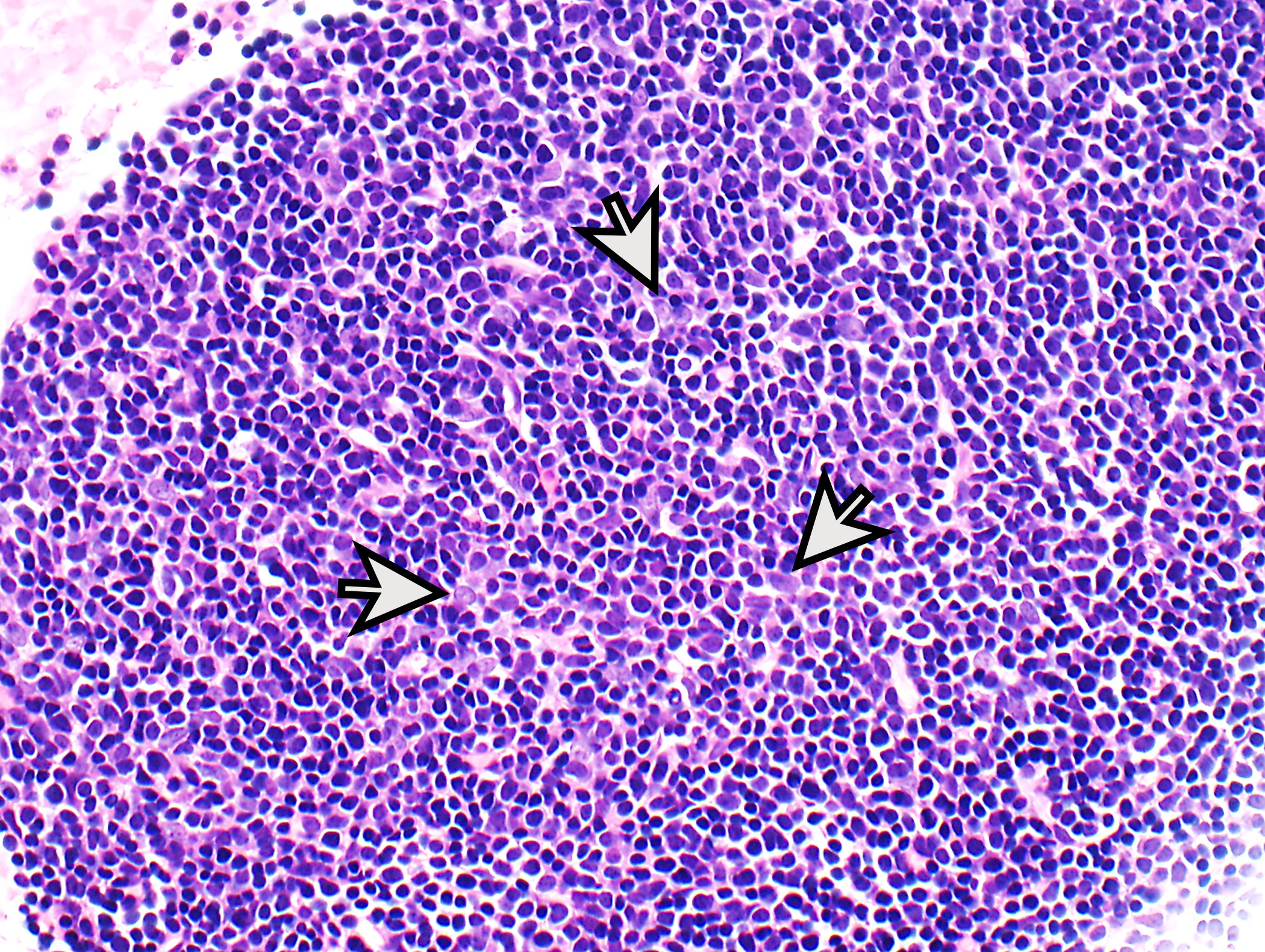
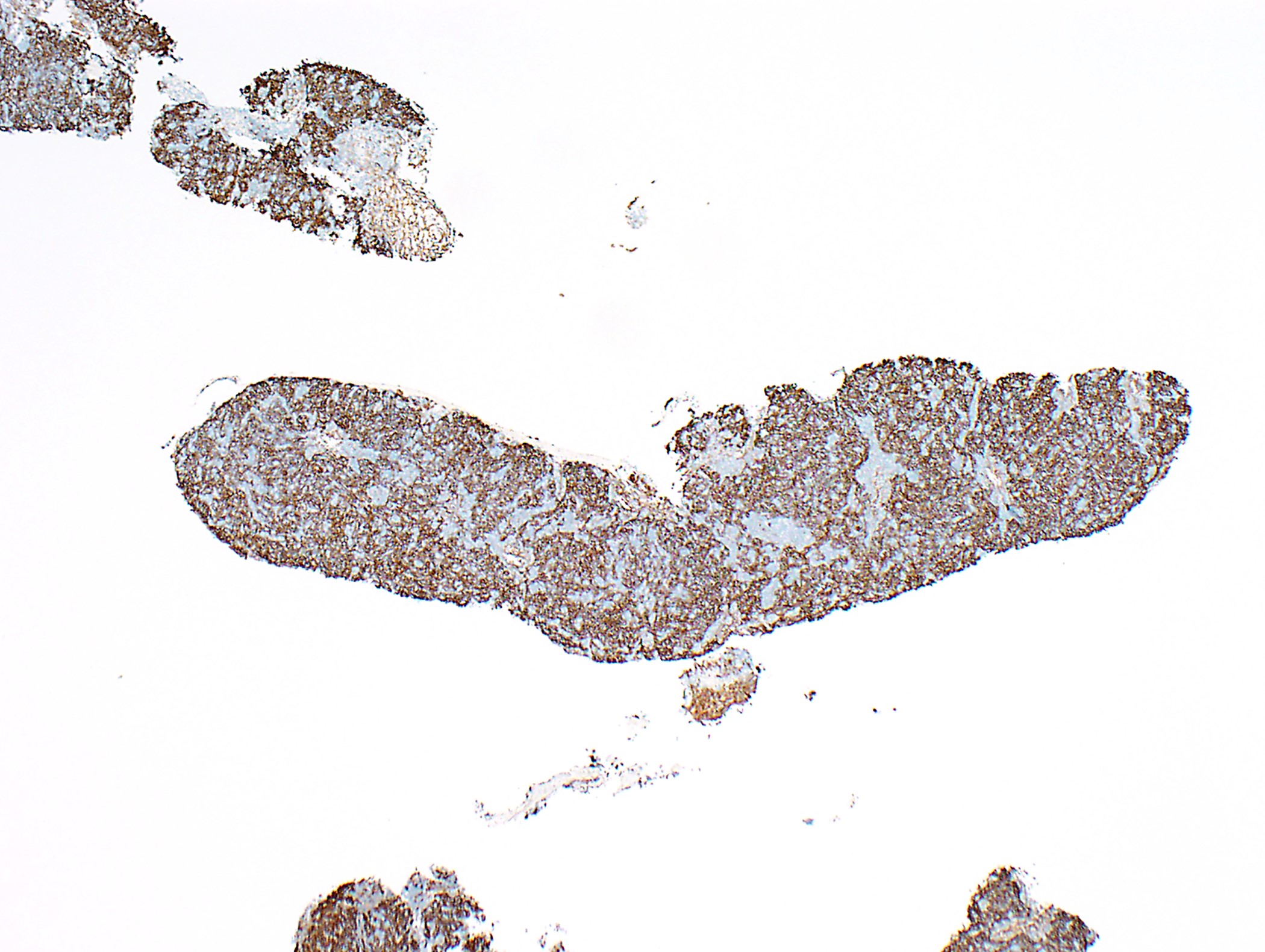
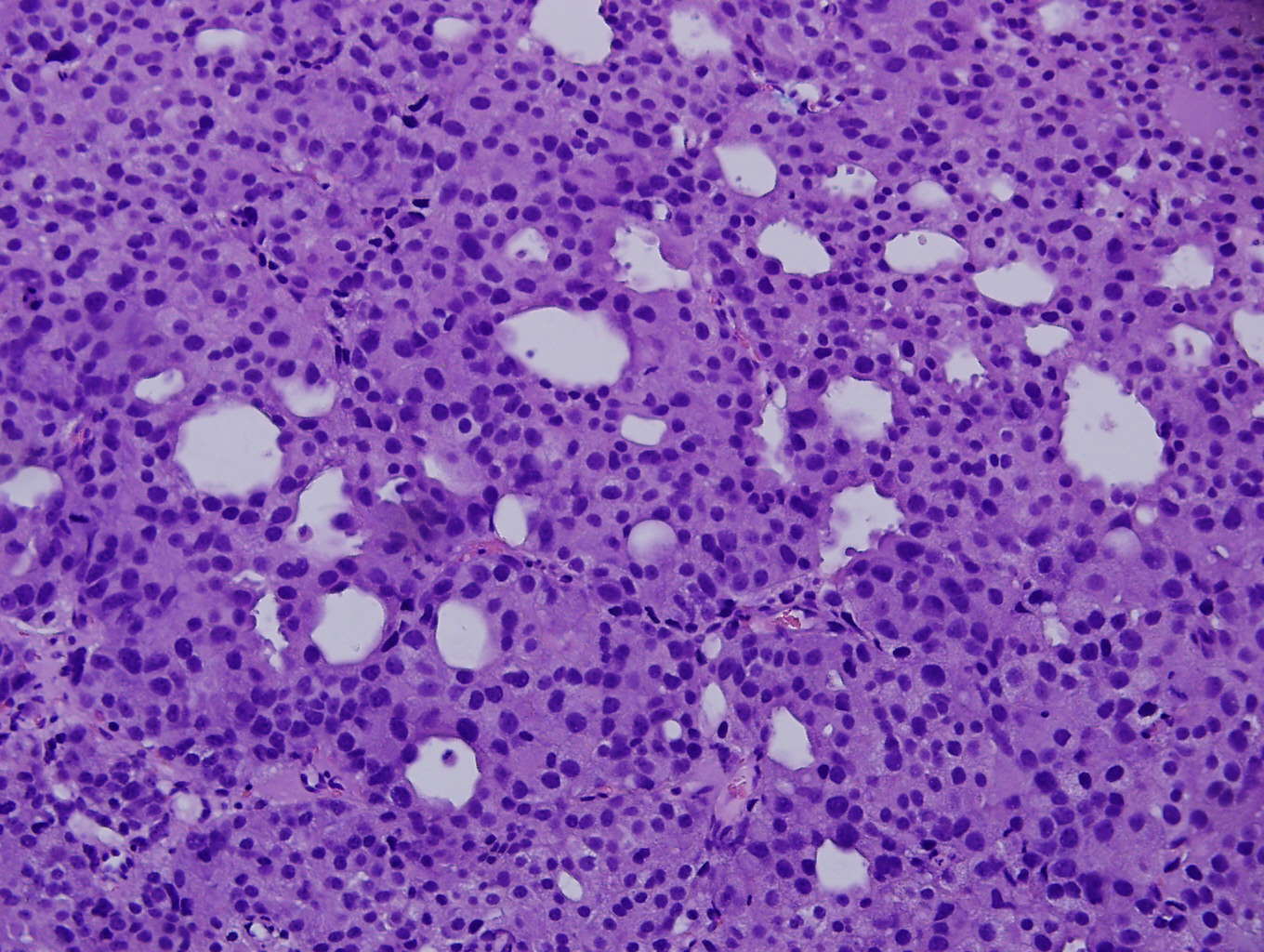
Thymoma: core needle biopsy
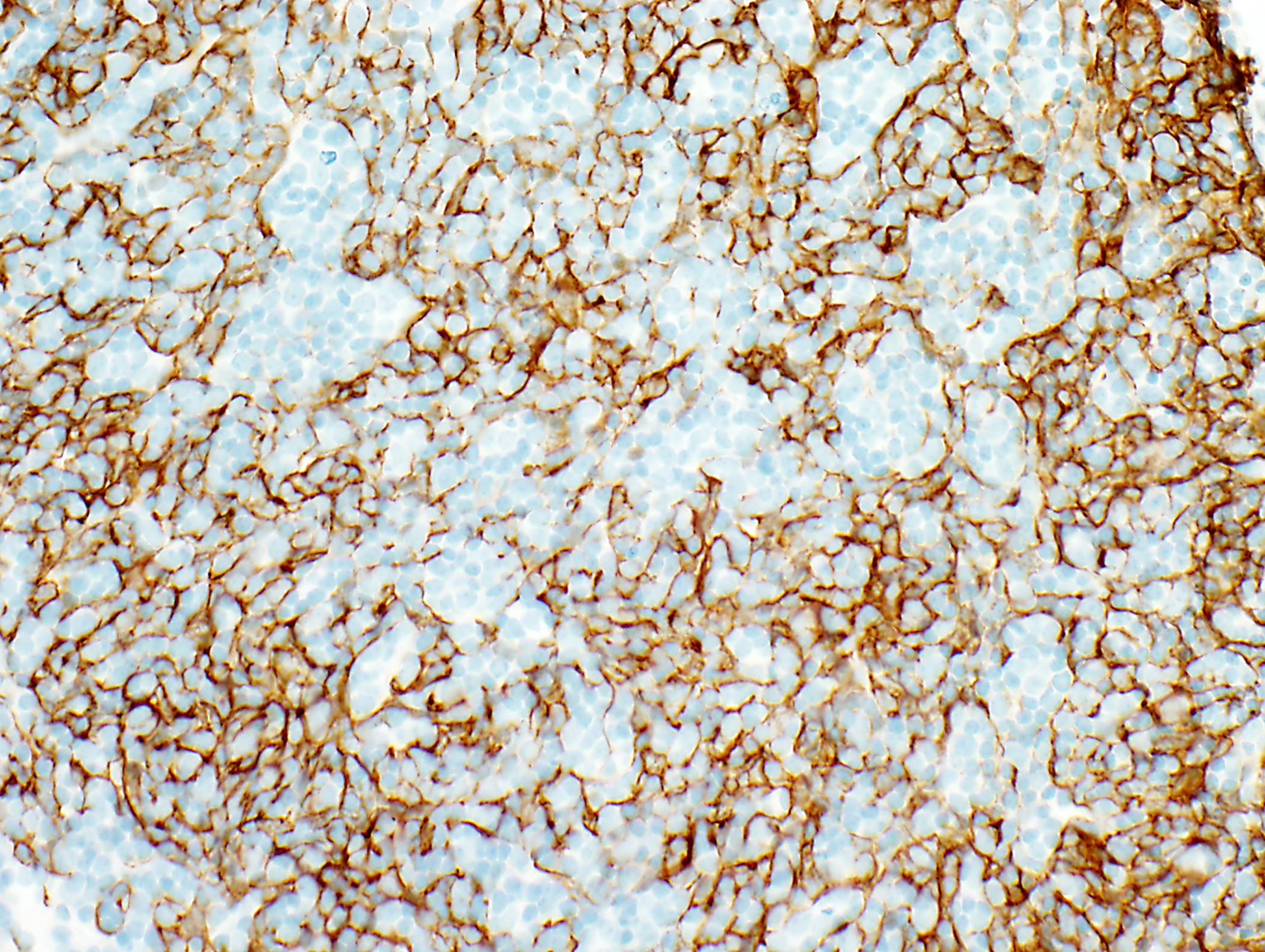
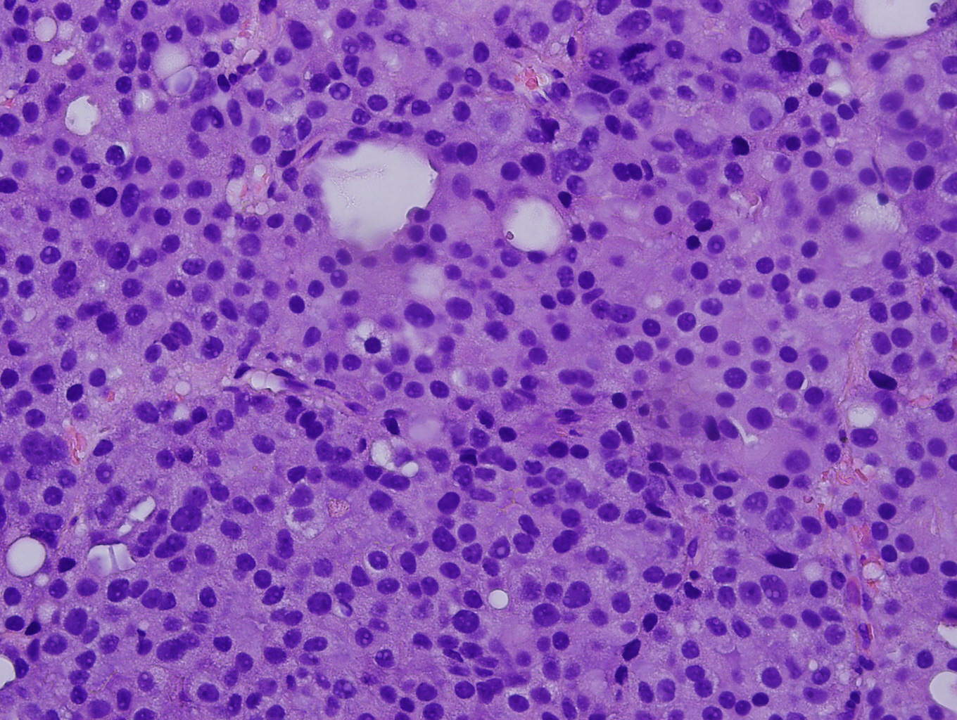
Thymoma: core needle biopsy
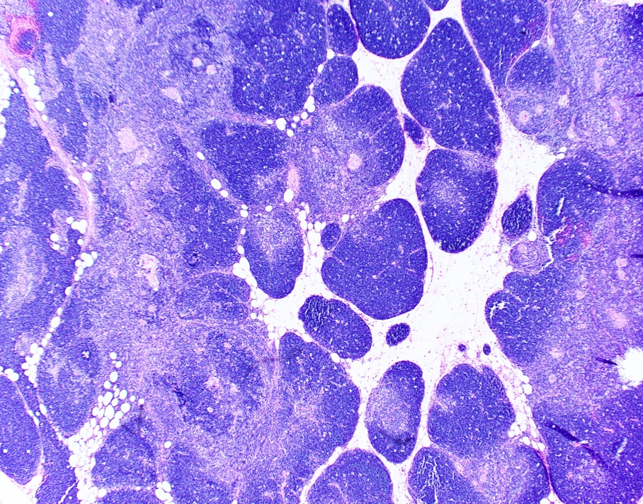
Micronodular thymoma with lymphoid stroma

Metaplastic thymoma
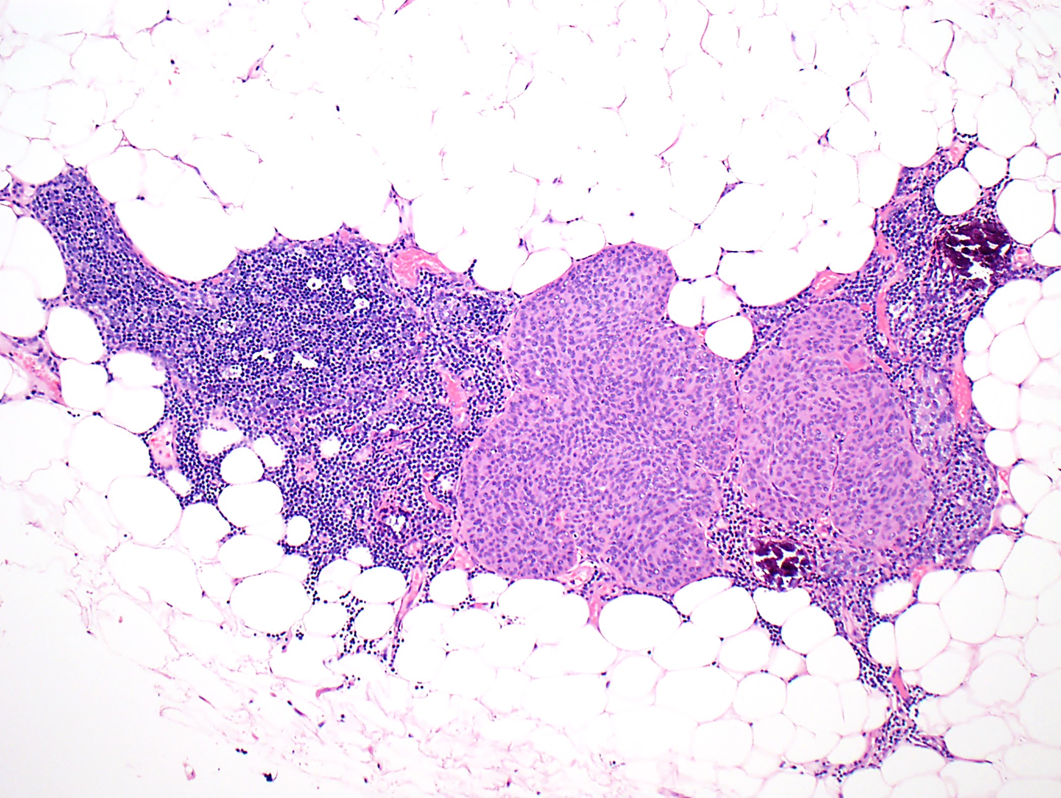
Lipofibroadenoma
Lipofibroadenoma
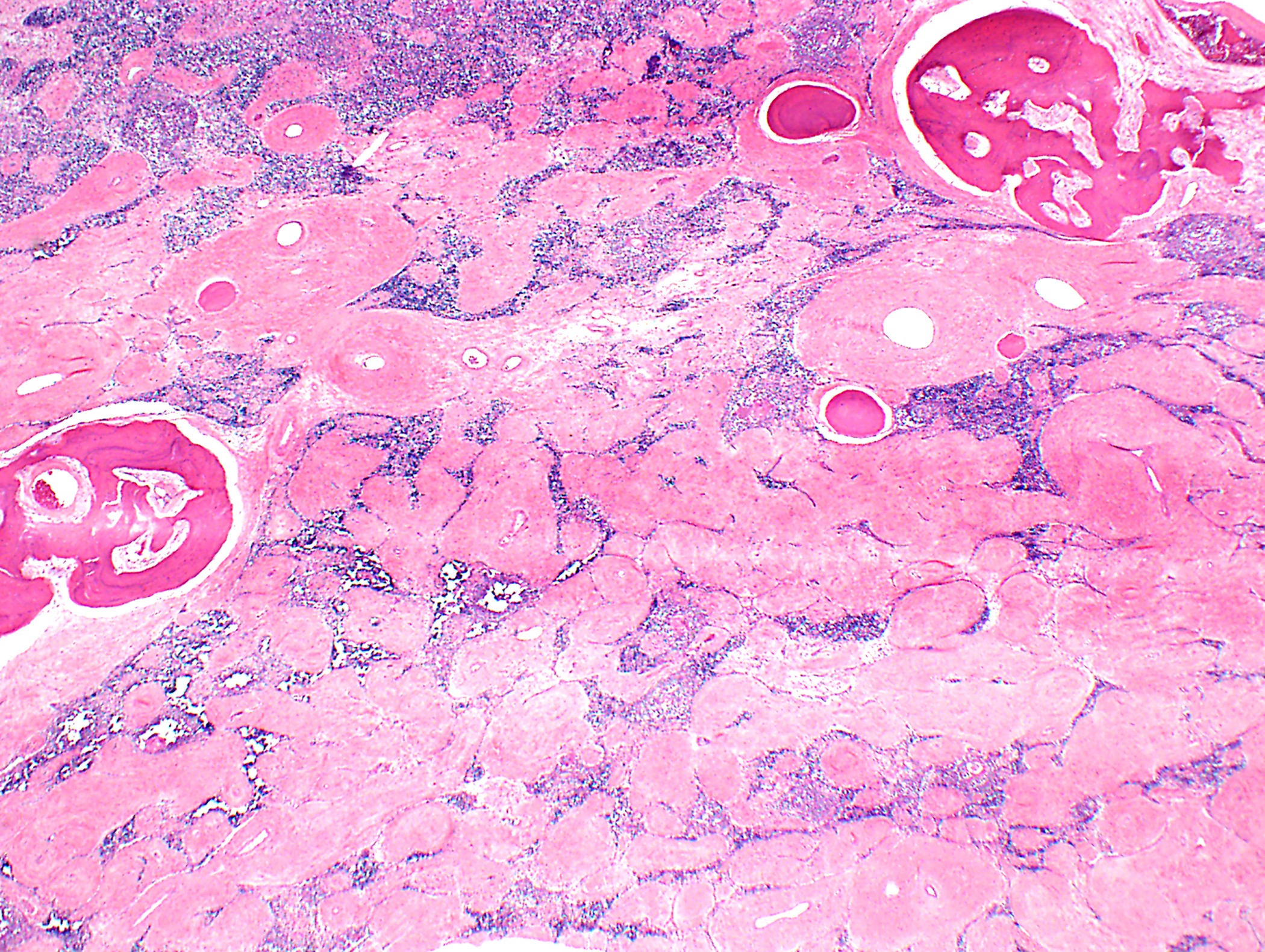
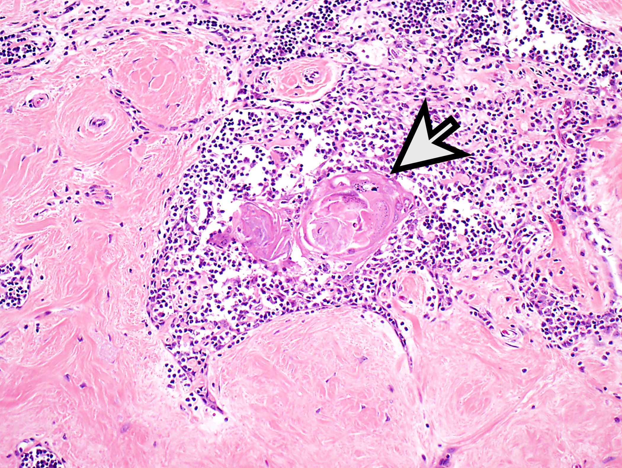
Thymoma with extensive sclerosis and ossification
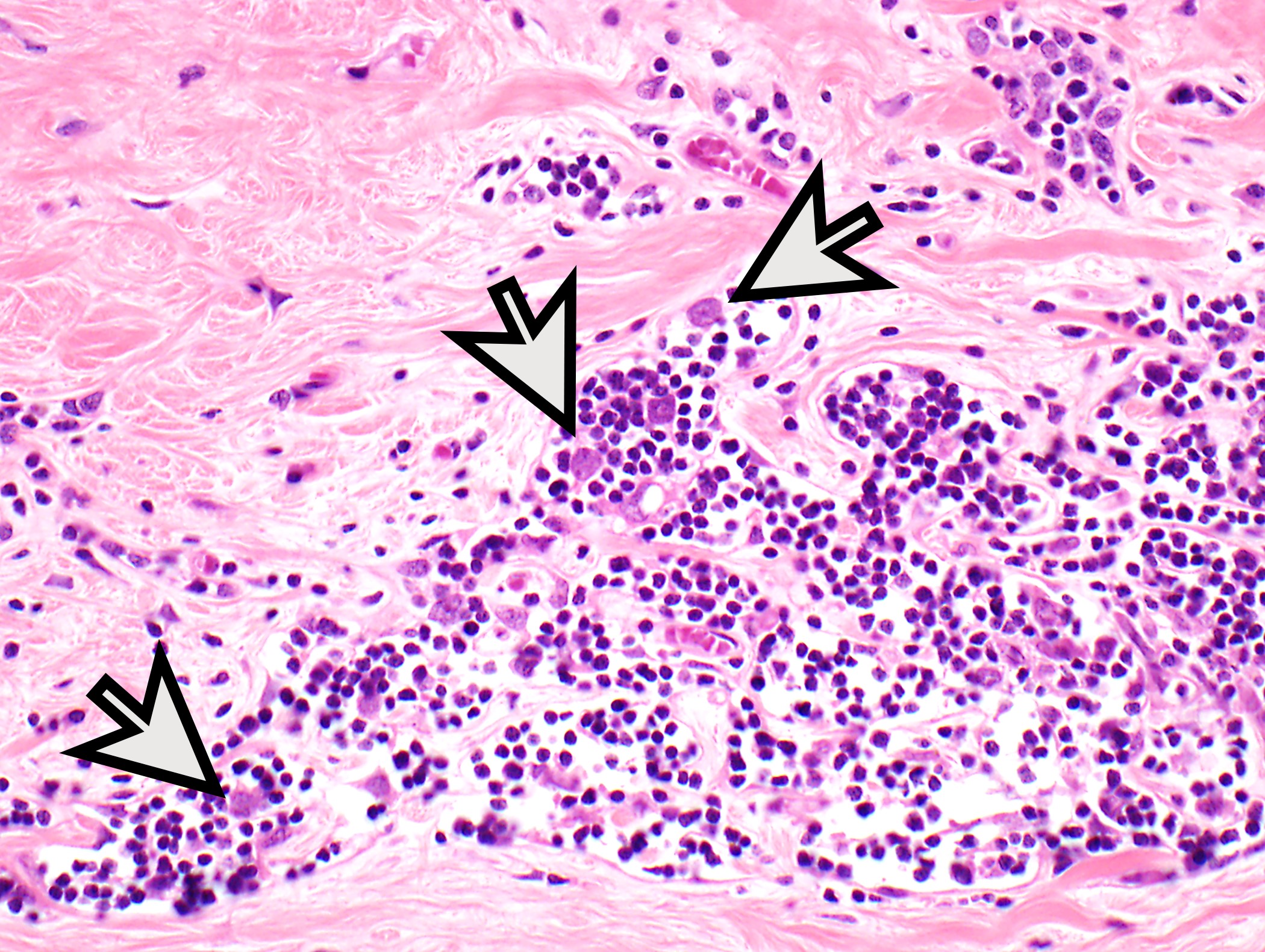
Small thymic epithelial cell nest
Contributed by Anja C. Roden, M.D.
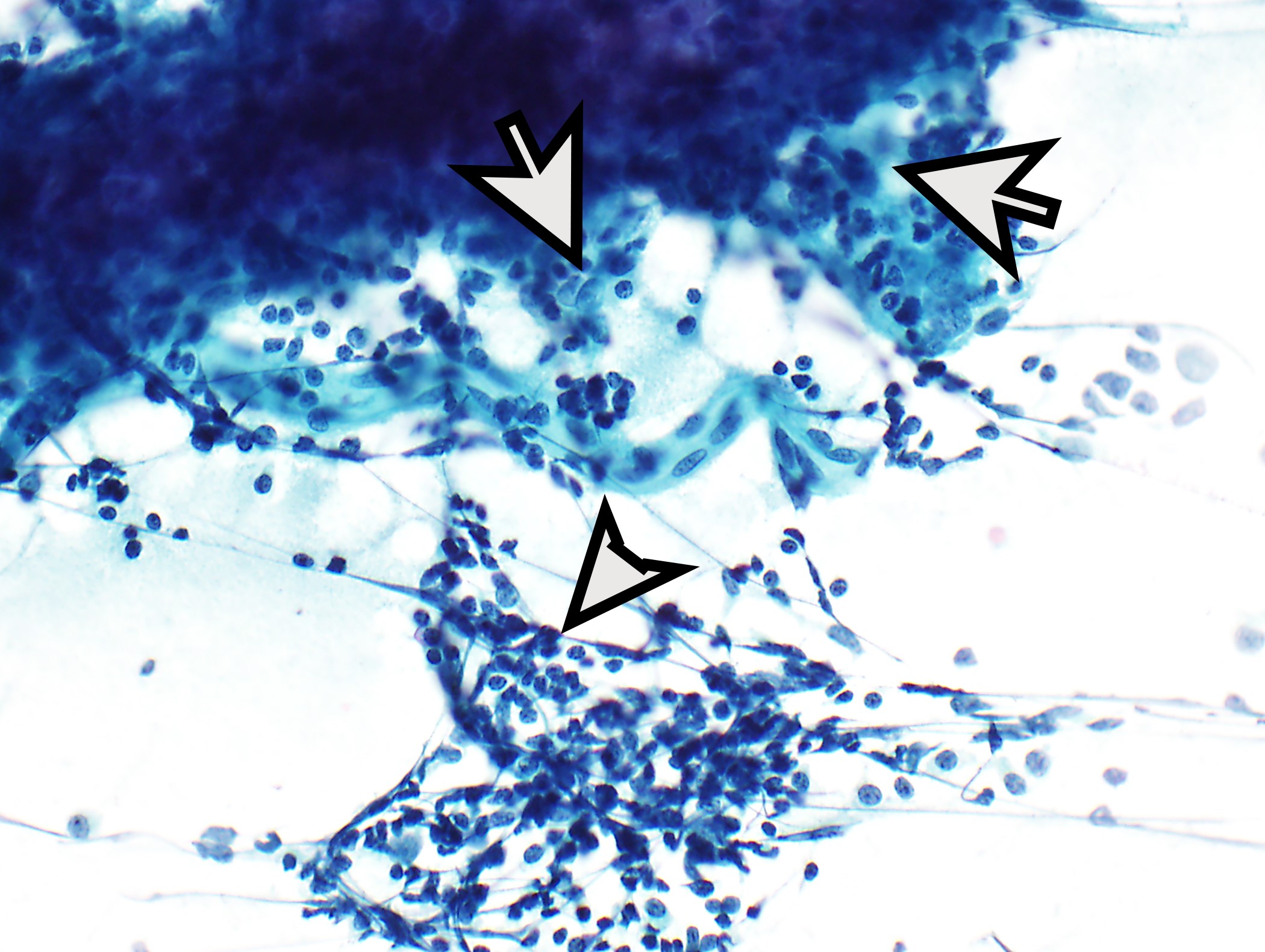
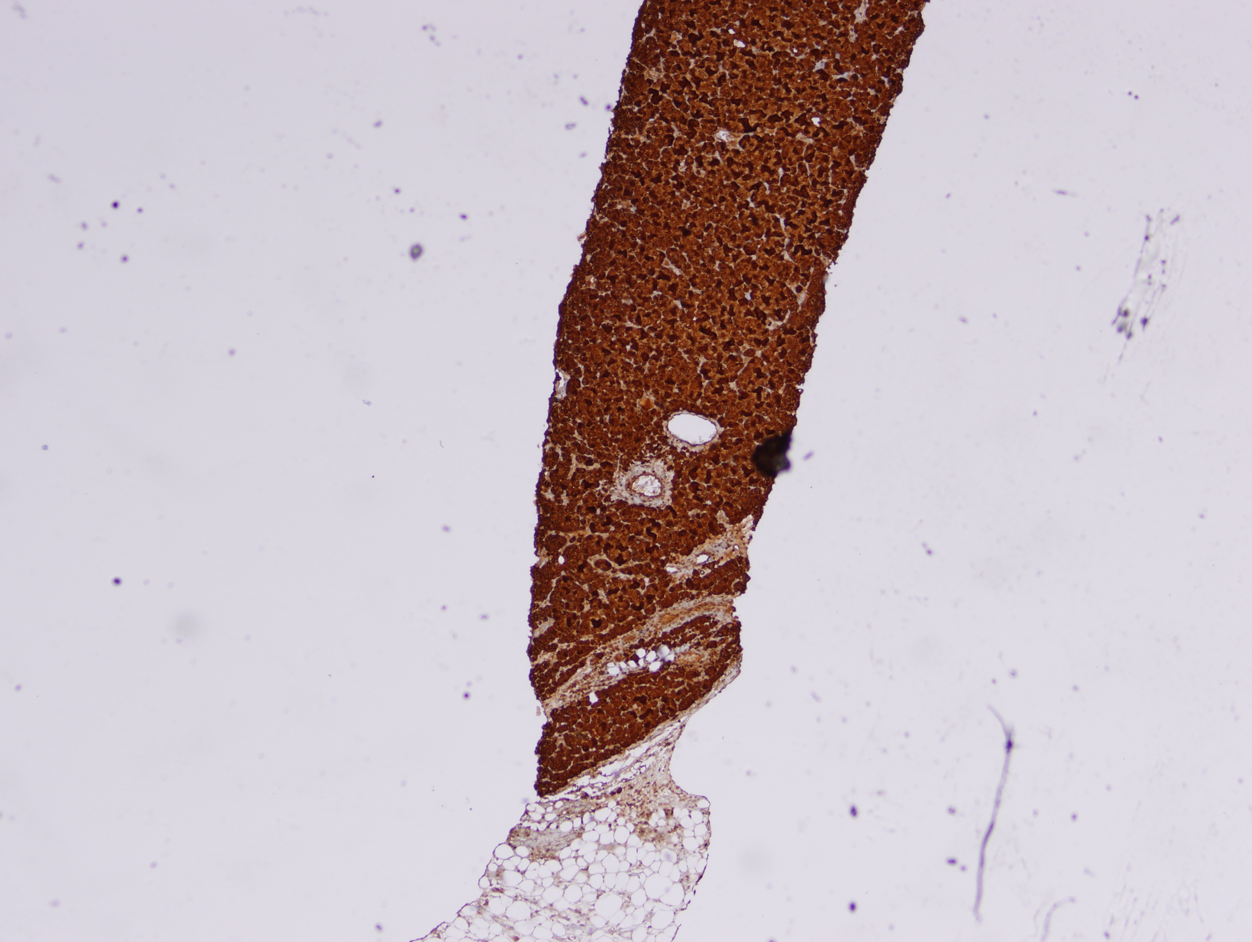
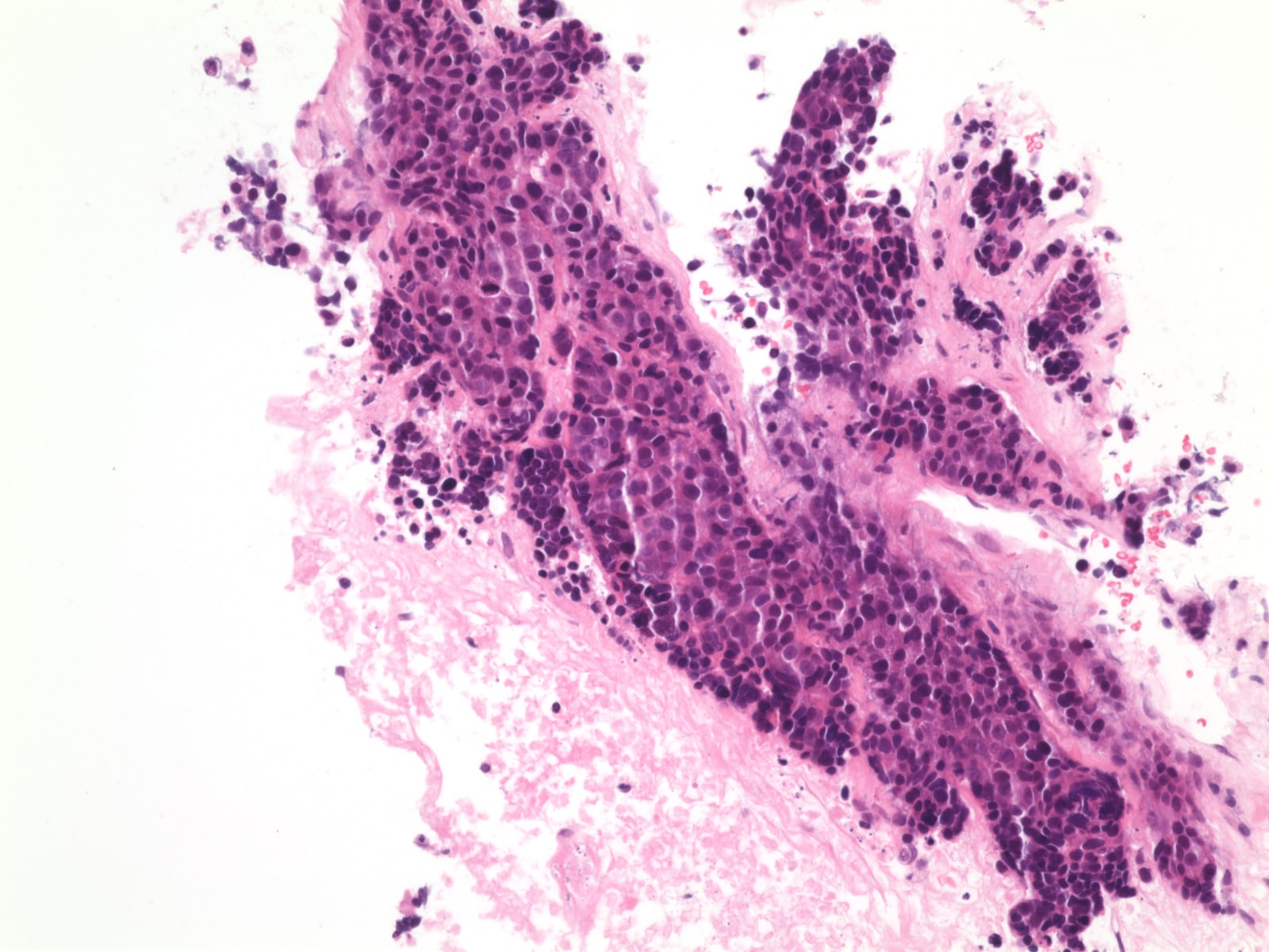
Cytology preparation of thymoma
Images hosted on other servers:

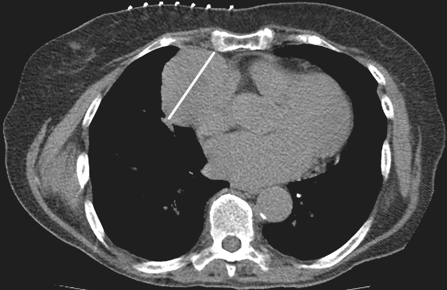
25 year old woman with pure red cell aplasia

Chest CT scan showing anterior mediastinal mass
Images hosted on other servers:

Intraoperative images following successful resection
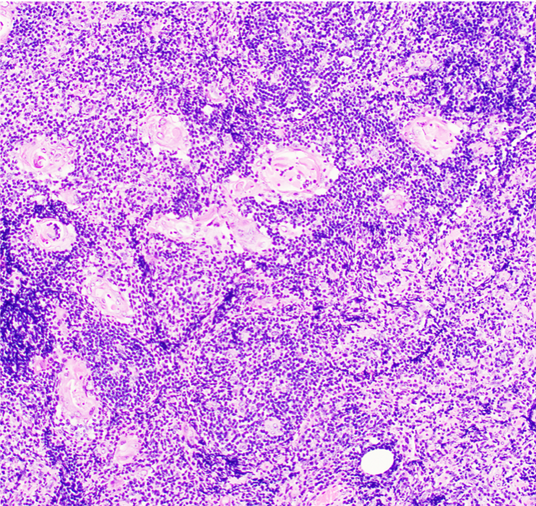
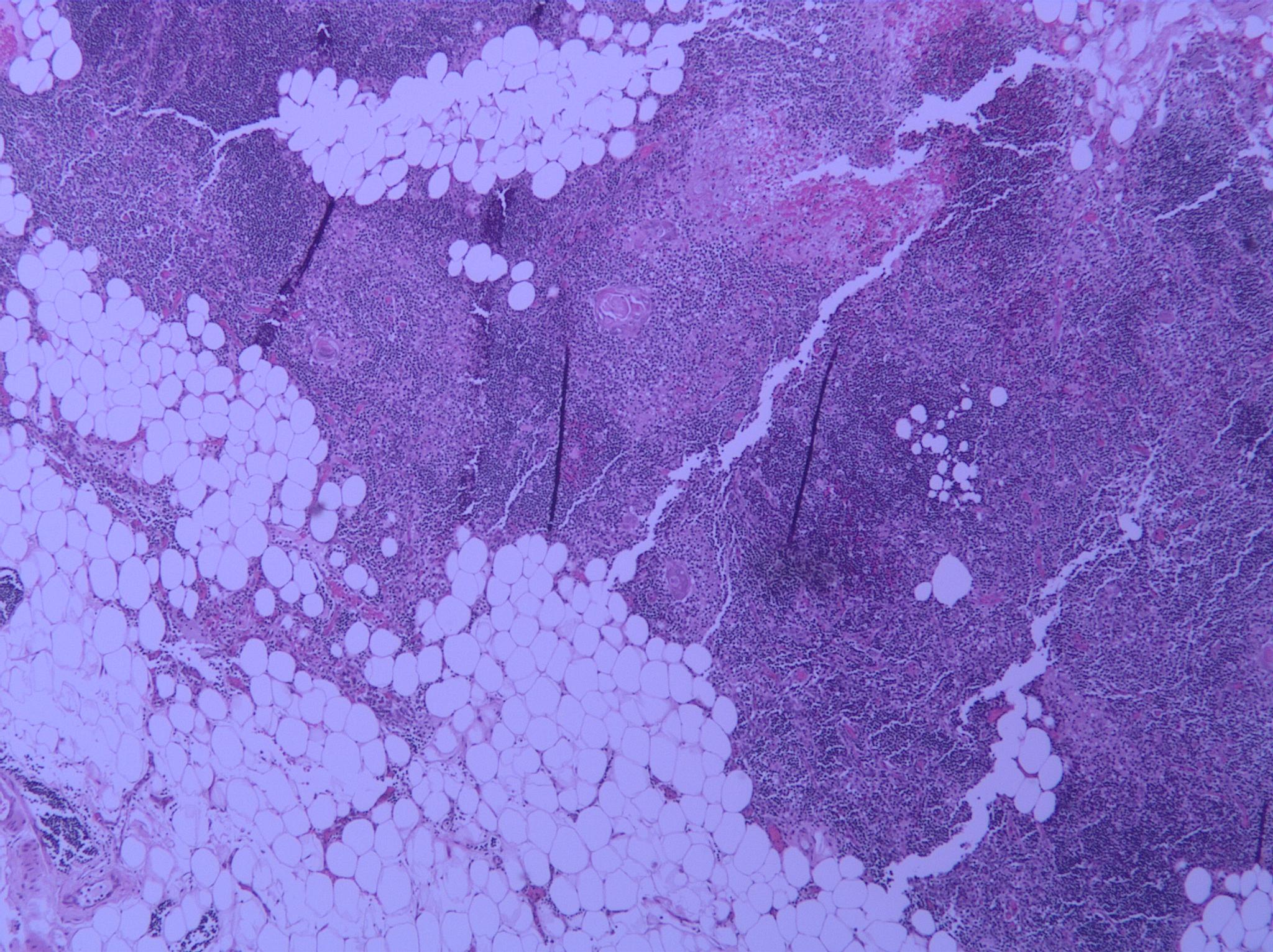
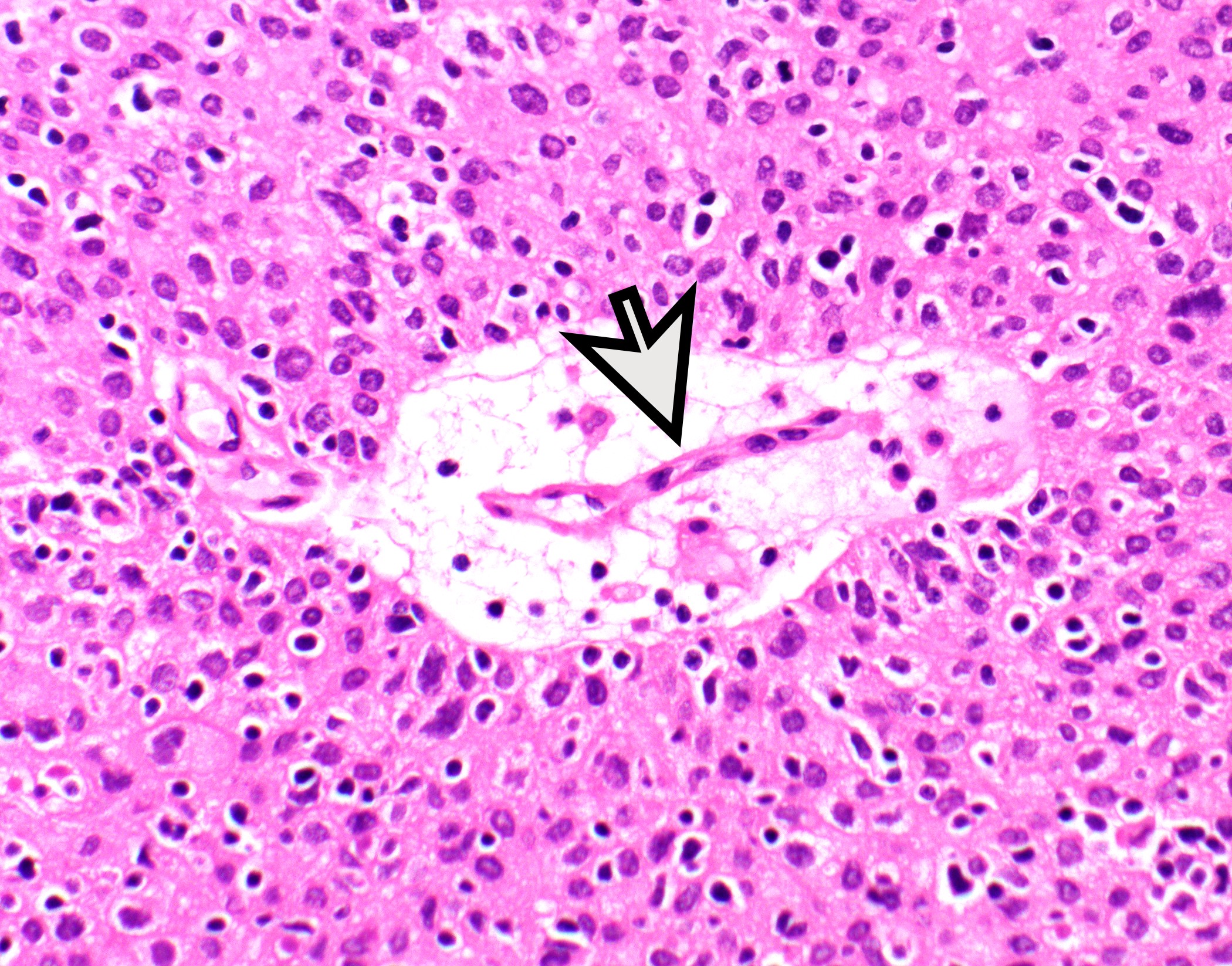
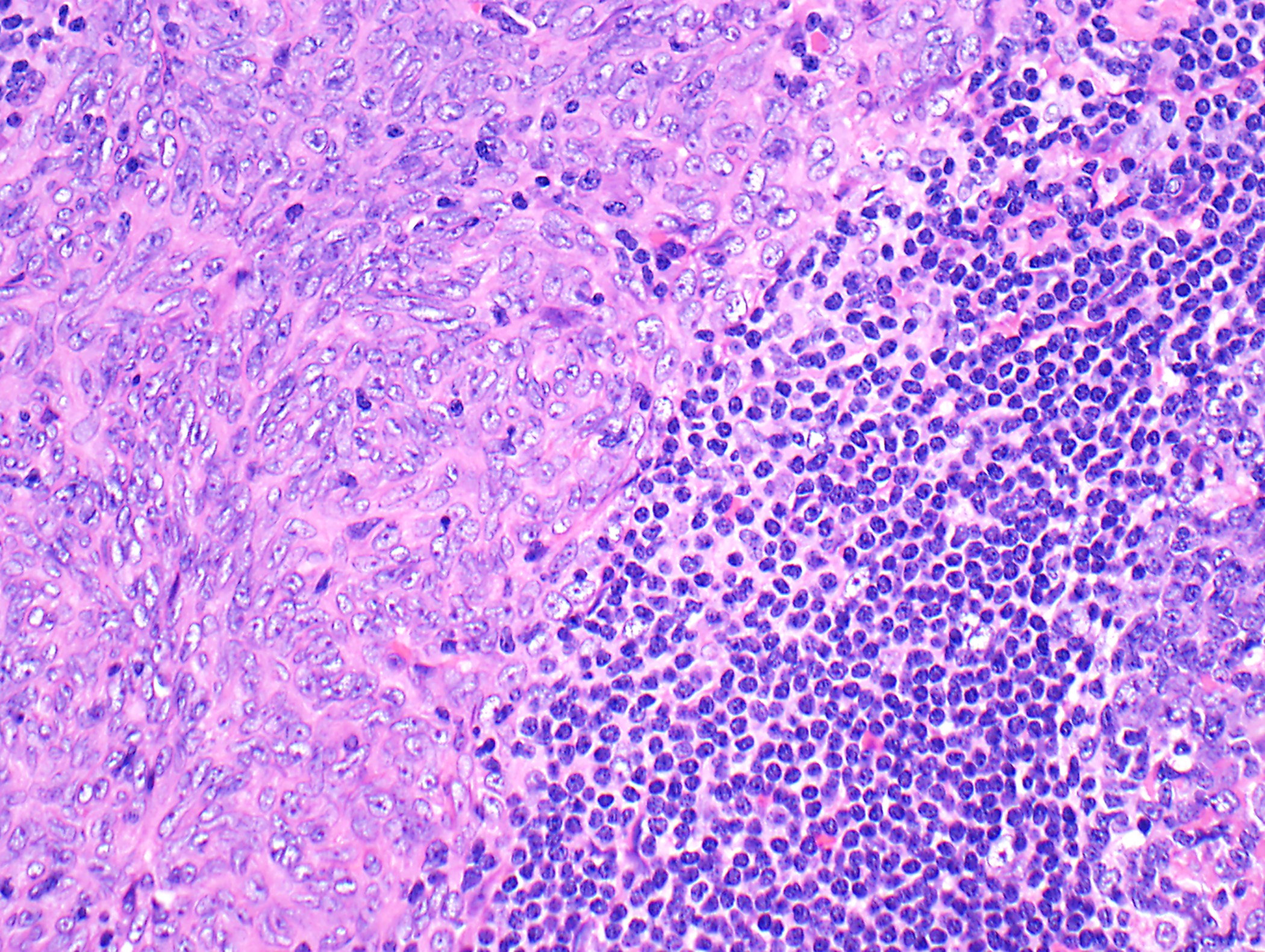
Contributed by Azadeh Esmaeili, M.D. and Sarmad H. Jassim, M.D.
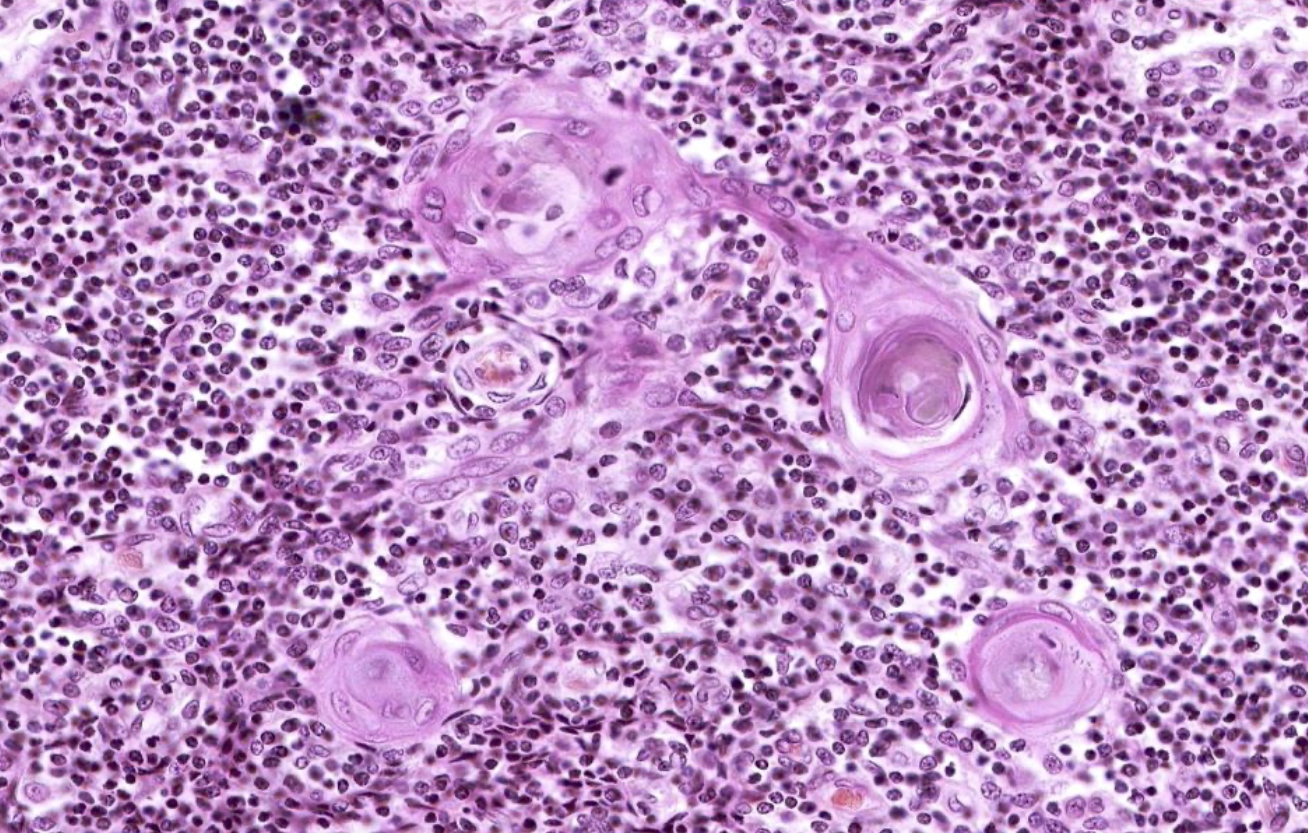
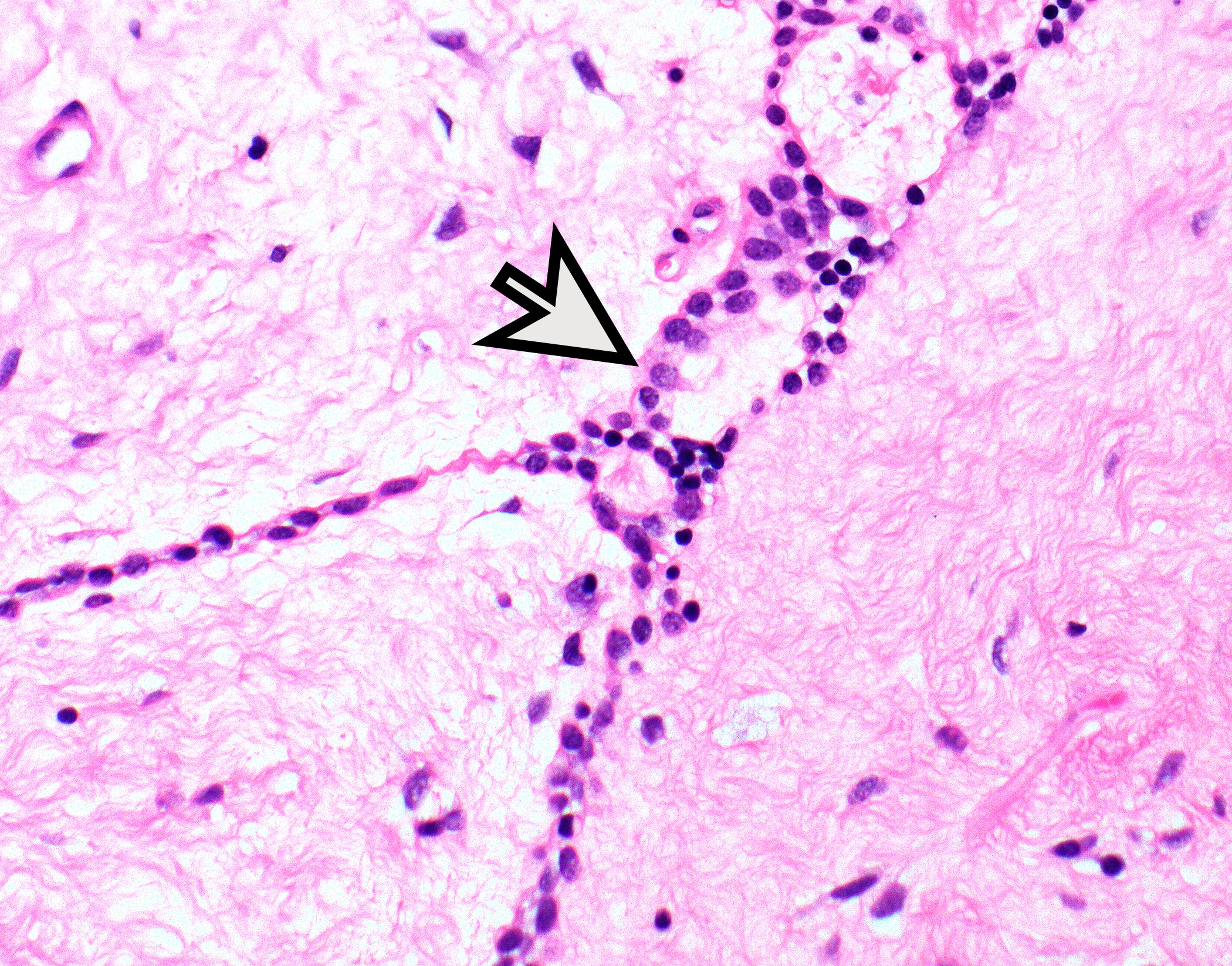
Expansion of normal component of thymic gland
Hassall corpuscles
Allen: 2015
Bardales: 2015
IARC: 2015
IARC: 2021
Mukhopadhyay: 2023
Shimosato: 2010
Suster: 2022
Find related Pathology books: cardiovascular, lung, mediastinum/serosa
