
Images hosted on other servers:

Large right nasal cavity mass

Contrast CT of a soft tissue mass

MRI with gadolinium showing an enhancing mass
Images hosted on other servers:

Painless, soft mass on the glabellar region

Gray-white polypoidal mass in right nostril
Images hosted on other servers:

Fragile grayish white mass
Contributed by Diana Bell, M.D.
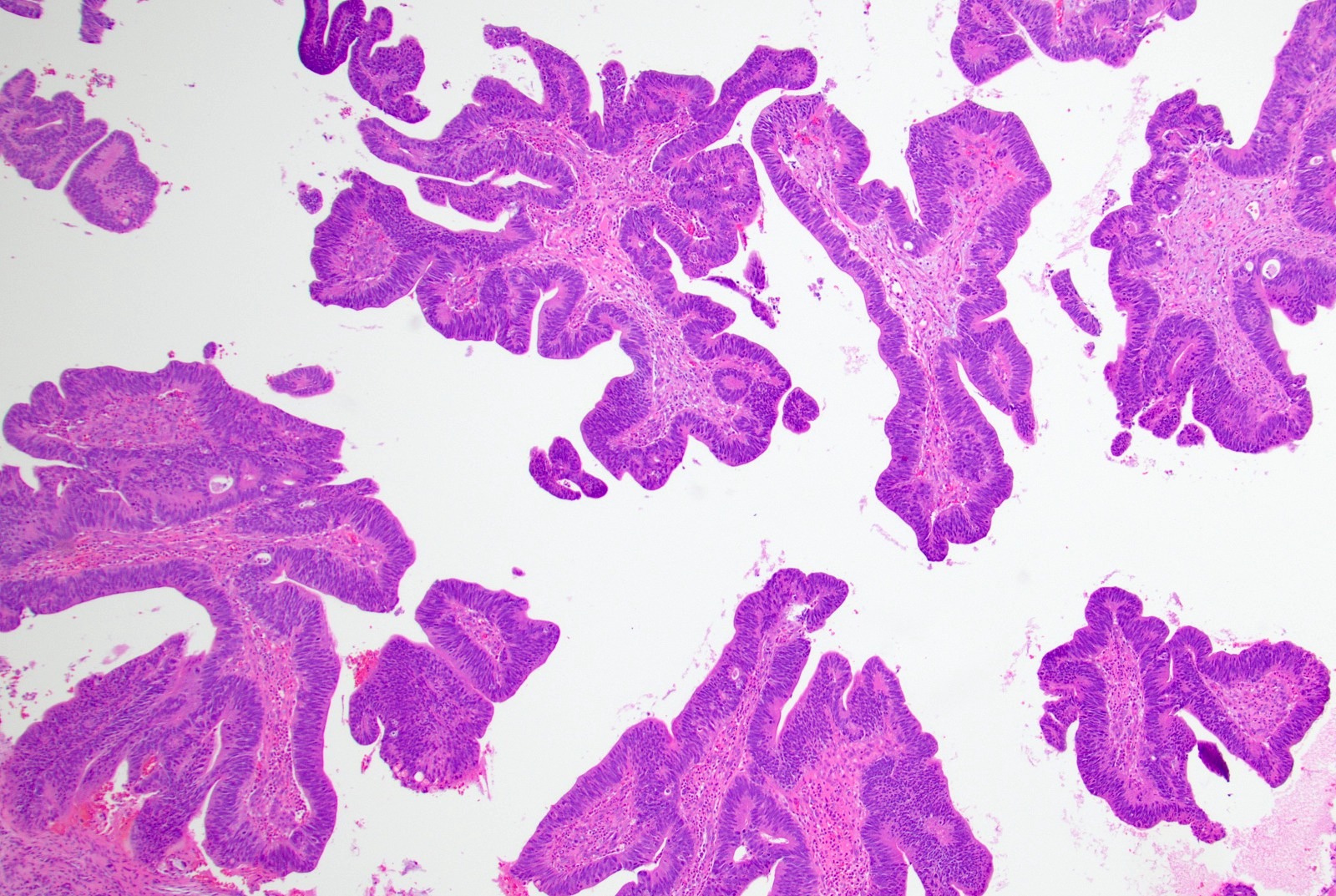
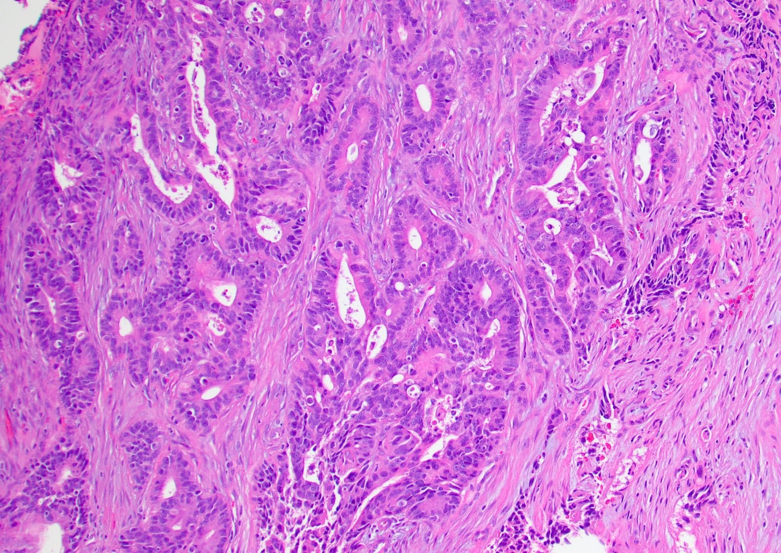
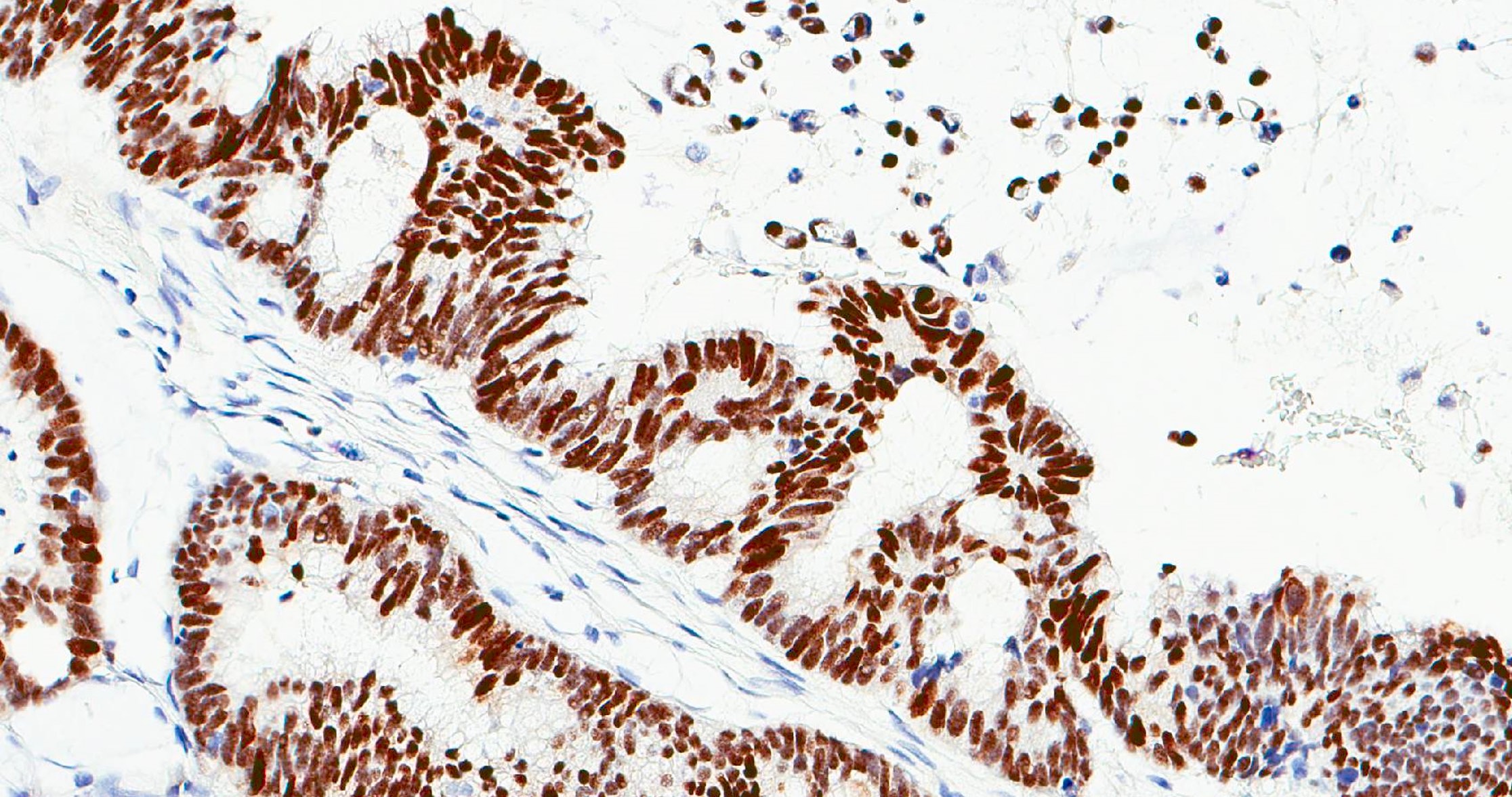
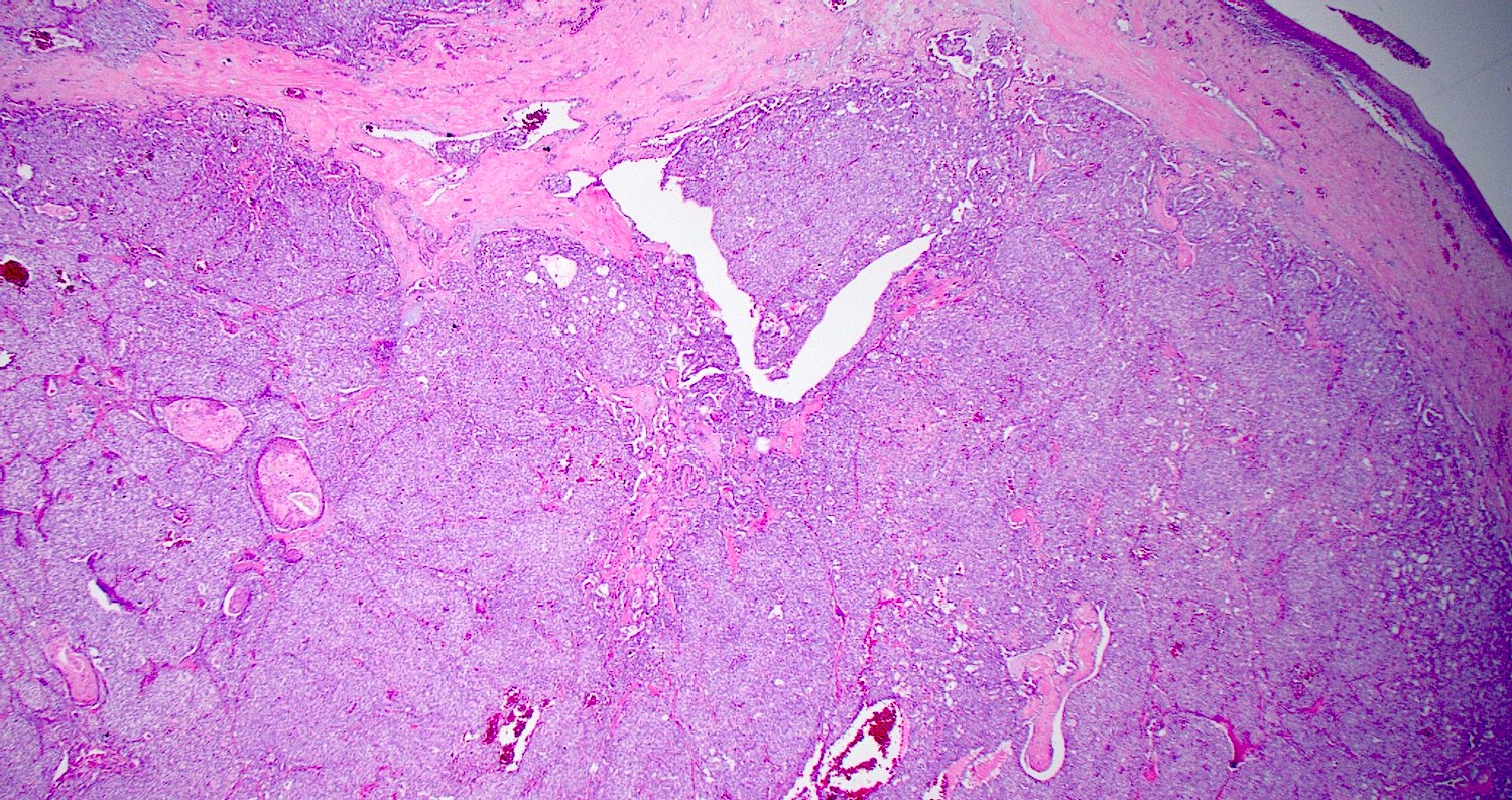
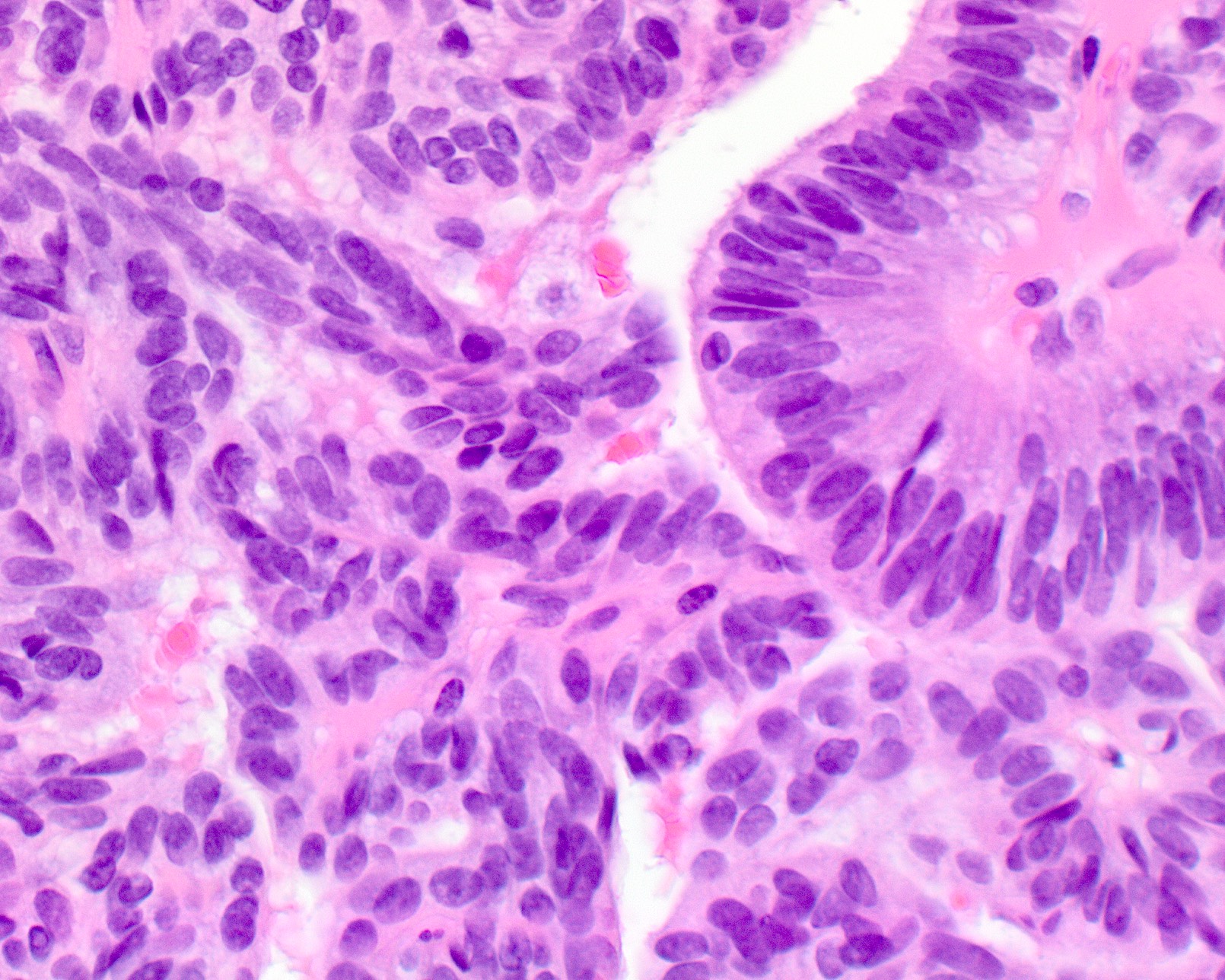
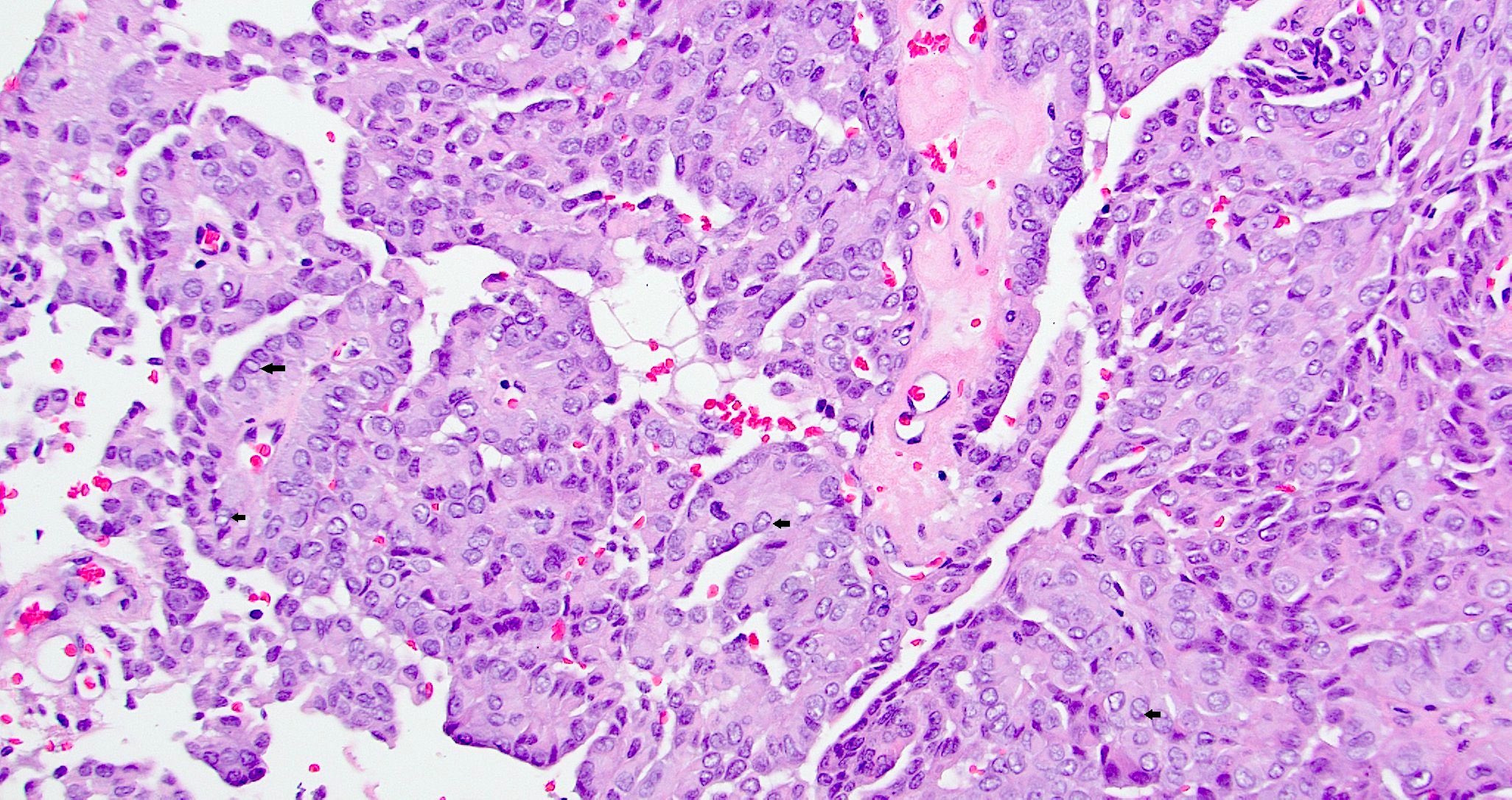
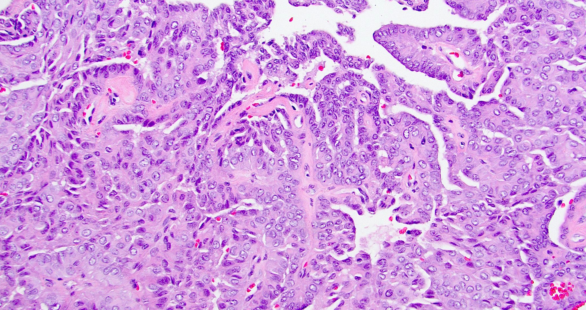
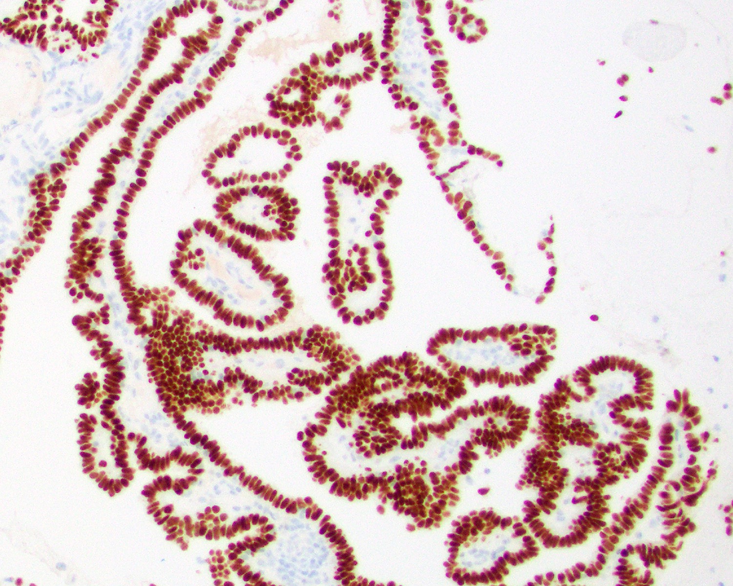
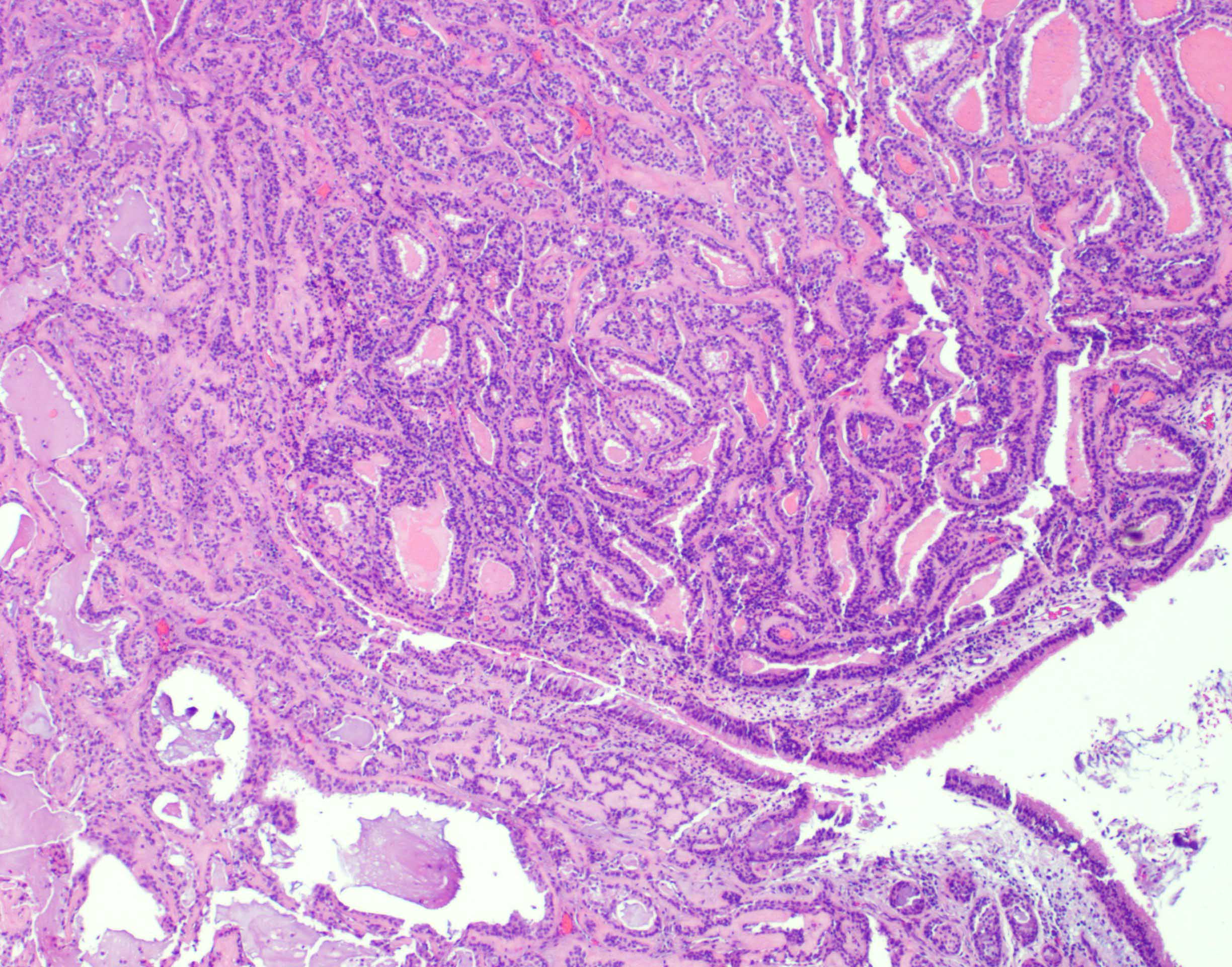
Intestinal adenocarcinoma (ITAC)
Various architectures
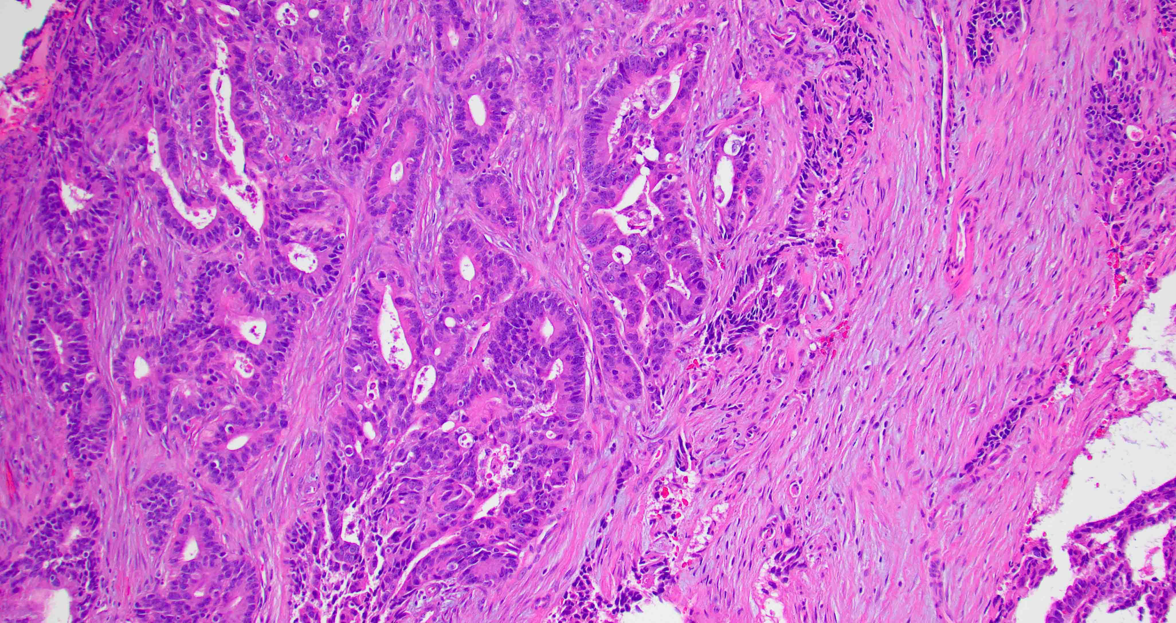
Tubular and cribriform growth pattern
Papillary growth pattern
Nonintestinal adenocarcinoma (non-ITAC)

Low grade with uniformly located nuclei

Tubular growth pattern
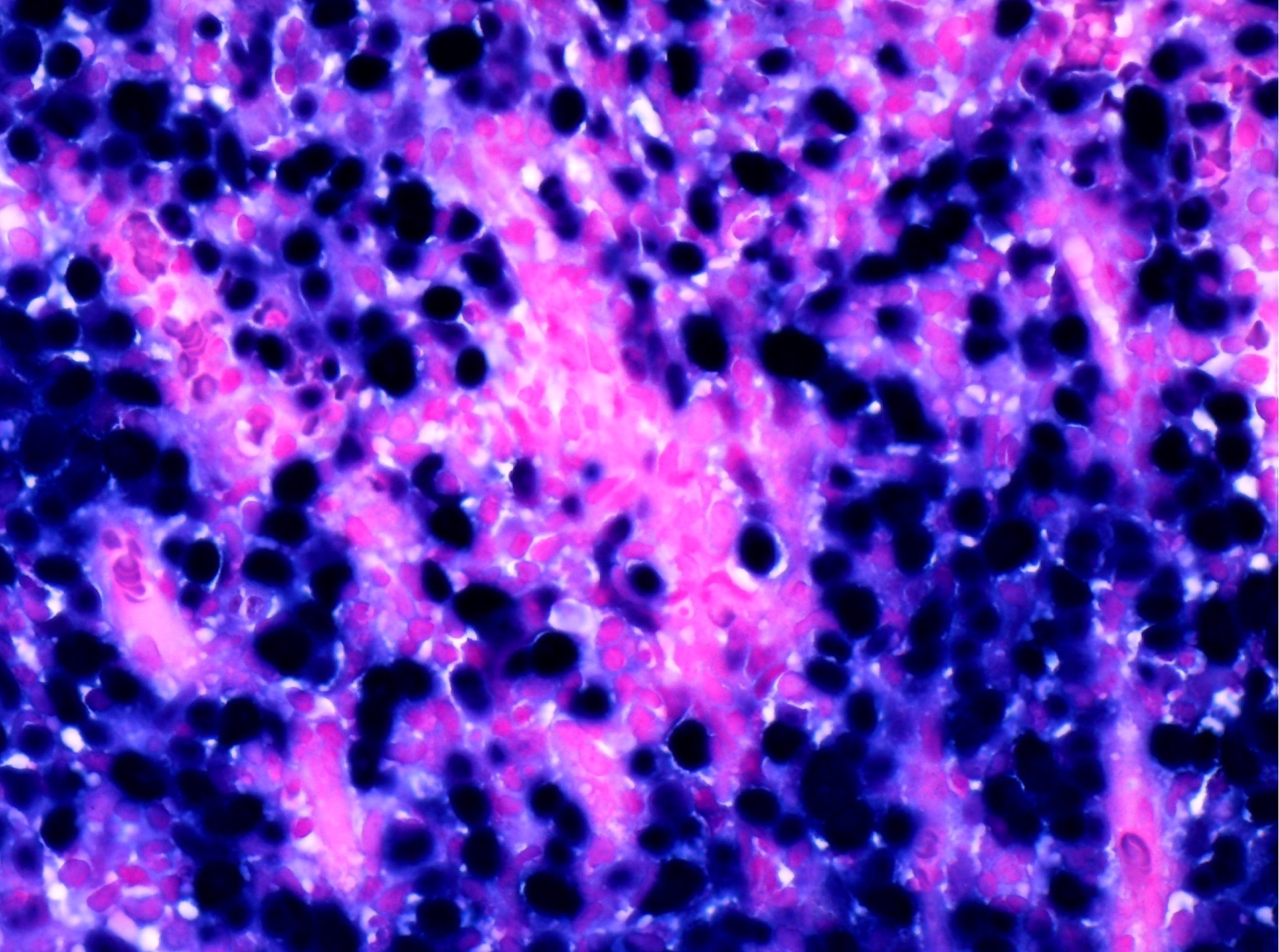
Sinonasal renal cell-like adenocarcinoma
Nested architecture
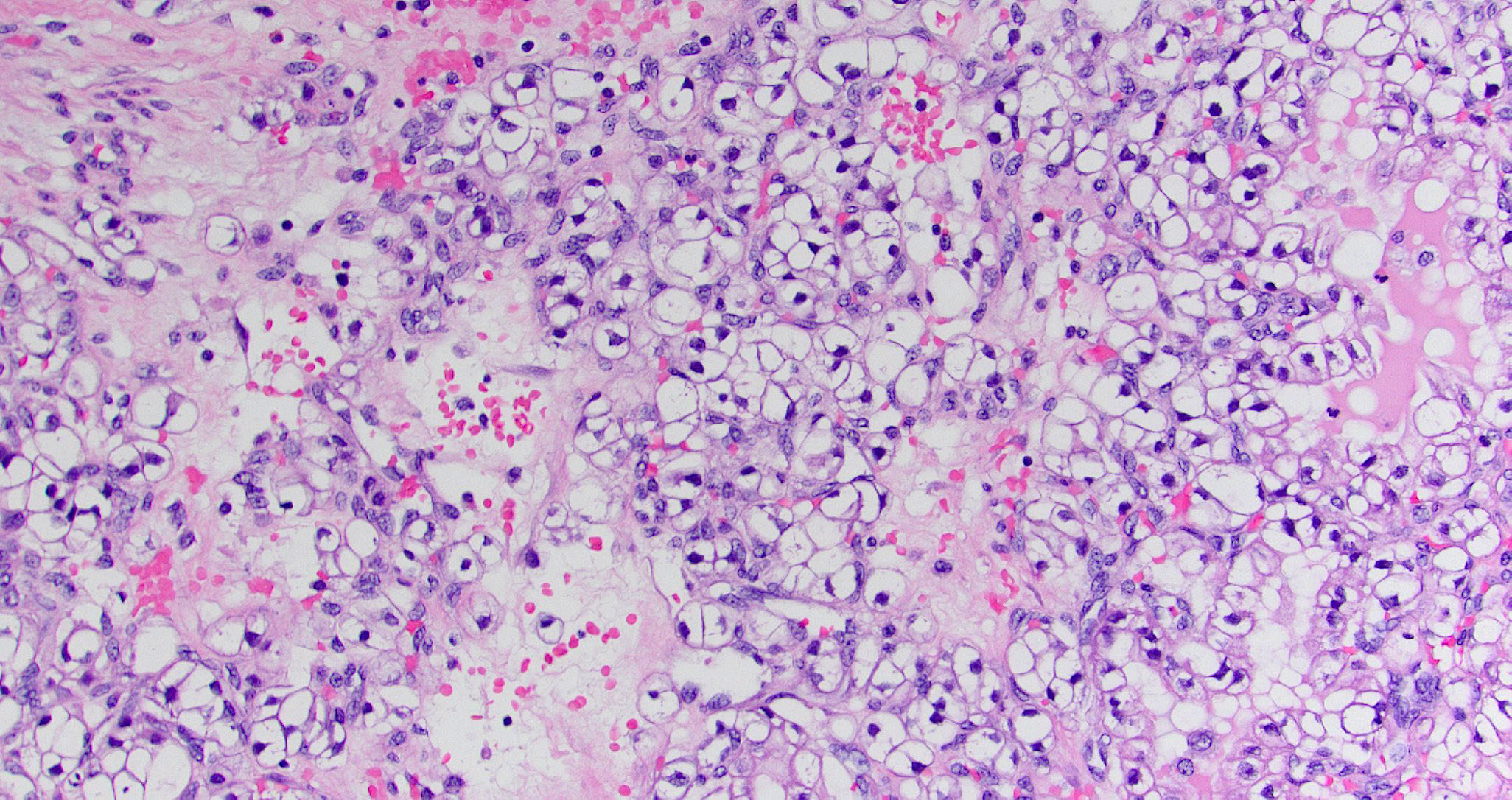
Clear cytoplasm
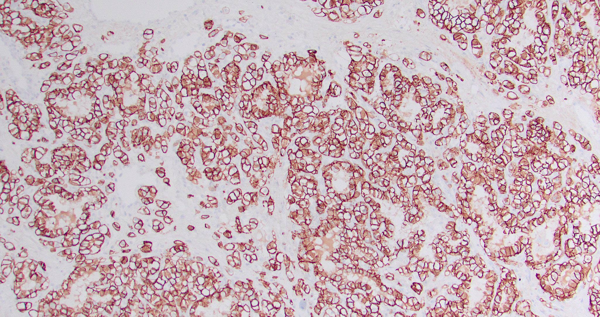
CAIX positivity
Sinonasal carcinoma: updated phenotype and molecular characterization
Malignant tumors of the paranasal sinuses by Dr. Nadir Ahmad
Contributed by Margie Brandwein-Gensler, M.D.
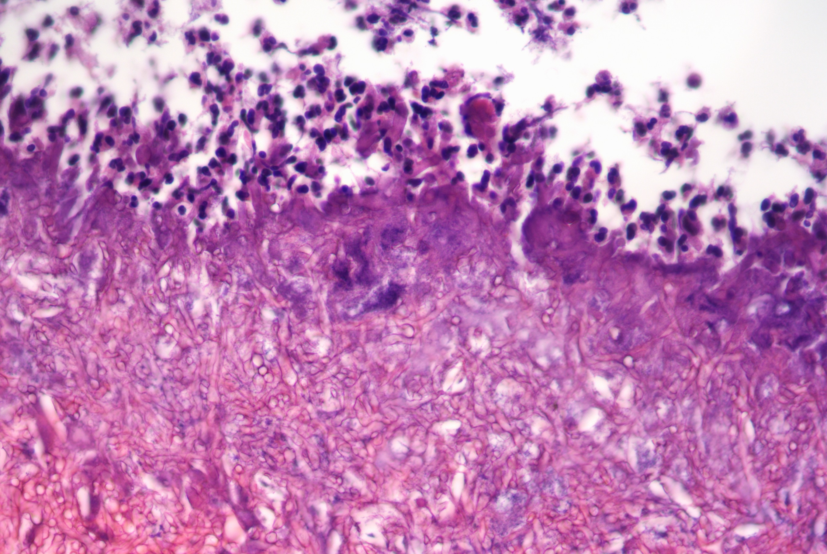
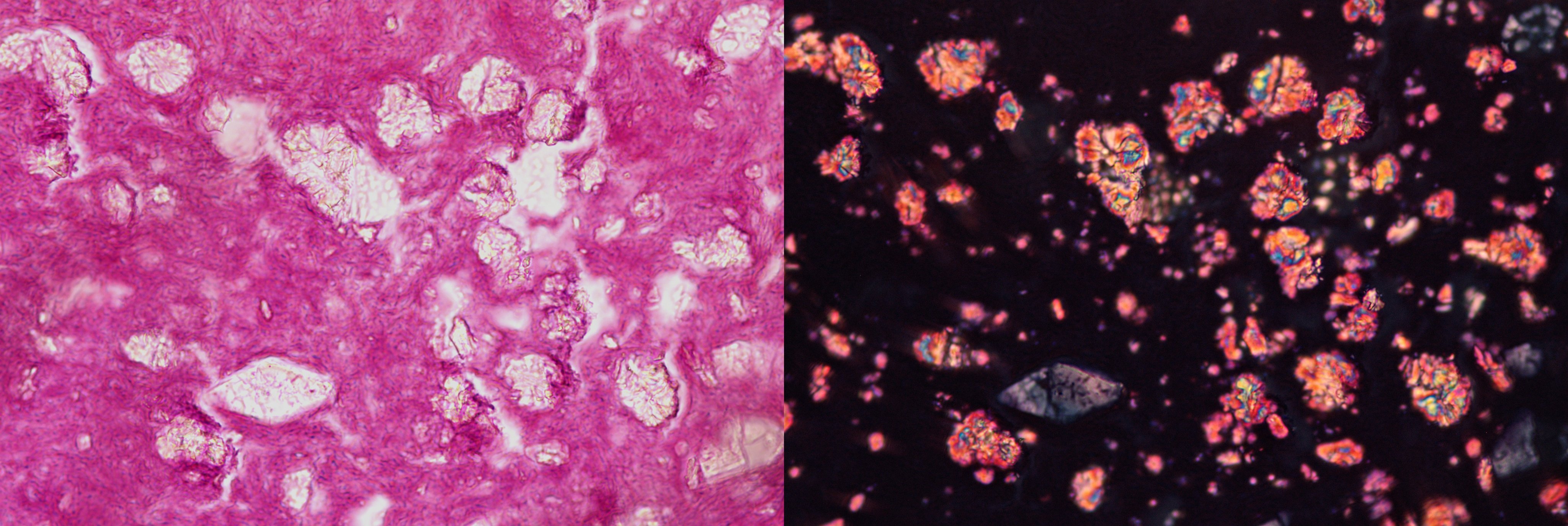
Allergic fungal rhinosinusitis
Contributed by Kelly R. Magliocca, D.D.S., M.P.H.
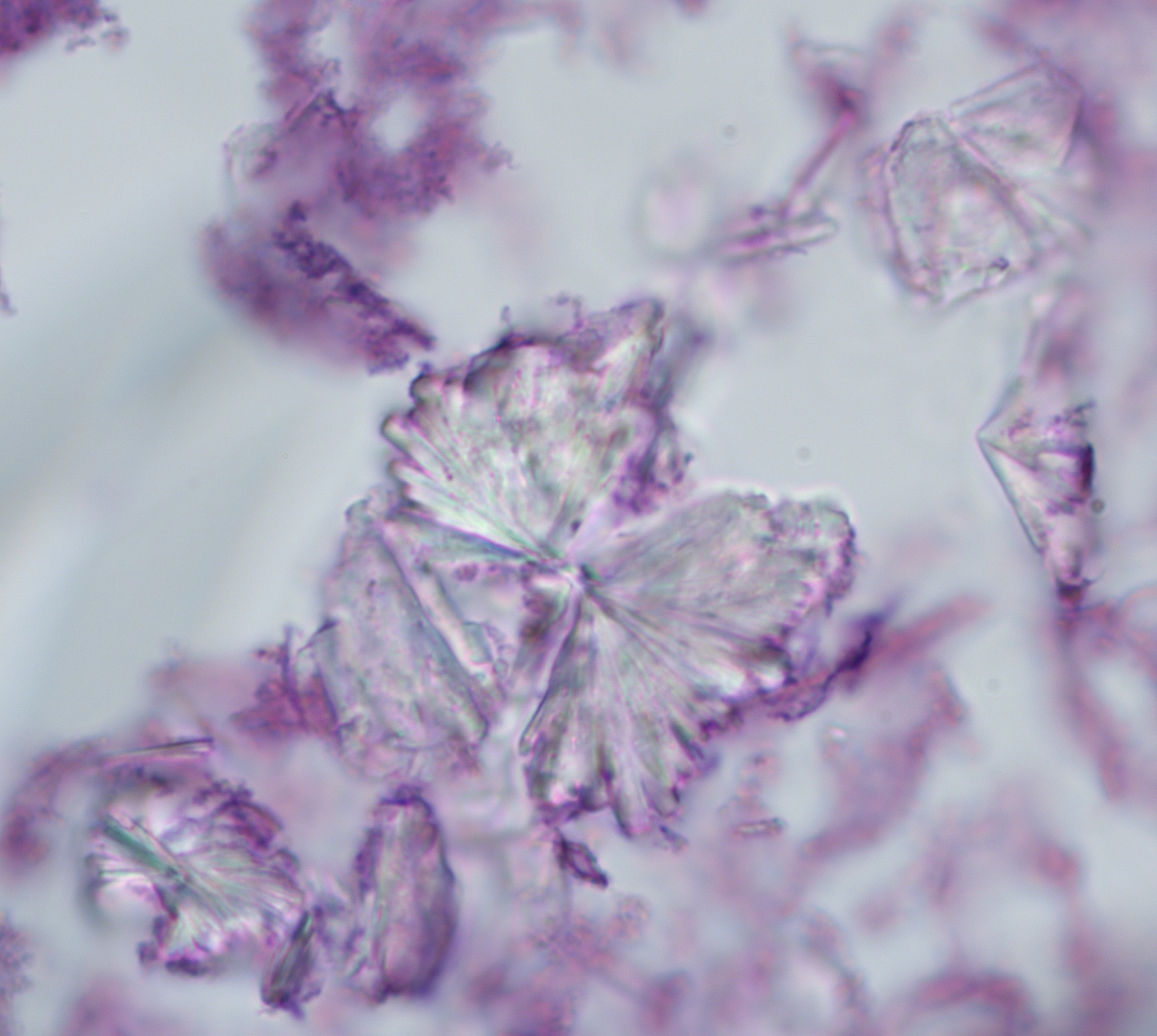
Eosinophilic mucin associated with allergic fungal sinusitis

Charcot-Leyden crystals

Grocott-Gomori methenamine silver
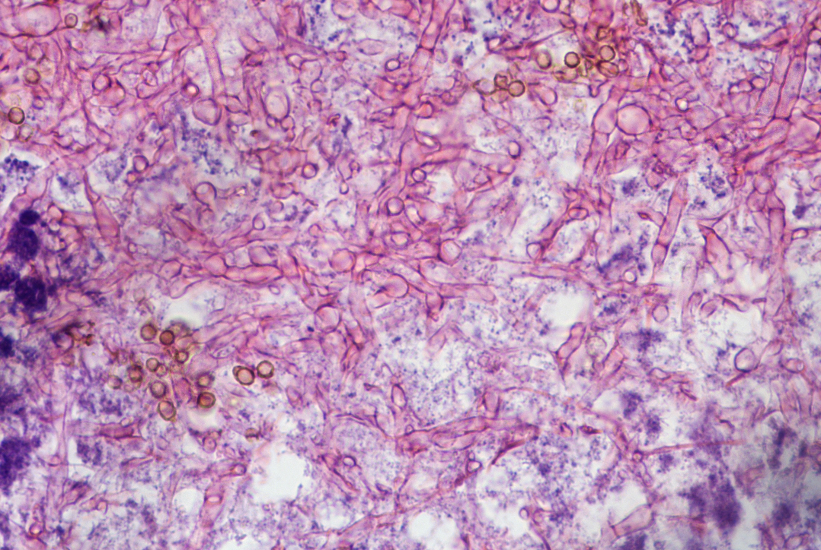
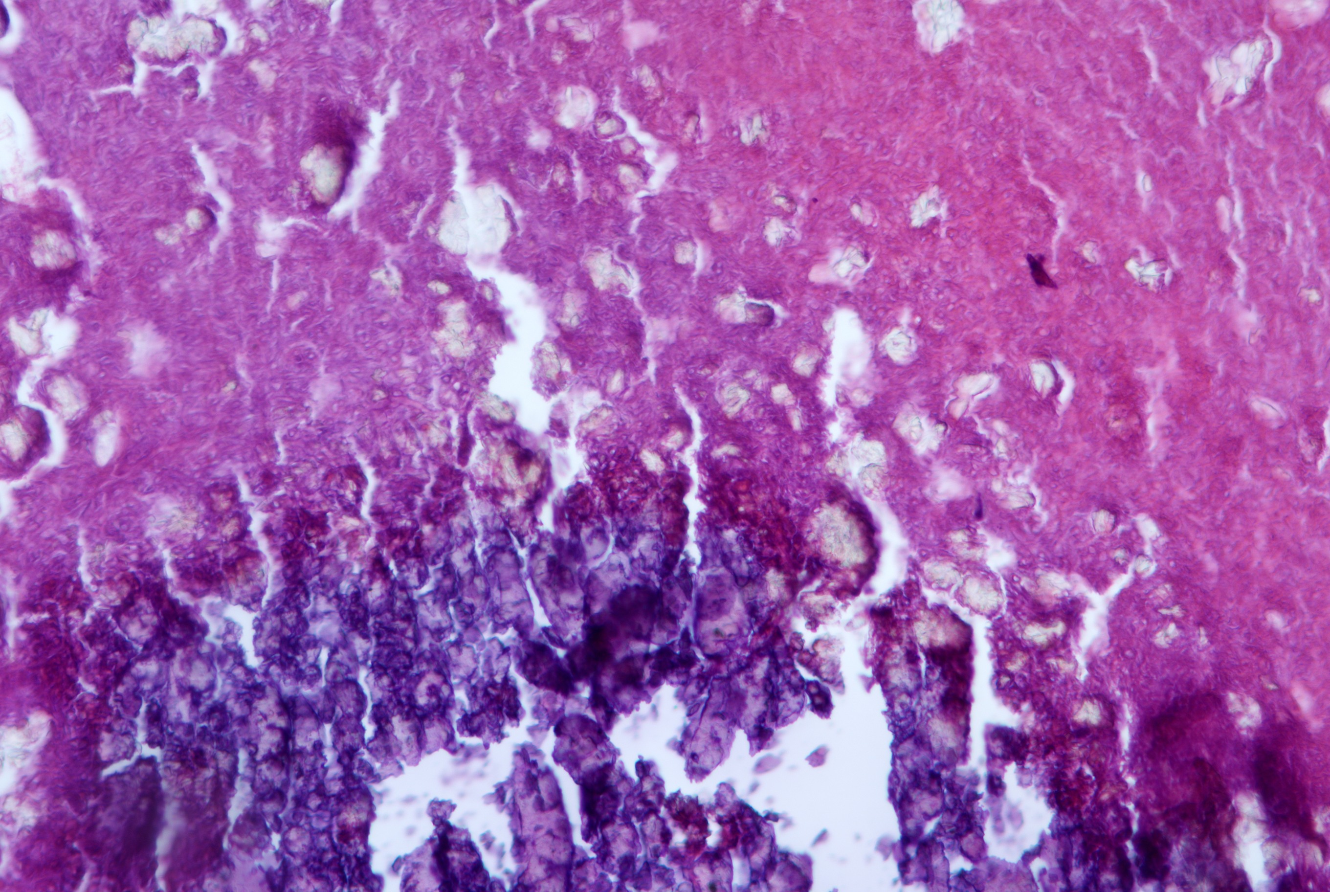
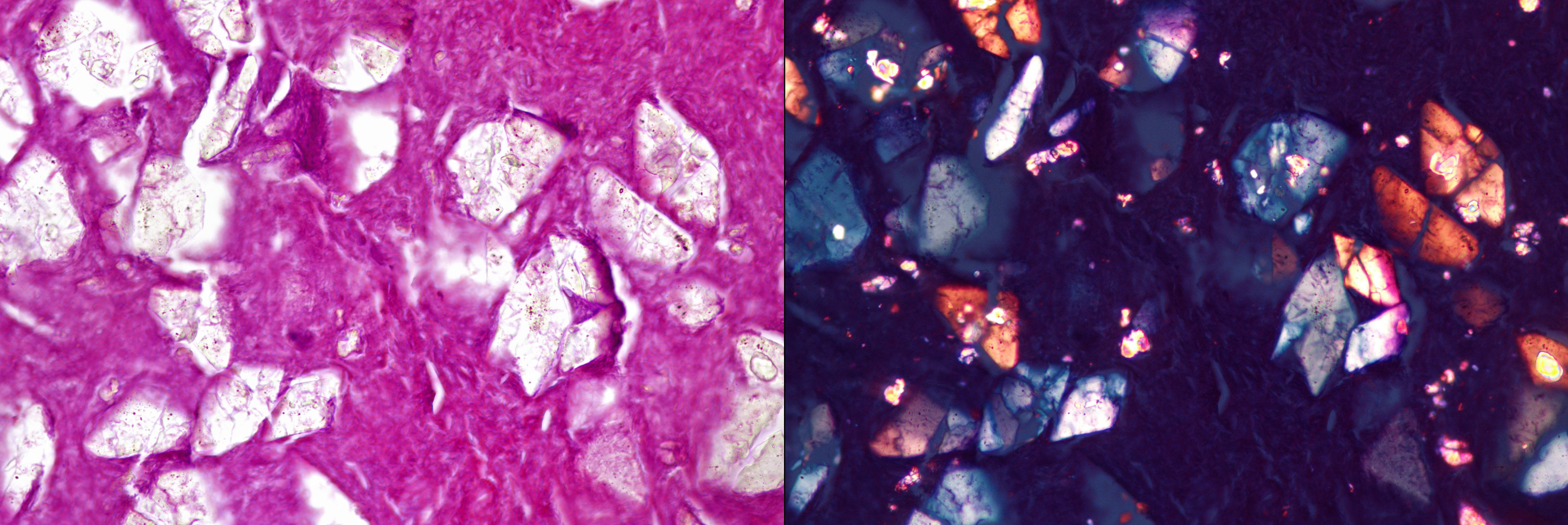
Contributed by Margie Brandwein-Gensler, M.D.
Eosinophilic mucin

Fungal detection
Images hosted on other servers:

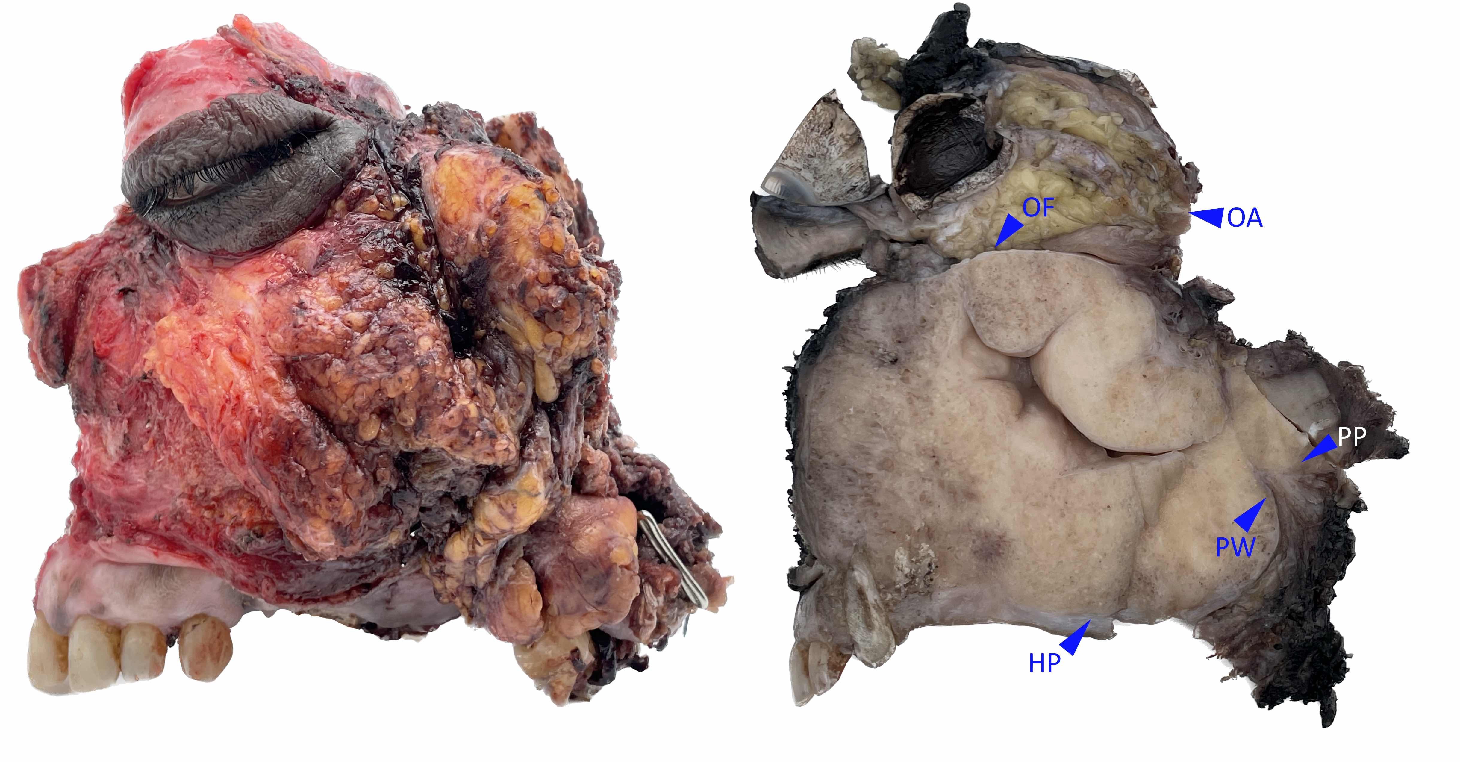
Lateral wall

Bones and cartilages
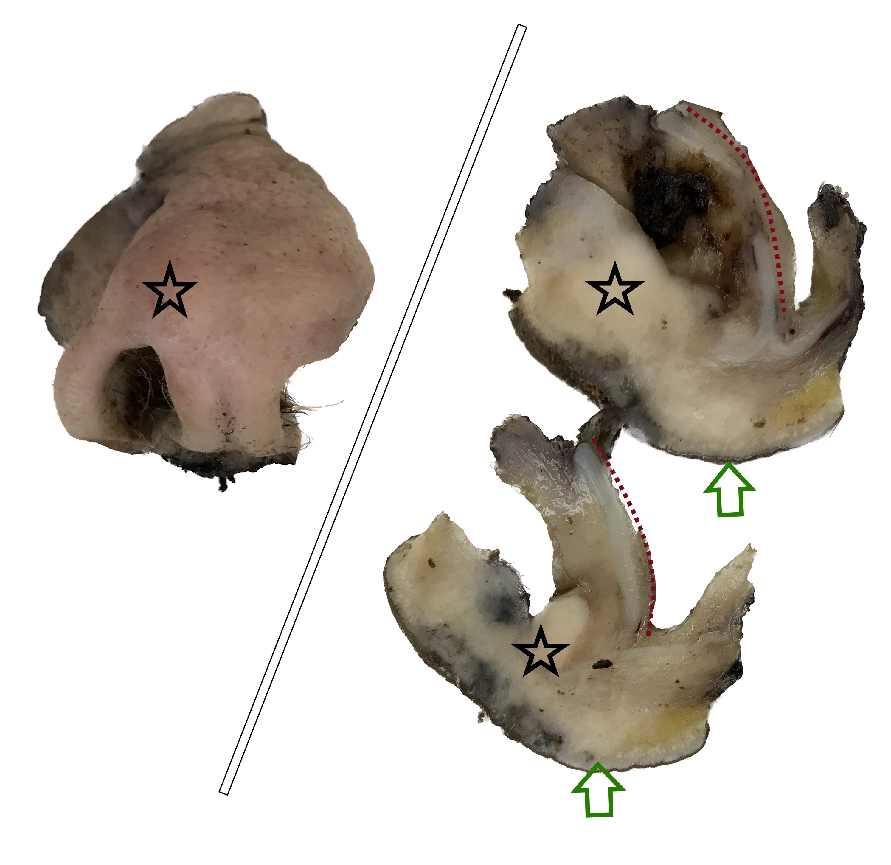
Case #390

Various images
Contributed by Josephine K. Dermawan, M.D., Ph.D. and Laura O. Rabinowitz, M.D.
Superior nasal cavity mass (coronal)
Superior nasal cavity mass (transverse)
Contributed by Josephine K. Dermawan, M.D., Ph.D. and Laura O. Rabinowitz, M.D.

Cellular spindle proliferation
Prominent stromal vasculature
Surface epithelial invaginations

Hemangiopericytoma-like vasculature
Herringbone pattern
Medium to long fascicles
Positive S100
Positive SMA
Negative CD34
Contributed by Margie Brandwein-Gensler, M.D.

Sinonasal polyps
Chronic rhinosinusitis
Various images
Images hosted on other servers:

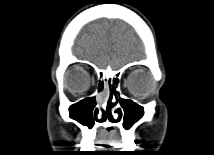
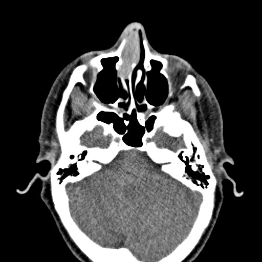
CT of face

MR of face

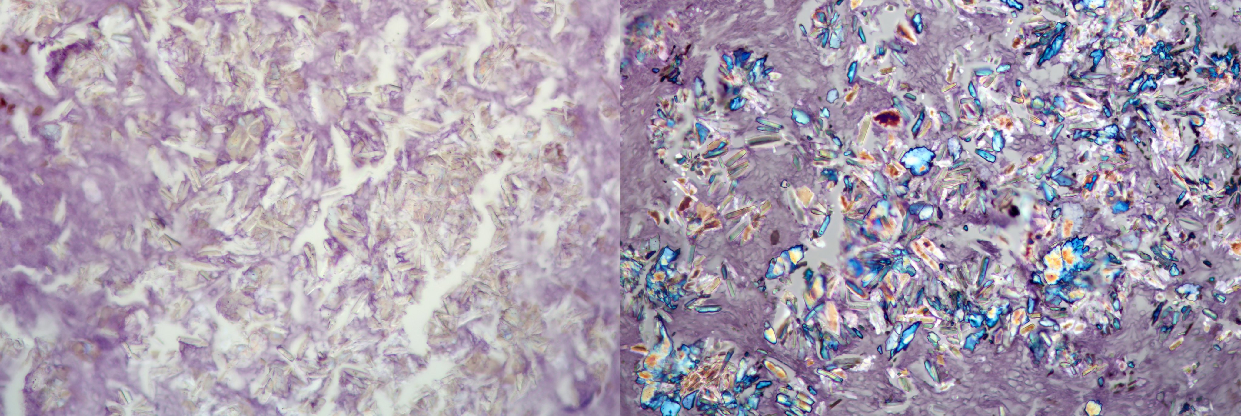
Likely calcific in nature

Normal pneumatization

Opacification
Contributed by @Andrew_Fltv on Twitter
Fungal ball
Images hosted on other servers:

Maxillary sinusotomy
Contributed by Margie Brandwein-Gensler, M.D. and @Andrew_Fltv on Twitter
Large fungus ball
Fungal hyphae, fungal sexual reproduction (fruiting heads)
Fungal ball
Fungal ball
Aspergillus lesions of the sinuses
Images hosted on other servers:

Aspergilloma

Hyphae of mycetes

Foot
Contributed by Nasir Ud Din, M.B.B.S.
Nasal swelling
Nasal cavity lesion
Nasal cavity mass
Images hosted on other servers:

External nasal lesion with skin tightening

Globular swelling at the root of nose

Nasal endoscopy
Images hosted on other servers:

Homogeneous, tan, firm cut surface
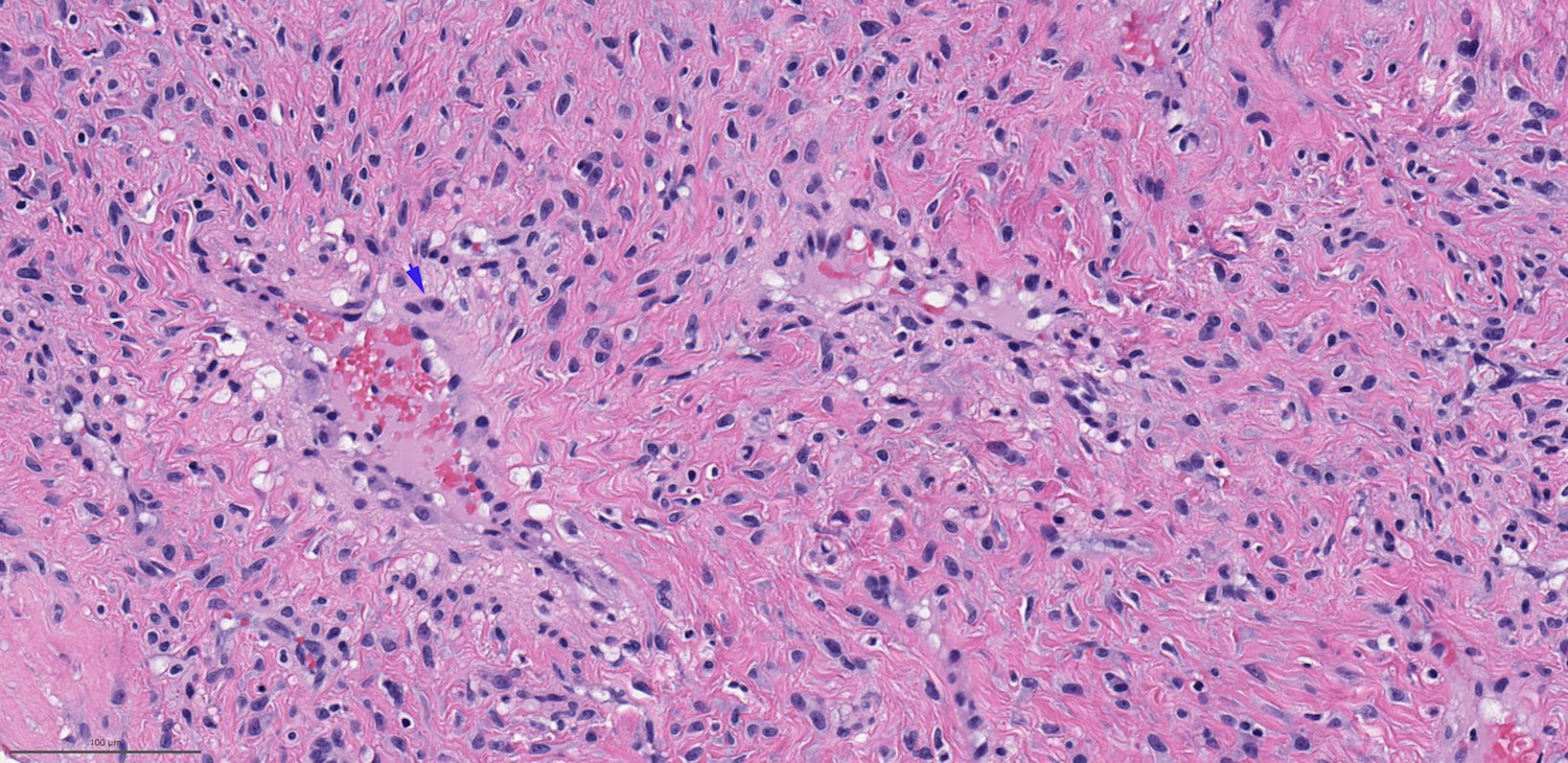
Contributed by Nasir Ud Din, M.B.B.S.
Polypoid lesion
Syncytial architecture

Nested architecture
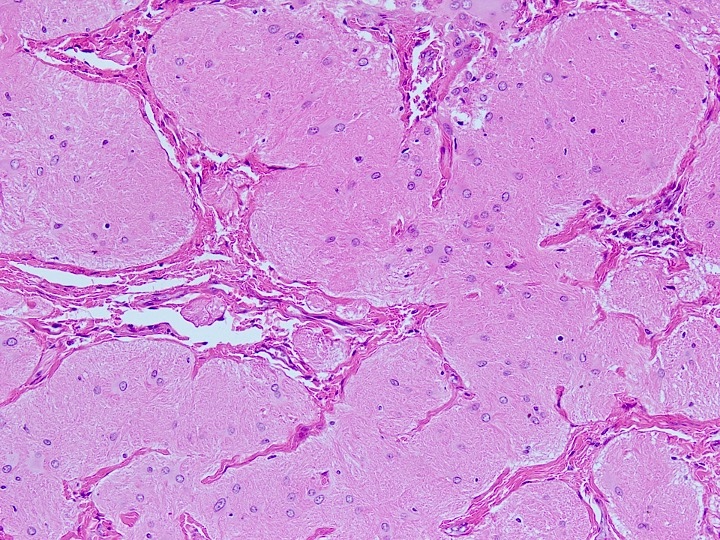
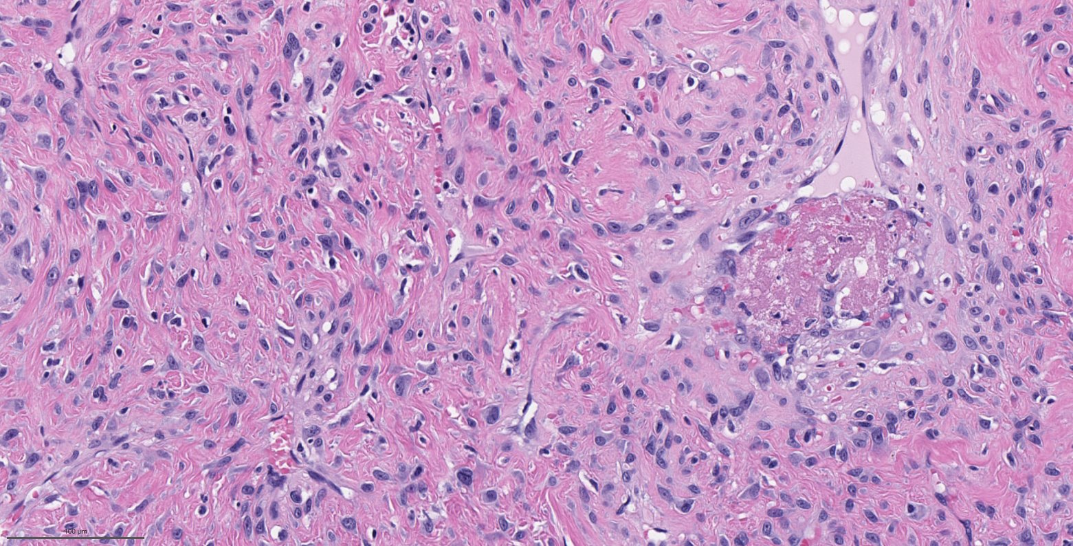
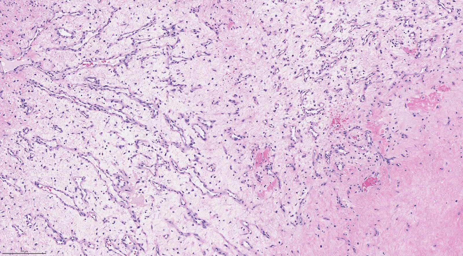
Astrocytes in fibrillary matrix
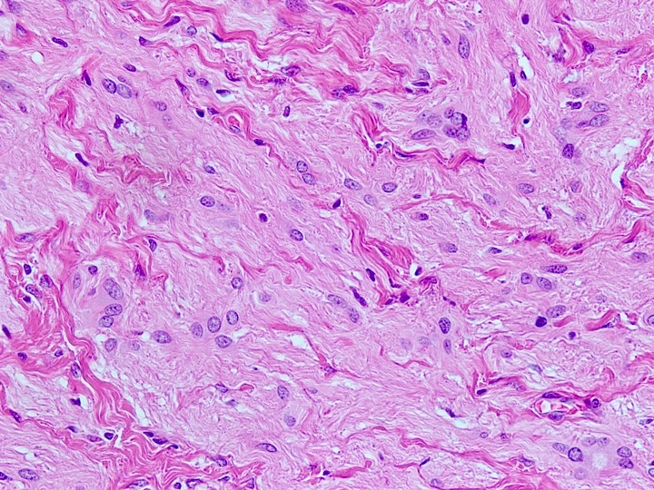
Astrocyte morphology
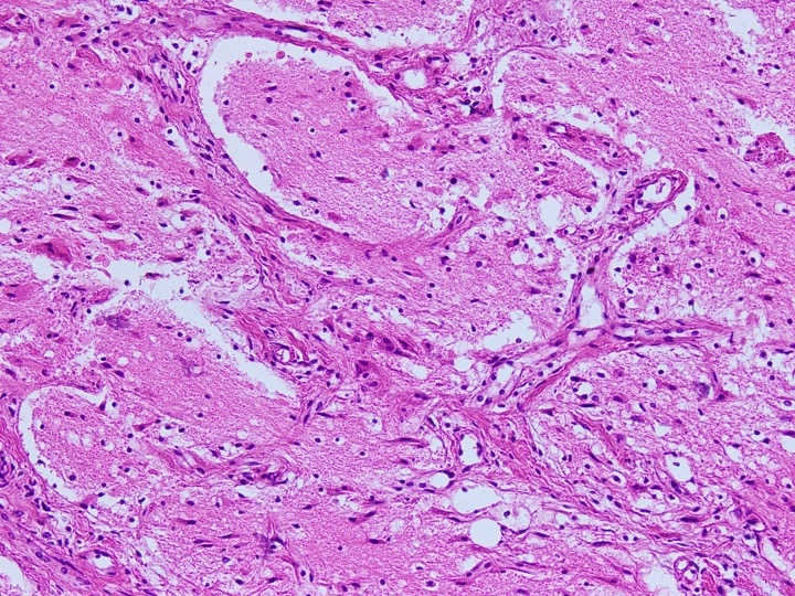
Astrocytes in mild inflammatory stroma
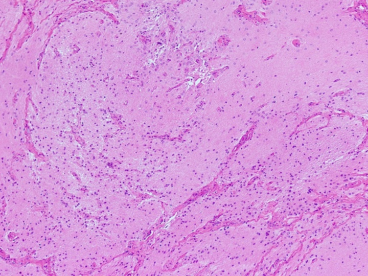
Vascularized background
Fibrotic appearance
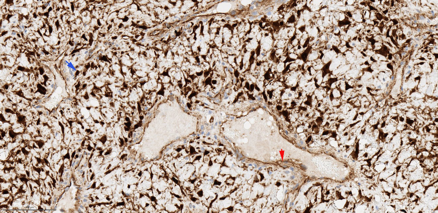
Positive GFAP expression
Images hosted on other servers:

Epidemiology and clinical features over 2 decades
Contributed by Muthu Kumar Sakthivel, M.D.
Mass with fat attenuation
Pedunculated tubular fatty mass
3 dimensional reconstructed oral lesion
Images hosted on other servers:

Nasal oropharyngeal mass

Nasal vestibule mass
Contributed by Ezer Benaim, M.D.
Mobile oropharyngeal mass
Nasal endoscopy
Images hosted on other servers:

Polypoid lesion from mouth
Images hosted on other servers:

Skin covered pedunculated lesion

Polypoidal lesion
Contributed by Surekha Bantumilli, M.D. and Steven Johnson, M.D.
Squamous epithelium
Polypoidal appearance

Stratified squamous epithelium
Mesoderm derivative
Mesenchymal core
Images hosted on other servers:

Mass of maxillary sinus
Images hosted on other servers:

Expansile swelling of maxilla
Contributed by Bin Xu, M.D., Ph.D.
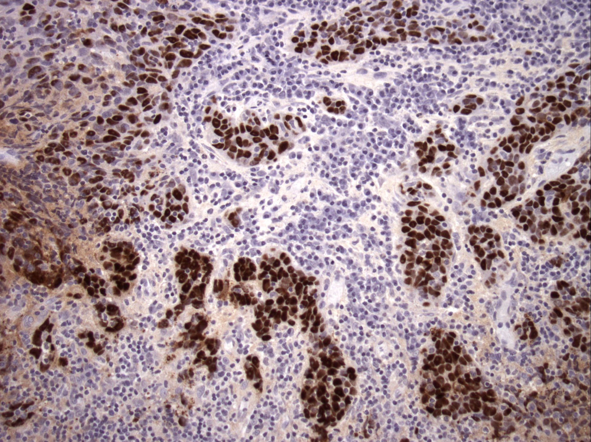
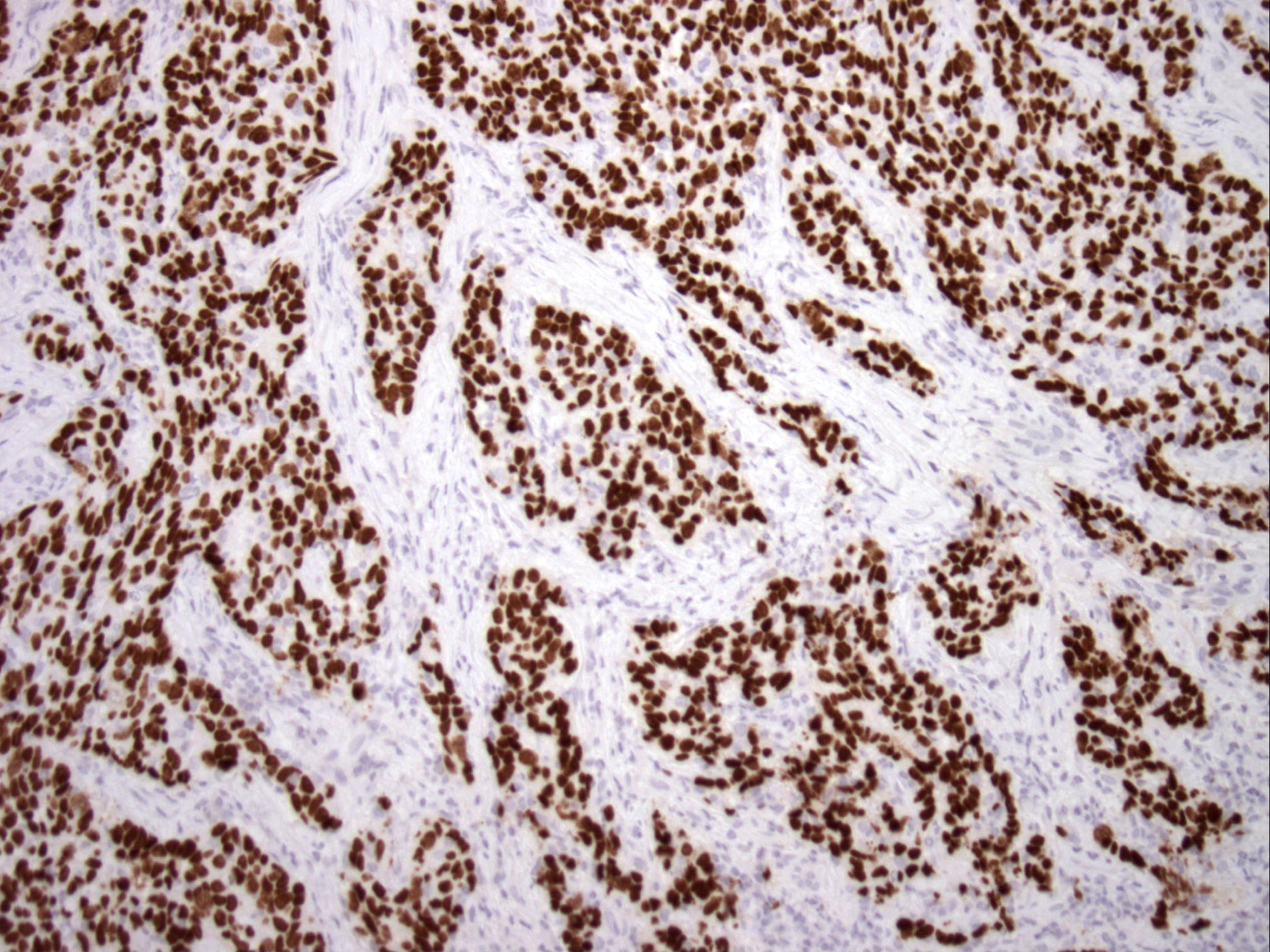
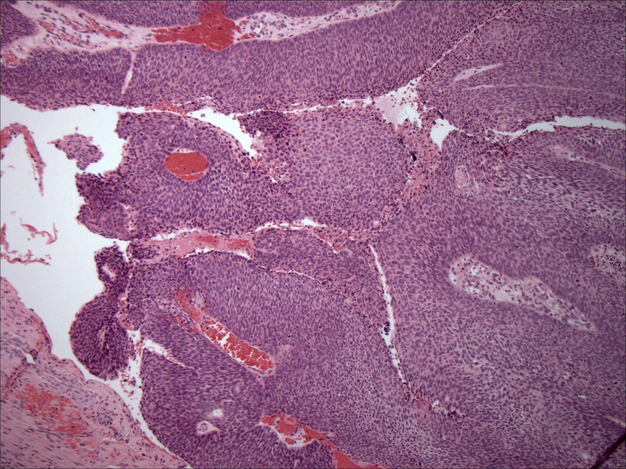
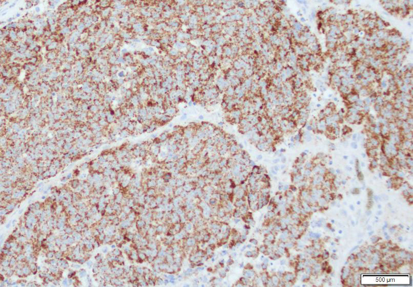
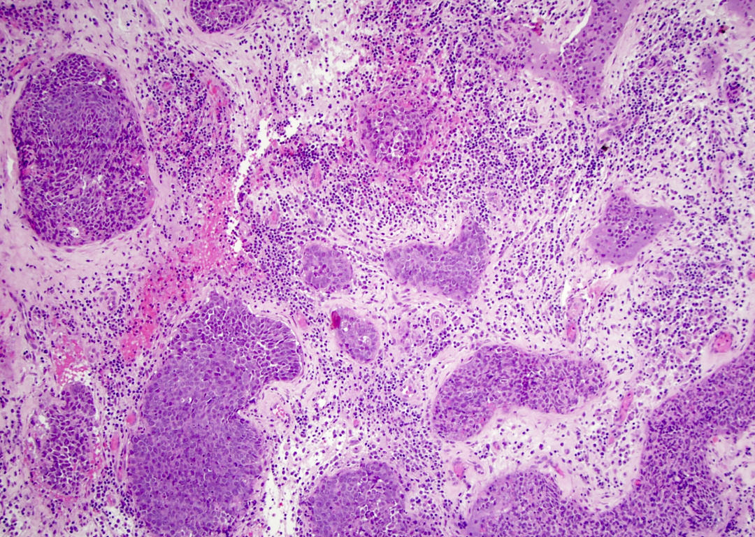
Small cell carcinoma
Finely granular chromatin
Frequent mitoses and nuclear molding
CAM5.2 (dot-like pattern)
Synaptophysin
Chromogranin
INSM1
Elevated Ki67 proliferation index
TTF1
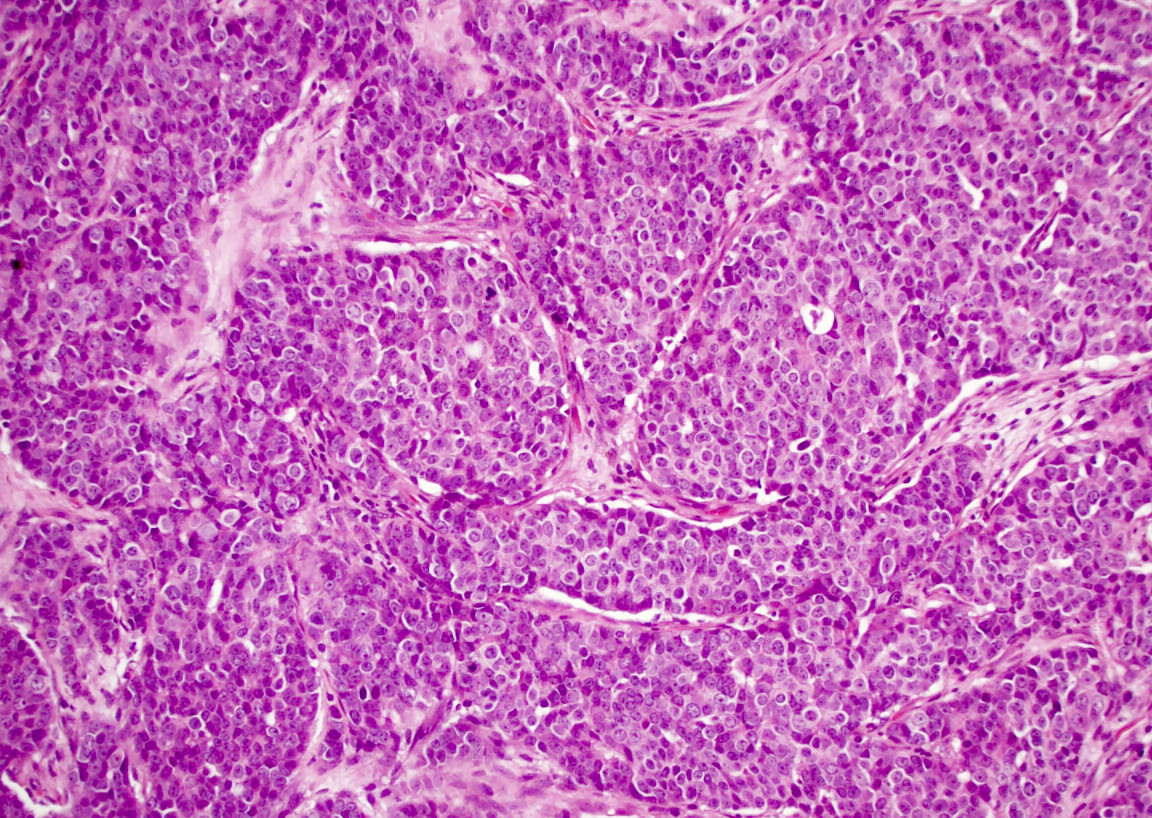
Large cell neuroendocrine carcinoma
Large tumor cells
Synaptophysin
Chromogranin
Combined small cell carcinoma and squamous cell carcinoma
Squamous and neuroendocrine component
p40
Contributed by Bin Xu, M.D., Ph.D.
Small cell carcinoma
Images hosted on other servers:

Aggressive mass in the inferior nasal cavity
Contributed by Bin Xu, M.D., Ph.D.

Surface involvement
Spindle cell area
Cribriform architecture
Tubular pattern
Basaloid area
Basaloid area with peripheral palisading
Resembling adenoid cystic carcinoma

Squamous differentiation
Calponin
SMA

p16

S100
p40
Contributed by Bin Xu, M.D., Ph.D.

RNA-ISH positive for HPV
Images hosted on other servers:

Coronal CT
Images hosted on other servers:

Translucent polyps with a smooth surface
Contributed by Bin Xu, M.D., Ph.D.
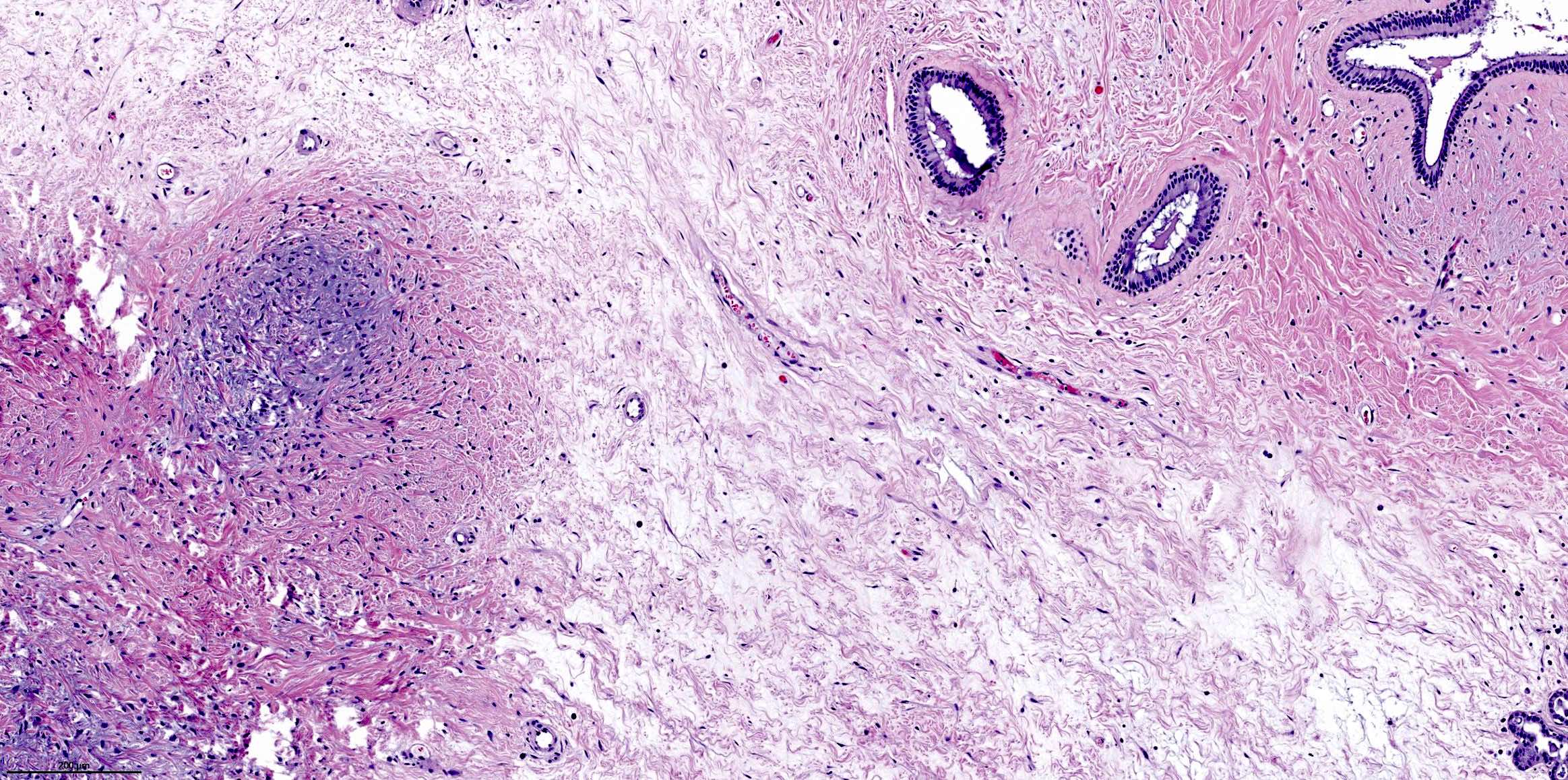
Edematous stroma

Mixed inflammatory infiltrate
Respiratory epithelium lining
Surface ulceration, squamous metaplasia

Stroma with hemorrhage
Infarction
Images hosted on other servers:

Frontoethmoidal sinus mass

Frontal sinus mass
Images hosted on other servers:

Mass on glabellar region

Wide excision of mass

76 days after surgery

Endoscopic resection
Contributed by Kelly Magliocca, D.D.S., M.P.H.
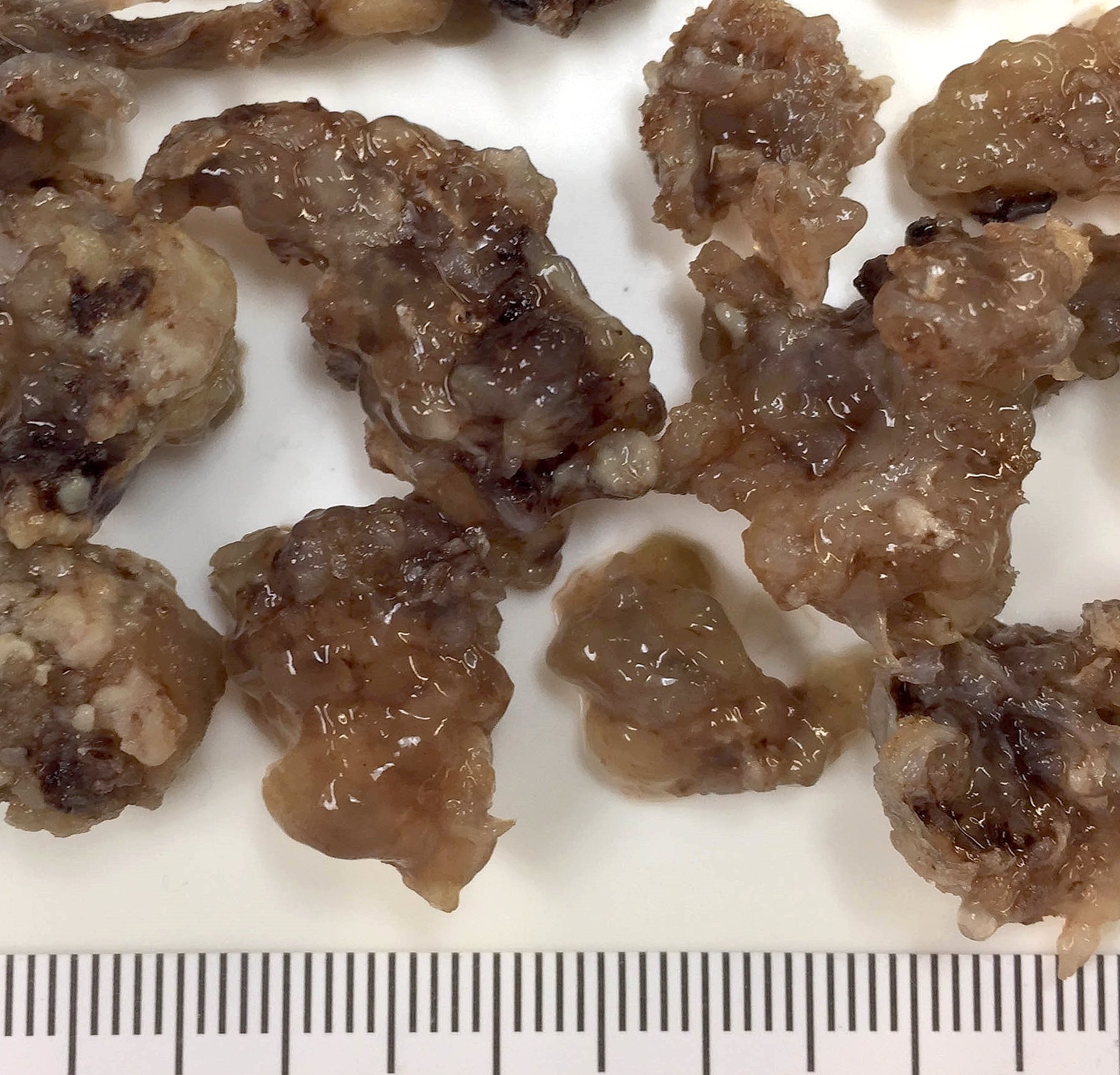
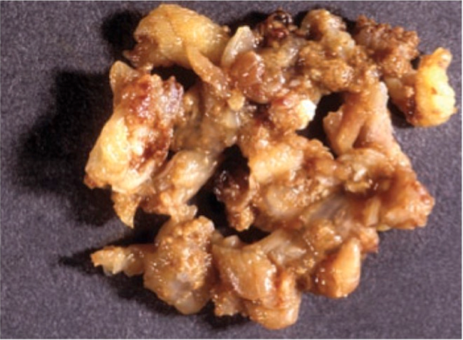
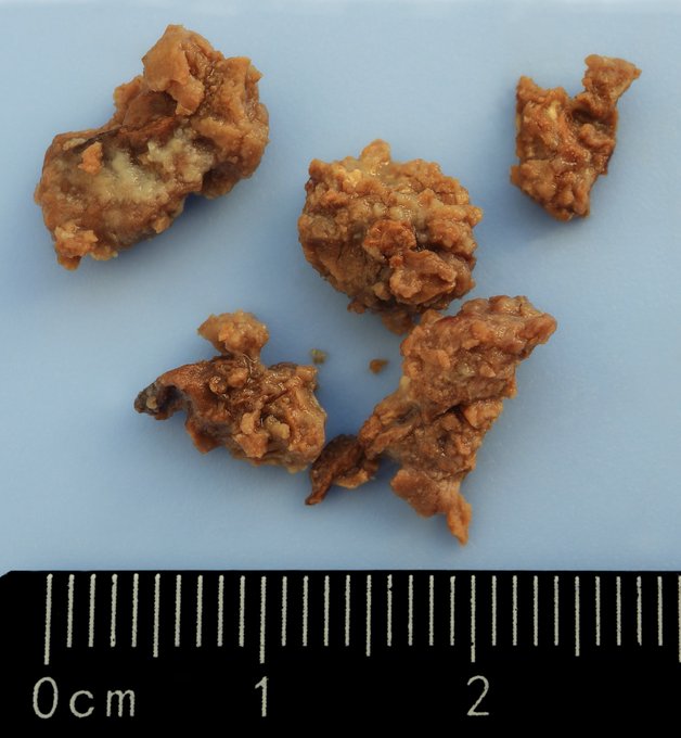
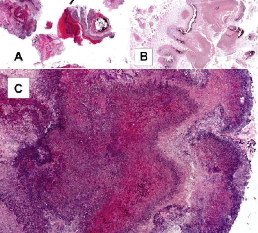
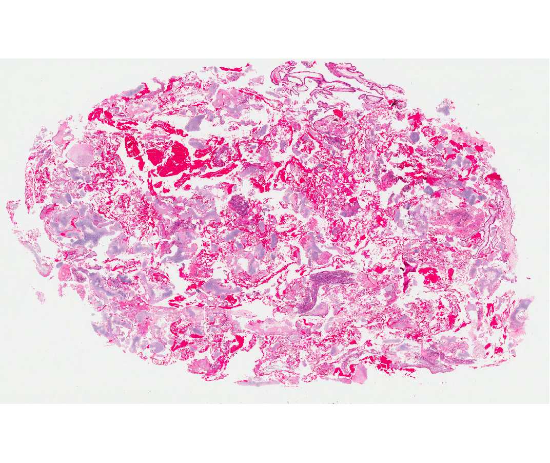
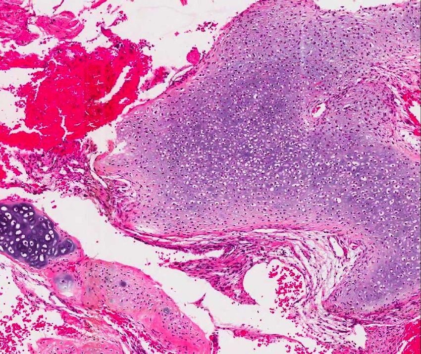
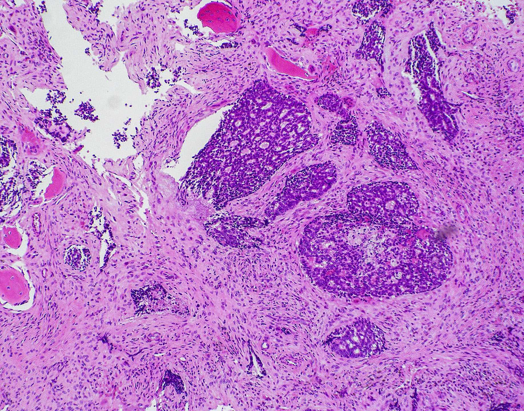
Macroscopic appearance
Images hosted on other servers:

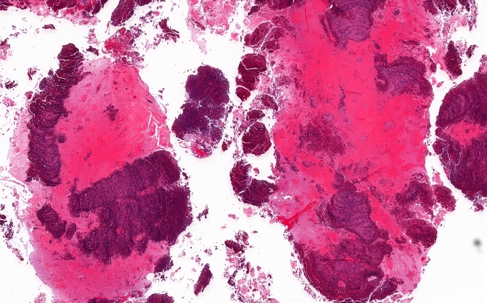
Open biopsy masses
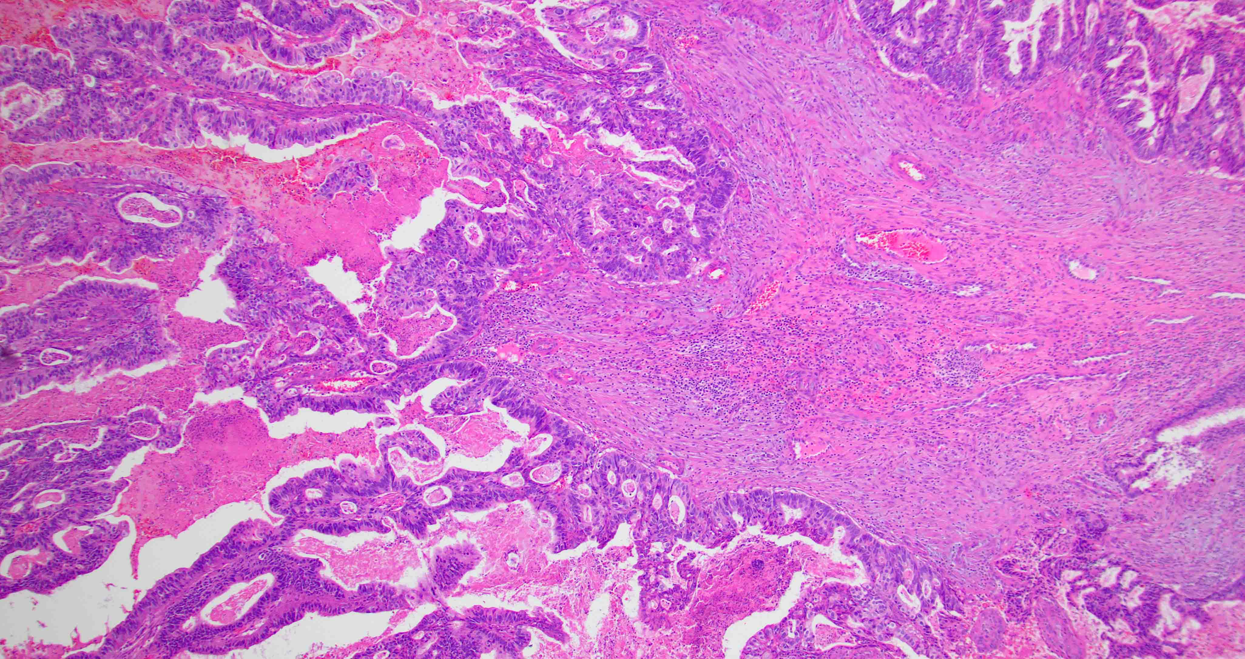
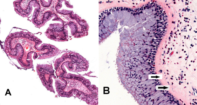
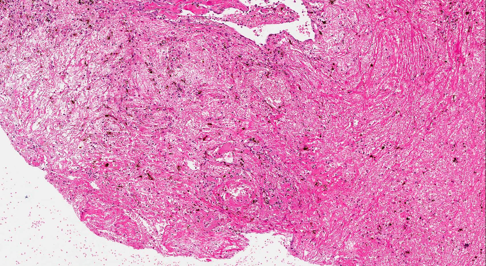
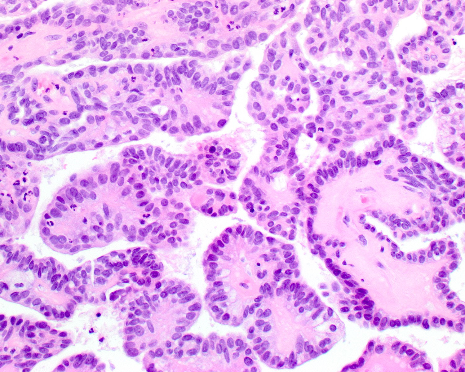
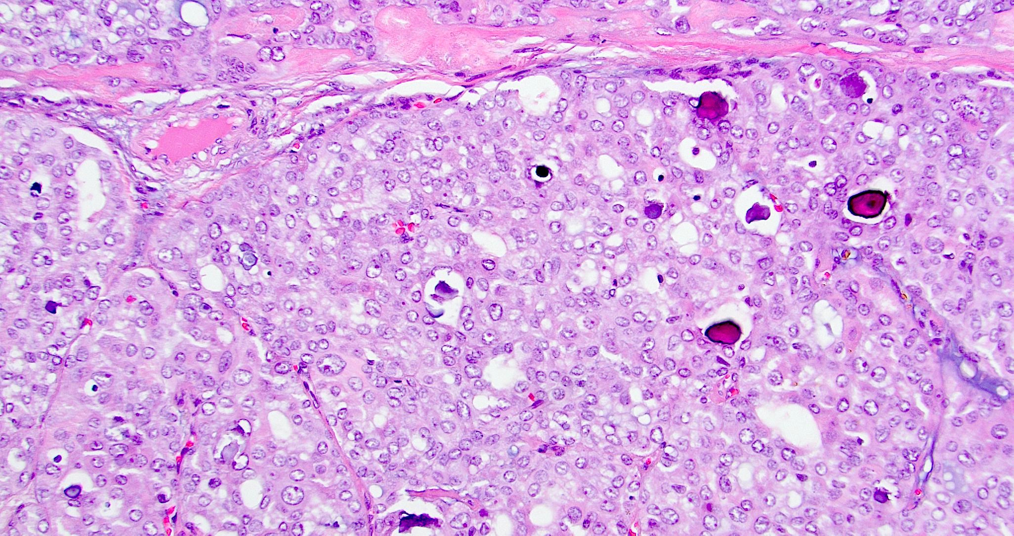
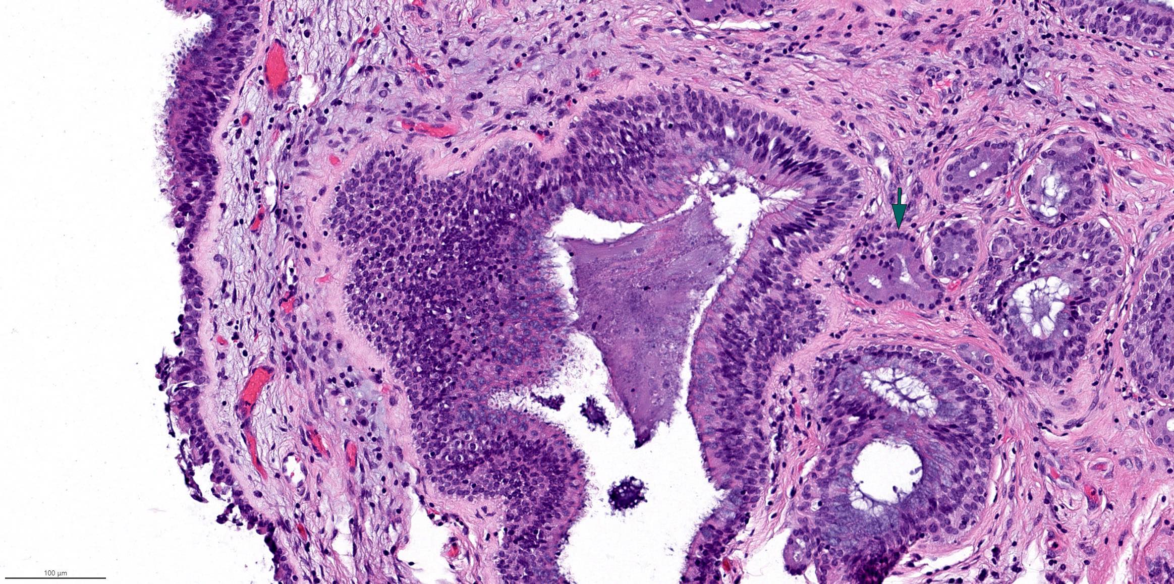
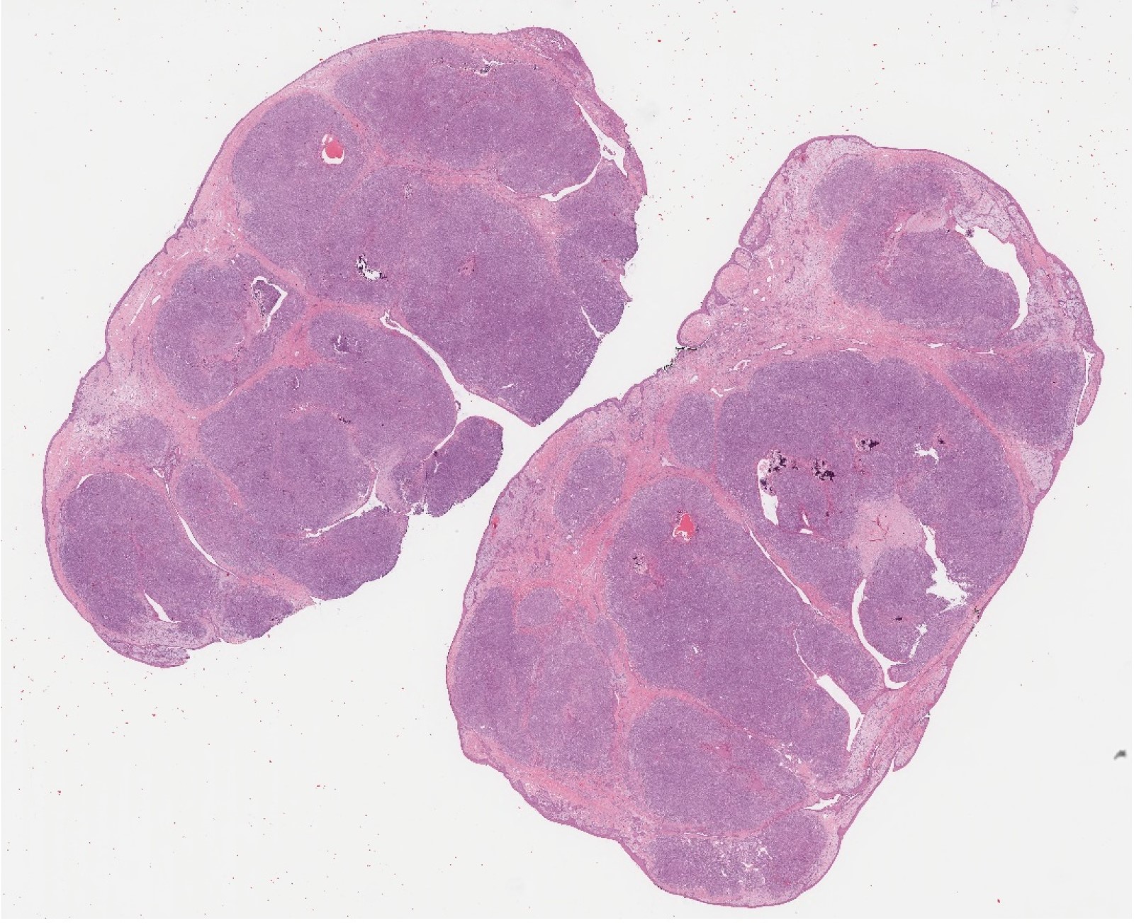
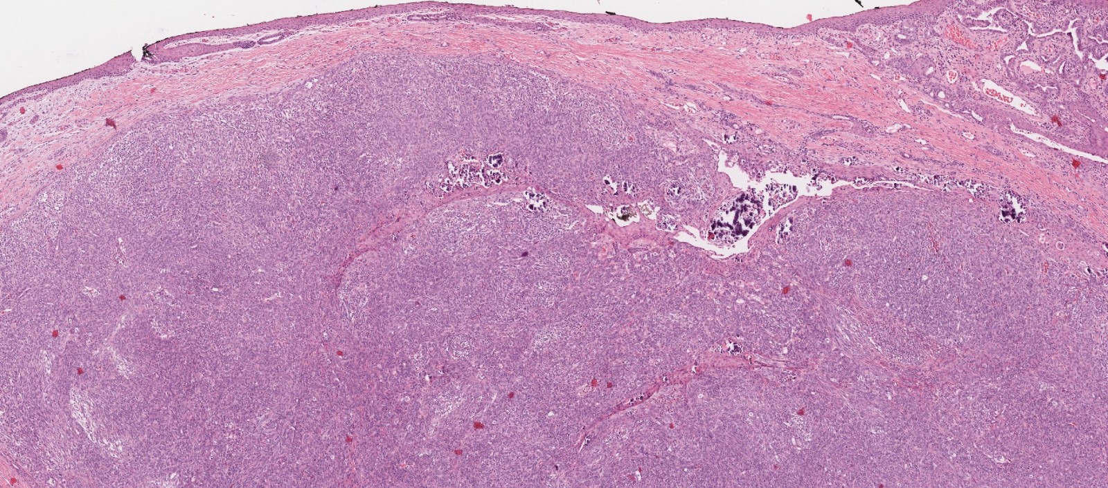
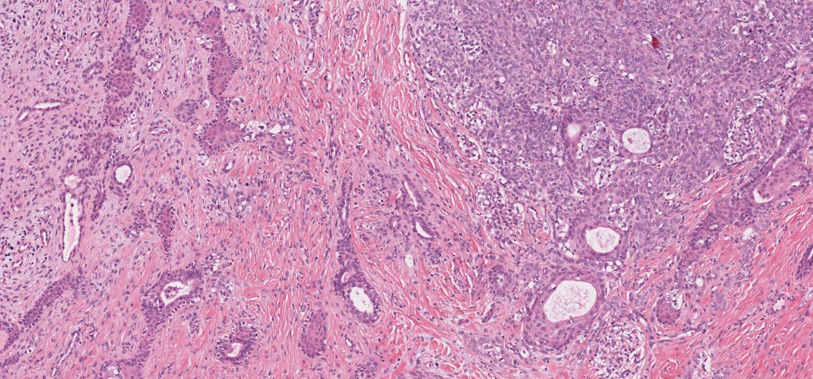
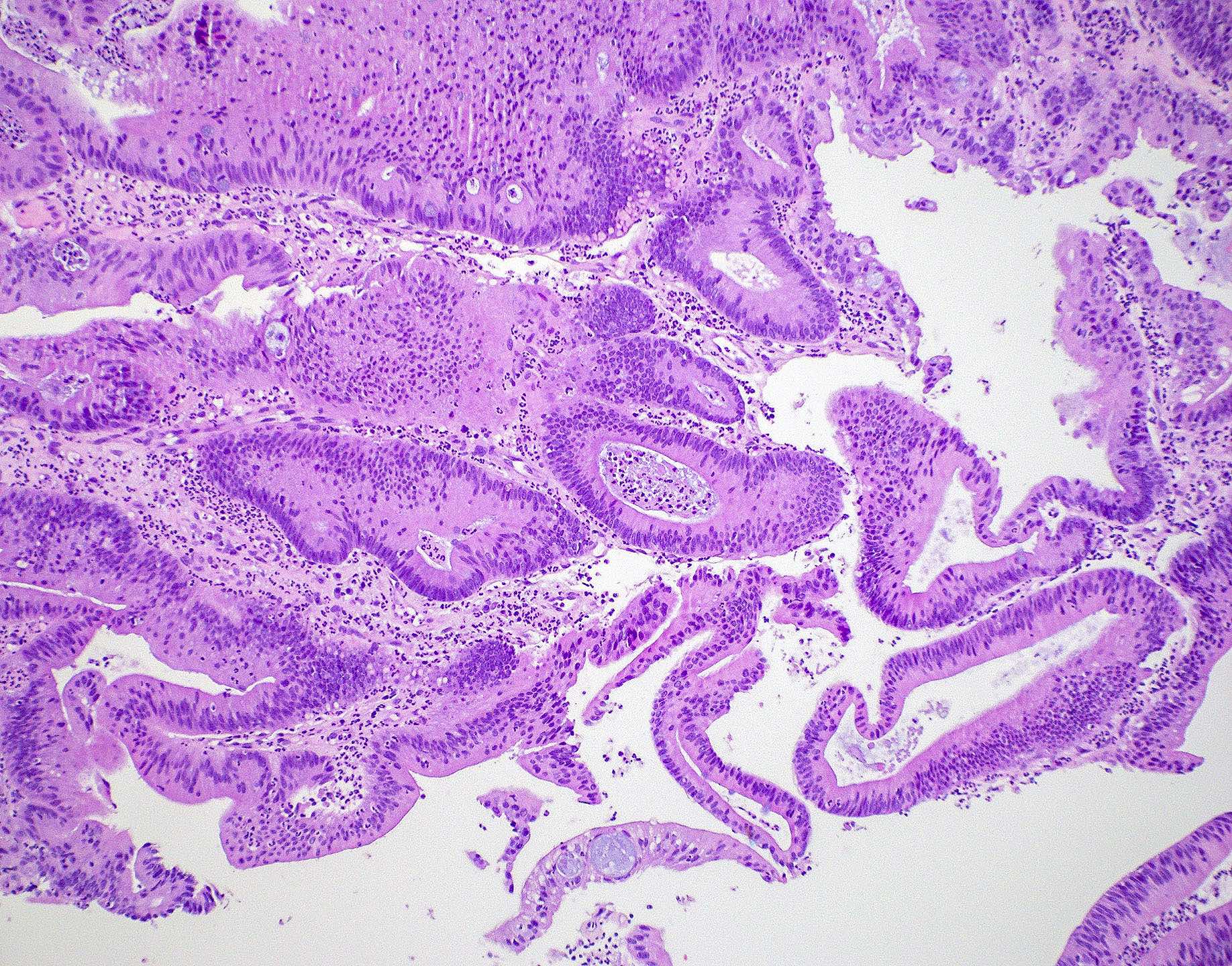
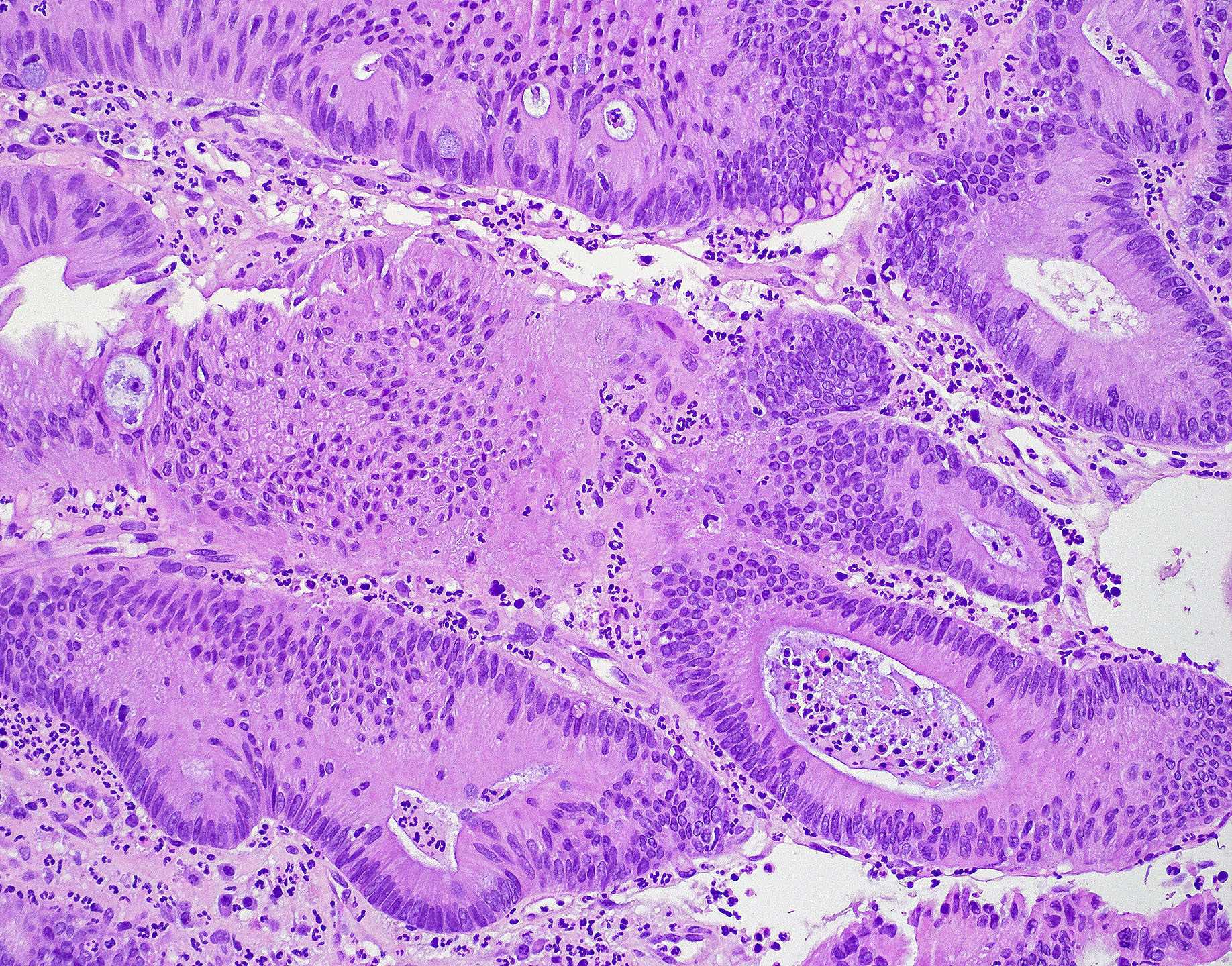
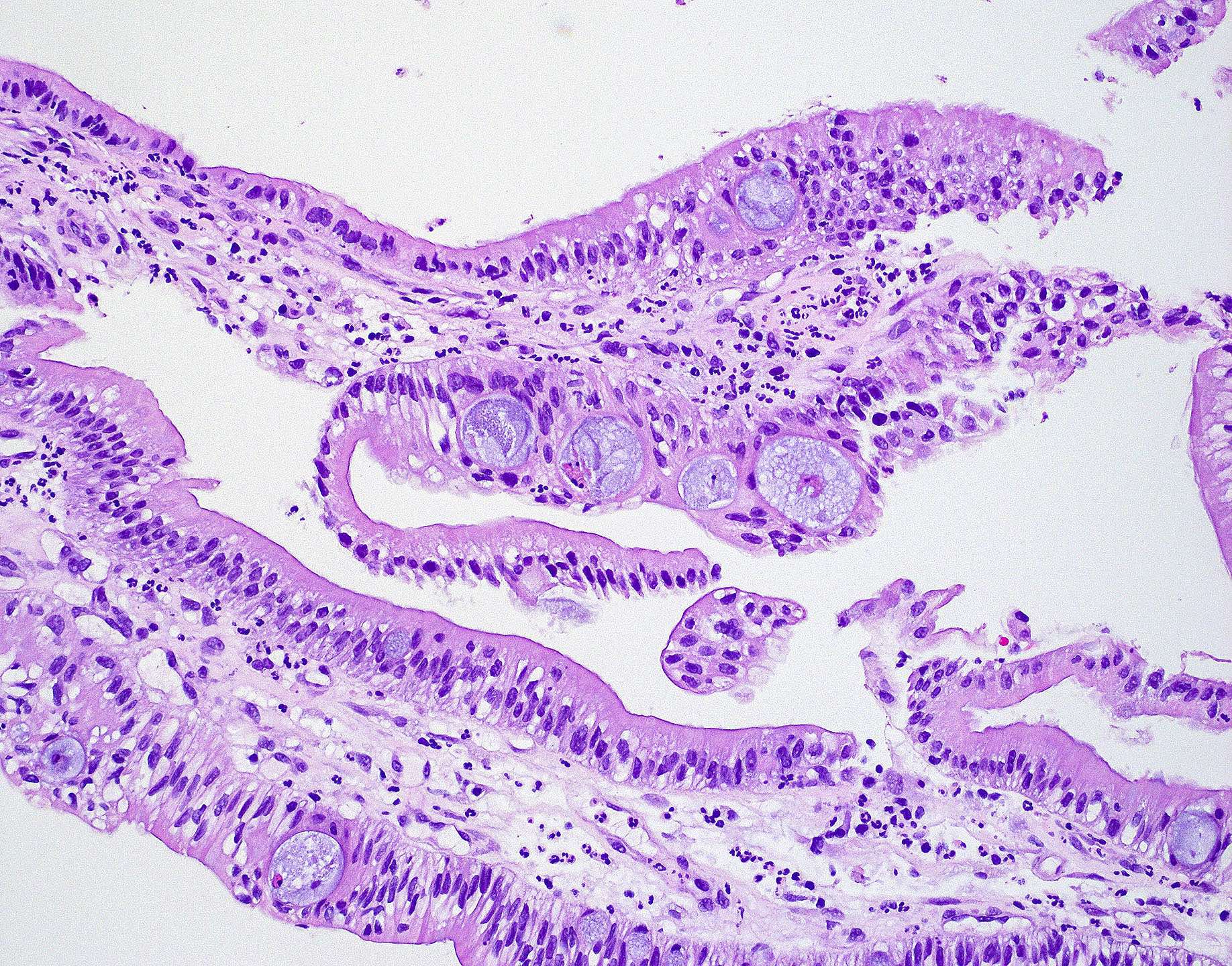
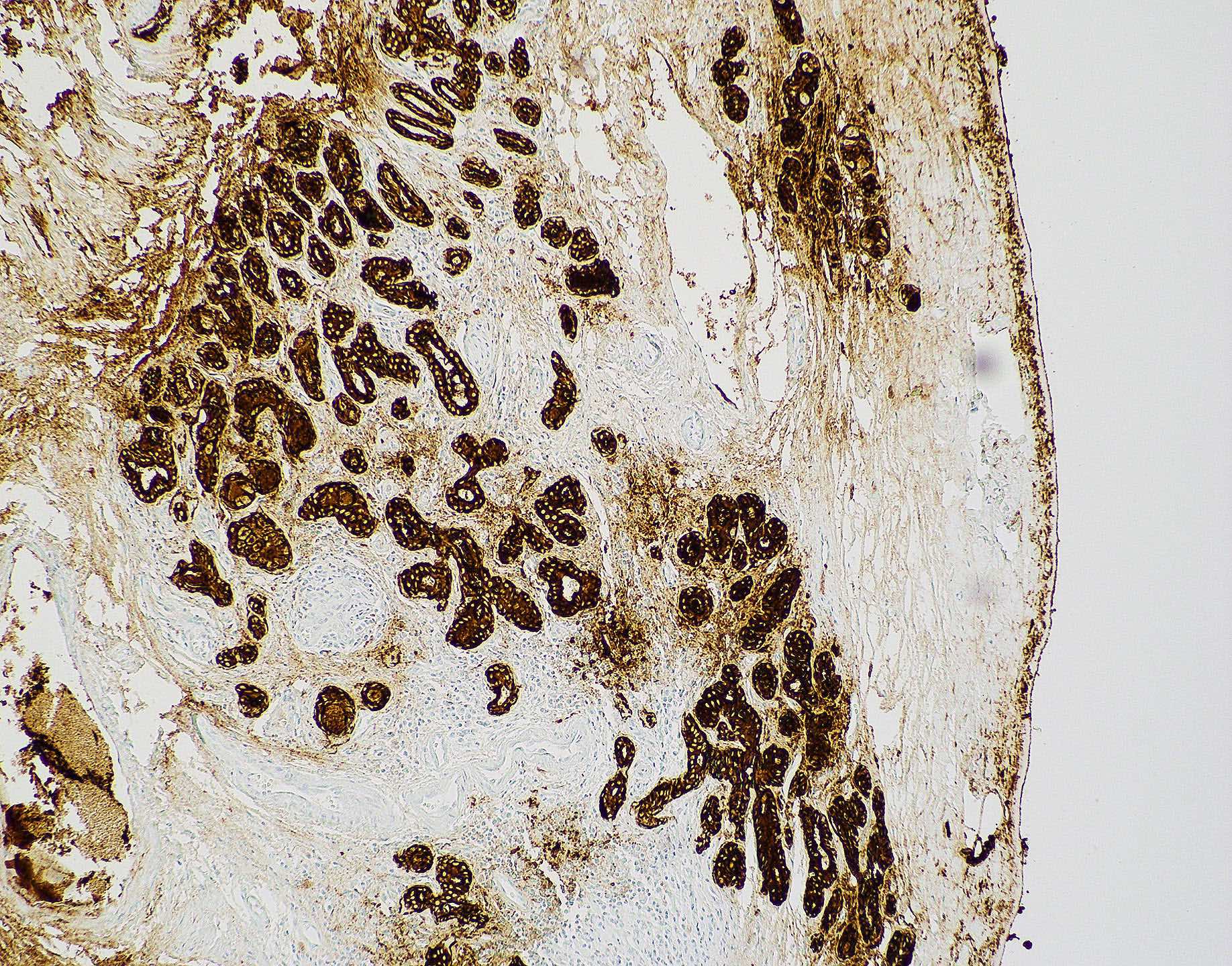
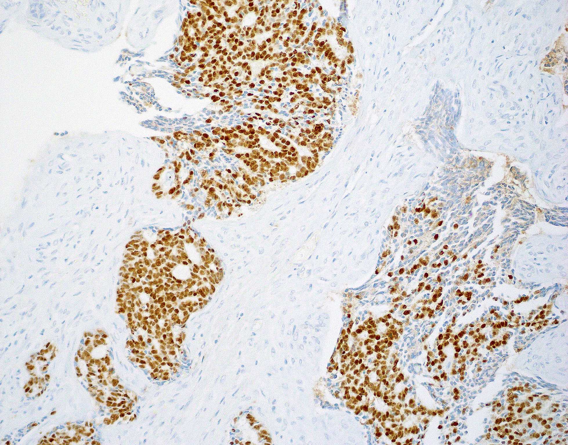
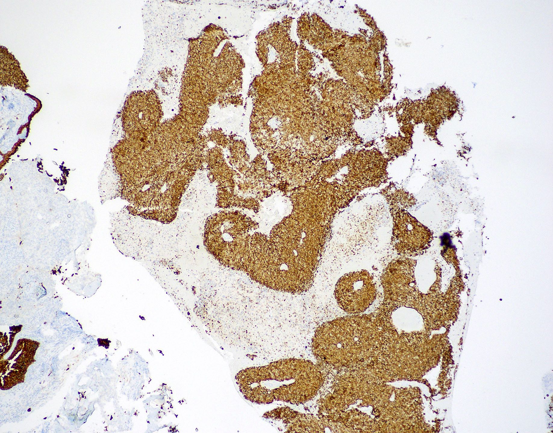
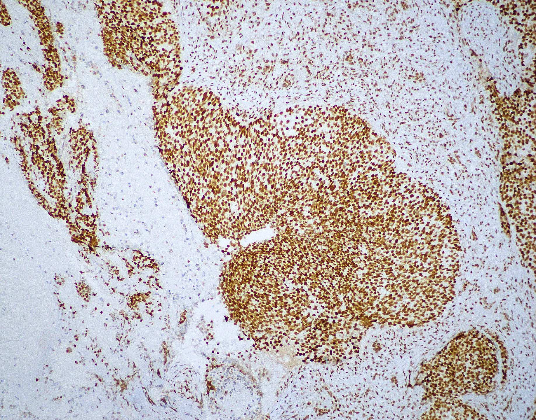
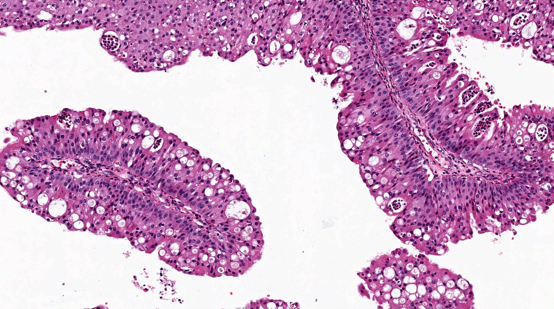
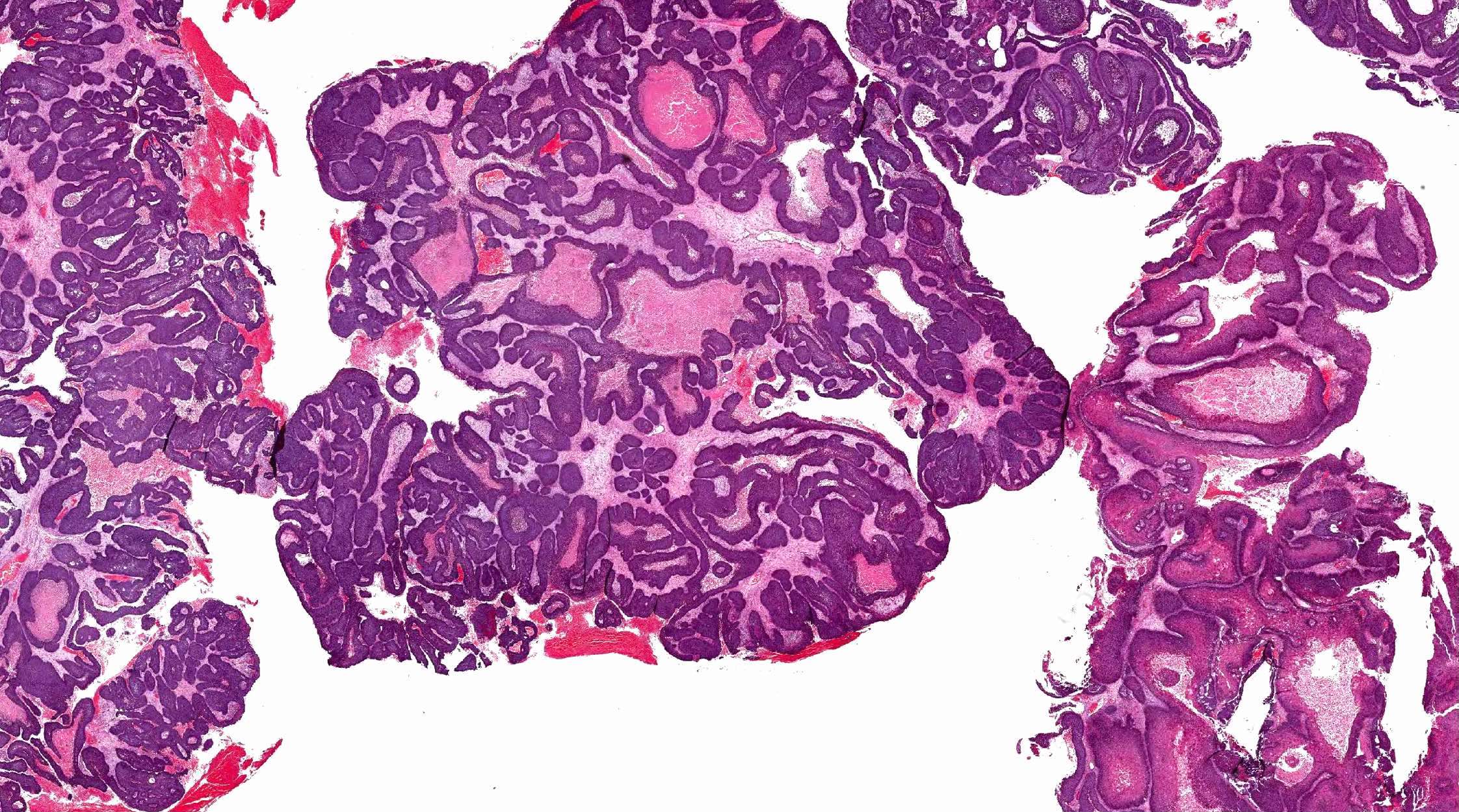
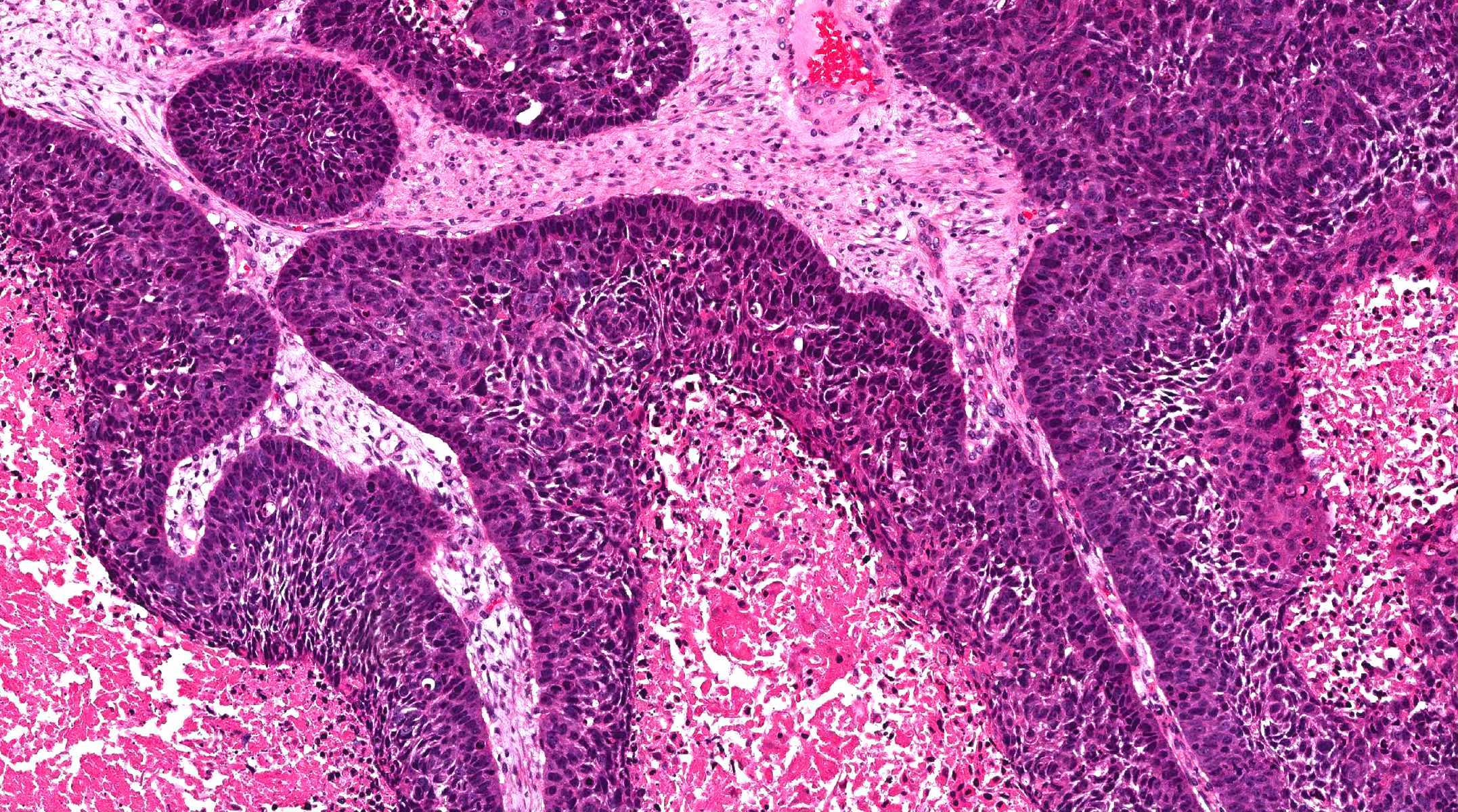
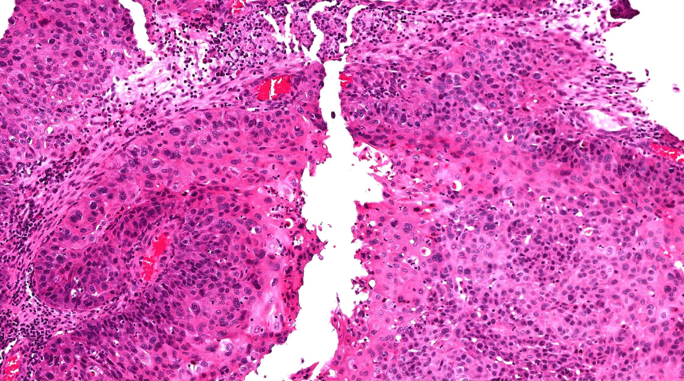
Contributed by Vladyslav Ilchenko, M.D., Diana Bell, M.D. and Kelly Magliocca, D.D.S., M.P.H.
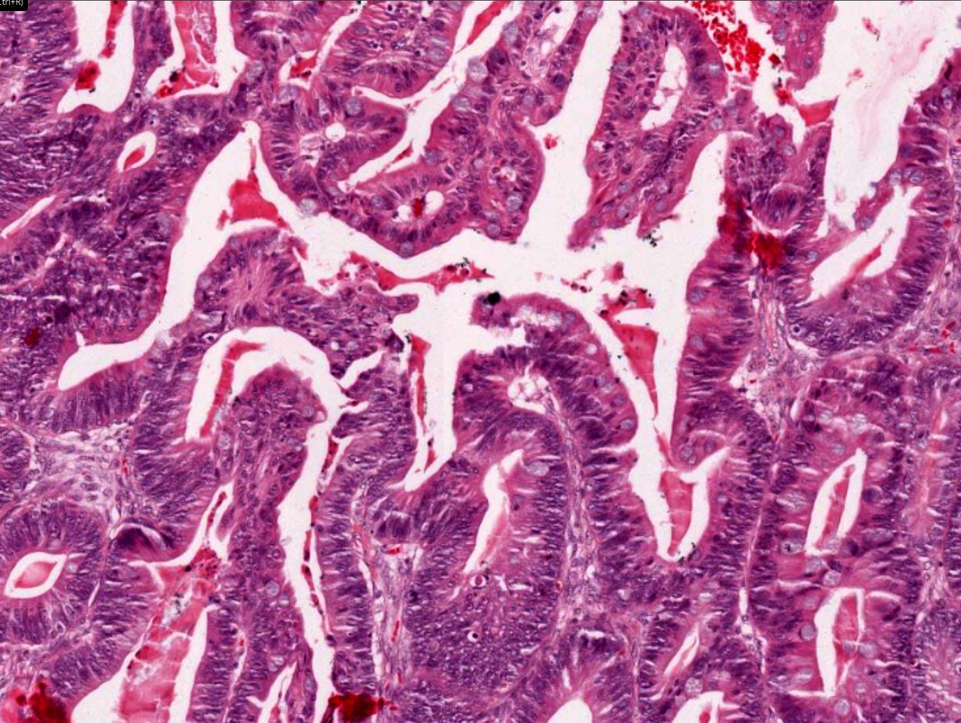
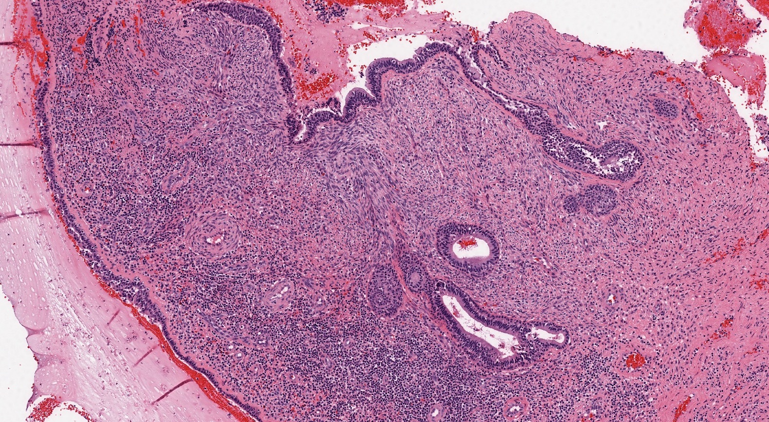
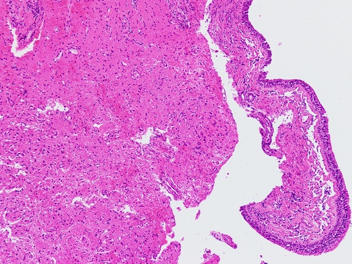
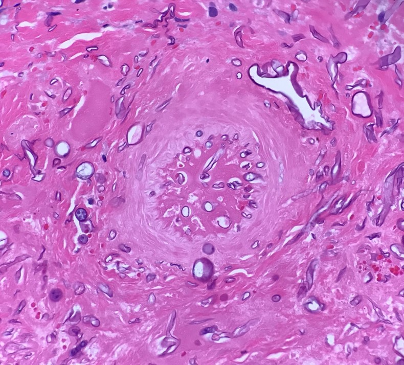
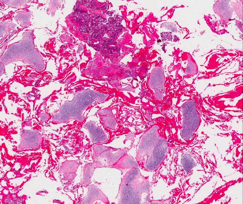
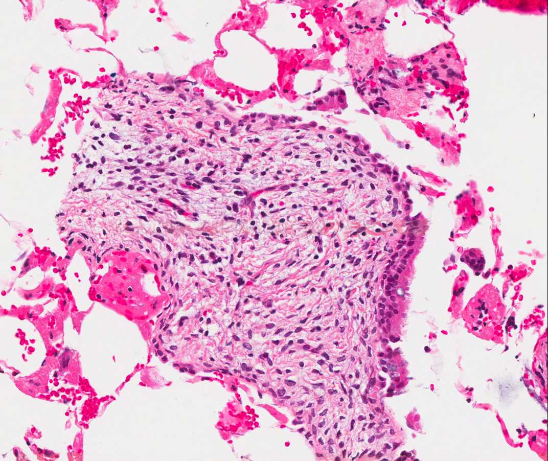
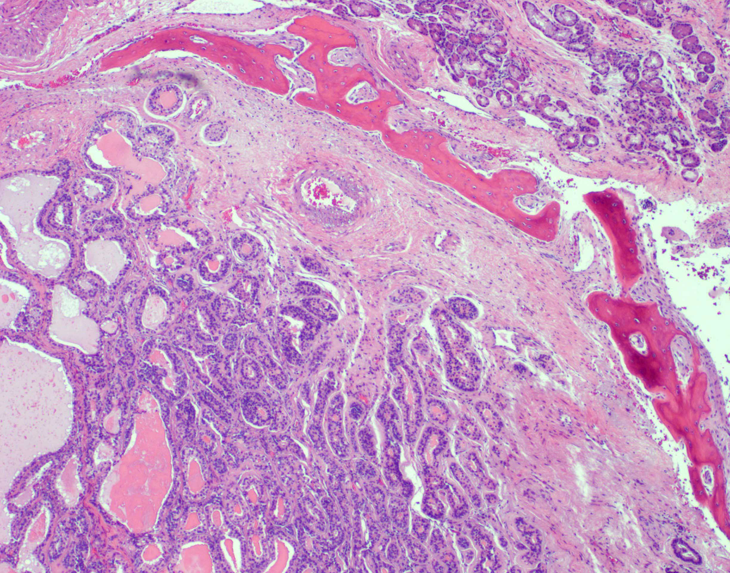
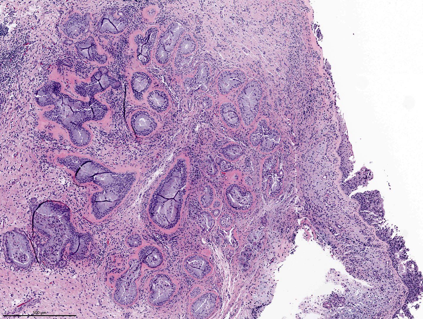
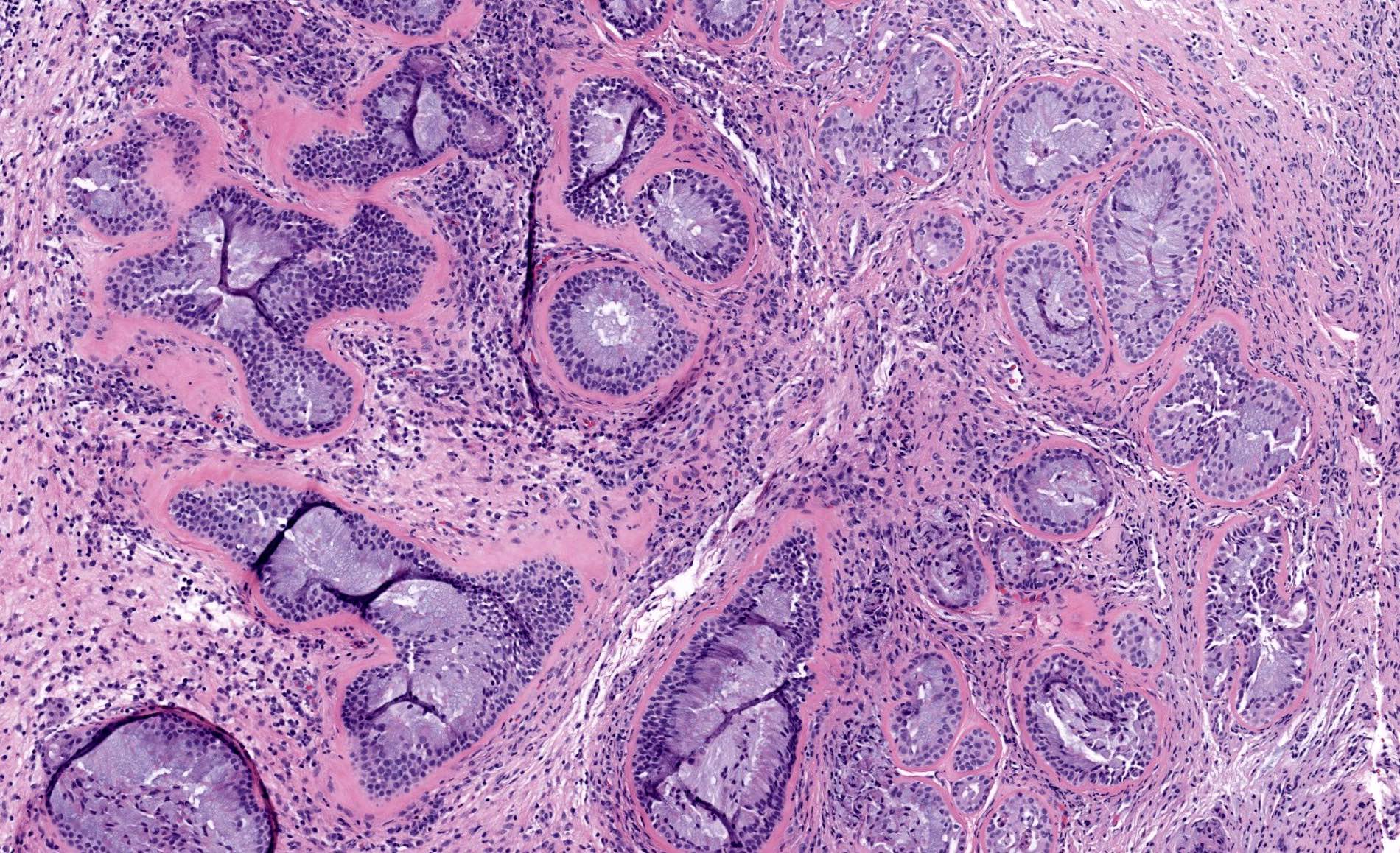
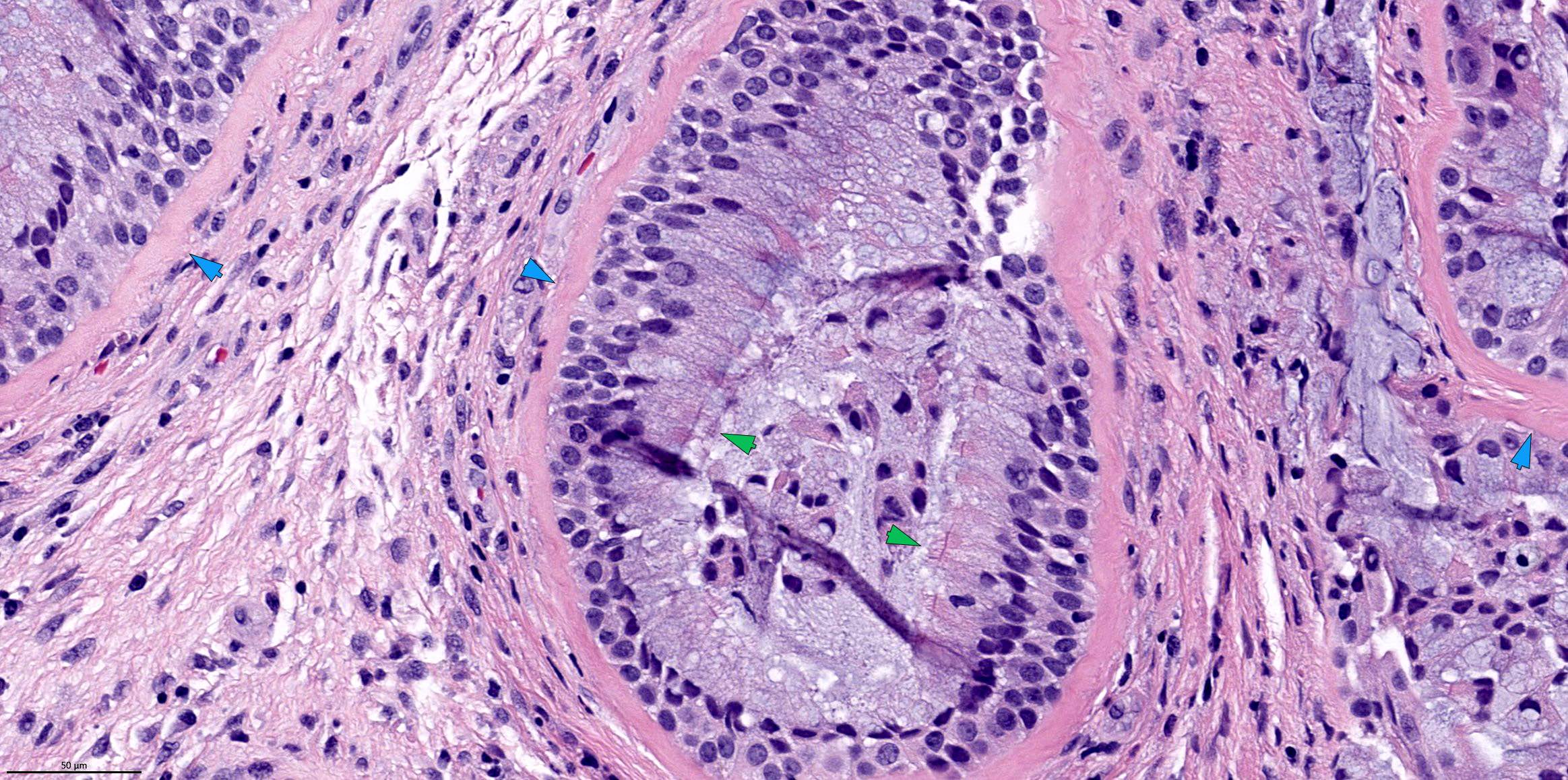
Complex glands
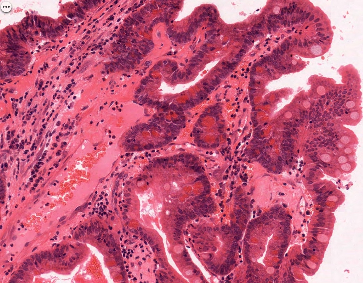
Papillary pattern with goblet cells
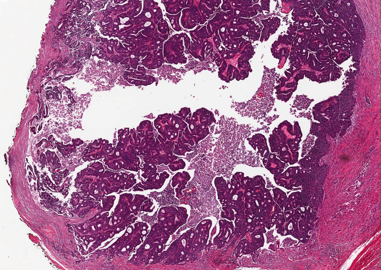
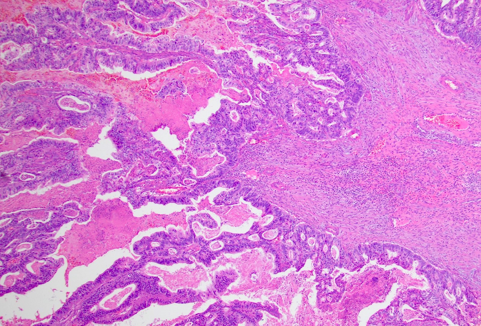
Tubuloglandular architecture
Complex colonic type glands and stroma
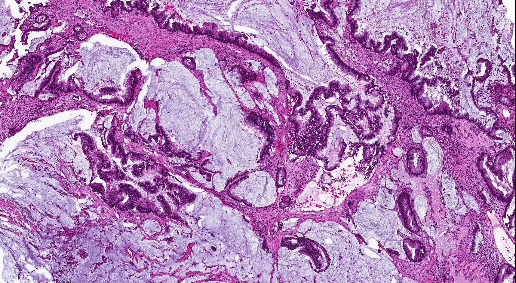
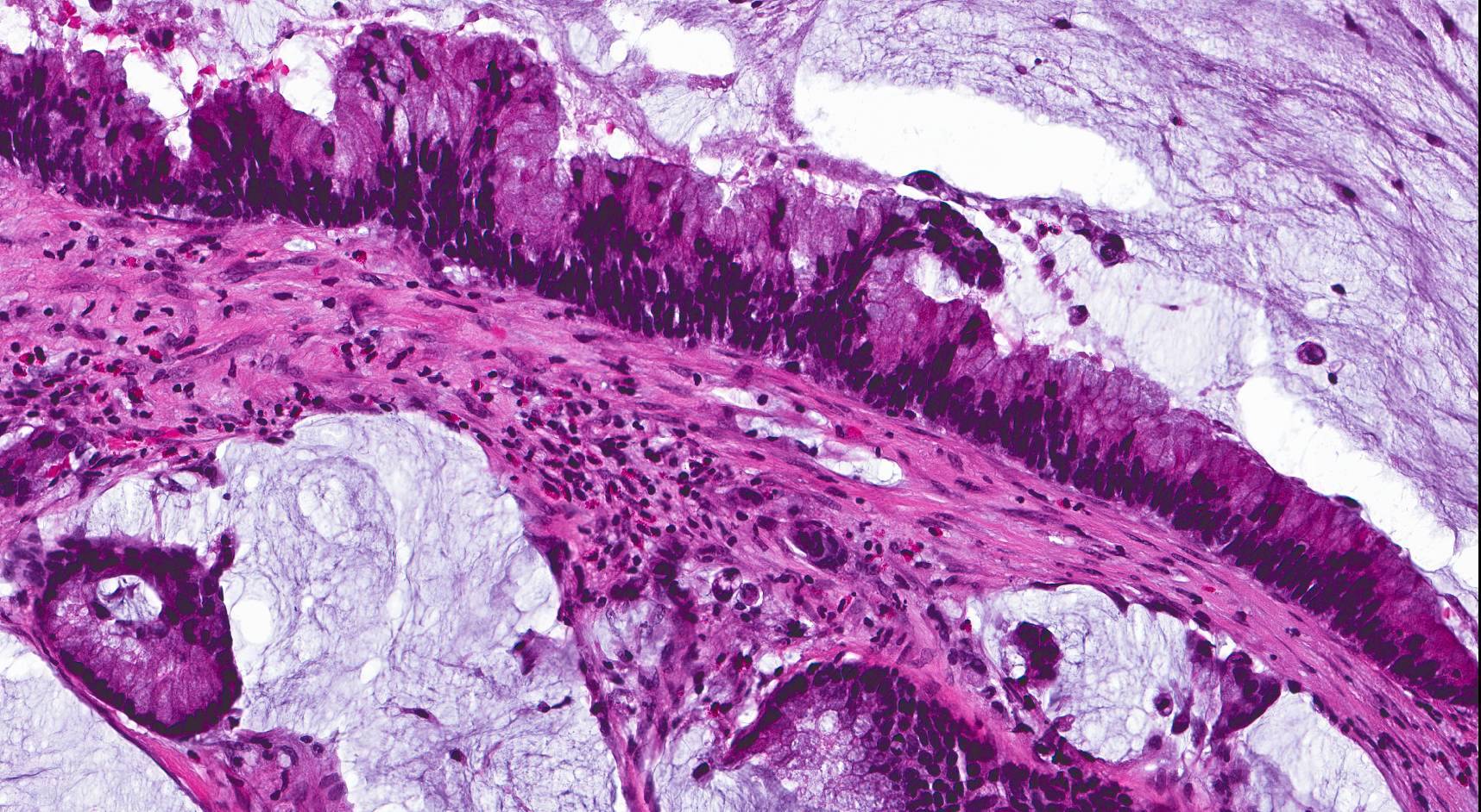
Mucinous type
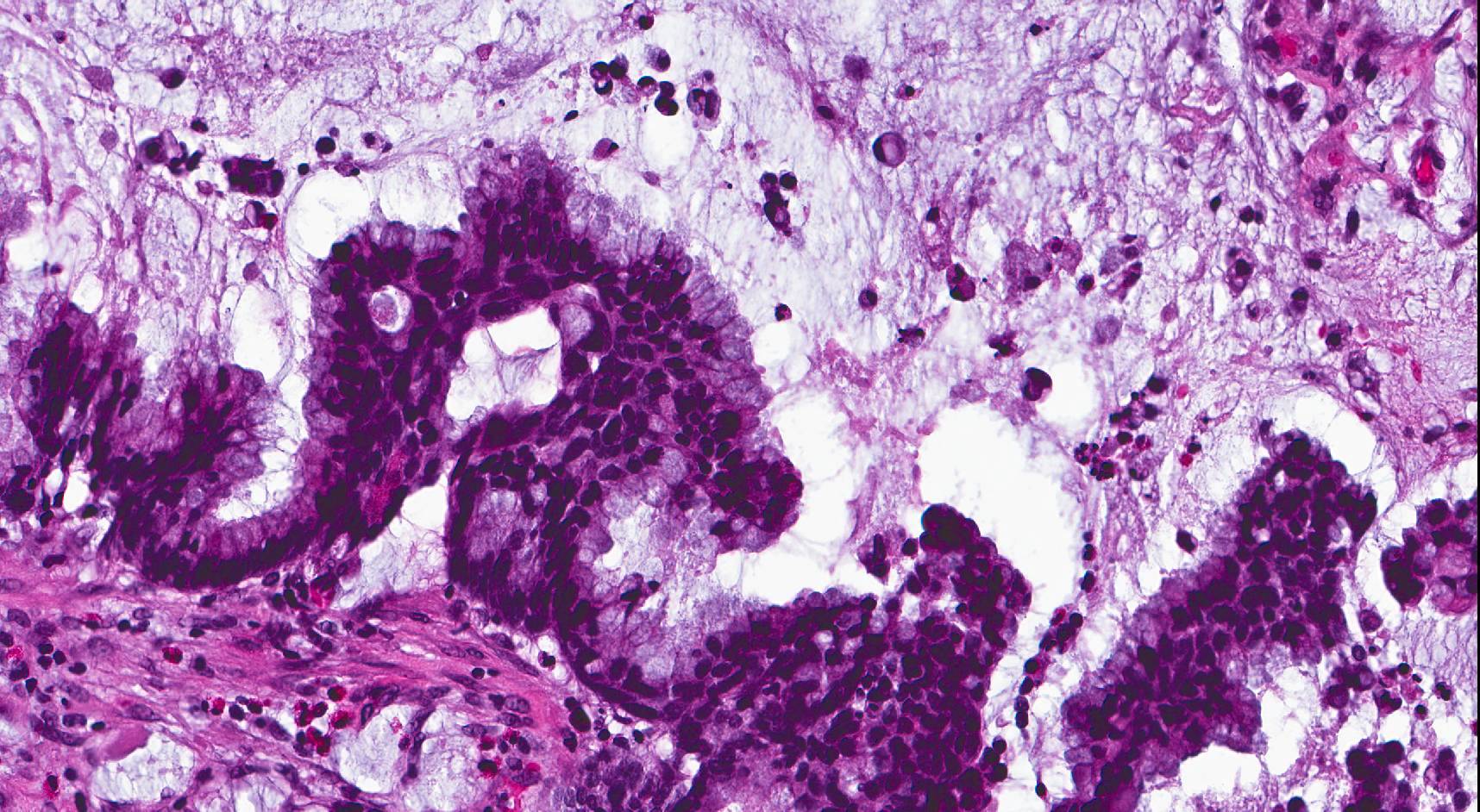
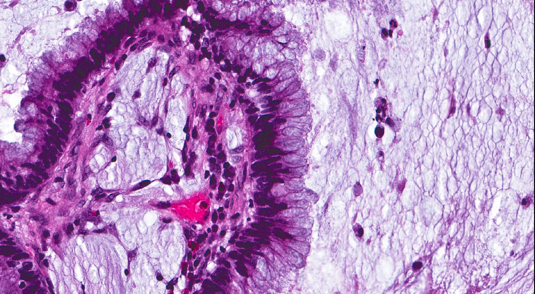
Mucinous type
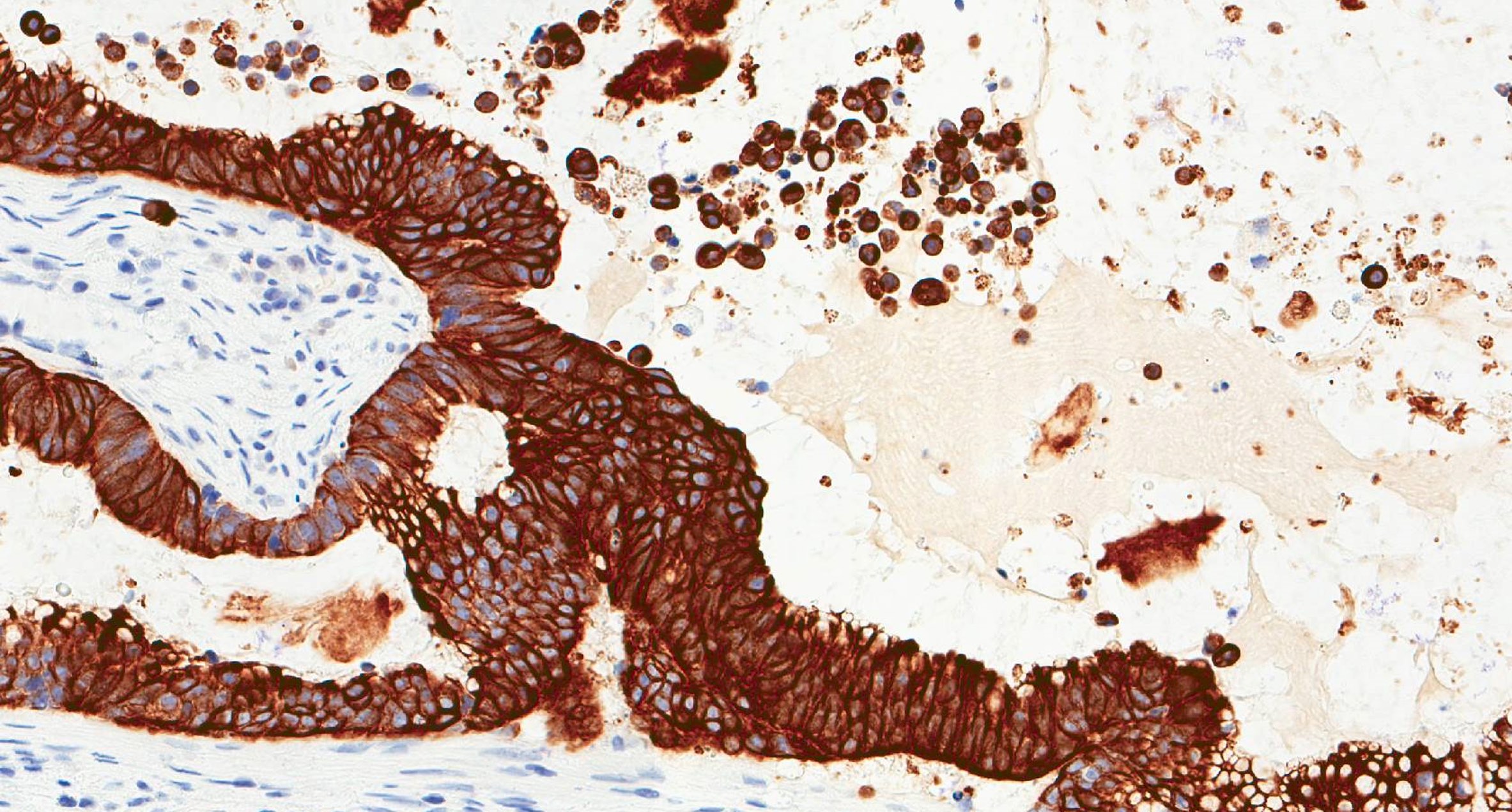
CK20
CDX2
Update on sinonasal carcinoma
Malignant tumors of the paranasal sinuses
Images hosted on other servers:

Mucosal thickening

Advanced manifestations

Extrasinonasal extension
Images hosted on other servers:

Periorbital swelling

Palatal ulceration
Contributed by Bin Xu, M.D., Ph.D. and Kelly Magliocca, D.D.S., M.P.H. (Case #493)
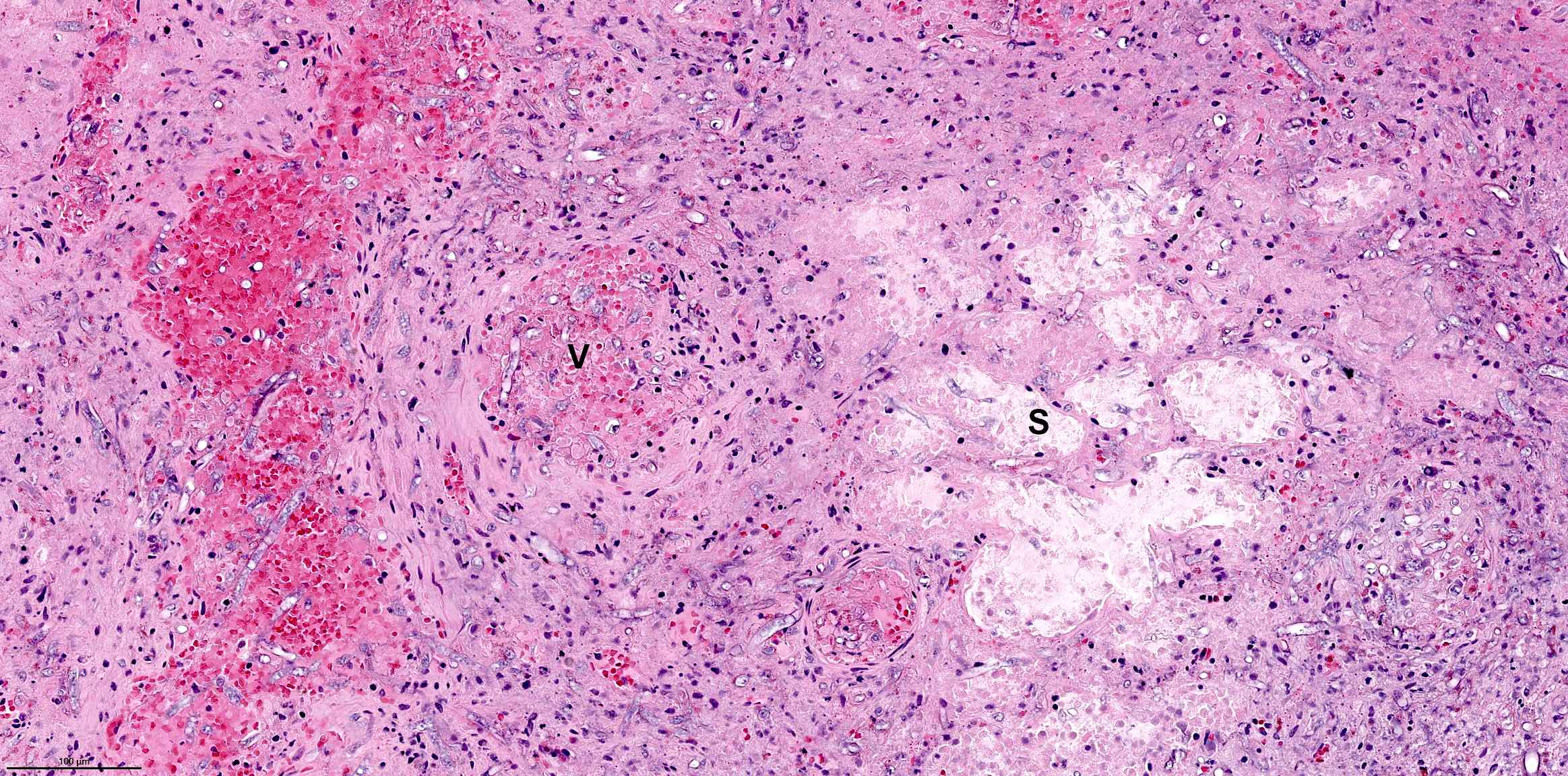

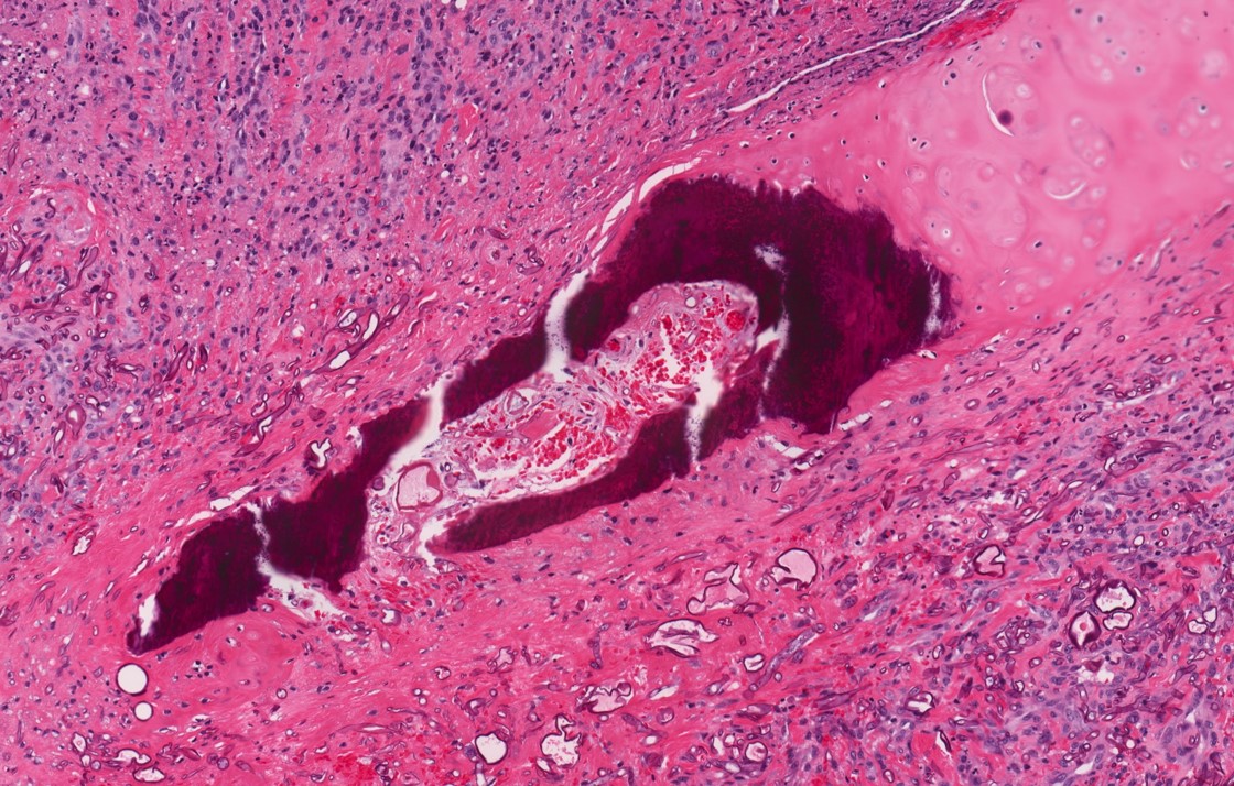
Fungal hyphae invade tissue
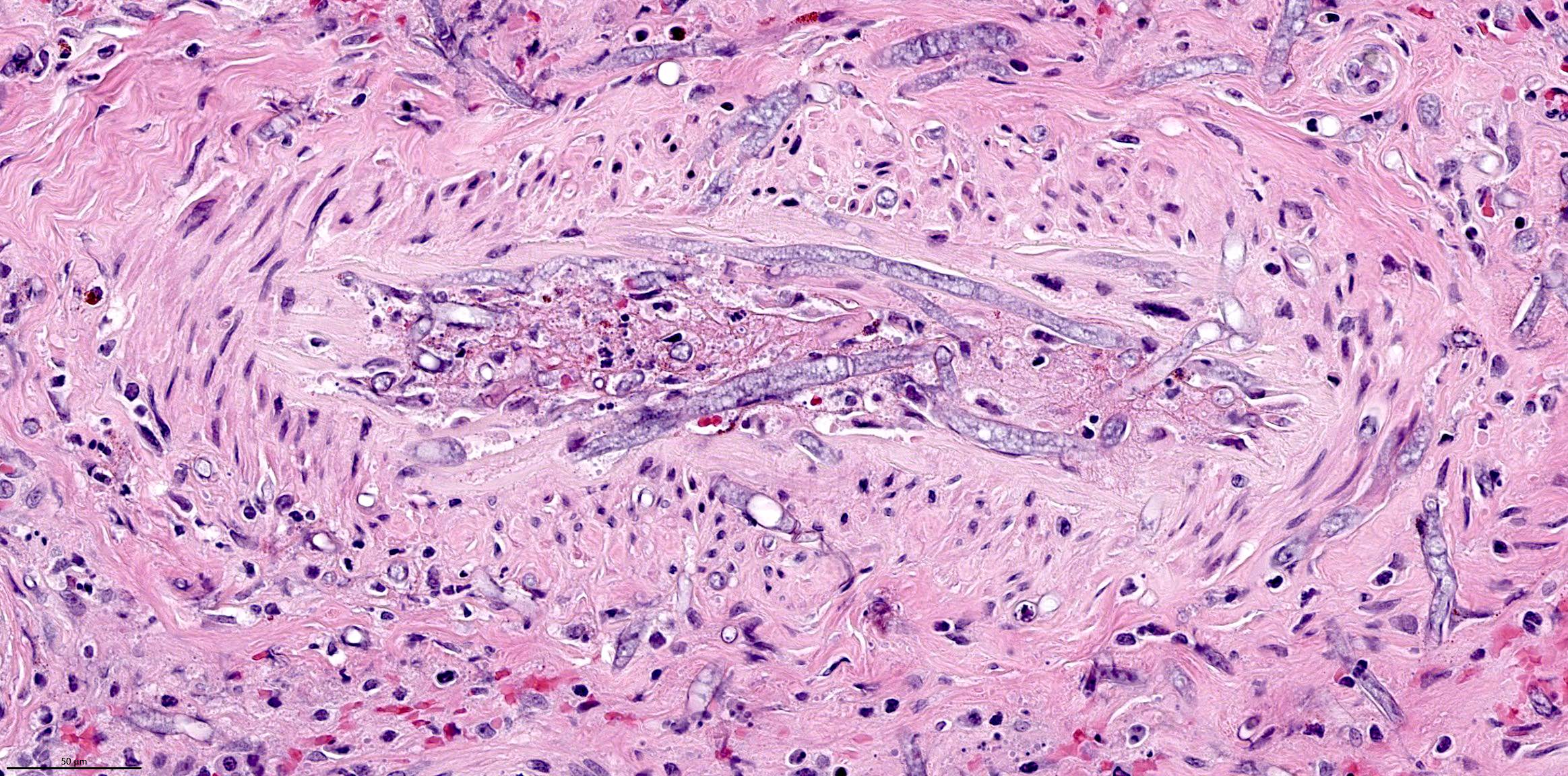
Angioinvasion

Angioinvasion and necrotic wall
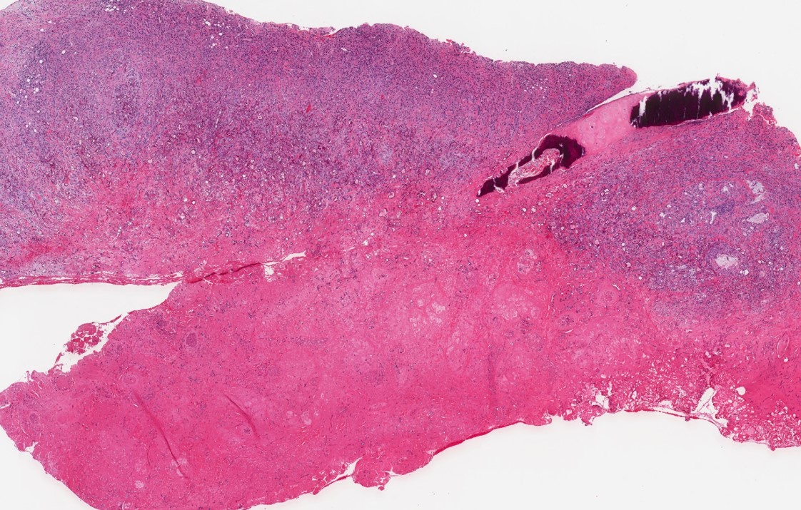
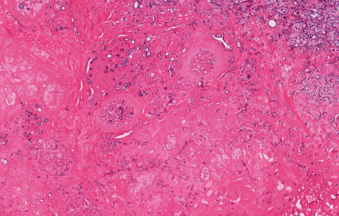
Tissue necrosis
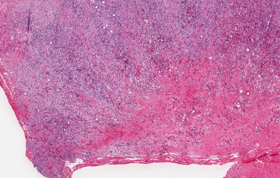
Necrotic and adjacent viable sinonasal mucosa
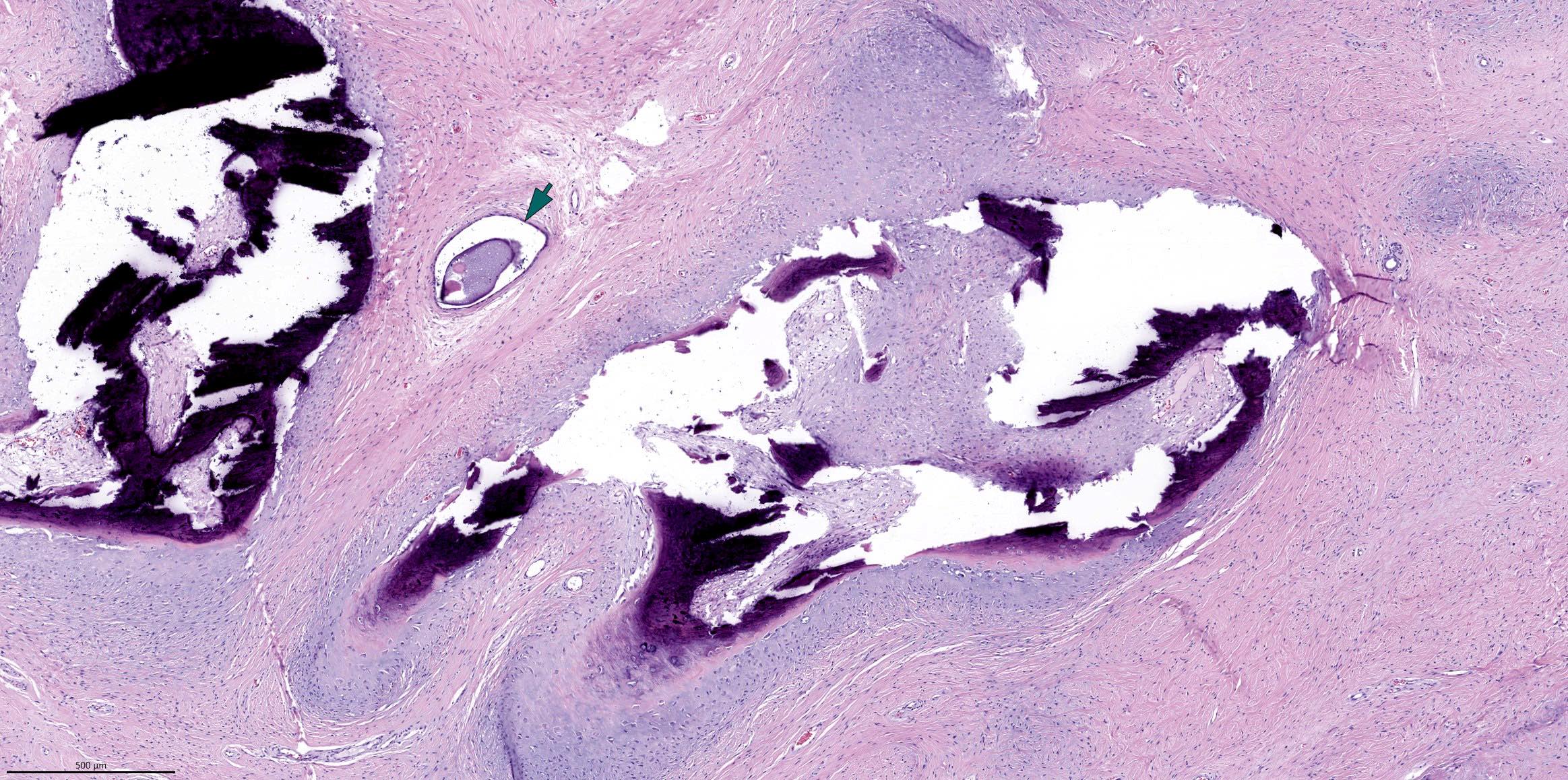
Ossified cartilage
and adjacent
viable sinonasal
mucosa
Angioinvasion

GMS: 90 degree branching


GMS stain

PAS stain
Images hosted on other servers:

Pedunculated tumor without bone destruction
Images hosted on other servers:

Well defined large polypoid tumor
Contributed by Diana Bell, M.D. and Lester Thompson, M.D.
Tumor underneath nasopharyngeal mucosa
Tumor infiltrating underlying stroma
Interwoven papillae and glands
Papillae lined by columnar cells
Nuclear overlapping
Nuclear features mimicking PTC
Pseudoinclusions and nuclear clearing
Psammoma bodies
TTF1 IHC
Thyroglobulin IHC
Contributed by I Weng Lao, M.D., Ph.D. and Jian Wang, M.D., Ph.D.
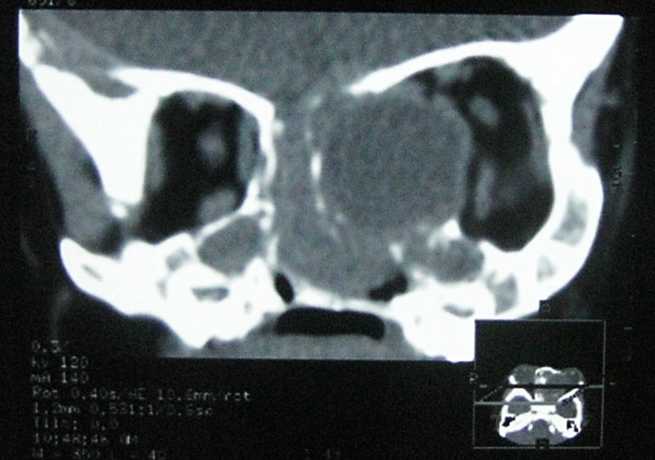
CT scan
Contributed by I Weng Lao, M.D., Ph.D. and Jian Wang, M.D., Ph.D.

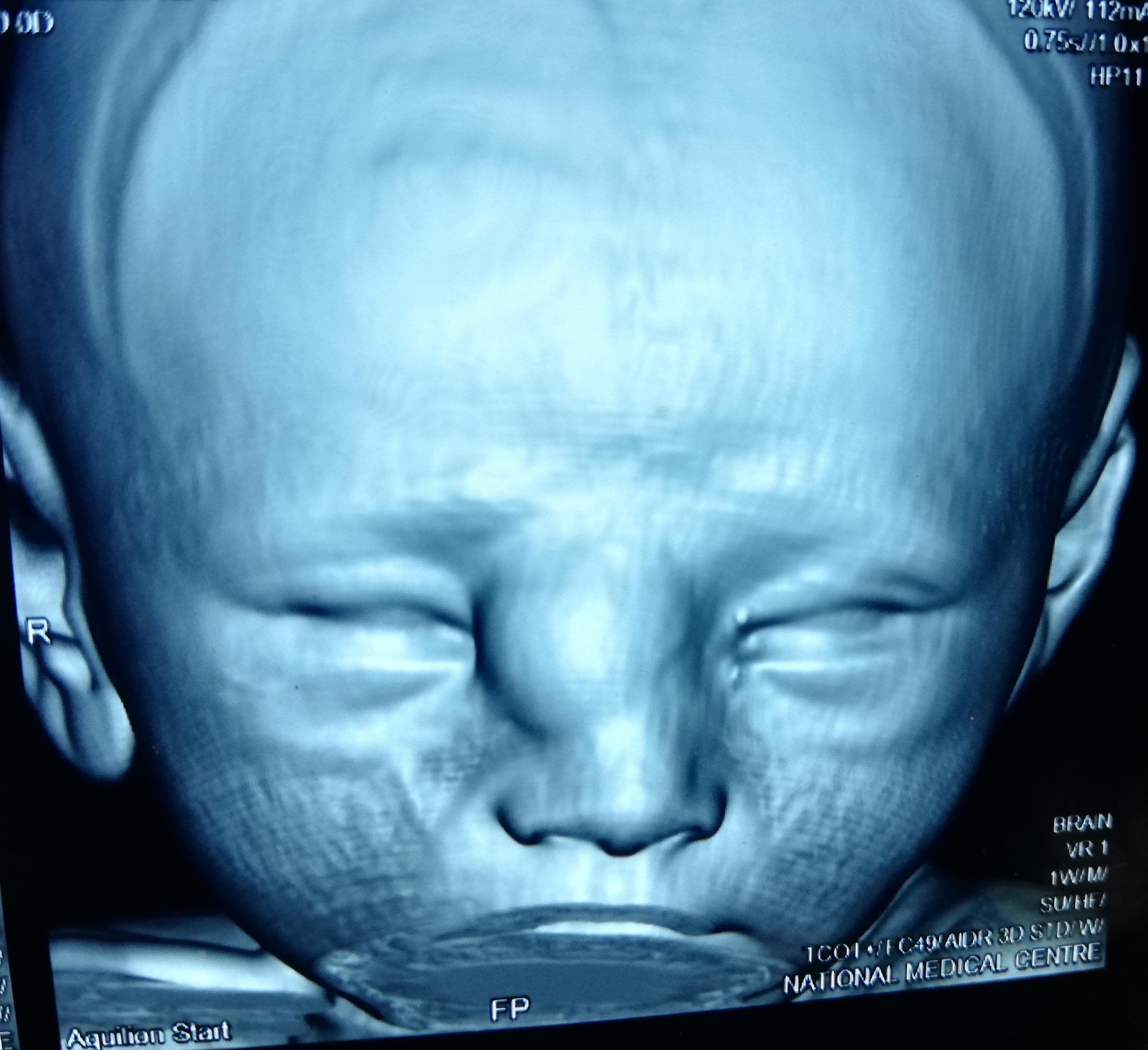
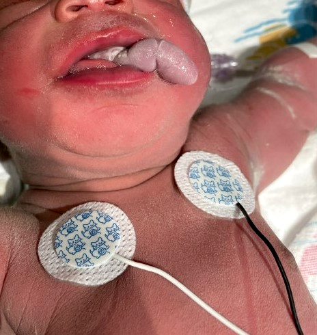
7 month old infant boy
Case #291

Various images
Images hosted on other servers:

Angiography
Images hosted on other servers:

Facial swelling
Contributed by Kelly Magliocca, D.D.S., M.P.H.
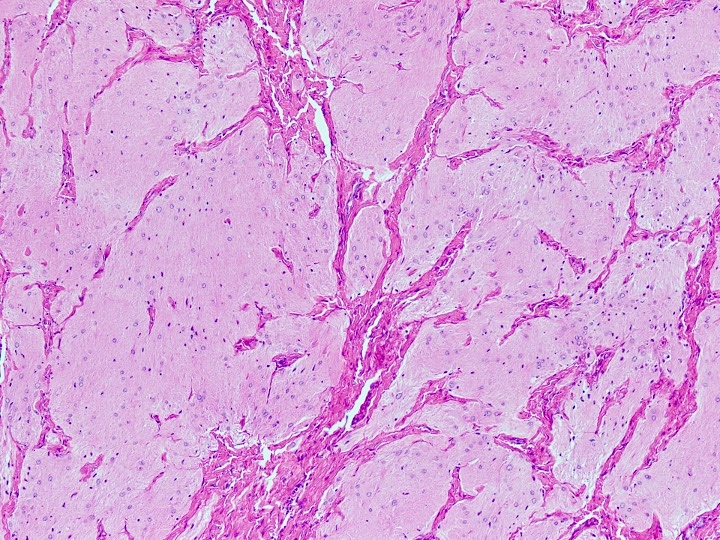
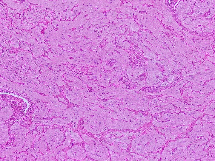
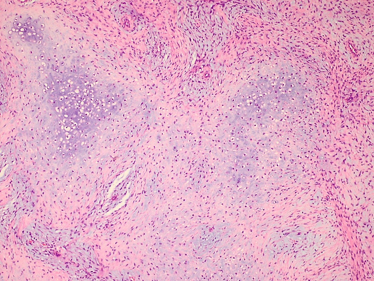
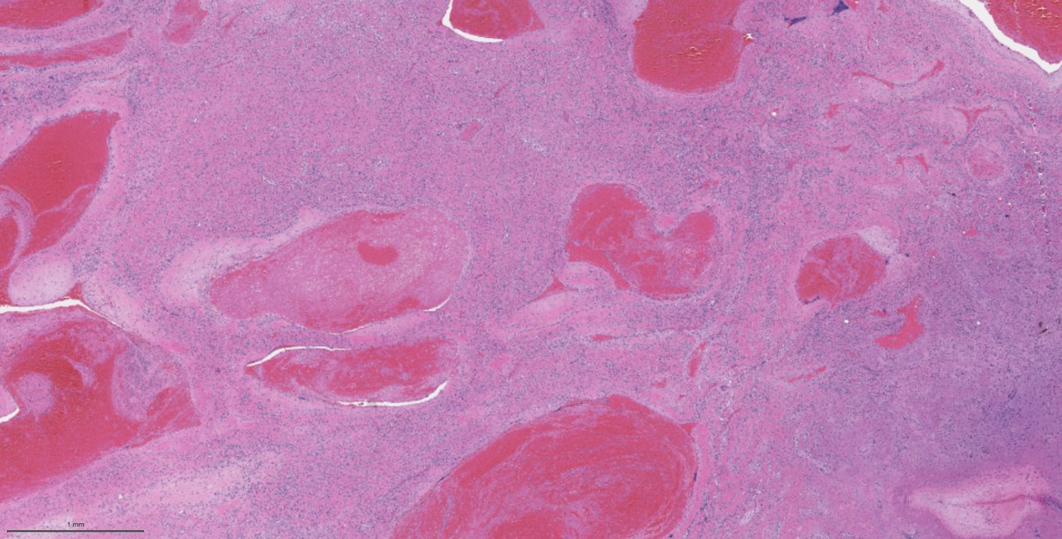
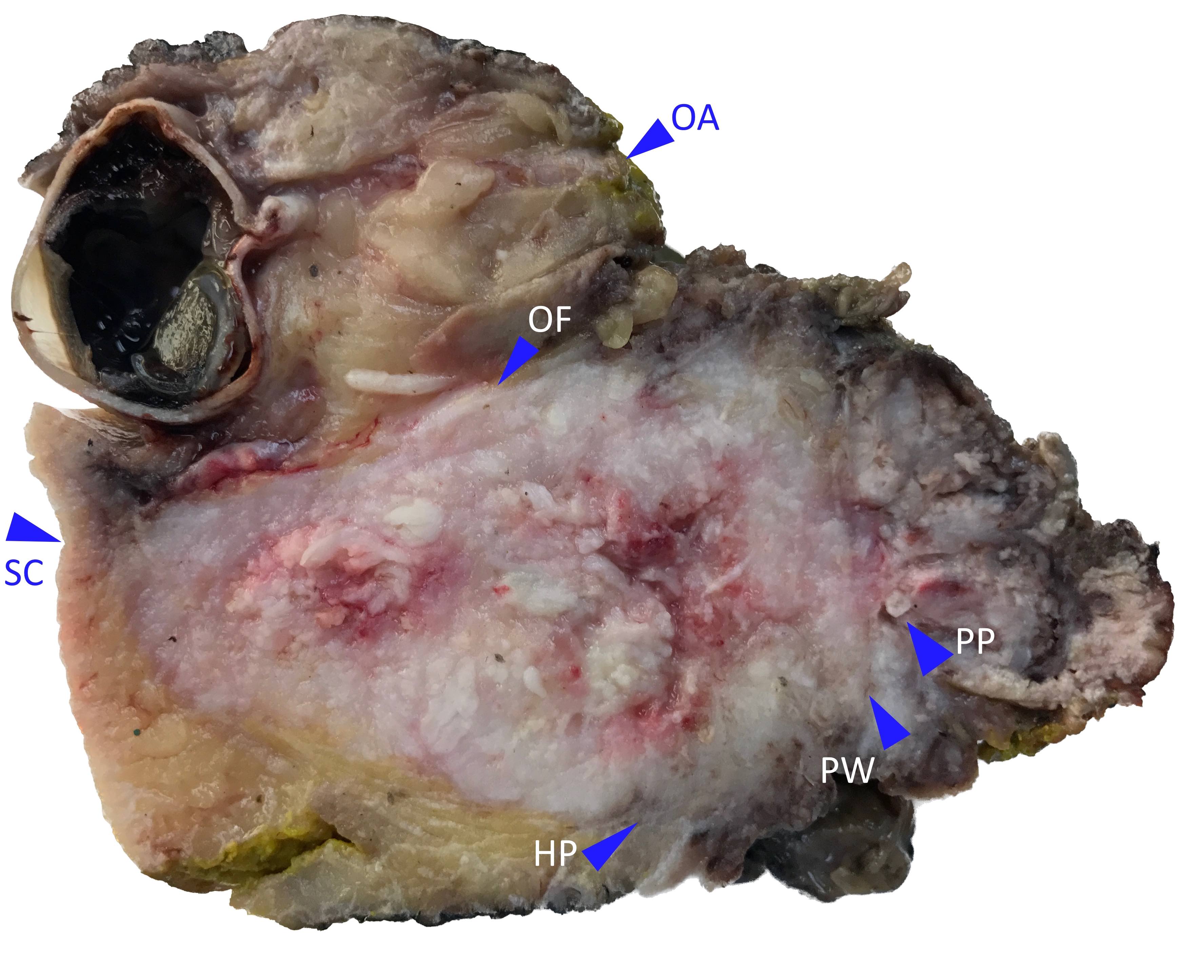
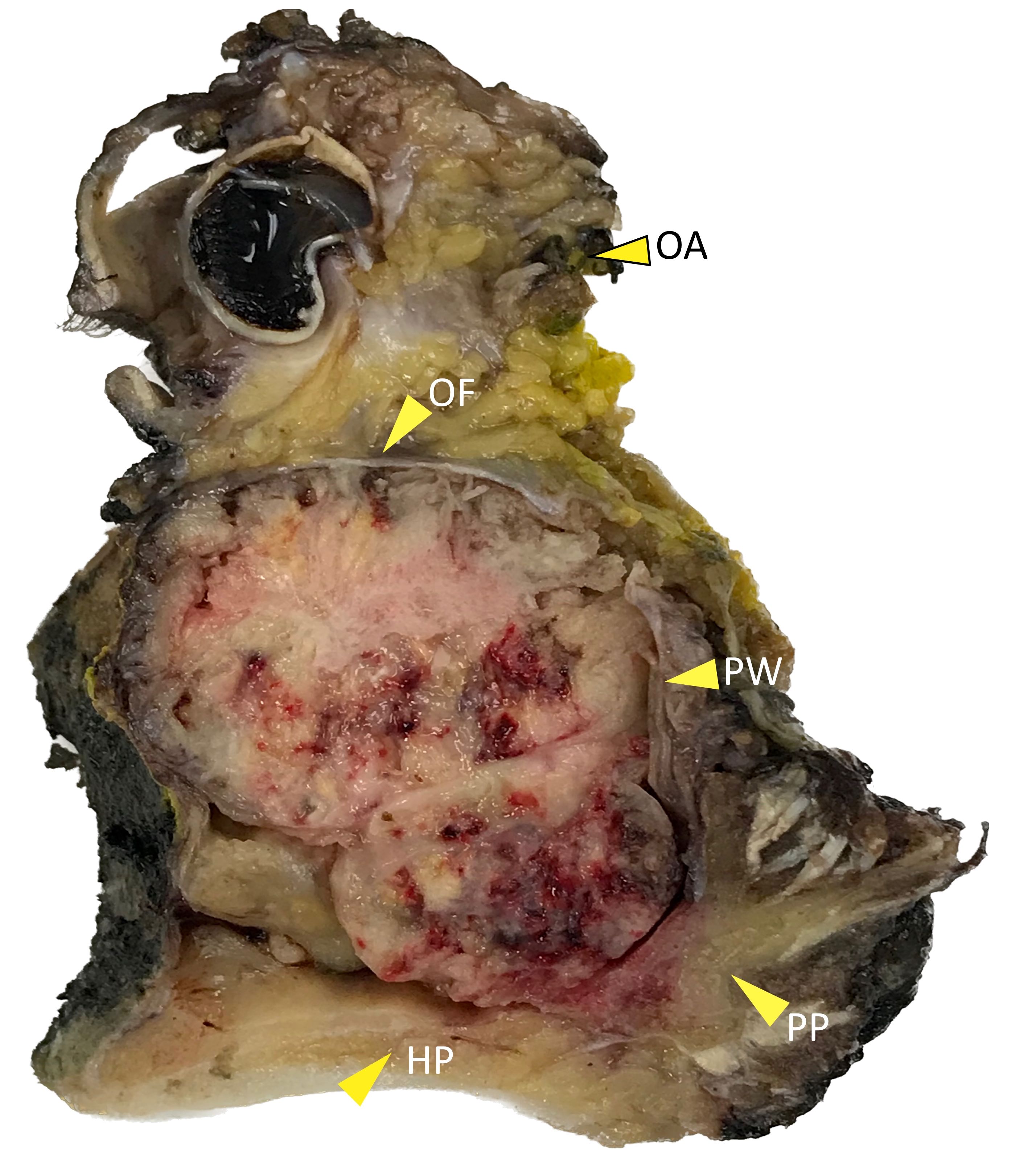
Gross appearance, cut surface
Gross appearance with ink
Images hosted on other servers:

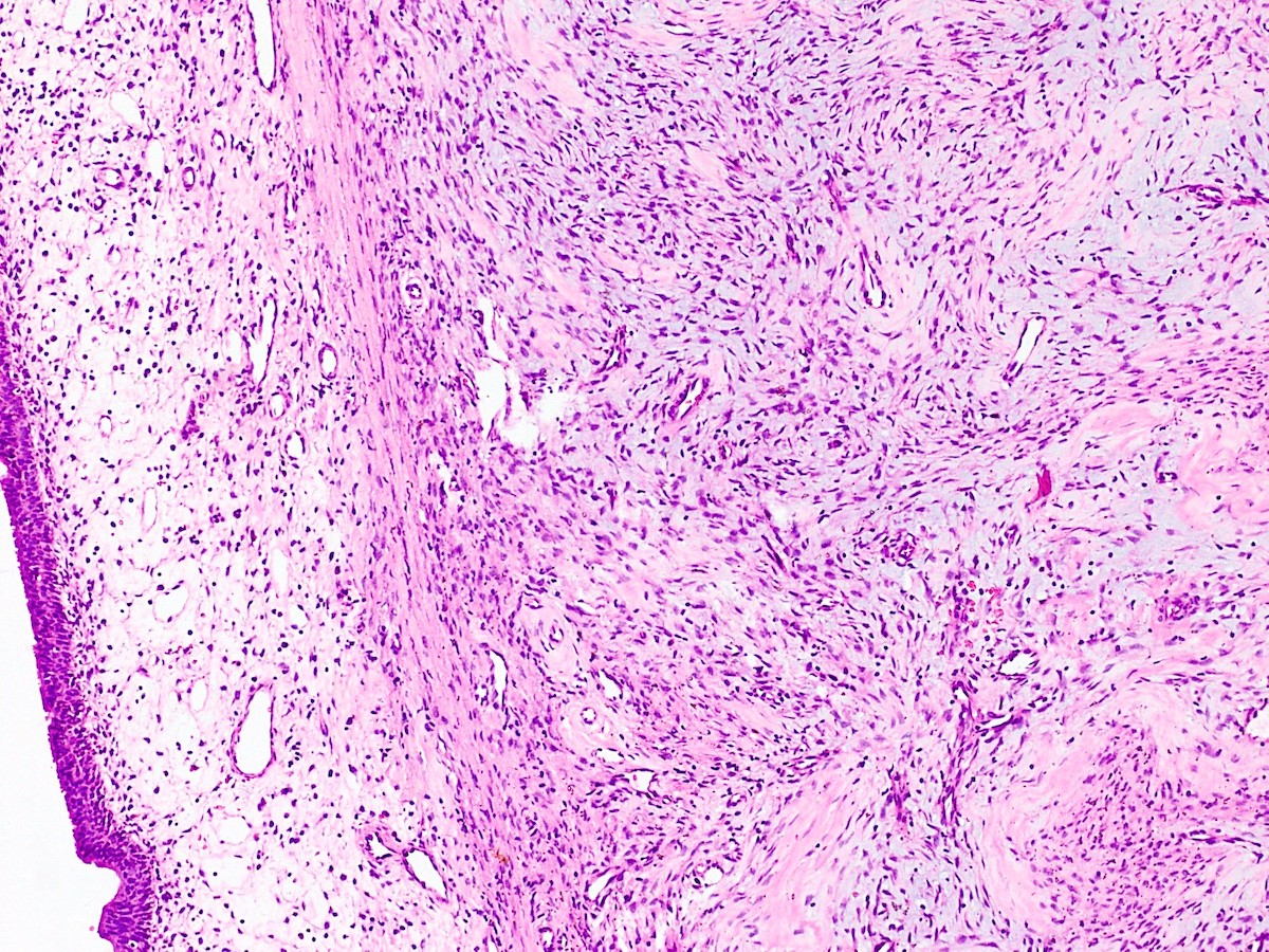
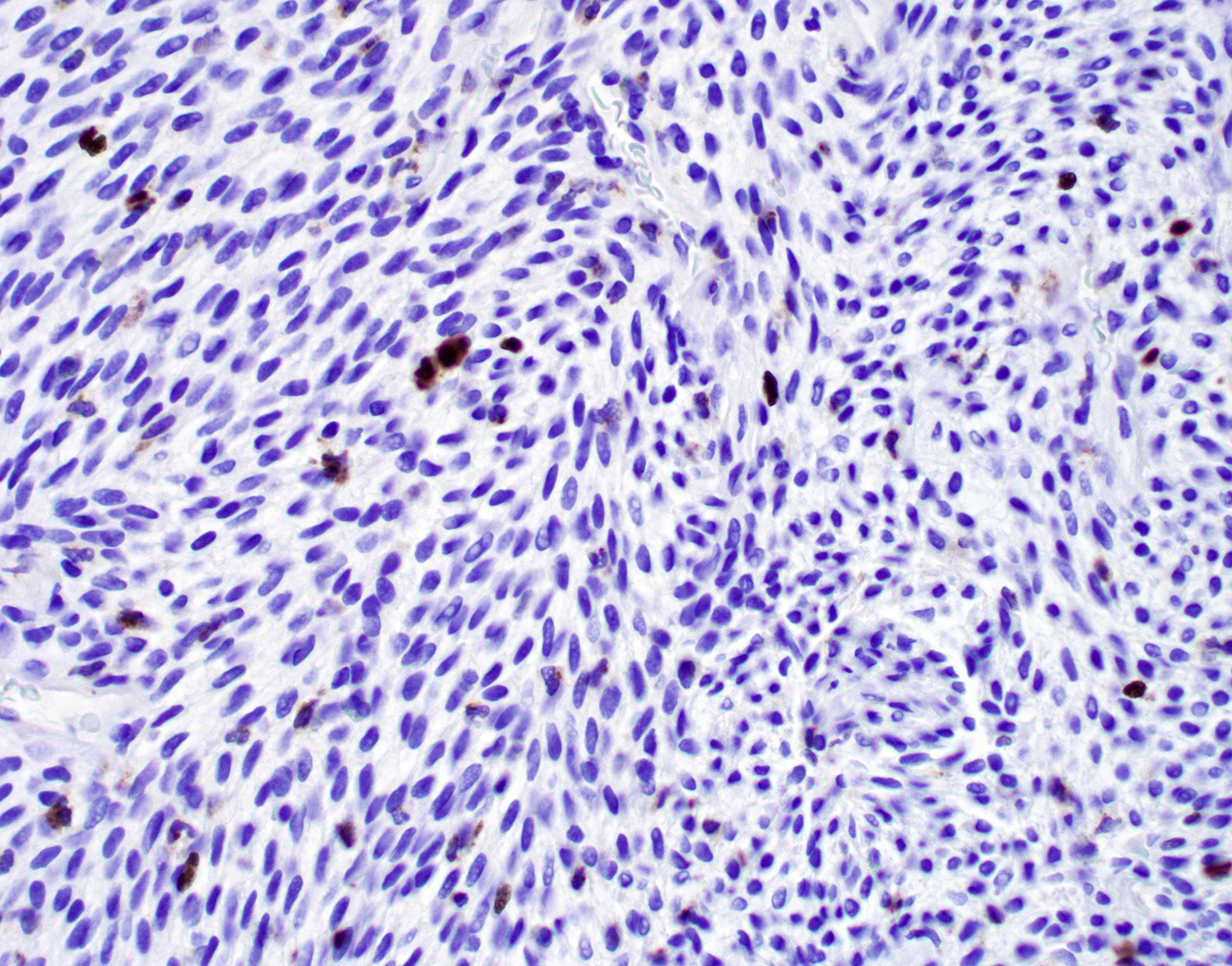
Polypoid, beige to brown, firm, fibrotic mass
Contributed by Bin Xu, M.D., Ph.D.
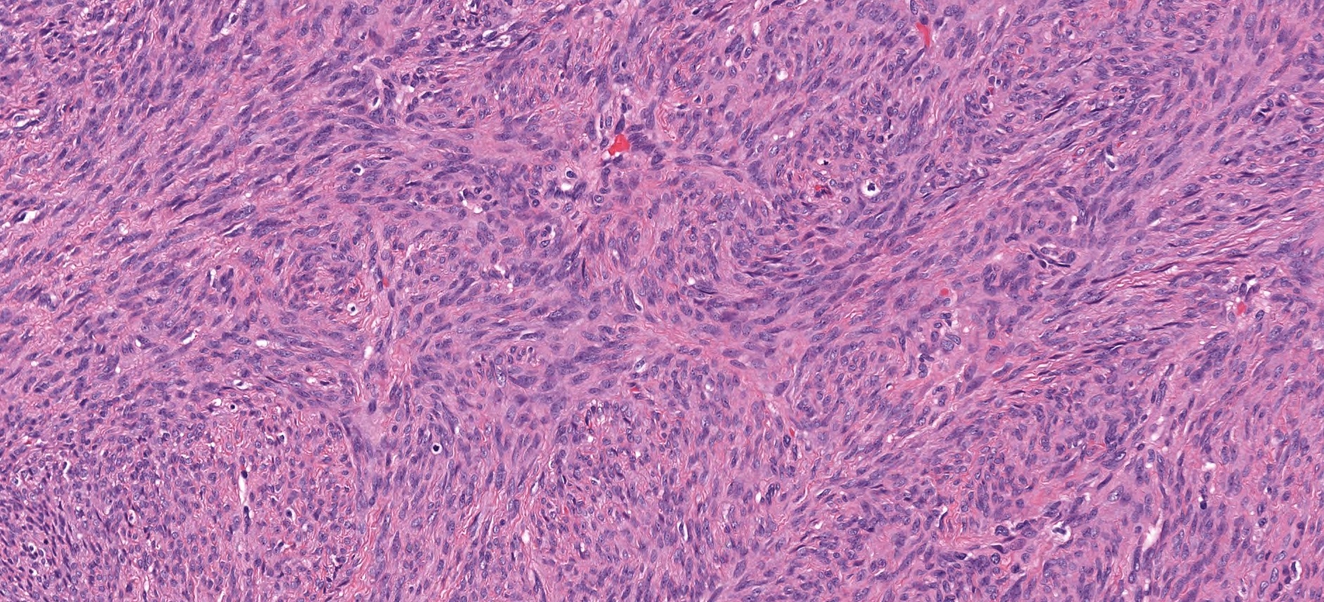
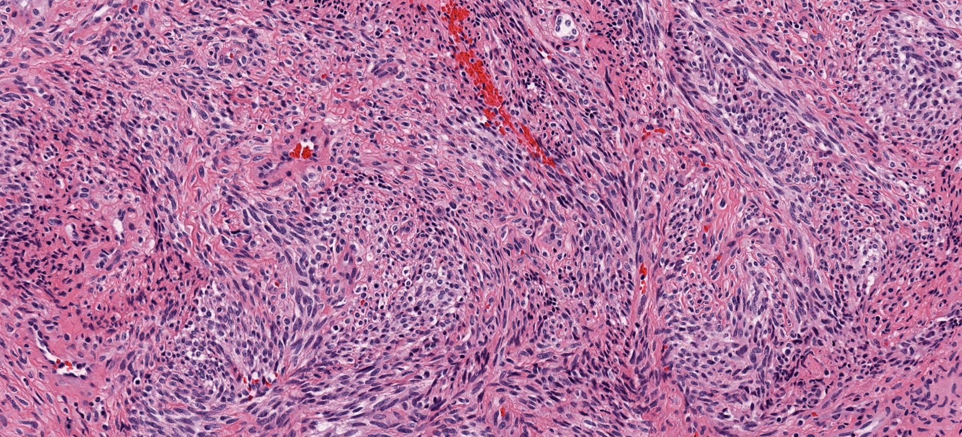
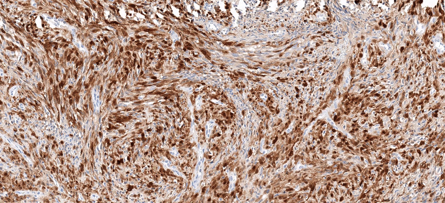
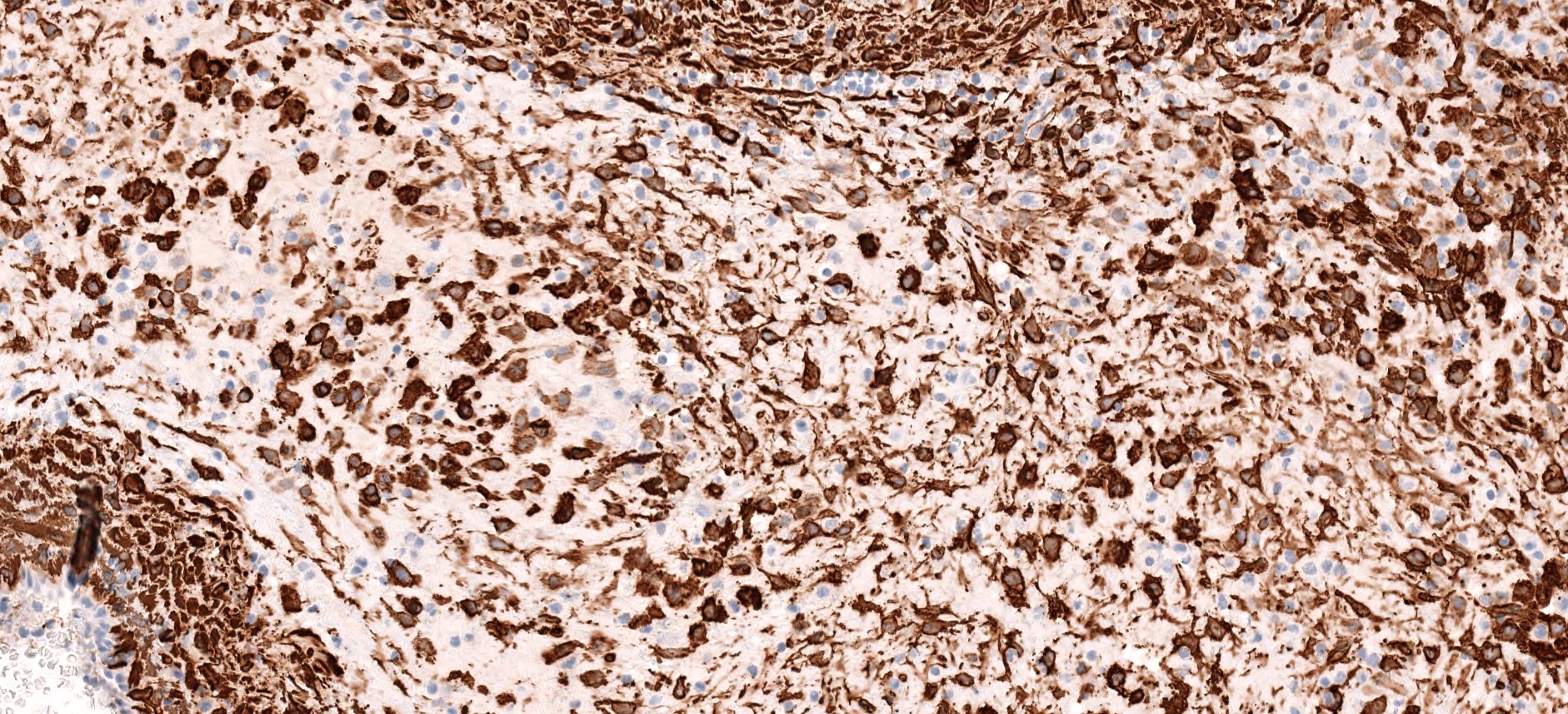
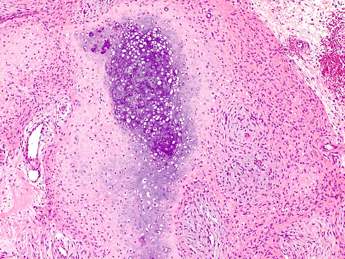
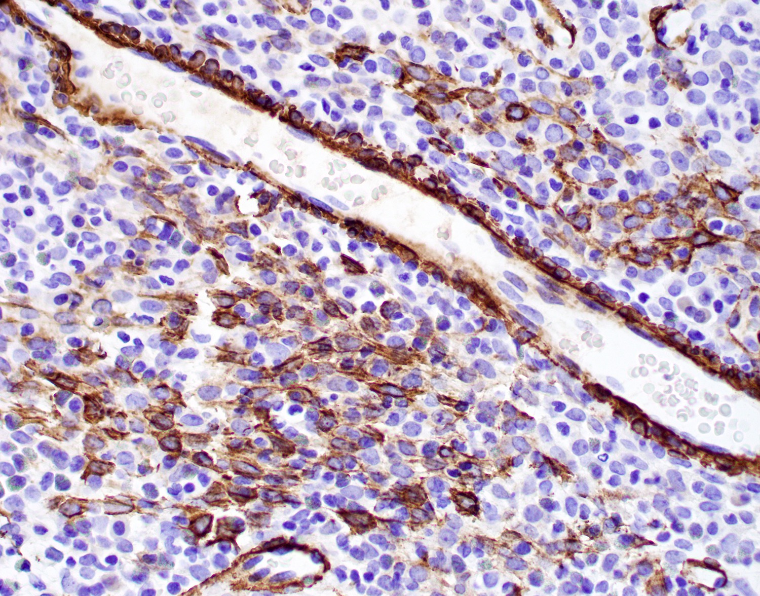
Polypoid mass
Large caliber vessels
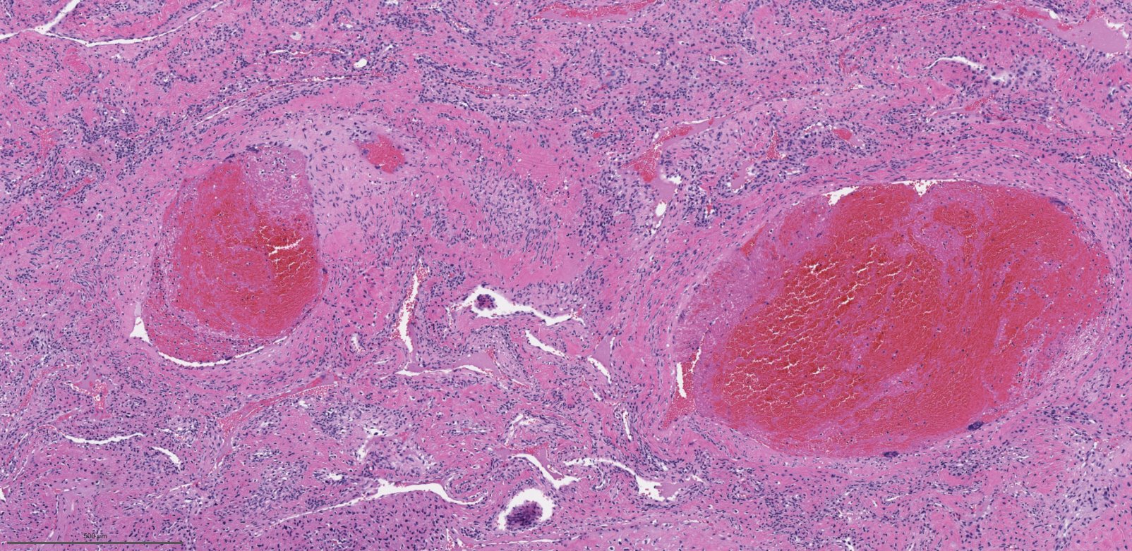
Slit-like vasculature
Cellular stroma
Bland fibroblasts
Edematous or collagenized stroma
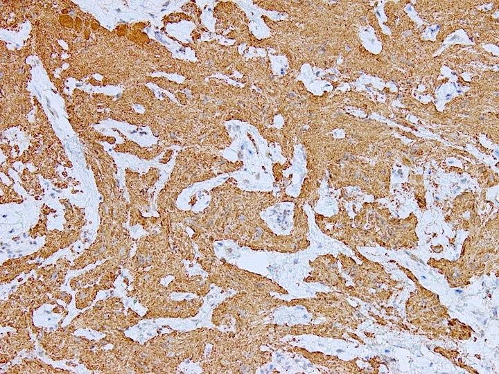
Beta catenin
AR
Contributed by Kelly Magliocca, D.D.S., M.P.H.
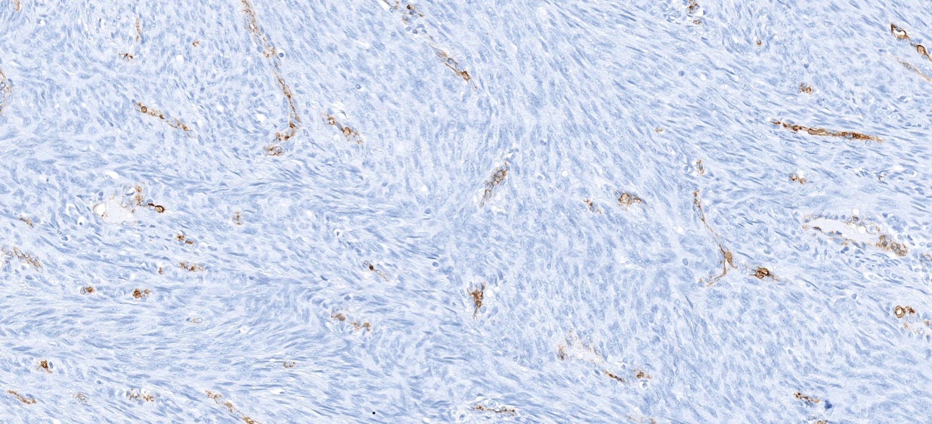
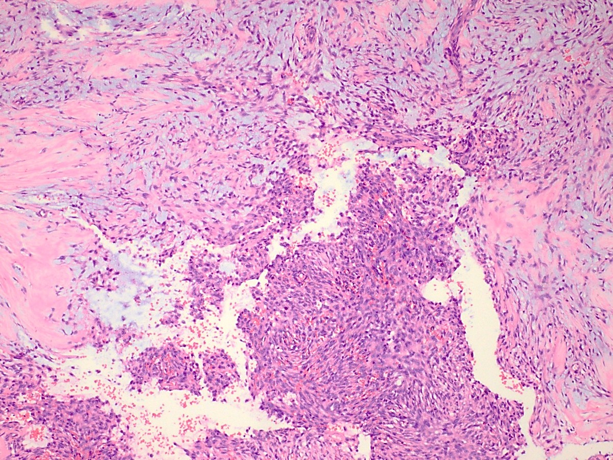
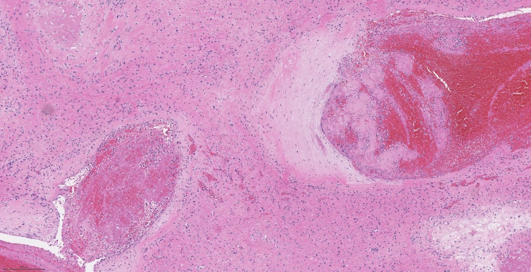
Correlate to "Gross appearance with ink"
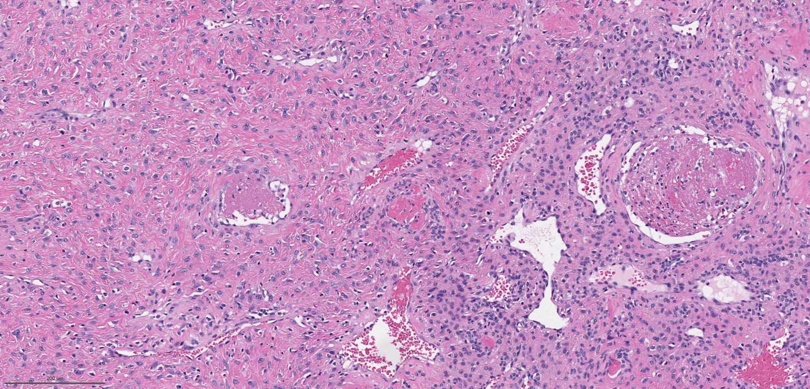
Vascular channel, low magnification

Vascular channel, higher magnification
Images hosted on other servers:

Age standardized incidence
Images hosted on other servers:

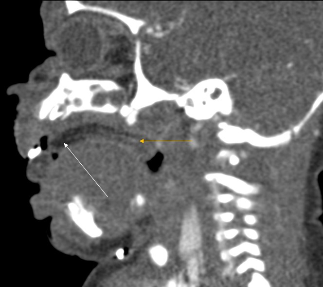
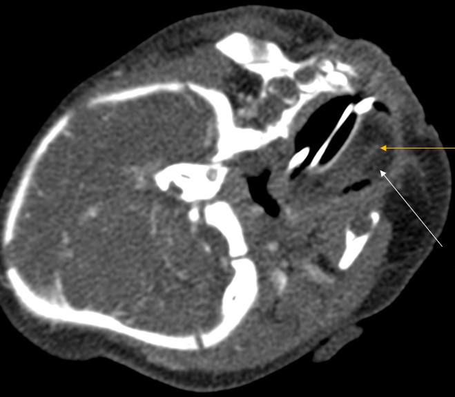
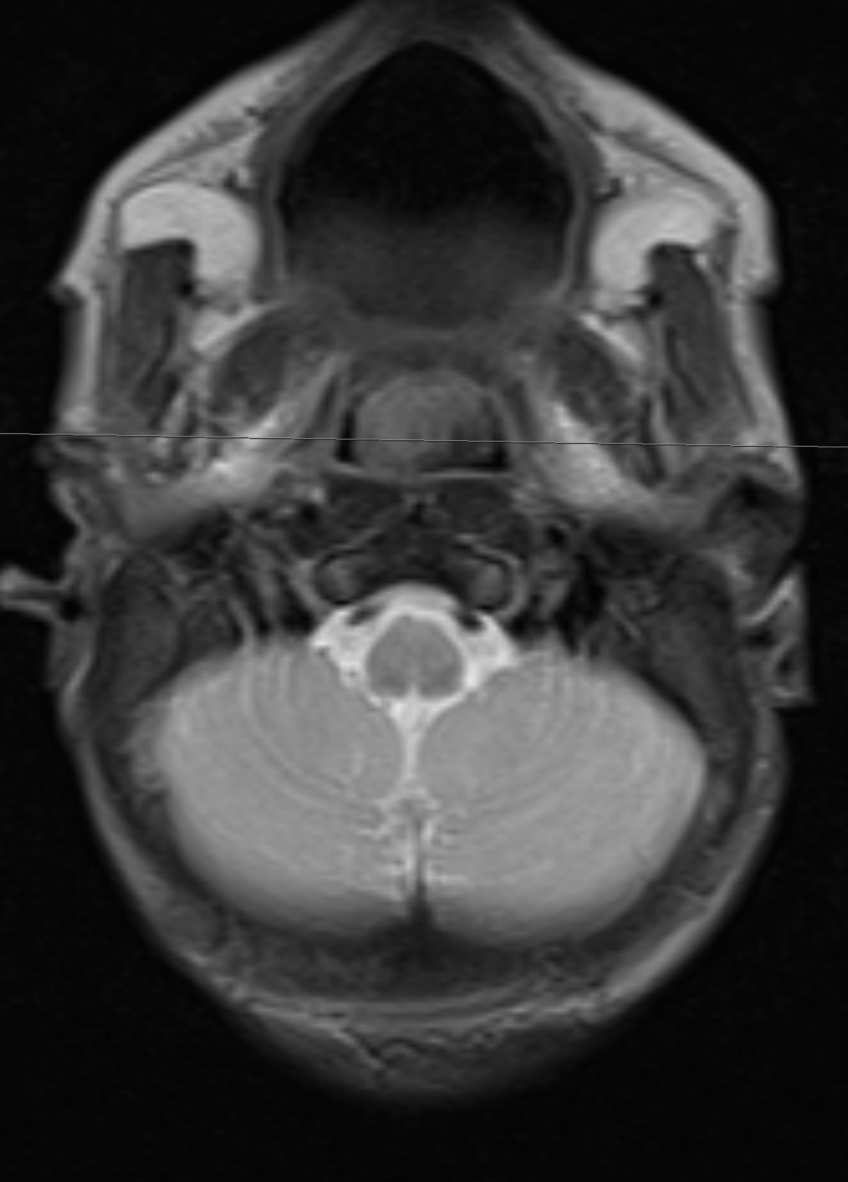
CT scan: wedge shaped mass

MRI: Enhanced mass in the fossa of Rosenmüller
Contributed by Bin Xu, M.D., Ph.D. and Andrey Bychkov, M.D., Ph.D. and Songyang Yuan, M.D., Ph.D.
Nonkeratinizing, undifferentiated subtype
Nonkeratinizing undifferentiated subtype
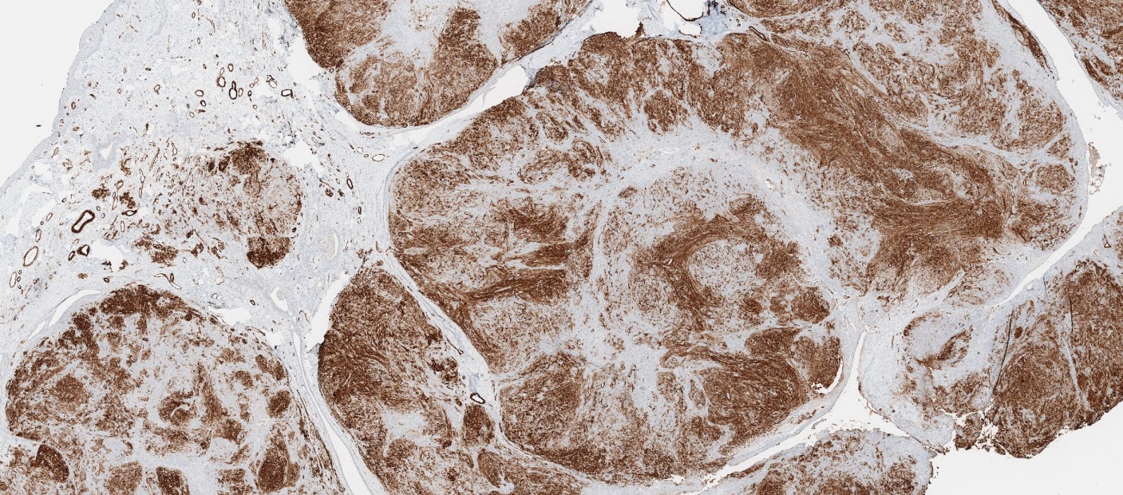
AE1 / AE3+
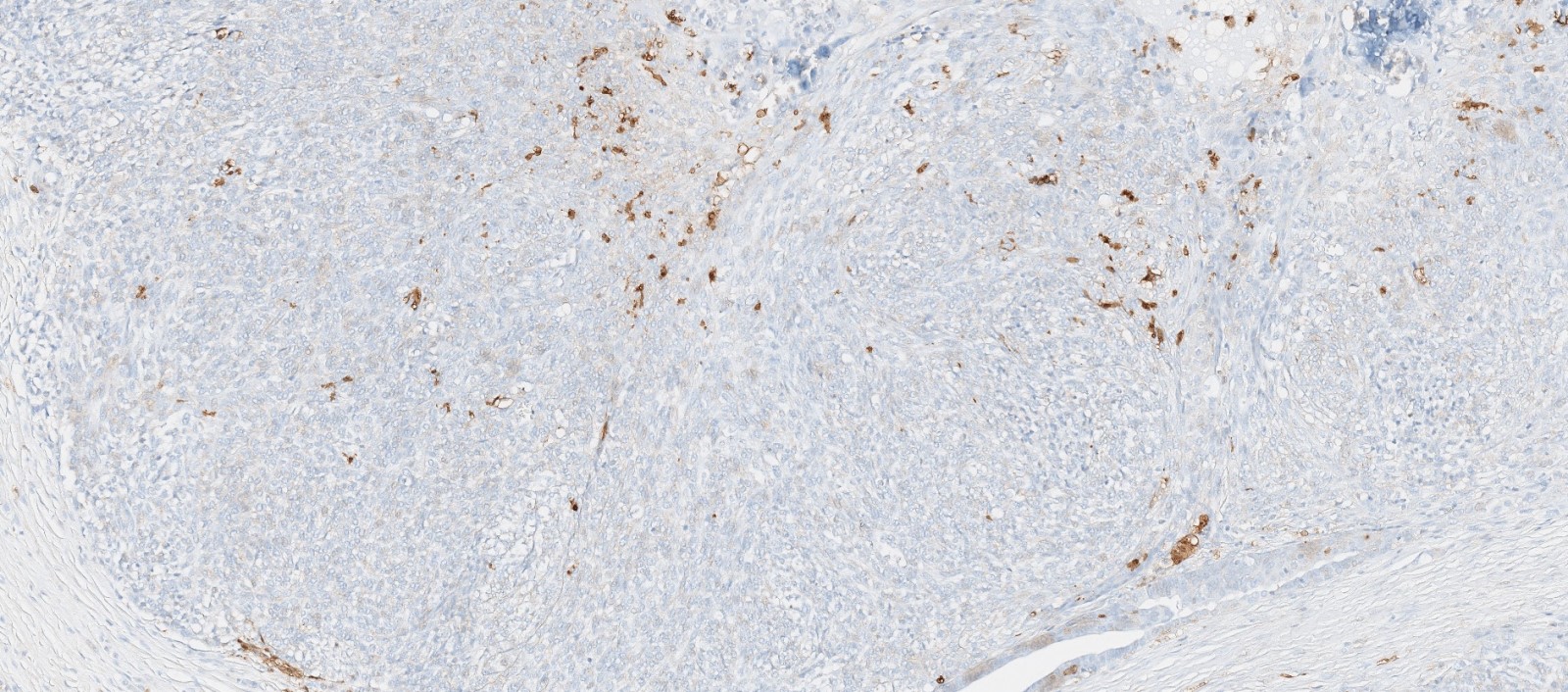
EBV LMP1 IHC
Nonkeratinizing undifferentiated subtype
CK5 / 6+
EBER+
p63+
Nonkeratinizing, differentiated subtype
Nonkeratinizing differentiated subtype
Keratinizing subtype
Keratinizing subtype
Contributed by Marino E. Leon, M.D. and Case #100
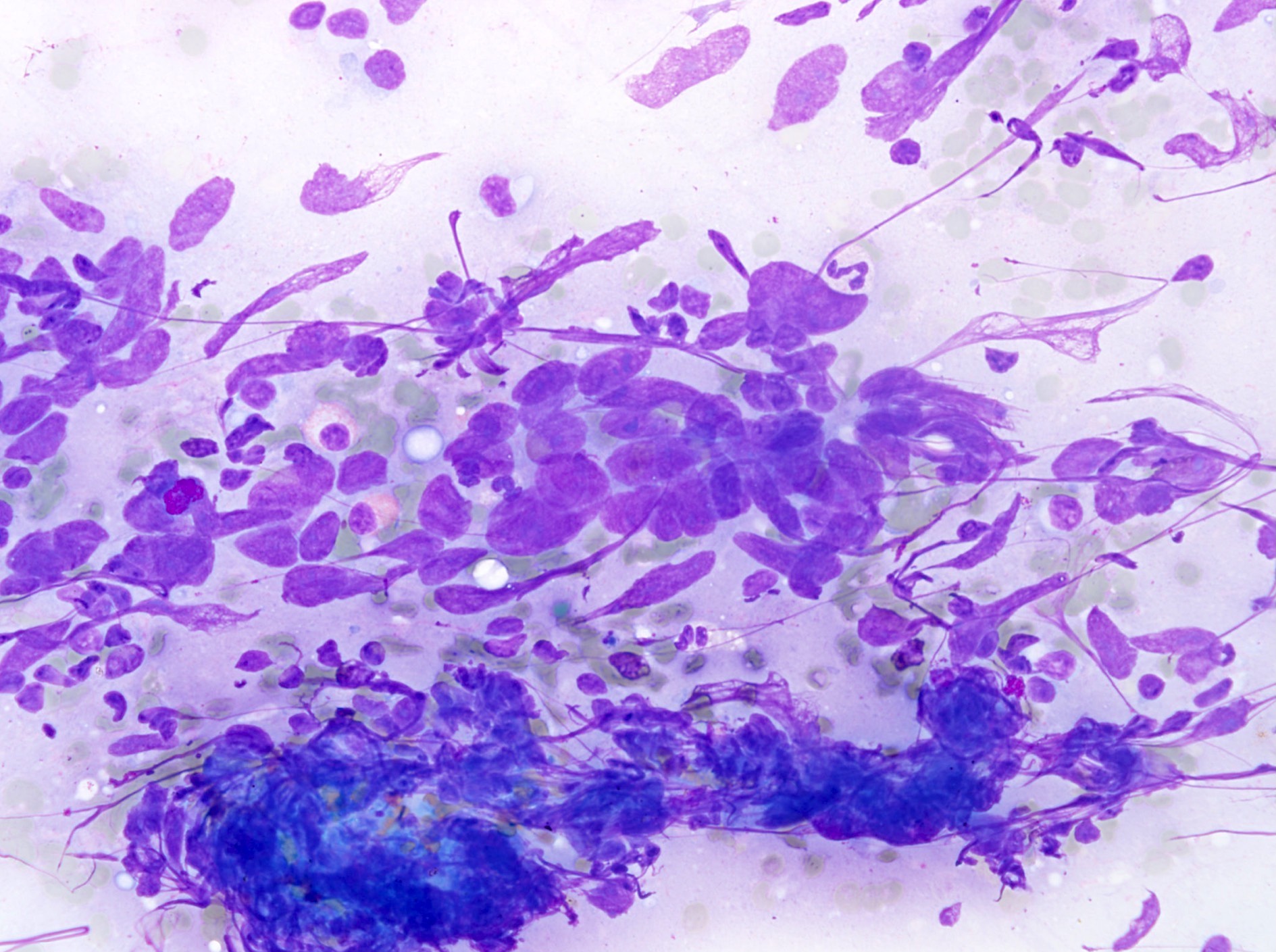
FNA of a neck mass (Diff-Quik)
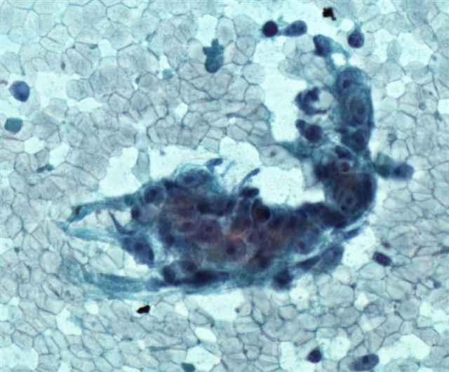
Pap stain
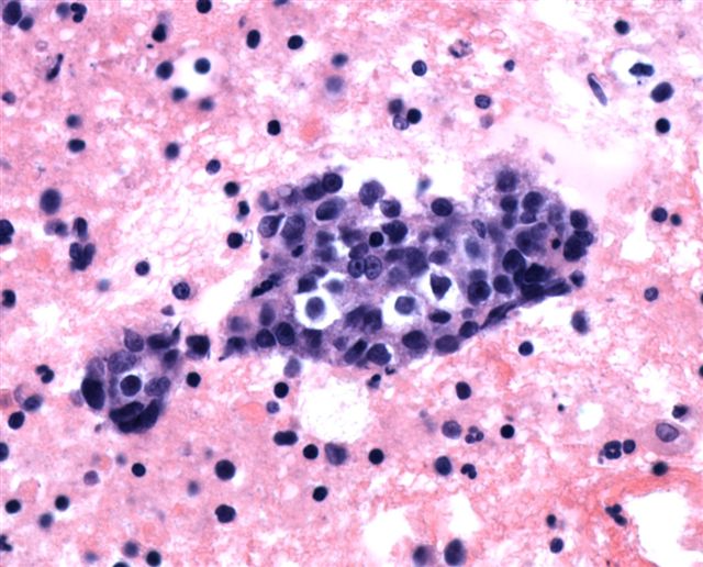
Cell block
Contributed by Andrey Bychkov, M.D., Ph.D. and Bin Xu, M.D., Ph.D.
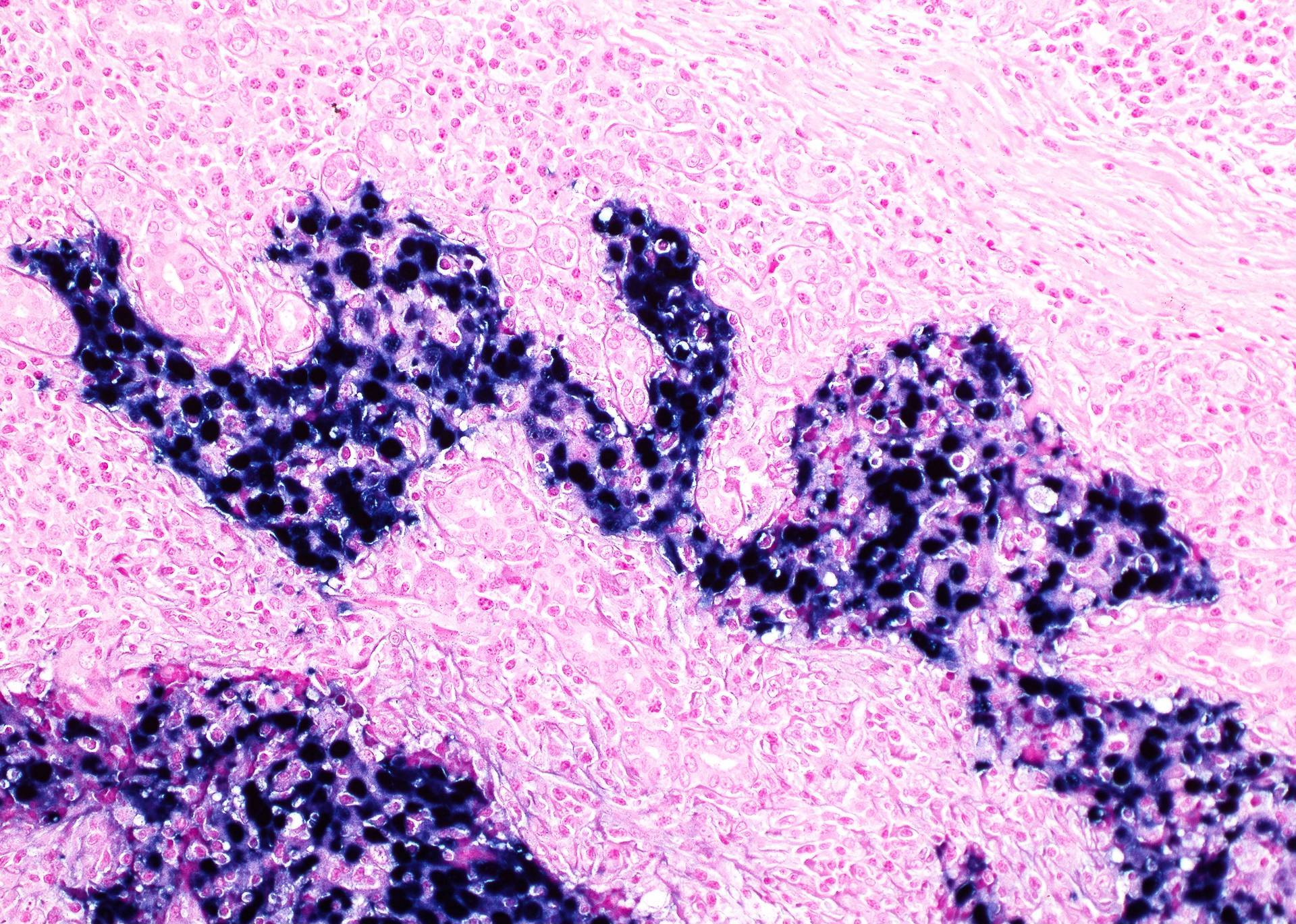
EBER ISH
Images hosted on other servers:

Heterogeneously hyperintense lesion on T2 weighted image
Images hosted on other servers:

Eustachian tube tumor
Contributed by Diana Bell, M.D. and Wai Szeto, M.D., M.S.
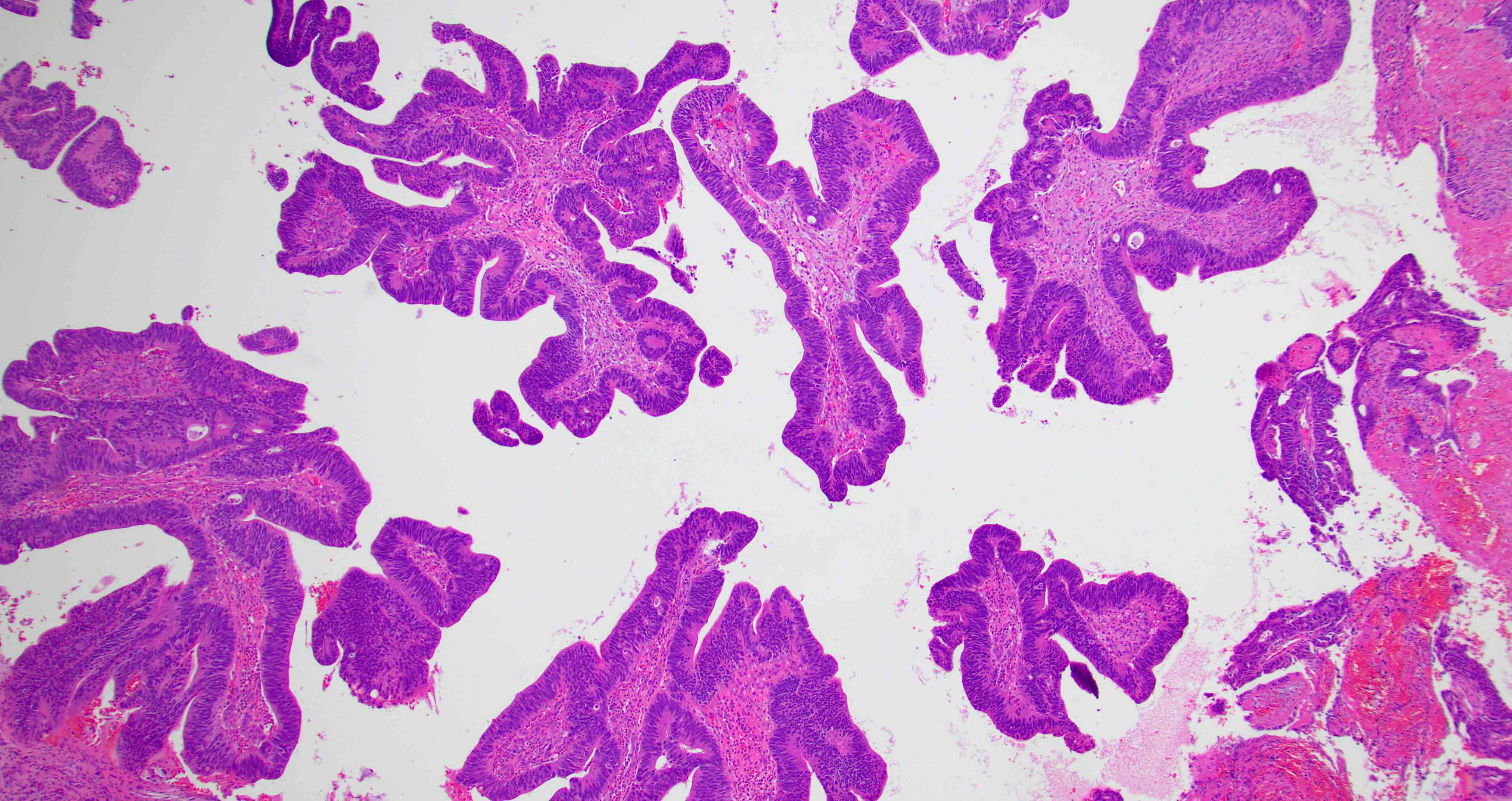
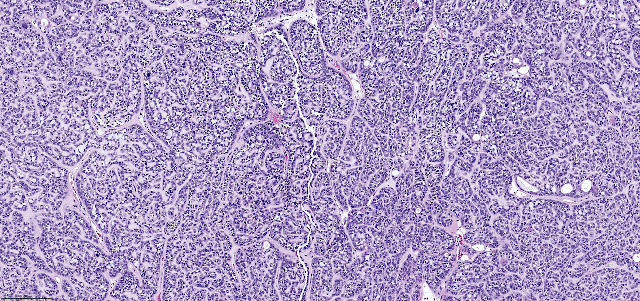
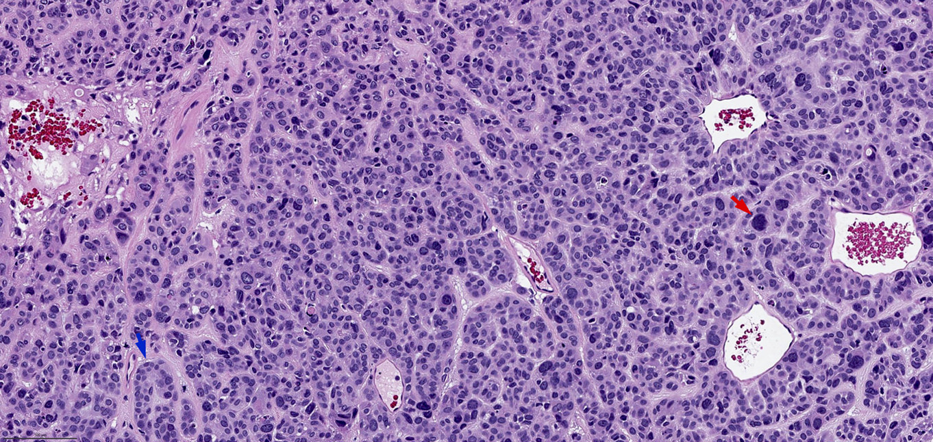
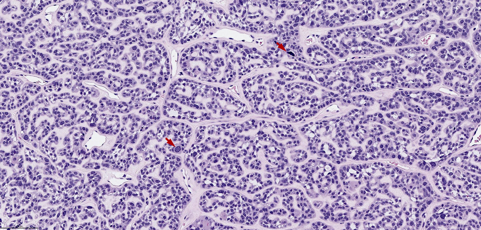
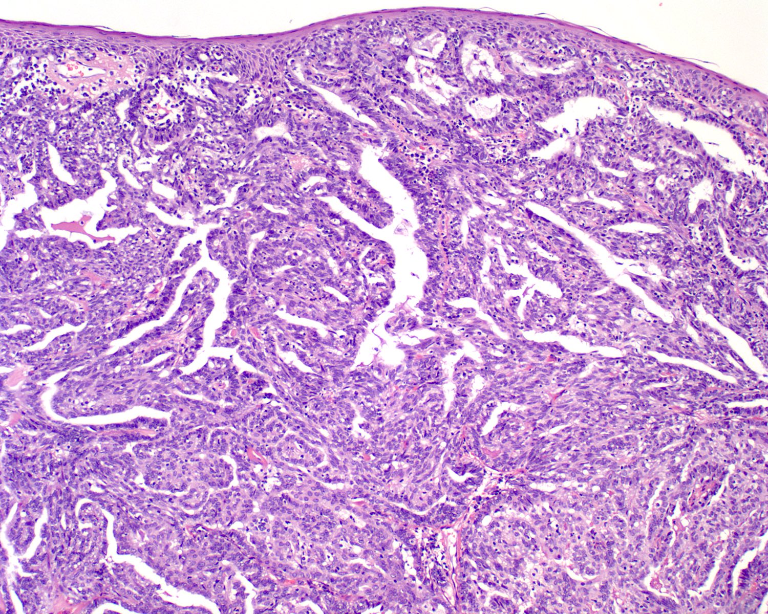
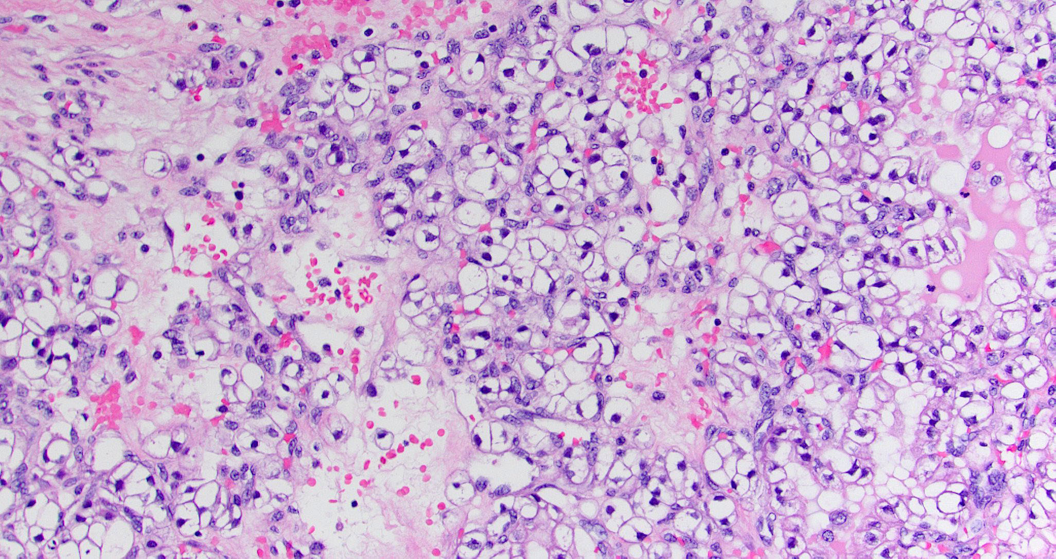
Low grade sinonasal adenocarcinoma
Low grade sinonasal adenocarcinoma
Tubular growth pattern
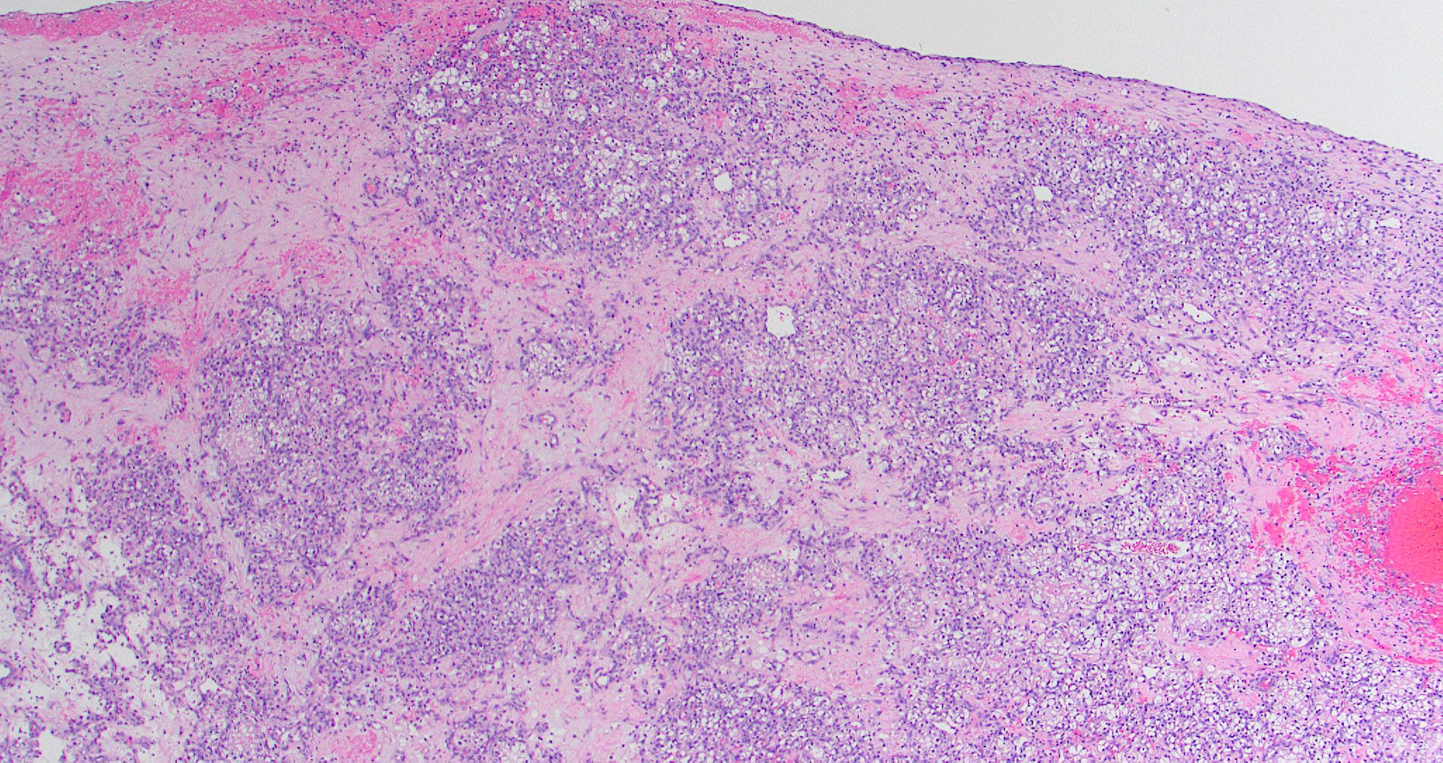
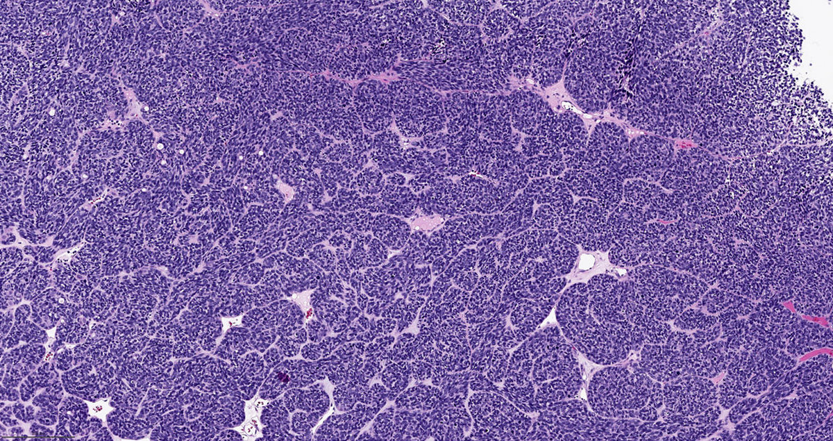
High grade sinonasal adenocarcinoma

High grade sinonasal adenocarcinoma

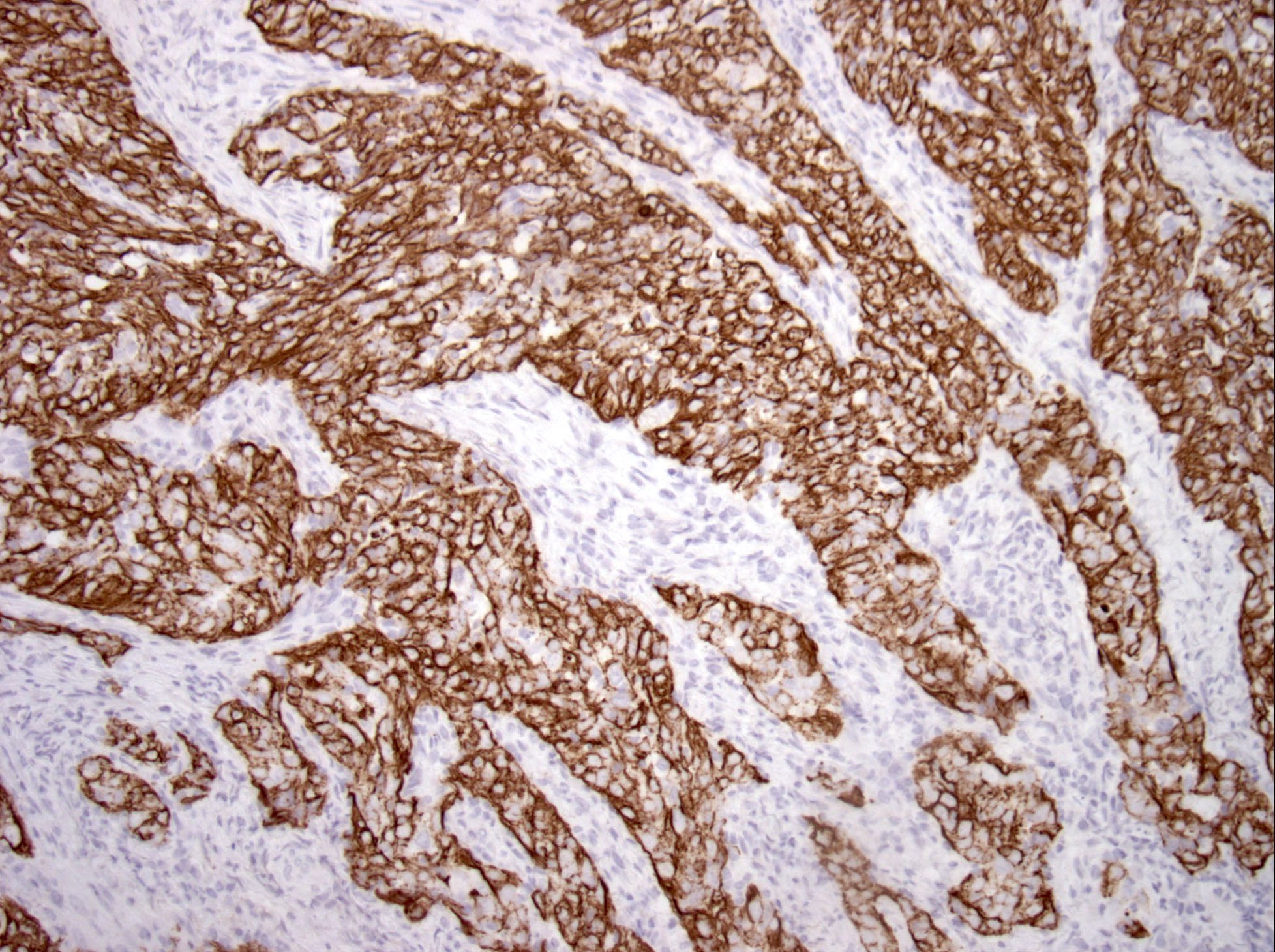
CK7

SOX10
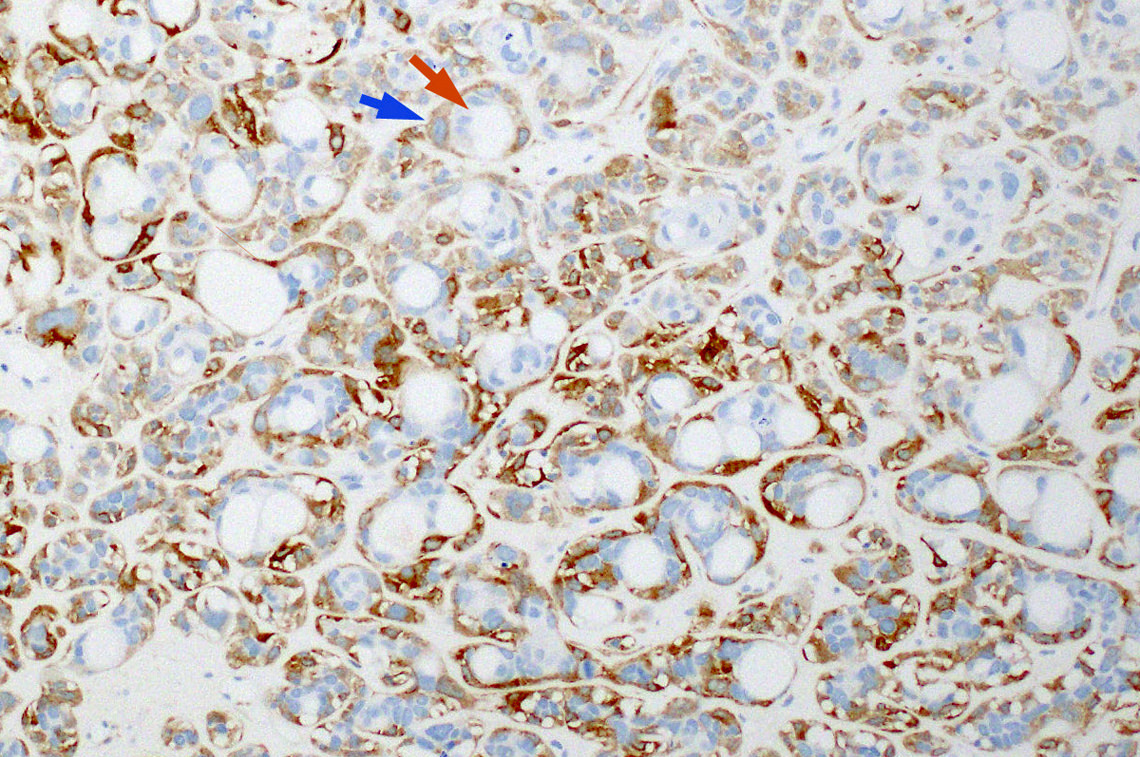
Sinonasal renal cell-like adenocarcinoma

Sinonasal renal cell-like adenocarcinoma

Mimics renal cell carcinoma
Clear cytoplasm

CAIX
Images hosted on other servers:
Eustachian tube tumor
Images hosted on other servers:

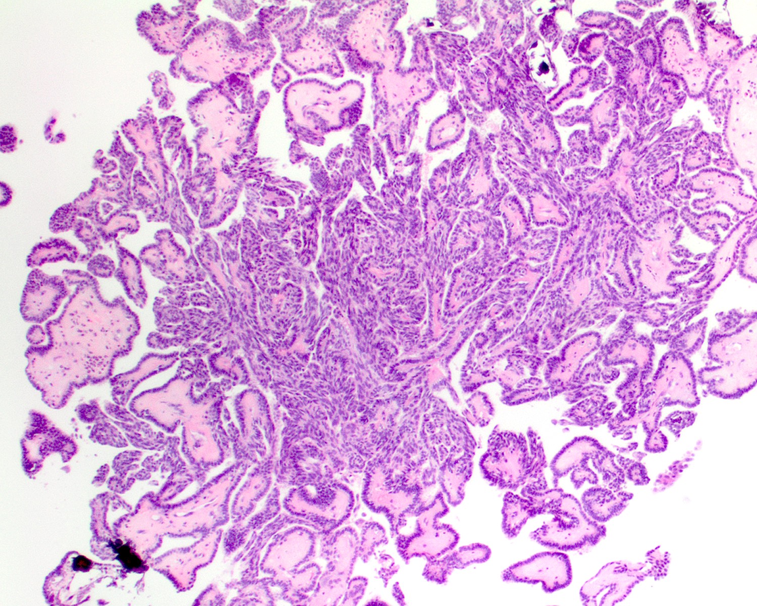
Bland serous glands

Complex papillary growth pattern

Tubulopapillary pattern

Eustachian tube tumor


TTF1+ case
Images hosted on other servers:

Large nasal mass
Images hosted on other servers:

Lacrimal mass

Nasopharyngeal endoscopy
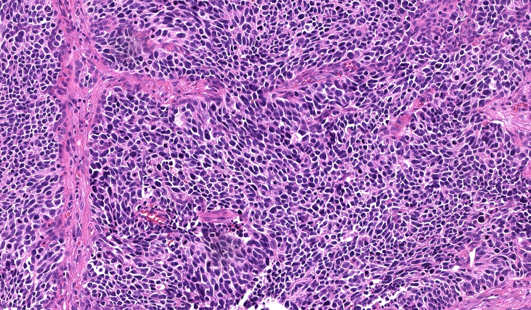
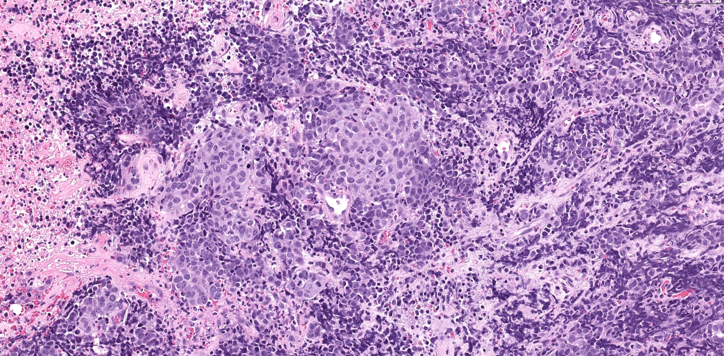
Contributed by Lisa Rooper, M.D.

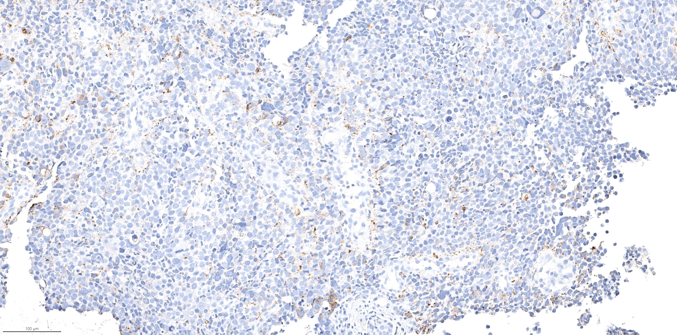
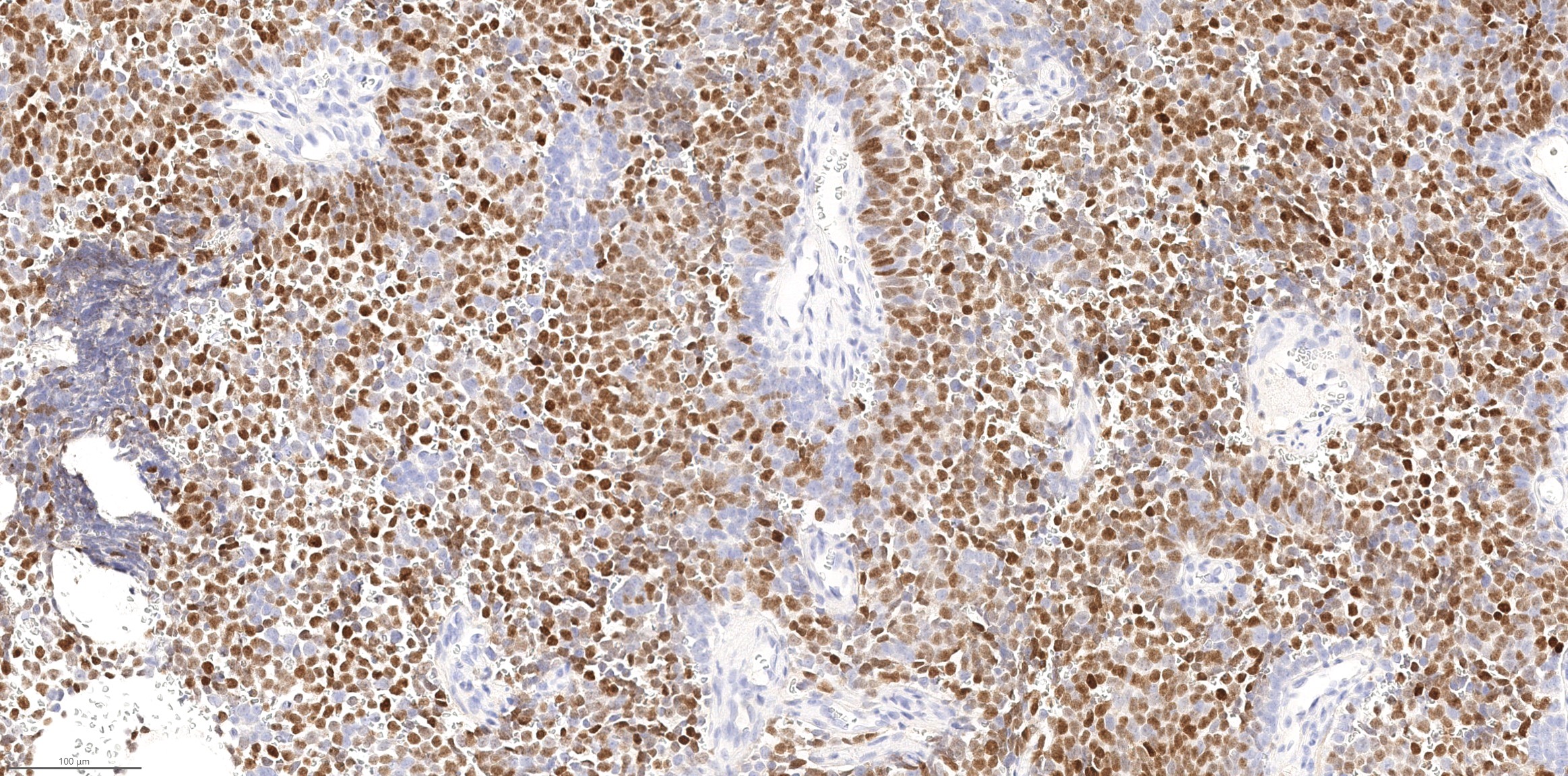
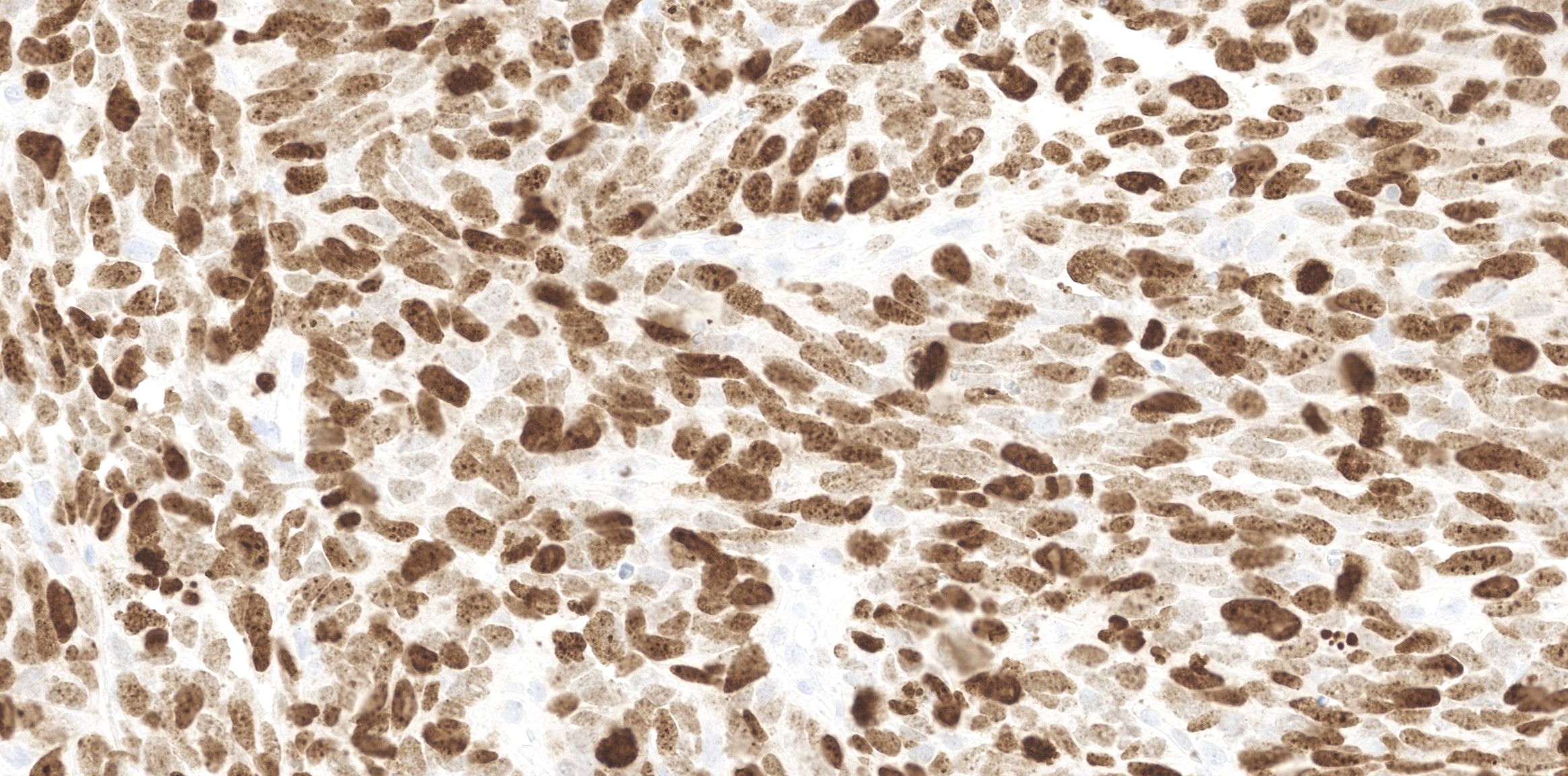
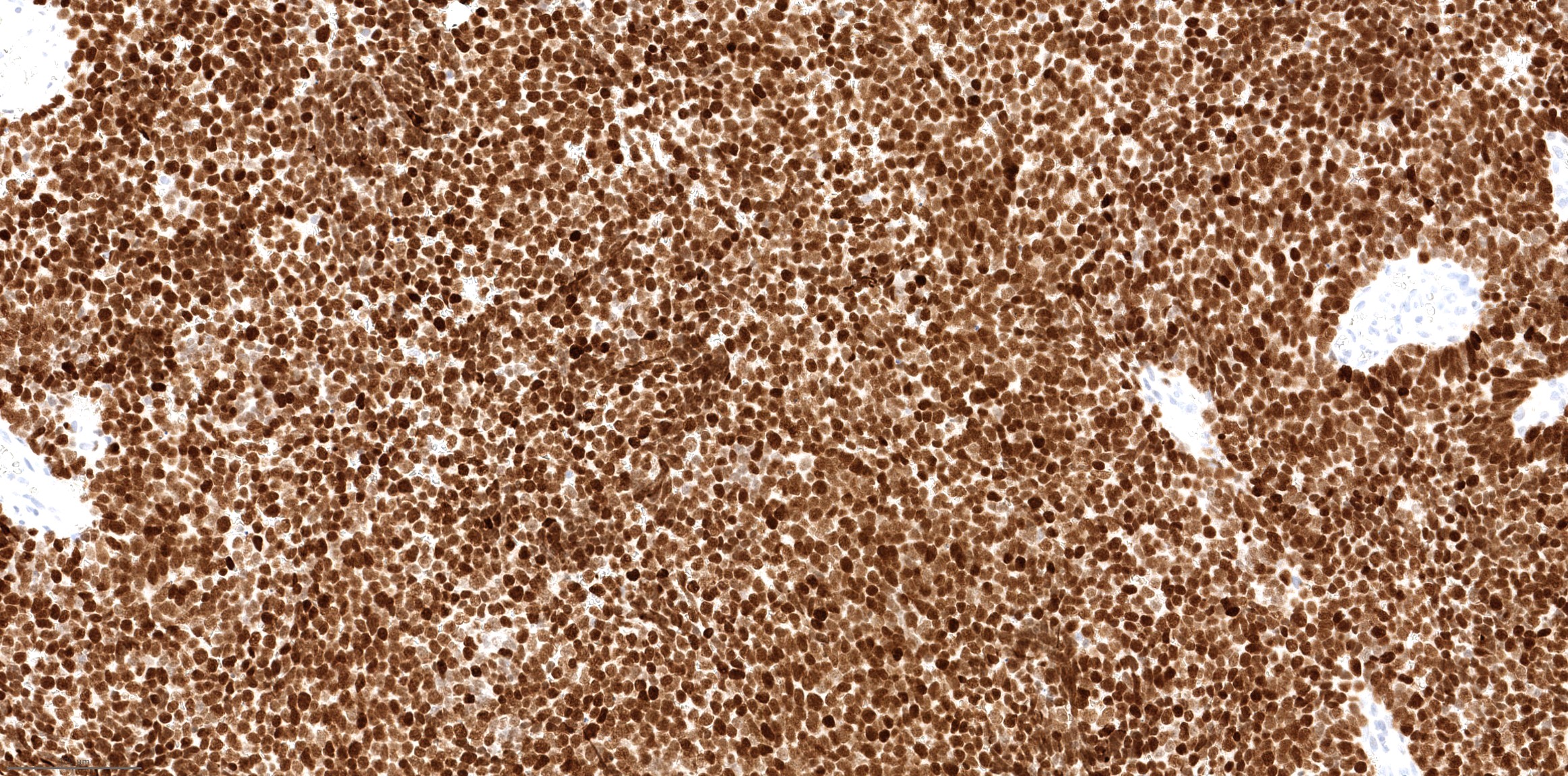
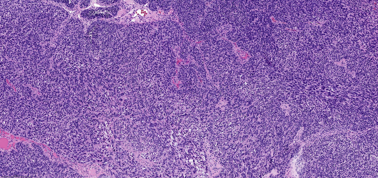
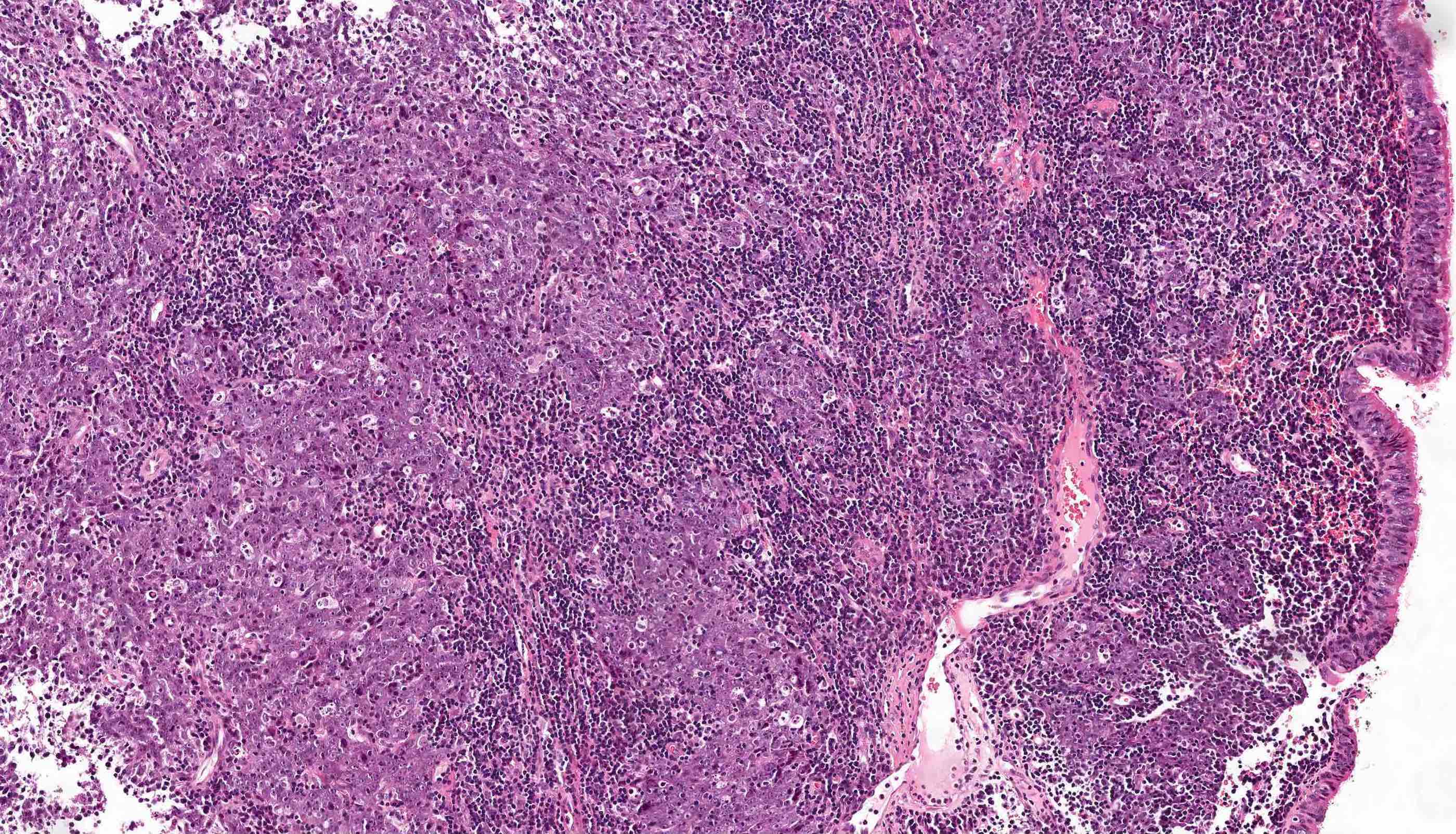
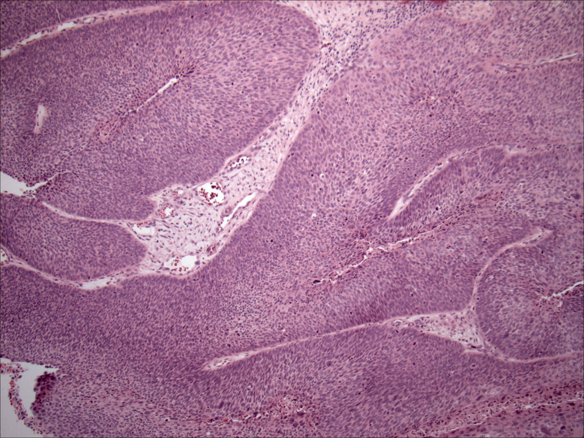
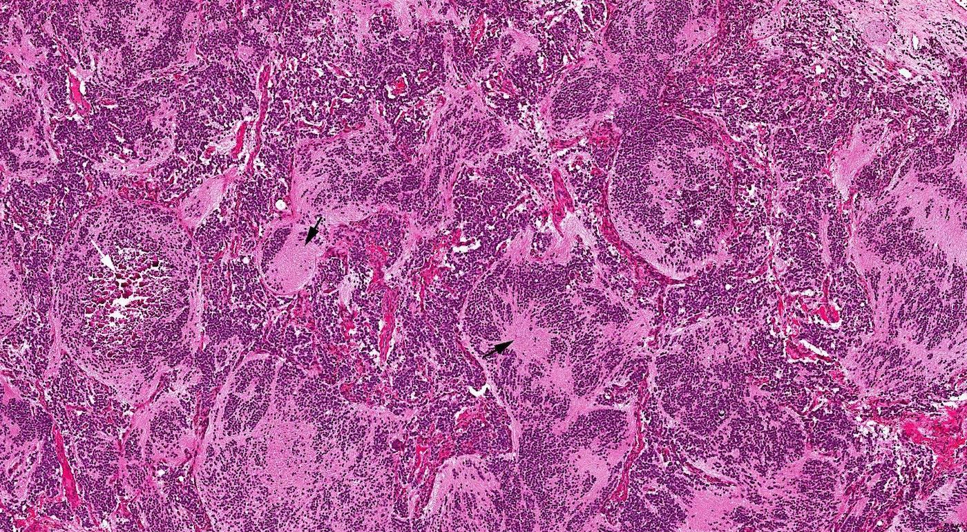
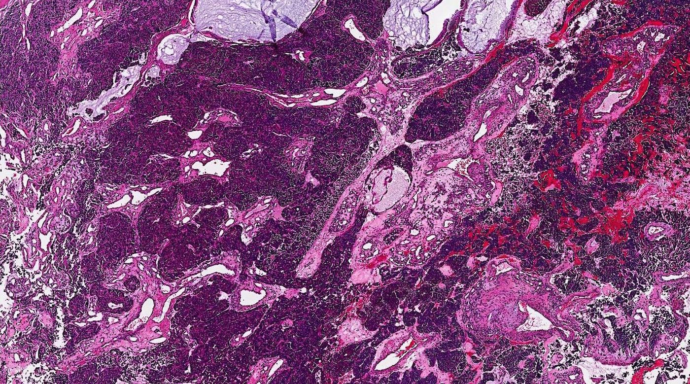
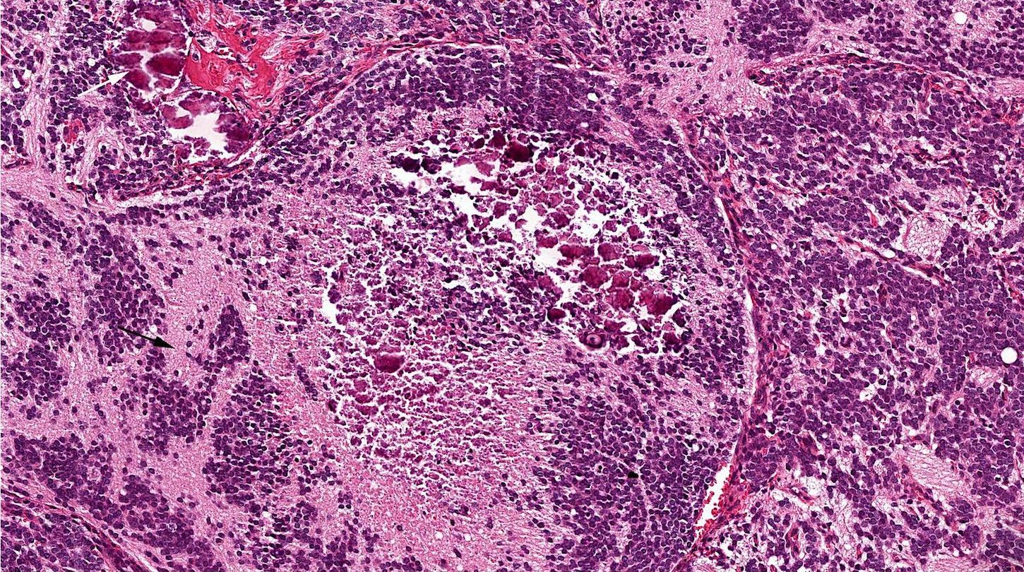
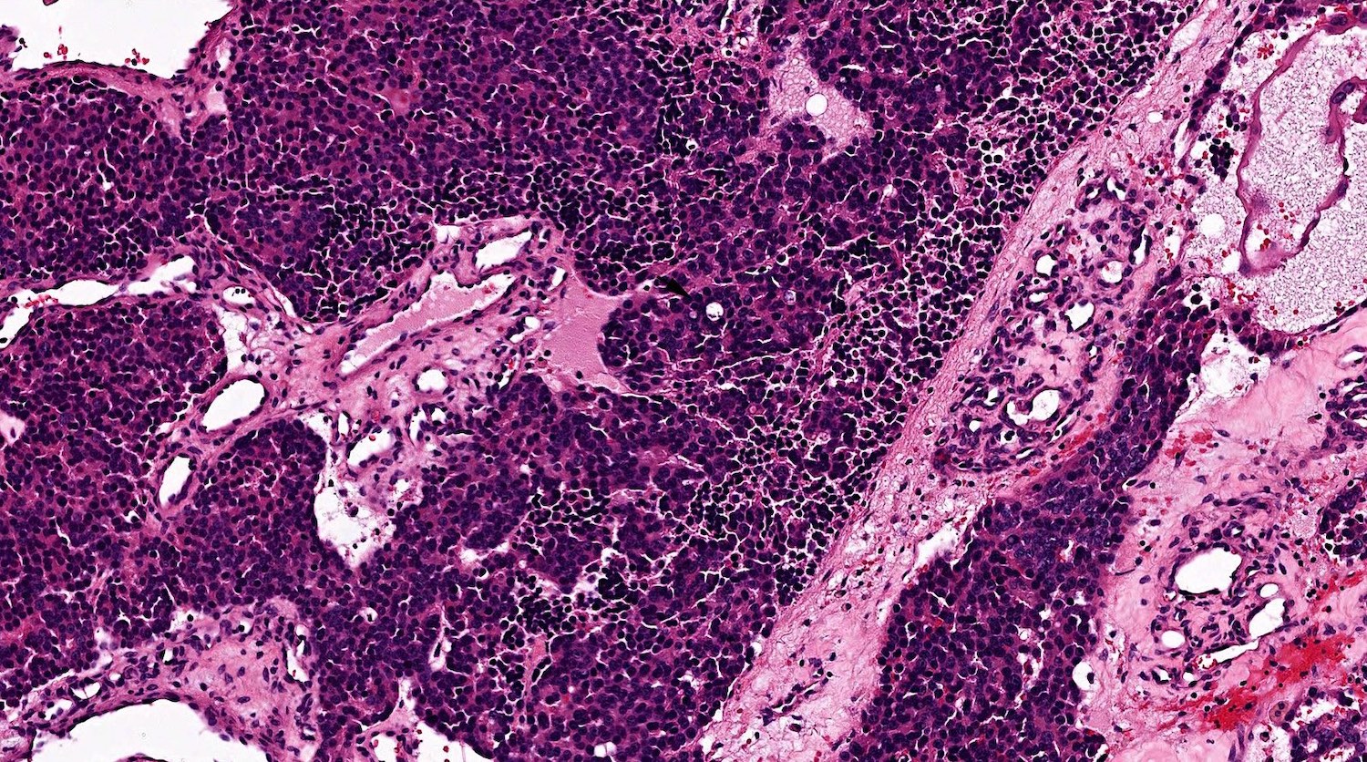
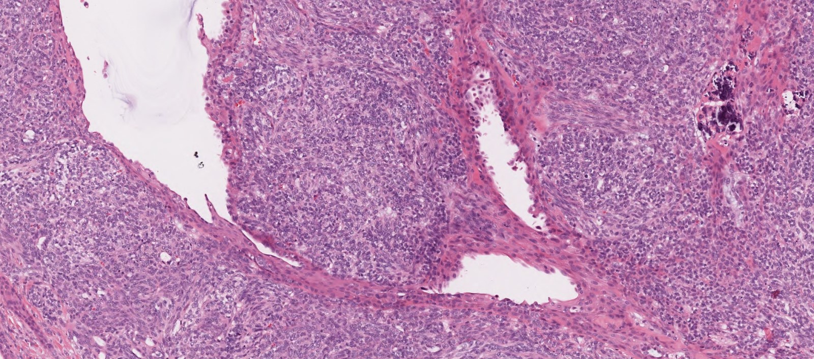
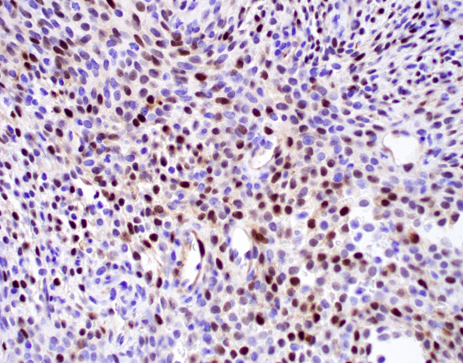
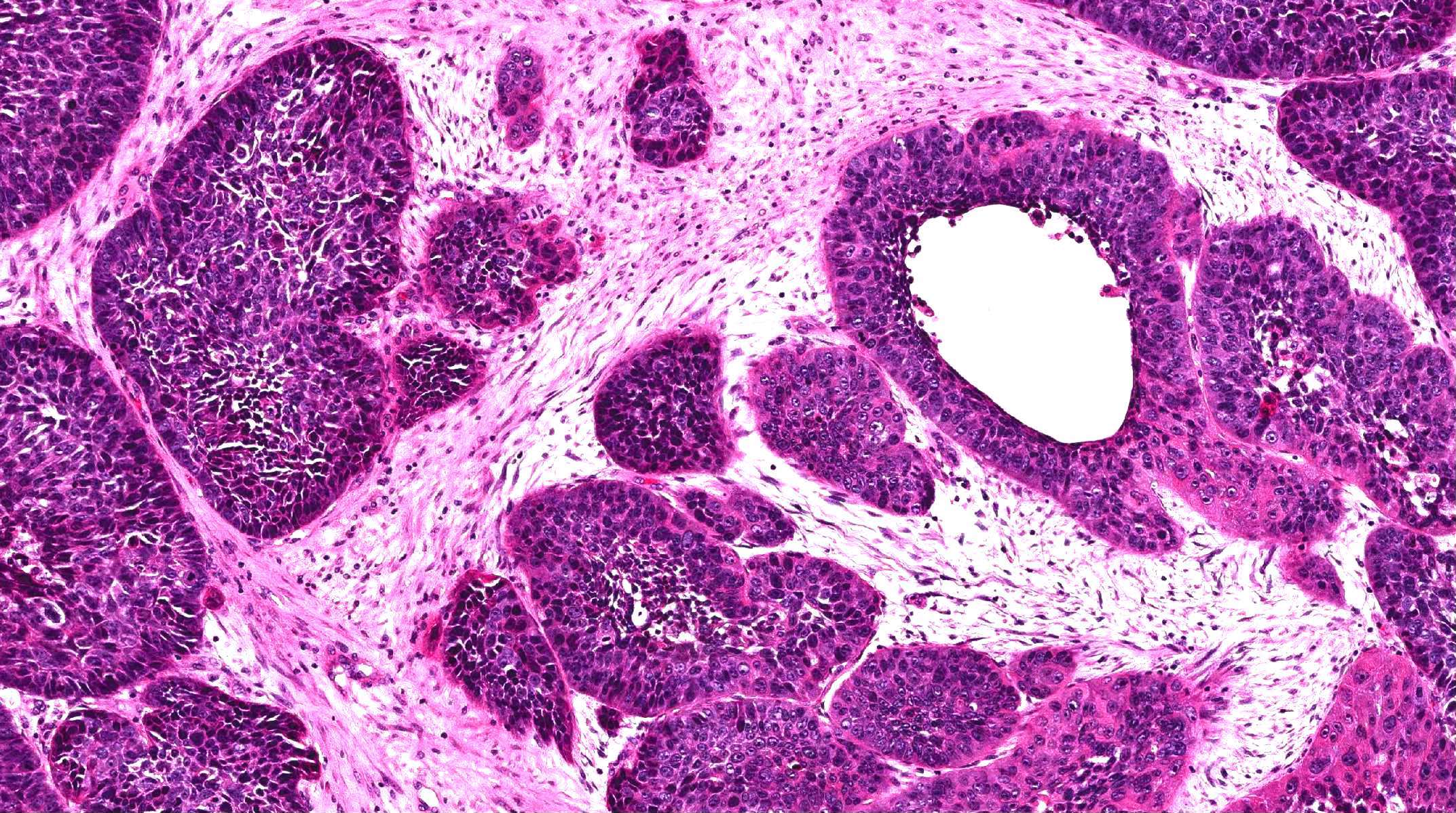
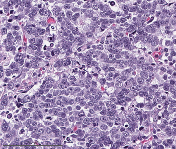
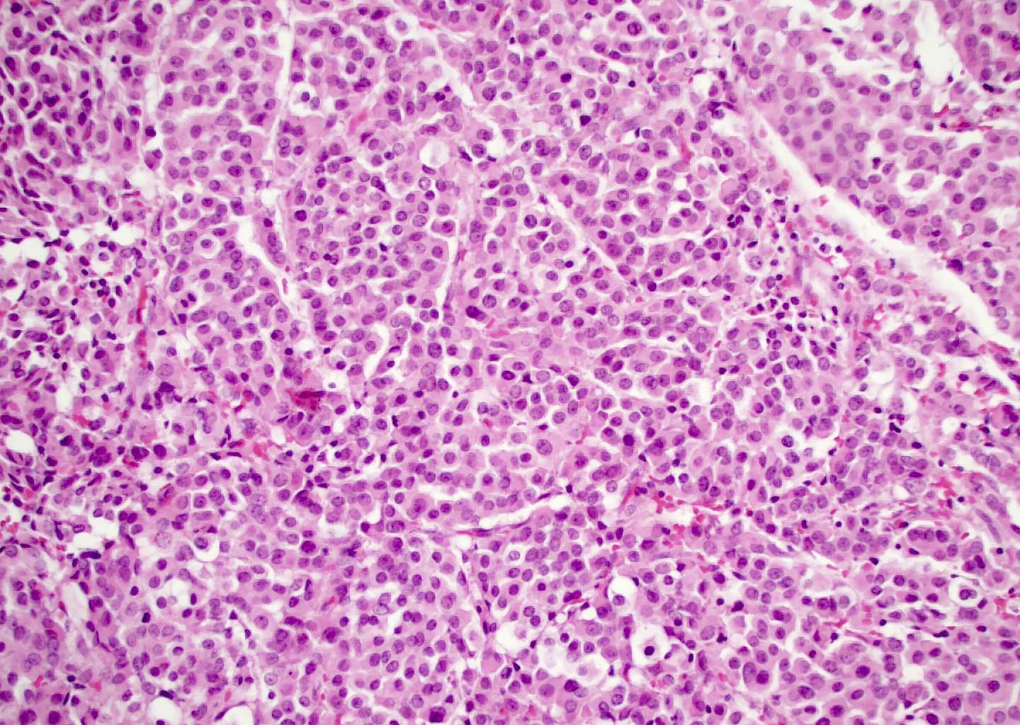
Primitive small to medium cells

Highly infiltrative growth
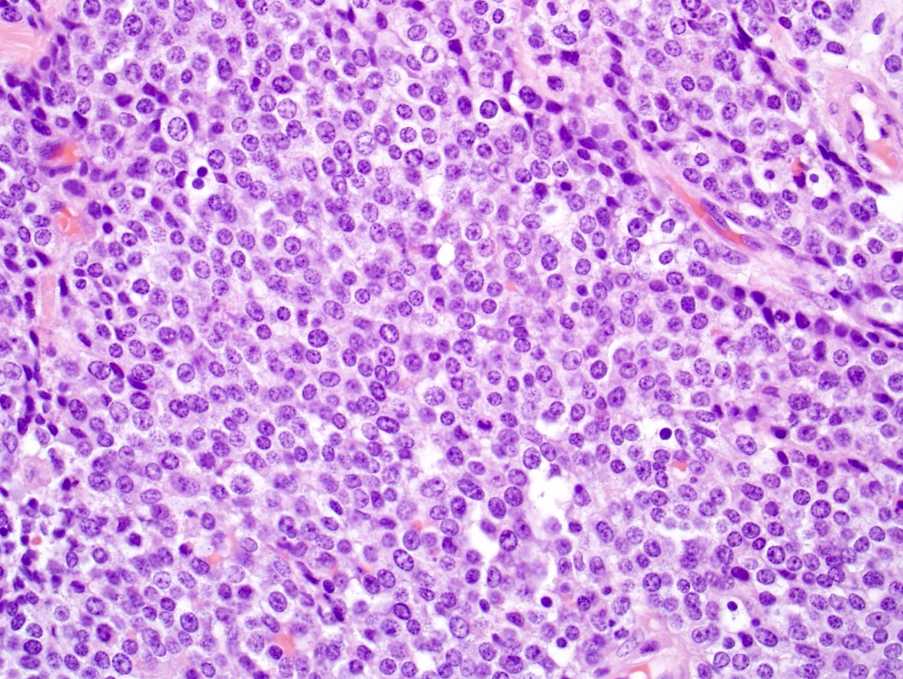
Monotonous nuclei
Prominent necrosis
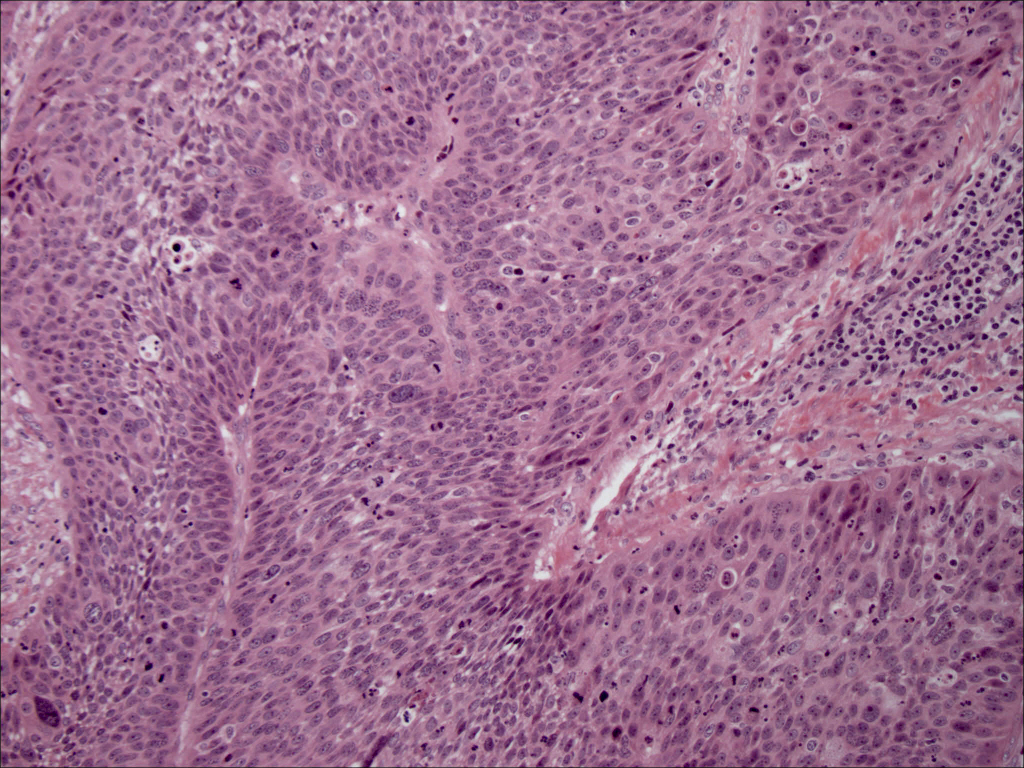
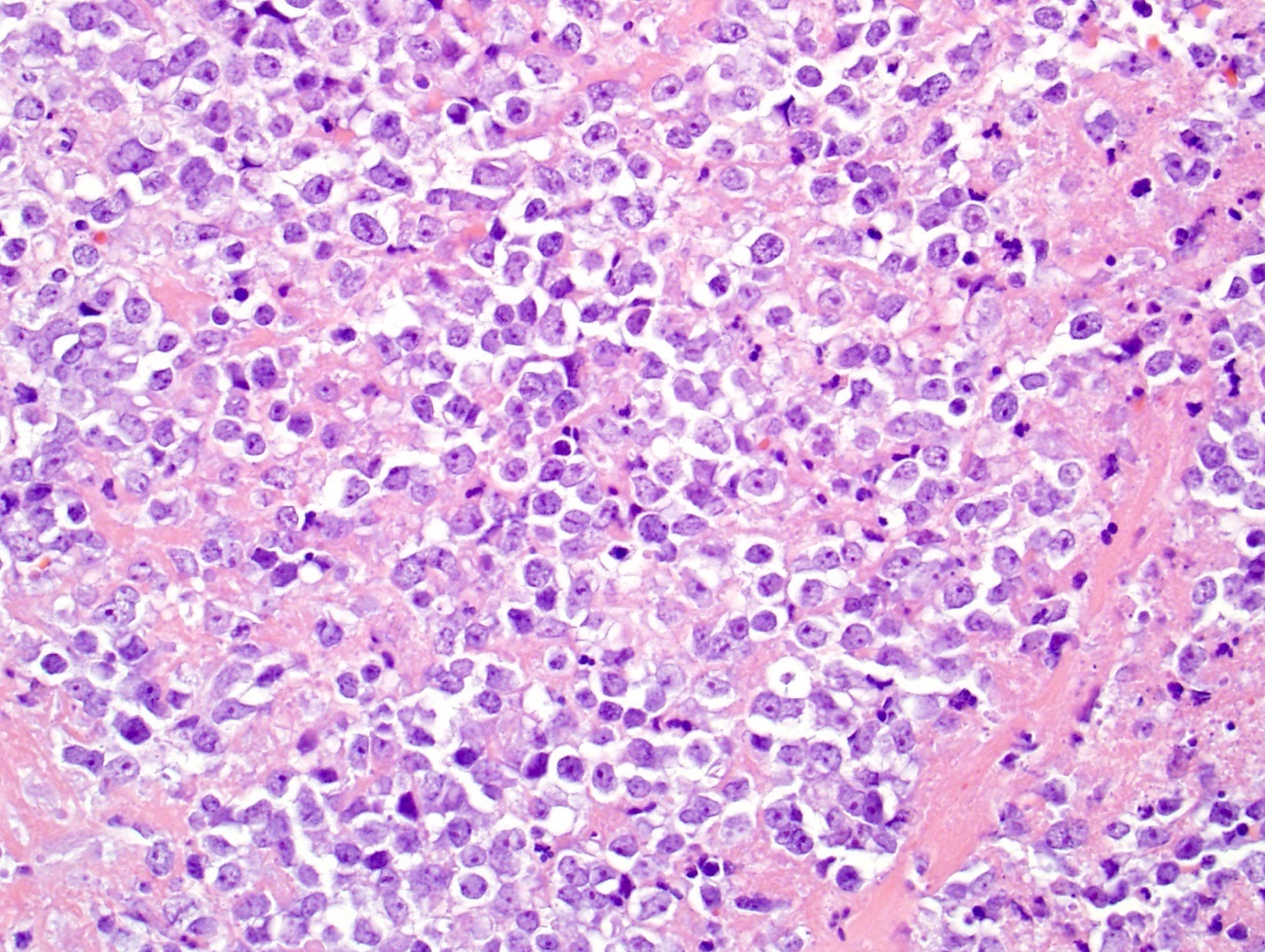
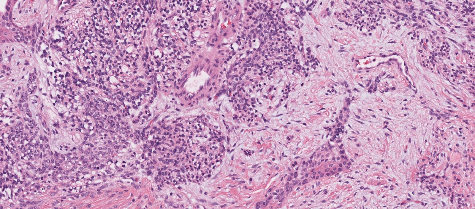
Abrupt keratinization
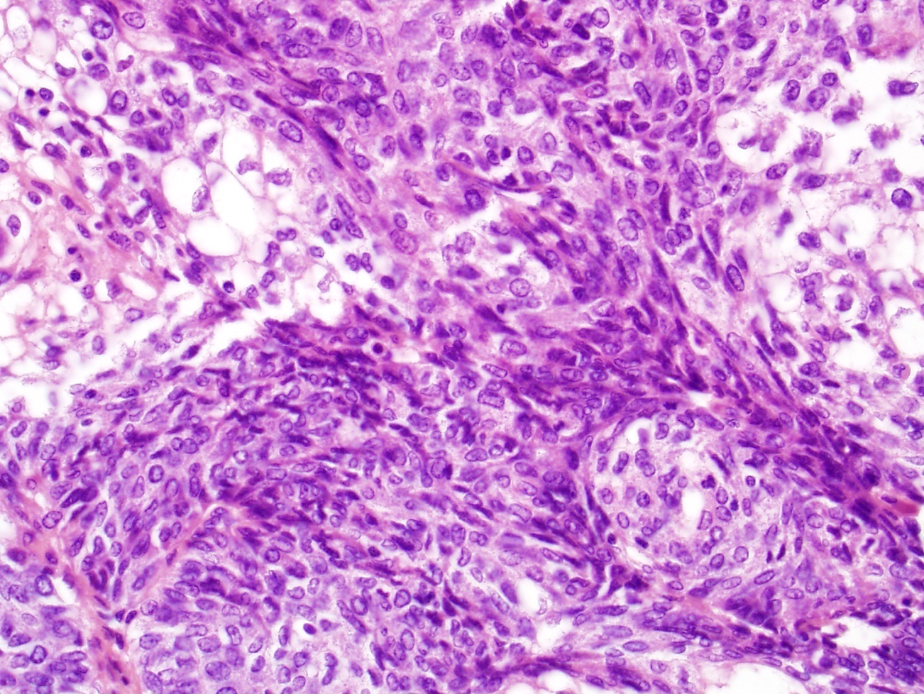
Prominent spindled foci
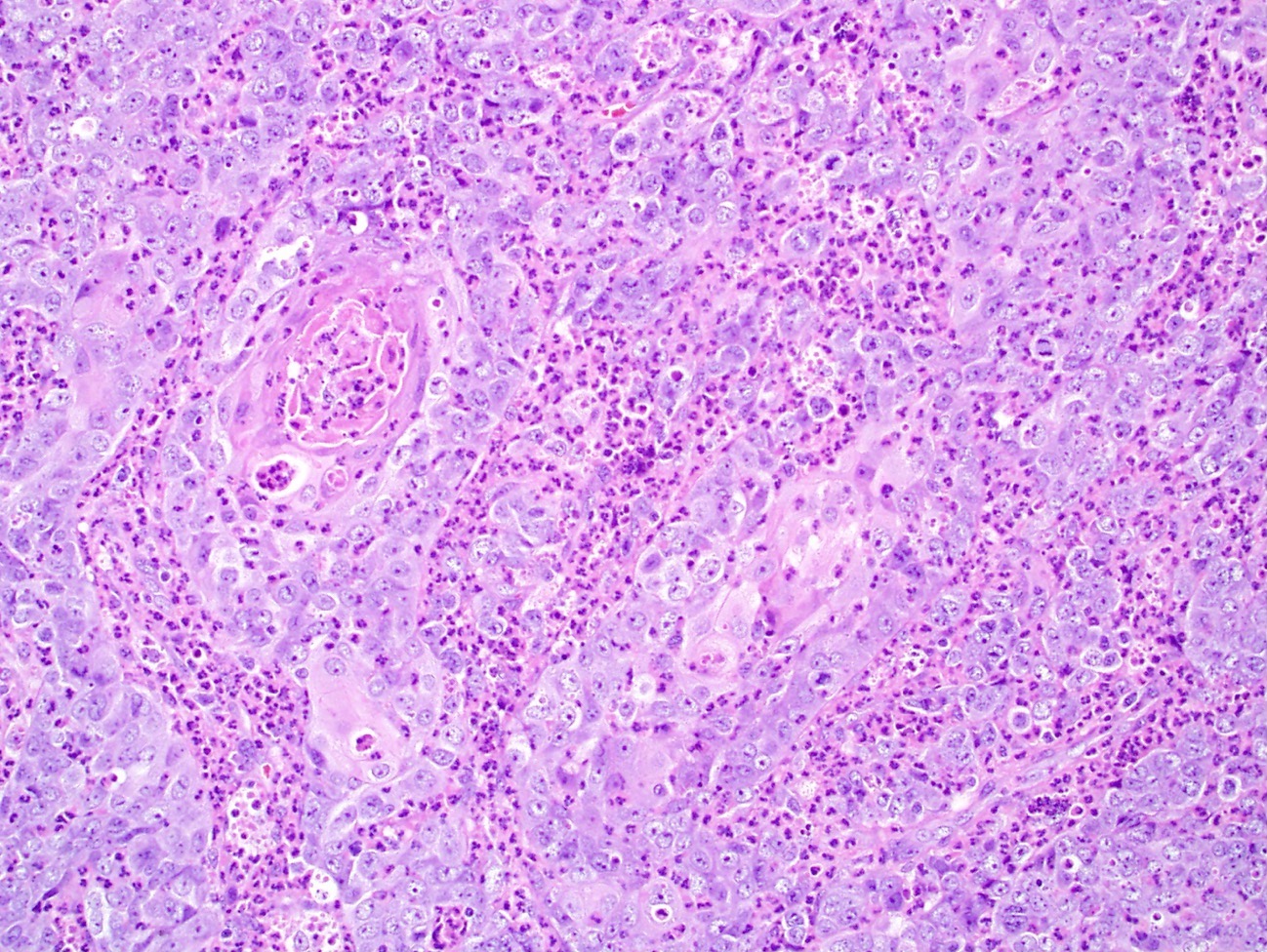
Infiltrating neutrophils
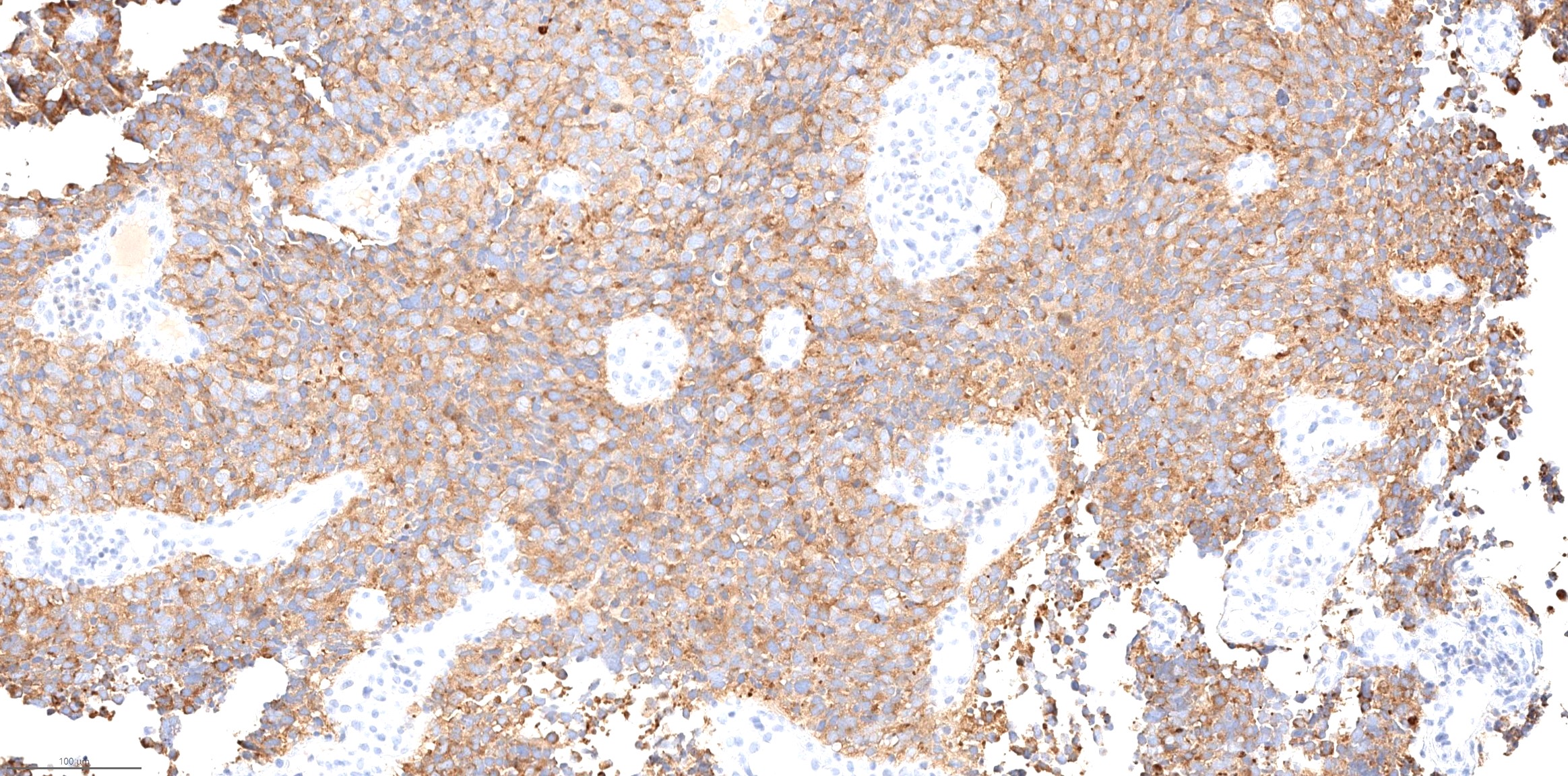
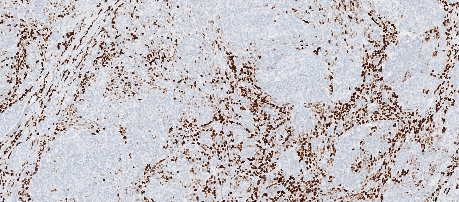
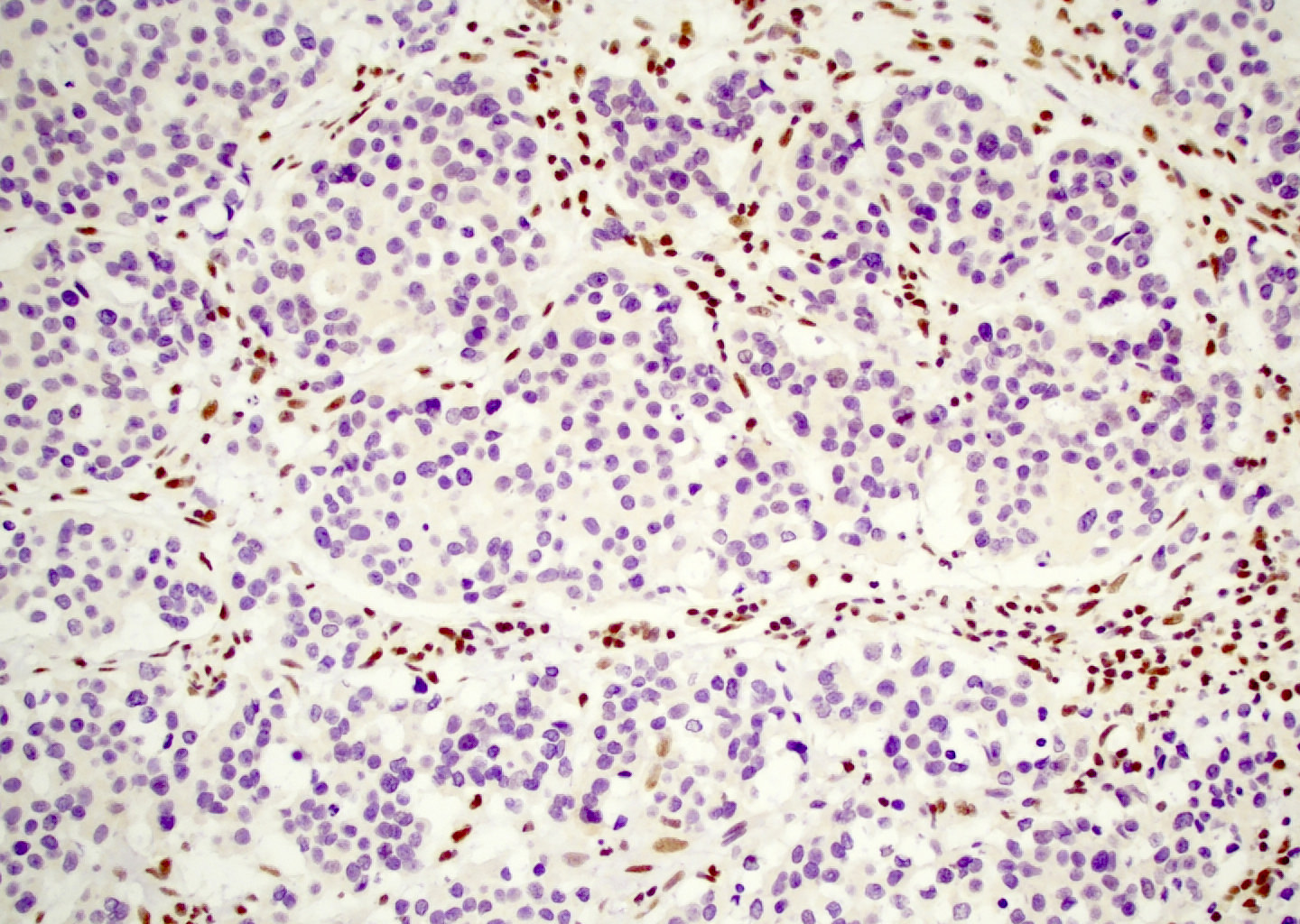
NUT1
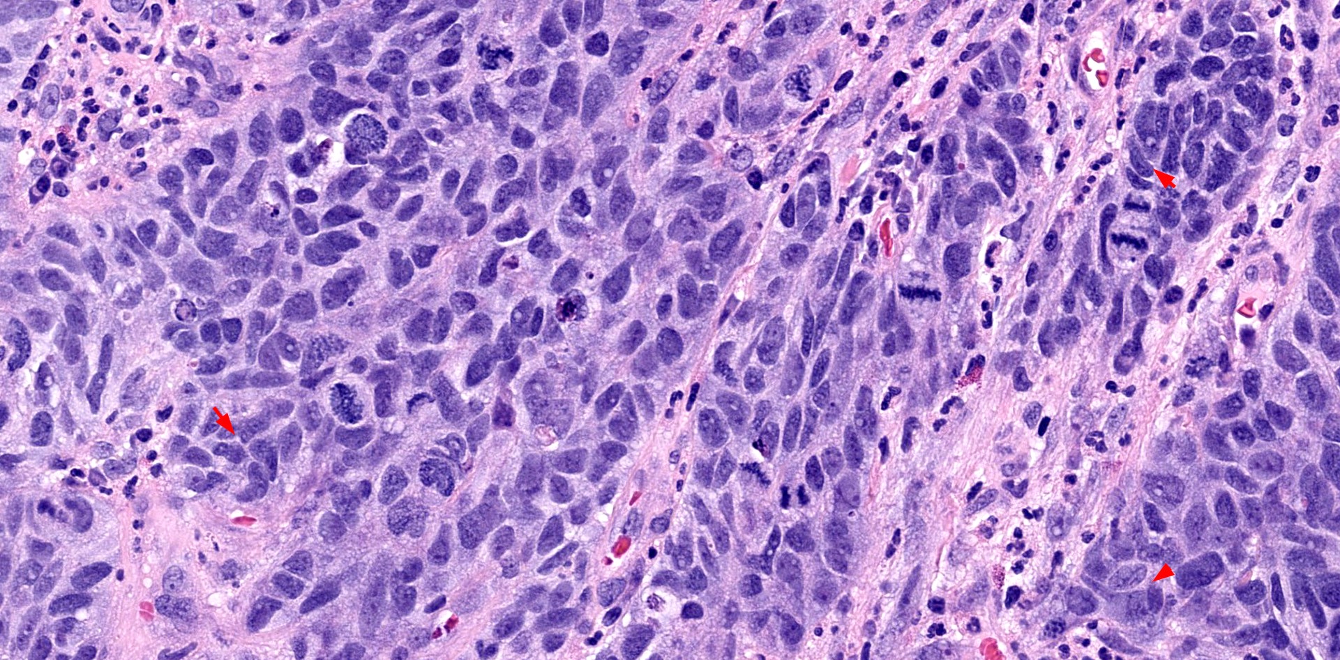
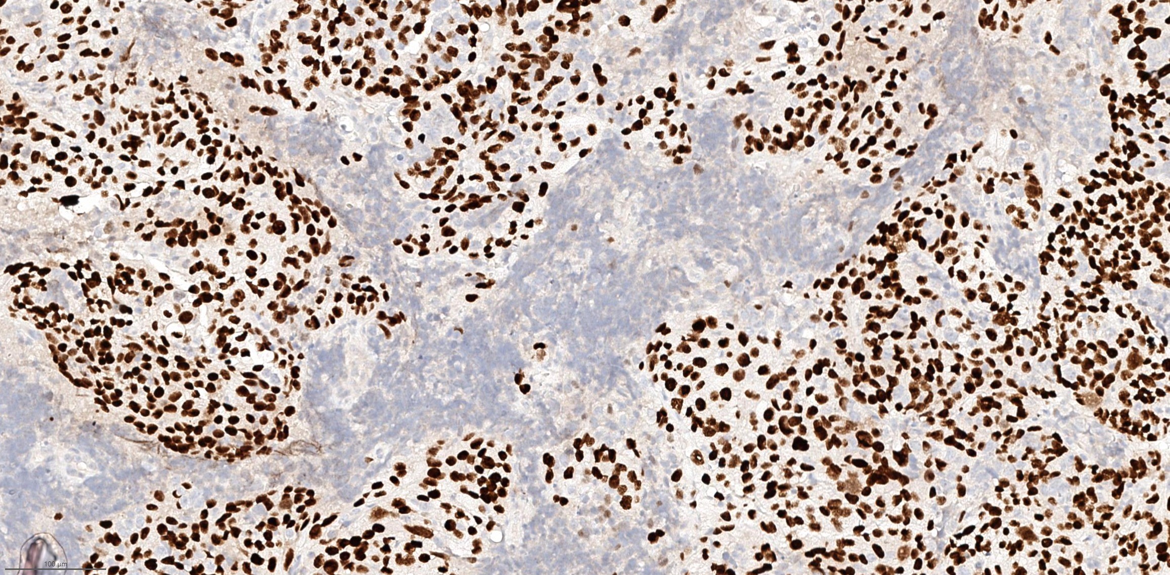
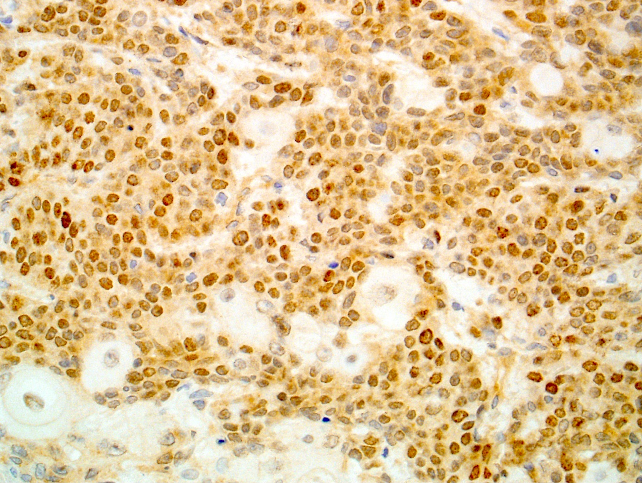
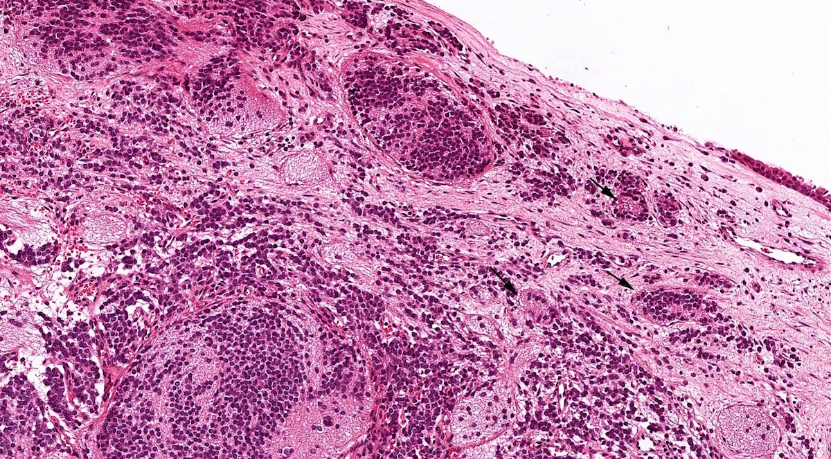
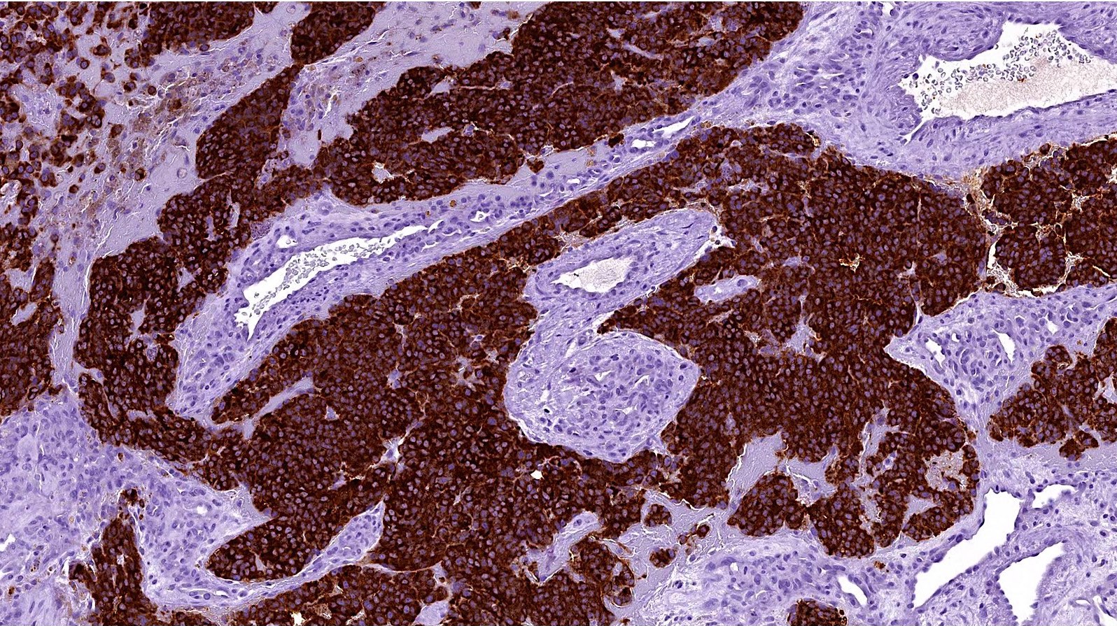
Contributed by Brendan Dickson, M.D.

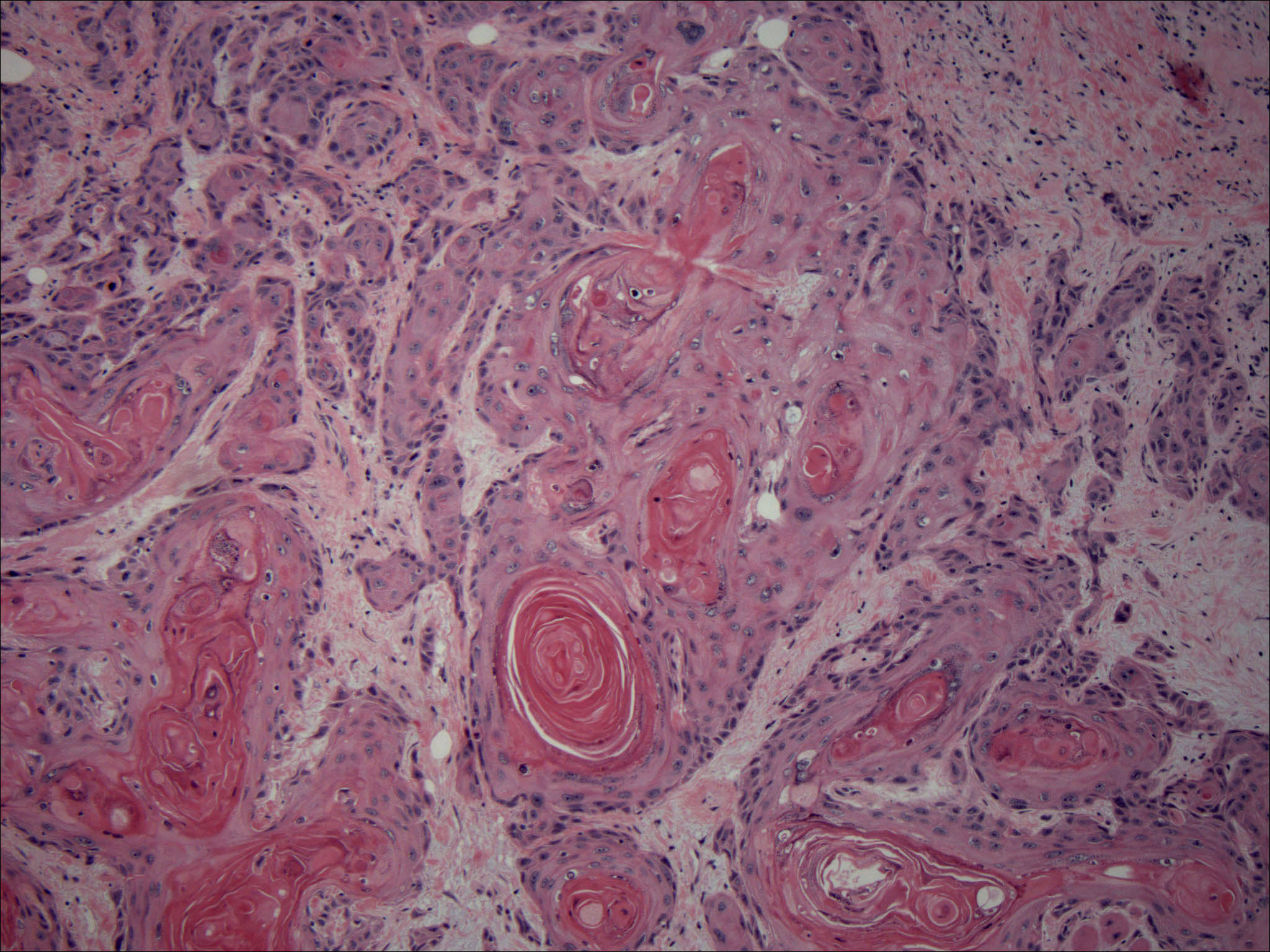
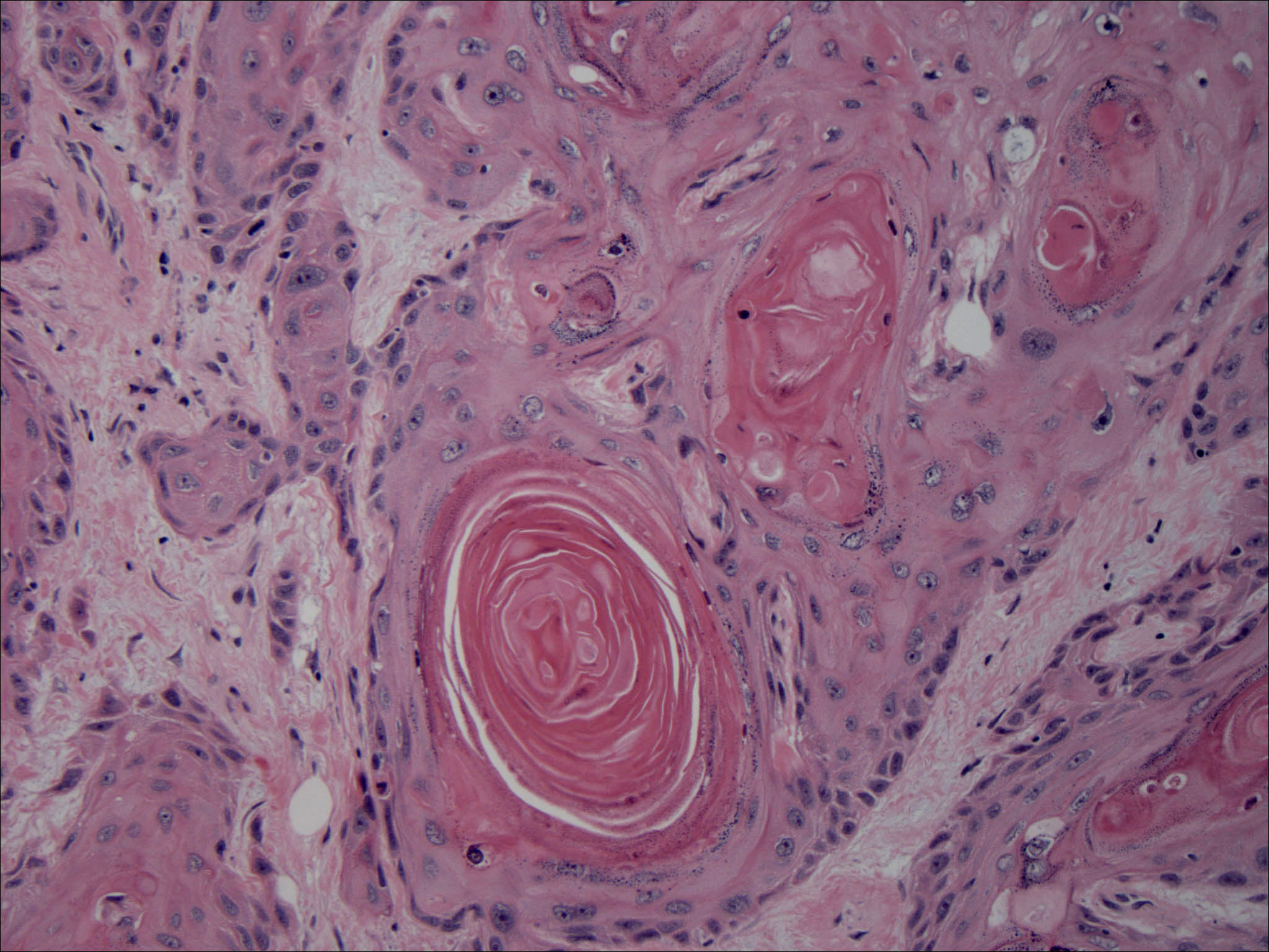
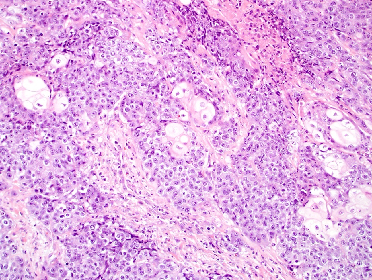
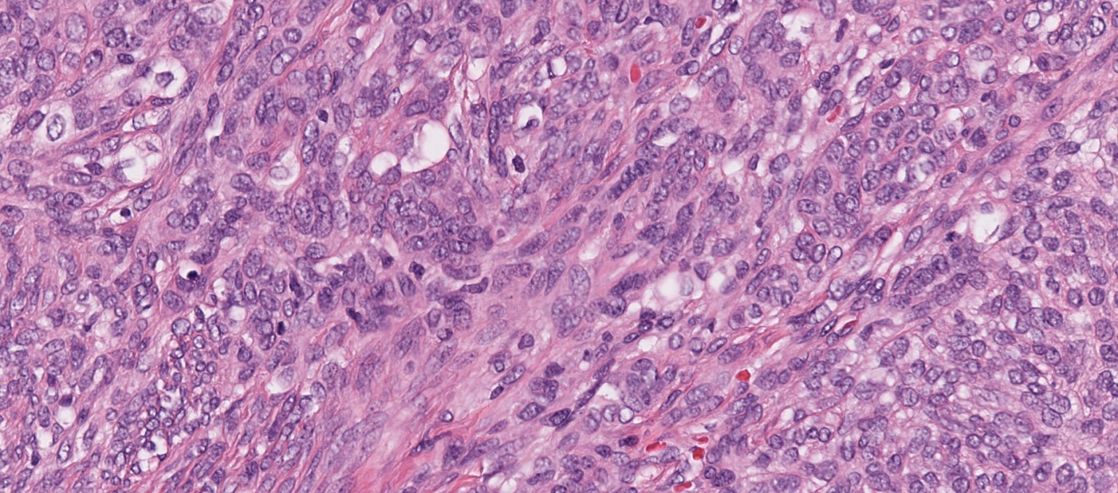
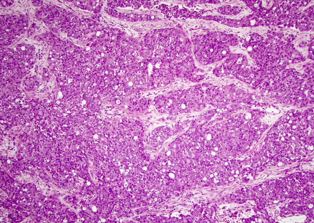
Abrupt keratinization
Necrosis
Undifferentiated areas
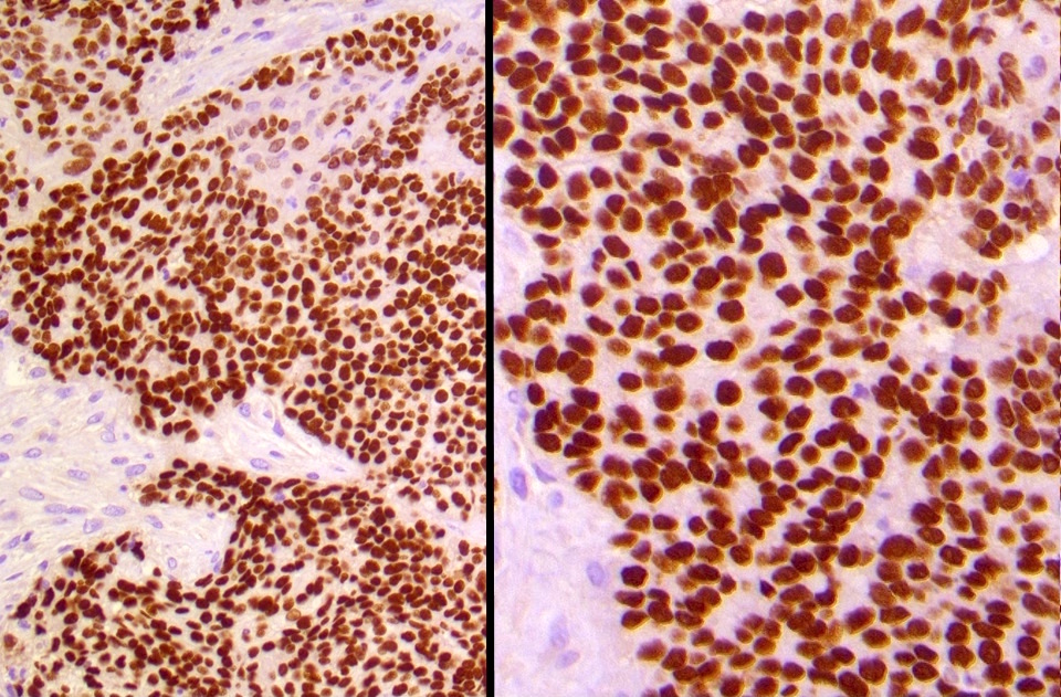
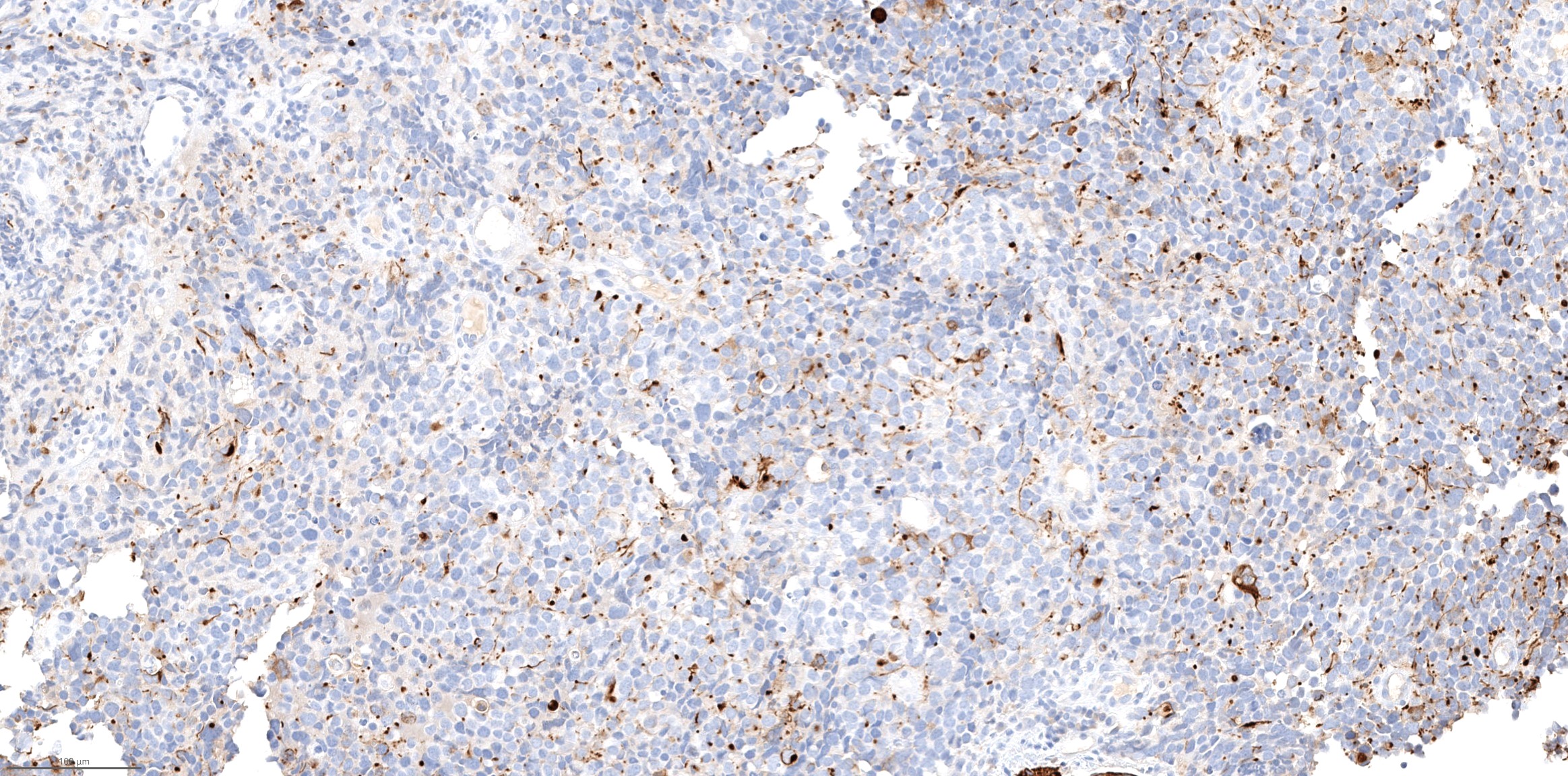
p63
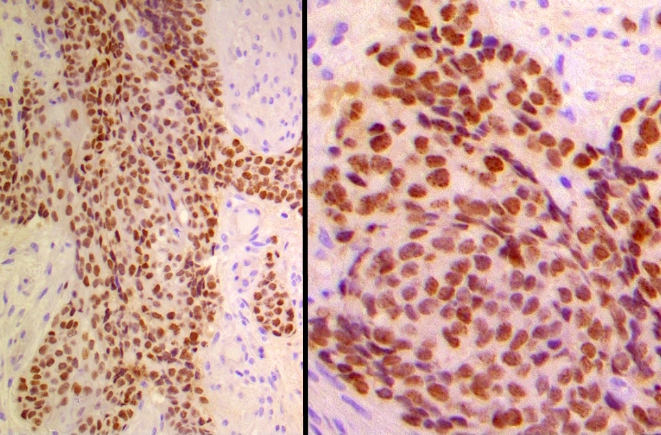
NUT1

AE1 / 3
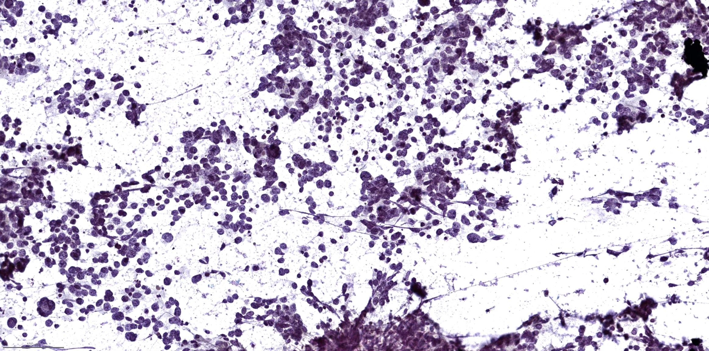
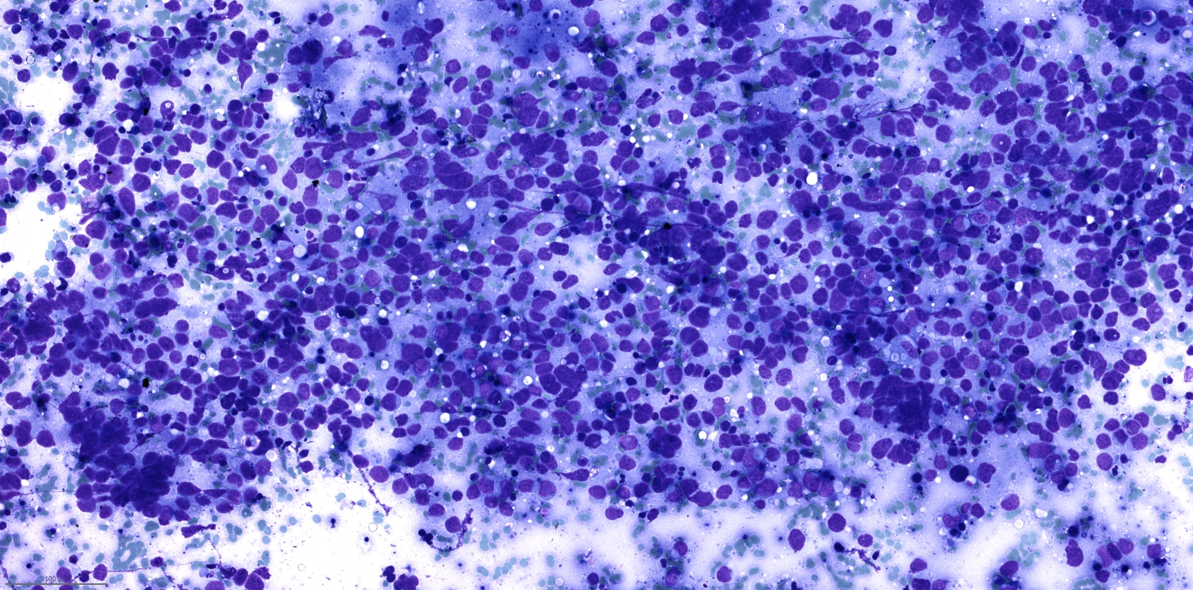
Images hosted on other servers:

Aspirate of metastasis
Images hosted on other servers:

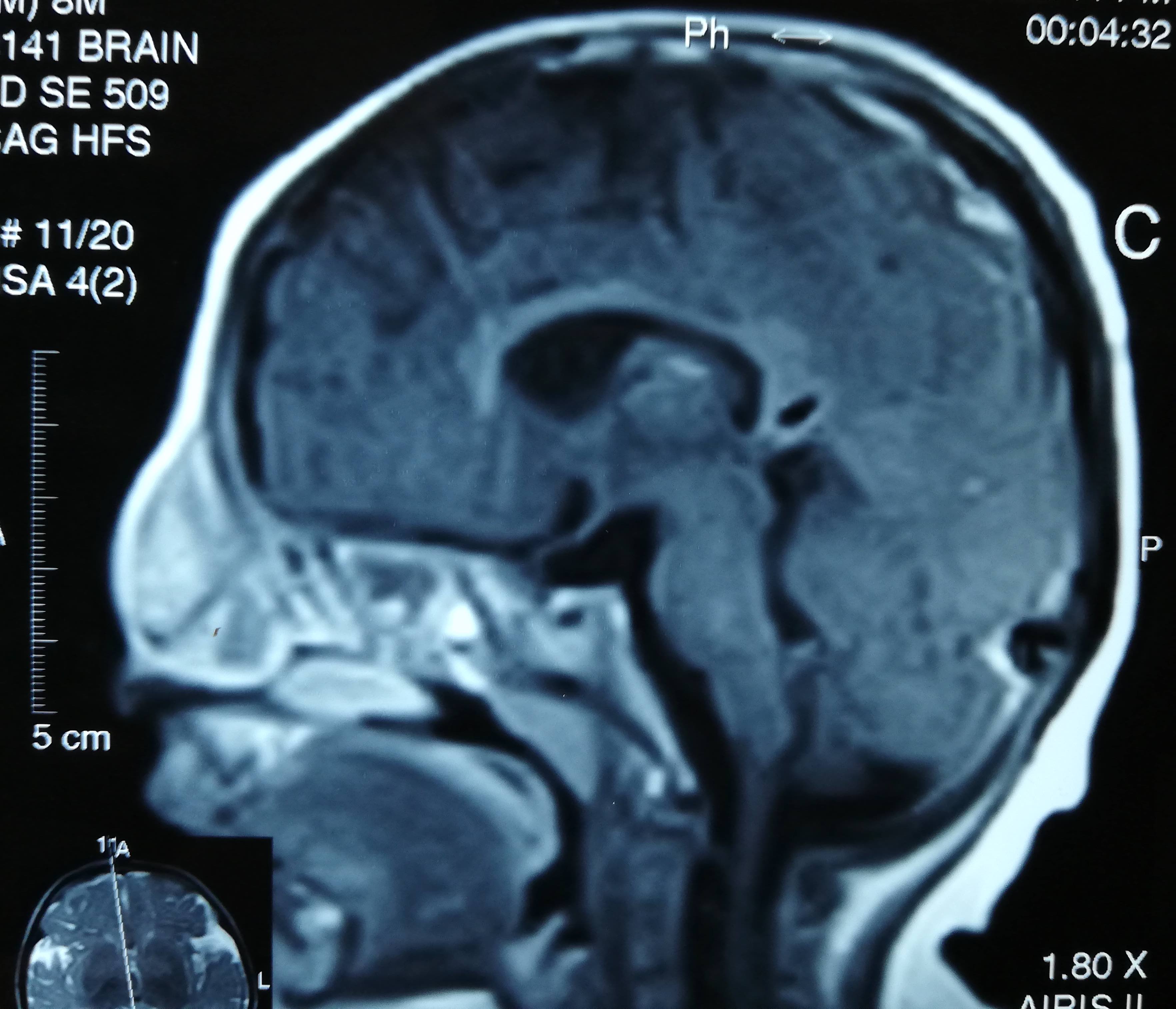
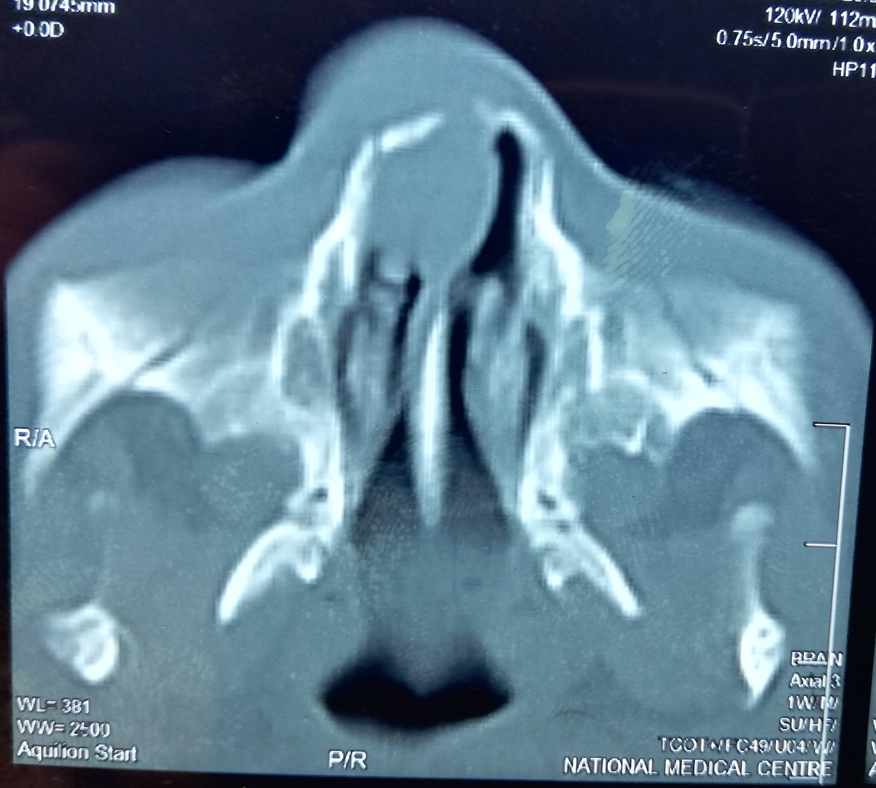
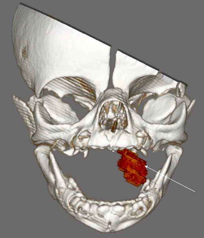
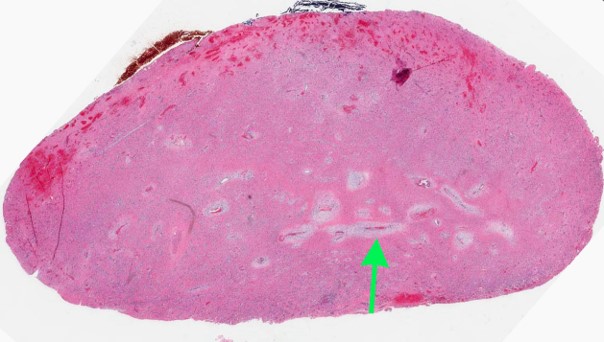
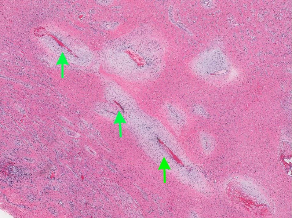
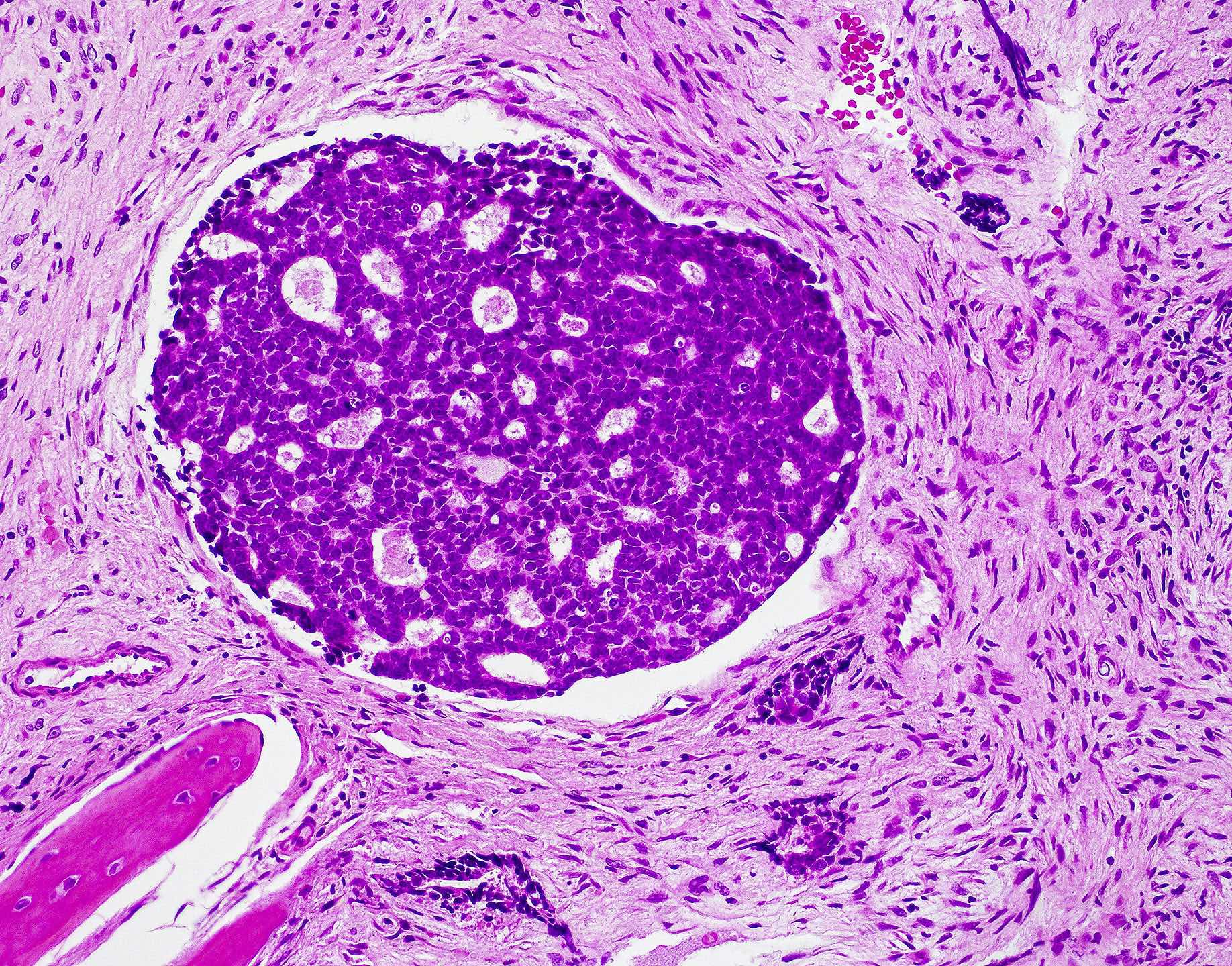
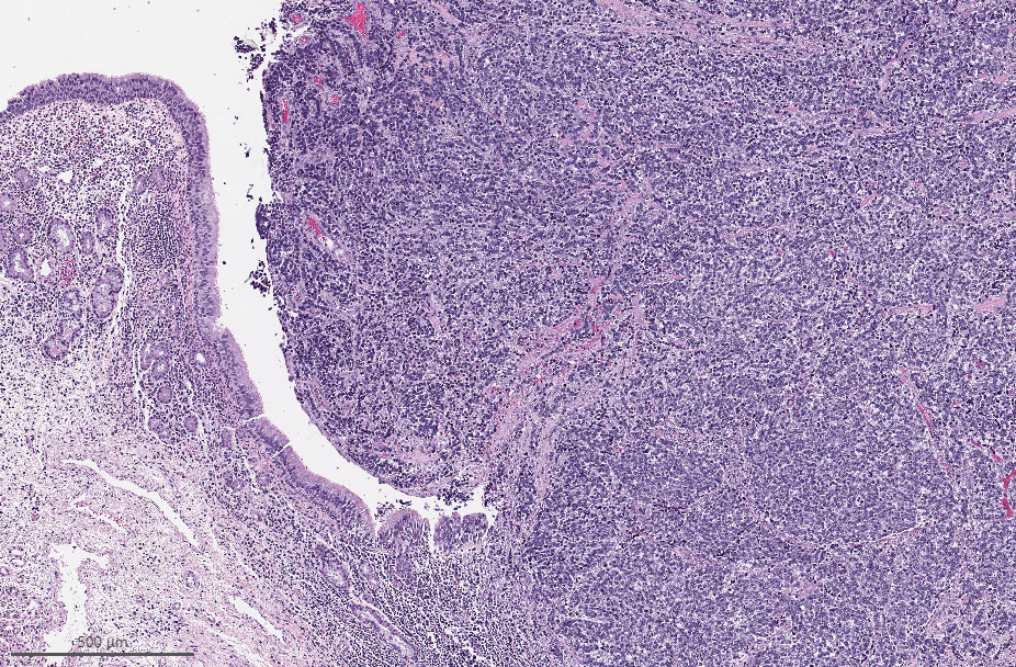
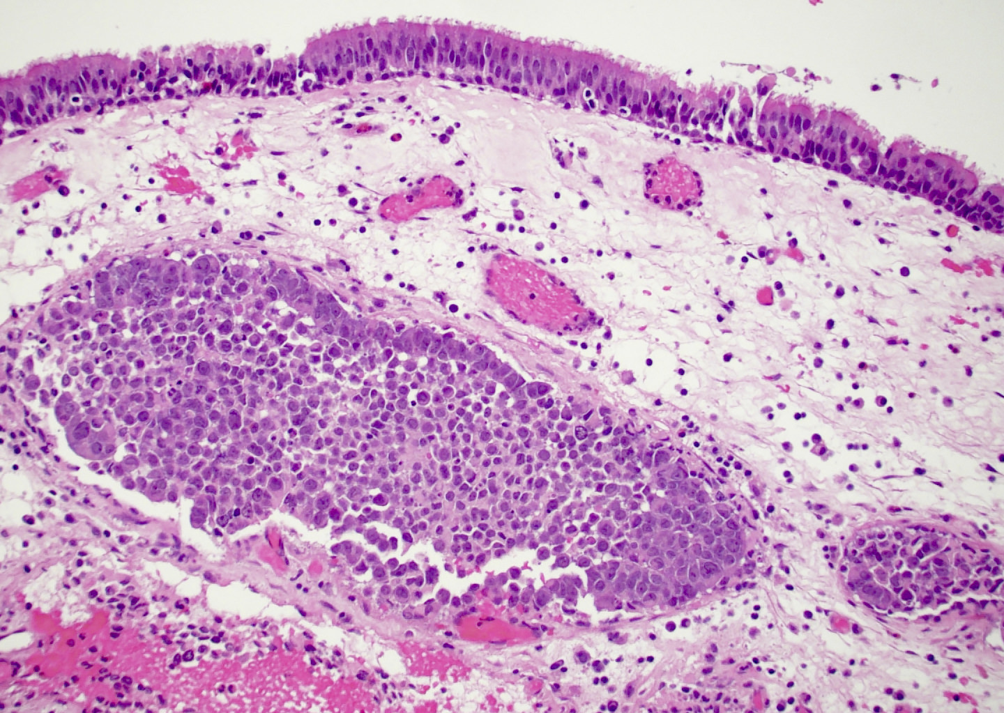
Tumor extending into cribriform plate and lateral lamella

Large, avidly enhancing skull base mass in left nasal cavity

Olfactory neuroblastoma, a. coronal view, b sagittal view
| Grade I | Grade II | Grade III | Grade IV | |
| Architecture | Lobular | Lobular | Variable | Variable |
| Fibrillary matrix | Prominent | Present | Minimal | Absent |
| Mitosis | Absent | Present | Prominent | Marked |
| Necrosis | Absent | Absent | May present | Common |
| Nuclear pleomorphism | Absent | Moderate | Prominent | Marked |
| Rosettes | Homer Wright | Homer Wright | Flexner- Wintersteiner |
Flexner- Wintersteiner |
Table 2: Staging systems of olfactory neuroblastoma
Kadish staging:
Morita modification:
|
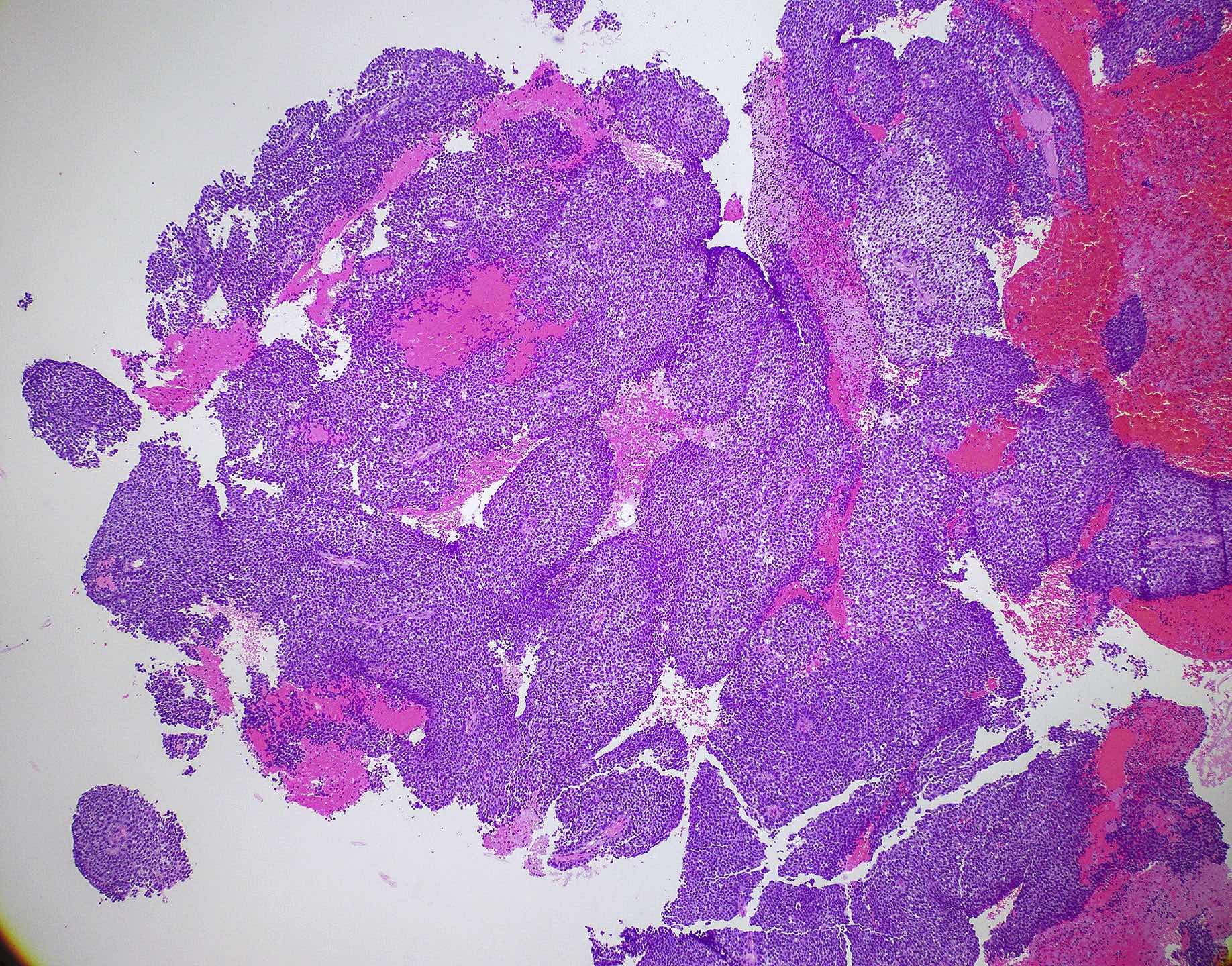
Images hosted on other servers:

Nasal endoscopy

Bifrontal craniotomy
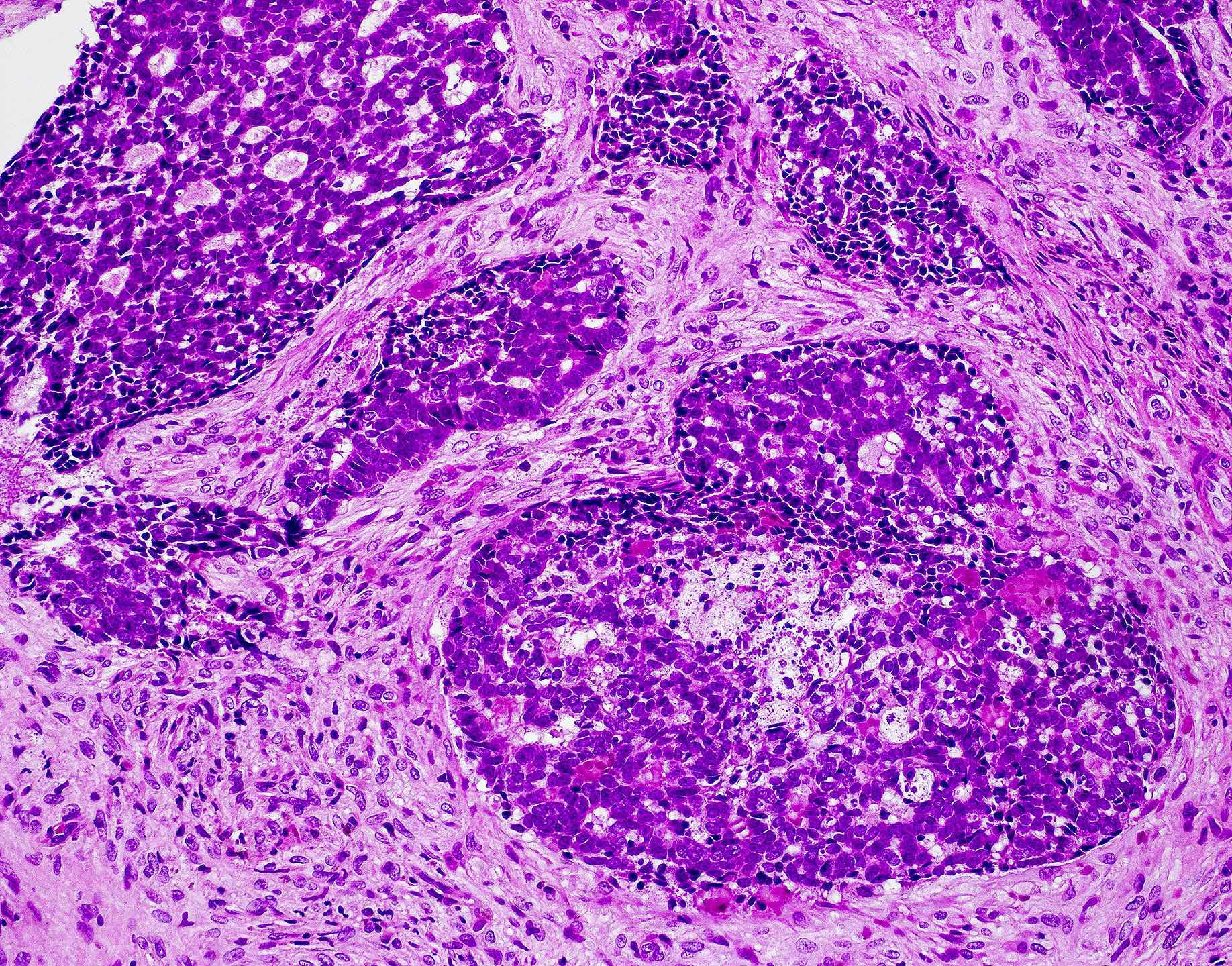
Contributed by Bin Xu, M.D., Ph.D.
Lobulated growth,
microcalcification,
abundant fibrillar
neural matrix
Lobulated growth with
infiltration between
submucosal glands
Hyams grade I
olfactory
neuroblastoma
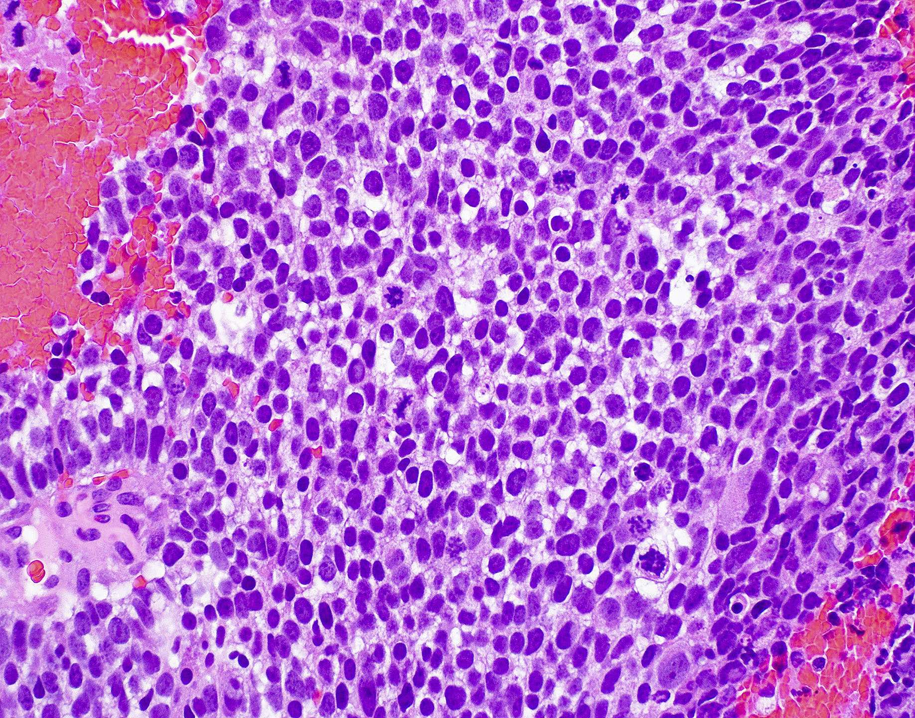
Flexner-Wintersteiner
rosettes
Homer Wright
pseudorosettes
Synaptophysin
diffusely positive
in tumor cells
S100 highlights
sustentacular cells
Images hosted on other servers:

Widened olfactory cleft
Images hosted on other servers:

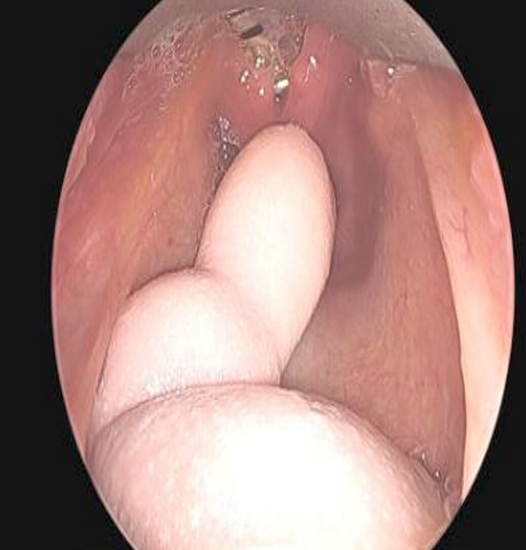
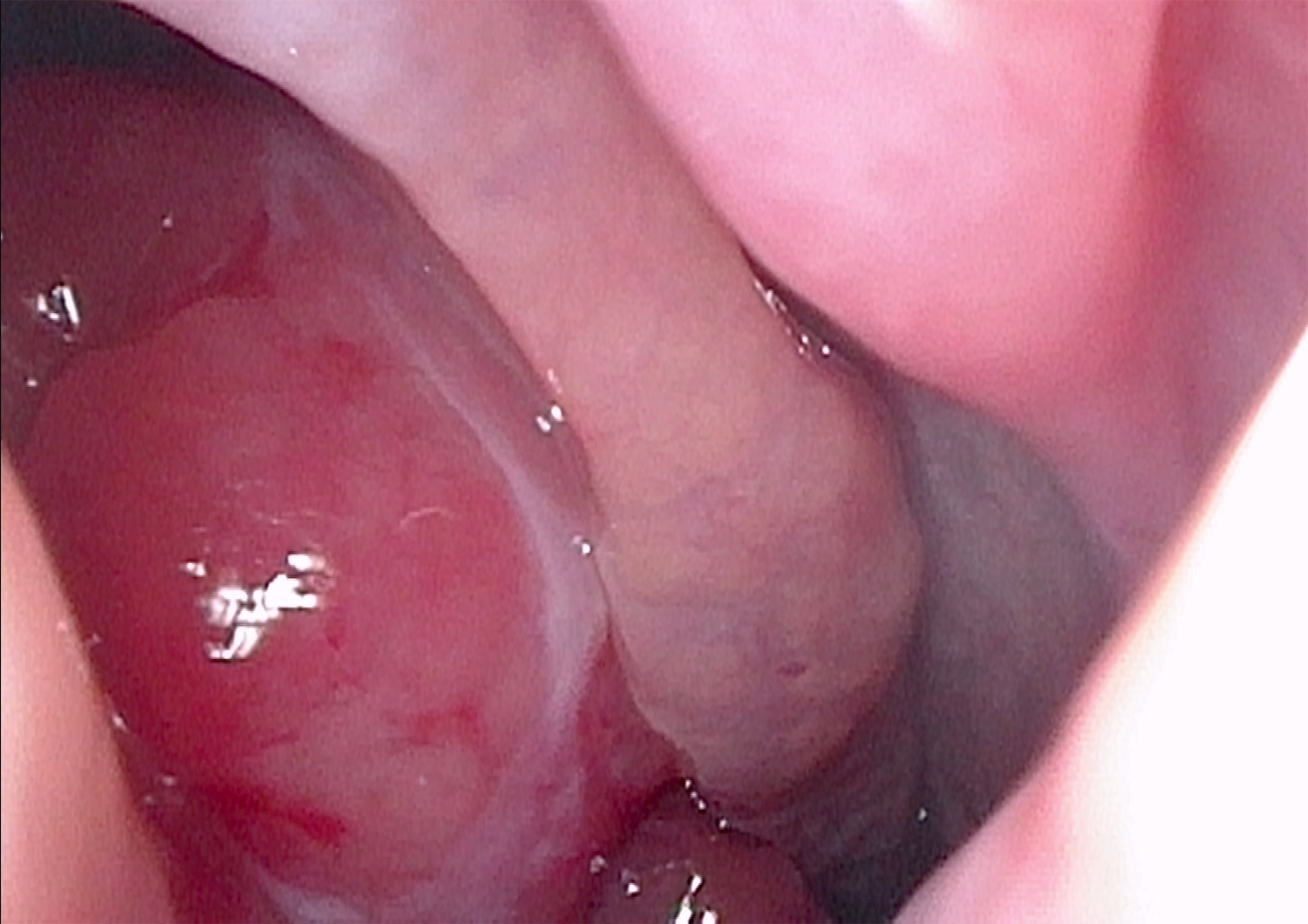
Polypoid glistening mass
Contributed by Bin Xu, M.D., Ph.D.
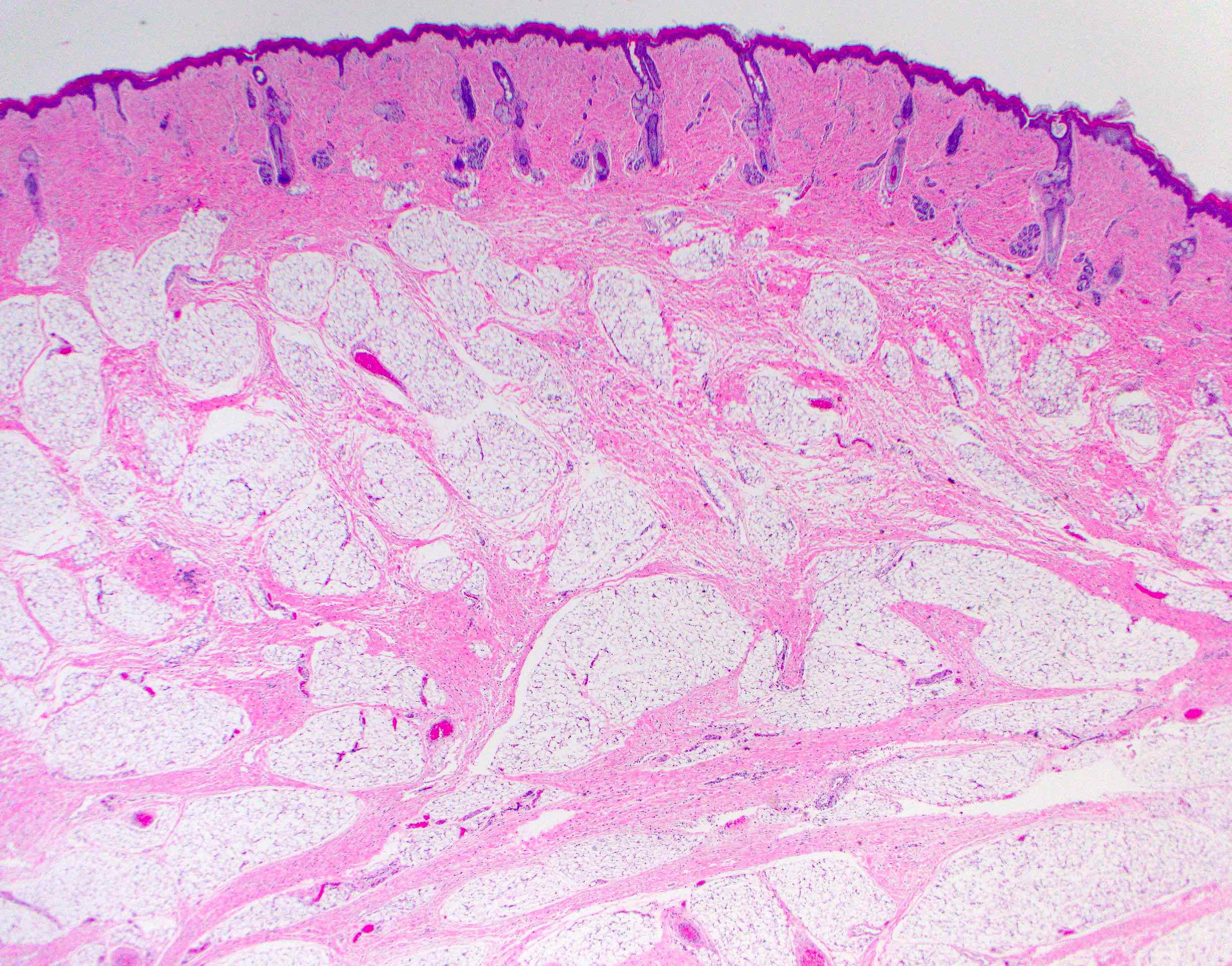
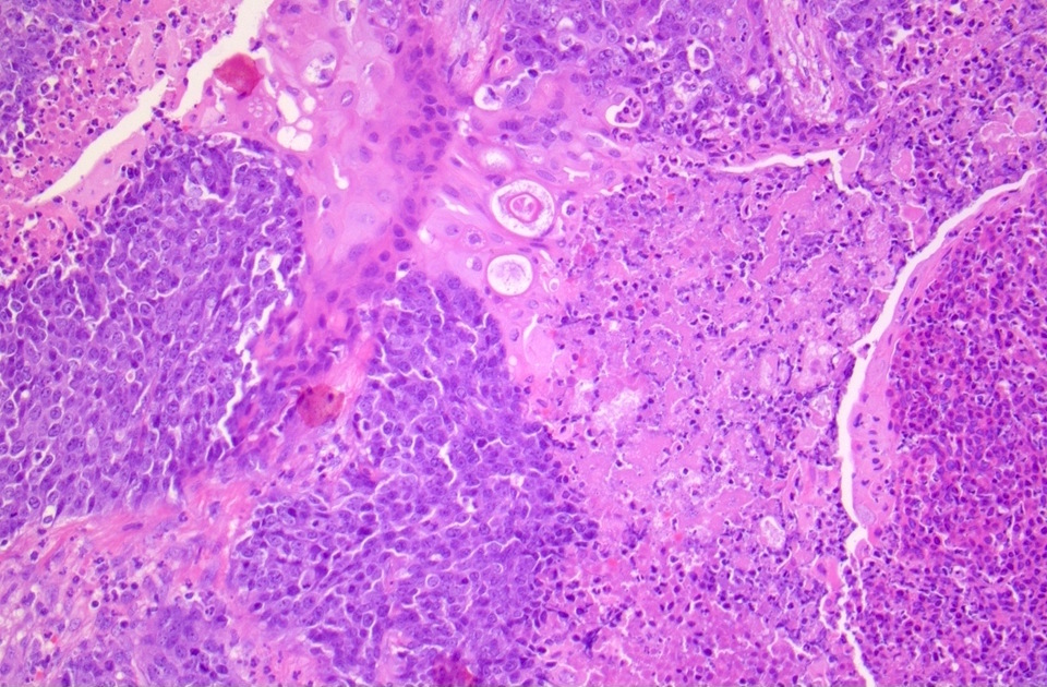
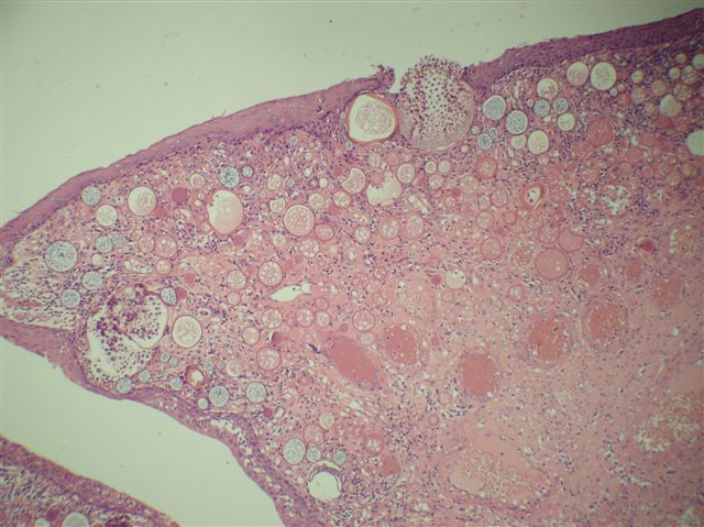
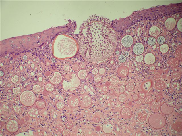
Respiratory epithelial adenomatoid hamartoma (REAH)
Proliferation of medium sized glands
Thickened basement membrane
Glands with cilia
Scattered seromucinous glands
Chondro-osseous respiratory epithelial (CORE) hamartoma
Glands and chondromyxoid stroma
Myxoid mesenchyme and gland

Epithelium with cilia
Contributed by Hanni Gulwani, M.B.B.S. (Case #97), Veena Maheshwari, M.D., Kiran Alam, M.D., Anshu Jain, M.D., Alia Albawardi, M.B.B.S. and @DrTravisBrown on Twitter
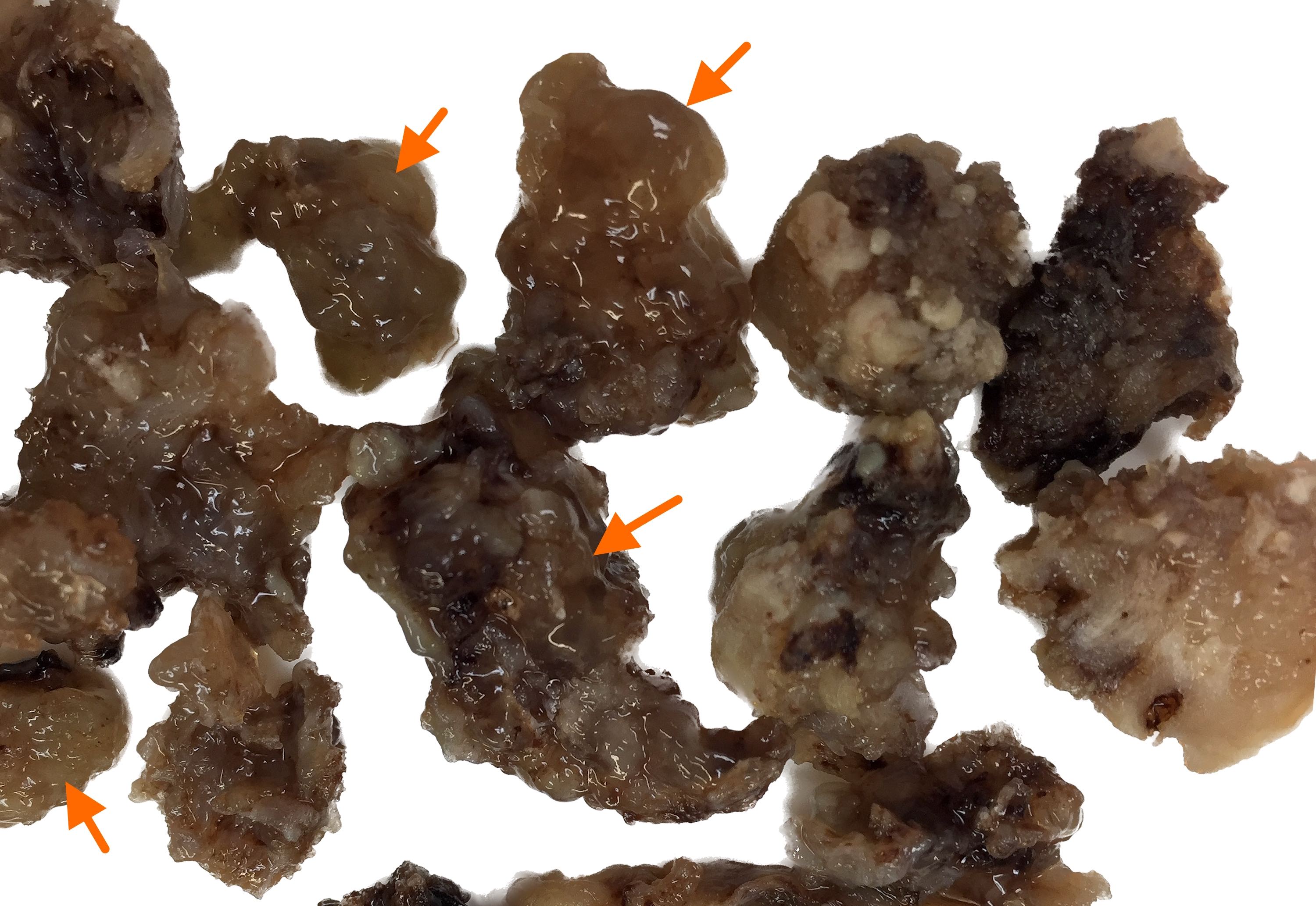
Various images

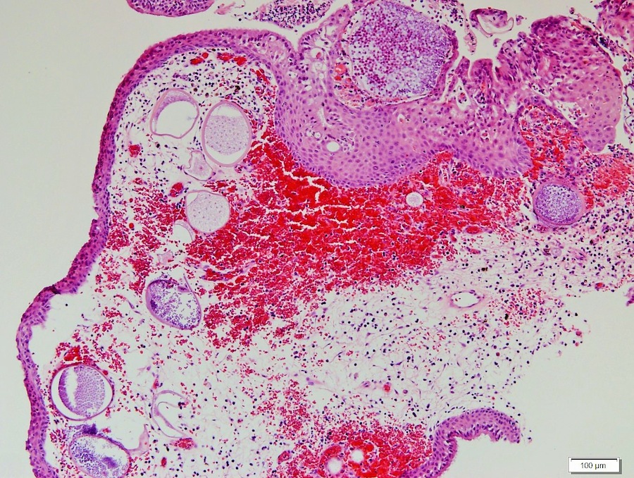
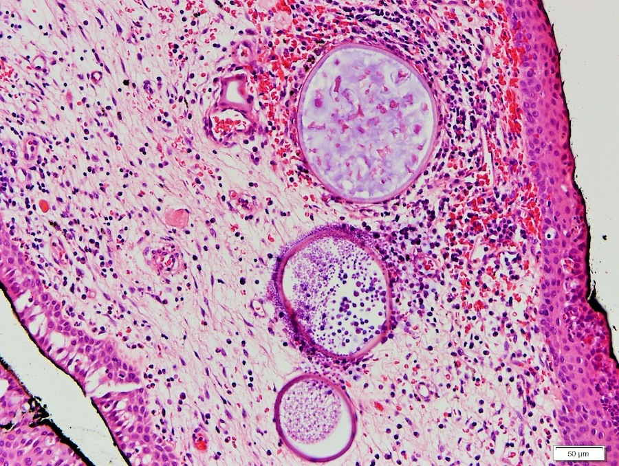

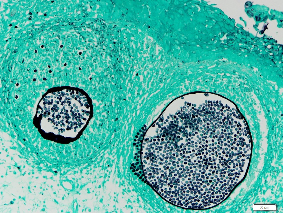
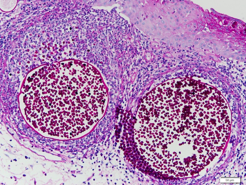
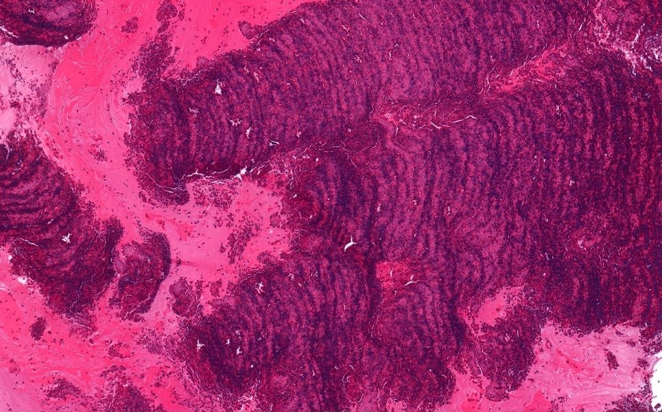
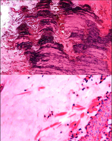
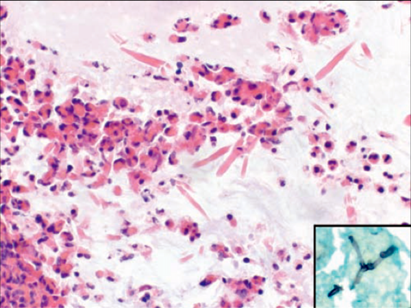
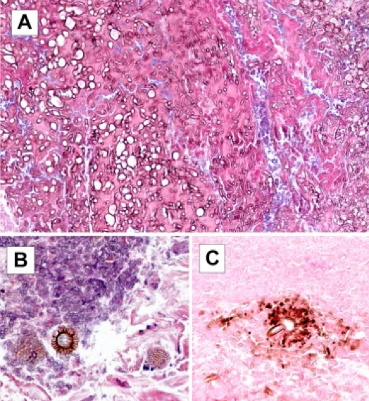
28 year old from Nepal with uvular mass; left to right: H&E (4 images), GMS, PAS
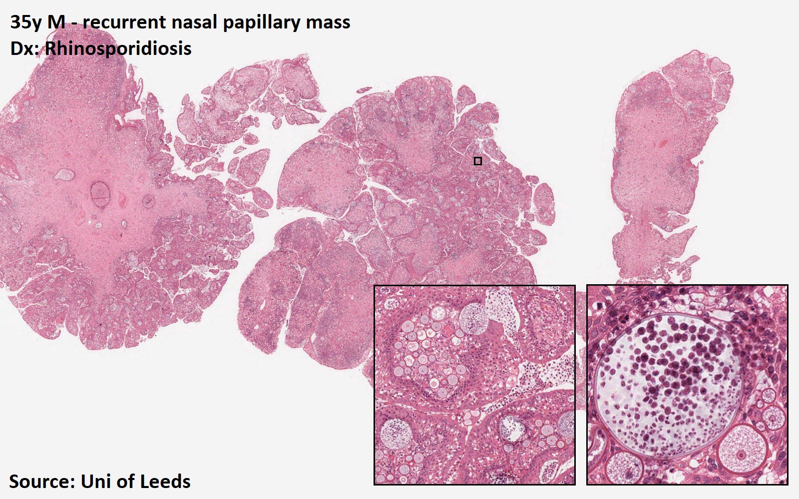
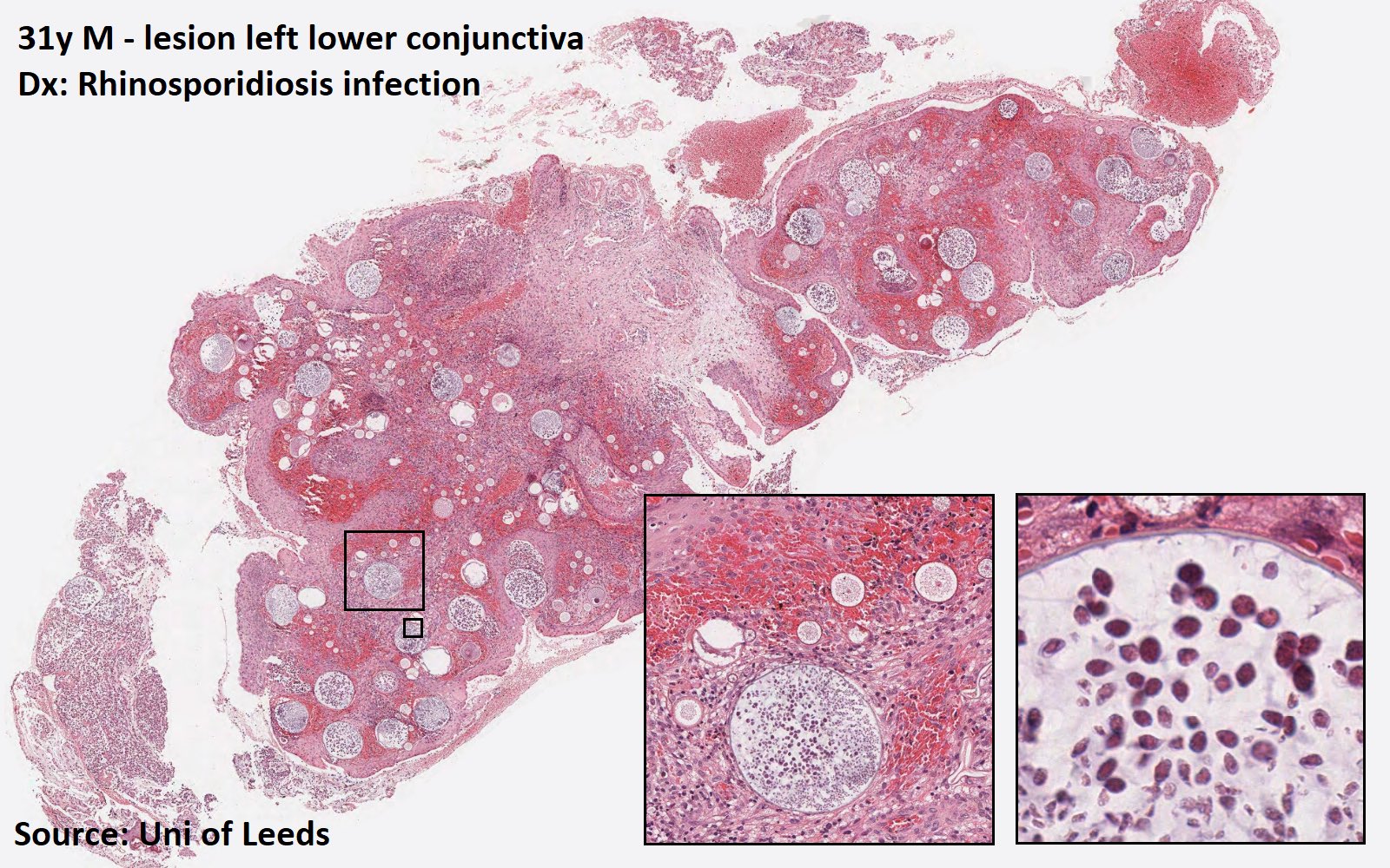
Rhinosporidiosis
Contributed by Scott Poswilko, M.D.
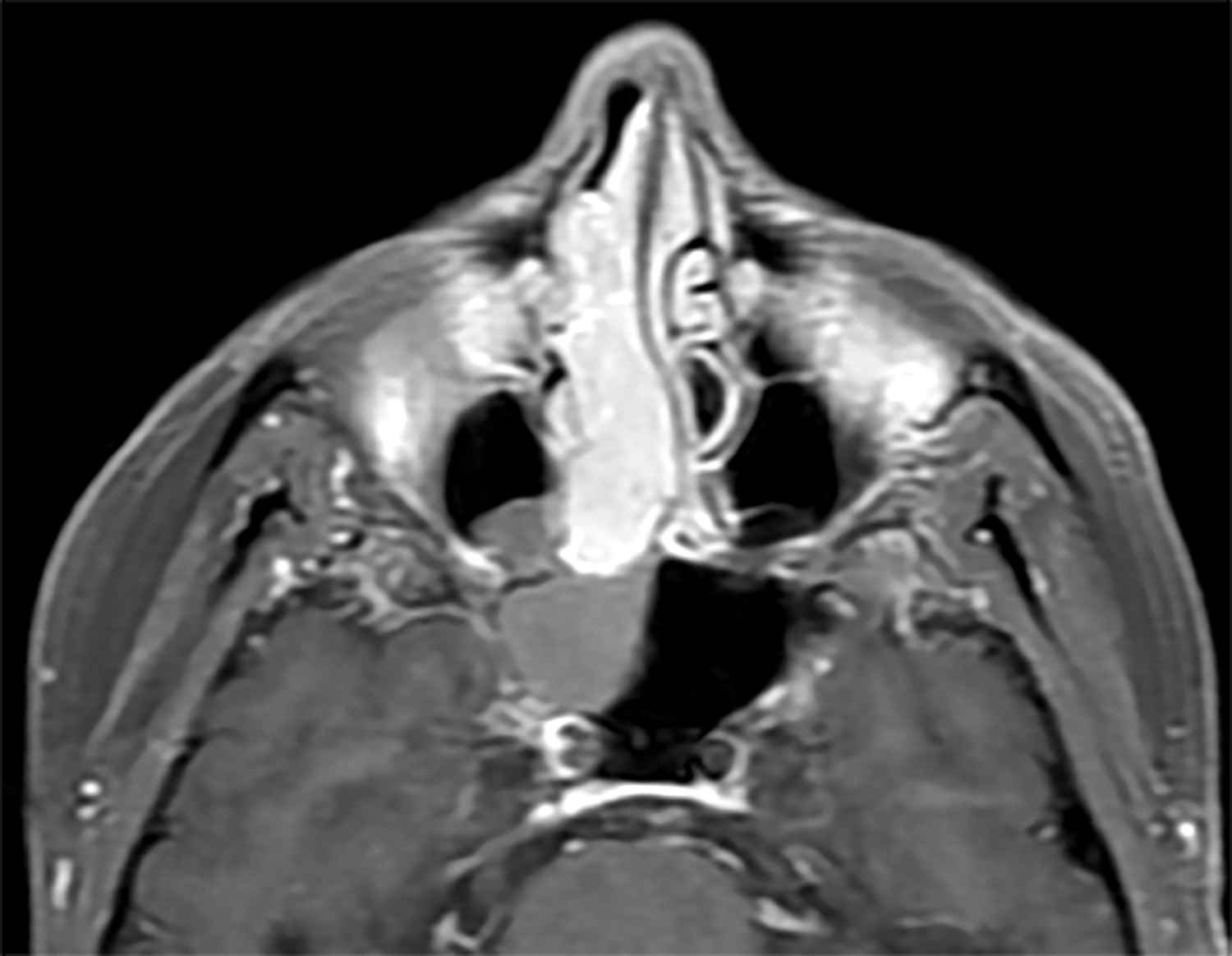
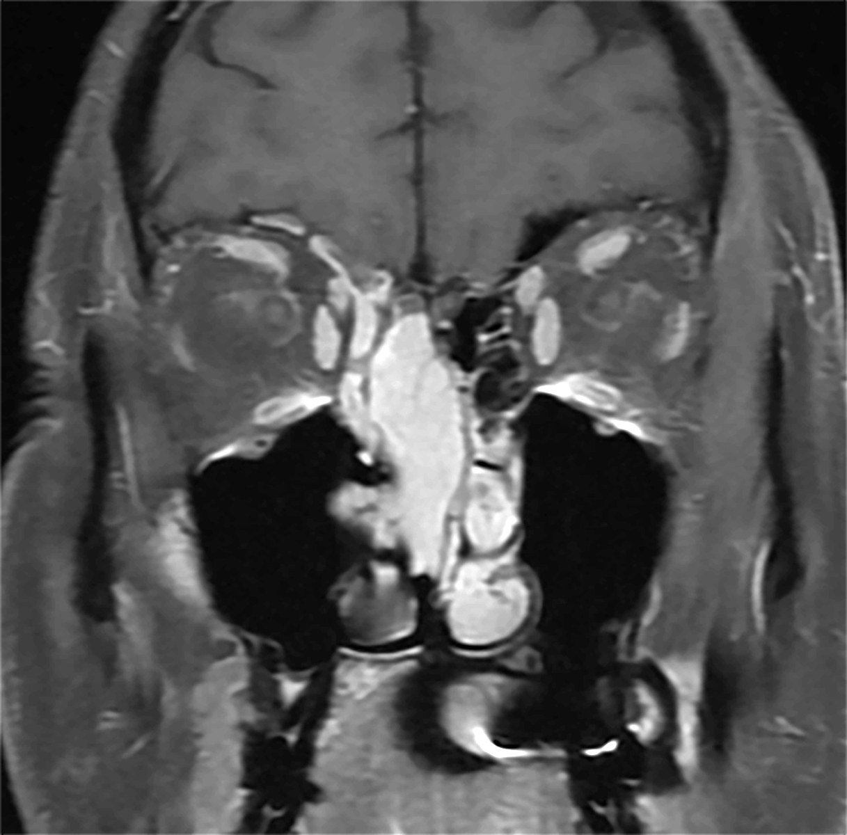
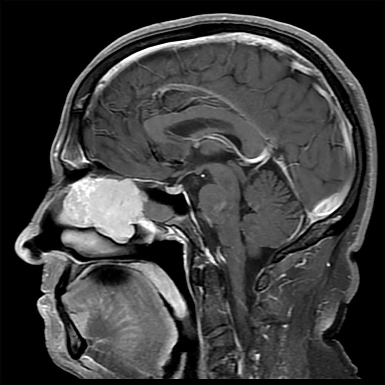
Naso-oropharyngeal mass (T1 axial)
Naso-oropharyngeal mass (T1 coronal)
Naso-oropharyngeal mass (T2 axial)
Images hosted on other servers:

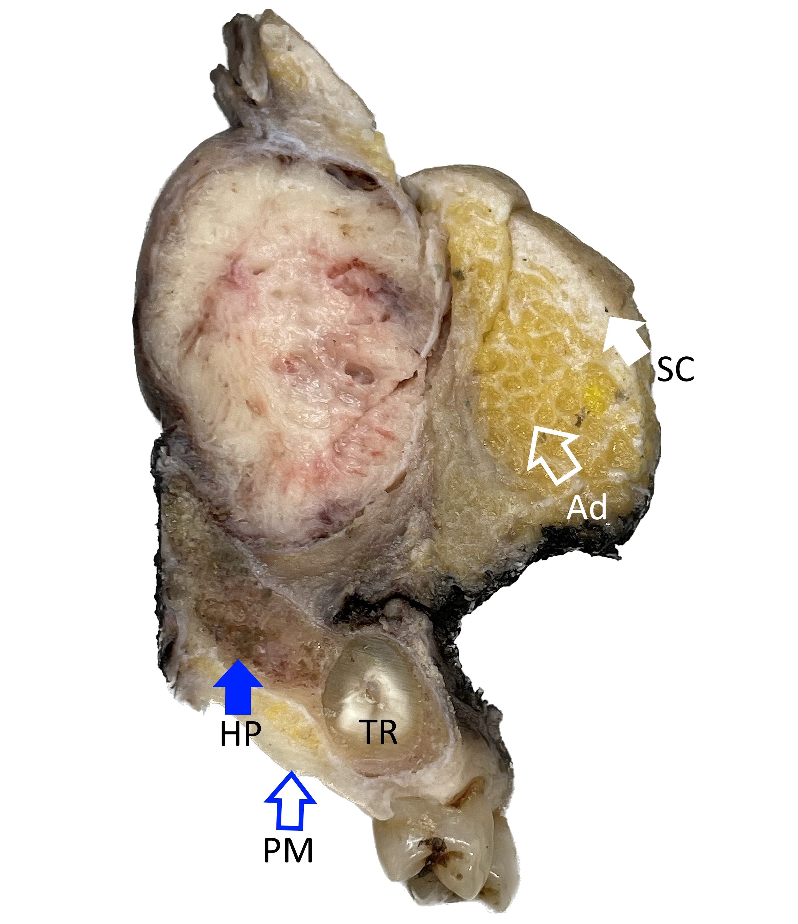
Lobulated mass
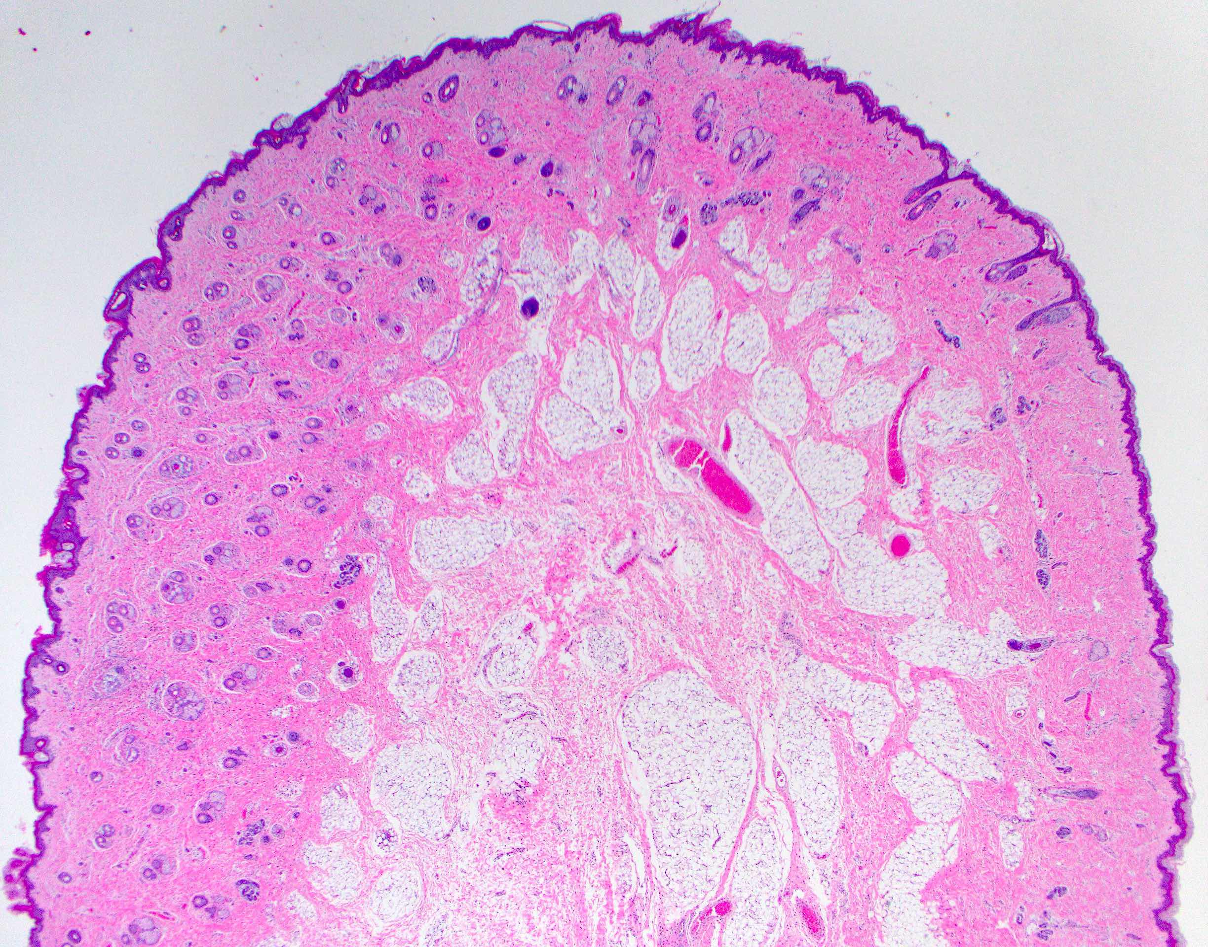
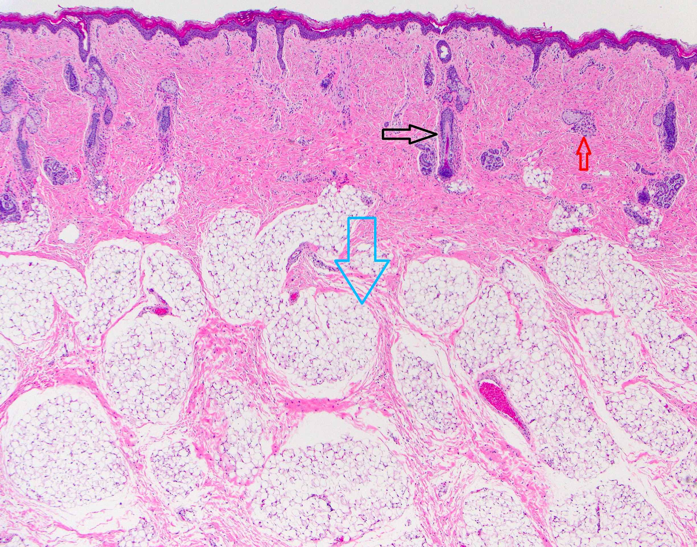
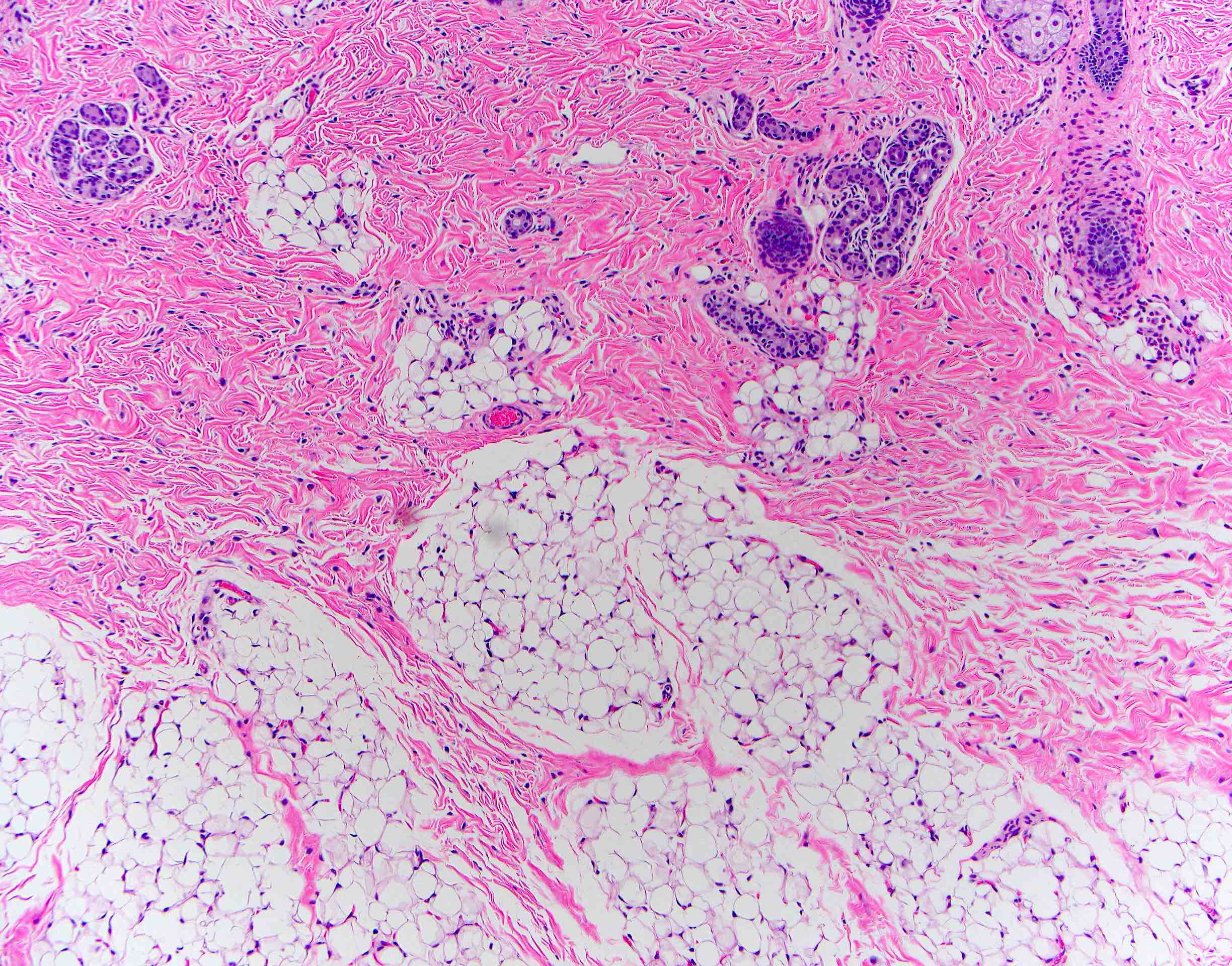
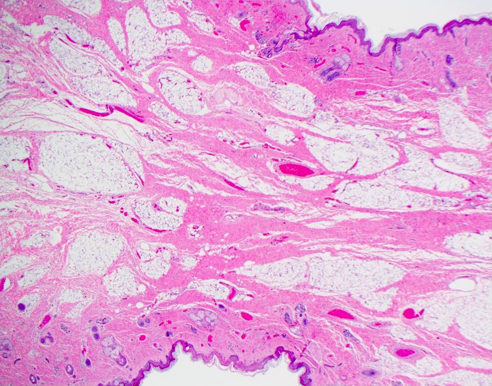
Contributed by Josephine K. Dermawan, M.D., Ph.D. and Laura O. Rabinowitz, M.D.
Submucosal lobular proliferation
Nonkeratinizing squamous mucosa
Epithelial and mesenchymal components
Duct-like structures and calcifications
Focal chondromyxoid stroma
Ovoid to spindled stromal cells

Positive CK7
Positive p63
Variable SMMS1
Focal S100
Images hosted on other servers:

Maxillary sinus squamous cell carcinoma (CT)

Large exophytic squamous cell carcinoma (CT)

Maxillary sinus adenocarcinoma (MRI)

Sinonasal undifferentiated carcinoma (CT)

SCC bone destruction (CT)
Images hosted on other servers:

Endoscopic view of squamous cell carcinoma

Exophytic squamous cell carcinoma
Contributed by Kelly Magliocca, D.D.S., M.P.H.
Maxillary sinus squamous carcinoma
Squamous carcinoma ex papilloma
HPV related multiphenotypic carcinoma
Maxillary sinus adenoid cystic
HPV associated adenosquamous carcinoma
Sinonasal intestinal type adenocarcinoma
Images hosted on other servers:

Craniectomy with squamous cell carcinoma
Contributed by Wai Szeto, M.D., M.S.
Squamous cell carcinoma and inverted papilloma
Intestinal type adenocarcinoma
Intestinal type adenocarcinoma
Maxillary nonintestinal type adenocarcinoma
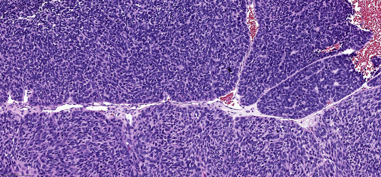
Sinonasal undifferentiated carcinoma
Sinonasal undifferentiated carcinoma mitotic activity
Nonintestinal type adenocarcinoma (CK7)
Nonintestinal type adenocarcinoma (SOX10)
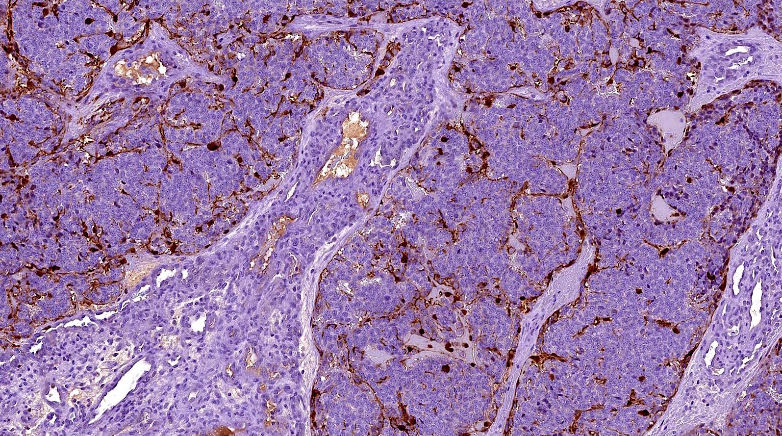
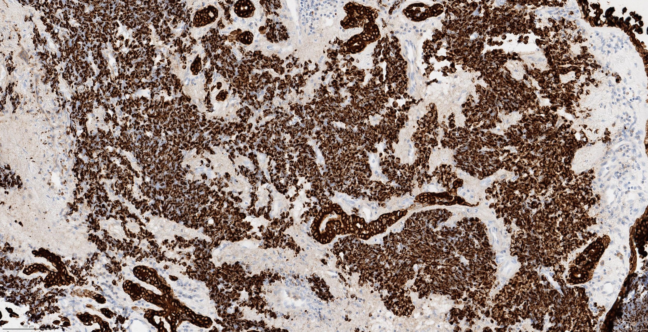
Sinonasal undifferentiated carcinoma (pancytokeratin+)
Retained INI1
Contributed by Jinping Lai, M.D., Ph.D.
Right nasal mass (MRI axial)
Right nasal mass (MRI coronal)
Right nasal mass (MRI sagittal)
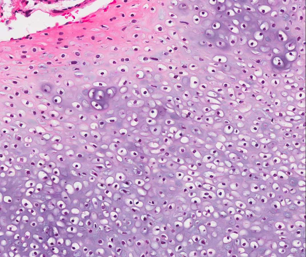
Contributed by Jinping Lai, M.D., Ph.D.
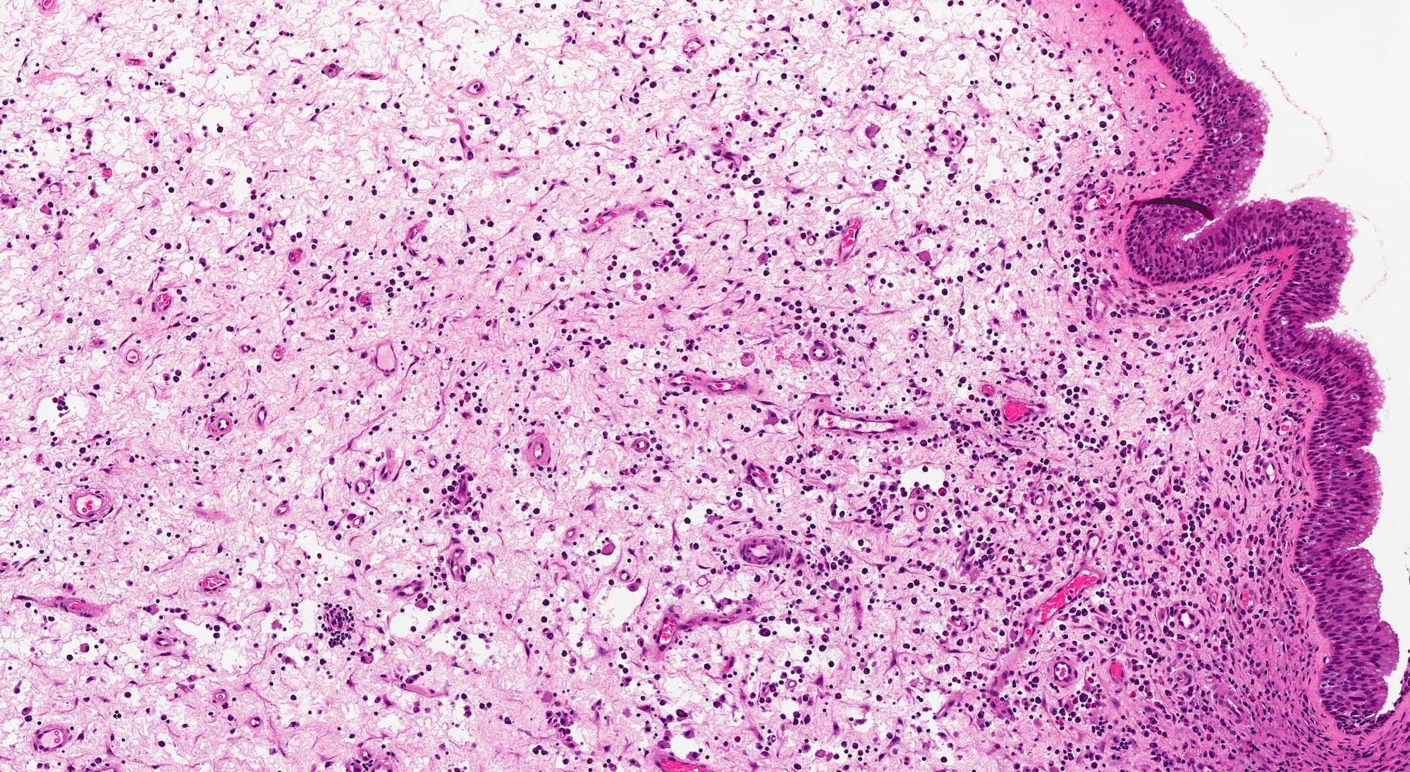
Nasal endoscopy
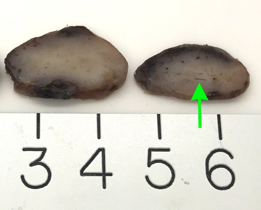
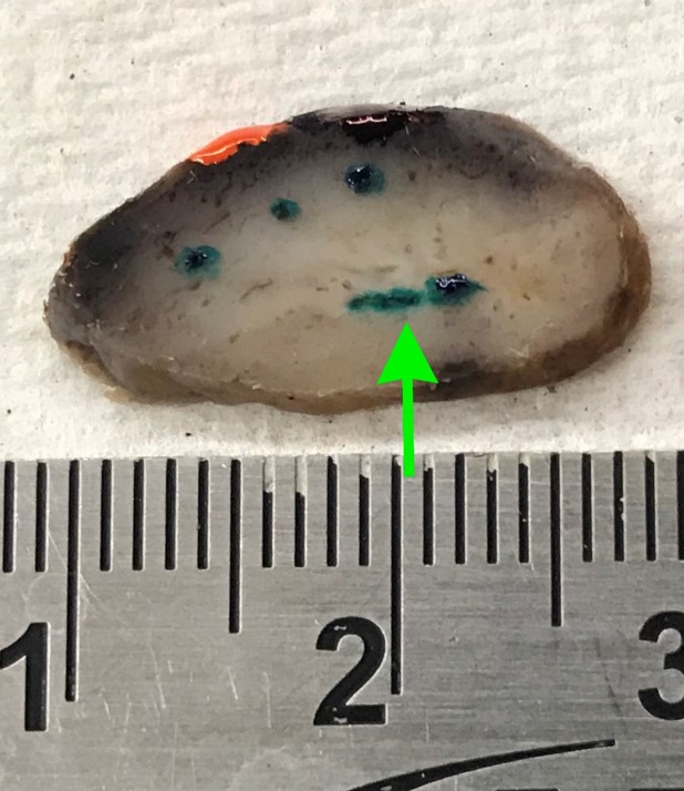
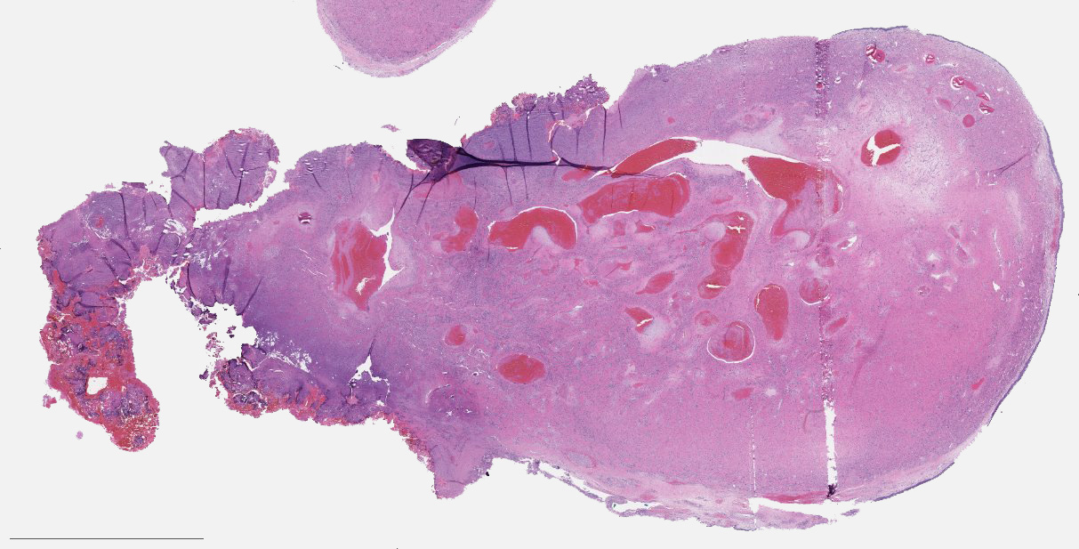
Contributed by Jinping Lai, M.D., Ph.D.
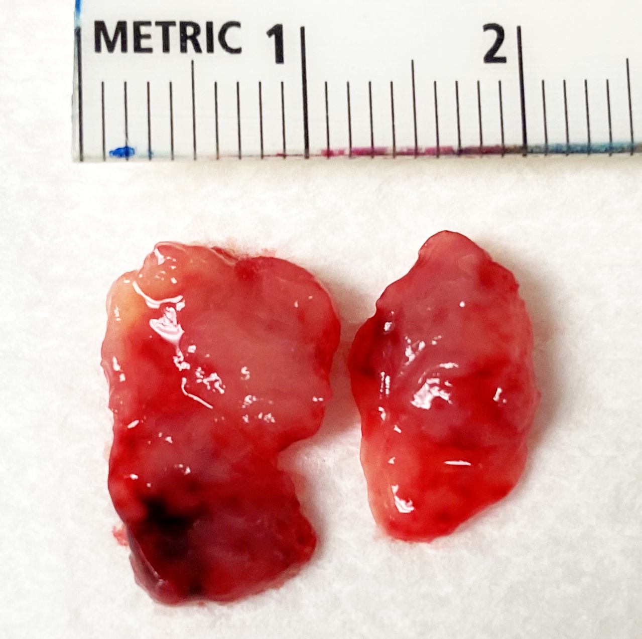
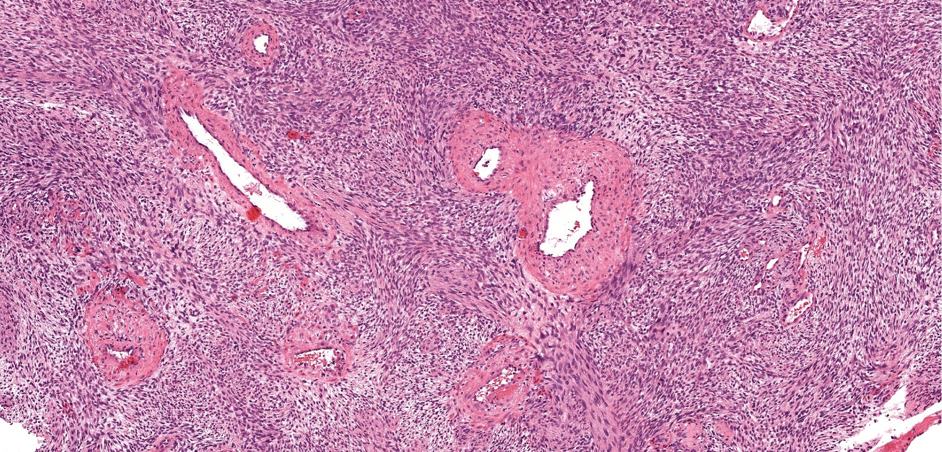
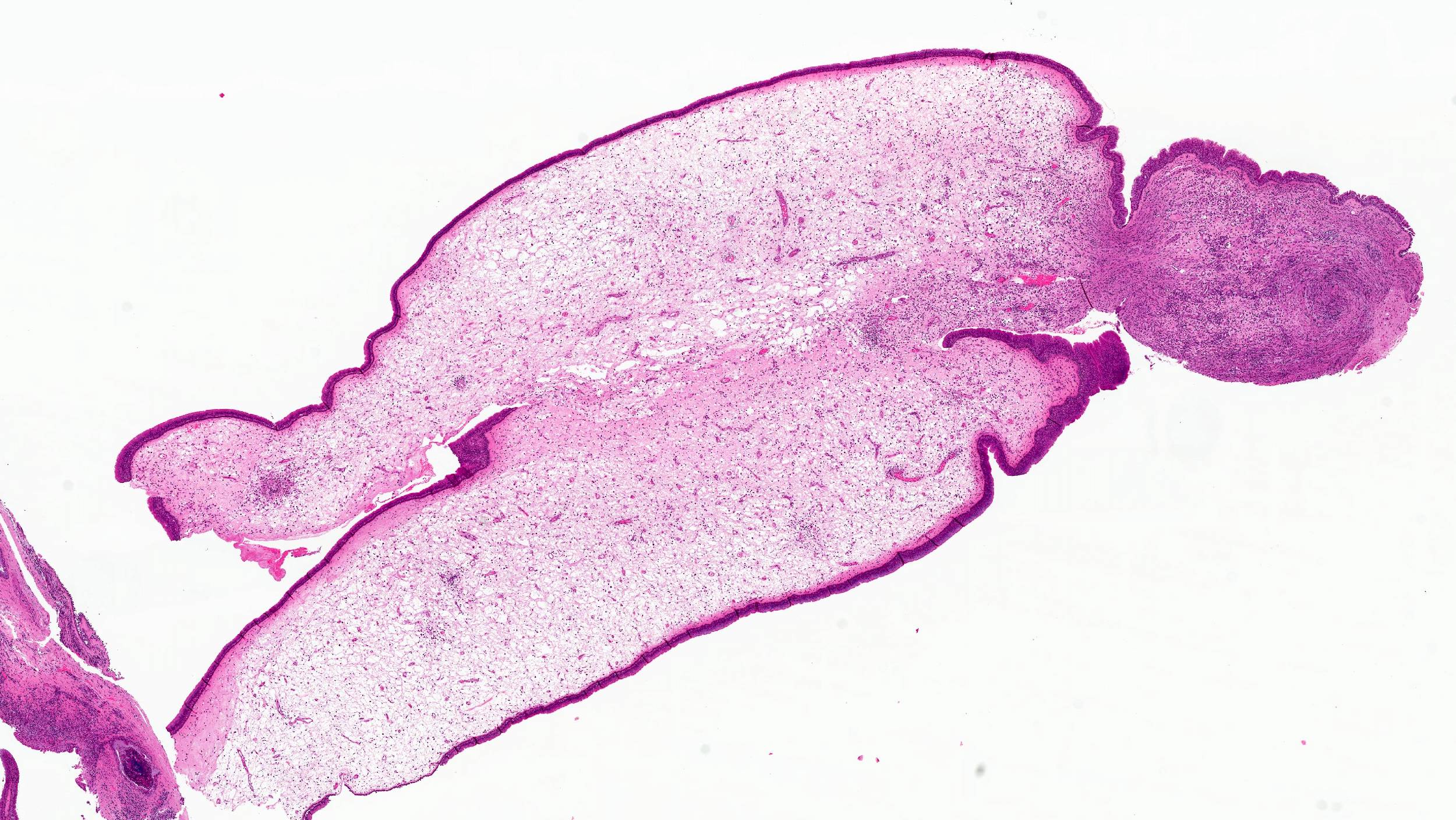
Cut surface
Contributed by Jinping Lai, M.D., Ph.D.
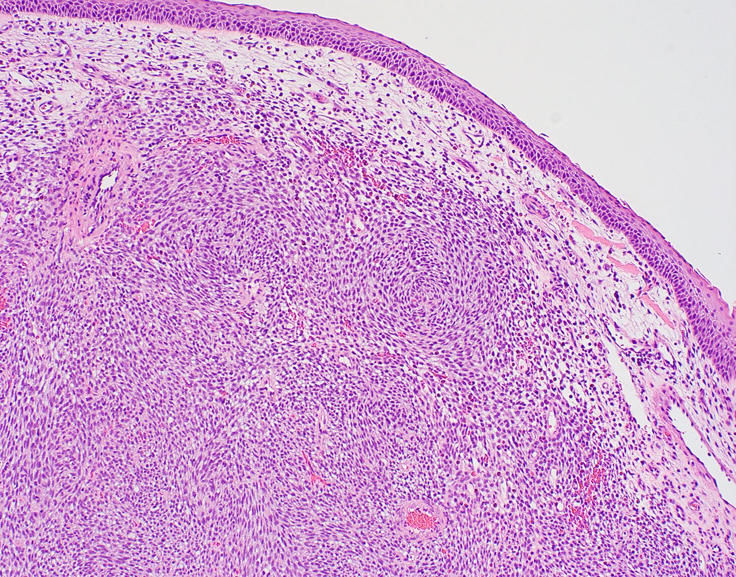
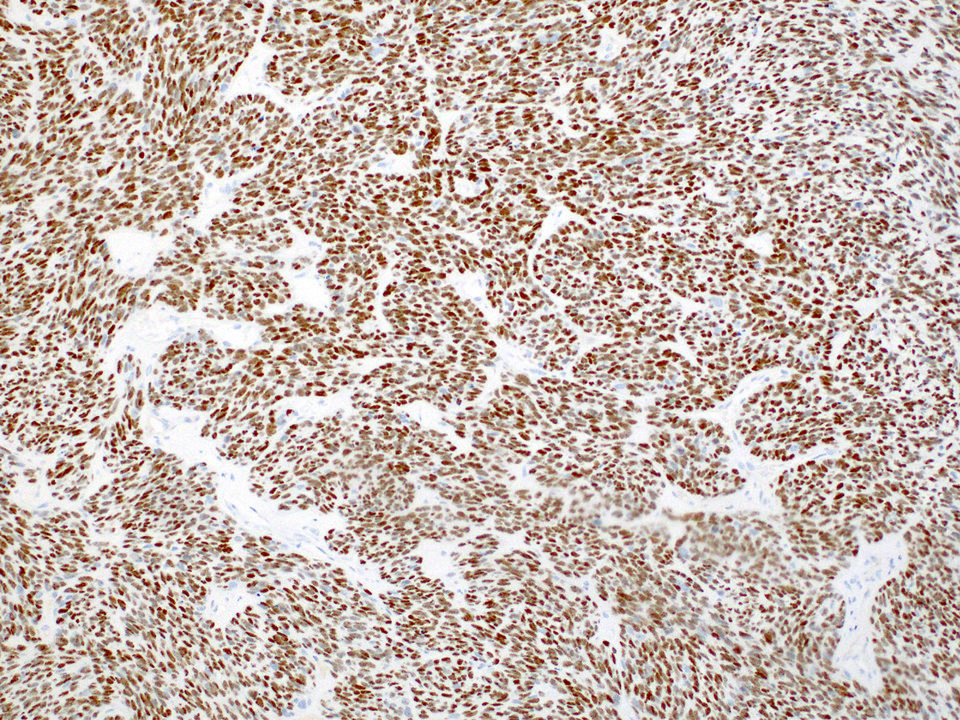
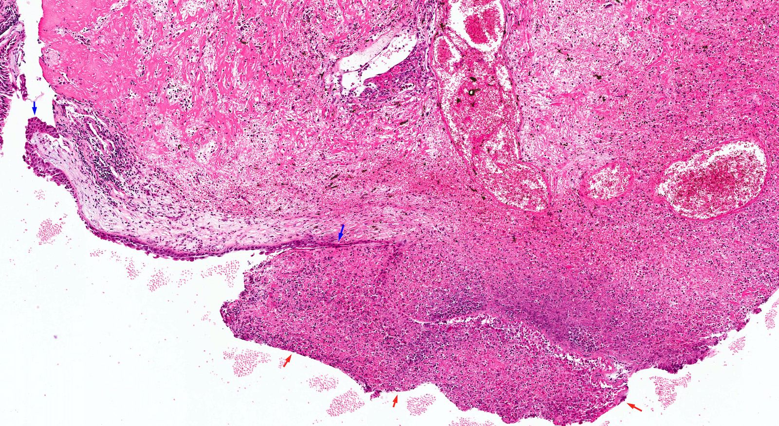
Subepithelial grenz zone and whorled features

Perivascular hyalinization and staghorn vessels
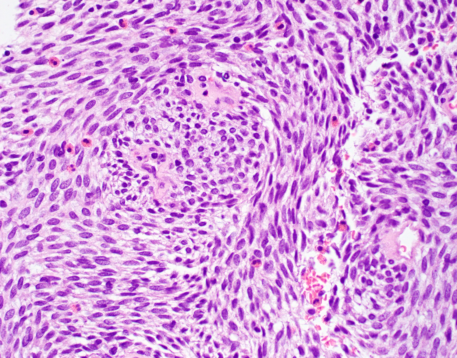
No mitosis, eosinophils
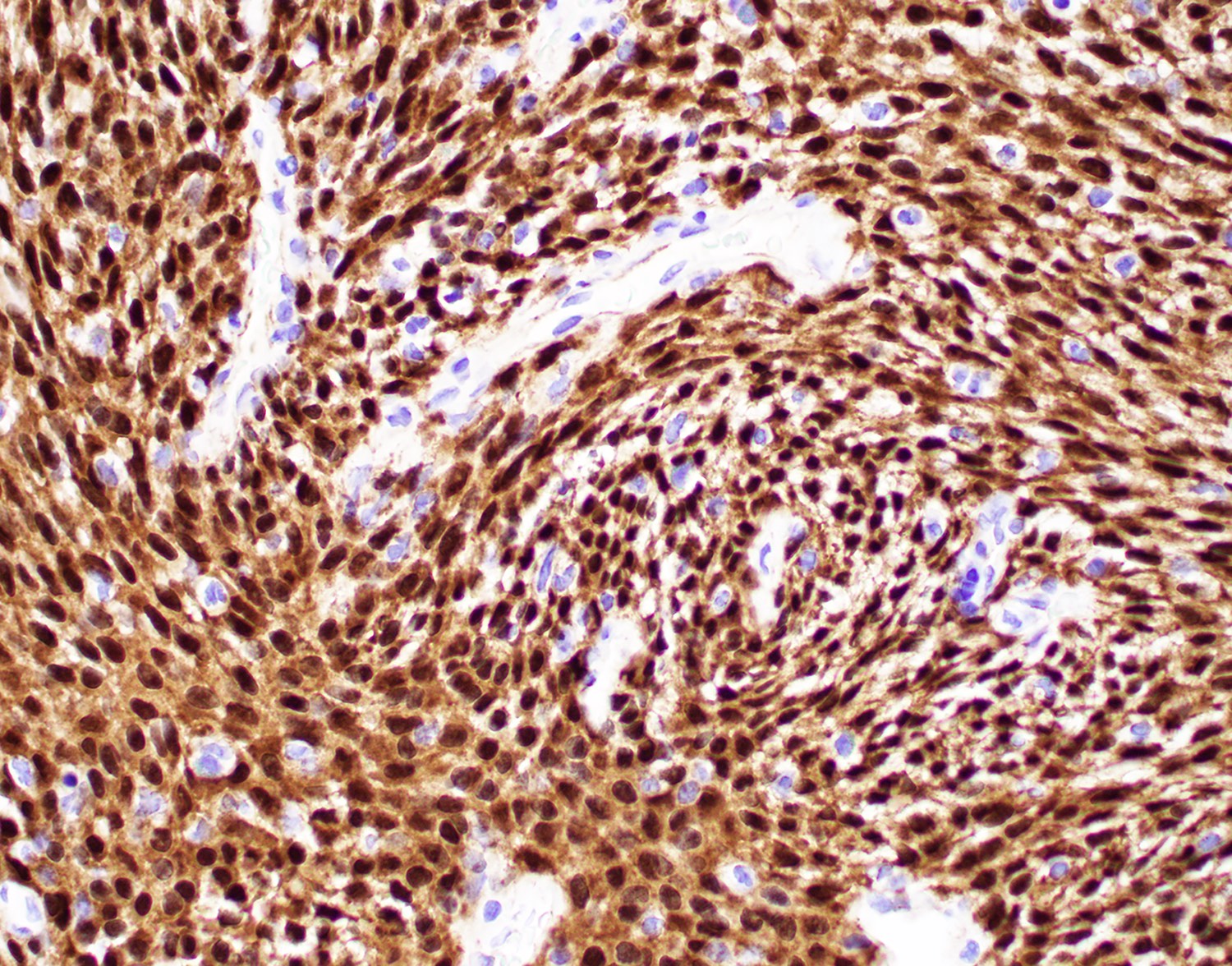
Beta catenin nuclear staining
Positive cyclin D1
Positive CD99
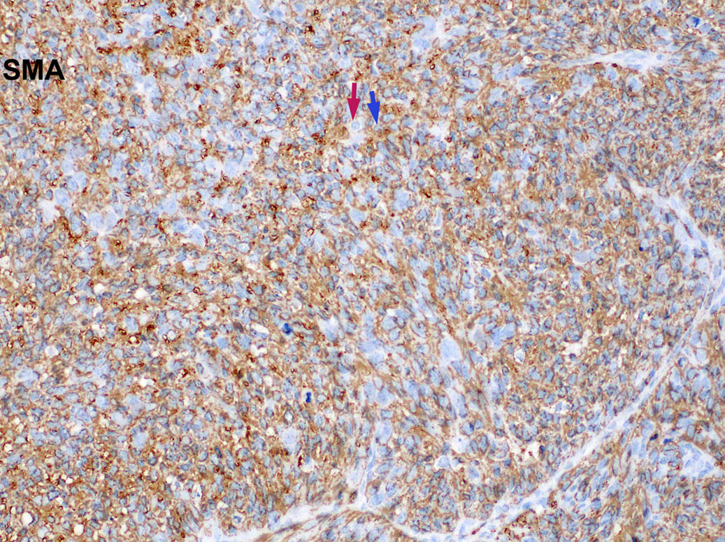
Positive SMA
Low Ki67 index
Images hosted on other servers:

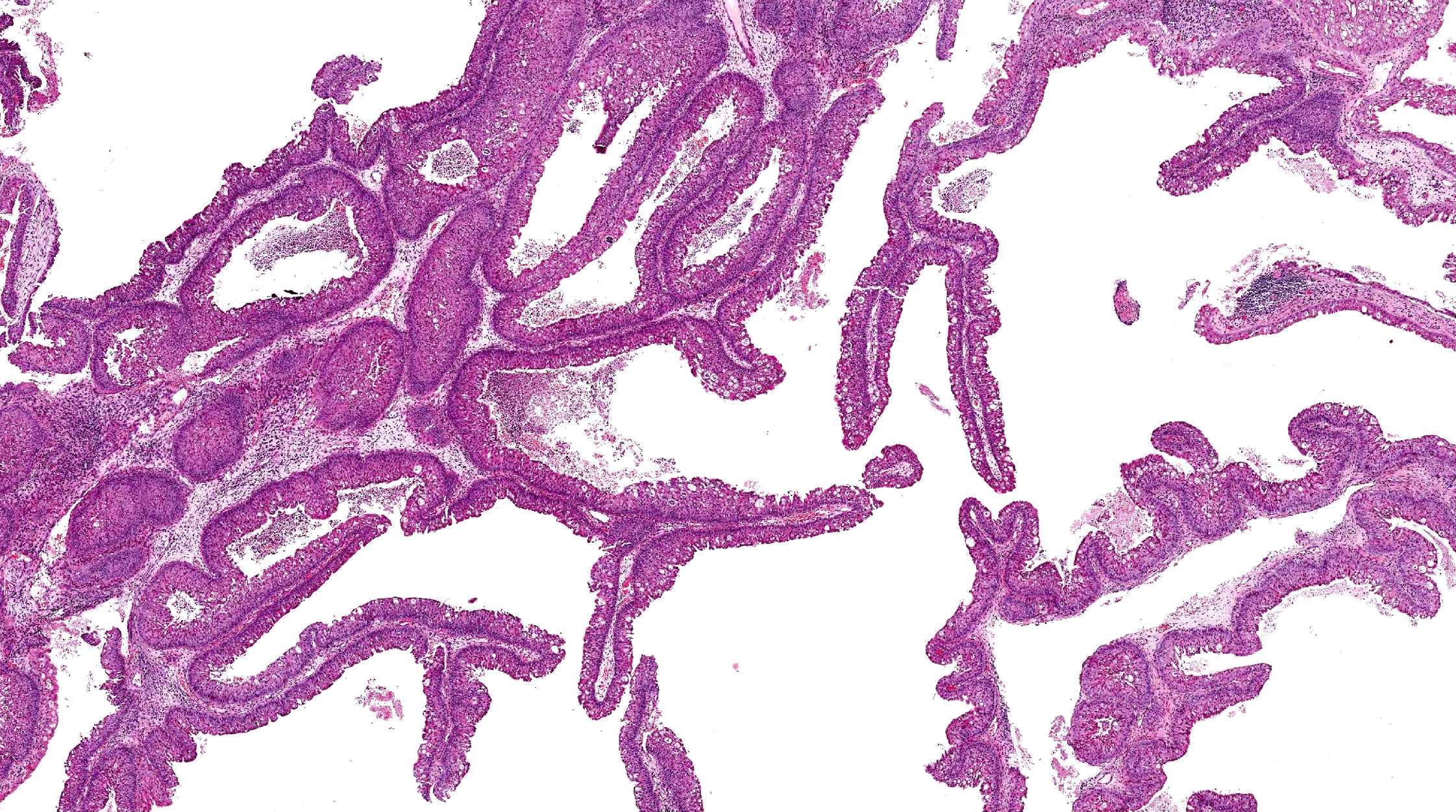
CT findings of an inverted papilloma

MRI findings of an inverted papilloma
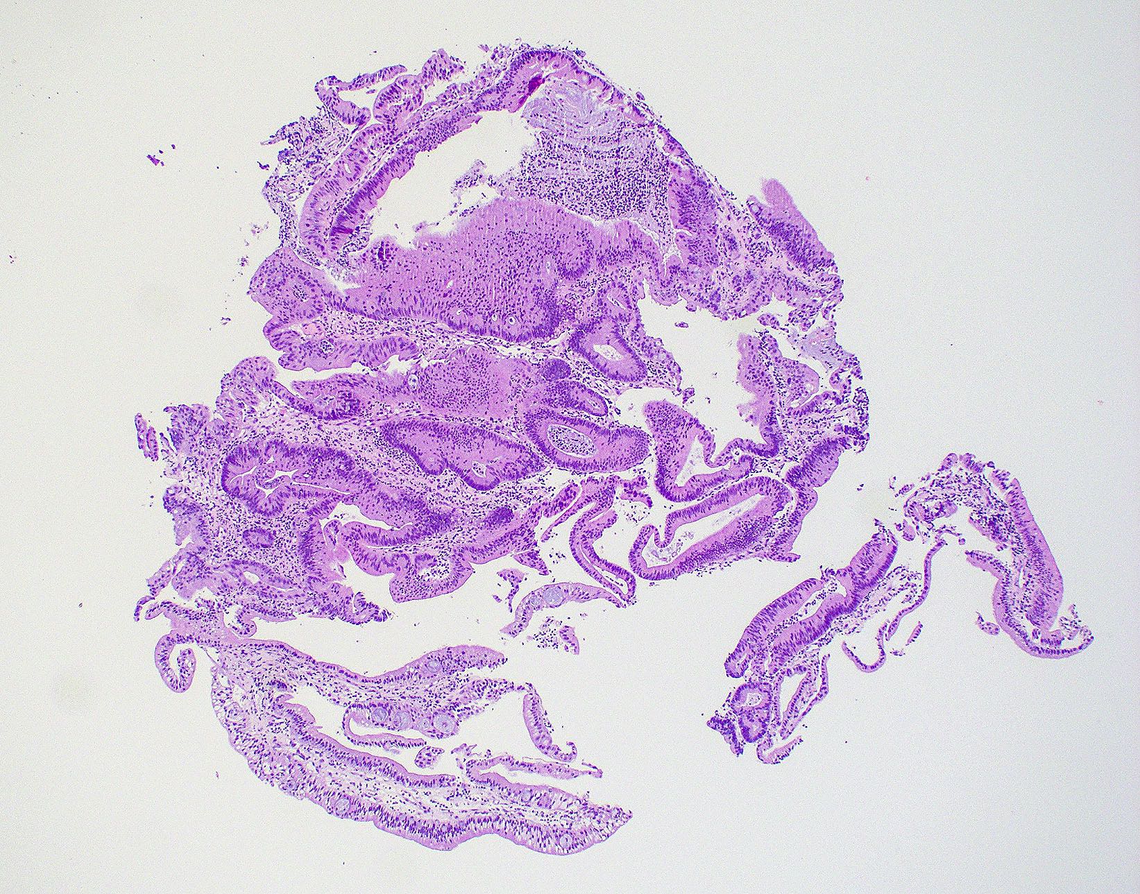
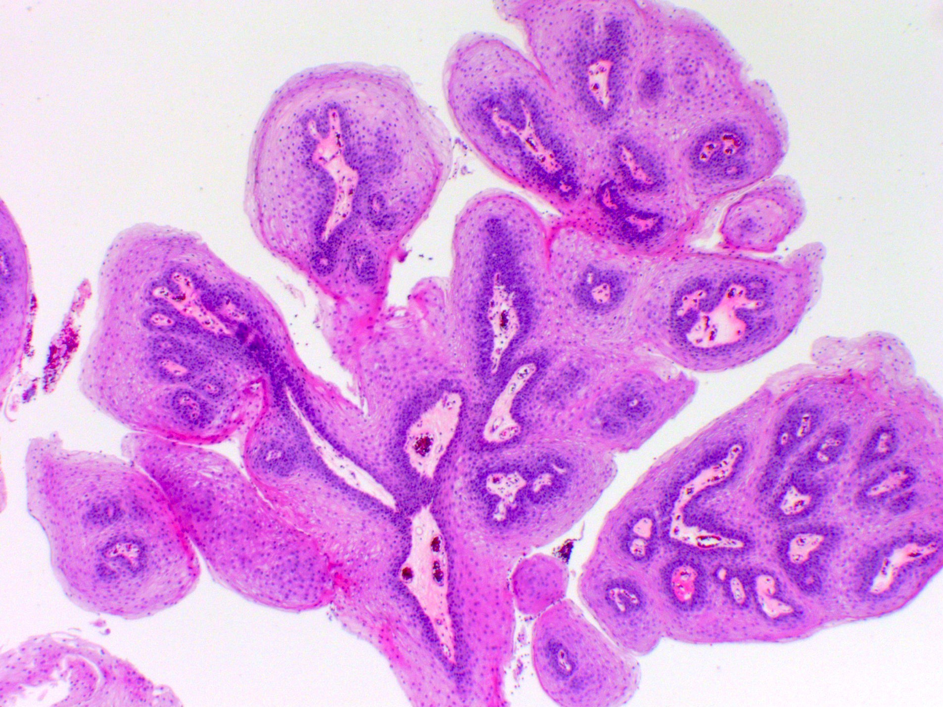
Images hosted on other servers:

Squamous papilloma

Endoscopic image
of an inverted
papilloma
Images hosted on other servers:

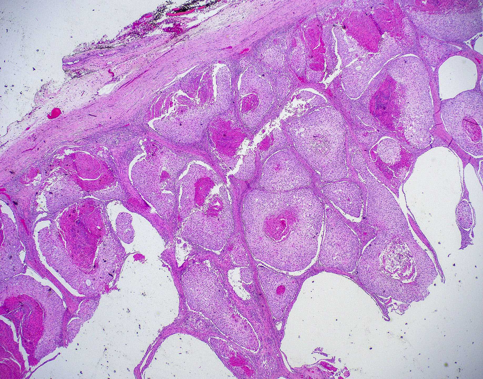
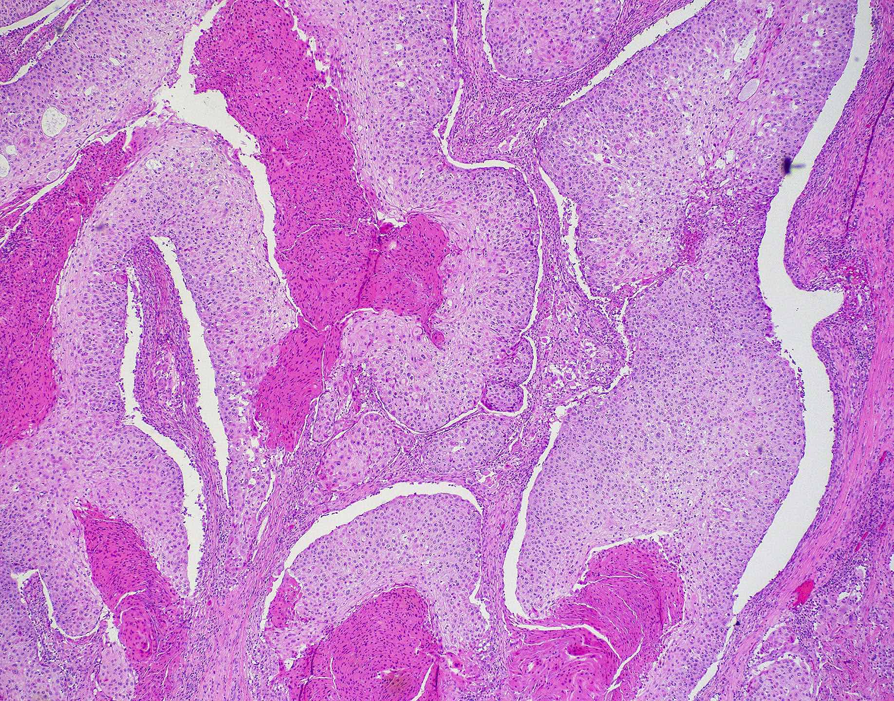
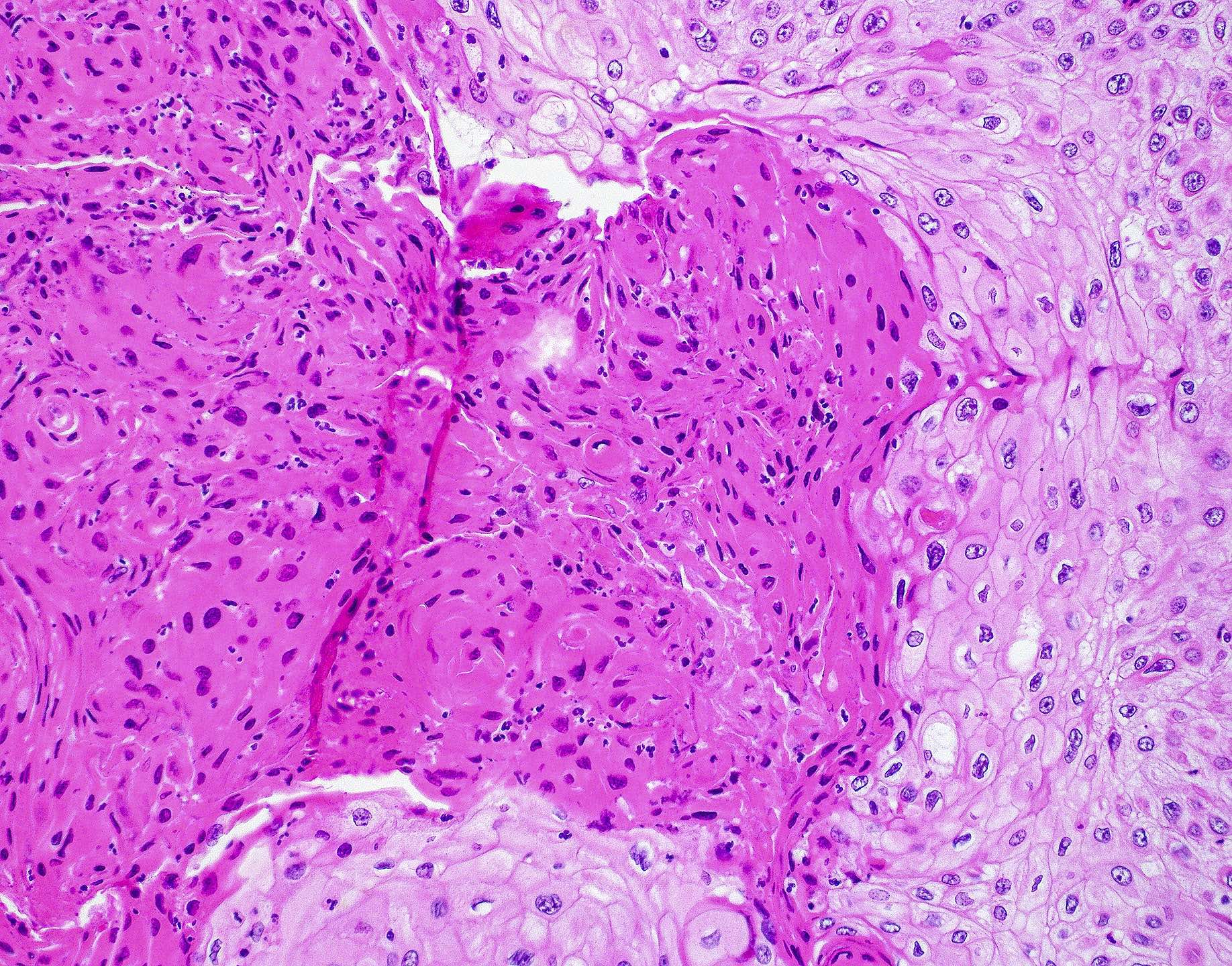
Inverted papilloma
Contributed by Bin Xu, M.D., Ph.D.

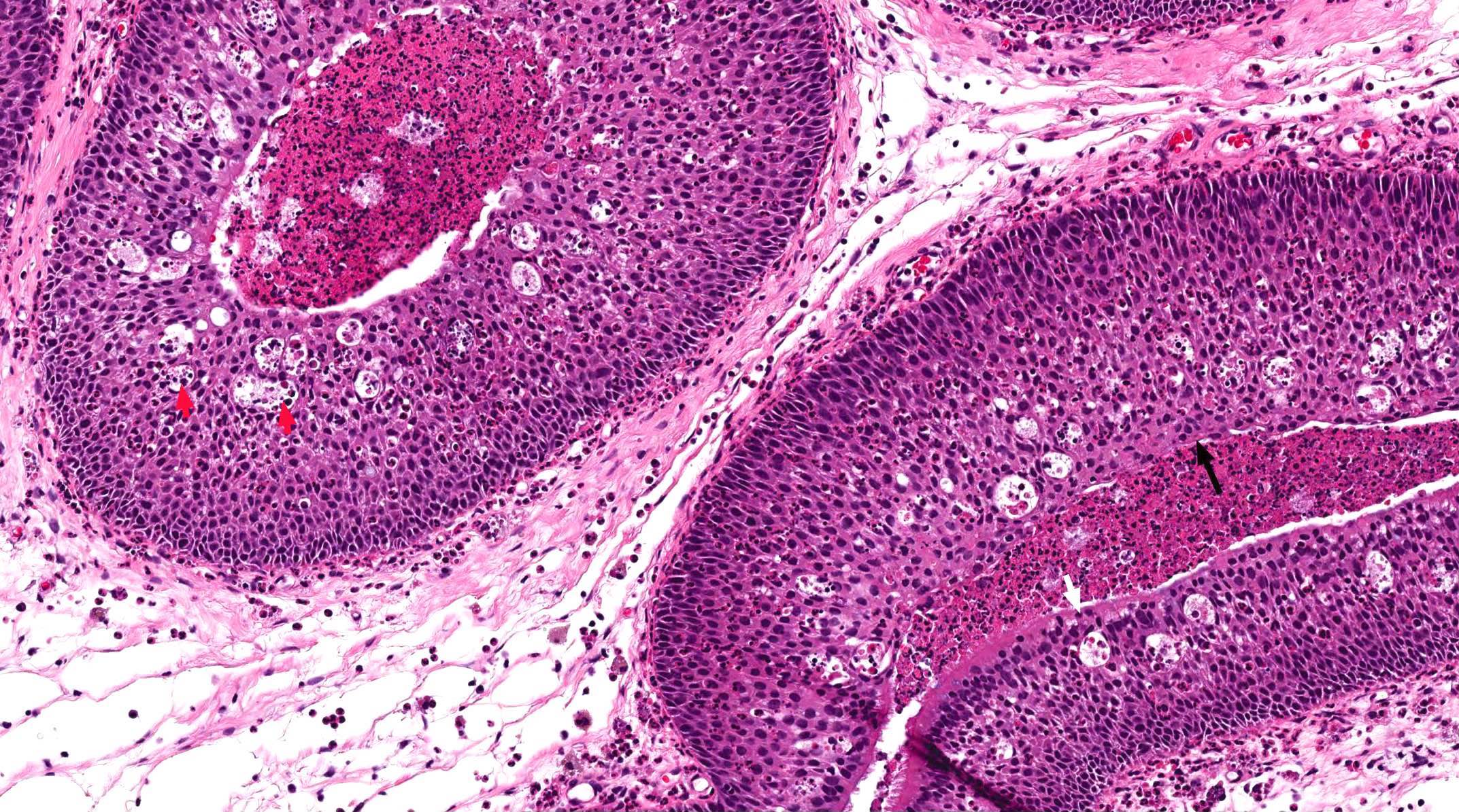
Inverted papilloma
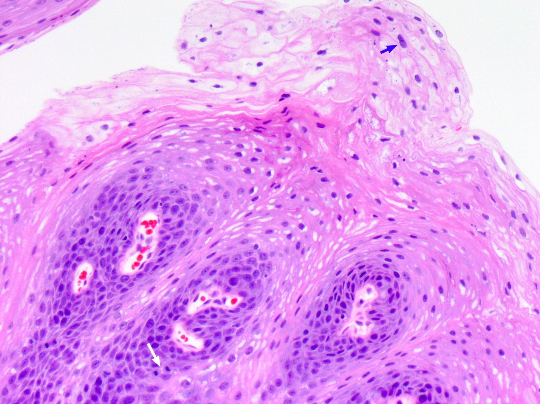
Exophytic papilloma
Oncocytic papilloma
Malignant transformation (carcinoma ex sinonasal papilloma)
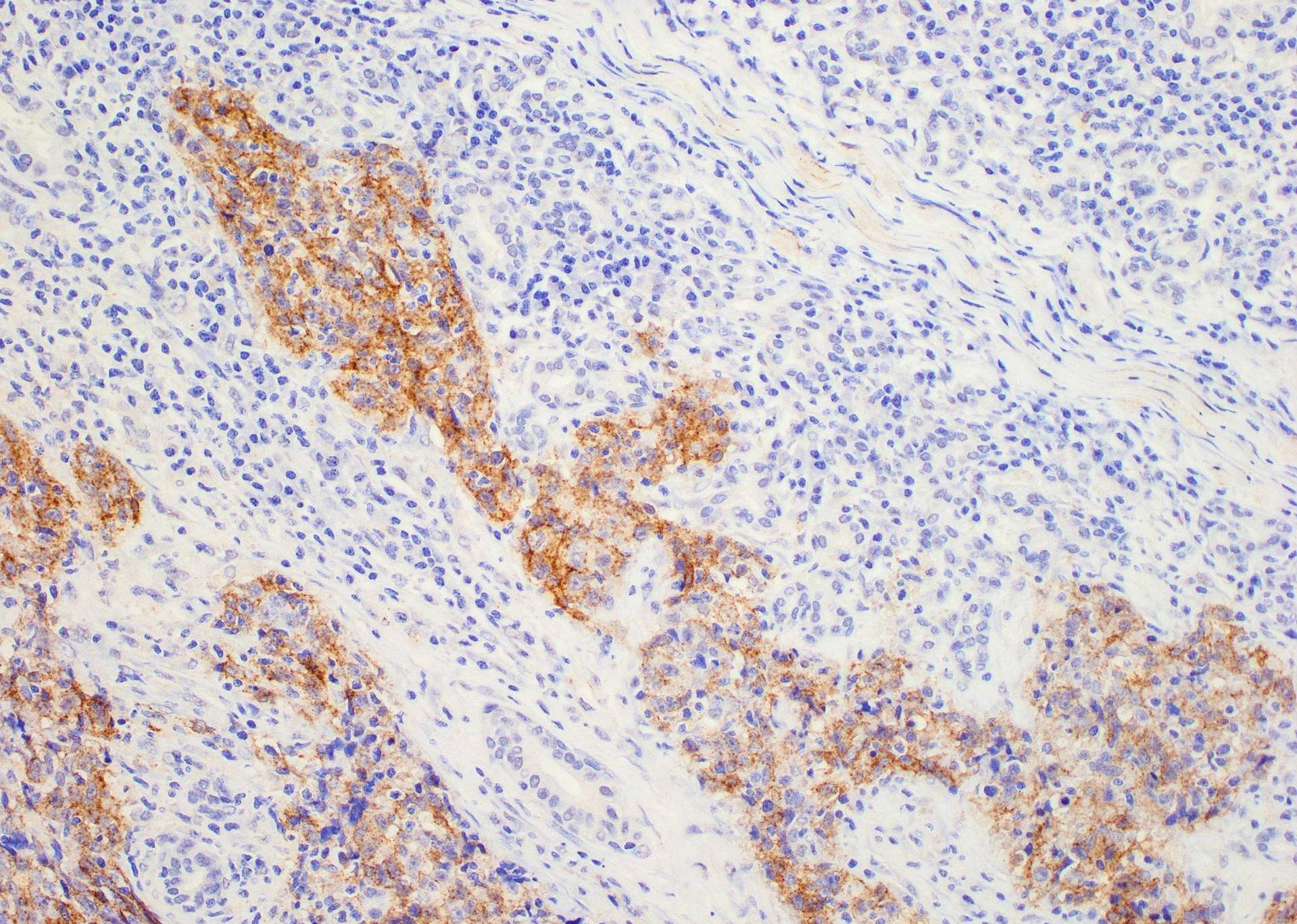
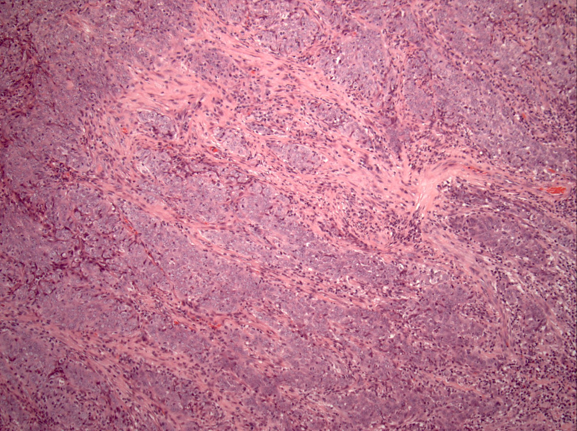
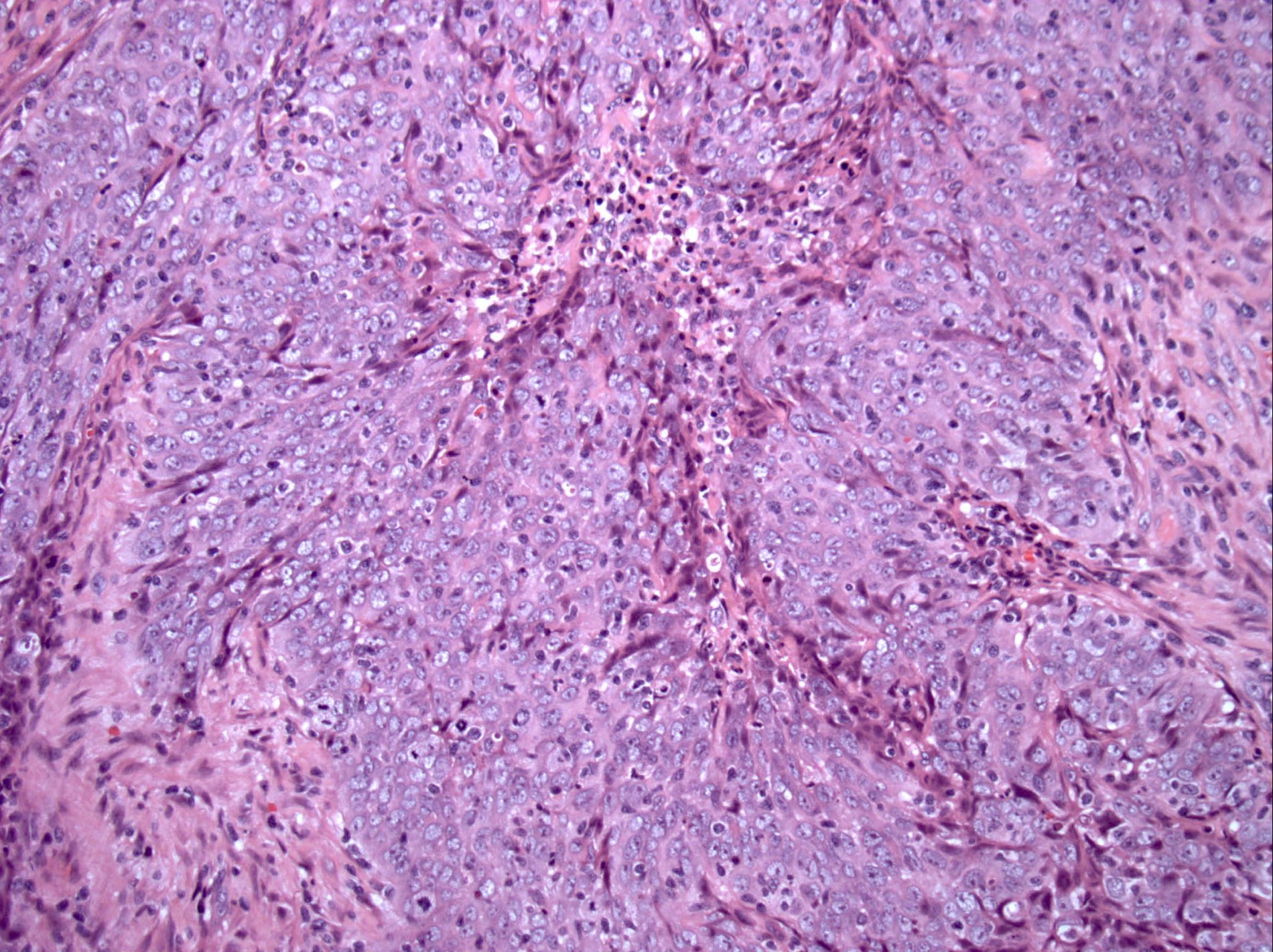
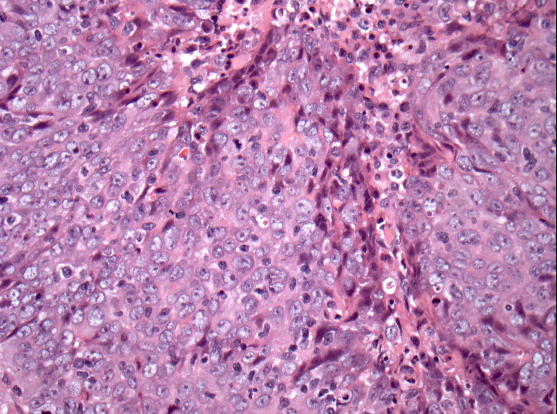
Contributed by Bin Xu, M.D., Ph.D. and Abeer Salama, M.D.

MRI
Contributed by Bin Xu, M.D., Ph.D. and Abeer Salama, M.D.
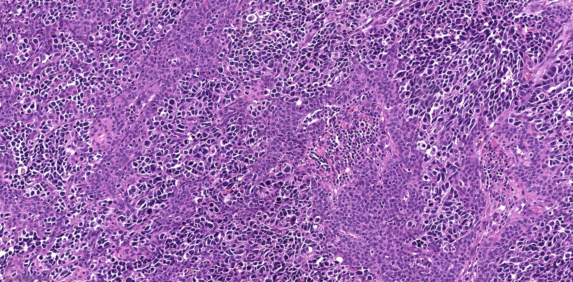
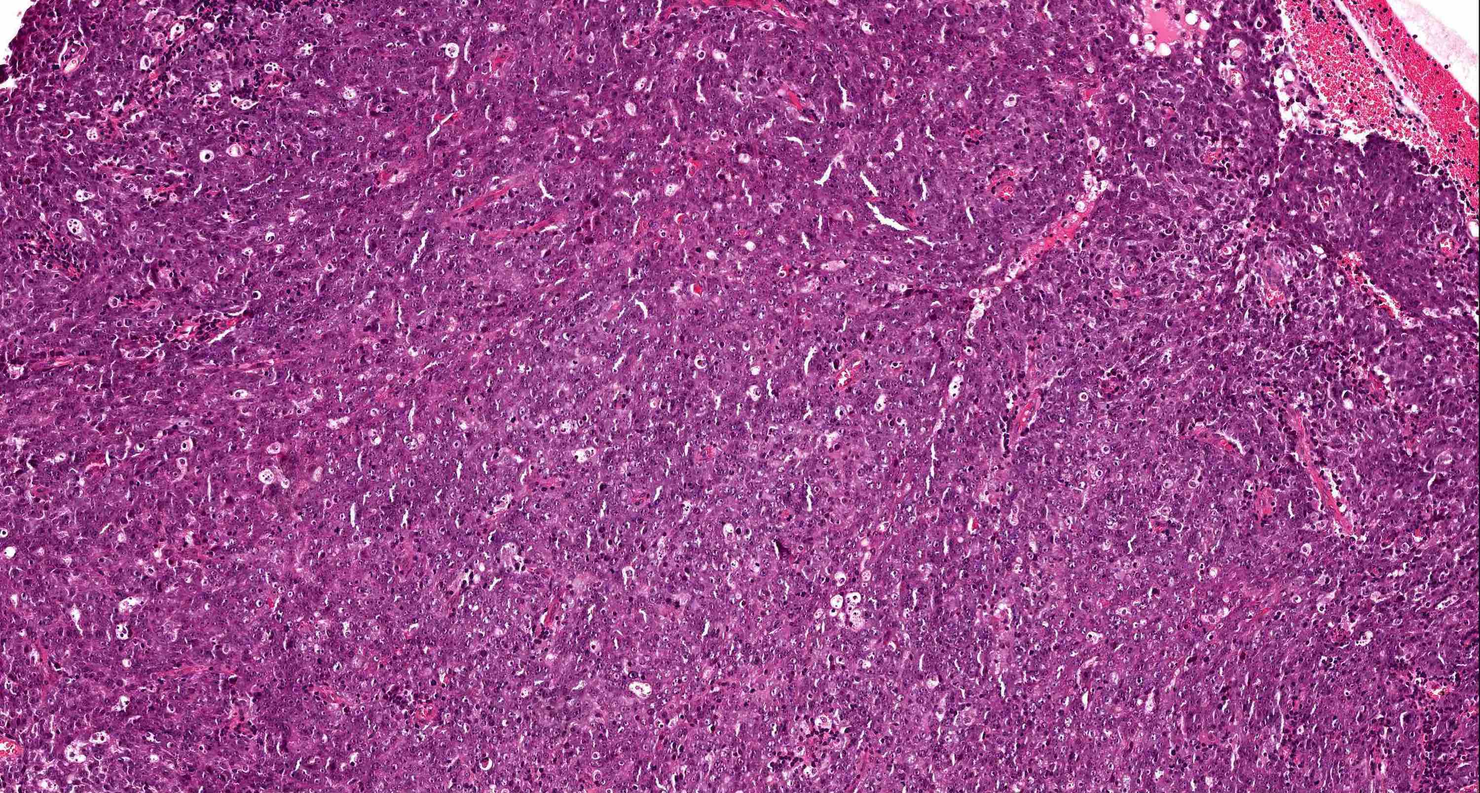
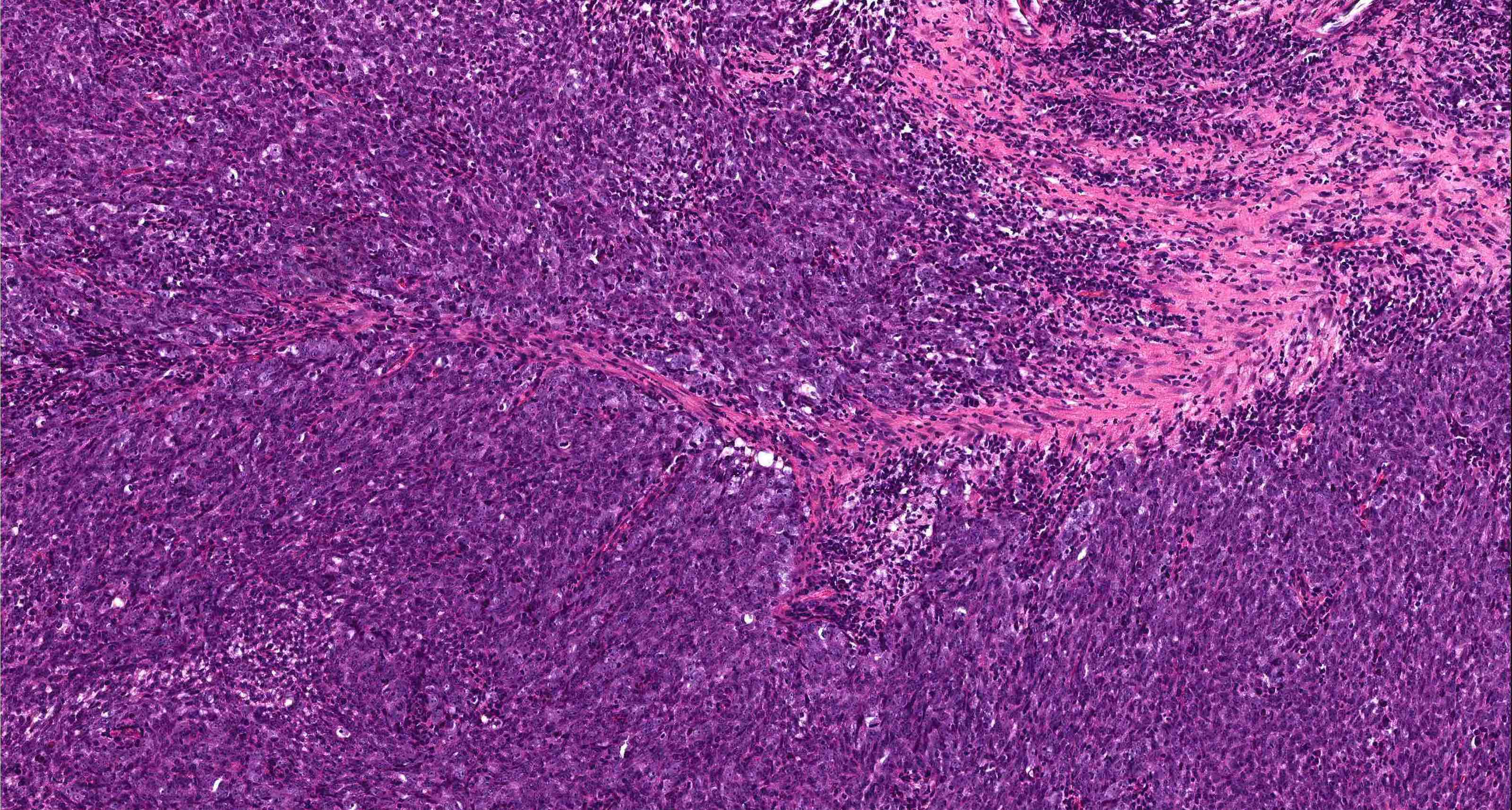
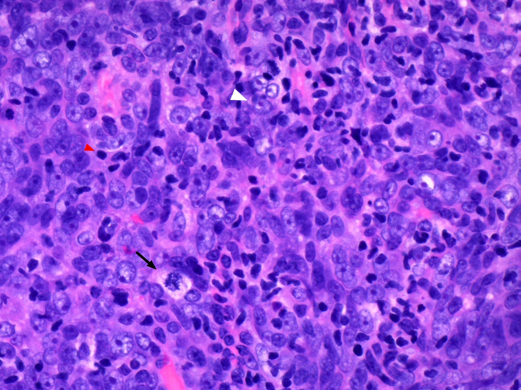
Lobules and sheets of malignant cells
Cytologic features of malignant epithelial cells
High grade features: mitosis and apoptosis
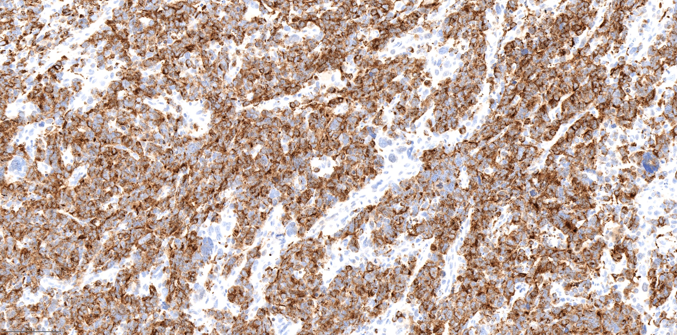
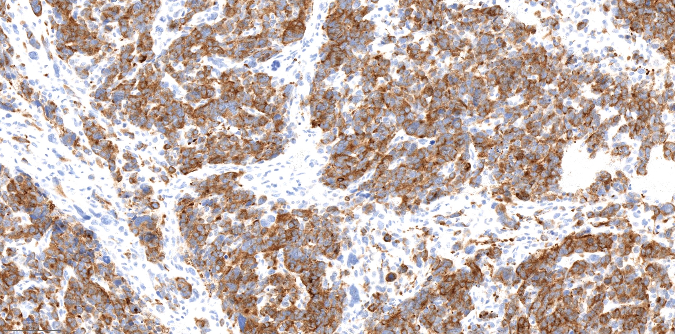
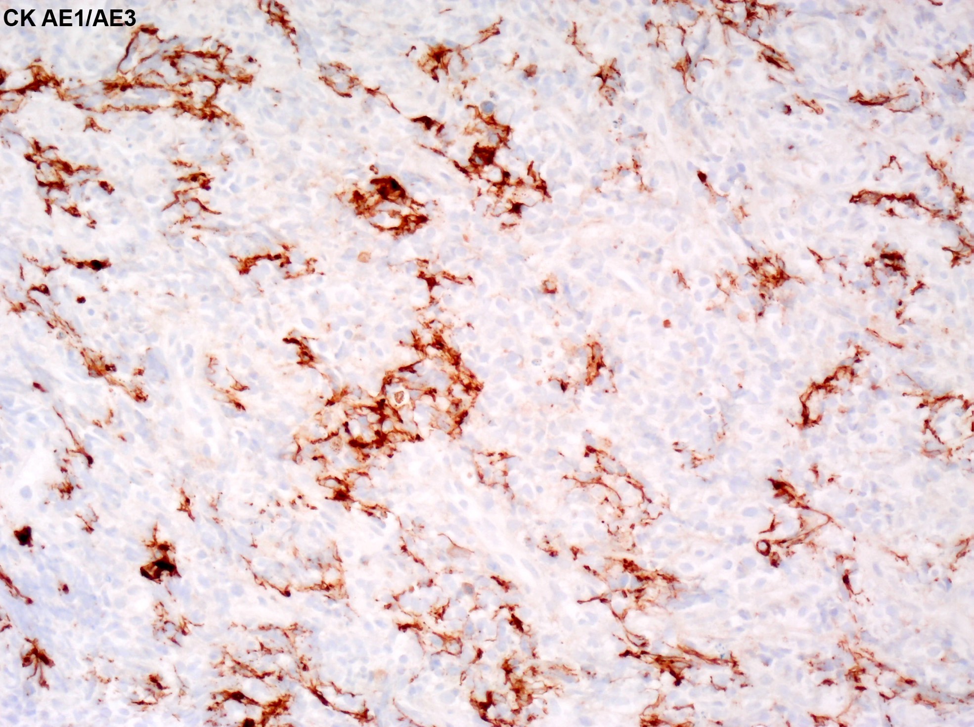
Cytokeratin AE1 / AE3
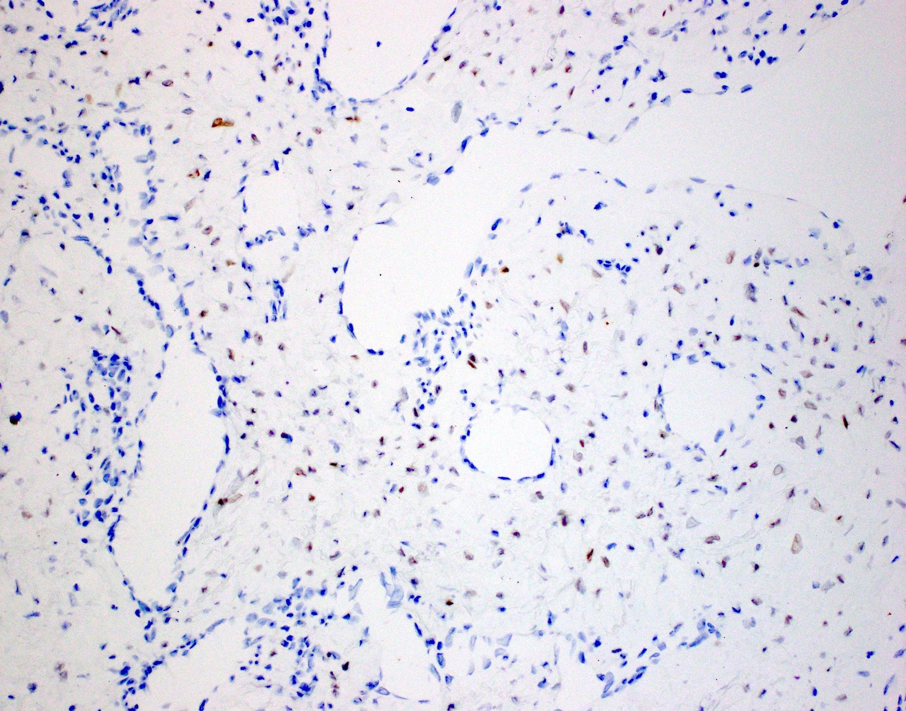
IDH2 R172T immunohistochemistry
Images hosted on other servers:

Keratinizing squamous cell carcinoma

Nonkeratinizing squamous cell carcinoma


Basaloid SCC: H&E and p63
Images hosted on other servers:

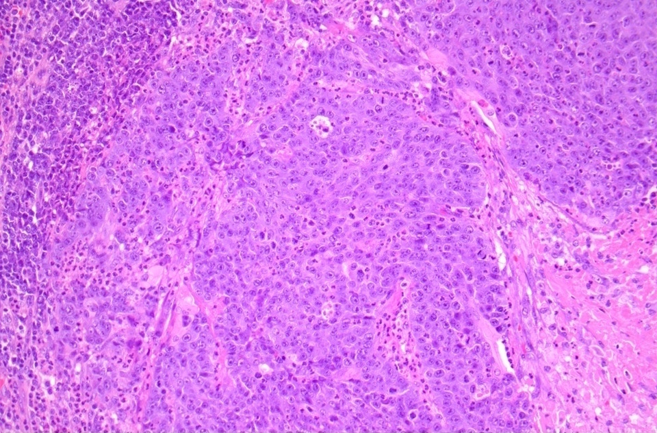
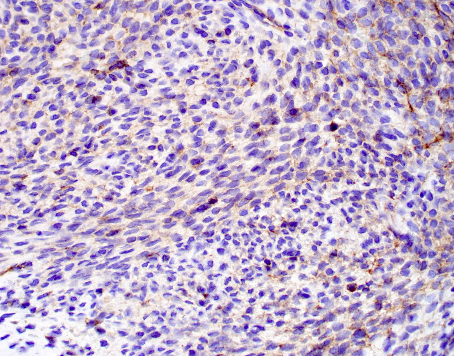
Infiltration into orbit
Contributed by Lisa Rooper, M.D.

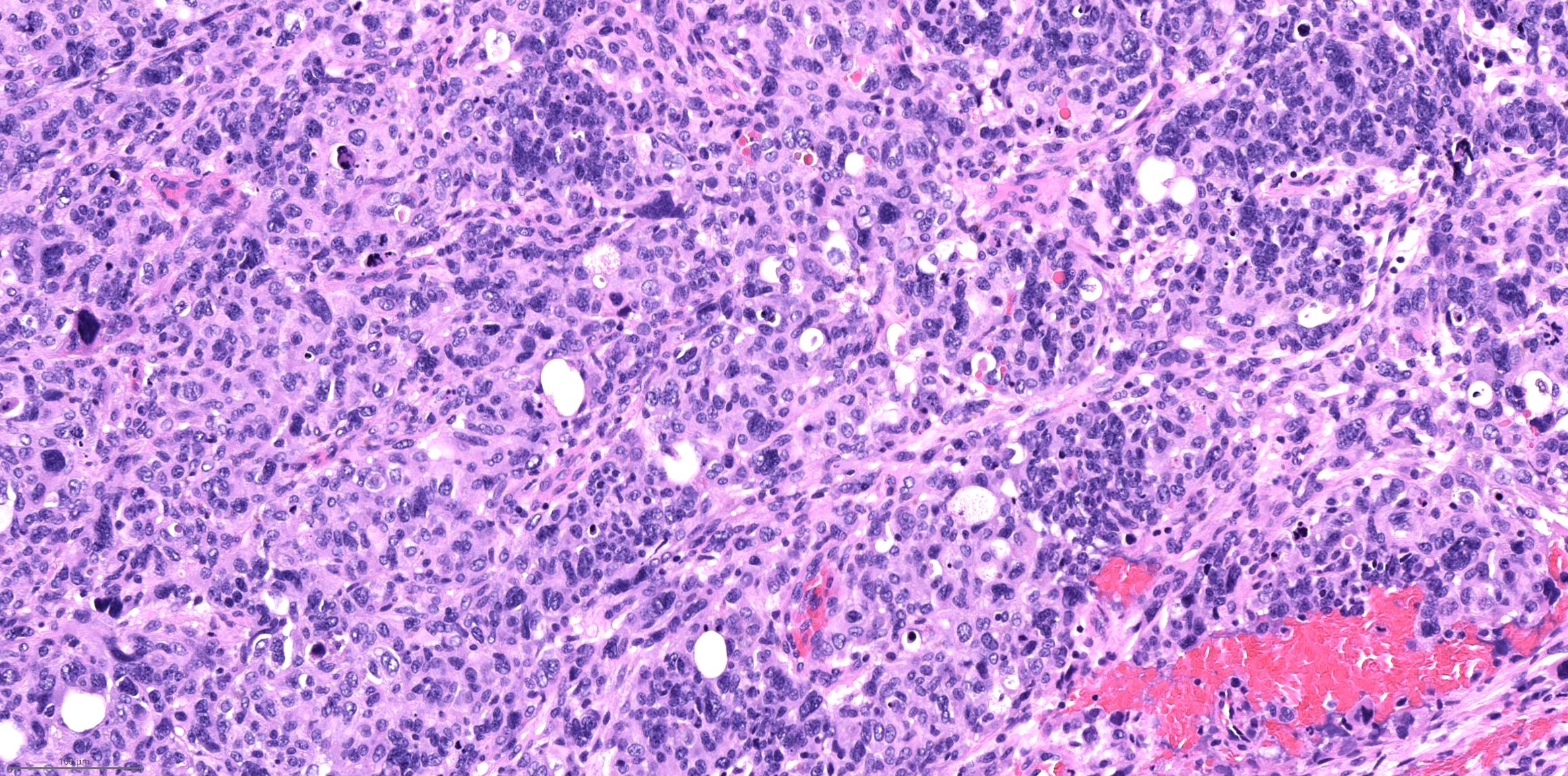
Highly infiltrative growth

Cytologic uniformity despite high grade
Nests of basaloid cells
Interspersed
basaloid and
rhabdoid
plasmacytoid cells
Sheets of rhabdoid plasmacytoid cells
No surface dysplasia
Cytoplasmic vacuoles
Loss of SMARCB1 (INI1)
WHO's new in sinonasal tract pathology
Dr. Thompson
Cardesa: 2016
Franchi: 2020
Gnepp: 2021
IARC: 2024
Neville: 2023
Robinson: 2012
Stelow: 2020
Thompson: 2022
Volavsek: 2016
Wenig: 2017
Wenig: 2024
Find related Pathology books: head & neck/endocrine
