

AFIP images
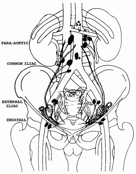
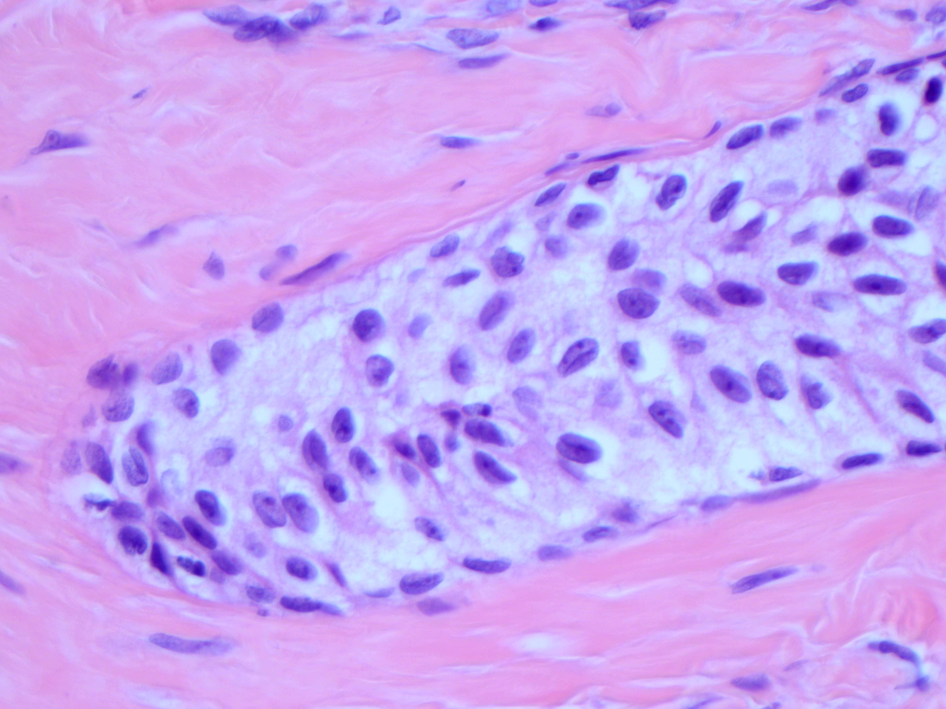
Ovary - lymphatics
Images hosted on other servers:

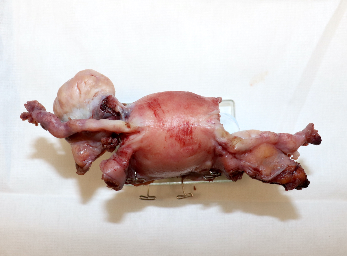
Anatomic relationships of ovary

Ovary - vascular relations

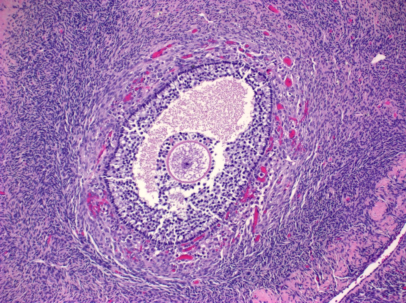
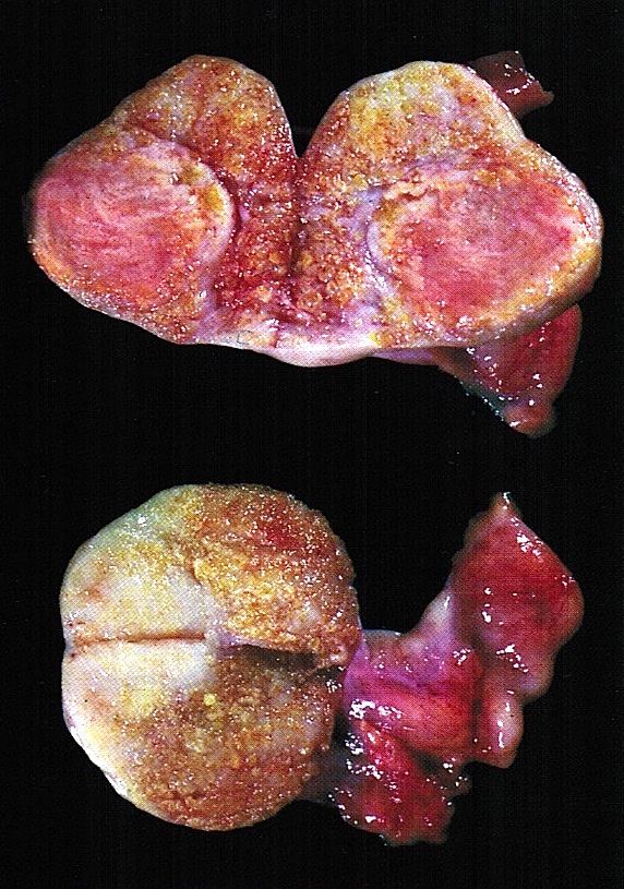
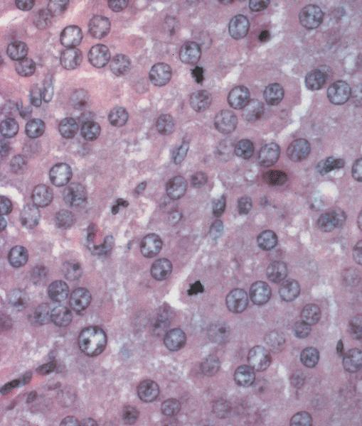
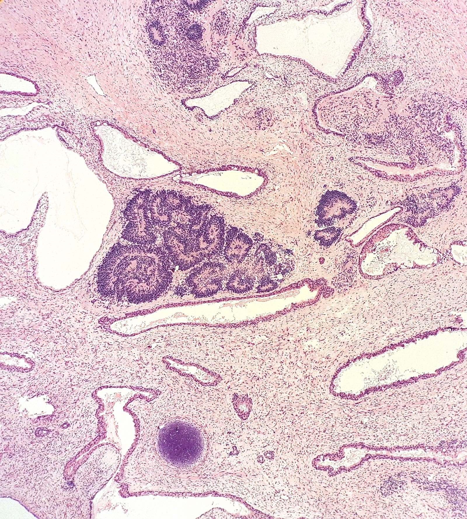
Section of the ovary

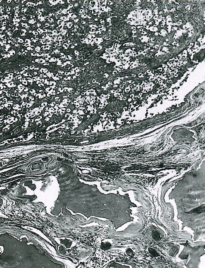
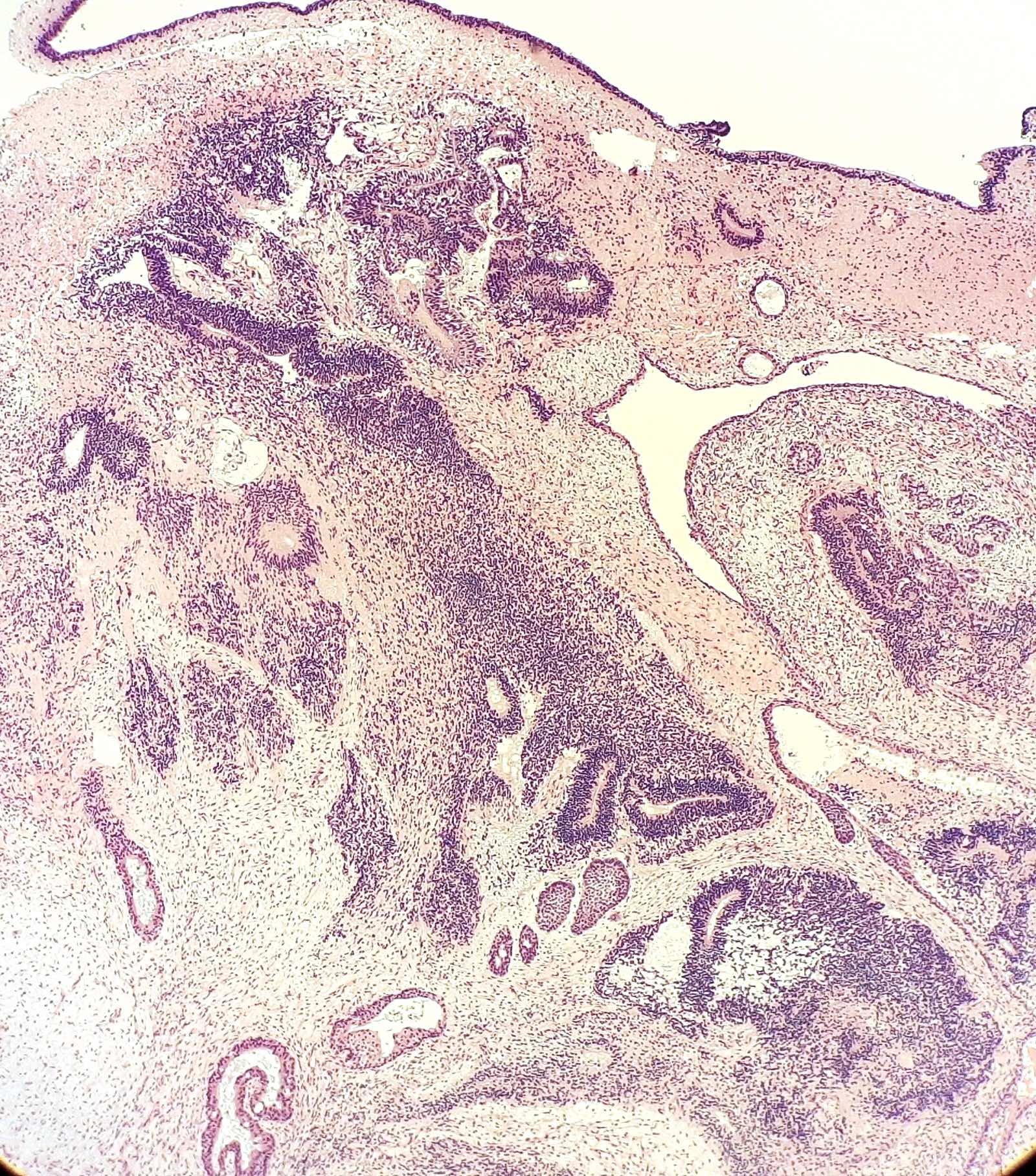
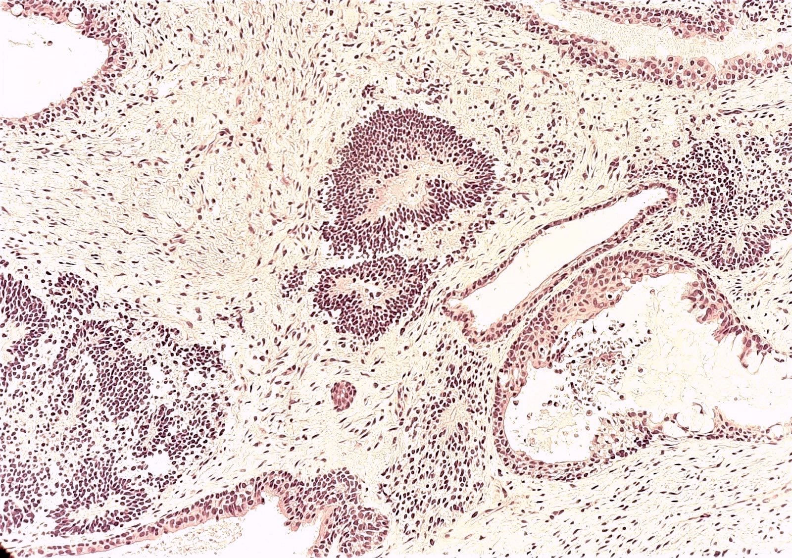
Ovarian stroma

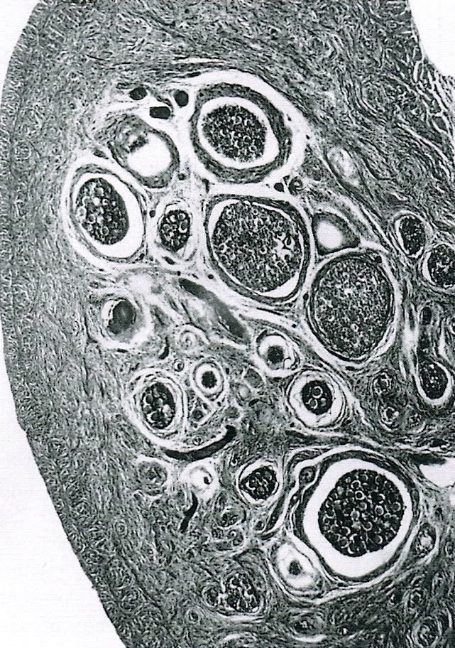
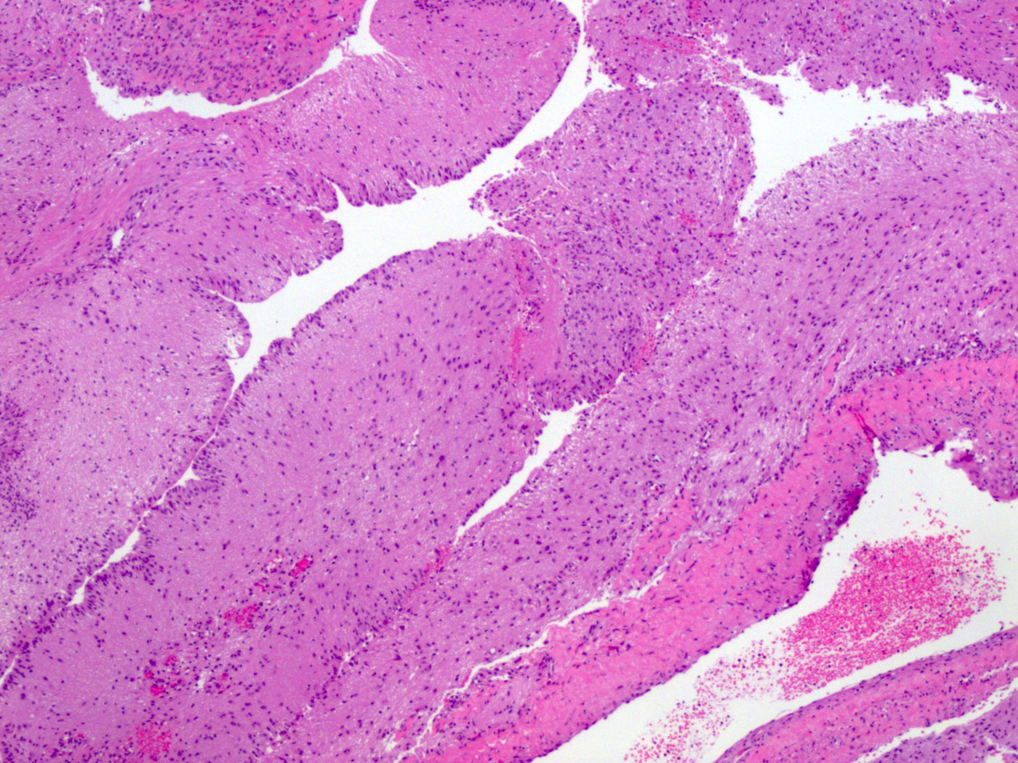
Folliculogenesis
Contributed by Doaa Atwi, M.D.
Ovarian attachment
Images hosted on other servers:


Corpus luteum
Contributed by Doaa Atwi, M.D.
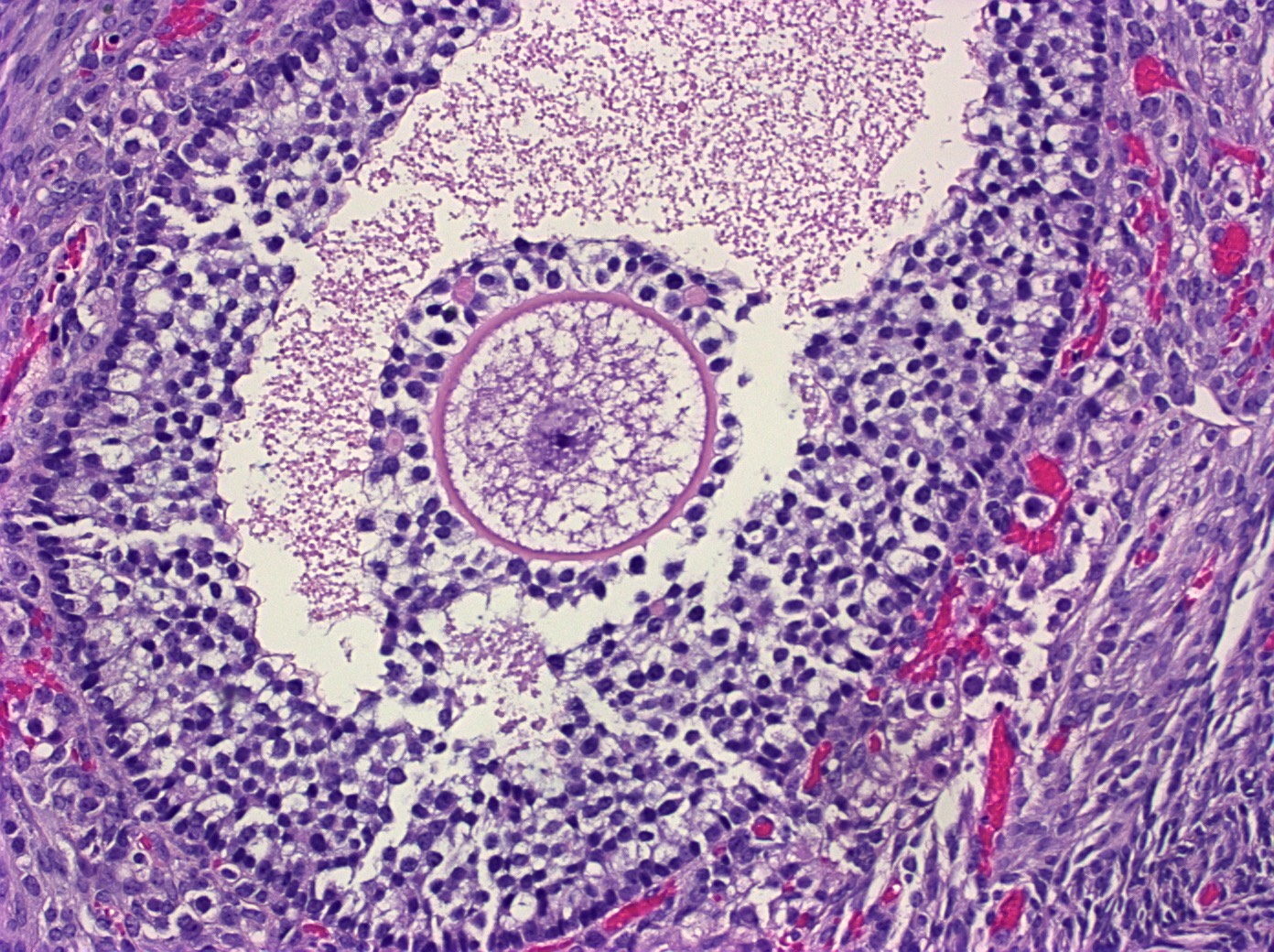
Mature follicle
Corpus luteum
Cortical granuloma
Cortical granuloma
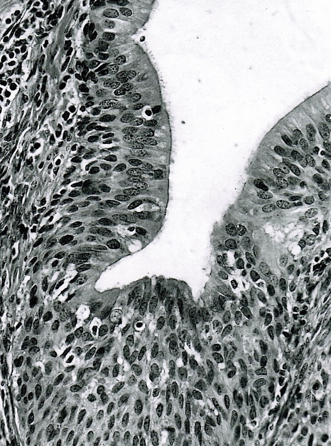
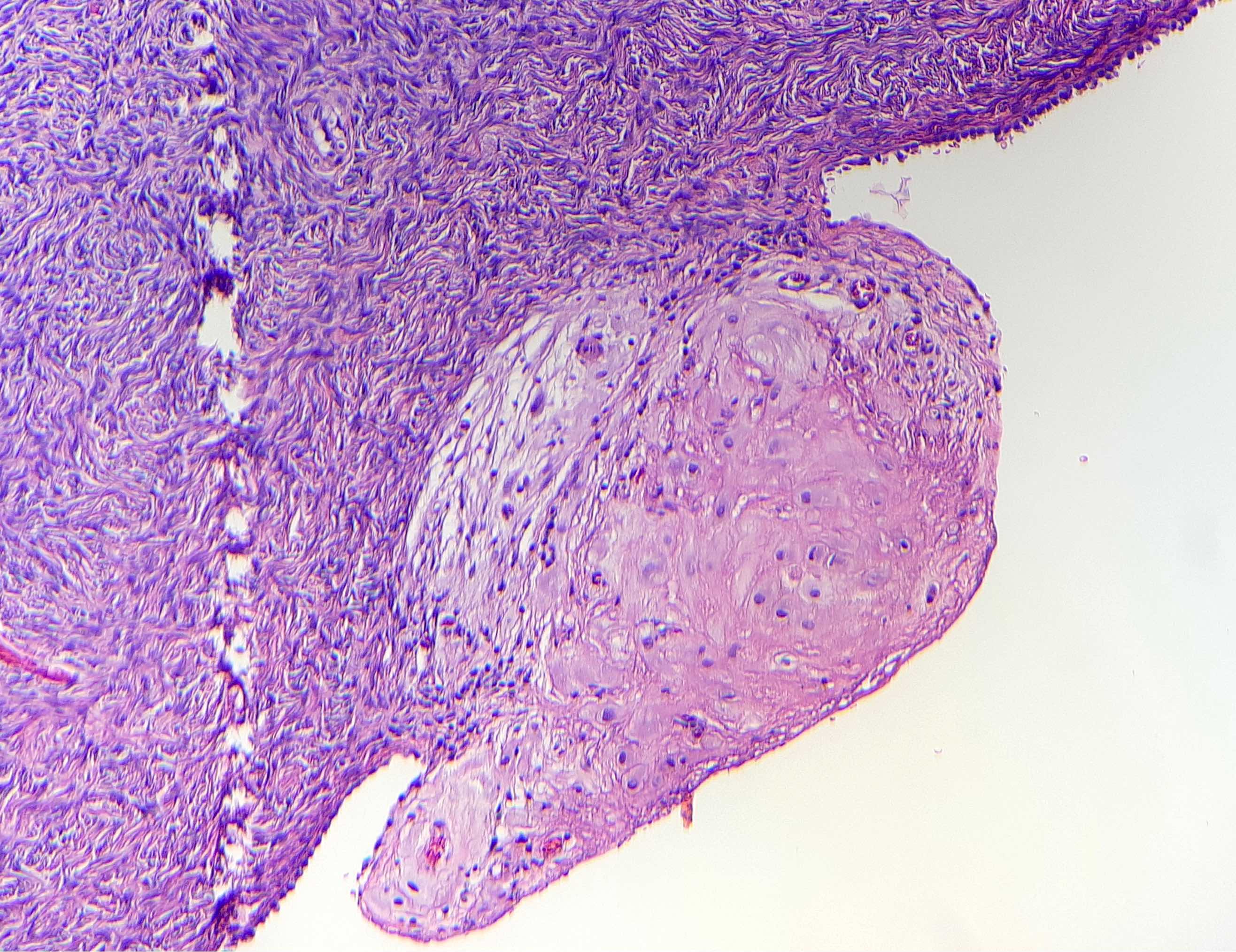
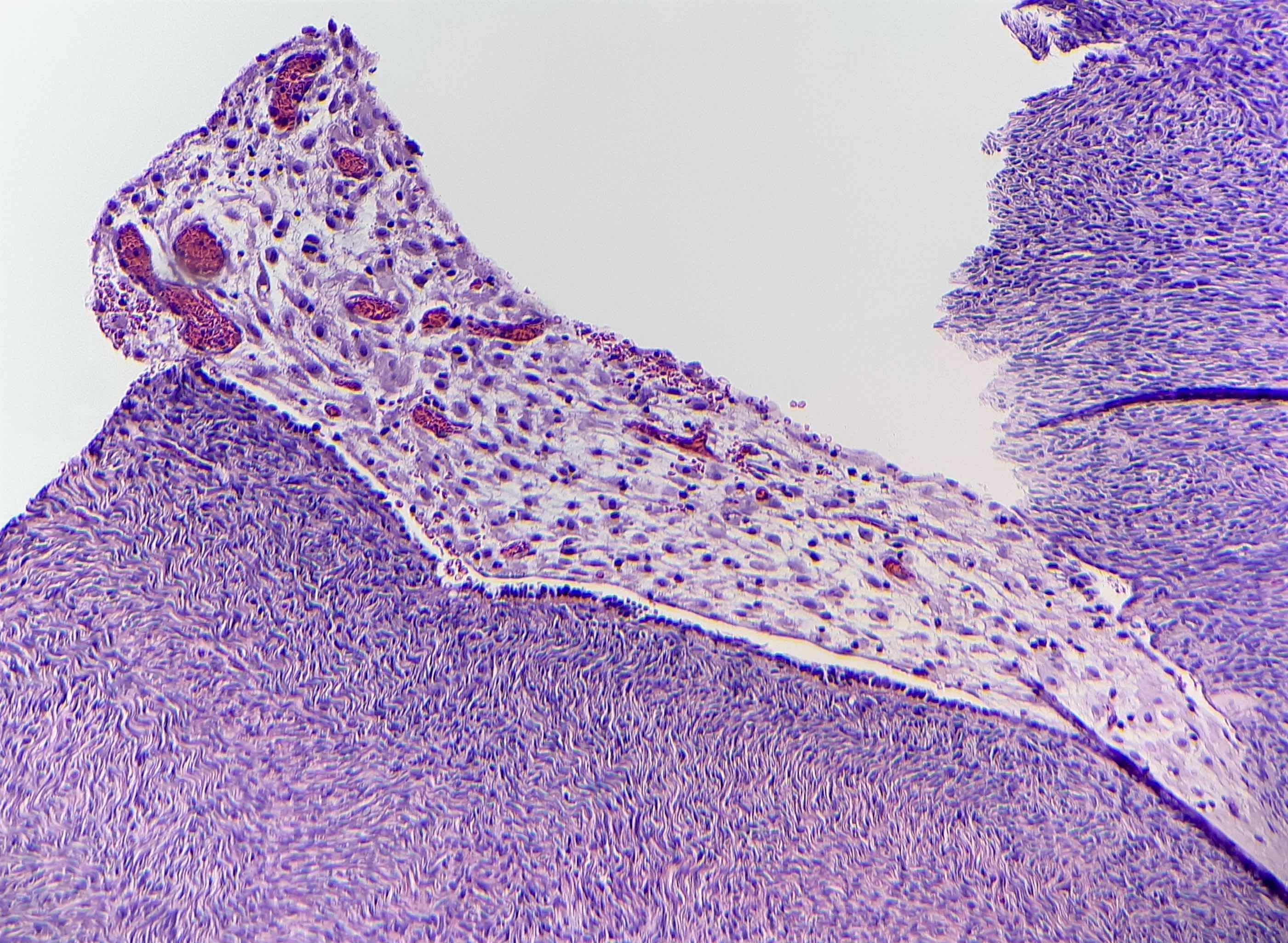
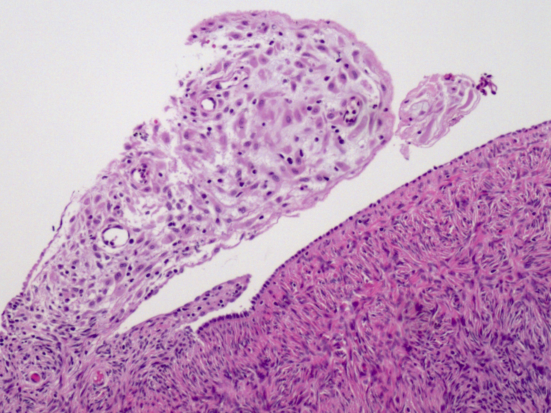
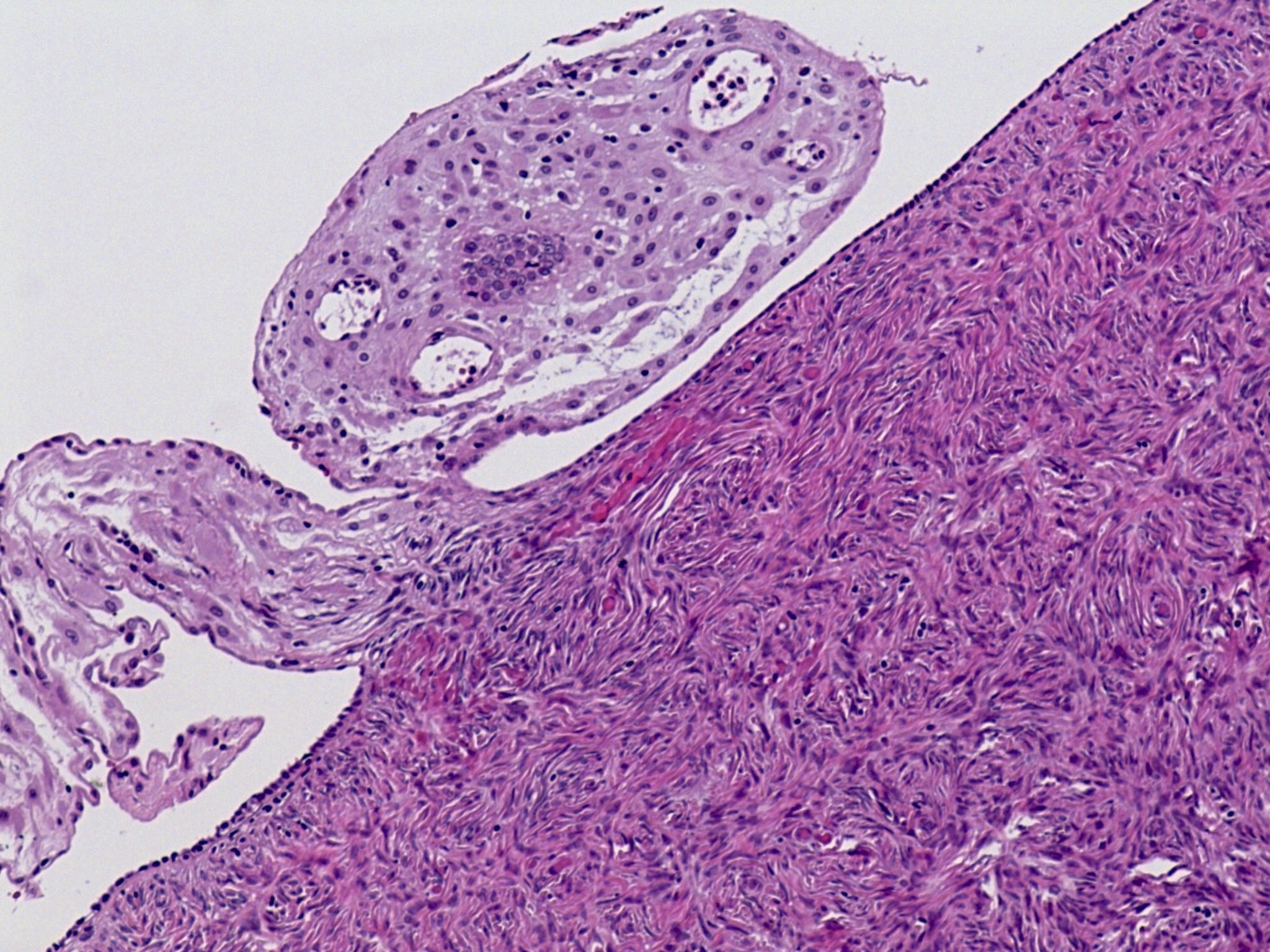
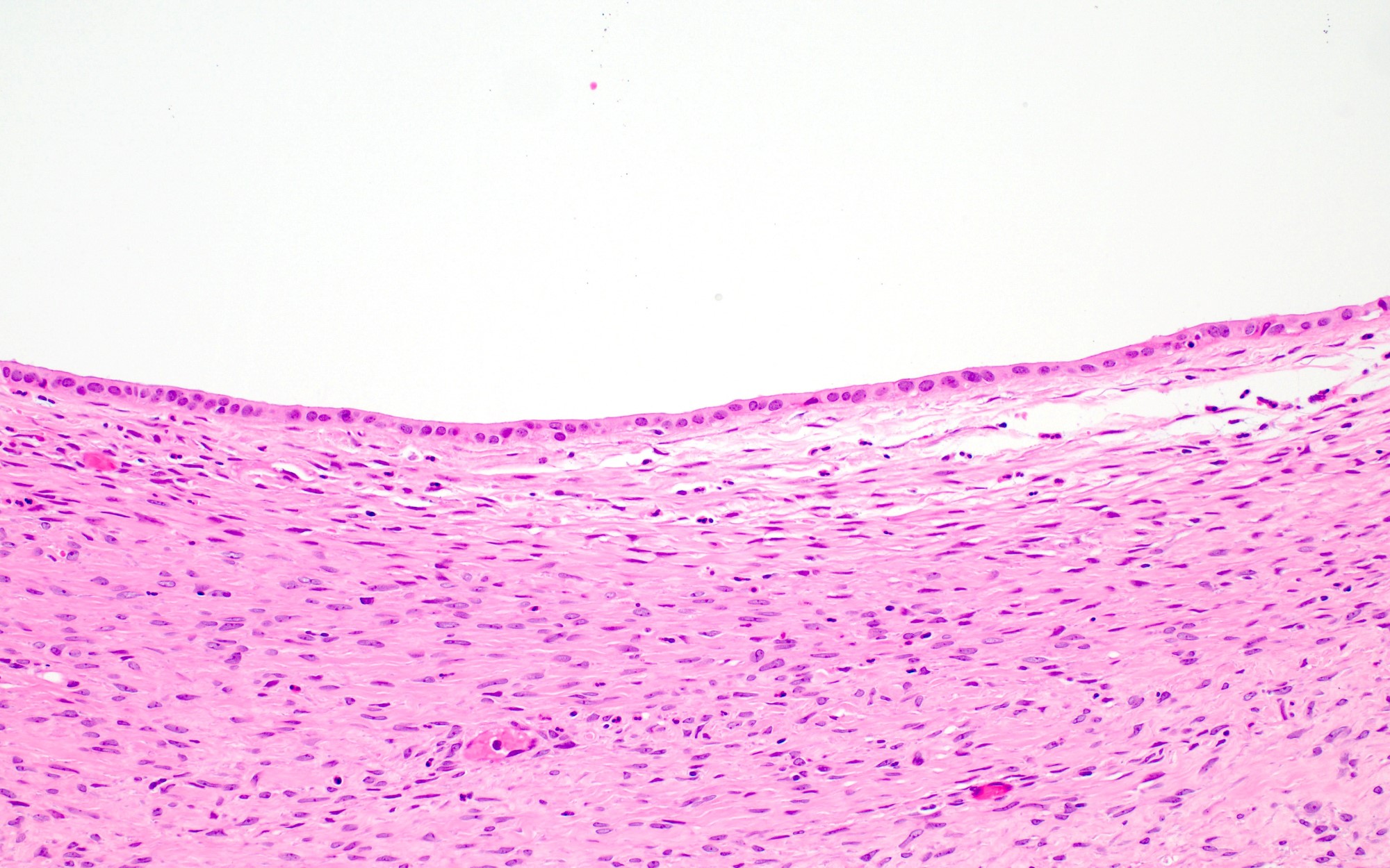
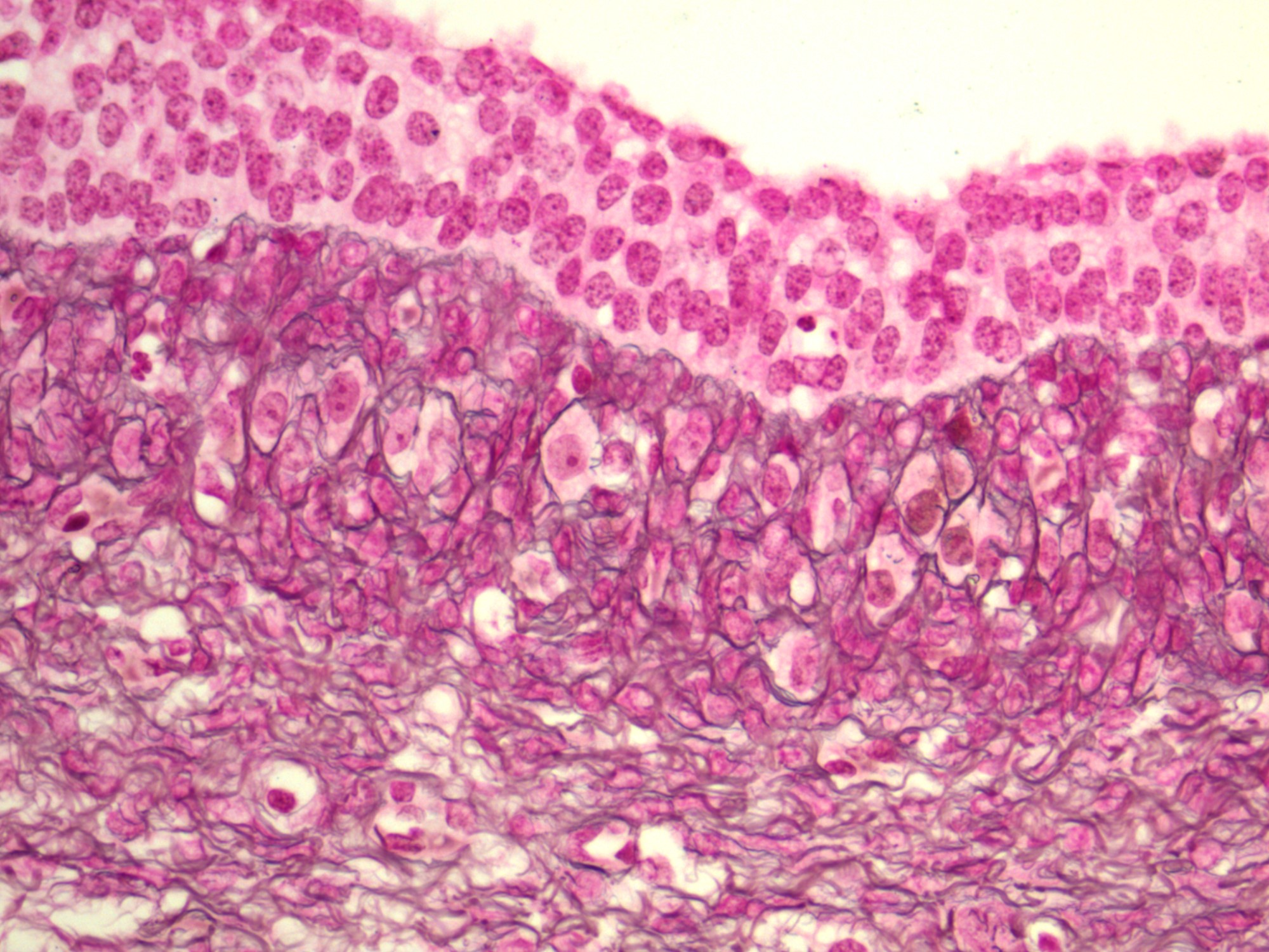
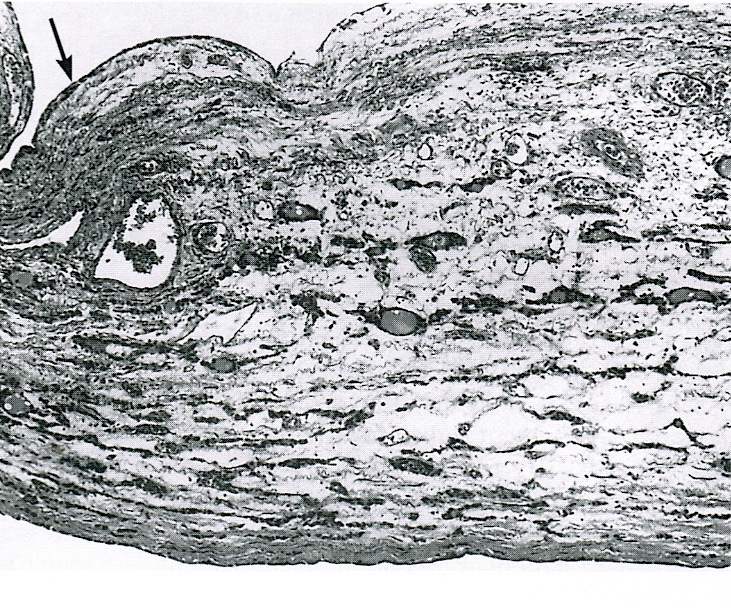
Ovarian surface epithelium
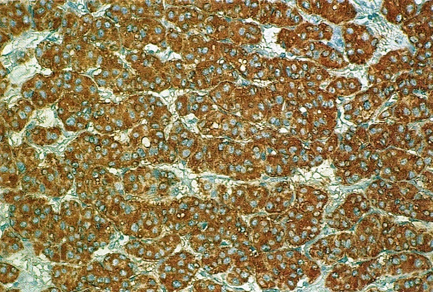
Luteinized stromal cells
CD68
AFIP images
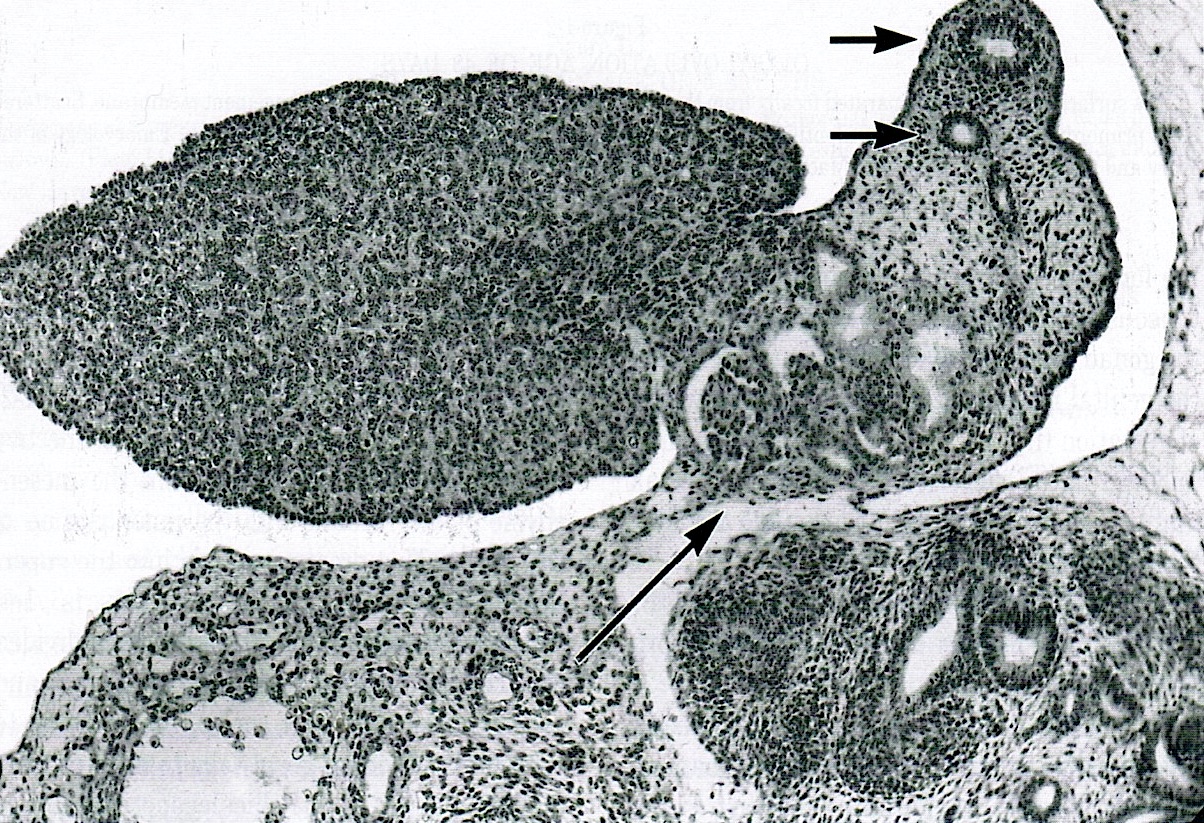
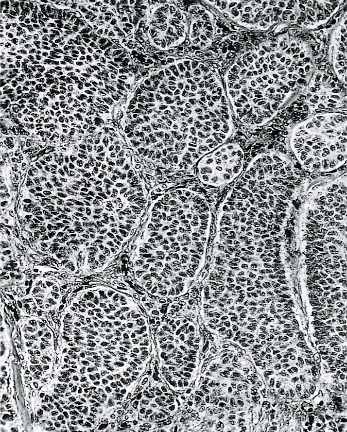
Undifferentiated gonad

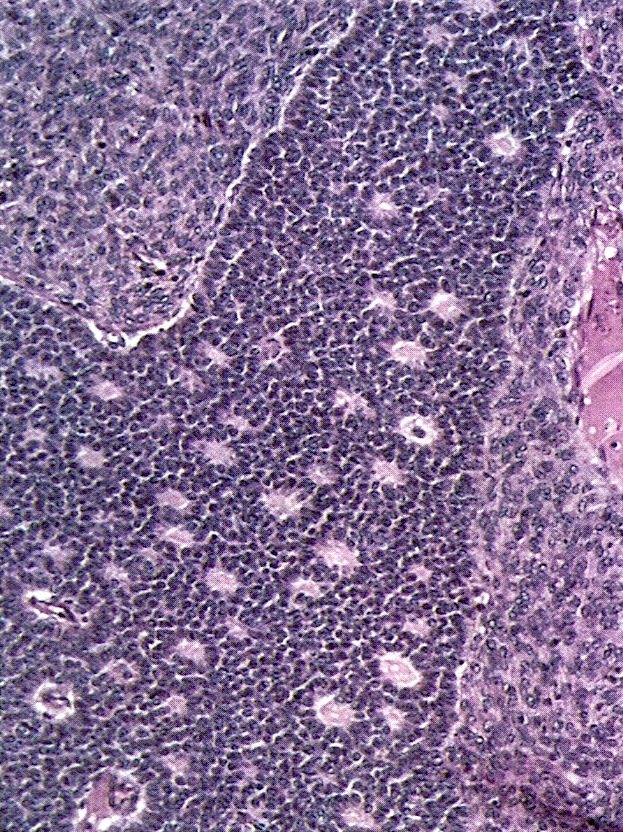
Ovulation age of 48 days


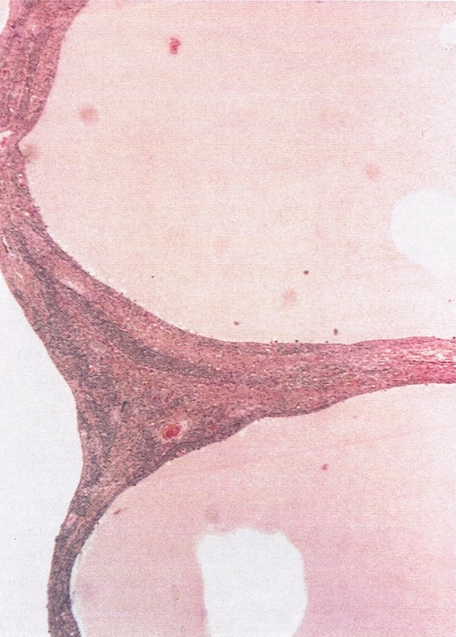
Ovulation age of 5 months
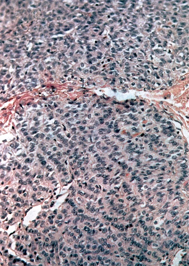
Corpus luteum
Corpus albicans
Contributed by Carlos Parra-Herran, M.D.
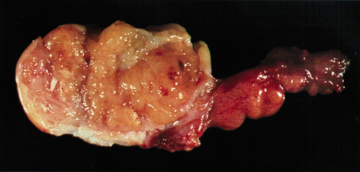
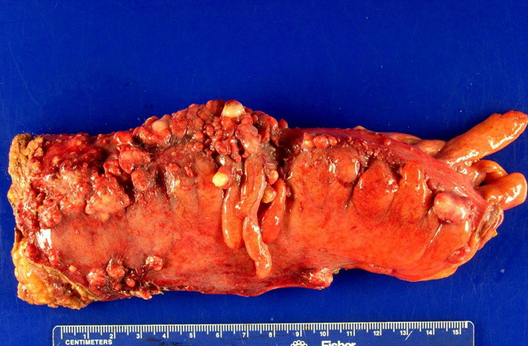
Appendiceal adenocarcinoma
Low grade appendiceal mucinous neoplasm

Low grade appendiceal mucinous neoplasm
Contributed by Jutta Huvila, M.D., Ph.D. and C. Blake Gilks, M.D. and AFIP images
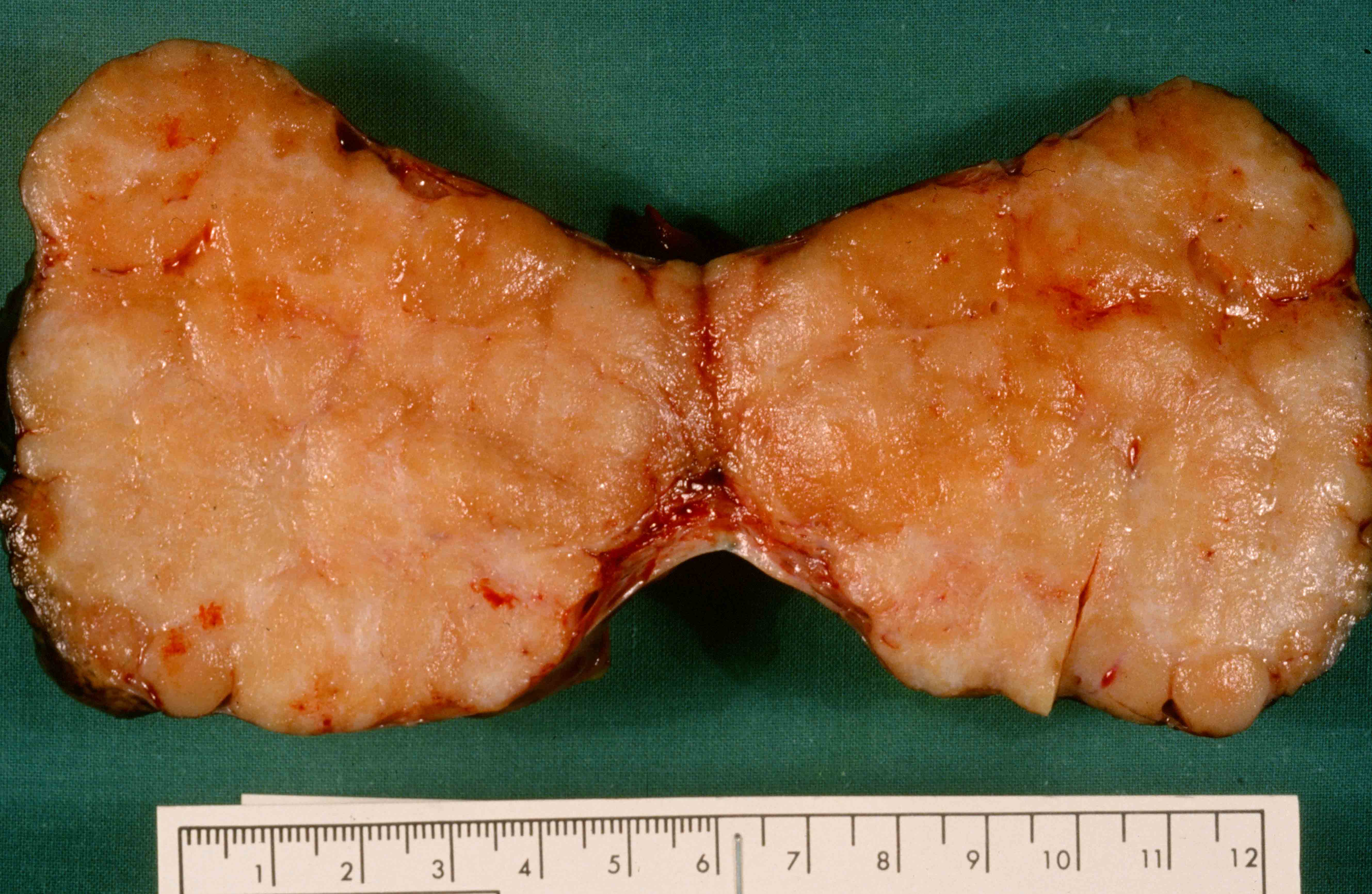
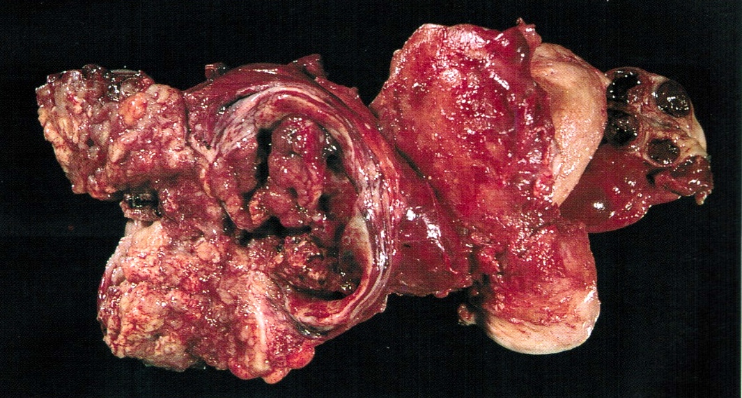
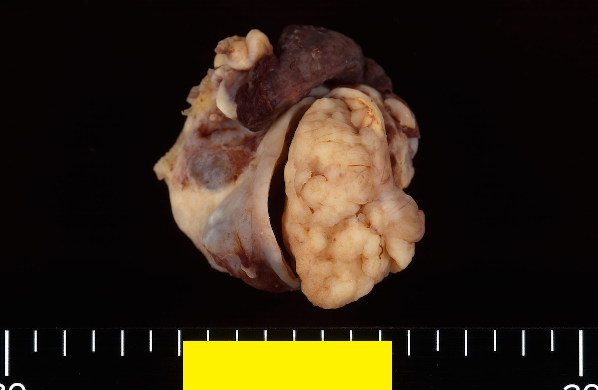
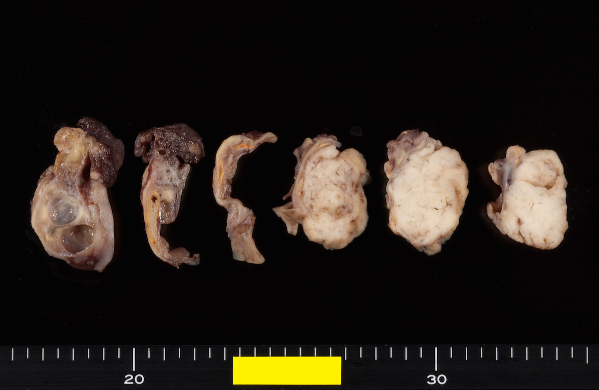
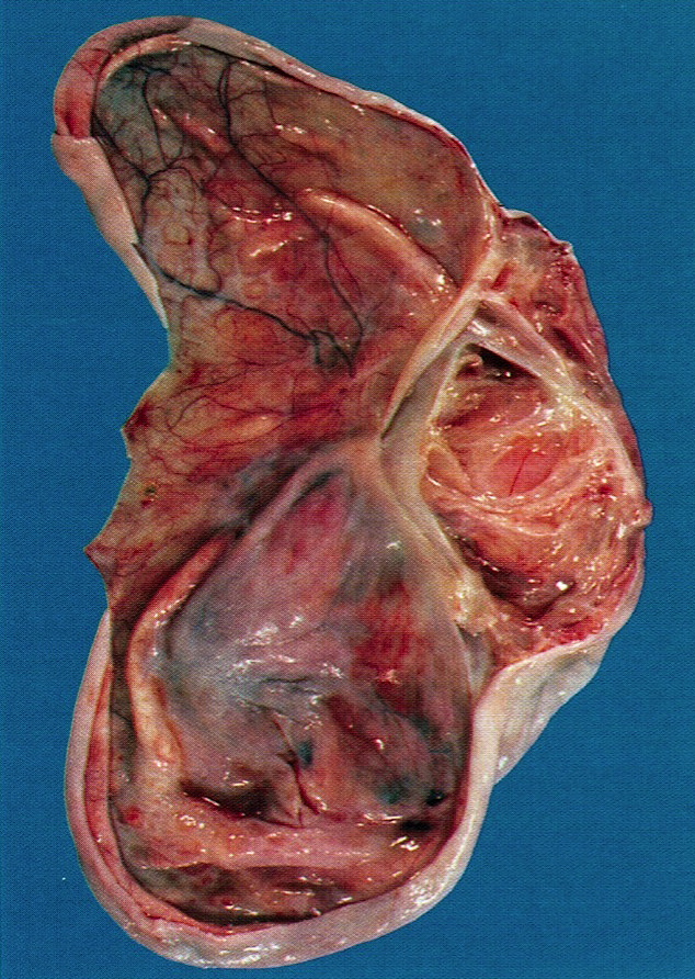
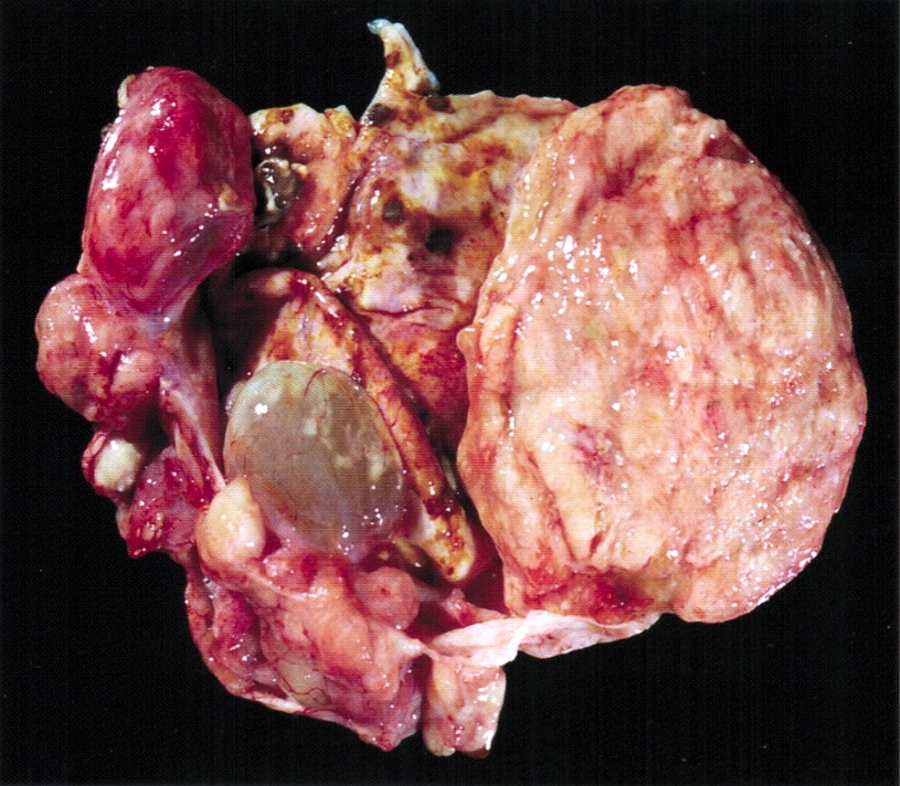
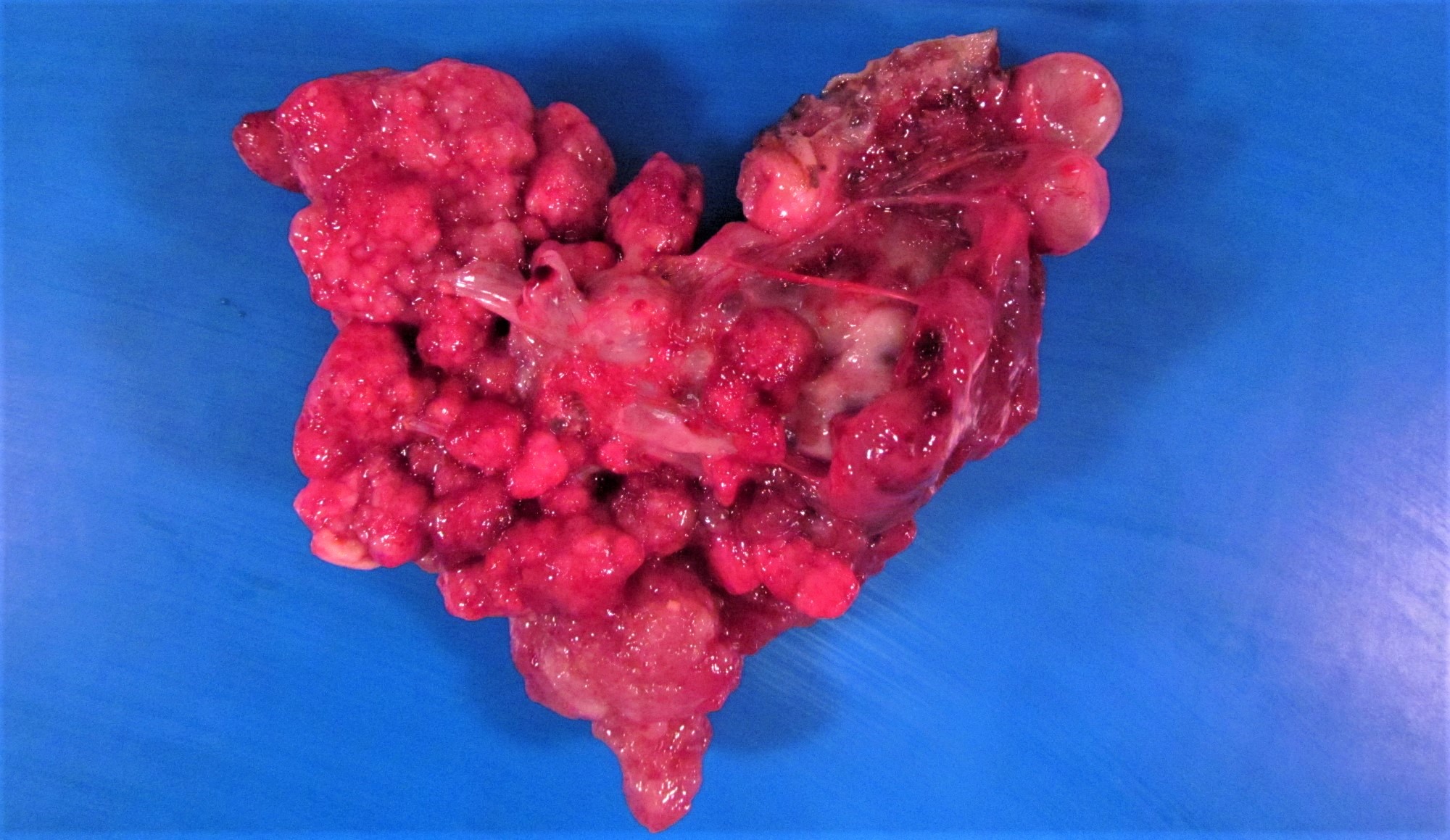
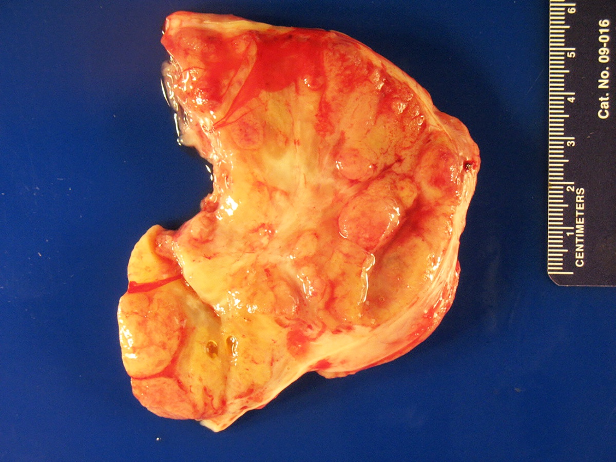
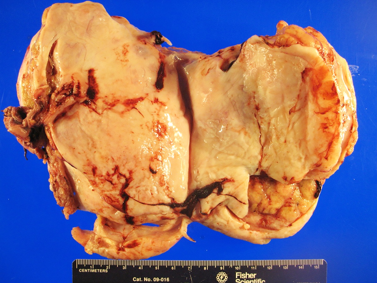
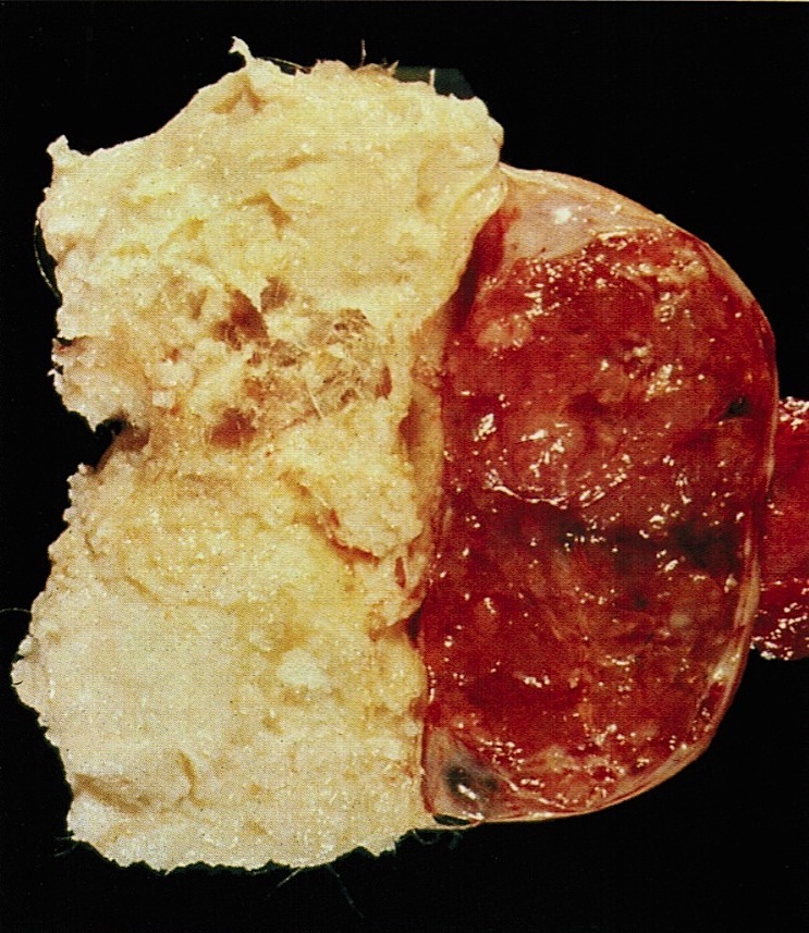
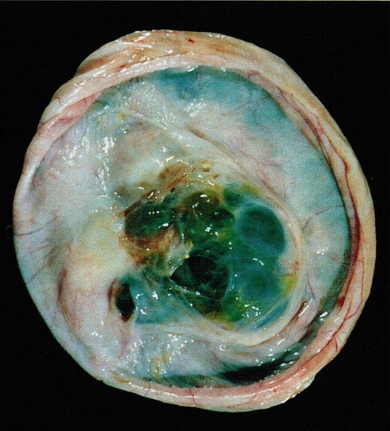
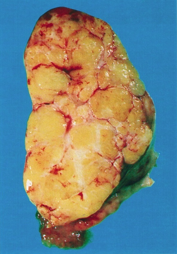
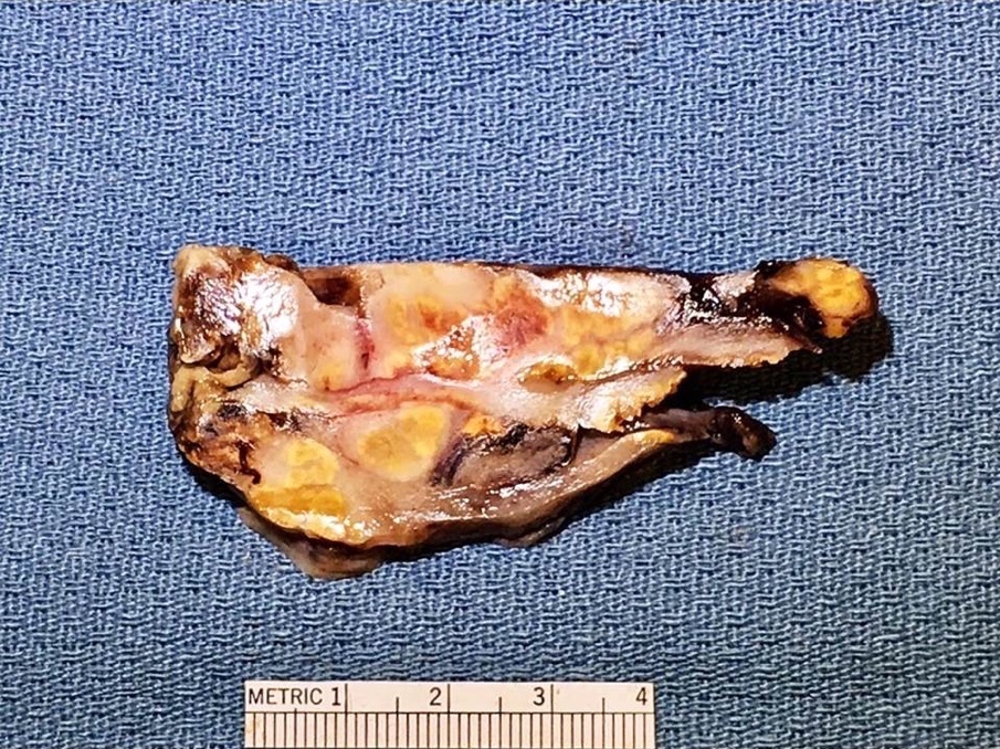
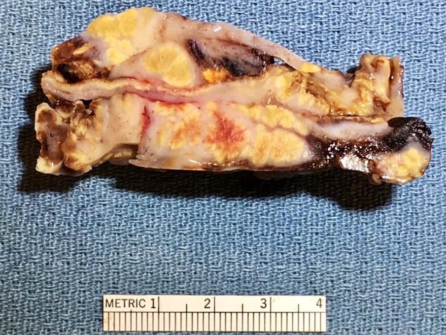
Solid, uniform cut surface

Fleshy appearance
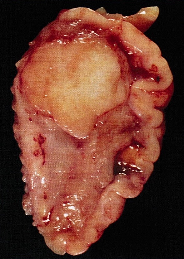
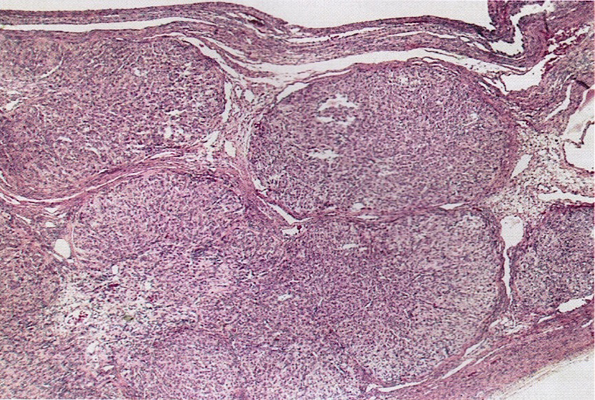
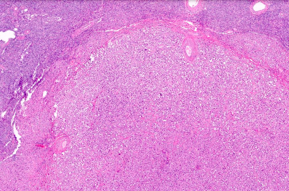
Sharply demarcated
Fibroma-like appearance

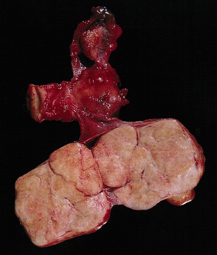
Solid fibroma-like component and 2 cysts
Images hosted on other servers:

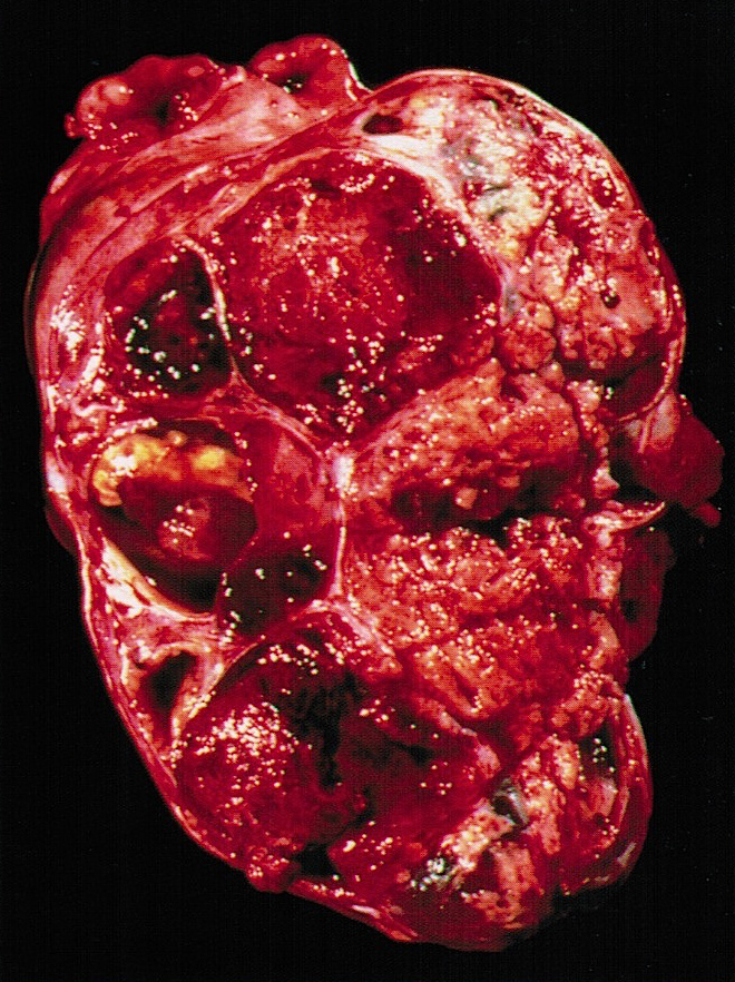
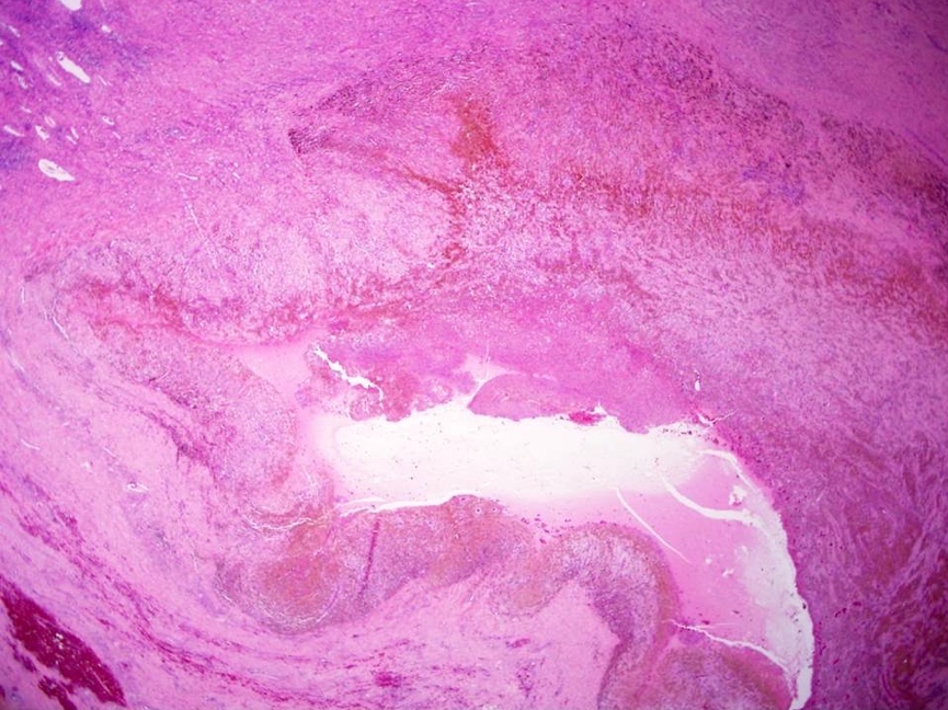
Infarcted tumor
Contributed by Jutta Huvila, M.D., Ph.D. and C. Blake Gilks, M.D.
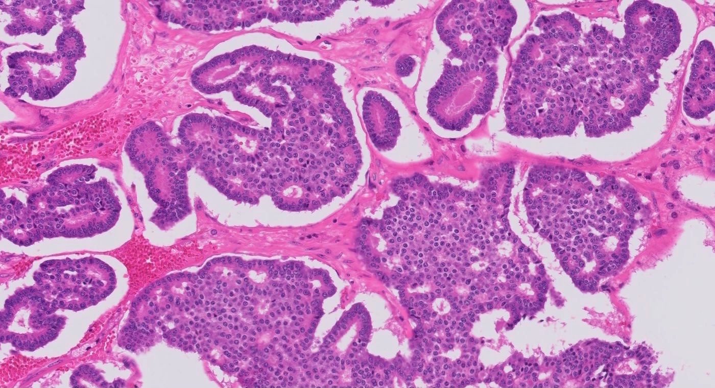
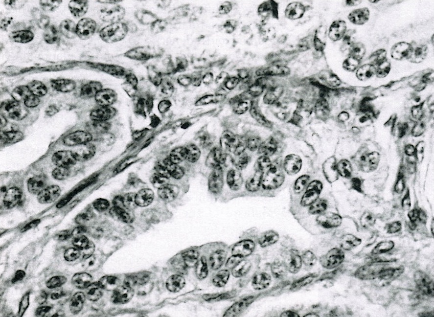
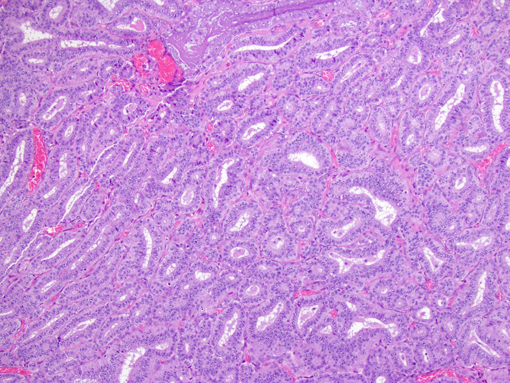
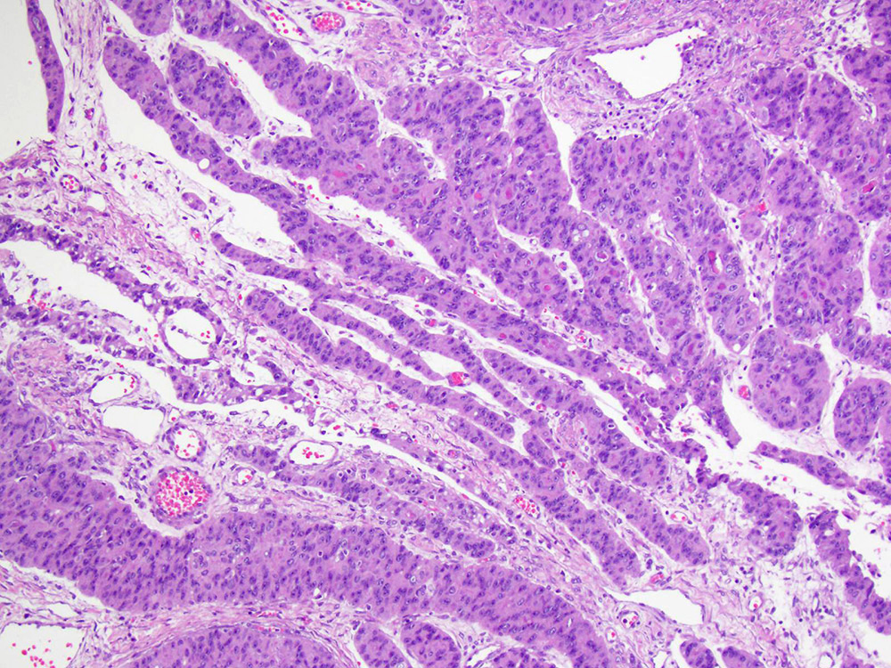
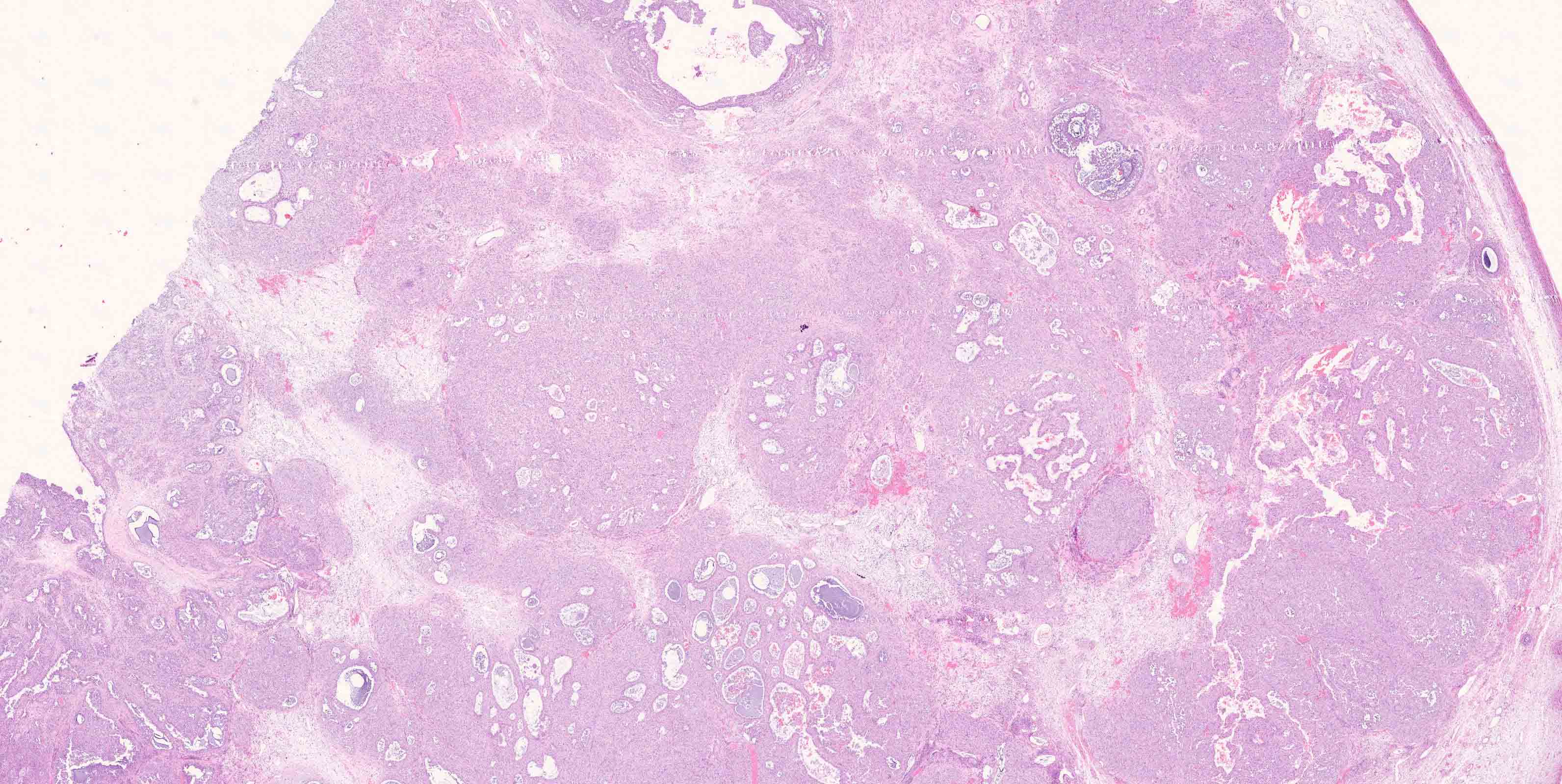
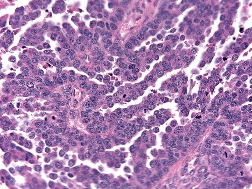
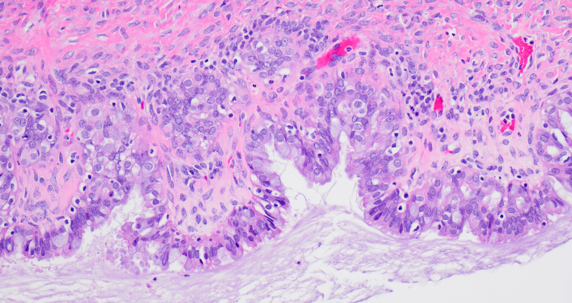
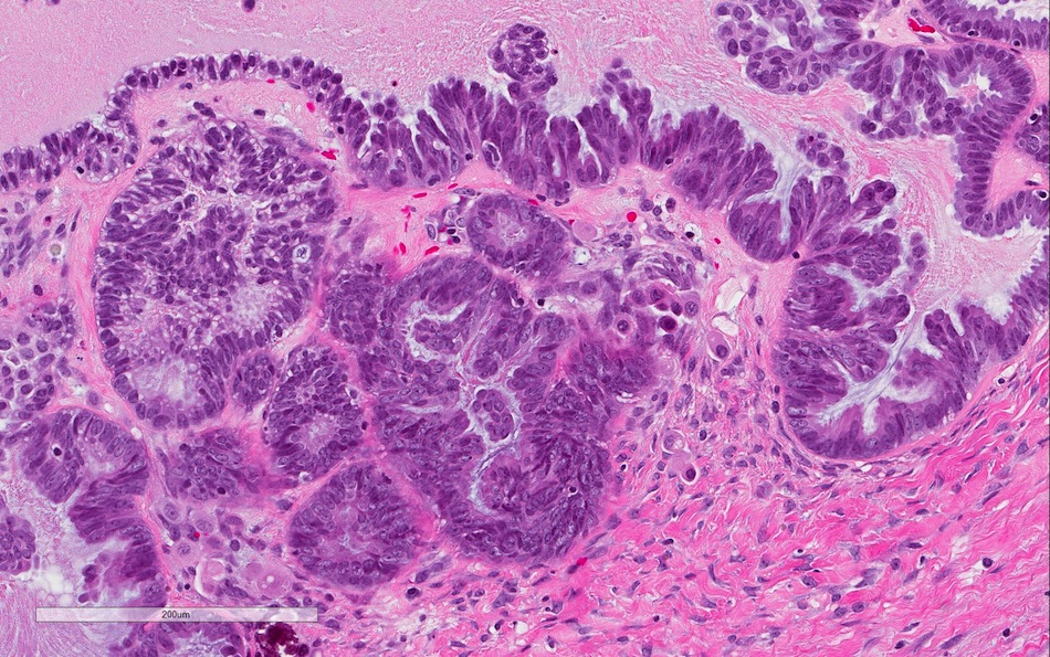
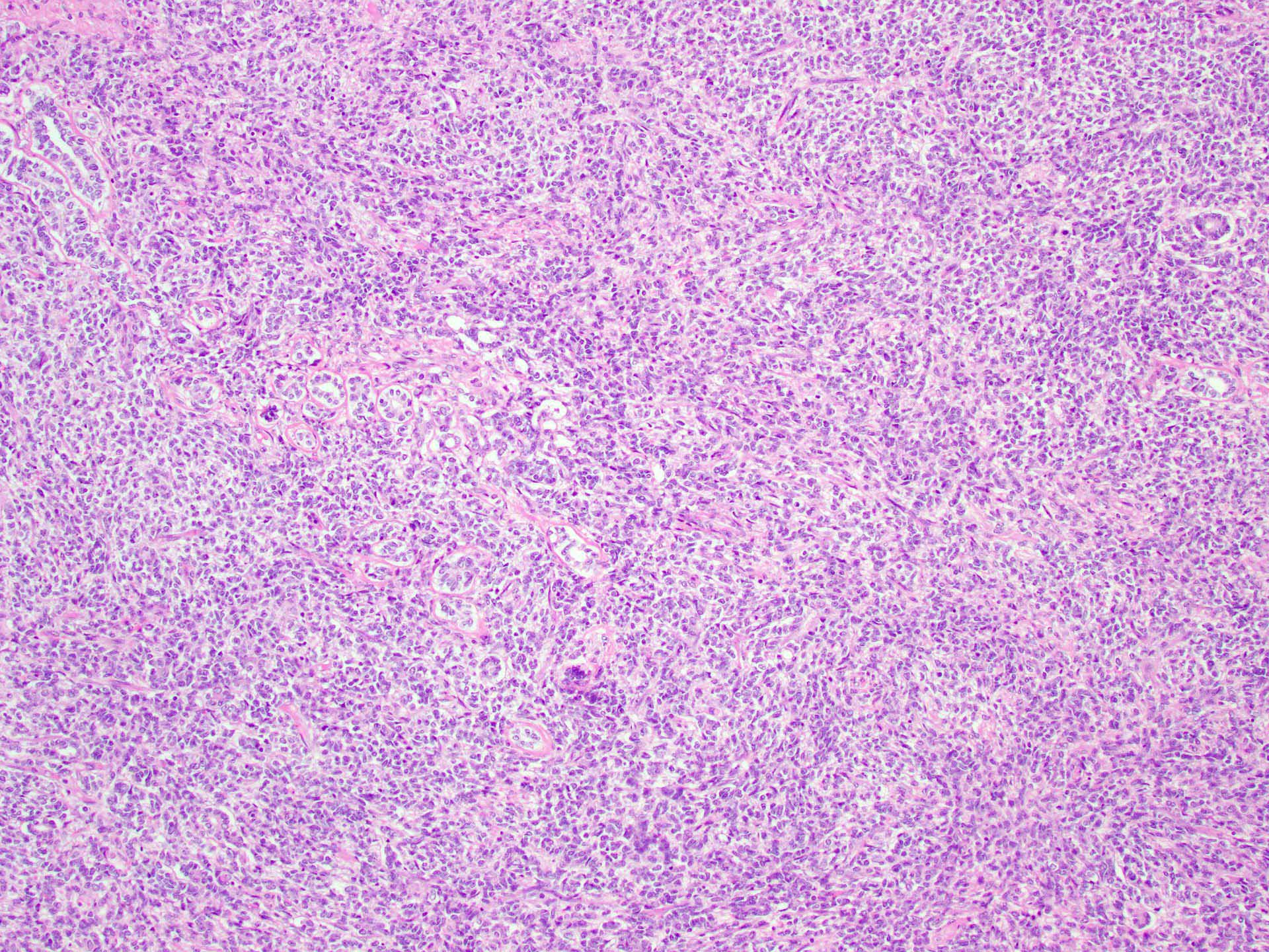
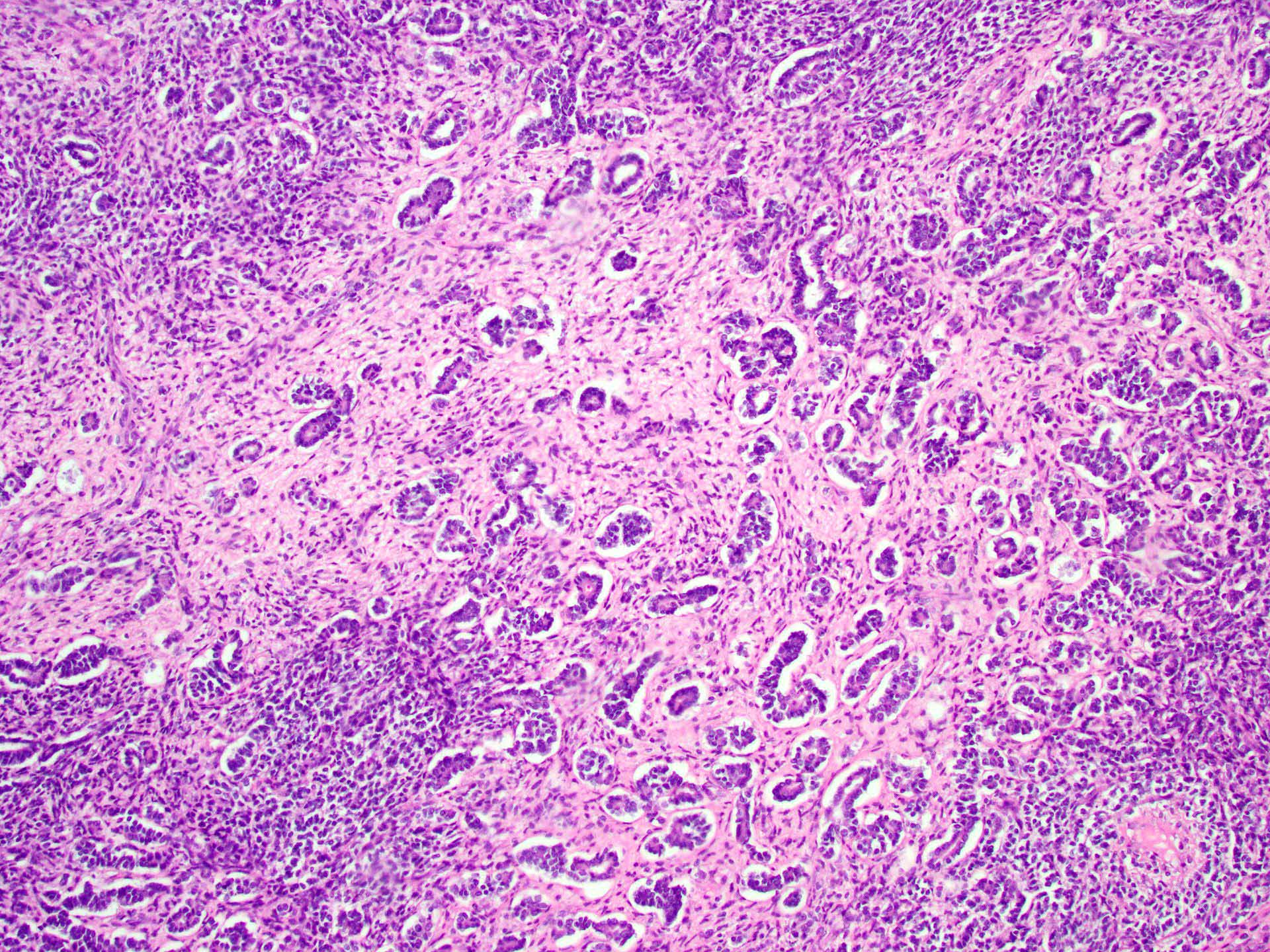
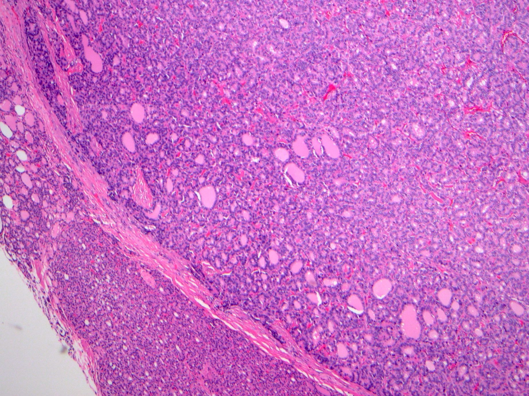
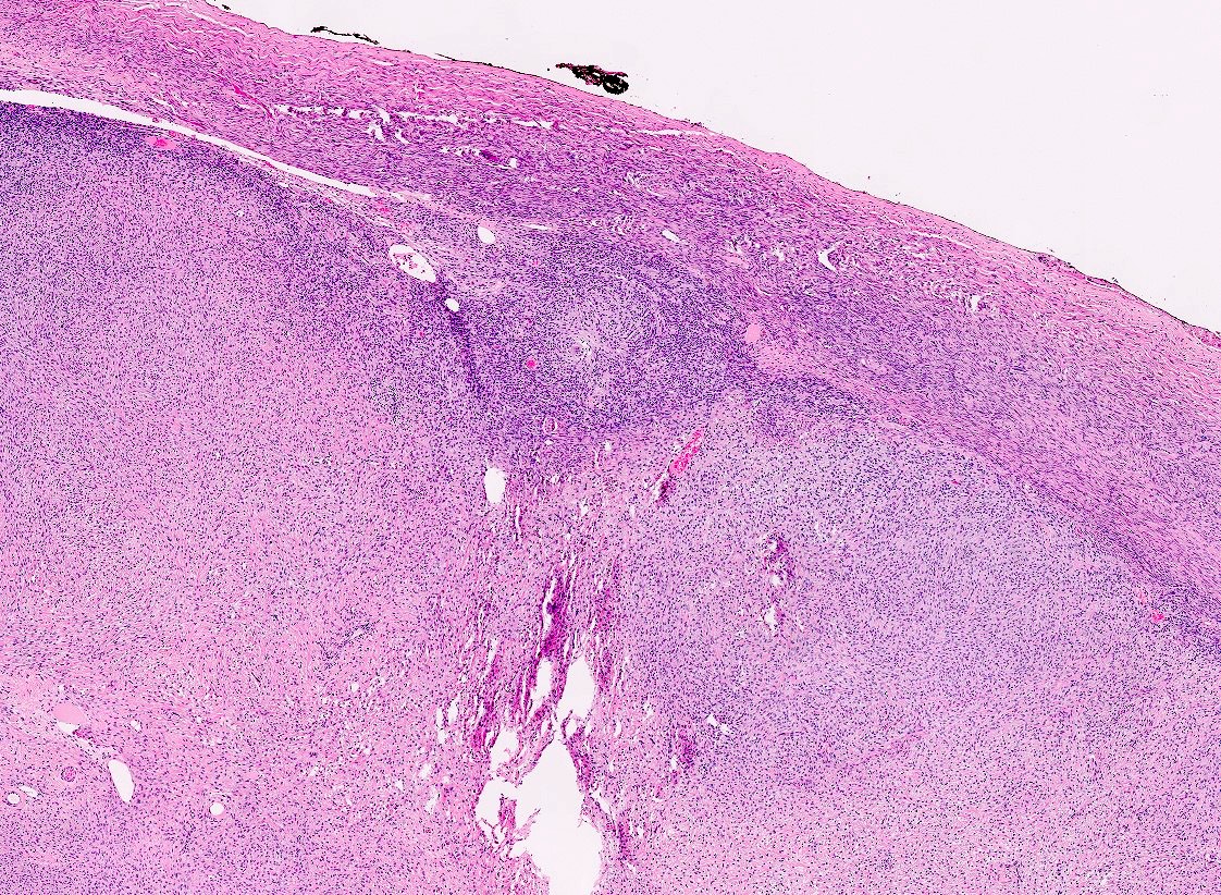
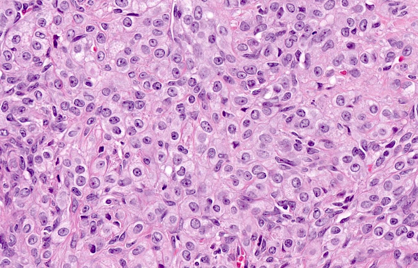
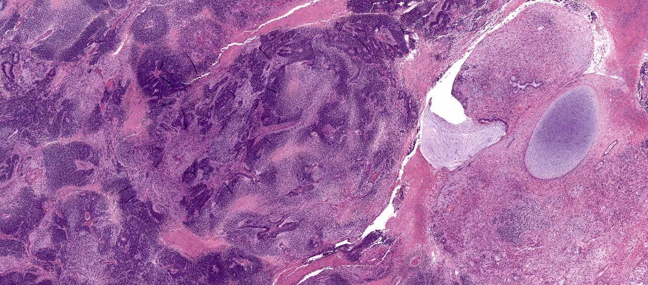
Benign transitional nests
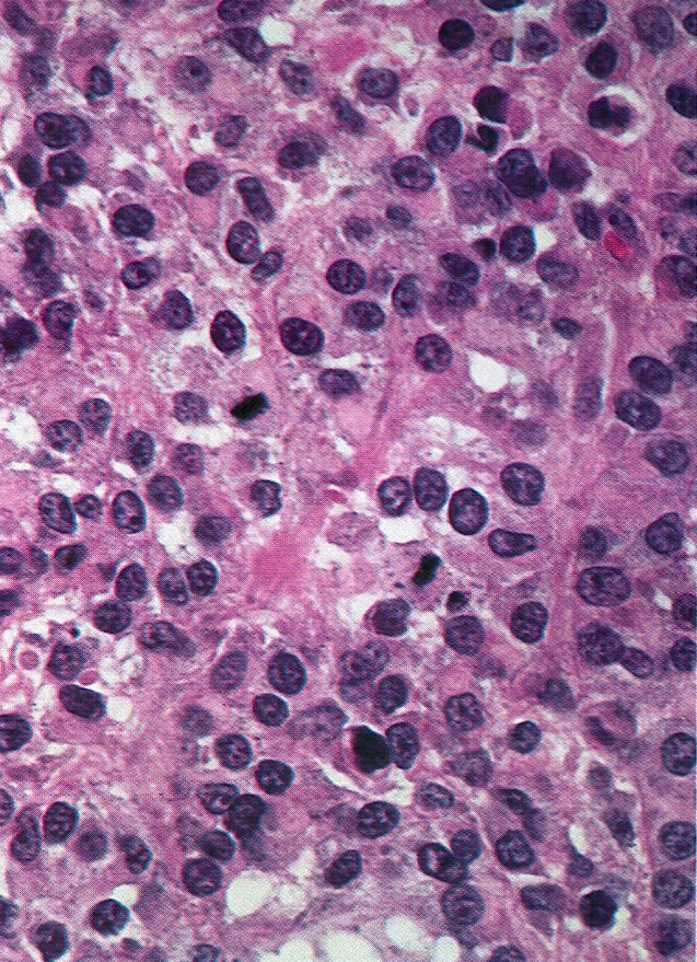
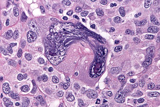
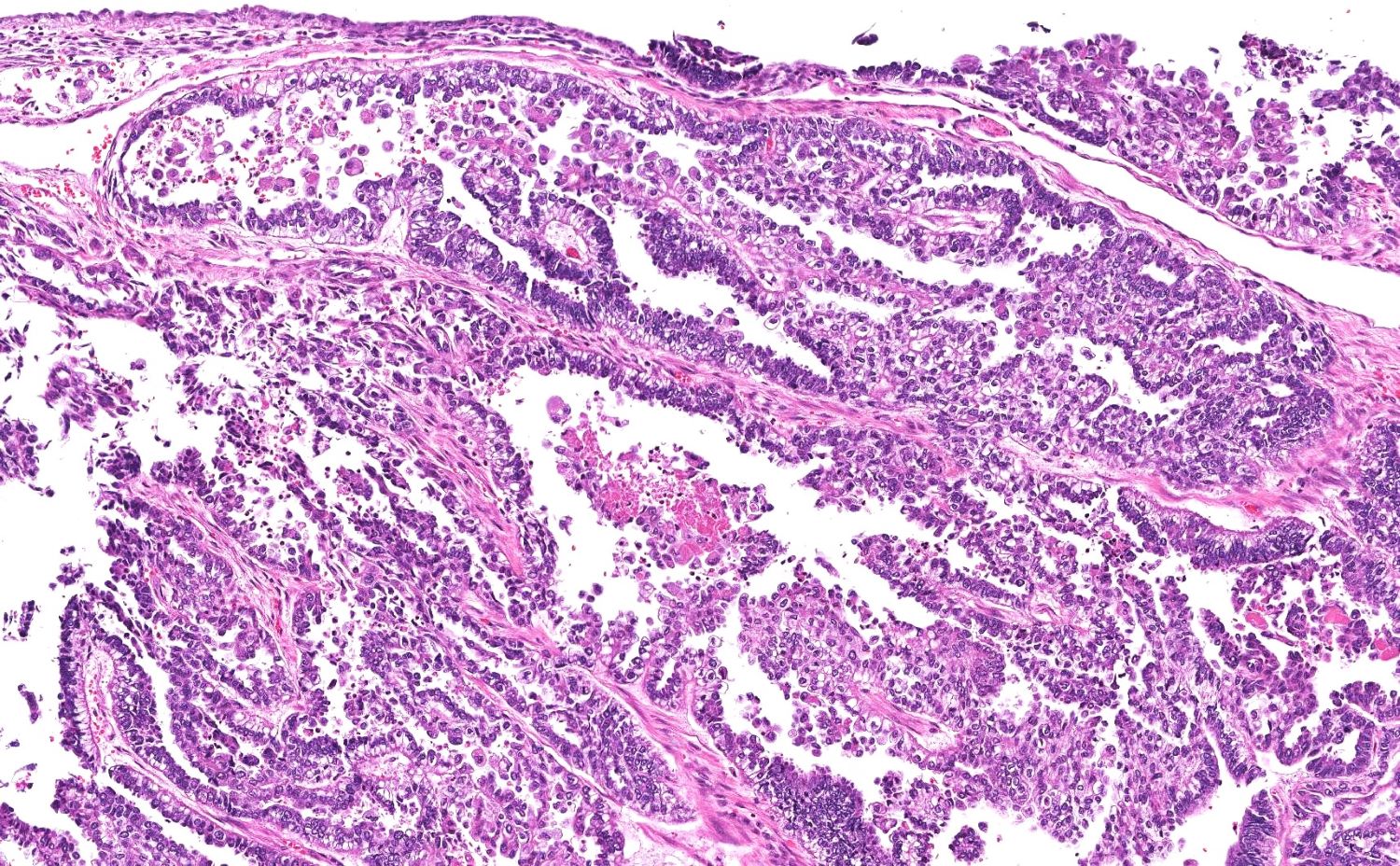
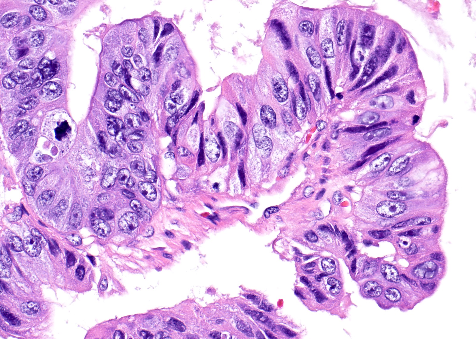
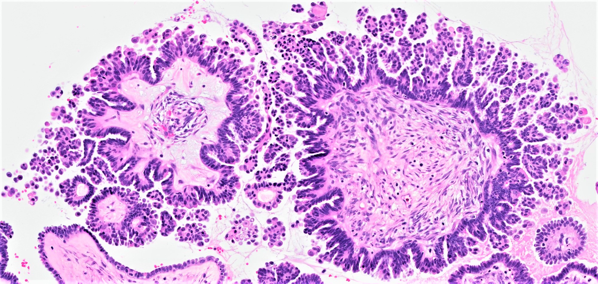
Nuclear grooves
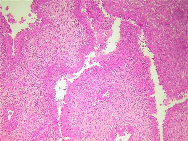
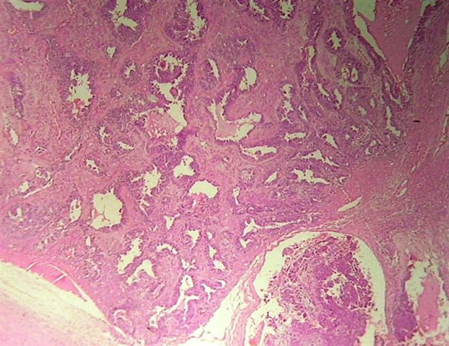
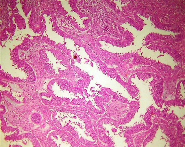
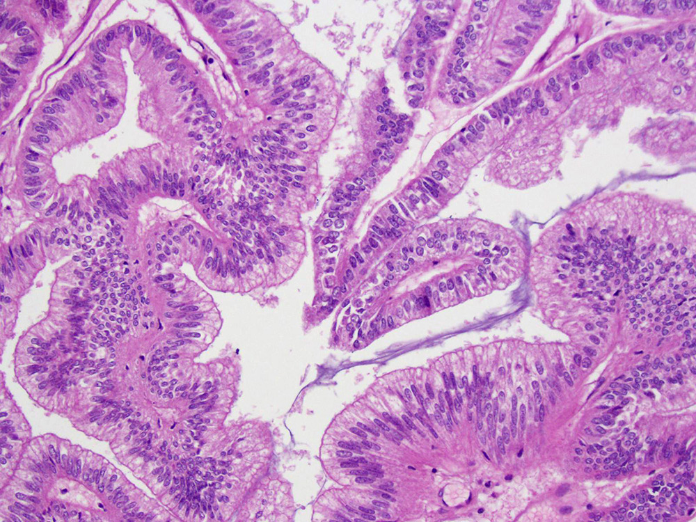
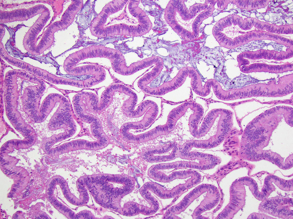
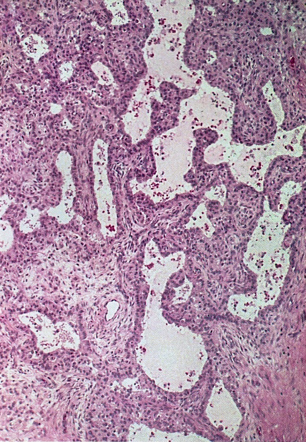
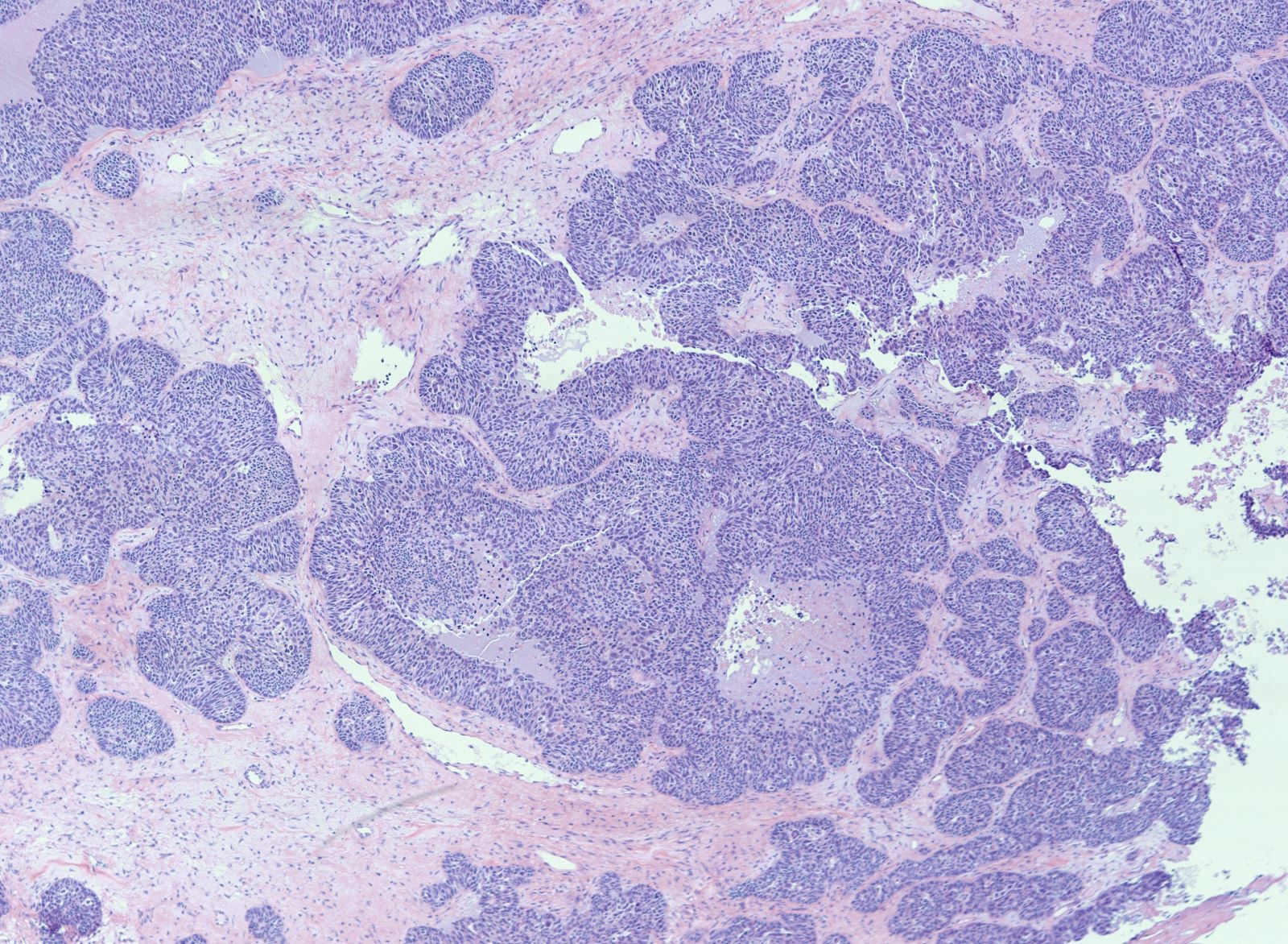
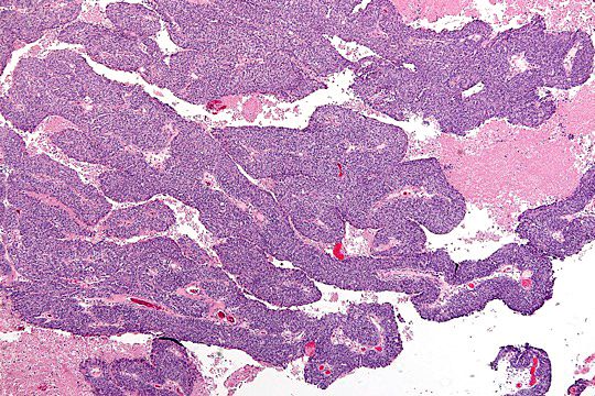
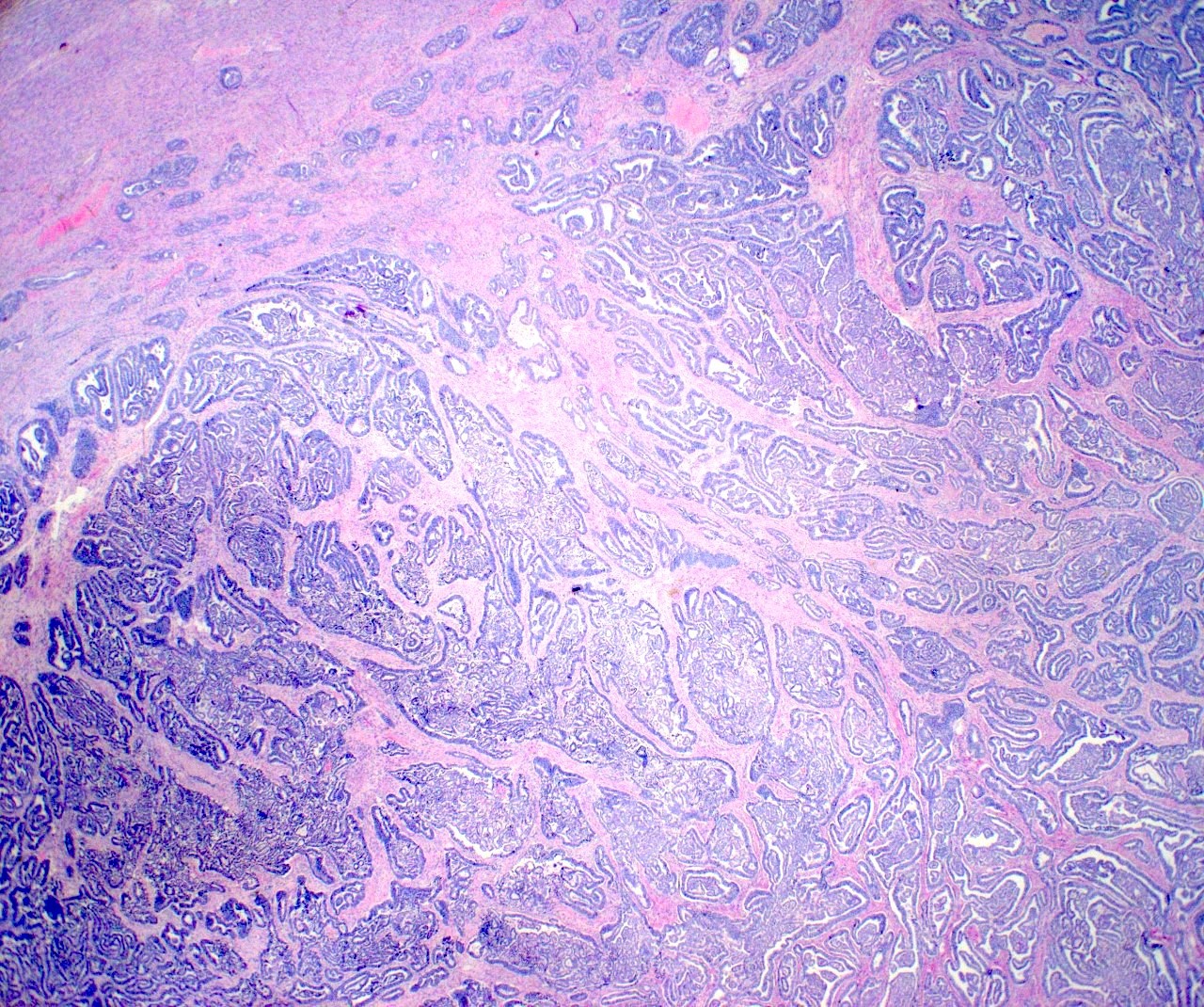
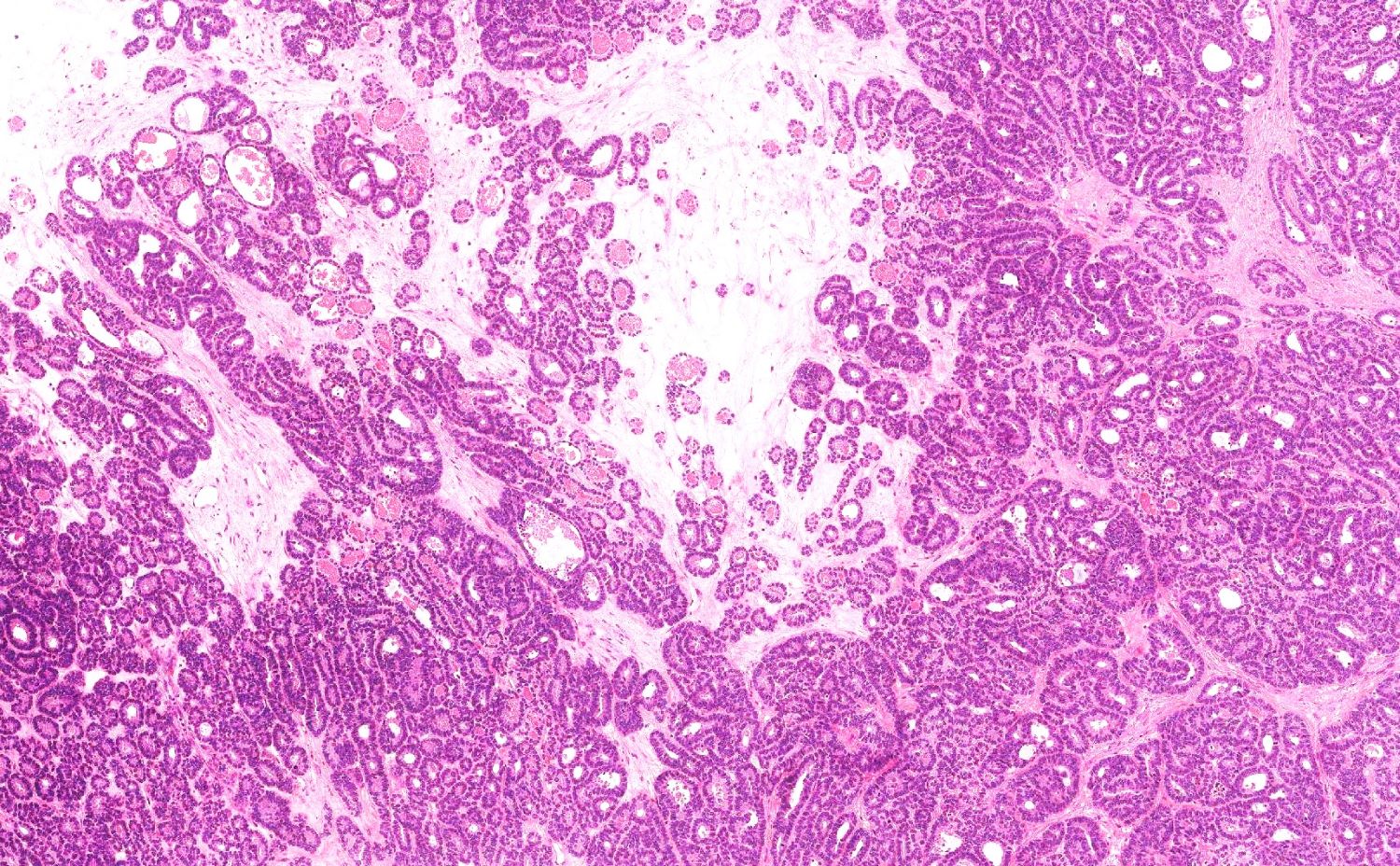
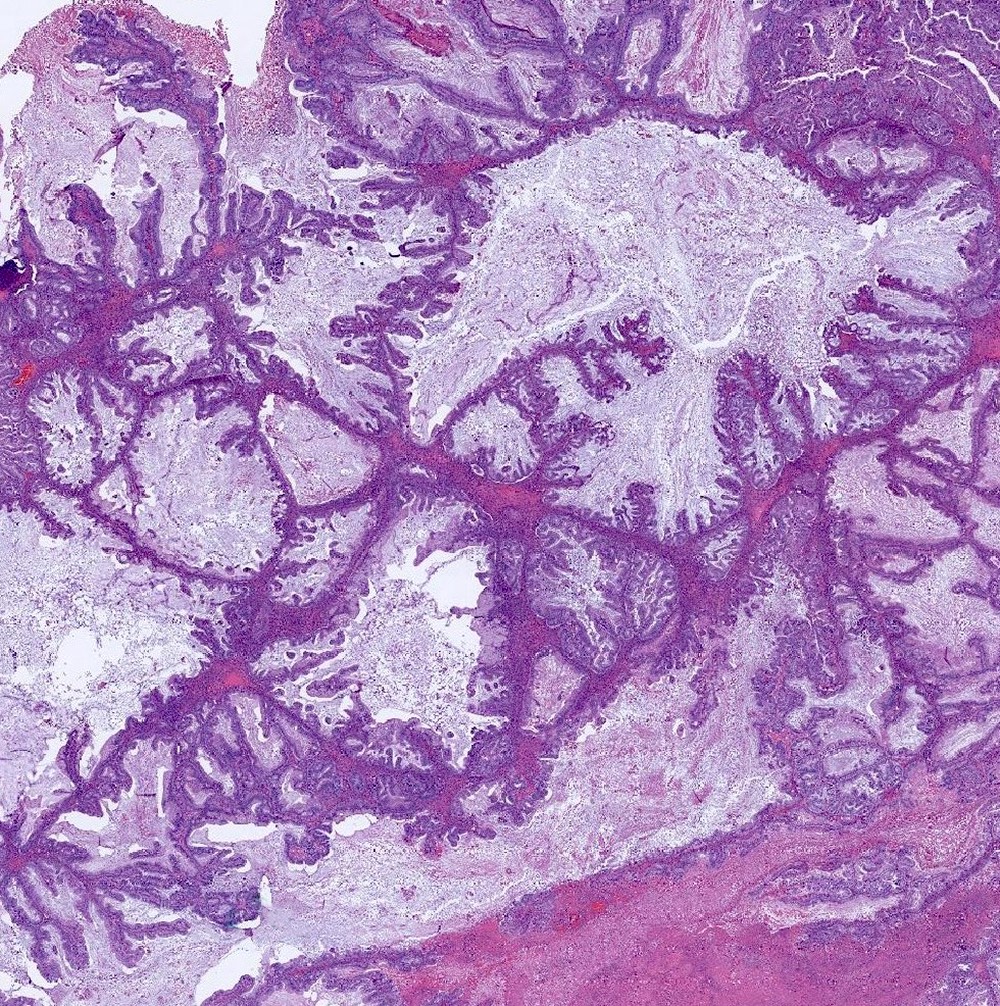
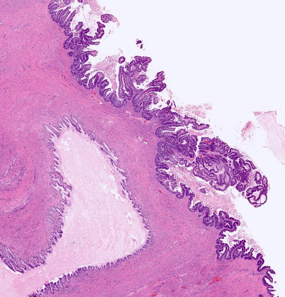
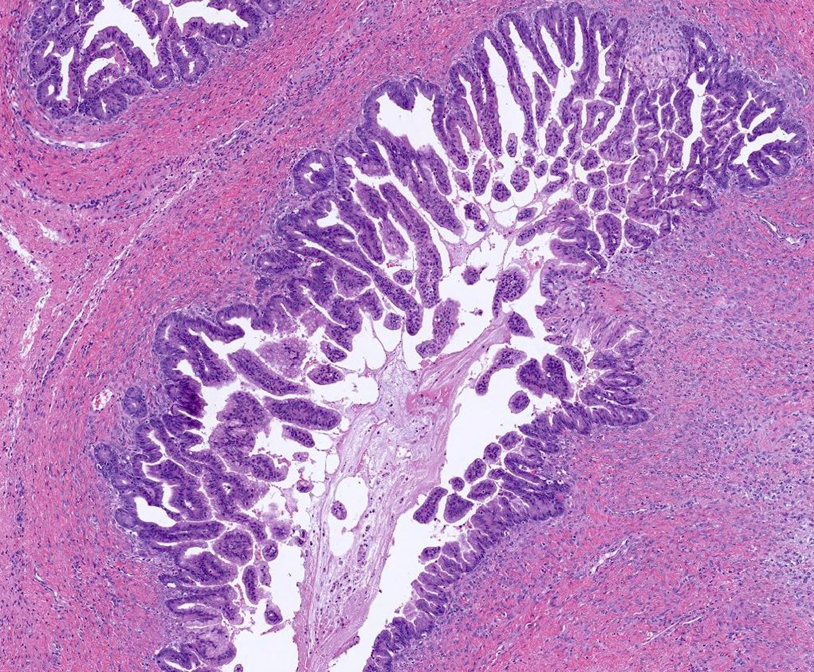
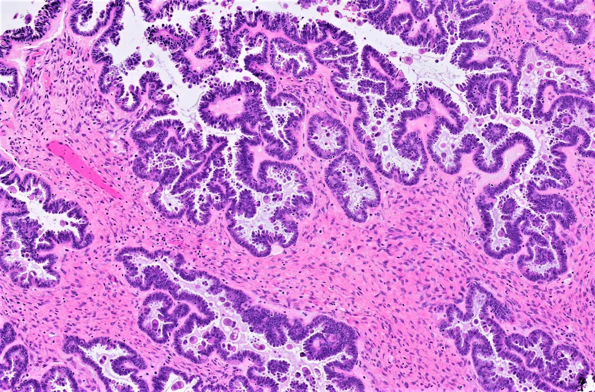
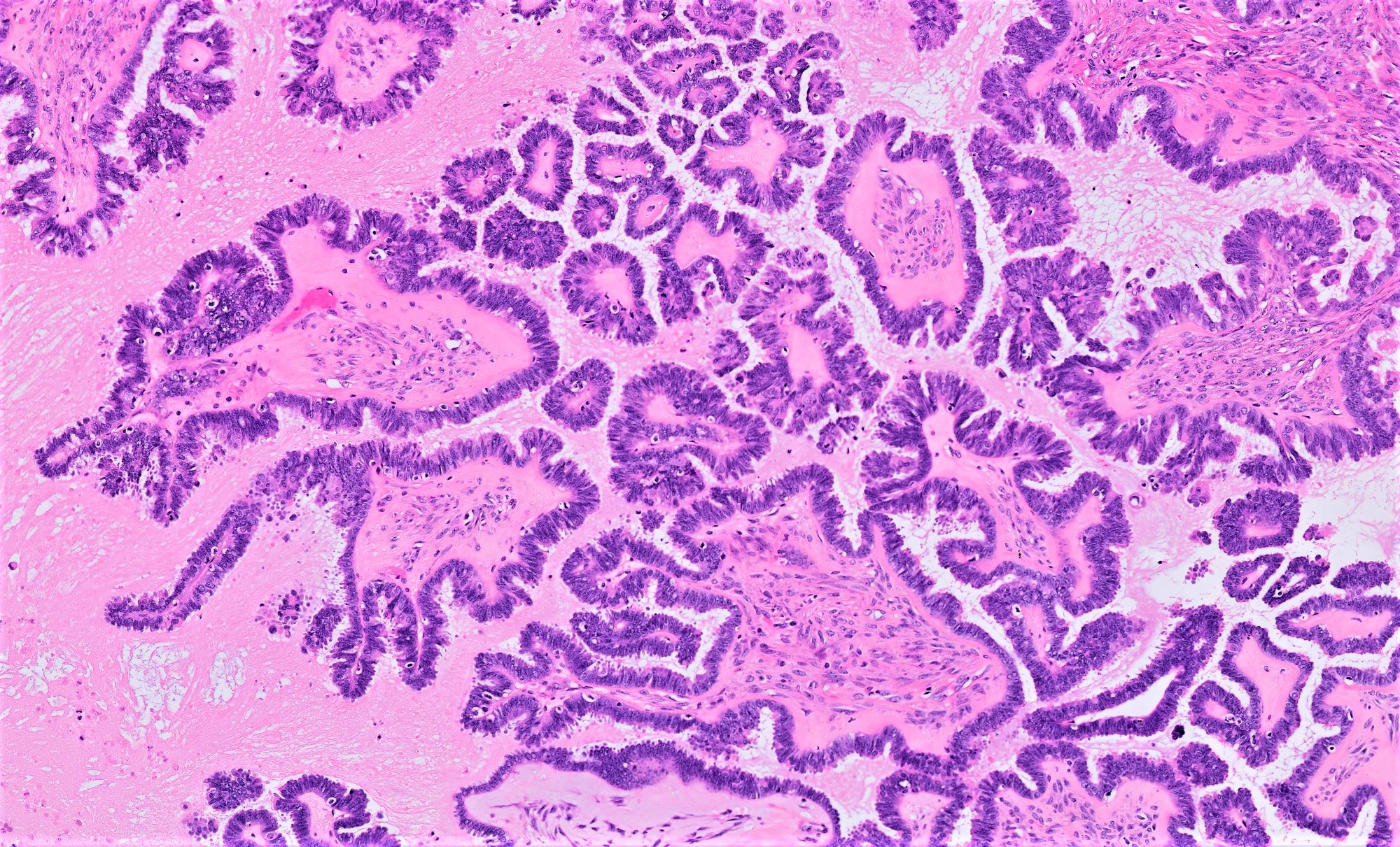
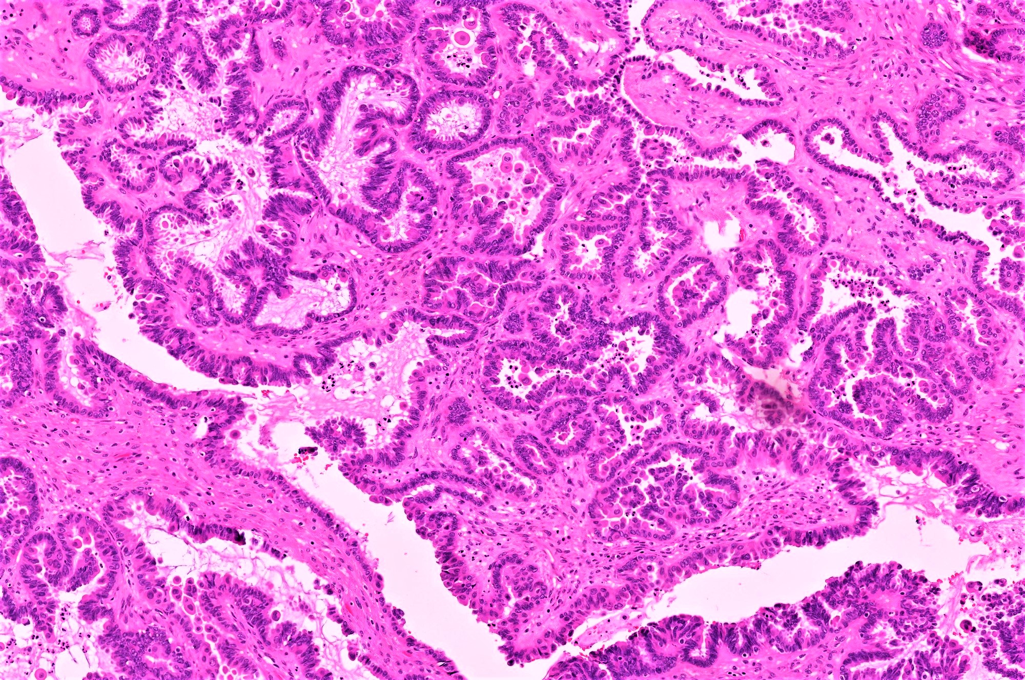
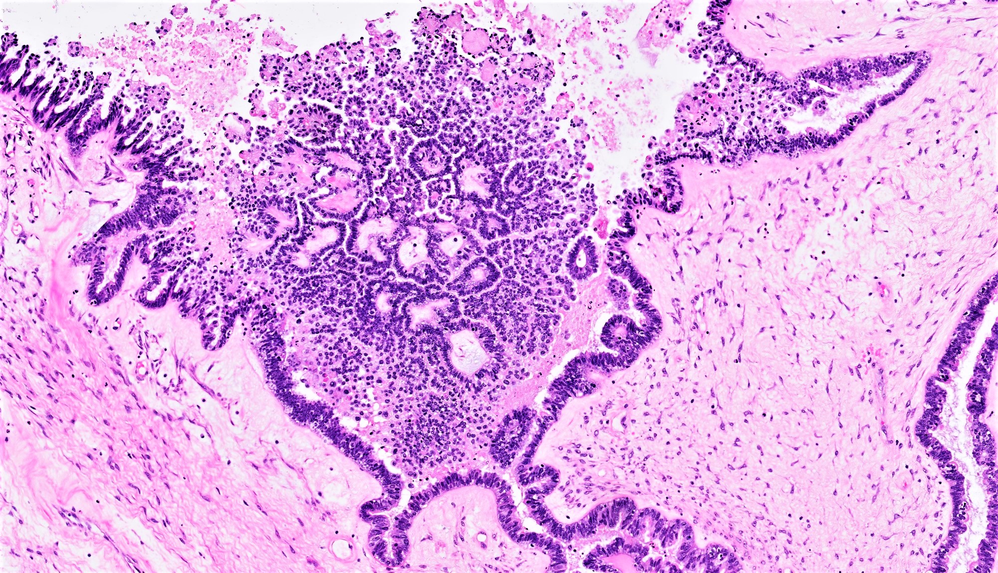
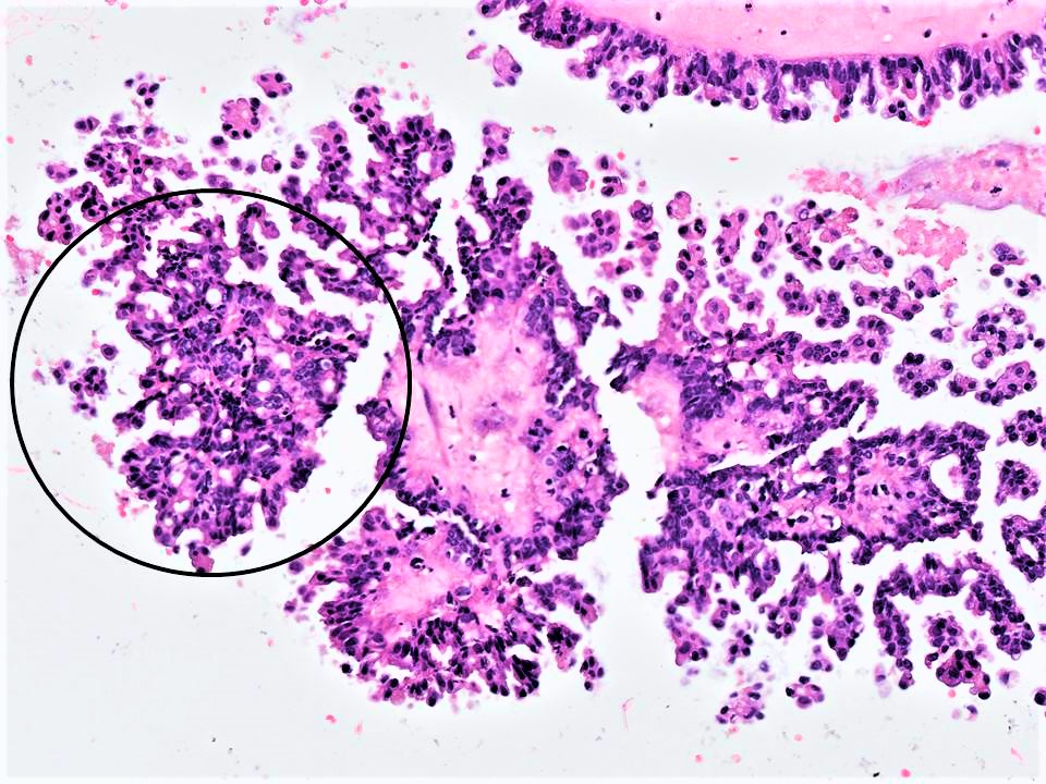
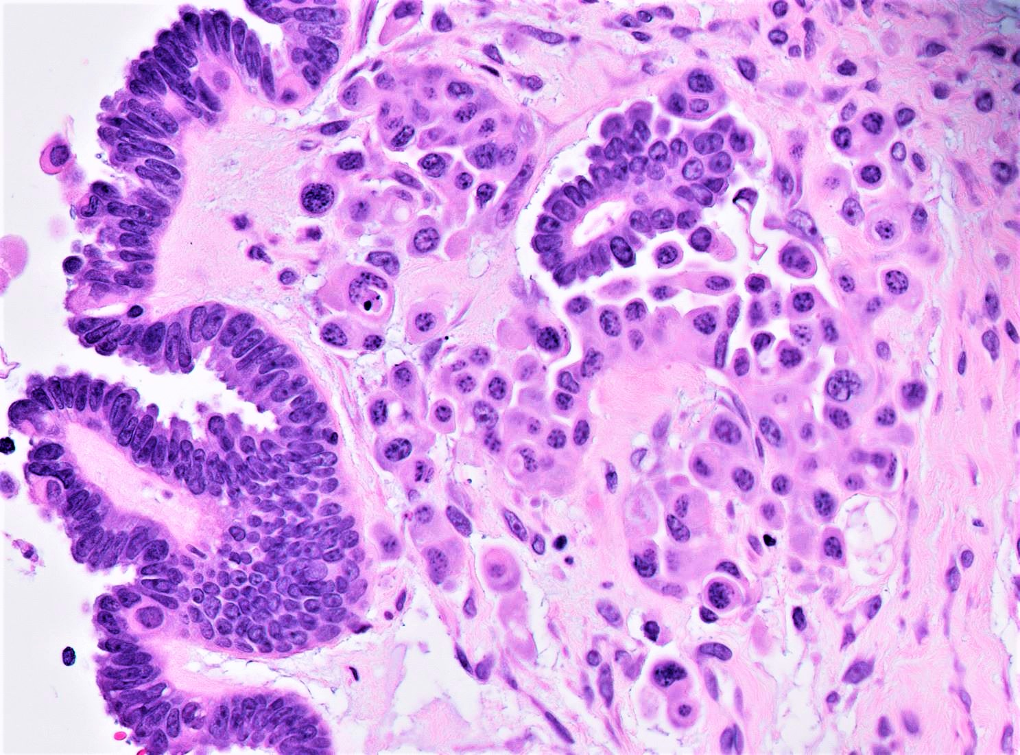
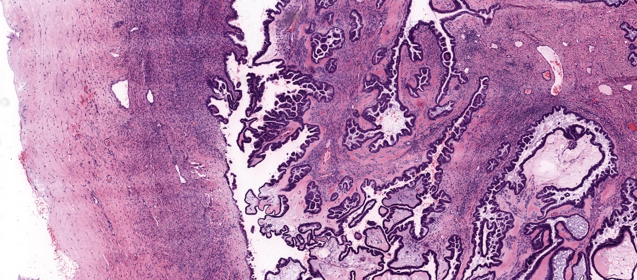
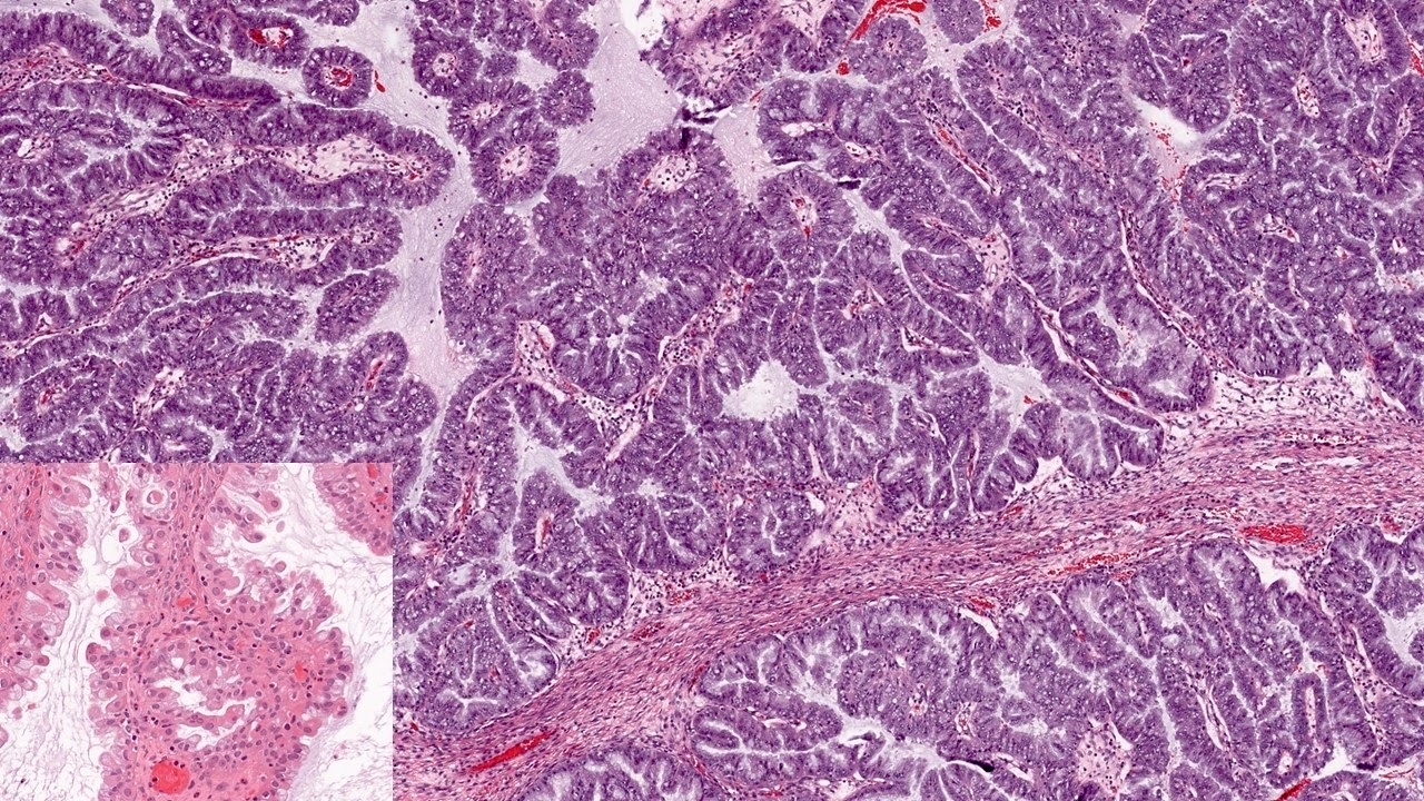
Papillary architecture
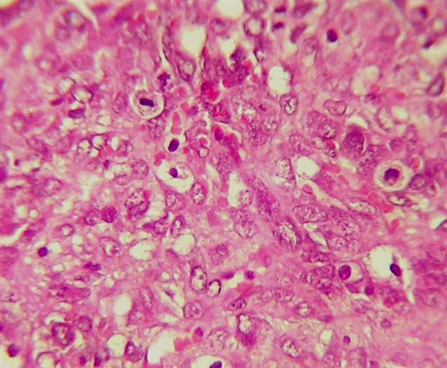
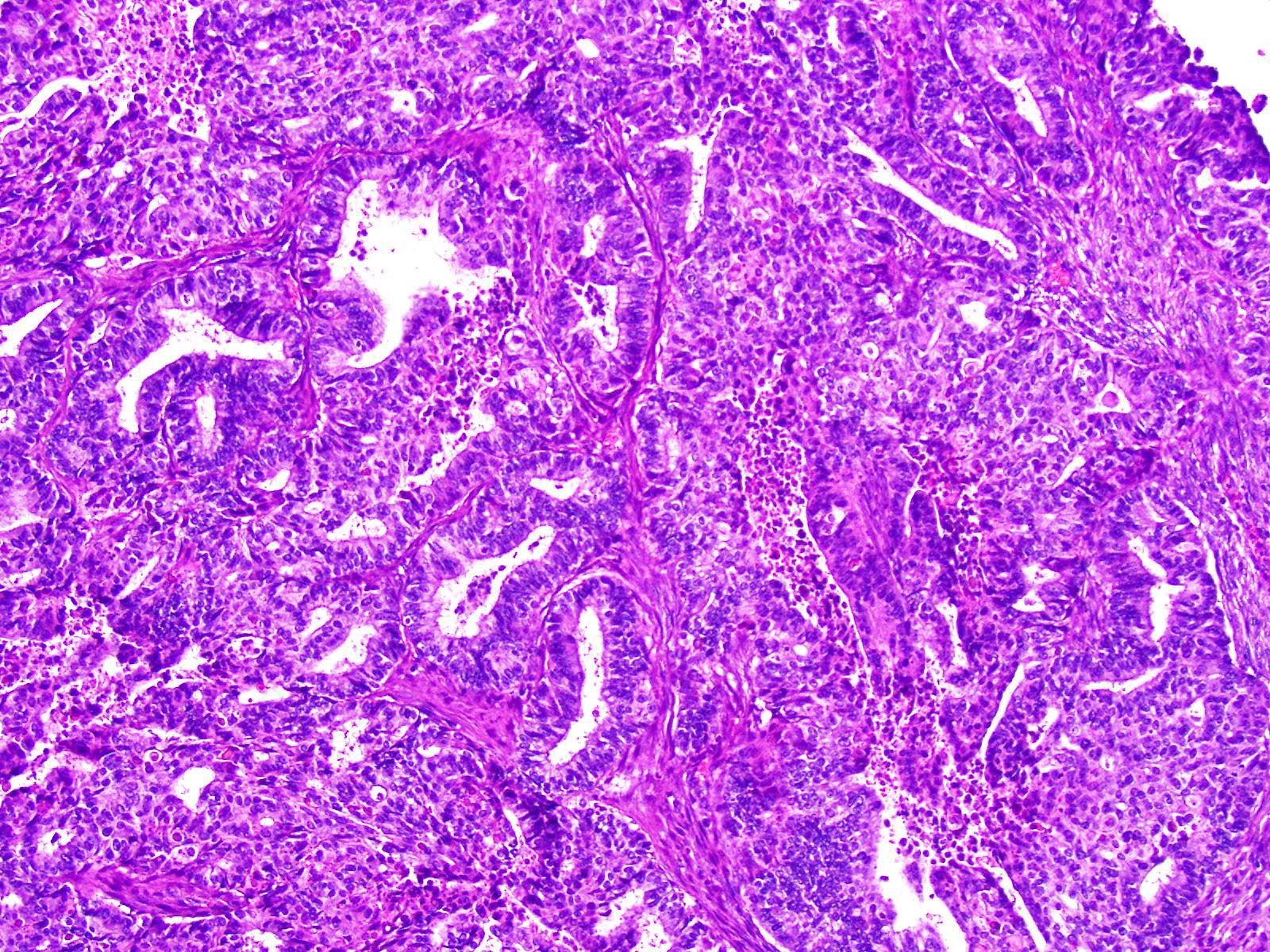
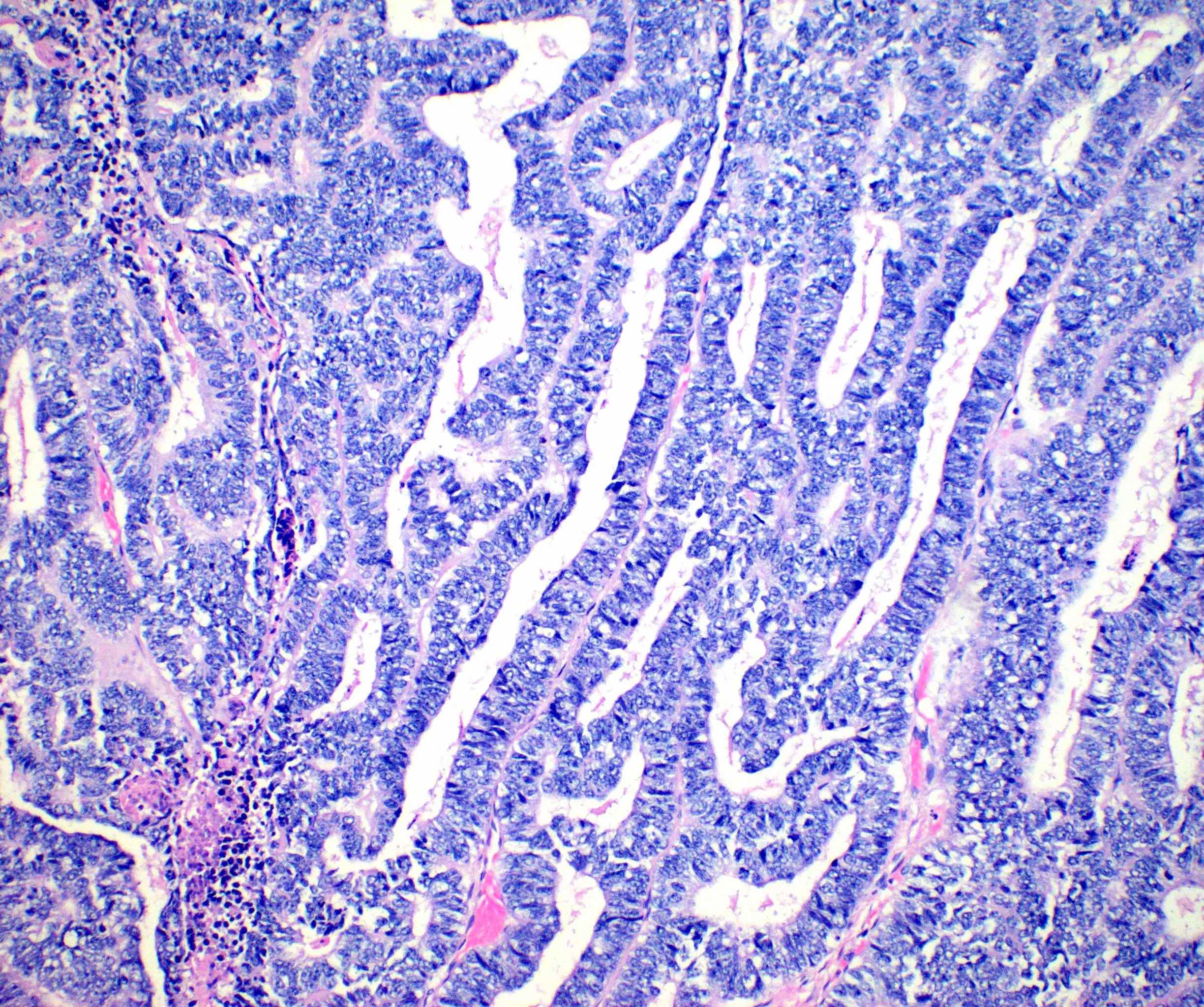
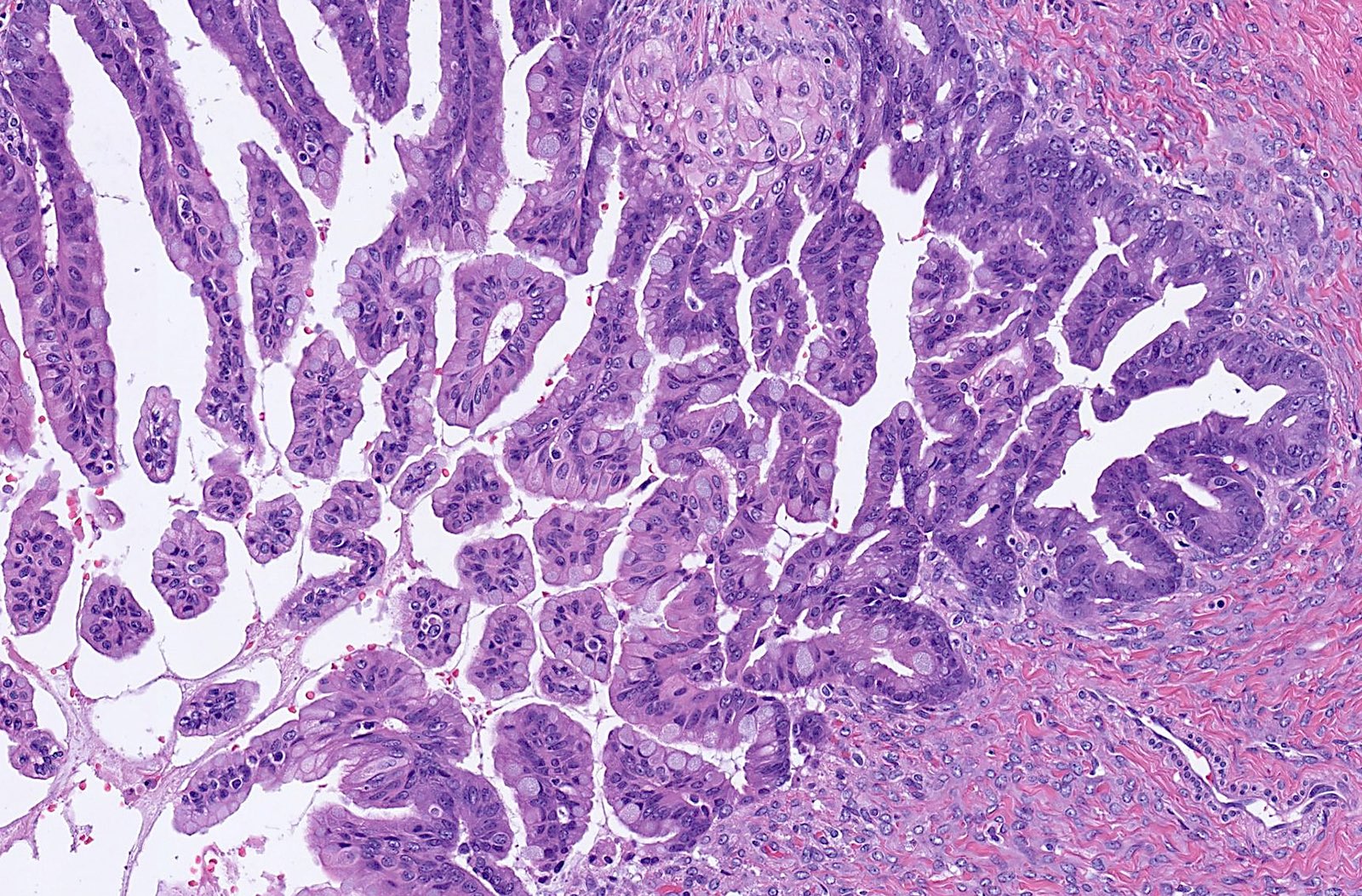
Nuclear atypia
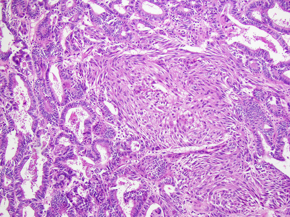
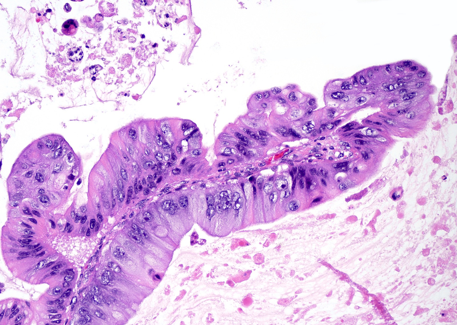
Stromal invasion
Malignant nuclear features
AFIP images

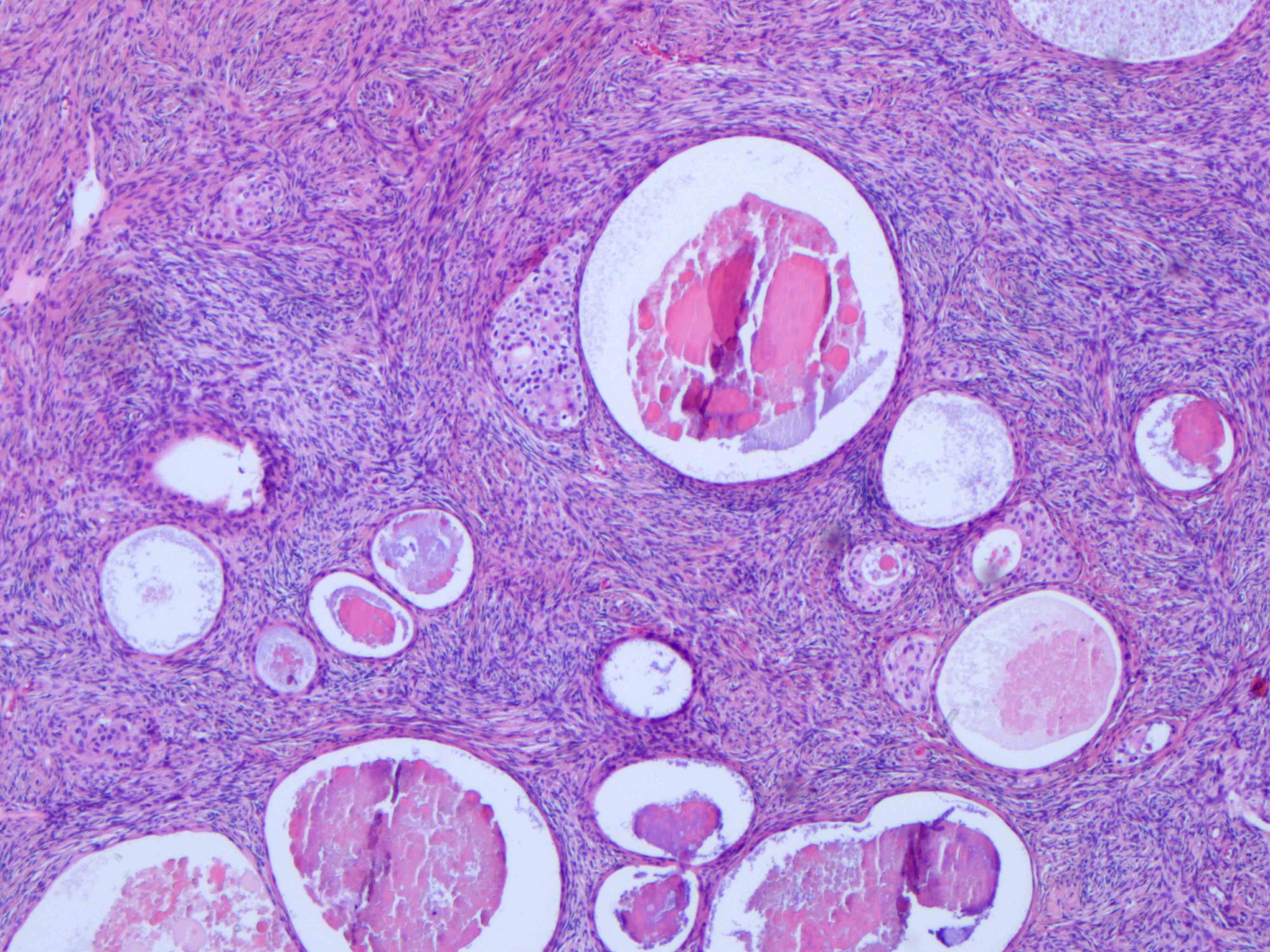
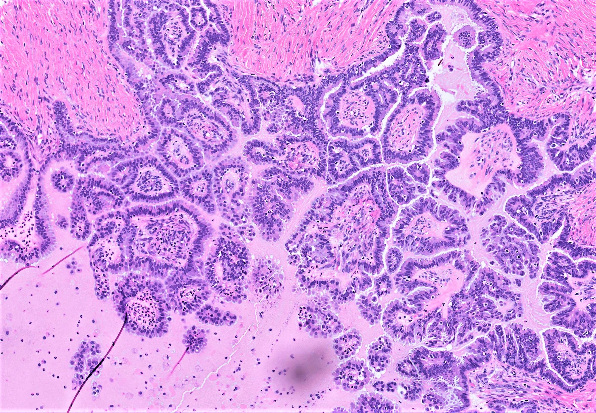
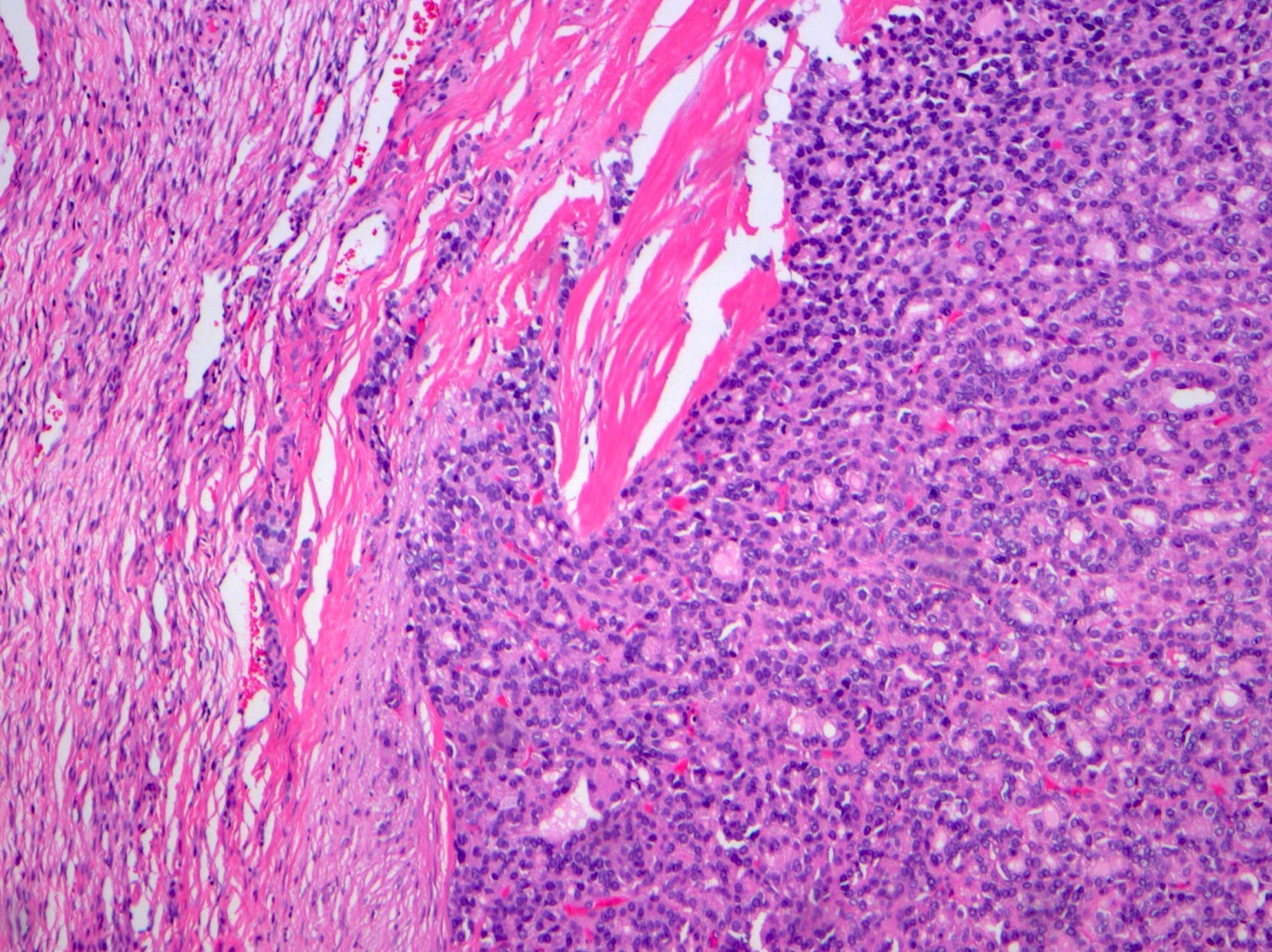
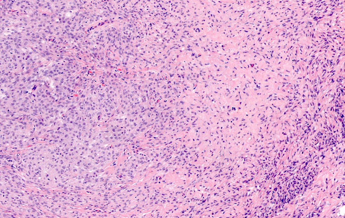
Fibromatous stromal component
Resembling coffee beans
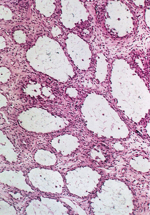
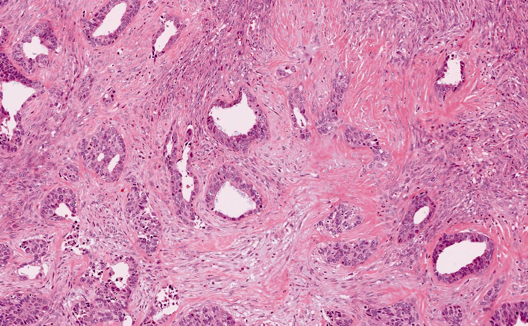
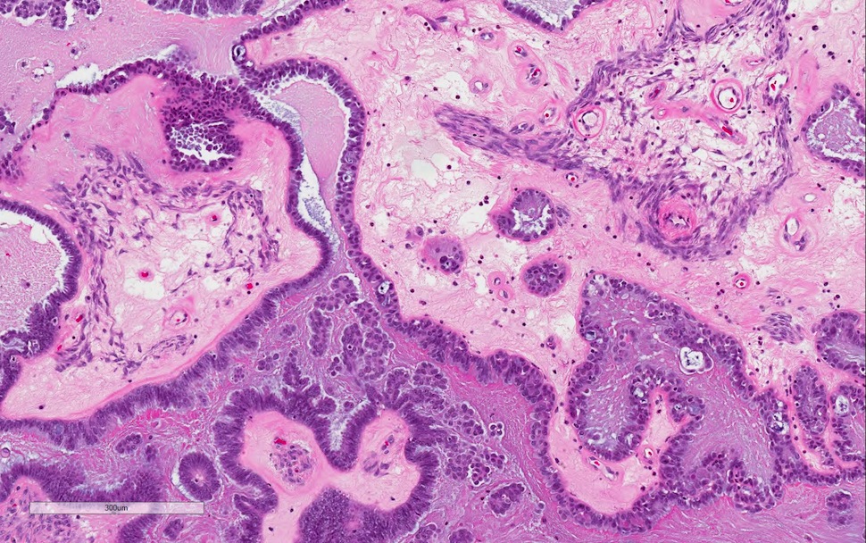
Several lumens
Mucinous epithelium
Ciliated epithelium
Images hosted on other servers:
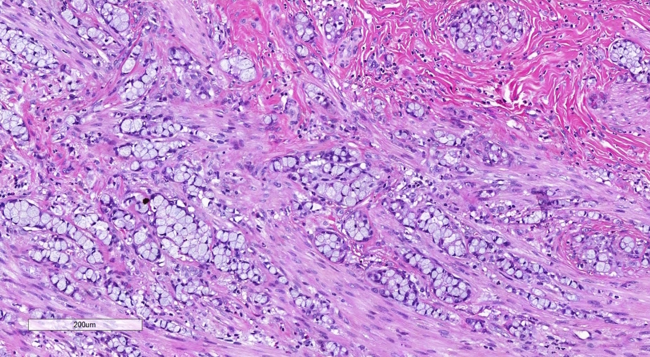
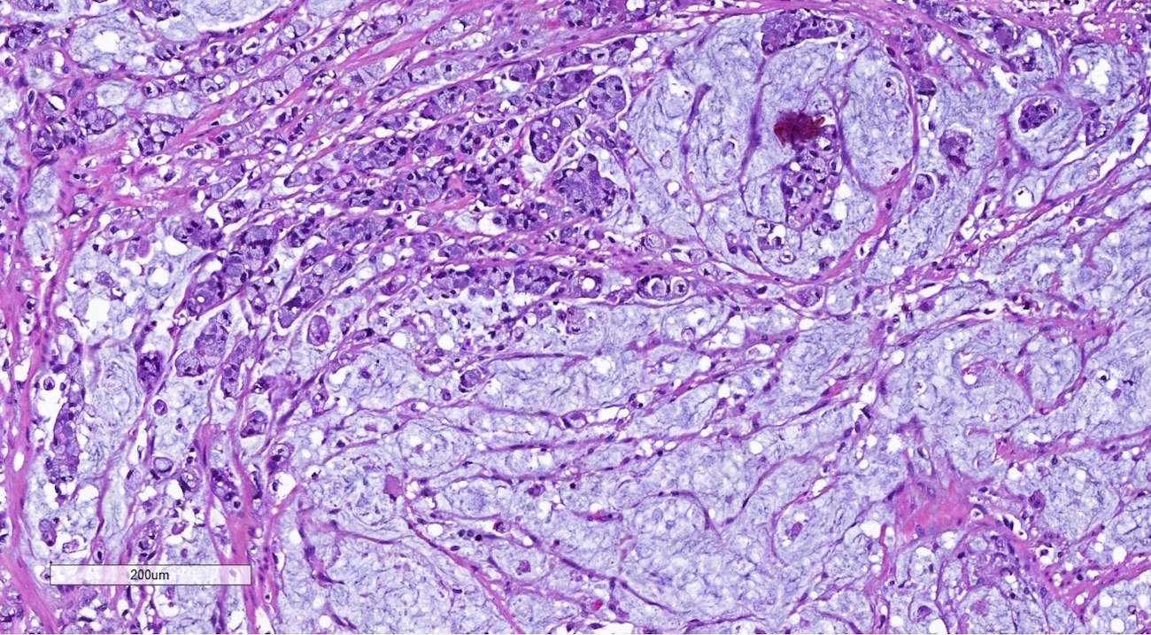
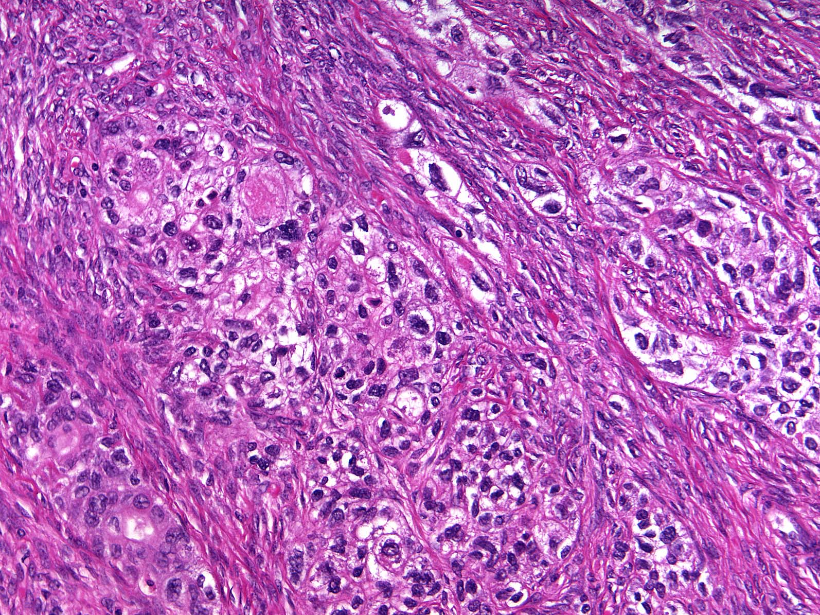
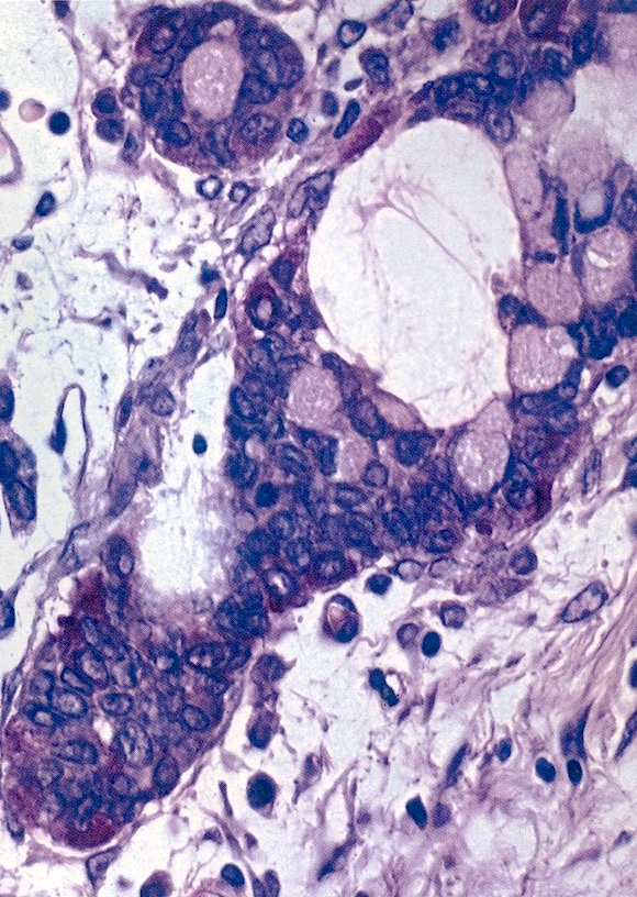
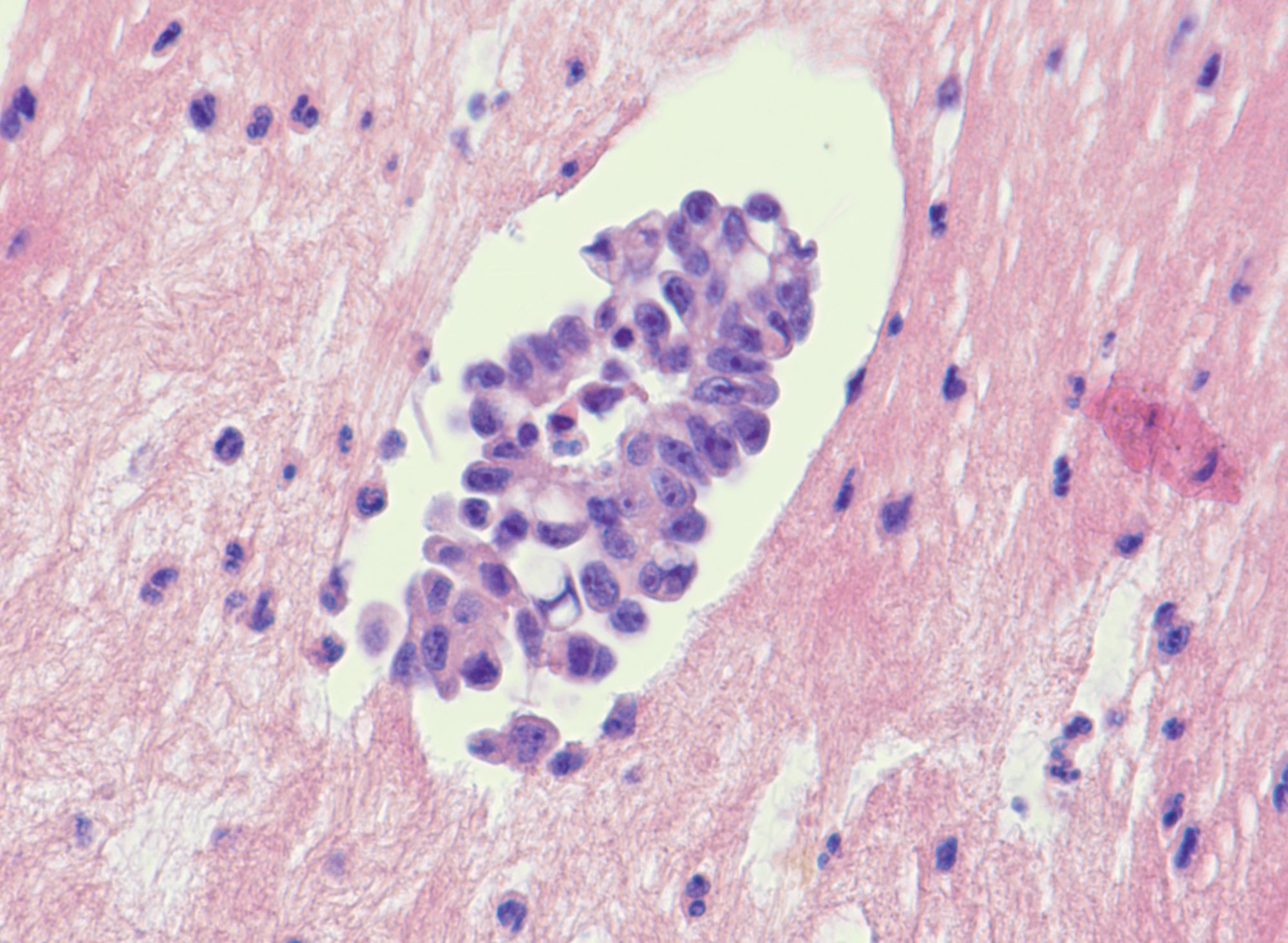
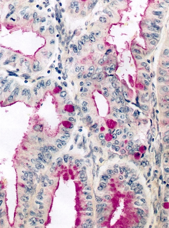
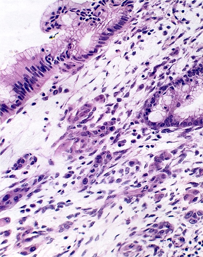
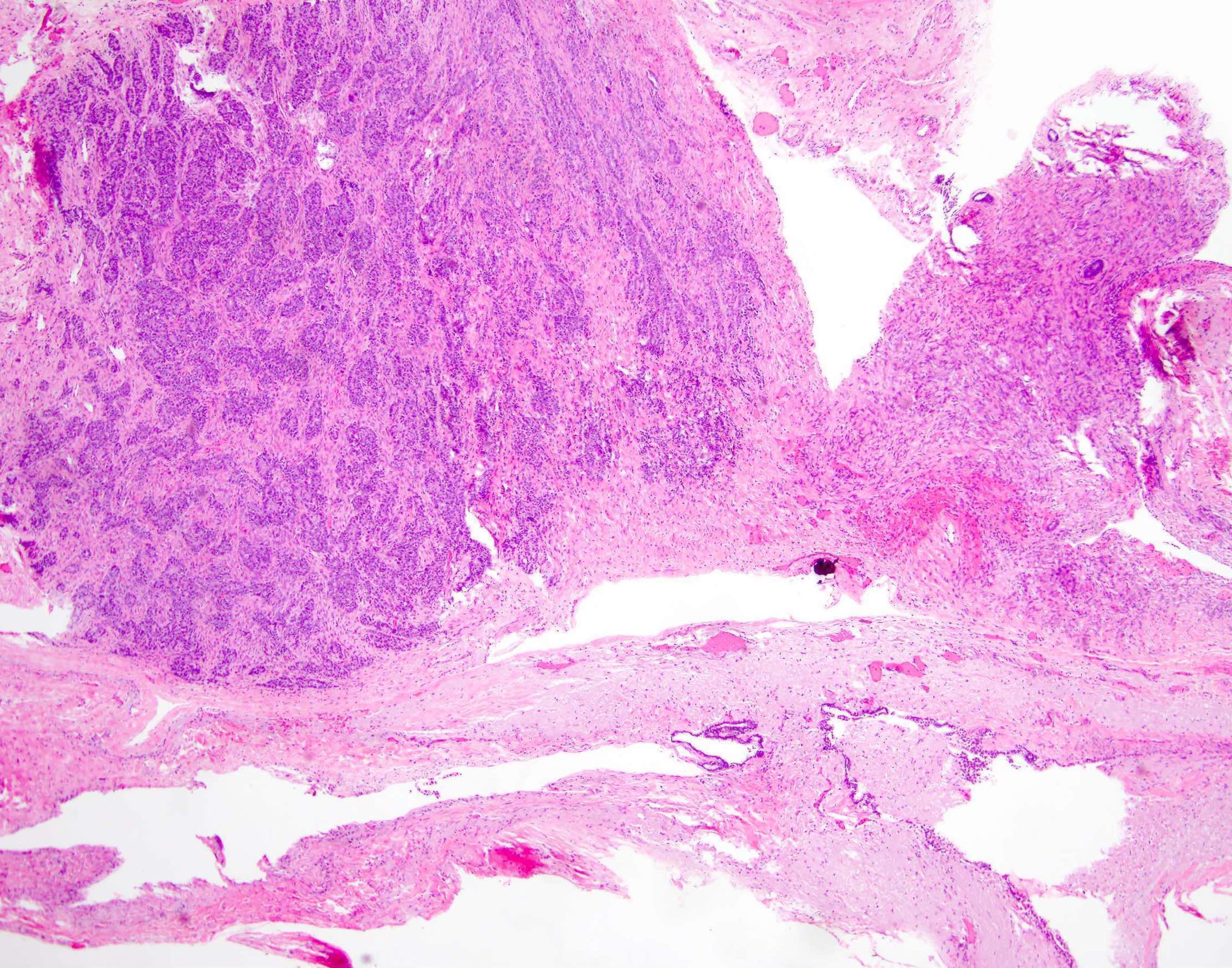
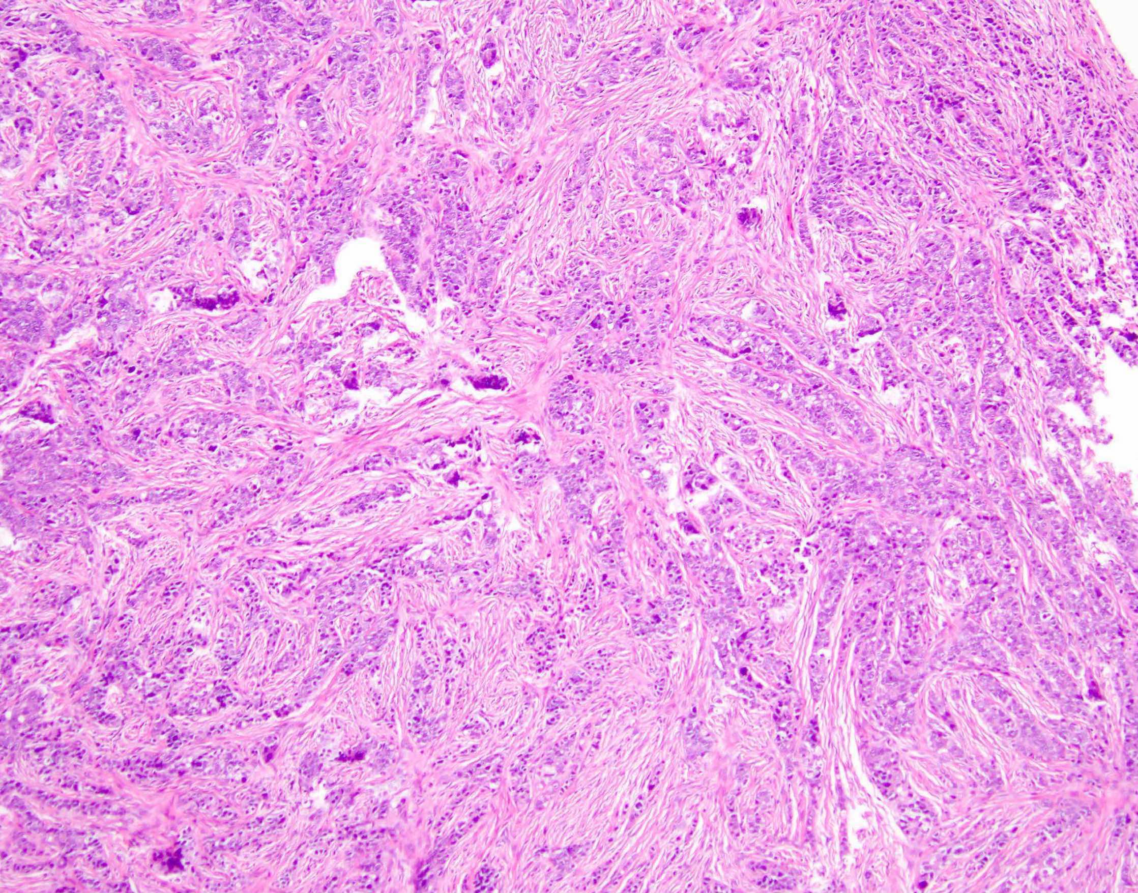
Diffuse infilitration
by lobular type
epithelial cells

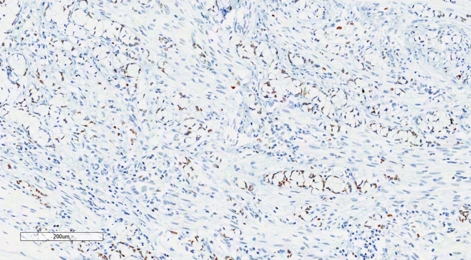
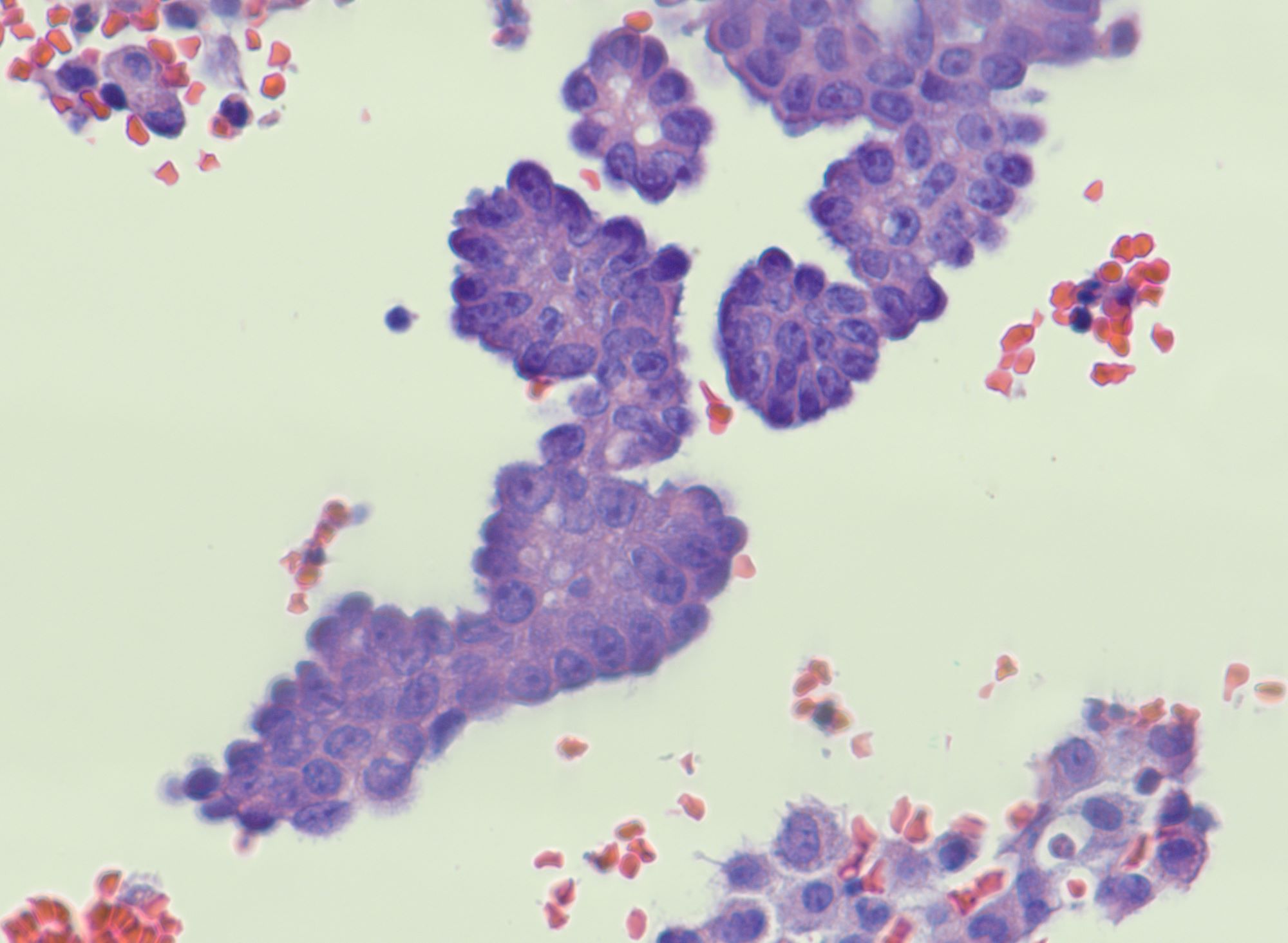
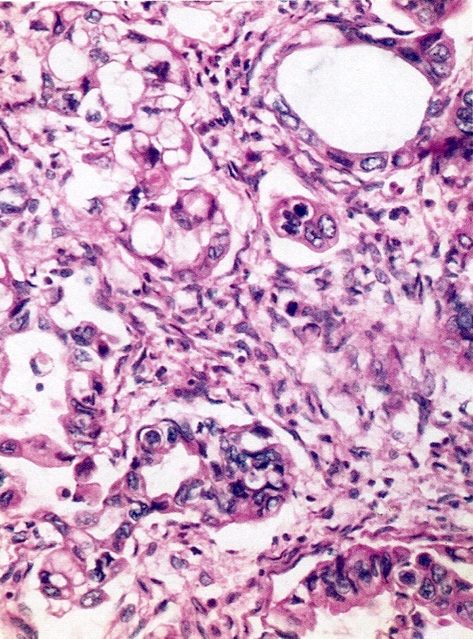
Ductal
Images hosted on other servers:

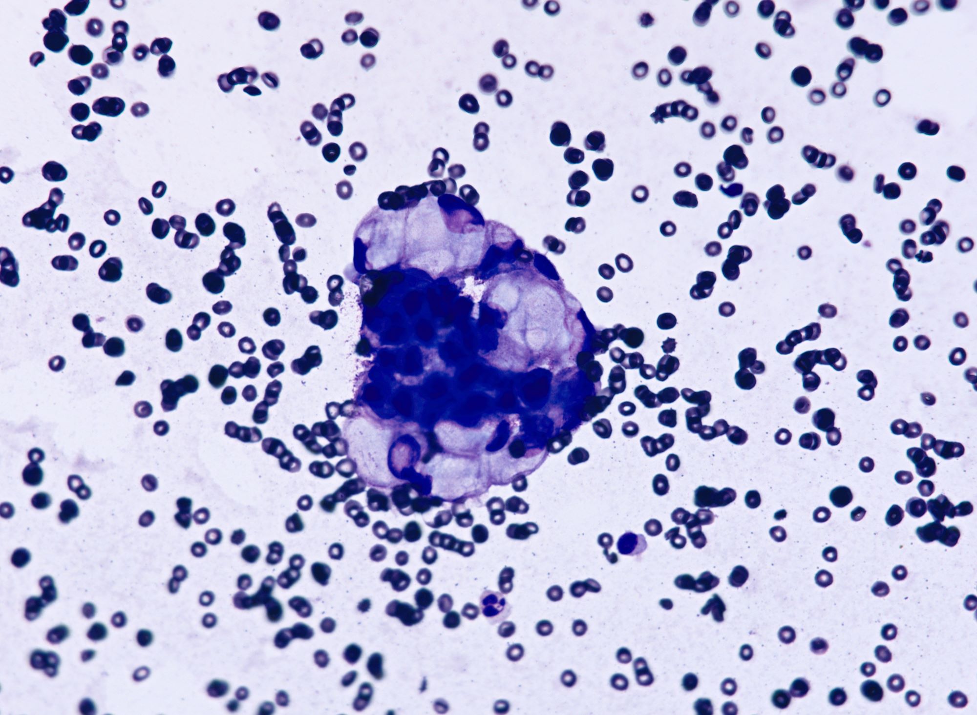
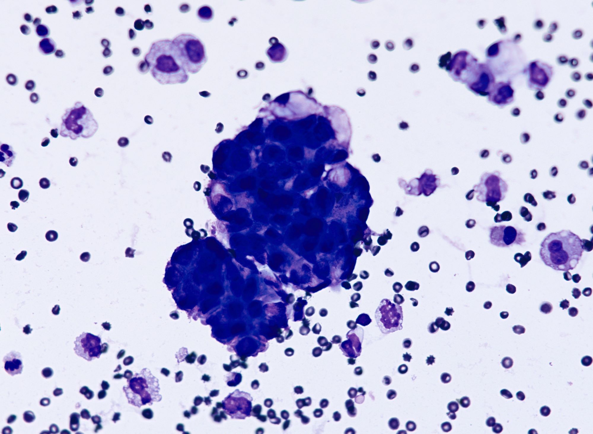
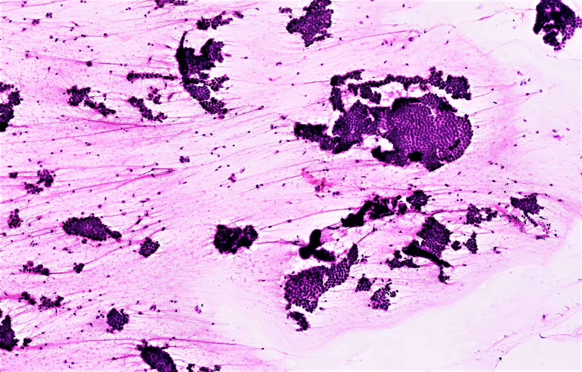
Malignant cells
in cluster, Giemsa
Images hosted on other servers:

Calcifications associated with a mature teratoma
Contributed by Aurelia Busca, M.D., Ph.D.
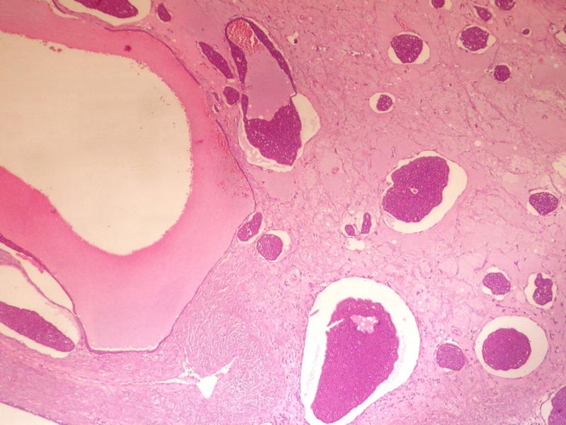
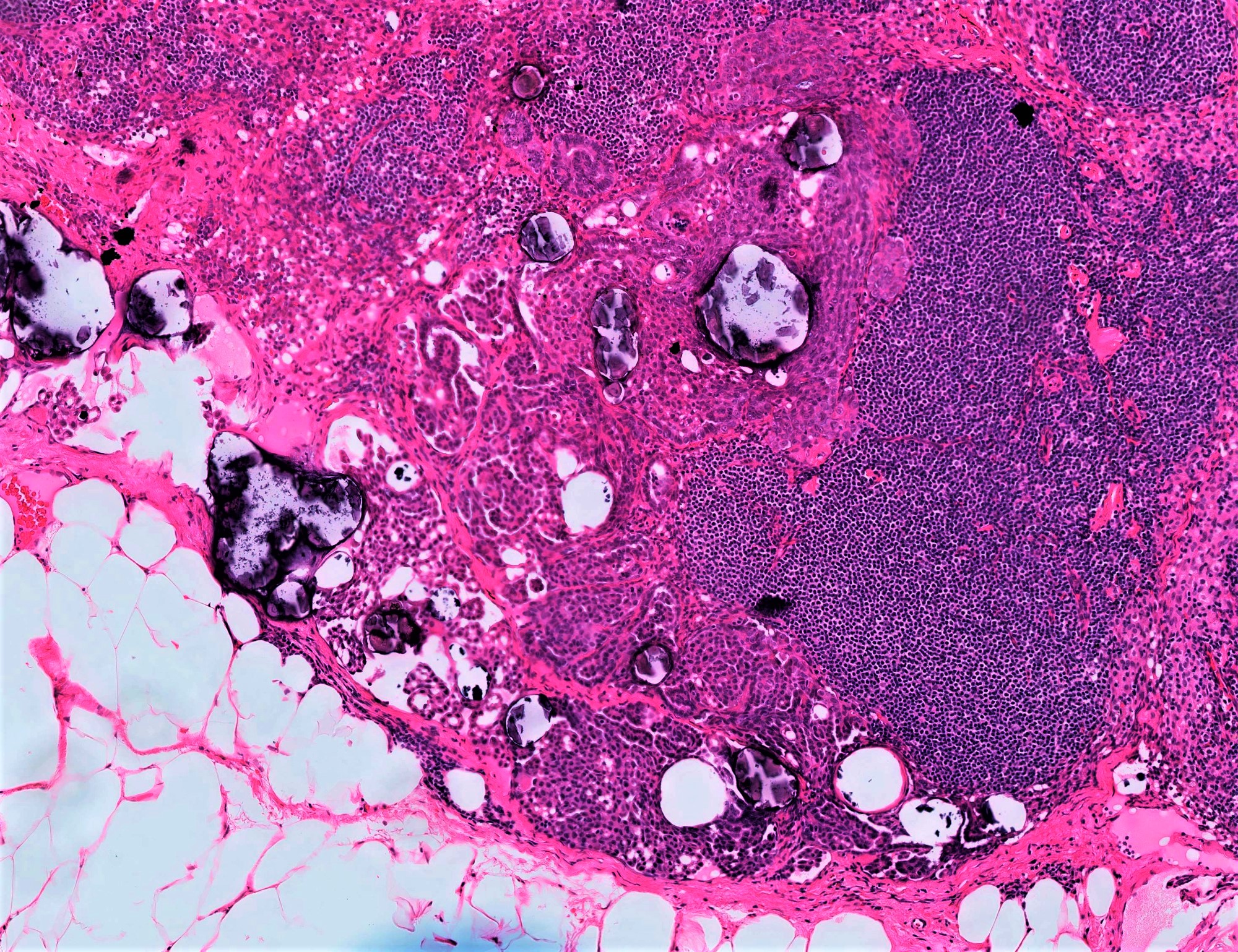
Calcifications
Calcifications
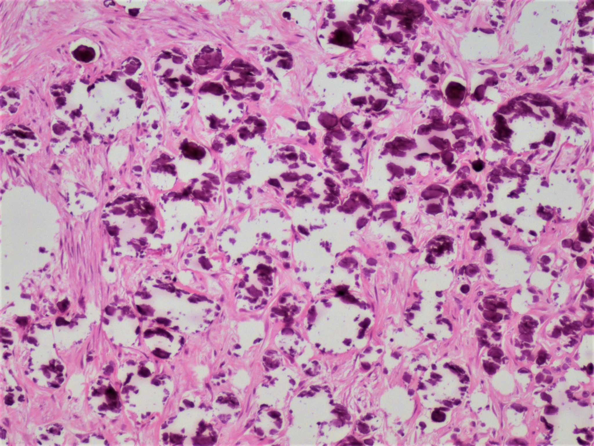
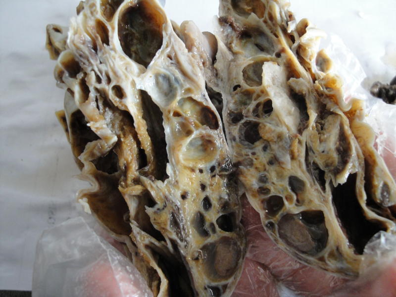
Calcifications associated with low grade serous carcinoma of the ovary
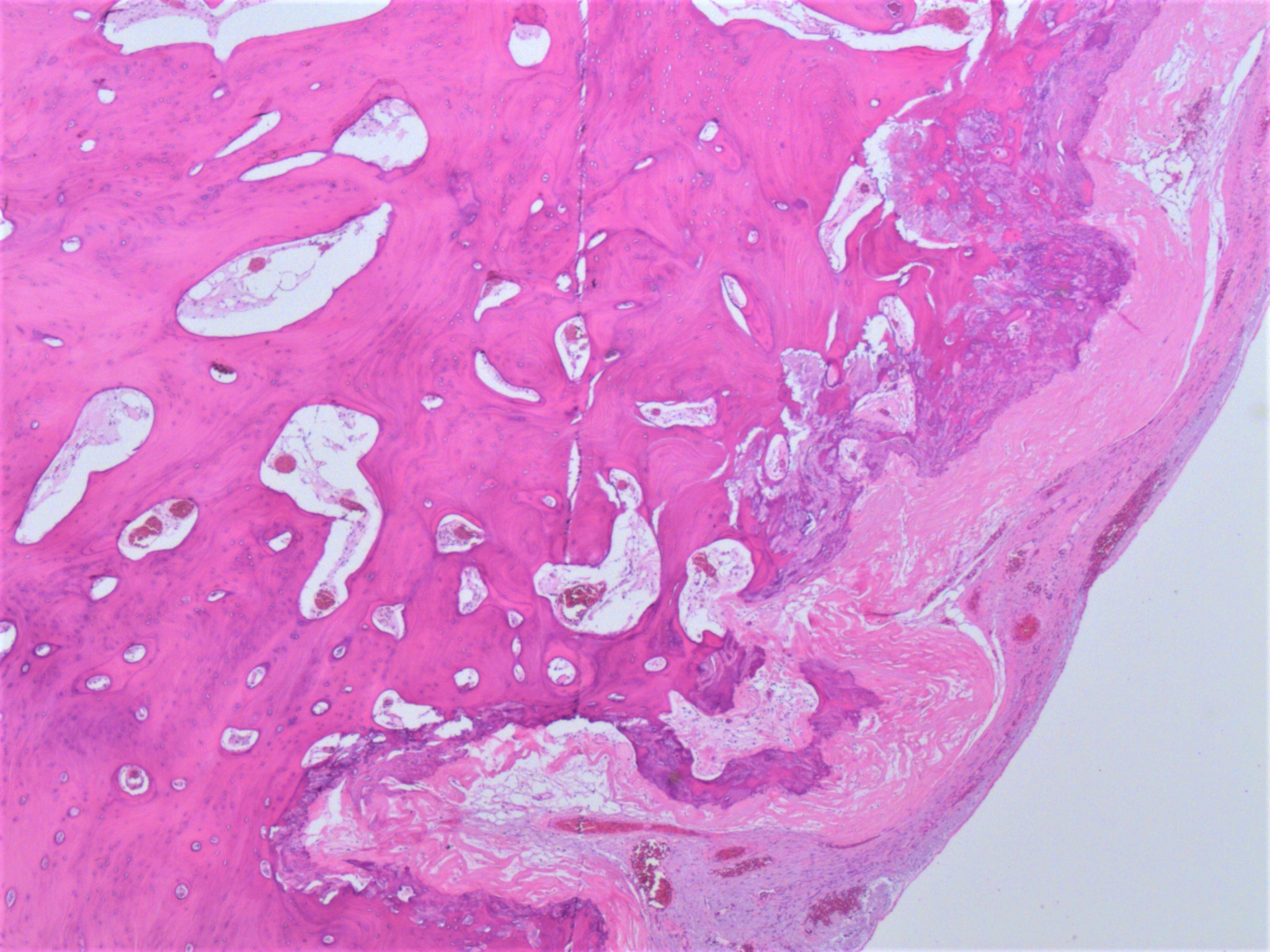
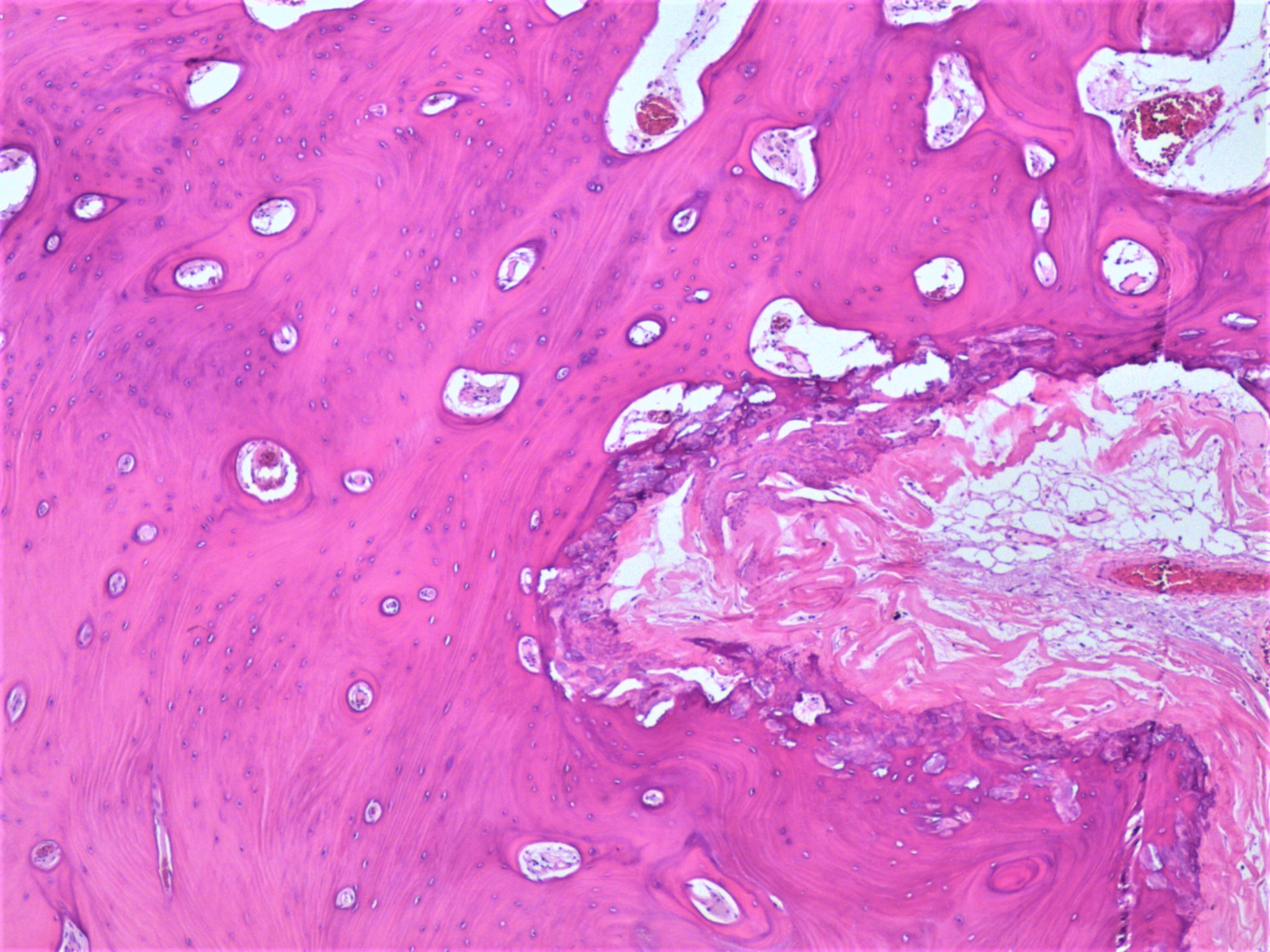
Osseous metaplasia of the ovary
Contributed by Chenxi Wu, M.D., Ph.D.
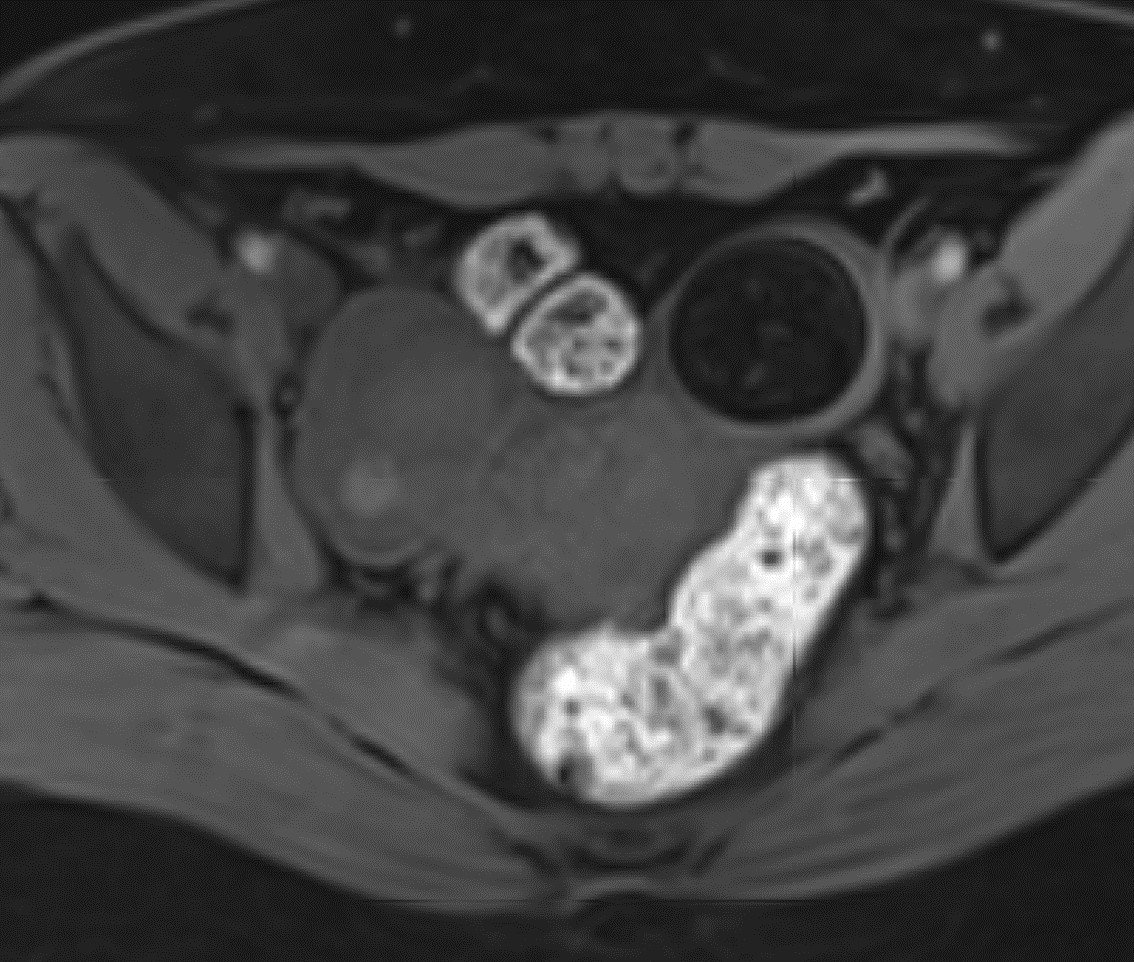
T1 precontast MRI
T2 MRI

Noncontrast CT, coronal plane
Images hosted on other servers:

Ovarian mass intraoperatively
AFIP images
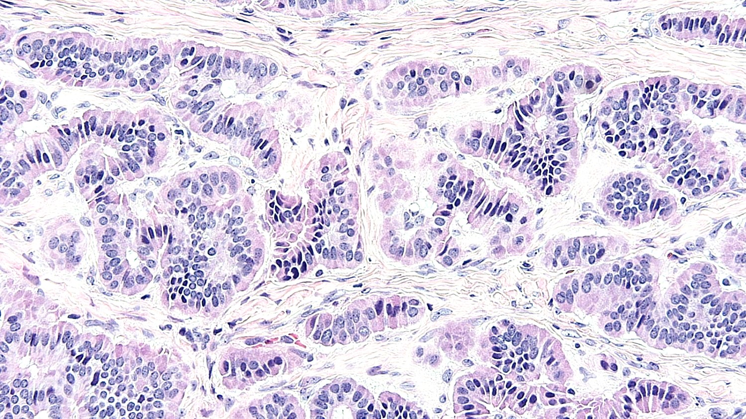
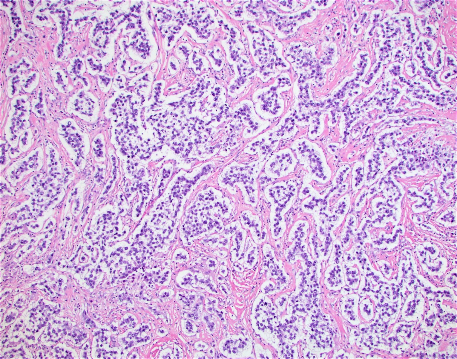
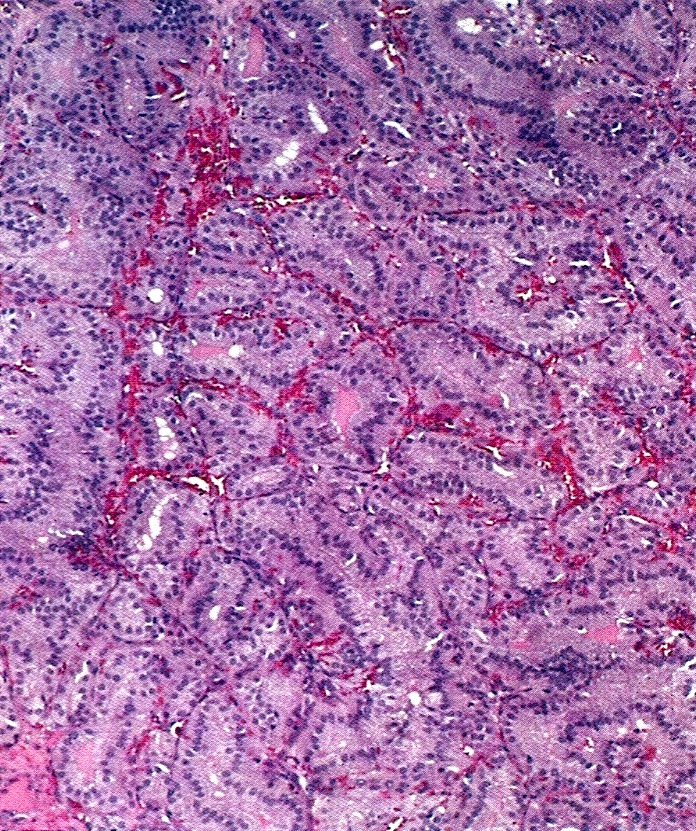
Insular tumor
Contributed by Krisztina Hanley, M.D., Rulong Shen, M.D. and AFIP
Trabecular pattern
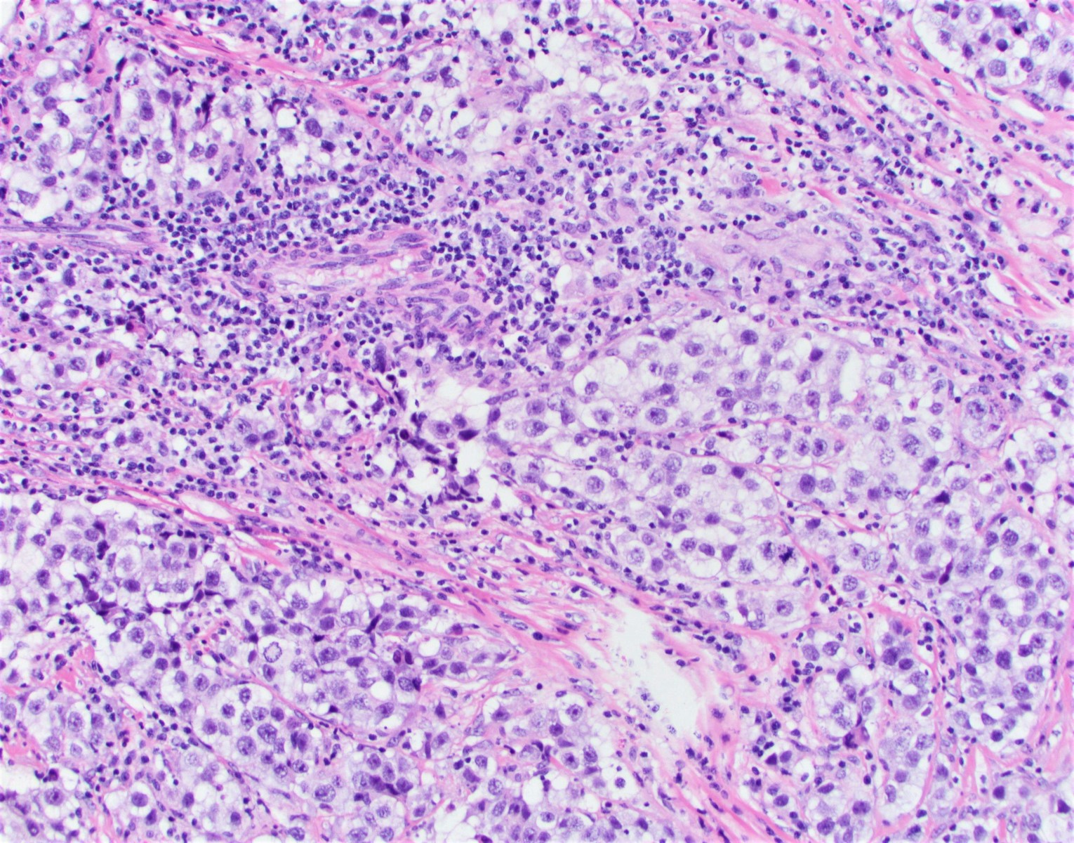
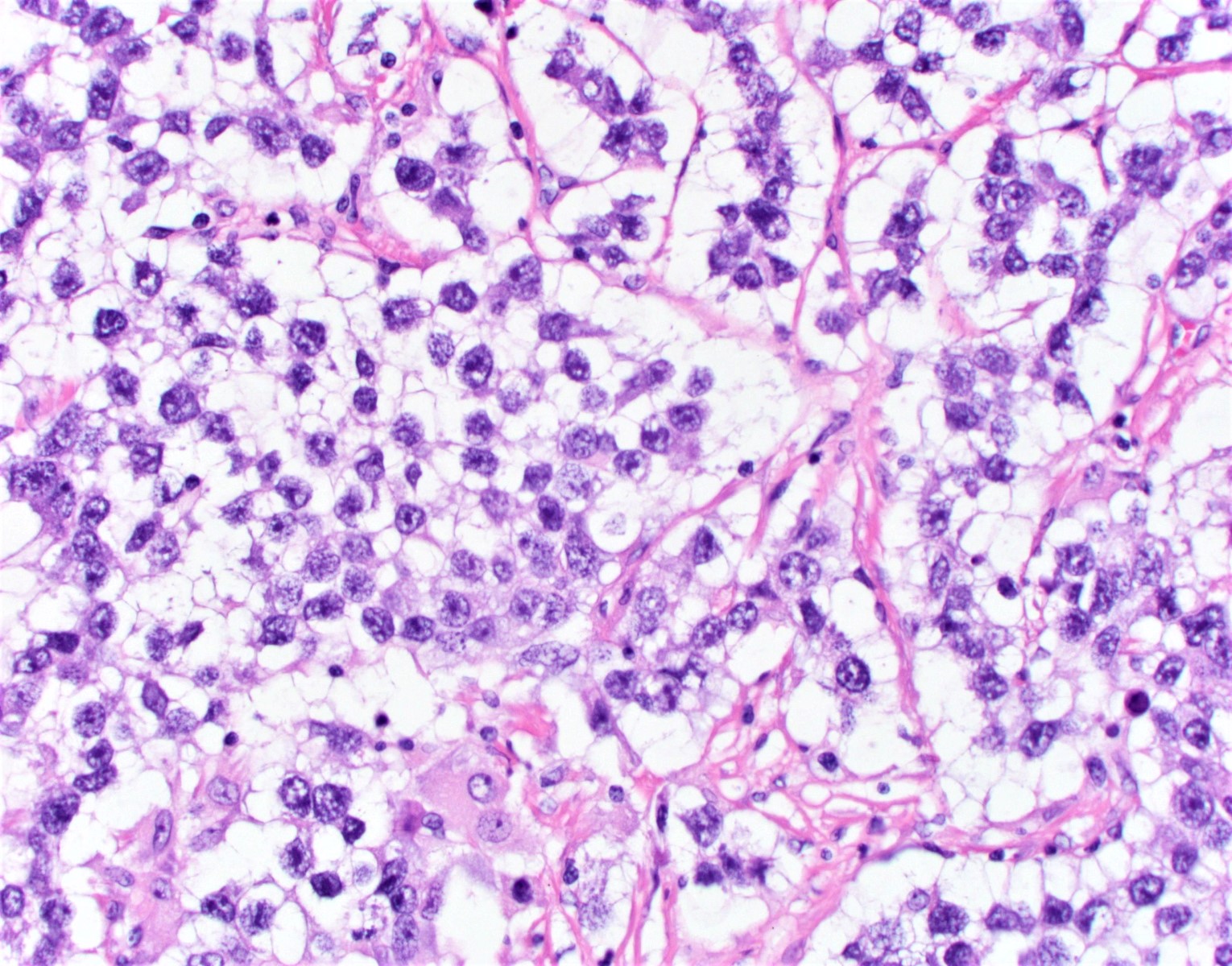
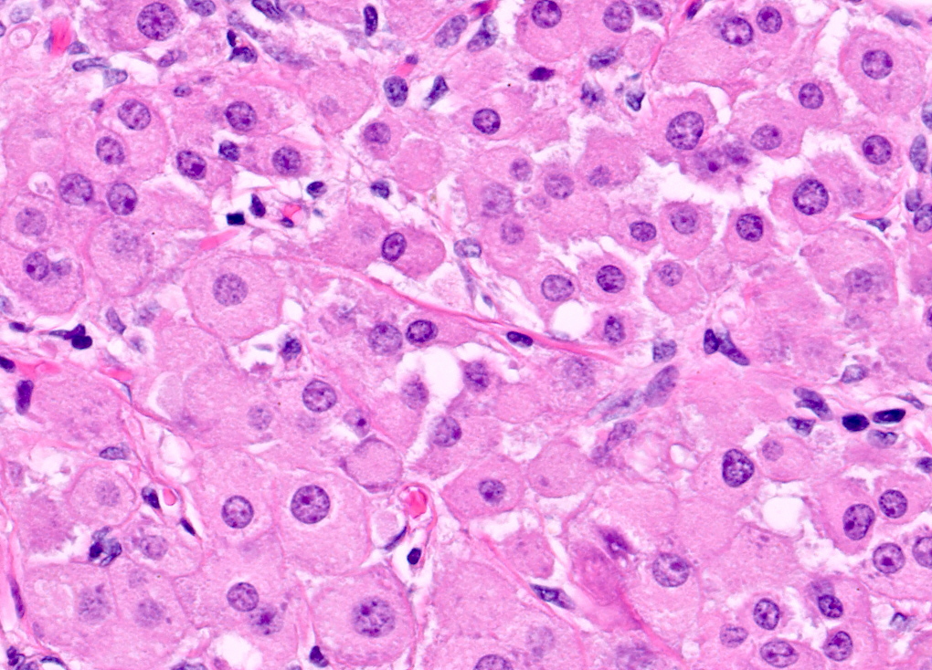
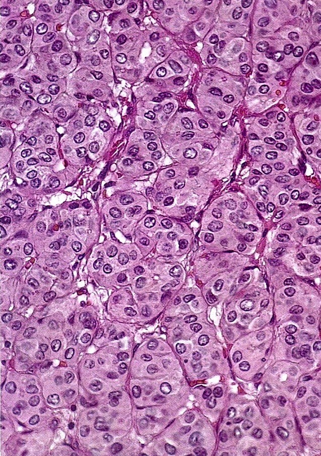
Nuclear features
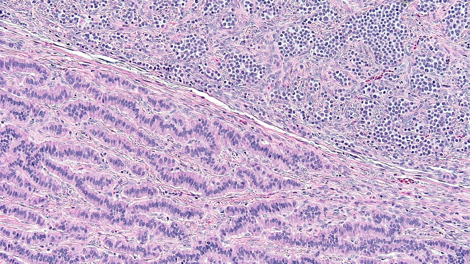
Mixed patterns
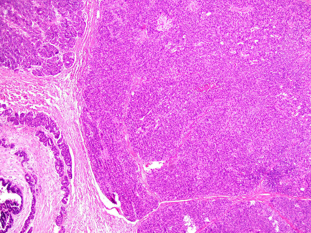
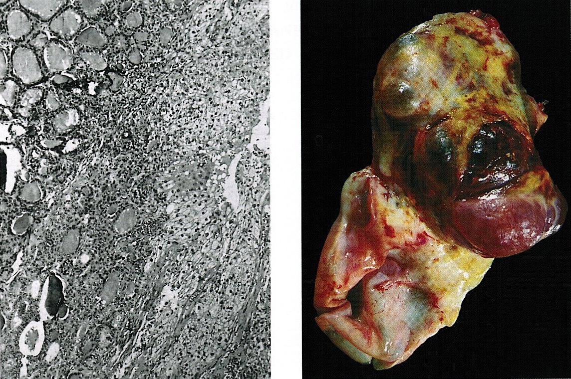
Strumal carcinoid
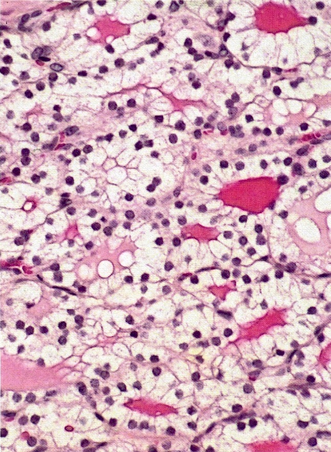
Thyroid component
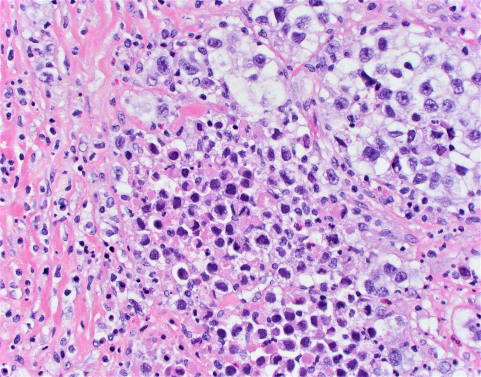
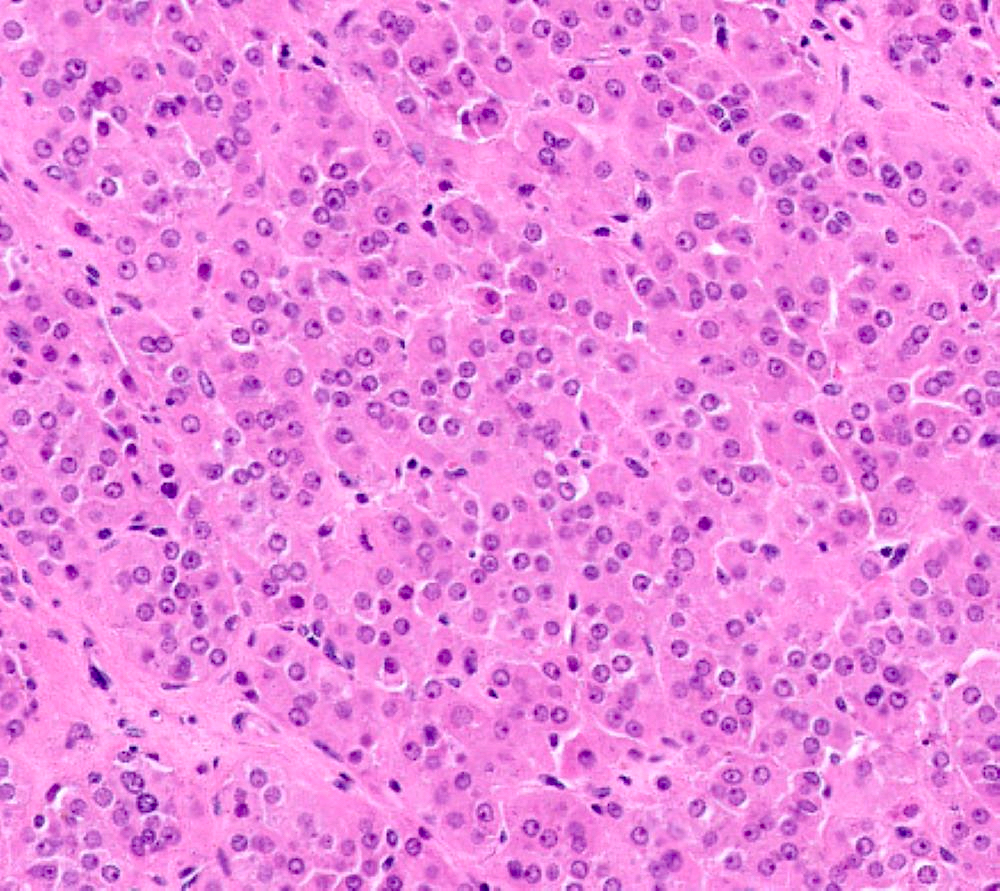
Cytologic features of carcinoid
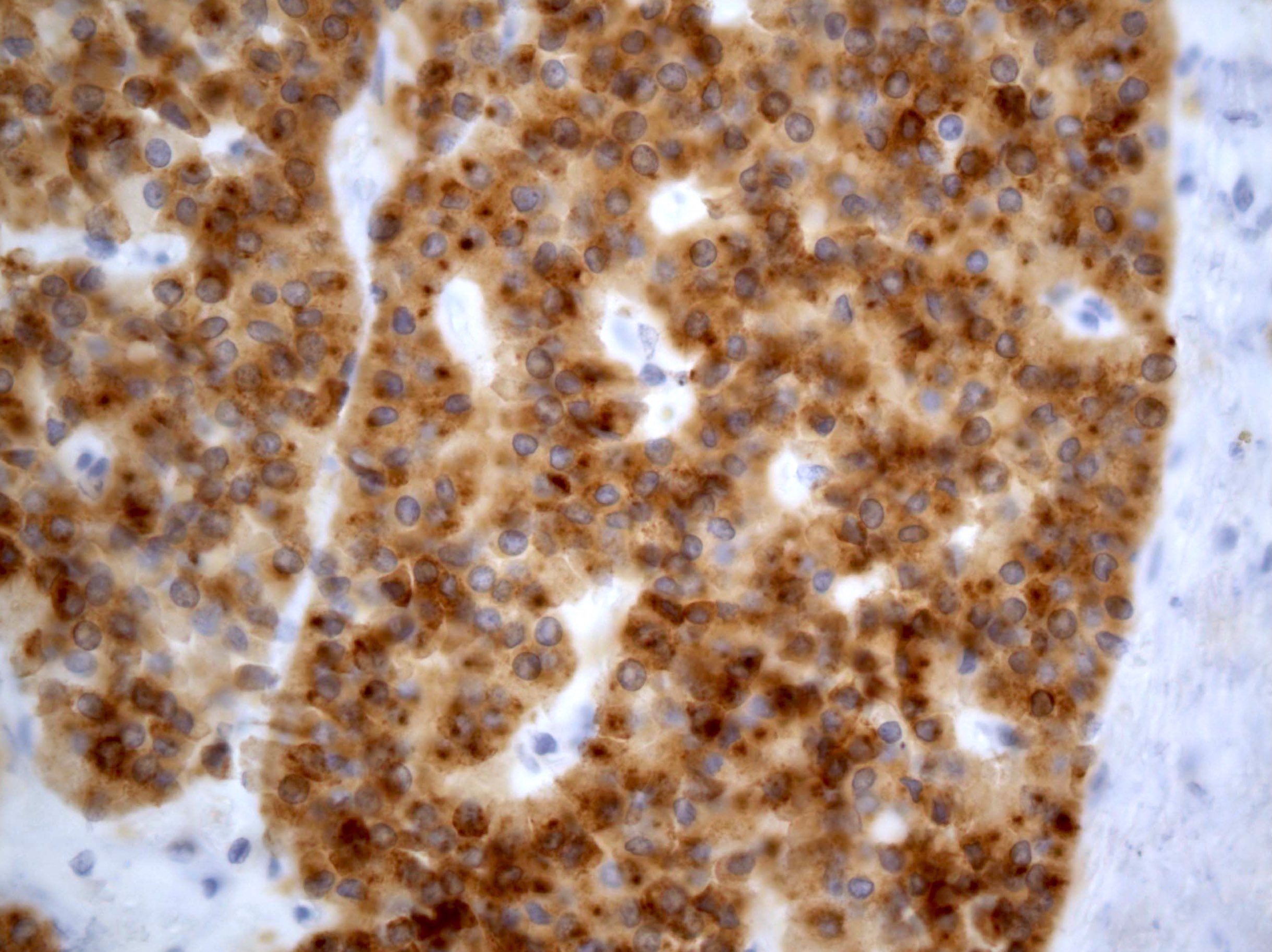
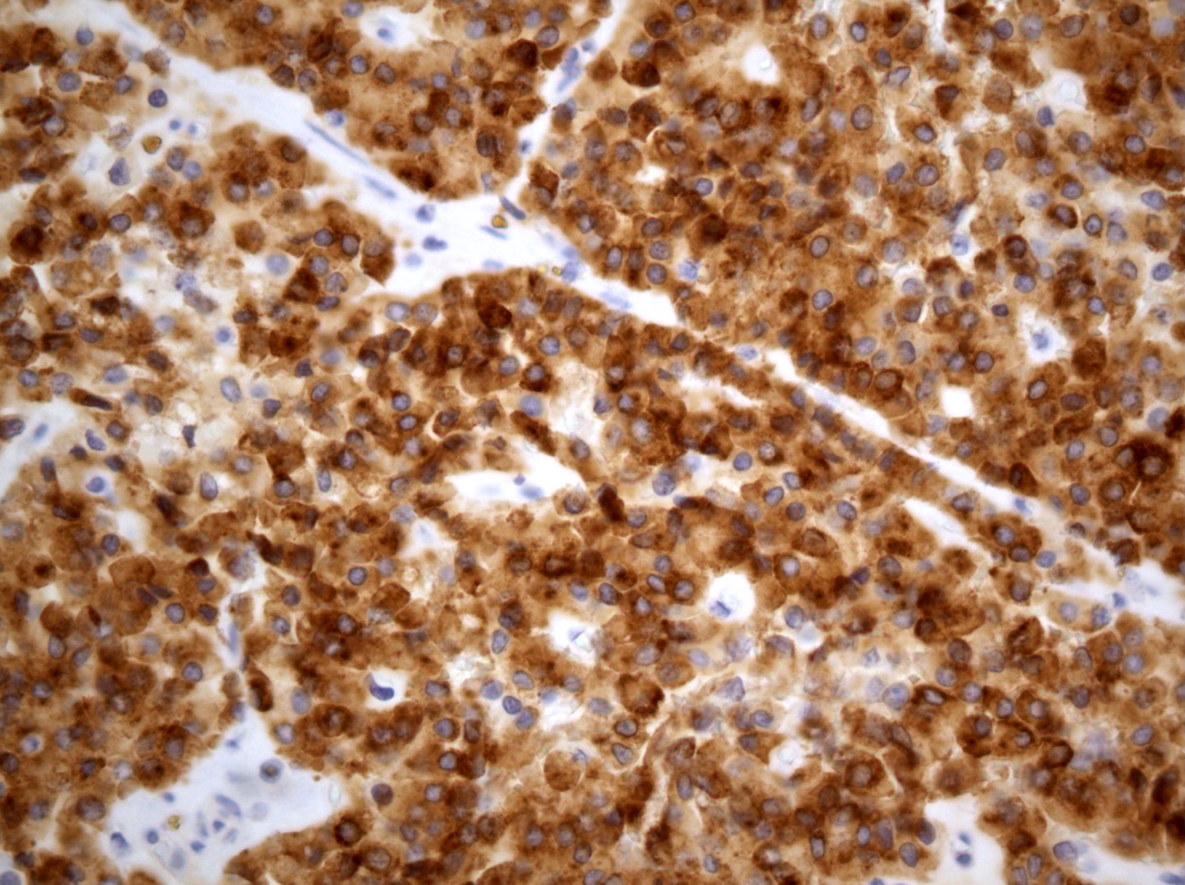
Chromogranin in strumal ovarian carcinoid
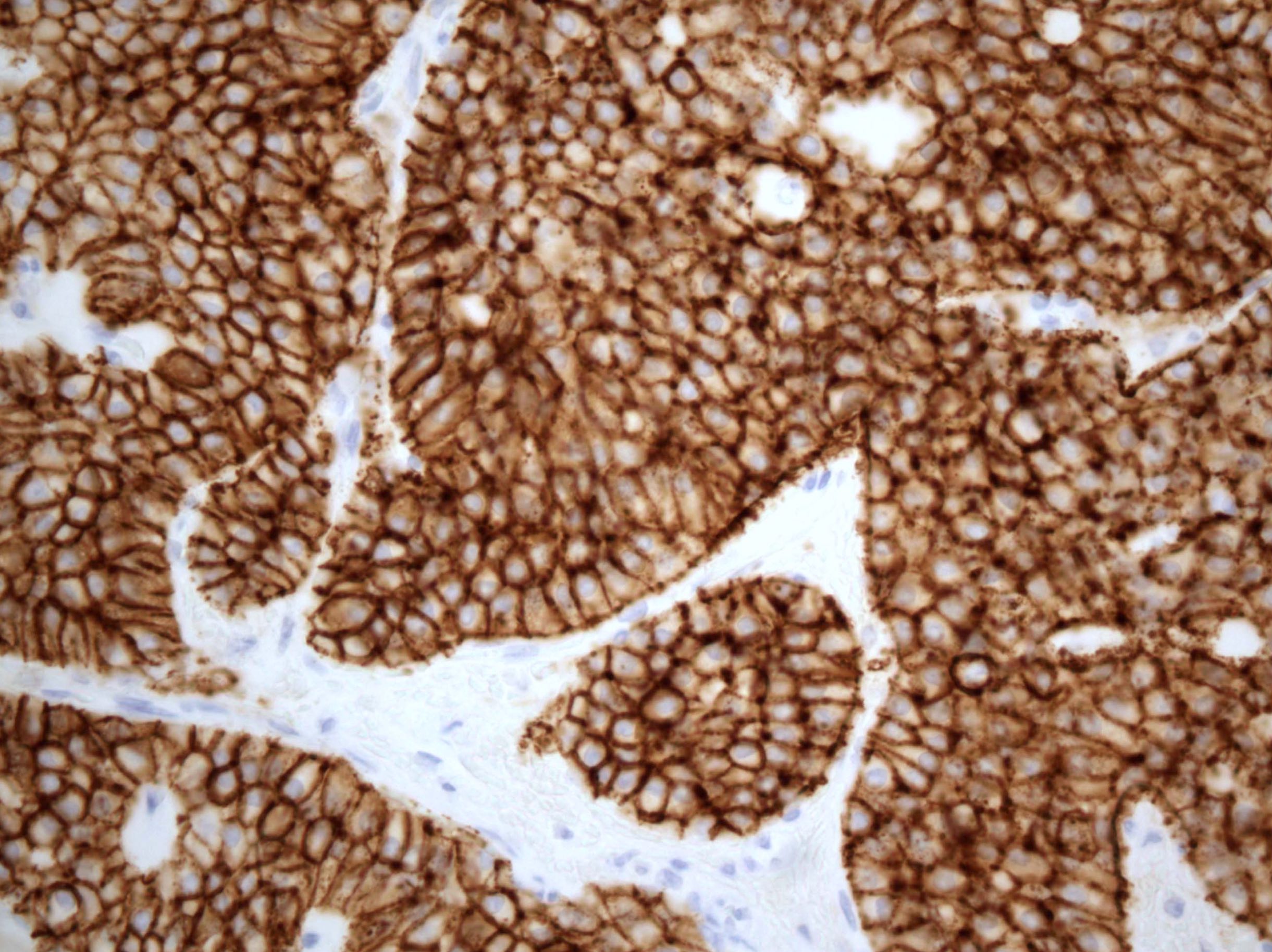
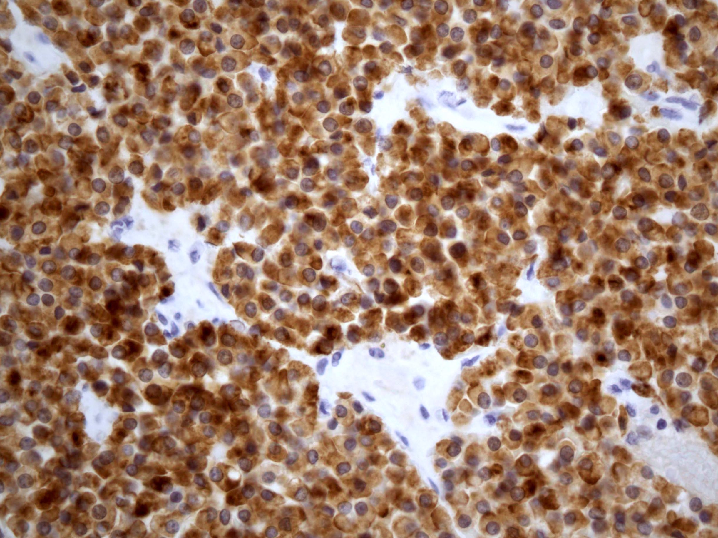
Synaptophysin in strumal ovarian carcinoid
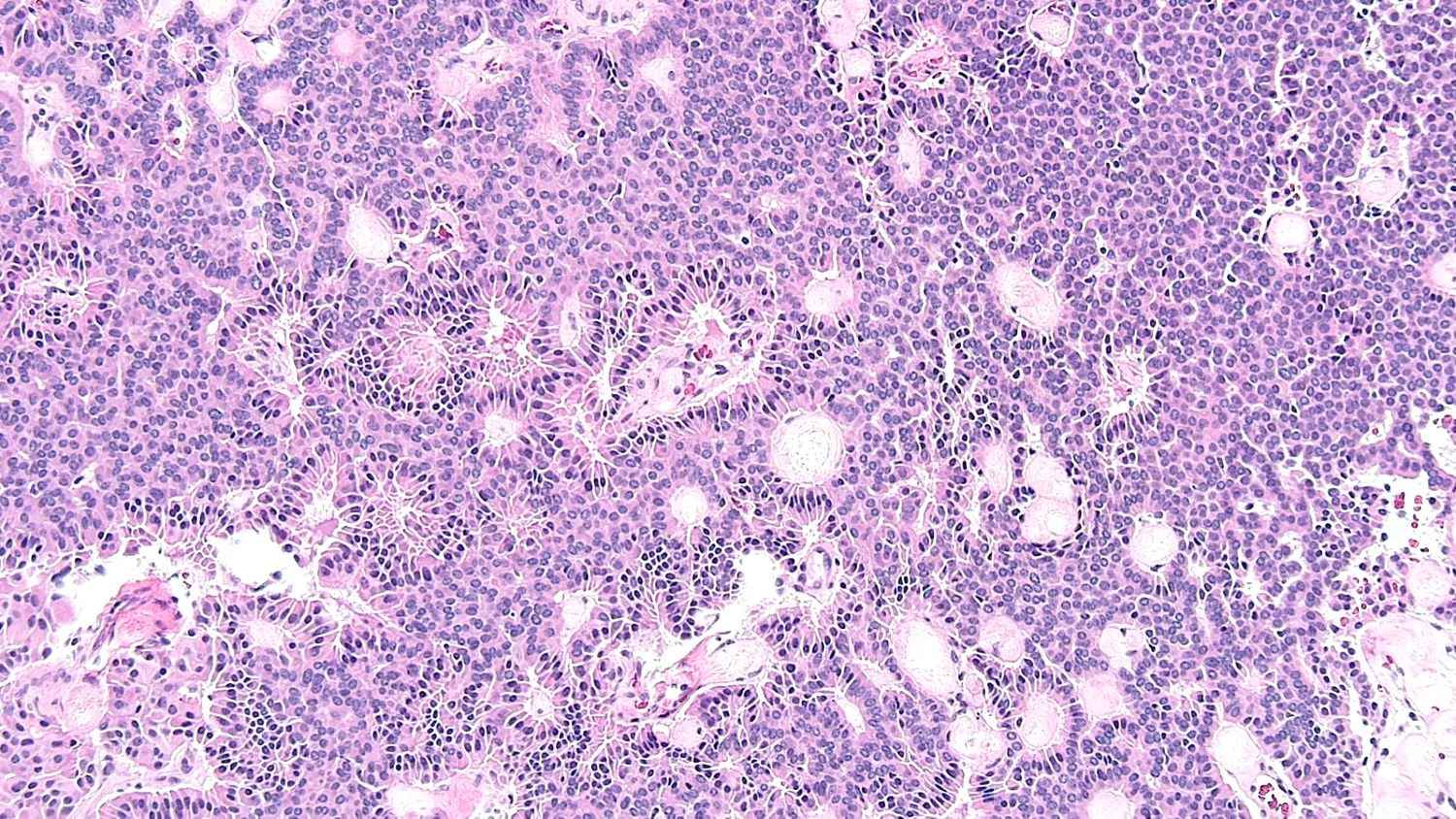
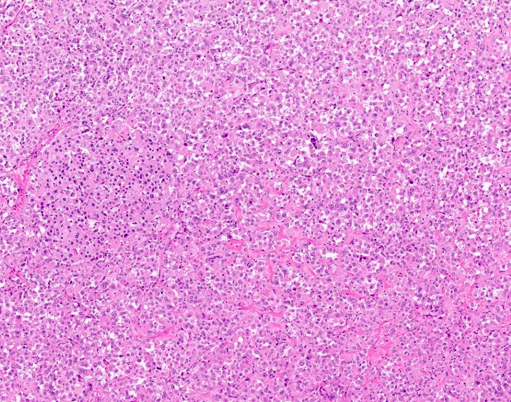
Insular carcinoid
Insular carcinoid
Insular carcinoid
TTF1
AE1 / AE3
CDX2
Synaptophysin
Chromogranin
Inhibin
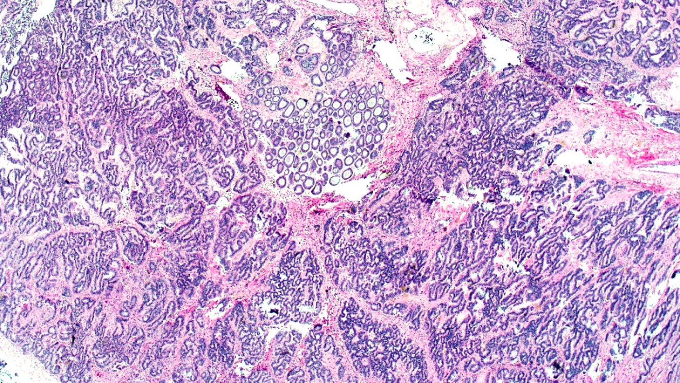
Mucinous carcinoid
Trabecular tumor
Germ cell neoplasms of the ovary - ovarian carcinoid tumor
by Dr. Wafaey Badawy
Contributed by Hayam Aiad, M.D.
Cut section
Contributed by Hayam Aiad, M.D.
Higher power
Low power
Carcinoid in meckels
High power showing uniform monotonus nuclei
Contributed by Mona Kandil, M.D., Ph.D. and AFIP

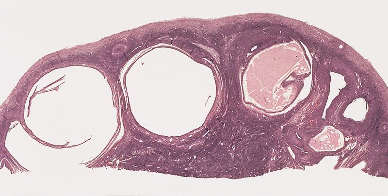
Ovary

Uterus

Omental deposit
Fallopian tube
Contributed by Mona Kandil, M.D., Ph.D.
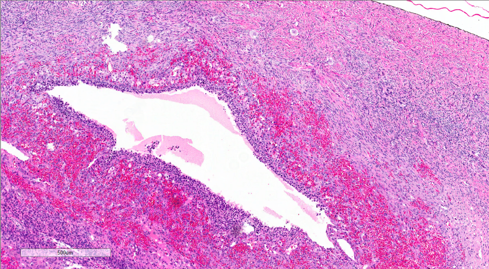
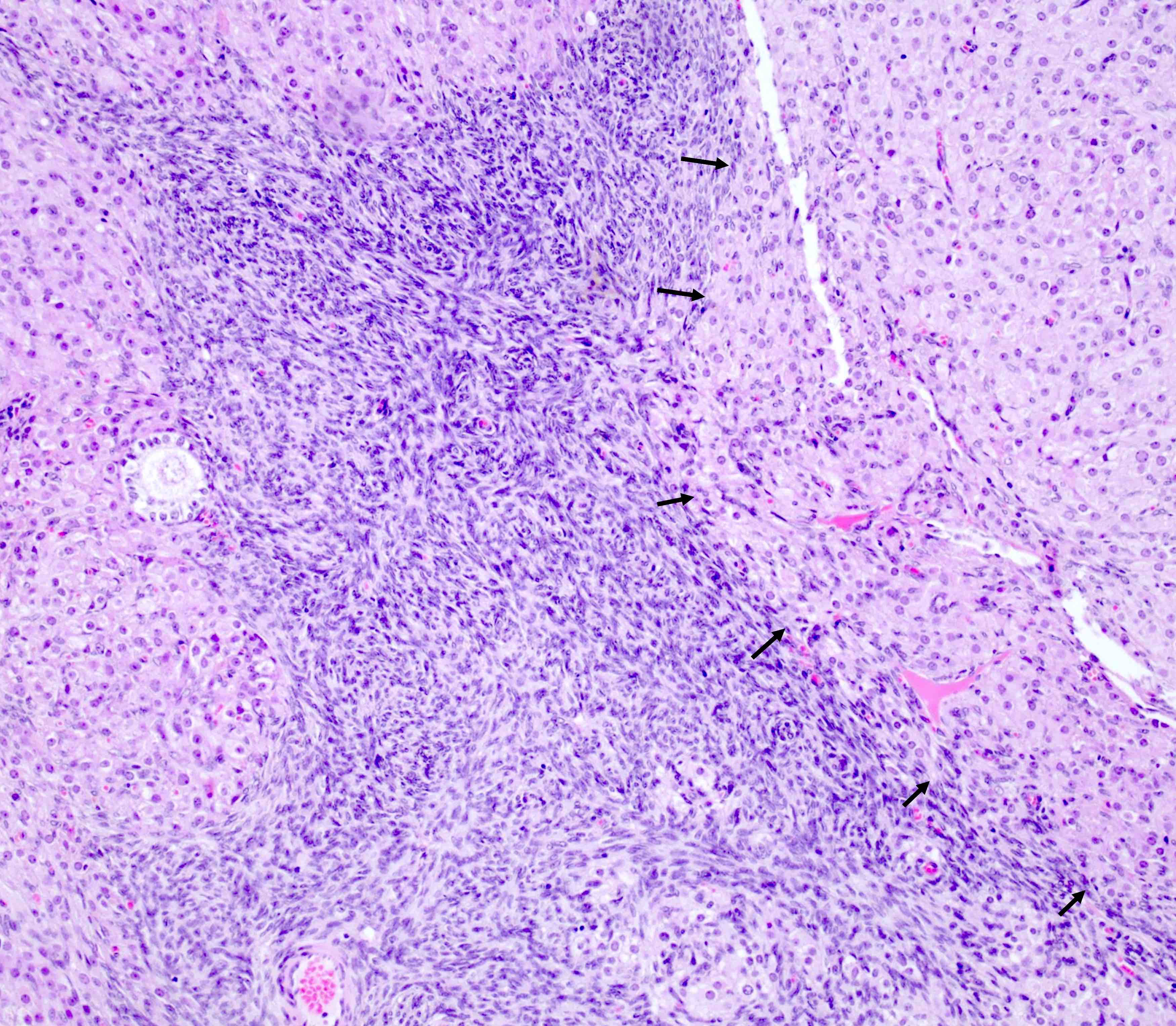
Malignant stroma

High grade malignant glands

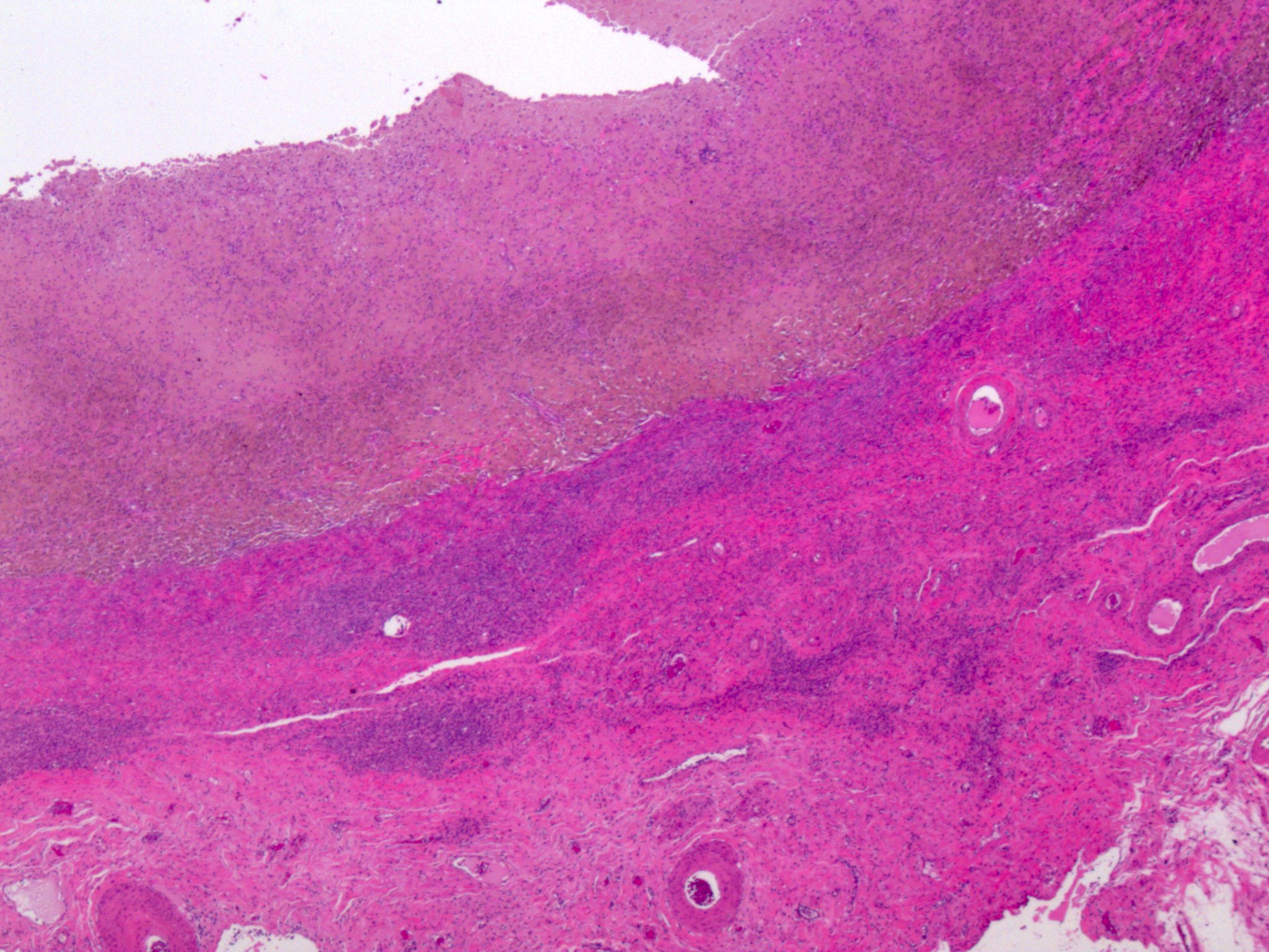
Necrotic foci

High grade epithelial tumor cells and necrosis
High grade tumor cells with mitotic figures
Images hosted on other servers:


Rhabdomyosarcoma component
Images hosted on other servers:

Uterus, growth
in cervix
extending up to
endocervical canal

Bisected uterus, cut surface ovary
Contributed by Carlos Parra-Herran, M.D.
Endocervical carcinoma primary
Images hosted on other servers:

Foci of metastatic squamous cells, H&E

CIN

Metastases


p63, CK5/6, CK7, CK20
Image hosted on other servers:

High grade SIL
Images hosted on other servers:

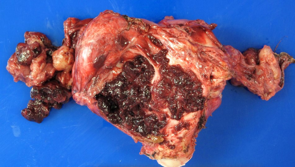
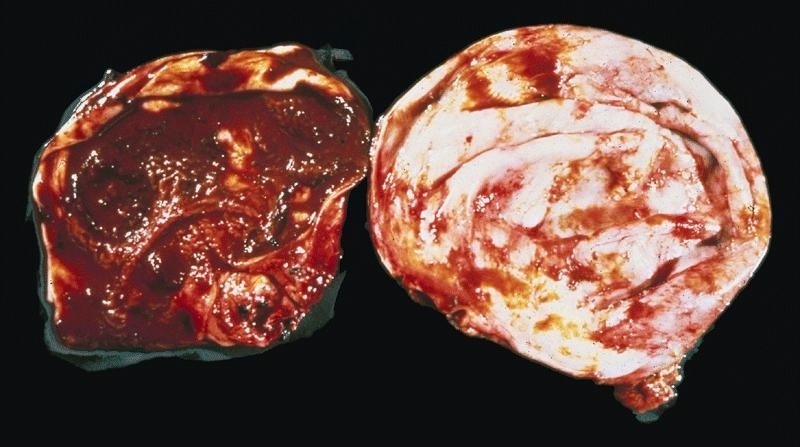
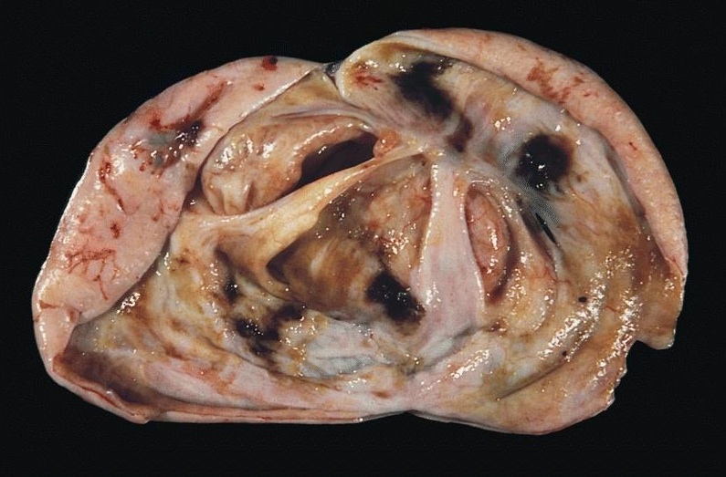
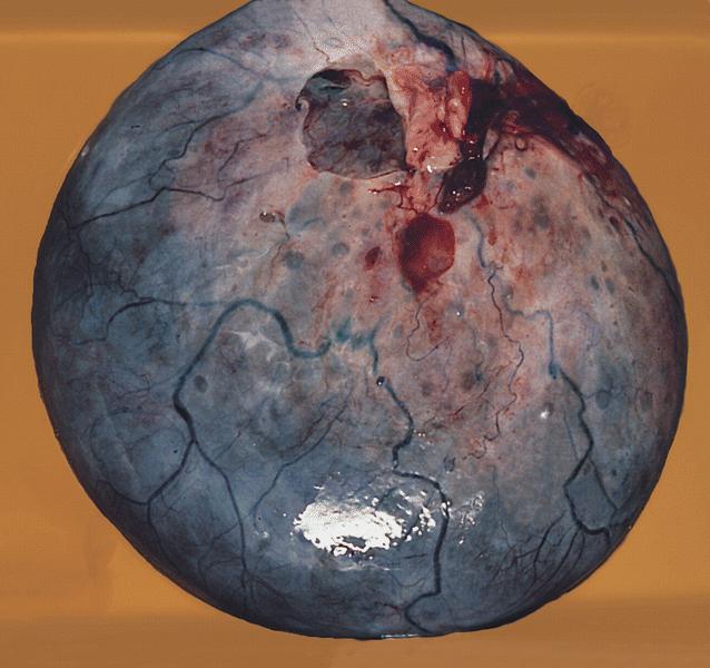
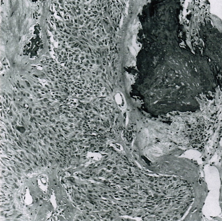
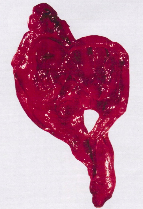
Hemorrhagic mass

Ovarian mass

TAHBSO specimen
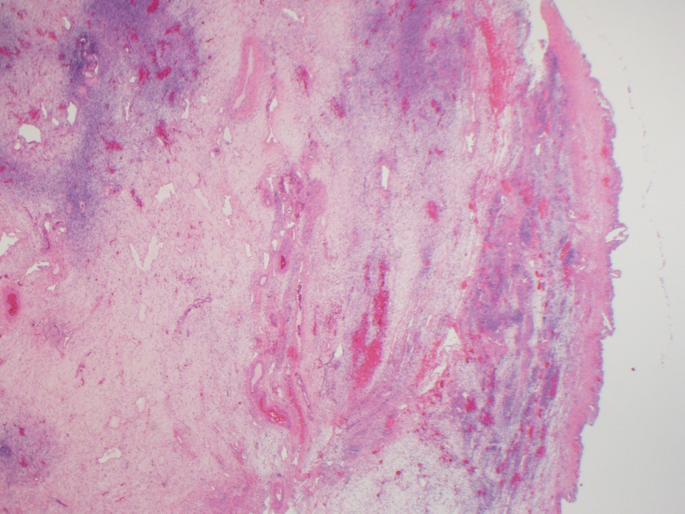
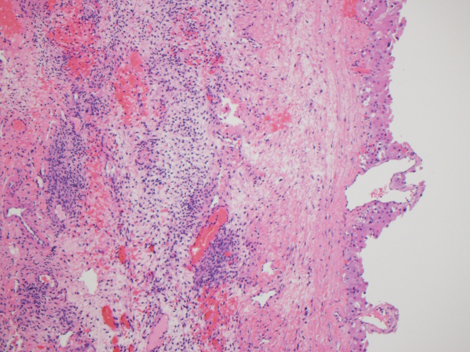
AFIP images

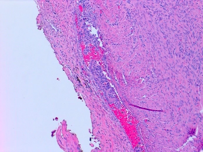
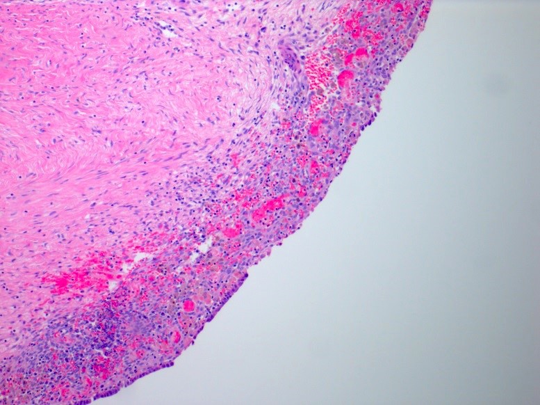
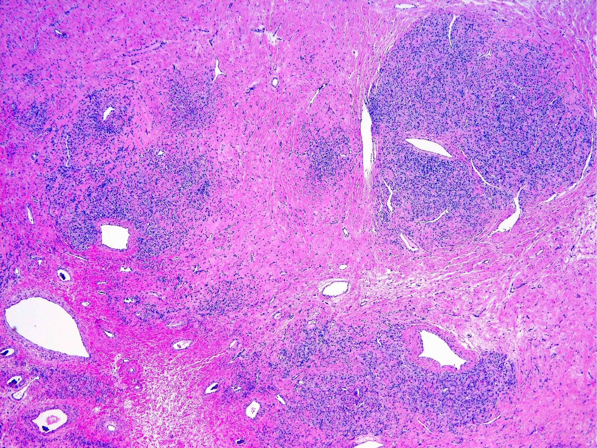
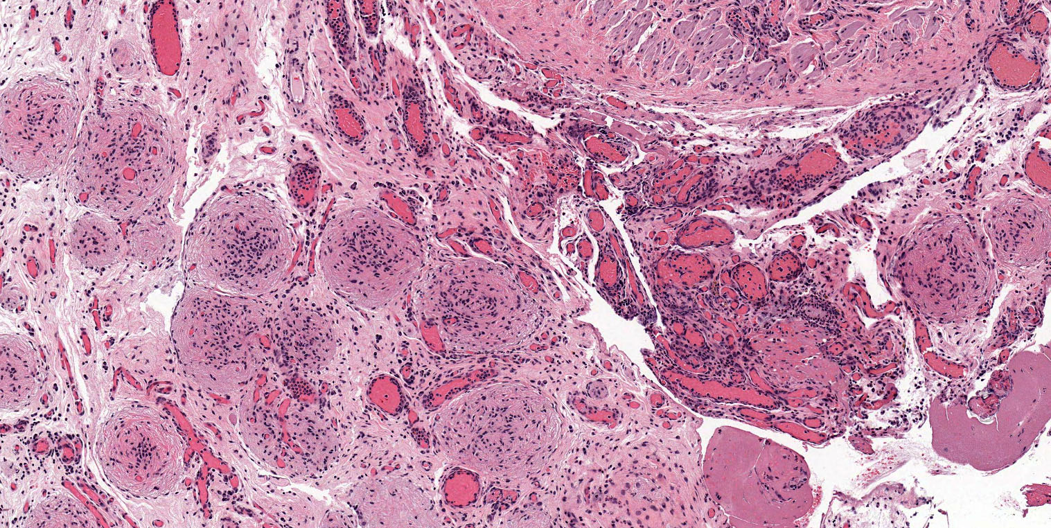
Plexiform pattern
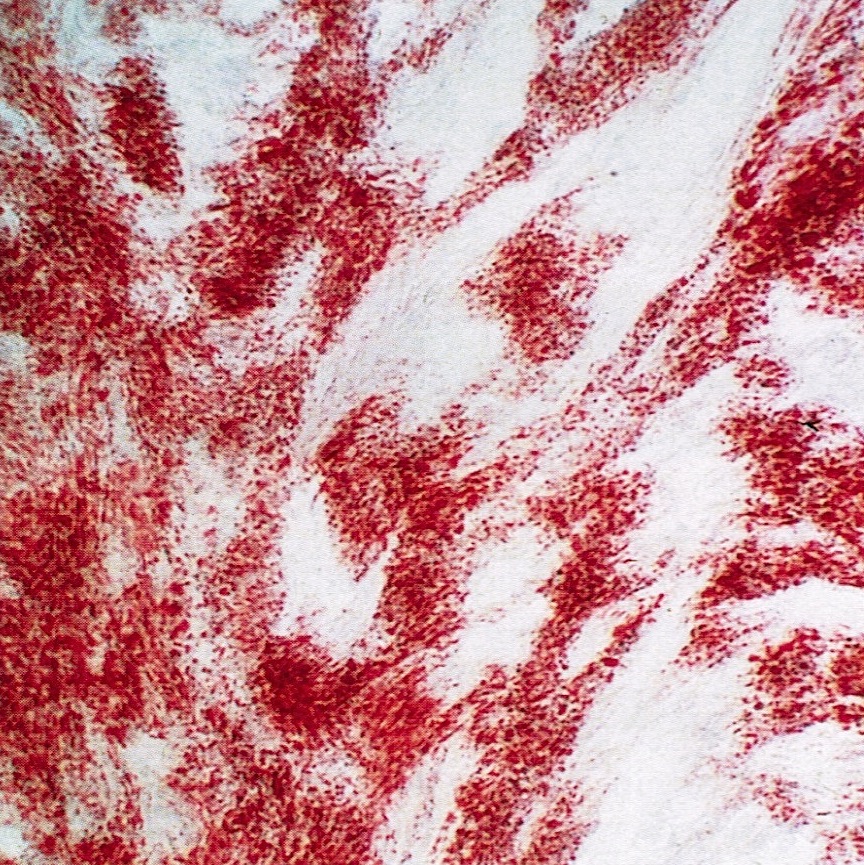
Images hosted on other servers:


H&E stain

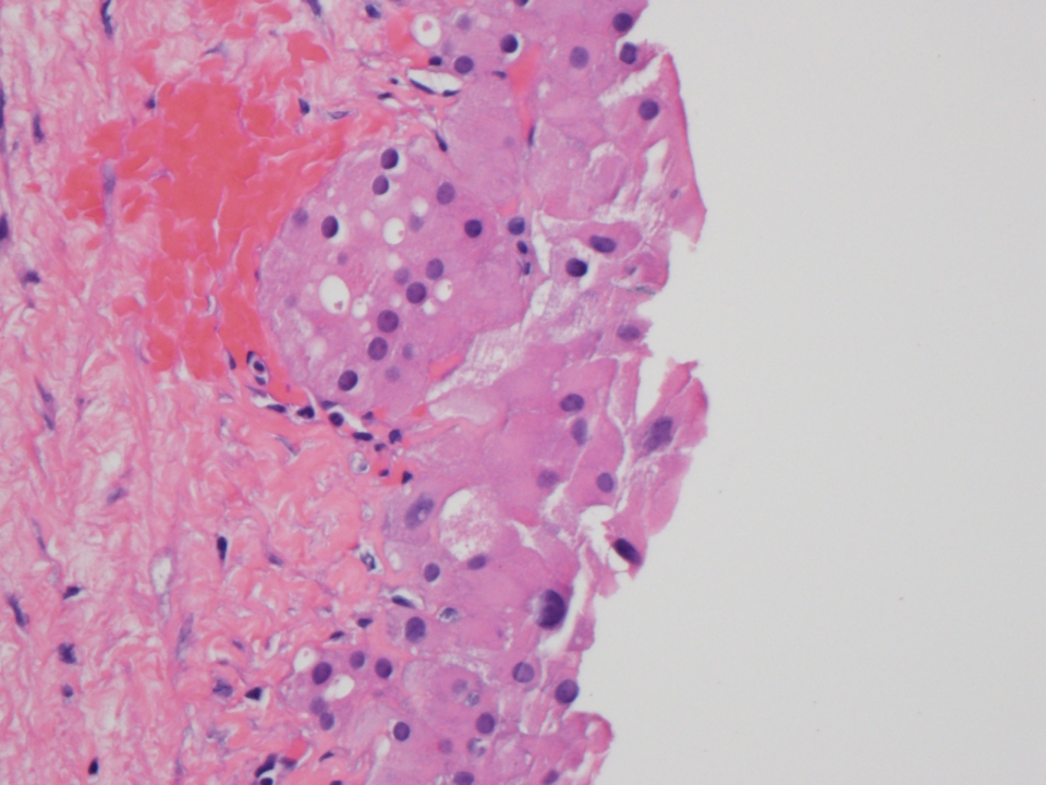
Atypical syncytio
cytotrophoblast

β hcG+

Contributed by Lucy Ma, M.D.
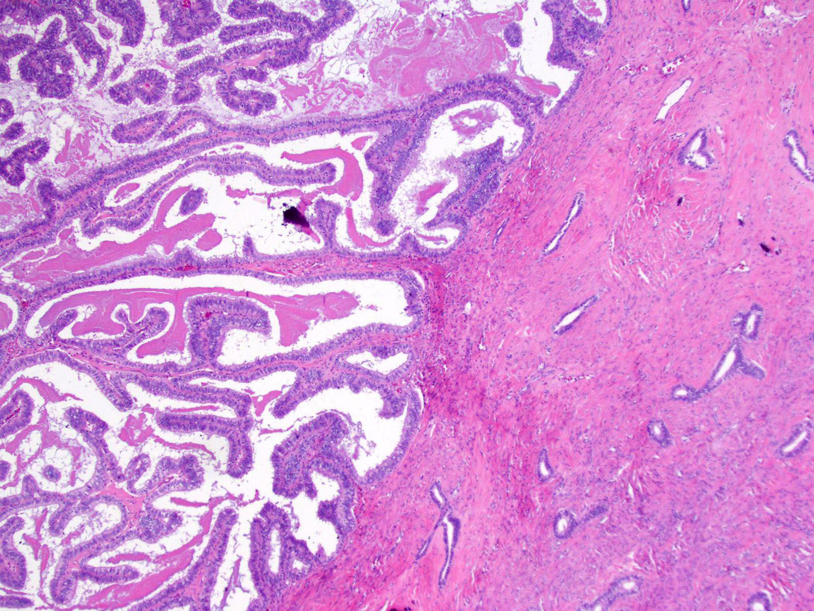
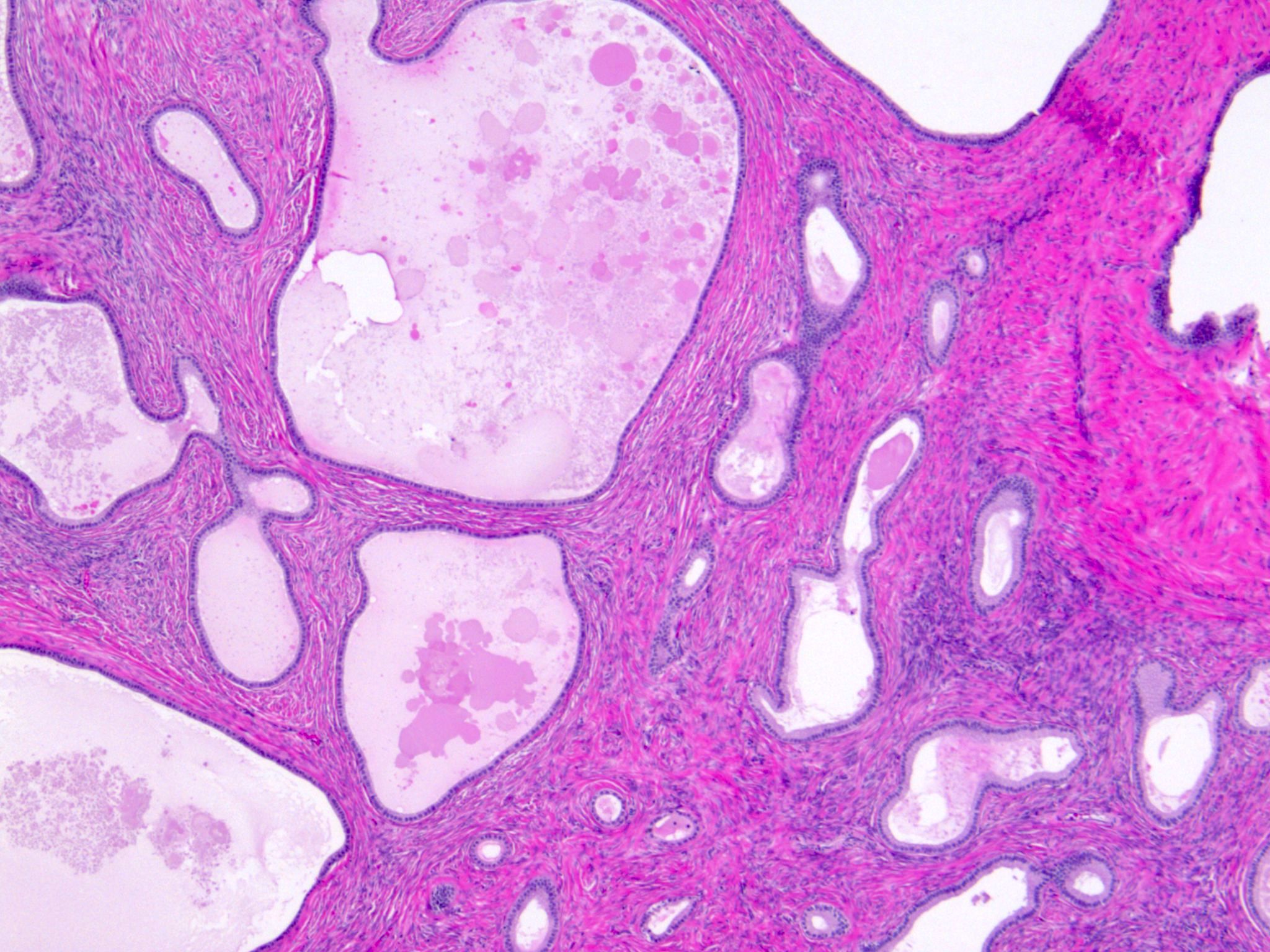
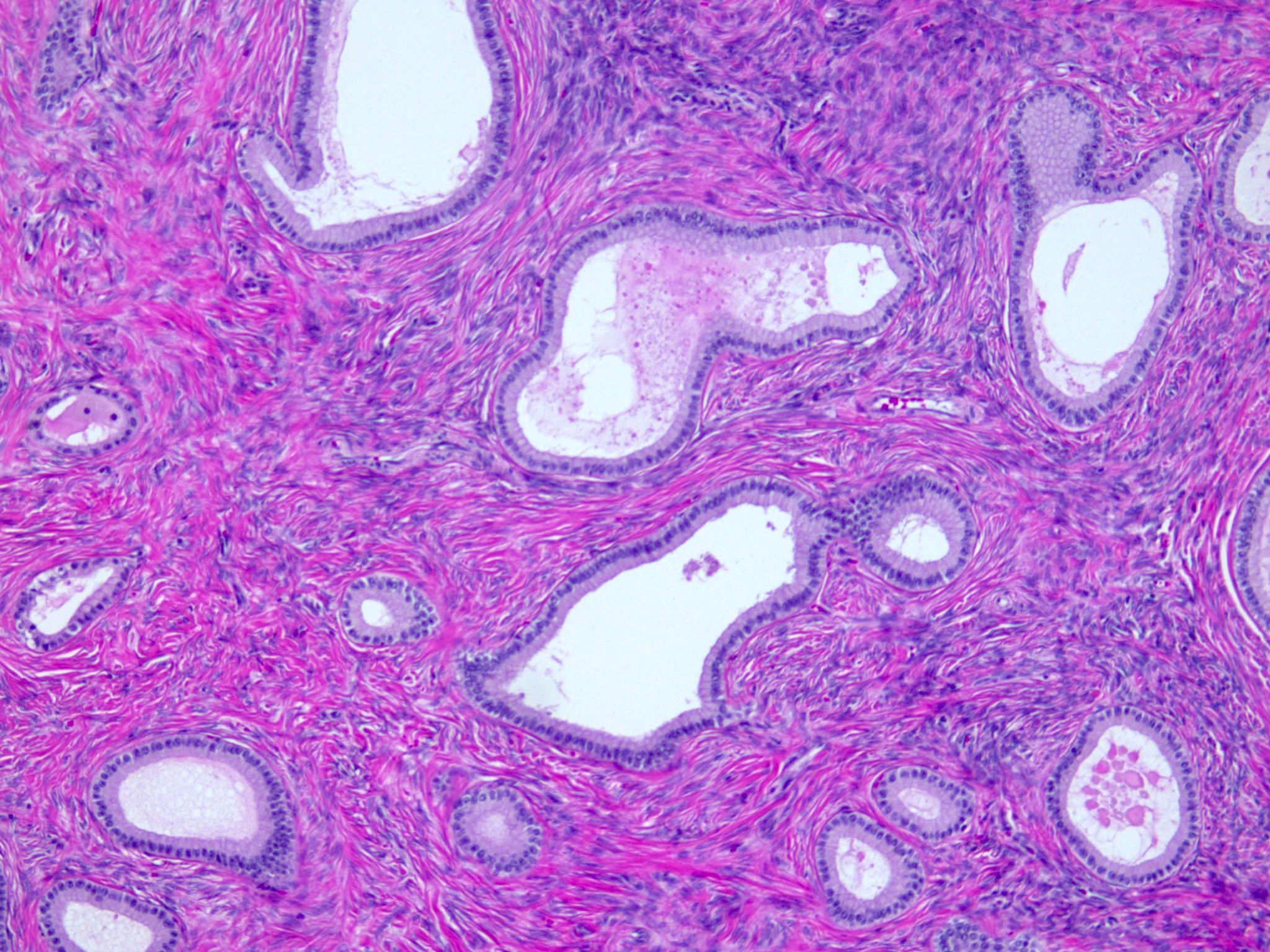
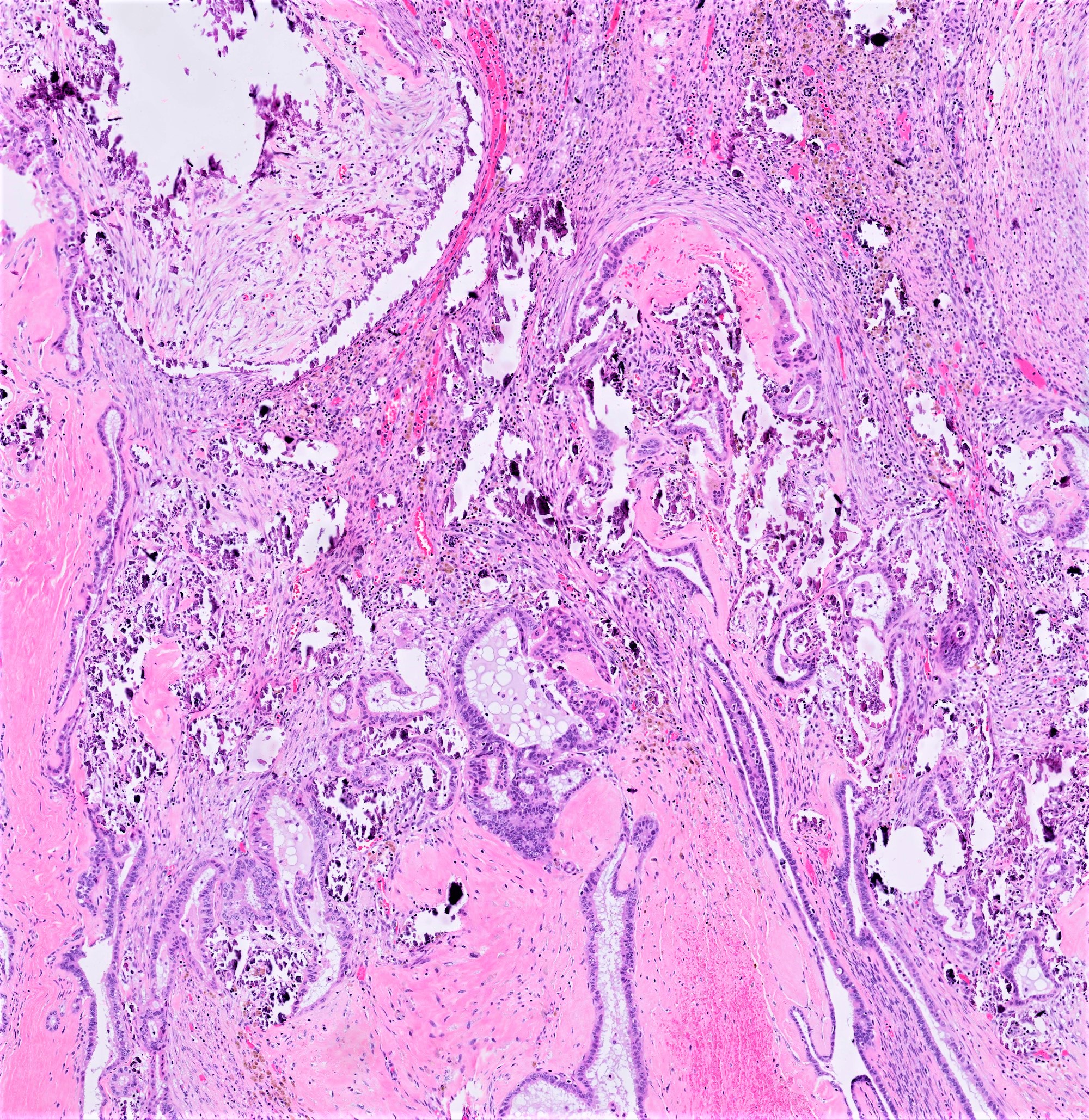
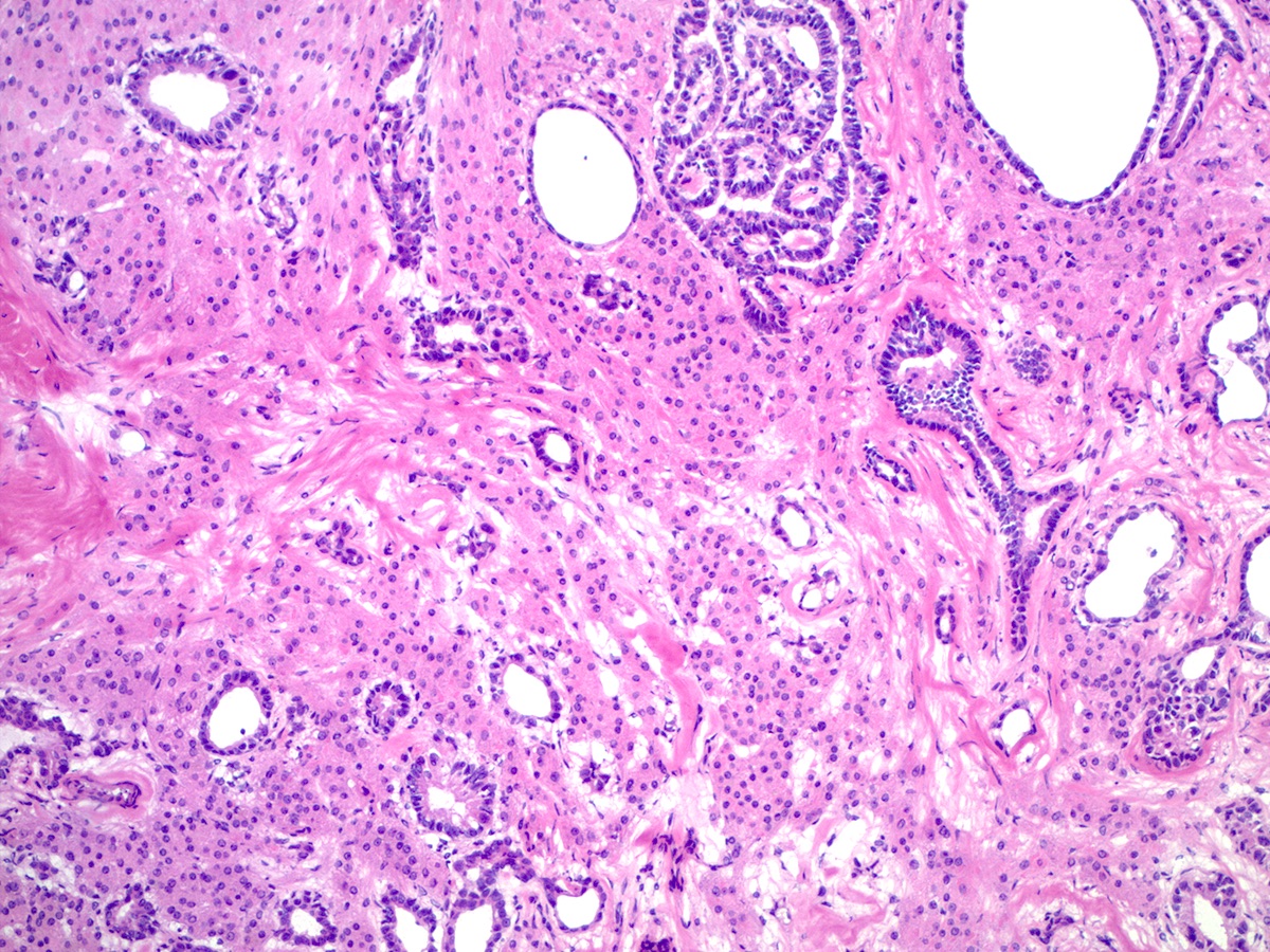
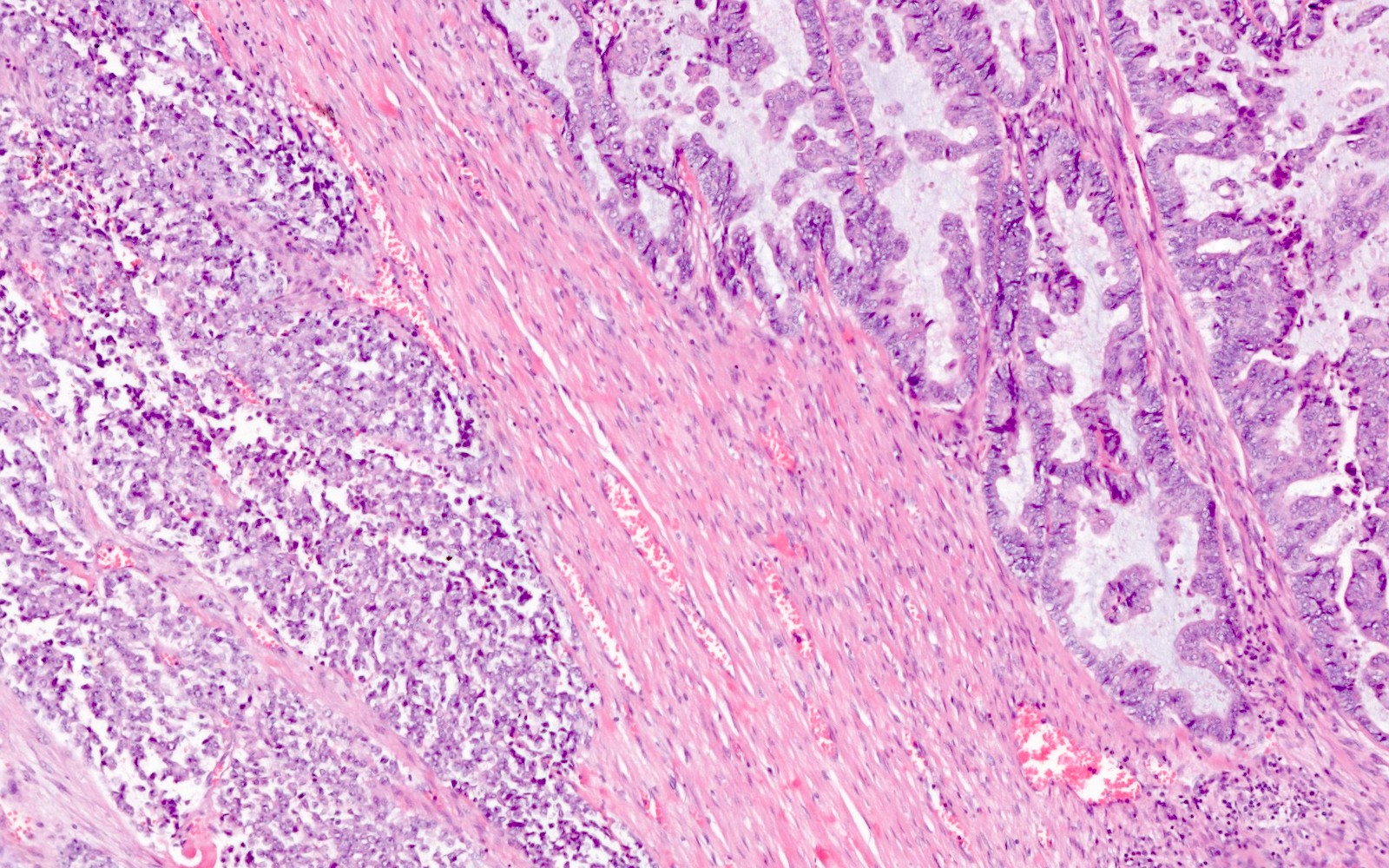
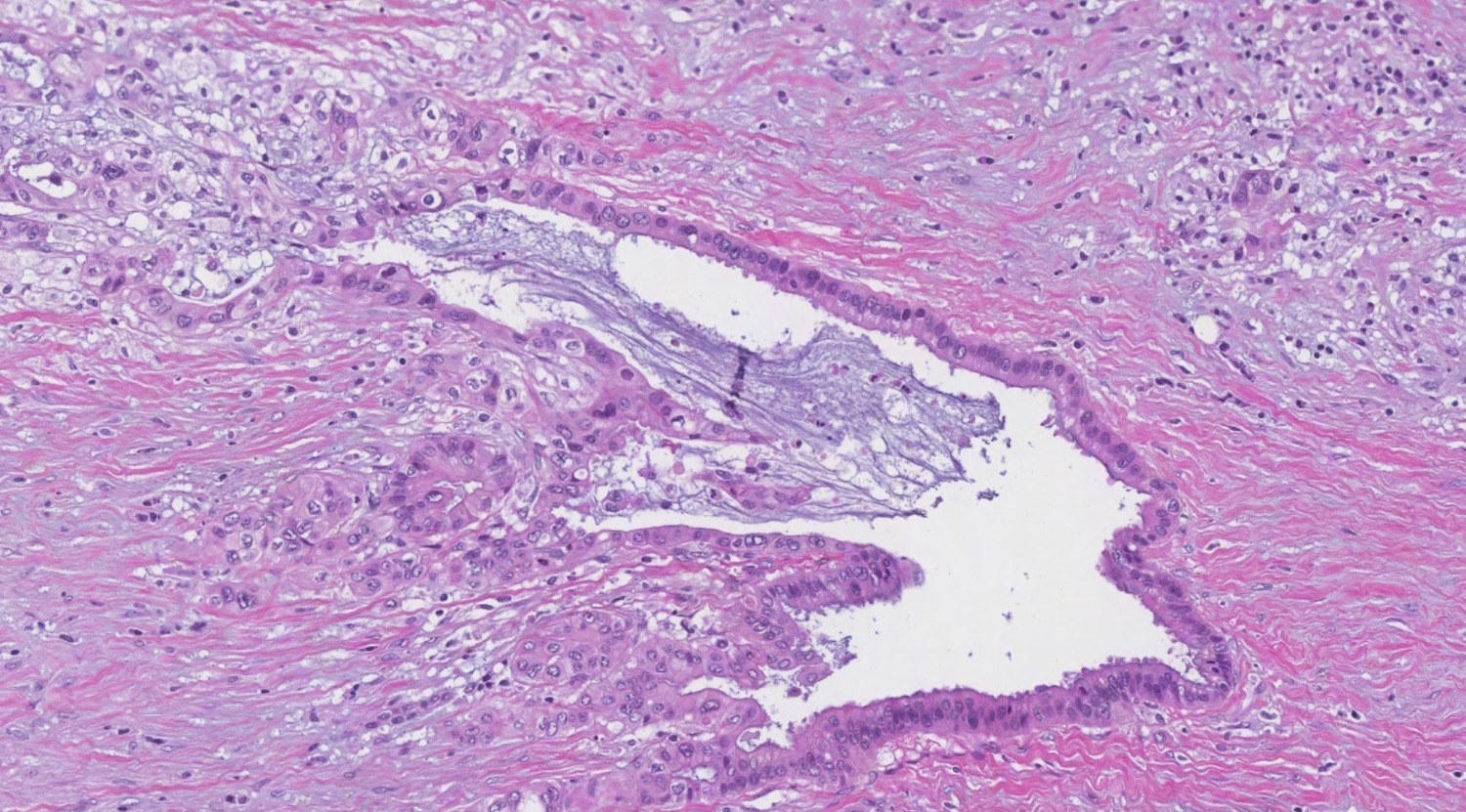
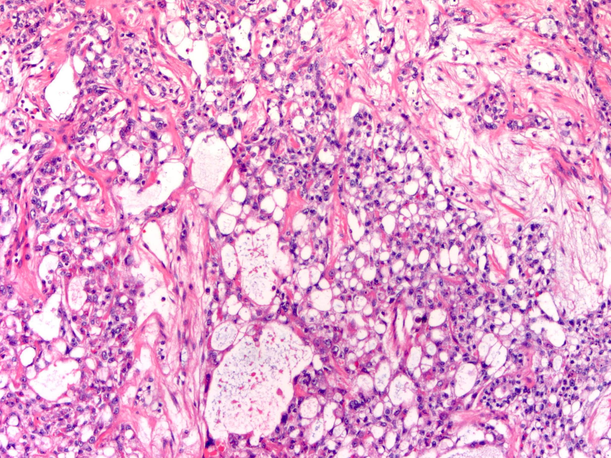
Small glands / cysts
Low grade cytologic atypia
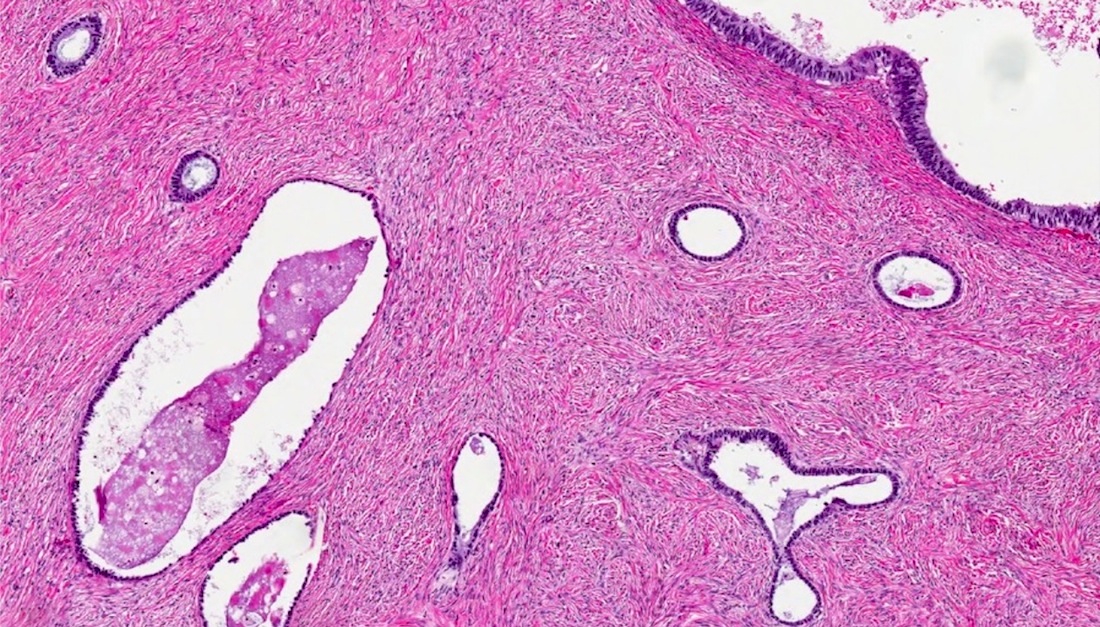
Adenofibromatous component
Images hosted on other servers:
Ovarian mass on ultrasound
Contributed by Ayse Ayhan, M.D. and AFIP images
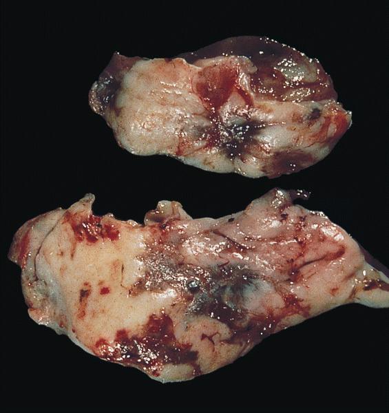
External surface
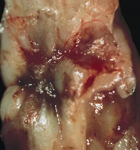
Cut surface
External surface
Cut surface
Polypoid areas in a cyst
Contributed by Gulisa Turashvili, M.D., Ph.D., Sonali Lanjewar, M.D., M.B.B.S., Semir Vranić, M.D., Ph.D. and AFIP images
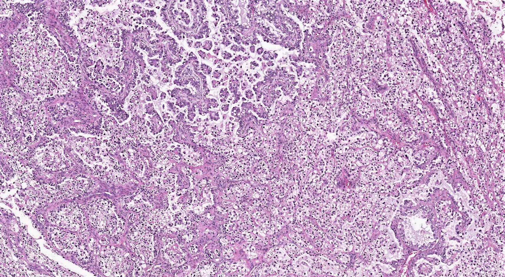
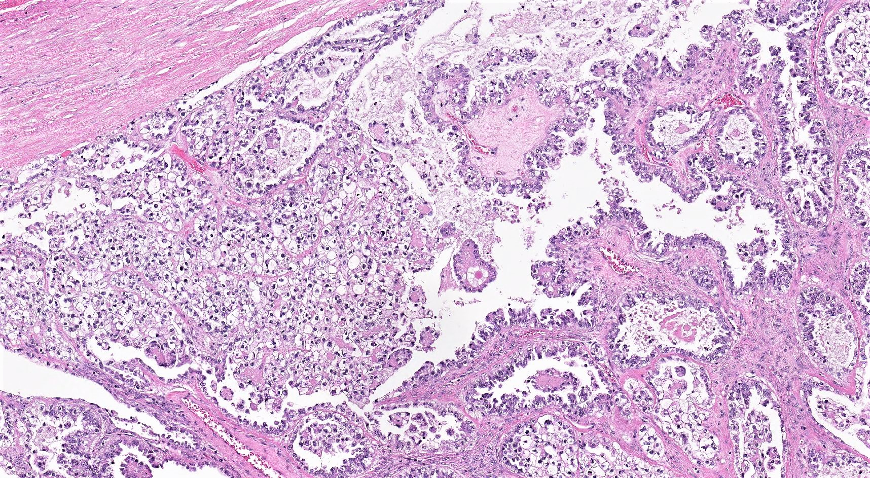
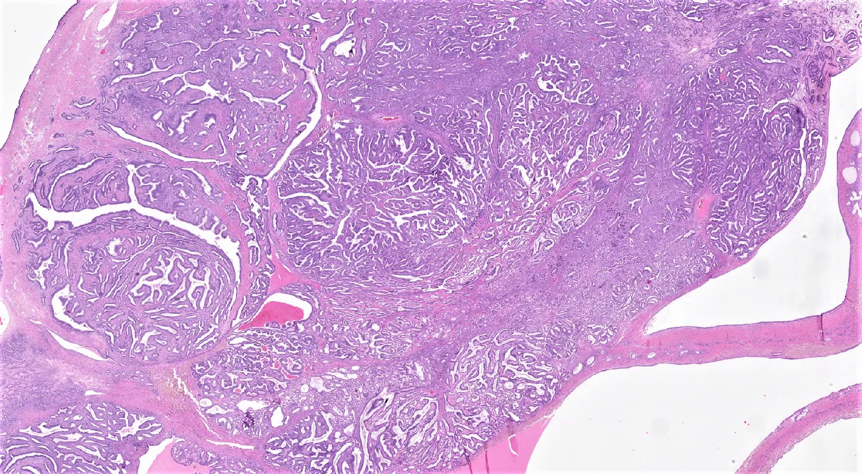
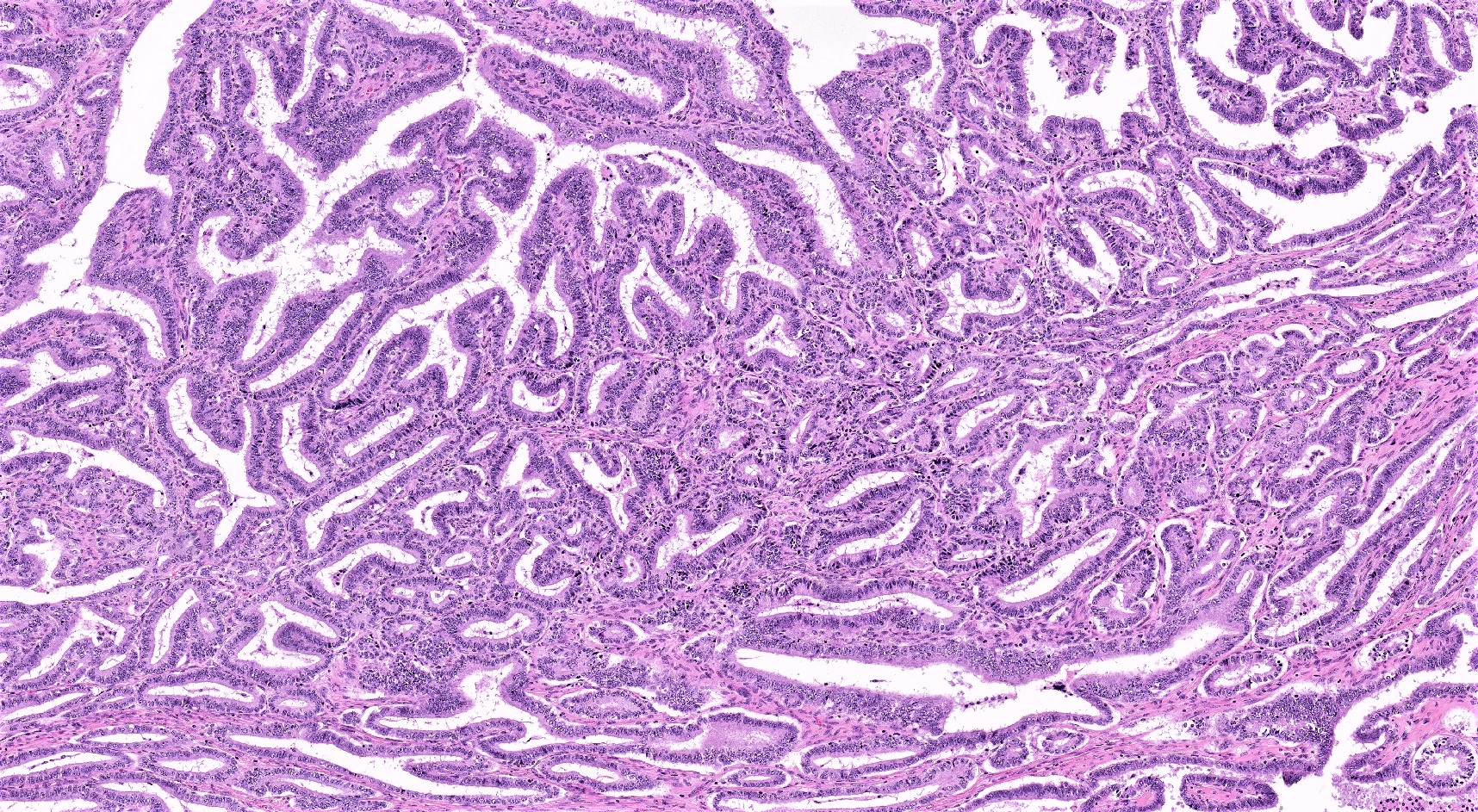
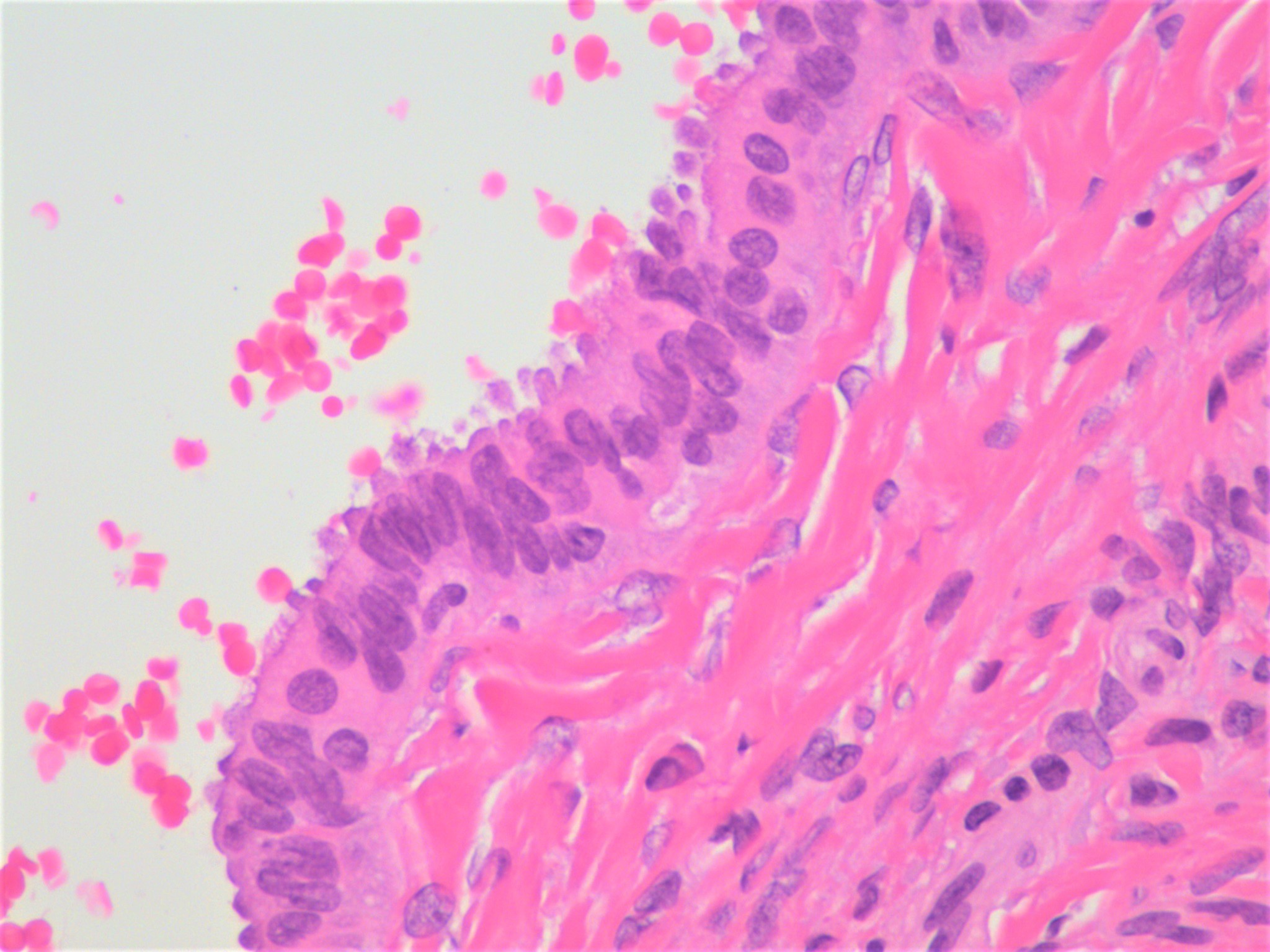
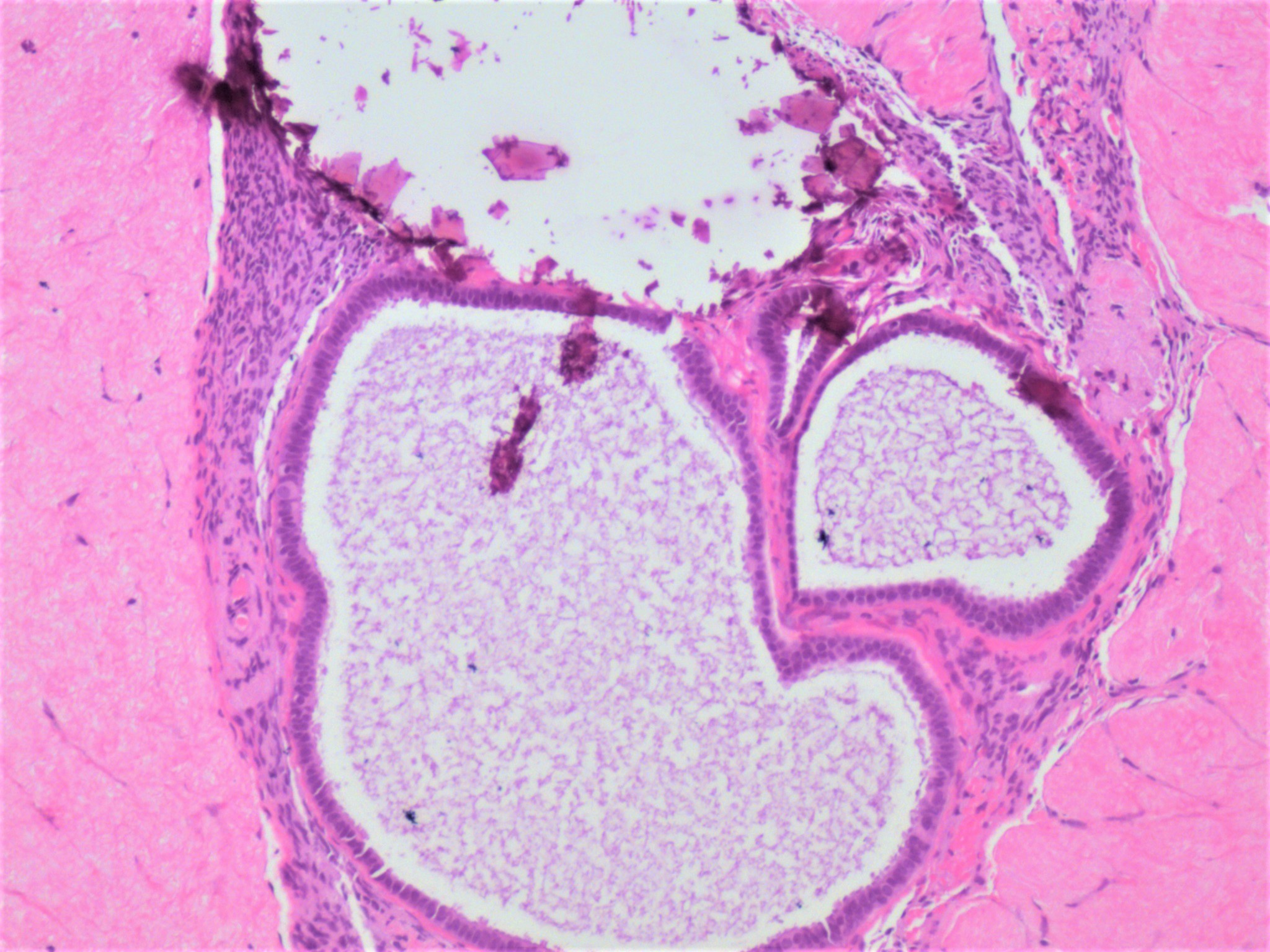
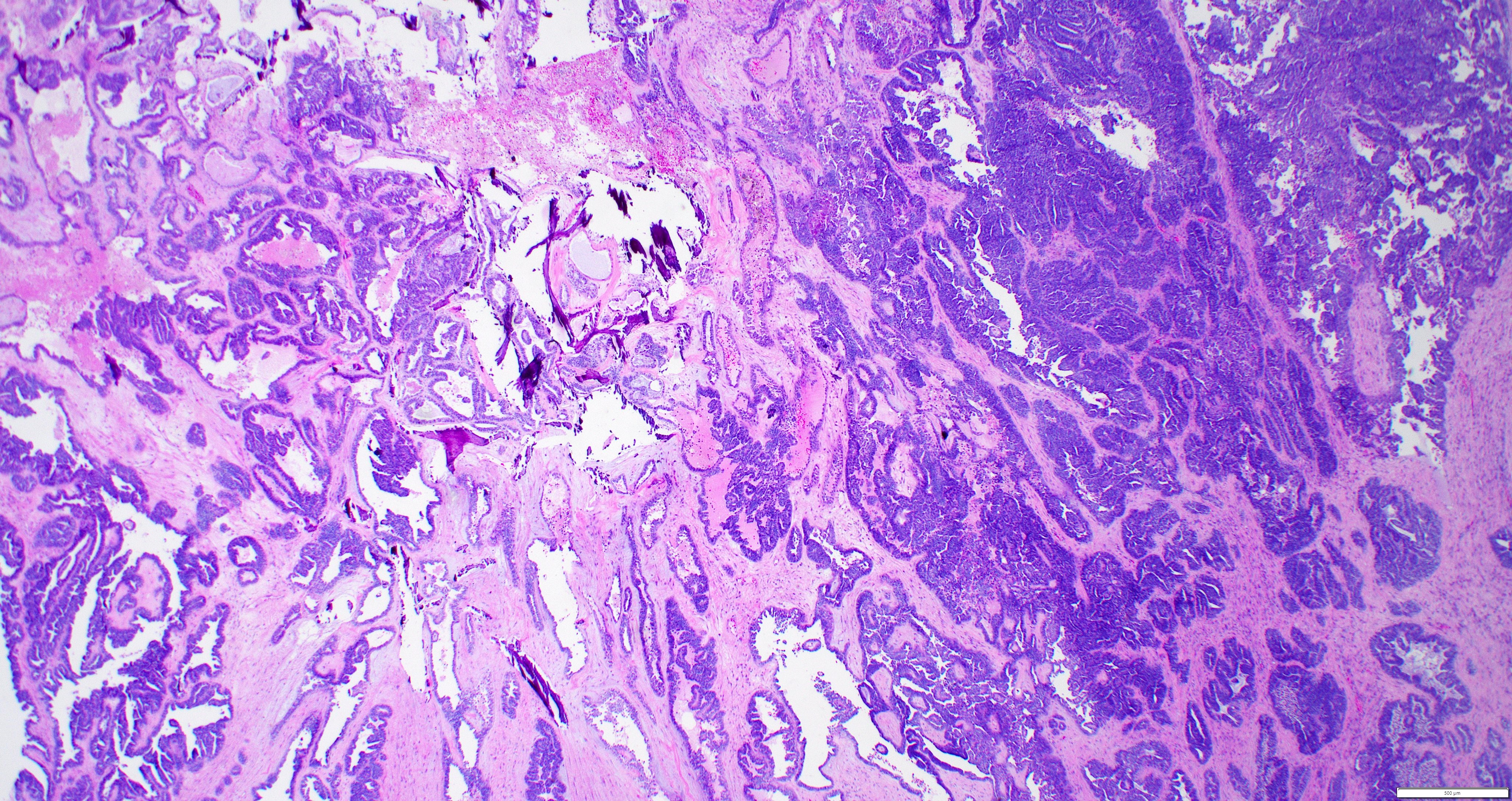
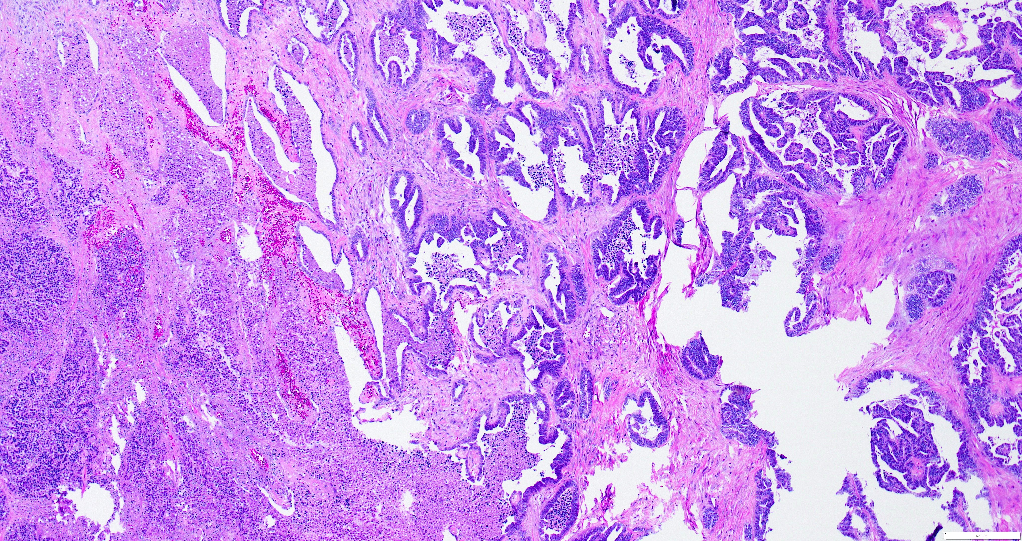
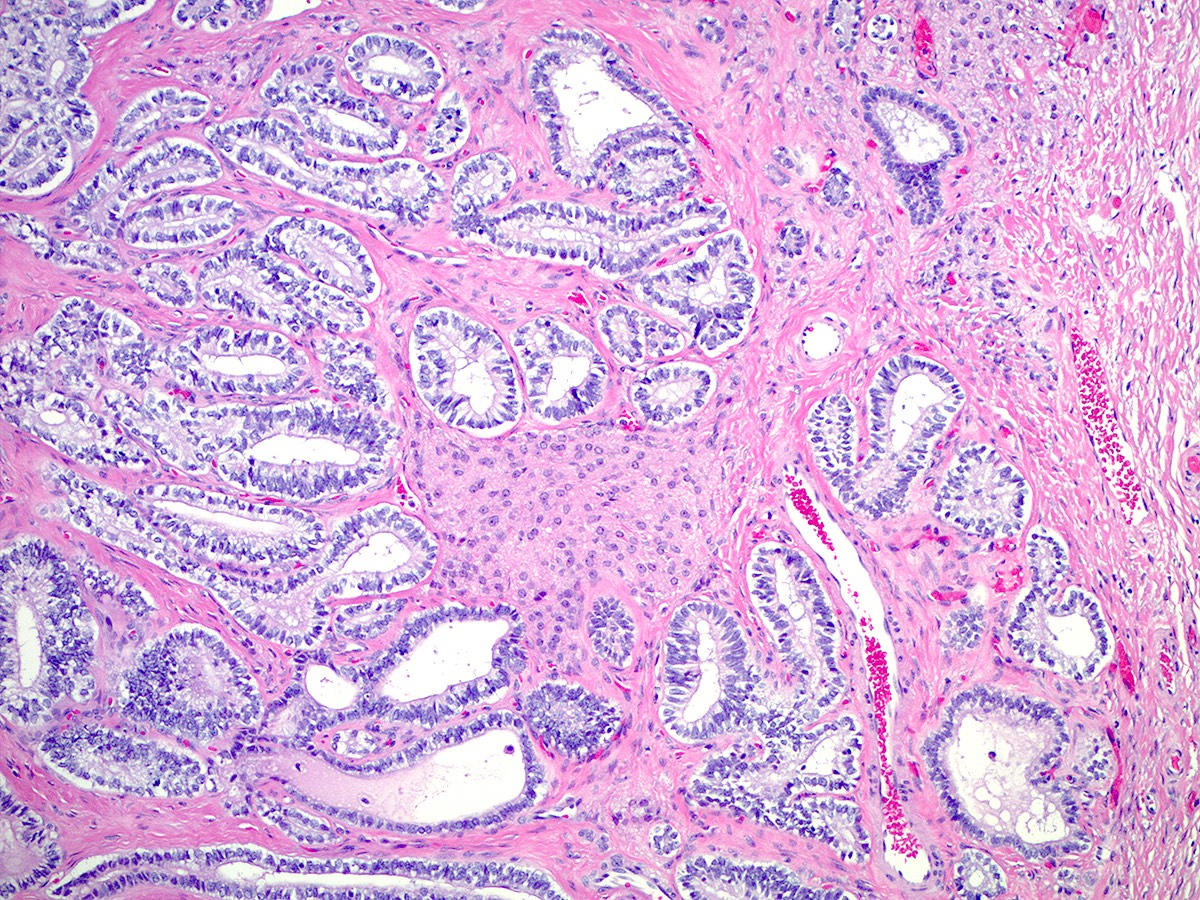
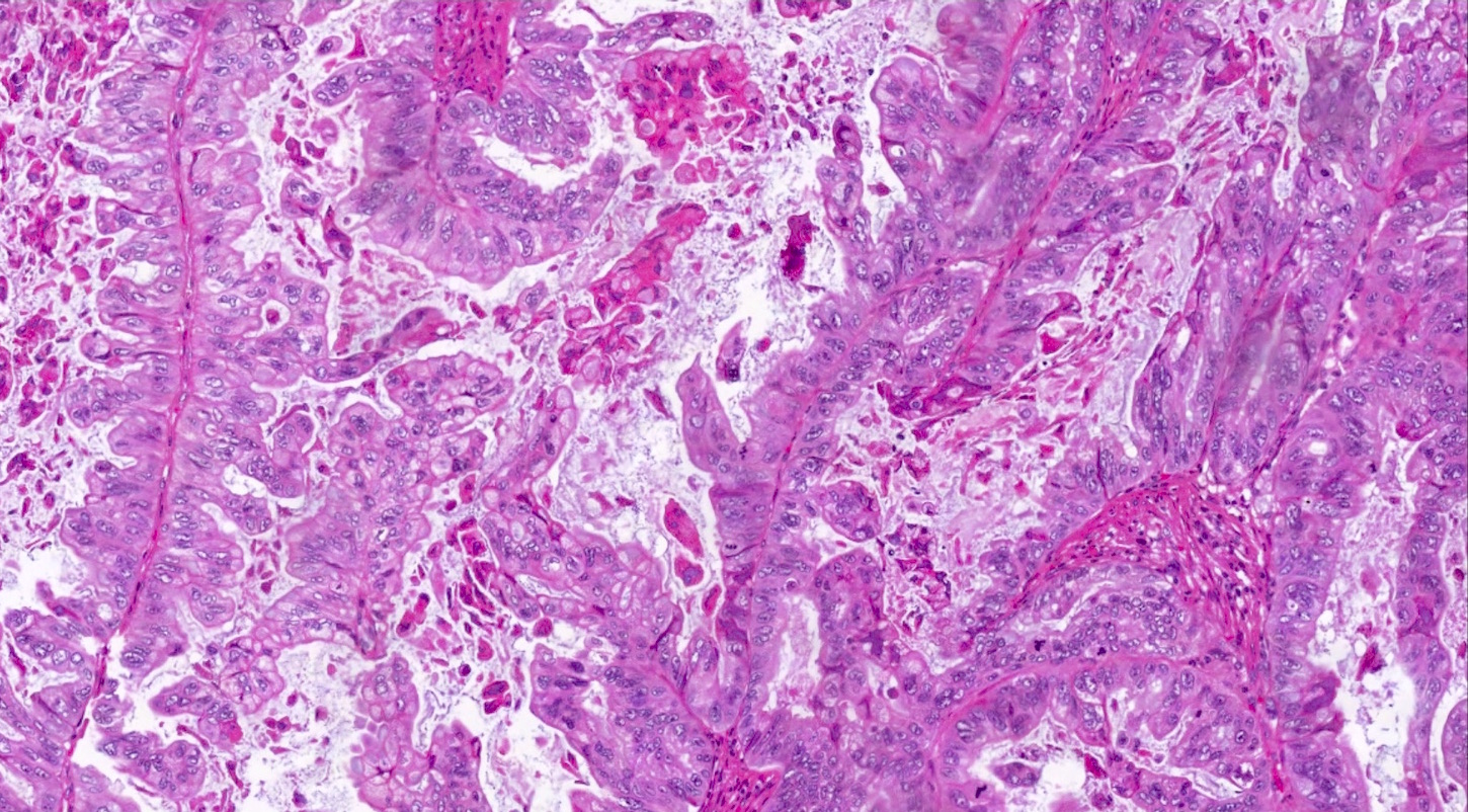
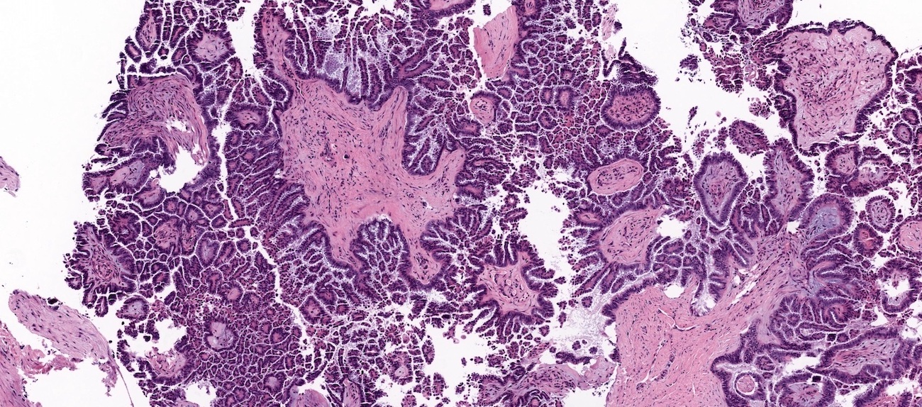
Variable architecture

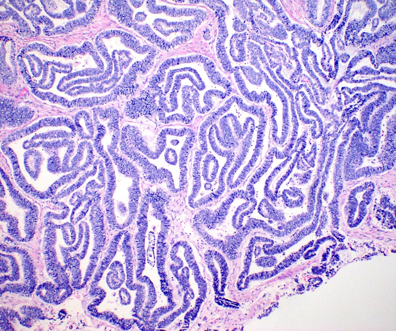
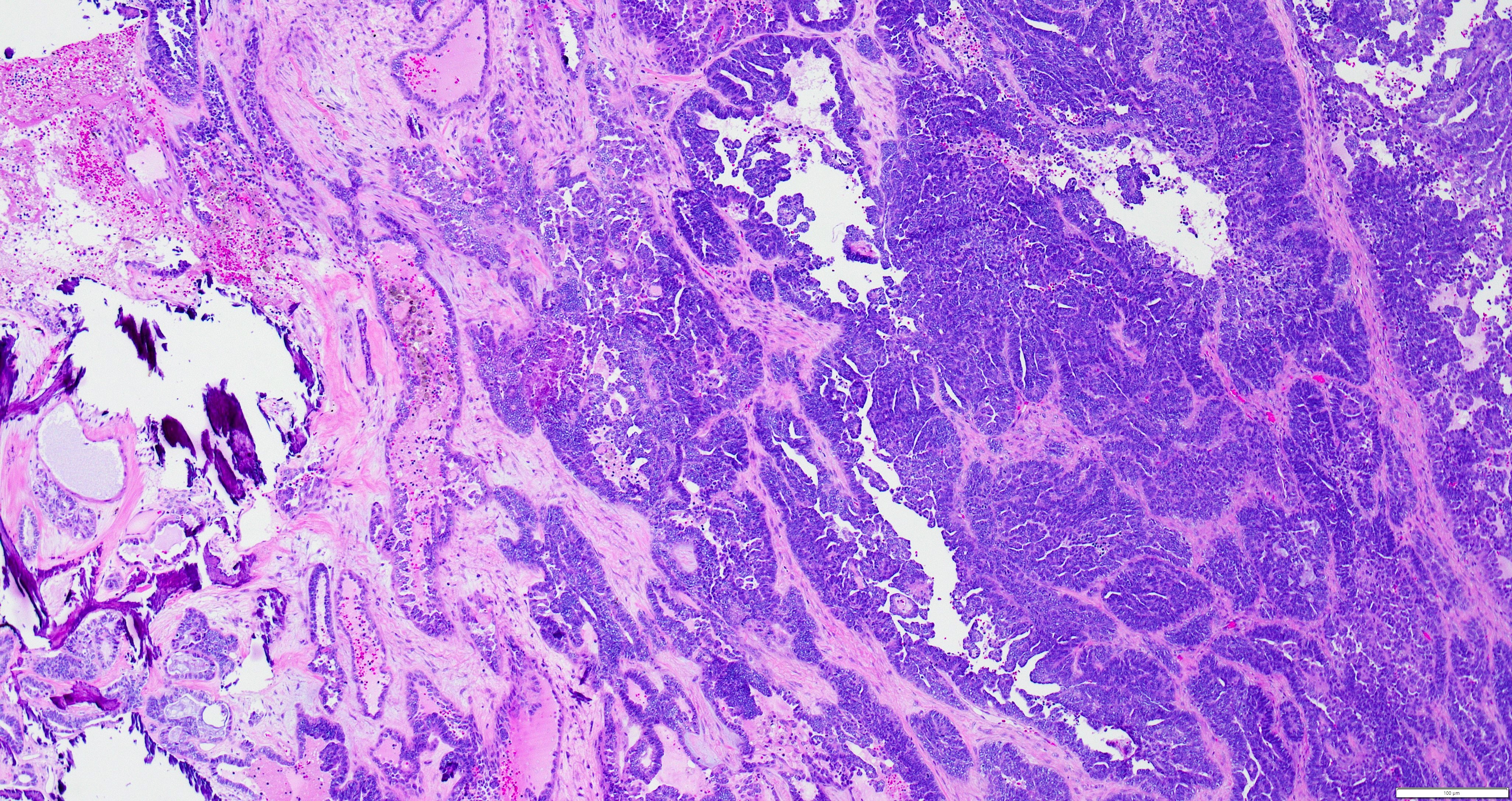
Papillary architecture
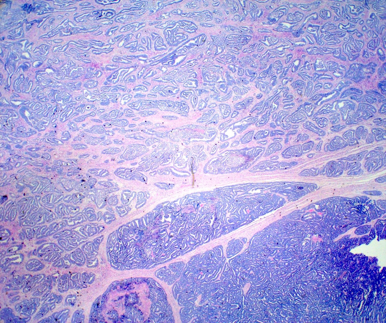
Tubulocystic architecture
Tubulocystic and papillary architecture
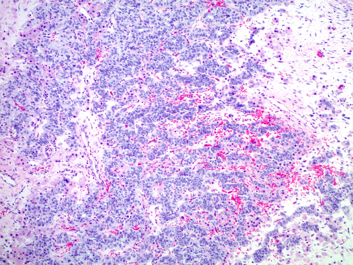
Solid architecture
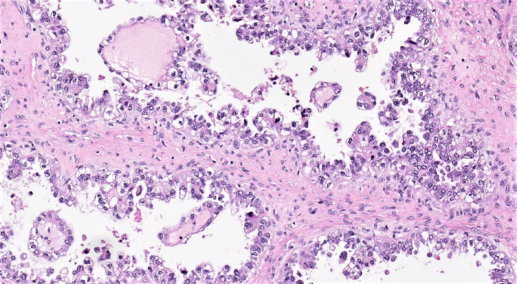
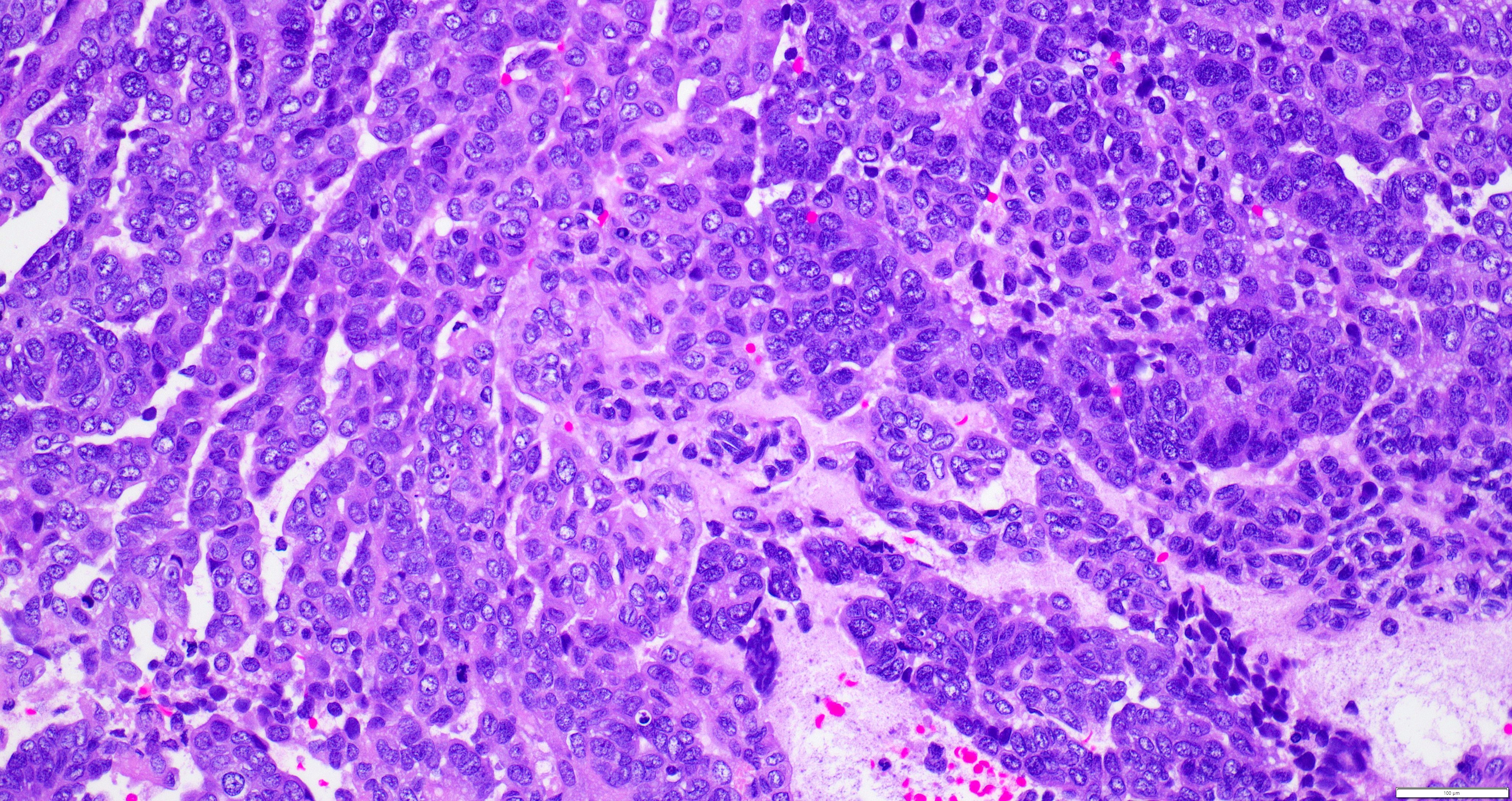
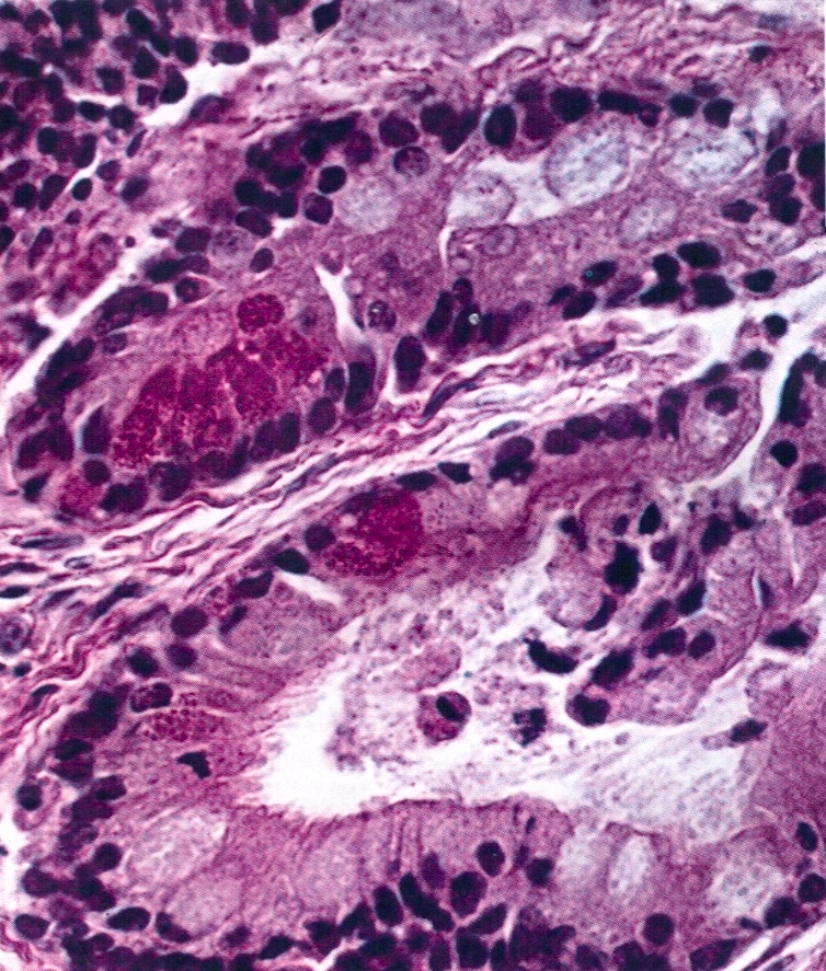
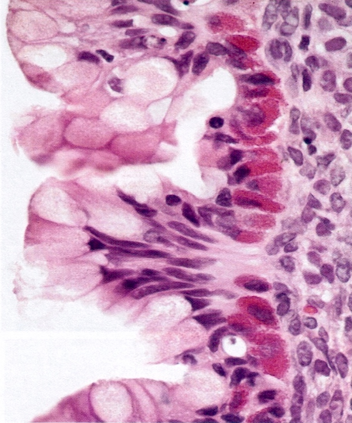
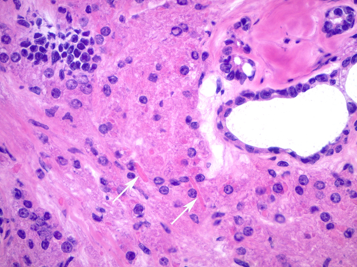
Targetoid cells
Left: cystic pattern; right: hobnail cells

Adenofibroma component
Hyaline bodies
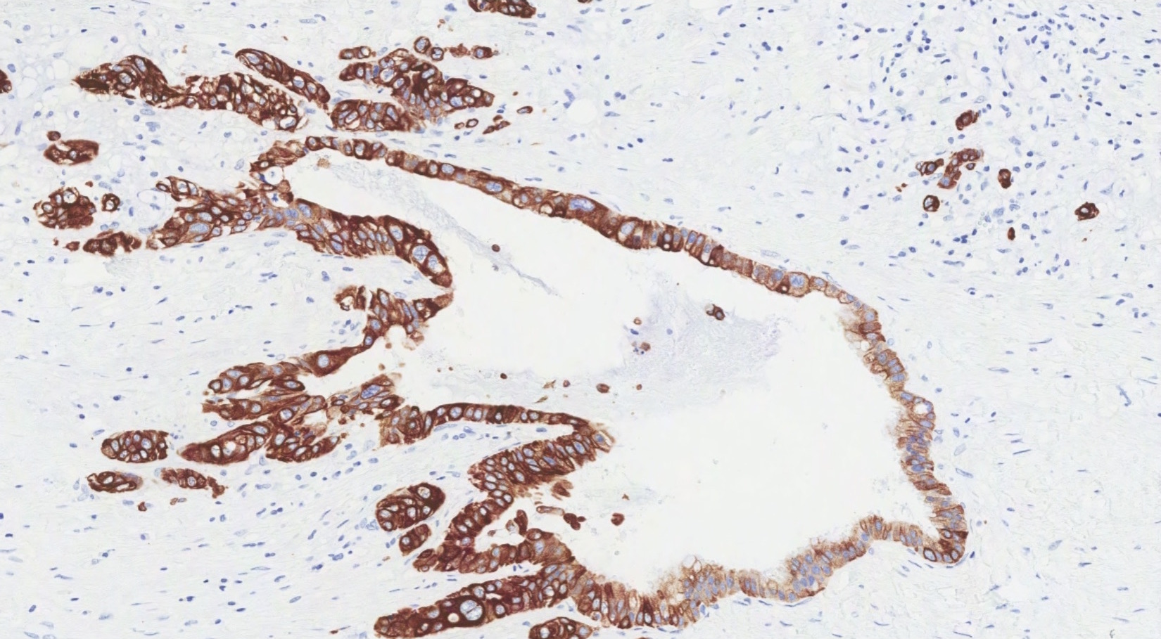
HNF1β
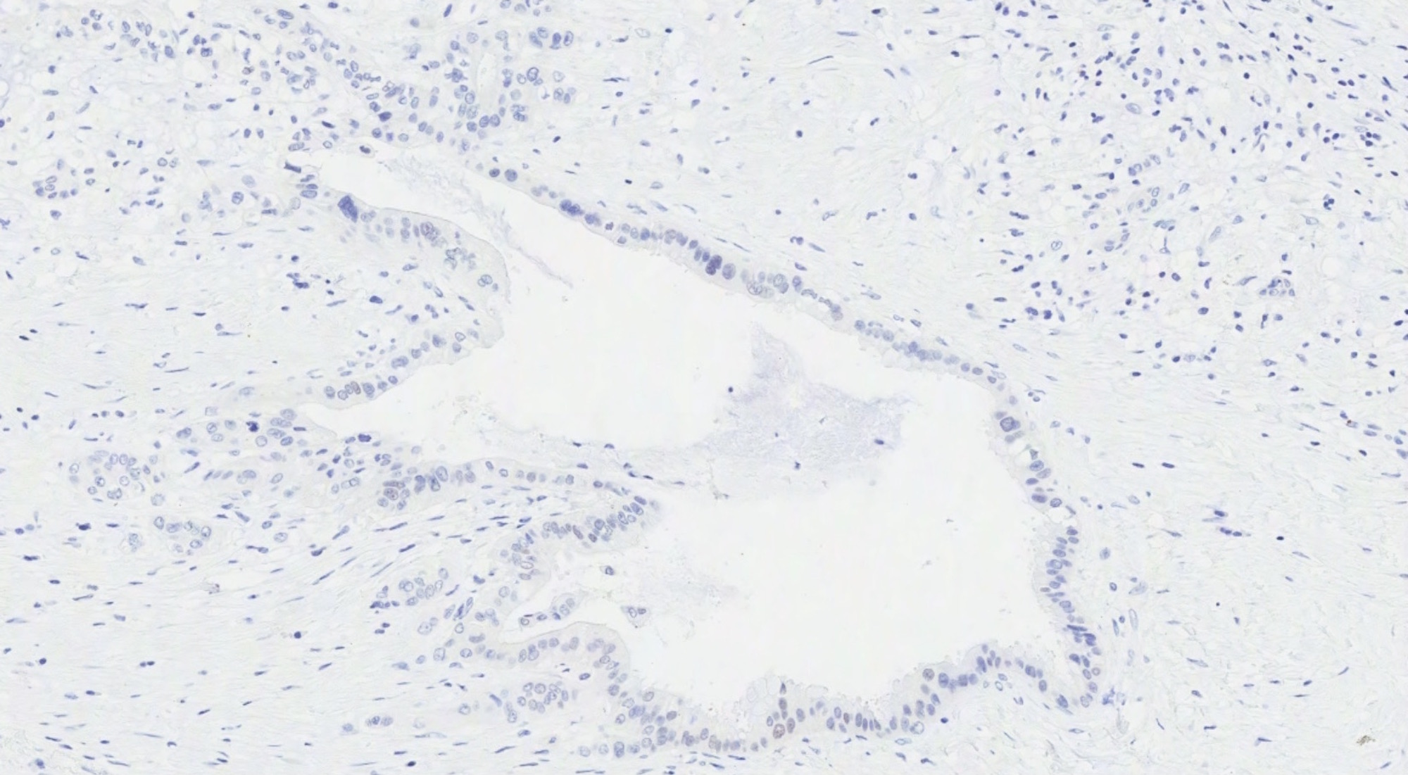
Napsin A
AFIP images
Borderline malignancy
AFIP images

Borderline malignancy
- See Figure 5 for algorithm to determine origin using currently available markers: Int J Gynecol Pathol 2016;35:191
Table 1. Most common immunohistochemical expression
patterns seen in primary ovarian mucinous neoplasms and
secondary epithelial tumors from gastrointestinal origin
| Lower GI | Upper GI | Ovarian | |
| CK7 | Negative | Positive | Positive |
| SATB2 | Positive | Negative | Negative |
| PAX8 | Negative | Negative | Positive* |
| CK20 | Positive | Pos / neg | Pos / neg |
| CDX2 | Positive | Pos / neg | Pos / neg |
| MUC2 | Positive | Pos / neg | Pos / neg |
| ER | Negative | Negative | Pos / neg |
| Beta catenin | Pos / neg | Negative | Negative |
| MUC1 | Pos / neg | Positive | Positive |
- GI = gastrointestinal; pos = positive; neg = negative
- In this table, positive markers are those with expression in > 75% and negative markers those with expression in < 25% of tumors in the indicated category
- Expression ranging from 25 to 75% is coded as pos / neg
- *PAX8 expression was 75% in all primary ovarian neoplasms but lower in the subgroup of primary ovarian carcinomas (65%)
- Table contents adapted from Int J Gynecol Pathol 2016;35:191
Images hosted on other servers:

Cystic and solid architecture
Contributed by Carlos Parra-Herran, M.D. and Adam Lechner
Neoplastic columnar epithelium
Dirty necrosis
Dilated glands
Sharply defined, angulated glands
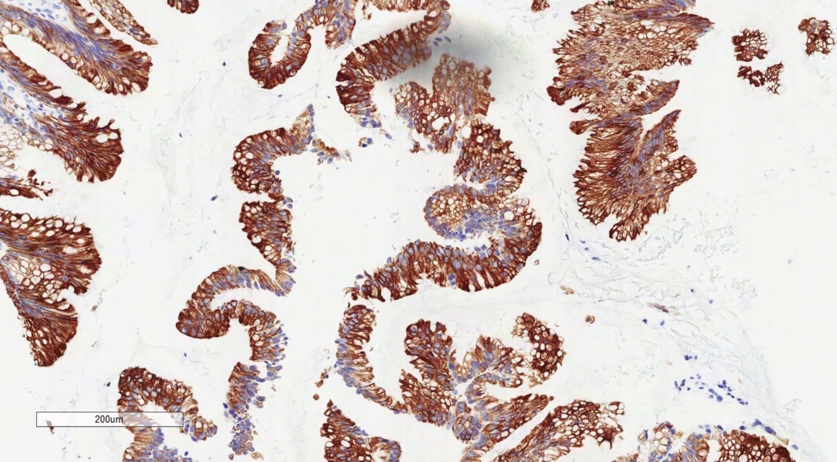
CK7

SATB2
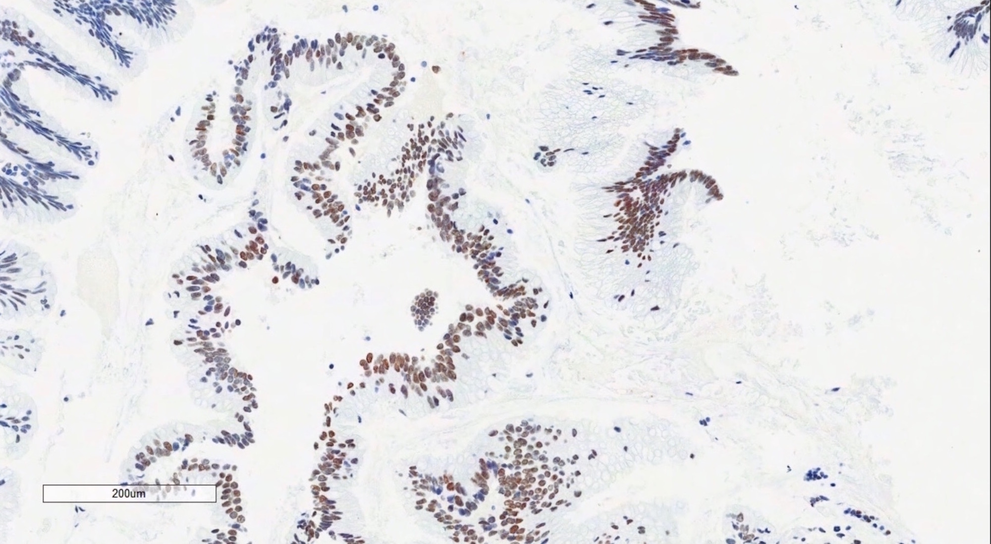
CDX2
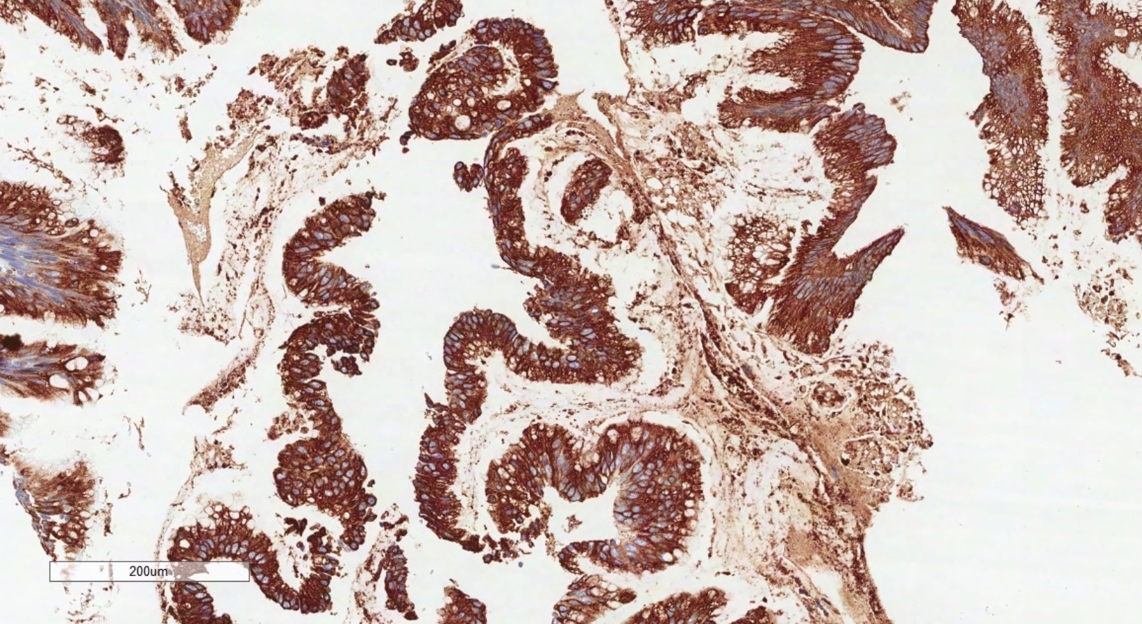
CK20
Images hosted on other servers:

CT: ruptured corpus luteum cyst and hemoperitoneum
Images hosted on other servers:

Intraoperative: hemorrhage of corpus luteum cyst
Images hosted on other servers:

Corpus luteum cyst
Contributed by Aurelia Busca, M.D., Ph.D.
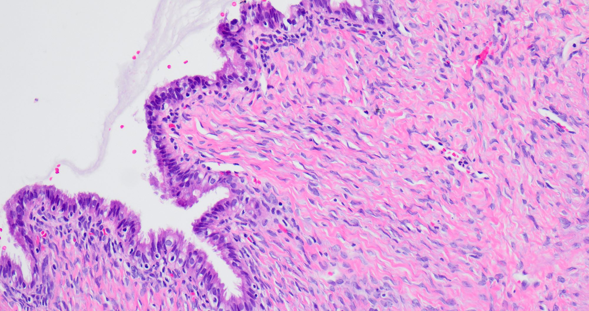
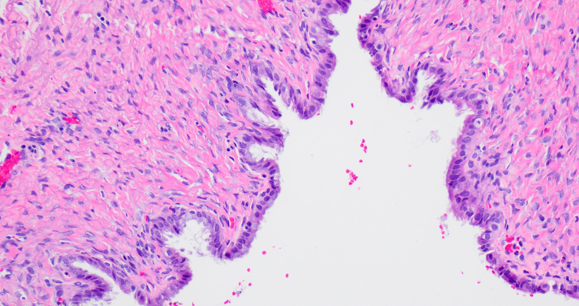
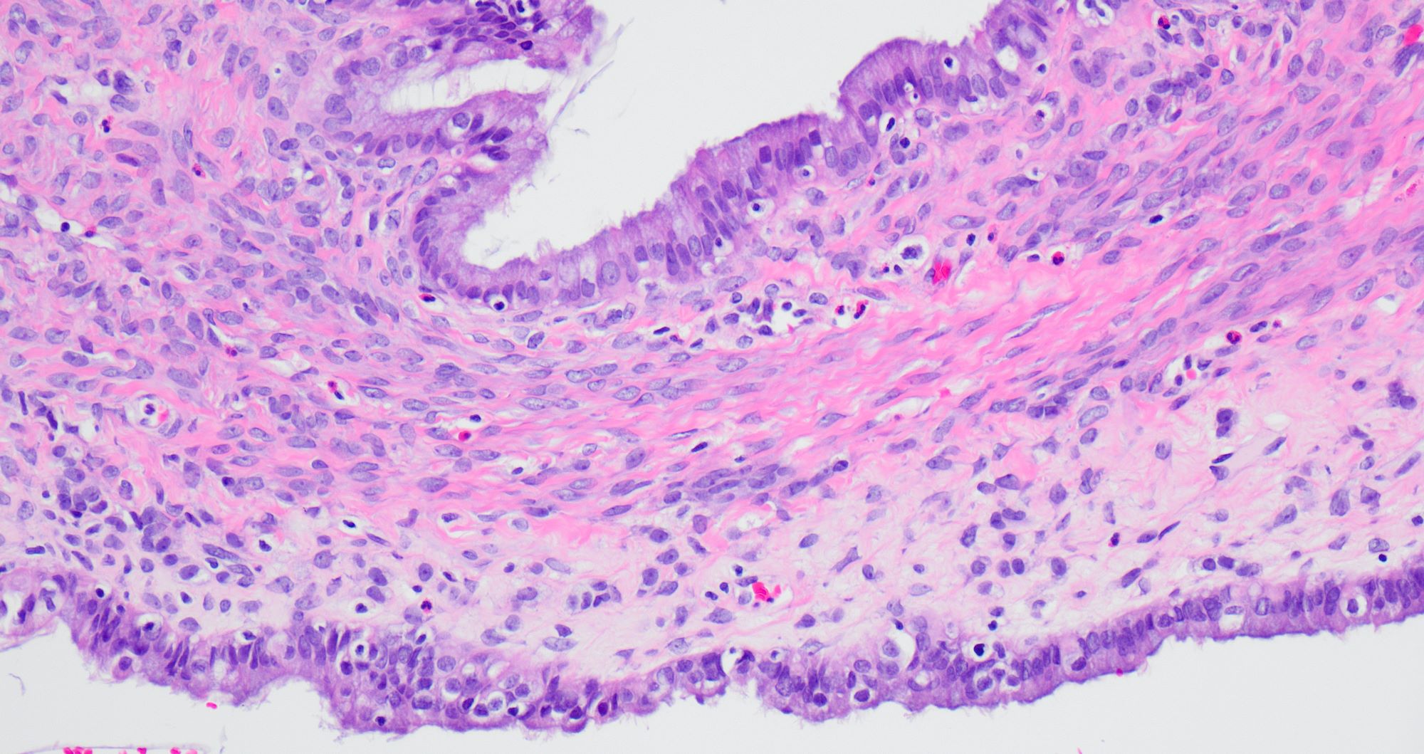
Convoluted cyst lining
Bilayered cyst lining
Granulosa and theca cells
Reticulin stain
Contributed by Aurelia Busca, M.D., Ph.D.
Cystically dilated glands
AFIP images
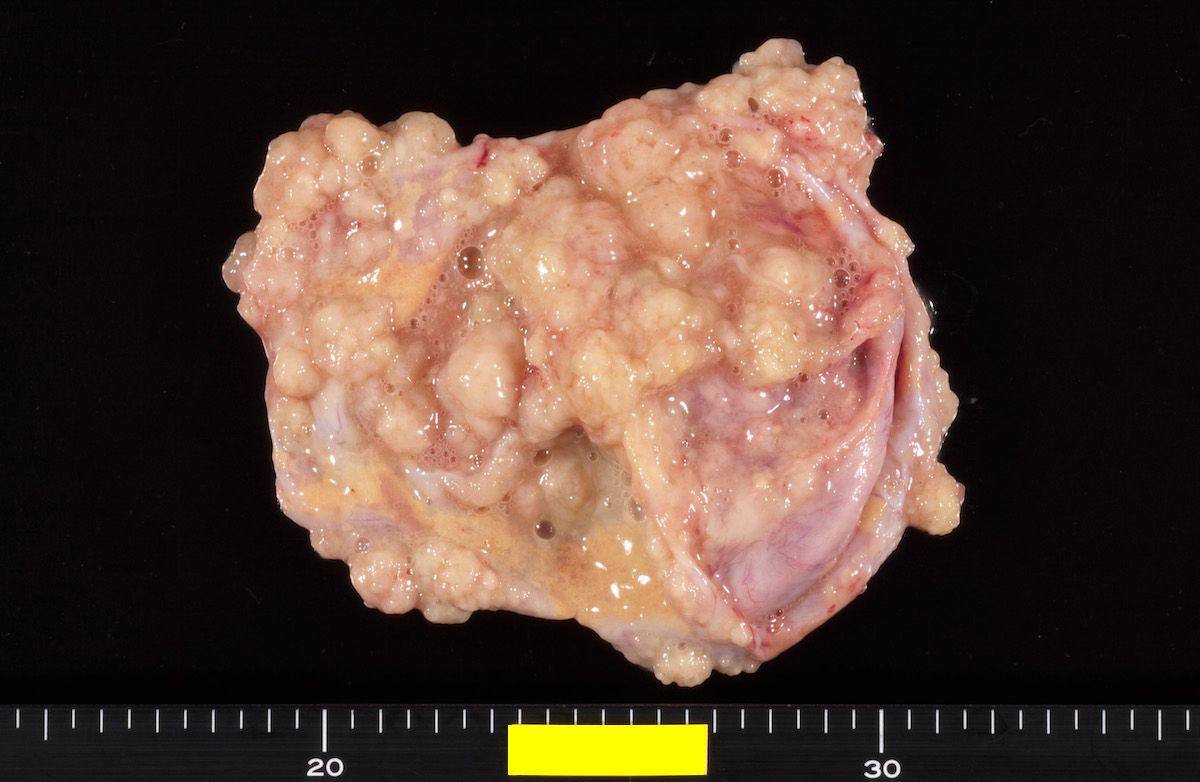
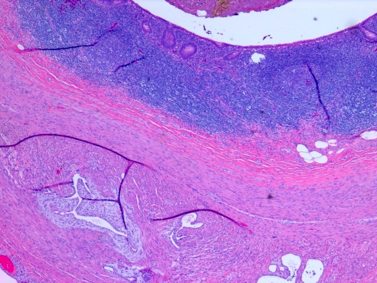
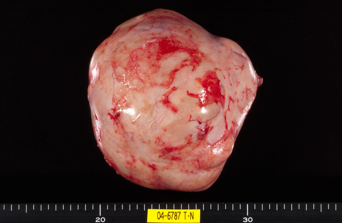
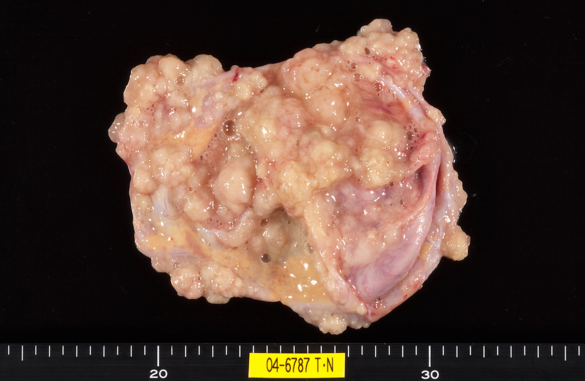
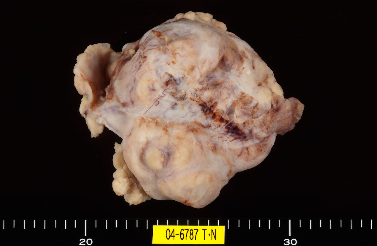
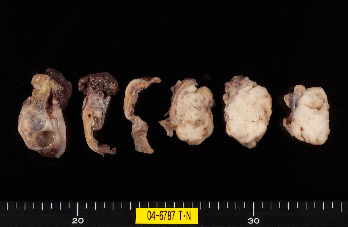
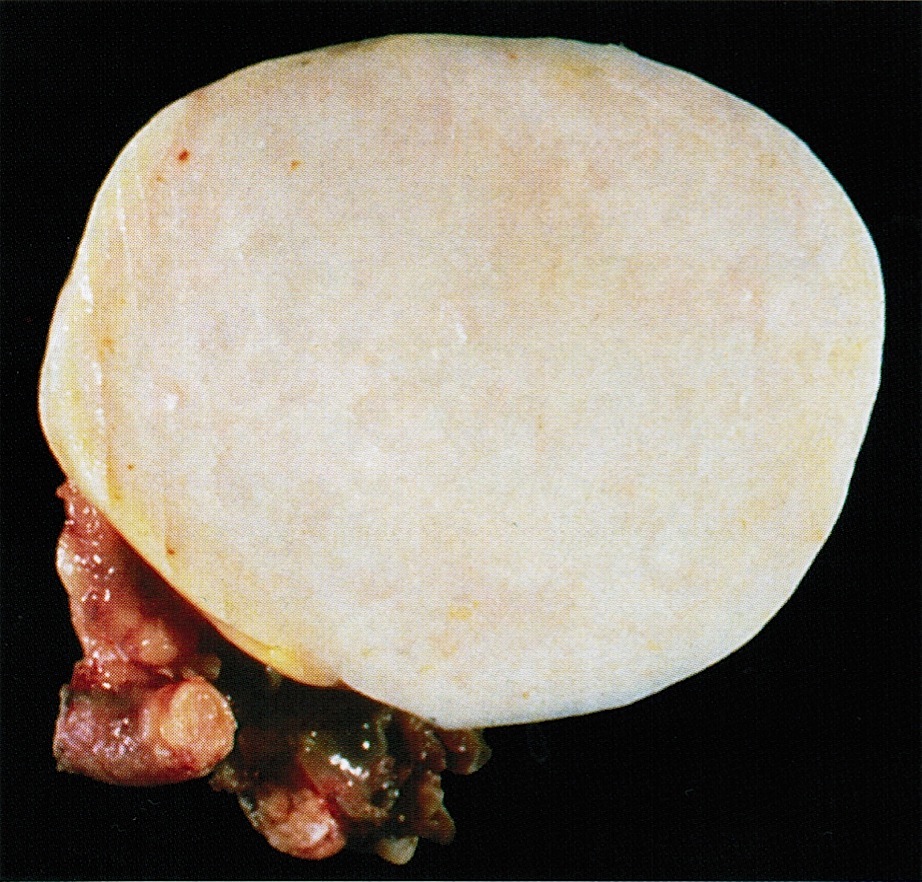
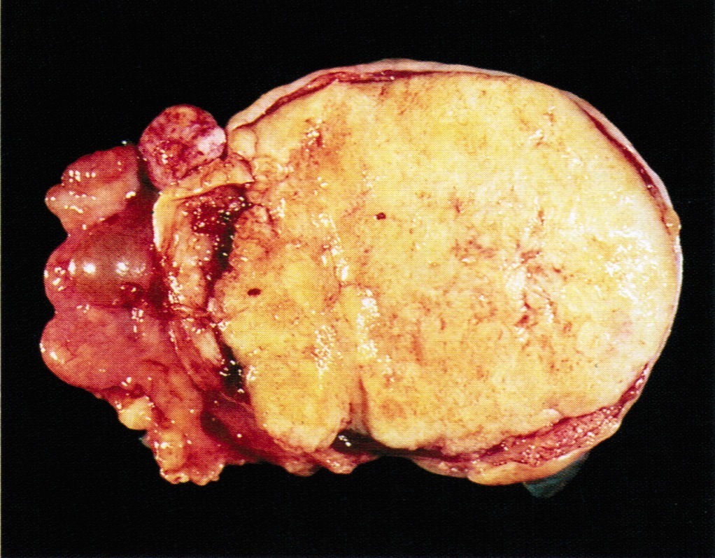
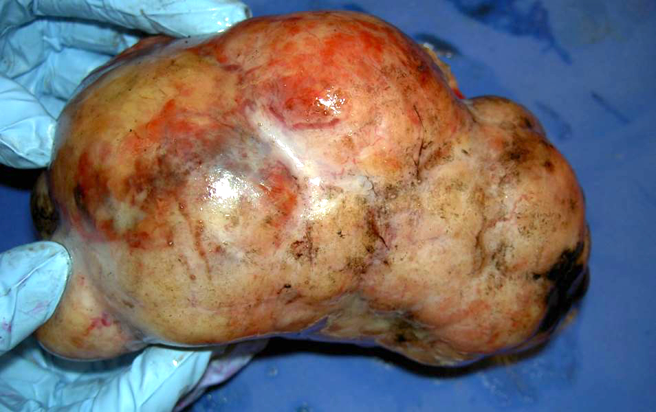
Solid, fleshy, lobulated
Cystic degeneration
Images hosted on other servers:

Pale brown parenchyma and central scarring
Contributed by Sharon Song, M.D.
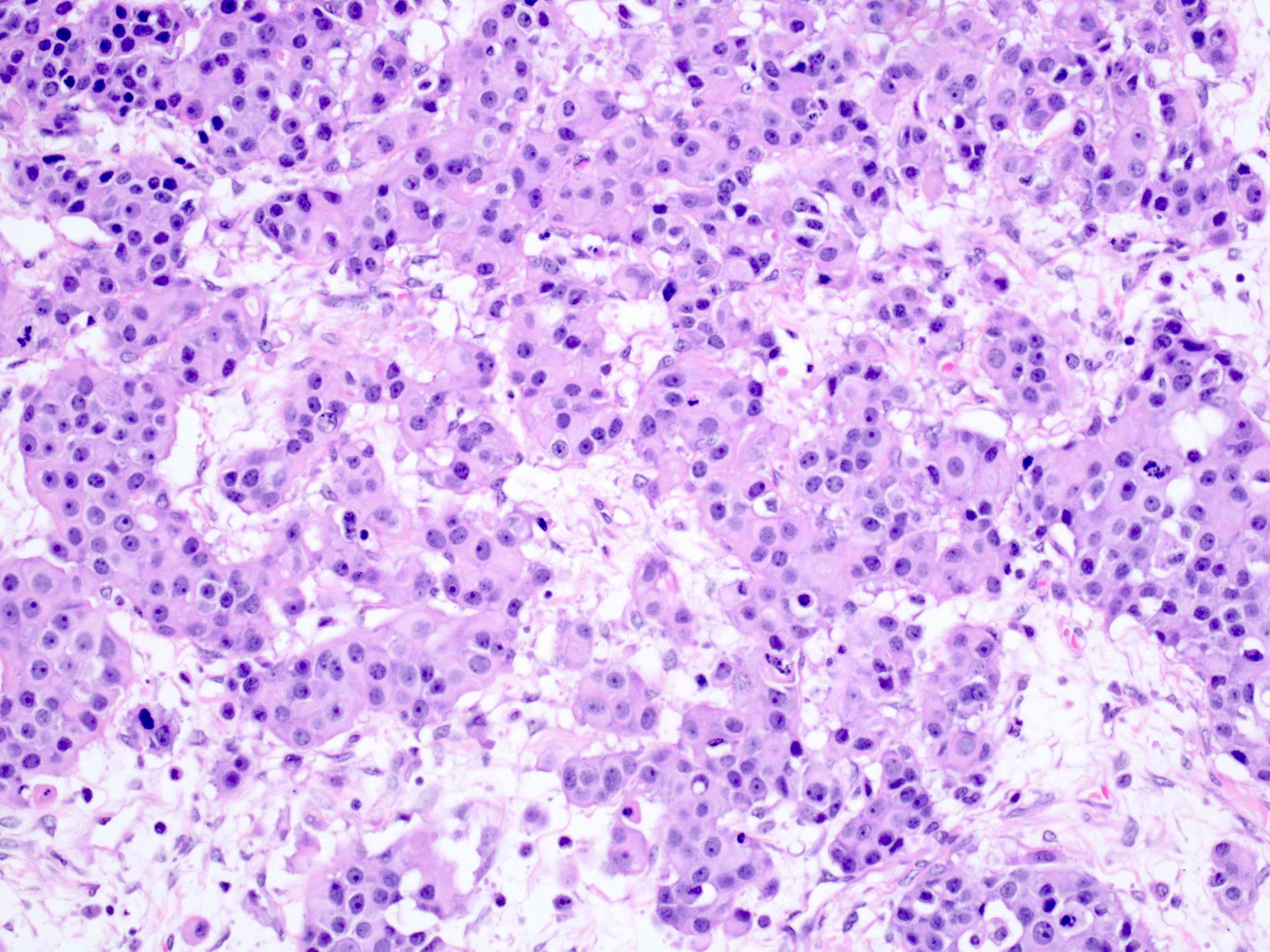
Medium sized cells
Eosinophilic cytoplasm

Cells growing as cords
Varied growth pattern
Nested growth
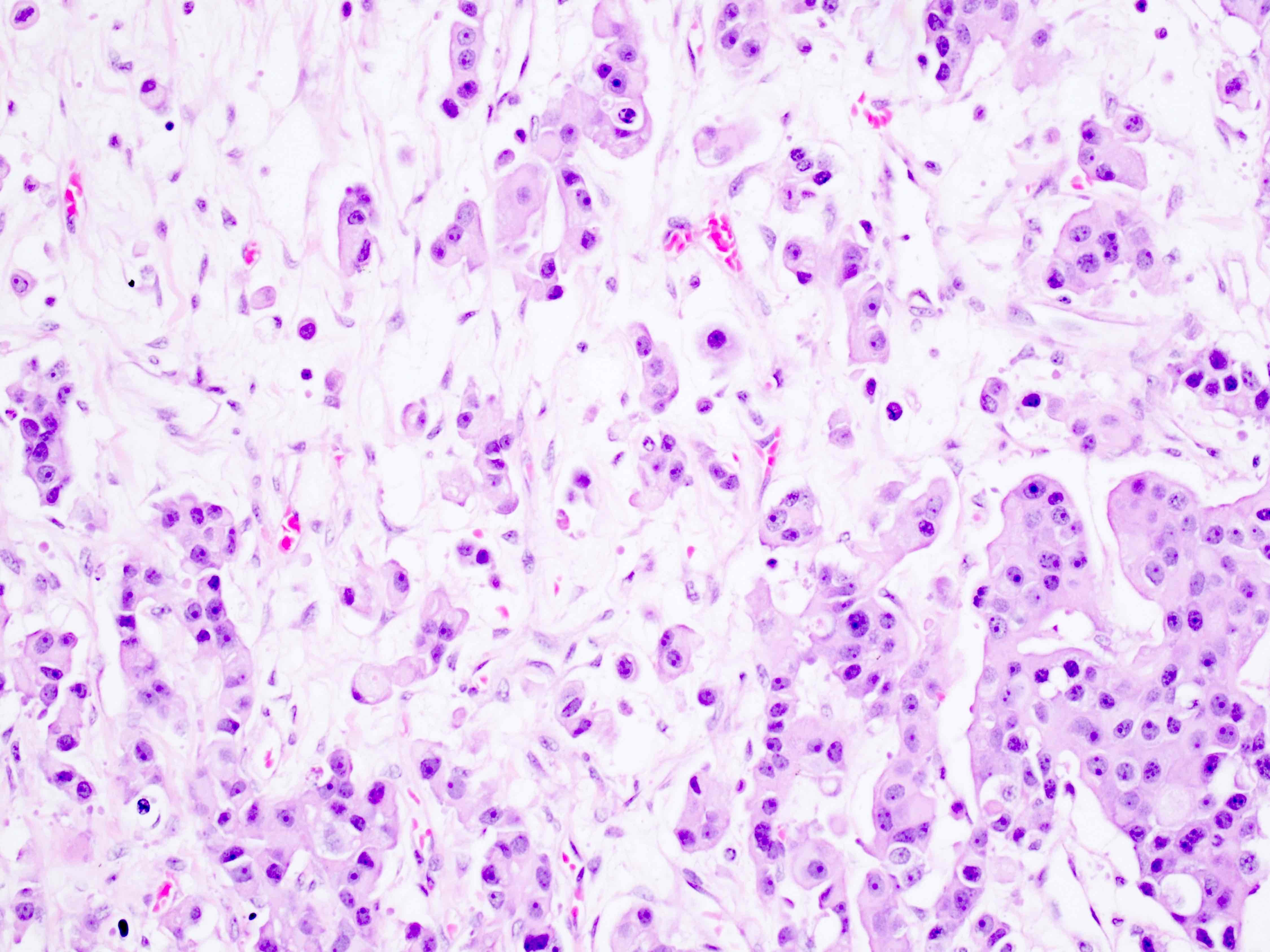
Inflammation in fibrous septae
Poor preservation
Epithelioid cytomorphology
OCT3/4
SALL4
CD117
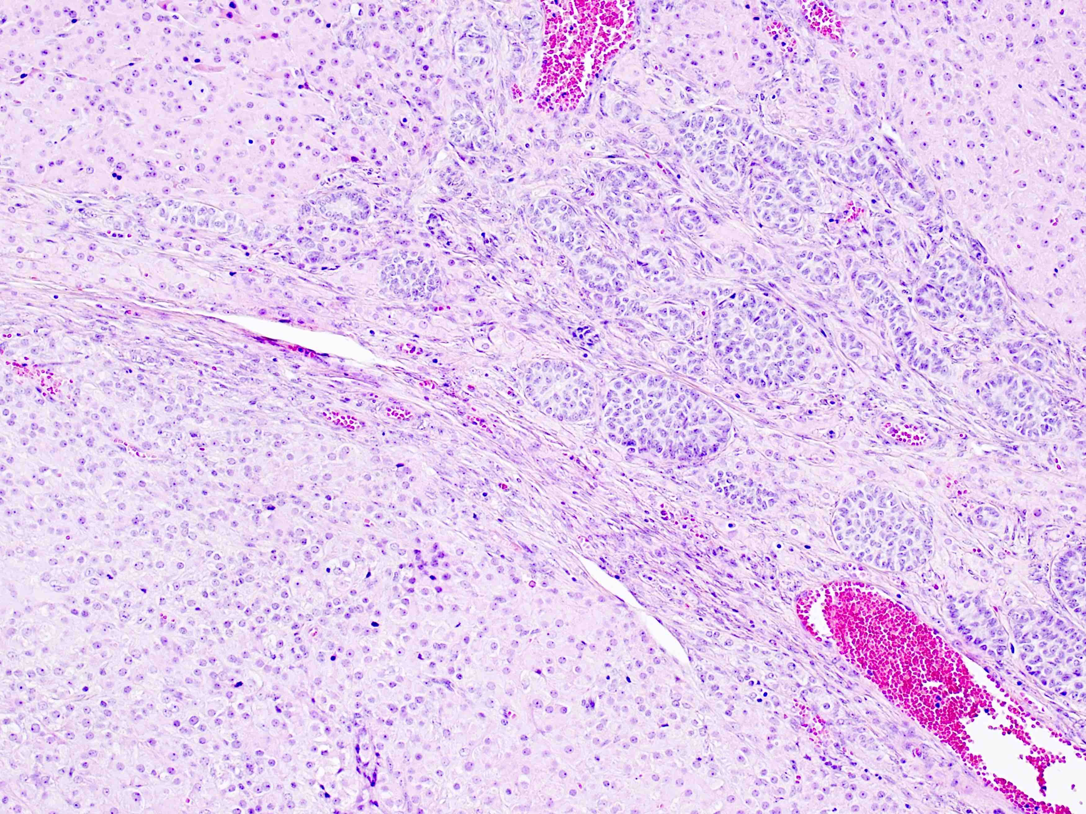
AFIP images
Lymphocytes
Cords
Solid tubular pattern
Syncytiotrophoblasts
Images hosted on other servers:

Macroscopic appearance
Contributed by Aurelia Busca, M.D., Ph.D.
Deciduosis of ovarian stroma

Areas of deciduosis
Ovarian surface adhesions with deciduosis
Ovarian surface adhesions with deciduosis
Ovarian deciduosis in a patient on progestin therapy
Images hosted on other servers:

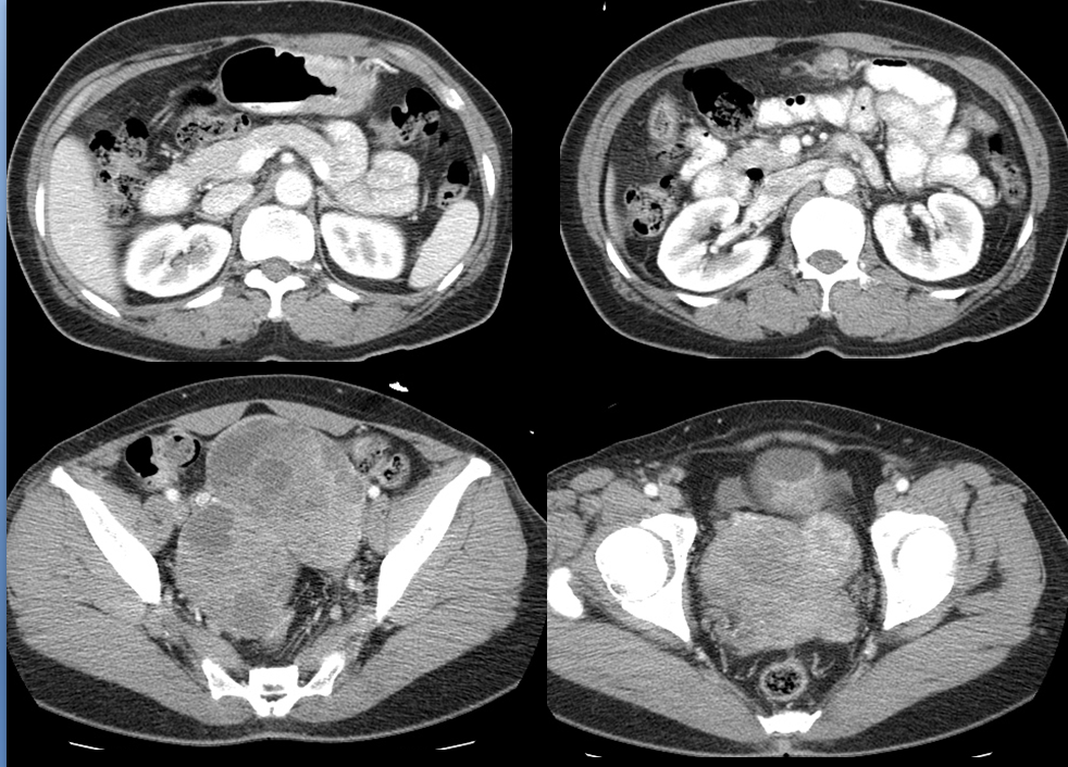
Ultrasound

CT abdominal mass & ascites
Images hosted on other servers:

External and cut surface of ovarian tumor
Contributed by Jessica L. Bentz, M.D. and AFIP
Primitive atypical cells
Mitoses and apoptotic debris
Papillary pattern
CD30
CD30
OCT 3/4
CK AE1 / AE3
AFIP images

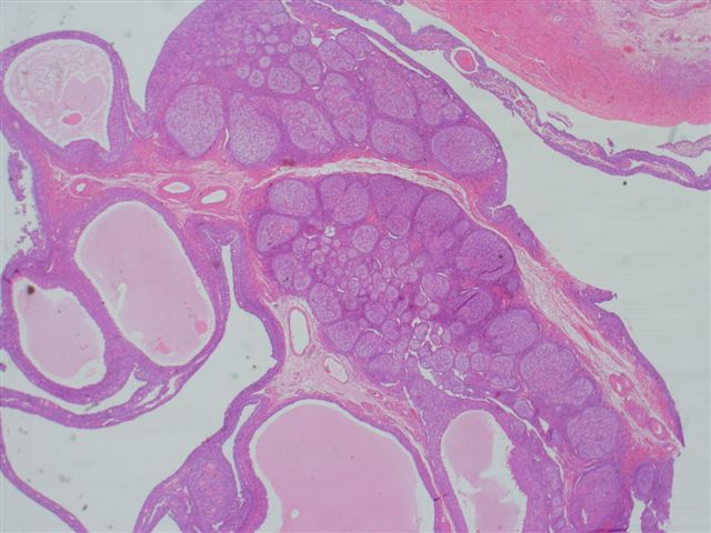
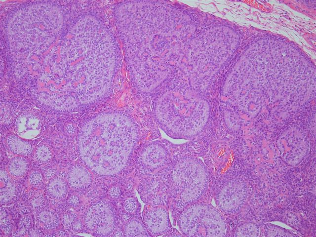
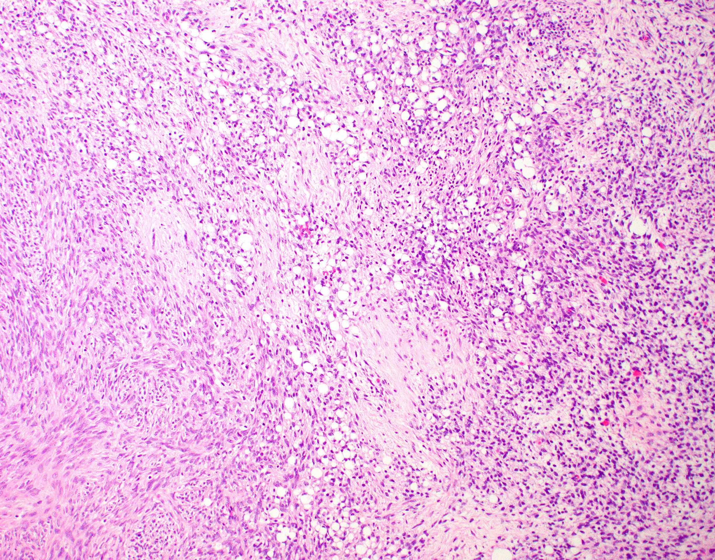
Endometrioid stromal sarcoma
With sex cord-like differentiation
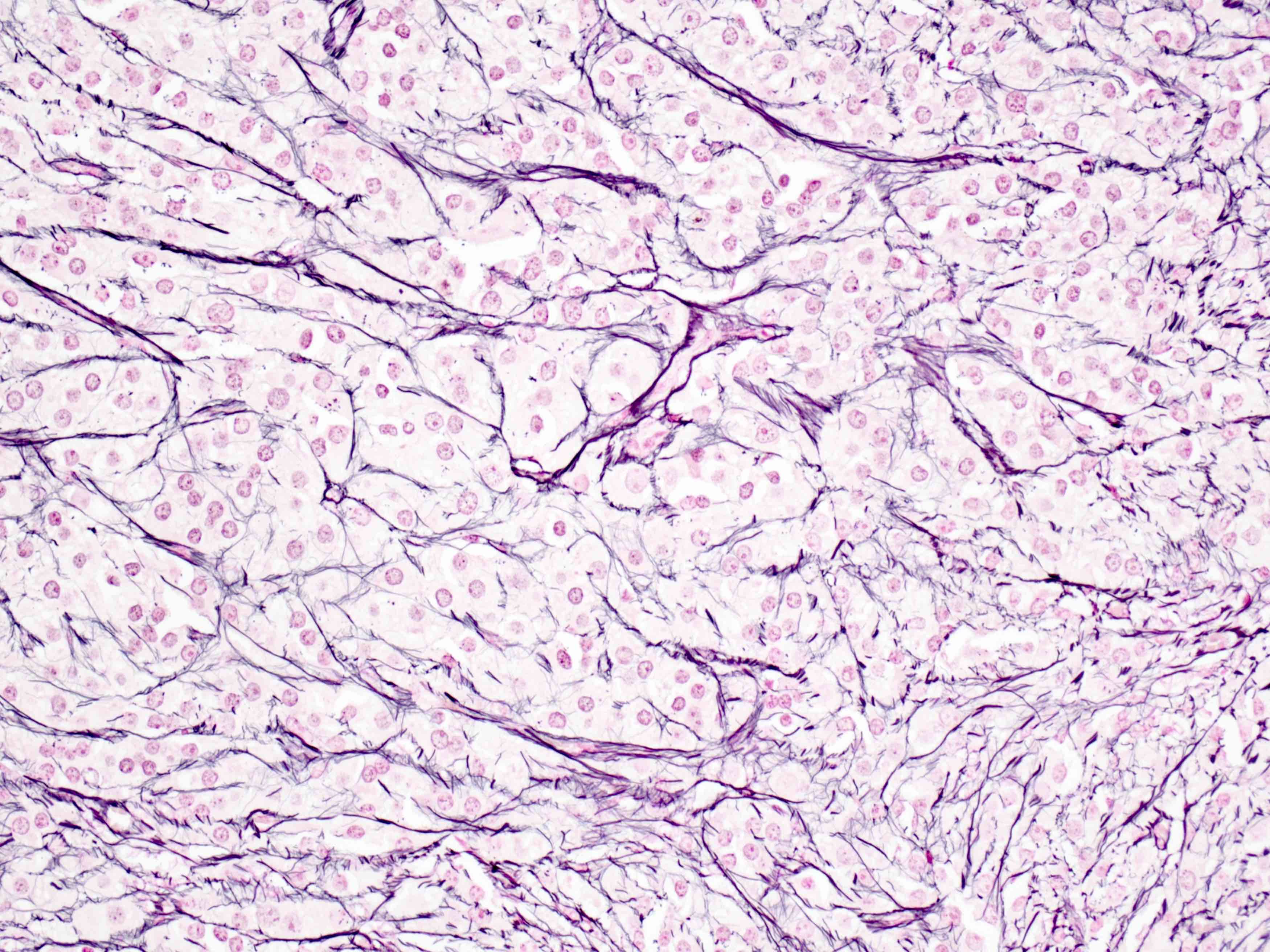
Reticulin stain
Metastatic to omentum
Images hosted on other servers:

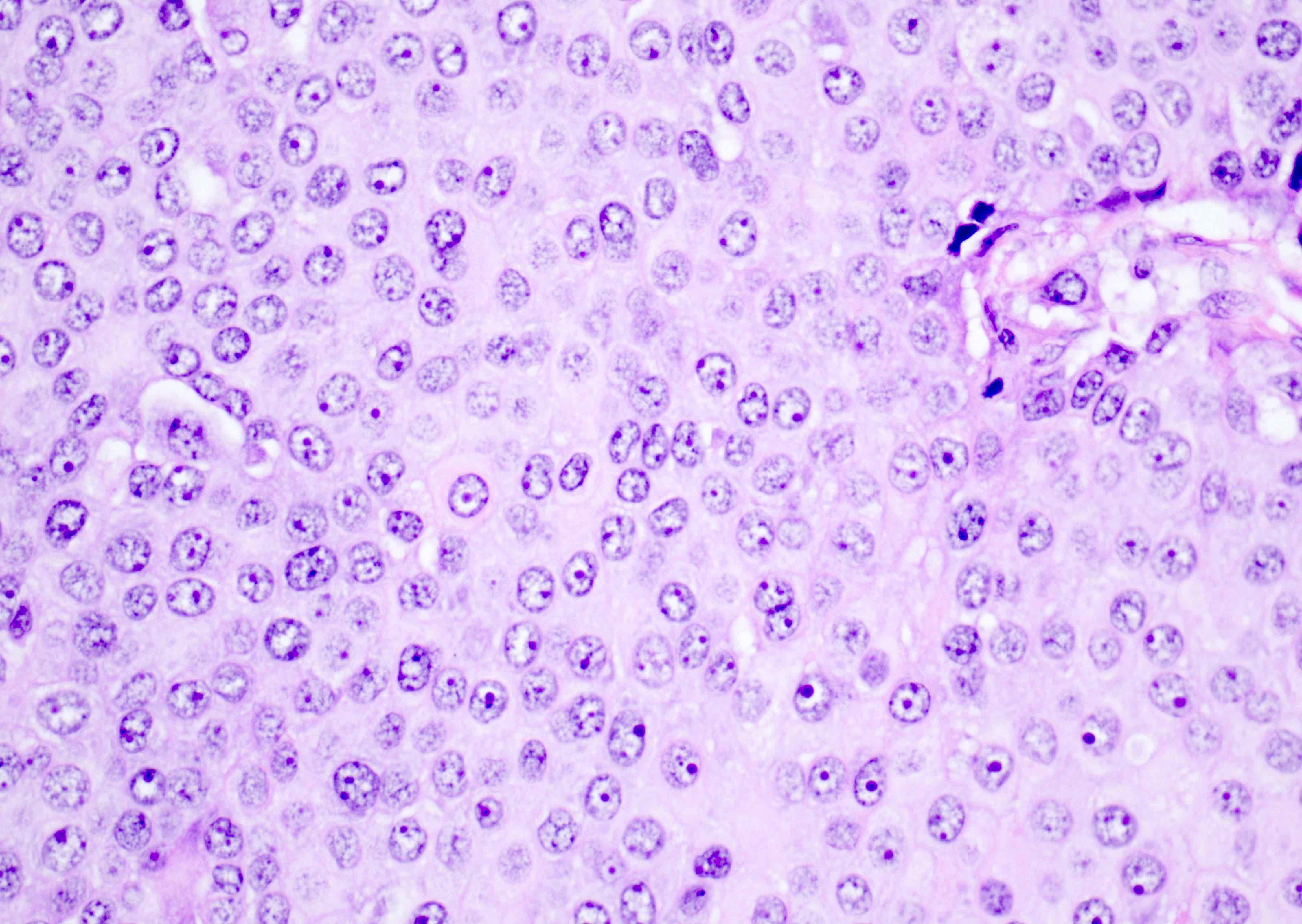
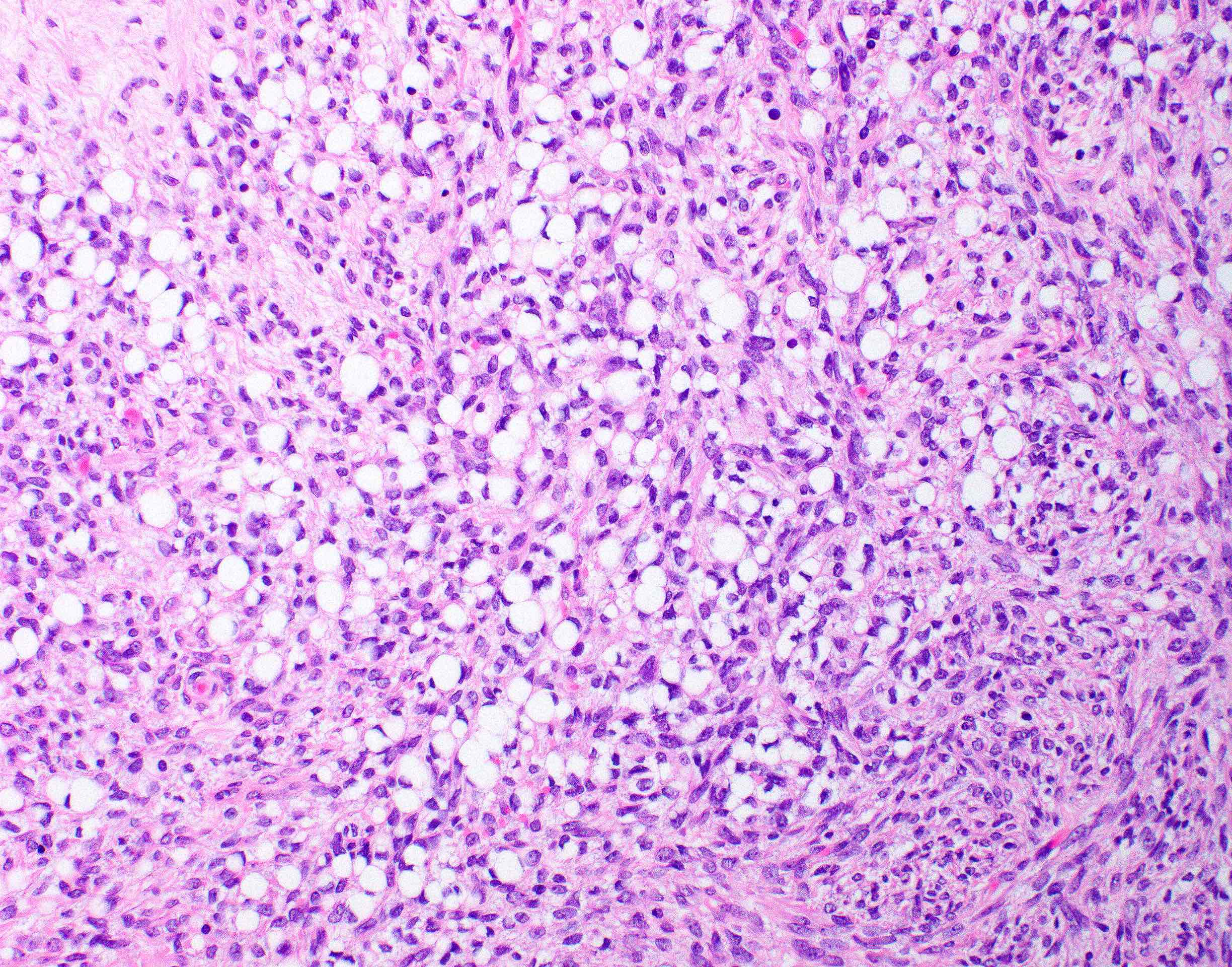
High grade tumor

Monotonous cells

Endometrium-like glands

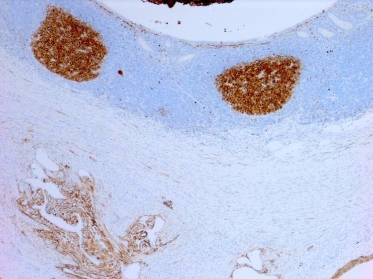
CD10+
AFIP images
With intraepithelial carcinoma, grade 1
Atypical
endometrioid
glands separated
by stroma
AFIP images

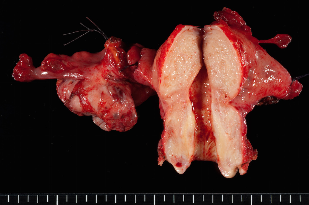
Ovary and uterus with tumor
Tumor arising in endometriotic cyst
Contributed by Ayse Ayhan, M.D.
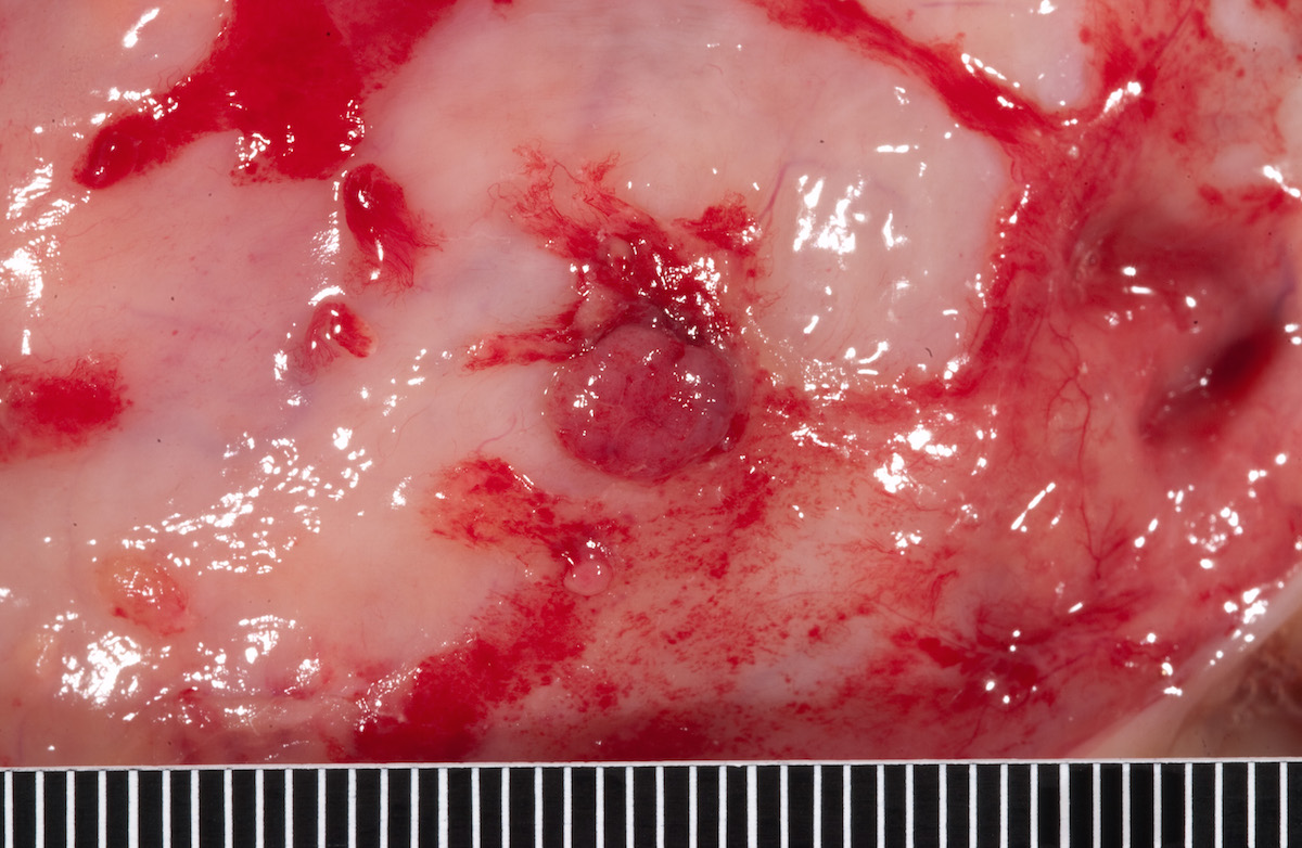
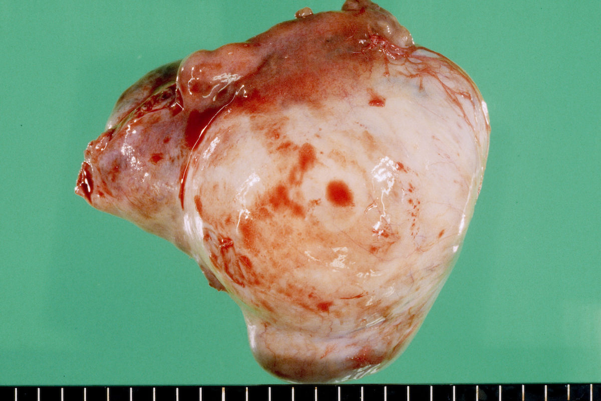
Smooth ovarian capsule
Multiple papillary excrescences
Papillary / nodular solid component
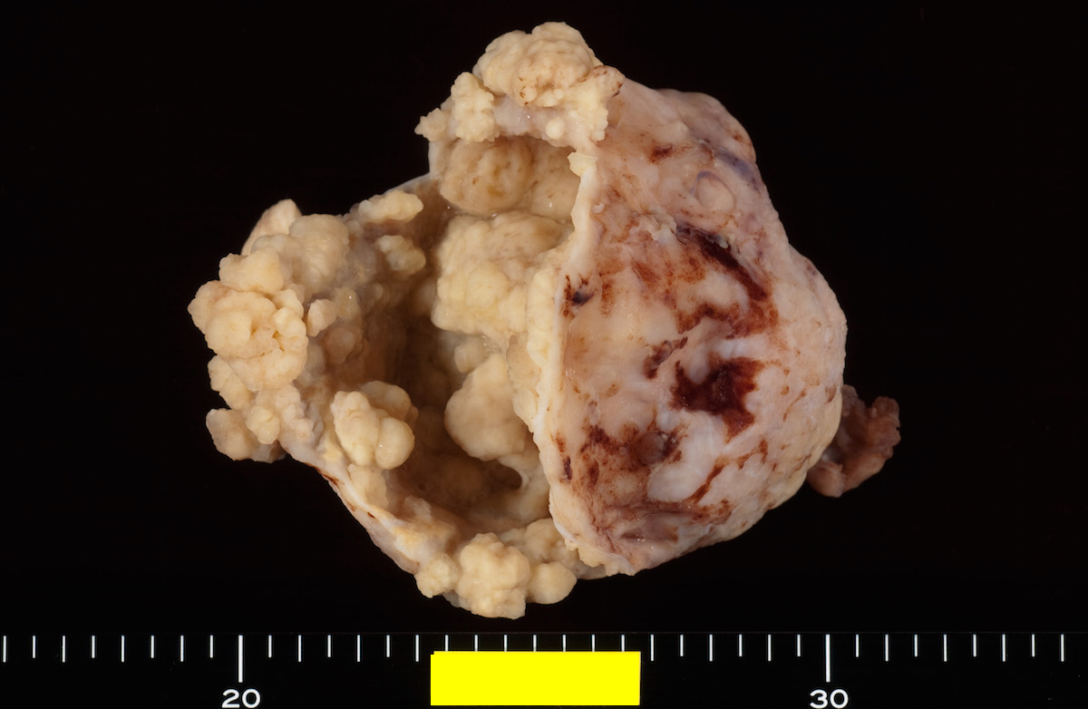
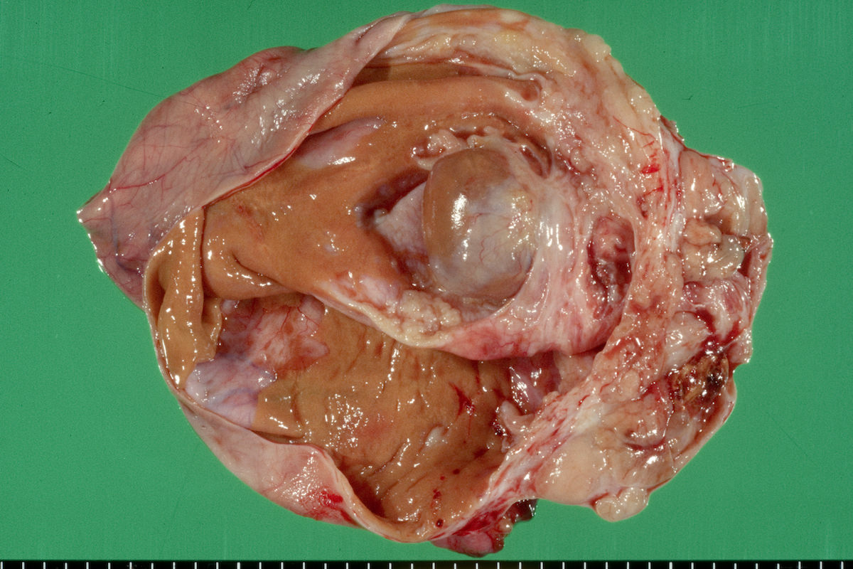
Partially opened cyst wall
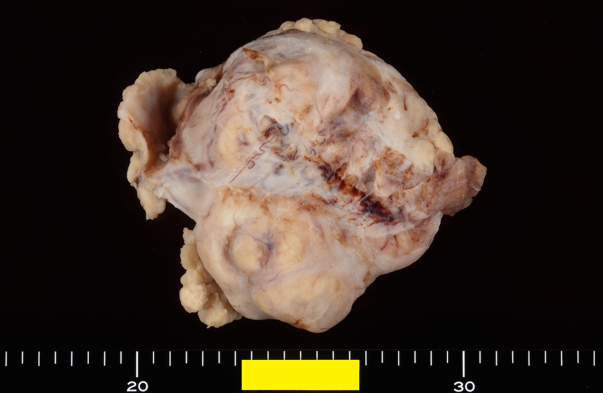
Cyst wall outer surface
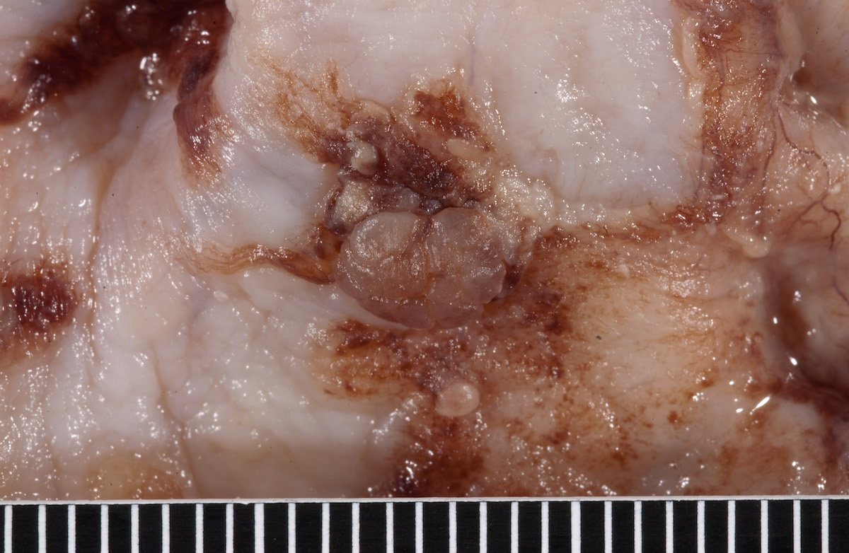
Solid / papillary areas
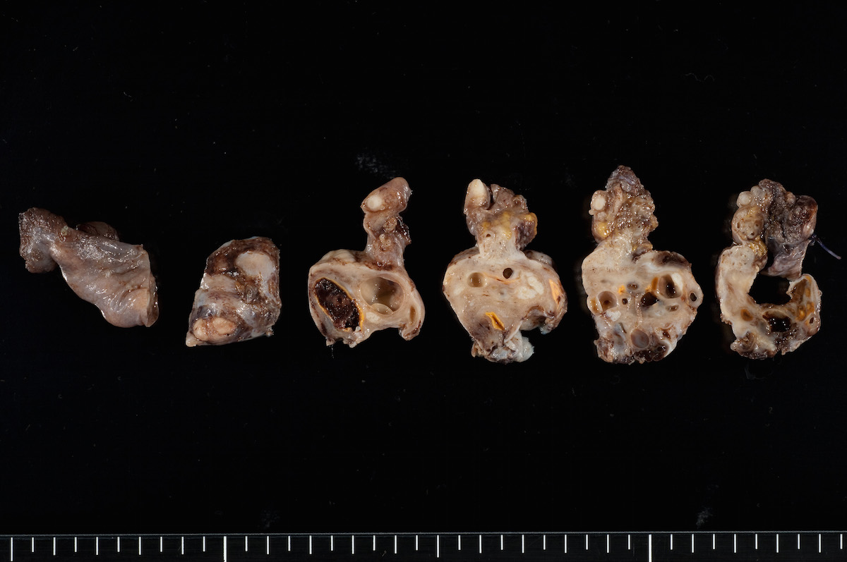
Serial sections
Cross section
Outer aspect
Partially opened cyst wall
Fixation
Ovarian mass lesion
Contributed by Ozlen Saglam, M.D.
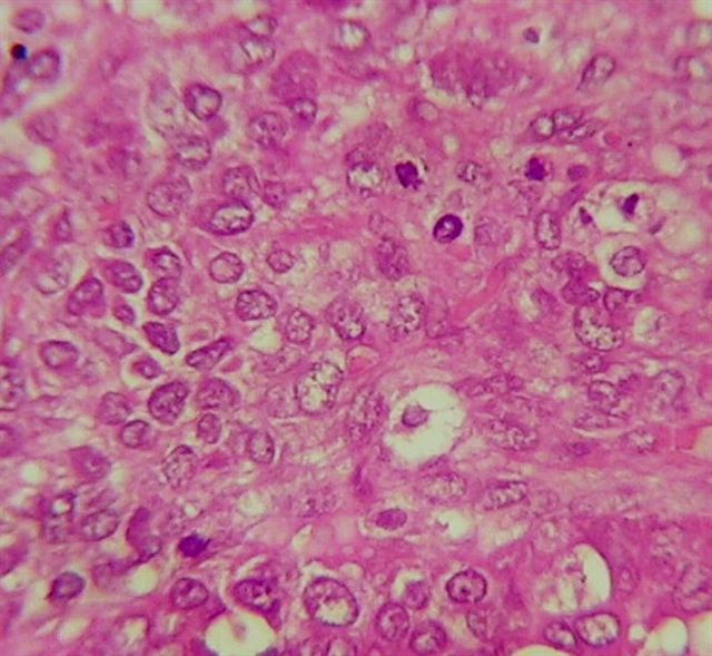
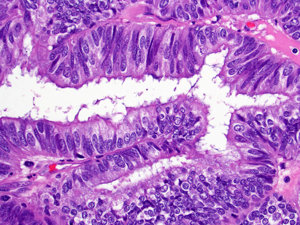
Endometrioid adenocarcinoma, FIGO grade 1
Endometrioid adenocarcinoma, FIGO grade 2
Endometrioid adenocarcinoma, FIGO grade 3
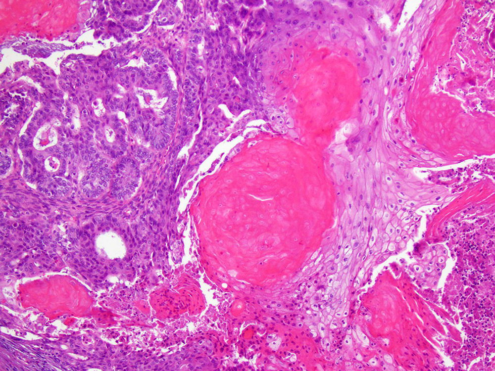
Squamous differentiation
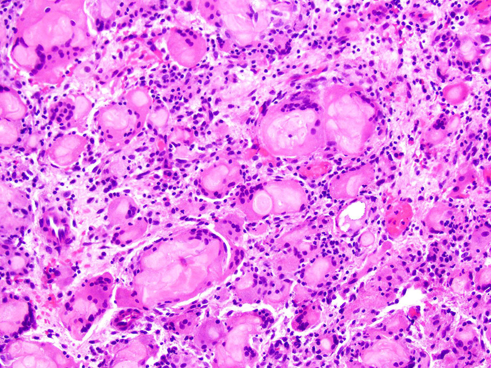
Peritoneal keratin granuloma
Ciliated cells
Secretory change
Oncocytic cells
Spindle variant
Trabecular pattern
resembling sex
cord stromal tumor
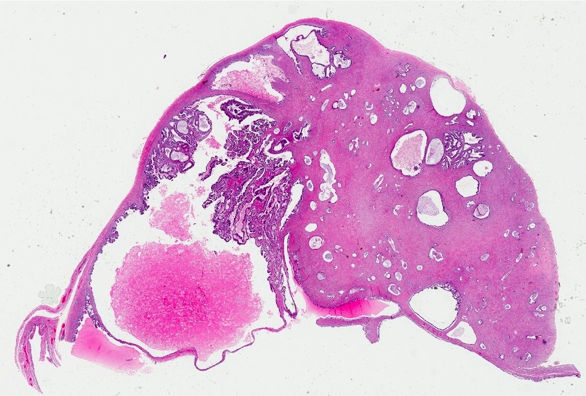
Endometrioid carcinoma and background adenofibroma
Endometriosis
Contributed by Sakinah A Thiryayi, M.D. and Gulisa Turashvili, M.D., Ph.D. (Case #500)
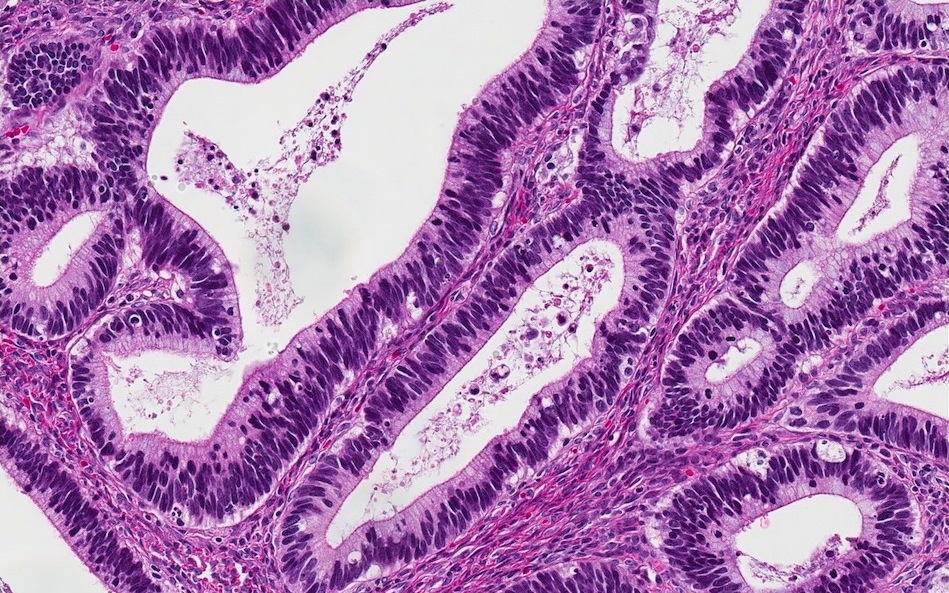
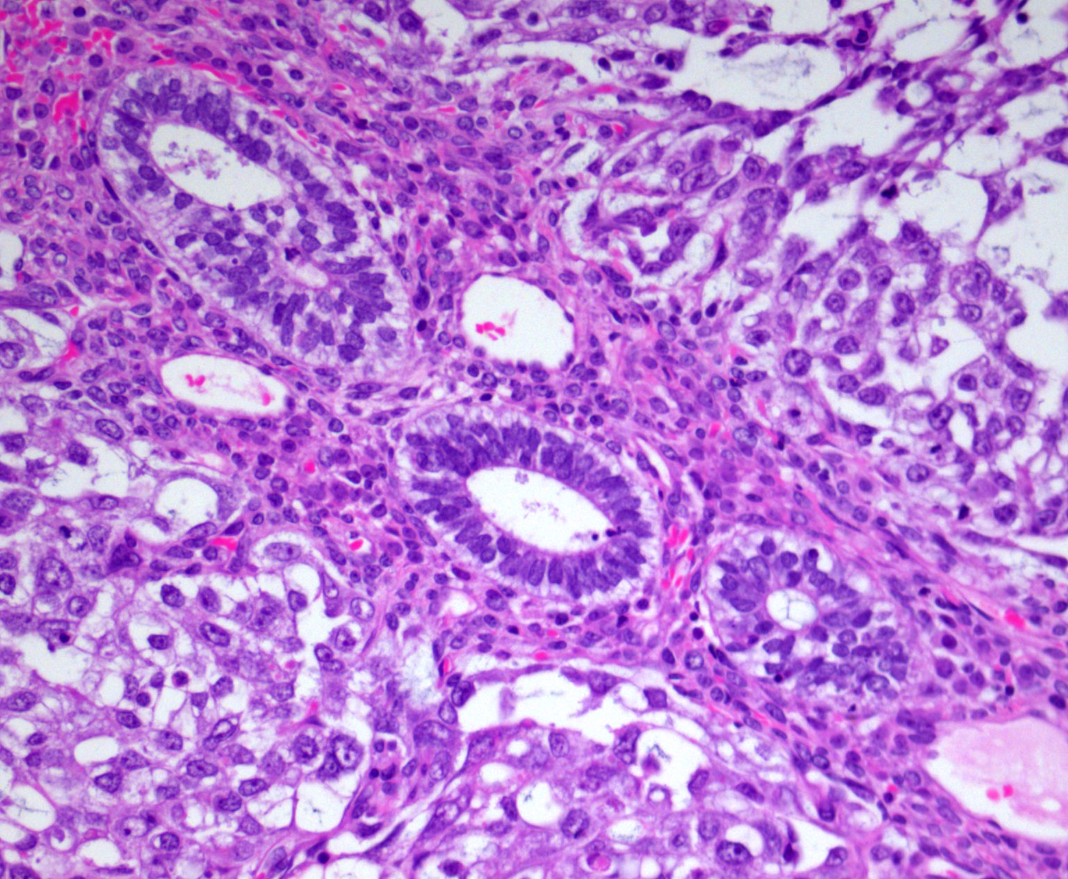
Glandular architecture
Back to back glands
Low grade cytology
Cellular intervening stroma
Plump stromal cells
Inhibin
p53
AFIP images
With endometriosis
Contributed by Stephanie L. Skala, M.D.
Variably dilated endometrioid glands
Endometrioid glands, some ciliated

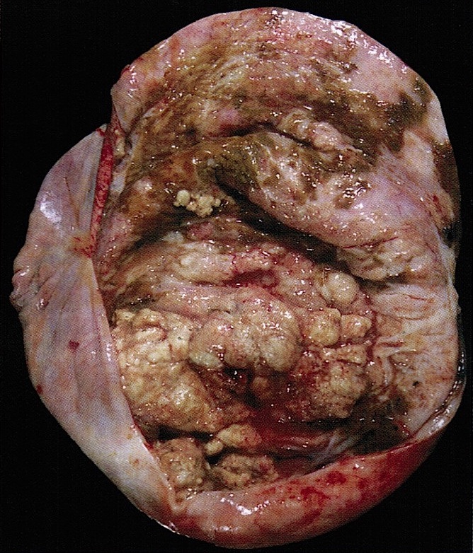
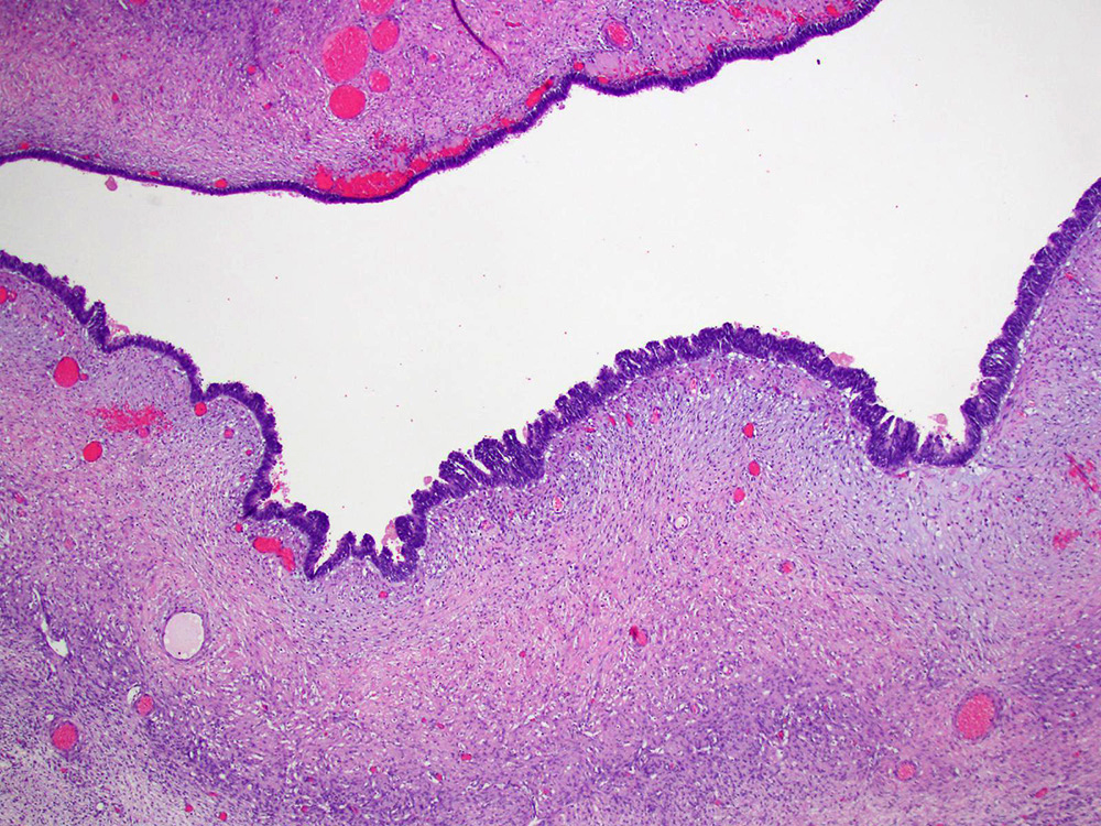
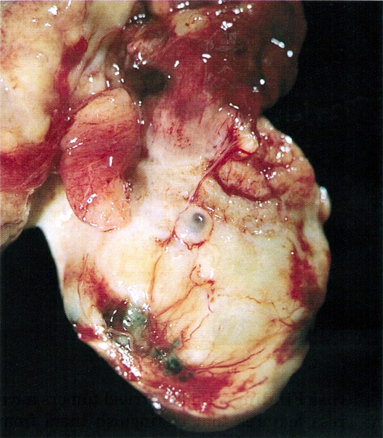
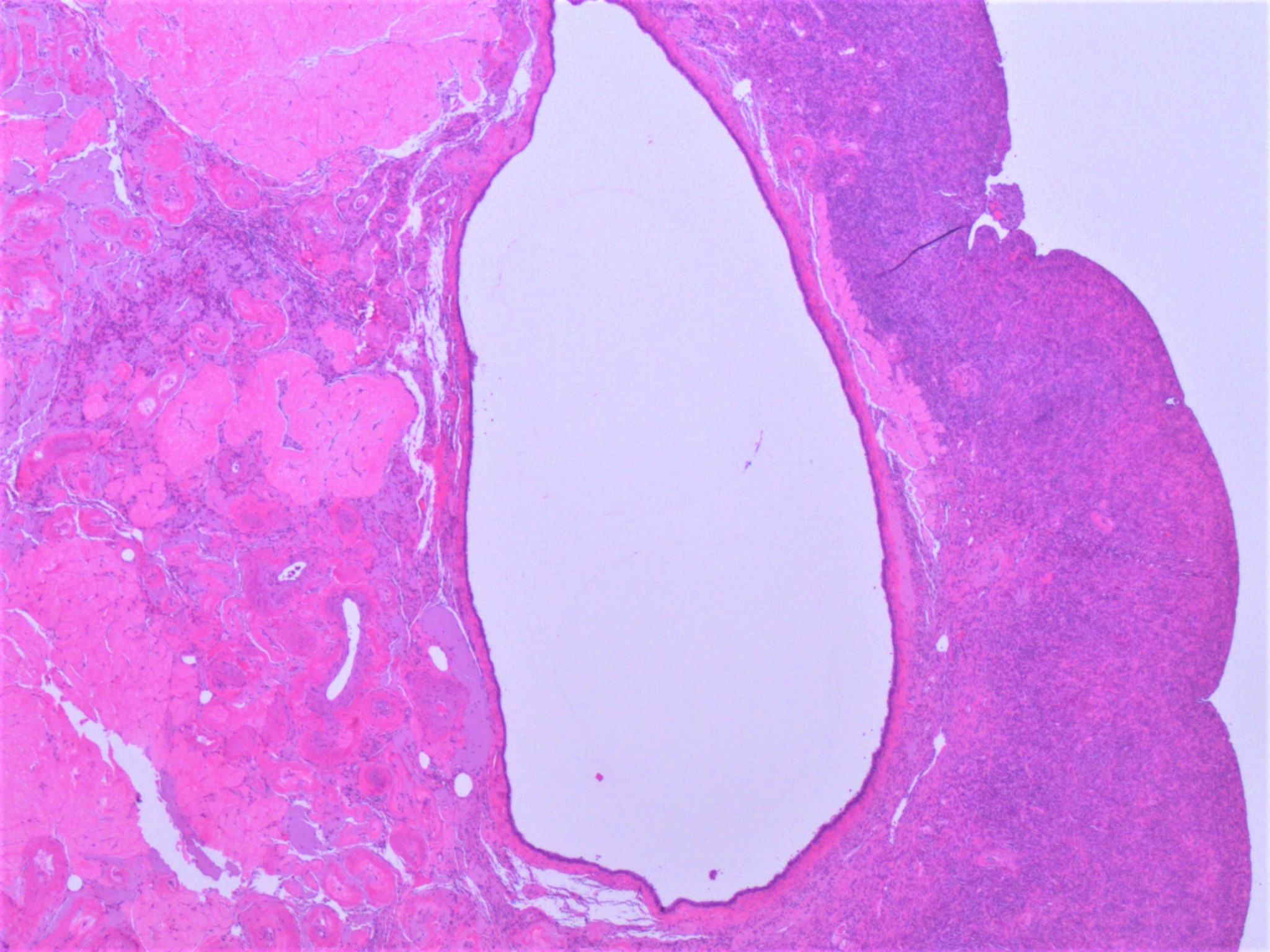
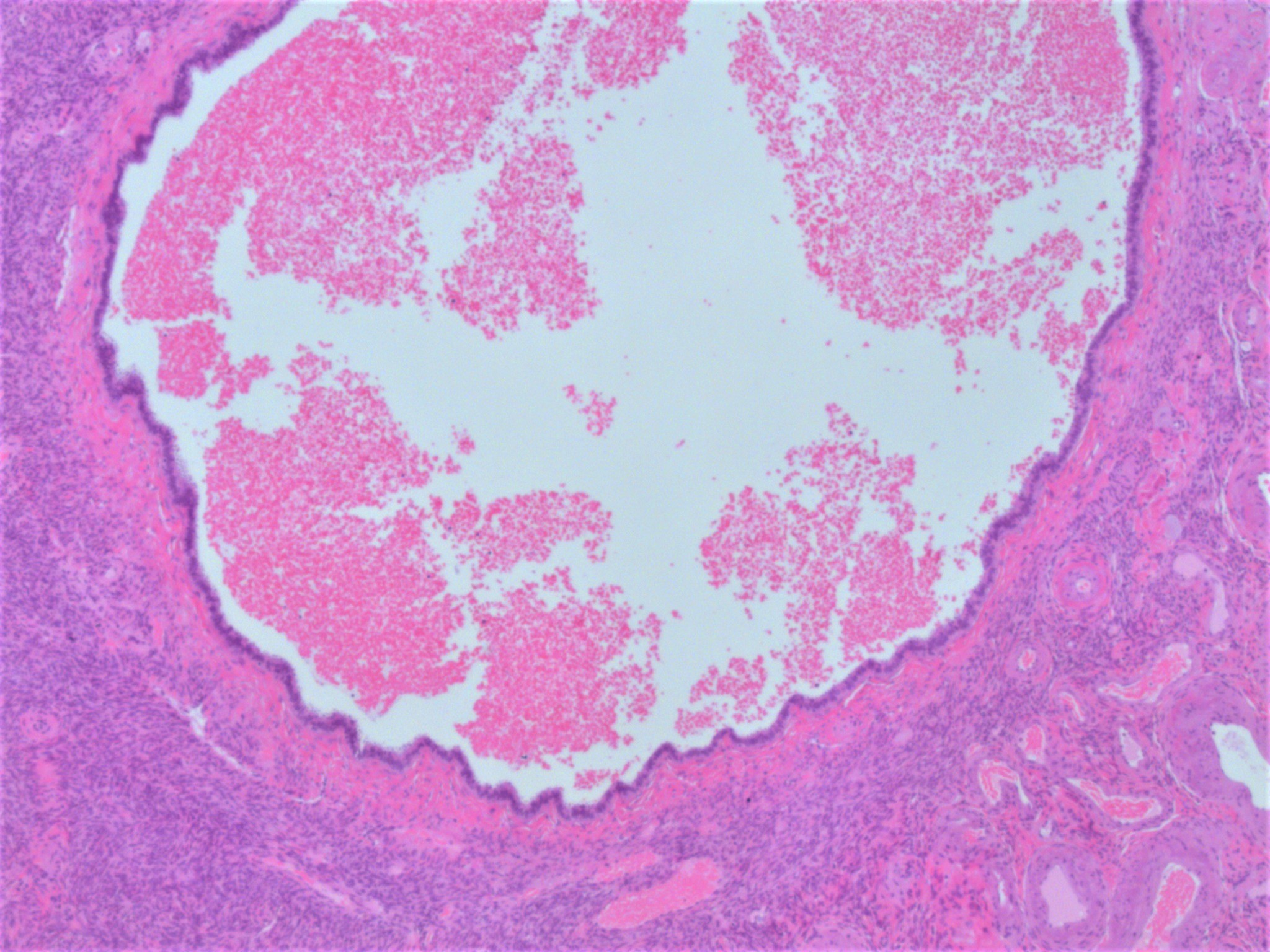
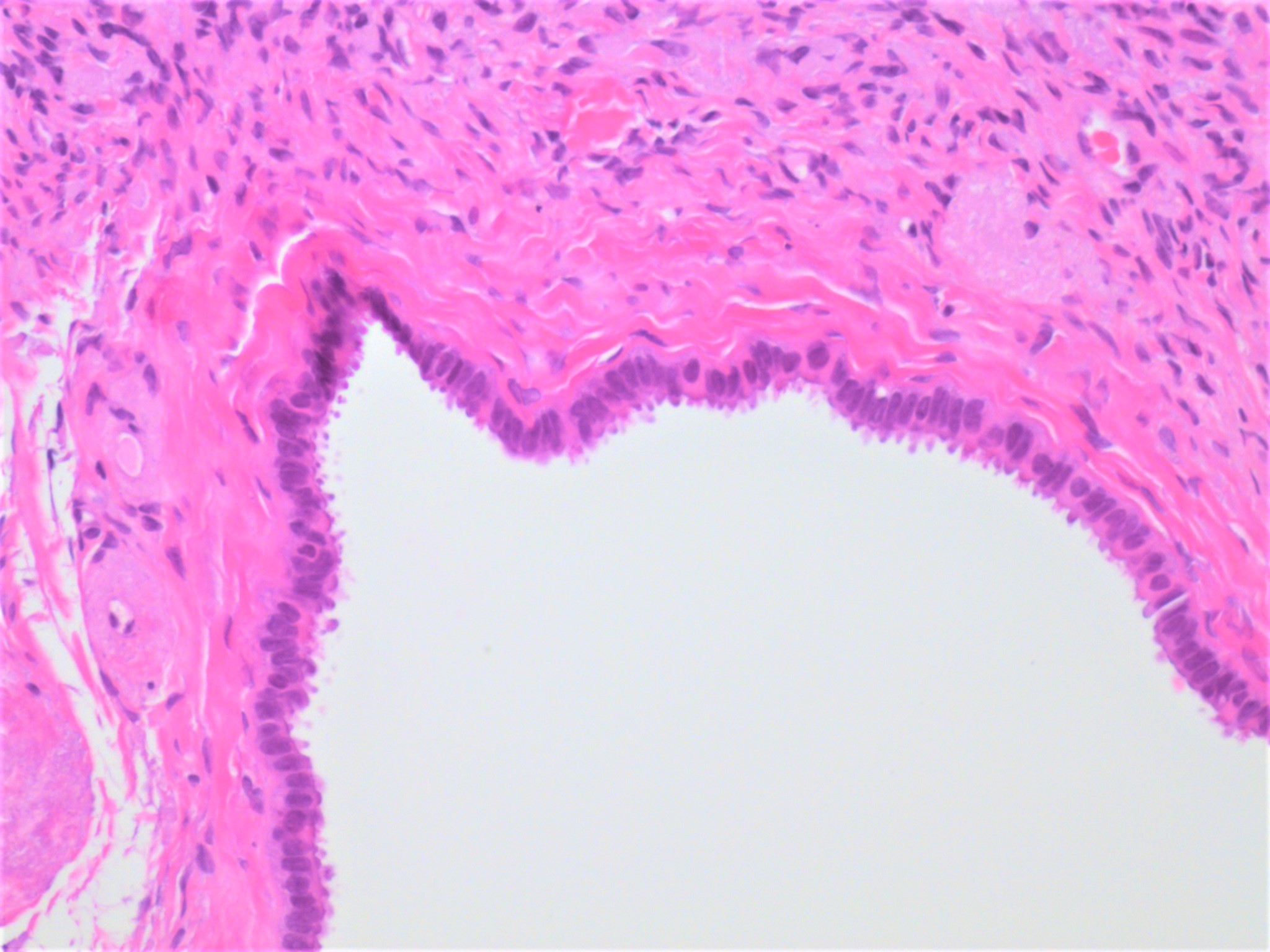
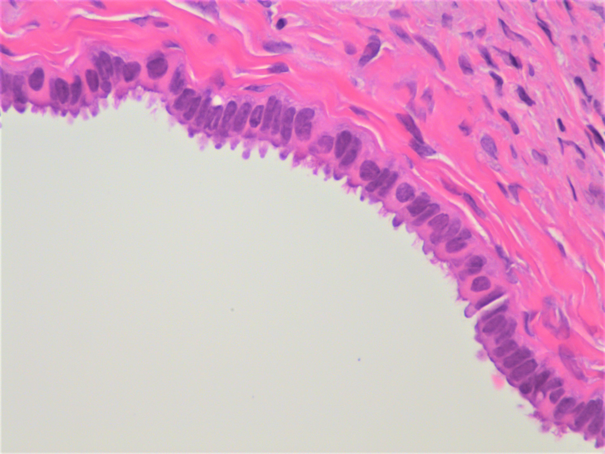
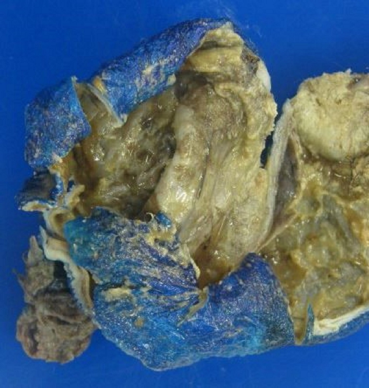
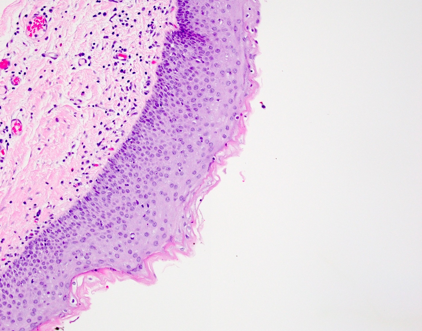
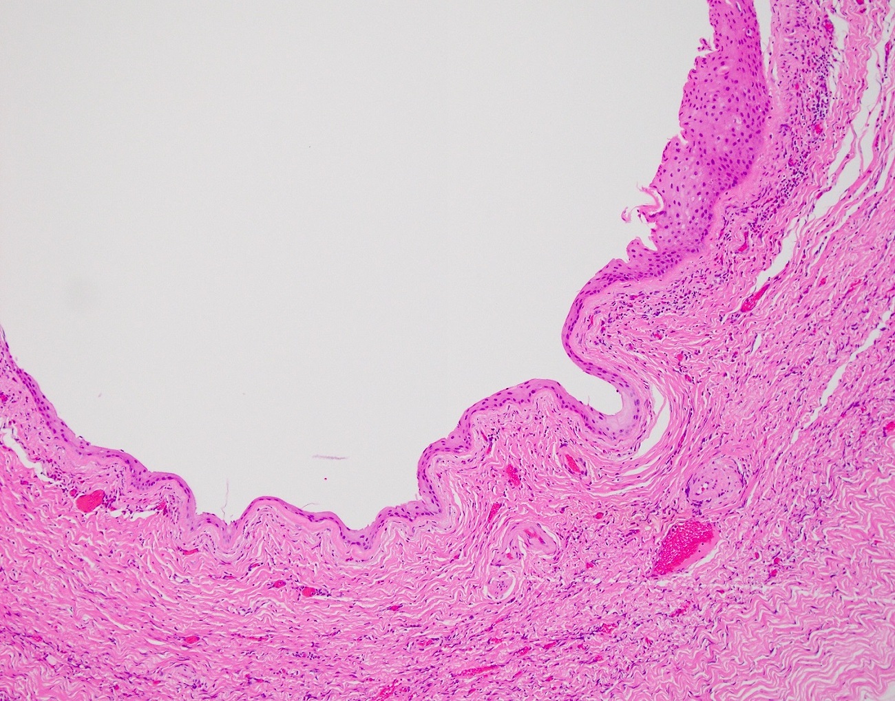
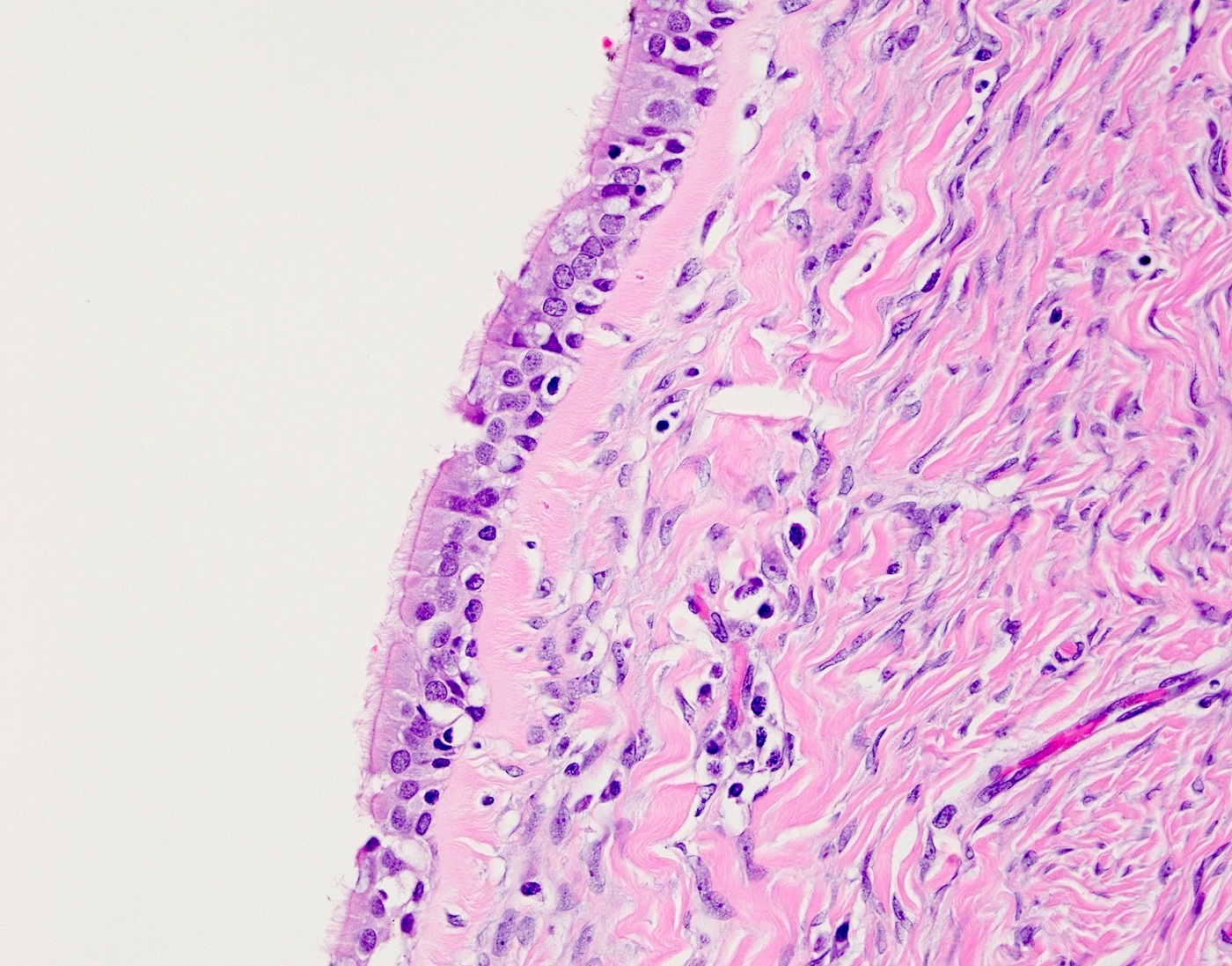
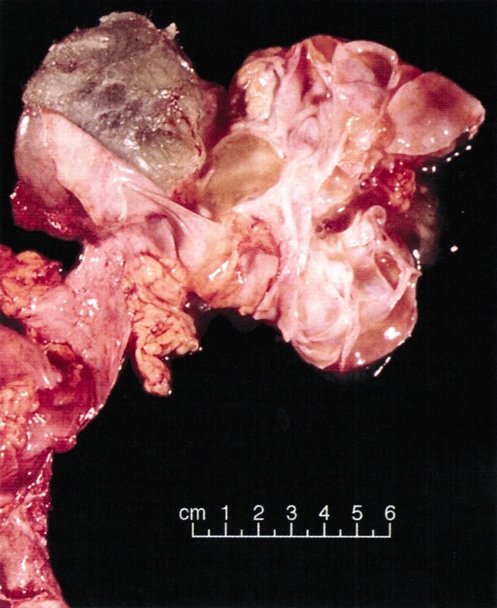
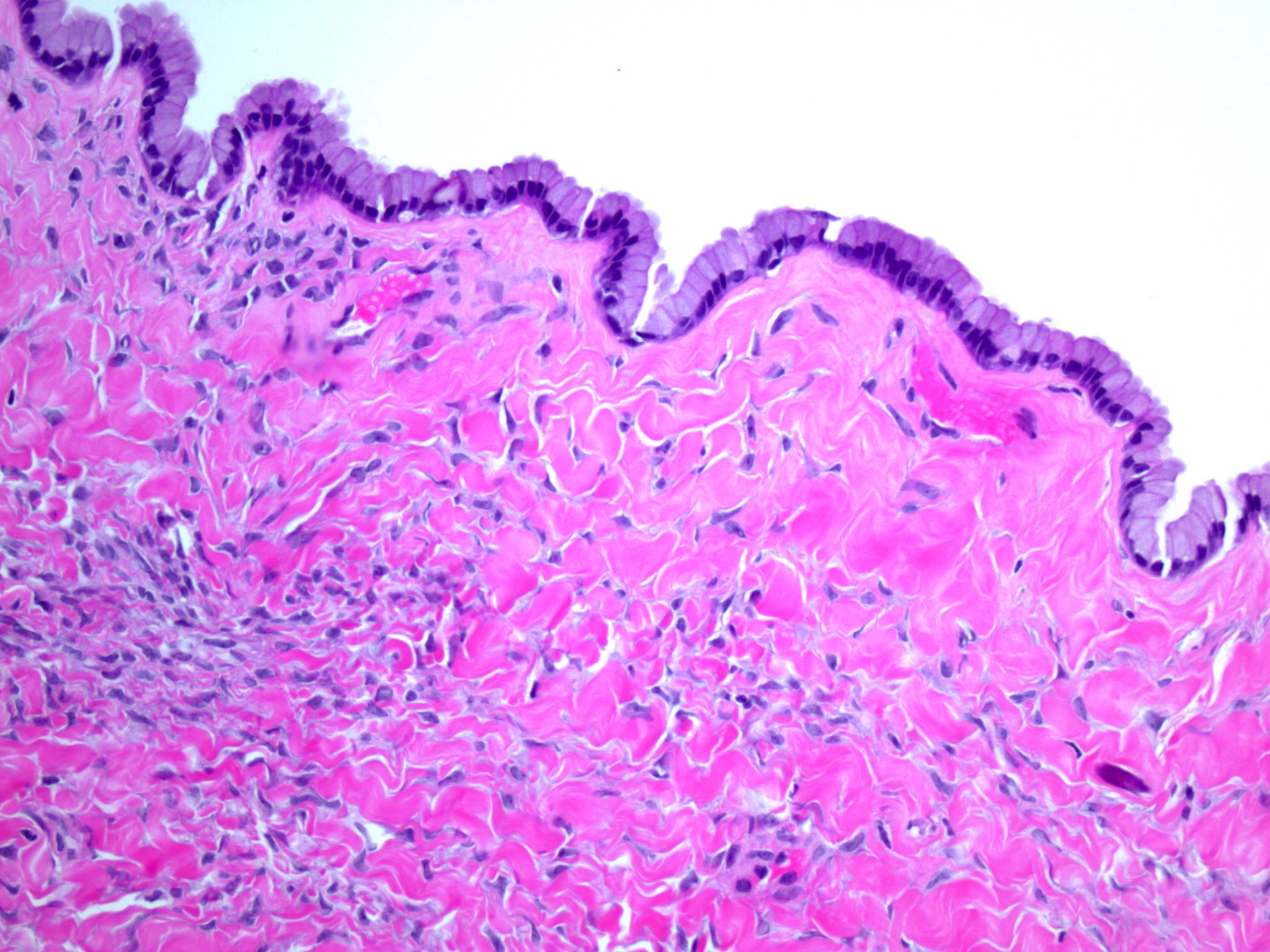
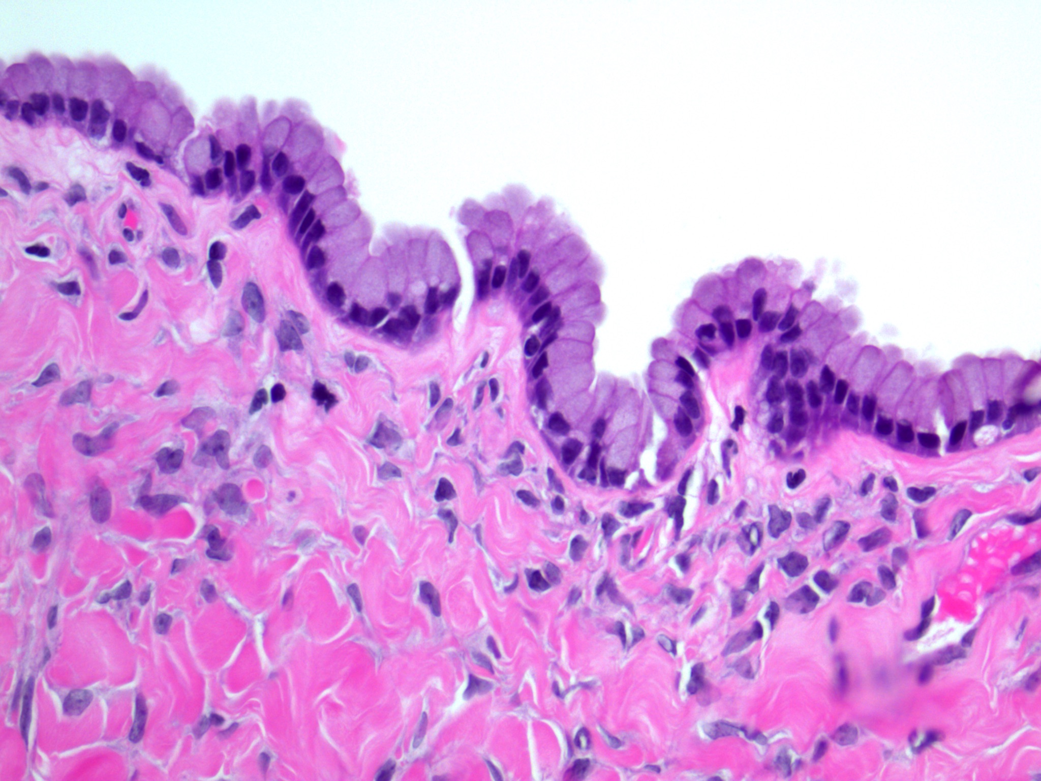
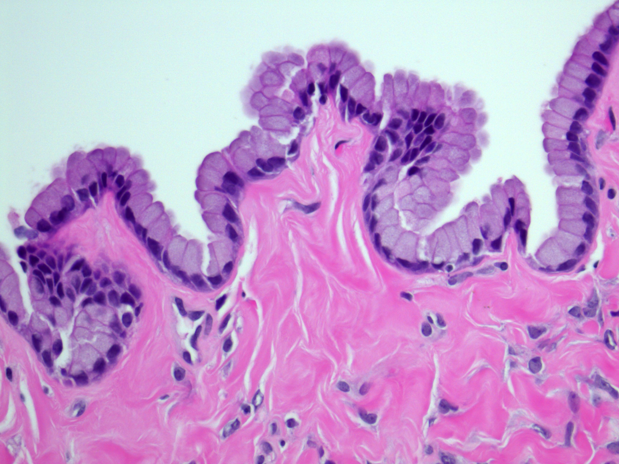
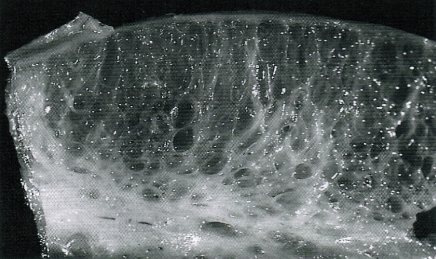
Predominantly cystic neoplasm
Attenuated cyst lining
Contributed by University of Washington Medical Center and AFIP
Uterus with shaggy hemorrhagic adhesions
External surfaces of the ovarian wedges
Cyst contains chocolate colored fluid
Cyst has dark brown discoloration
Images hosted on other servers:


Small foci resembling powder burns

Chocolate cyst
Contributed by University of Washington Medical Center and Aurelia Busca, M.D., Ph.D.
Uterine serosa with endometriosis
Endometriosis, partially denuded

Endometriosis with hemosiderin
Endometriosis involving appendix
Denuded hemorrhagic cyst
Endometriotic cyst
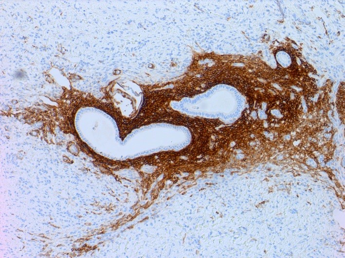
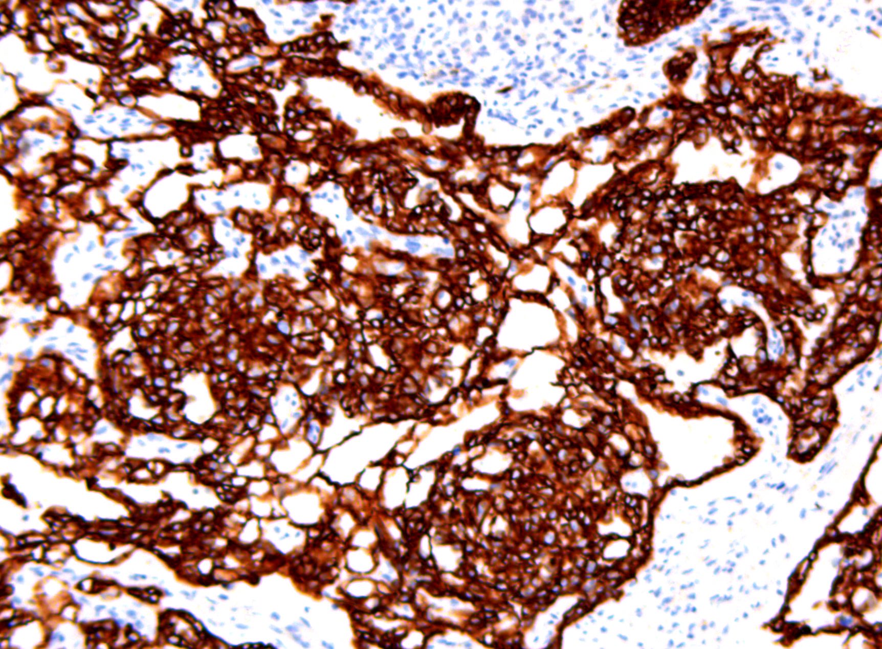
CD10
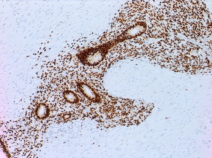
ER
Contributed by Carmen Luz, M.D.
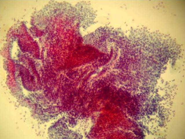
FNA from abdominal wall
Causes, symptoms, diagnosis, treatment, pathology
Histopathology - ovary
Images hosted on other servers:

Multiple lesions
Contributed by Aurelia Busca, M.D., Ph.D.
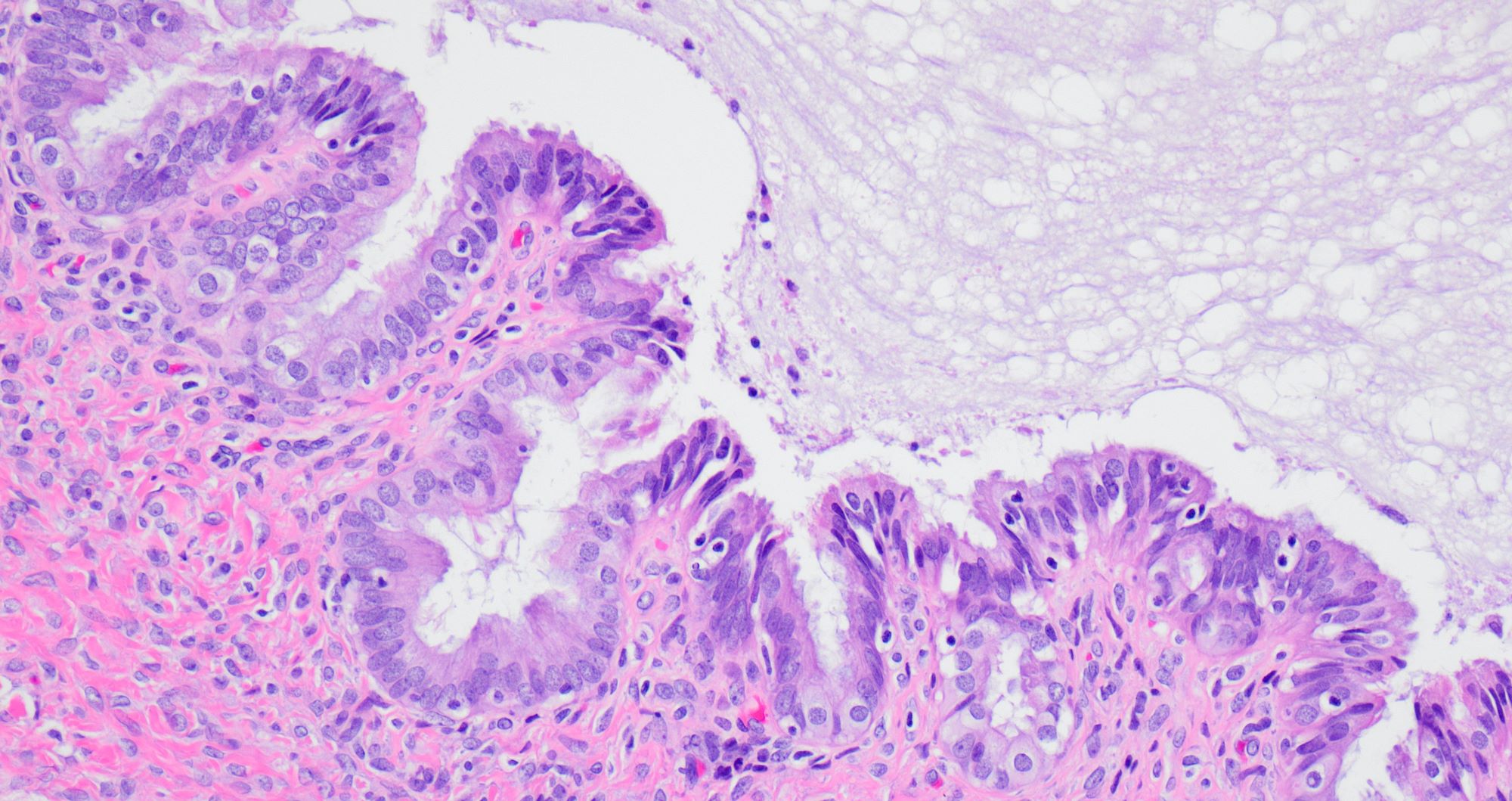
Cystically dilated glands
Cystically dilated glands with tubal differentiation
Cystically dilated glands with stromal microcalcifications
AFIP images

Keratinized material
Contributed by Ayse Ayhan, M.D.
Clear cell carcinoma, case 1:
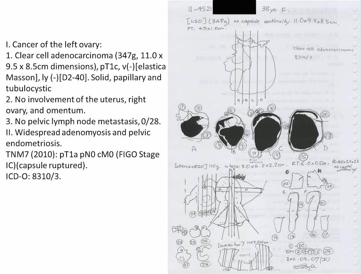
Schema
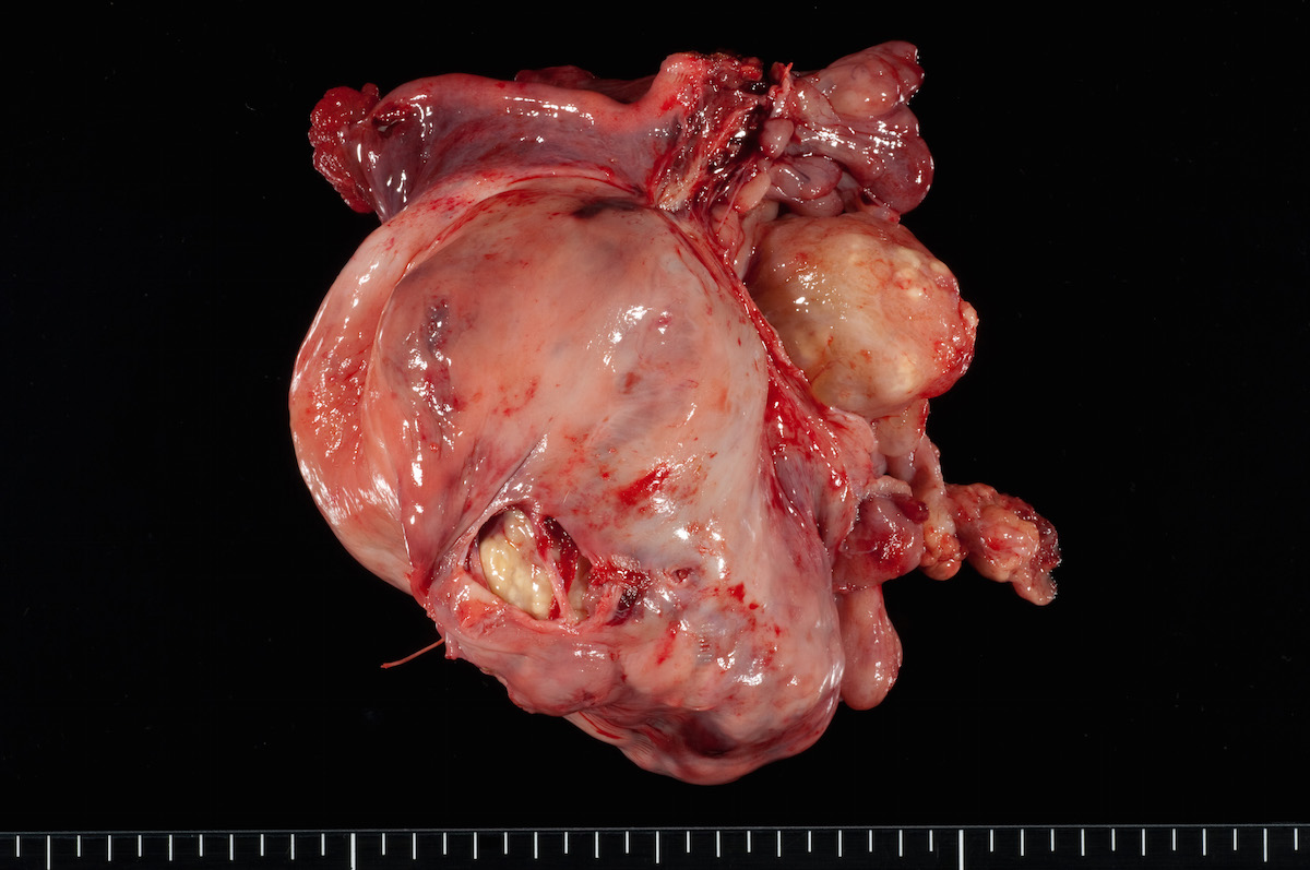
Outer appearance
Fixed pictures of surgical material

Cut surface
Clear cell carcinoma, case 2:
Outer appearance

Cut surface
Fixed cut surface
Bilateral endometrioid cancer:

Left, 730g, 14.5x12.6cm

Left, 730g, 14.5x12.6cm
Ovarian tumor:


Right, 70g, 7.5x3.5cm
Images hosted on other servers:

Bilateral ovarian fibroma on MRI

Unilateral ovarian fibroma on MRI
Contributed by Rex Bentley, M.D. and AFIP images
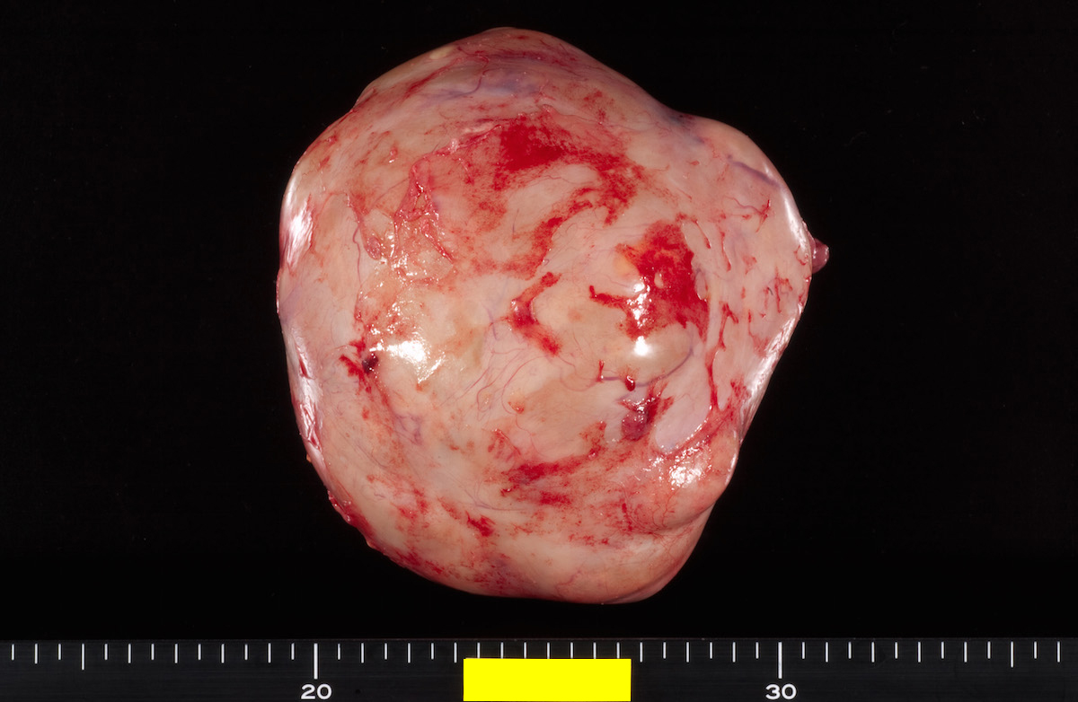
Chalky white surface

Edematous and focally hemorrhagic

Dense white mass
Images hosted on other servers:

Hard white tumor

Fibrothecoma
Contributed by Gulisa Turashvili, M.D., Ph.D., Kyle C. Strickland, M.D., Ph.D. and Rex Bentley, M.D.
Cellular spindle cell neoplasm
Mild cytologic atypia
Mitotic activity
Small tubules
Variable cellularity
Bland spindle cells
Spindled to ovoid nuclei
Dense collagen
Cellular spindle cell neoplasm
Reticulin
Inhibin
Calretinin
Contributed by Dr. Josehp Christopher Castillo


AFIP images

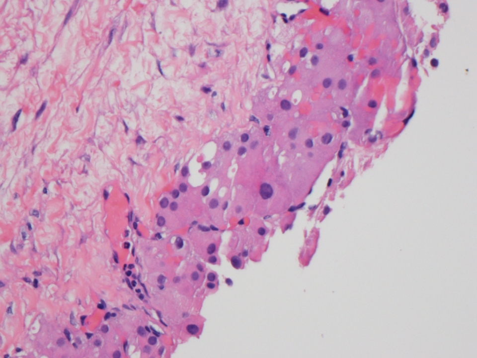
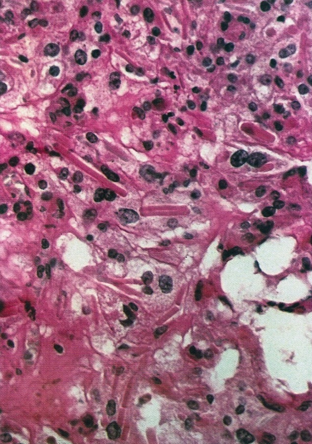
Clusters of lutein cells lie in the edematous stroma
Images hosted on other servers:


Loose stroma with occasional inflammatory cells
Images hosted on other servers:

Capsular disruption and hemorrhage
AFIP images

Moderate cytologic atypia
Images hosted on other servers:

Clusters of pleomorphic spindle cells
Images hosted on other servers:

Follicle cyst on ultrasound
AFIP images
Unilocular cyst with smooth lining
Images hosted on other servers:


Large follicle cysts
Contributed by Gulisa Turashvili, M.D., Ph.D.
Cyst wall
Cyst lining

Luteinized theca cells
Reticulin stain
AFIP images
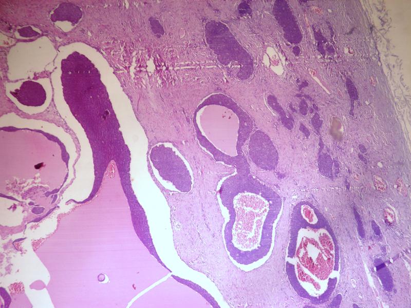
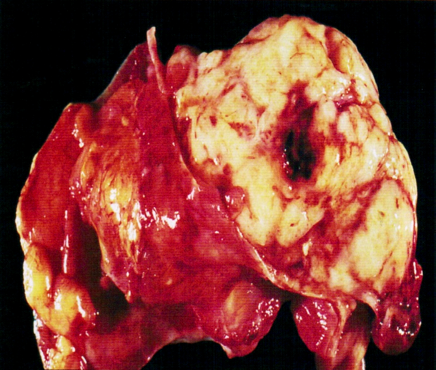
Bilateral tumor, with germinoma

With germinoma
Images hosted on other servers:

Gonadal dysgenesis
AFIP images
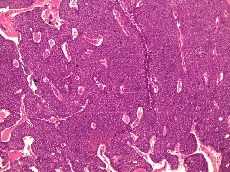
Nests of tumor
With germinoma
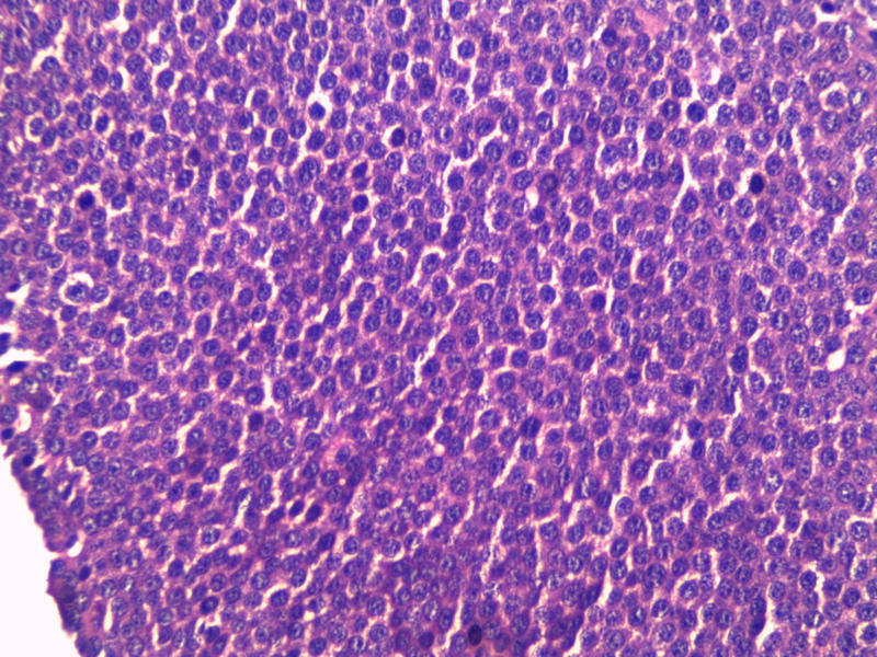
Small sex cord type cells
Contributed by Eman Abdelzaher, M.D., Ph.D. and AFIP

Actinomycosis: tubo-ovarian


Actinomycosis
Images hosted on other servers:

Central suppuration
and surrounding
giant cells in
Crohn's disease

Tuberculosis
Images hosted on other servers:


Large pelvic mass
Images hosted on other servers:

Hirsutism

Intraoperative mass
Contributed by Shabnam Zarei, M.D. and AFIP

Solid, cystic and hemorrhagic mass
Solid and cystic mass
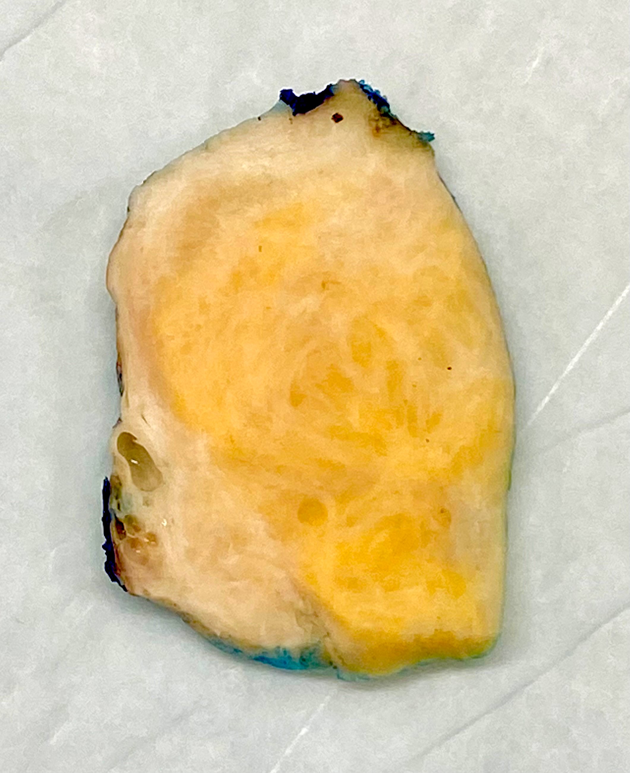
Solid yellow mass
Predominantly cystic mass
Solid and cystic tumor
Images hosted on other servers:

Solid cystic mass
Contributed by Shabnam Zarei, M.D. and Sharon Bihlmeyer, M.D.
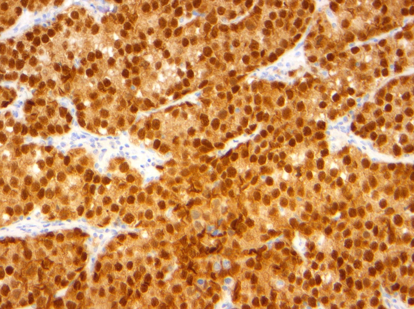
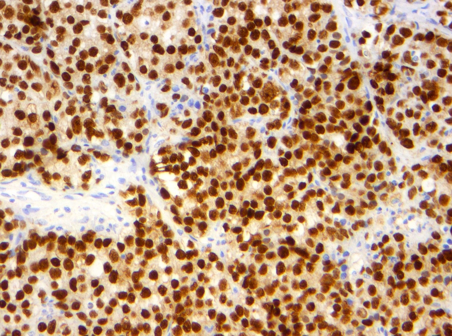
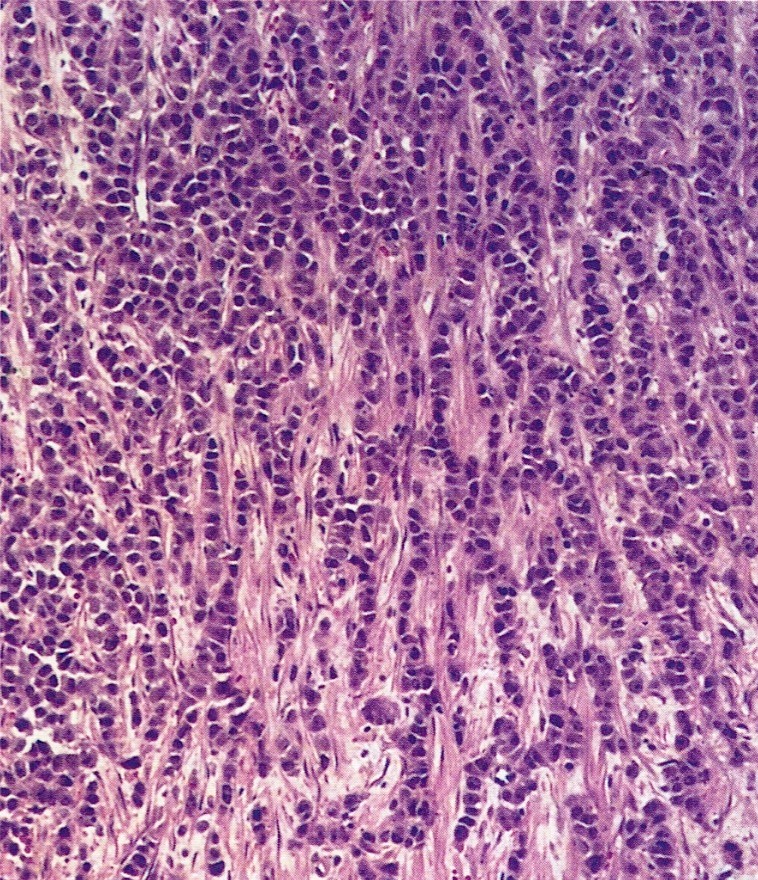
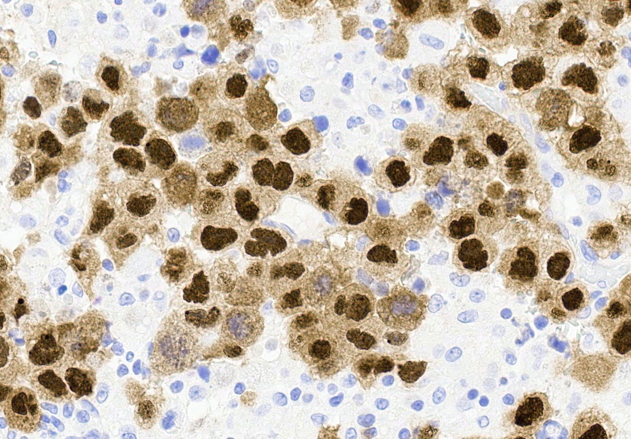
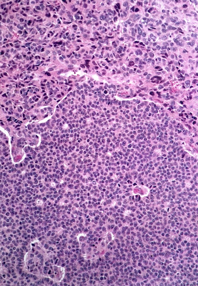
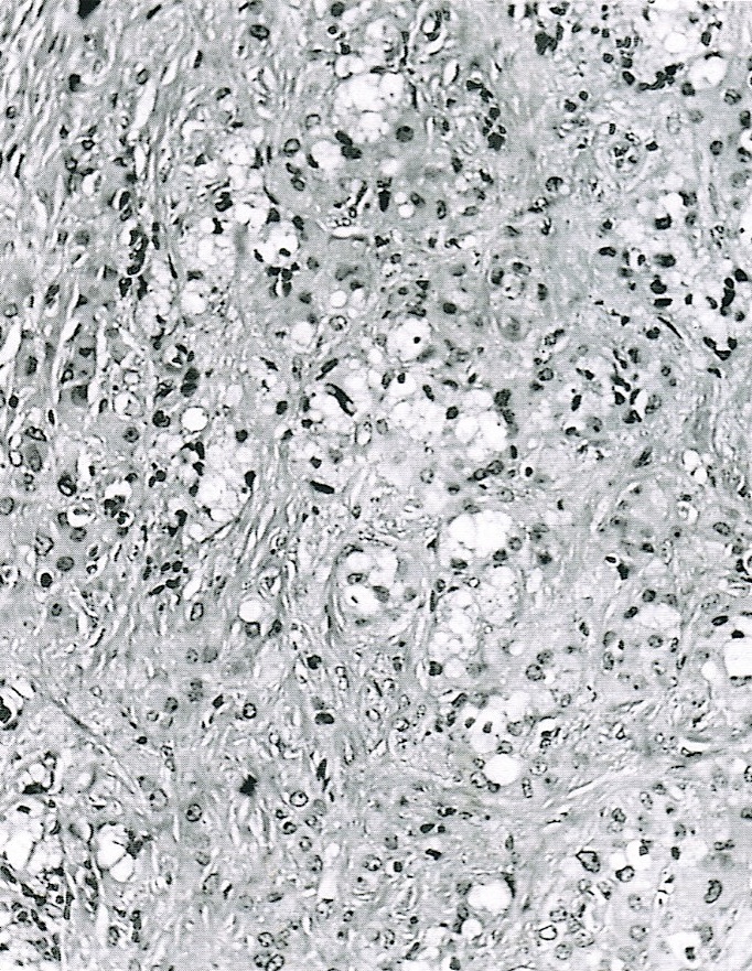
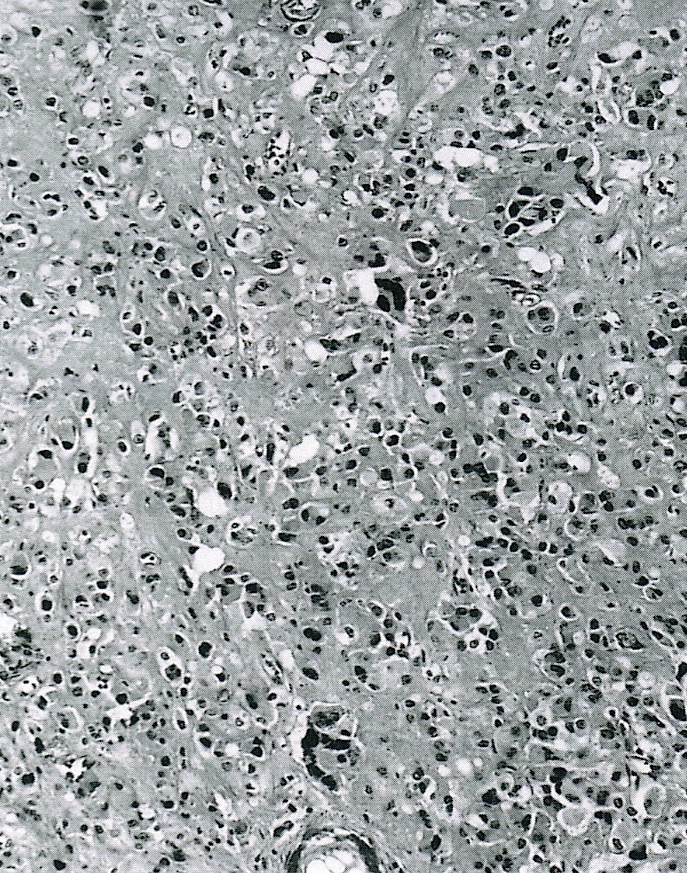
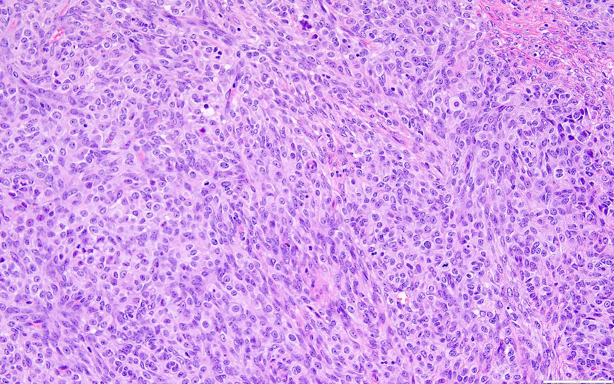
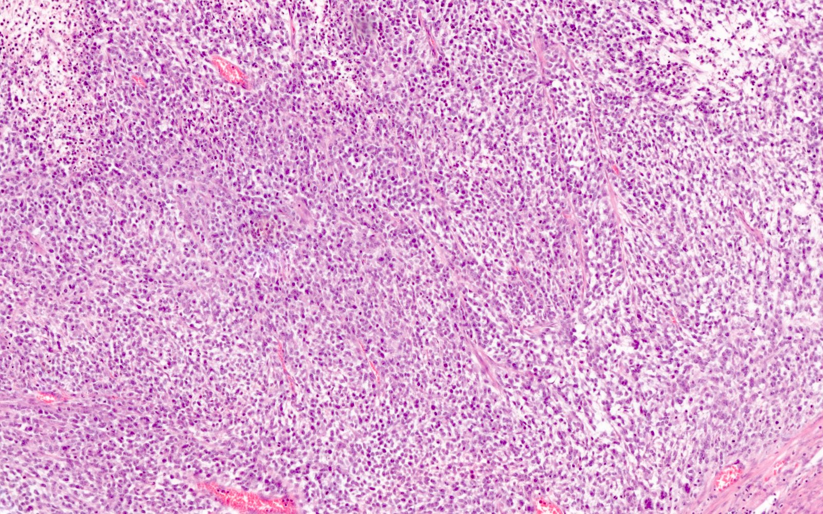
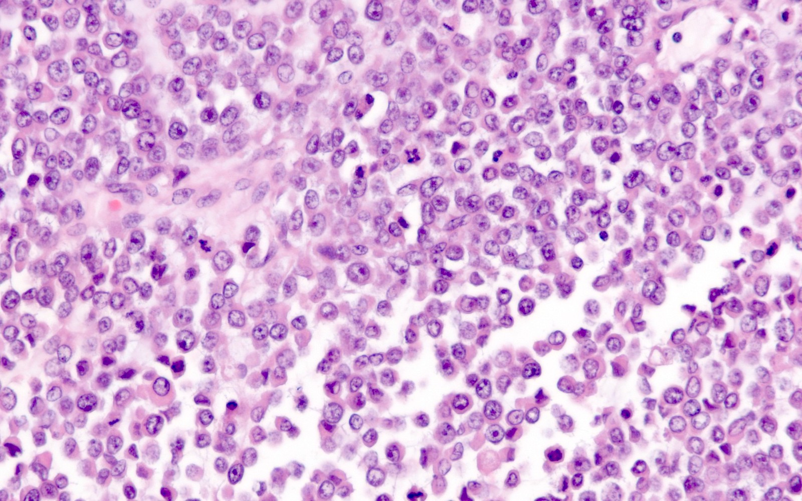
Classic diffuse pattern
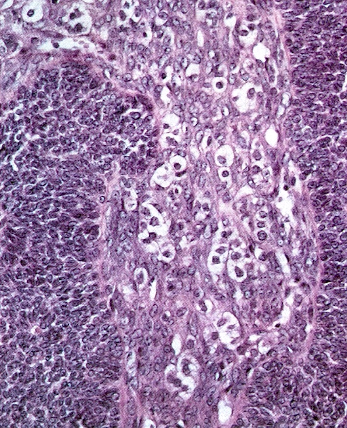
Nuclear grooves
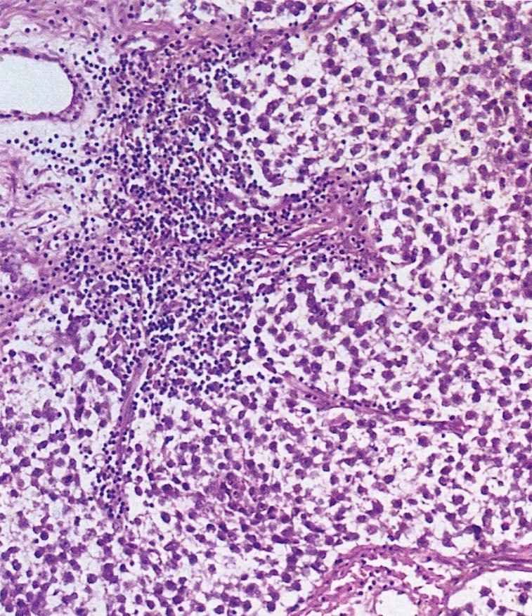
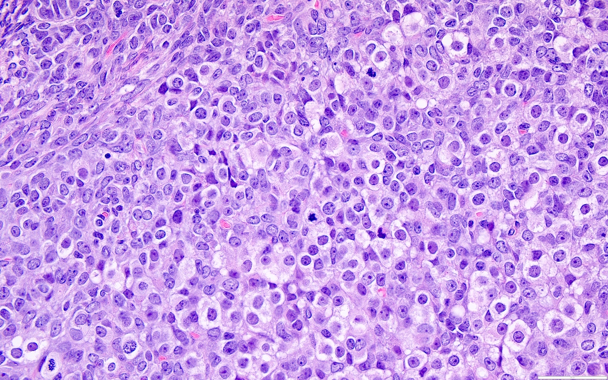
Luteinized
Luteinized with round nuclei
Luteinized
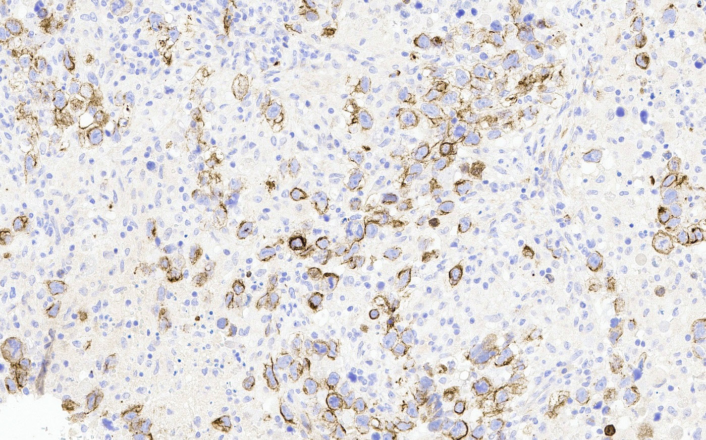
Calretinin
Reticulin
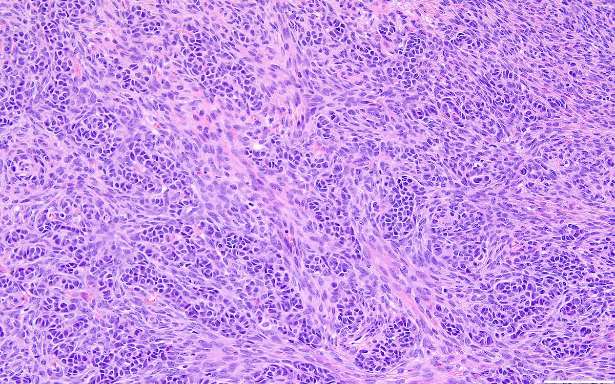
Diffuse pattern
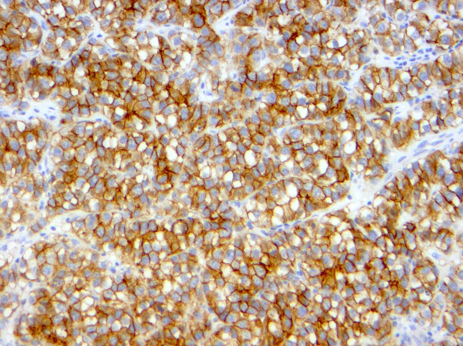
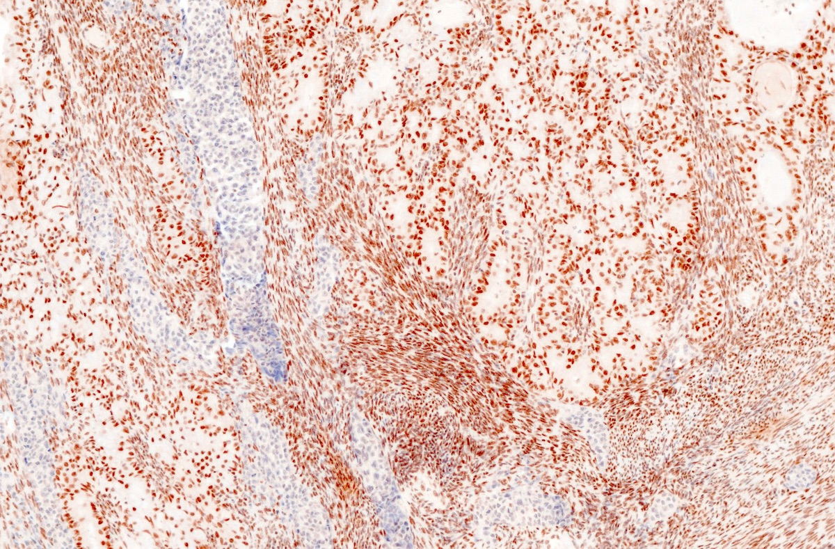
Inhibin
Calretinin
CK7
AFIP images
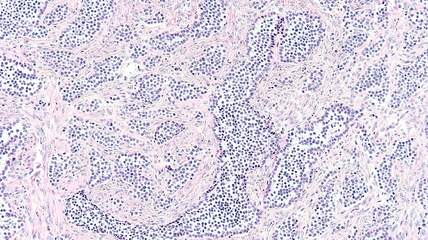
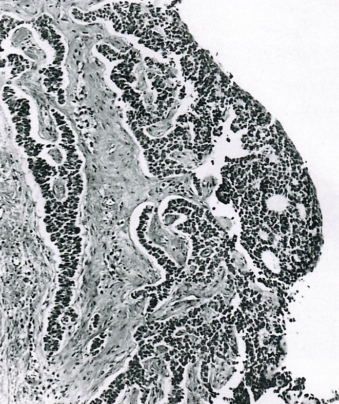
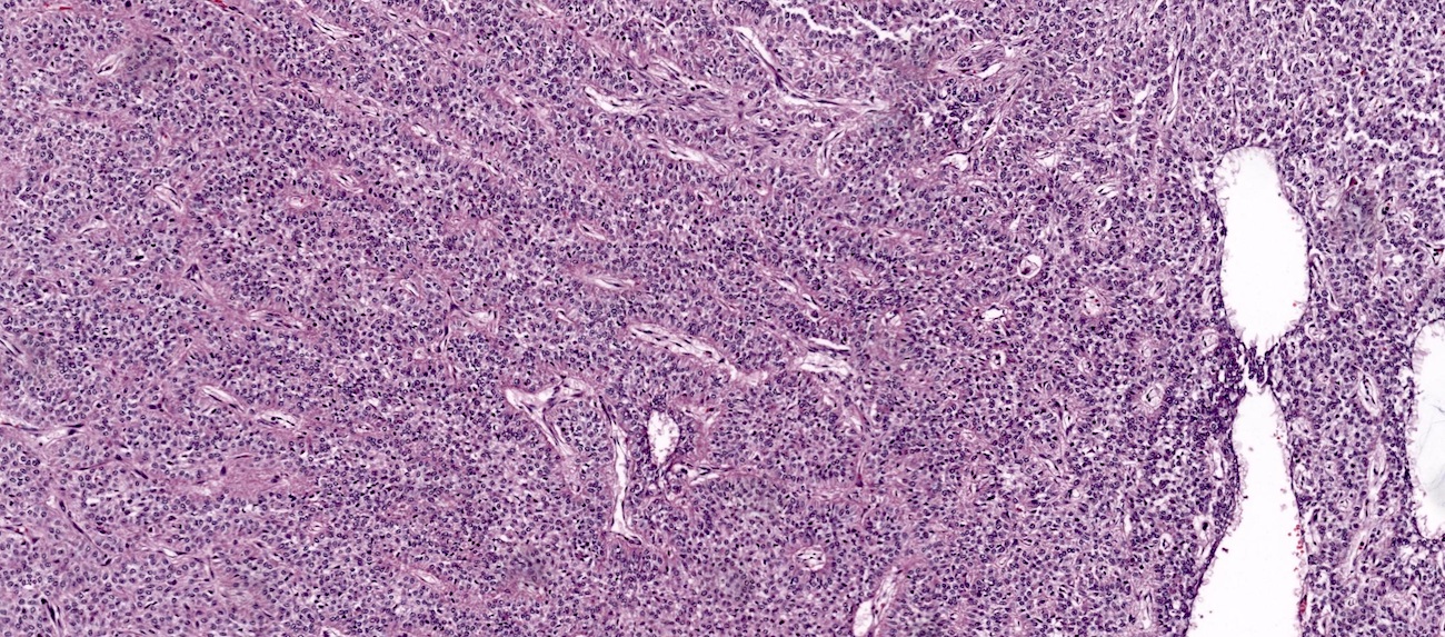
Trabecular pattern
Insular pattern
Microfollicular pattern
Macrofollicular pattern
Watered silk (moiré silk) pattern
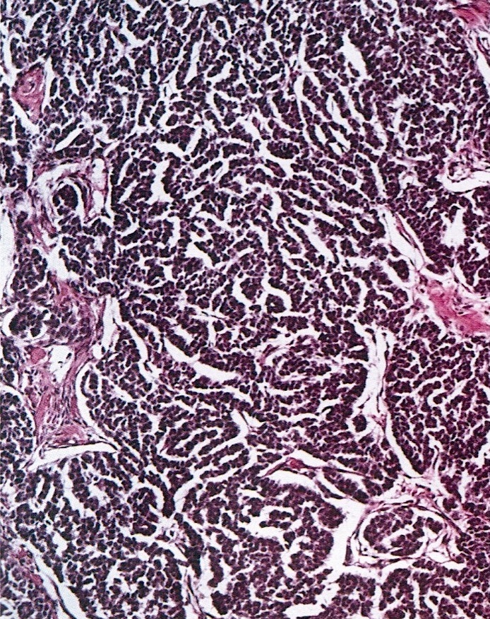
Gyriform pattern
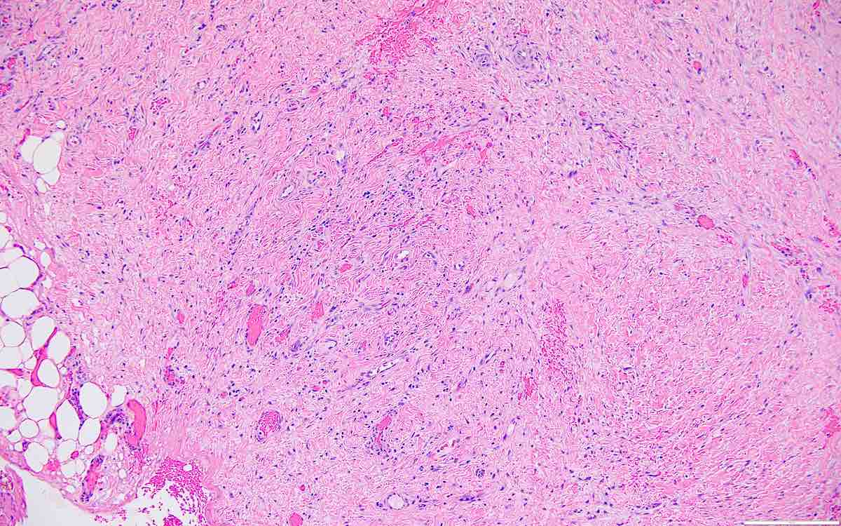
Cystic tumor
Enlarged, hyperchromatic, bizarre nuclei
Theca cells
Sex cord stromal tumors of the ovary and mimics
Histopathology of adult granulosa cell tumor
Granulosa cell tumor
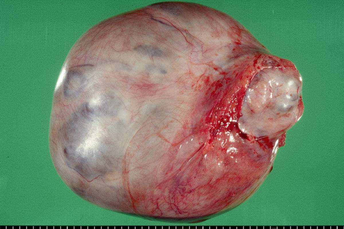
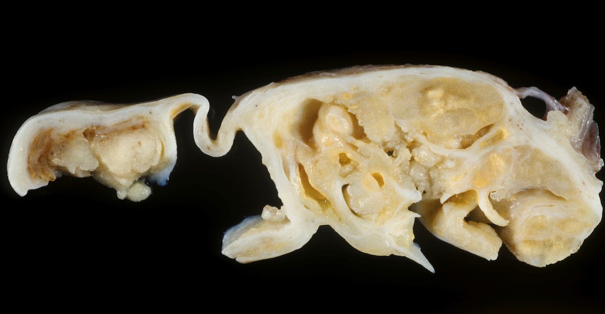
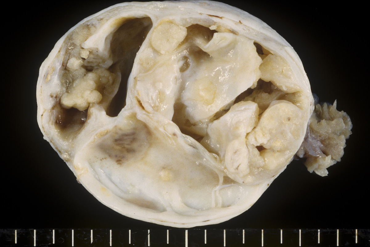
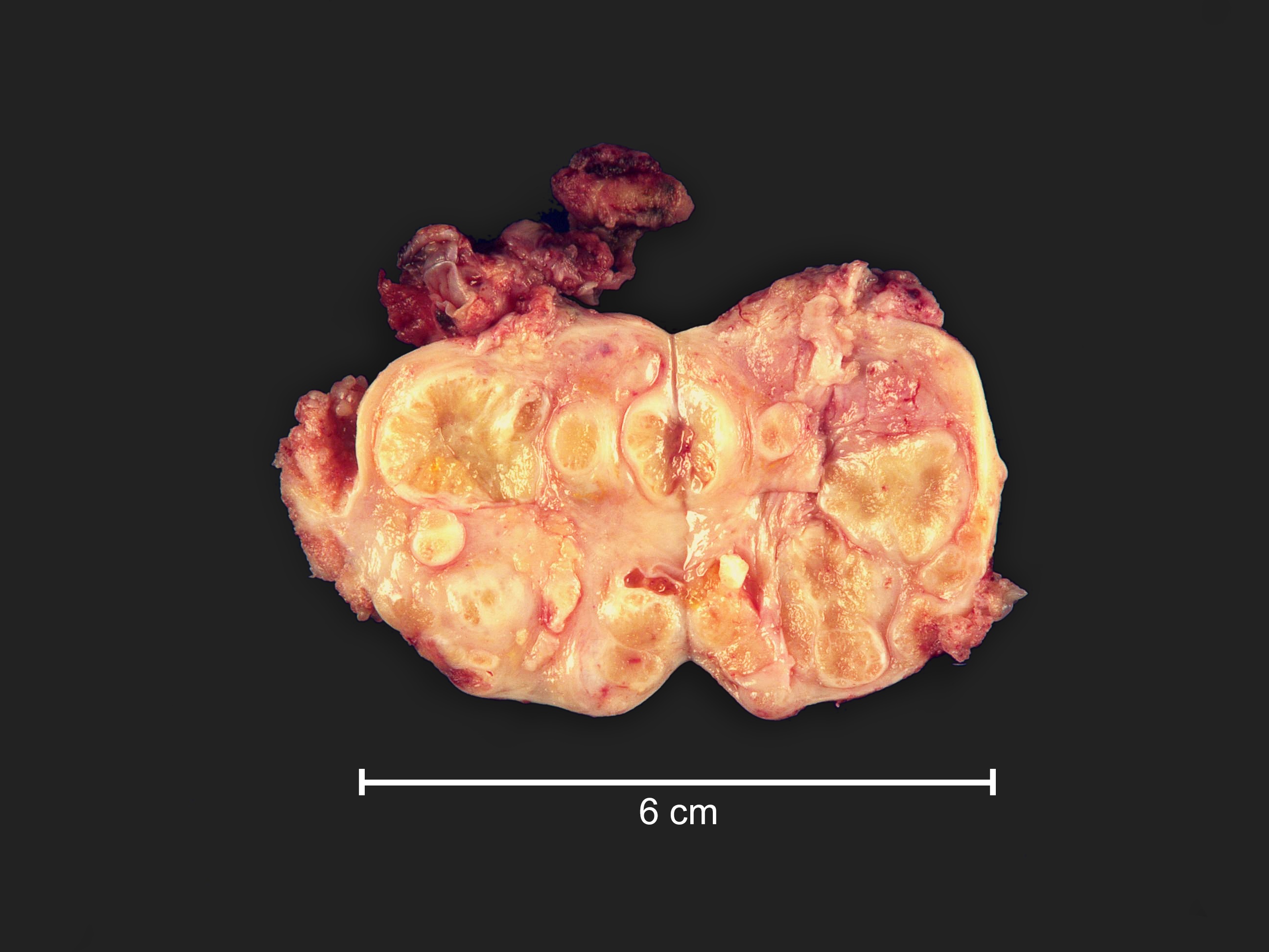
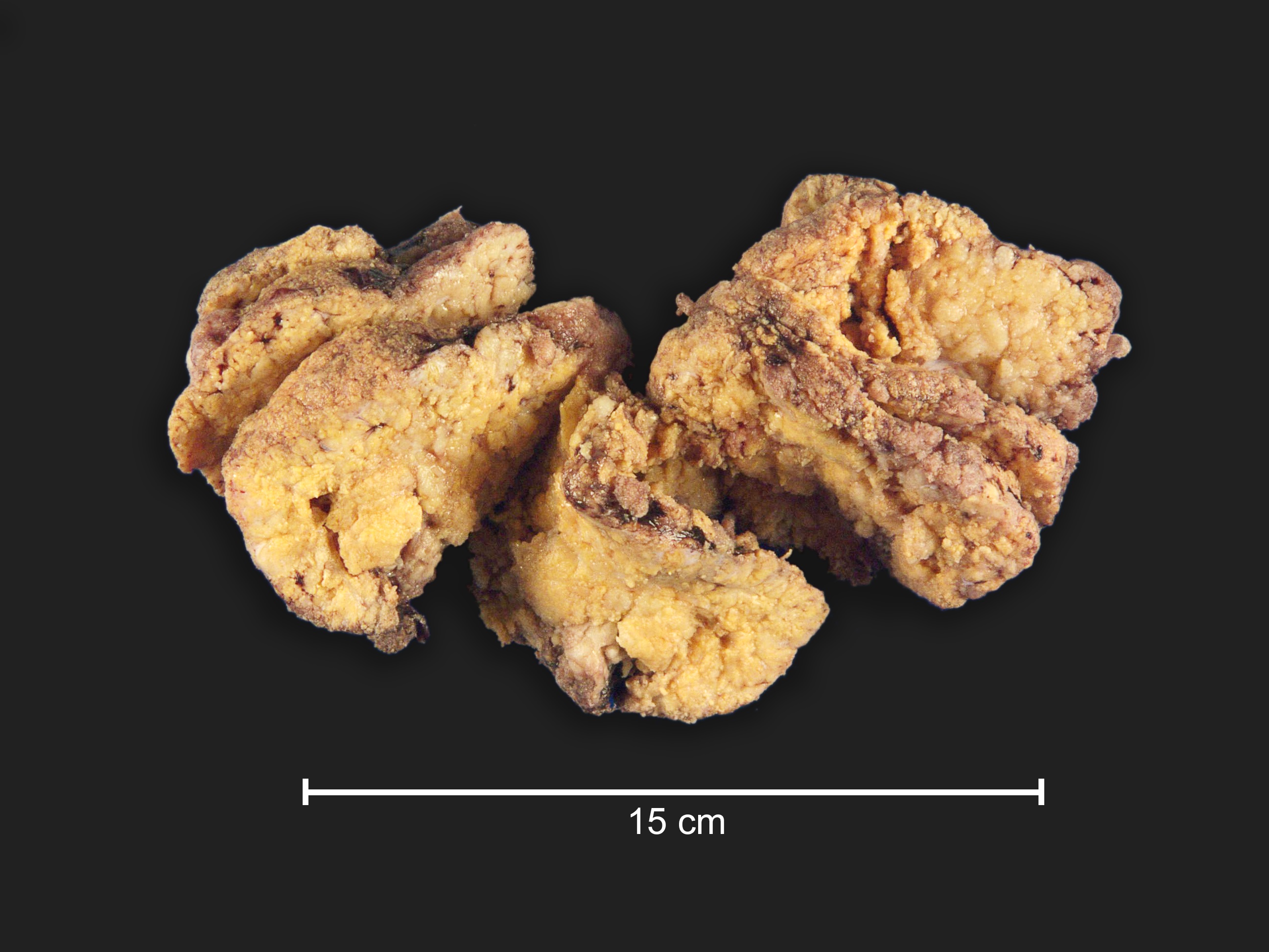
AFIP Images
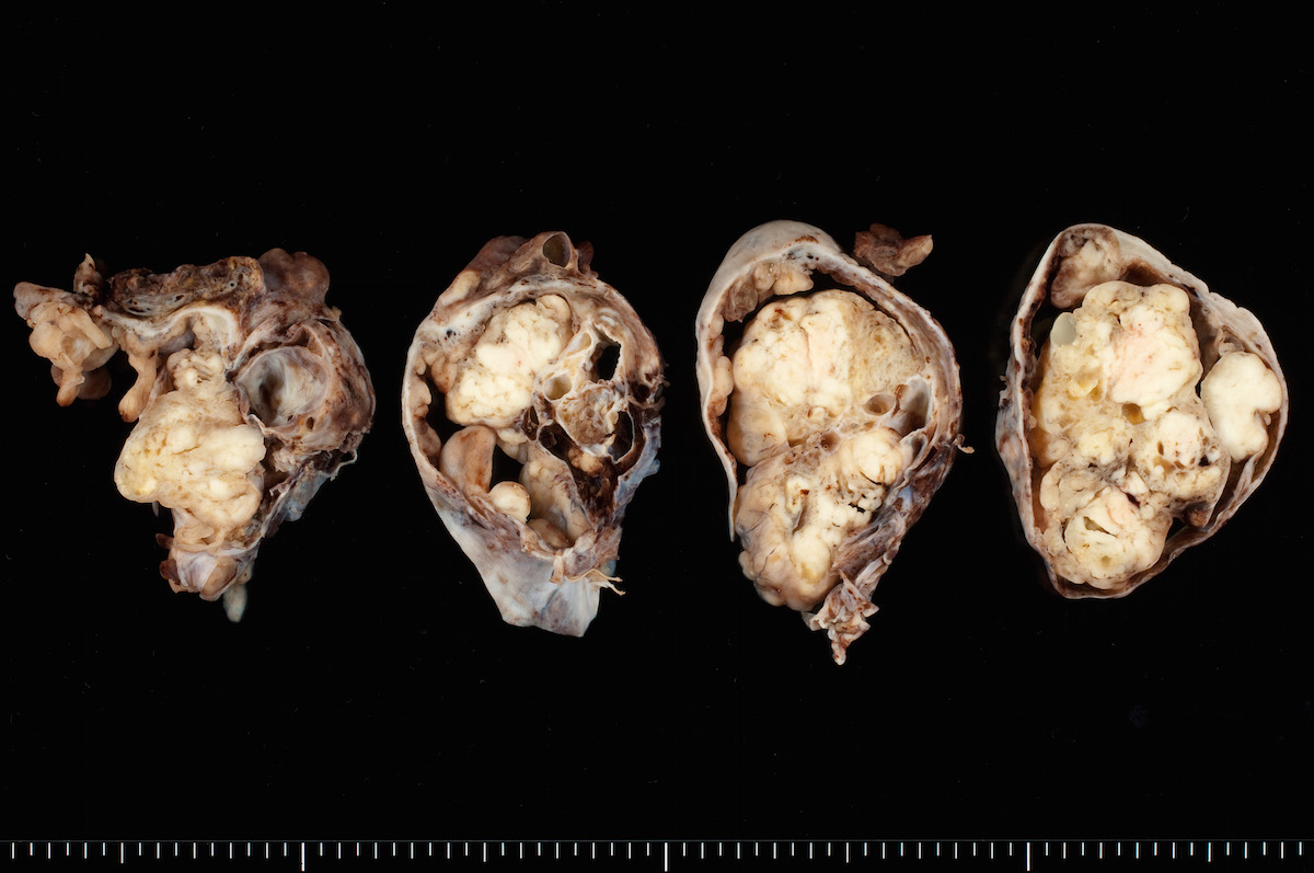
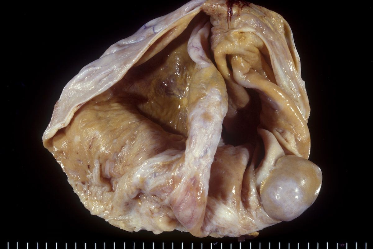
Solid and slightly lobulated
Large locules with smooth linings
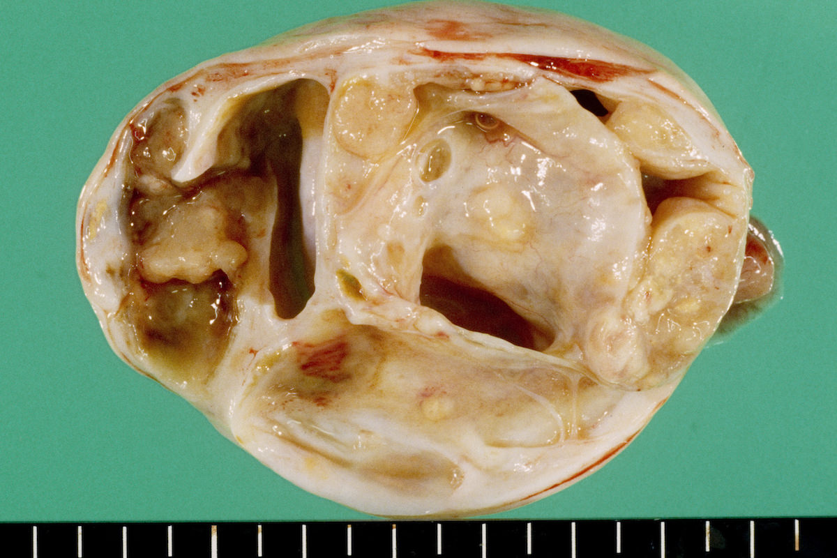
Solid and cystic
Contributed by Jutta Huvila, M.D., Ph.D. and AFIP
Lobulated growth pattern
Follicular differentiation
Atypical cells lining follicle
Solid growth
Solid cellular neoplasm with focal follicle formation
Solid nodules
Mitotic figures
Hobnail nuclei
Bizarre nuclei
AFIP images
Well differentiated
ovarian and testicular
type cells
Images hosted on other servers:

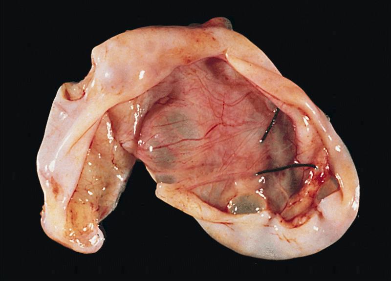
Cystic mass with papillary projections
Contributed by Erna Forgó, M.D. and Teri A. Longacre, M.D.
Cystic mass with papillary tumor growth
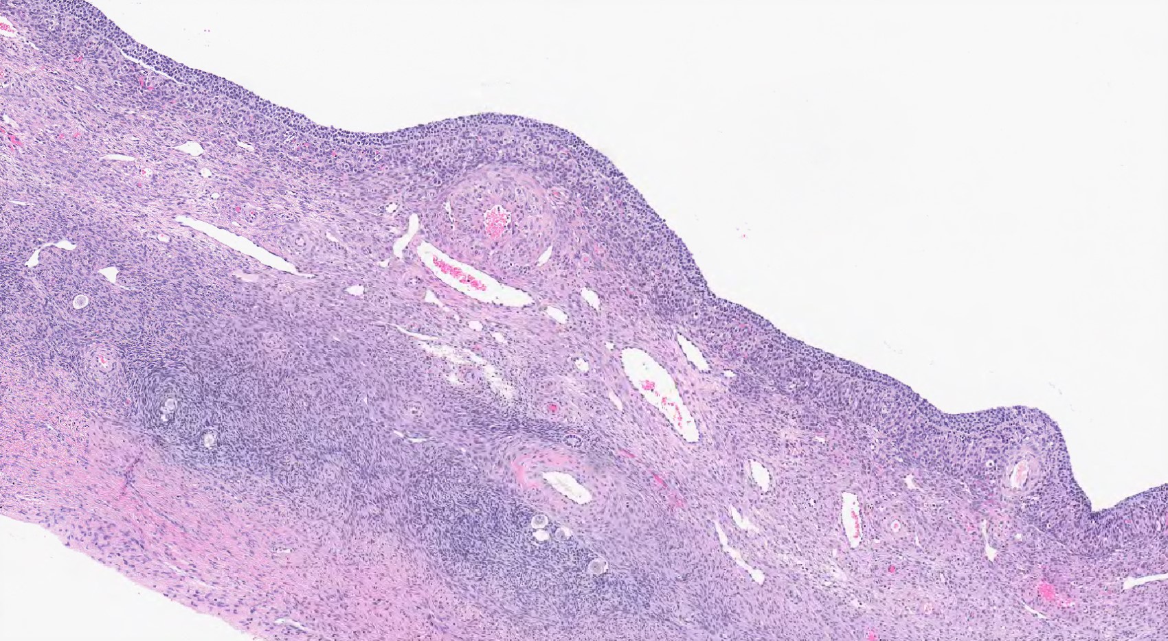
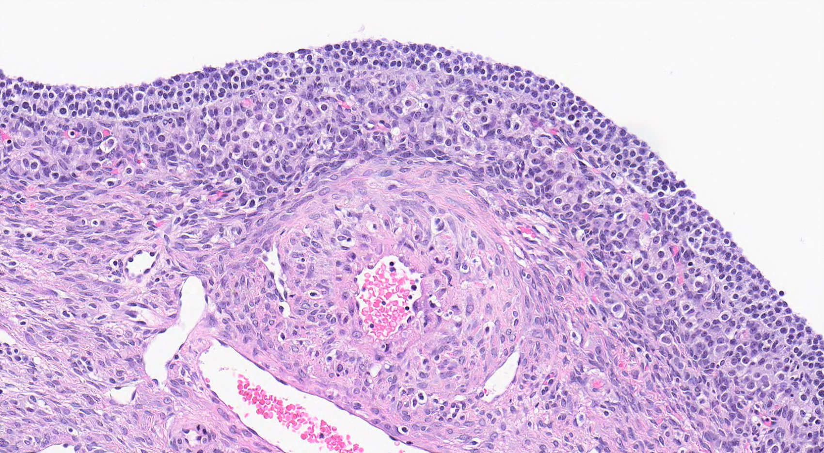
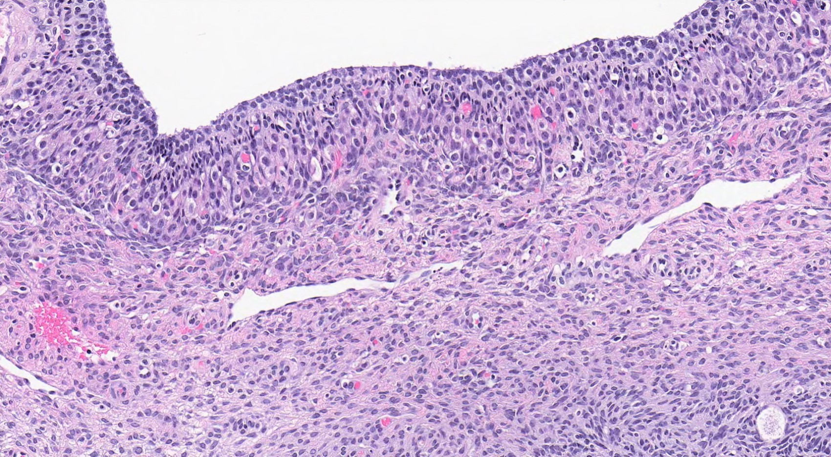
Contributed by Erna Forgó, M.D. and Teri A. Longacre, M.D.


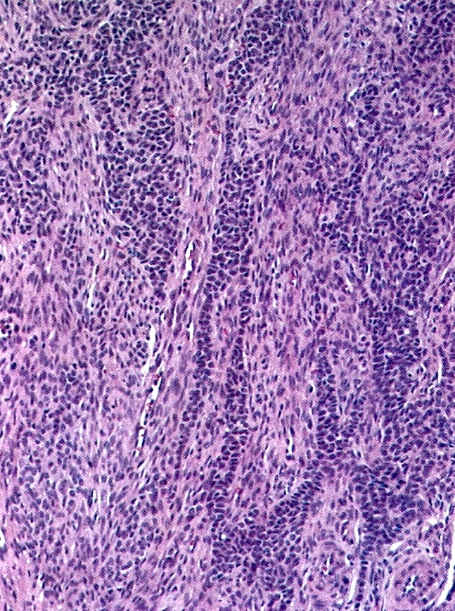
Significant nuclear atypia and pleomorphism
Micropapillary growth pattern
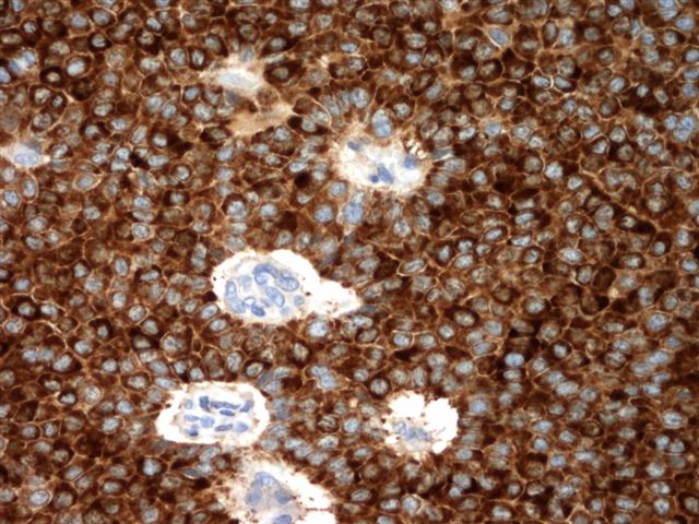
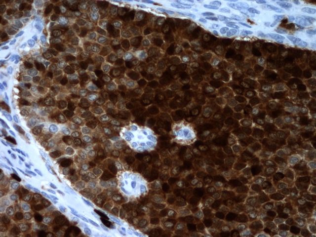
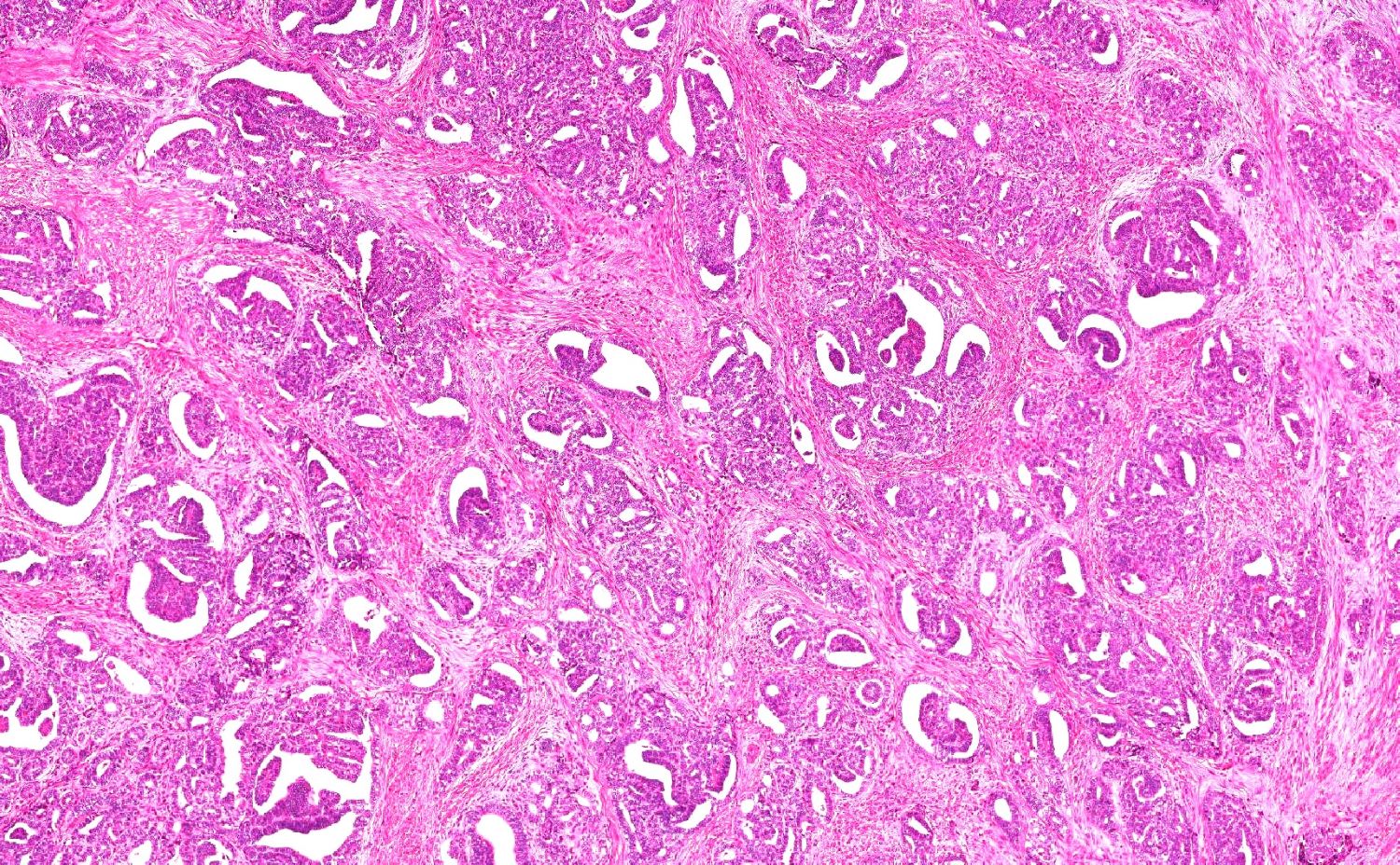
SET pattern
SET pattern
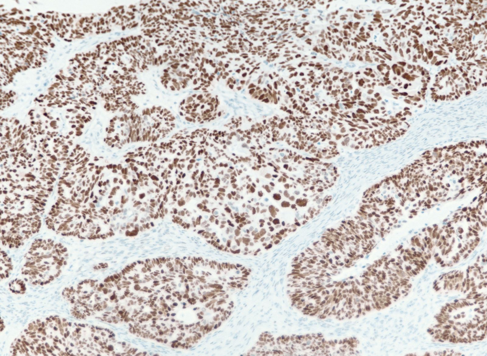
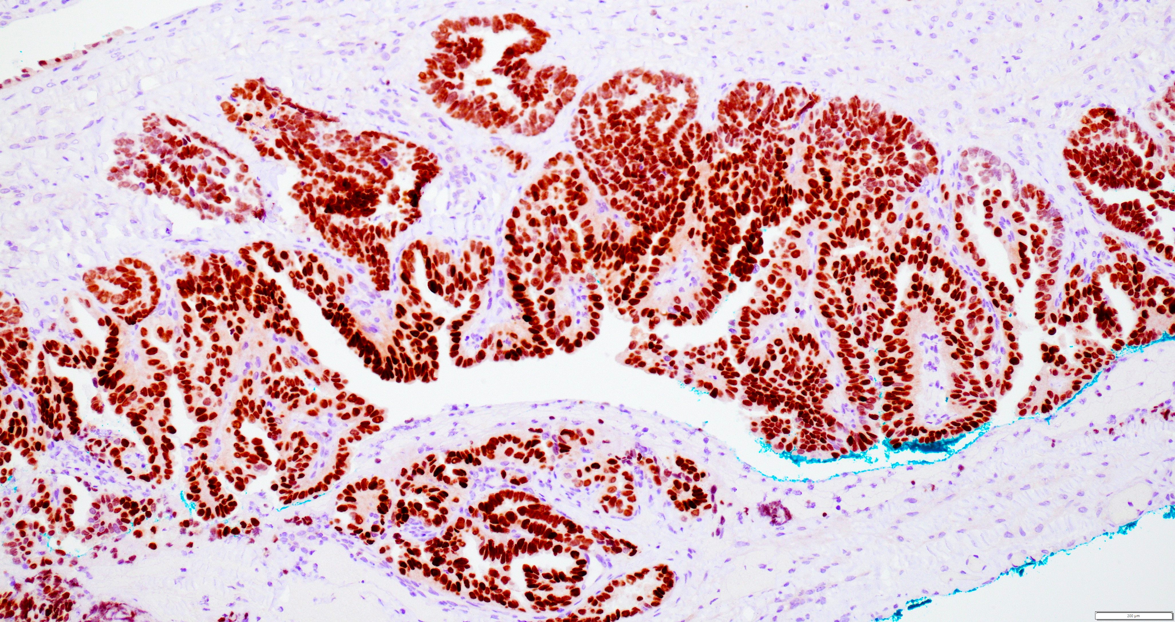
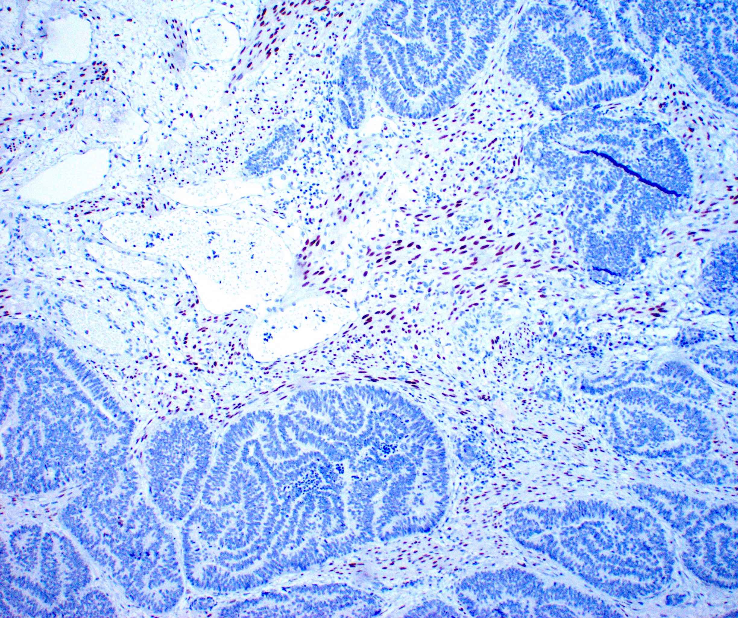
PAX8 positive
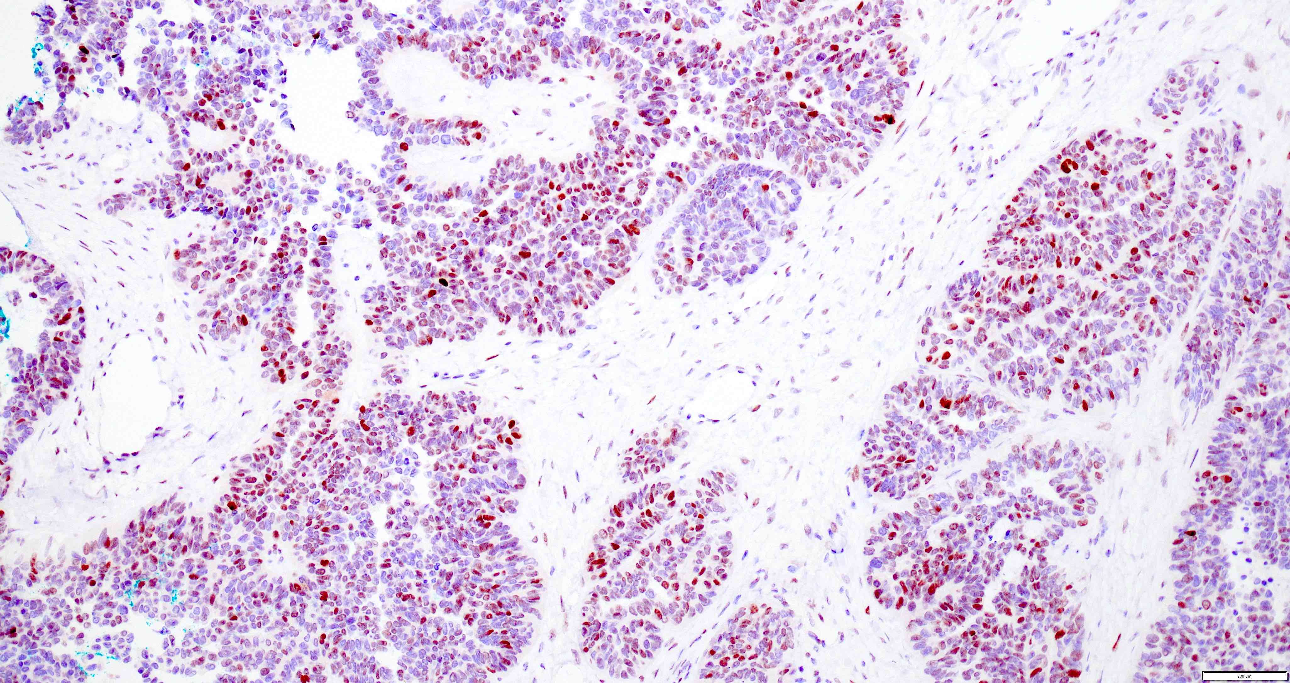
ER positive

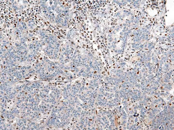
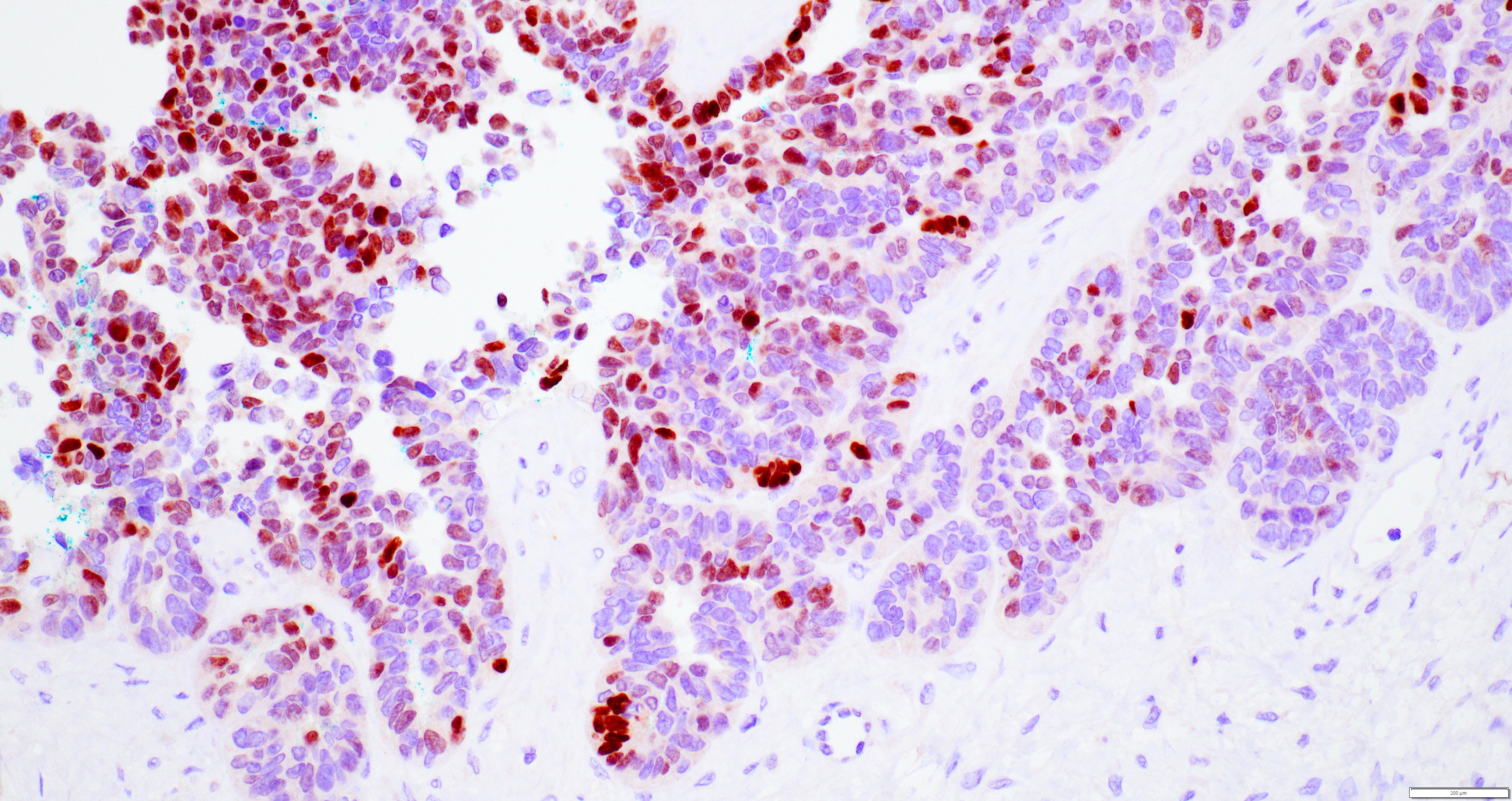
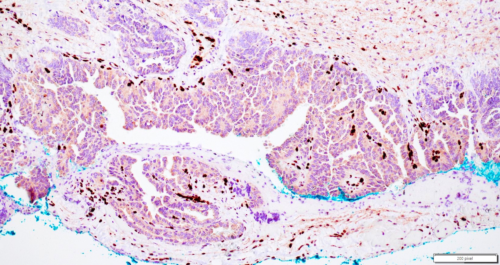
Variable PR (negative in this case)
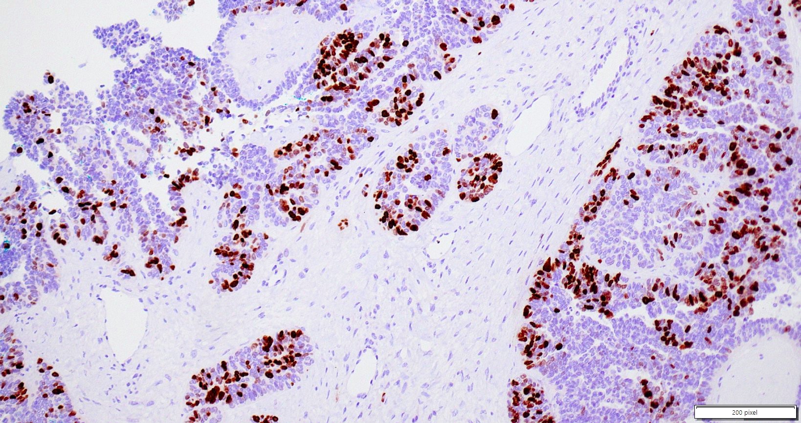
WT1 positive
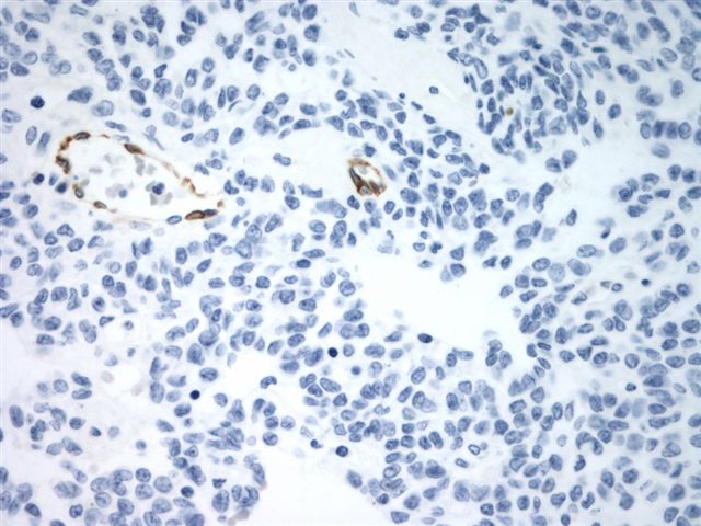
CK7 positive
CK20 negative

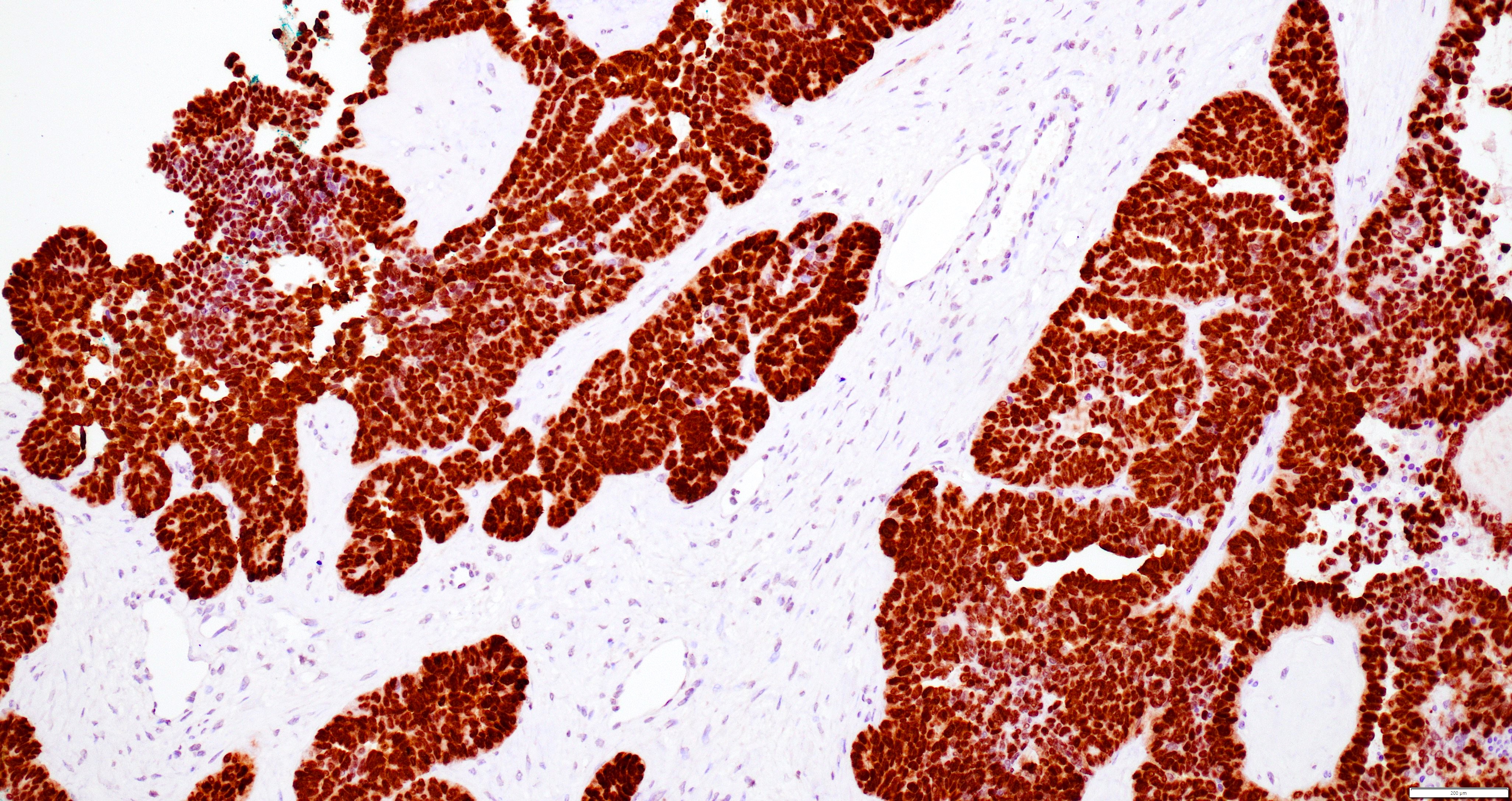
p53 overexpression
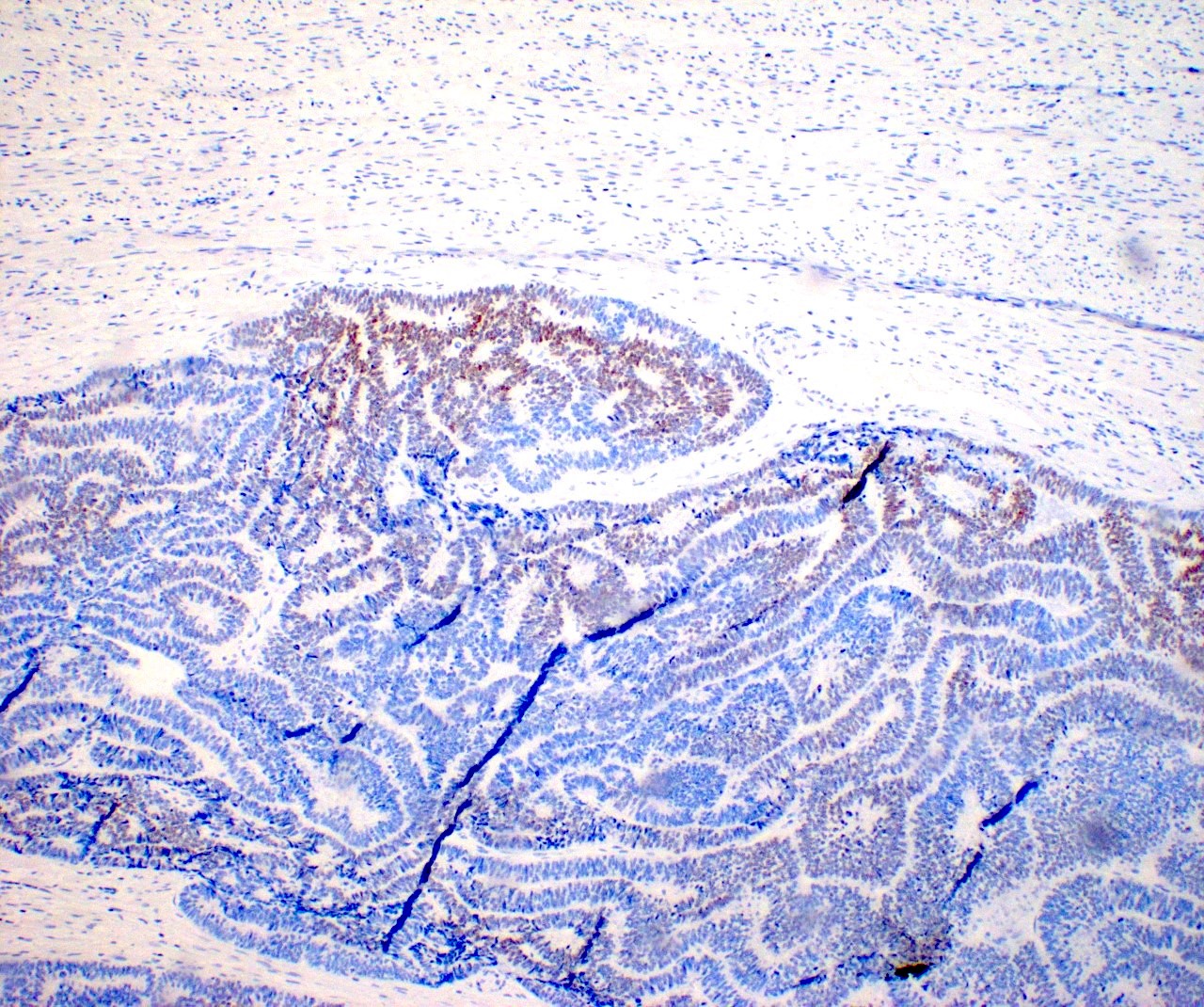
p53 null phenotype
Serous tubal intraepithelial carcinoma (STIC)
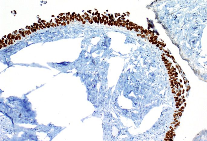
p53 overexpression in STIC
Contributed by Jijgee Munkhdelger, M.D., Ph.D. and Andrey Bychkov, M.D., Ph.D.
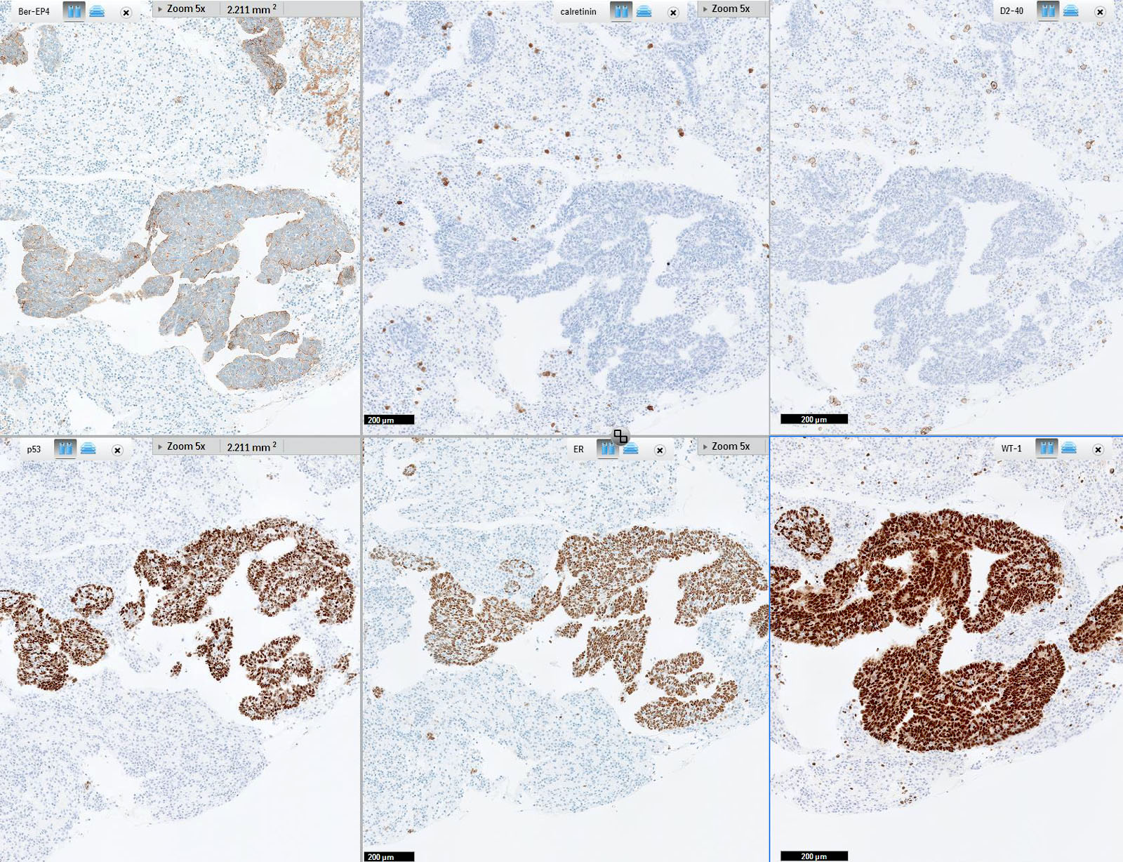
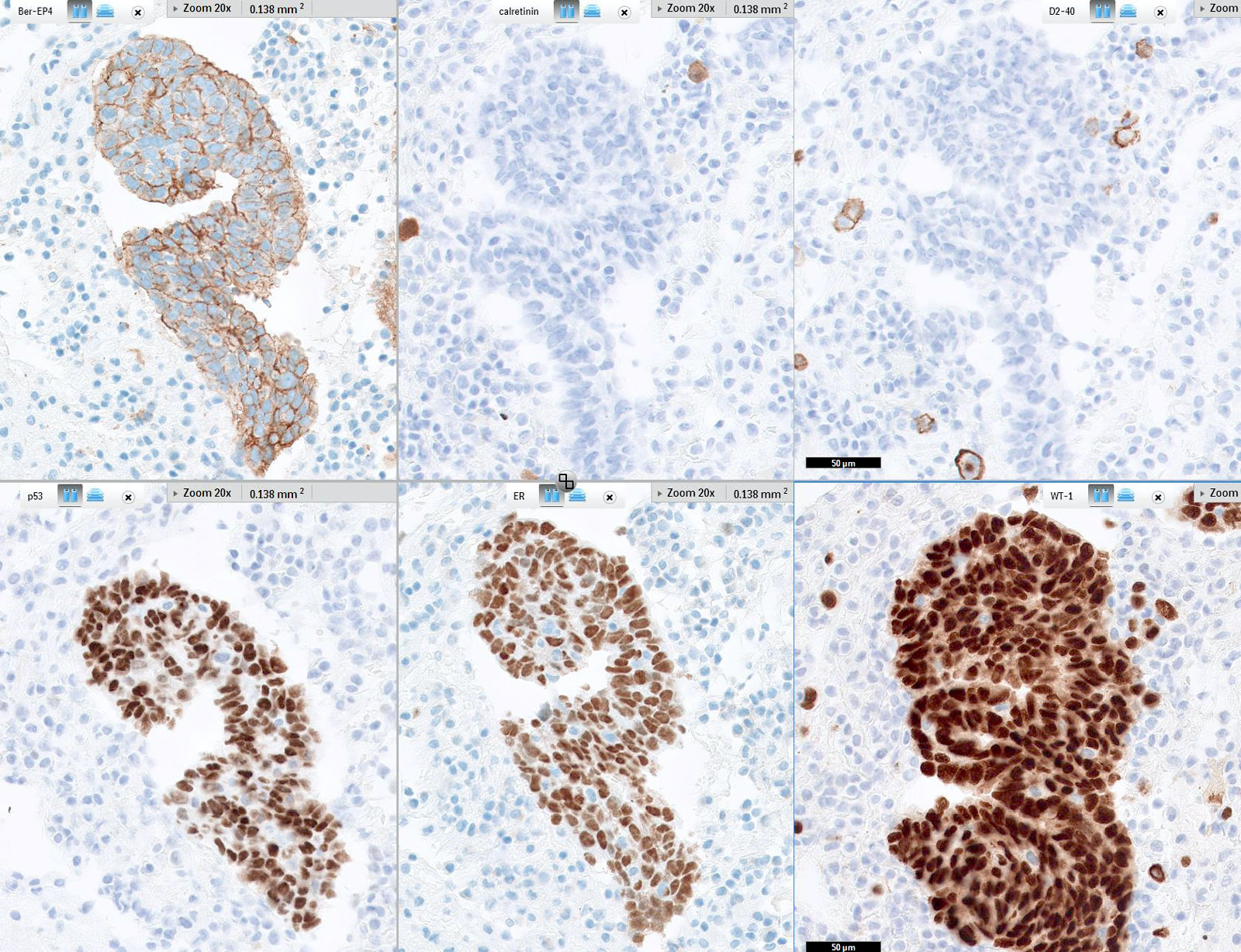
Ovarian serous carcinoma immunoprofile
Contributed by Erna Forgó, M.D. and Teri A. Longacre, M.D.
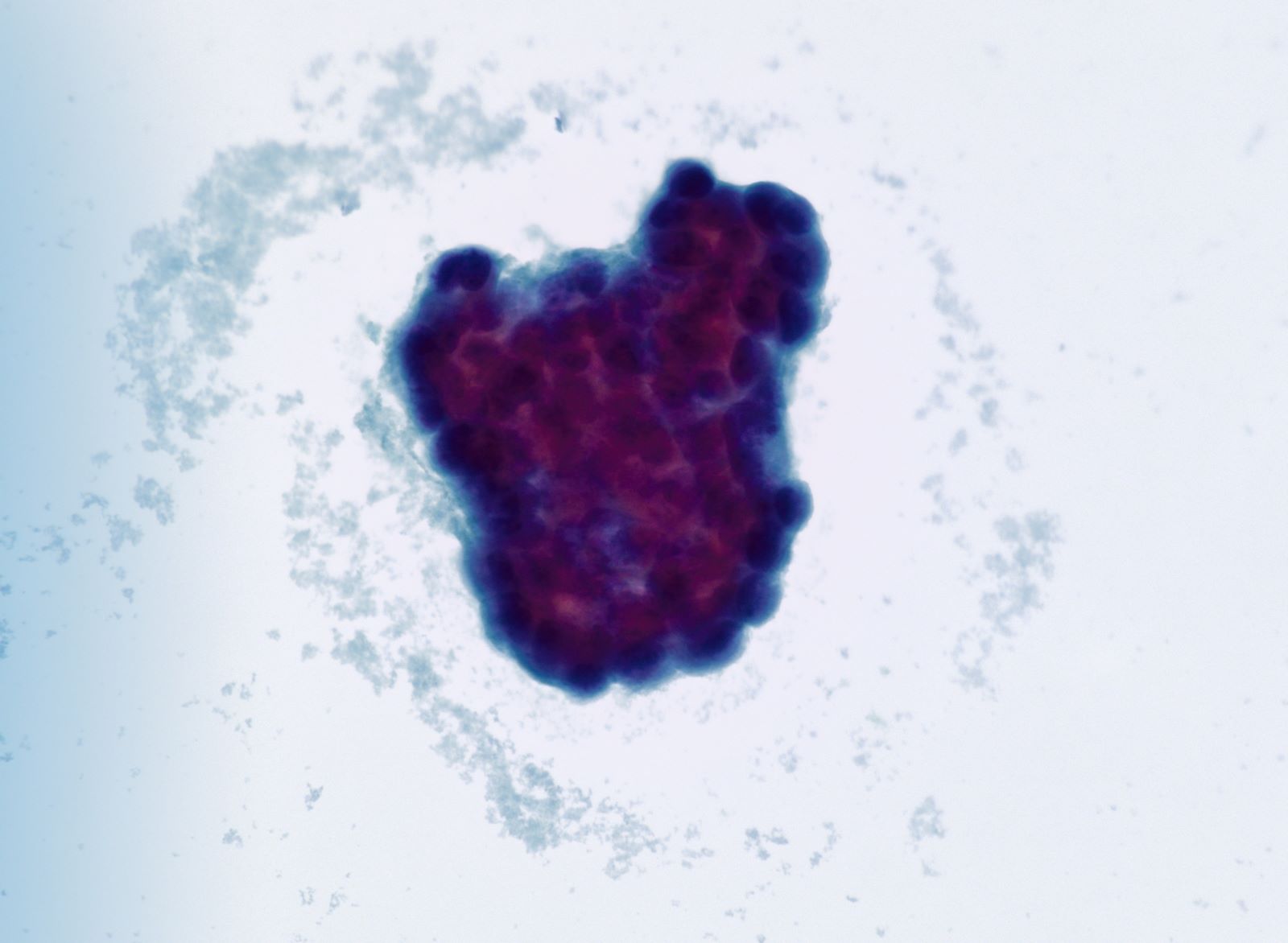
Malignant glandular cells in 3D clusters and singly
AFIP images
Both ovaries are
enlarged by
multiple thin
walled cysts
AFIP images
Luteinized granulosa cells
Prominent edema
in the luteinized
theca cell layer
Ovarian stroma is markedly edematous
AFIP images
25 cm unilocular cyst
Contributed by Raul S. Gonzalez, M.D.
Solitary luteinized follicle cyst
Images hosted on other servers:

Microscopic features of ovarian cyst

Histologic characteristics of ovarian cyst
AFIP images
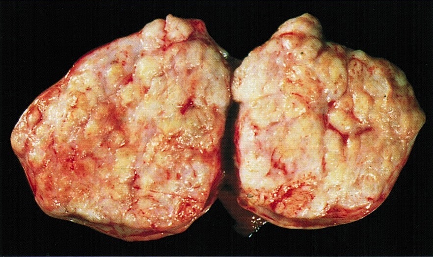
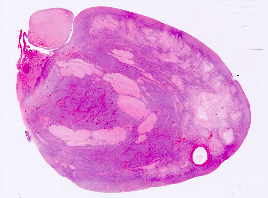
Lobulated sectioned surface
Images hosted on other servers:

Gray-white with whorled pattern

Ovary replaced with creamy mass
AFIP images
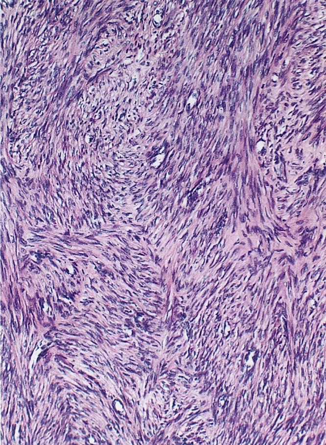
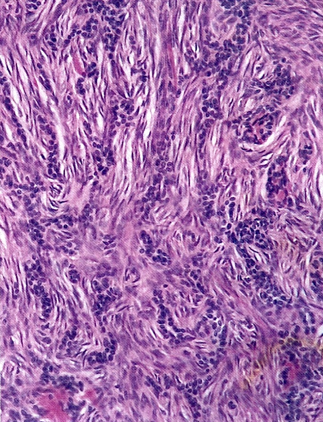
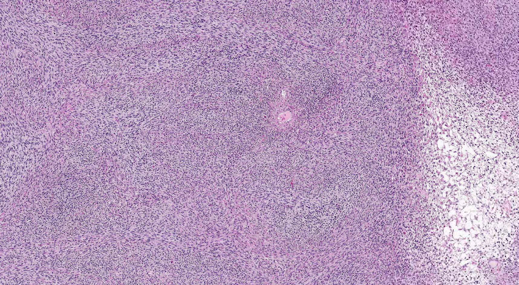
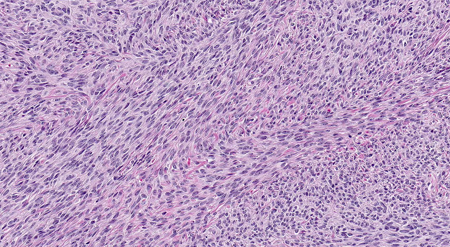
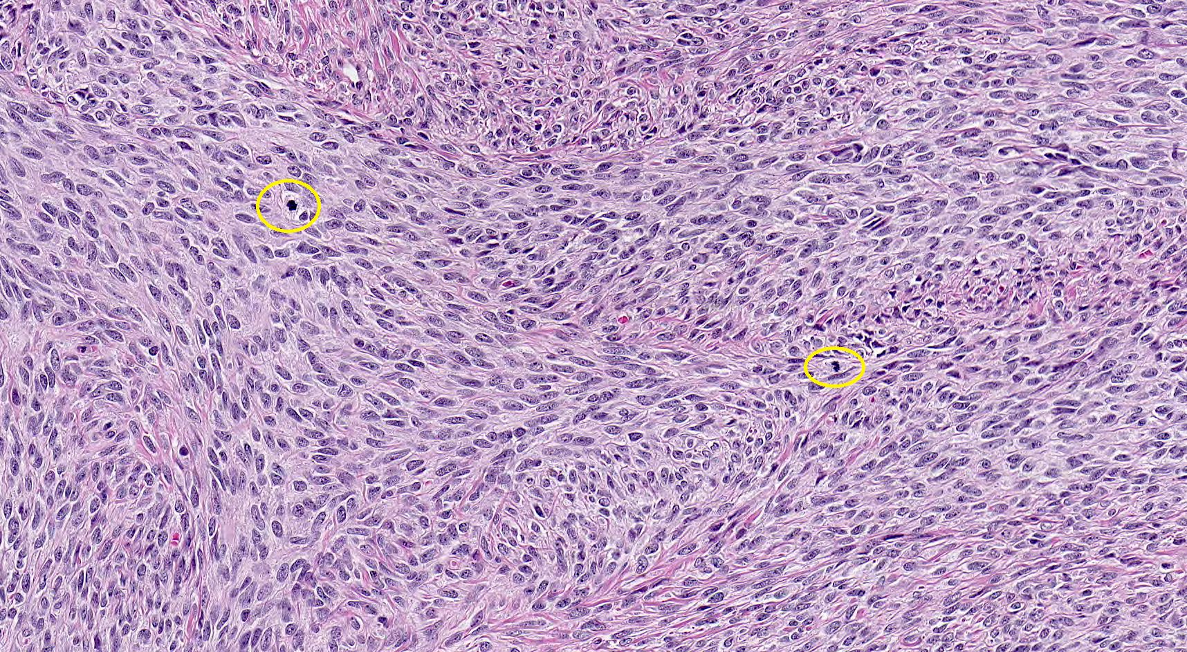
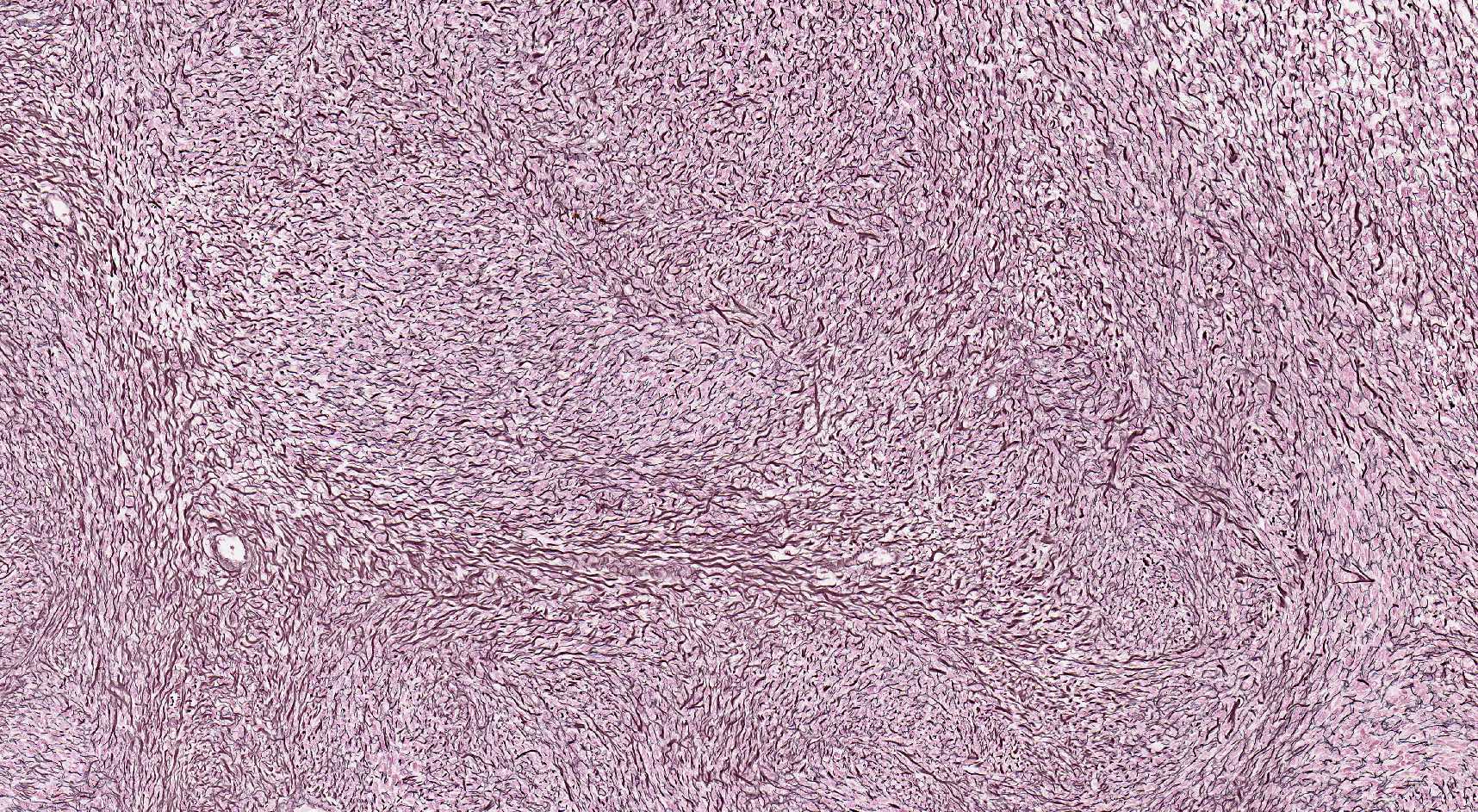
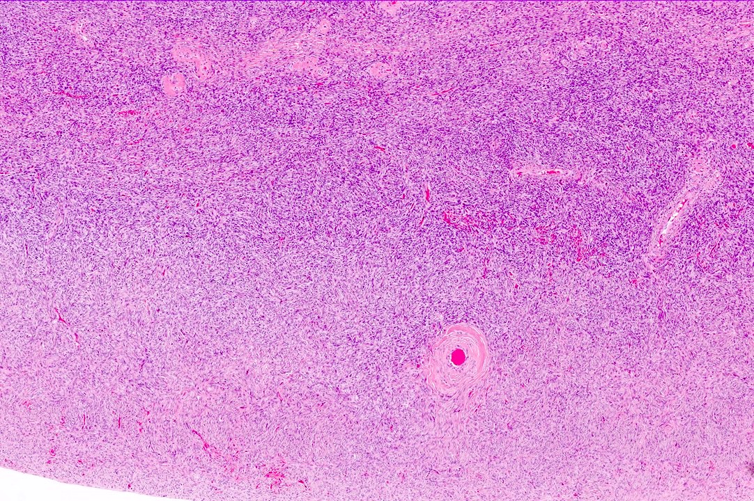
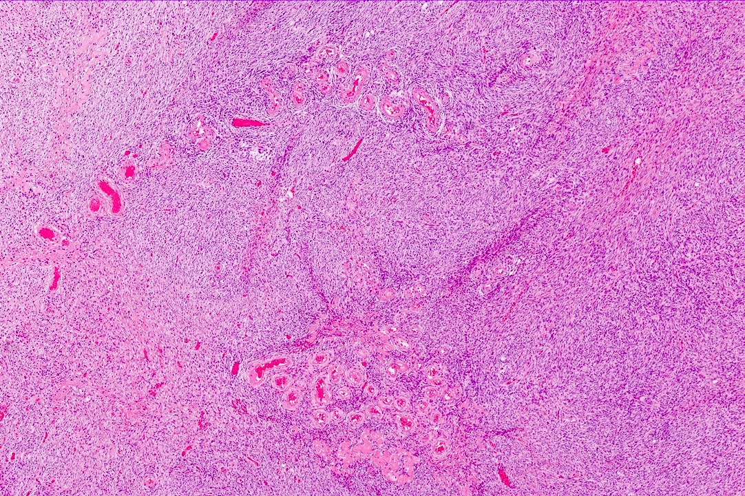
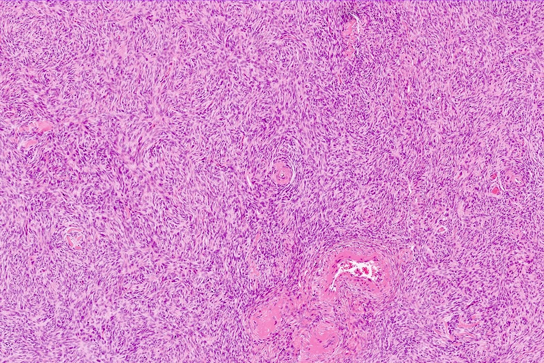
Spindle cells arranged in intersecting fascicles
Images hosted on other servers:

Long fascicle of spindle shaped cells

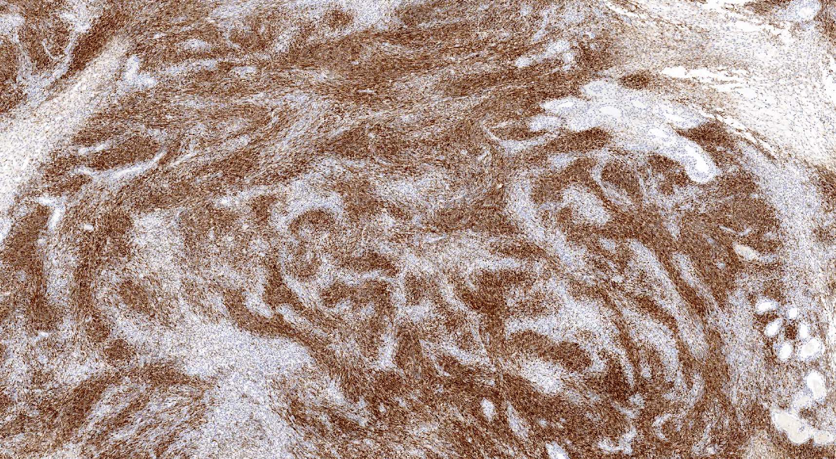
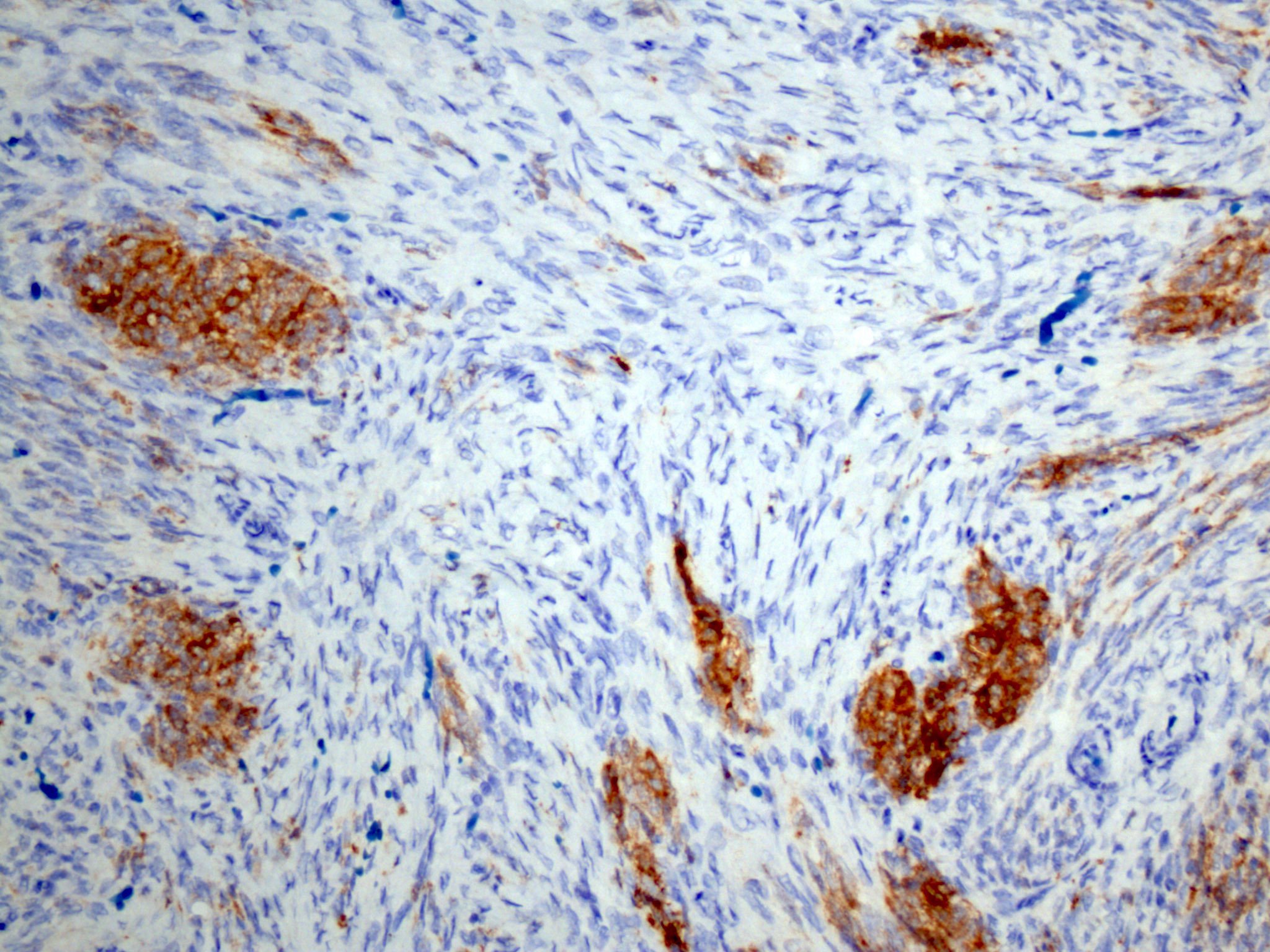
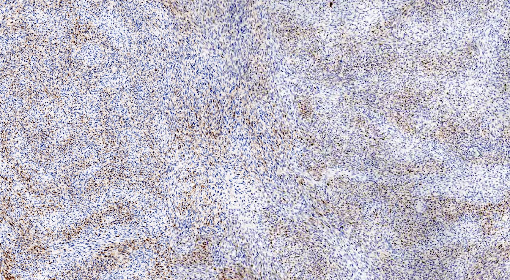
H&E, desmin, α smooth muscle actin

Positive for SMA
AFIP images
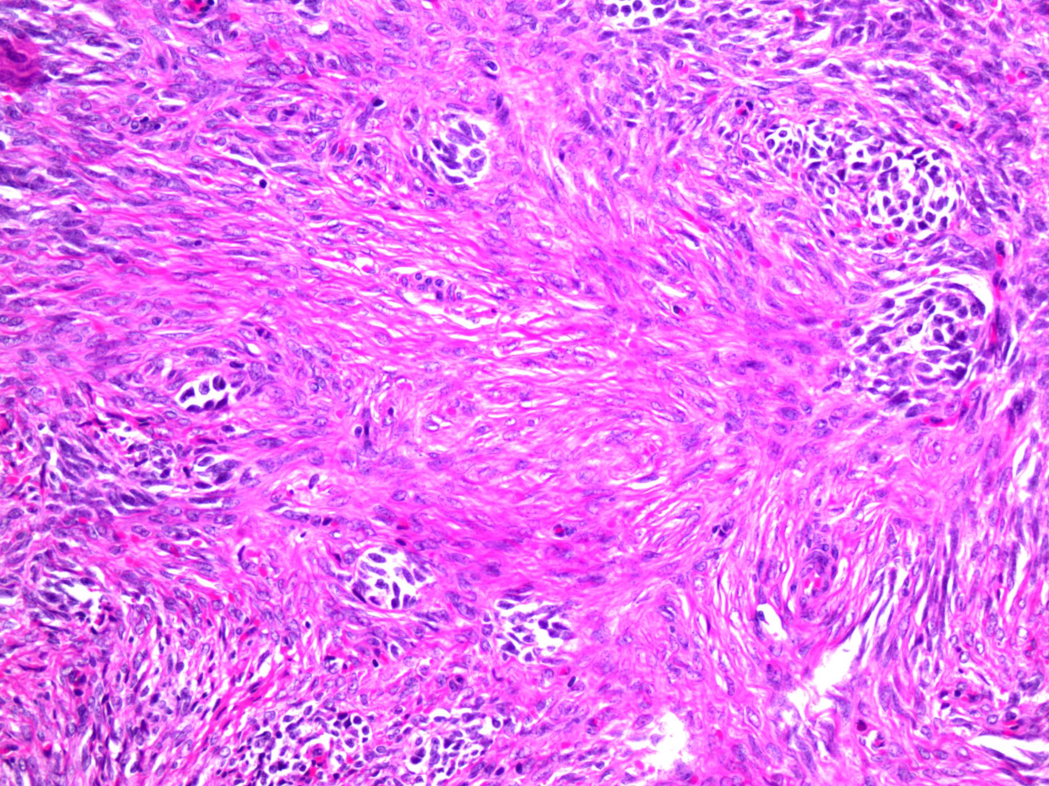
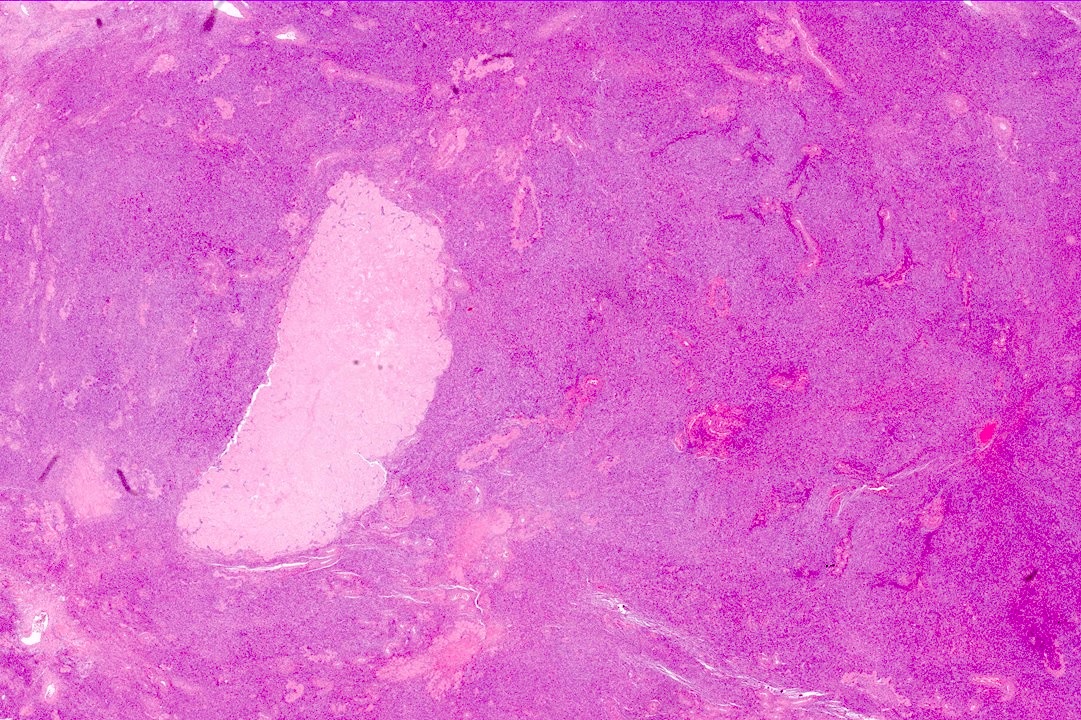
Epithelioid tumor
Images hosted on other servers:

Pleomorphic tumor cells

H-Caldesmon, desmin and smooth muscle actin
AFIP images
Red-brown mass
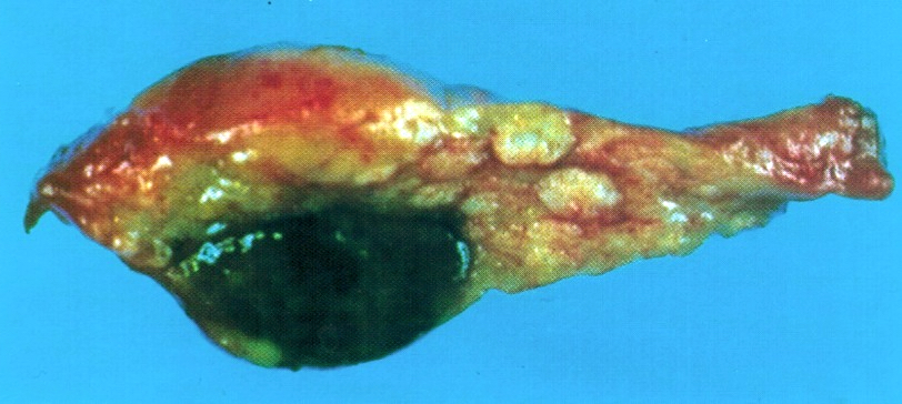
Brown-black nodule
AFIP images
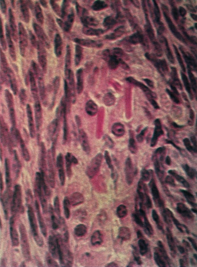
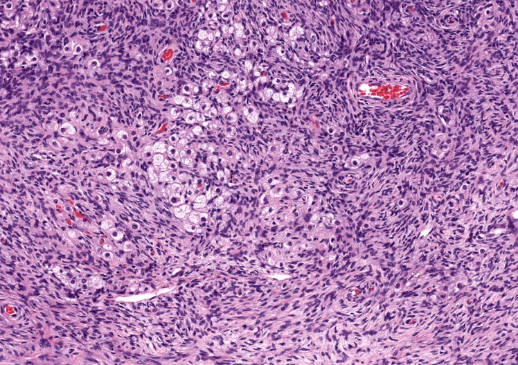
Crystals of Reinke
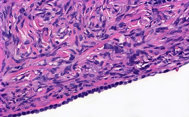
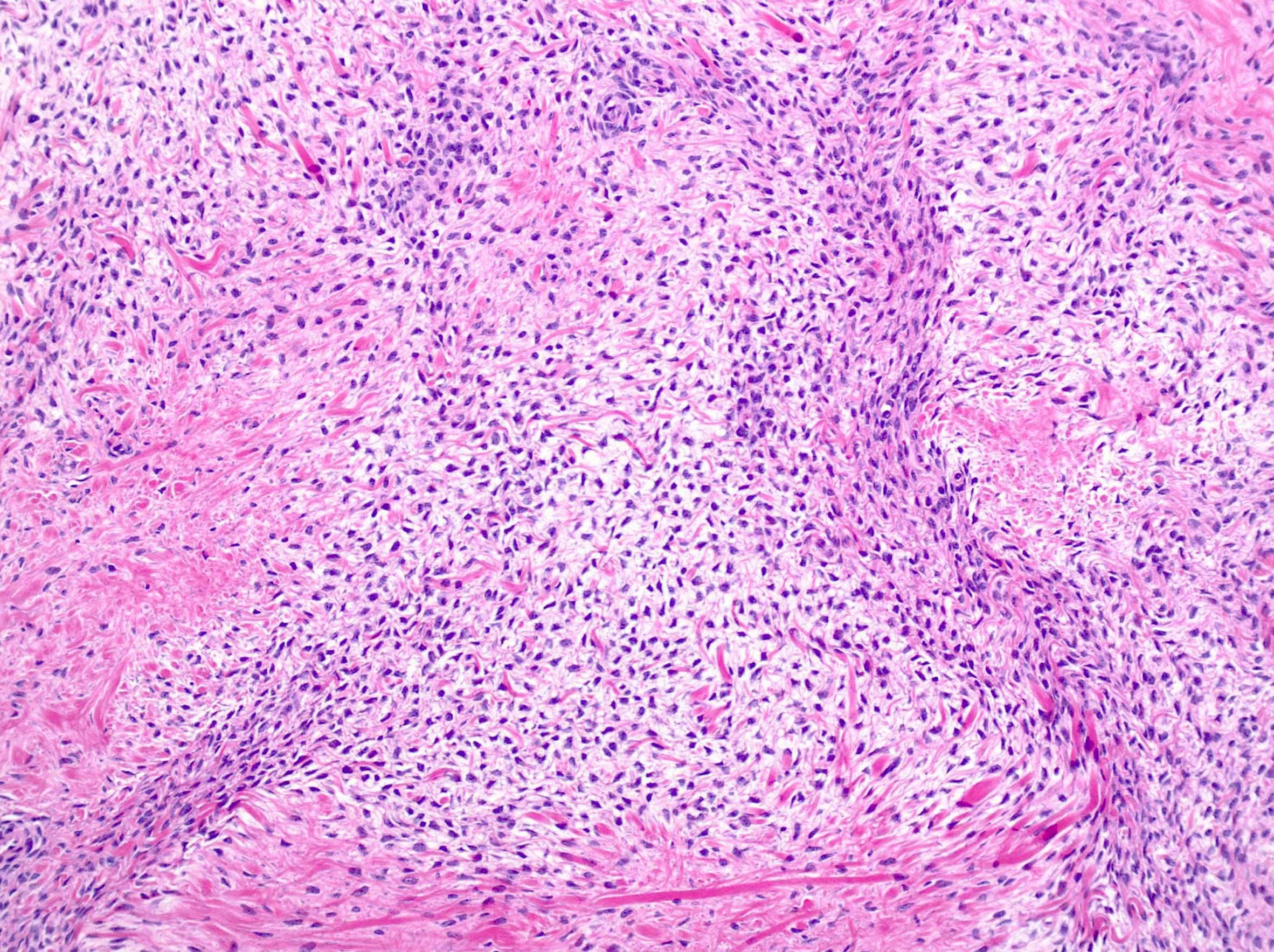
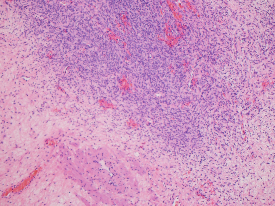
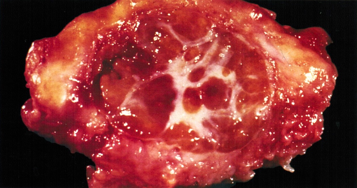
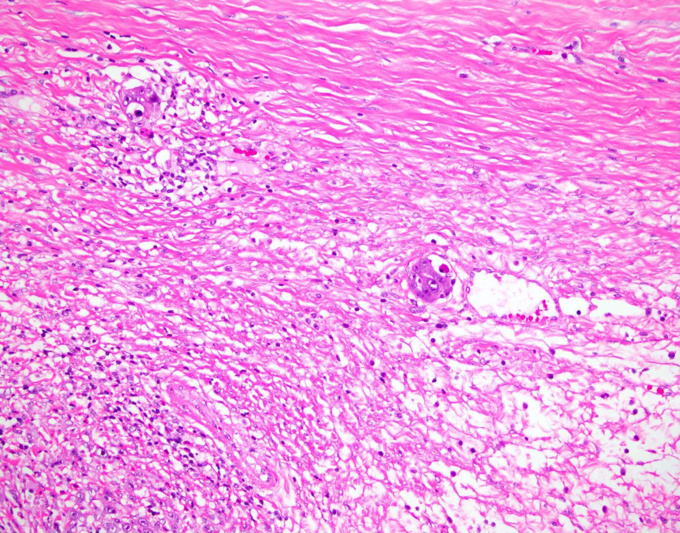
Separated cellular areas
Prominent fibrous stroma
Fibrinoid replacement
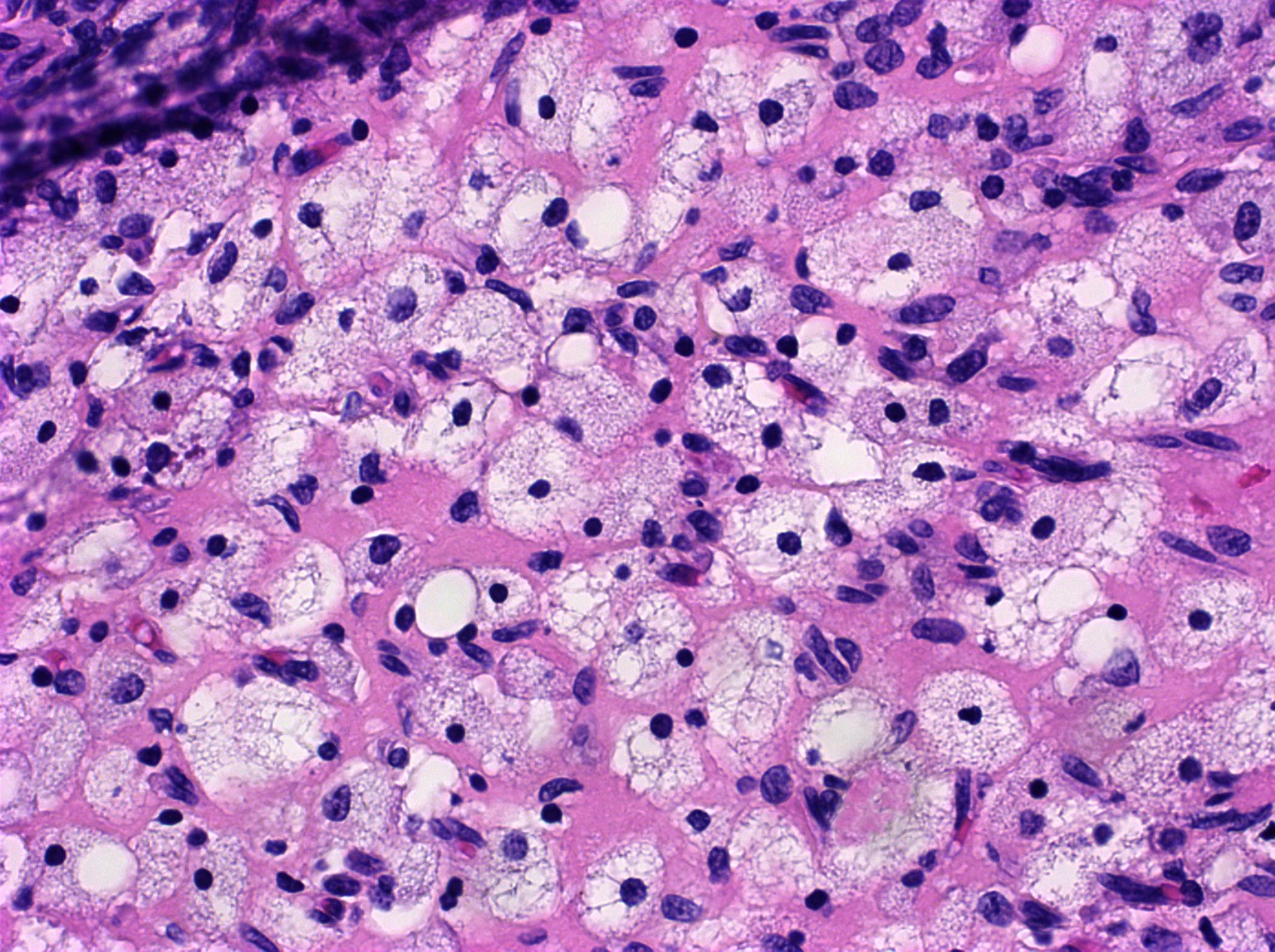
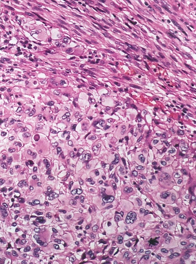
Abundant, pale, vacuolated cytoplasm
Enlarged, hyperchromatic, bizarre nuclei
Images hosted on other servers:

Solid pelvic mass with calcifications

Cystic pelvic mass with calcifications
Contributed by Erna Forgó, M.D. and Teri A. Longacre, M.D.
Irregular papillary mass
Heterogeneous cystic yellow friable mass
Contributed by Erna Forgó, M.D. and Teri A. Longacre, M.D.
Mild to moderate (low grade) cytologic atypia

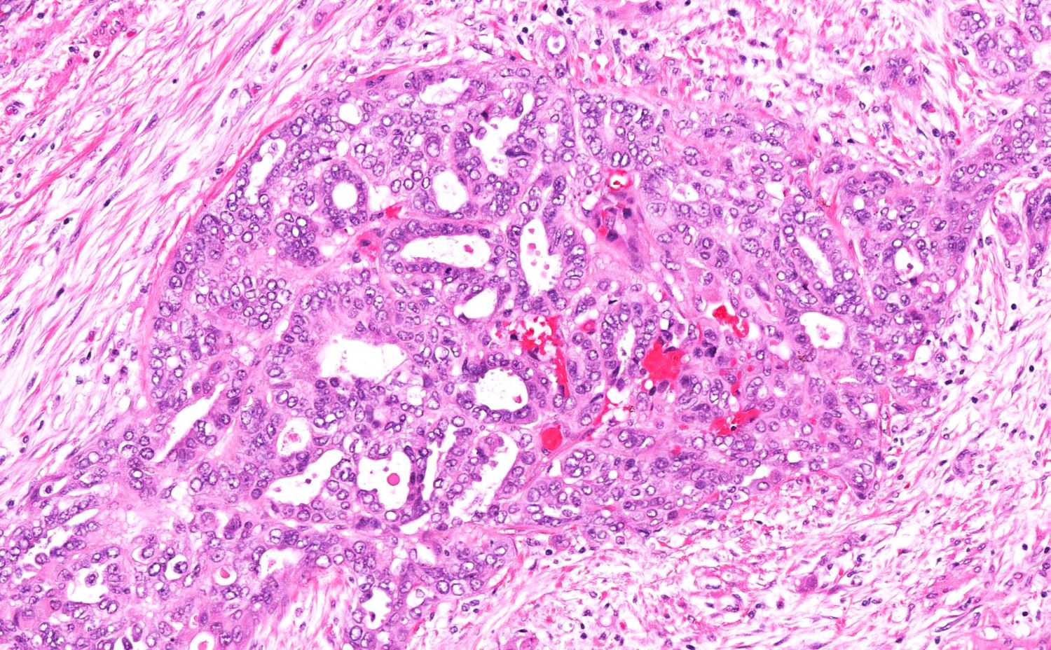
Admixed glandular, micropapillary and cribriform invasive patterns
Contributed by Erna Forgó, M.D. and Teri A. Longacre, M.D.
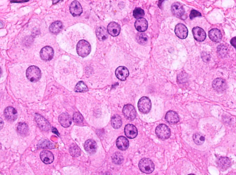
Finely vacuolated cytoplasm
Hyperchromatic nuclei


Clusters of uniform cells
Prominent nucleoli
Micropapillary clusters
Contributed by Rajesh Thampy, M.D.
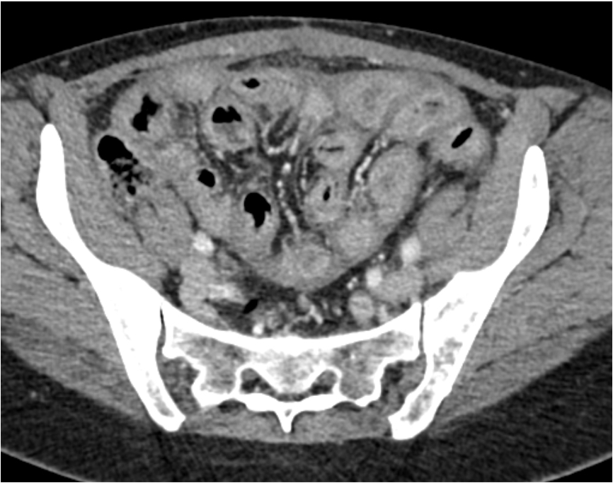
Sclerosing encapsulating peritonitis, abdominopelvic CT
Images hosted on other servers:

Luteinized thecoma
Contributed by Rayan Sibira, M.D. and Mahmoud A. Khalifa, M.D., Ph.D.
Spindled and luteinized cells
Luteinized cells
Luteinized cells with scarred mitosis
Sex cord-like differentiation
Peritoneal lesions
Reticulin stain
Calretinin IHC stain

Beta catenin IHC stain
Images hosted on other servers:

MGAT5B::NCOA3 fusion gene
Images hosted on other servers:

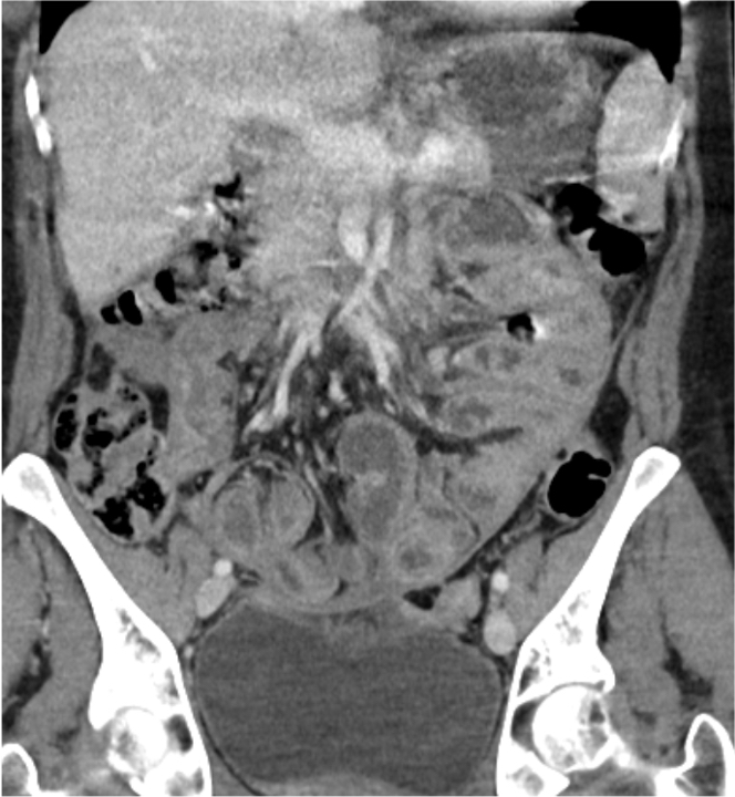
Bilateral ovarian masses
Images hosted on other servers:

Irregularly dilated endometrial glands

CD10, MIC2, calretinin, progesterone receptor, MIB1
Contributed by Daniel Graham, M.D., Adele Wong, M.B., B.Ch., B.A.O. and Lucy Ma, M.D.
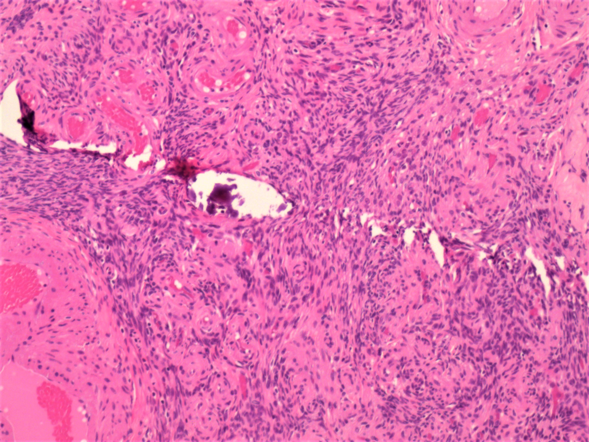
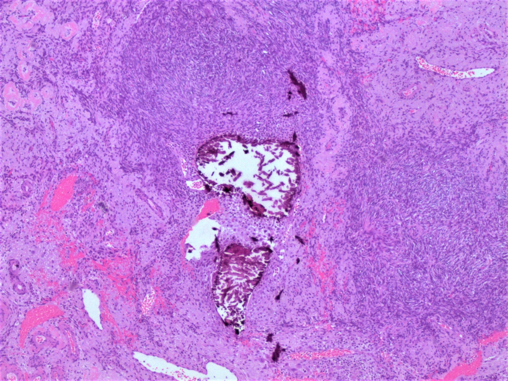
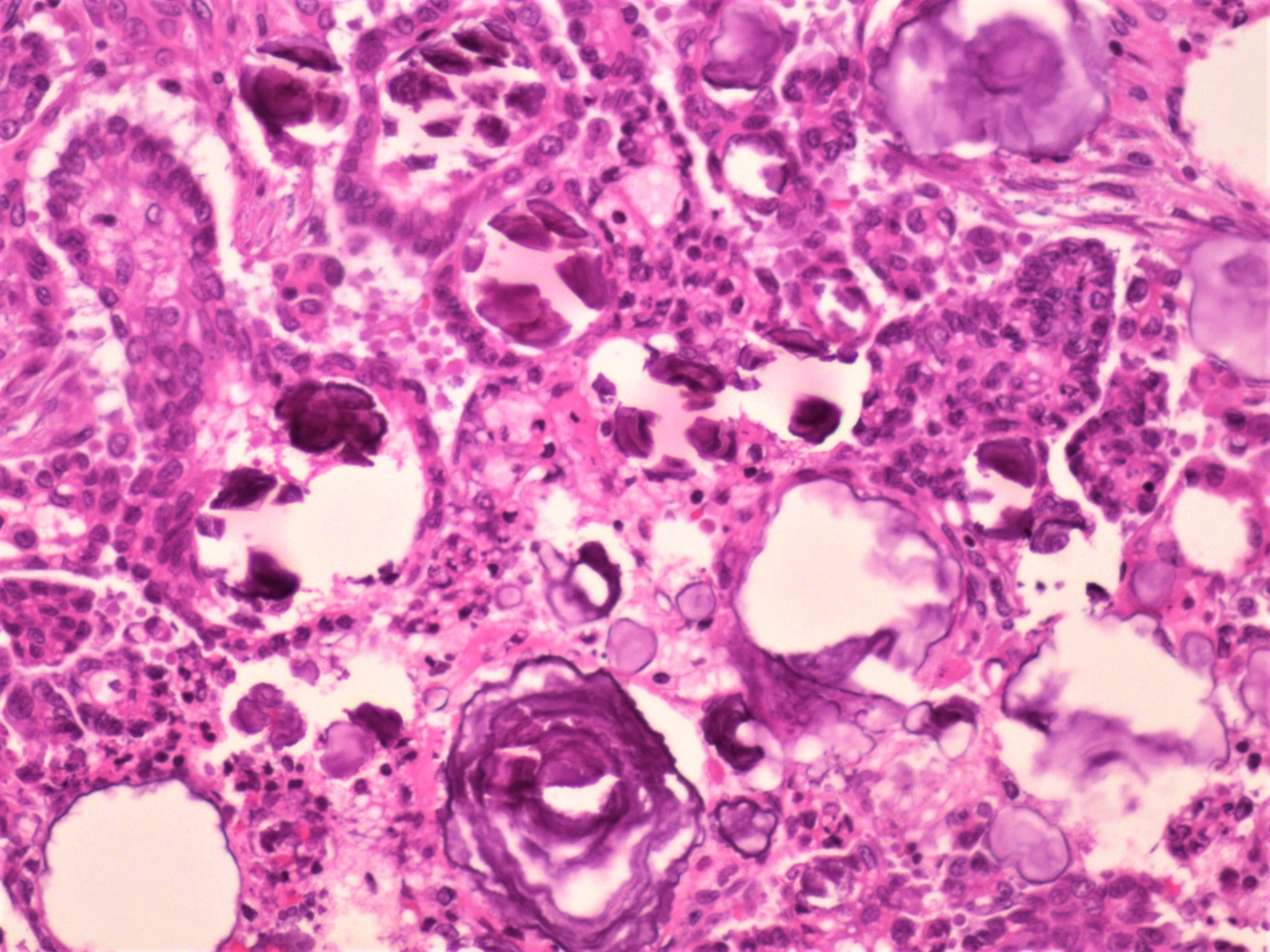
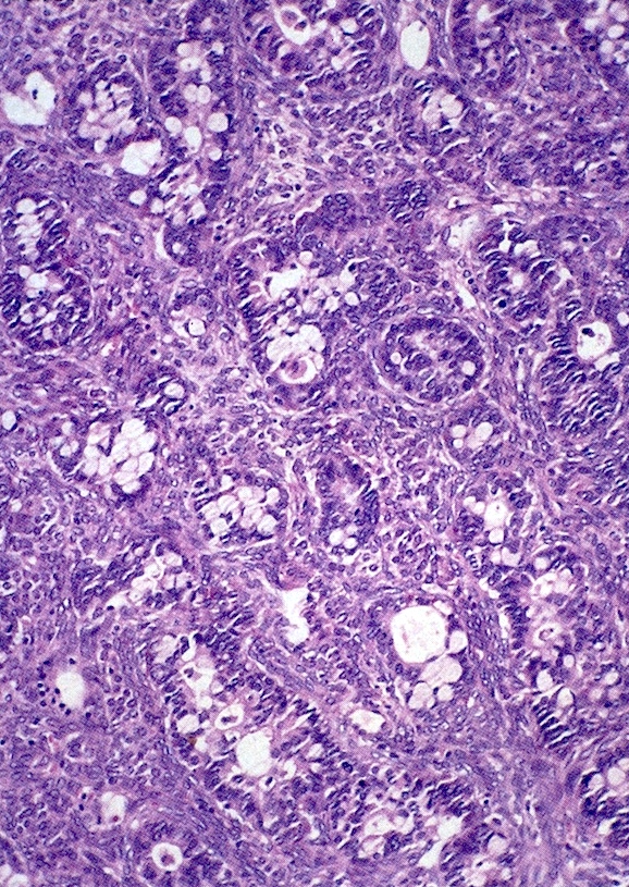
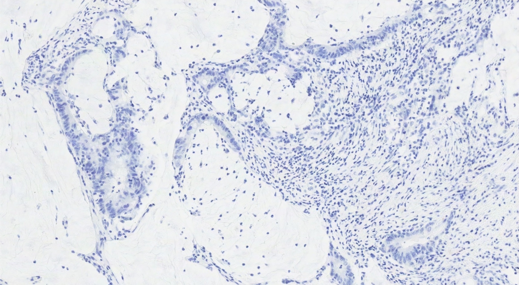
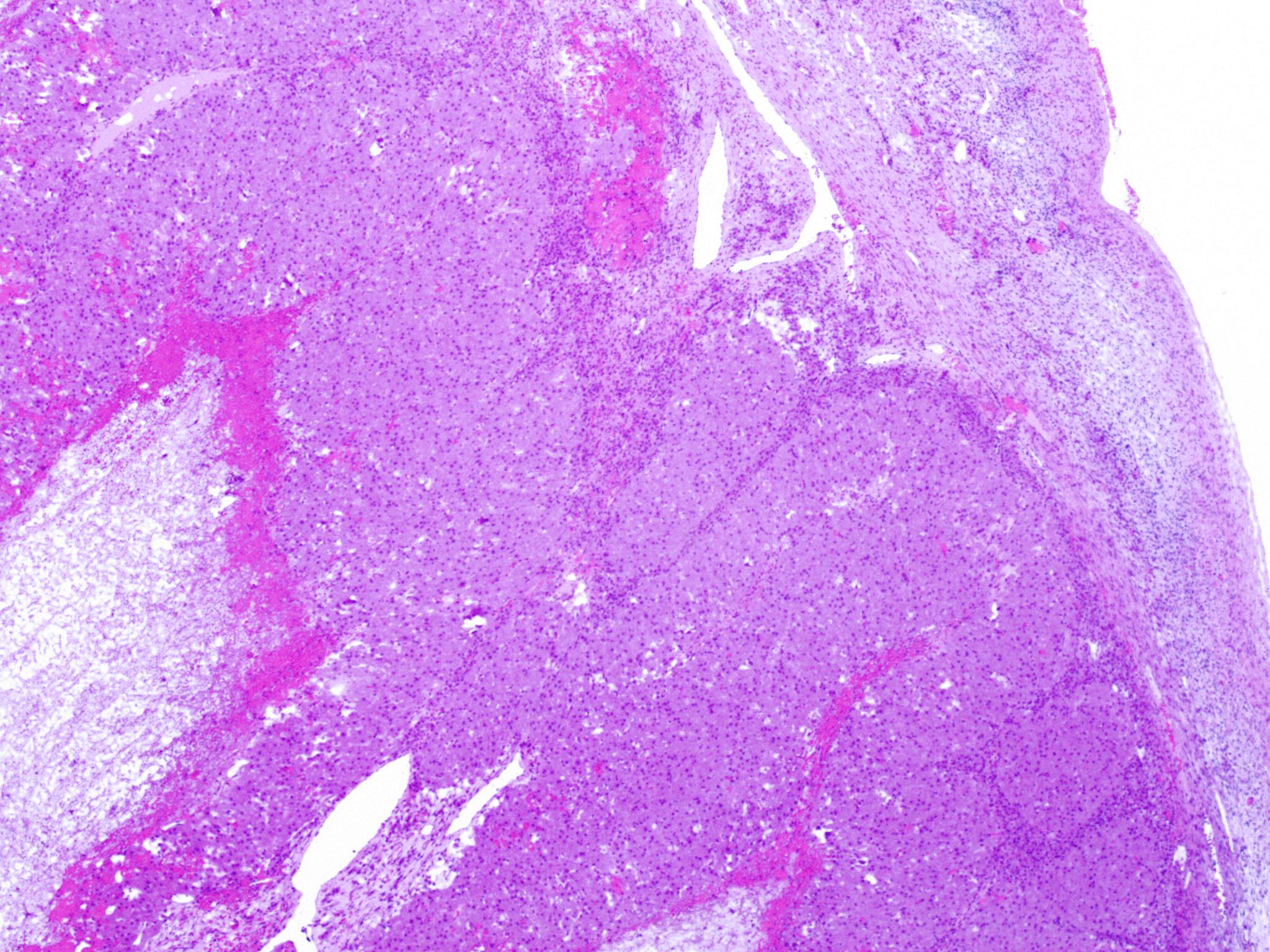
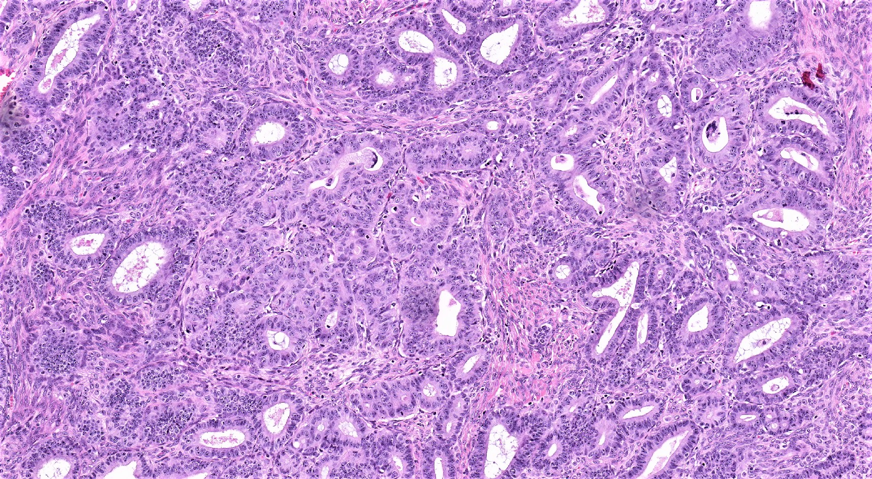
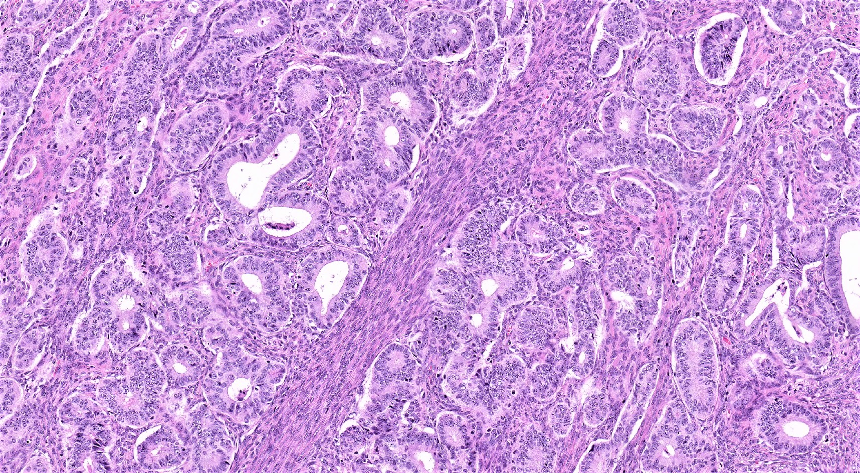
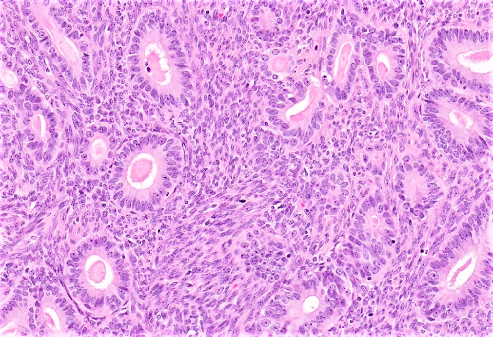
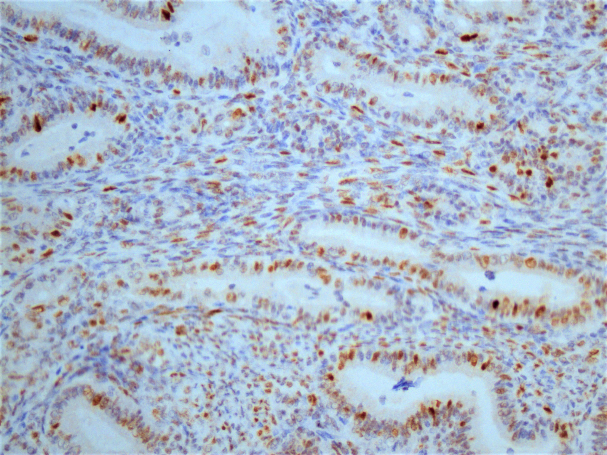
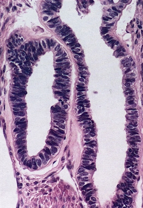
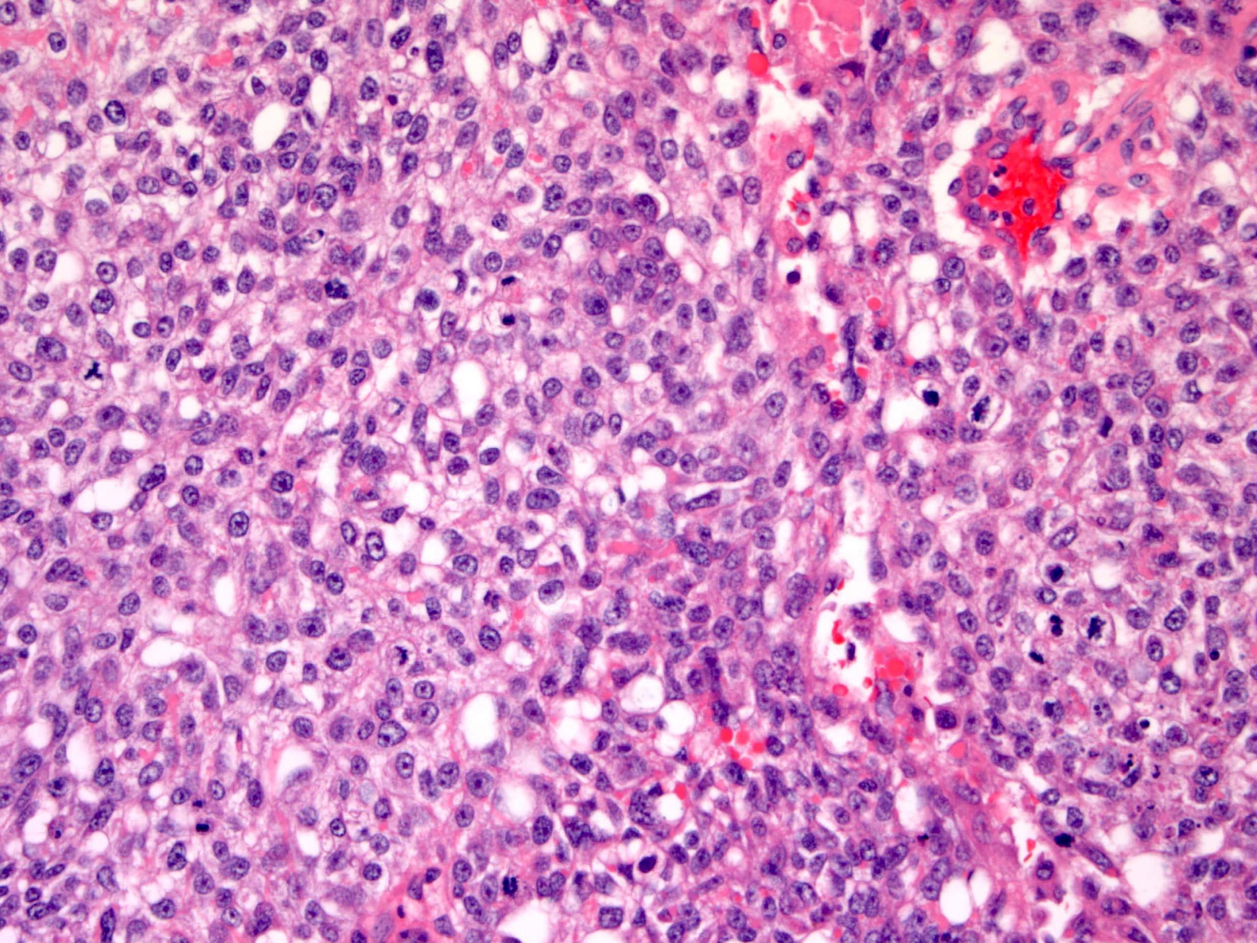
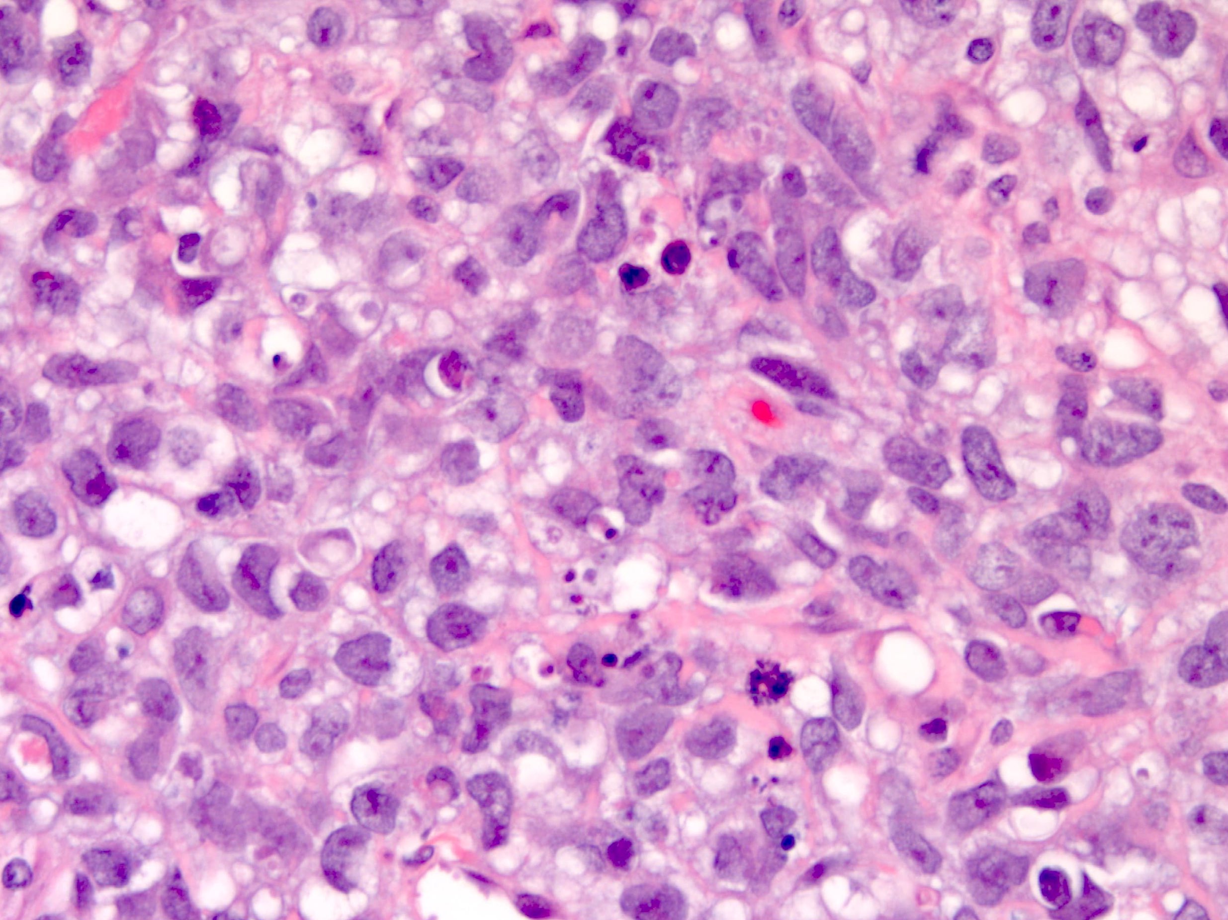
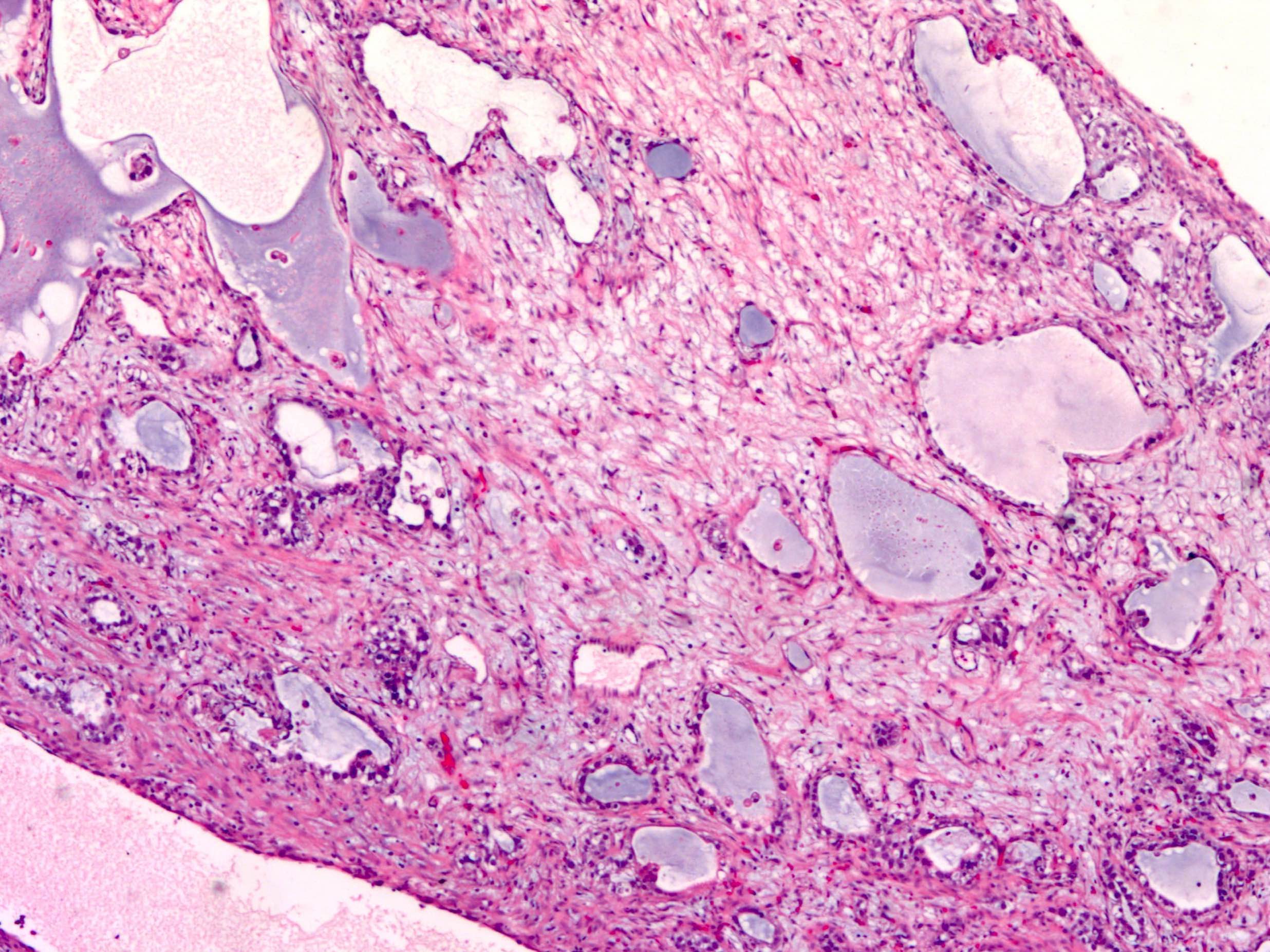
Glandular growth pattern
Variety of histologic patterns
Variable growth patterns

Glandular morphology
Glands
Multiple growth patterns
Glandular and cystic areas
Micropapillary architecture
Mitotic activity
Papillary architecture
Variable morphology
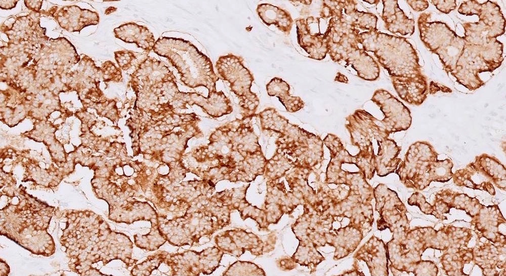
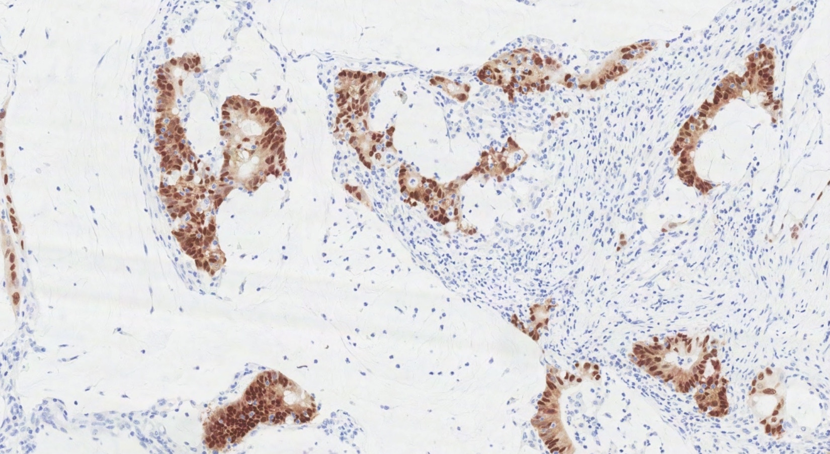
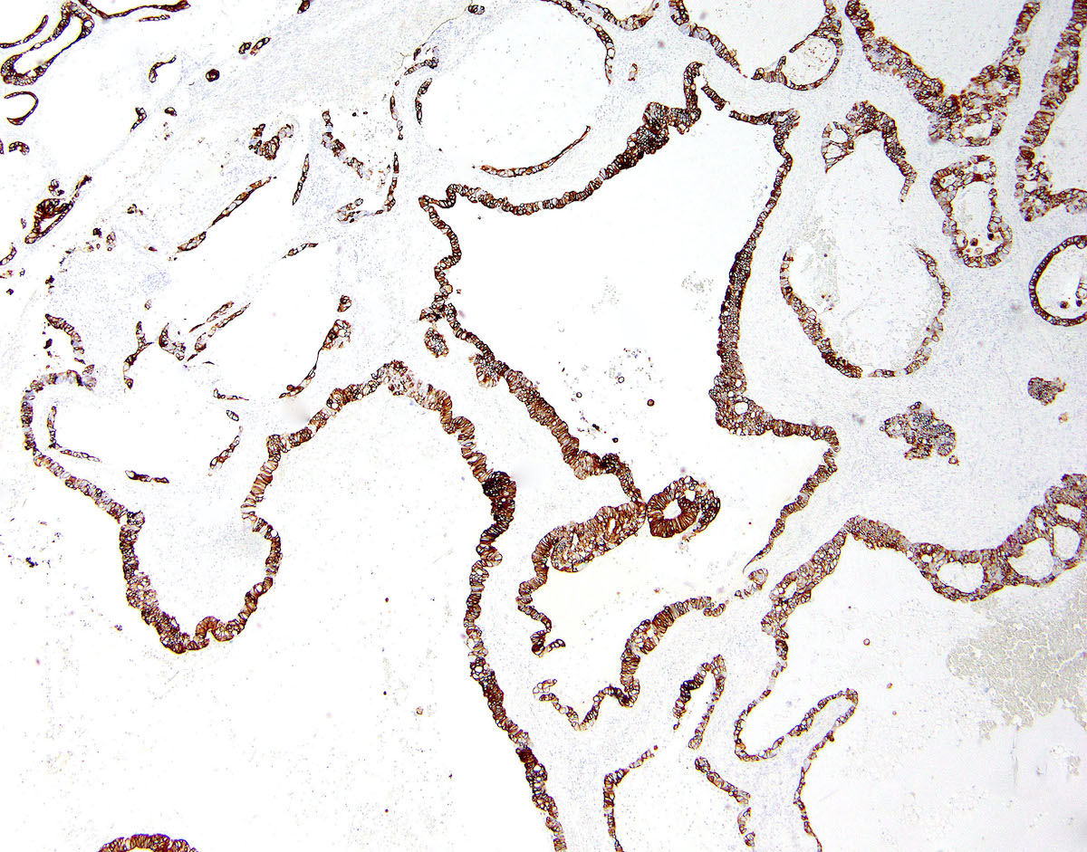
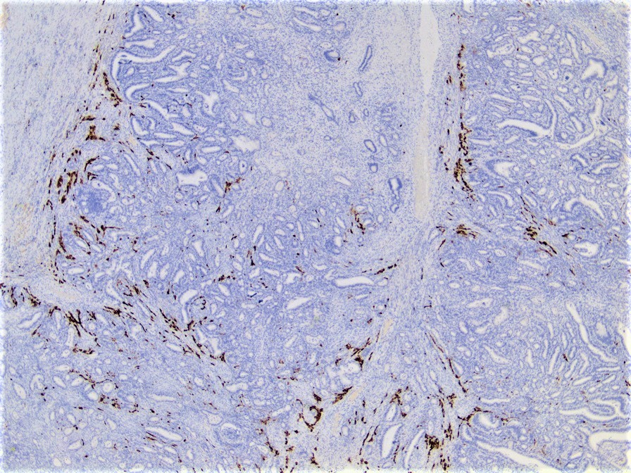
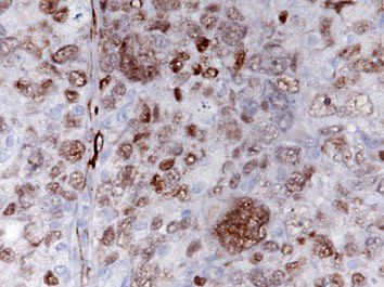
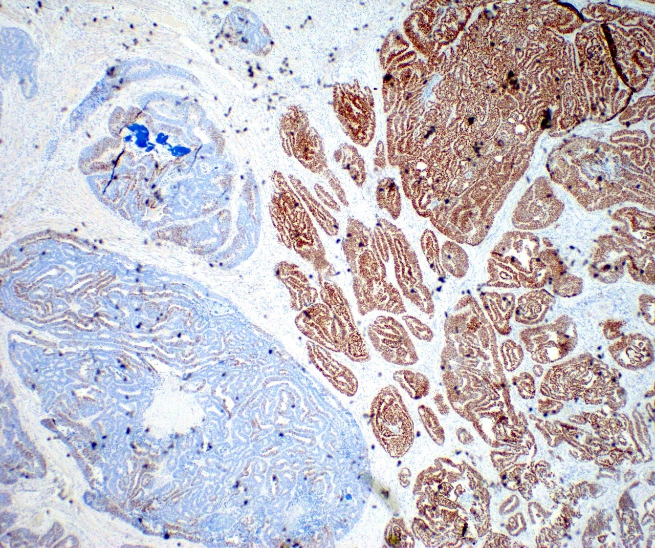
PAX8
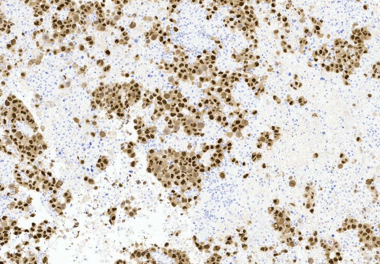
GATA3
GATA3
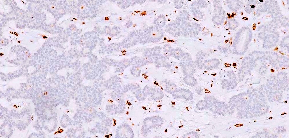
TTF1
TTF1
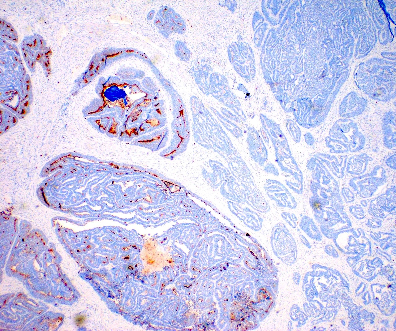
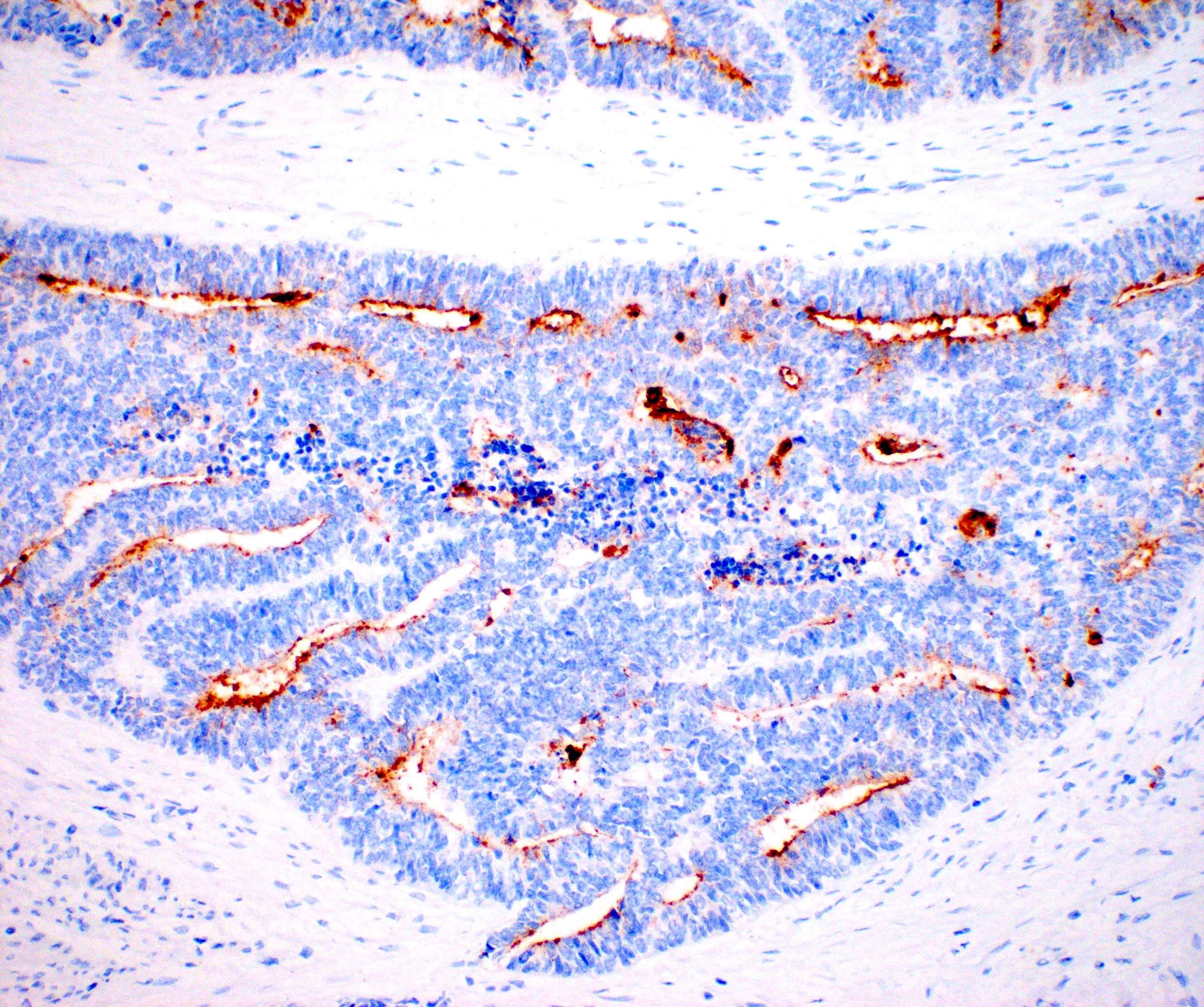
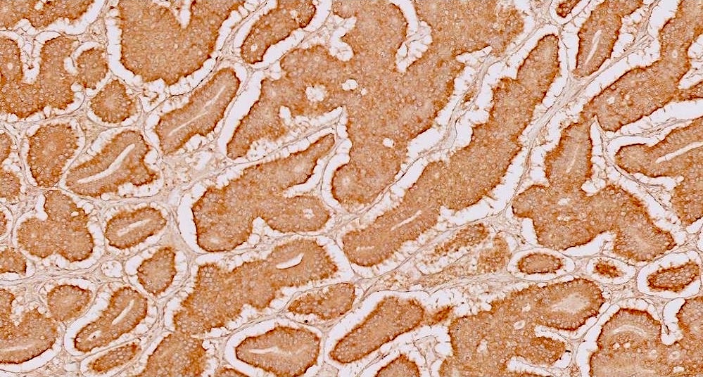
CD10
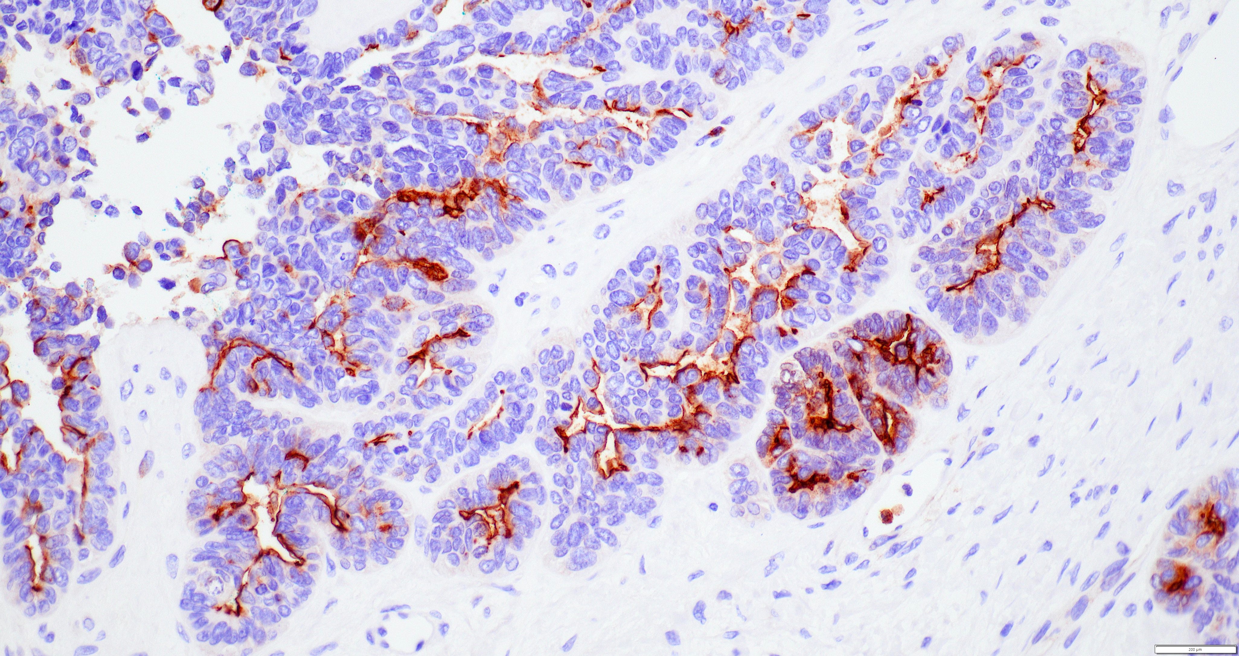
CD10
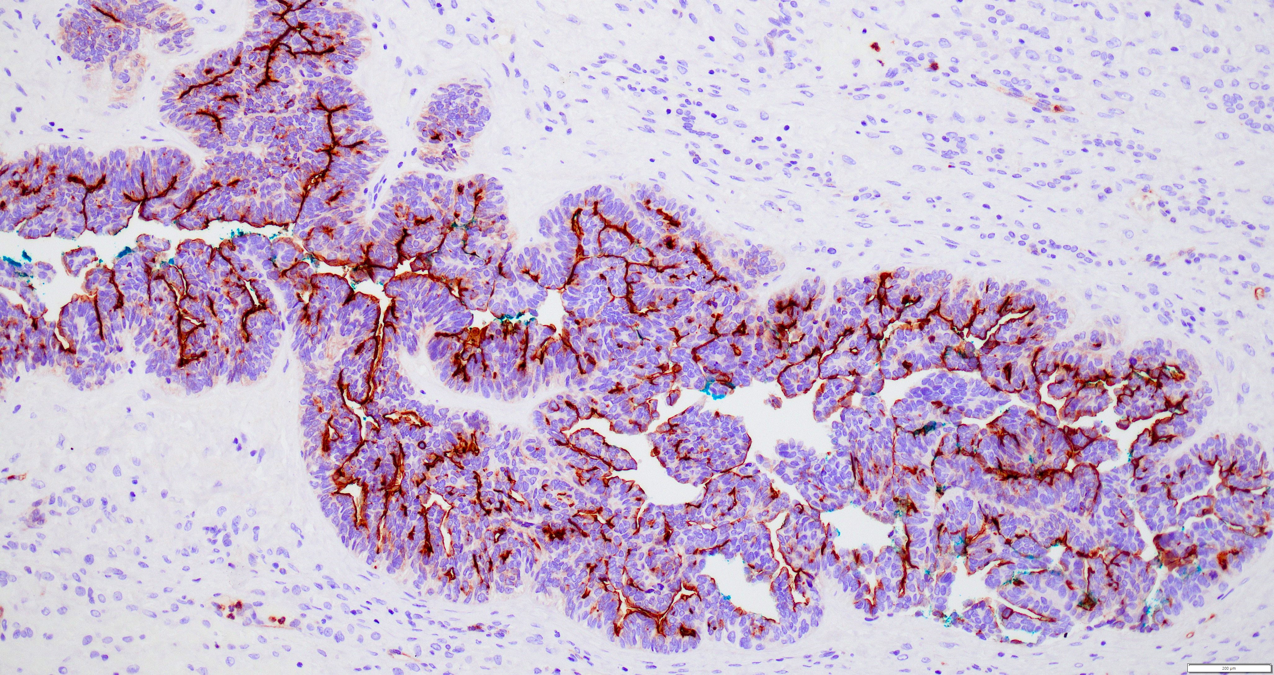
CD10
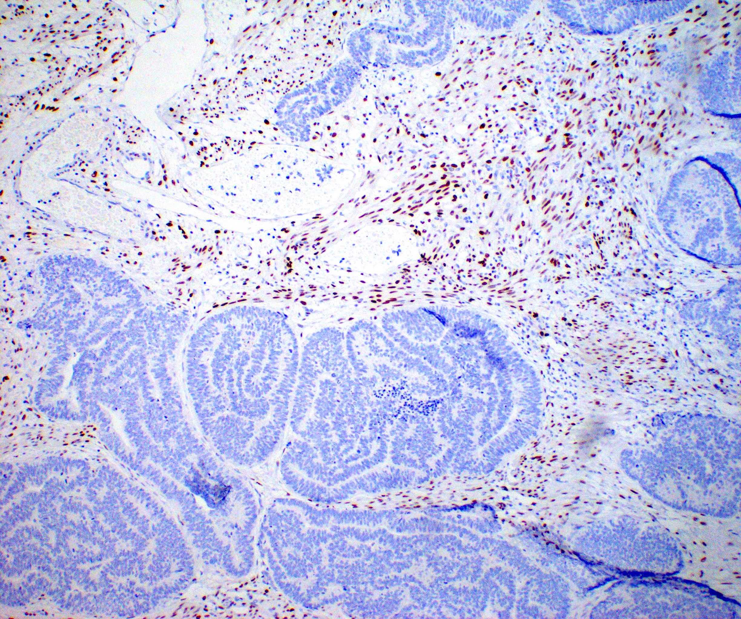

Estrogen receptor
Progesterone receptor
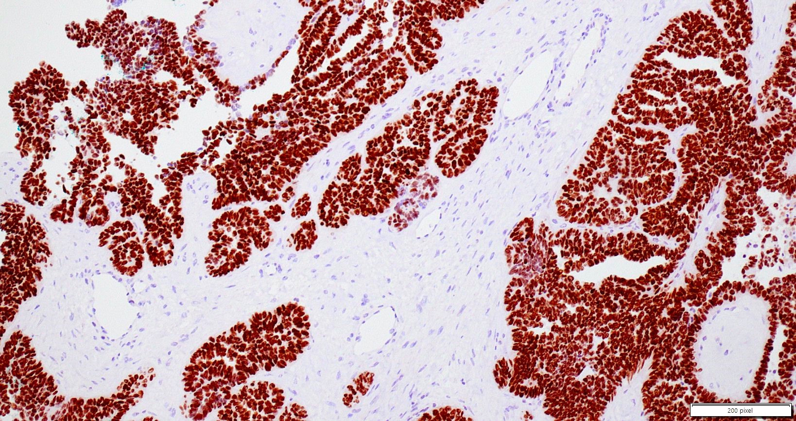
p53
Ki67
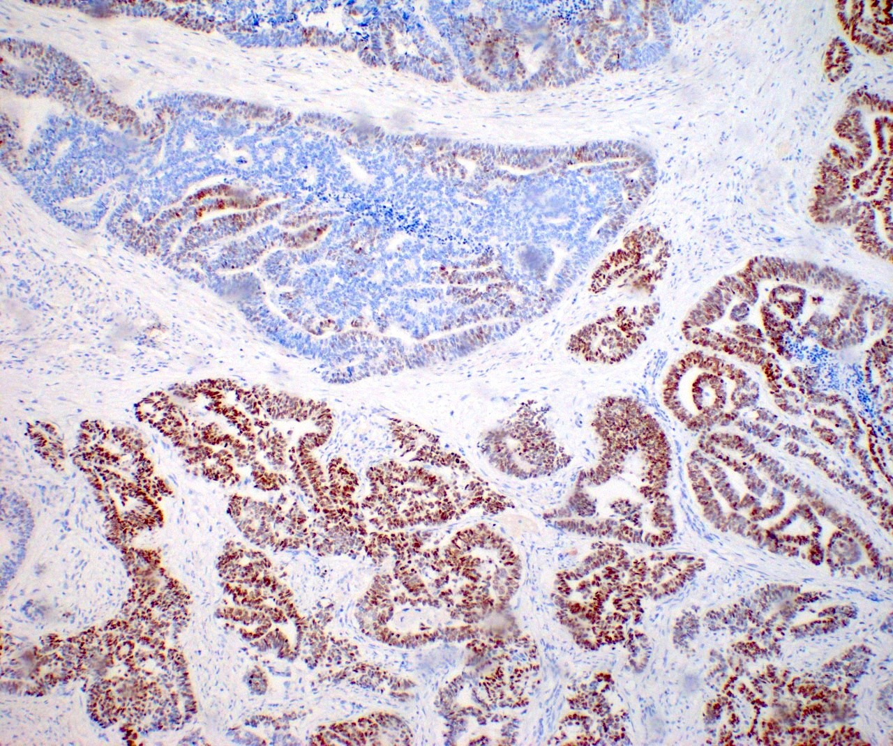
WT1
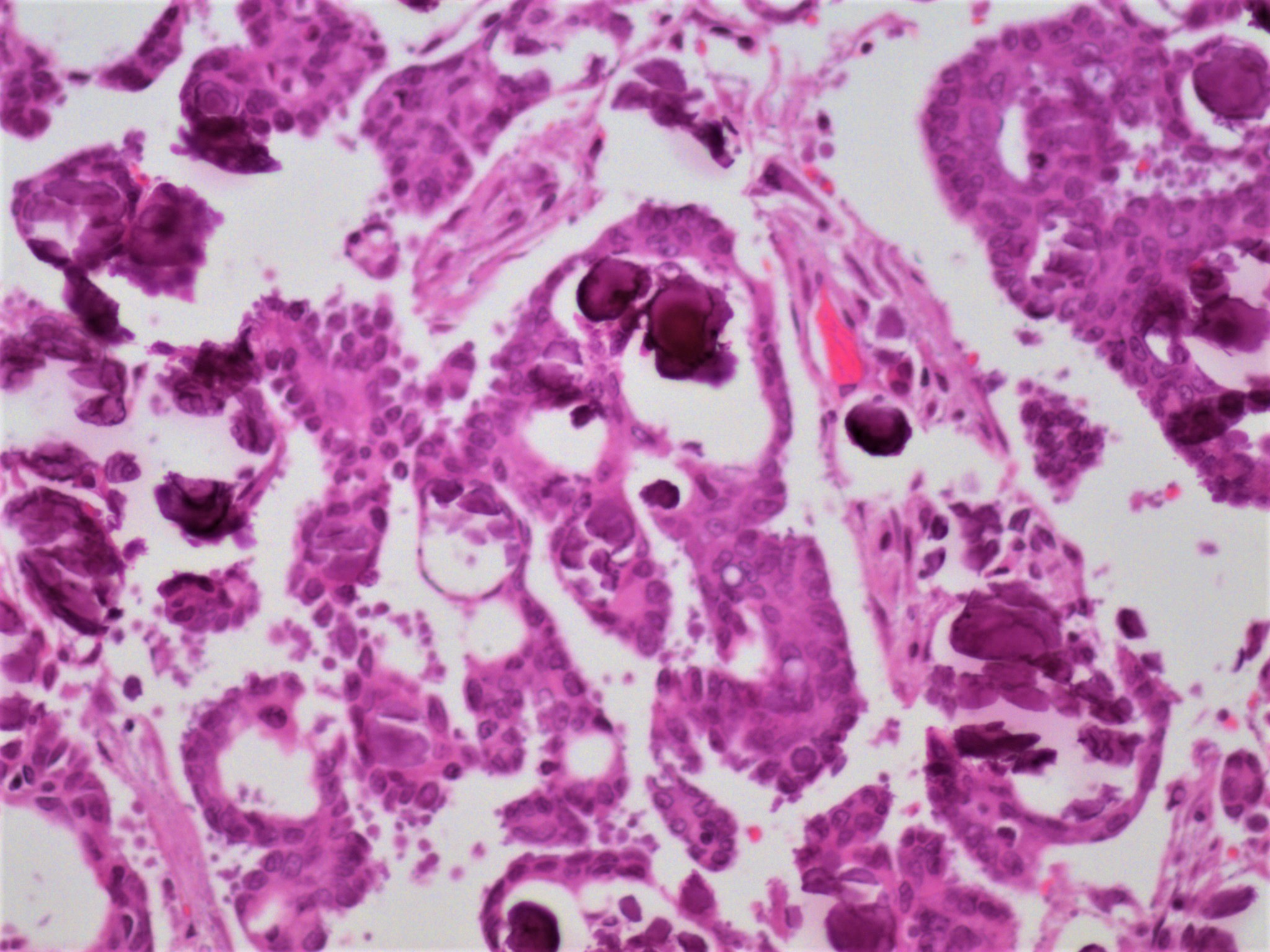
Varied morphology
PTC-like nuclear features
Mesonephric-like adenocarcinoma by Dr. Lewis Hassell
Images hosted on other servers:

Solid and cystic bilateral ovarian masses
Images hosted on other servers:

Bilateral ovarian masses with multinodular cut surfaces
Contributed by Lucy Ma, M.D.
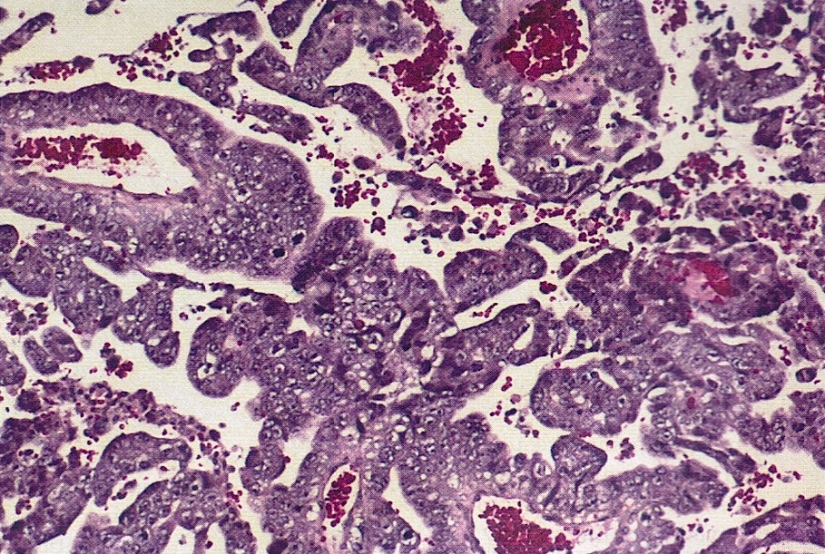
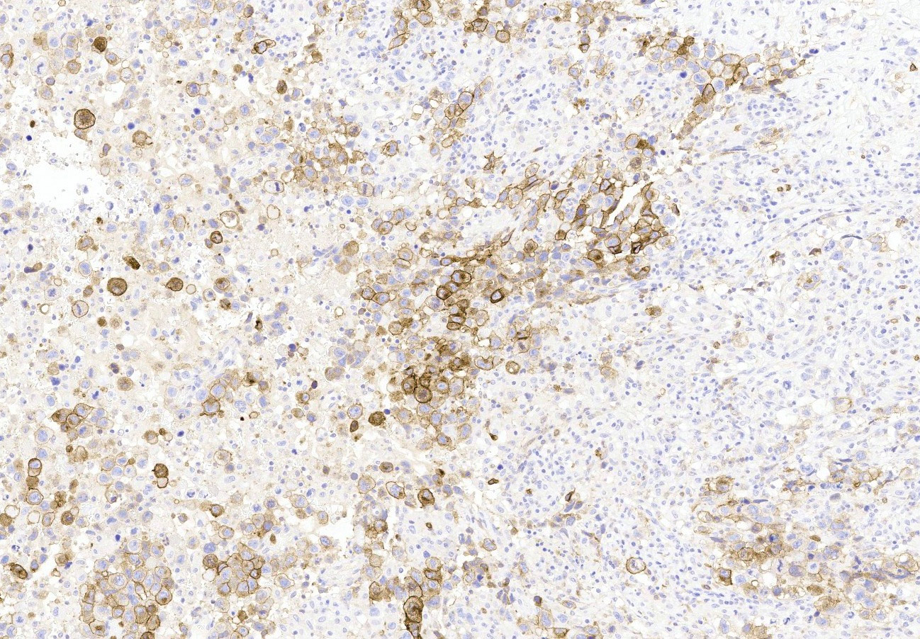
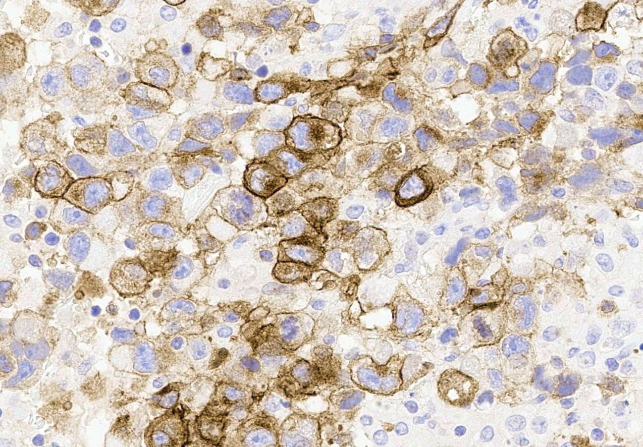
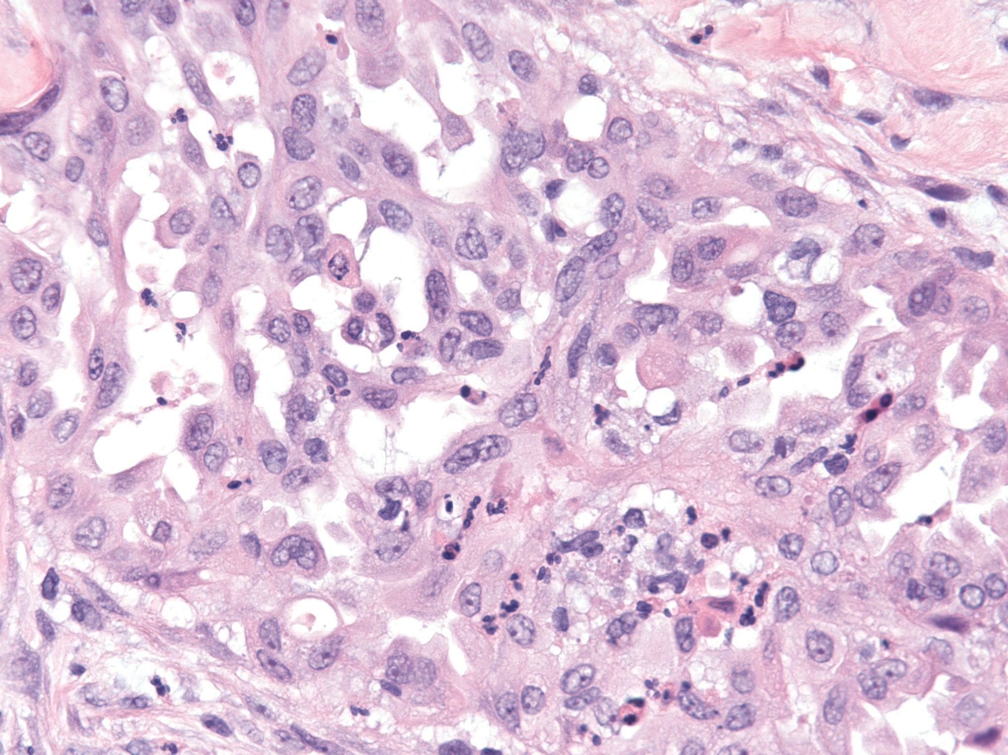
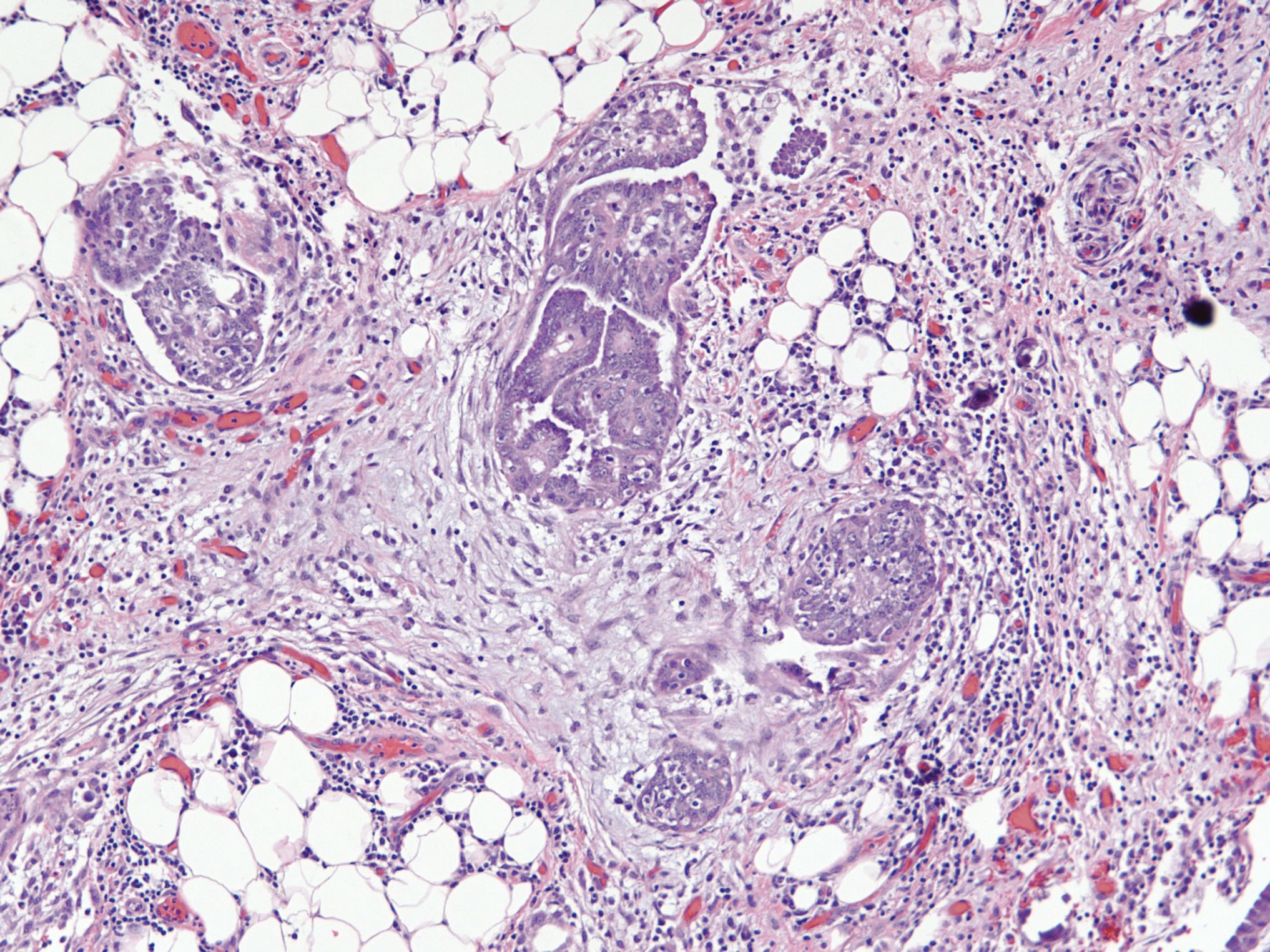
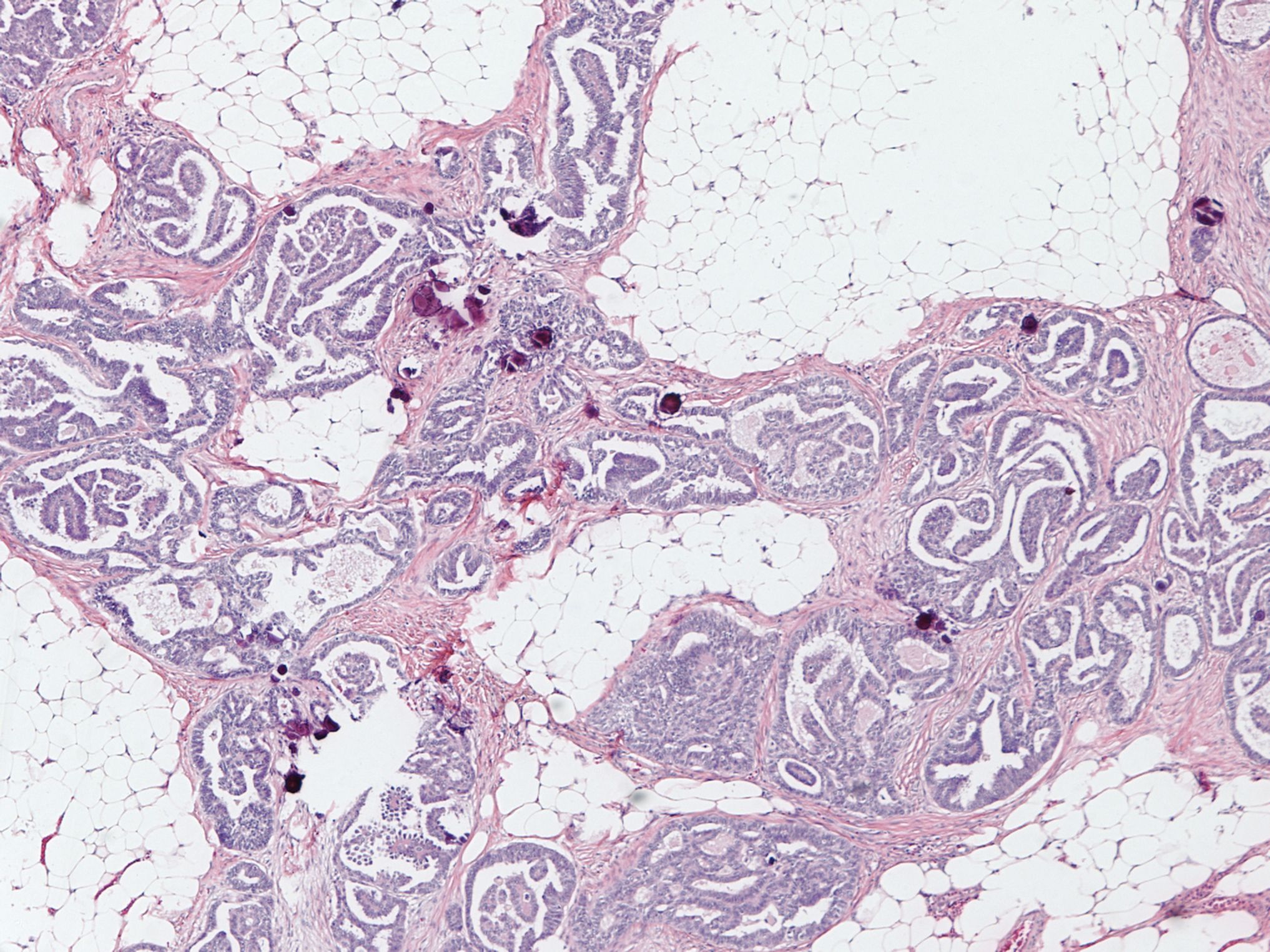
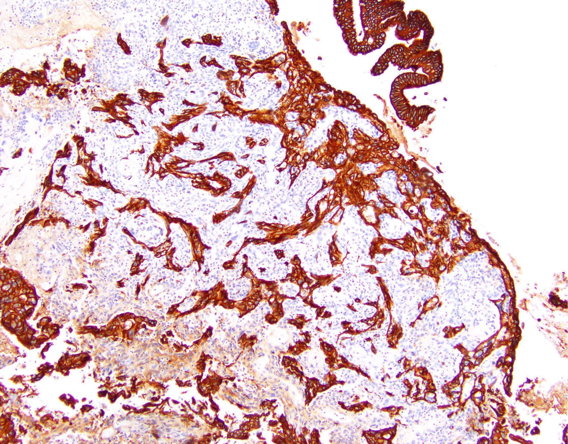
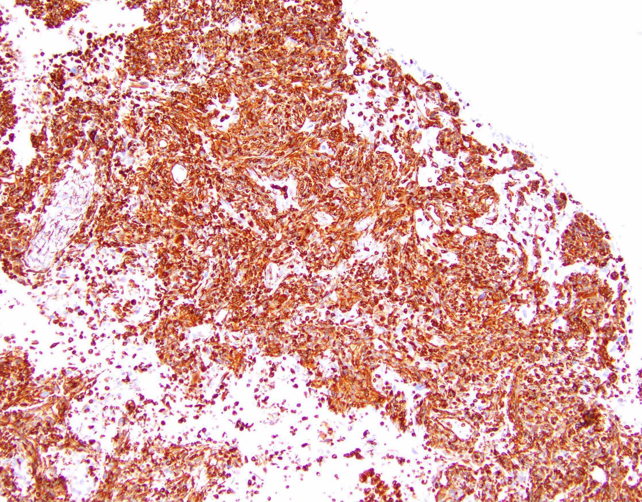
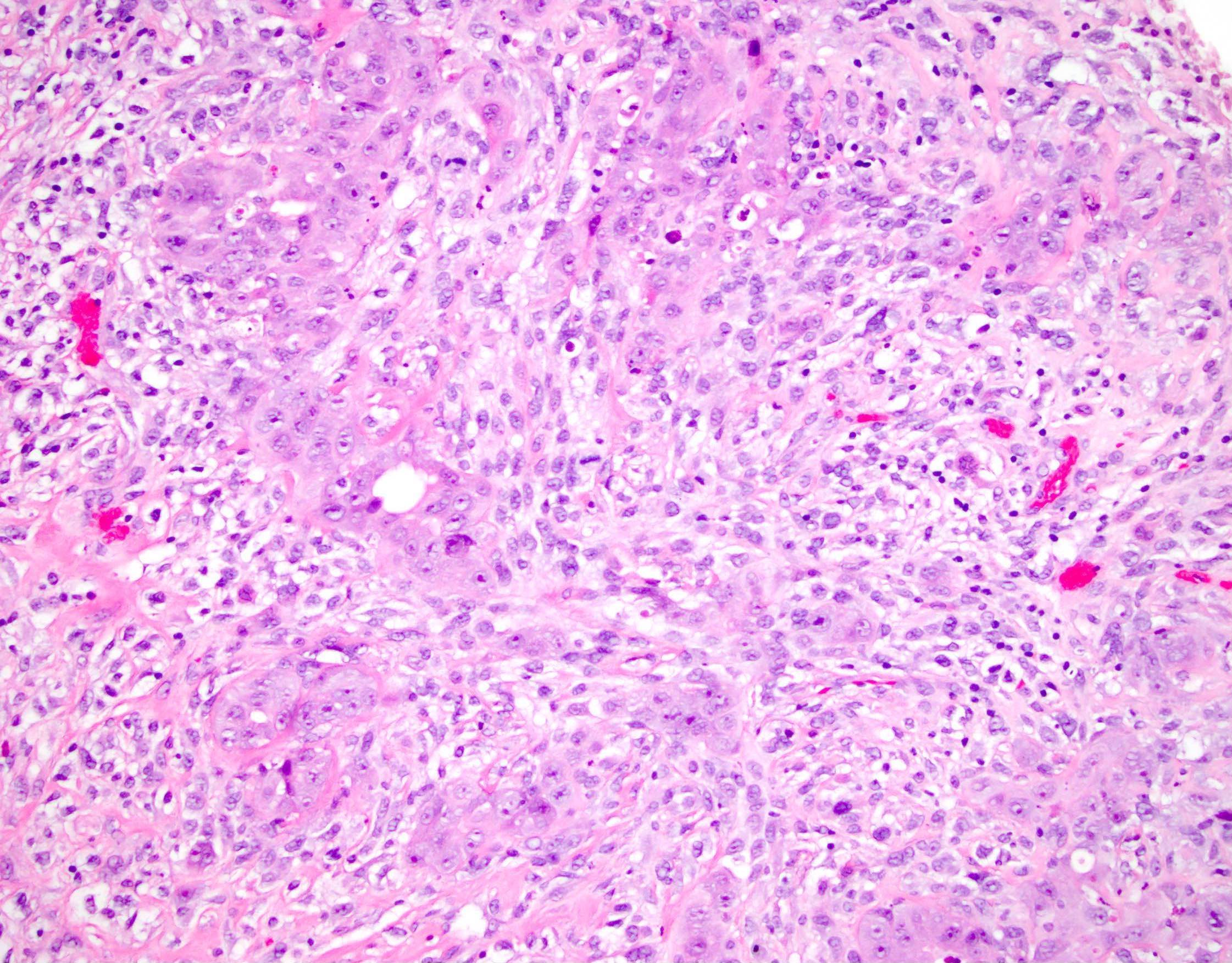
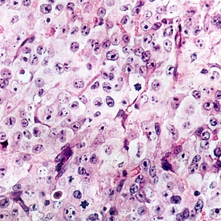
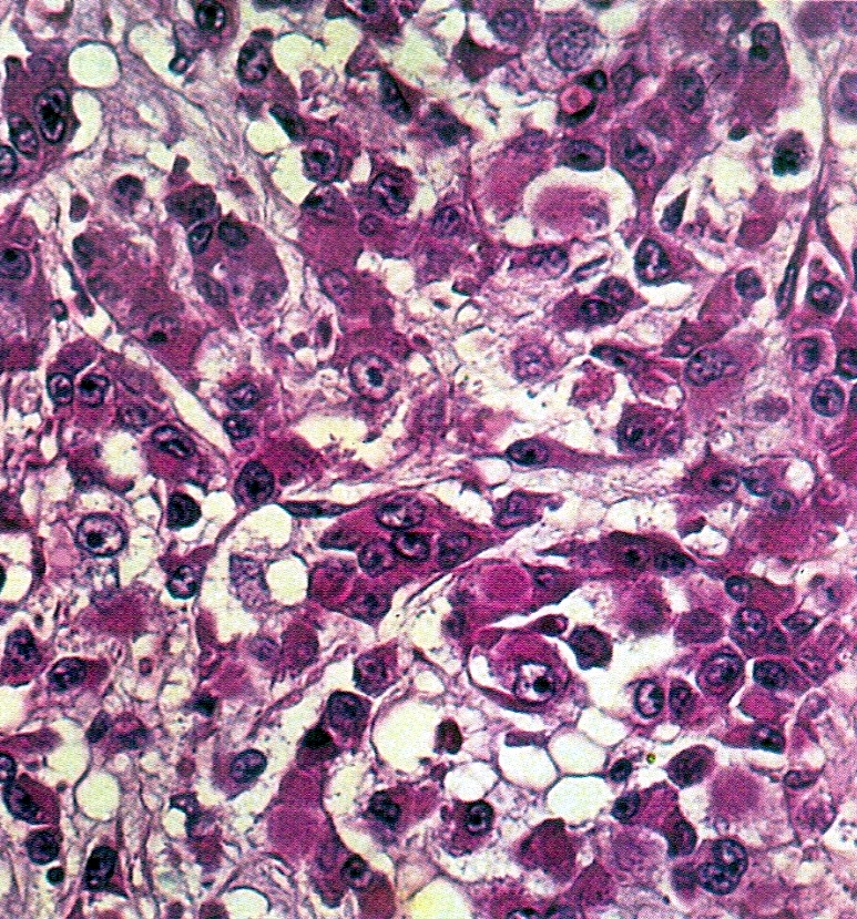
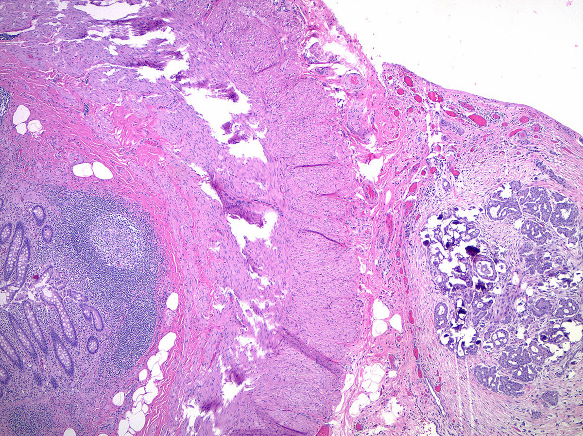
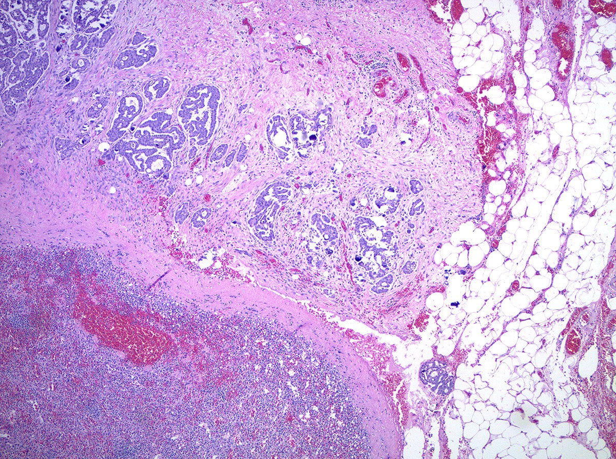
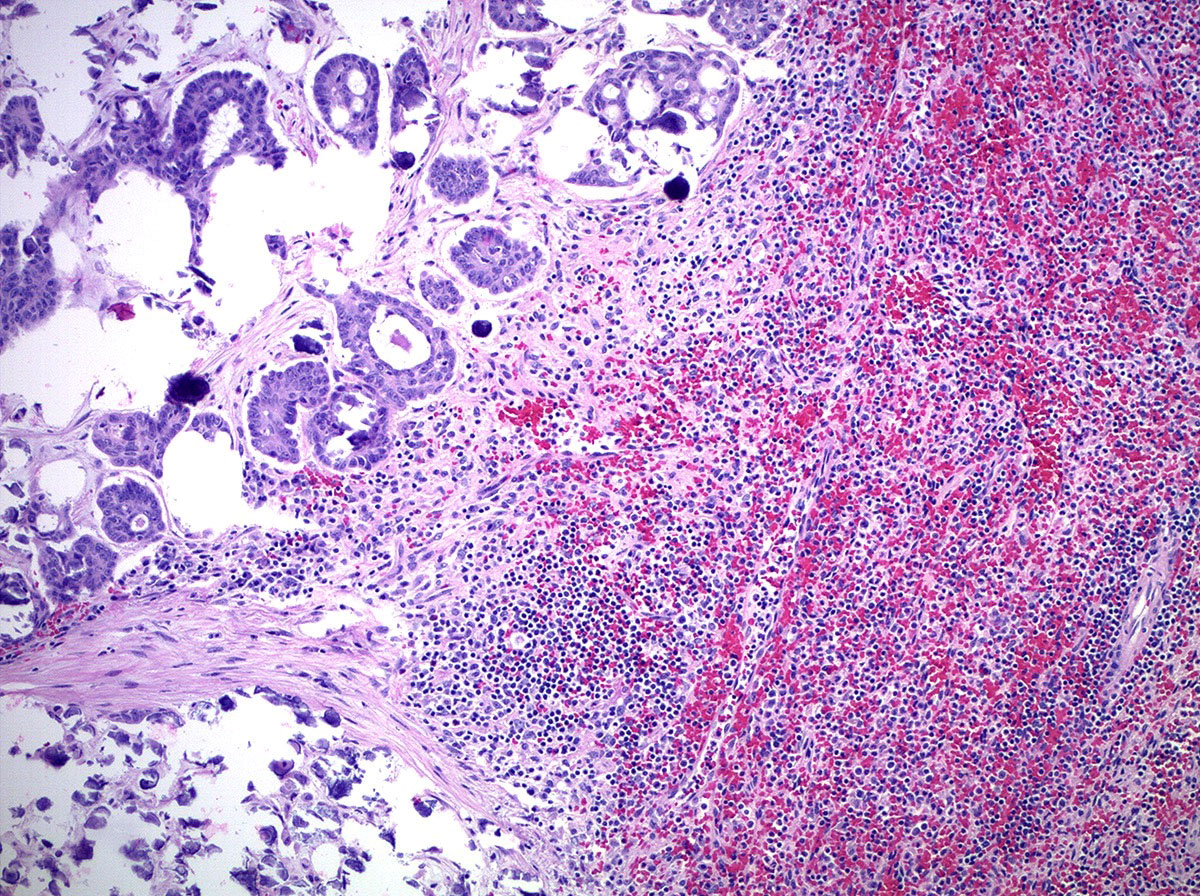
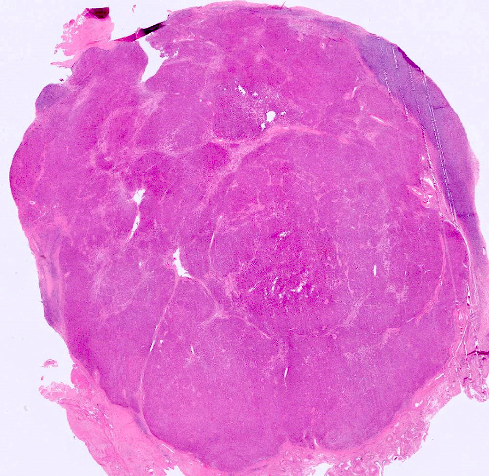
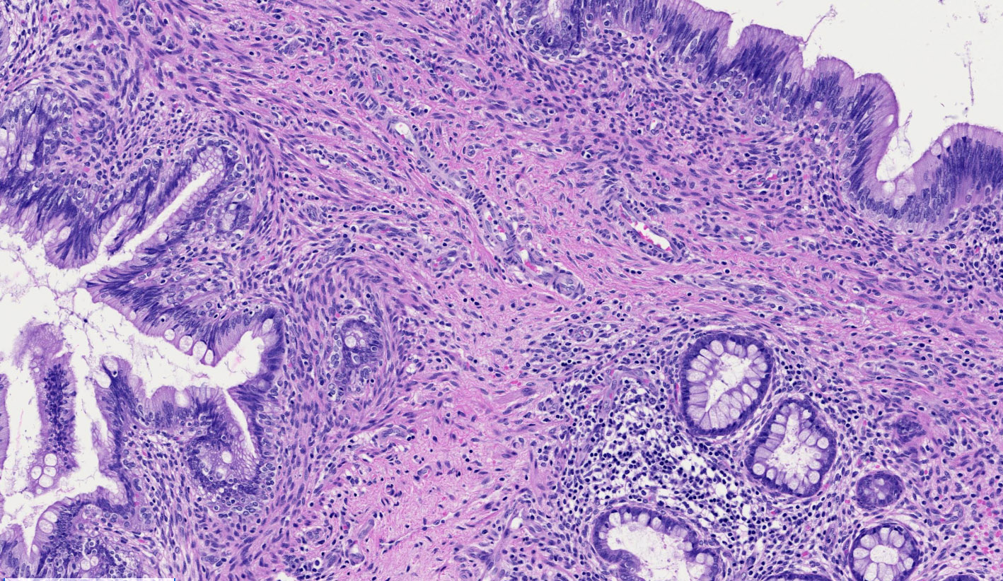
Krukenberg tumor
Metastatic cervical adenosquamous carcinoma
Metastatic low
grade appendiceal
mucinous
neoplasm
Metastatic lobular breast carcinoma
Images hosted on other servers:

Well demarcated mass

CD10, CD56, β-catenin, WT1, PR
AFIP images
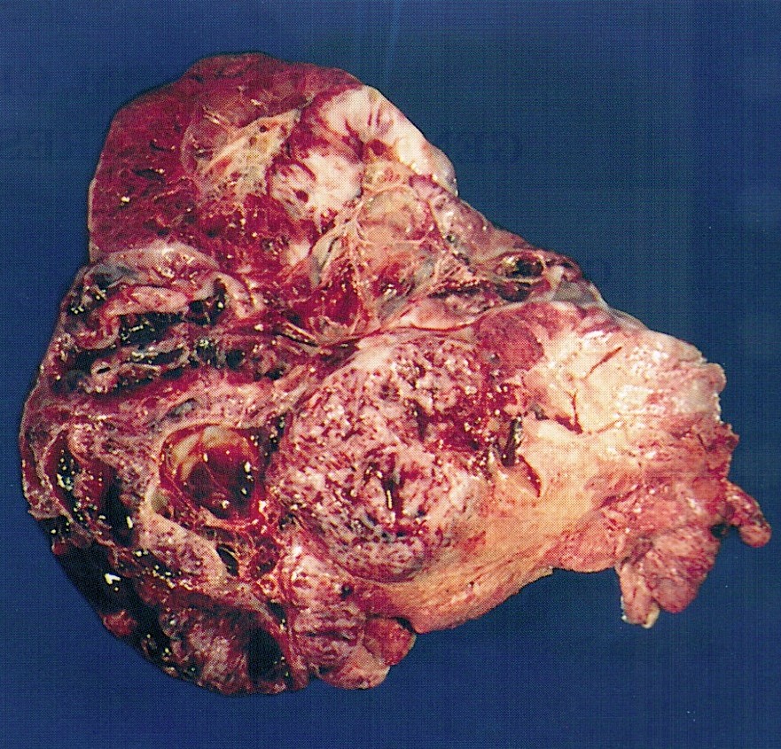
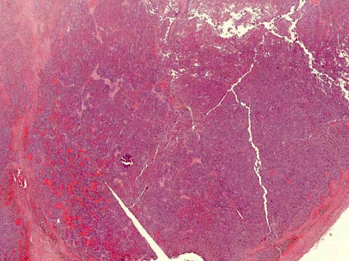
Lobulated, focally hemorrhagic
AFIP images
Mixed malignant germ cell tumor
Diffuse embryoma
Contributed by Azin Mashayekhi, M.D., M.B.A. and Gulisa Turashvili, M.D., Ph.D.
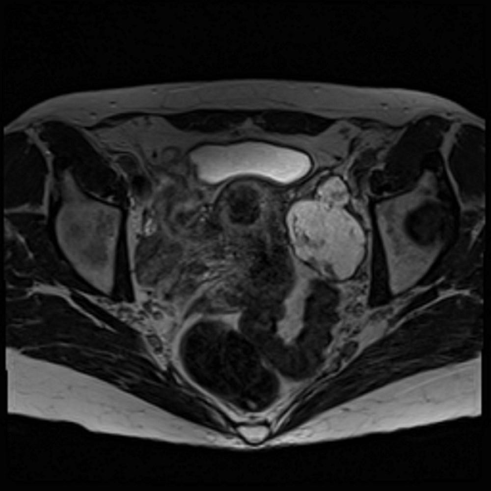
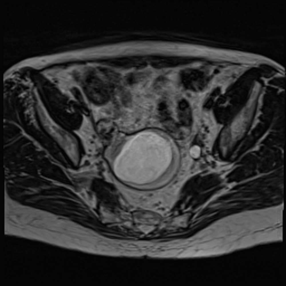
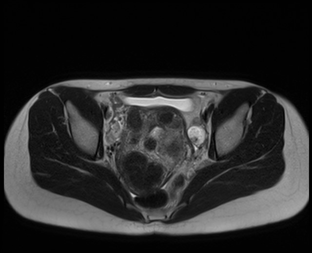
Magnetic resonance imaging (MRI) of pelvis
Contributed by Azin Mashayekhi, M.D., M.B.A. and Gulisa Turashvili, M.D., Ph.D.
Cystic lesion
Contributed by Azin Mashayekhi, M.D., M.B.A. and Gulisa Turashvili, M.D., Ph.D.
Cystic lesion
Cystic lesion
Intracystic proliferation
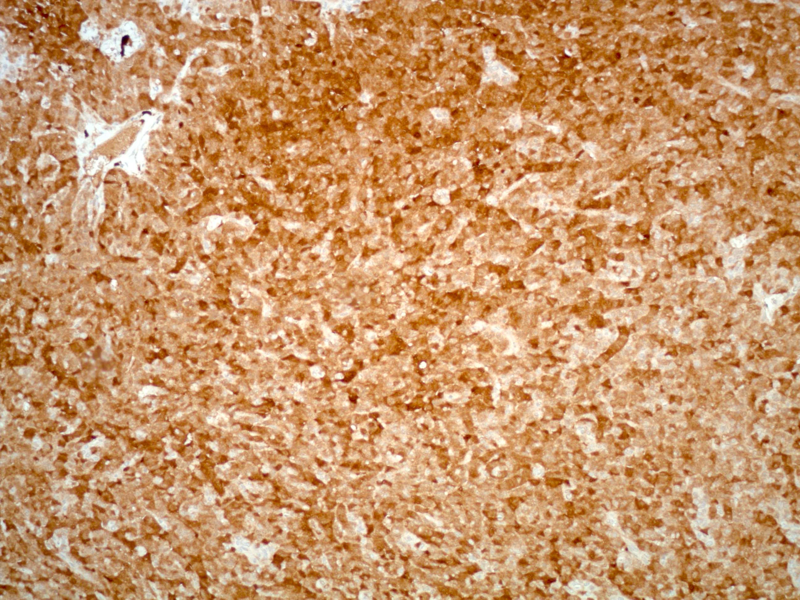
GFAP
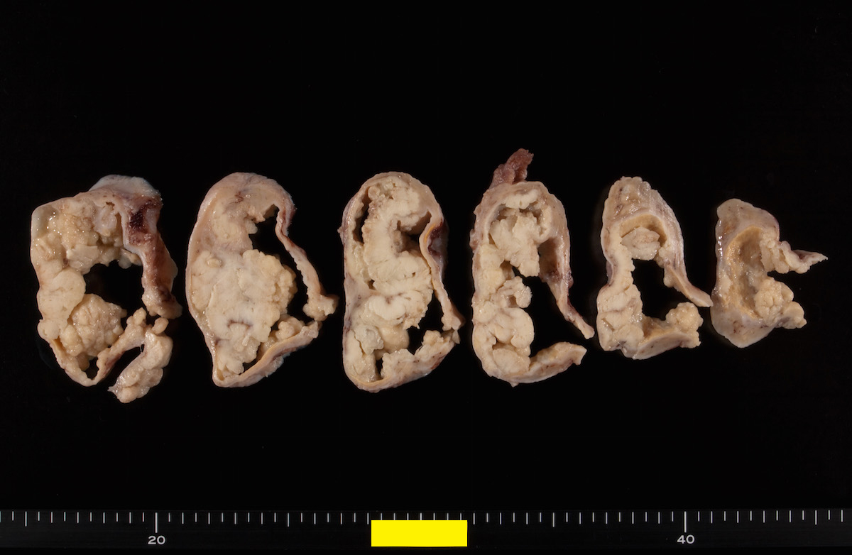
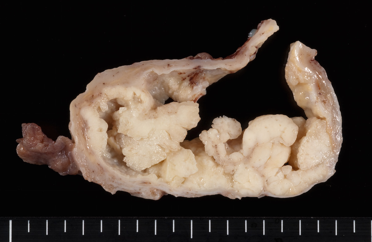
Contributed by Russell Vang, M.D.
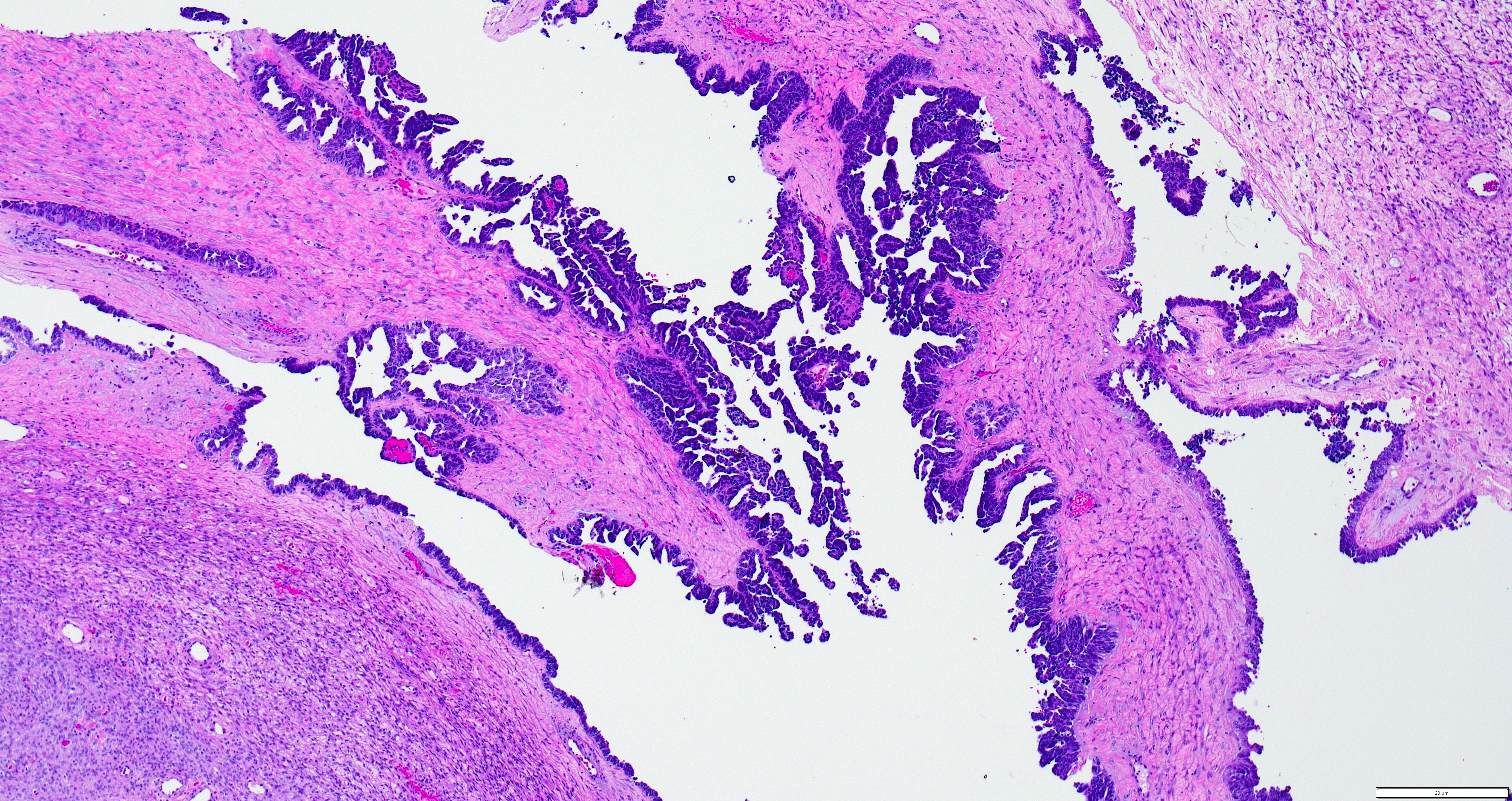
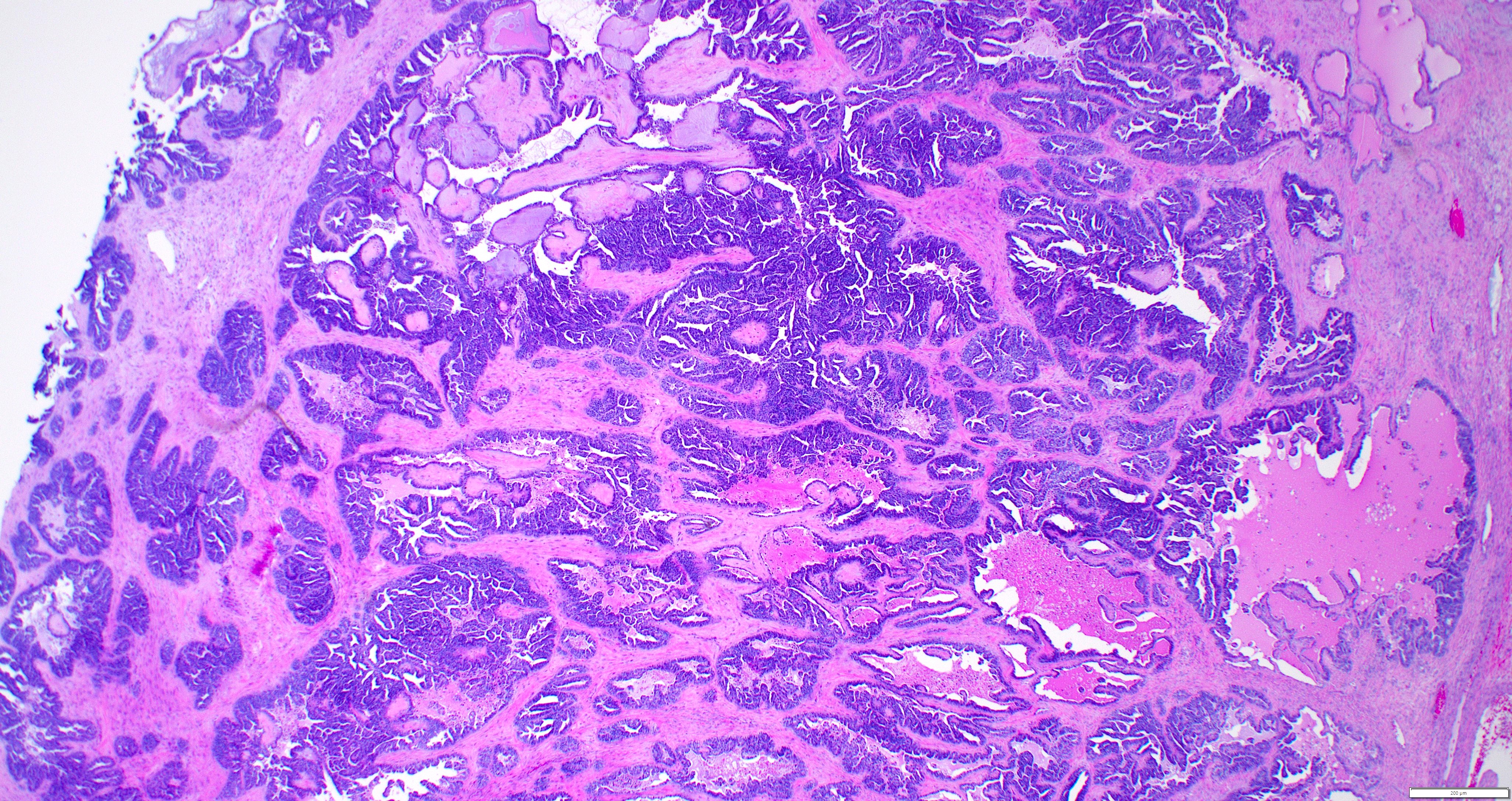
Multiloculated cyst
Contributed by Neshat Nilforoushan, M.D. and Russell Vang, M.D.
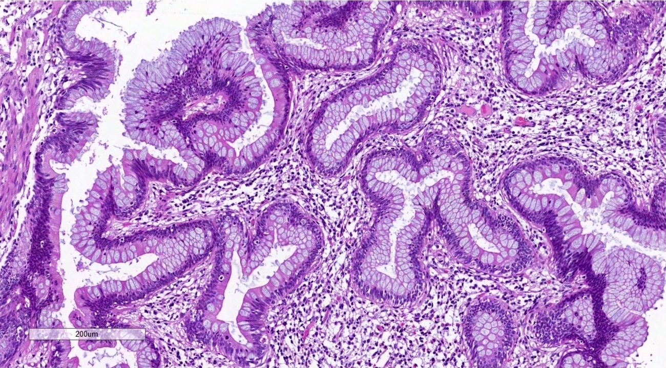
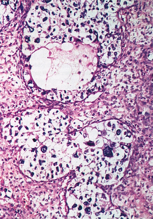
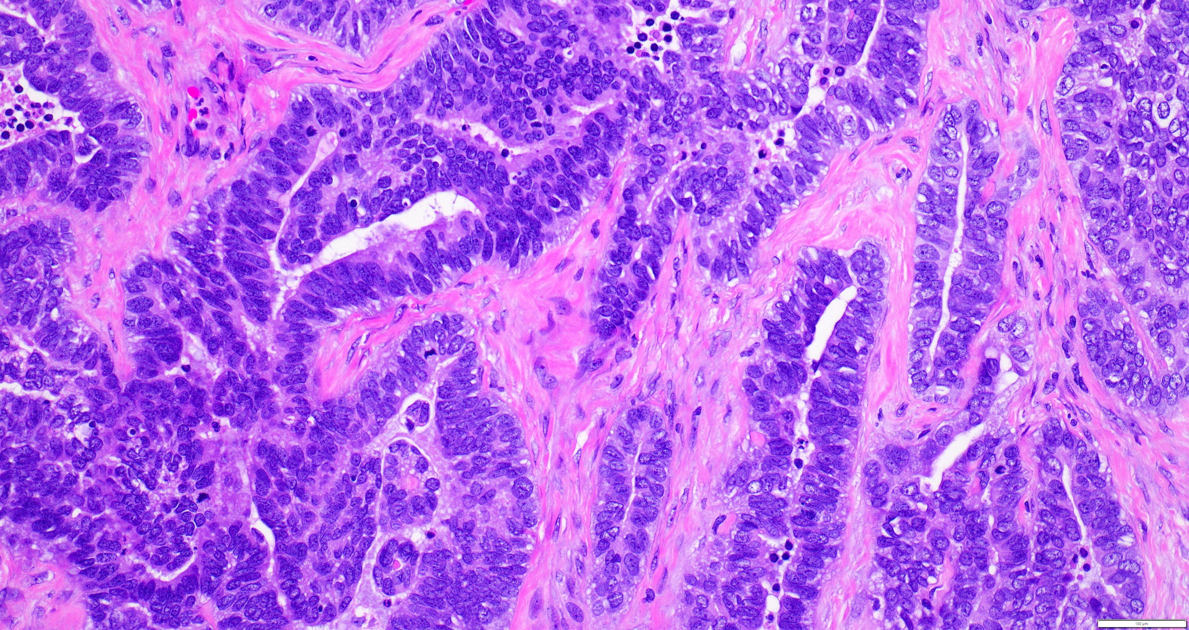
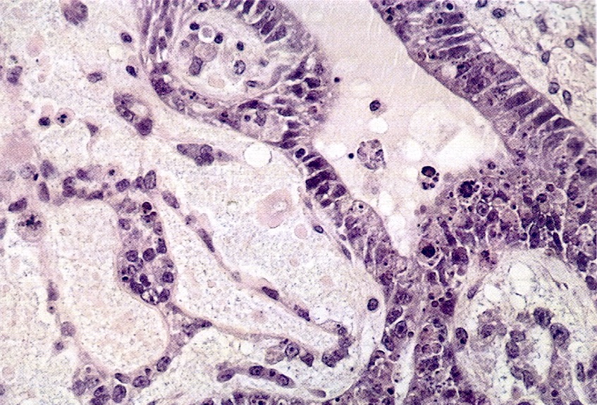
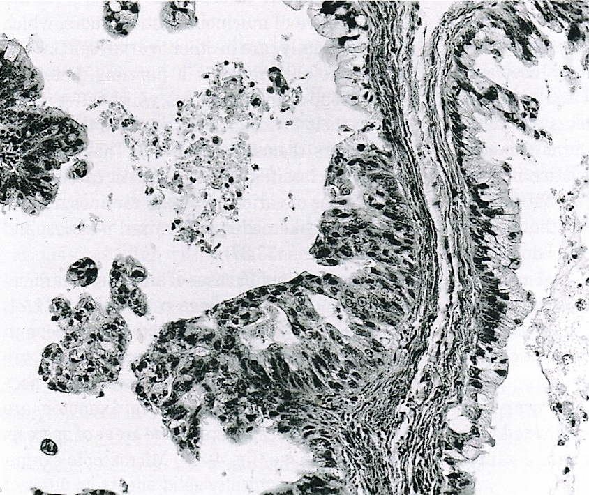
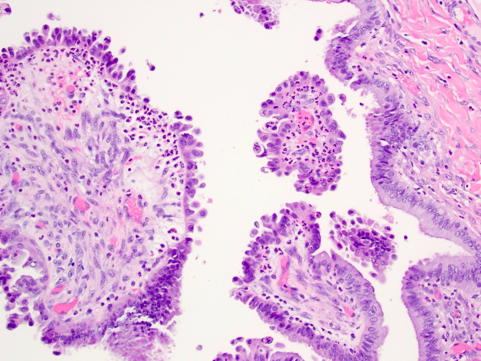
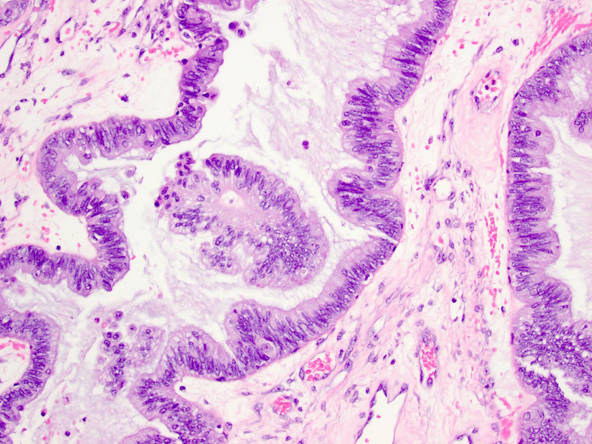
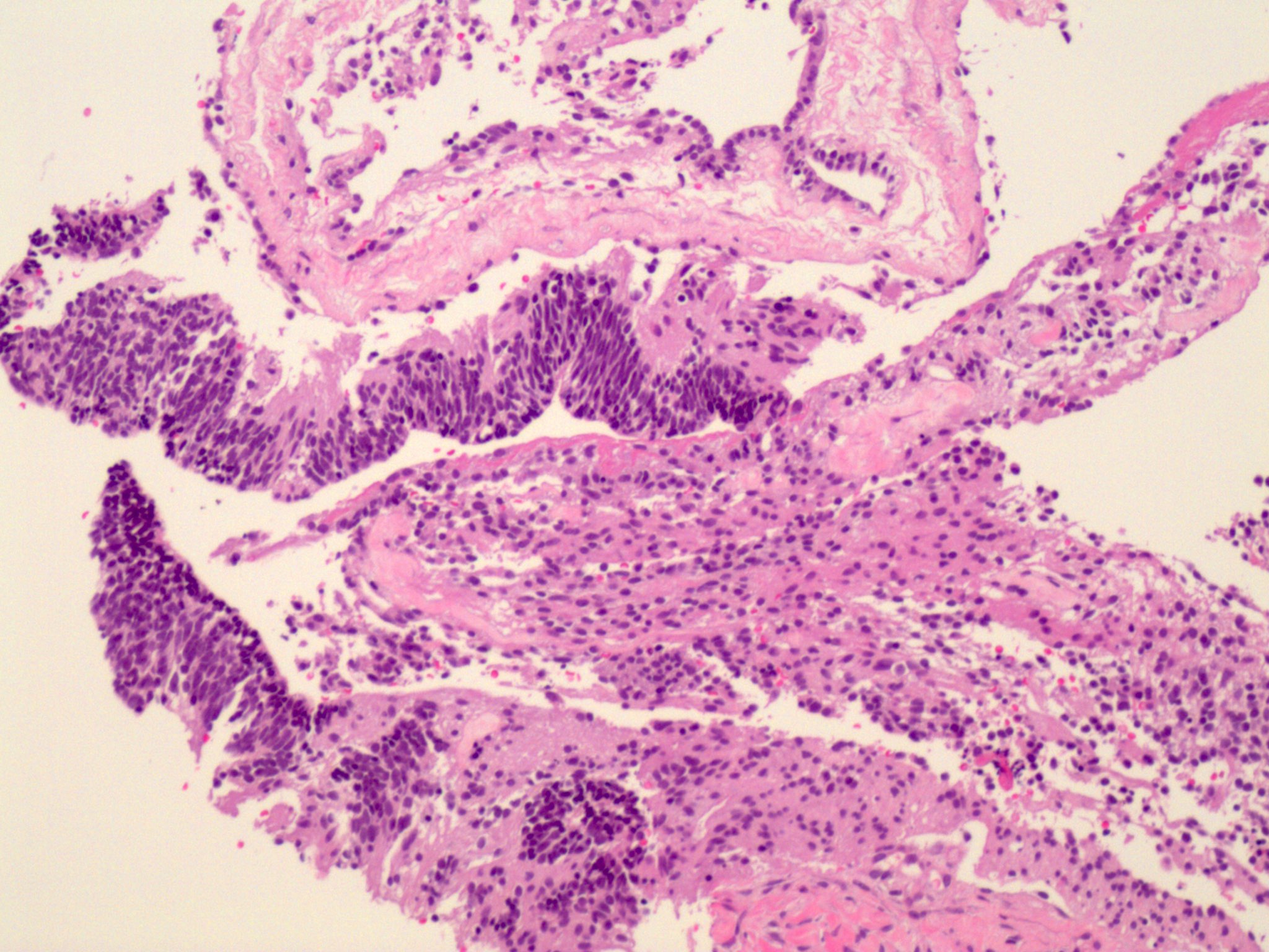
Complex architecture, tufting and villus formation
Mucinous intestinal type epithelium
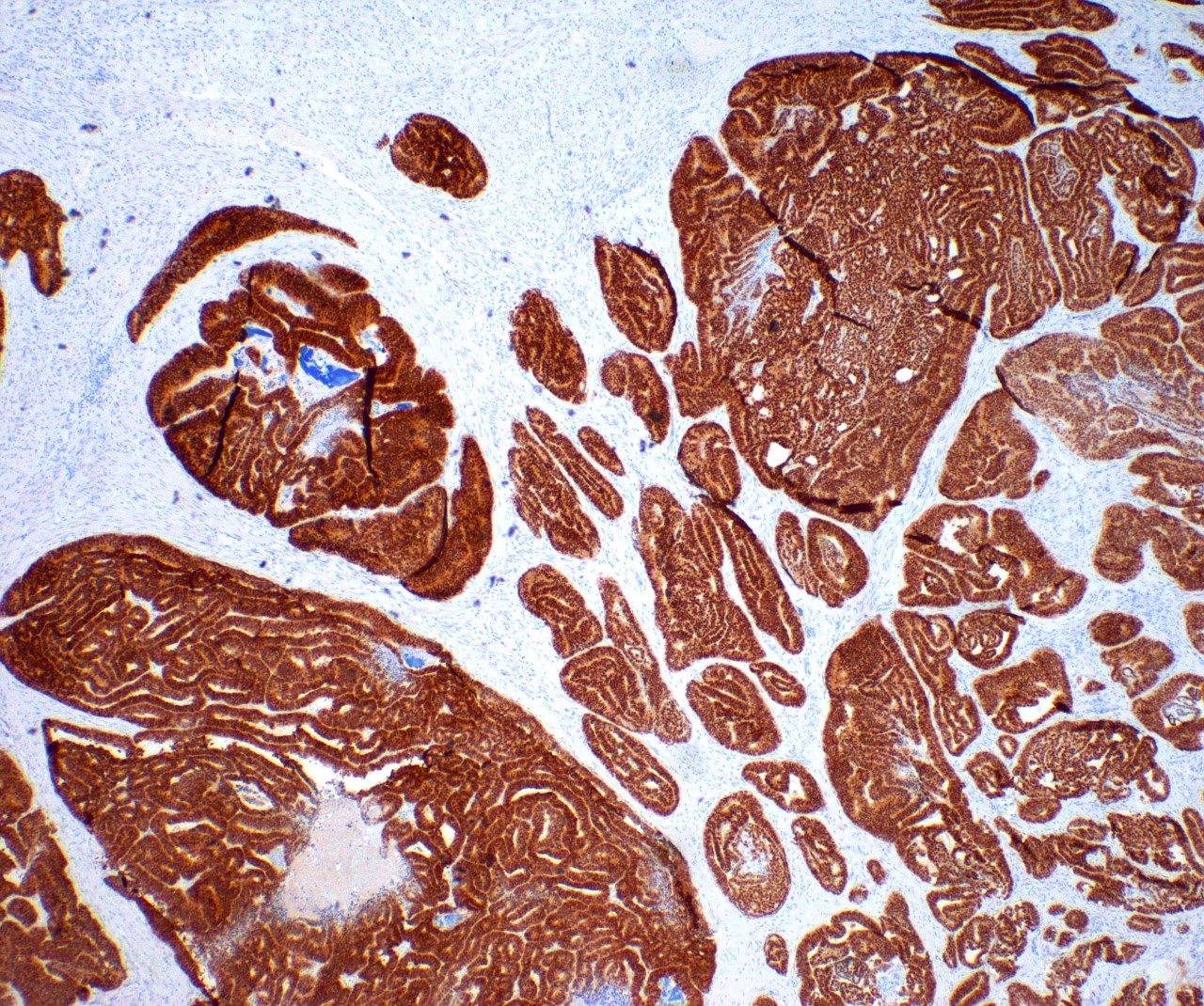
Intraepithelial carcinoma
Microinvasion
AFIP images
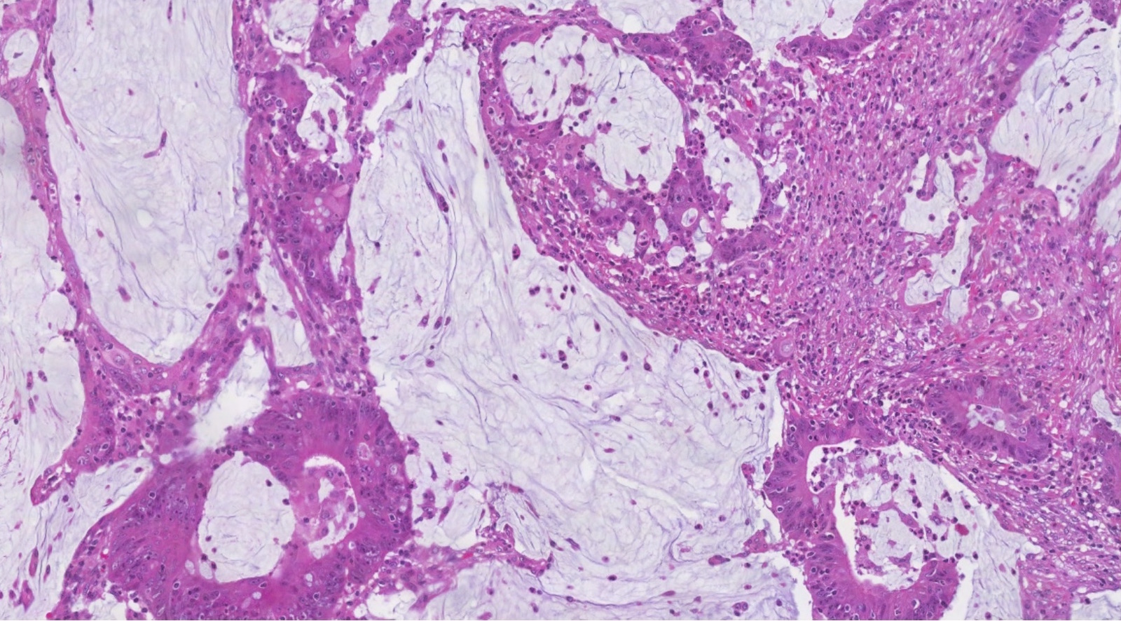
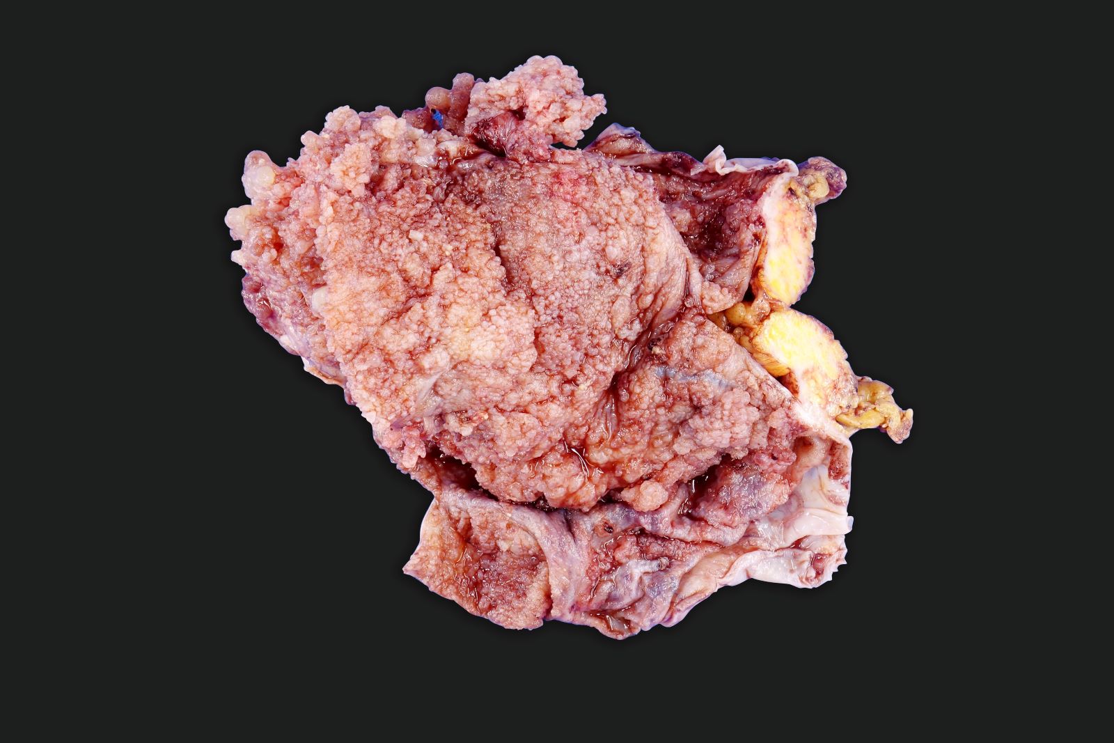
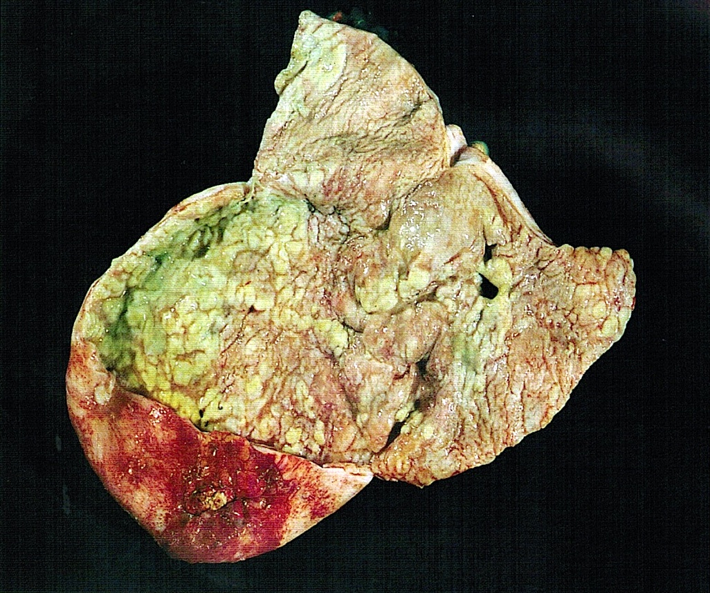
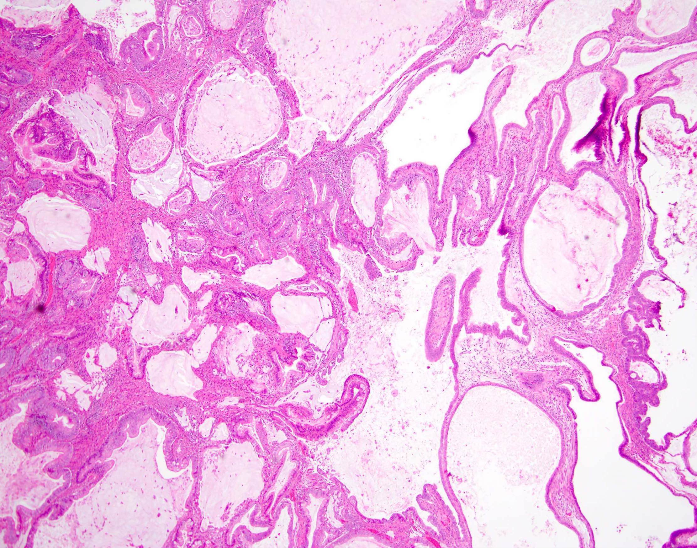
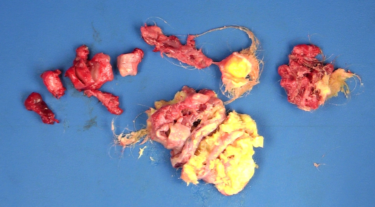
Mucinous
cystadenocarcinoma
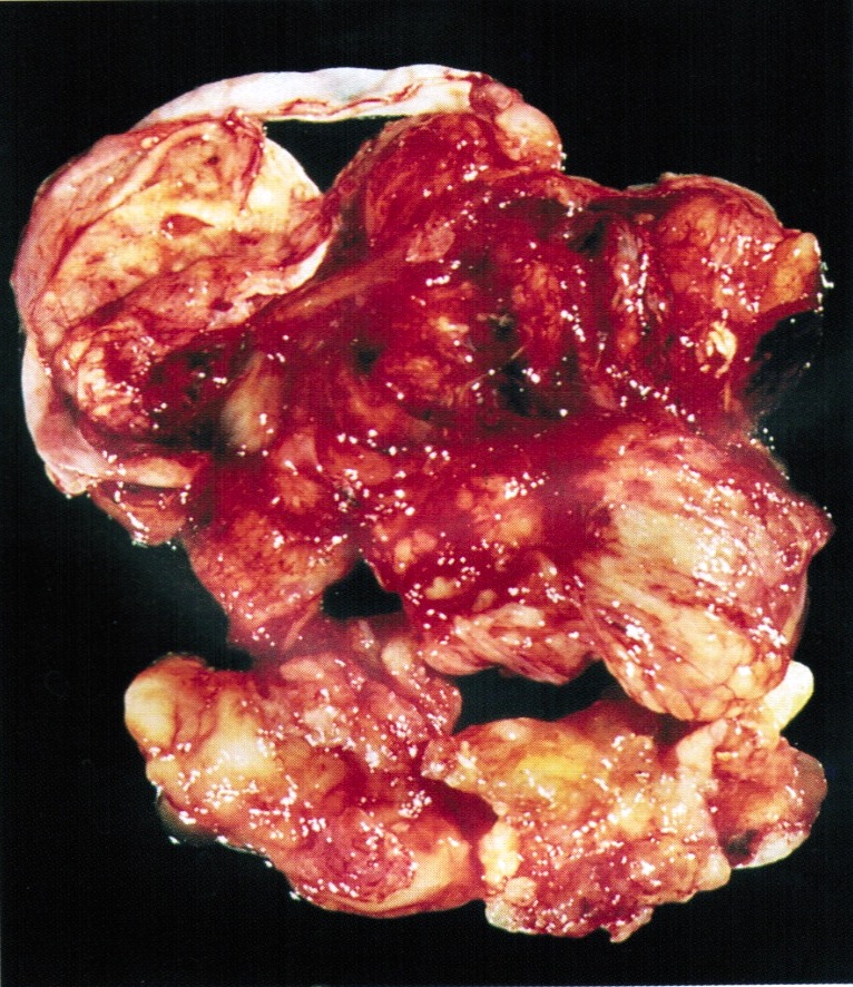
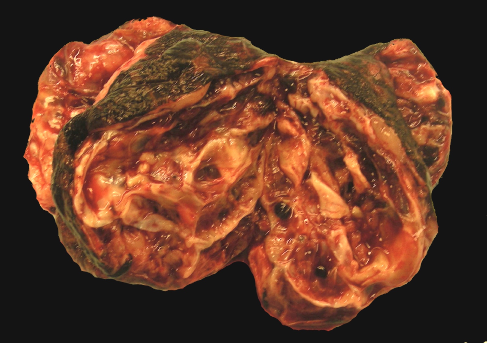
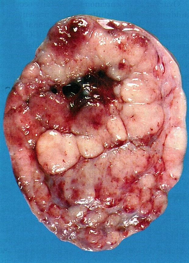
Gelatinous with extensive hemorrhage and necrosis
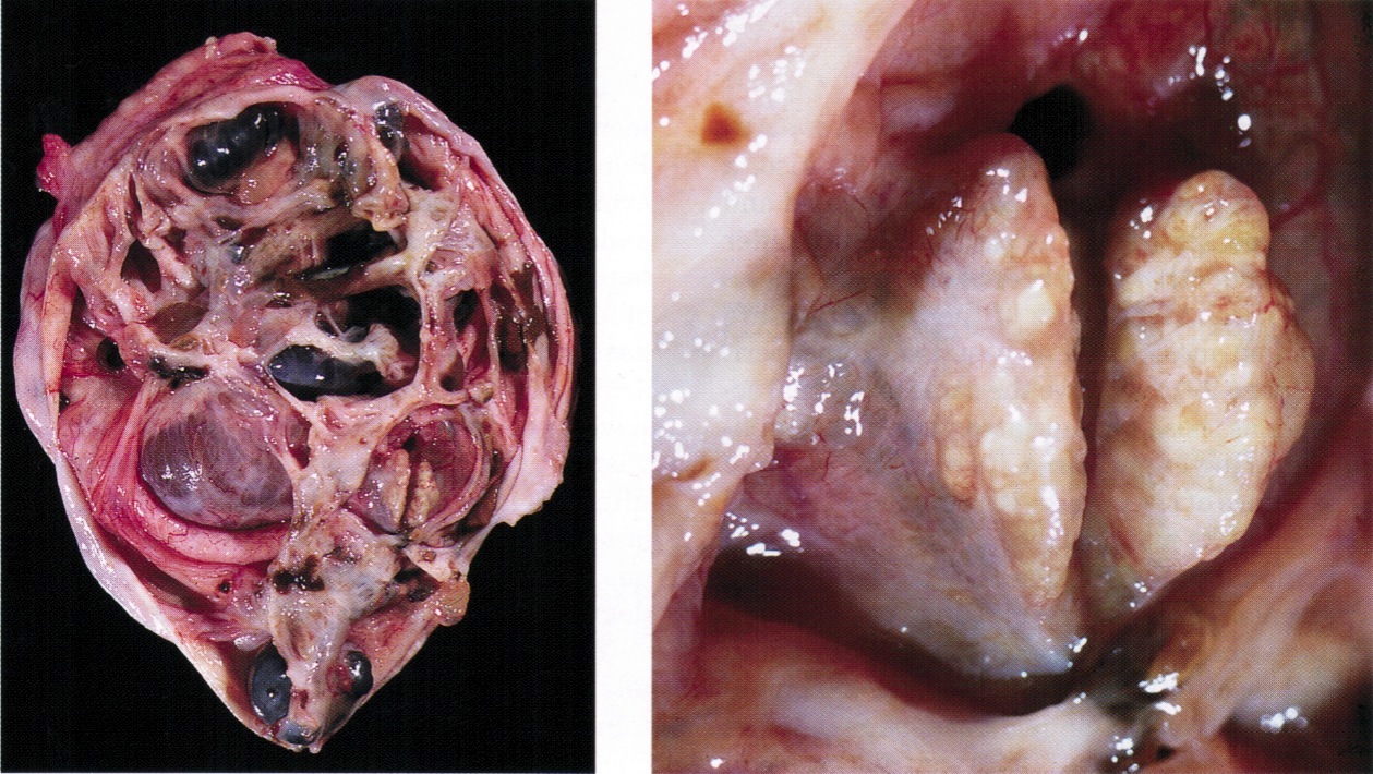
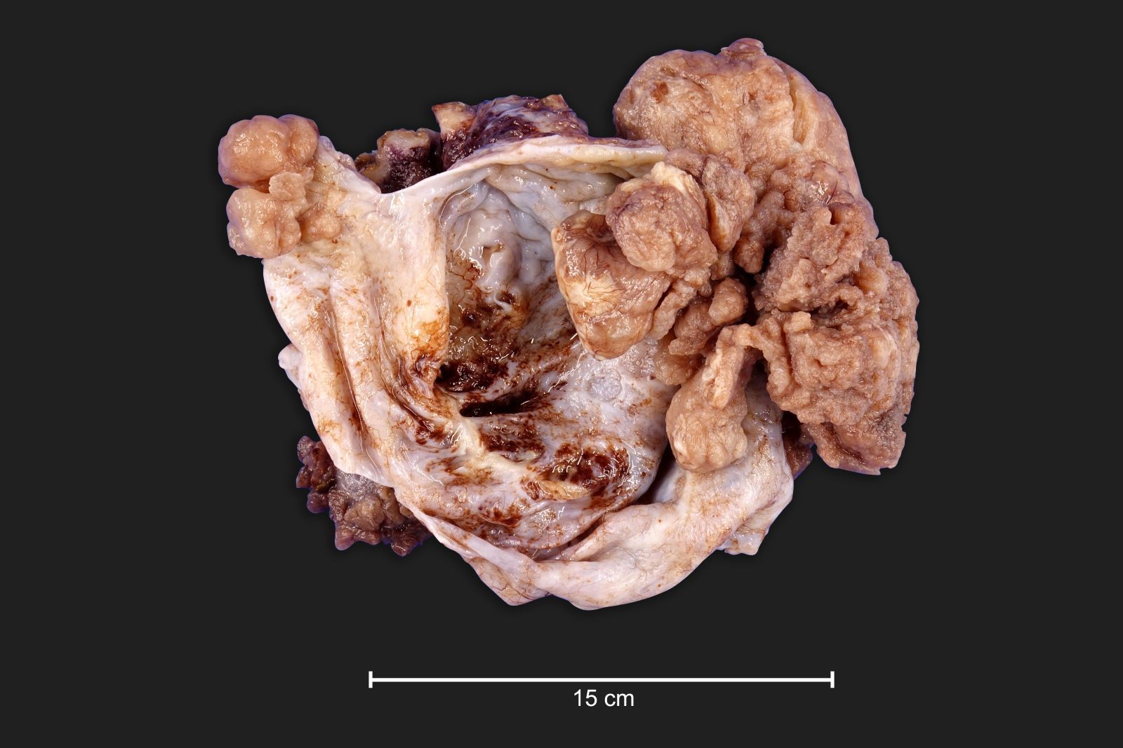
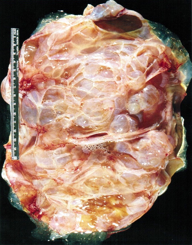
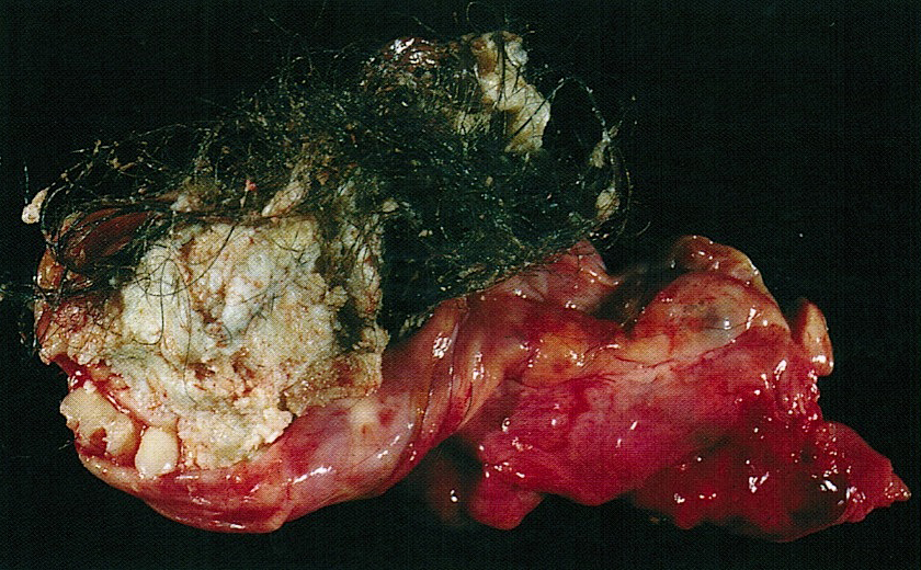
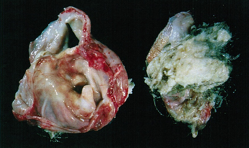
Mucinous cystic
tumor with
various
components
Images hosted on other servers:

Mucinous
cystadenocarcinoma
AFIP images
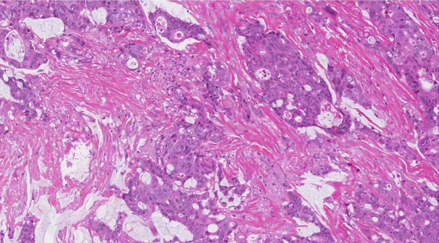
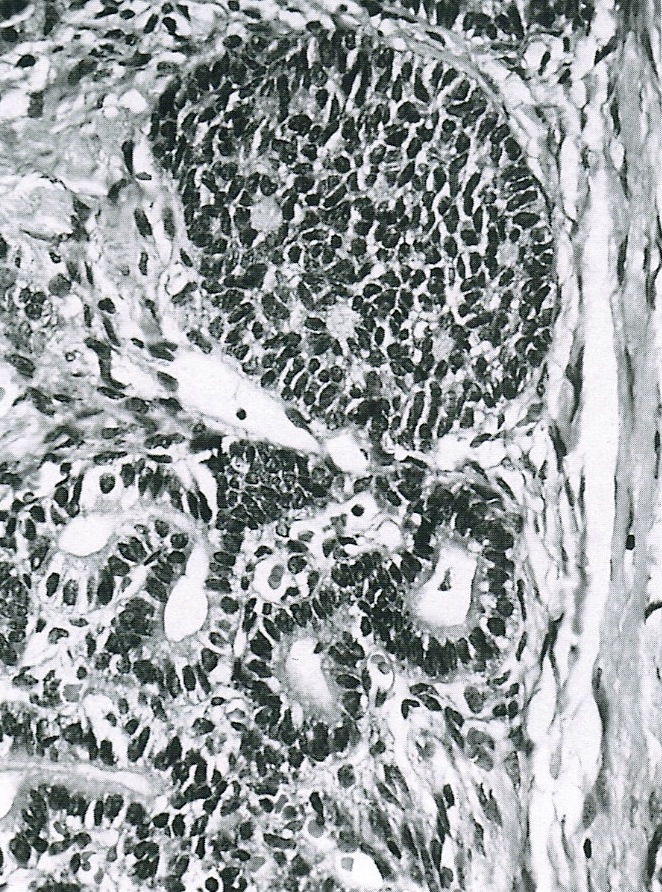
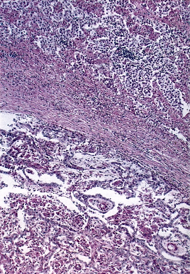
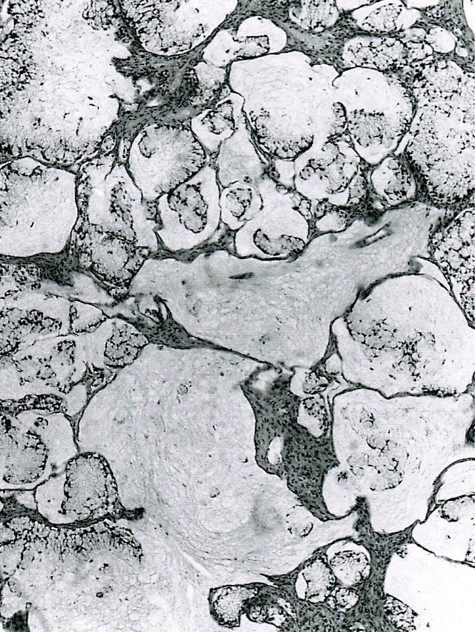
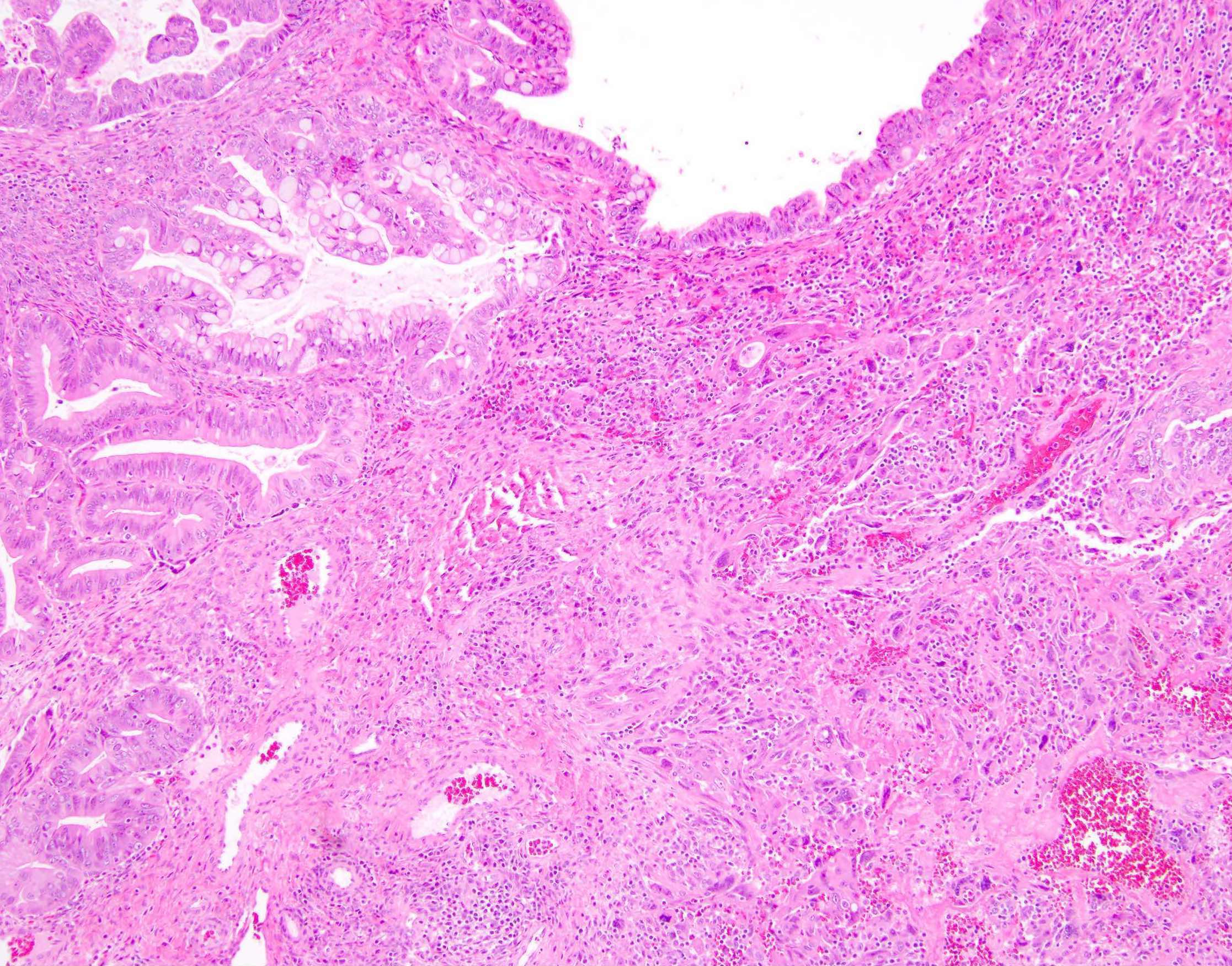
Cribriform pattern
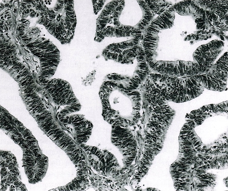
Resembling endometrioid adenocarcinoma
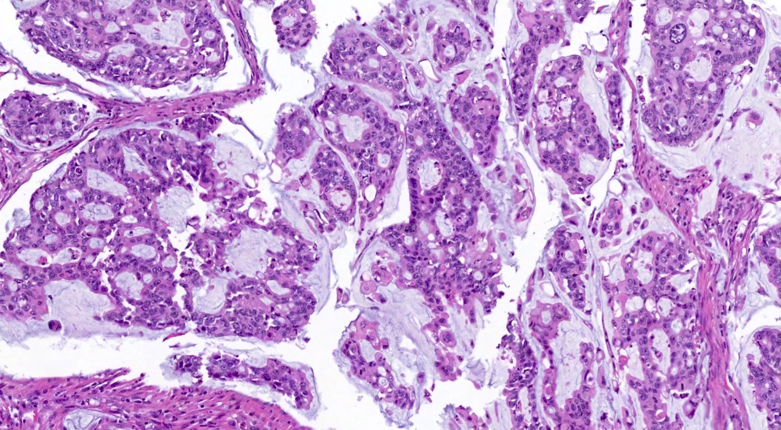
Mostly apical
mucin, but several
goblet cells
with red mucin
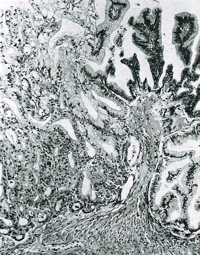
With microinvasion of stroma
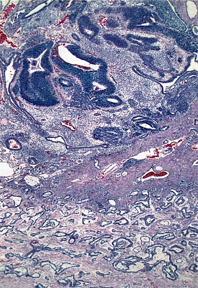
Disorderly
arrangement of
cysts and glands
of irregular shapes
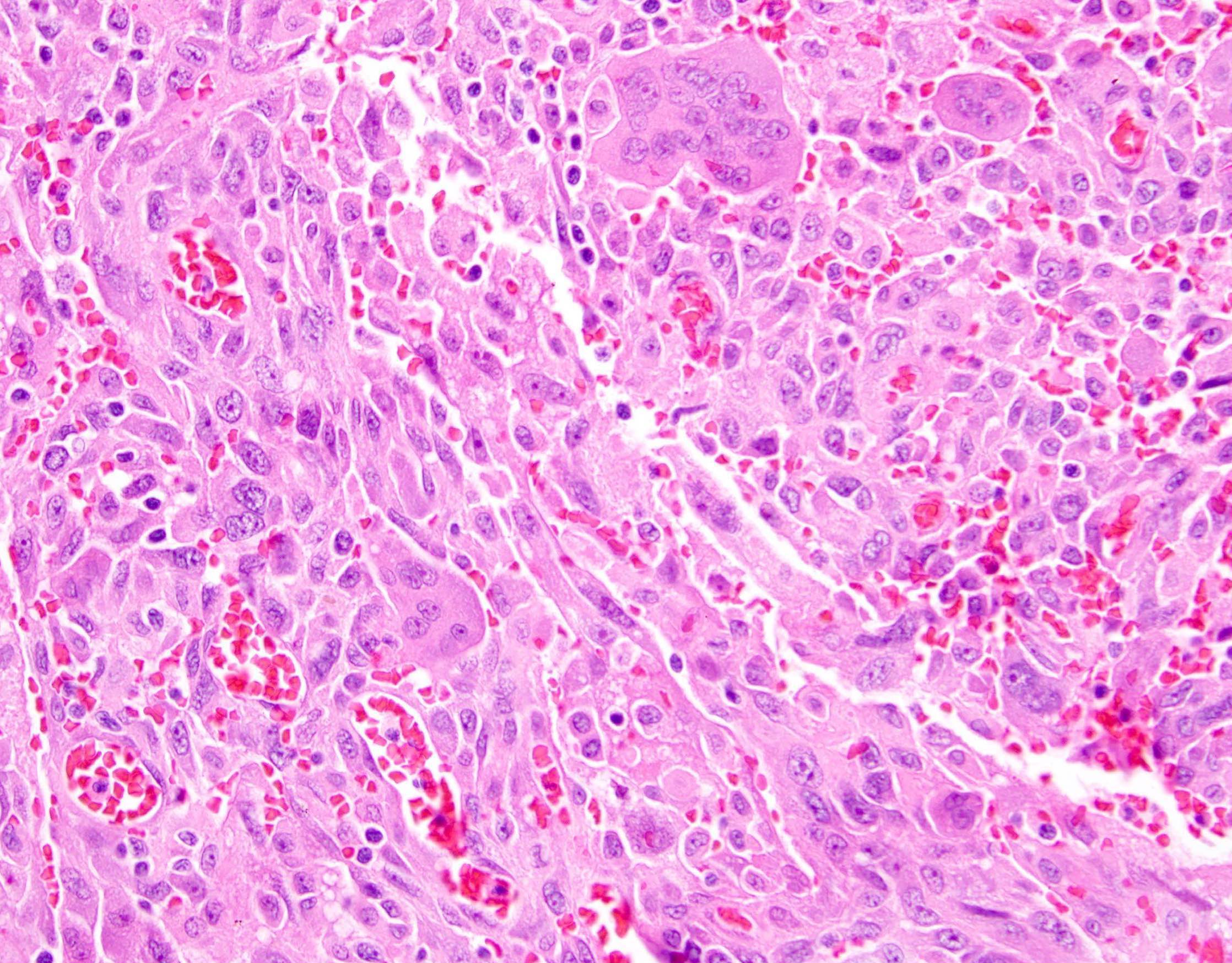
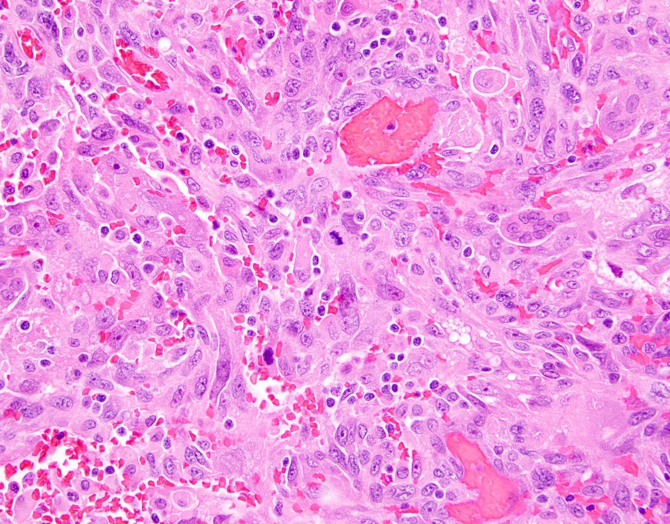
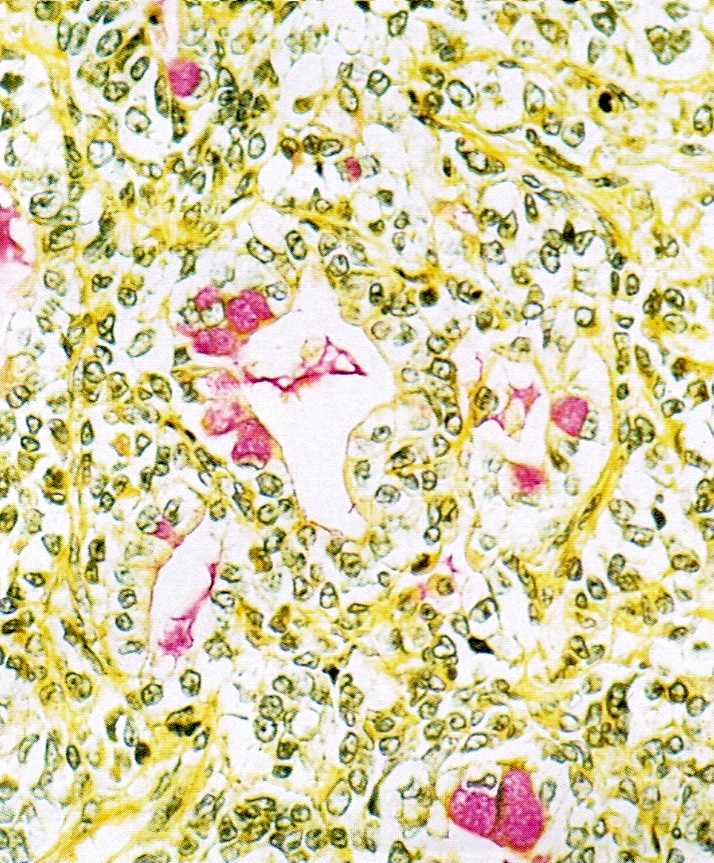
Invasive small glands and small collections of signet ring cells
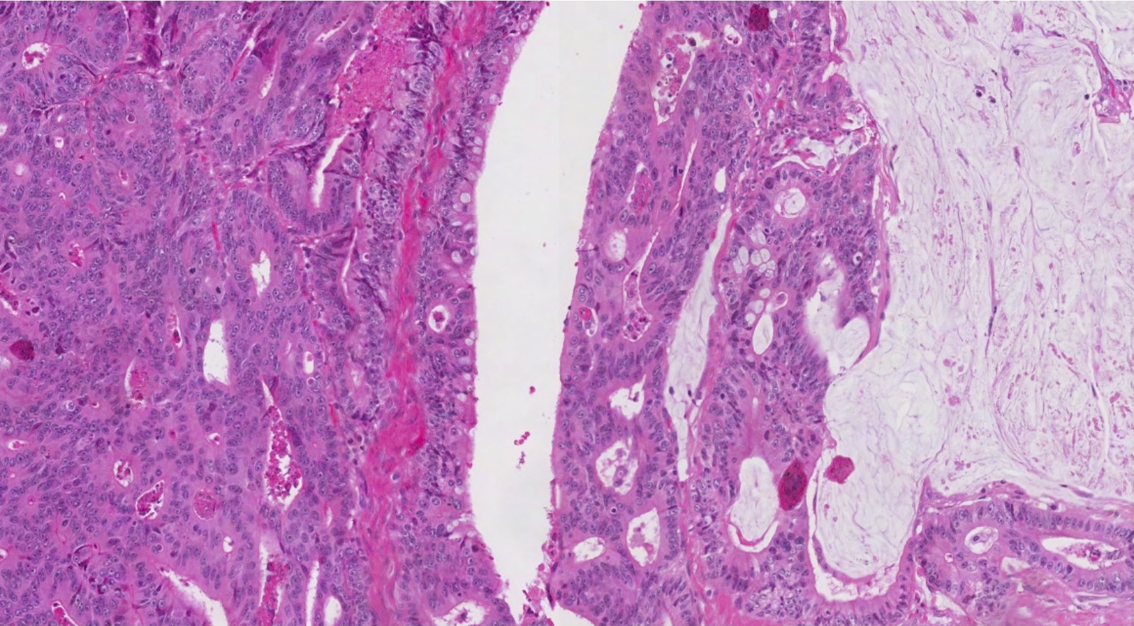
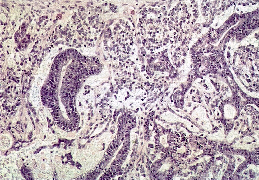
Cyst lined by cells with abundant mucin and highly stratified; nuclei highly atypical and presence of necrotic debris in lumen
Resembling
mucinous (colloid)
carcinoma
of intestine
Images hosted on other servers:

Palpable lower abdominal mass

Right pelvic pain
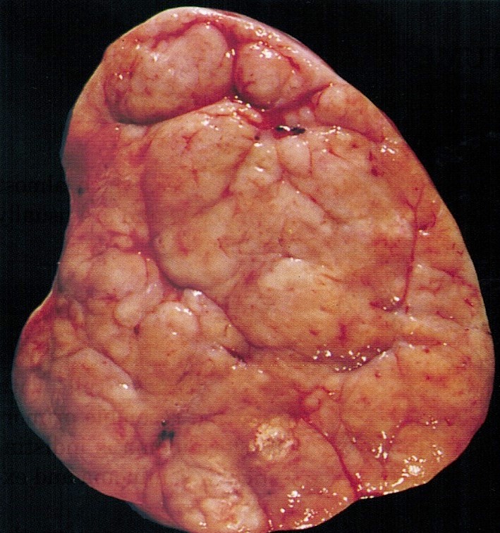
AFIP images
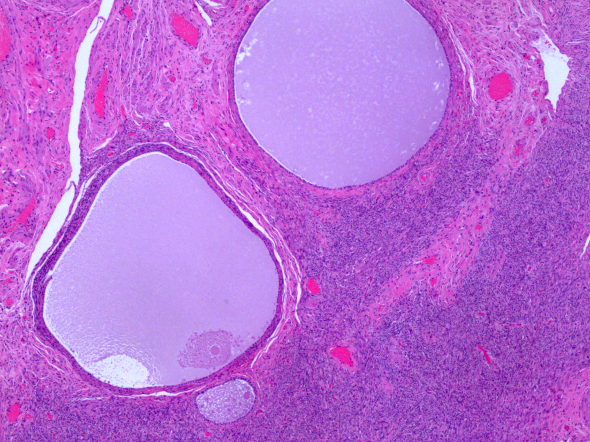
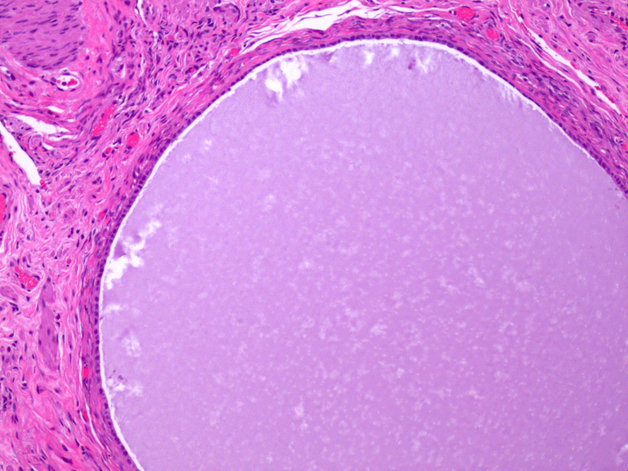
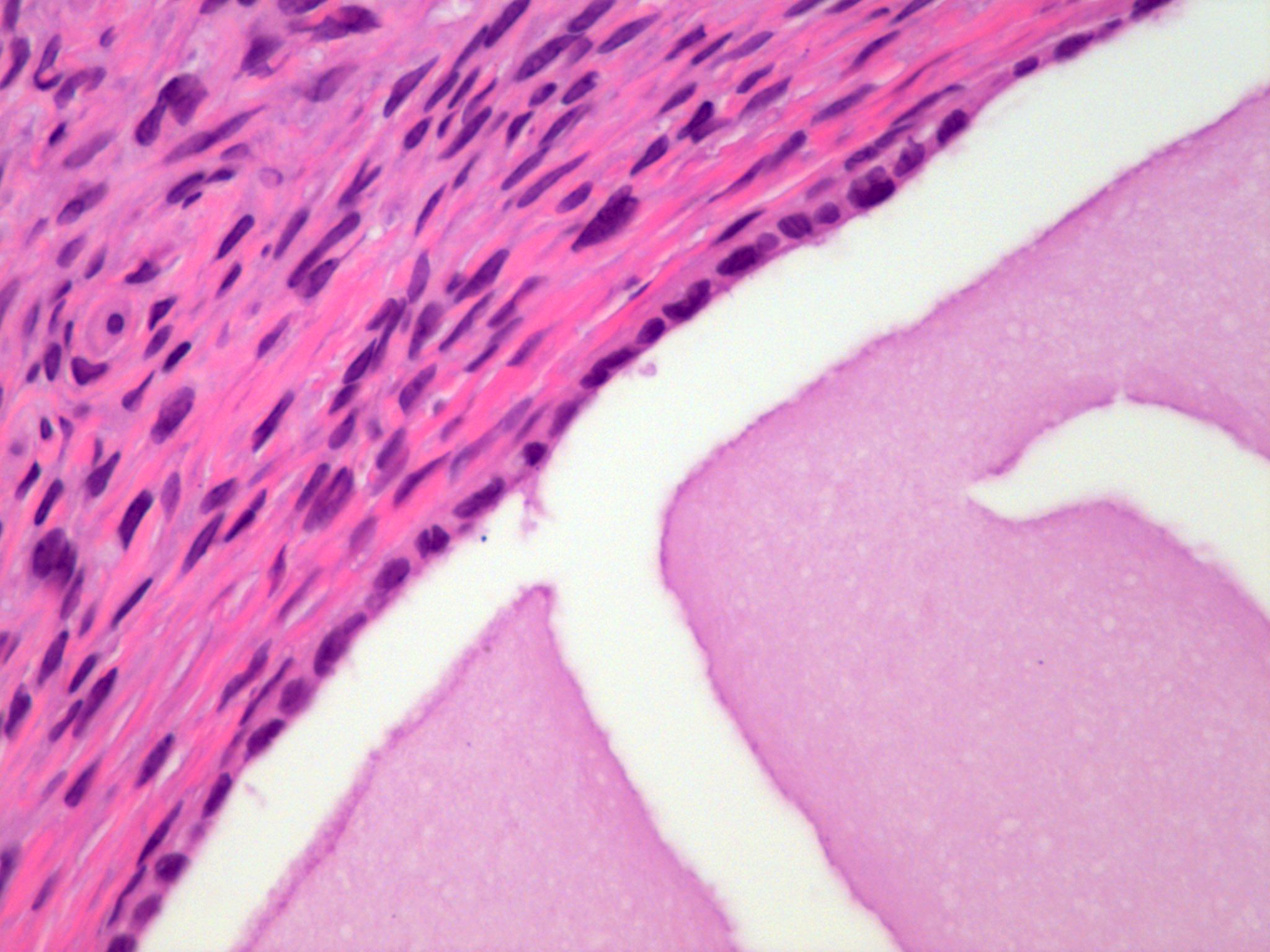
Mucinous cystadenoma
Associated with dermoid cyst
Contributed by Gulisa Turashvili, M.D., Ph.D. and AFIP images

Unilocular cyst
Bland mucinous epithelium
Mucinous adenofibroma
Mucinous cystic tumor
Images hosted on other servers:

Mural anaplastic carcinoma

Mural leiomyoma
Images hosted on other servers:

Mural leiomyoma

Sarcoma-like mural nodule


Anaplastic carcinoma mural nodule

Clear cell mural nodule
Contributed by Kruti P. Maniar, M.D. and Jian-Jun Wei, M.D.
Mural carcinosarcoma
Stromal invasion
Atypical spindled and epithelial cells
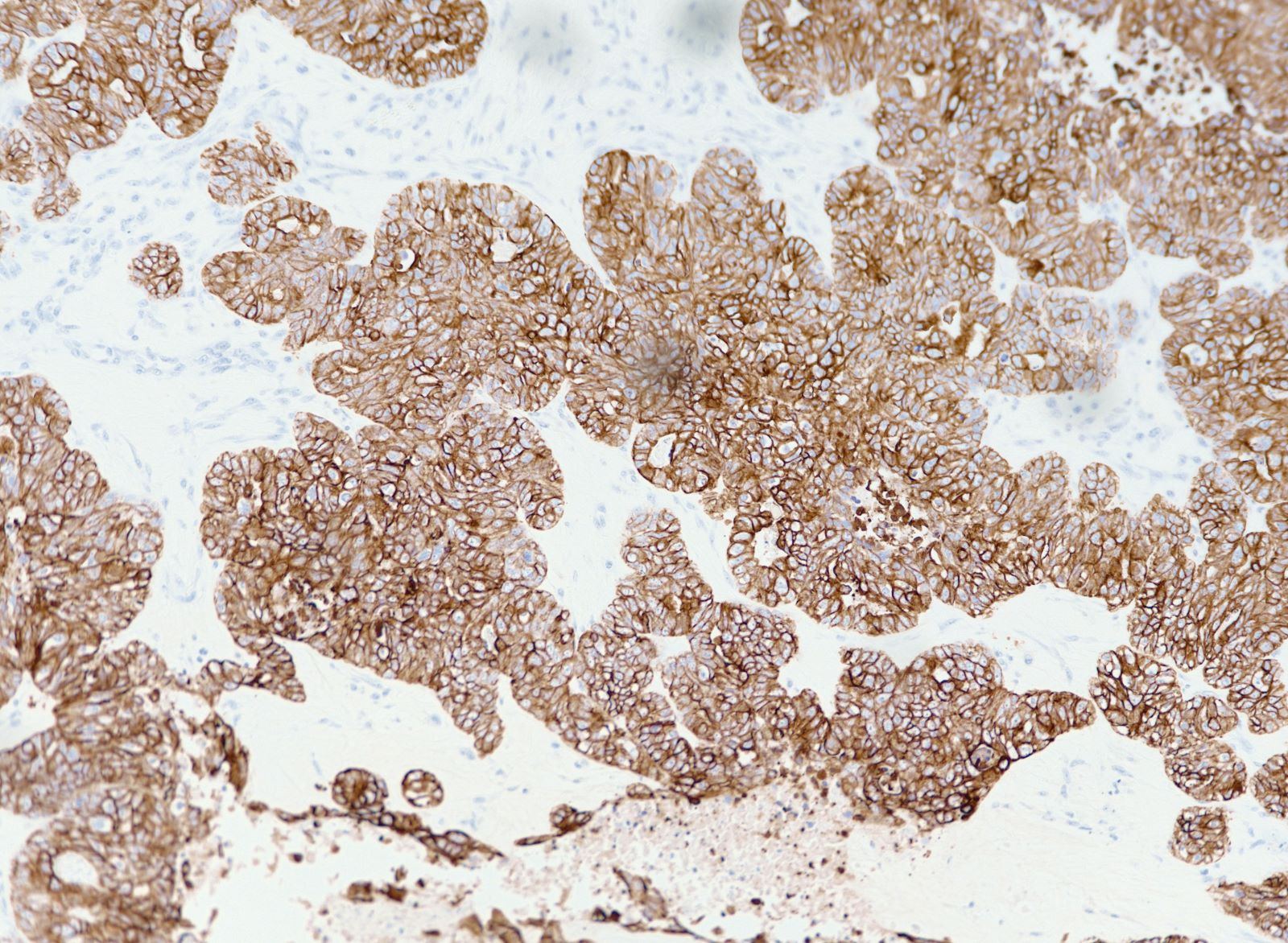
CK7
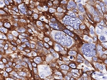
Vimentin
Mural neuroendocrine carcinoma
Stromal invasion
Marked atypia
Synaptophysin
Mural anaplastic carcinoma
Marked atypia
Vascular / stromal invasion
Metastasis
Mural carcinosarcoma
Stromal invasion
Malignant cells
Images hosted on other servers:

Rhabdomyoblastic

Sarcomatoid anaplastic

AFIP images

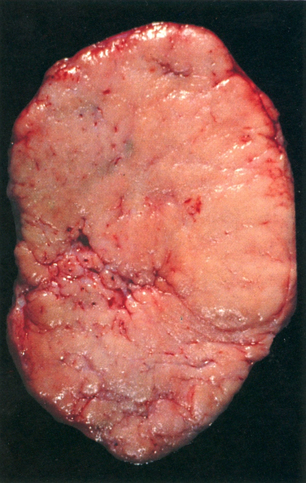
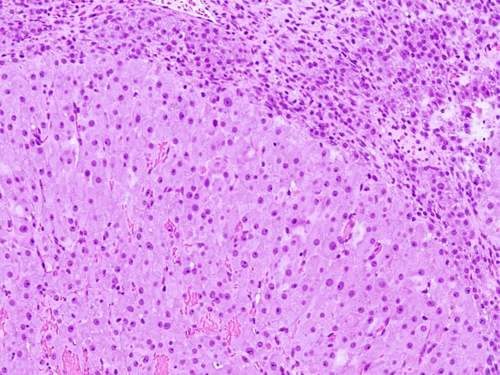
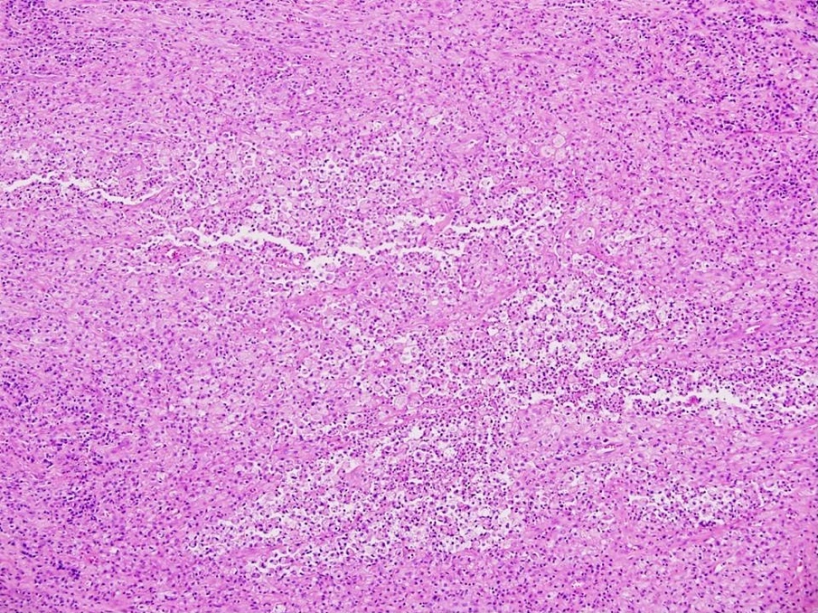
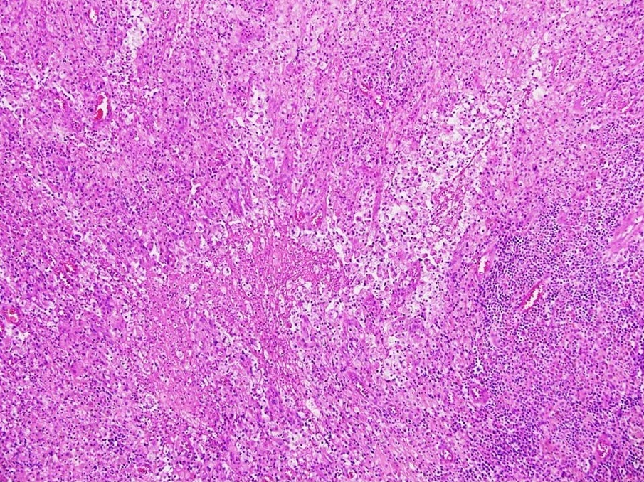
Enlarged ovary is
pearly white
Contributed by Krisztina Lengyel, M.D. and AFIP
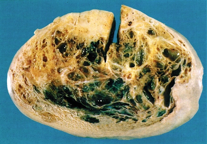
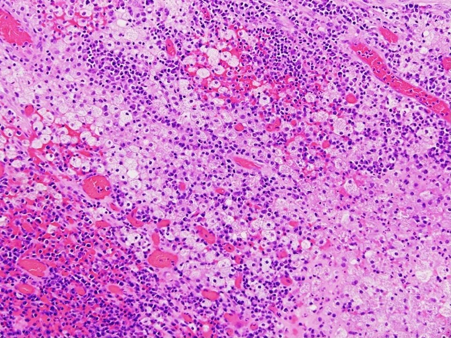
Multiple variably sized cysts and cystic follicles
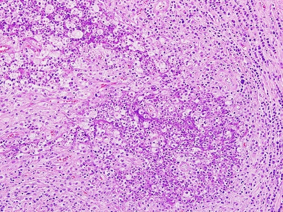
Atretic follicle with bands of luteinized theca cells
Bands of luteinized theca cells with hemorrhage
Outer cortex is
collagenized with
several follicle
cysts
Images hosted on other servers:

Peripherally located multinodular ovarian masses
Images hosted on other servers:

Bilaterally enlarged ovaries

Multicentric and bilateral tumor
Contributed by Swati Bhardwaj, M.B.B.S., M.D., Tamara Kalir, M.D., Ph.D. and AFIP images
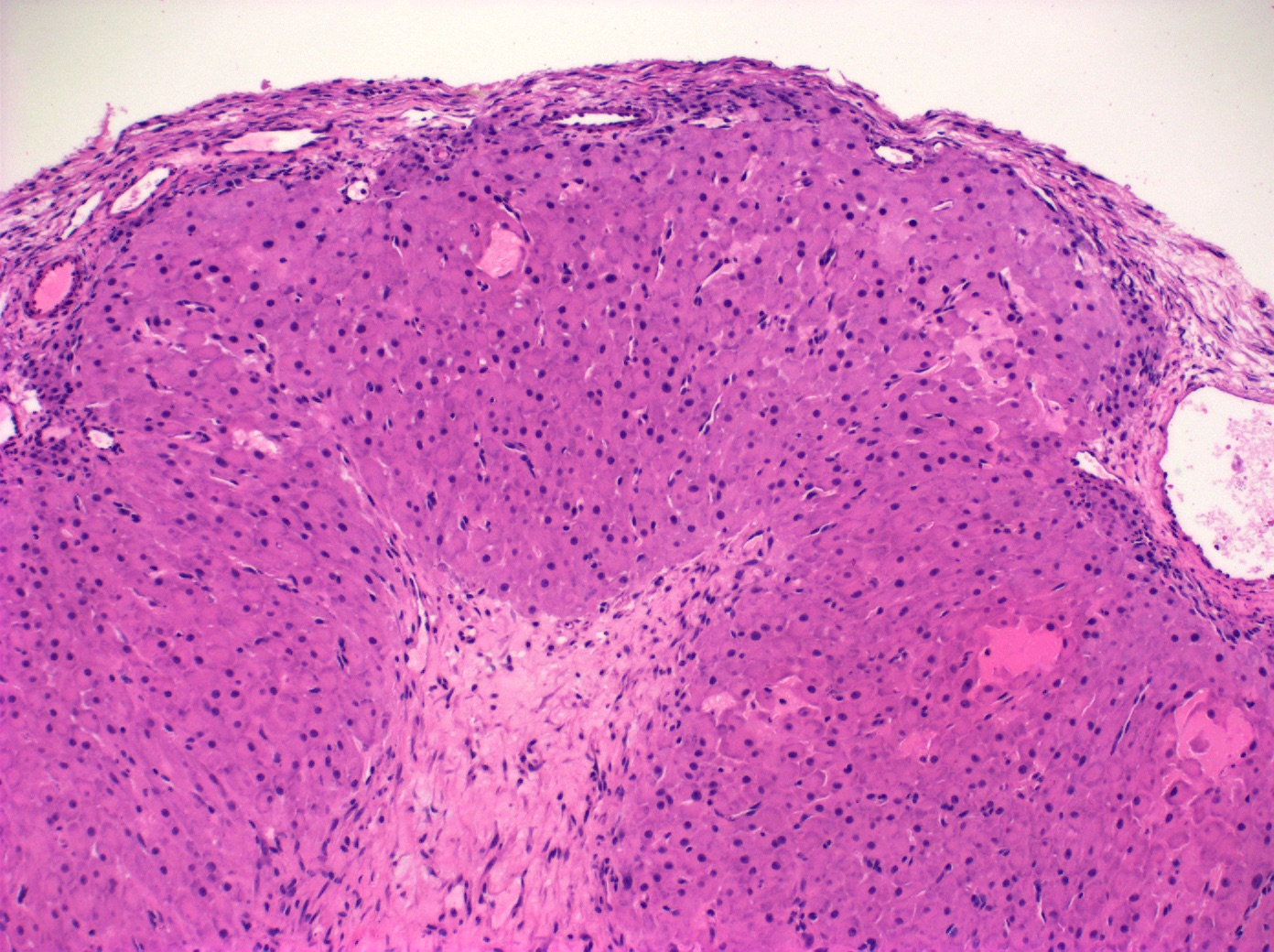
Multinodular,
well circumscribed
pregnancy
luteoma
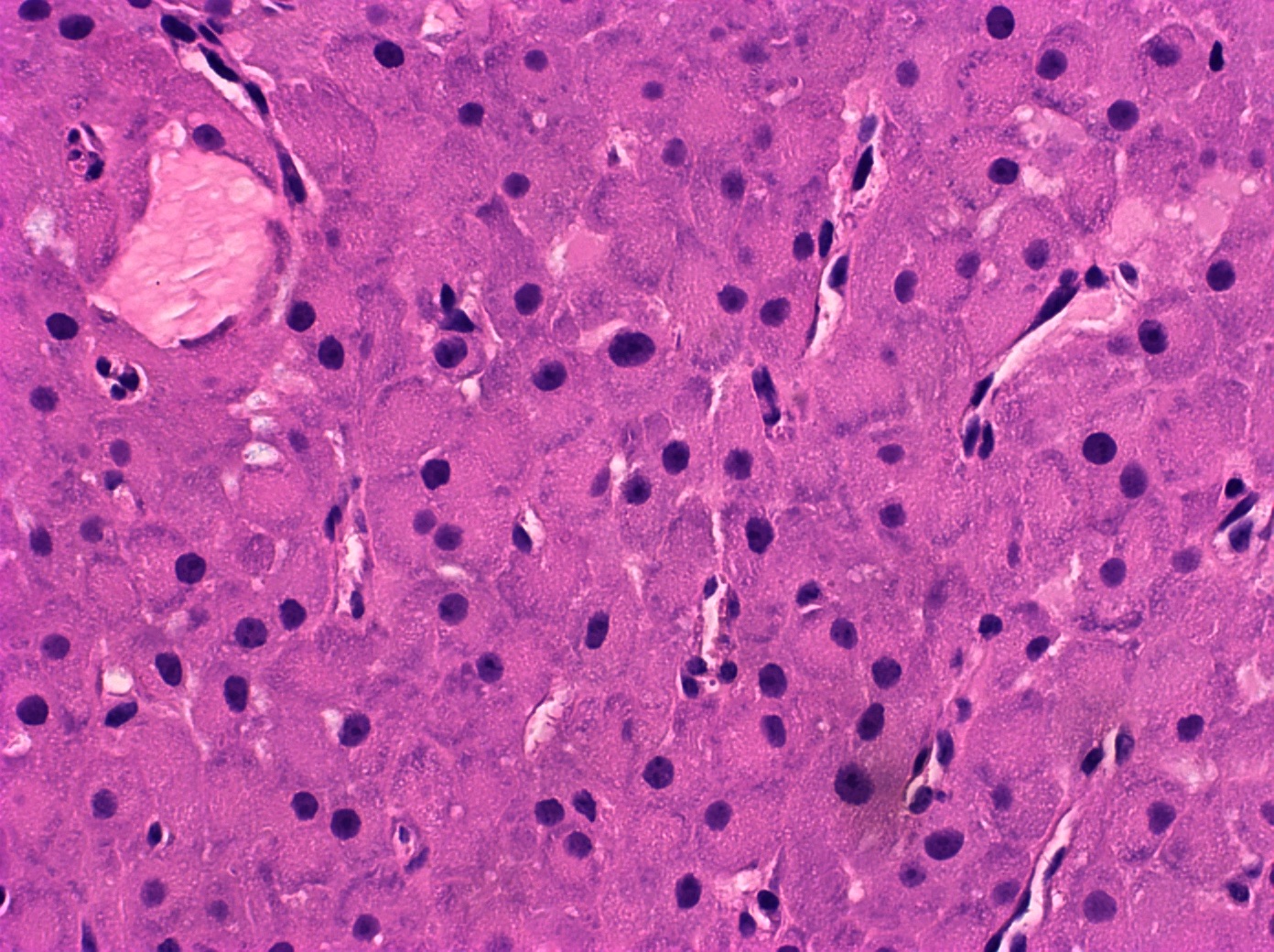
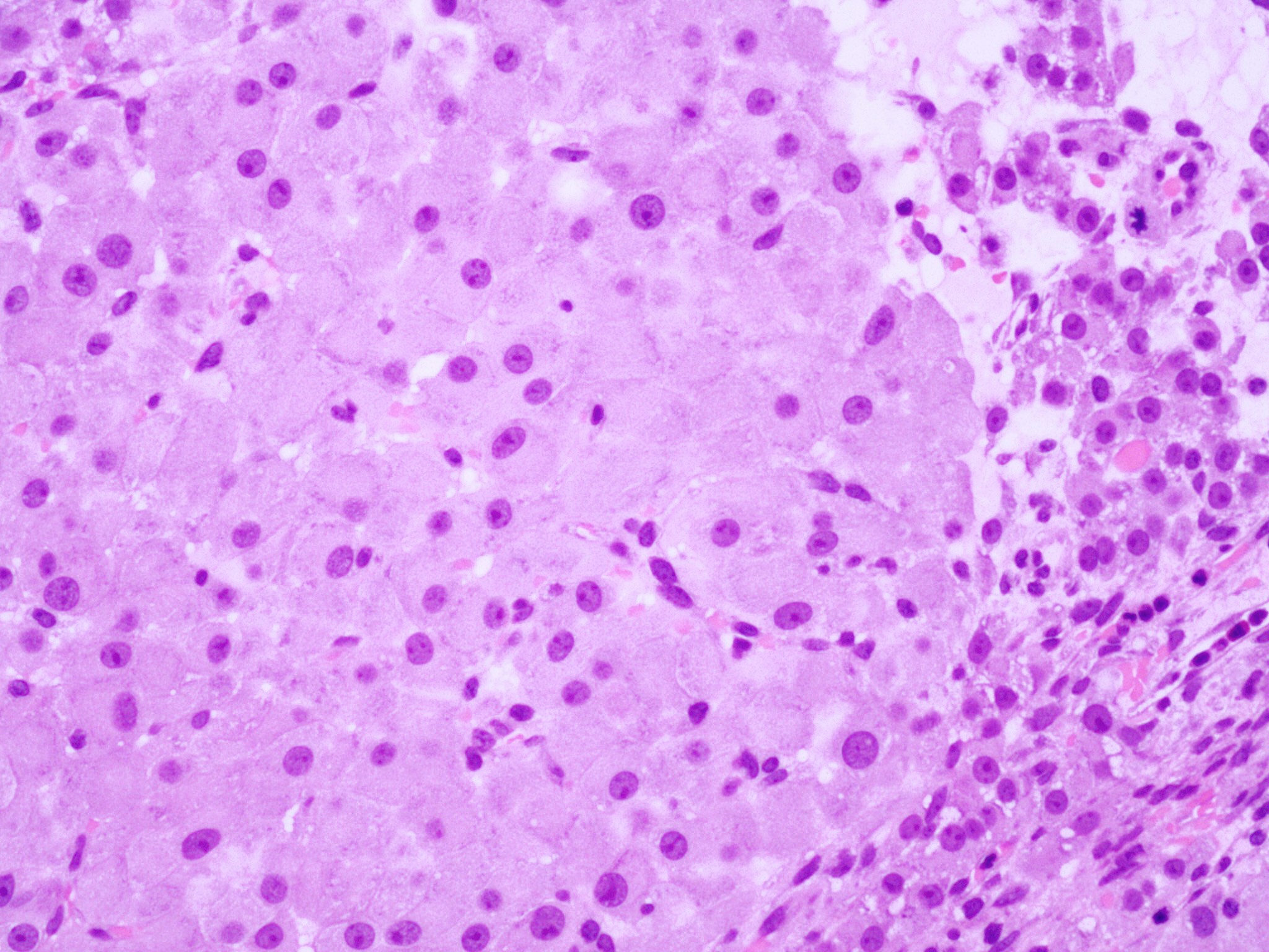
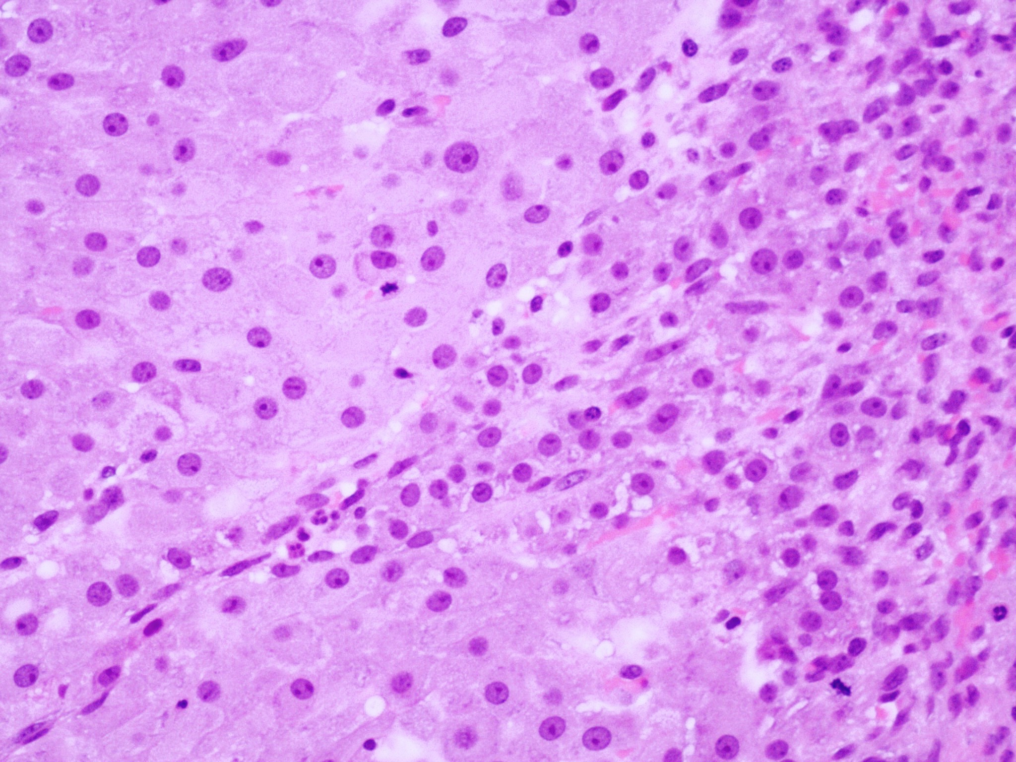
Uniform, round, eosinophilic luteoma cells

Luteoma with brisk mitoses

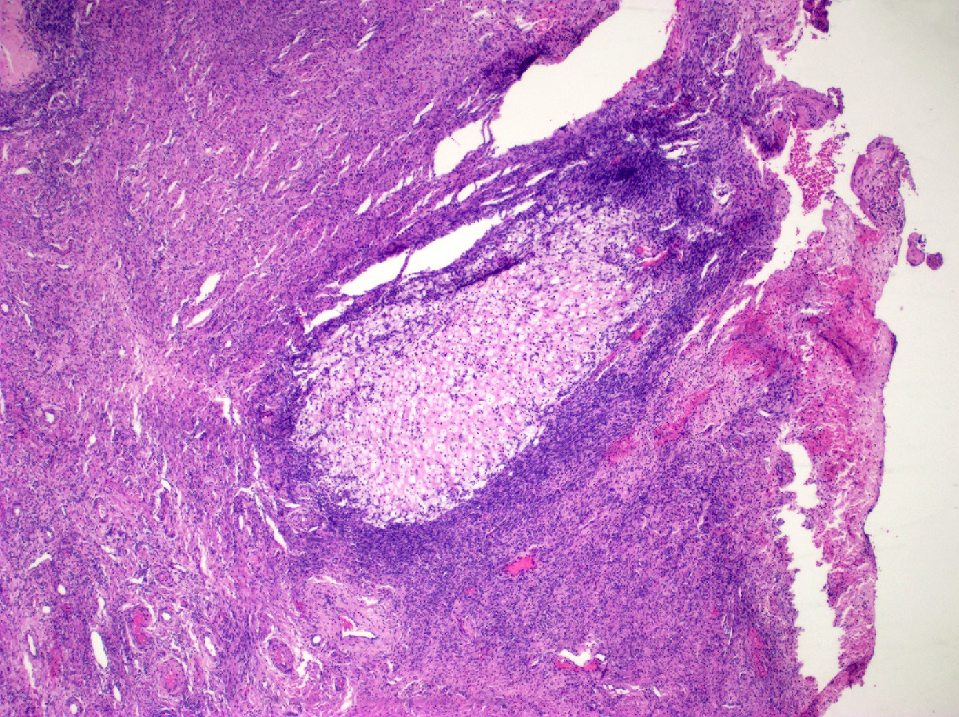
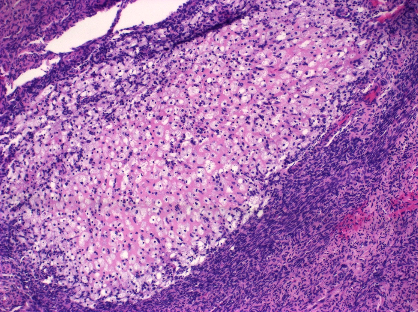
Stromal edema
Stromal edema with nested appearance of luteoma cells
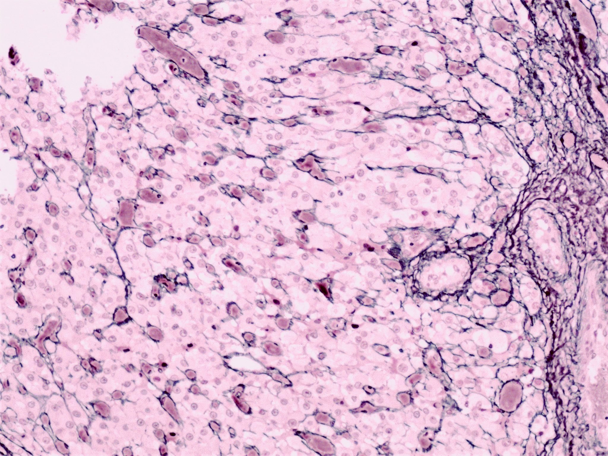
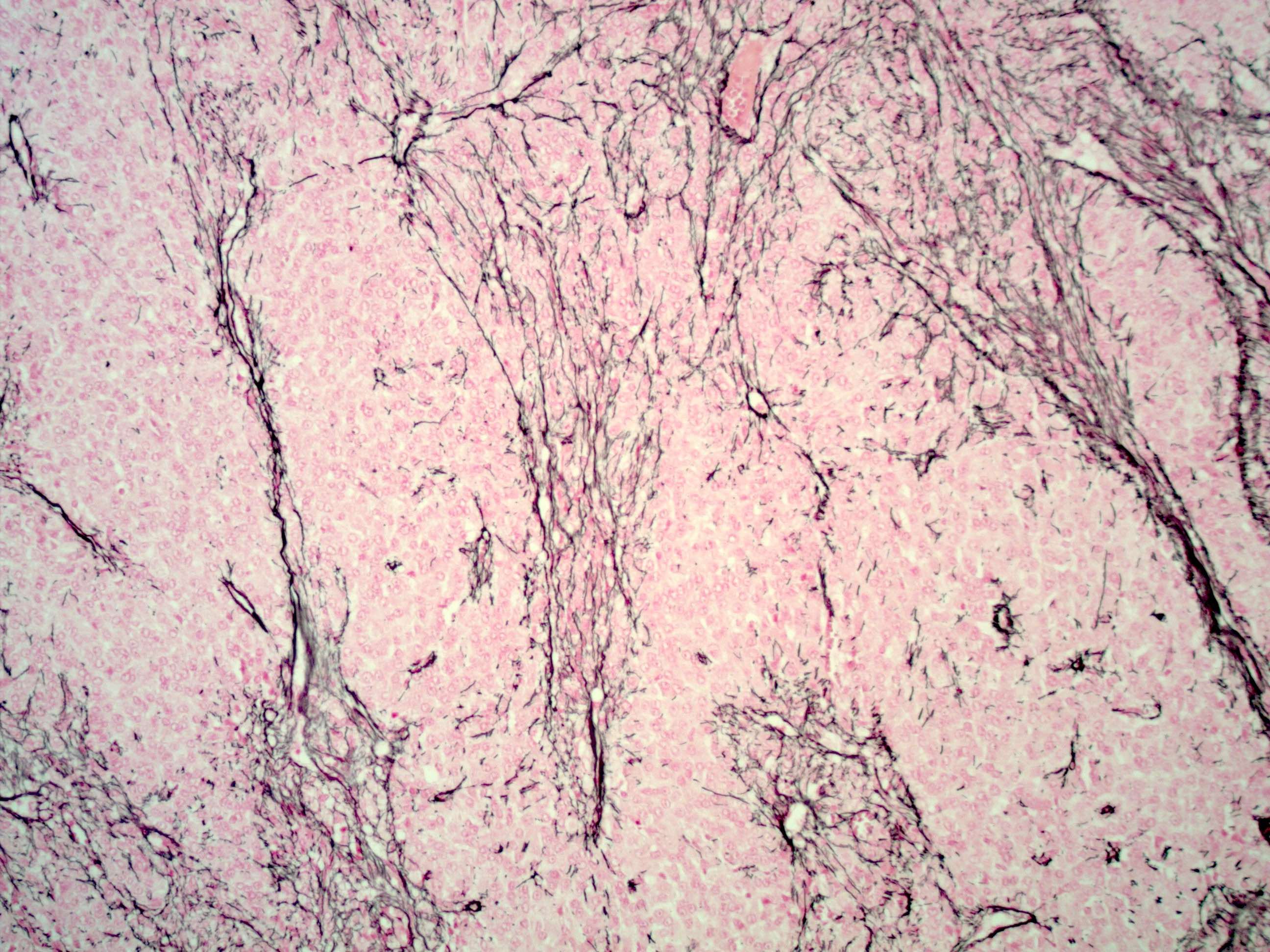
Reticular pattern
Granulosa cell proliferations adjacent to luteoma
Groups of cells surrounded by reticulin fibers
Mitoses
AFIP images
Small cyst in hilus
Numerous small crevices
Images hosted on other servers:

CT ovarian mass

MRI ovarian mass
Images hosted on other servers:

Laparoscopy

Large mass
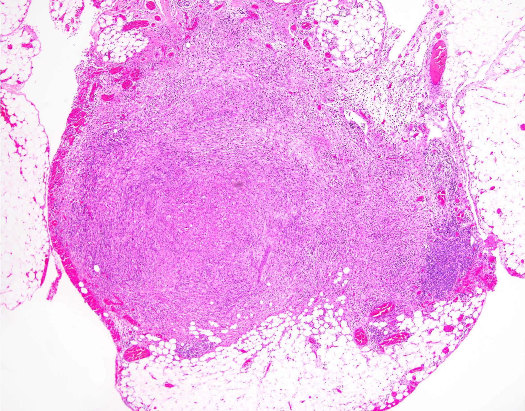
Contributed by Jennifer Bennett, M.D.
Yellow-white mass
Contributed by Jennifer Bennett, M.D.
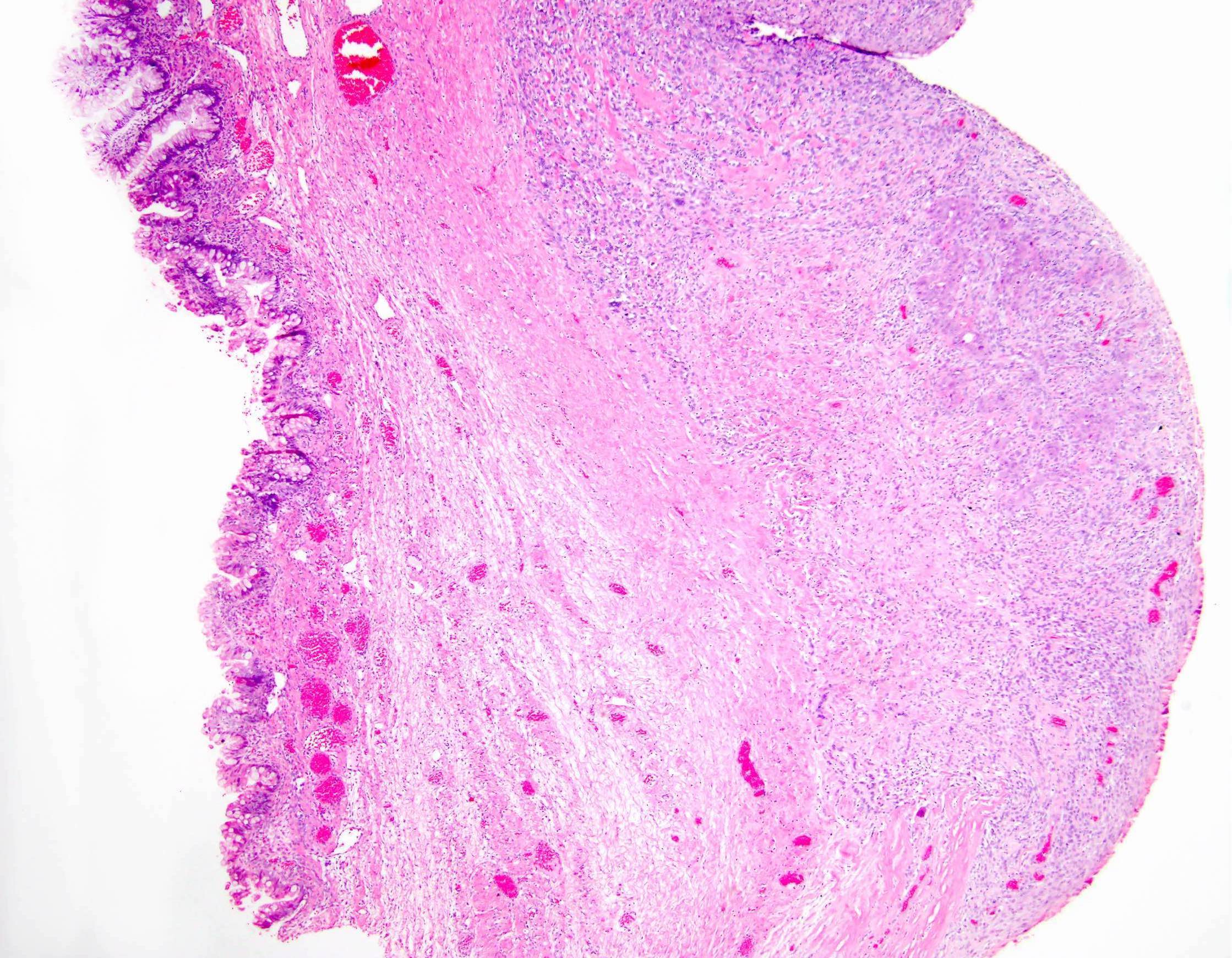
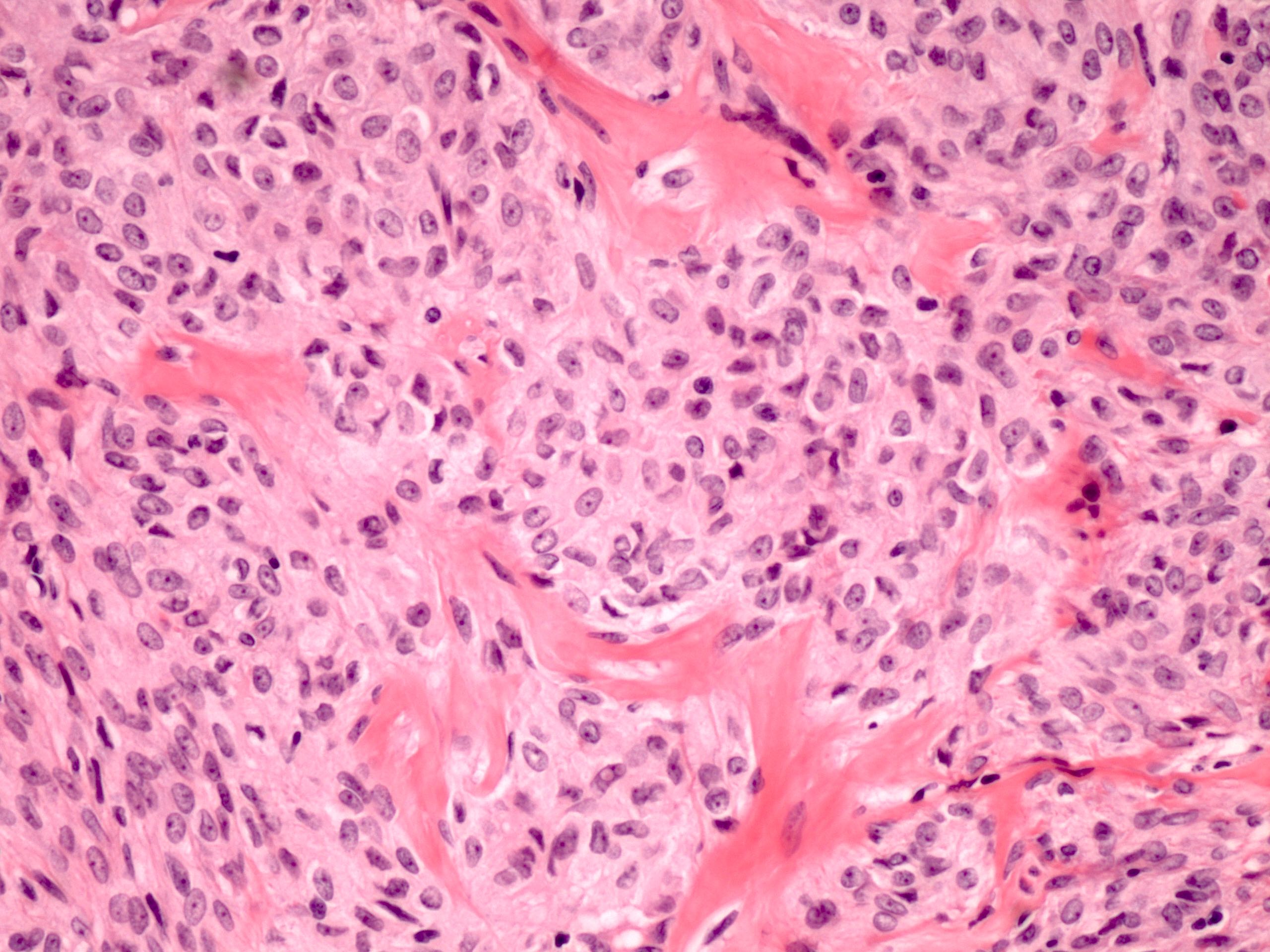
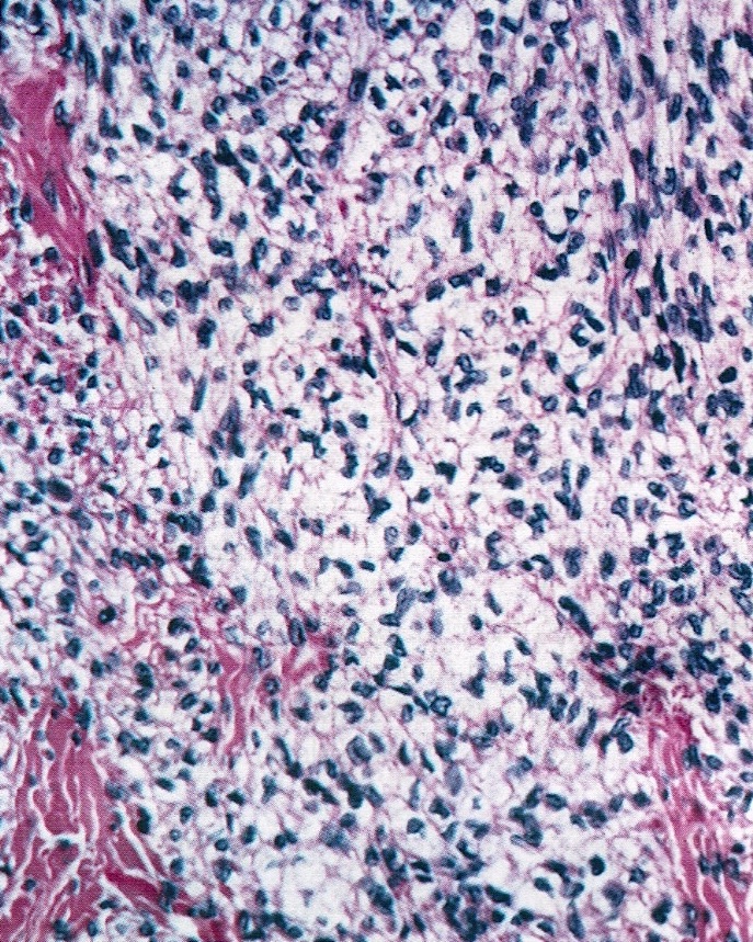
Alternating cellularity
Pseudolobule
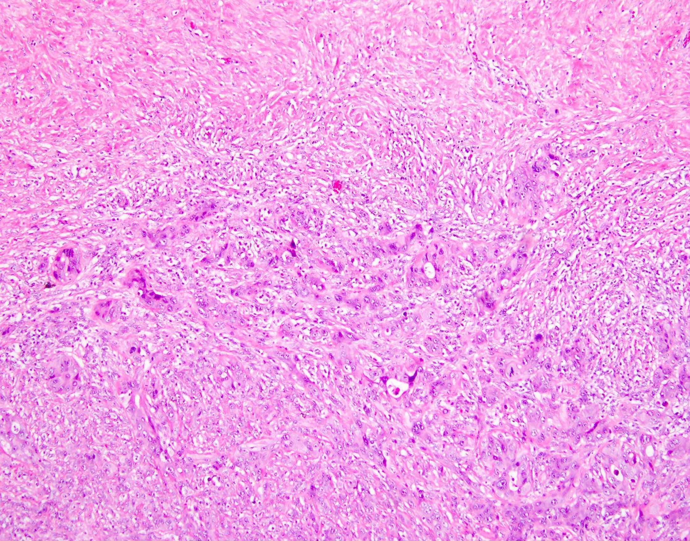
Collagenous background

Abundant eosinophilic cytoplasm
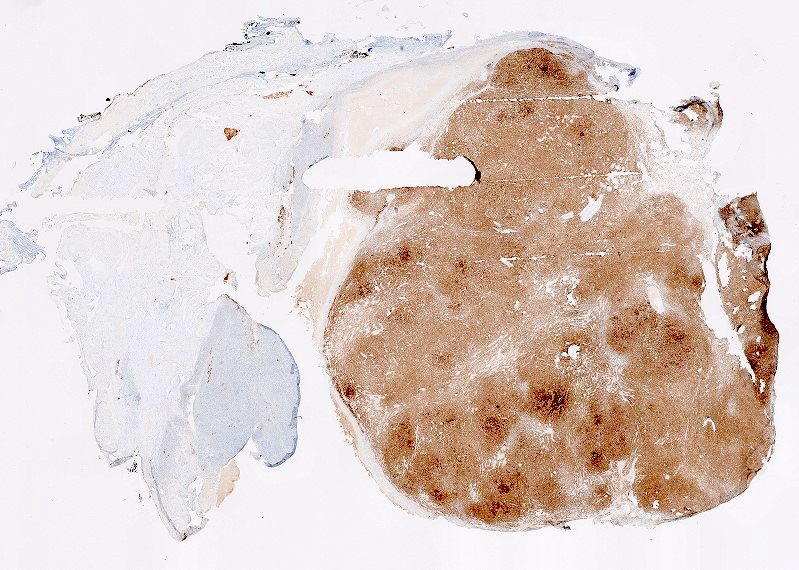
Inhibin
Contributed by Shannon Mingo Welter, M.D.
Endometriotic cyst
Squamous and endocervical
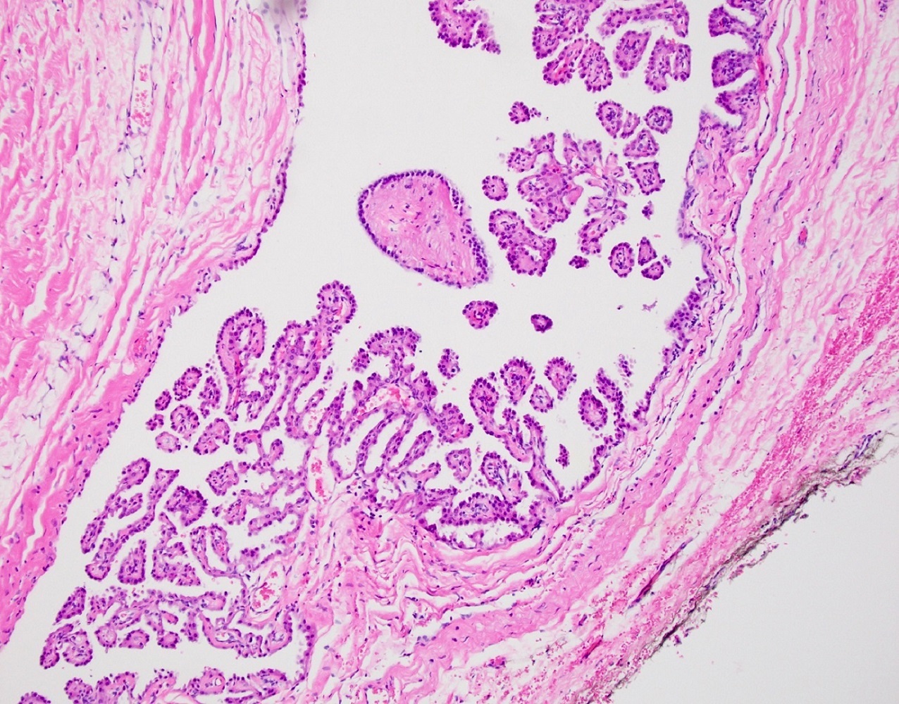
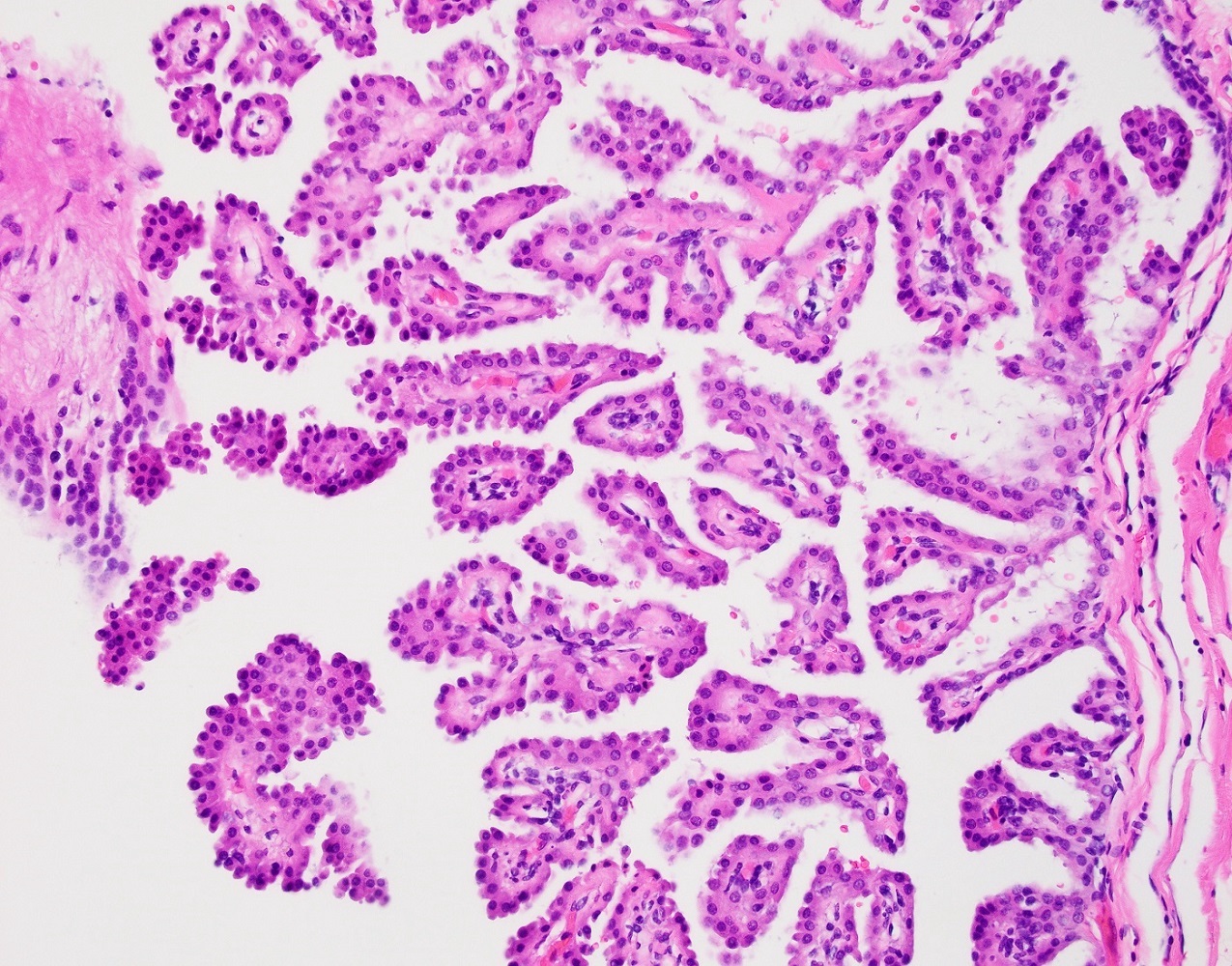
Stromal cores with edema or fibrosis
Papillary tumor
Fibrous cores with inflammation
Variable cell types
Hobnail and endocervical
Endocervical cell lining
Nondescript eosinophilic cells
Ovary tumors associated with endometriosis
Contributed by Gulisa Turashvili, M.D., Ph.D.
Cyst wall lined by simple epithelium
Cyst wall lined by simple epithelium consisting of serous type ciliated cells admixed with endocervical type mucinous cells
Contributed by Aarti Sharma M.D. and Ricardo R. Lastra, M.D.
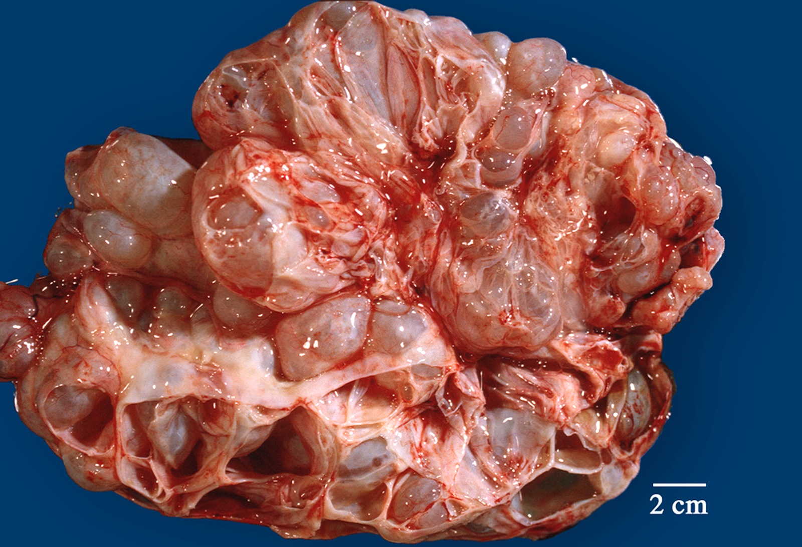
26 cm mass
Contributed by Aarti Sharma M.D. and Ricardo R. Lastra, M.D.
Papillary clusters

Excrescences
Contributed by Aarti Sharma M.D., Ricardo R. Lastra, M.D. and Carlos Parra-Herran, M.D.
Conventional SBT
Micropapillary SBT
Lymph node involvement
Noninvasive implants
Microinvasion
Contributed by Aarti Sharma M.D. and Ricardo R. Lastra, M.D.
Clusters of cells with irregular contours

High N/C ratio in clusters
Images hosted on other servers:


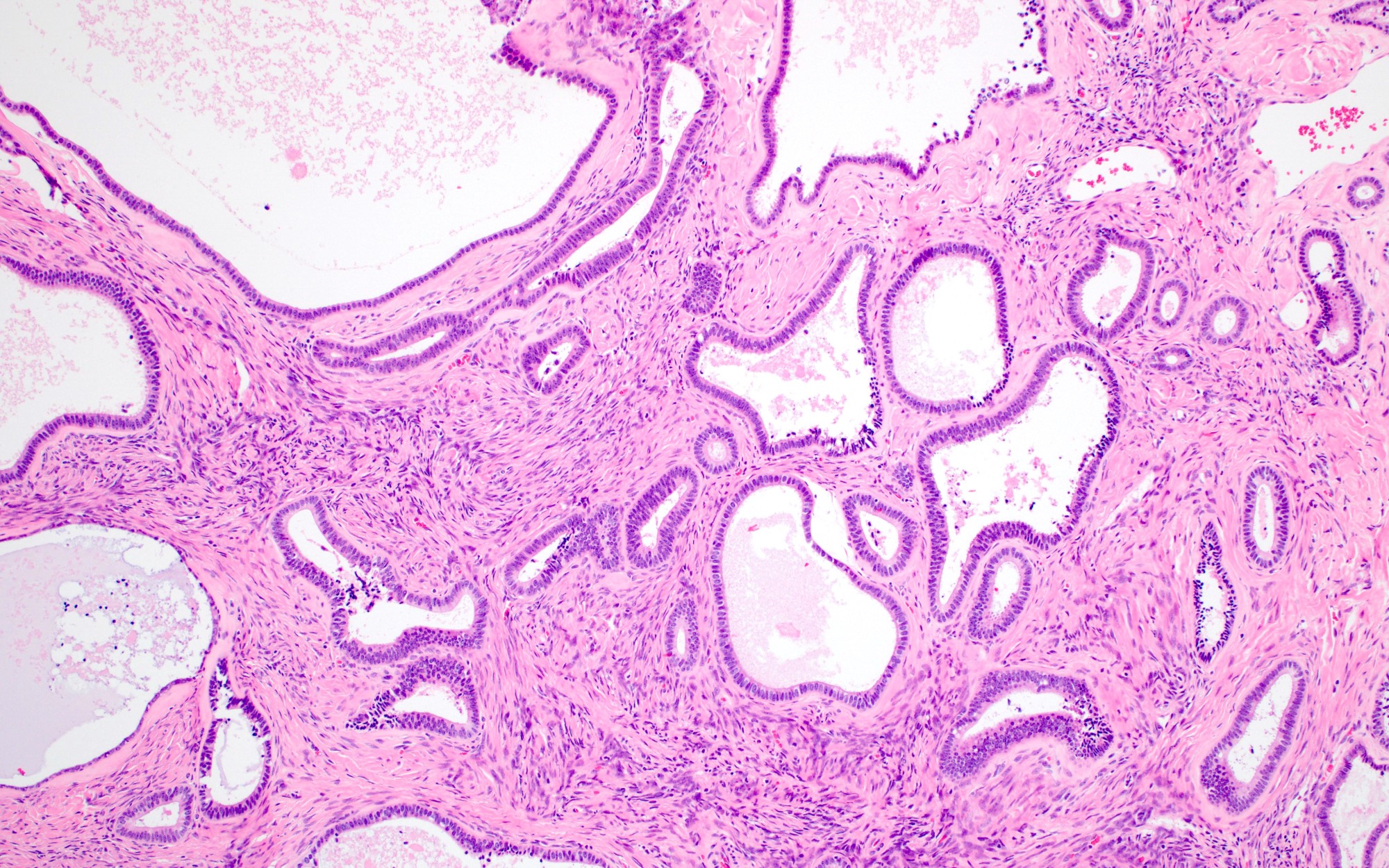
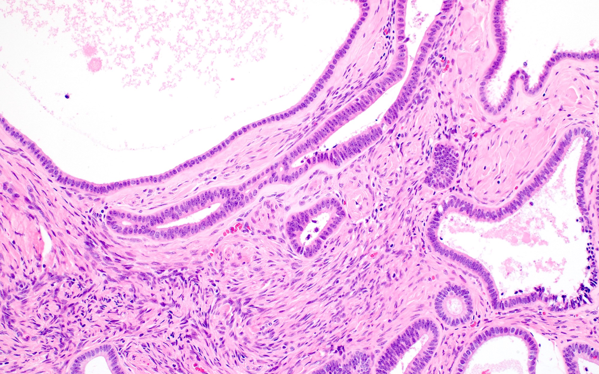
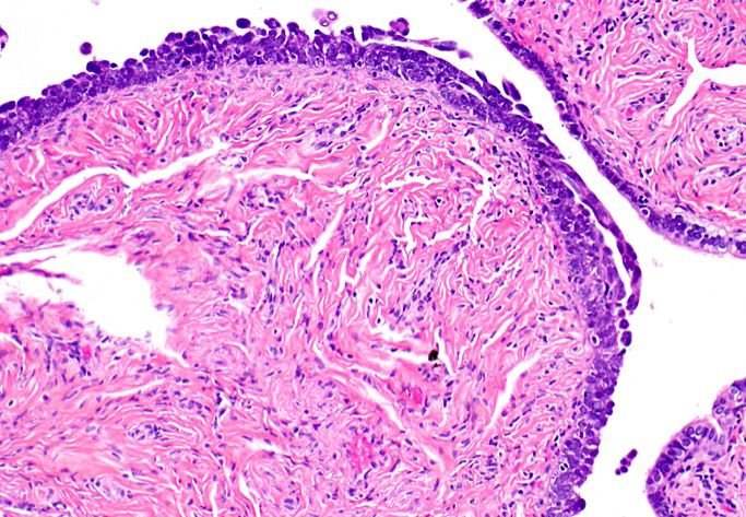
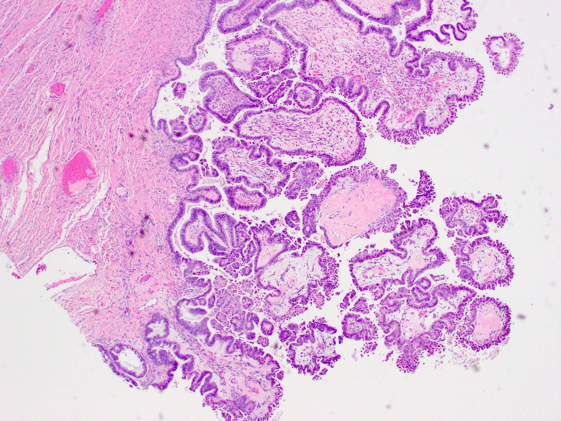
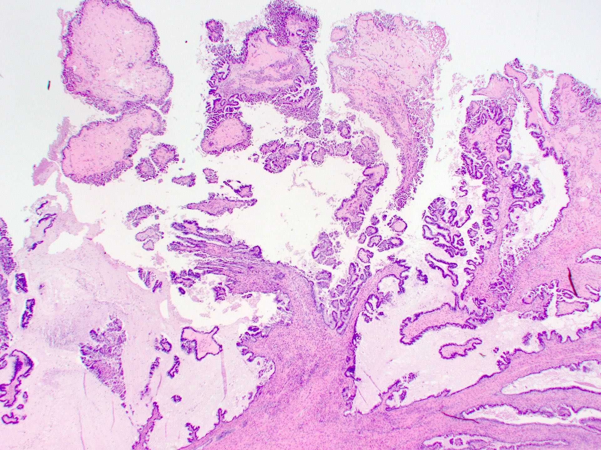
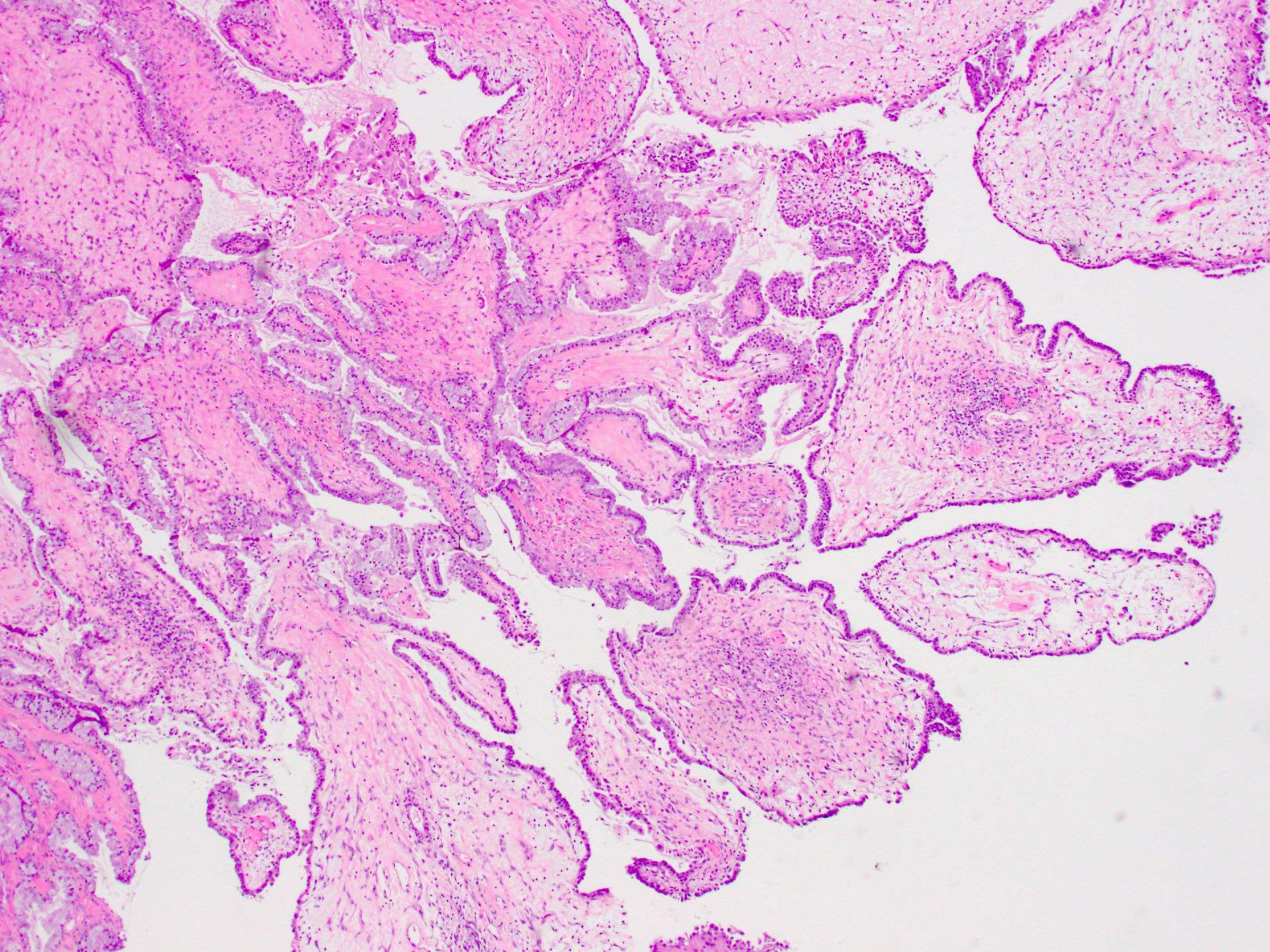
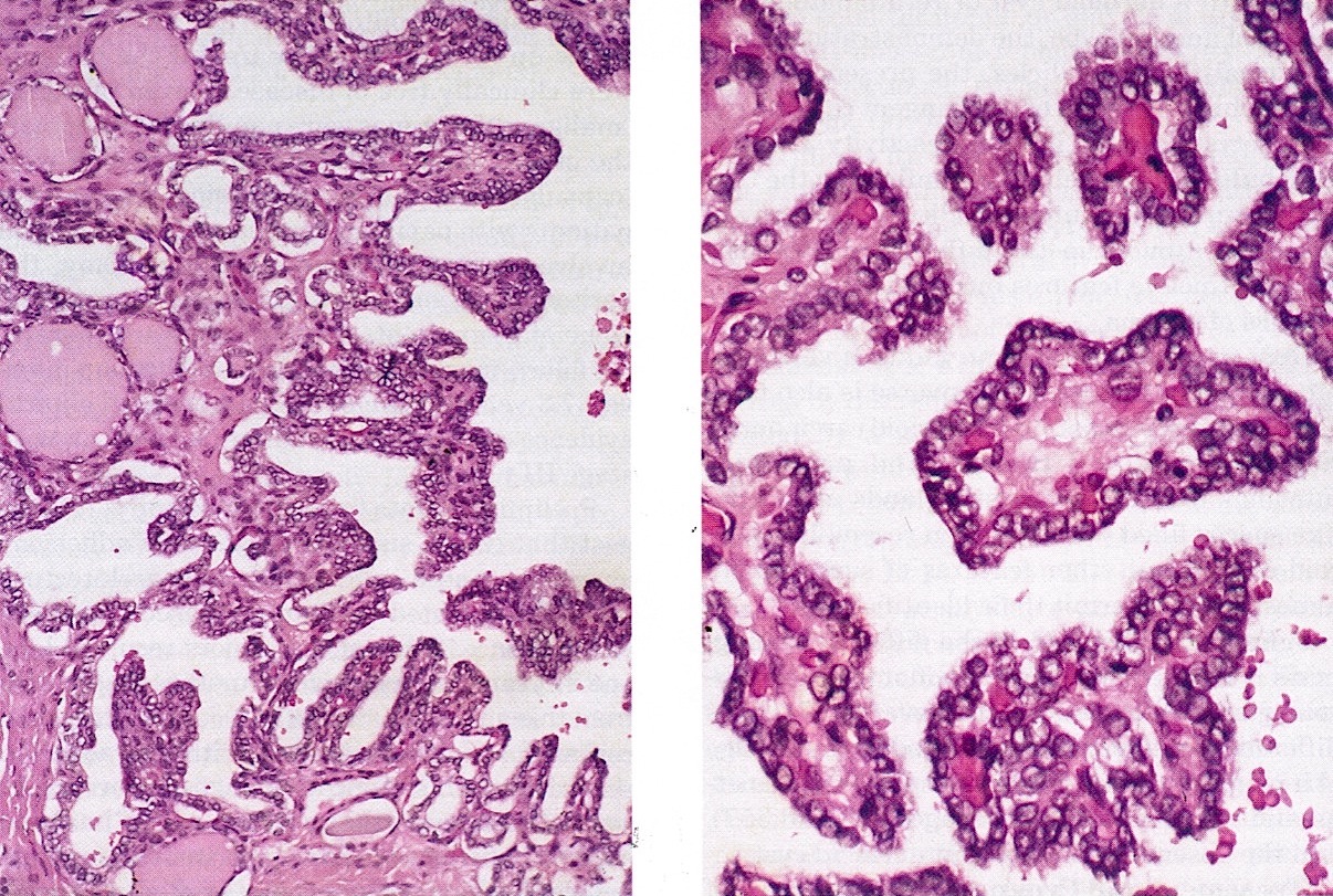
Serous cystadenoma

Cystadenofibroma
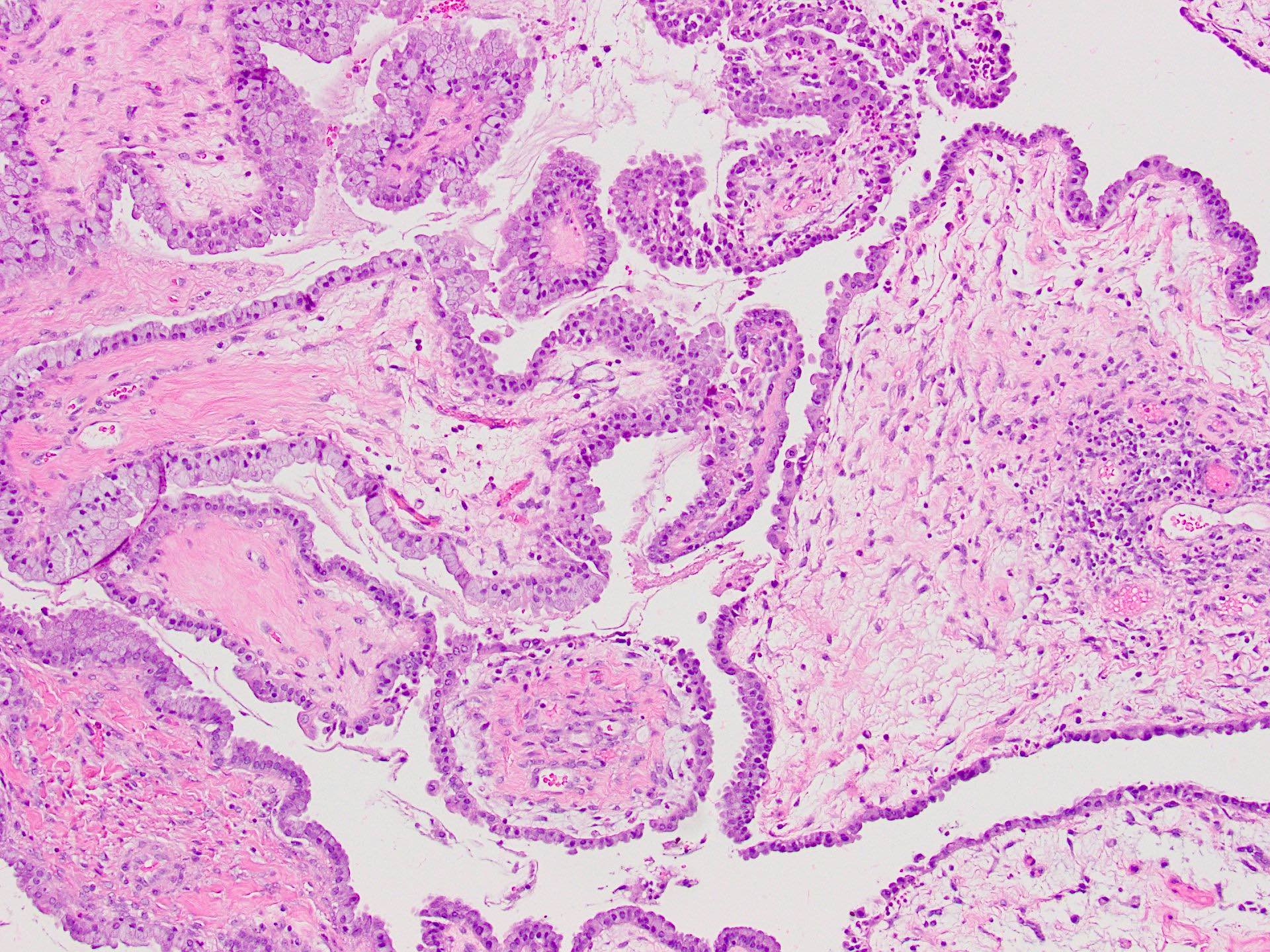
Contributed by Catherine J. Roe, M.D. and Krisztina Hanley, M.D.
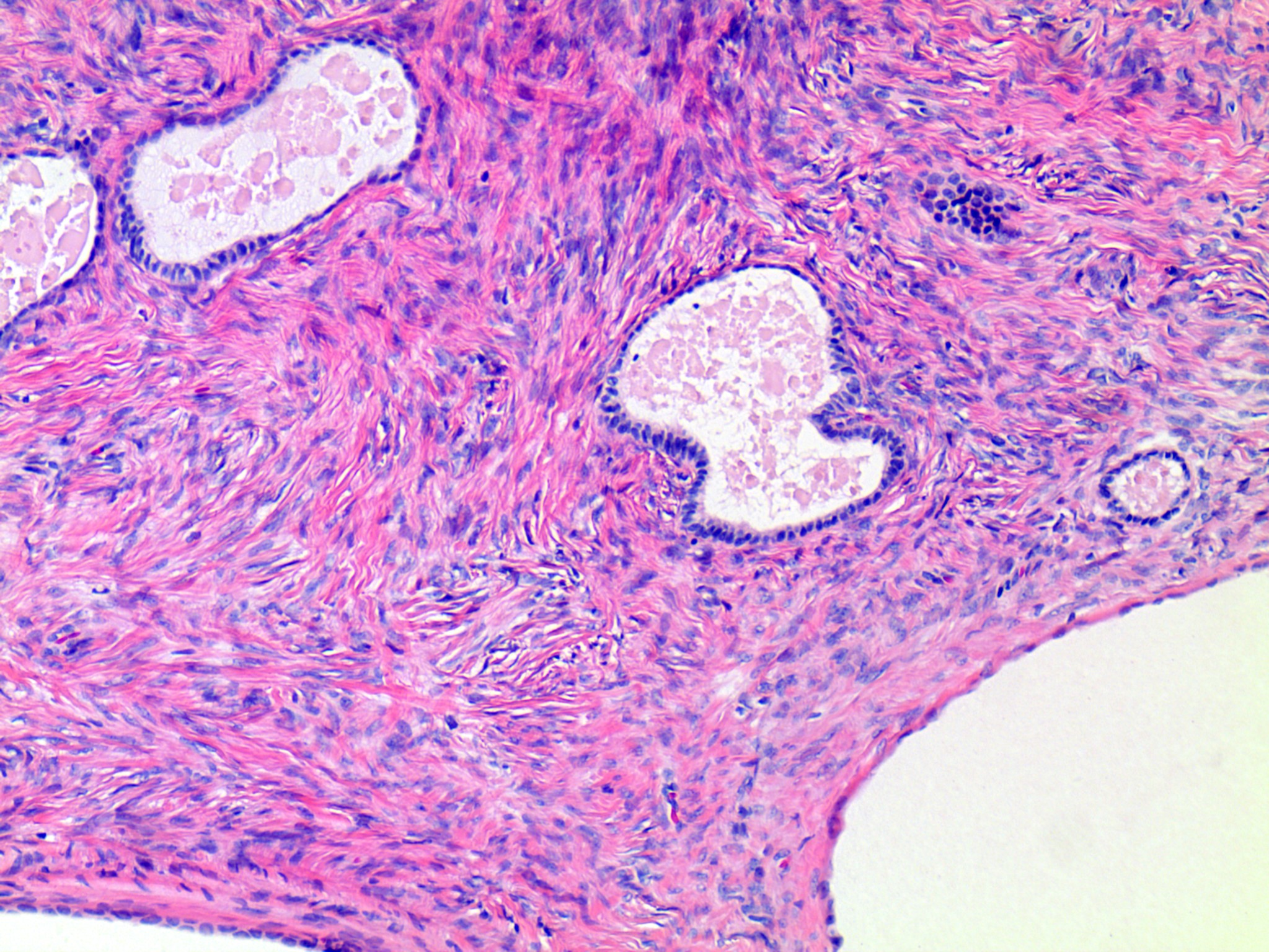
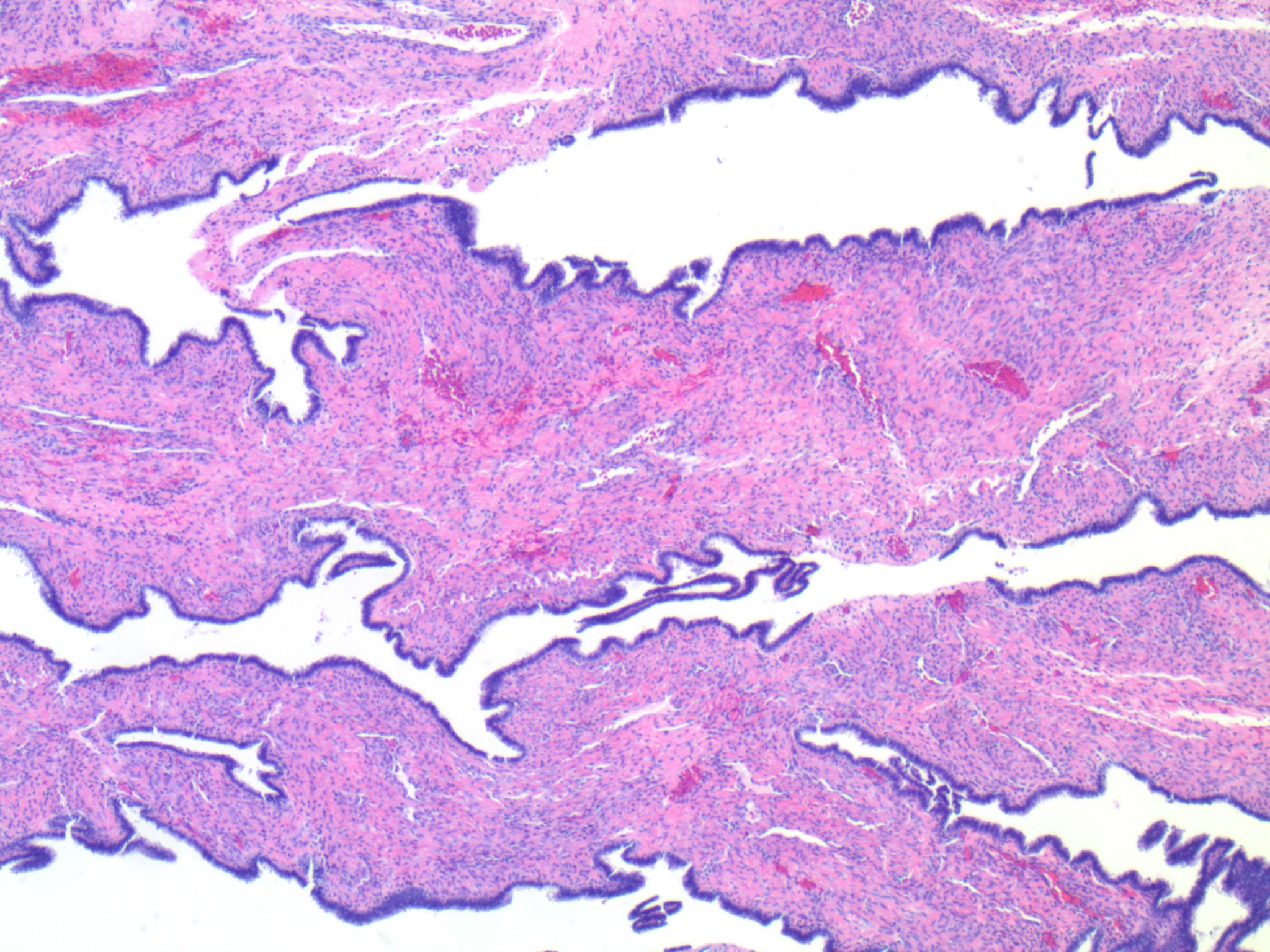

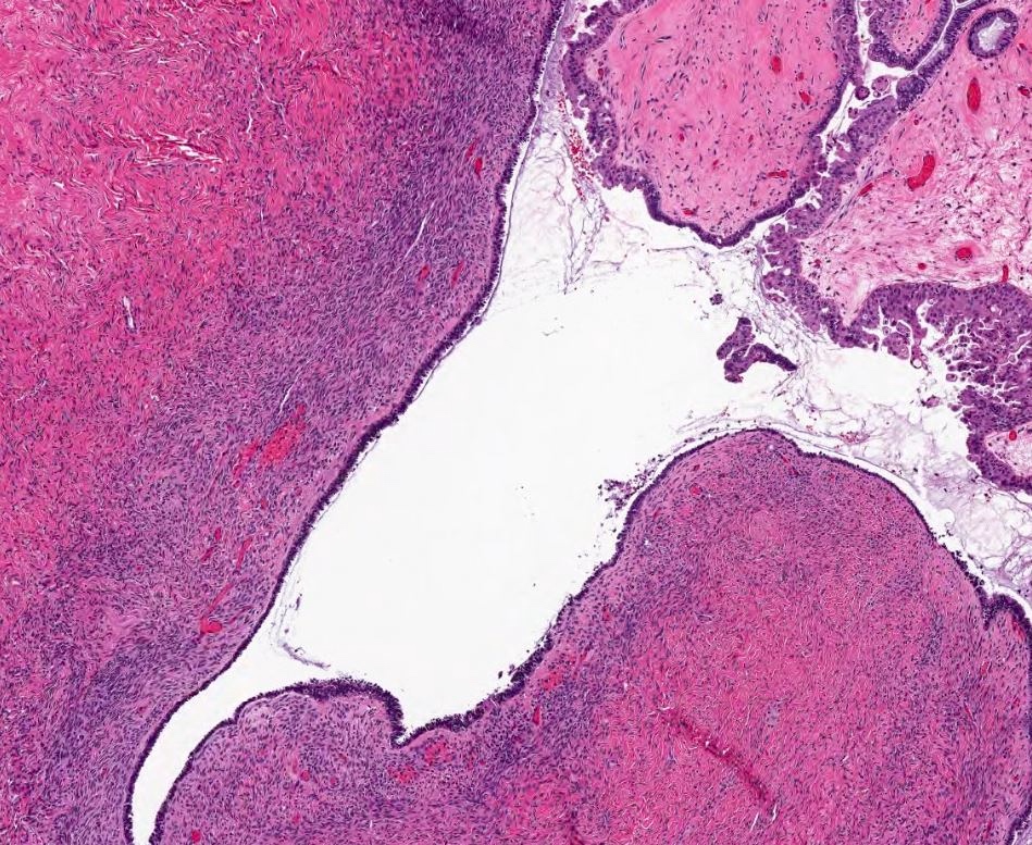
Serous cystadenofibroma
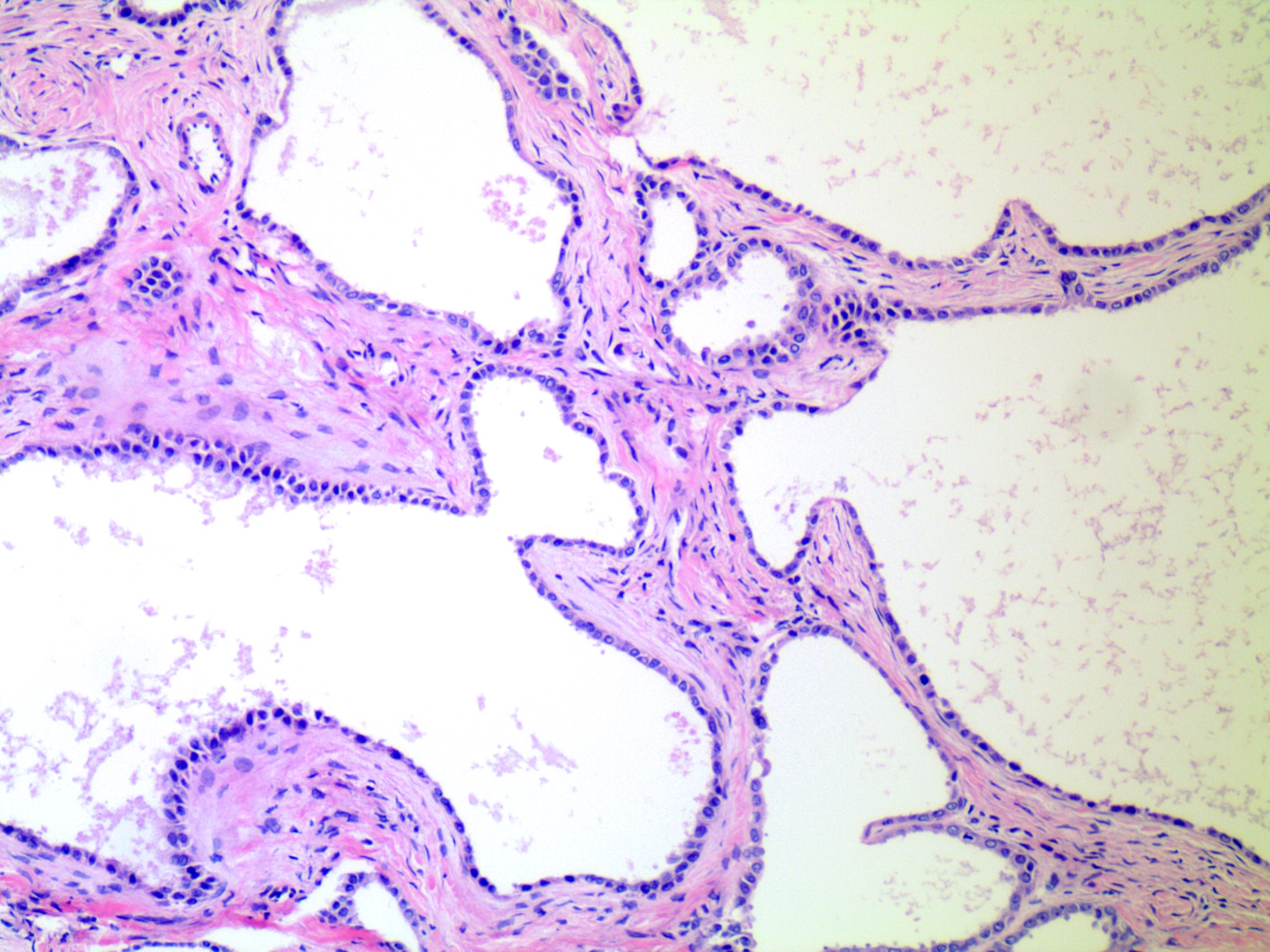
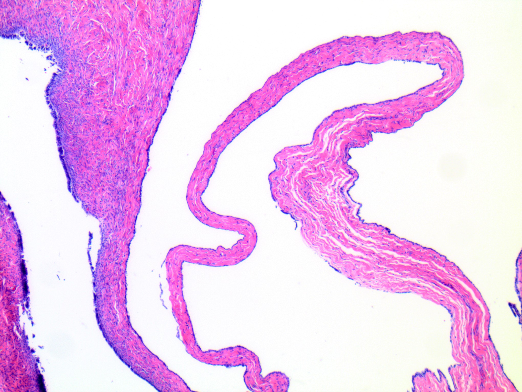
Serous cystadenoma
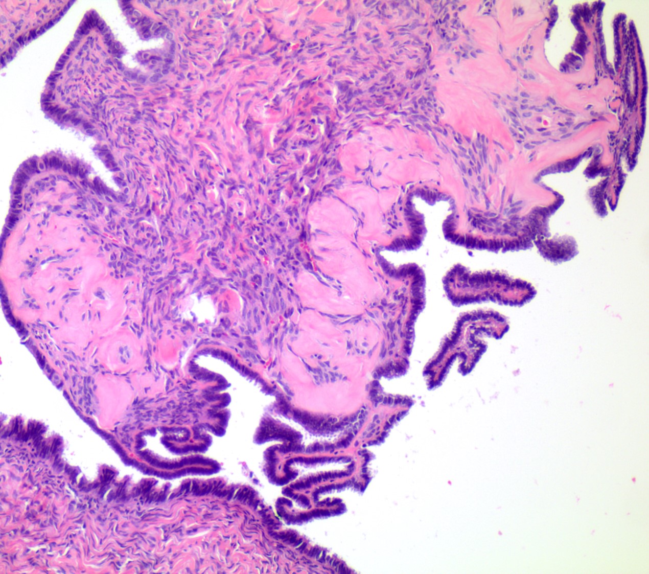
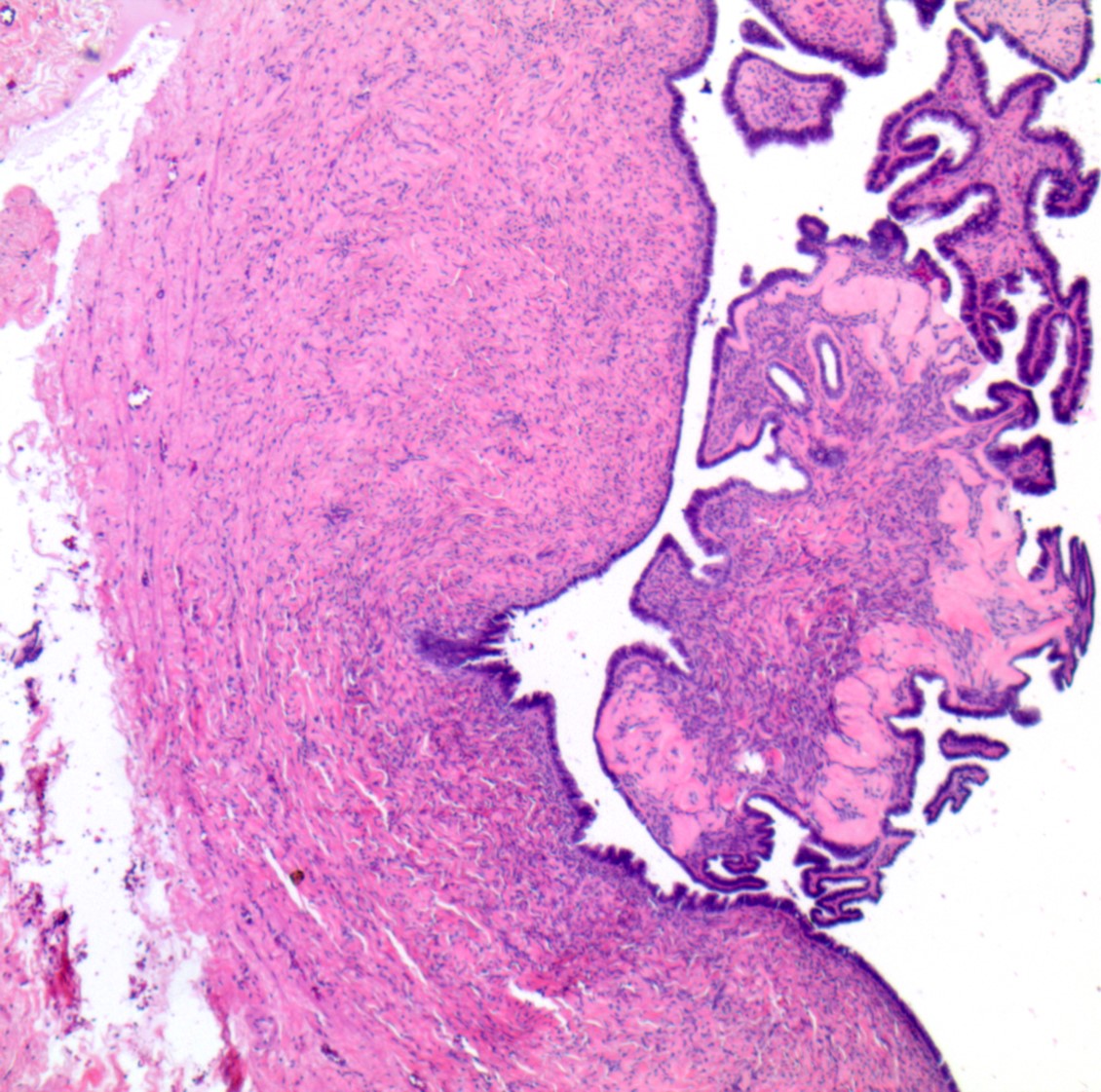
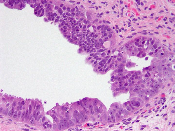
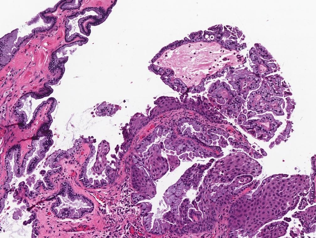
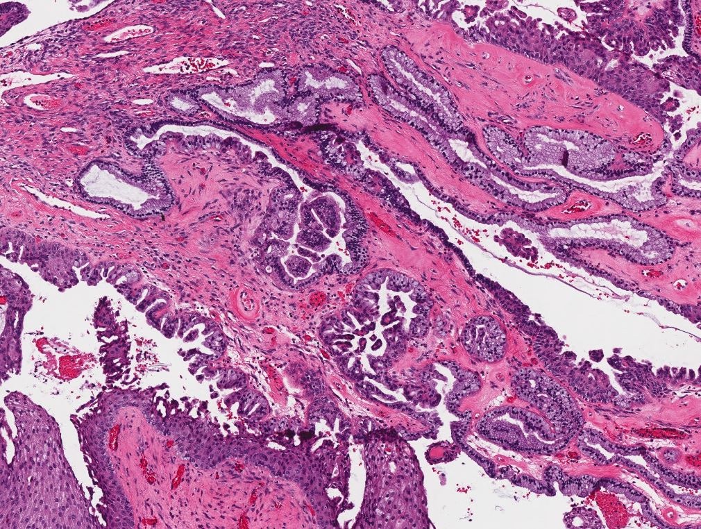
Serous cystadenoma with focal epithelial proliferation
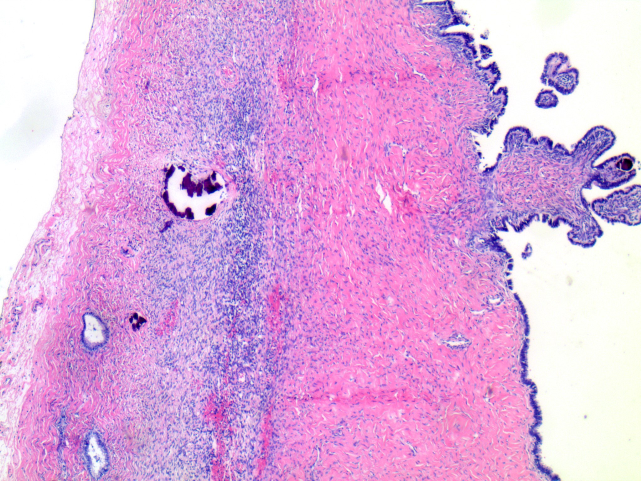
Papillary tufting

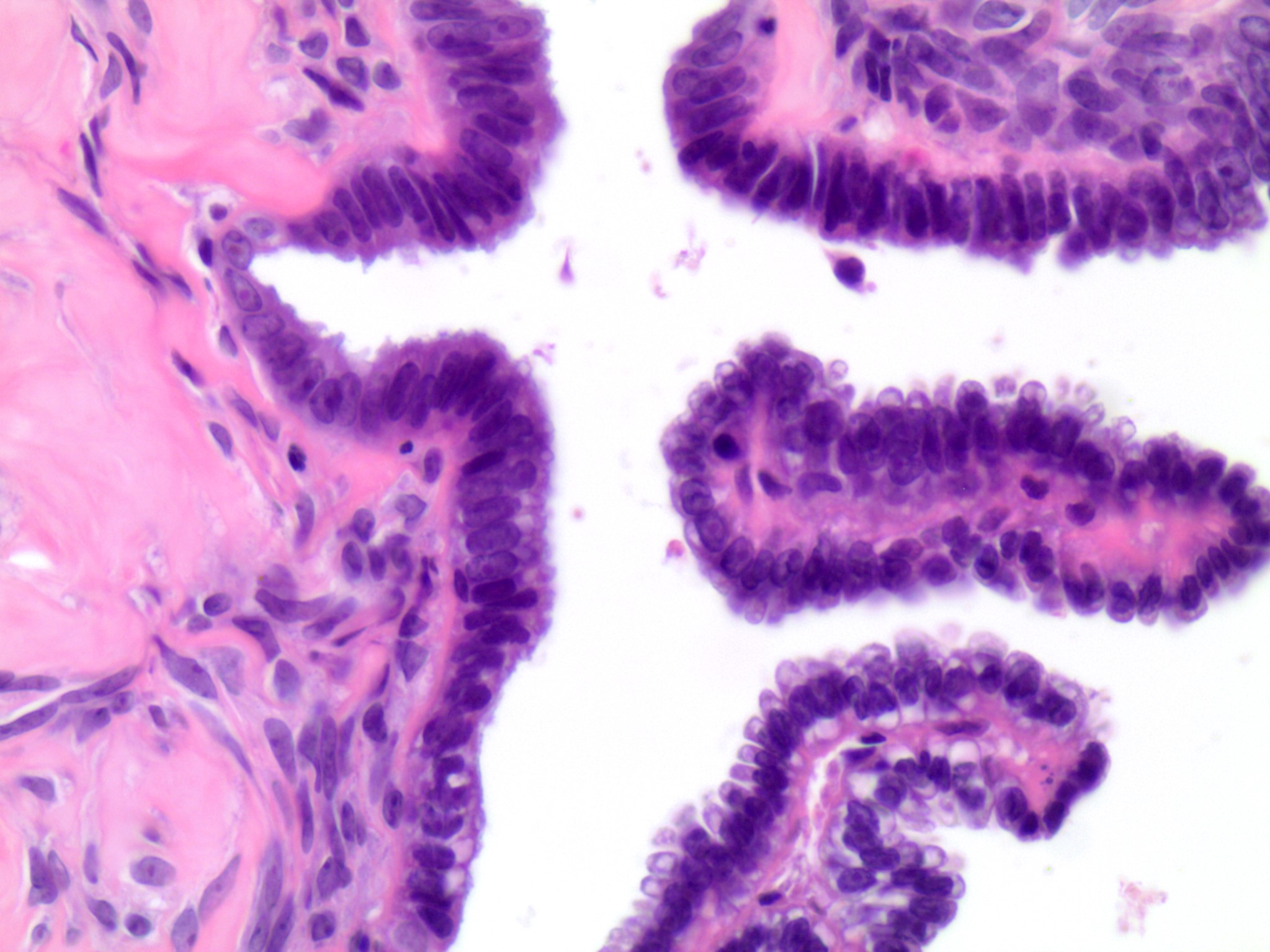
Cytologic features
Focal epithelial proliferation
Images hosted on other servers:

Small epithelial cell cluster

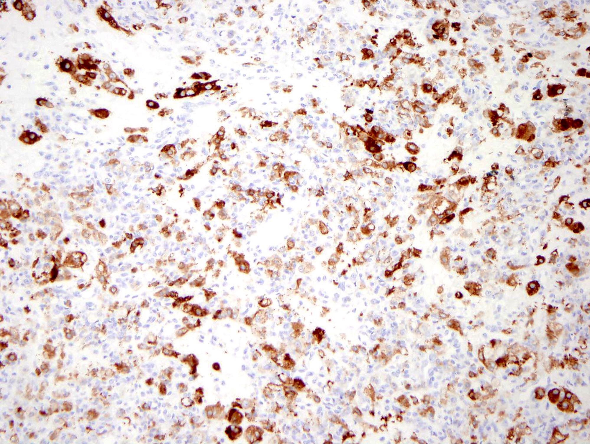
CD68
Contributed by Mahmoud A. Khalifa, M.D., Ph.D.
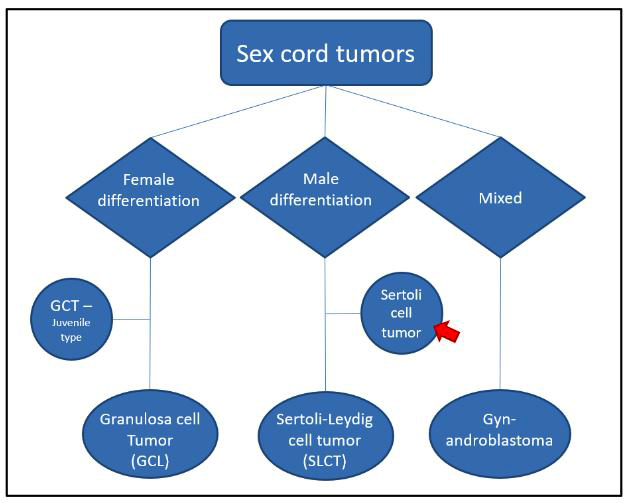
Sex cord classification
AFIP images
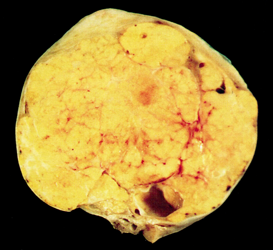
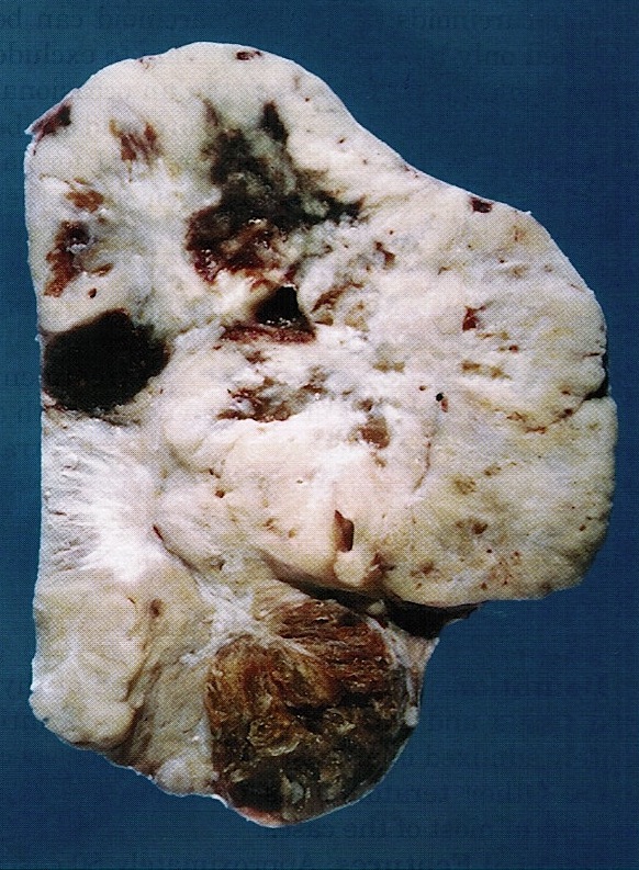
Yellow and lobulated
Contributed by Shannon Mingo Welter, M.D.
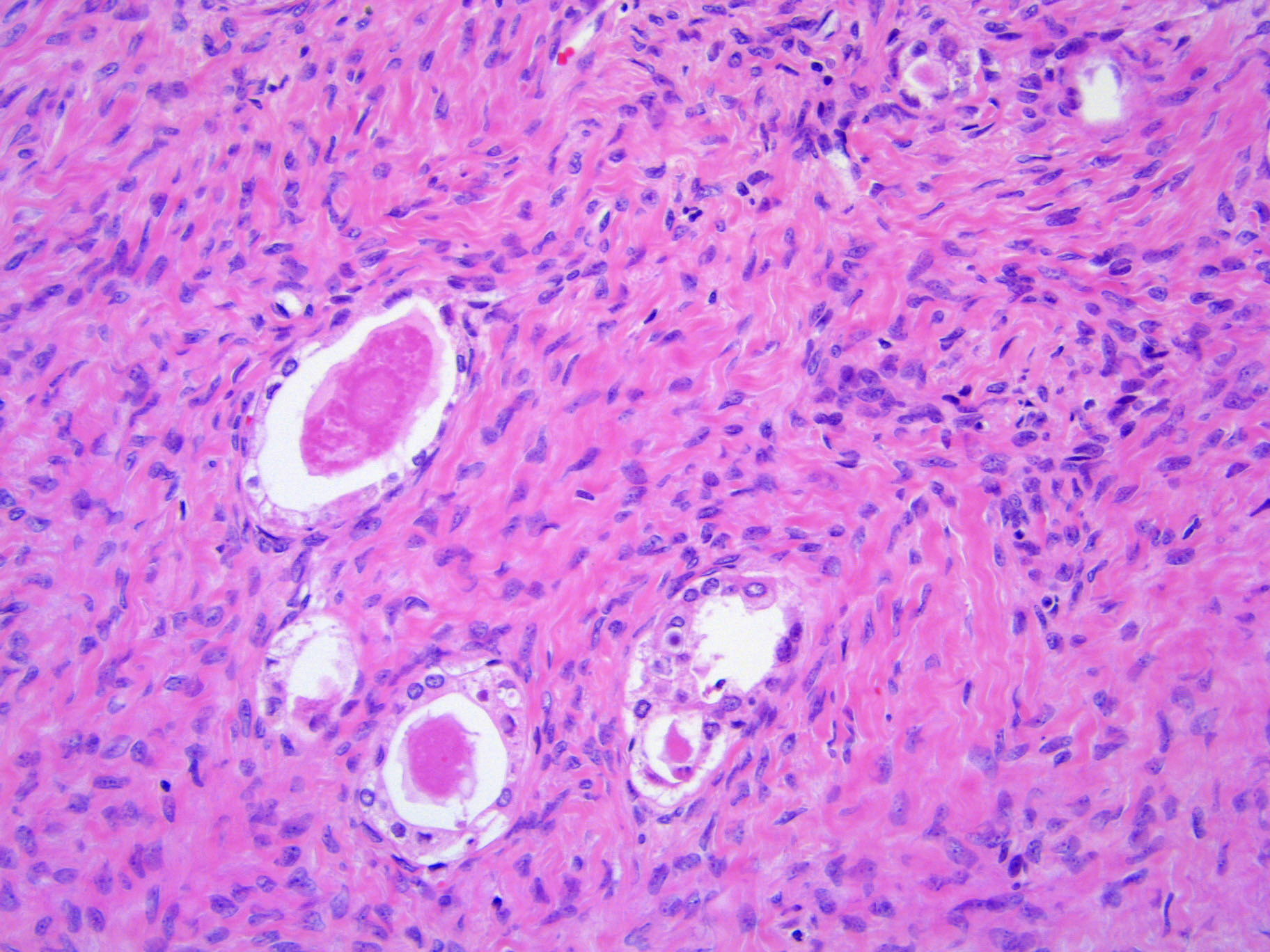
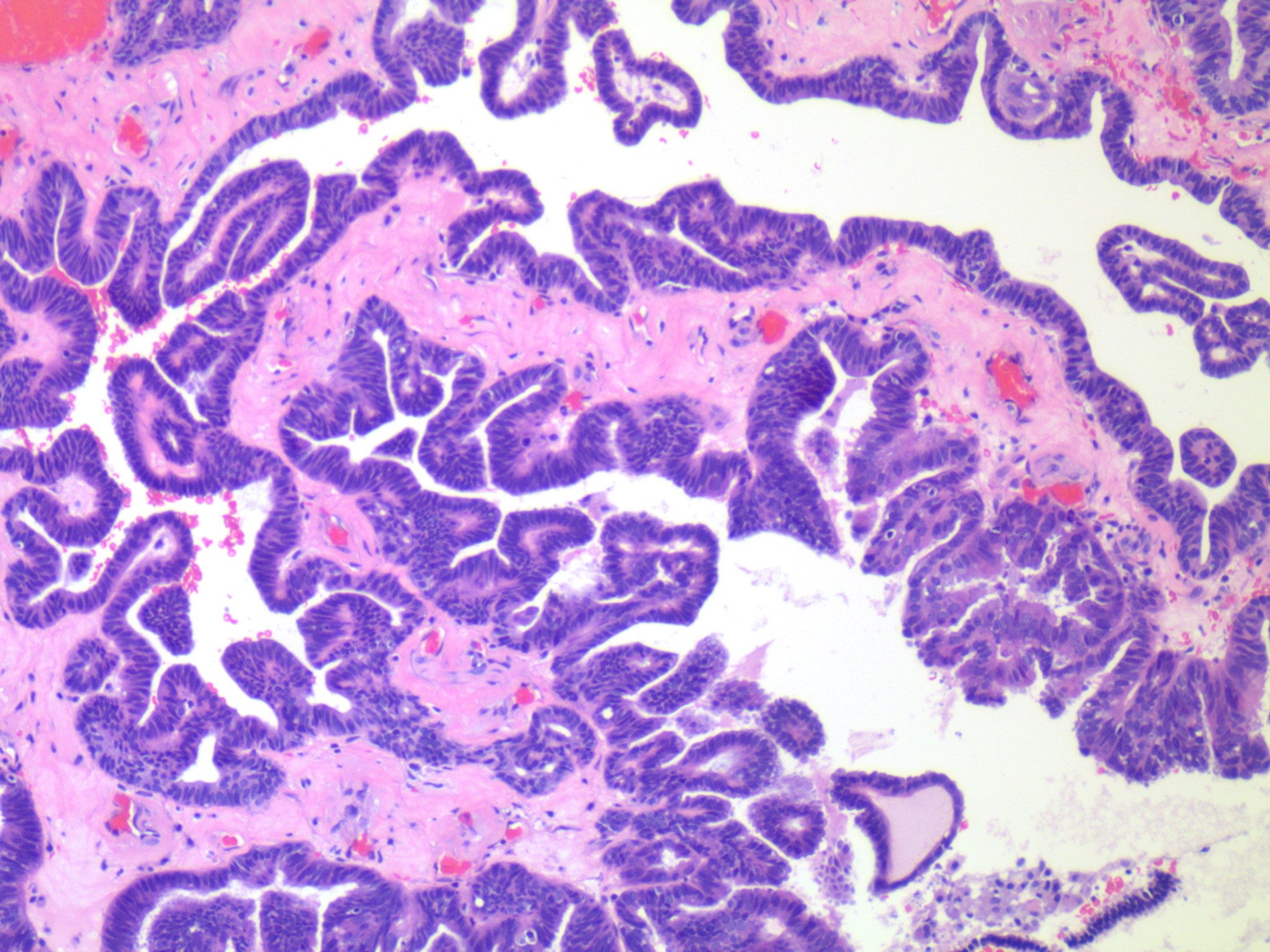
Hollow and solid tubules
Tubules with hyalinized stroma
Tubular and trabecular
Trabecular
Diffuse with tubules
Mixed patterns
Images hosted on other servers:

Large tumor
Contributed by Natalia Buza, M.D.
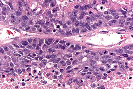
Intermediate grade tumor
Contributed by Natalia Buza, M.D.
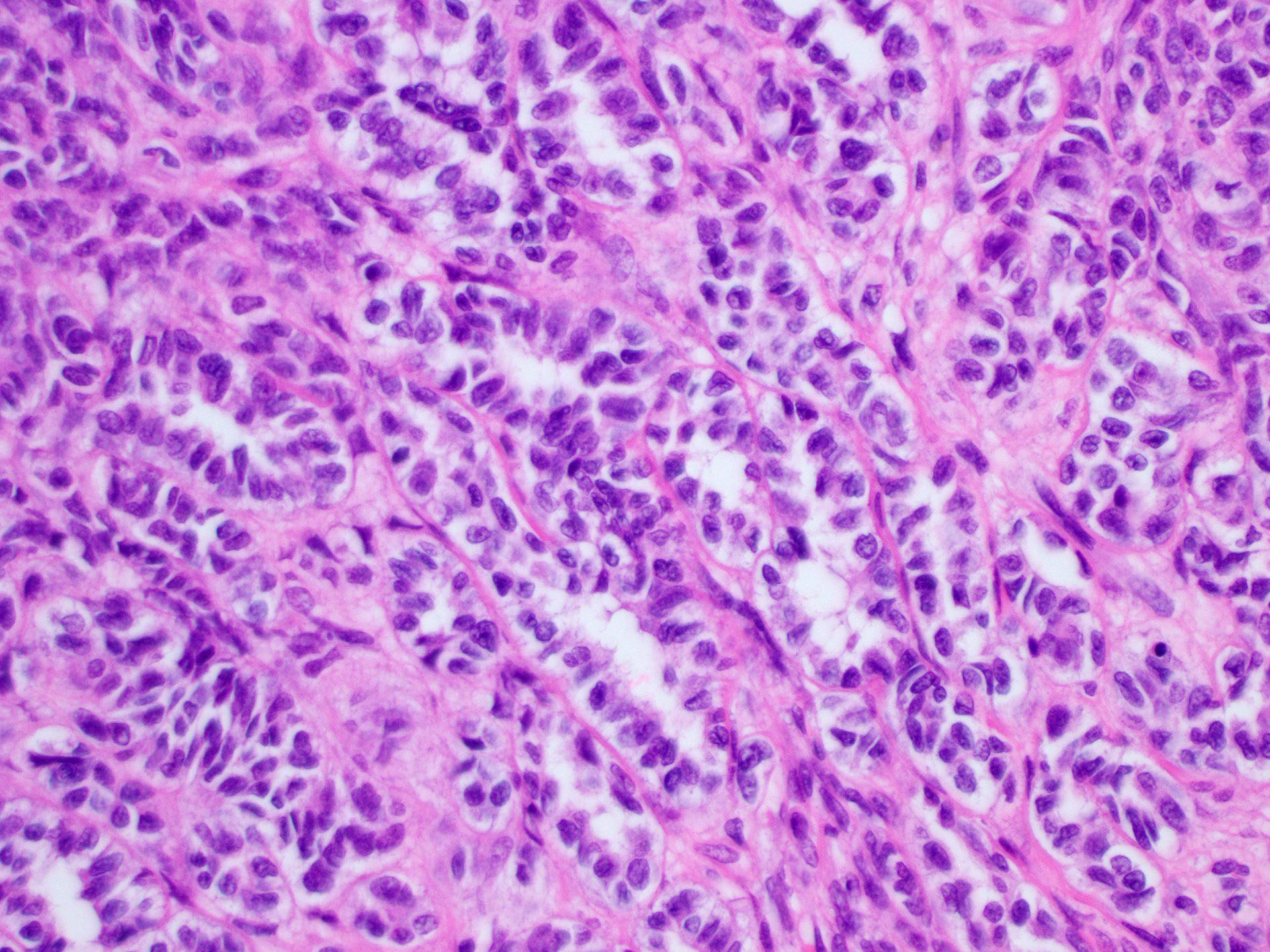
Well differentiated tumor
Moderately differentiated tumor
Poorly differentiated tumor
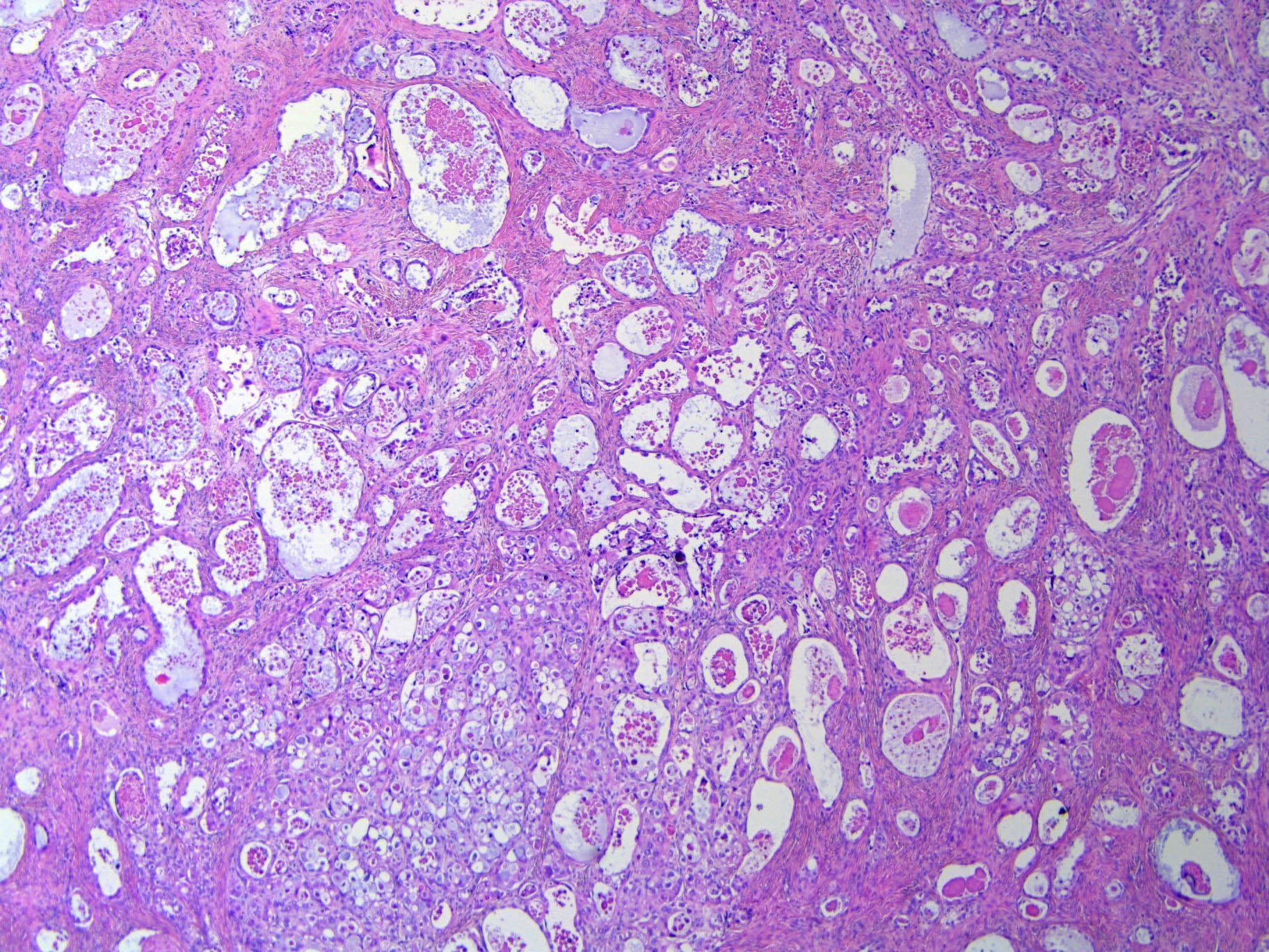
Retiform pattern
Heterologous elements
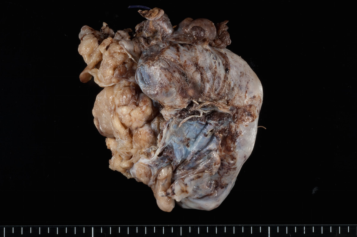
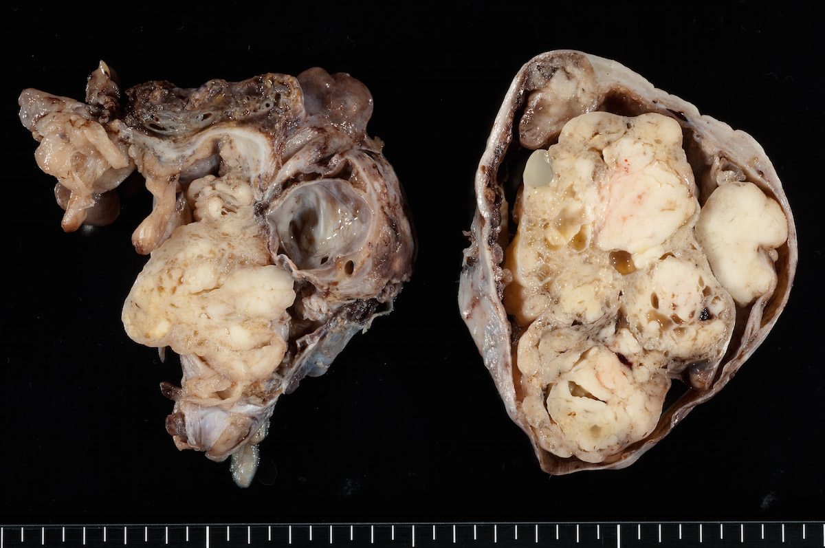
AFIP images
Predominantly solid tumor
Contributed by Krisztina Lengyel, M.D. and AFIP
Sharply delineated bland nests
Variably sized nests
Variably sized discrete nests
Numerous and variable nests
Multiple cysts and nests in young patient
Multiple nests of cells
Contributed by Glenn McCluggage, M.D.
Spindled and signet ring cells
Signet ring cells
AFIP images

Fleshy cream colored tissue
Contributed by Basile Tessier-Cloutier, M.D. and AFIP
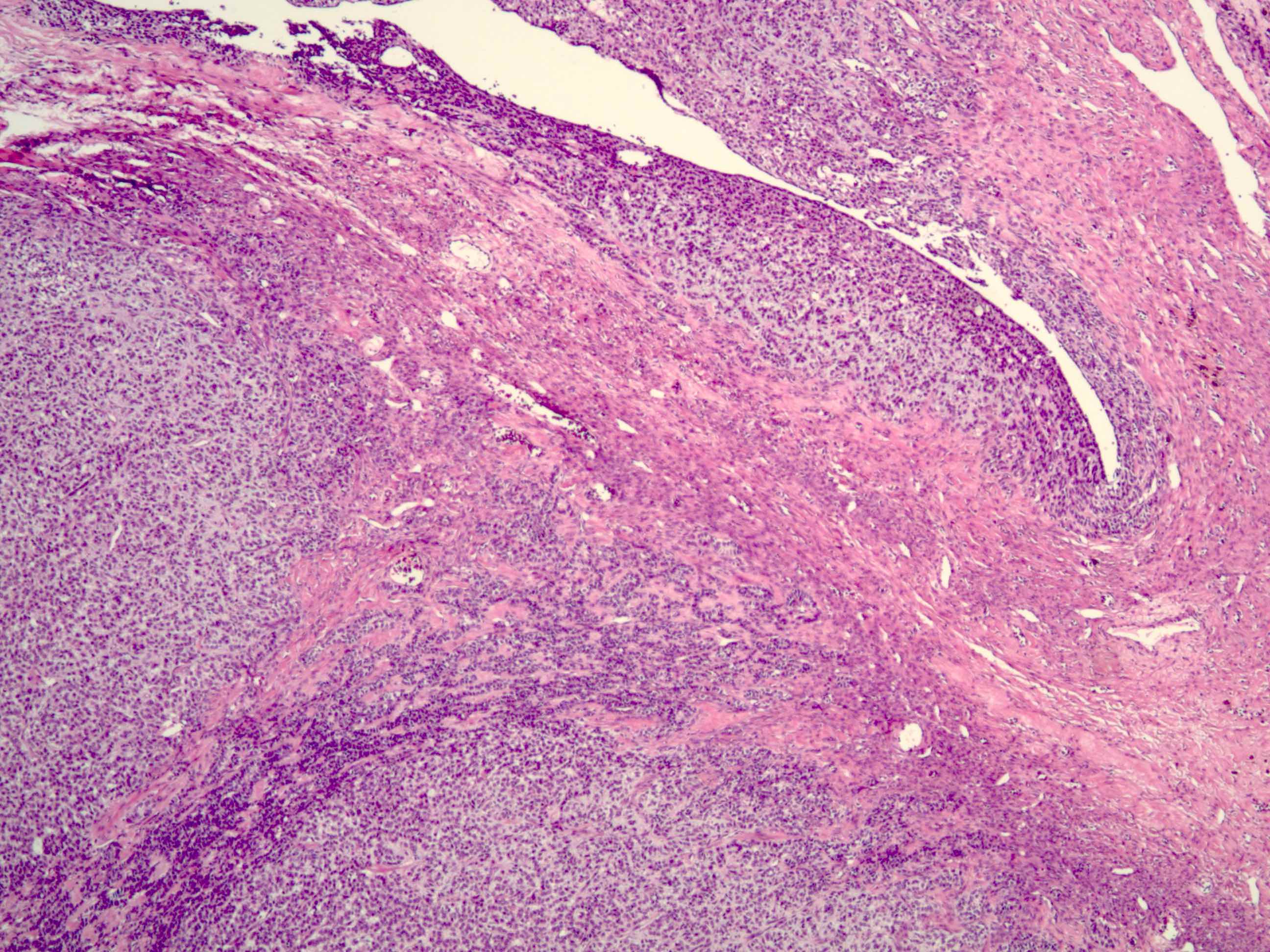
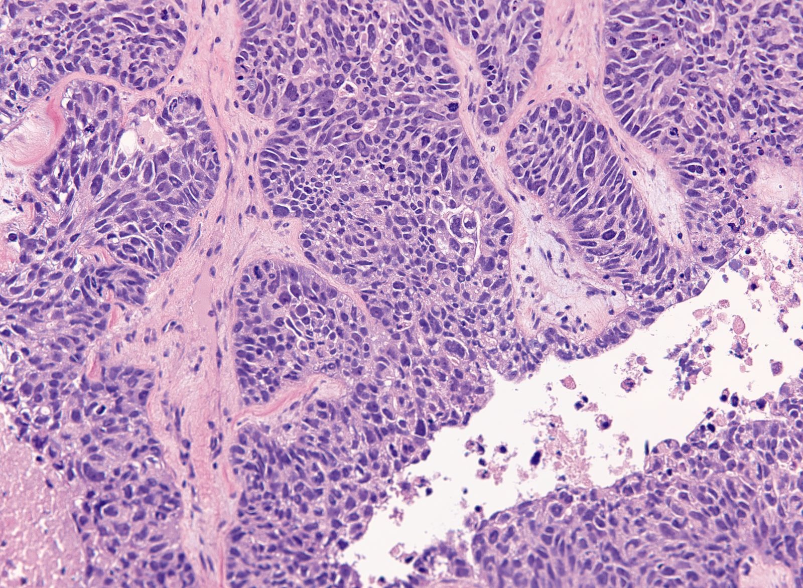
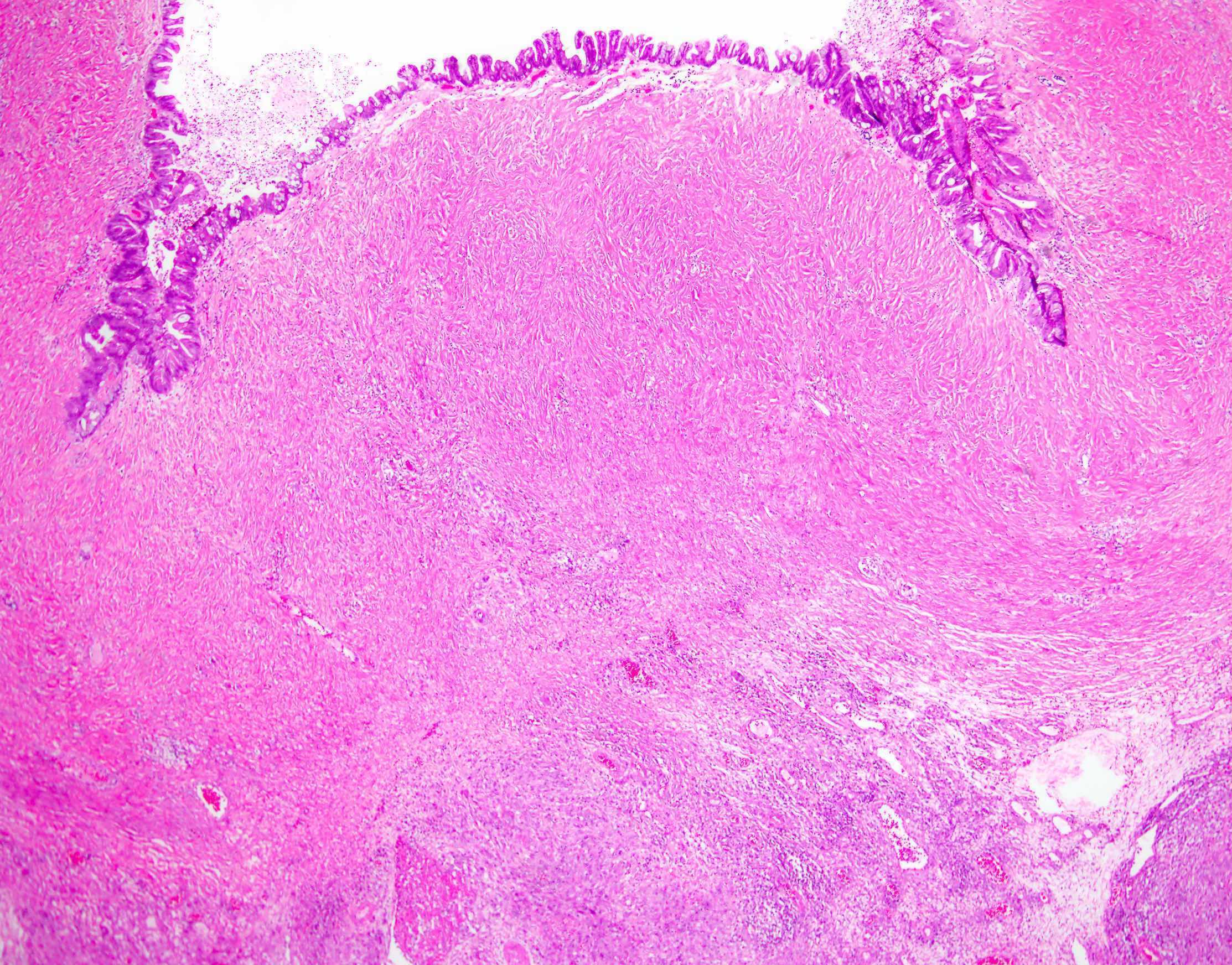
Follicle-like spaces and myxoid stroma
Solid and corded architecture
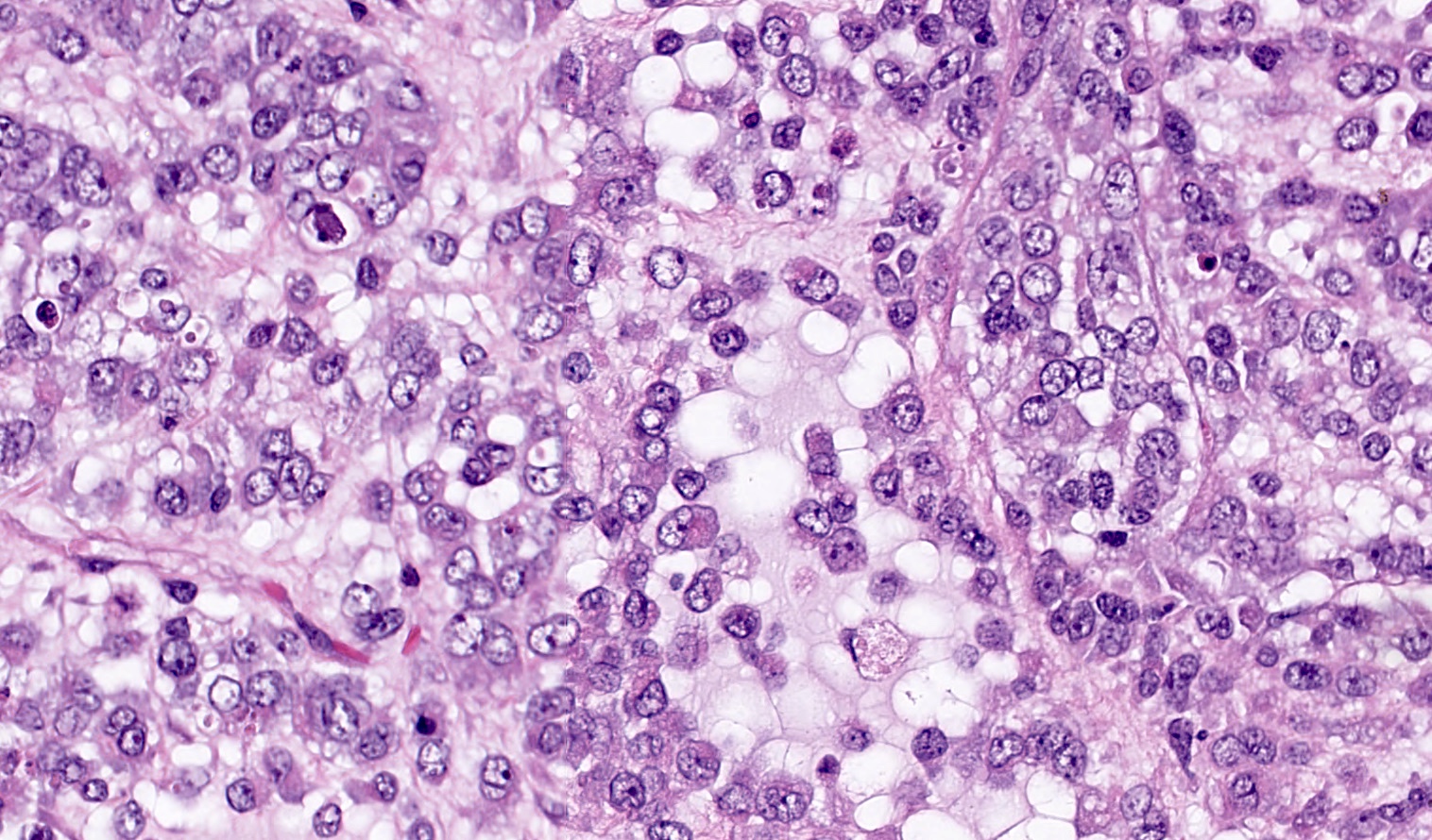
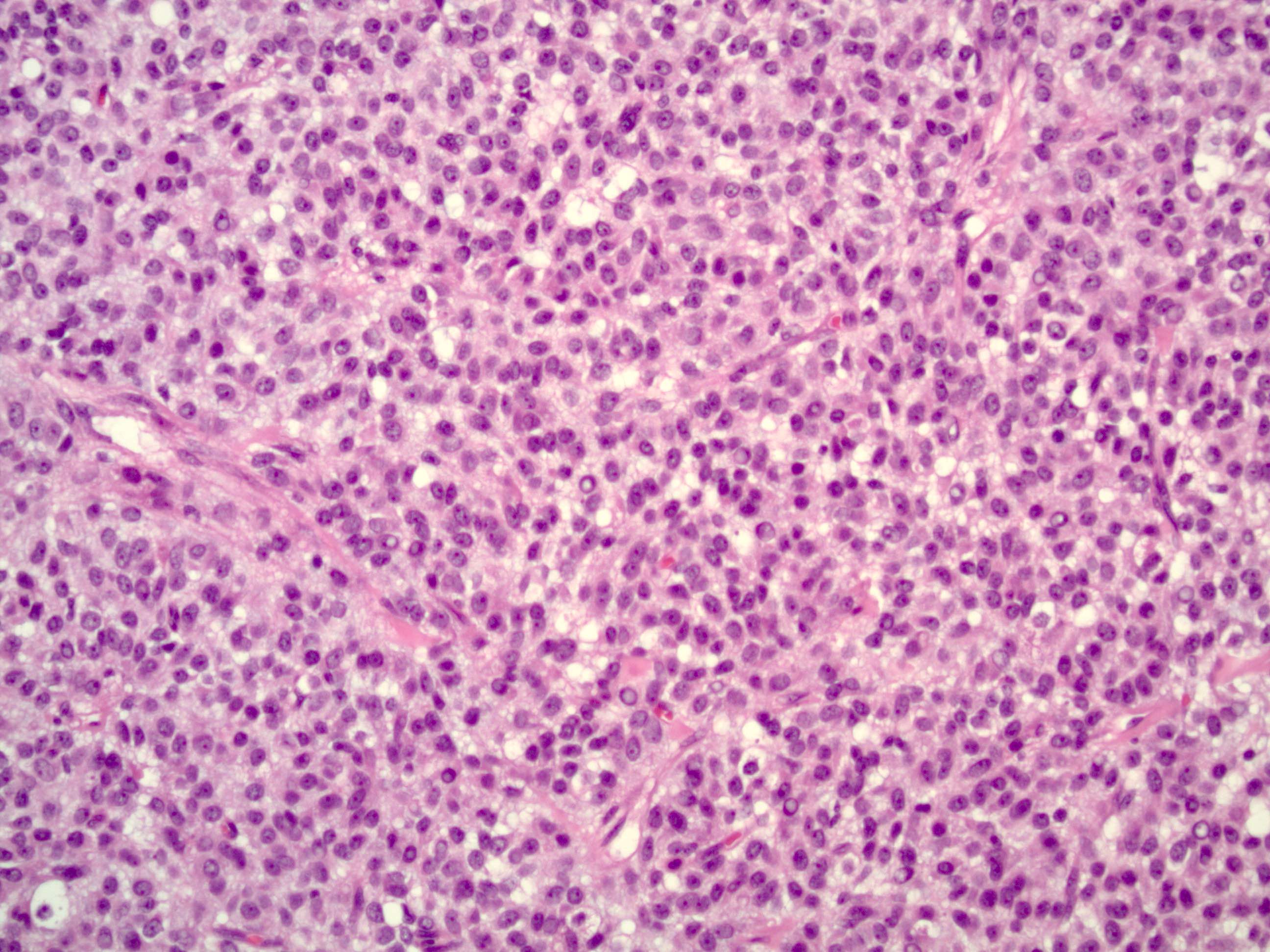
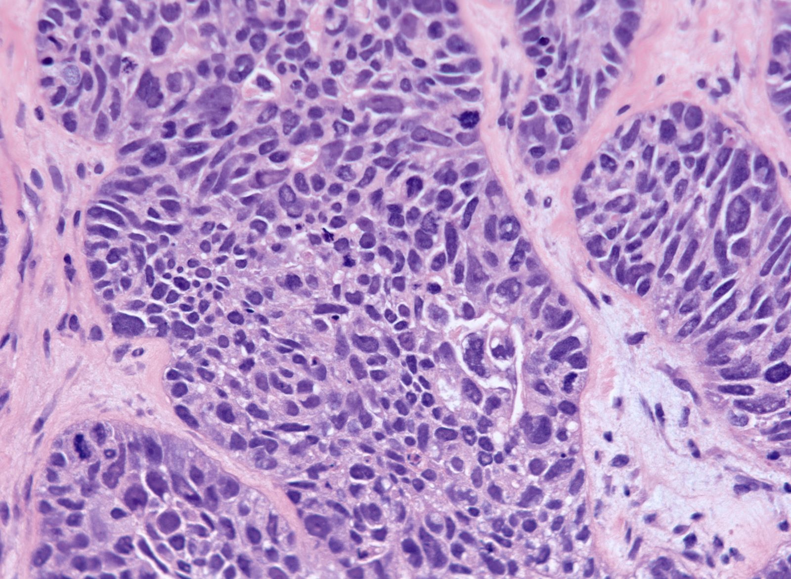
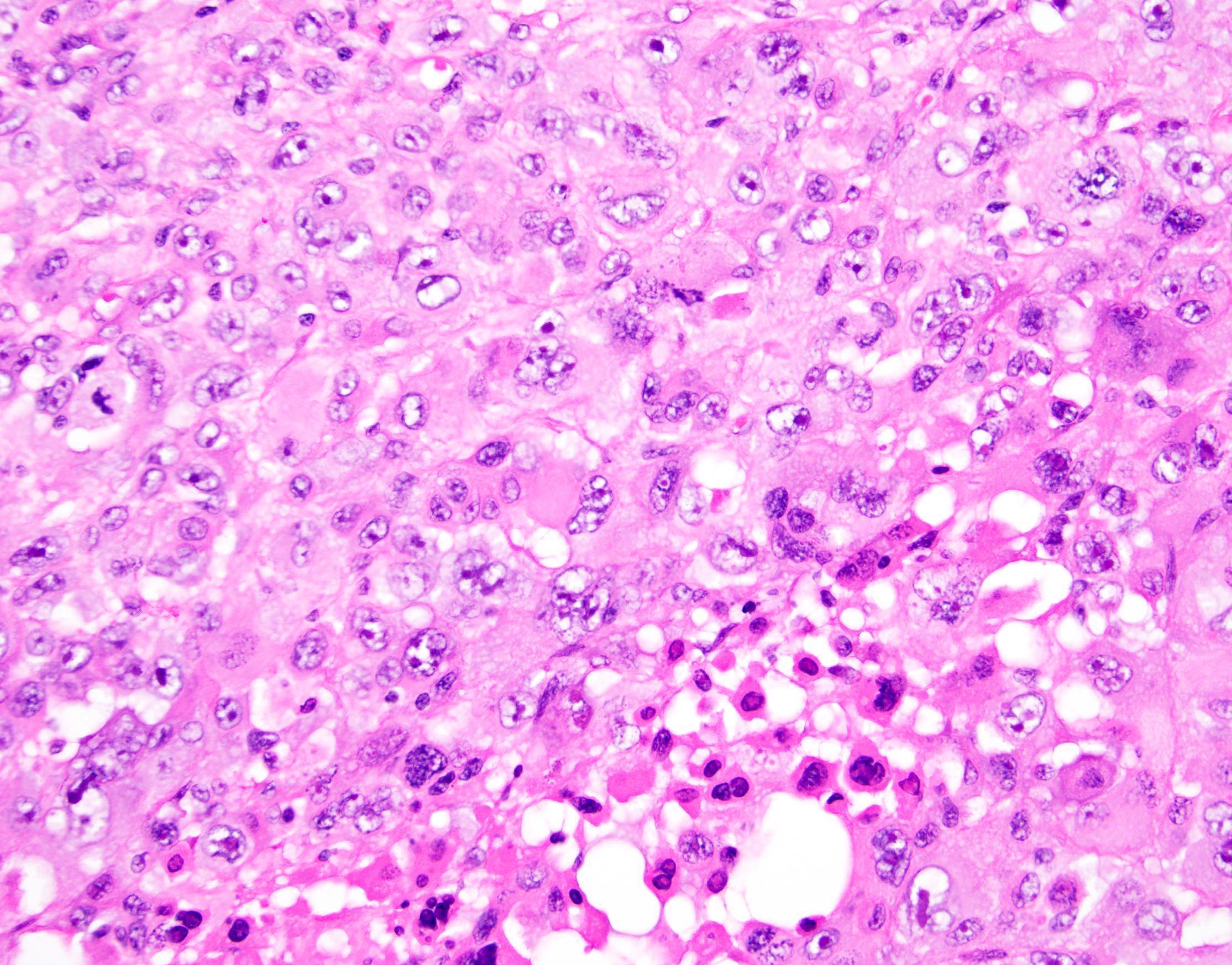
Rhabdoid morphology
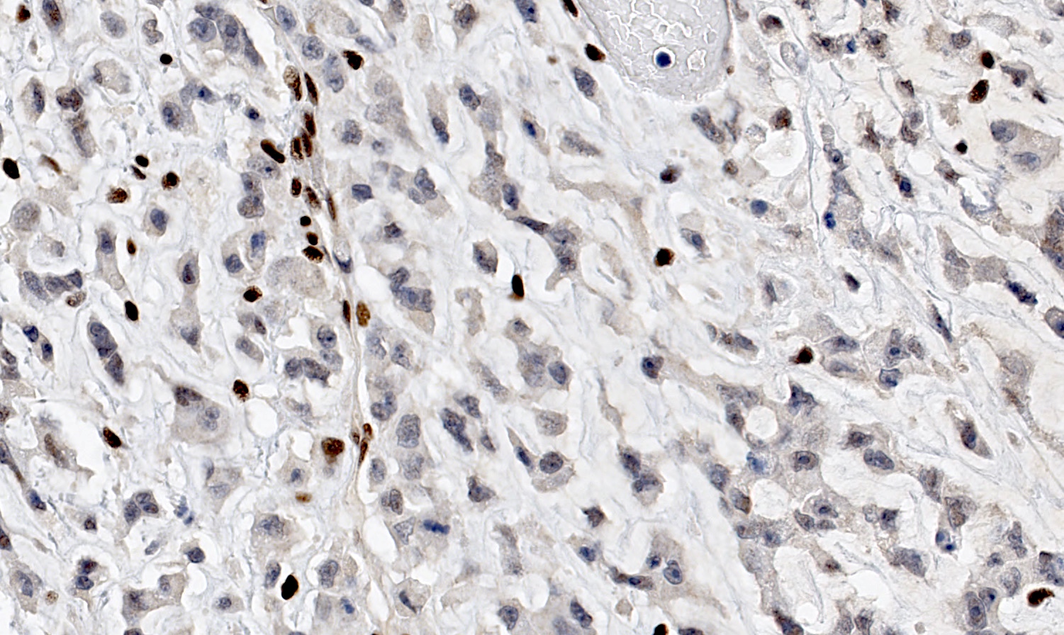
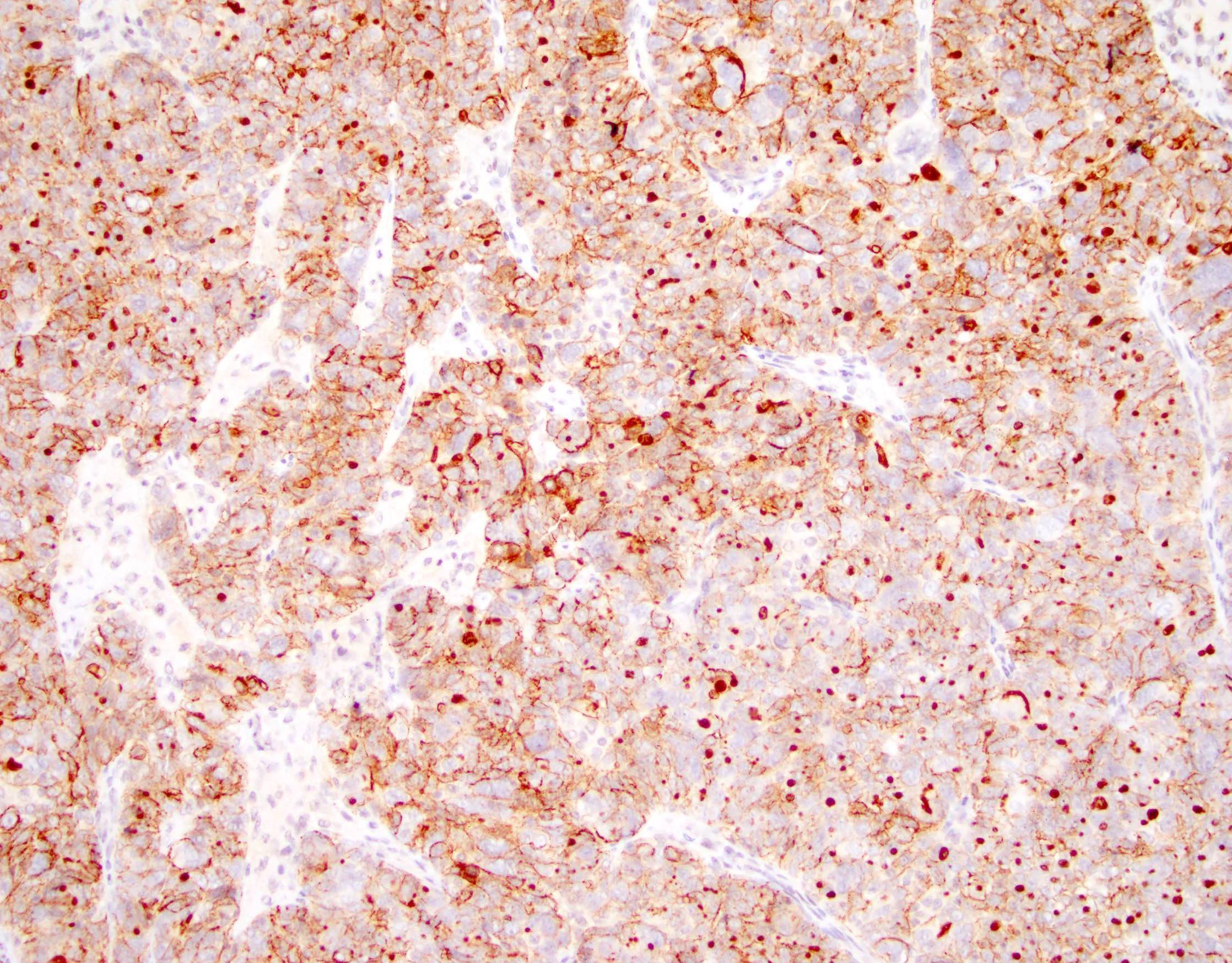
SMARCA4 / BRG1 loss of expression
Reduced SMARCA4 / BRG1 expression
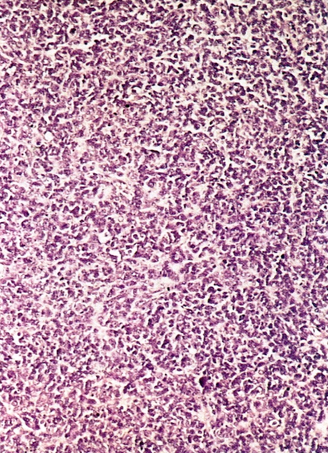
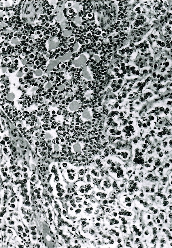
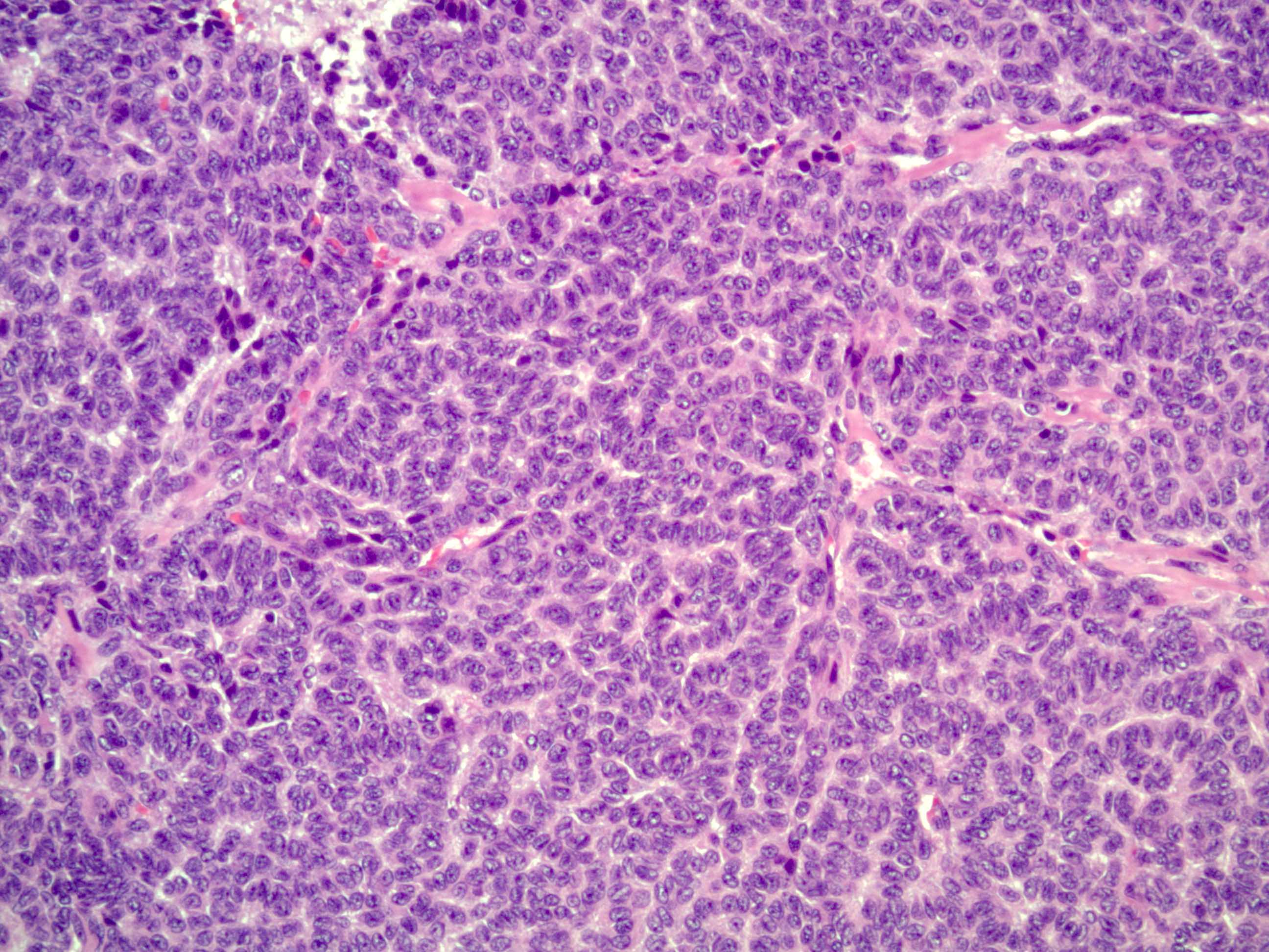
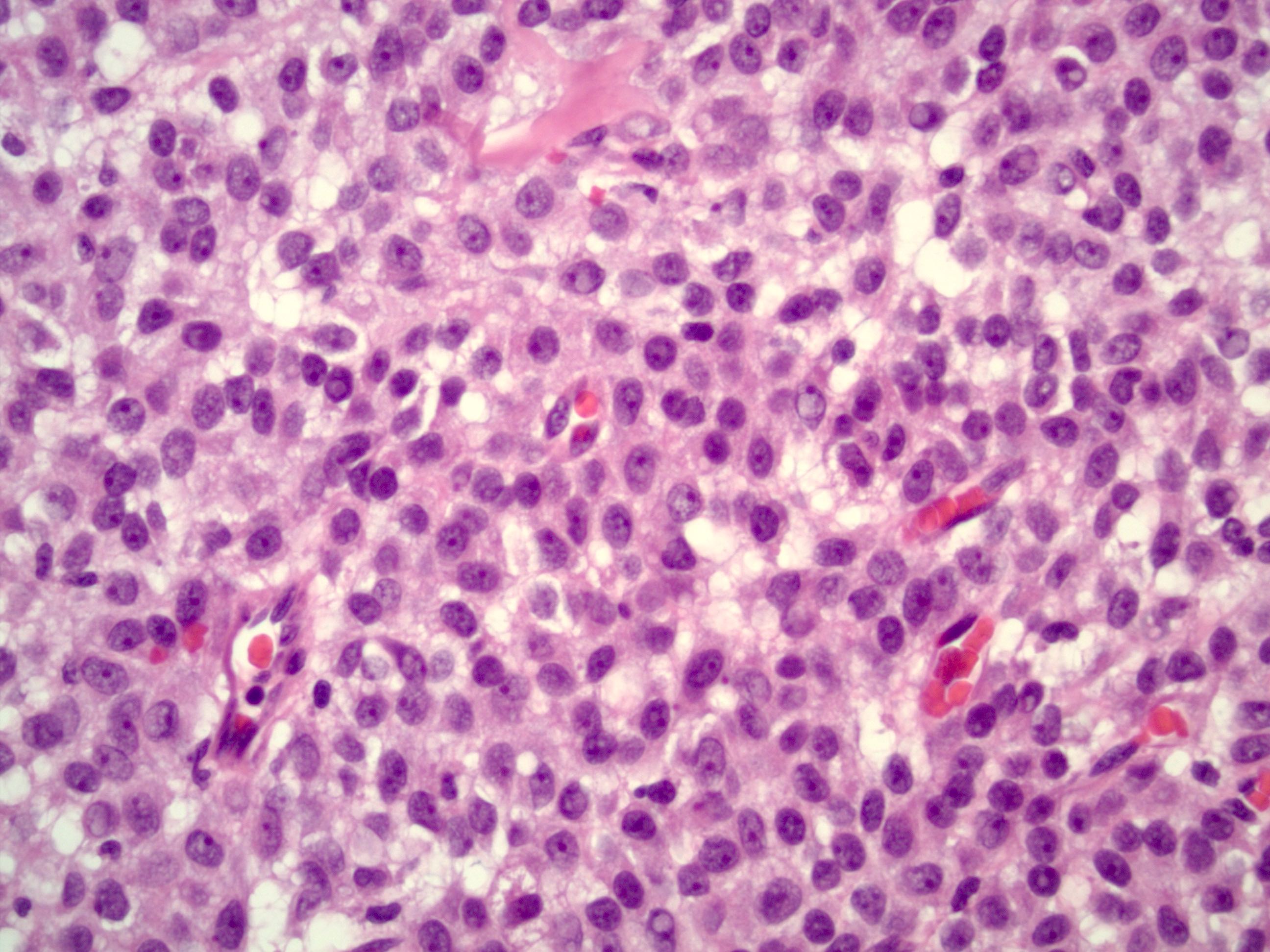
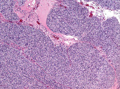
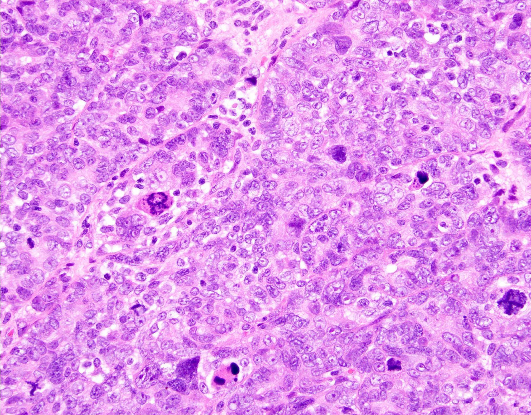
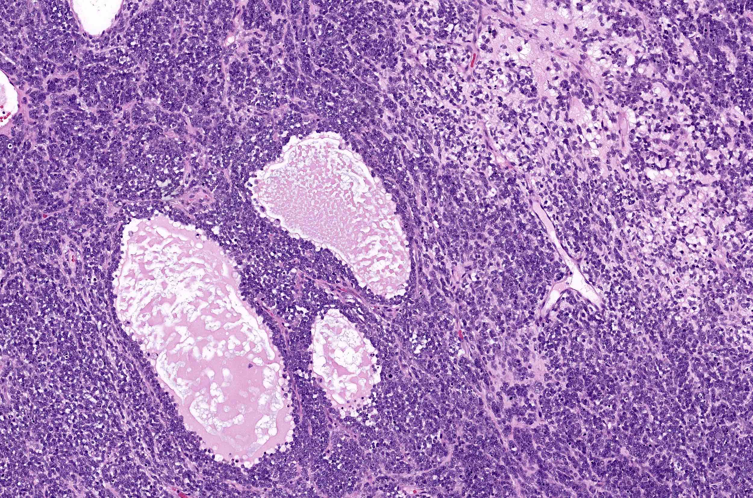
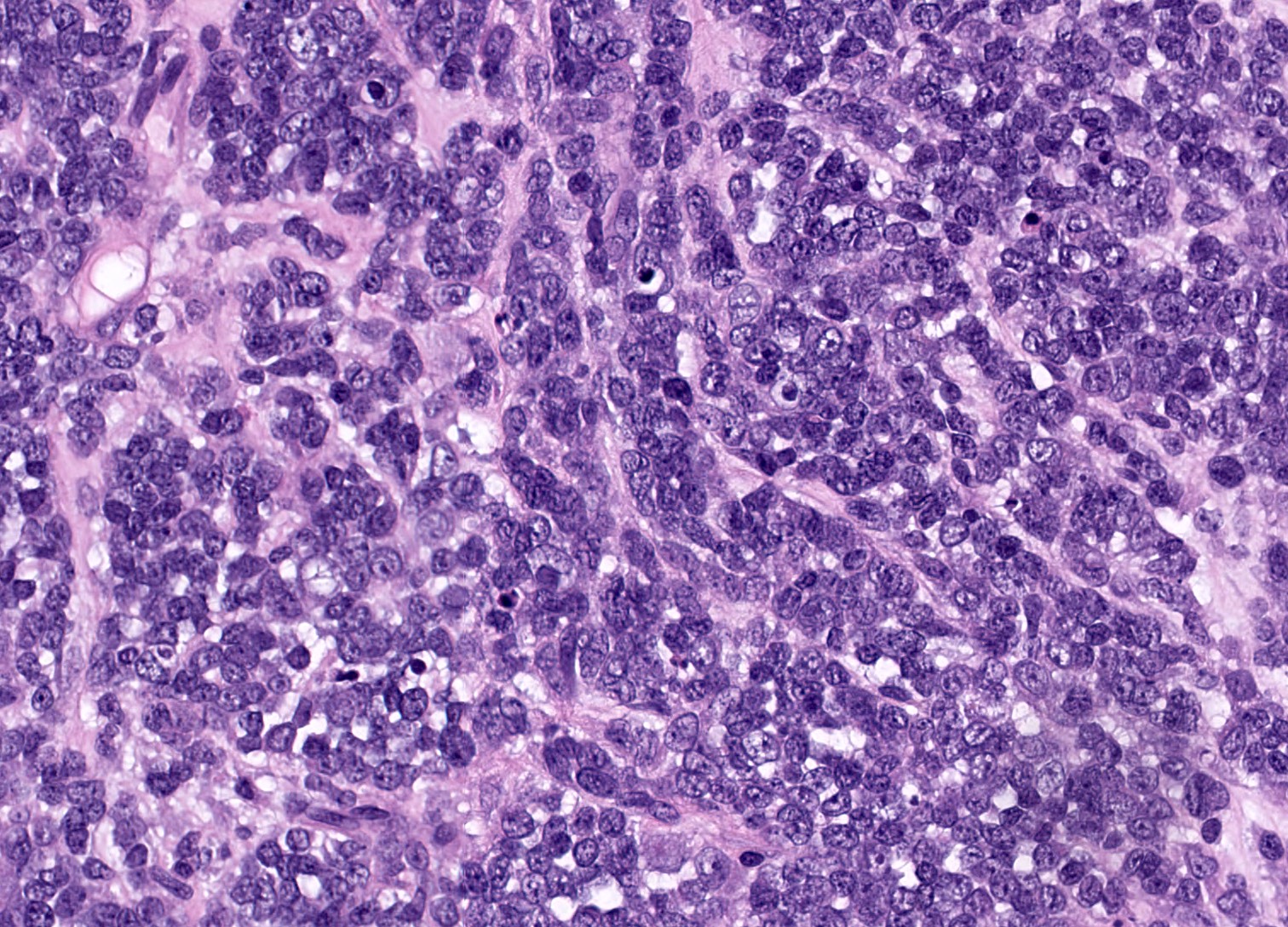
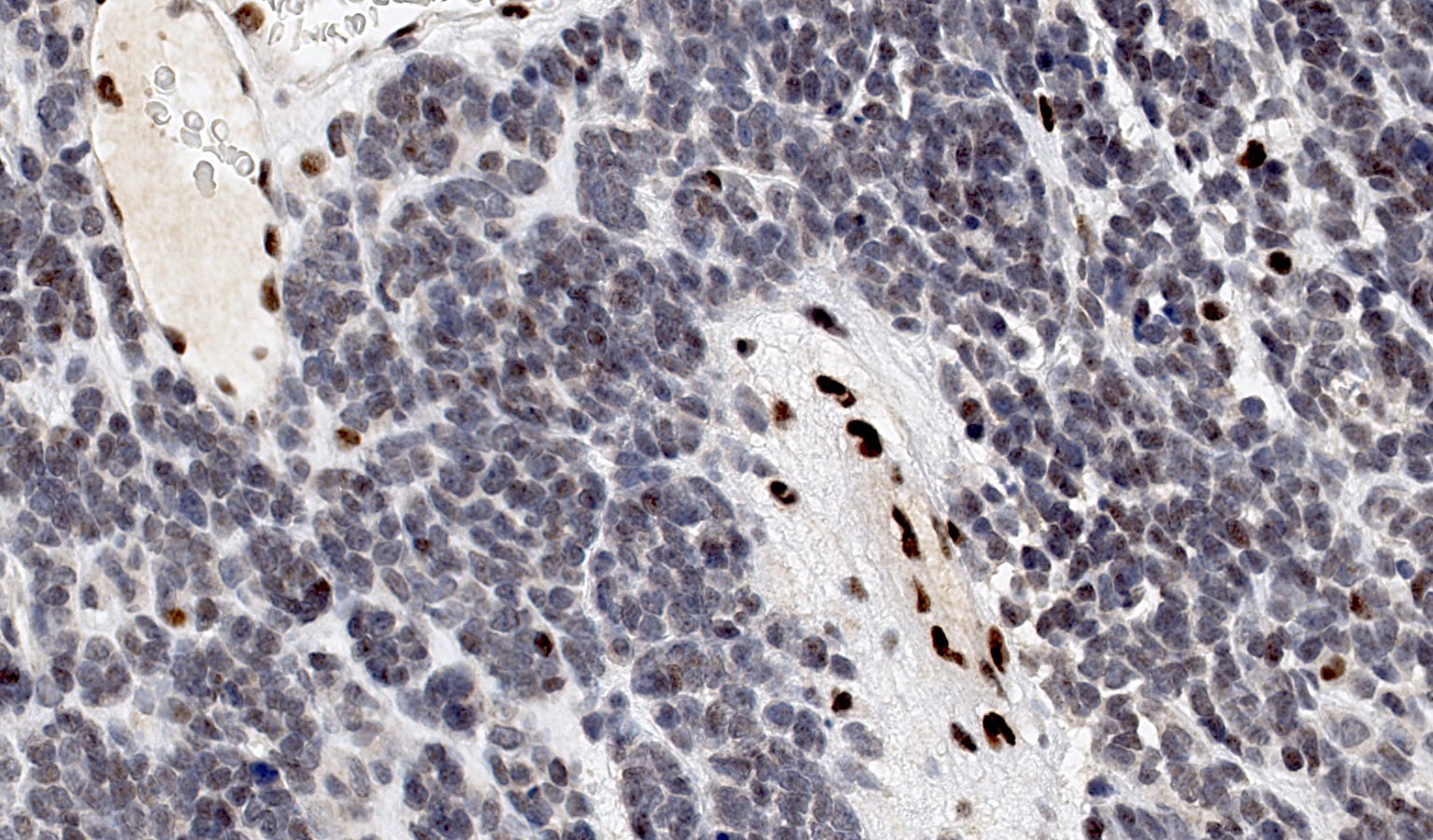
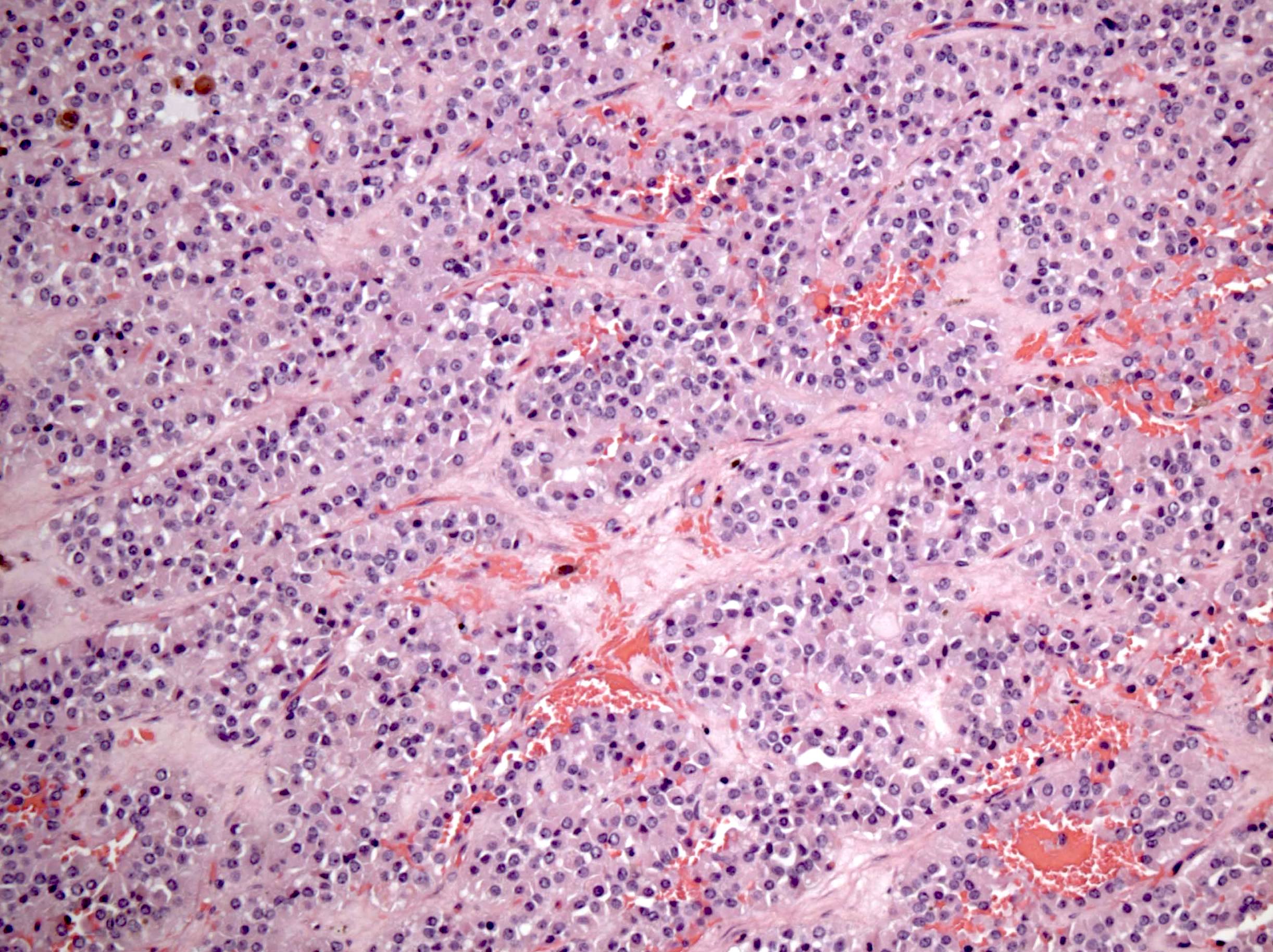
Diffuse growth of small cells with scanty cytoplasm
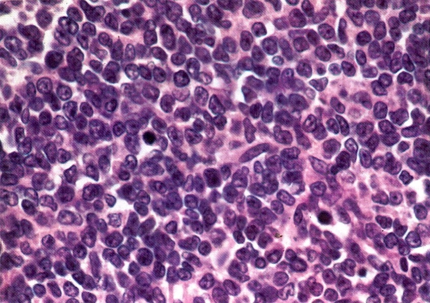
Hyperchromatic nuclei
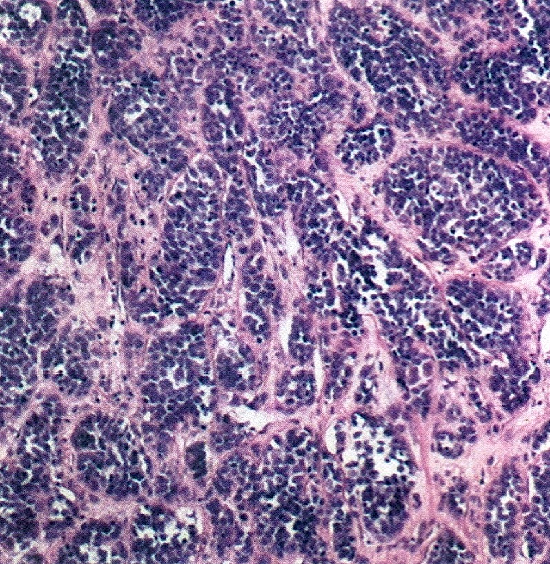
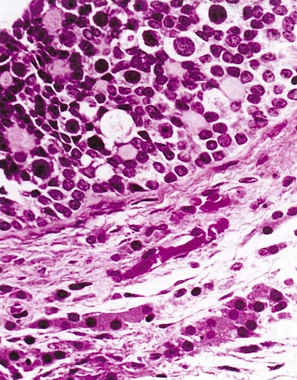
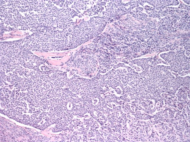
Growing in nests
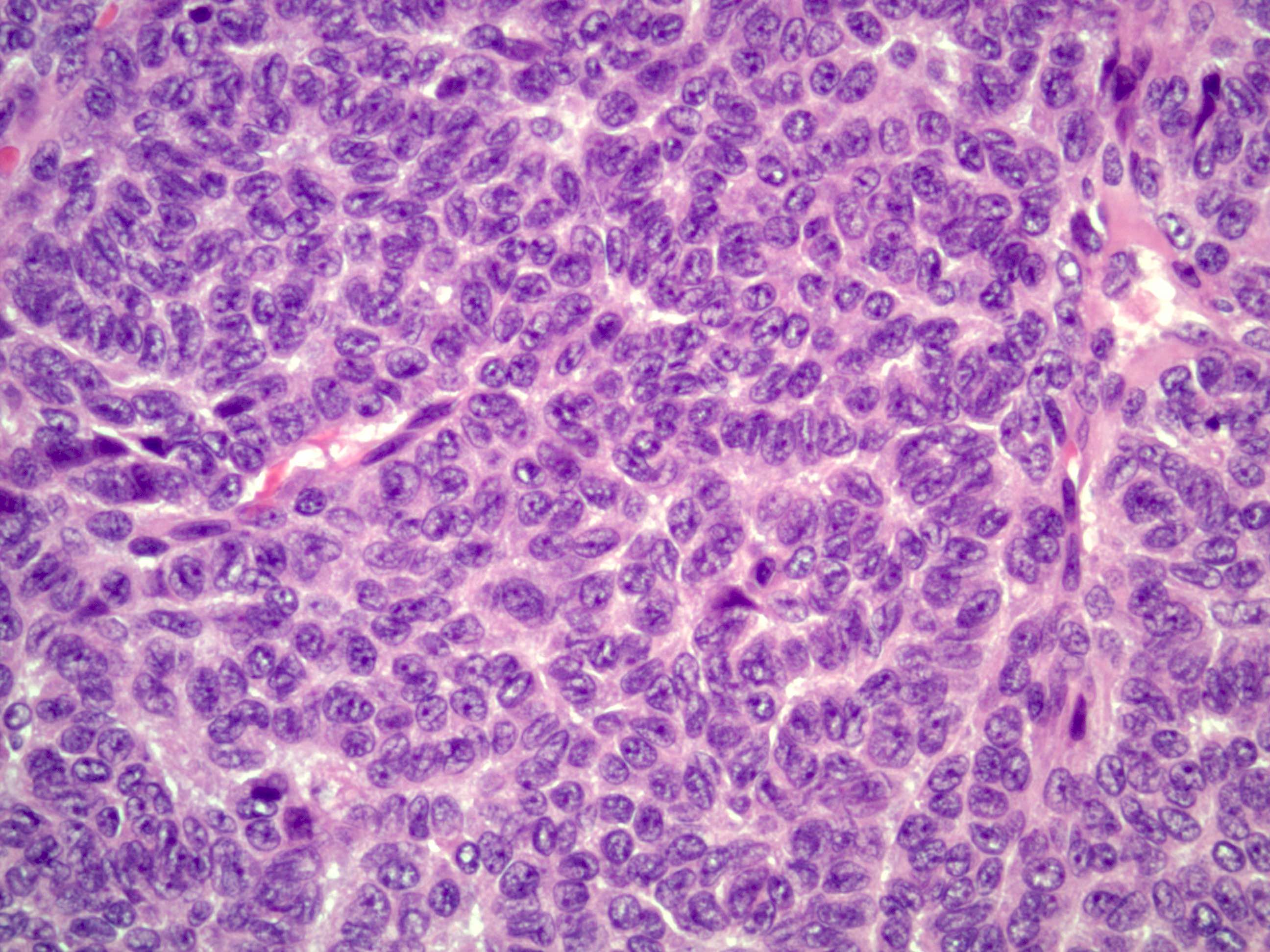
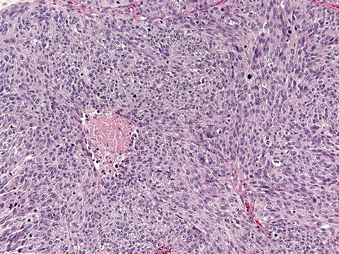
Large cell variant
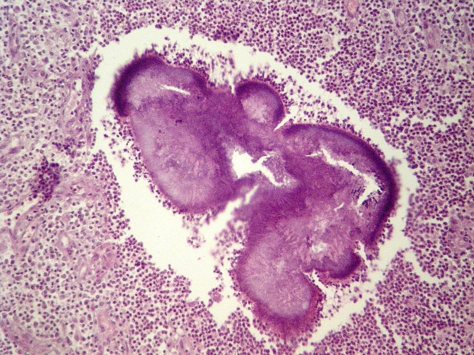
Follicle-like structures with eosinophilic material
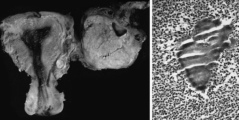
Several mucinous glands
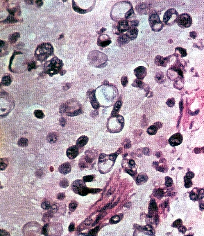
Signet ring cells in basophilic mucin
Several glands lined by tumor cells, some with mucin
Images hosted on other servers:

Solid pseudopapillary neoplasm
Contributed by Kamaljeet Singh, M.D.
Circumscribed mass
Nests with fibrous septa
Pseudopapillae

Intracytoplasmic vacuoles
β catenin
c-kit
CAM5.2
CD10
CD99
Synaptophysin
Vimentin
WT1
E-cadherin
Ki67
Contributed by Natalia Buza, M.D.
Ovarian surface involvement (pT1c2)
Rectosigmoid colon (pT2b)
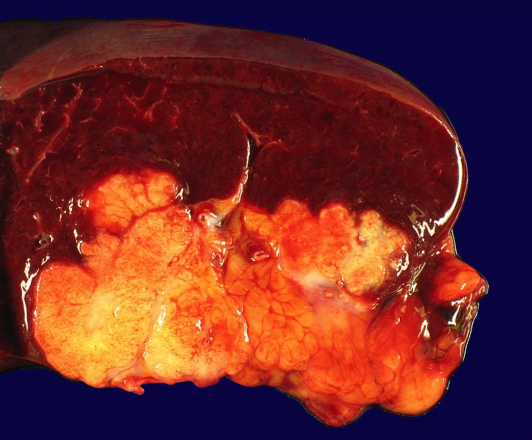
Splenic hilum and parenchyma (pM1b)
Contributed by Natalia Buza, M.D.
Ovarian surface involvement (pT1c2)
Uterine serosal (pT2a)
Appendiceal serosal (pT3b)
Splenic capsule (pT3c)
Splenic parenchyma (pM1b)
Images hosted on other servers:


Enhancing adnexal mass

Fat containing pelvic mass


Homogeneous isoechoic mass

Solid adnexal mass
Images hosted on other servers:

Hirsutism

Male pattern baldness
Images hosted on other servers:

Yellow cut surface

Metastatic steroid cell tumor

Lobulated cut surface

Nodular solid tumor

Tan-brown, well circumscribed tumor with hemorrhage
Contributed by Fatemeh Ghazanfari Amlashi, M.D., Tamara Kalir, M.D., Ph.D. and AFIP
Well defined tumor
Sparse stroma
Diffuse sheets
Moderate to abundant cytoplasm
Round to polygonal cells
Distinct cell borders
Spongy and eosinophilic cytoplasm
2 atypical mitotic figures
Inhibin
Calretinin
MelanA
Abundant lipid (oil red O stain)
Images hosted on other servers:

Large polygonal cells
Steroid cell tumor
Images hosted on other servers:

Ovarian stromal hyperplasia
AFIP images
Enlarged ovaries
Contributed by Hafza Asma Adnan, M.B.B.S. and AFIP
Diffuse stromal hyperplasia
Ovarian stromal hyperplasia

Nests of vacuolated, luteinized cells

Large nodule of luteinized cells

Lutein cells contain abundant lipid (oil red O stain)
Images hosted on other servers:

Left adnexal mass
Contributed by Kseniya Korchagina, M.D. and AFIP
Cystic teratoma
Solid and cystic
Reddish brown solid mass
Cystic tumor
Contributed by Sharon Song, M.S., M.D., Kseniya Korchagina, M.D. and AFIP
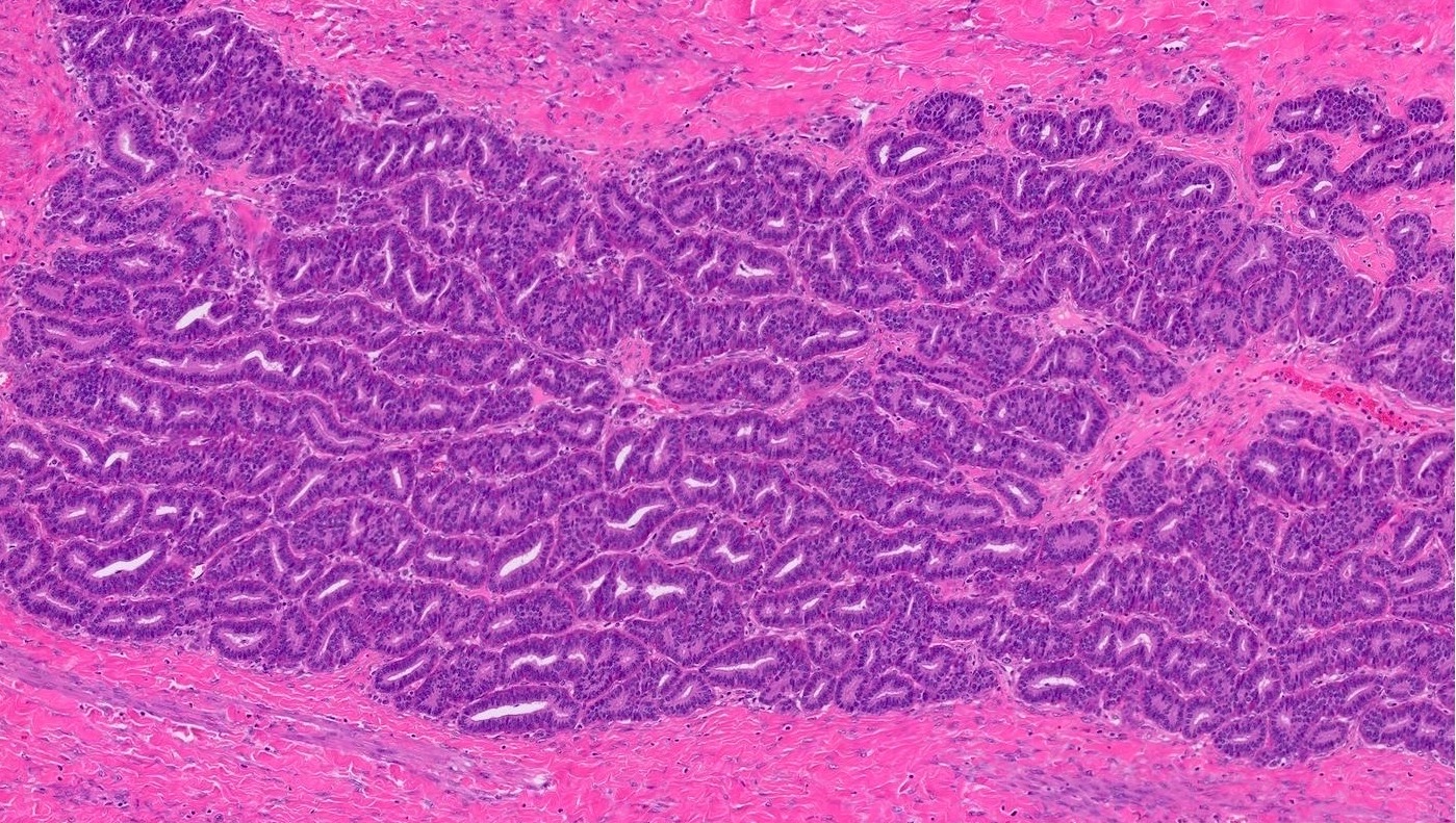
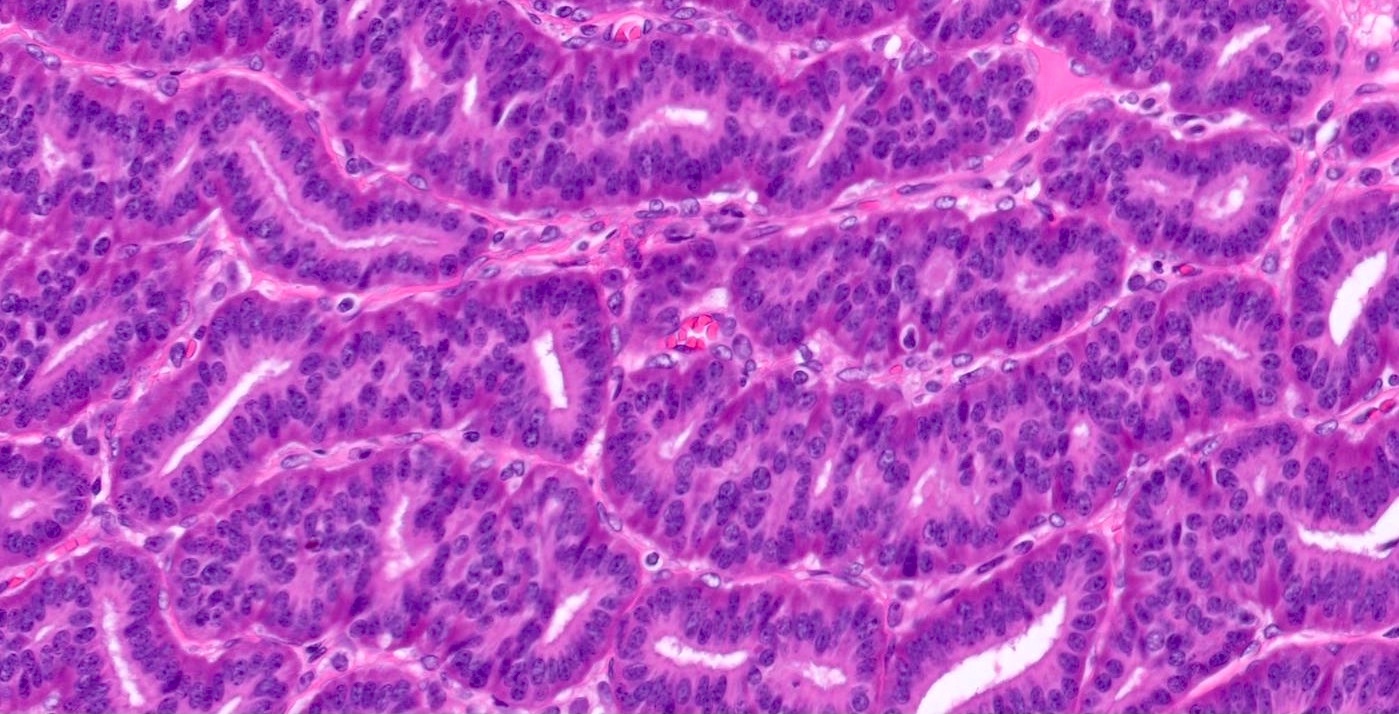
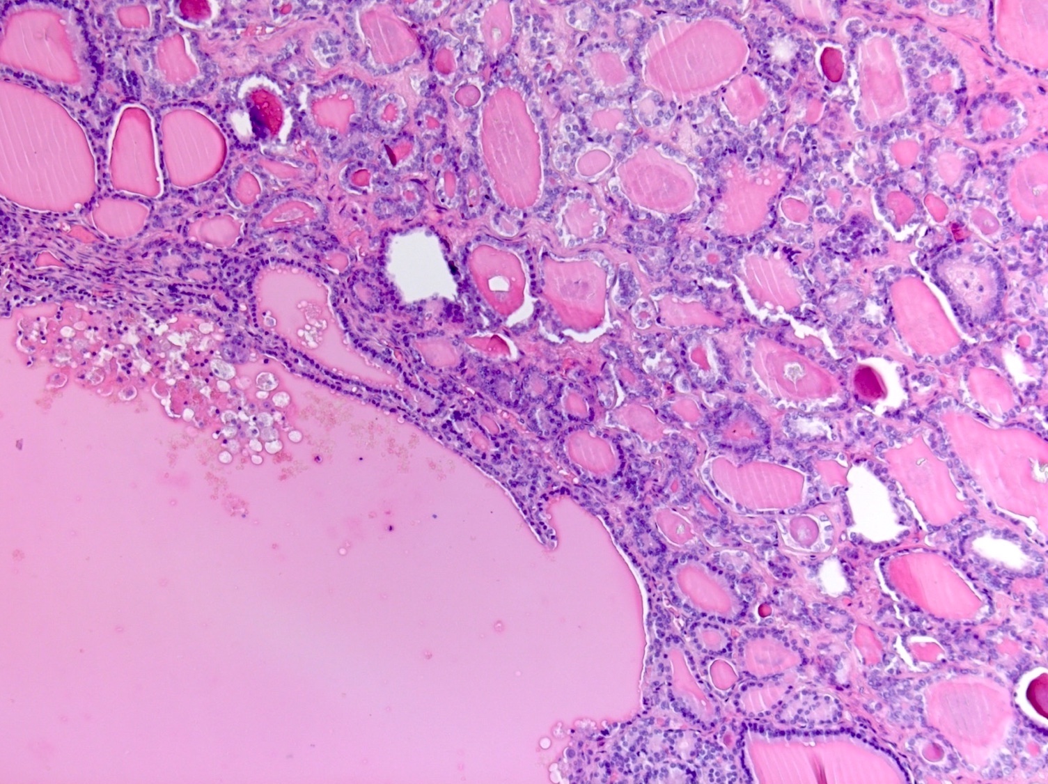
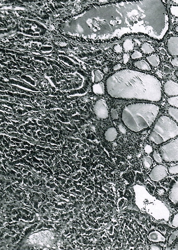
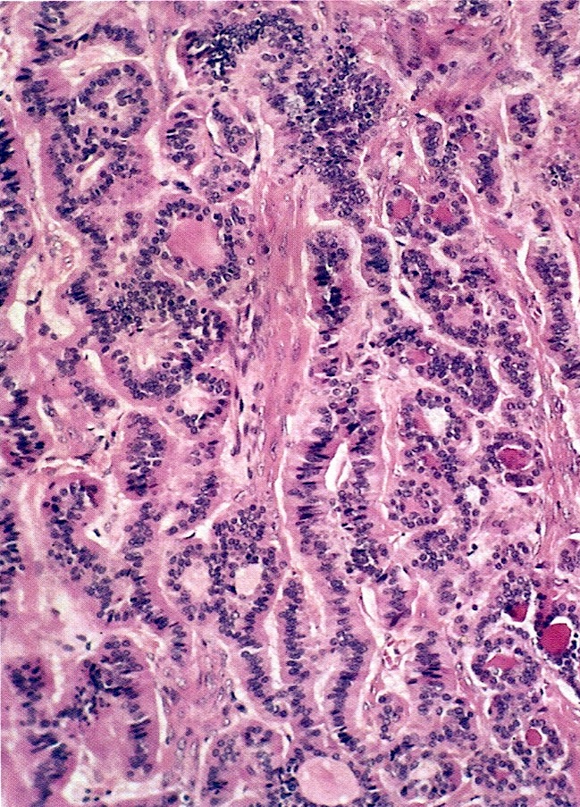
Follicular variant PTC
Capsular invasion
TTF1 stain
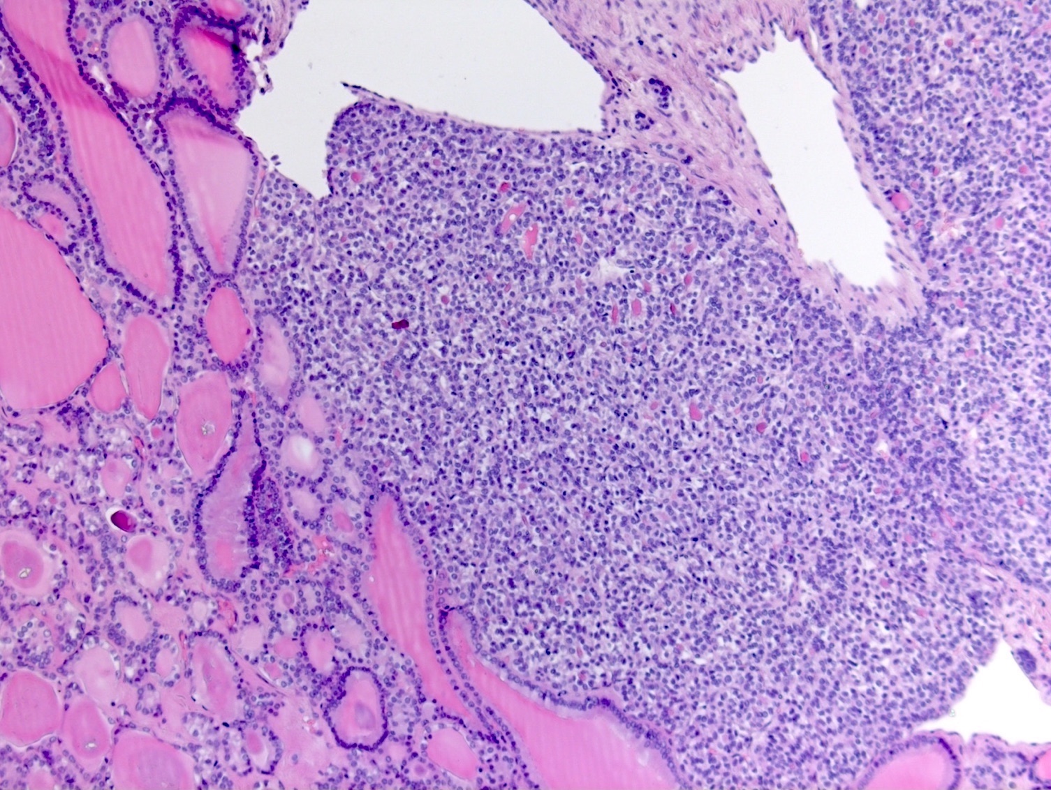
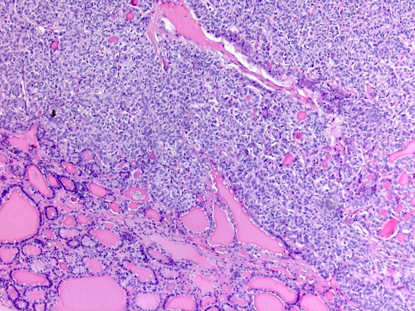
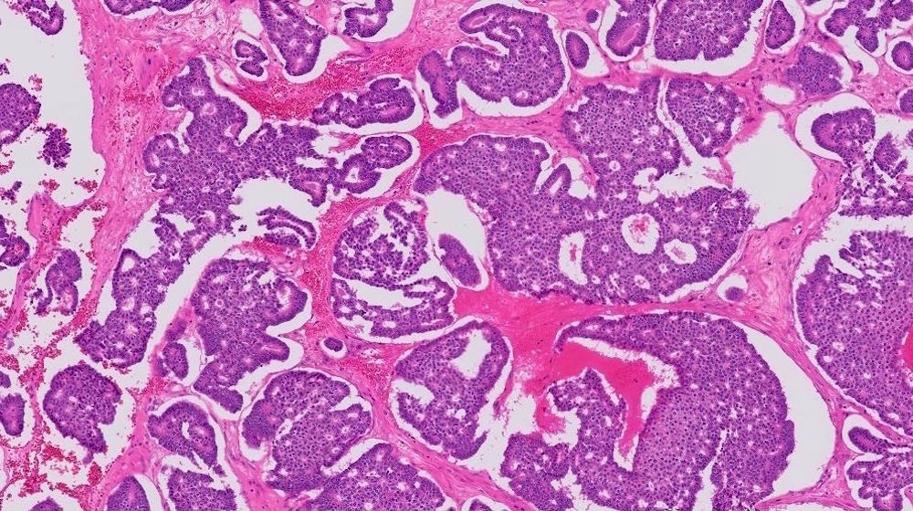
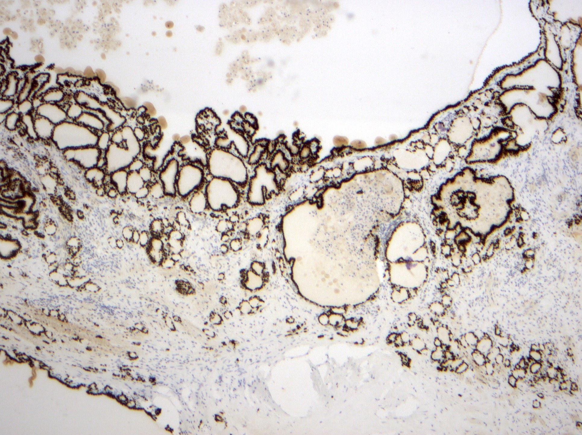
Papillary thyroid carcinoma in struma ovarii
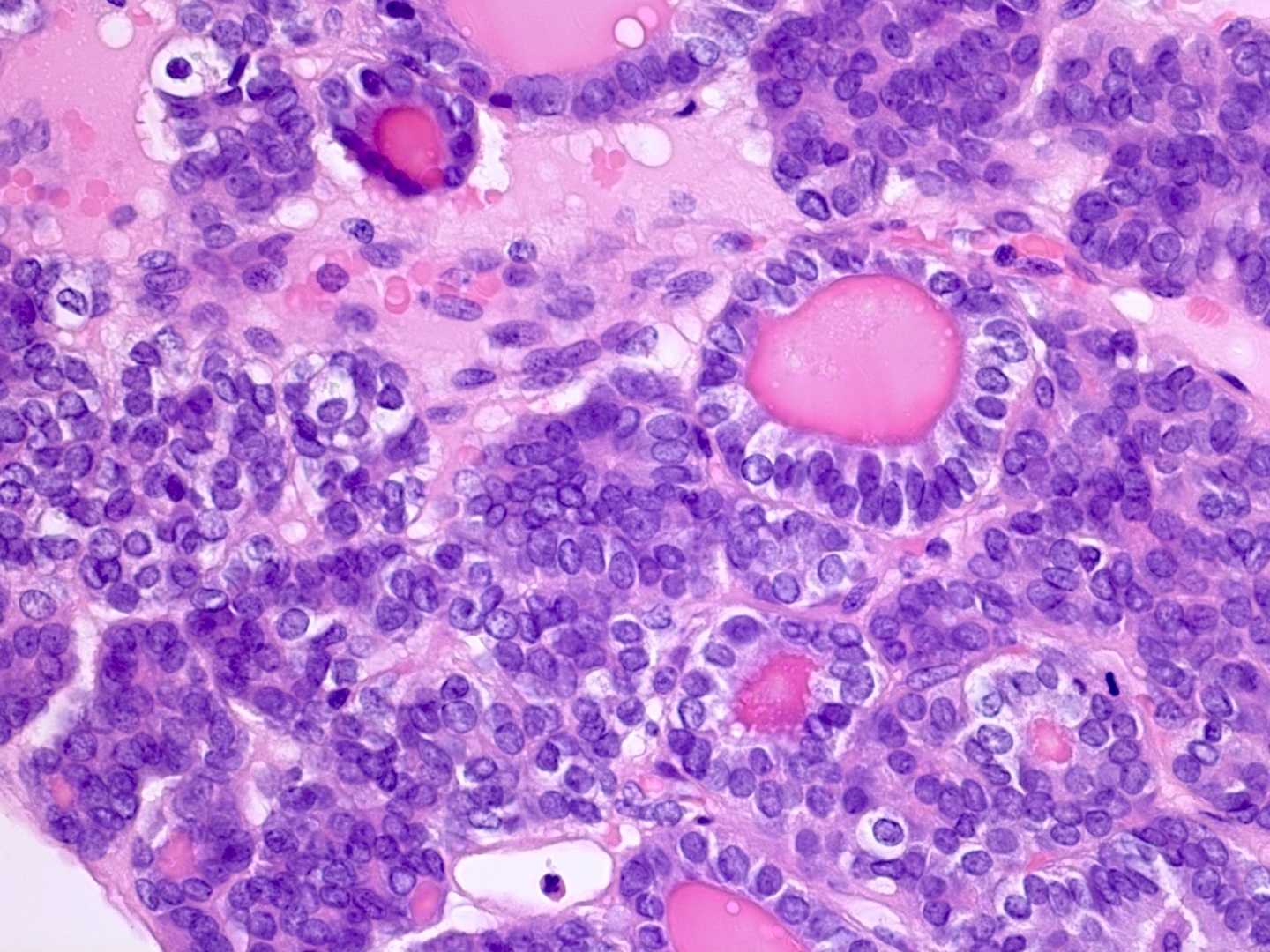
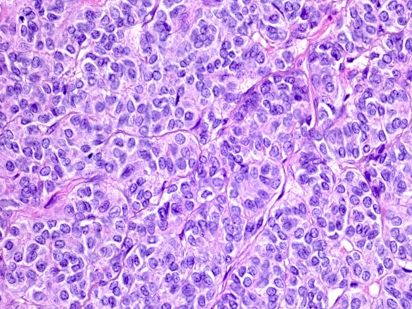
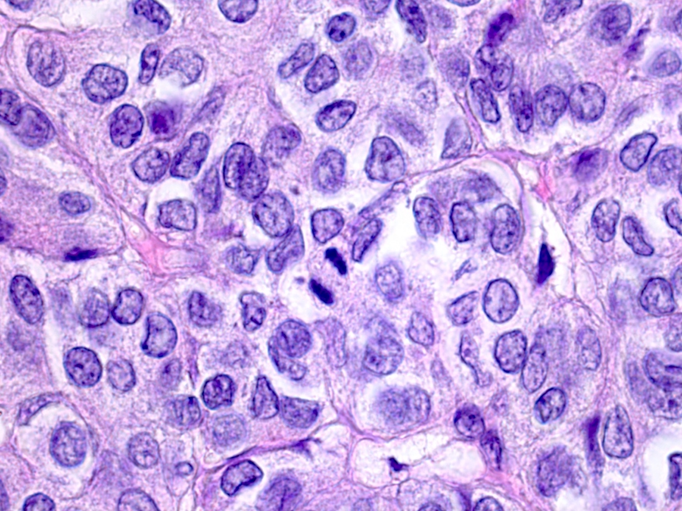
Papillary thyroid carcinoma in struma ovarii
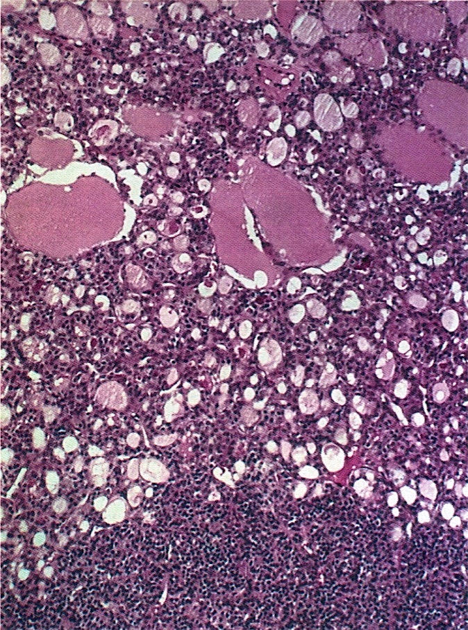
Macrofollicular, microfollicular and solid areas
Solid tubular pattern
Oxyphilic cells arranged in nests
With peripheral formation of lutein cells
Resembling clear cell carcinoma
Cystic tumor
Thyroglobulin
With papillary carcinoma
AFIP images
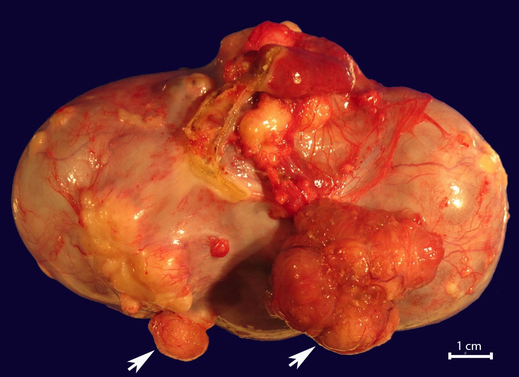
4 cm, rounded, brown nodule (bottom)
AFIP images
Admixed: struma (right) merges with trabecular carcinoid (left)
Admixed: trabecular
carcinoid with
microfollicles
containing colloid

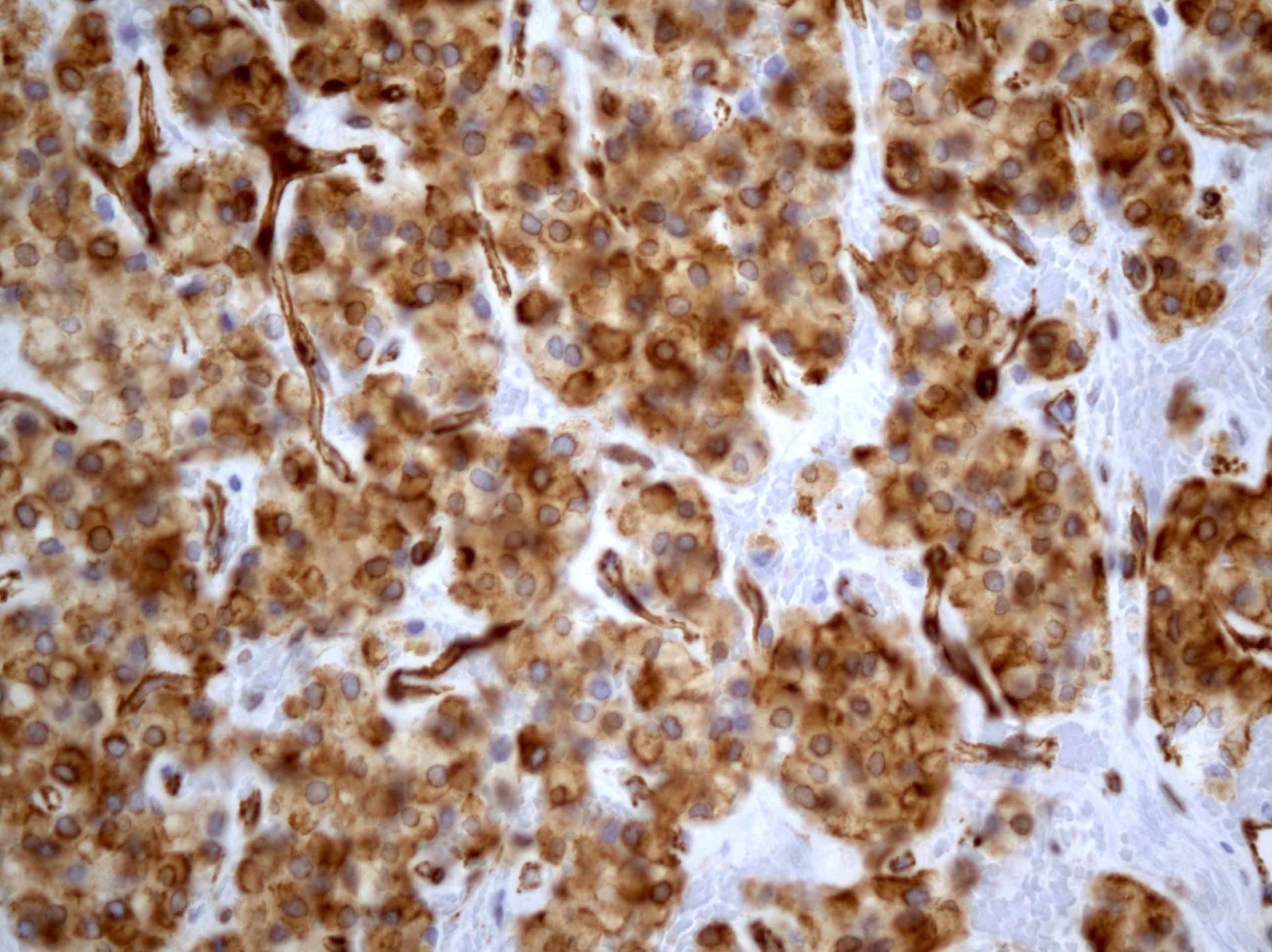
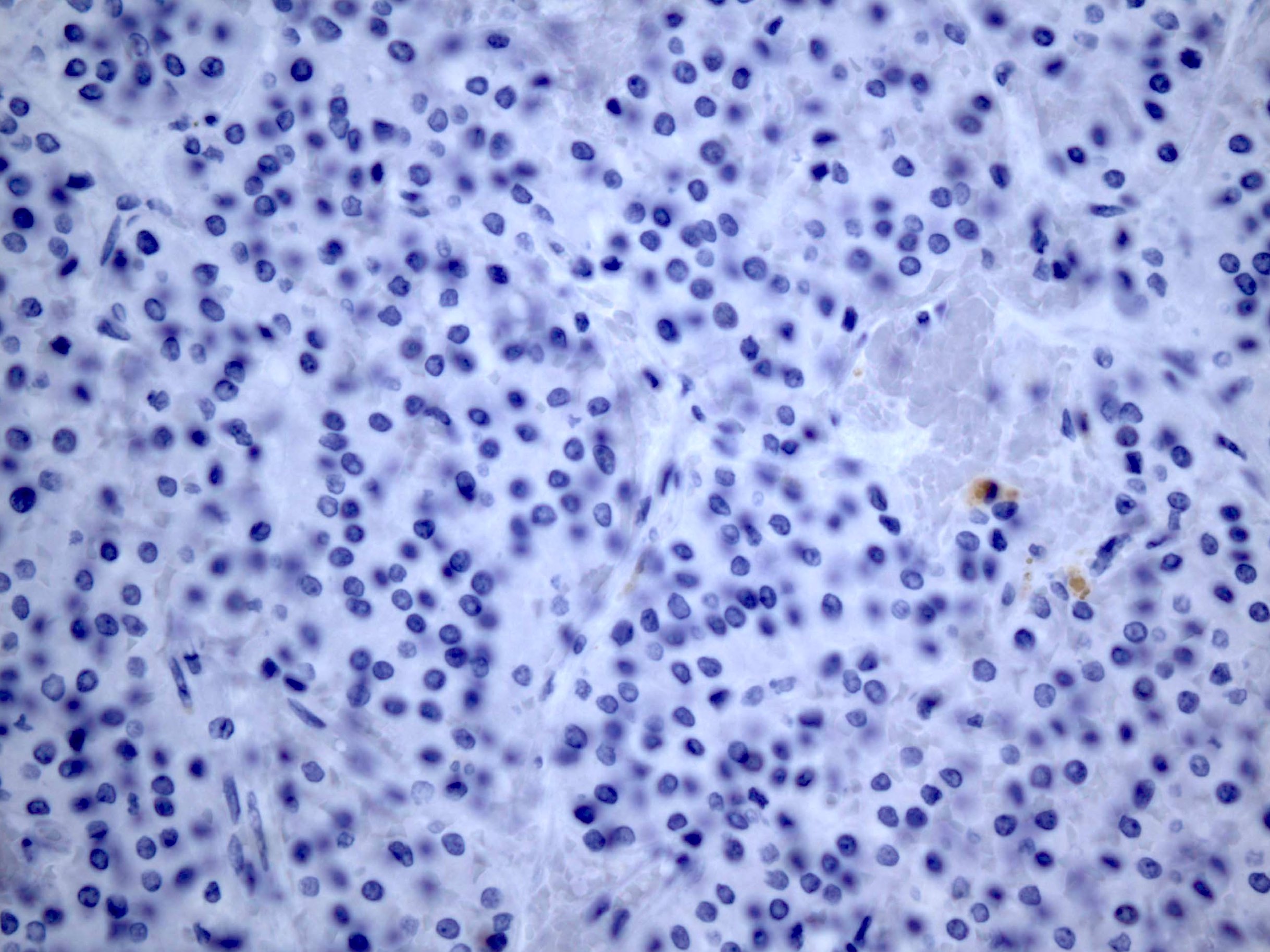
Chromogranin (top), thyroglobulin (bottom)
With formation of
layer of steroid cells
in adjacent ovarian
stroma
Images hosted on other servers:

Complex ovarian mass
Images hosted on other servers:


Solid ovarian mass with variegated appearance
Contributed by Aurelia Busca, M.D., Ph.D.
Mature component
Immature component
Immature component

Images hosted on other servers:


Ultrasound characteristics


CT characteristics

MRI characteristics
Contributed by Shannon Mingo Welter, M.D., AFIP and @Andrew_Fltv on Twitter
Fragmented ovarian mature teratoma

Ovarian mature teratoma
Mature teratoma
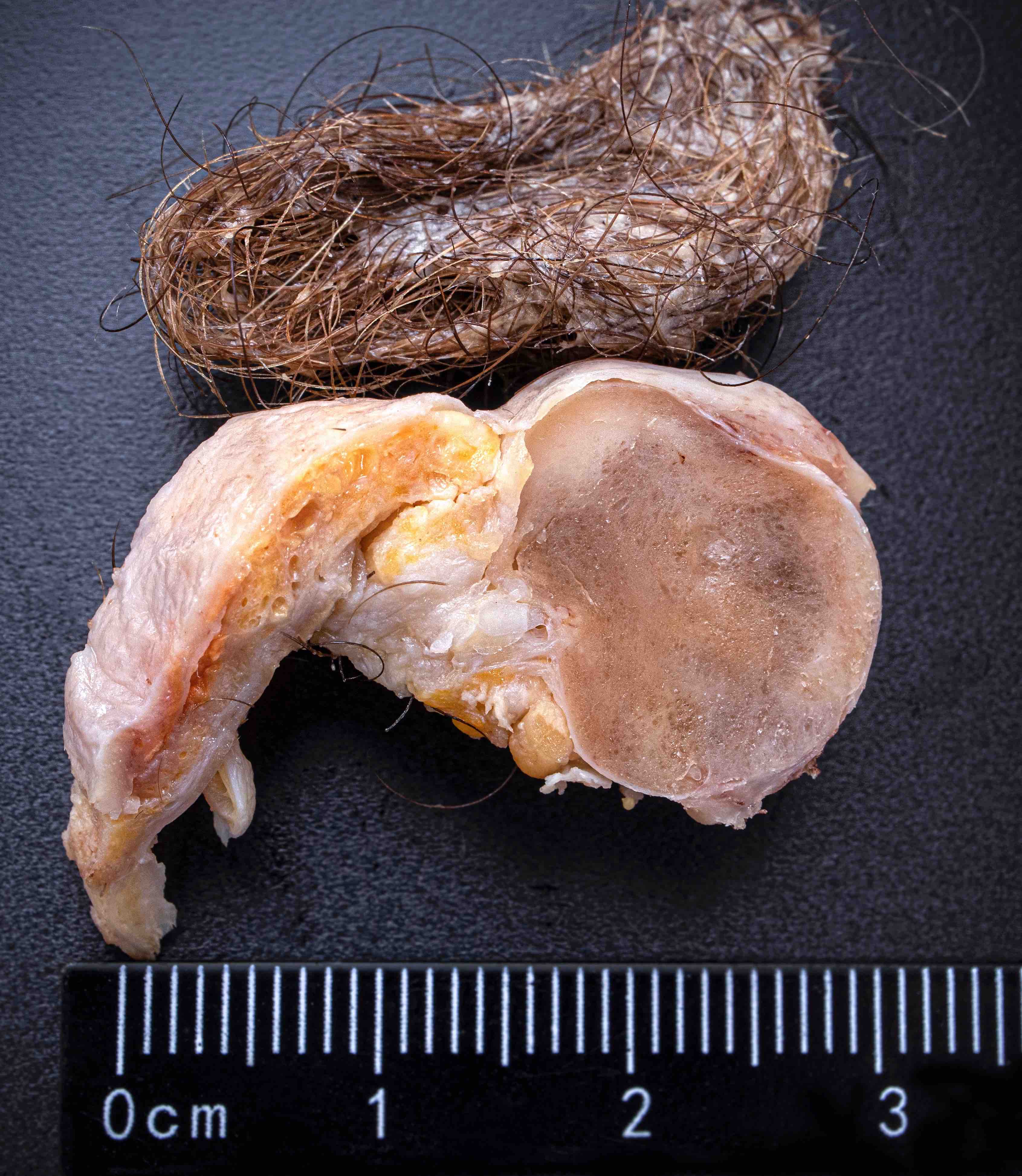
Mature teratoma
Contributed by Shannon Mingo Welter, M.D. and @Andrew_Fltv on Twitter
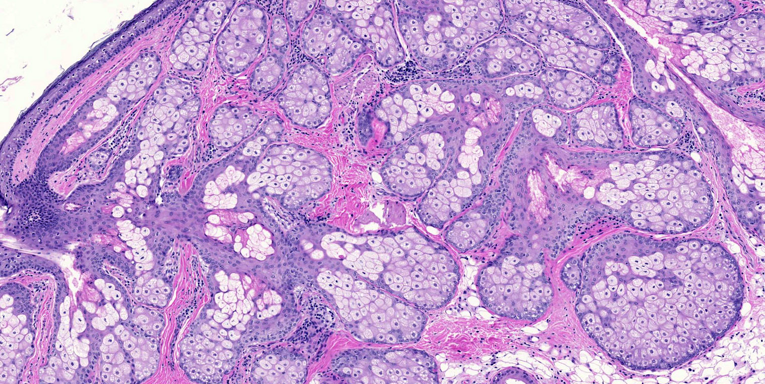
Skin with sebaceous glands
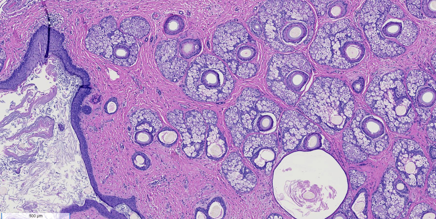
Skin with keratin and adnexa
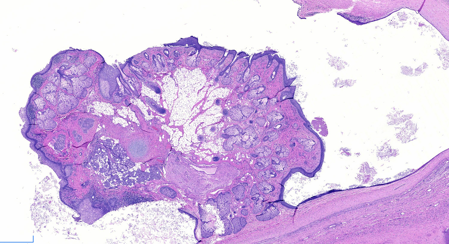
Rokitansky nodule
Mucin producing epithelium
Multiple tissue types

Orderly arrangement of tissues
Cartilage and other mature tissues
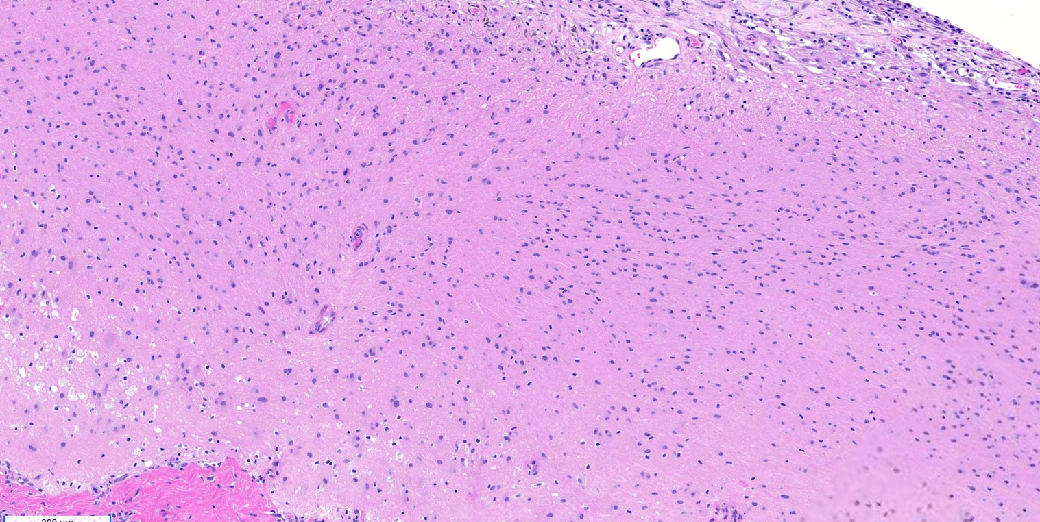
Glial tissue
Gliomatosis peritonei
Mature teratoma
Images hosted on other servers:

Bilateral thecomas on MRI
Images hosted on other servers:

Bitemporal hair loss

Frontal balding and hirsutism
Contributed by Victoria Collins, M.D., Tamara Kalir, M.D., Ph.D. and AFIP
Well circumscribed, yellow-tan mass
Lobulated and yellow
Images hosted on other servers:

Yellow, lobulated mass
Contributed by Victoria Collins, M.D., Tamara Kalir, M.D., Ph.D. and AFIP
Well circumscribed

Ovoid to round cells
Mild nuclear atypia
Thecoma and fibroma areas

Cystic change
Hyaline plaques
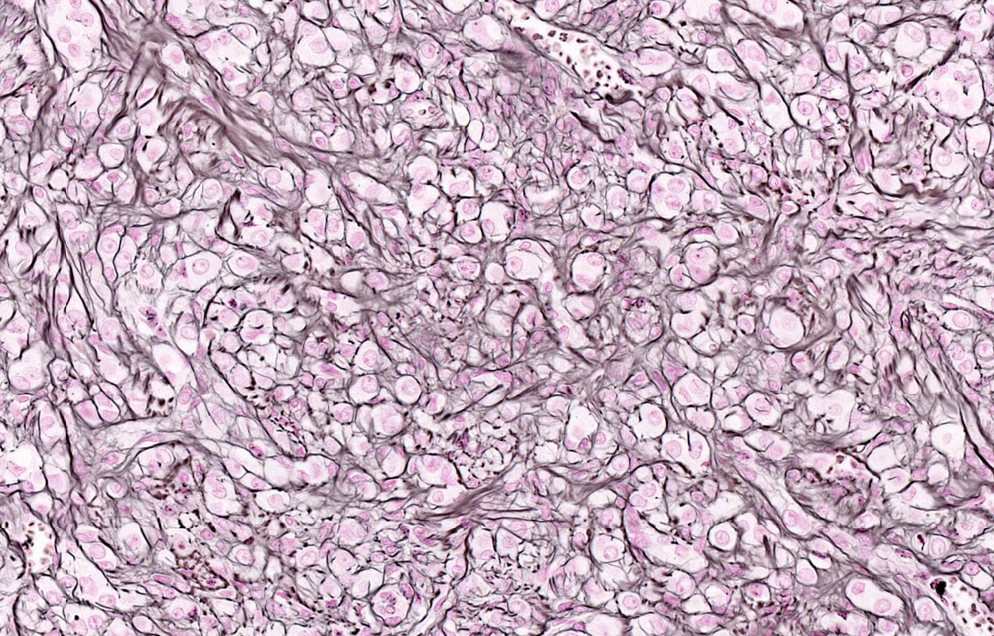
Reticulin
Inhibin
Vacuolated tumor cells
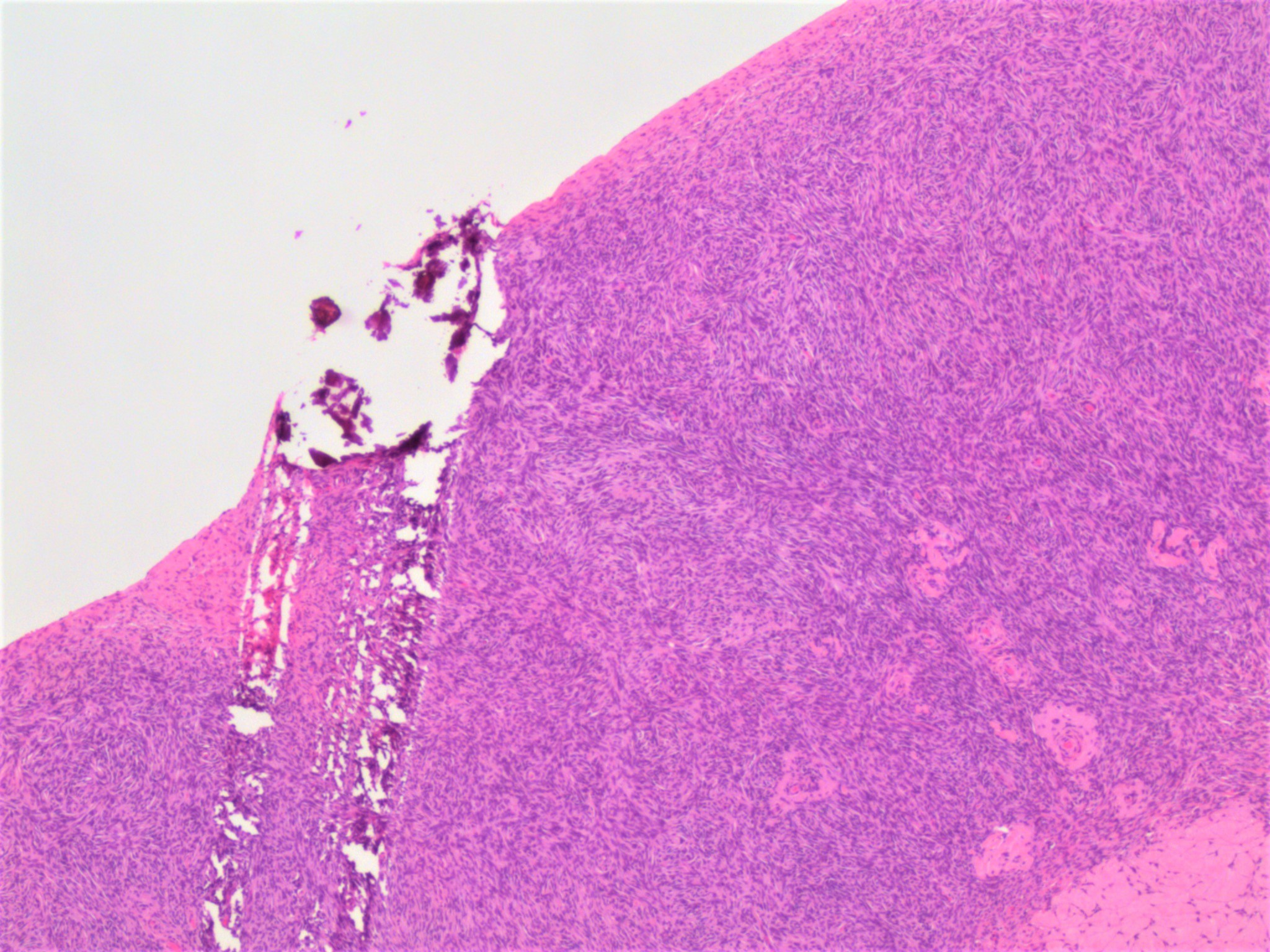
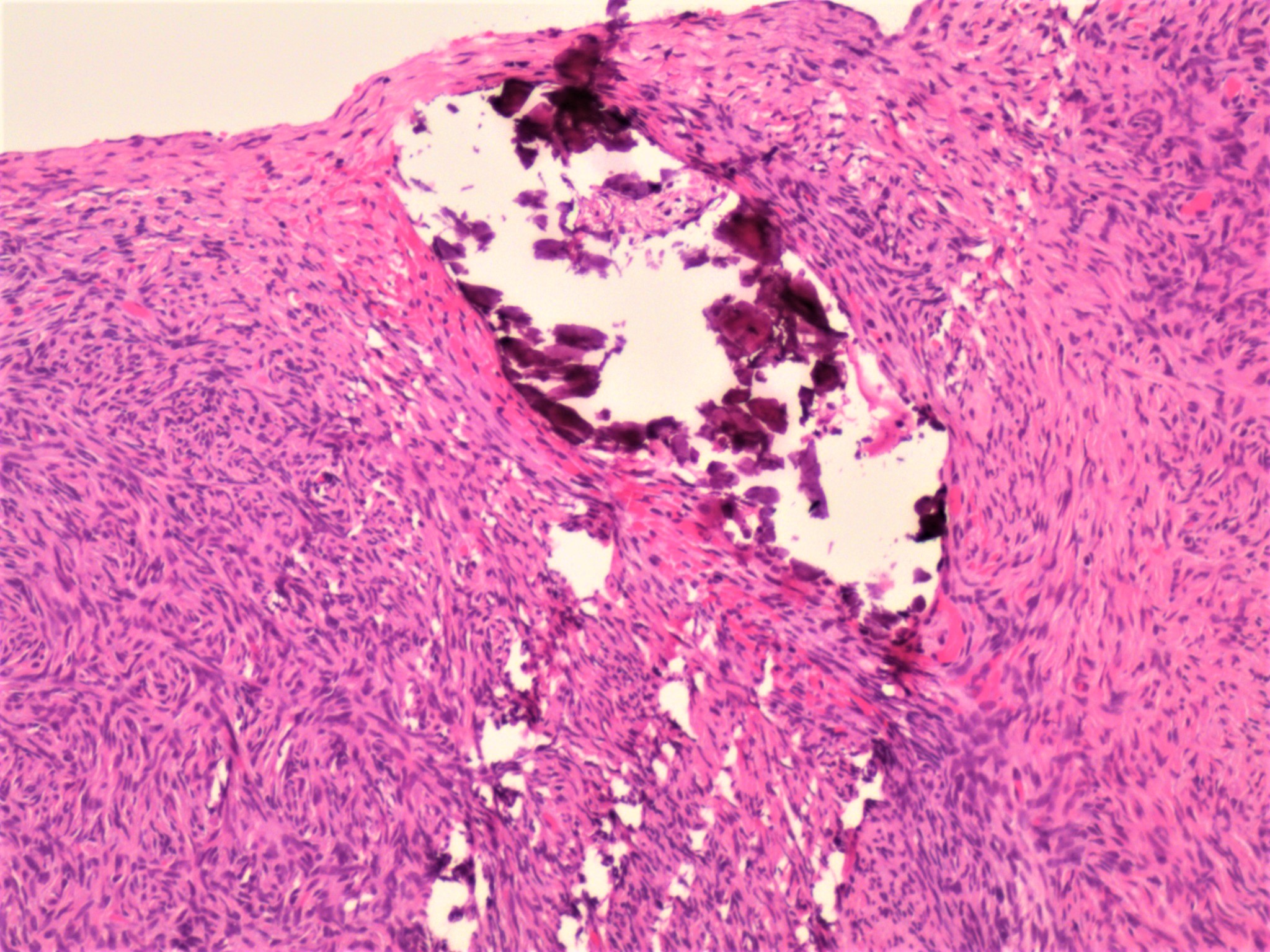
Stromal calcification
Oil red O stain
Thecoma of ovary
Ovarian pathology
Images hosted on other servers:


Fallopian tube
AFIP images
Hemorrhagic infarction of tube
Images hosted on other servers:

Surgical specimen
Contributed by Basile Tessier-Cloutier, M.D.
Undifferentiated and differentiated components
Solid architecture
Discohesive rhabdoid cells
ARID1B
Contributed by Carlos Parra-Herran, M.D.
Gastric
Pancreatic
Contributed by Lewis A. Hassell, M.D.
Serous borderline tumor
Serous
borderline tumor,
micropapillary
type

High grade serous carcinoma
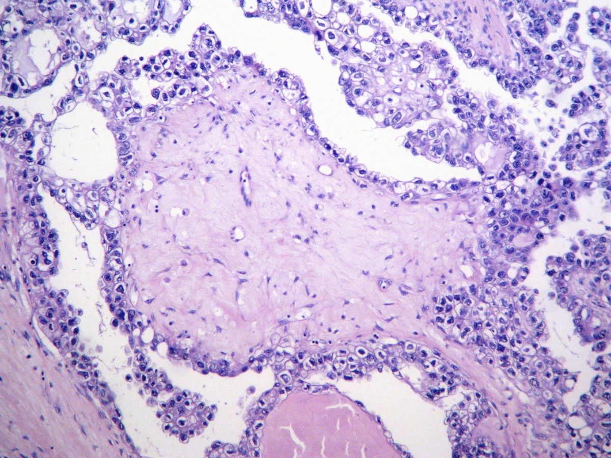
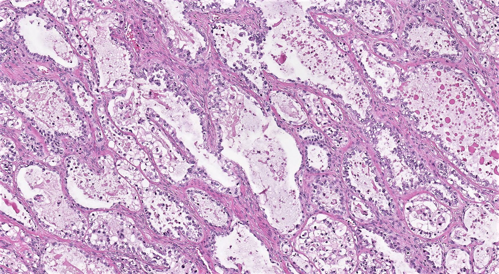
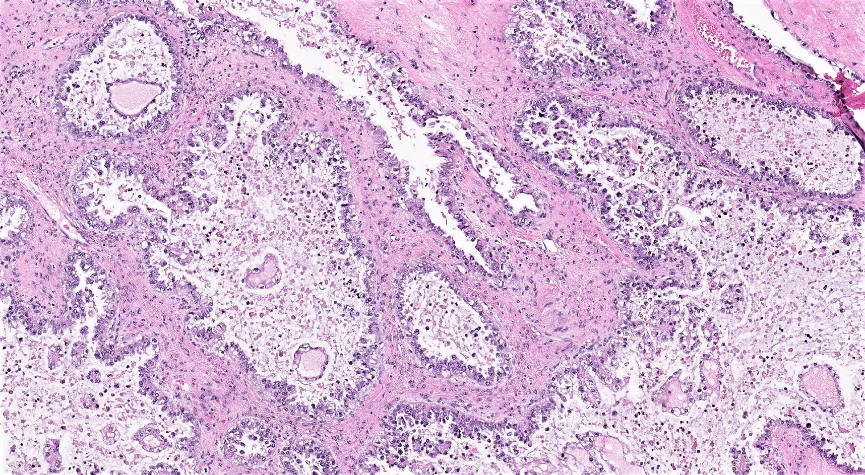
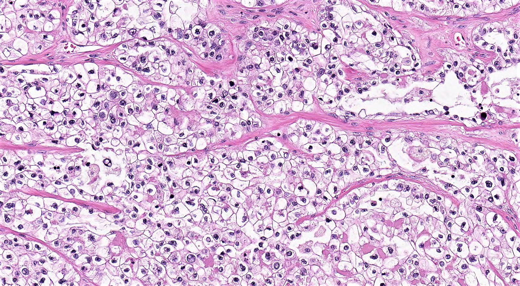
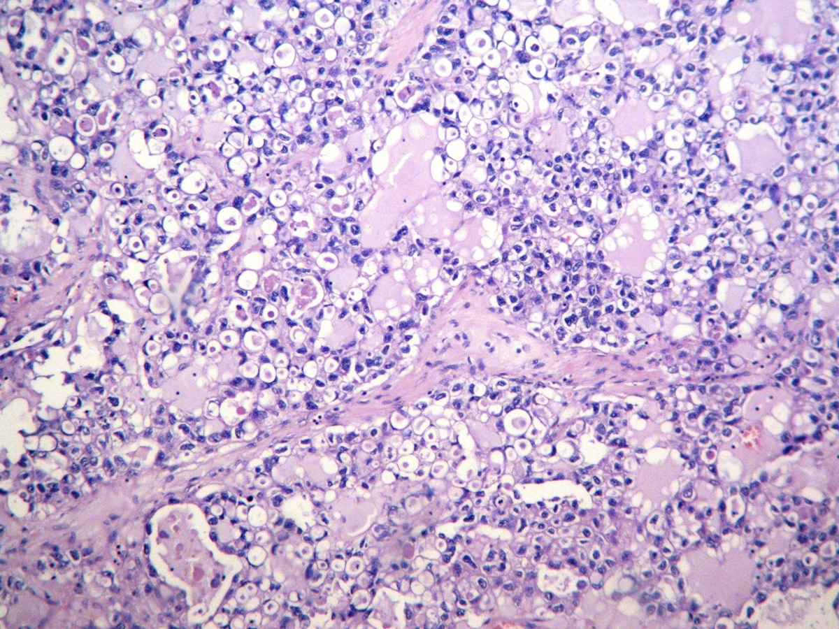
Clear cell carcinoma, ovary
Clear cell adenofibroma, borderline
Seromucinous borderline tumor
Dedifferentiated carcinoma in ovary
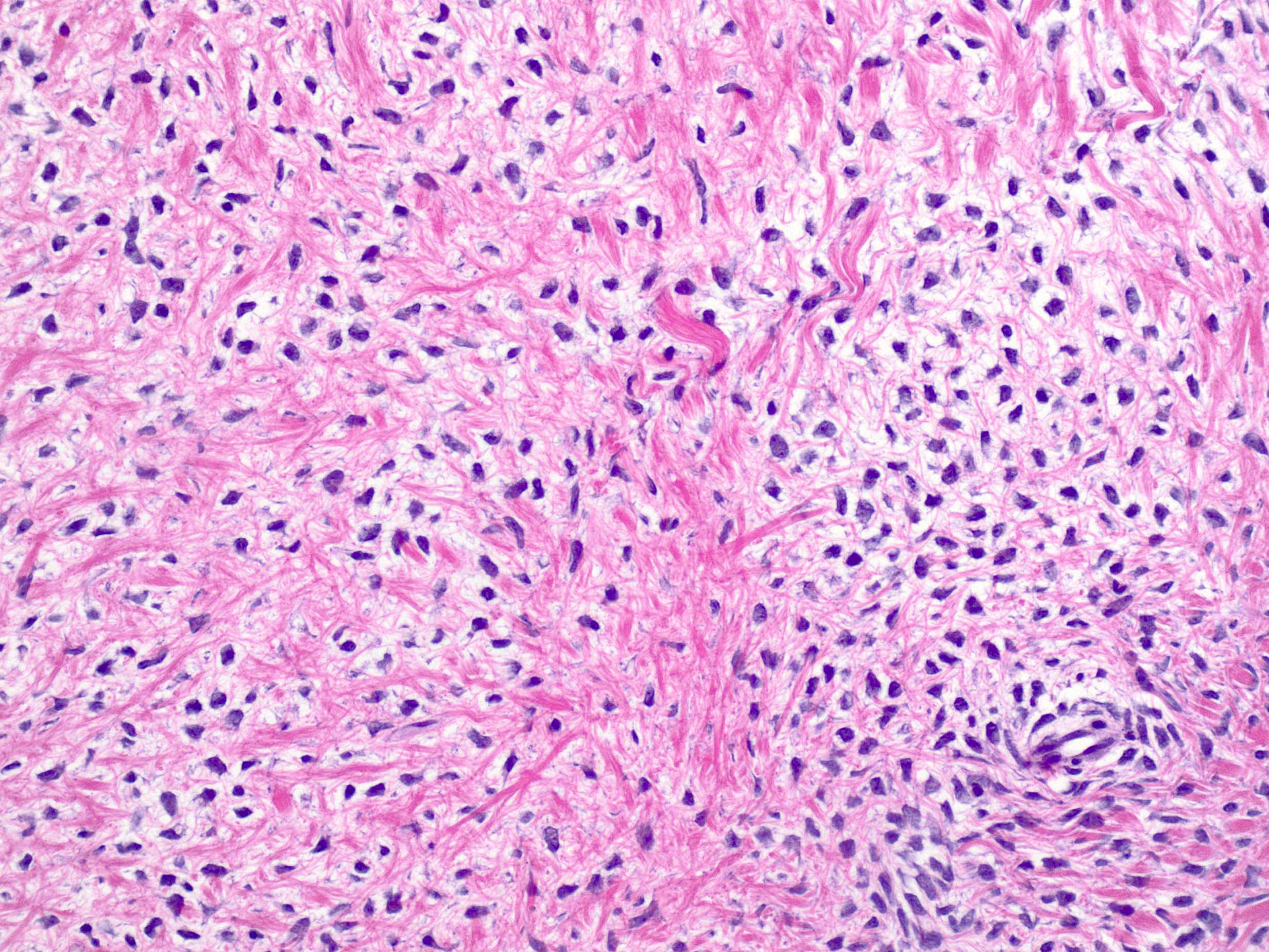
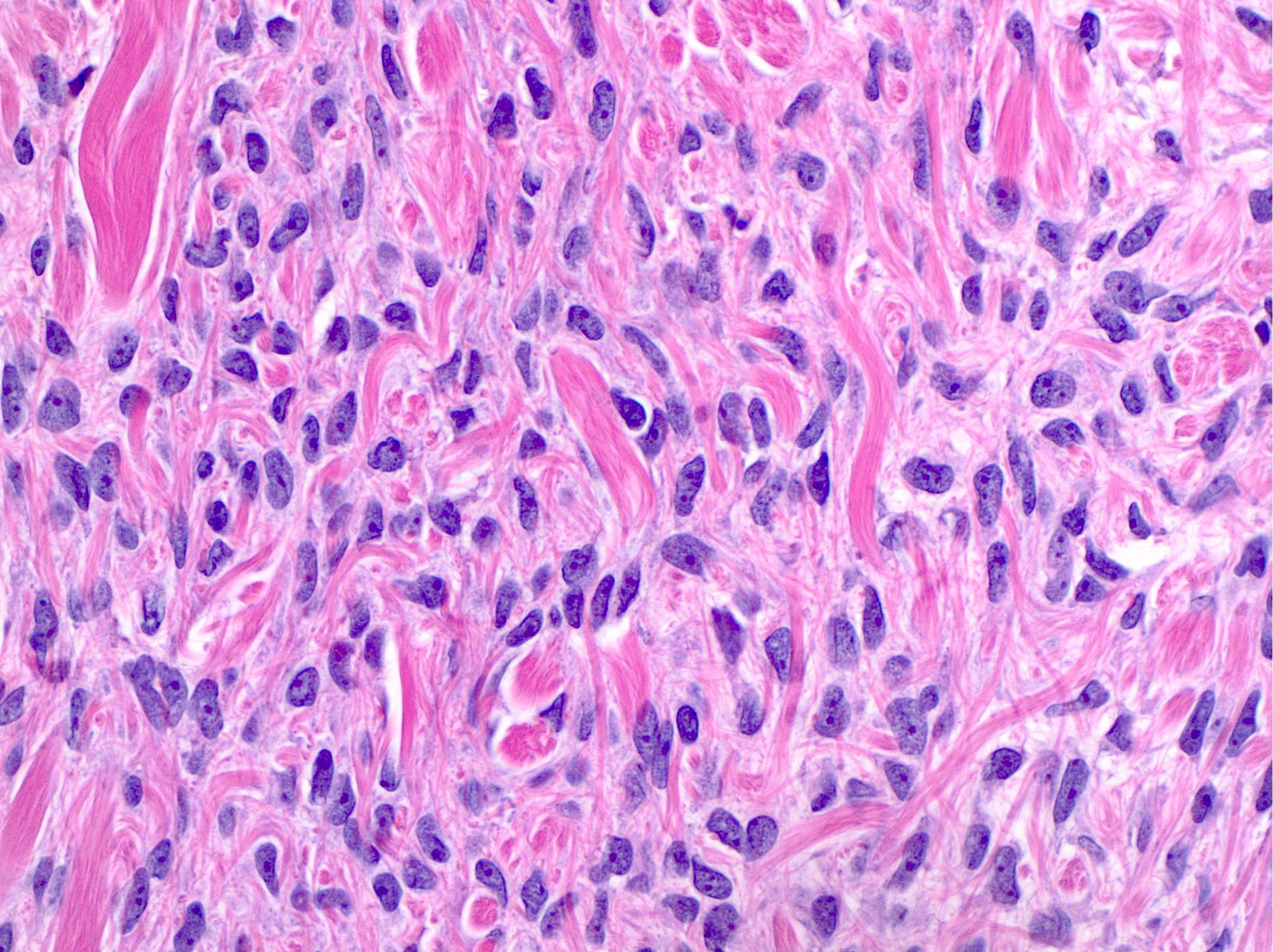
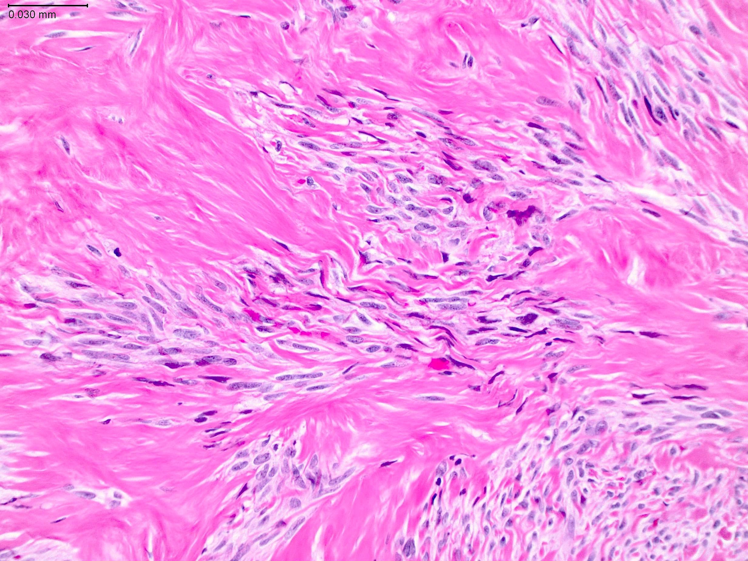
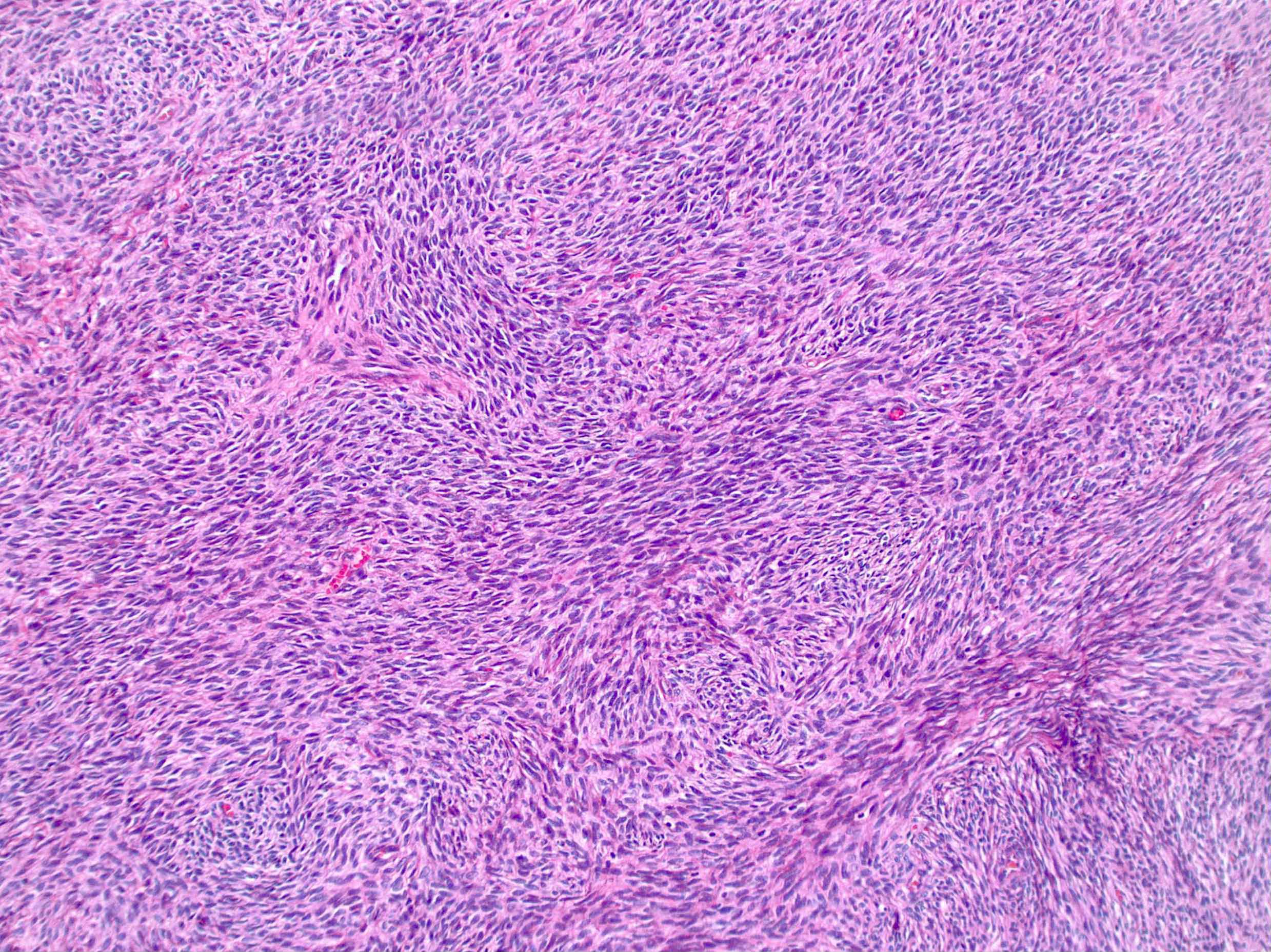
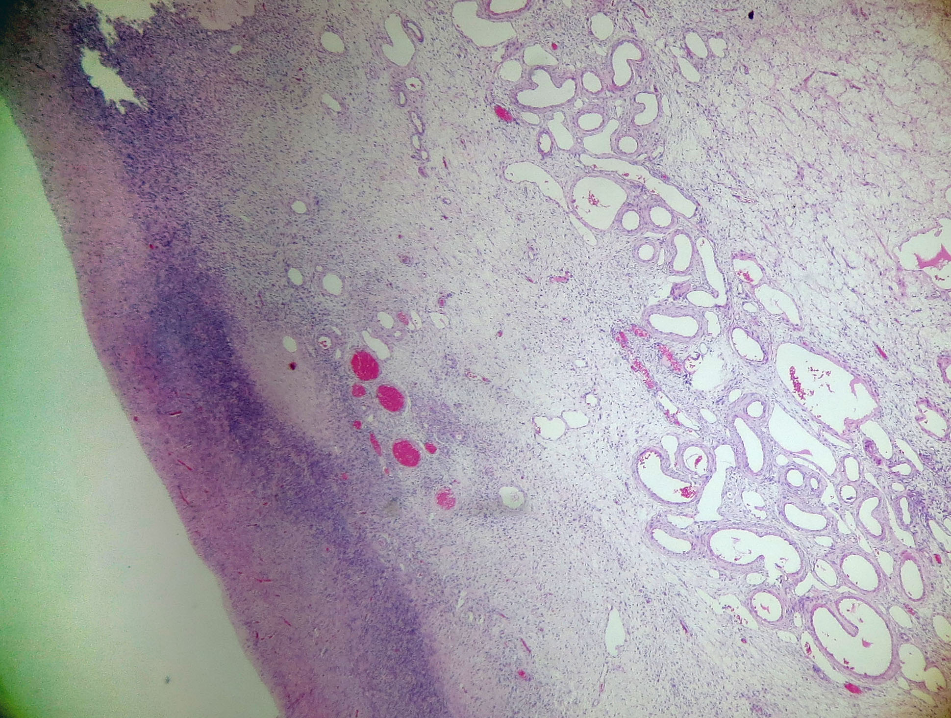
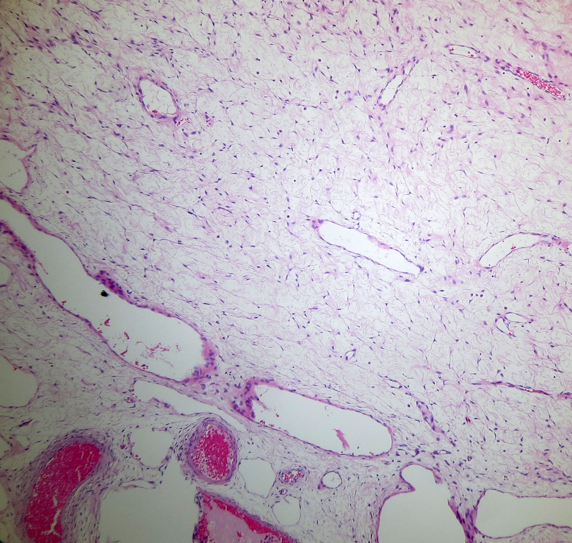
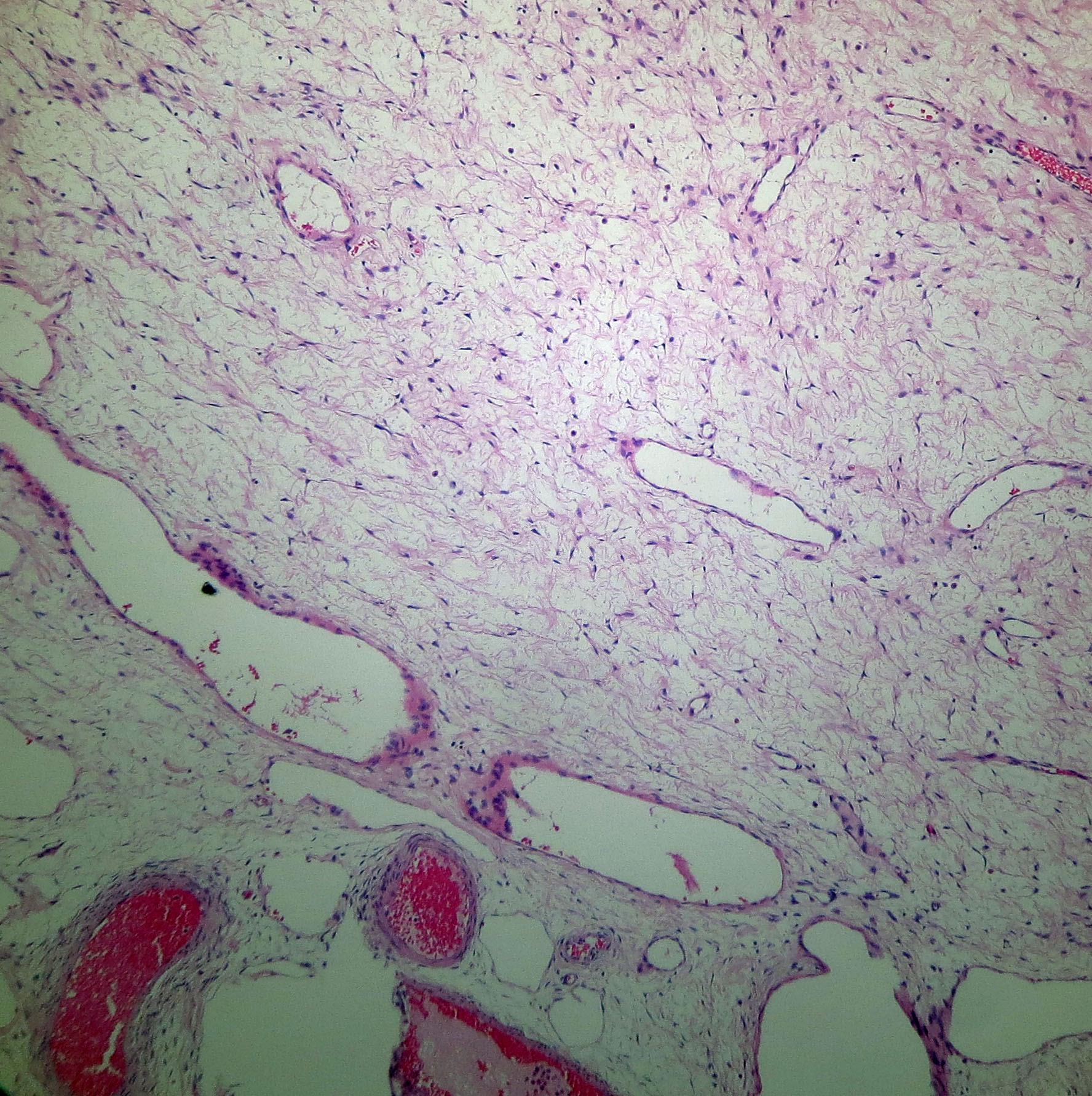
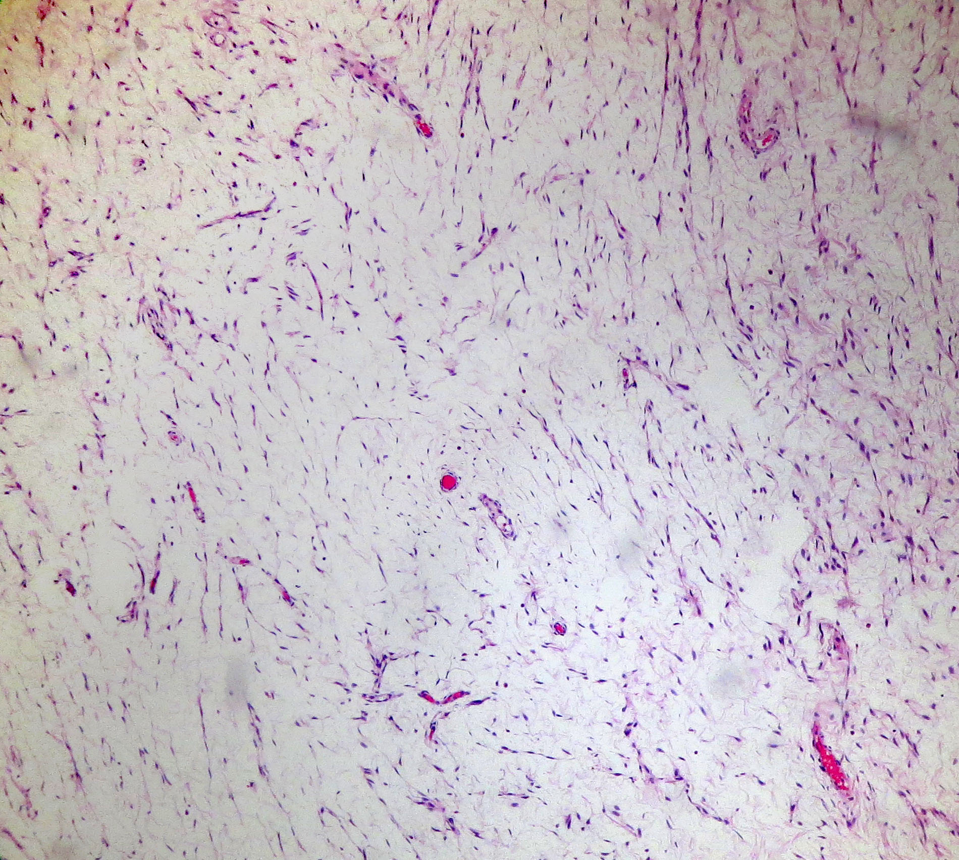
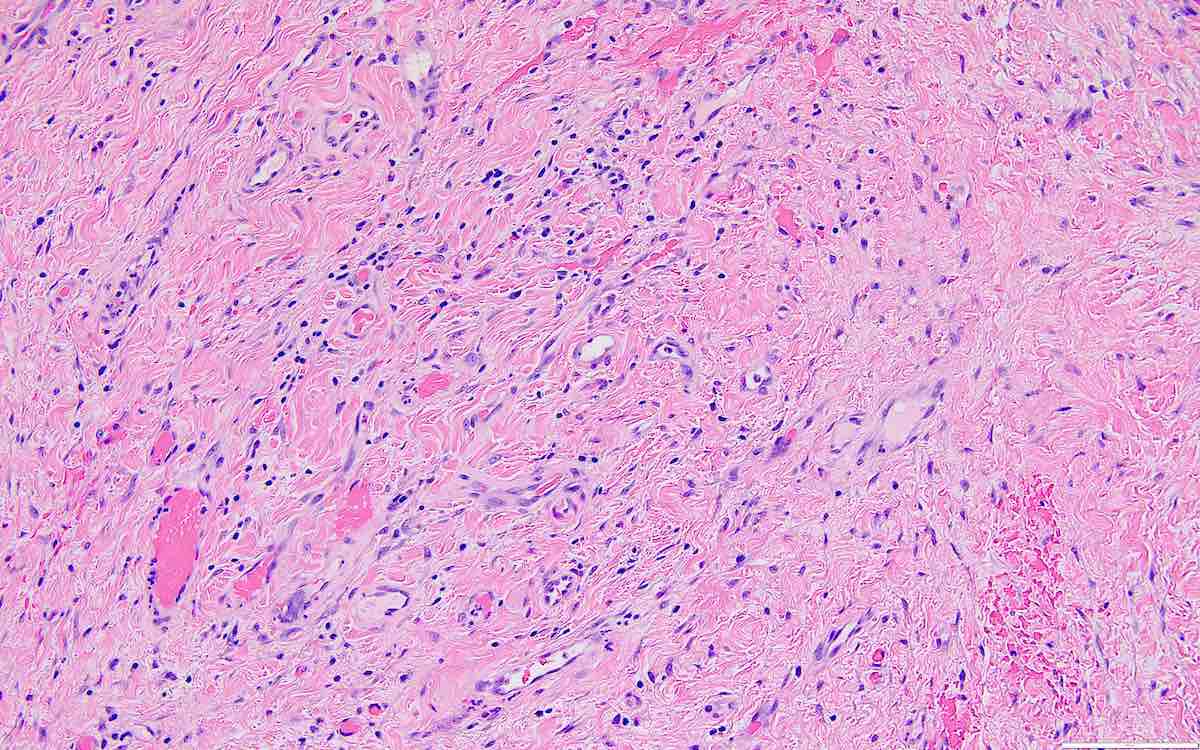
Sclerosing stromal tumor
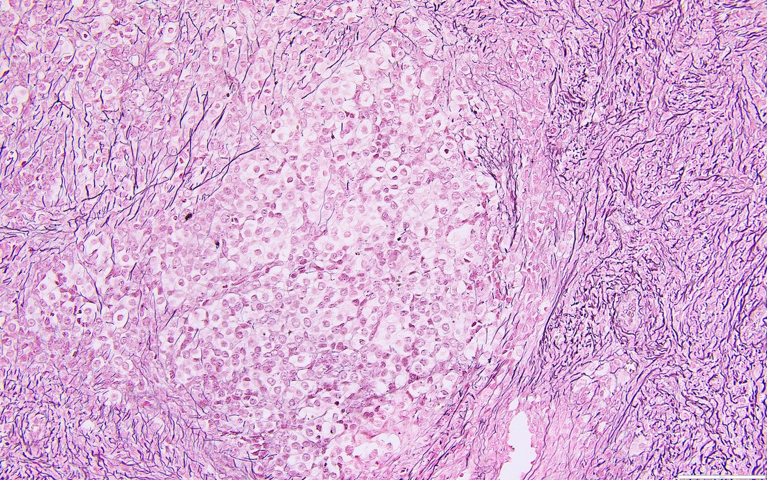
Steroid cell tumor, ovary
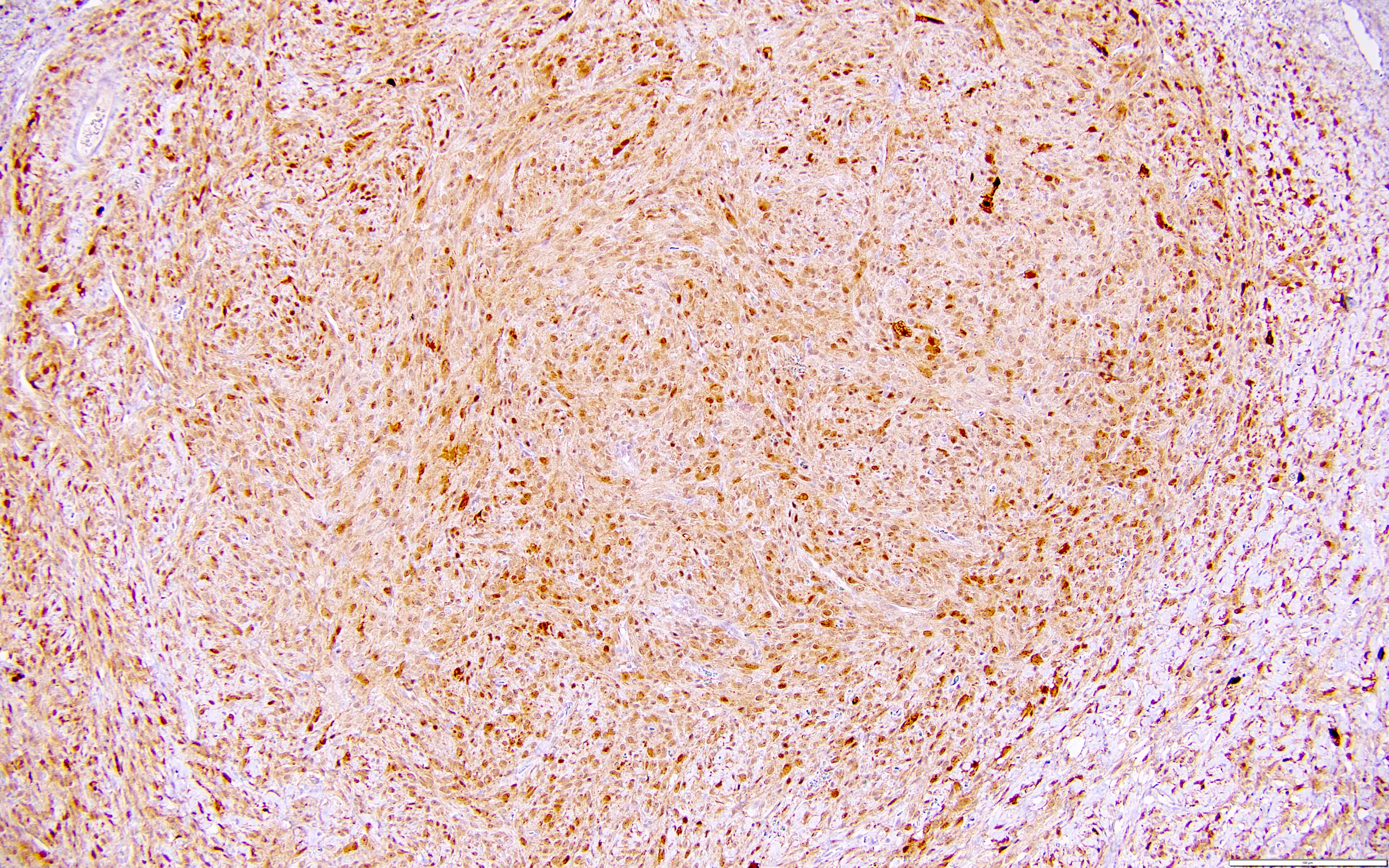
Granulosa cell tumor, adult type

Sex cord tumor with annular tubules
Retiform Sertoli-Leydig tumor
Sertoli-Leydig tumor, intermediate
Mixed germ cell tumor
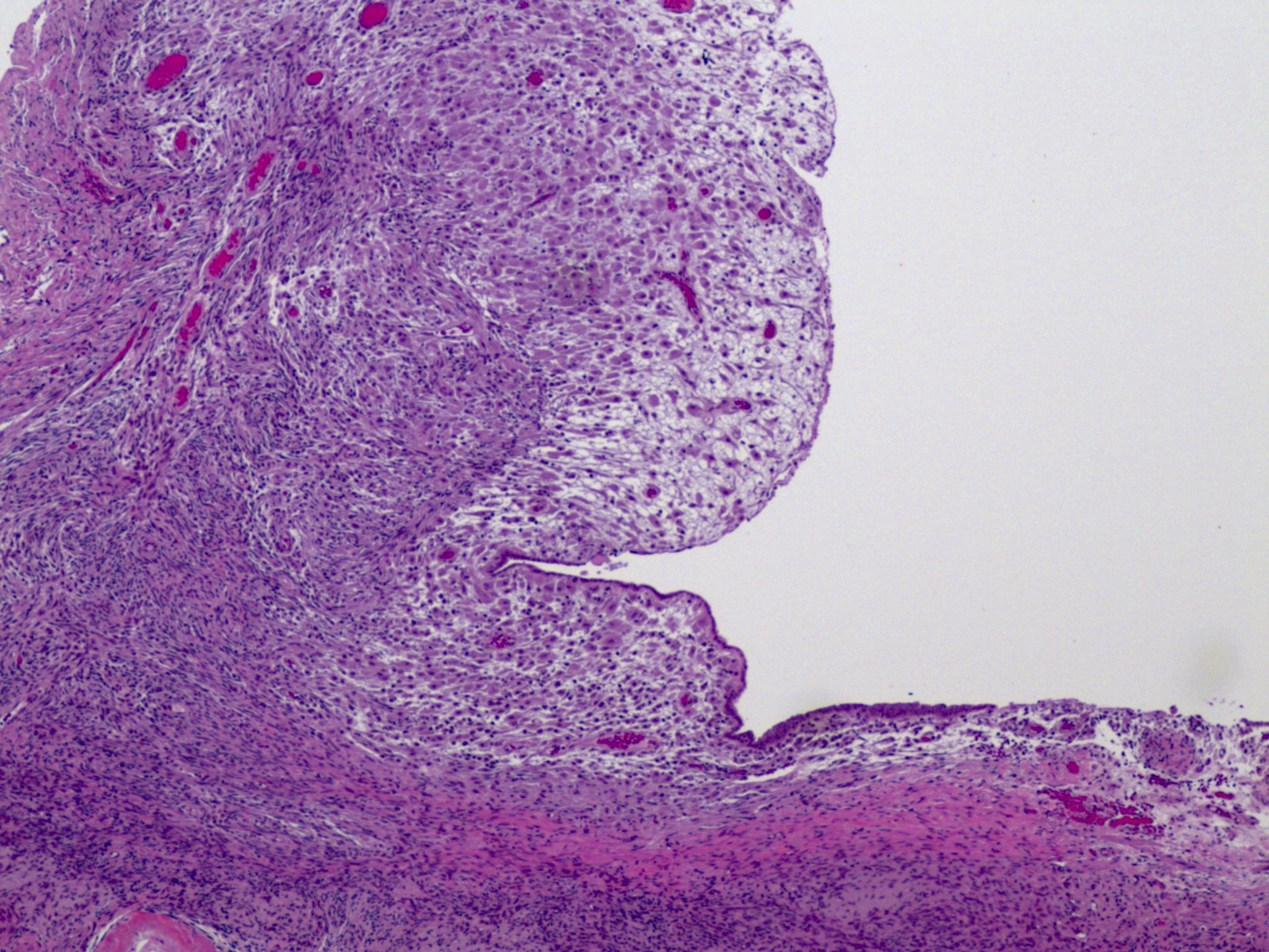
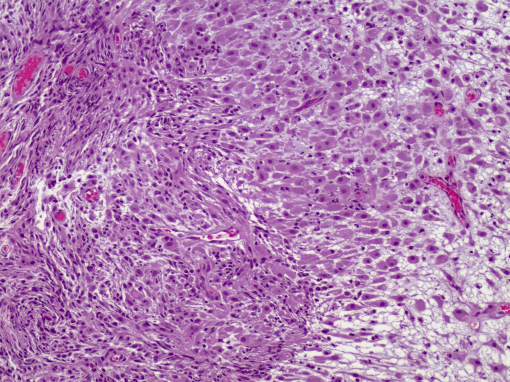
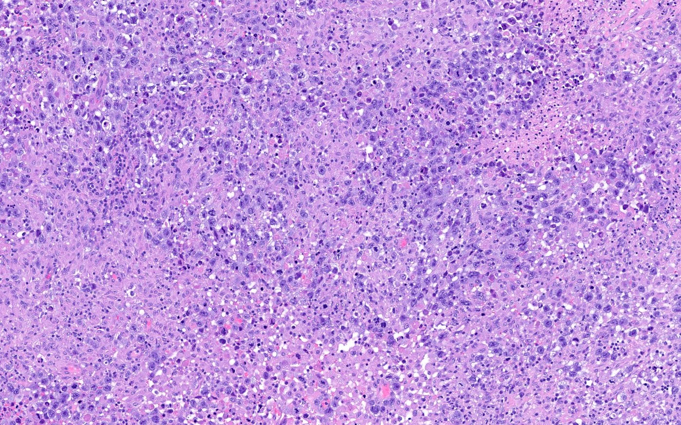
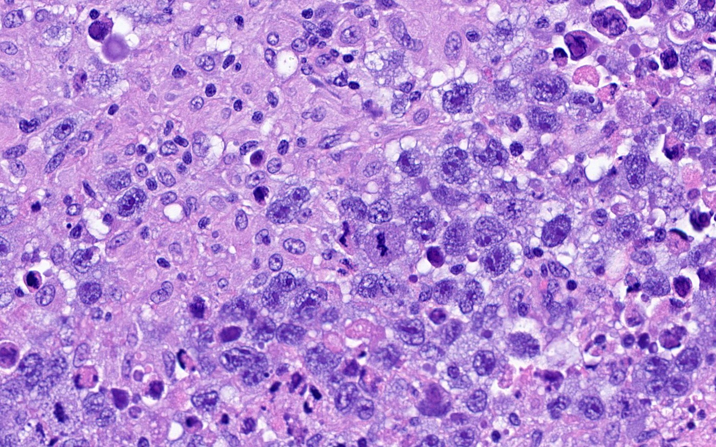
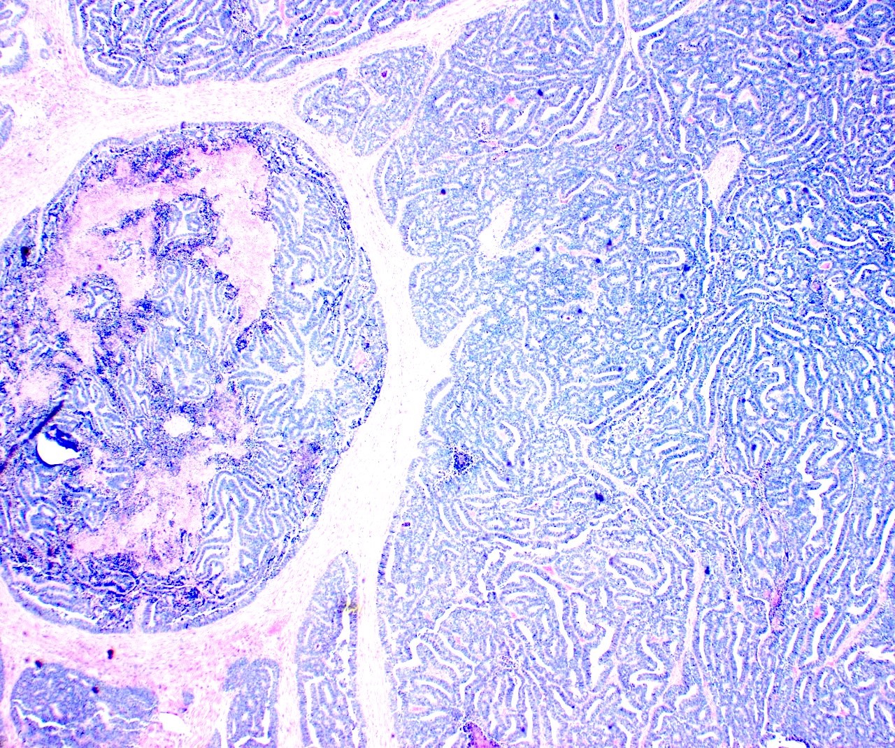
Immature teratoma
Images hosted on other servers:

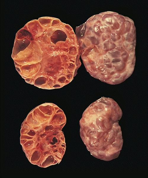
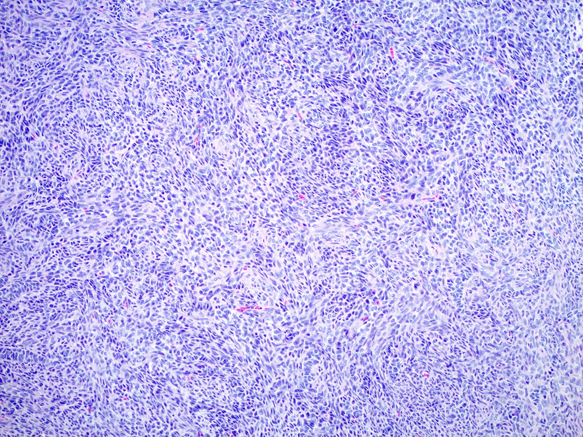
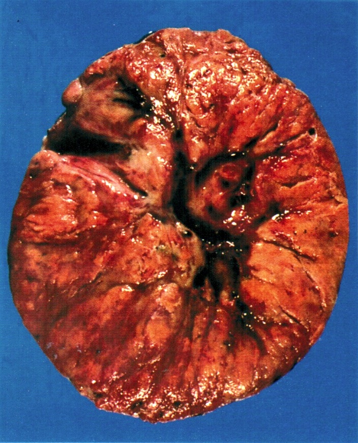
Wilms tumor involving both kidneys
Images hosted on other servers:


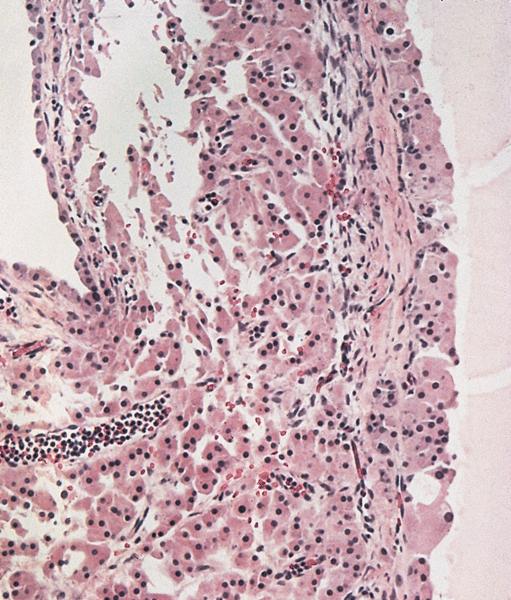
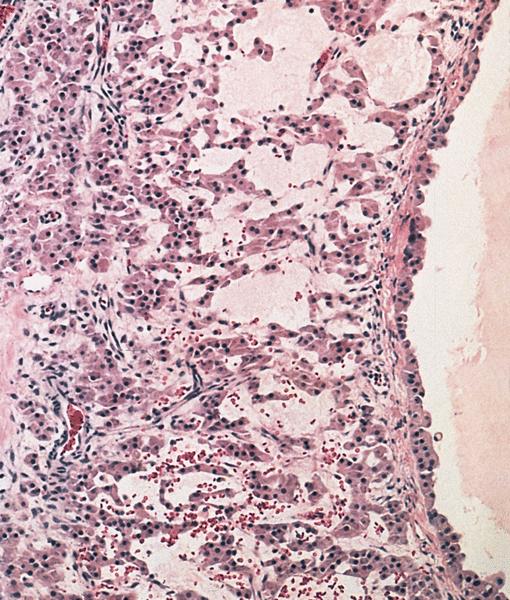
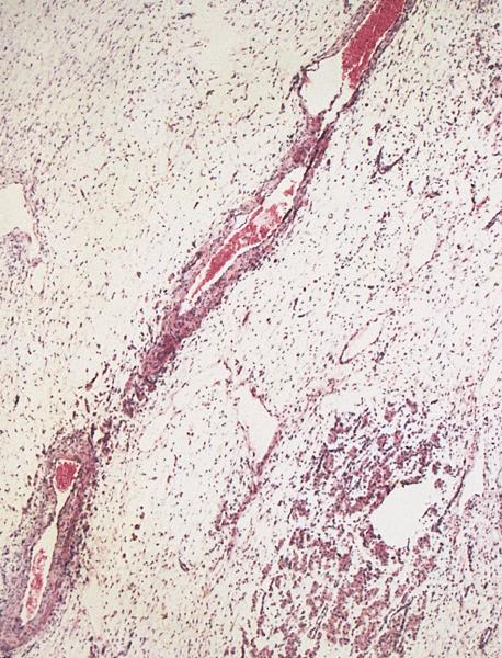
Adult tumor (kidney)


Various images (kidney)
Images hosted on other servers:

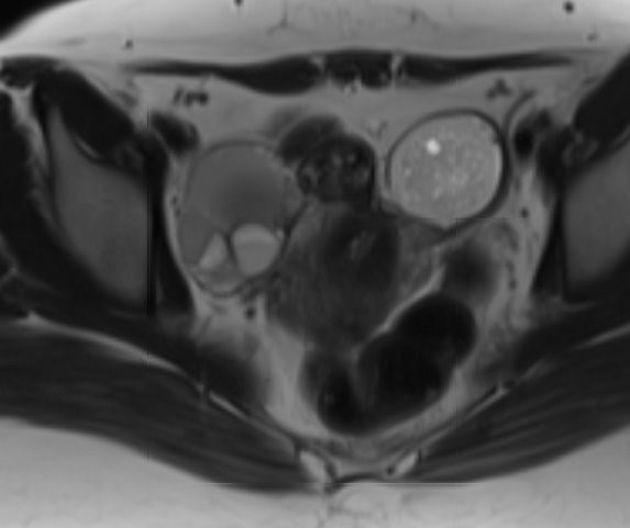
Pelvic sonography
shows a mixed
echogenicity mass

T2 MRI shows multiseptated cystic mass


Axial T2 weighted turbo spin echo MRI (TR / TE, 3900 / 99)

Axial T1 weighted spin echo MRI (800 / 12)
Contributed by Jian-Hua Qiao, M.D.
Yellow nodules
Contributed by Jian-Hua Qiao, M.D.

Foamy histiocytes in cystic wall
Foamy histiocytes mixed with inflammatory cells


Foamy histiocytes
Images hosted on other servers:


Large mass with ascites

Bilateral ovarian mass
Images hosted on other servers:

Large ovarian tumor
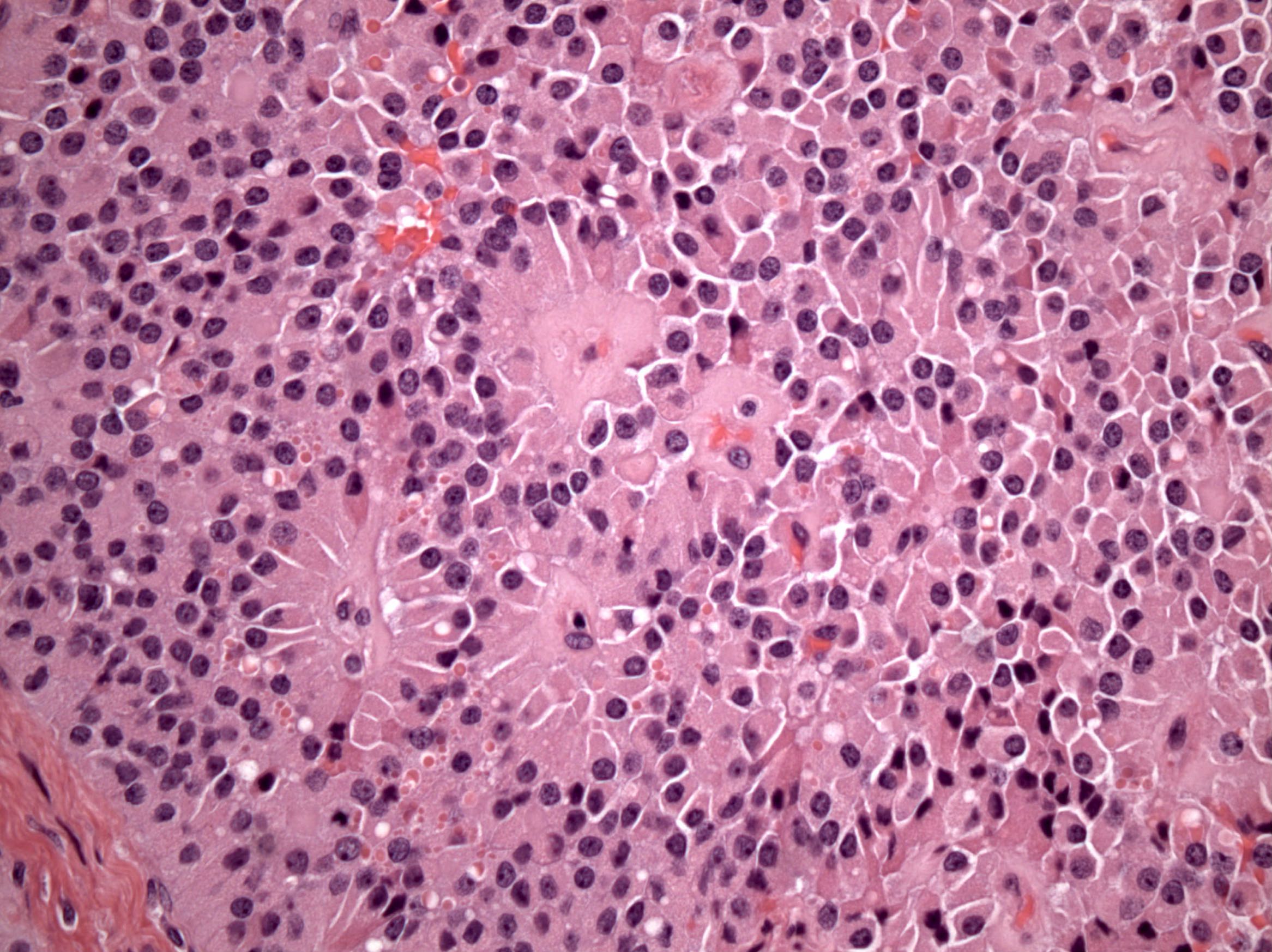
Contributed by Debra L. Zynger, M.D. and AFIP images
Smooth, nodular exterior

Gelatinous cut surface

Gelatinous cystic surface
Honeycomb surface (polyvesicular vitelline pattern)
Contributed by Gulisa Turashvili, M.D., Ph.D., Sharon Song, M.D. and AFIP images
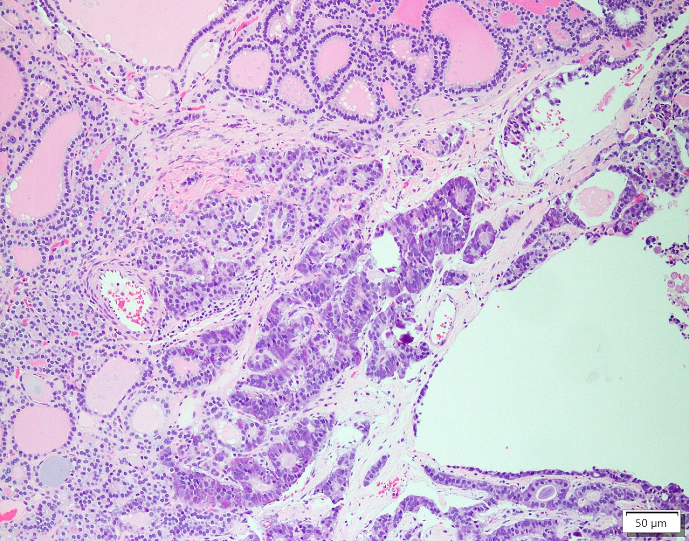
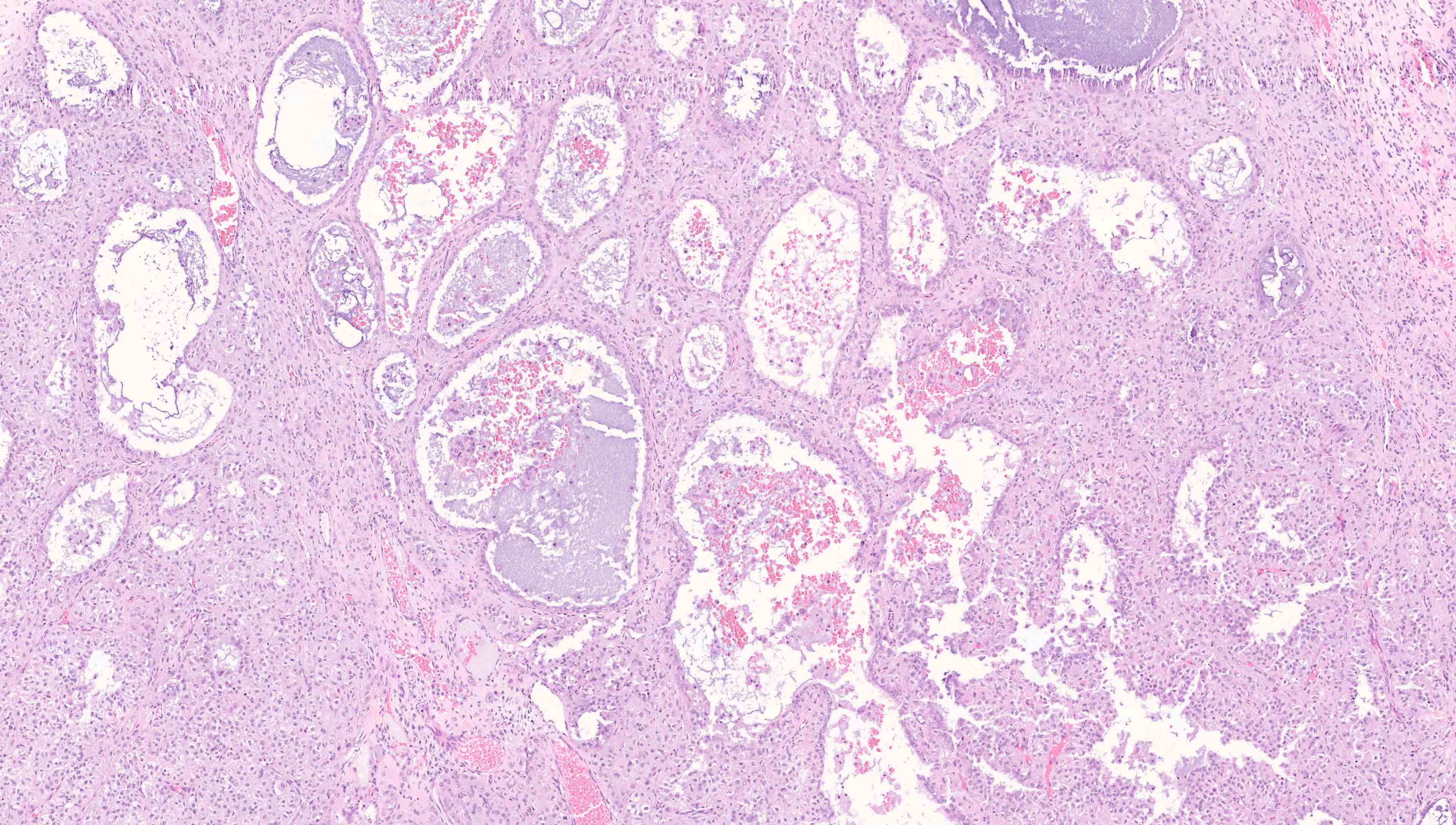
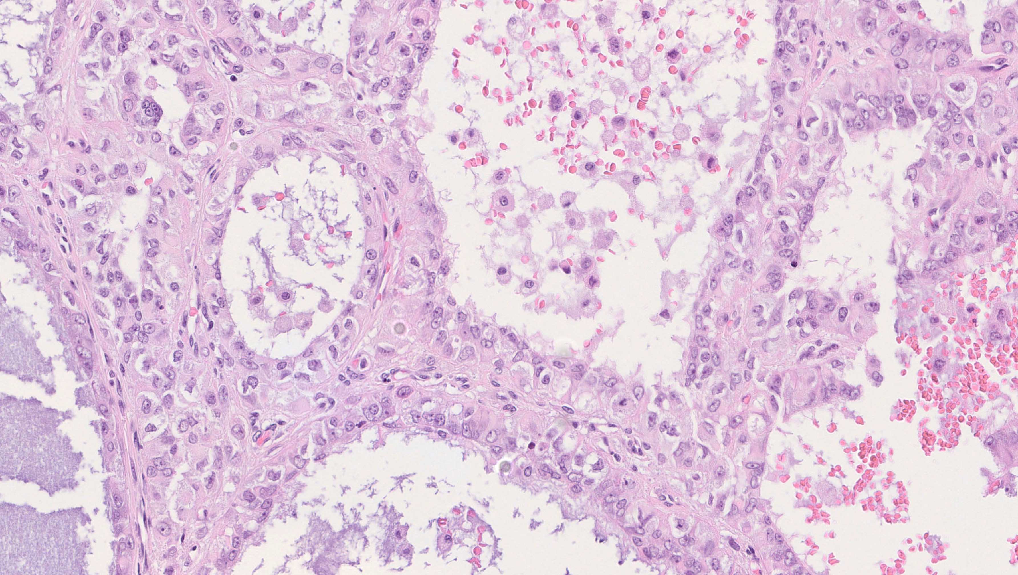
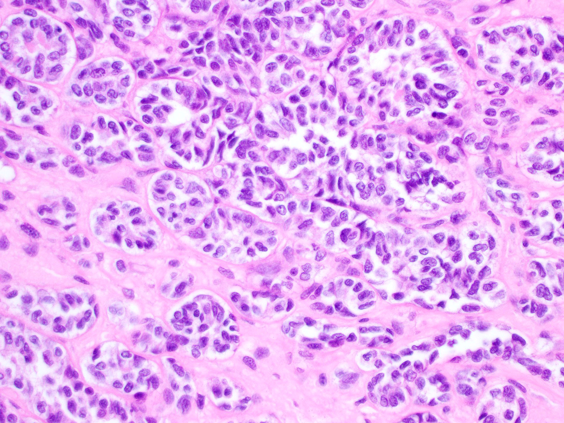
Reticular / microcystic pattern
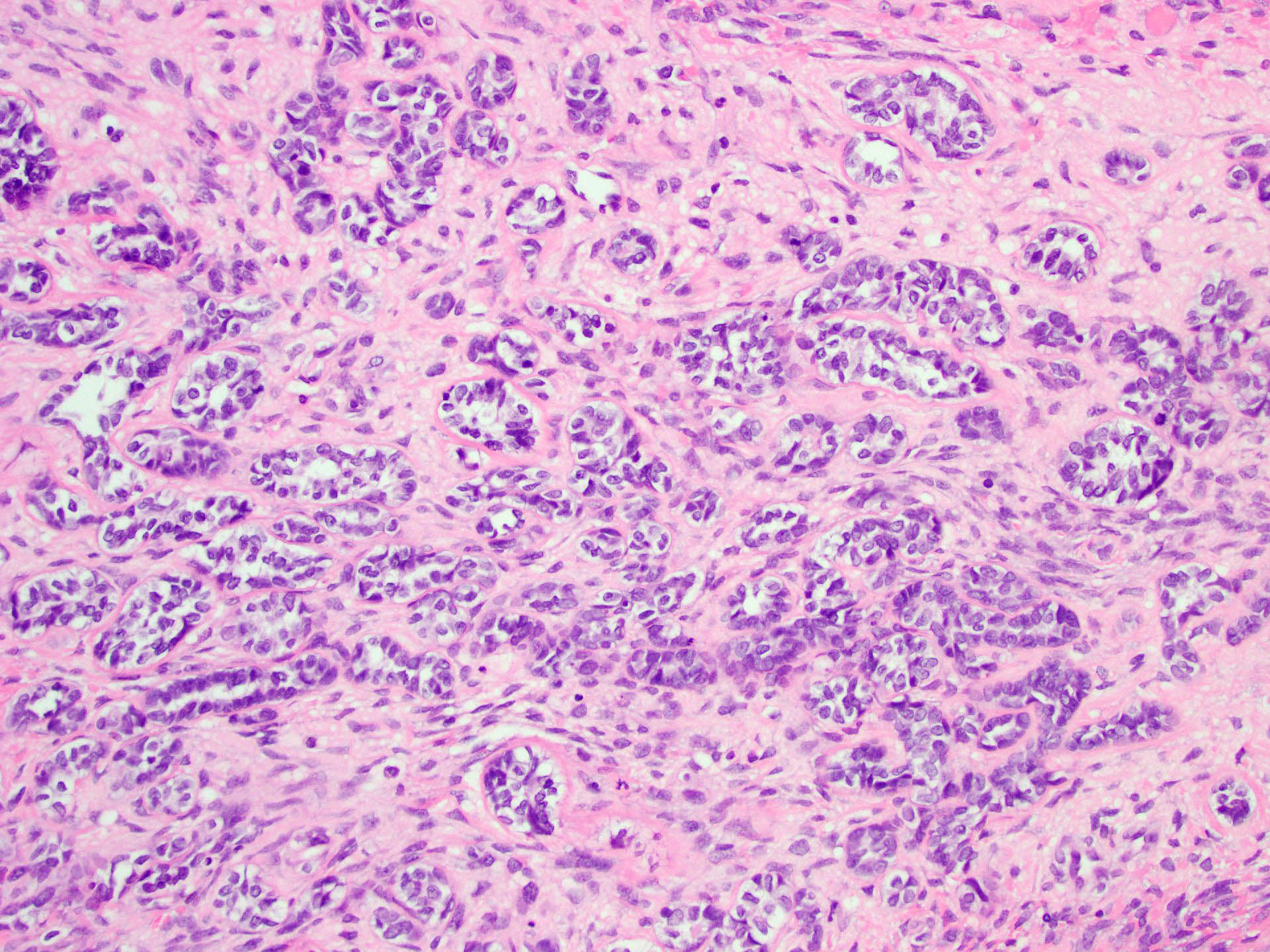
Glandular pattern
Endometrioid-like glandular variant
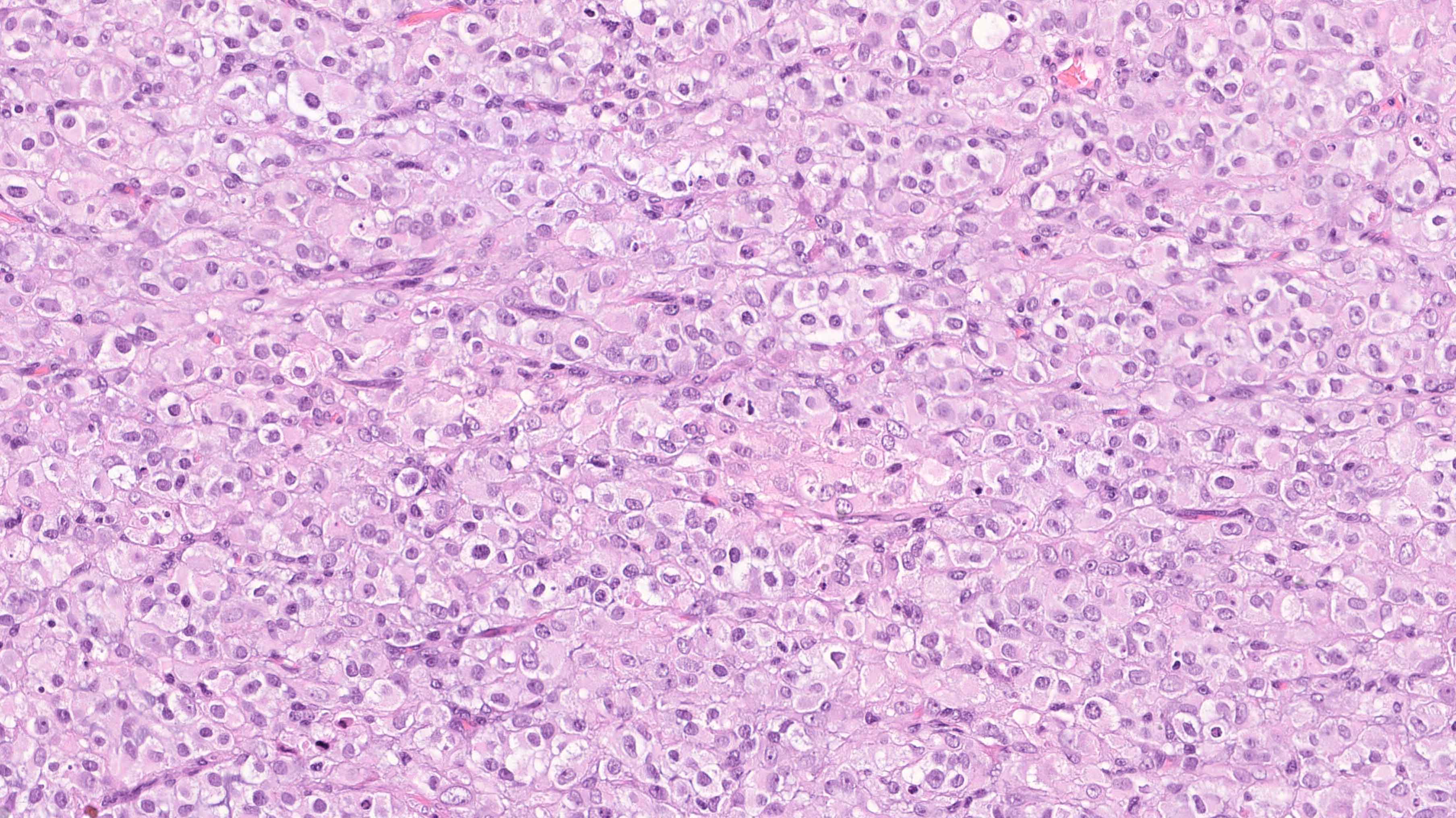
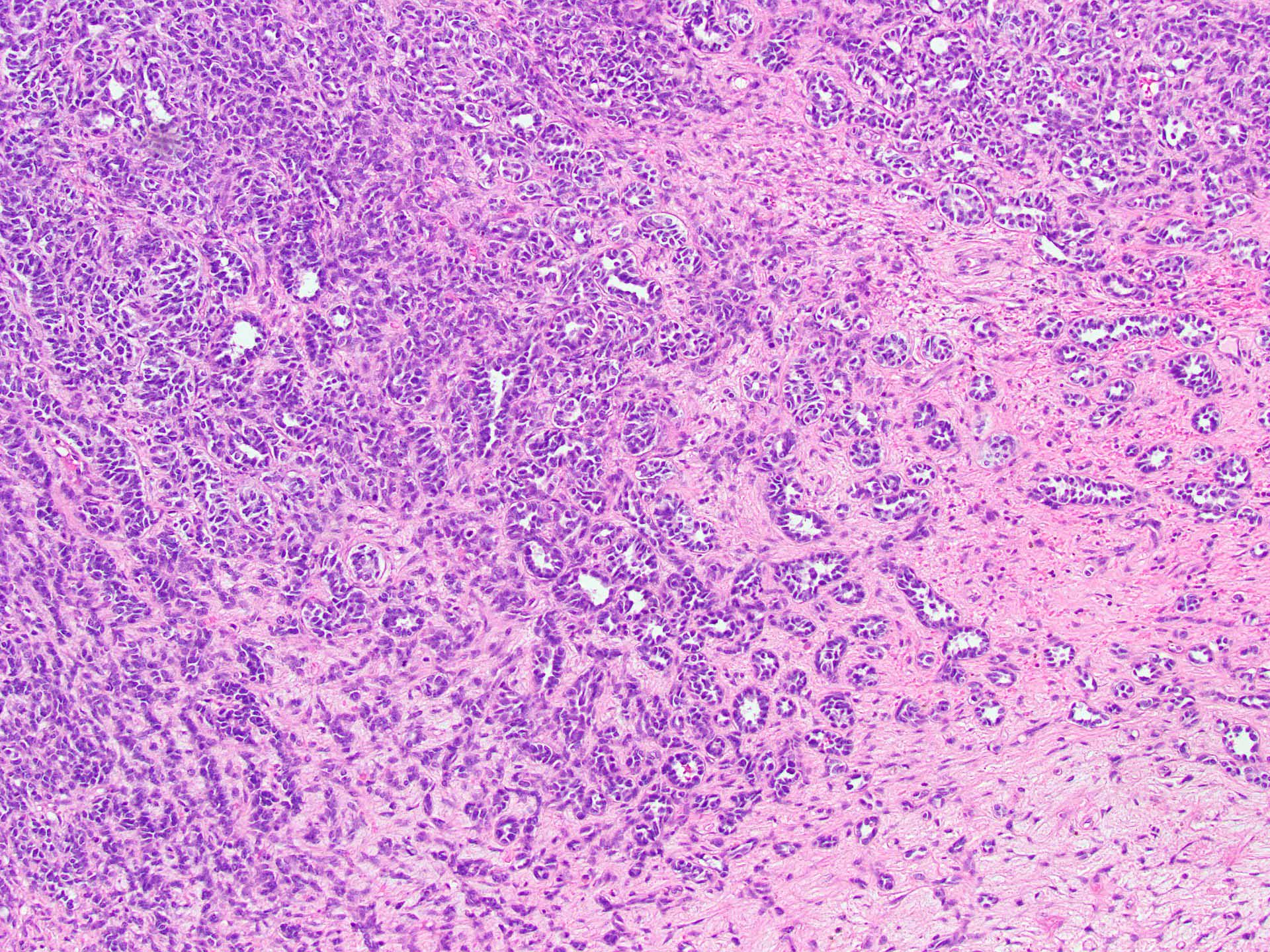
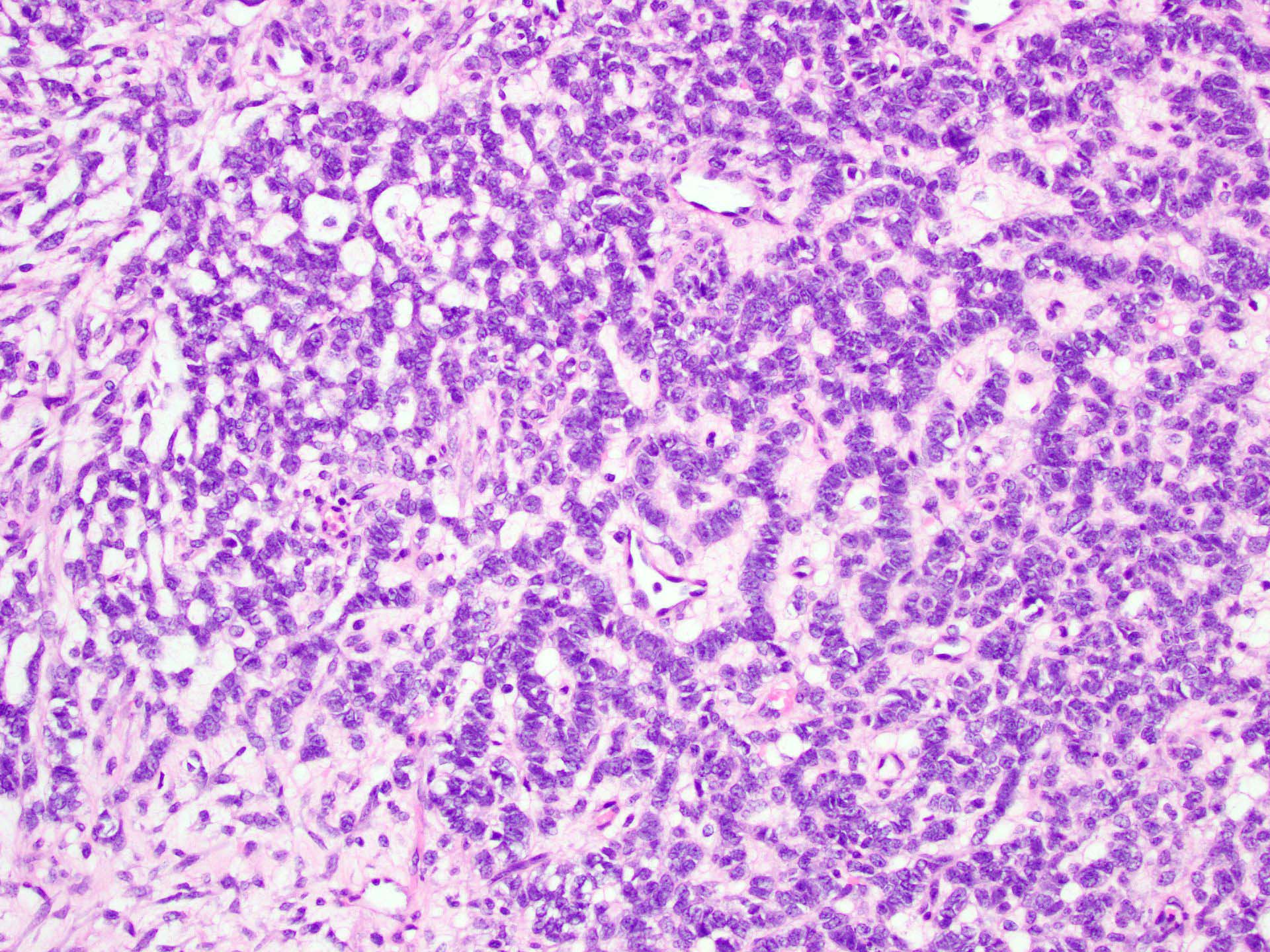
Solid pattern

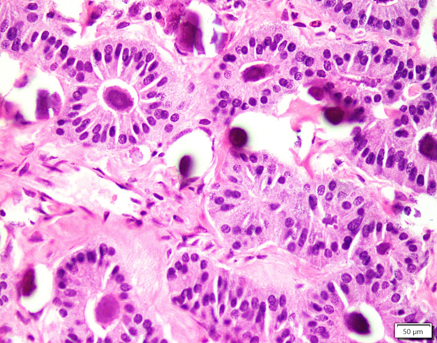
Papillary pattern
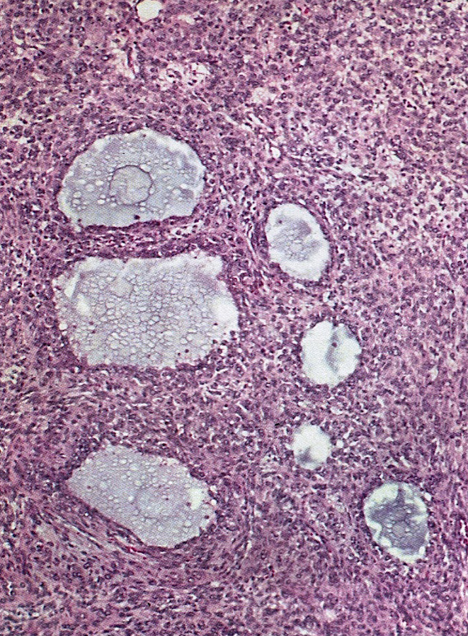
Polyvesicular vitelline pattern
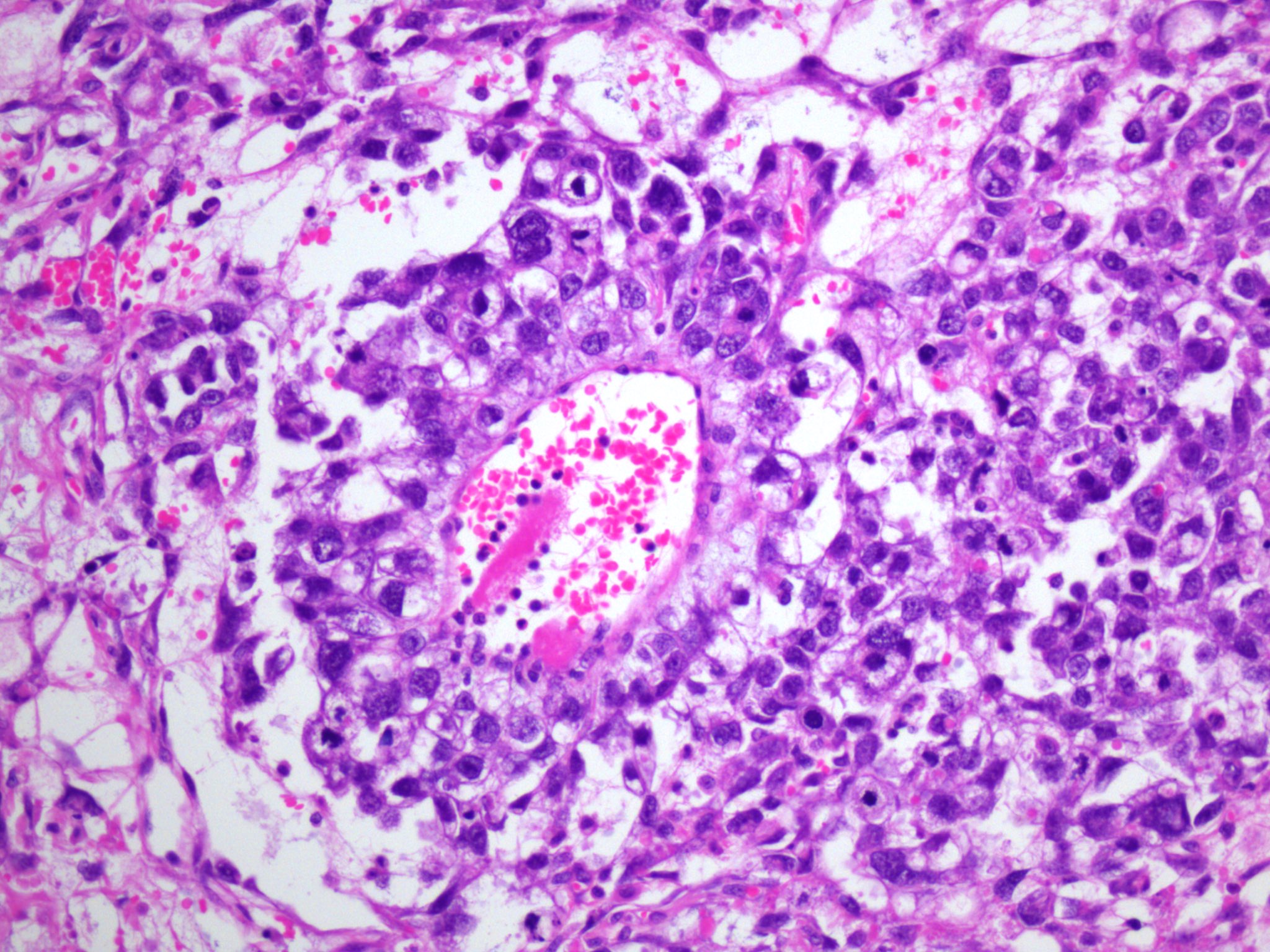
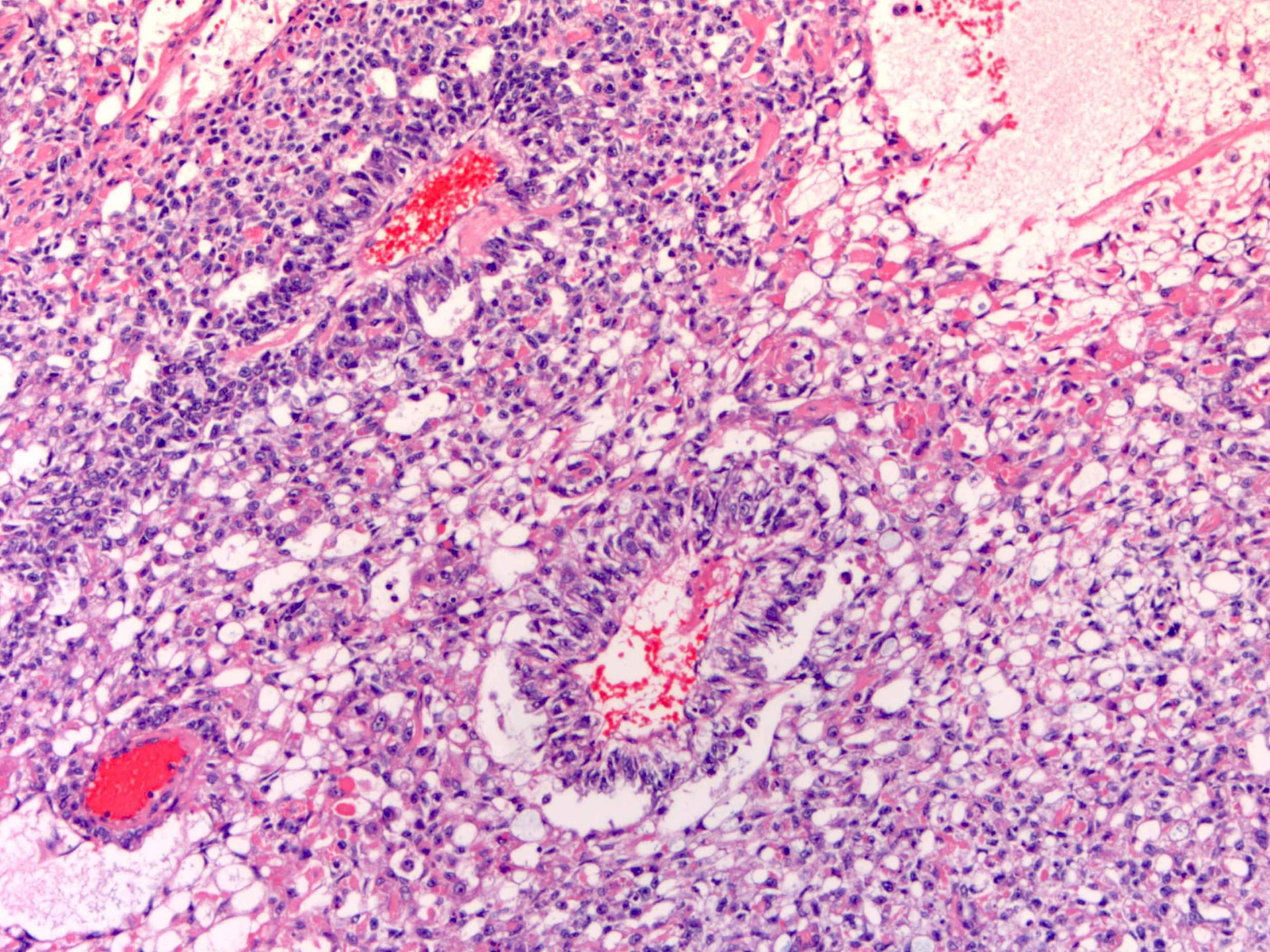
Schiller-Duval body

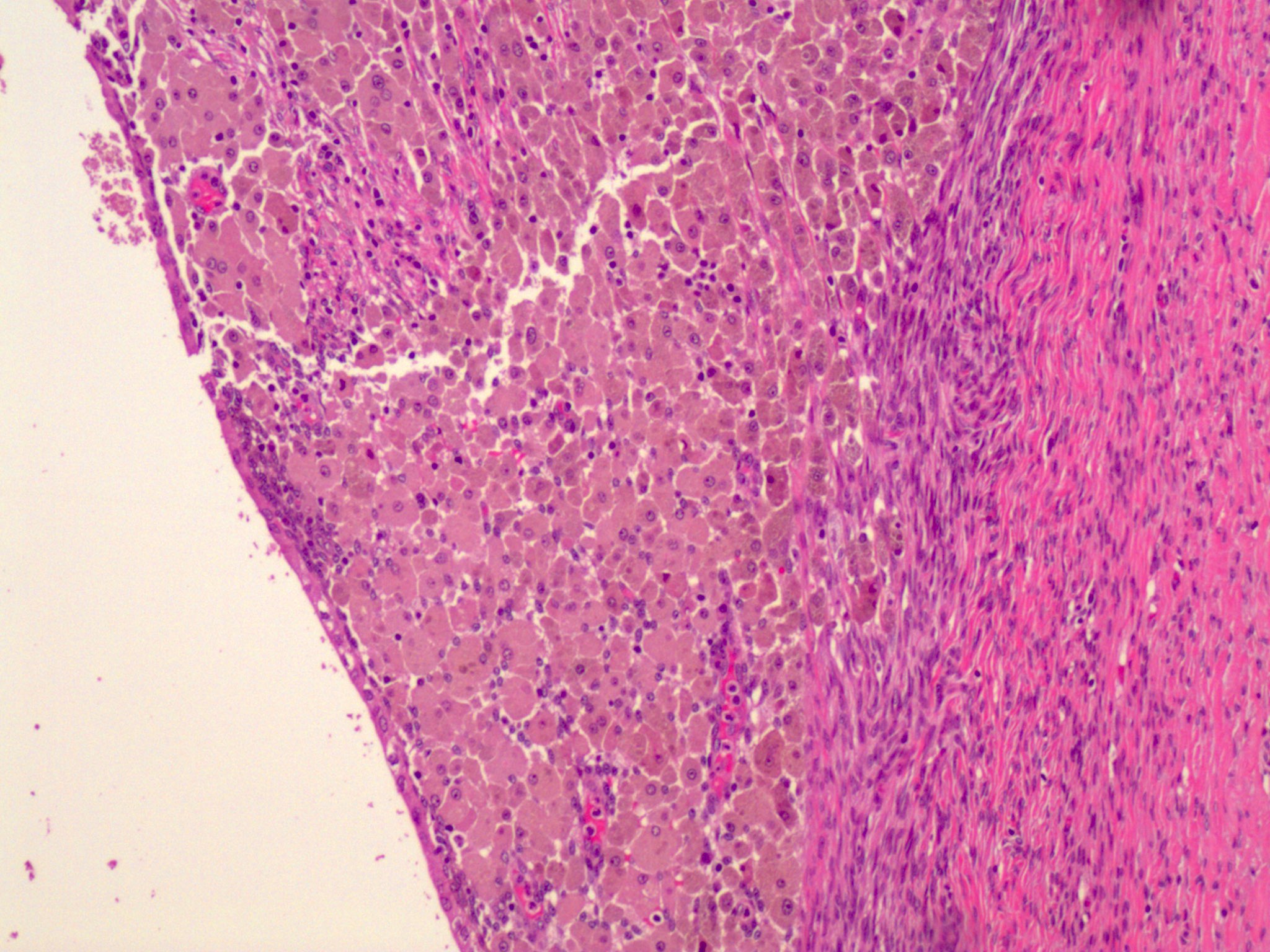
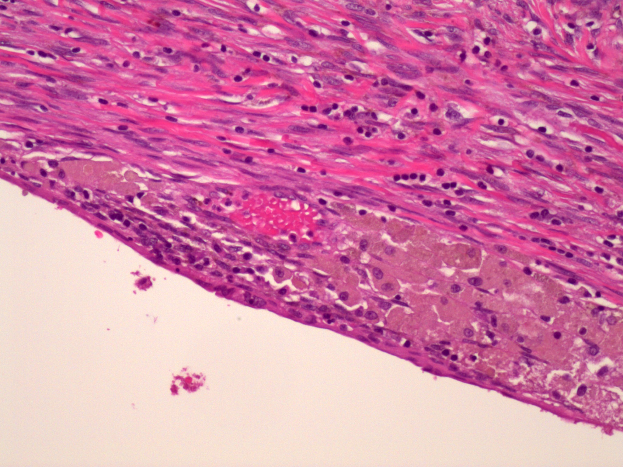
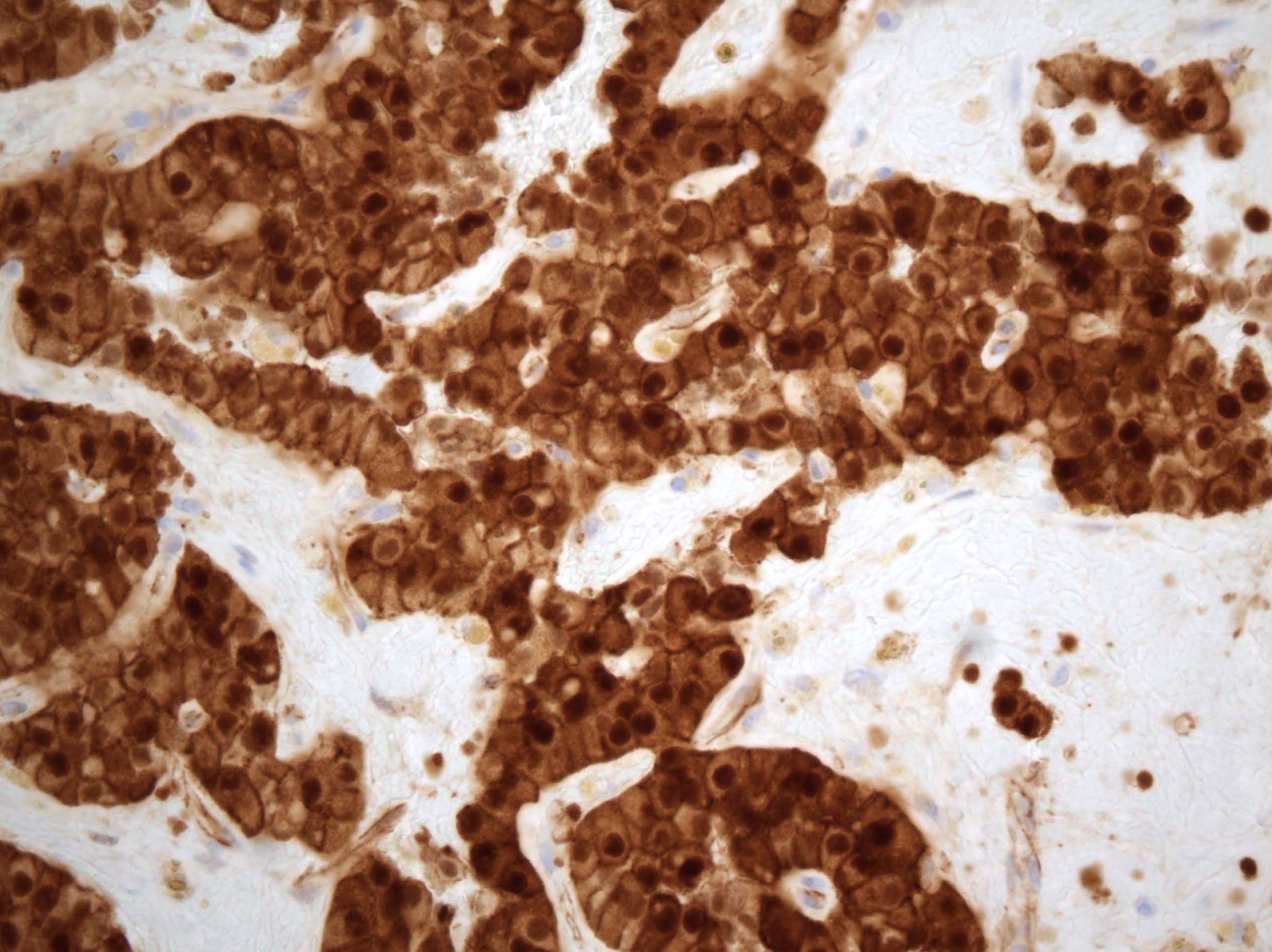
Hepatoid variant
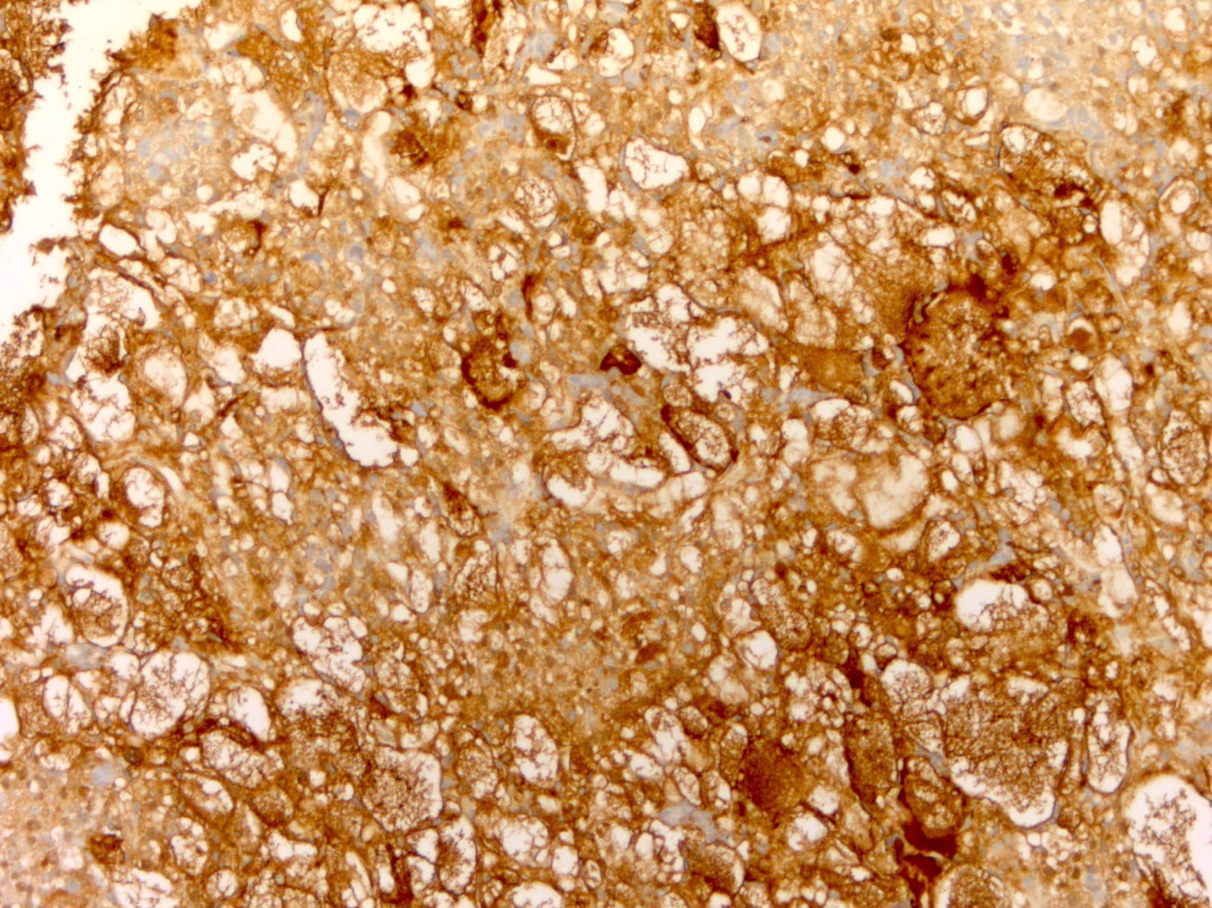
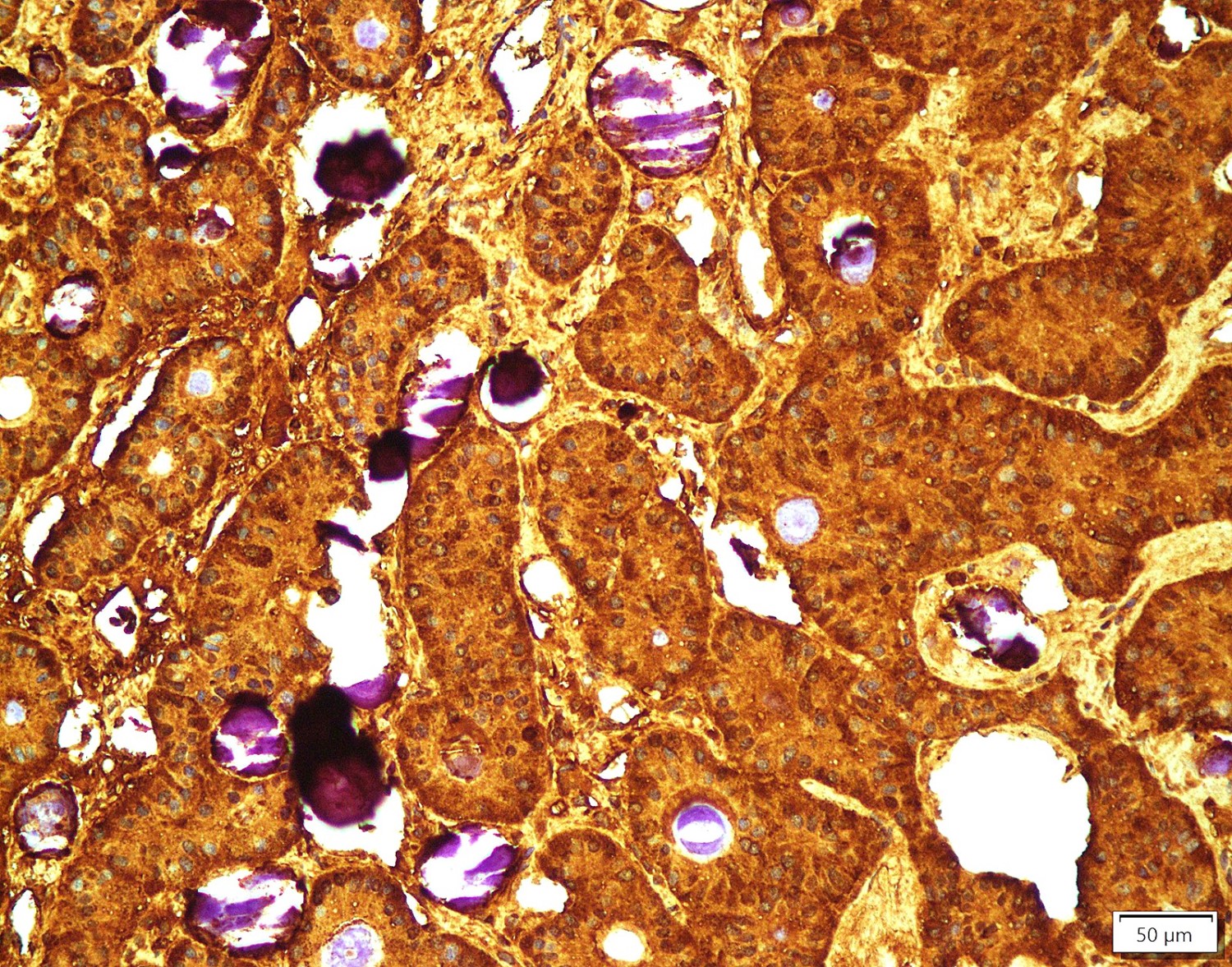
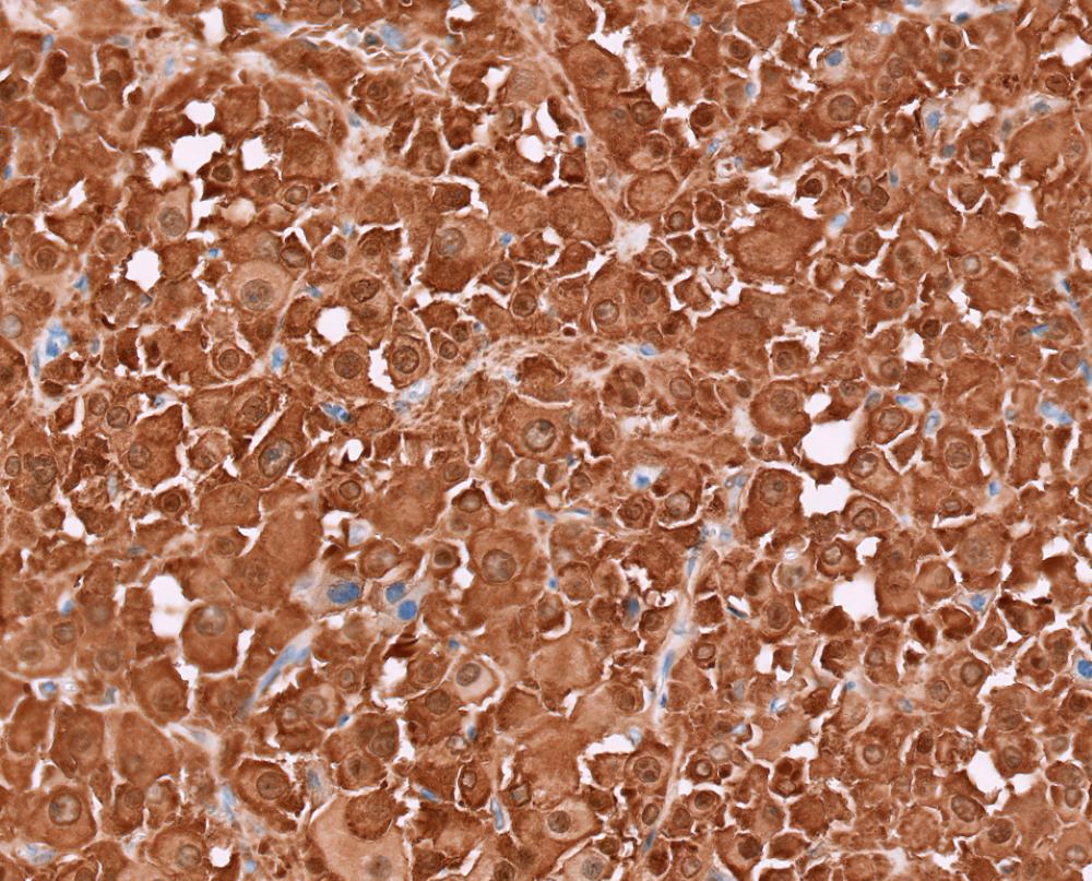
AFP
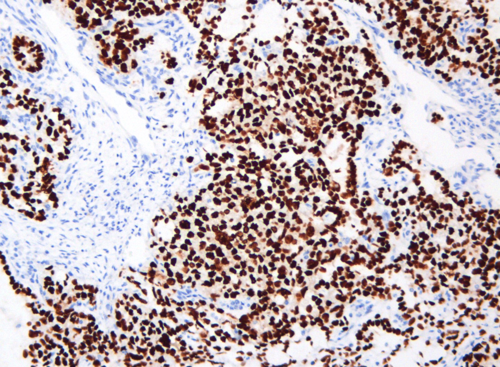
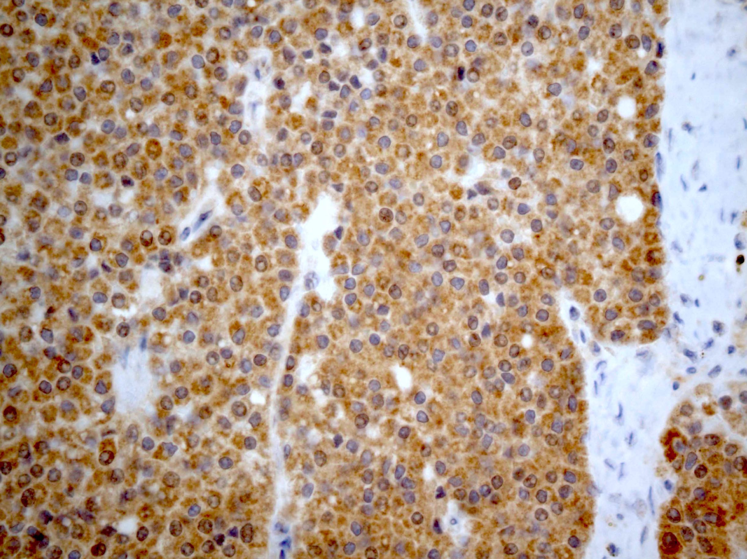
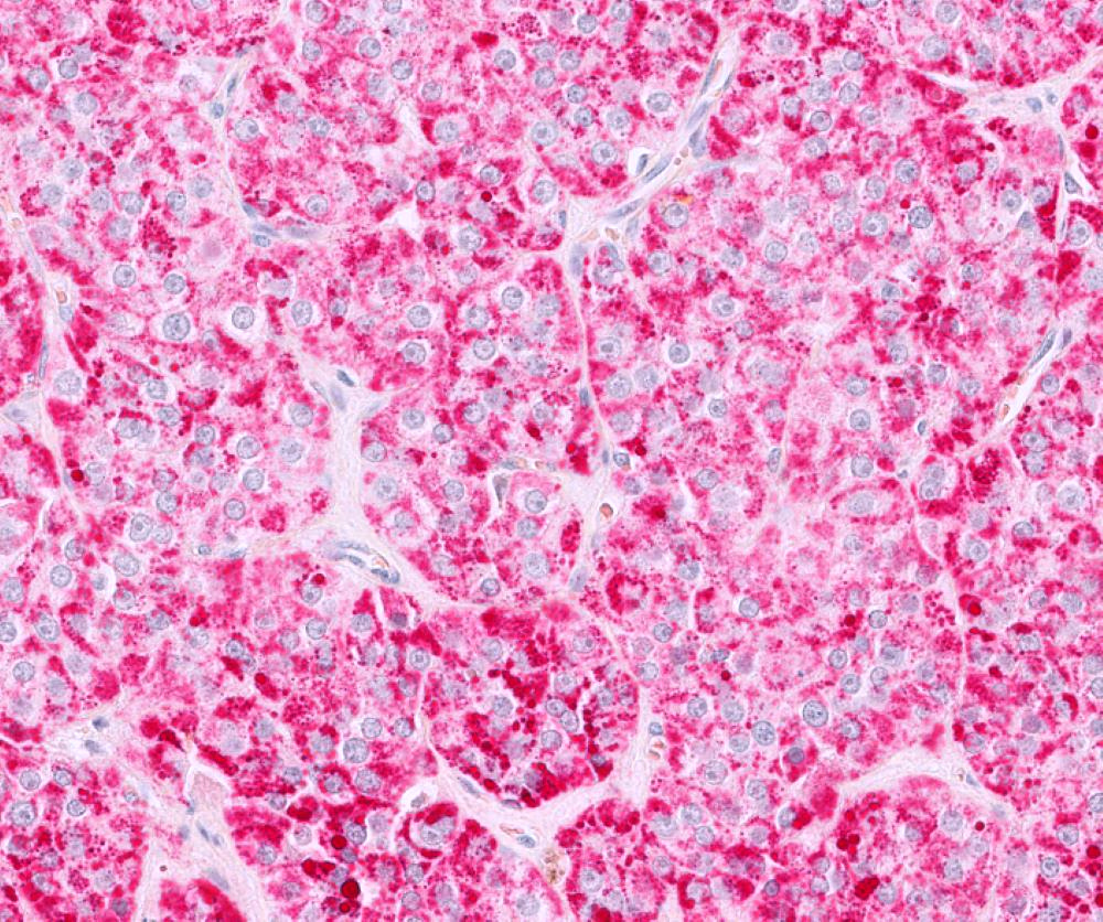
SALL4

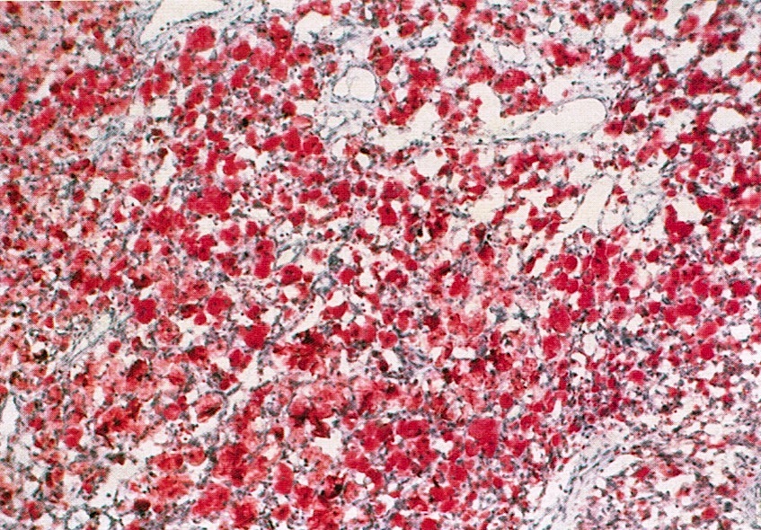
GATA3
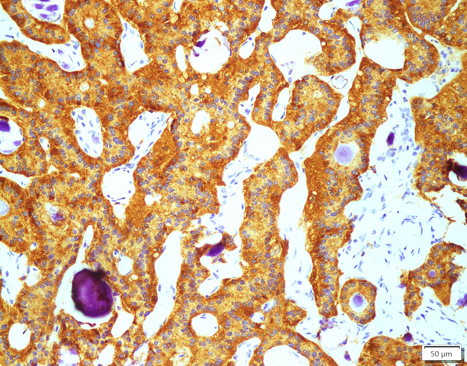
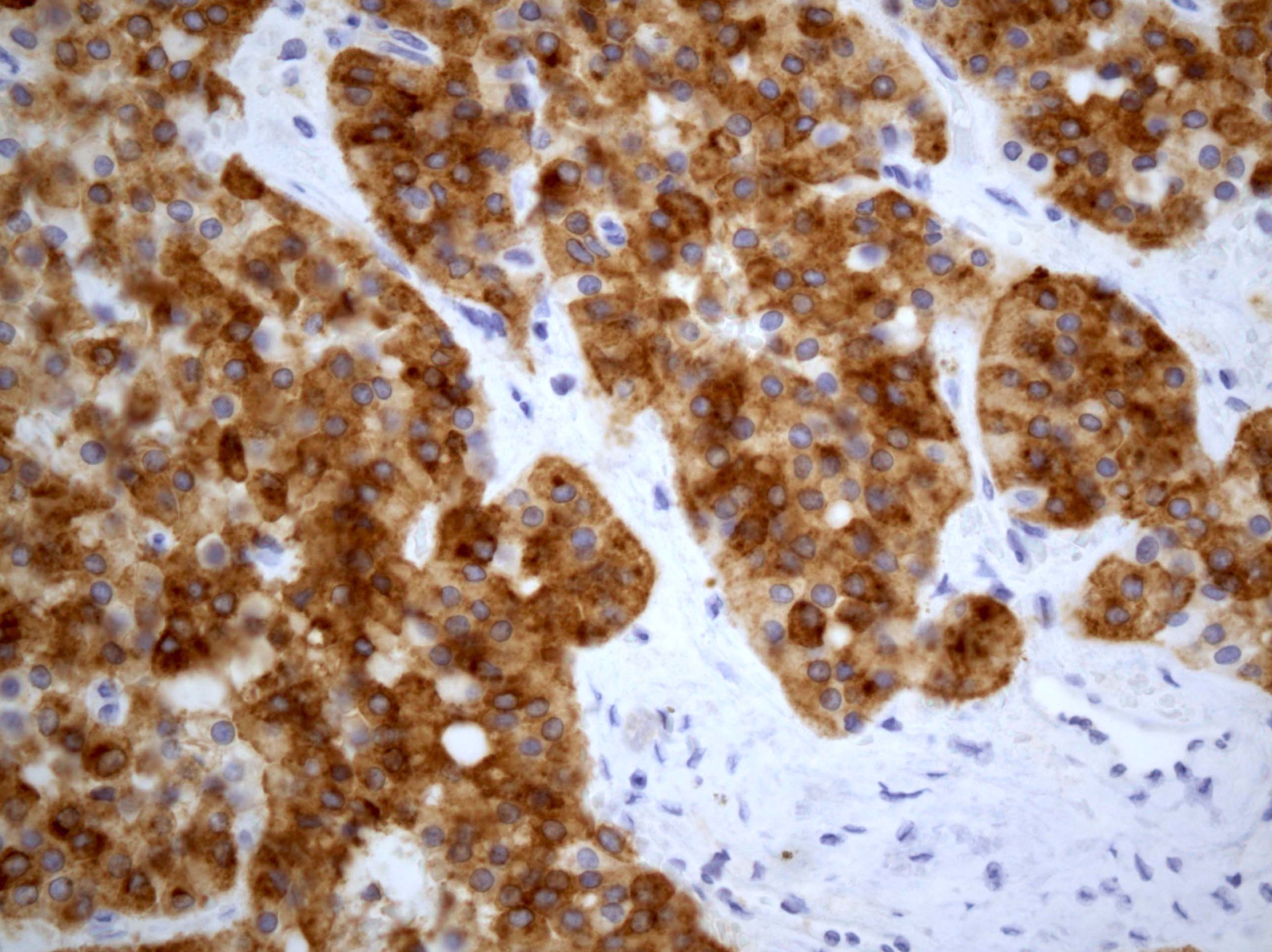
Pancytokeratin

CK7
Carlson: 2023
Clarke: 2016

Clement: 2019
Crum: 2015
Crum: 2017
Fadare: 2015
Heller: 2015
IARC: 2020
Kigawa: 2015

Kurman: 2019

Nucci: 2020
Nucci: 2023
Vang: 2017
Find related Pathology books: gynecologic
