
AFIP images
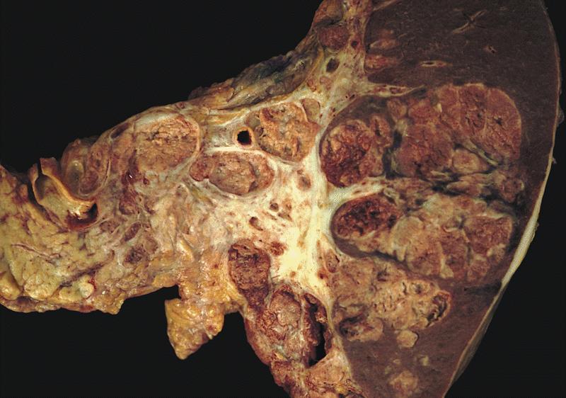
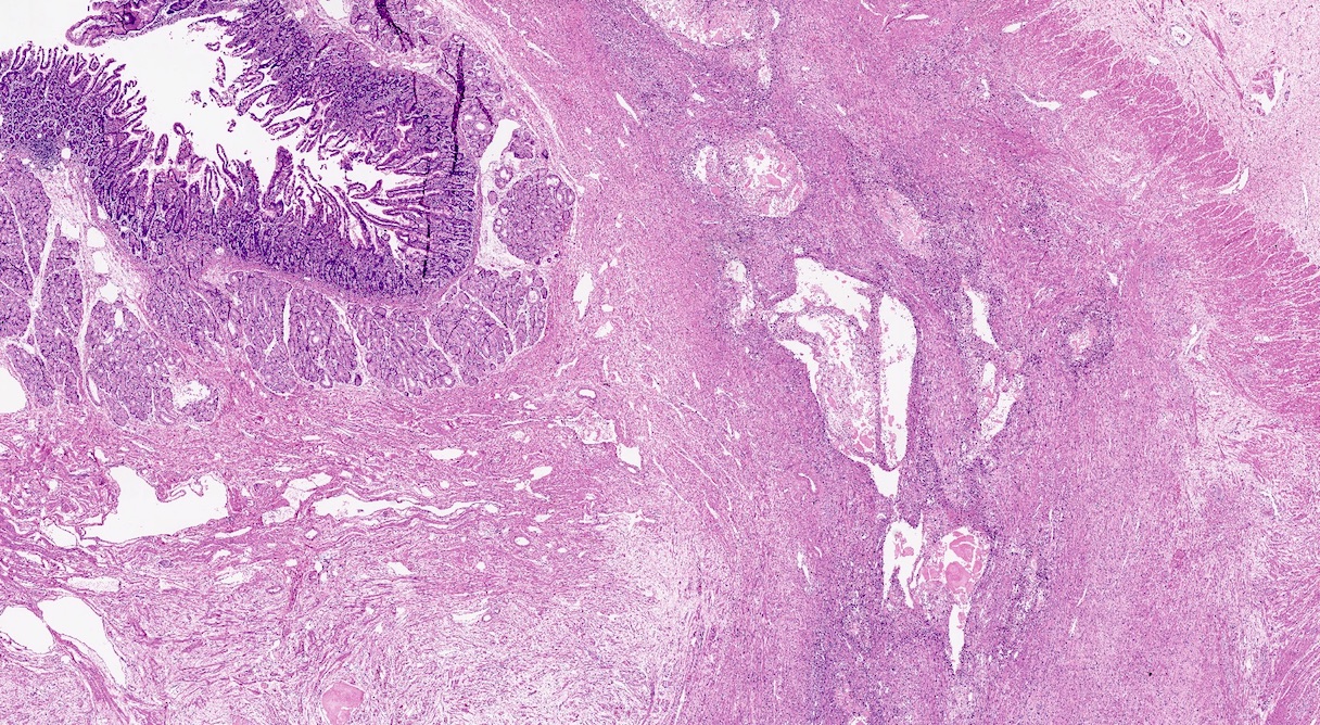
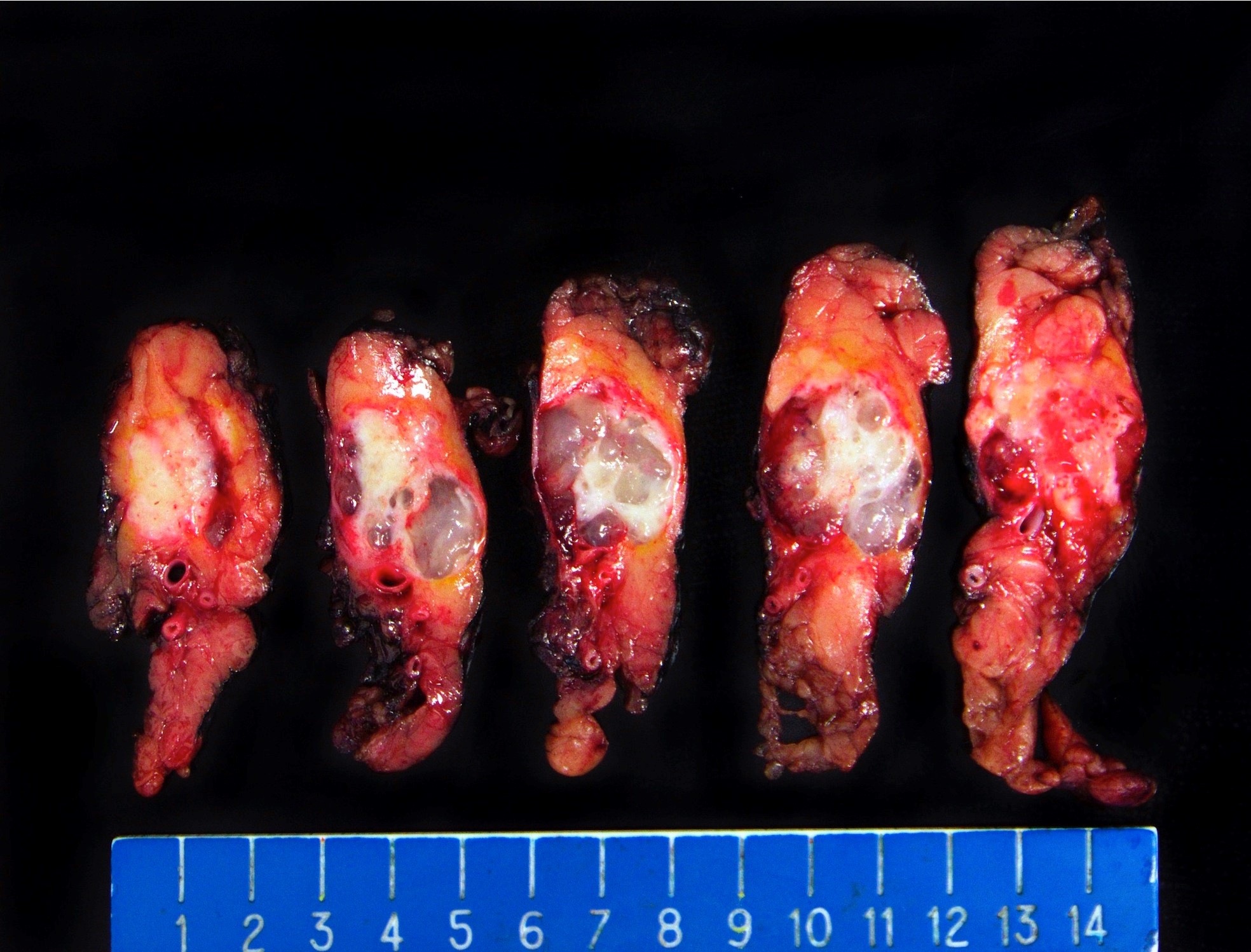
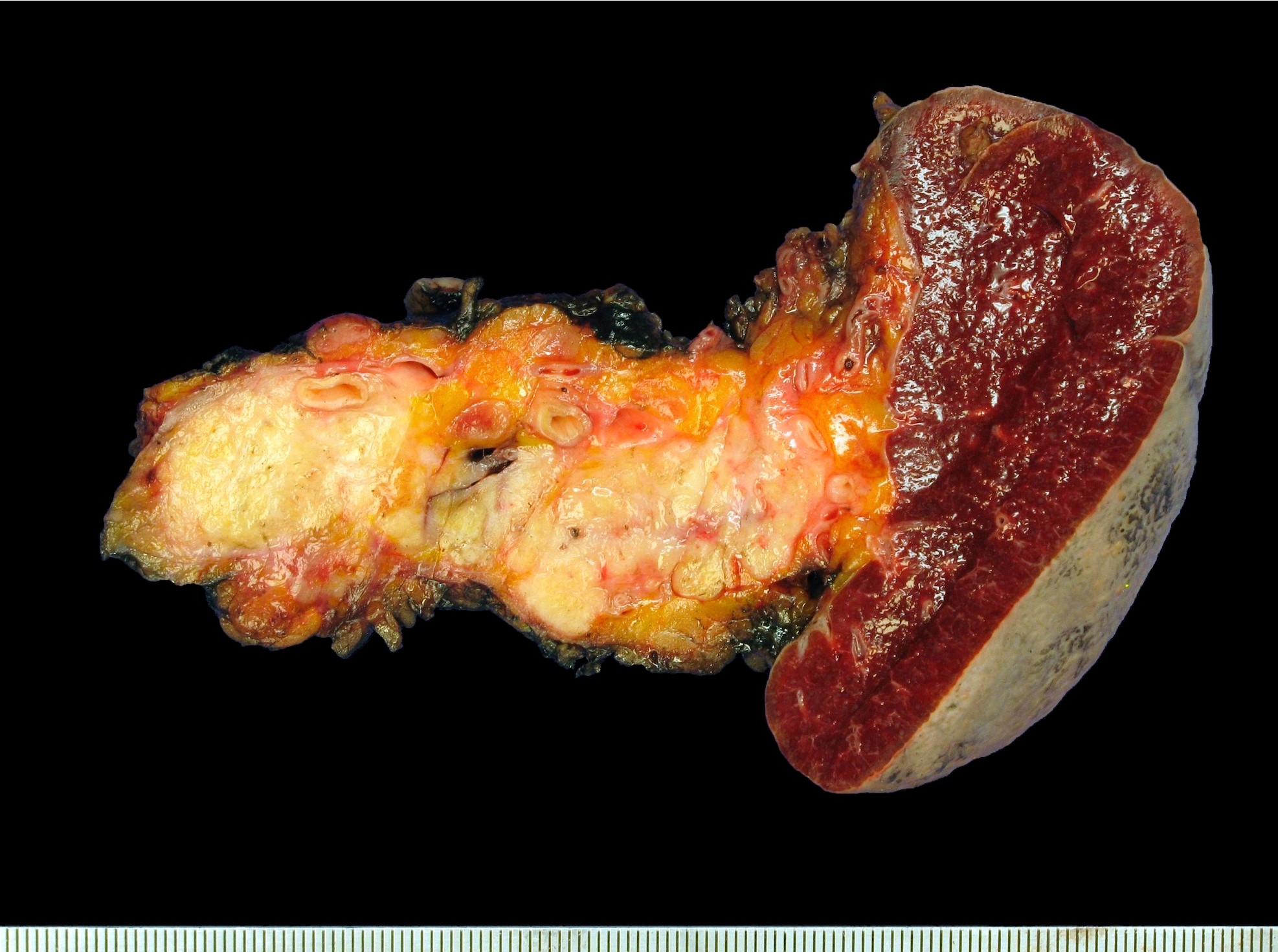
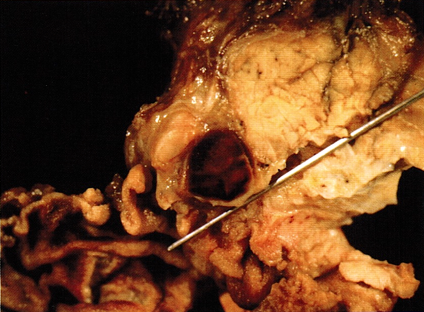
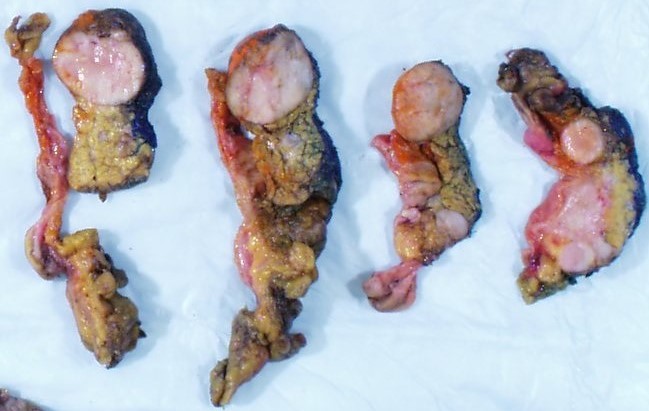
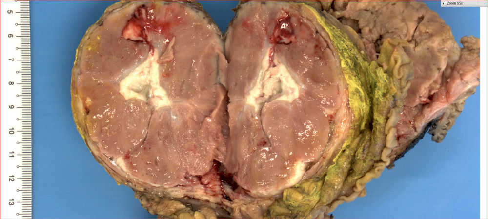
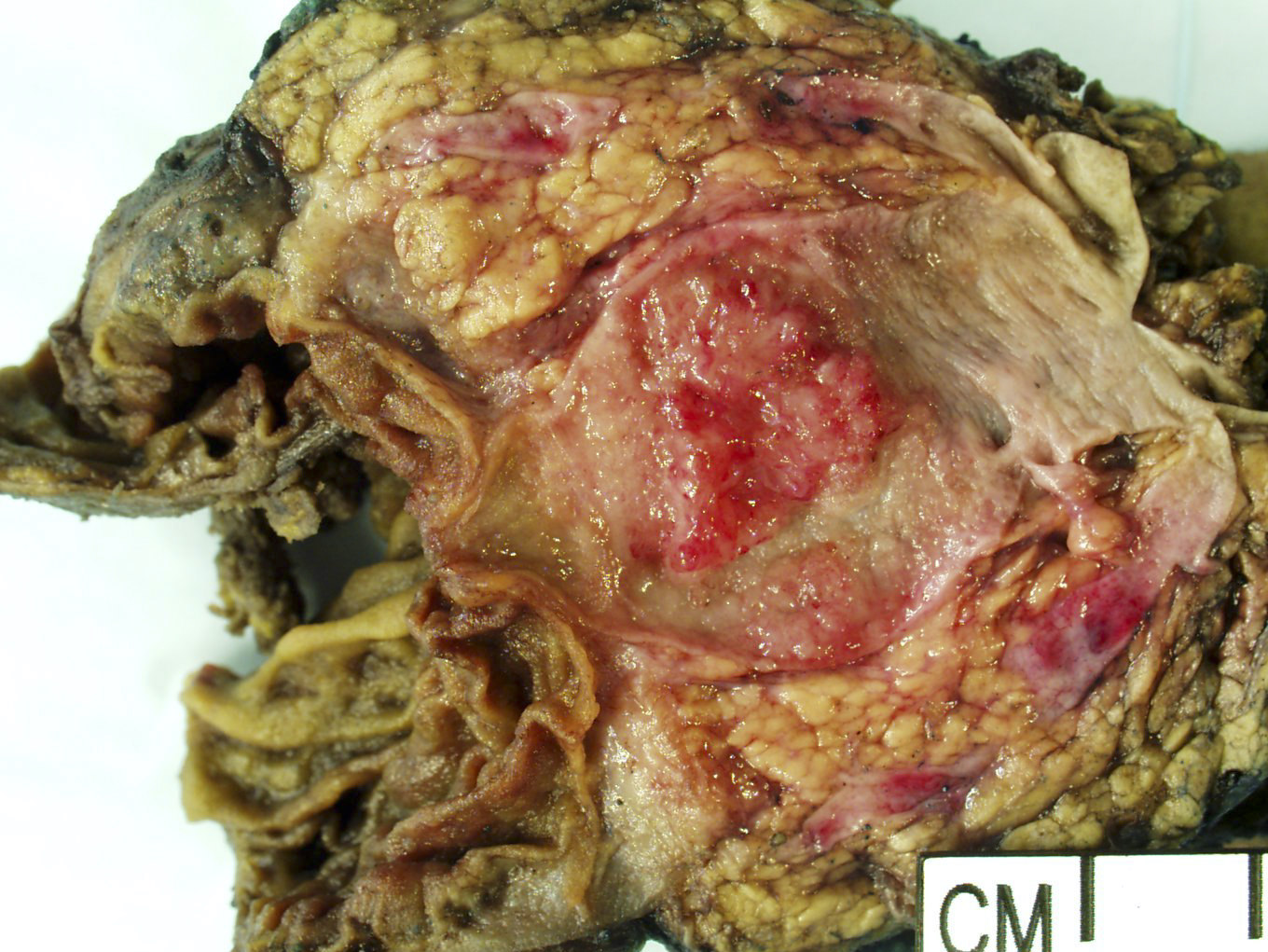
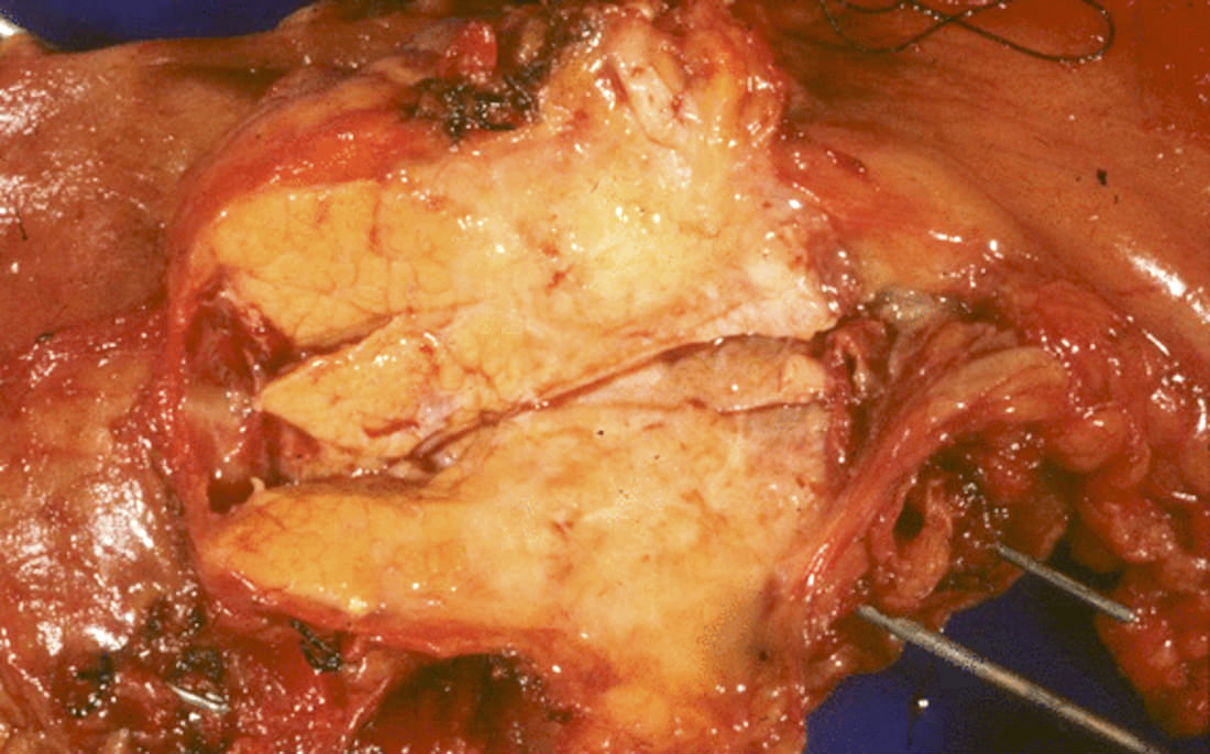
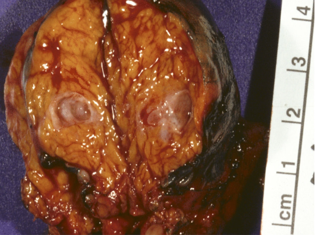
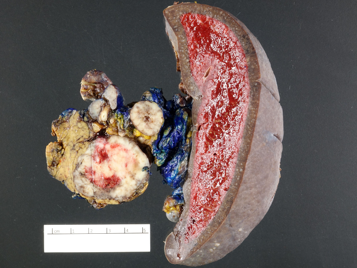
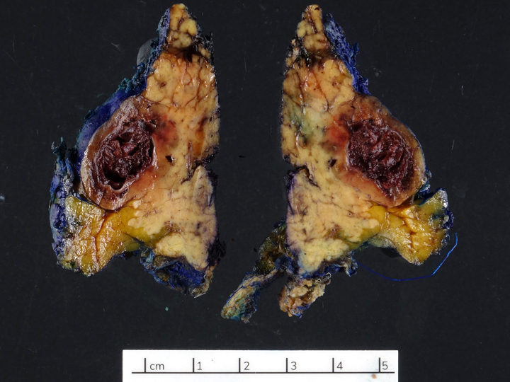
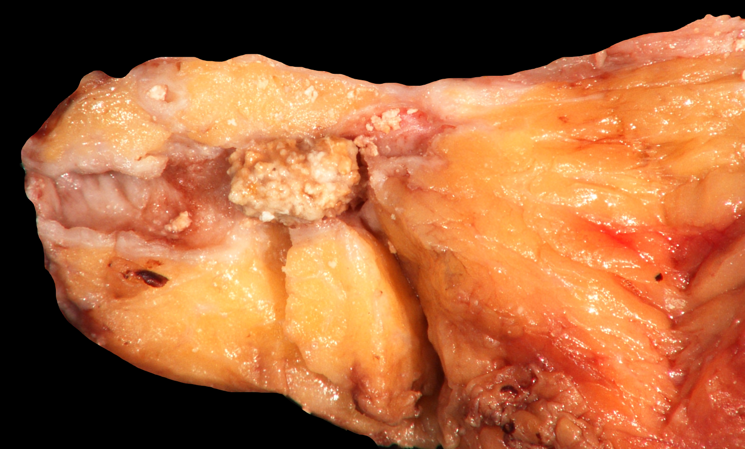
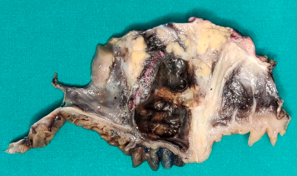
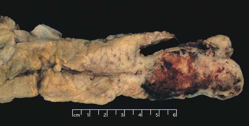
Cut surface displays large nodules
Contributed by Claudio Luchini, M.D., Ph.D.
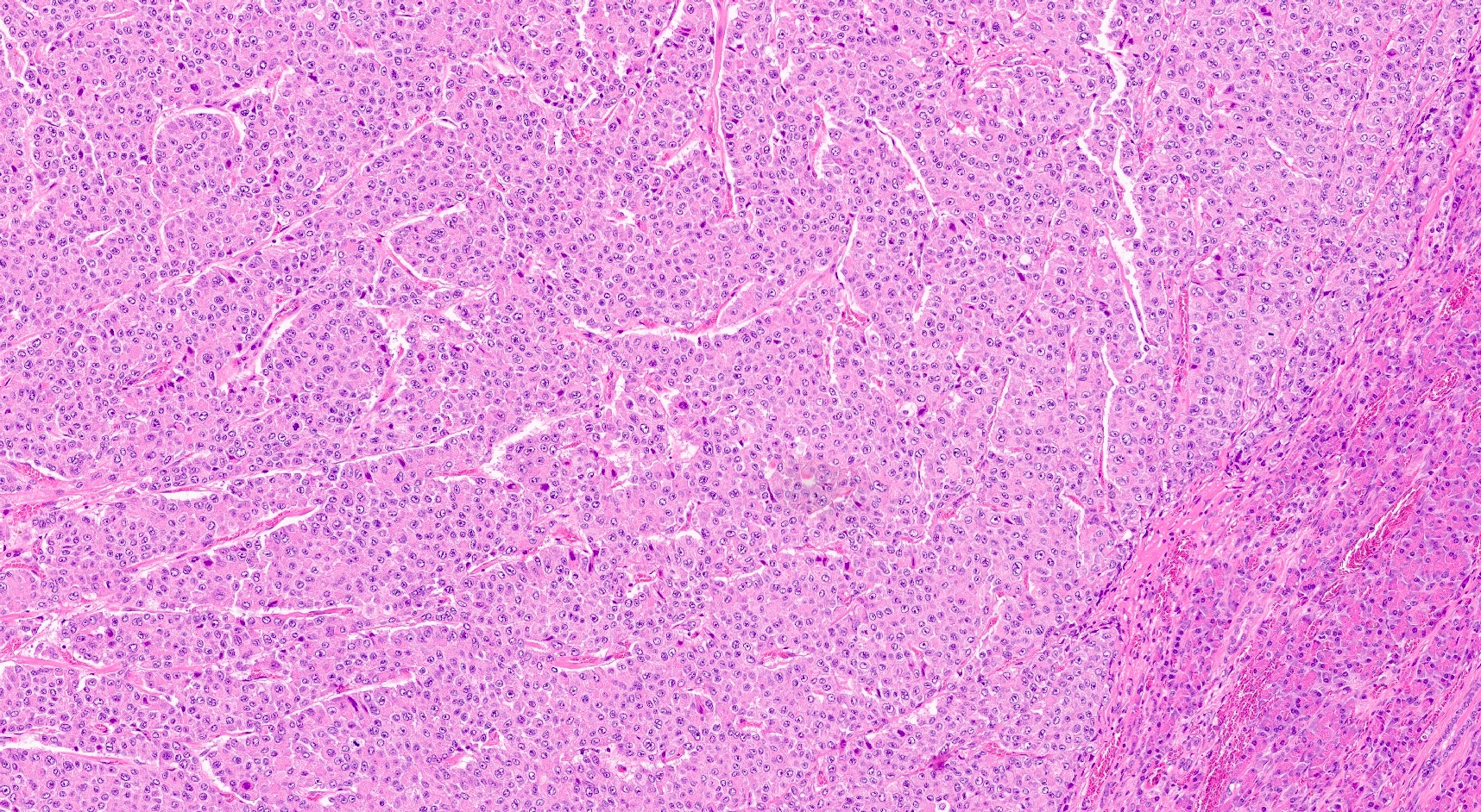
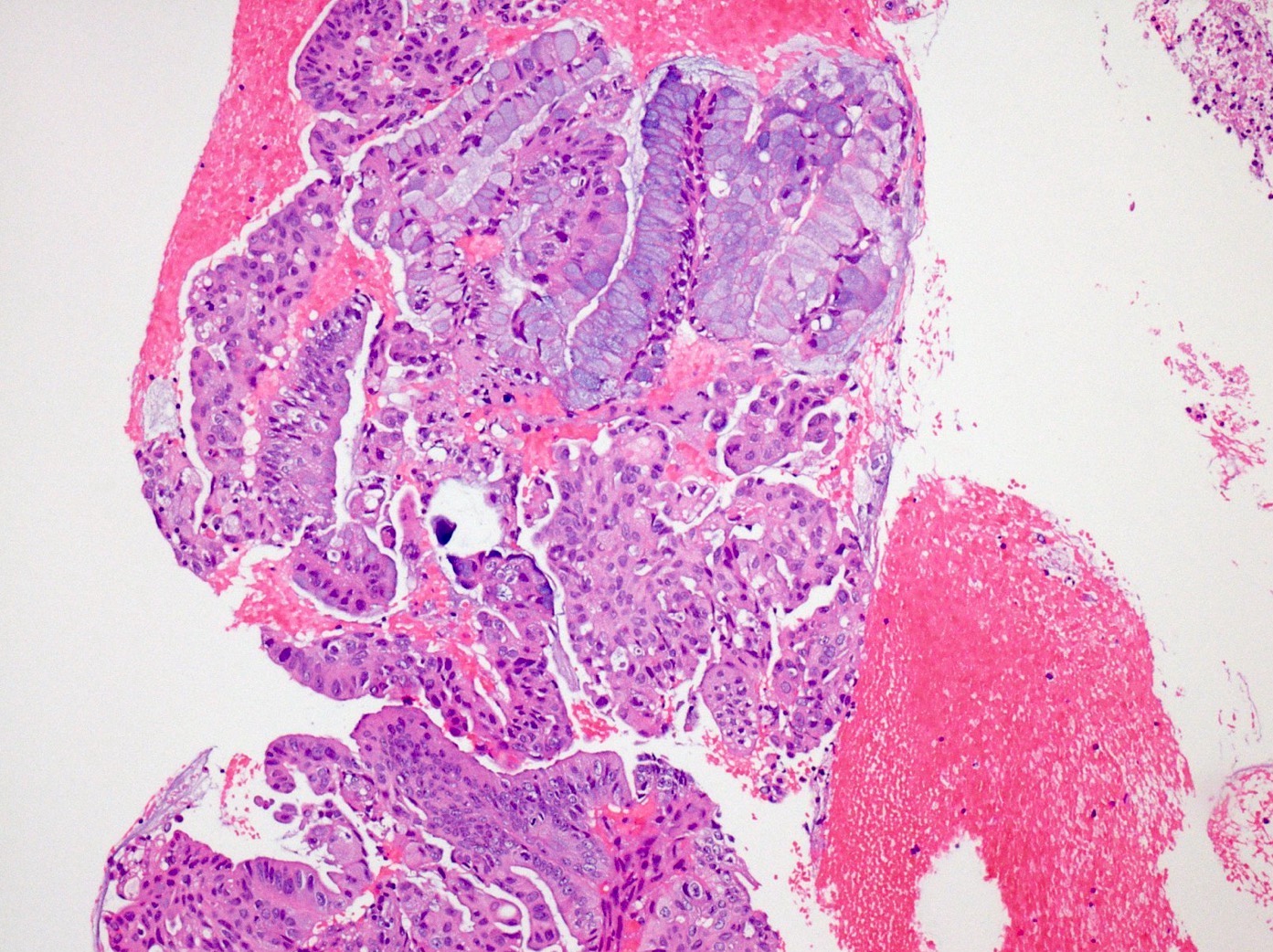
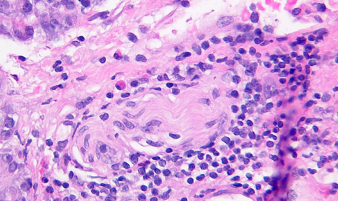
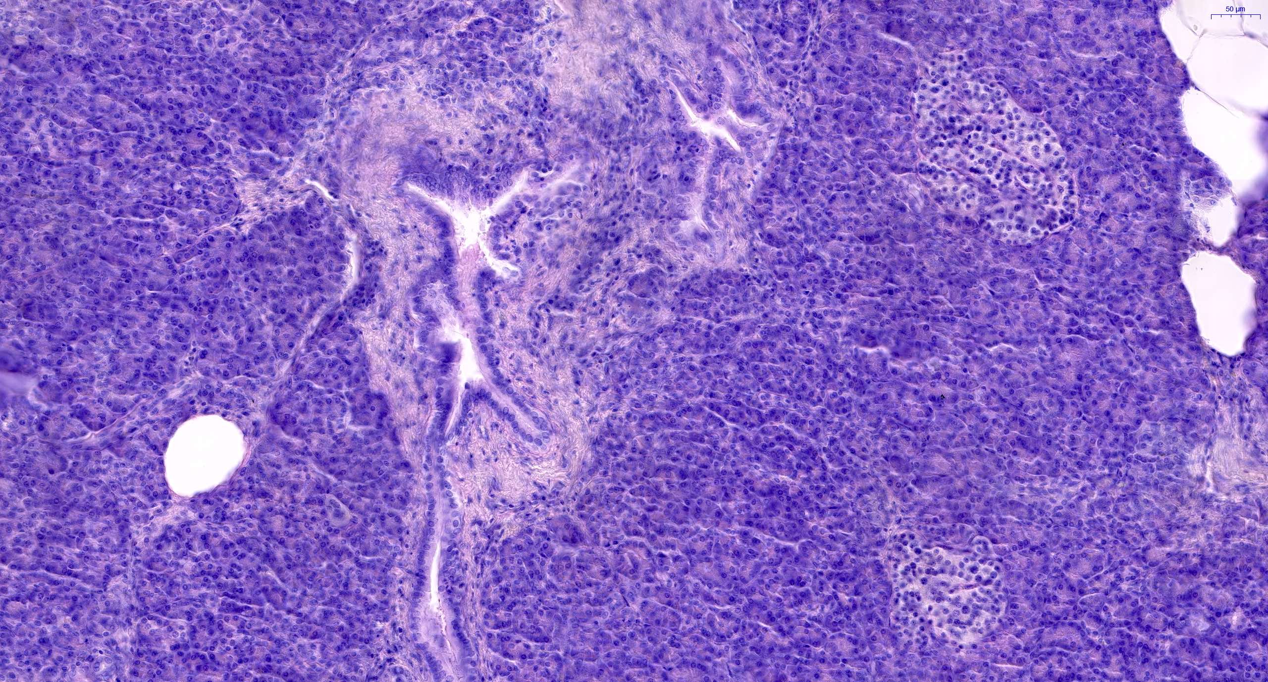
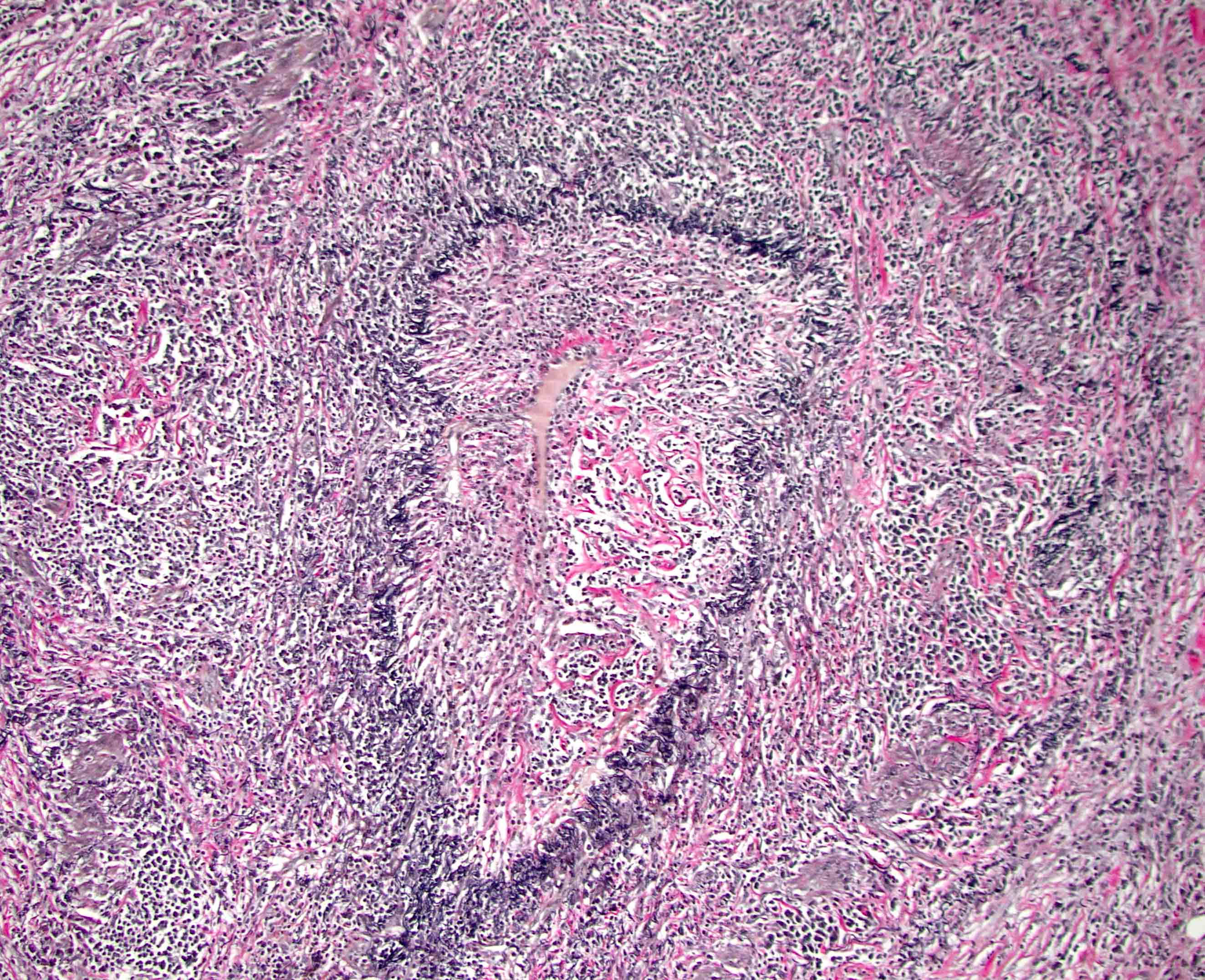
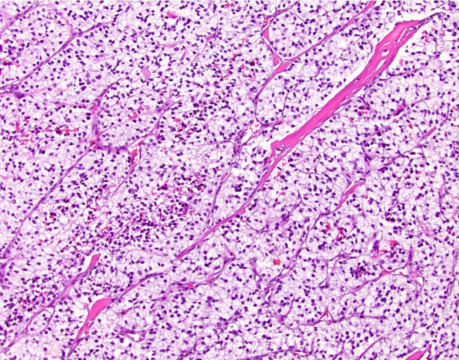
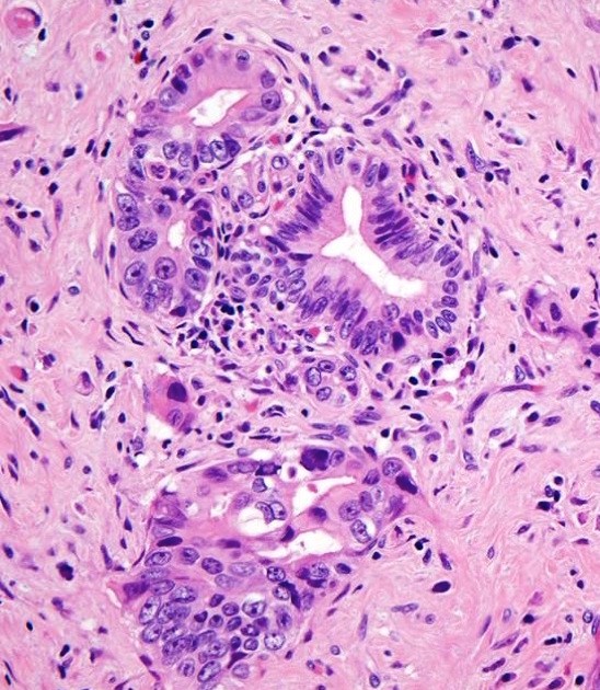
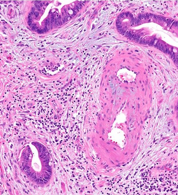
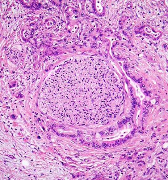
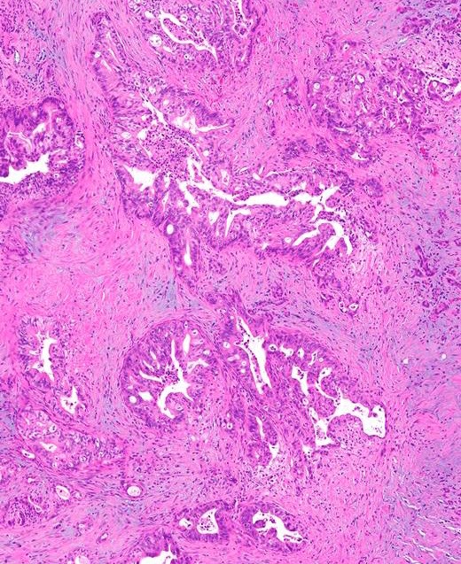
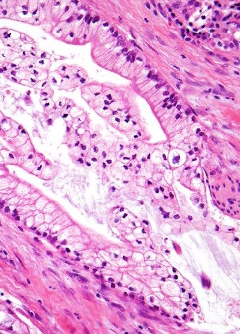
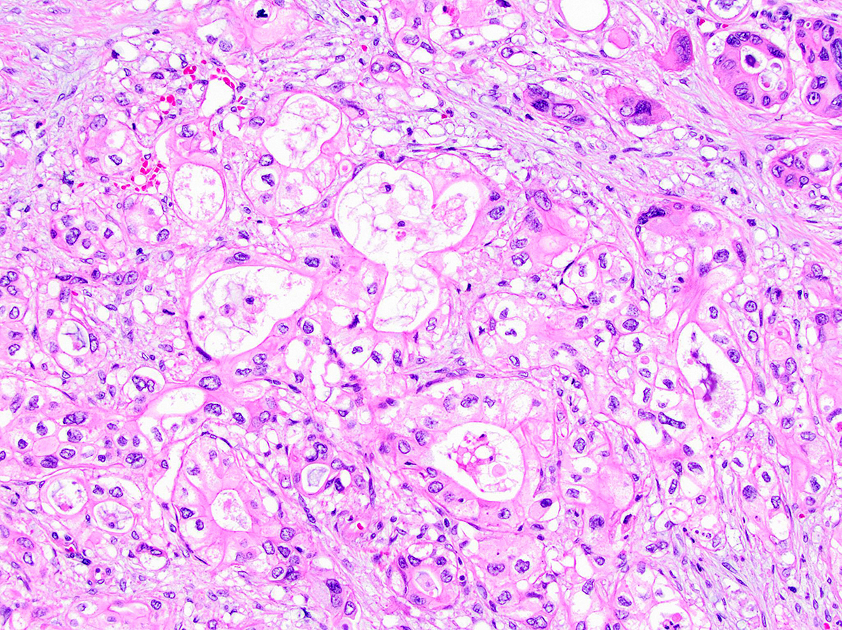
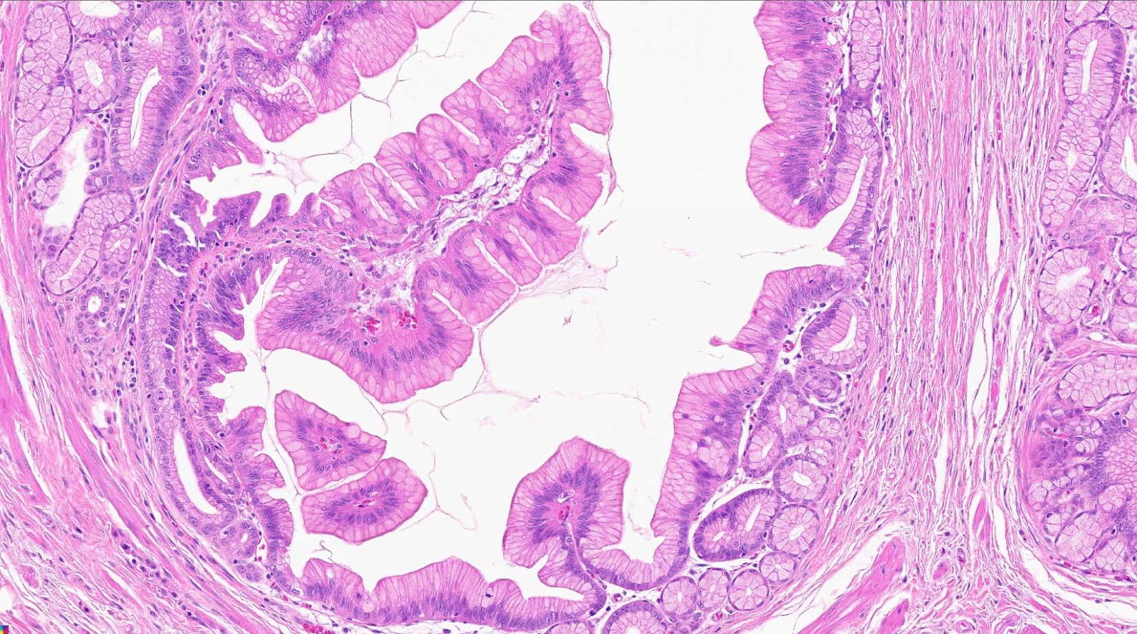
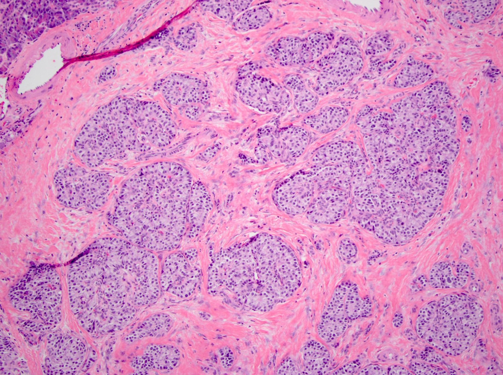
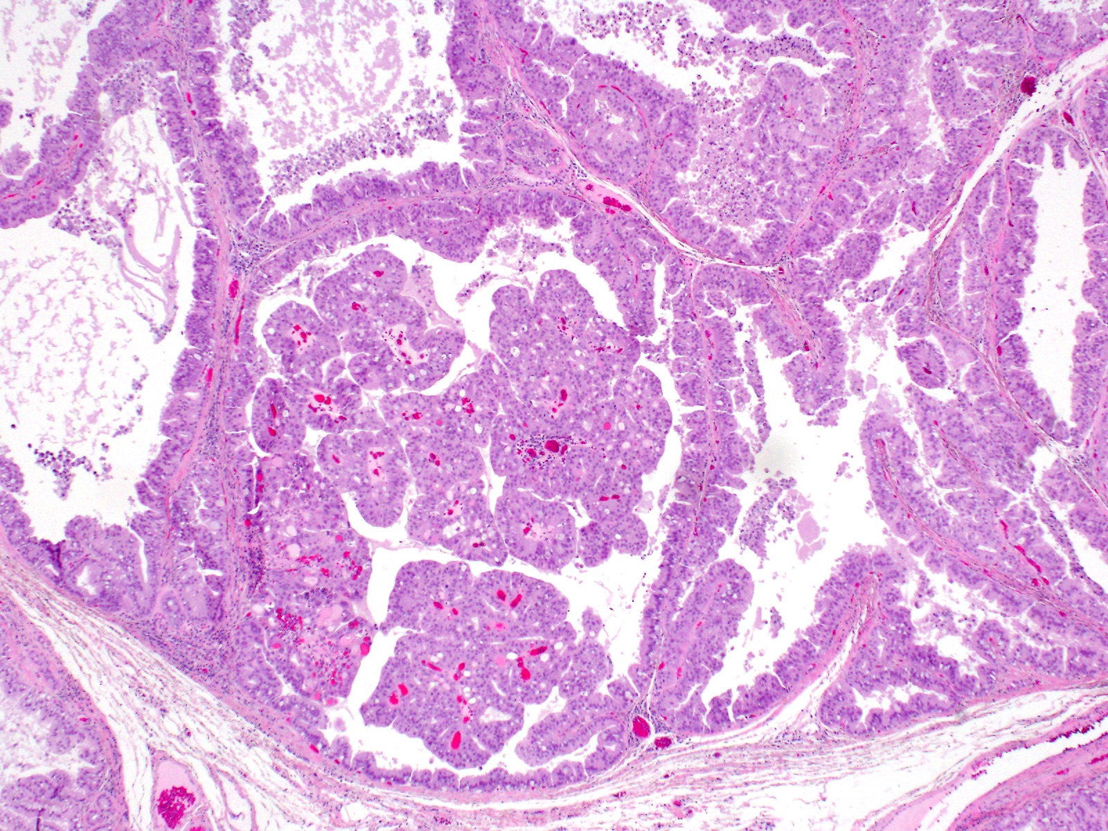
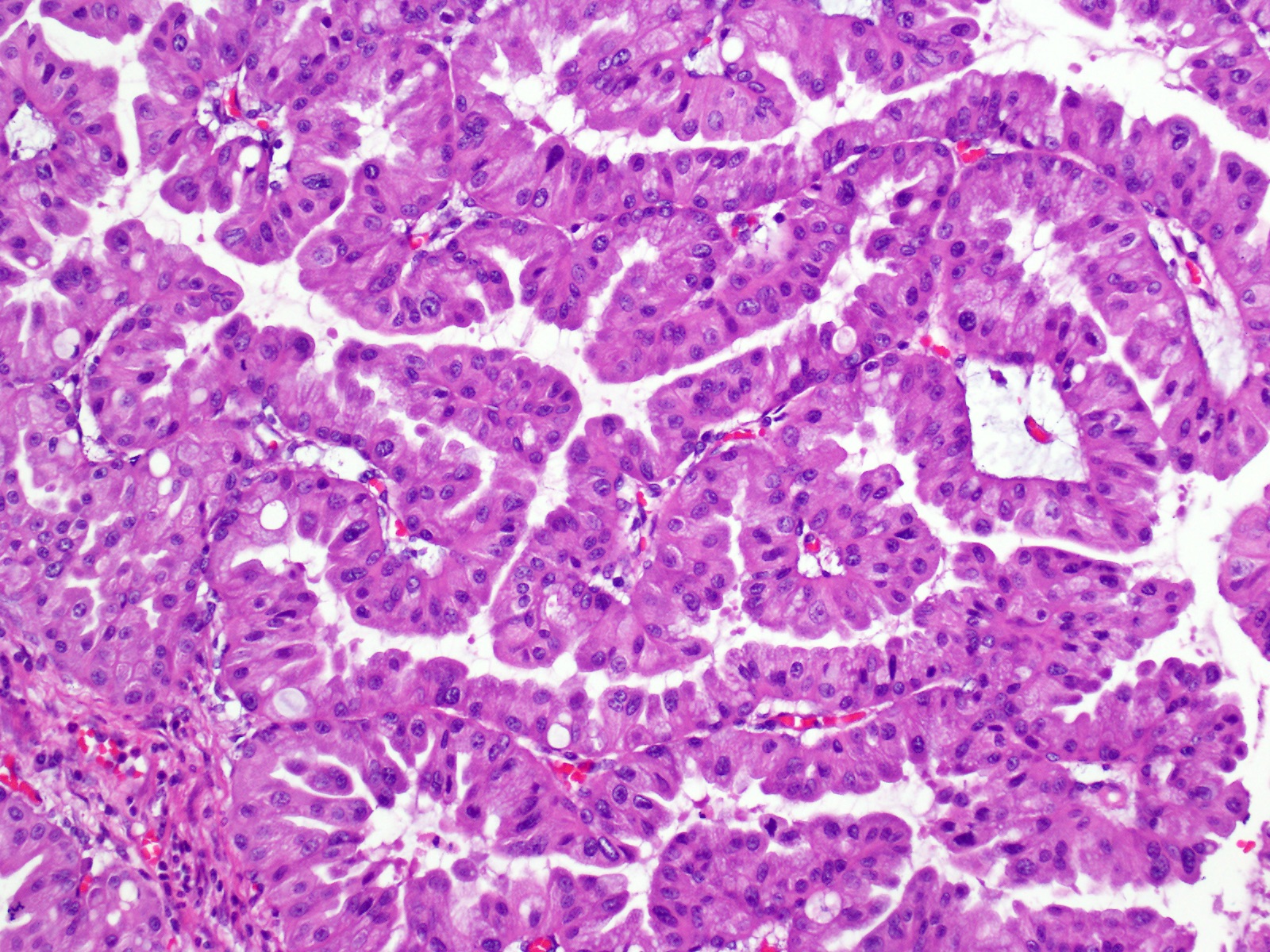
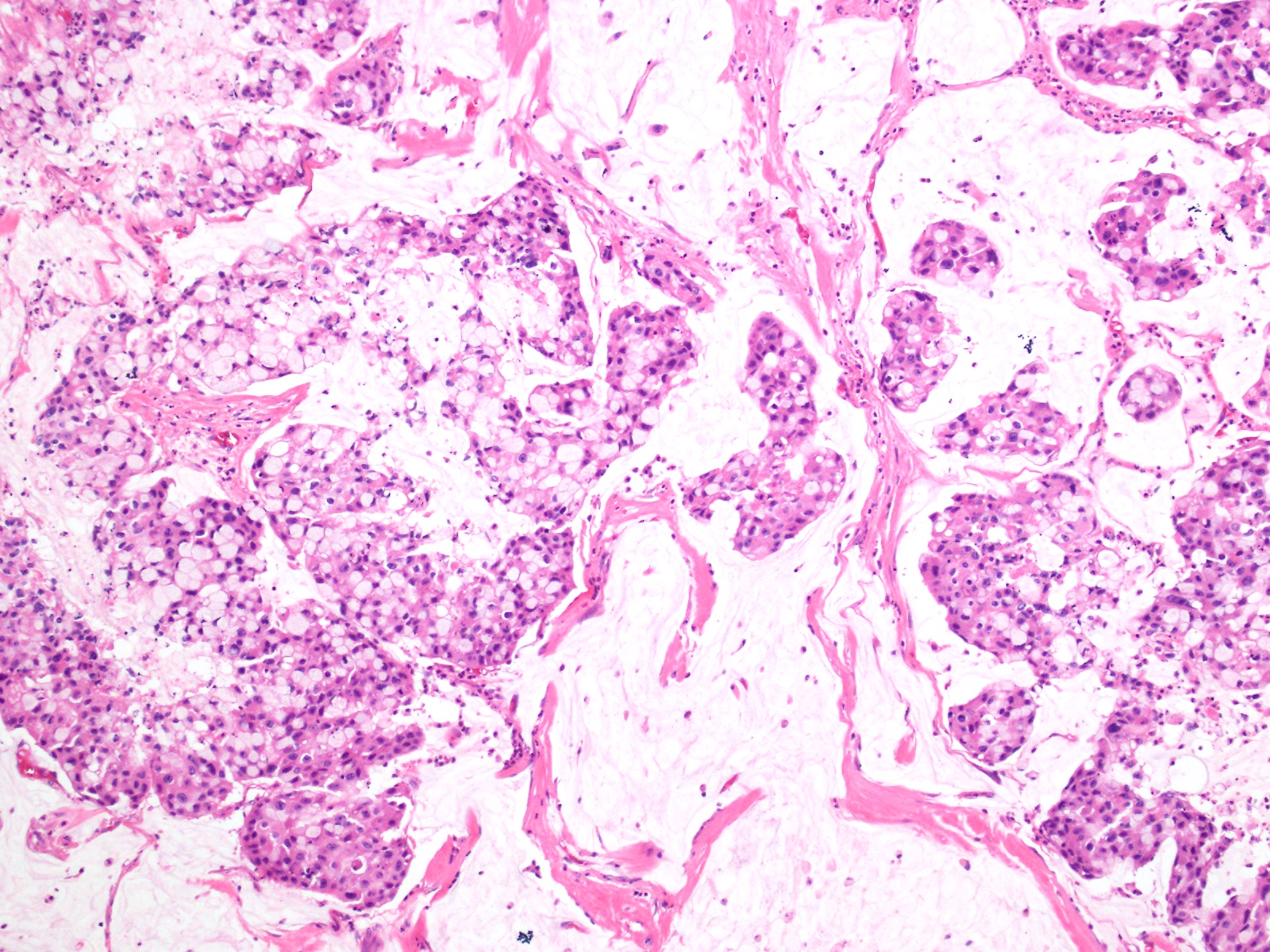
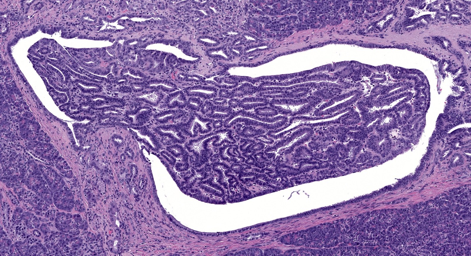
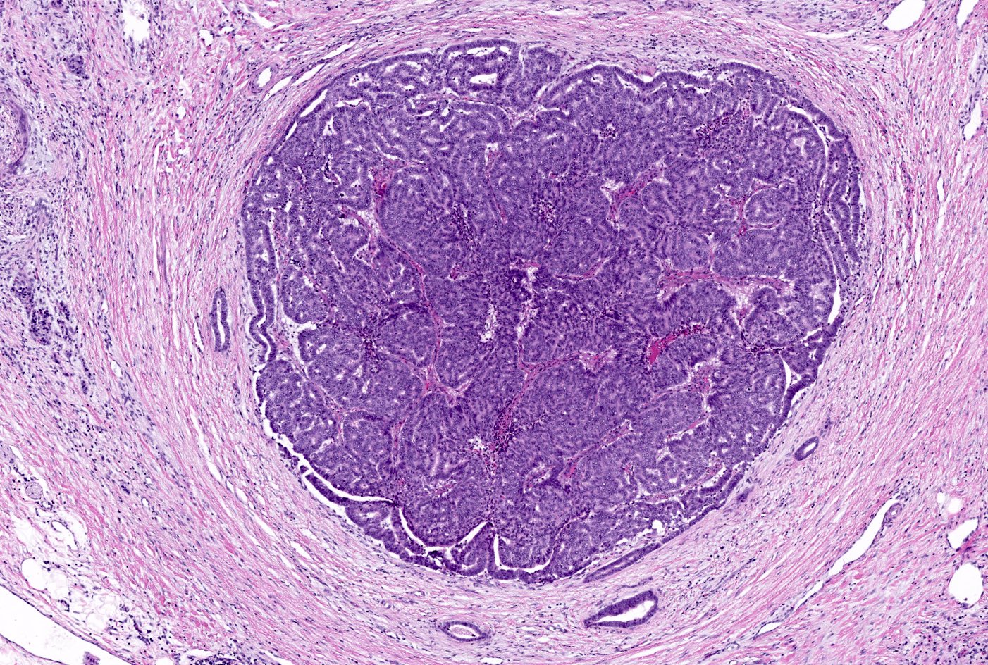
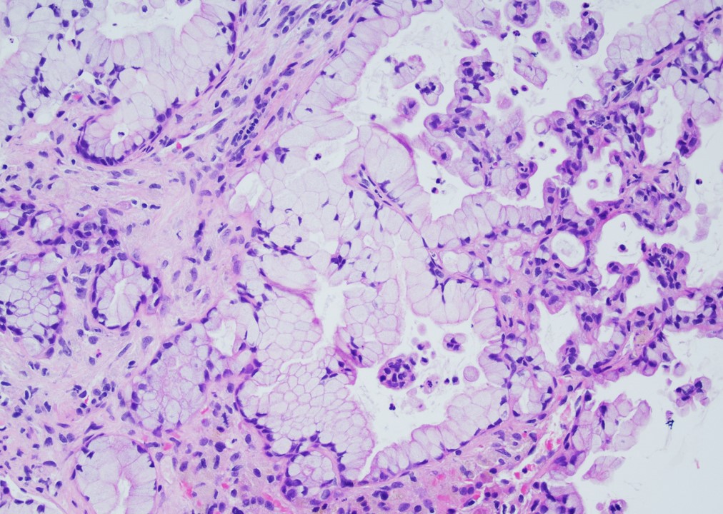
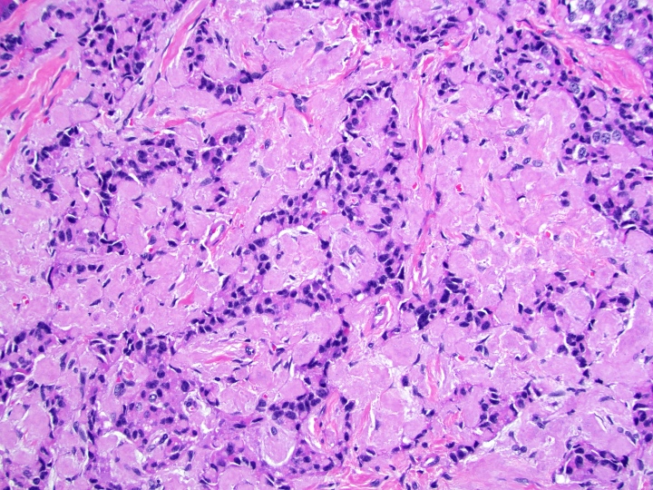
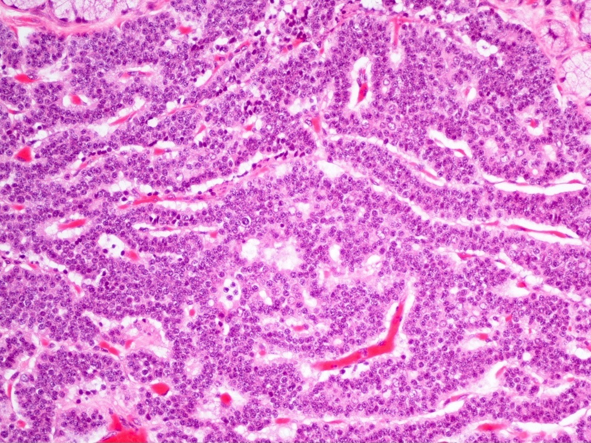
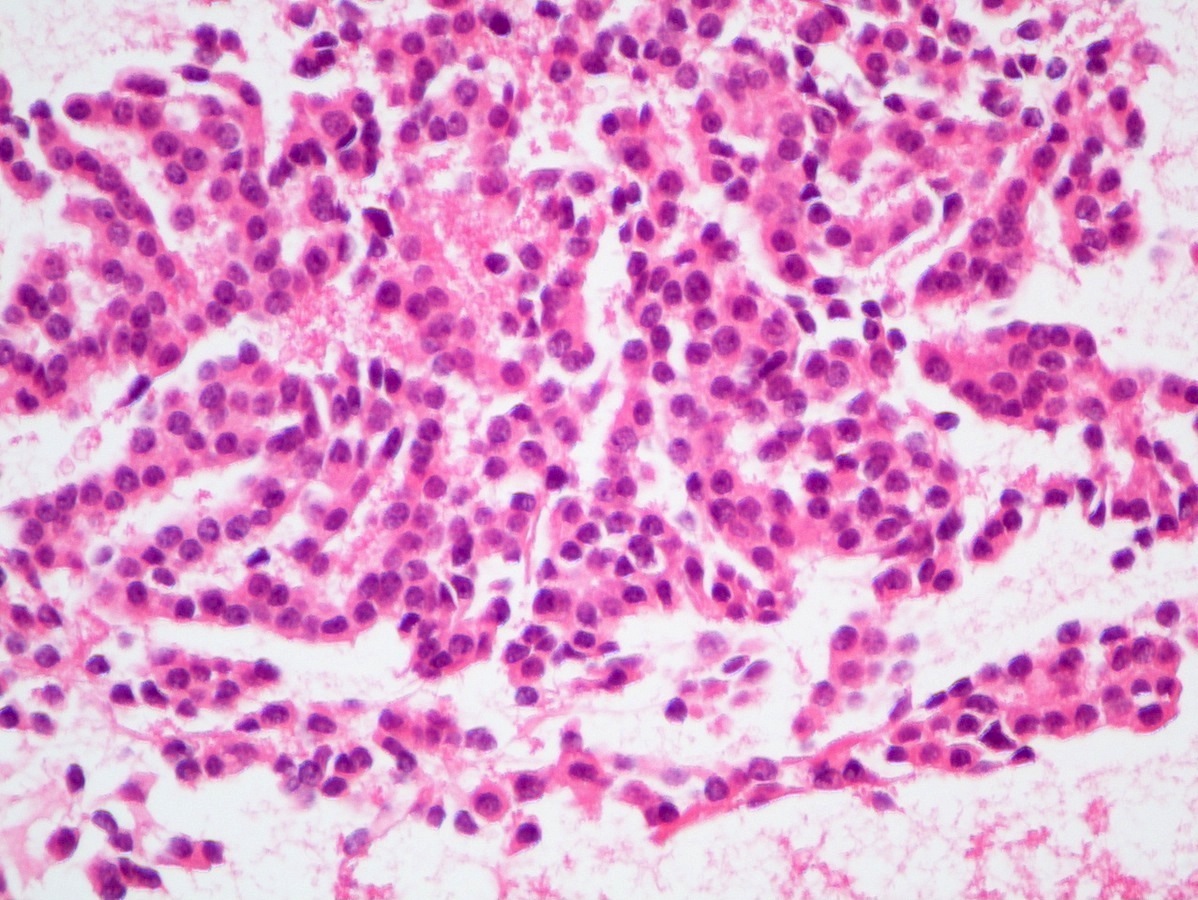
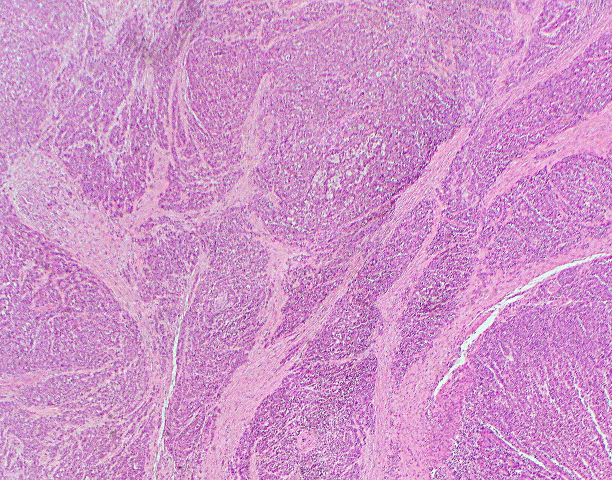
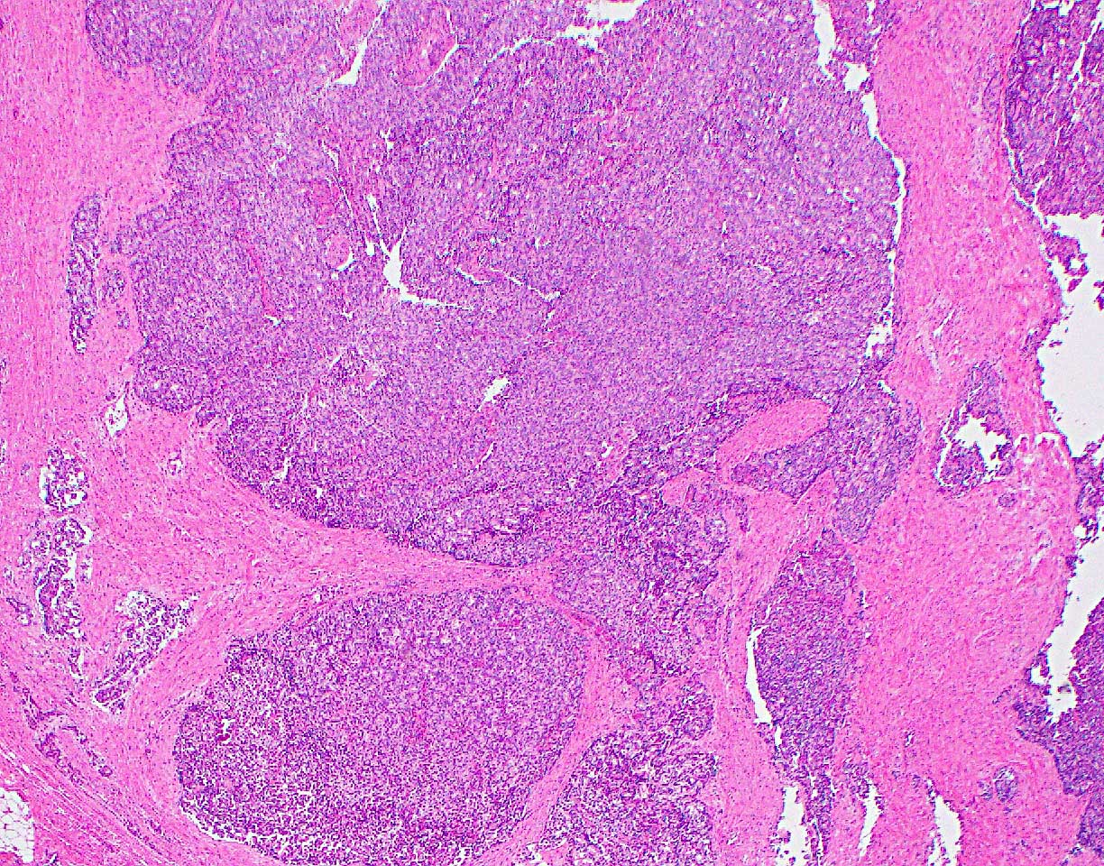
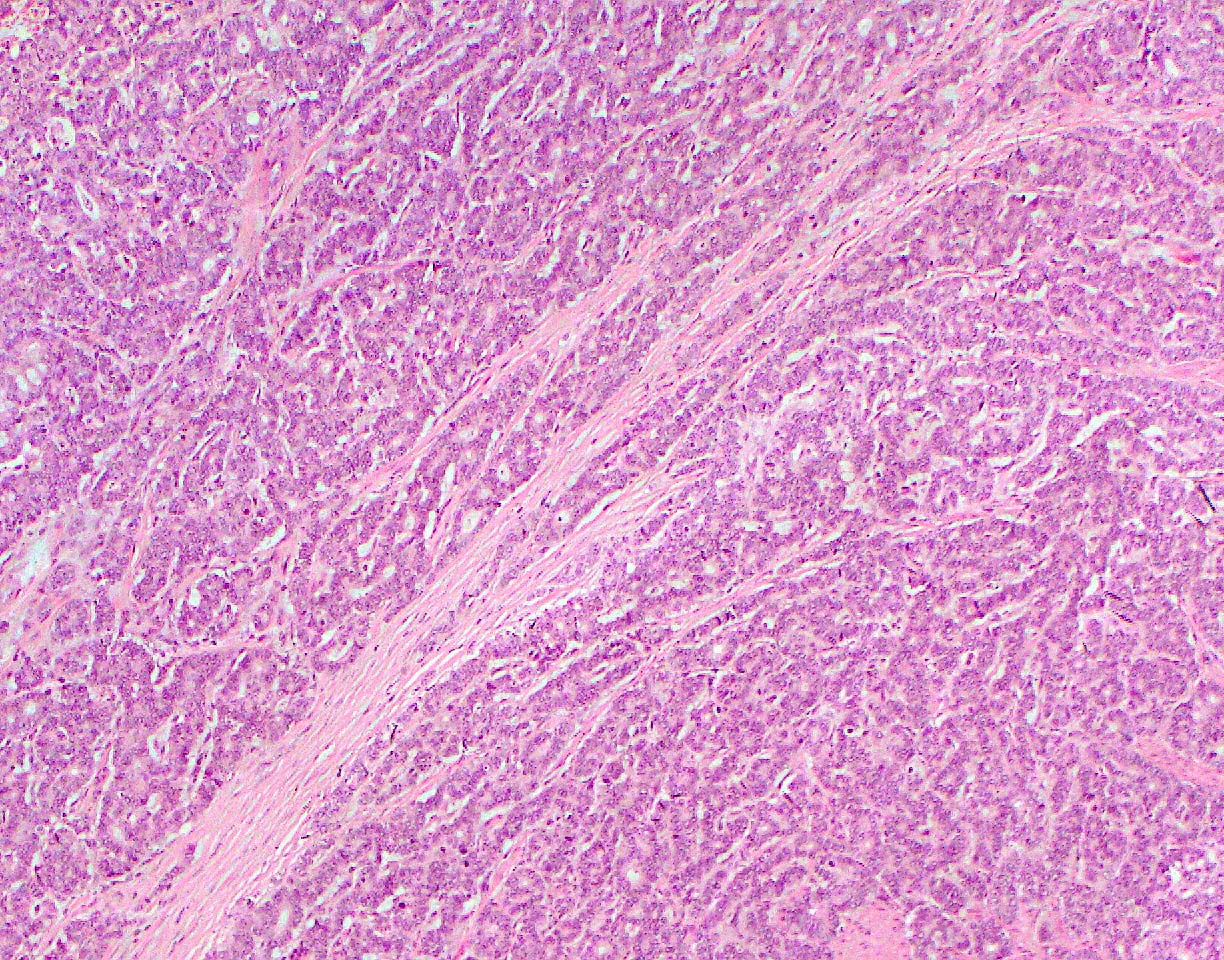
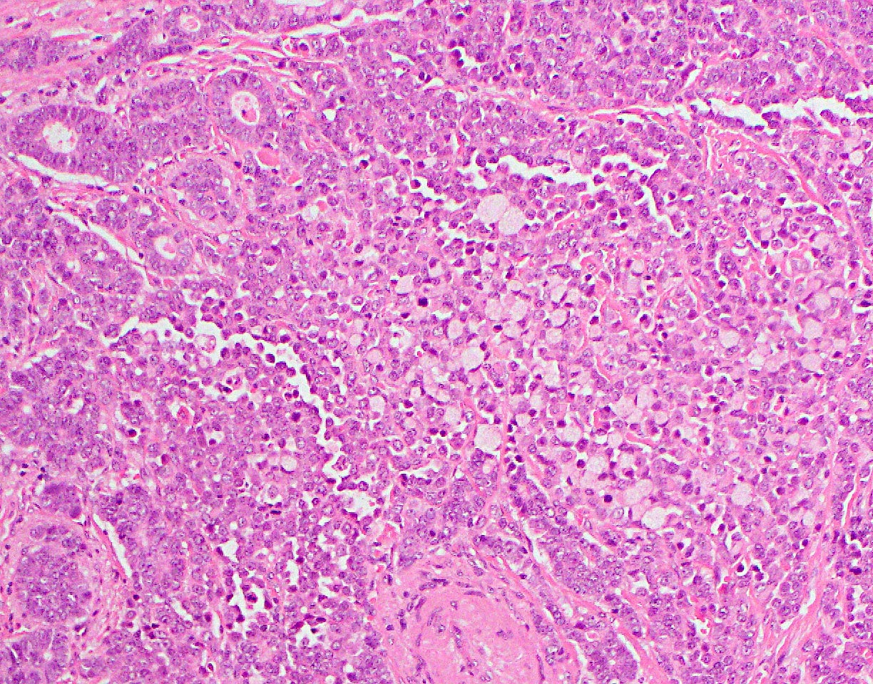
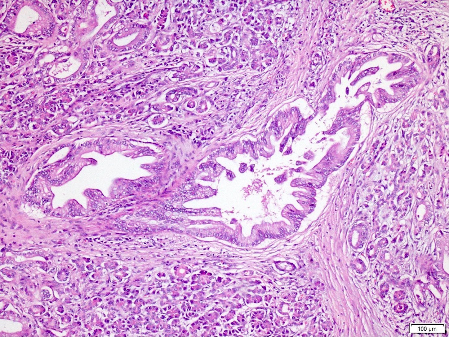
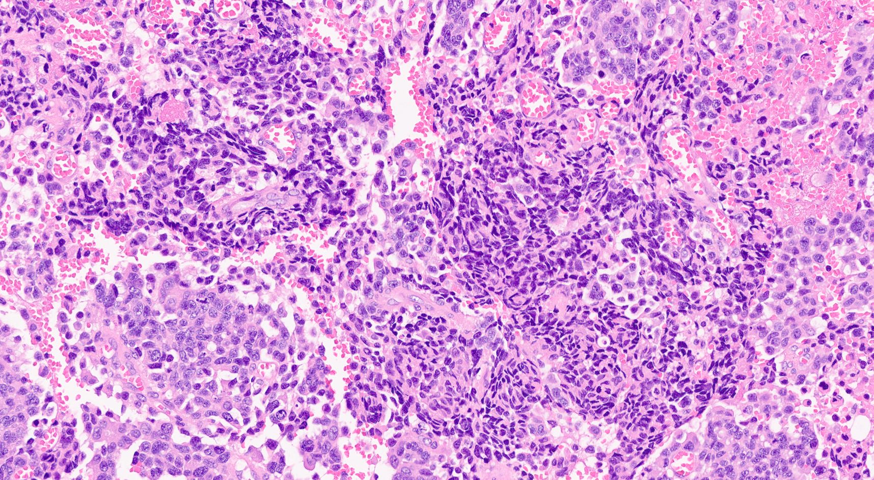
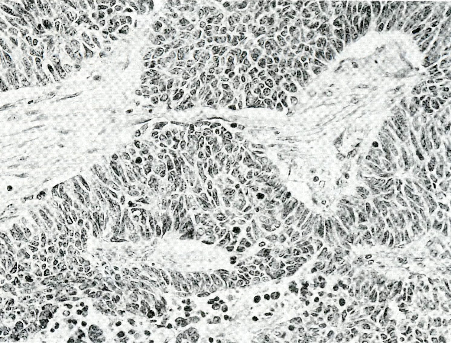
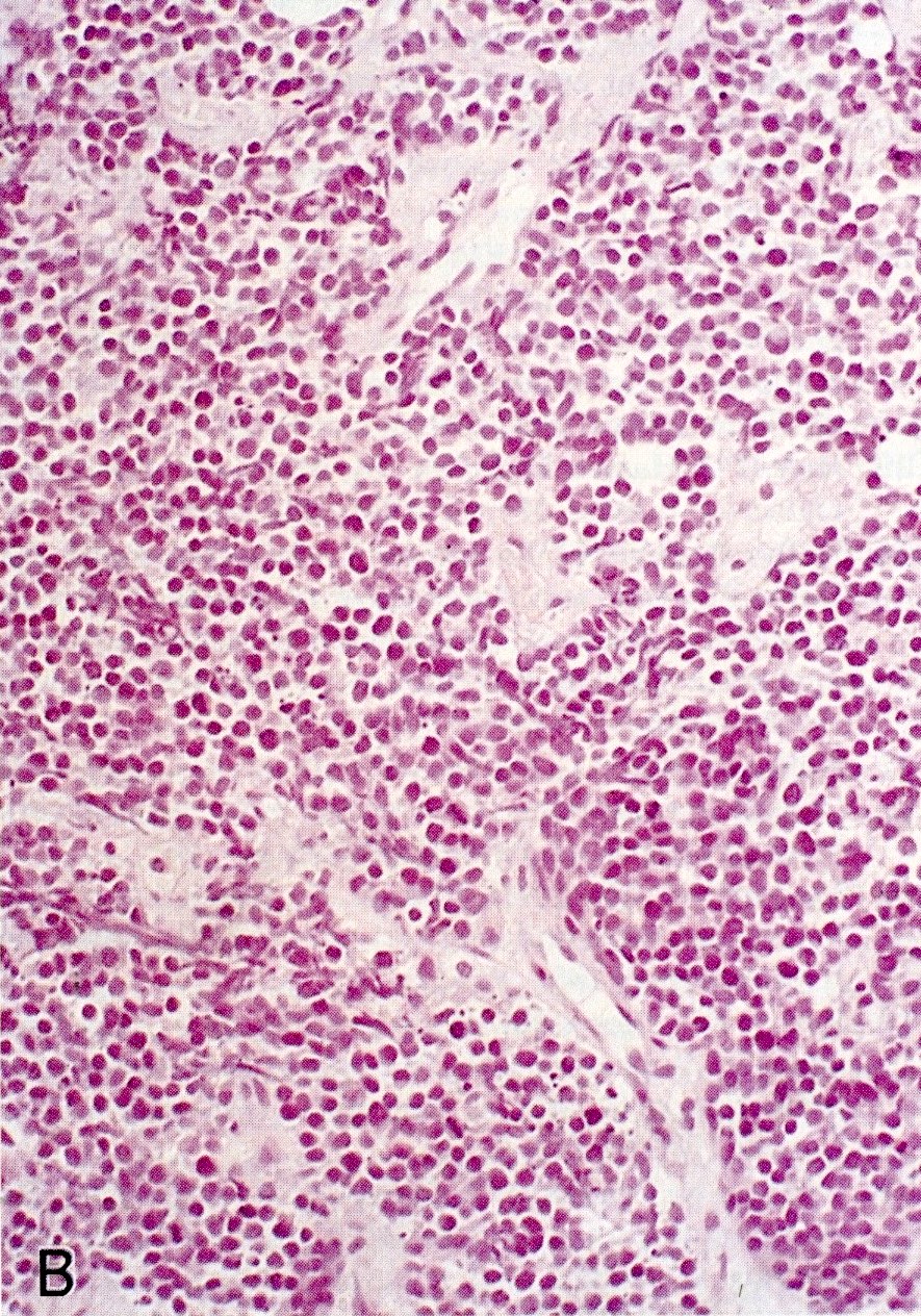
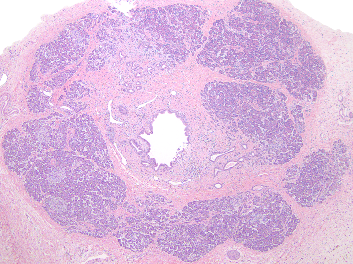
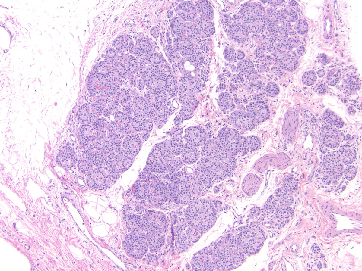
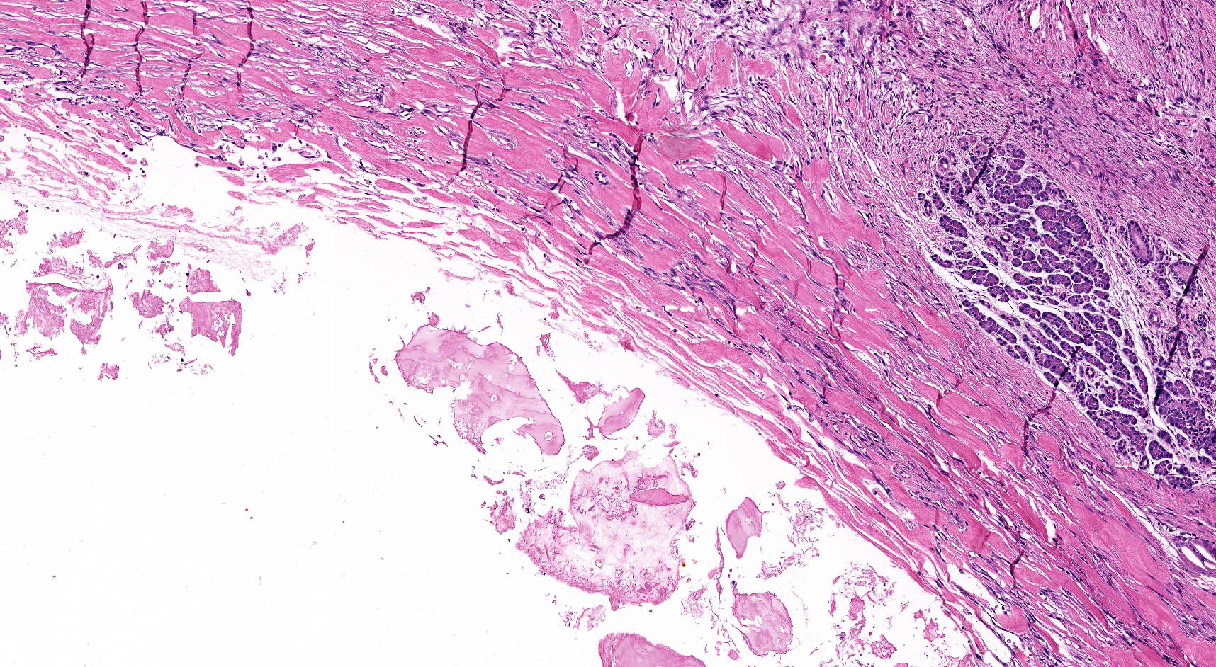
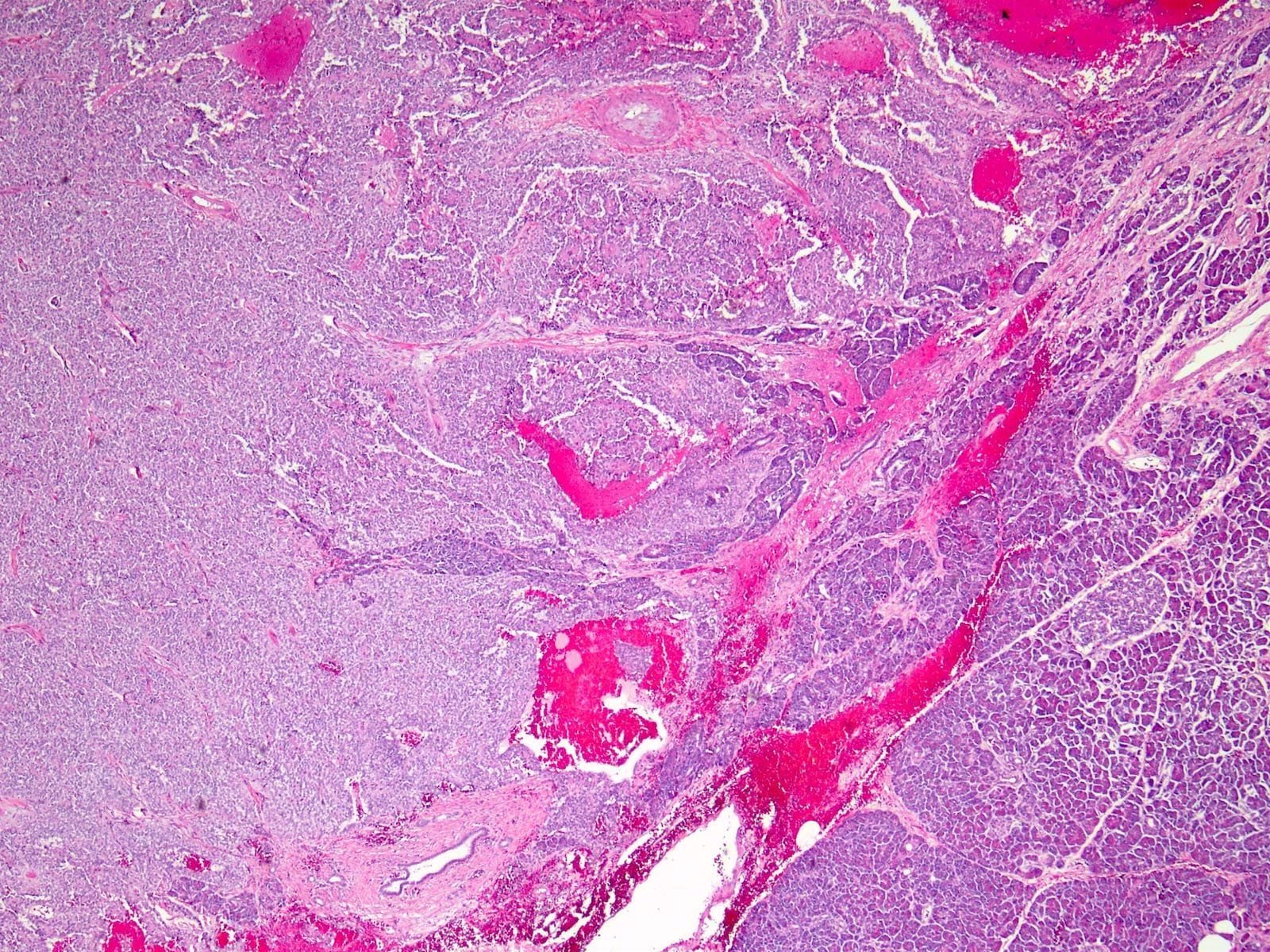
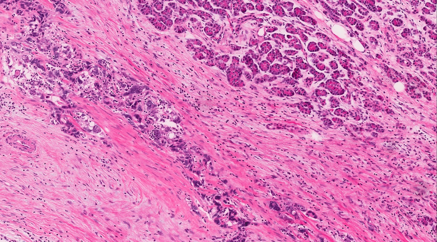
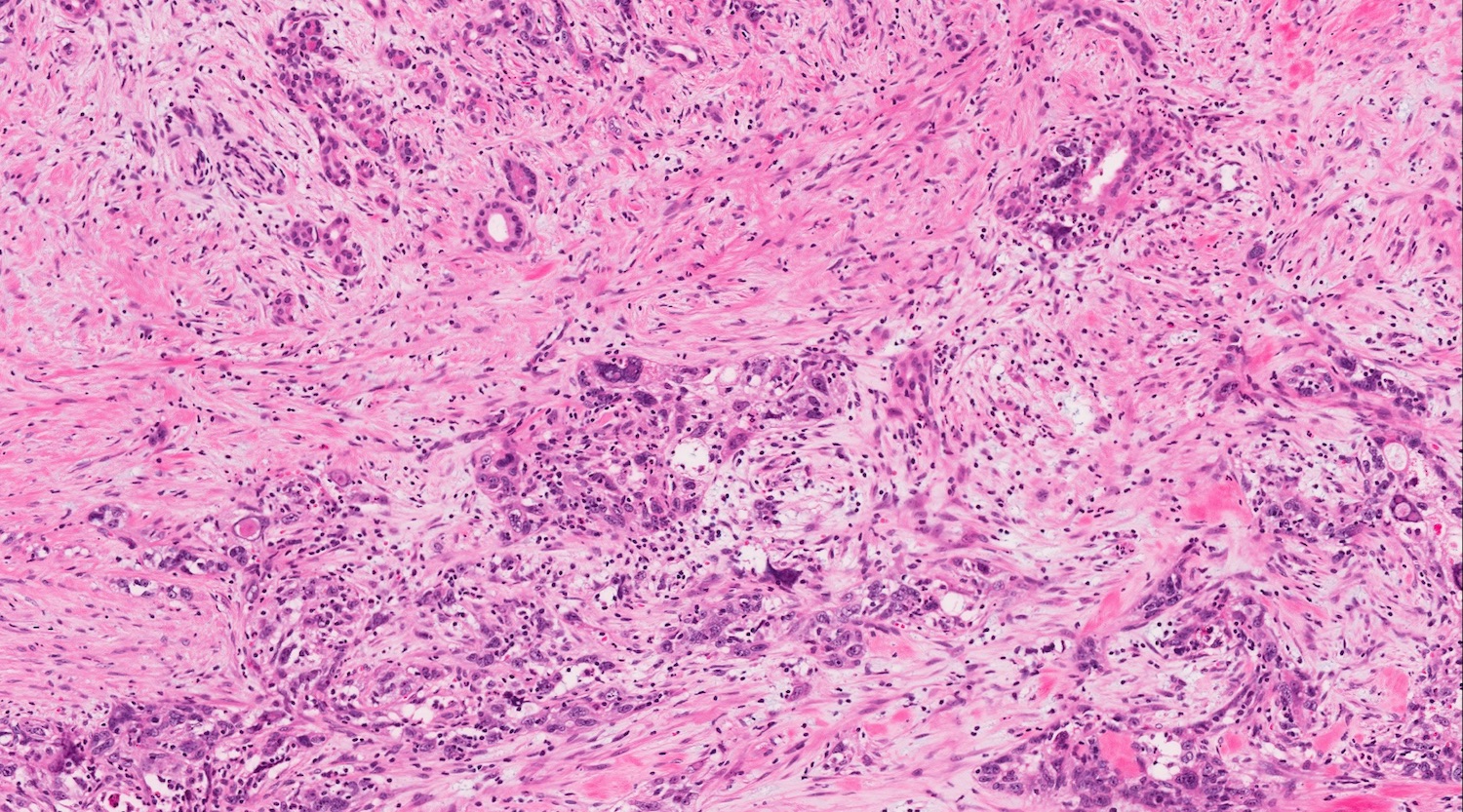
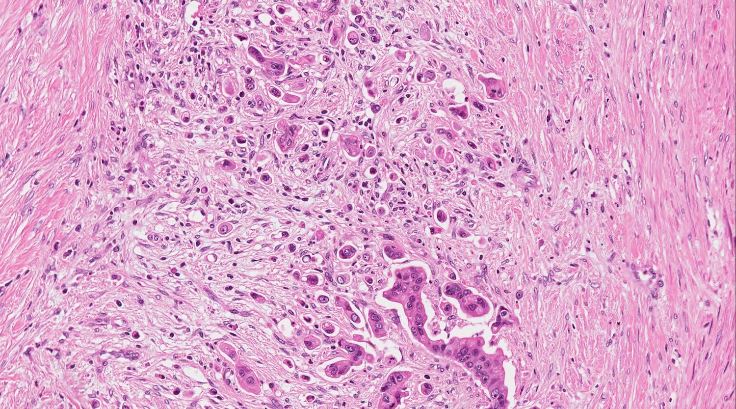
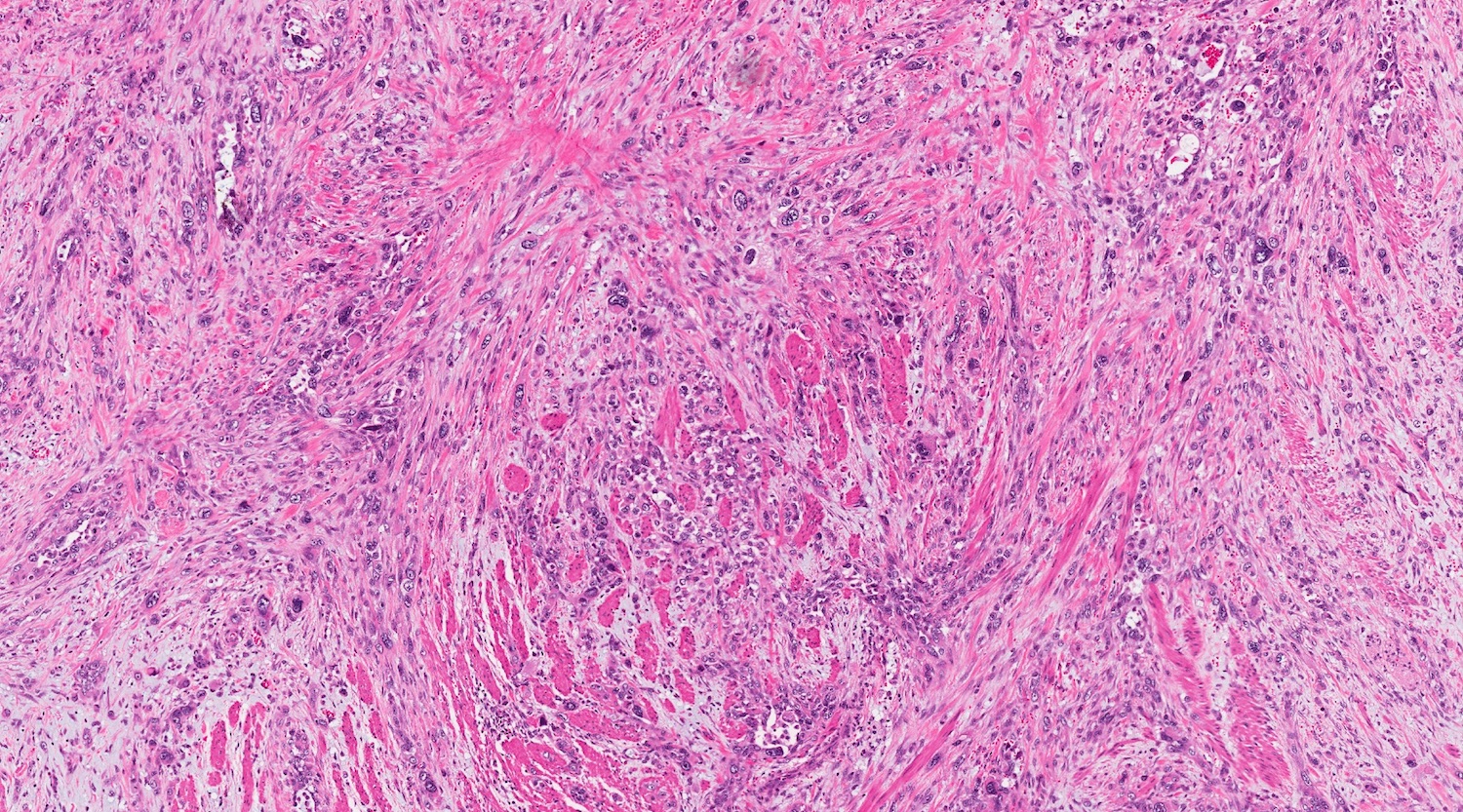
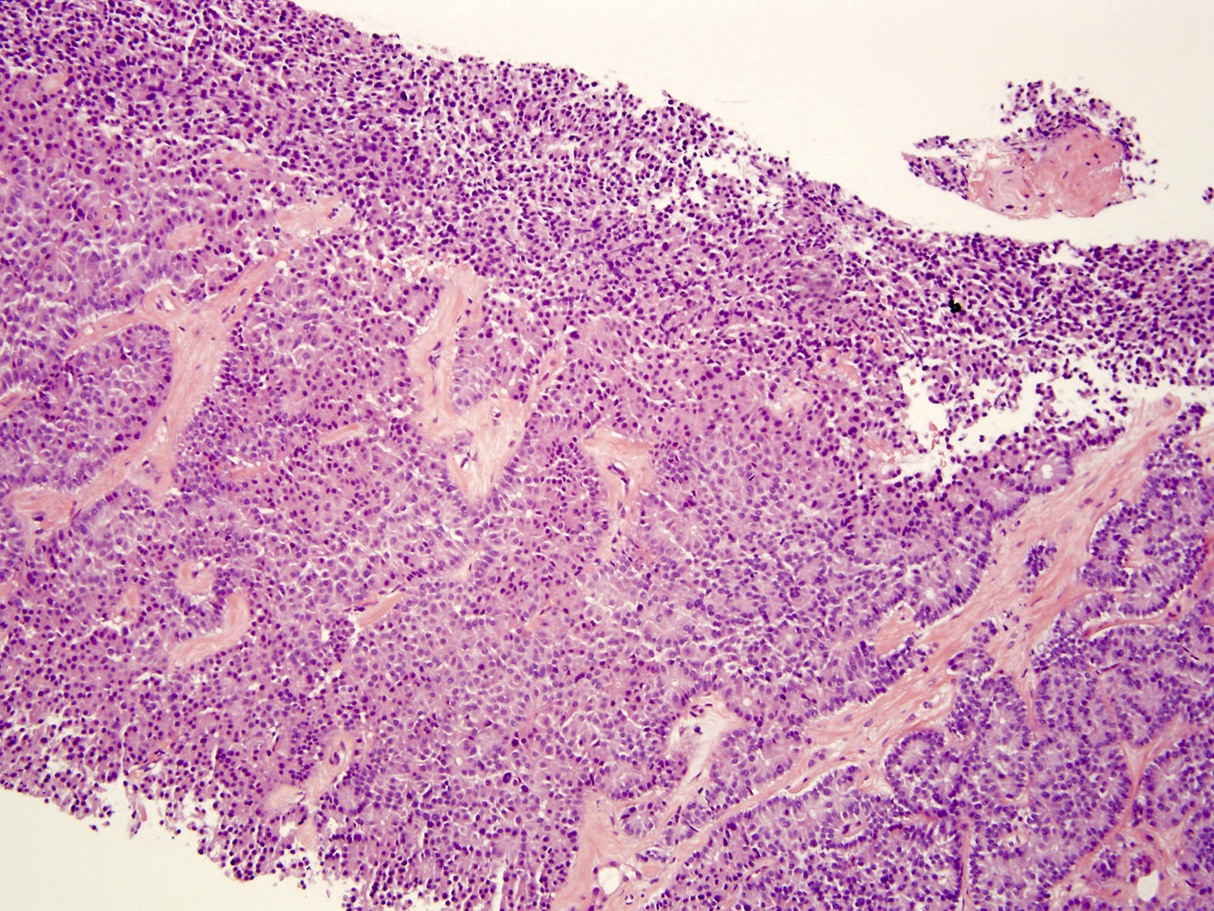
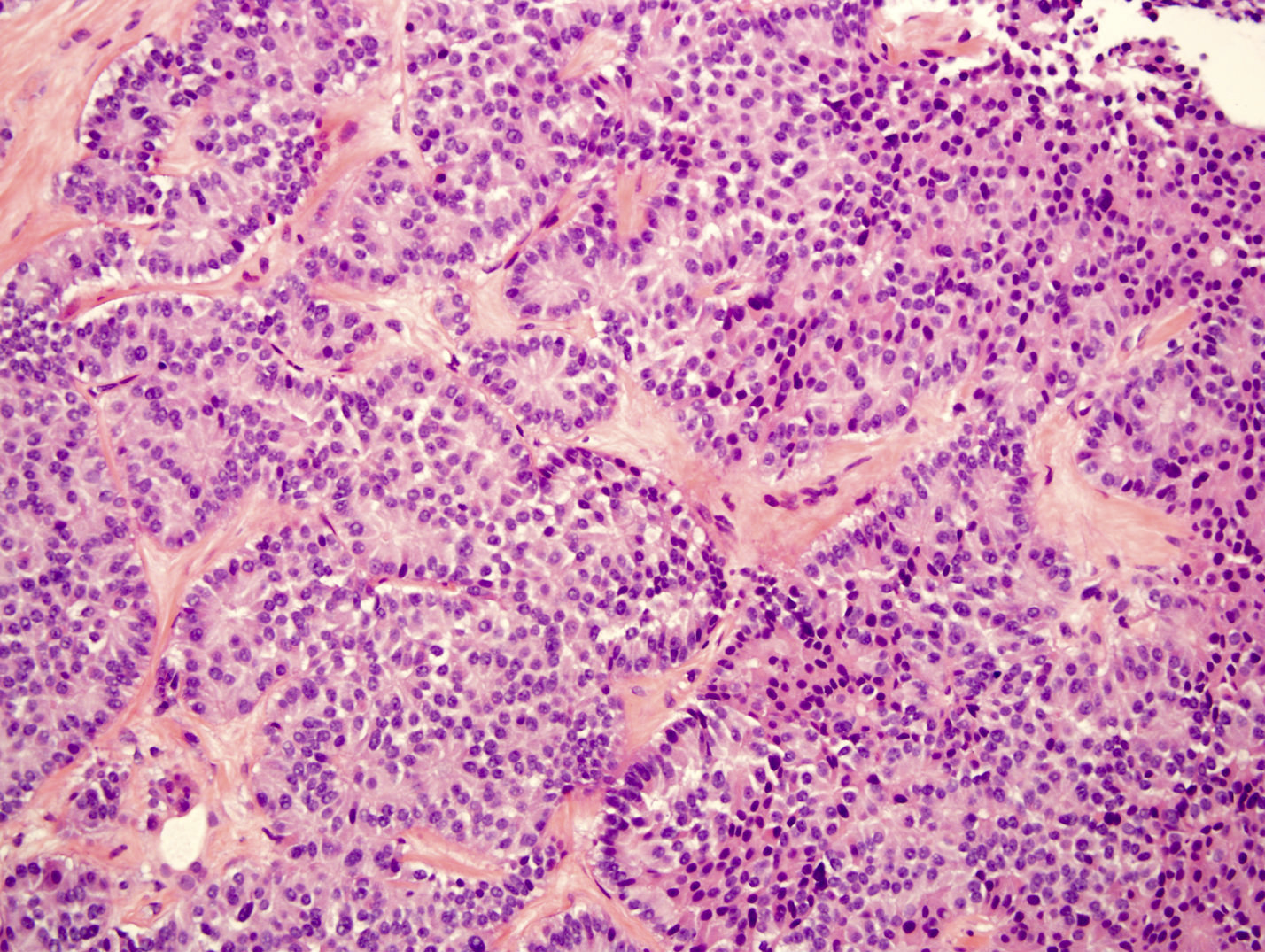
Typical histology
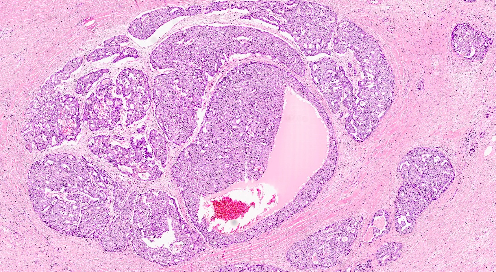
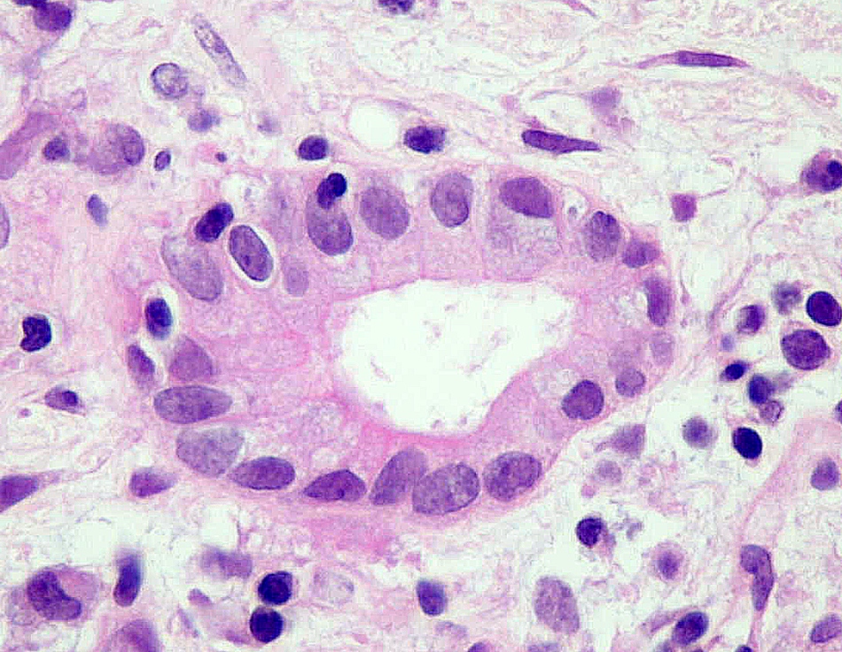
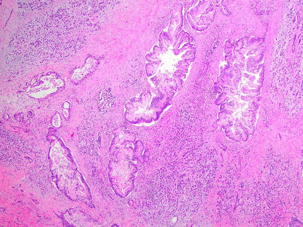
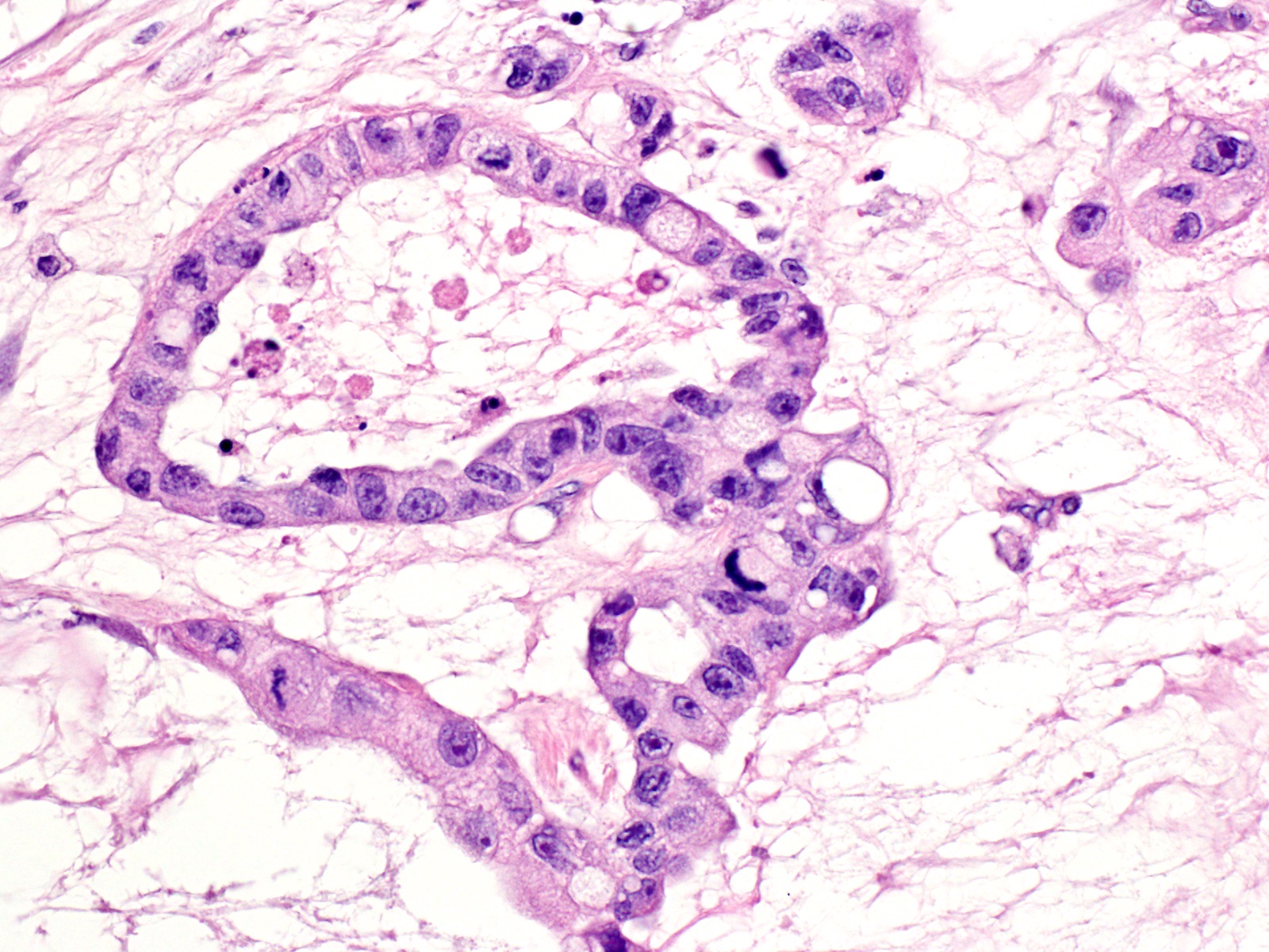
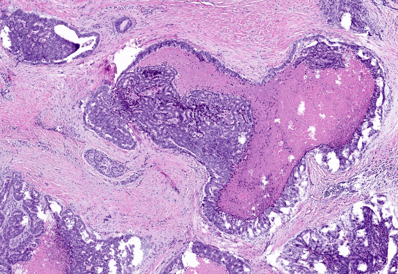
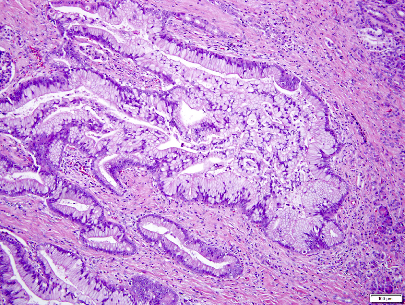
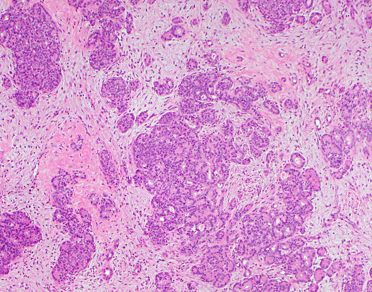
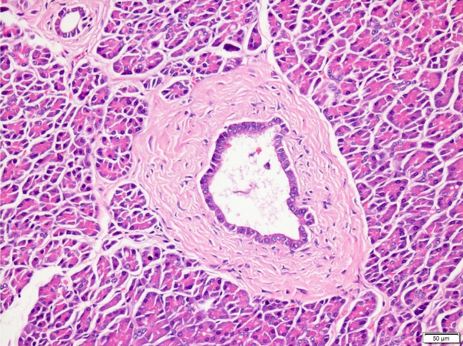
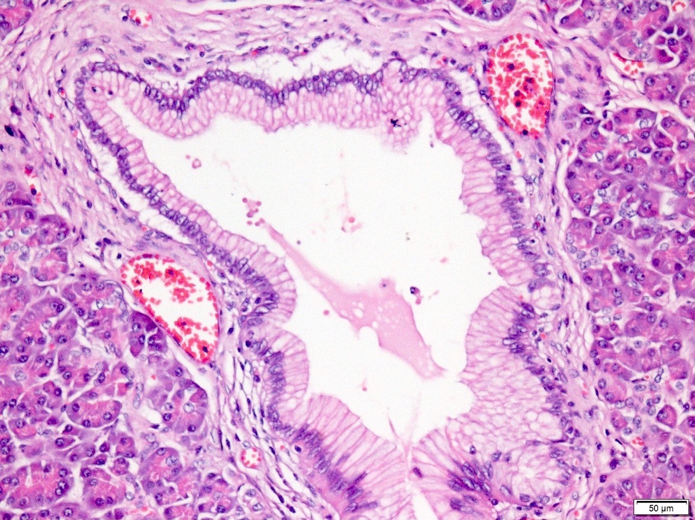
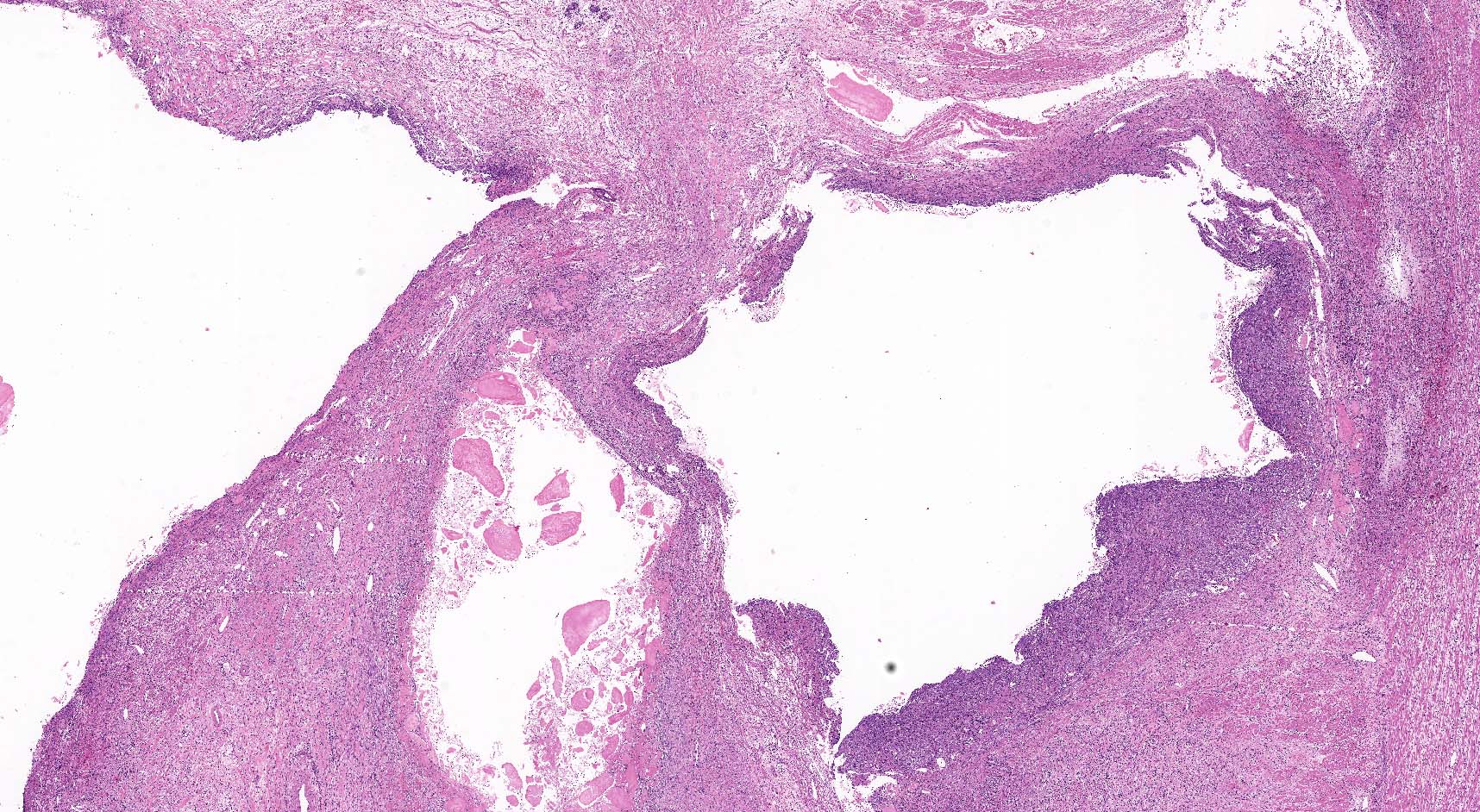
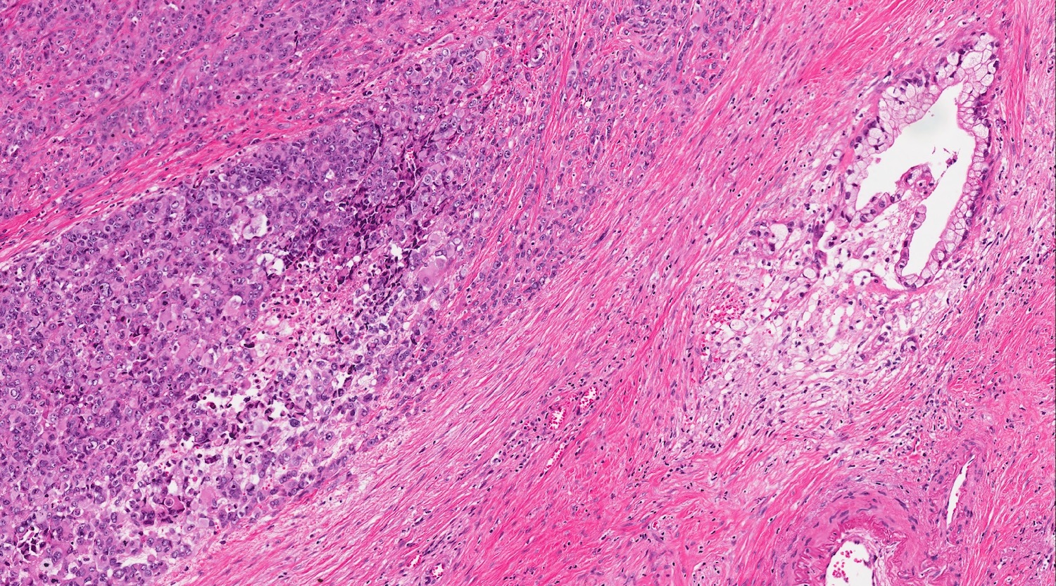
Intraductal growth
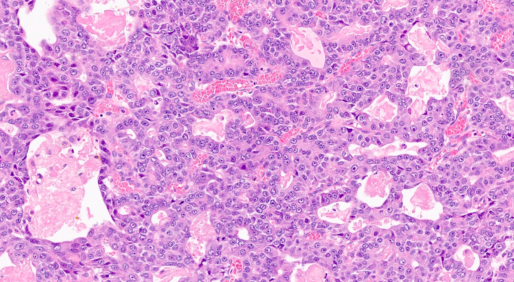
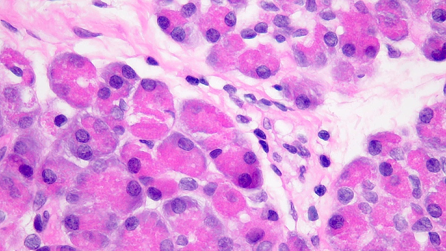
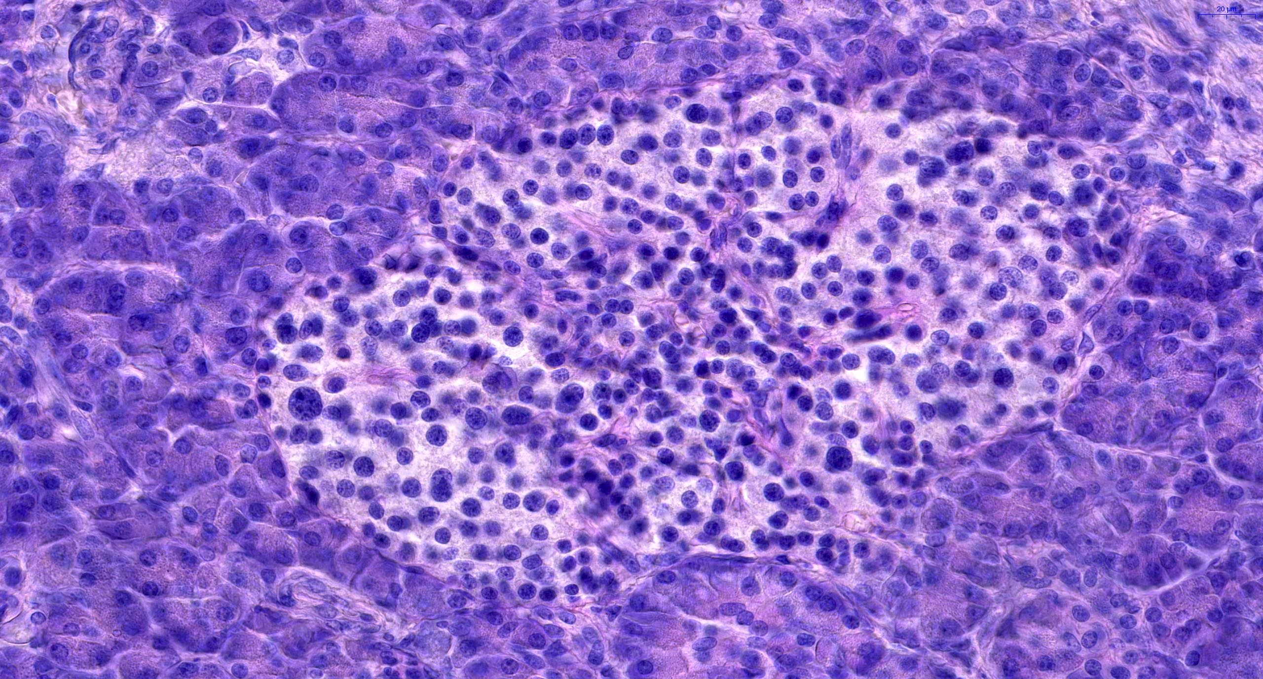
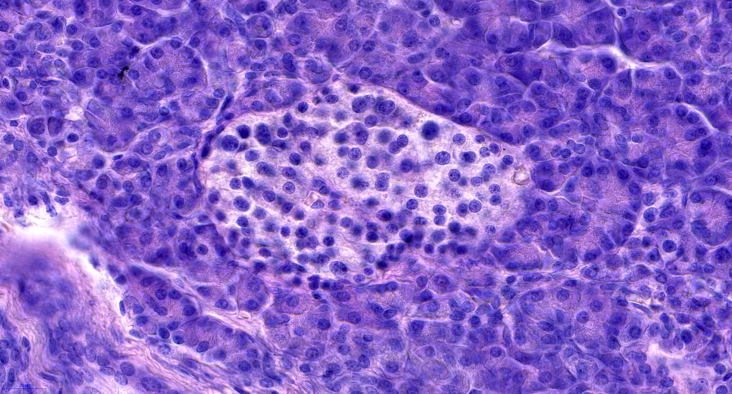
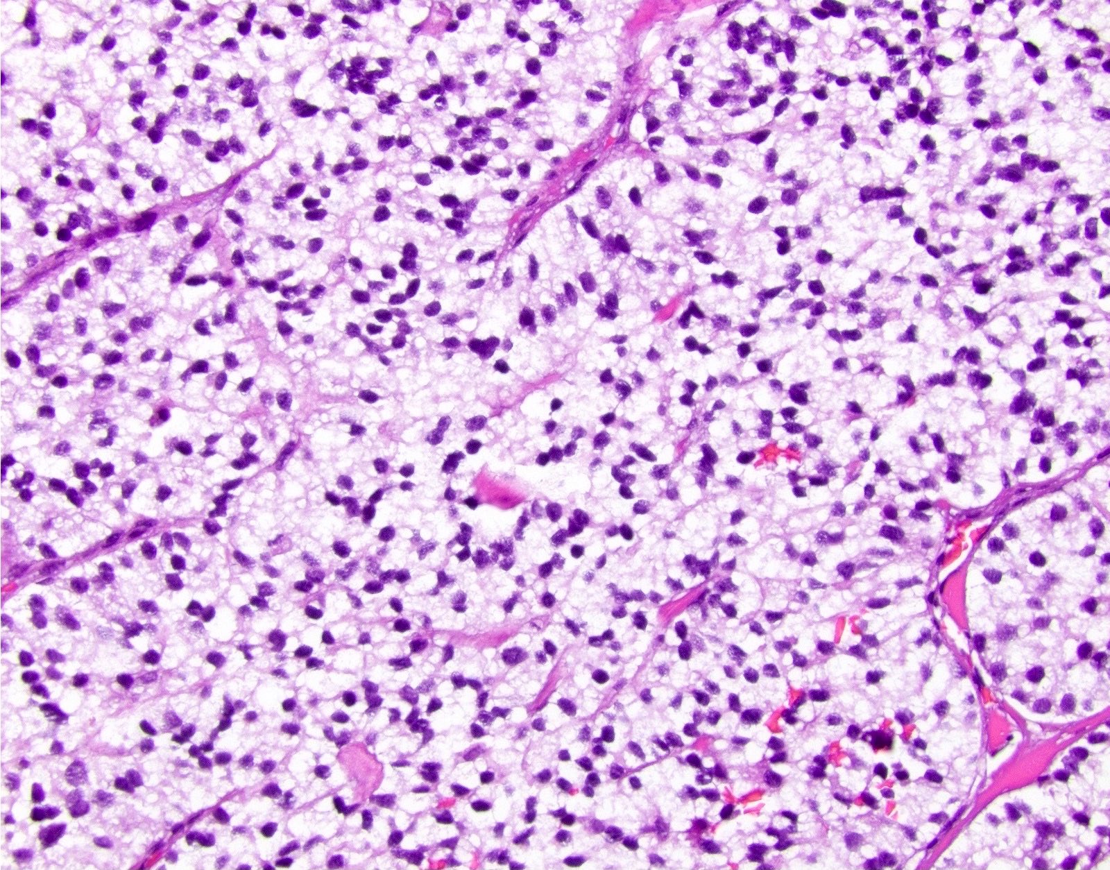
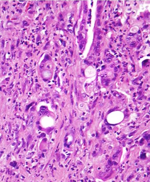
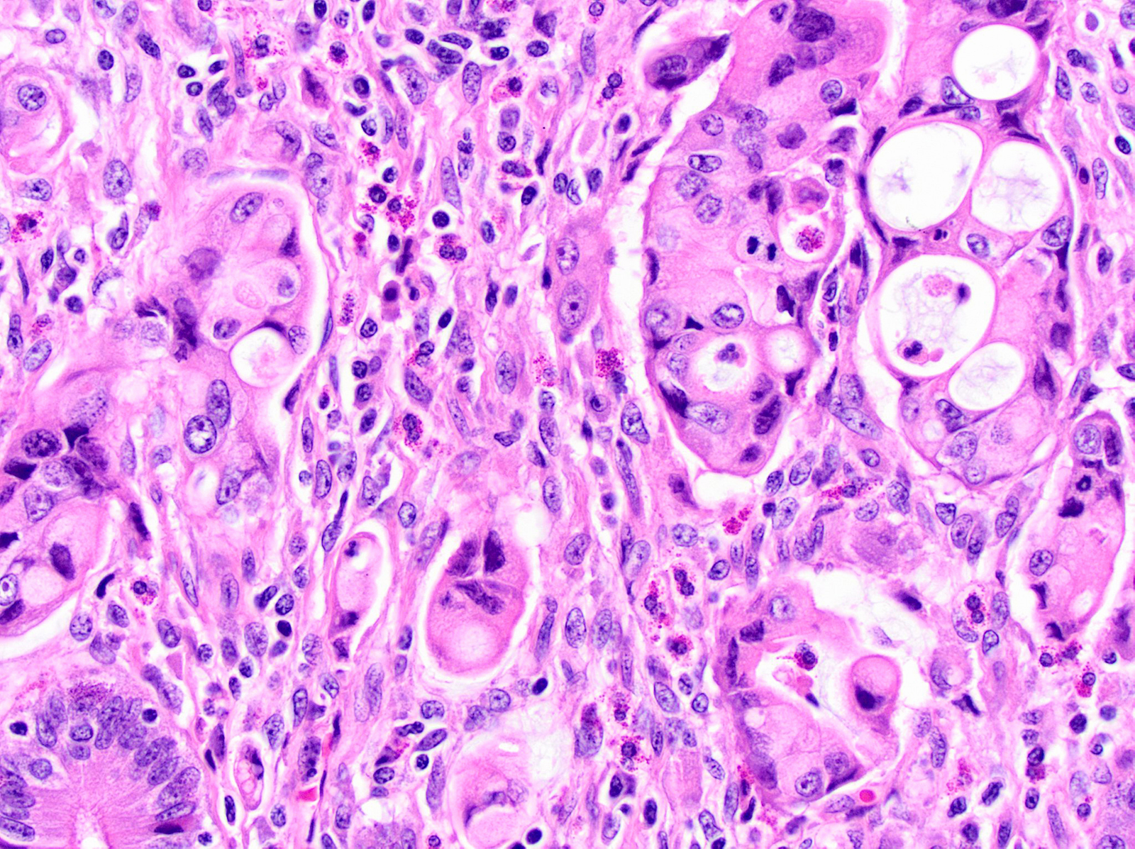
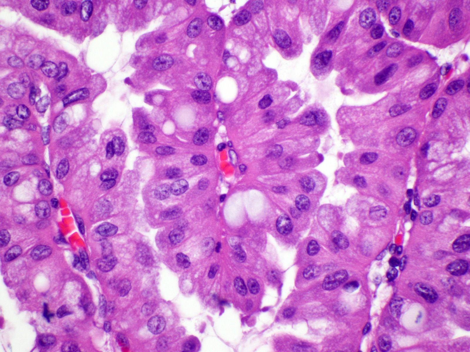
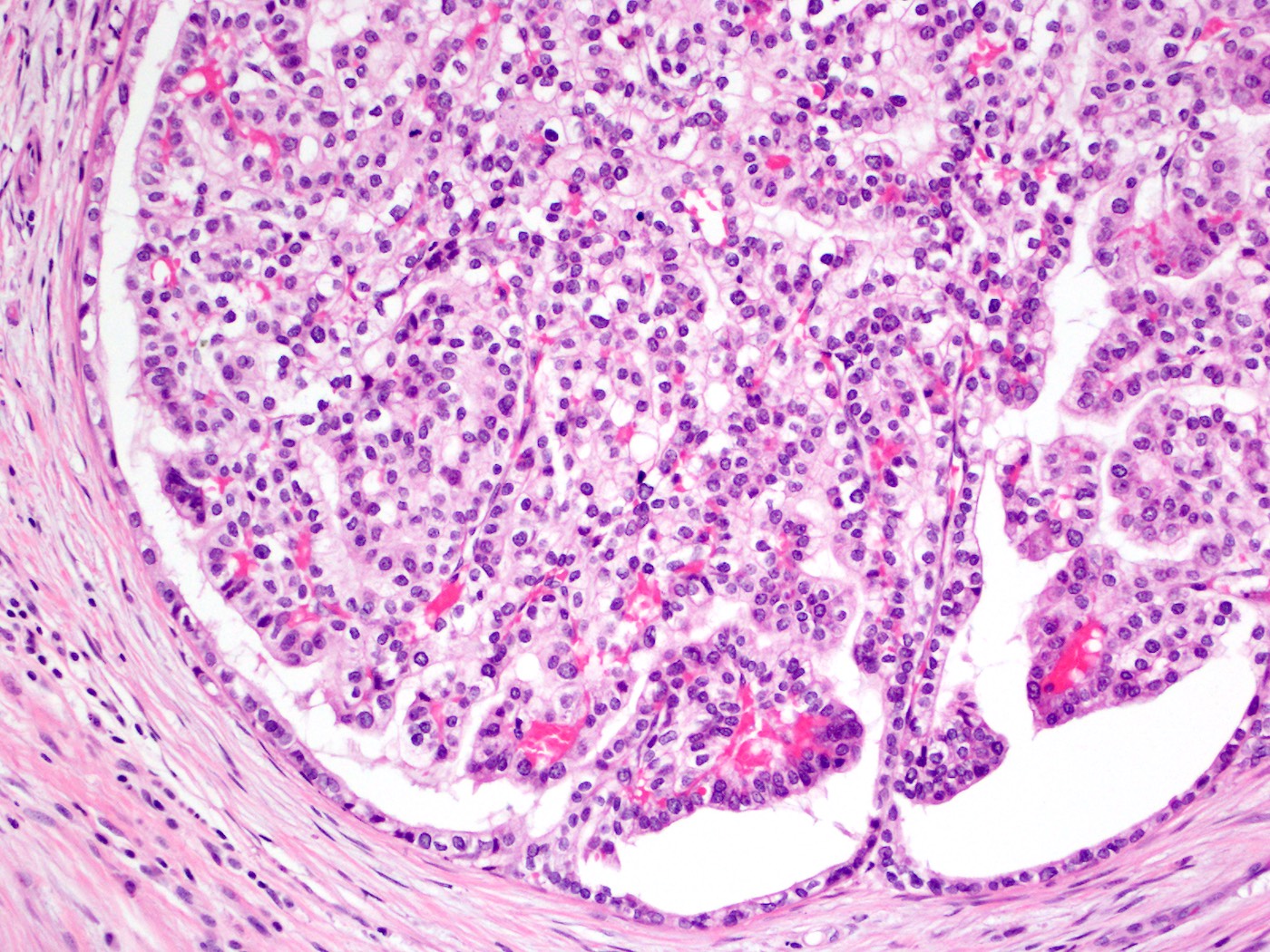
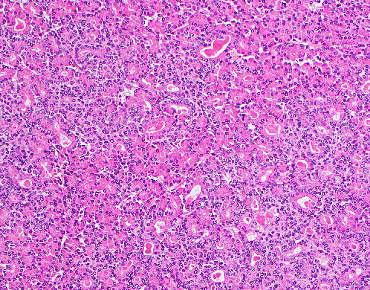
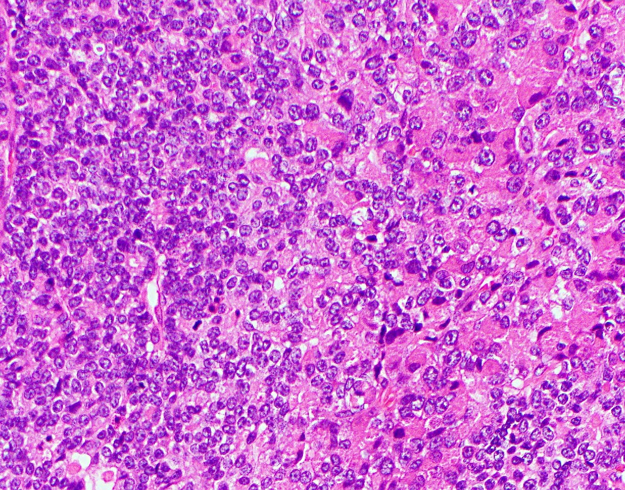
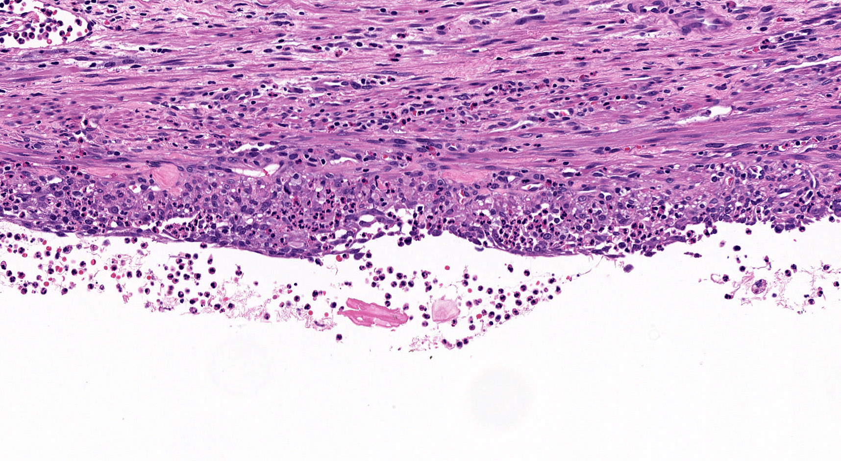
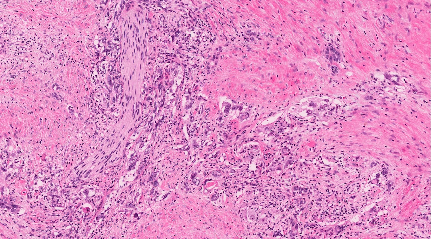
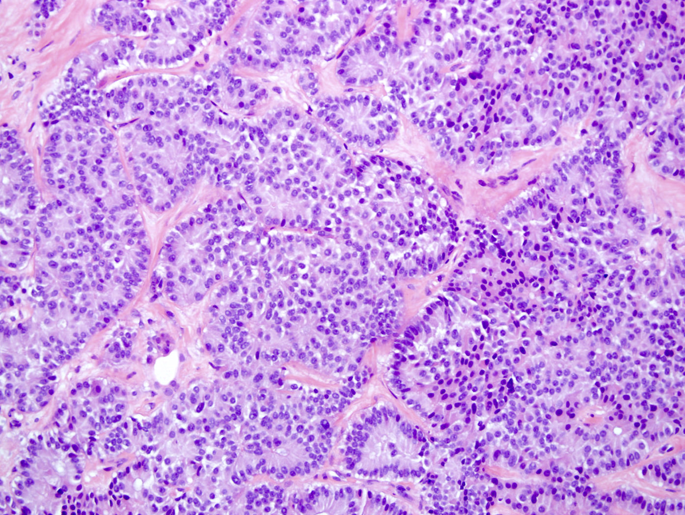
Cell detail
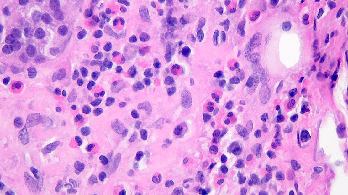
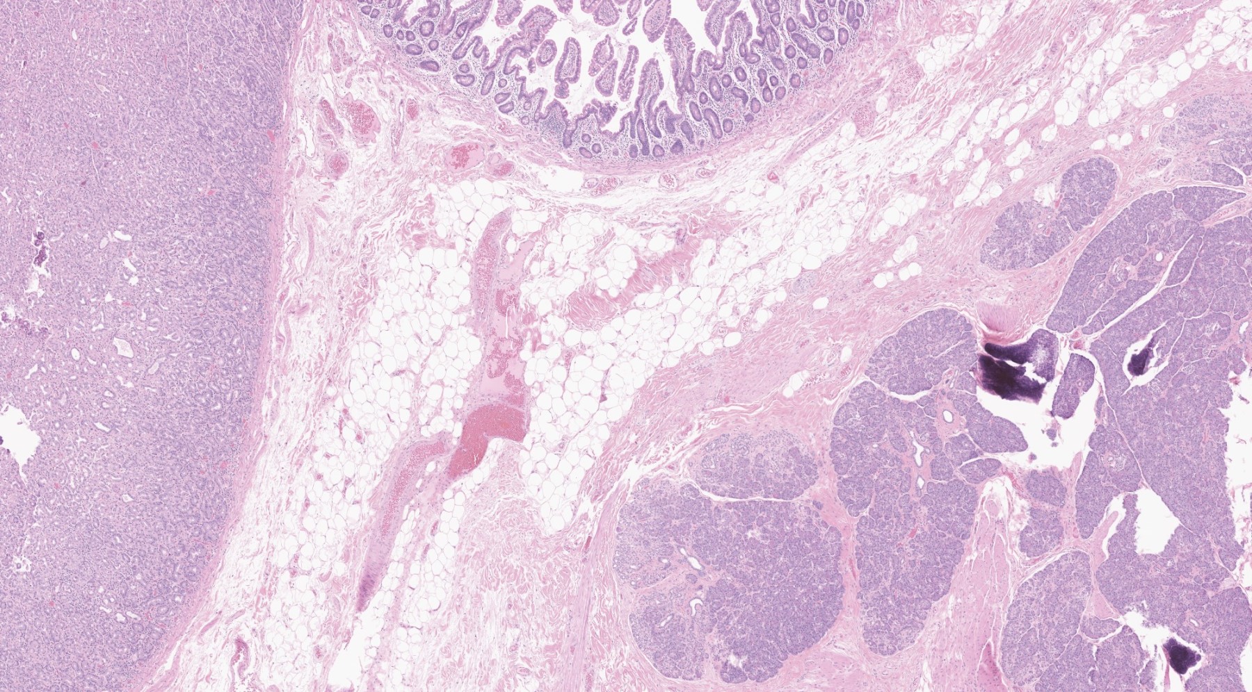
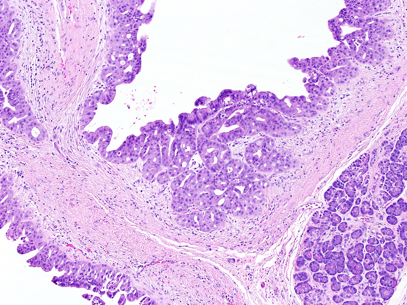
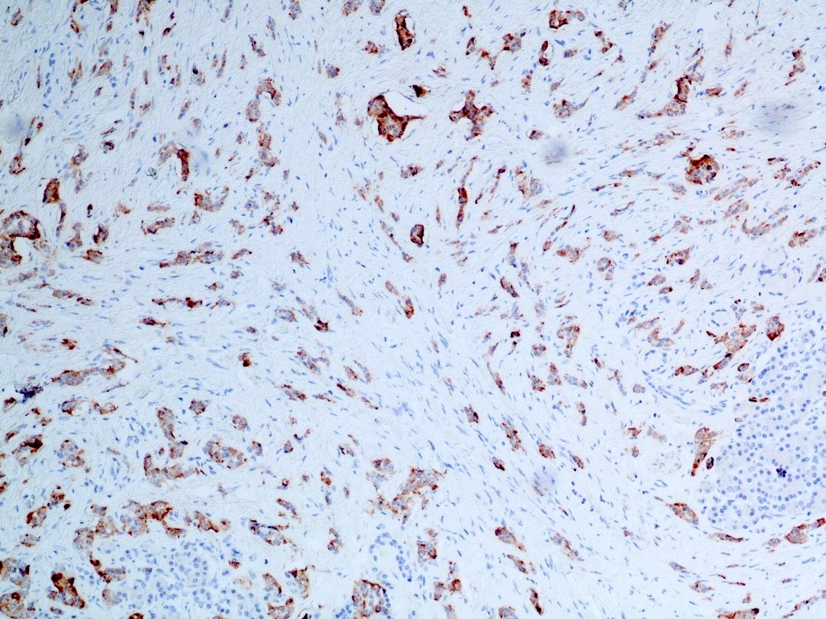
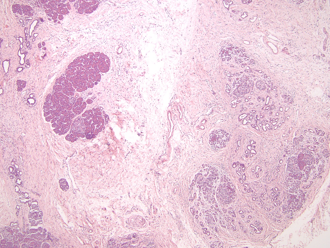
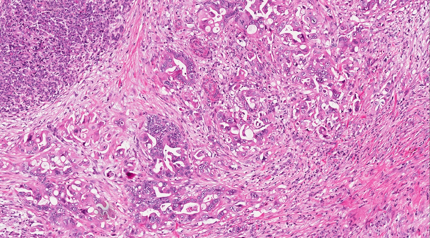
Association with neuroendocrine tumors
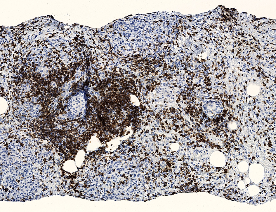
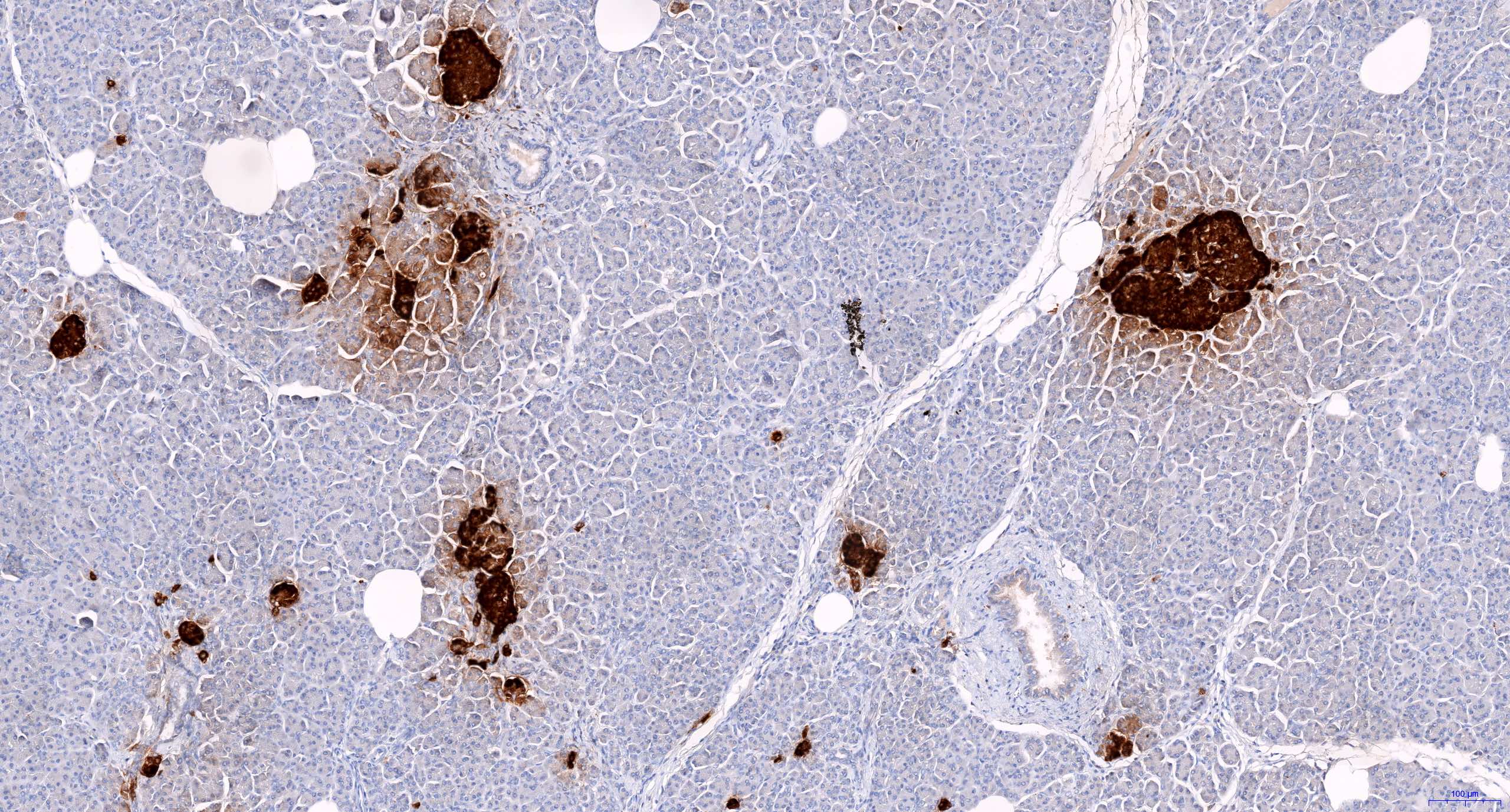
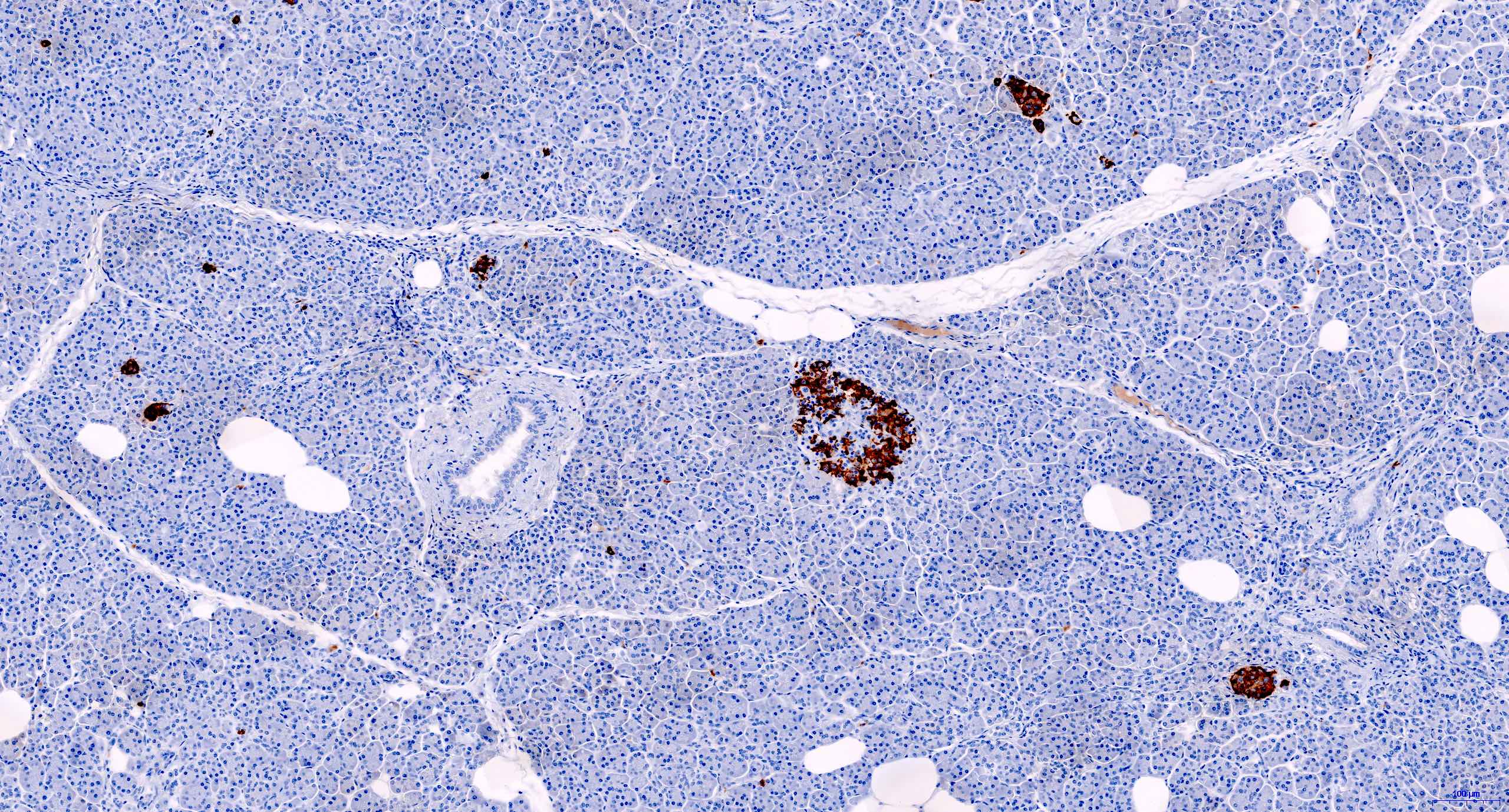
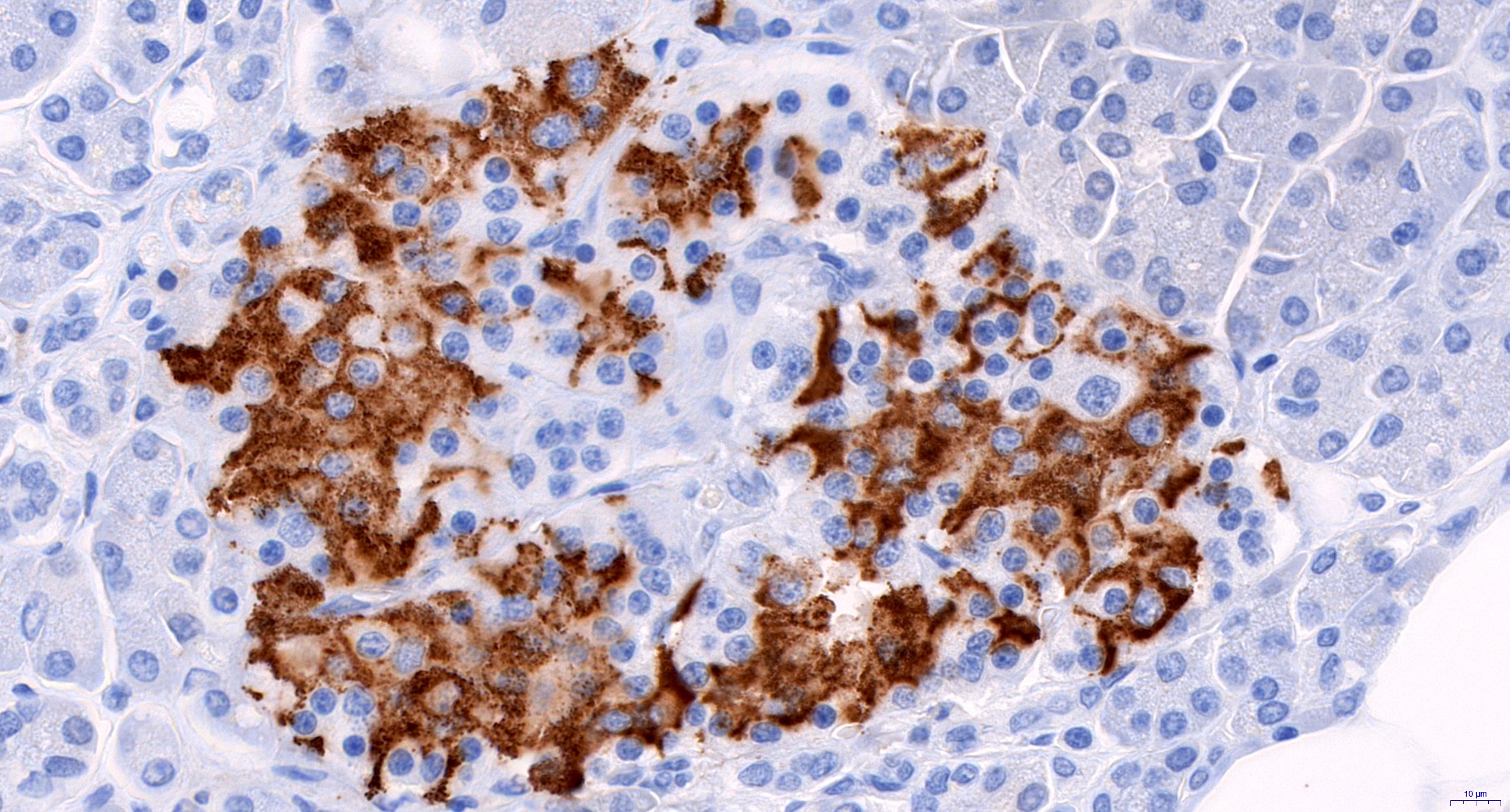
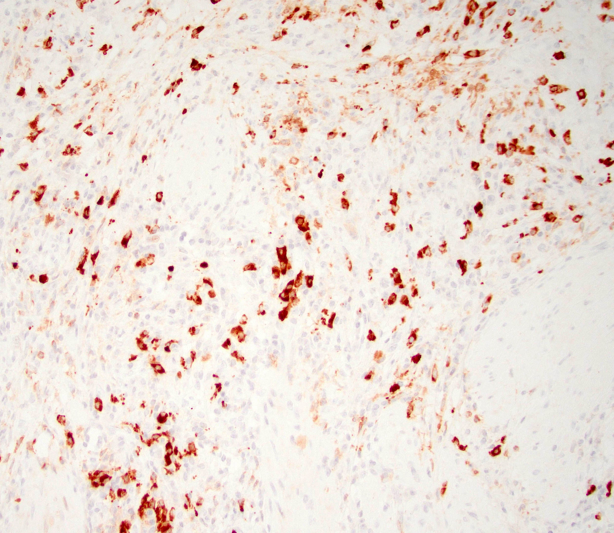
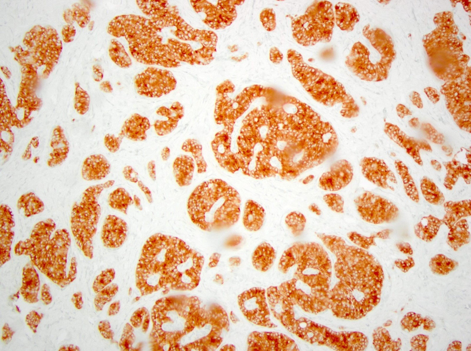
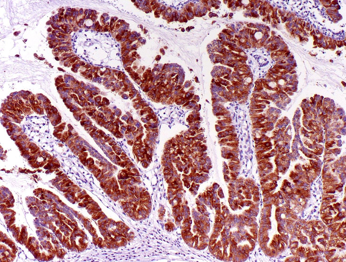
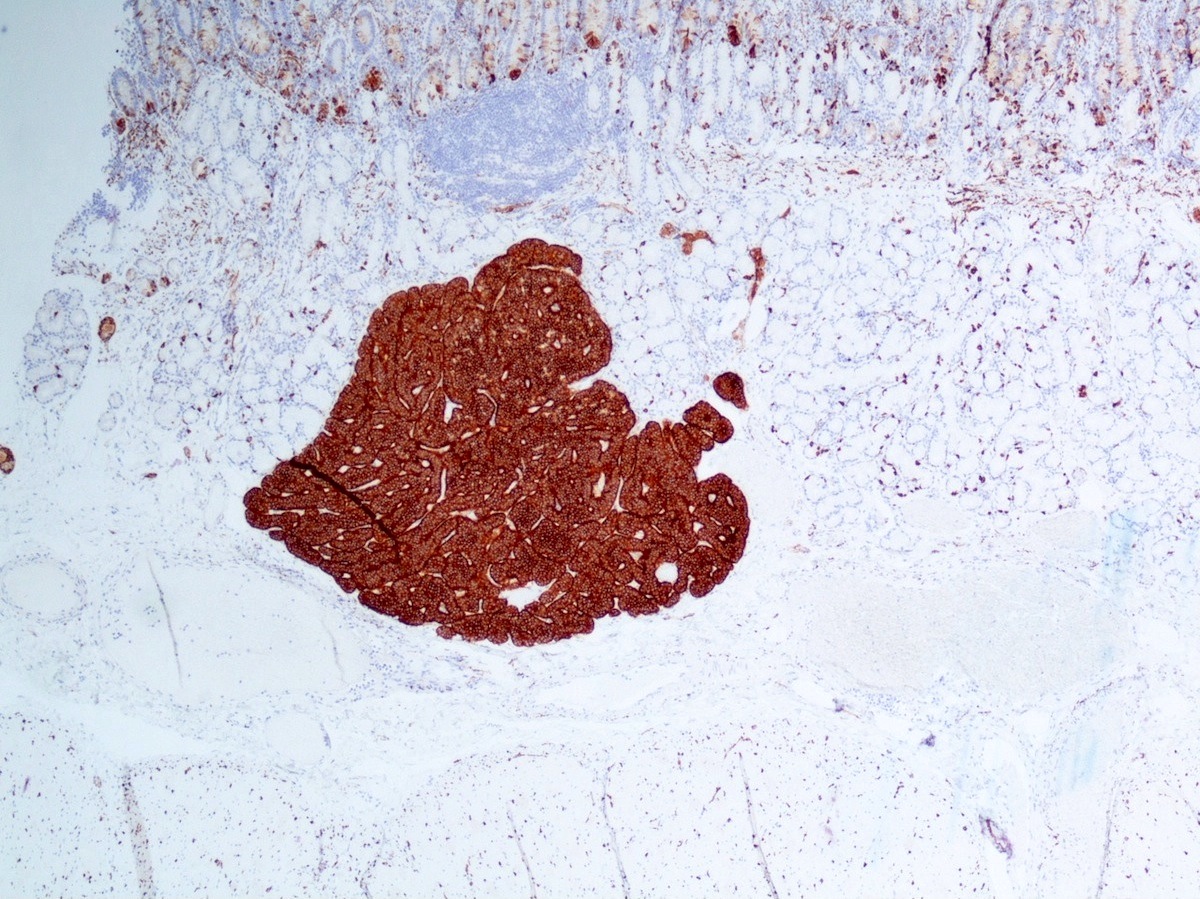
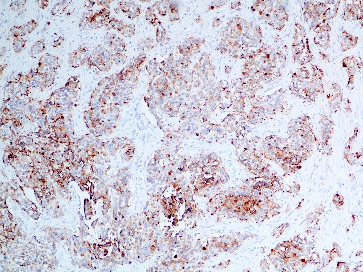
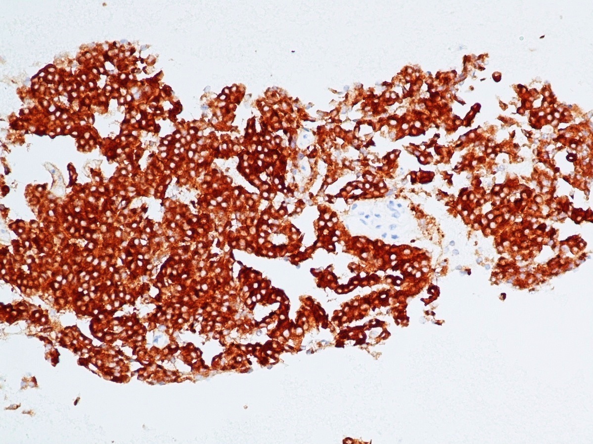
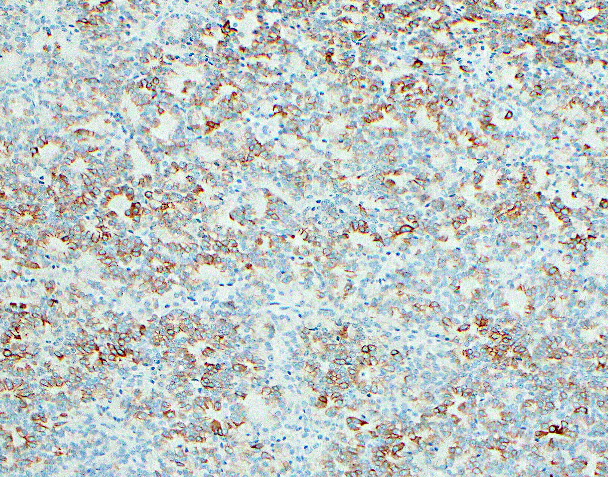
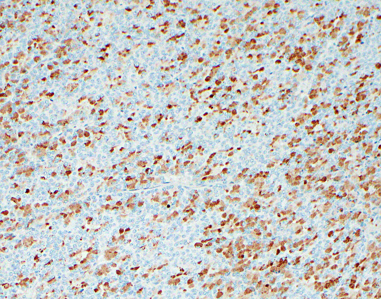
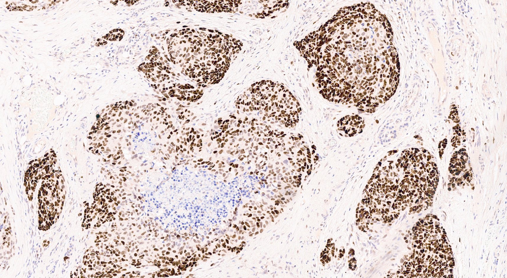
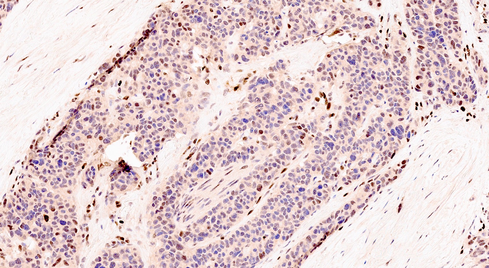
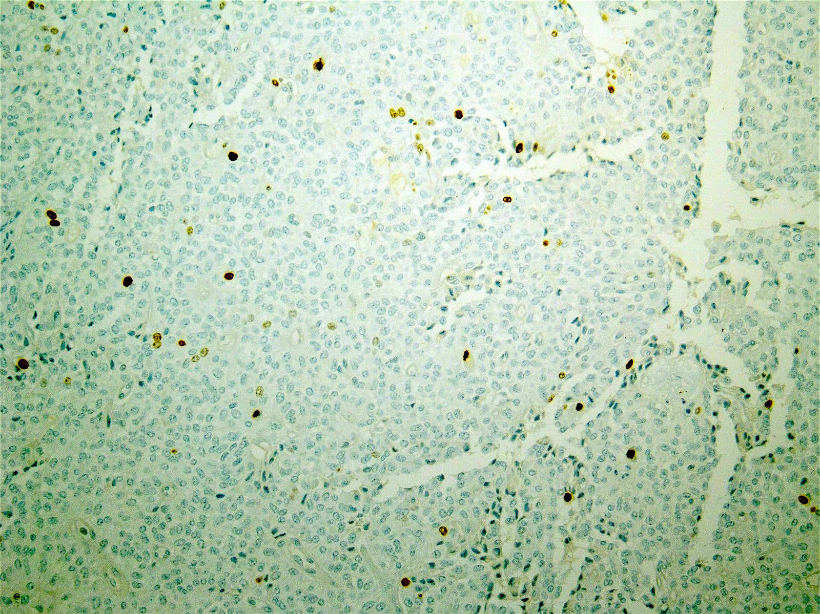
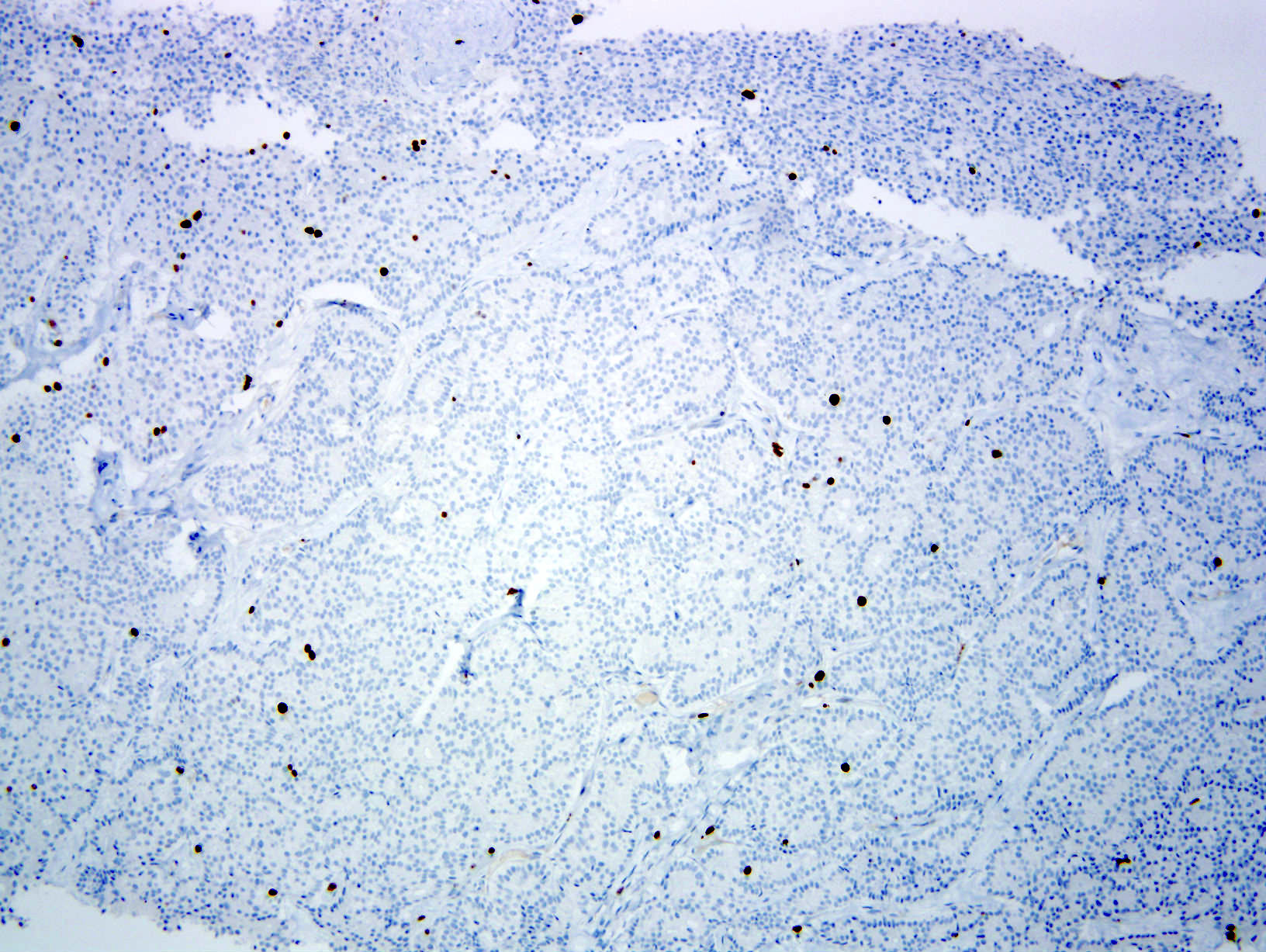
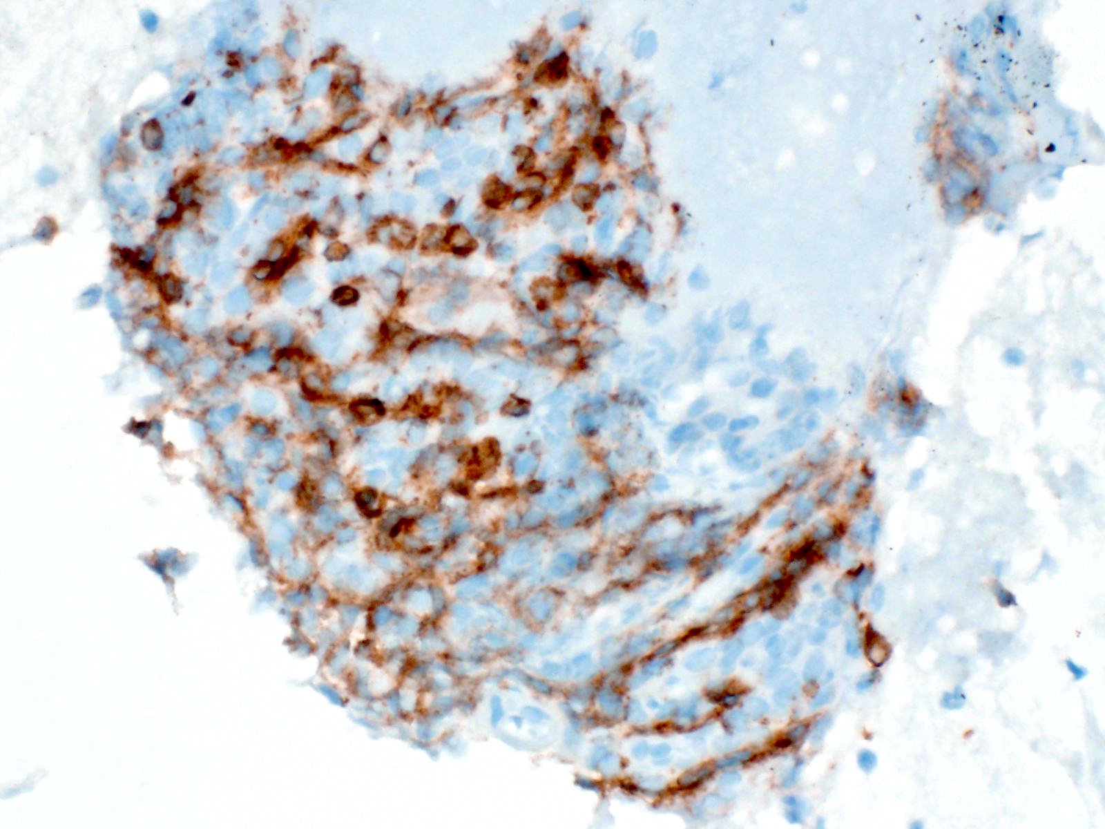
BCL10 expression pattern
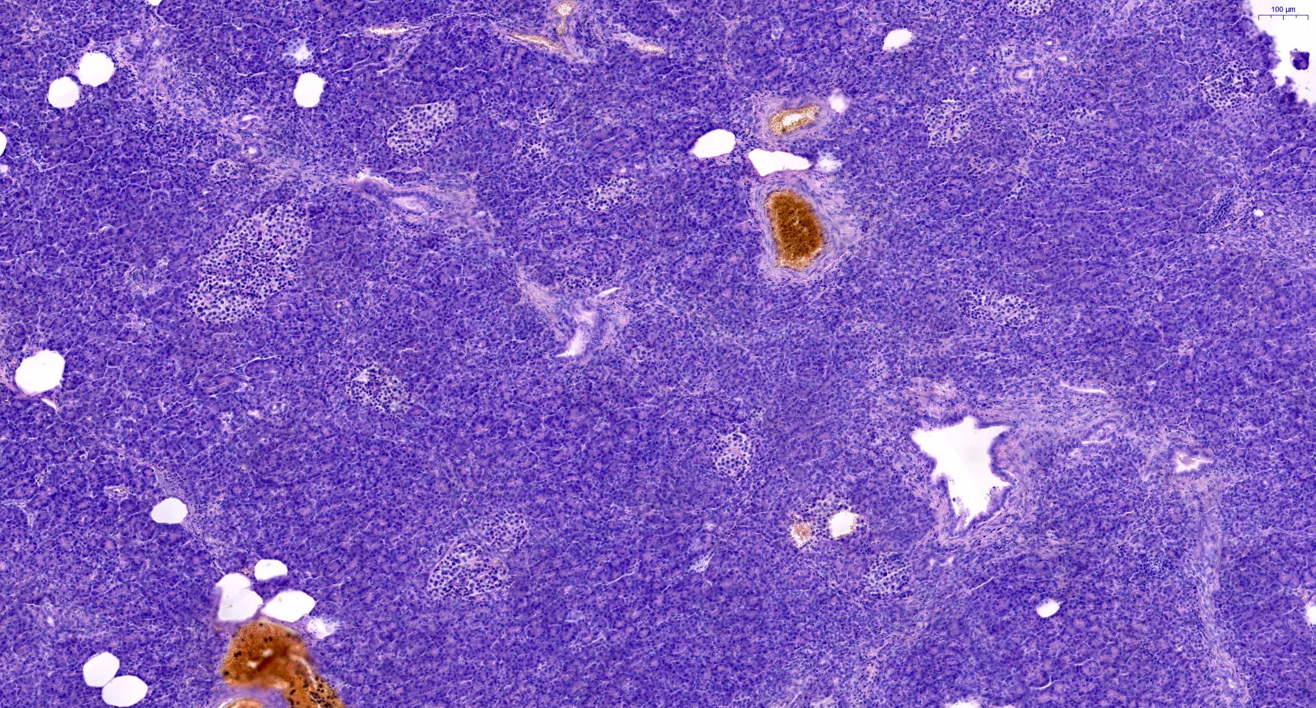
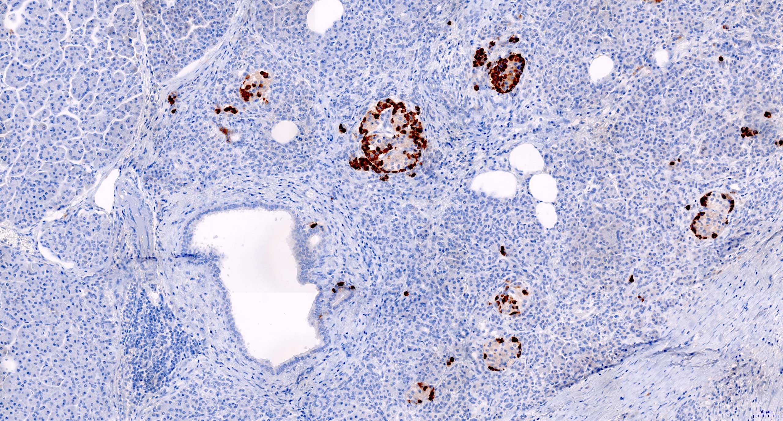
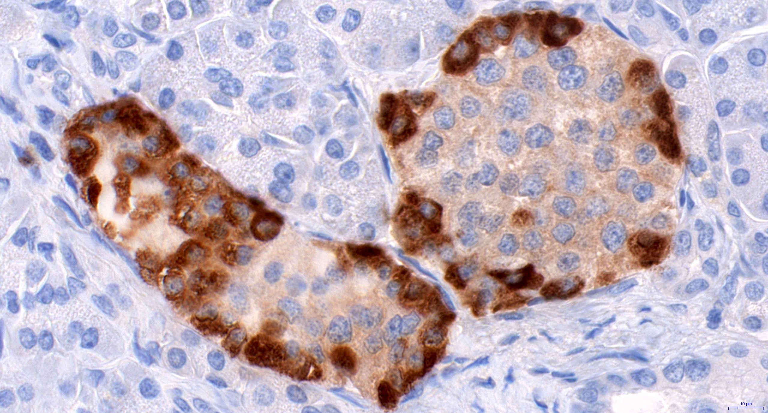
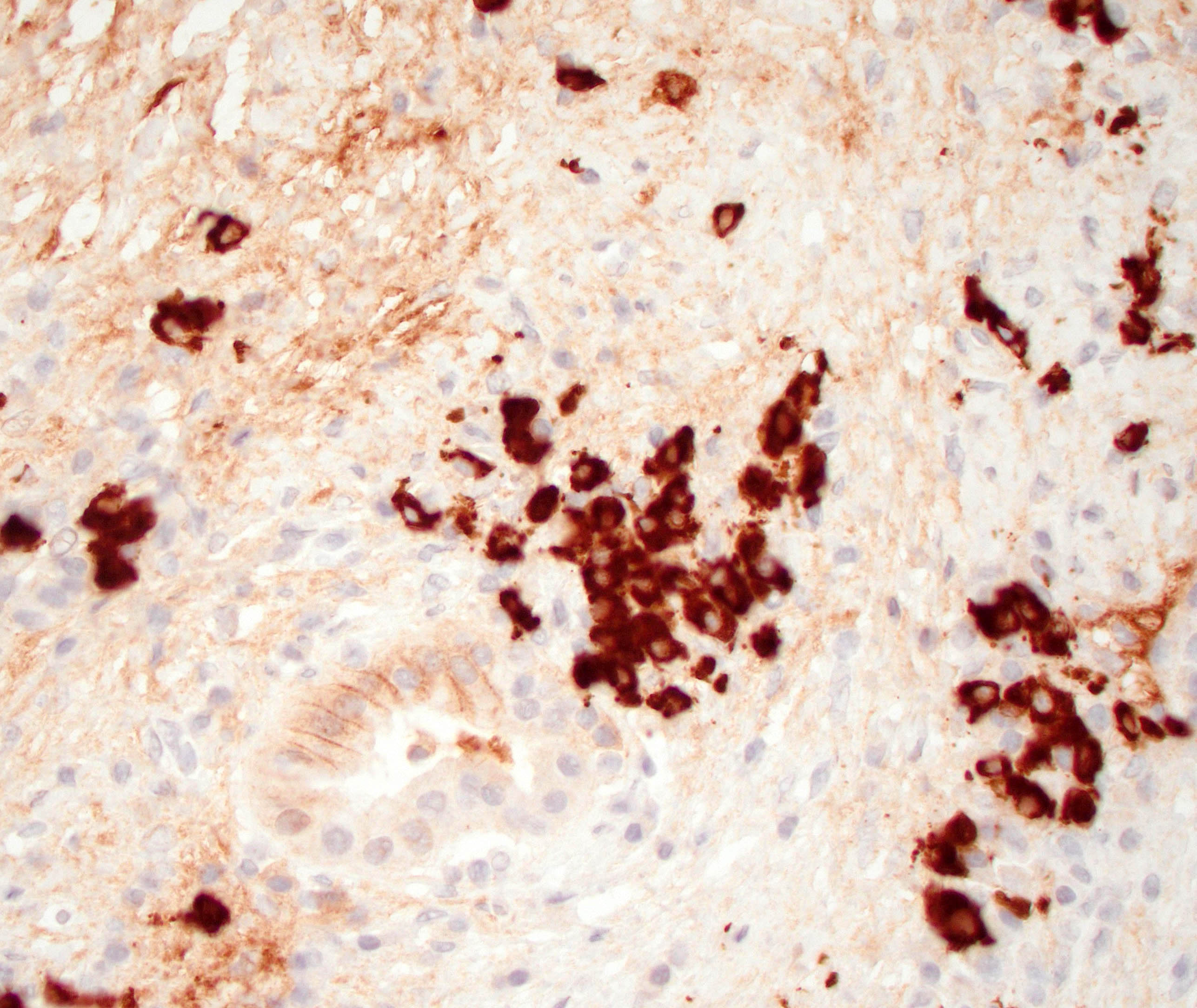
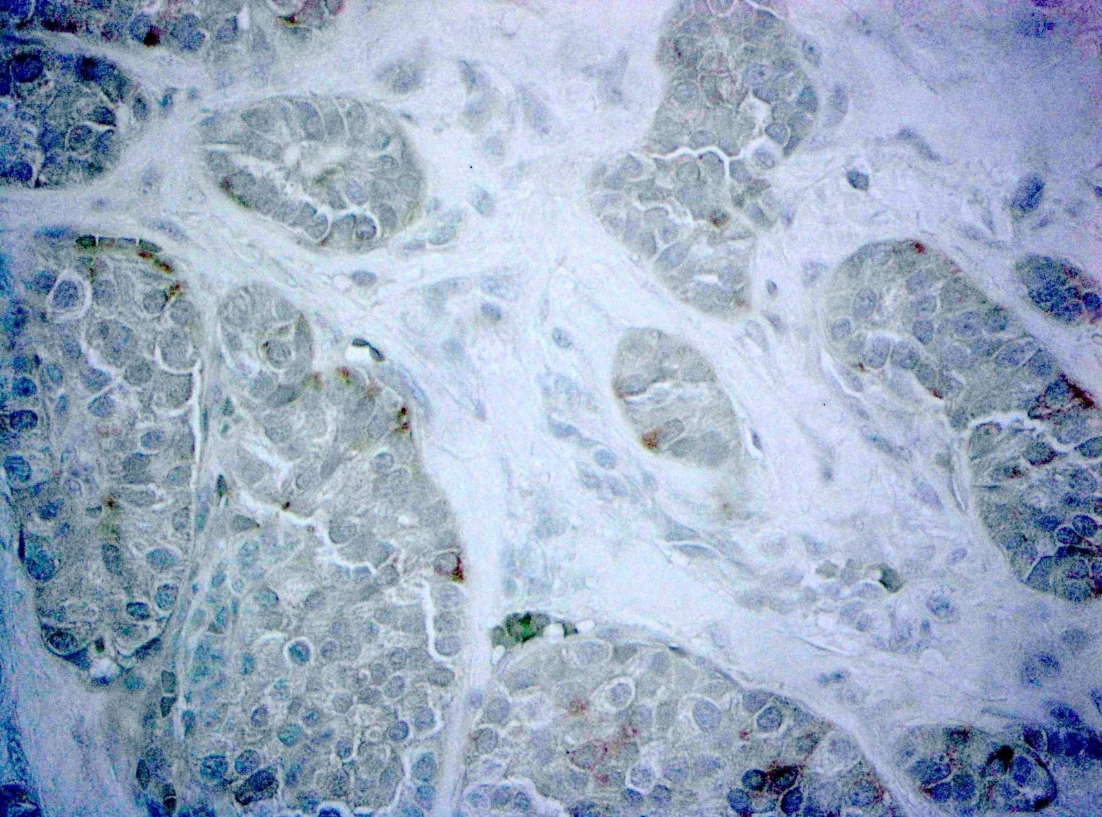
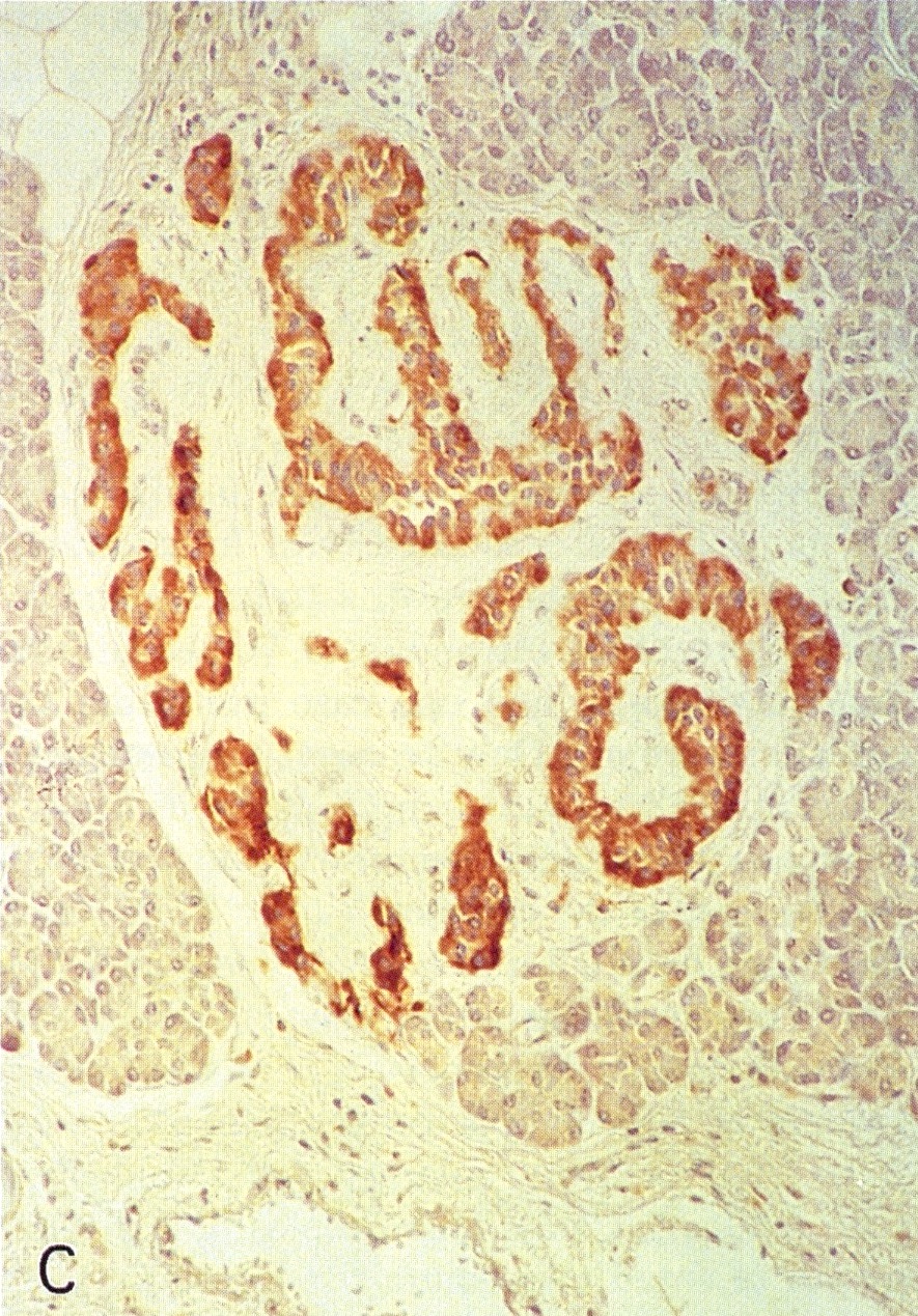
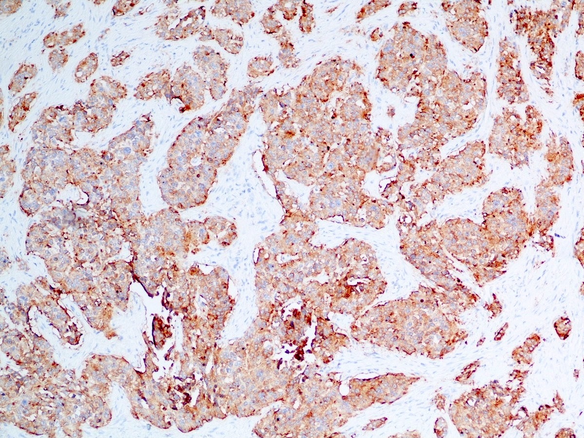
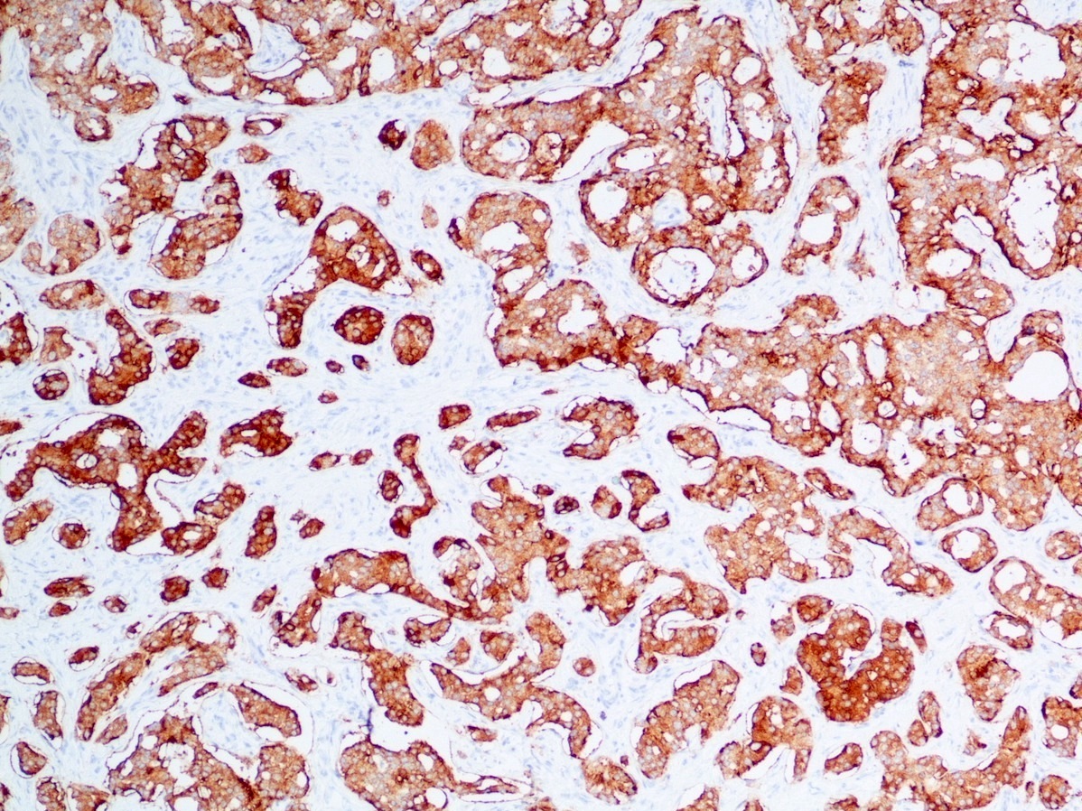
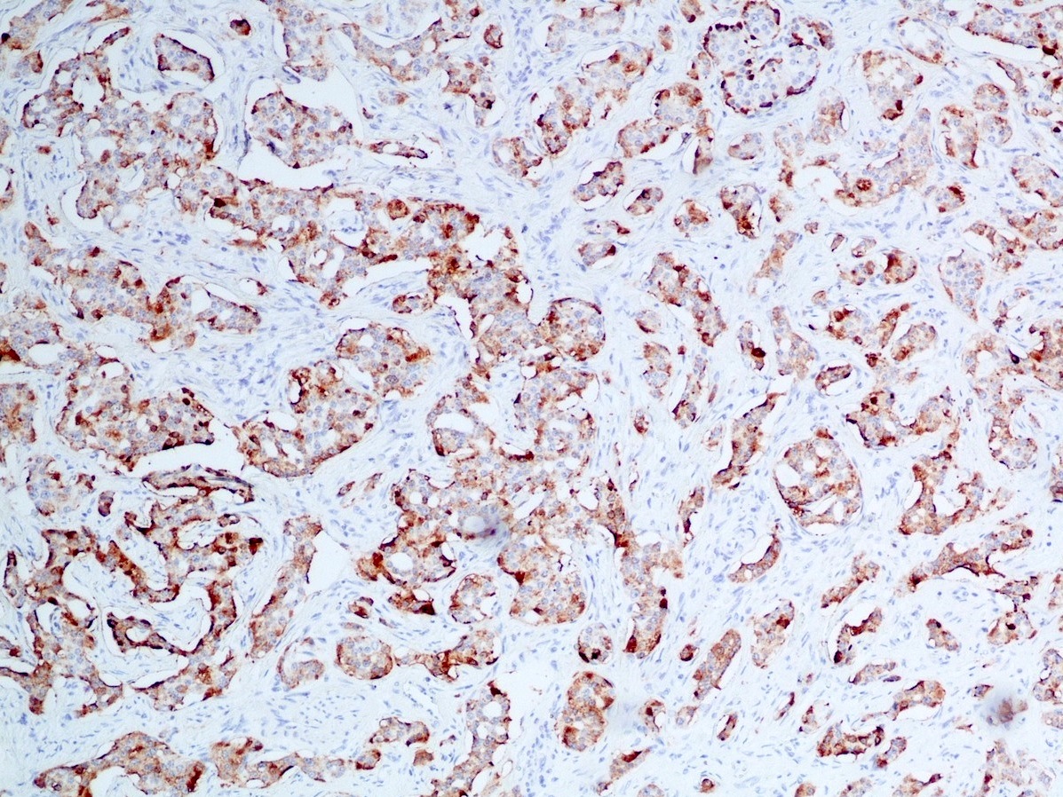
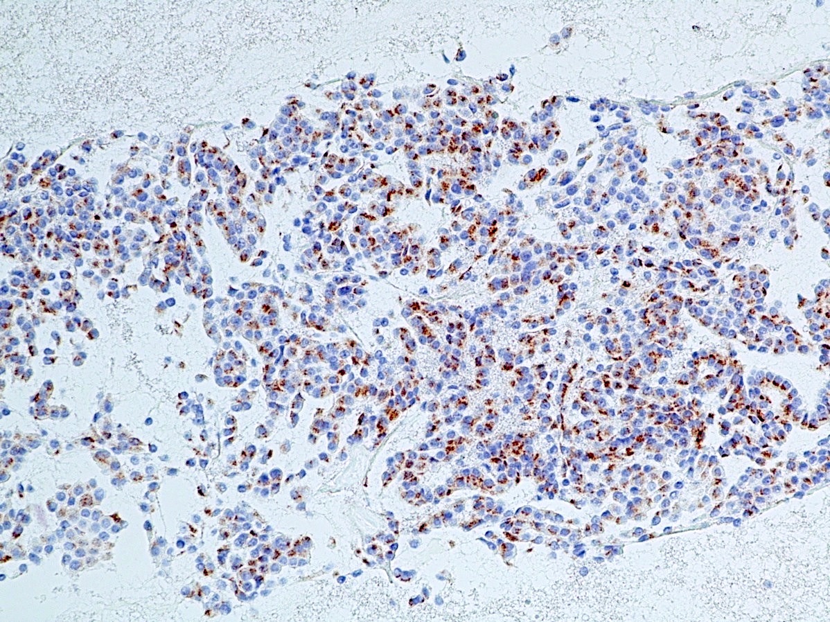
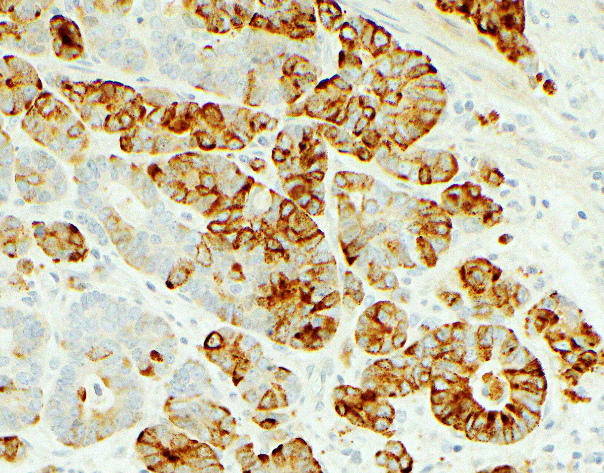
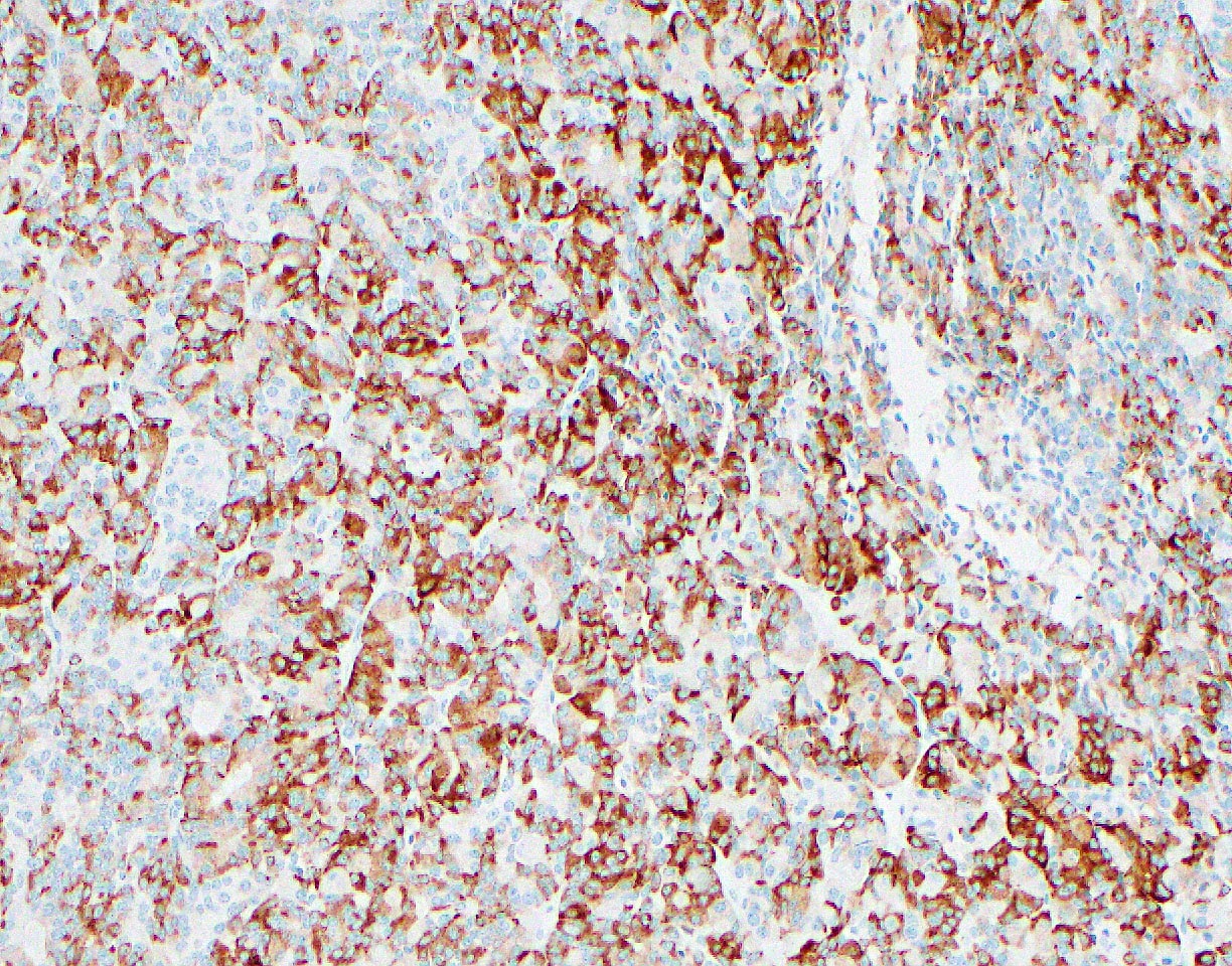
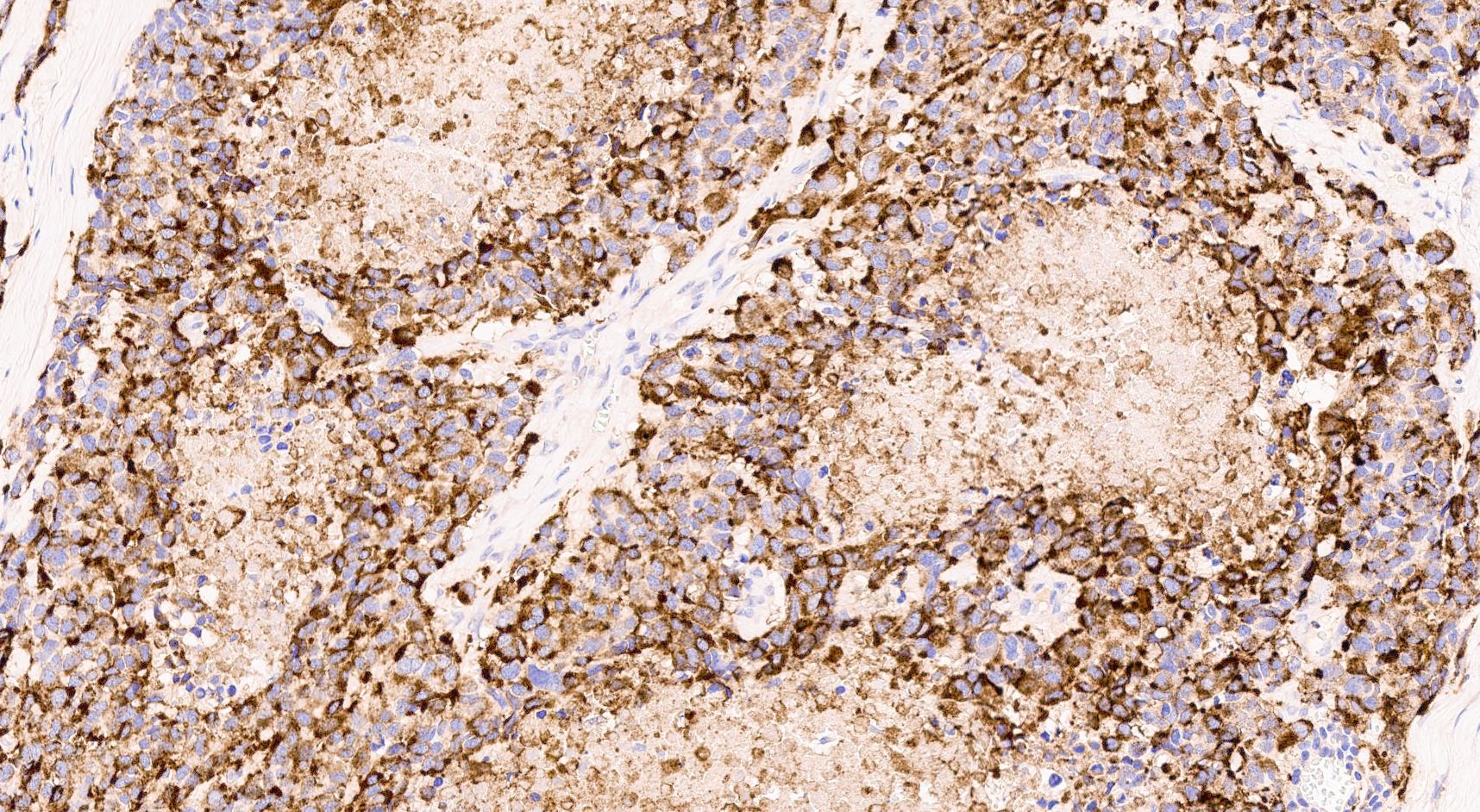
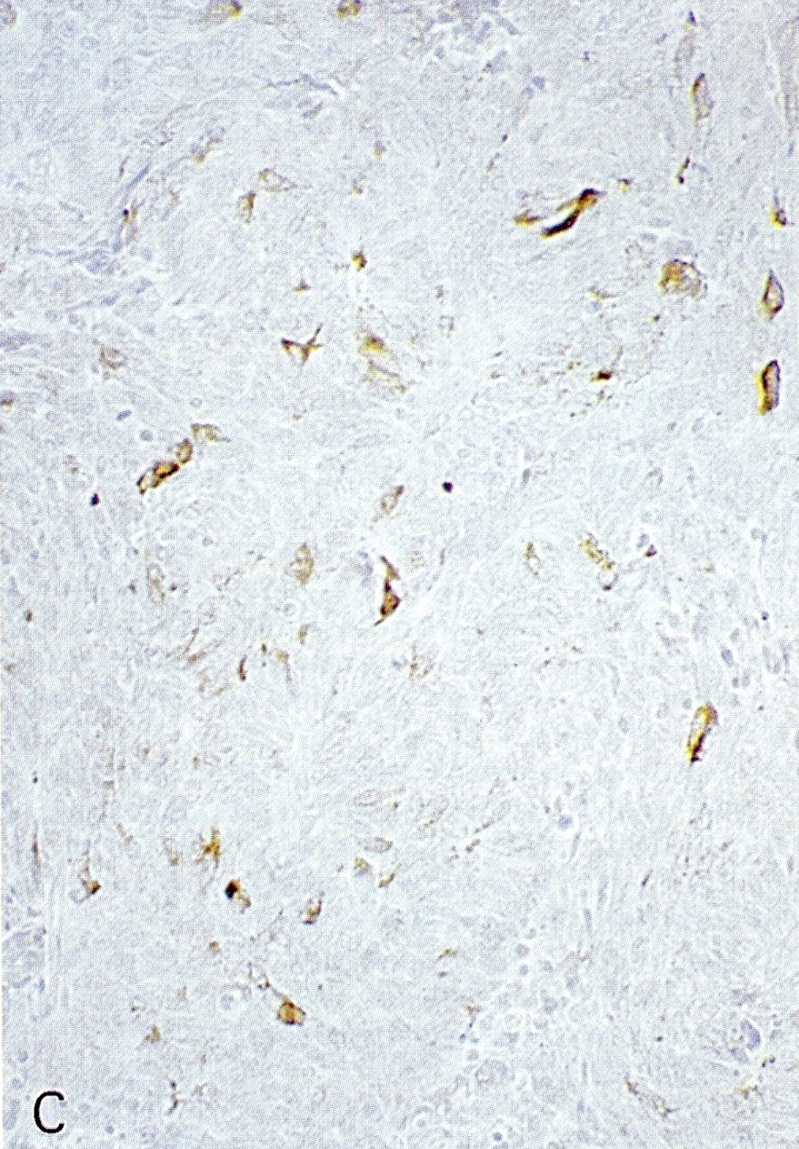
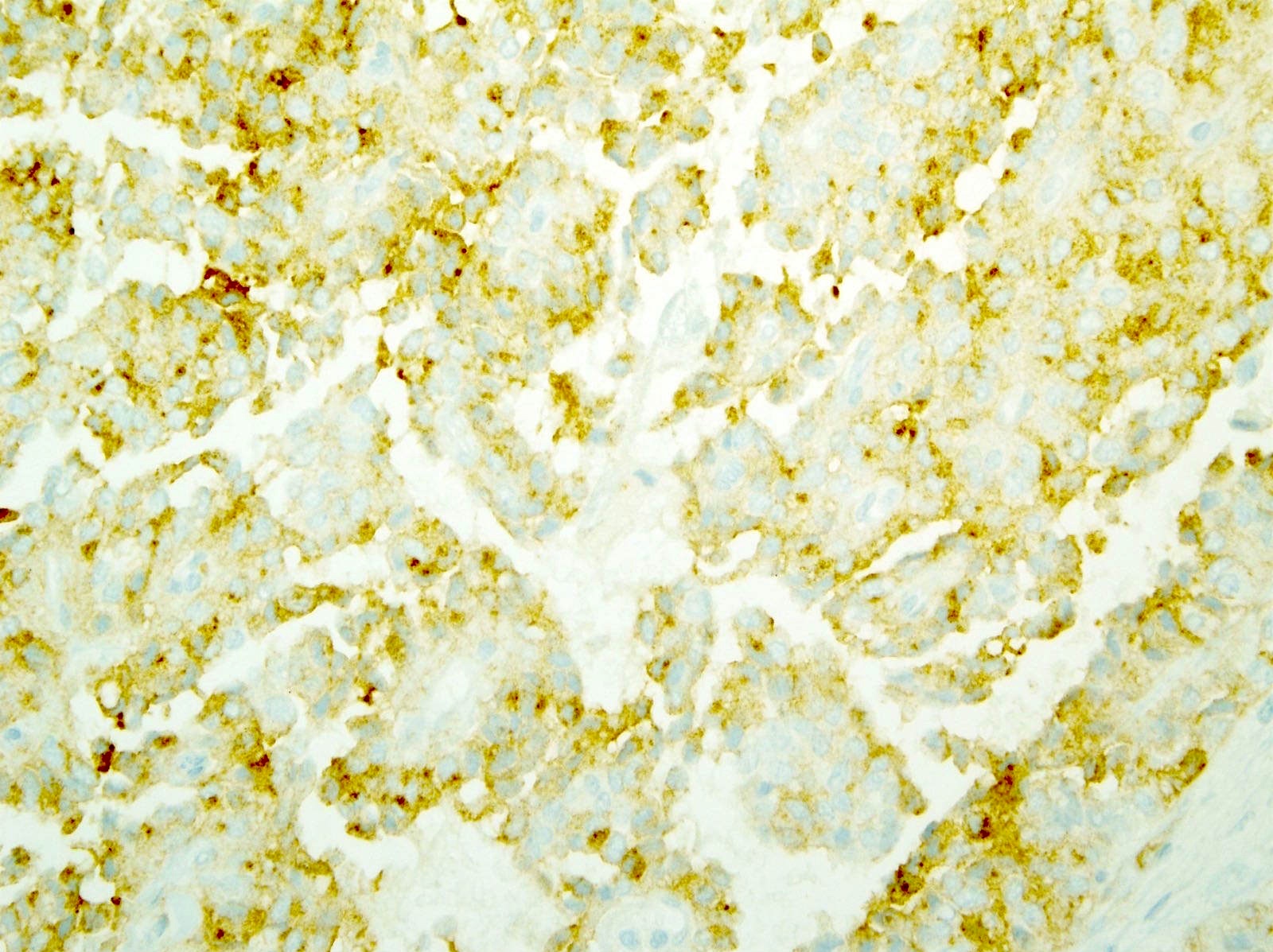
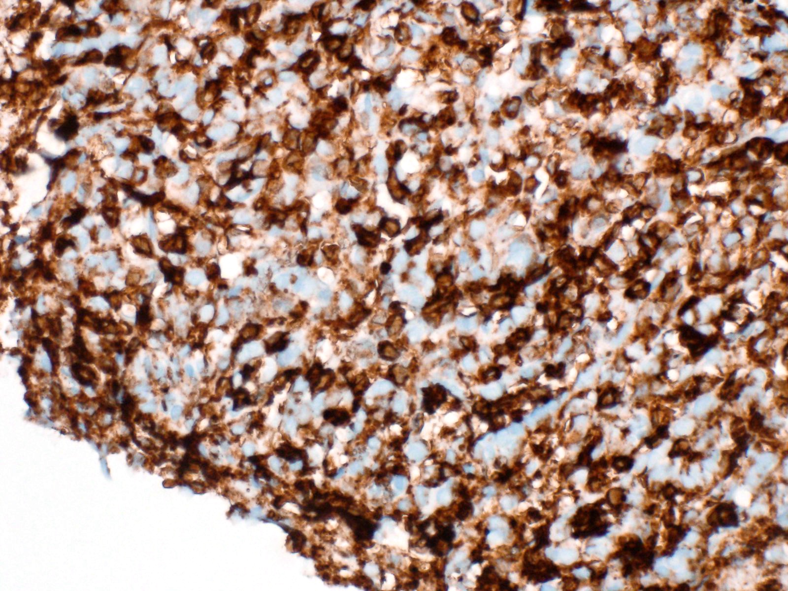
Trypsin expression pattern
Contributed by Claudio Luchini, M.D., Ph.D.
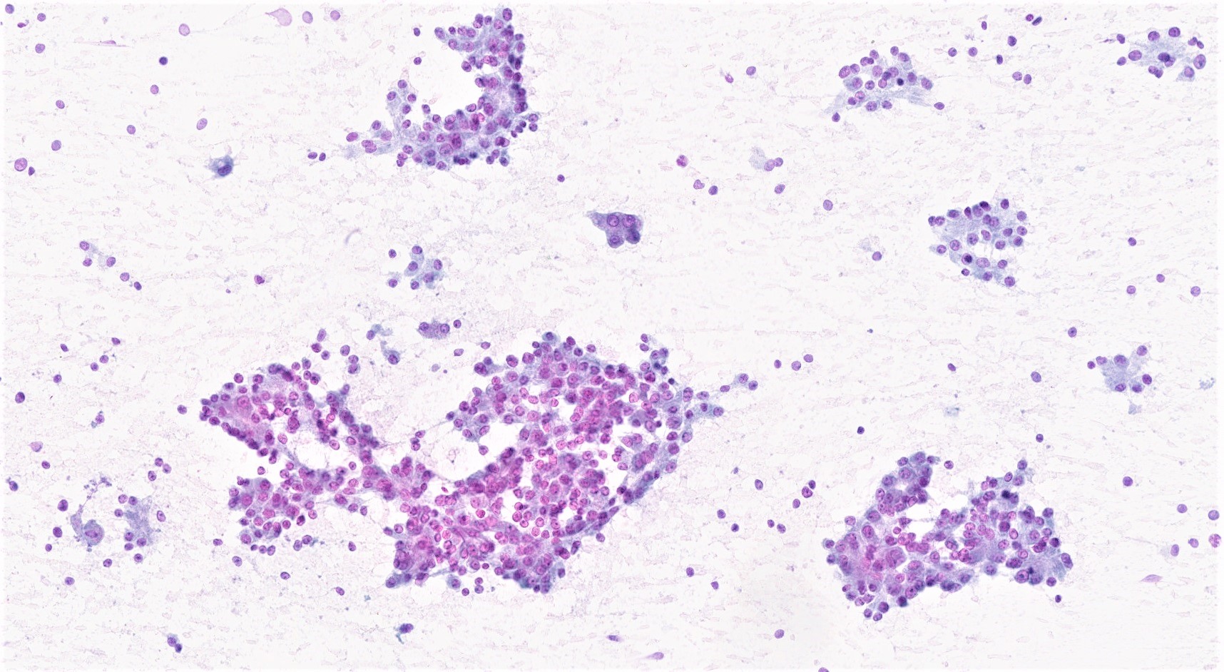
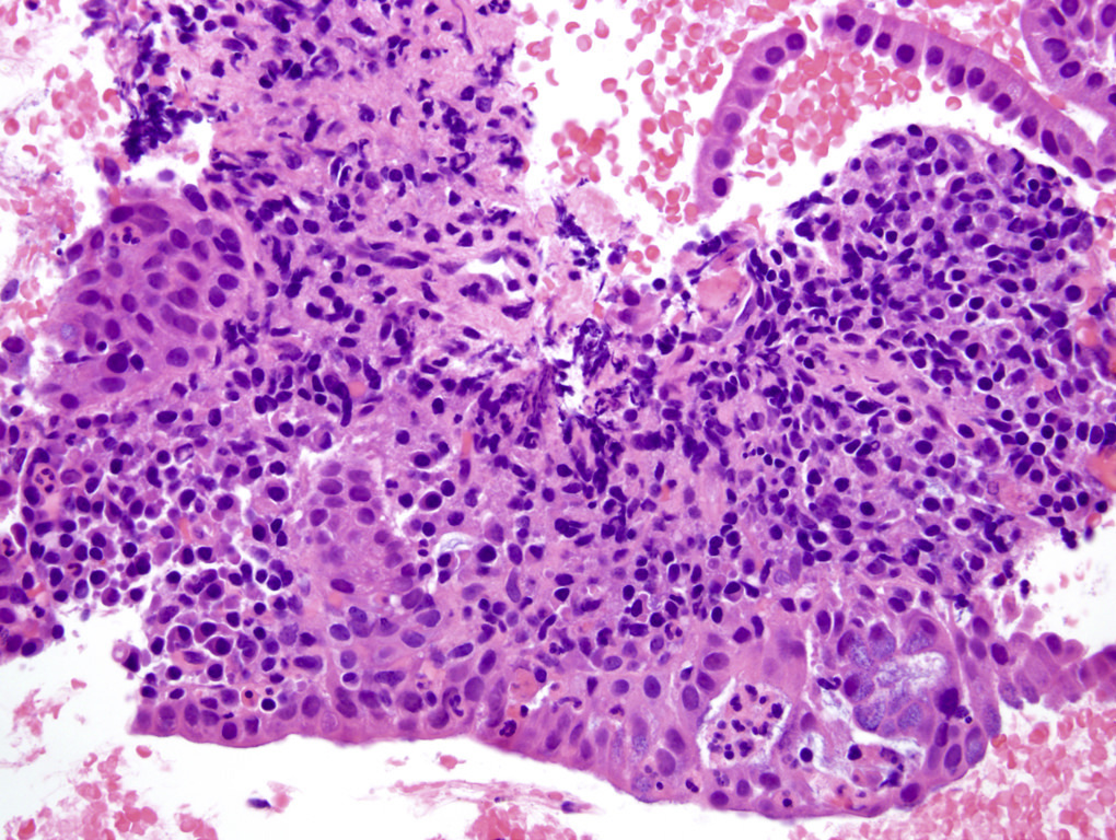
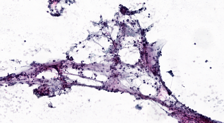
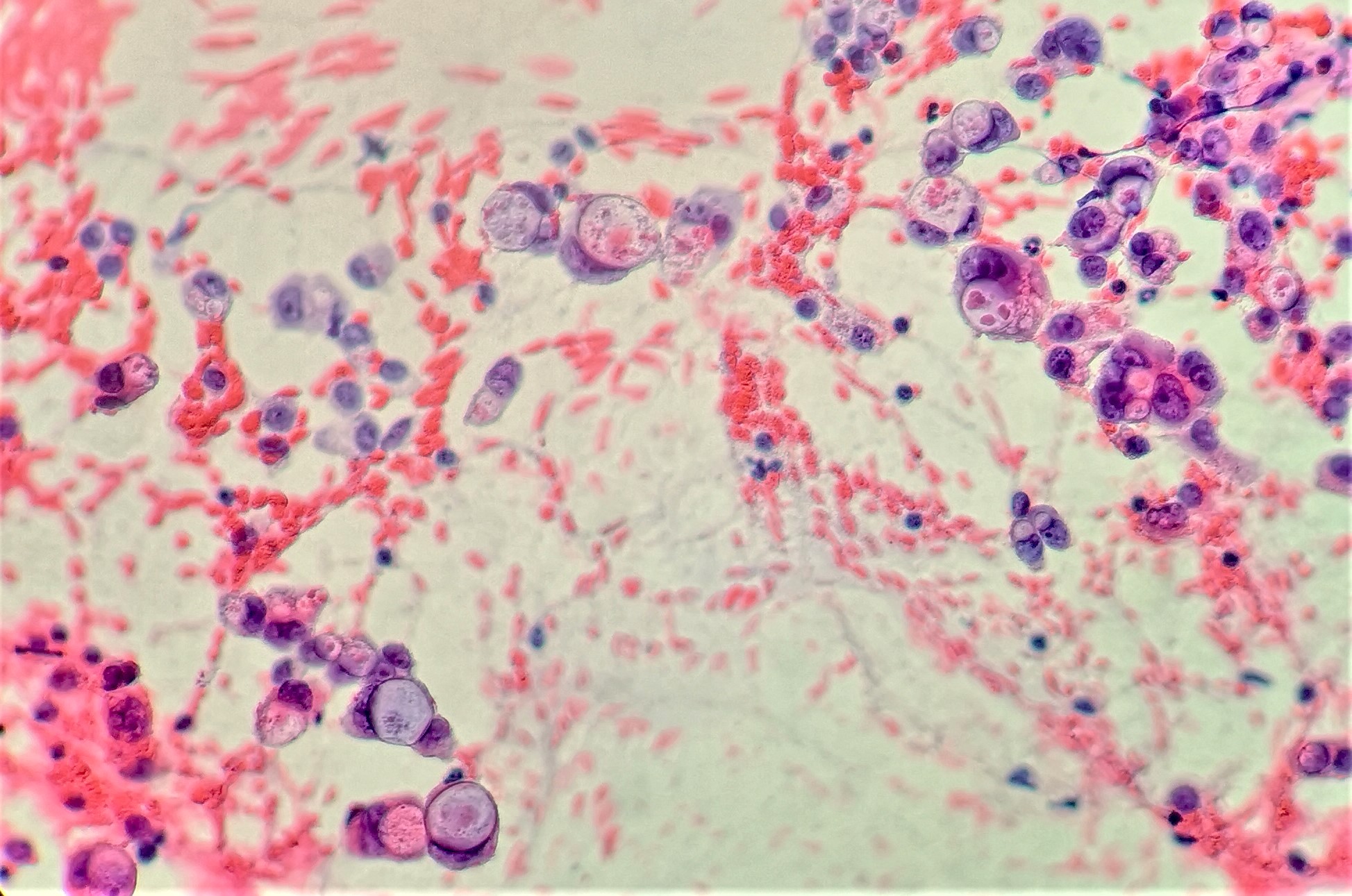
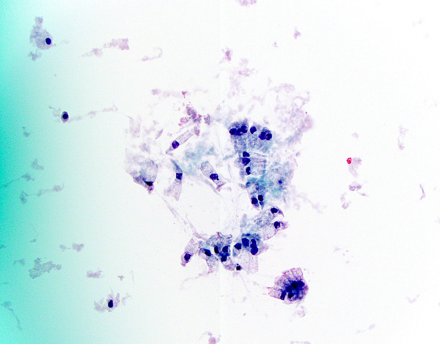
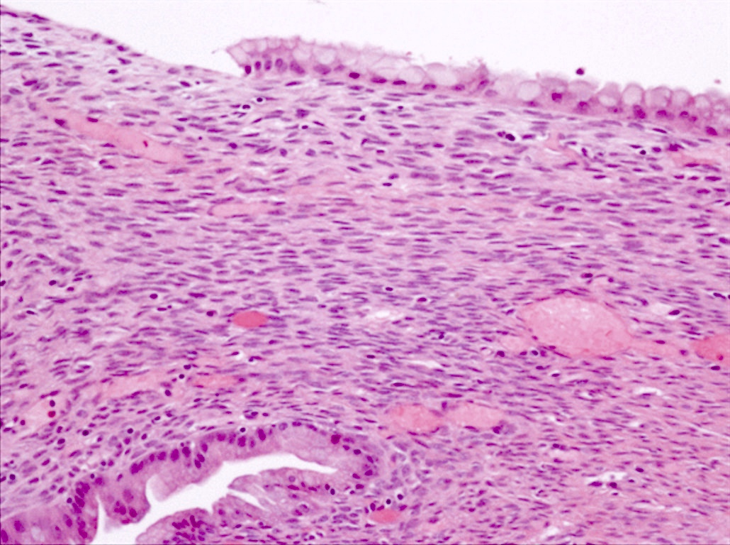
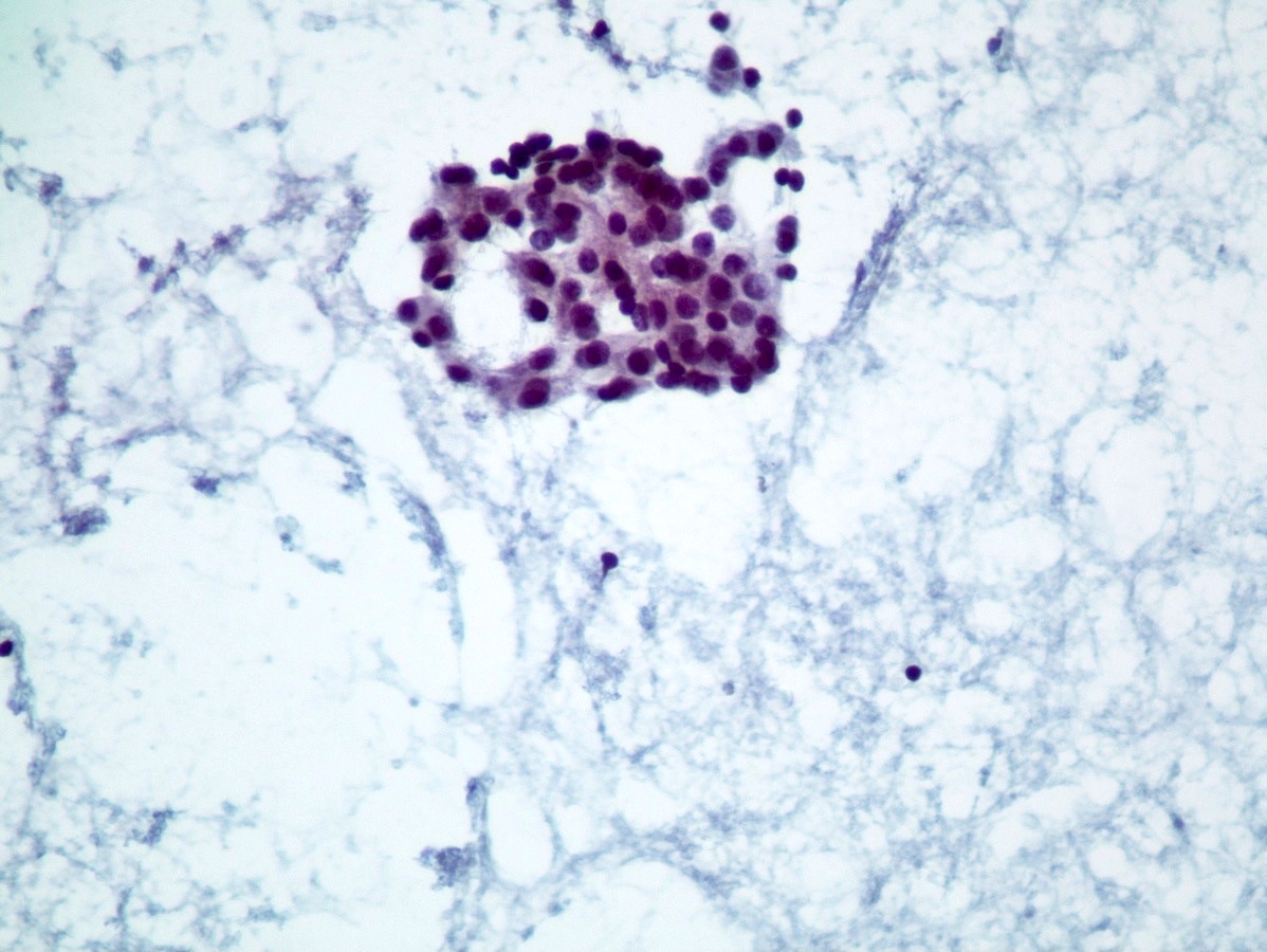
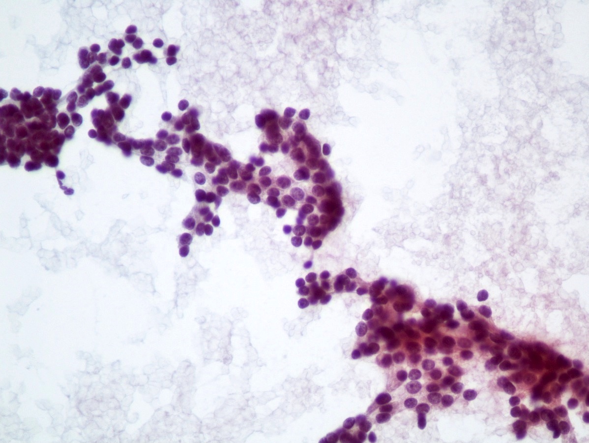
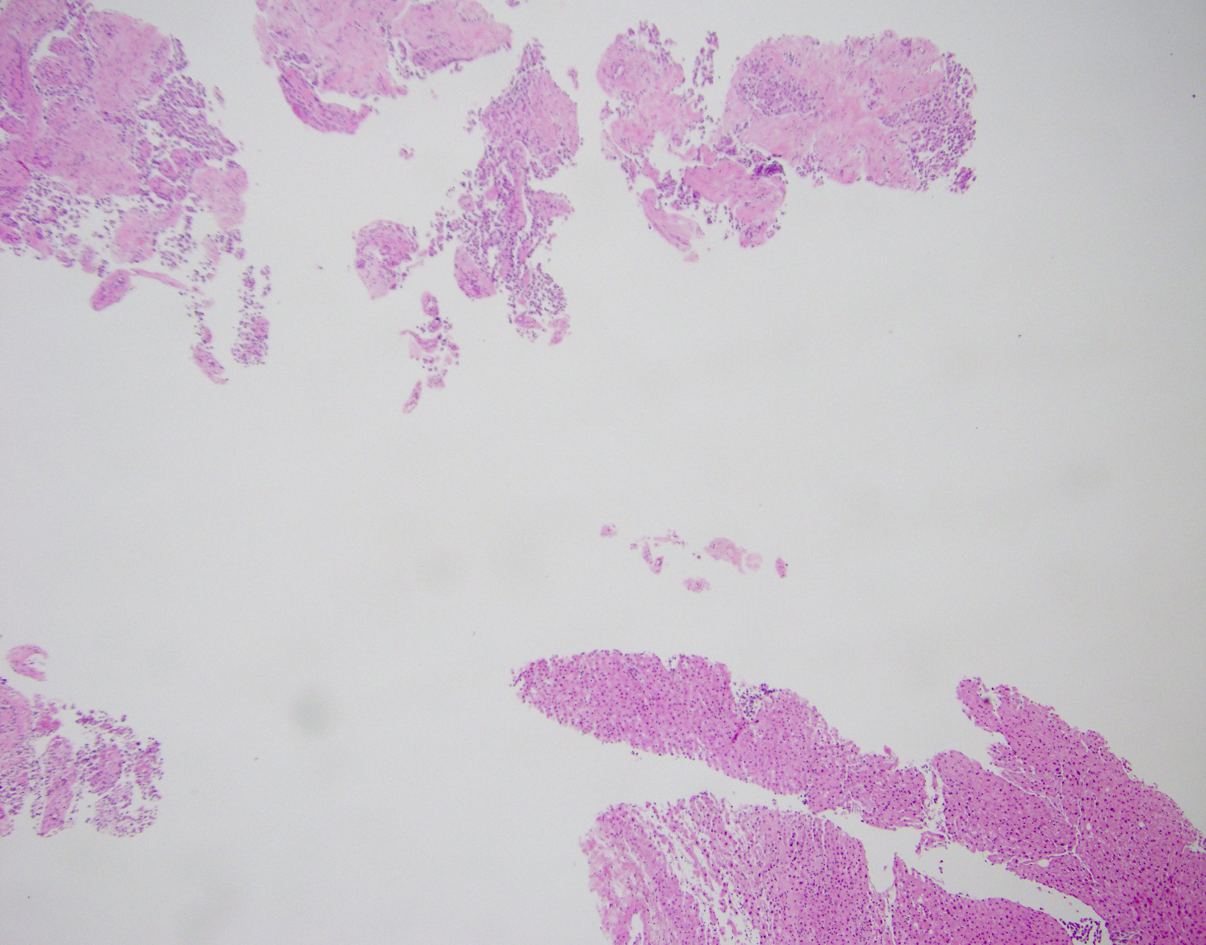
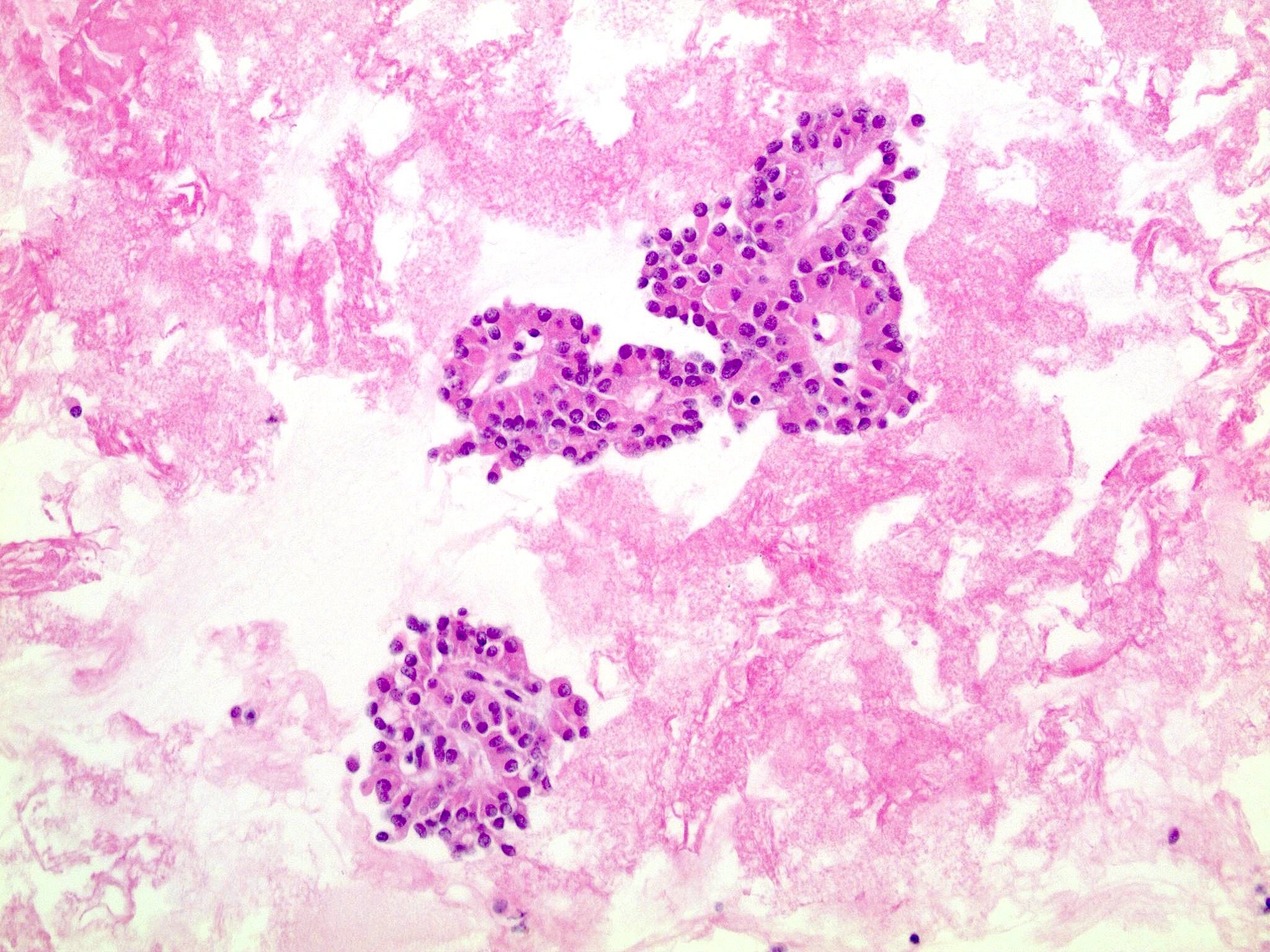
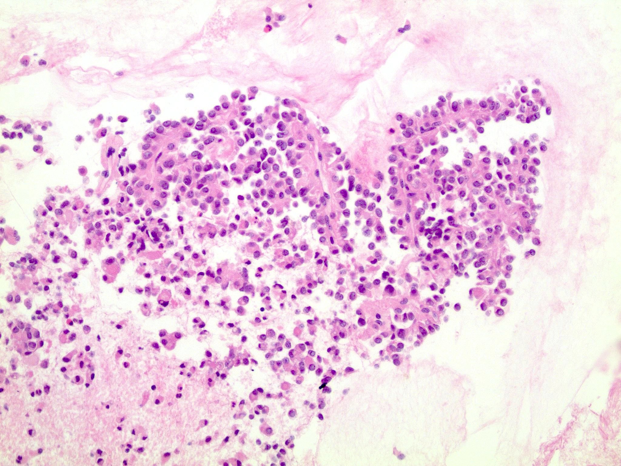
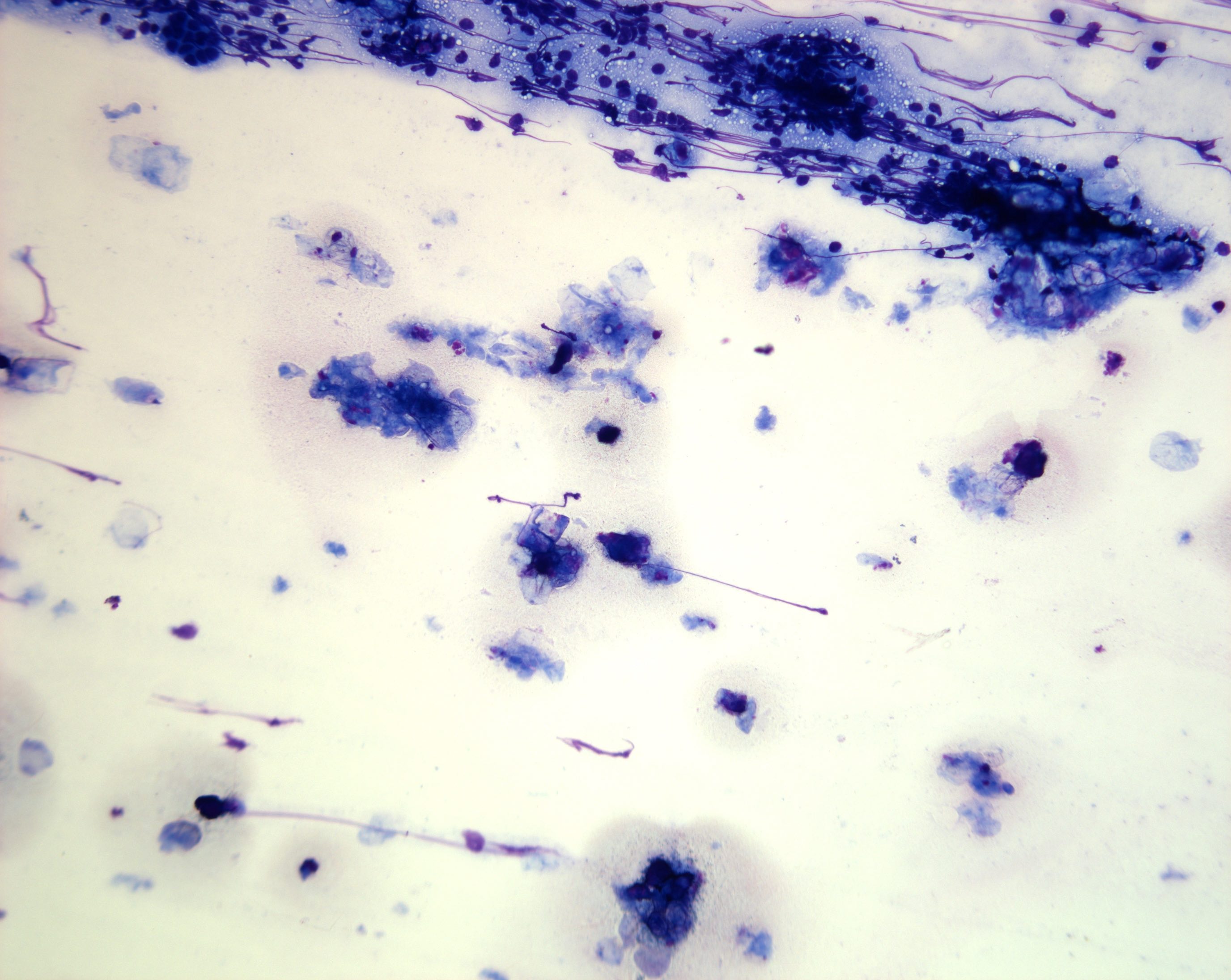
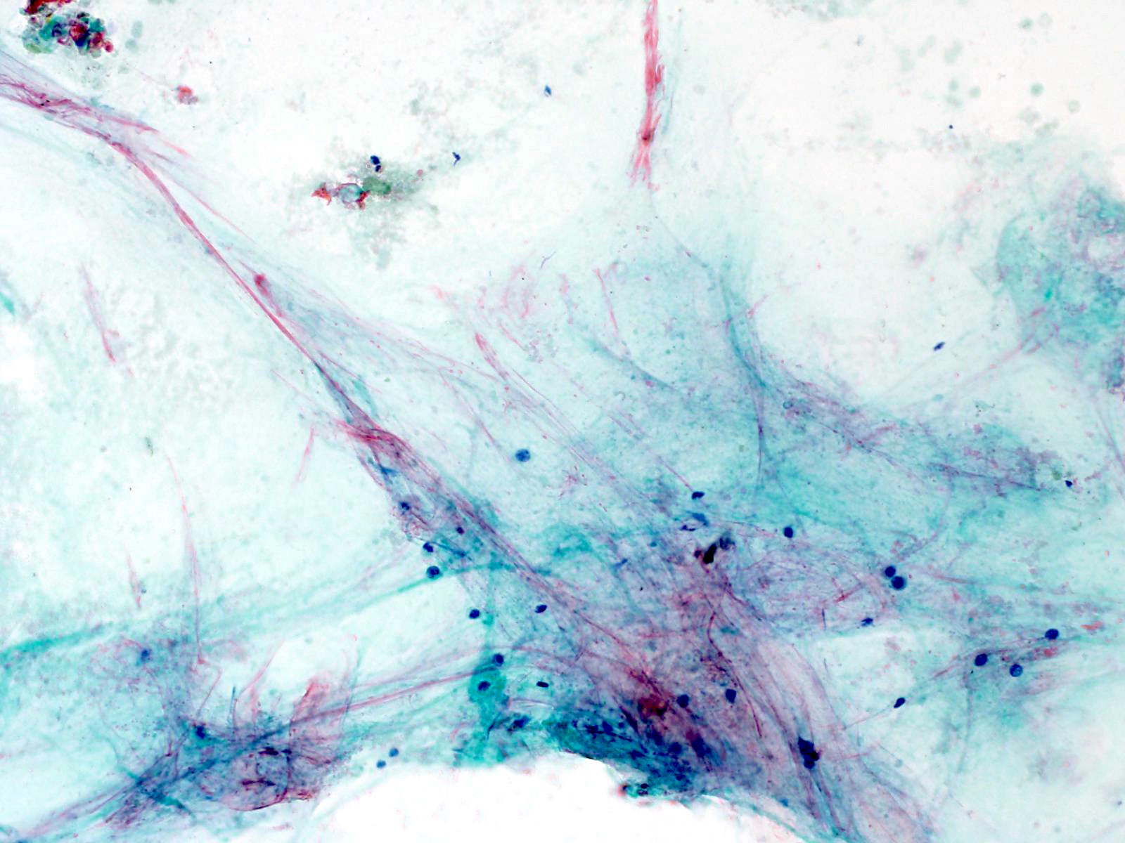
Cytological aspects
Contributed by Wei Chen, M.D., Ph.D.
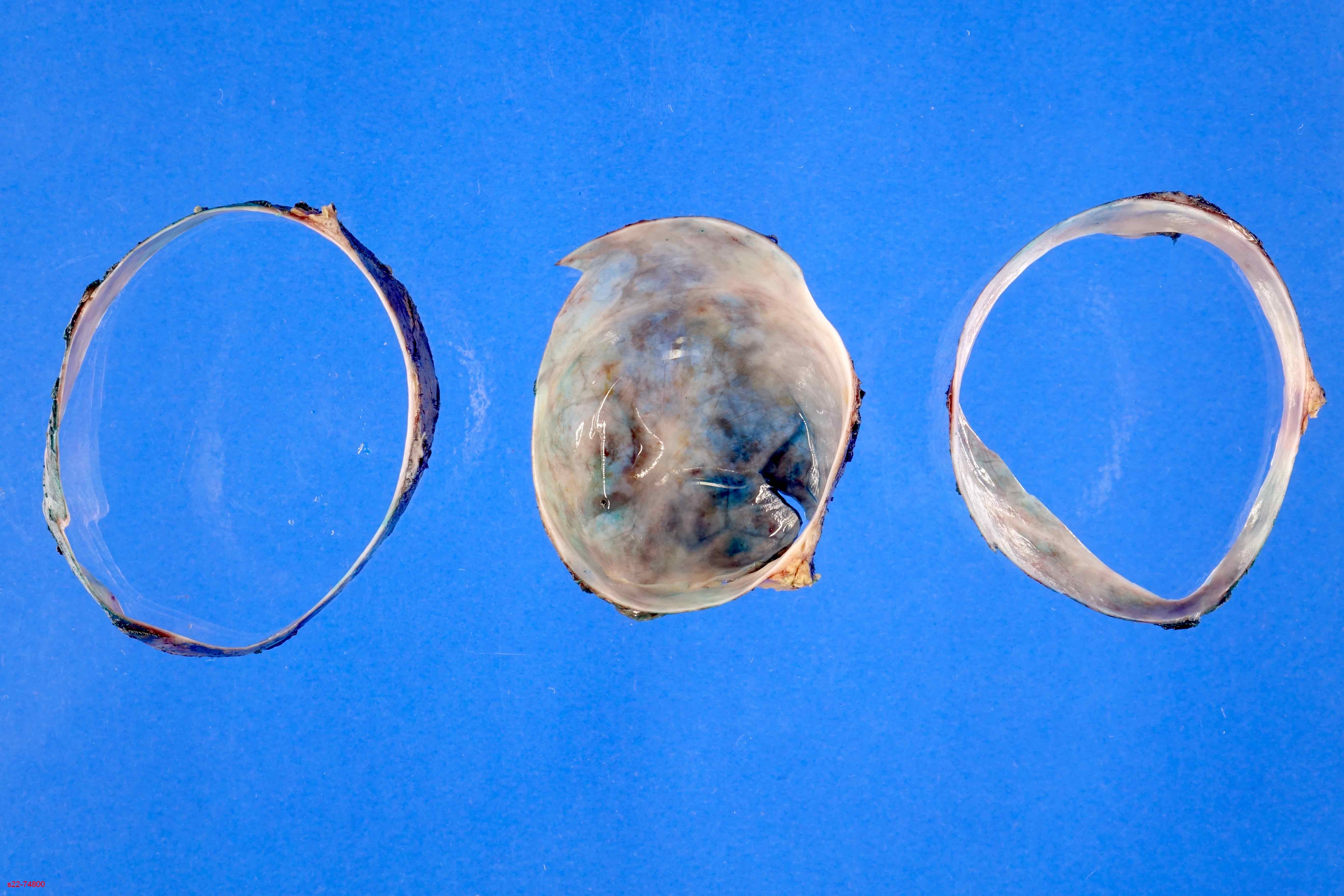
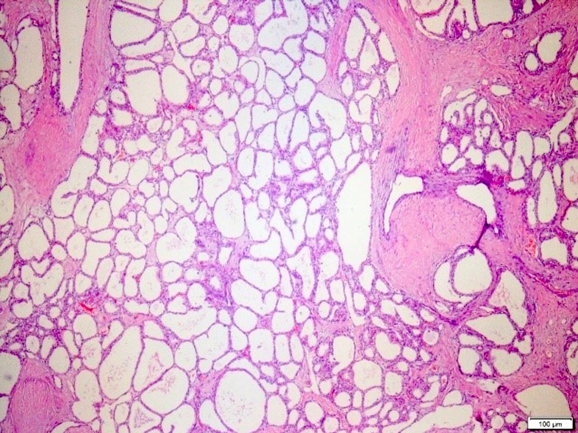
Unilocular ACT
Images hosted on other servers:

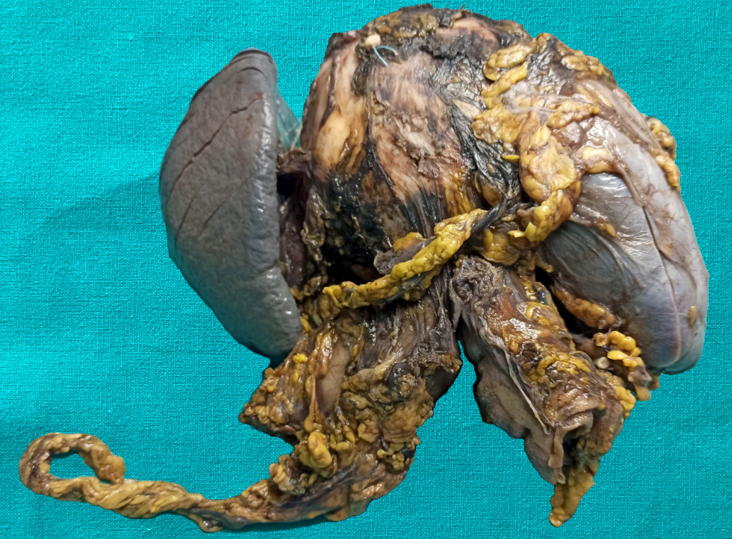
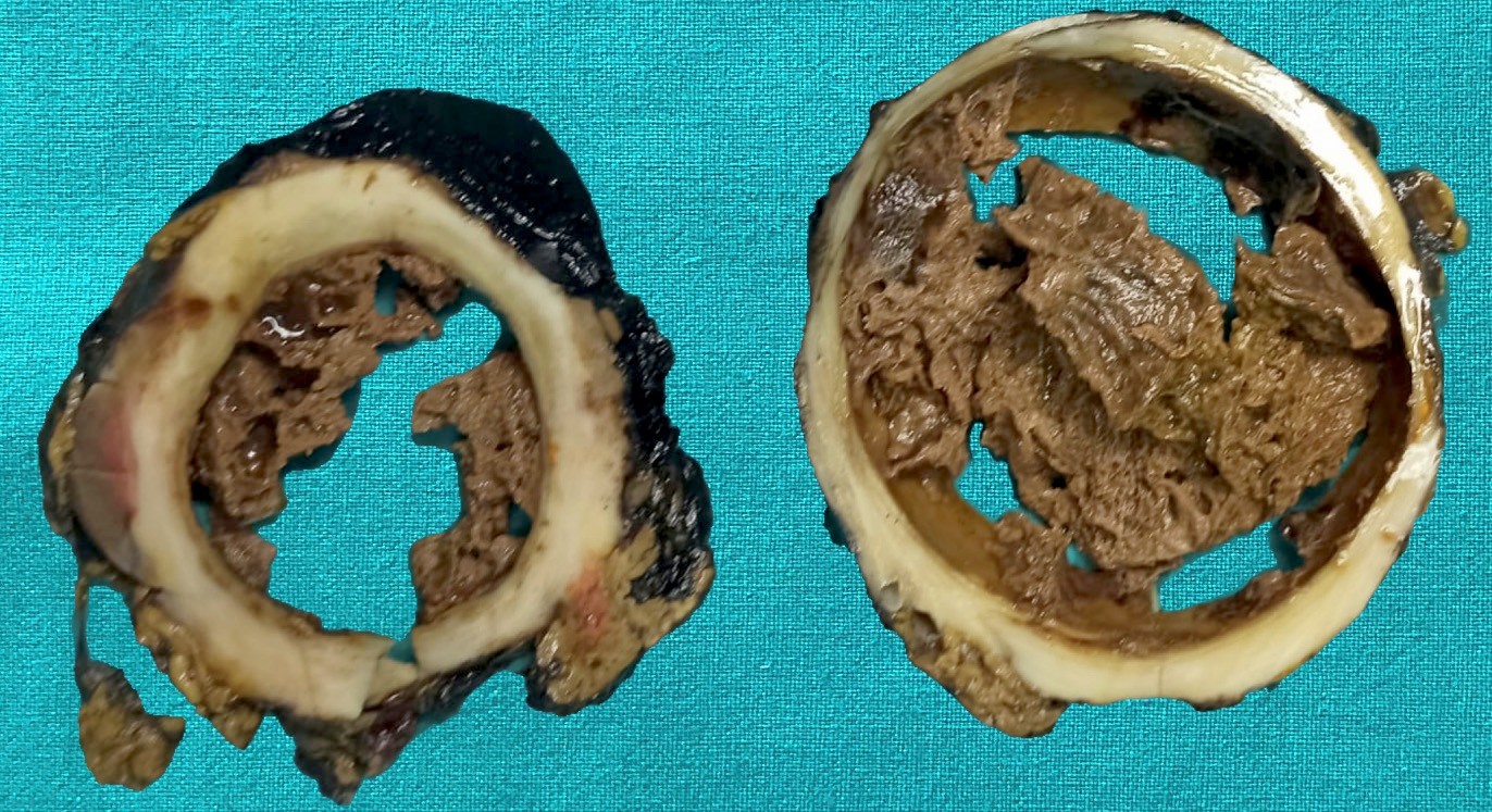
Multiple cystic lesions

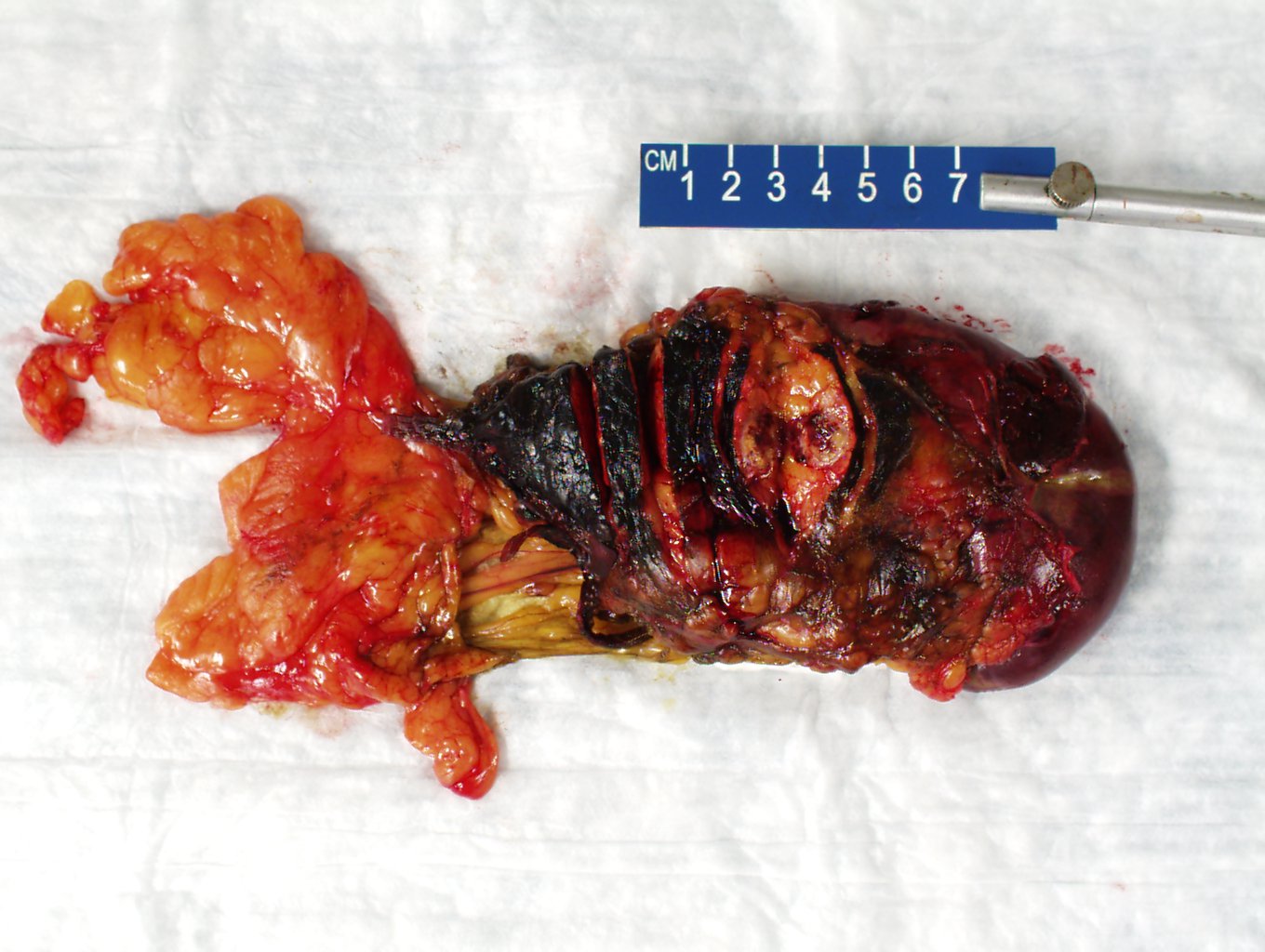
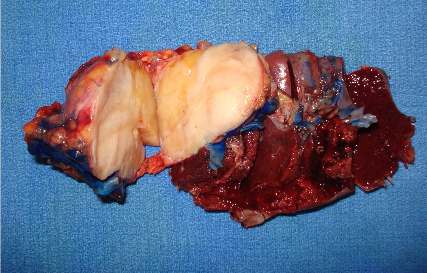
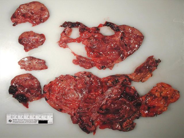
Raw specimen
and fixed and
cut specimen
Contributed by Vidya Arole, M.D. and Wei Chen, M.D., Ph.D.
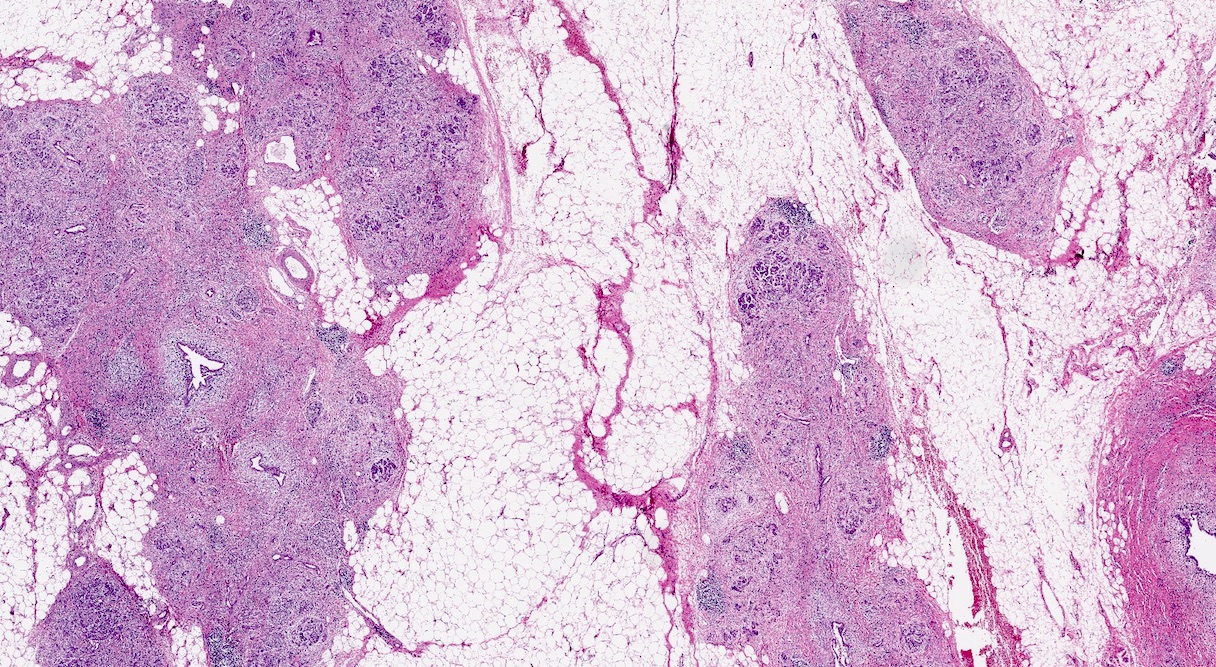
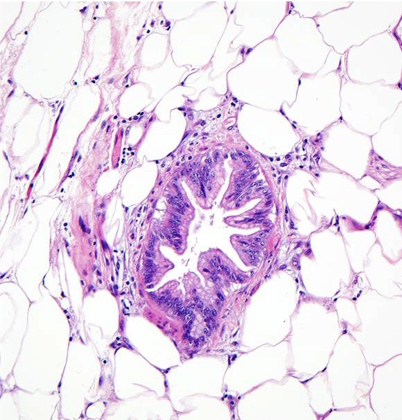
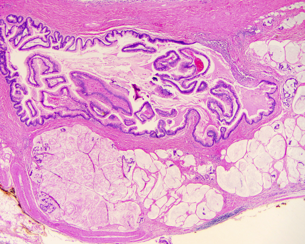
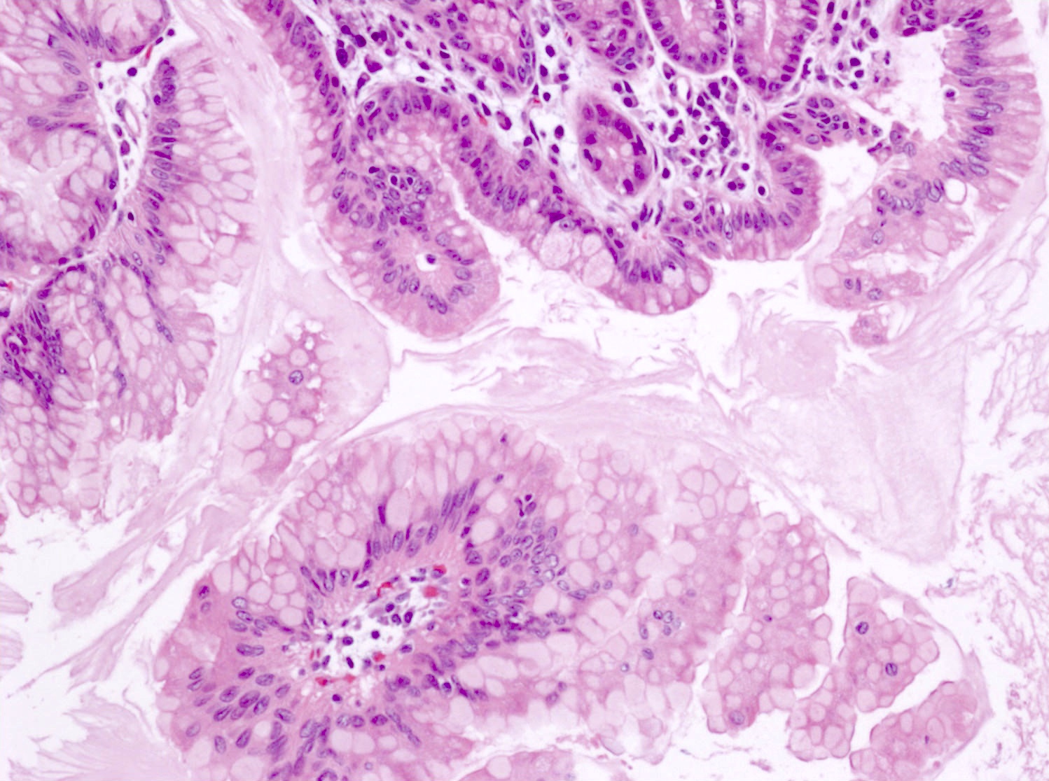
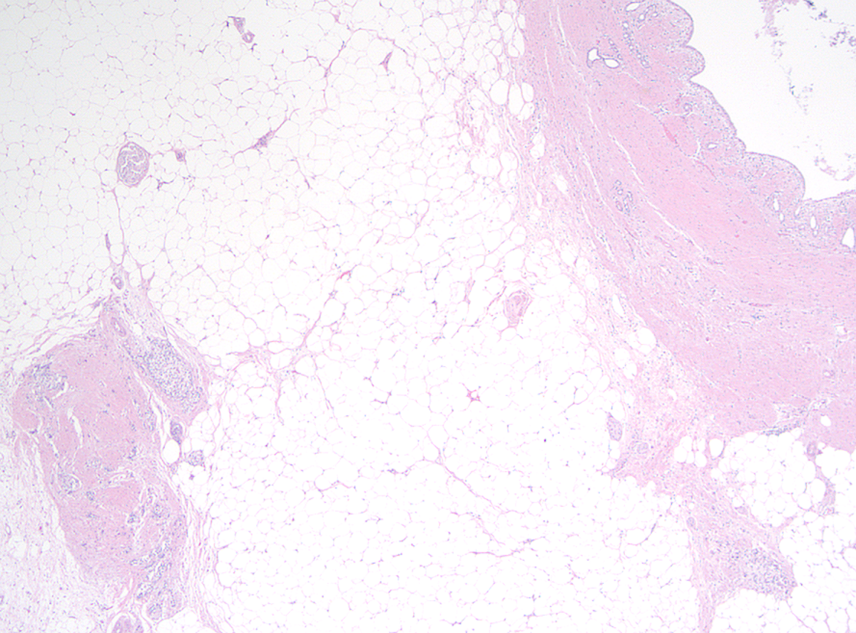
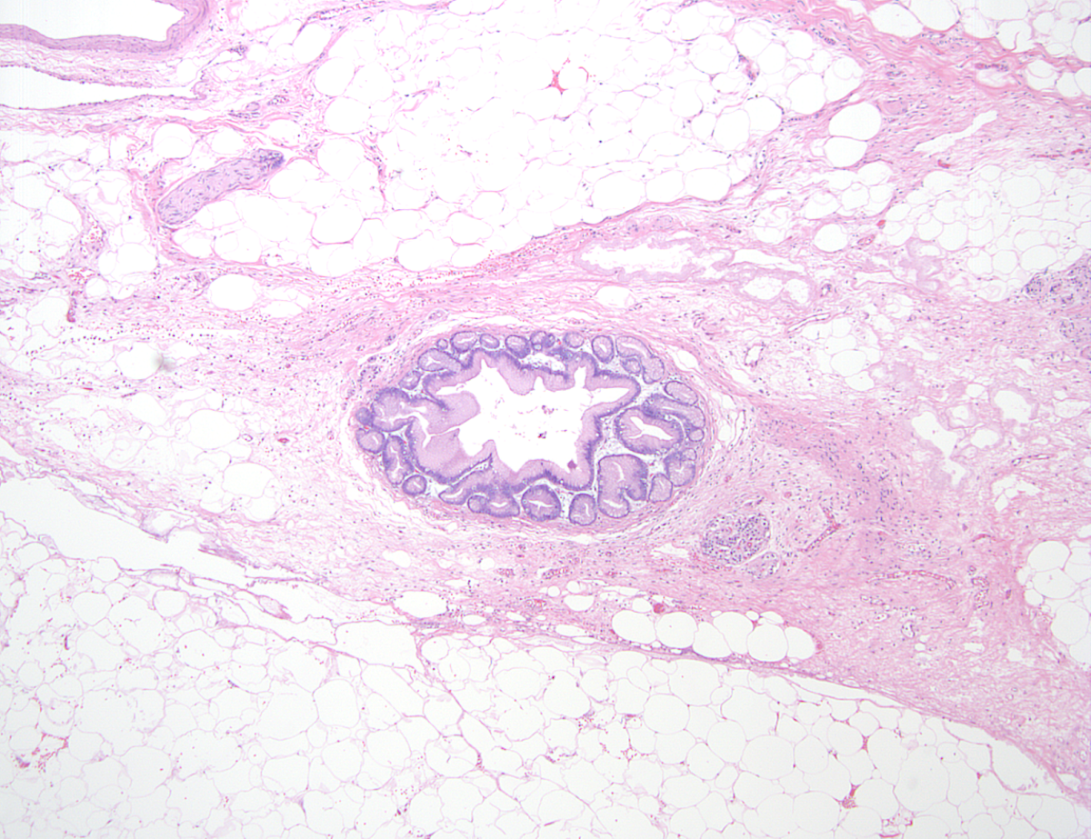
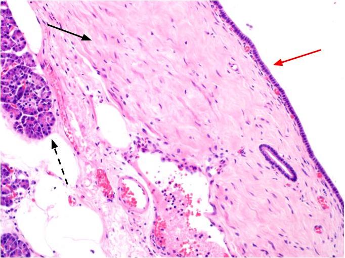
Incidental microscopic ACT
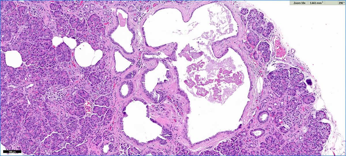
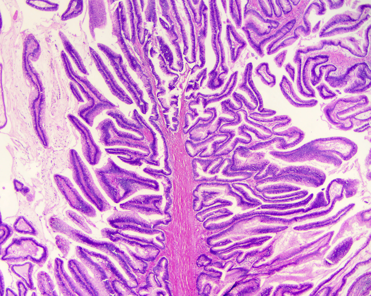
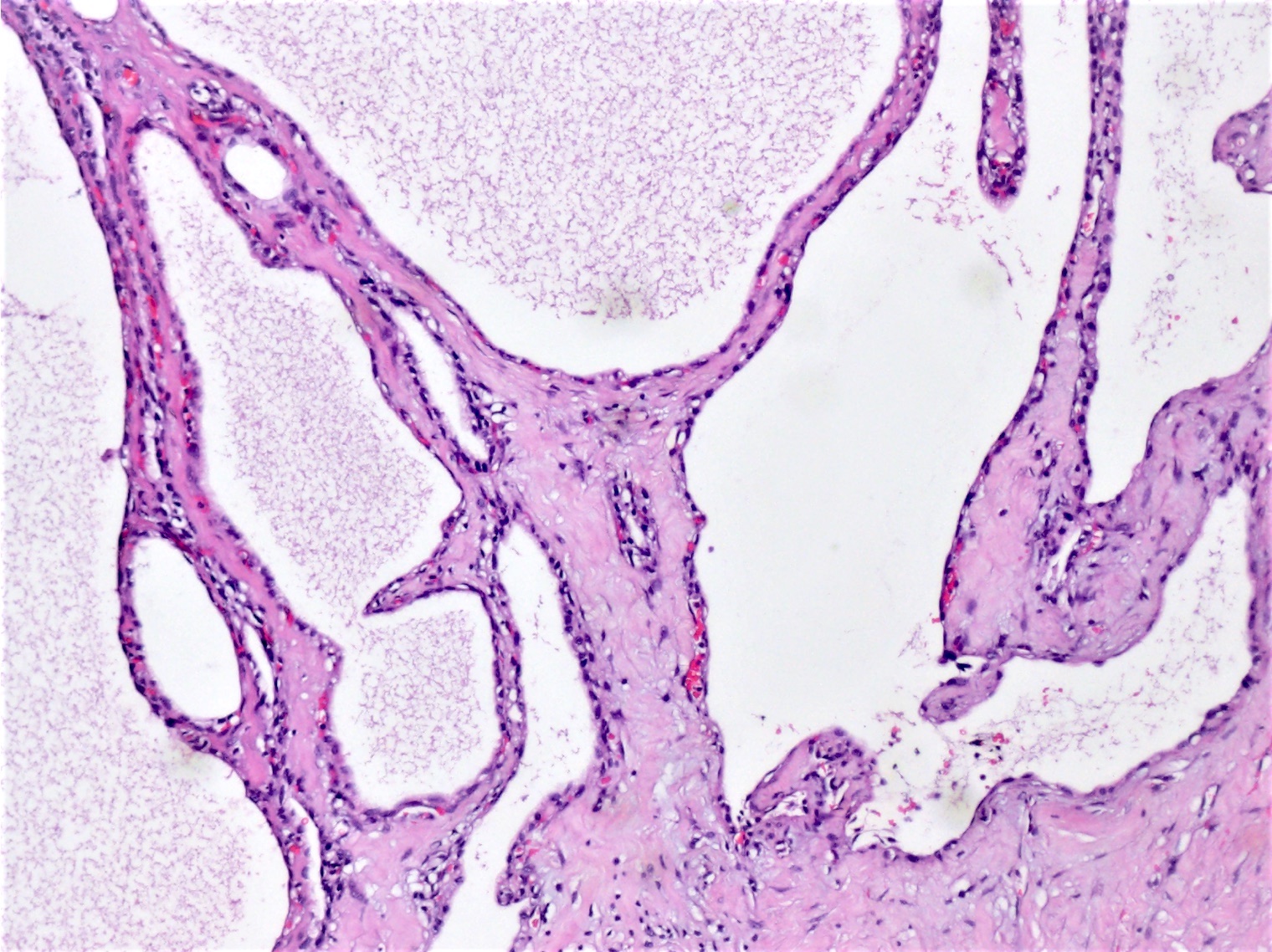
Multilocular cystic structure
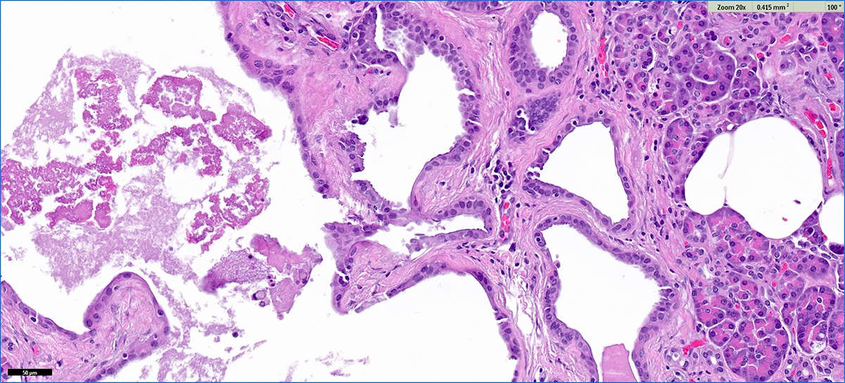
Club-like pseudopapillae
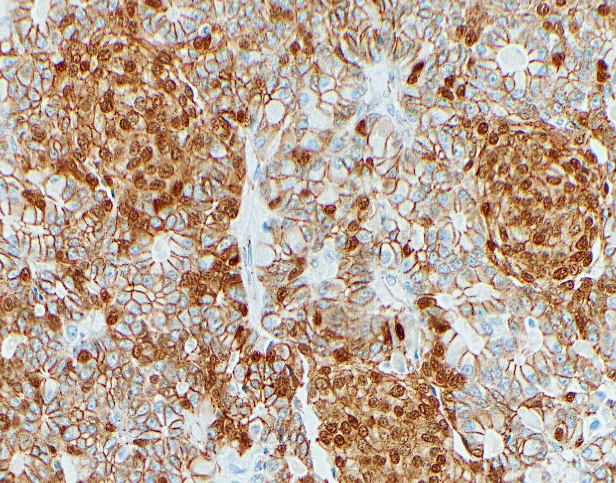
Microscopic ACT CK19 immunostain
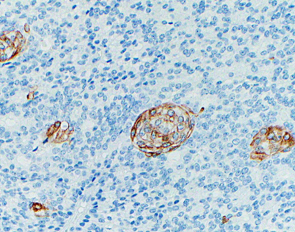
Microscopic ACT trypsin immunostain
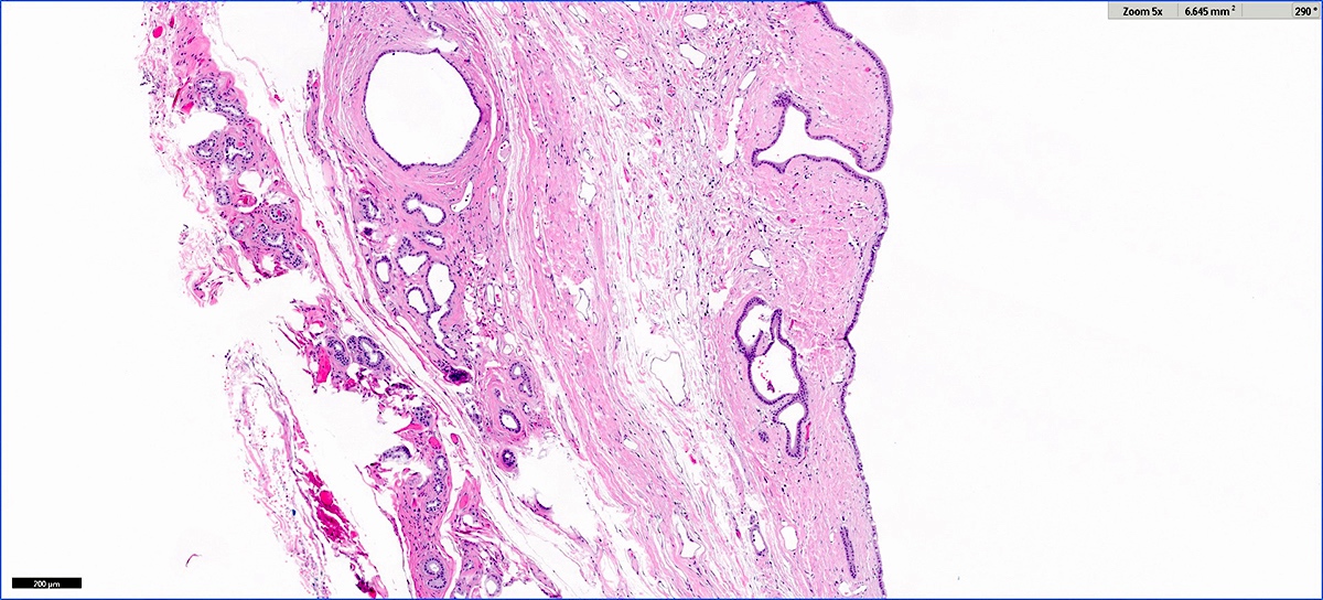
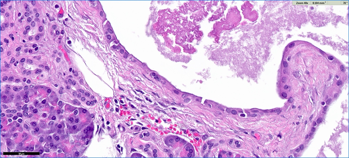
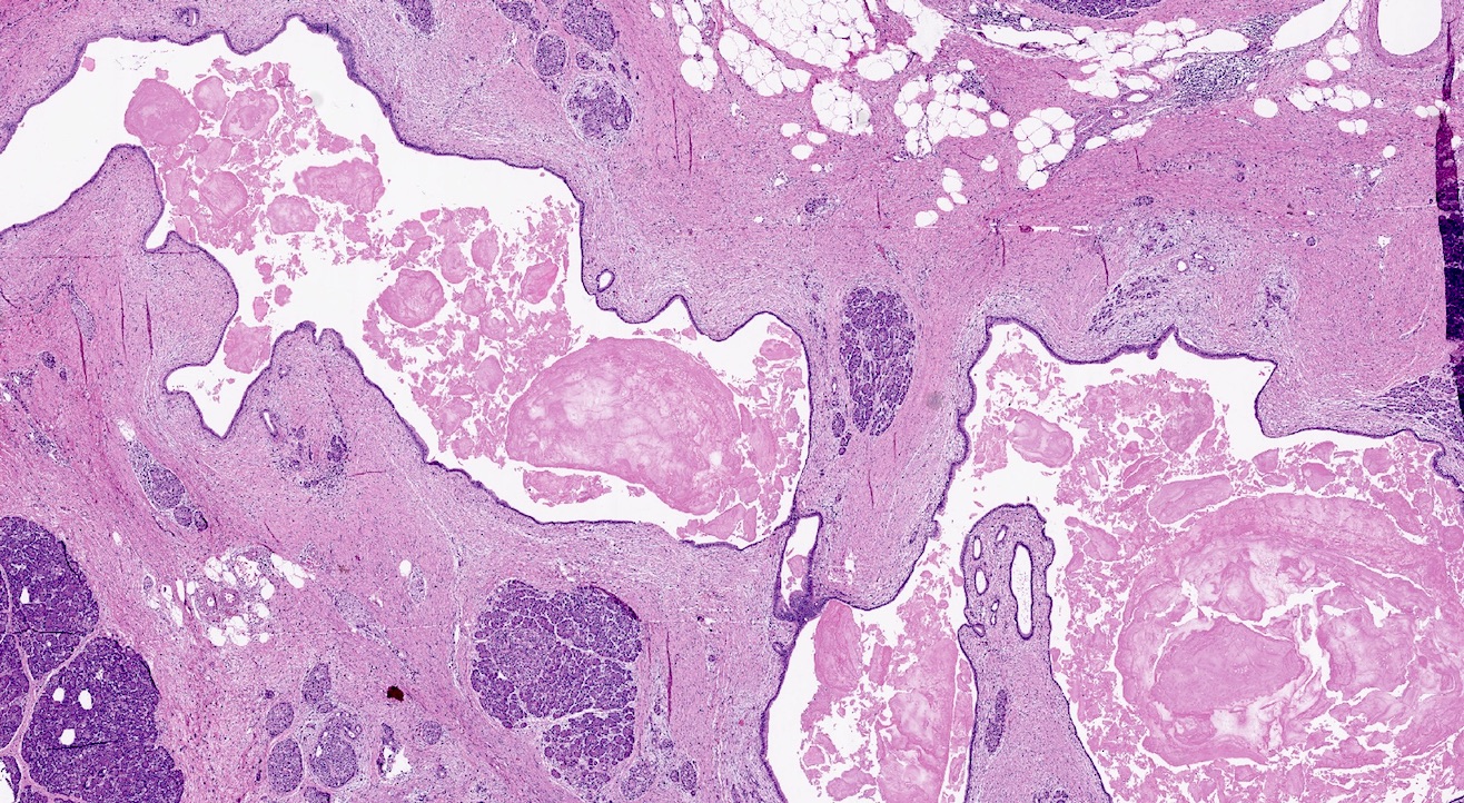
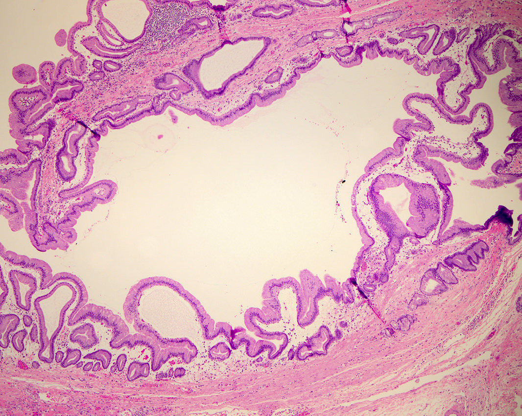
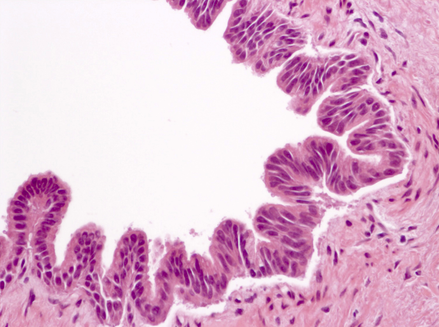
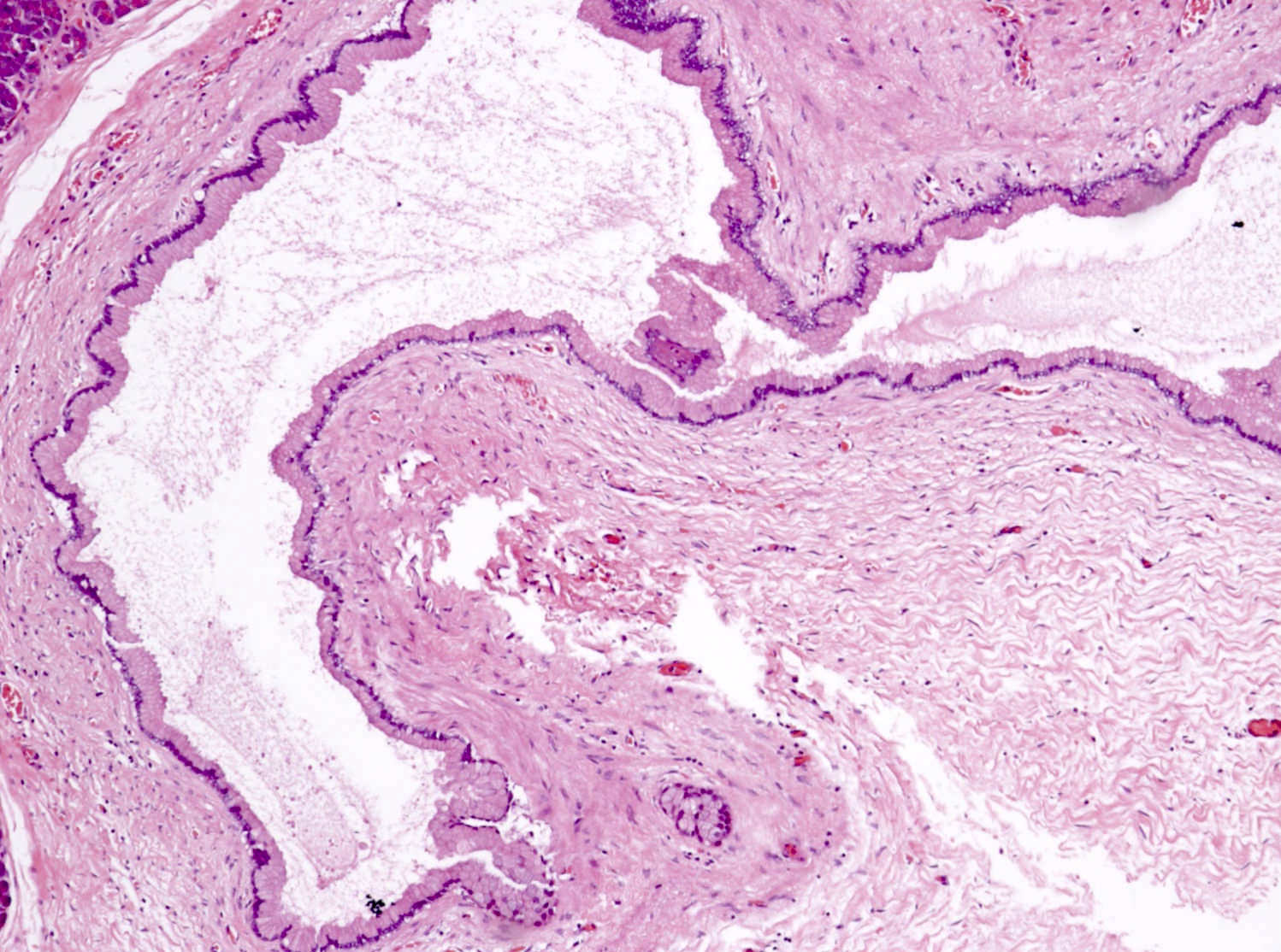
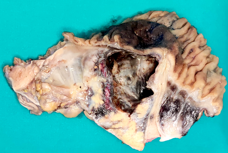
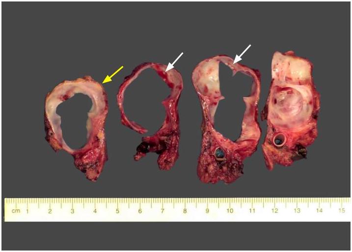
Large unilocular ACT
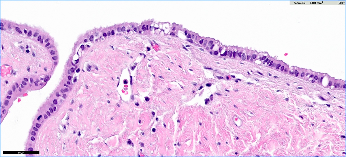
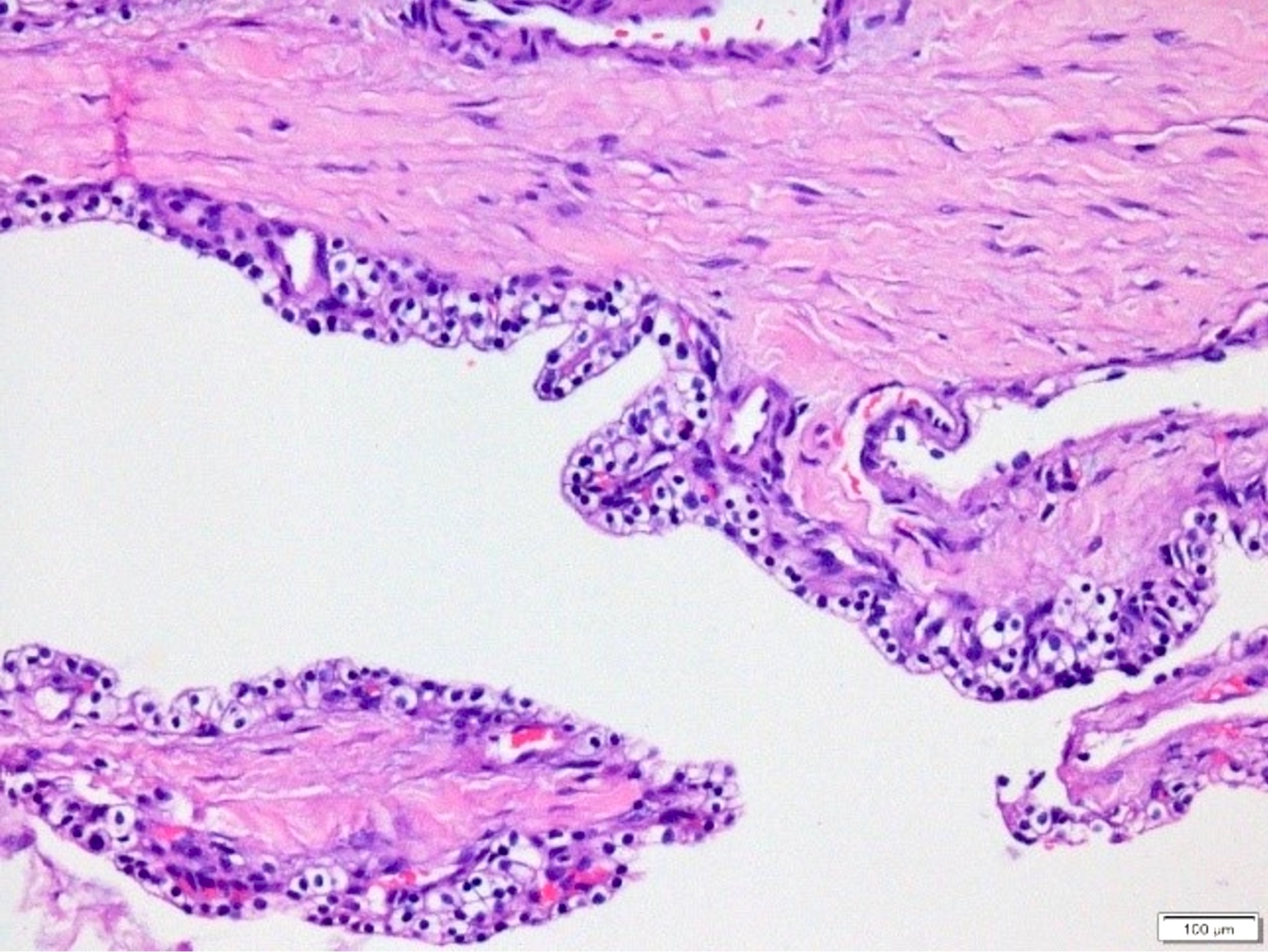
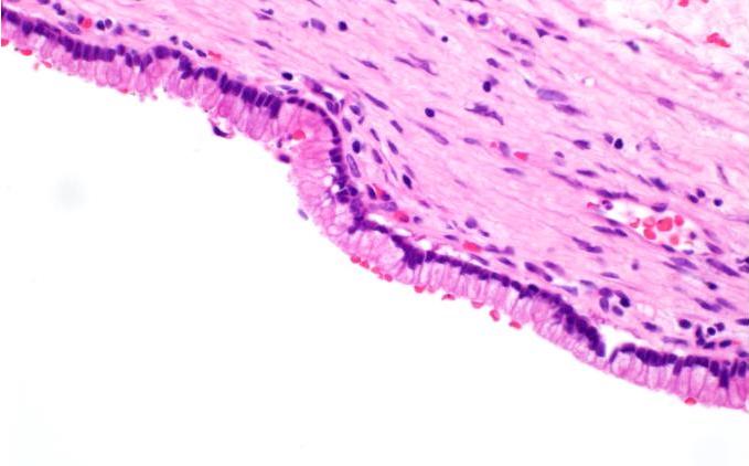
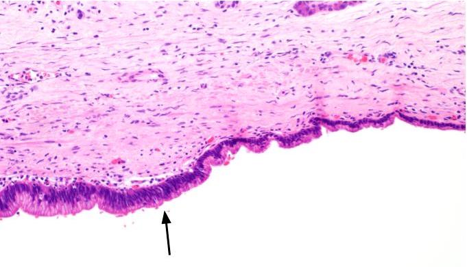
ACT cyst epithelium
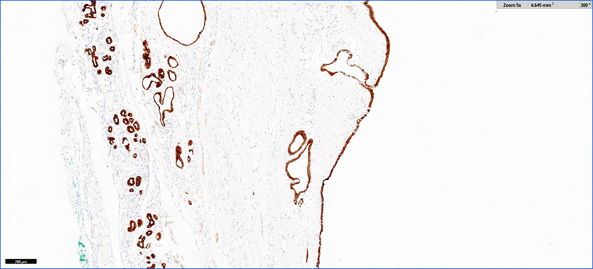
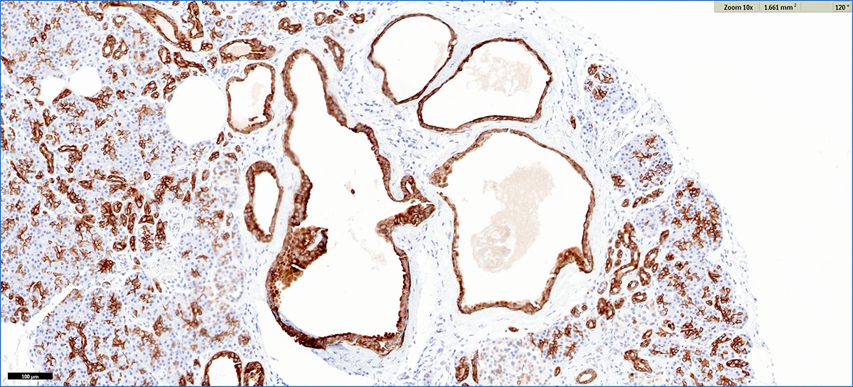
Large ACT CK7 immunostain
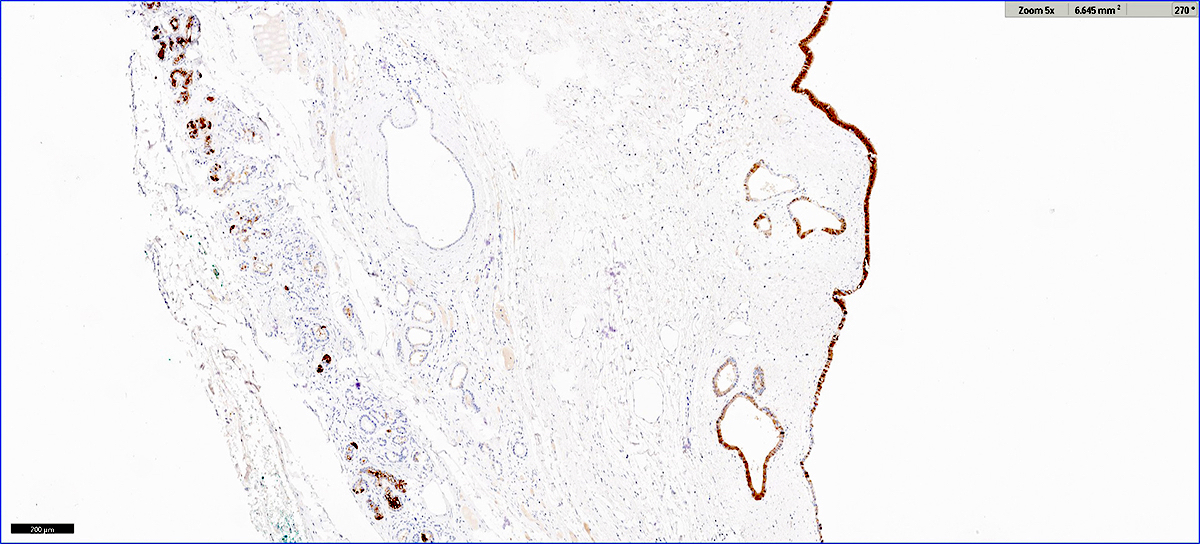
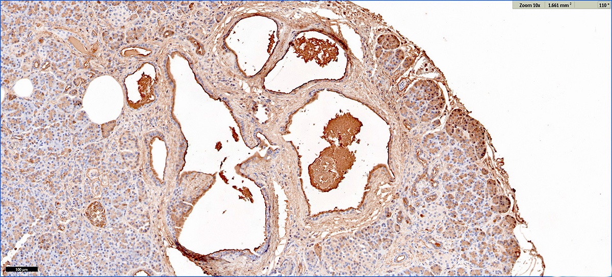
Large ACT trypsin immunostain
Images hosted on other servers:

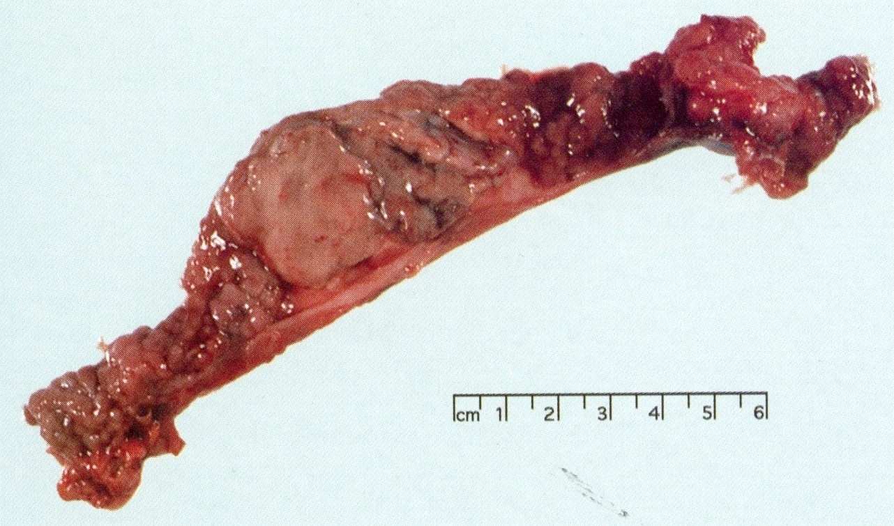
Pancreatic tail / body mass
Images hosted on other servers:

Pancreatic tail mass
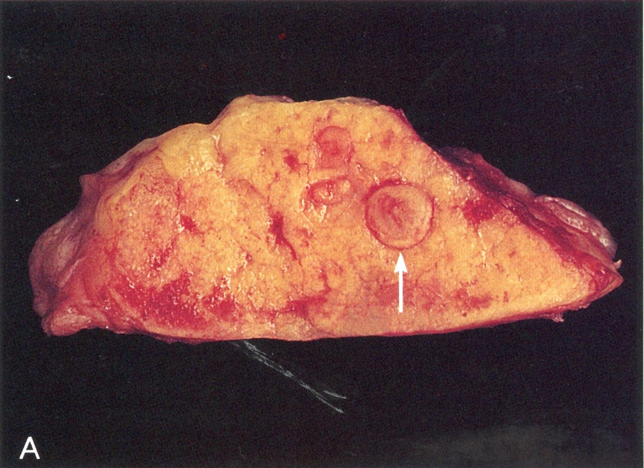
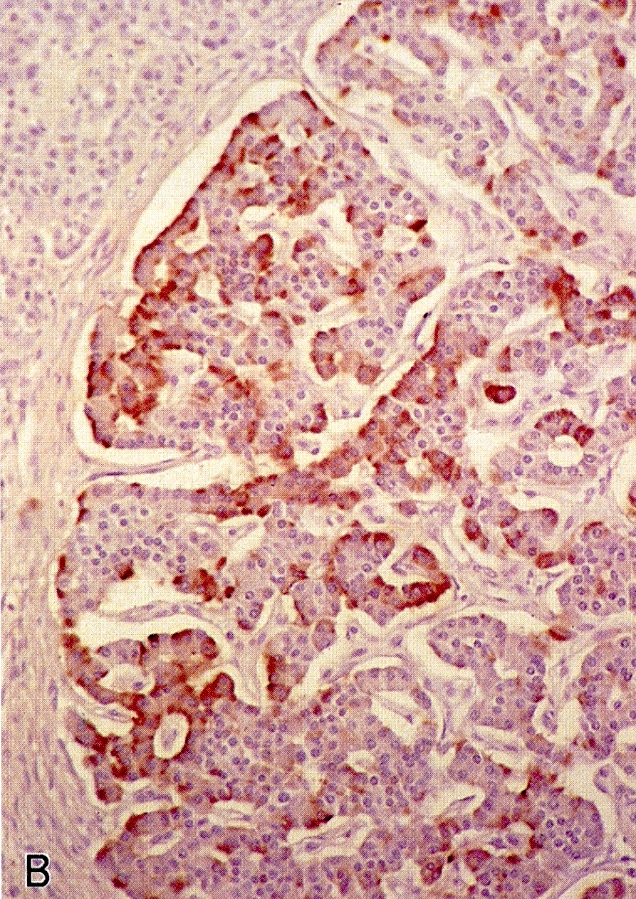
AFIP images
ACTH producing tumor with Cushing syndrome
Images hosted on other servers:

Typical neuroendocrine tumor appearance


66 year old man with ultrasound guided biopsy: H&E and ACTH antibody
Images hosted on other servers:

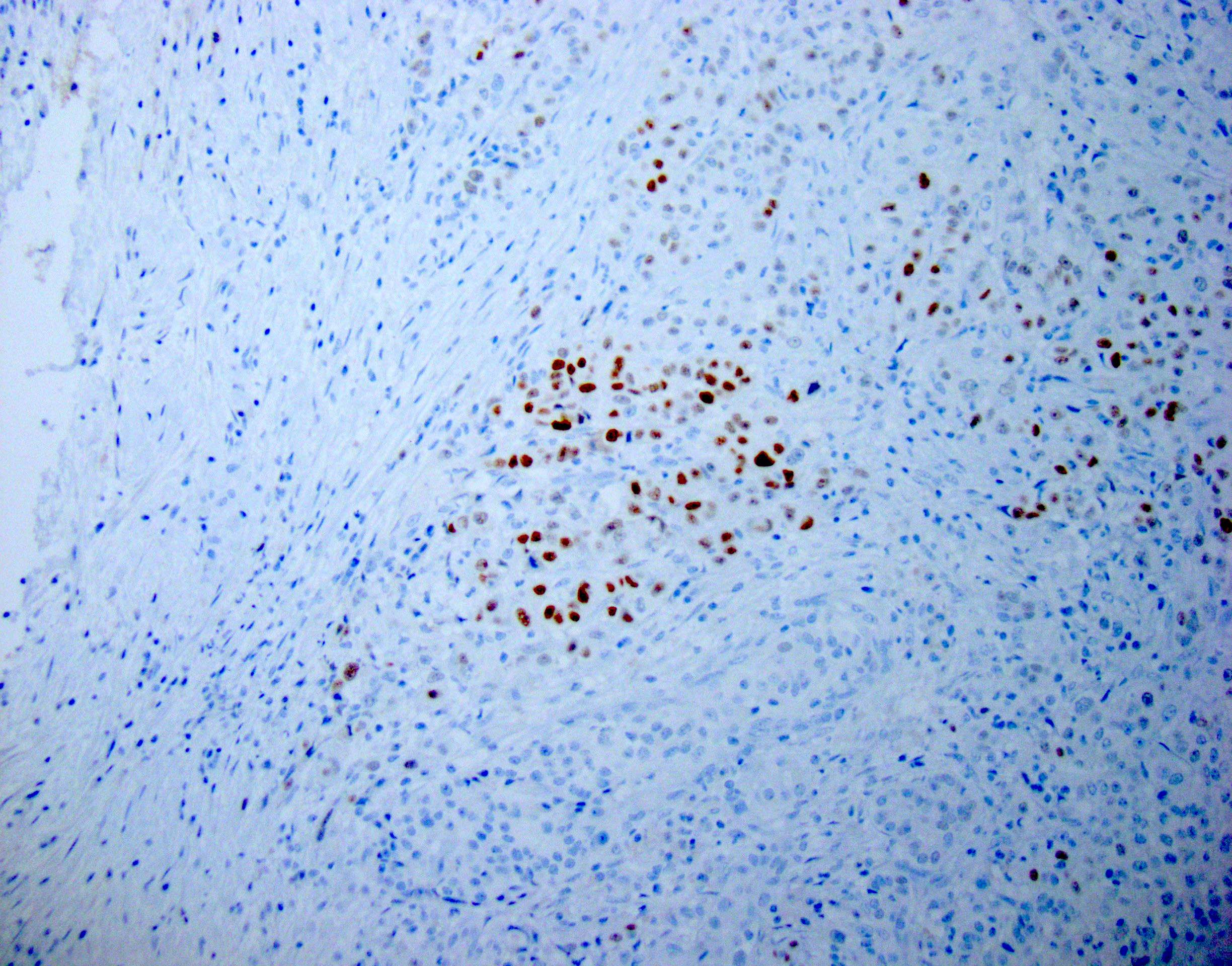
Scattered neuroendocrine cells
Images hosted on other servers:

Overview of pathogenesis
Images hosted on other servers:

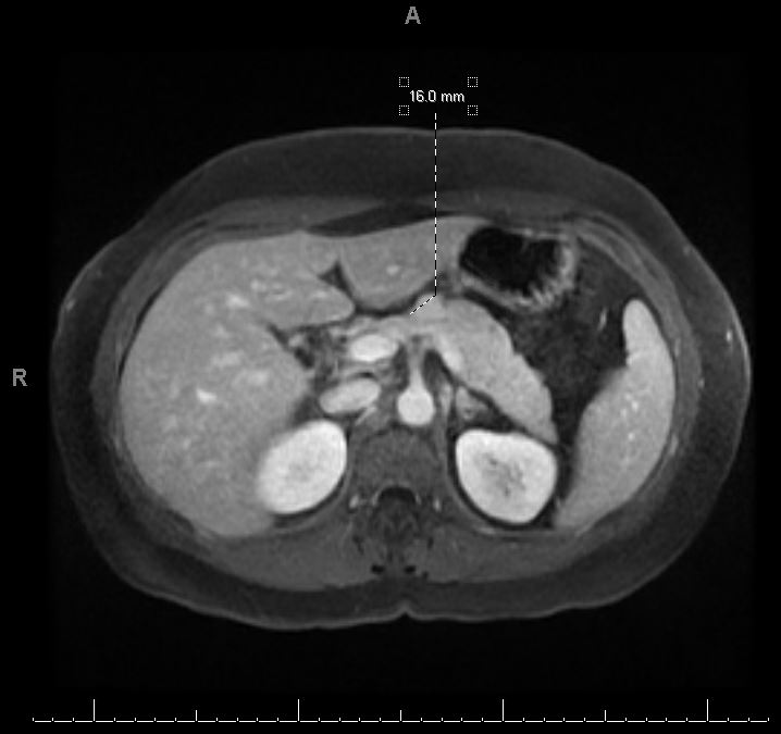
CT scan

Necrotizing pancreatitis
Images hosted on other servers:

Intraoperative findings of necrotizing pancreatitis
Images hosted on other servers:

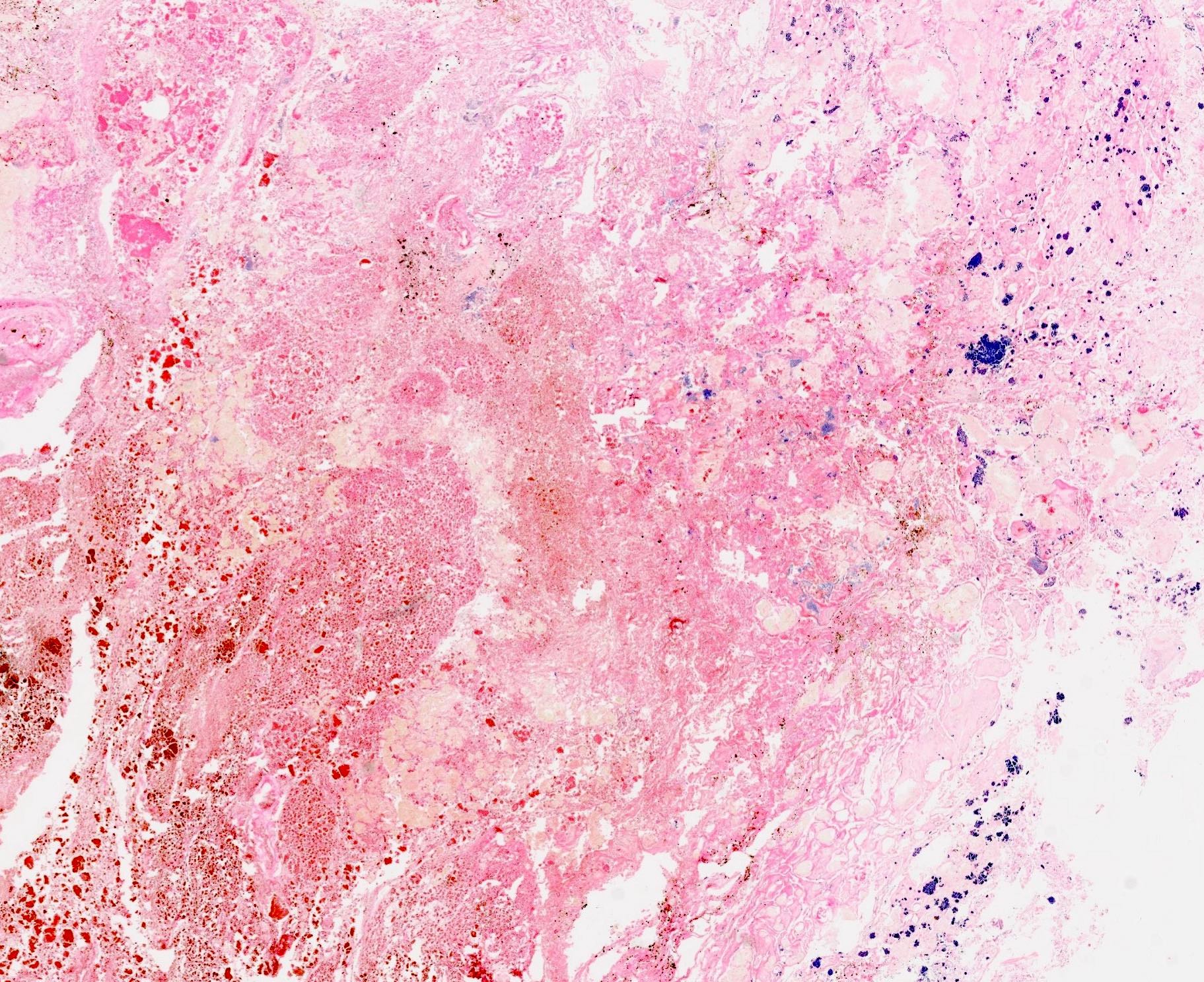
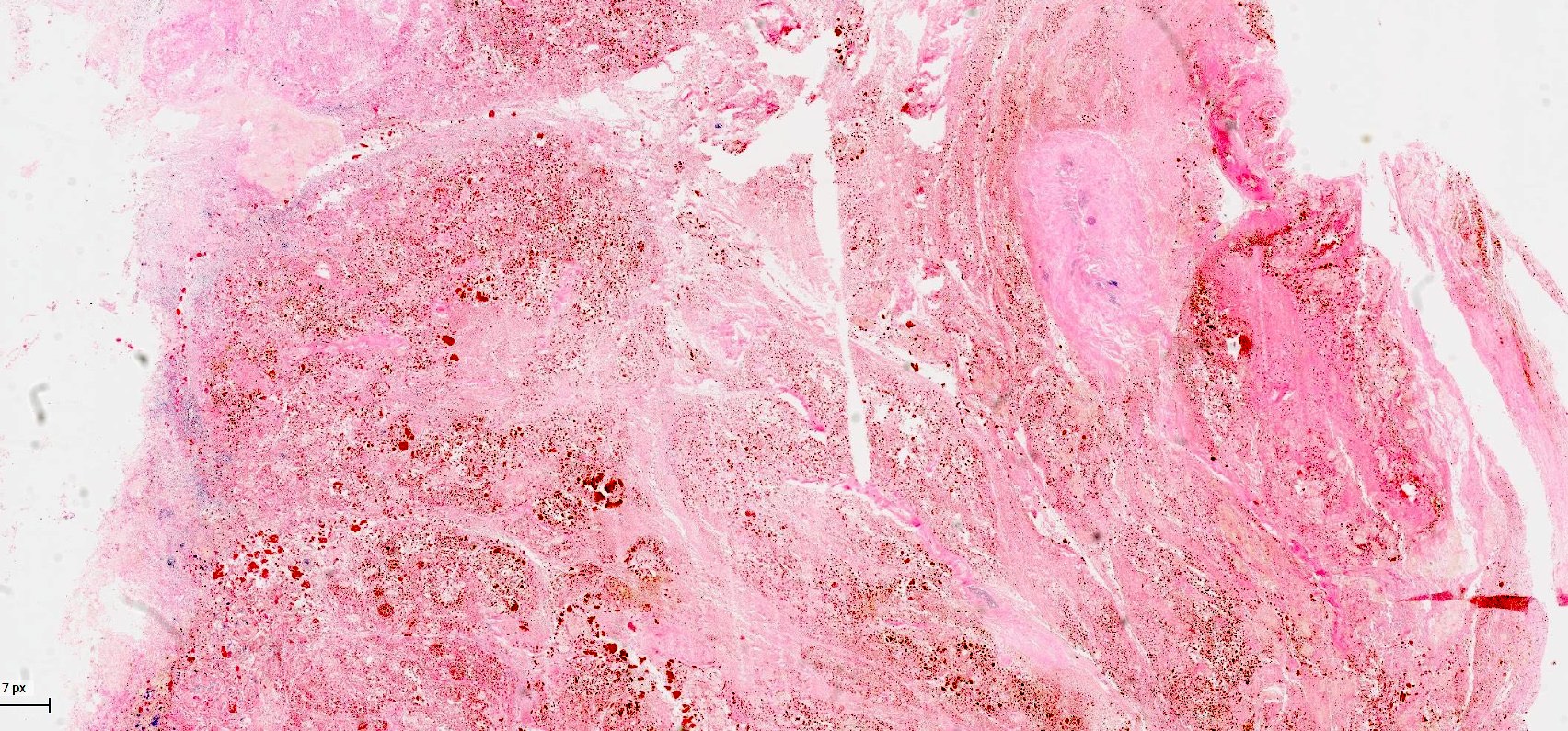
Acute pancreatitis with fat necrosis

Acute hemorrhagic pancreatitis
Contributed by Beena Ashan, M.D. and Sarah Kisha, M.D.

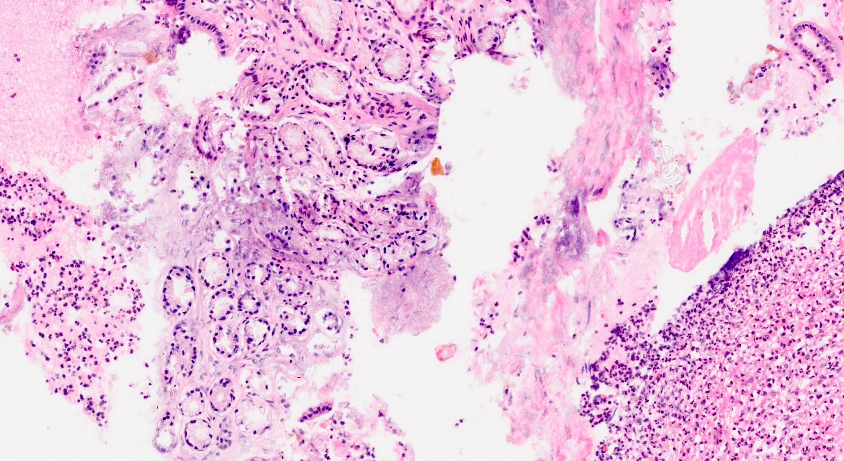
Acute interstitial pancreatitis
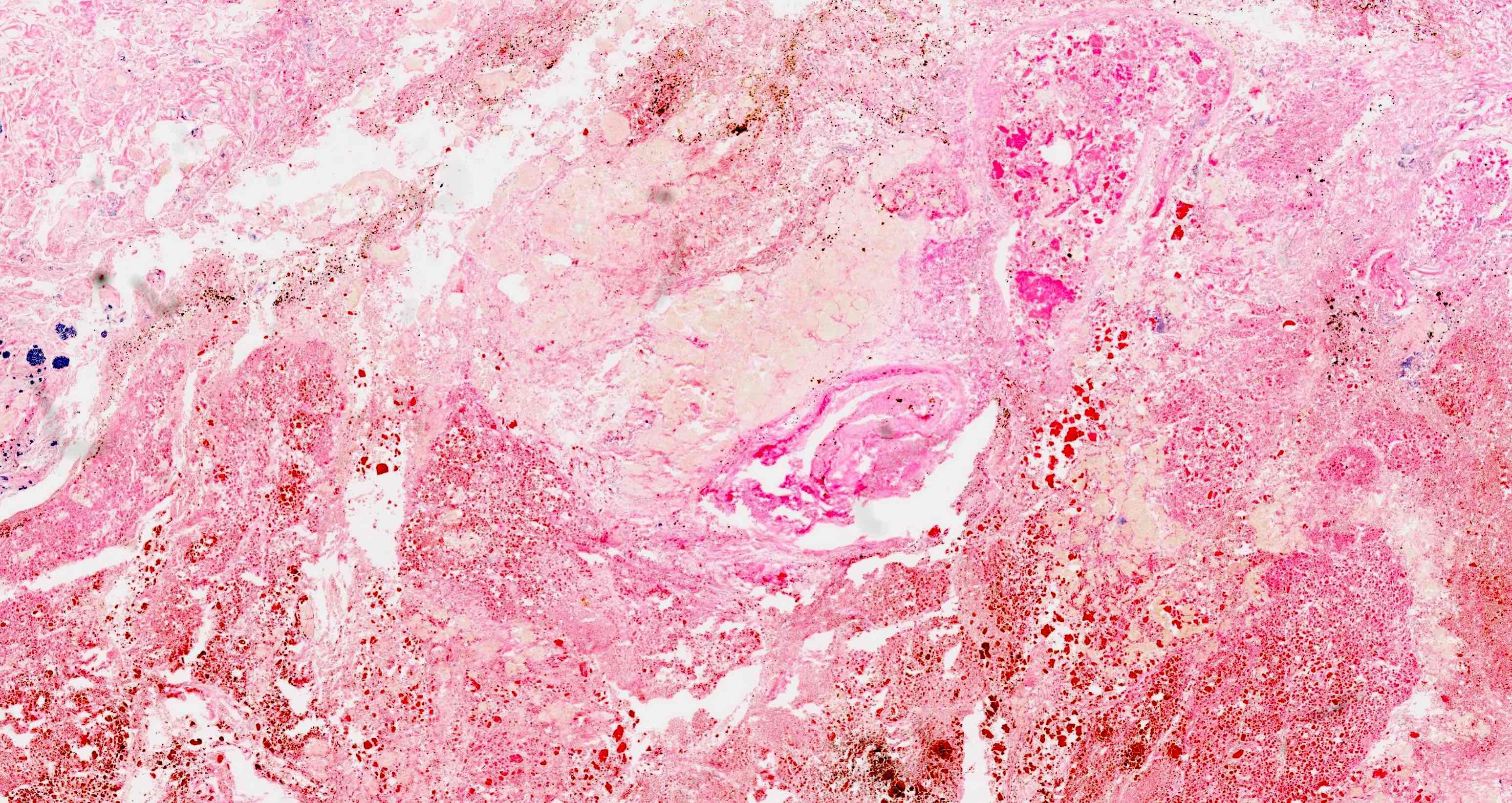
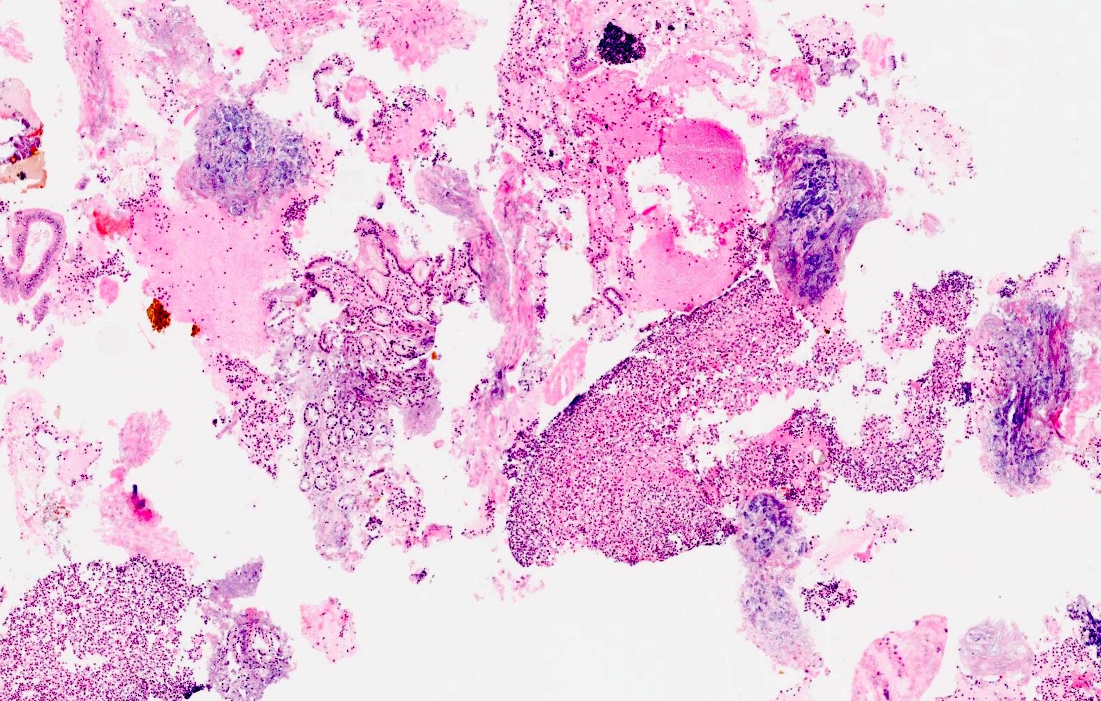
Hemorrhagic pancreatitis

Fat necrosis with hemosiderin disposition
Contributed by Beena Ashan, M.D. and Sarah Kisha, M.D.
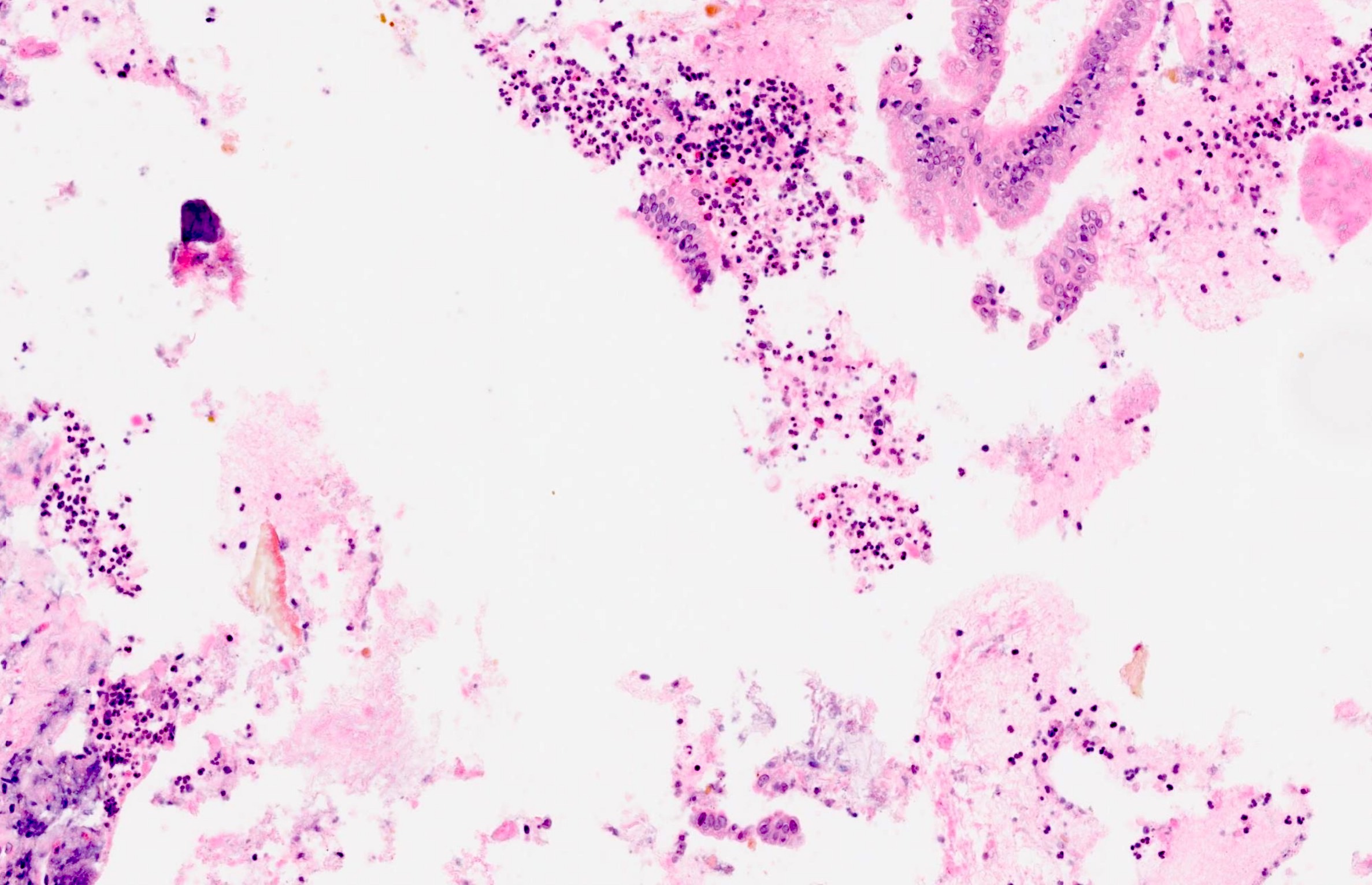
Dense inflammatory infiltrate
Pancreas: acute pancreatitis, gross and microscopy
Contributed by Orhun Çığ Taşkın, M.D.

Cystic and necrotic changes
Contributed by Orhun Çığ Taşkın, M.D., Raul S. Gonzalez, M.D. and AFIP

Glandular and squamous components
p63
Adenocarcinoma and squamous carcinoma
Squamous components
Mucoepidermoid pattern
CAM5.2
CK13
Contributed by Orhun Çığ Taşkın, M.D.
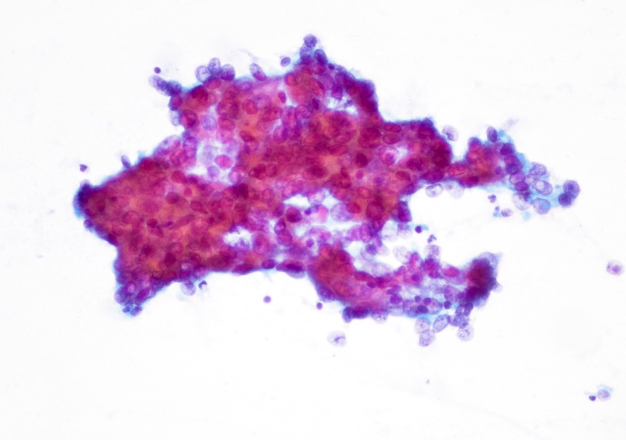
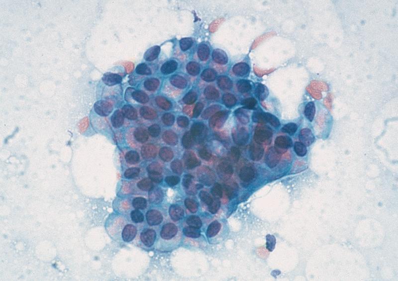
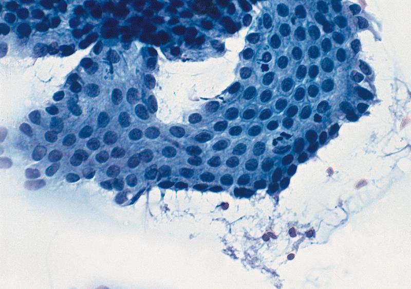
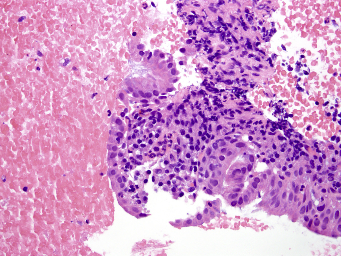
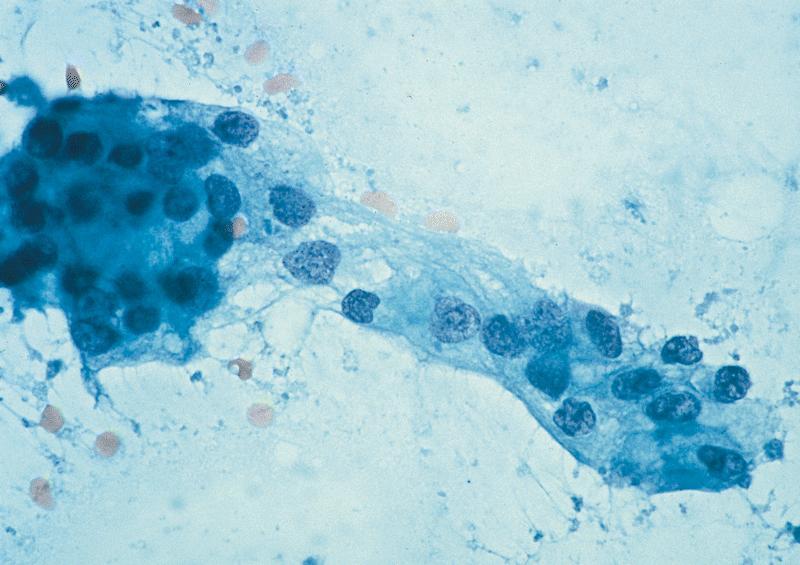
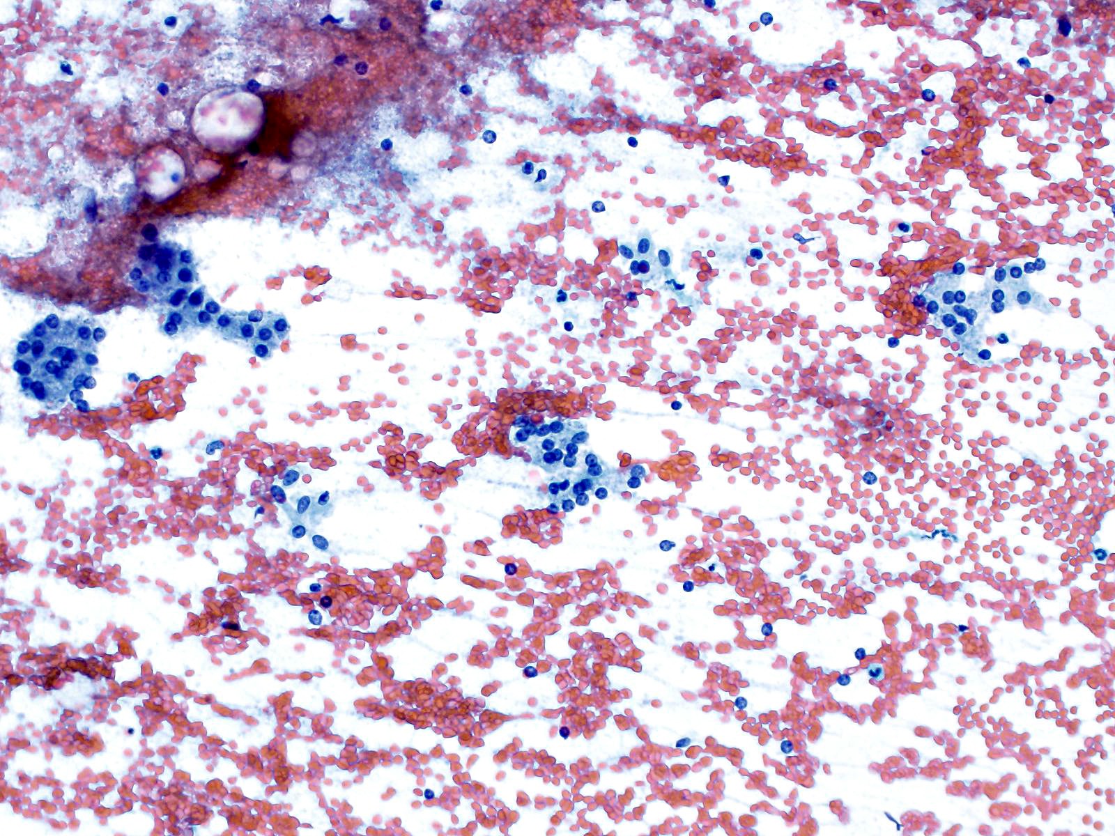
Pap stain
Cell block

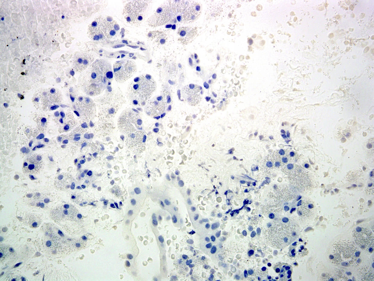
Cell block, p40
Images hosted on other servers:

Ultrasound guided biopsy
Contributed by Alexei Mikhailov, M.D., Ph.D. and Cinthia Drachenberg, M.D.

Inactive lymphocytes
Active acinar inflammatory infiltrate
Ductitis, no duct damage

Ductitis with epithelial damage

Venulitis
Neural inflammation

Cellular rejection grade 1

Cellular rejection grade 2
Cellular rejection grade 3

Graft fibrosis stage 2

Graft fibrosis stage 3

Chronic active cellular rejection

Antibody mediated rejection

Irregular fibrosis in peripancreatitis
Contributed by Jan Hrudka, M.D., Ph.D.
Normal pancreas histology
Normal pancreas histology

Chromogranin A IHC
Glucagon IHC
Insulin IHC
Contributed by Grigory Demyashkin, M.D., Ph.D. and AFIP images

10 week old fetus
Islets in newborn
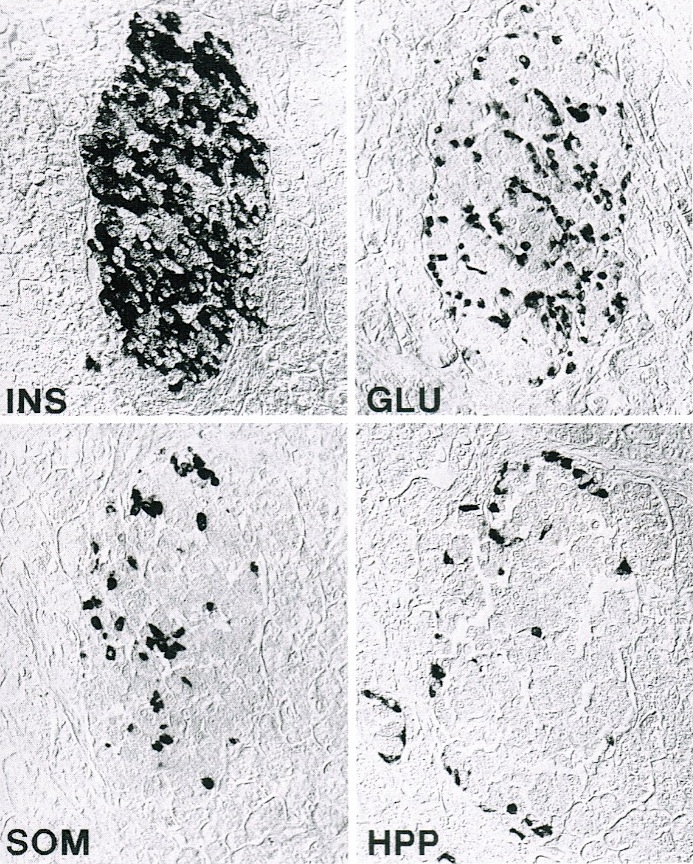
Adult pancreas: distribution of islet cell types
6 - 8 week embryo
AFIP images
Normal pancreatic acinar cells
Normal ductal cells in sheet arrangement
AFIP images
38 / 39 week old fetus
Adult pancreatic islet cells
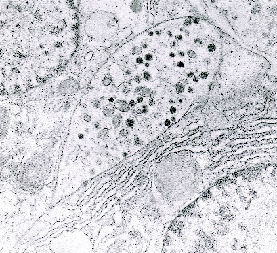
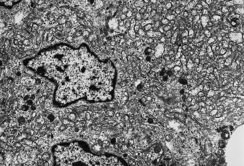
2 centroacinar cells with electron lucent cytoplasm
Images hosted on other servers:
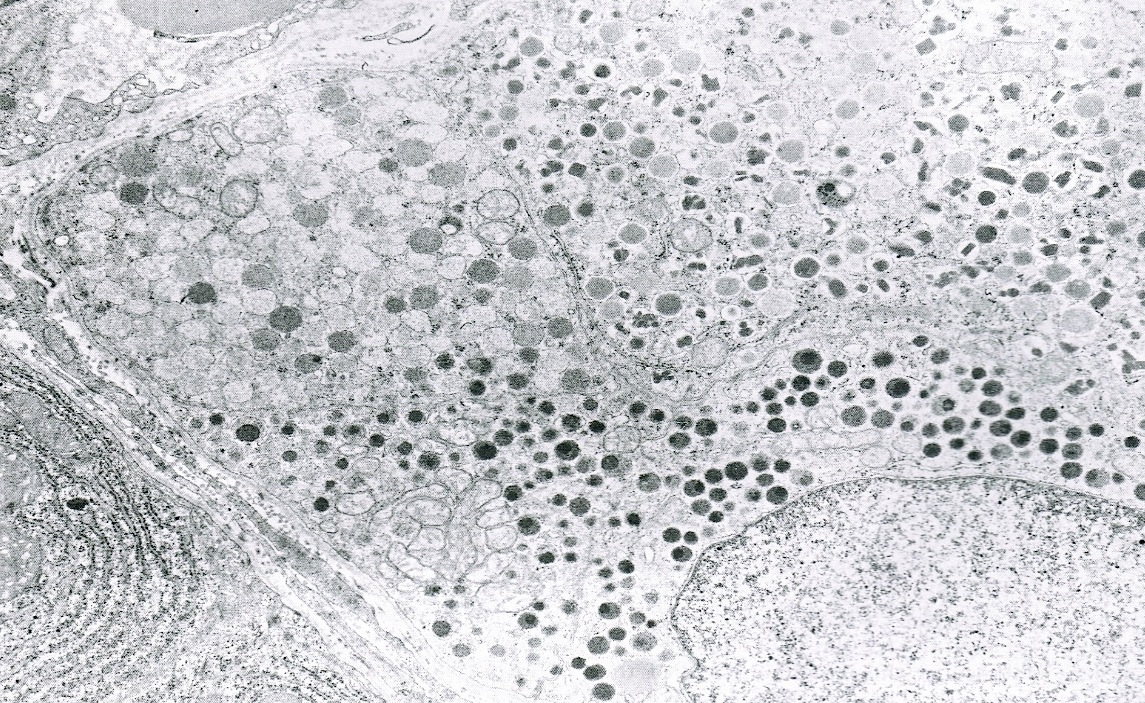
Zymogen granules


Images hosted on other servers:

CT

PET

Endoscopic ultrasound

ERCP with stricture

Ultrasound

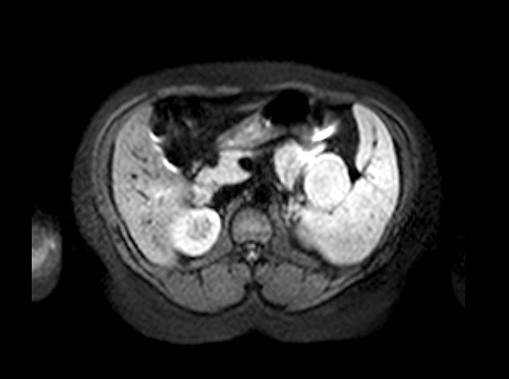
MRI

Mass forming autoimmune pancreatitis (CT)

Mass forming autoimmune pancreatitis (PET)
Contributed by Aaron R. Huber, D.O.
Ill defined fibrosis
Images hosted on other servers:

Nodular lesions

Ill defined pancreatic head mass
Contributed by Aaron R. Huber, D.O. and @RaulSGonzalezMD on Twitter
Fibrosis and inflammation
Inflammation
Fibrosis
Phlebitis
Phlebitis
Autoimmune pancreatitis type 1
Elastic stain
IgG4
Images hosted on other servers:

Type 1 FNA
Images hosted on other servers:

Diffuse pancreas enlargement
Contributed by David J. Escobar, M.D., Ph.D.
Granulocyte epithelial lesion
IgG4-
Contributed by Claudio Luchini, M.D., Ph.D. and Gaetano Paolino, M.D.

Head transverse section
Contributed by Claudio Luchini, M.D., Ph.D. and Gaetano Paolino, M.D.
Cardinal triad

Duct dilation
Lipomatous atrophy
Chronic inflammation
Squamous metaplasia
Perivenulitis
Residual Langerhans islets
Paraduodenal (groove) pancreatitis
Contributed by Claudio Luchini, M.D., Ph.D. and Gaetano Paolino, M.D.
Fibrous stromal elements and inflammatory cells
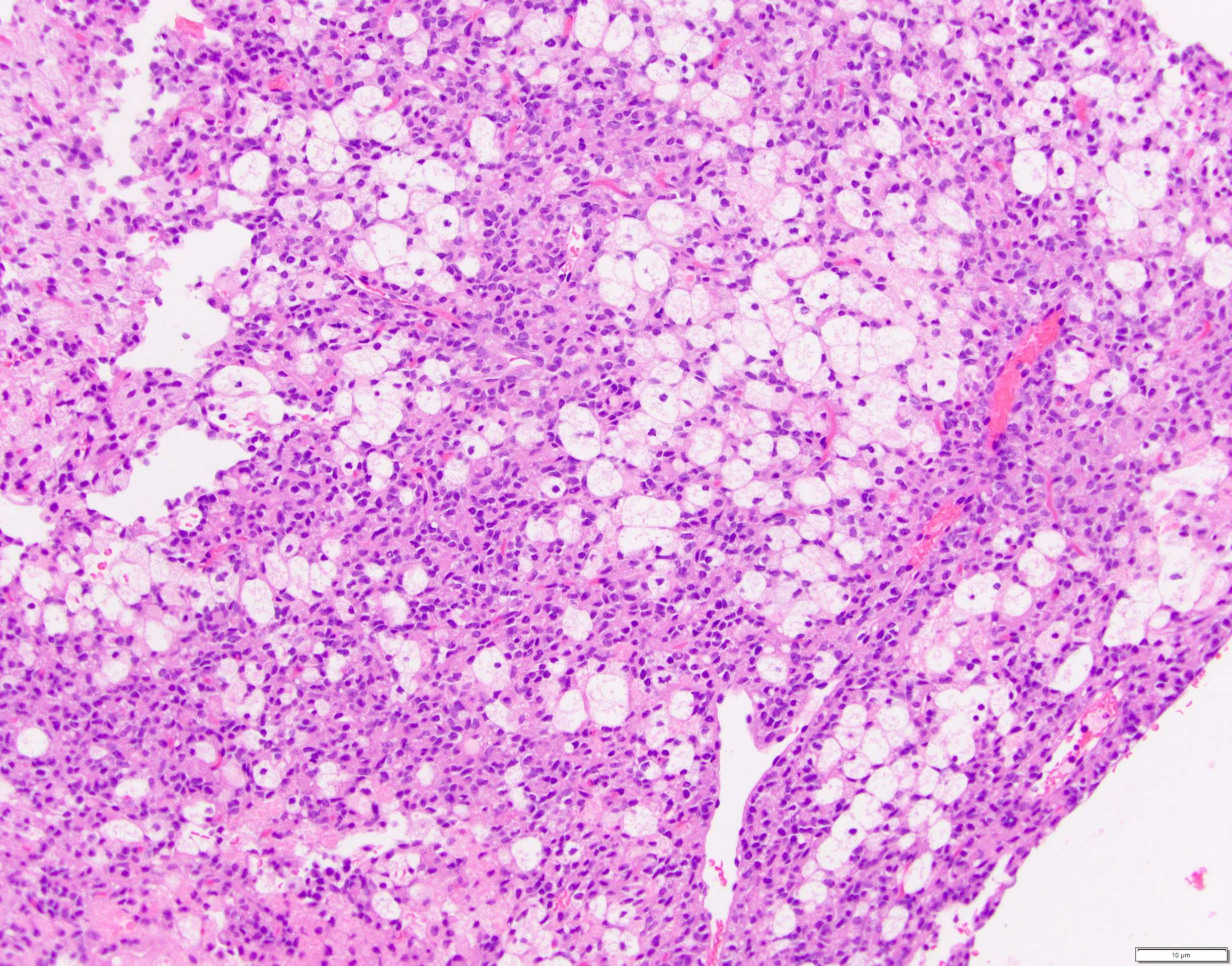
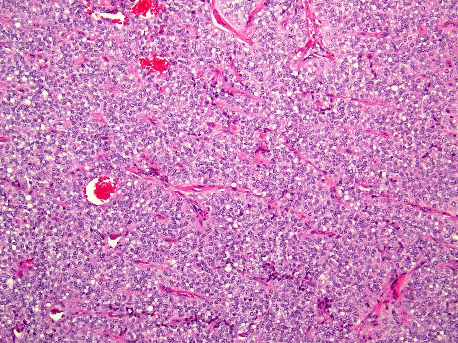
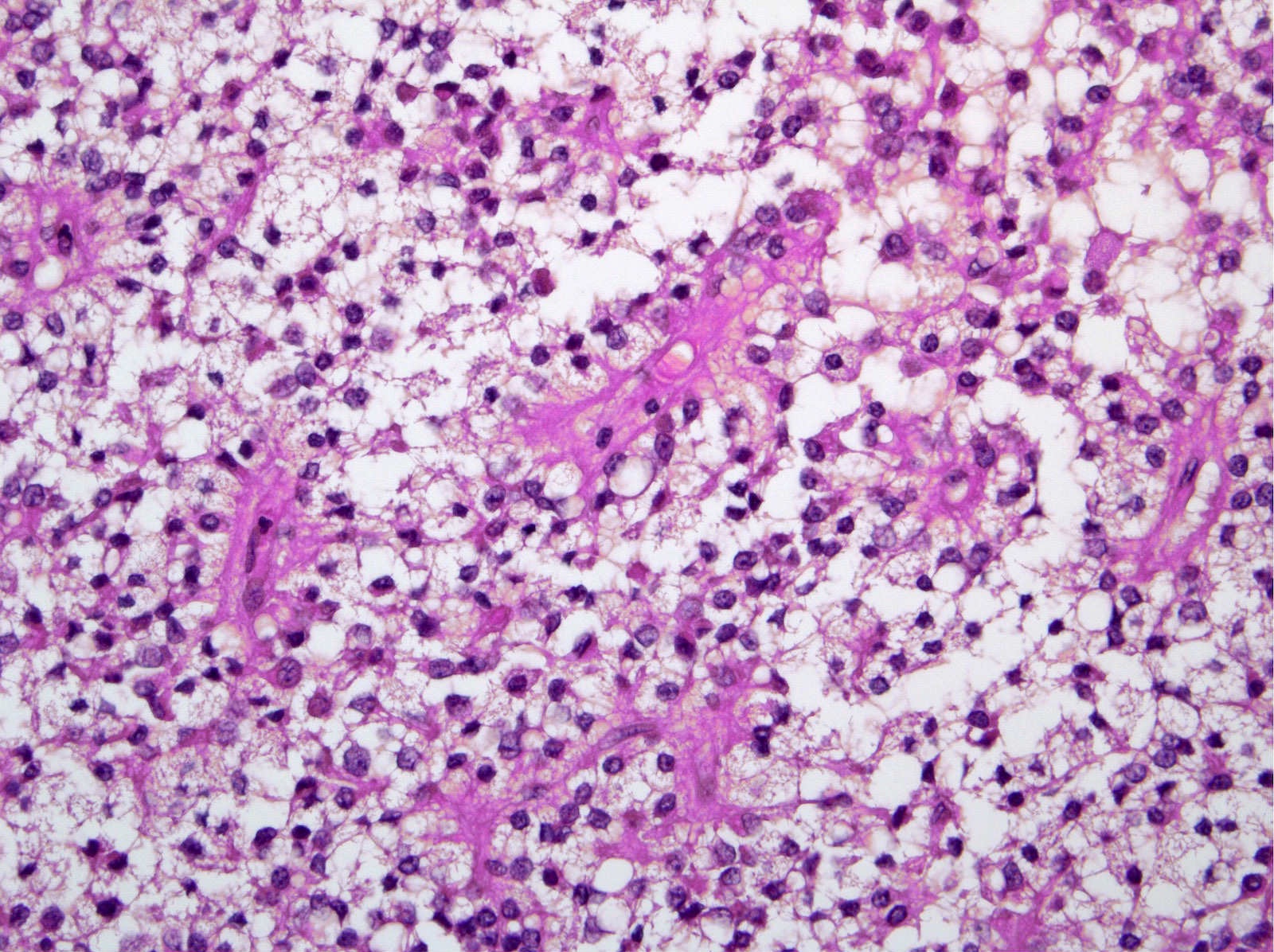
Contributed by Aatur Singhi, M.D., Ph.D.
Nests of tumor cells
Clear, vacuolated cytoplasm
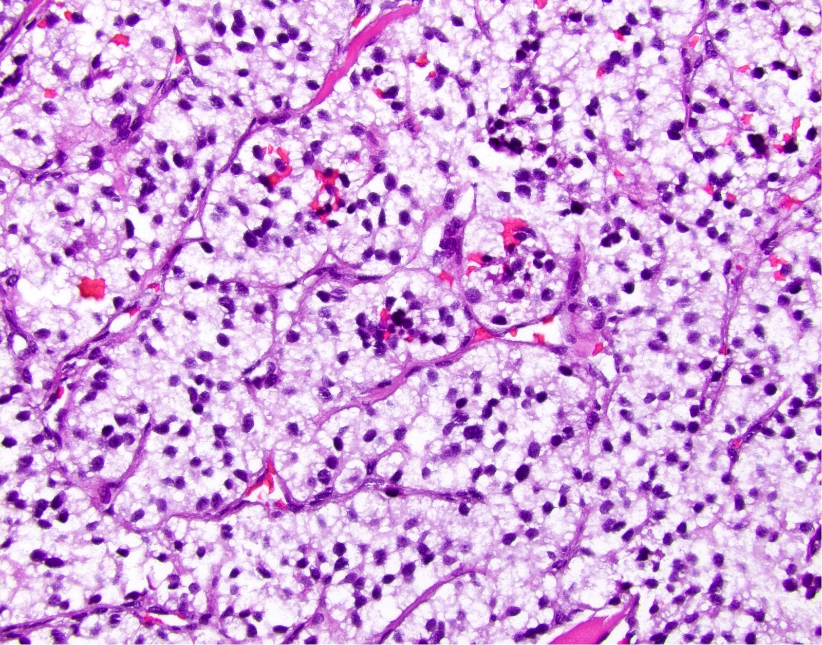
Salt and pepper chromatin
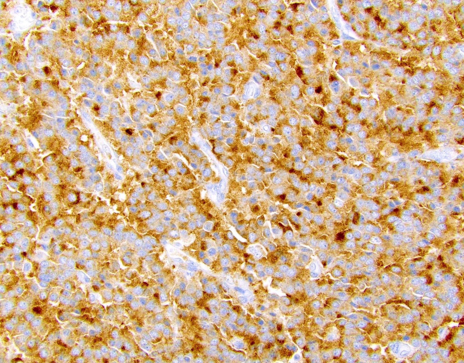
Synaptophysin positivity
Image hosted on other servers:

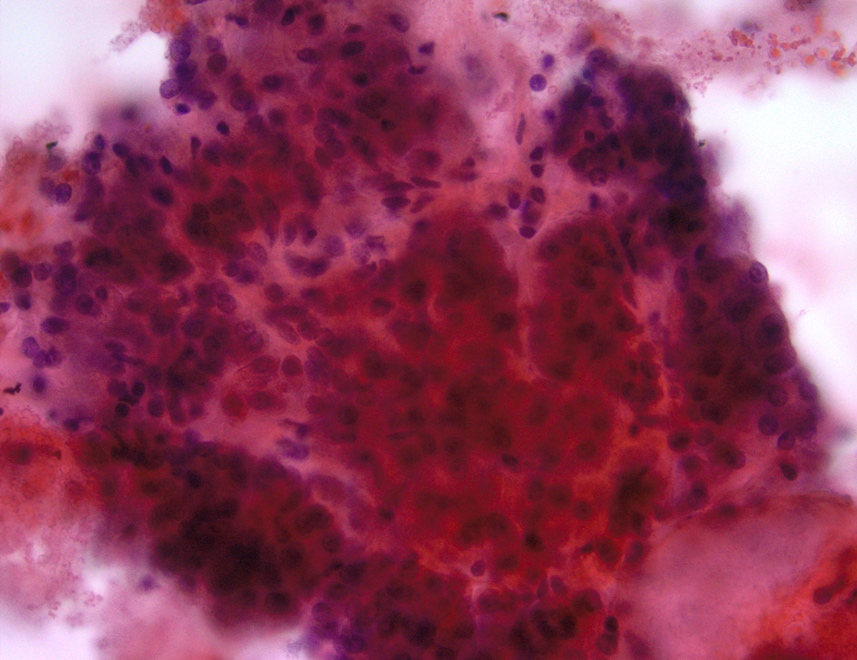
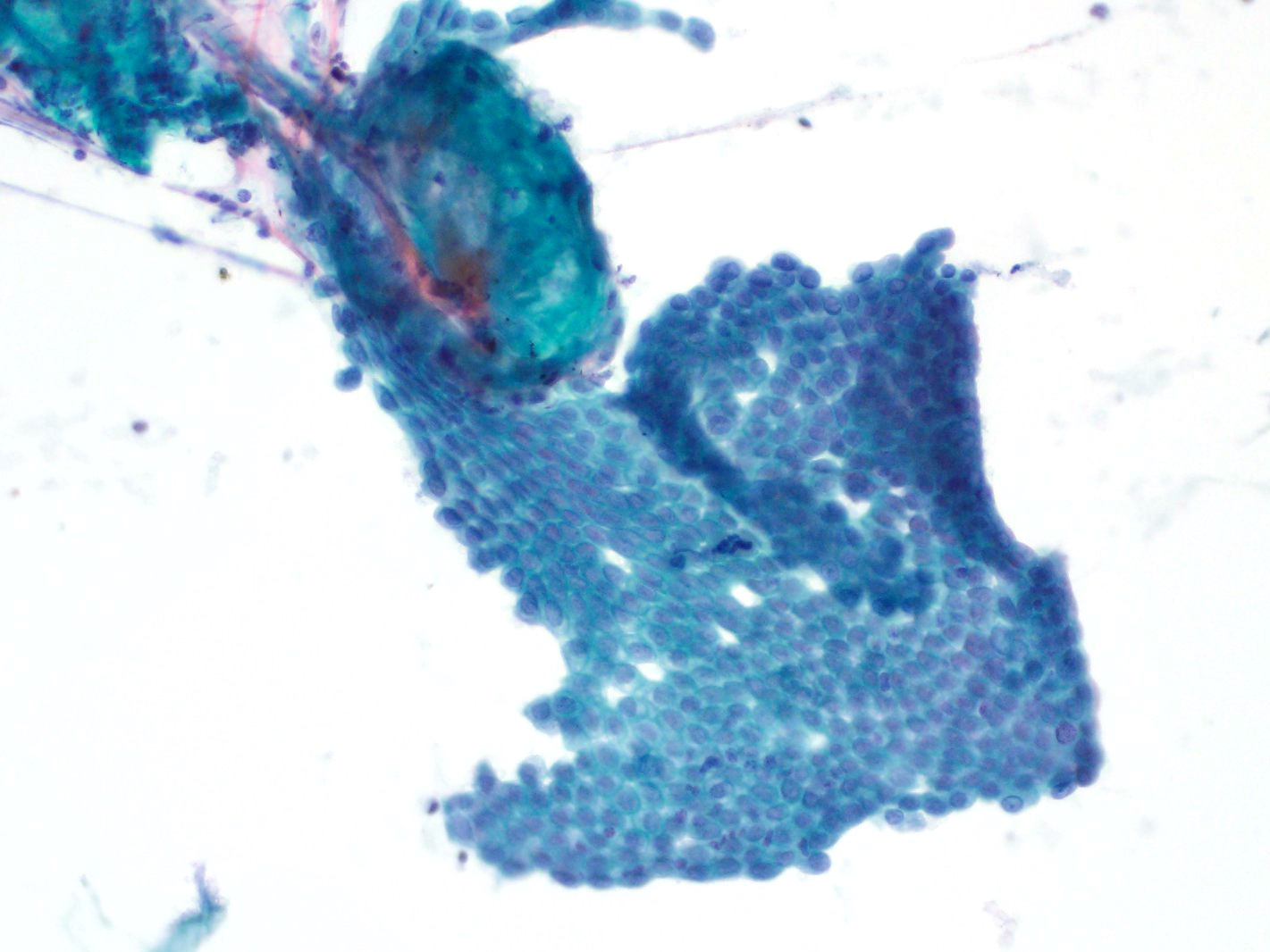
Papanicolaou stain of FNA
Pancreatic neuroendocrine neoplasms: overview
Images hosted on other servers:

CT scan
AFIP images
Whipple resection specimen
Contributed by Jennifer Vazzano, D.O., M.S. and Wei Chen, M.D., Ph.D.
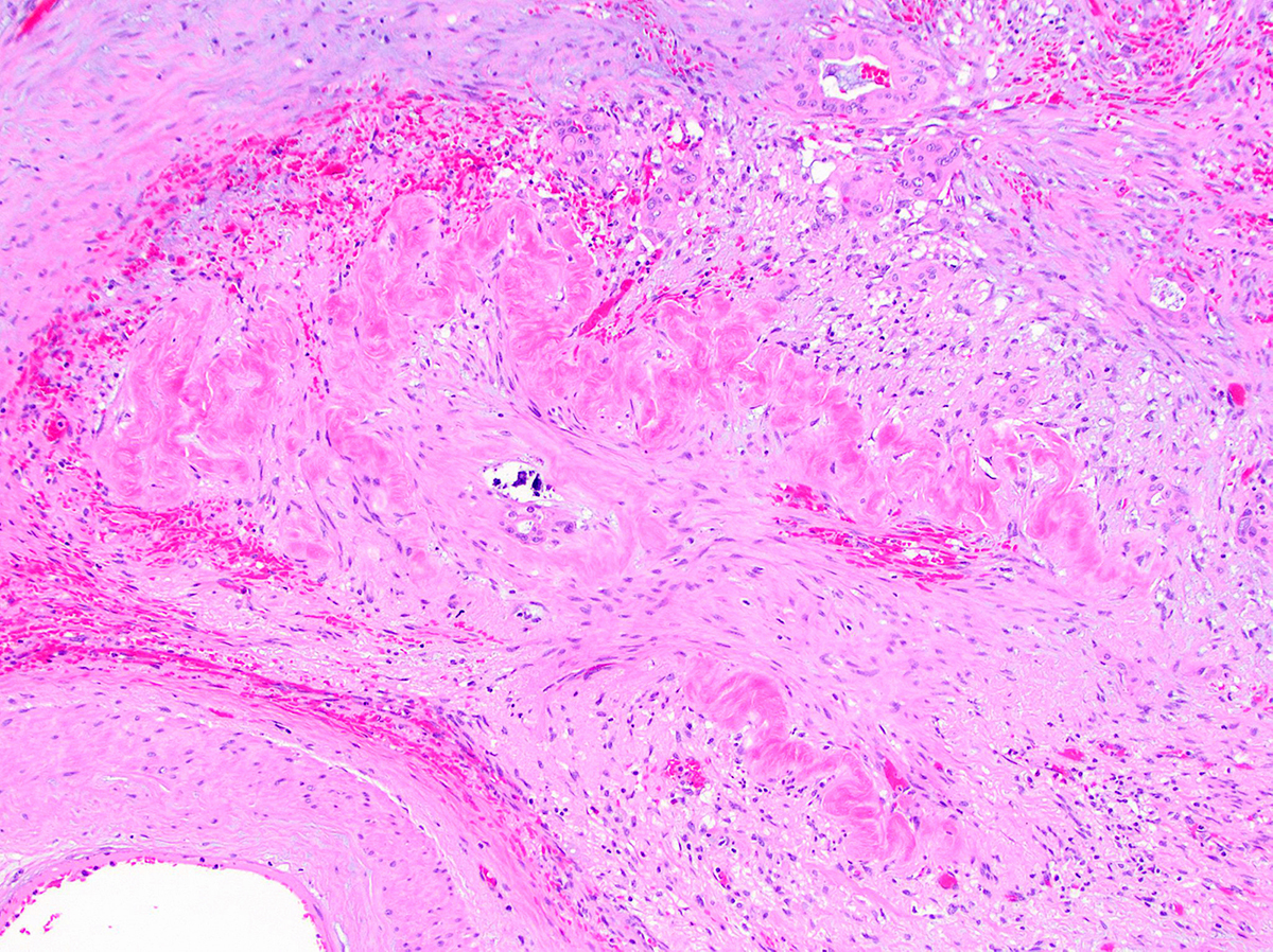
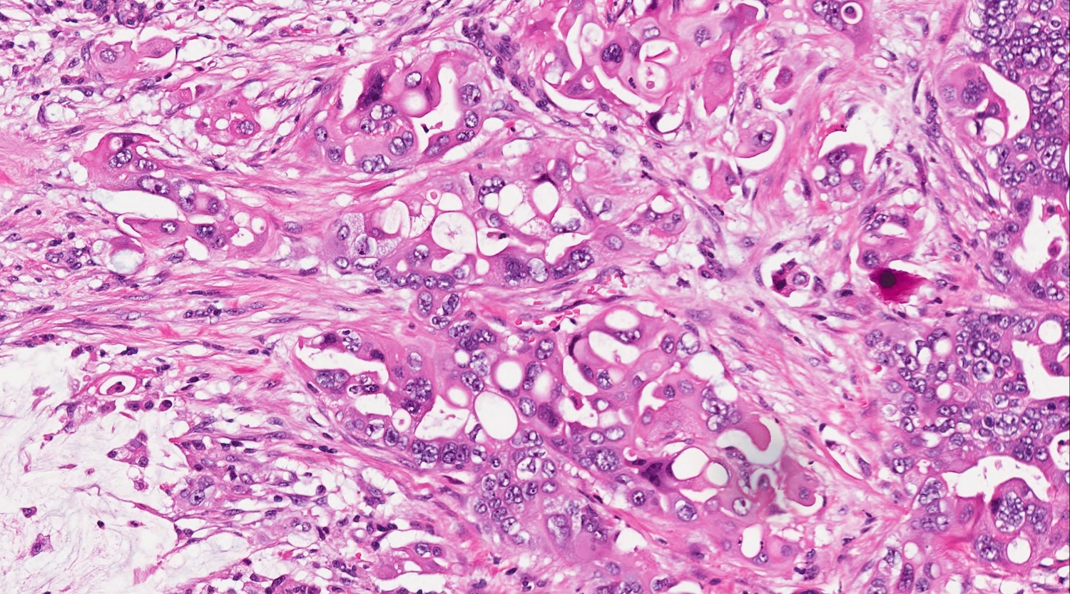
Large mucin pools
Images hosted on other servers:

Cystic pancreas lesion on CT scan
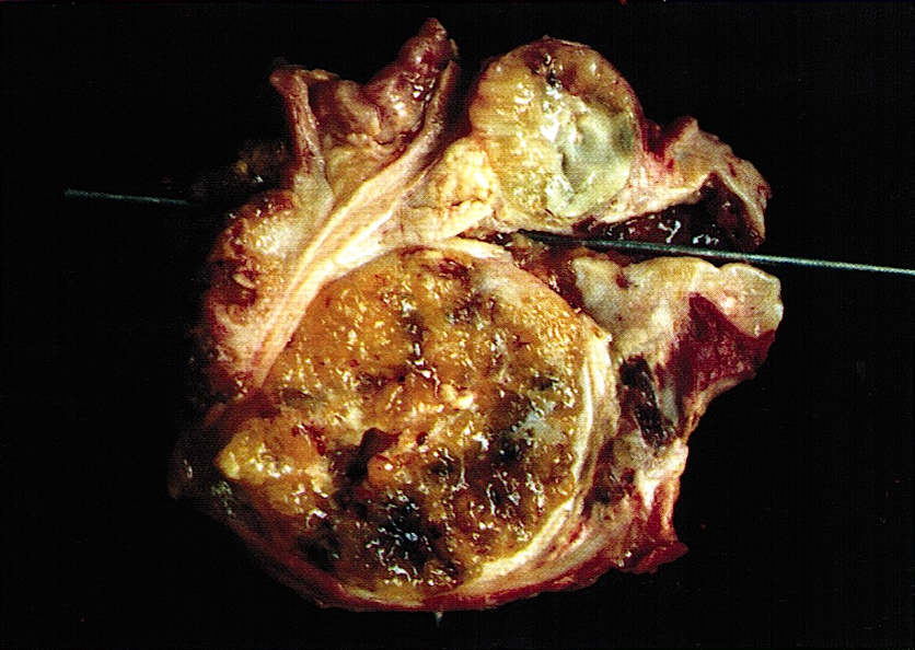
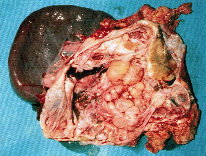
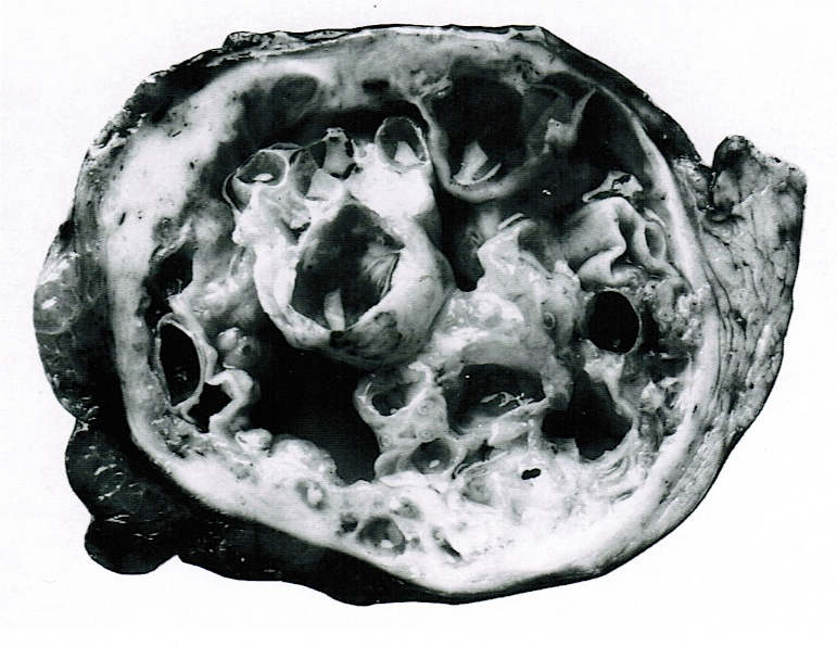
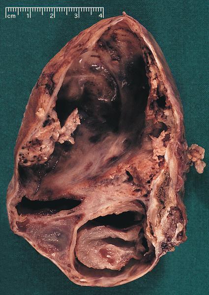
AFIP images
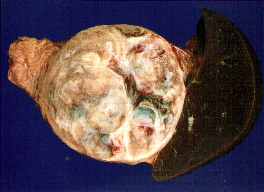
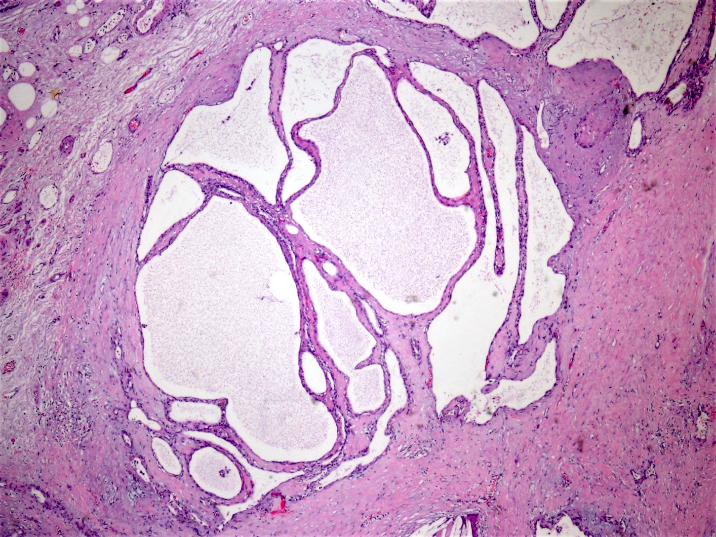
Large cystic tumor
Images hosted on other servers:

Cystic pancreas mass


Large spherical lesion with multiple loculations arising from head of pancreas

Cystic pancreatic endocrine neoplasm
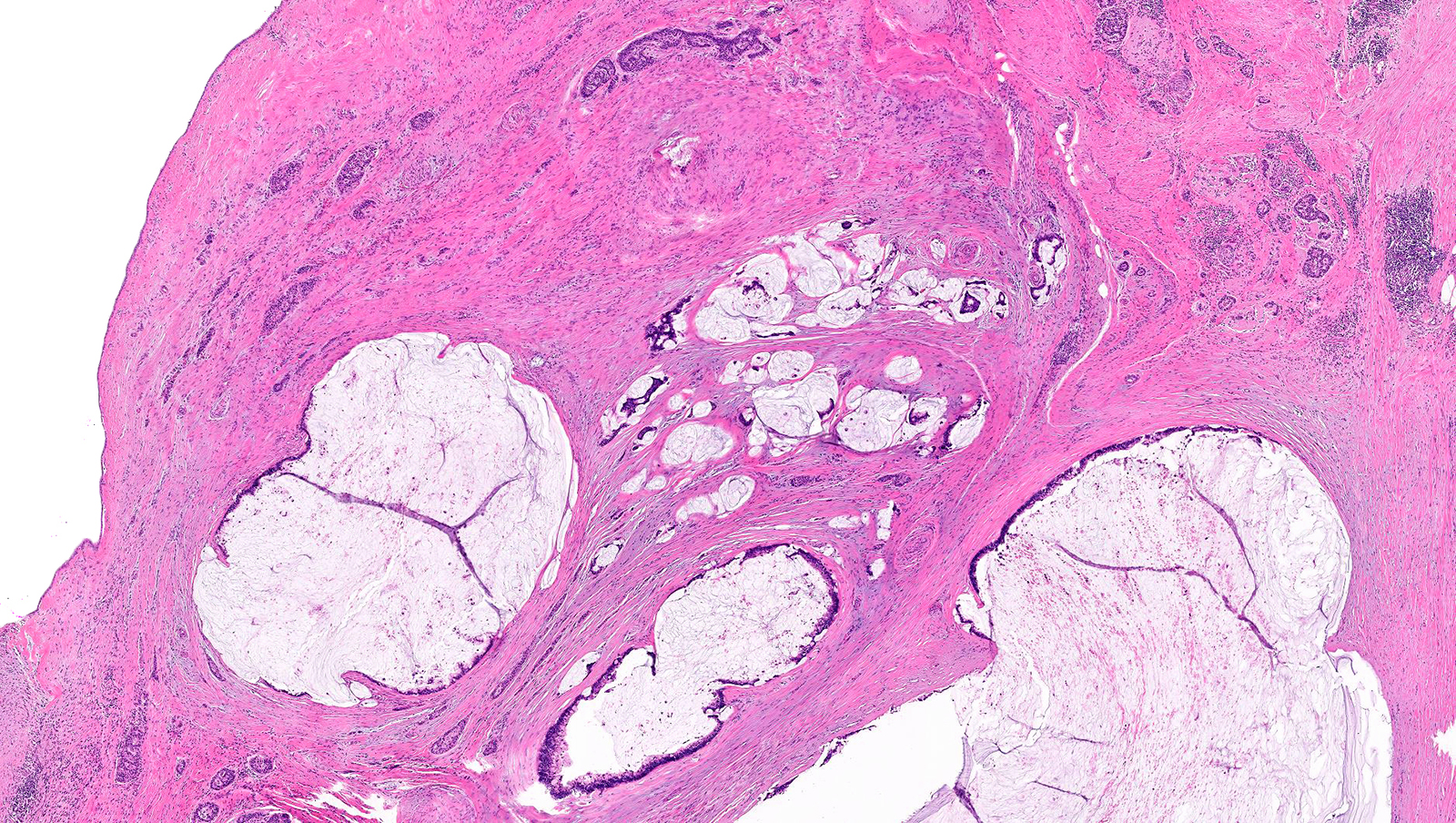
Contributed by Raul S. Gonzalez, M.D.
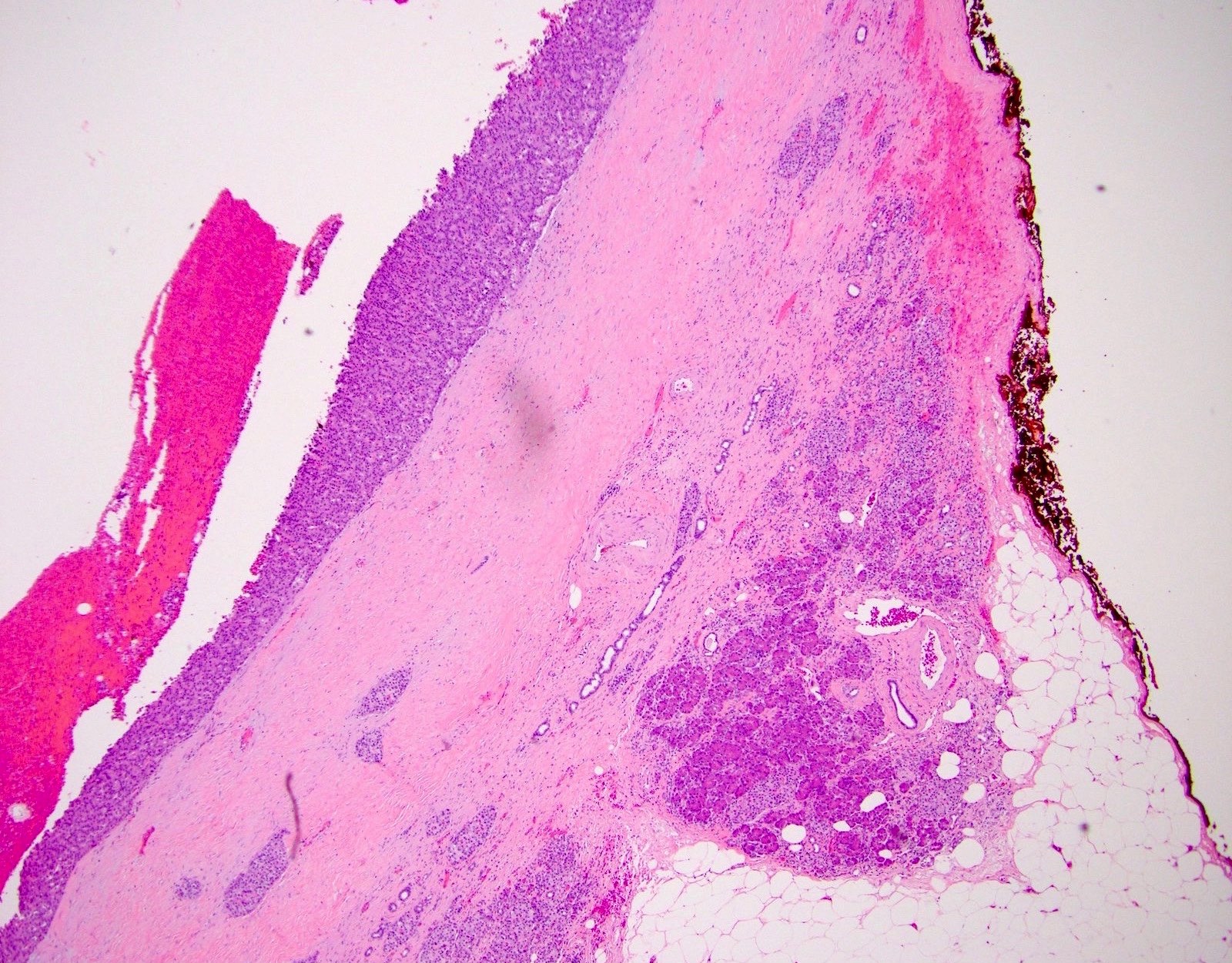
Fibrous wall
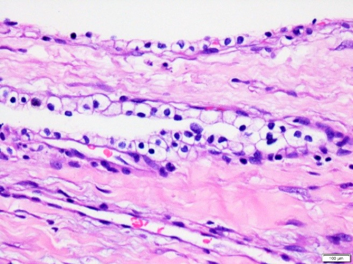
Cyst lining

Unilocular lesion
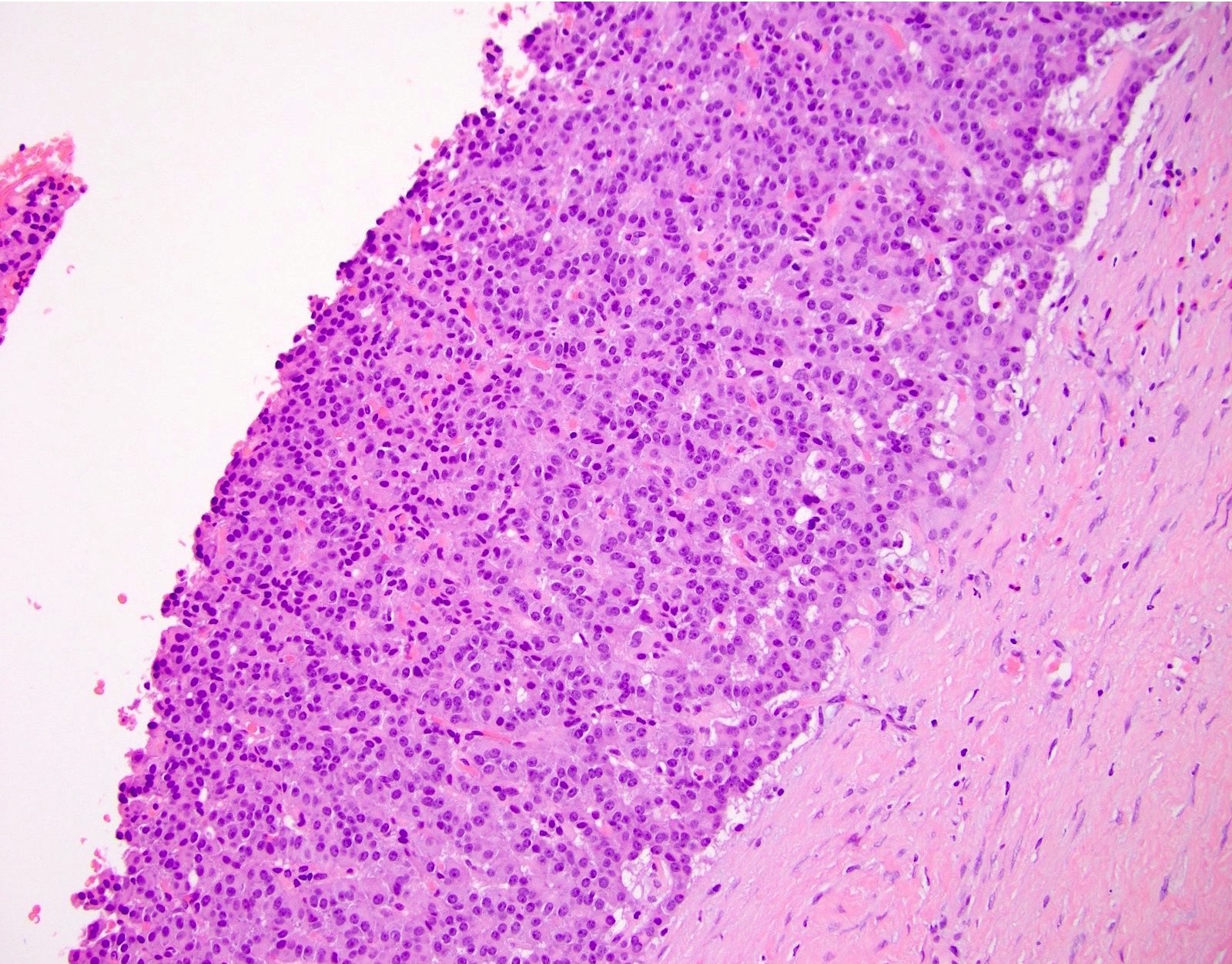
Neuroendocrine cells within fibrosis
Images hosted on other servers:

Cystic pancreatic neuroendocrine tumor
Images hosted on other servers:

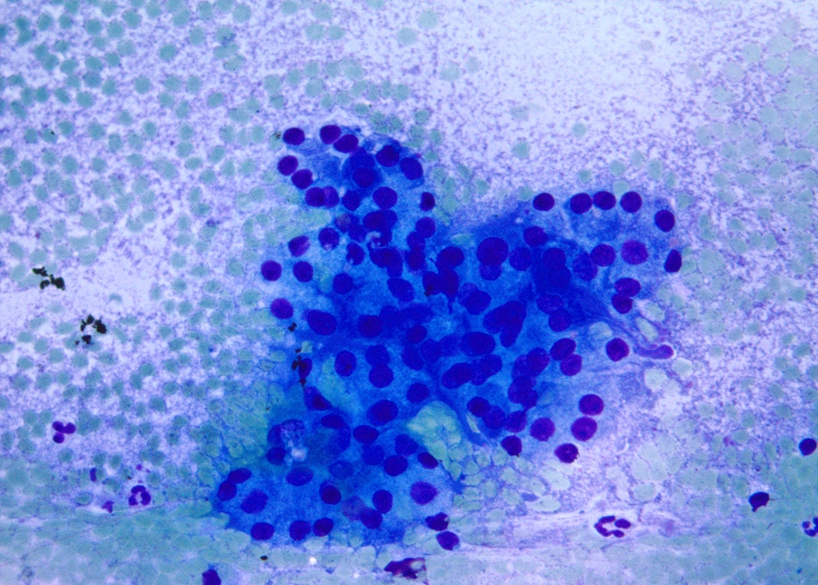
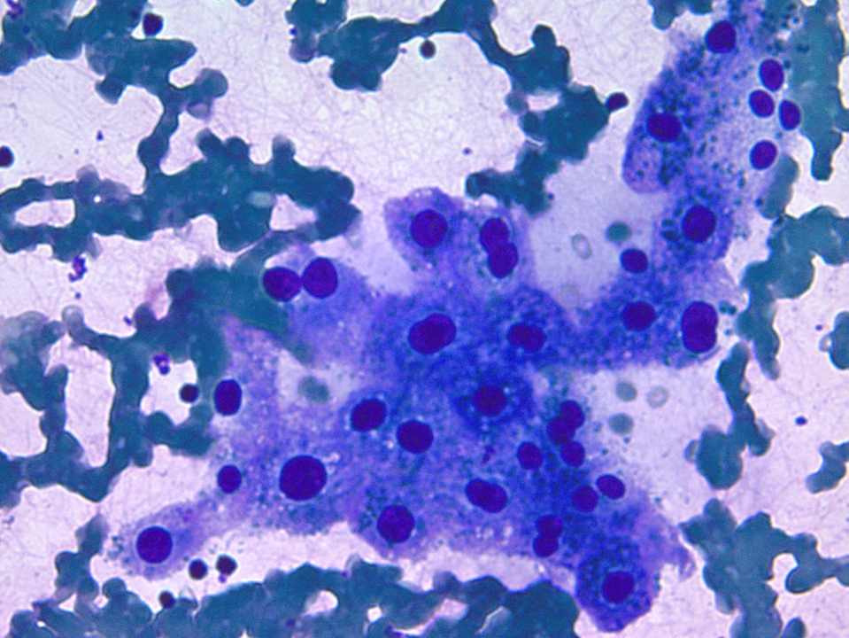
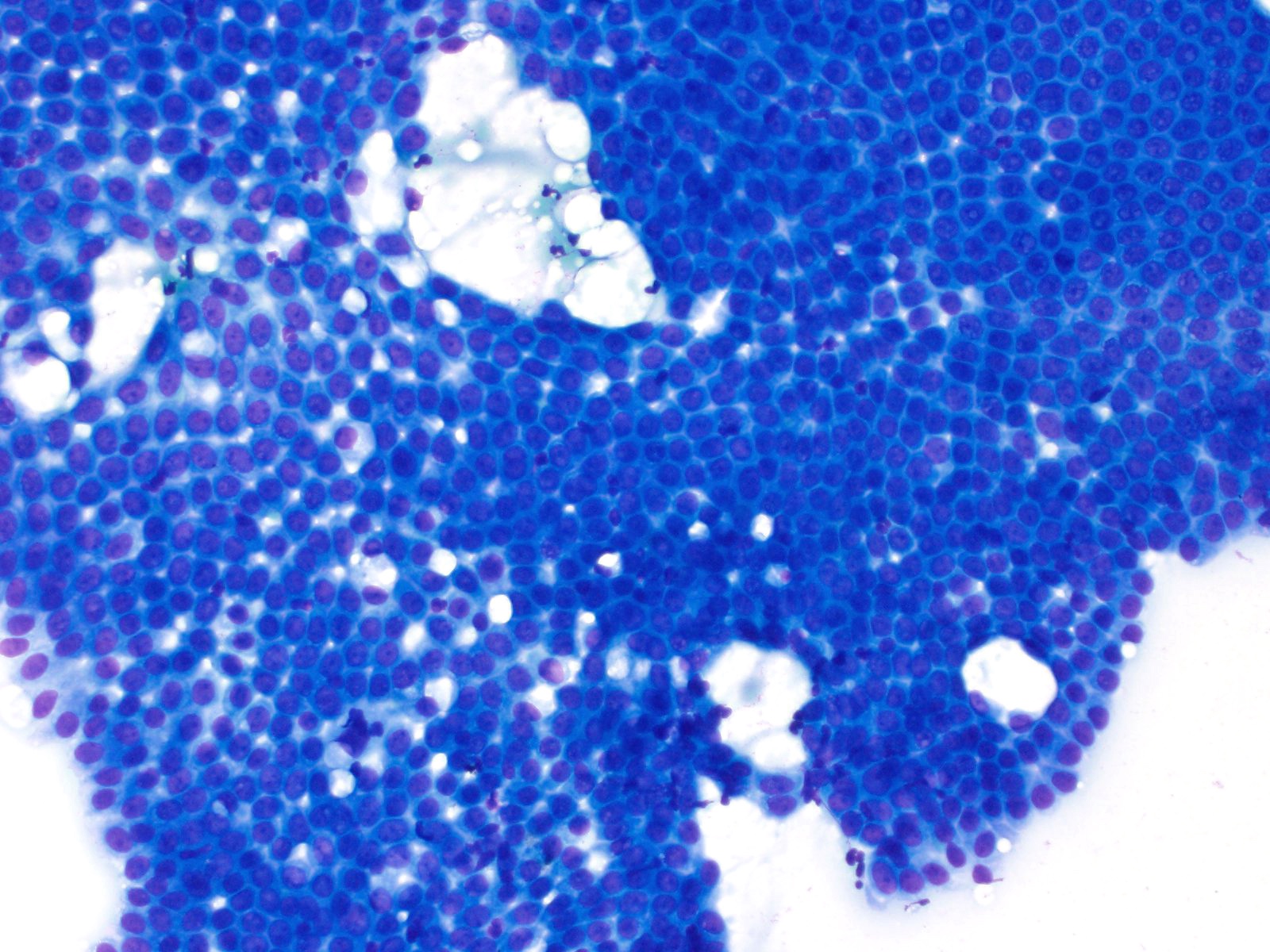
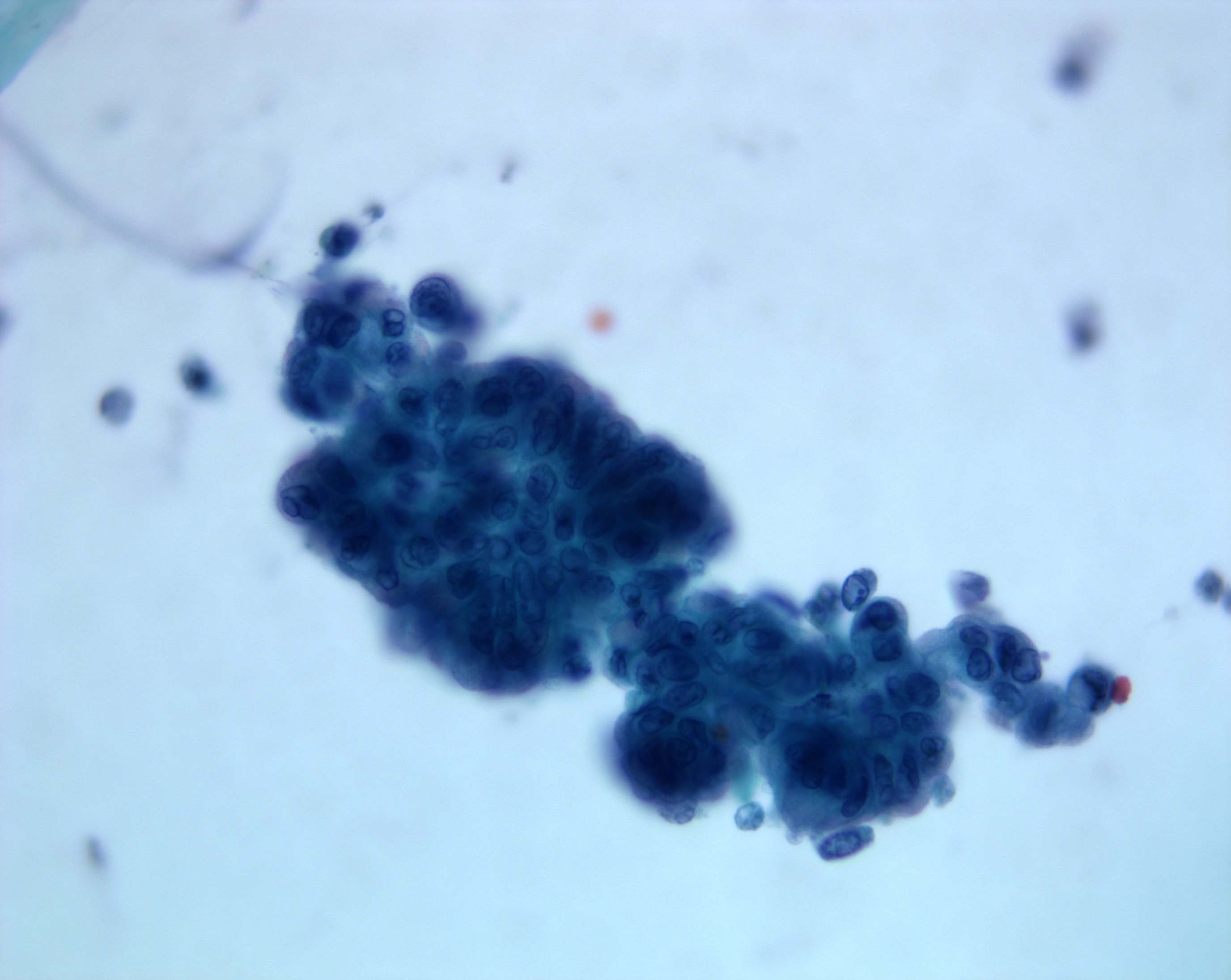
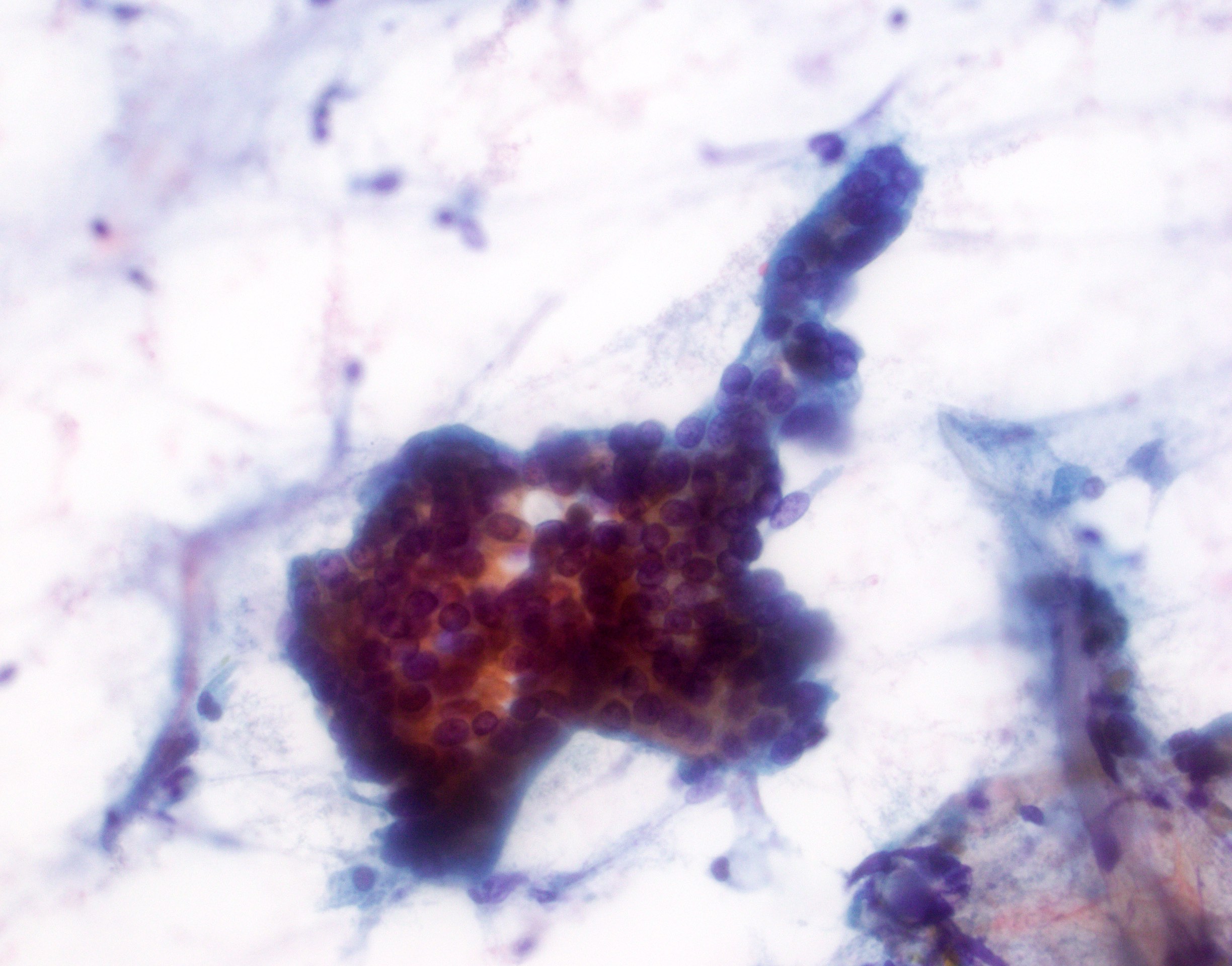
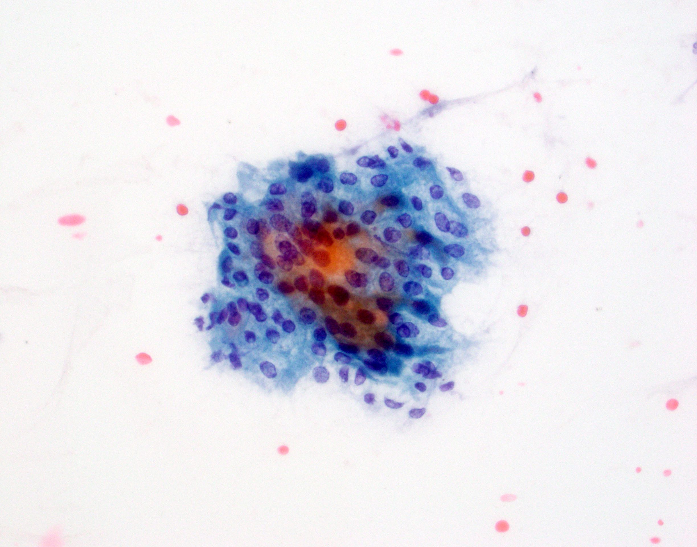
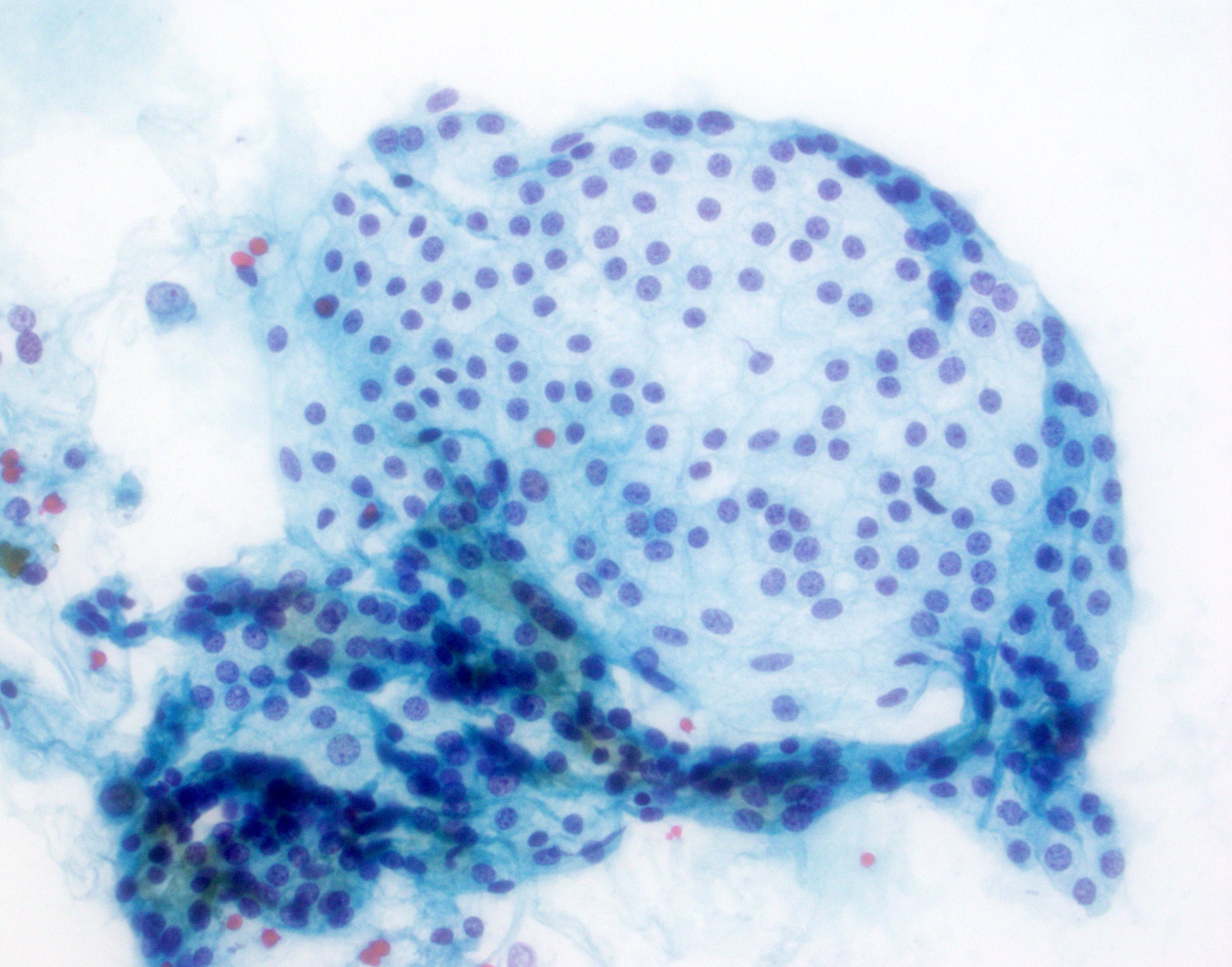
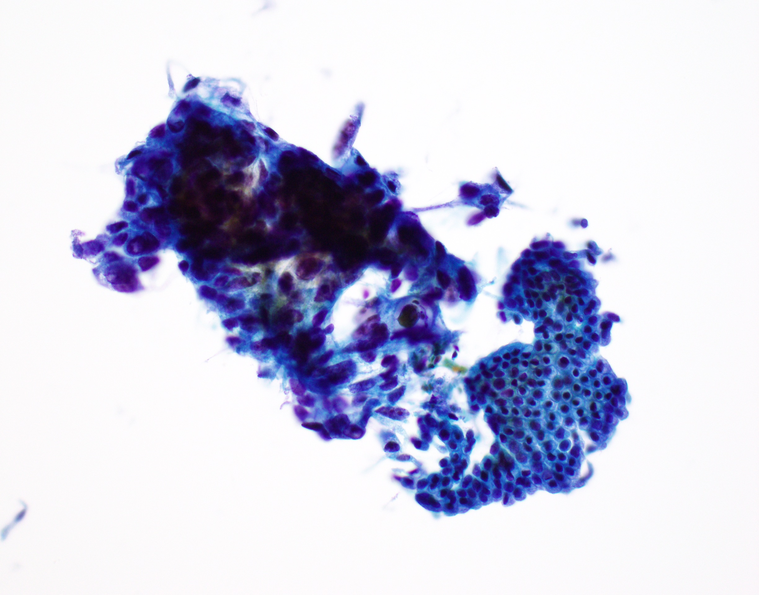
Plasmacytoid cells on FNA

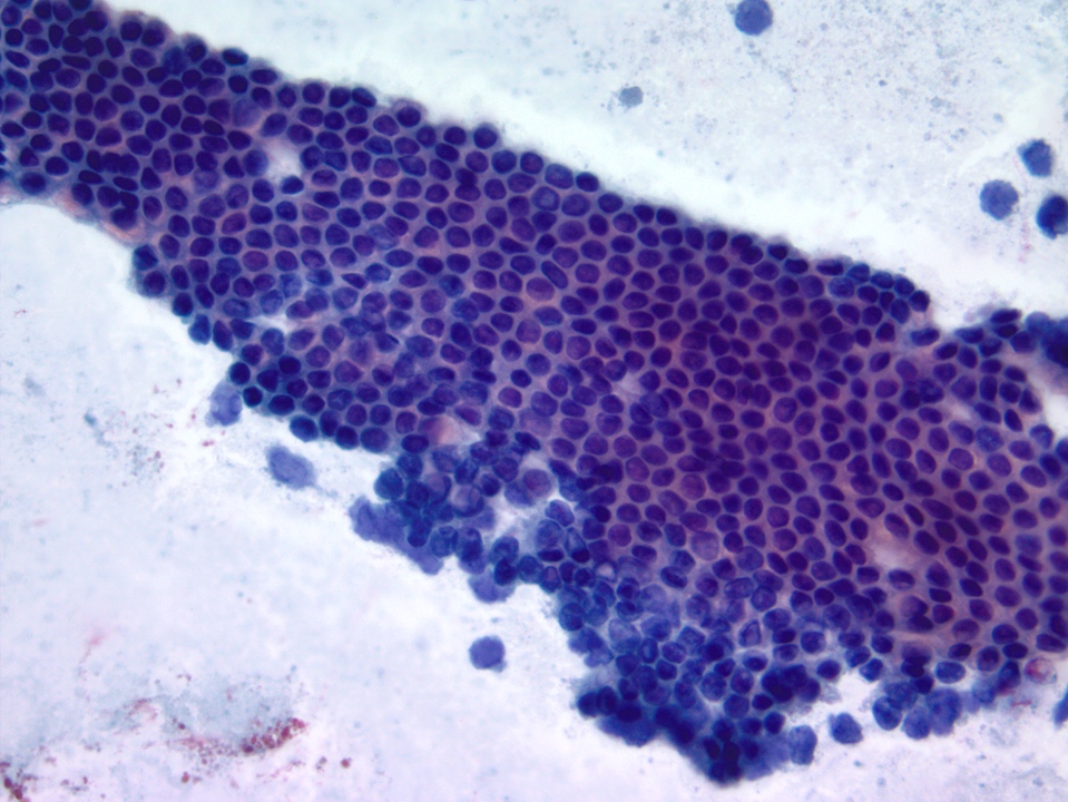
Conventional smear
Images hosted on other servers:

At autopsy, pancreas
is mucoid and
slightly smaller
than normal
Histopathology pancreas: cystic fibrosis
Contributed by Maria Luisa C. Policarpio-Nicolas, M.D.
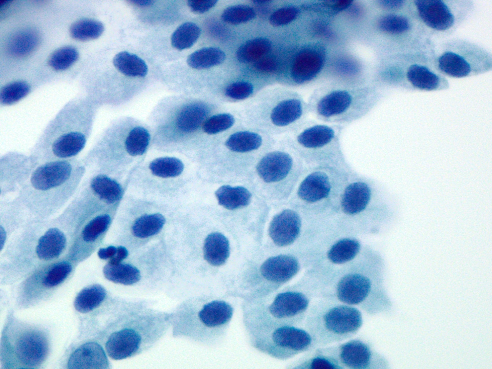
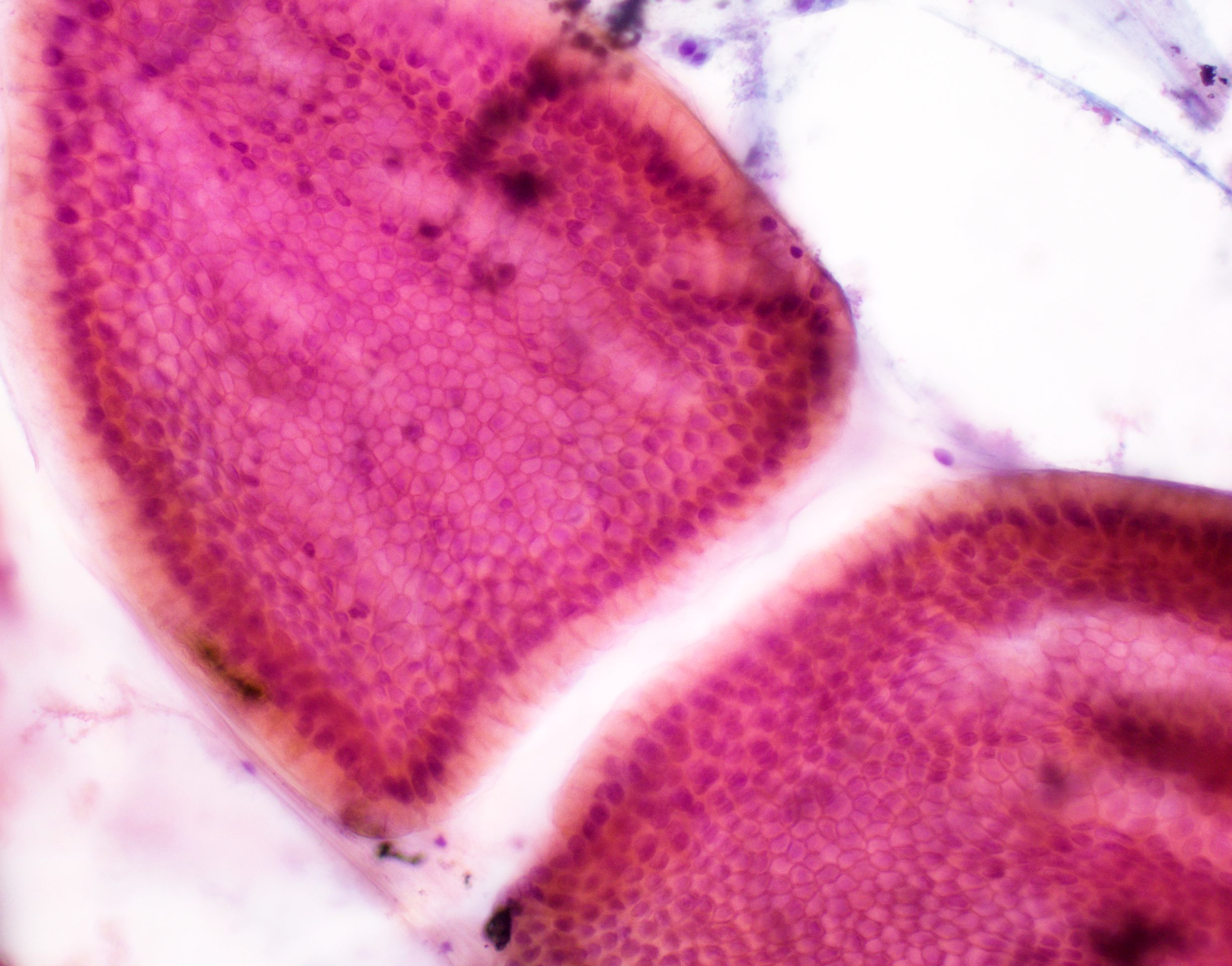
Ductal cells with bland nuclei
Ductal cells with tubule formation
Ductal epithelium cell block
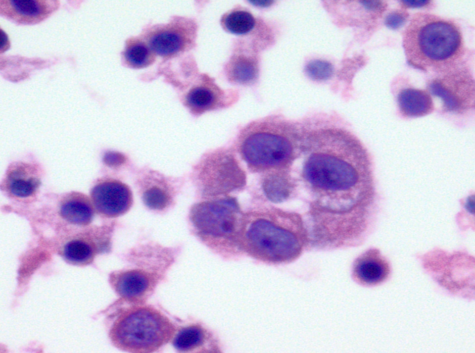
Clusters of acinar cells
Acini in grape-like arrangement
Acinar and ductal cells histology

Gastric epithelium
Duodenal epithelium with goblet cells
Hepatocytes with intracytoplasmic pigment
Hepatocytes with bile
Mesothelial cells in sheets
Mesothelial cells with intercellular windows

Histopathology pancreas: type 2 diabetes mellitus
Images hosted on other servers:

Axial CT pancreas
Contributed by Wei Chen, M.D., Ph.D.
Arising in IPMN
Arising in high grade PanIN
Contributed by Wei Chen, M.D., Ph.D.
Anisonucleosis
Glands next to vessel
Perineural invasion
Gland in adipose tissue
Moderately differentiated
Poorly differentiated
Foamy gland pattern
Vacuolated pattern
Clear cell pattern
Large duct pattern
Post therapy tumor bed
Post therapy eosinophilic change
Post therapy dystrophic tumor glands
Perineural residual tumor glands
Post therapy vascular change
Contributed by Regina Plummer, D.O., Victoria Saksenberg, M.D. and AFIP images

Abundant inspissated mucin
Transition into drunken honeycomb
Anisonucleosis (4:1 variation)
Signet ring cells
Moderately
differentiated
ductal
adenocarcinoma
AFIP images
Well differentiated
Images hosted on other servers:

CT of pancreatic gastrinoma
AFIP images
Lymph node gastrinoma
Contributed by Raul S. Gonzalez, M.D. and AFIP images
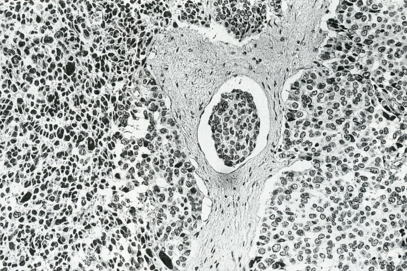
Nests and cords of cells

Malignant tumor has trabecular pattern
Lobular trabecular pattern
AFIP images
Pancreatic gastrinoma
Images hosted on other servers:

CT

MRI

Liver metastases
AFIP images
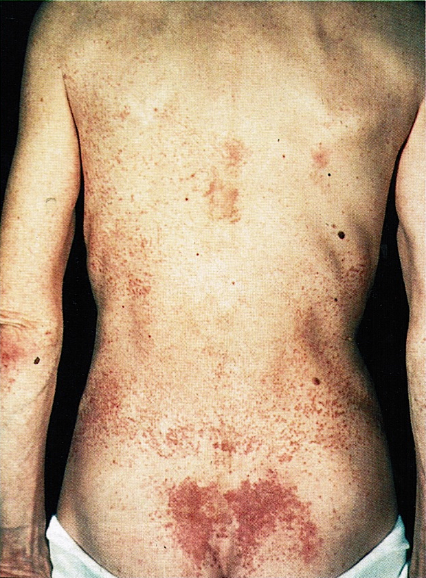
Necrolytic migratory erythema
Images hosted on other servers:


Necrolytic migratory erythema
Images hosted on other servers:

H&E and immunostains

H&E and glucagon

Liver metastasis

Chromogranin
Contributed by Olca Basturk, M.D.
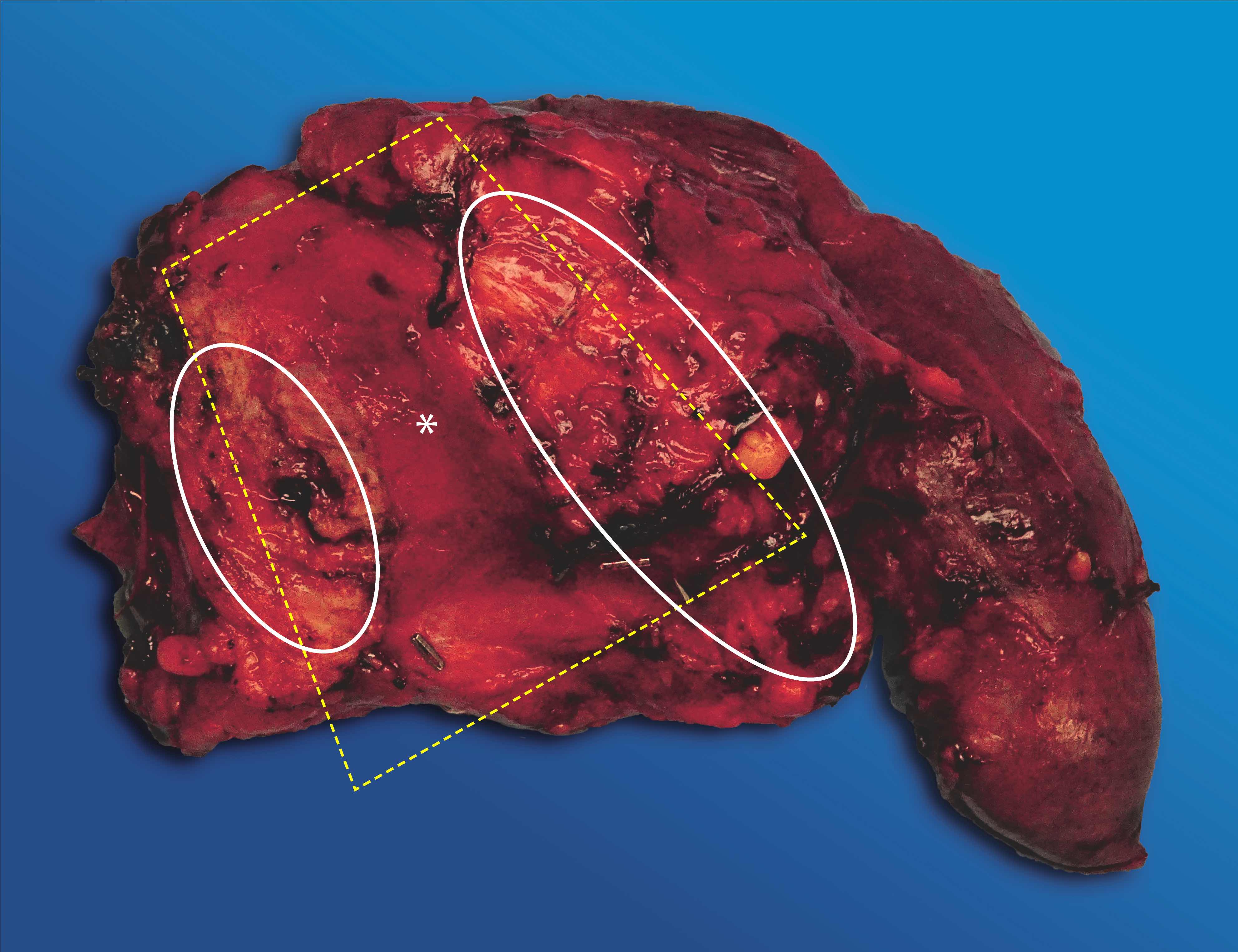
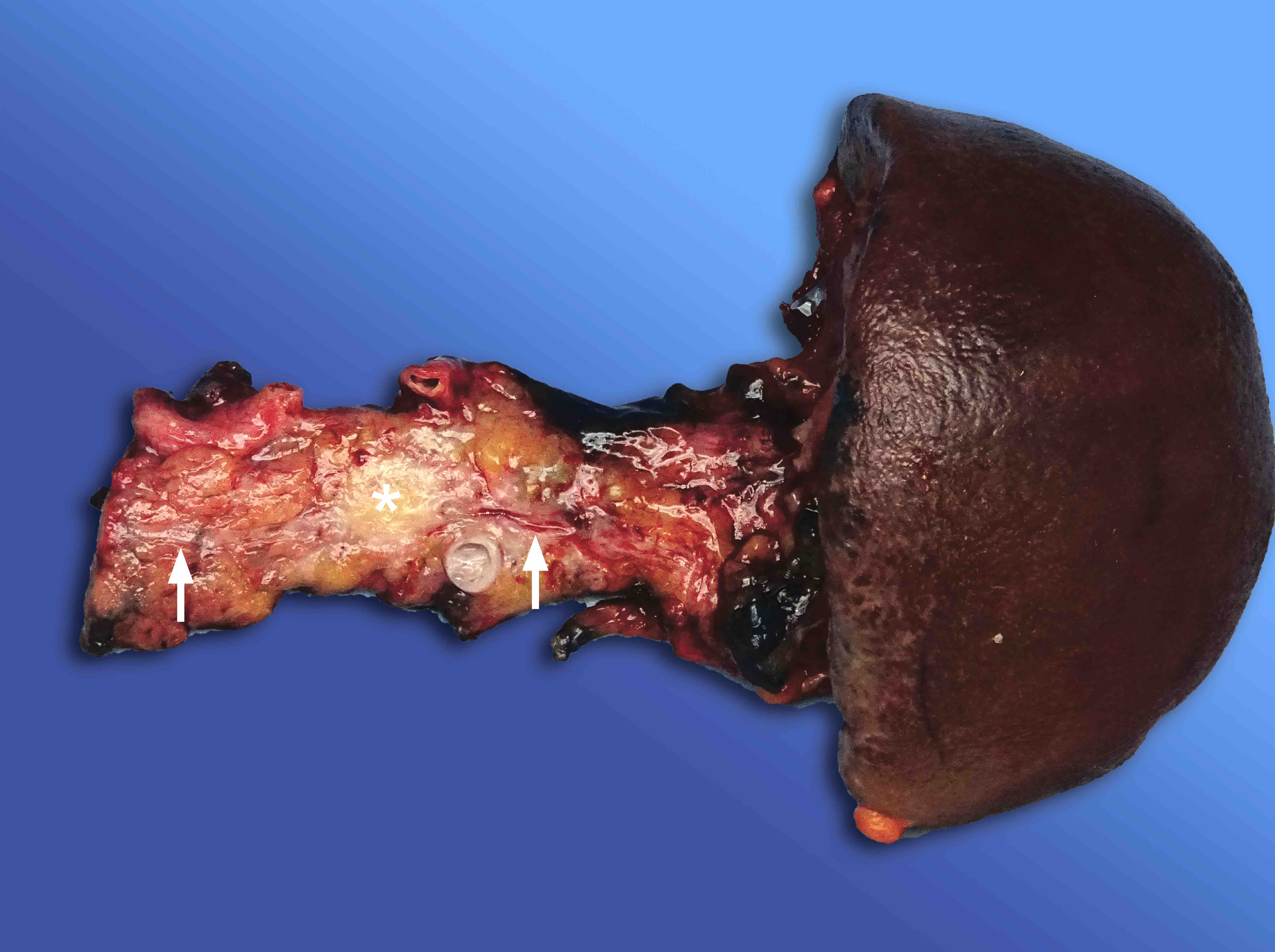
Trapezoid and important structures
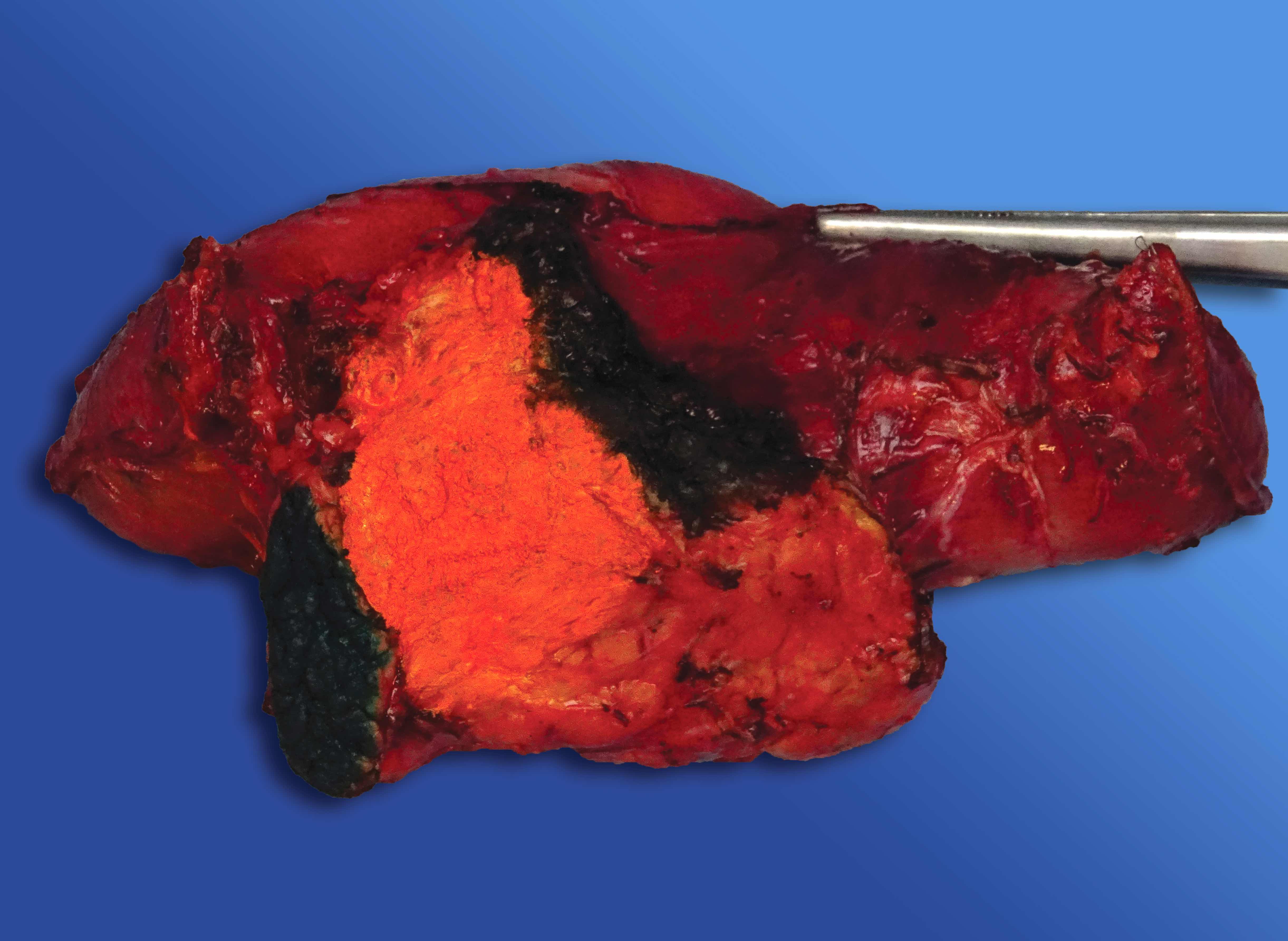
Inked margins and vascular groove
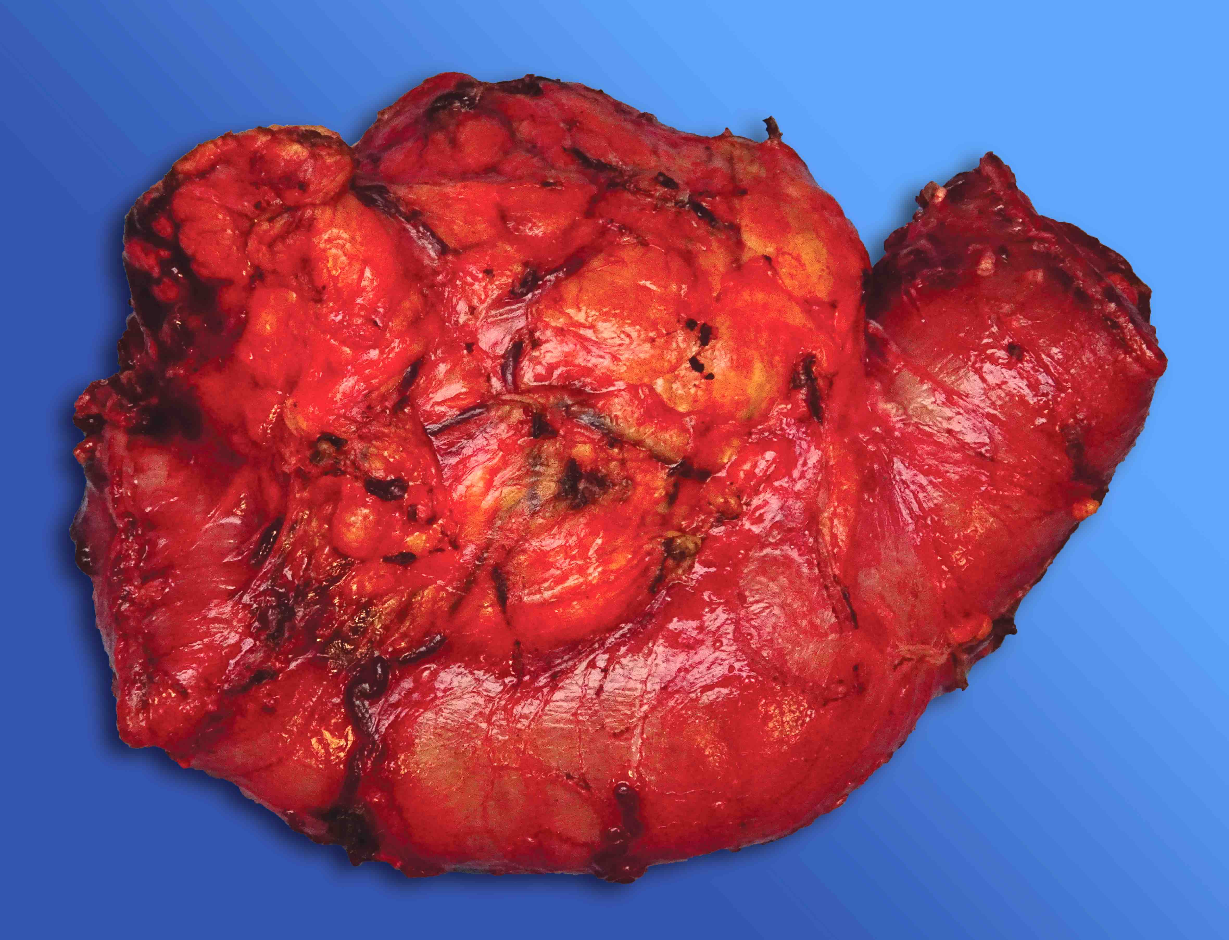
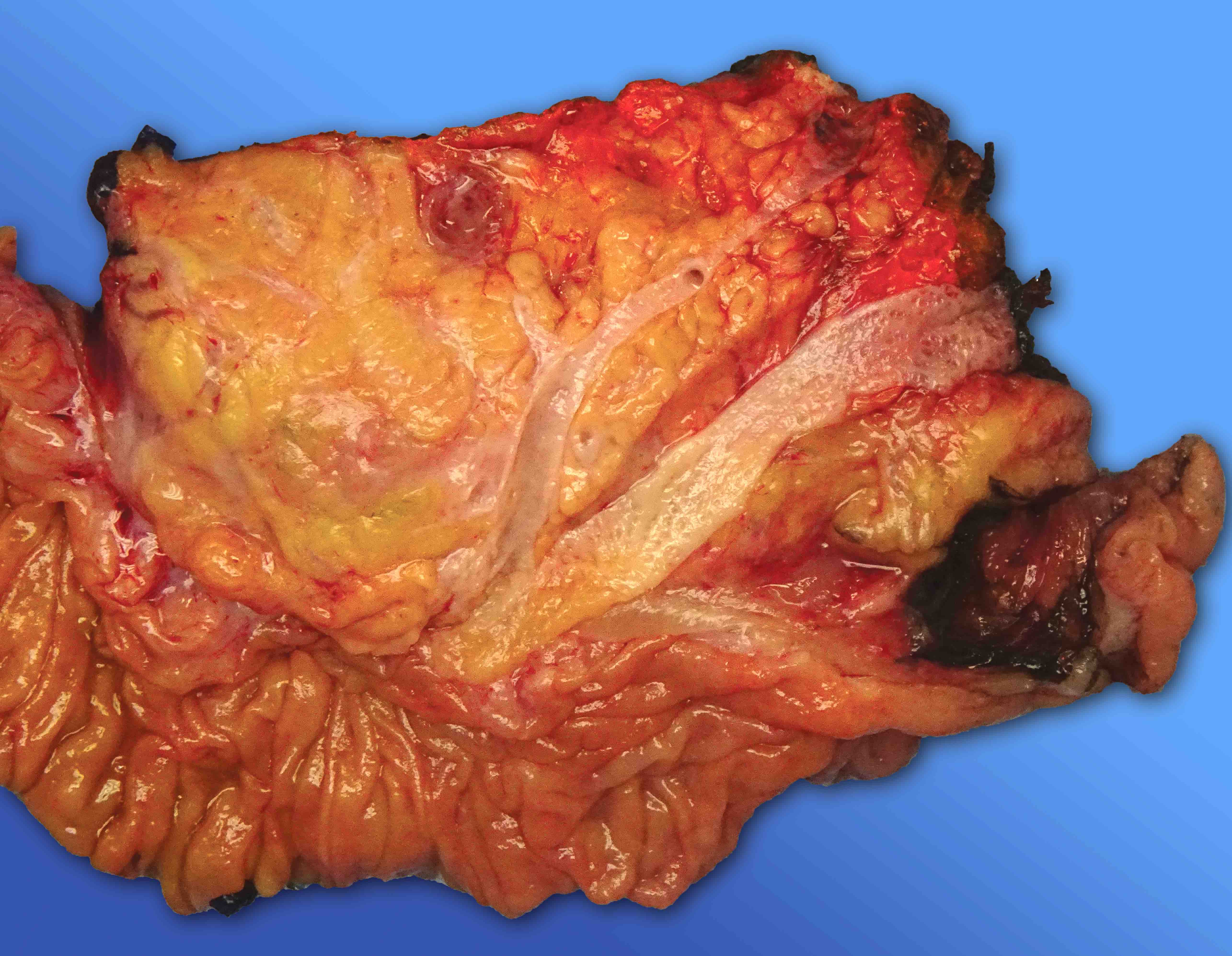
Anterior free surface
Posterior free surface
Common bile duct margin
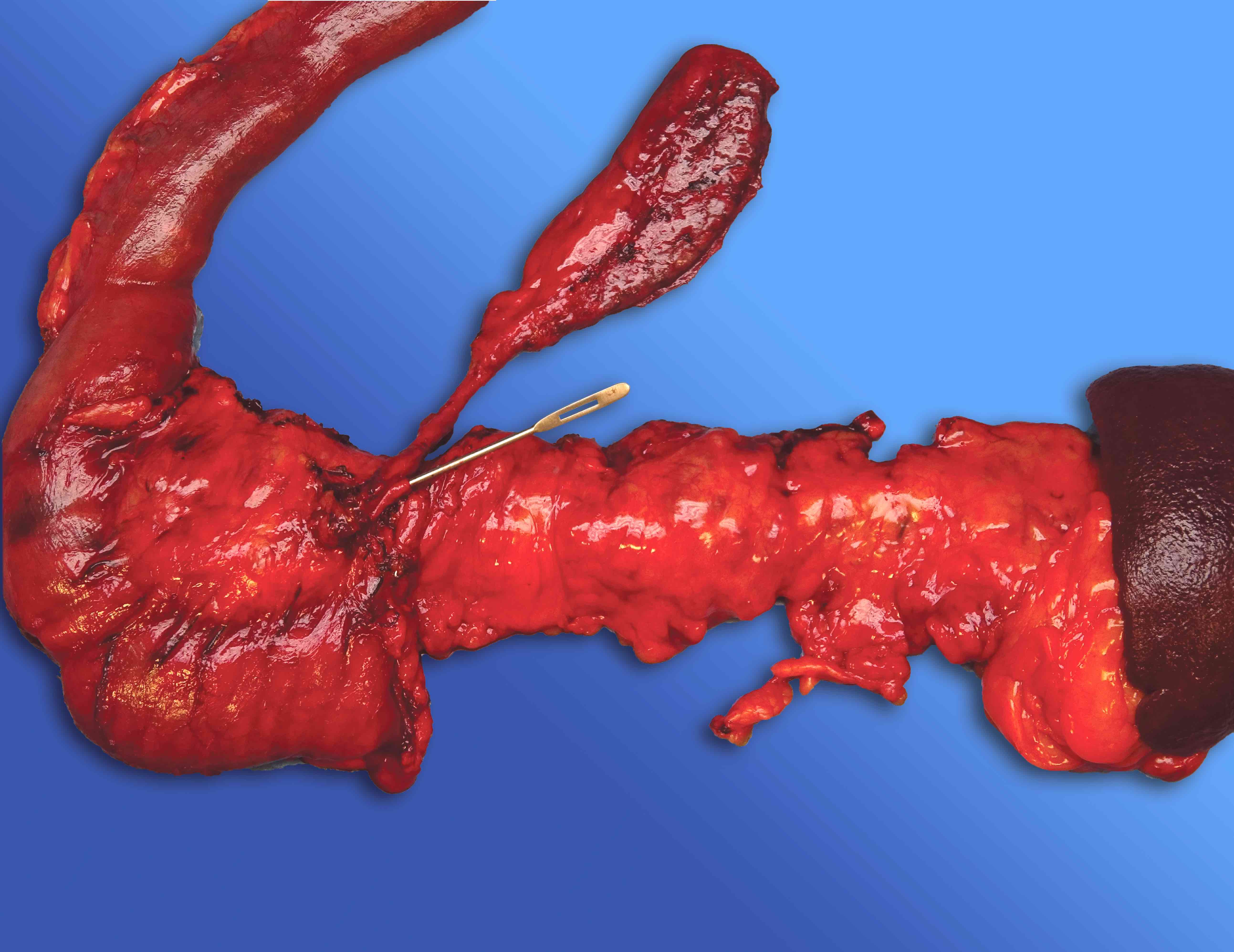
Pancreatic neck / duct margin
Uncinate margin
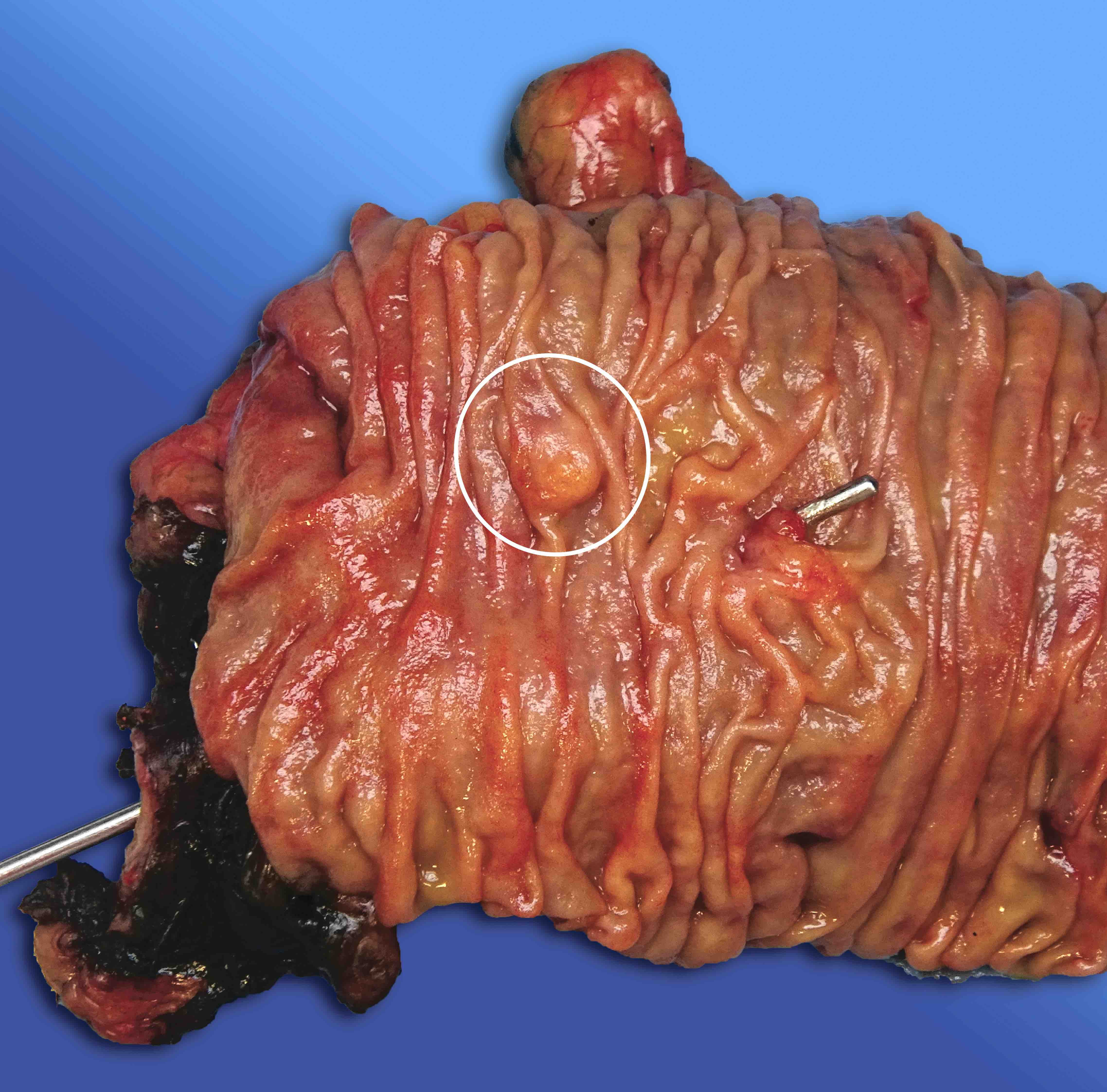
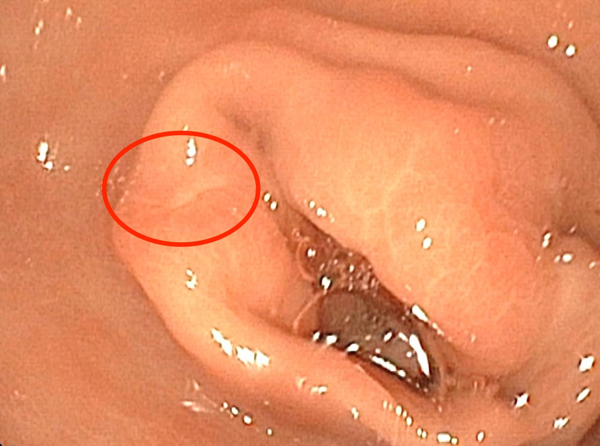
Minor (accessory) ampulla
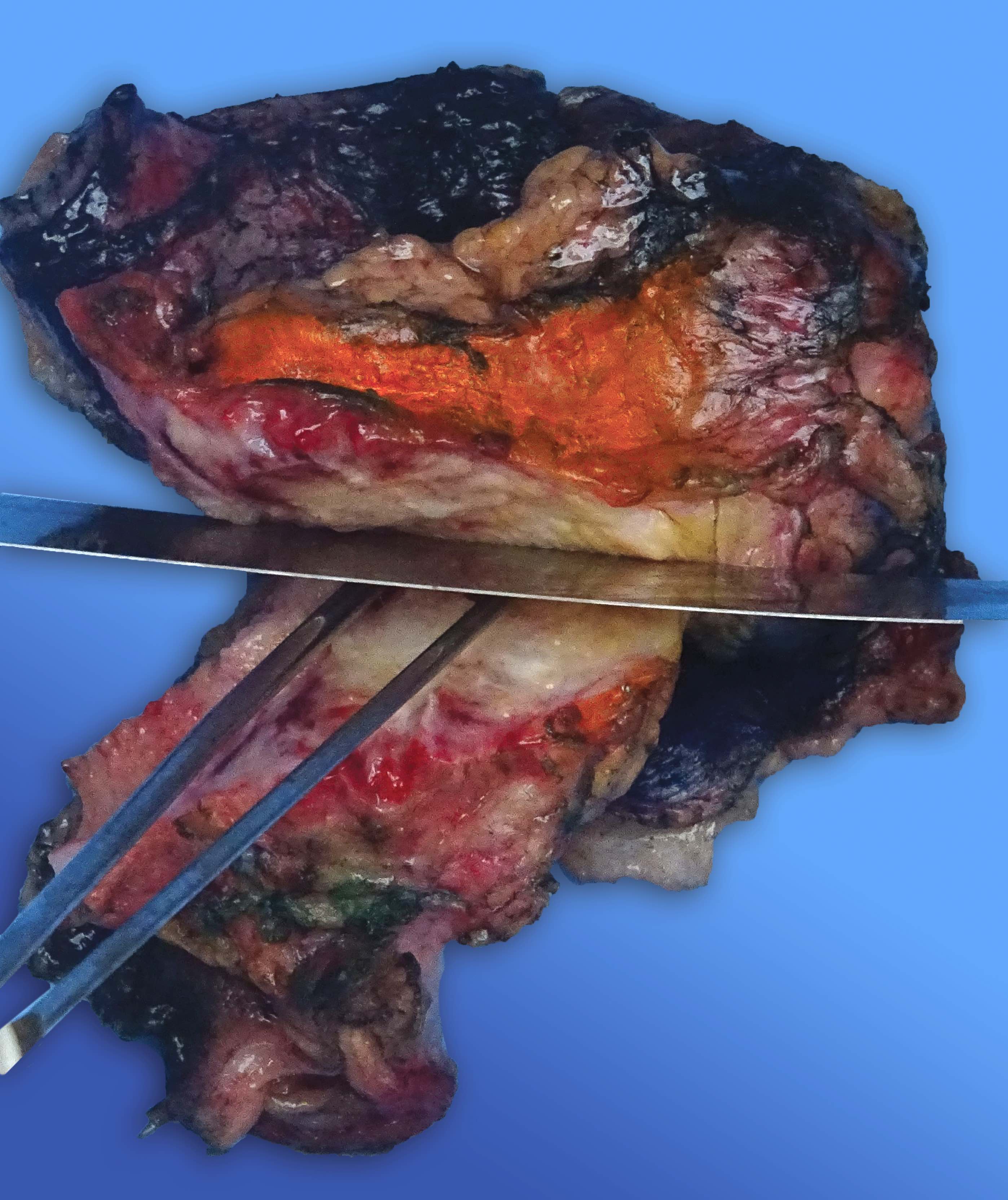
Bivalving and sectioning
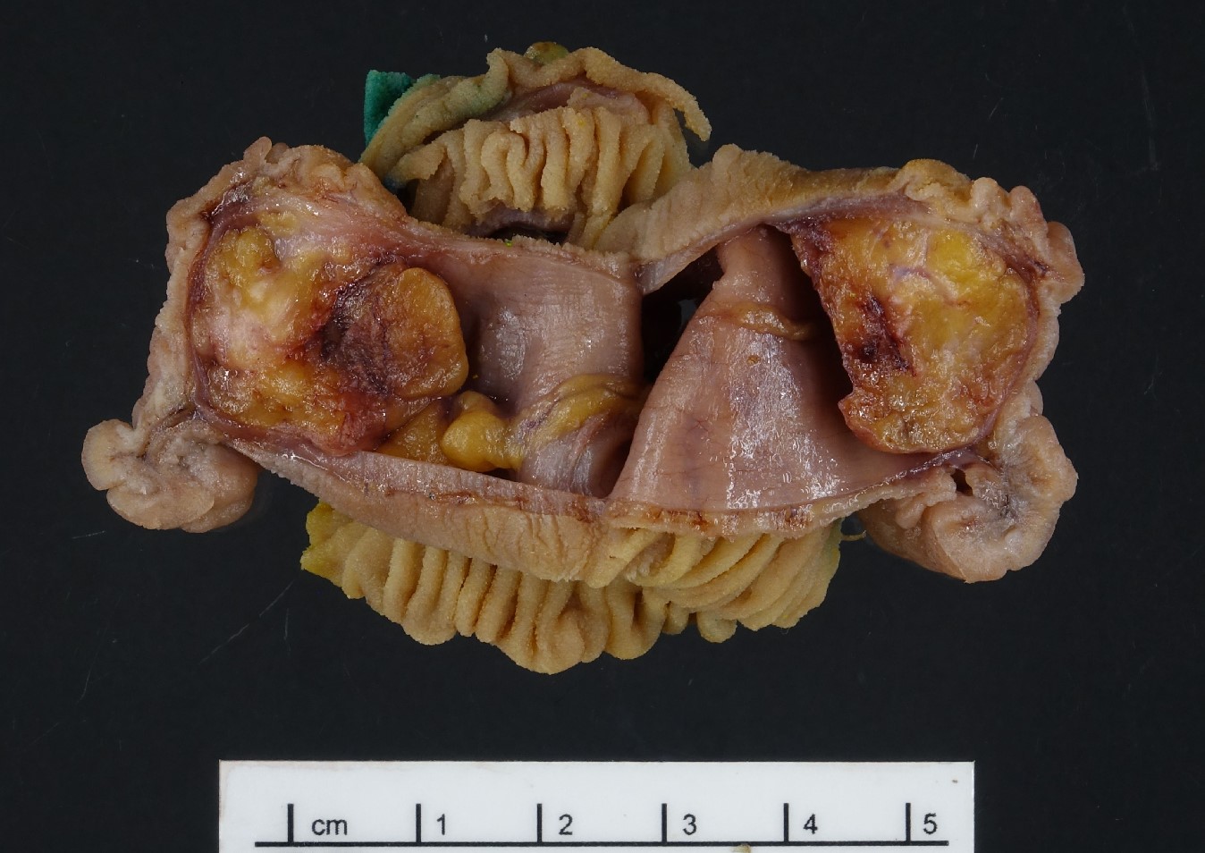
Cut surface of the pancreatic head
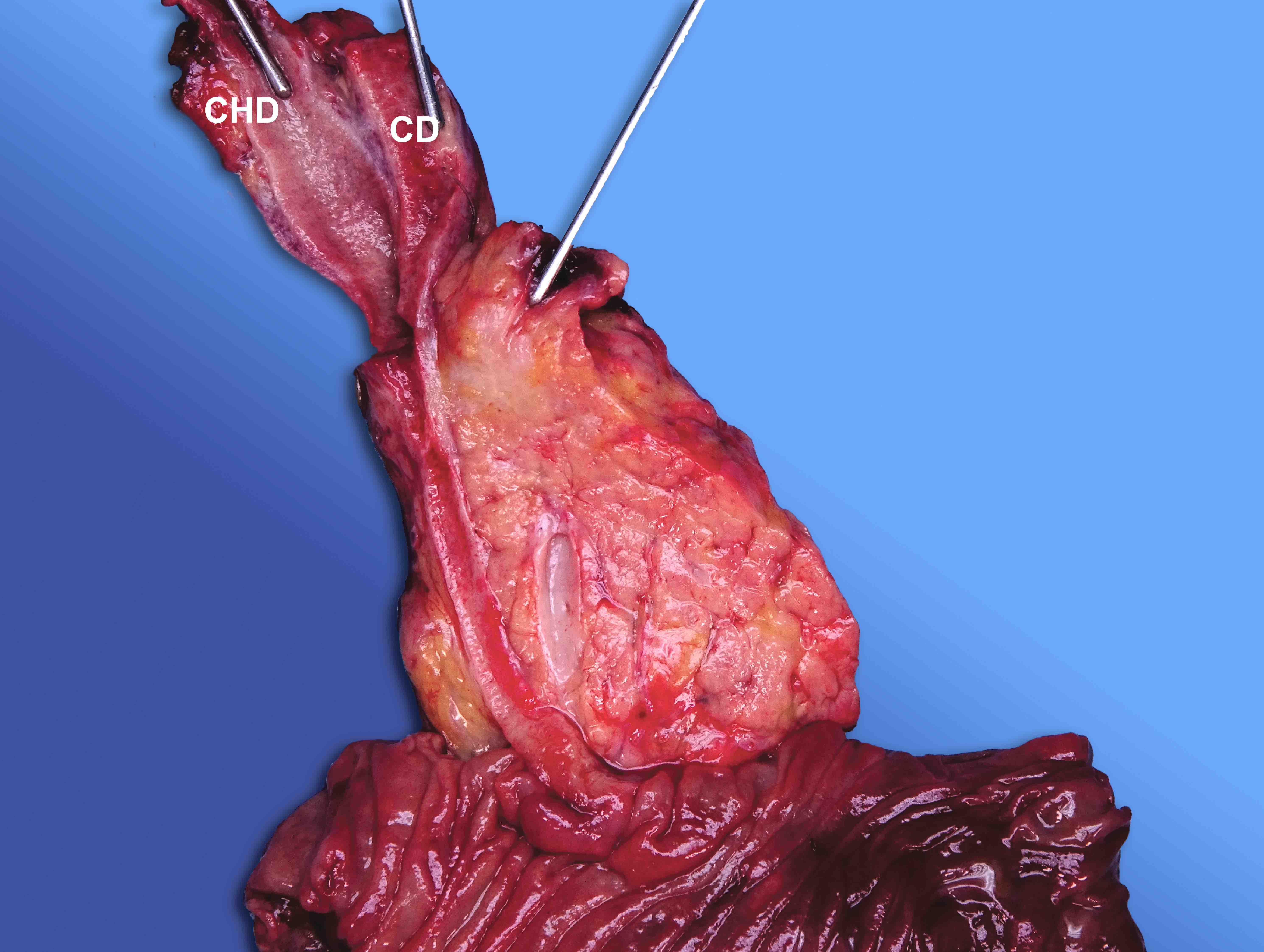
Low union
Peripancreatic lymph nodes
Lymph node dissection (orange peeling)
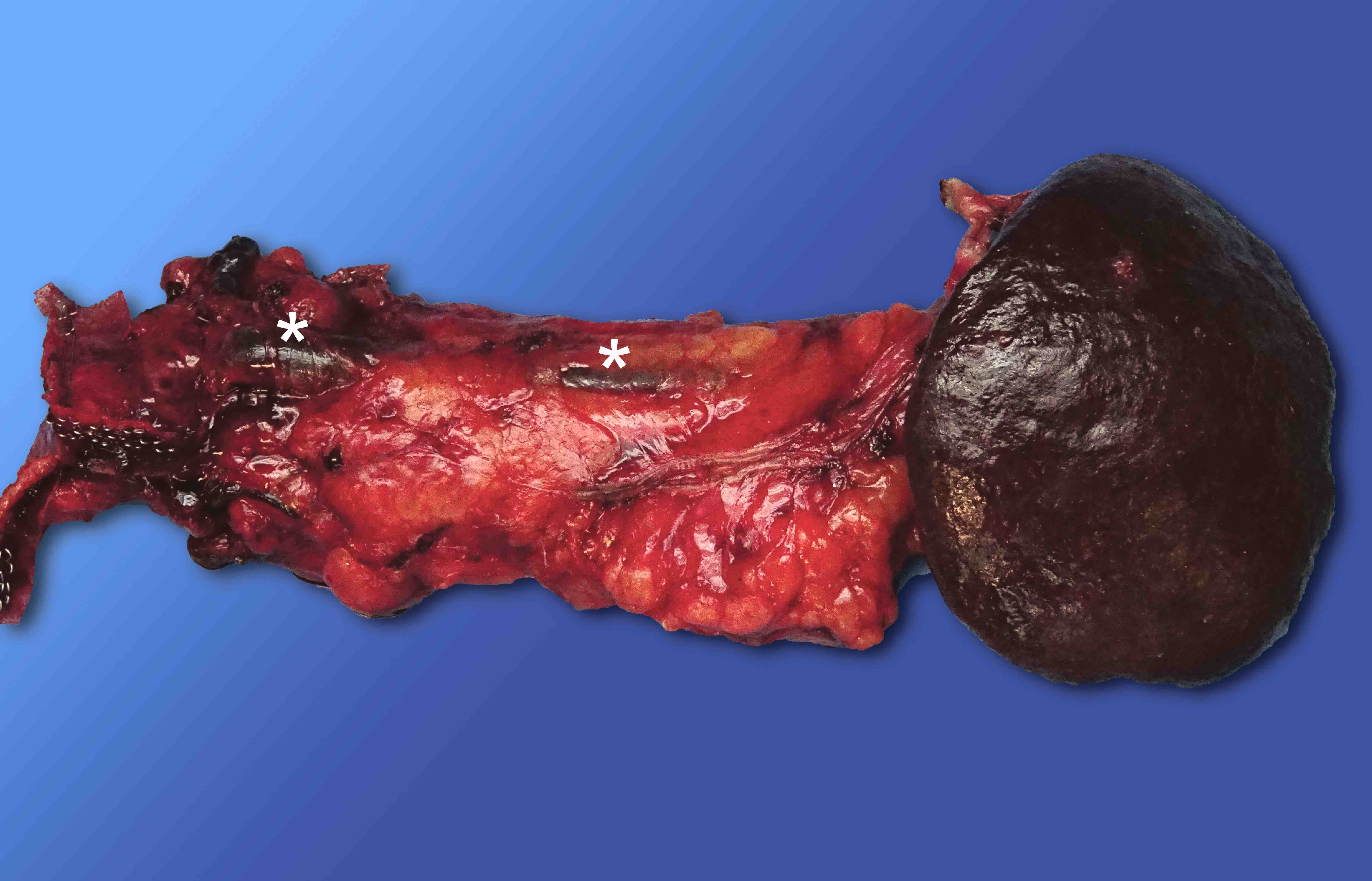
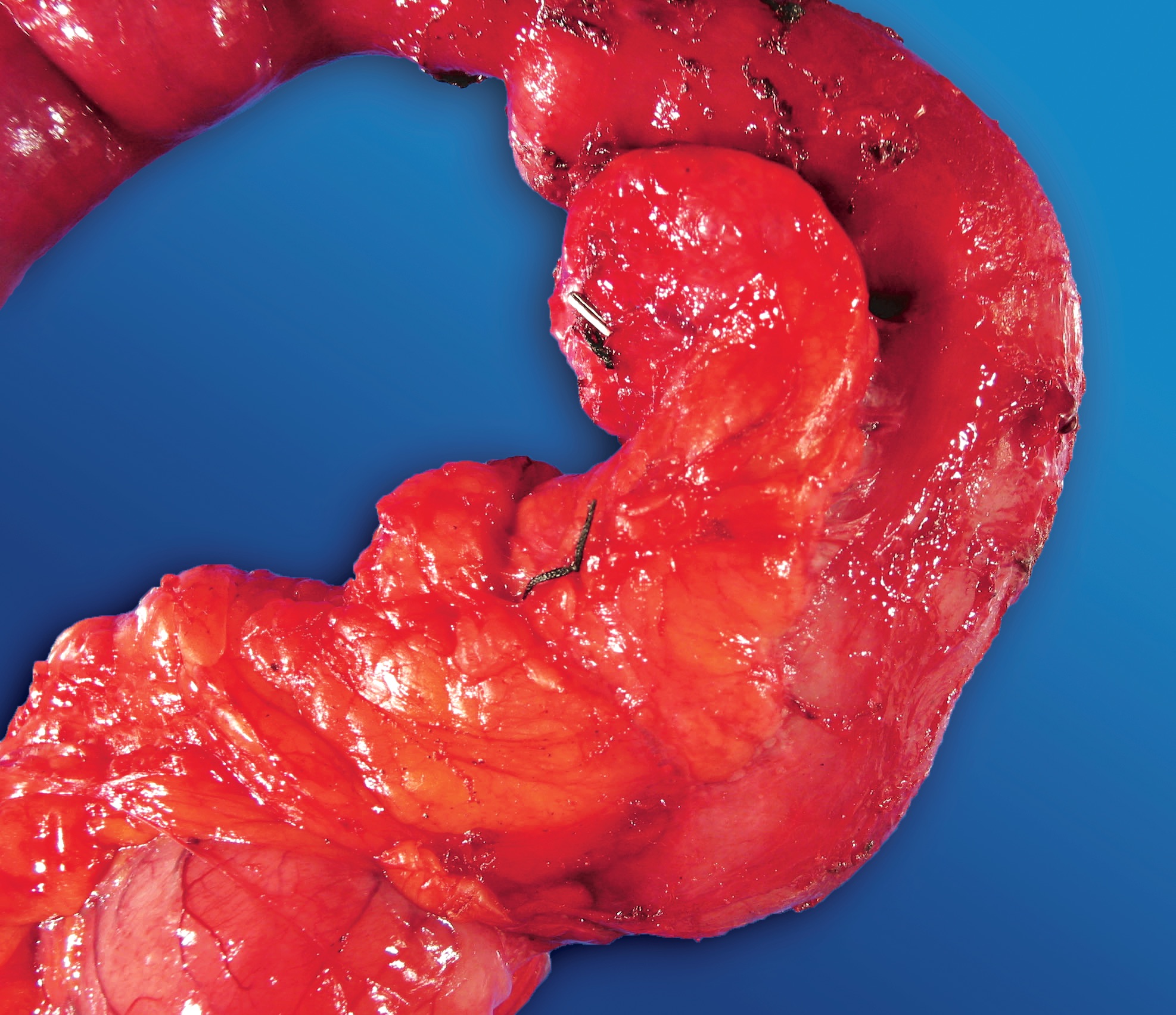
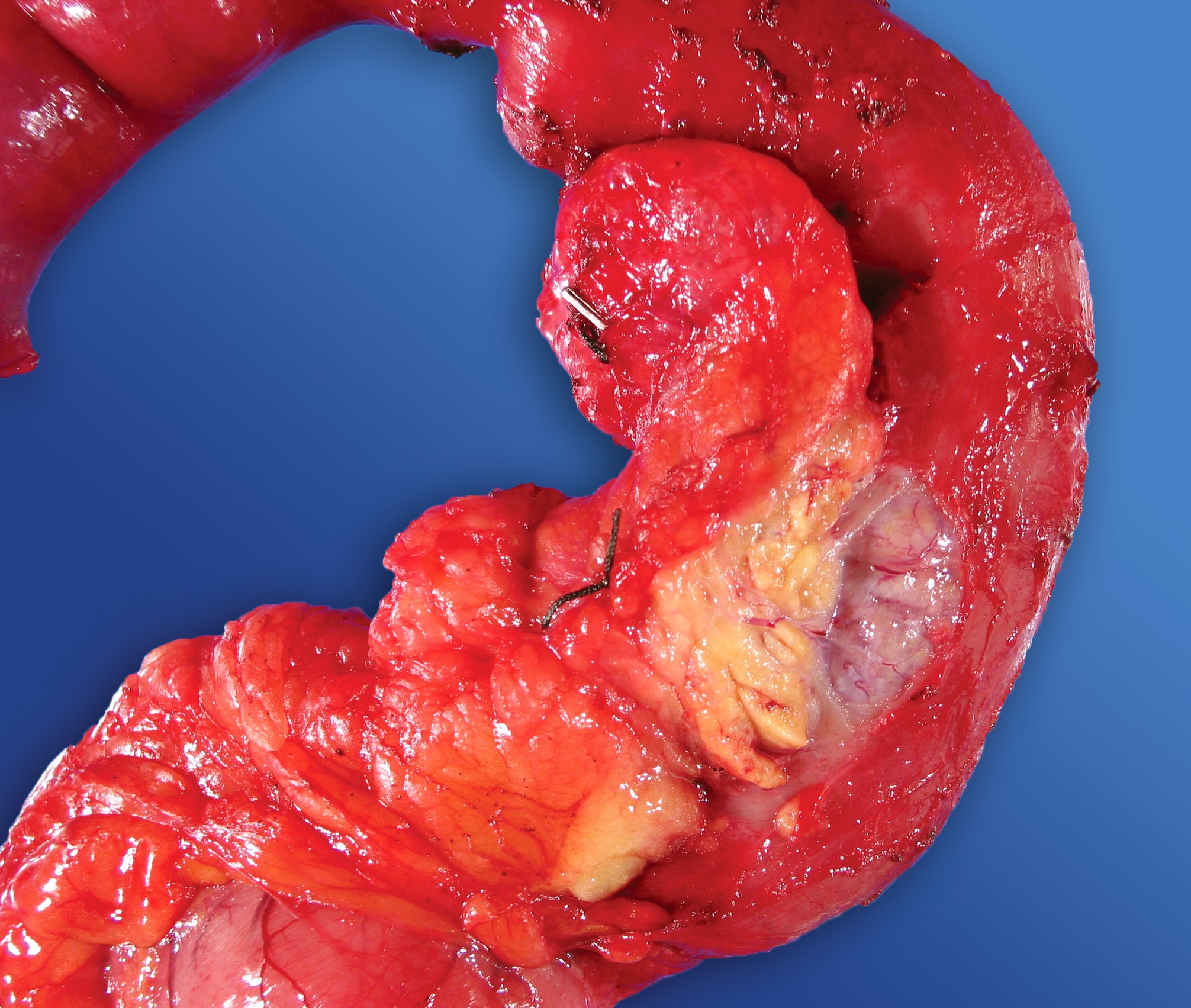
Anterior surface of distal pancreatectomy
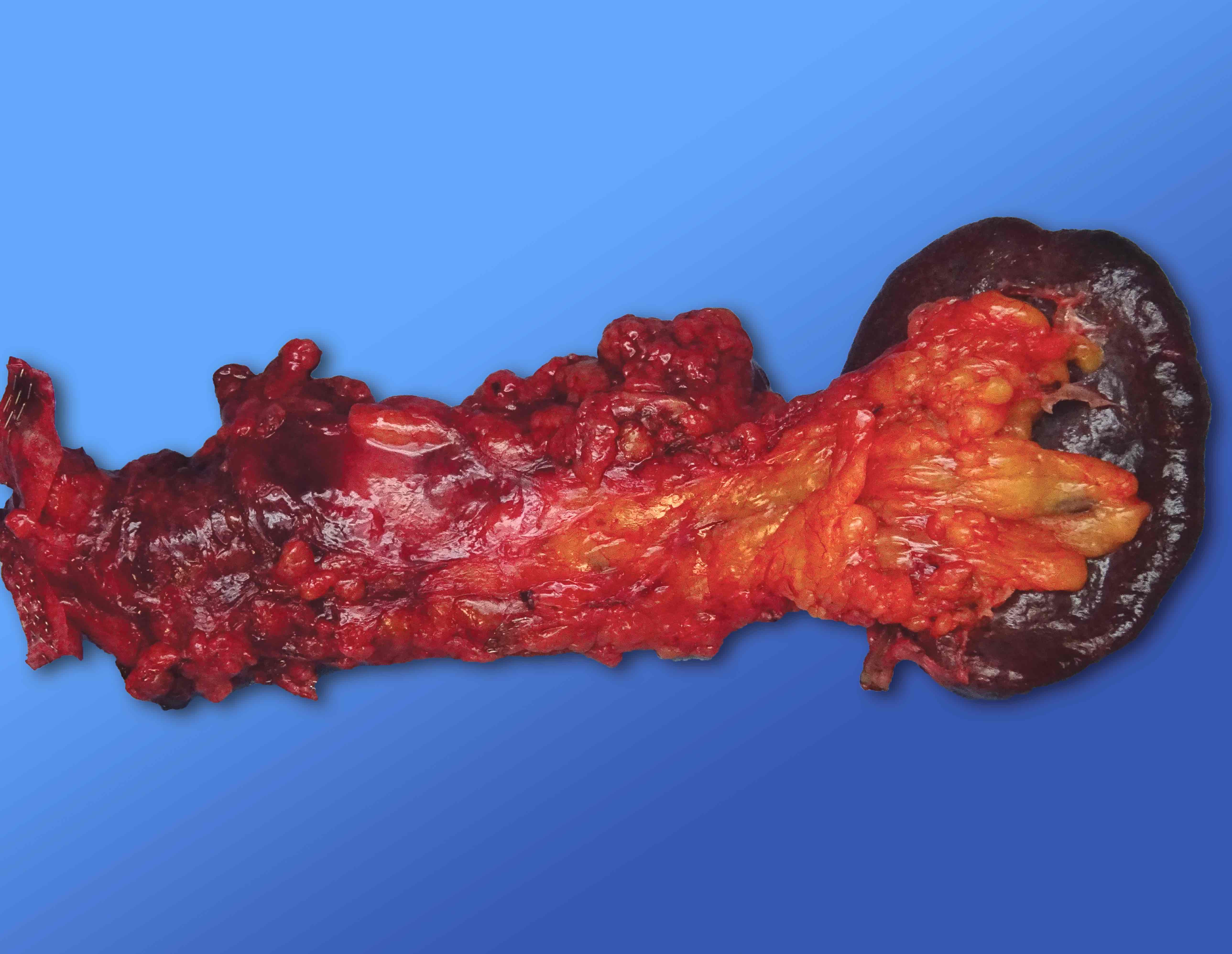
Posterior surface of distal pancreatectomy

Serially sectioned distal pancreatectomy
Bivalved distal pancreatectomy
Total pancreatectomy
Images hosted on other servers:

Gastric submucosal mass

Duodenal and gastric lesions

MRI lesion in duodenum
Contributed by Kenechukwu Ojukwu, M.D., M.P.P. and Stephanie Dreikorn, M.D.
Endoscopy, subtle ectopic pancreas
Images hosted on other servers:

Single balloon
enteroscopy
showing
submucosal lesion
Contributed by Kenechukwu Ojukwu, M.D., M.P.P. and Danielle Hutchings, M.D.
Heterotopic pancreas in Meckel diverticulum
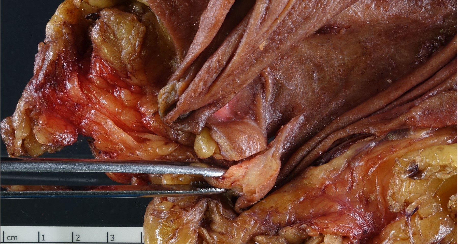
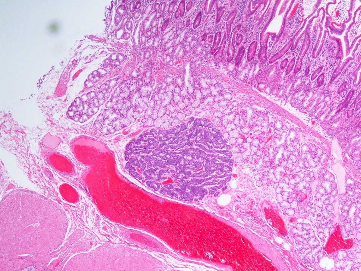
Heterotopic pancreas in stomach
Contributed by Kenechukwu Ojukwu, M.D., M.P.P. and Danielle Hutchings, M.D.
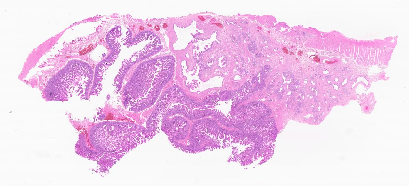
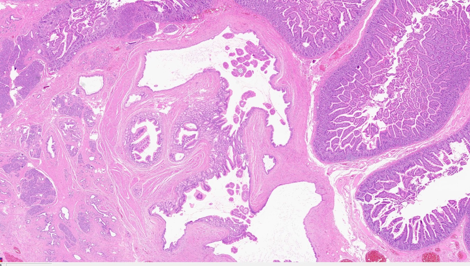
Pancreatic heterotopia in the stomach
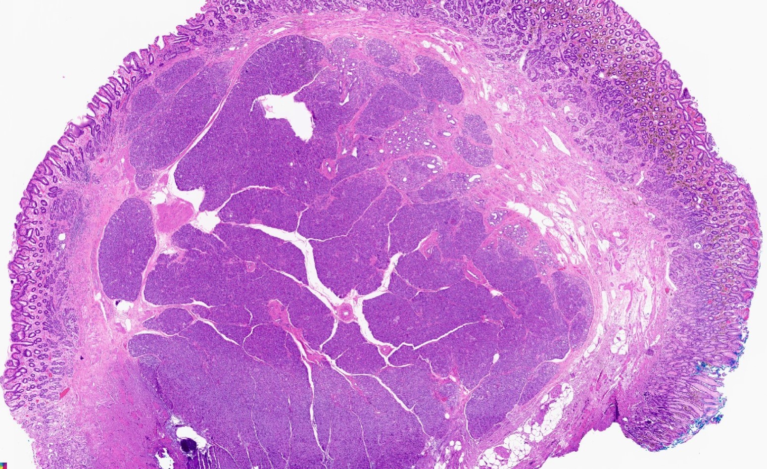
Submucosal lesion, lobular architecture
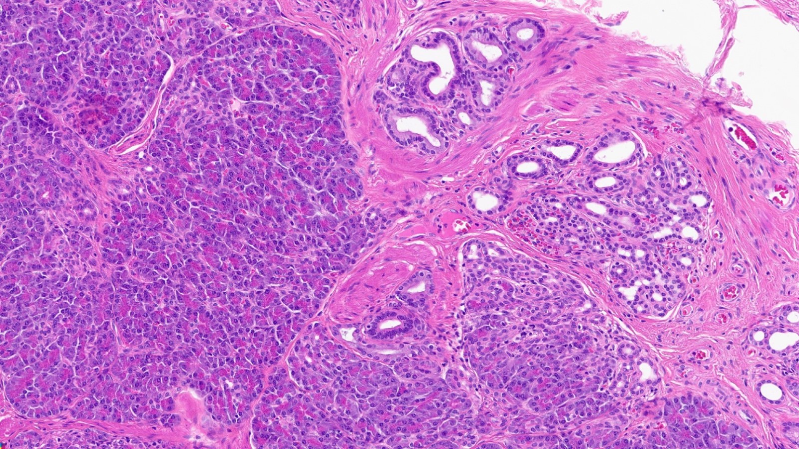
Pancreatic acini and ducts
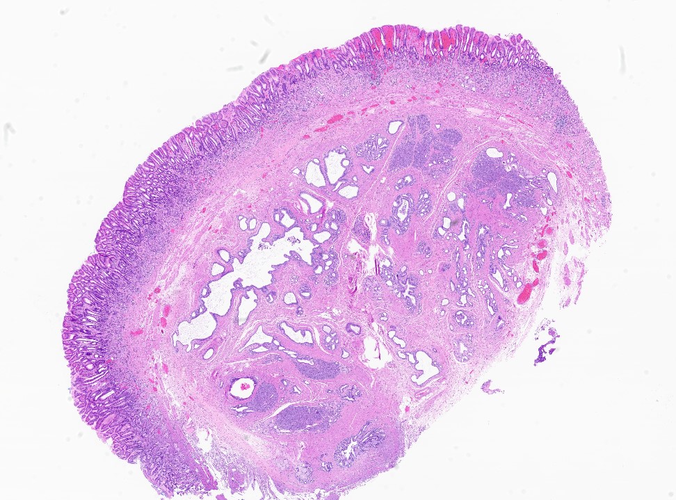
Heterotopic pancreas predominantly ducts
Branching ectopic pancreatic ducts
Ducts, acini and islets
Intraductal papillary mucinous neoplasm (IPMN)
Gastric type IPMN
IPMN, low grade dysplasia
Meckel diverticulum with heterotopia
Images hosted on other servers:

Cell block with normal pancreatic acini

Benign pancreatic
acinar cells and
ductal epithelium
(Diff-Quik)
Contributed by Irene Y. Chen, M.D.
CT abdomen
MRI abdomen
Contributed by Irene Y. Chen, M.D. and Dennis R. Dening, PA (ASCP)
Pancreatic head tumor
Pancreatic tail tumor
Contributed by Diana Agostini-Vulaj, D.O.
Nested pattern
Synaptophysin
Insulin
Contributed by @liverwei on Twitter
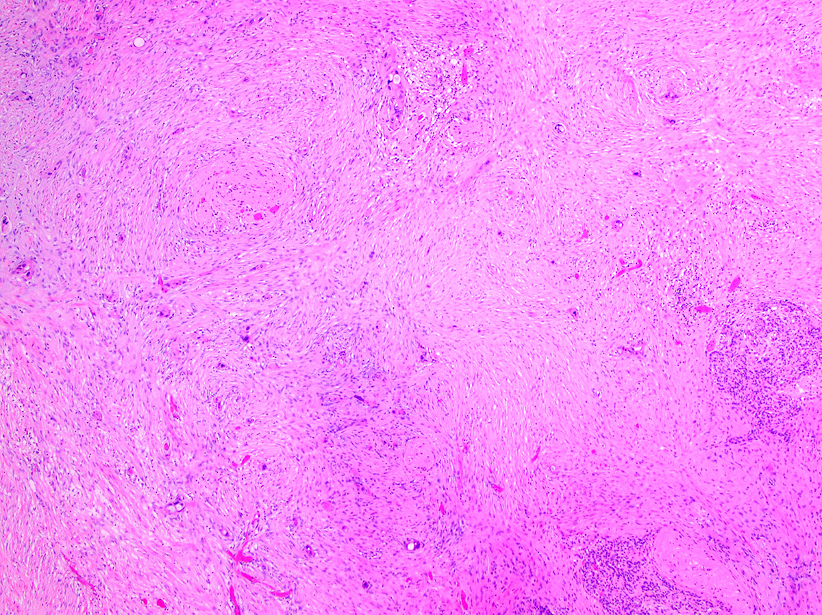
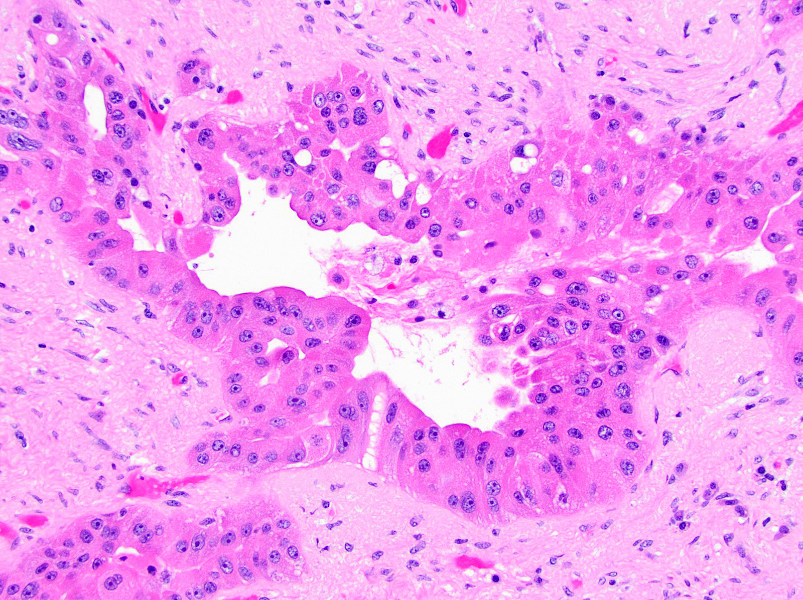
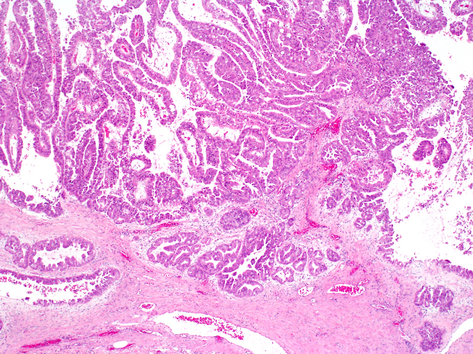
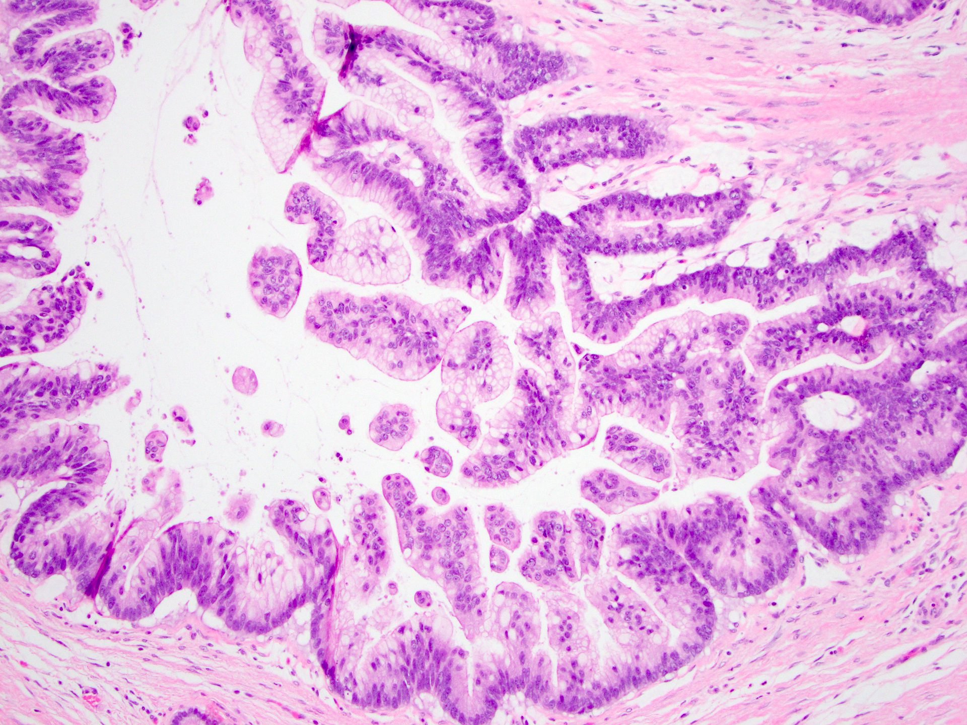
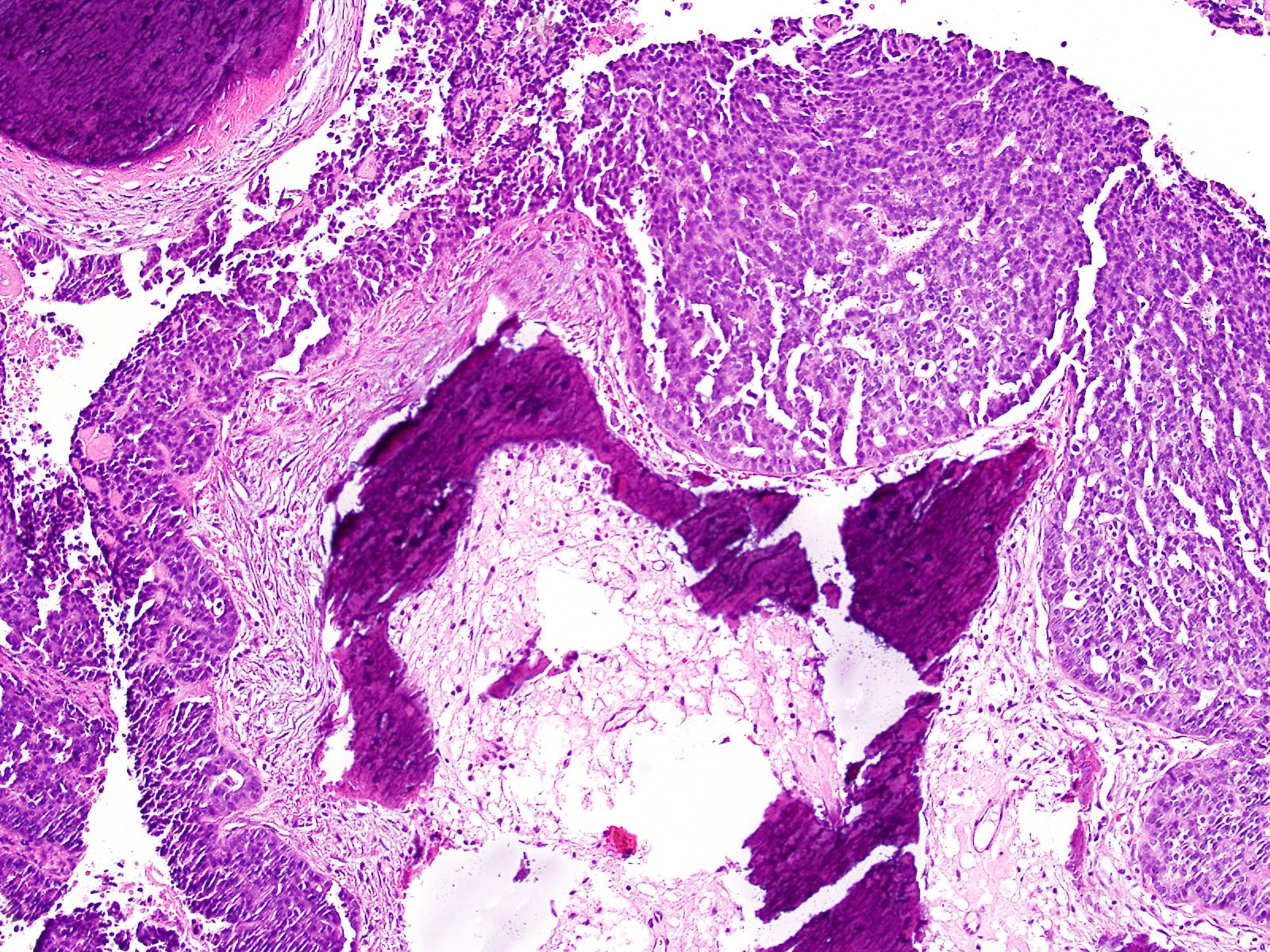
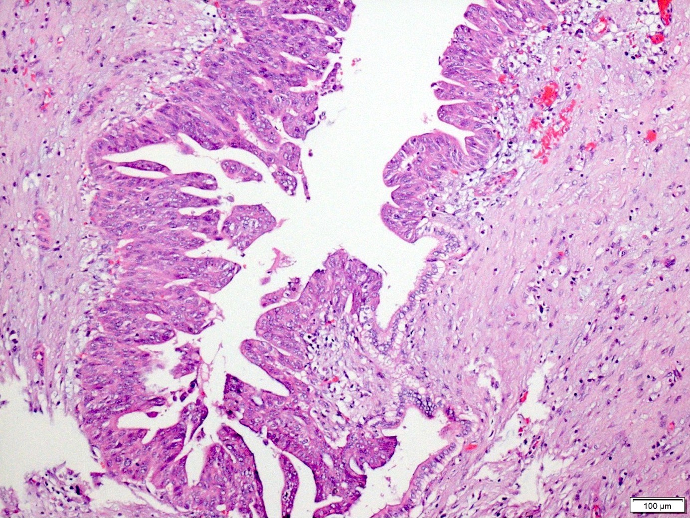
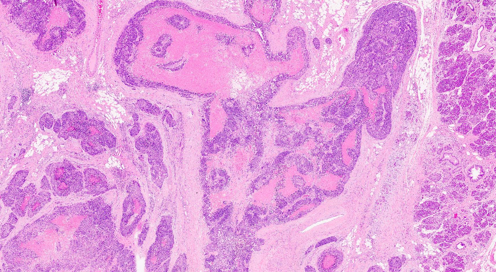
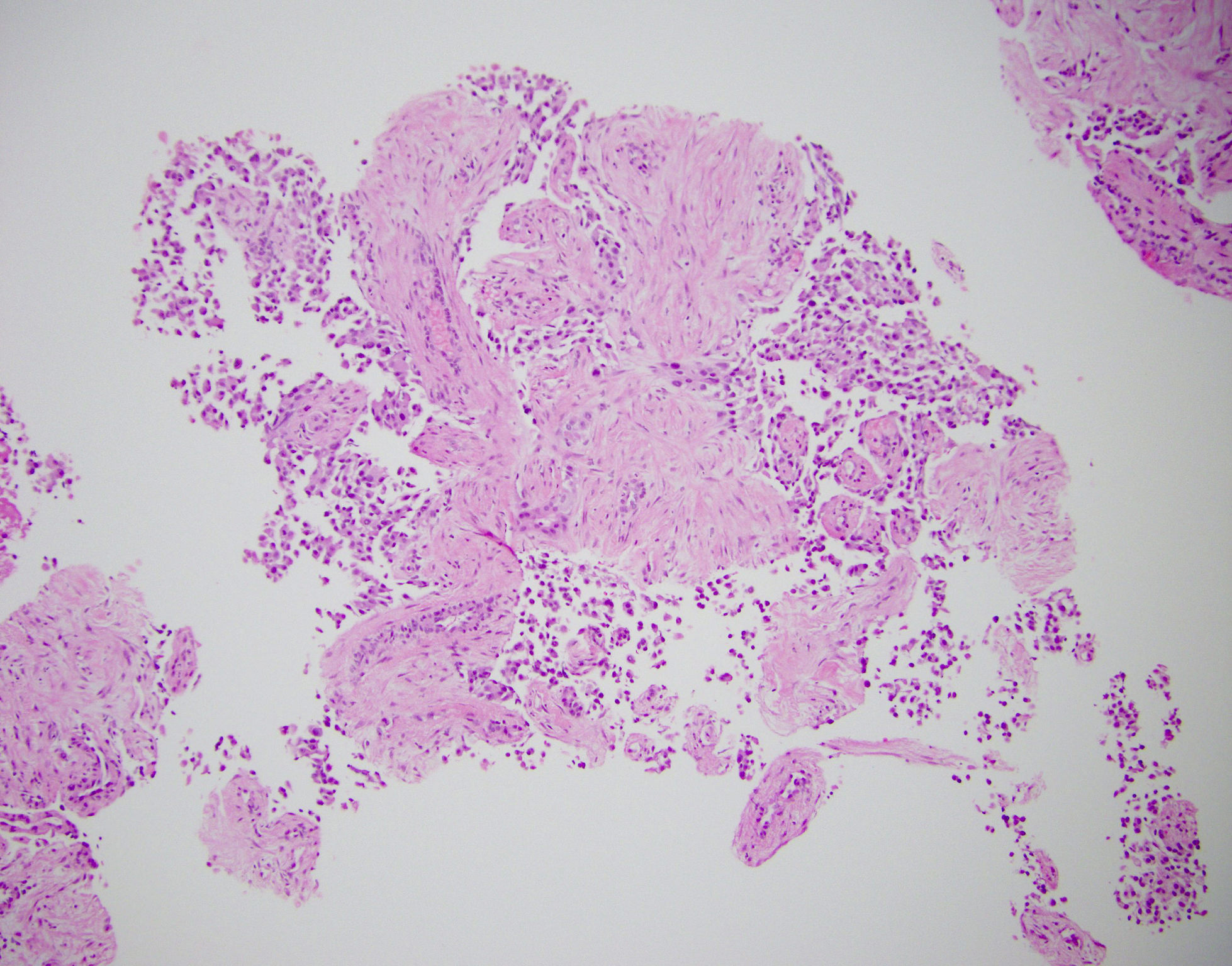
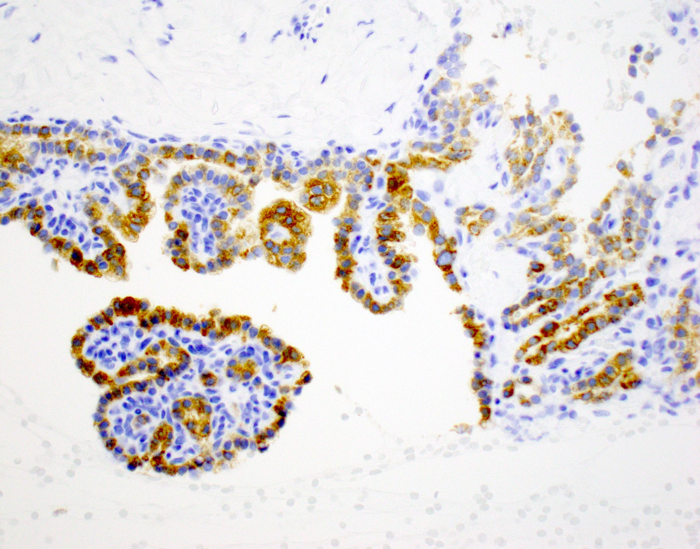
Intraductal oncocytic papillary neoplasm
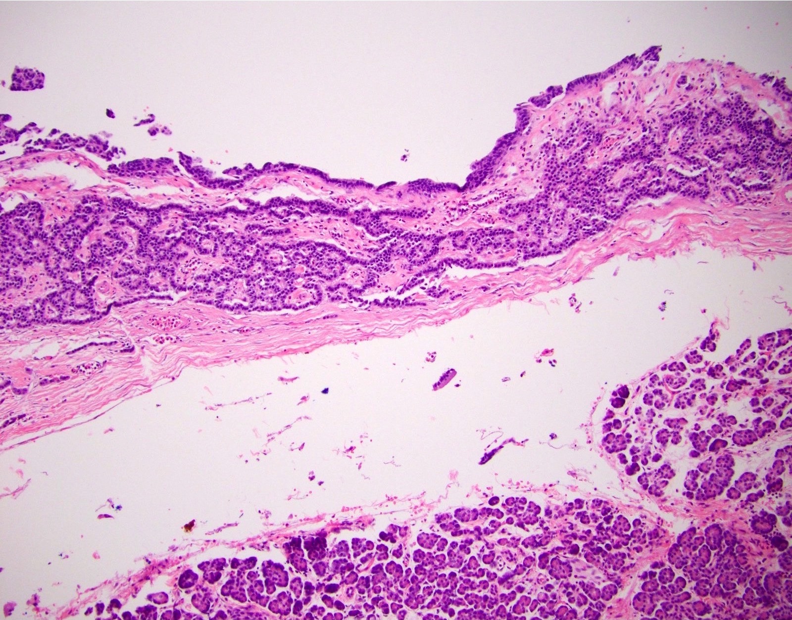
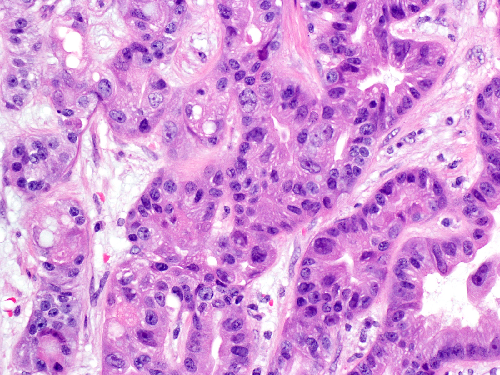
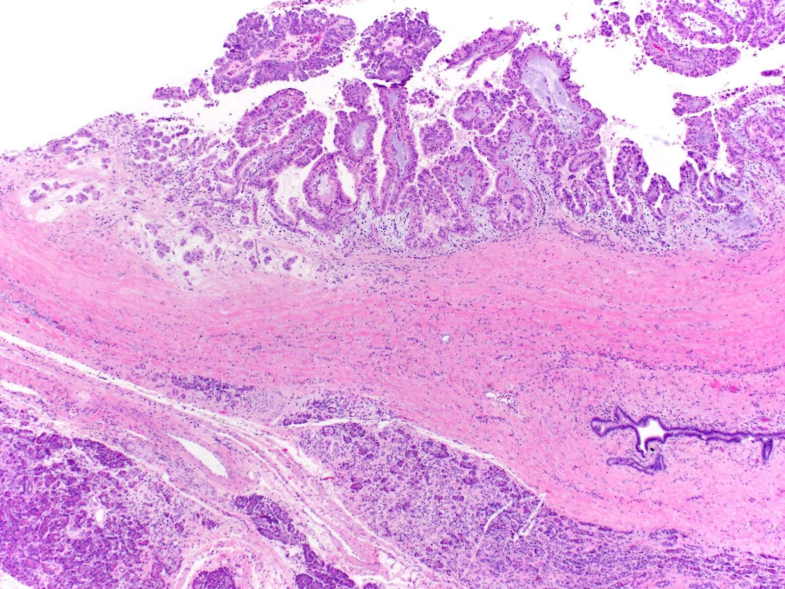
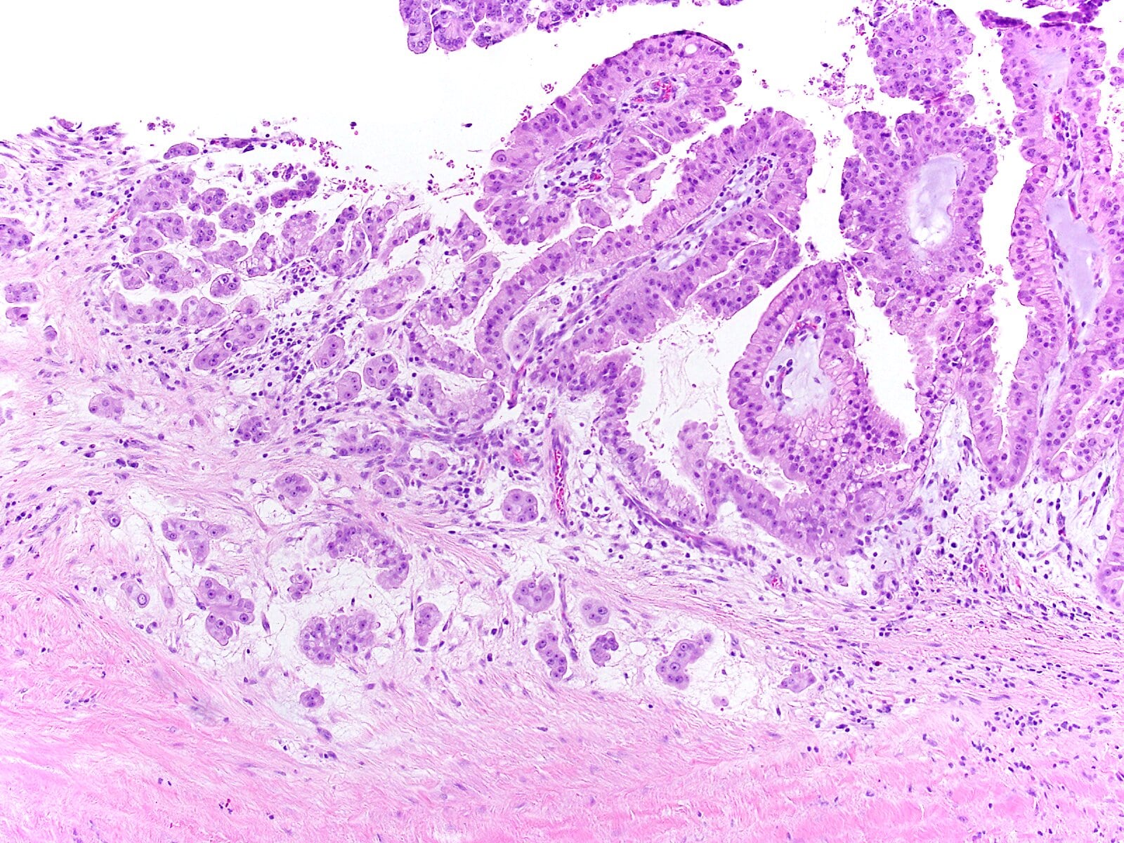
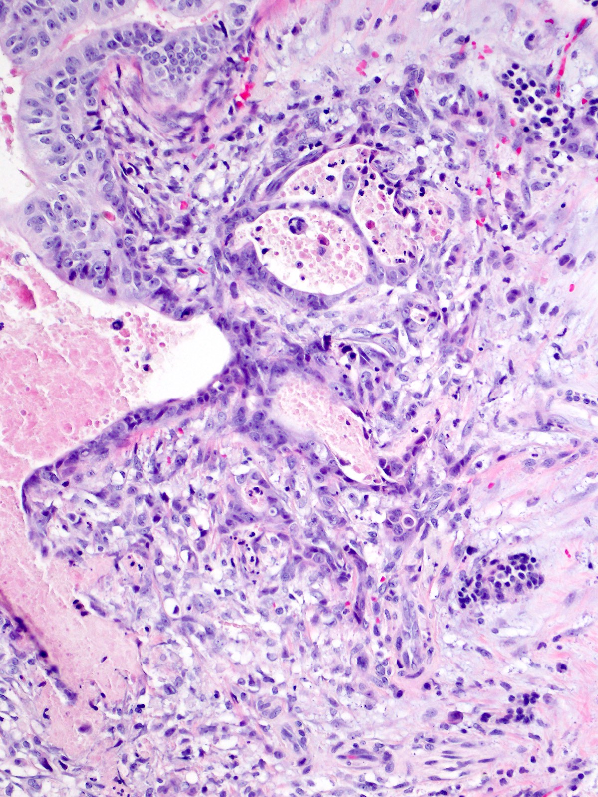
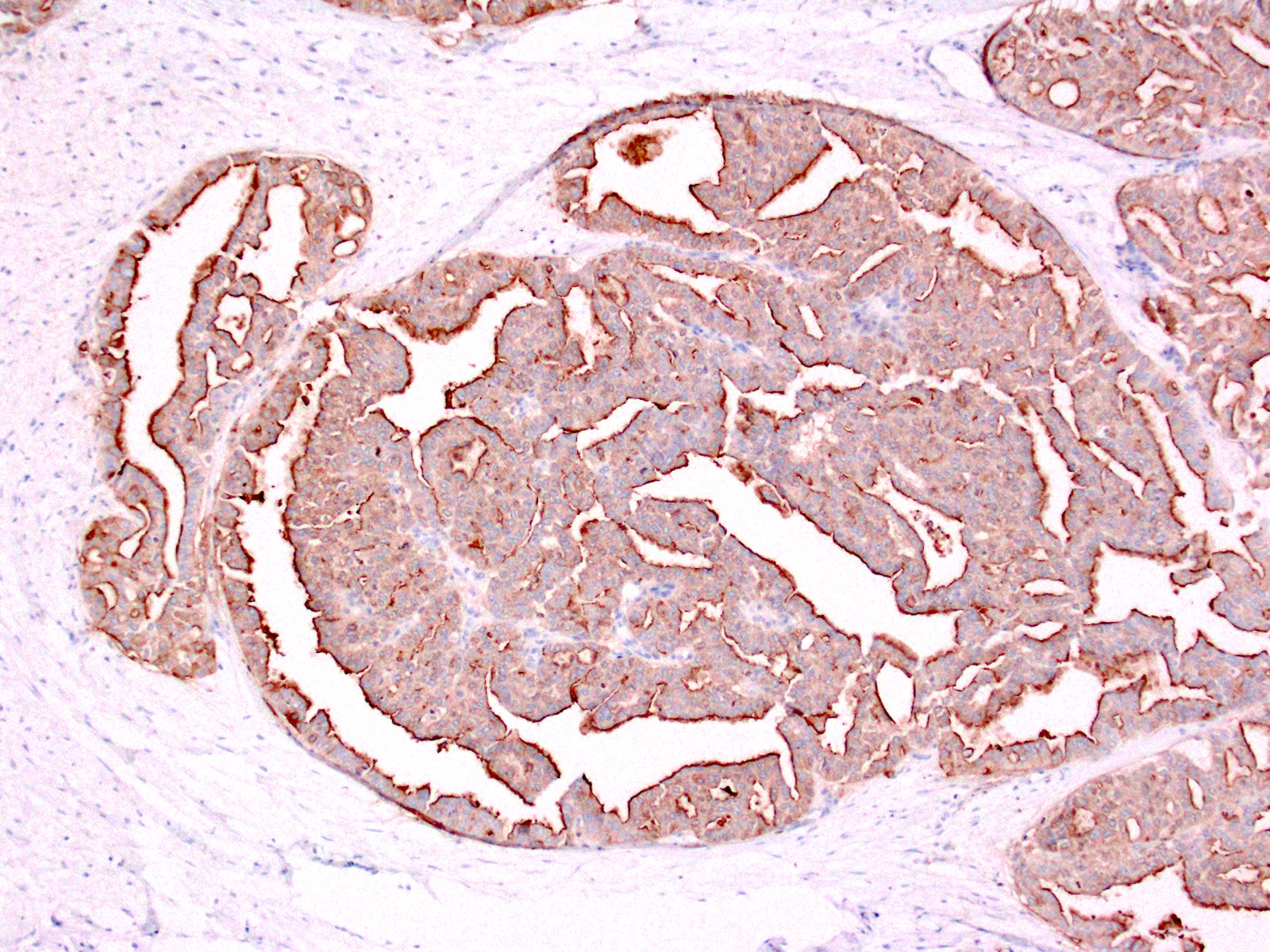
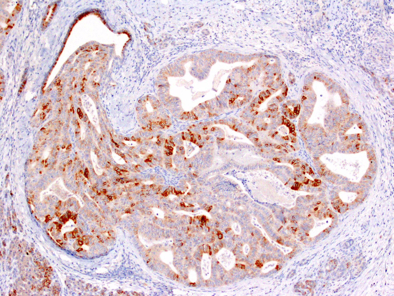
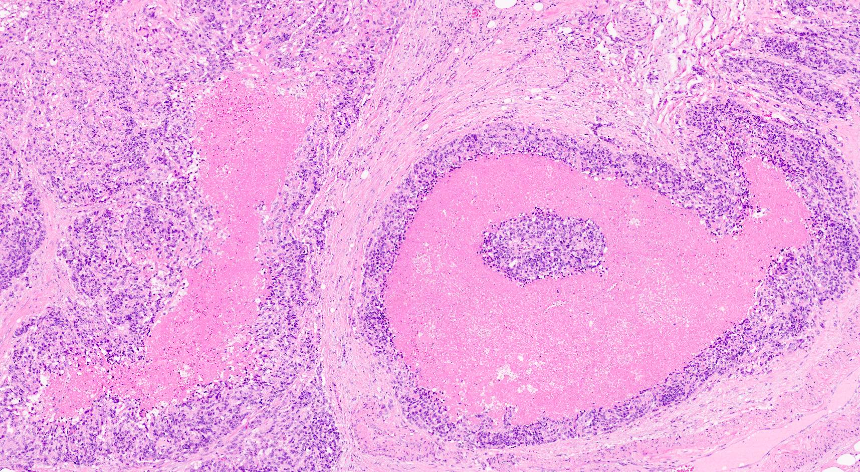
Contributed by Gokce Askan, M.D. and Olca Basturk, M.D.
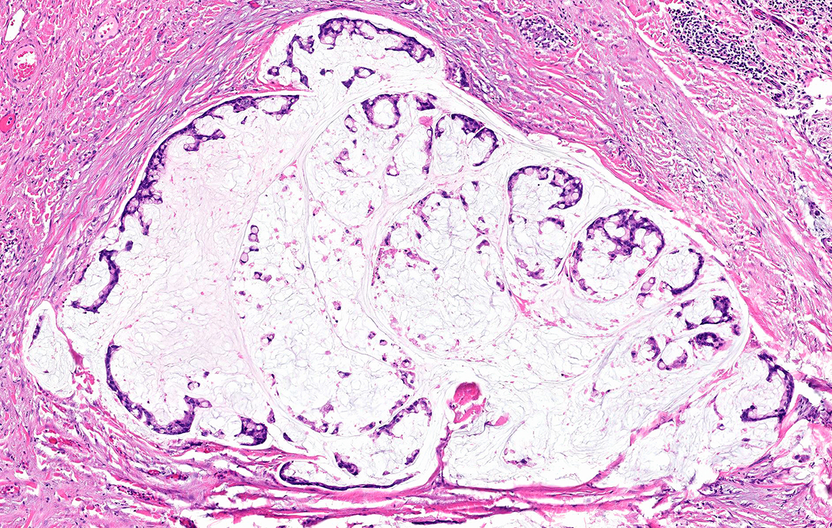
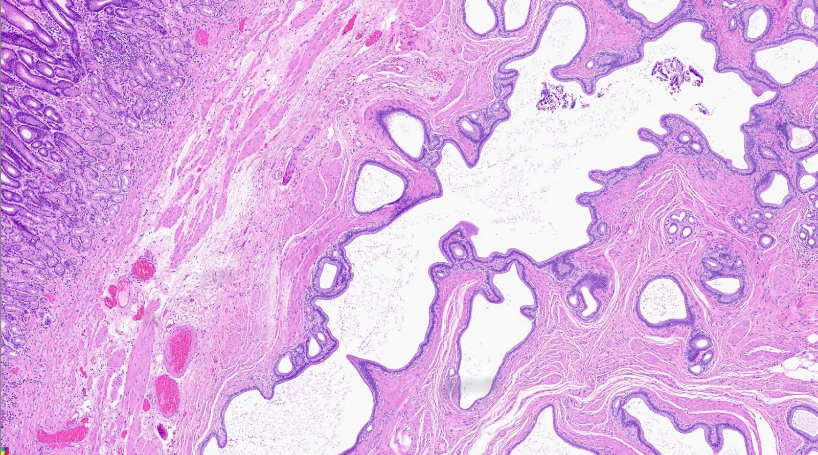
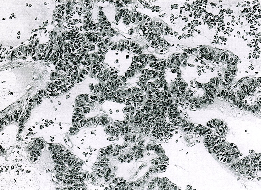
Unilocular or multilocular cystic lesion
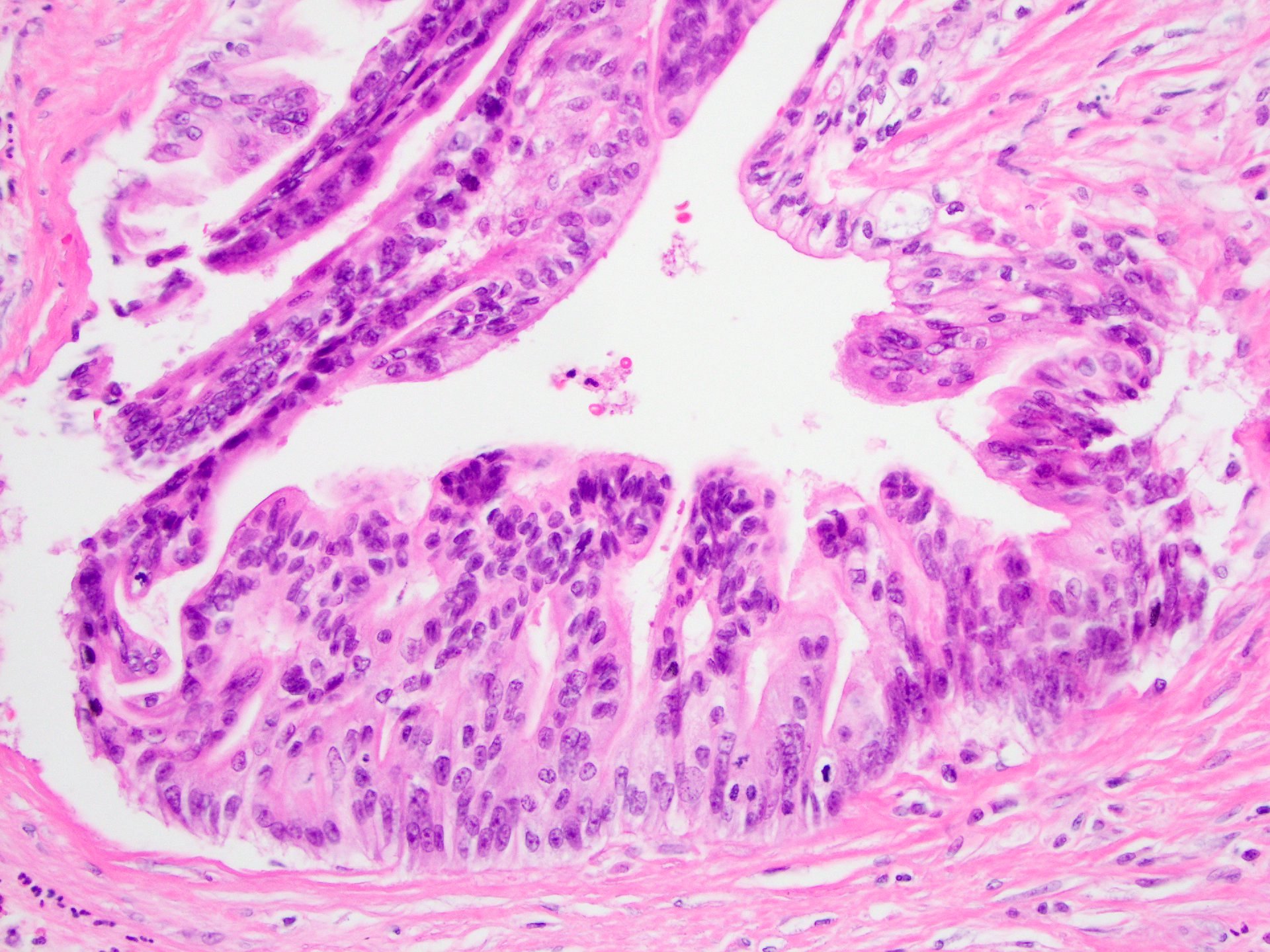
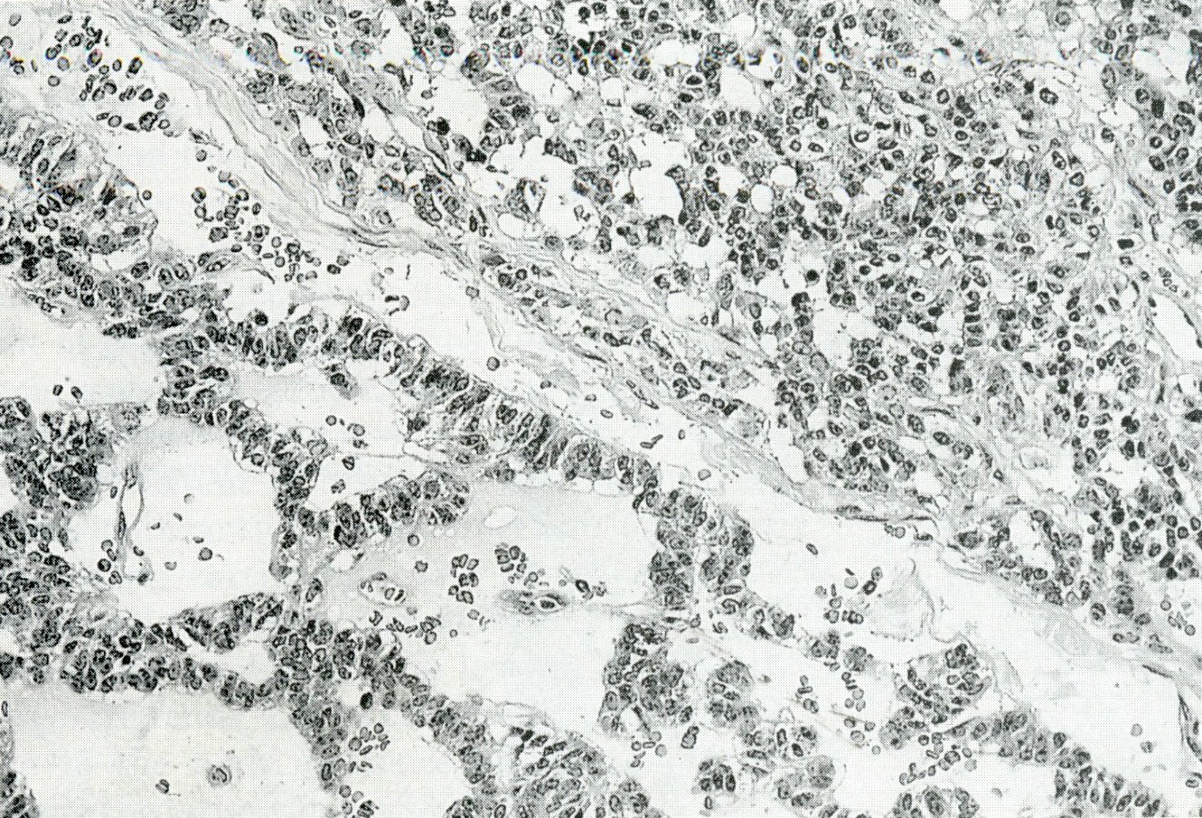
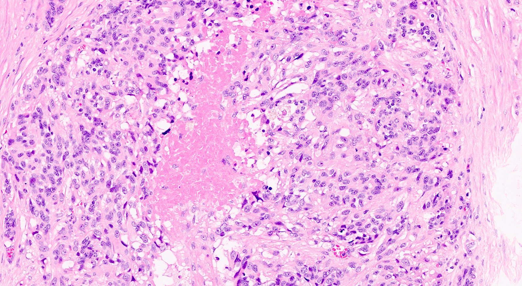
Papillae lined by oncocytic epithelium
Intracytoplasmic vacuoles
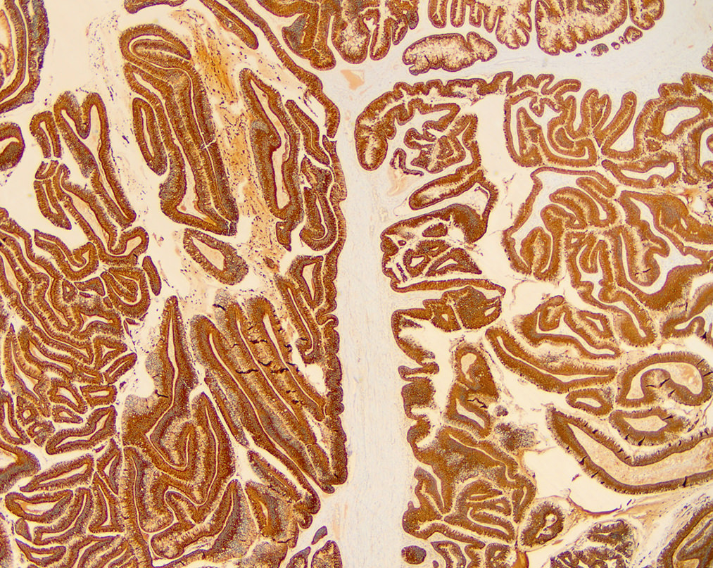
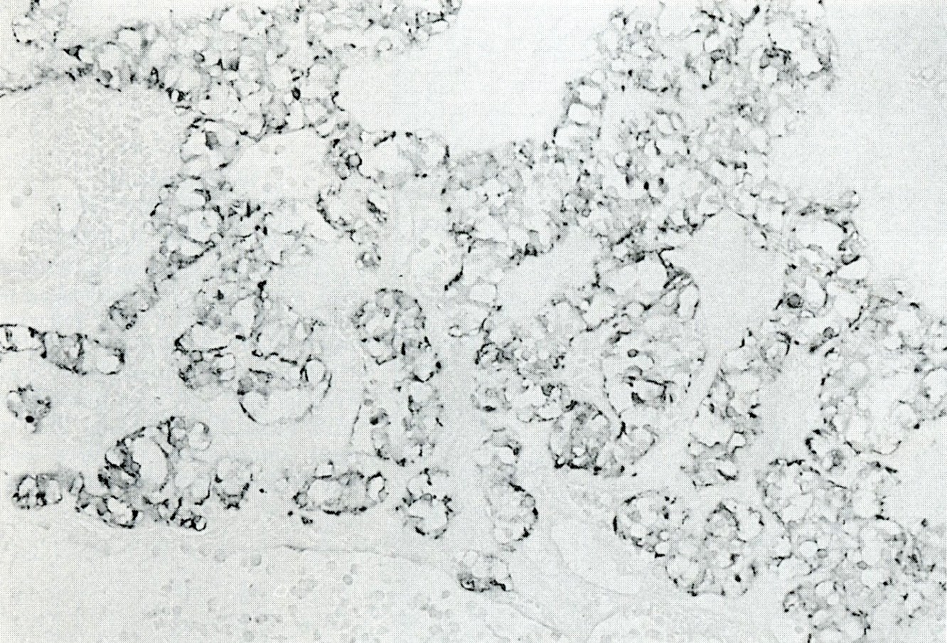
MUC6
Pseudoinvasion
Tubular invasion pattern
Mucinous invasion pattern
Contributed by @liverwei on Twitter
 Contributed by @liverwei on Twitter (see original post here)">
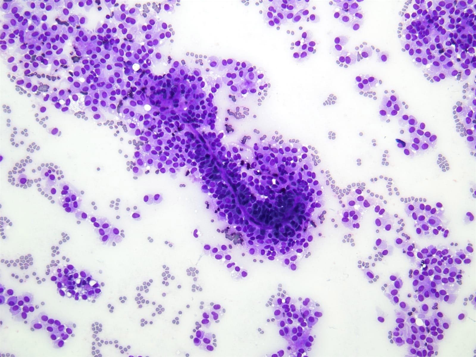
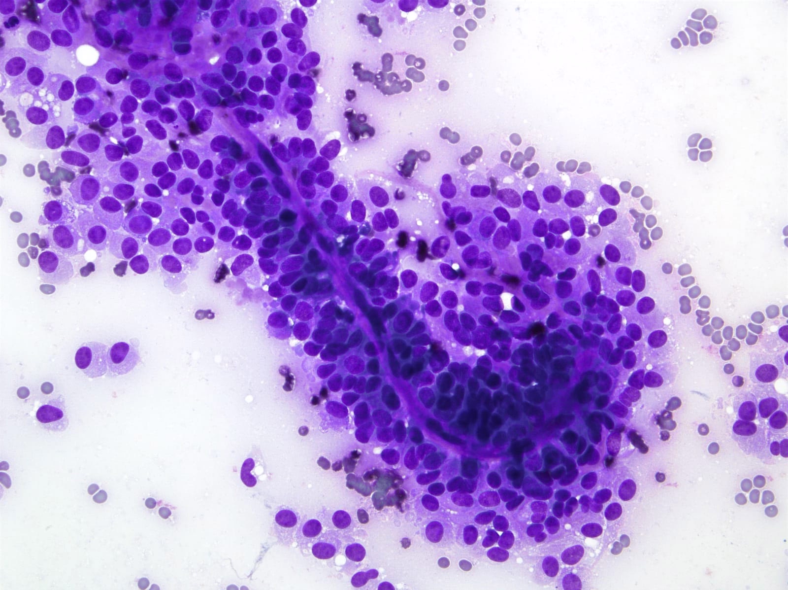
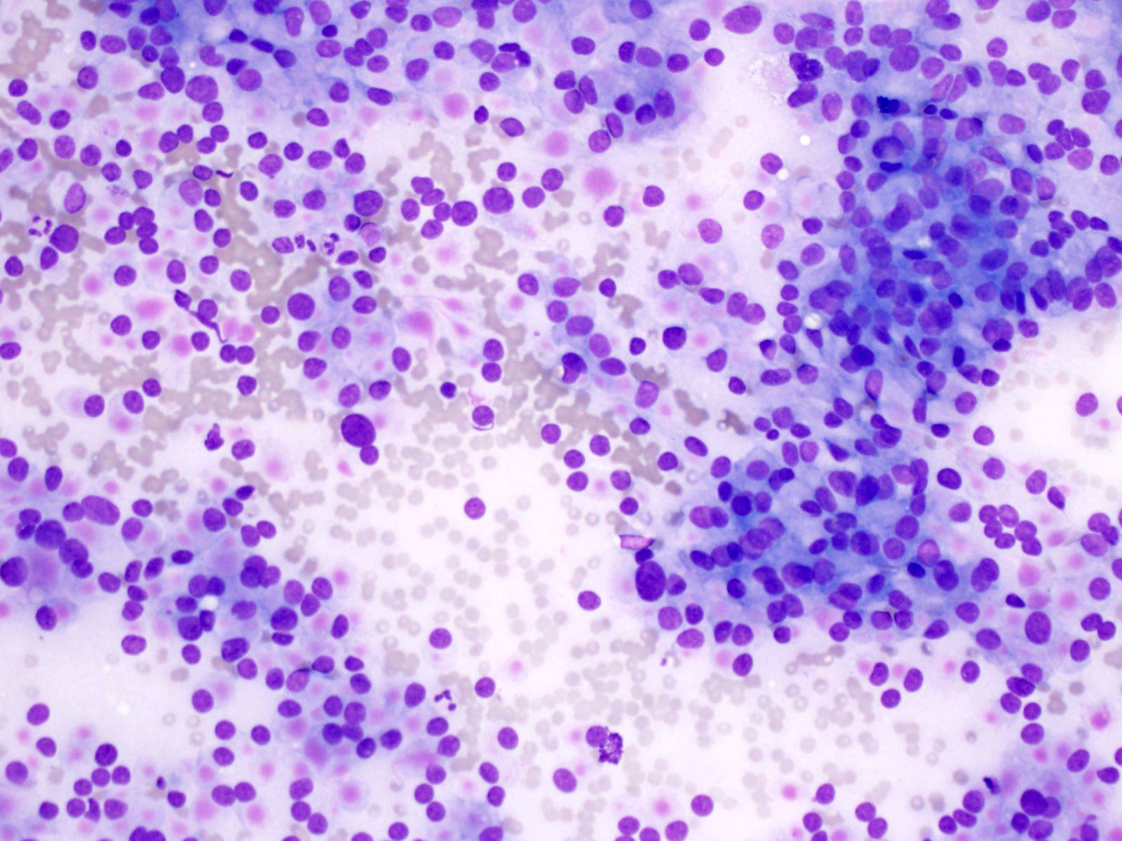
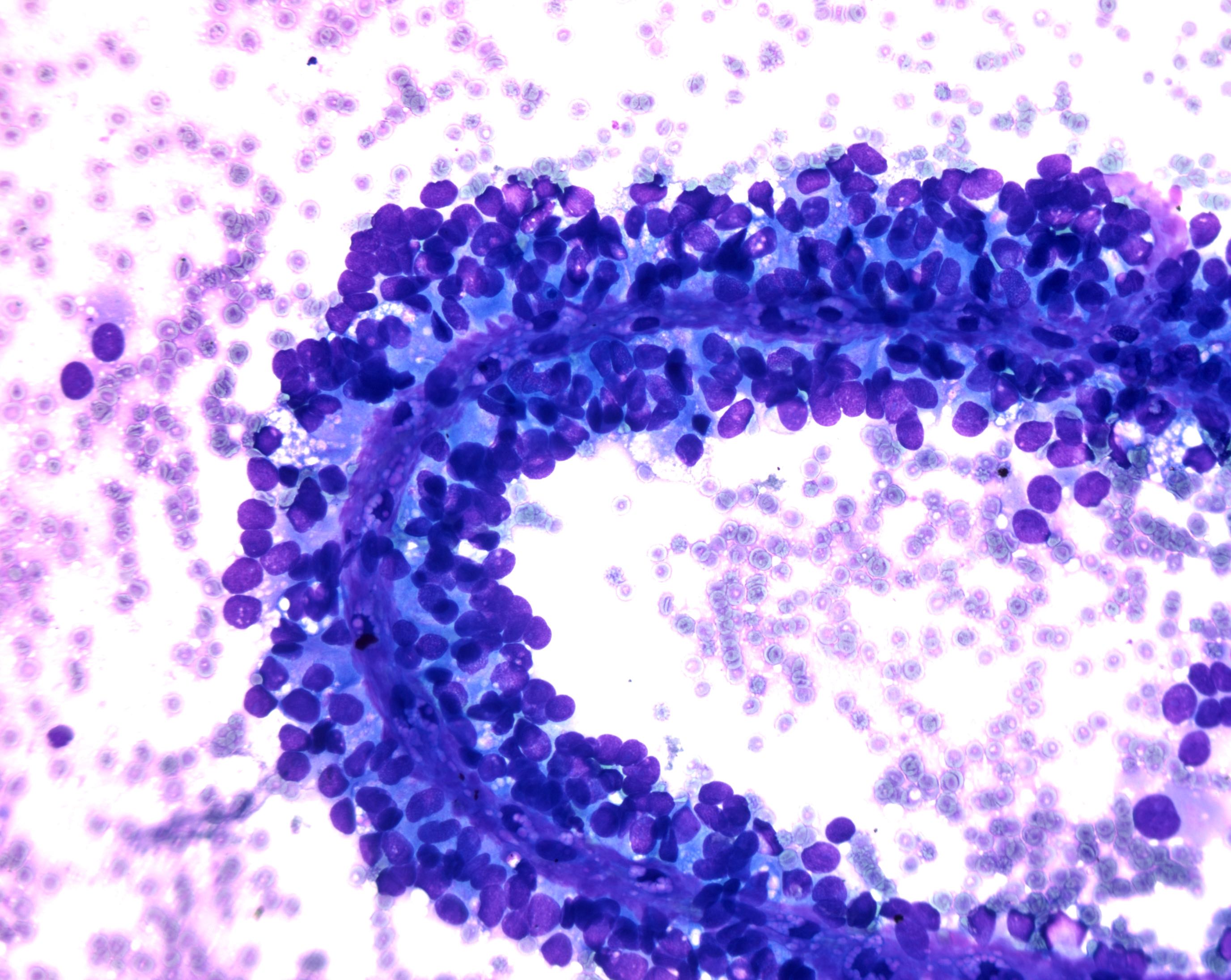
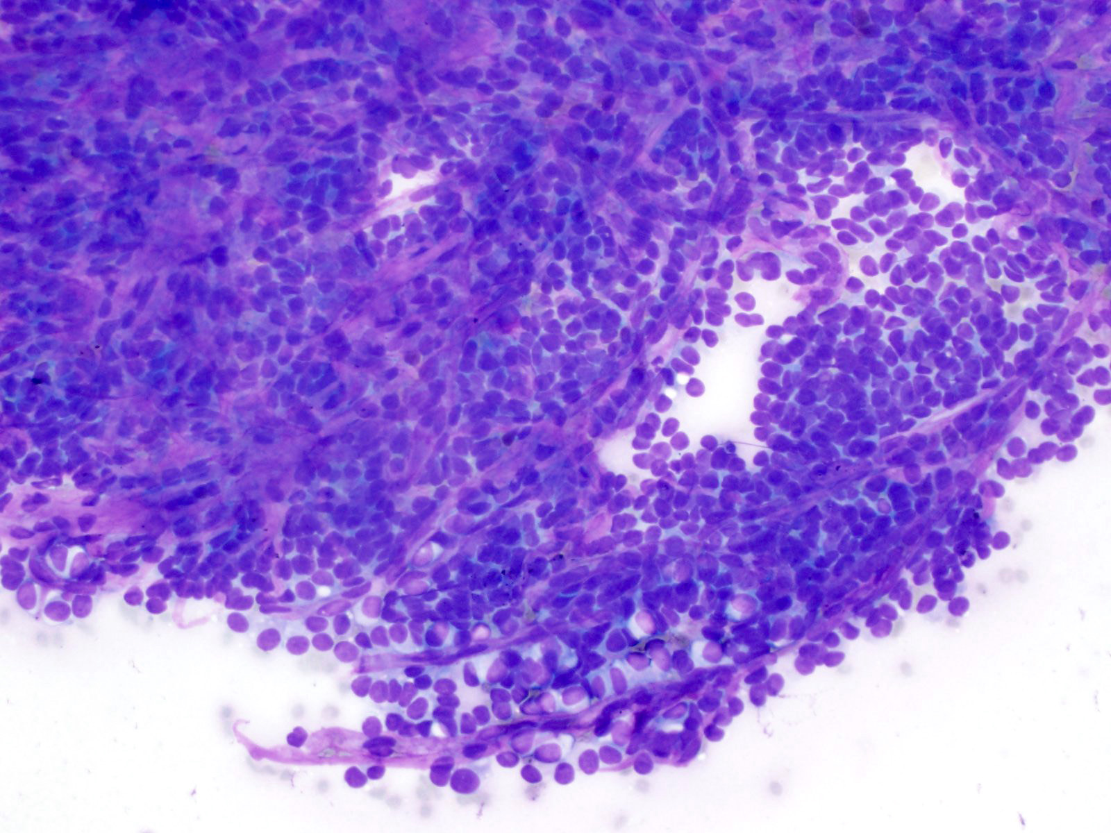
Intraductal oncocytic papillary neoplasm
Images hosted on other servers:

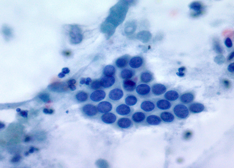
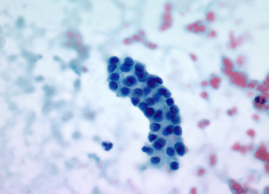
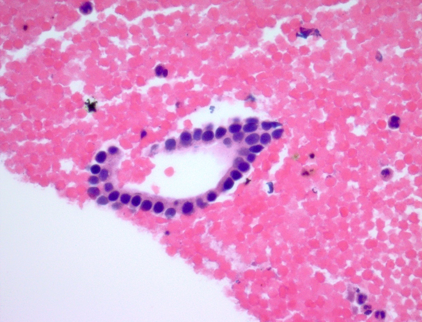
Oncocytic cell sheets with prominent nucleoli

Punched out spaces, intracytoplasmic mucin

Images hosted on other servers:


MRCP demonstrating various types of IPMN

MRCP demonstrating side branch IPMNs

Differing radiologic
modalities demonstrating
various types of
IPMN and MCN
Images hosted on other servers:

Mucin extrusion from duodenal papilla
Contributed by Dennis R. Dening, PA (ASCP)
Main duct IPMN
Contributed by Diana Agostini-Vulaj, D.O.
IPMN intestinal subtype with low grade dysplasia

IPMN gastric subtype with low grade dysplasia
IPMN with high grade dysplasia

MUC1
MUC2
Images hosted on other servers:

Low grade epithelial atypia

High grade epithelial atypia with necrotic debris
Images hosted on other servers:

Polypoid mass within dilated main pancreatic duct
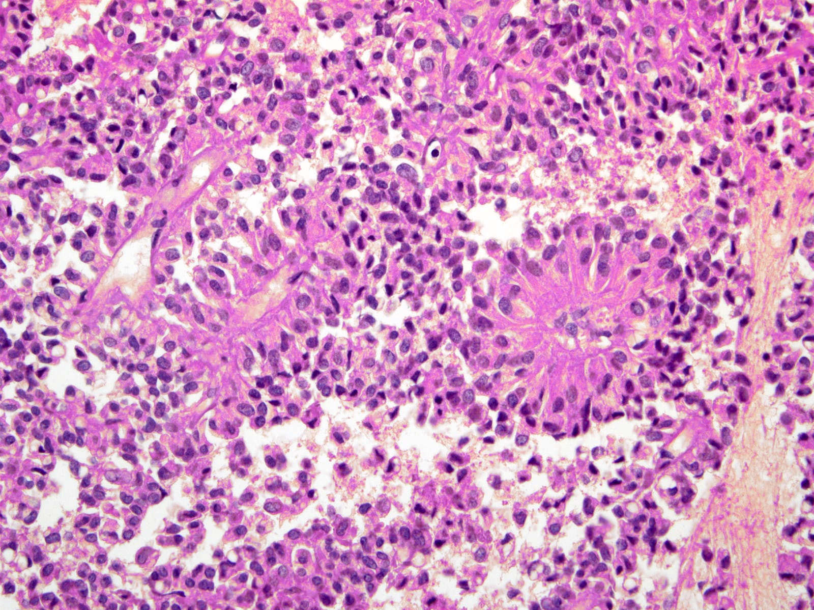
Contributed by Gokce Askan, M.D. and Olca Basturk, M.D.
Nodules of back to back tubular glands
Intraductal growth pattern
Sharply circumscribed nest formation

Tightly packed tubules lined by atypical cuboidal epithelium
Comedo-like necrosis
Clear cell changes
Osseous metaplasia

Invasion
MUC1 expression
MUC6 expression
Images hosted on other servers:

CT

Large cystic mass

Mimicking serous / mucinous cystadenoma
Images hosted on other servers:

Intraoperative view
Contributed by Jen Rytych, M.D.

Sections of lymphoepithelial cyst
Images hosted on other servers:

Well demarcated cysts

Pancreas tail
Contributed by Jen Rytych, M.D. and Katrina Krogh, M.D.
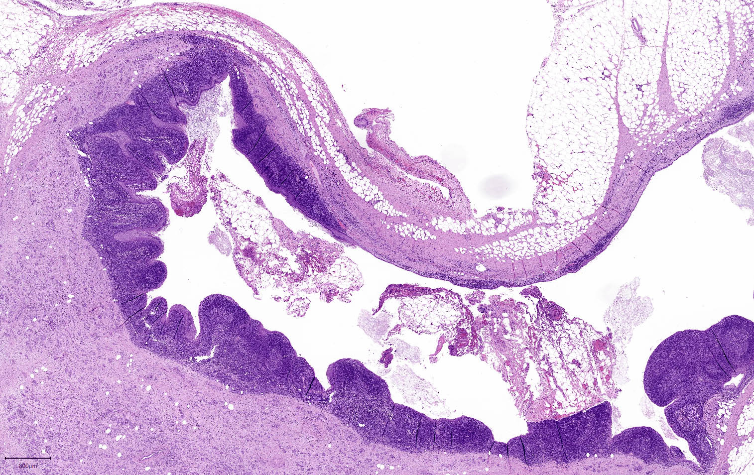
Squamous lined cyst
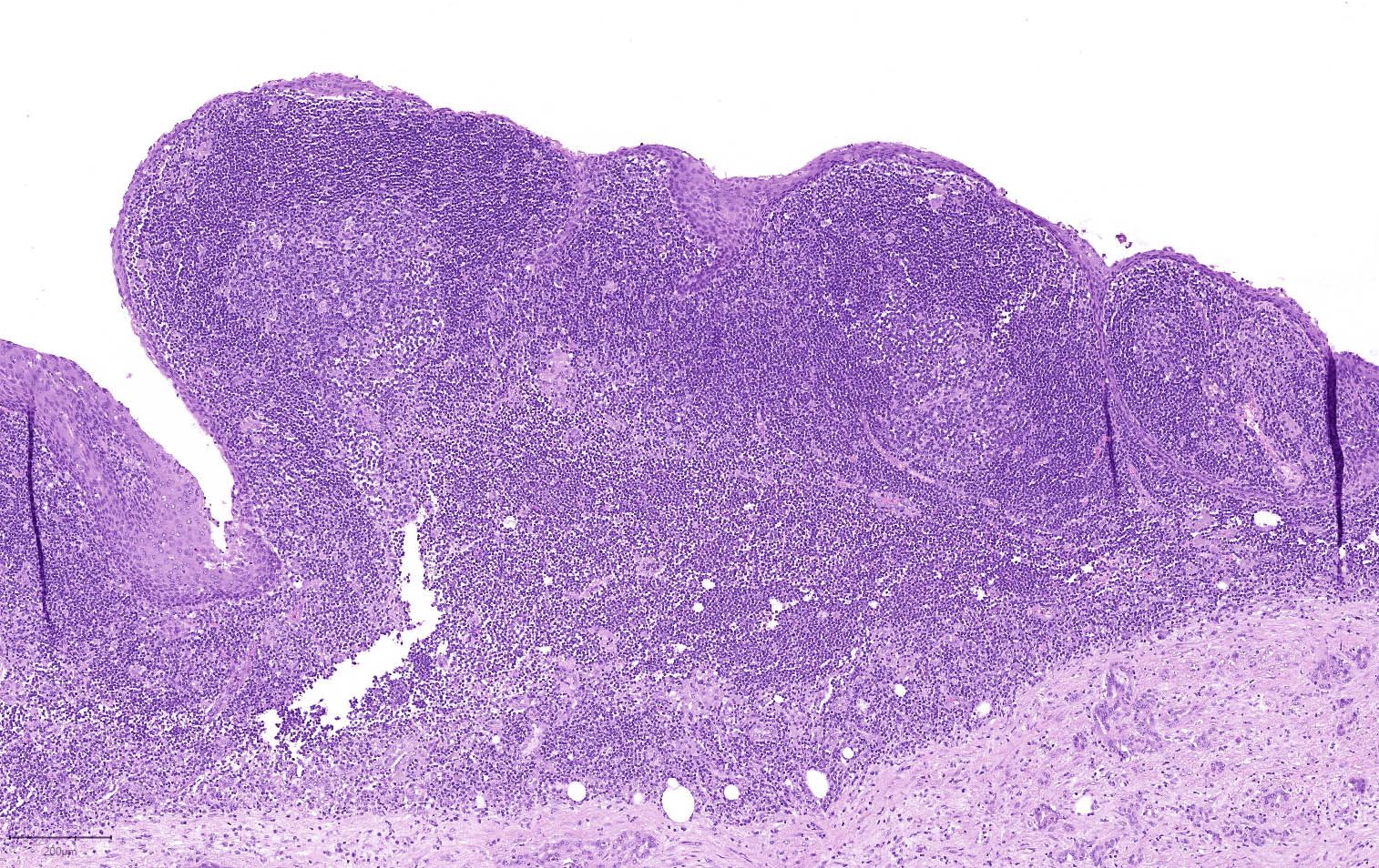
Cyst with lymphoid cuff
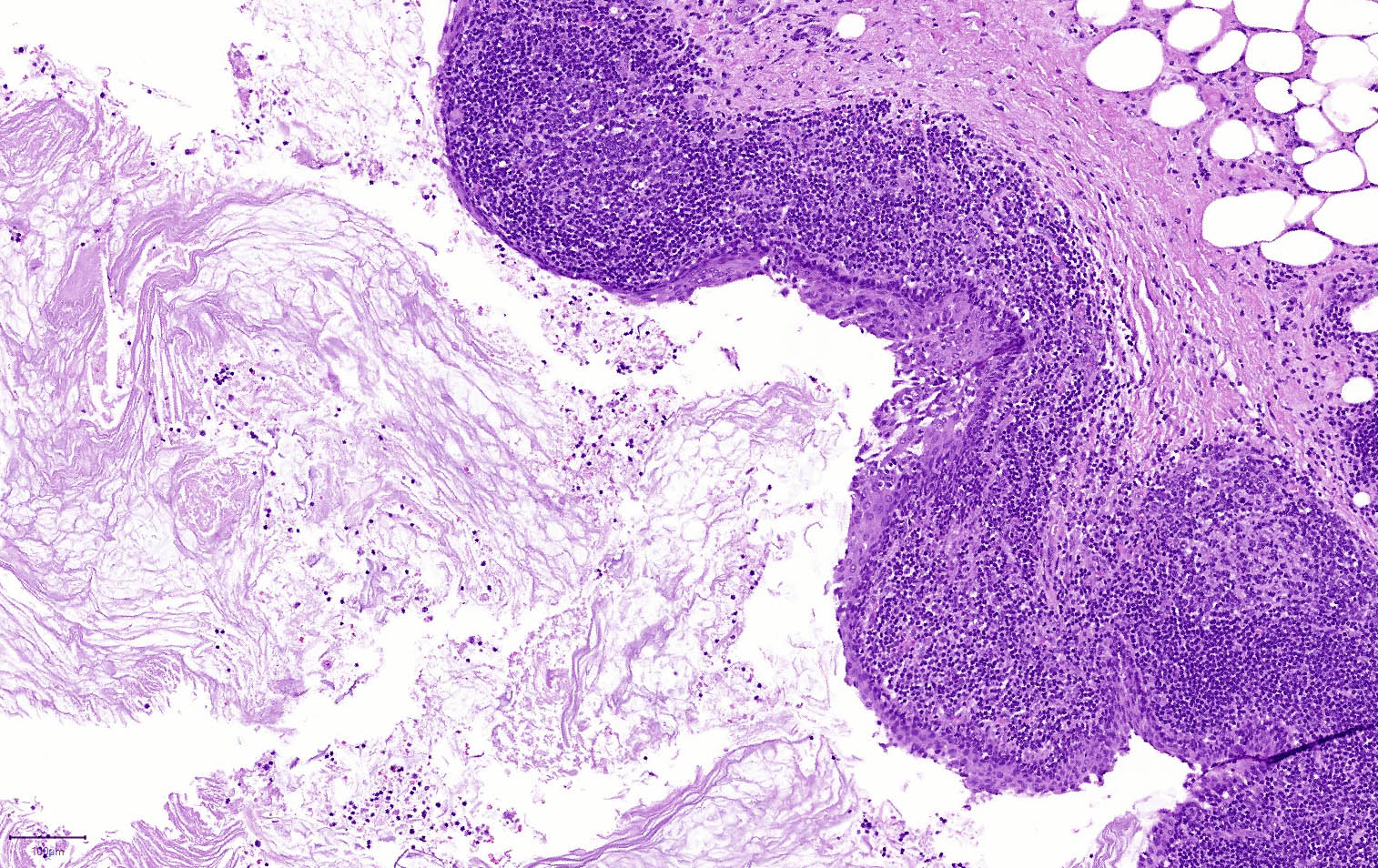
Abundant keratin debris
Simple columnar and stratified squamous linings
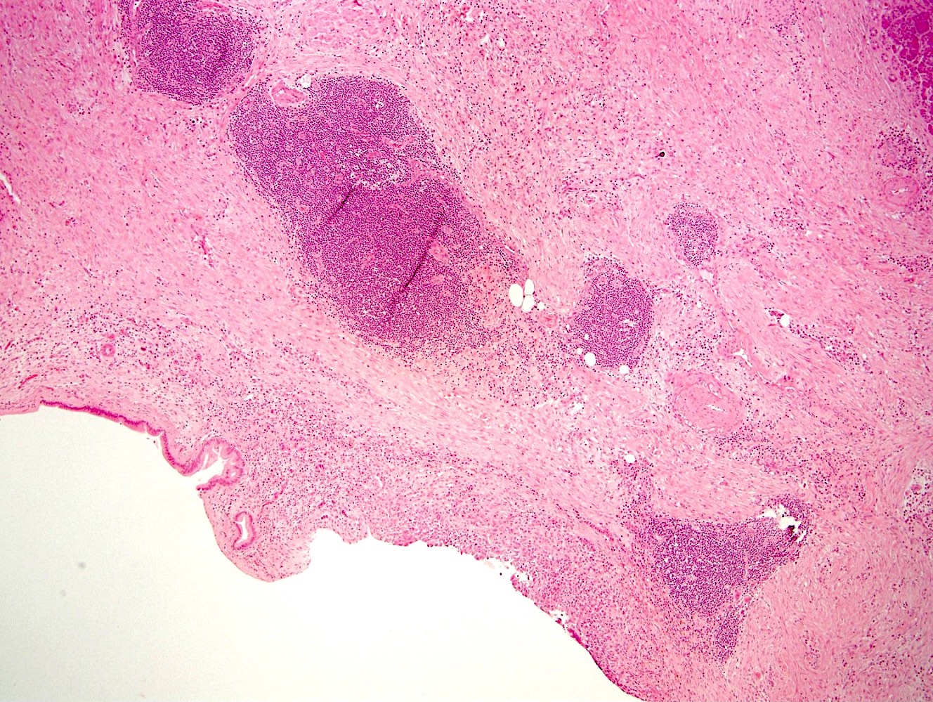
Squamous lined lymphoid tissue
Contributed by Jen Rytych, M.D.

Degenerated squamous epithelial cells
Images hosted on other servers:

Squamous epithelium,
keratinaceous debris
and lymphocytes

Keratinaceous debris
Contributed by Rong Xia, M.D., Ph.D. and Beena Ahsan, M.D.
Circumscribed borders
Sheet-like growth pattern
Syncytial growth pattern
Positive p63 immunostain
Images hosted on other servers:

Location of tumors in patients with MEN1
AFIP images
Intramucosal tumor nodules
Zollinger-Ellison syndrome
AFIP images
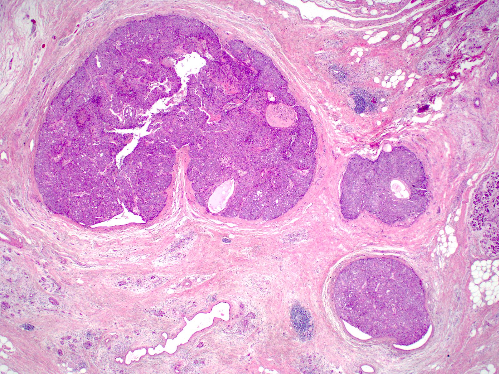
Pancreatic microadenomas in patients with MEN1
Duodenal gastrinoma in the mucosa and submucosa
Hypertrophic and eroded mucosa
Images hosted on other servers:

MiNEN in the head of the pancreas on CT

MiNEN in pancreas head with portal vein narrowing on CT
Images hosted on other servers:

Mixed ductal neuroendocrine carcinoma
Contributed by Jennifer Ziebell, M.D. and Ashwini Esnakula, M.D., M.S.
Mixed ductal neuroendocrine carcinoma
Neuroendocrine carcinoma component
Ductal adenocarcinoma component
Chromogranin A
Ki67 neuroendocrine carcinoma component
MiNEN basics
Contributed by Claudio Luchini, M.D., Ph.D.
Medullary histology
Mucinous colloid histology
Adenosquamous histology
Contributed by Diana Agostini-Vulaj, D.O. and AFIP images

MCN with no connection to main duct
Unilocular cyst
Conspicuous, irregular, solid protuberances
Multiloculated cystic lesion

36 year old
woman: large
cyst with
solid tumor
Images hosted on other servers:

39 year old man with multilocular cyst with thick mucin
Contributed by Raul S. Gonzalez, M.D. and Matthew W. Rosenbaum, M.D.
Spindled ovarian type stroma

Focal minute papillation
Foveolar mucinous epithelium
Classic ovarian type stroma

MCN with invasion
Estrogen receptor
Contributed by Matthew W. Rosenbaum, M.D.

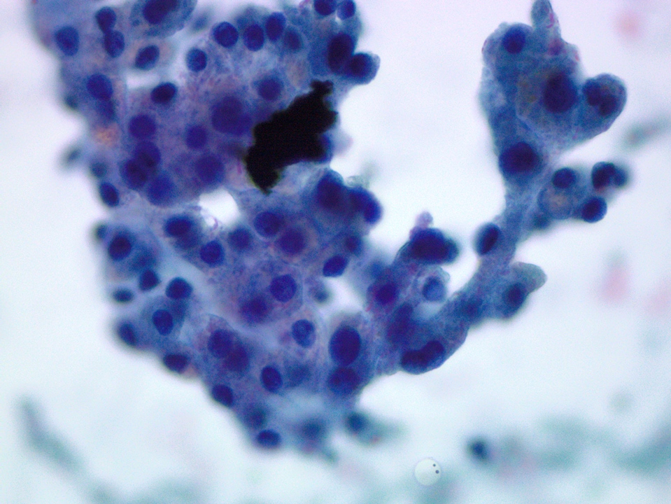
Cytology of mucinous cystic neoplasm
Contributed by Katrina Krogh, M.D.
PDA: ill defined white lesion
MCN: unilocular cyst with thick wall
IPMN: small cysts with smooth linings
MNC: unilocular
simple cyst with
smooth thin lining
Contributed by Jen Rytych, M.D. and Katrina Krogh, M.D.
Mucinous pancreatic adenocarcinoma
Pancreatic adenocarcinoma, grade 2
MCN: ovarian type stroma
IPMN, gastric type
IPMN, intestinal type
MNC
Images hosted on other servers:


20 day old girl with persistent hypoglycemia and seizures


23 year old man with hypoglycemia mimicking an insulinoma
Images hosted on other servers:

MRI demonstrating small WDNET

CT and Gallium 68 Dotatate PET showing WDNET
Contributed by Danielle Hutchings, M.D.
Well circumscribed, solid tumor in pancreatic tail
Tumor with hemorrhage
Invasion into spleen

Large vessel invasion
Pancreatic duct obstruction

Cystic degeneration
Contributed by Danielle Hutchings, M.D. and Sabrina Sopha, M.D.
Amyloid deposition

Poorly differentiated neuroendocrine carcinoma, small cell type
Poorly differentiated
neuroendocrine
carcinoma, large
cell type
Calcification
Cribriform pattern
Pseudoacinar pattern

Organoid pattern
Ki67 staining
INSM1 immunostain

Spread to duodenum (H&E and synaptophysin)
Synaptophysin staining

Synaptophysin staining
Chromogranin staining
Well differentiated pancreatic neuroendocrine tumor
(cell block, synaptophysin, chromogranin)
Contributed by Sabrina Sopha, M.D. and AFIP images

Well differentiated pancreatic neuroendocrine tumor
(pap stained smears)

Round nuclei and scanty cytoplasm
AFIP images
Enterochromaffin cell tumor: irregularly shaped
Solid tumors of the pancreas
Images hosted on other servers:

CT scan
Images hosted on other servers:

Nests of small, uniform polygonal cells

Pushing margin

Spindle cell component

Chromogranin

Desmin


Topoisomerase II alpha
Images hosted on other servers:

CT and ultrasound
with pancreatic
head mass
AFIP images
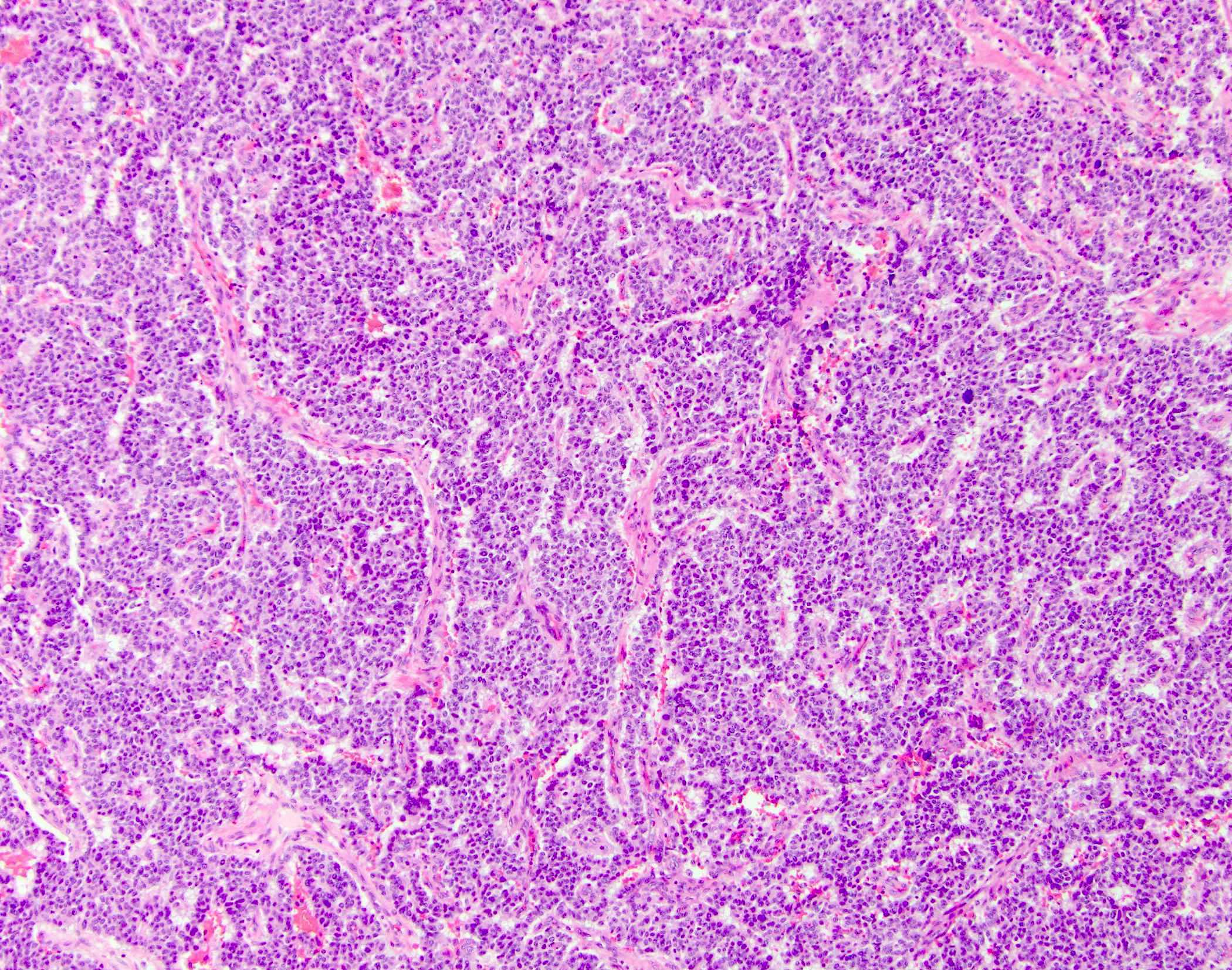
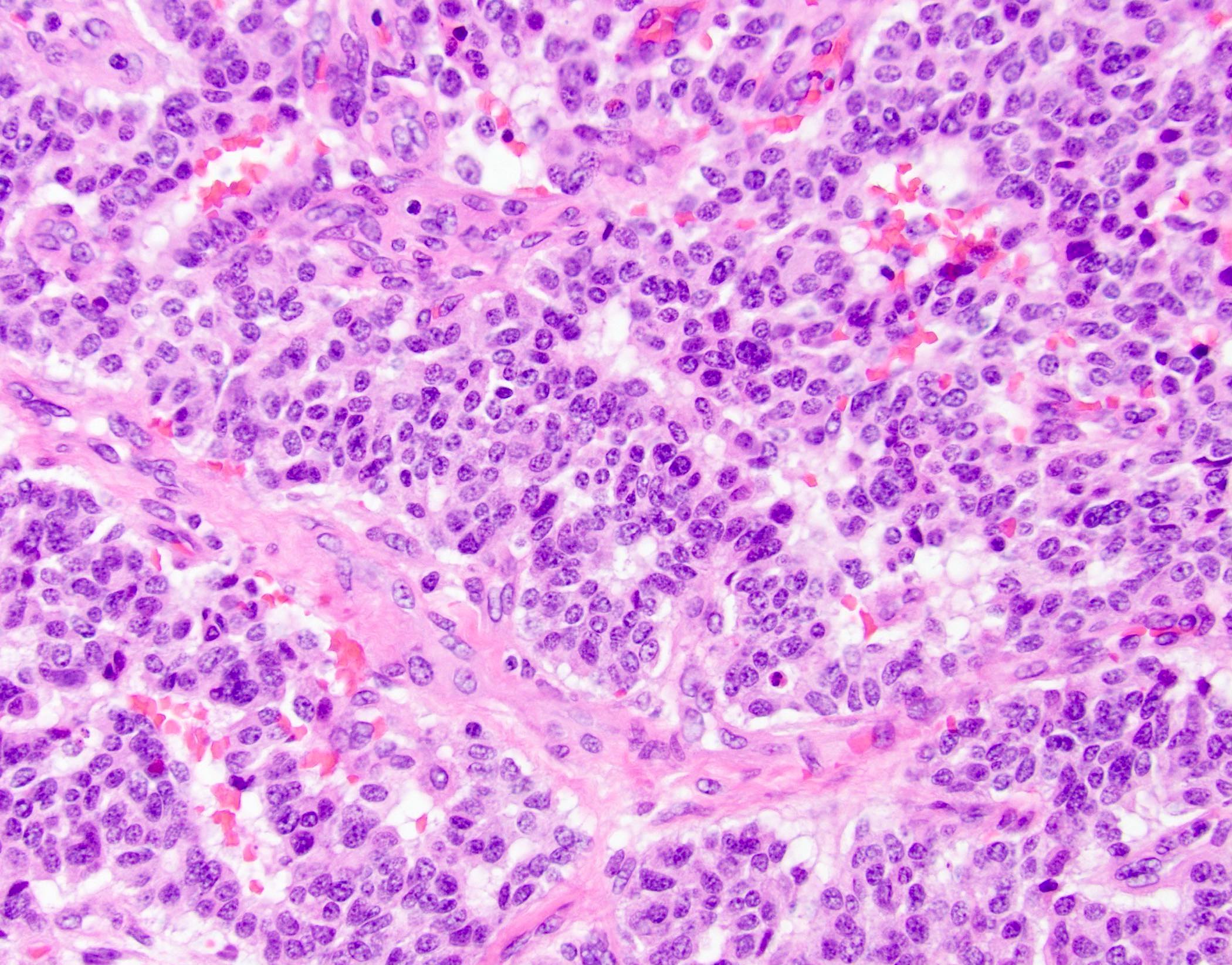
Trabecular growth
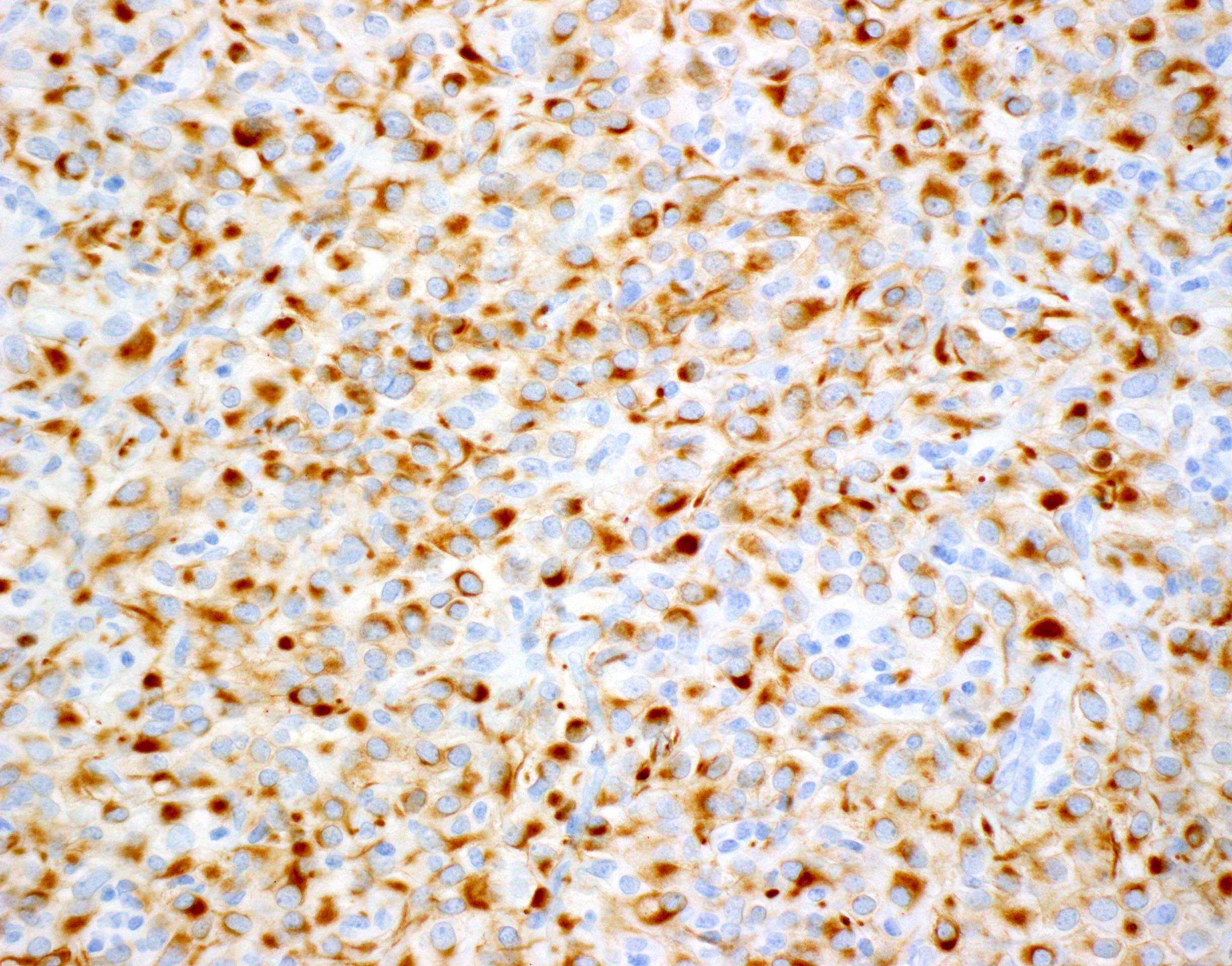
Pancreatic endocrine tumor with PP expression
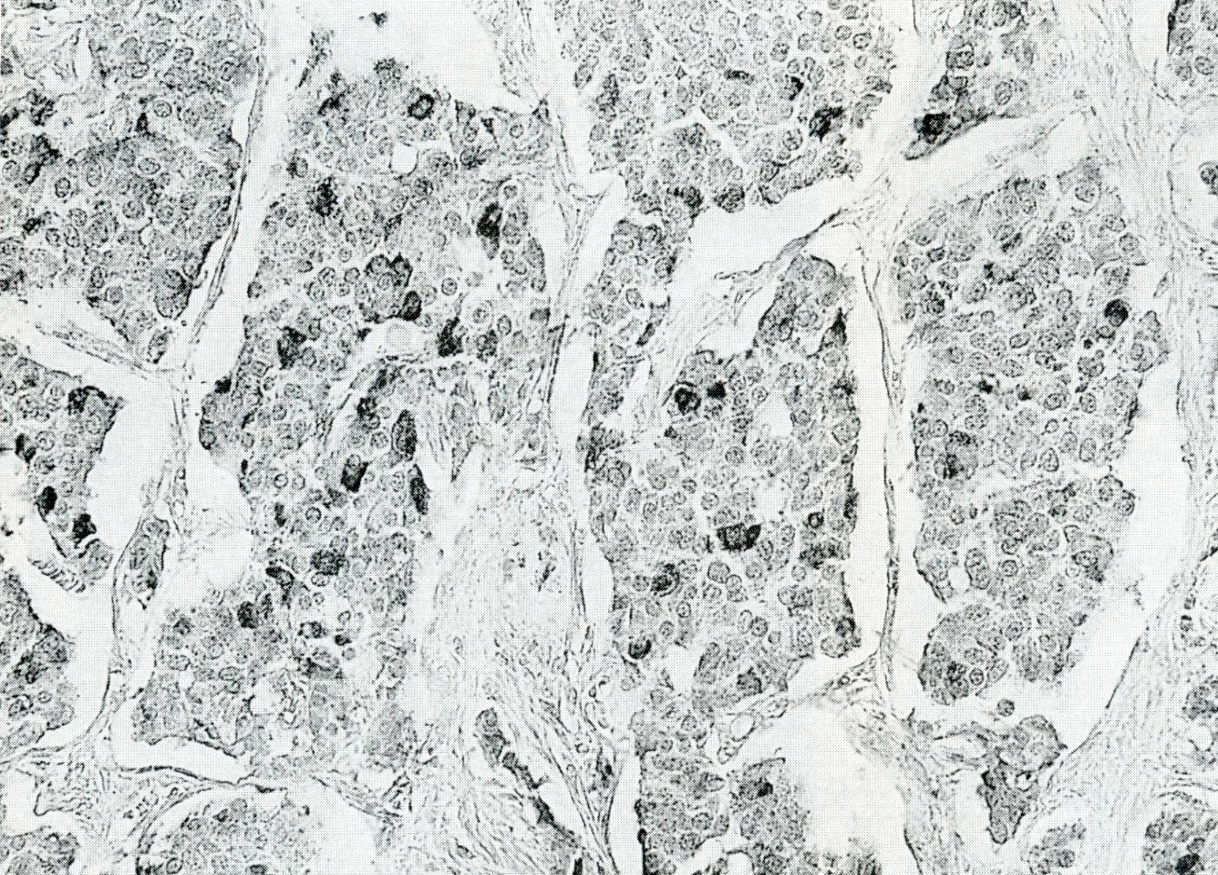
Images hosted on other servers:

Chromogranin and PP
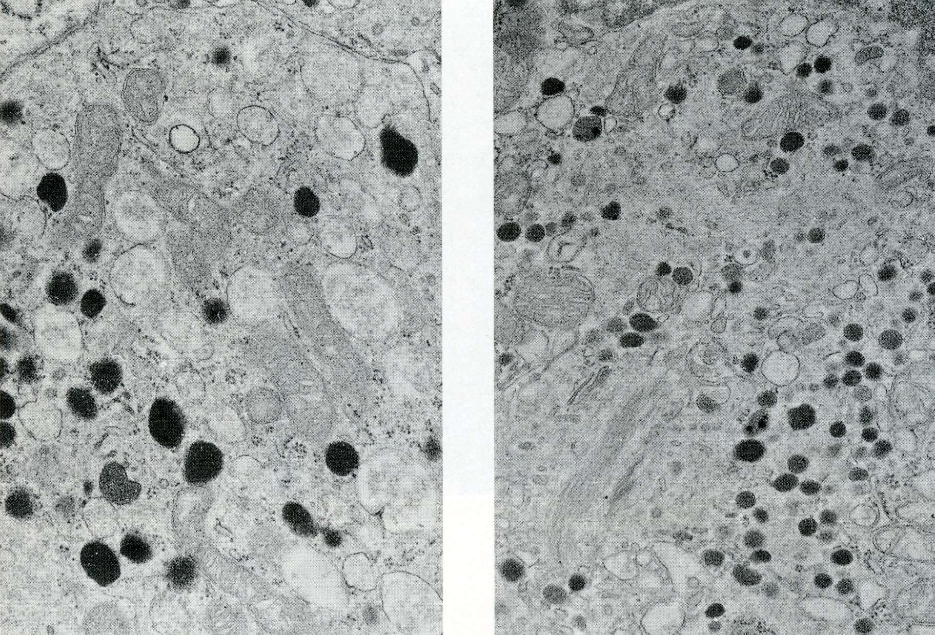
Images hosted on other servers:

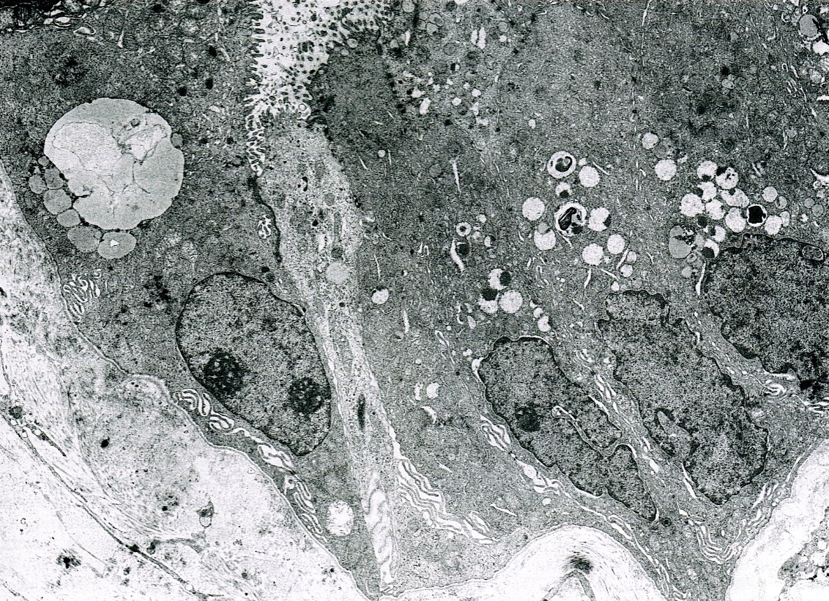
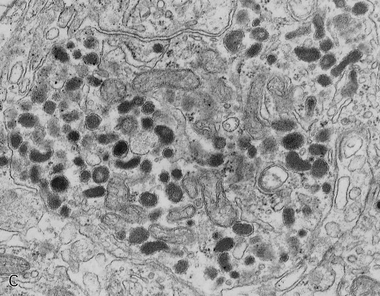
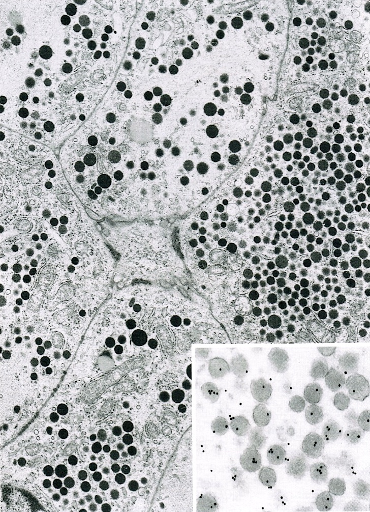
Abundant basal accumulation of secretory granules
Images hosted on other servers:

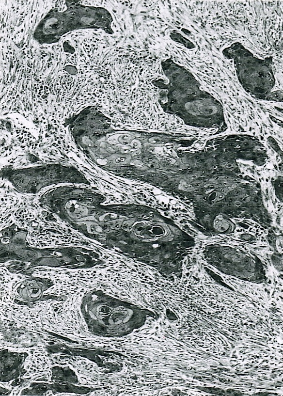
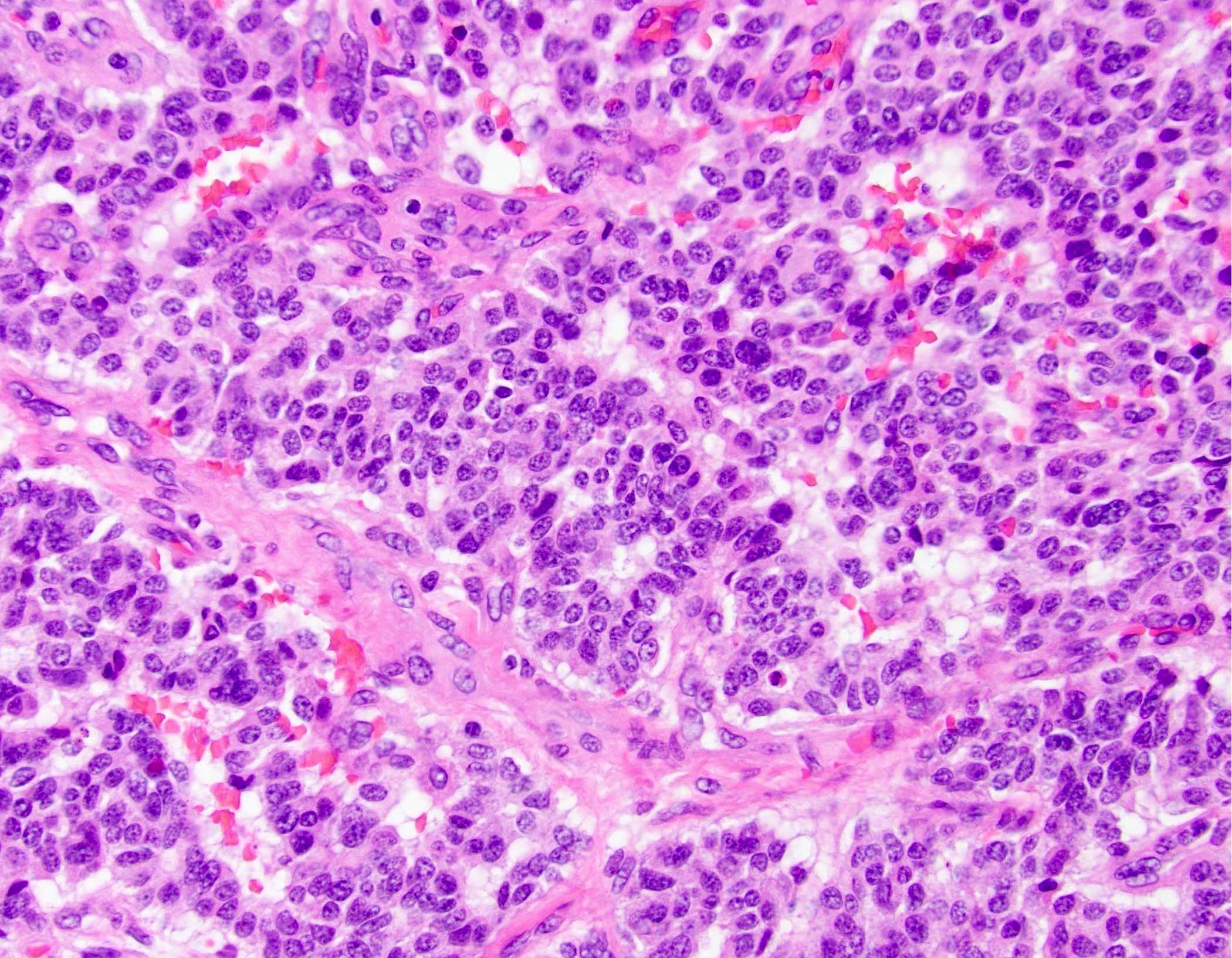
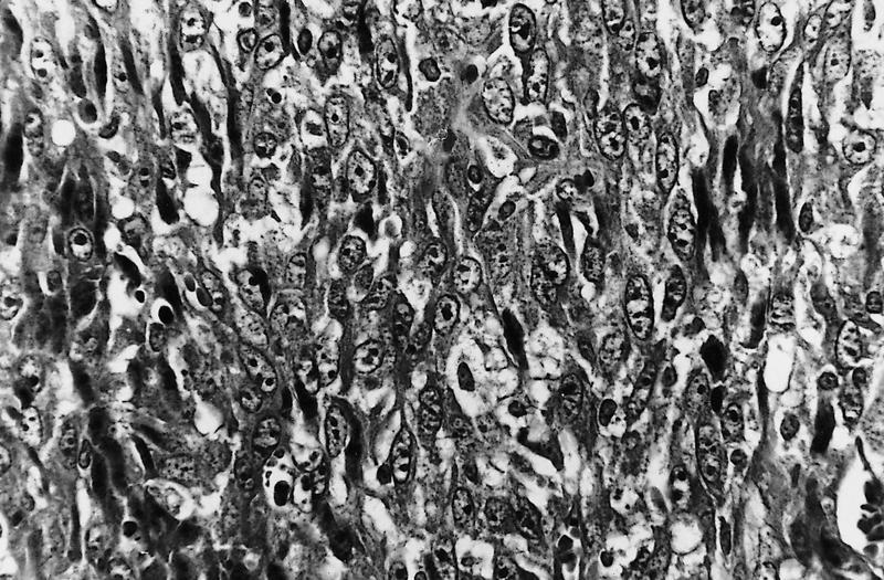
Pancreatoblastoma,
CT scan
Images hosted on other servers:

Pancreatoblastoma,
body / tail mass
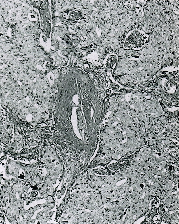
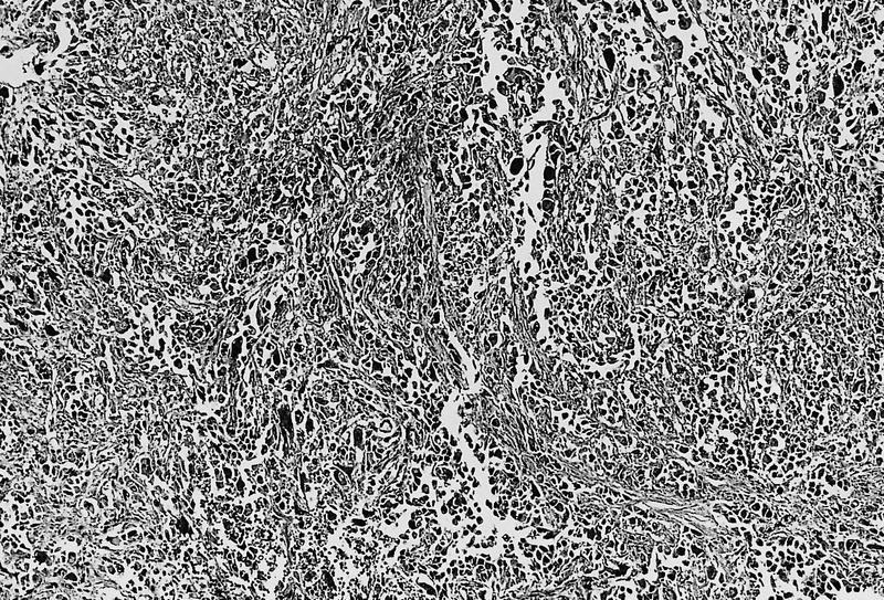
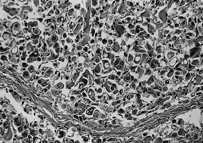
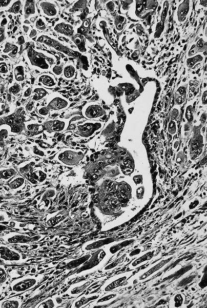
Contributed by Lizhi Zhang, M.D.
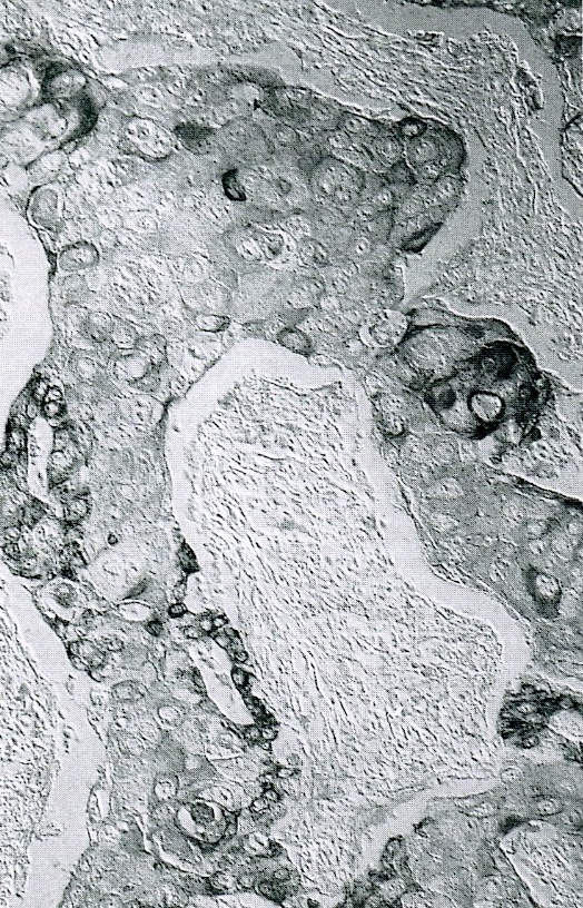
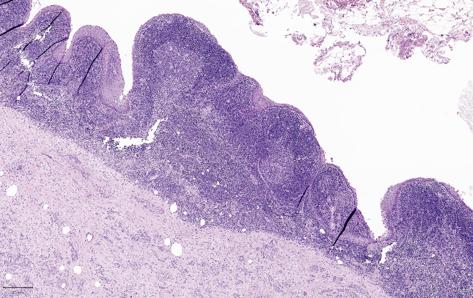
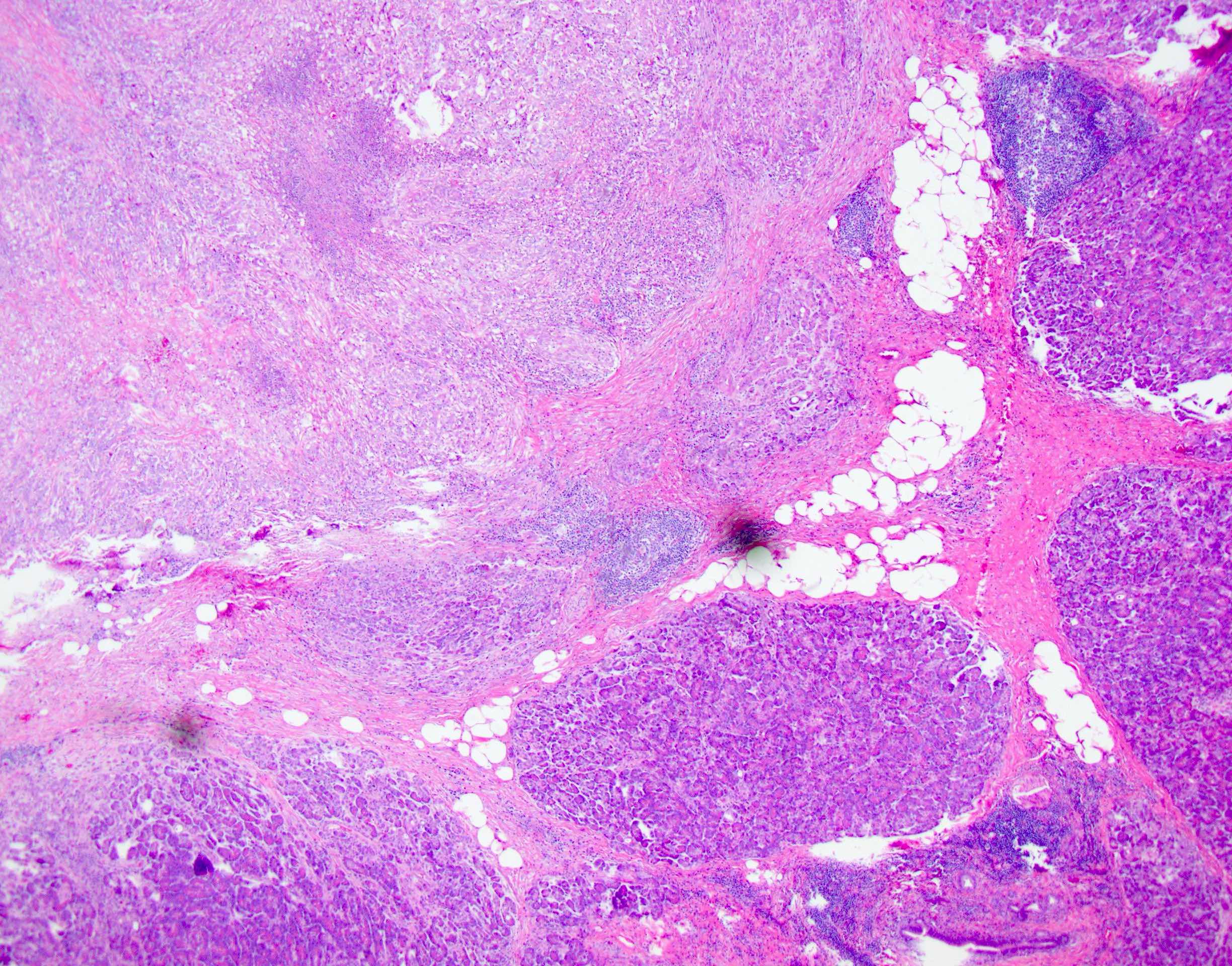
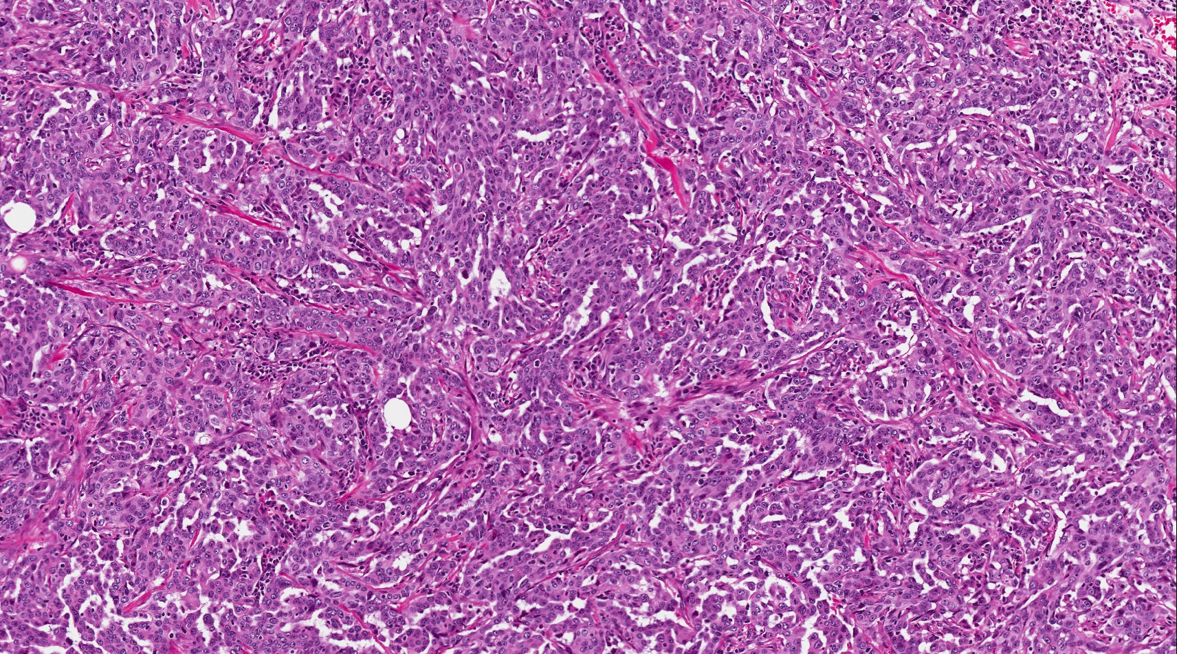
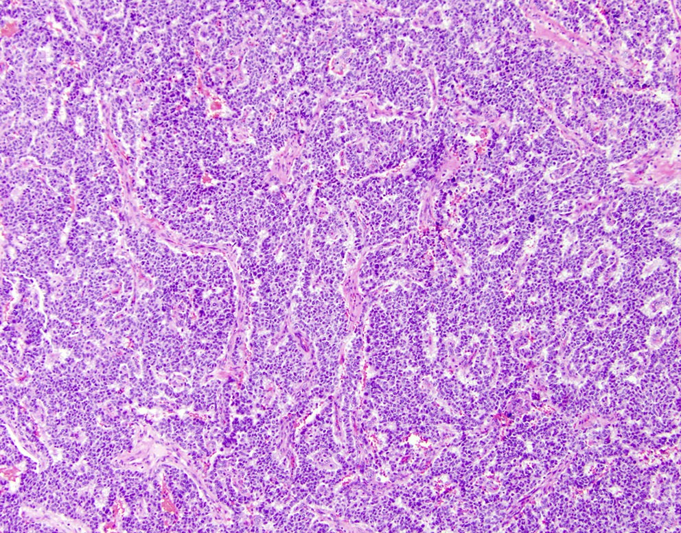
Geographic tumor lobules
Lobulated tumor nests
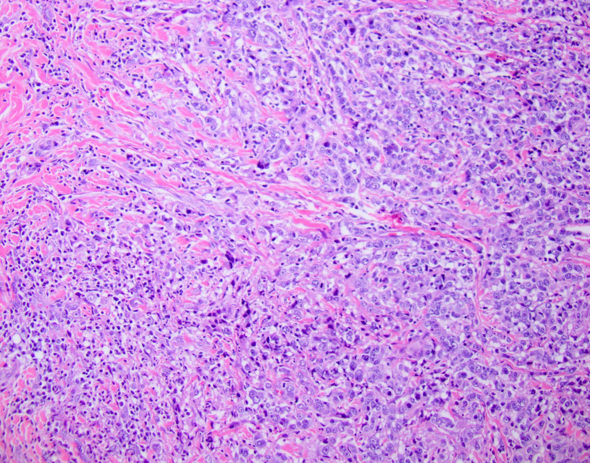
Acinar differentiation
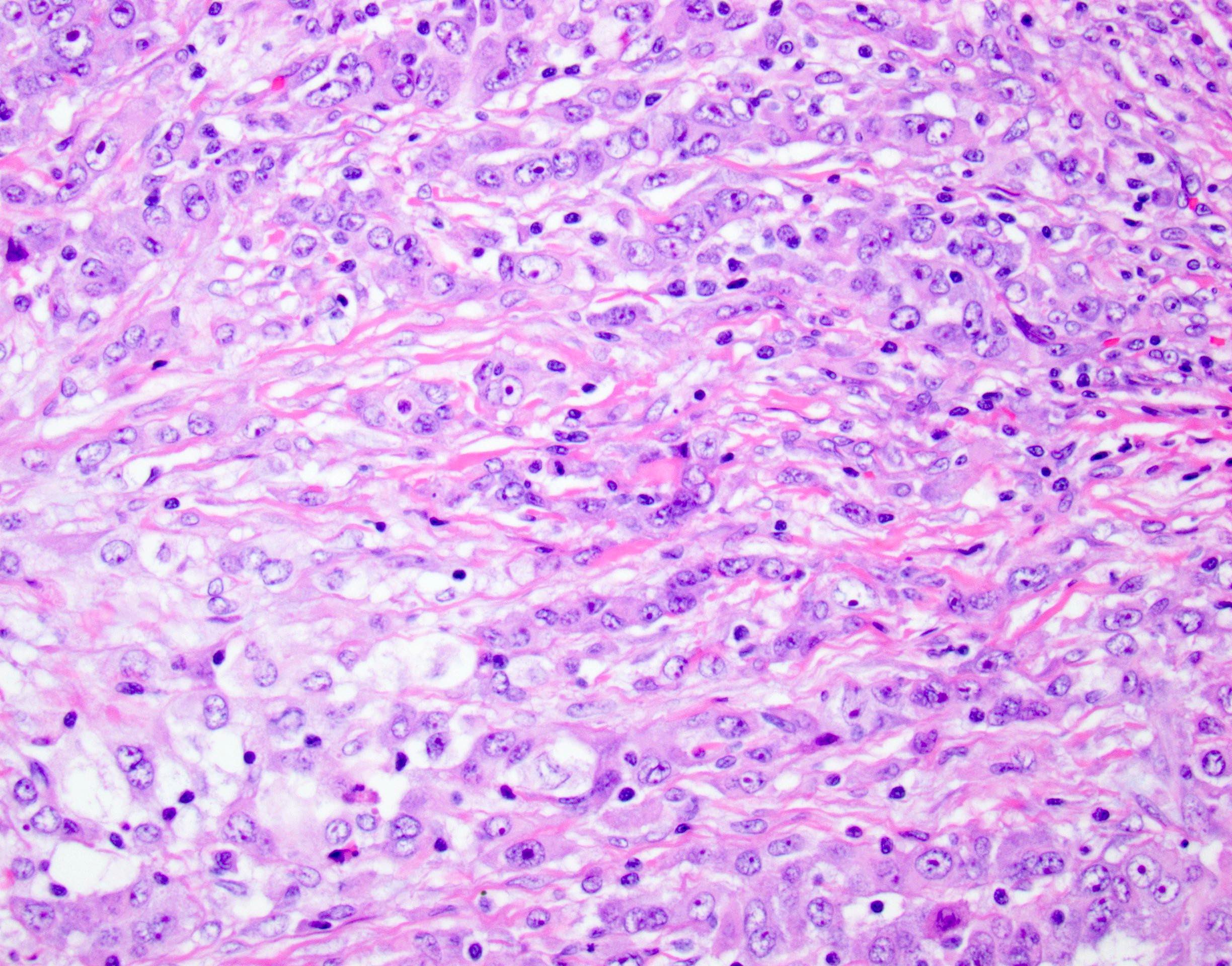
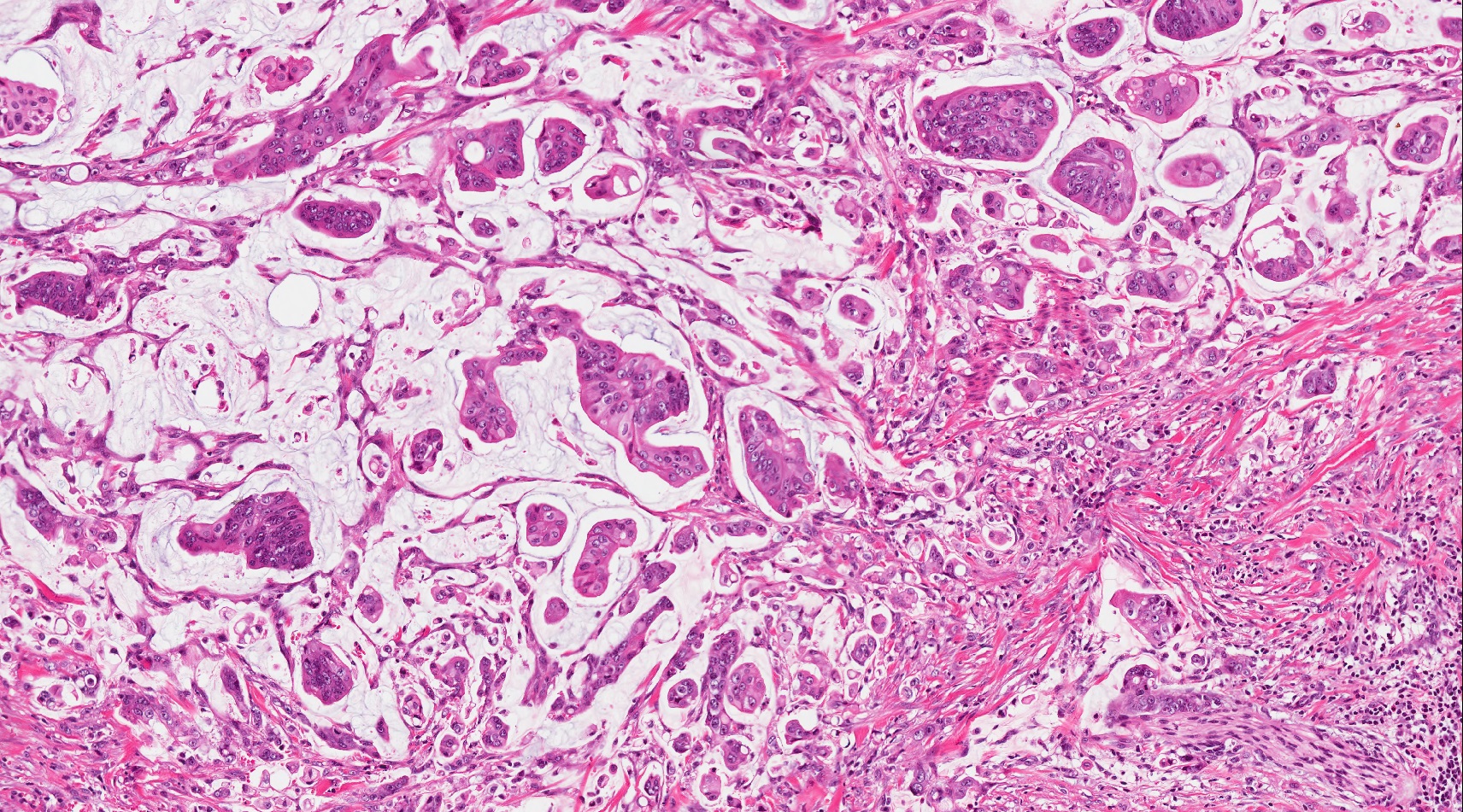
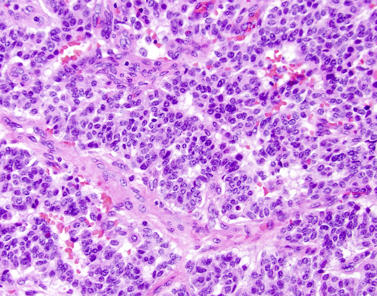
Squamoid nests
Squamoid nests with keratinization
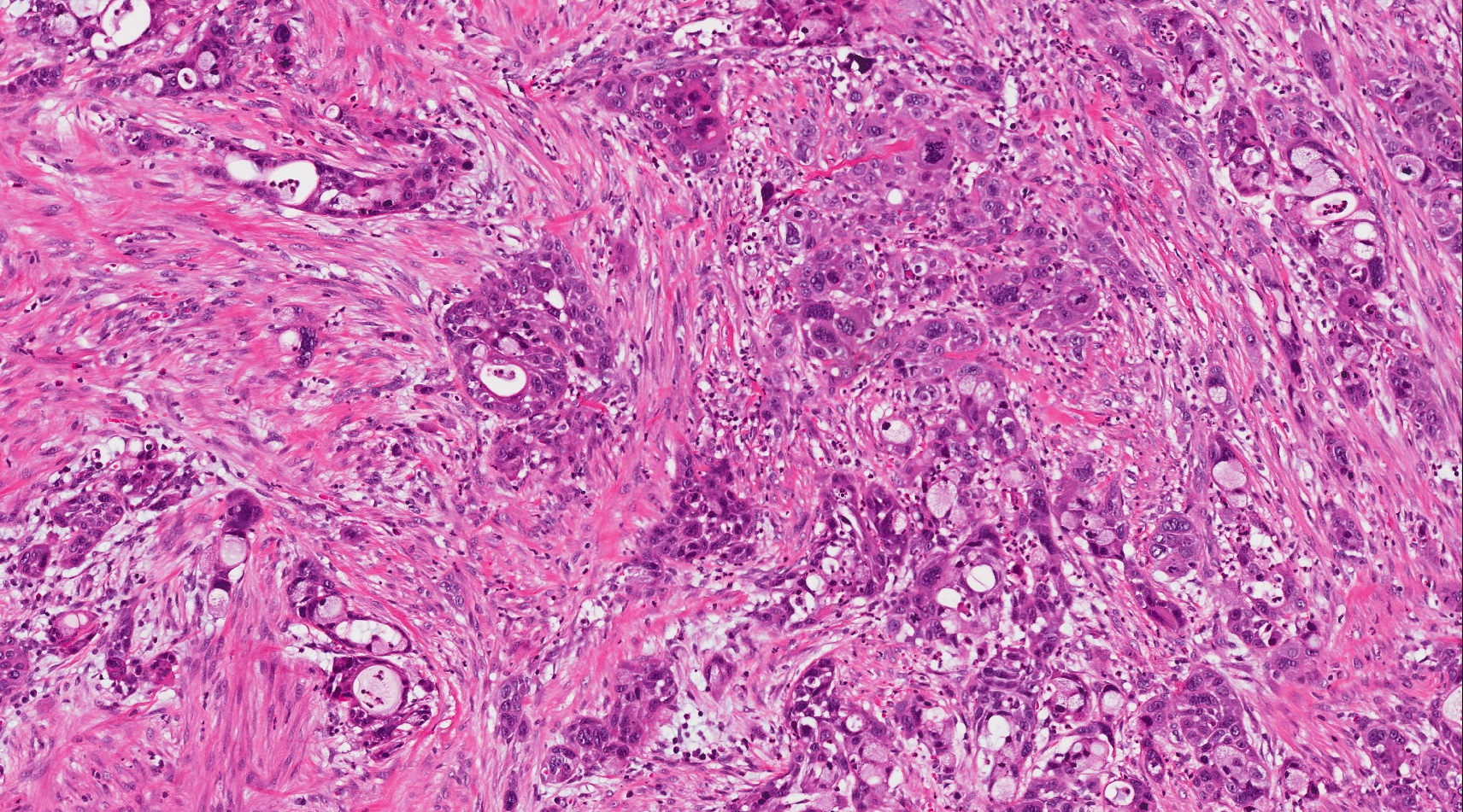
Neuroendocrine component
Ductal component
Primitive component
Stroma
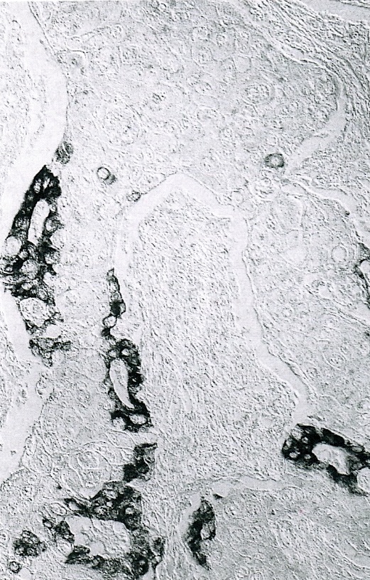
Keratin immunostain
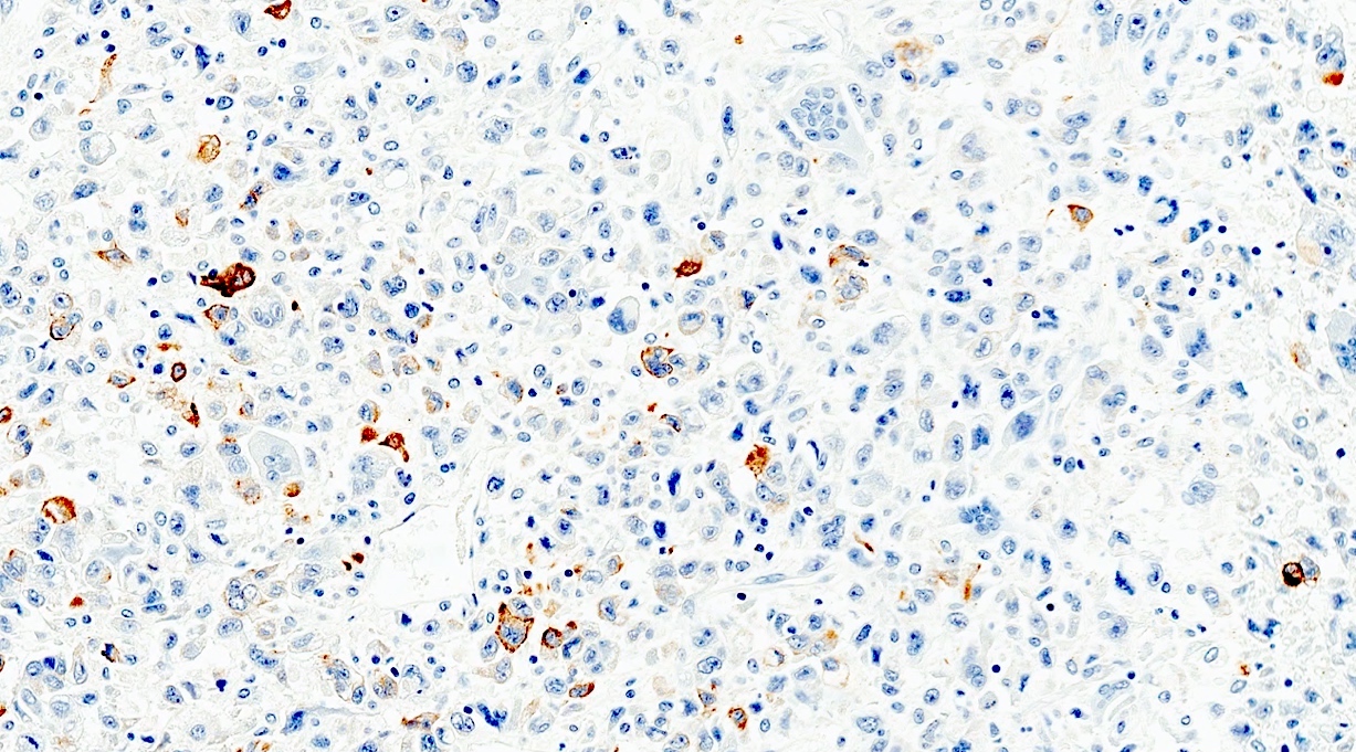
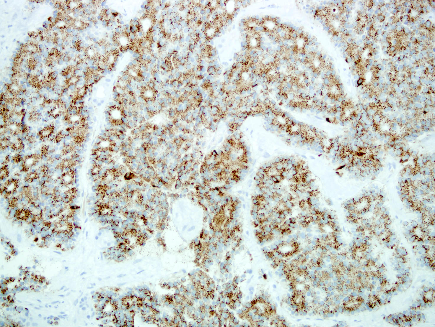
Trypsin immunostain
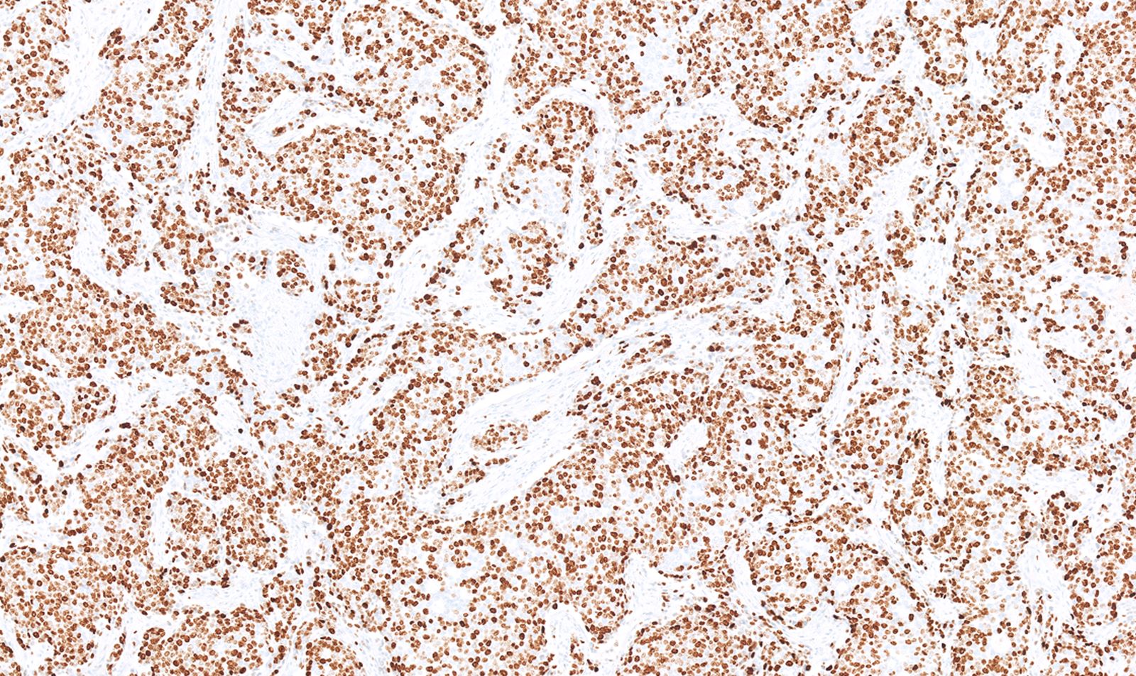
Beta catenin immunostain
CK5 immunostain
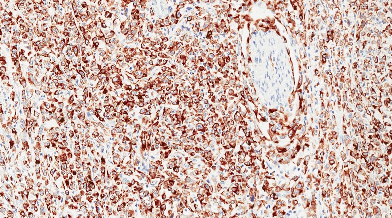
Synaptophysin immunostain
Images hosted on other servers:


Classification
Contributed by Ayşe Armutlu, M.D.
Normal
Low grade / PanIN 1A

Low grade / PanIN 1B
Low grade / PanIN 2
High grade / PanIN 3
Images hosted on other servers:

Survival curves for
well differentiated
tumors and
carcinomas
Images hosted on other servers:


Small cell carcinoma
Contributed by Claudio Luchini, M.D., Ph.D. and AFIP images
General morphological appearance
Tumor necrosis
Large cell PanNEC
Small cell PanNEC

Nodal metastasis of PanNEC

Synaptophysin positivity
Chromogranin A positivity
Typical p53 expression
Rb loss
Focal necrosis

Intense immunostaining for NSE
Solid diffuse pattern
Scattered somatostatin immunoreactive cells
Images hosted on other servers:

Overlapping pancreatic cells
Contributed by Aatur D. Singhi, M.D., Ph.D.
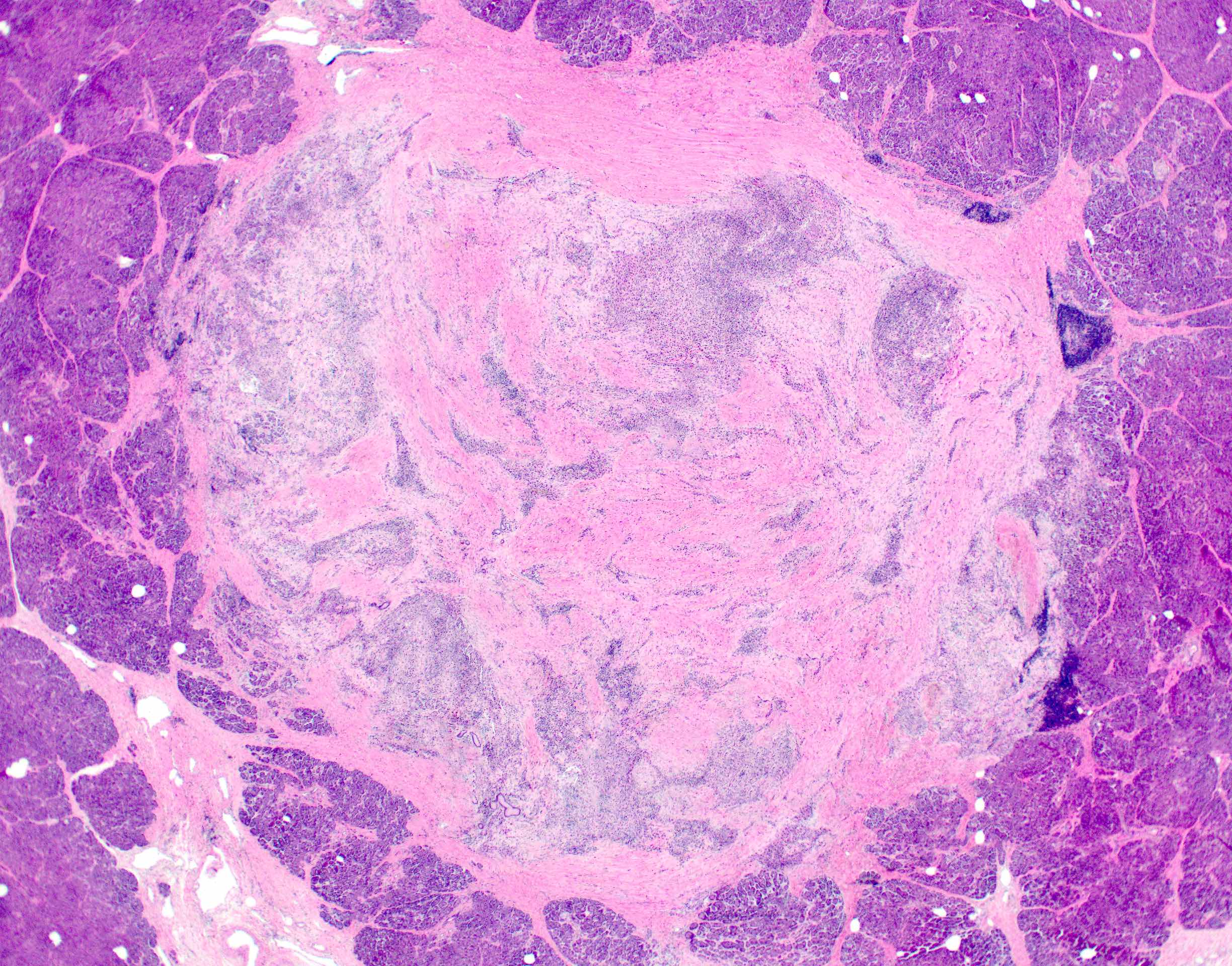
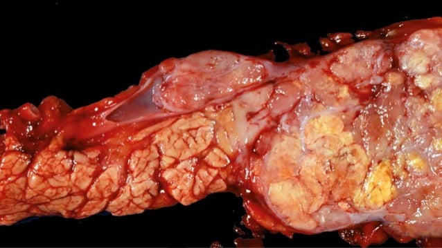
Gross parenchymal atrophy
Contributed by Aatur D. Singhi, M.D., Ph.D.
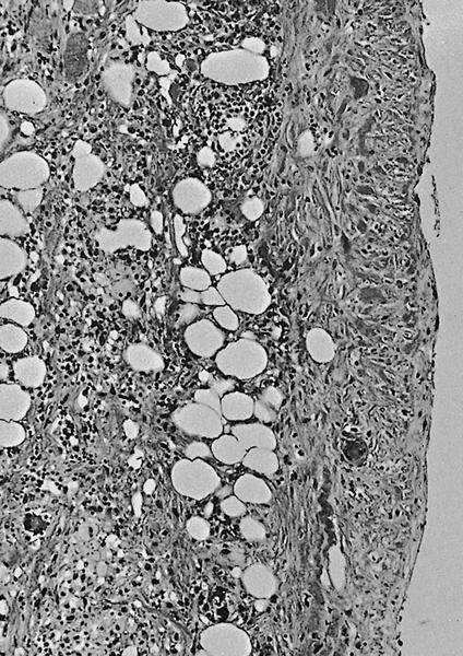
Early microscopic changes
Residual islets of Langerhans
Progressive replacement by fibrosis and fat
Near complete replacement
Low grade PanIN
Contributed by Paola Mattiolo, M.D. and Claudio Luchini, M.D., Ph.D.
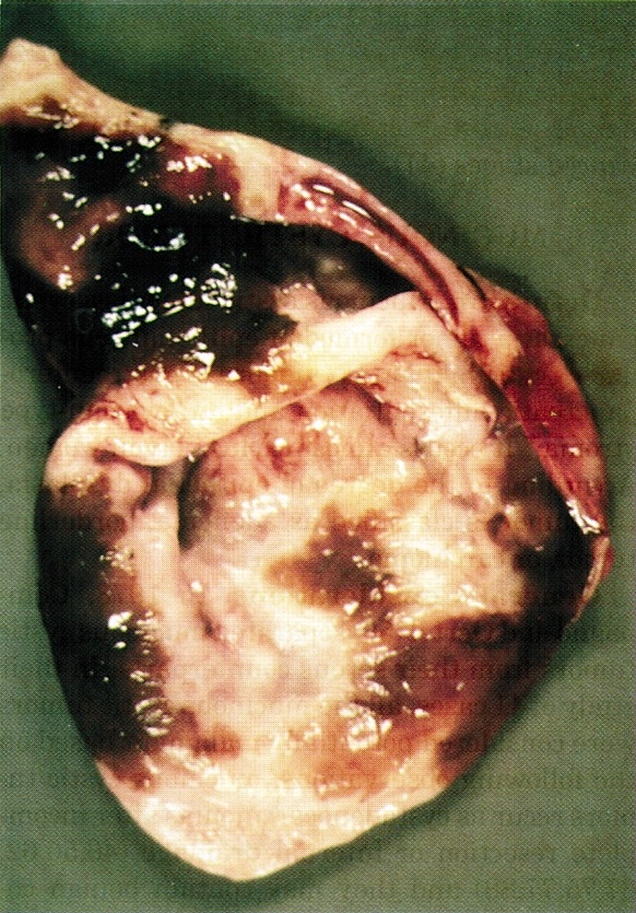
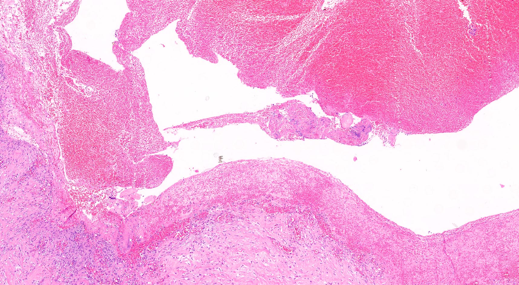
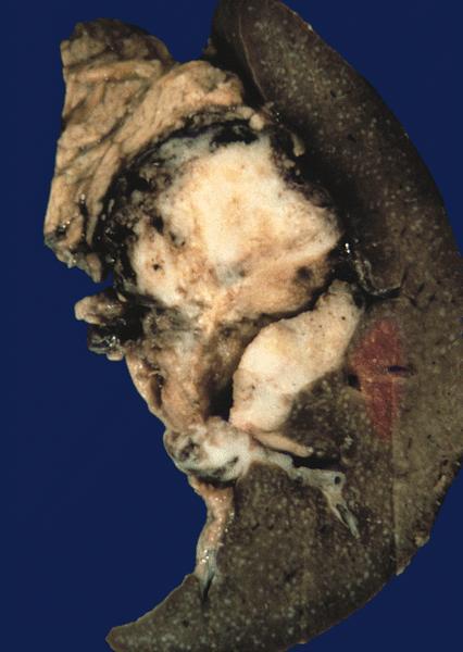
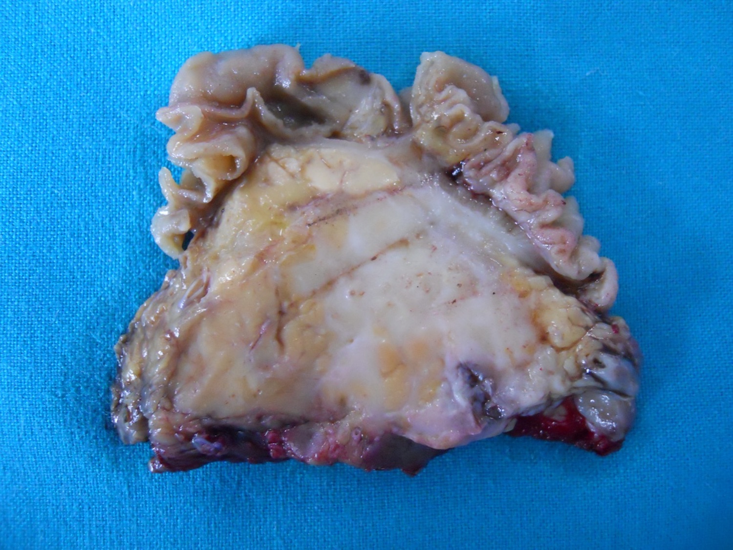
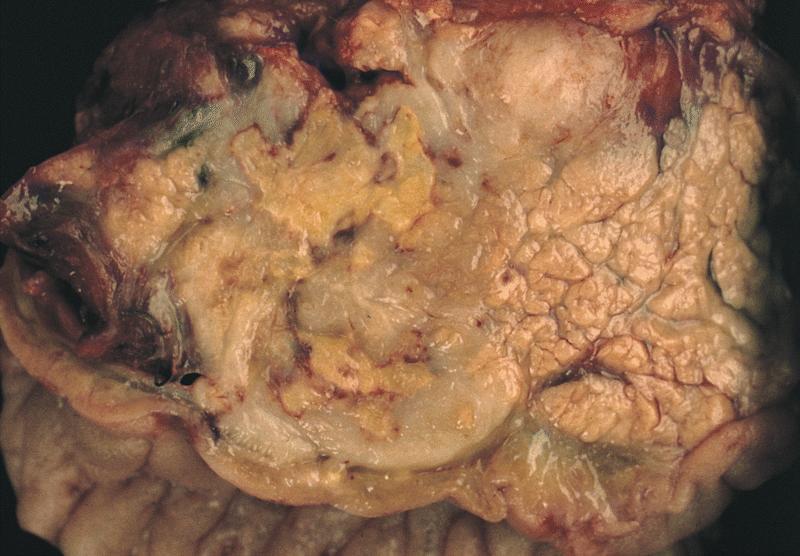
Large pancreatic pseudocyst
Fibrous wall of pseudocyst
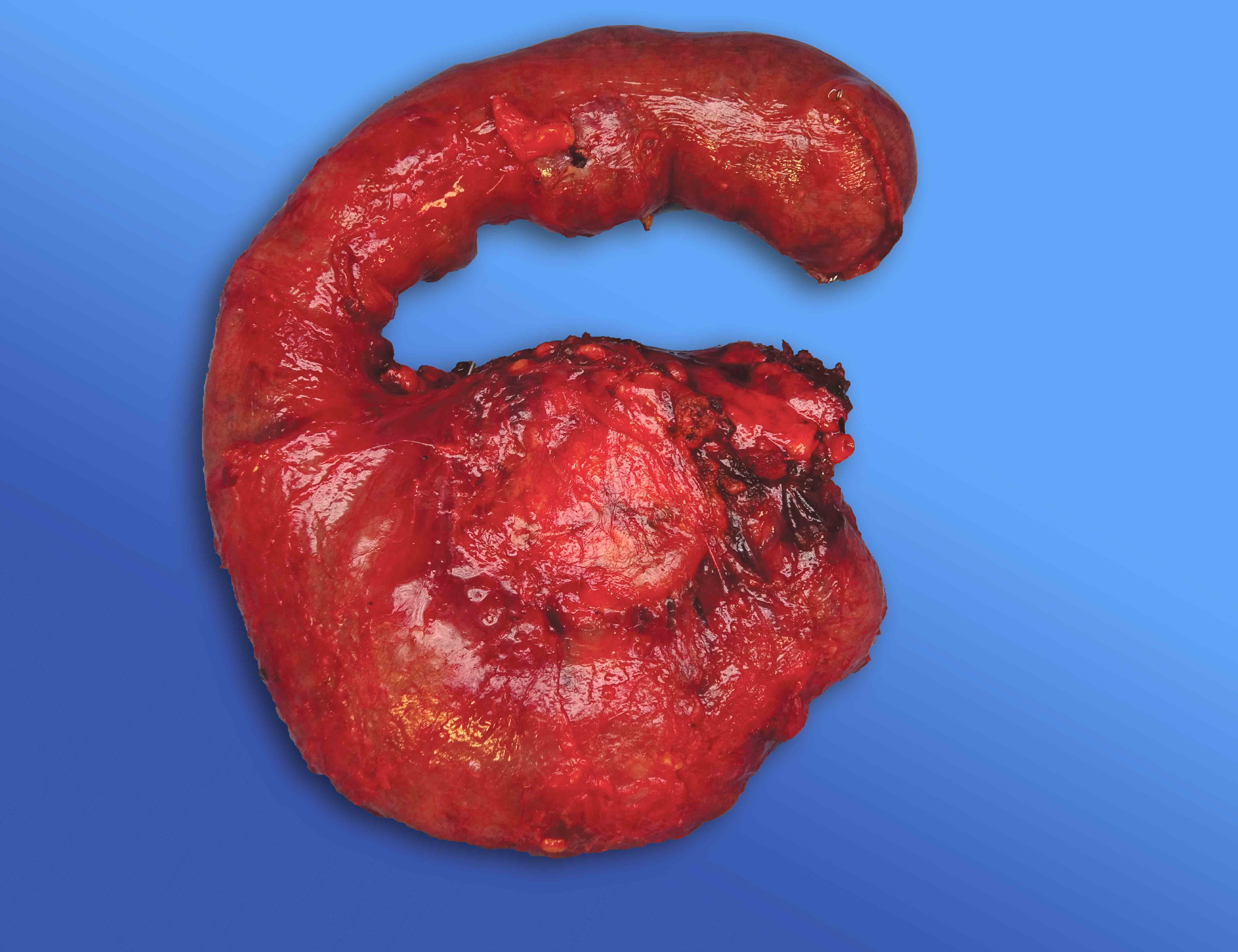
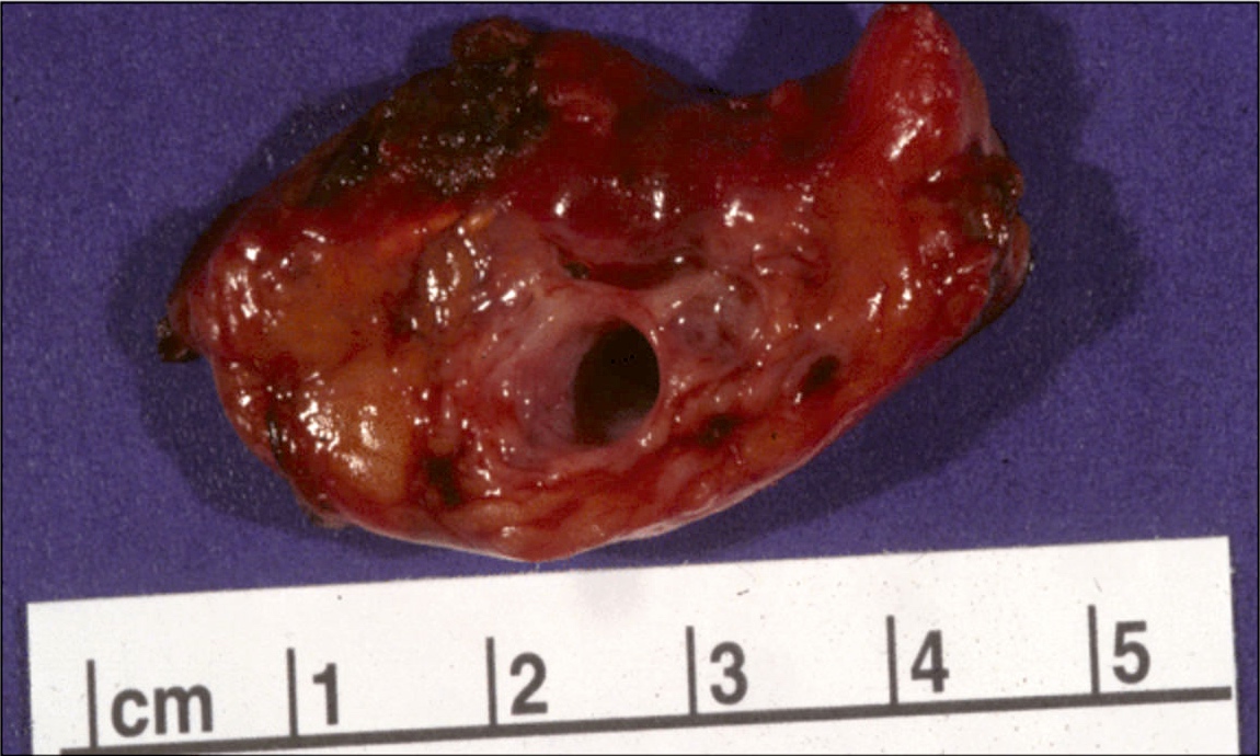
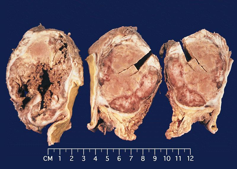
Large pseudocyst of the pancreatic head
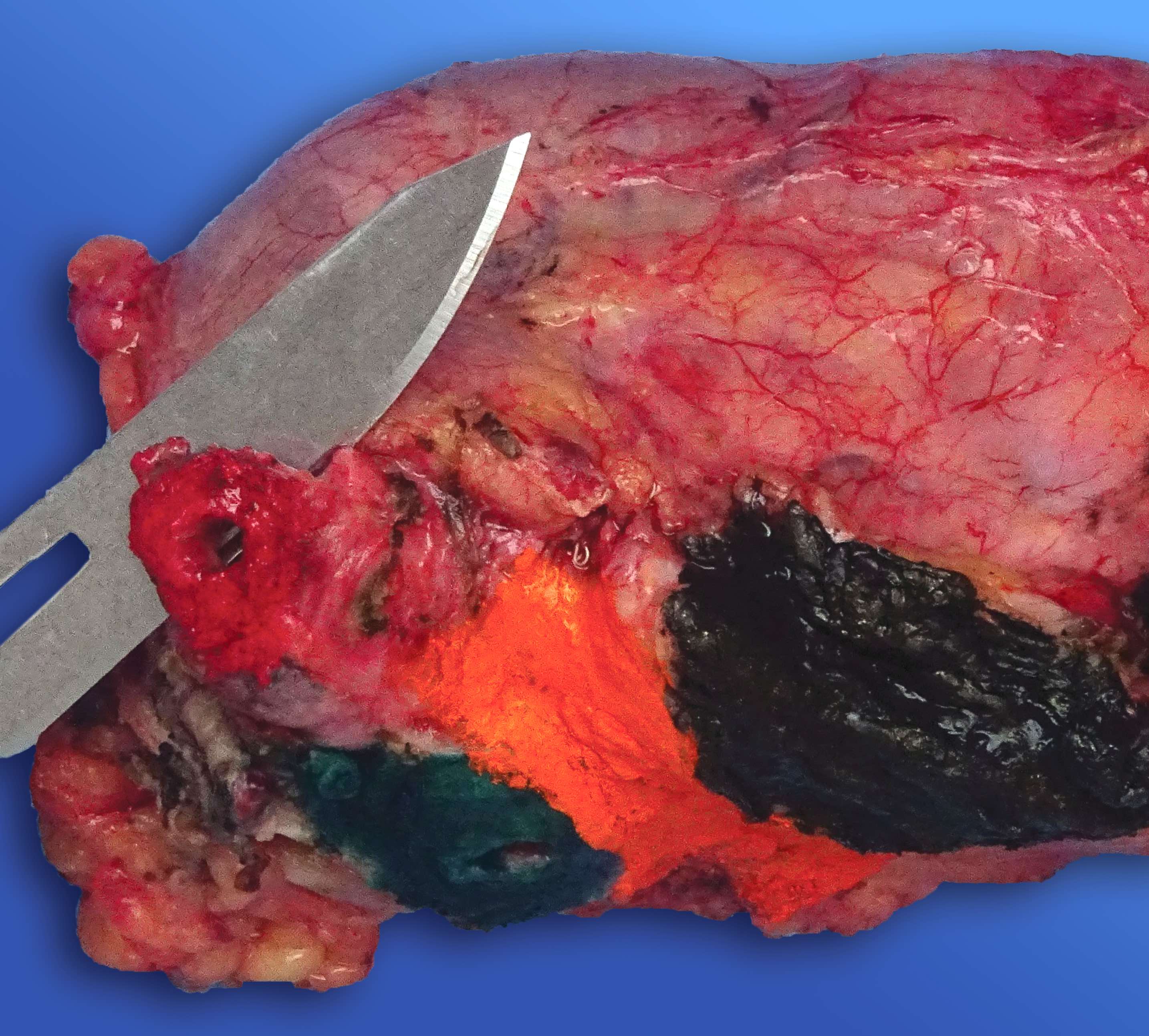
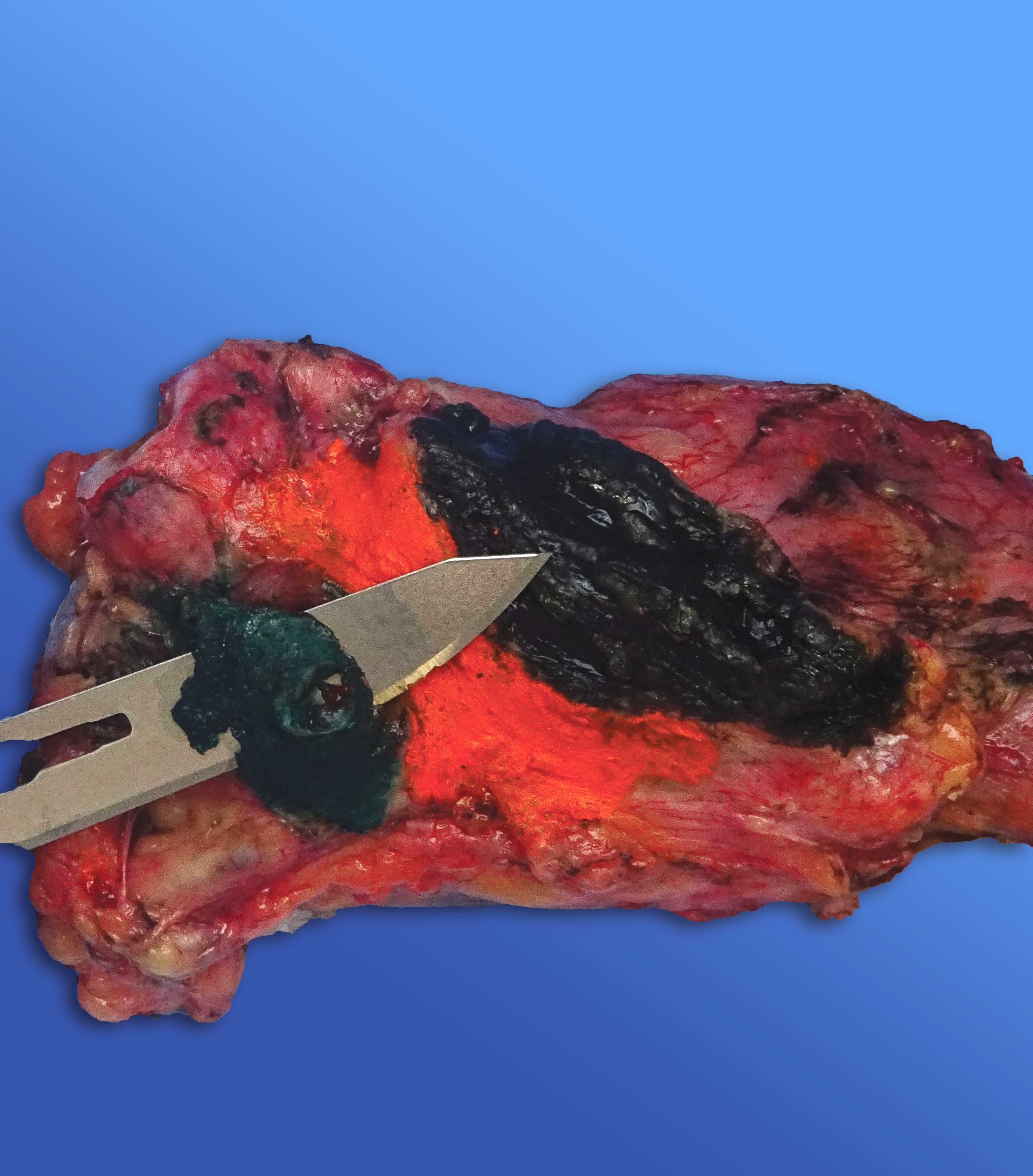
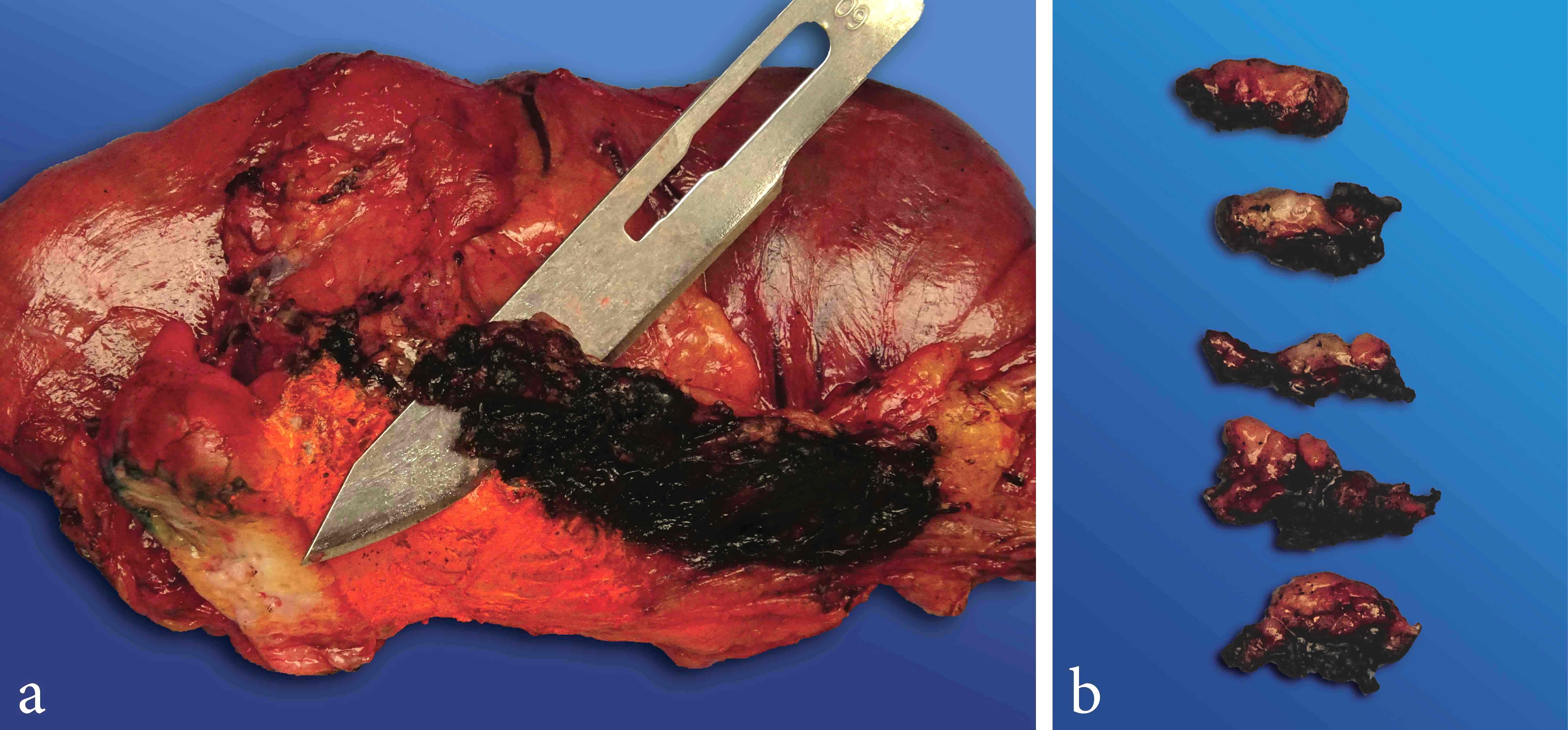
Cut surface of the pseudocyst of figure 3
Images hosted on other servers:

Multiseptated lesion filled with fluid

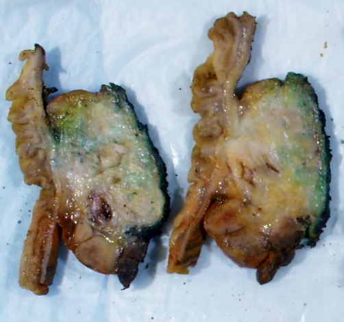
Encapsulated cystic lesion with yellow-green material

Resembling cystic neoplasm
Contributed by Paola Mattiolo, M.D., Claudio Luchini, M.D., Ph.D. and AFIP images
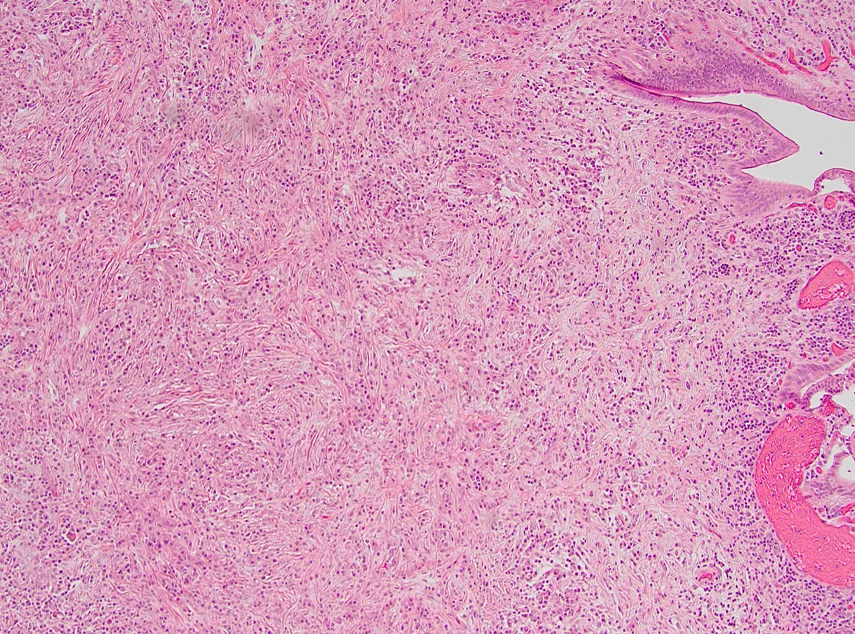
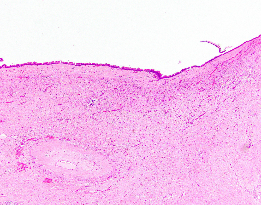
Absence of an epithelial lining
Inflammation of pseudocyst wall
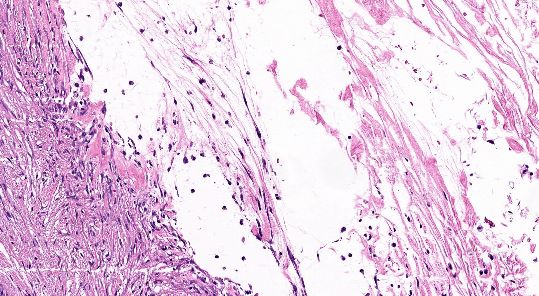
Fibrotic reaction
Hemorrhagic modifications
Patient with chronic pancreatitis and pseudocyst
Images hosted on other servers:

Well circumscribed solid lesion
Contributed by Olca Basturk, M.D.
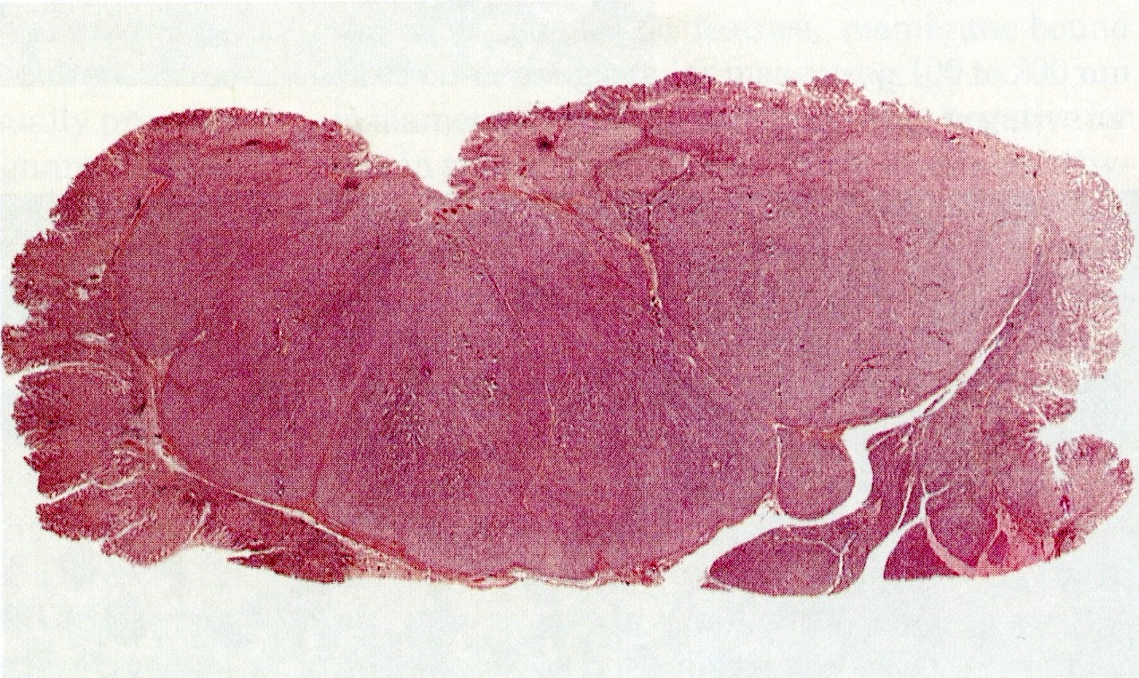
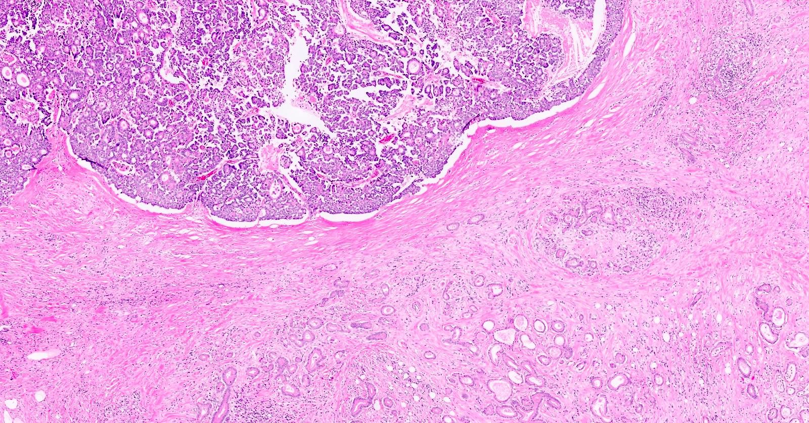
Overall appearance
Well circumscribed lesion
Peripheral dense lymphoid aggregates
Entrapped pancreatic parenchyma
Hypercellular and hypocellular areas
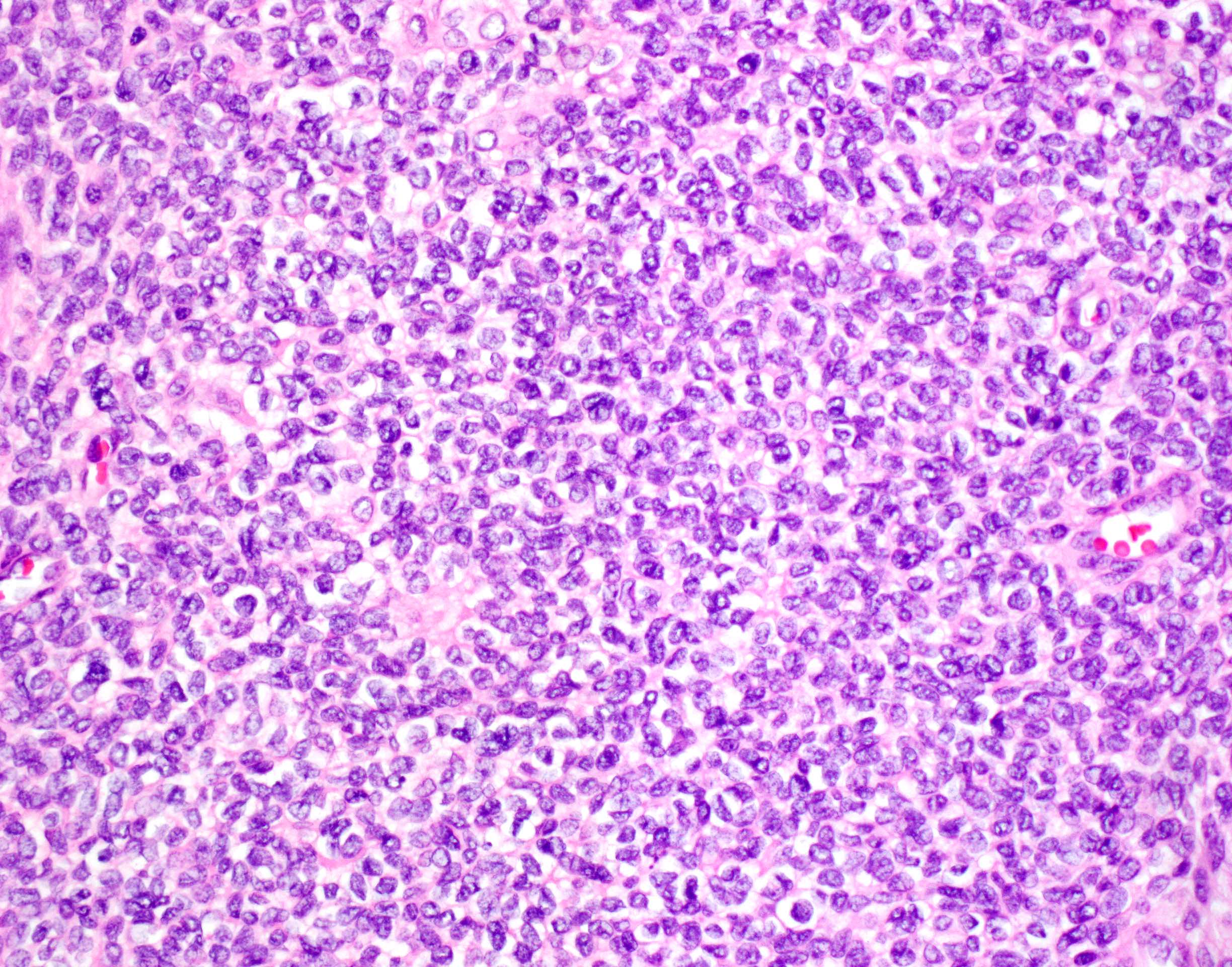
Sheets of epithelioid cells
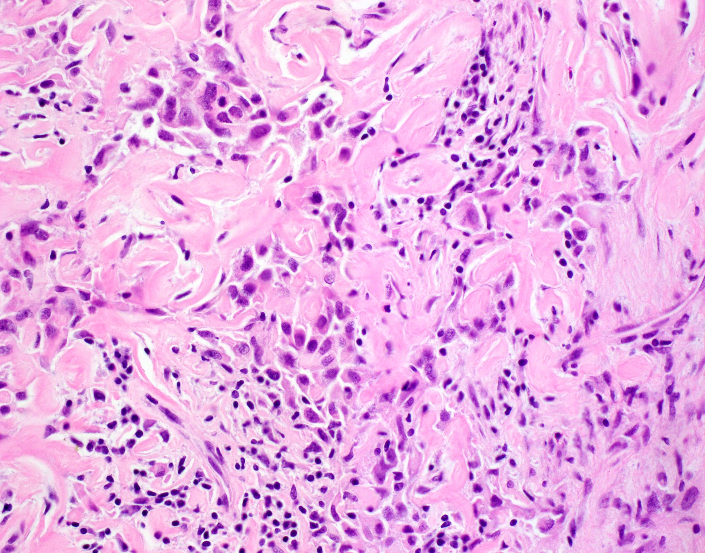
Atypical spindle cells
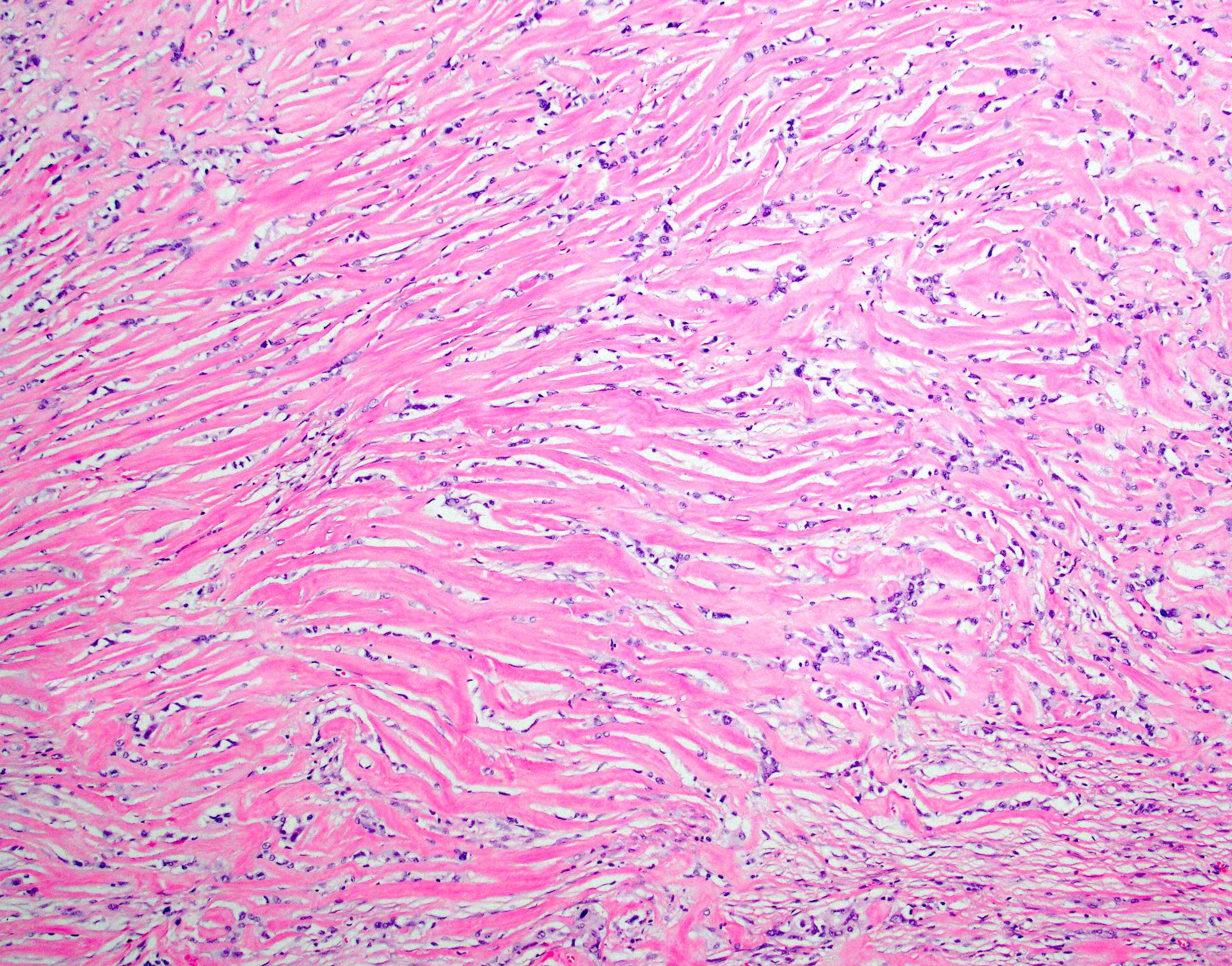
Dense hyaline fibrosis
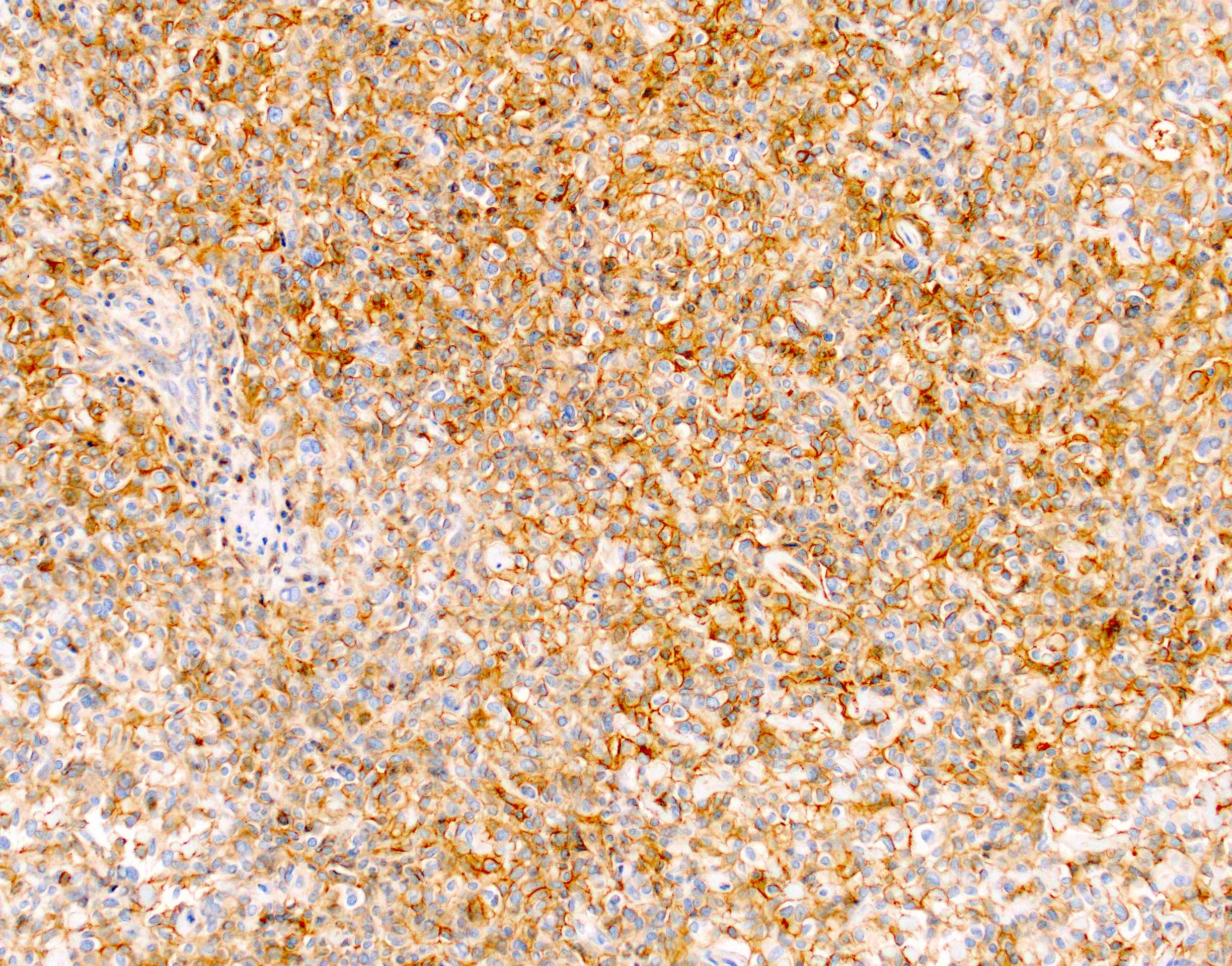
Membranous CD99 expression
Dot-like CK18 expression
Images hosted on other servers:

Genomic and transcriptomic characterization
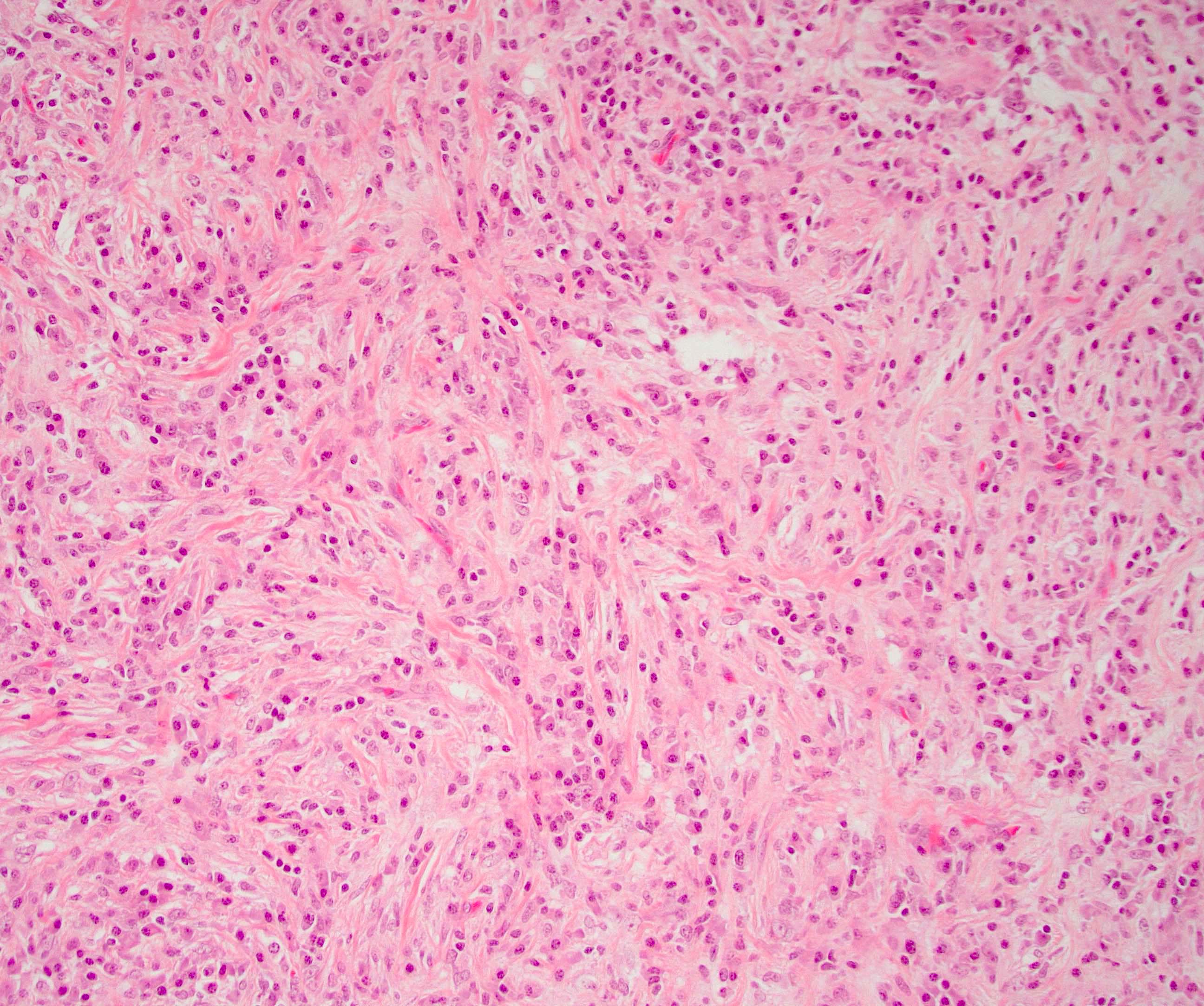
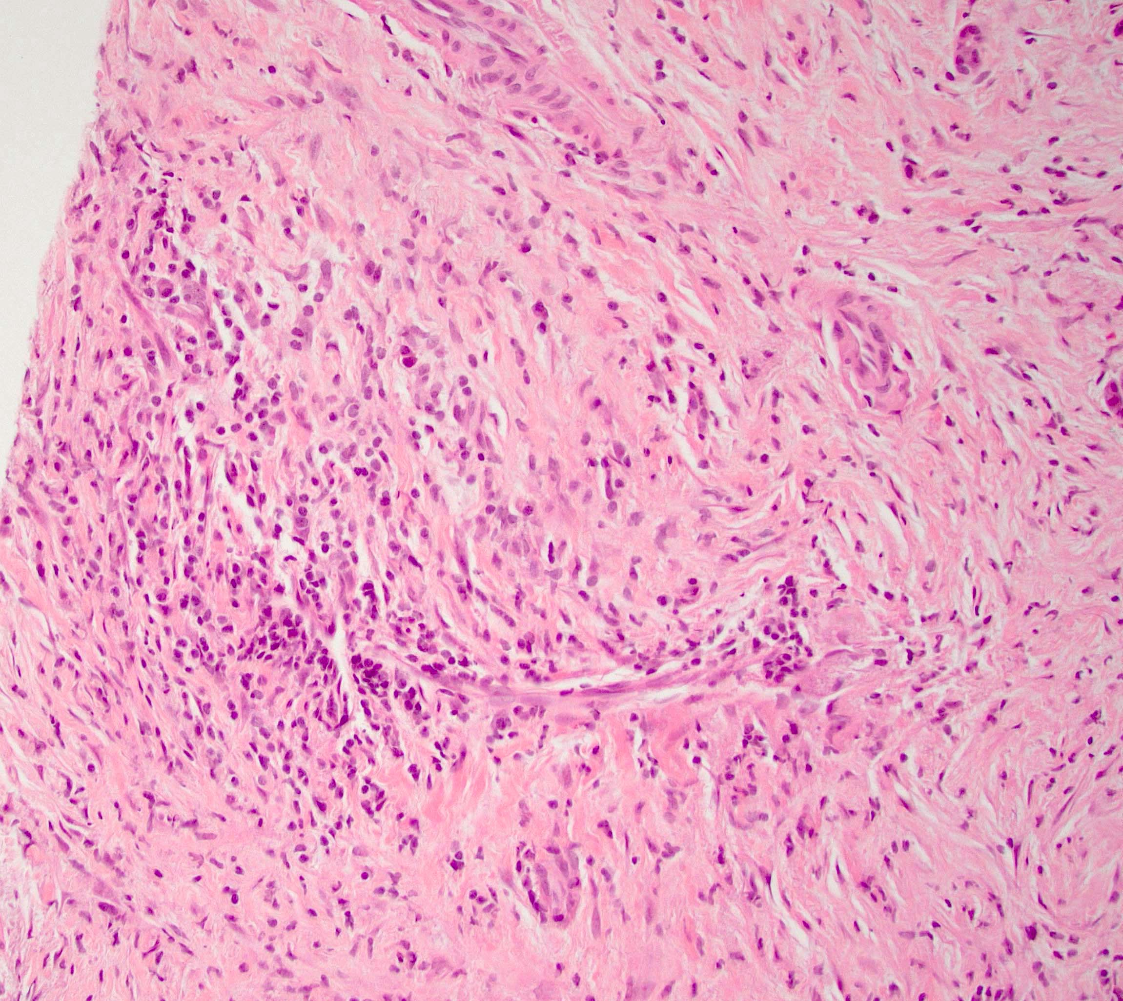
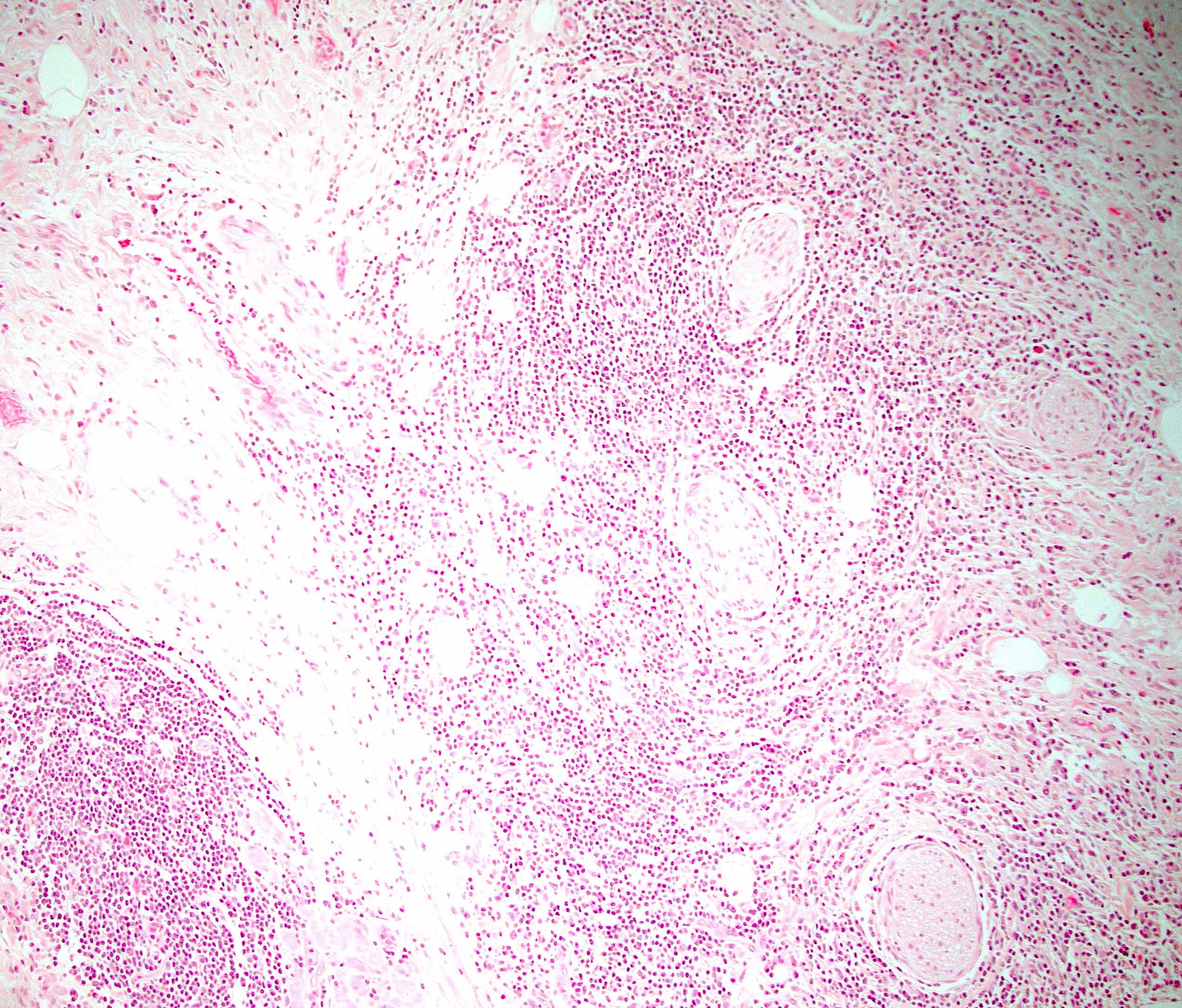
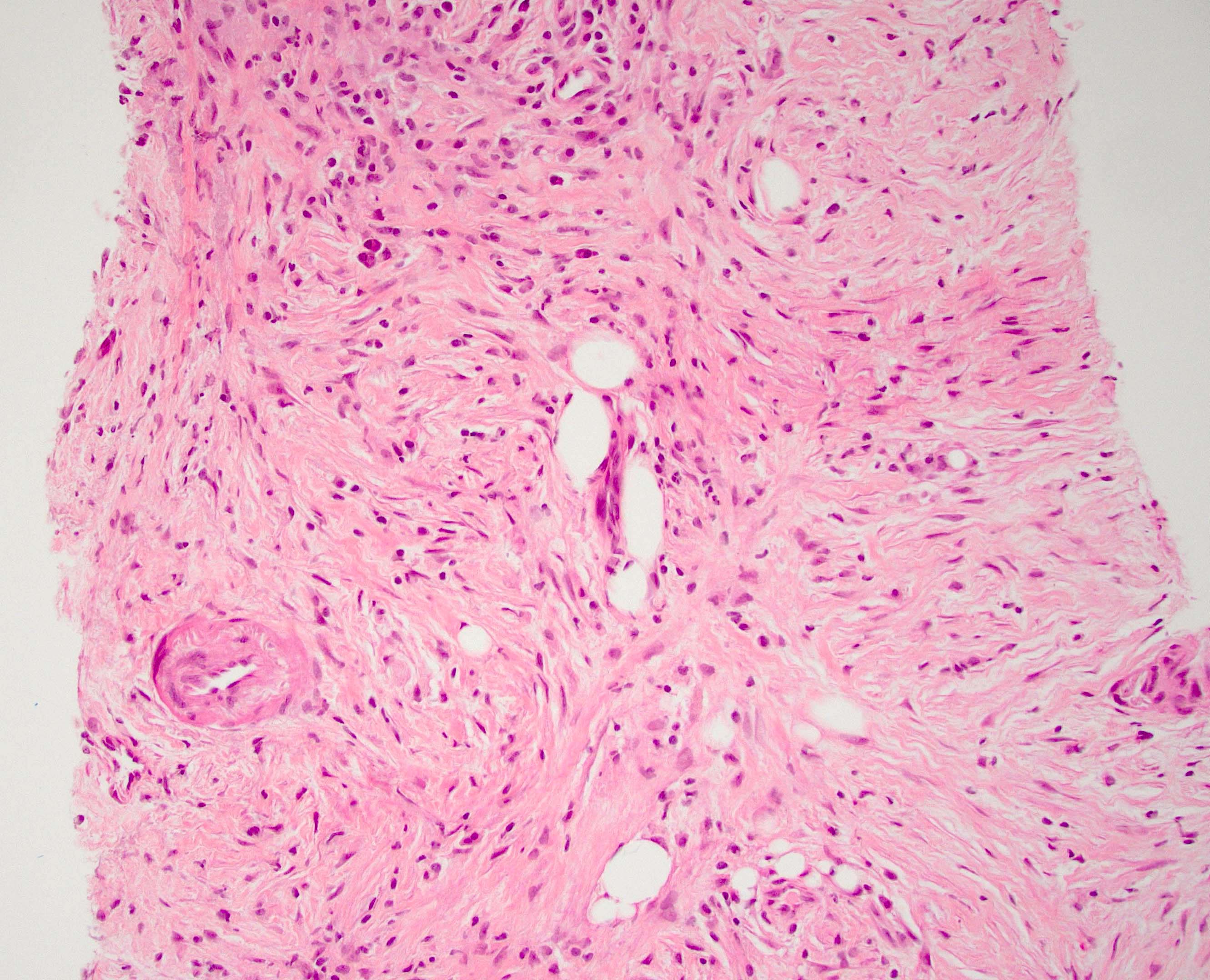
Contributed by Pınar Bulutay, M.D.
Central scar
Thin fibrous septa
Macrocystic and solid variant
Images hosted on other servers:

Solid variant
Contributed by Pınar Bulutay, M.D.
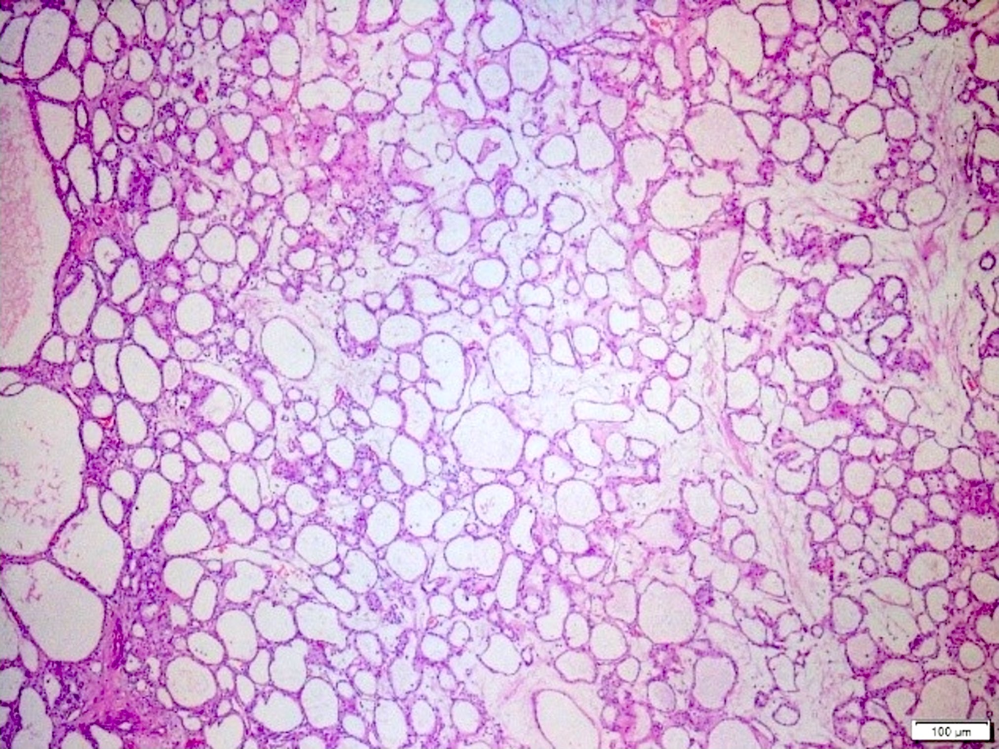

Innumerable submillimeter cysts
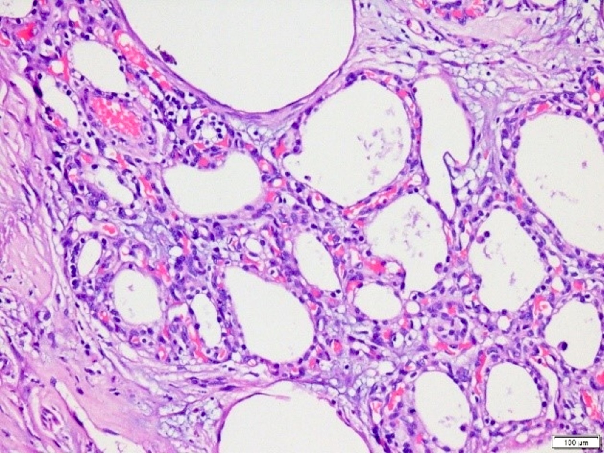
Prominent capillary network


Cellular details
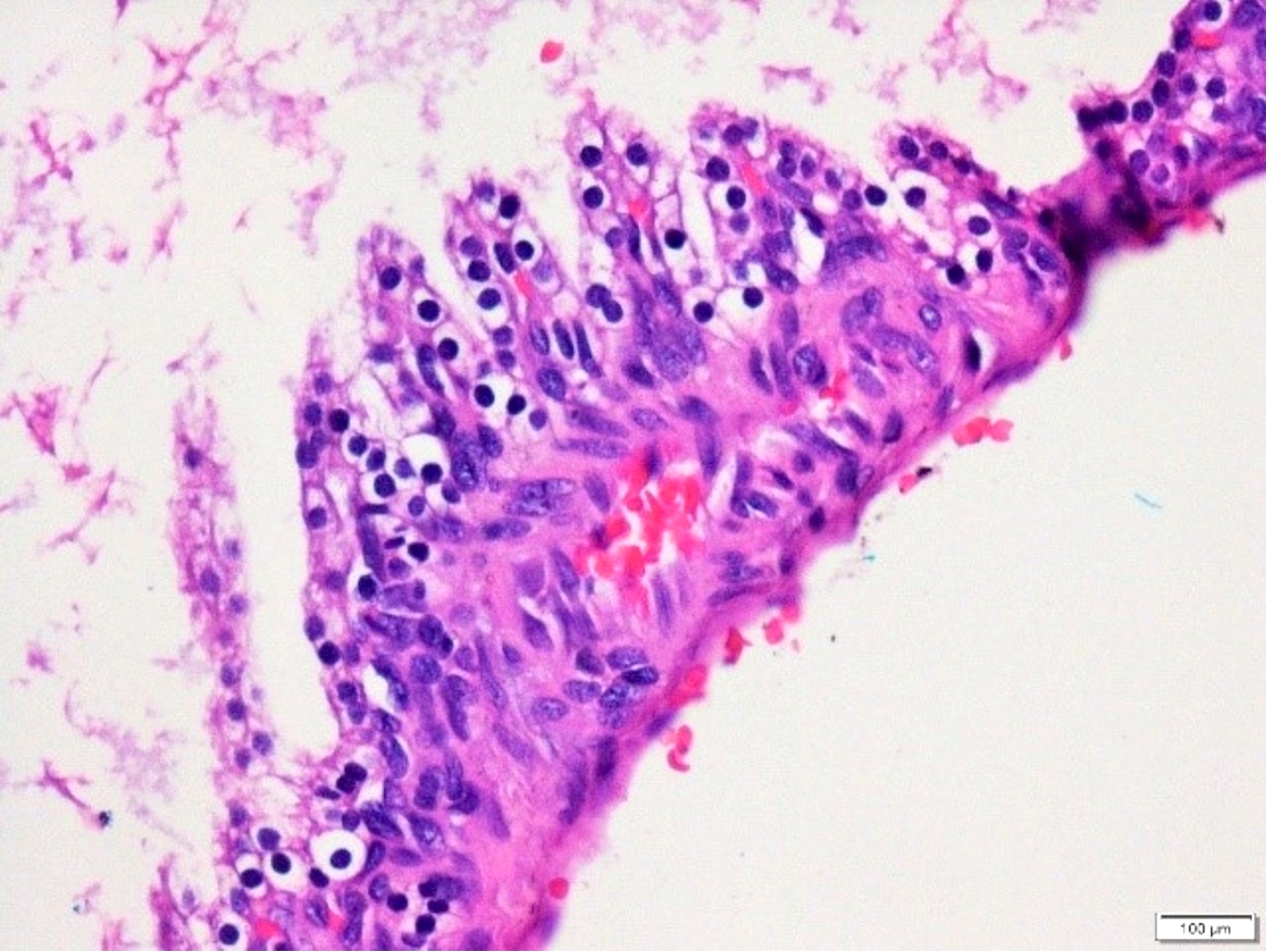
Intracystic papillary projections
Macrocystic type
Cellular details, macrocystic type
Papillary projections, macrocystic type
Contributed by Amanda Kitson, M.D. and Jiaqi Shi, M.D., Ph.D.
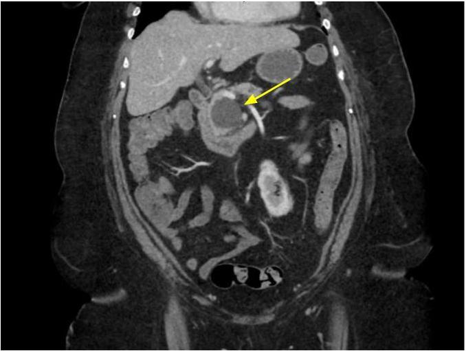
CT: abdomen coronal
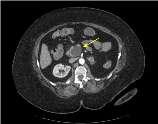
CT abdomen, transverse section
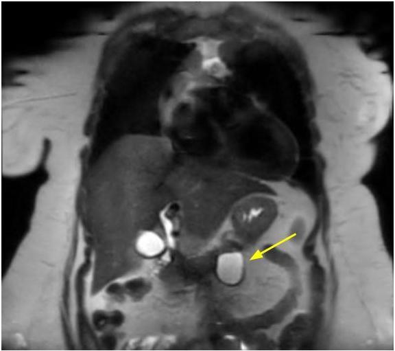
T2 MRI: abdomen coronal
Images hosted on other servers:

Hematochezia and active bleeding from ampulla

Pancreatic cyst and elevated posterior wall of gastric body
Contributed by Amanda Kitson, M.D. and Jiaqi Shi, M.D., Ph.D.
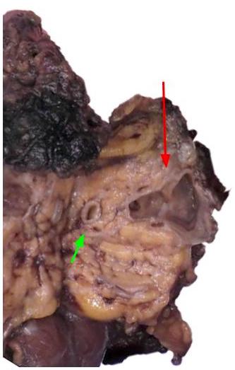
Multiloculated cyst in pancreatic head
Cyst in pancreatic body
Contributed by Amanda Kitson, M.D. and Jiaqi Shi, M.D., Ph.D.

Cuboidal to columnar epithelium
Gastric type mucinous epithelium
Hypocellular stroma
Focal atypia
Contributed by Omid Savari, M.D.
MR of abdomen
Images hosted on other servers:

Hypodense lesion

Endoscopic ultrasound

Multiple metastases
Contributed by Omid Savari, M.D., Dr. Andreas Schulz, Manfred Stolte, M.D., Dr. Helmut Luchtrath and Case #121
Predominately solid tumor
Partially necrotic tumor
Invasion of spleen
Cystic lesion
Multilocular cystic mass
Contributed by Monika Vyas, M.D., Omid Savari, M.D. and Raul S. Gonzalez, M.D.
Pleomorphism and cytologic atypia
Intracytoplasmic eosinophilic hyaline globules
Tumor infiltration by foamy histiocytes


Solid pseudopapillary neoplasm
Solid pattern

Clear cell changes

Cholesterol cleft, scattered giant cells
Tumor invading parenchyma

Beta catenin
CD10
Ki67
Metastasis to liver
Contributed by Omid Savari, M.D. and @ThatGlassTho on Twitter
Diff-Quik

Pap
Cell block
Solid pseudopapillary neoplasm
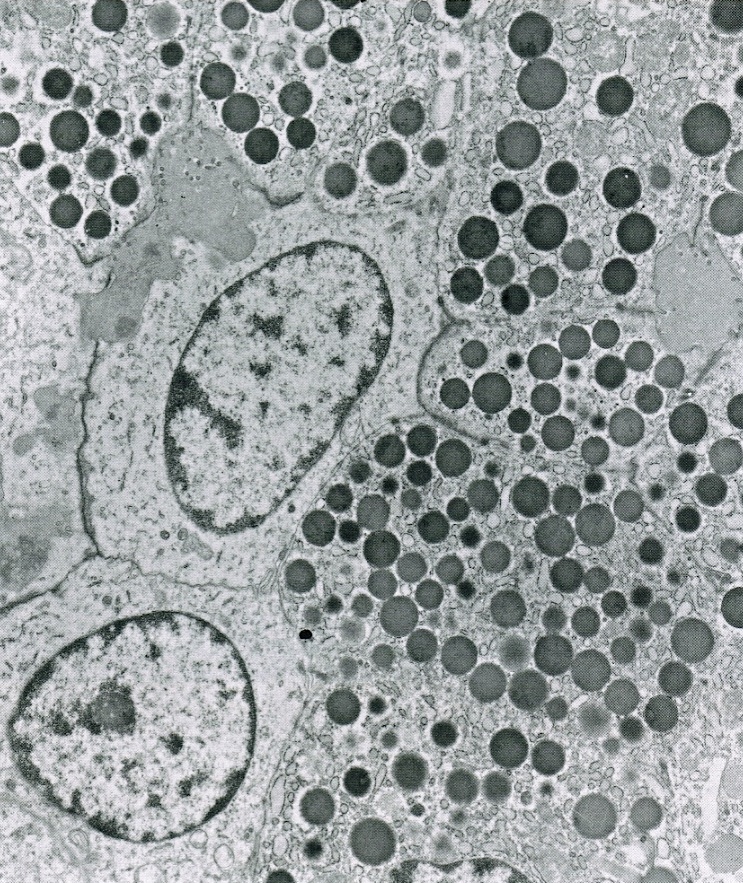
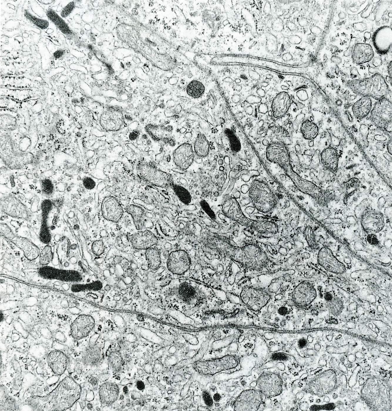
Contributed by Dr. H.D. John, Mainz
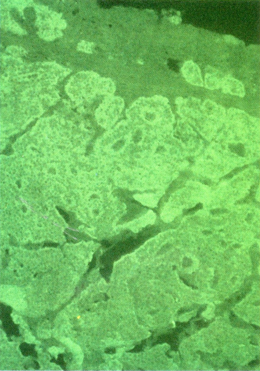
Abundant mitochondria

Granules
Images hosted on other servers:

CT pancreas mass

PET with metastases
Images hosted on other servers:

Pancreatic and duodenal tumors
Contributed by Raul S. Gonzalez, M.D.
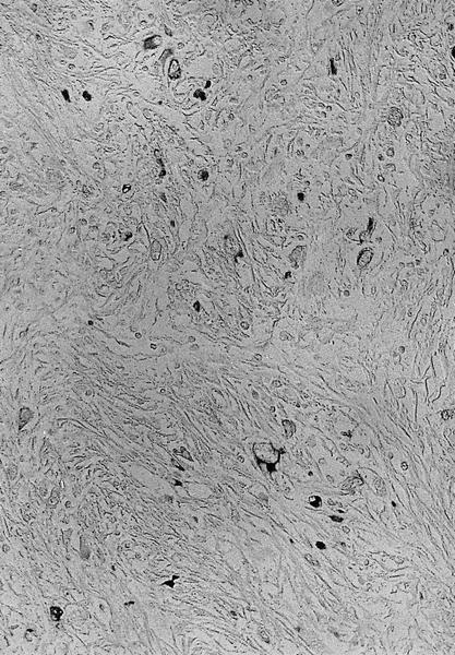
Nests and cords of cells
AFIP image
D cell granules
Contributed by Claudio Luchini, M.D., Ph.D. and AFIP
Ductal
adenocarcinoma
with undifferentiated
component
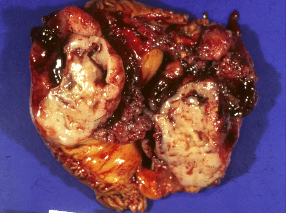
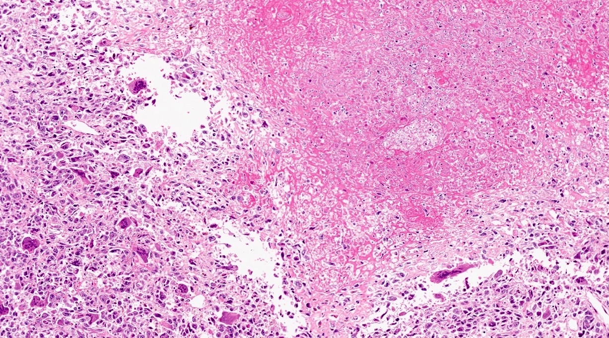
Ill demarcated tumor in head of pancreas
Extensive hemorrhagic necrosis
Contributed by Claudio Luchini, M.D., Ph.D. and AFIP
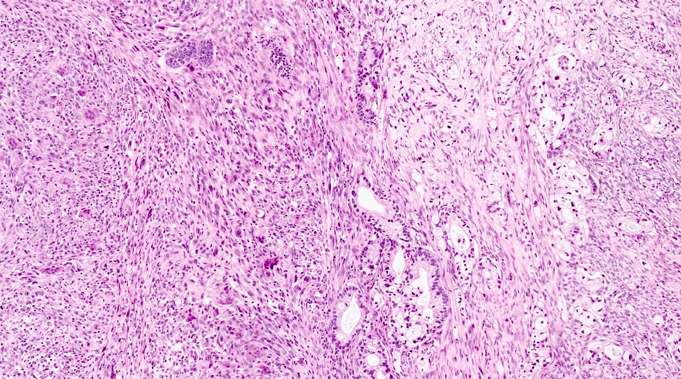
Anaplastic undifferentiated carcinoma
Massive perineural invasion
Massive vascular invasion
Association with ductal adenocarcinoma
Invasion of duodenum
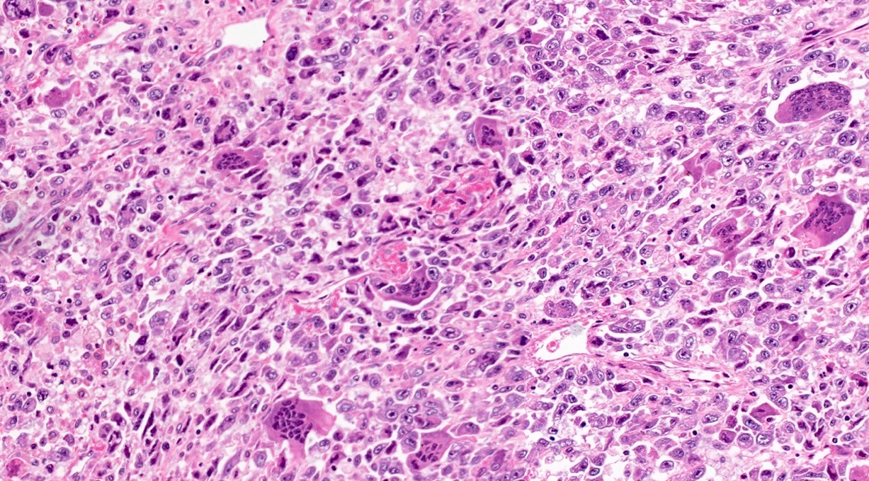
Rhabdoid aspects
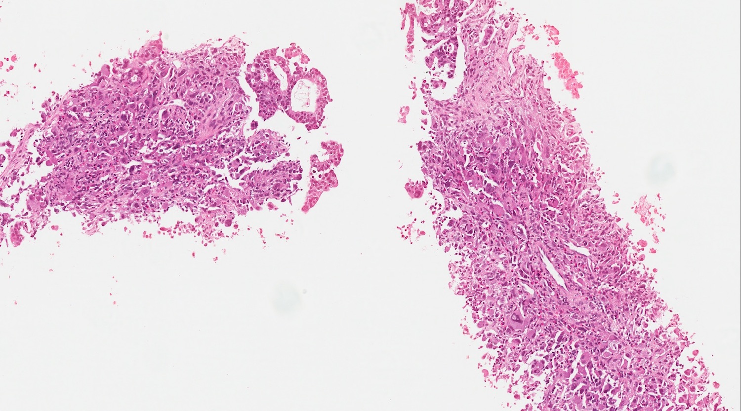
Biopsy
Details on biopsy
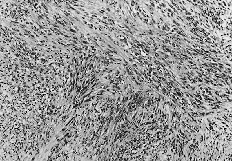
Spindle cell sarcomatoid features
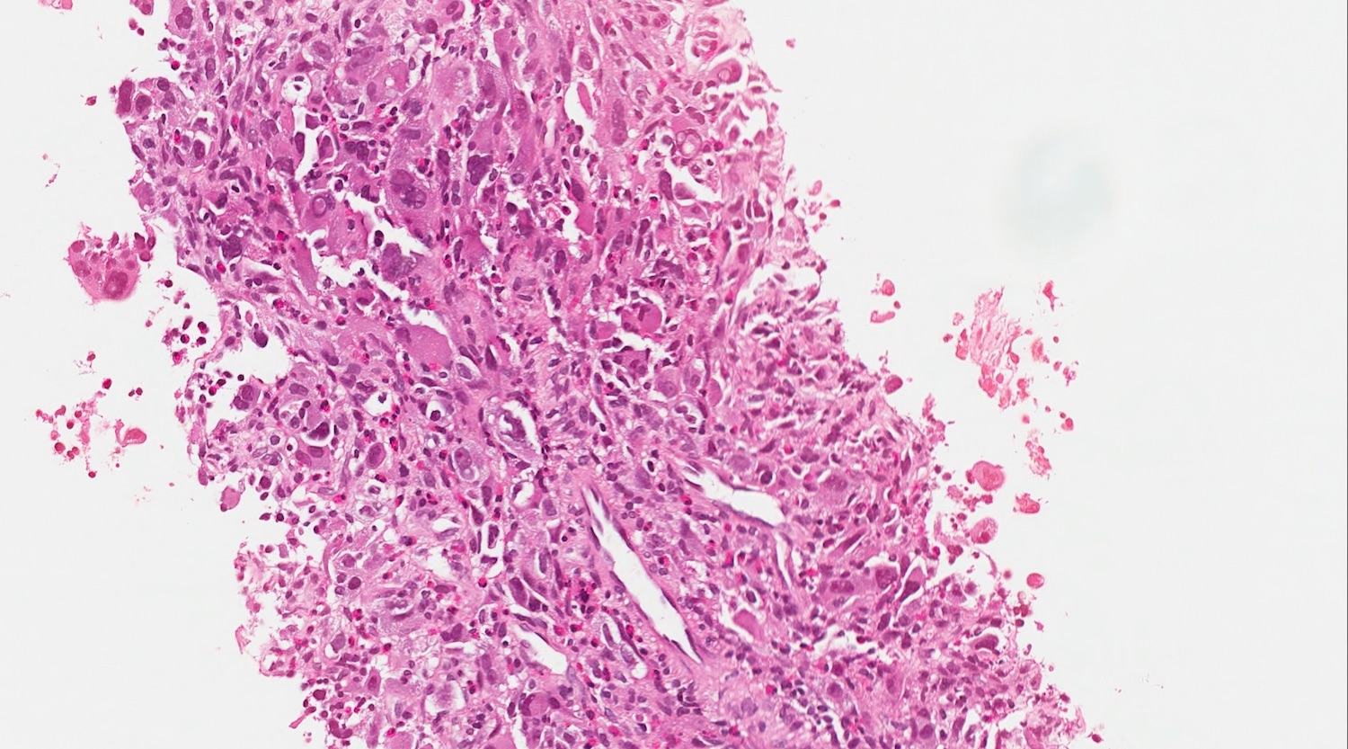
Pleomorphic large cells
Engulfment of red blood cells
Glandular differentiation
Tumor is keratin+
Tumor is CEA+ in glandular portions
Images hosted on other servers:

Ultrasound


CT
Contributed by Claudio Luchini, M.D., Ph.D.
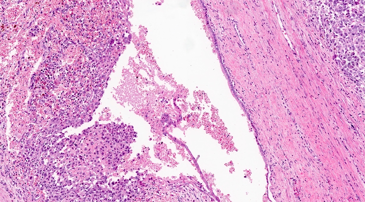
Cystic tumor
Images hosted on other servers:

Hemorrhagic mass

Macrophotograph revealing cystic mass

Cystic mass with gastric invasion
Contributed by Claudio Luchini, M.D., Ph.D.
Associated conventional adenocarcinoma
Ductal tree involvement
Microhemorrhagic foci
Extensive necrotic hemorrhage
CK8/18+
Focal CK8/18+

CK8/18-
Images hosted on other servers:

Romanowsky stain

Papanicolaou stain
Images hosted on other servers:

Neoplastic cell

Giant cell
Images hosted on other servers:

Pancreatic tail mass

Liver metastasis
Contributed by Raul S. Gonzalez, M.D.
Nests and cords of cells
AFIP images

Pancreatic VIPoma
Images hosted on other servers:

Large mass in pancreas tail
Contributed by Katrina Krogh, M.D.
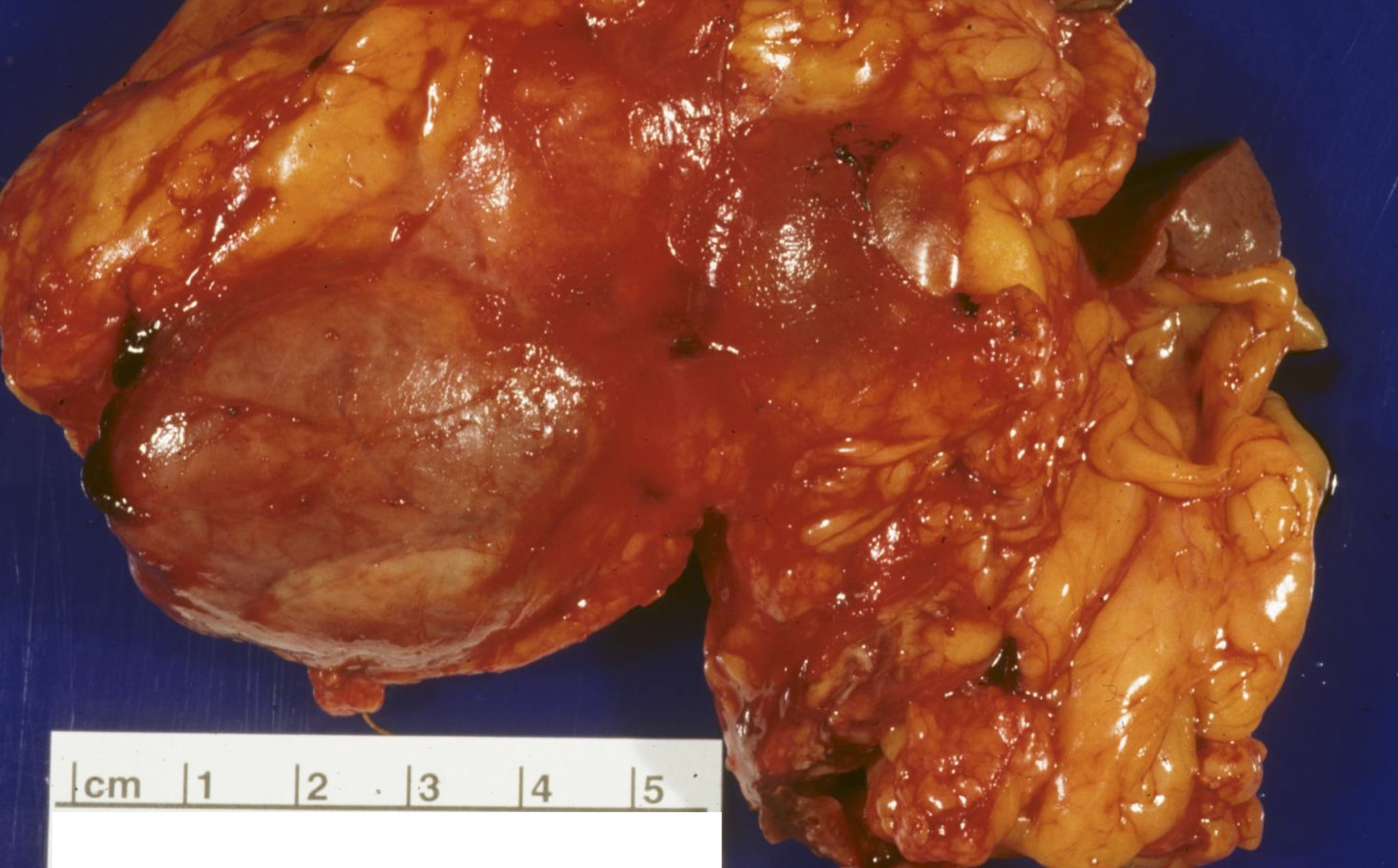
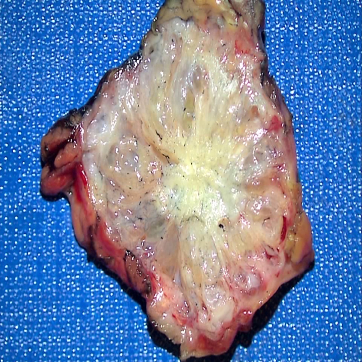
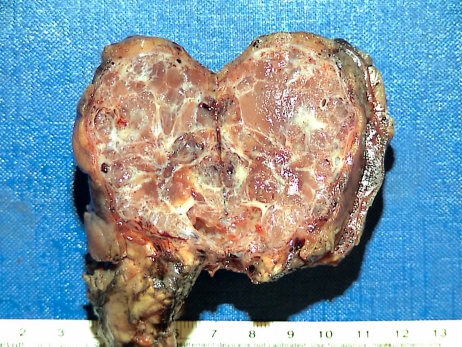
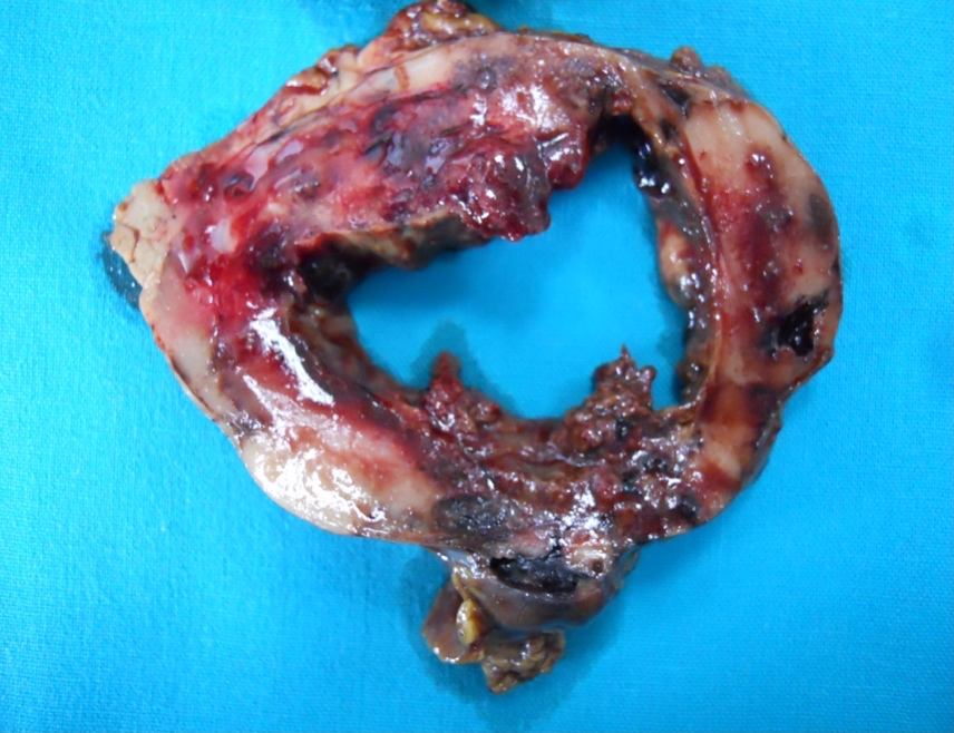
Well circumscribed mass
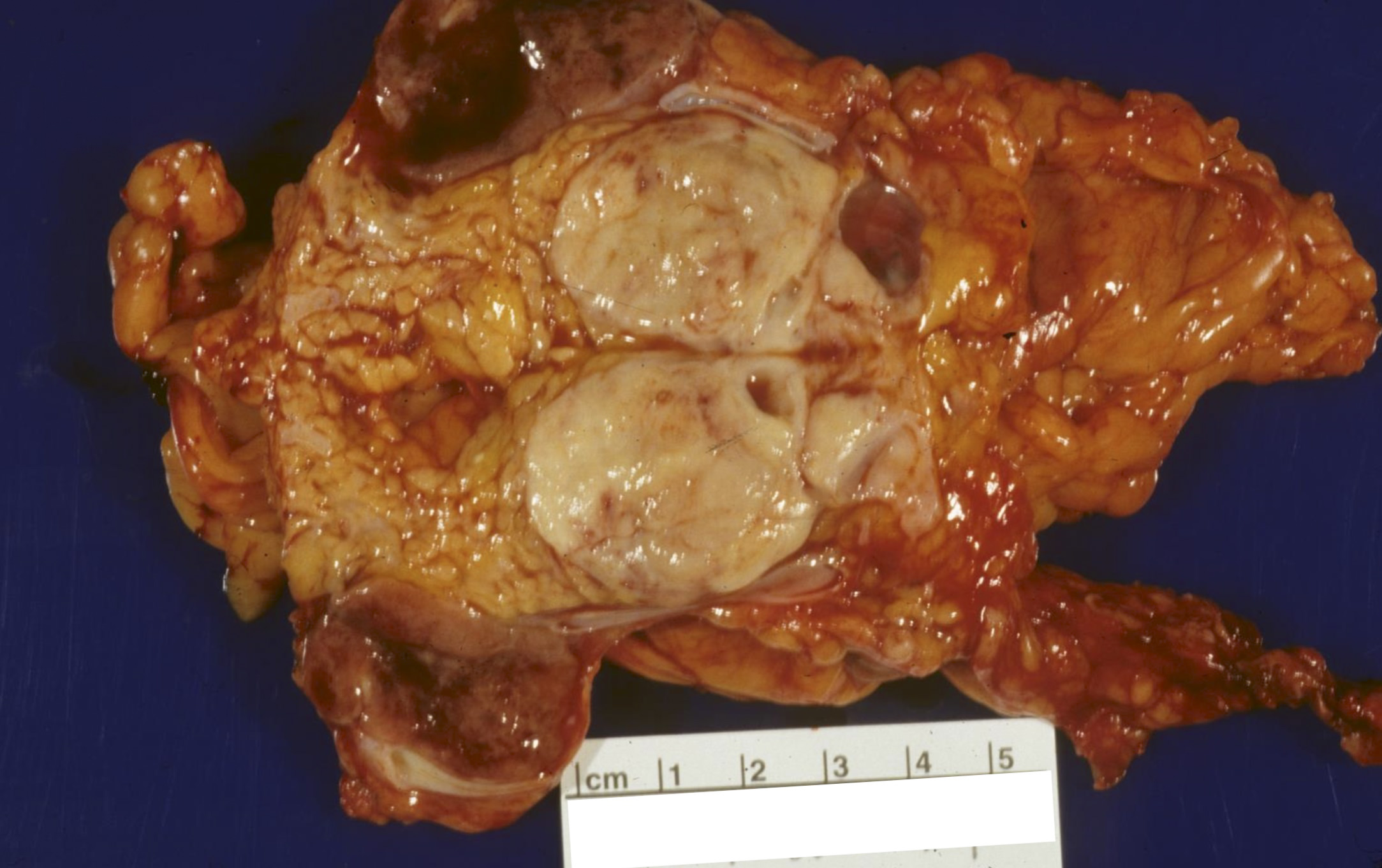
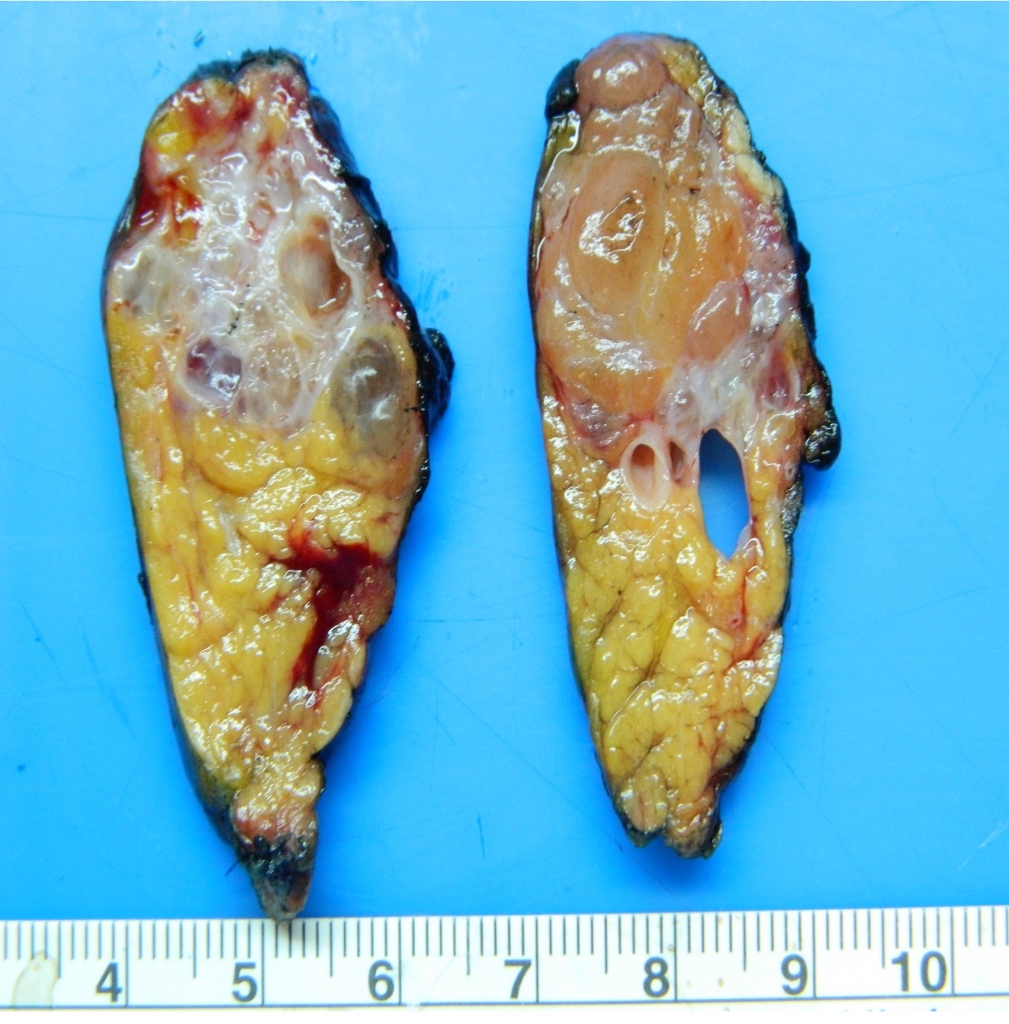
Cut surface of mass
Images hosted on other servers:
Invasion of large vessel

Tumor in isthmus / body
Contributed by Katrina Krogh, M.D.
Trabecular growth
Neuroendocrine nuclei
PanNET (VIPoma) metastatic to liver
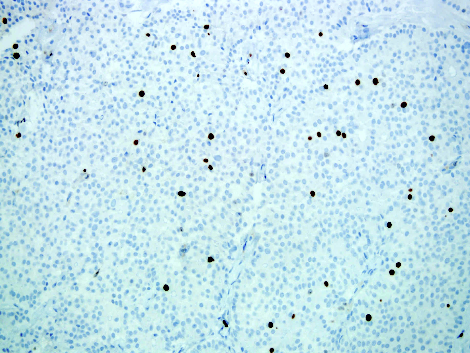
Ki67
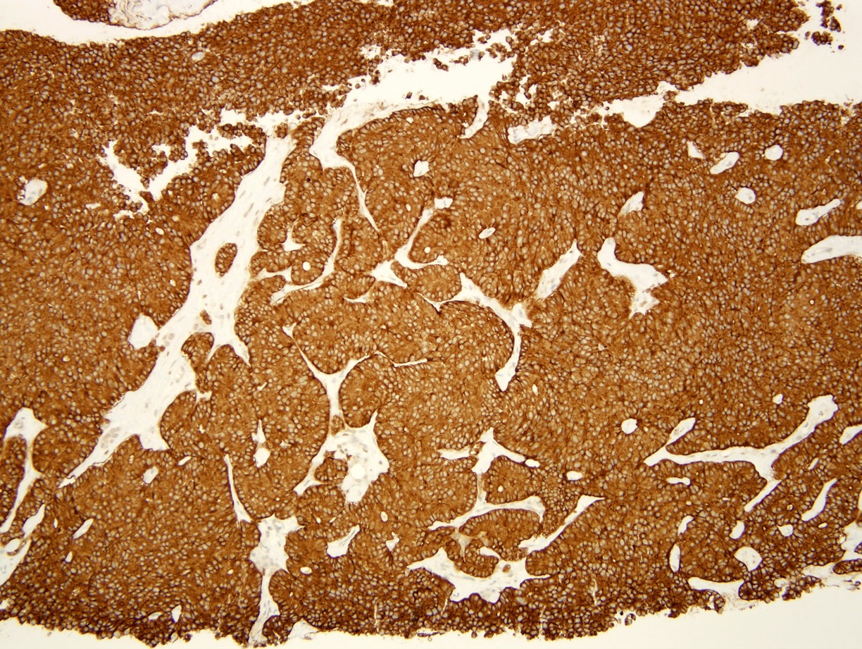
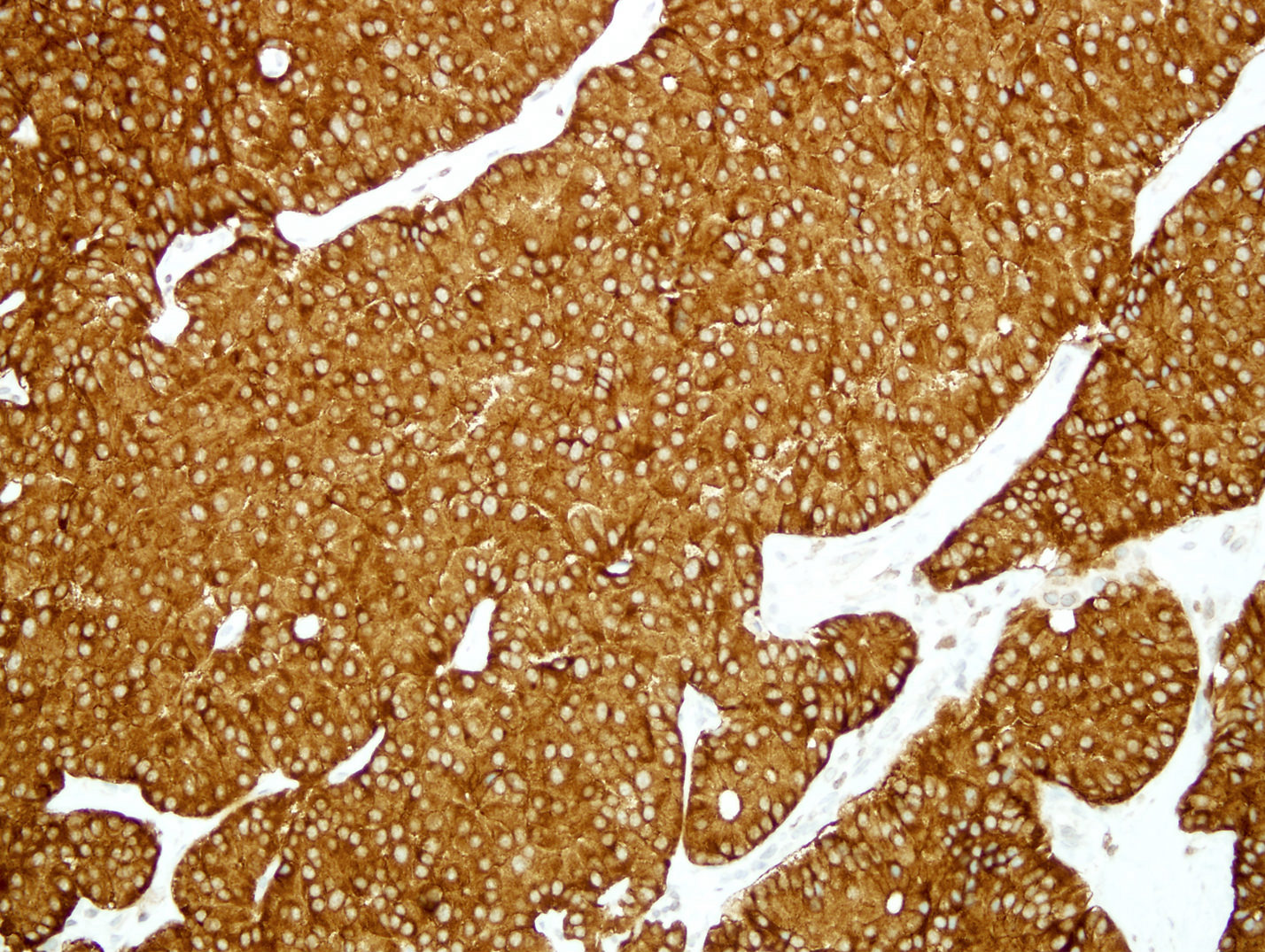
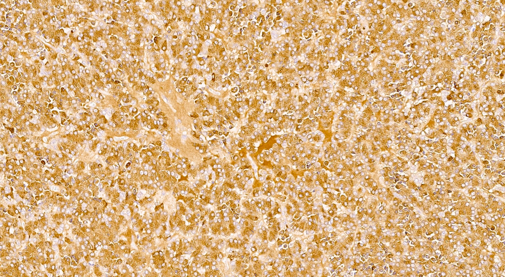
Synaptophysin
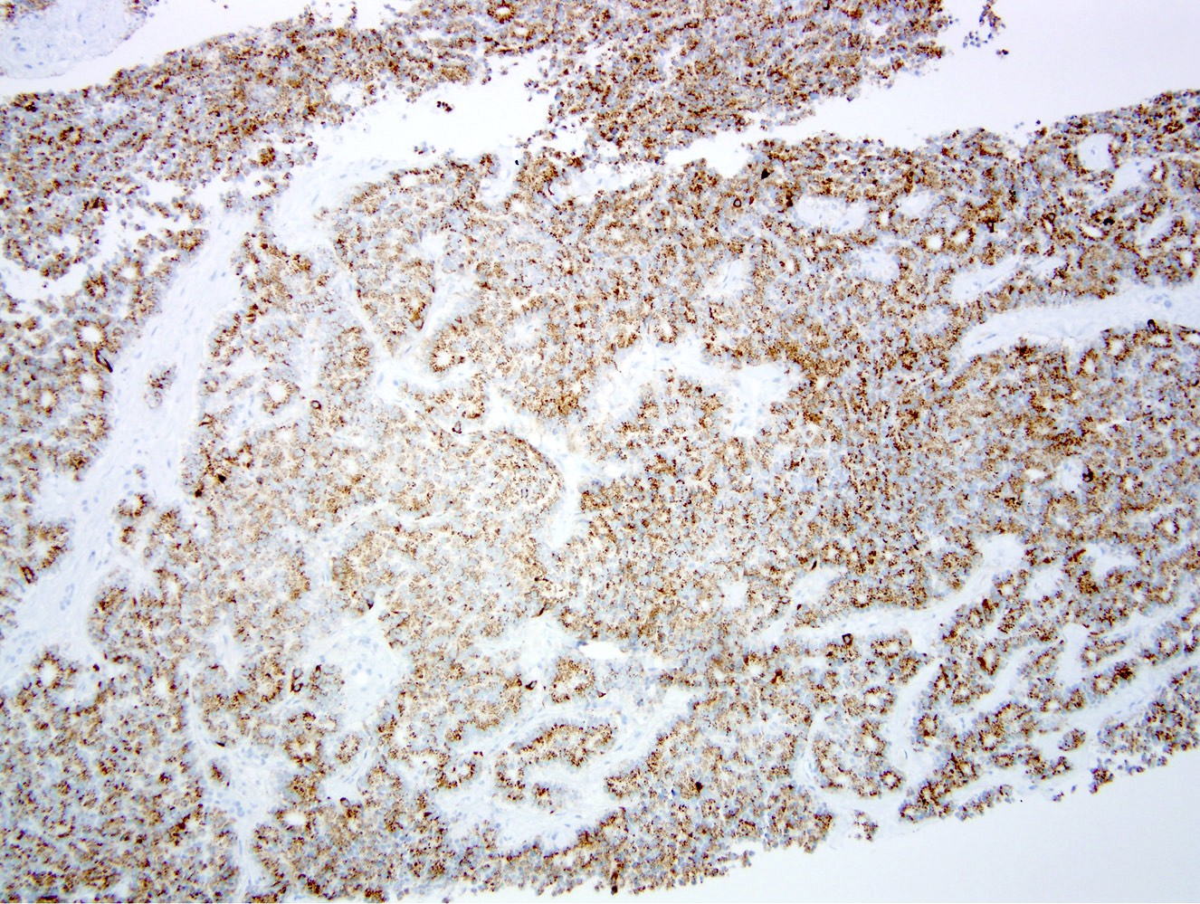
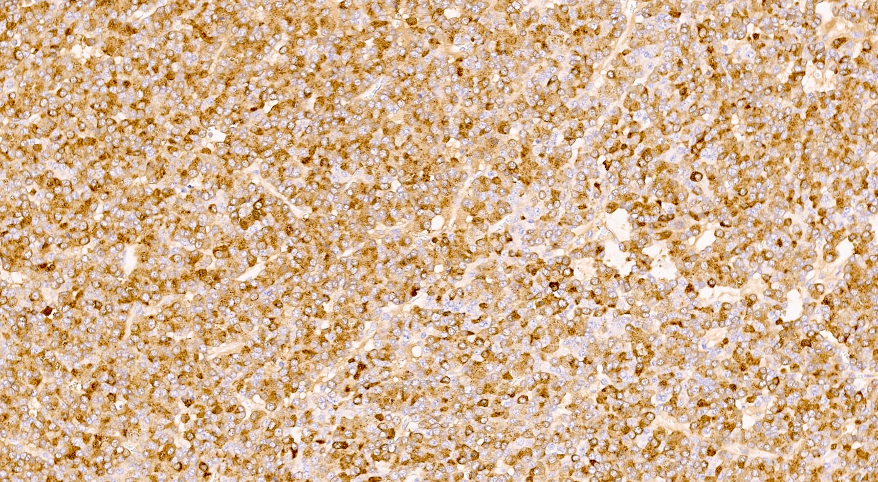
Chromogranin
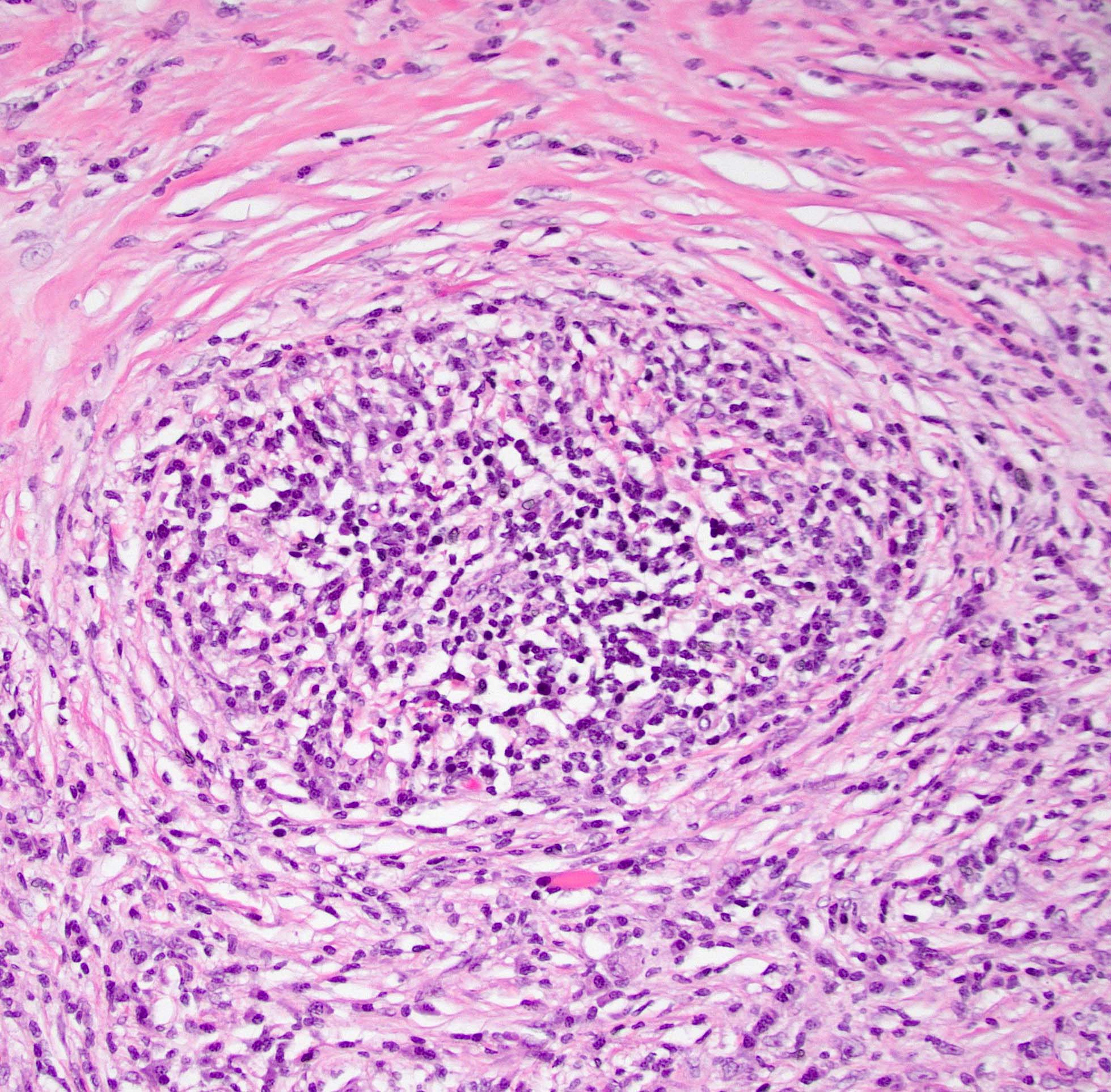
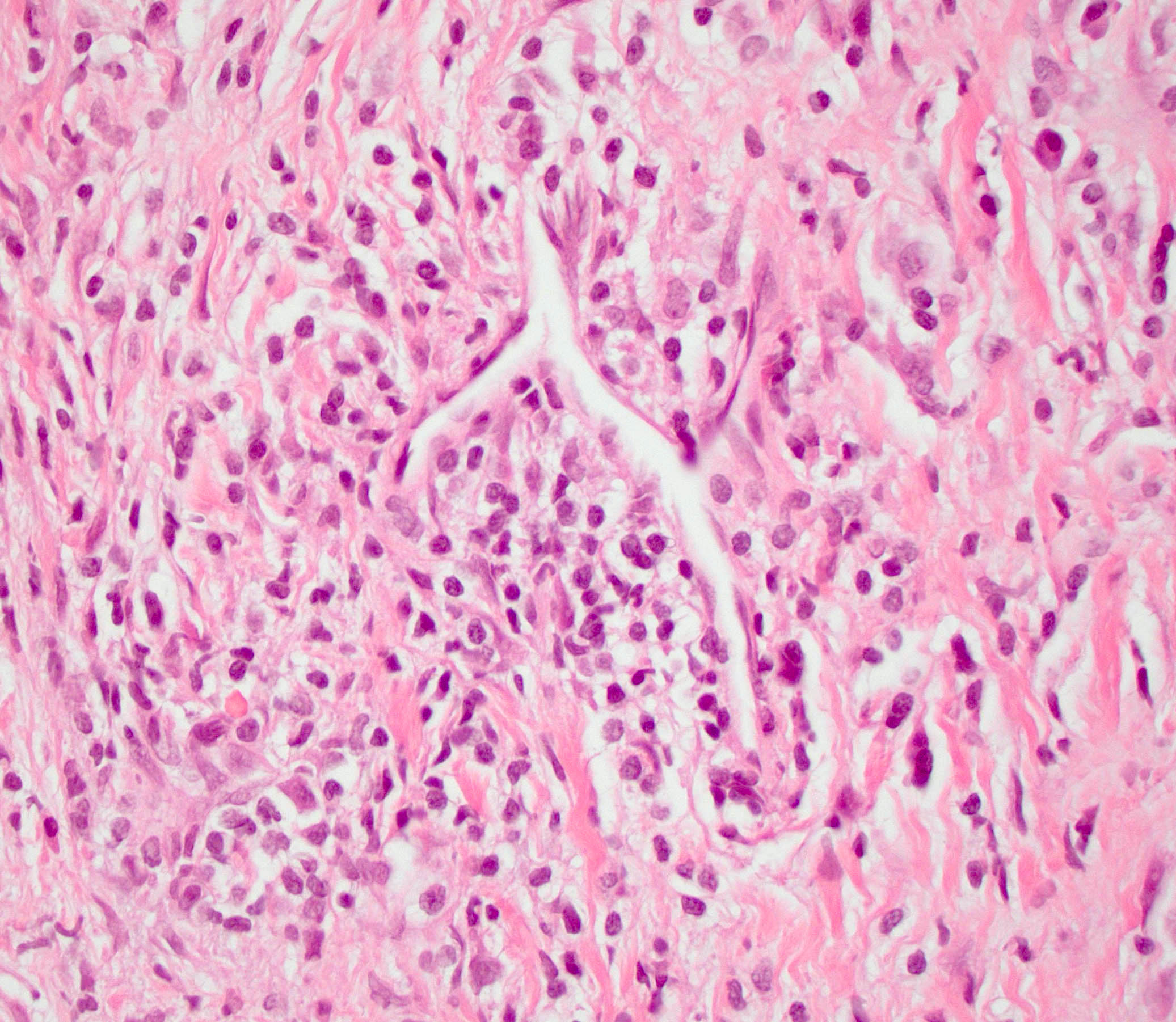
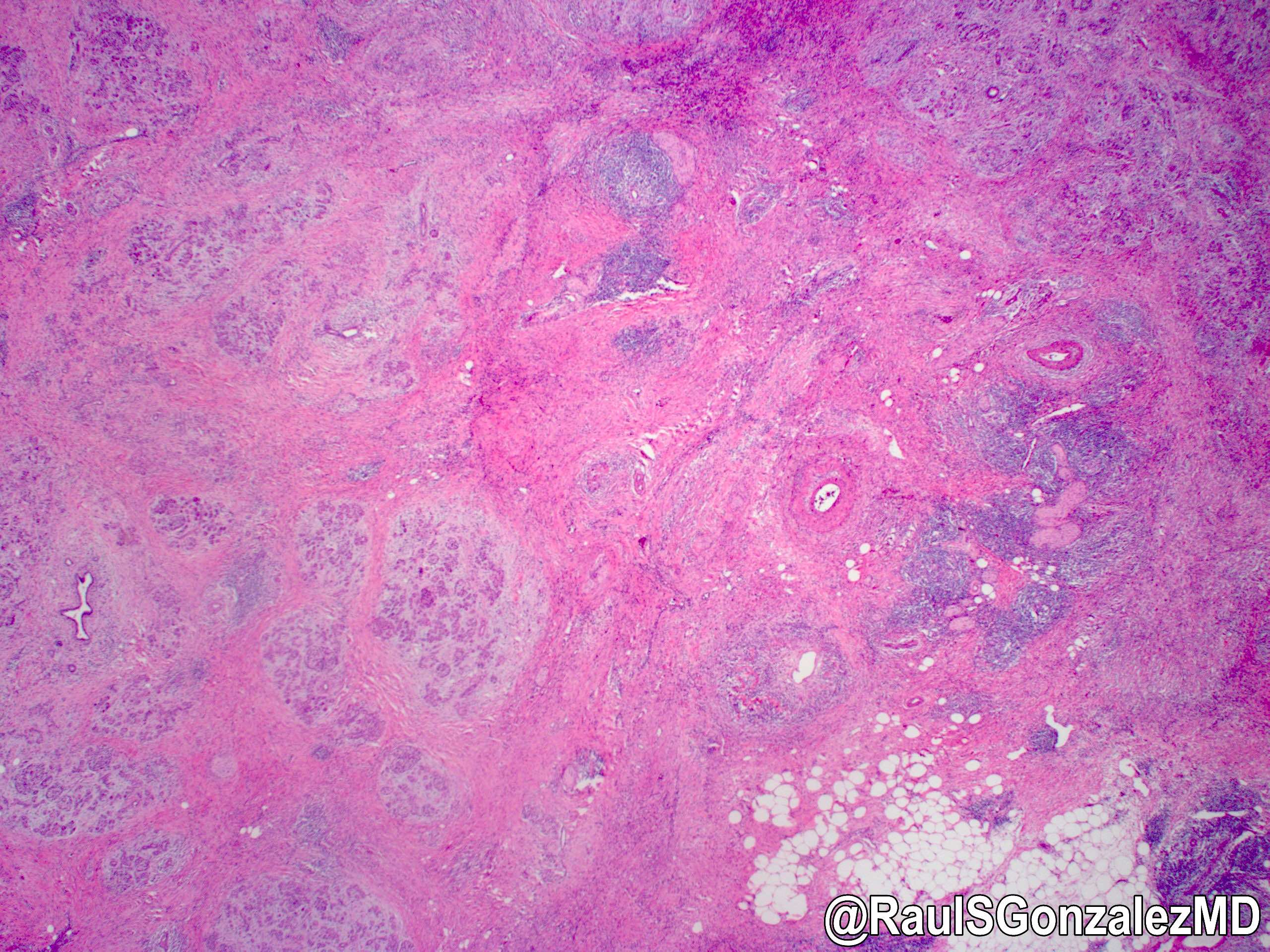
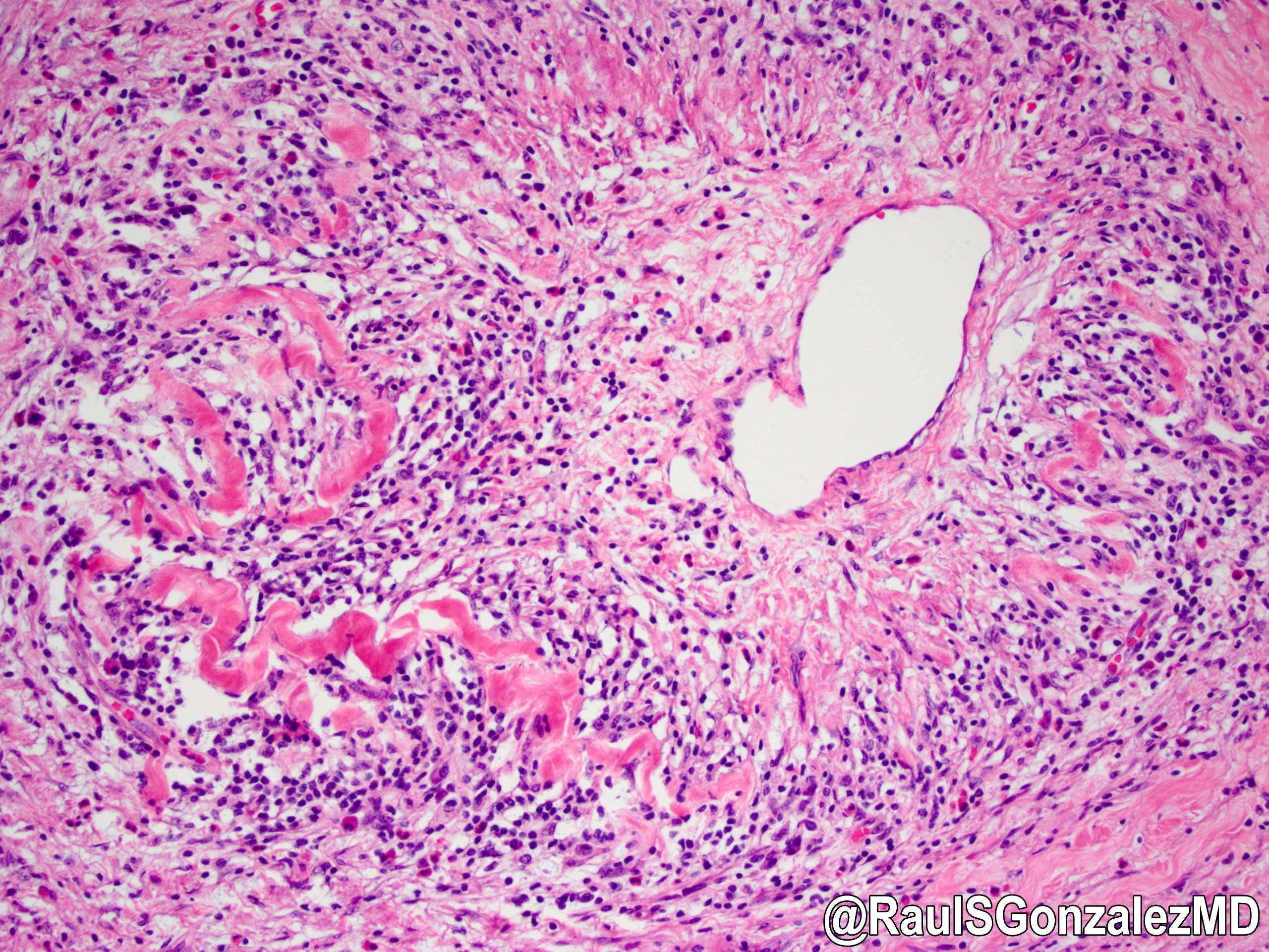
Contributed by @RaulSGonzalezMD on Twitter
Contributed by @RaulSGonzalezMD on Twitter (see original post here)">
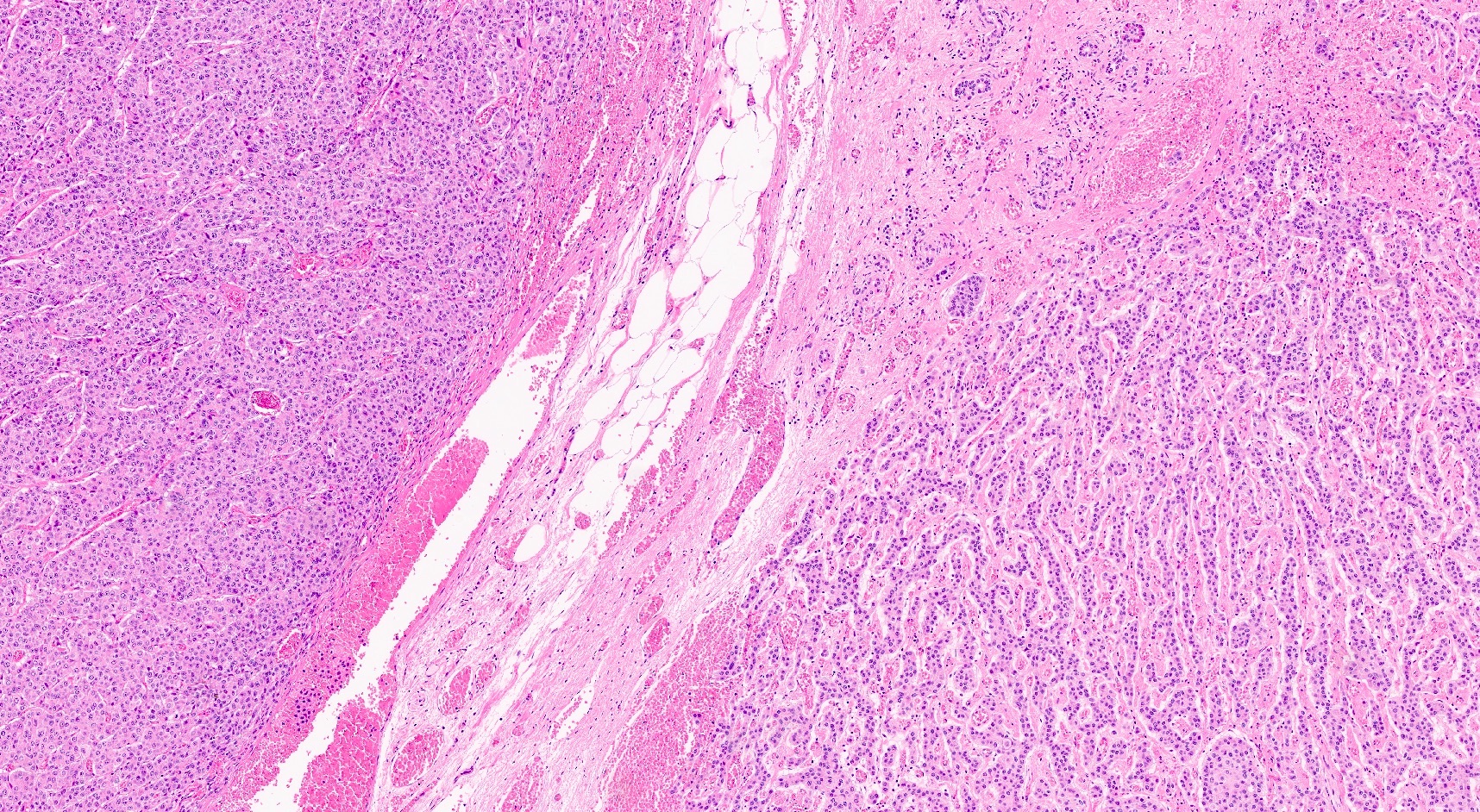
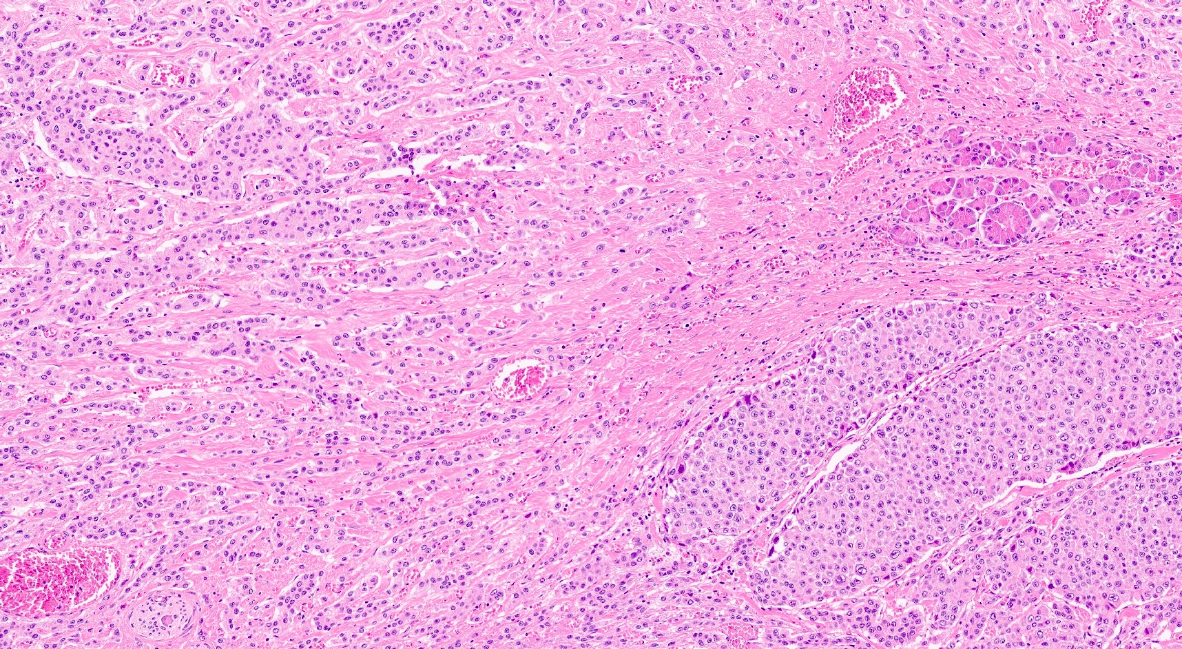
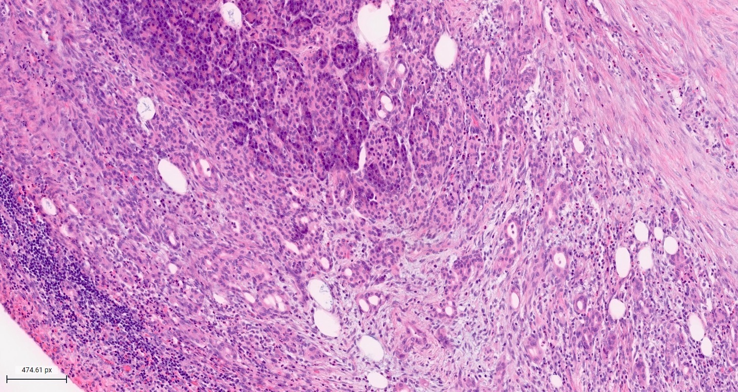
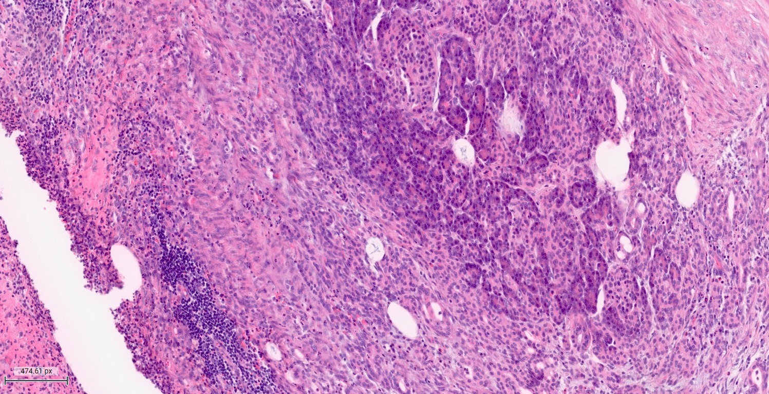
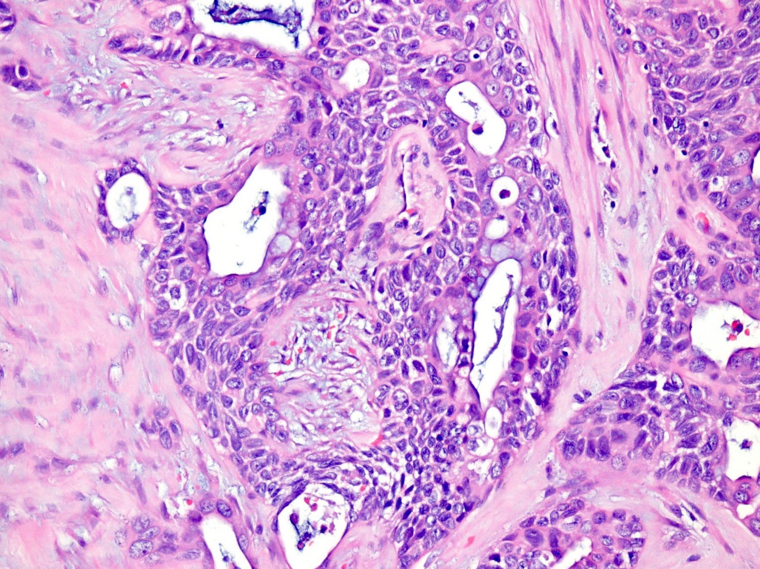
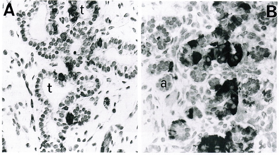
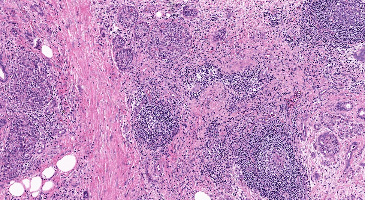
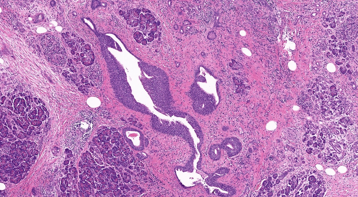
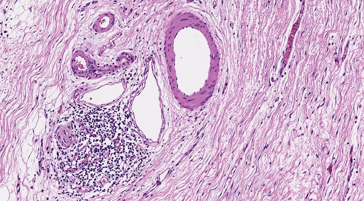
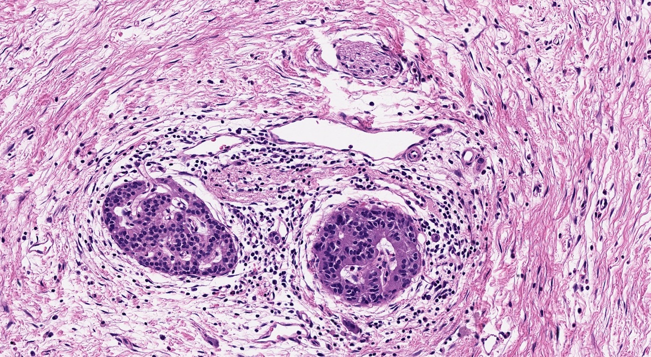
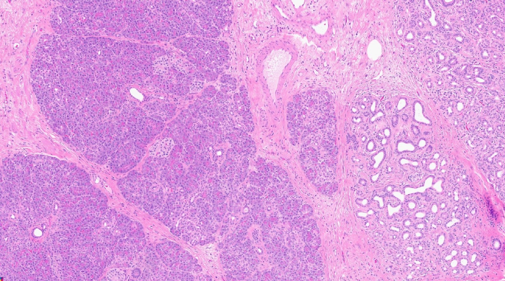
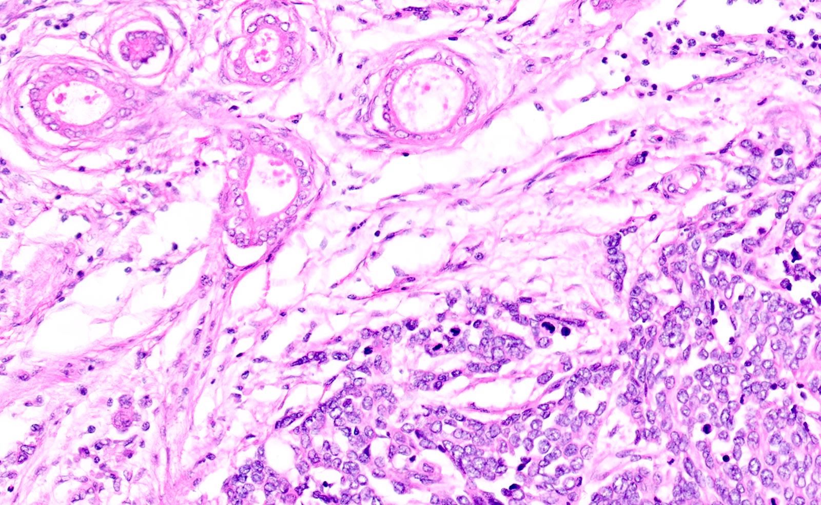
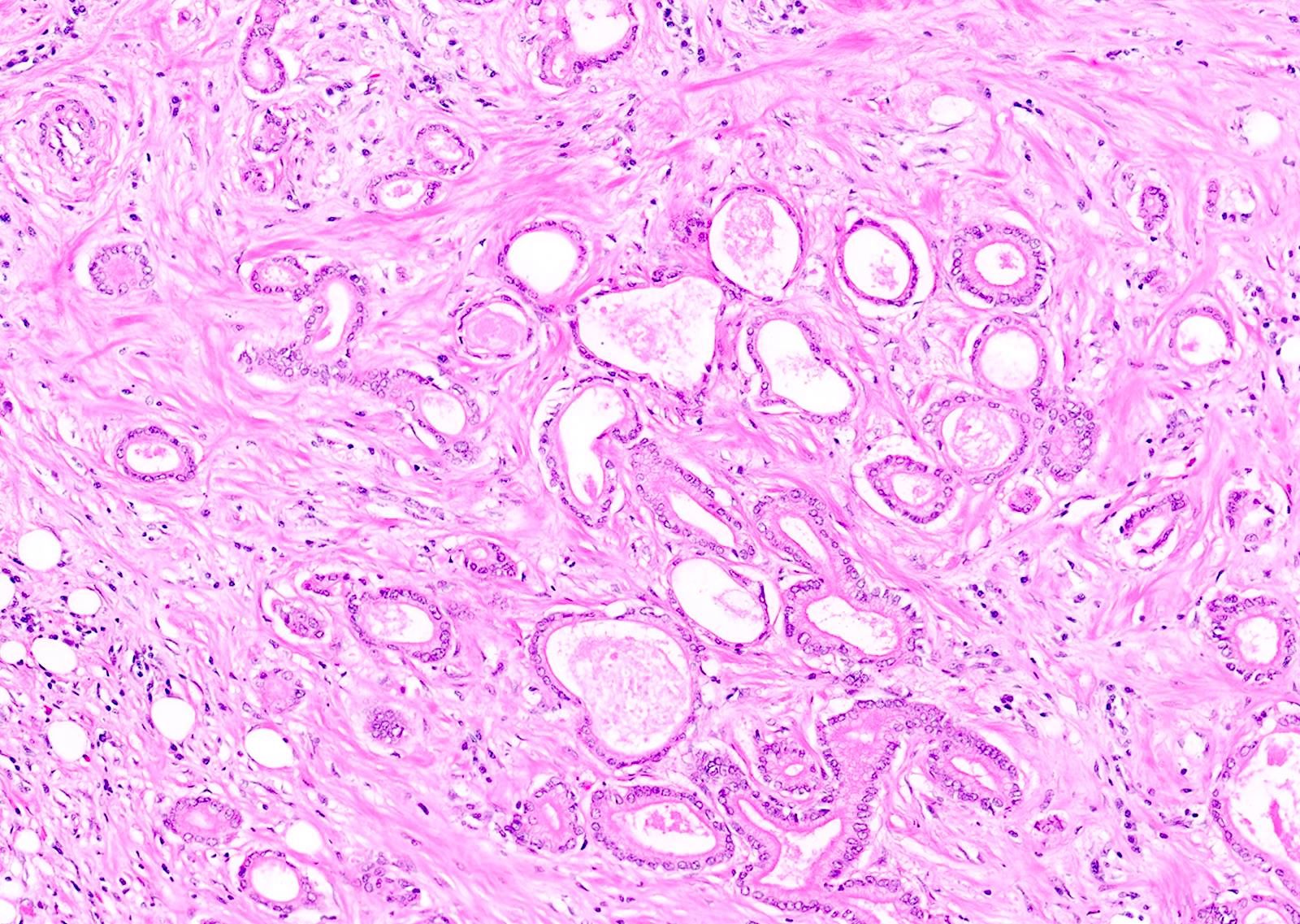
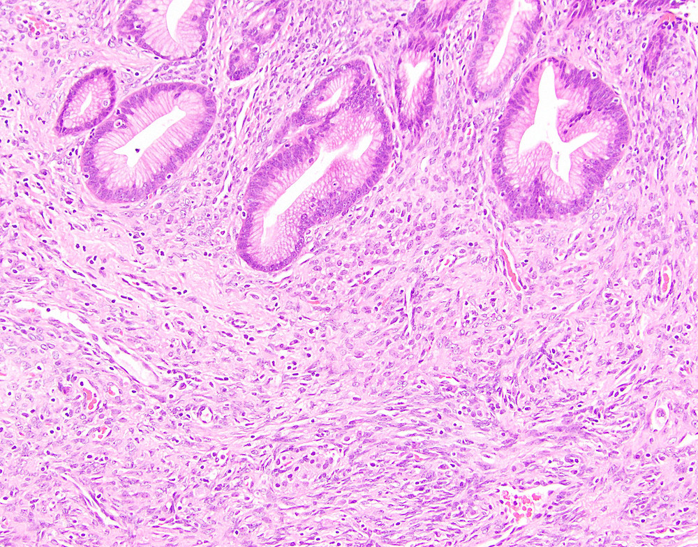
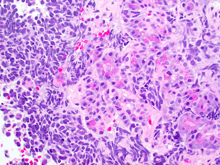
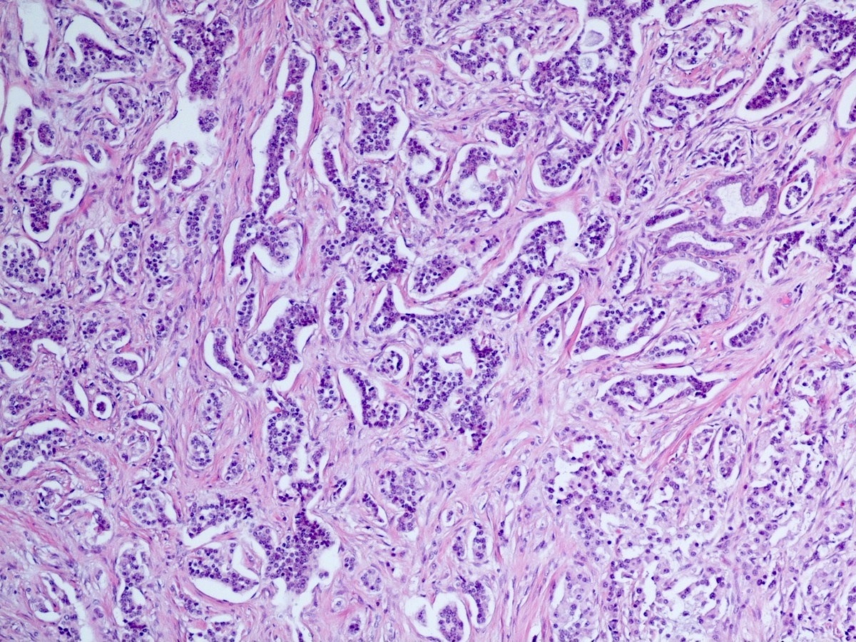
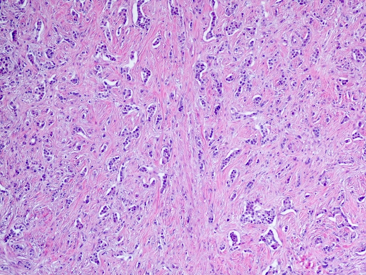
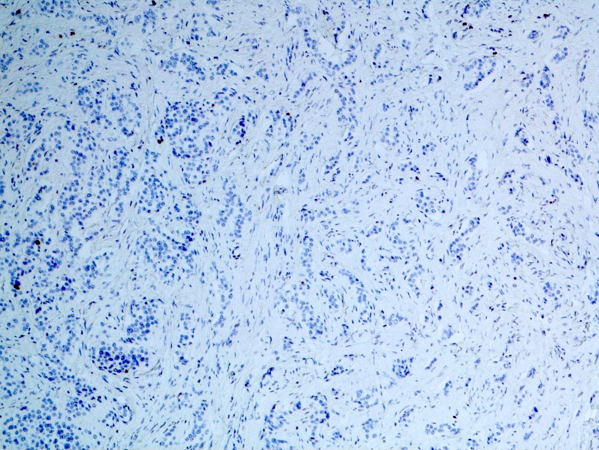
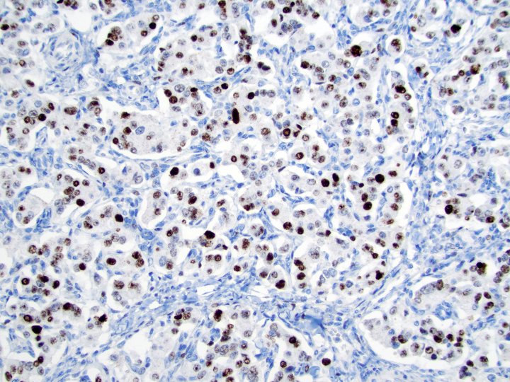
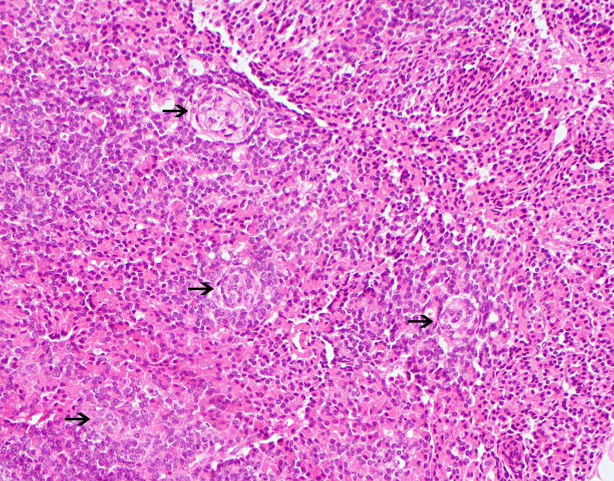
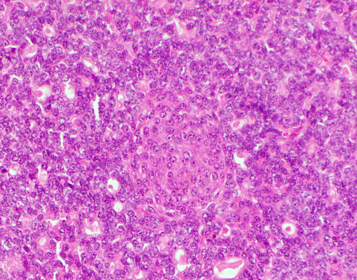
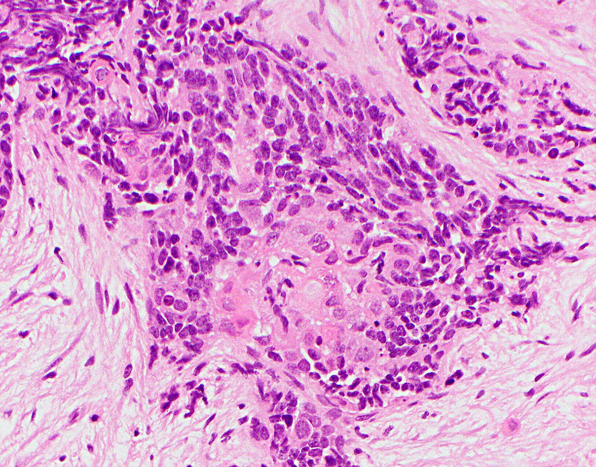
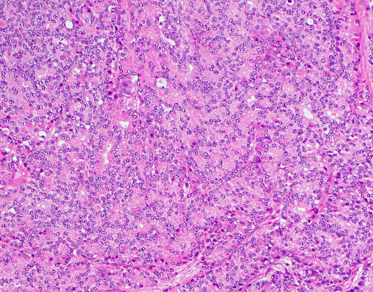
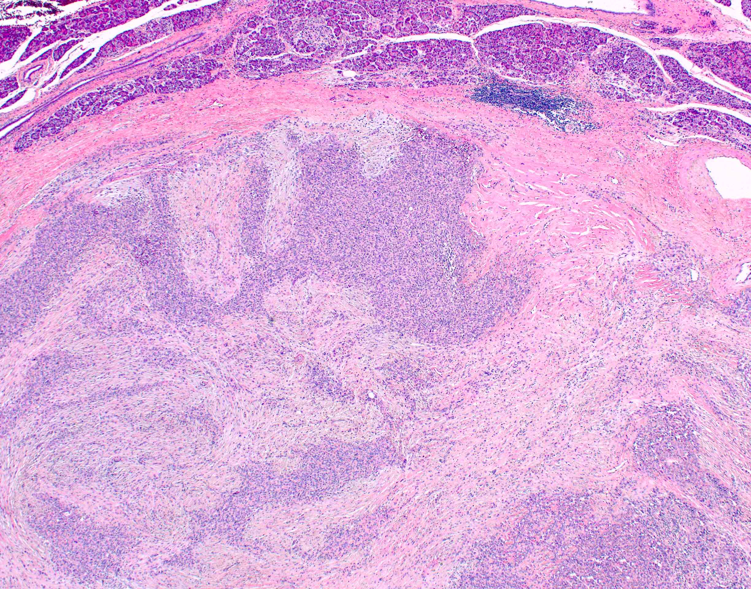
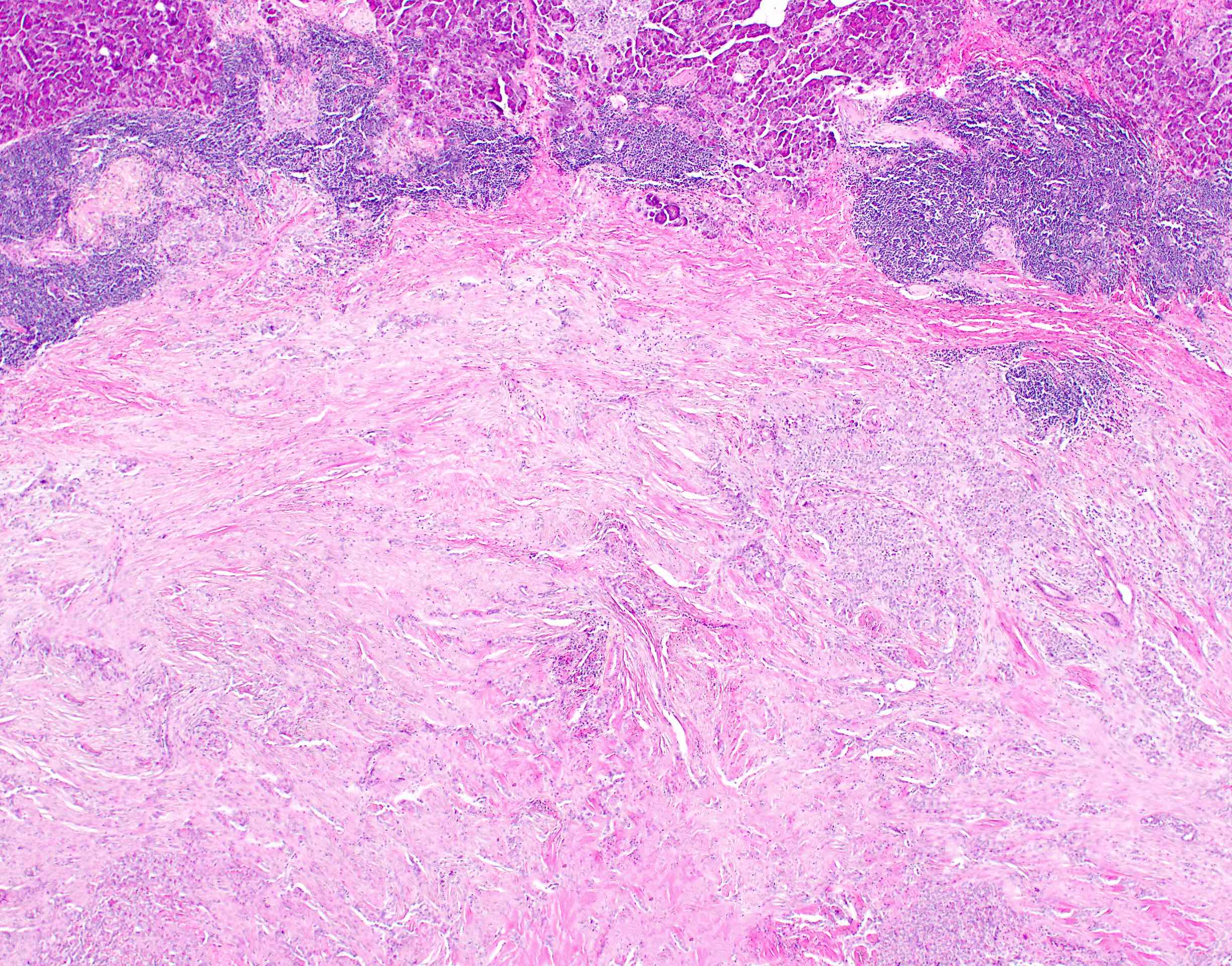
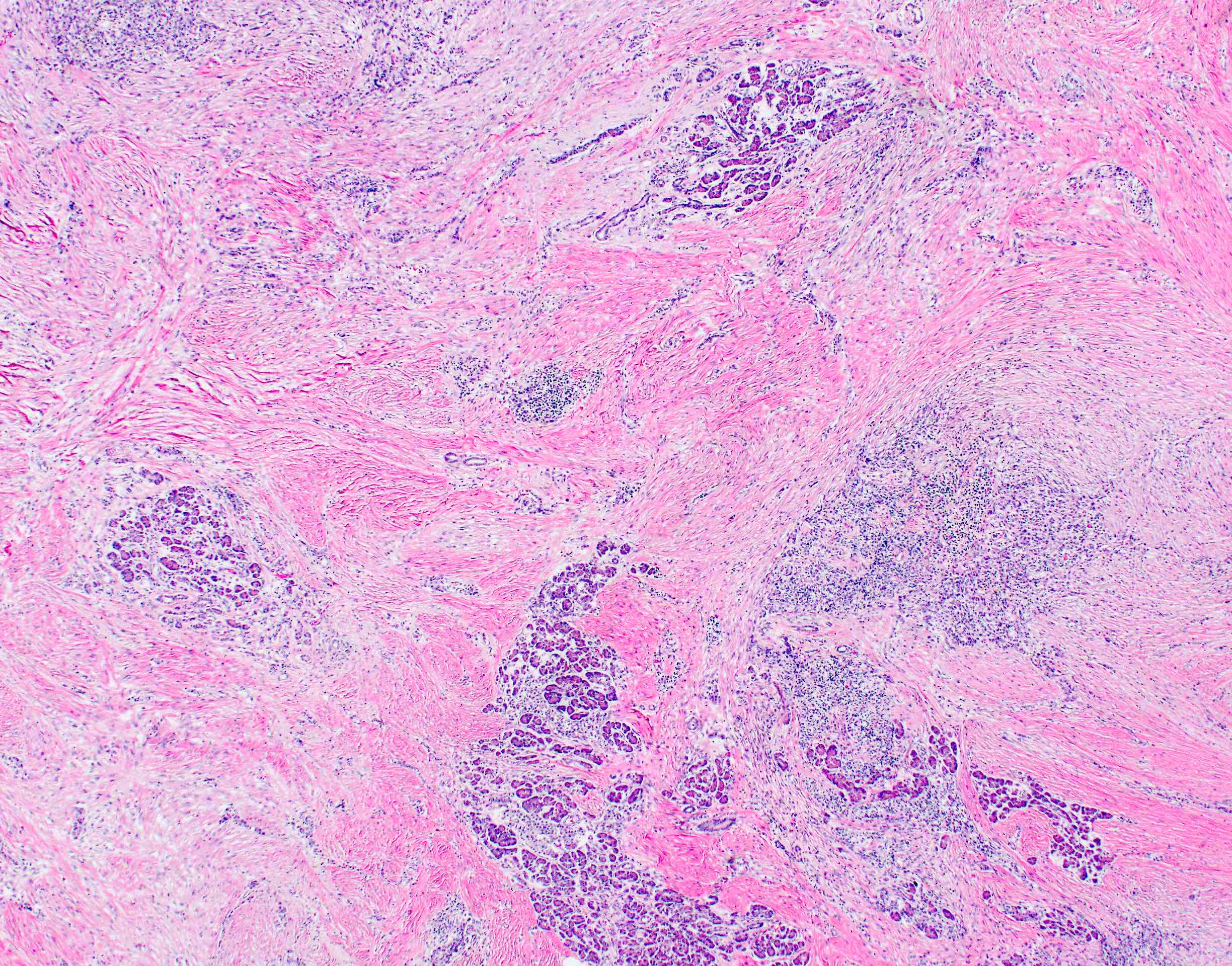
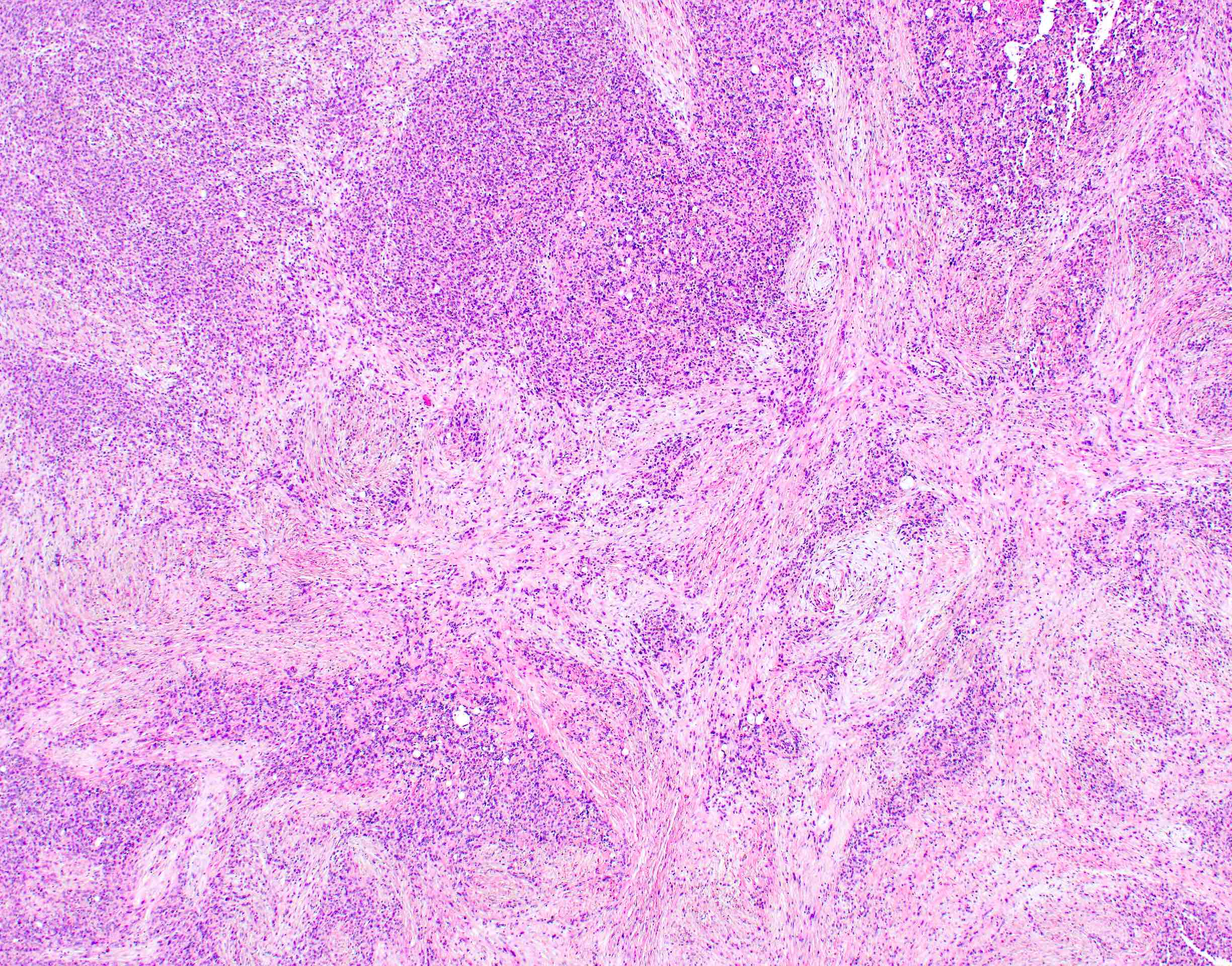
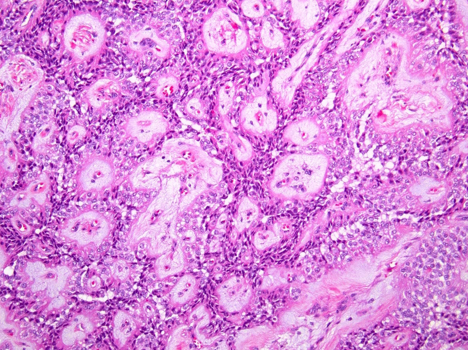
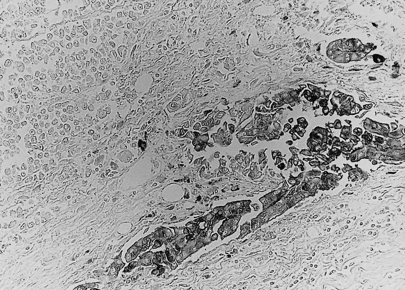
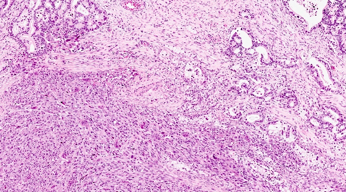
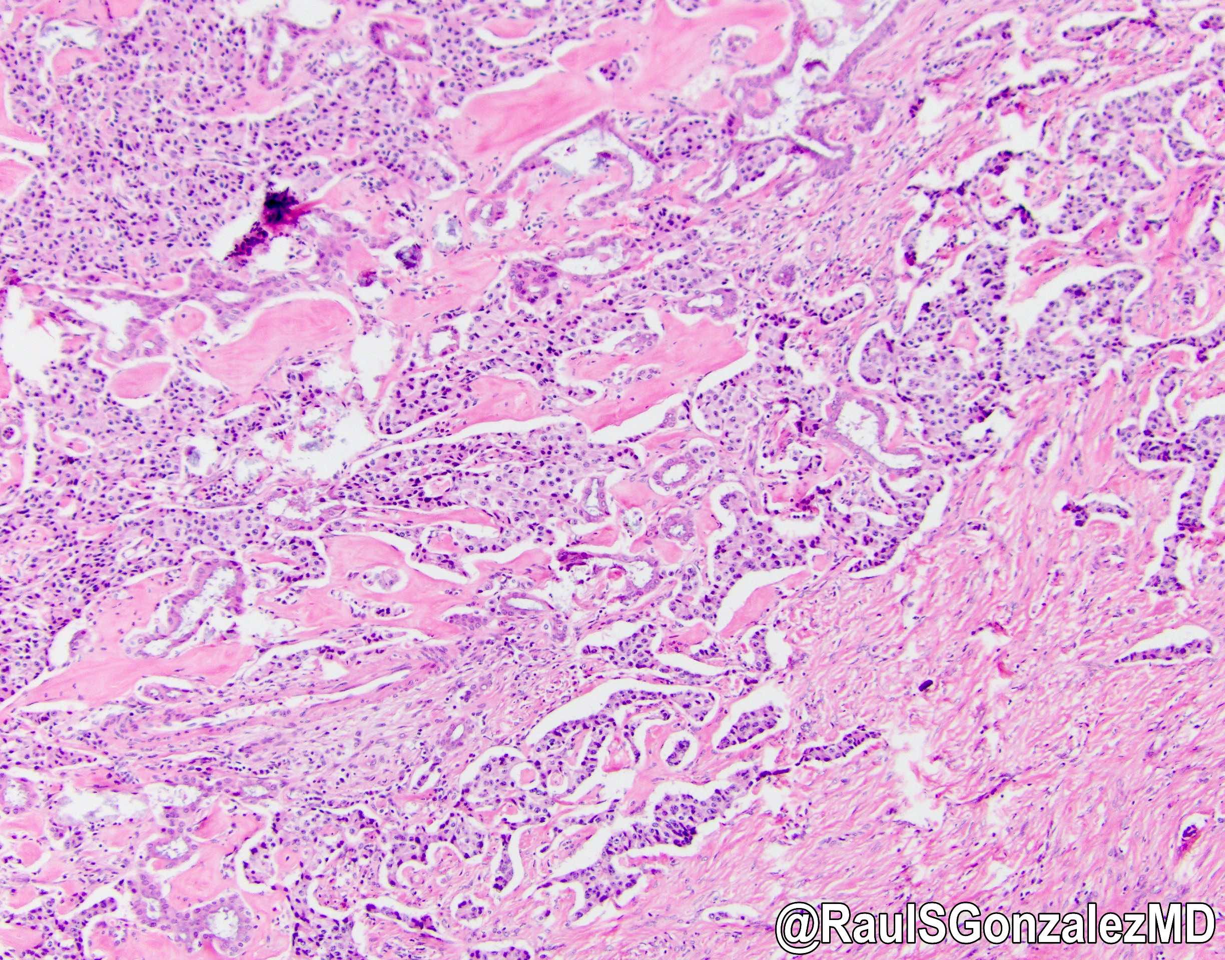
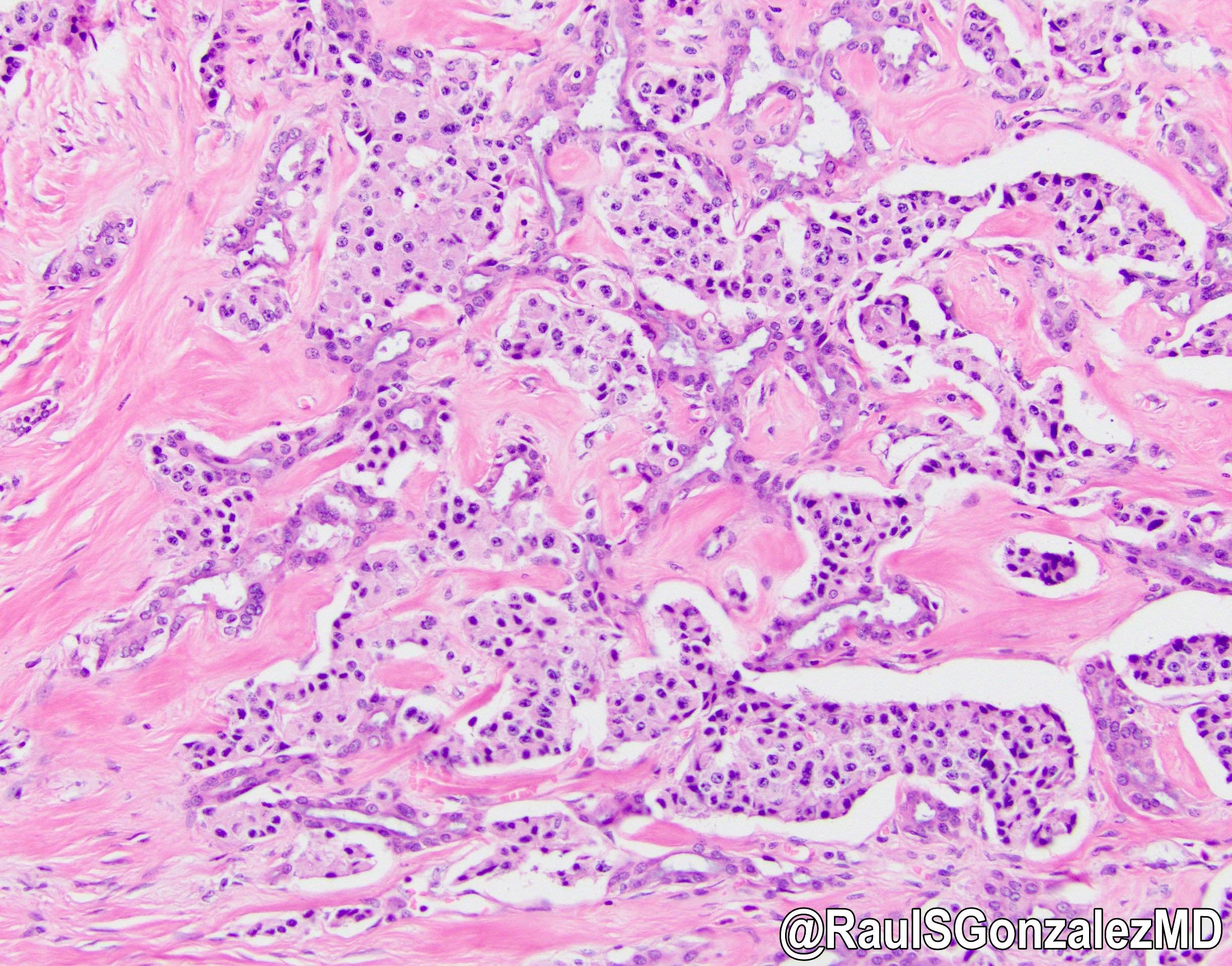
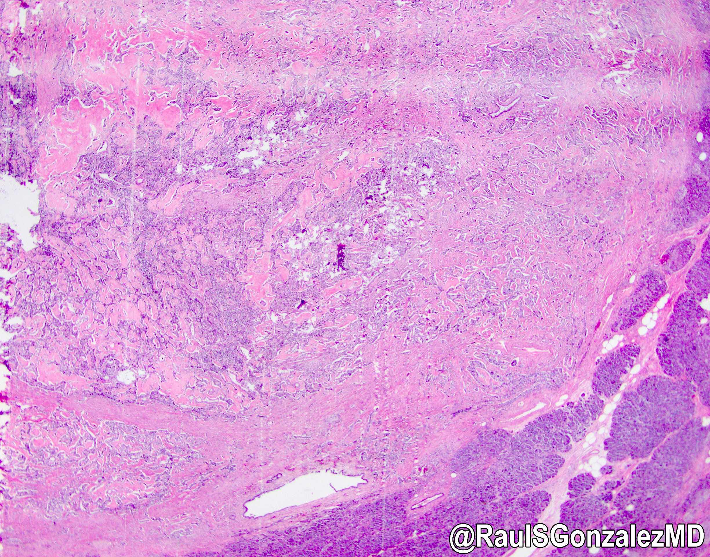 bit.ly/3zIR8Xf for a nice review). Here's a great example of the ductulo-insular subtype, with admixed benign glandular structures. Good prognosis. #pathology #gipath #PathTwitter #PathOutPic"
Contributed by @RaulSGonzalezMD on Twitter (see original post here)">
 bit.ly/3zIR8Xf for a nice review). Here's a great example of the ductulo-insular subtype, with admixed benign glandular structures. Good prognosis. #pathology #gipath #PathTwitter #PathOutPic"
Contributed by @RaulSGonzalezMD on Twitter (see original post here)">
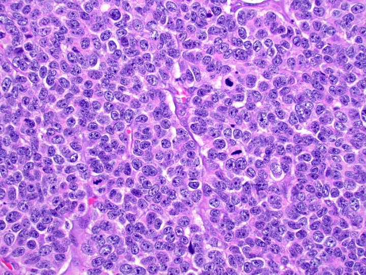
Well differentiated neuroendocrine tumor
Contributed by Katrina Krogh, M.D.

Diff-Quik
Images hosted on other servers:

MEN1 mutation
Table 1: Papanicolaou reporting system for pancreaticobiliary cytology
| Diagnostic categories | Average risk of malignancy | Examples of entities | Selected ancillary testing | Management |
I. Nondiagnostic | 23.1% | • Cellular artifact, obscuring hemorrhage or necrosis • Pure GI contamination • Acellular cyst aspirate with no evidence of mucinous etiology • Benign pancreatic parenchyma with clearly defined mass on imaging | Cyst fluid analysis: CEA and amylase (no mucinous etiology = lack of mucin, lack of elevated CEA, lack of KRAS or GNAS mutations) | Reassess / repeat |
II. Negative | 8.2% | • Pancreatitis • Pseudocyst • Lymphoepithelial cyst • Splenule / accessory spleen • Benign pancreatic parenchyma with no mass on imaging | Pseudocyst: high amylase + low CEA | Conservative |
III. Atypical | 46.8% (range: 28 - 100) |
• Ductal cells with indeterminate atypia (reactive versus low grade dysplasia, versus scant lesional tissue) • Abundant intracytoplasmic mucin in the epithelium | Cyst fluid analysis: CEA and amylase | Conservative, repeat or surgery |
IV. Neoplastic: benign | 20.2% |
• Serous cystadenoma (SCA) • Lymphangioma • Cystic teratoma • Schwannoma | SCA: low amylase + low CEA, VHL mutation, positive inhibin staining | Conservative |
IV. Neoplastic: other | 37.5% |
• GIST • Extra-adrenal paraganglioma • Pancreatic solitary fibrous tumor Premalignant • IPMN with dysplasia • IPNB • MCN with dysplasia Low grade malignant • PanNET • Solid pseudopapillary neoplasm (SPN) |
Solitary fibrous tumor: STAT6 mutation Intraductal papillary mucinous neoplasm: KRAS, RNF43, GNAS (specific), TP53, SMAD4 mutations Solid pseudopapillary neoplasm: CTNNB1 (beta catenin) mutation | Surgery with an option for conservative management in select scenarios |
V. Suspicious | 92.1% |
• Pancreatic ductal adenocarcinoma (PDAC) • PanNET • Acinar cell carcinoma • Pancreatoblastoma • Solid pseudopapillary neoplasm • Cyst aspirate with solid mural nodule or cyst wall mass • Lymphoma • Metastases • High grade BilIN | Immunocytochemistry, FISH molecular analysis Cyst fluid analysis: CEA and amylase | Surgery |
VI. Positive | 99.6% | • PDAC and variants • Cholangiocarcinoma • Acinar cell carcinoma • Small and large cell neuroendocrine carcinoma • Pancreatoblastoma • Lymphomas • Sarcomas • Metastases to the pancreas | Immunocytochemistry patterns in malignancy: • Positive: S100P, IMP3, MUC4, mesothelin • Negative: pVHL, CD10, clusterin beta, SMAD4 FISH significantly improves diagnostic sensitivity of adenocarcinoma (copy number abnormalities in CEP3, CEP7, CEP17 and of band 9p21) | Surgery or neoadjuvant therapy / clinical trials |
- Reference: Diagn Cytopathol 2020;48:494
Contributed by Derek Allison, M.D.
Duodenal contaminant only
GI contaminant only
Benign pancreatic parenchyma
Pancreatic splenule
Pancreatic splenule (CD8+)
Pancreatic splenule (CD45+)
Lymphoepithelial cyst
Clean mucin
IPMN with cytologic atypia
Reactive atypia; chronic pancreatitis
Biliary brush stricture
Serous cystadenoma
Serous cystadenoma: inhibin
Well differentiated
NET, WHO grade 1
Solid pseudopapillary neoplasm
Mucinous cystic neoplasm
Suspicious for pancreatic adenocarcinoma
Pancreatic ductal adenocarcinoma
Well differentiated pancreatic adenocarcinoma
PDAC by biliary brush

Bekaii-Saab: 2017

Campbell: 2020
Casillas: 2015
Centeno: 2015
Cheng: 2023
D'Onofrio: 2015
Greenson: 2019
Husain: 2021
IAC-IARC-WHO: 2023
IARC: 2017

IARC: 2019
Jiang: 2023
Klimstra: 2023
La Rosa: 2015
Lamps: 2021
Mody: 2022
Nosé: 2022
Odze: 2022
Srivastava: 2023
Thompson: 2021
Wood: 2017
Find related Pathology books: cytopathology, GI, GU/adrenal, head & neck/endocrine, liver, molecular, pediatric
