

AFIP images

Various images
Contributed by Alcides Chaux, M.D. and Antonio Cubilla, M.D.
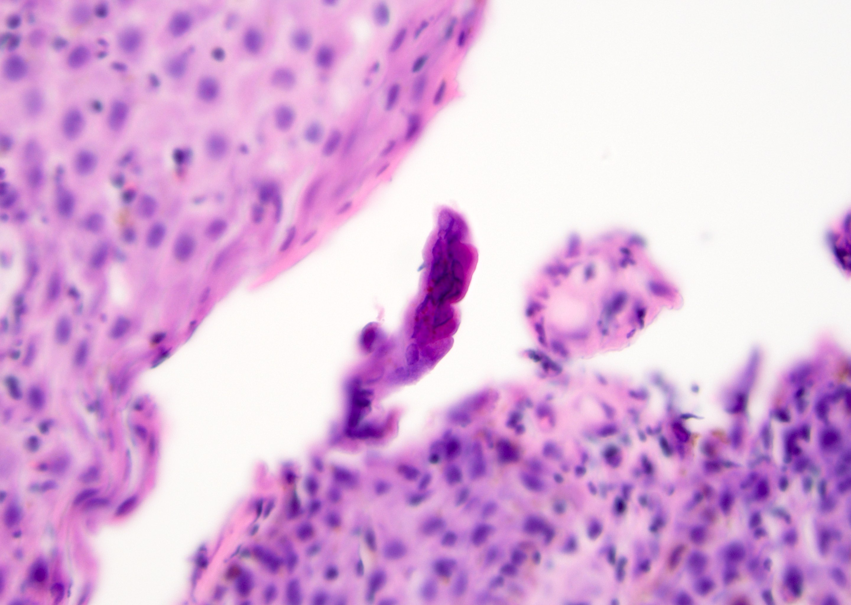
Nests and differentiation
AFIP images
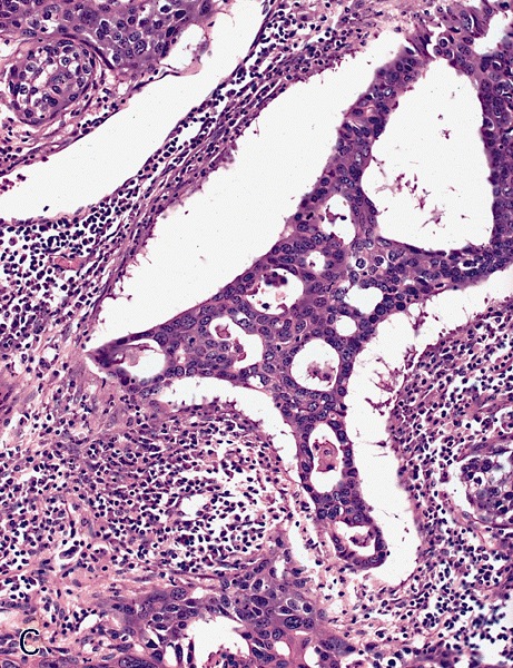
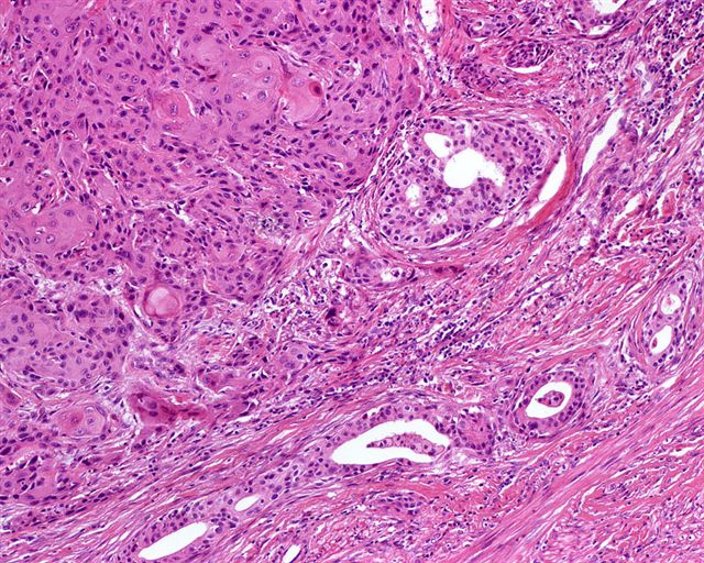
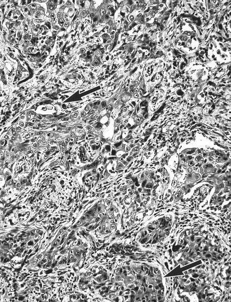
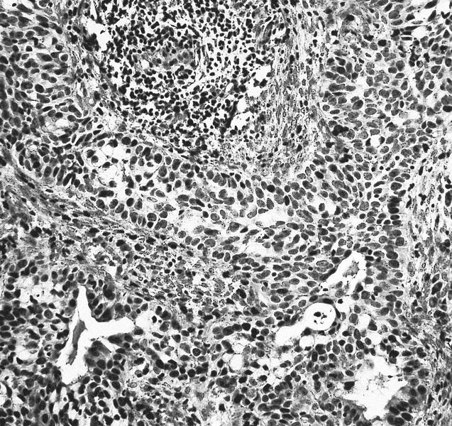
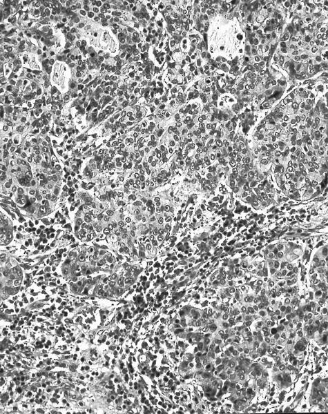
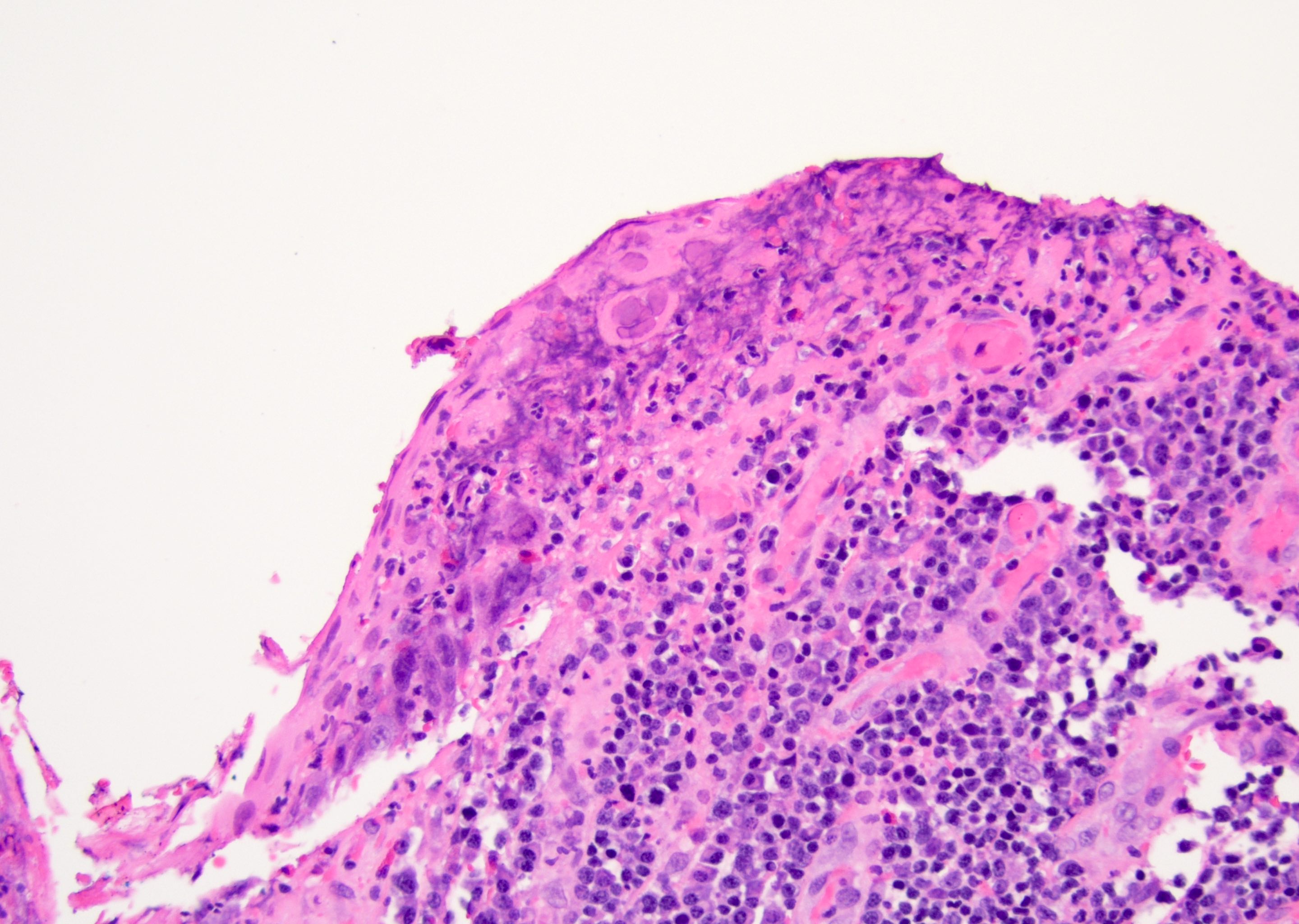
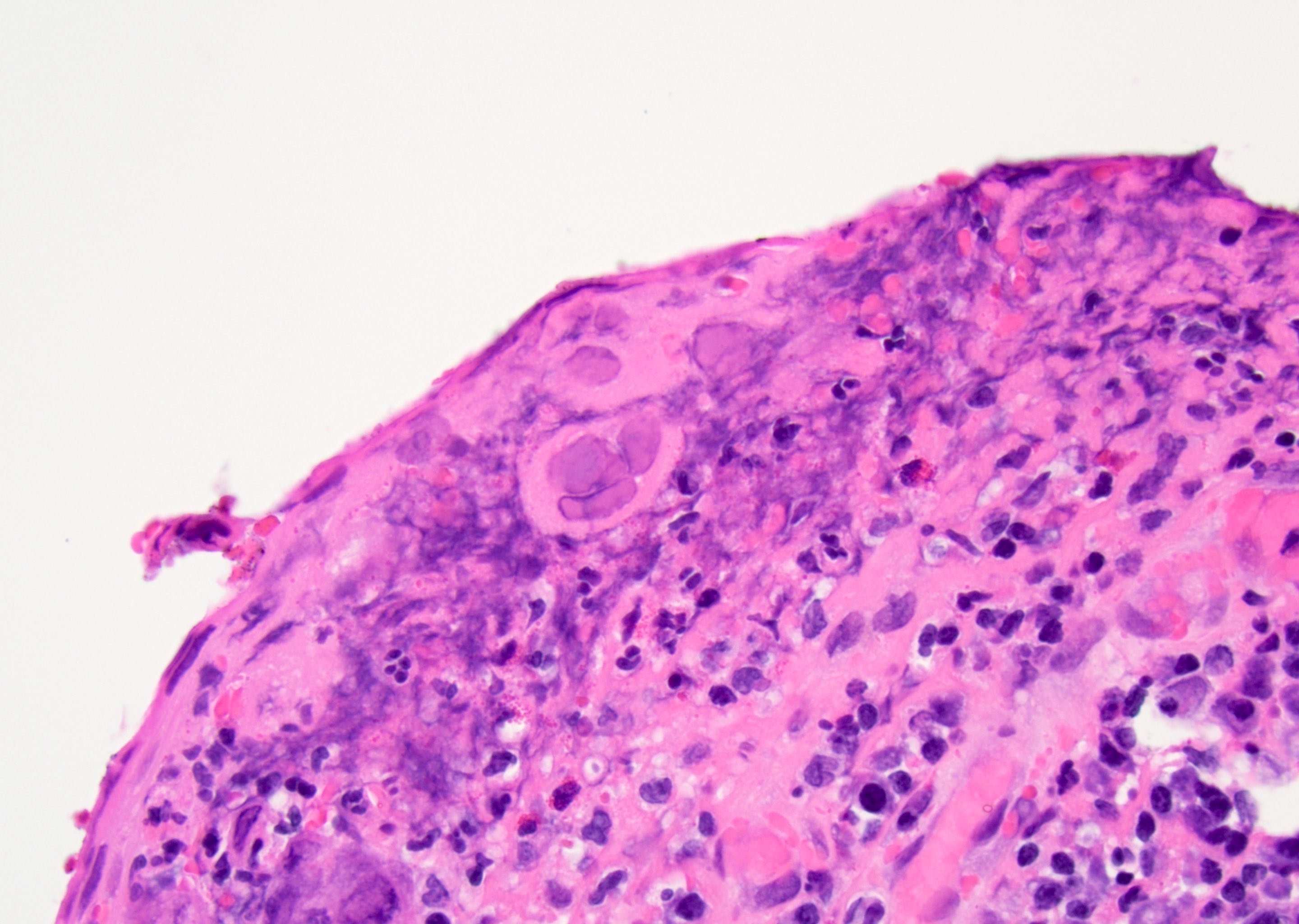
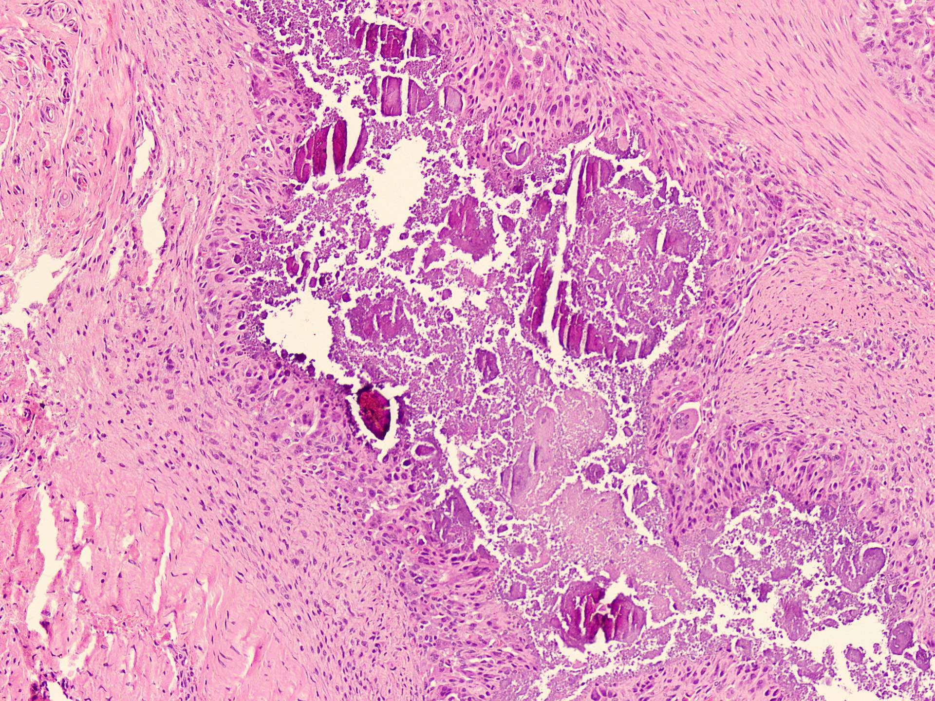
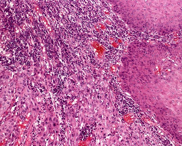
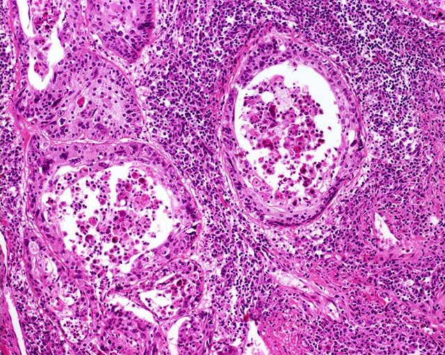
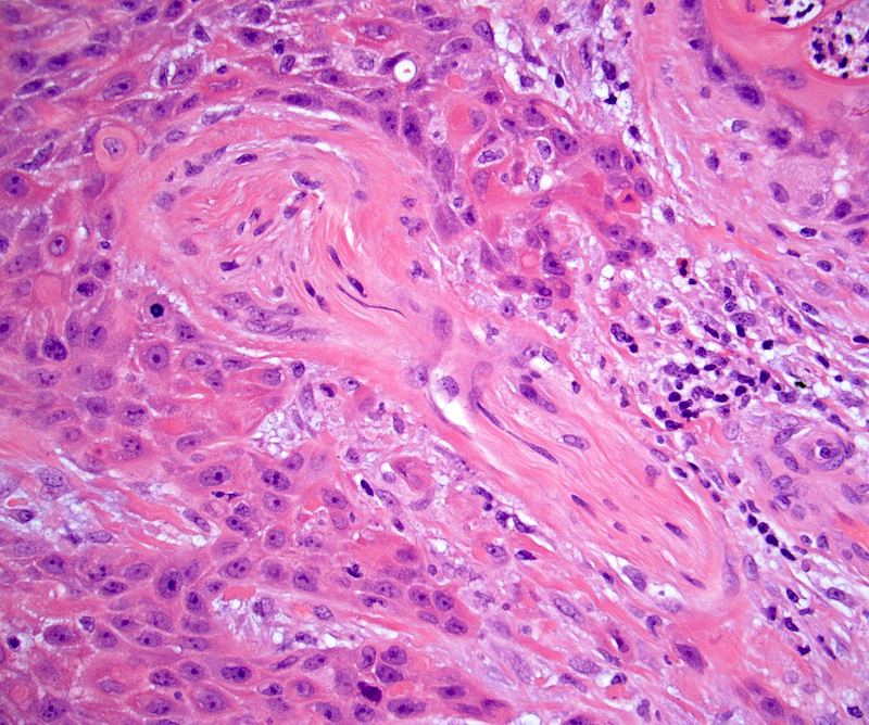
Infiltrating tumor
has squamous
and glandular
features

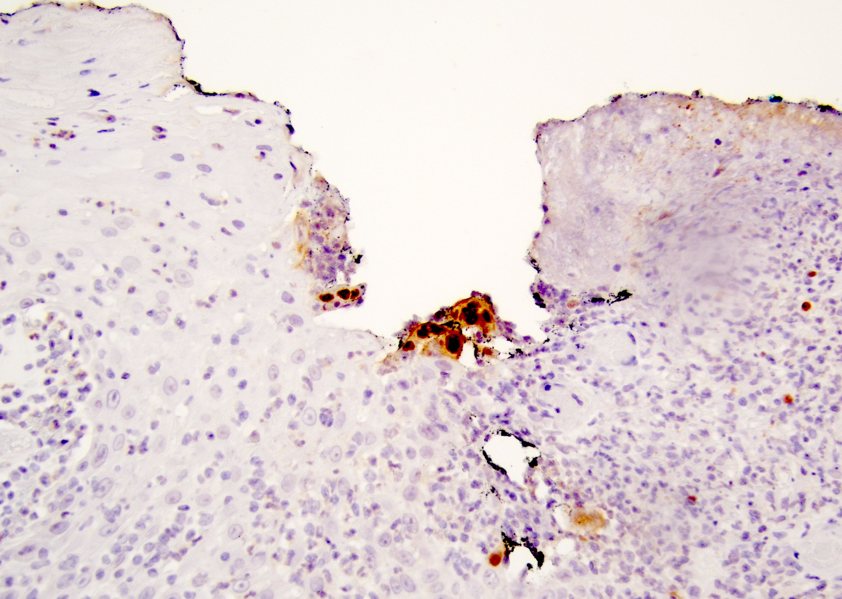
Glandular portion is CEA+
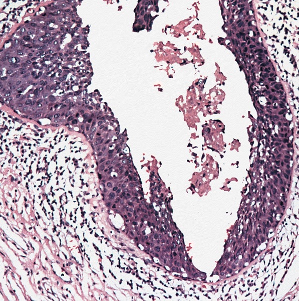
Cervix: poorly formed glands and squamous components (arrows)
Images hosted on other servers:


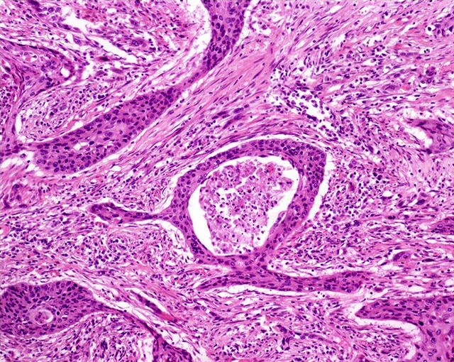
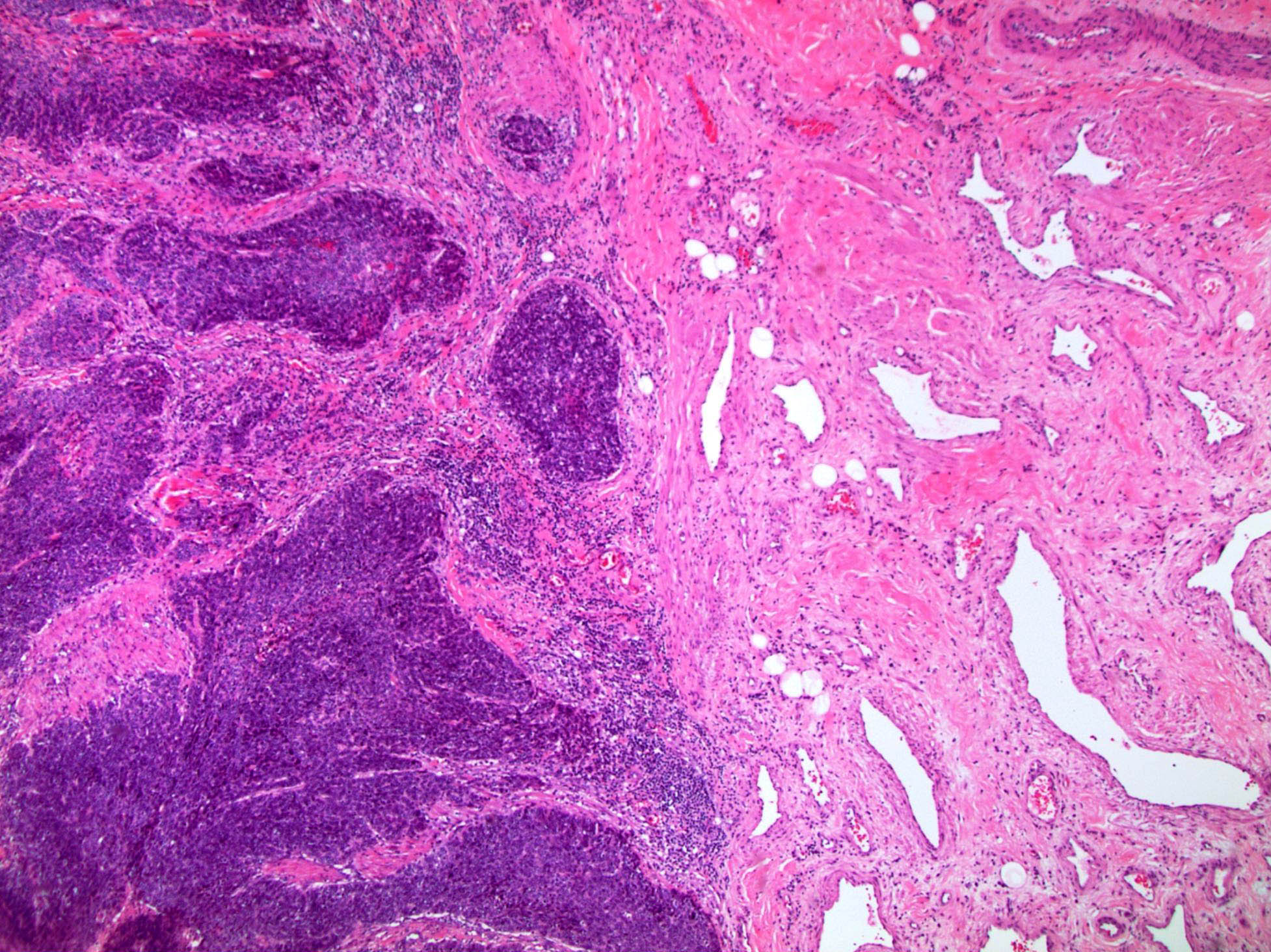
Cervix: malignant glandular (arrow) and squamous components (star)
Images hosted on other servers:


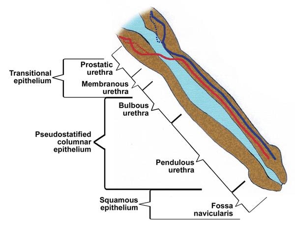
Male urethra
Male urethra

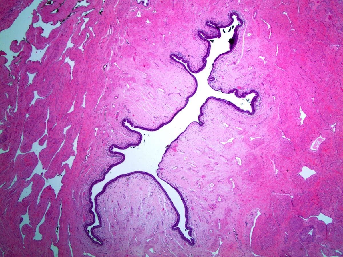
Prostatic urethra

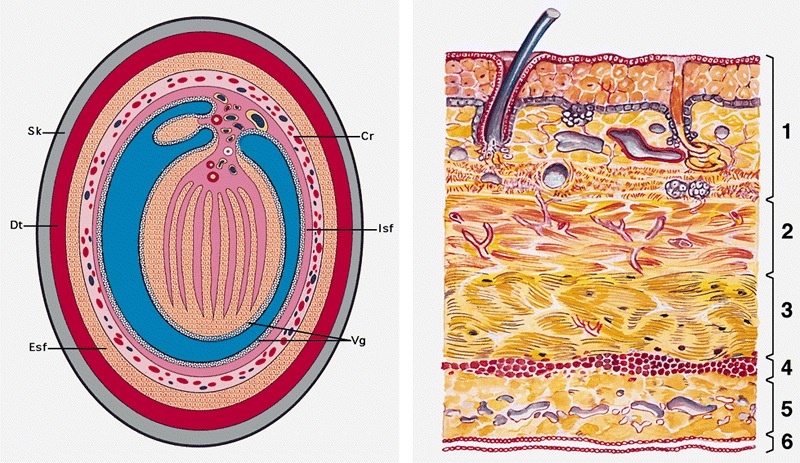
Male urethra development
Contributed by Debra L. Zynger, M.D.
Prostatic urethra
Penile urethra
Contributed by Debra L. Zynger, M.D.
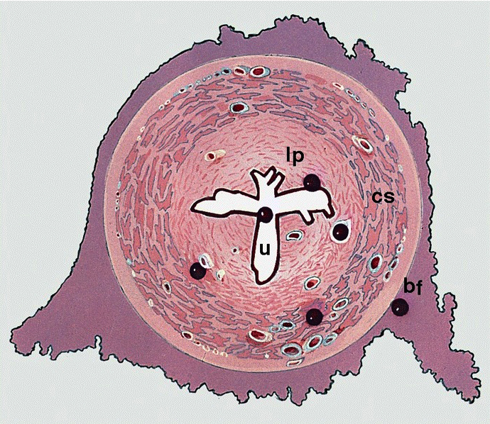
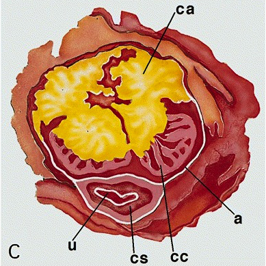
Prostate, cross section with prostatic urethra
Prostatic urethra
Prostatic
verumontanum
and prostatic
urethra
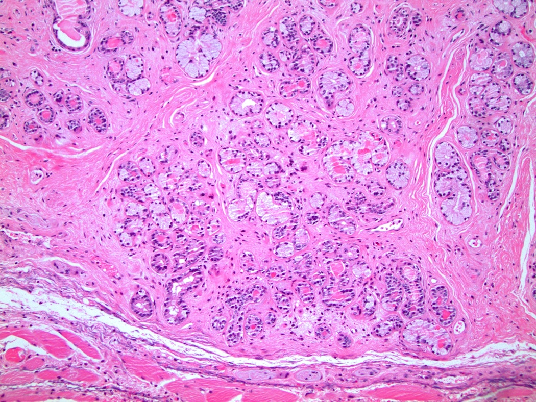
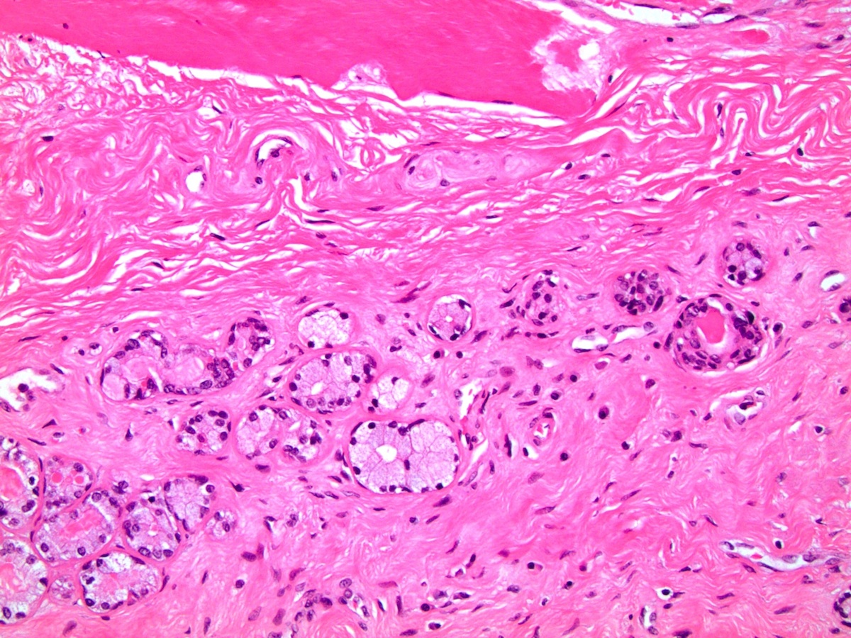
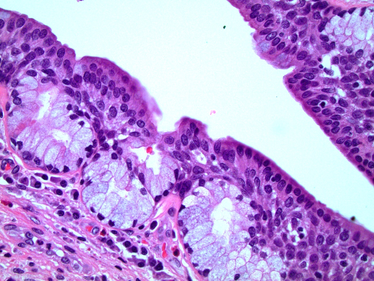
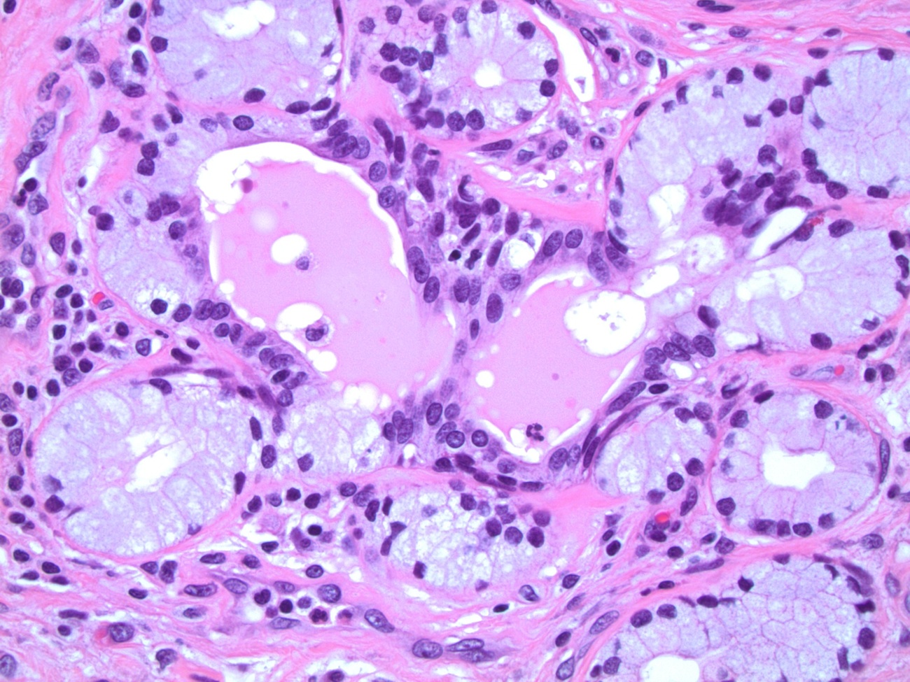
Bulbourethral (Cowper) glands
Penile urethra
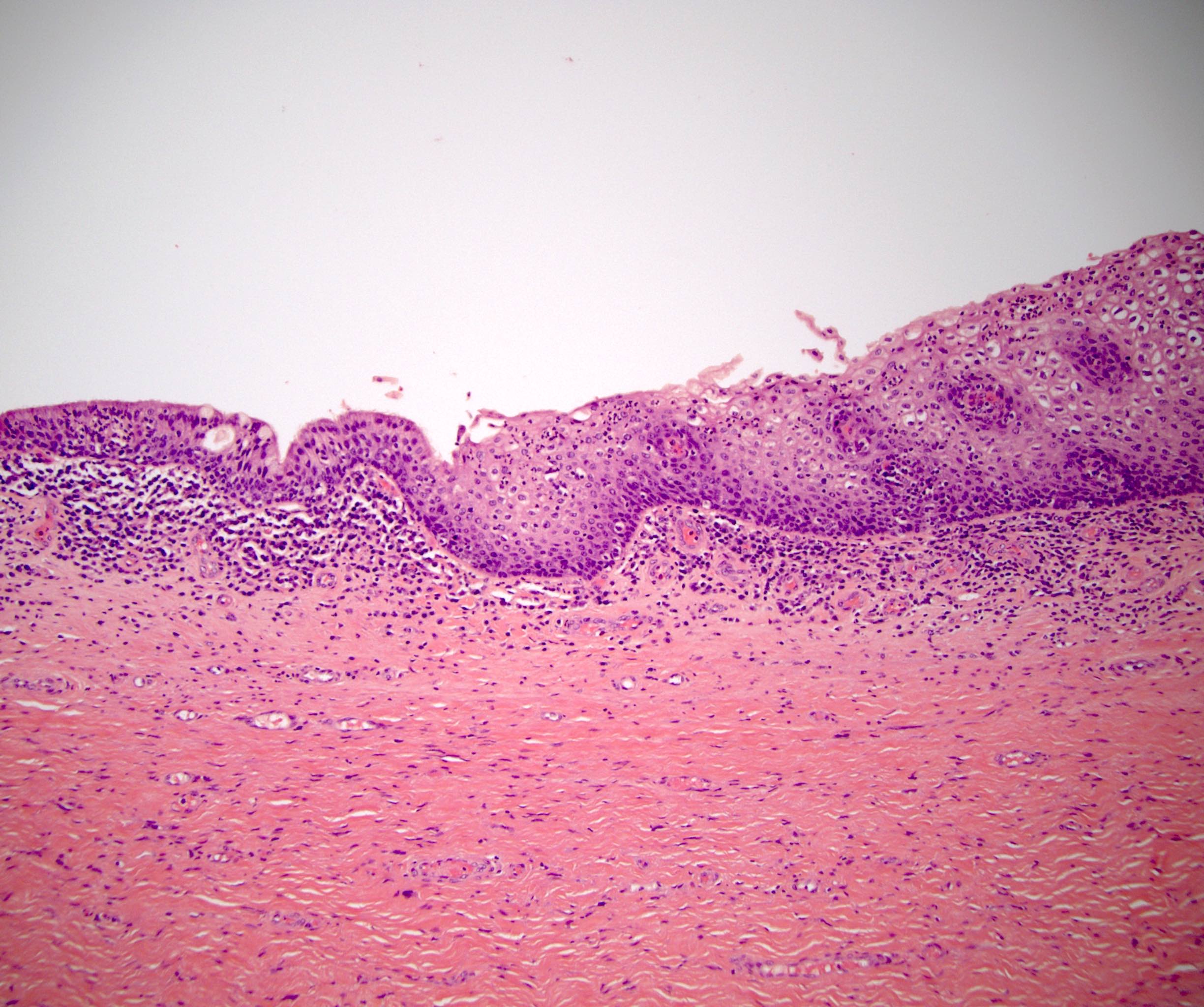
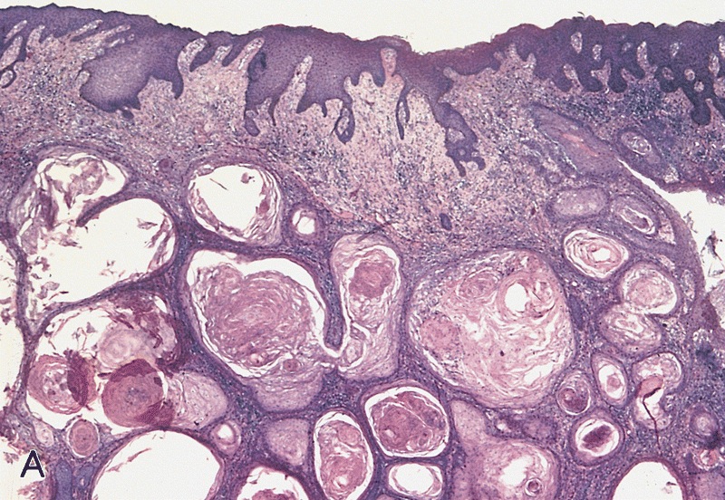
Penile intraepithelial (juxtaepithelial) glands
Penile glands of Littré
Fossa navicularis
AFIP images

Transverse section
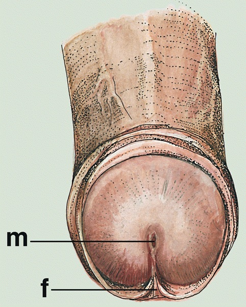
Meatus
Glans
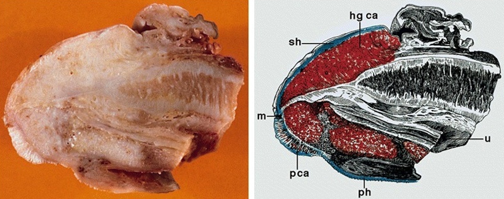
Cut section
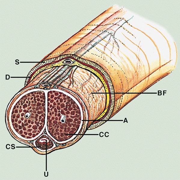
Shaft cross section
Images hosted on other servers:

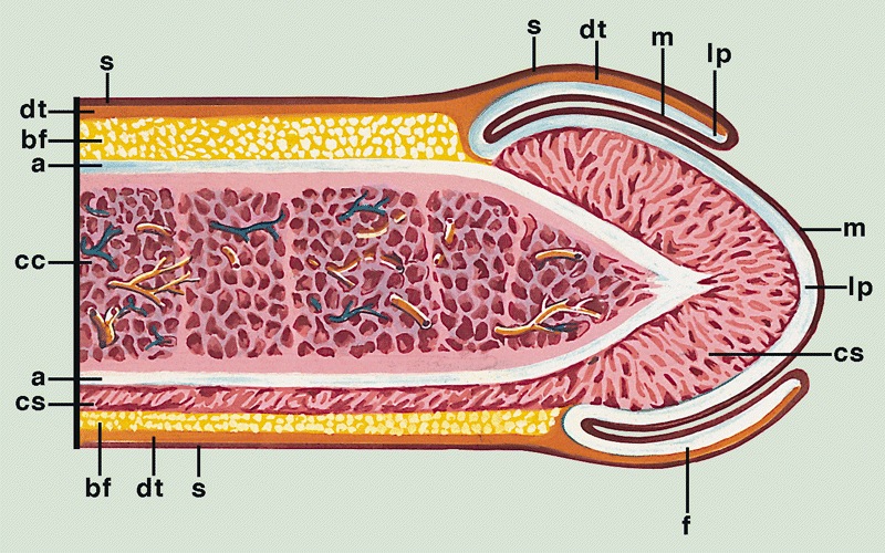
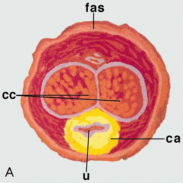
Compartments


Local anatomy

Arteries

Veins

Urethra

Corpora cavernosa

Transverse sections
AFIP images
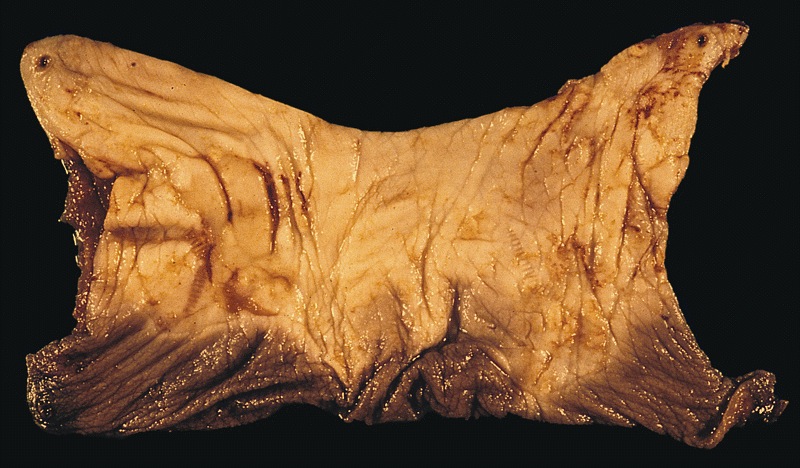
Mucosal and wrinkled portion of foreskin
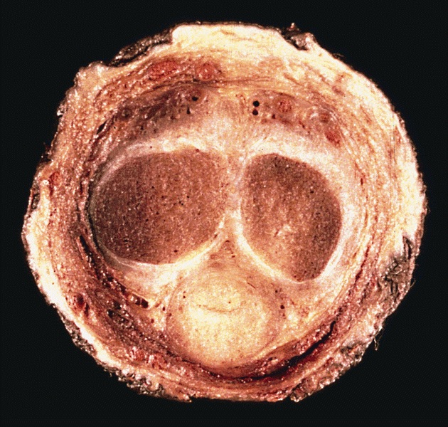
Cross section
Contributed by Diego F. Sanchez, M.D. and Antonio L. Cubilla, M.D.
Penile shaft surgical margin
Surgical margin connective tissue
Erectile corpora
Albuginea and corpus cavernosum
Corpora cavernosa
Corpus spongiosum
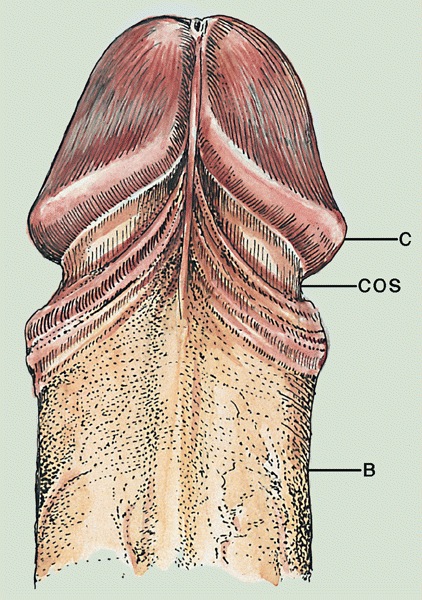
Glans
Foreskin in anatomical position
Foreskin epithelium
Foreskin dartos
Penile urethra
Littre glands
AFIP images
Cross section of the scrotal wall
Images hosted on other servers:


Embryologic development
Images hosted on other servers:

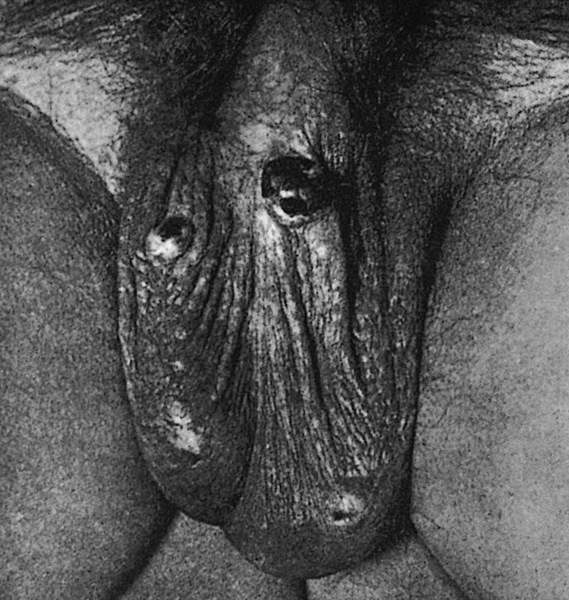
Constricted human
scrotum (without
hair) with the raphe
clearly exposed
AFIP images

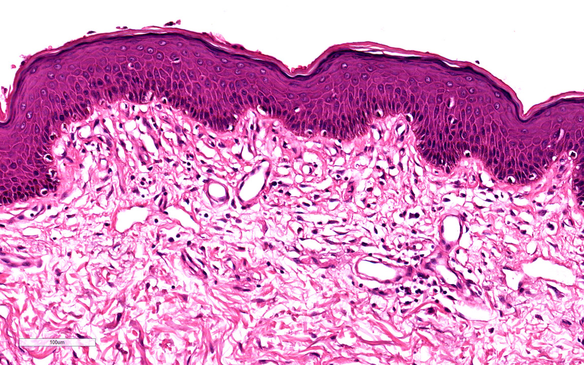
Muscle bundles of the
dartos are beneath
the keratinized
squamous epithelium
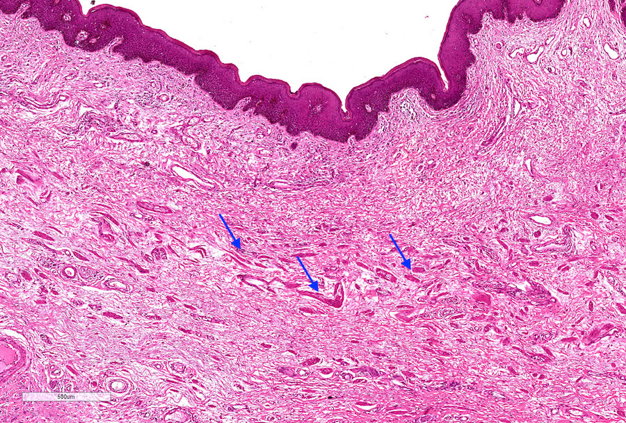
Thick bundles of
conspicuous smooth
muscle in the deep
reticular dermis
Images hosted on other servers:

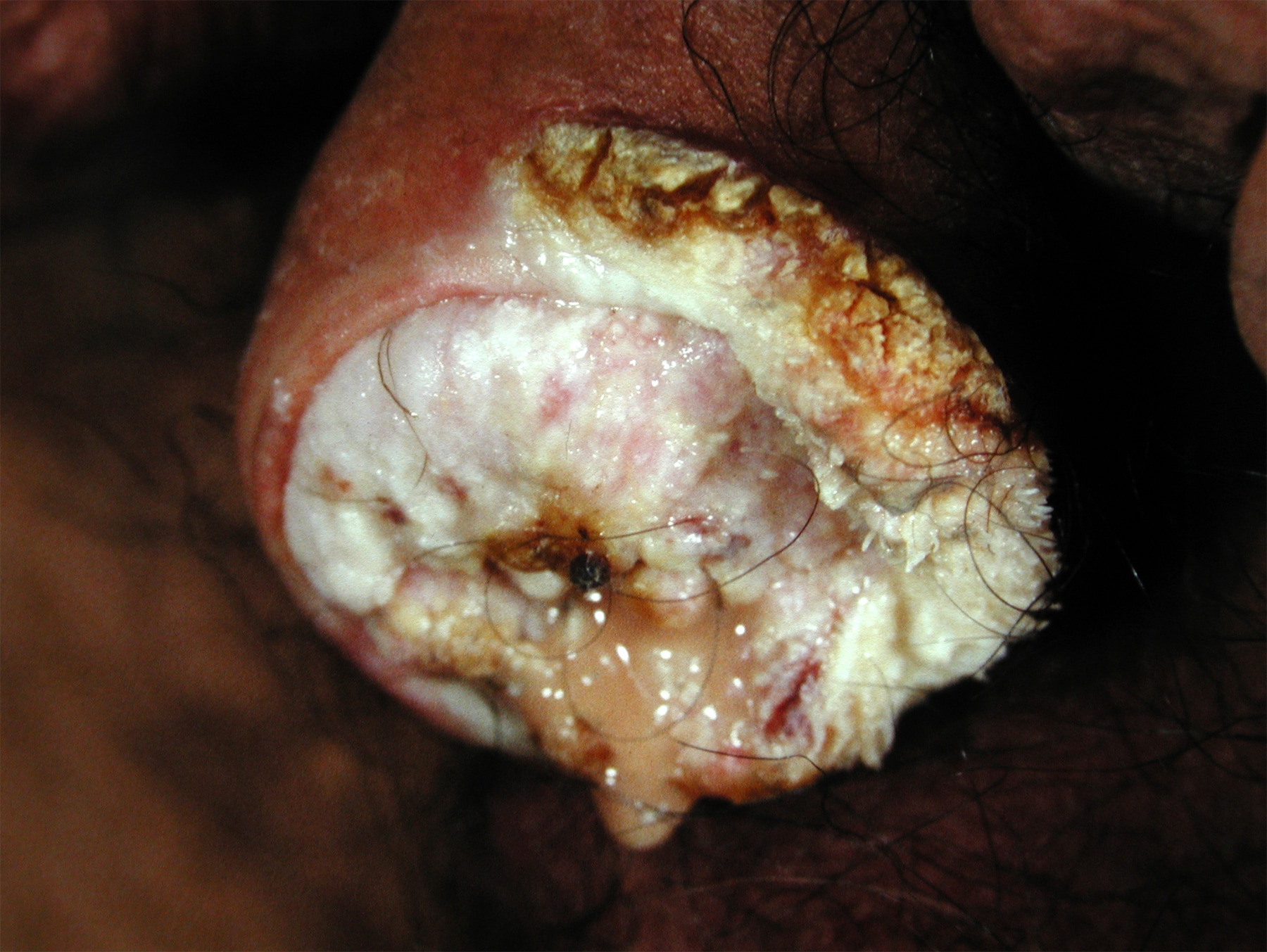
Marked ulceration with tissue destruction
Images hosted on other servers:
Dermis with
lymphoplasmacytic
infiltrate and dilated
blood vessels
Images hosted on other servers:


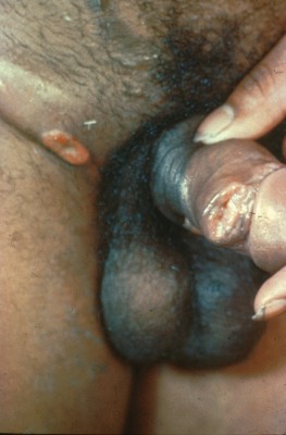
Ulcer almost circumscribing penis and causing penis / scrotal edema
AFIP images
Skin: not necessarily penis
Images hosted on other servers:


Skin - not necessarily penis
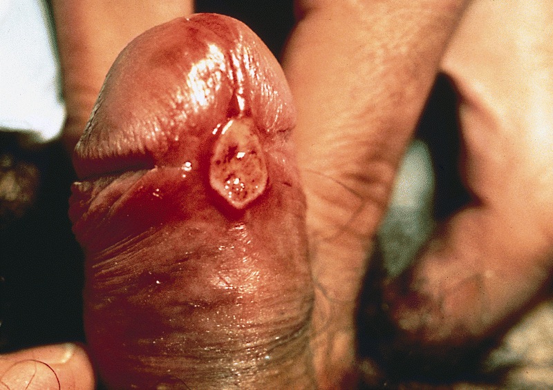
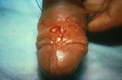
AFIP images
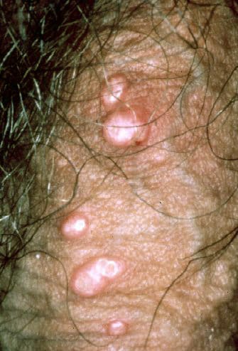
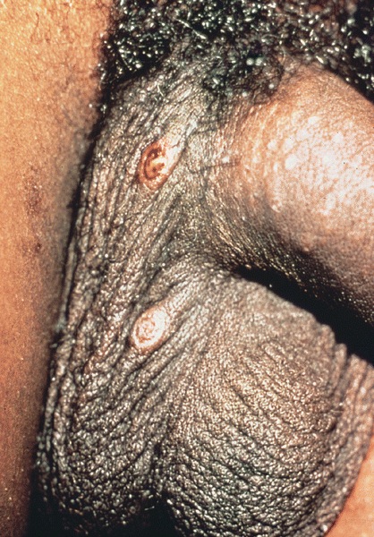
2 small papules with irregular margins
Images hosted on other servers:

Penile lesions
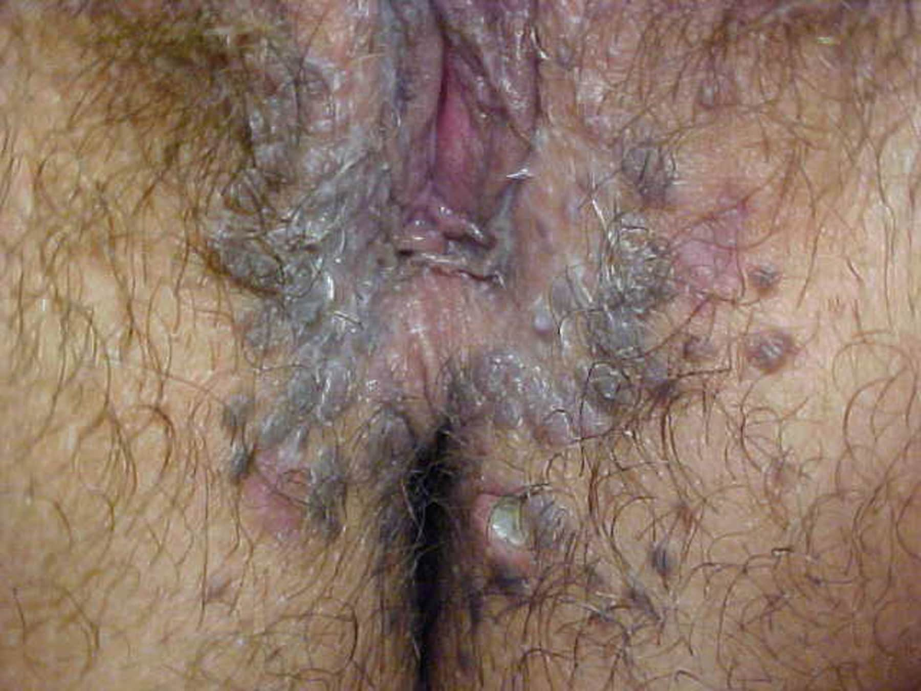
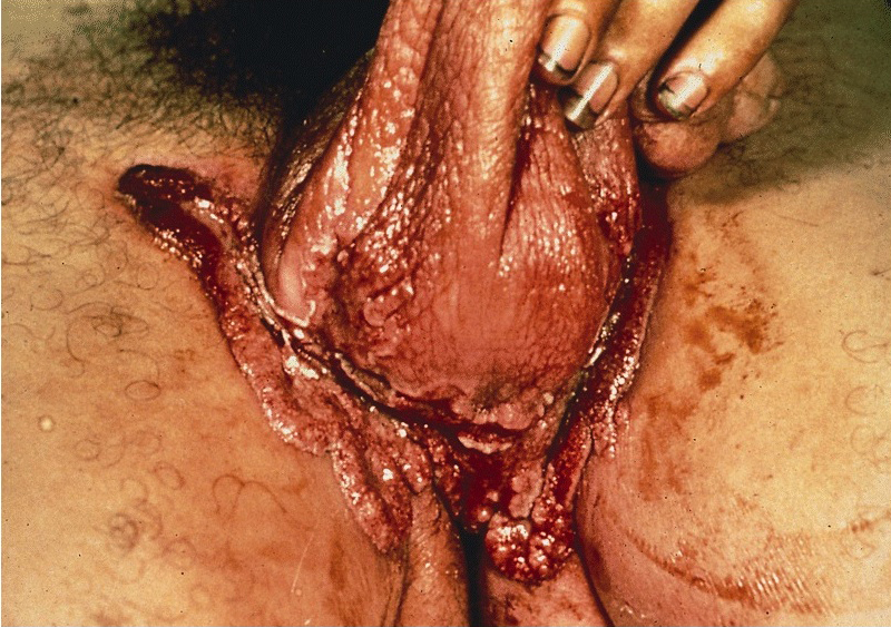
Typical appearance in female
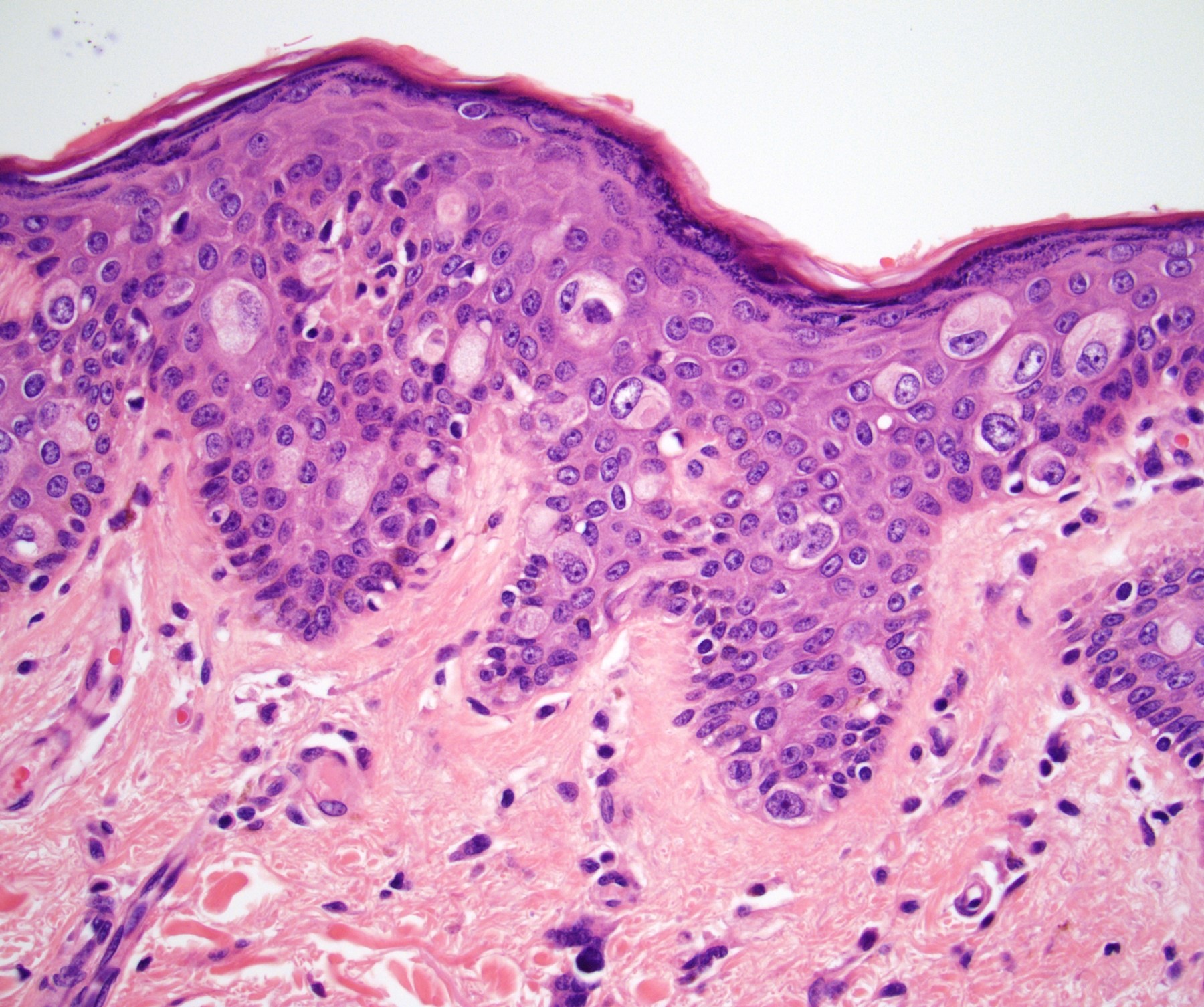
Contributed by Liwei Jia, M.D., Ph.D.
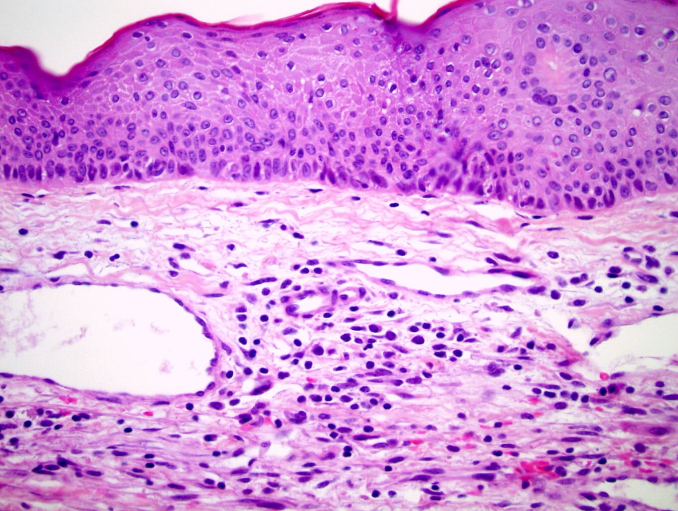
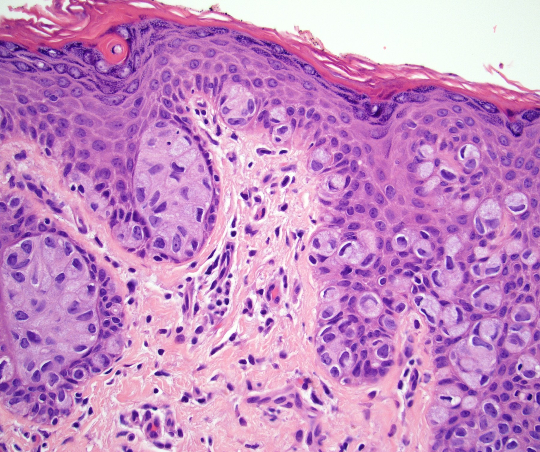
Intact basement membrane
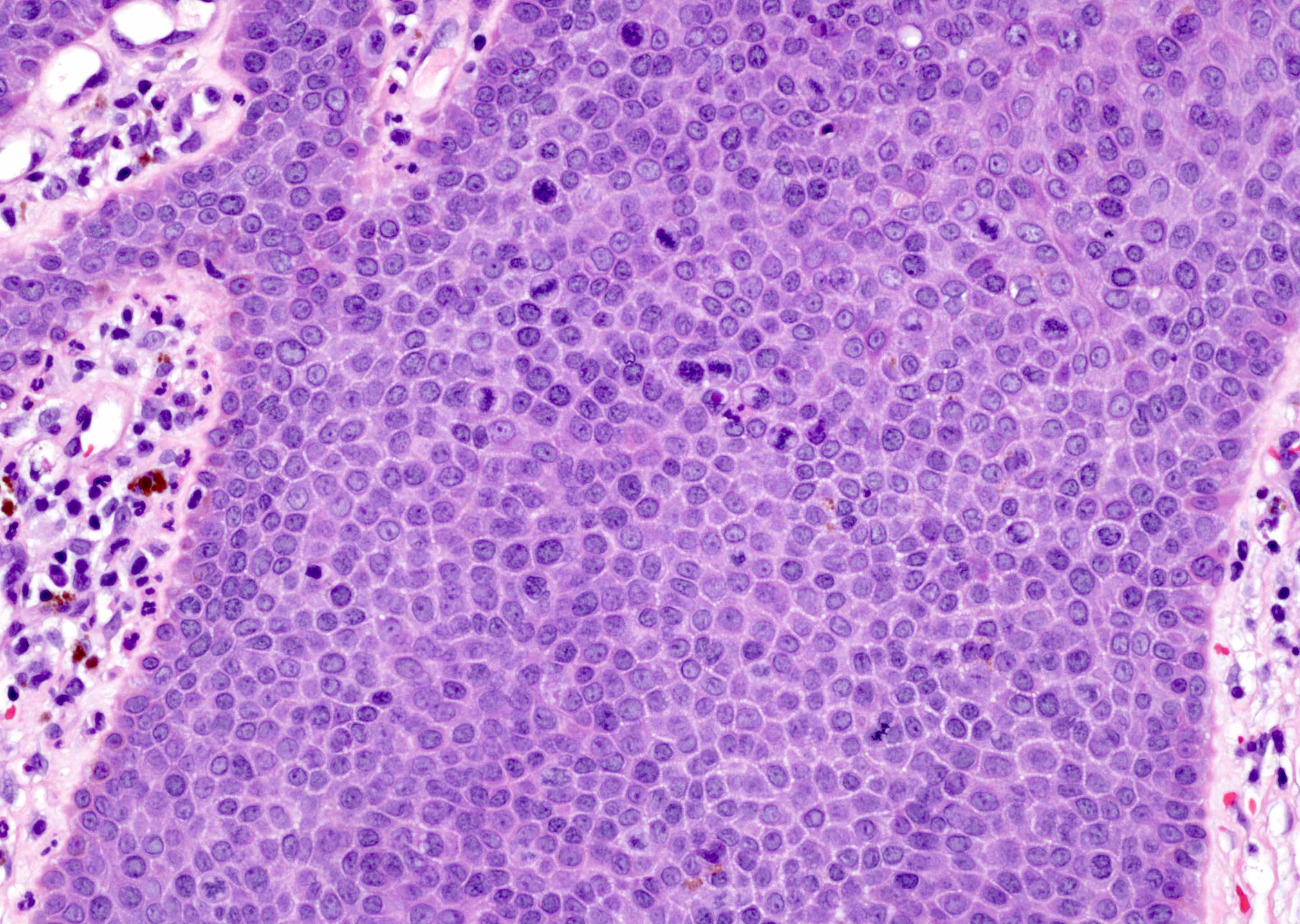
Mitotic figures

Proliferation
AFIP images
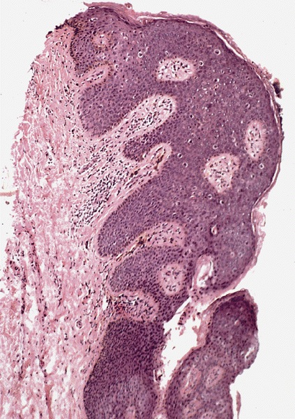
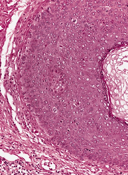
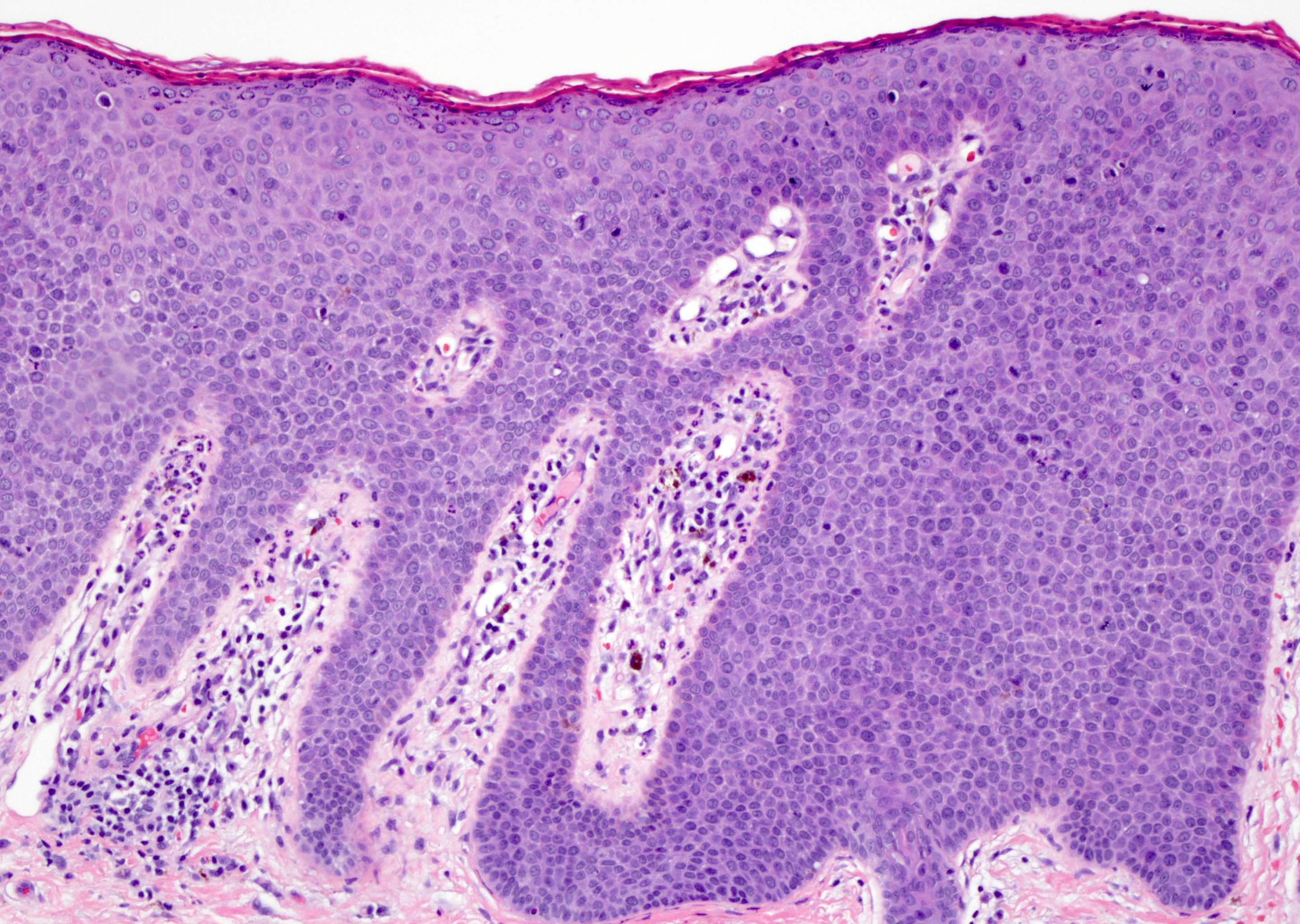
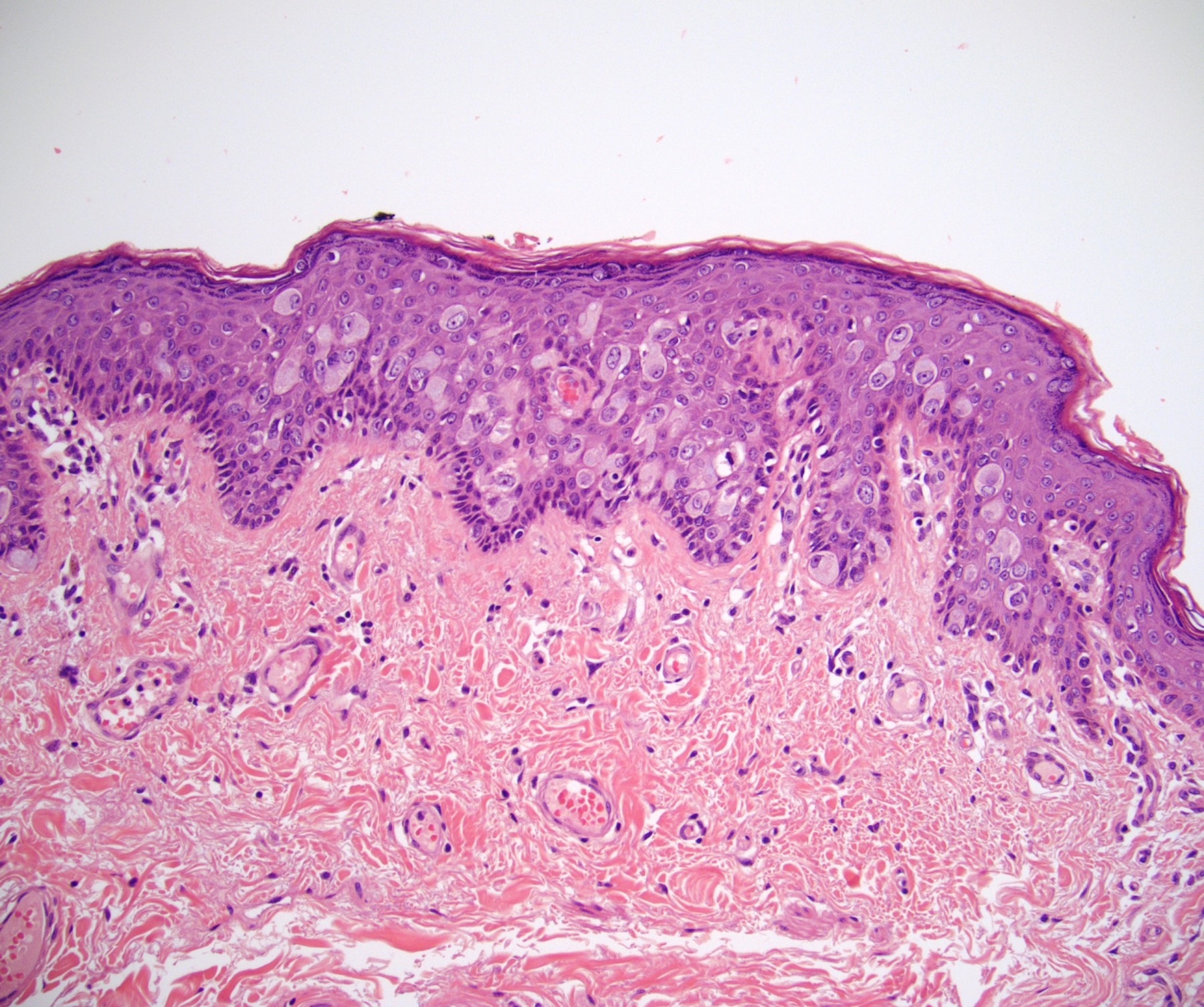
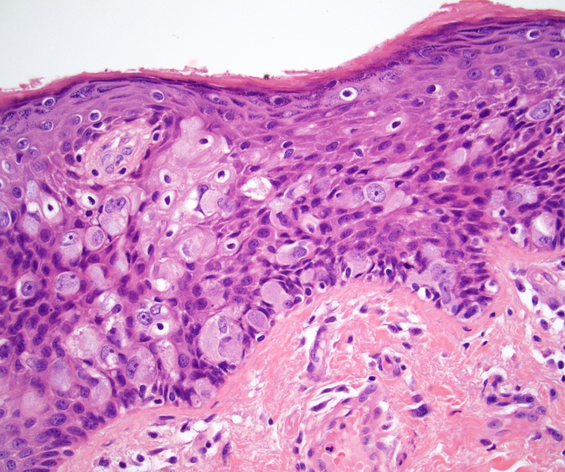
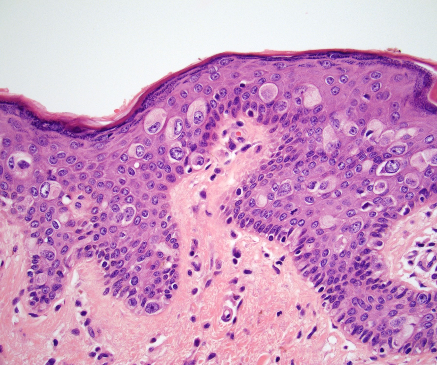
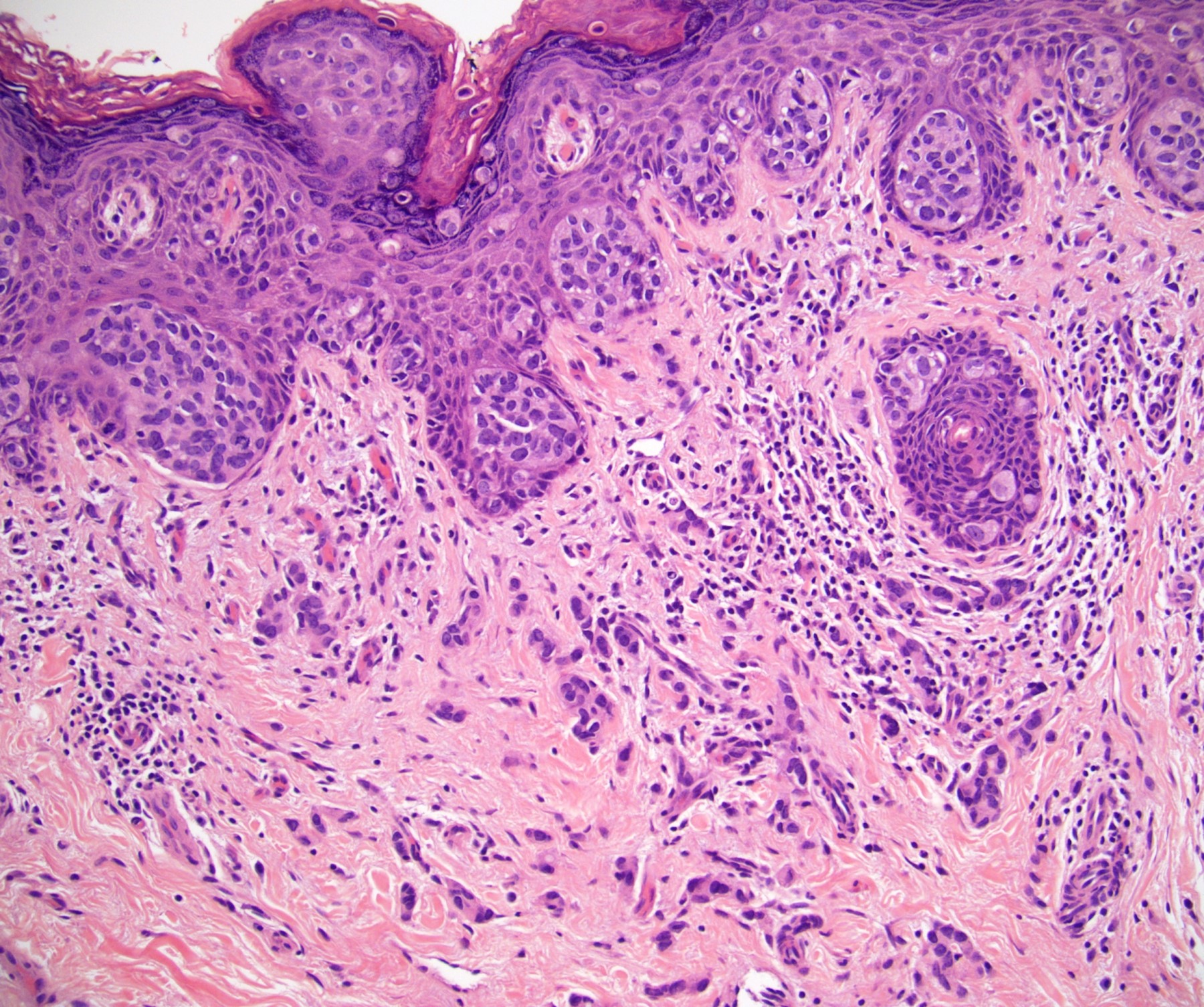
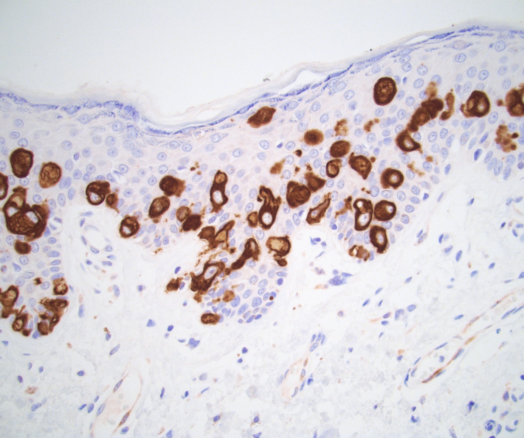
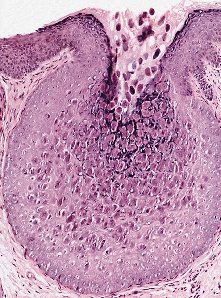
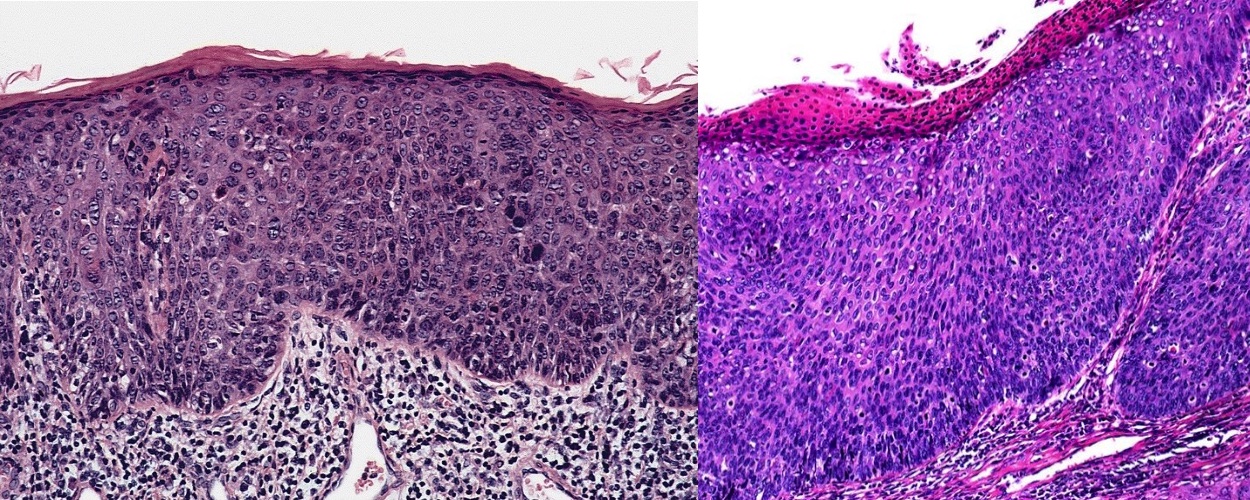
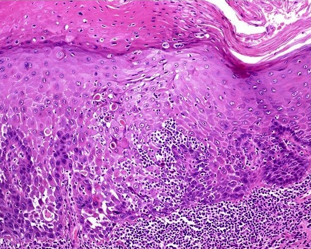
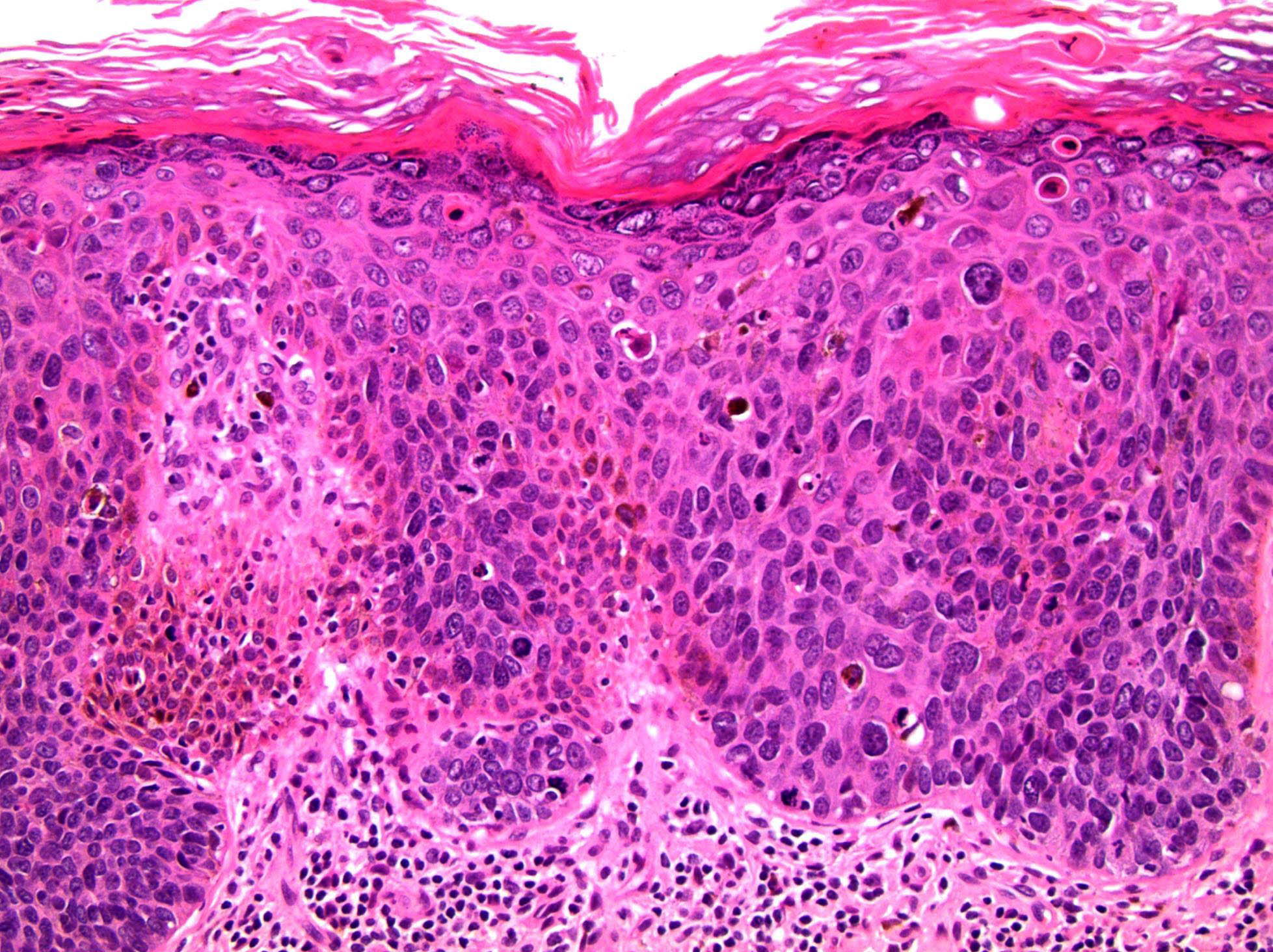
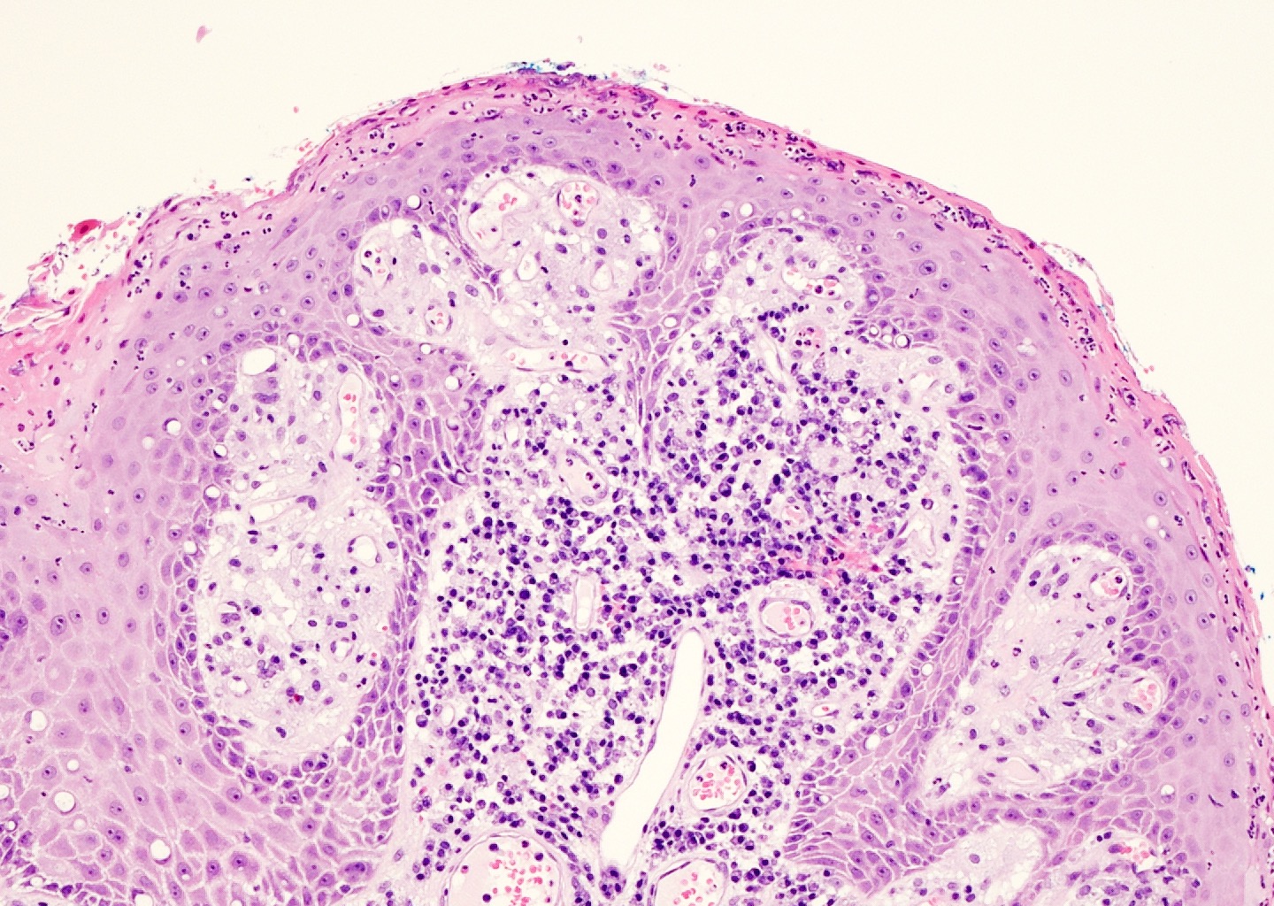
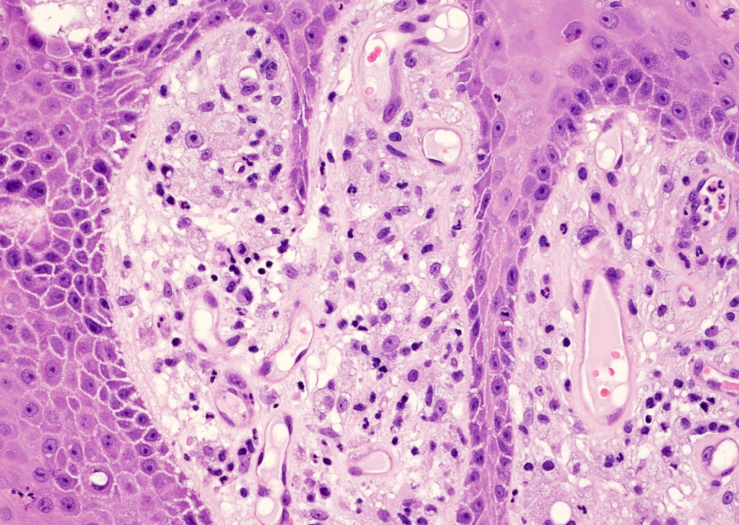
Acanthosis and spotty distribution of atypical cells
Microscopic findings of bowenoid papulosis / HSIL
Images hosted on other servers:

Acute erythema and edema
AFIP images
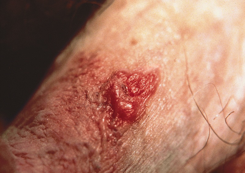
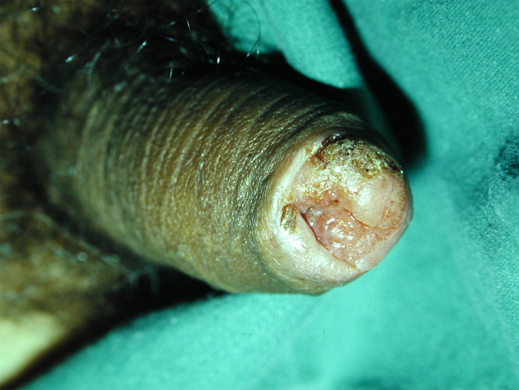
Slightly raised flat disc with central ulceration
Images hosted on other servers:
Ulcers
Regional adenopathy
Images hosted on other servers:
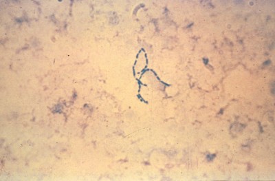
Gram stain

Gentian violet stain
AFIP images
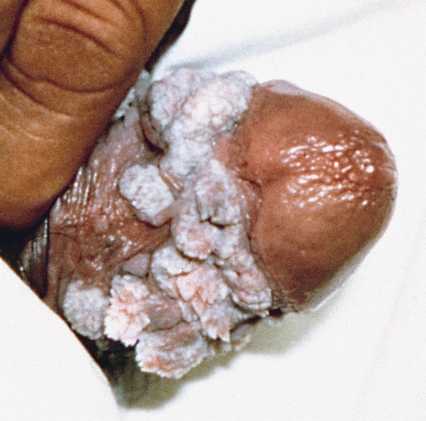
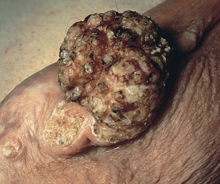
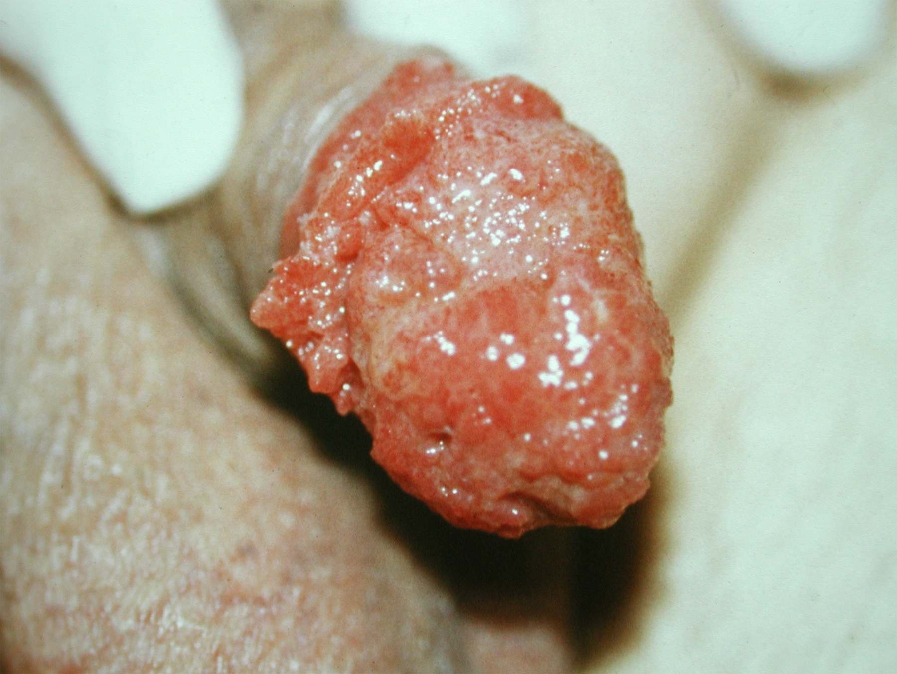
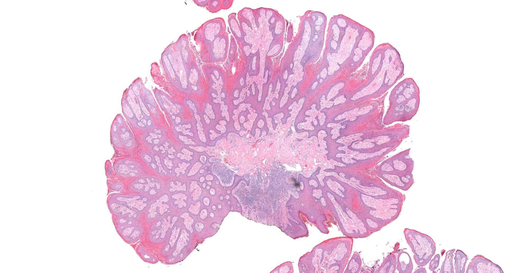
Multiple exophytic lesions
Images hosted on other servers:

Multiple exophytic and warty lesions on the shaft and glans of the penis
Contributed by Debra L. Zynger, M.D.

Giant condyloma acuminatum
Contributed by Asra Feroze, M.B.B.S. and Ritu Bhalla, M.D.
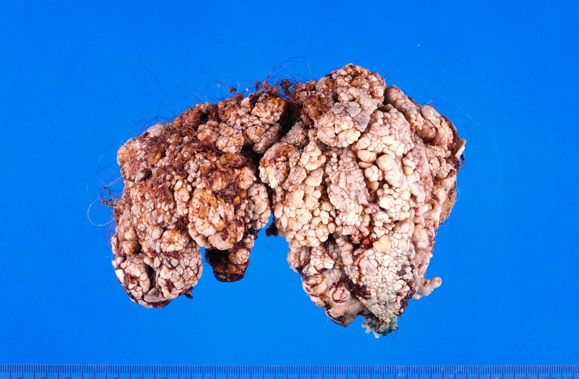
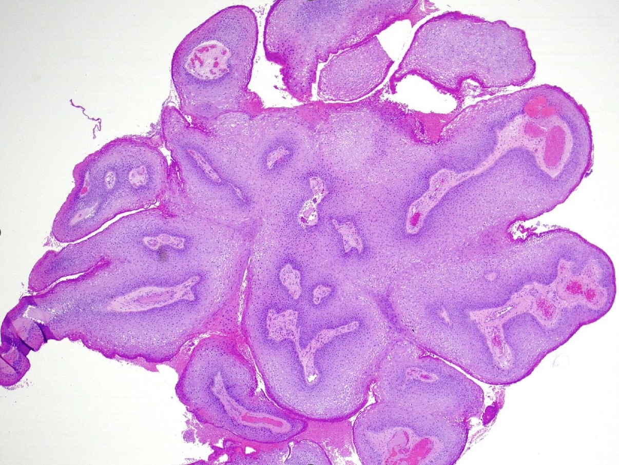
Exophytic growth
Cauliflower-like lesion

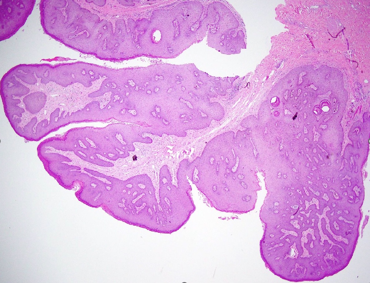
Fibrovascular cores

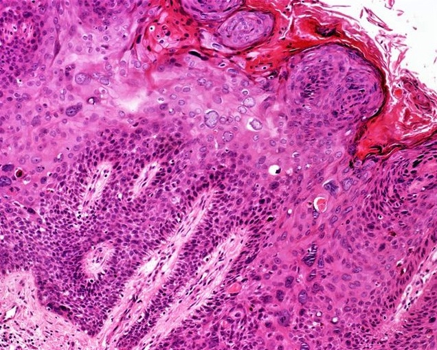
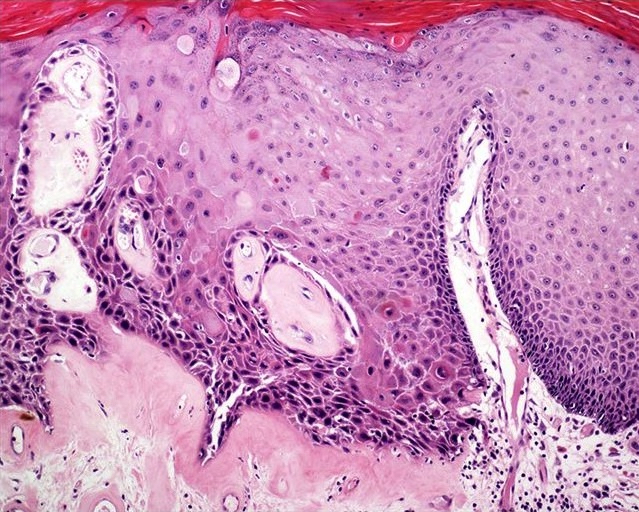
Hyperkeratosis and parakeratosis

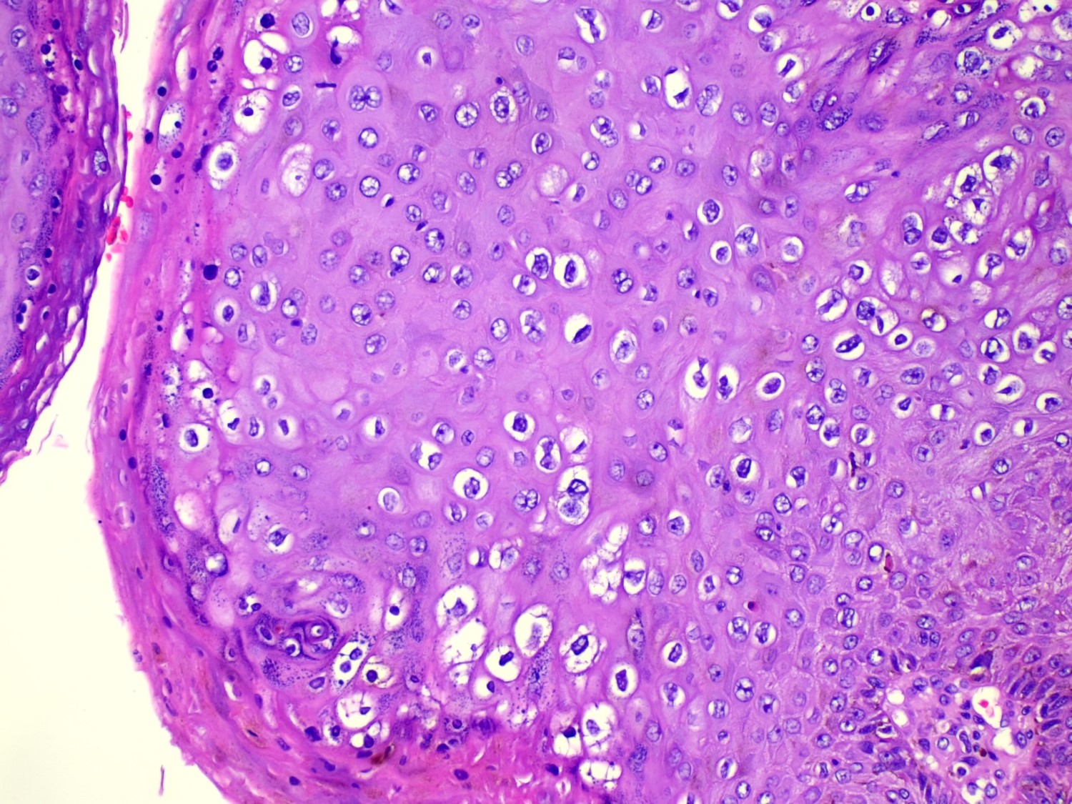
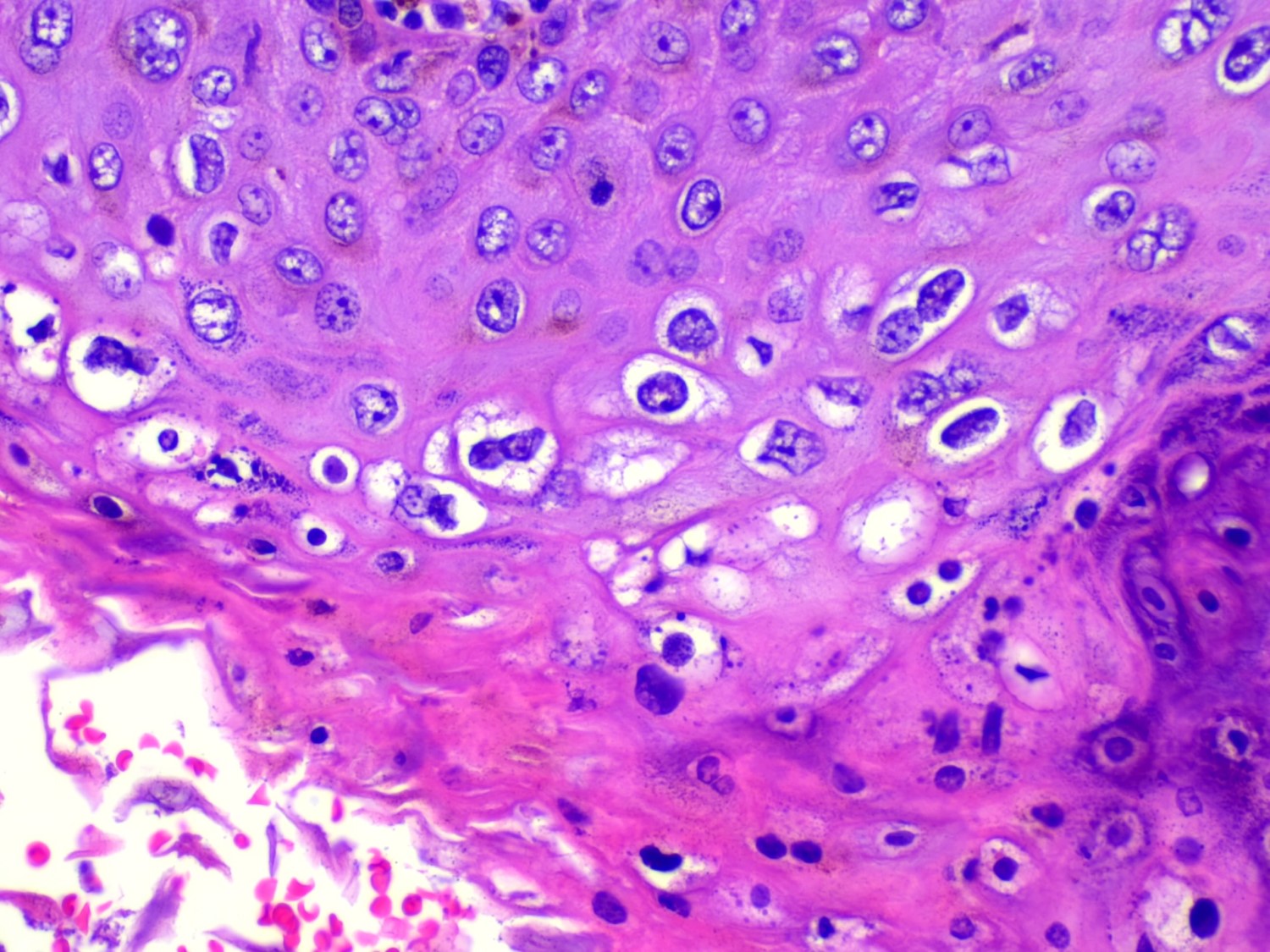
Characteristic koilocytes

p16
Condyloma histopathology
Genital warts under microscope
Images hosted on other servers:

Classification

Epispadias: male baby


Proximal shaft

Penoscrotal

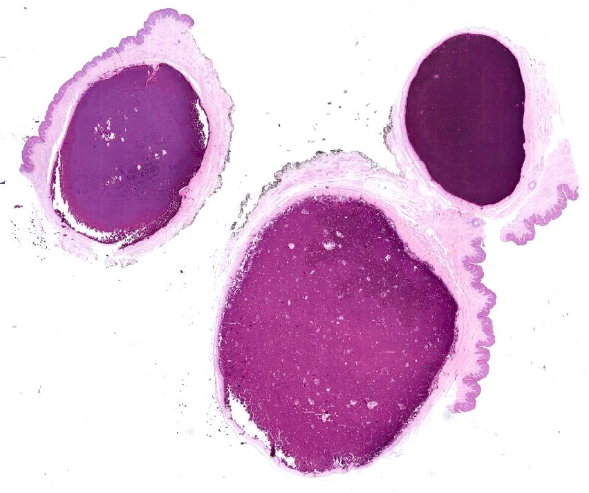
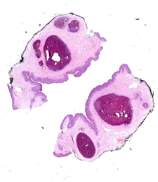
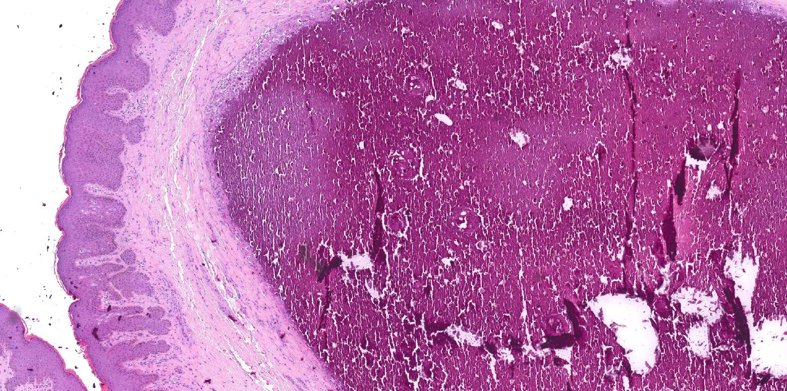
43 year old man with median raphe cyst
Images hosted on other servers:
Median raphe cysts:

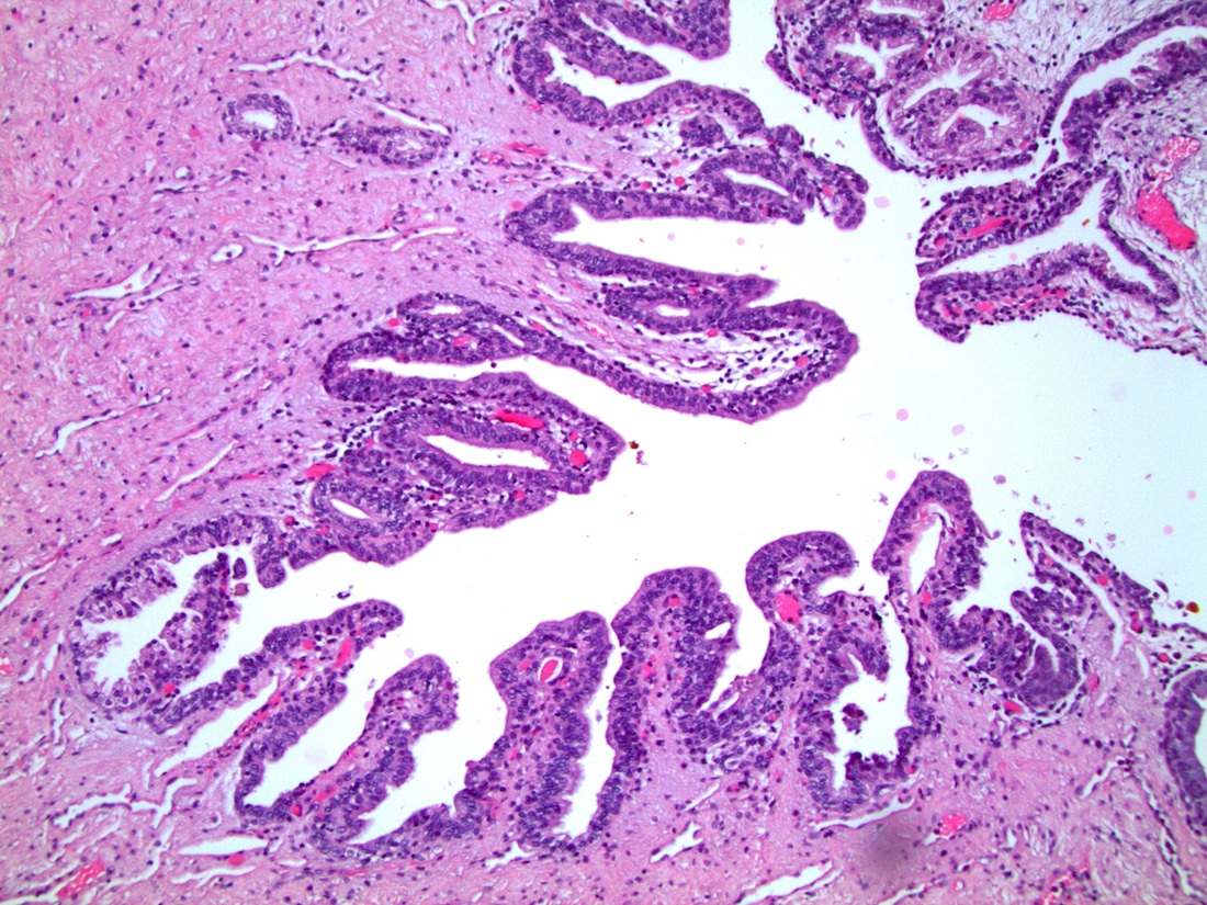
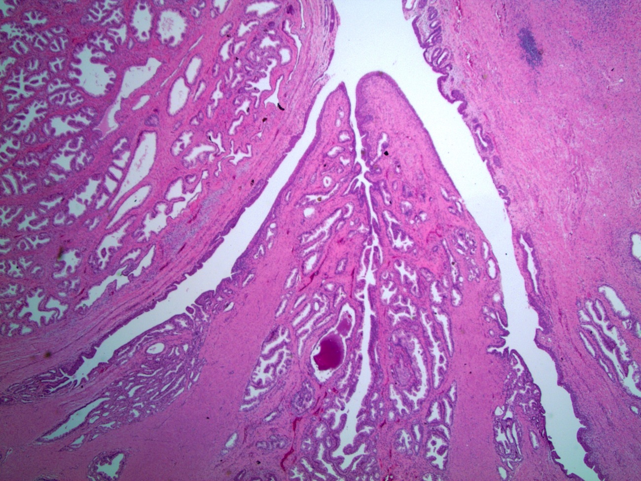
Pseudostratified columnar epithelium

Single larger mucinous cell

Cyst lining cells are CK7+
Images hosted on other servers:

Scrotal MR and ultrasound

Scrotal ultrasound
AFIP images

Scrotal lesion
Images hosted on other servers:


Erythematous penile patches
Contributed by Debra L. Zynger, M.D.

White lesion
Contributed by Debra L. Zynger, M.D.
Intraepithelial growth
Nests and single cells
Basal location
Nuclear features
Microinvasion

Deep invasion
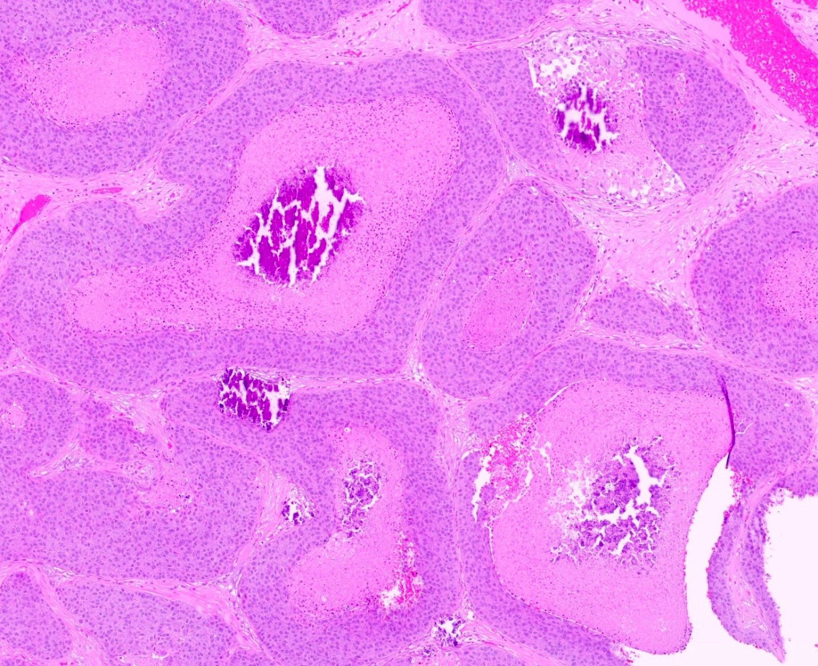
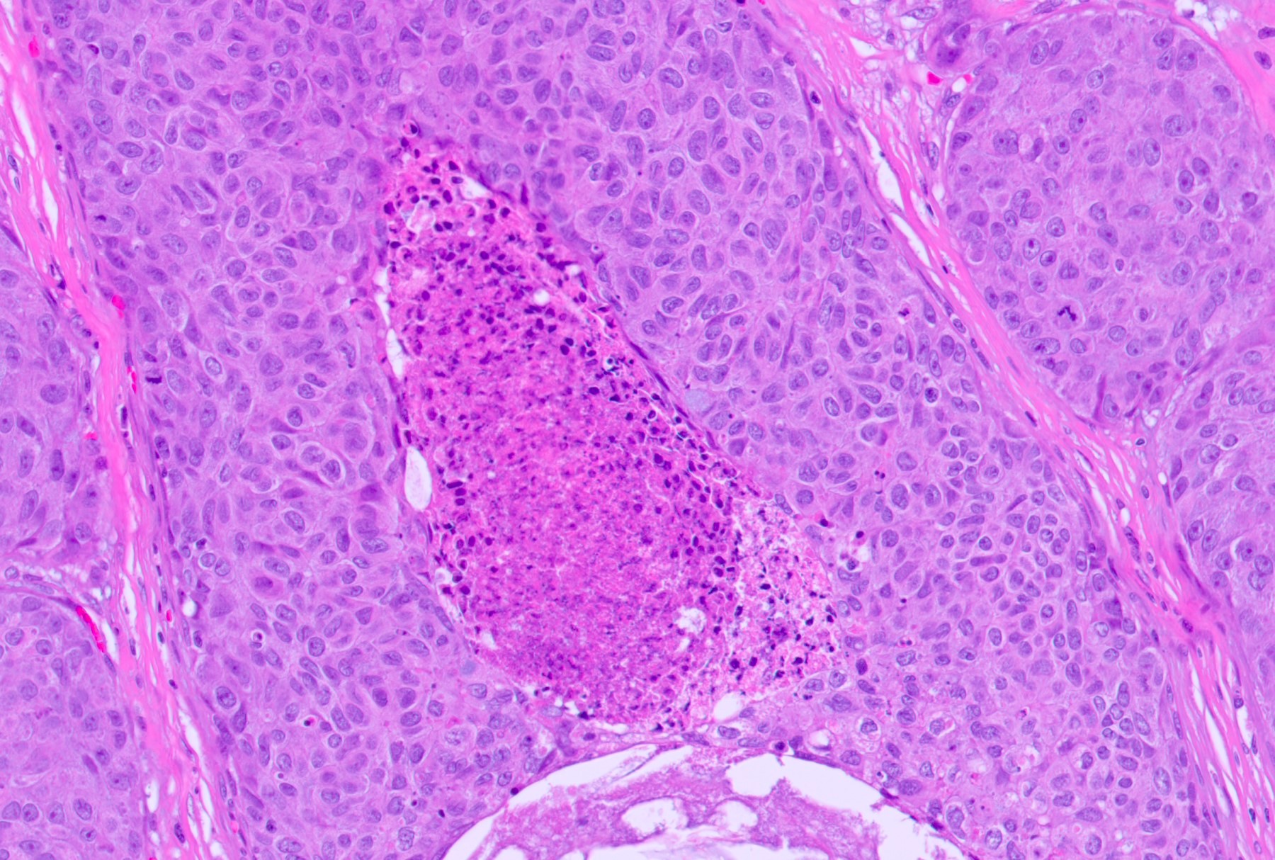
Comedonecrosis
Muscle invasion
H&E for comparison with IHC
CK7
GATA3
CK5/6
p63
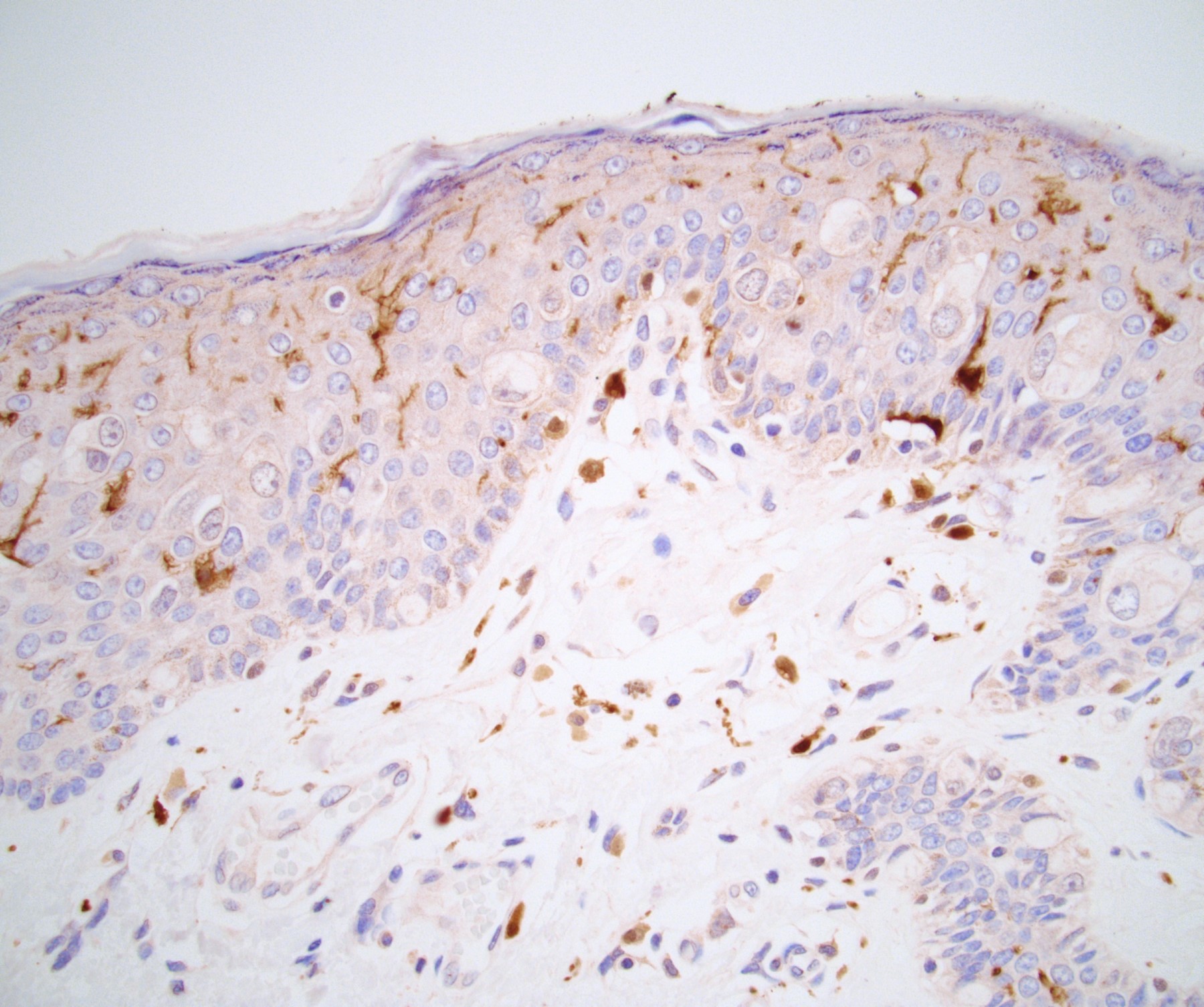
S100
Images hosted on other servers:

Fournier gangrene severity index
Images hosted on other servers:

Necrosis spreading along fascial planes

Paraphimosis


Lesions on penis and scrotum
AFIP images
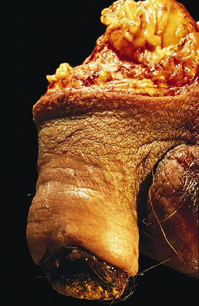
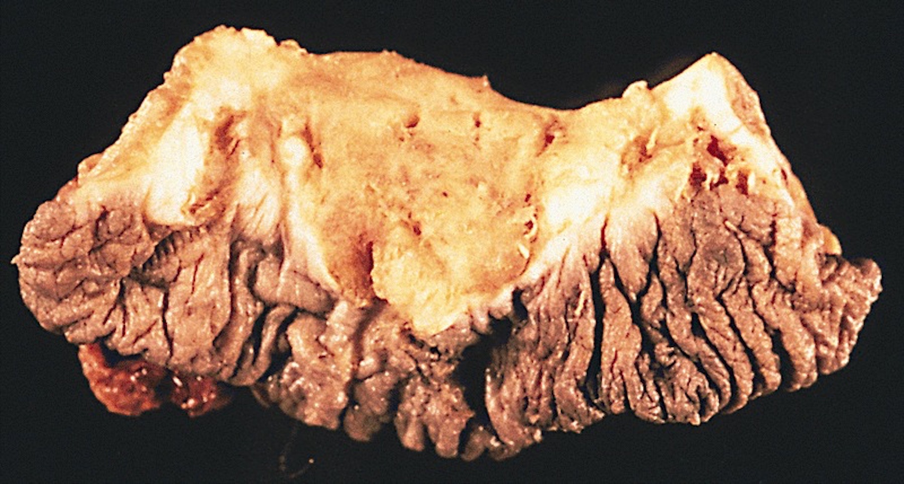
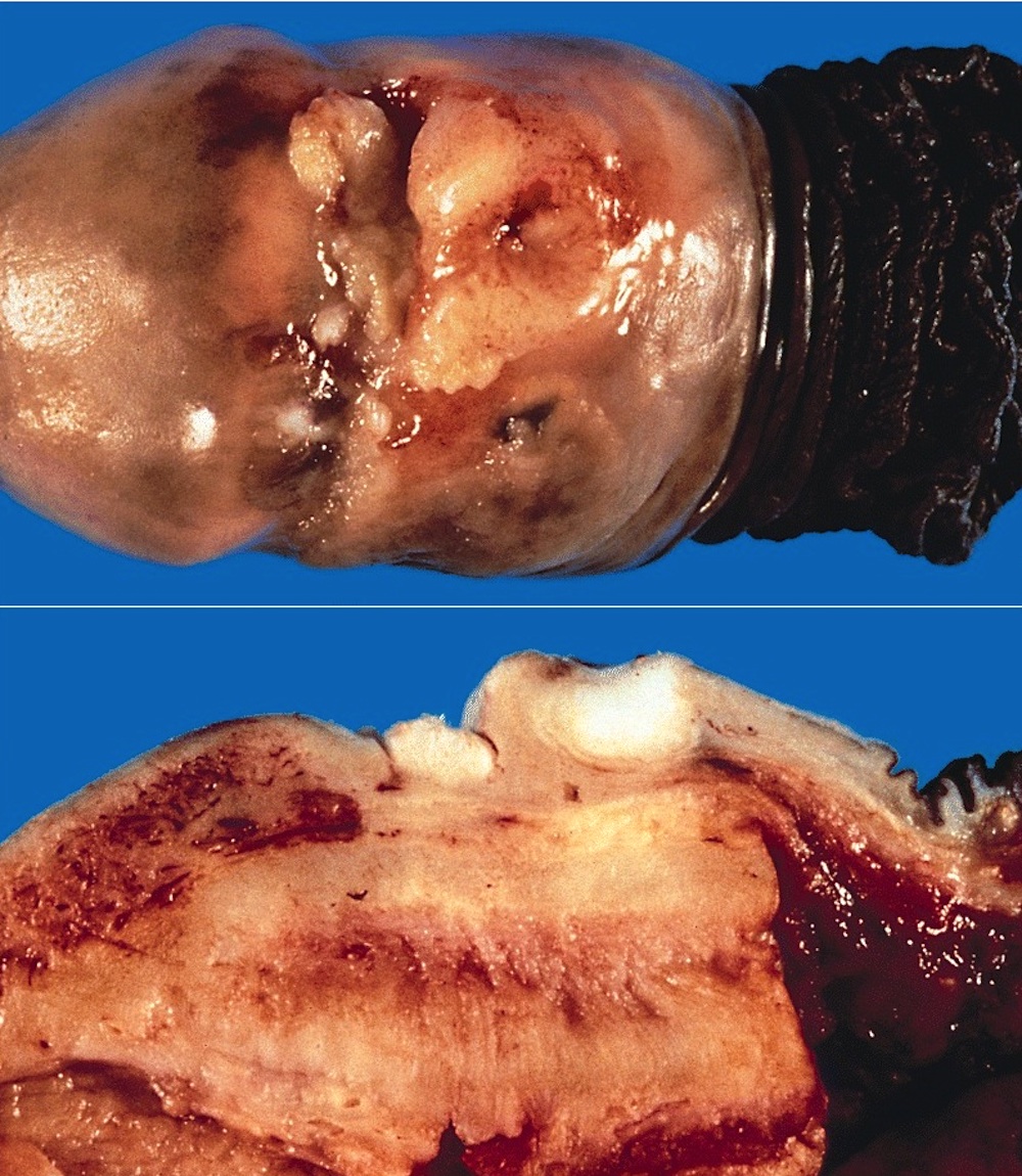
Extensive sloughing
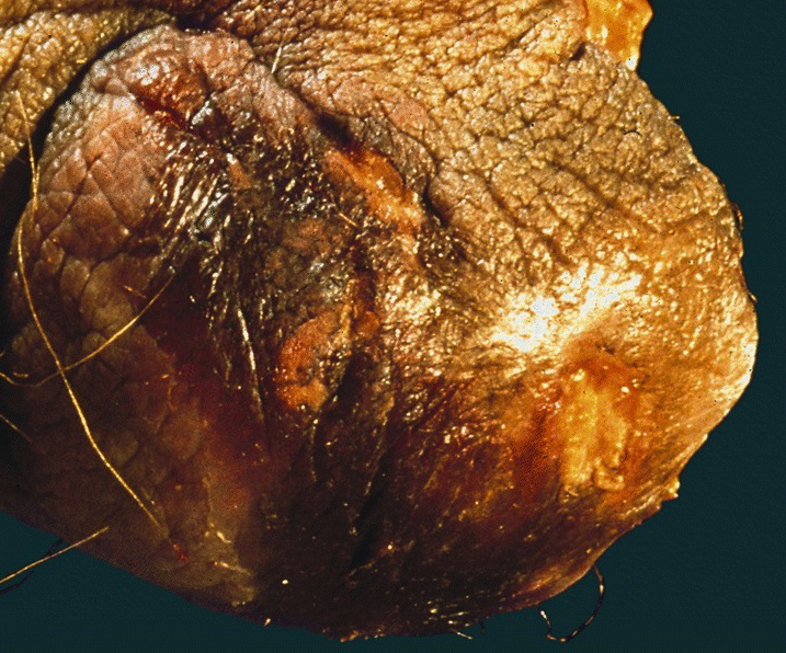
Corbus disease: necrosis of glans
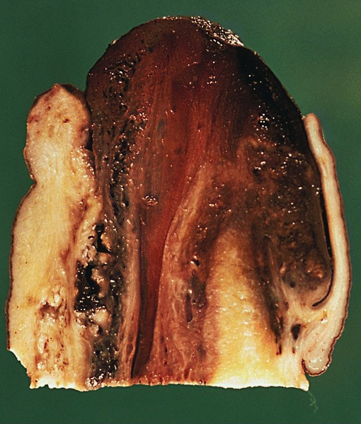
Corbus disease: cut surface
Images hosted on other servers:

Bacteria, neutrophils and necrotic tissue
AFIP images

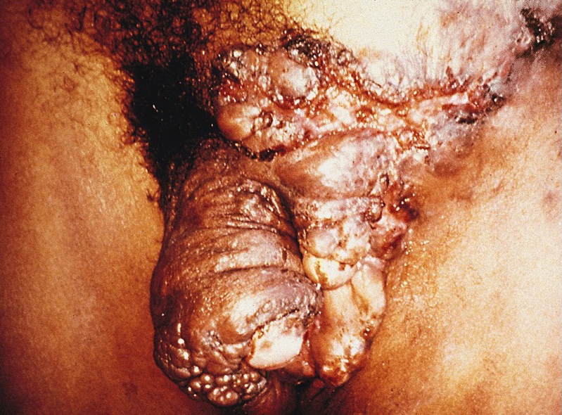
Figure 10-76
Scrotum and inner thigh skin
Images hosted on other servers:

Ulcerated lesion with hypertrophic borders


Various images

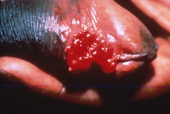
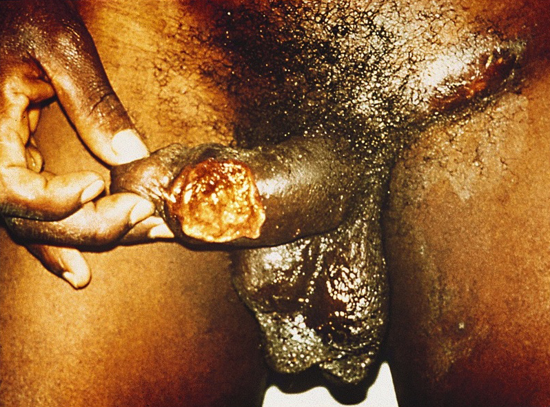
Beefy red penile ulcer


Multiple ulcers on the penile shaft, pubis and scrotum

Raw granulation tissue

Diffuse ulceration
Images hosted on other servers:


Donovan bodies

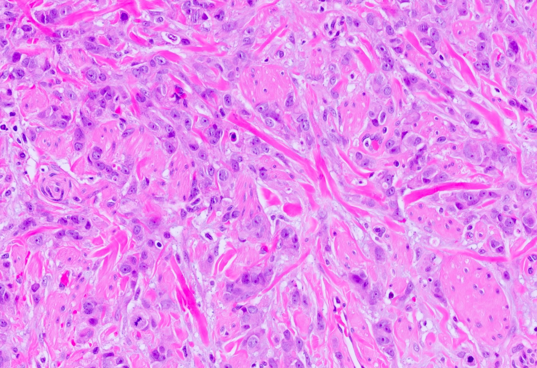
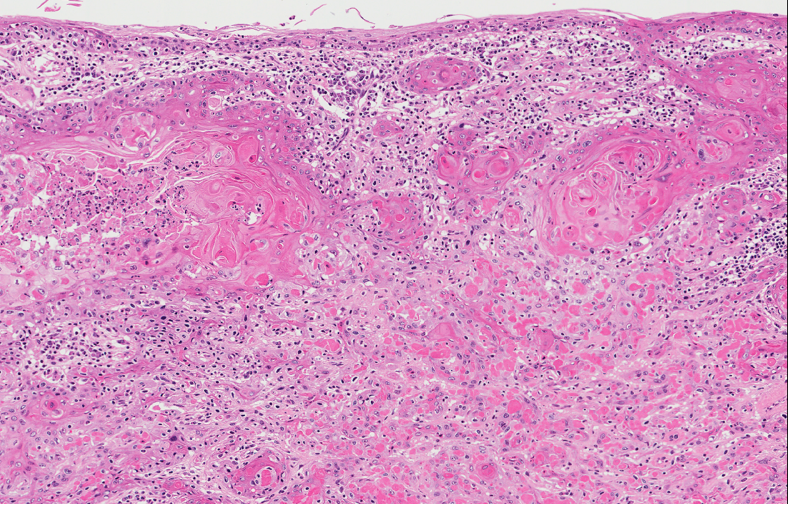
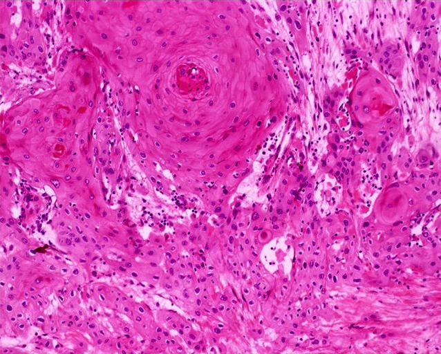
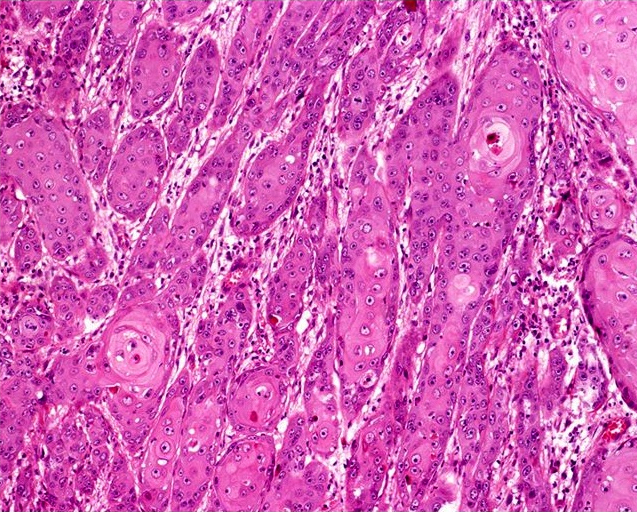
Squamous cells and keratin pearls
Contributed by Shaheed W. Hakim, M.D. and AFIP
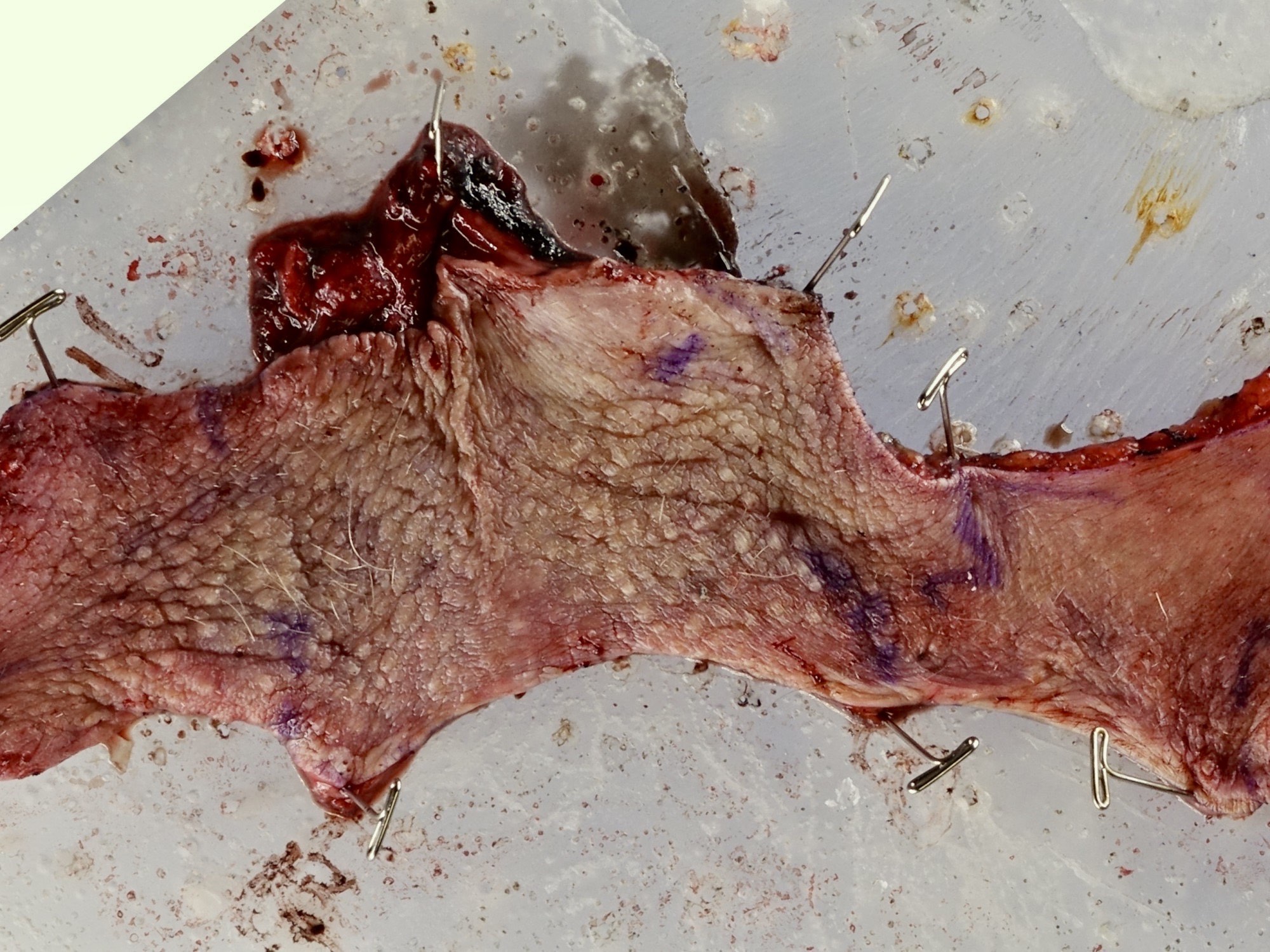
Mapped partial penectomy specimen
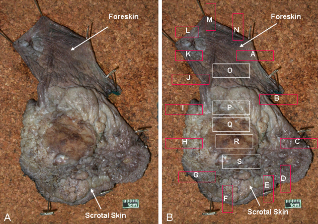
Foreskin
SCC involves corpora cavernosa
Partial penectomy specimen
Contributed by Shaheed W. Hakim, M.D. and AFIP
Grade 1
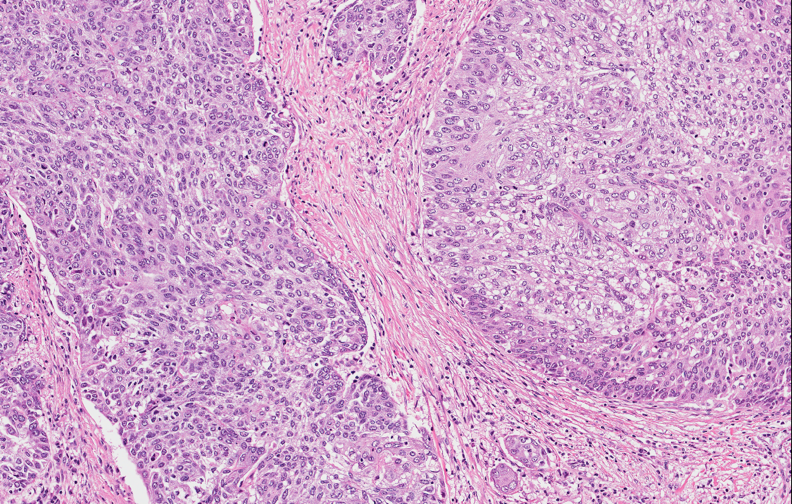
Grade 2, more disorganized growth

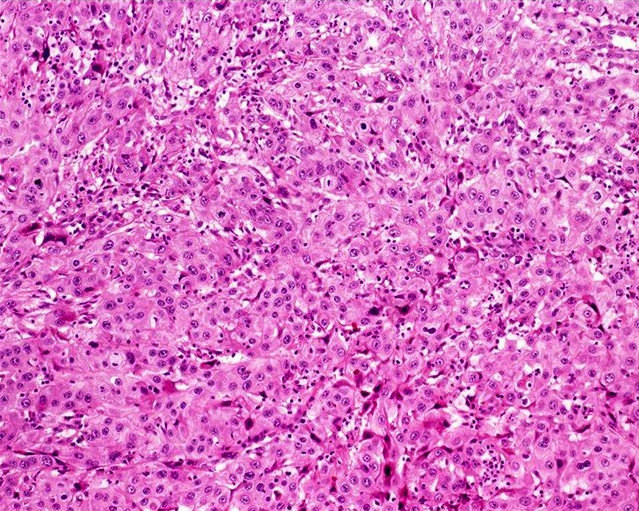
Grade 3
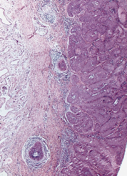
Lamina propria invasion
Urethral mucosal involvement
AFIP images

Periurethral corpus spongiosum involvement
Possible sites of involvement
Tumor involvement in yellow
Images hosted on other servers:

Penis anatomy
Images hosted on other servers:
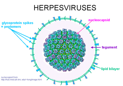
Herpesvirus structure
Images hosted on other servers:

Multiple vesicles and ulcerations on surface

Herpetic vesicles of penis

Herpes vegetans
Contributed by Daniel Anderson, M.D., M.B.A. and Garrison Pease, M.D.
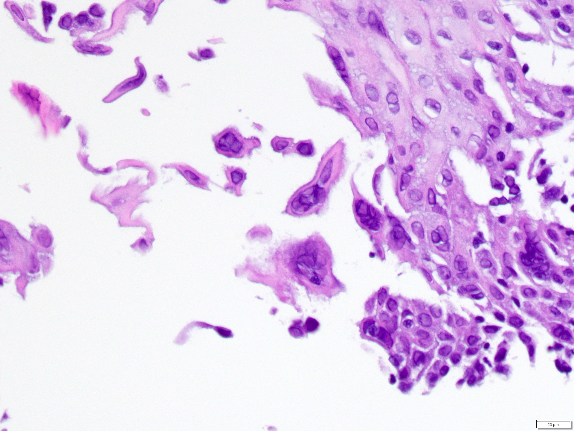
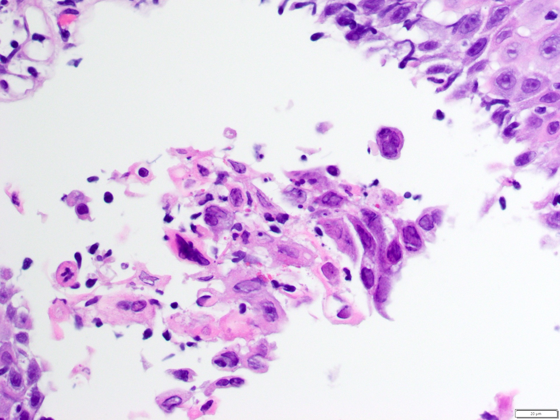
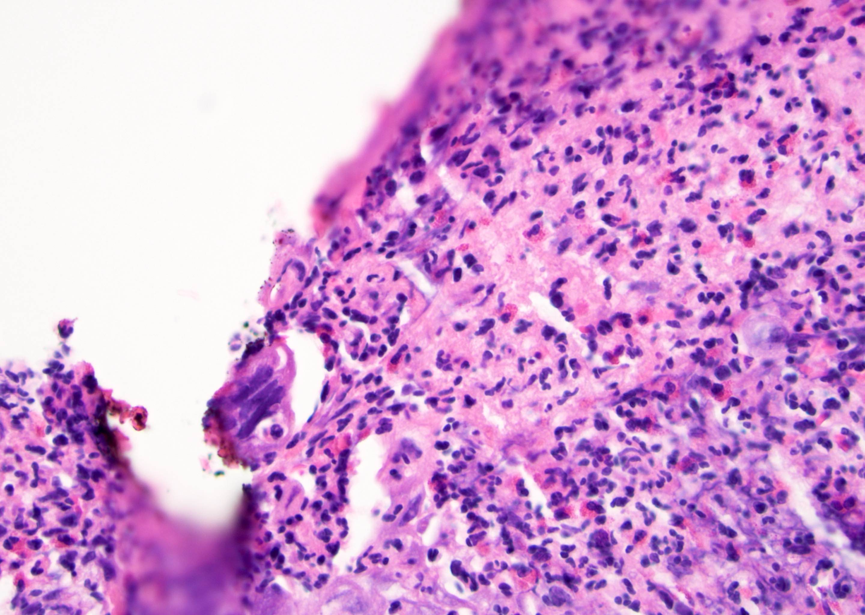
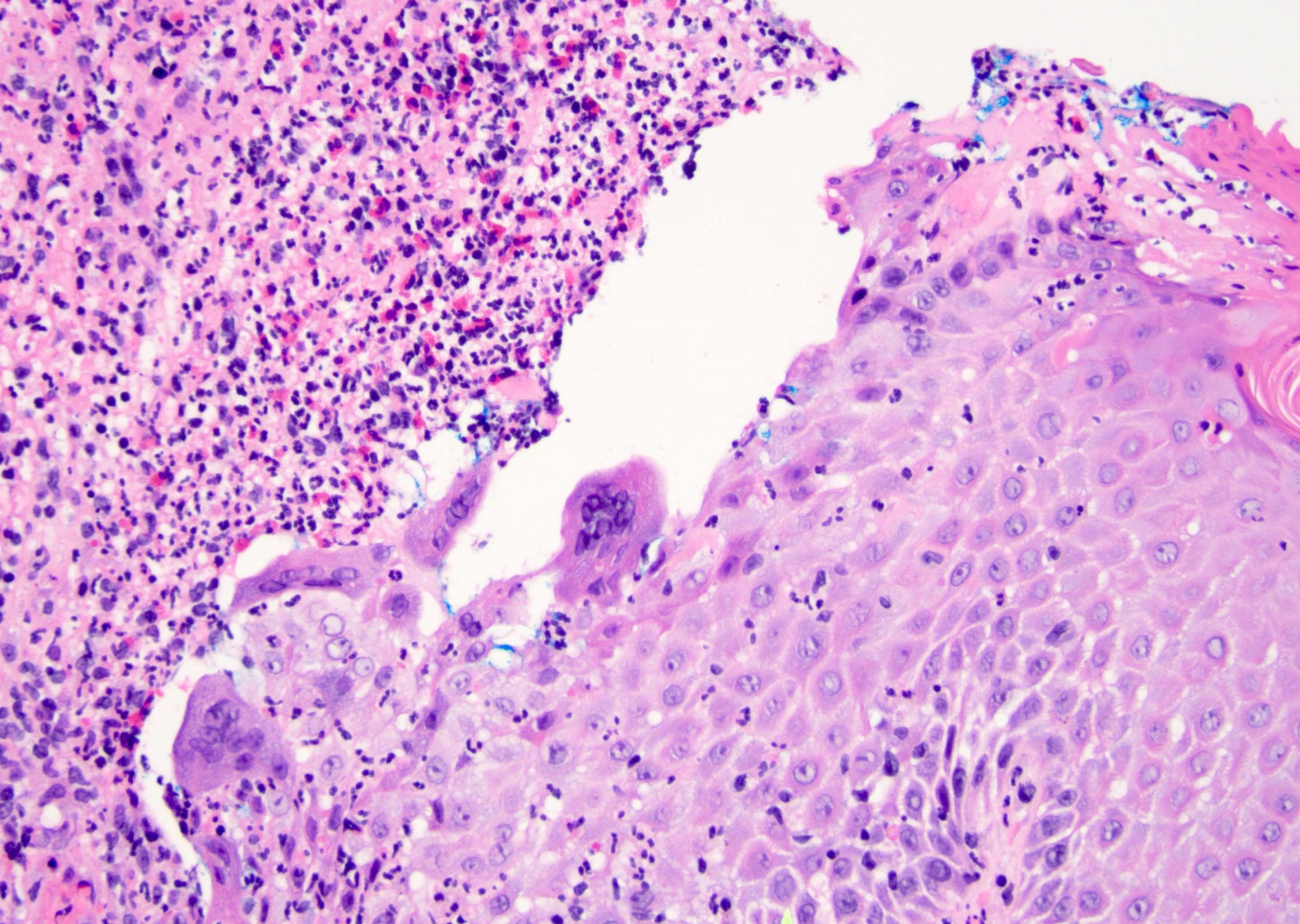
HSV infected cells
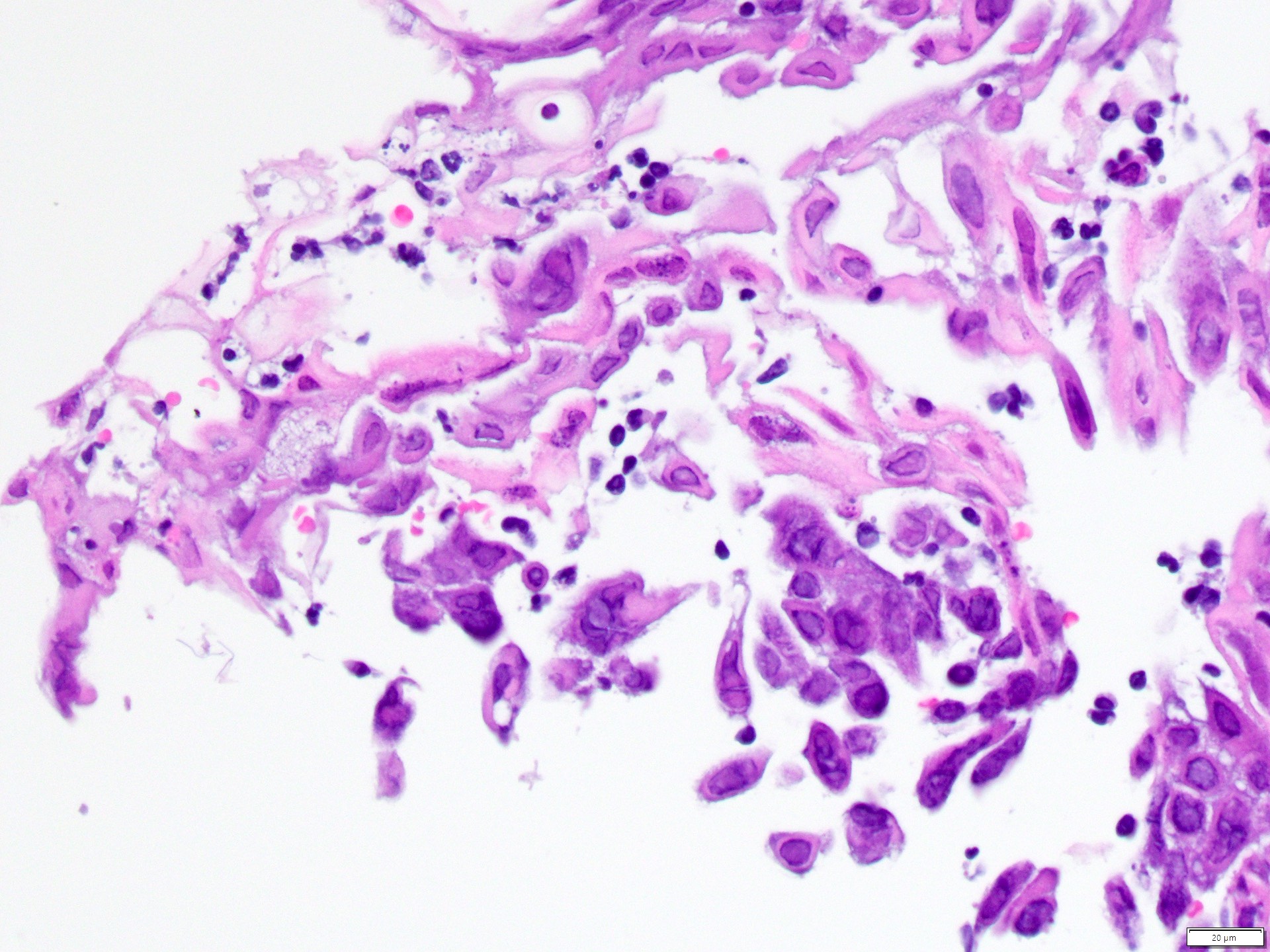
HSV infected cells
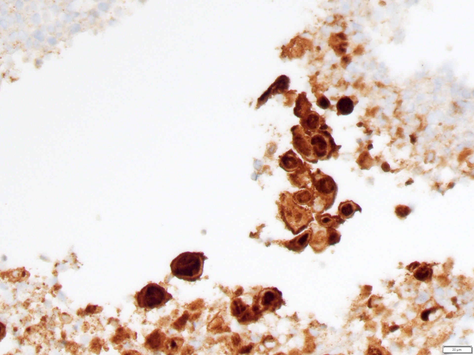
HSV1 / HSV2 IHC
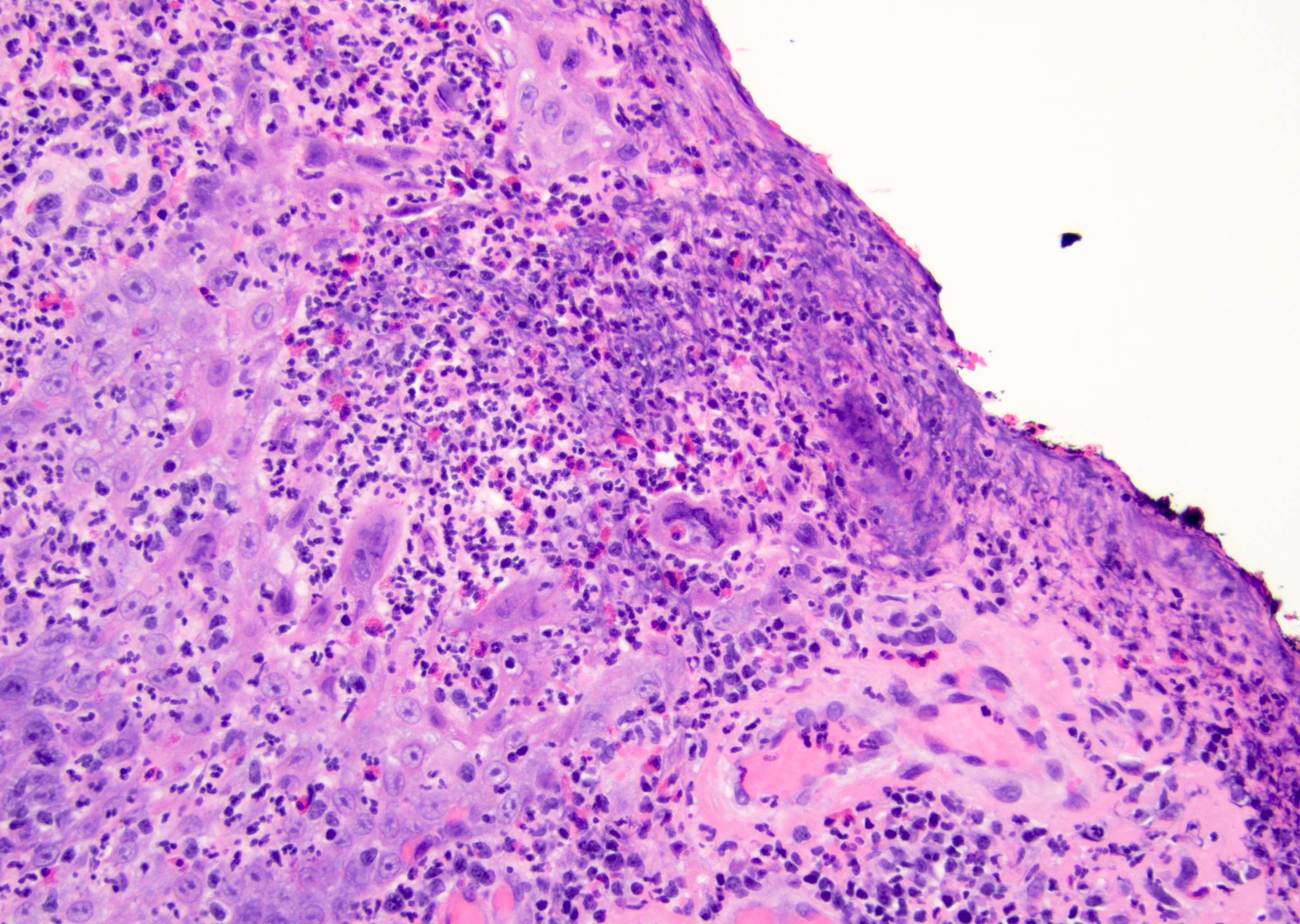
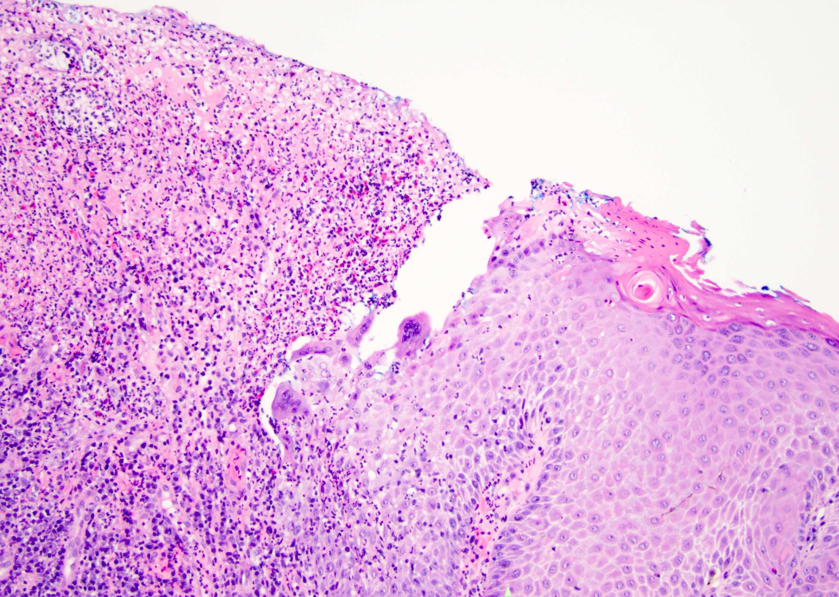
Ulcer interface, virally infected cells
Ulcer interface, viral changes
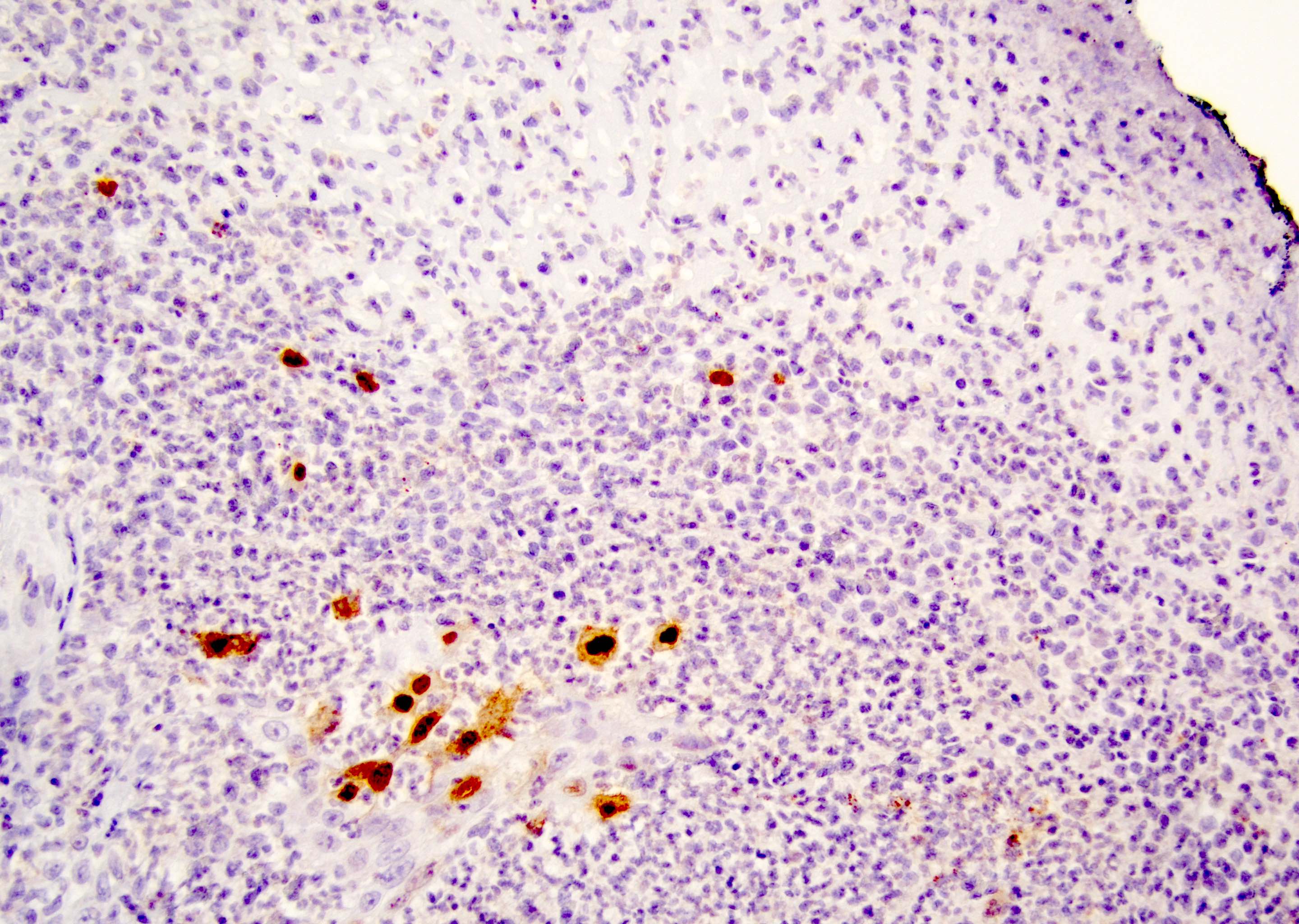
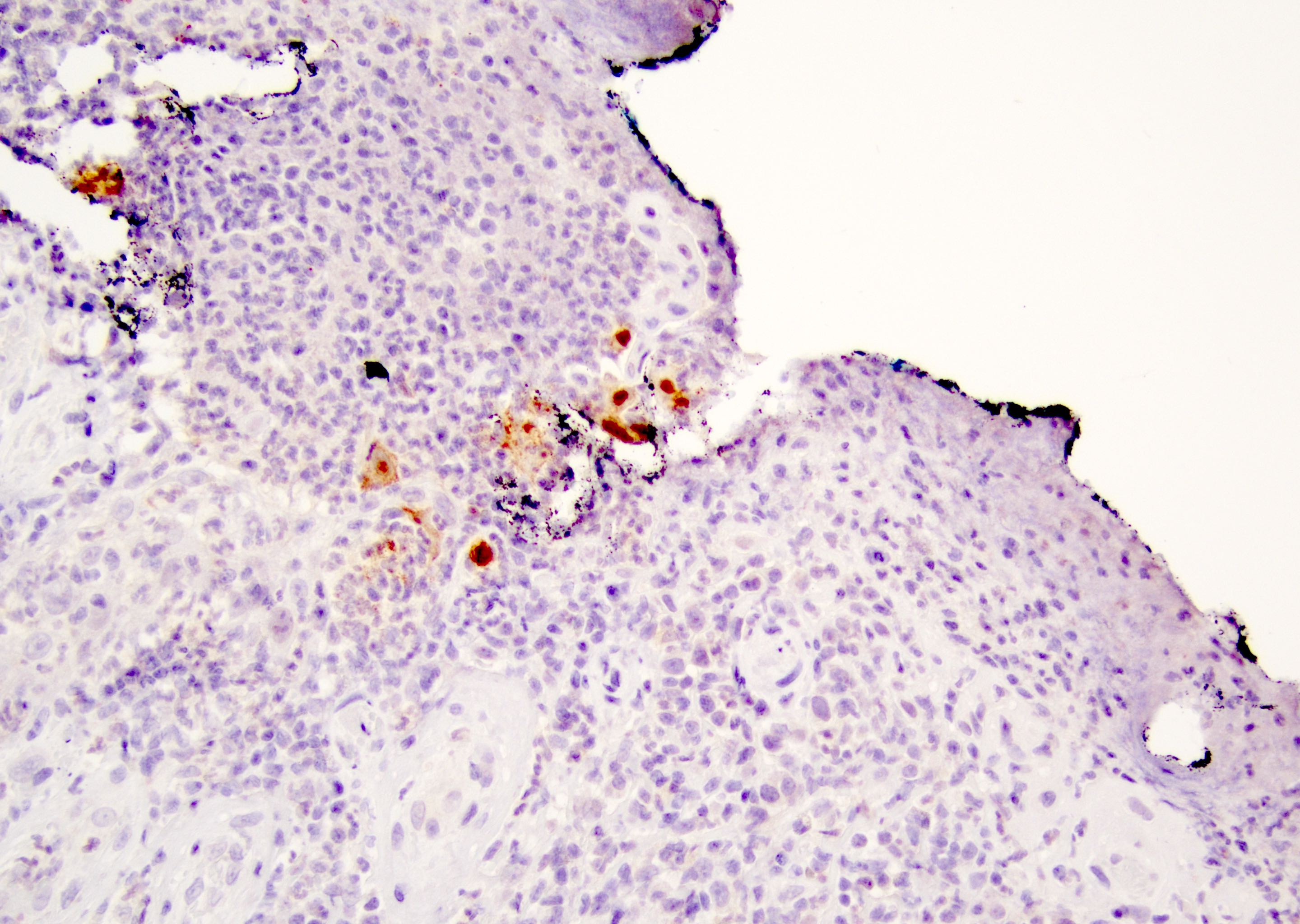
HSV1 / HSV2 IHC
HSV1 / HSV2 IHC
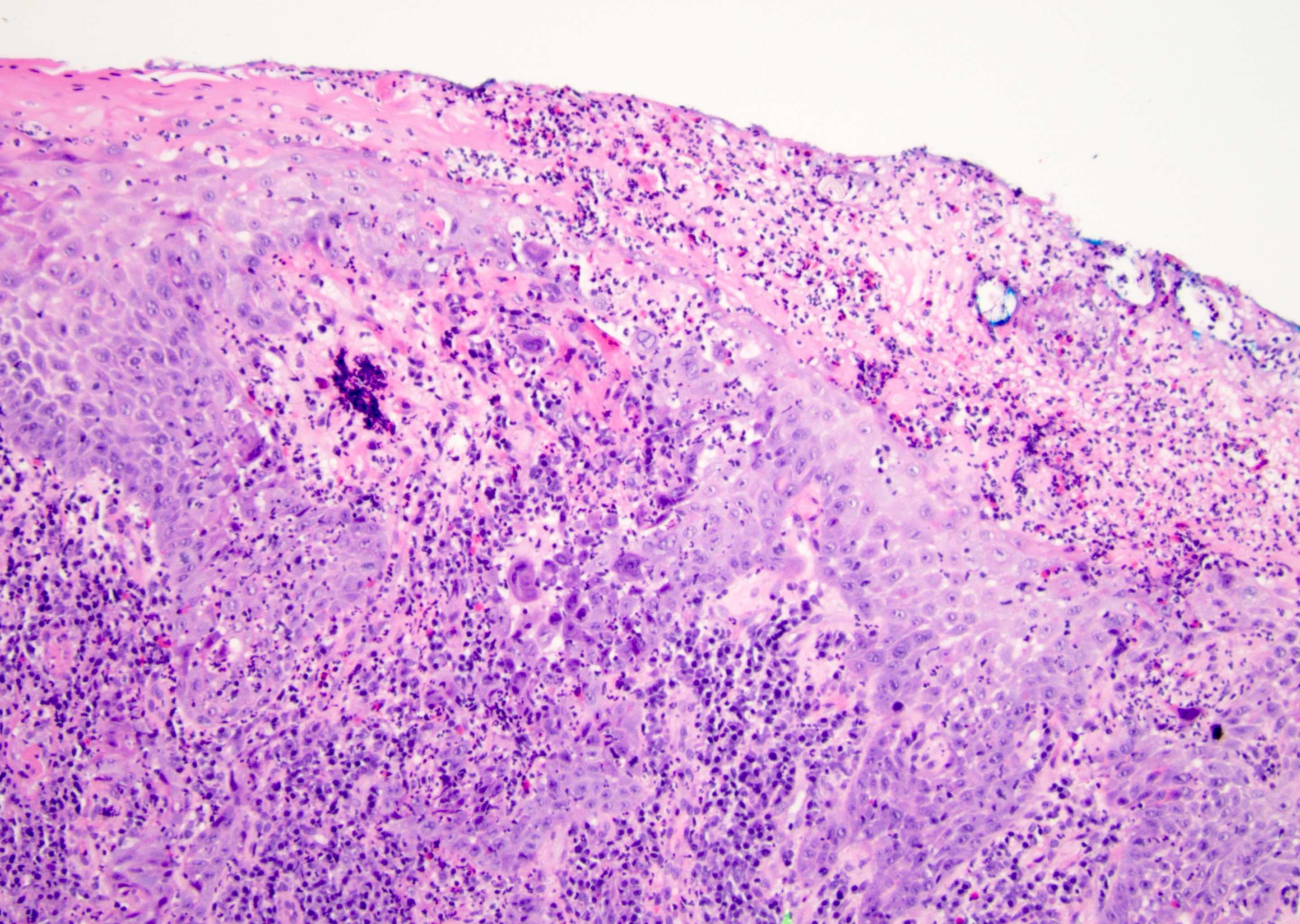
HSV ulcer, mixed inflammation
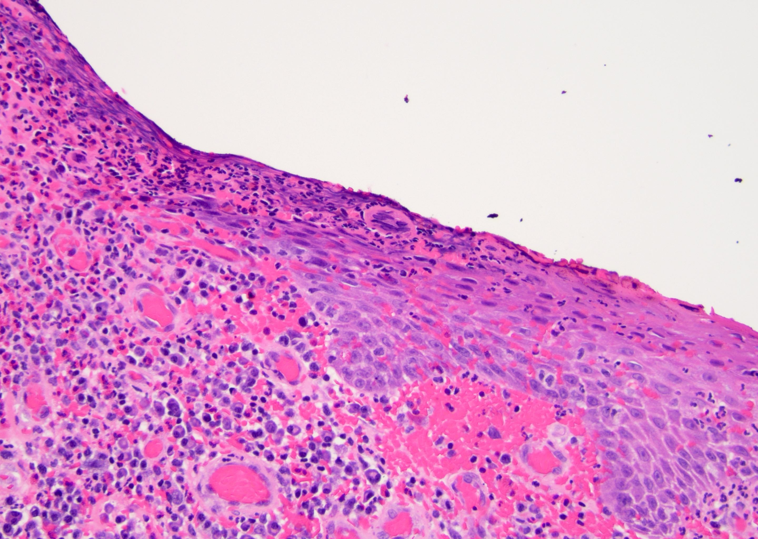
Necrotic virally infected cells
Viral multinucleation, margination, molding
Hypertrophic epidermis, dense inflammation
Dense inflammation and ulcer
Ulcer interface, dense inflammation
Mixed inflammation and ulceration
Cell in ulcer, viral change
Ulcer interface, viral changes
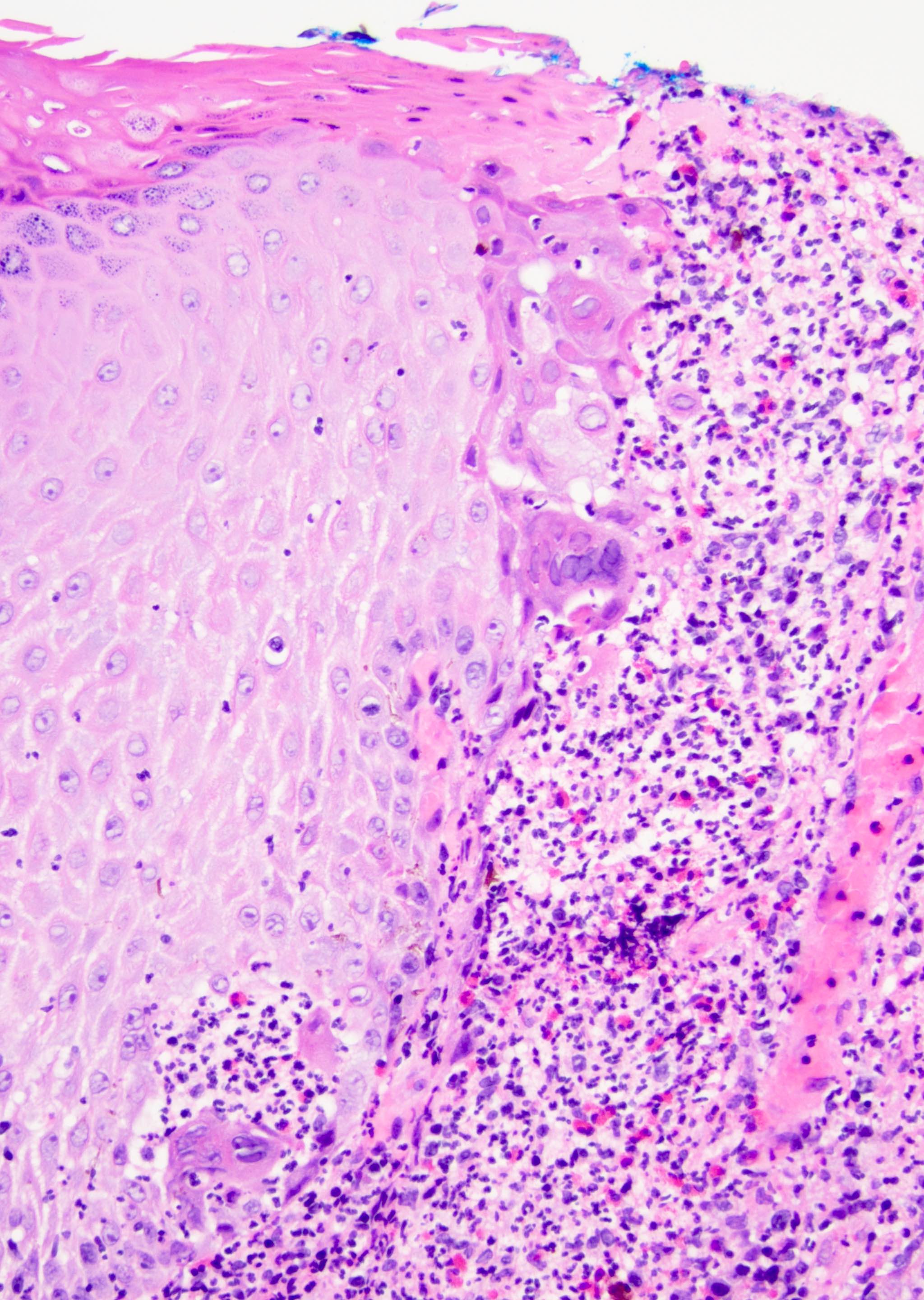
Details viral changes
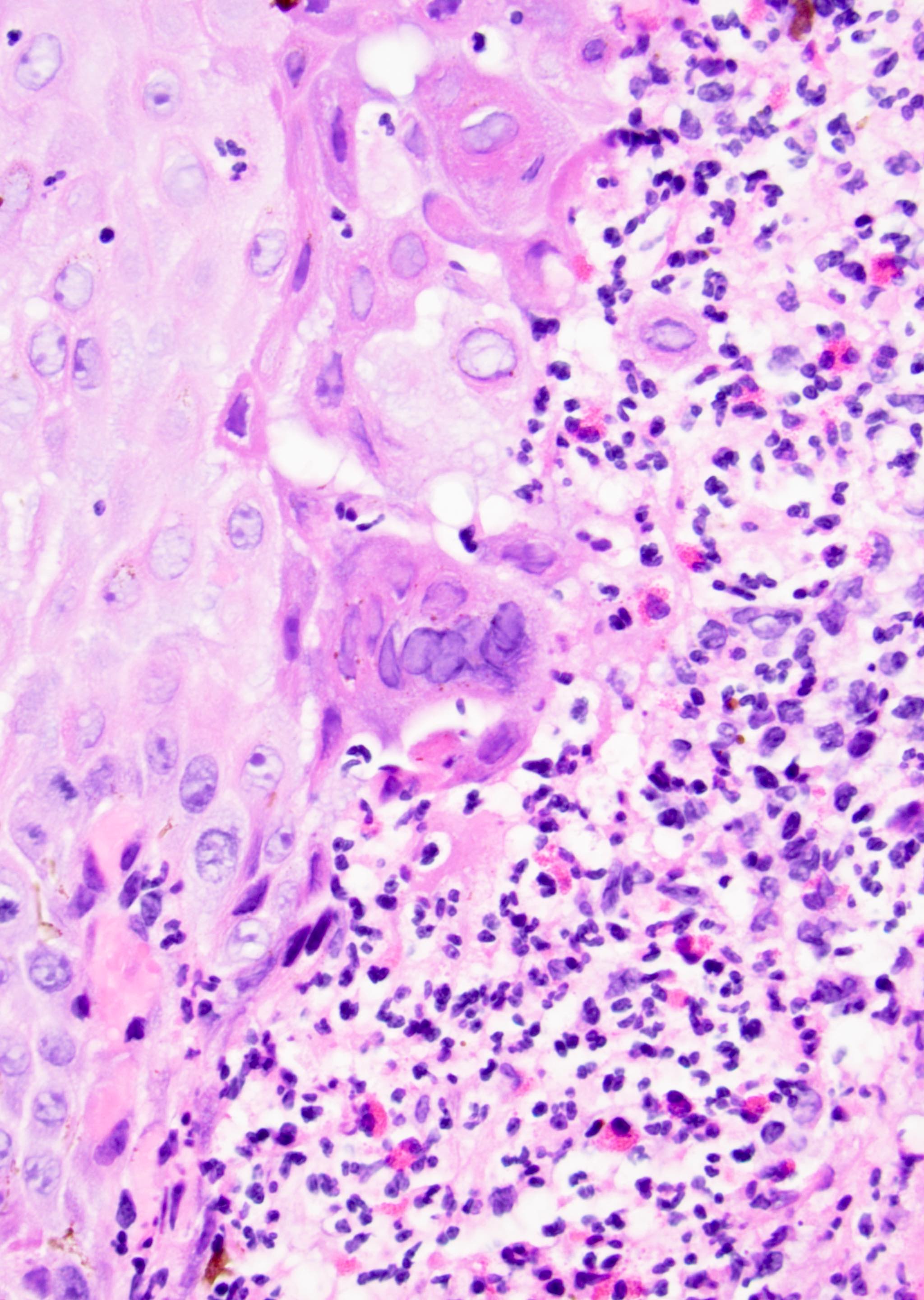
Multinucleation, margination, molding
Virally infected cells at interface
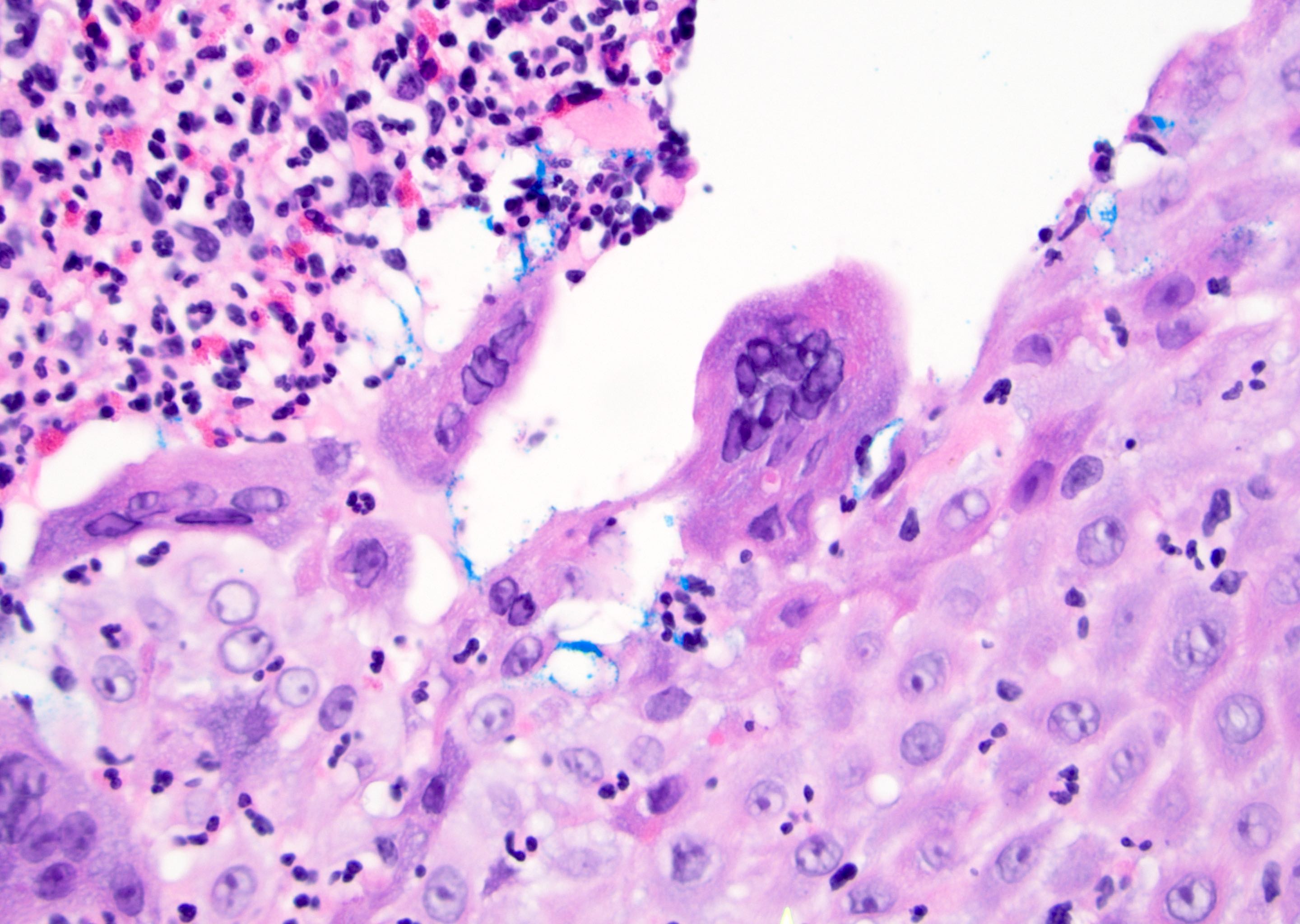
Multinucleation, margination, molding
Ulcer and viral changes
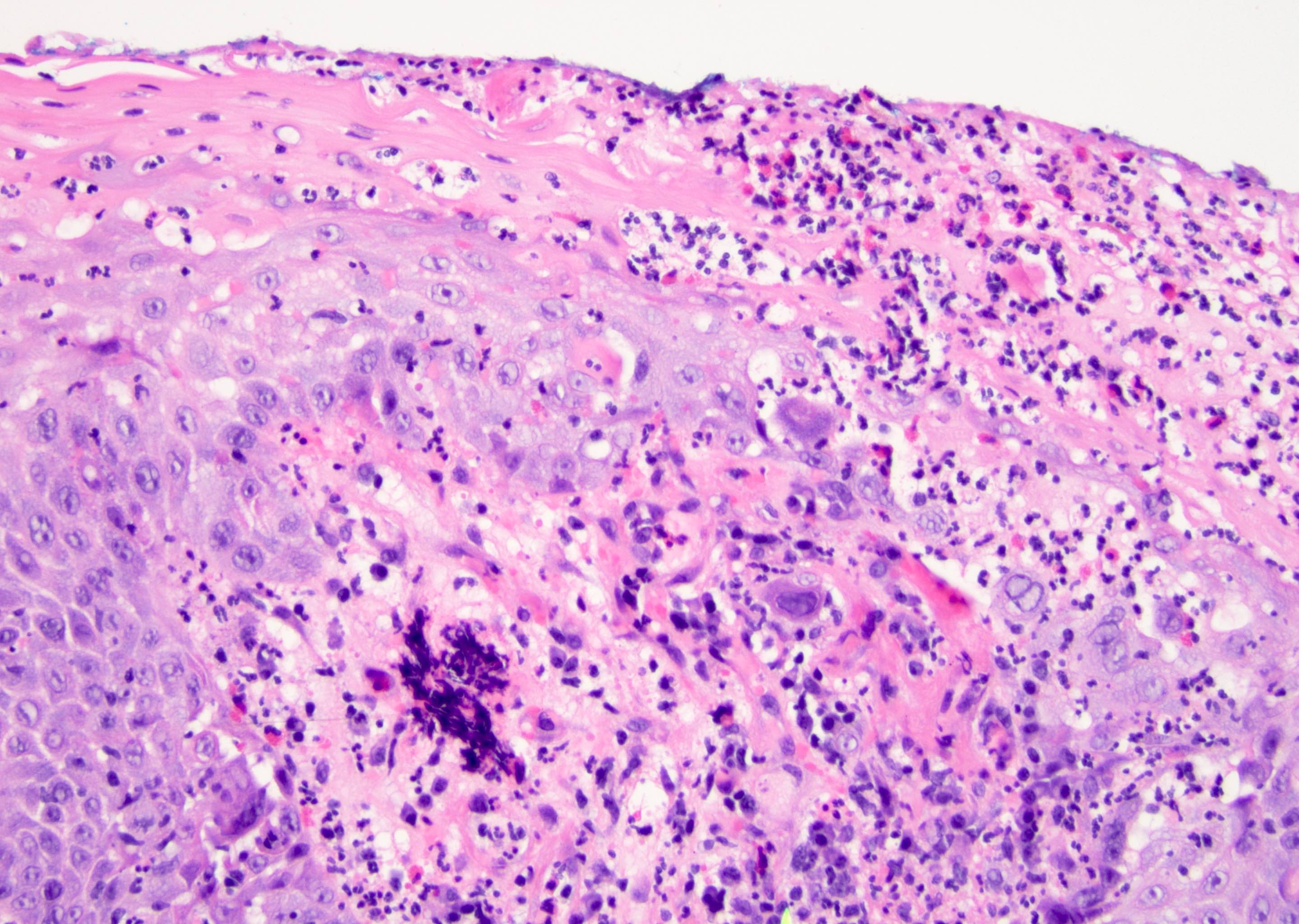
Viral changes at interface
Images hosted on other servers:


HSV infected squamous cells
Herpes under the microscope
by Dr. Jared Gardner
Images hosted on other servers:

Lichen sclerosus and melanosis

Lichen sclerosus and phimosis

Depigmentation, glans atrophy and ecchymoses

Severe sclerosis, scarring and ulceration
Contributed by Diego F. Sanchez, M.D. and Antonio L. Cubilla, M.D.

Atrophy and sclerosis

Hyperplasia and sclerosis

Linear sclerosis
Vacuolization and perivascular sclerosis

Edema and hyalinization
Classic lichen sclerosus
Lichenoid lichen sclerosus
Lymphocytic depleted lichen sclerosus

PeIN and lichen sclerosus
Pseudohyperplastic carcinoma, lichen sclerosus
Lichen sclerosus et atrophicus
Images hosted on other servers:

Sinus tract
Images hosted on other servers:
Lymph nodes
AFIP images
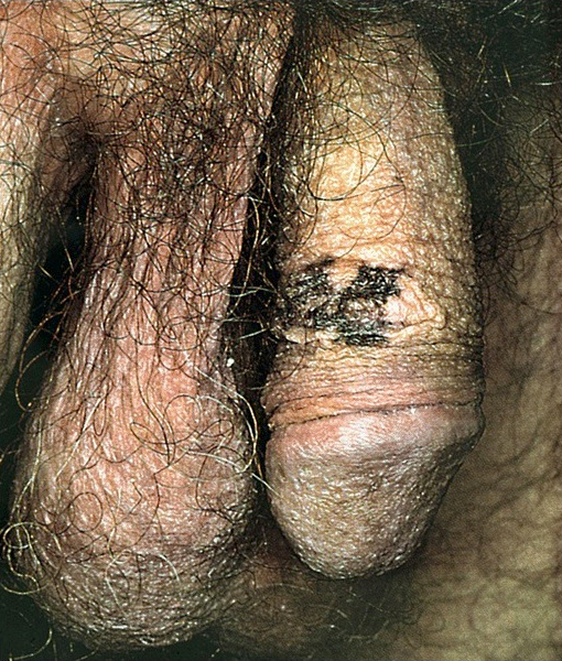
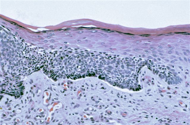
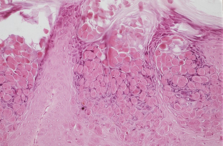
Melanosis
AFIP images
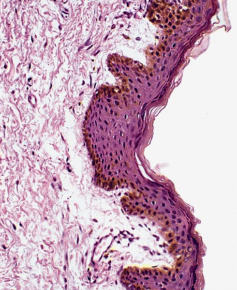
Melanosis
Contributed by Harry Ioachim, M.D.
Multiple lesions on shaft of 30 year old drug abuser
Images hosted on other servers:

Lesions on shaft of penis


Skin lesions (site unspecified)
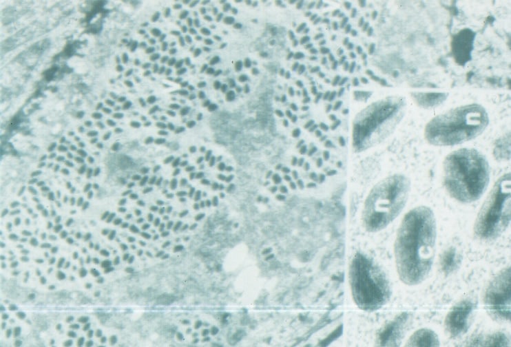
AFIP images
Henderson-Patterson bodies
Images hosted on other servers:

Henderson-Patterson bodies

Molluscum bodies
Images hosted on other servers:


Pap stain
Images hosted on other servers:

Viral particles
Images hosted on other servers:


Median raphe cysts
Images hosted on other servers:

Pseudostratified columnar epithelium

Single larger mucinous cell within epithelium

CK7

CK13

CEA
Images hosted on other servers:

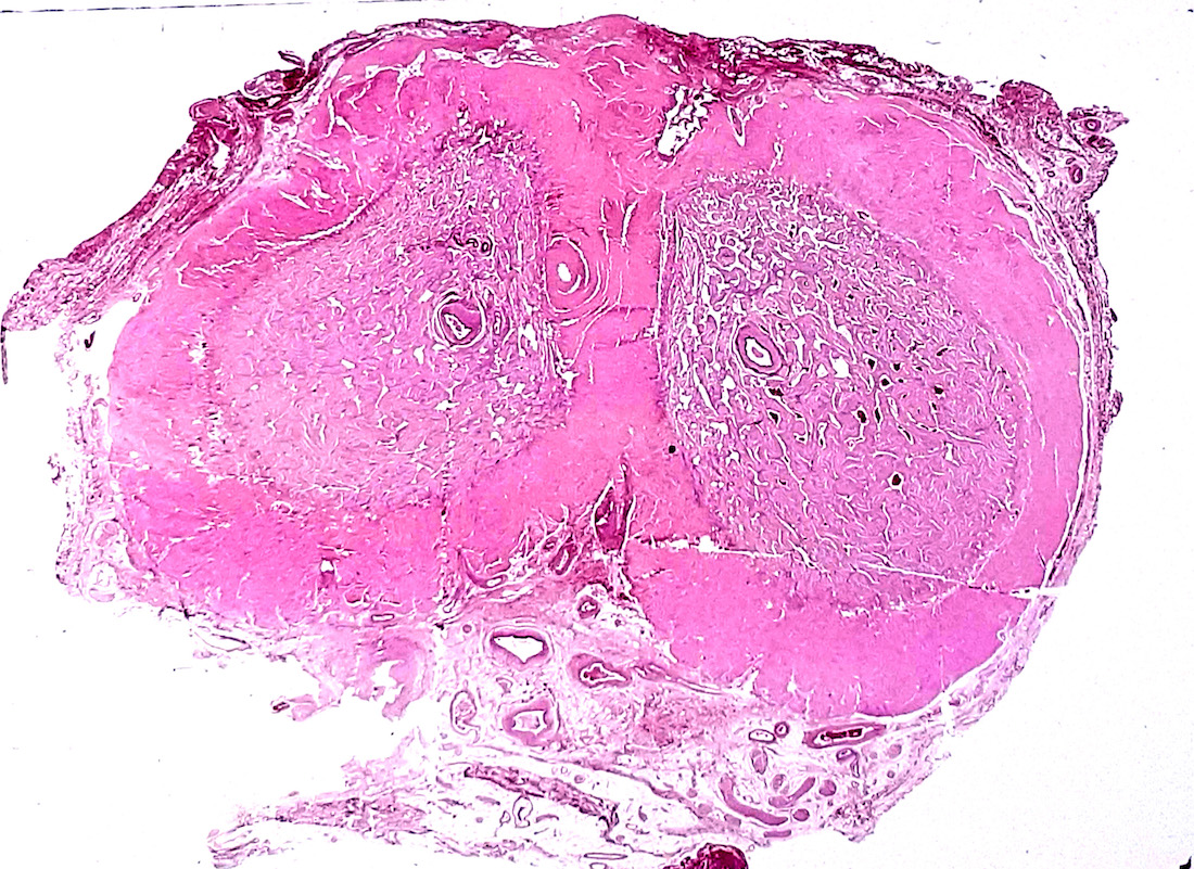
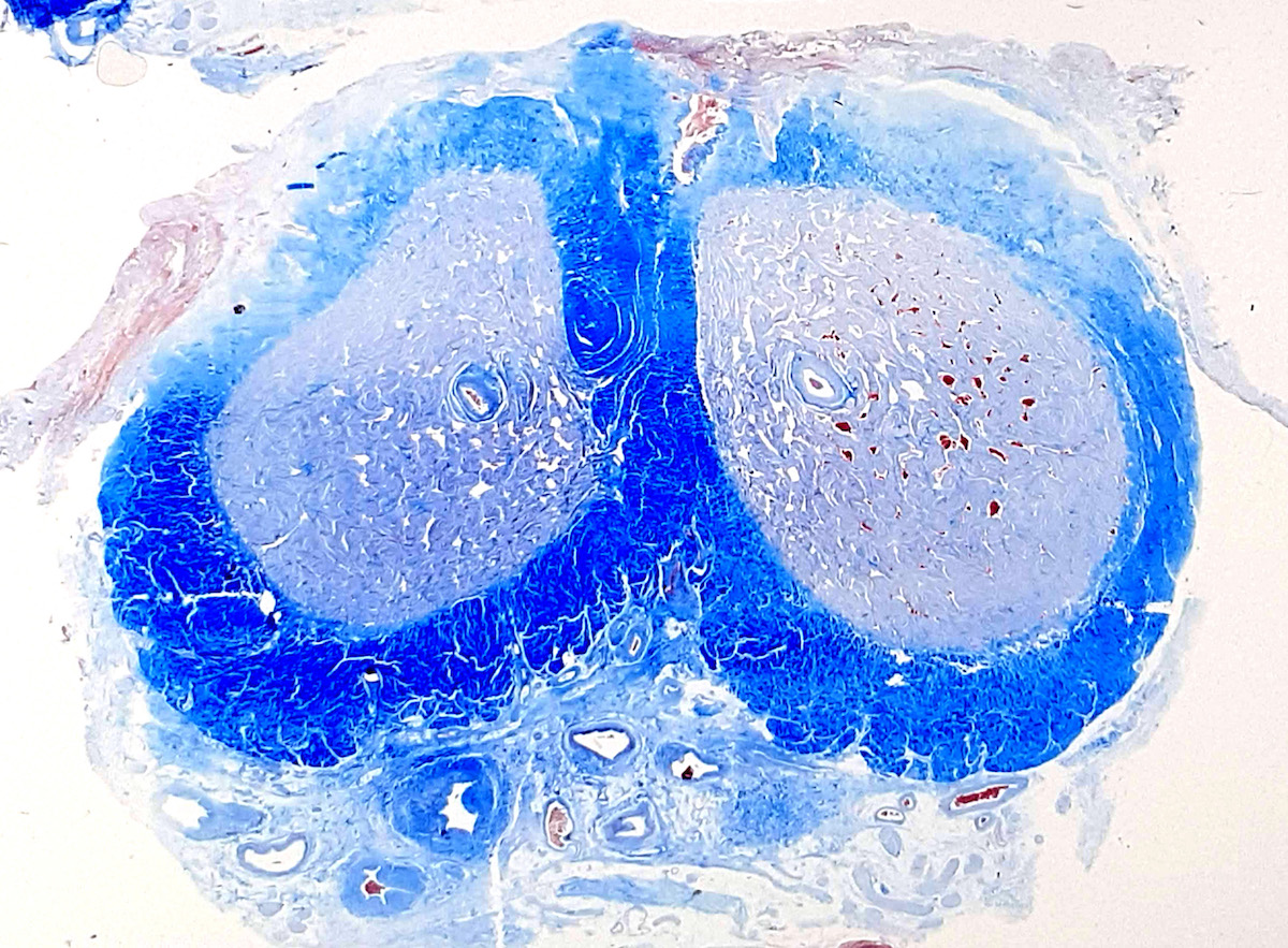
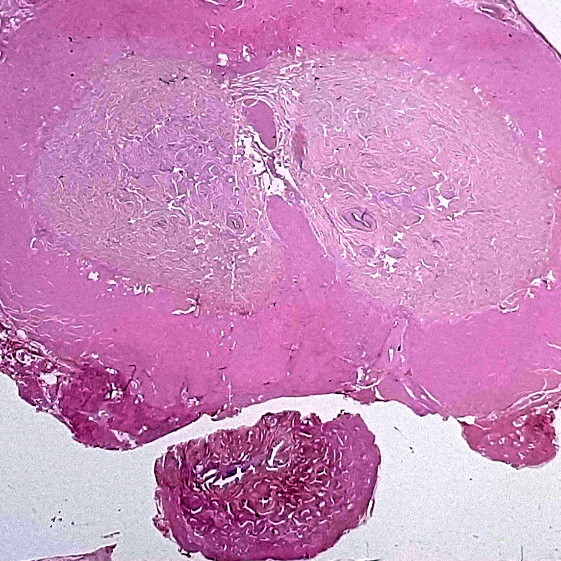
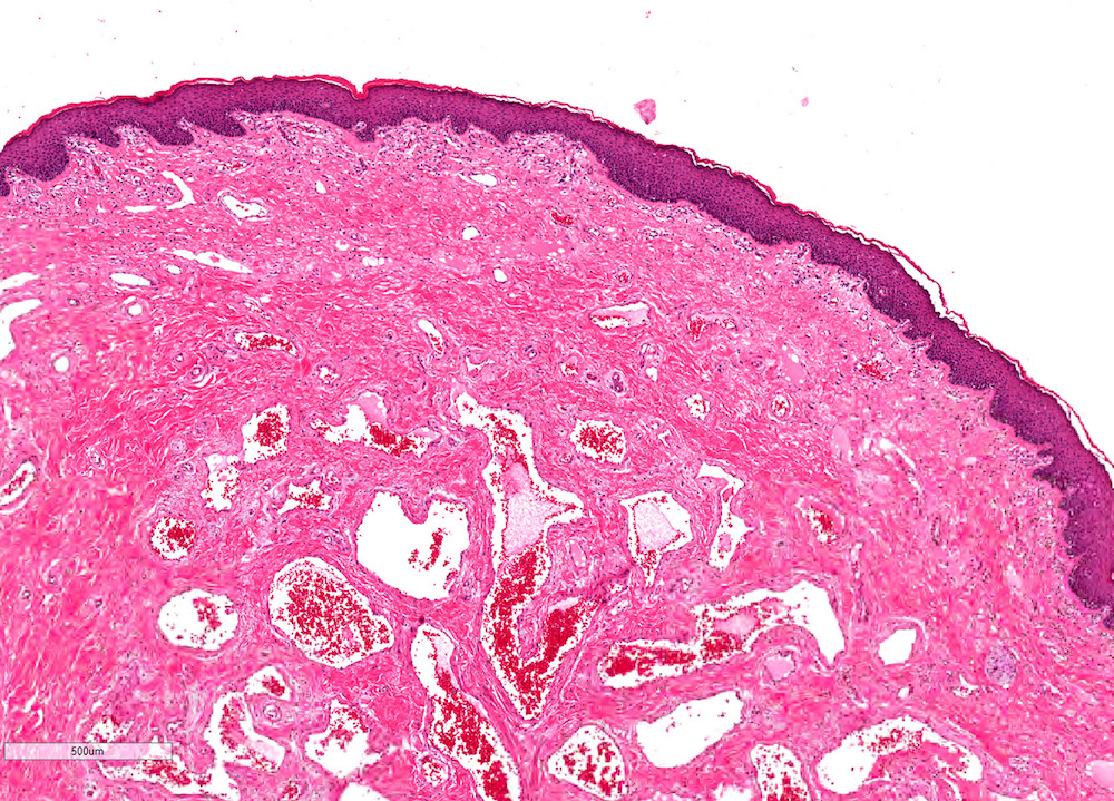
Anatomy of corpus spongiosum in glans penis

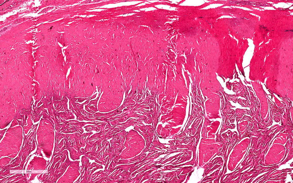
Histology of intima
Images hosted on other servers:

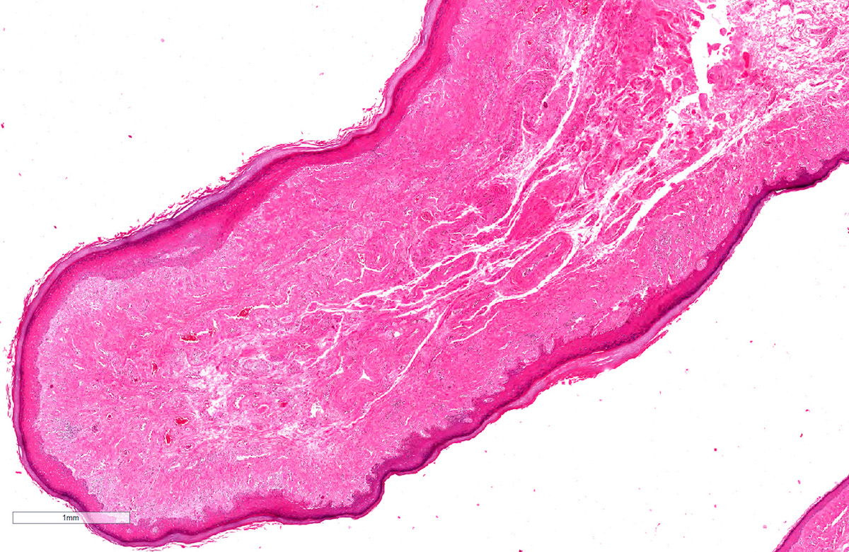
Nodule on the left side of glans penis
Images hosted on other servers:

Excised nodule
Contributed by Maria Tretiakova, M.D., Ph.D.

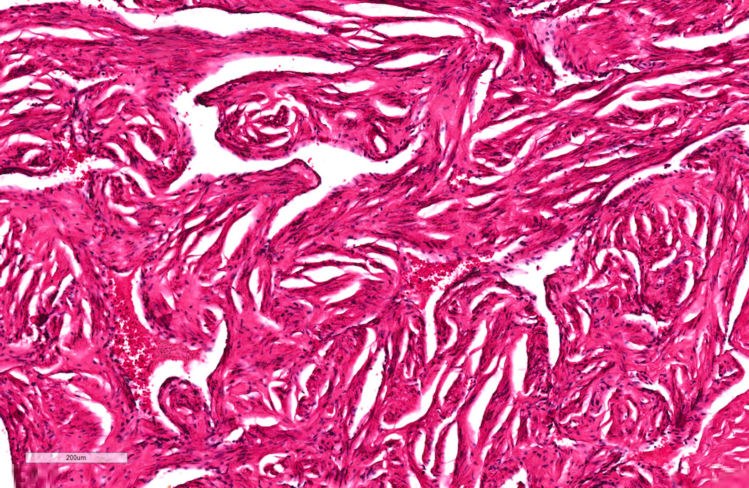
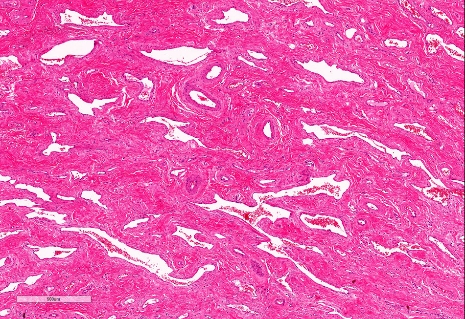
Plexiform architecture
Intravascular proliferation
Intravascular intimal proliferation

Fibromyxoid stroma

Bland myofibroblastic cells

SMA staining
Images hosted on other servers:

Raccoon

Dog

Walrus

Figure F: obvious distal ligament within the glans penis
Images hosted on other servers:

Figure C: distal ligament
Images hosted on other servers:

Nonretractable foreskin

Fournier gangrene
AFIP images
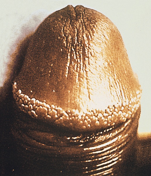
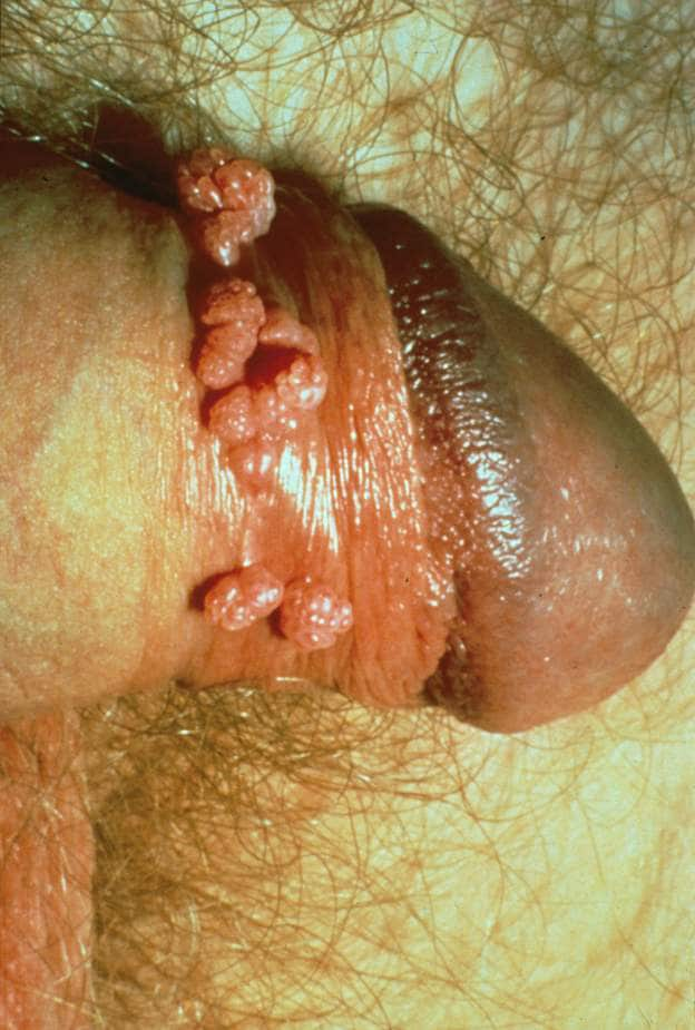
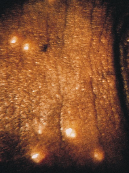
Regular rows of
small papules
form a ring
around the corona
Images hosted on other servers:

1 - 2 cm papules
AFIP images
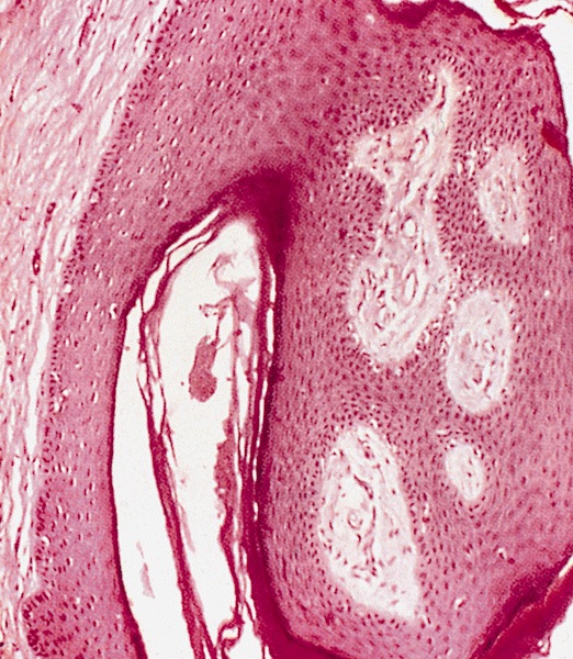
Benign epithelial hyperplasia with vascular fibrous stroma
AFIP images
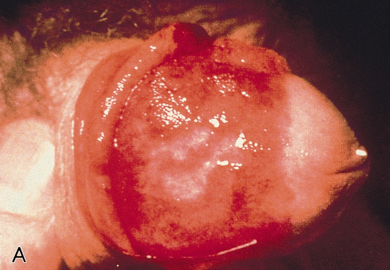
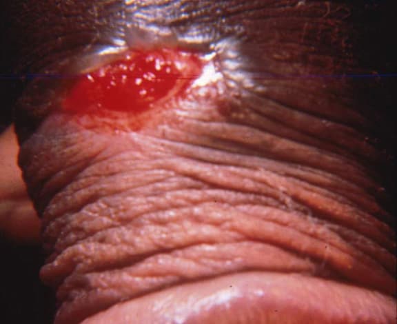
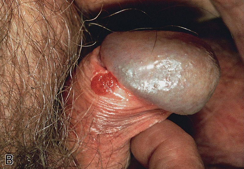
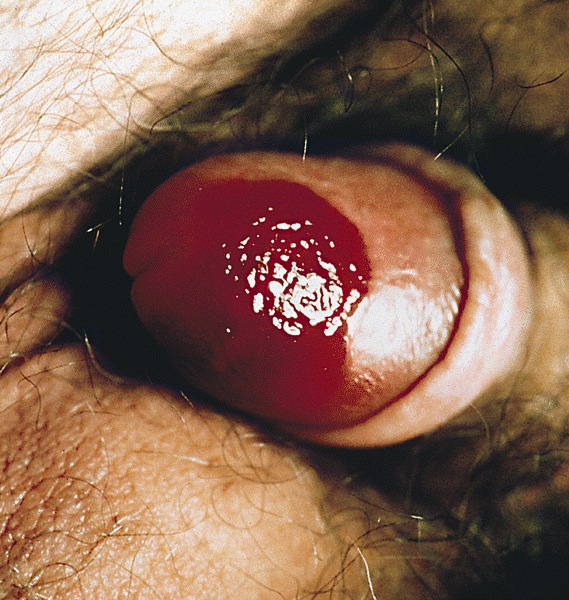
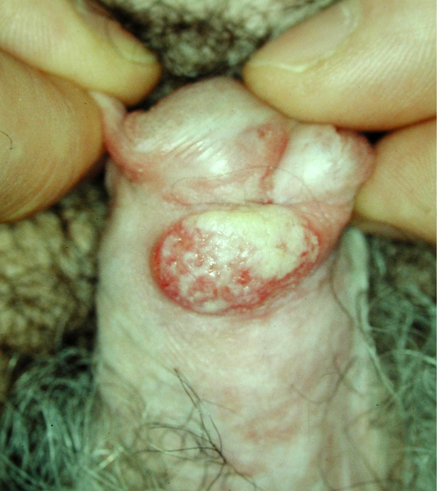
Erythroplasia of Queyrat: moist red lesion involving glans
Bowen disease
Images hosted on other servers:

Erythematous lesion in the glans


Erythroplasia of Queyrat

PeIN with lichen sclerosus

PeIN in multiple compartments
Contributed by Alcides Chaux, M.D. and Antonio Cubilla, M.D.

Squamous cell atypia
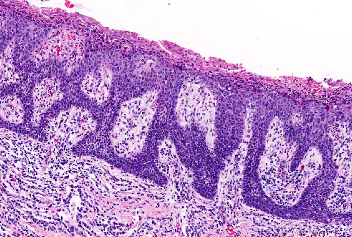
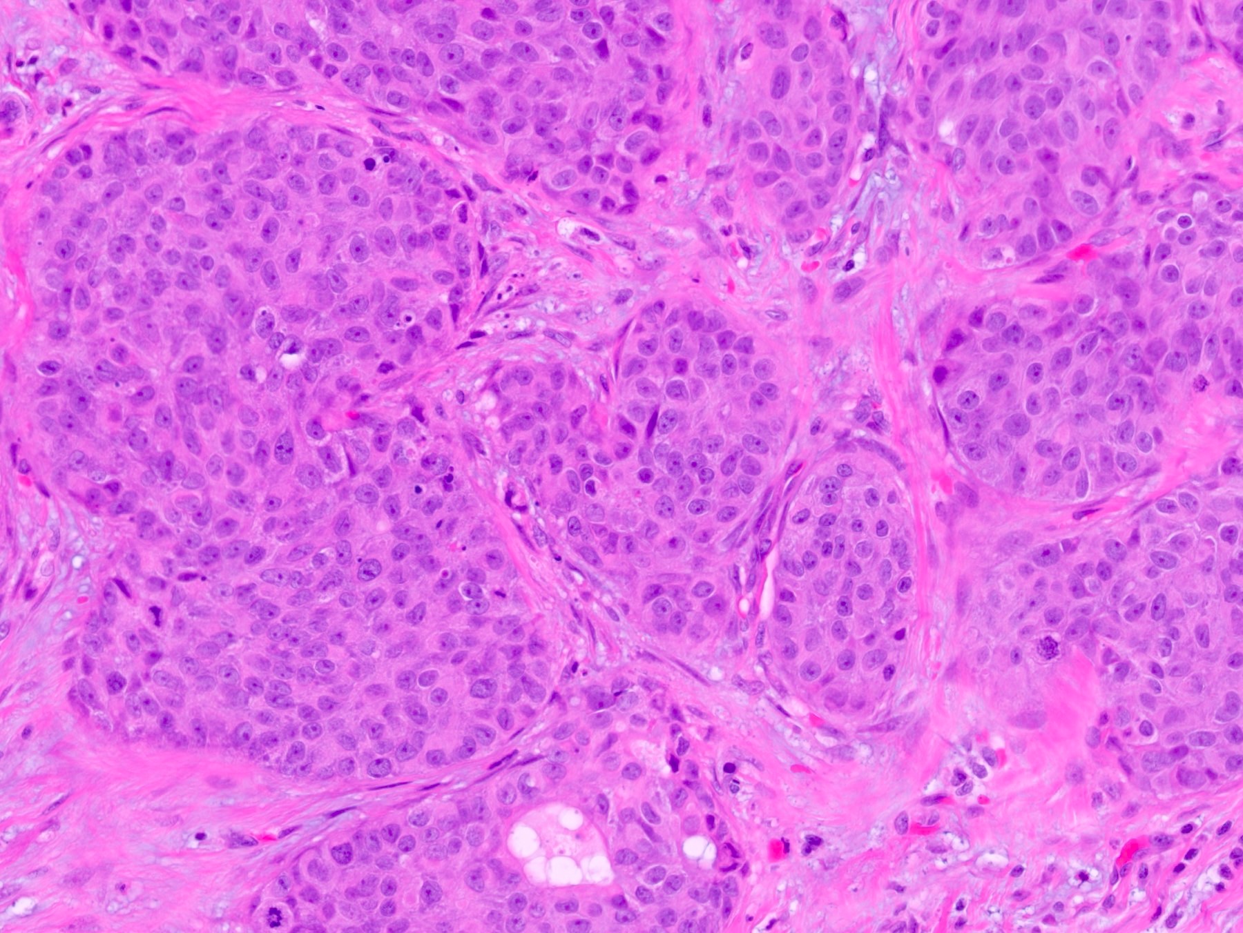
Small basaloid cells

Basaloid nonkeratinized cells
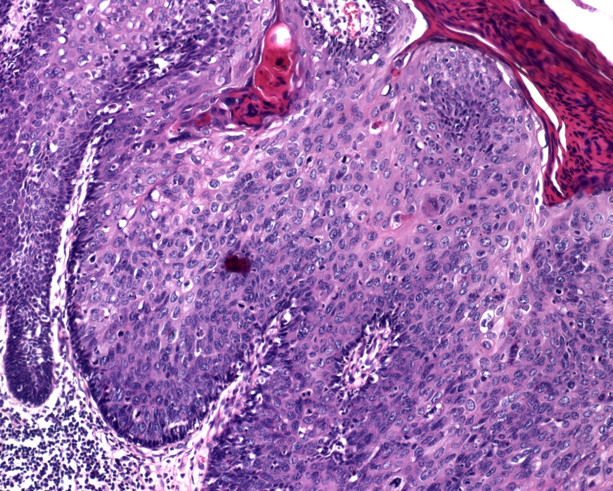
Small blue cells and koilocytes
Basaloid type
Warty type
Atypical lichen sclerosus

Differentiated type
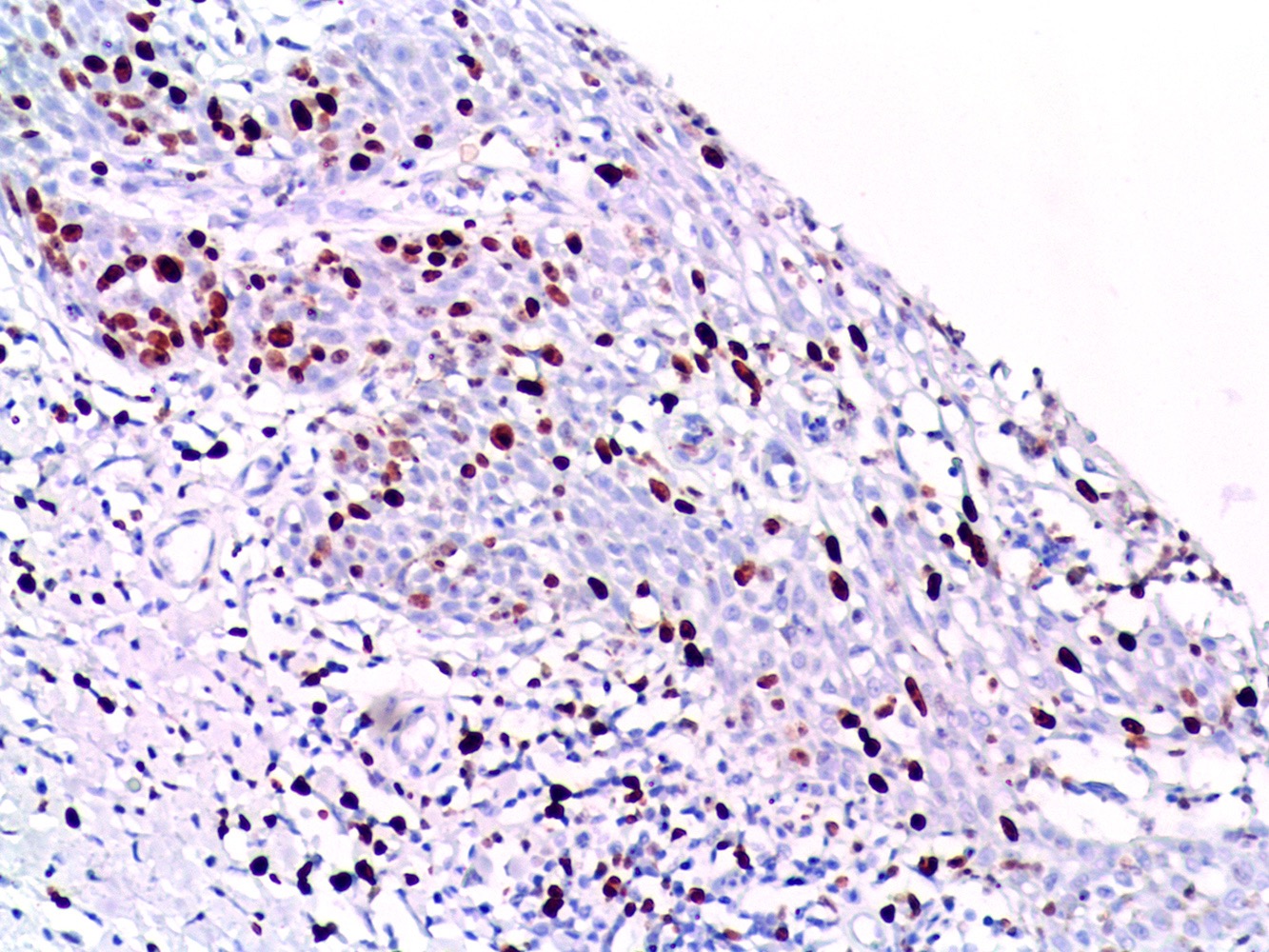
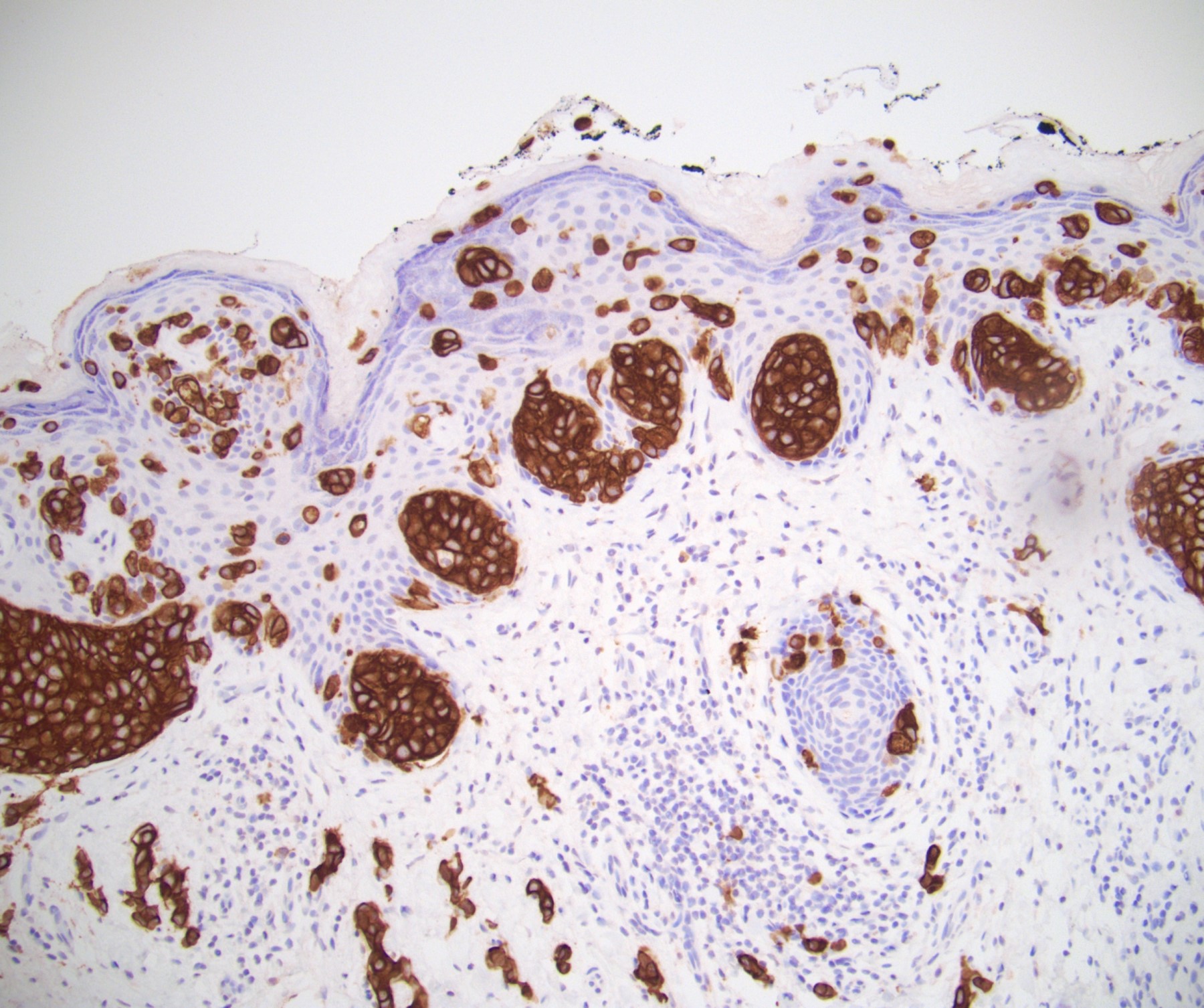
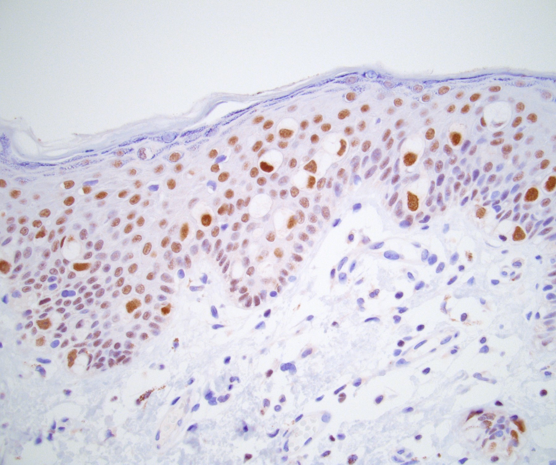
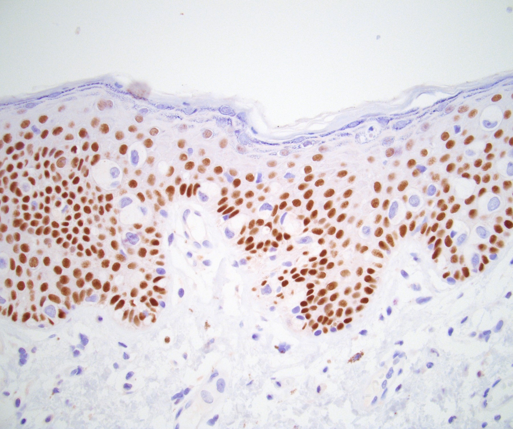
Ki67 positivity


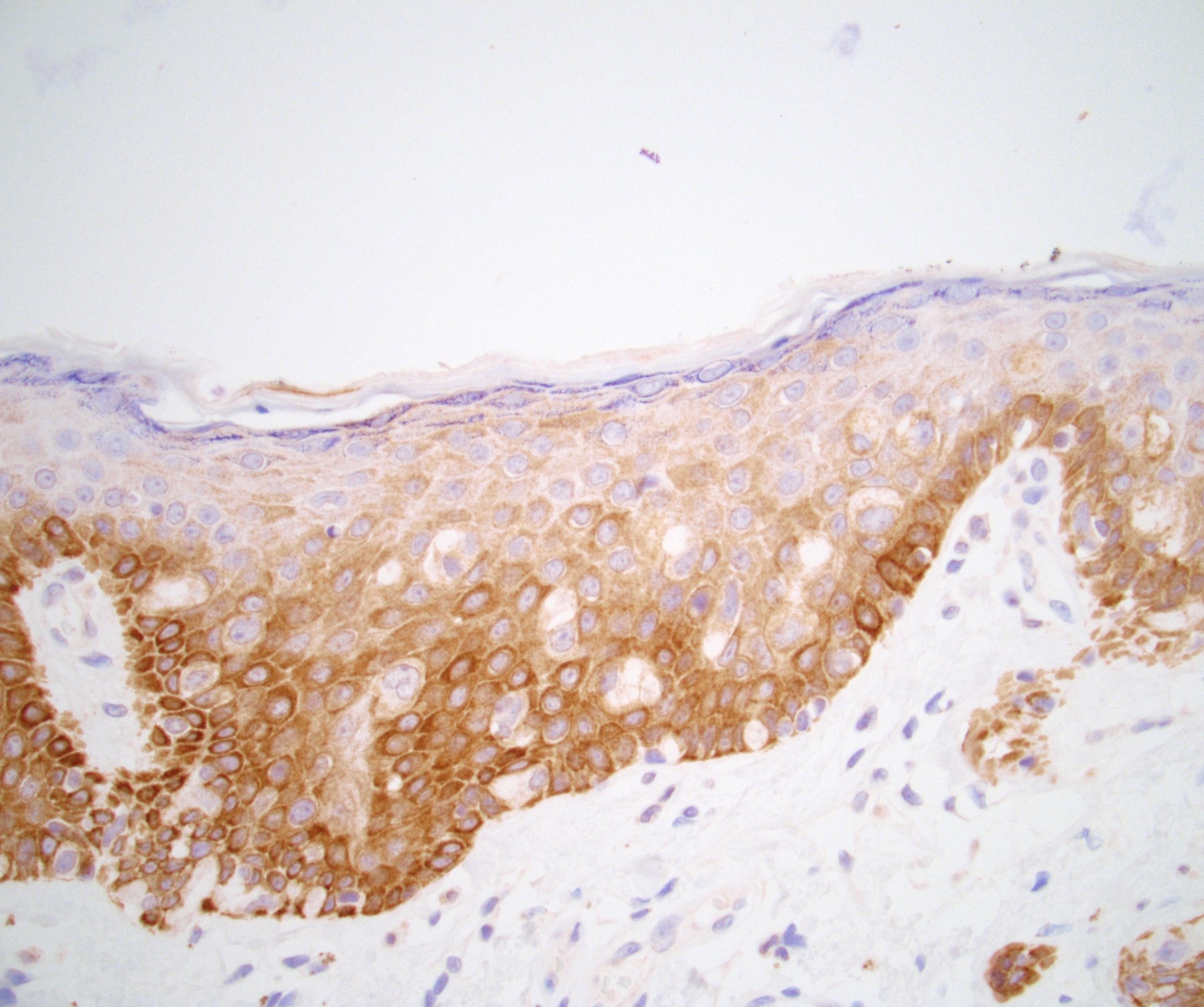
Strong en bloc cytoplasmic and nuclear p16 immunostaining
Images hosted on other servers:

p16 and HPV ISH
Images hosted on other servers:

Penile deformation

Dorsal plaque
AFIP images
Penile curvature
Images hosted on other servers:


Penile curvature
AFIP images

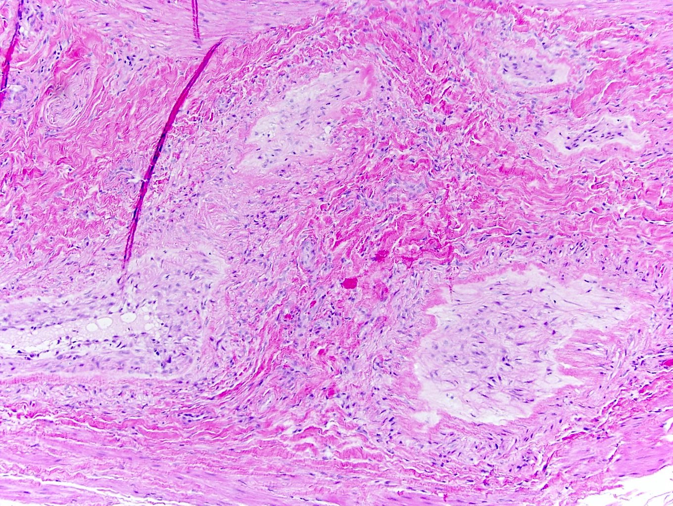
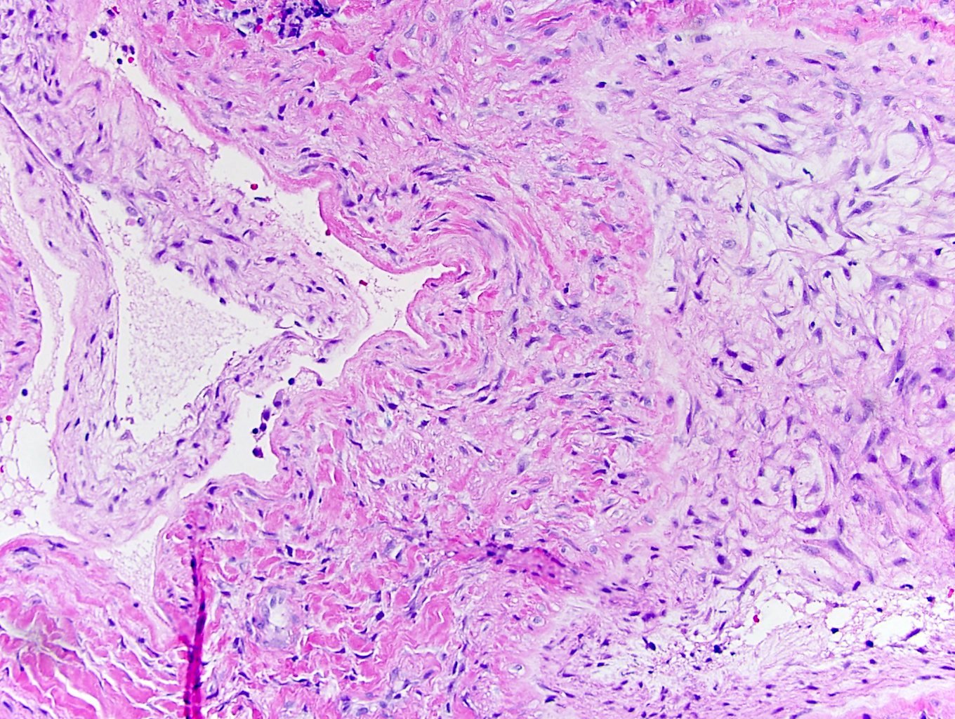
Tunica albuginea
AFIP images
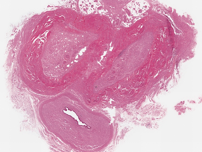
Thickening of tunica albuginea
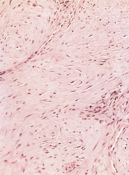
Fibrosis
Penile fibromatosis
Images hosted on other servers:

Leprosy presenting as phimosis

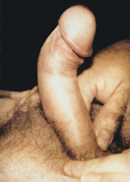
Tight preputial orifice

Erection with phimosis

Phimotic foreskin


Treatment
AFIP images
Glans mucosa
AFIP images

Marked deformity of penis
Contributed by Debra L. Zynger, M.D. and AFIP images
Scrotoplasty

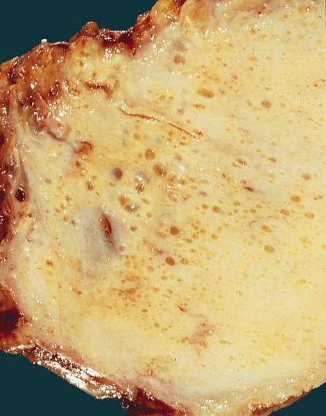
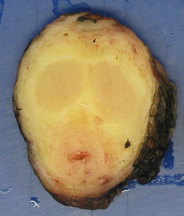
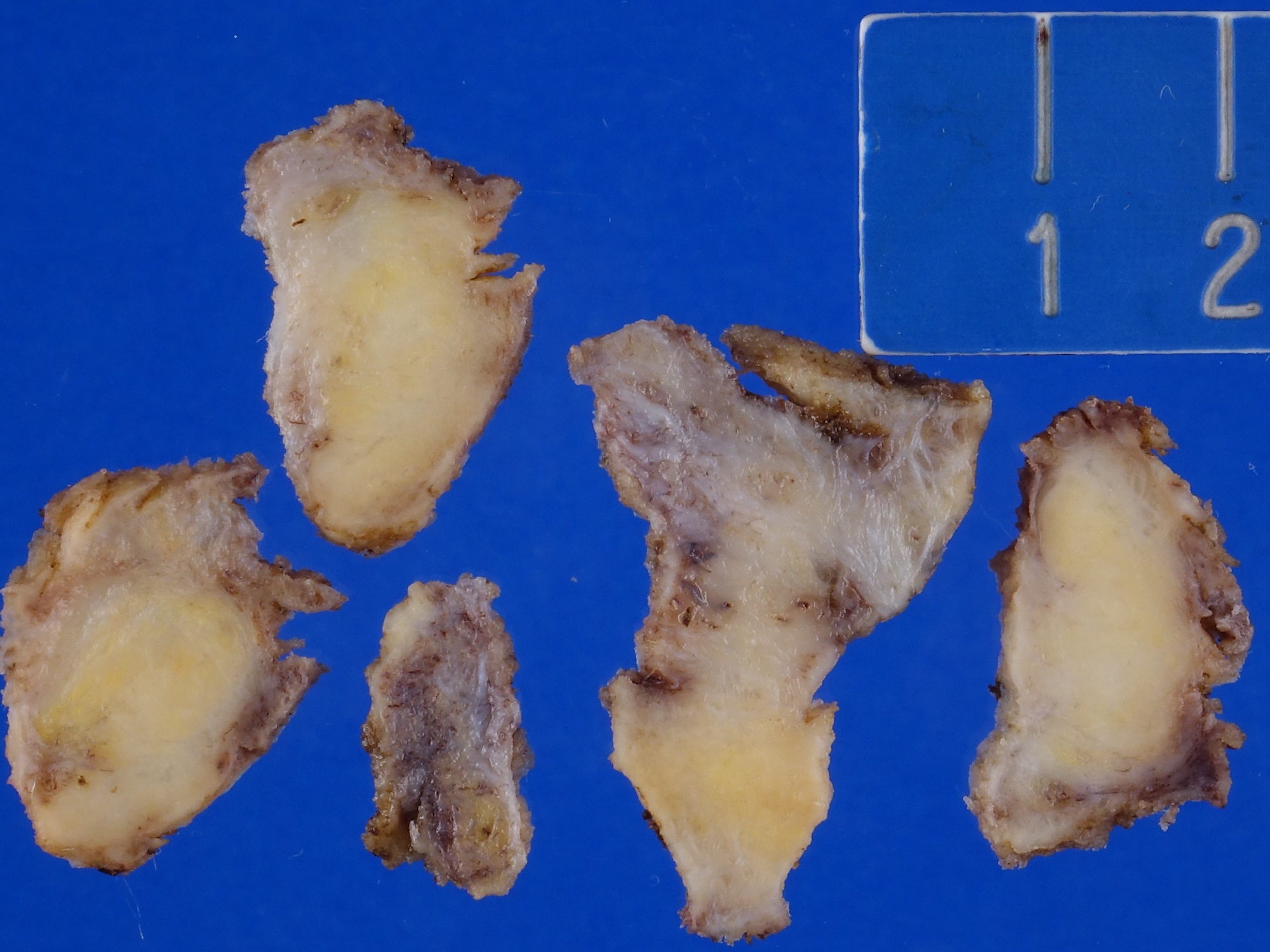
Replacement of entire scrotal wall by solid yellow-white tissue with interspersed cysts
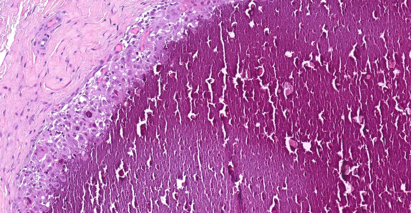
AFIP images
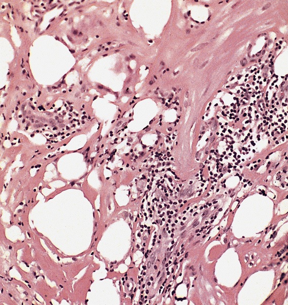
Characteristic vacuoles,
sclerotic stroma and
occasional foreign body
giant cells
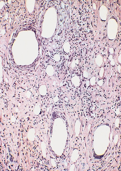
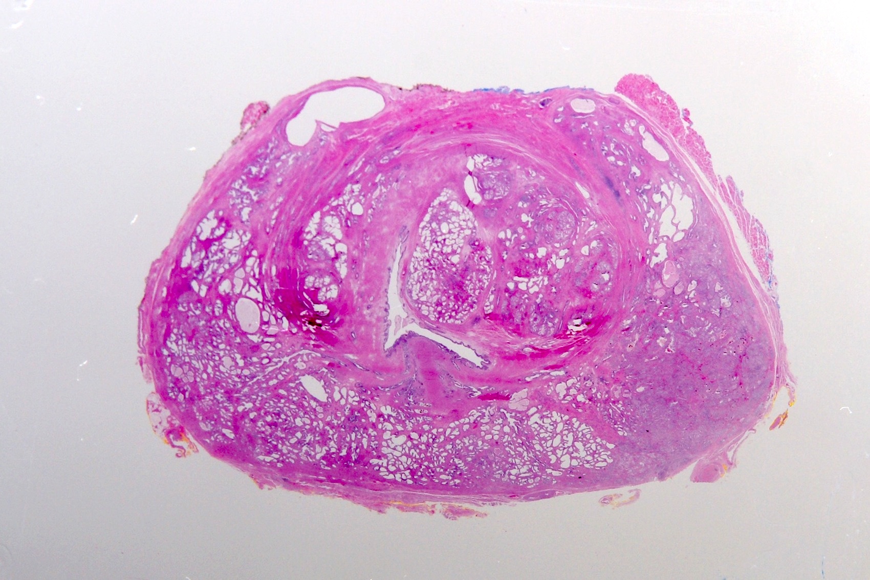
Numerous, variably sized vacuolated spaces

Oil Red O stain
Contributed by Nasir Ud Din, M.B.B.S.

Incidentally found scrotal calcinosis
Images hosted on other servers:

Scrotal calcinosis with vitiligo

Pre and postoperative scrotum

Scrotal skin showing calcifications

Multiple white scrotal nodules
Contributed by Debra Zynger, M.D.
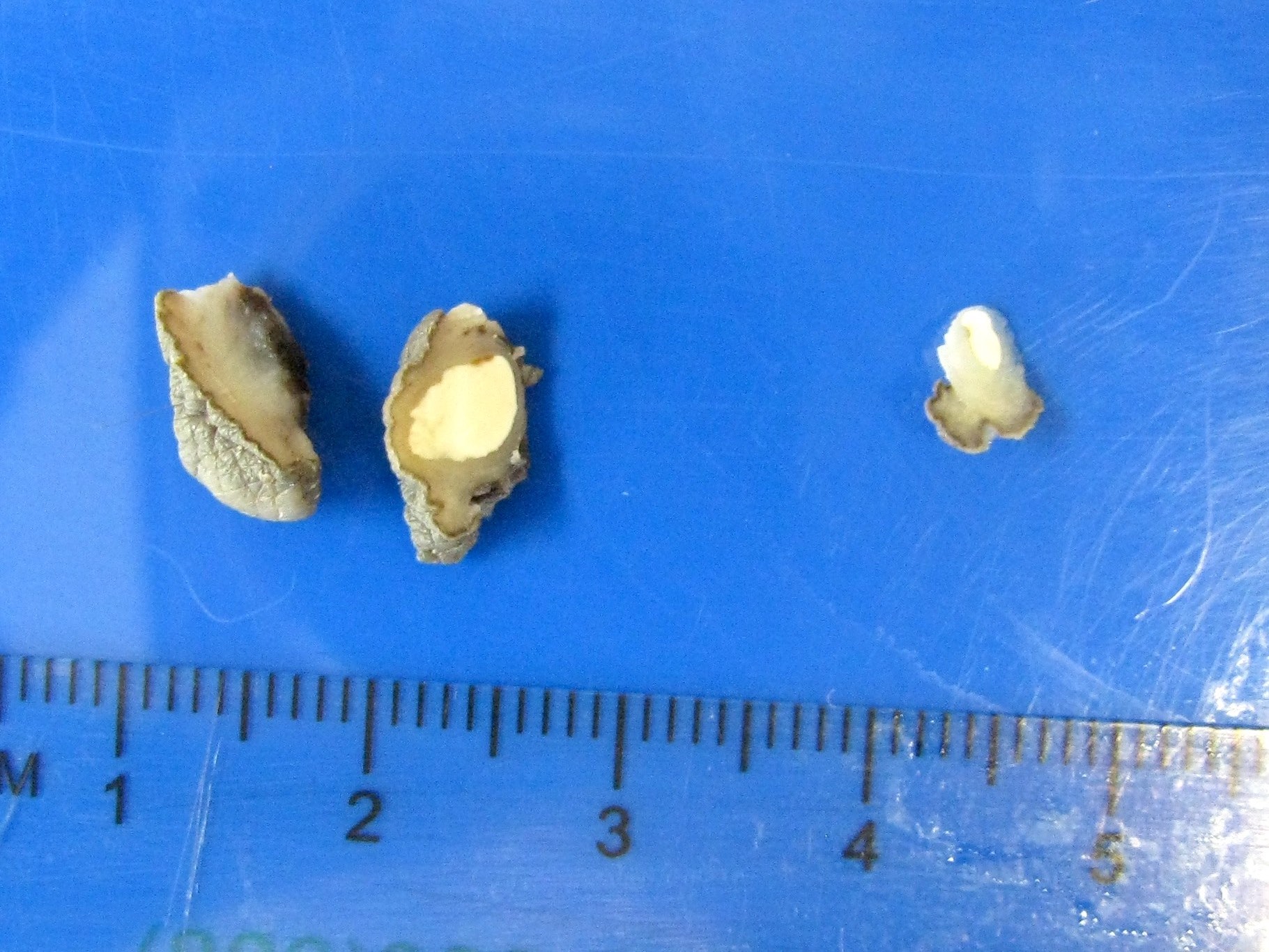
White nodule
Contributed by Nasir Ud Din, M.B.B.S., Debra Zynger, M.D. and @katcollmd on Twitter
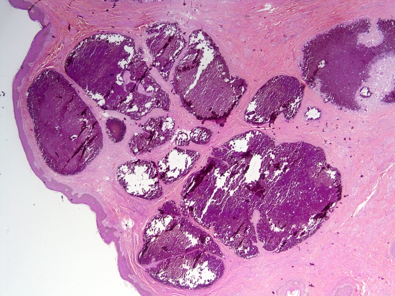
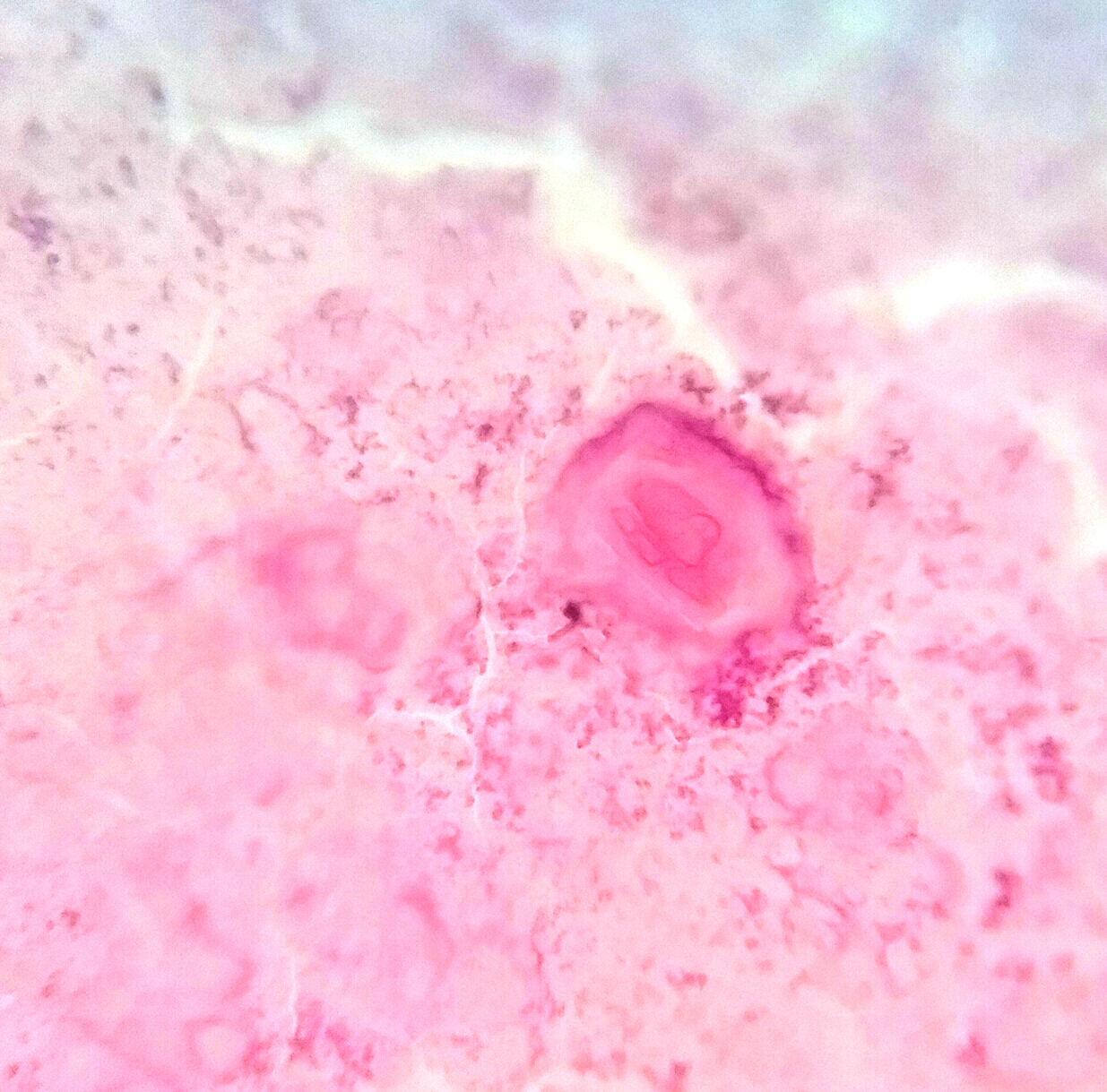
Skin tissue showing calcium deposits
Multiple calcium deposits
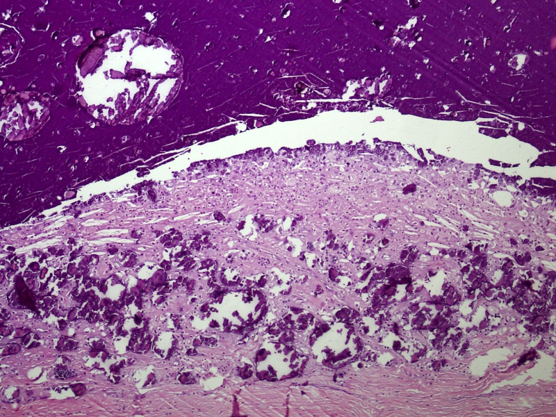
Calcium deposits with granulomatous reaction

Dermal calcium deposits with epidermis
Scrotal calcinosis
Scrotal calcinosis
Contributed by Sleiman Khal, M.D. (Case #468)
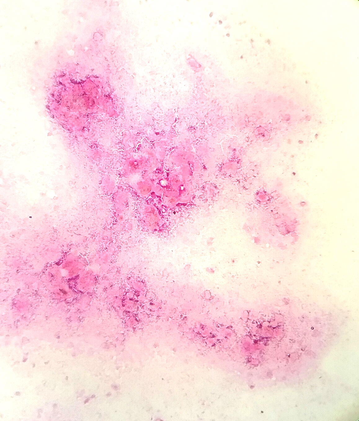
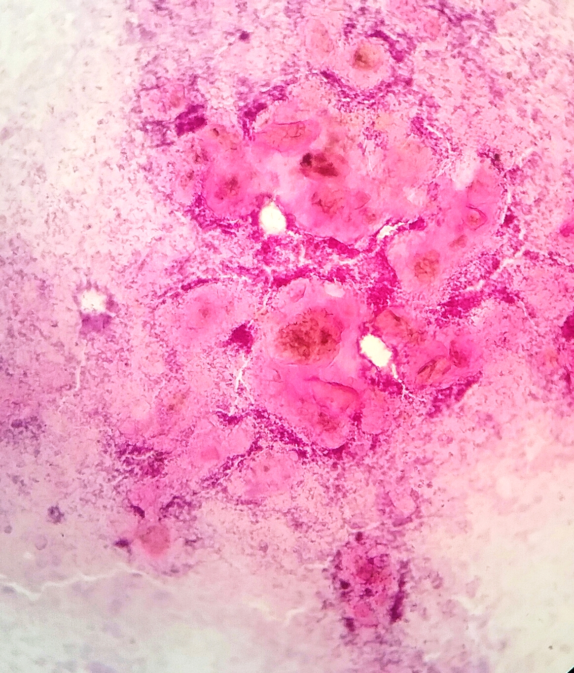
FNA smear of scrotal calcinosis
Images hosted on other servers:

FNA smear of scrotal calcinosis
Scrotal calcinosis: pathology mini tutorial
AFIP images

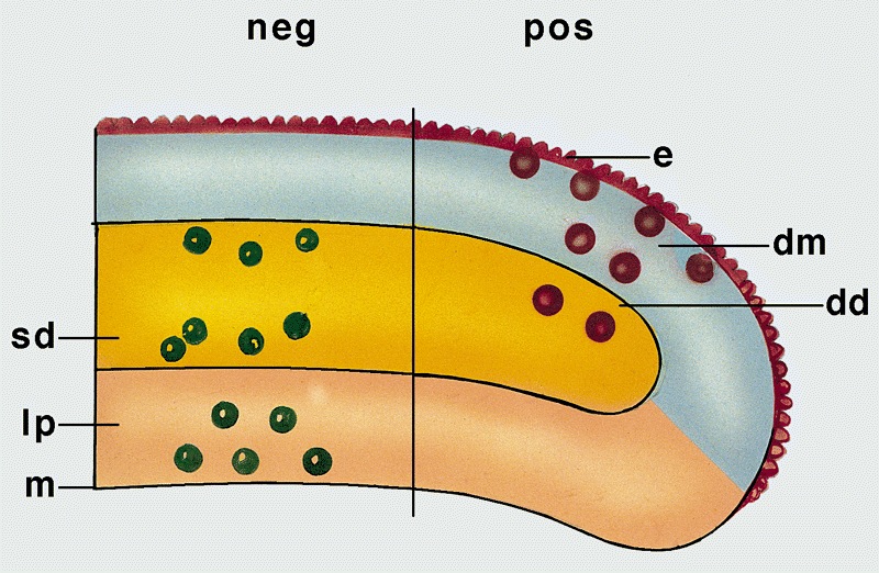
Schematic representation of effect of anatomic depth of invasion on risk of lymph node metastasis in carcinoma

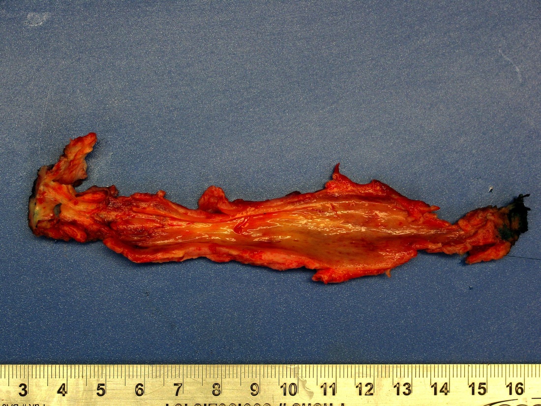
Possible sites of
resection margin
involvement at time
of frozen section
Verruciform lesions
Frozen section evaluation of surgical margins
Contributed by Stewart F. Cramer, M.D. and AFIP images
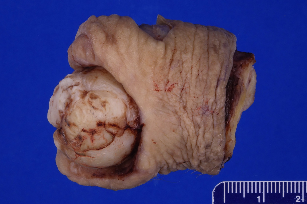
Large bosselated mass
Tumor at junction of scrotum and penis
Images hosted on other servers:

Arising on genital lichen sclerosus
Papillary SCC
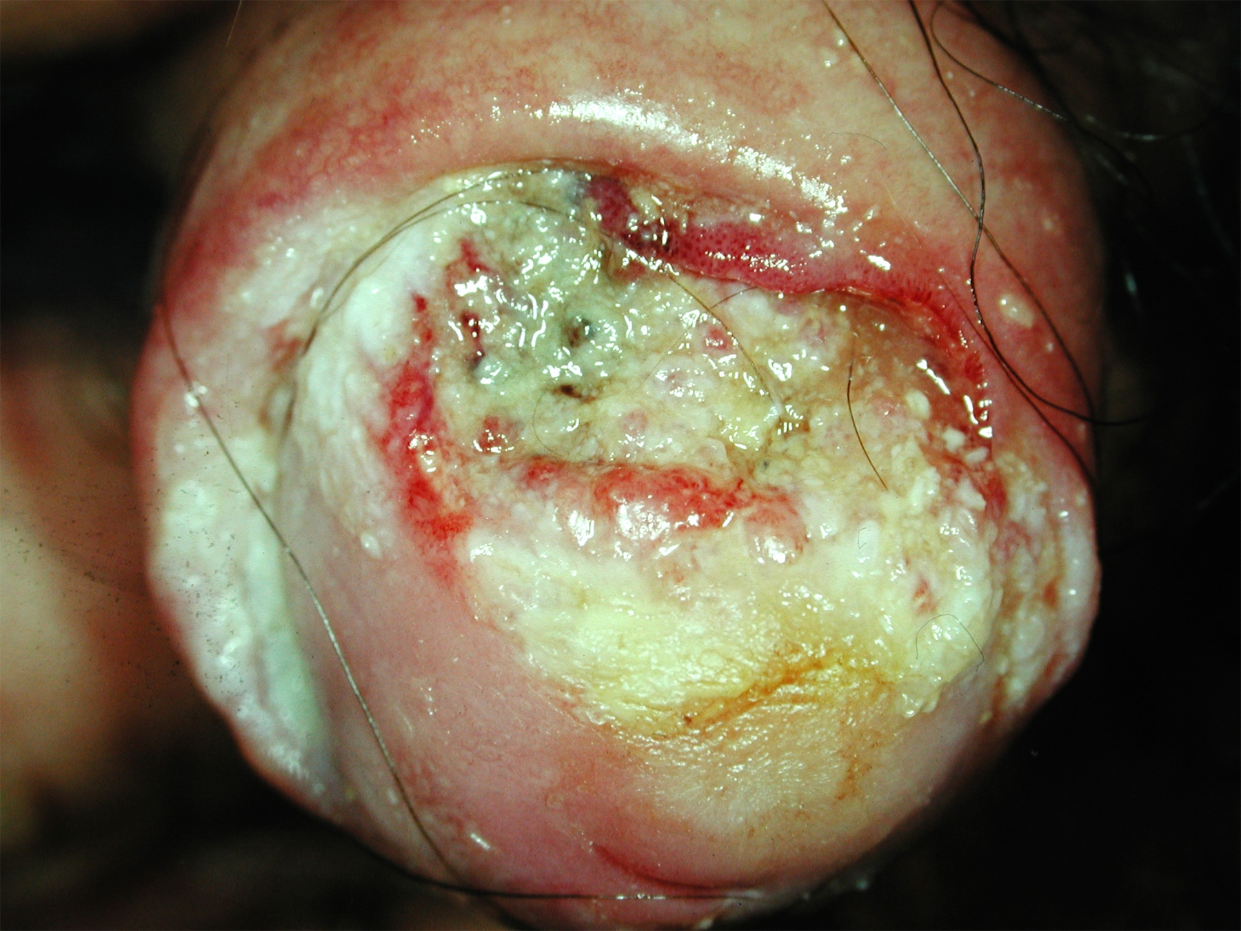
Ulcerated SCC on glans
Verrucous carcinoma
AFIP images
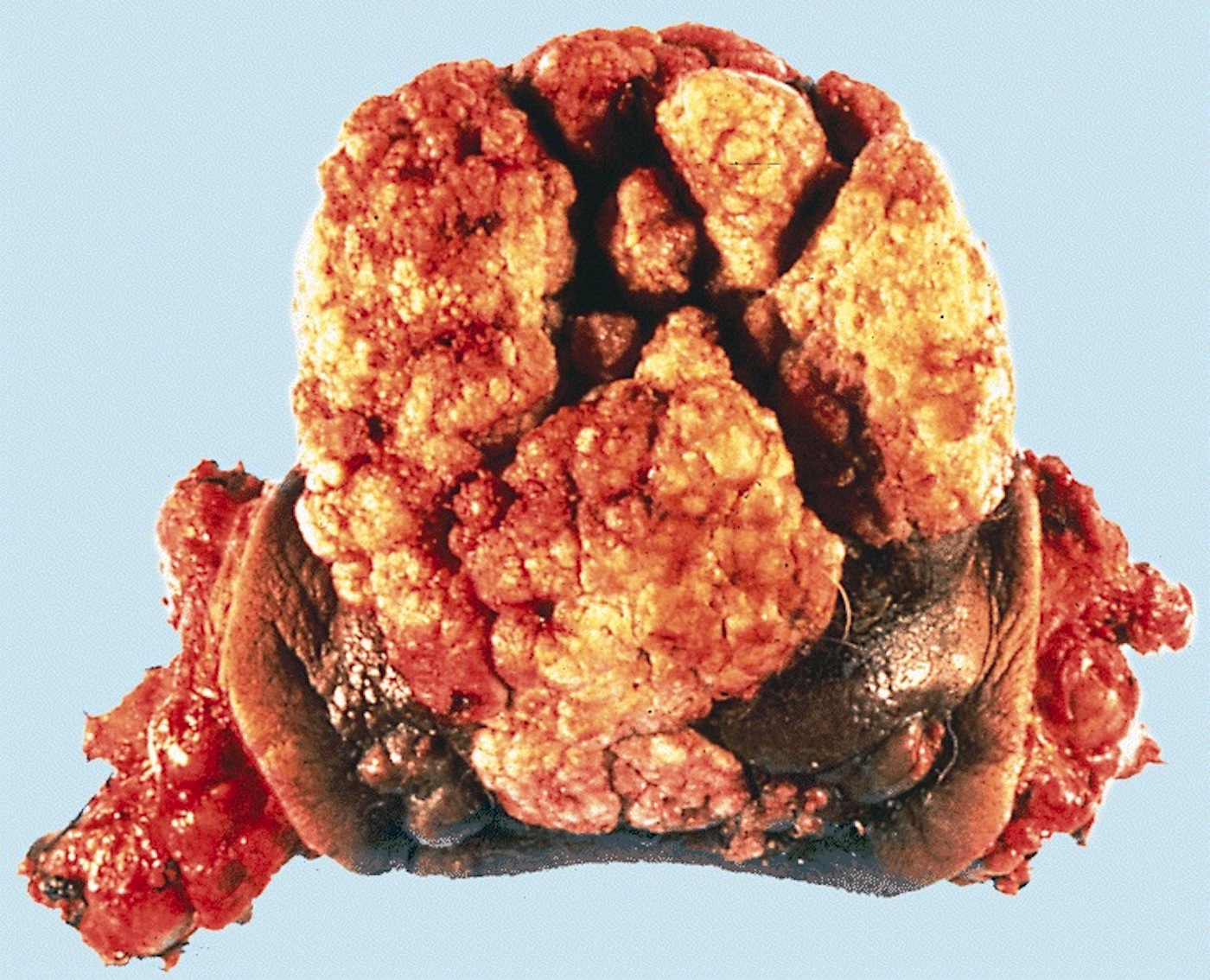
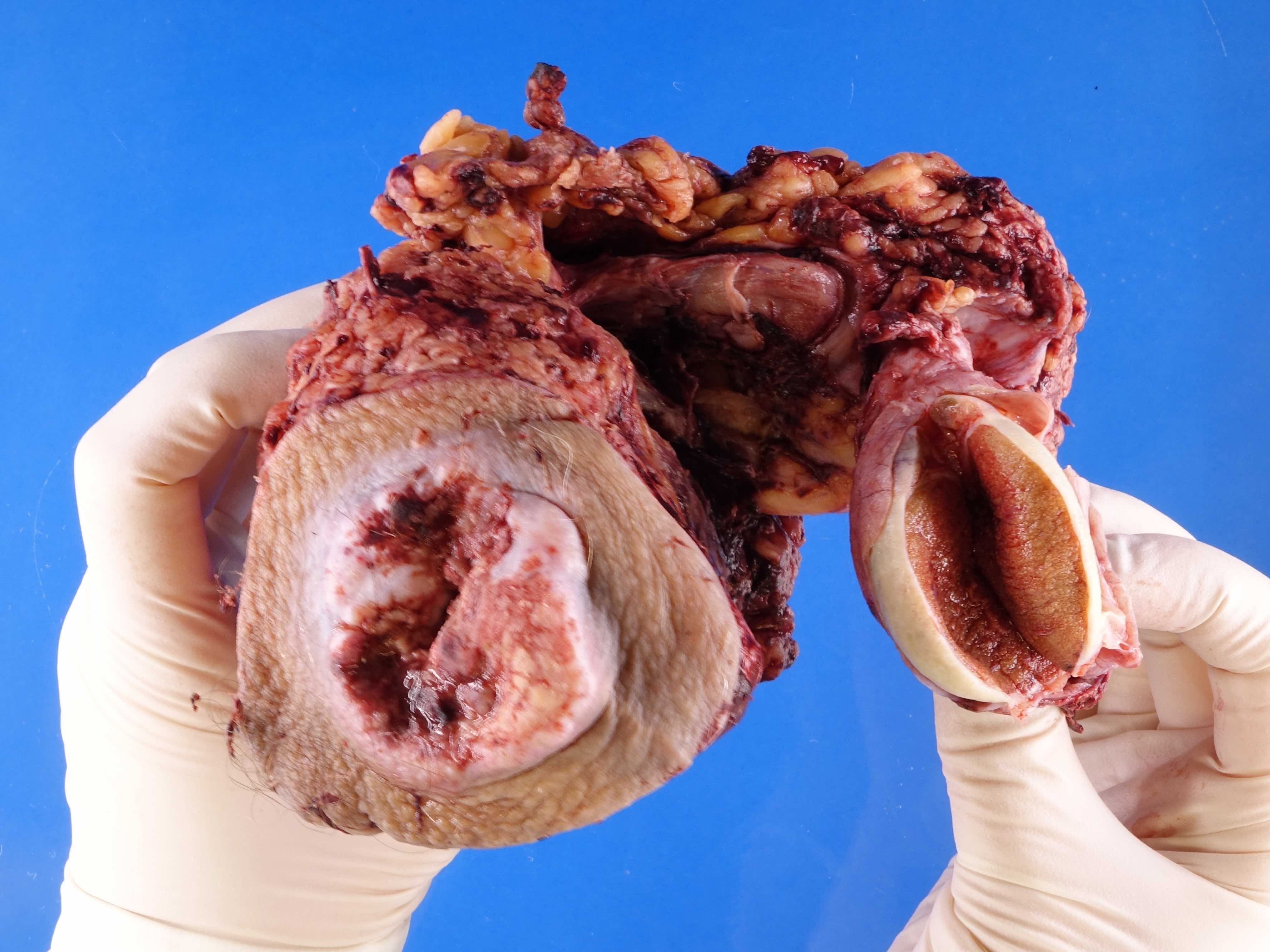
Glans:

Exophytic cauliflower-like mass

Glans extensively
involved by a
multinodular mass
with focal ulceration
Foreskin:
Circumcision
specimen shows a
flat, granular and
beige neoplasm

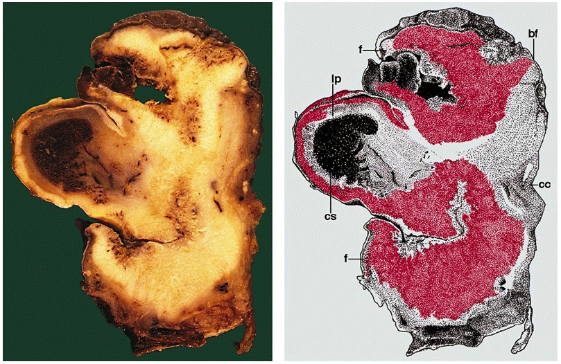
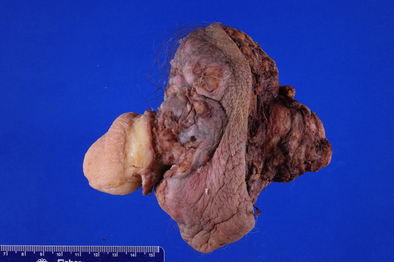
Massive involvement
has caused multiple
foci of ulceration
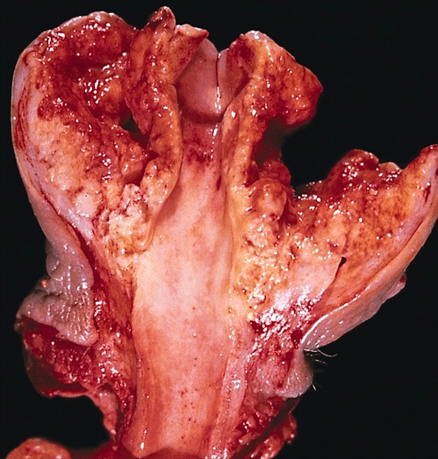
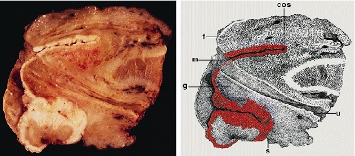
Coronal sulcus:
Nodular white
tumor extensively
involves the sulcus
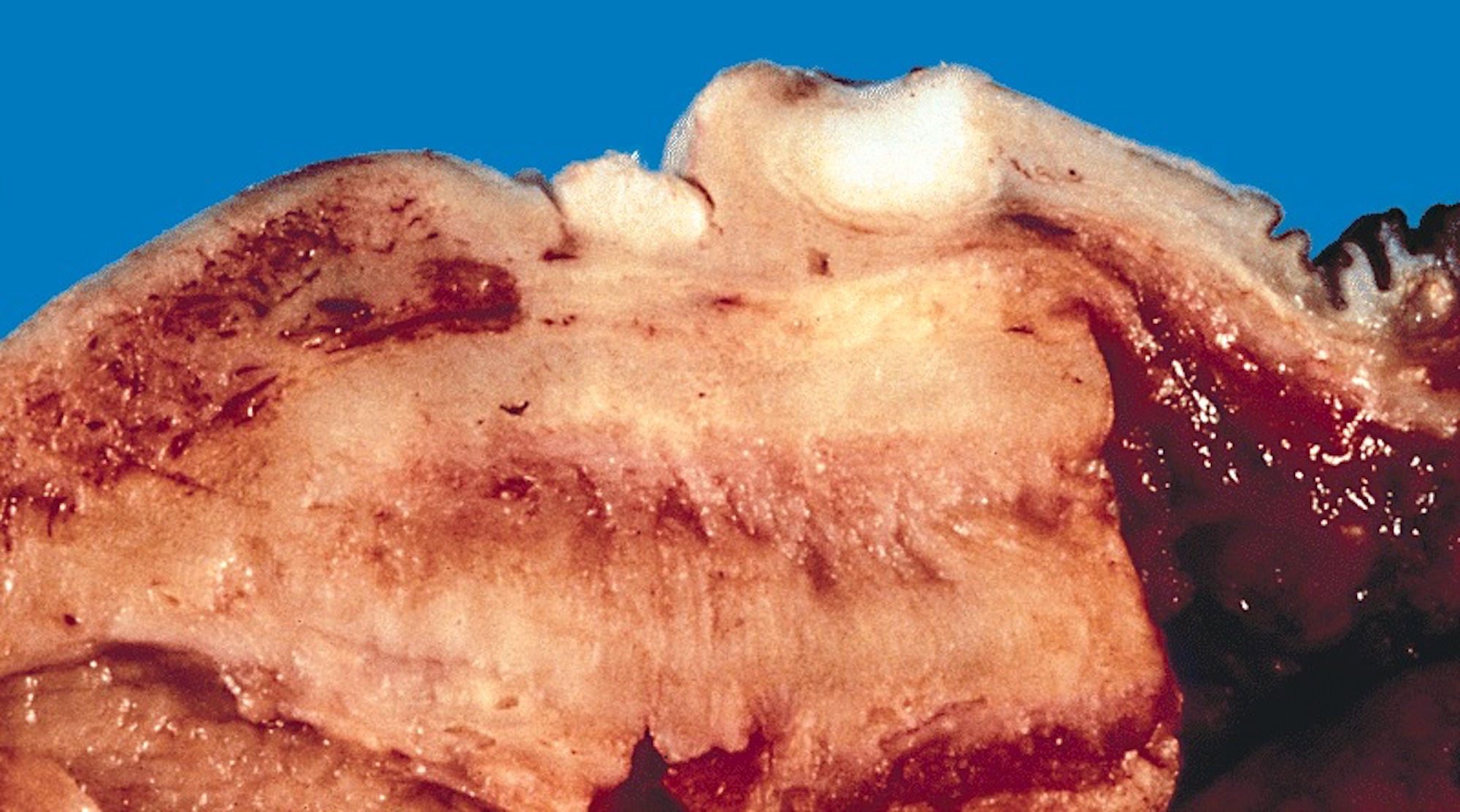
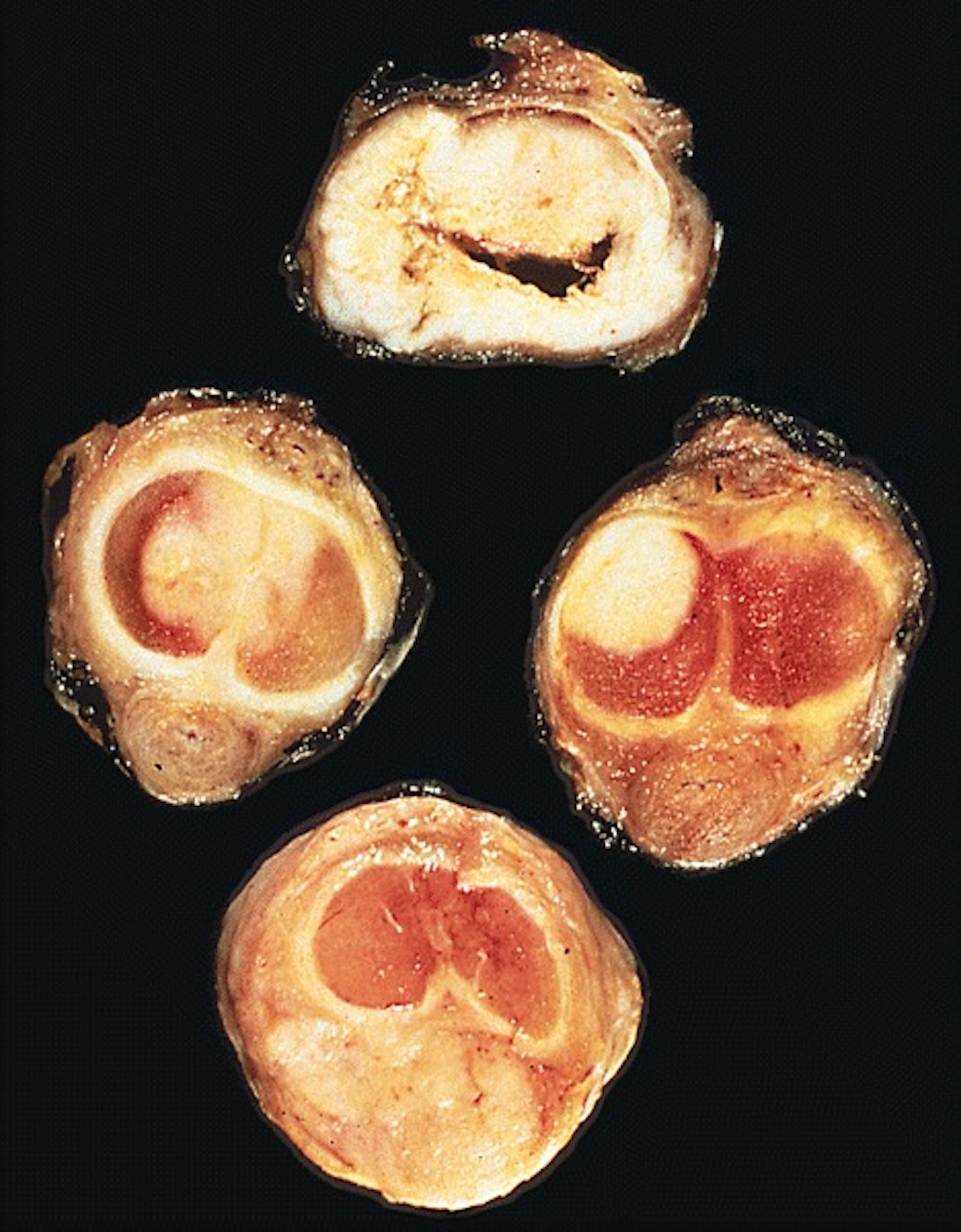
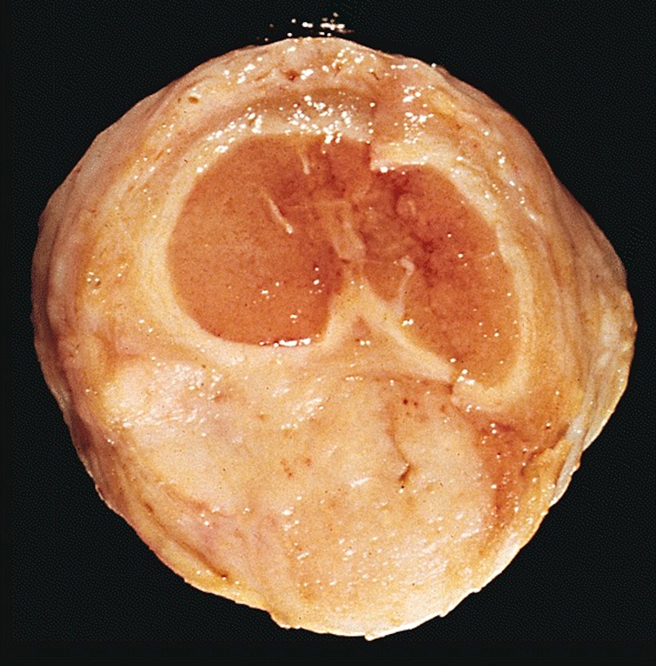
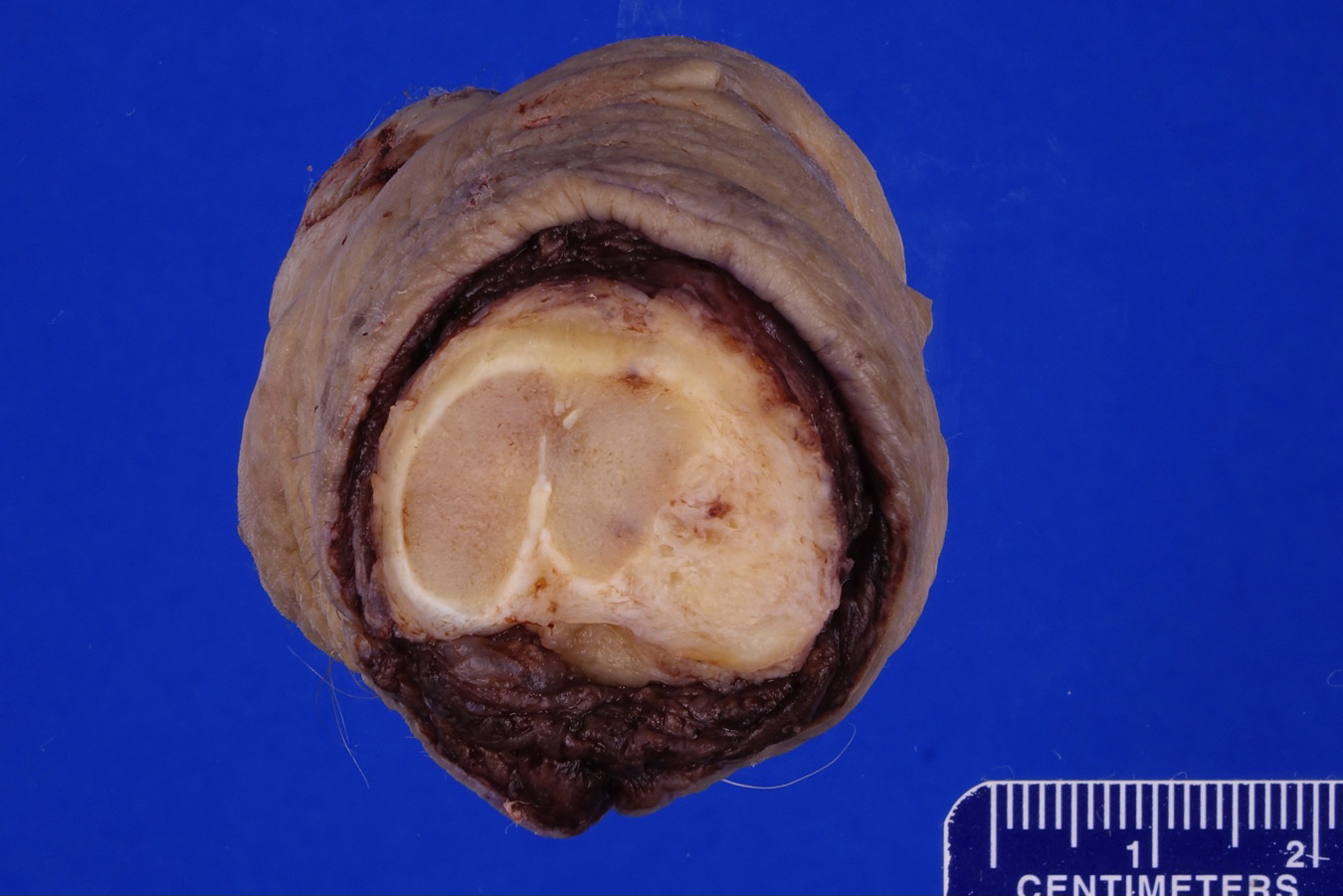
Cut section
shows 2
discrete nodules
of tumor
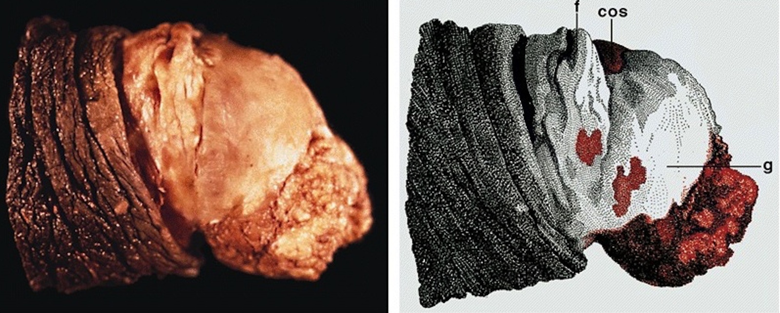
Verruciform lesions:
Verrucous
carcinoma
Cobblestone:

Cobblestone appearance representing condyloma
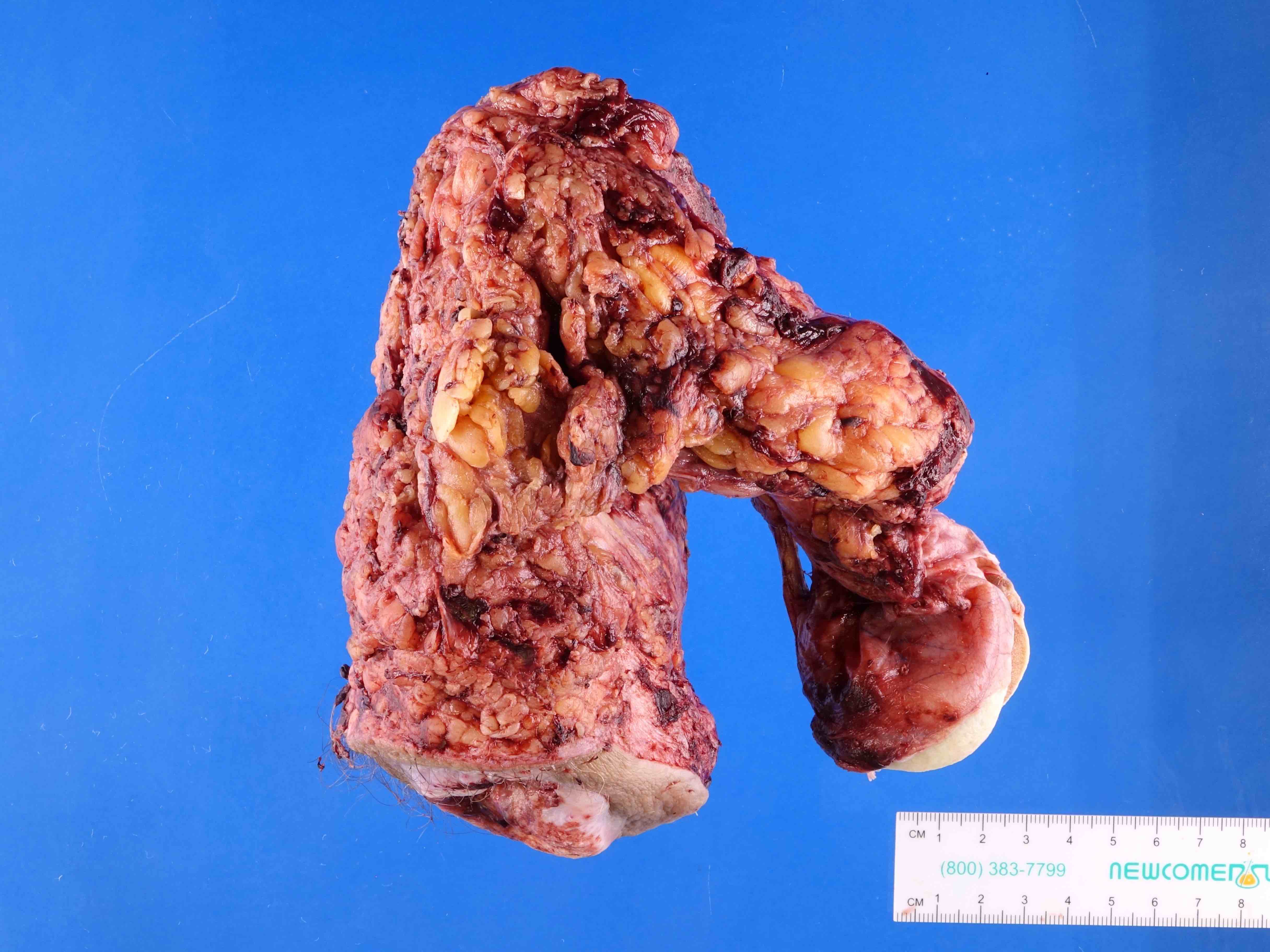
Multiple compartments:
Massive involvement
of glans, coronal
sulcus and foreskin

Massive involvement
has resulted in
autoamputation
4 separate
foci of
carcinoma
are present
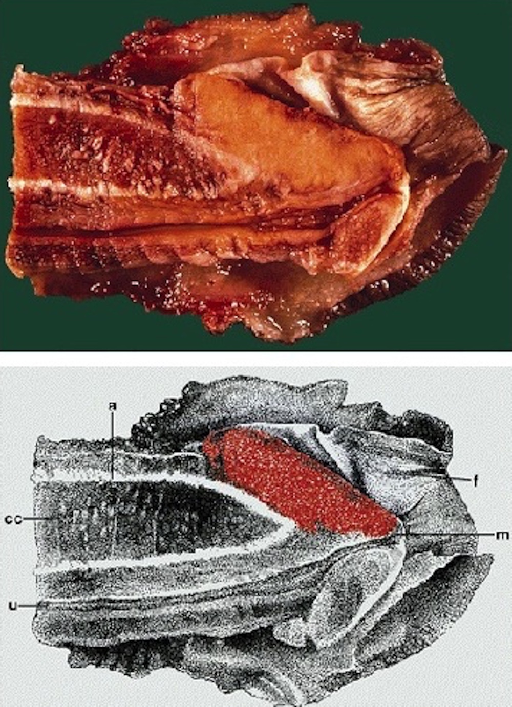
Superficial spreading (SCC):

Tumor involves
the glans with
extension to
coronal sulcus
Tumor is white,
involves the mucosa
of the foreskin
and coronal sulcus
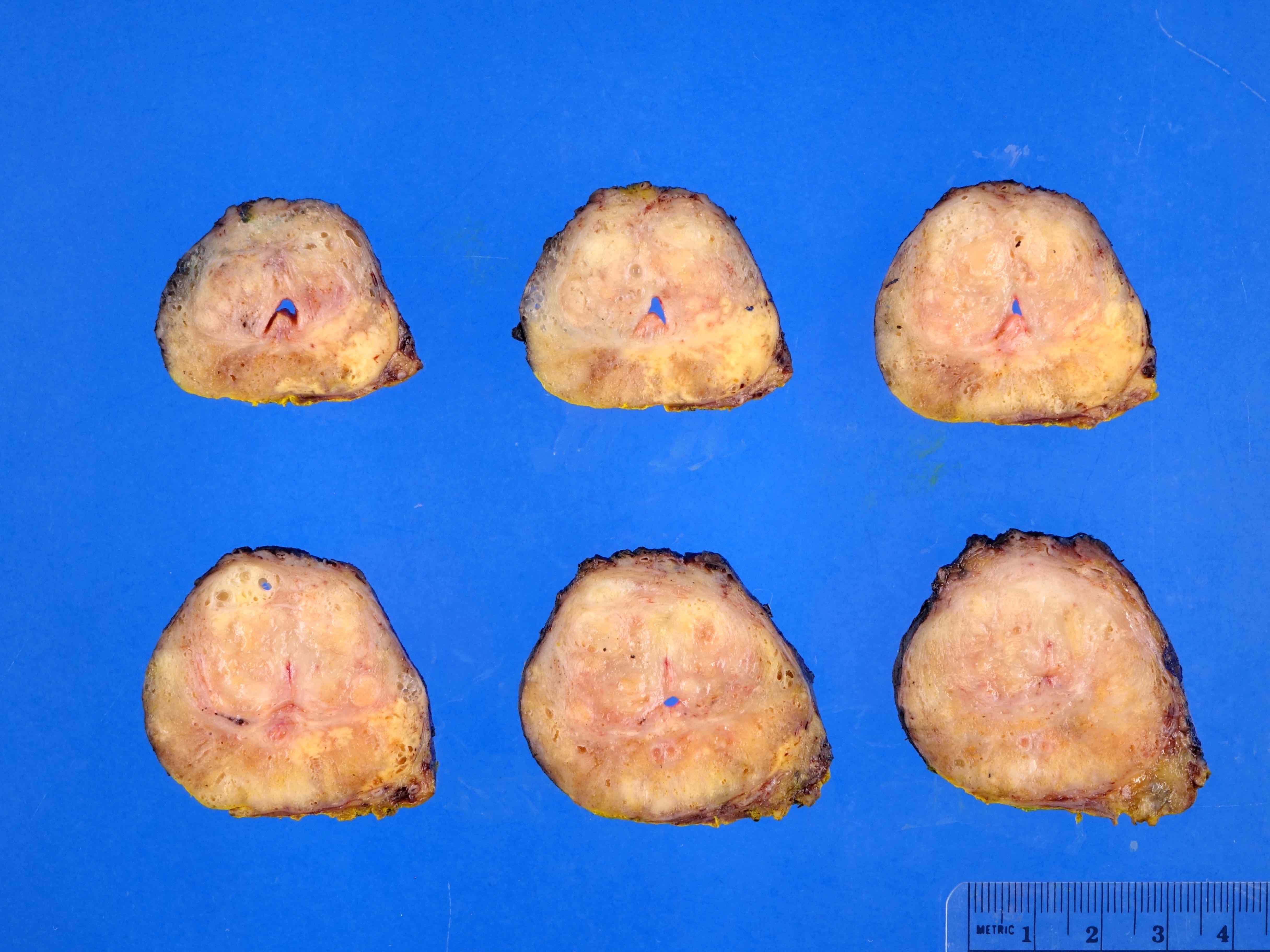
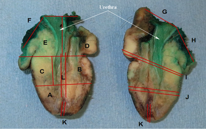
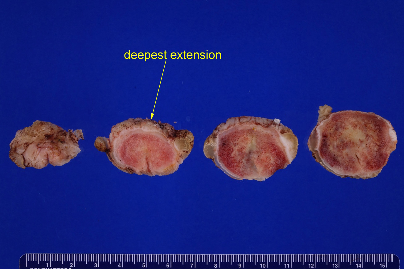
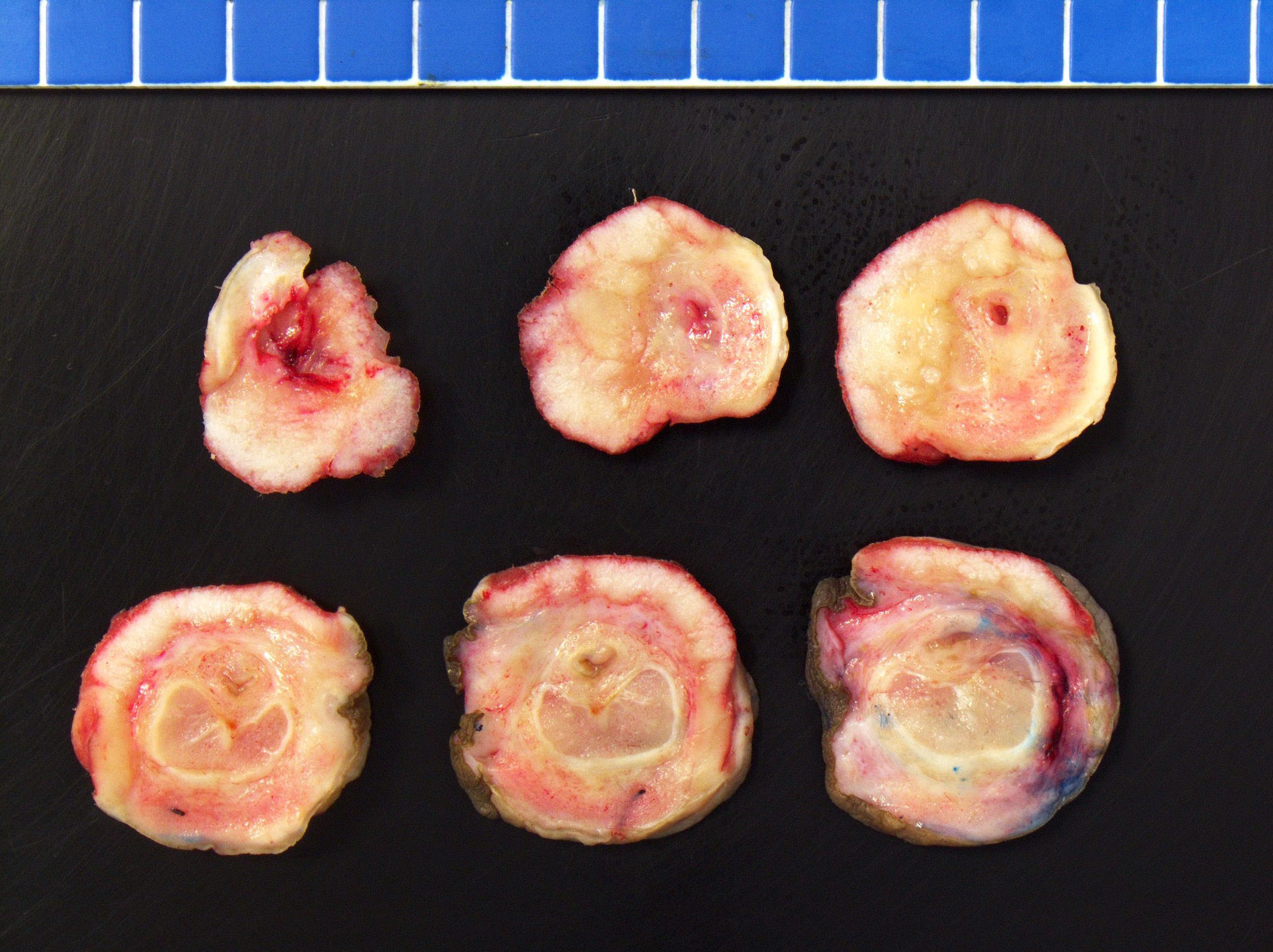
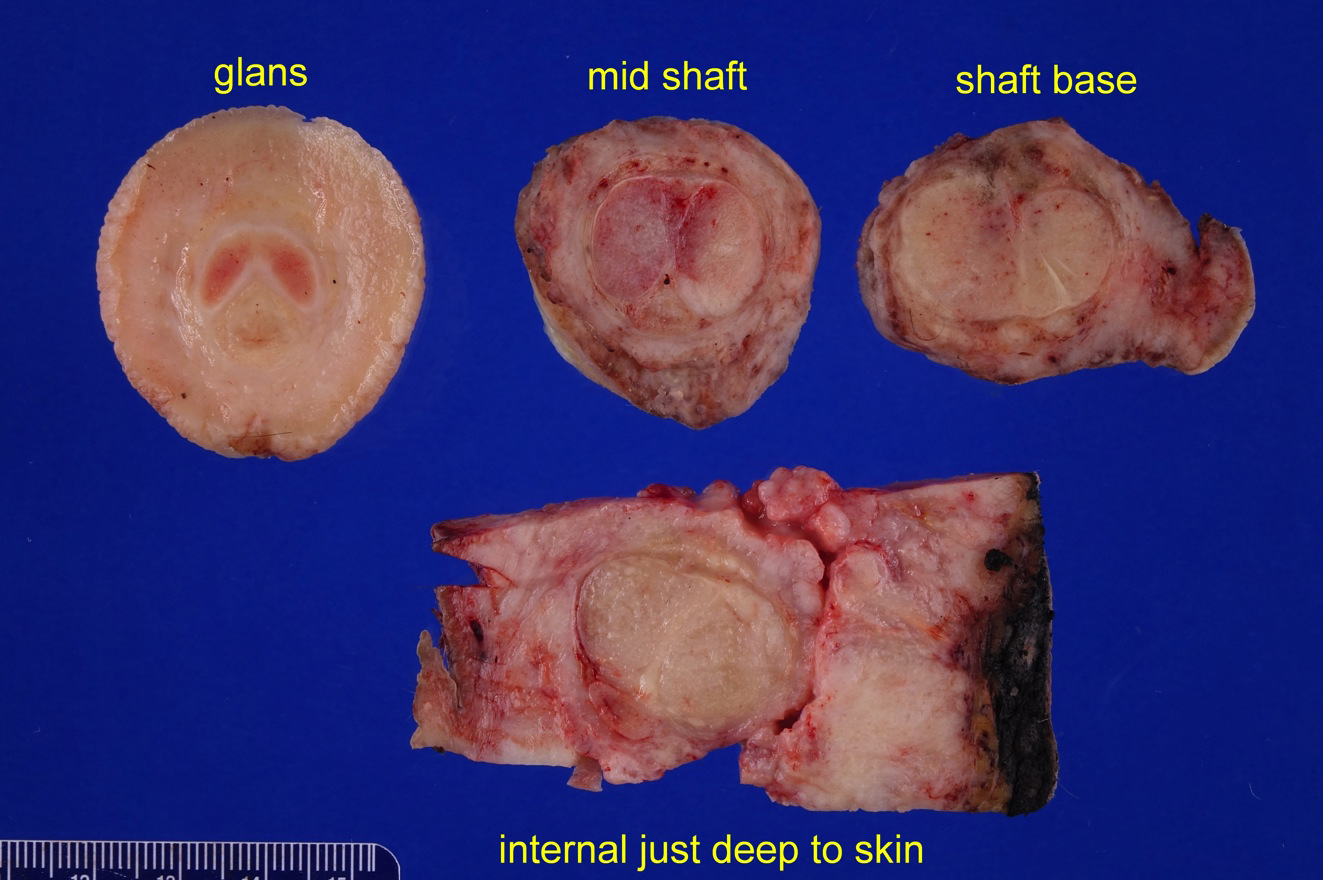
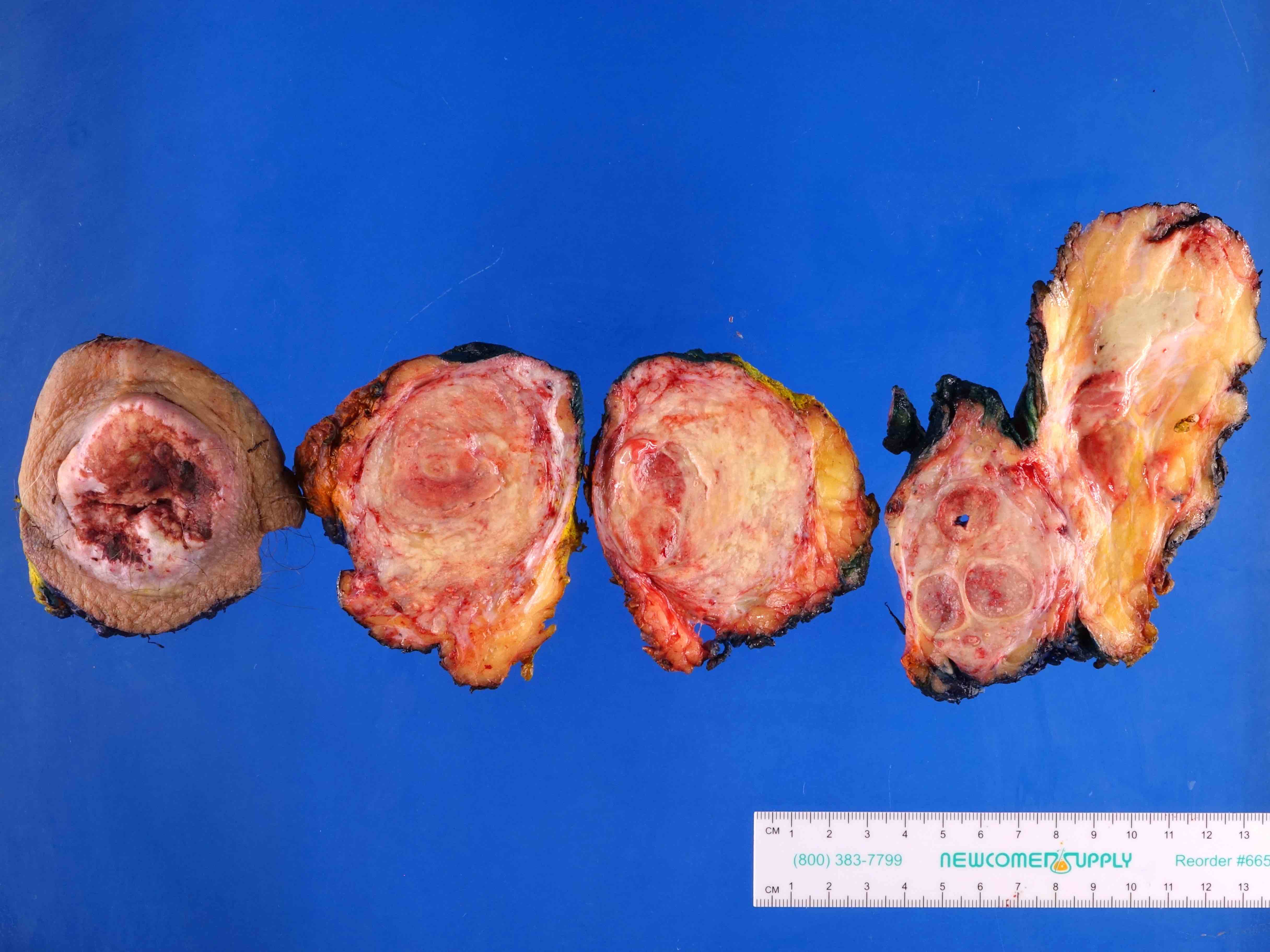
Assessment of depth of invasion:
Penile carcinoma has
been transversely
sectioned
Assessment of
depth of invasion
of tumor in
resected specimen
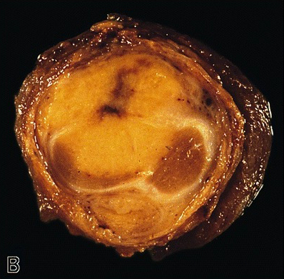
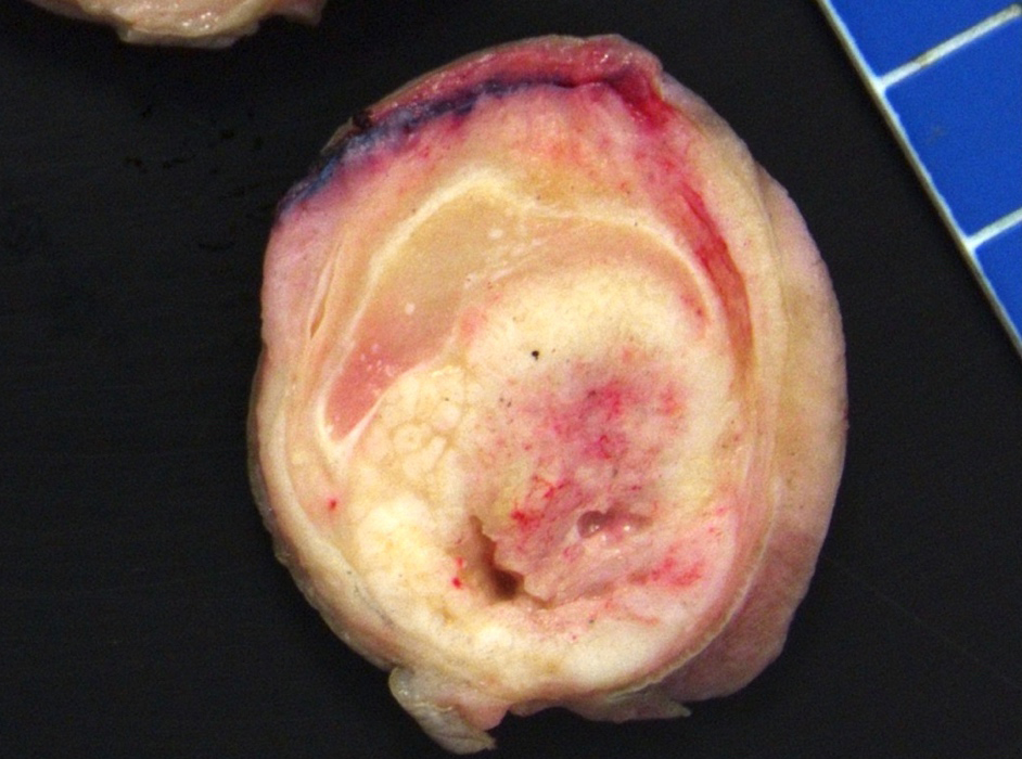
Vertical growth (SCC):
Solid yellow-tan
neoplasm in
the dorsal half
of the glans
Mixed low and high grade (SCC):
Neoplasm shows
superficial, white,
serrated papillary
Margin involvement:

Frozen section evaluation of surgical margins
Images hosted on other servers:

Red-tan ulcerated tumor

Fungating mass
Contributed by Antonio L. Cubilla, M.D., Alcides Chaux, M.D. and AFIP images

Scrotum: well, moderately and poorly differentiated tumors
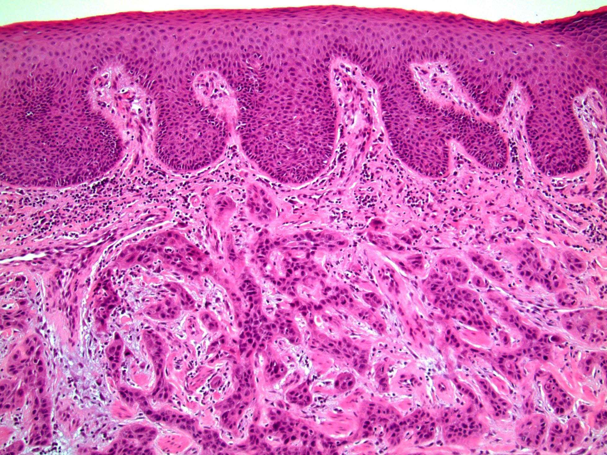
Mucosa is involved by carcinoma
Invasion of the lamina propria
Mixed (hybrid) usual: verrucous carcinoma
Usual type: well differentiated (left, grade 1); moderately differentiated (middle, grade 2); poorly differentiated (right, grade 3)

Pseudoglandular growth
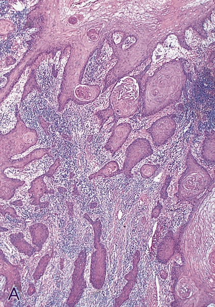
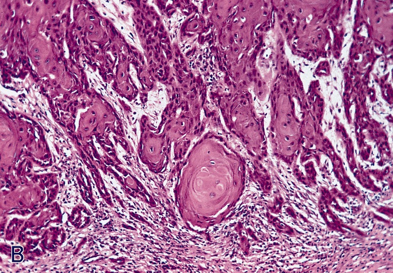
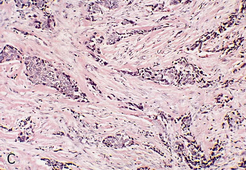
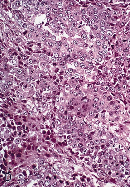
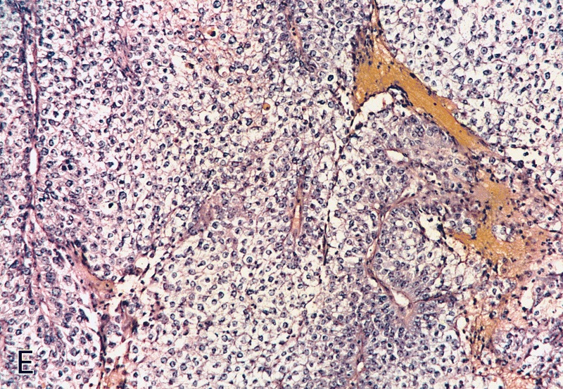
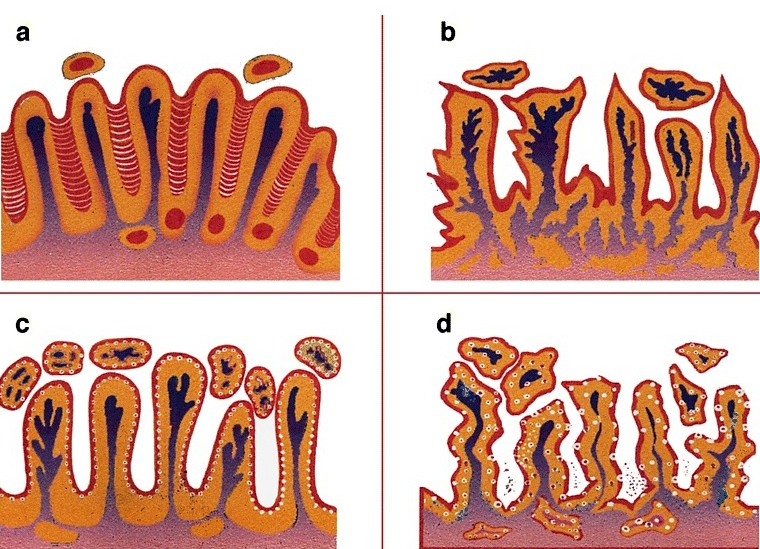
Low grade keratinizing (A, B); moderate to high grade nonkeratinizing (C); high grade nonkeratinizing tumor (D); and carcinoma with prominent glycogenated clear cells (E)
Images hosted on other servers:
Pseudoglandular growth:

Conjunctiva: acantholysis of neoplastic squamous cells


Head and neck: pseudolumina are present but no true glands
Contributed by Alcides Chaux, M.D. and Antonio Cubilla, M.D.
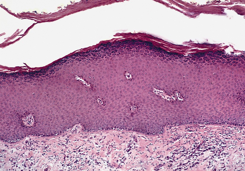
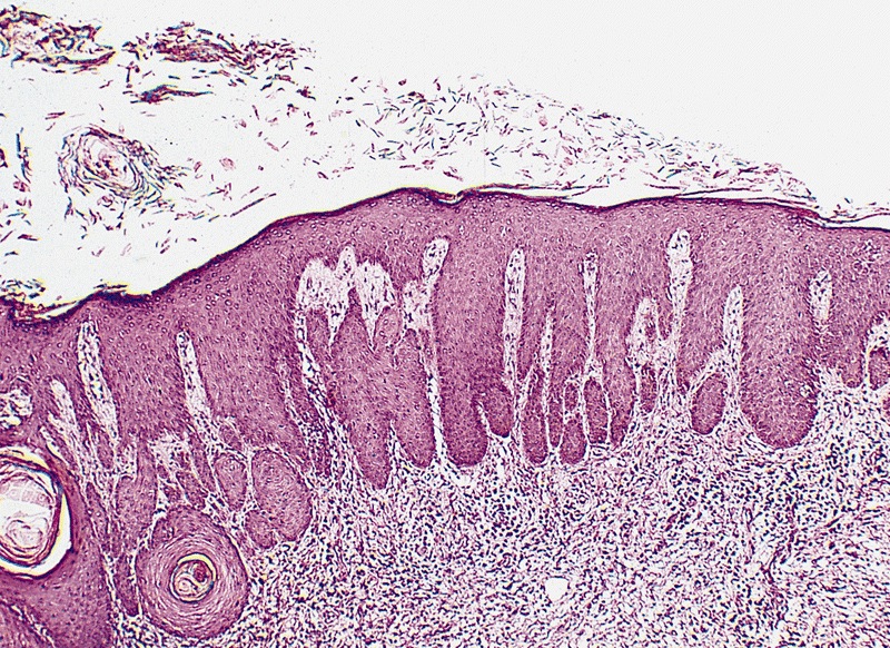
Flat: hyperkeratosis and acanthosis but also normal maturation without atypia
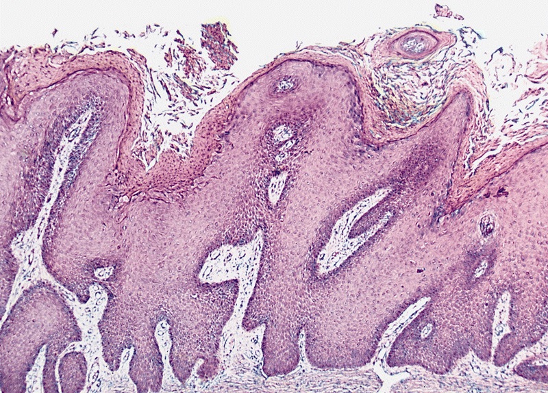
Papillary: hyperkeratosis, papillomatosis and acanthosis
Pseudoepitheliomatous
AFIP images

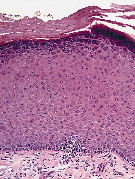
Acanthosis, absence
of nuclear atypias
and hyperkeratosis
Contributed by Debra Zynger, M.D.
pTis
pT1a
pT2
pT3
pT3 partial penectomy
pT3 penectomy
pT4 penectomy, orchiectomy and scrotal excision
Contributed by Debra Zynger, M.D.
CIS / PeIN (pTis)
Subepithelial invasion (pT1a)
Perineural invasion (pT1b)
Invasion of corpora spongiosum (pT2)
Inguinal lymph node metastasis (pN1)
AFIP images
Sharply delineated lesion
Two well circumscribed, minimally elevated lesions
Images hosted on other servers:


Primary chancre

Atrophic scar with condylomata lata
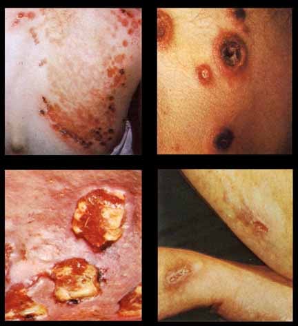
Secondary rash / lesions:


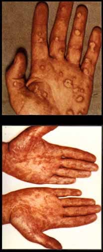
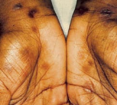
Palm, limbs and hands


Back


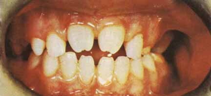
Face, teeth

"Kissing" lesions
Various images
AFIP images
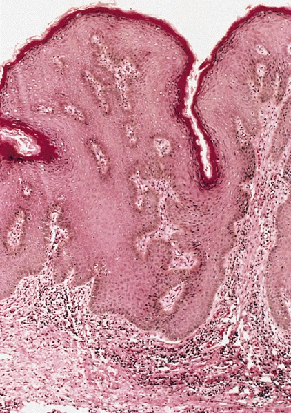
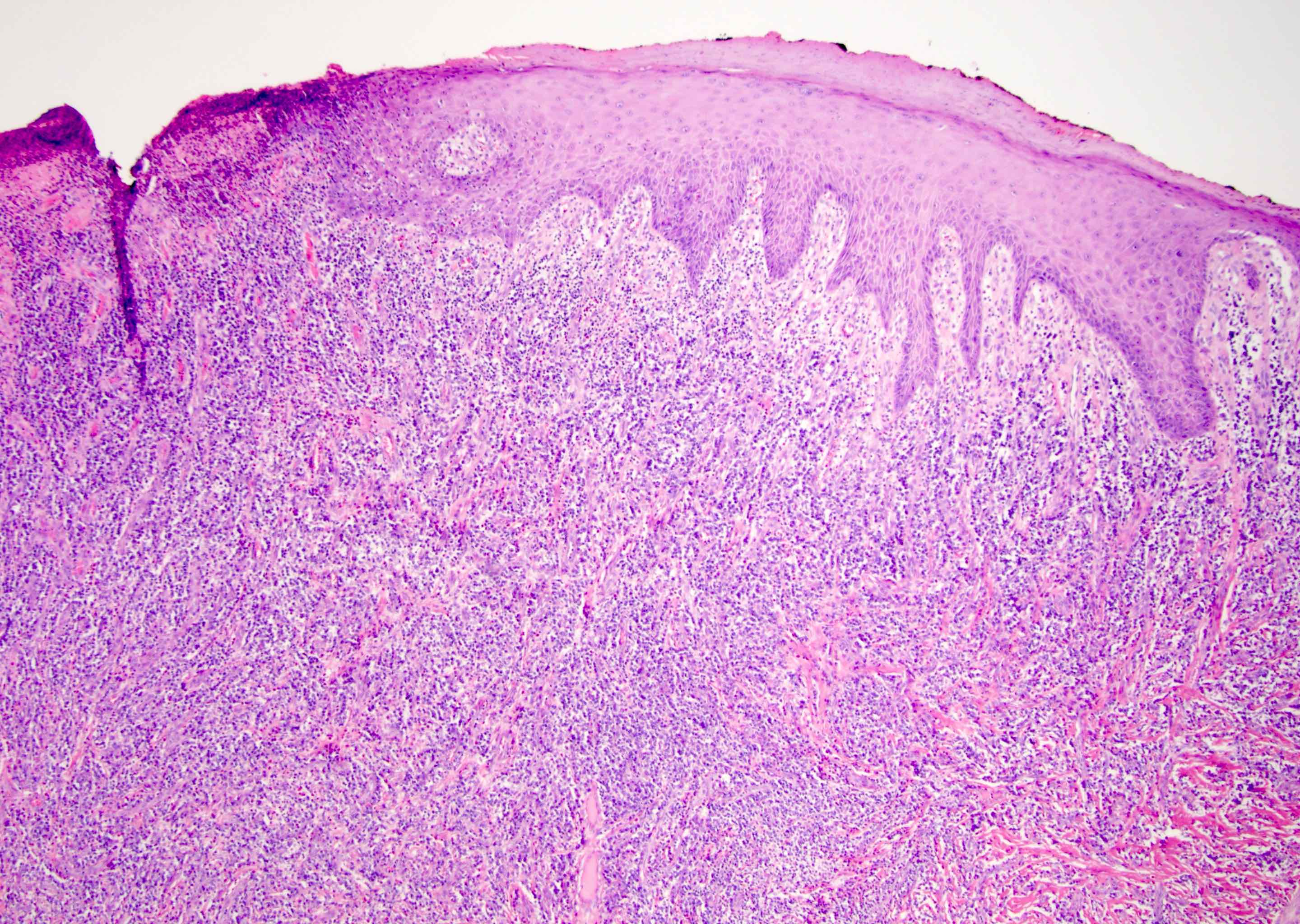
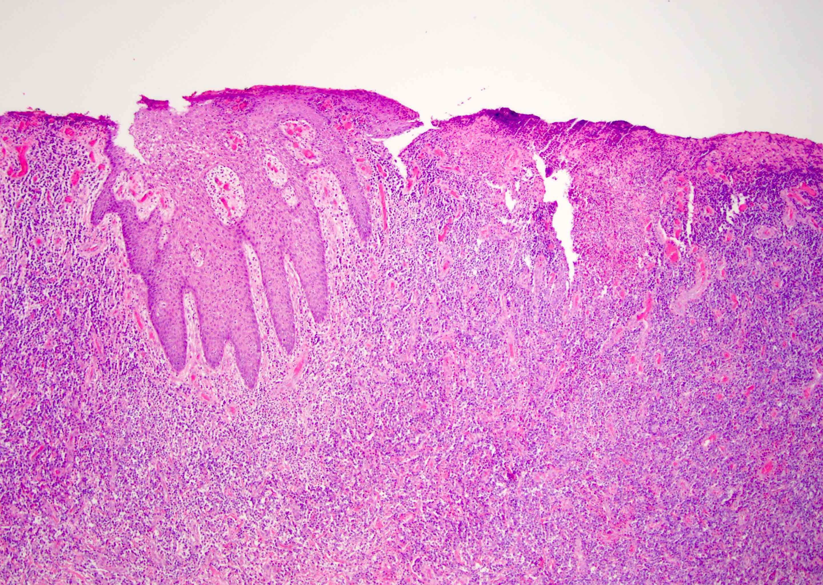
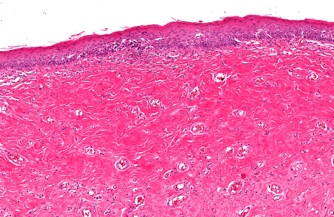
Lesion characterized by acanthosis
Images hosted on other servers:

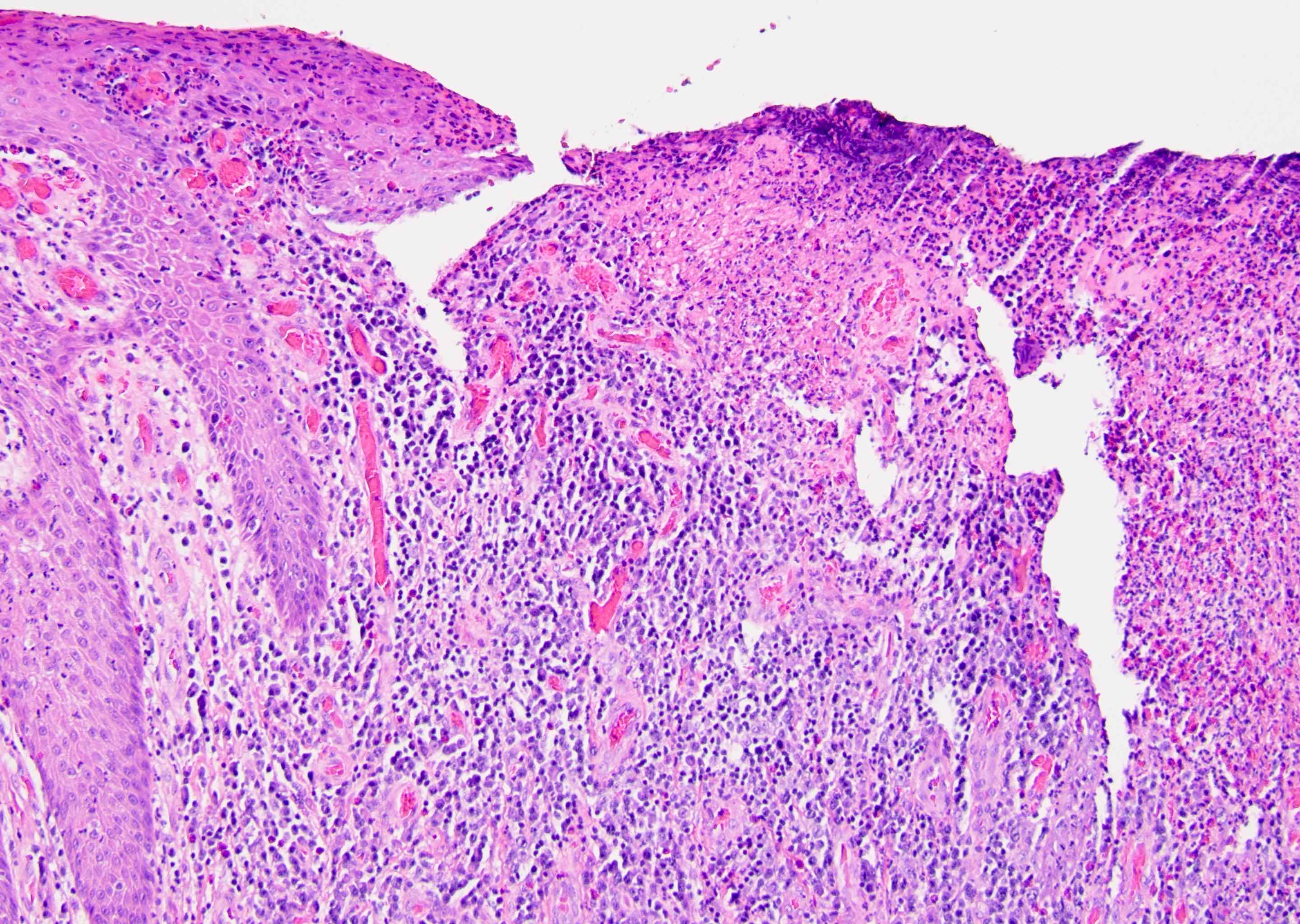
Perivascular dermal infiltrate
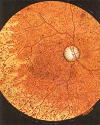
Chorioretinitis of congenital syphilis


Darkfield microscopy

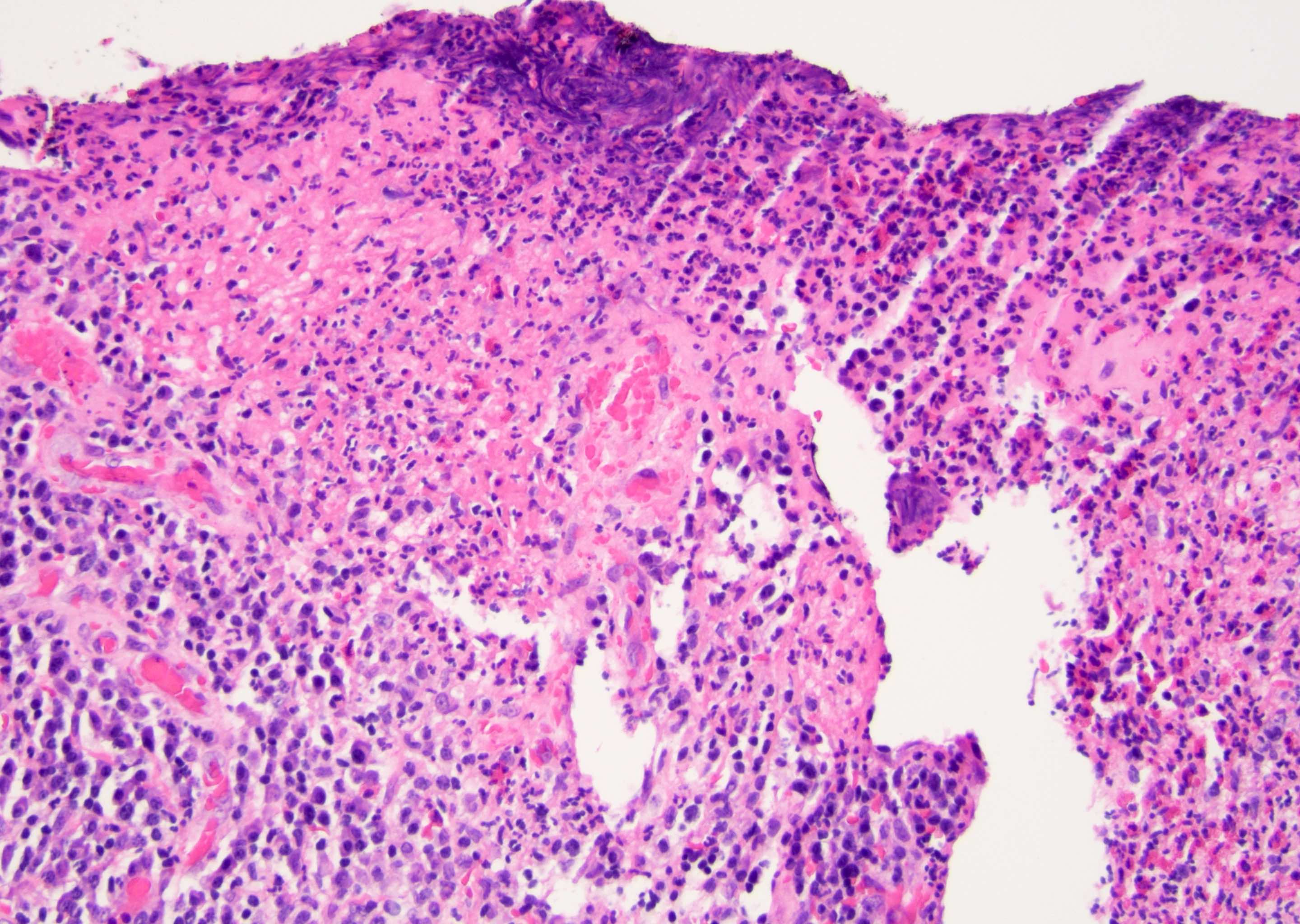
Patchy infiltrate

Epithelioid cells surrounded by lymphomono-nuclear cells
Various images
Images hosted on other servers:


Spirochete in culture
Images hosted on other servers:

Intraurethral paraffin
AFIP images

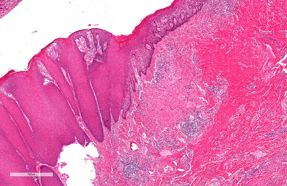
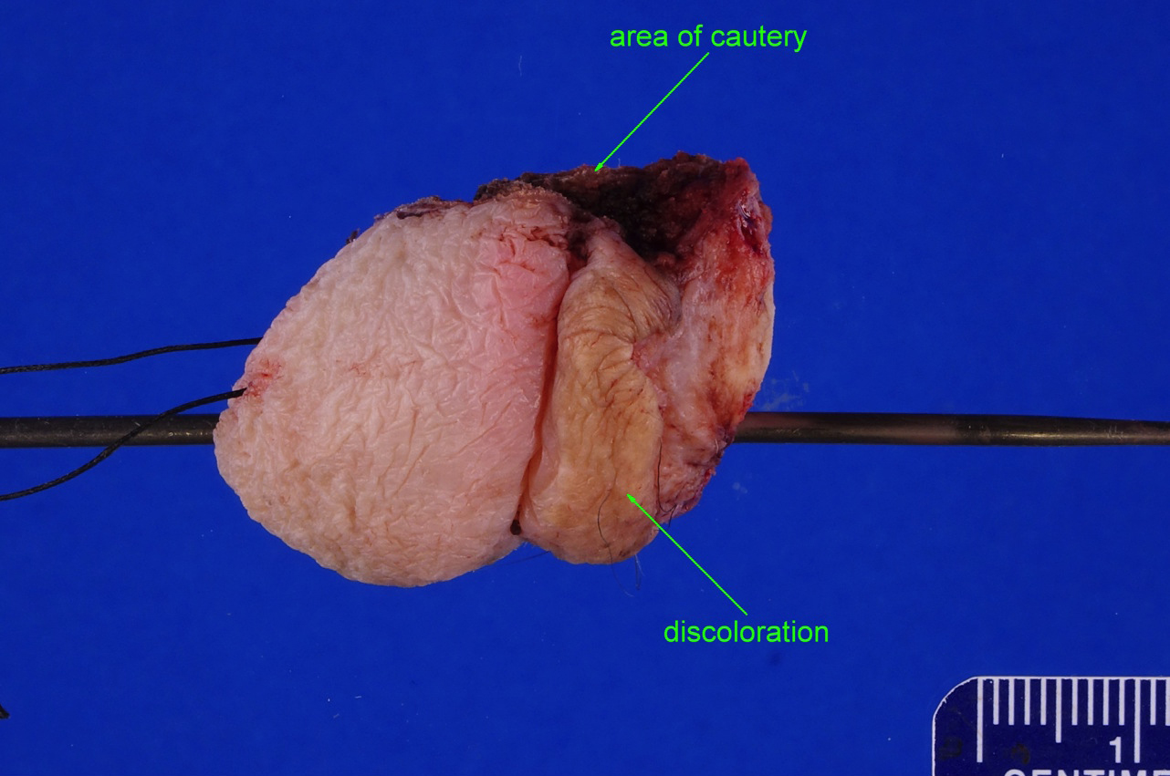
Granular, elevated
tan white mass
of distal shaft and
coronal sulcus
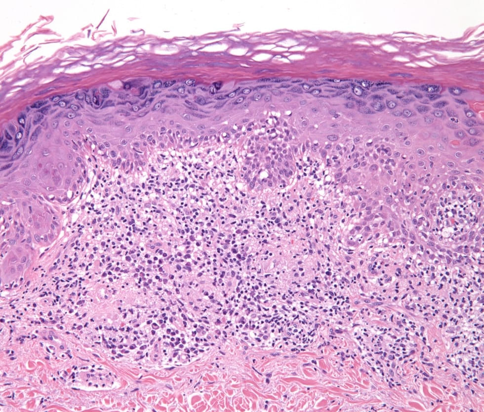
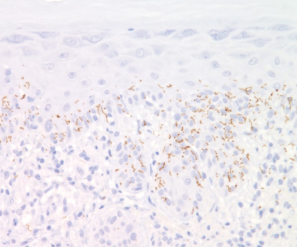
Contributed by Liwei Jia, M.D., Ph.D. and @JMGardnerMD on Twitter
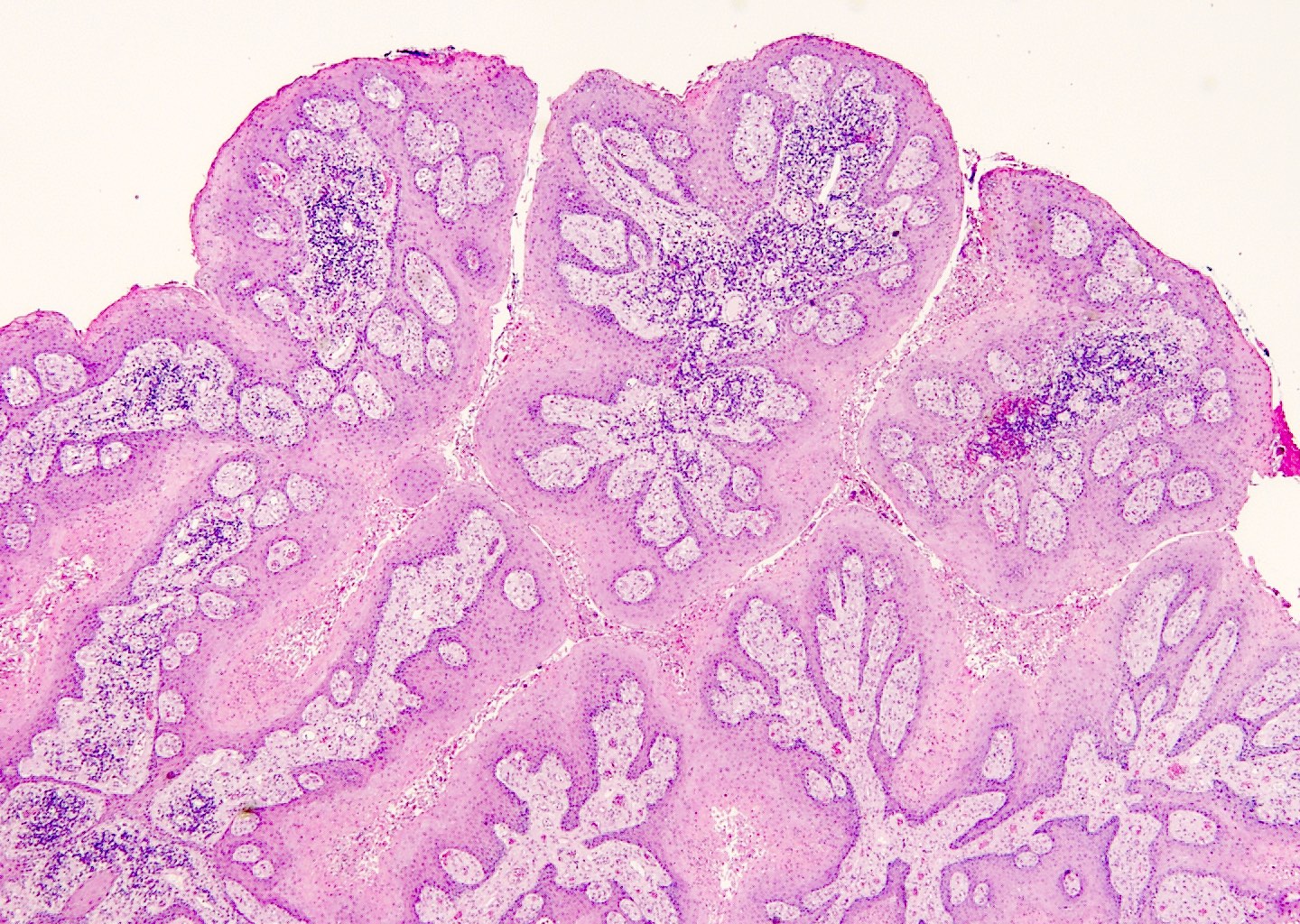
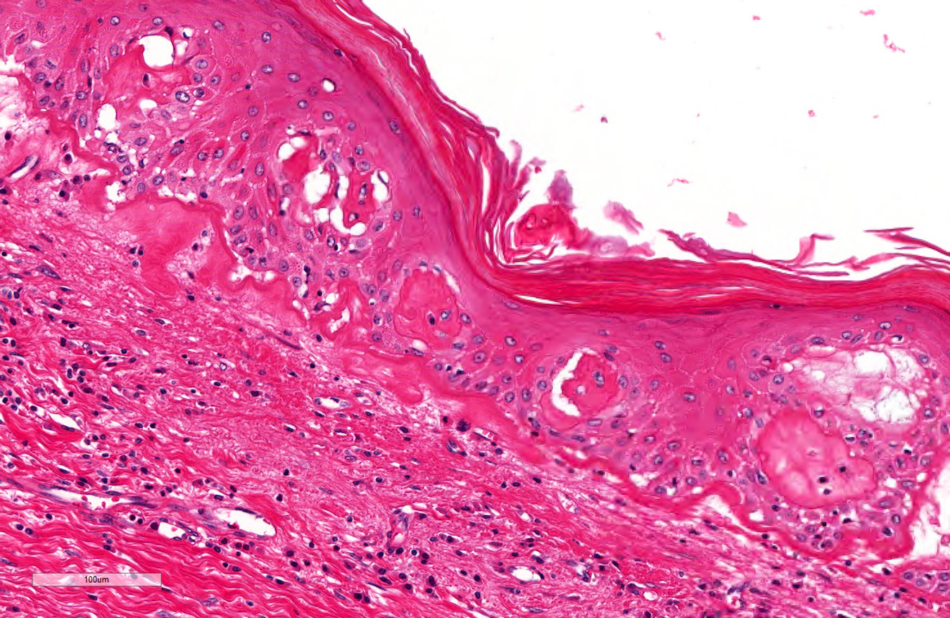
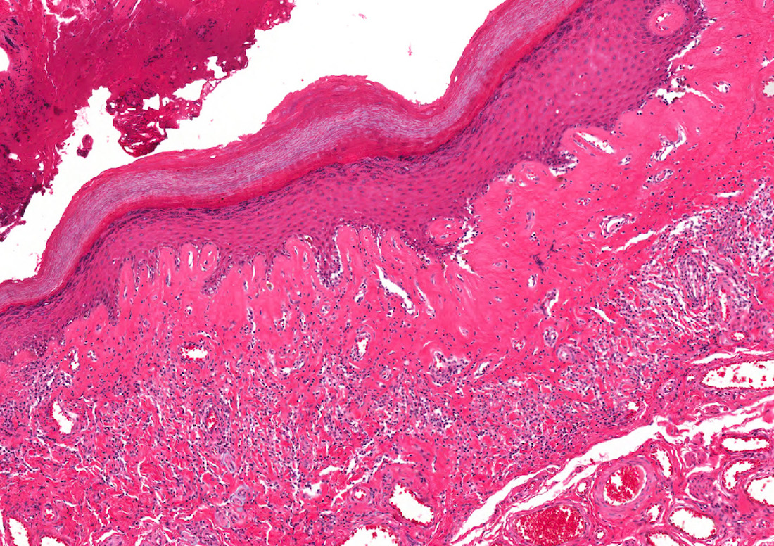
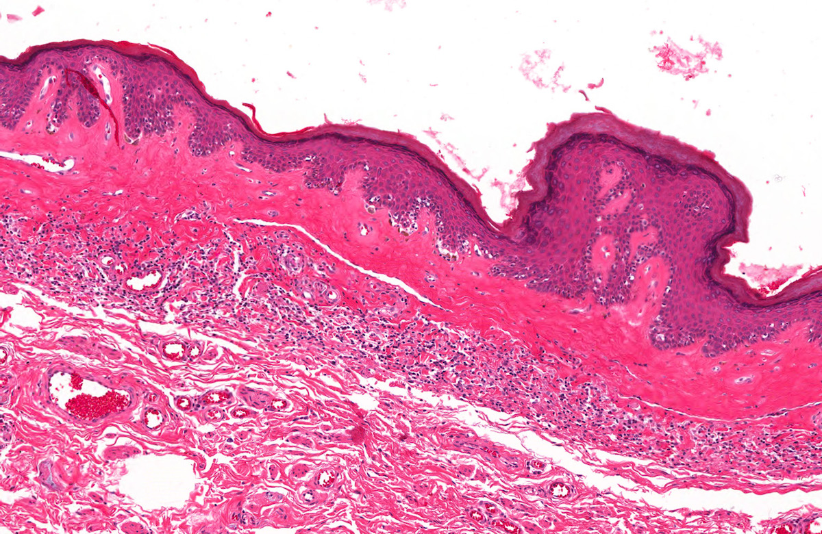
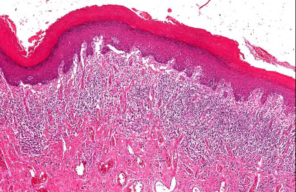
Verrucous epithelial hyperplasia
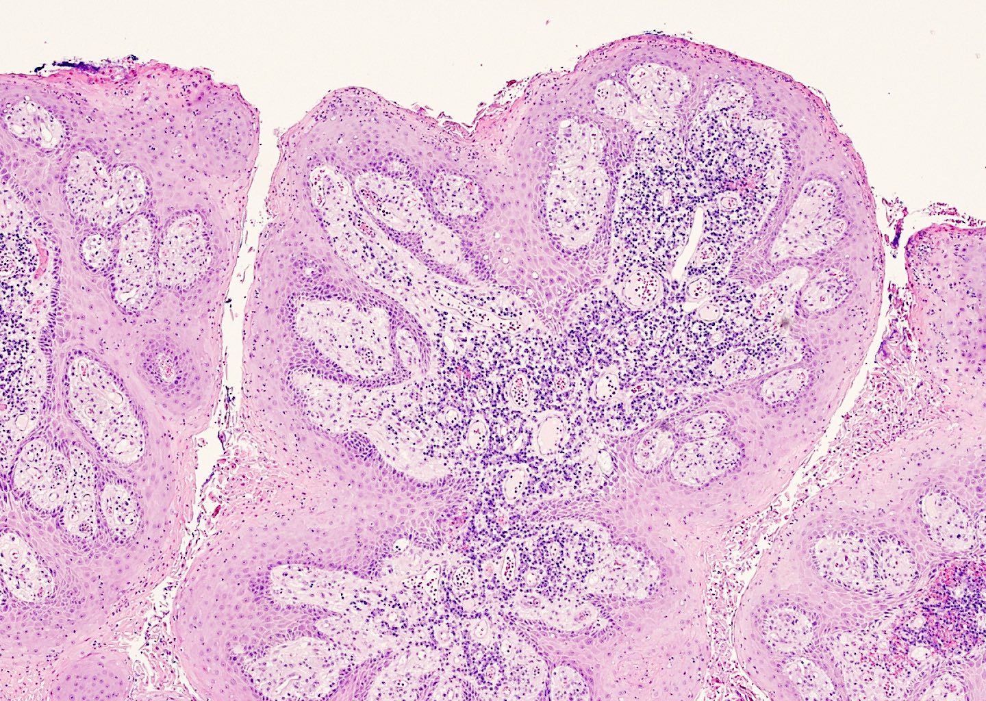
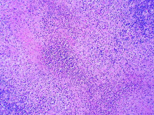
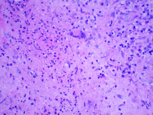
Band-like plasma cells
Neutrophilic infiltrates
Foamy histiocytes
Verruciform xanthoma
Amin: 2022

Cheng: 2019
Colecchia : 2016
Epstein: 2020
IARC: 2022

Wobker: 2021

Yang: 2020
Zhou: 2022
Find related Pathology books: GU/adrenal
