

Contributed by Kenneth A. Iczkowski, M.D.
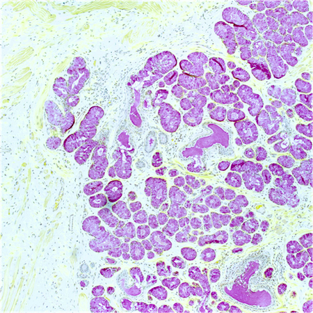
Atrophic-like dilated cancer acini
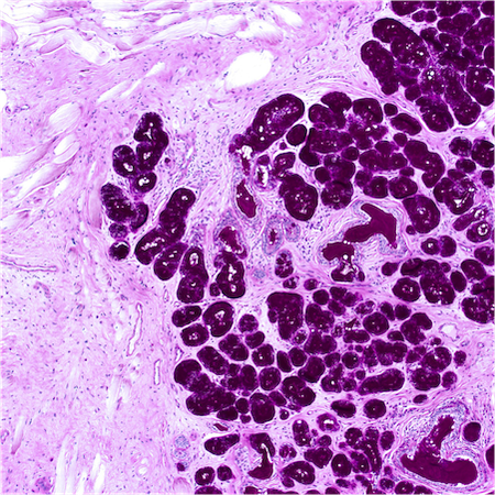
Basaloid appearance
Atrophic, mucinous cancer
Strong diffuse nuclear p63 expression
Few p63 positive acini
Cytoplasmic p63
Images hosted on other servers:

Abdominal
ultrasound showing
prostatic abscess

TRUS showing hypoechoic lesion

Pelvic CT showing an abscess in the prostate
Images hosted on other servers:

Transurethral unroofing of a prostatic abscess

Abscess drainage with transurethral resection
Contributed by Kenneth A. Iczkowski, M.D.
Prostatic microabscess
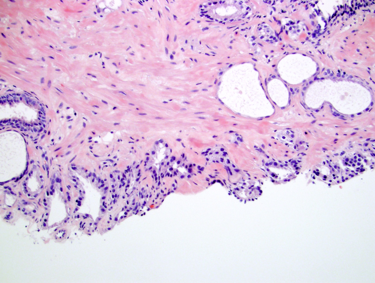
Contributed by Debra Zynger, M.D. and Kenneth A. Iczkowski, M.D.
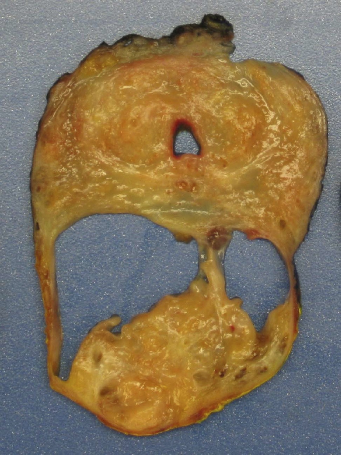
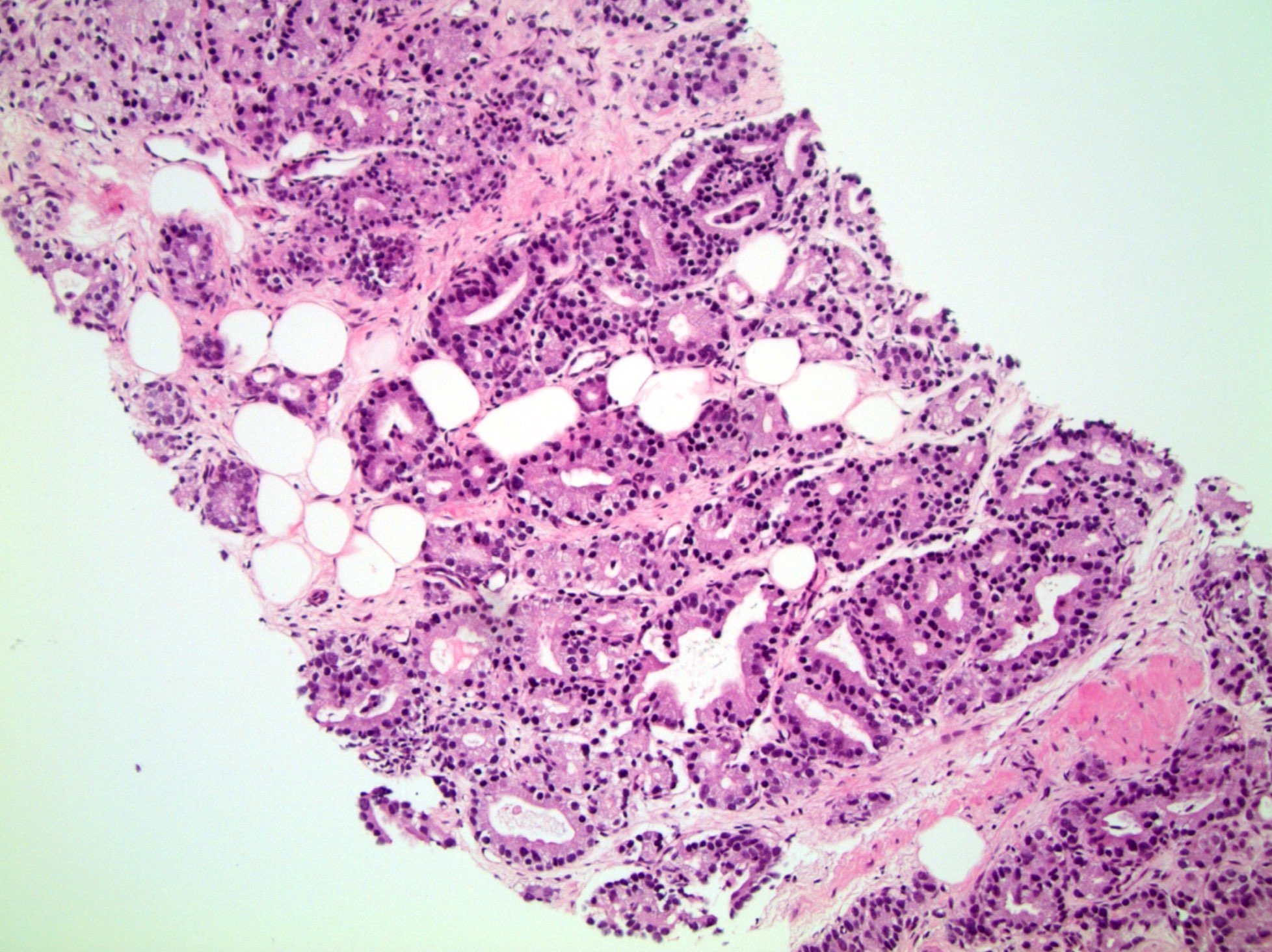
Prostatectomy specimen
Extraprostatic extension (pT3a)
Seminal vesicle invasion (pT3b)

Anterior horn of
peripheral zone
Contributed by Murali Varma, M.B.B.S.
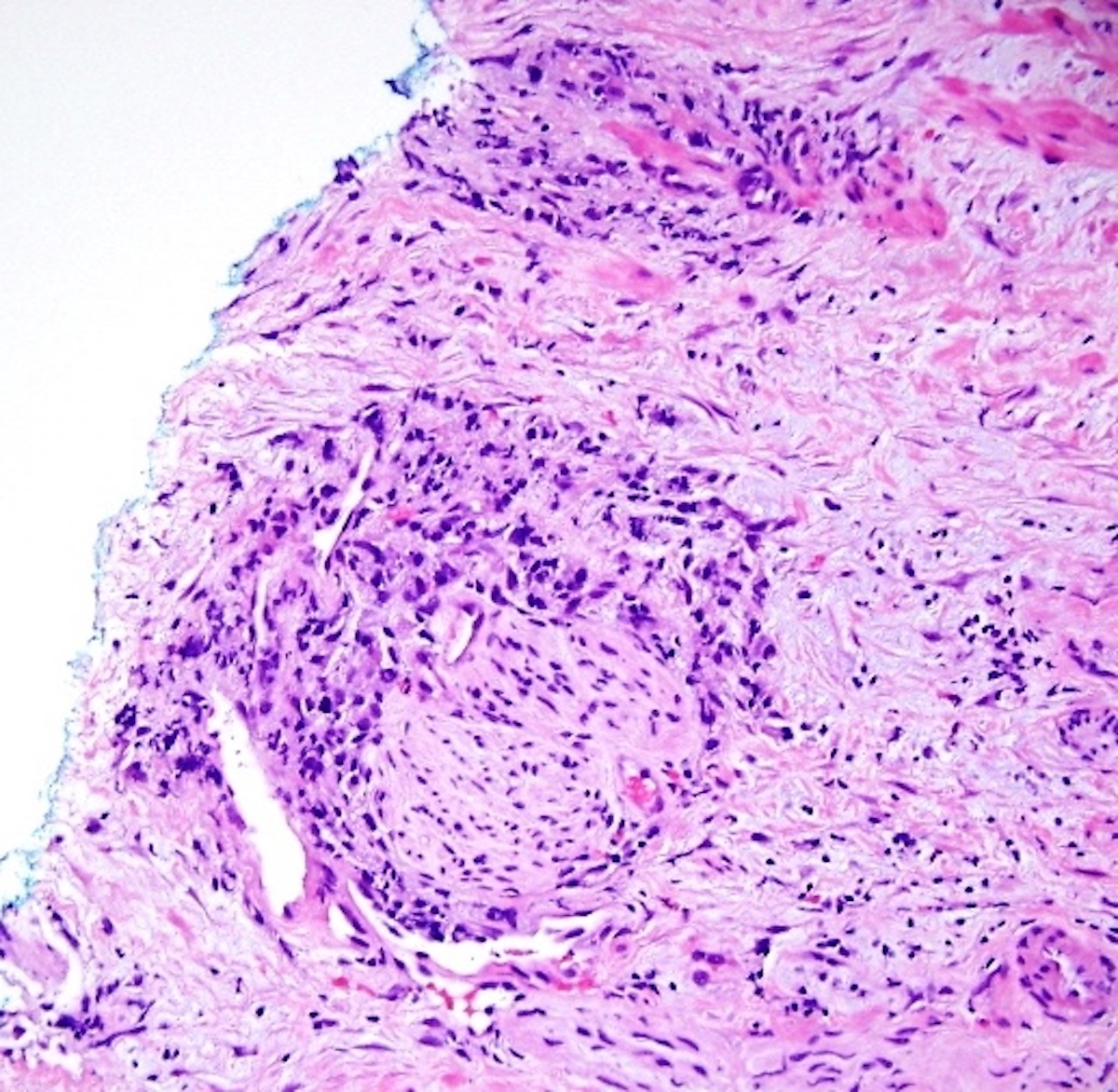
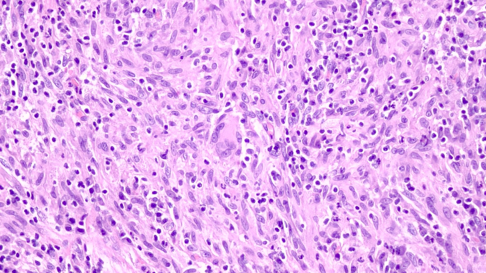
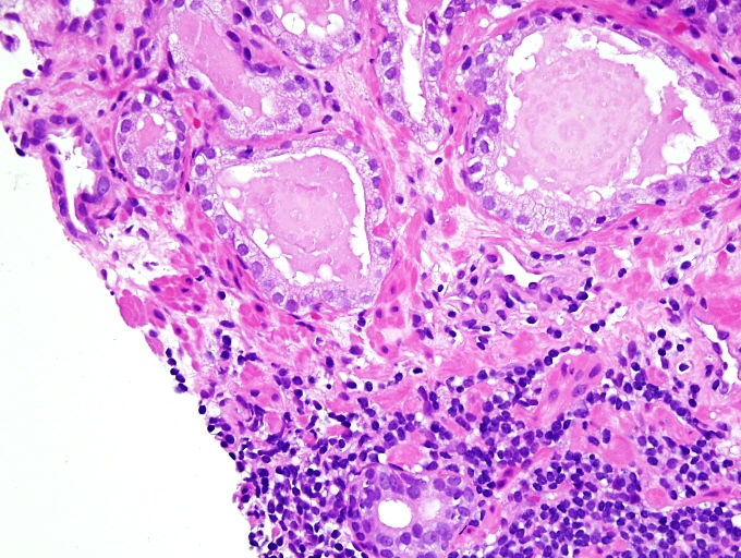
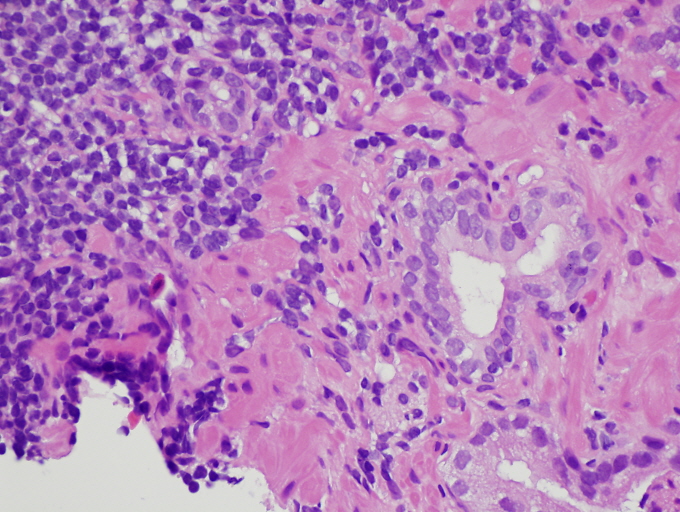
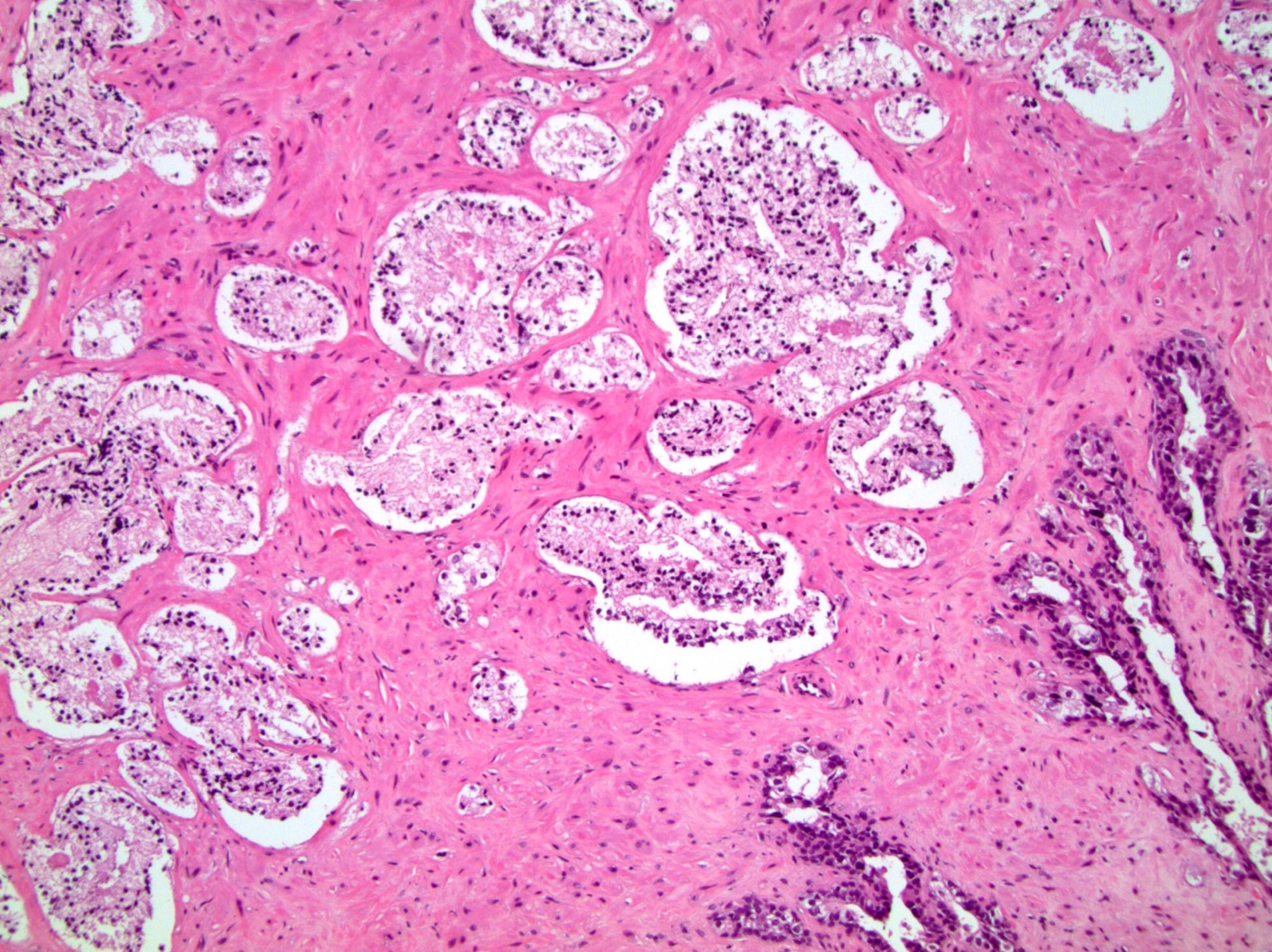
Infiltrative growth pattern
Glomerulations
Prominent nucleoli
Perineural invasion
Atrophic variant of prostate cancer
Atrophic variant of prostate cancer
Pseudohyperplastic variant of prostate cancer
Foamy gland variant of prostate cancer
Crystals in gland lumens
Pink amorphous material within gland lumens
Amphophilic cytoplasm
Mitosis
Collagenous micronodules
Images hosted on other servers:

Low grade epithelial stromal tumor
Images hosted on other servers:


Metastatic hepatocellular carcinoma


Low grade epithelial stromal tumor
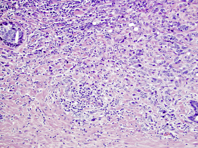
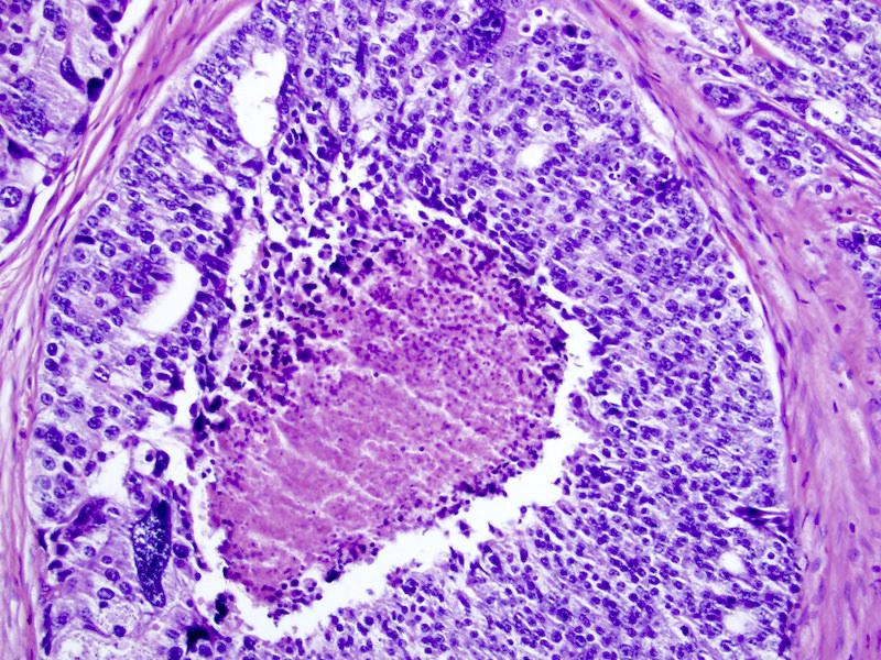
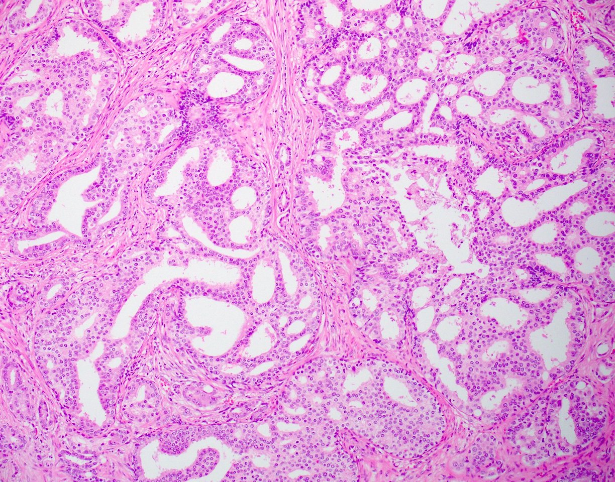
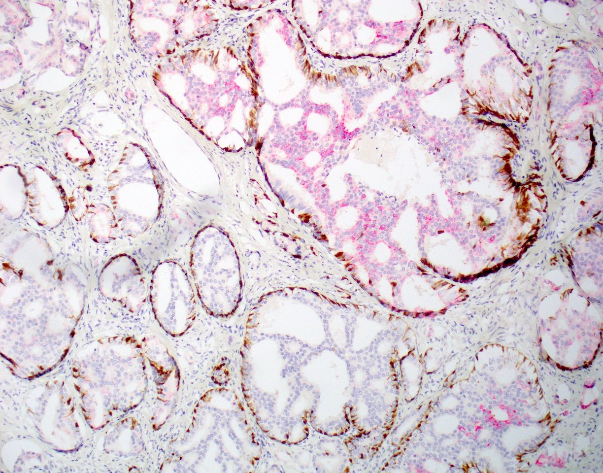
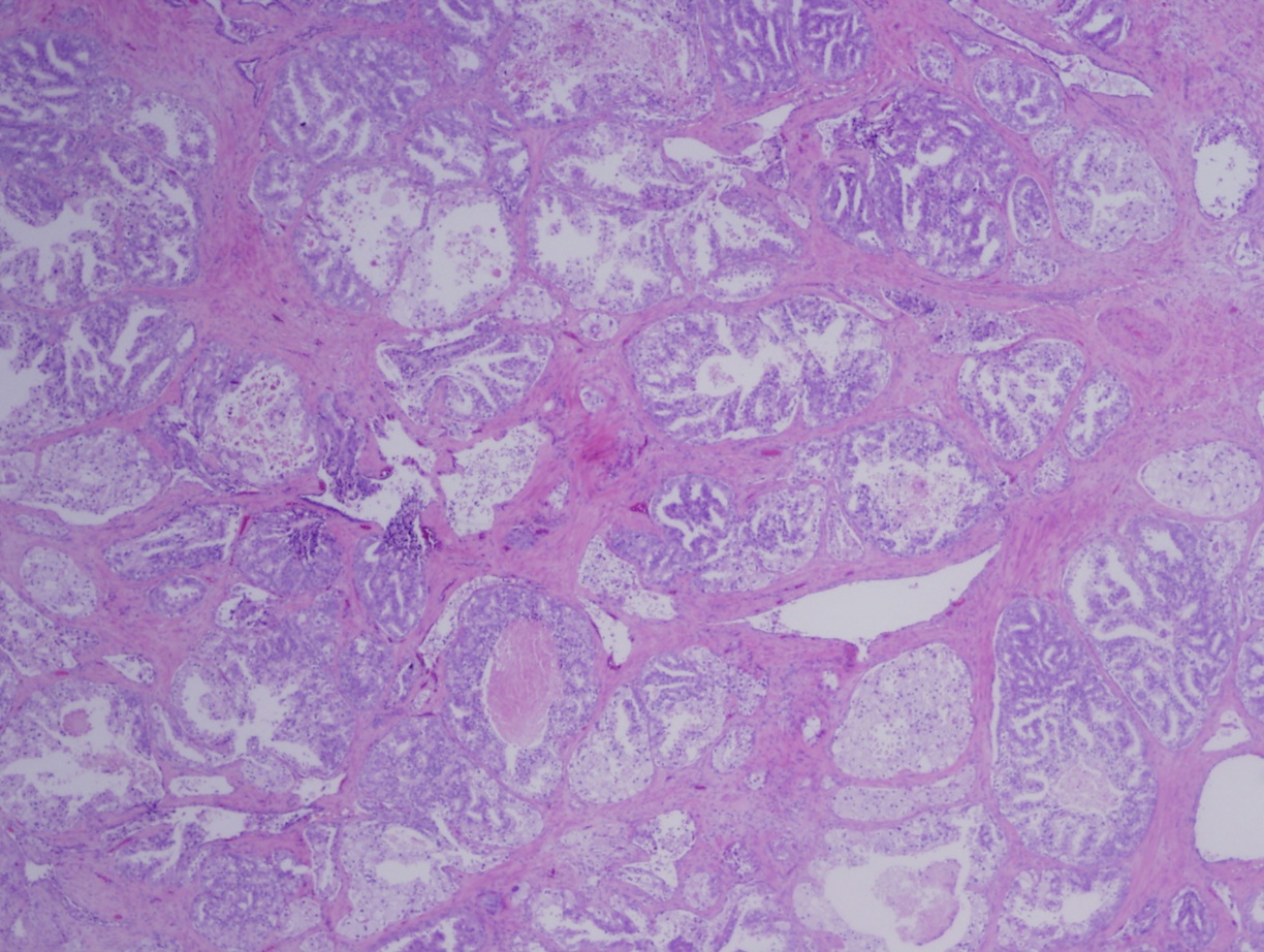
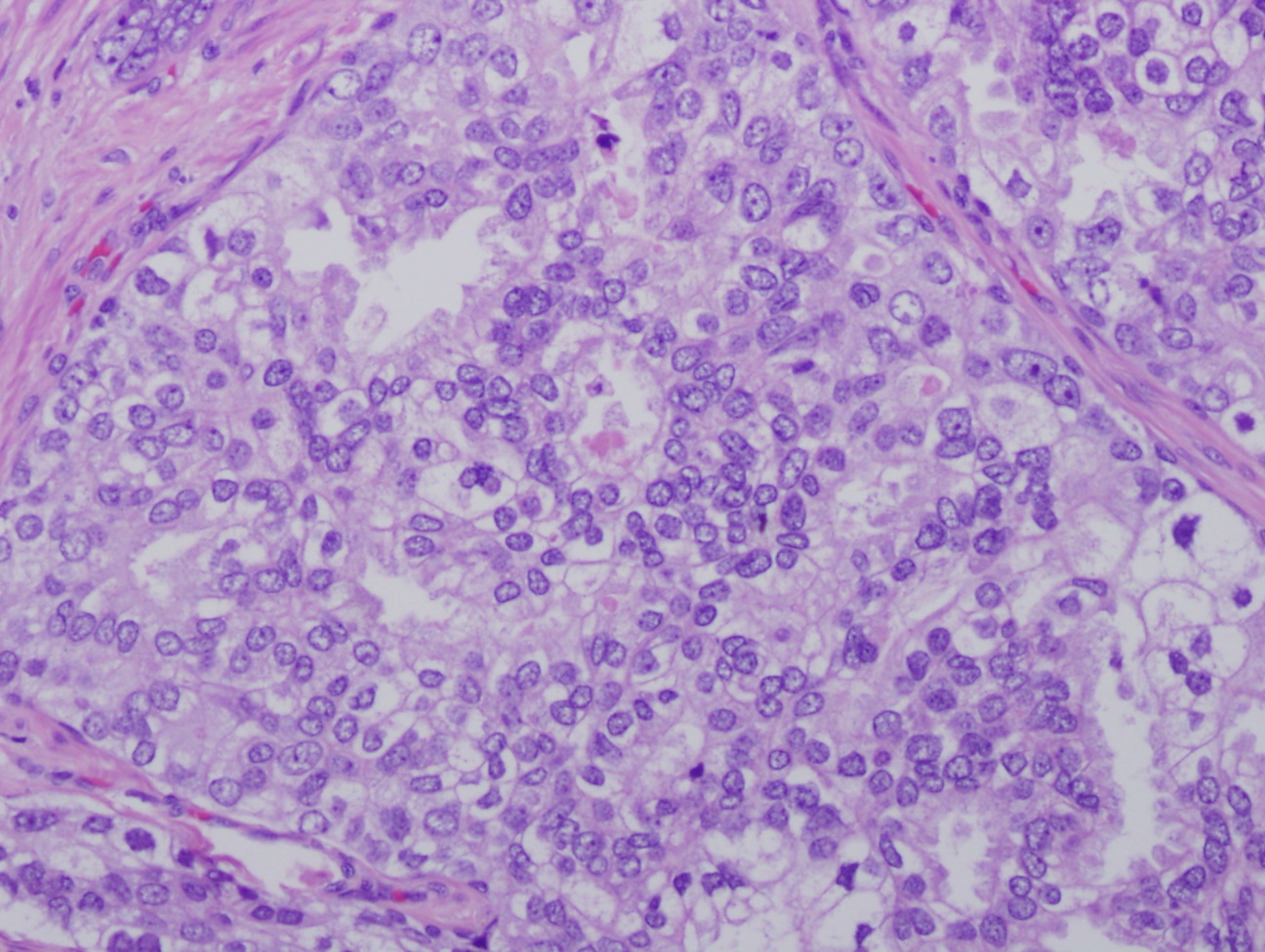
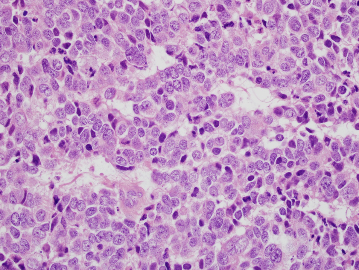
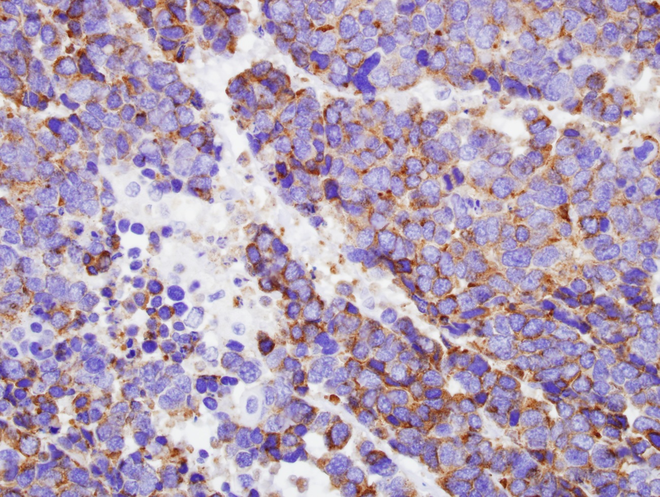
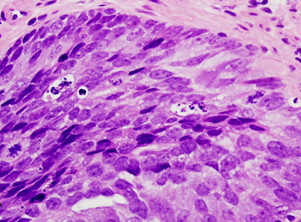
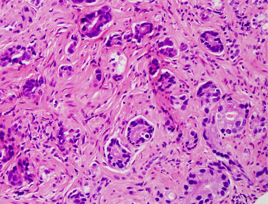
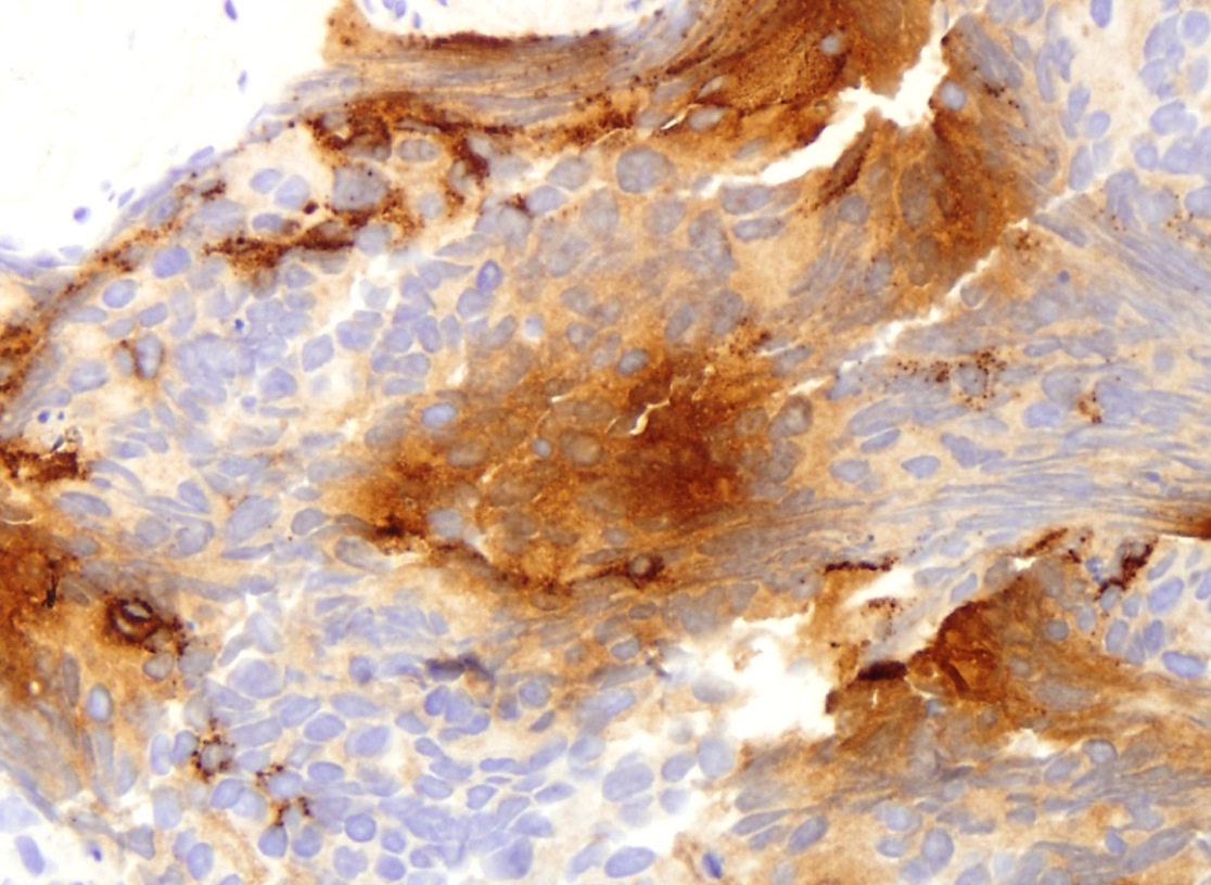
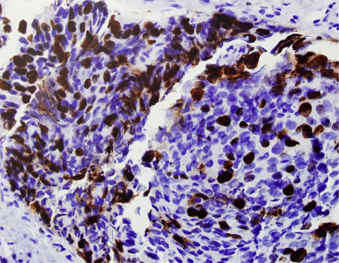
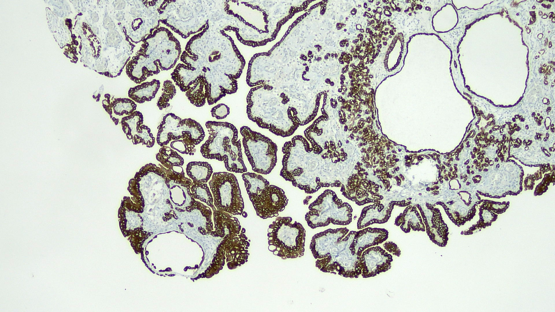
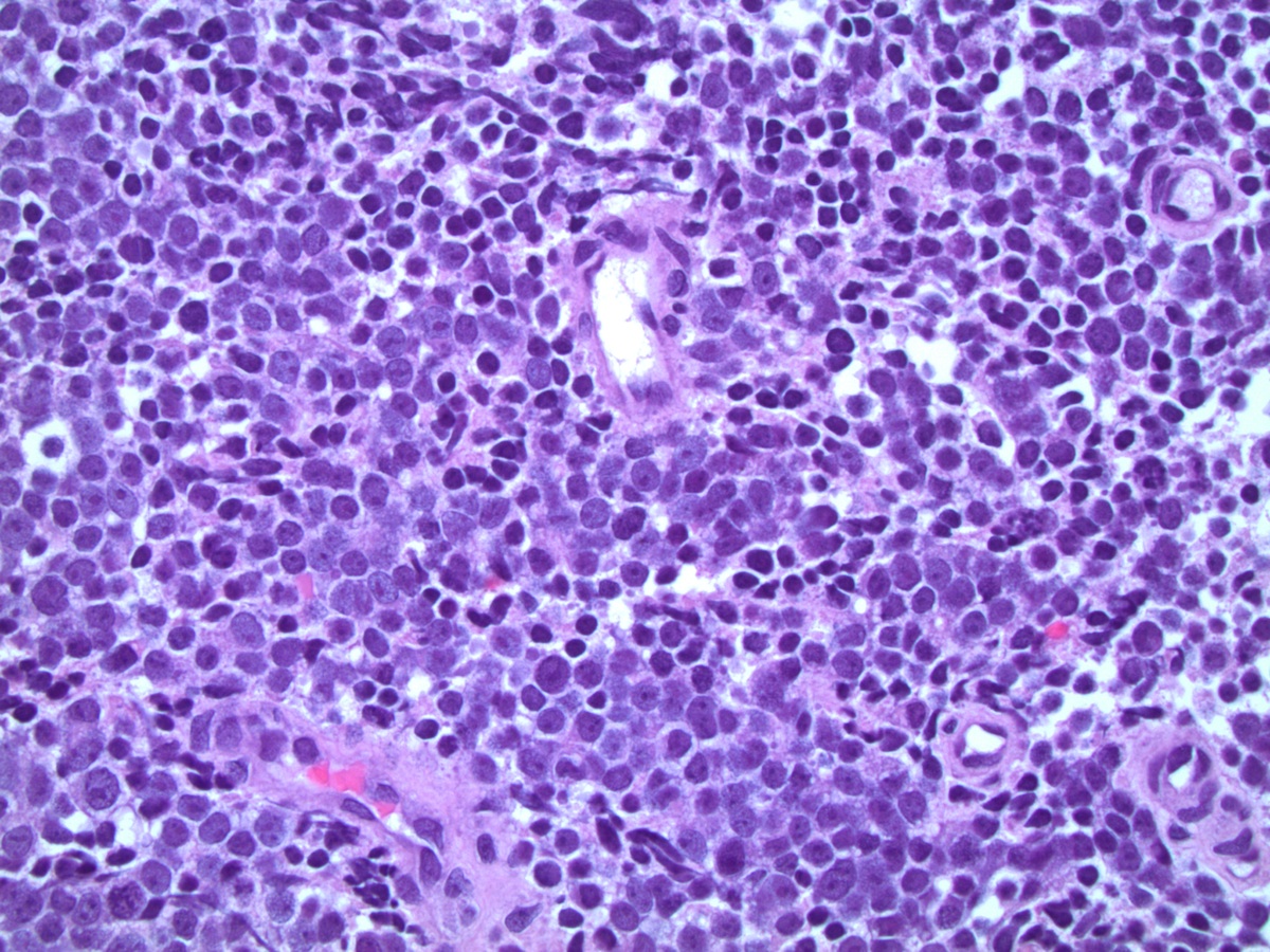
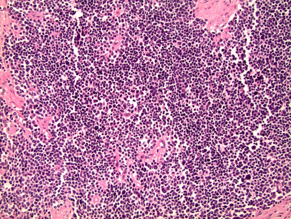
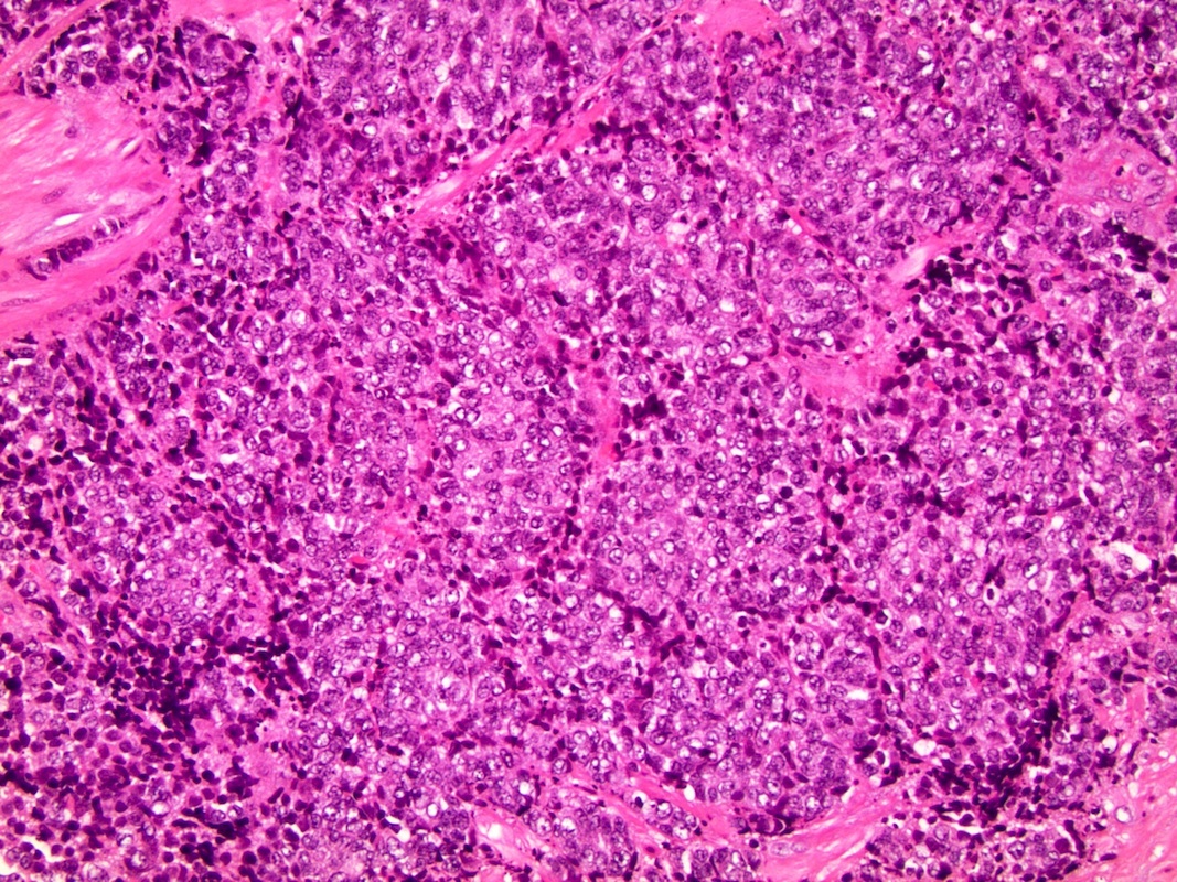
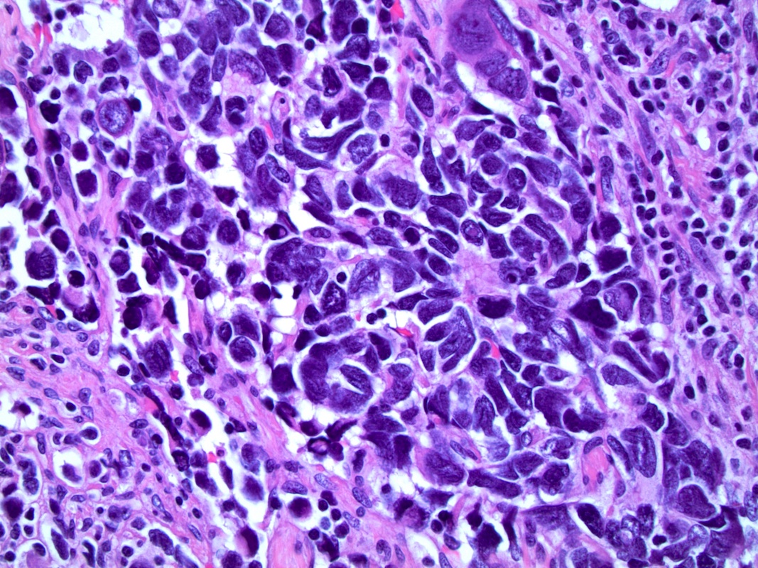
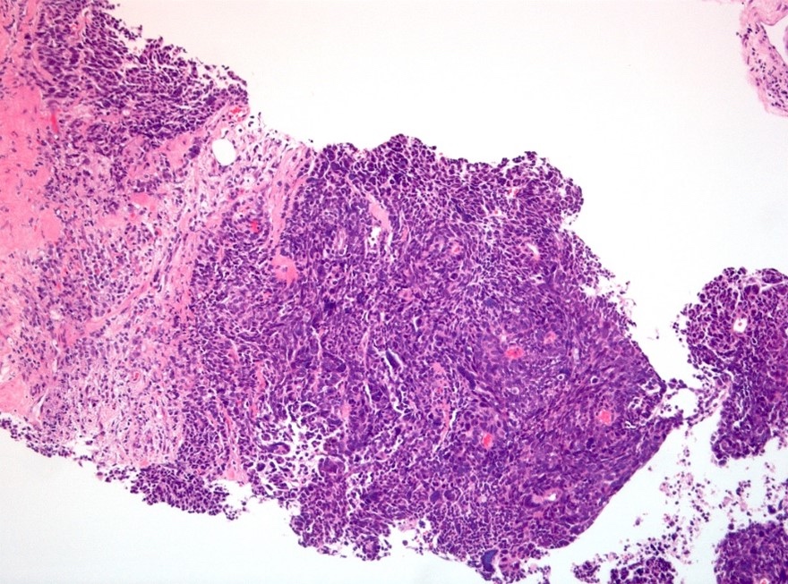
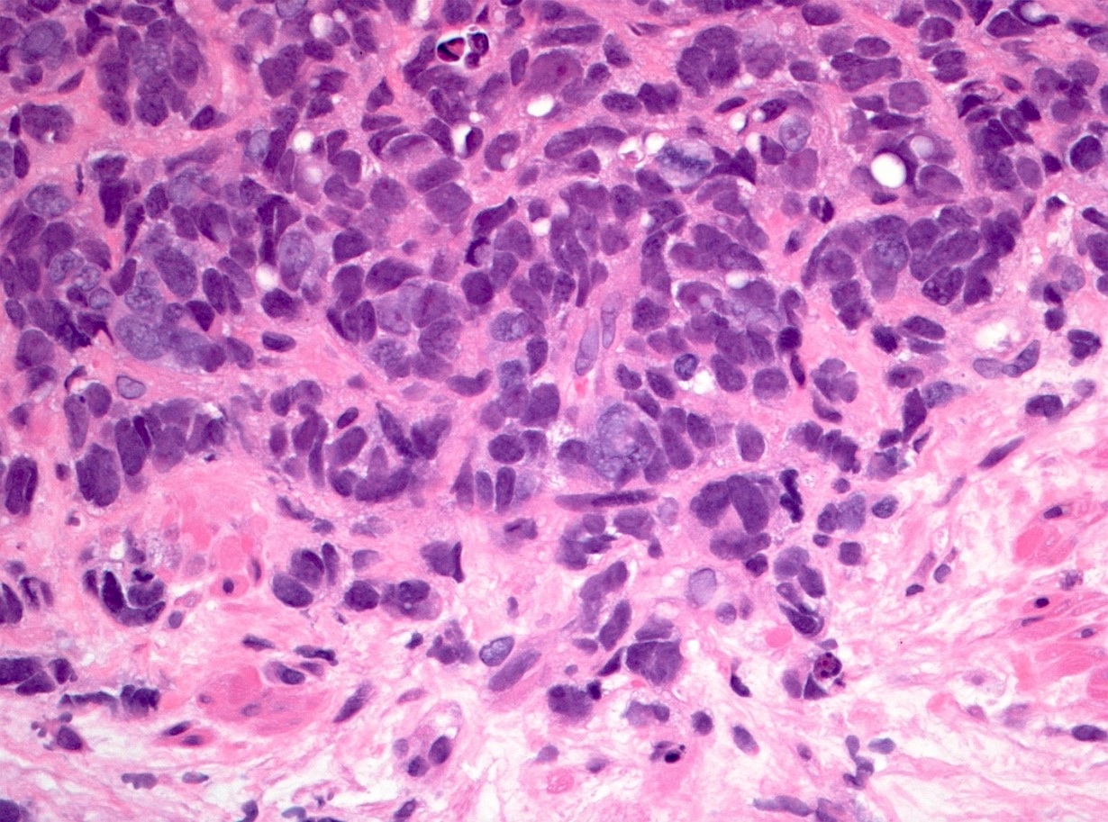
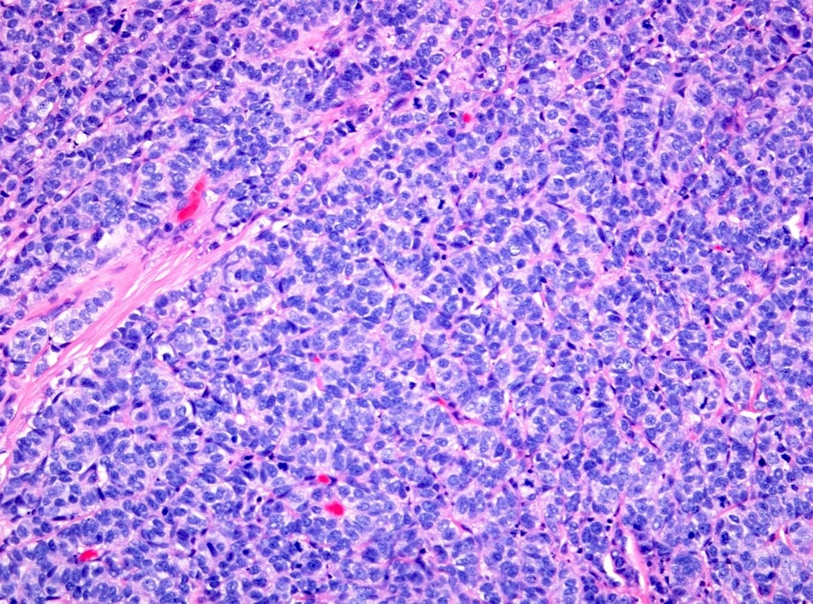
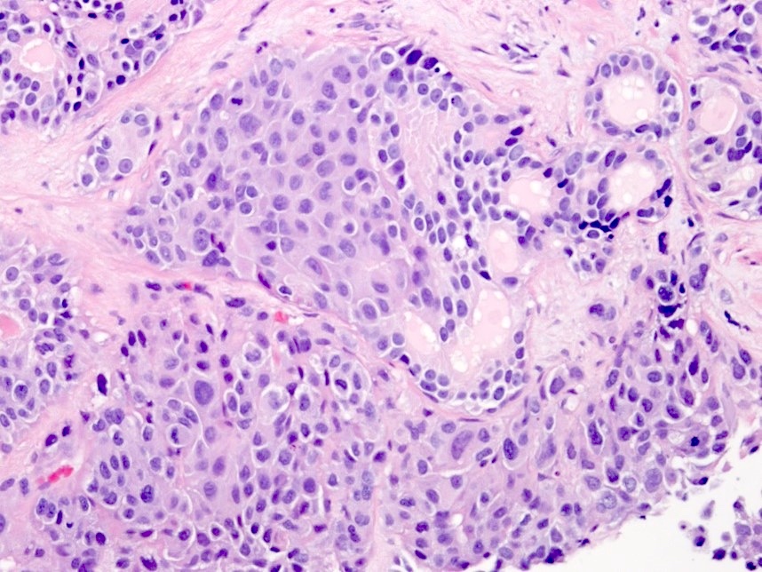
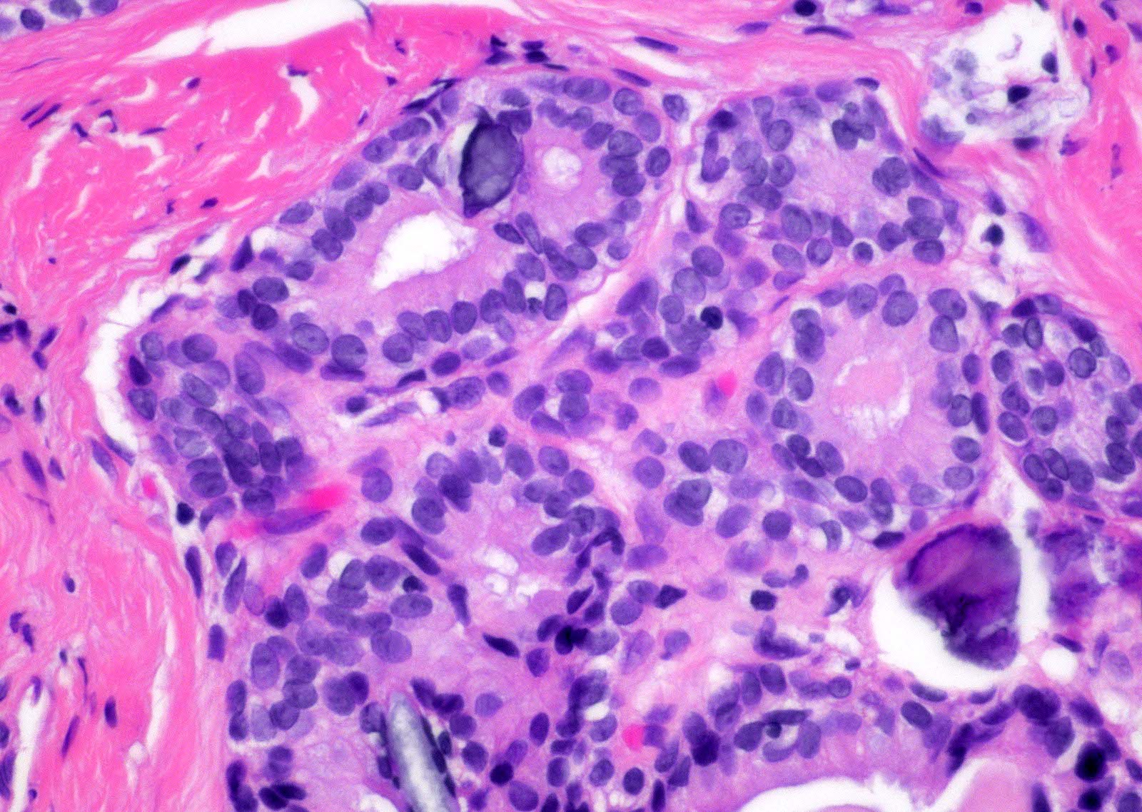
Contributed by Daniel Athanazio, M.D., Ph.D. and Maiara Souza, M.D.
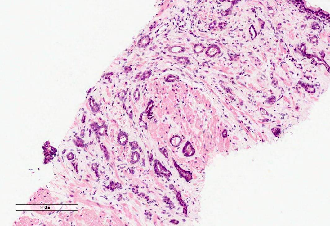
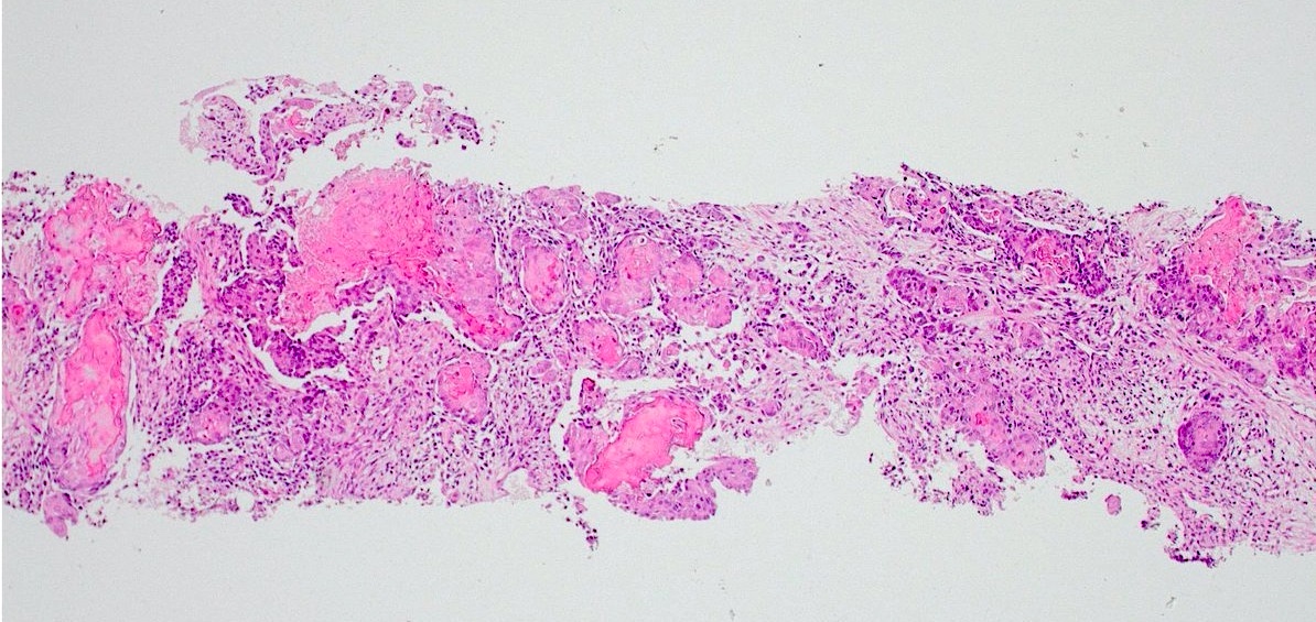
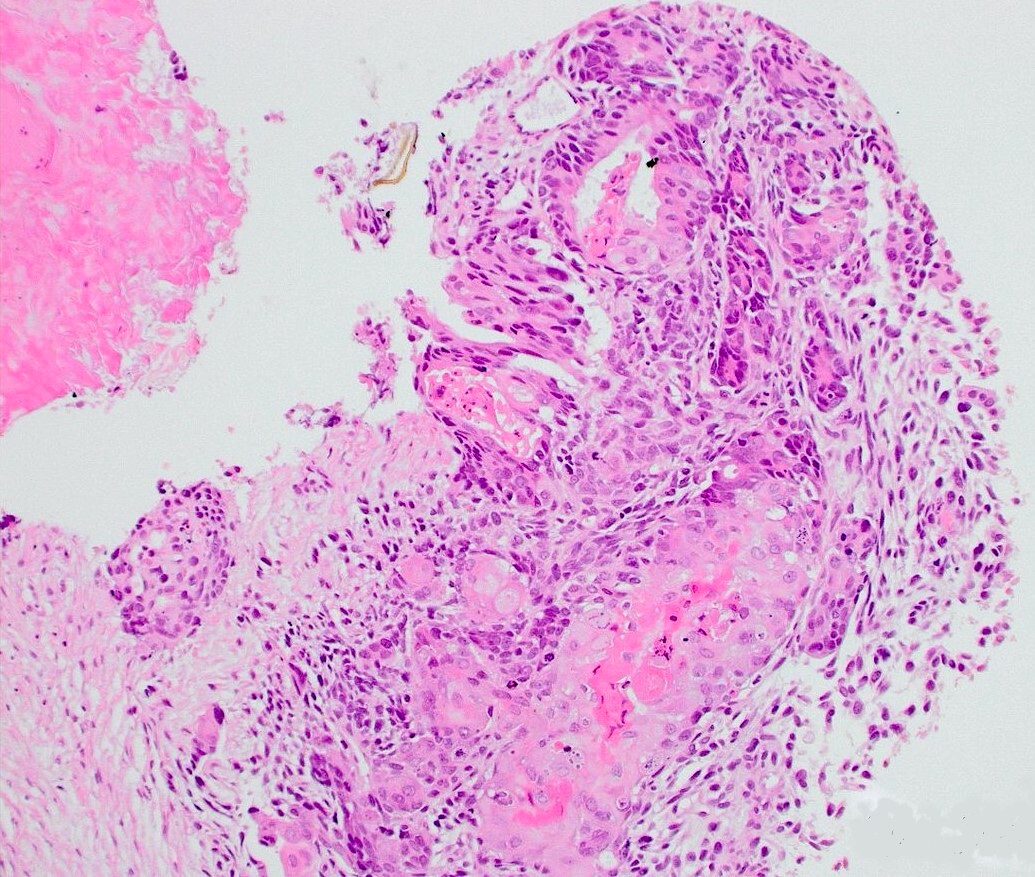
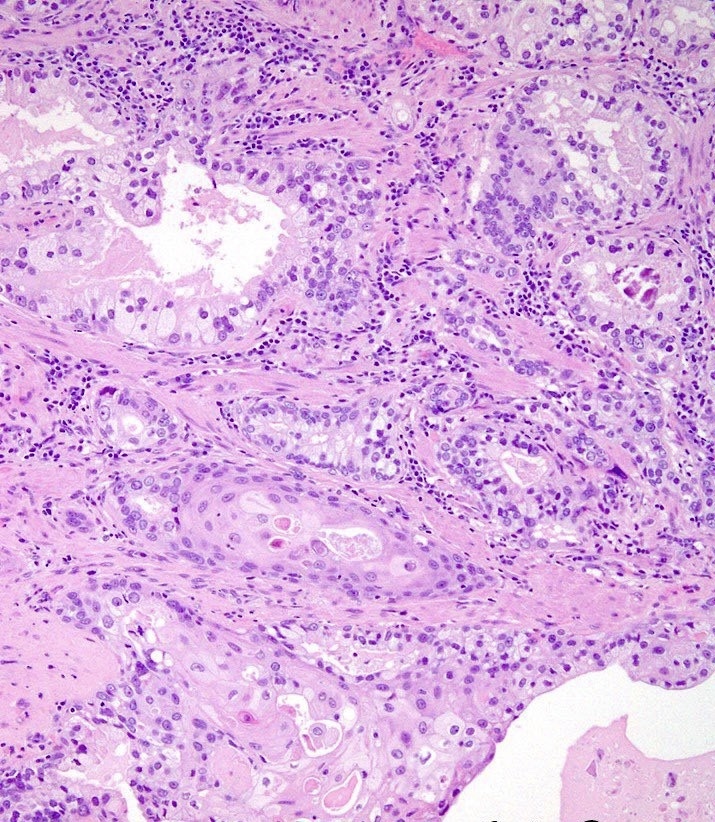
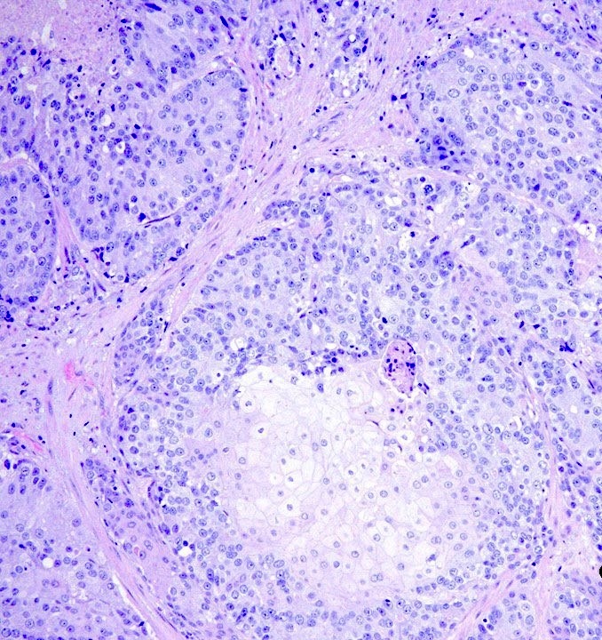
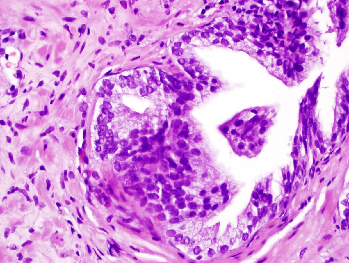
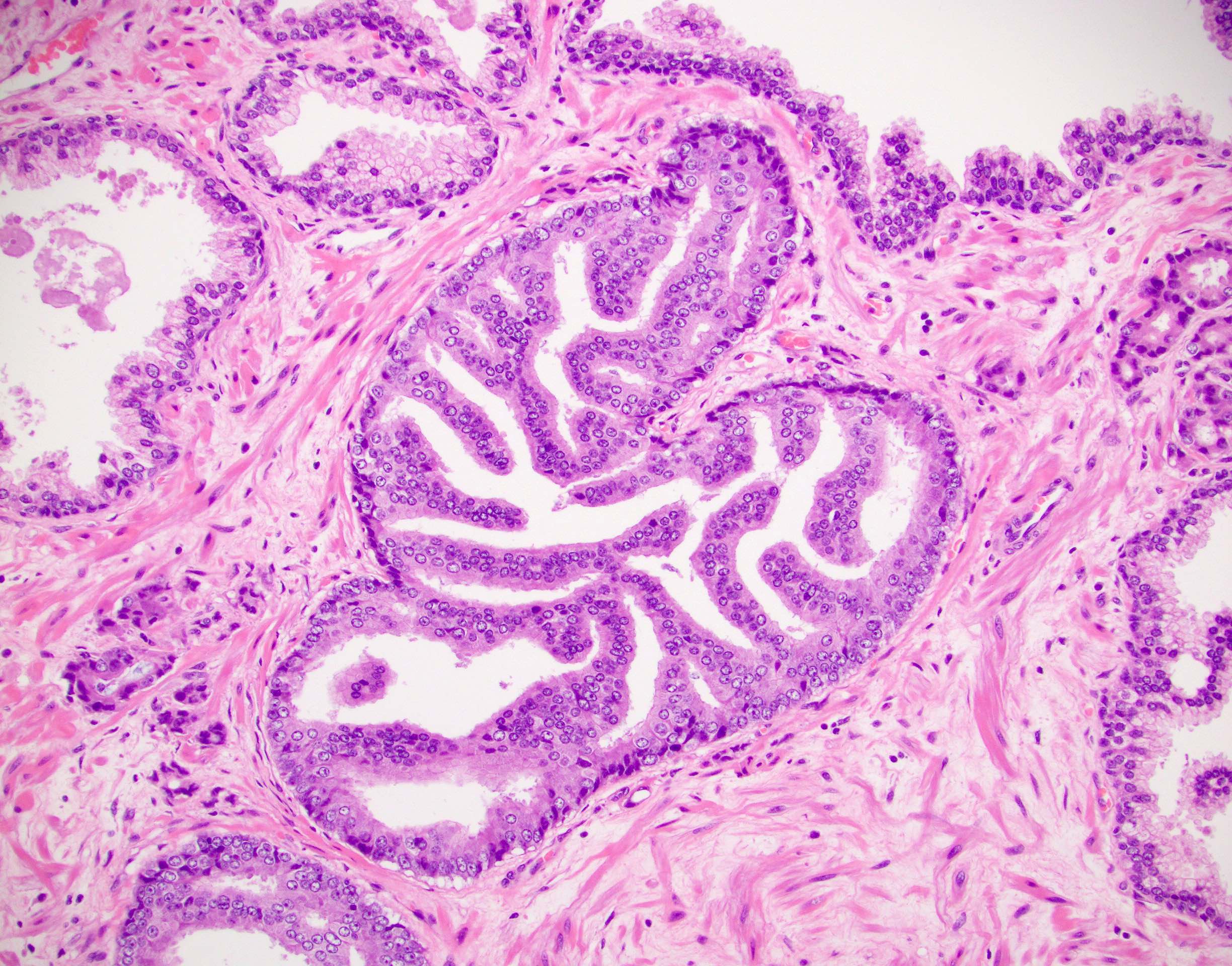
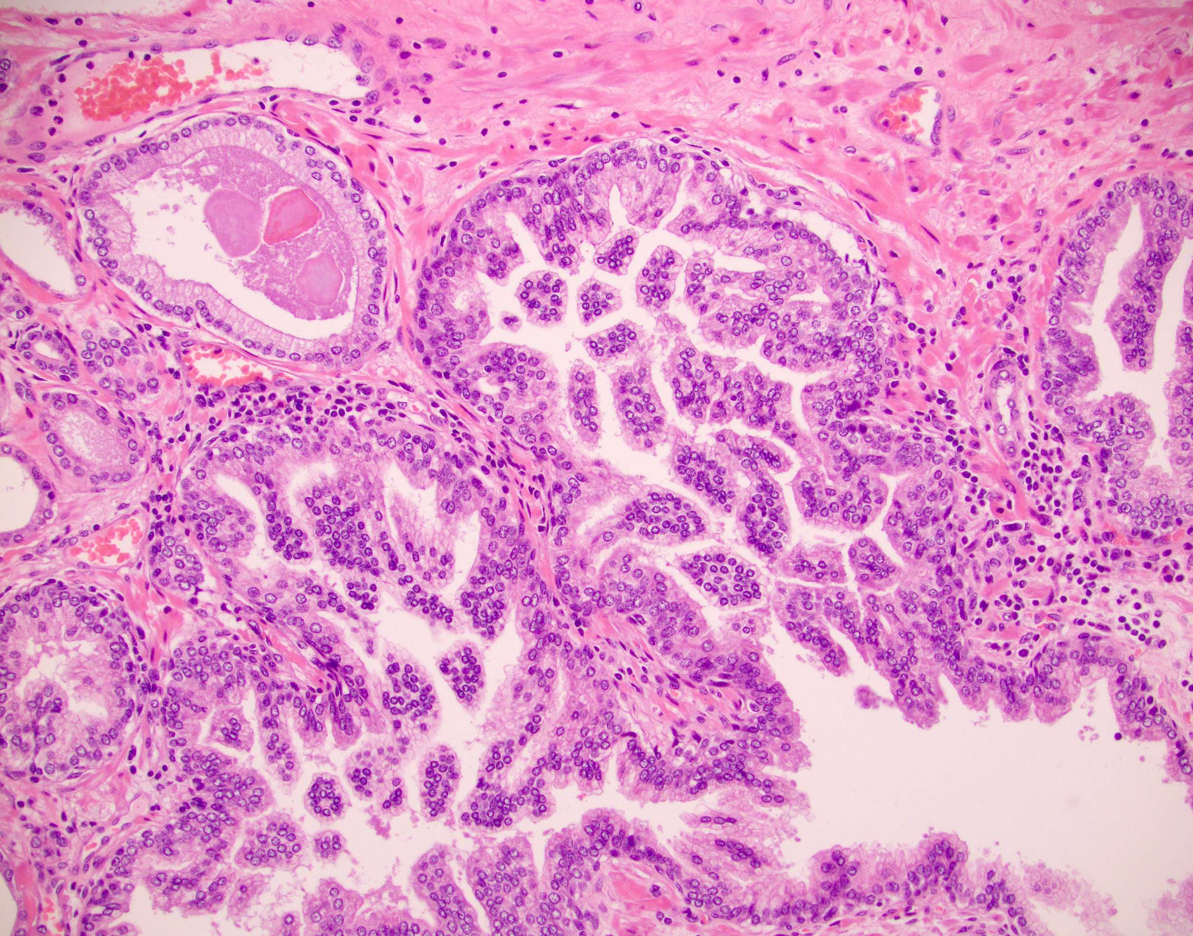
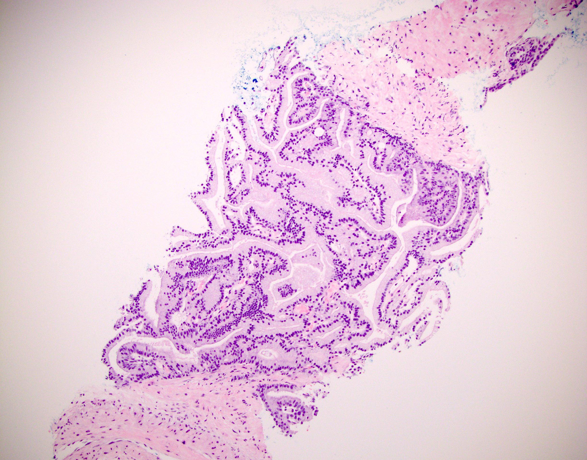
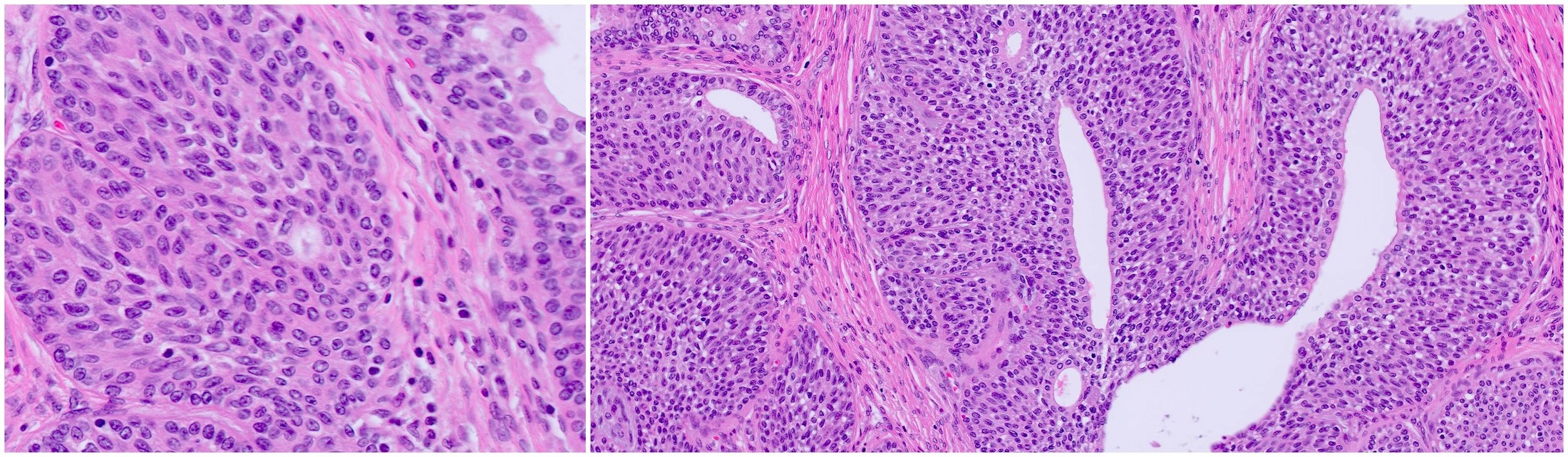
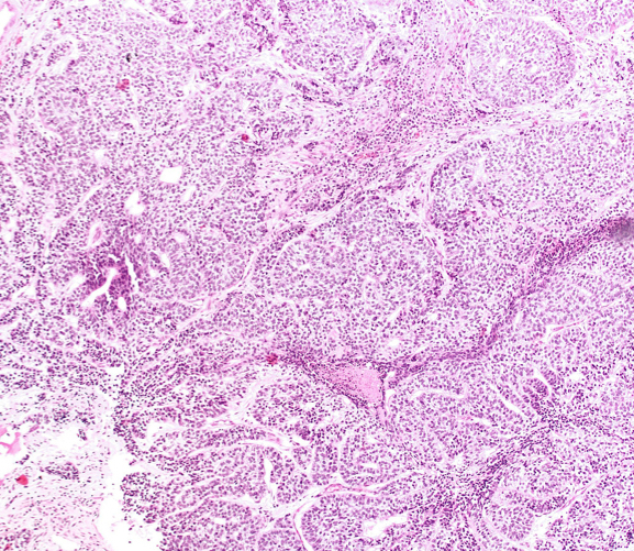
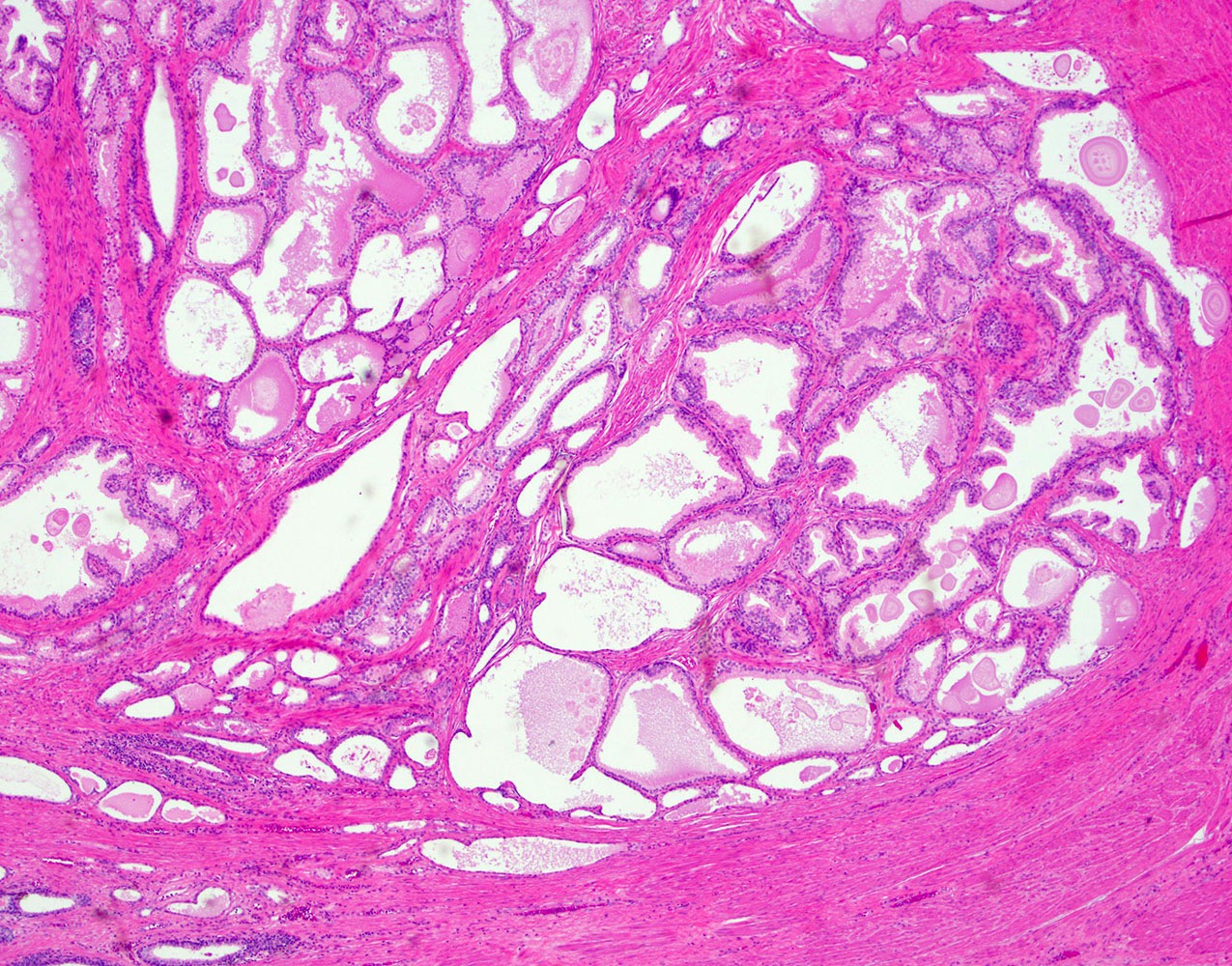
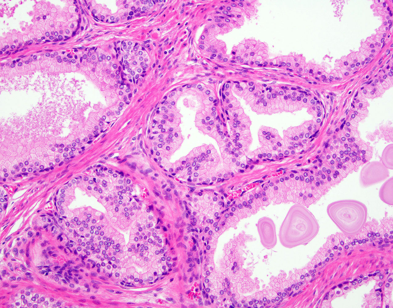
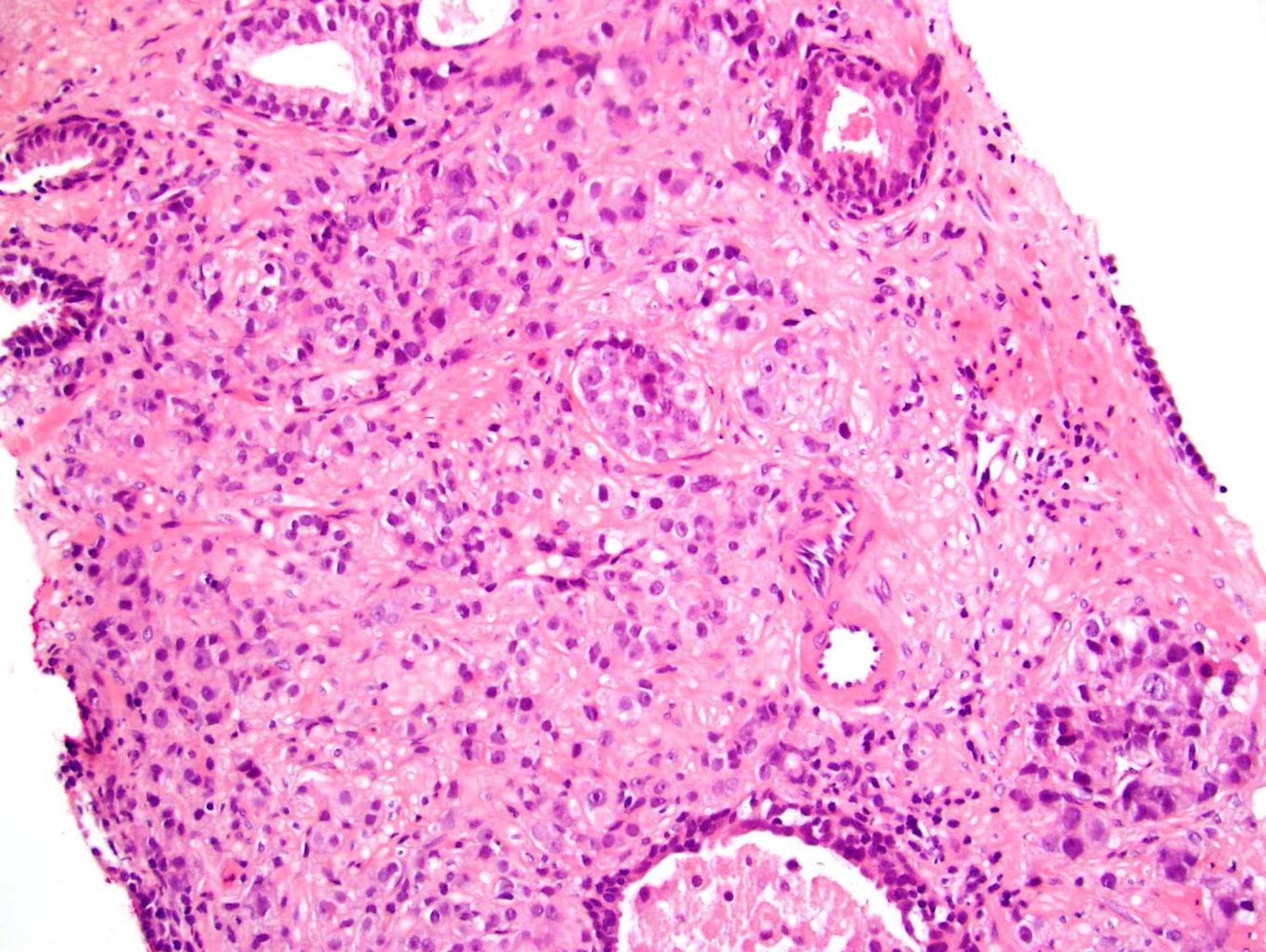
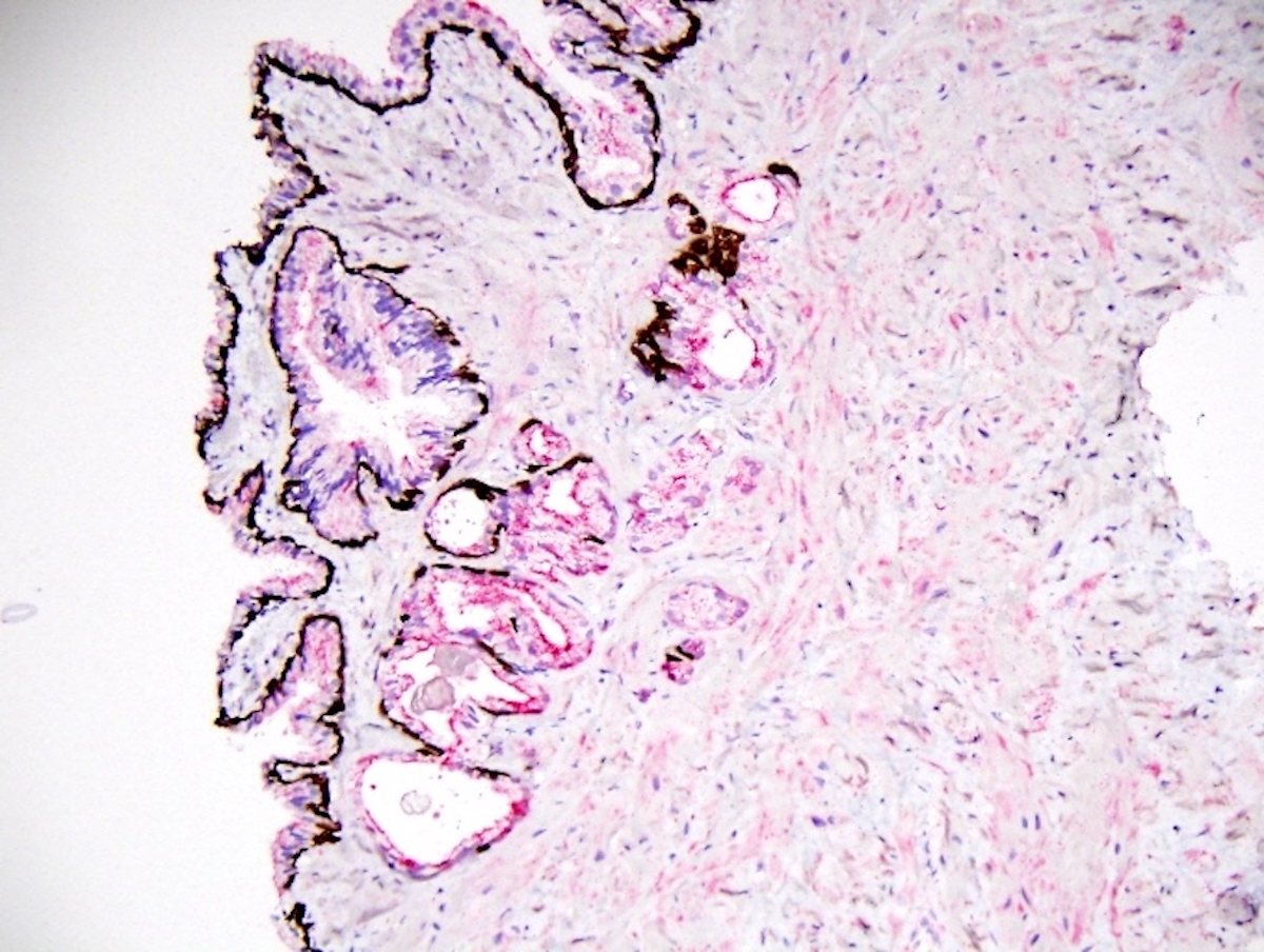
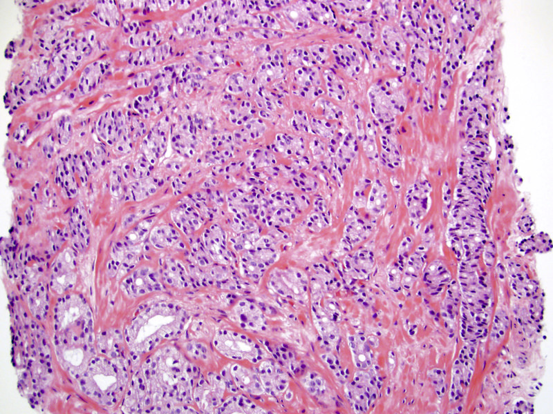
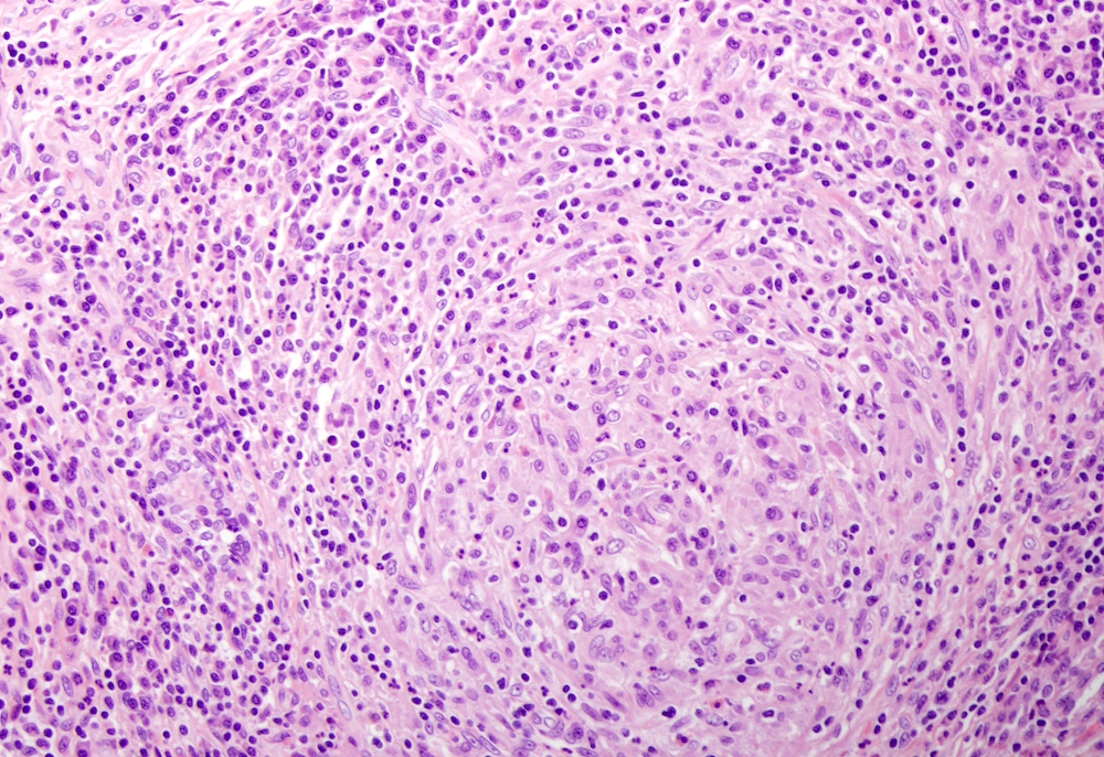
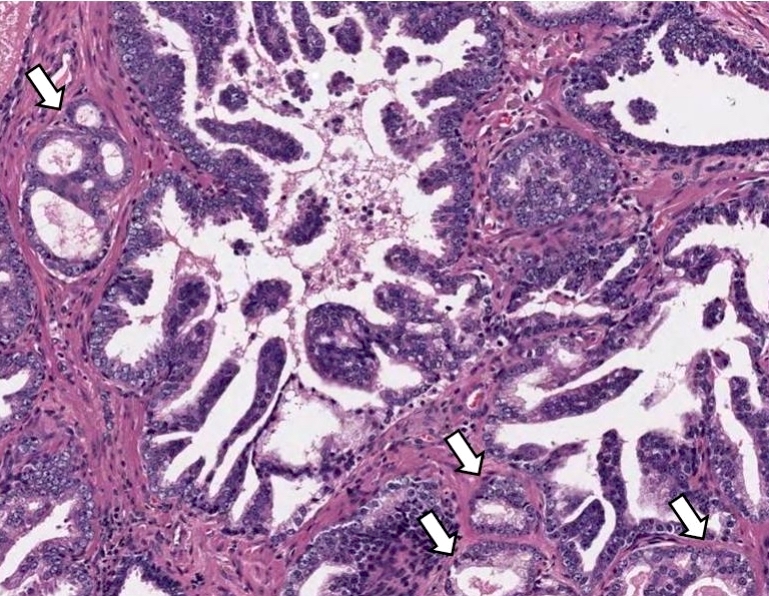
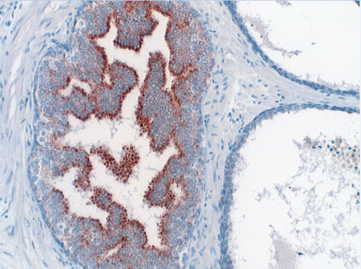
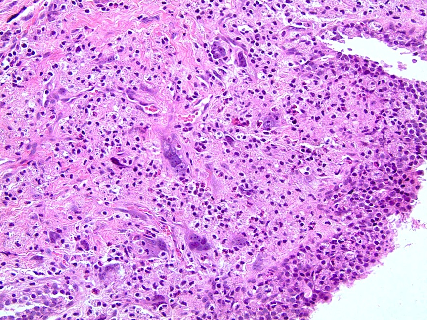
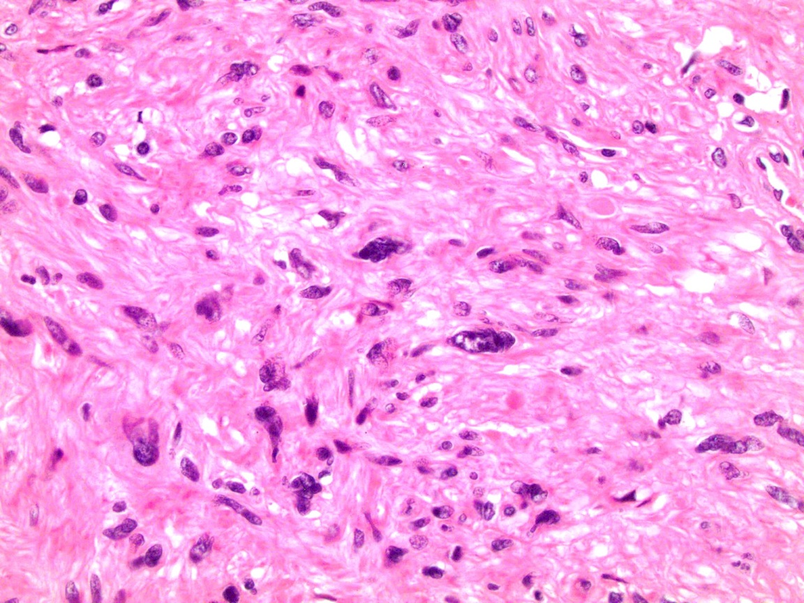
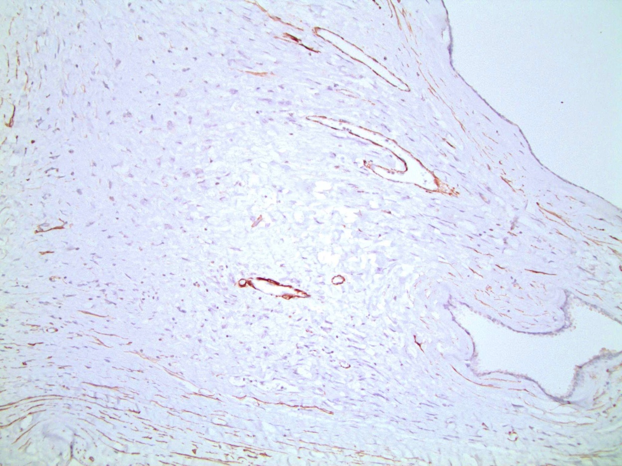
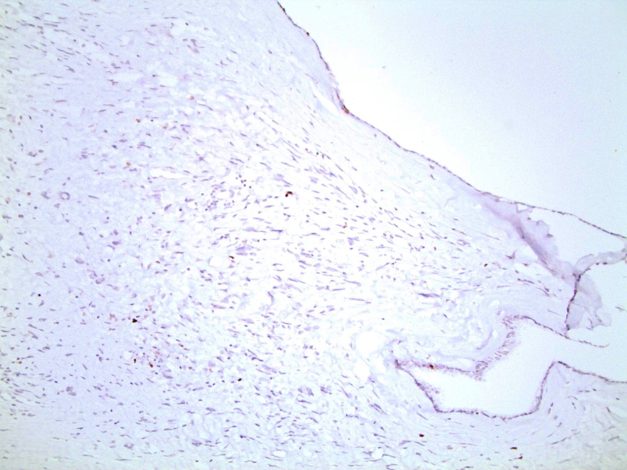
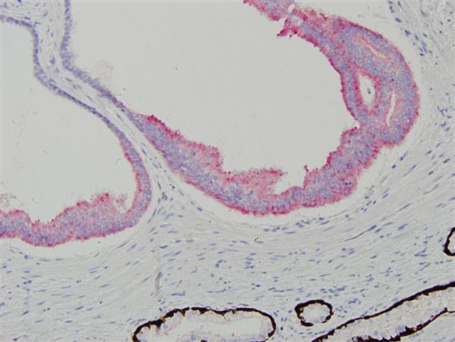
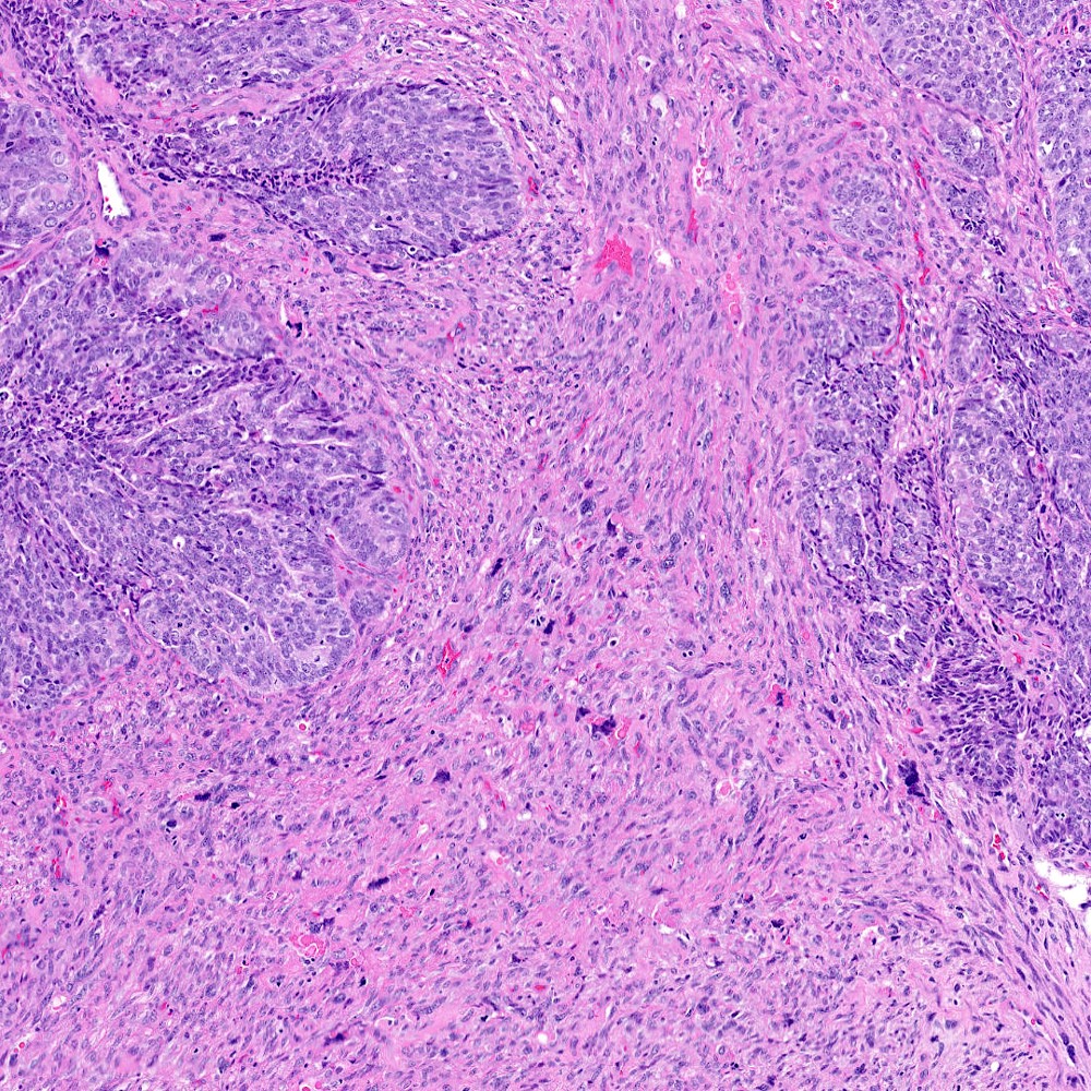
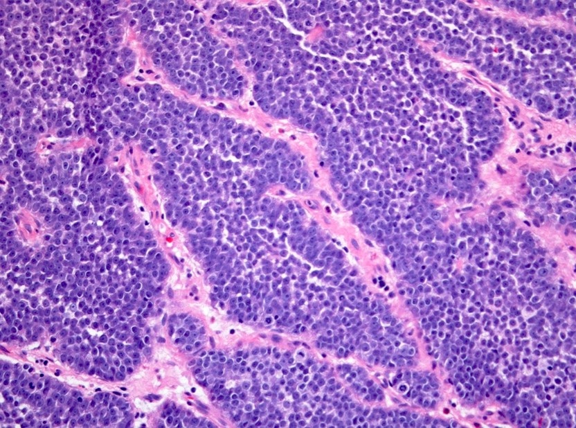
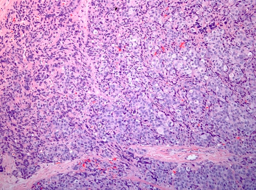
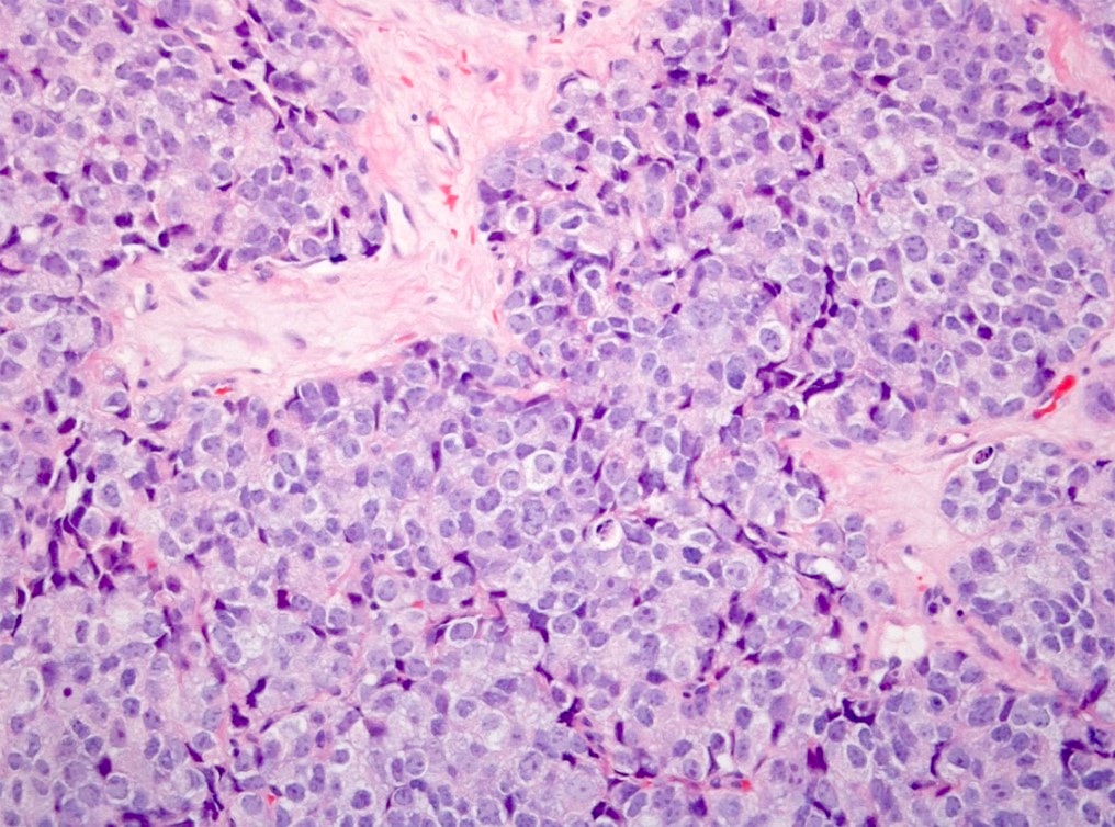
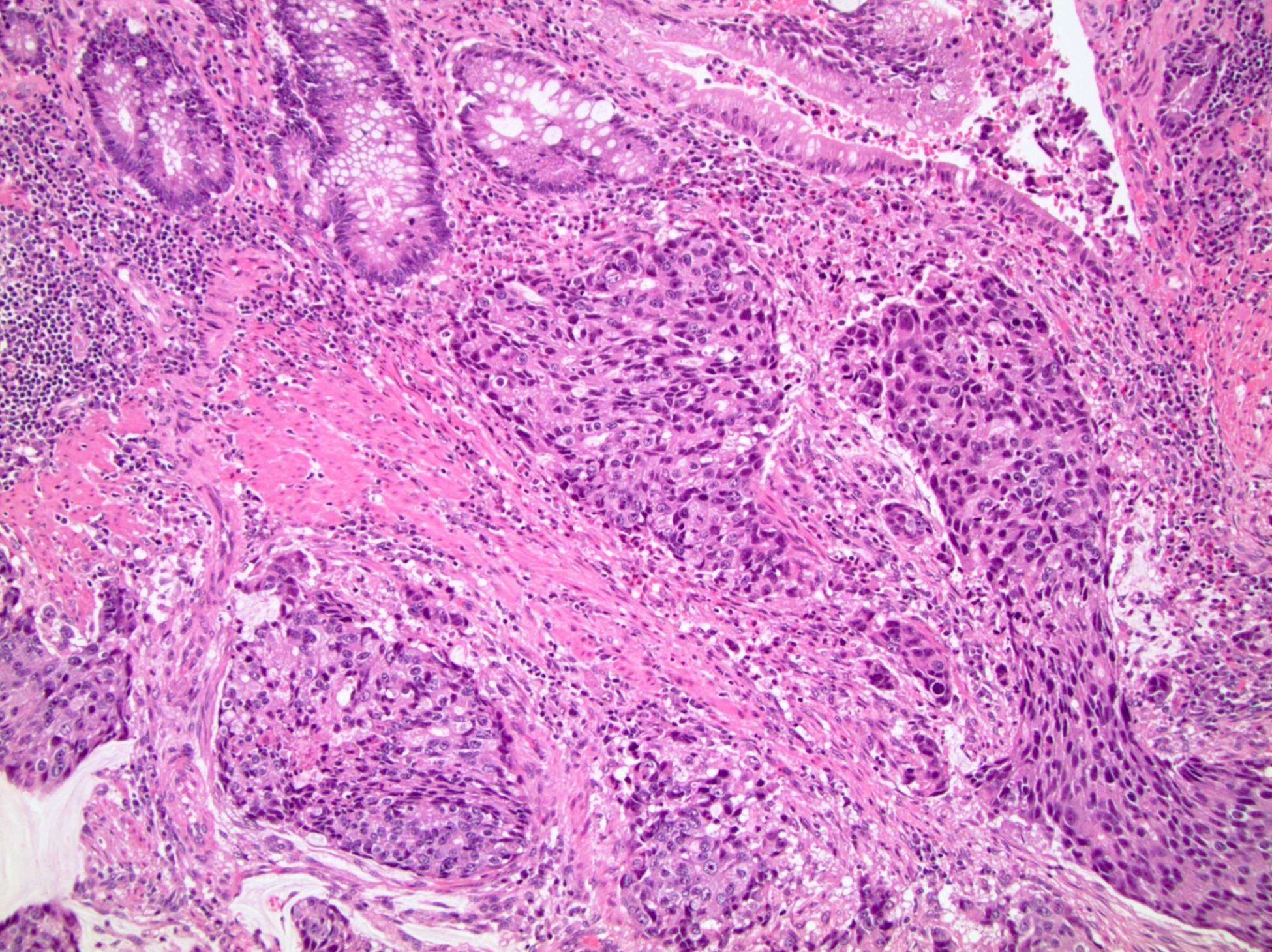
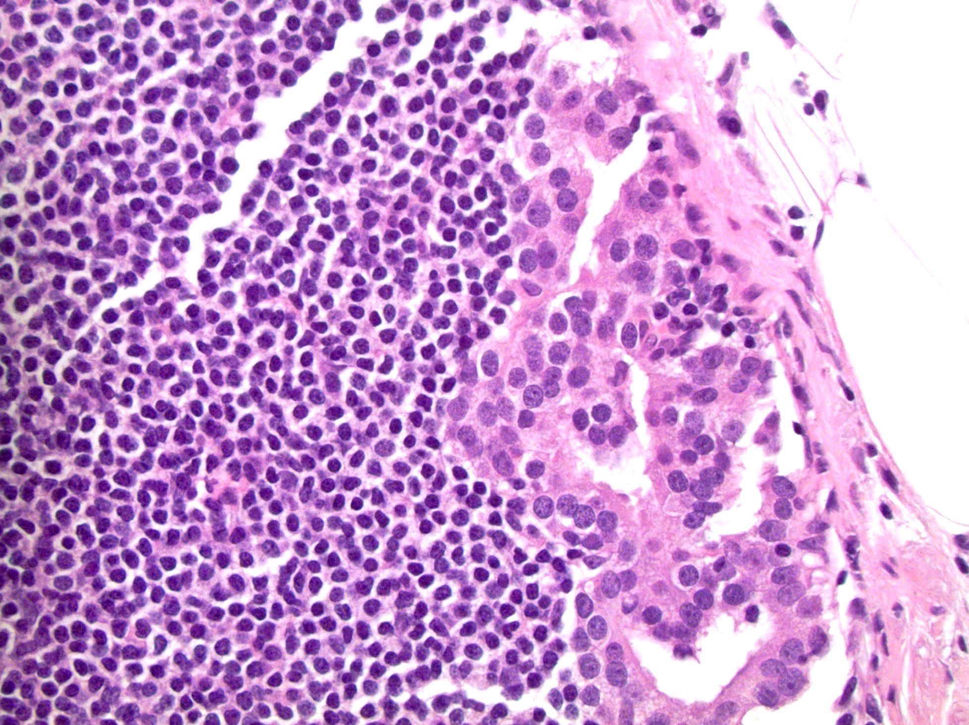
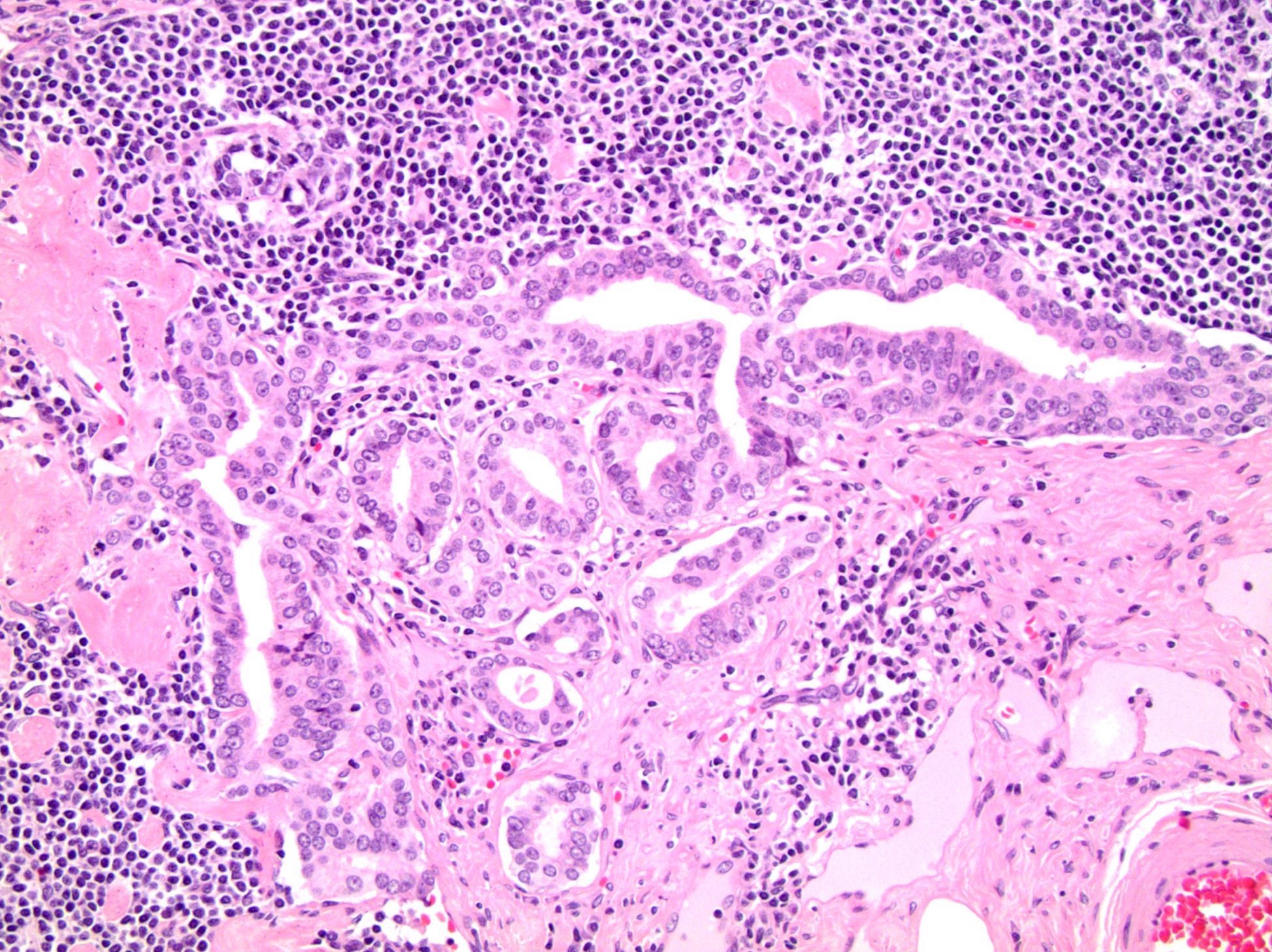
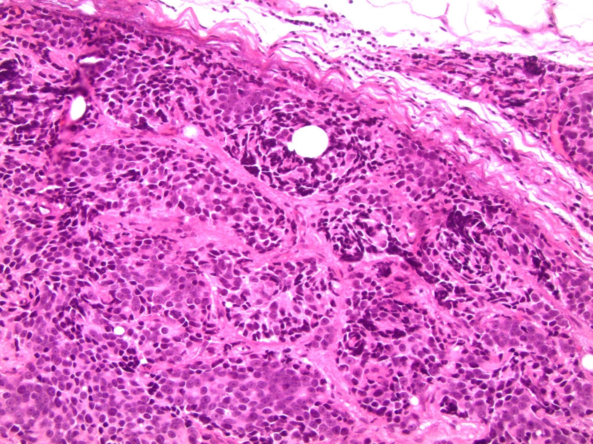
Solid growth - basaloid morphology
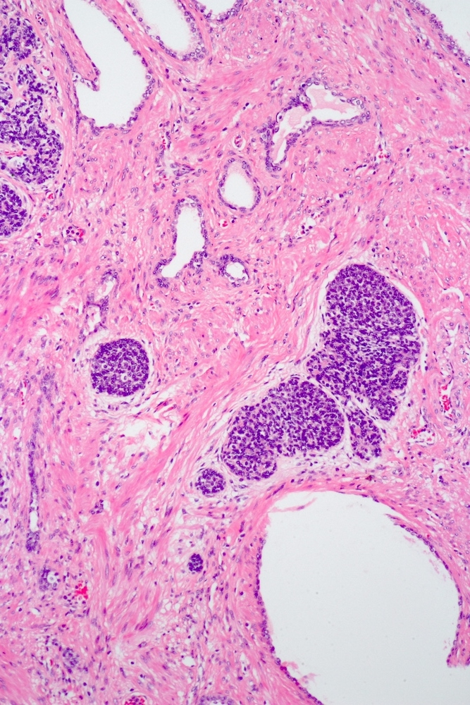
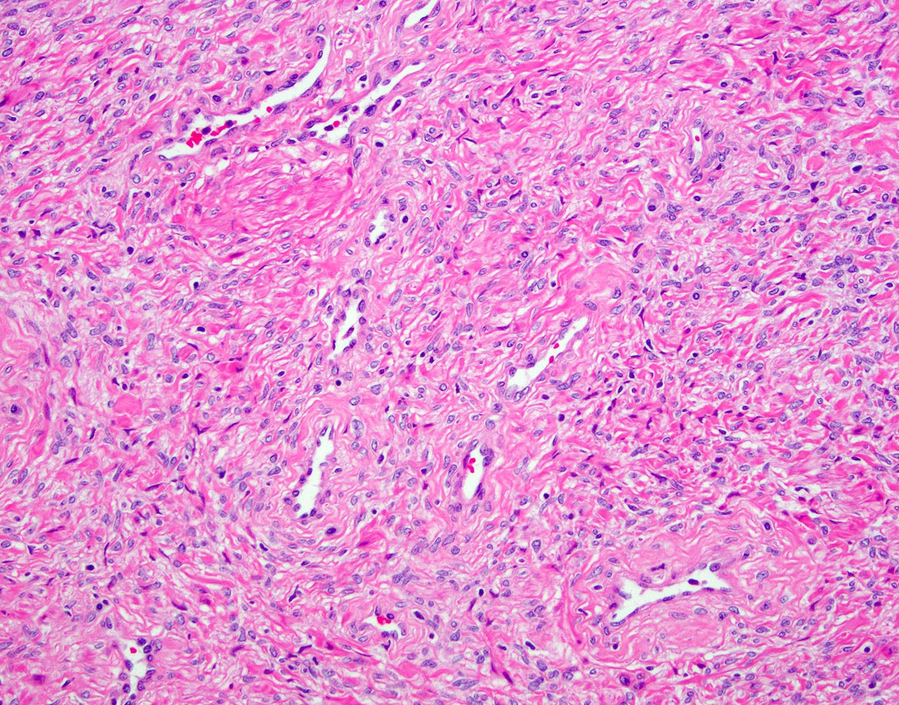
Infiltrative growth

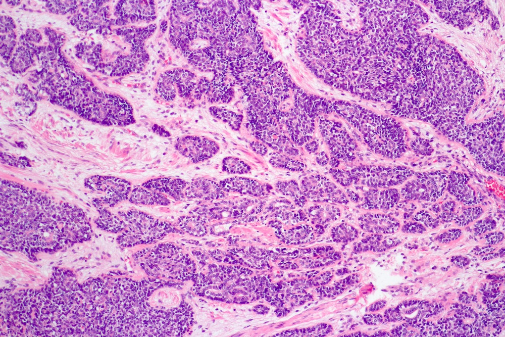
Basaloid morphology
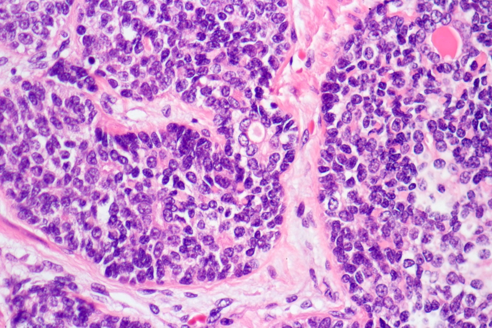

Combination of basaloid and luminal cells

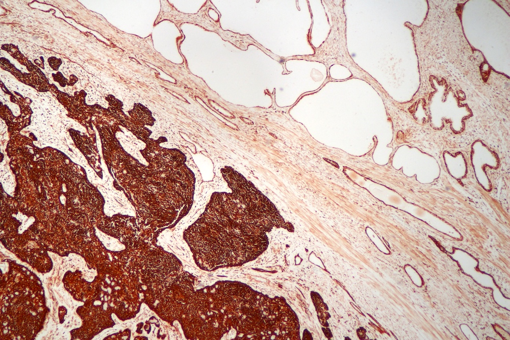
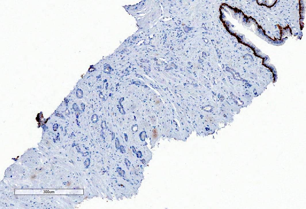
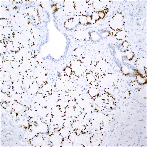
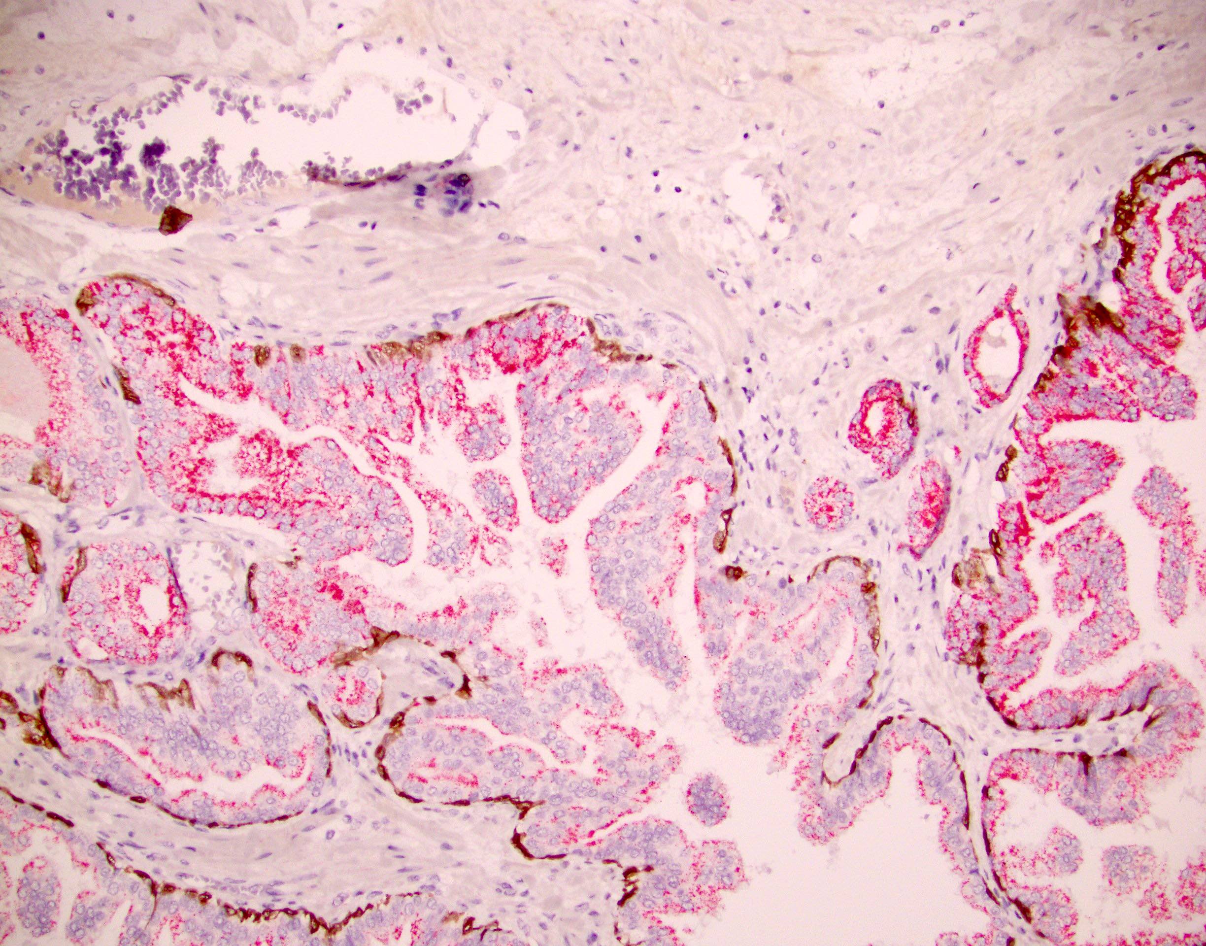
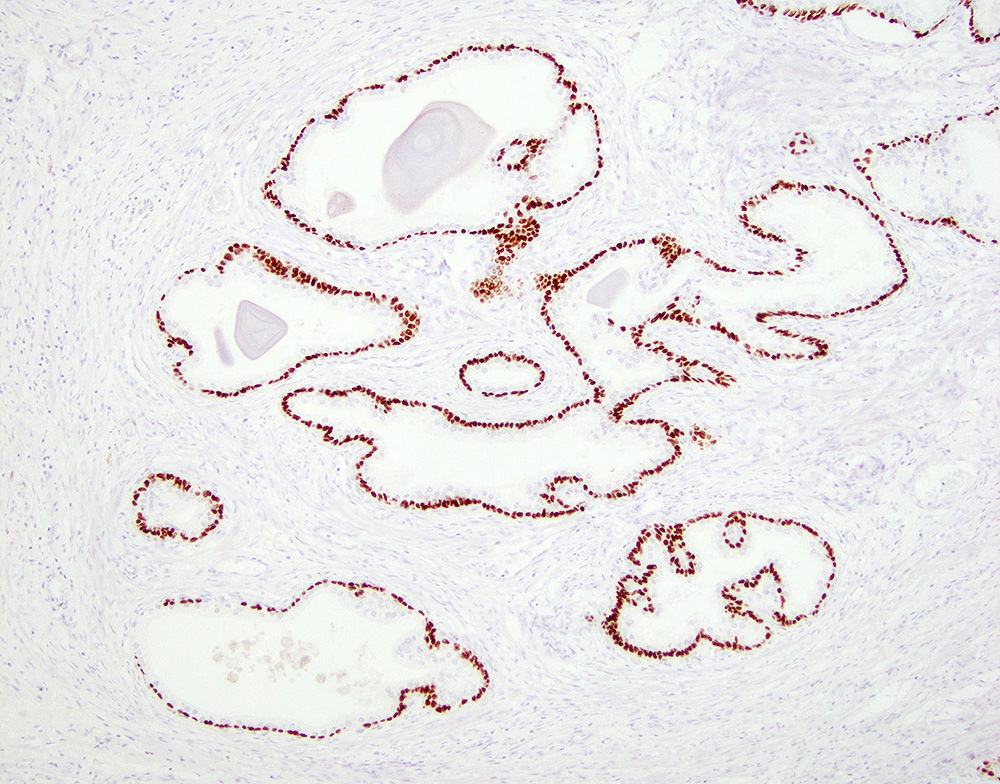
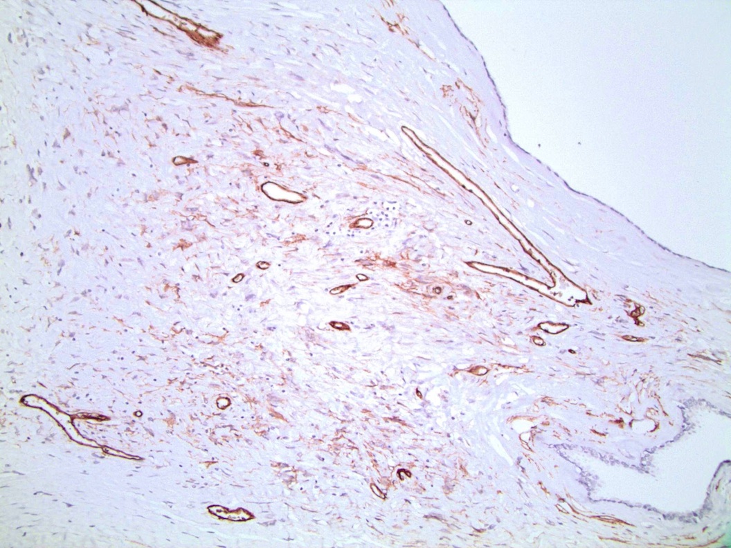
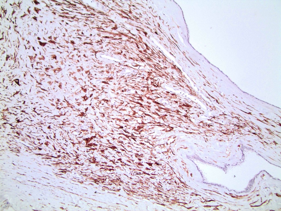
BCL2
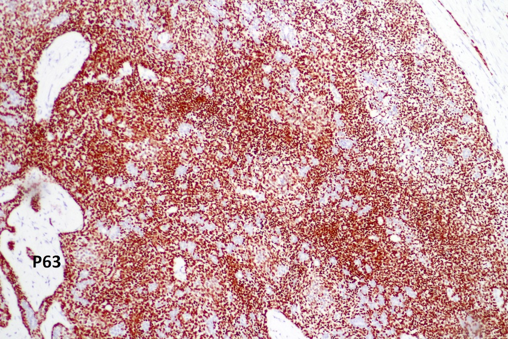
p63

PSA
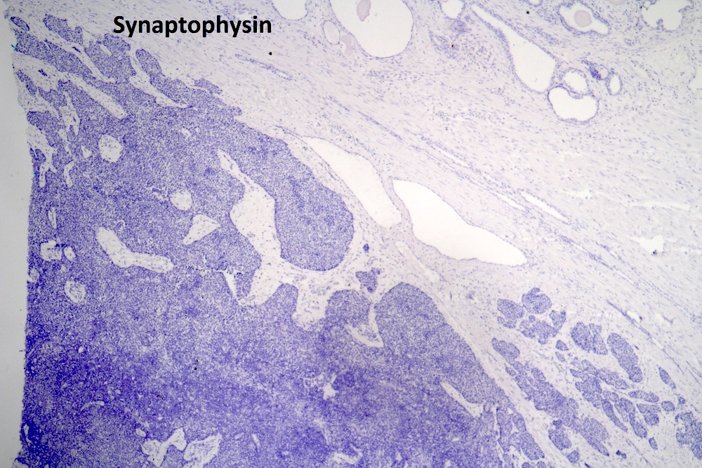
Synaptophysin
Contributed by Cristina Magi-Galluzzi, M.D., Ph.D.
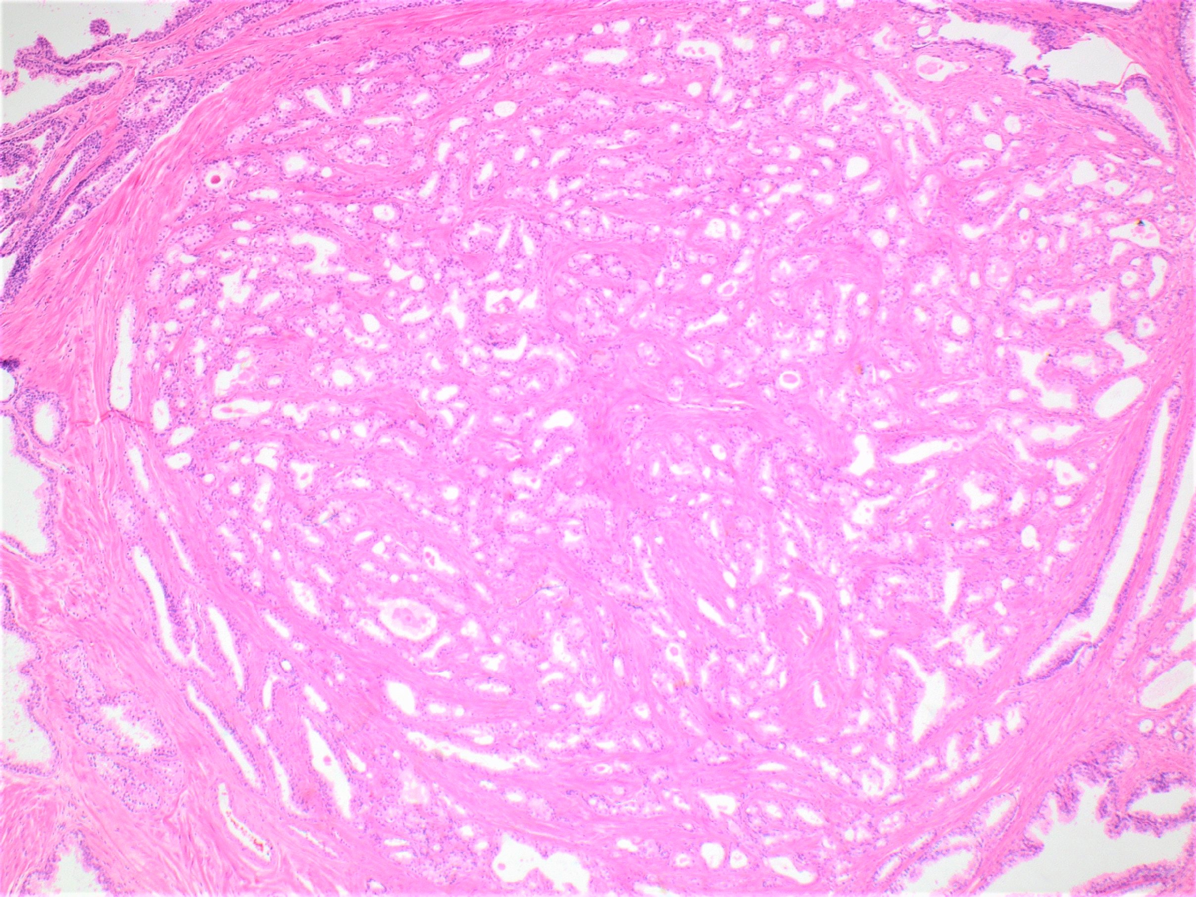
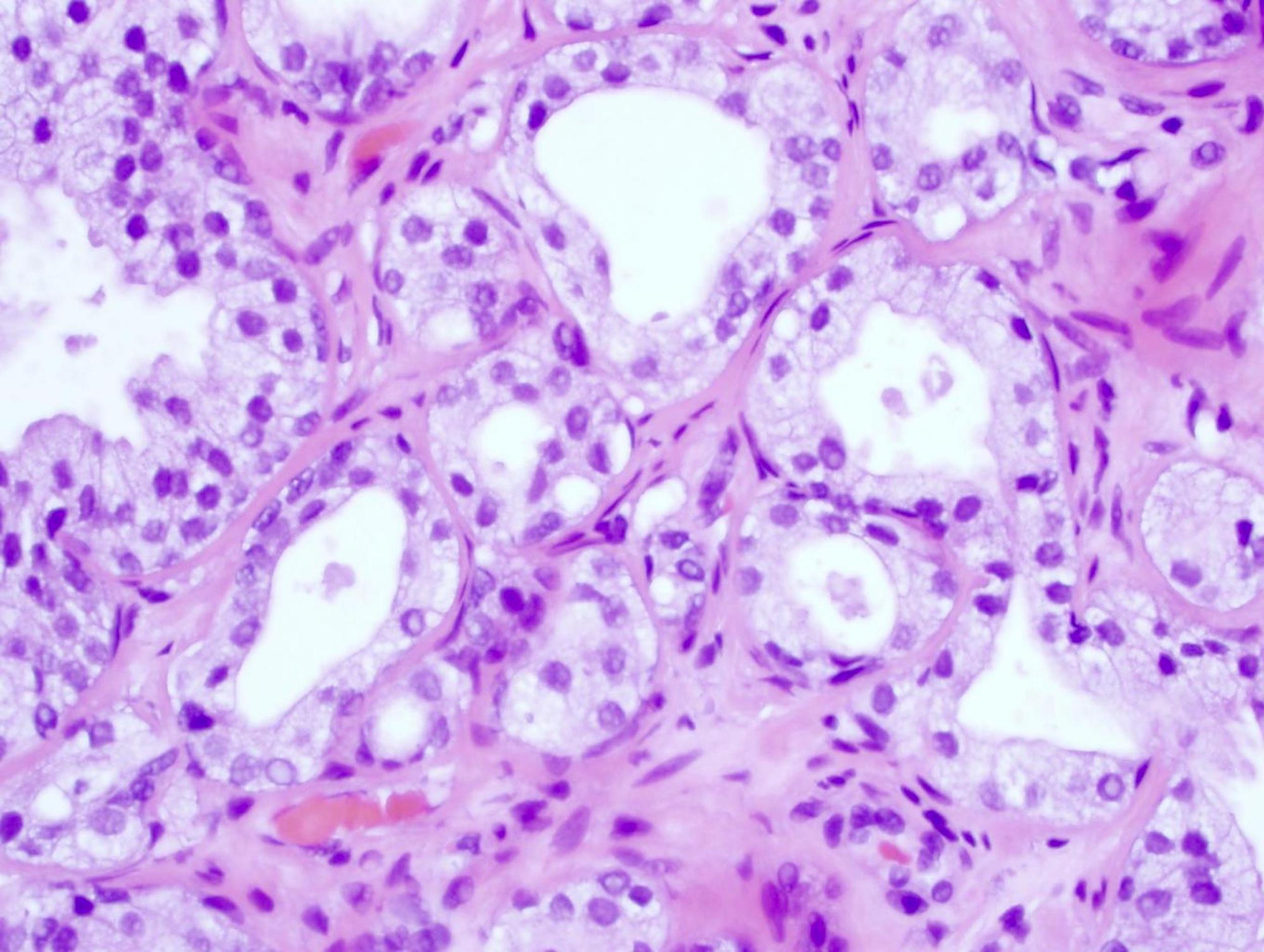
Well circumscribed nodule
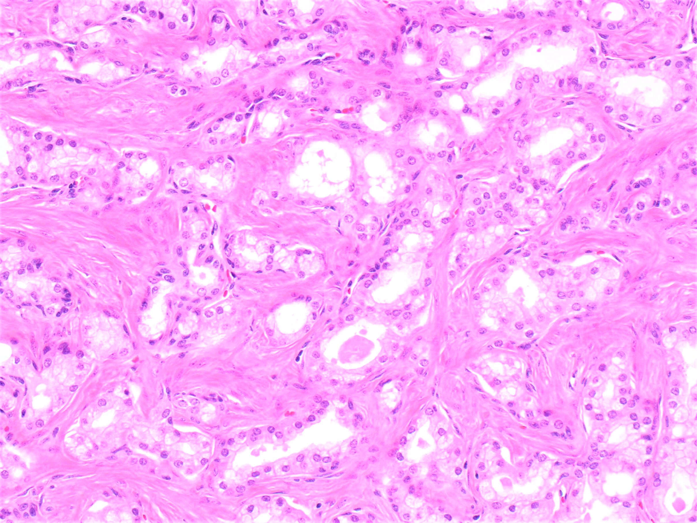
Mixed small and large acini
Basal cells
Crowded glands
Bland nuclear features
Fragmented basal cells
Images hosted on other servers:

Tumor abutting anorectal canal
Contributed by Susan Prendeville, M.D. and Gladell Paner, M.D.
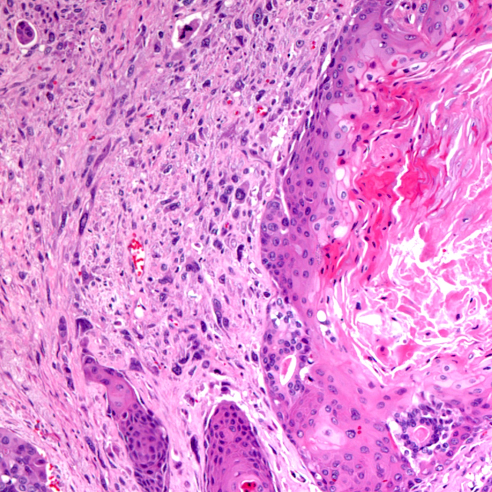
Biopsy, prominent squamous areas
With obvious keratin
Mixed components, postradiation setting
Transition, squamous to glandular
Contributed by Andres Matoso, M.D., @katcollmd on Twitter and Case #85
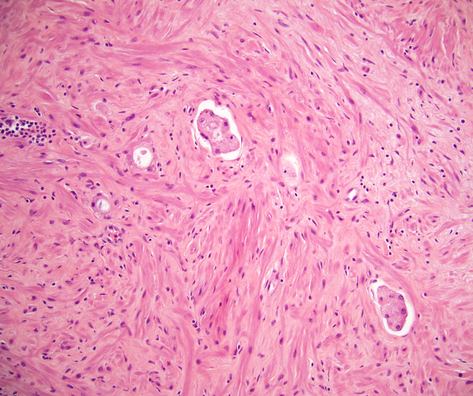
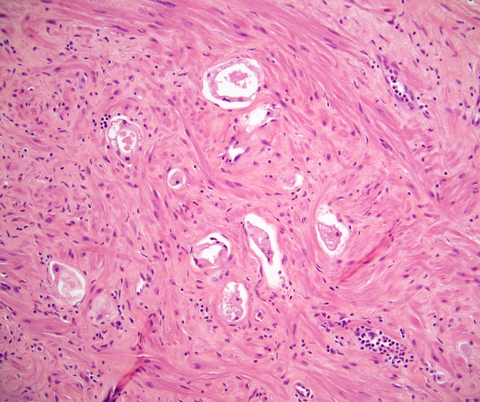
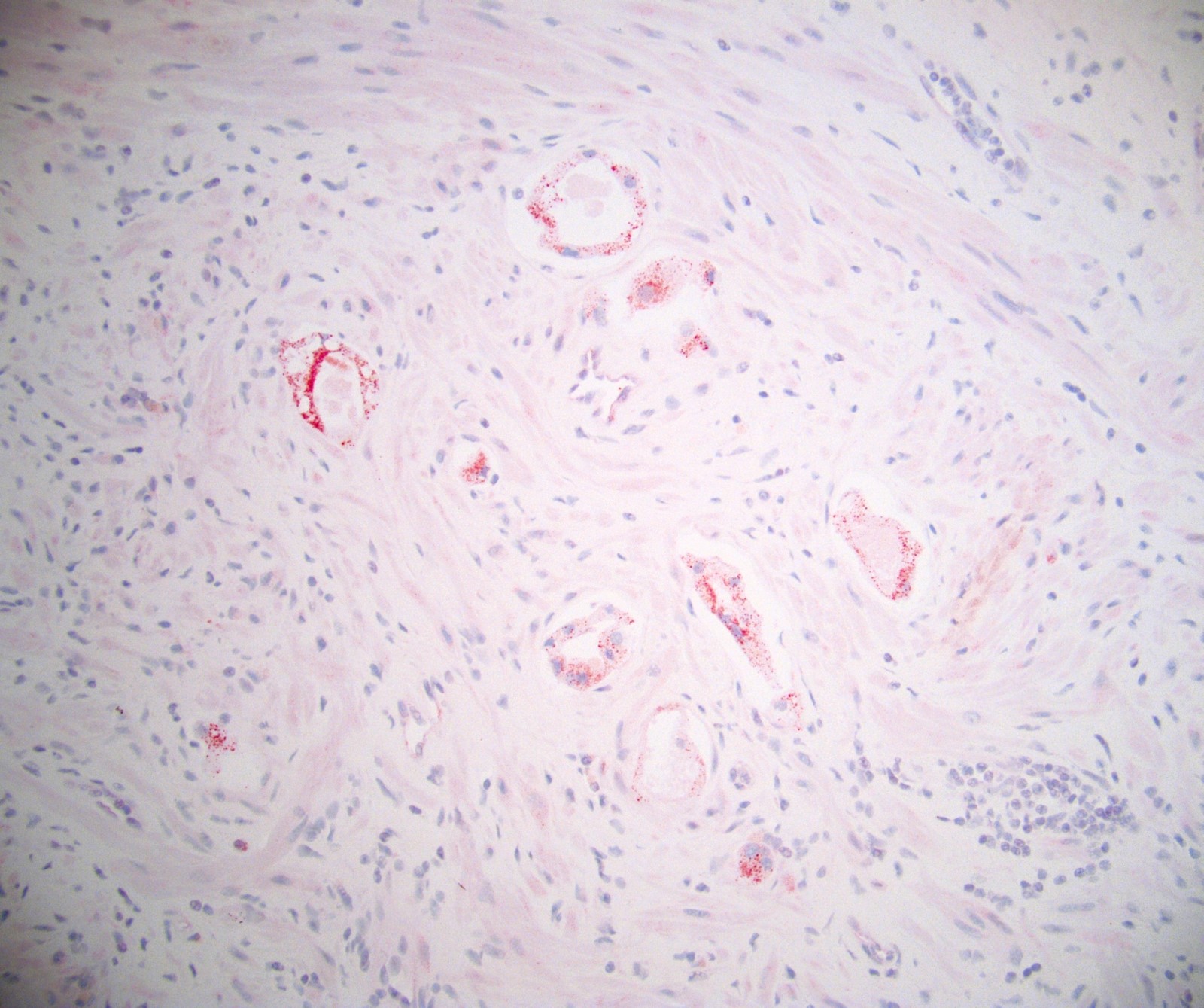
Various images
Amyloid
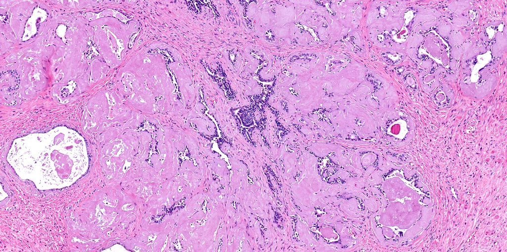
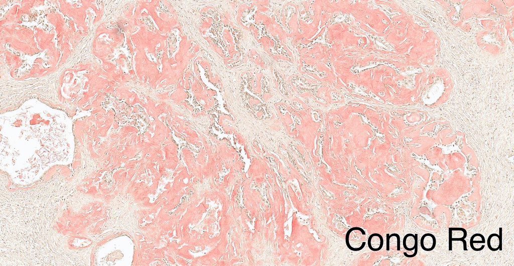
Amyloid
66 year old man with radical prostatectomy for adenocarcinoma
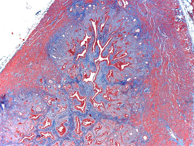
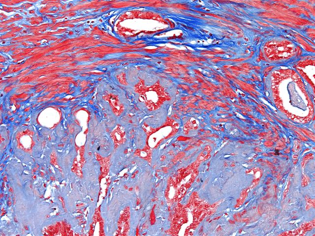
Trichrome stain
Contributed by Andres Matoso, M.D. and @ThatGlassTho on Twitter
Low power of Cowper glands
Glands and skeletal muscle
Mucicarmin+
PAS+
NKX3.1+
Anatomy & histology-Cowper glands
Contributed by Andres Matoso, M.D.


Chromogranin A+
Images hosted on other servers:

Fundus

Seminal vesicles and ducts

McNeal zones

Sagittal view

Axial views
Contributed by Kenneth A. Iczkowski, M.D. and AFIP
Cross section of gland
Normal gland
Contributed by Kenneth A. Iczkowski, M.D.
Whole mount histology
Close up, anterior aspect
Basal versus secretory cells
Basal cell hyperplasia
Differential diagnosis of a multilayered epithelium
Hyperplastic central zone
Images hosted on other servers:

Seminal vesicles and prostate
Images hosted on other servers:

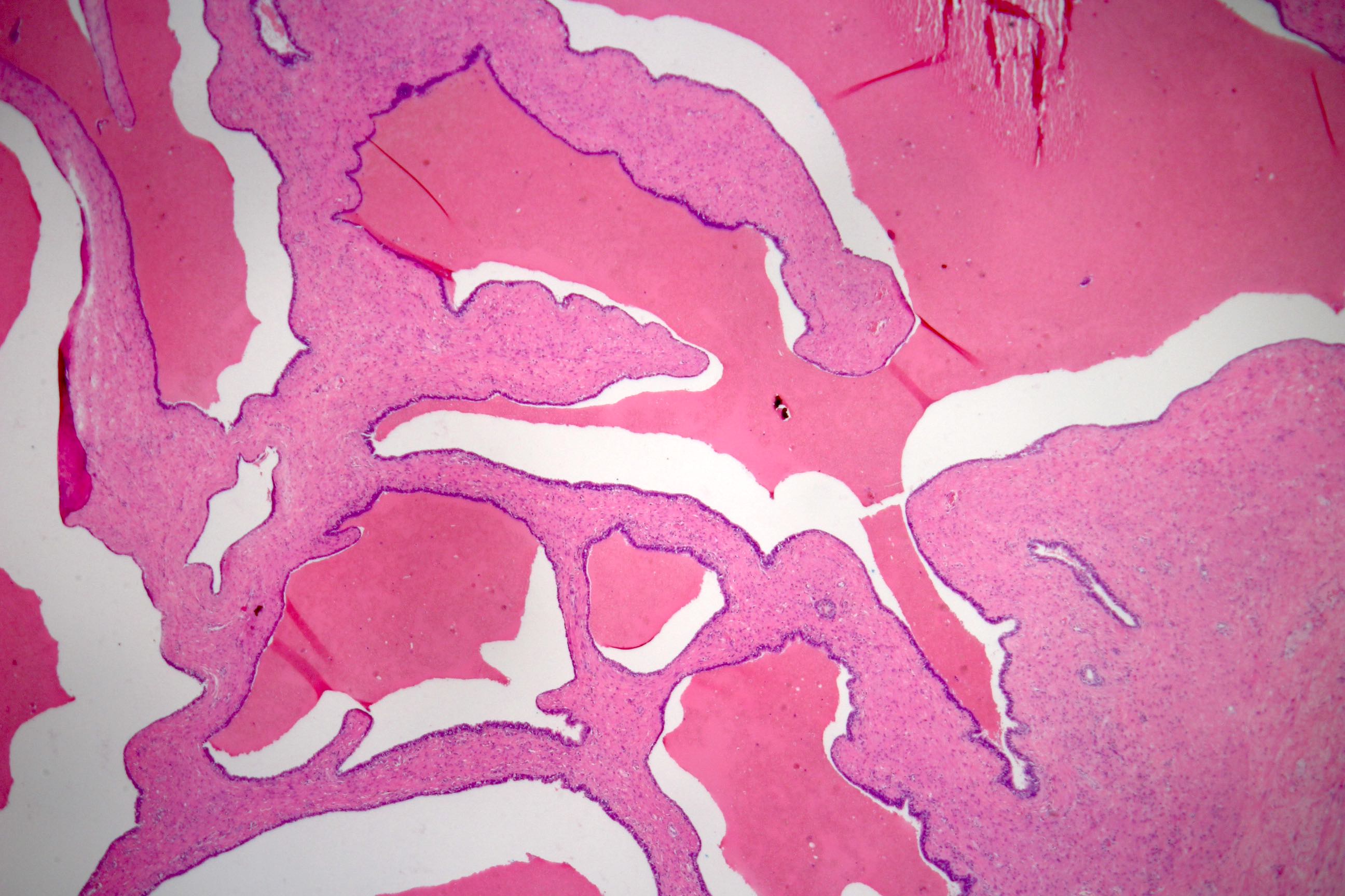
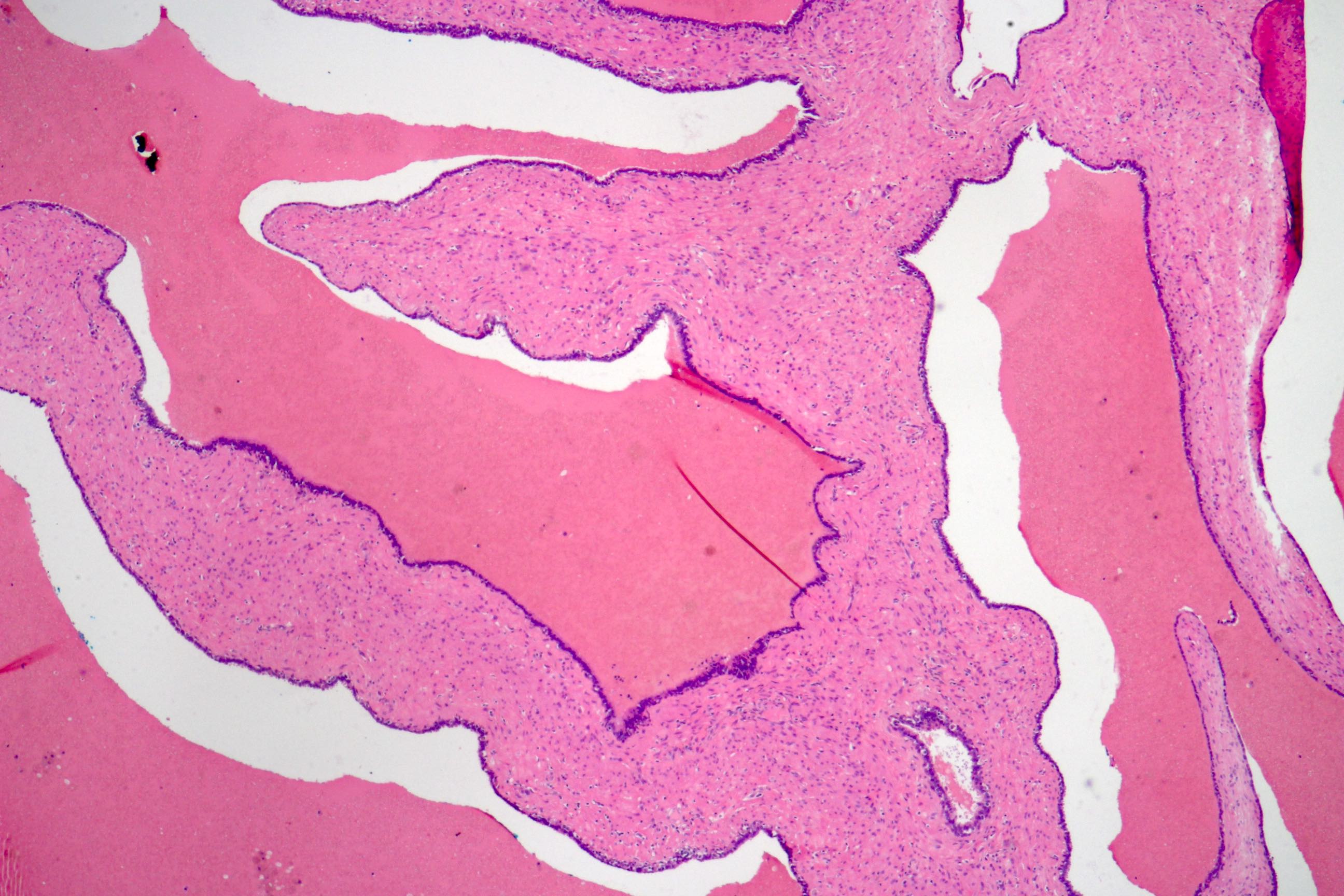
Normal seminal vesicles

Agenesis

Cyst

Seminal vesiculitis
Images hosted on other servers:

Cyst

Stones
Images hosted on other servers:


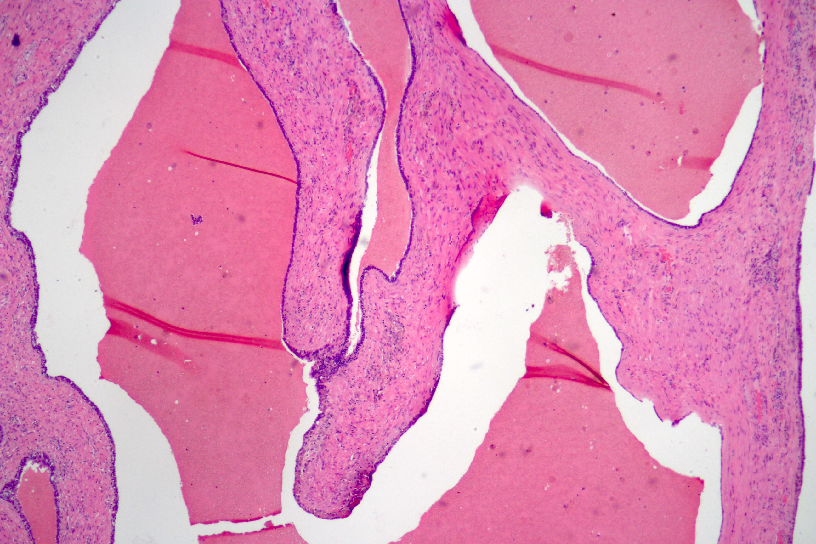
Normal seminal vesicle

Acquired cystic dilatation


Cyst

Calculus
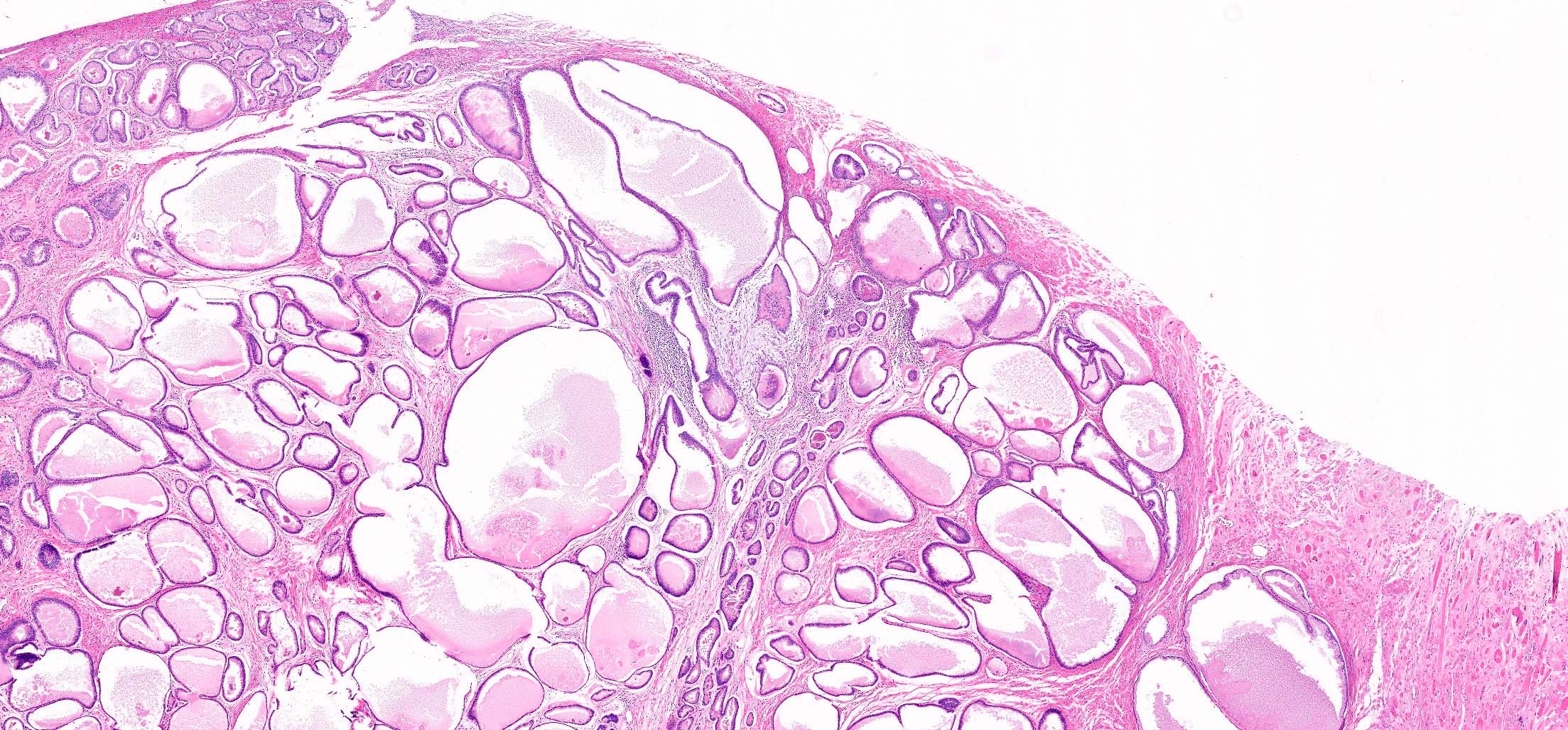
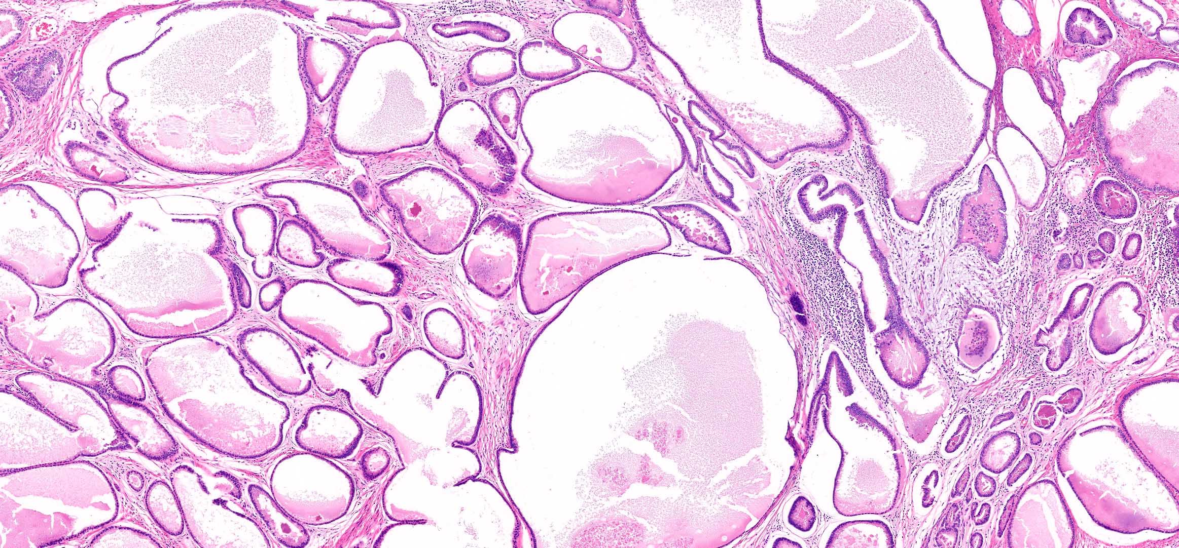
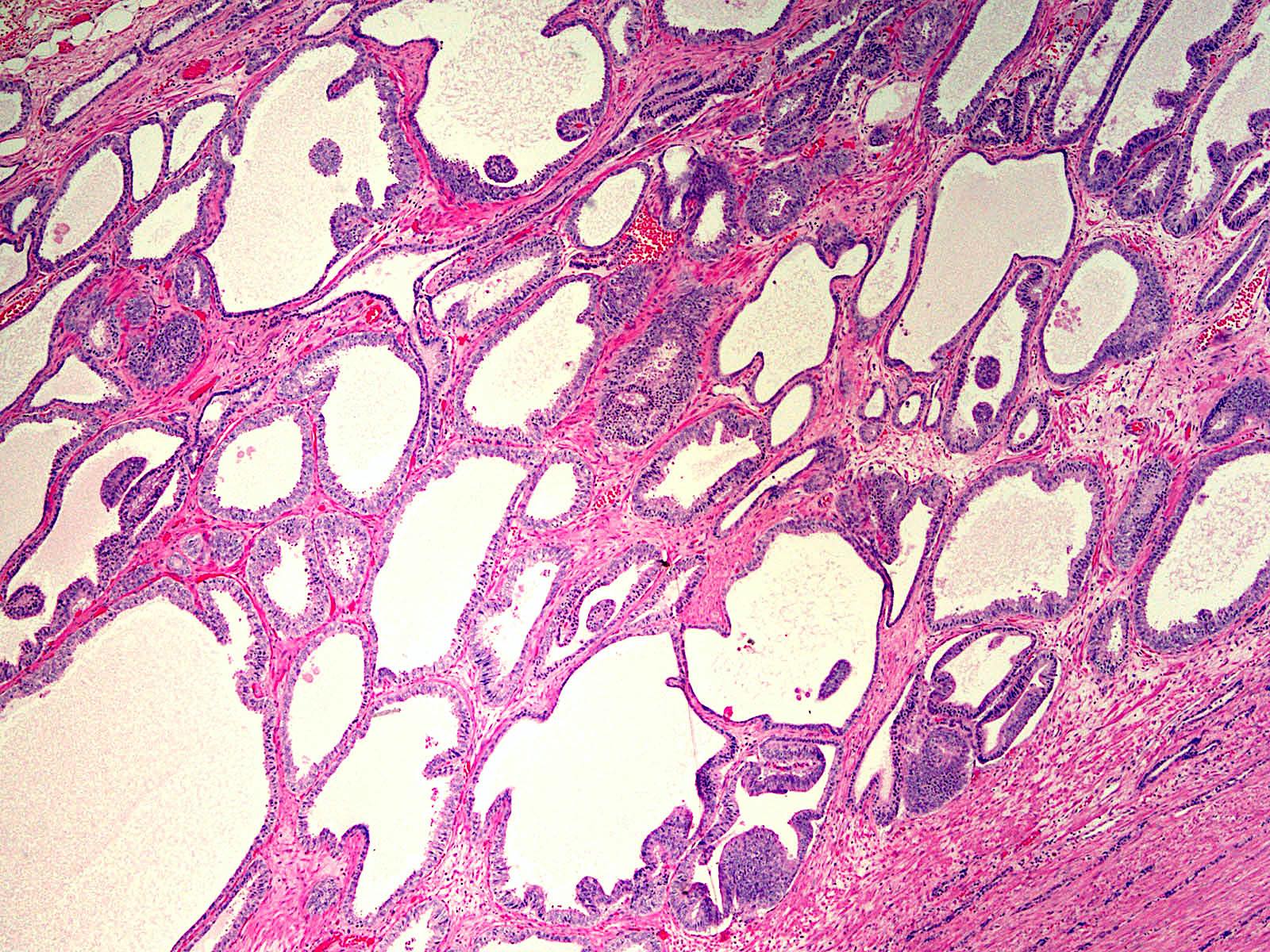
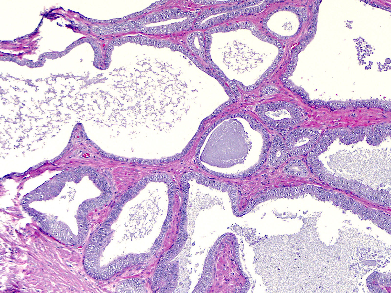
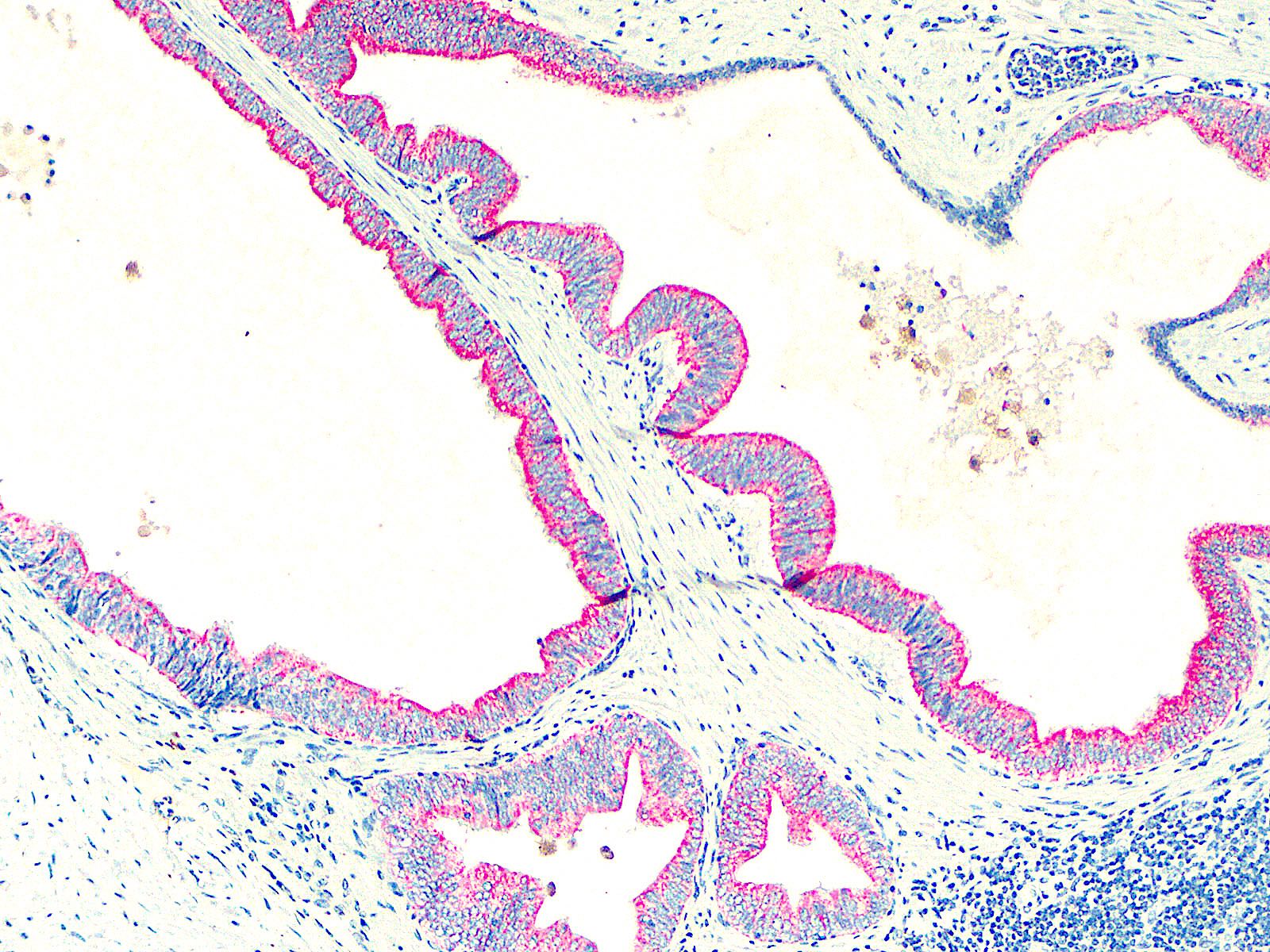
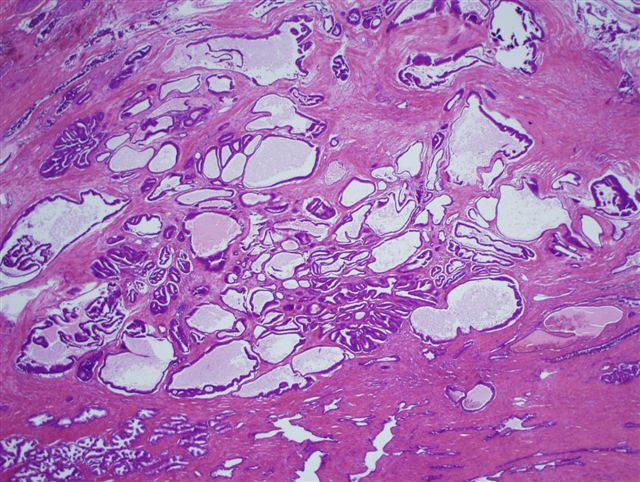
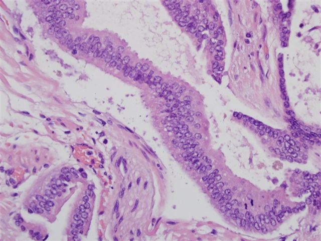
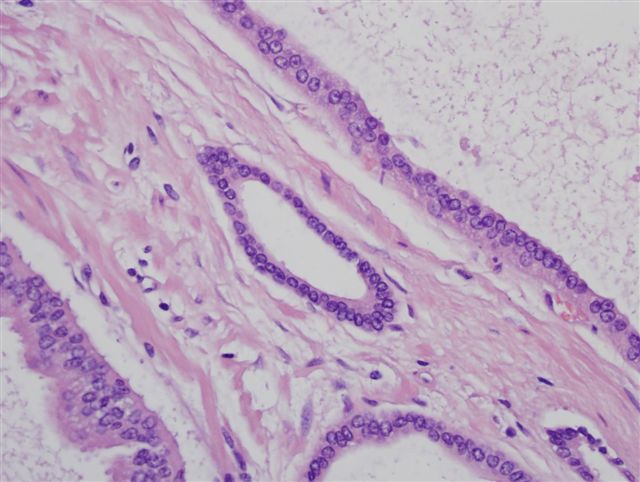
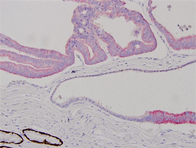
Contributed by Faryal Shoaib, M.D., Y. Albert Yeh, M.D., Ph.D. and Andres Matoso, M.D.
Multilobulated tubular structures
Branching side ducts
Lining columnar cells
Enlarged epithelial cells
Large atypical cells
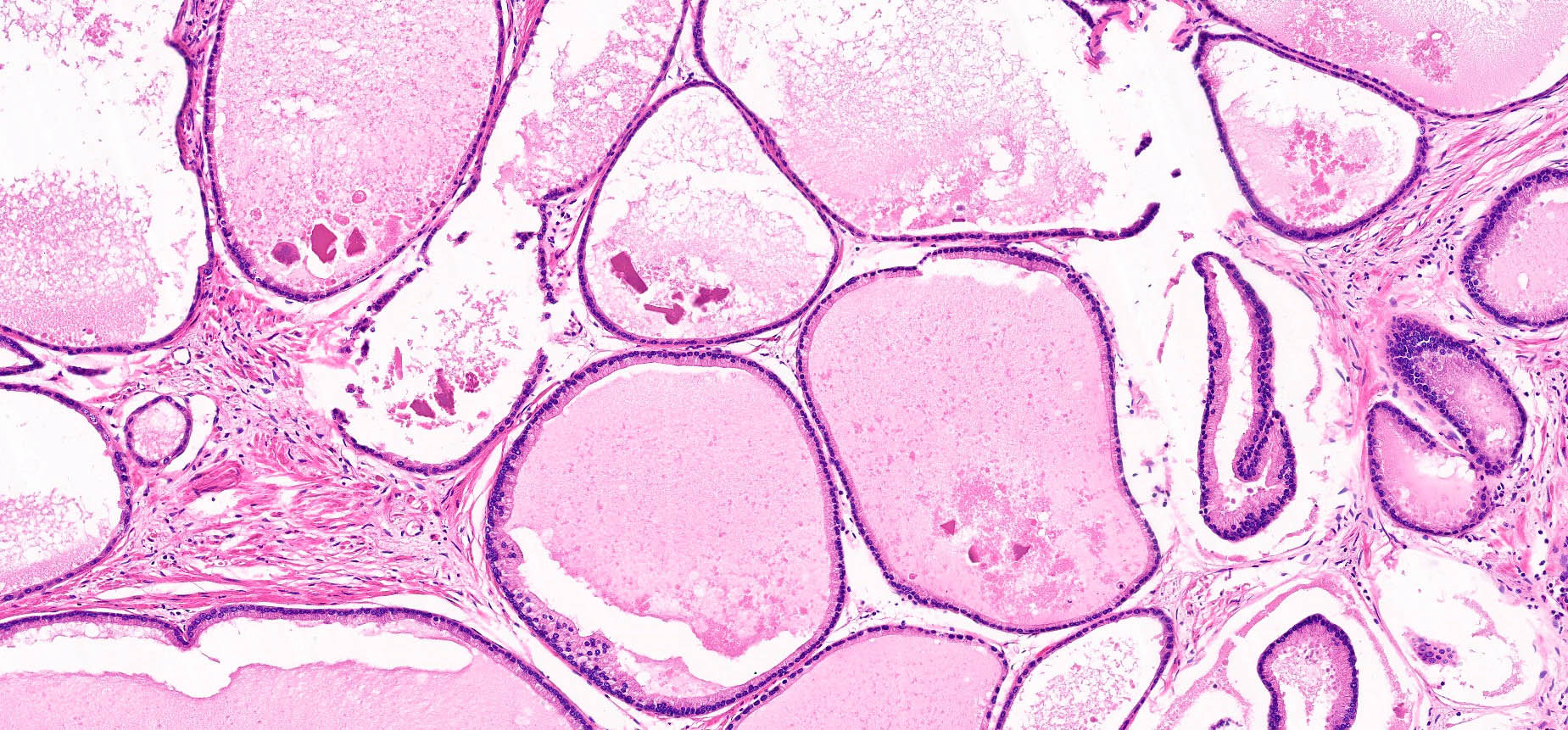
Alveolar-like mucosal folds
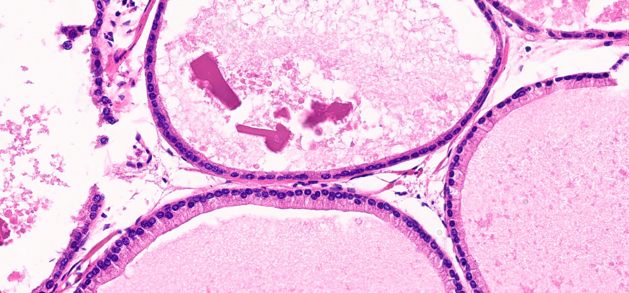
Eosinophilic crystalloids
Stromal hyaline body
Stromal hyaline body
Stromal hyaline body
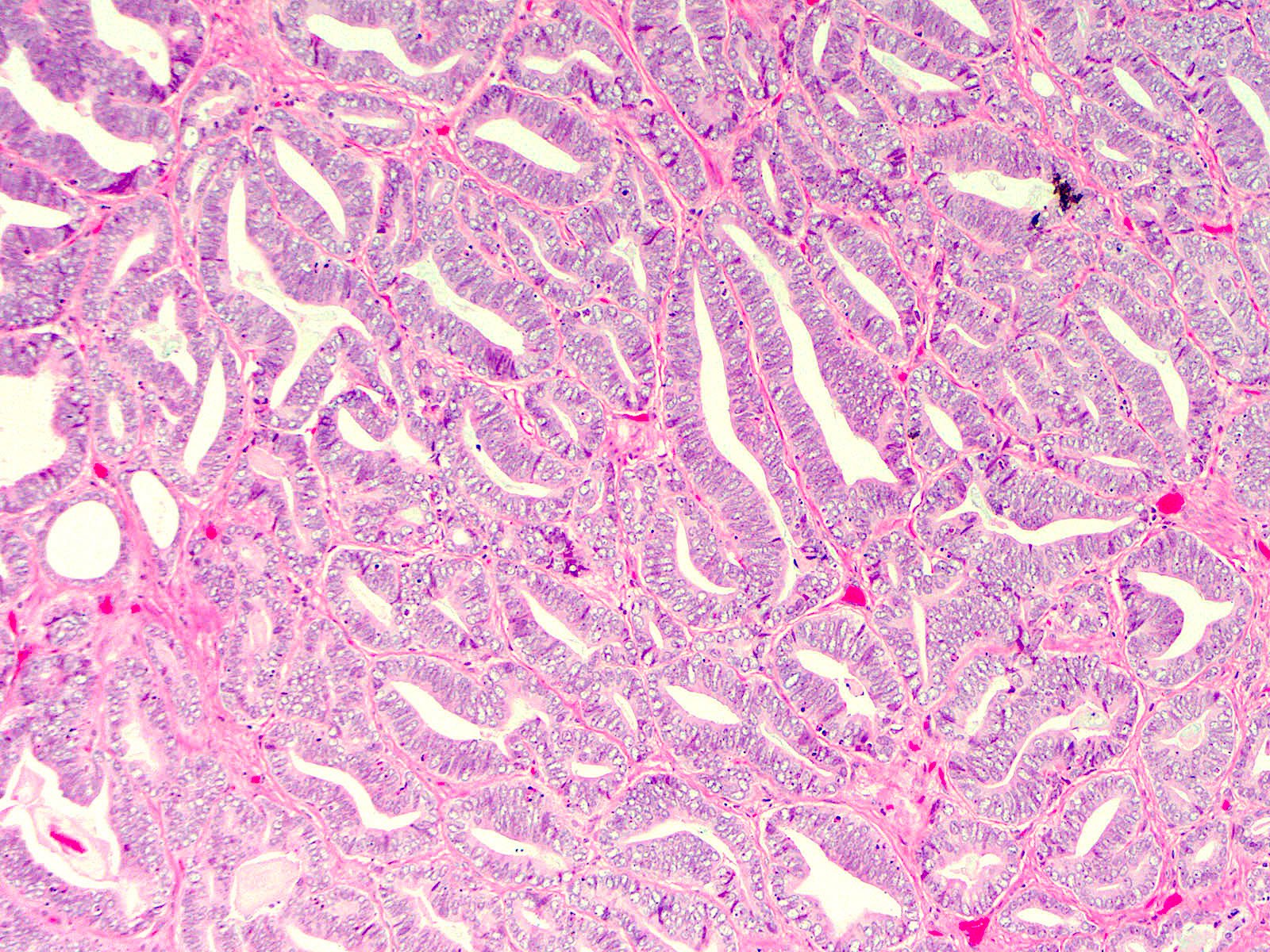
Monstrous epithelial cells
Prostatic glands
Nuclear pleomorphism
Yellow pigment
Images hosted on other servers:

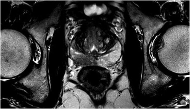
mpMRI findings, cystic atrophy
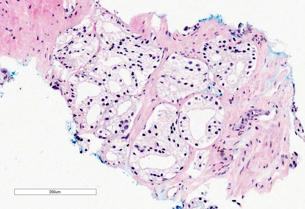
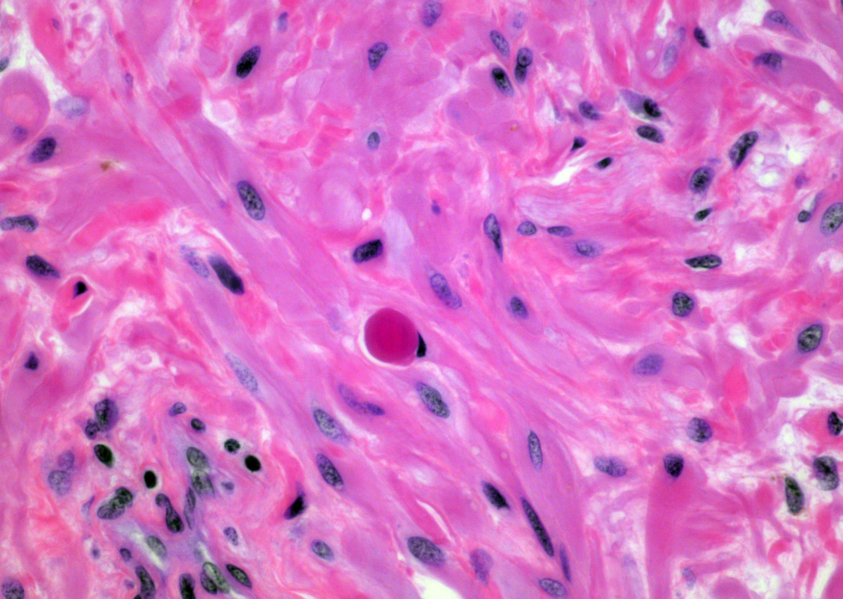
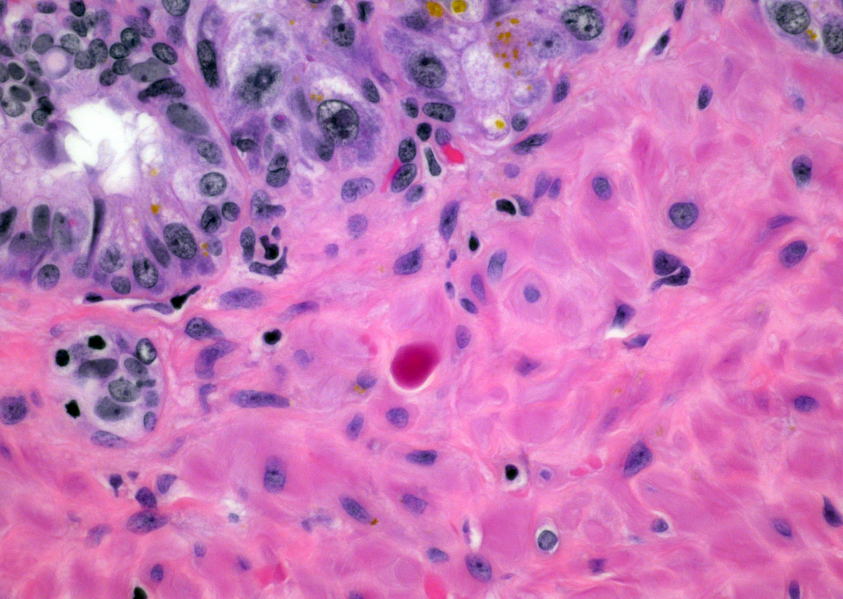
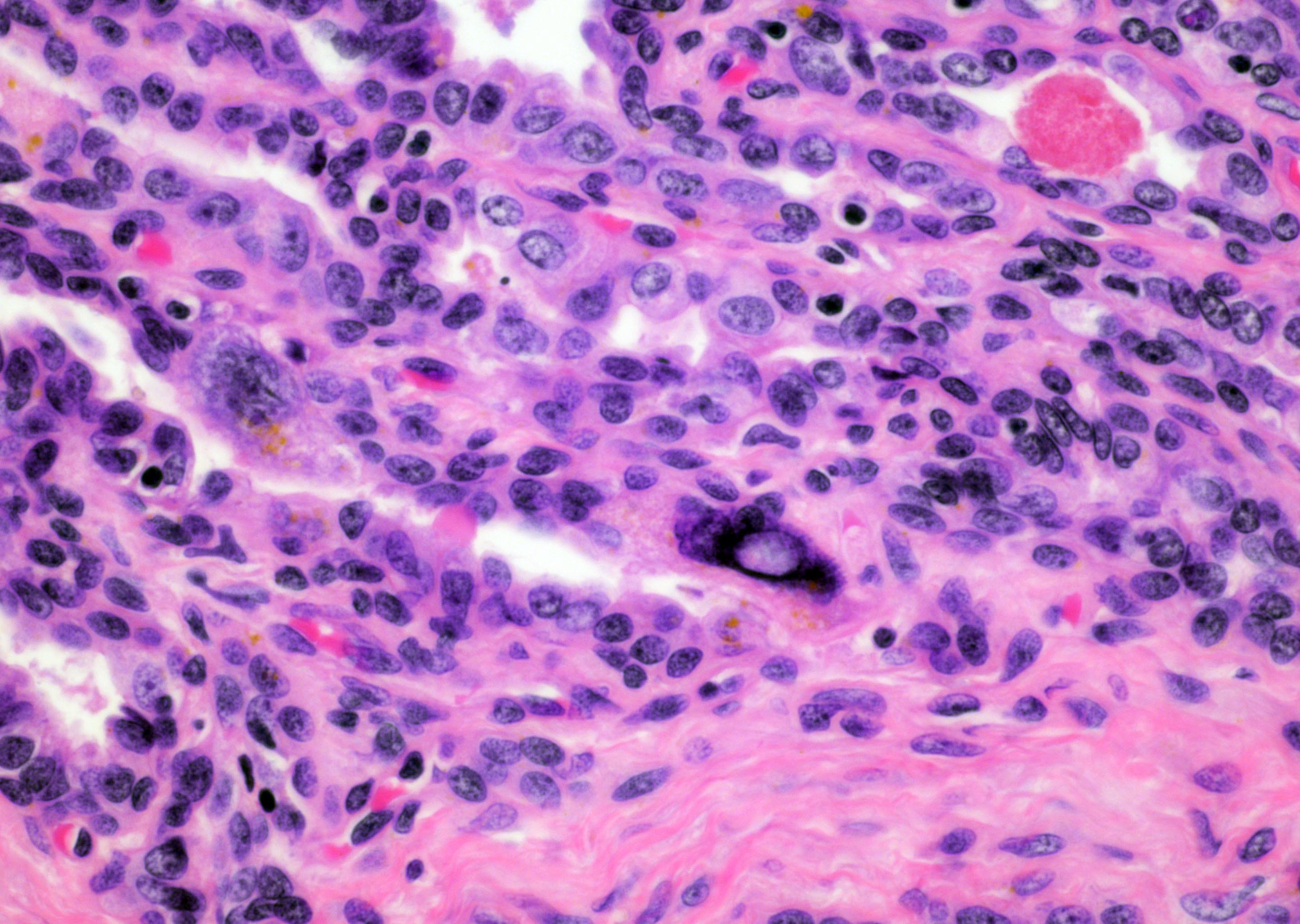
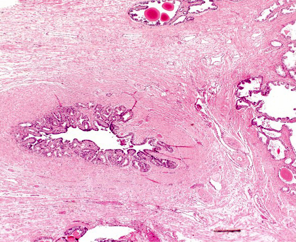
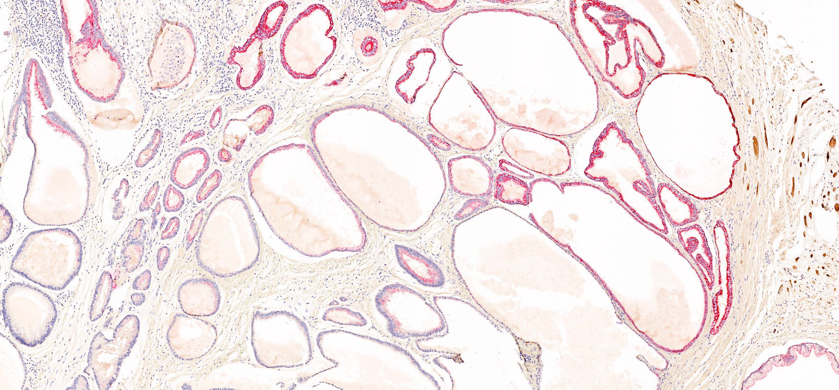
Contributed by Kenneth A. Iczkowski, M.D.
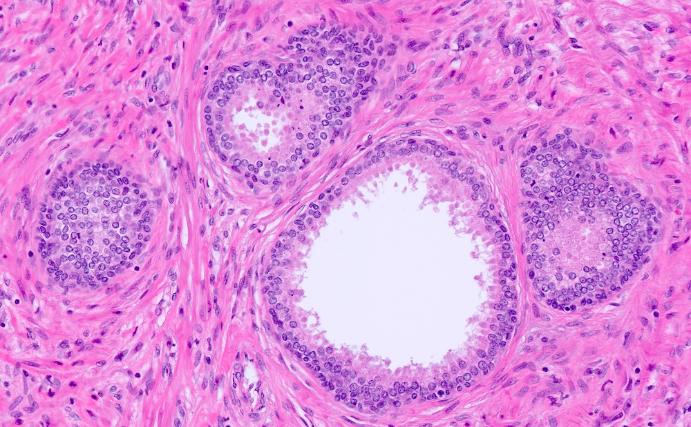
Small acinar atrophy
Dilated acinar atrophy
Red doesn’t equal cancer
Sclerosing adenosis

Cancer mimicking atrophy
Contributed by Kenneth A. Iczkowski, M.D.
Basal cells in AIP
Papillary AIP, with invasive acini
Cocktail immunostain of AIP
Isolated AIP on biopsy
Cribriform AIP
AIP, favor intraductal
Grade 5 foamy glands
Not an AIP
Not an AIP, immunostain
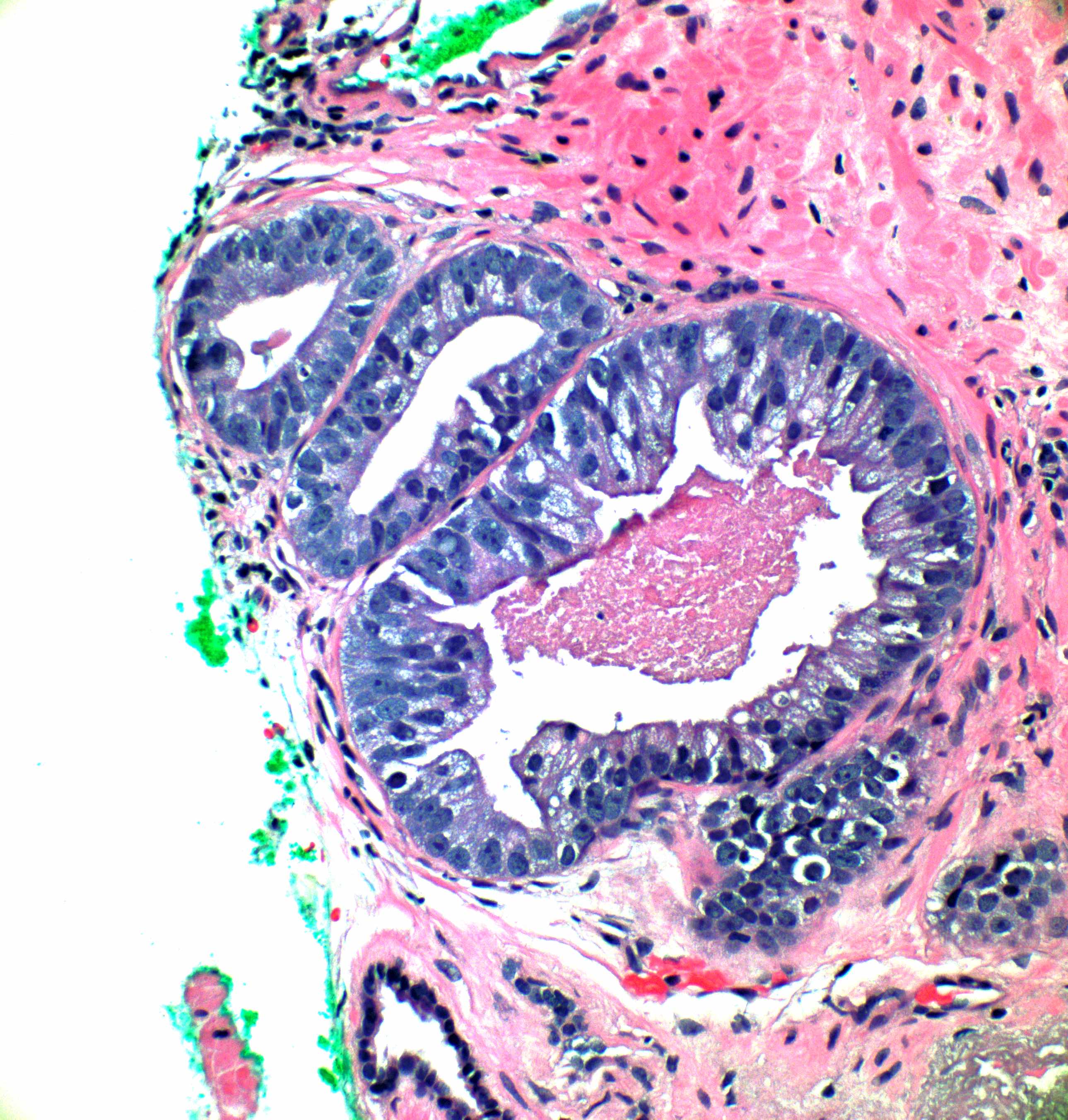
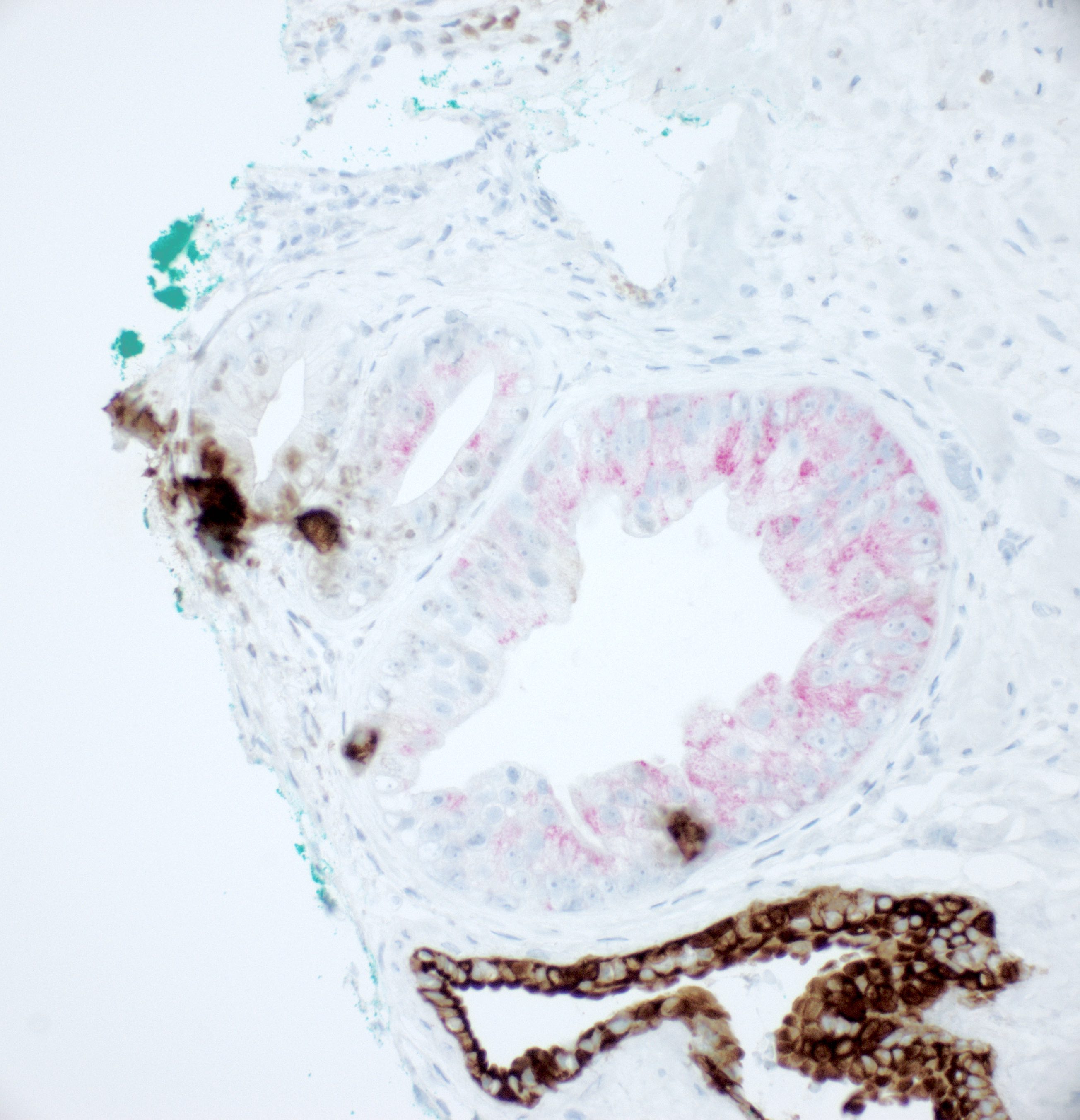
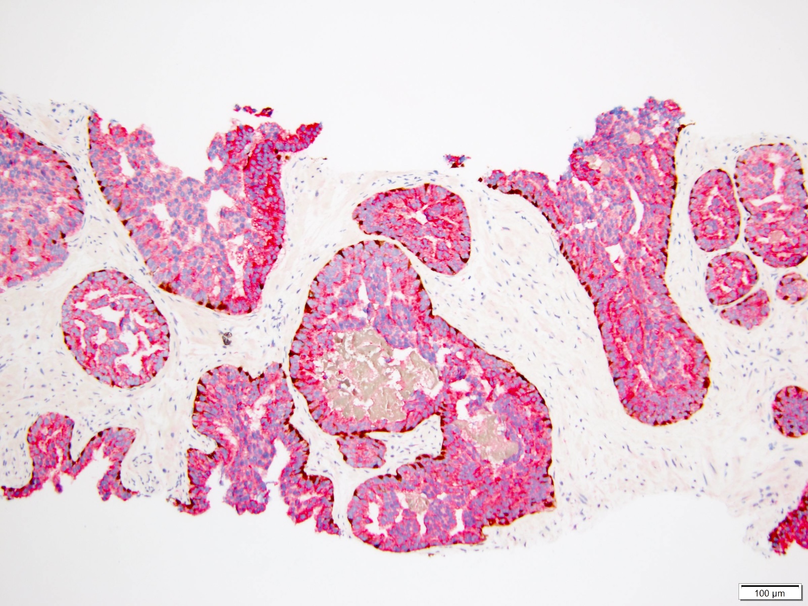
Contributed by Kenneth Iczkowski, M.D.
Quantitative ASAP
Three atypical acini and triple stain
Four atypical acini and triple stain
Qualitative ASAP
Upper left atypical acini and triple stain
Large atypical acini and triple stain
Eight small acini and triple stain
ASAP + HGPIN
ASAP + HGPIN and triple stain

ASAP + HGPIN and triple stain
Contributed by Debra Zynger, M.D.
Straight luminal border
Triple stain
Crowded, straight luminal border, mucin and nuceloli
Contributed by Anil Parwani, M.D., Ph.D., M.B.A. and Andres Matoso, M.D.
Benign mimicker of prostatic cancer
Basal epithelium proliferation
Intraluminal amorphic secretions
Florid basal cell hyperplasia
Basal cell carcinoma
Contributed by Debra L. Zynger, M.D.
H&E
Triple stain with CK5-p63-AMACR
Images hosted on other servers:

BPH on MRI
Contributed by Sara Moscovita Falzarano, M.D., Ph.D.
Prominent periurethral nodularity
Contributed by Sara Moscovita Falzarano, M.D., Ph.D.
Glandular / epithelial type
Benign prostatic glands

Stromal nodule
p63

p63 / HMWK
Contributed by Rafael E. Jimenez, M.D. and Arpan Samaddar, M.B.B.S.
Clusters of cribriform acini
Clear cells
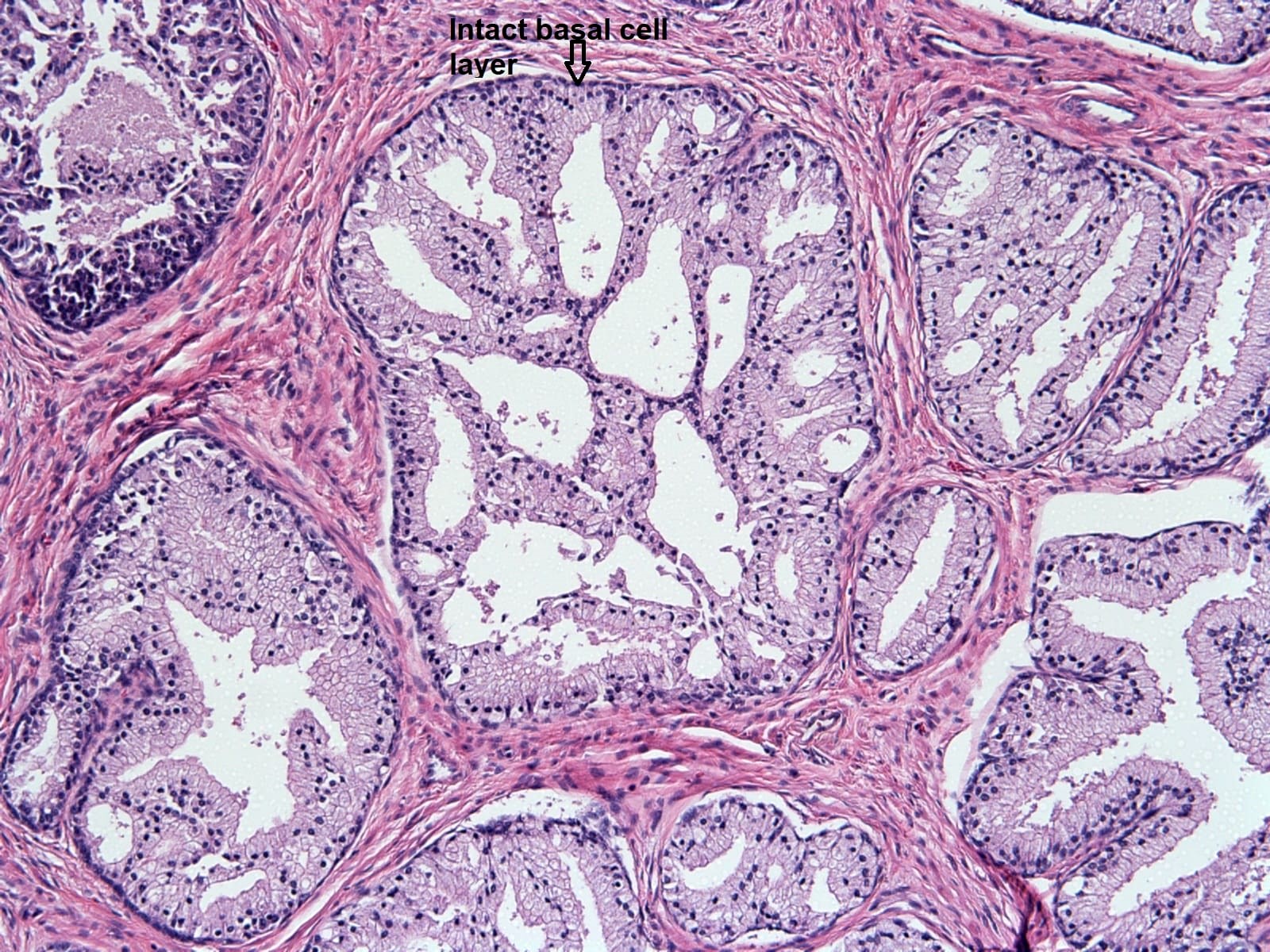
Intact basal cell layer
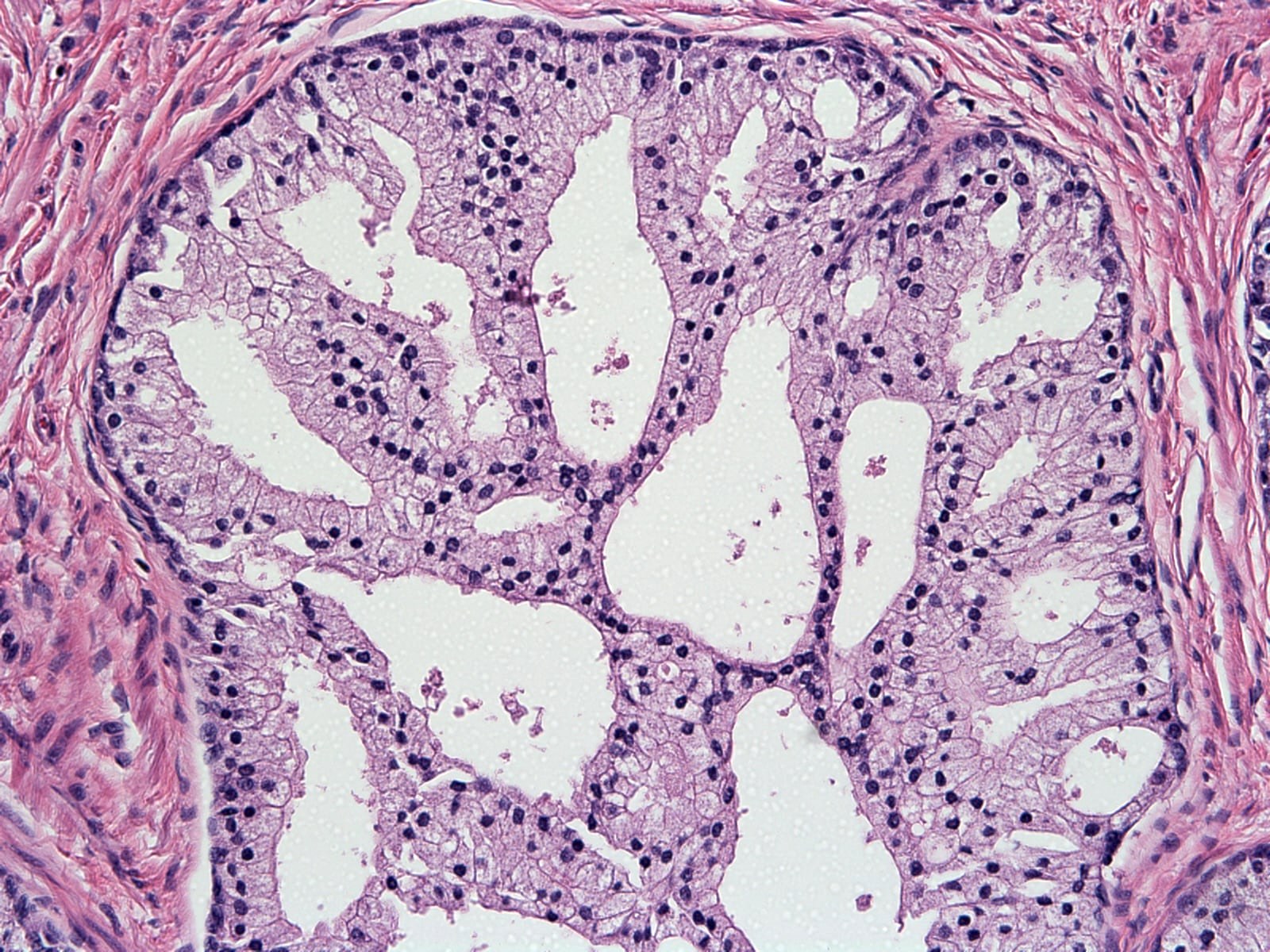
Small nuclei without atypia
Images hosted on other servers:

Mass of cystic component between bladder and rectum

Cystic mass with solid component connected to the seminal vesicle

Solid and cystic pelvic mass
Images hosted on other servers:

Laparoscopic view
Contributed by Daniel Athanazio, M.D., Ph.D.
Cystic mass centered in the seminal vesicle
Cut surface
Images hosted on other servers:

Multilobulated cystic mass

Cut surface
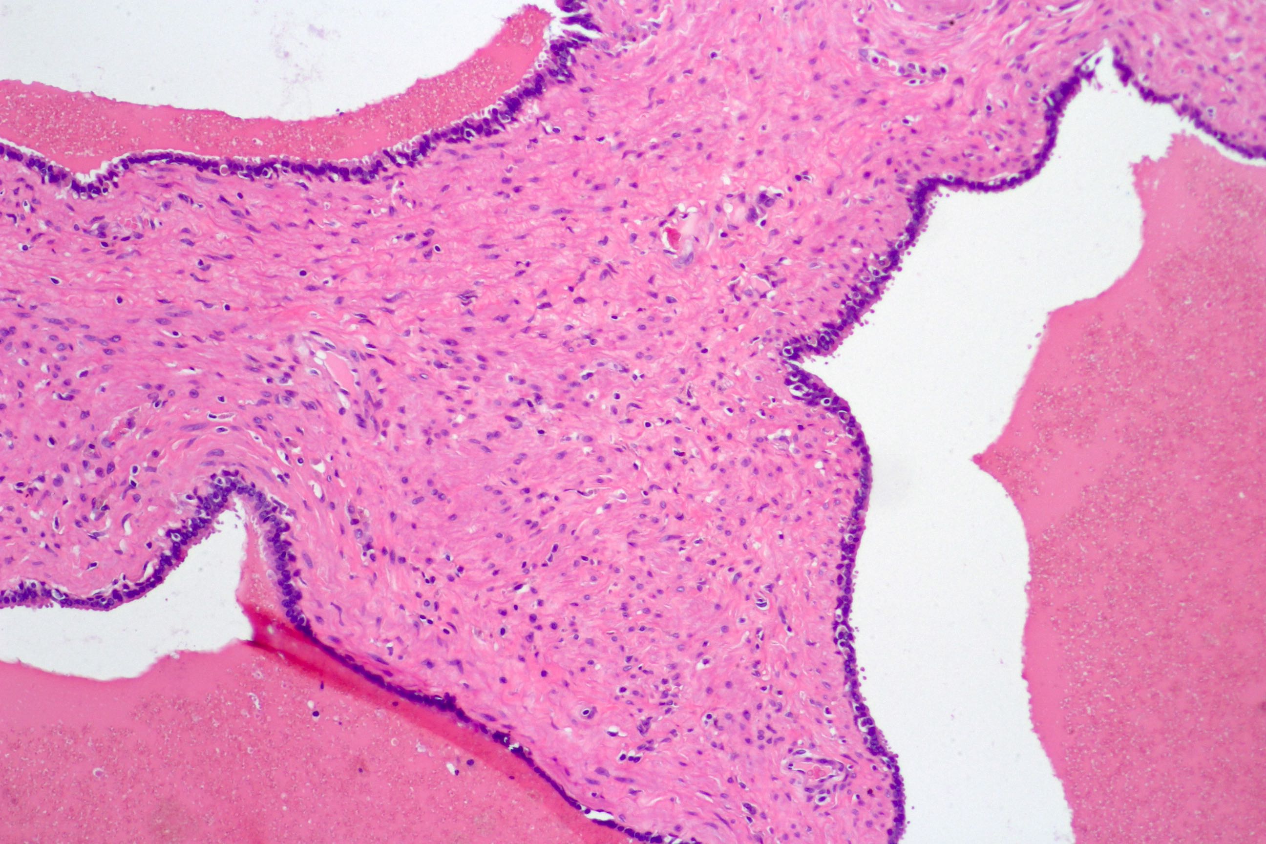
Contributed by Daniel Athanazio, M.D., Ph.D.
Cystic tumor
Finger-like projections

Finger-like projections
Leaf-like projections
Nonproliferative stroma
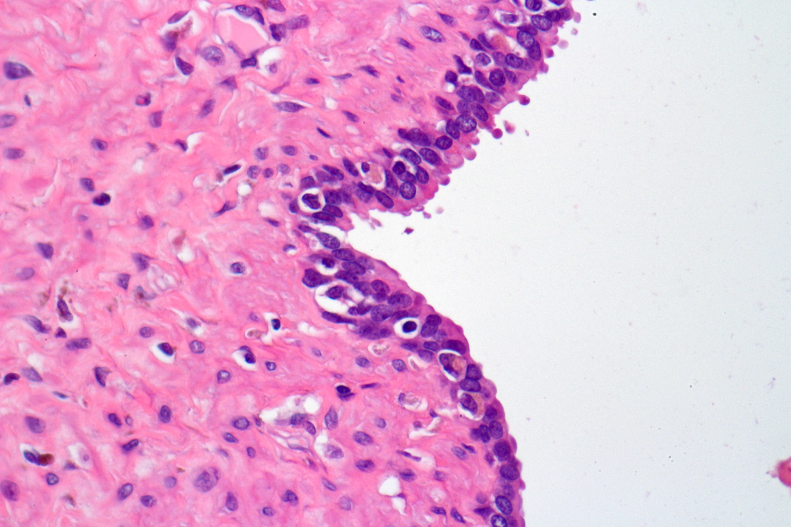
Bland epithelium
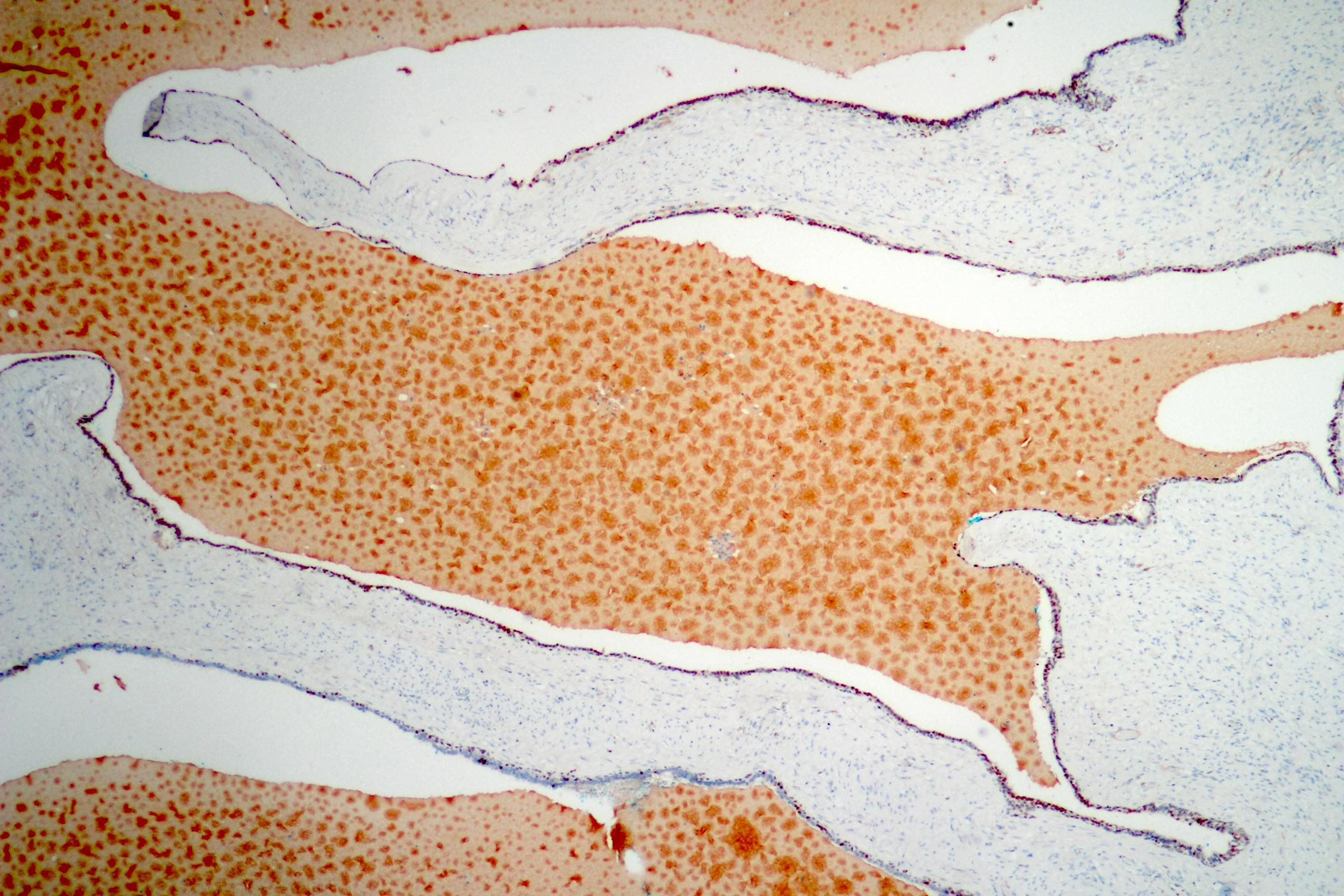
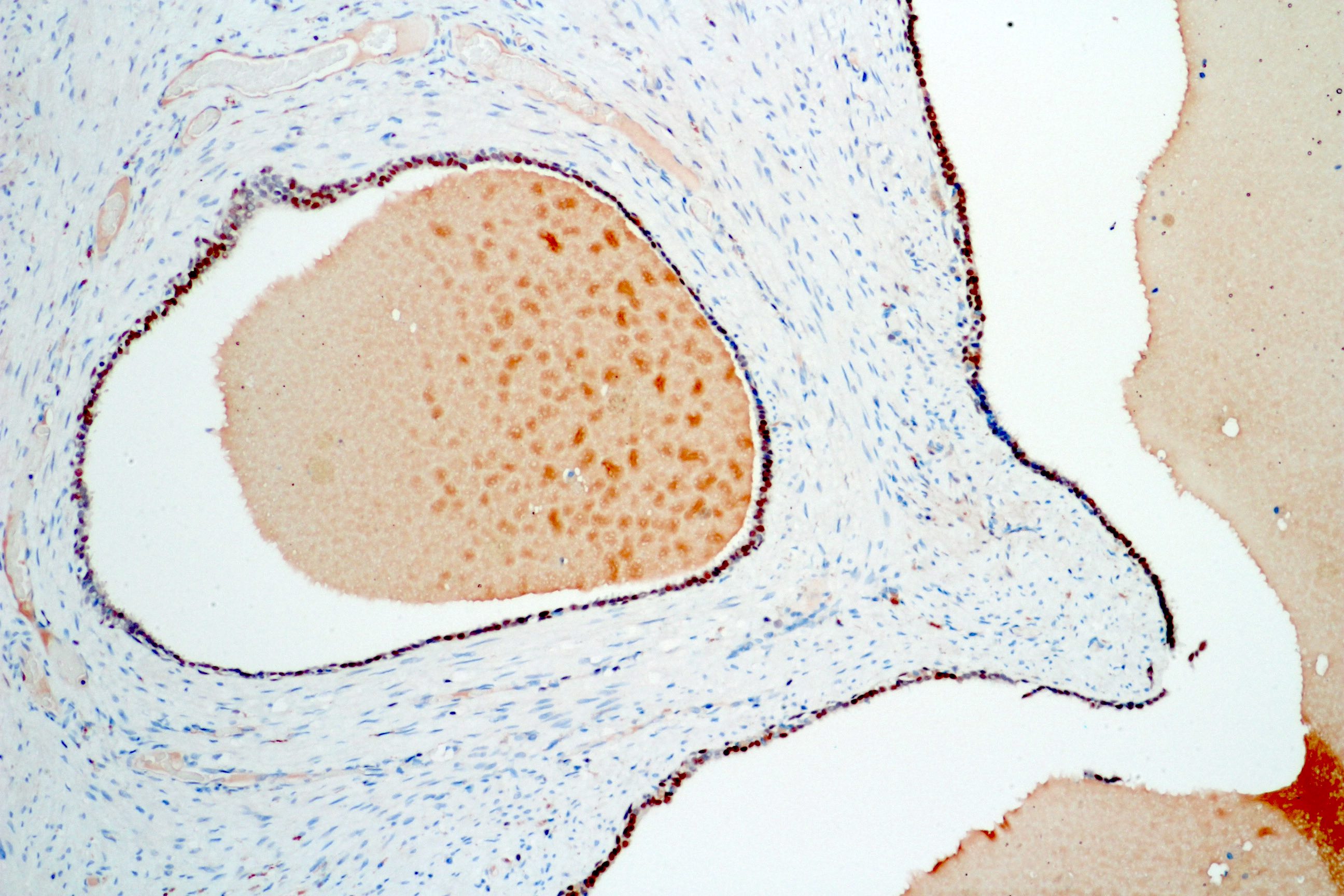
GATA3
GATA3

GATA3
GATA3
PSA negative
Calretinin negative
Prostein negative
Images hosted on other servers:

Cystic ductal adenocarcinoma
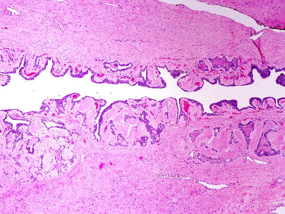
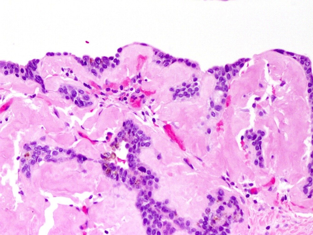
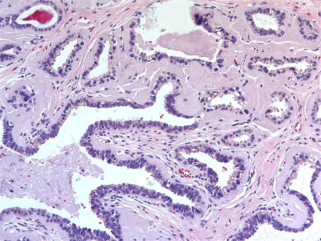
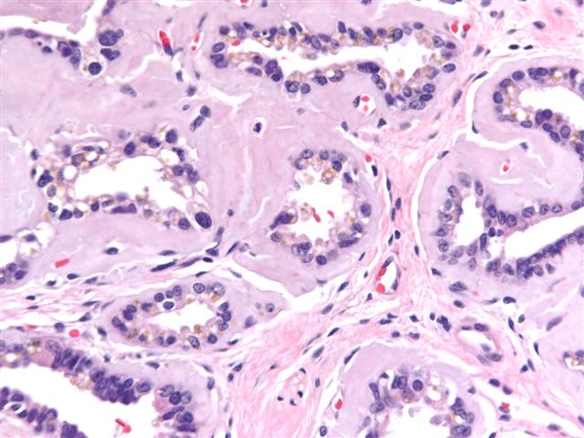
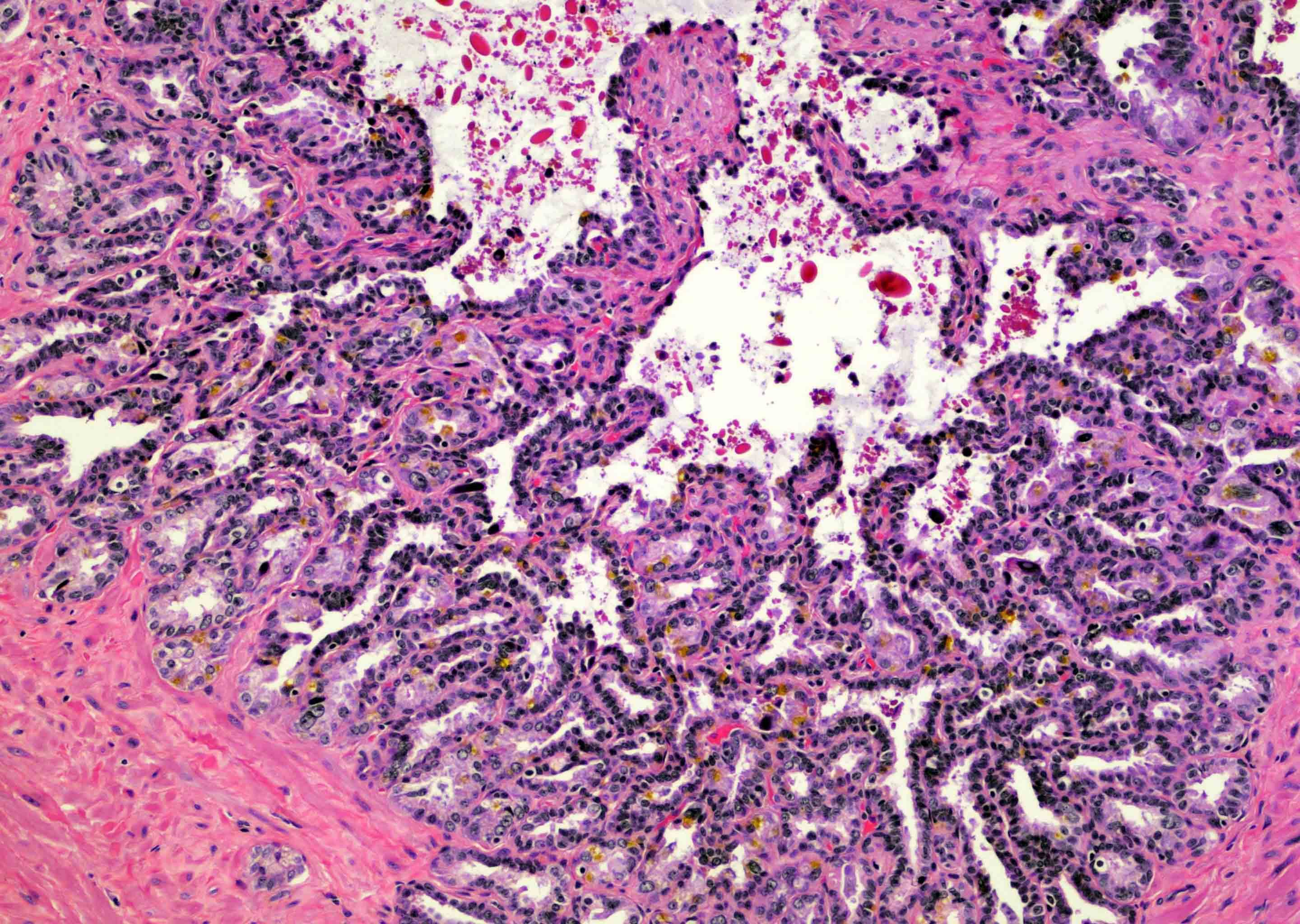
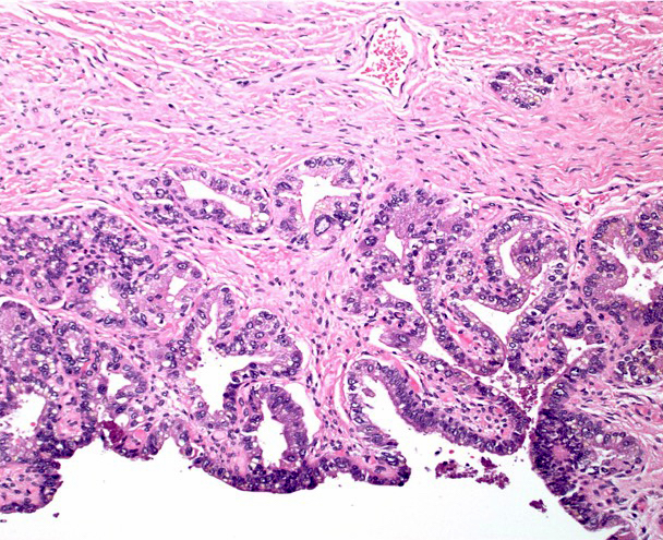
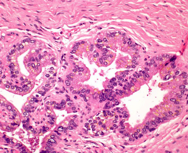
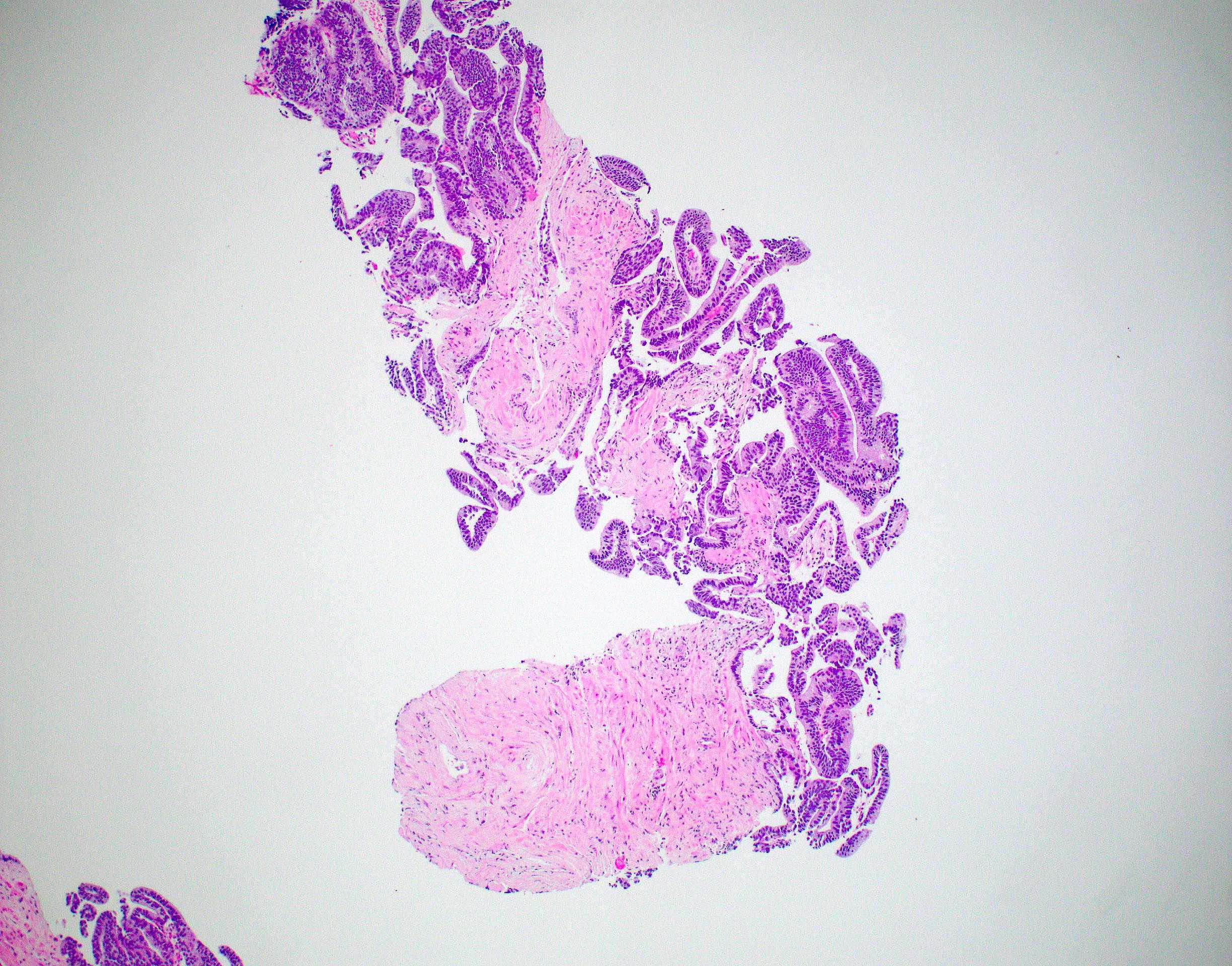
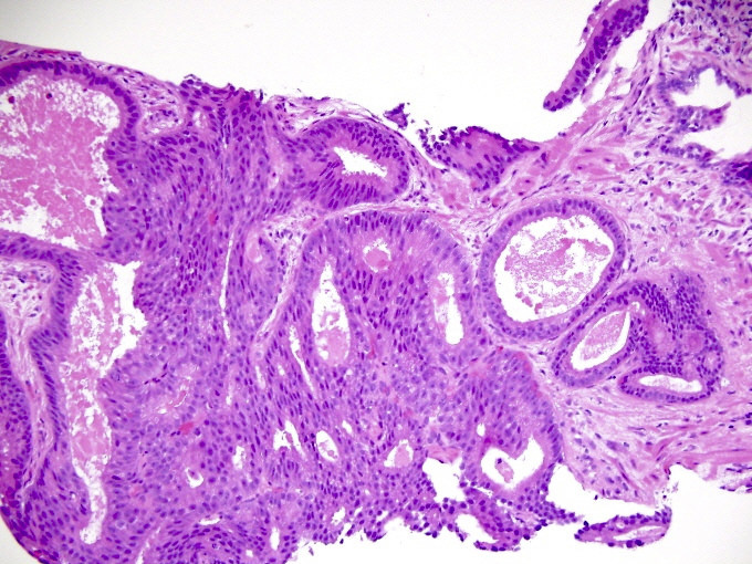
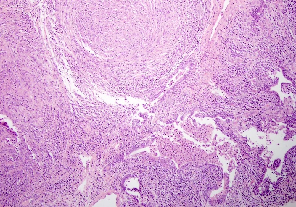
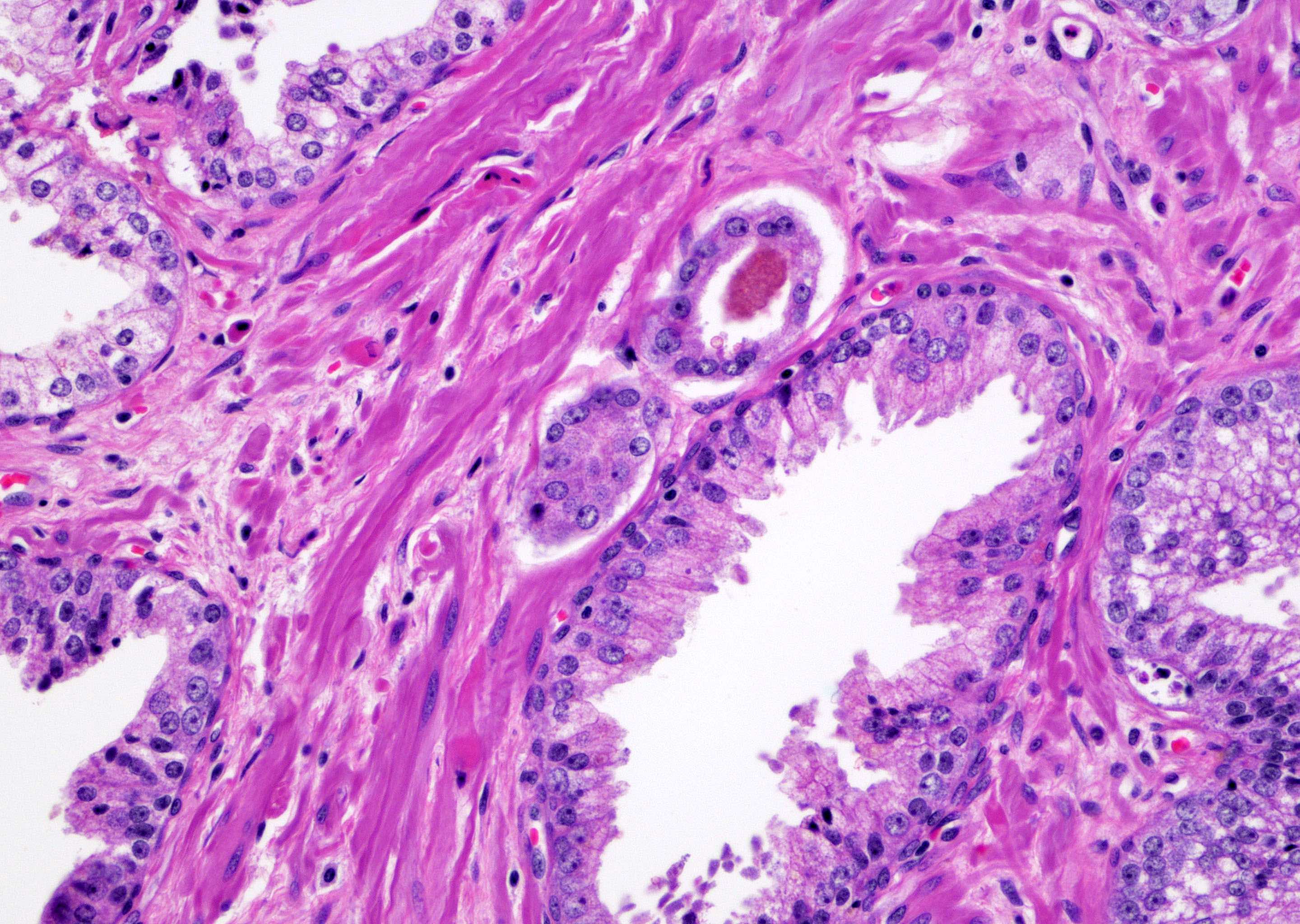
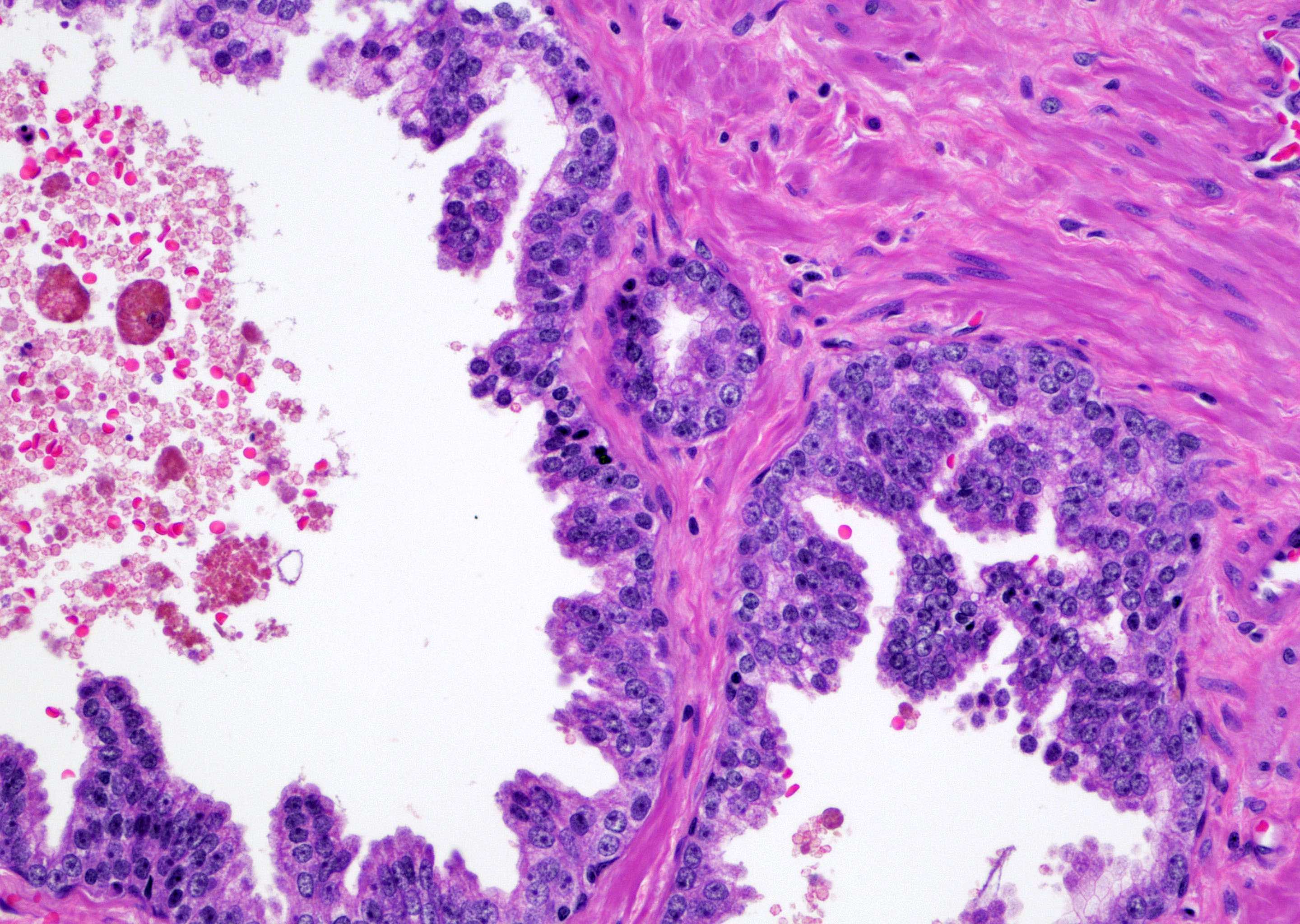
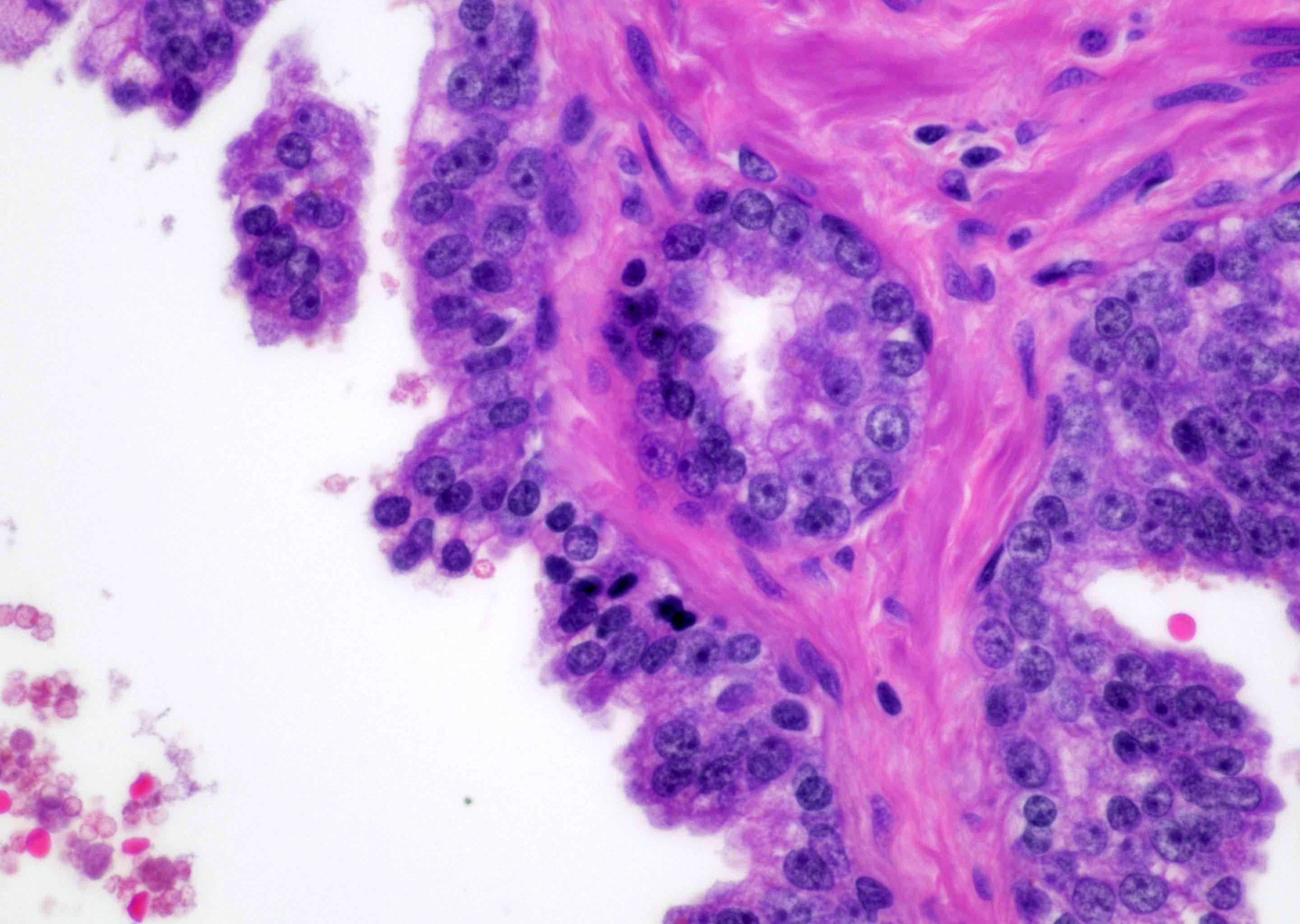
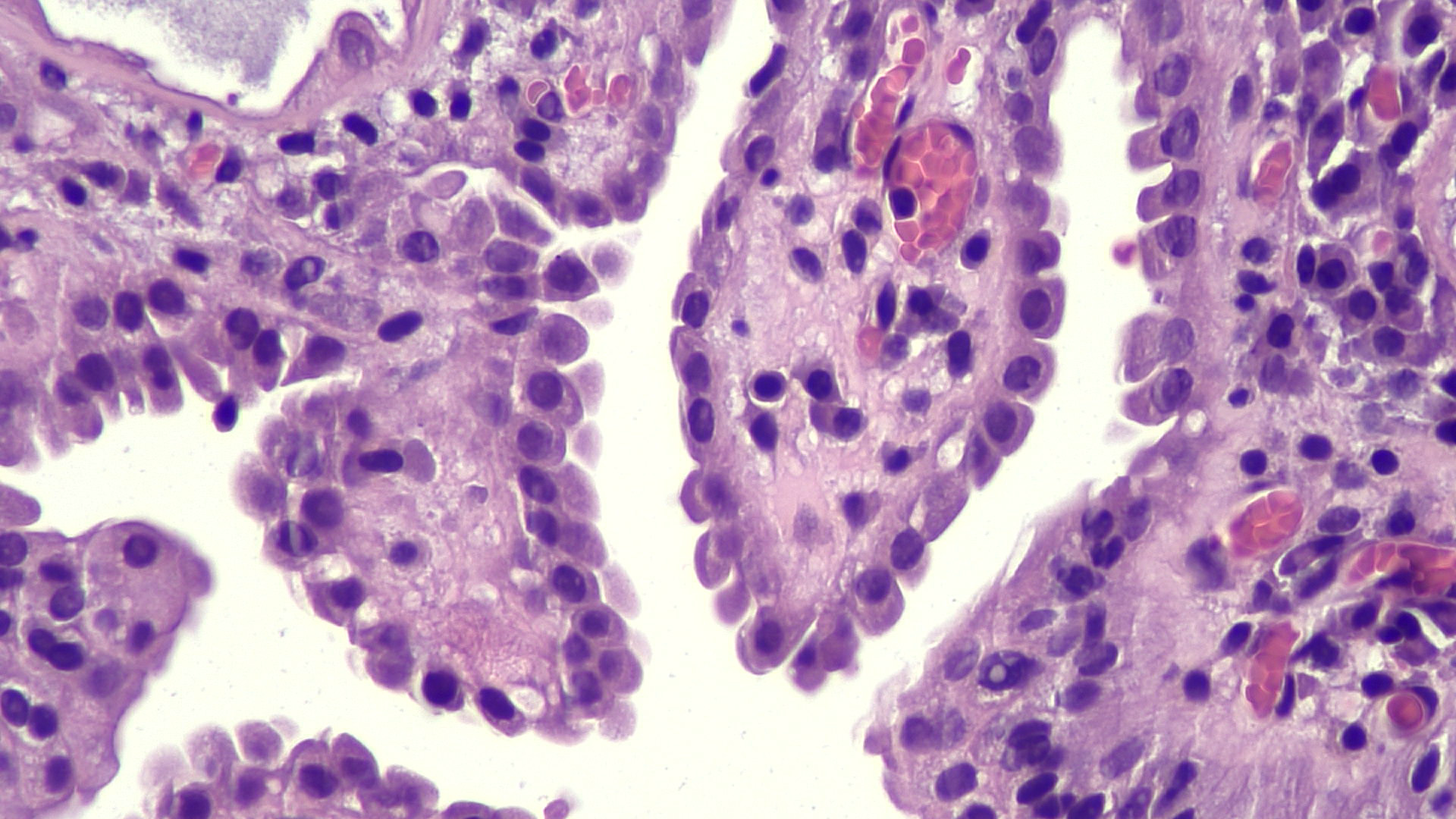
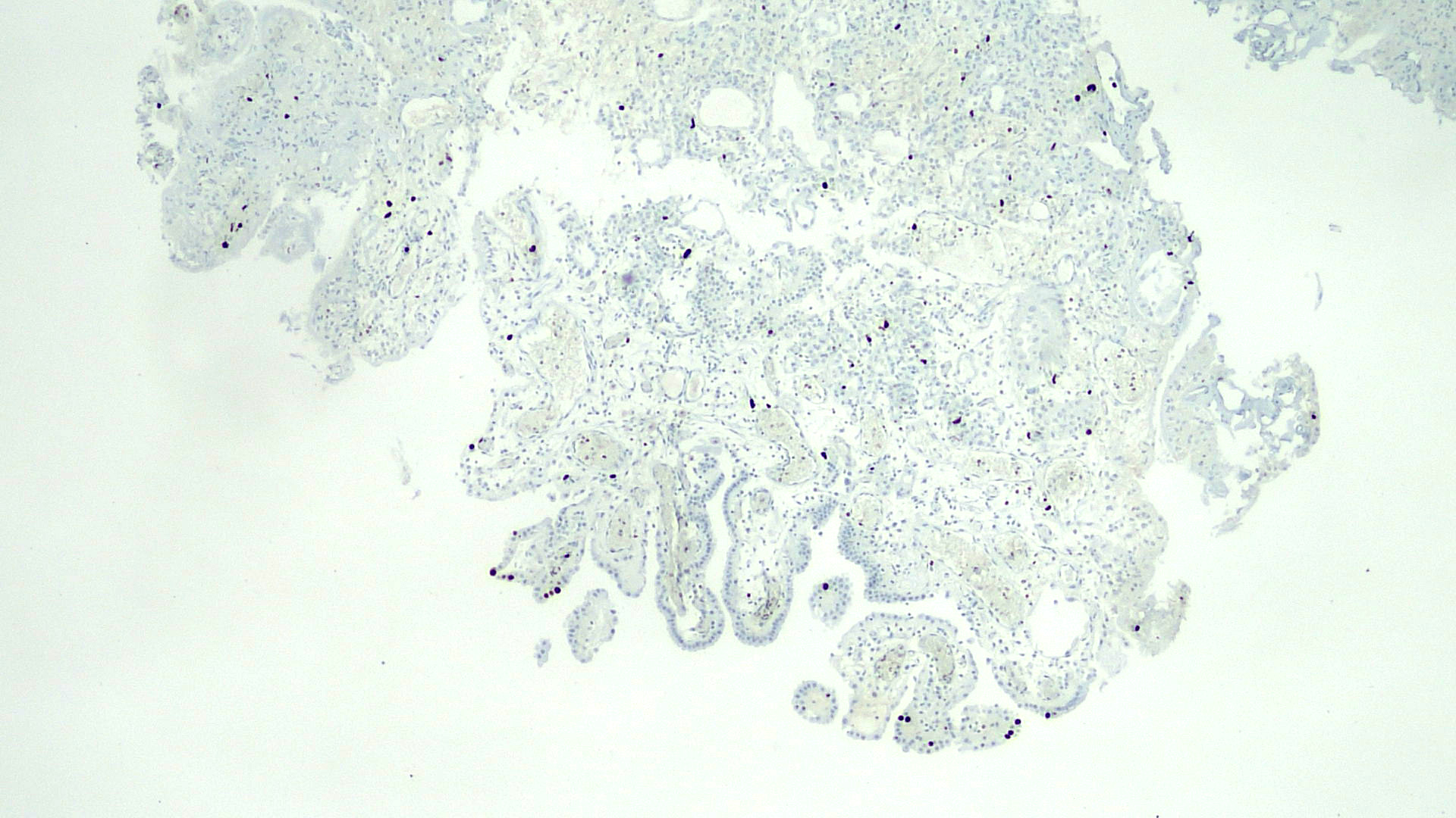
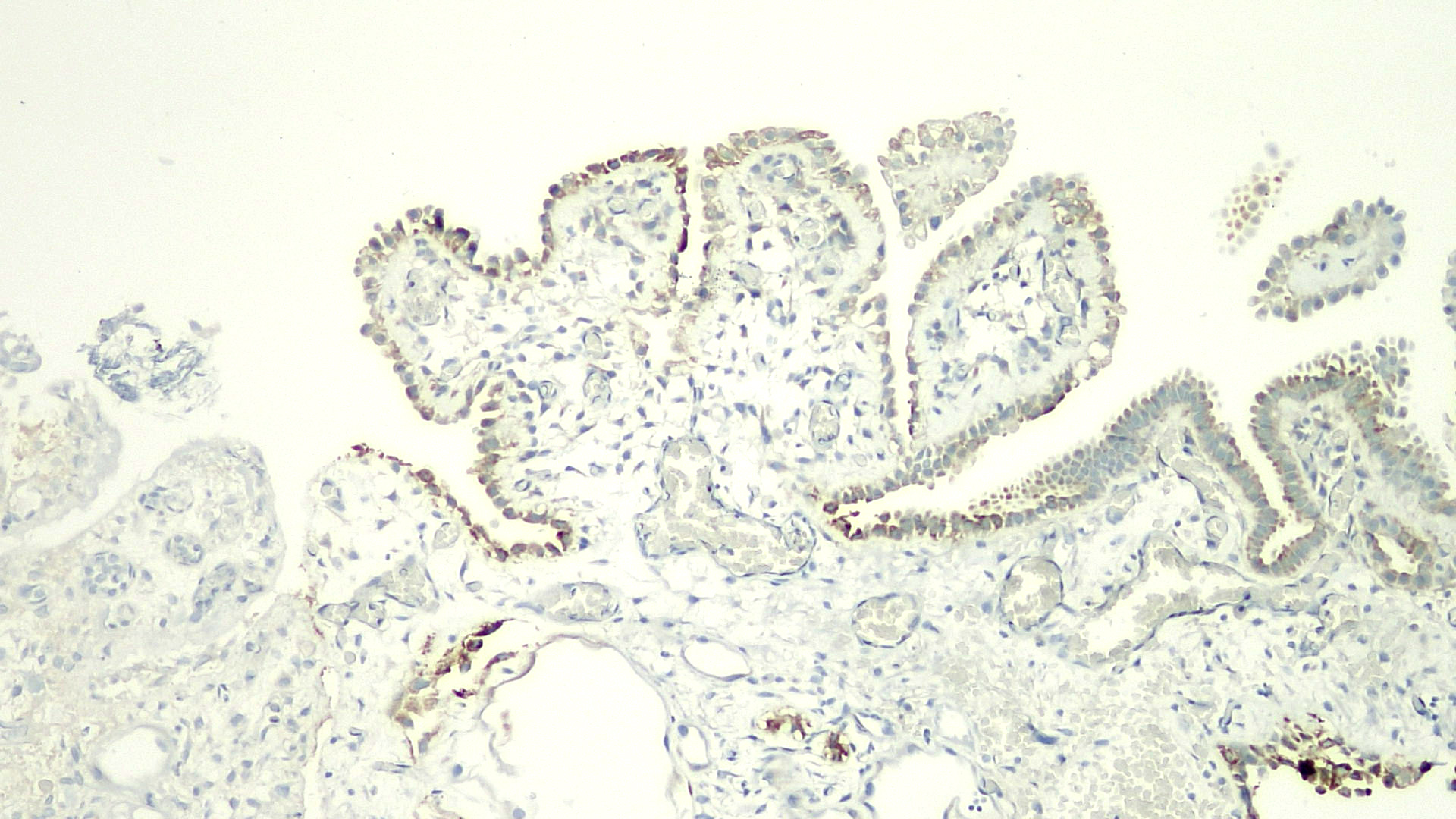
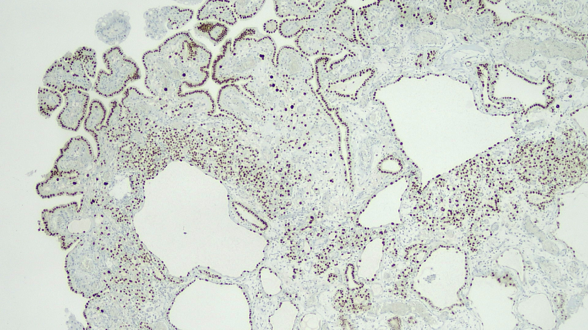
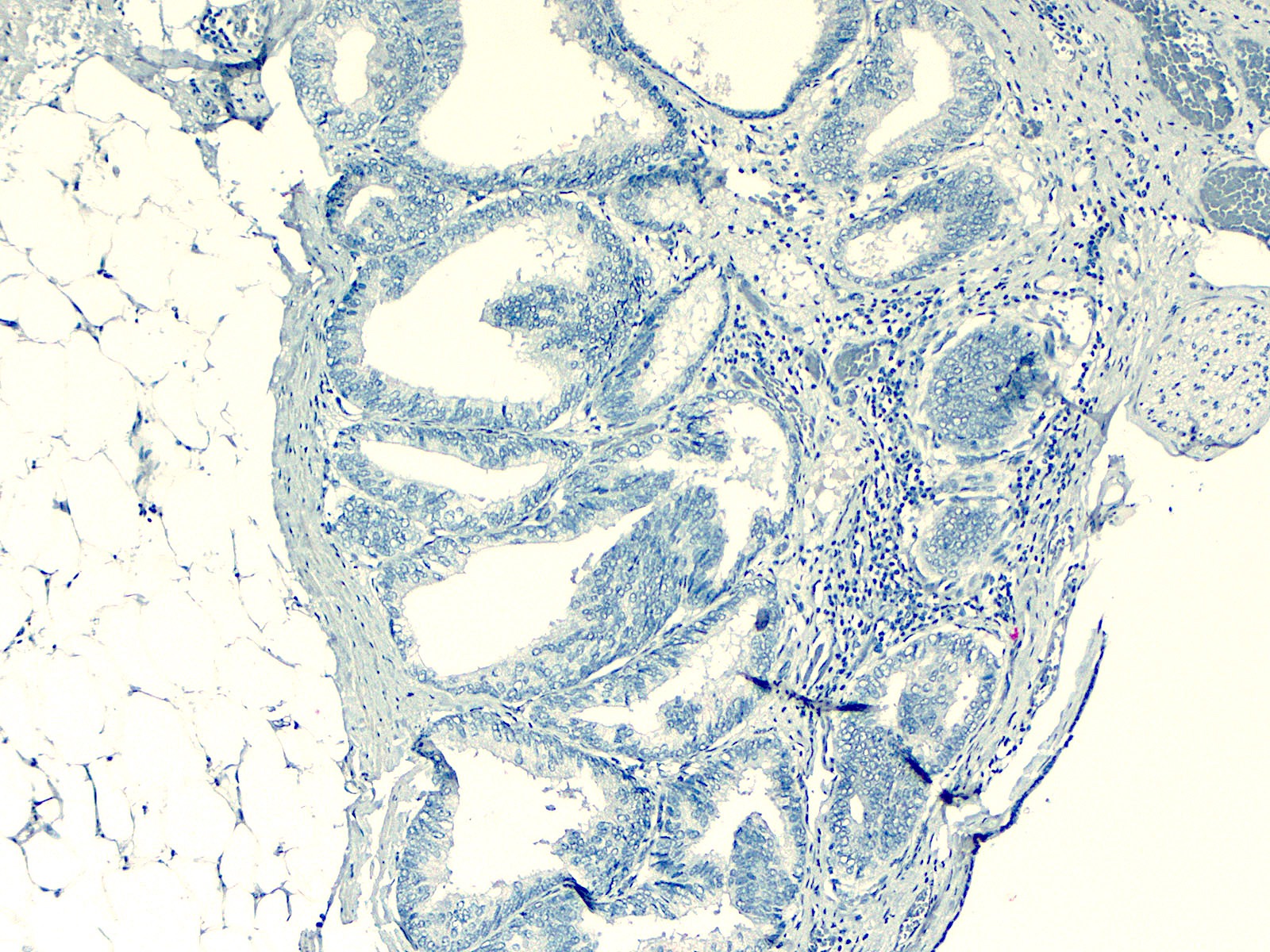
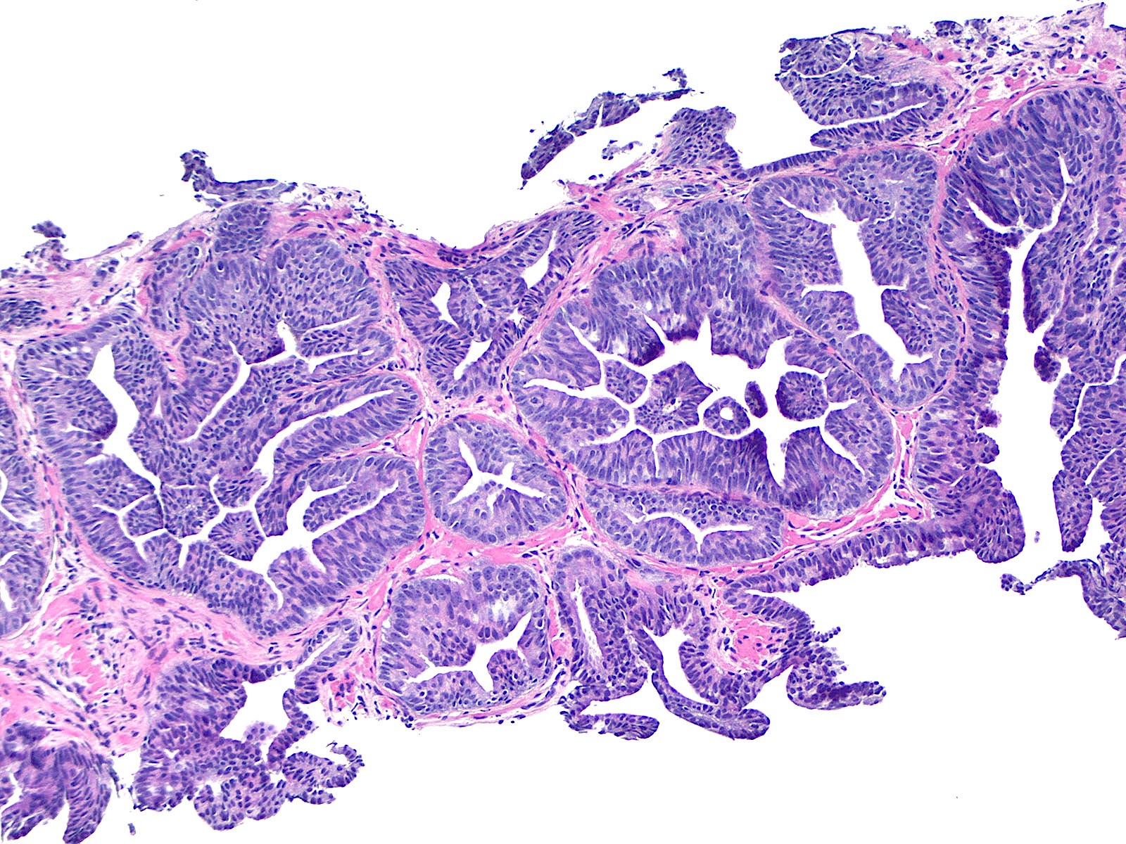
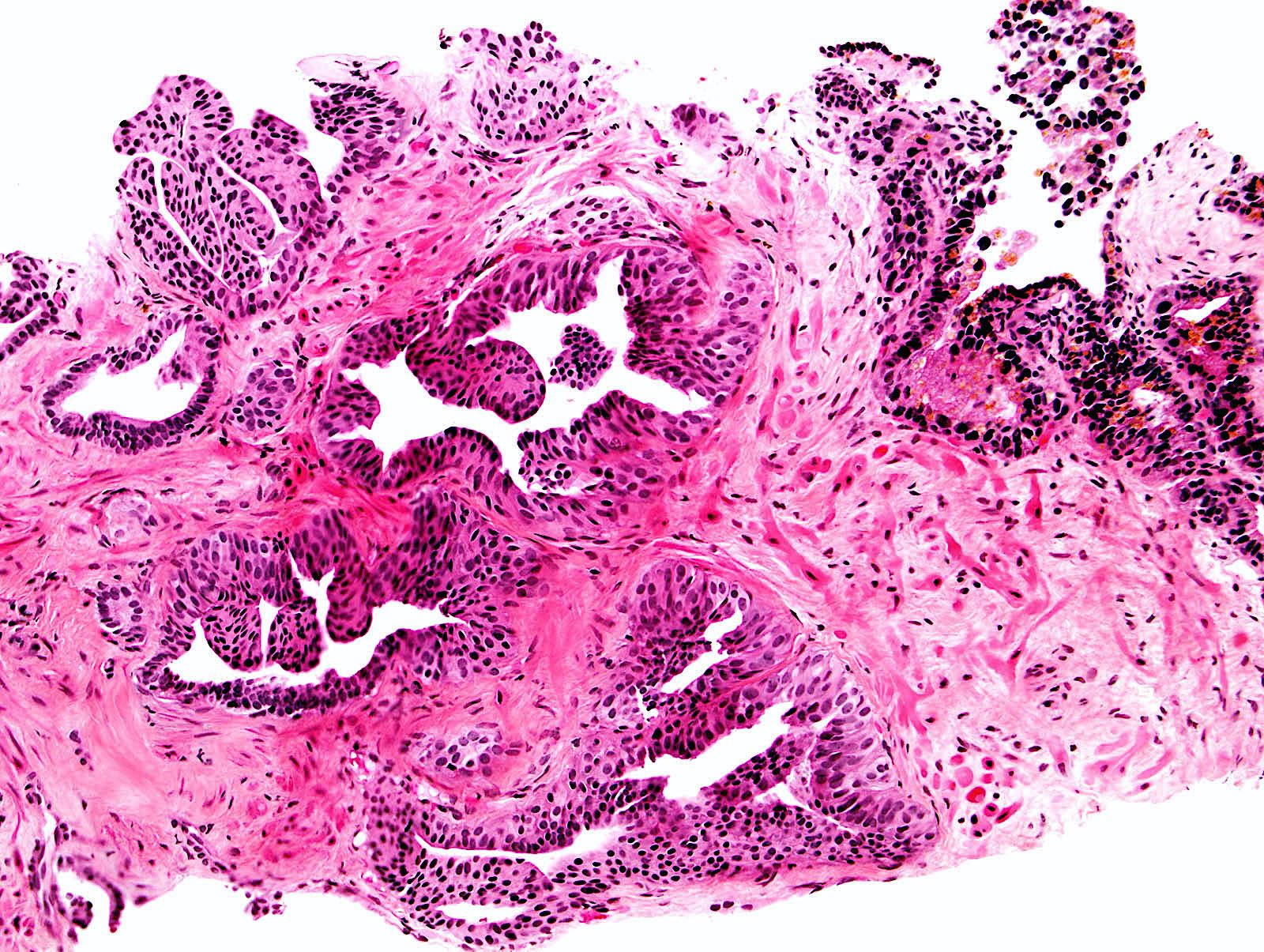
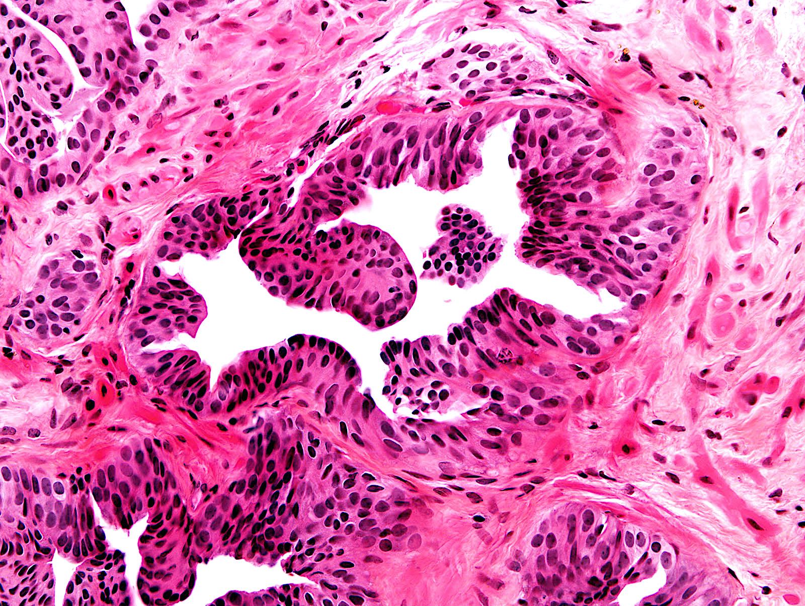
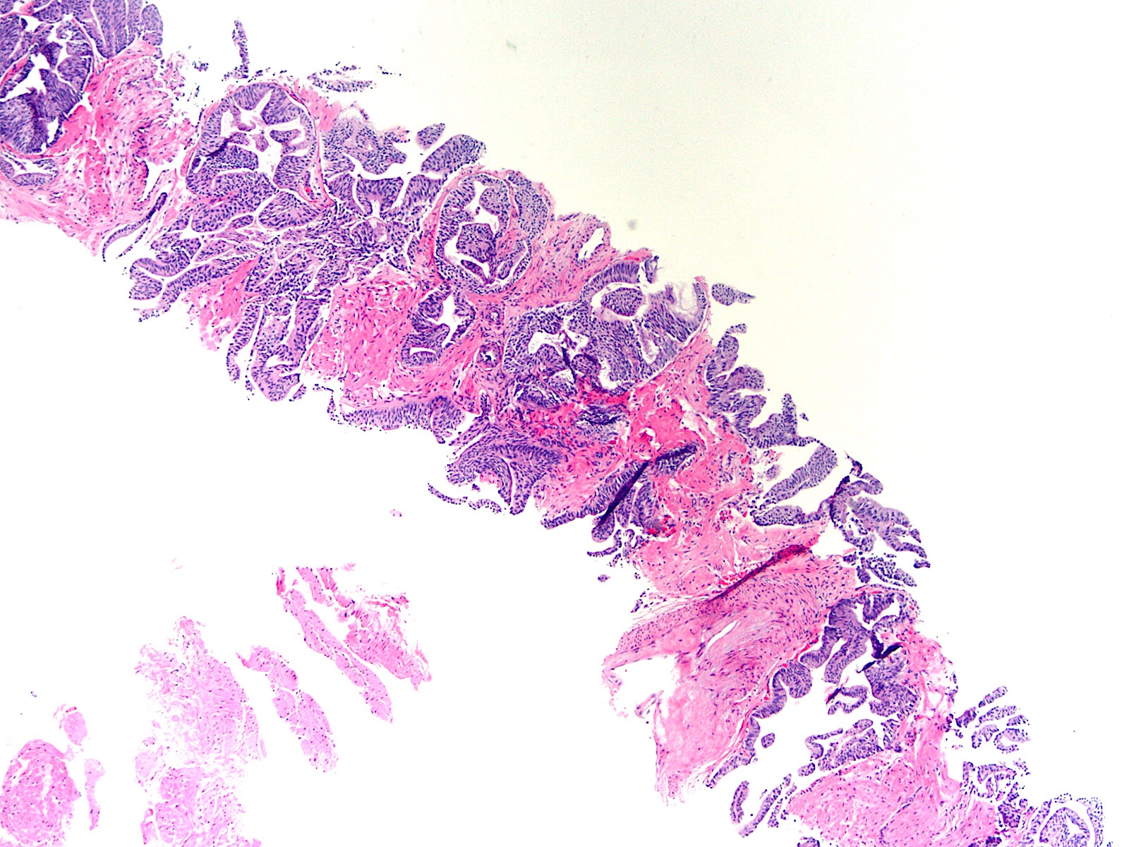
Contributed by He Huang, M.D., Ph.D. and Sarah Findeis, M.D.
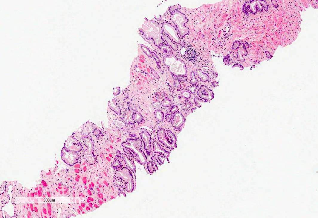
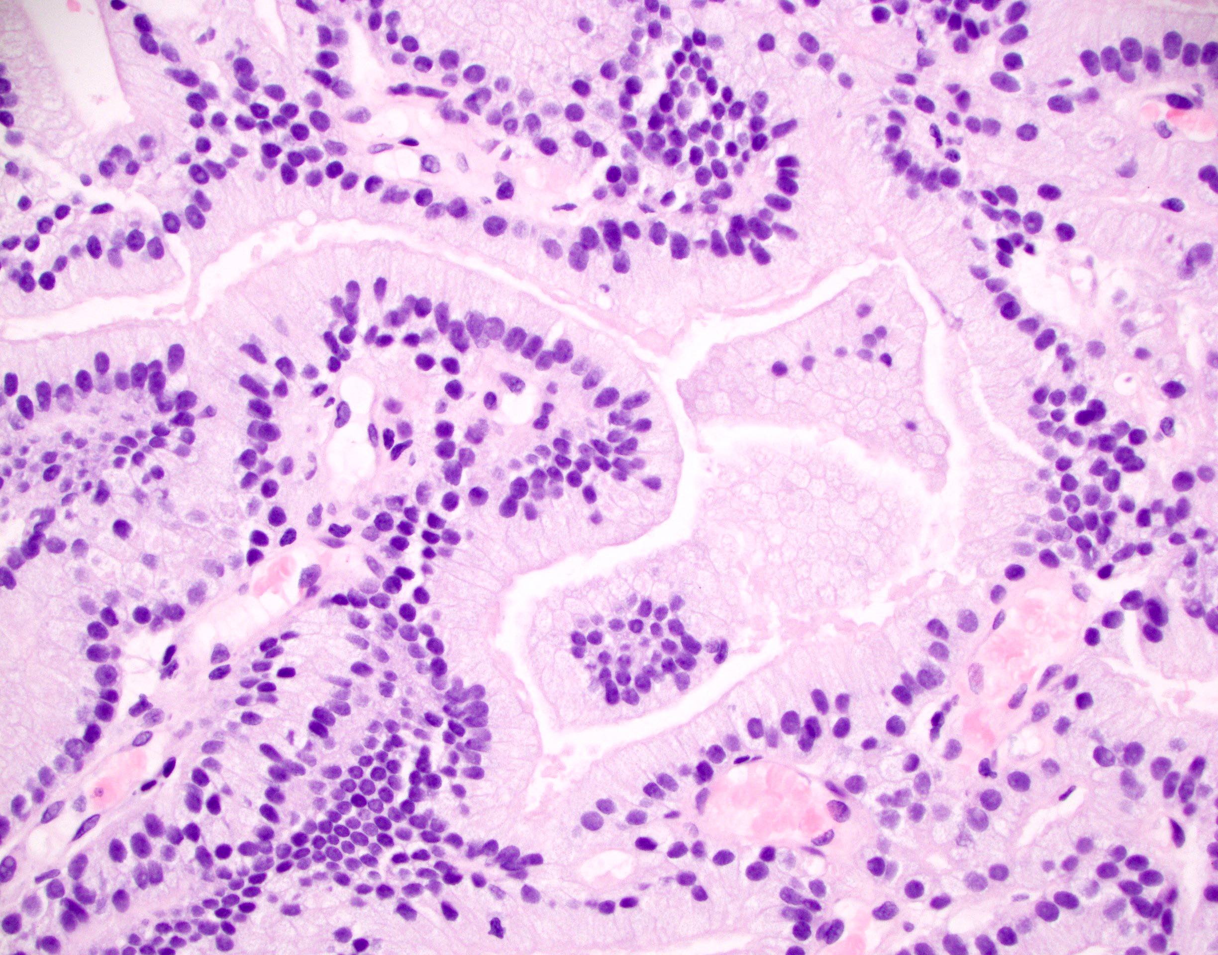
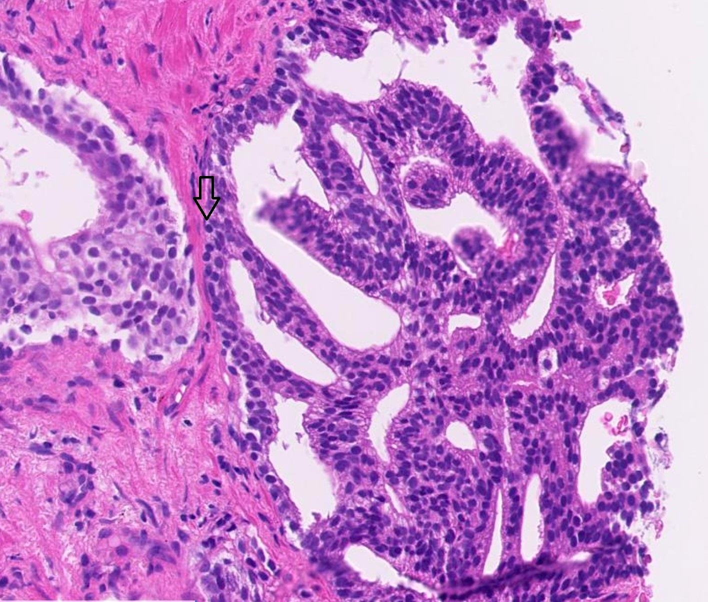
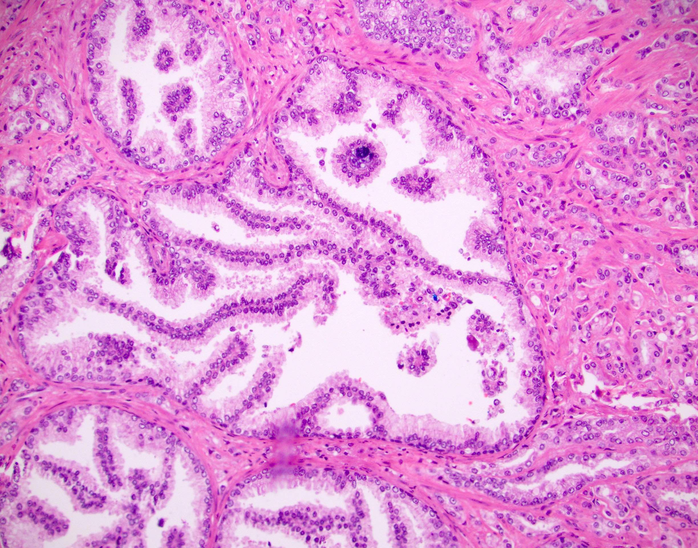
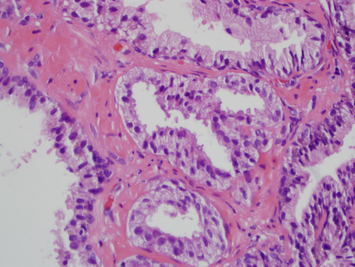
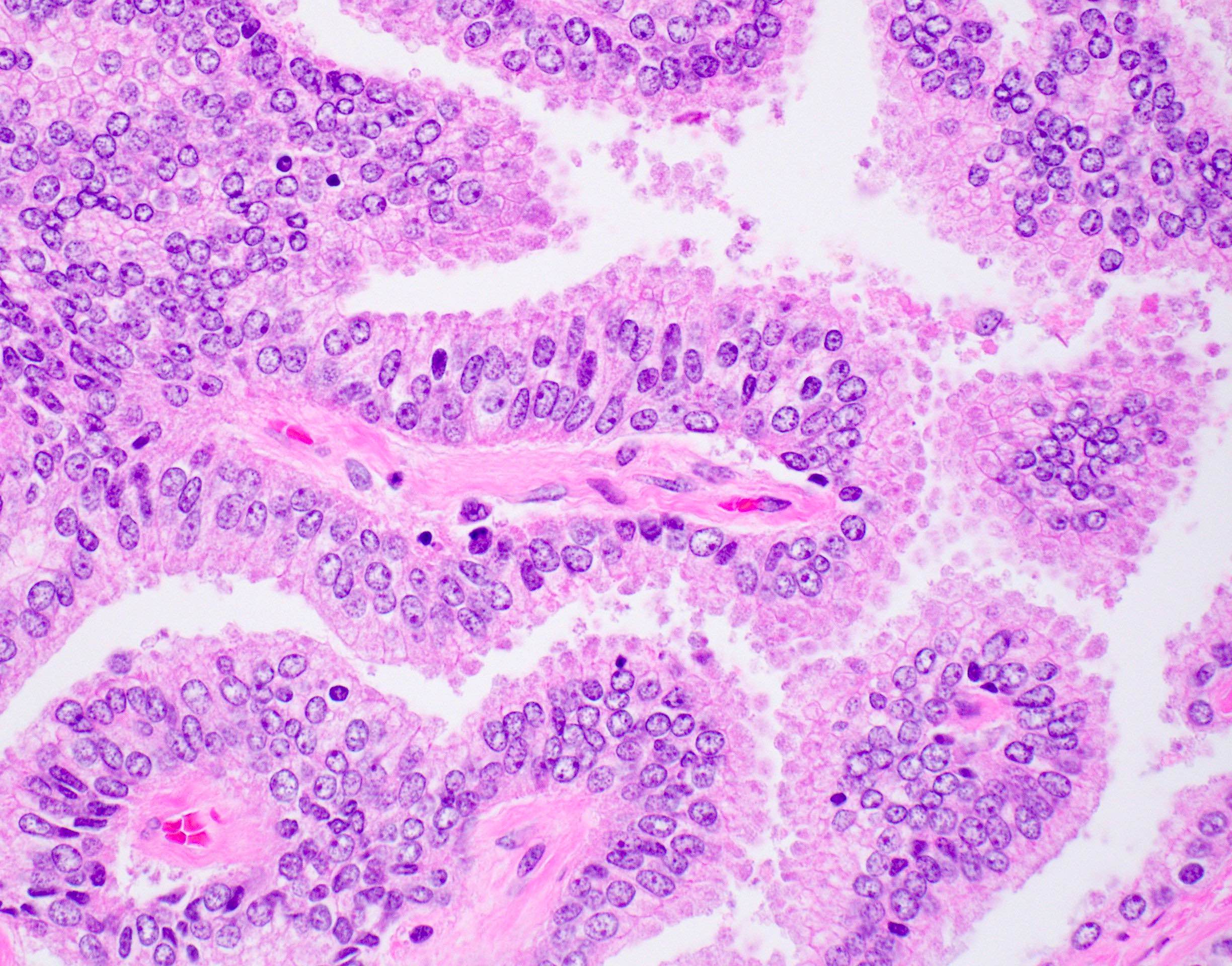
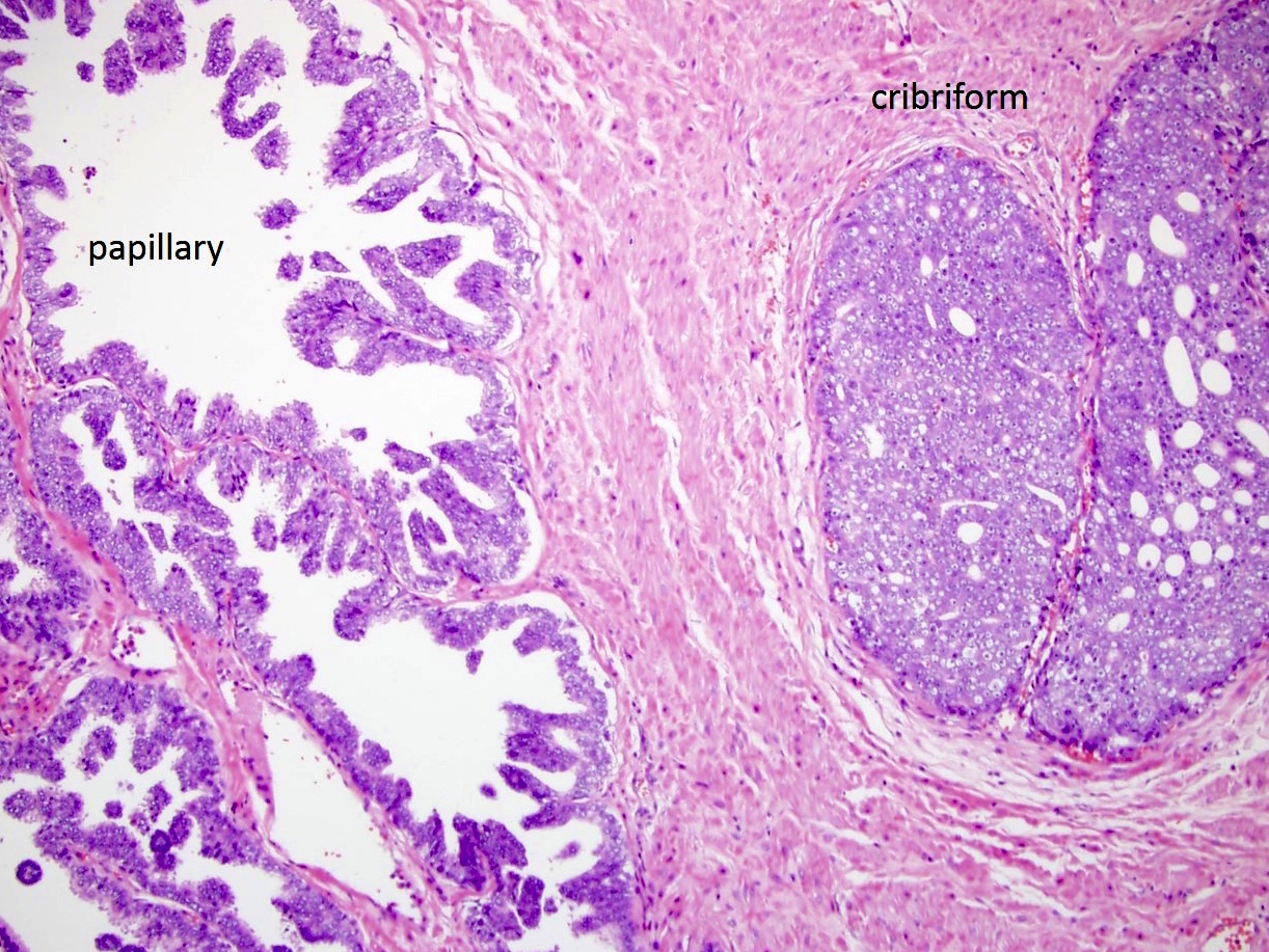
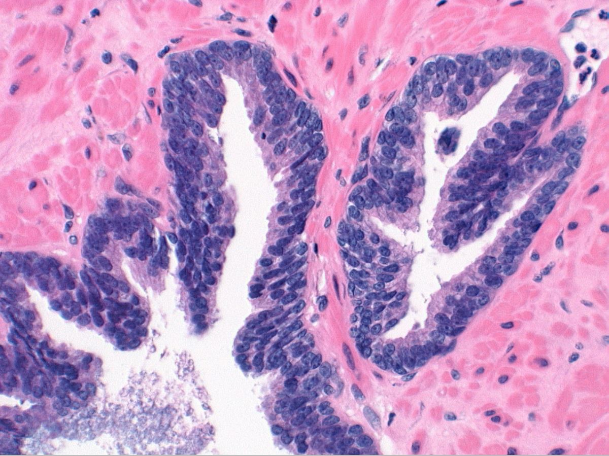
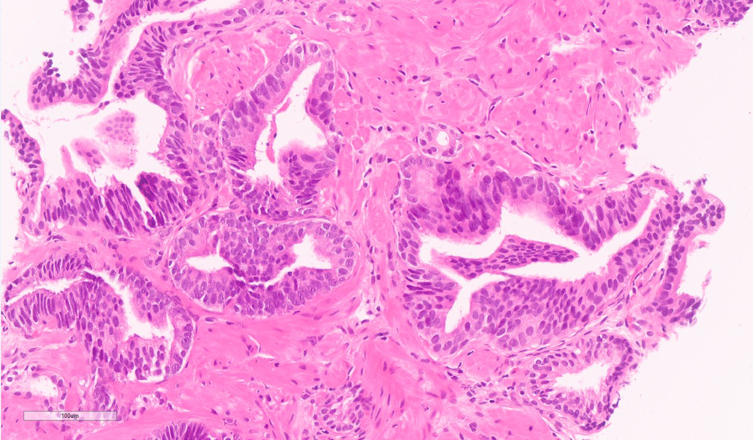
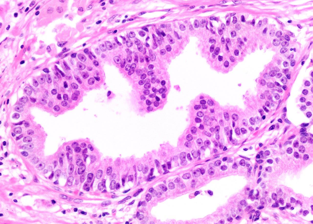
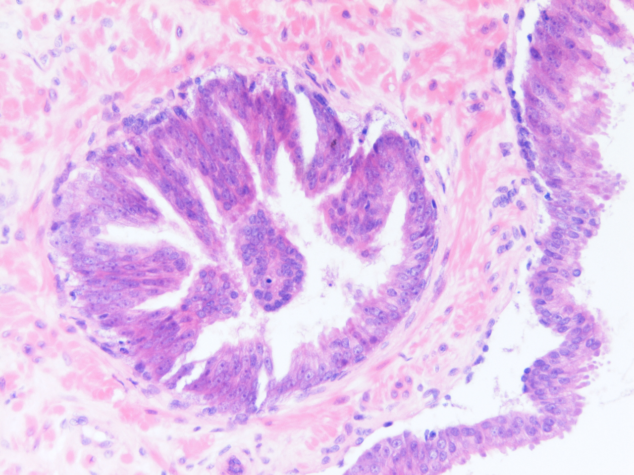
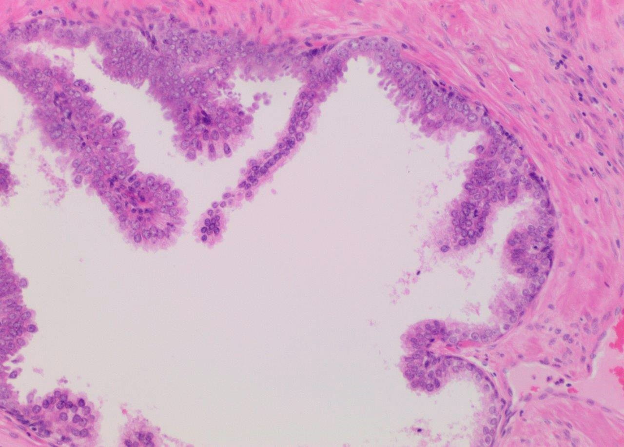
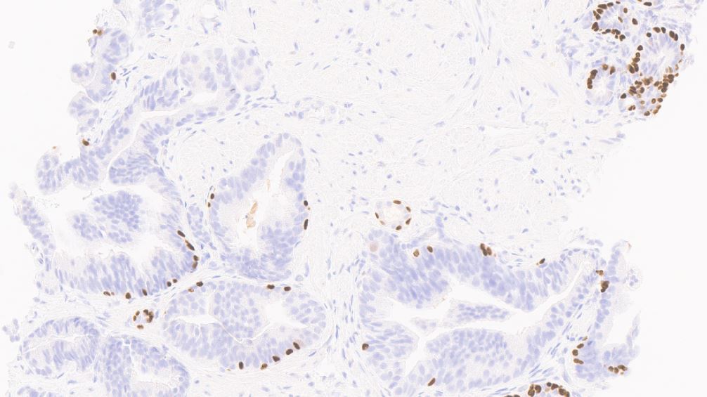
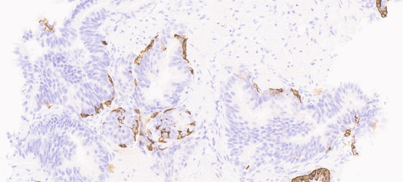
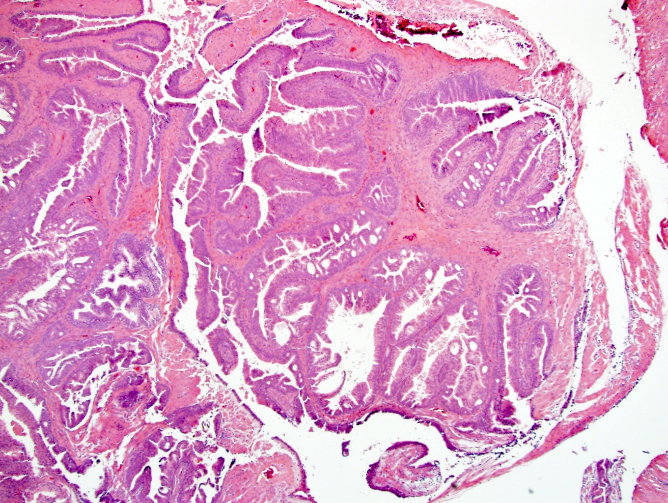
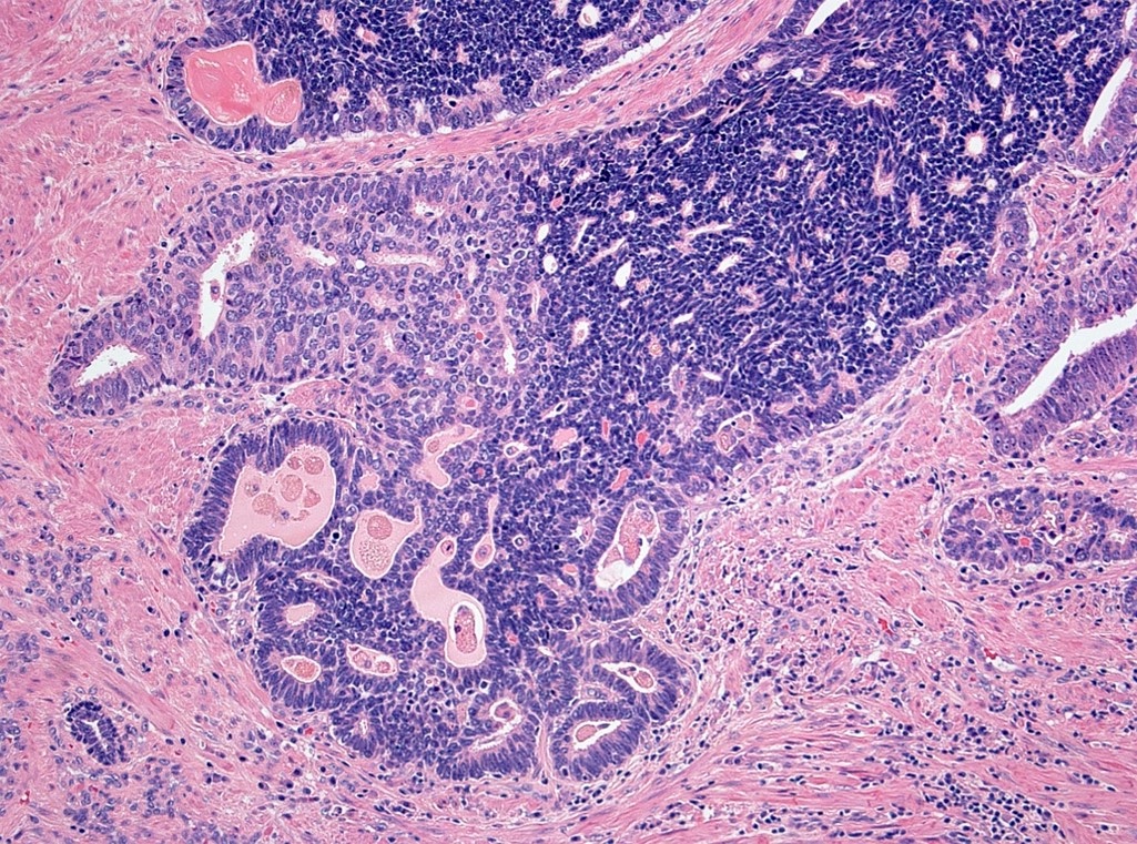
Glandular, cribriform and papillary patterns
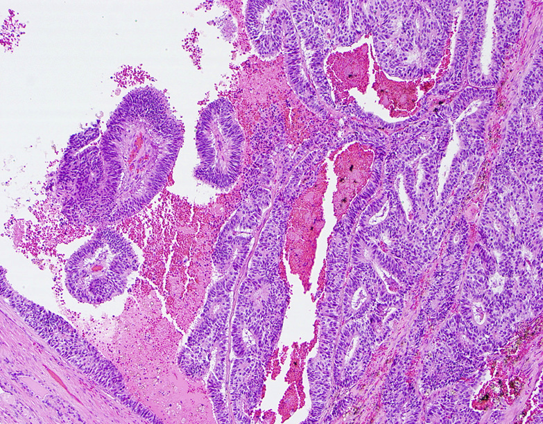
Pseudostratified columnar cells
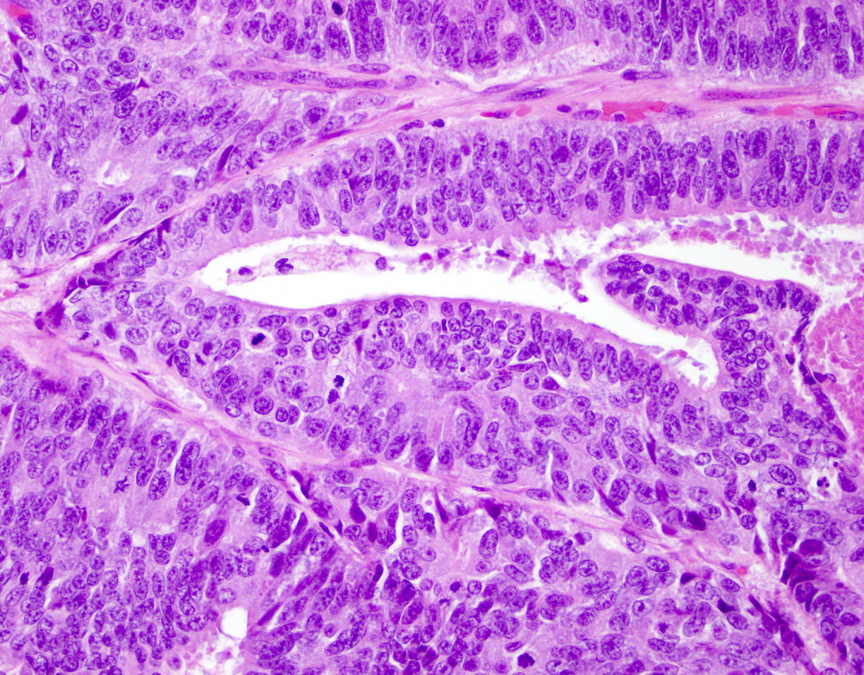
Mitotic figures
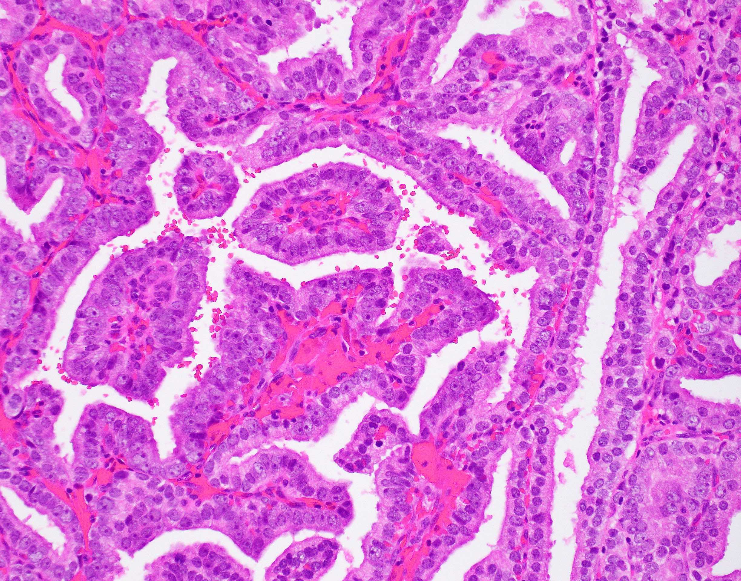
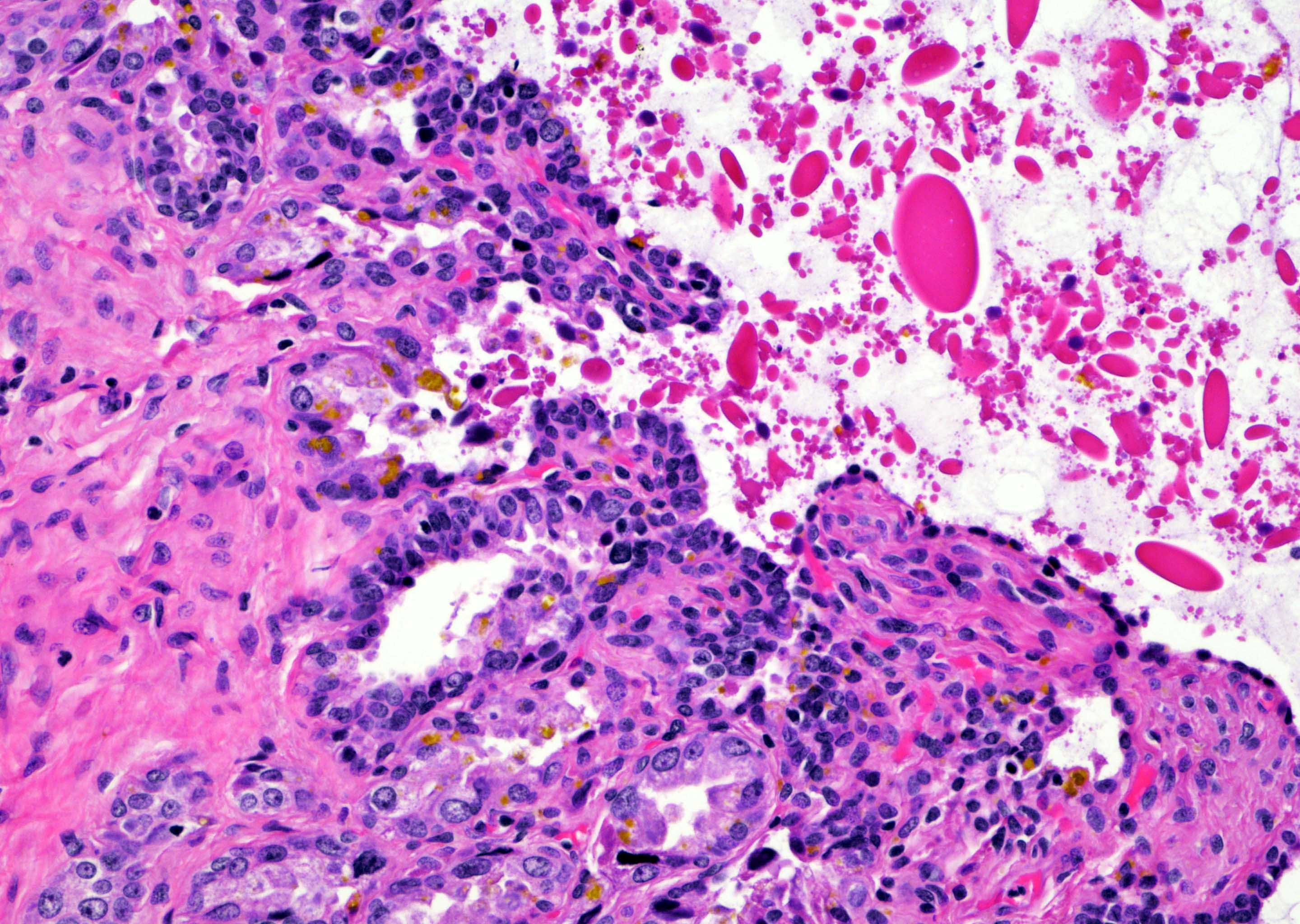
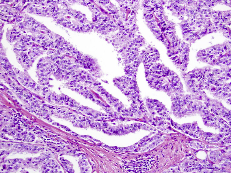
Papillary architecture
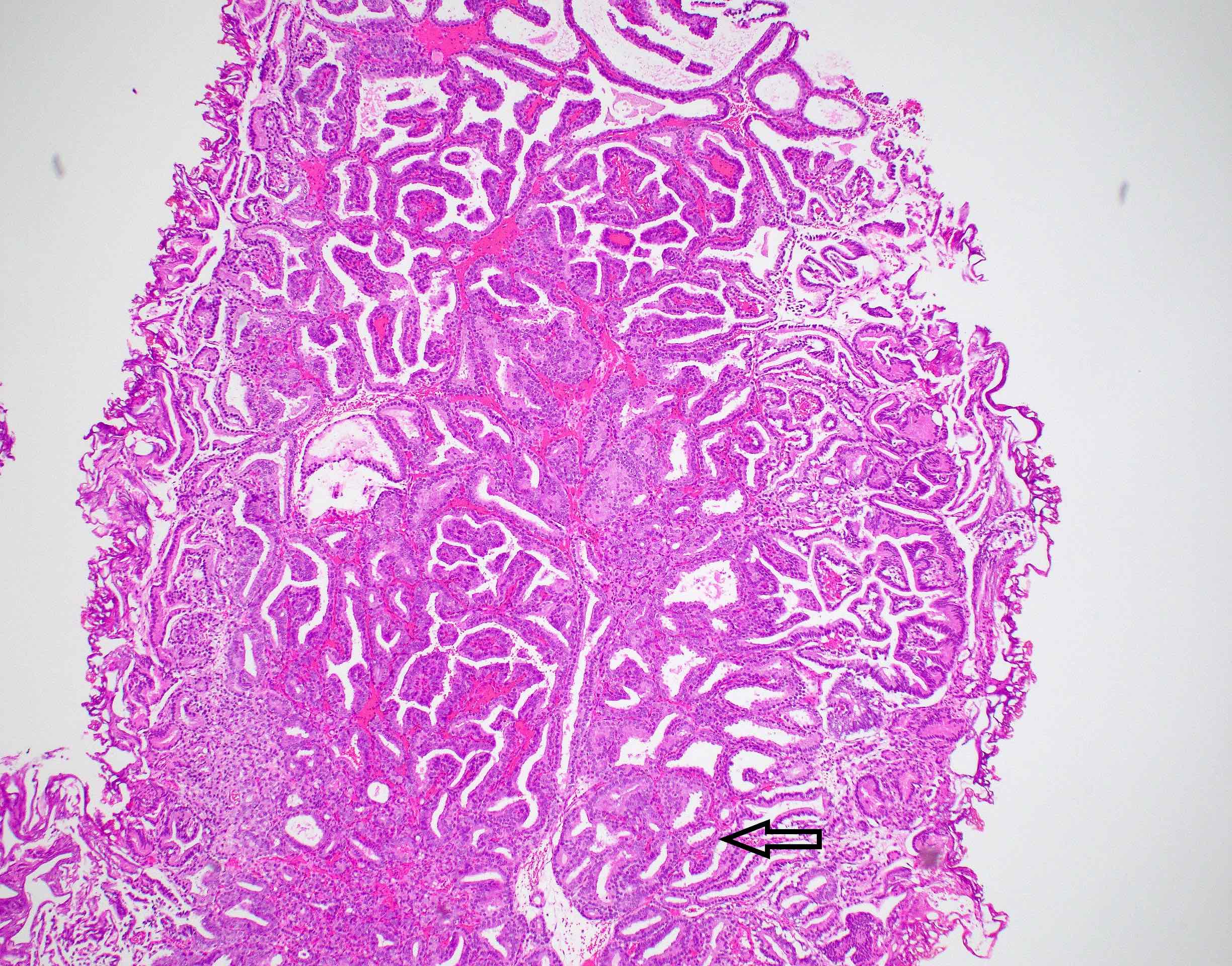
Papillary and cribriform pattern
Core with papillae
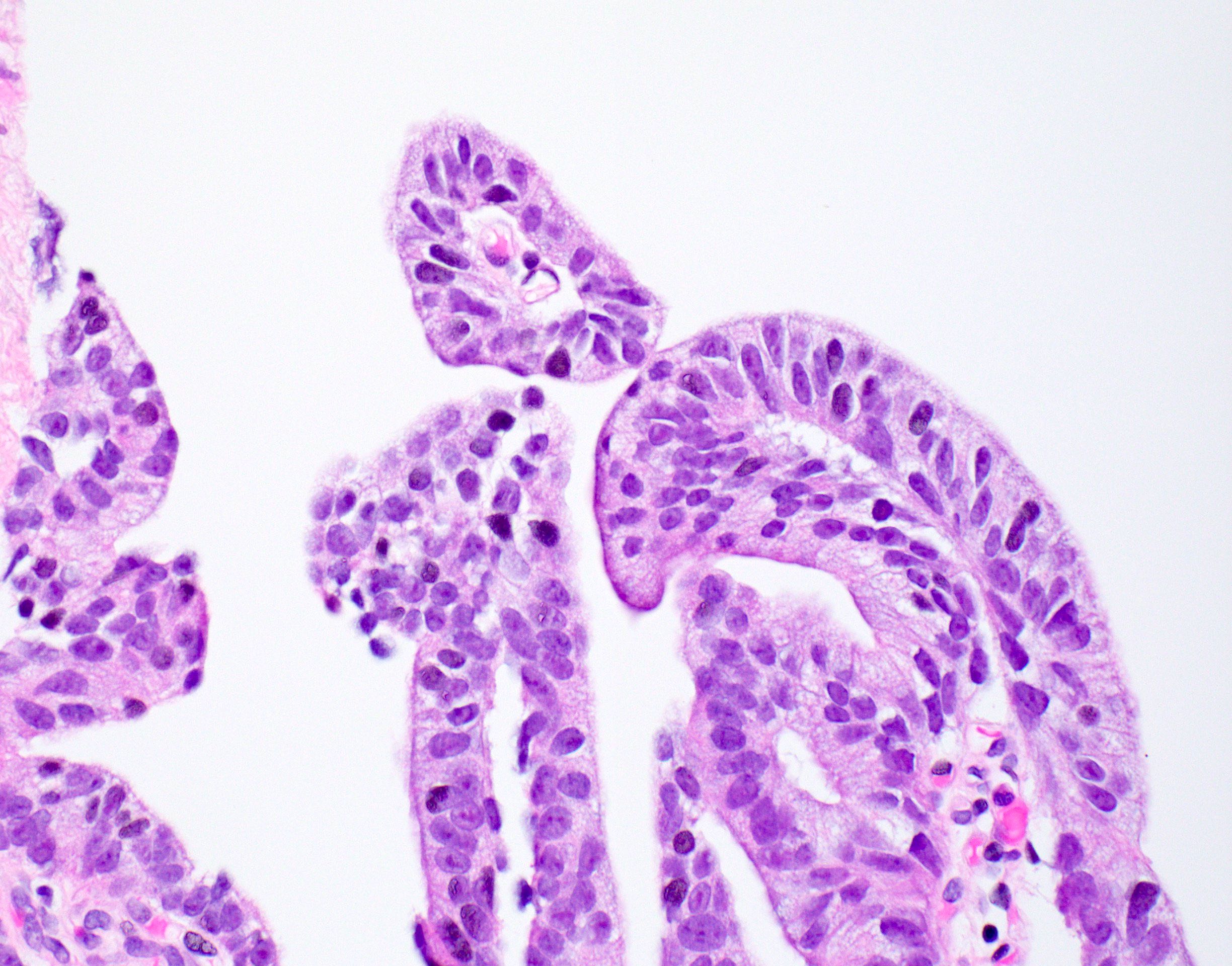
Pseudostratification
and papillae
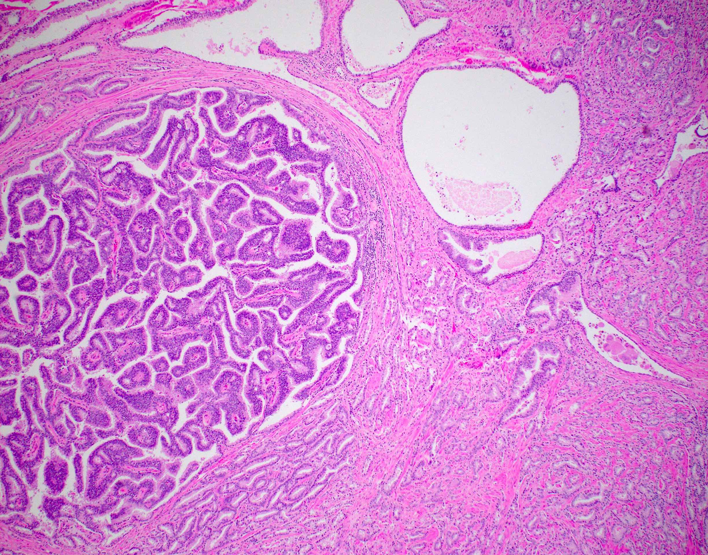
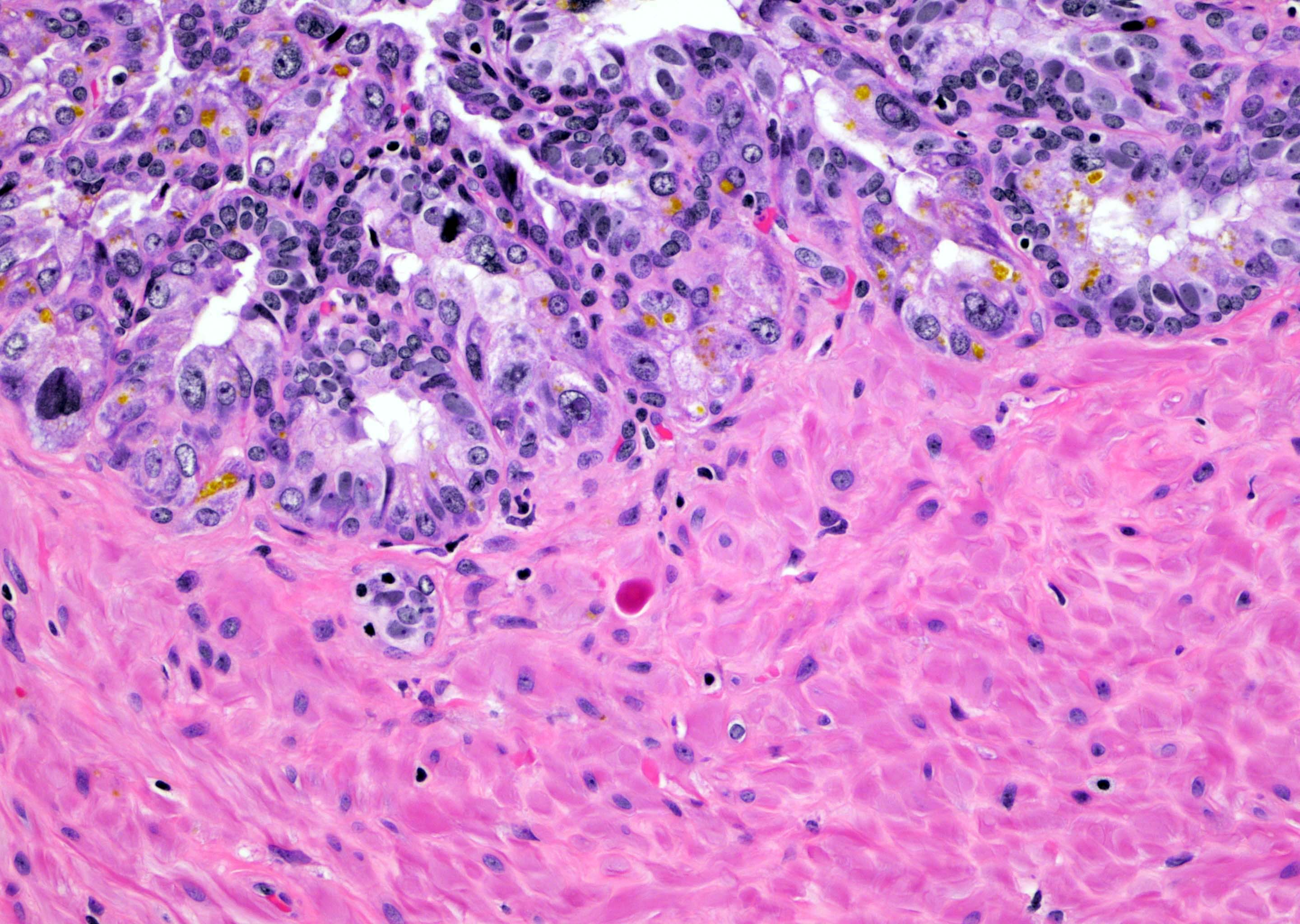
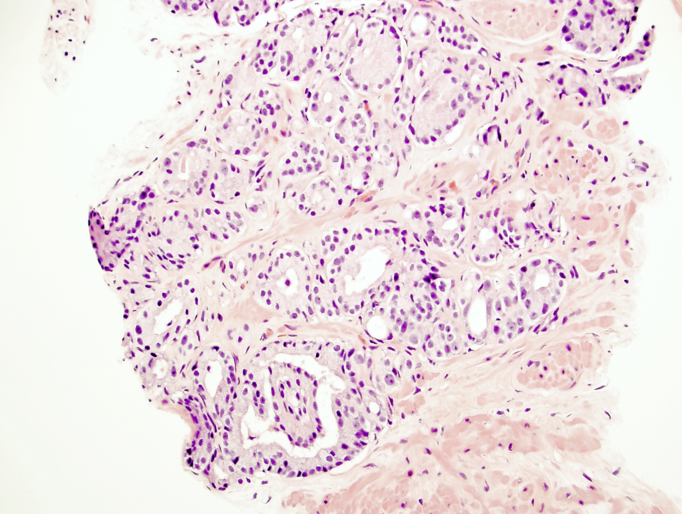
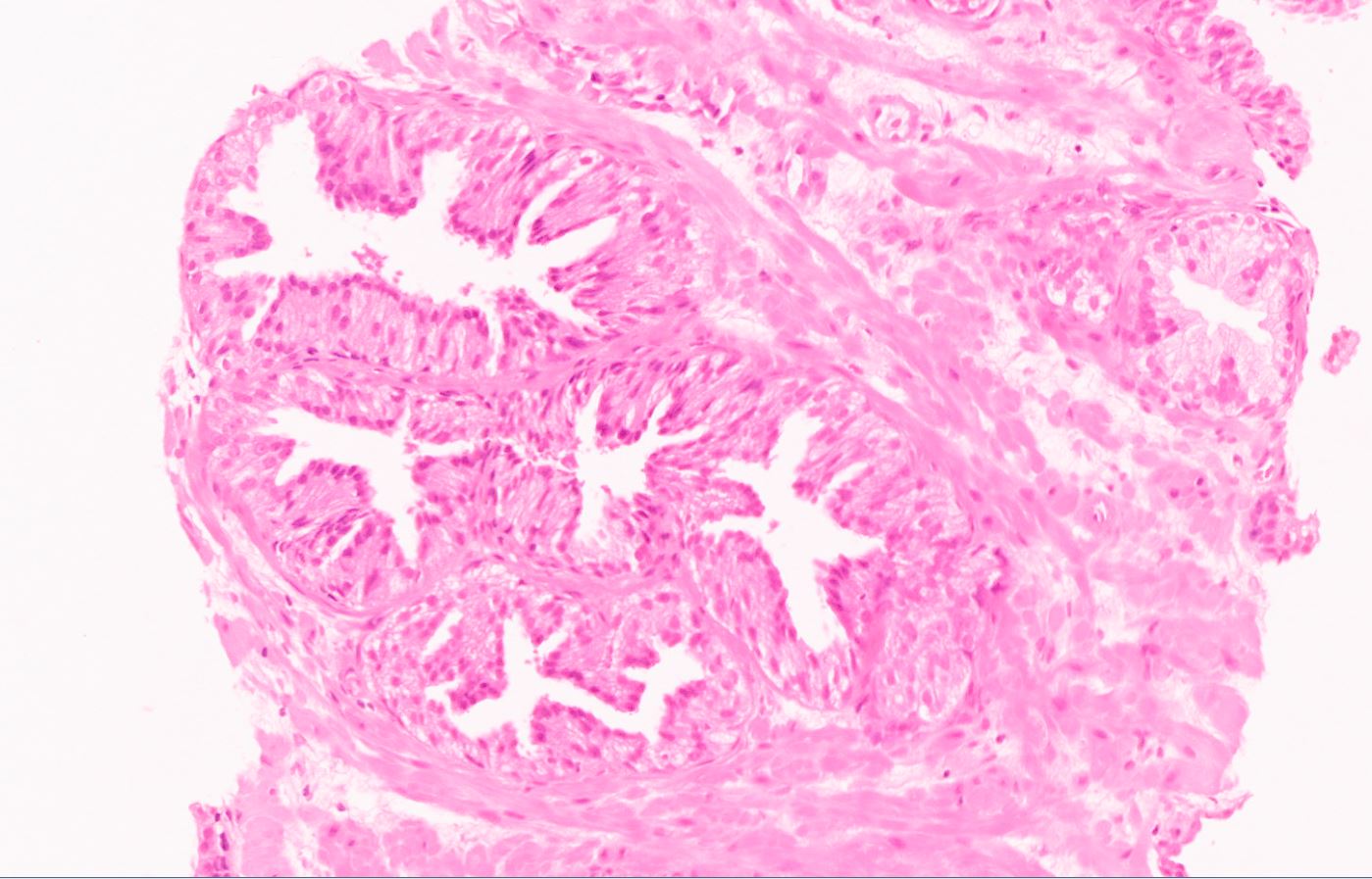
Mixed acinar and ductal adenocarcinoma
Pseudostratification
and
fibrovascular
core
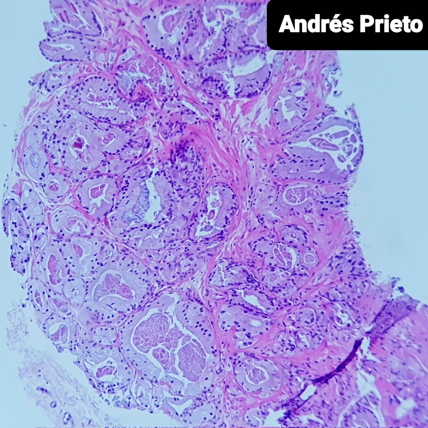
Contributed by @prietopath on Twitter
 Contributed by @prietopath on Twitter (see original post here)">
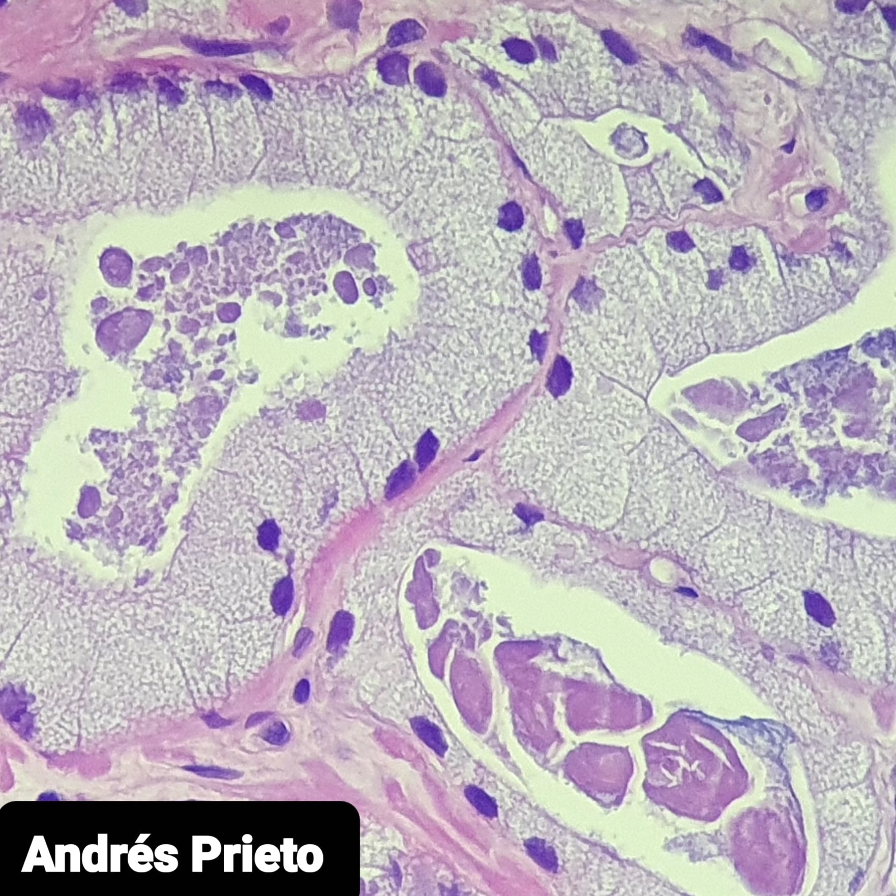
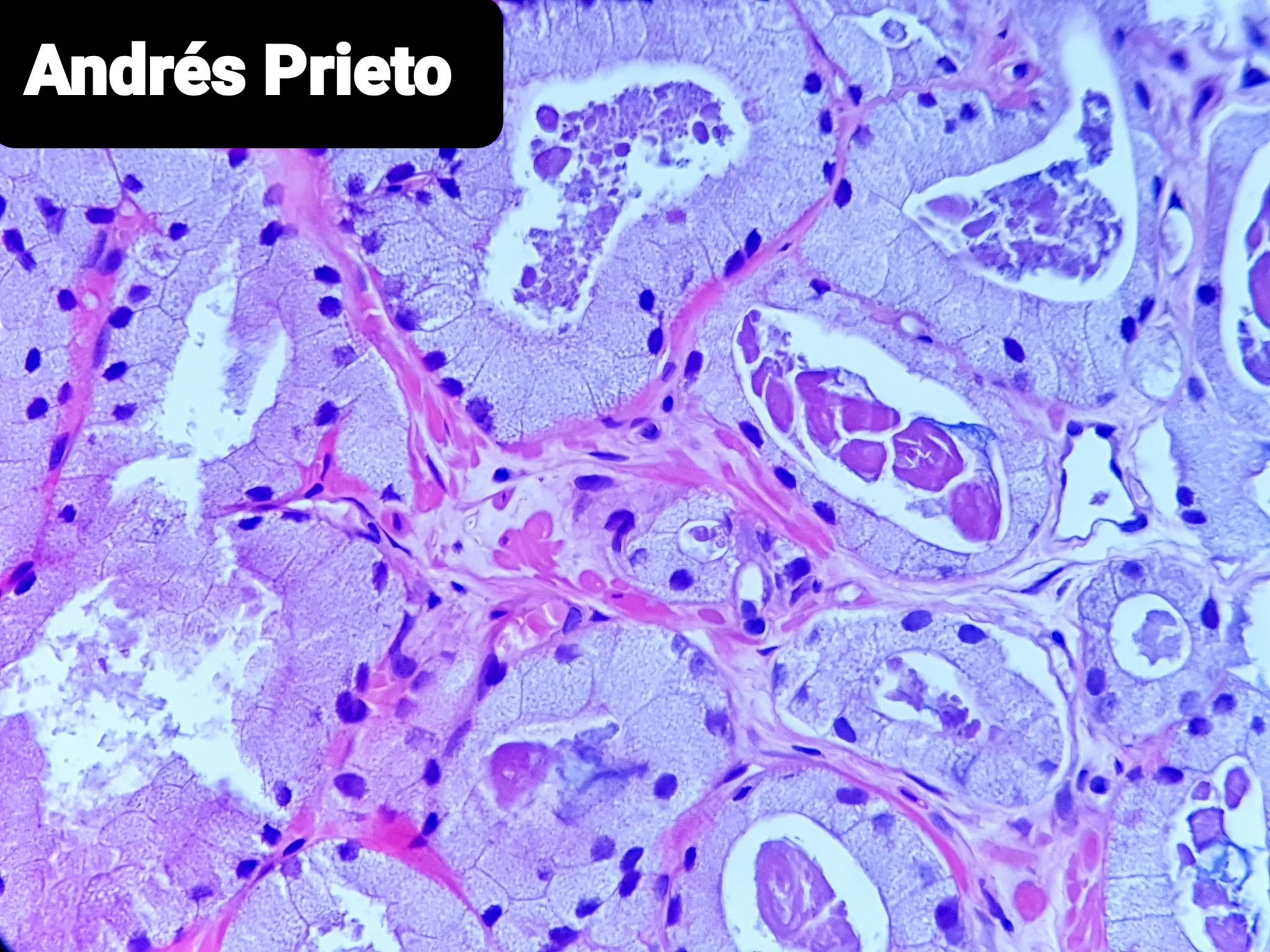
Foamy gland adenocarcinoma
 Contributed by @prietopath on Twitter (see original post here)">
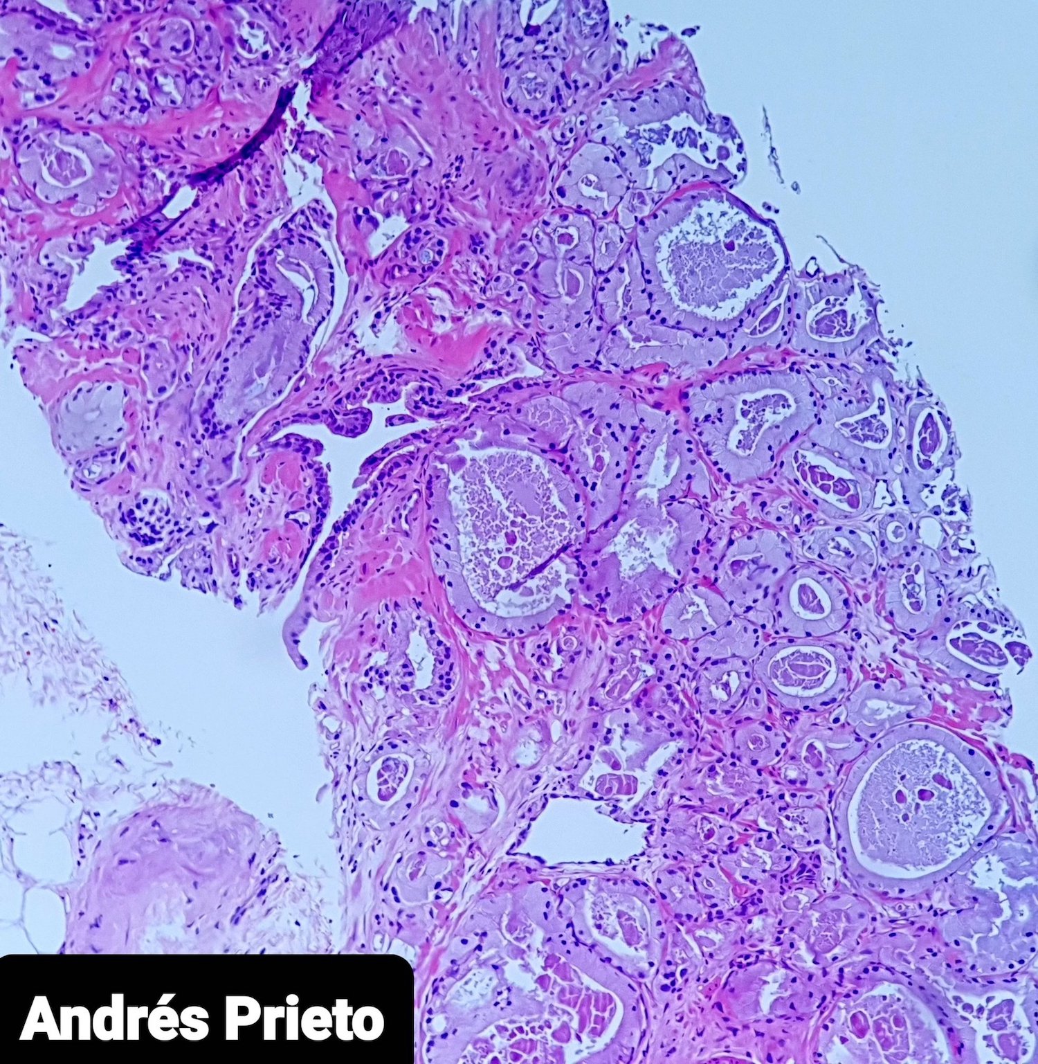
Foamy gland adenocarcinoma
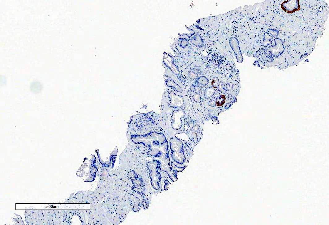
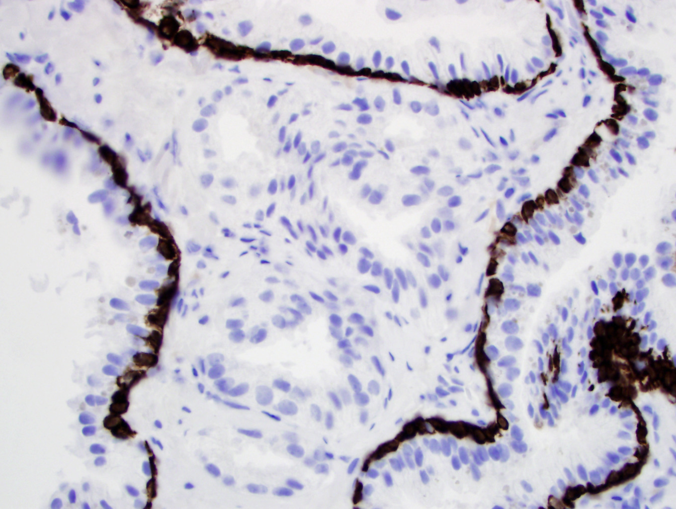
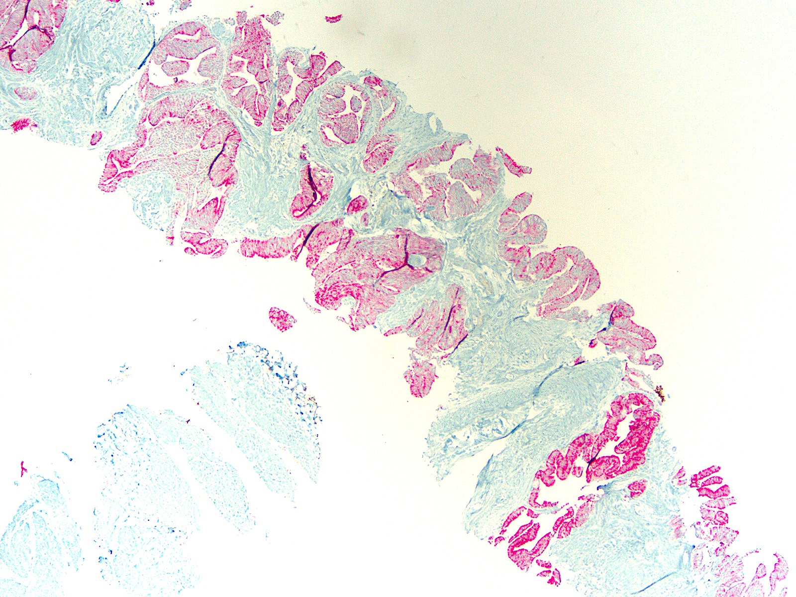
CK34BE12
p63
AMACR
| Evolution of grading of special prostate cancer patterns | |||
| Histologic pattern | 2005 consensus | 2014 consensus | 2019 consensus |
| Branched / undulating glands | Include as Gleason 3 | ||
| Cribriform (under Gleason scheme: mostly 3, sometimes 4) | 4 but can be 3 if much larger than benign gland, round and has loose cells | Always 4 | Always 4 and presence or absence should be specified for 3+4, 4+3 or 4+4 |
| Glomeruloid variant | No consensus, 3 versus 4 | Always 4 | -- |
| Mucinous variant | No consensus, some favored 4 | Depends on growth pattern regardless of mucin; could be 3, 4 or 5 | -- |
| Small cell (pure) | Do not grade | -- | -- |
| Intraductal, pure form | -- | Do not grade | Do not grade |
| Intraductal, associated with invasive cancer | -- | -- | Include in estimating the percentage of grade 4, instead of keeping it separate |
| Ductal | 4+4=8 | -- | -- |
| Adenoid cystic / basal cell carcinoma | -- | Do not grade | Do not grade |
Images hosted on other servers:

Prostatic hypoechoic lesion (ultrasound)

Prostatic hypointense lesion (MRI)
Images hosted on other servers:

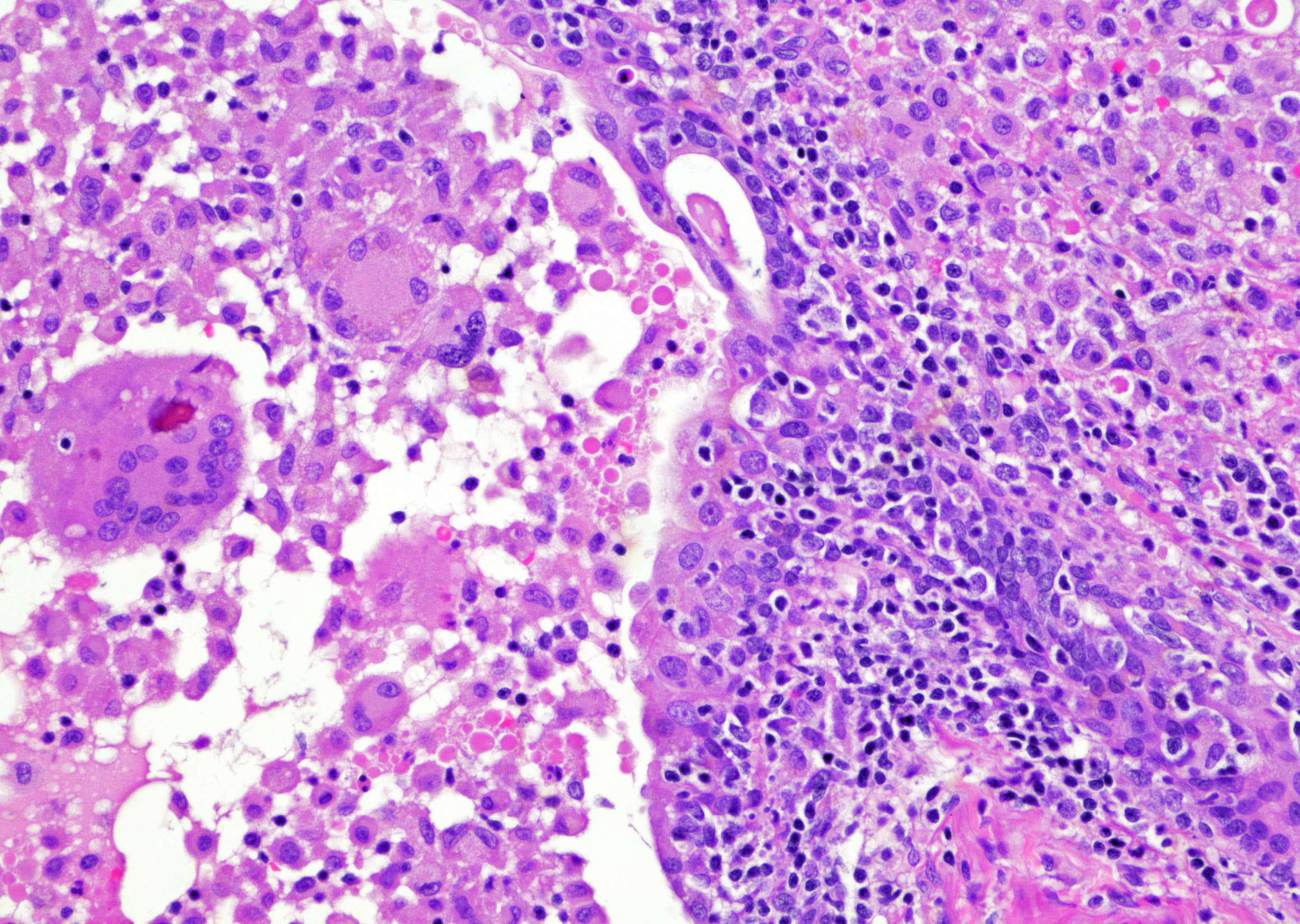
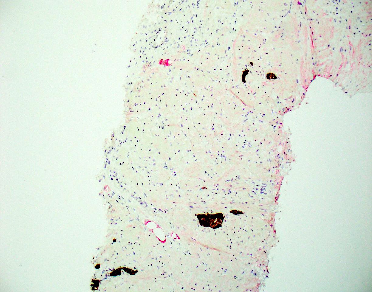
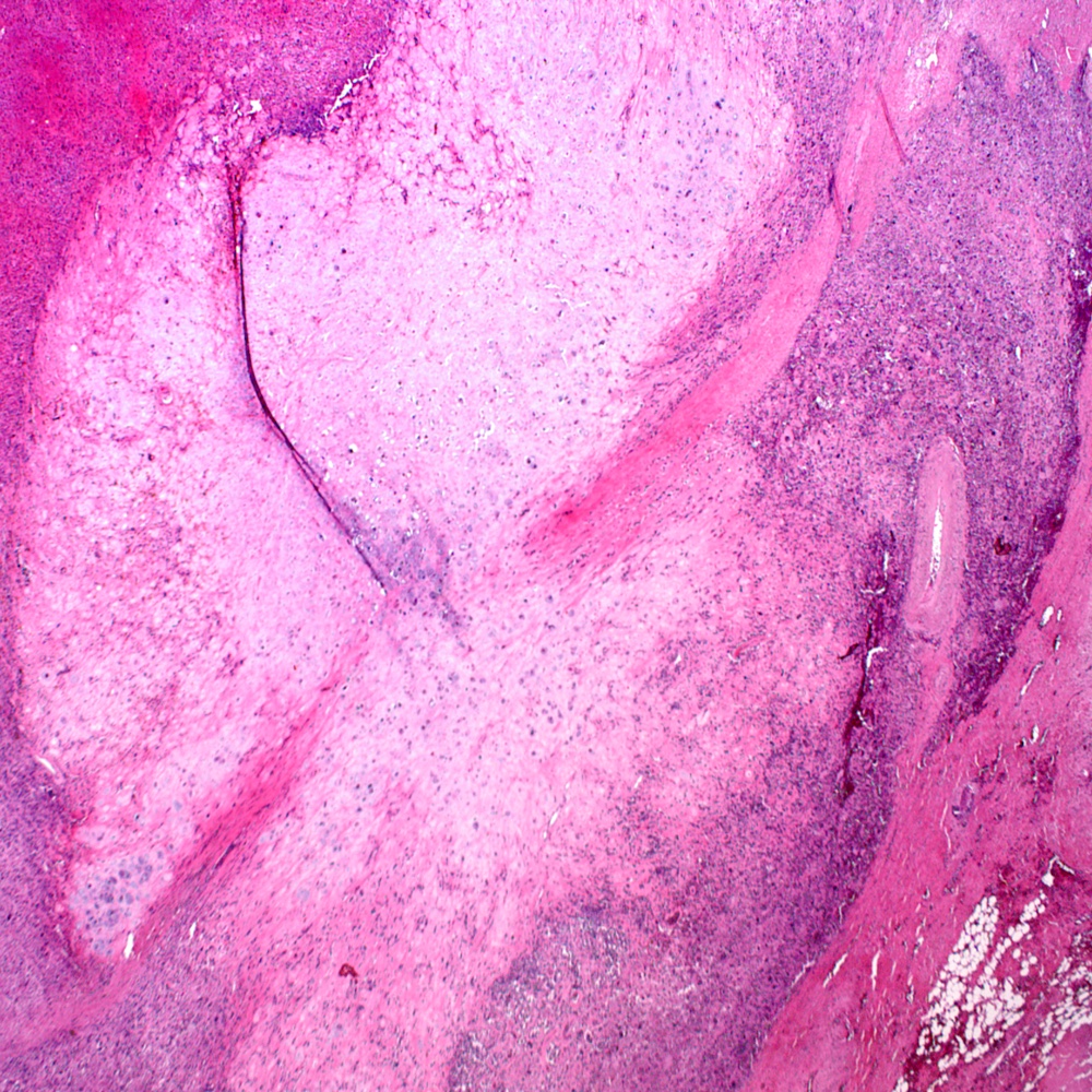
Xanthogranulomatous
prostatitis with
abscess
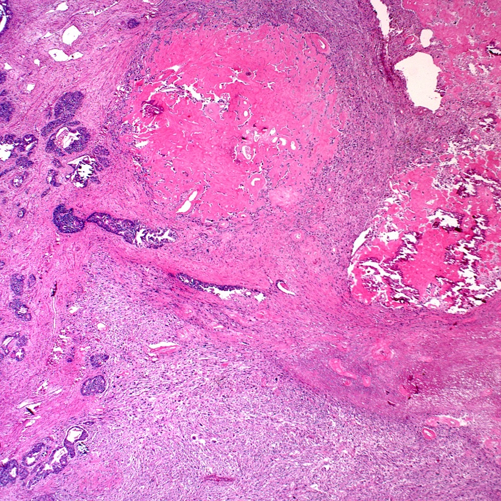
Contributed by Y. Albert Yeh, M.D., Ph.D.
Dilated ducts with inflammation
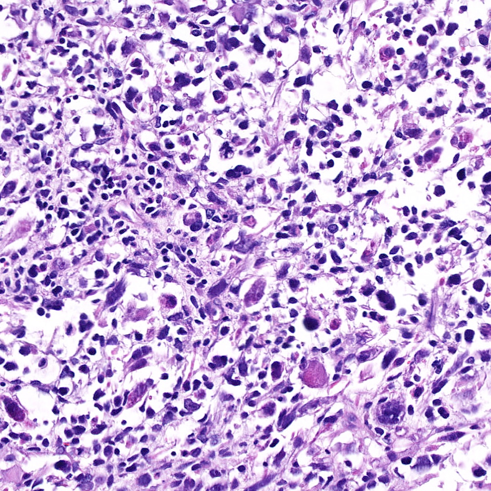
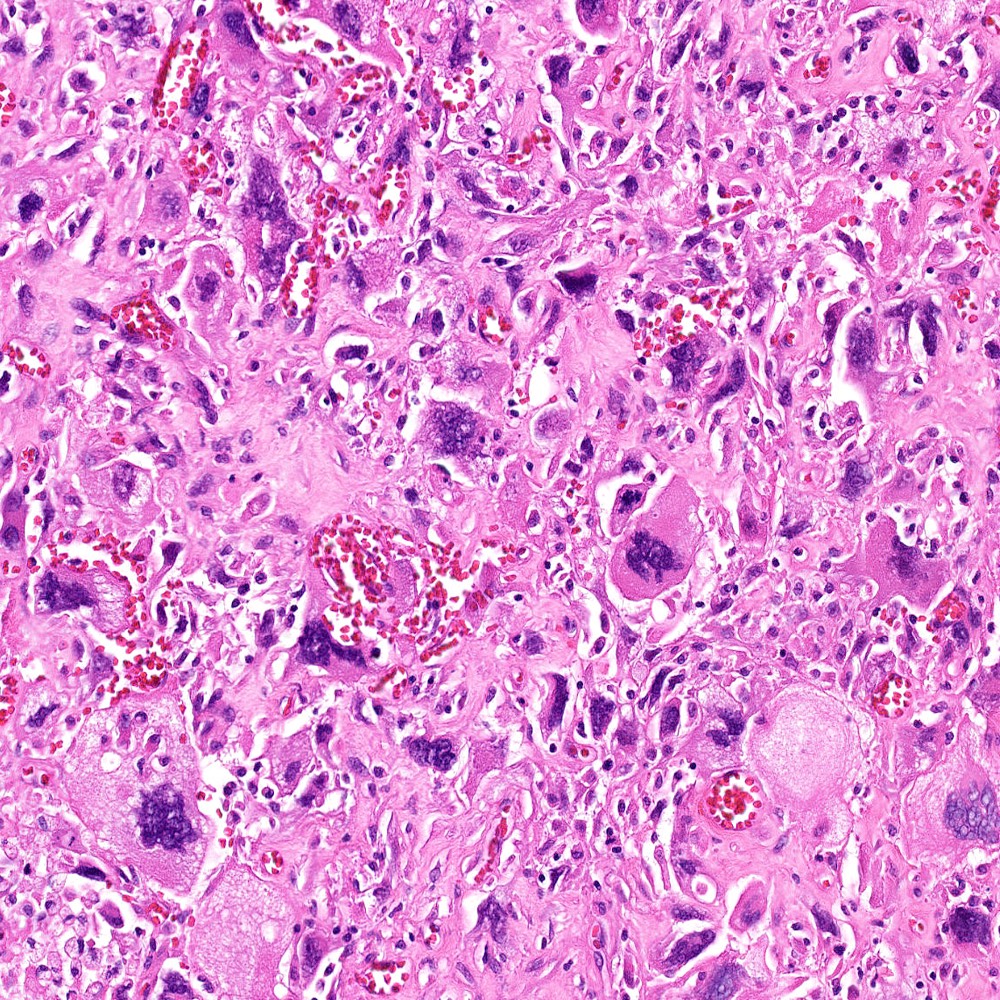
Histiocytes and giant cells
Expansile inflammatory areas
Foreign body giant cells
BCG related giant cells
Sarcoid related lesions
Sarcoid related giant cells
Sarcoid related Langhans cells
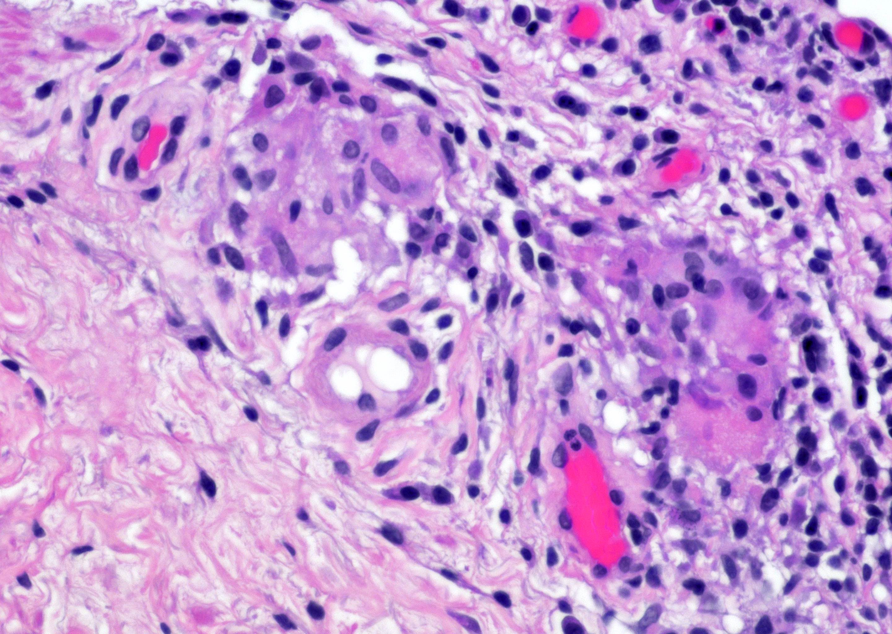
Contributed by Ximing J. Yang, M.D., Ph.D.
Multiple granulomas
Langhans giant cells
BCG induced granuloma

Foreign body giant cell granuloma
Indeterminate granuloma
Images hosted on other servers:


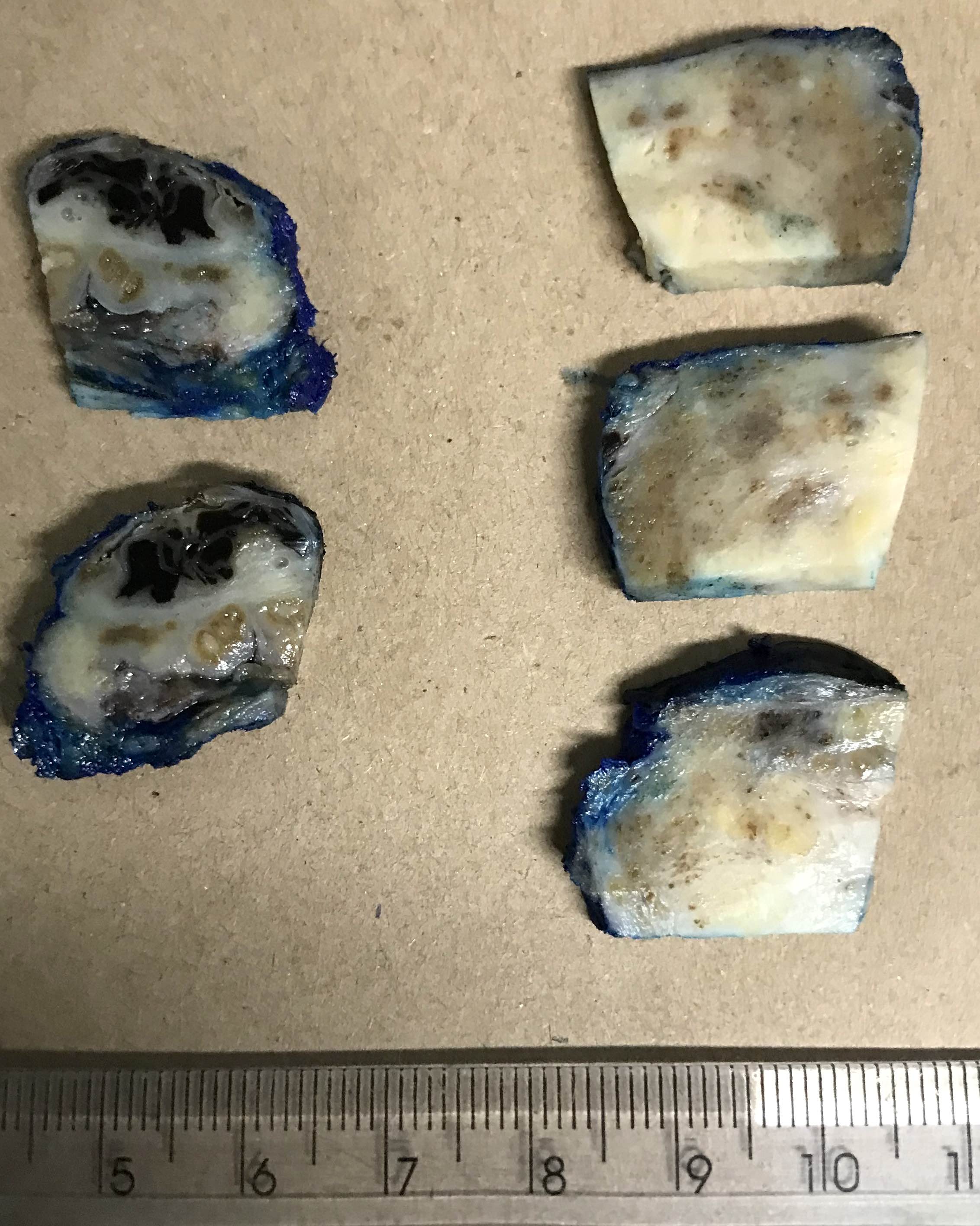
Device to generate uniform thickness of sections in radical prostatectomy

Whole mount sectioning
Images hosted on other servers:

Prostate cancer pathogenesis
Contributed by Y. Albert Yeh, M.D., Ph.D. and Nicholas P. Reder, M.D., M.P.H.
HGPIN with atypical glands
Atypical gland with basal cells
Single atypical gland
2 atypical glands
Atypical gland in 2 HGPINs
Atypical cytologic features
Small atypical glands adjacent to larger HGPIN glands
PIN4
Contributed by Murali Varma, M.D.
Flat HGPIN
Tufted HGPIN
Micropapillary and flat HGPIN
Micropapillary HGPIN
Inverted HGPIN
p63
CK5/6
AMACR
Contributed by Guang-Qian Xiao, M.D., Ph.D.
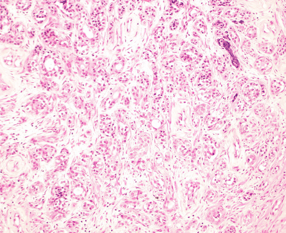
Adenosis

Prostatic adenocarcinoma
Atypical cribriform lesion
Prostatic adenocarcinoma
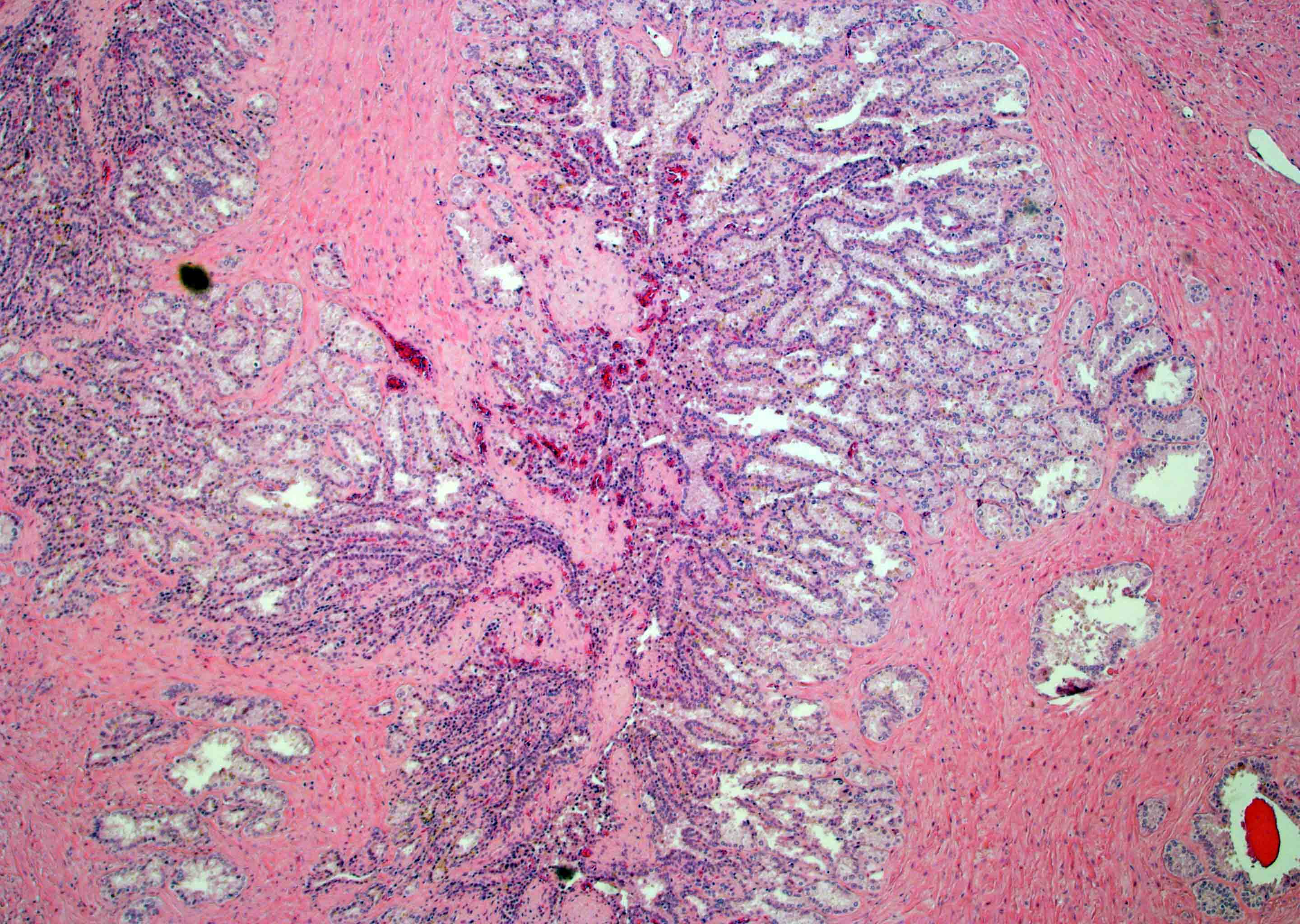
Partial atrophy

Carcinoma with radiation effect
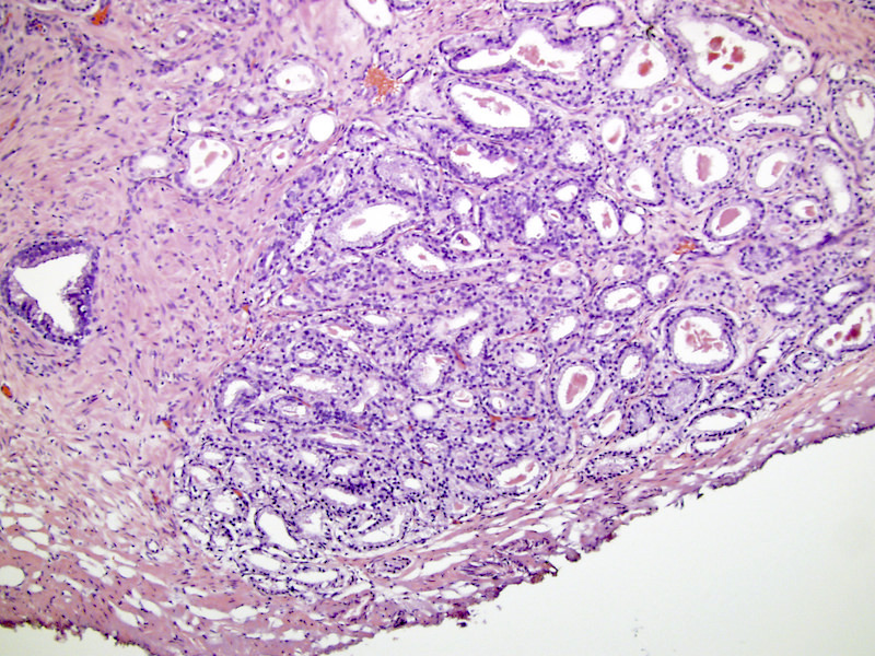
Contributed by Erica Vormittag-Nocito, M.D. and Case #286

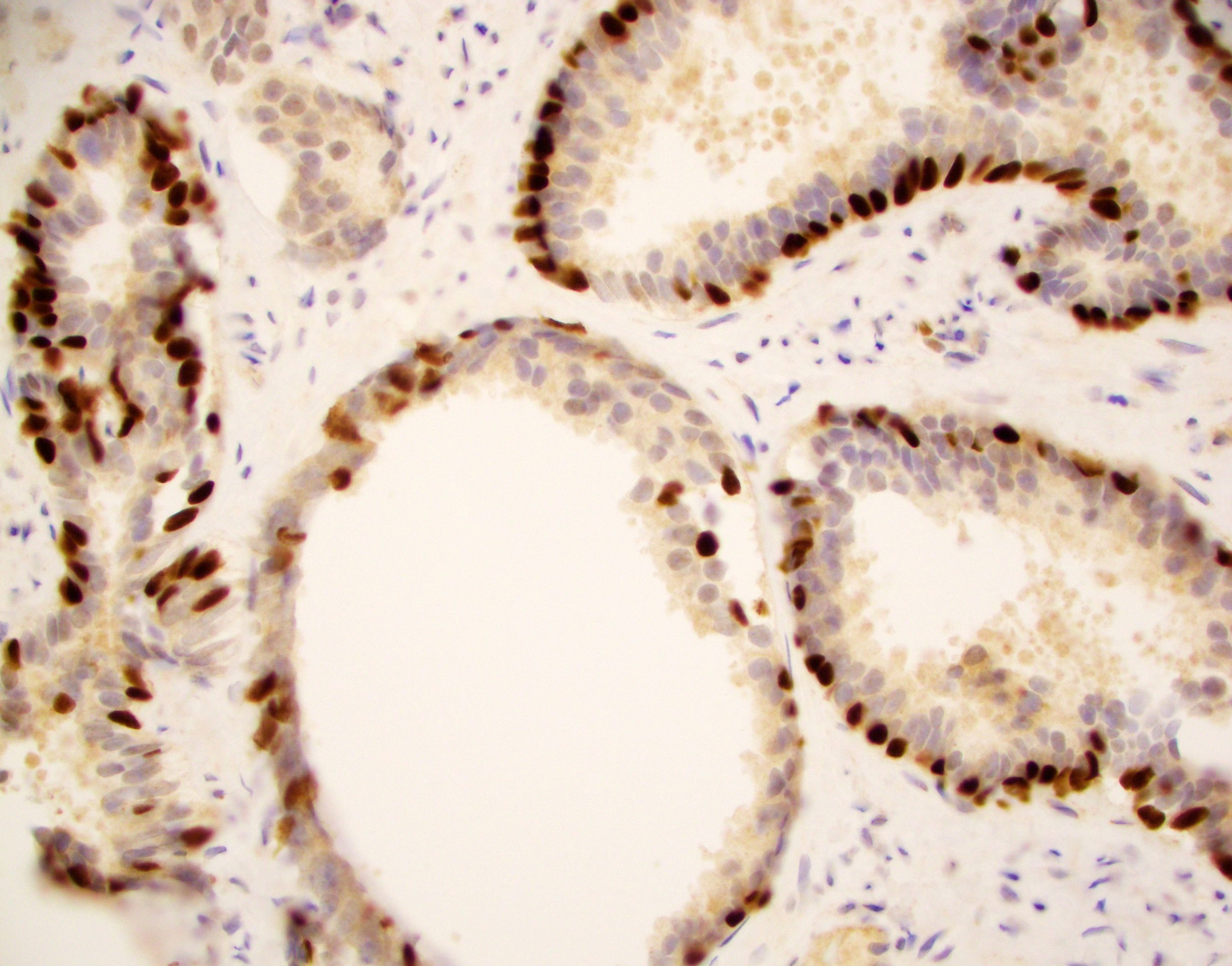
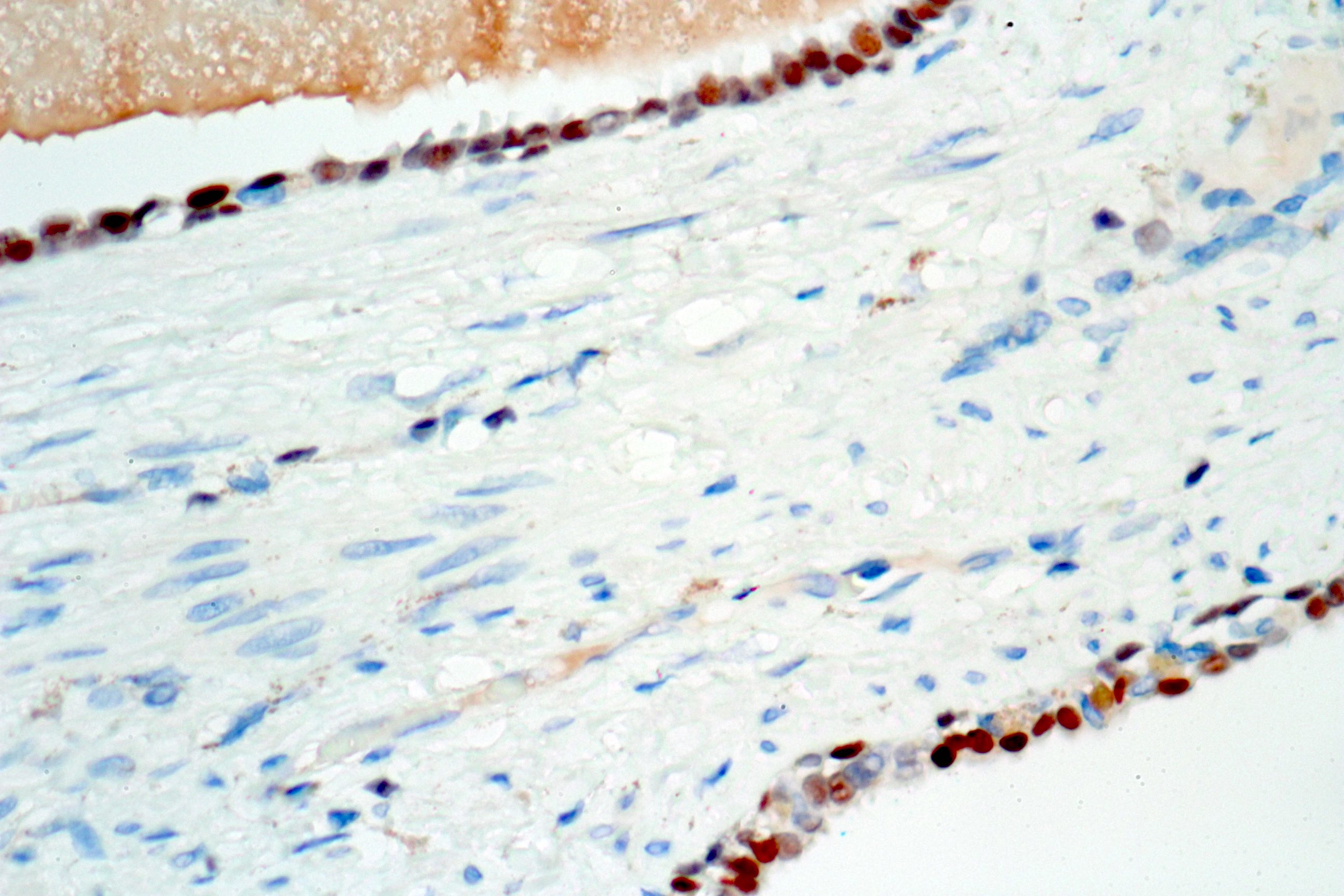
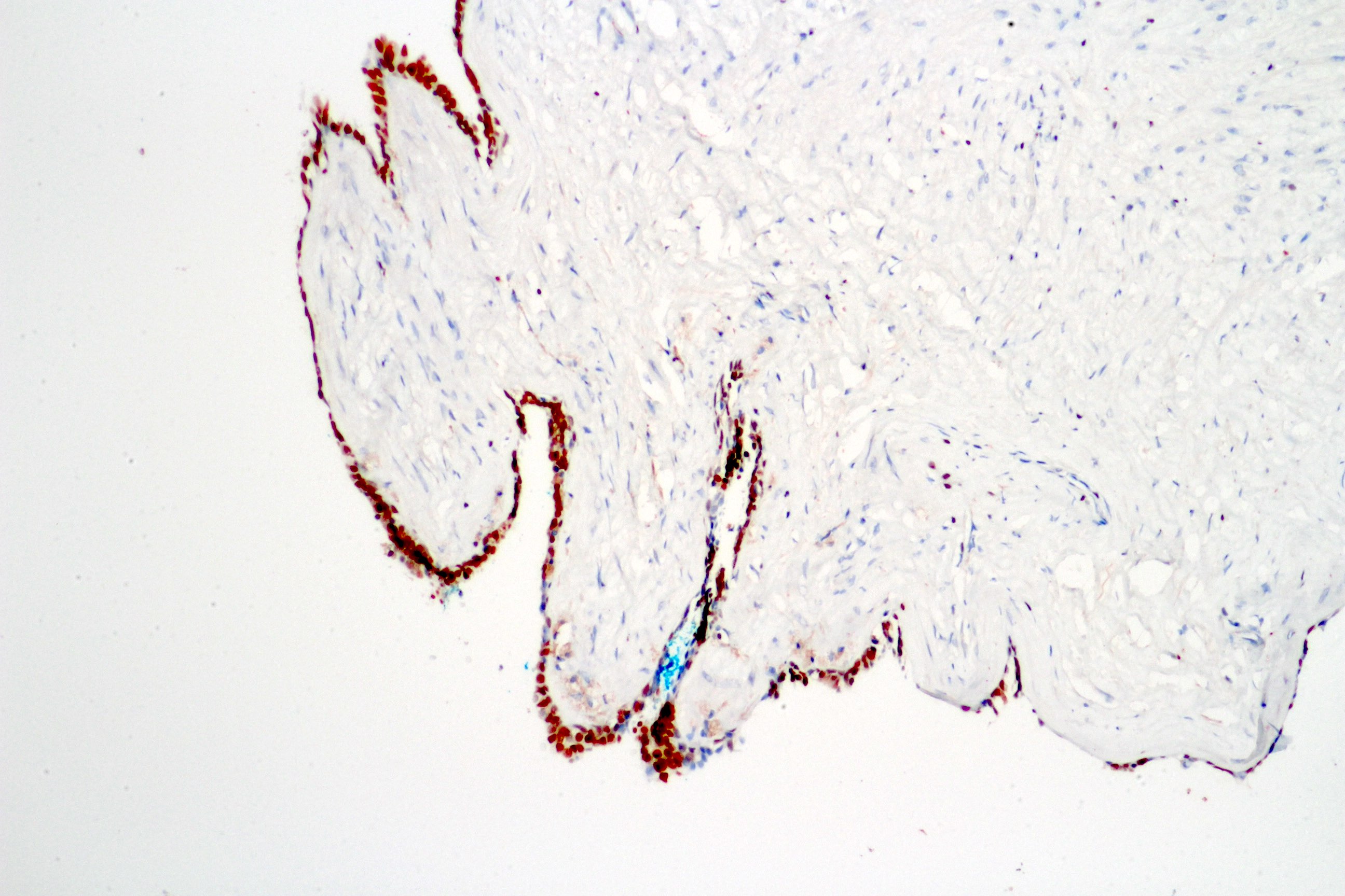
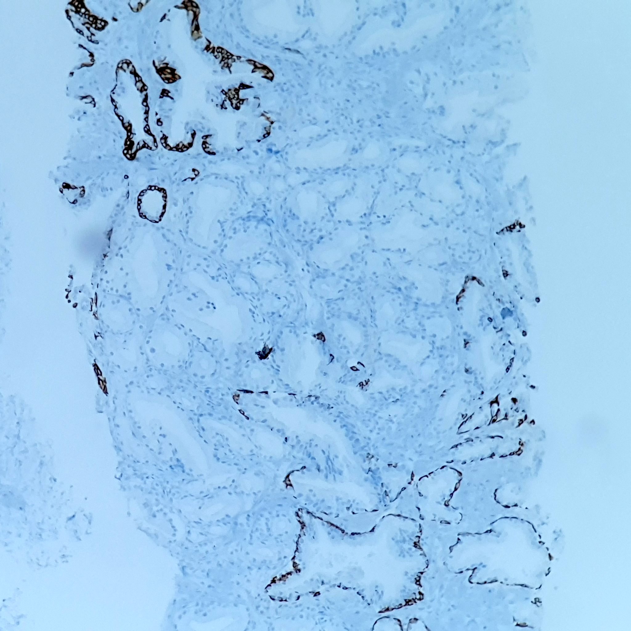
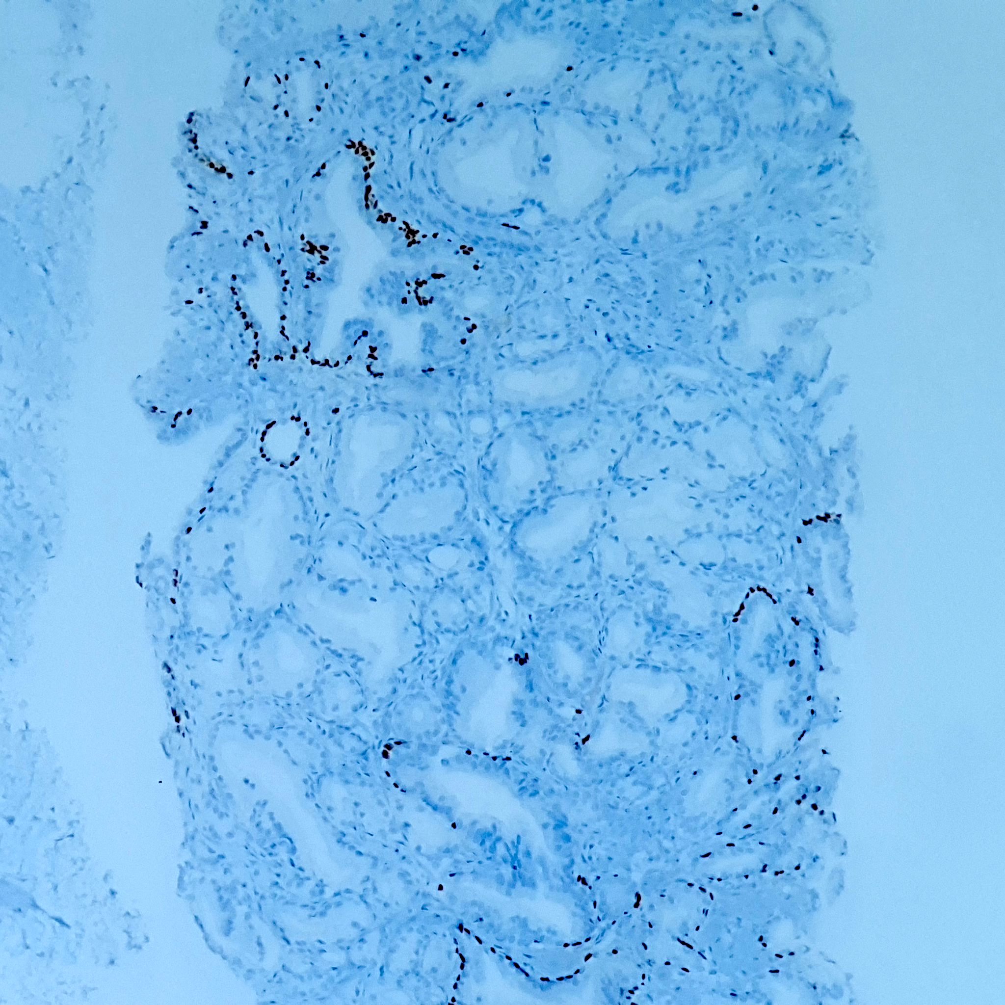
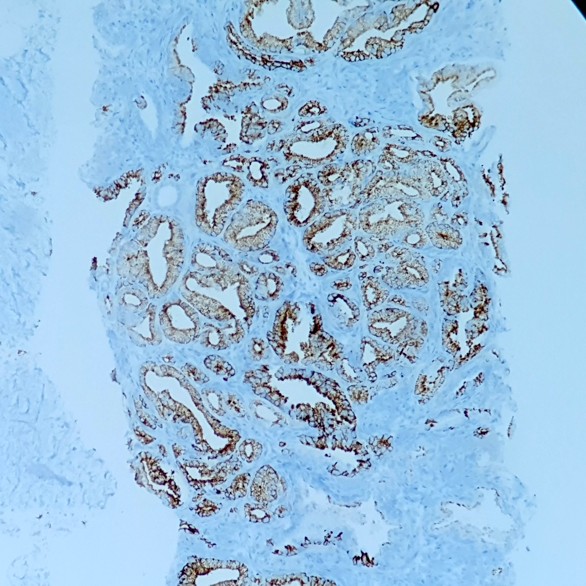
Cribriform architecture

p63
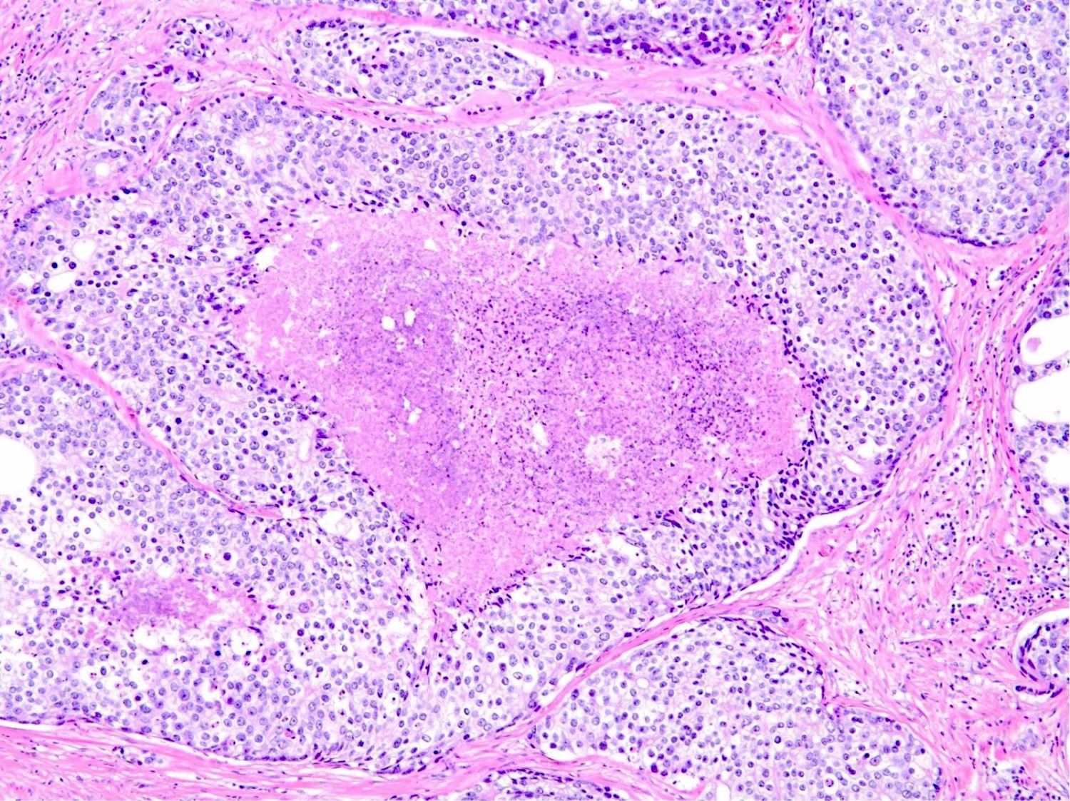
Comedonecrosis

p63
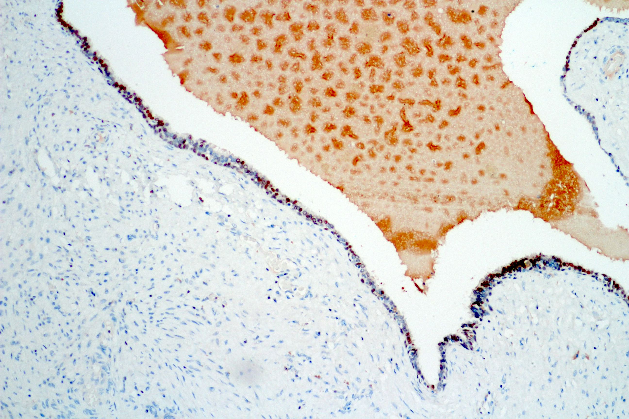
Gleason pattern 5
Gleason pattern 5, p63
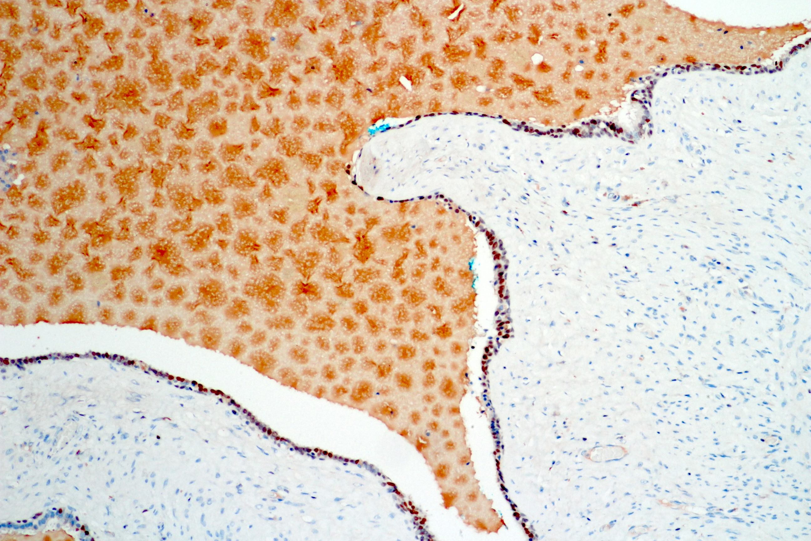
Diffuse intraductal carcinoma
Cribriform growth

CK903

CK903
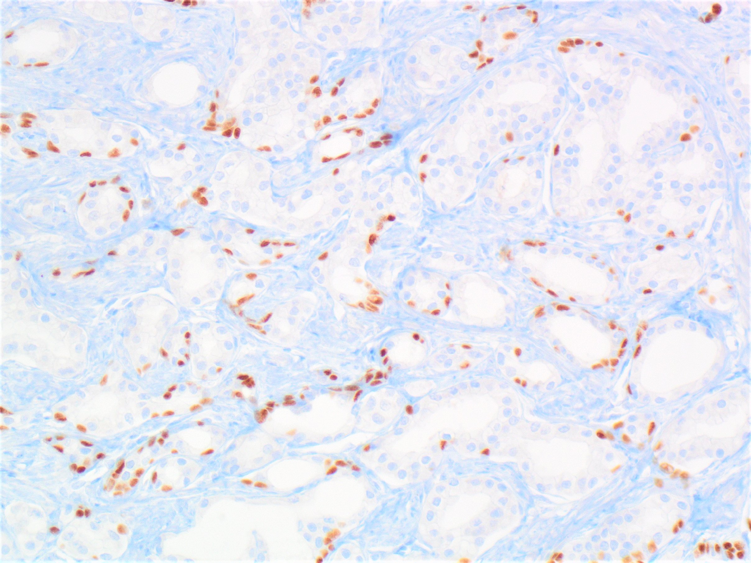
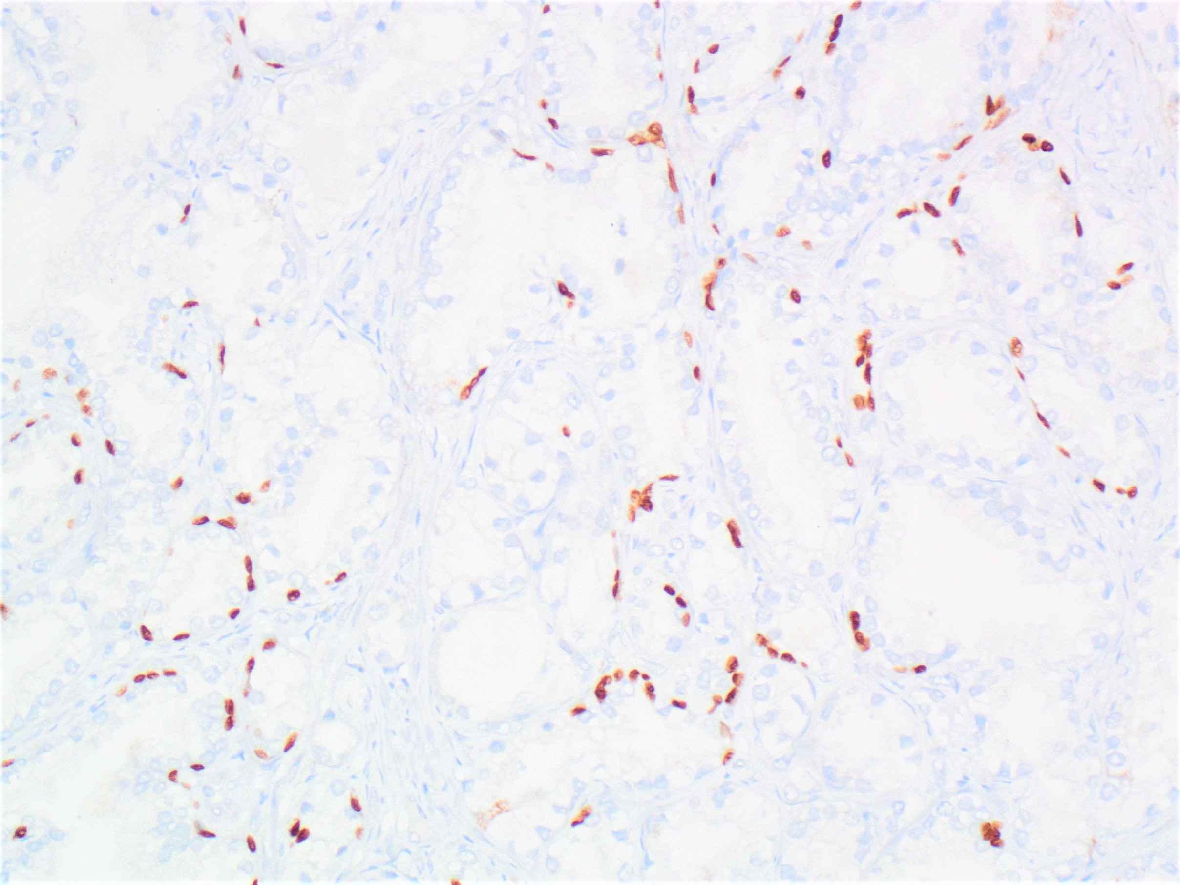
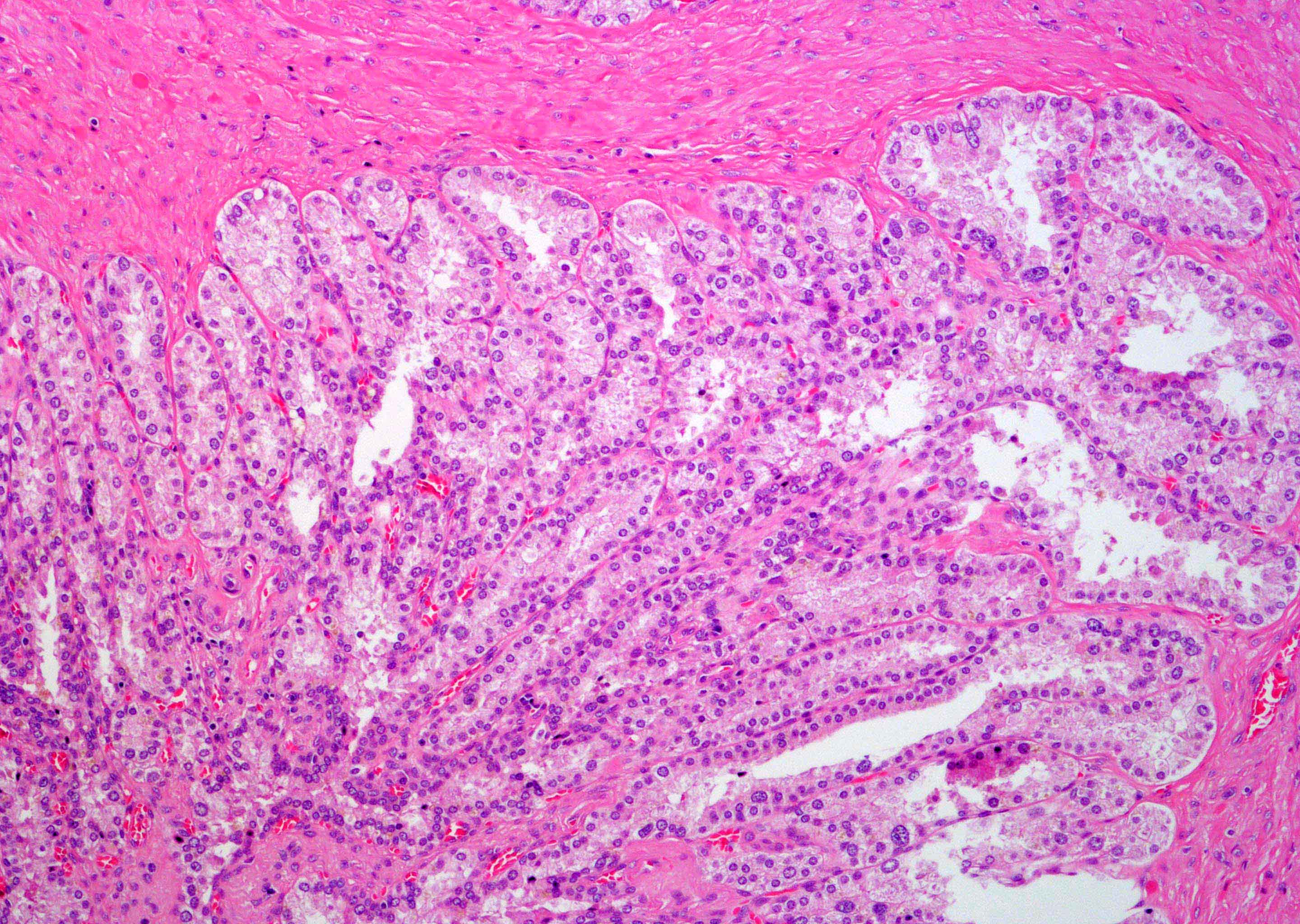
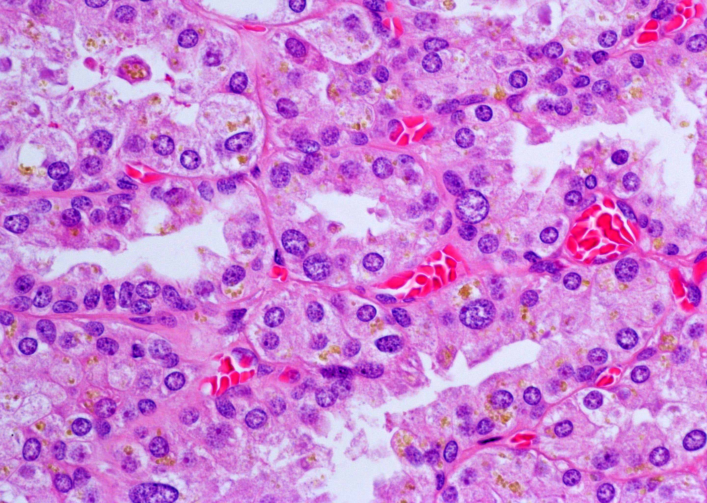
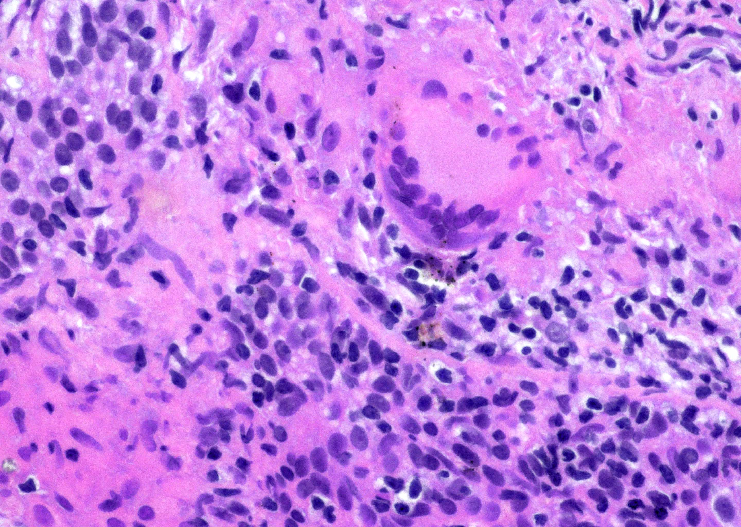
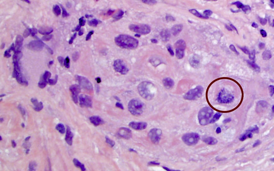
Contributed by Ximing J. Yang, M.D., Ph.D.
Large tumor cells
have slightly more
cytoplasm and
fine
"salt-pepper"
chromatin
Strong/diffuse
chromogranin
staining
Sheets of large
tumor cells with
"salt and pepper"
chromatin
Minor
component of
prostatic
adenocarcinoma
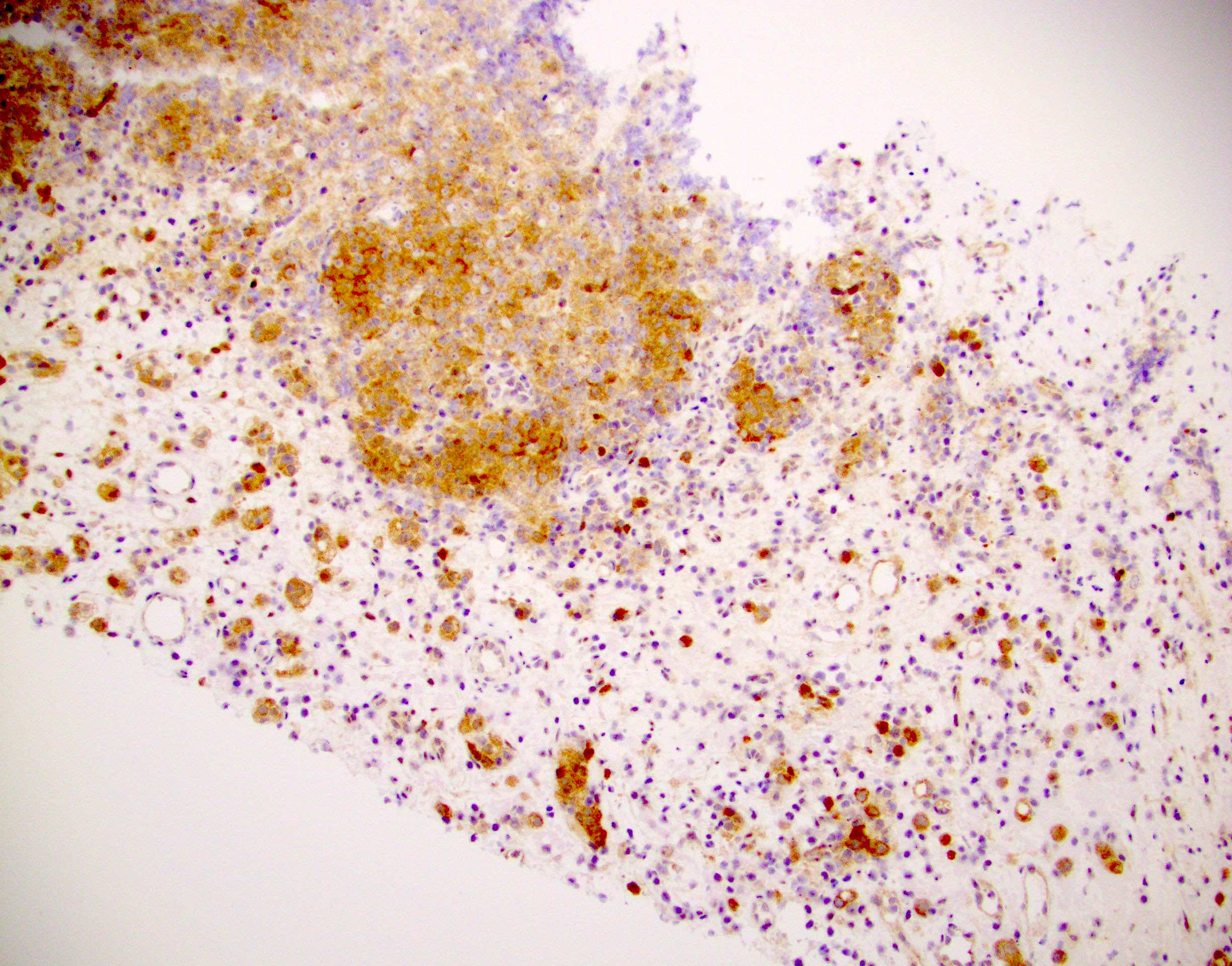
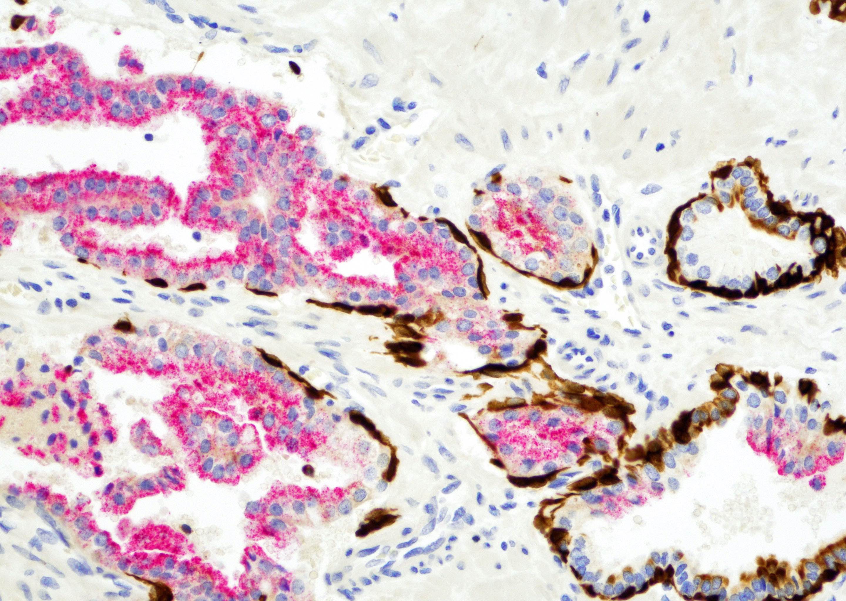
Diffuse chromogranin+

AMACR (triple stain)

Negative HMWCK,
p63, PSA
Ki67 proliferative index up to 50%
Contributed by Nicholas P. Reder, M.D., M.P.H.
Acinar cells with crowded nuclei
Atypical acinar cells
Low grade PIN next to high grade PIN
Images hosted on other servers:
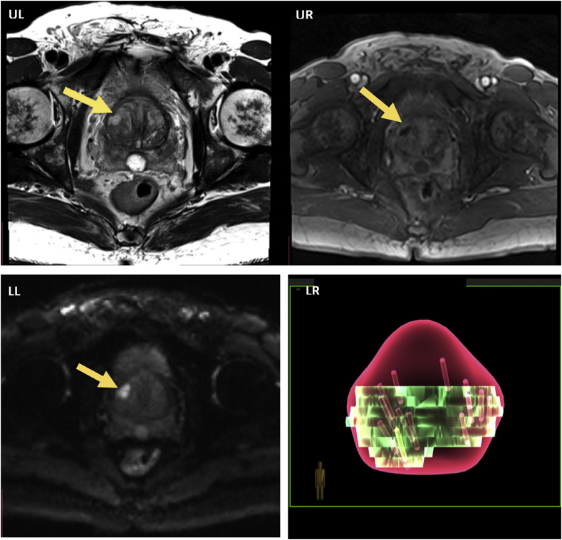
mpMRI of hypointense lesion
mpMRI showing hypointense mass
Contributed by Theodorus van der Kwast, M.D., Ph.D.

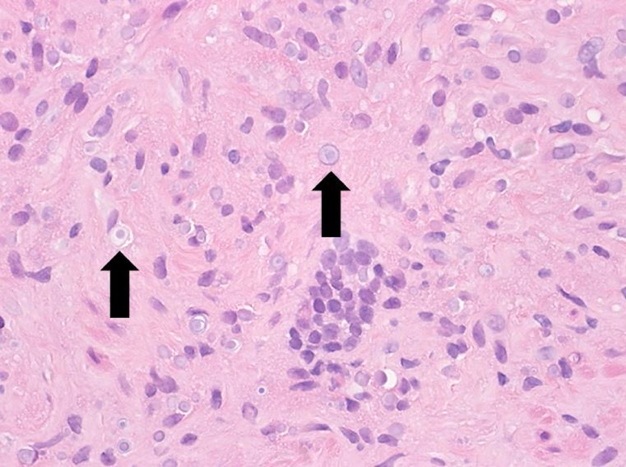
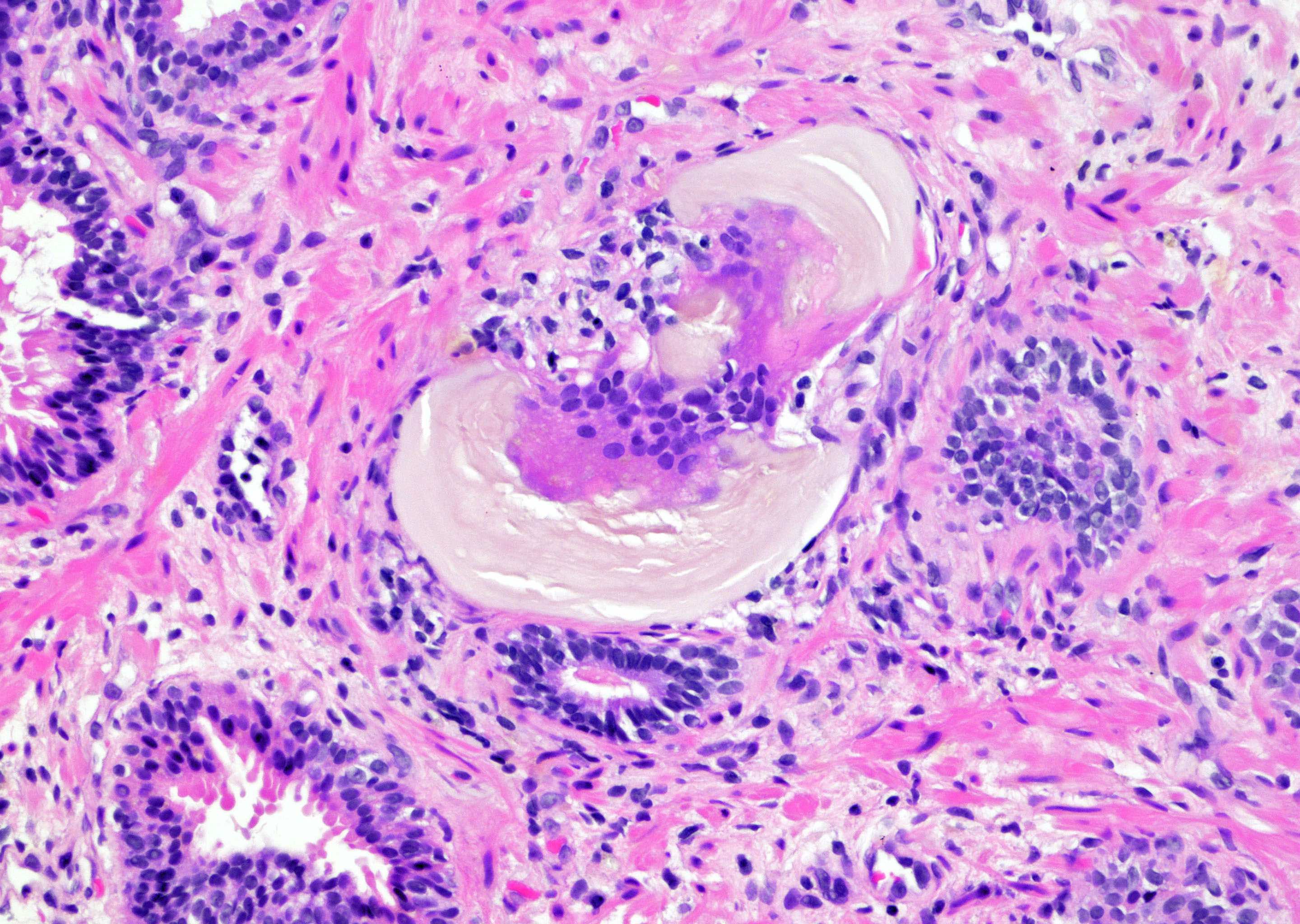
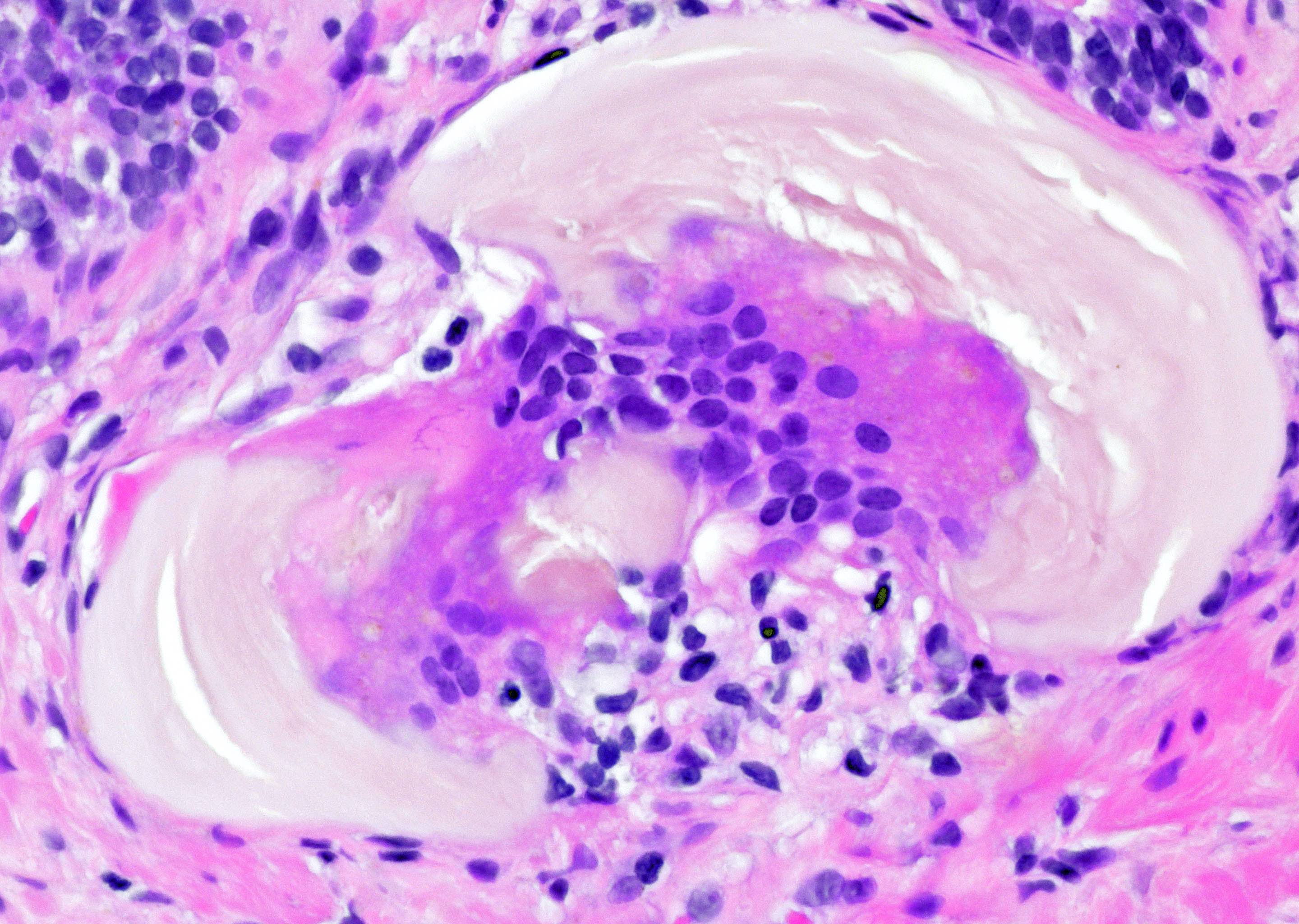
Histiocytes and inclusions
Histiocytic inflammation

PAS stain
Contributed by Andres Matoso, M.D.

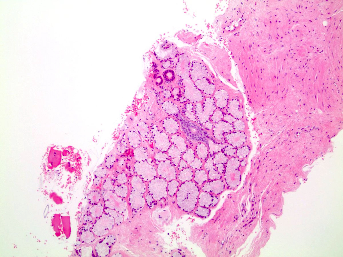
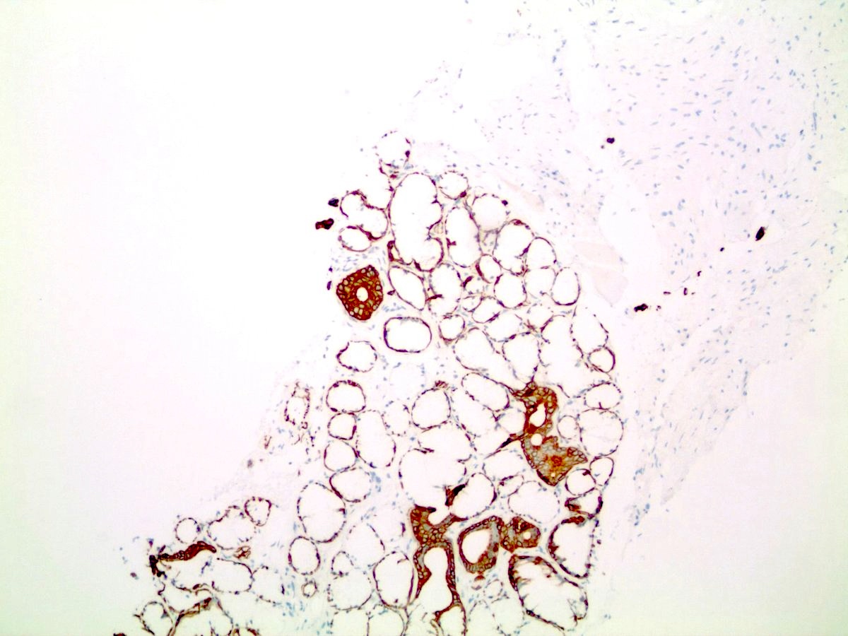
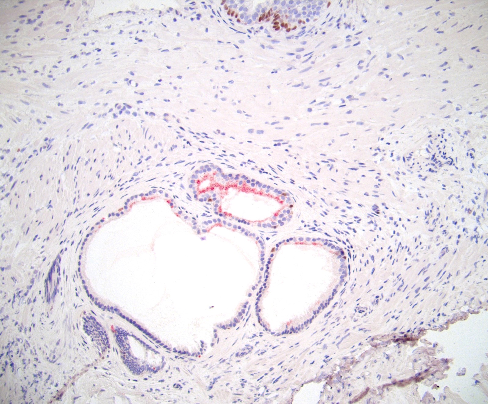
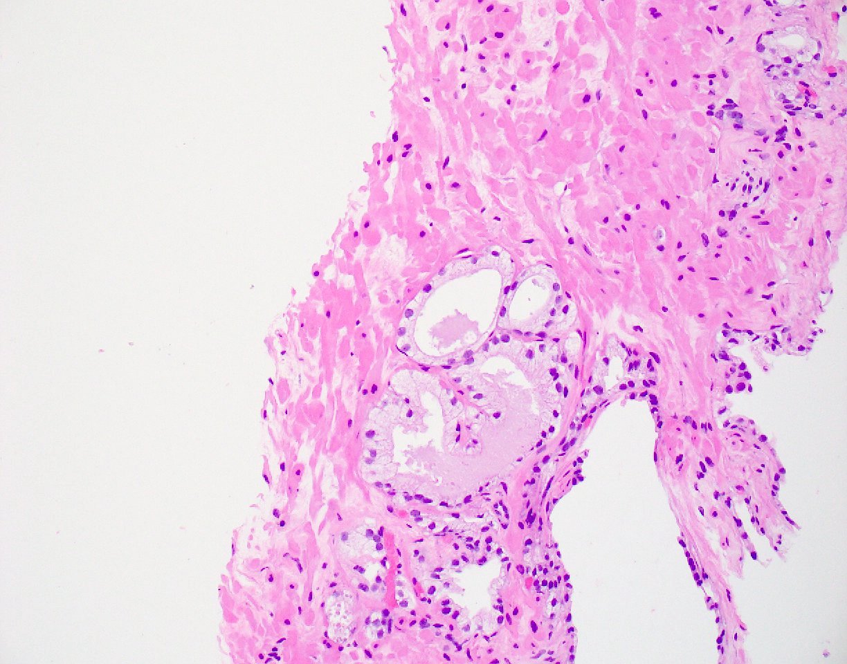
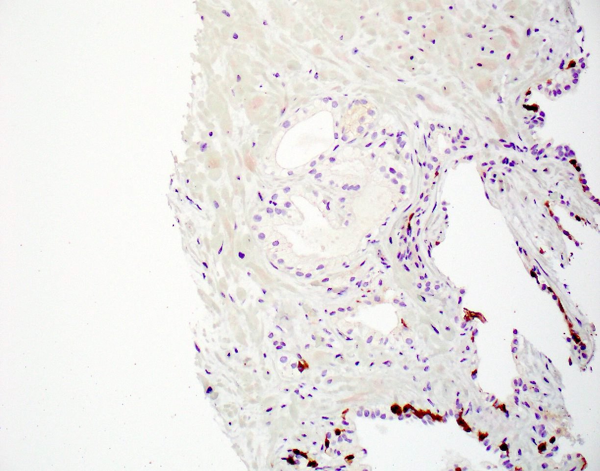
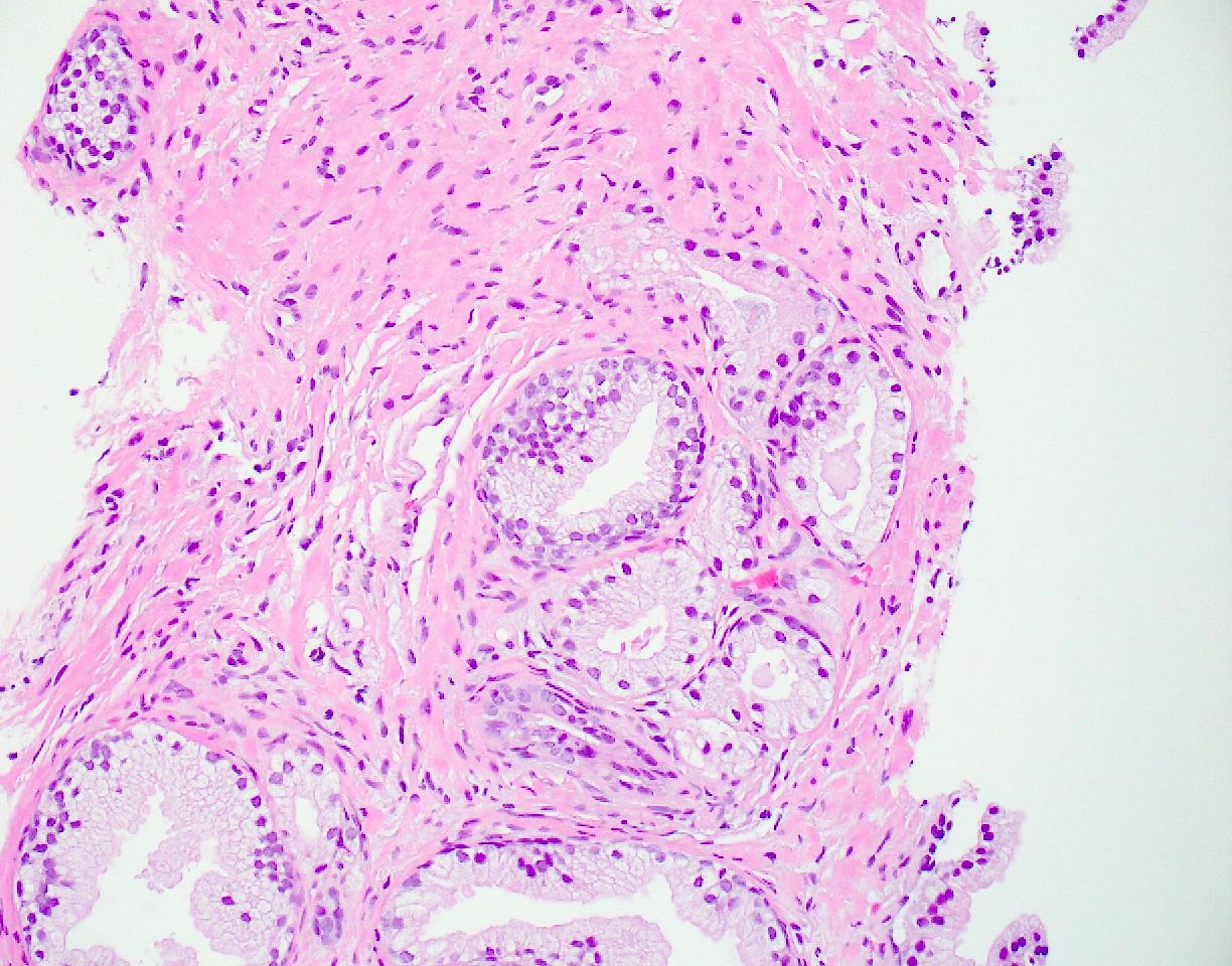
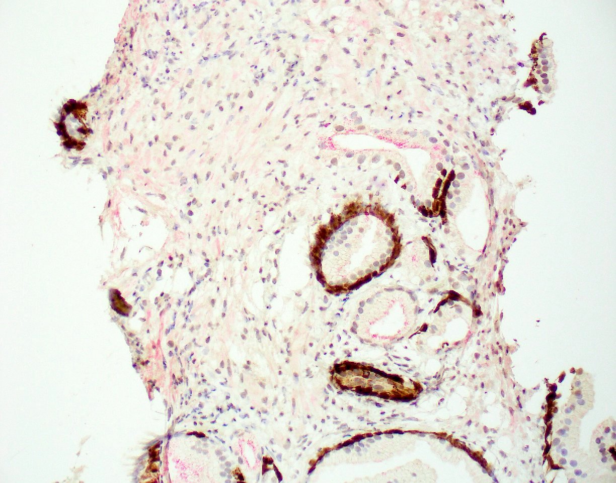
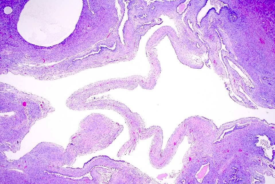
Mesonephric remnant / hyperplasia with acini
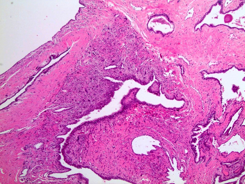
Contributed by Ankur Sangoi, M.D.
Compact dilated cysts
Basal nuclei with amphophilic cytoplasm
Prominent nucleoli and luminal crystalloid
Cytoplasmic P504S
Images hosted on other servers:

Pelvic MRI, seminal vesicle mass
Contributed by Gladell Paner, M.D.

Mass of seminal vesicle
Contributed by Fiona Maclean, M.D. and Gladell Paner, M.D.

Cellular stroma surrounds epithelium
Urothelial component
Contributed by Francesca Sanguedolce, M.D., Ph.D.
Multiple morphologies
Inflammation, thickened basement membrane
Cytological features
Ki67
AMACR
PAX8
CK7
Contributed by Francesca Sanguedolce, M.D., Ph.D. and Ximing J. Yang, M.D., Ph.D.

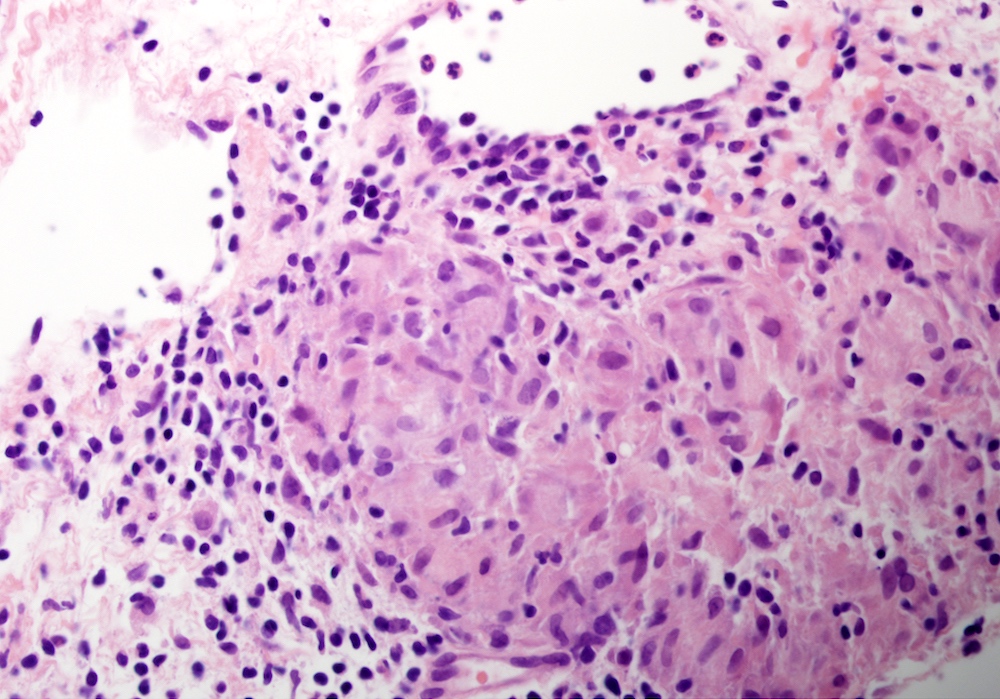
Inflammatory cells

Duct inflammation and granuloma

Microabscesses
Foreign body giant cell

Early fibrosis
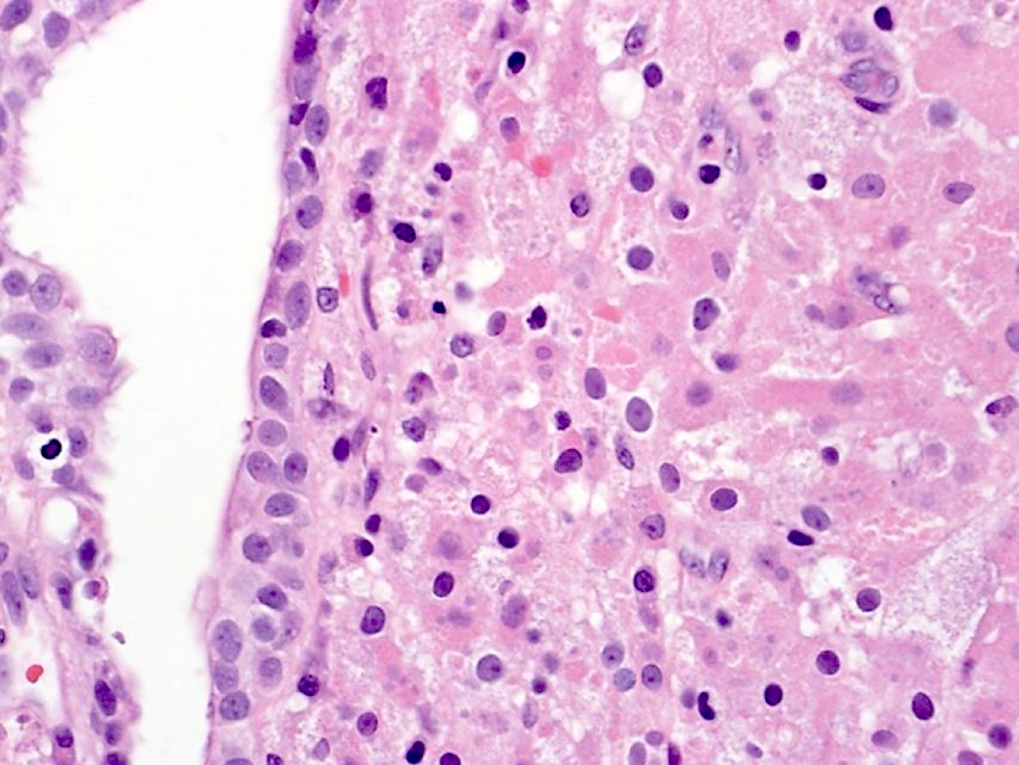
Benign glandular cells
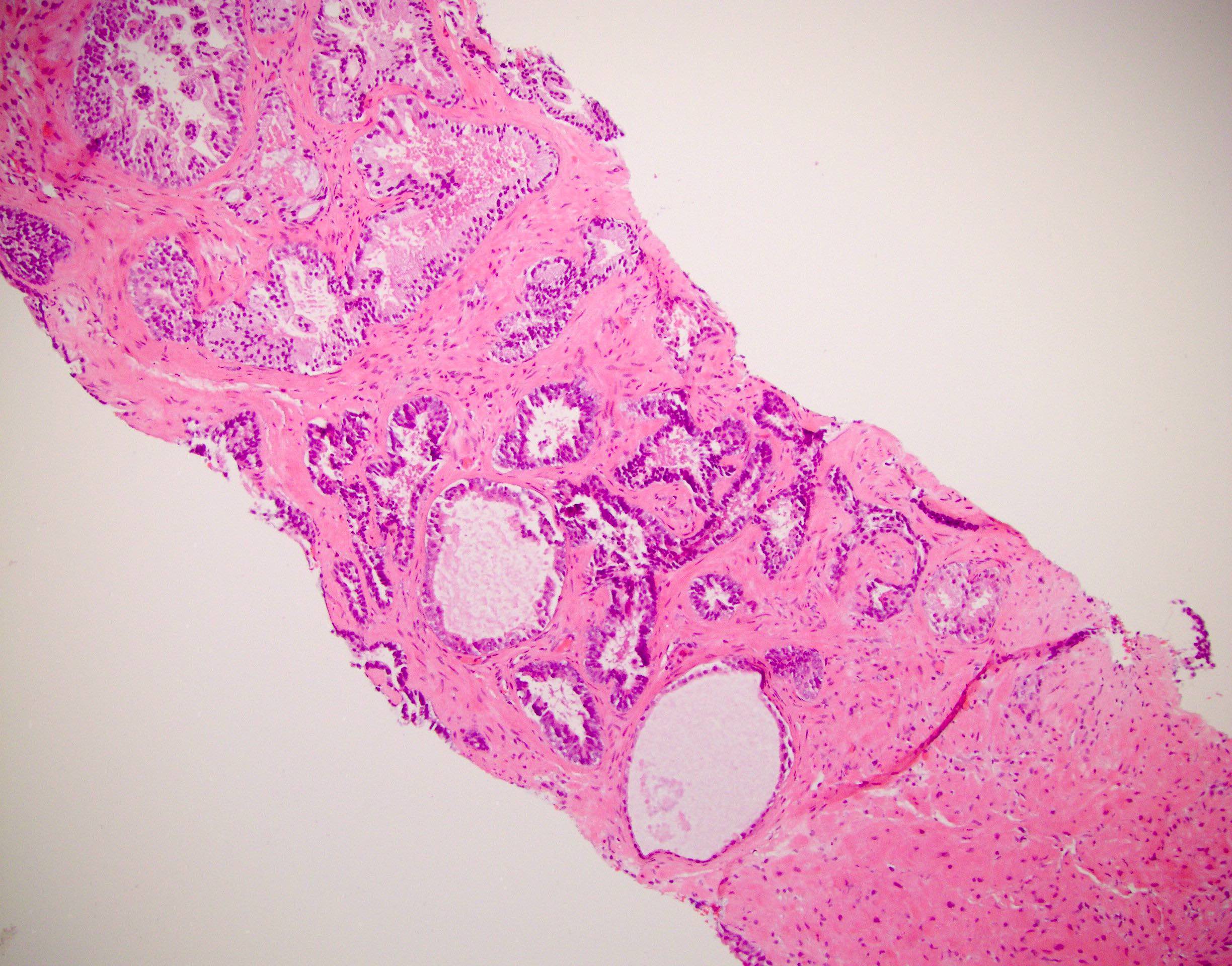
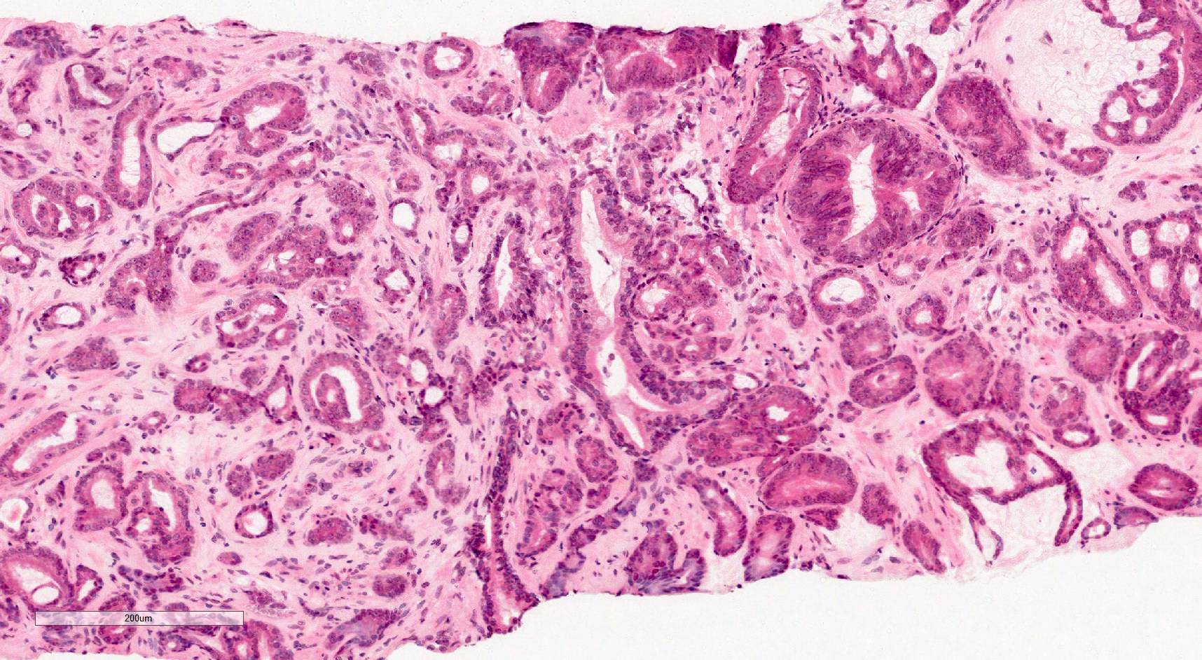
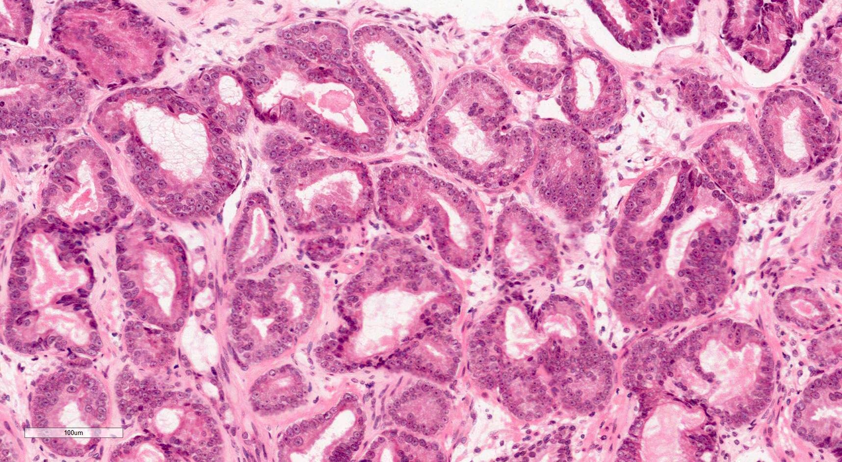
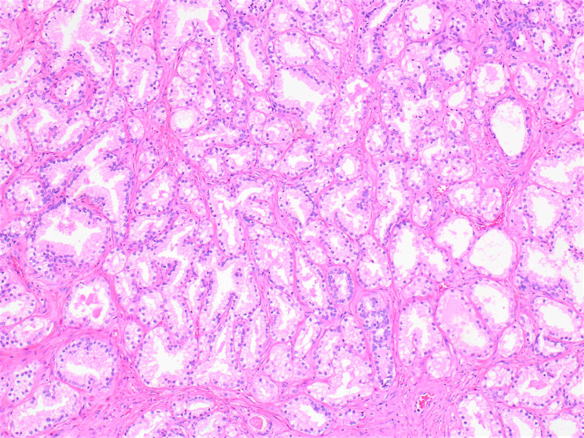
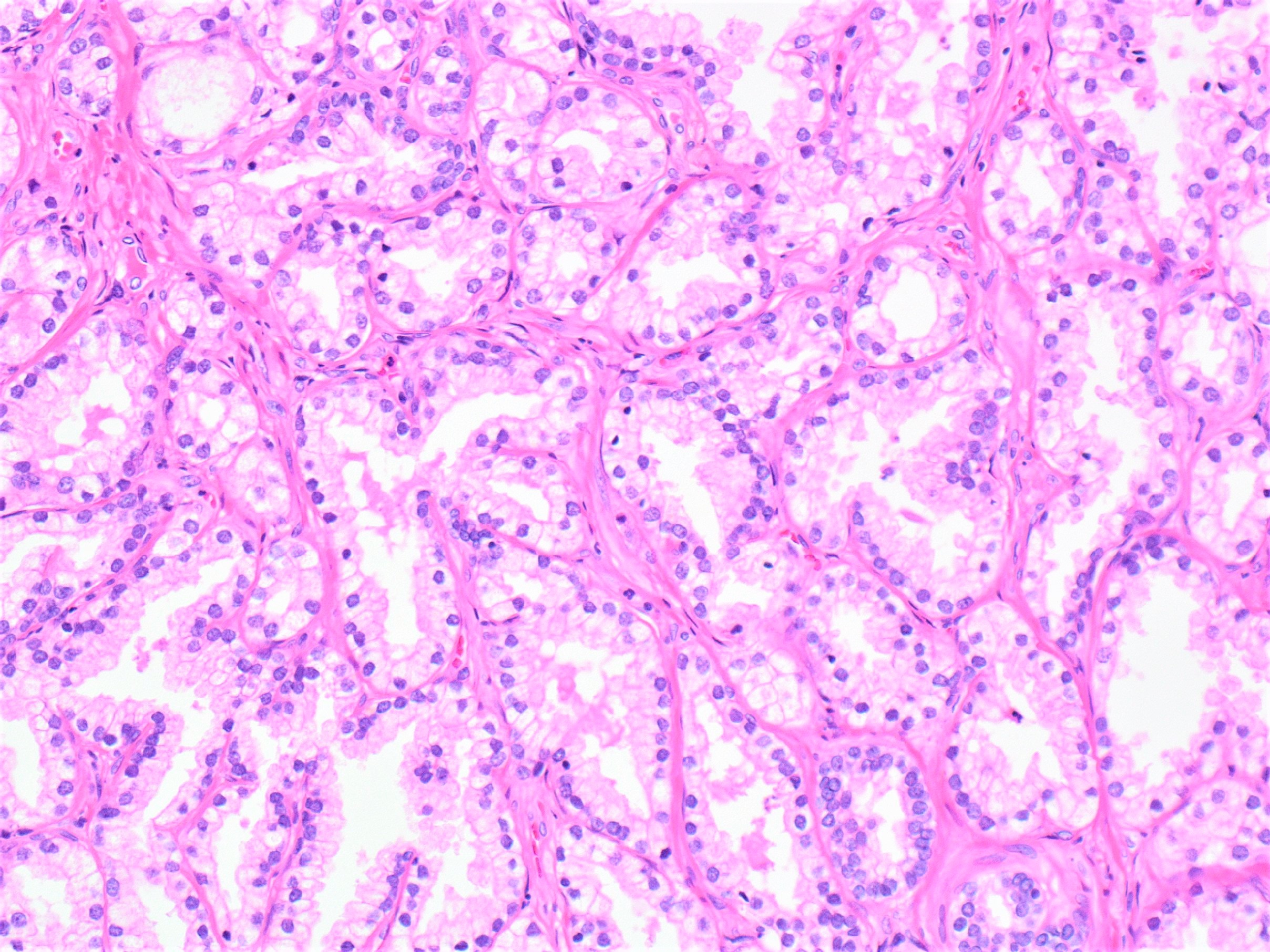
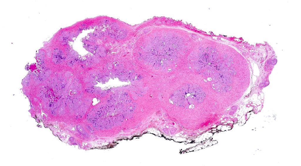
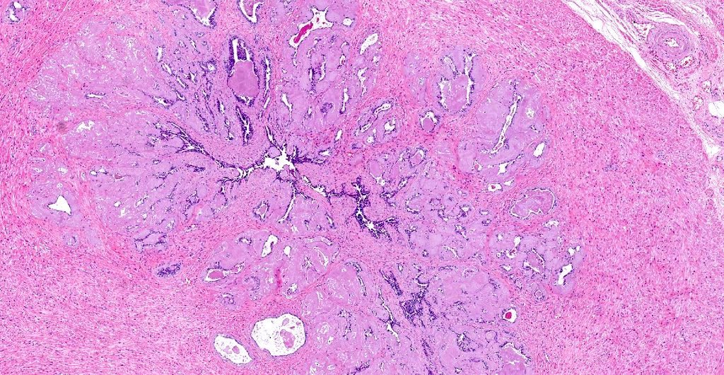
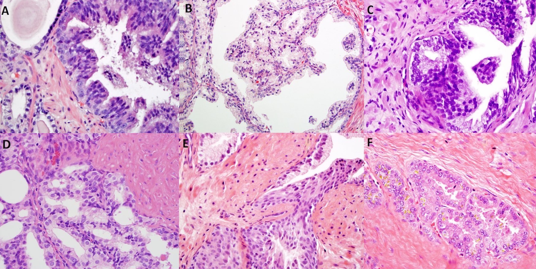
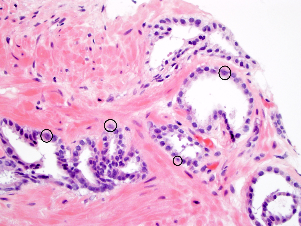
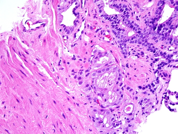
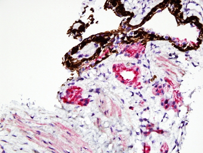
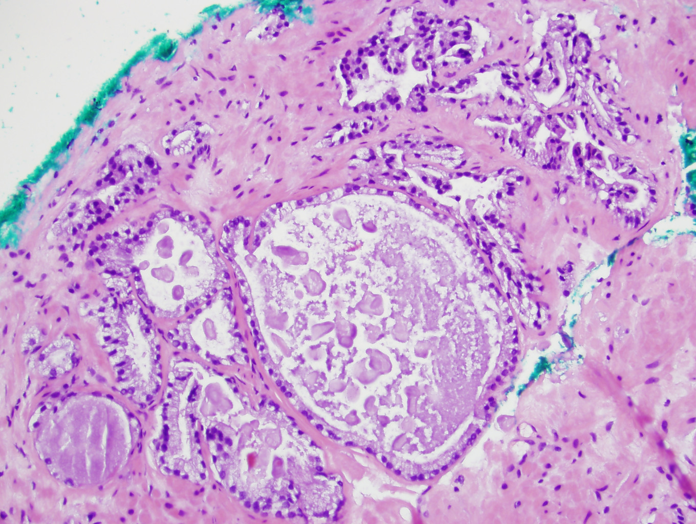
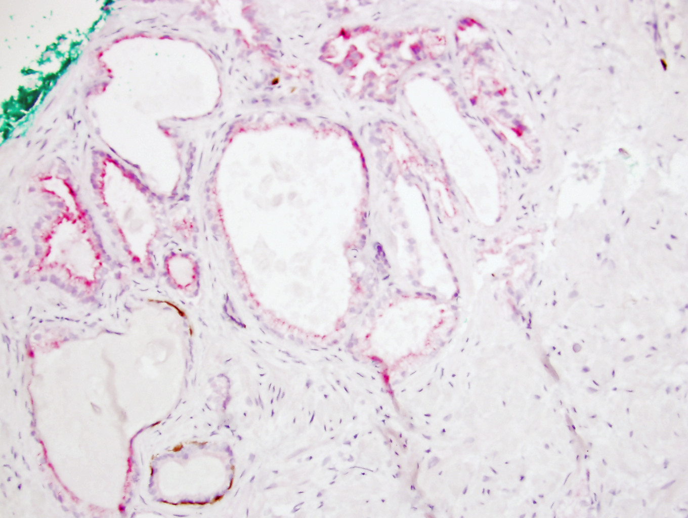
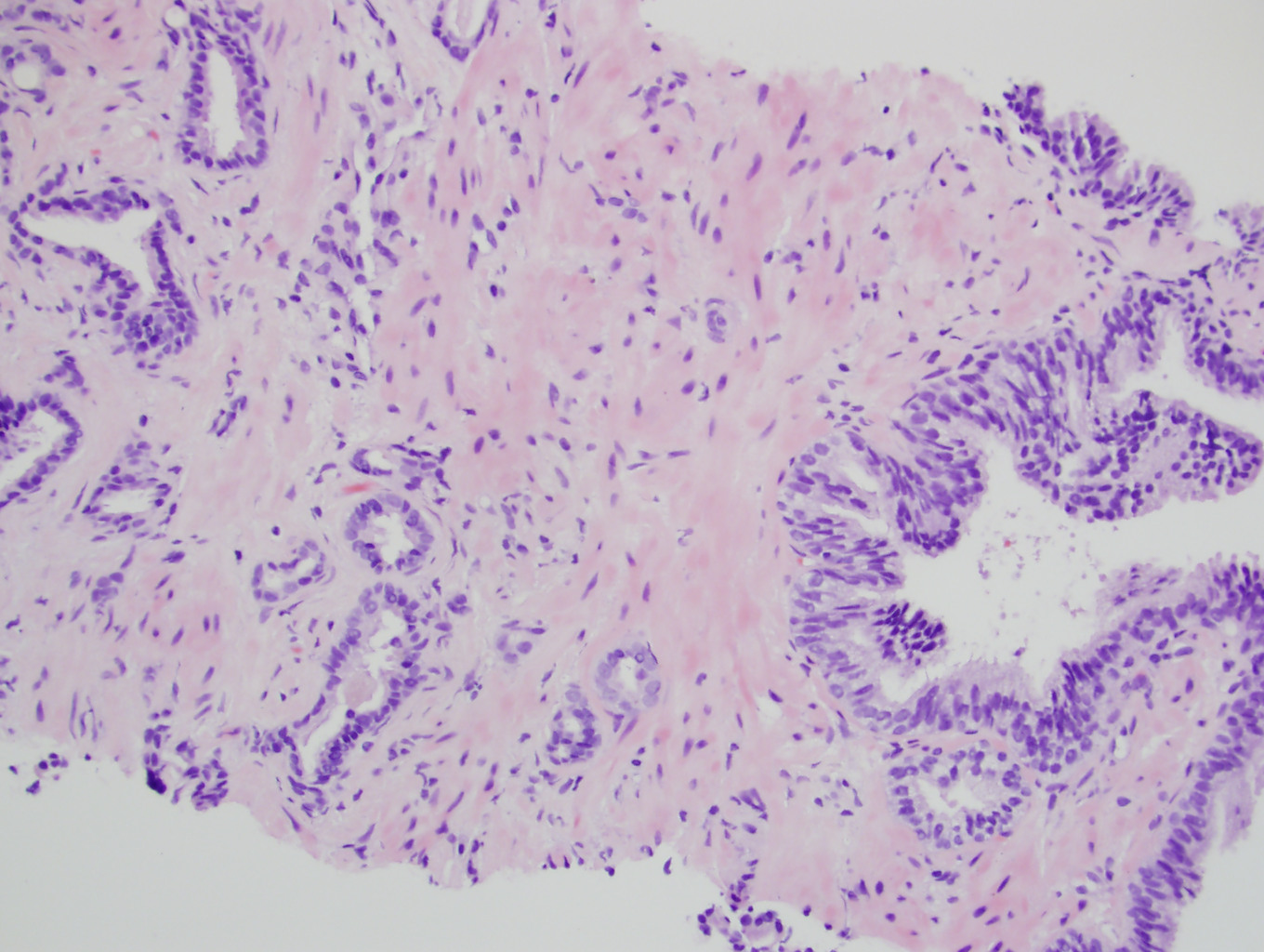
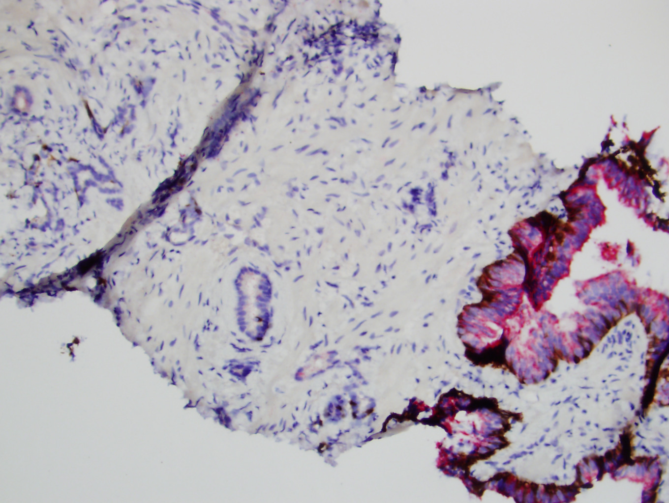
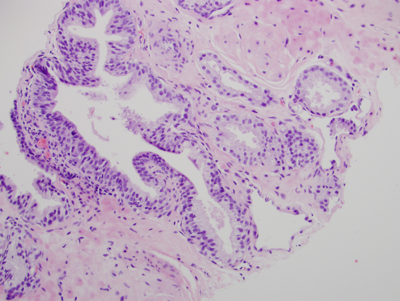
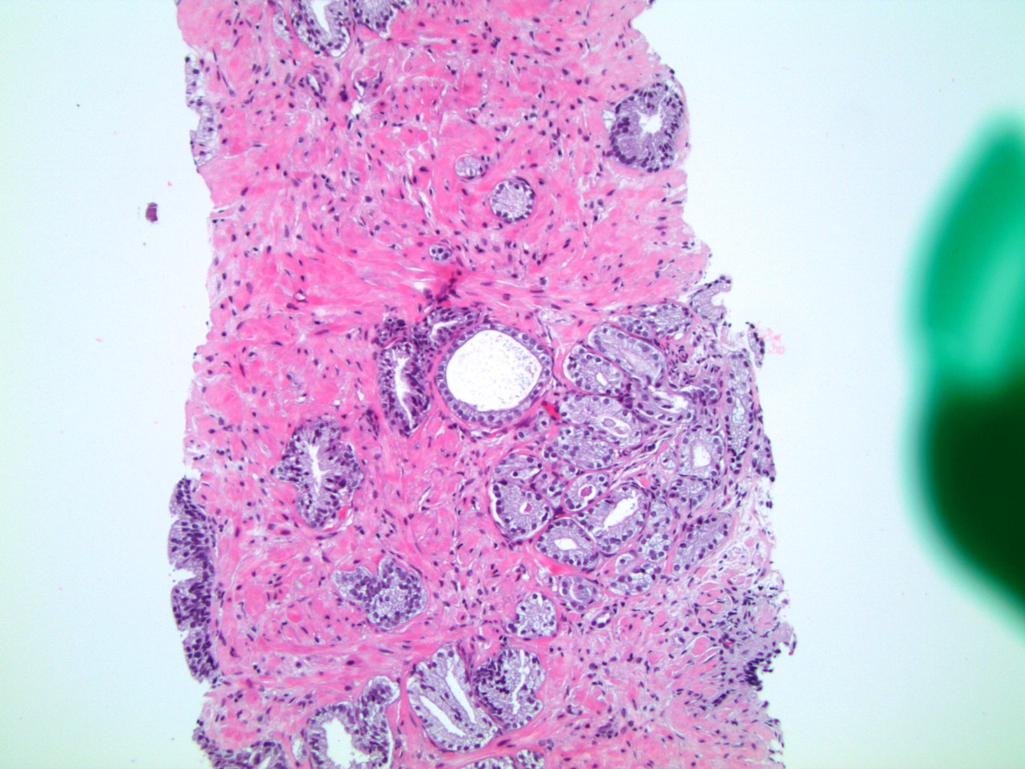
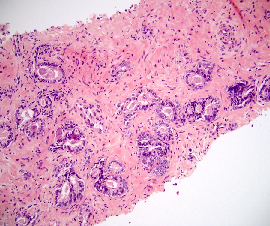
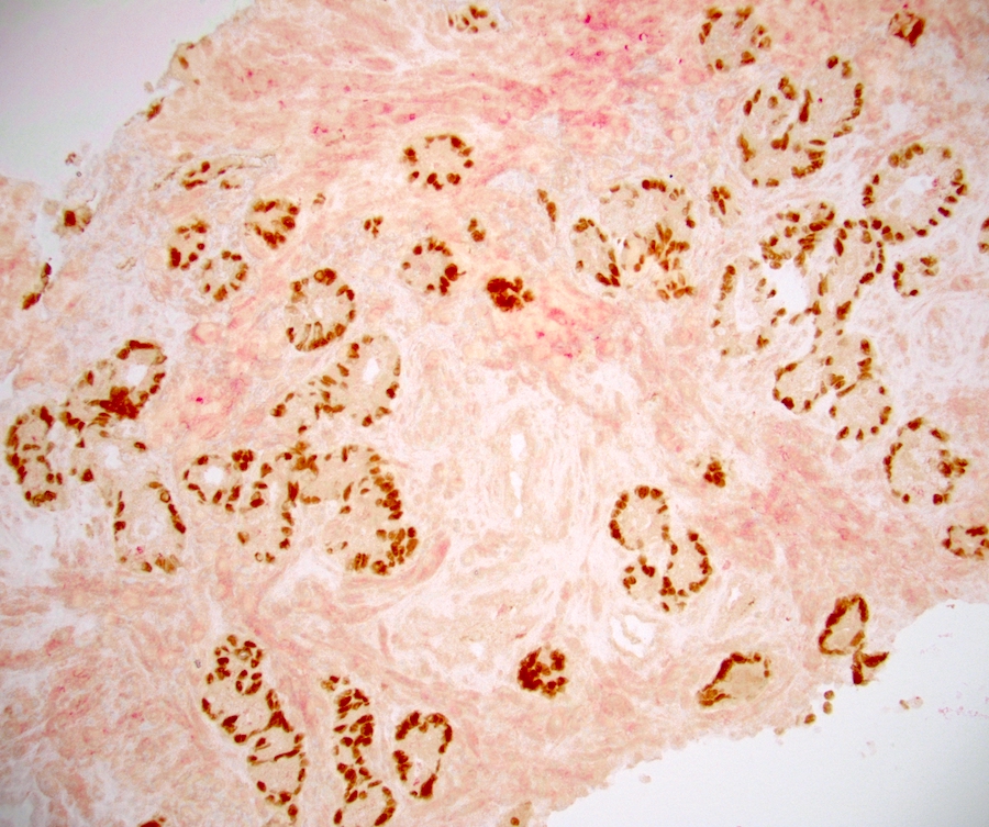
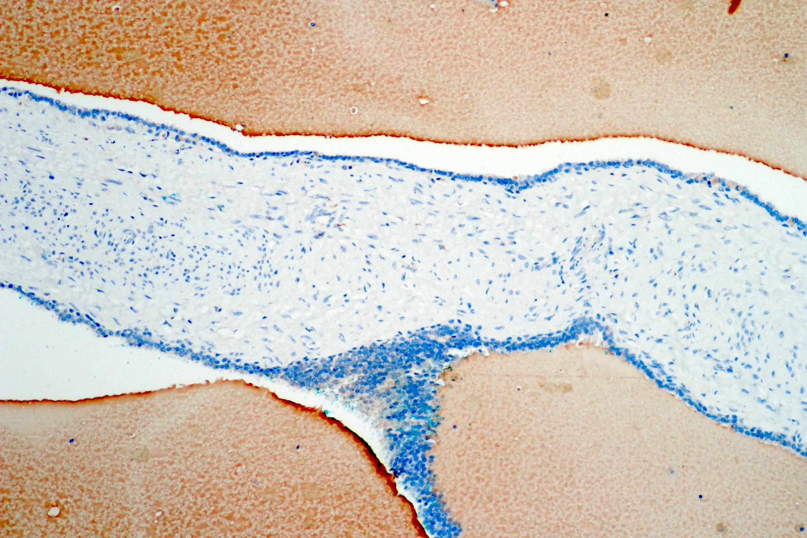
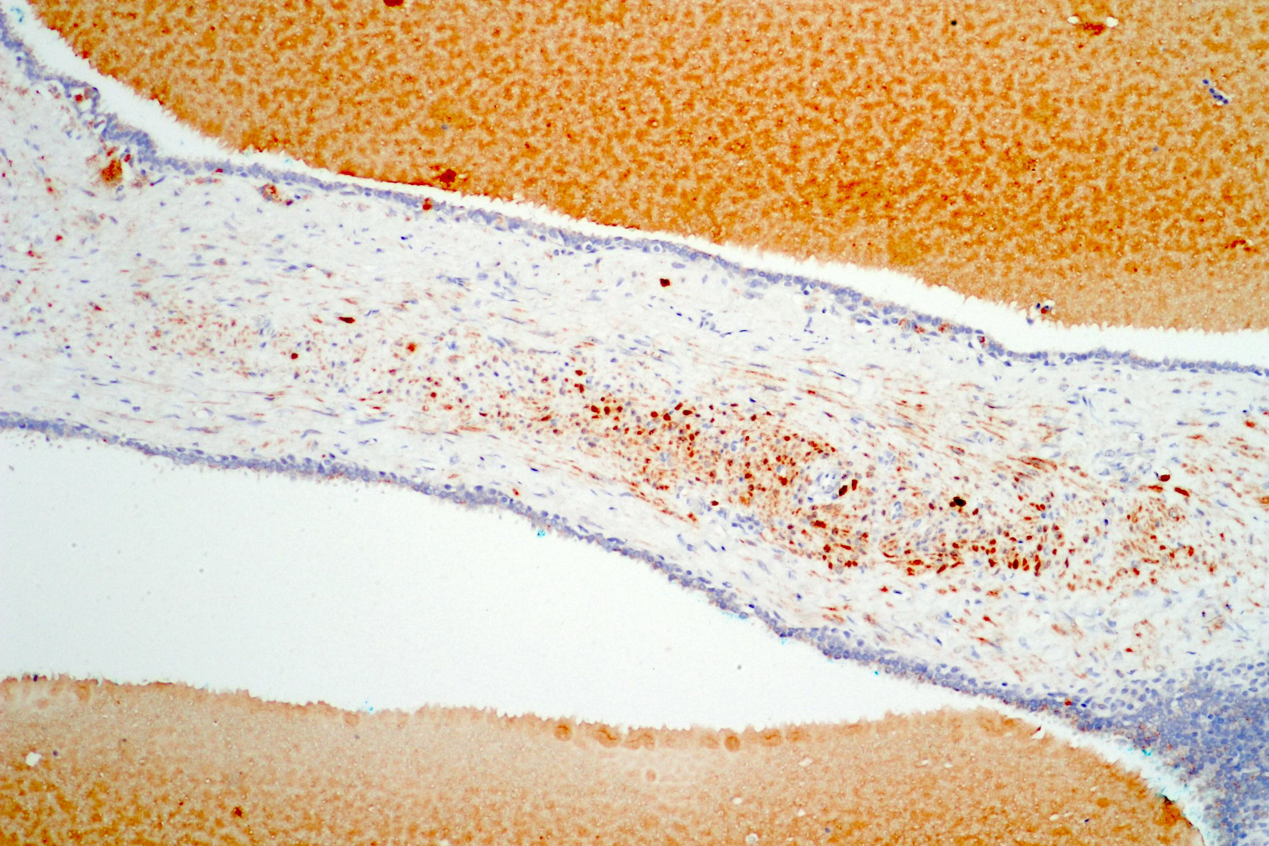
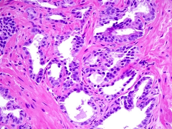
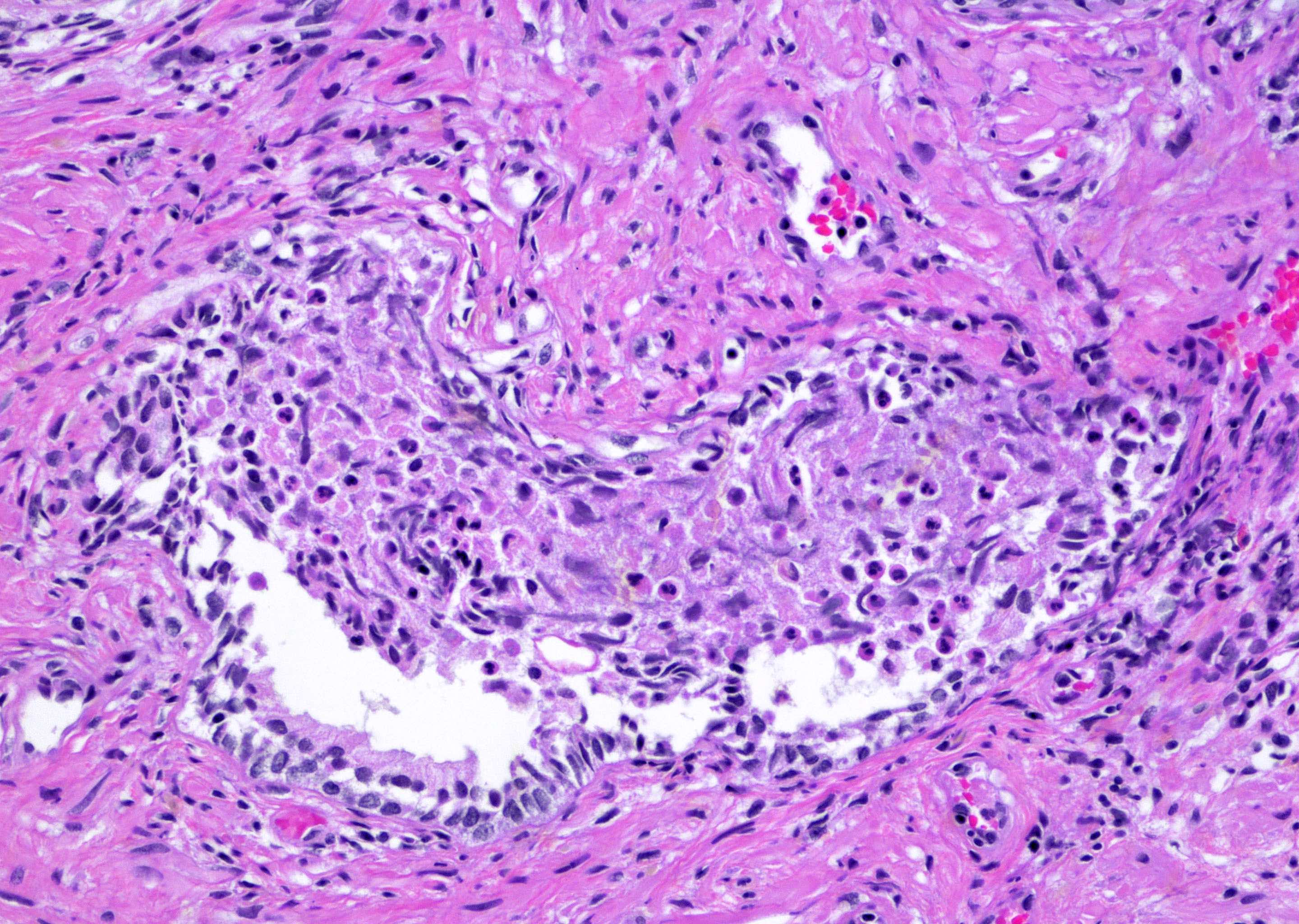
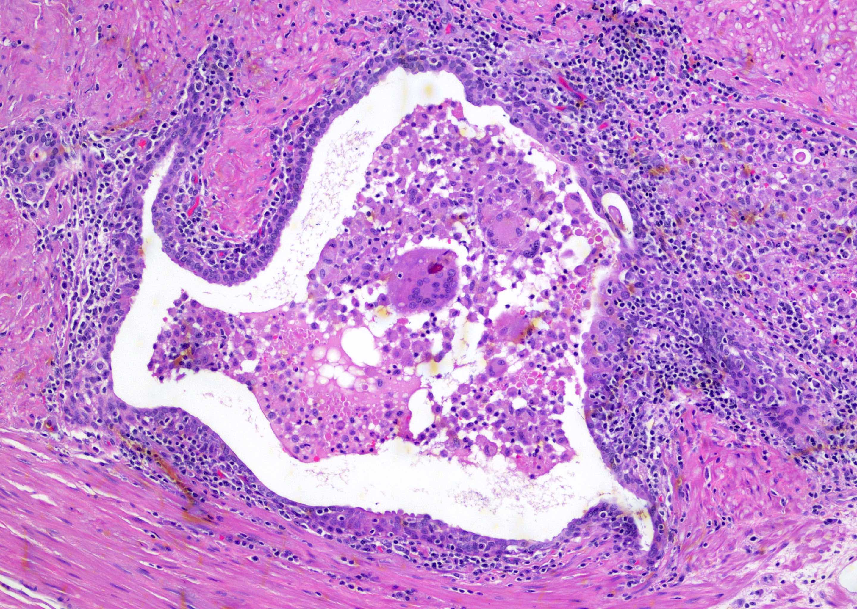
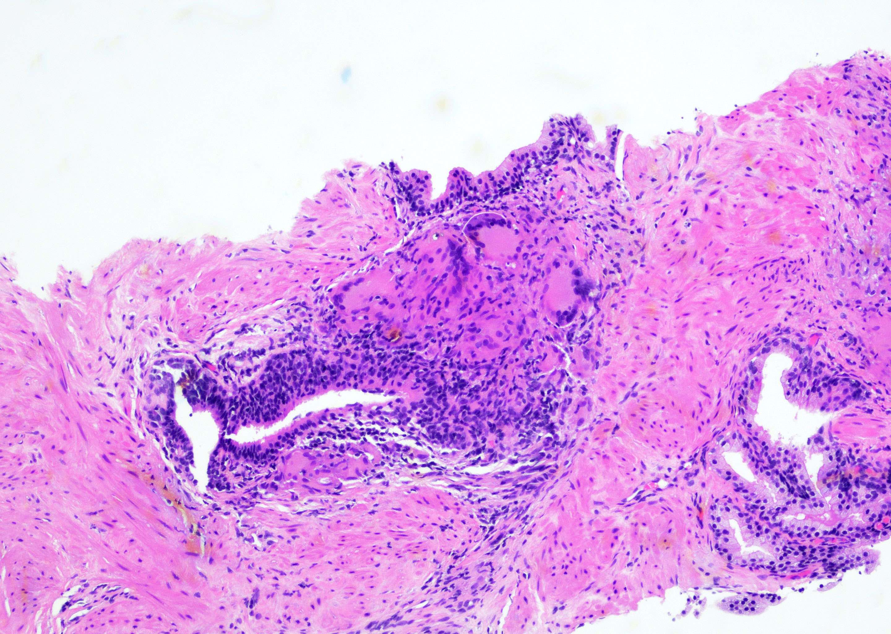
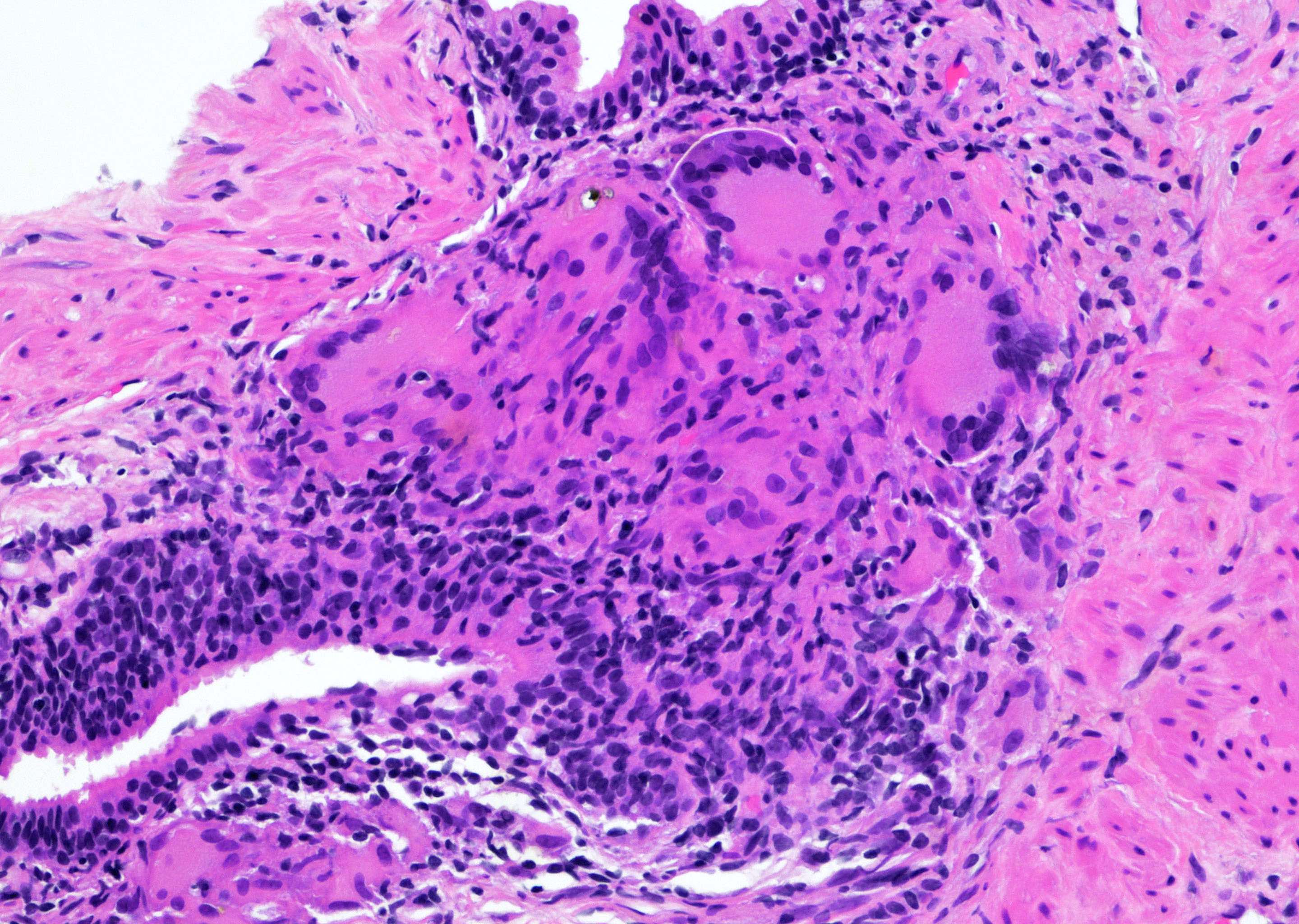
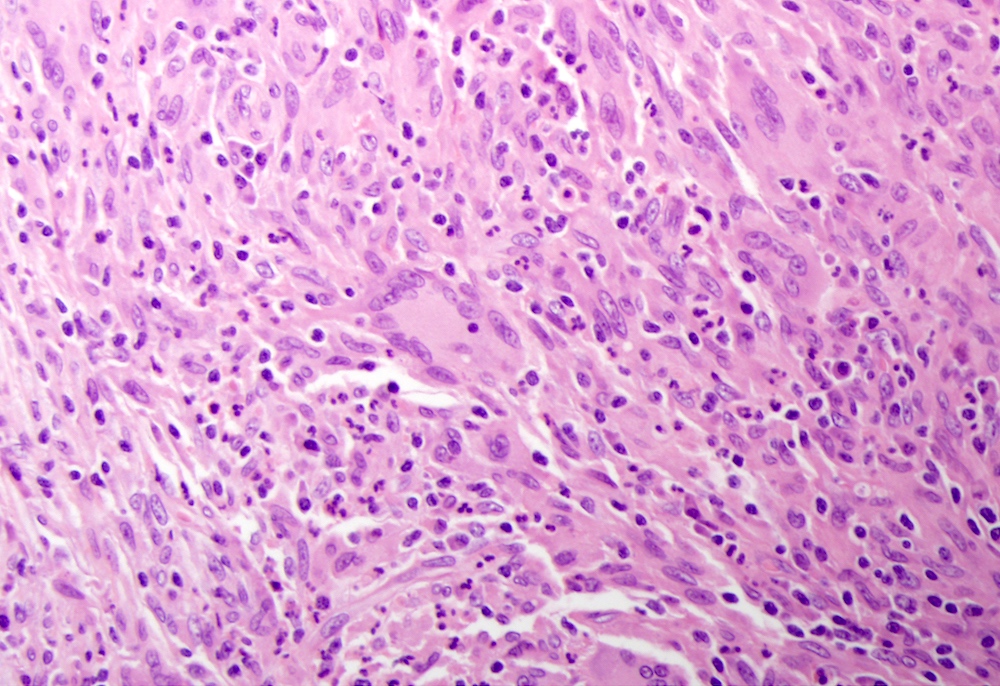
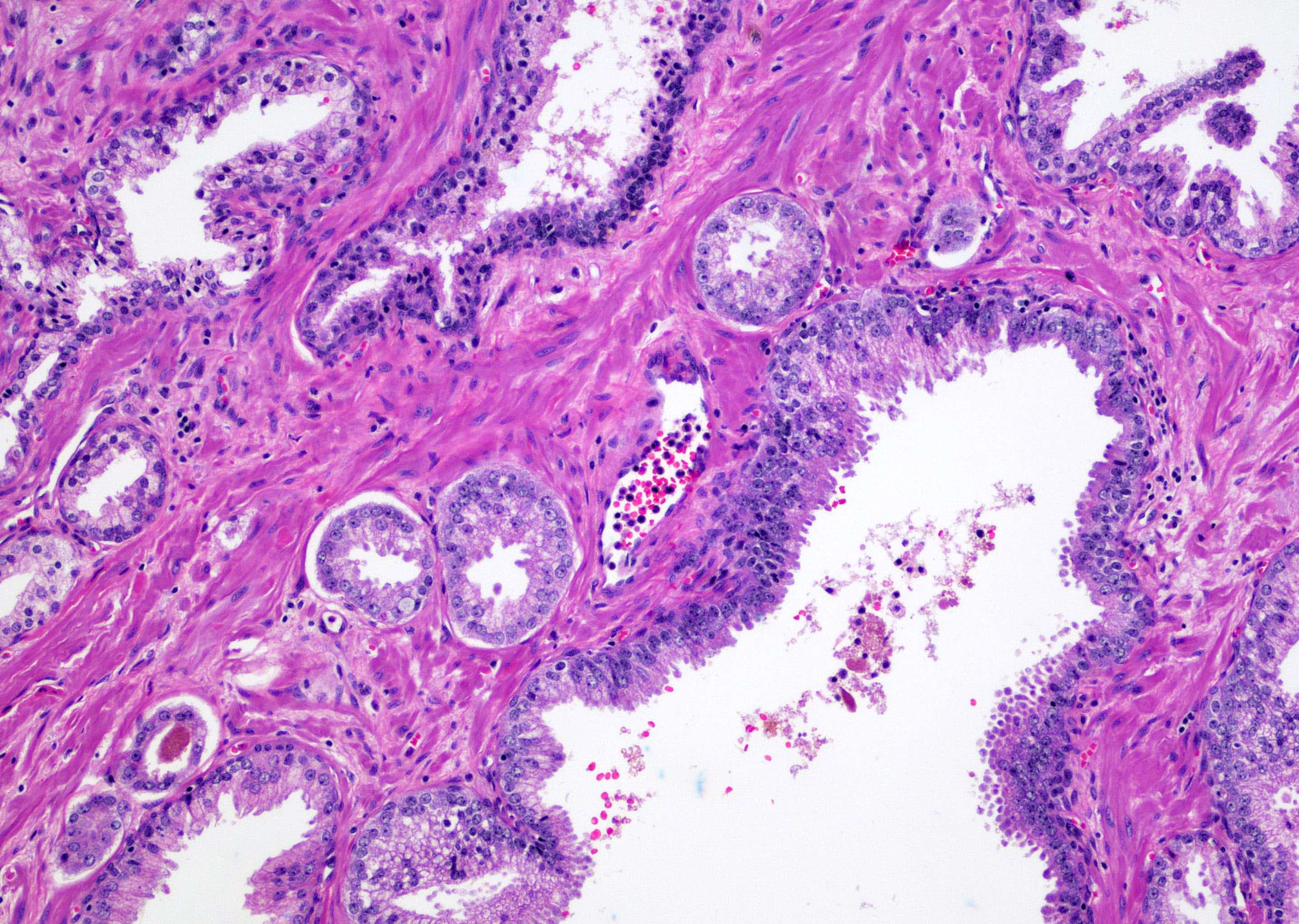
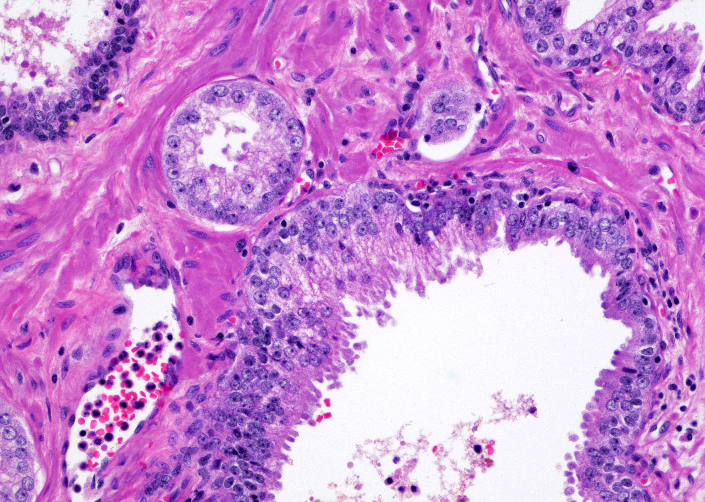
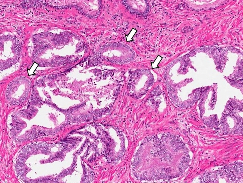
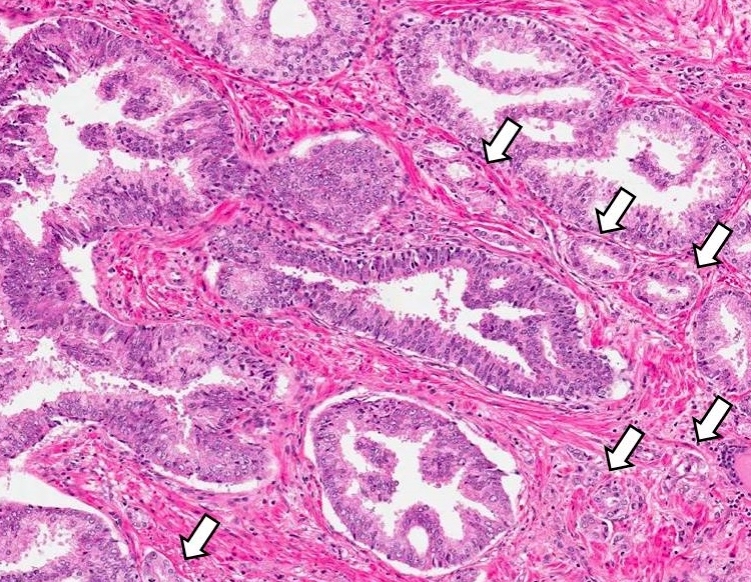
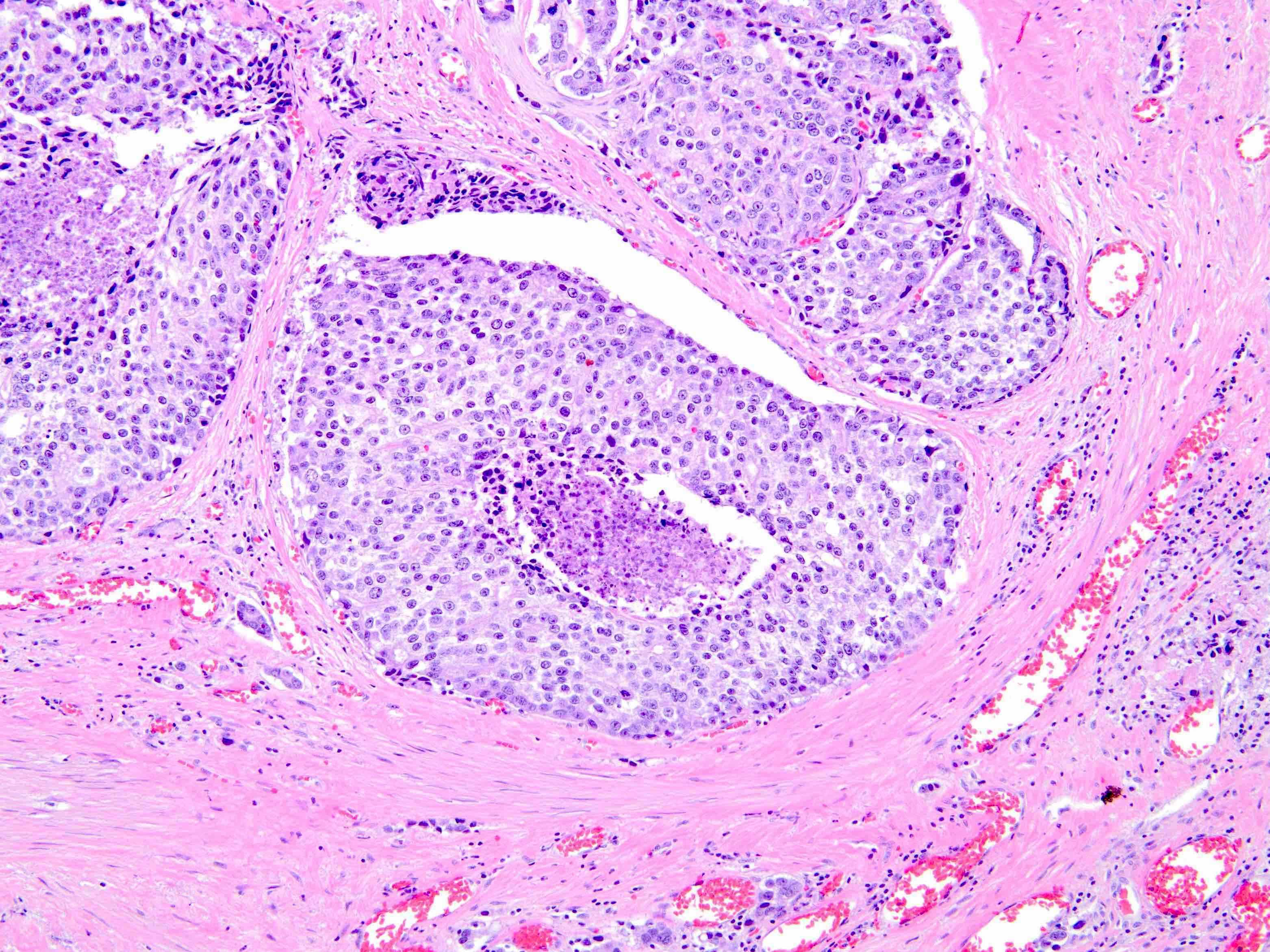
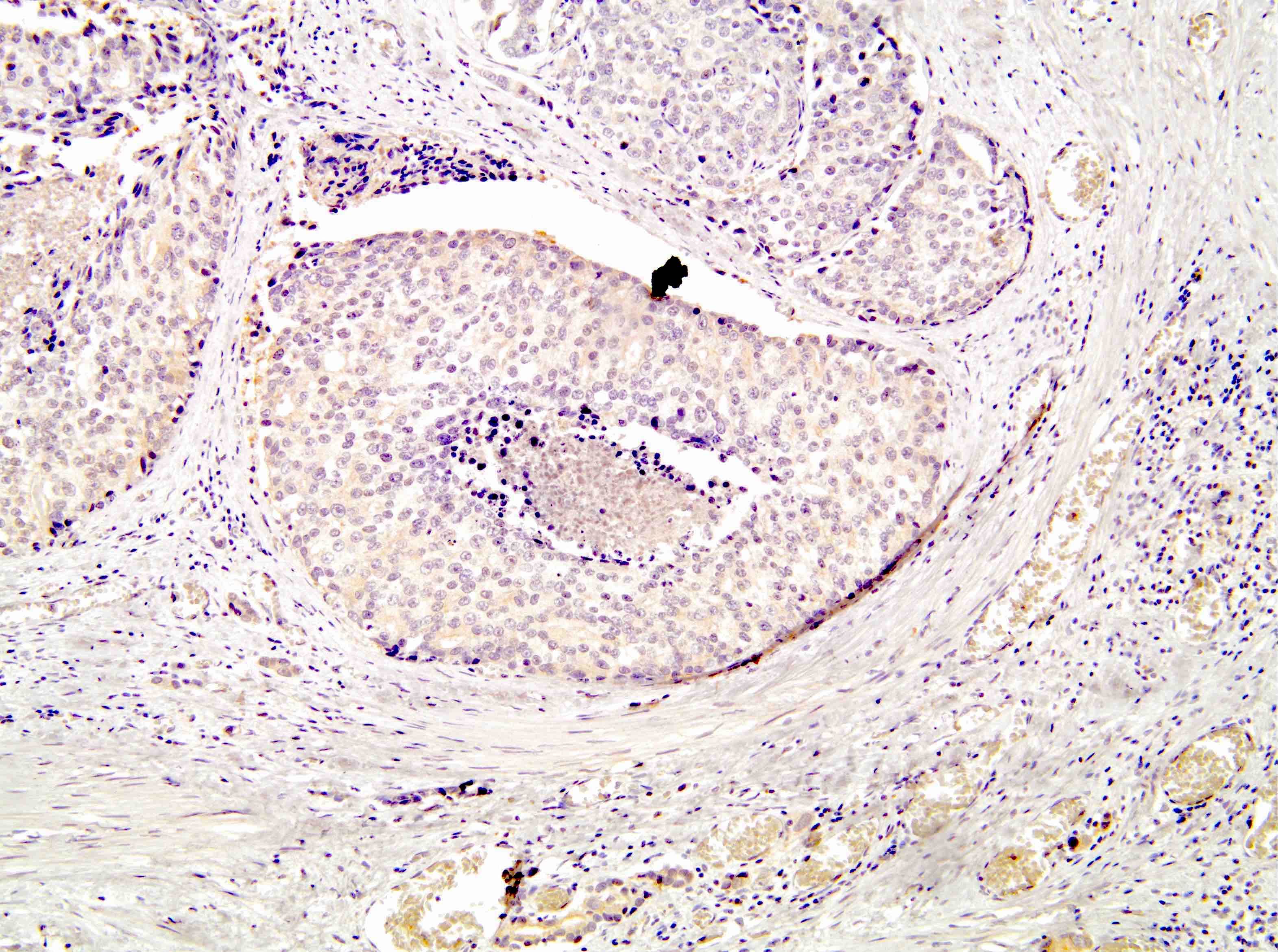
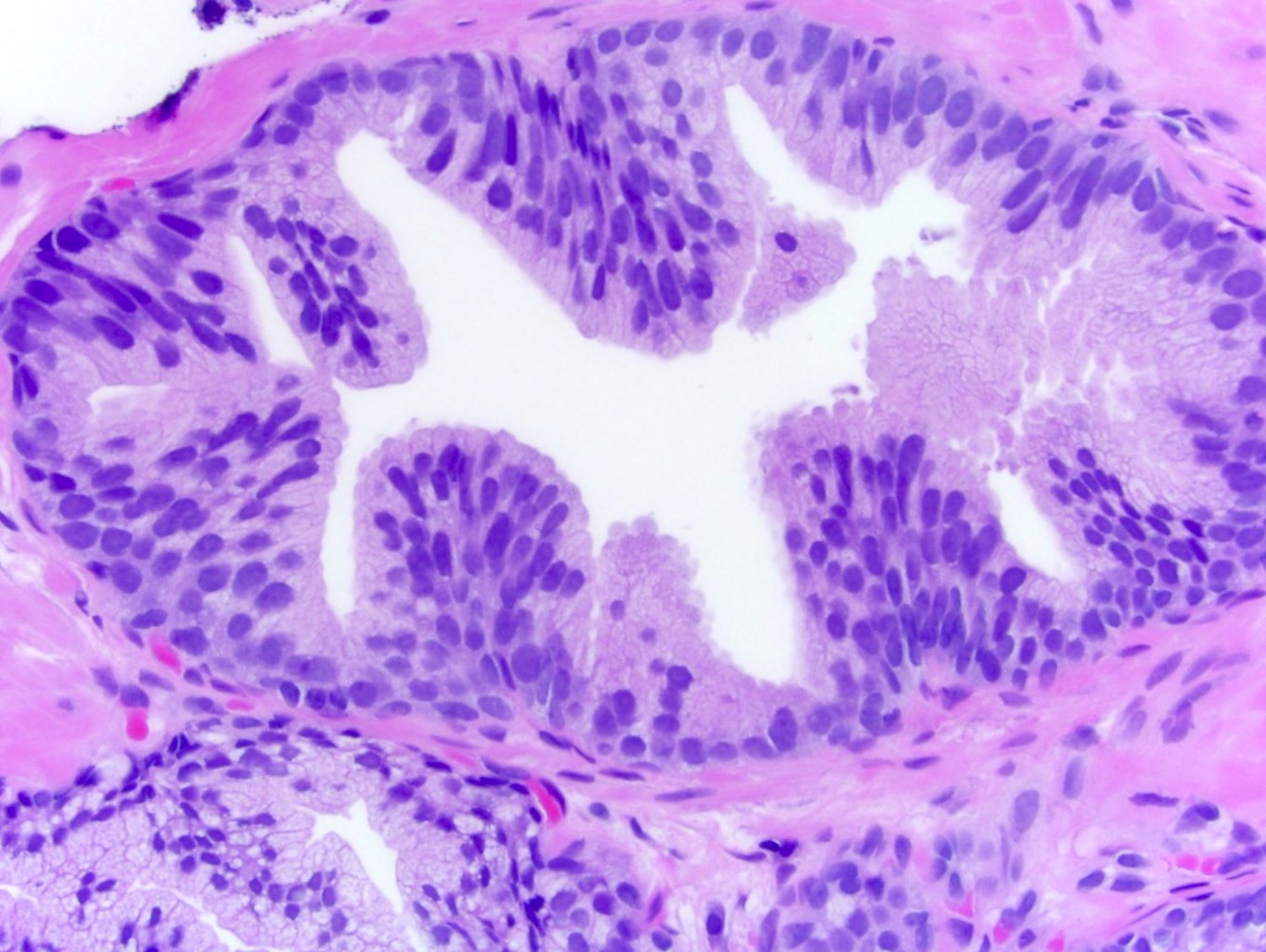
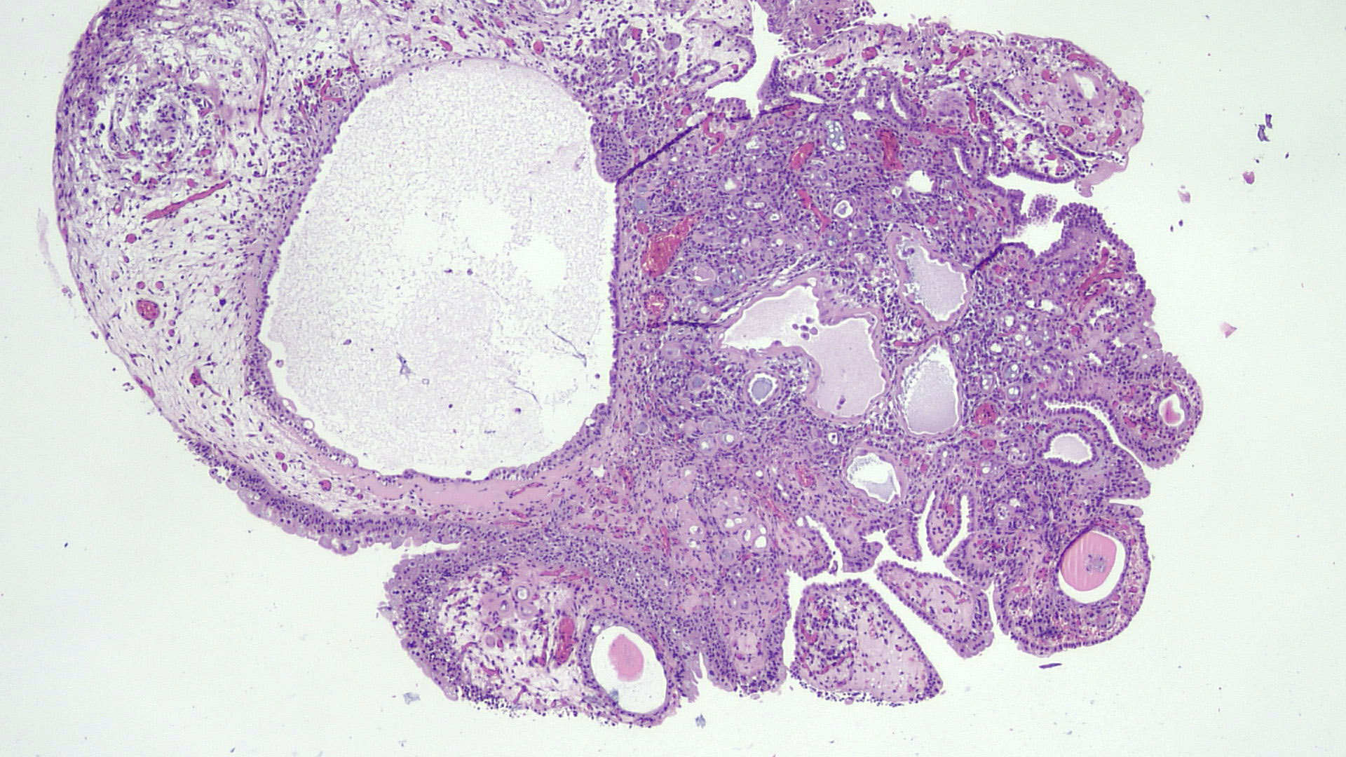
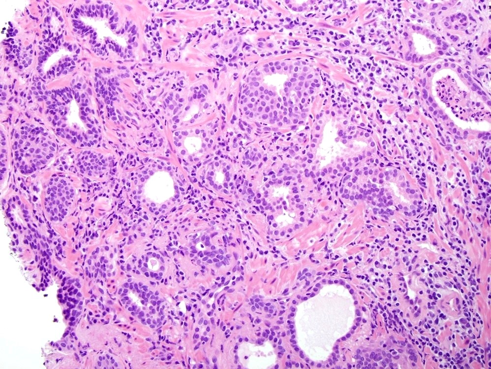
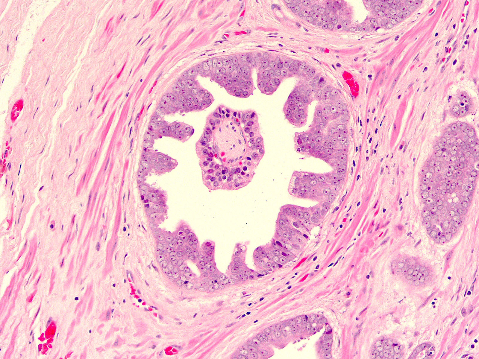
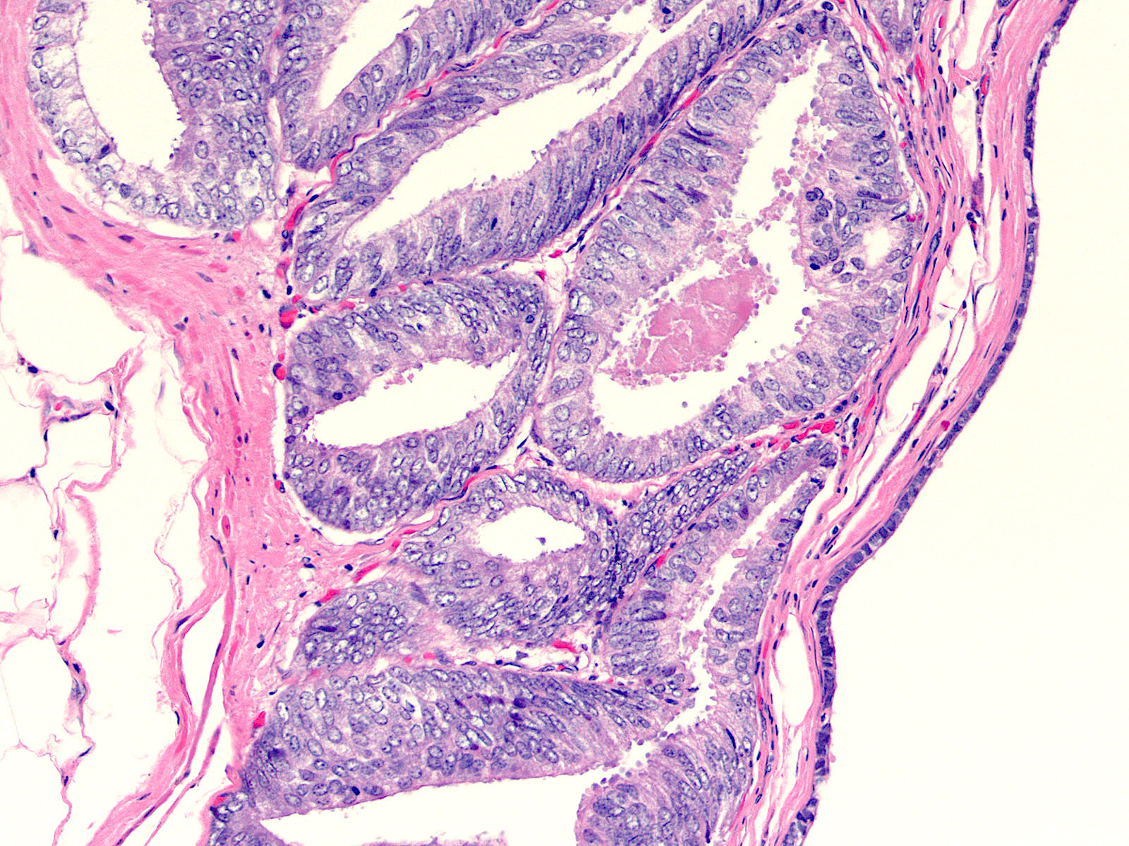
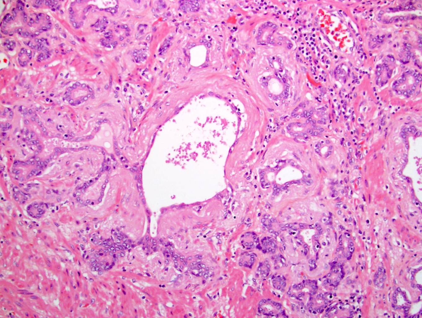
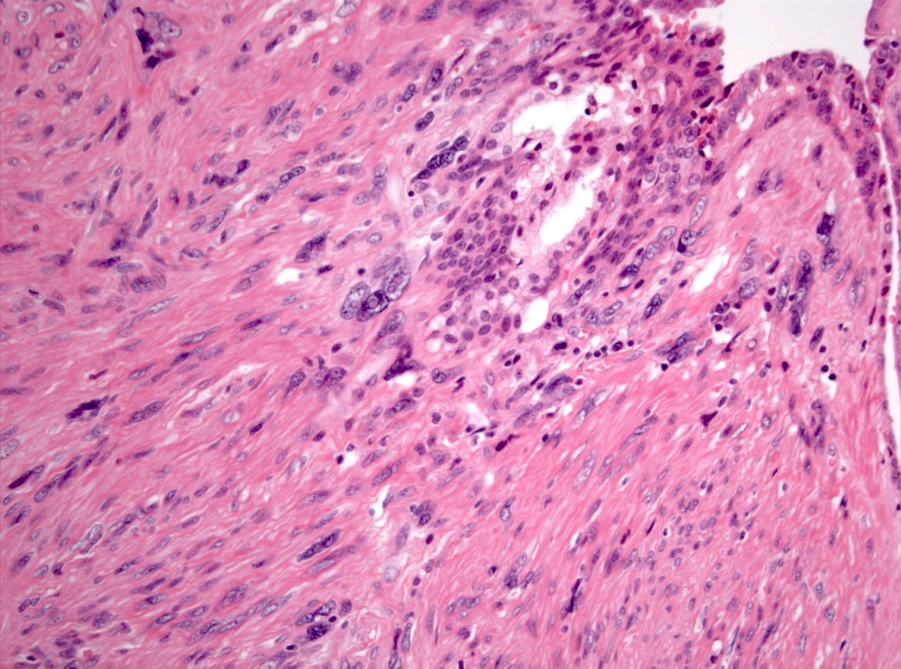
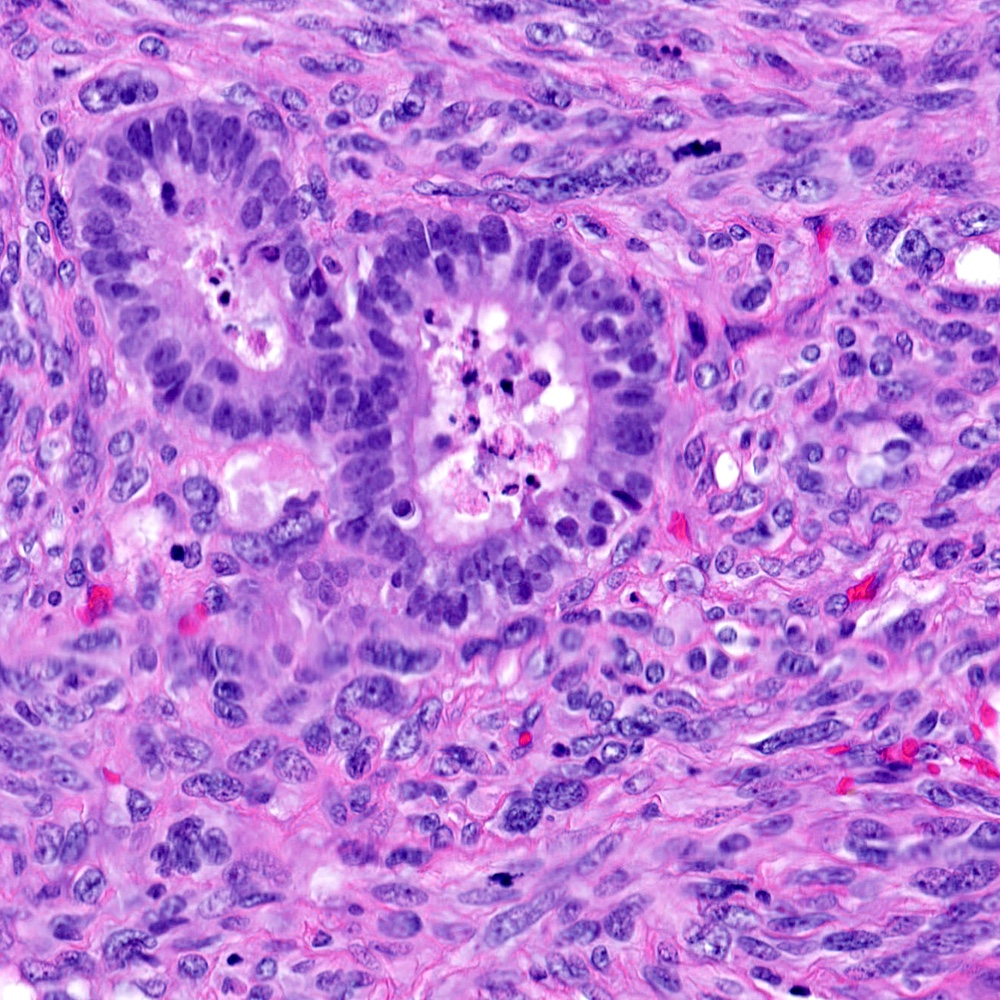
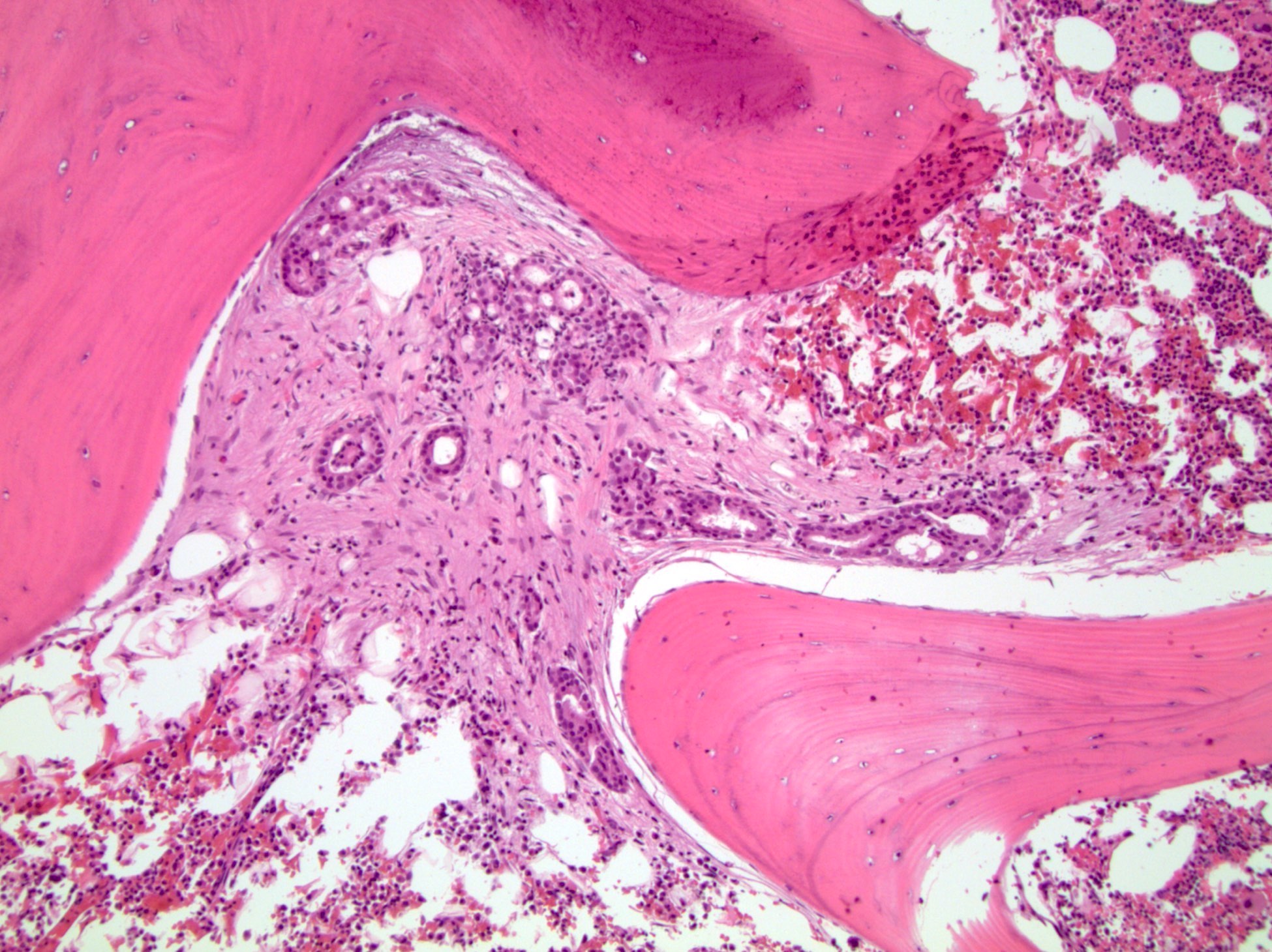
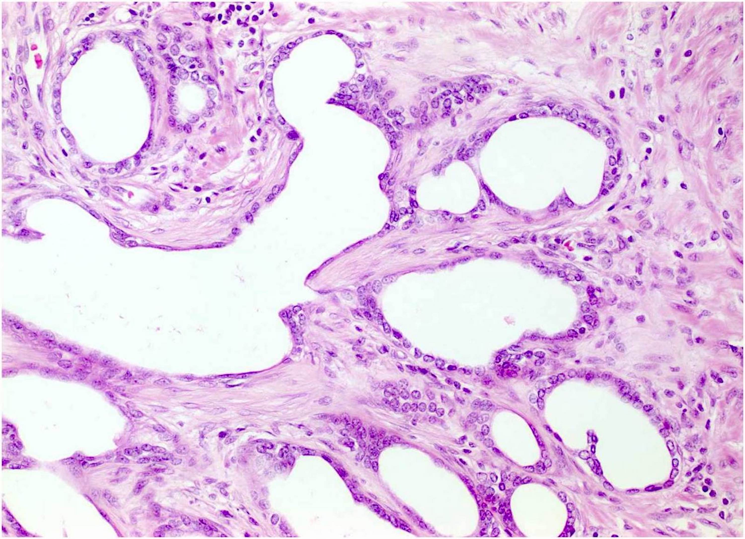
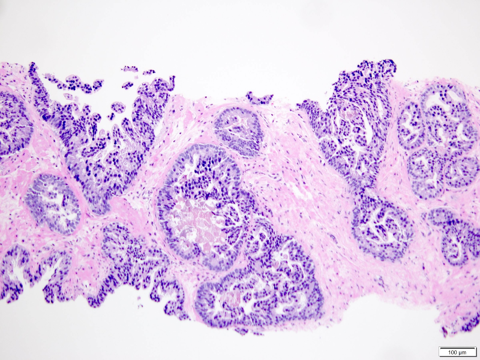
Contributed by Deepika Kumar, M.D. and Peter A. Humphrey, M.D., Ph.D.
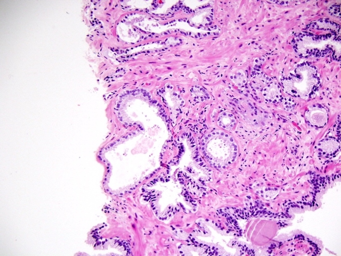
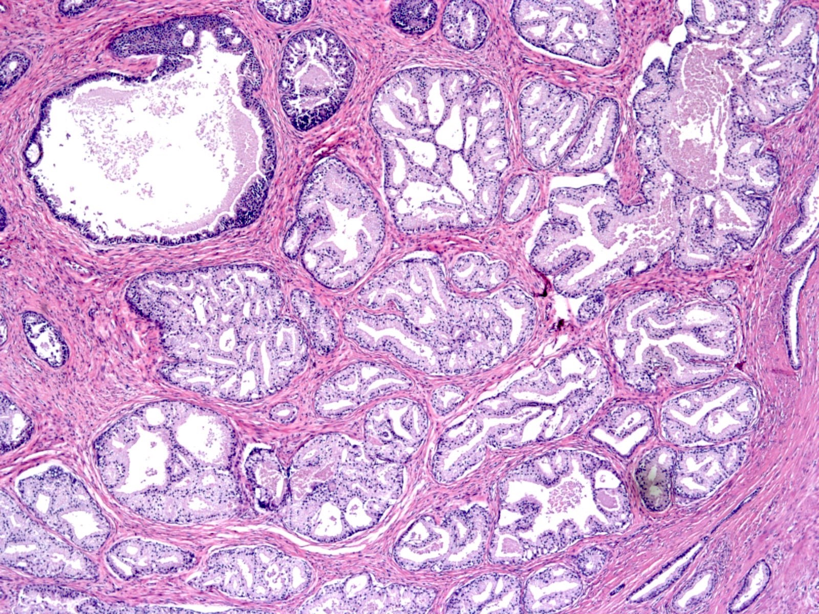
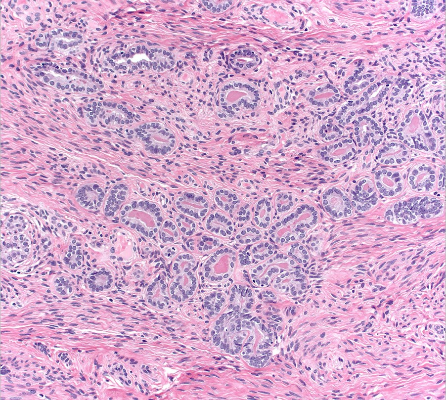
Variably sized glands
Cystic dilatation

Flat pseudostratified lining
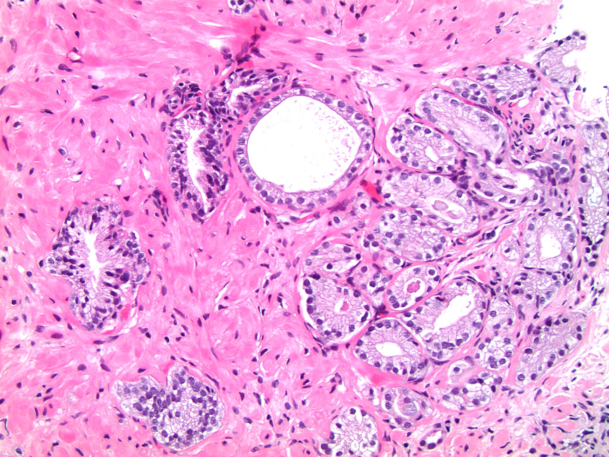
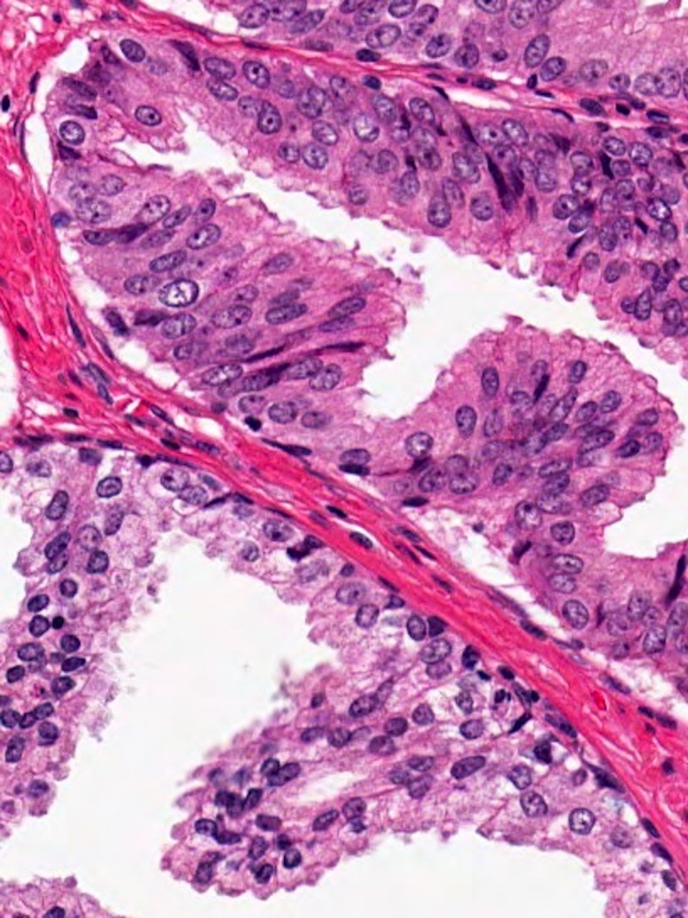
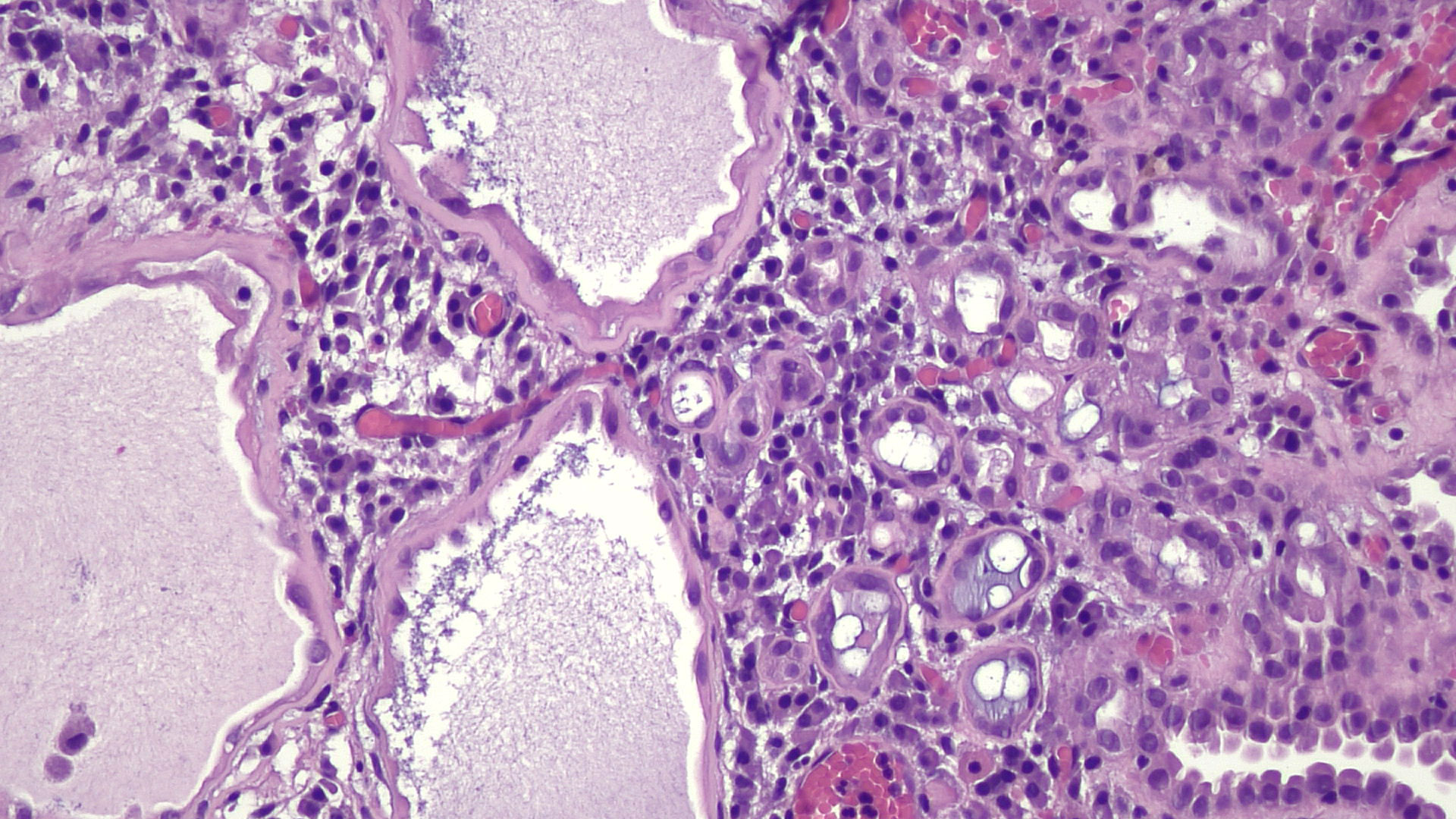
Crowded glands
Nuclear atypia
Prominent nucleoli
Perineural invasion
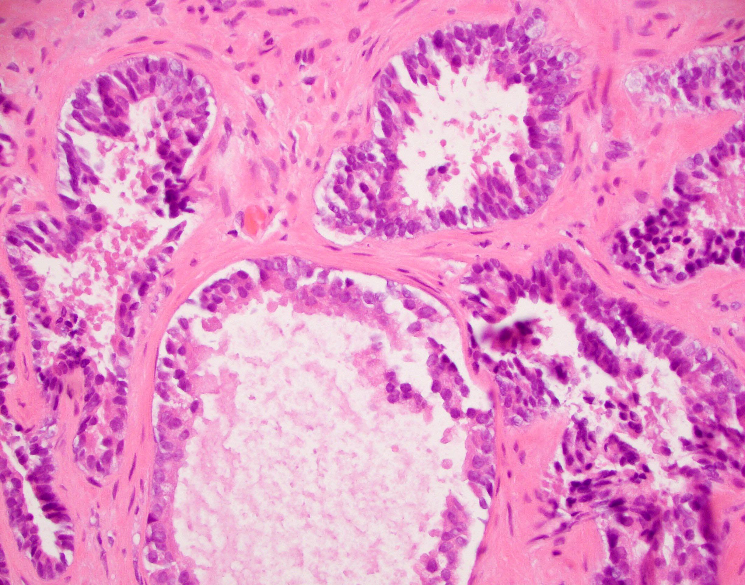
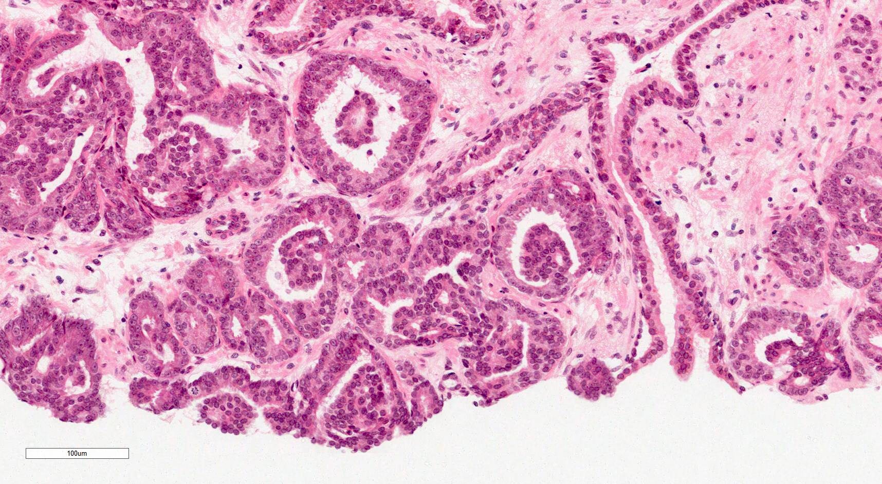
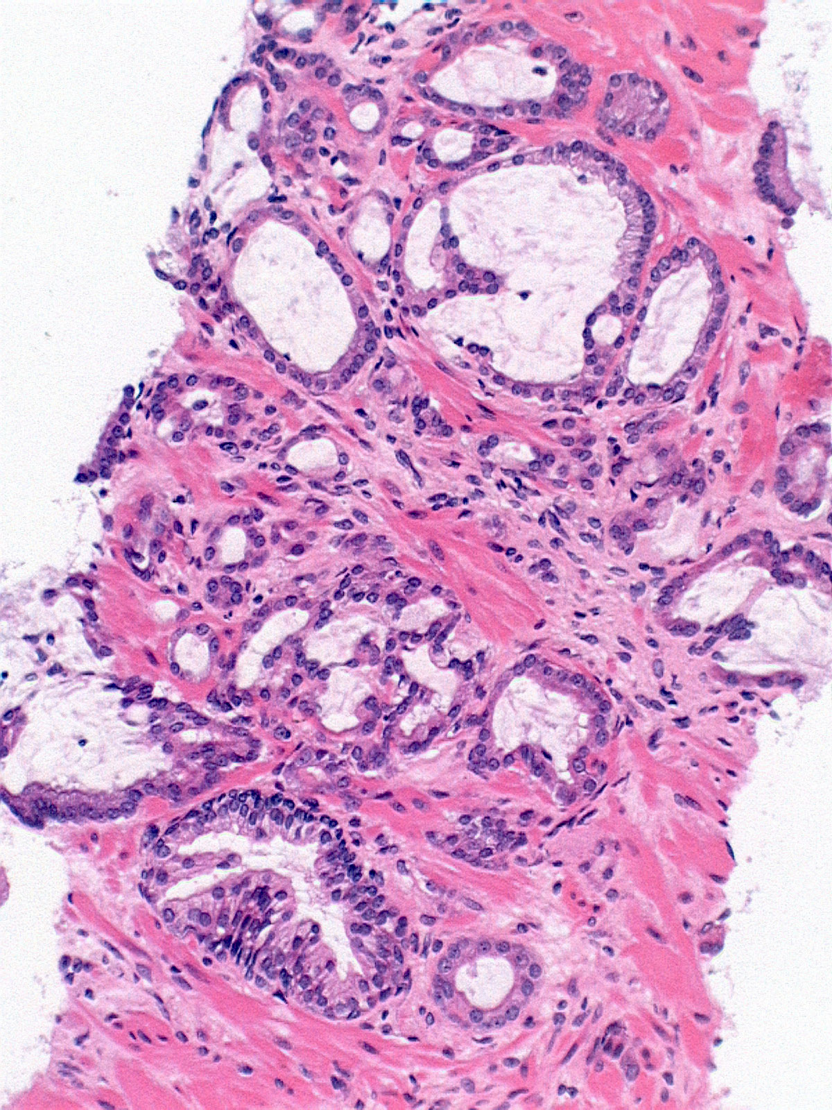
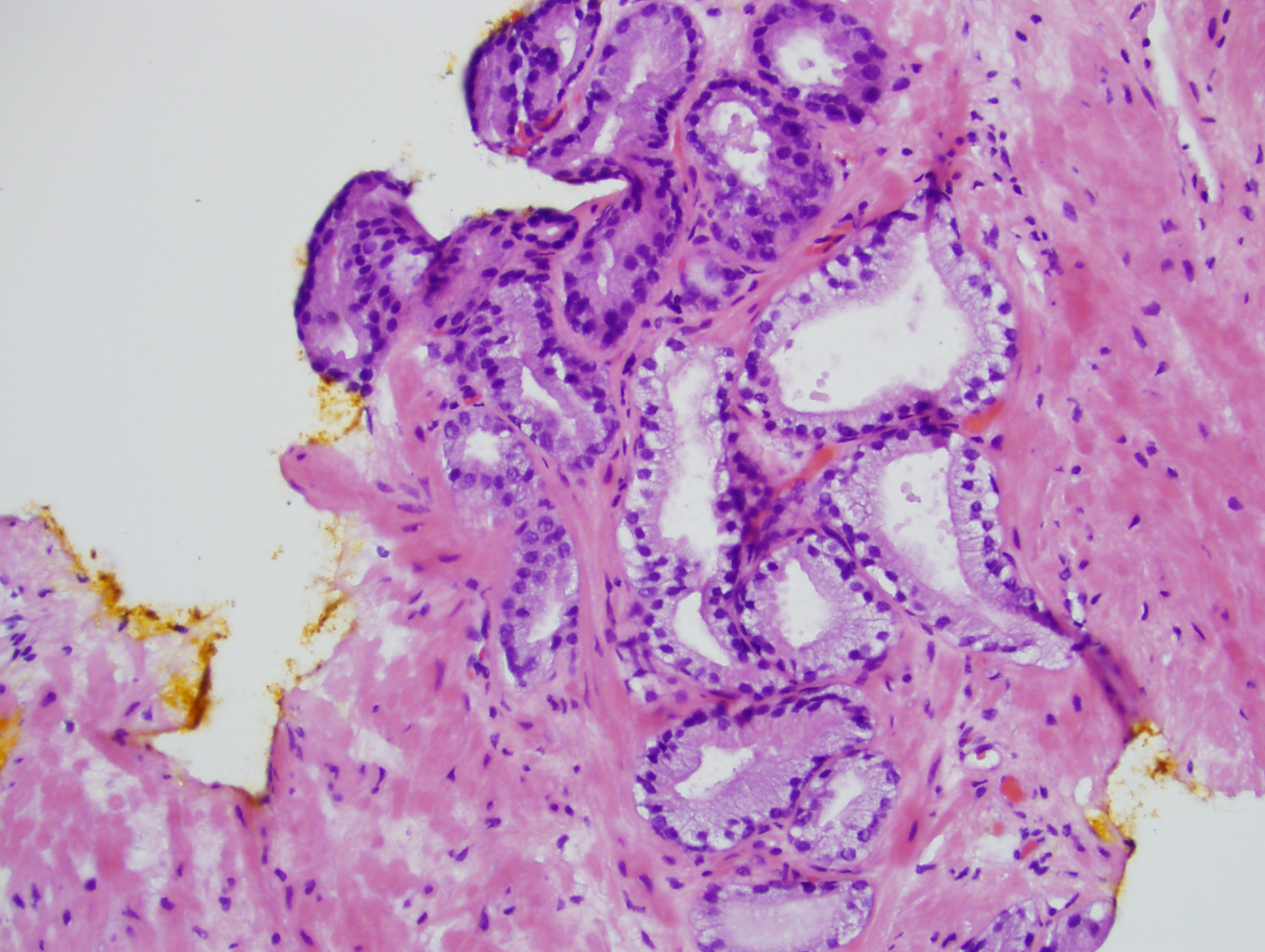
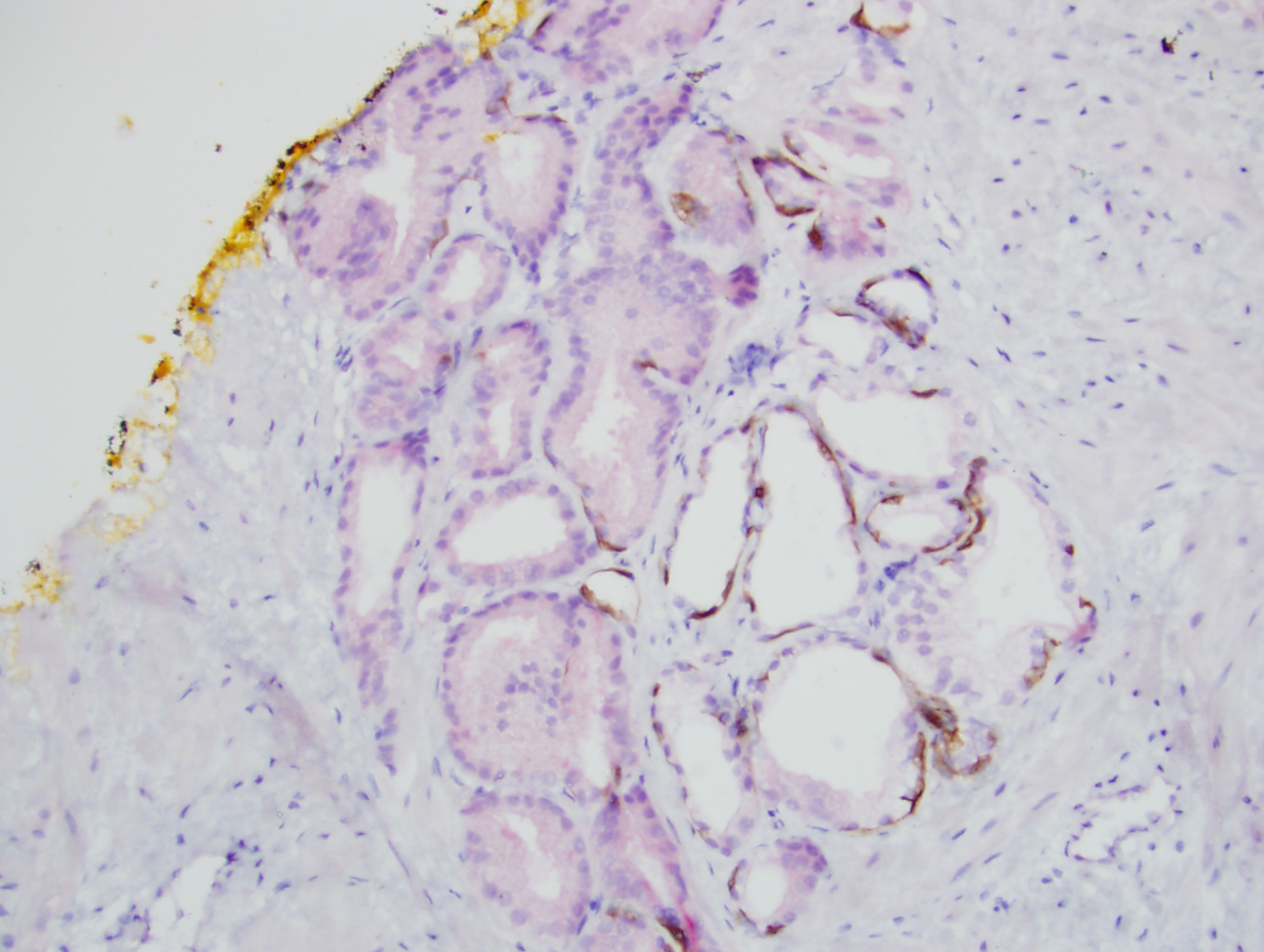
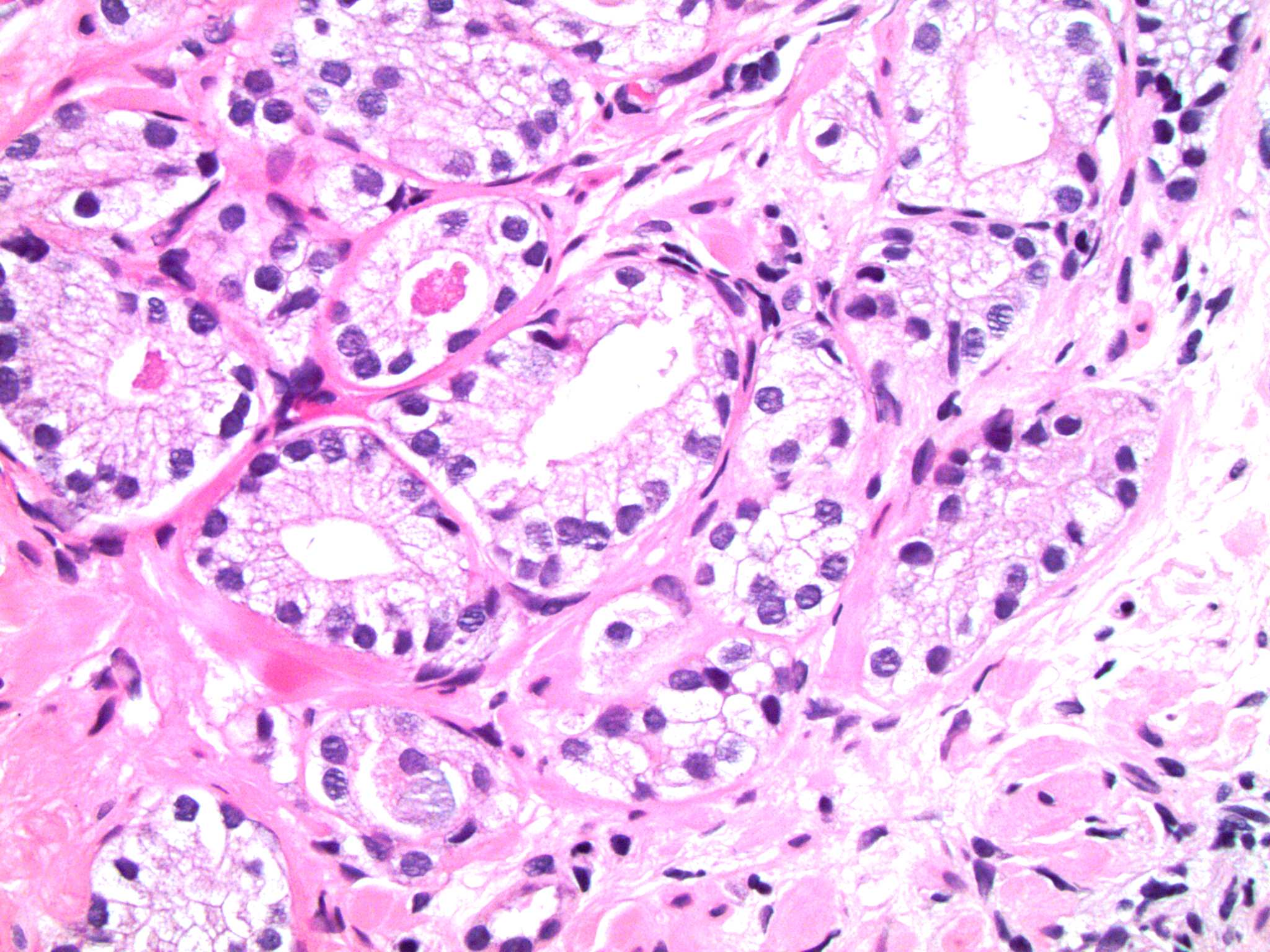
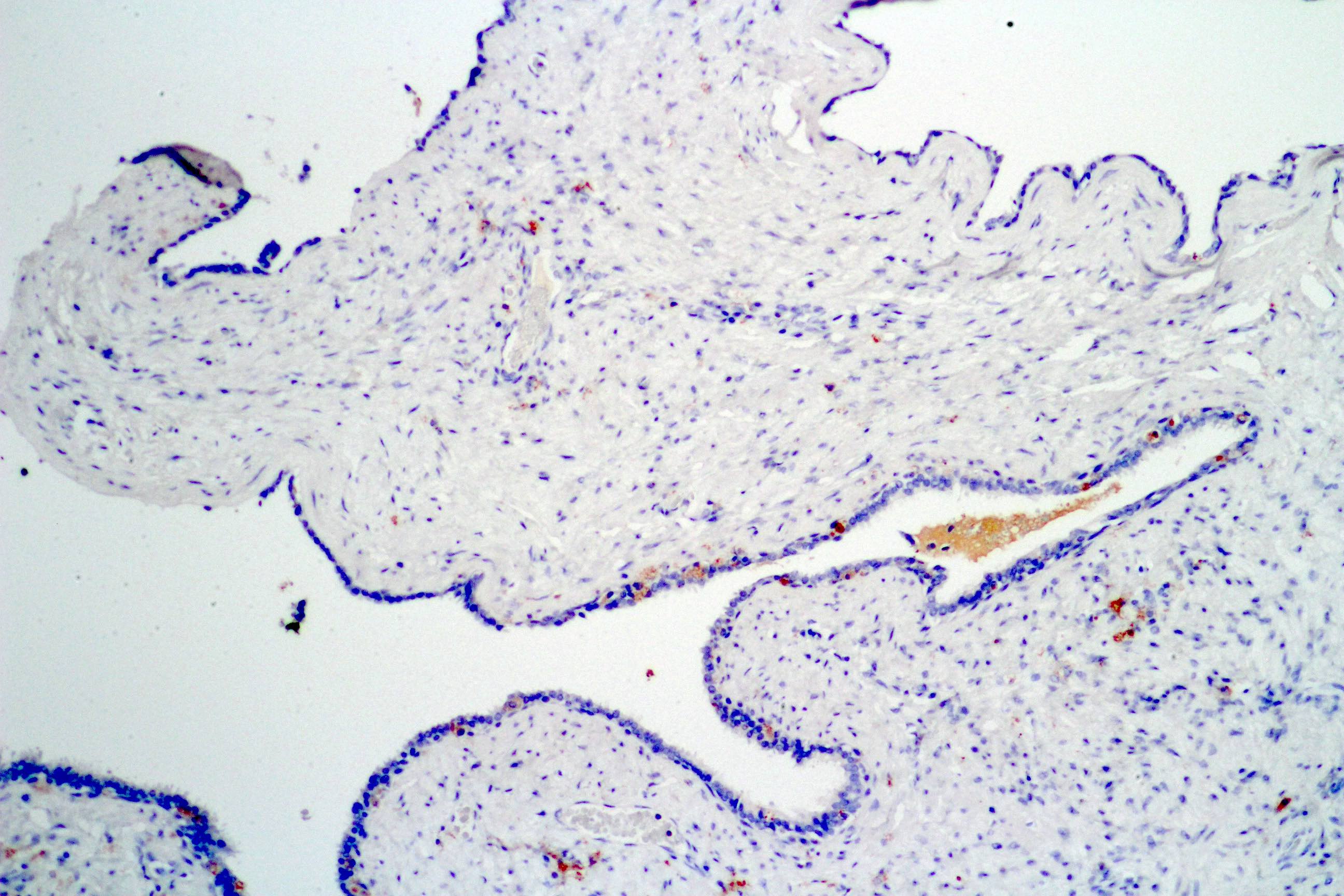
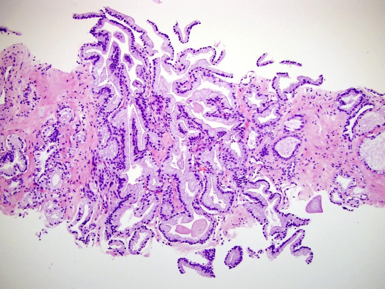
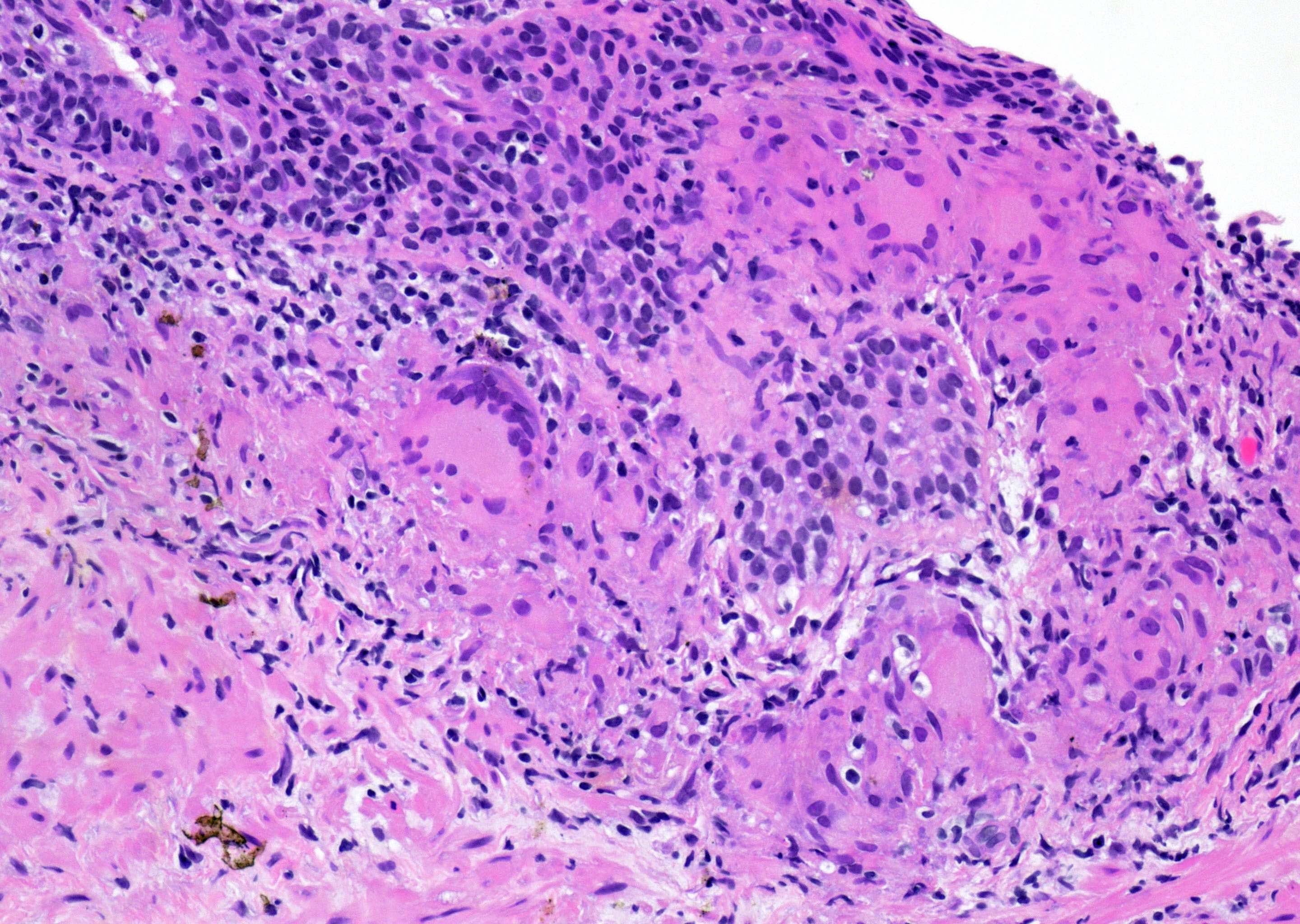
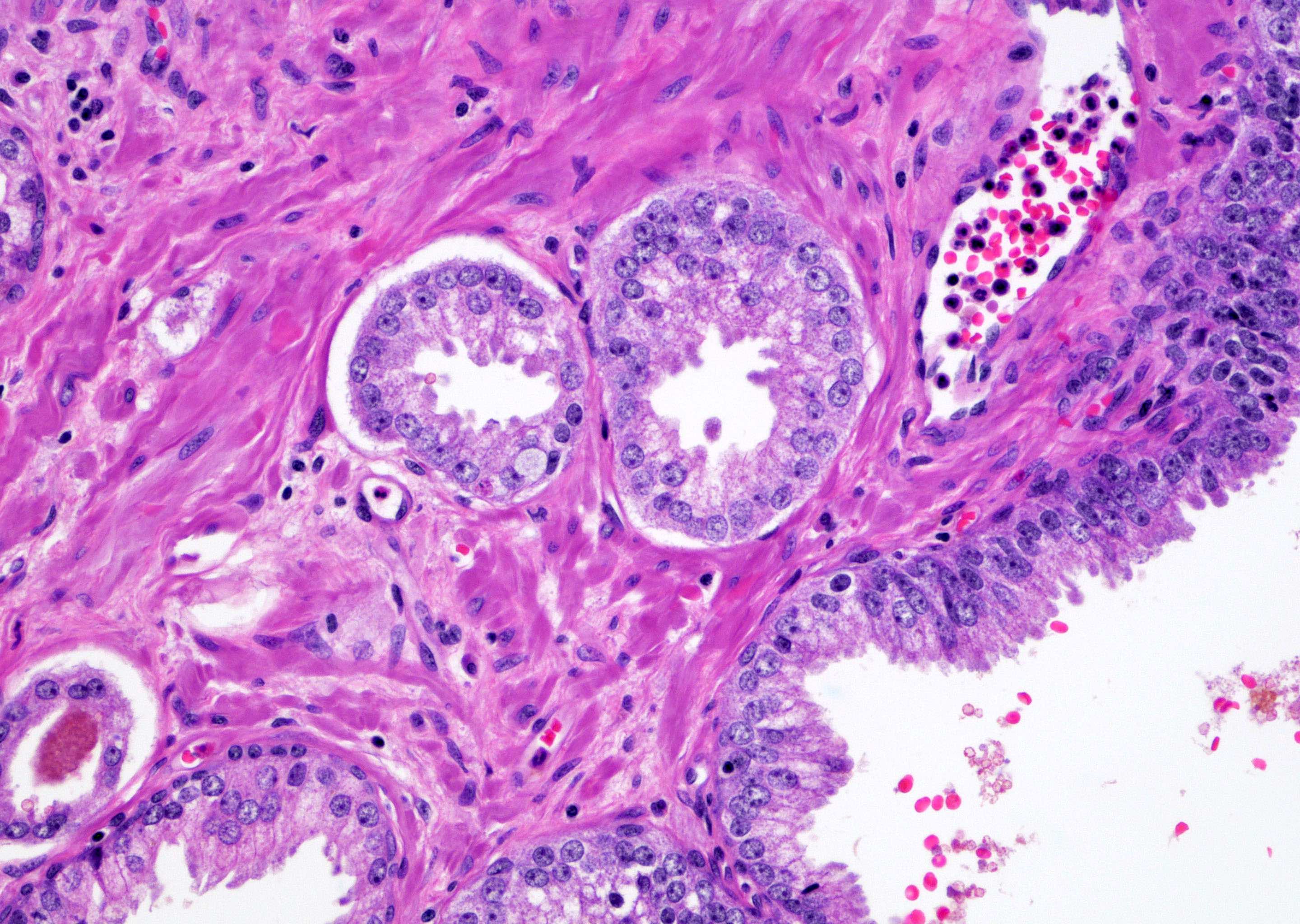
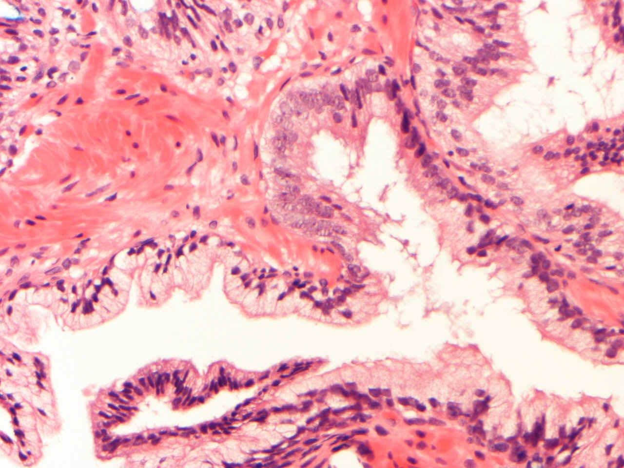
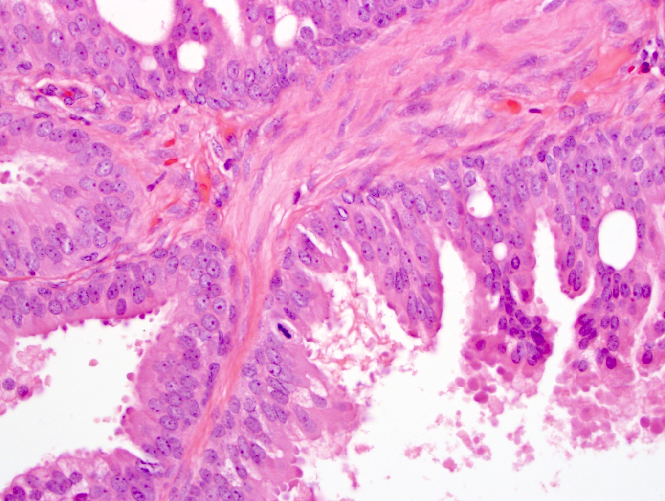
Crowded glands

Micropapillary growth
Less cytologic atypia
Mimics PIN and BPH
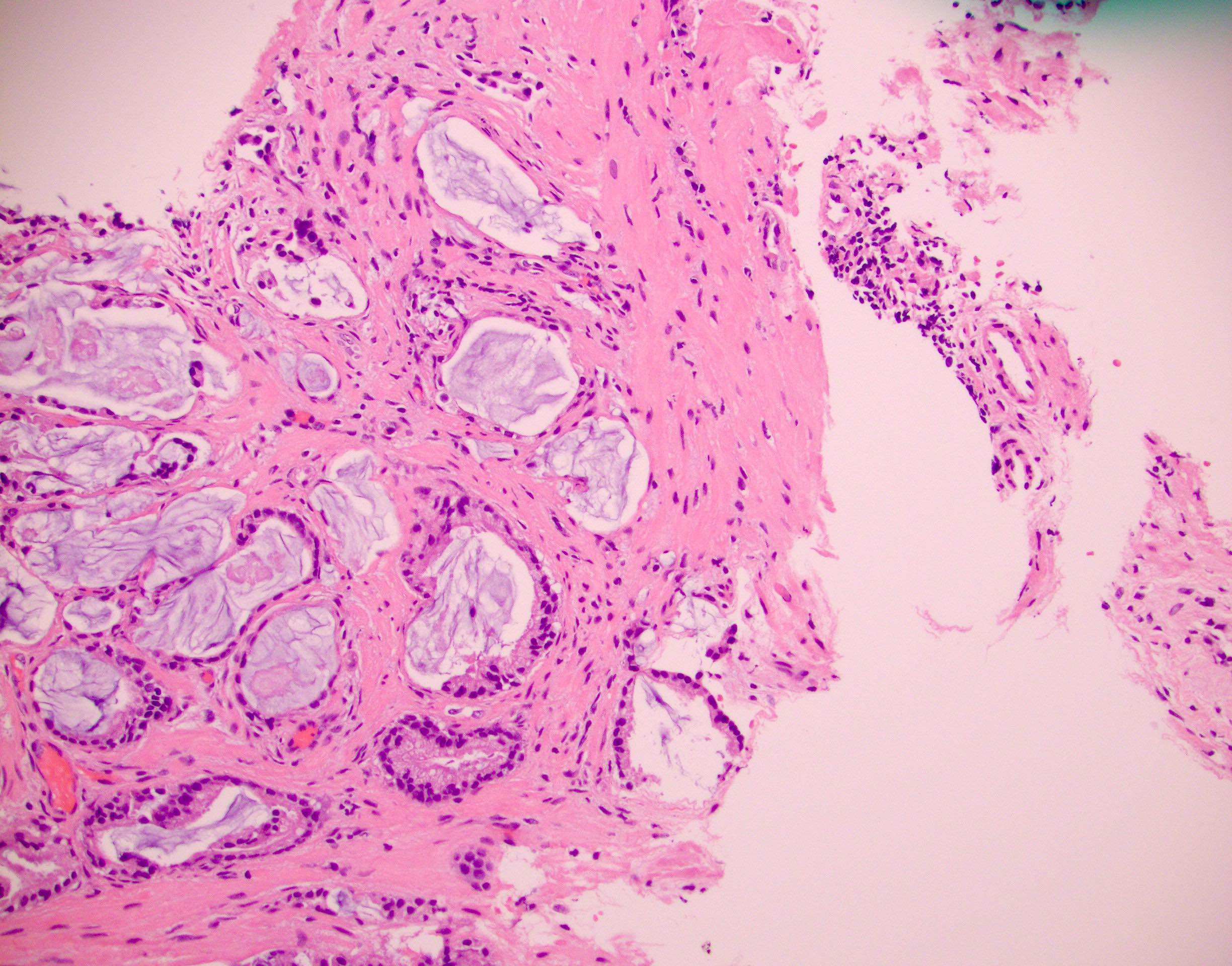
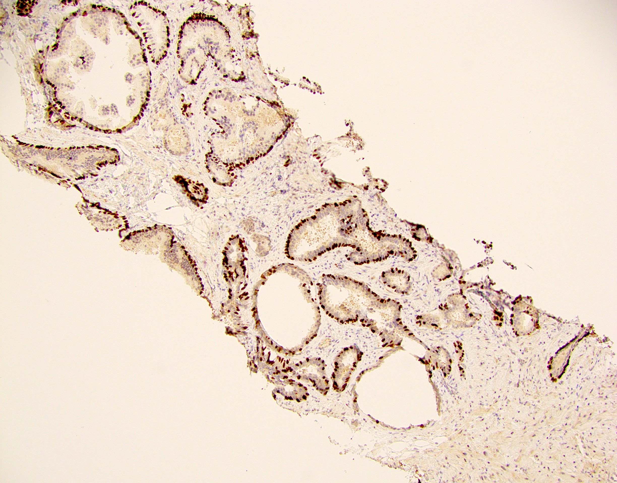
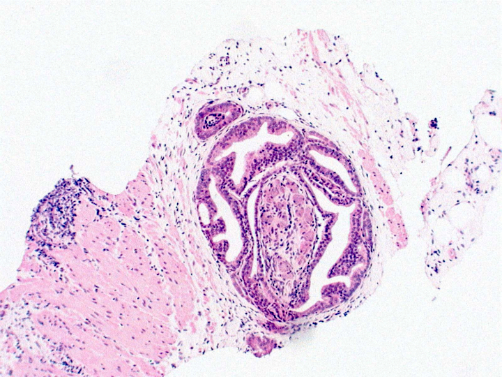
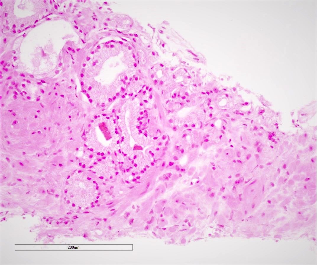
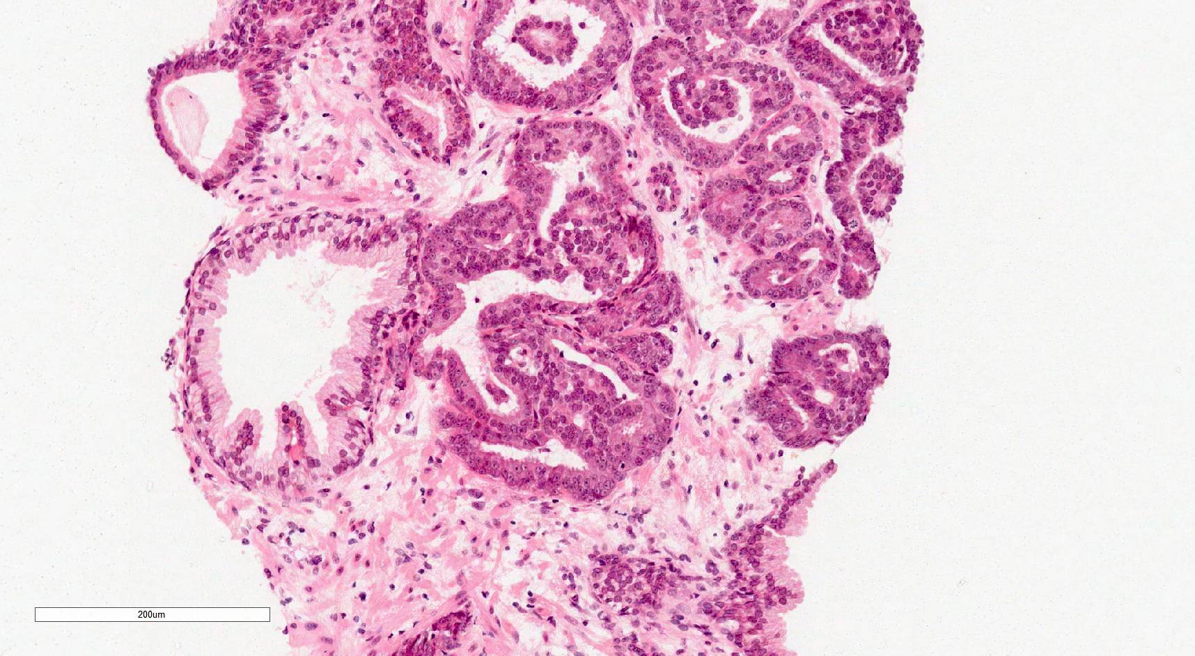
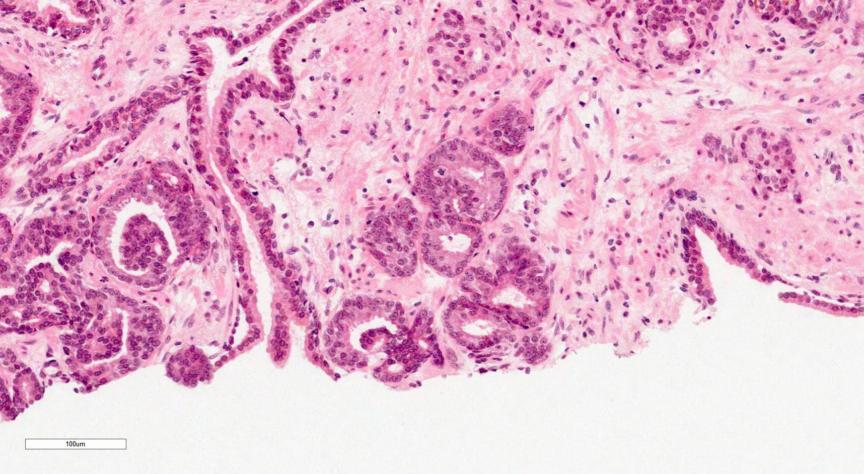
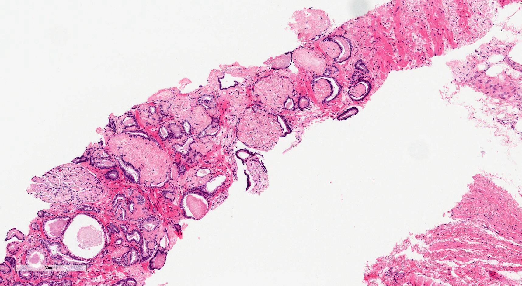
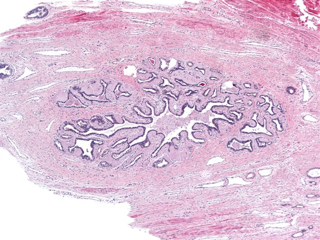
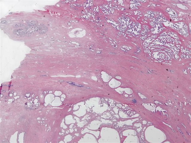
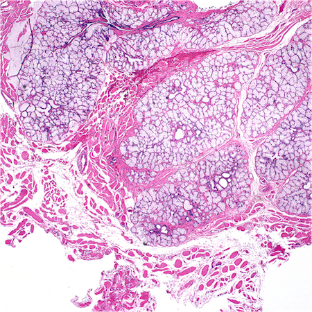
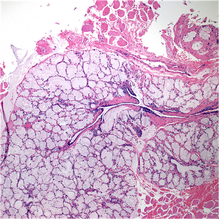
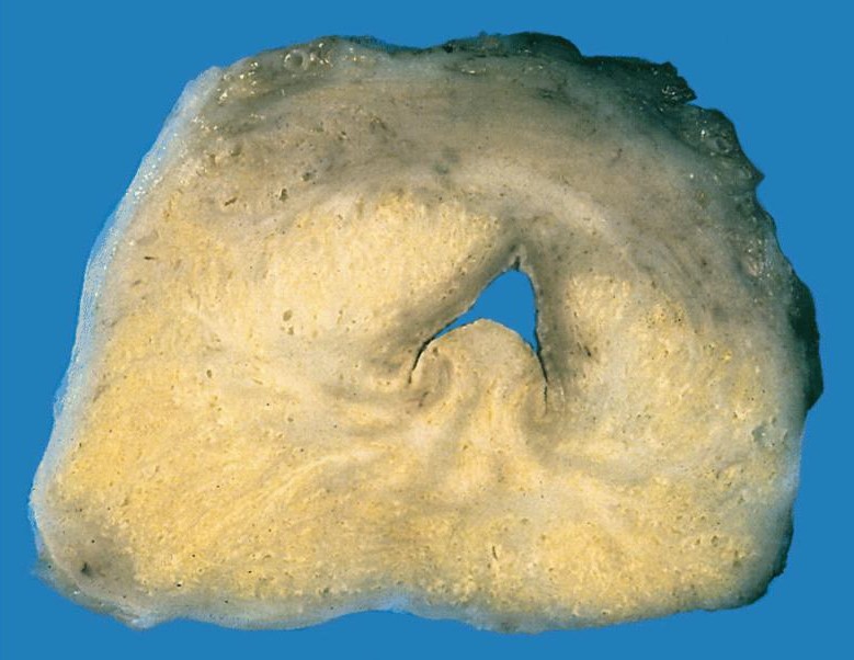
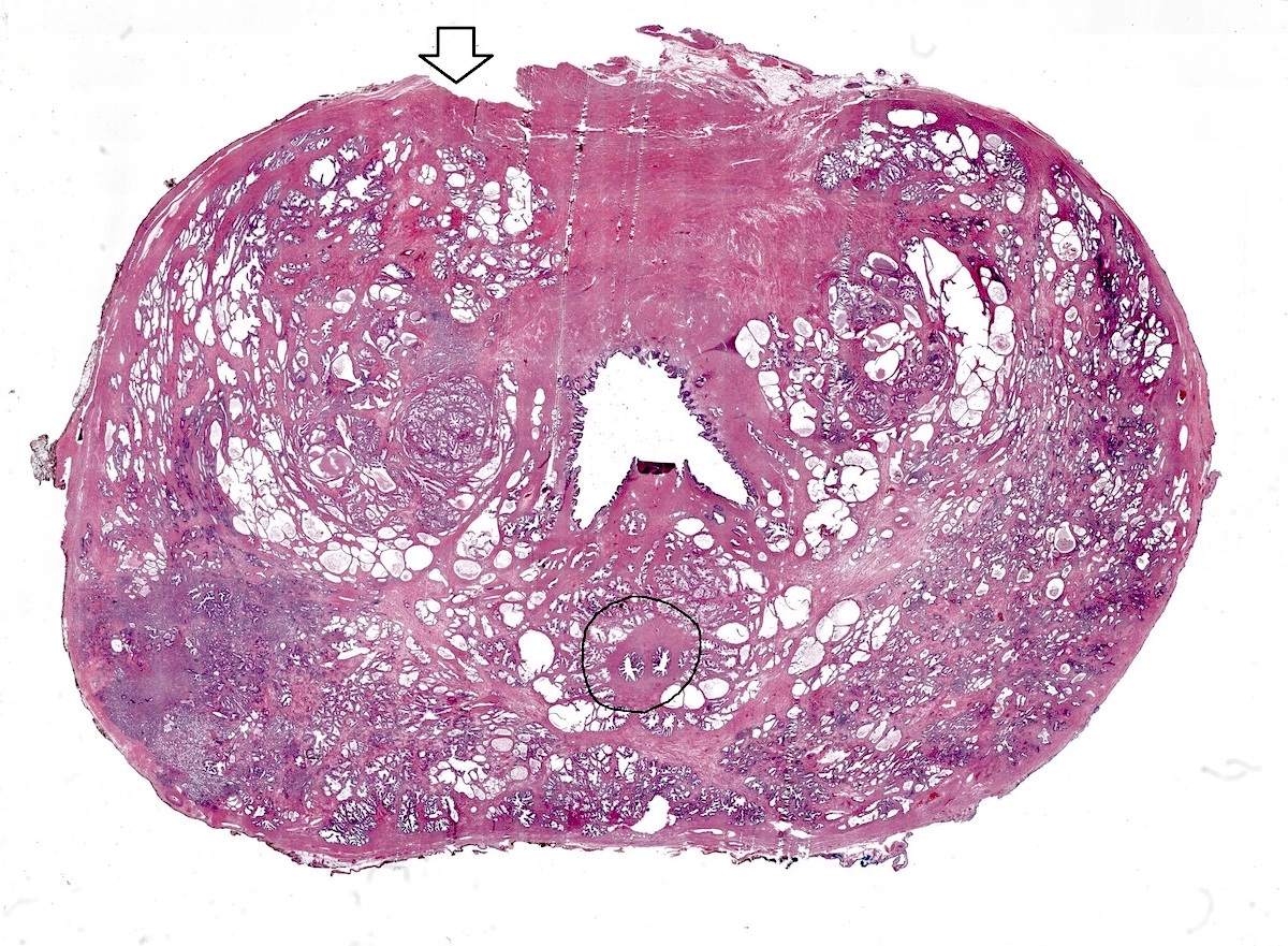
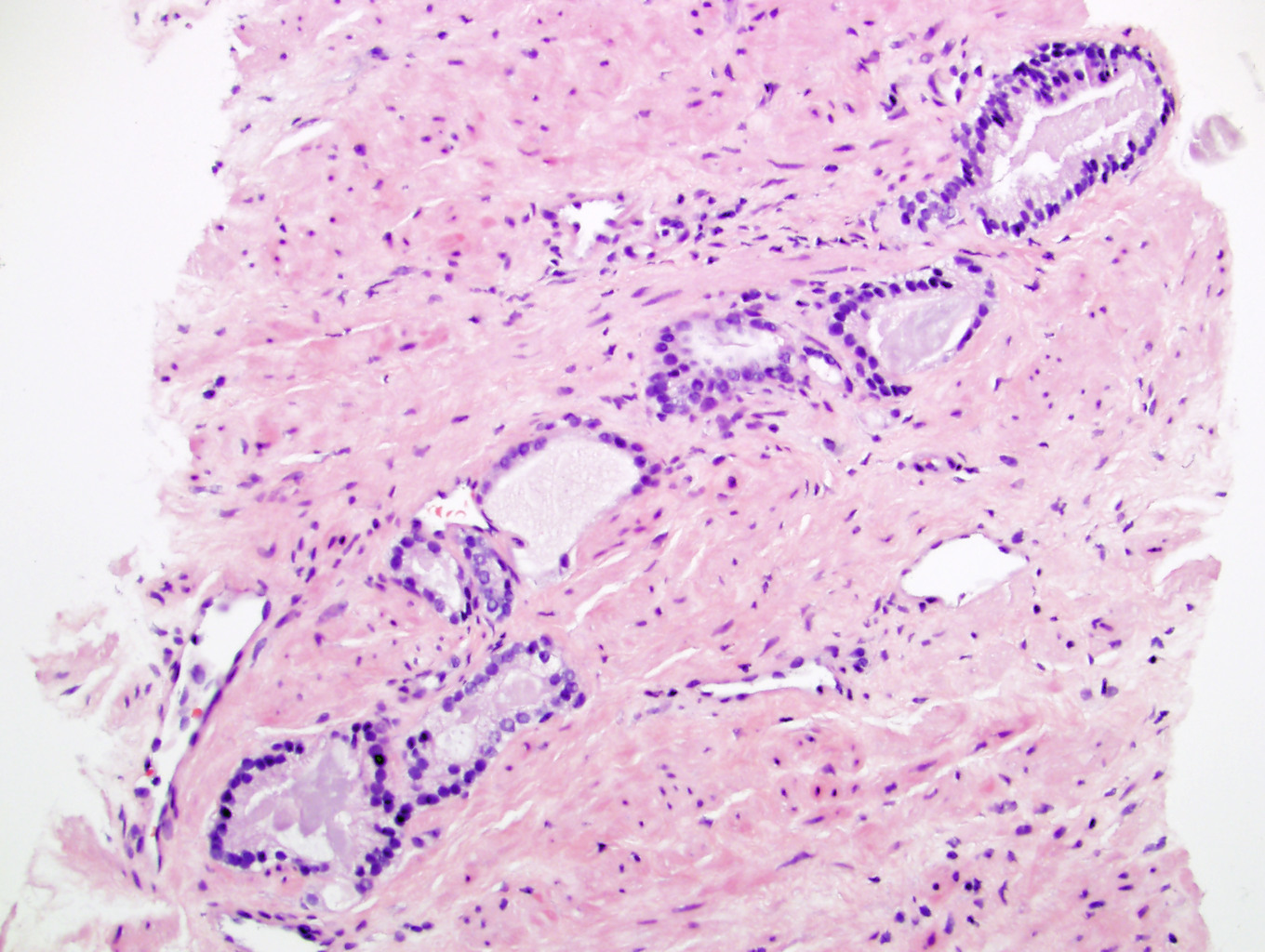
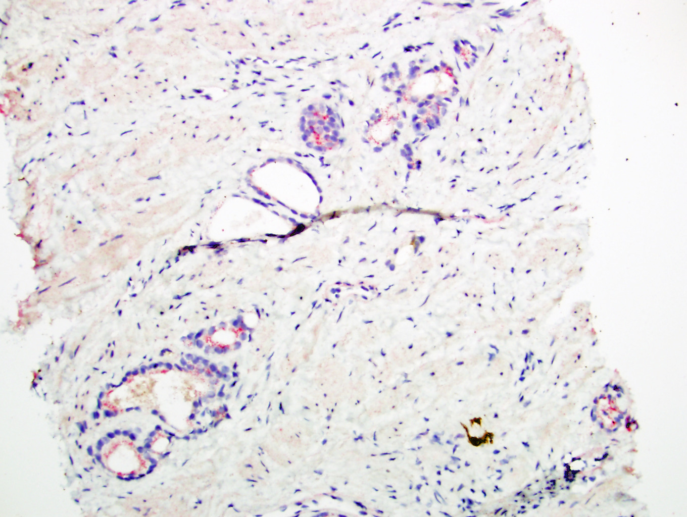
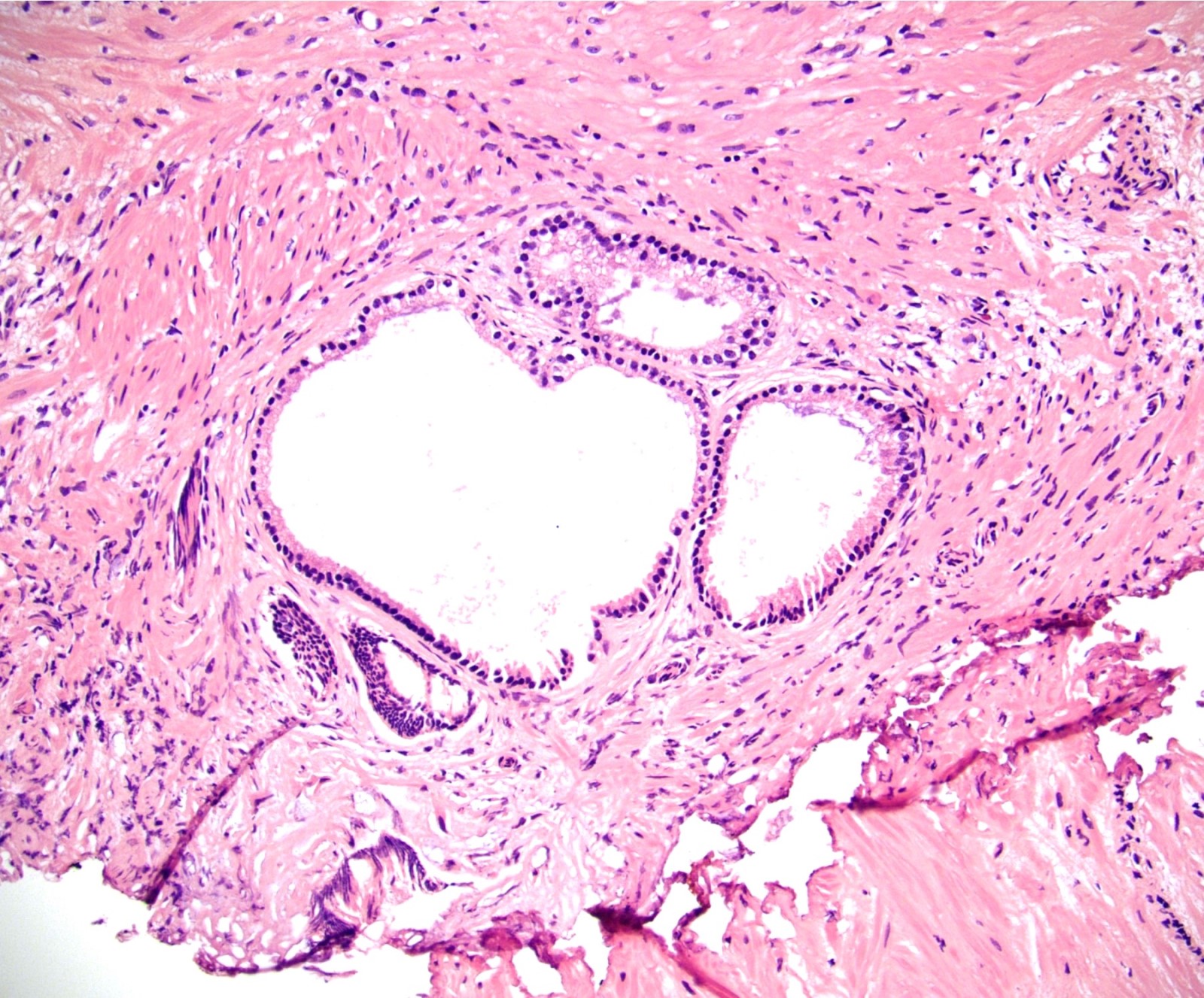
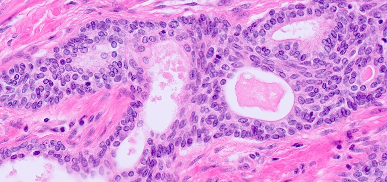
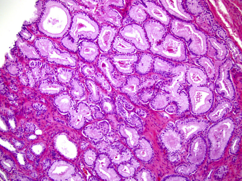
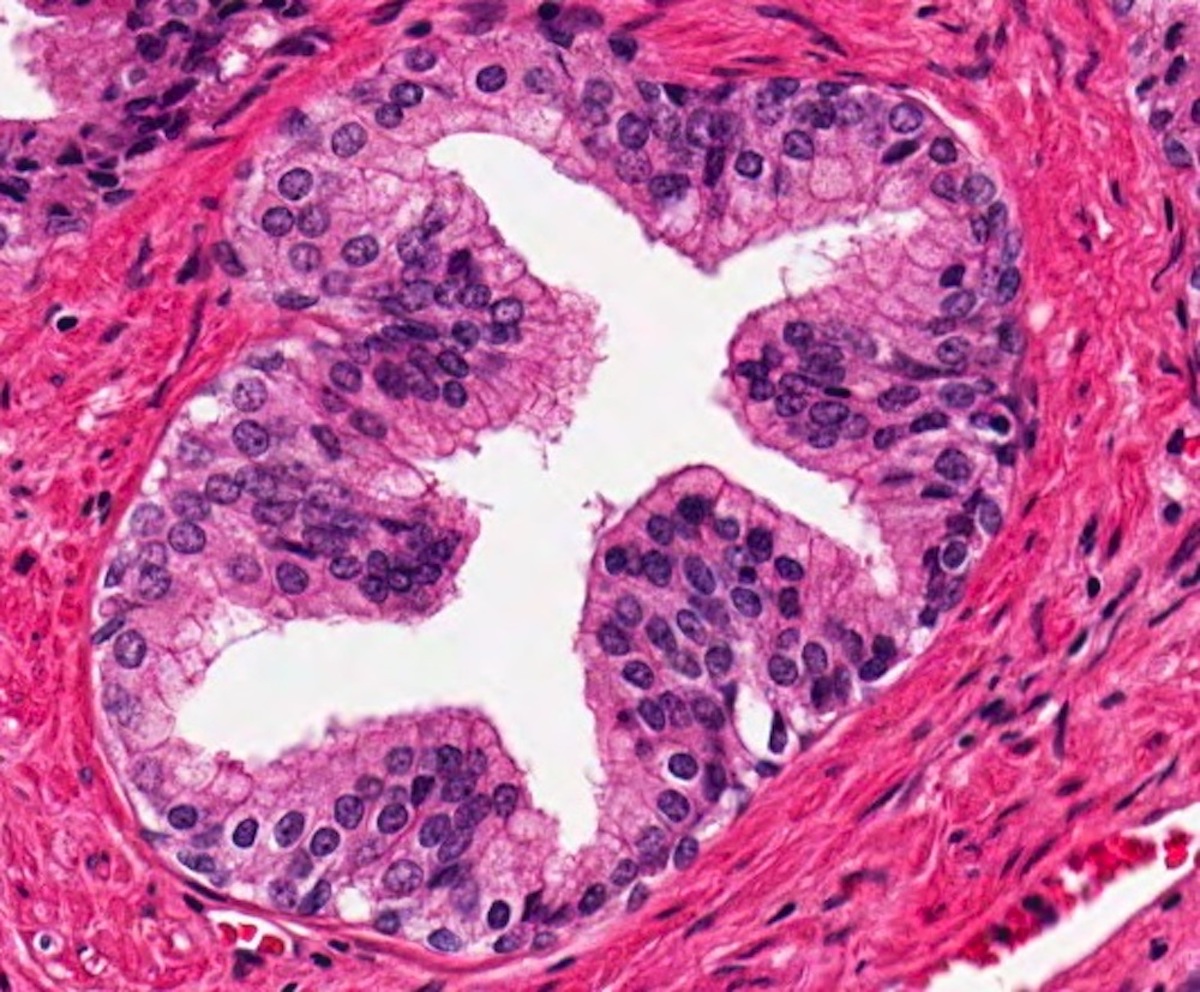
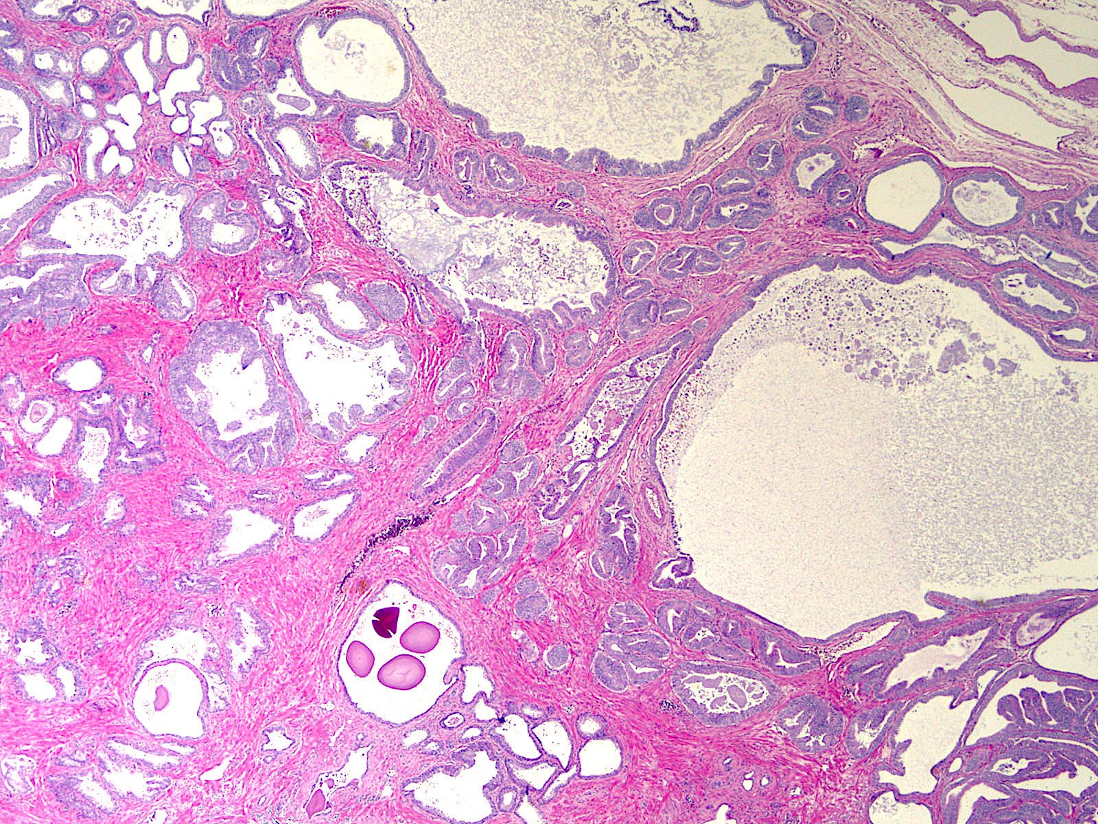
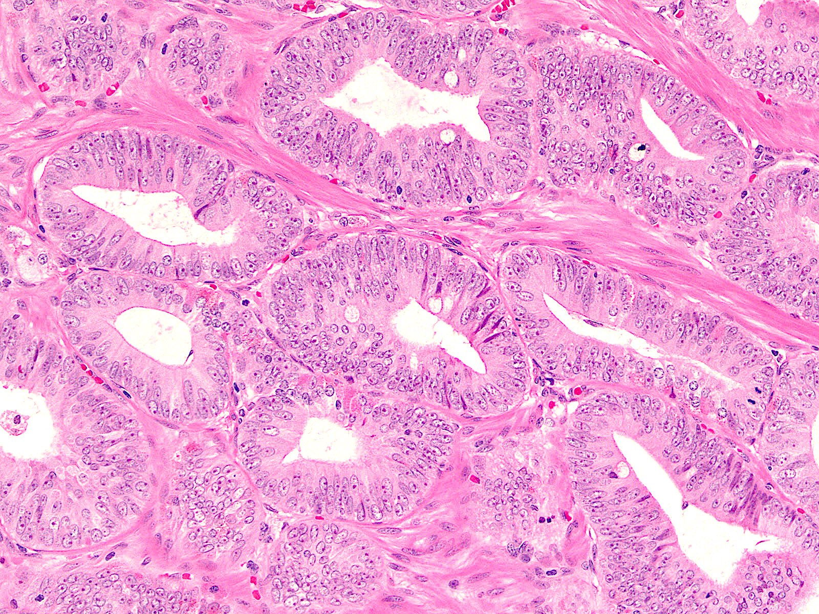
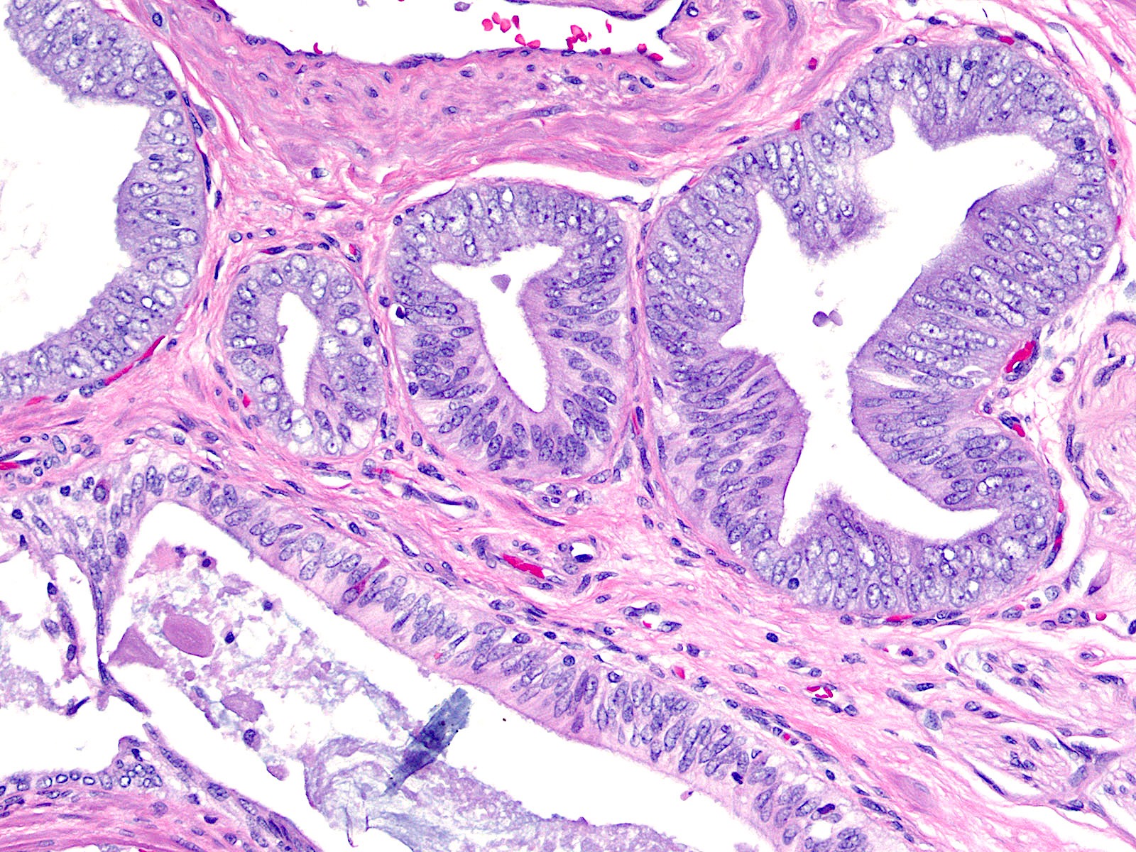
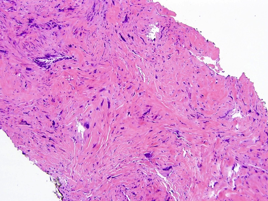
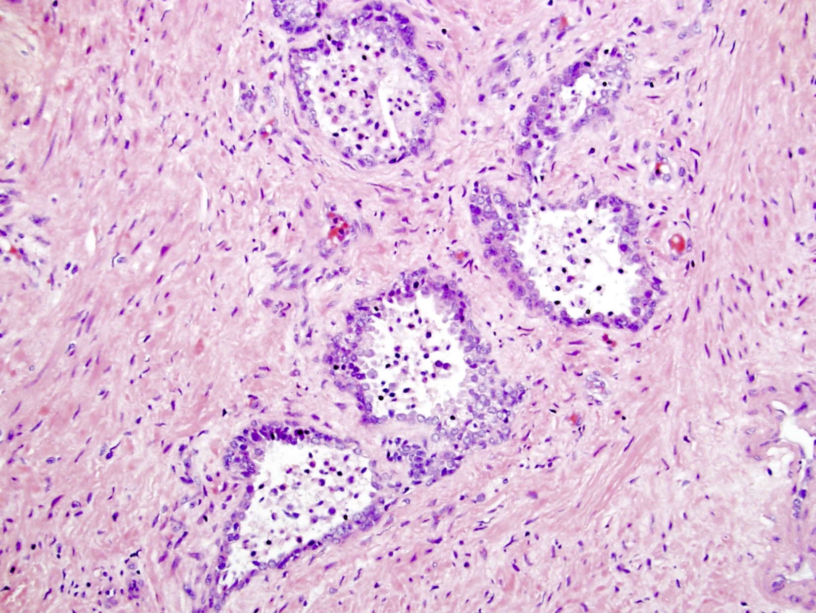
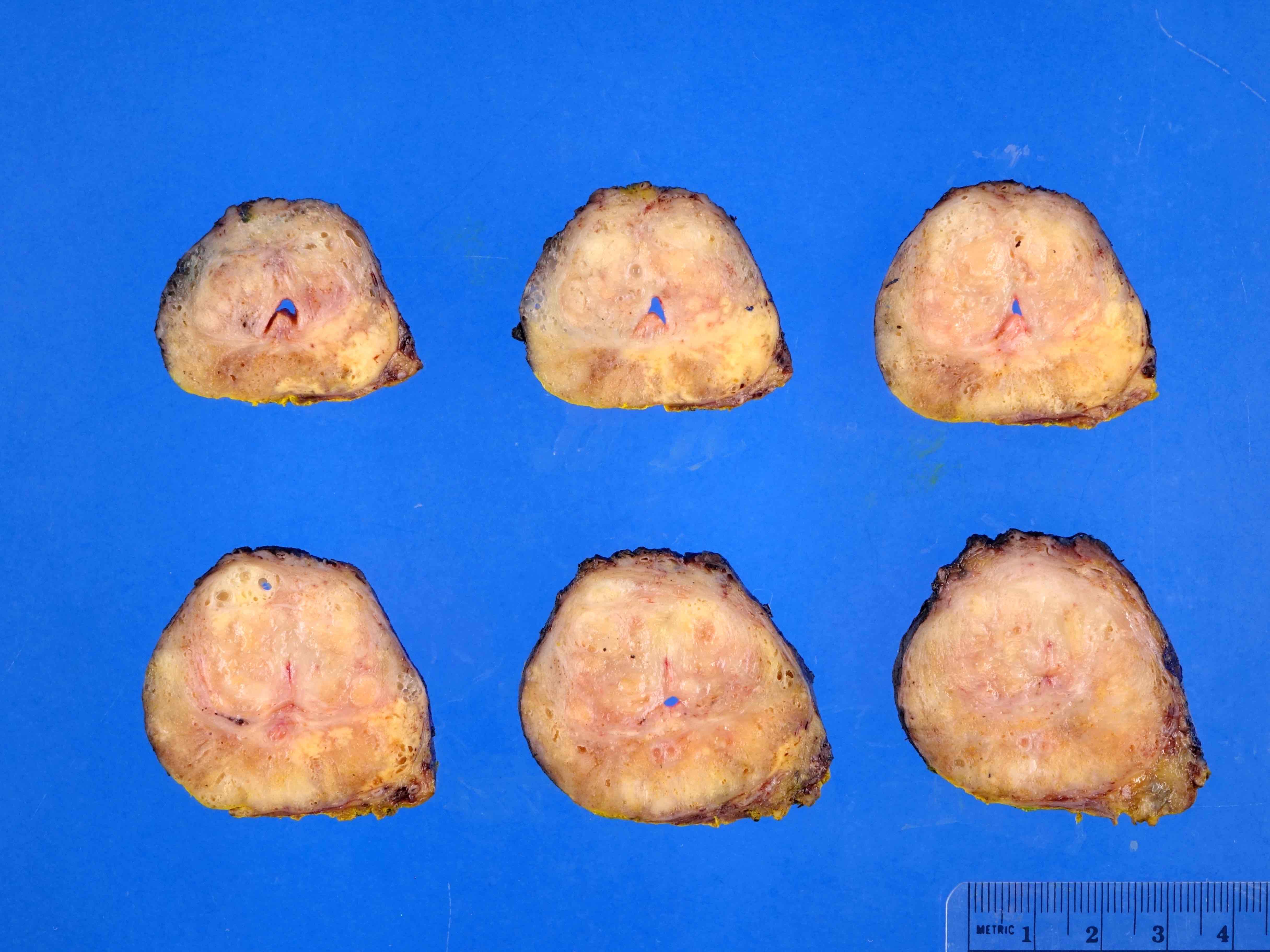
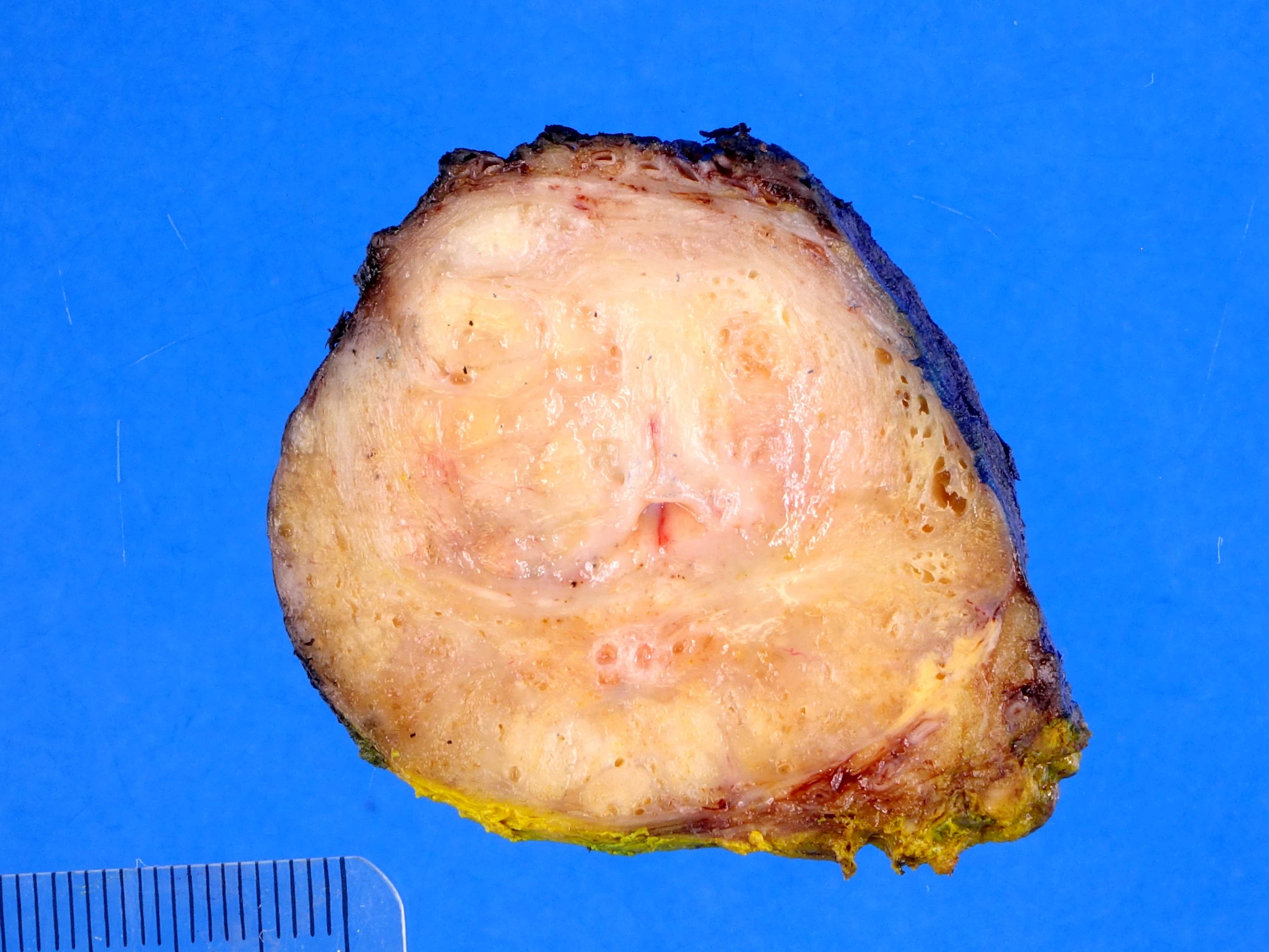
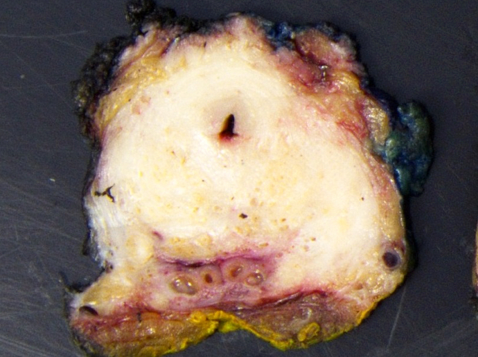
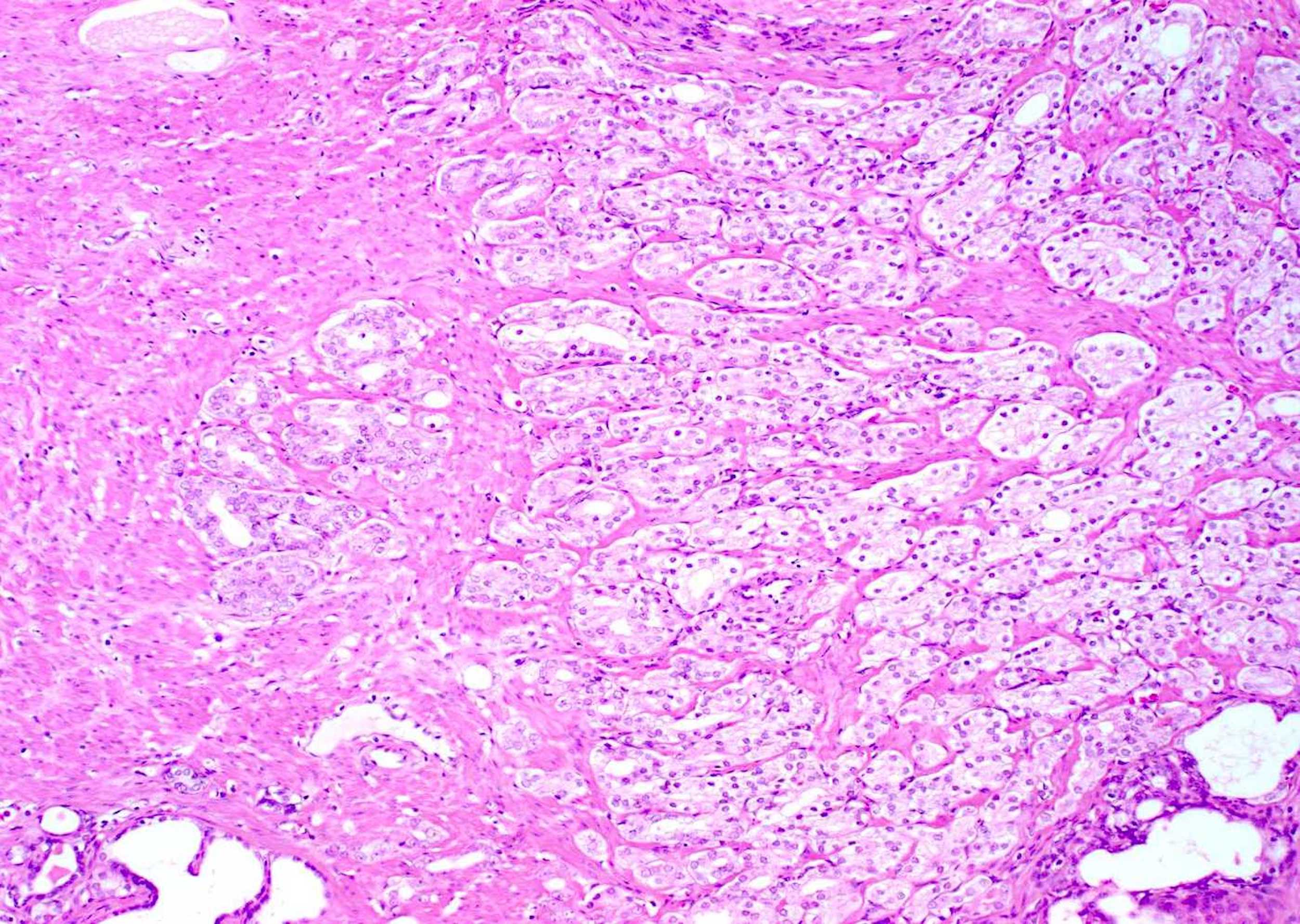
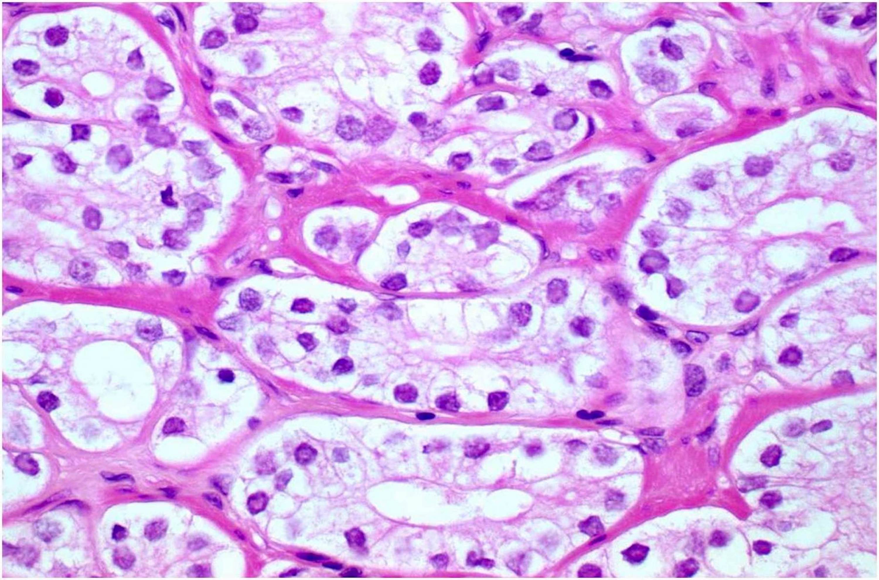
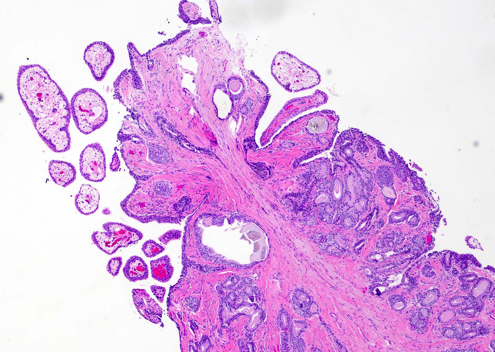
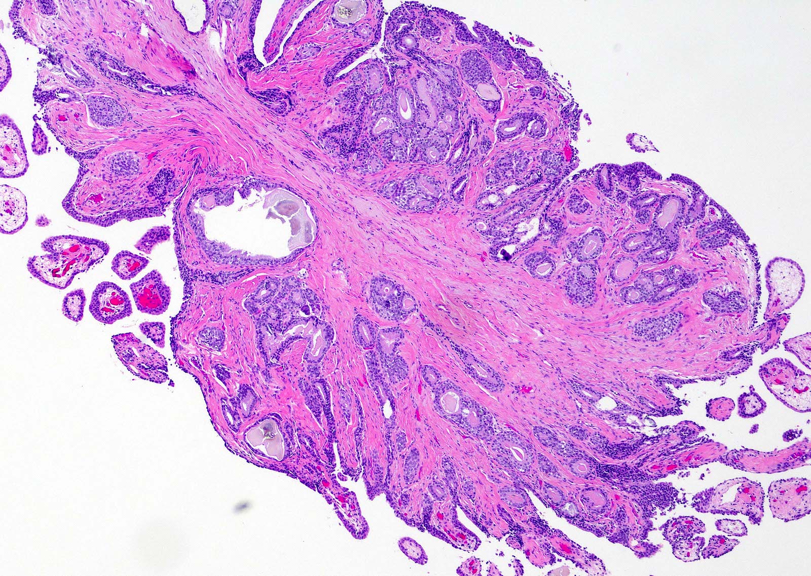
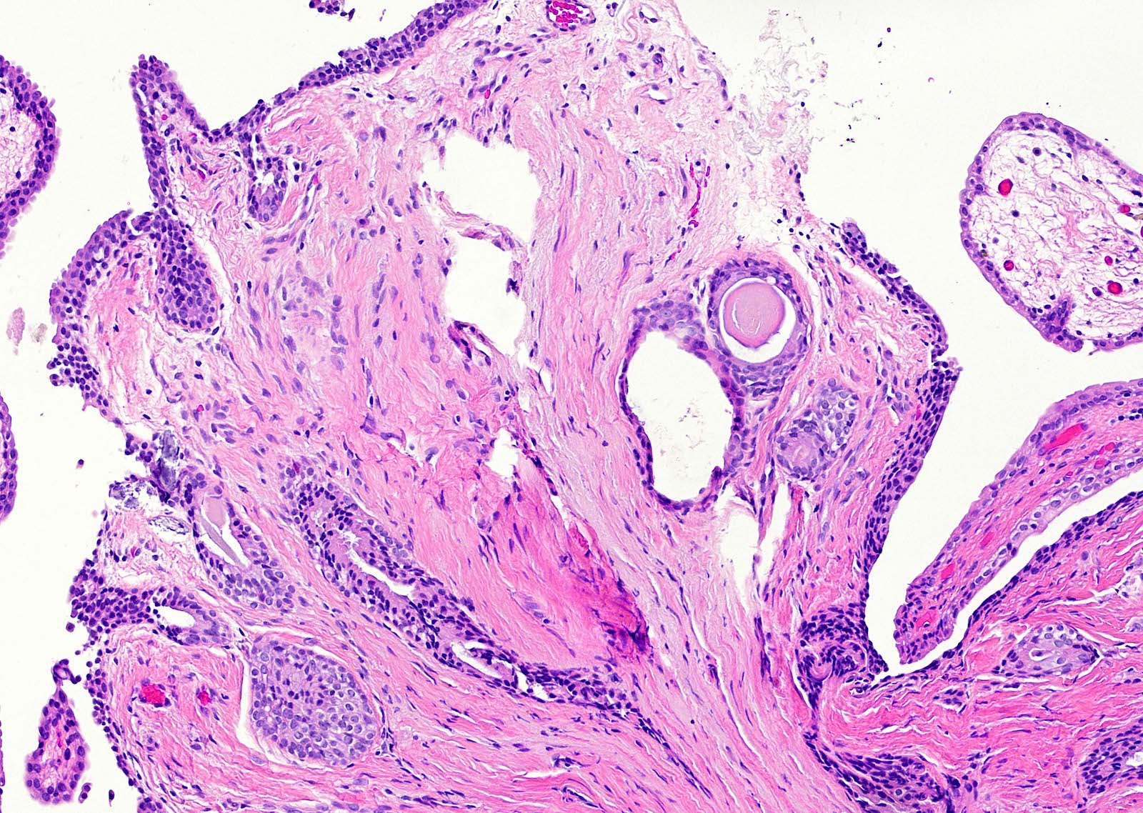
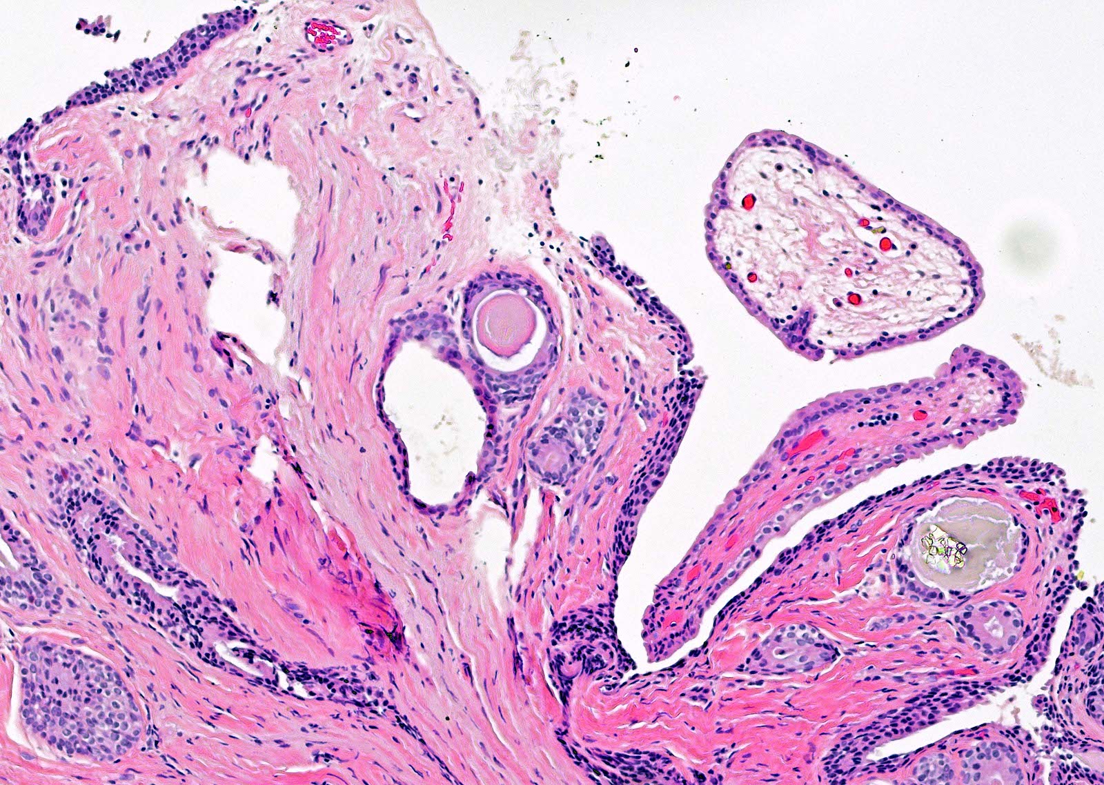
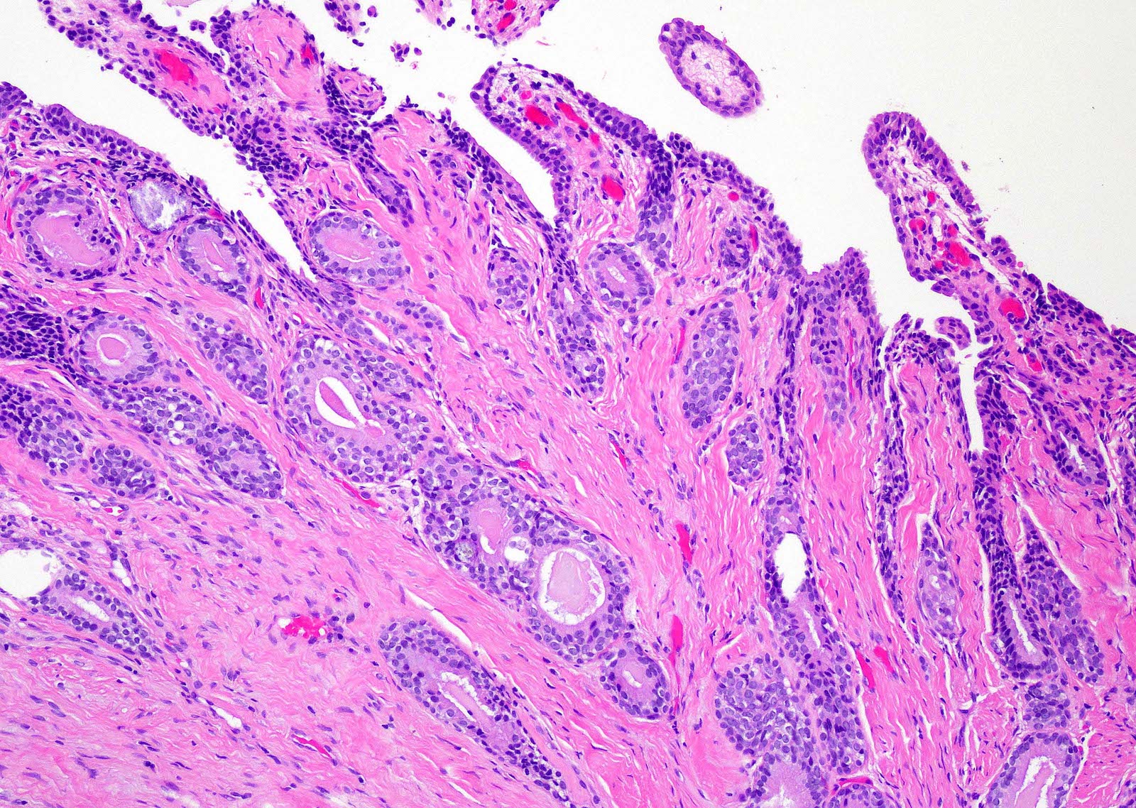
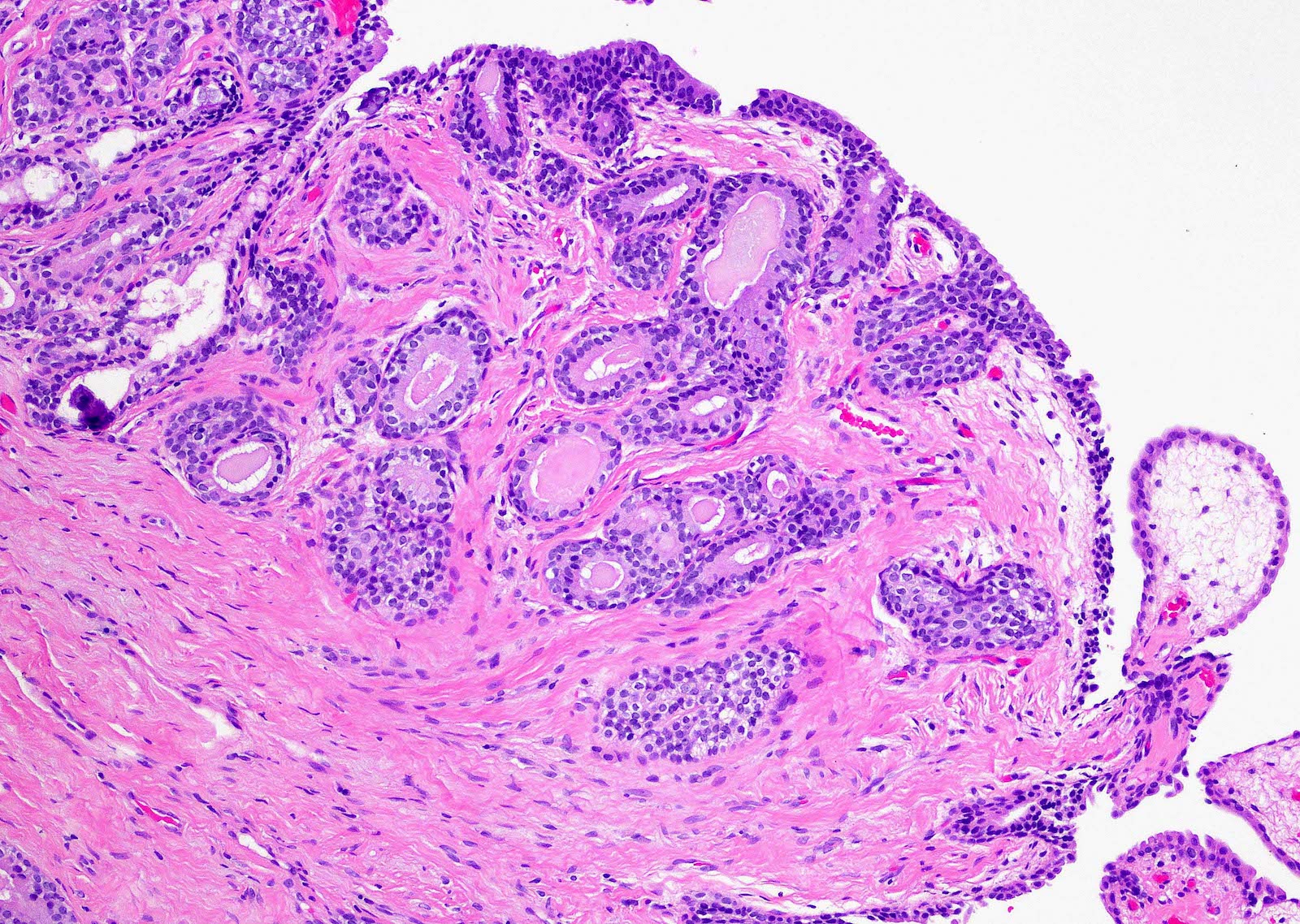
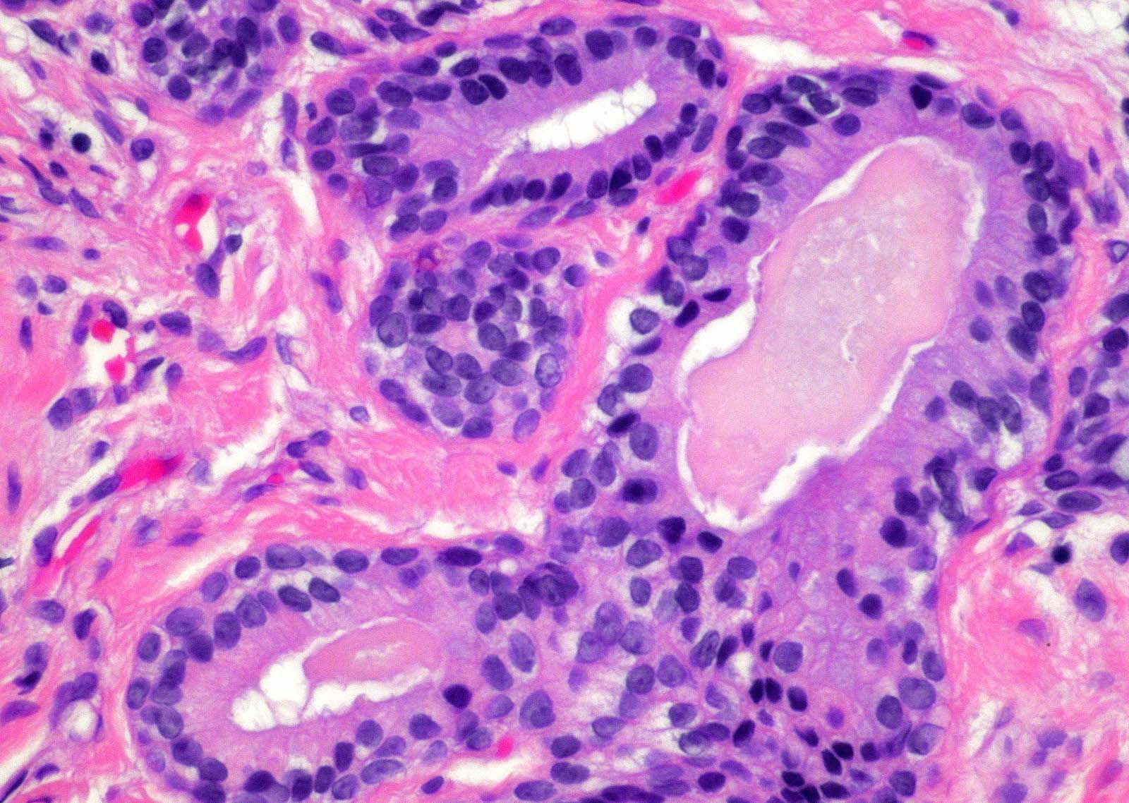
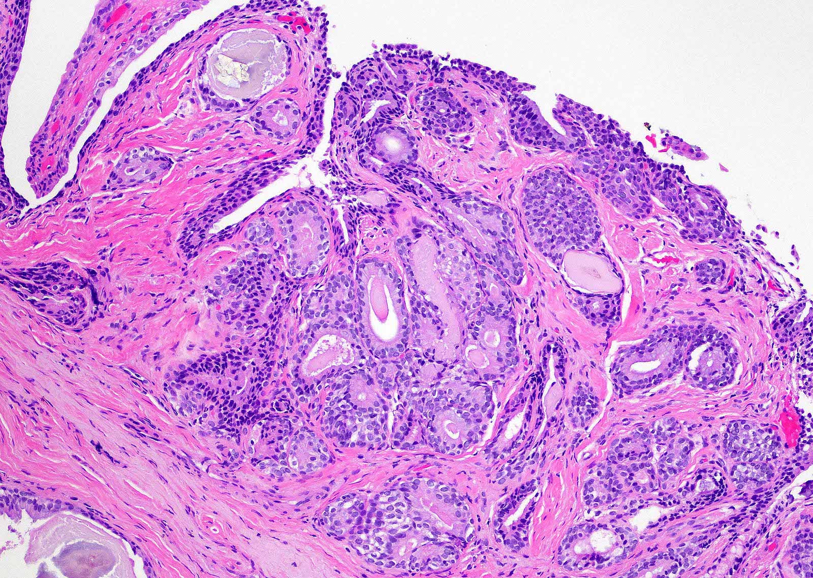
Contributed by Kenneth A. Iczkowski, M.D.
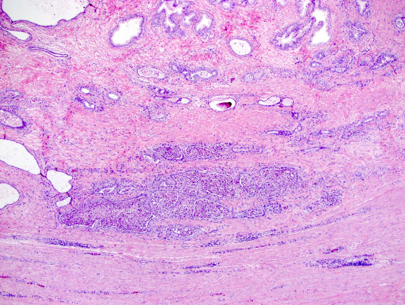
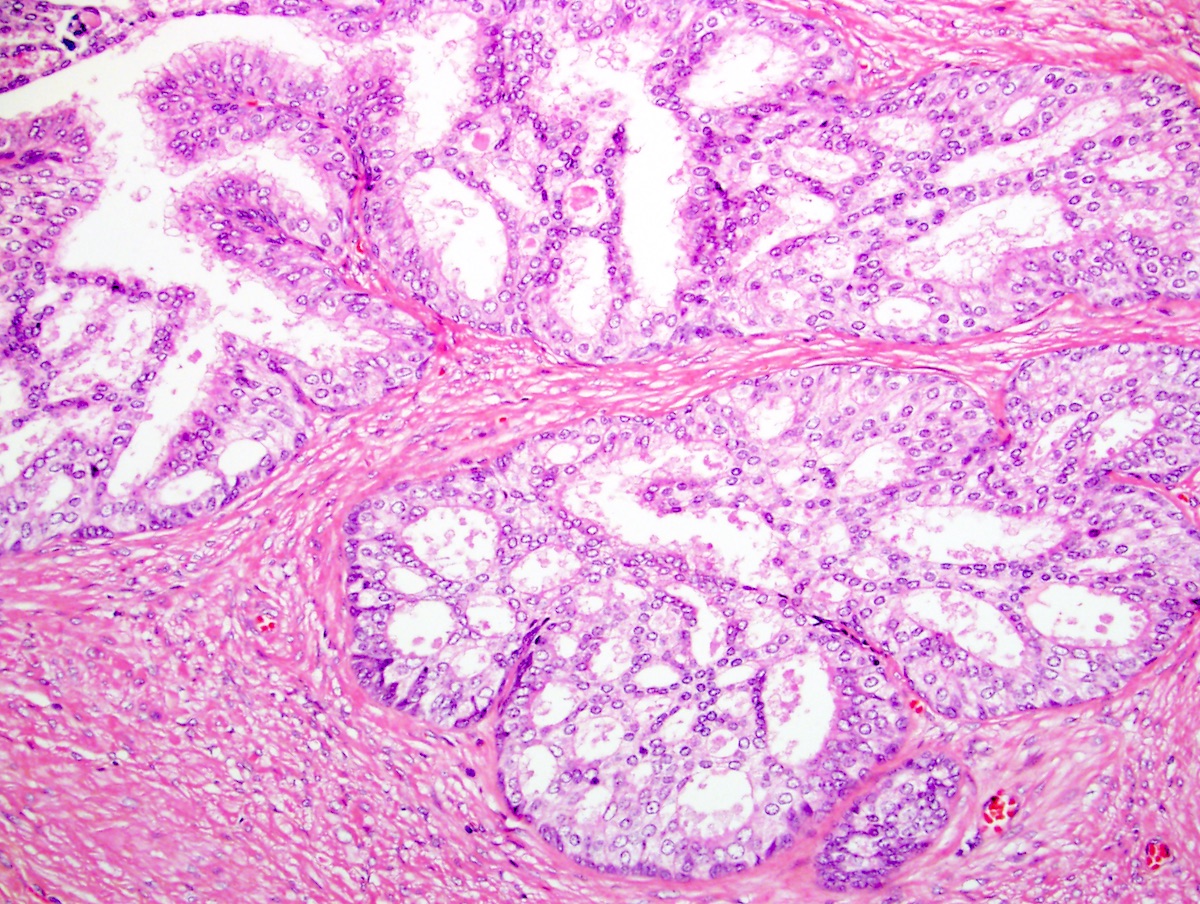
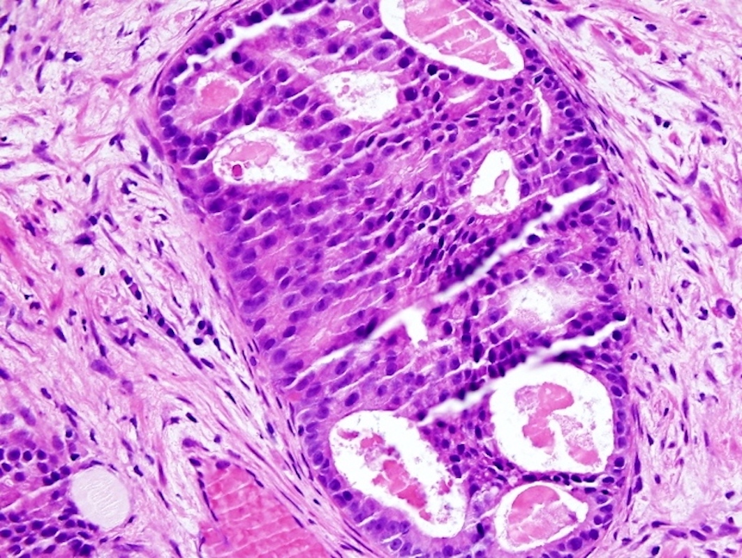
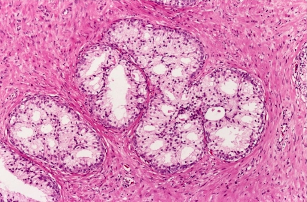
Lobule of small acini
Multiple clusters of acini
Lobular arrangement of small acini
Images hosted on other servers:

T1 weighted axial MRI image

T2 weighted axial MRI image
Contributed by Kenneth Iczkowski, M.D.
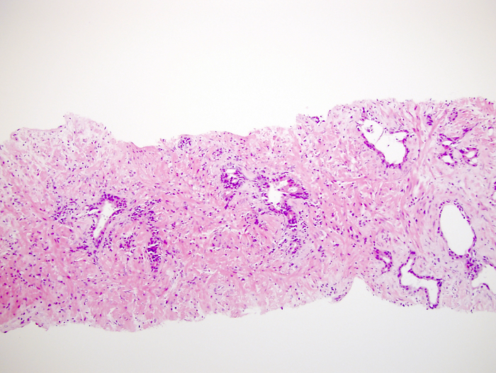
Phyllodes tumor: malignant epithelium
Images hosted on other servers:

Lesion: low FDG uptake
Case #269
STUMP, prostate cross section
Images hosted on other servers:

STUMP that eventually metastasized
Contributed by Kenneth Iczkowski, M.D., Theo van der Kwast, M.D. and Case #269
STUMP in prostate biopsy that had cancer in other fields
Mitosis
Hypercellular stroma
Circumscription of nodule
Multinucleation
Degenerative atypia
STUMP positive for CD34
STUMP positive for desmin
STUMP positive for HHF35
STUMP with low index for Ki67

STUMP positive for progesterone receptor
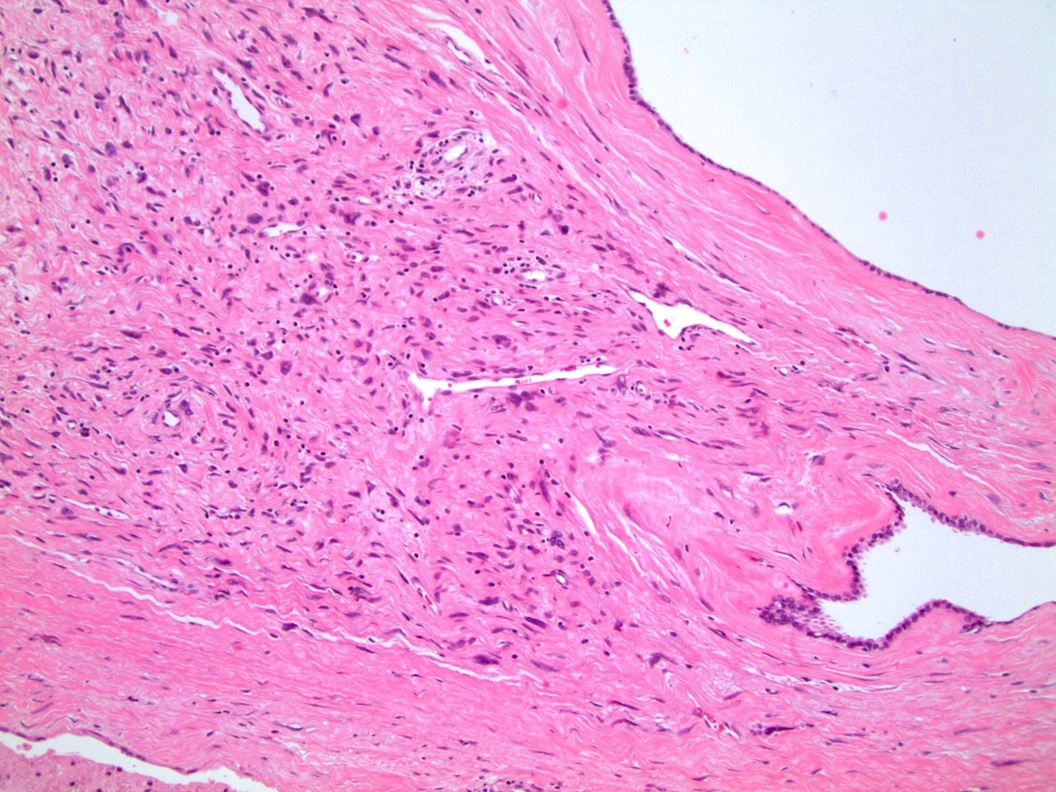
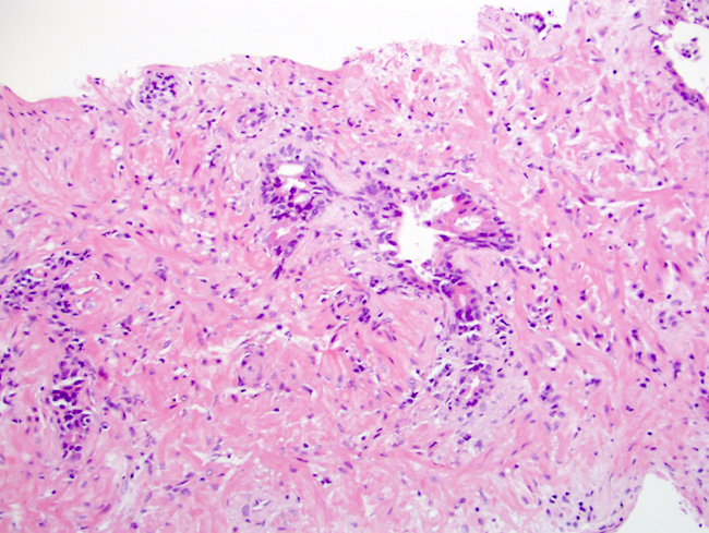
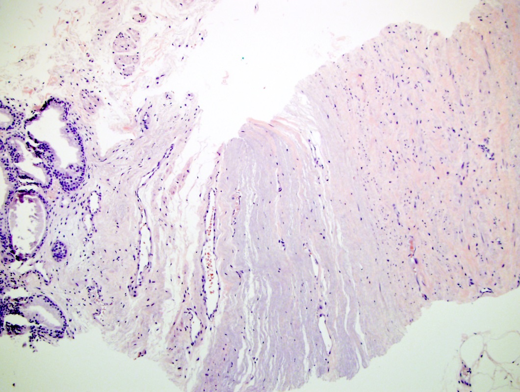
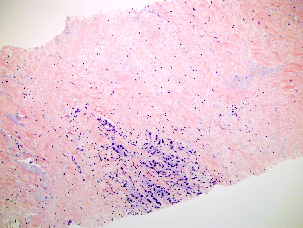
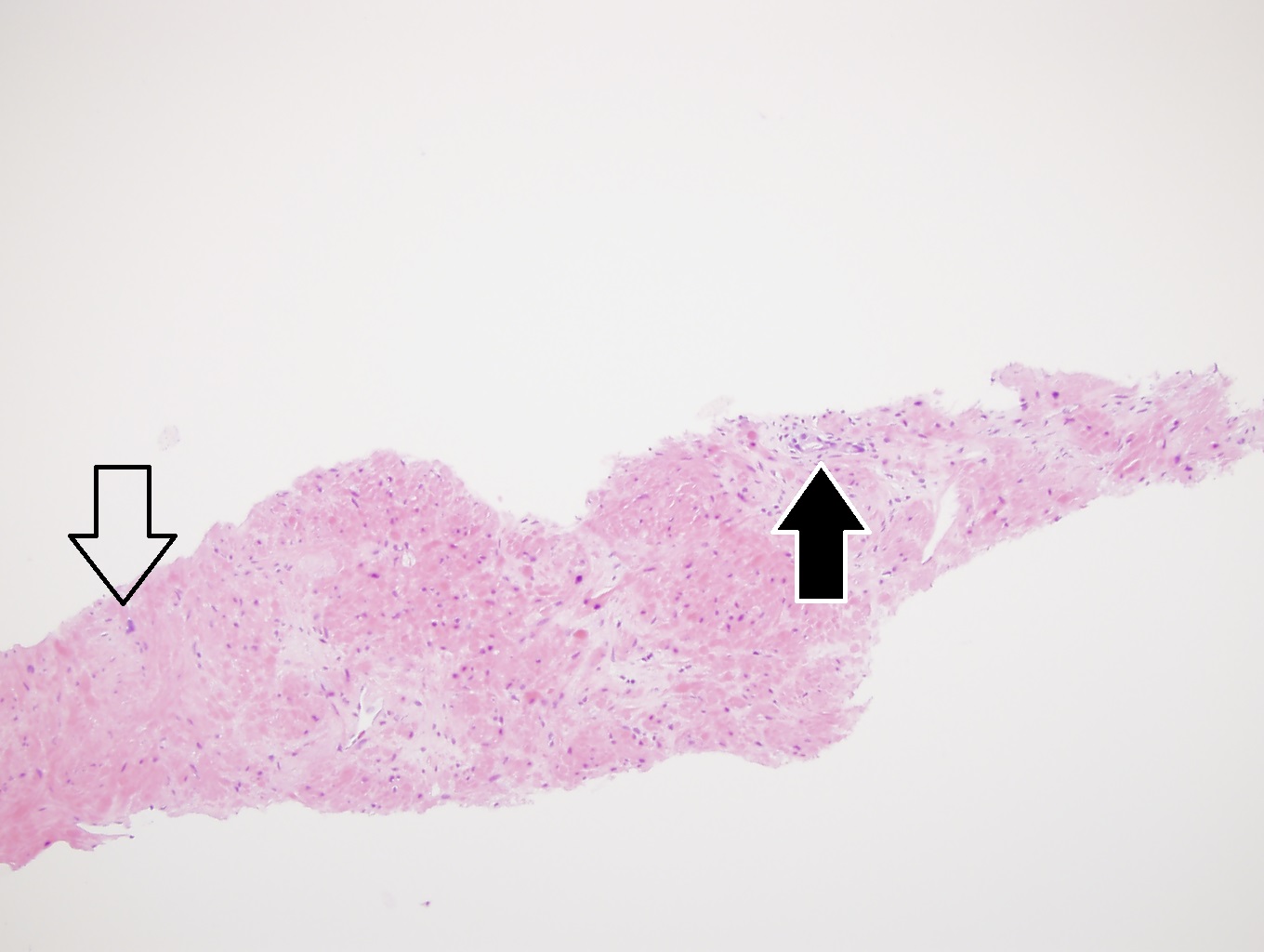
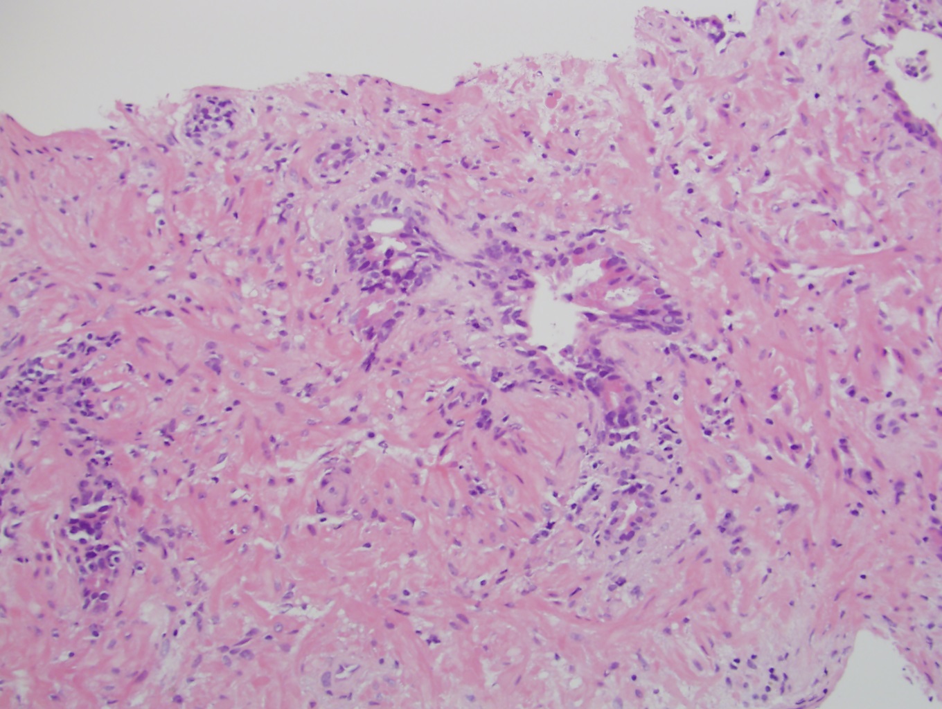
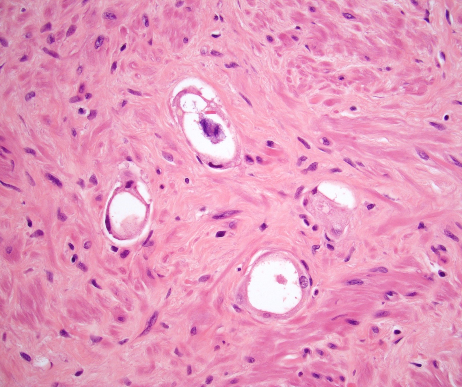
Contributed by Kenneth A. Iczkowski, M.D.
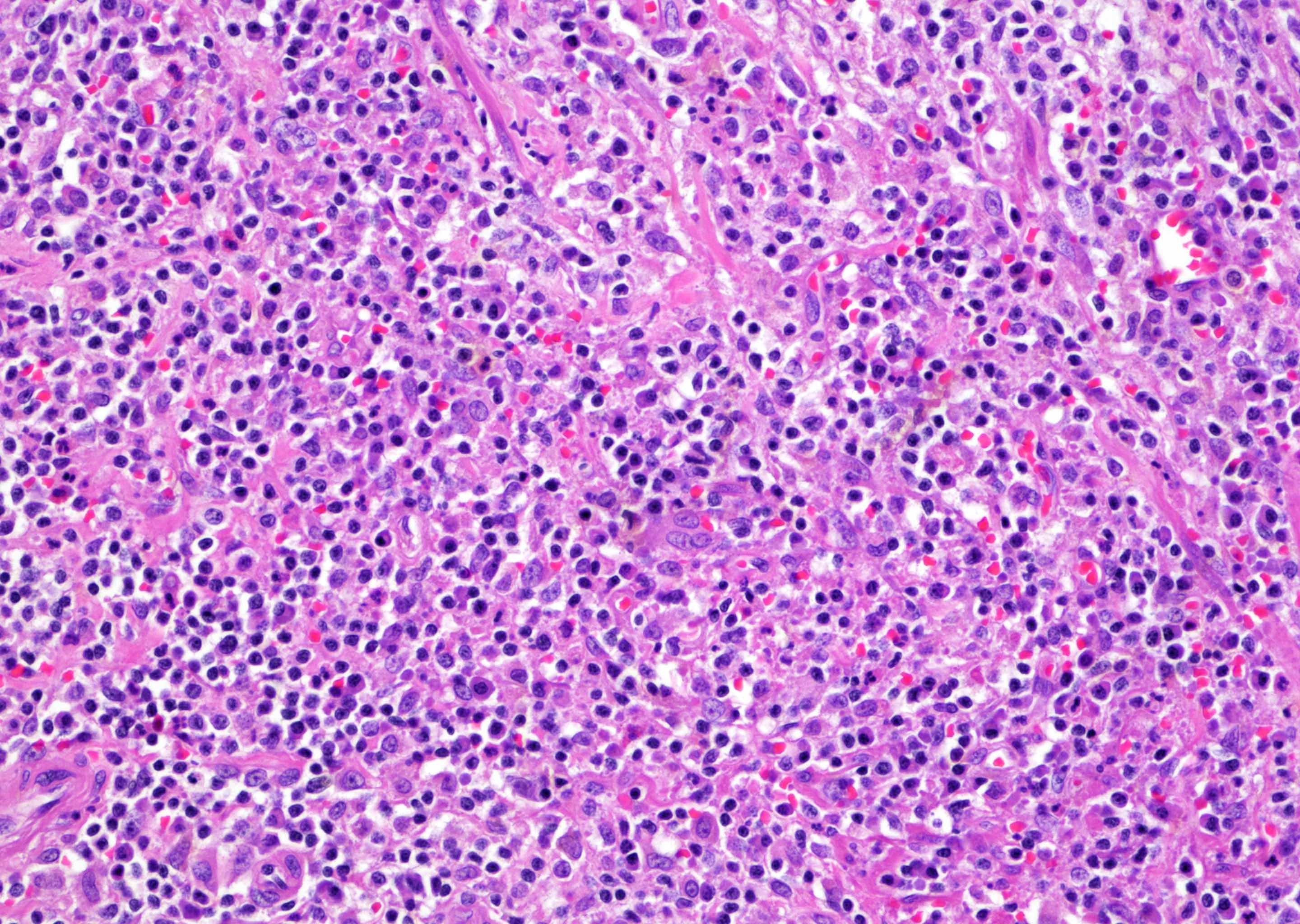
Reactive chronic inflammation
Acute inflammation in lumens
Idiopathic granuloma in a TURP
Xanthomatous histiocyte proliferation
CD68 positive stain - confirmatory
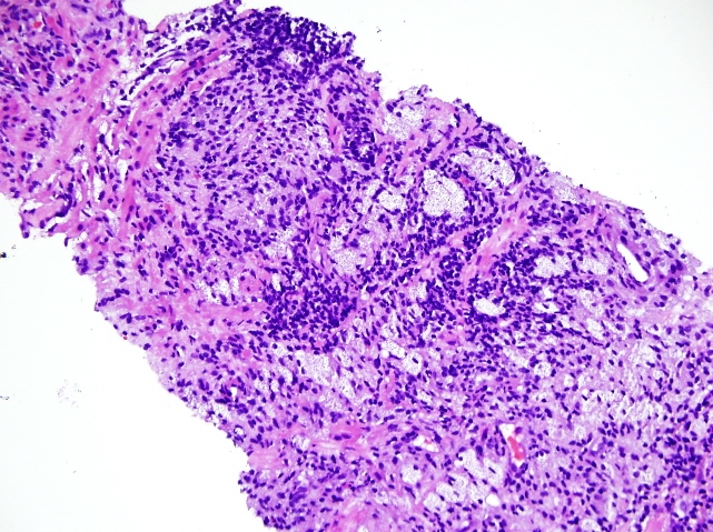
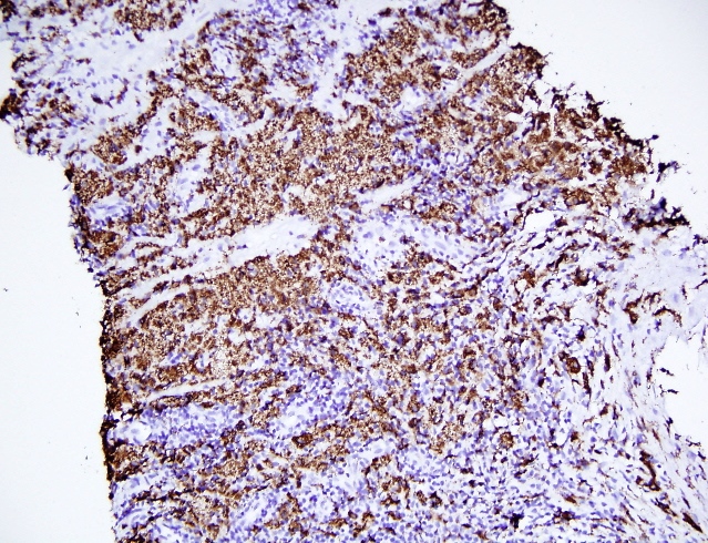
Case #117

Various images
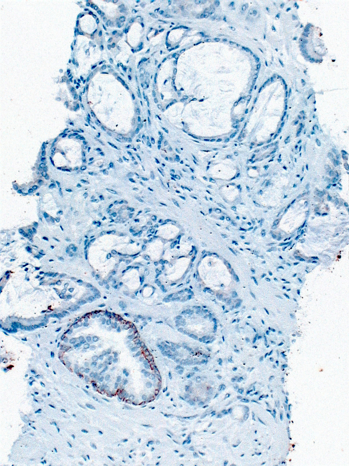
Triple stain (AMACR, p63, HMWK)
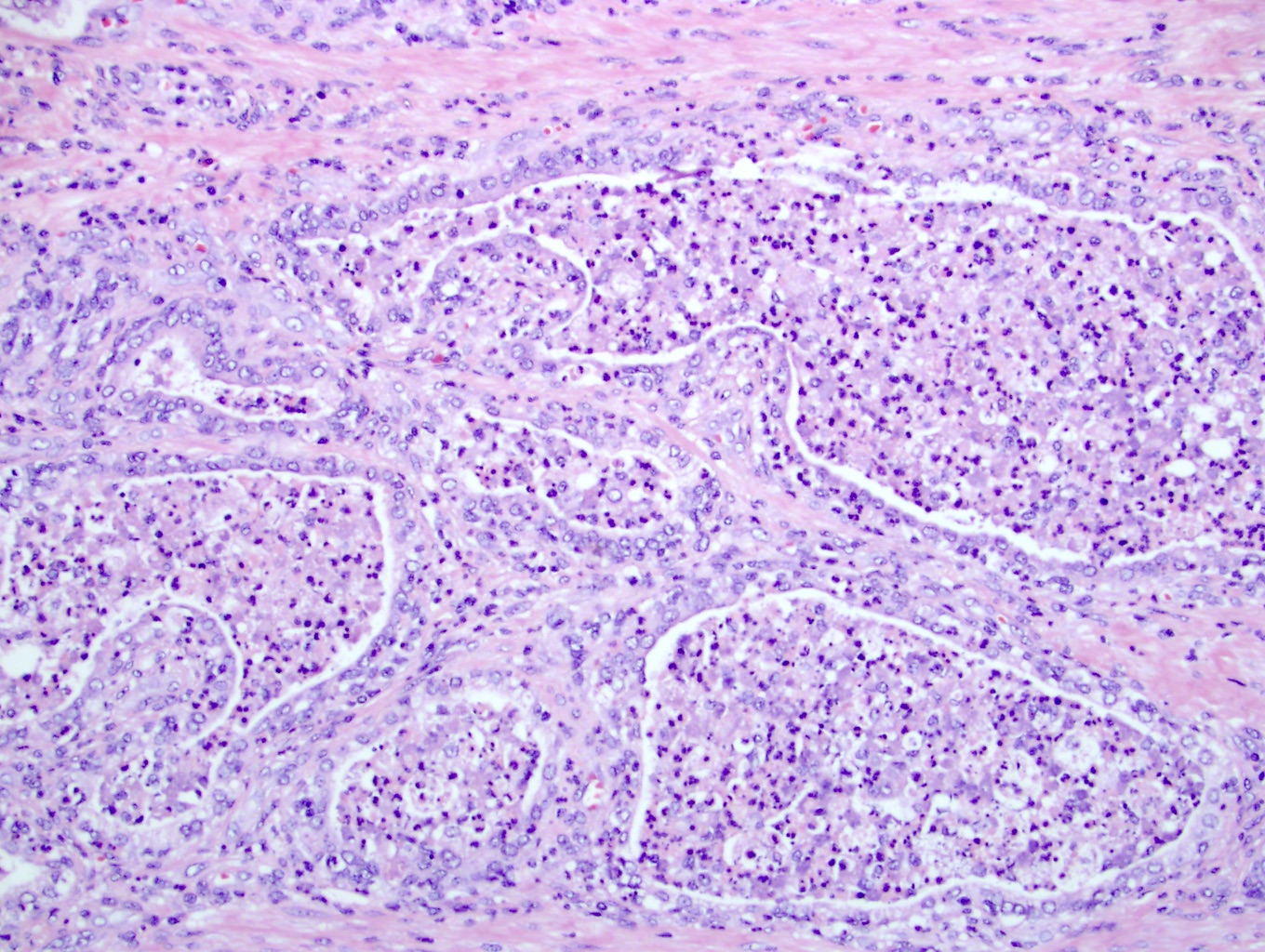
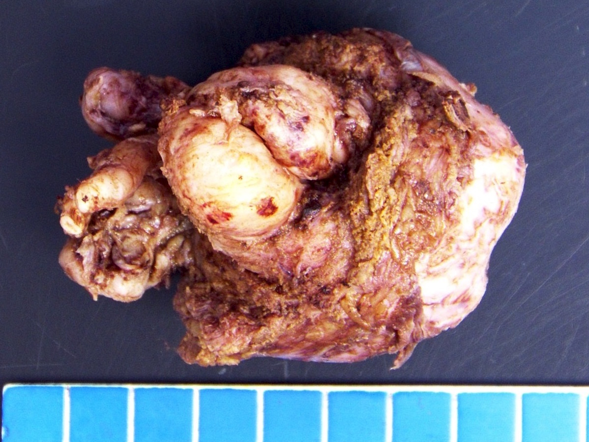
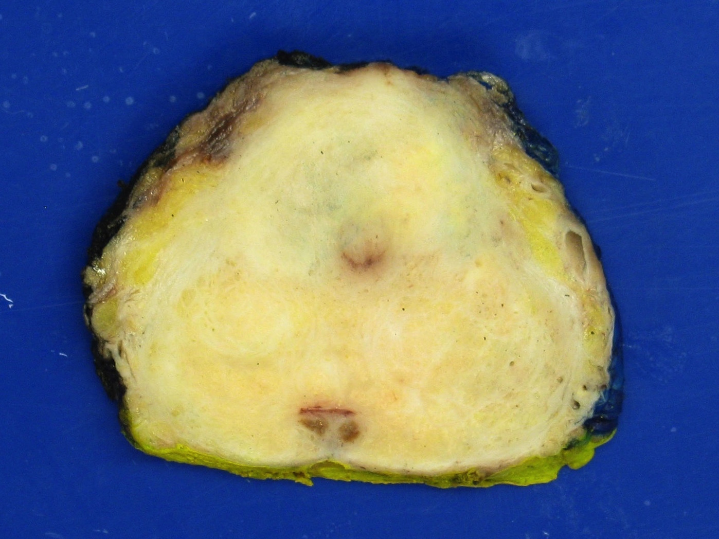
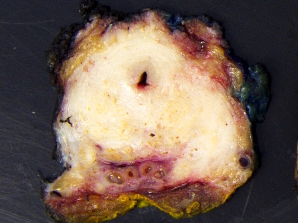
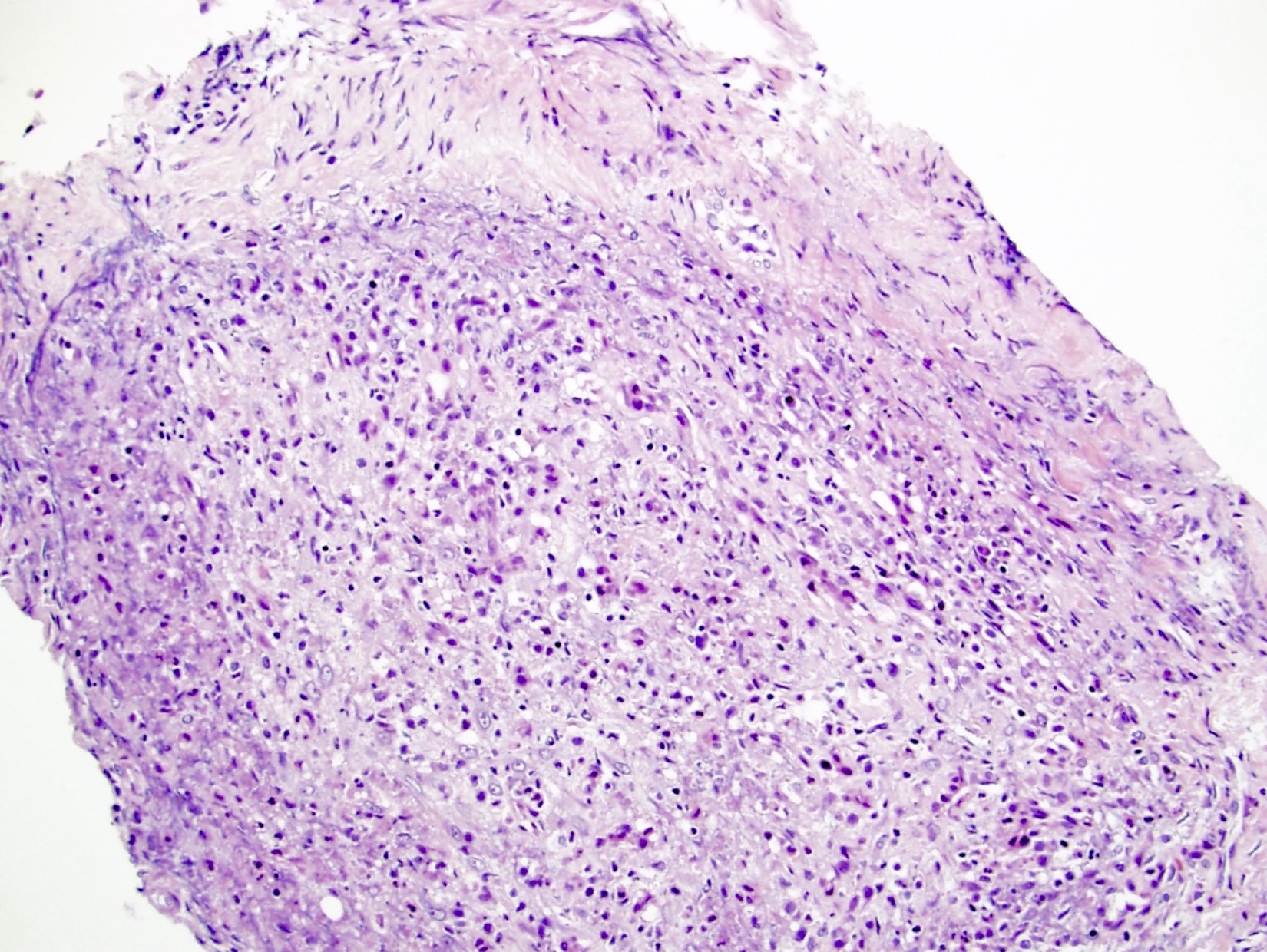
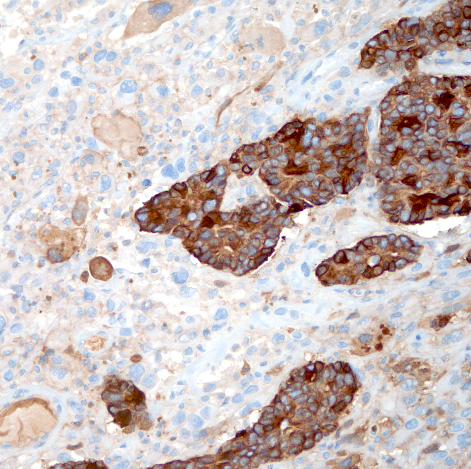
Contributed by Steven Christopher Smith, M.D., Ph.D.
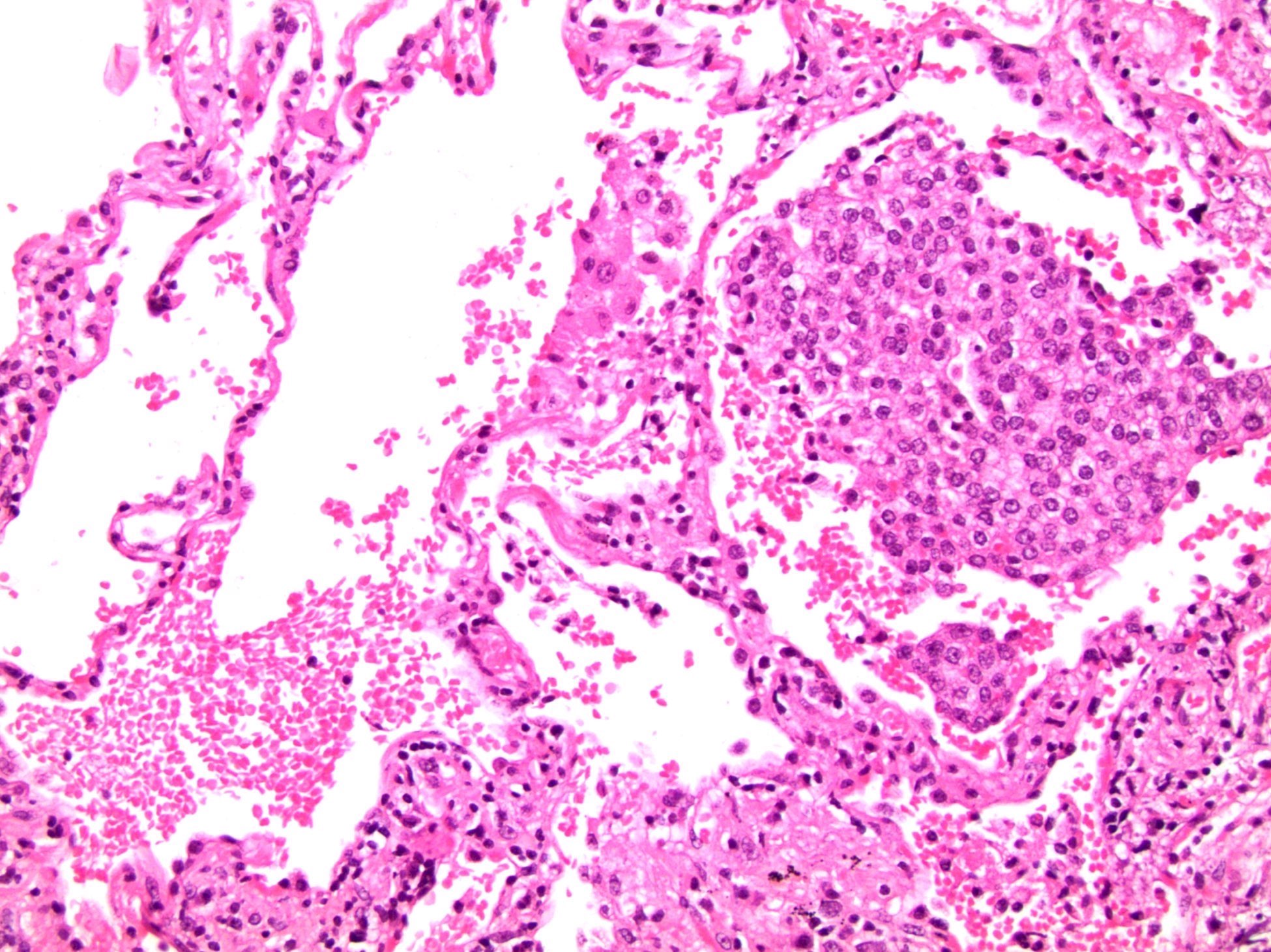
Liver metastases
Contributed by Steven Christopher Smith, M.D., Ph.D.
Transurethral resection
Biphasic histology
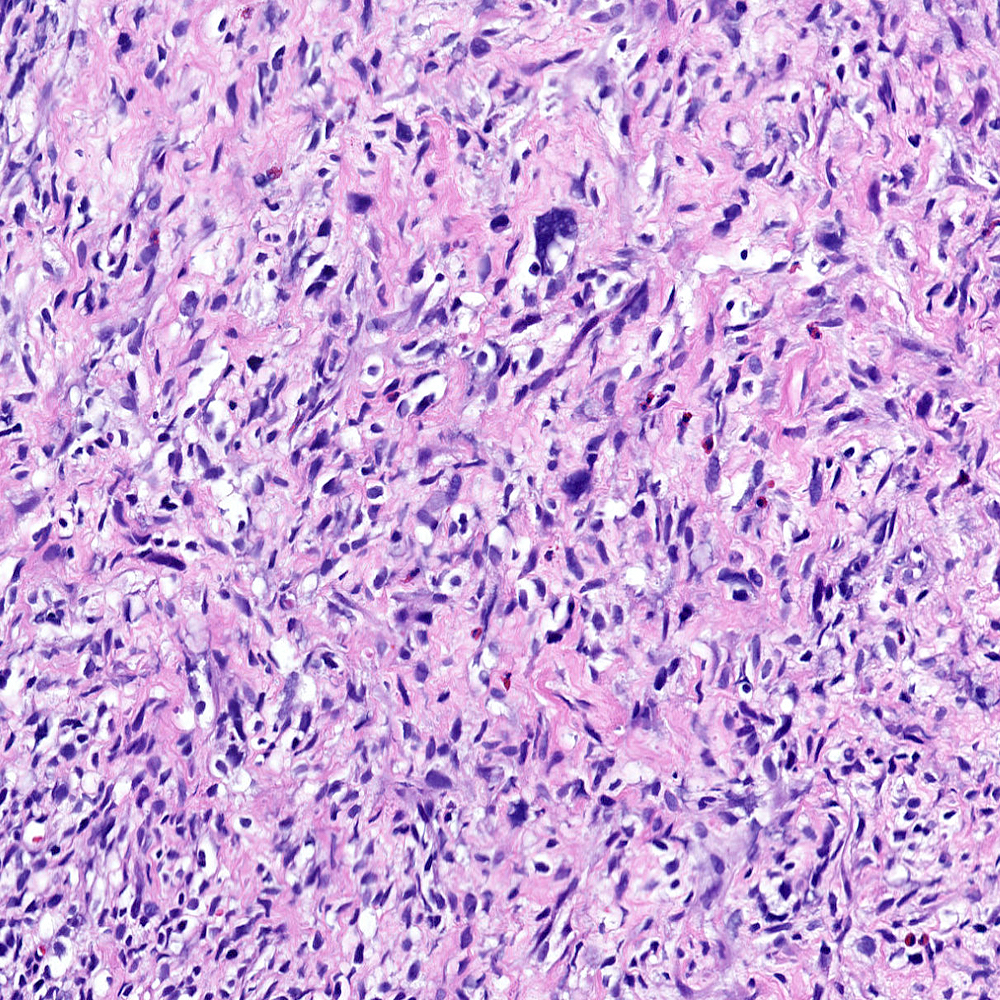
Monophasic sarcomatoid carcinoma
Adenosquamous component
Subtle epithelial component
Focal epithelial differentiation
Heterologous chondrosarcoma pattern
Heterologous osteosarcoma pattern
Heterologous rhabdomyosarcoma pattern
Pleomorphic sarcomatoid pattern
Keratin positivity
ERG positivity
Images hosted on other servers:


Various images
Contributed by Debra Zynger, M.D.
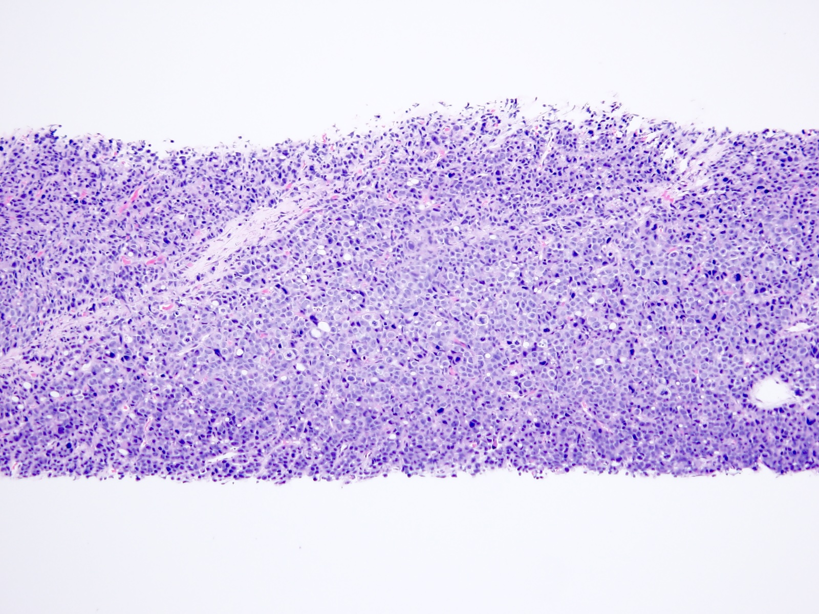
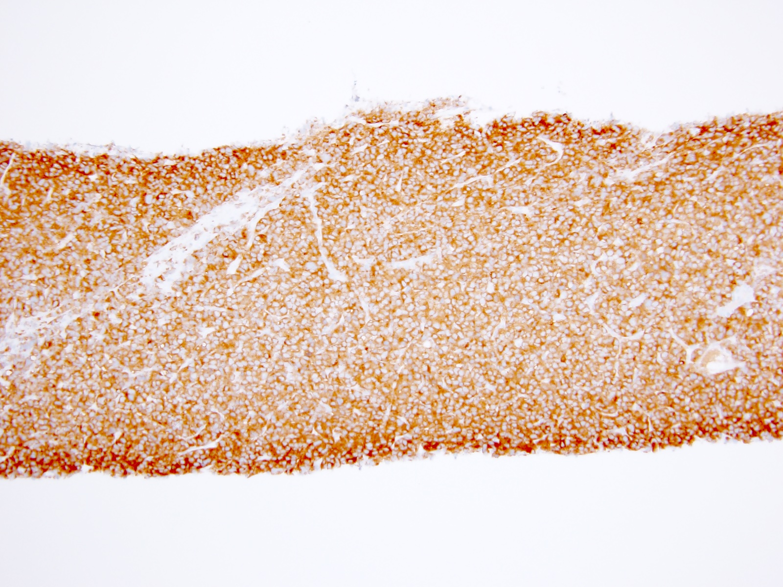
Prostatic small cell neuroendocrine carcinoma
Prostatic
adenocarcinoma
with 100% NE
differentiation
Metastatic prostatic
small cell
neuroendocrine
carcinoma
Images hosted on other servers:

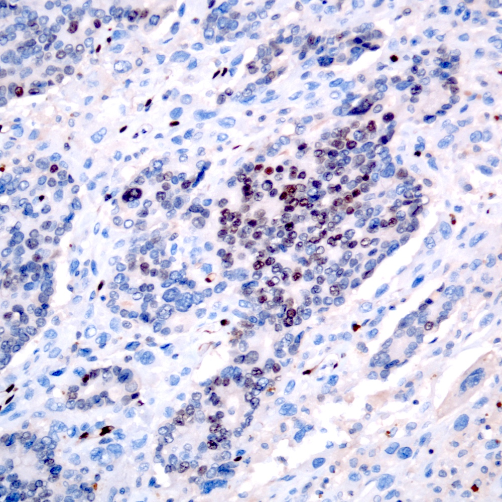
HE staining

Immunostaining for CD56
Images hosted on other servers:

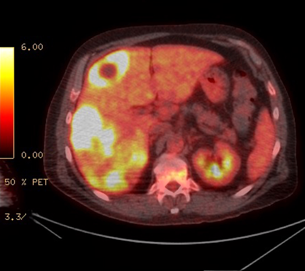
CT and MRI of prostate

Whole body scan

Pelvic CT with contrast

MRI of prostate
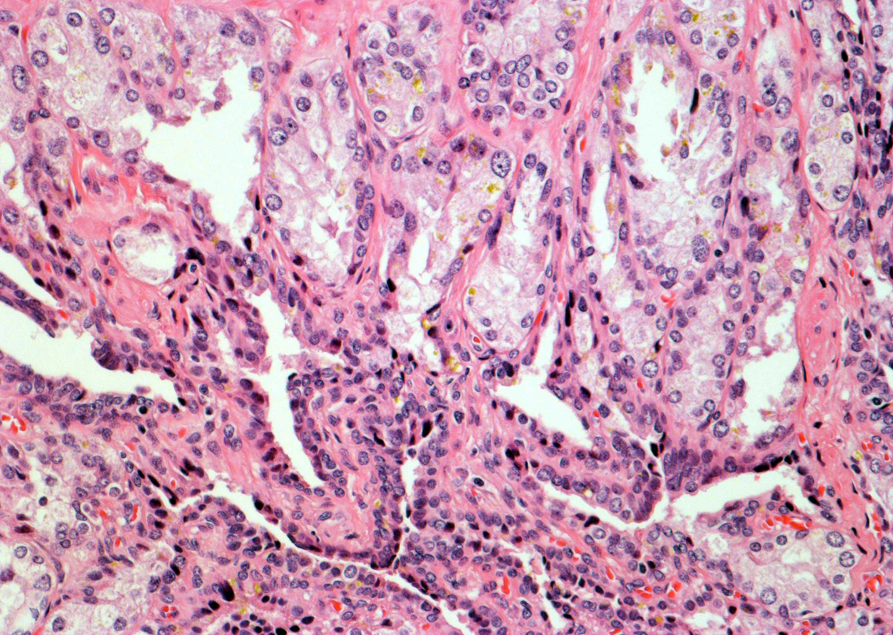
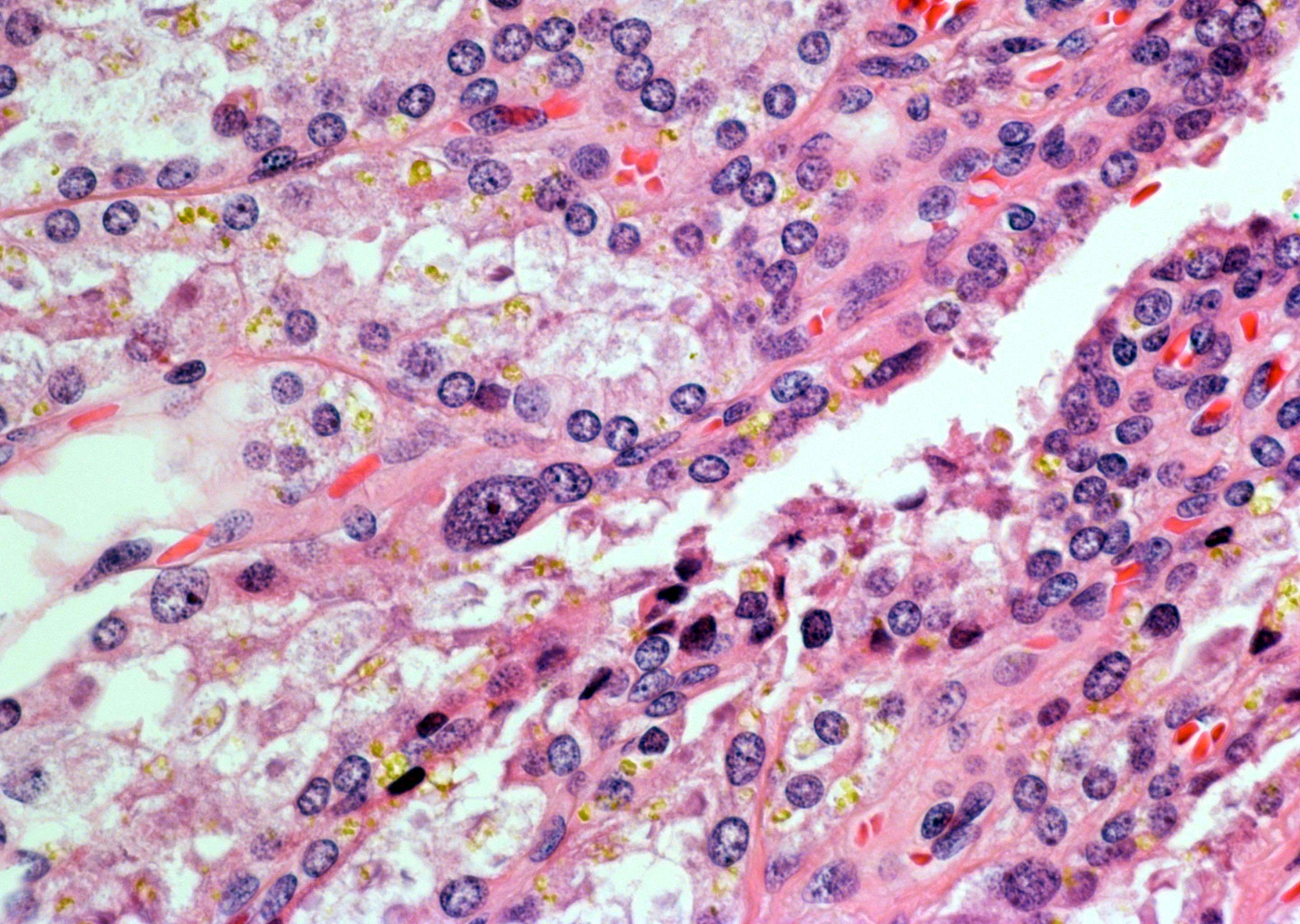
Contributed by Theodorus van der Kwast, M.D., Ph.D. and Debra Zynger, M.D.
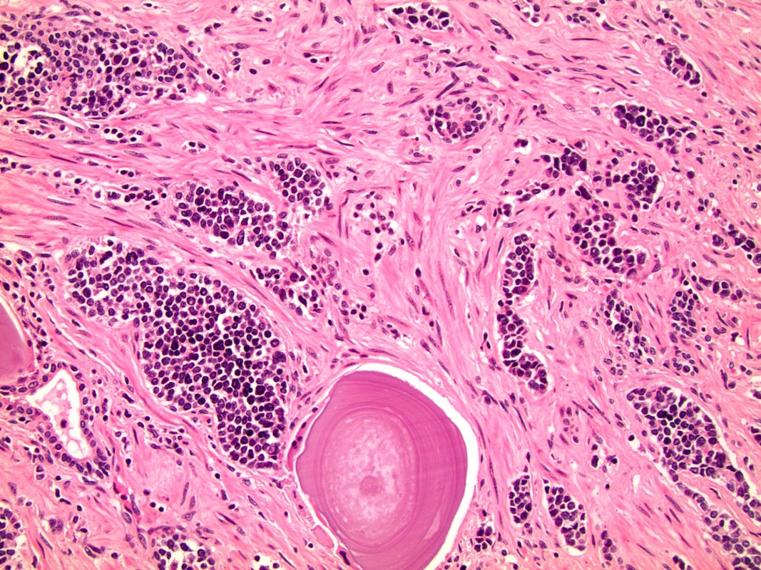
Sheets of carcinoma cells
Nuclear molding
Ribbons with pseudorosettes
Solid nests with glands
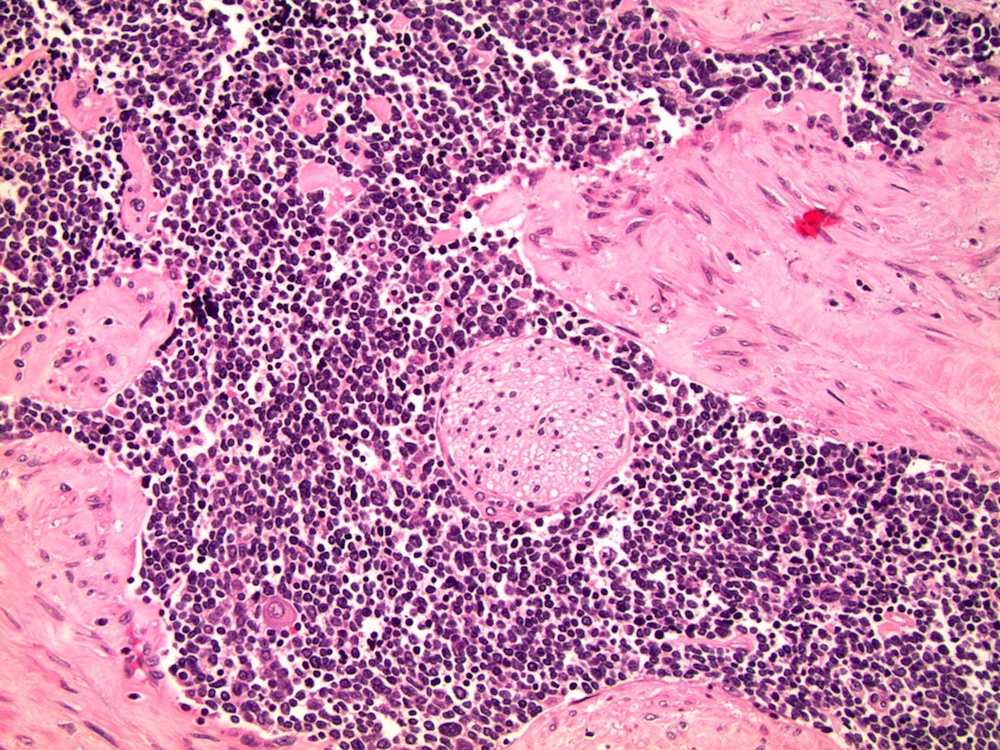
Solid sheets of adenocarcinoma
Pale and hyperchromatic tumor cells
Sheet of pale tumor cells
Dark cells within duct
Visible nucleoli
Chromogranin A staining
Contributed by Kenneth A. Iczkowski, M.D.
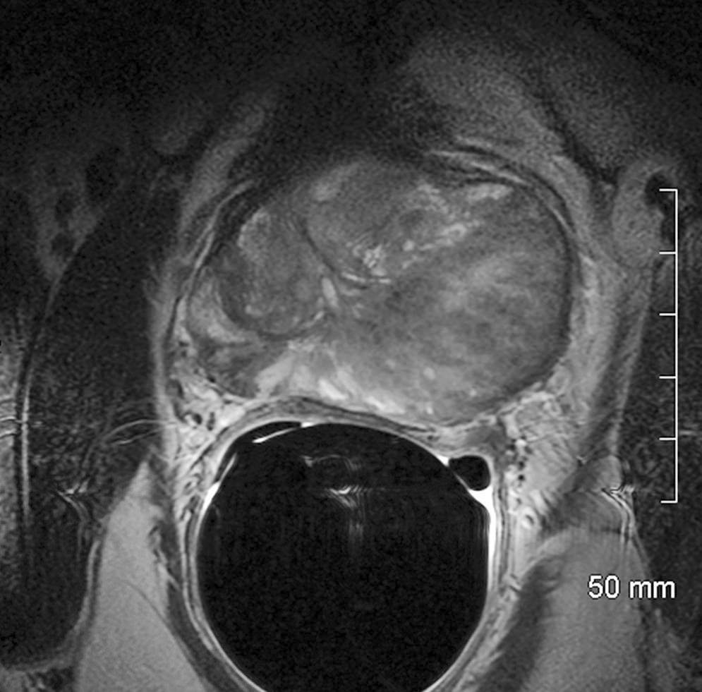
T2 weighted MRI
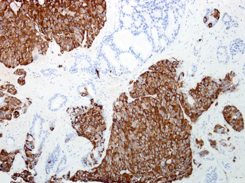
Contributed by Kenneth A. Iczkowski, M.D.
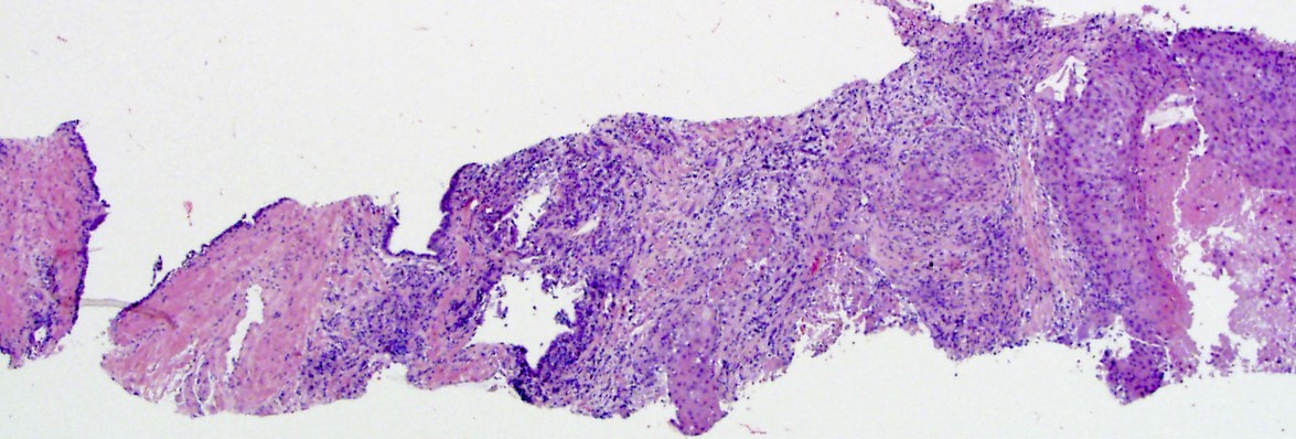
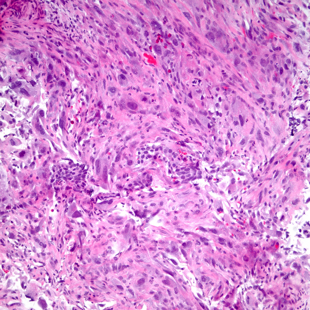
Needle biopsy
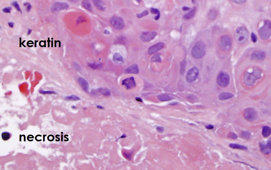
Keratinization with necrosis
Multinucleation
Images hosted on other servers:


Squamous cell carcinoma
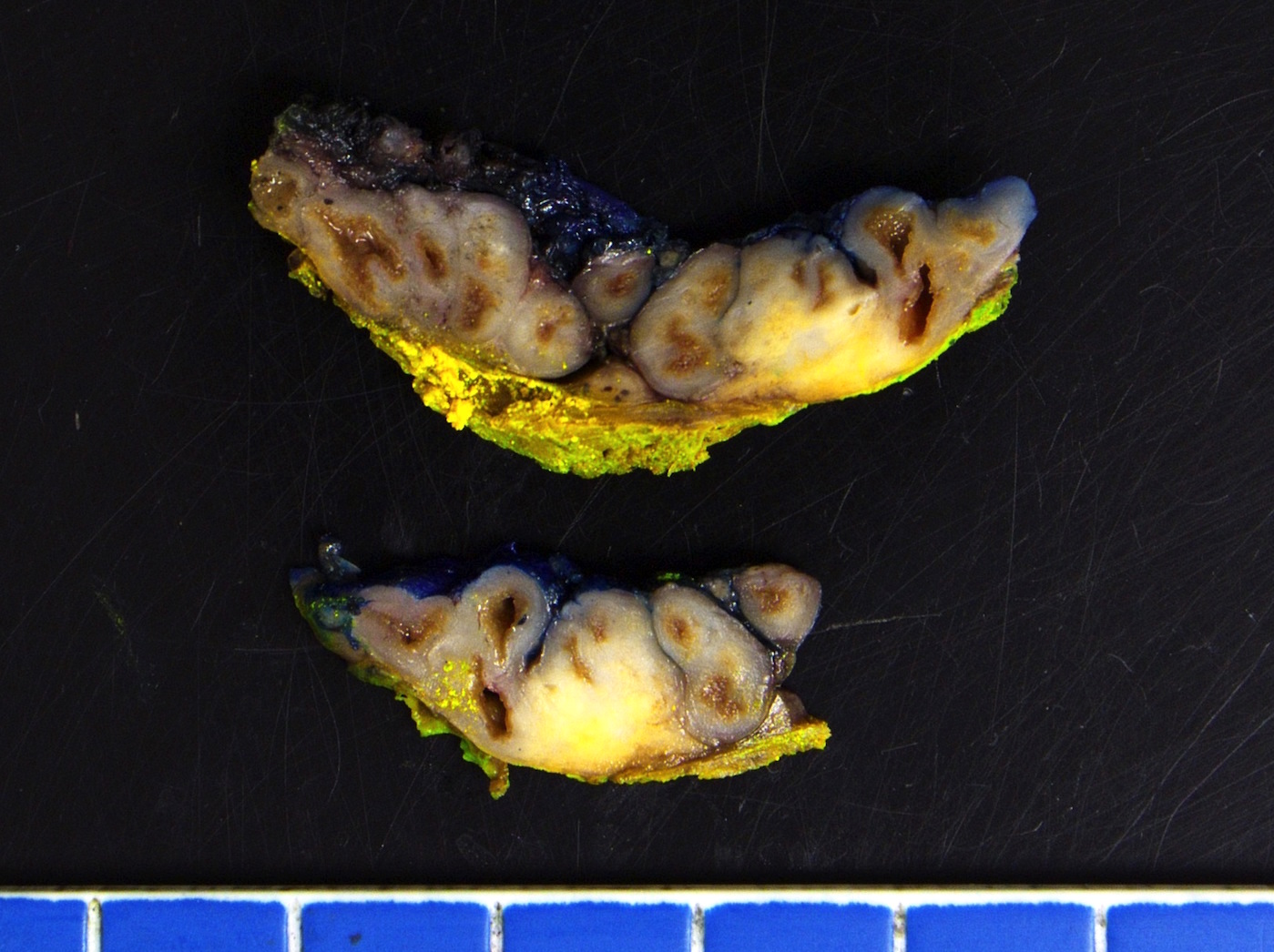
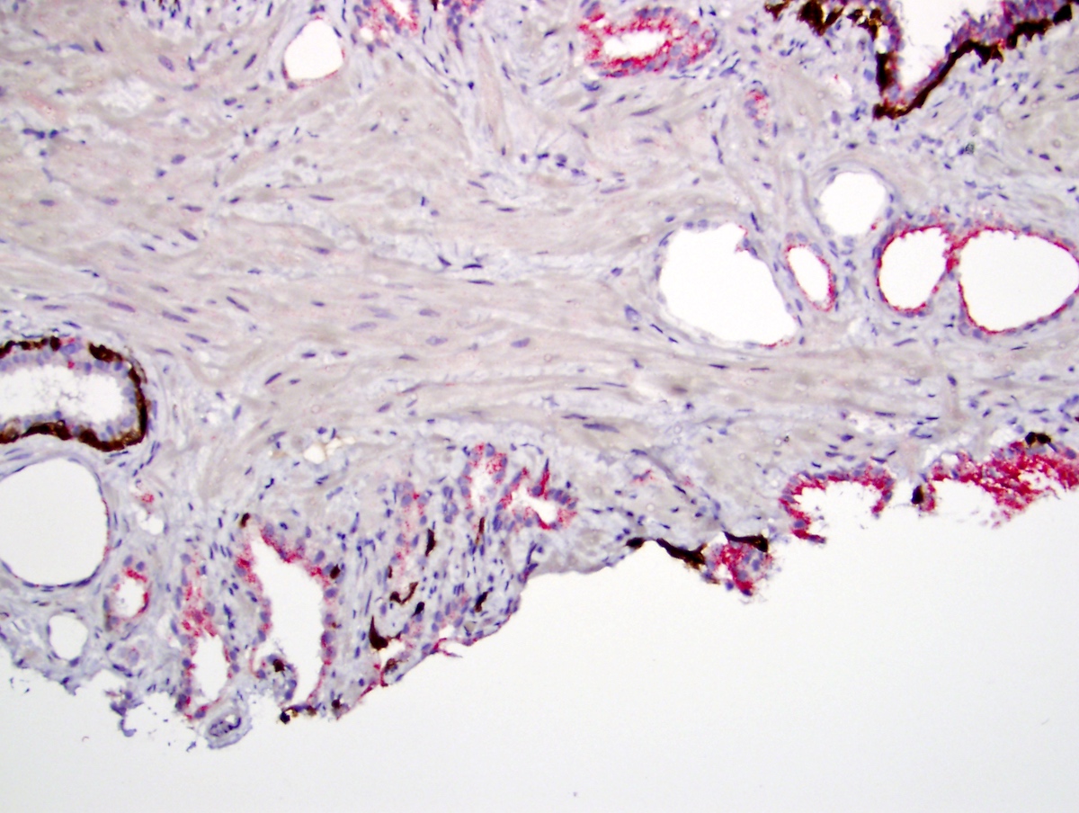
Contributed by Debra L. Zynger, M.D.

Extraprostatic extension, multifocal (pT3a)
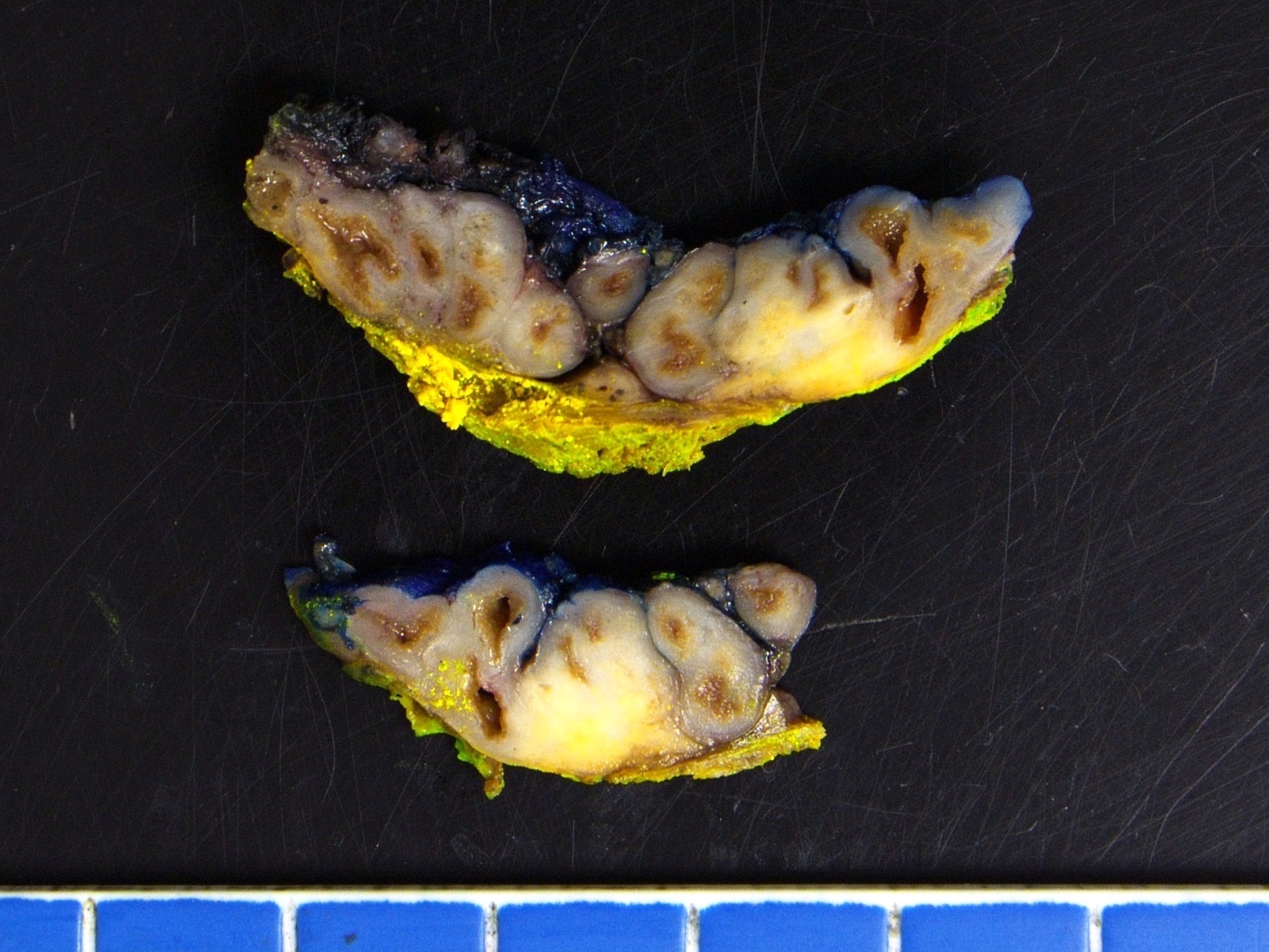

Seminal vesicle invasion (pT3b)
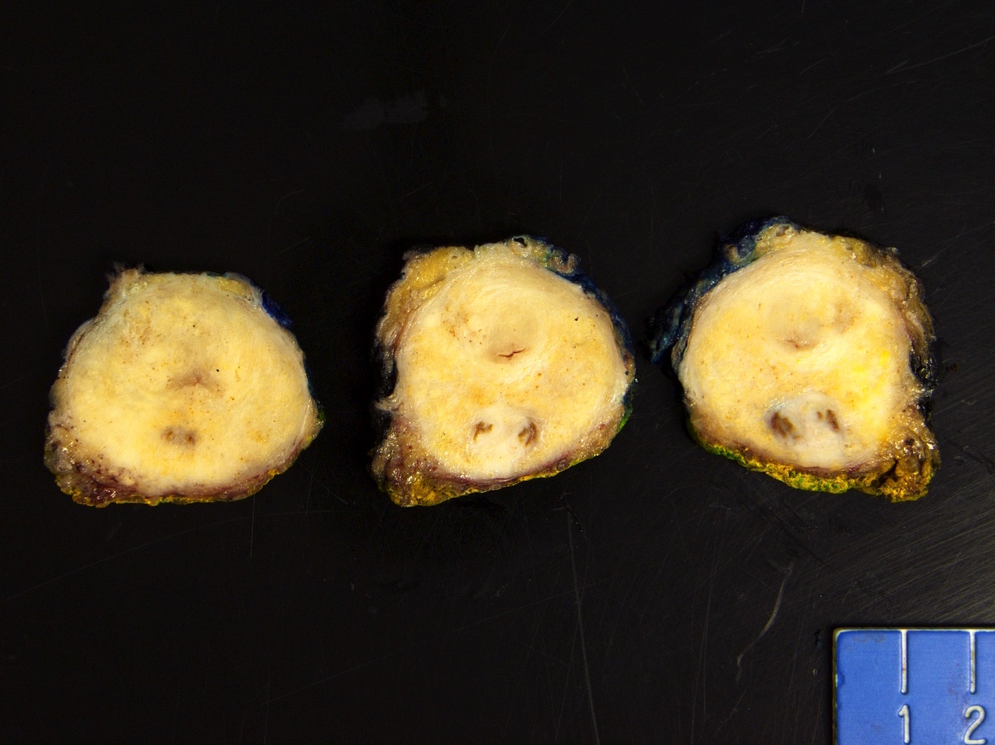
Contributed by Debra L. Zynger, M.D.
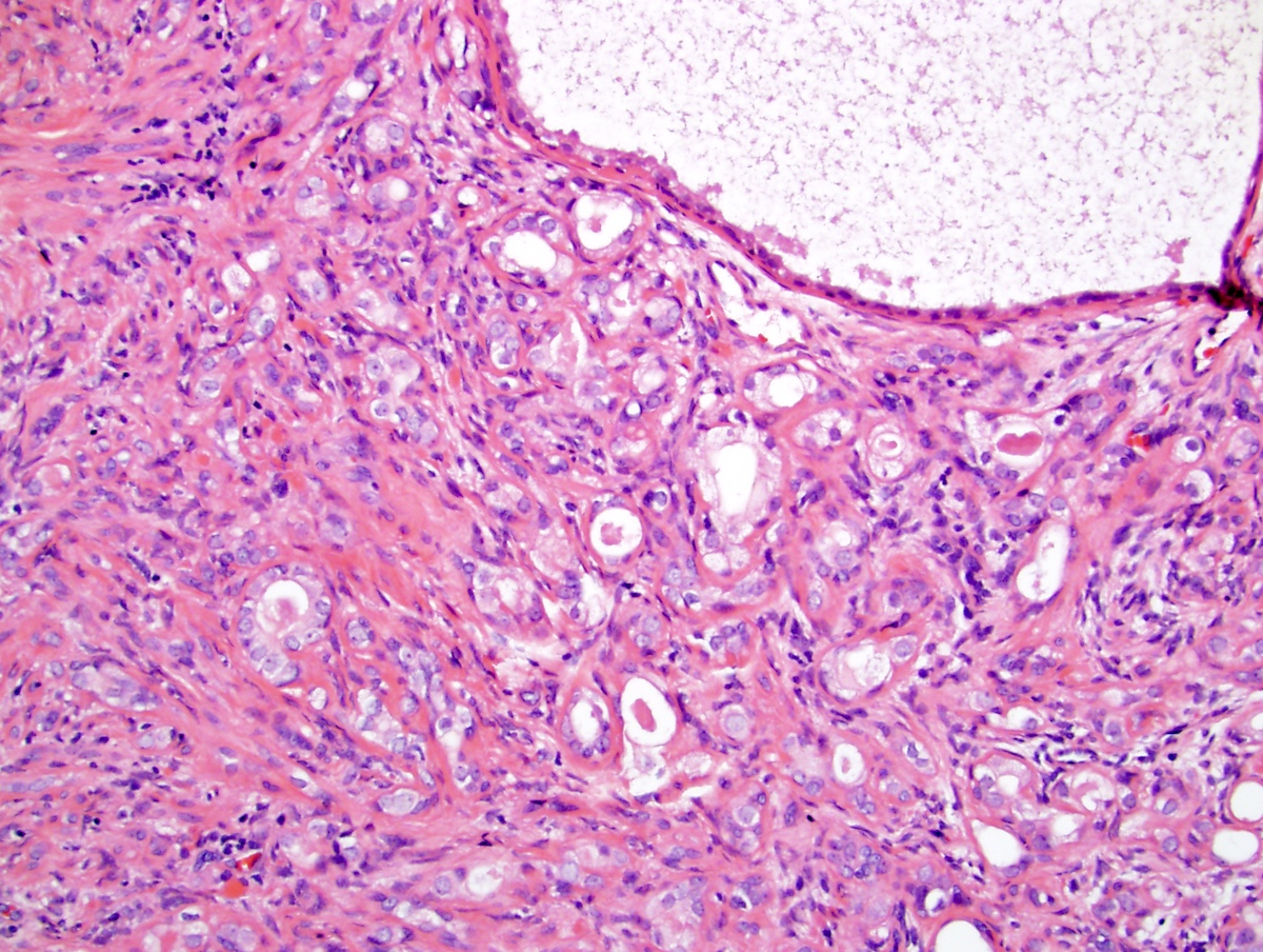
Extraprostatic extension (pT3a)
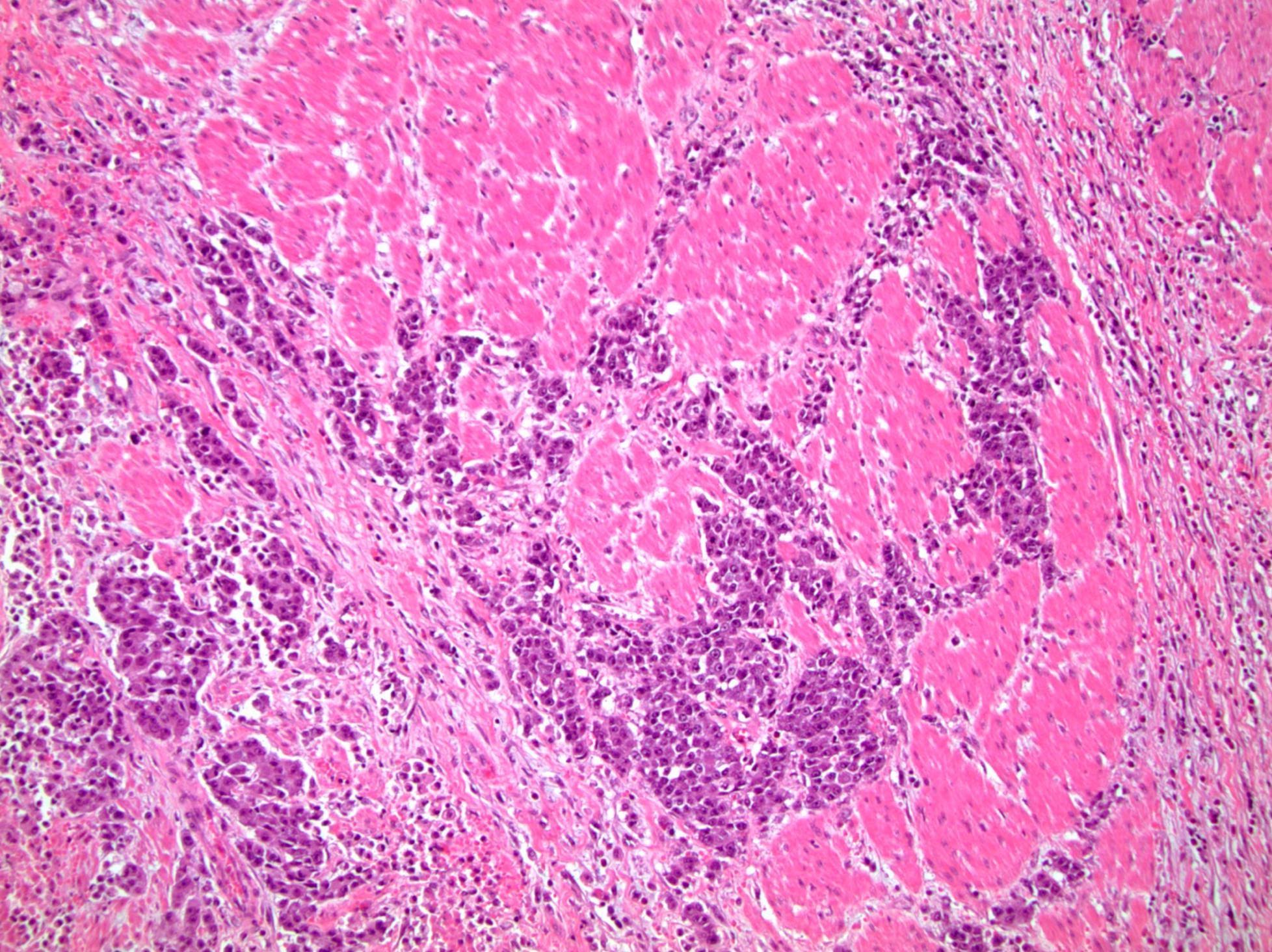
Bladder neck invasion (pT3a)

Seminal vesicle invasion (pT3b)
Rectal invasion (pT4)
Periprostatic lymph node metastasis (pN1)
Pelvic lymph node metastasis (pN1)
Neck lymph node metastasis (pM1a)
Bone metastasis (pM1b)
Lung metastasis (pM1c)

Brain metastasis (pM1c)
Contributed by Kenneth Iczkowski, M.D. and Debra L. Zynger, M.D.
Androgen deprivation, benign
Androgen deprivation, cancer
Radiotherapy, benign

Radiotherapy, benign
Cryotherapy, benign
Cryotherapy, cancer
Suspicious for cancer
Shrunken benign acinus
Radiation change, carcinoma
Images hosted on other servers:

Polypoid mass

Filling defect
Images hosted on other servers:

Urethrocystoscopic view of polyp
Images hosted on other servers:

Brown-tan knob-like polyp
Contributed by Y. Albert Yeh, M.D., Ph.D.
Fibroepithelial polyp
Polypoid mass
Cloverleaf-like and club-like projections
Broad based papillae
Cloverleaf-like projections
Club-like projections
Broad based finger-like papillae
Urethritis cystica and glandularis
Polypoid and club-like projections
Focal calcifications

Images hosted on other servers:

Paraffin blocks
before and
after block
flipping
Images hosted on other servers:


Primary carcinoid tumor of the verumontanum
Contributed by Alcino Pires Gama, M.D. and Bonnie Choy, M.D.

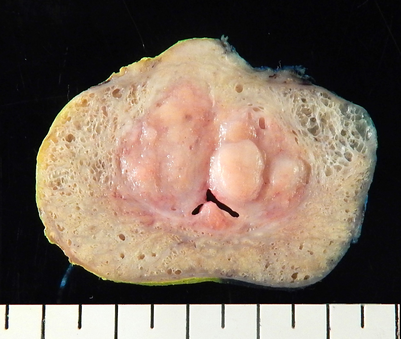
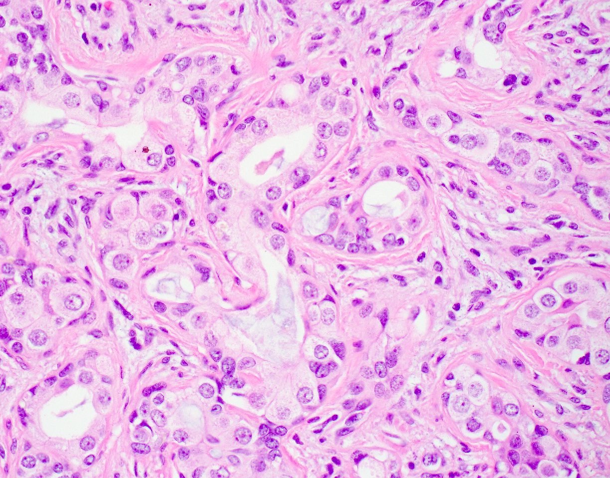
Prostatic ductal adenocarcinoma

Adenoid cystic (basal cell) carcinoma of the prostate
Treatment related neuroendocrine carcinoma
Intraductal carcinoma of the prostate
Amin: 2022

Cheng: 2019

Epstein: 2021

Epstein: 2020
Epstein: 2020
IARC: 2022
Lopez-Beltran: 2018
Matoso: 2020
Robinson: 2018
Shah: 2019
VandenBussche: 2022

Wobker: 2021

Yang: 2020
Zhou: 2022
Find related Pathology books: cytopathology, GU/adrenal
