
Contributed by Felicia D. Allard, M.D. and Claudio Luchini, M.D., Ph.D.
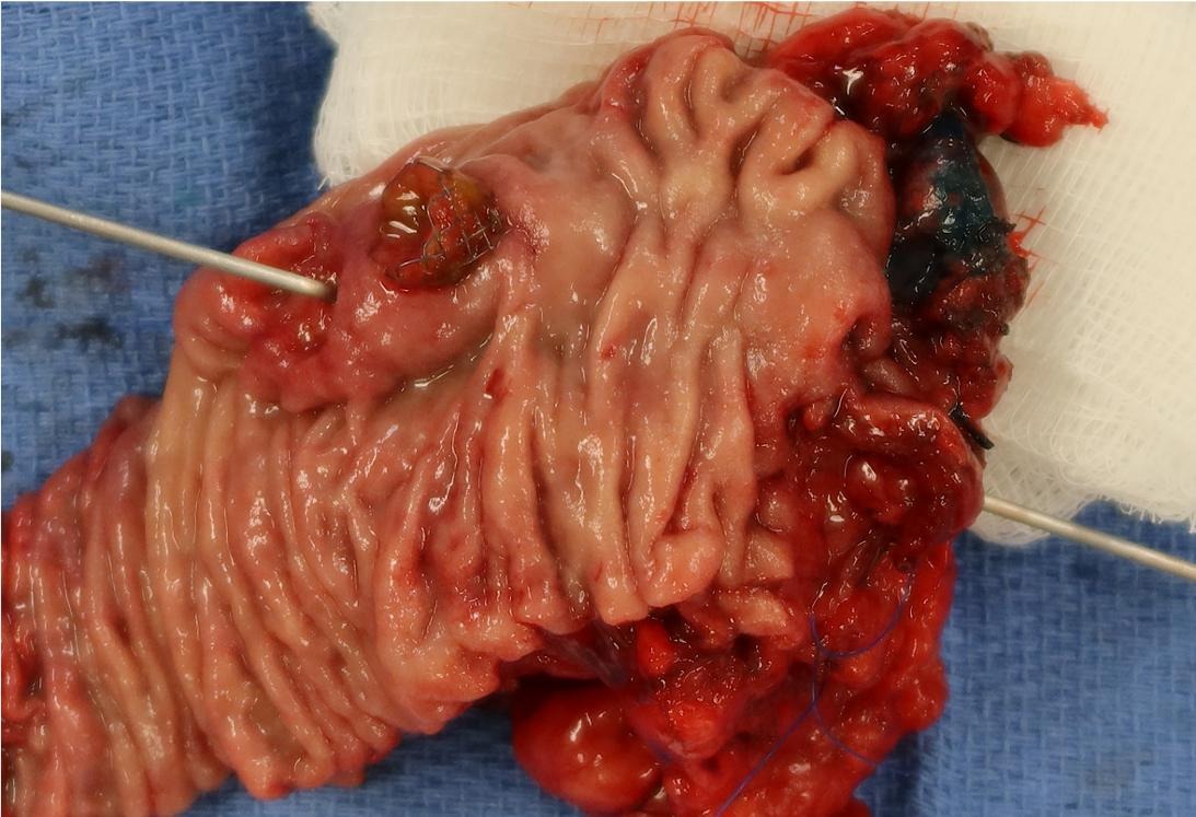
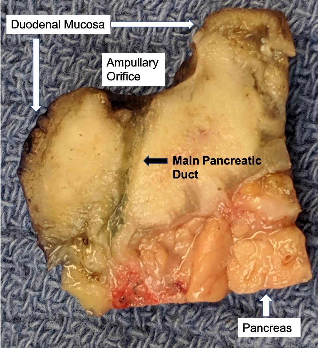
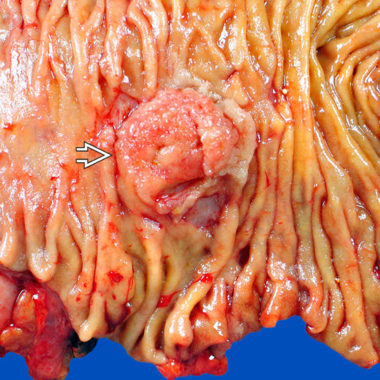
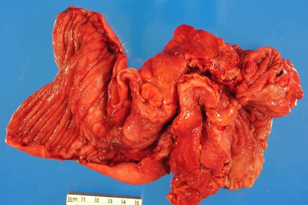
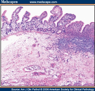
Ampullary ductal carcinoma
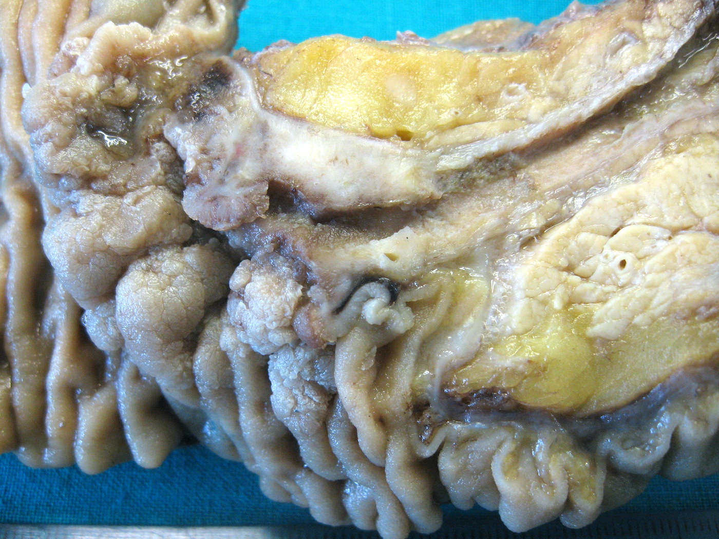
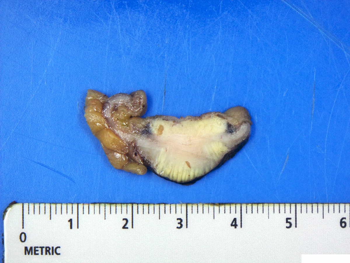
Firm gray-white thickening
Contributed by Felicia D. Allard, M.D.
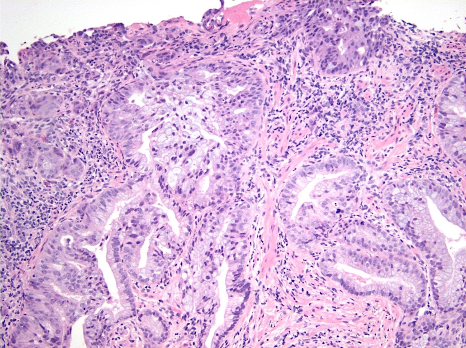
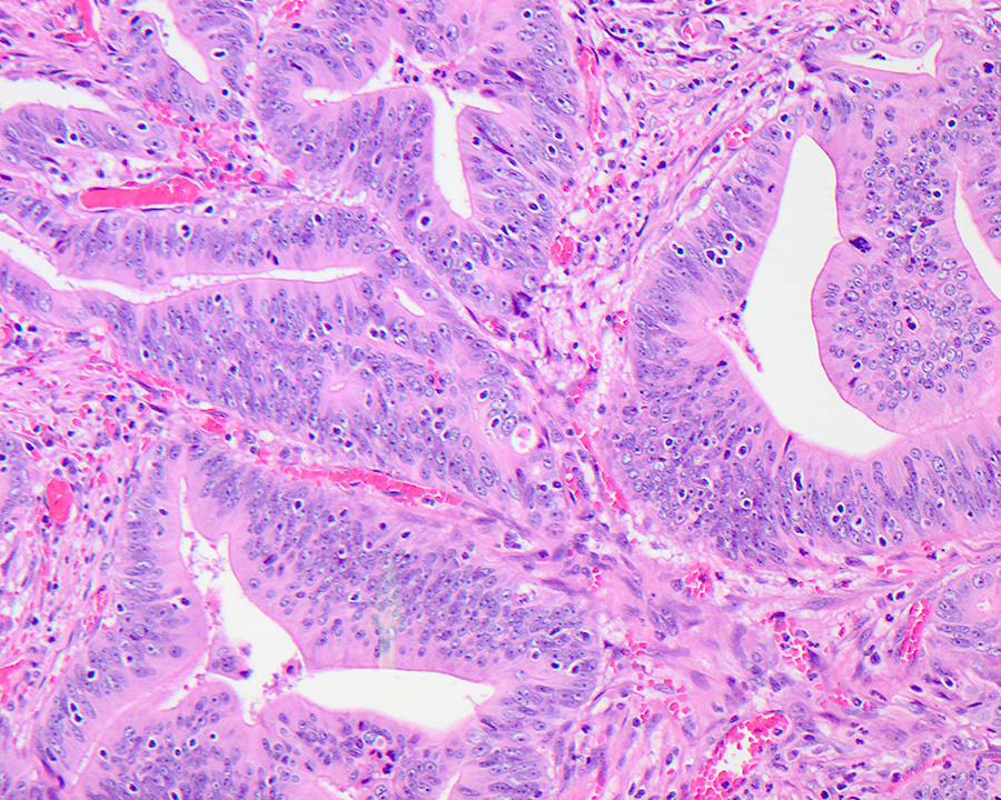
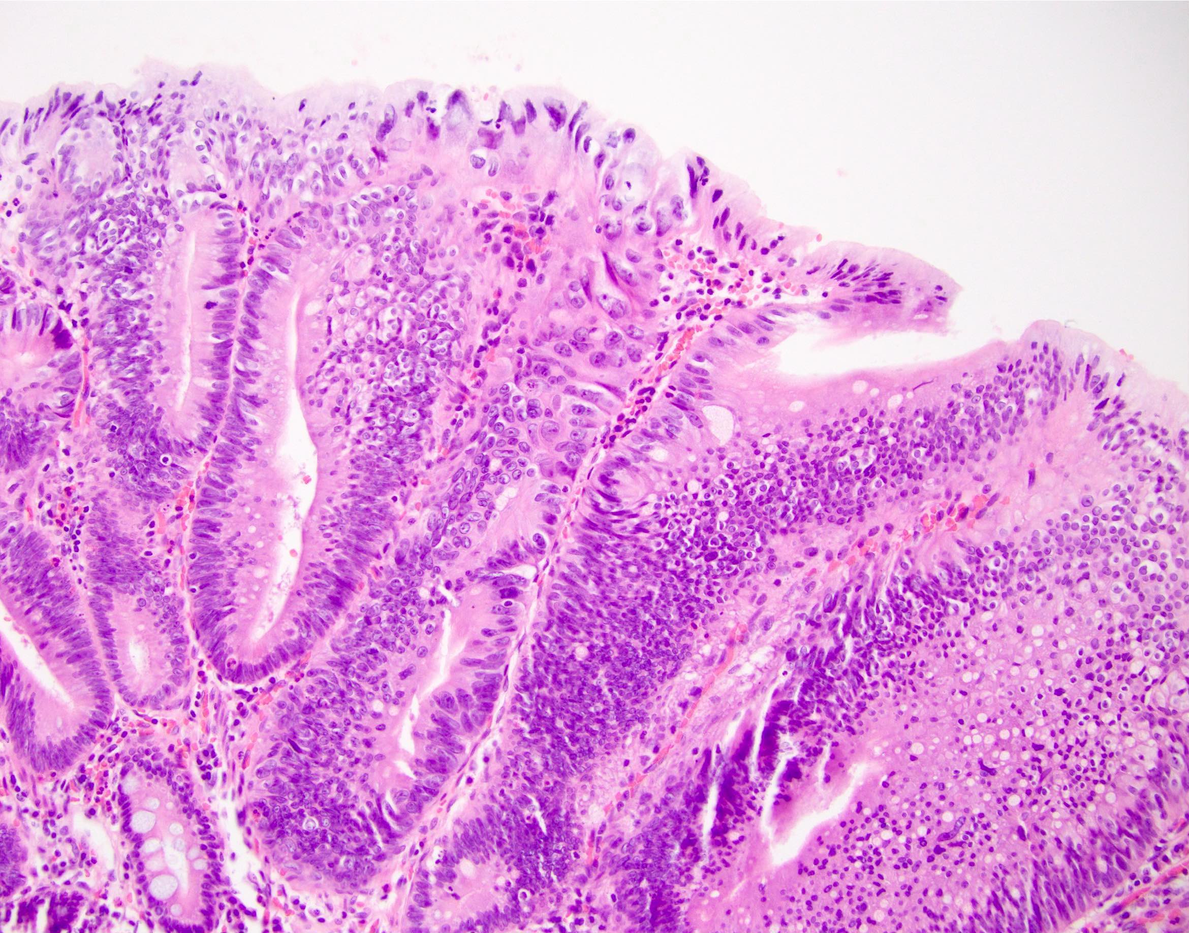
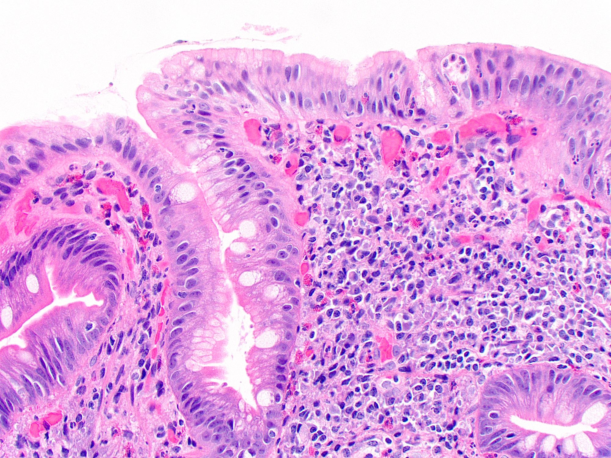
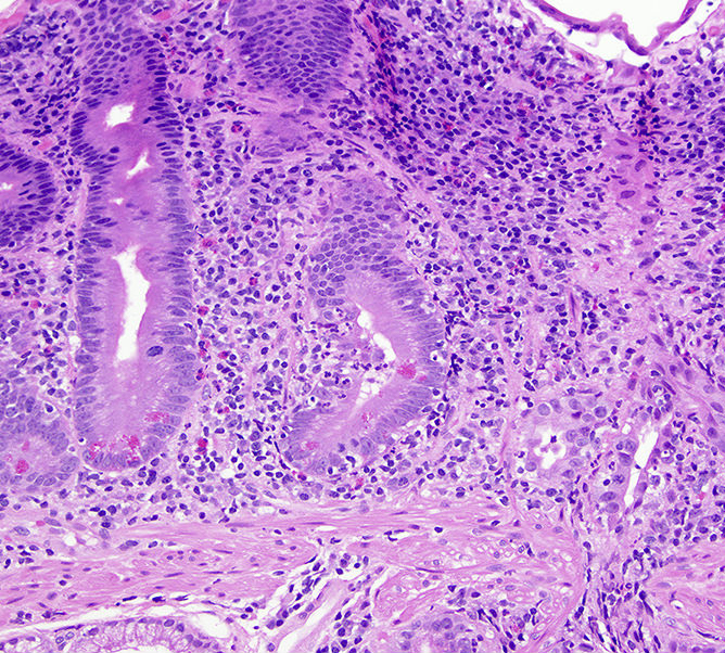
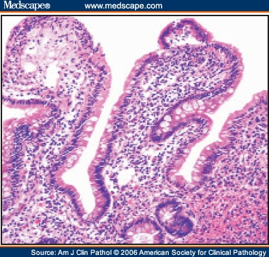
Intestinal type adenocarcinoma
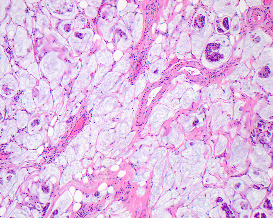
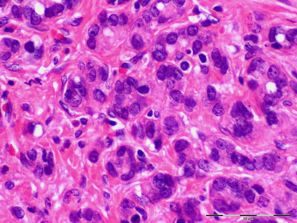
Signet ring cell morphology
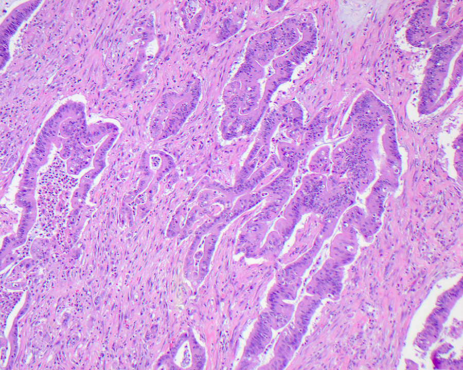
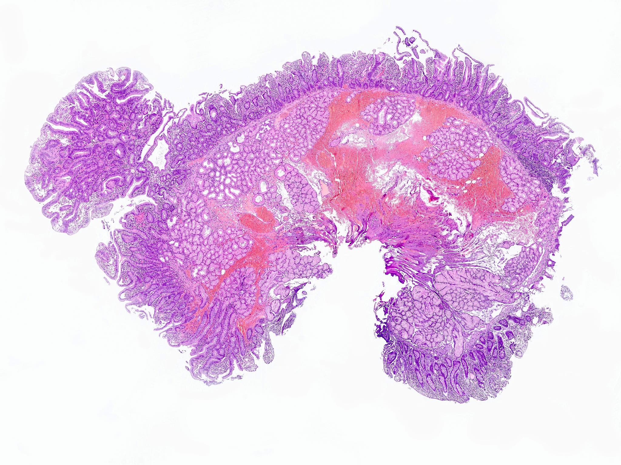
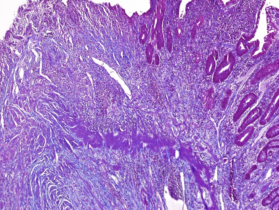
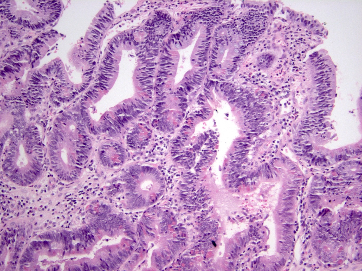
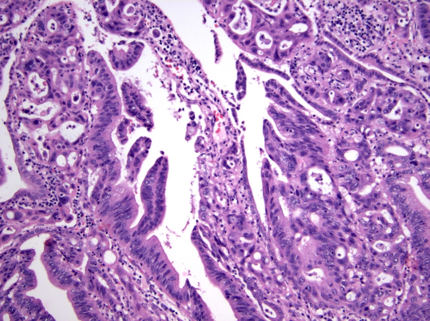
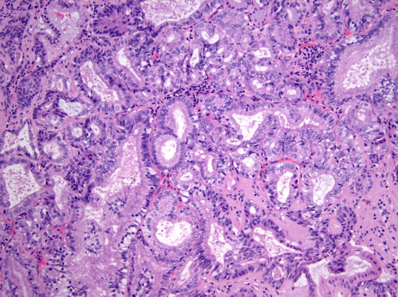
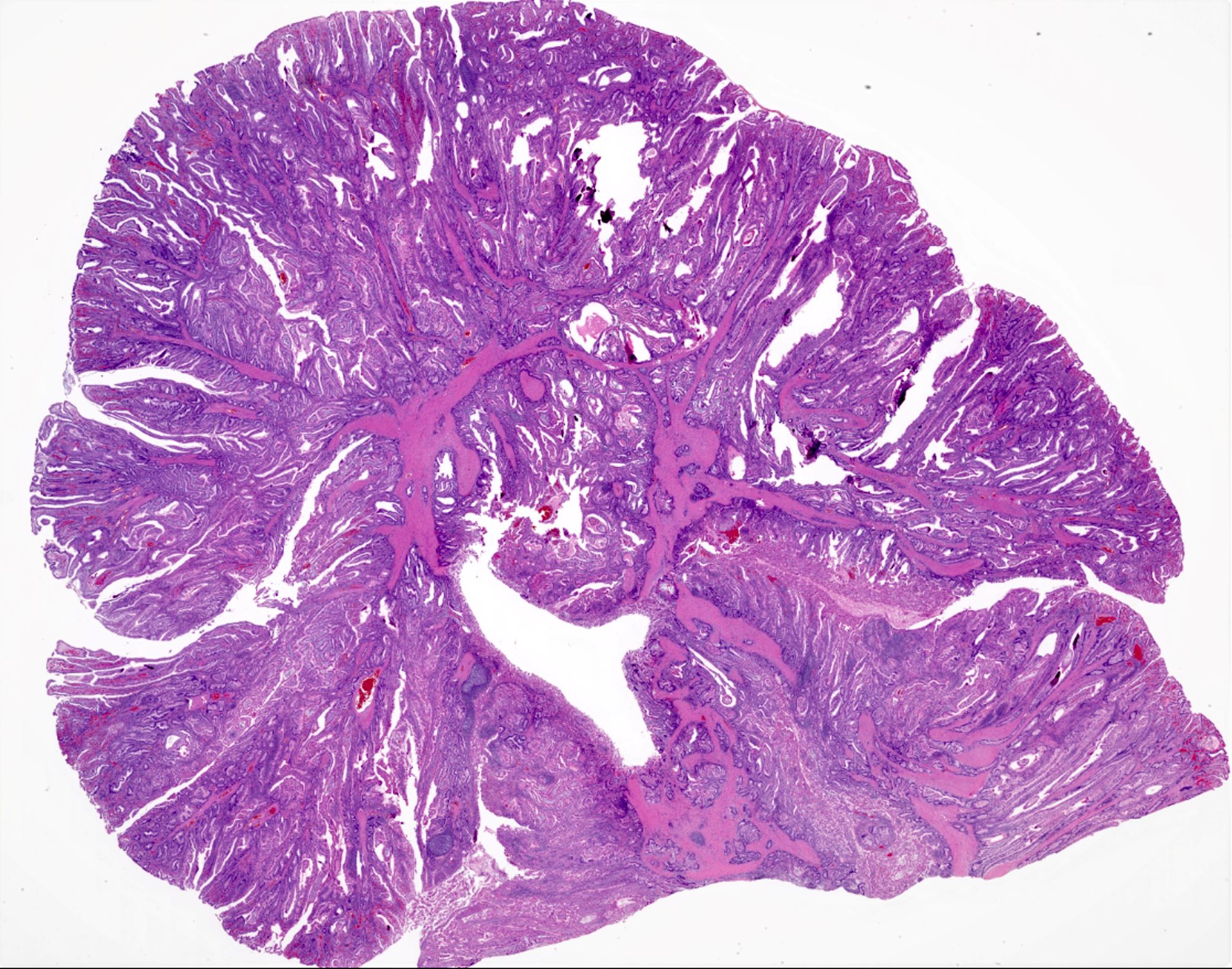
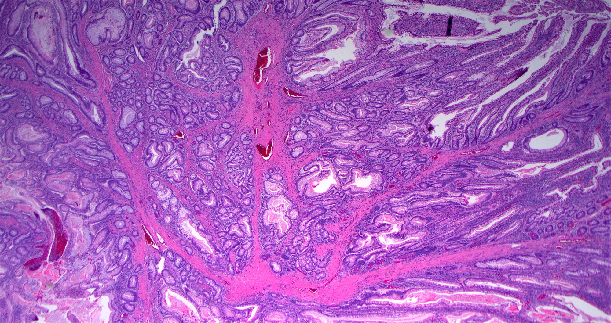
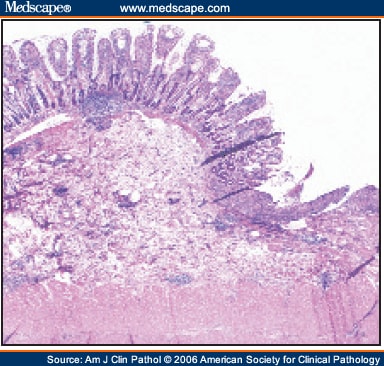
Adenocarcinoma arising from IPTN

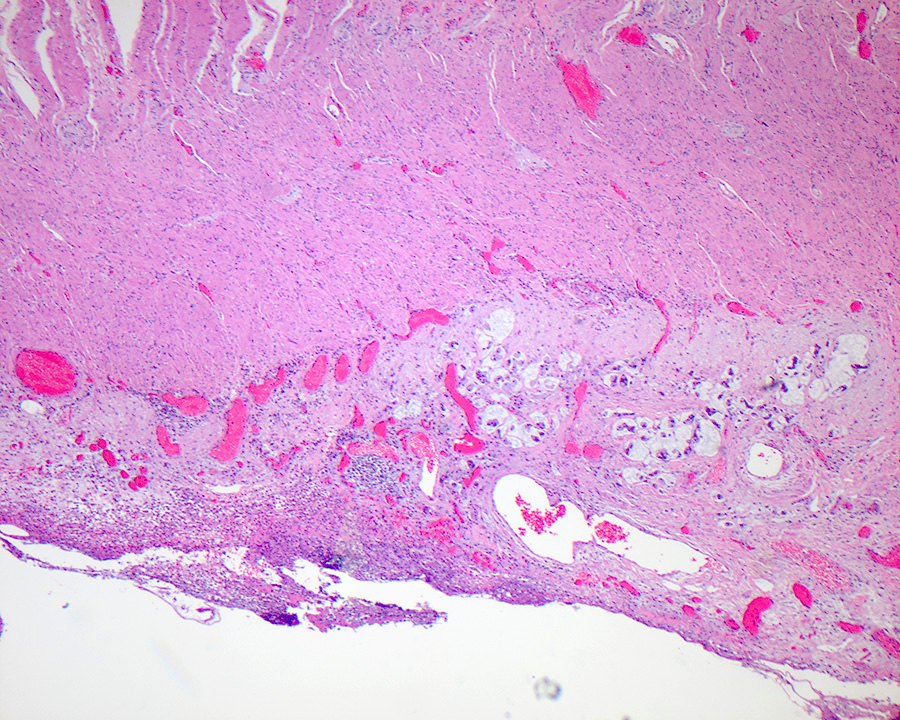
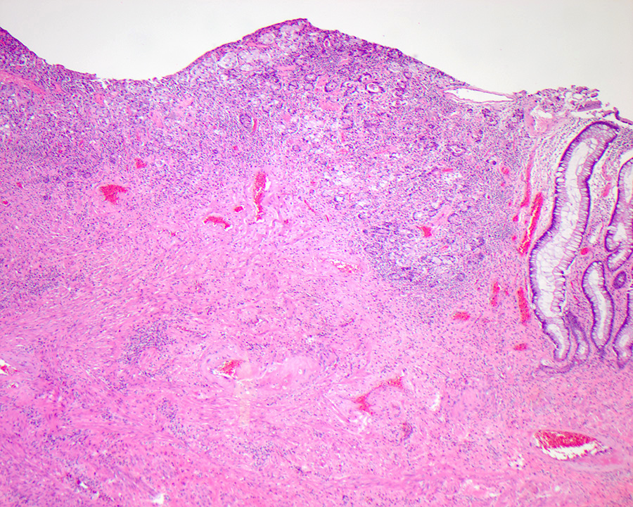
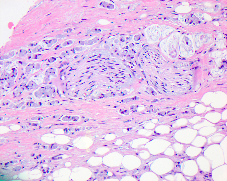
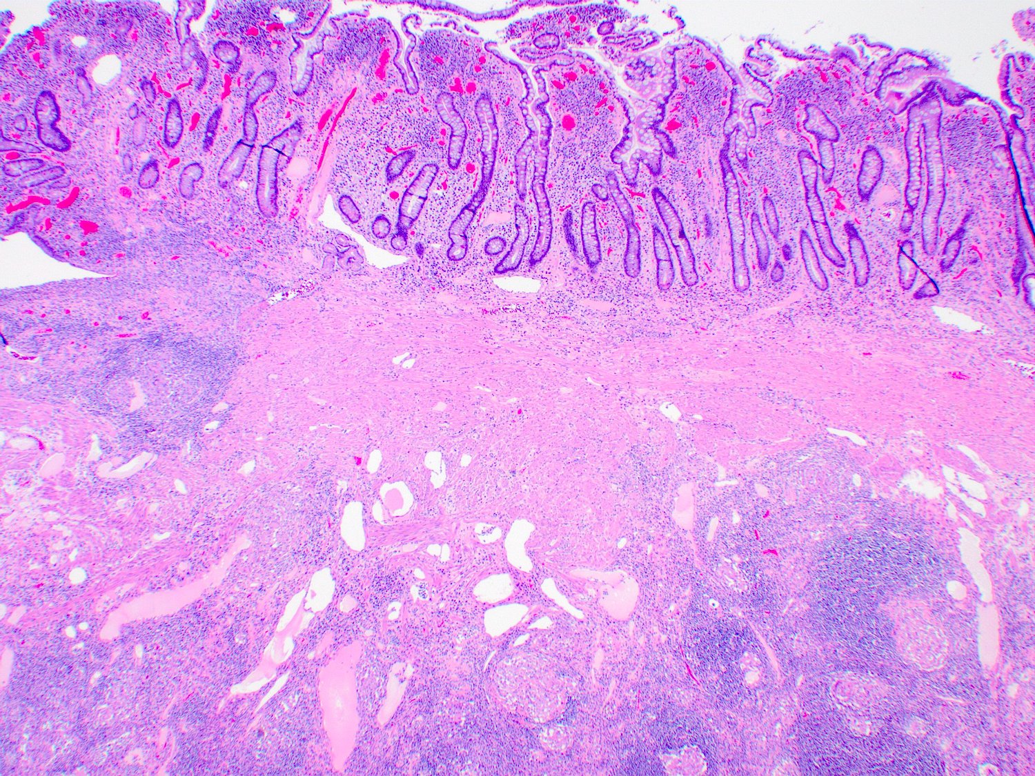
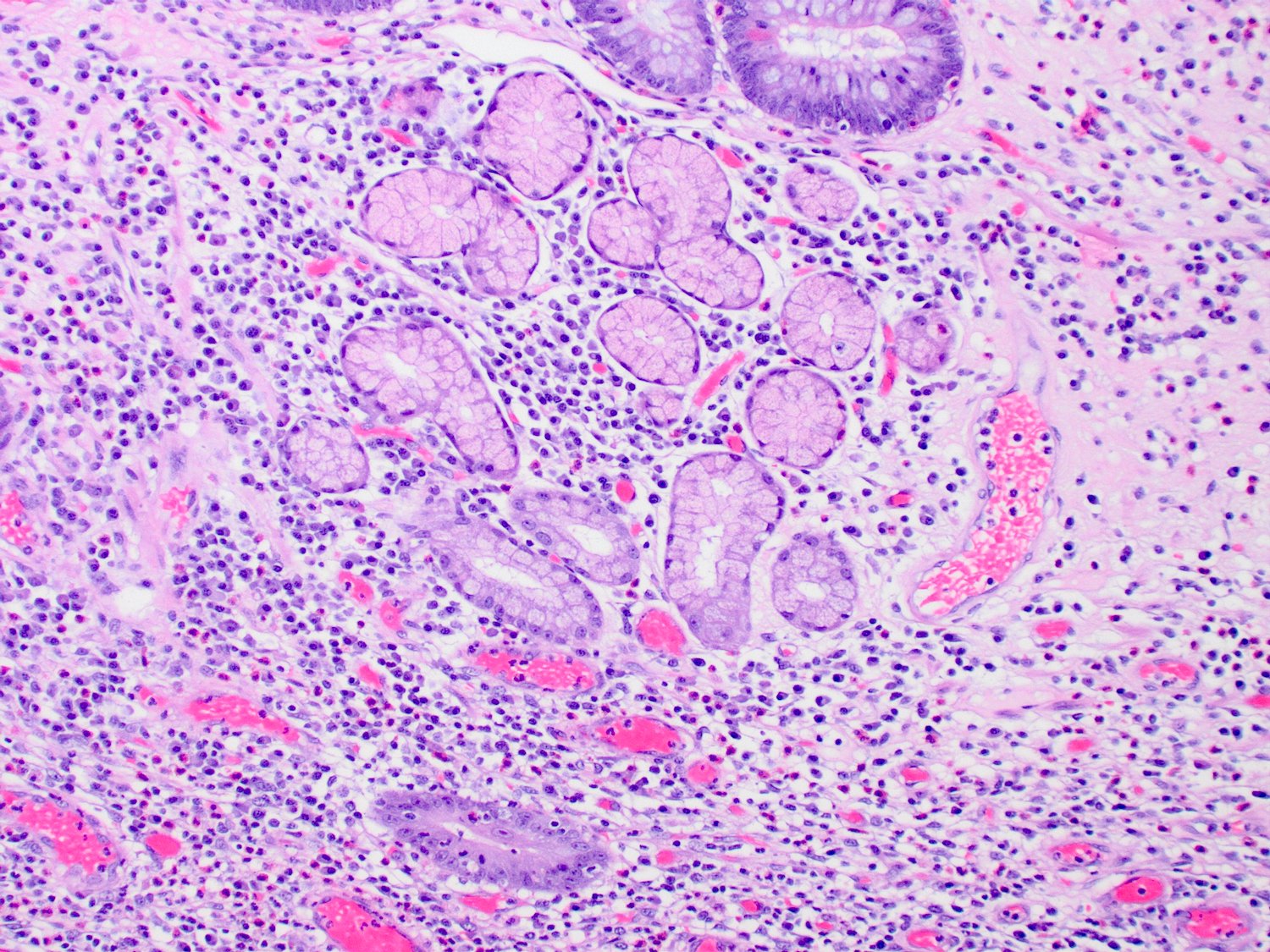
Ampullary duct carcinoma
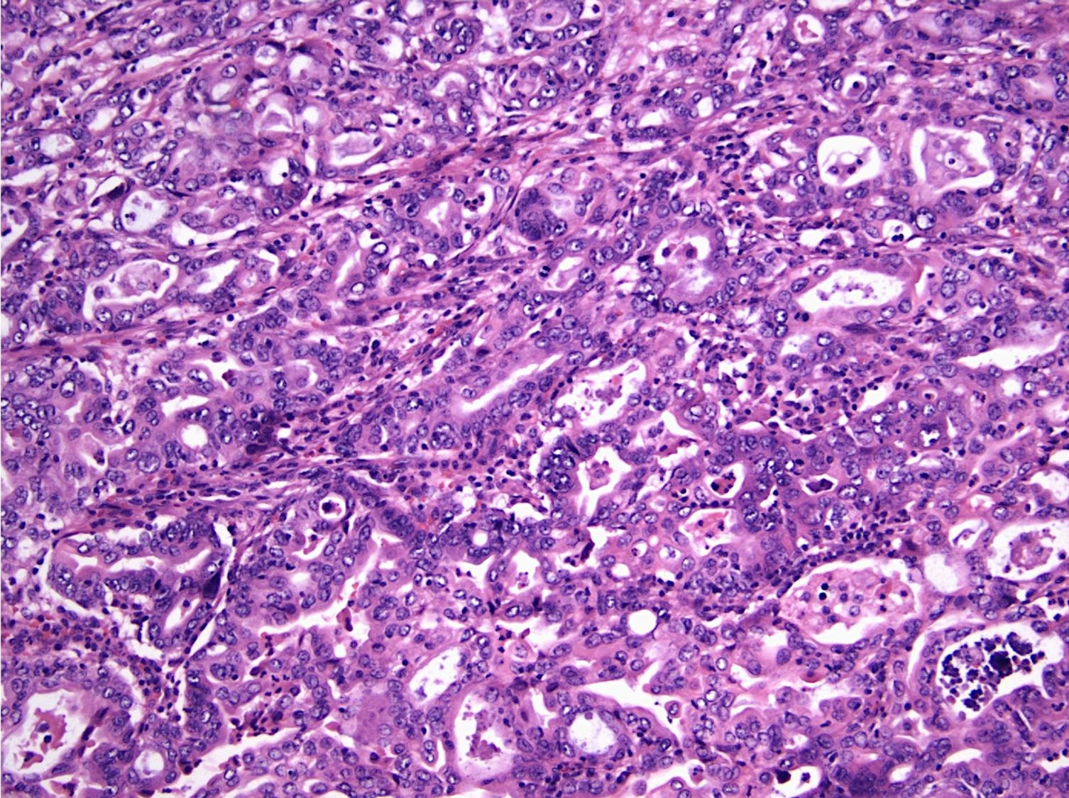
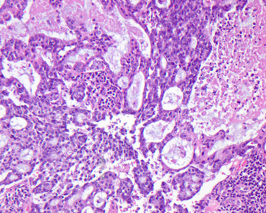
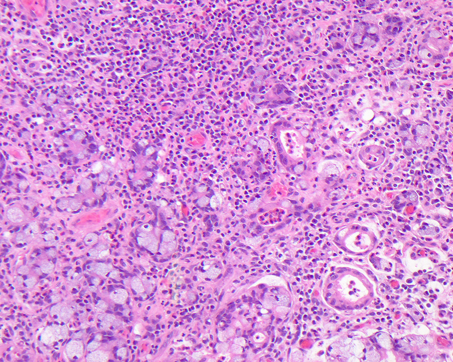
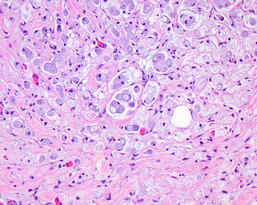
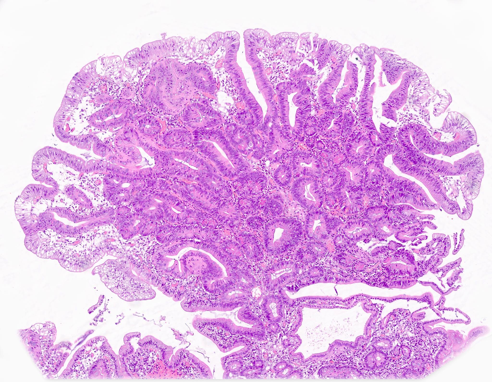
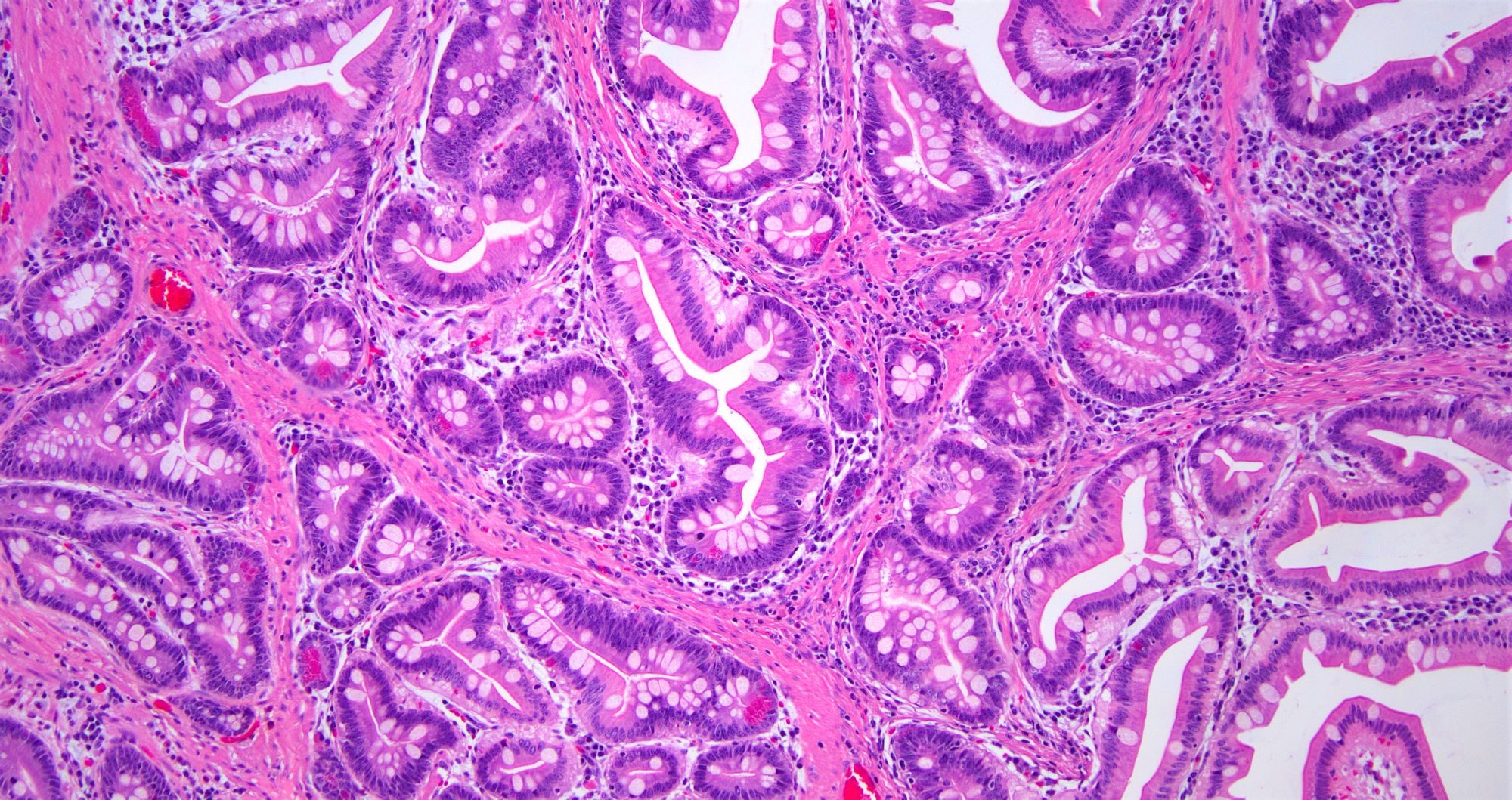
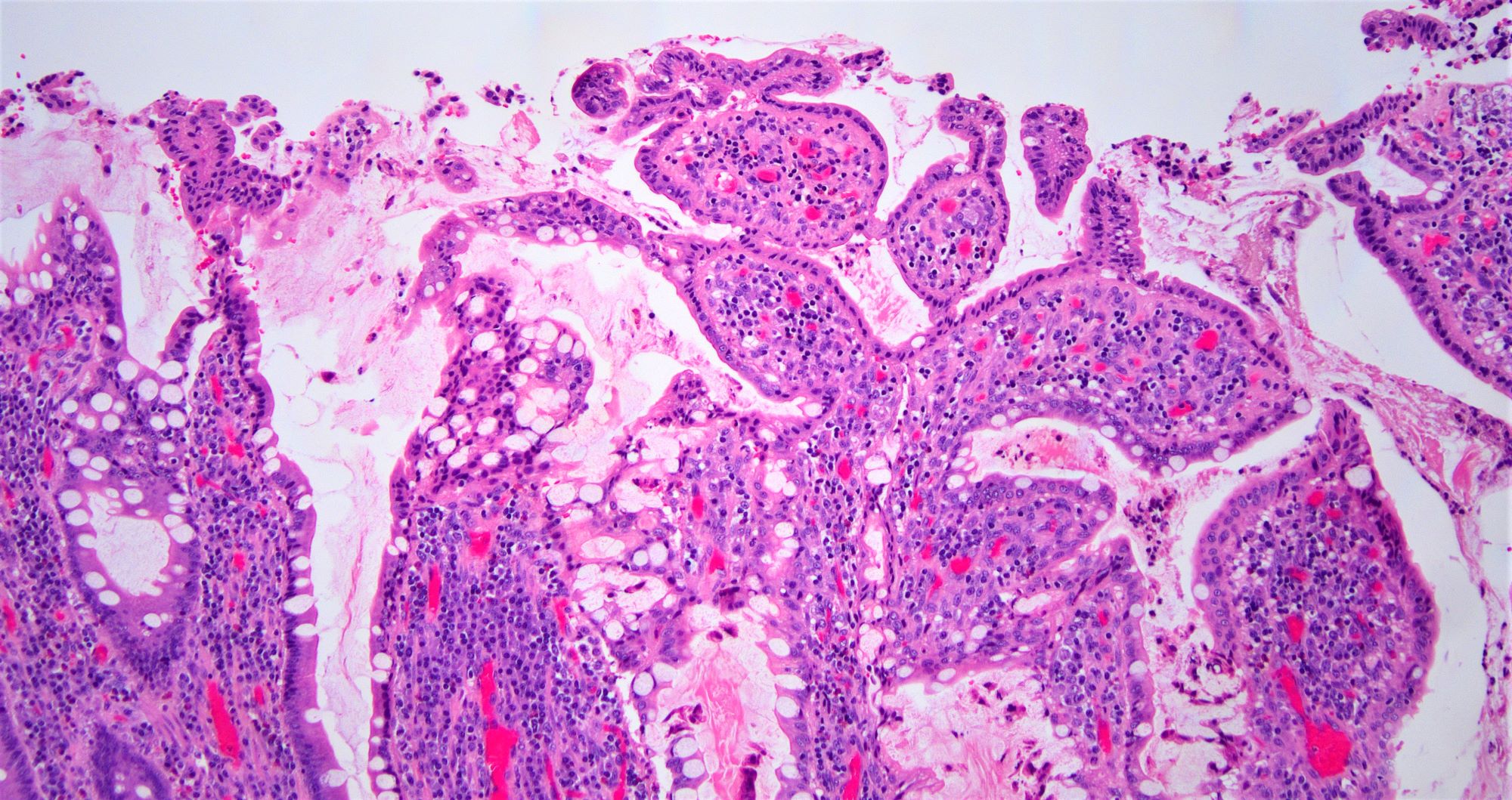
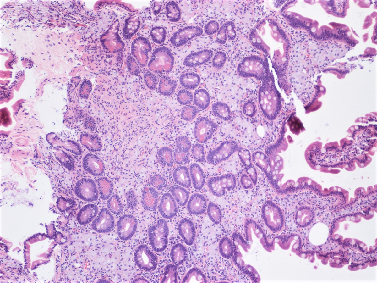
Pancreaticobiliary phenotype

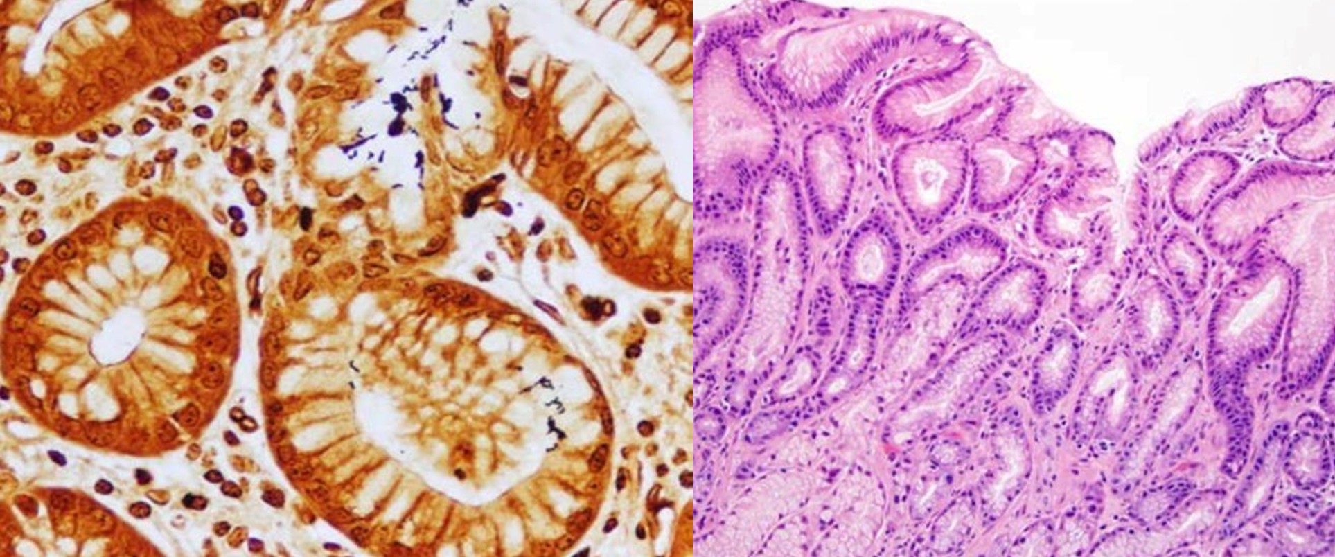
CDX2 pancreatobiliary phenotype

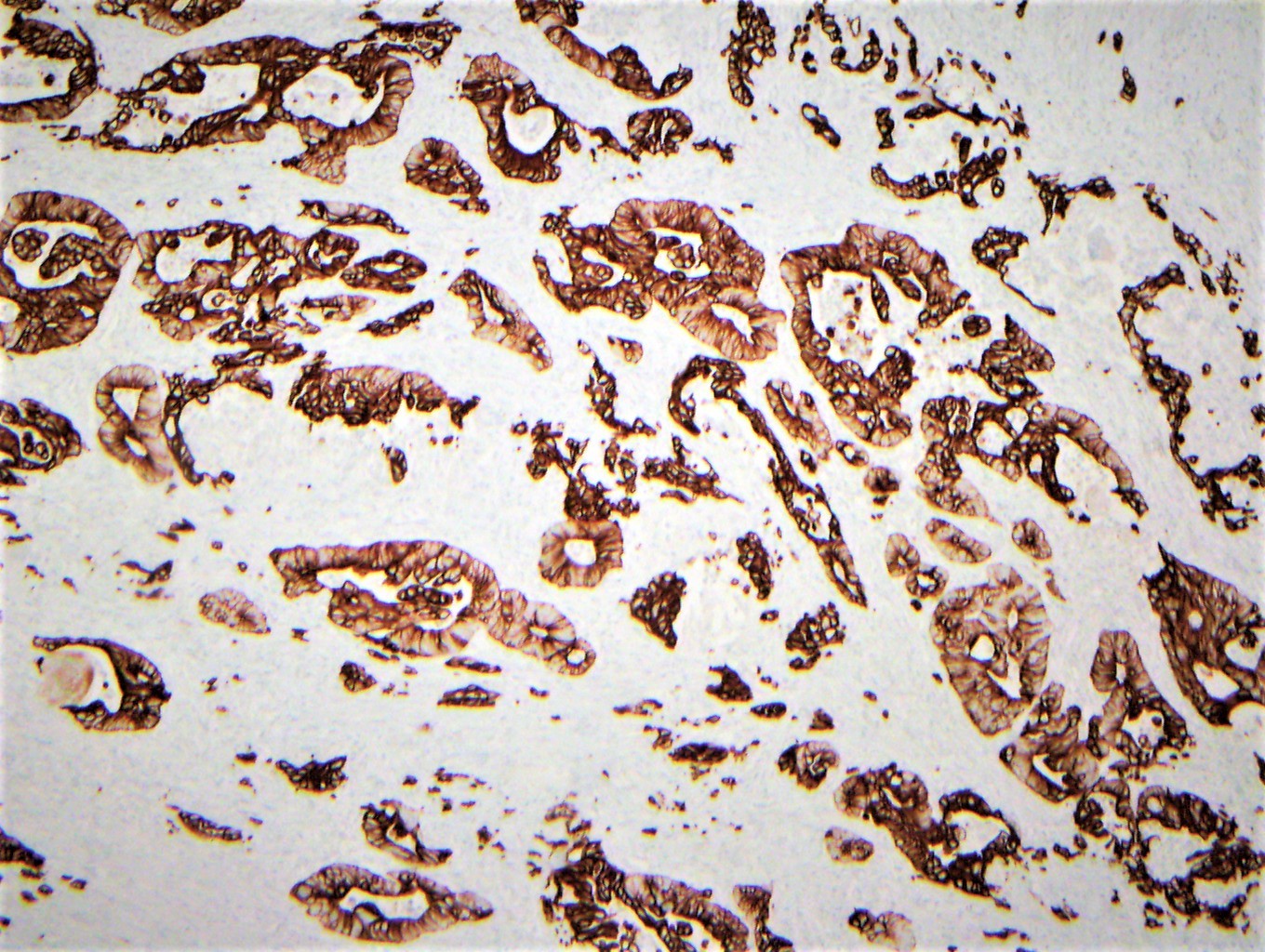
CK7 pancreatobiliary phenotype
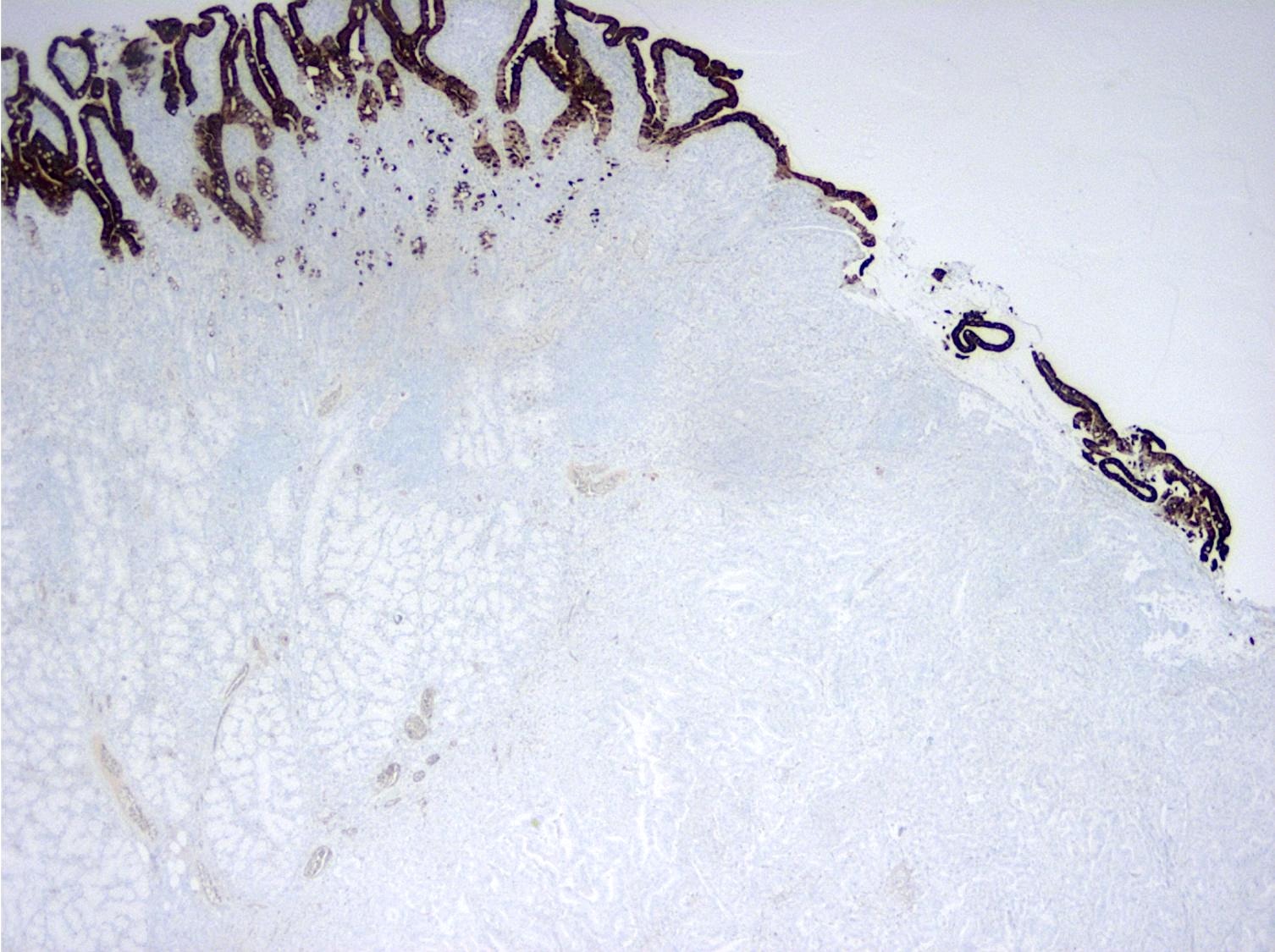
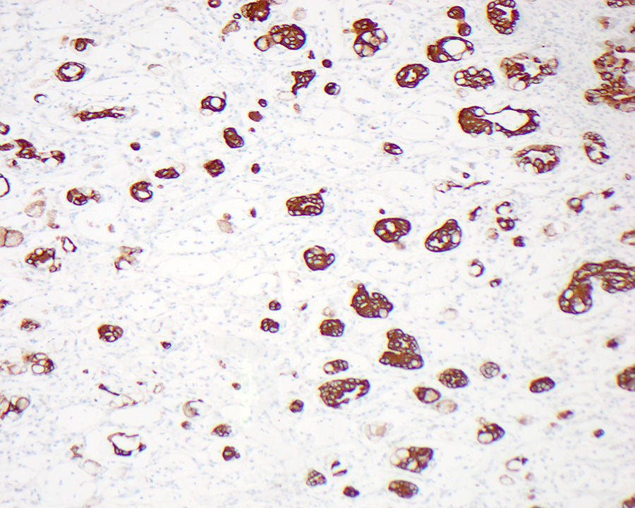
CK20 pancreatobiliary phenotype
Images hosted on other servers:

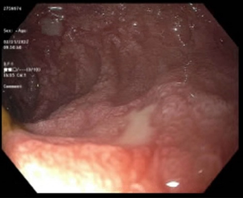
Suspicious growth with jejunal stricture

Near obstructing mass distal to duodenal bulb

Irregular and ulcerated jejunal lesion (capsule endoscopy)
Images hosted on other servers:

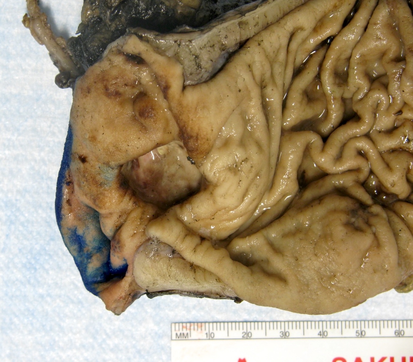
Jejunal
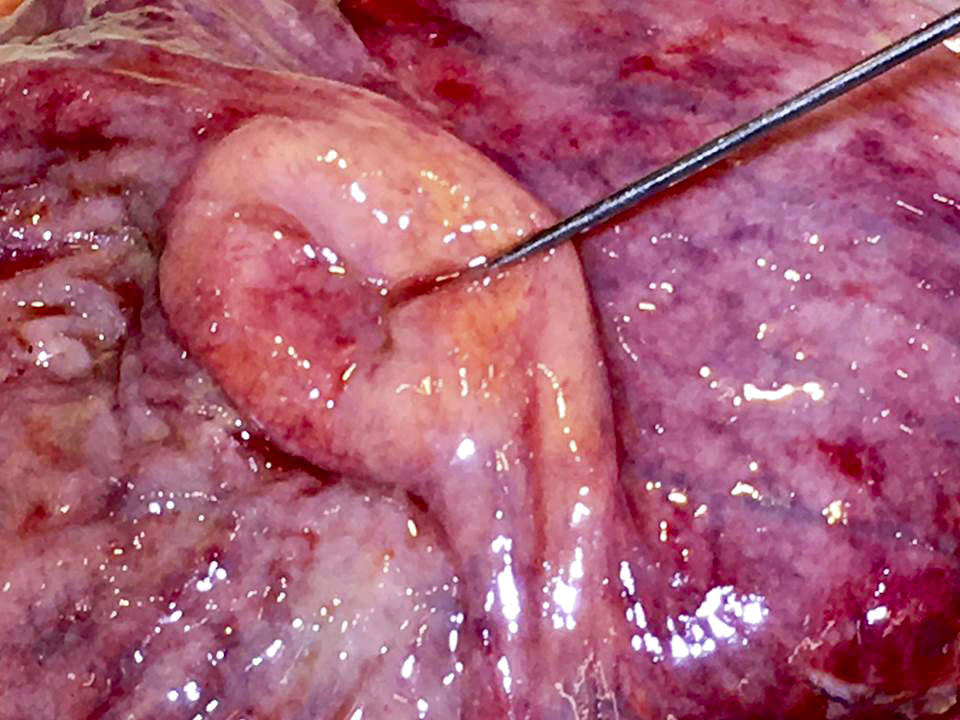
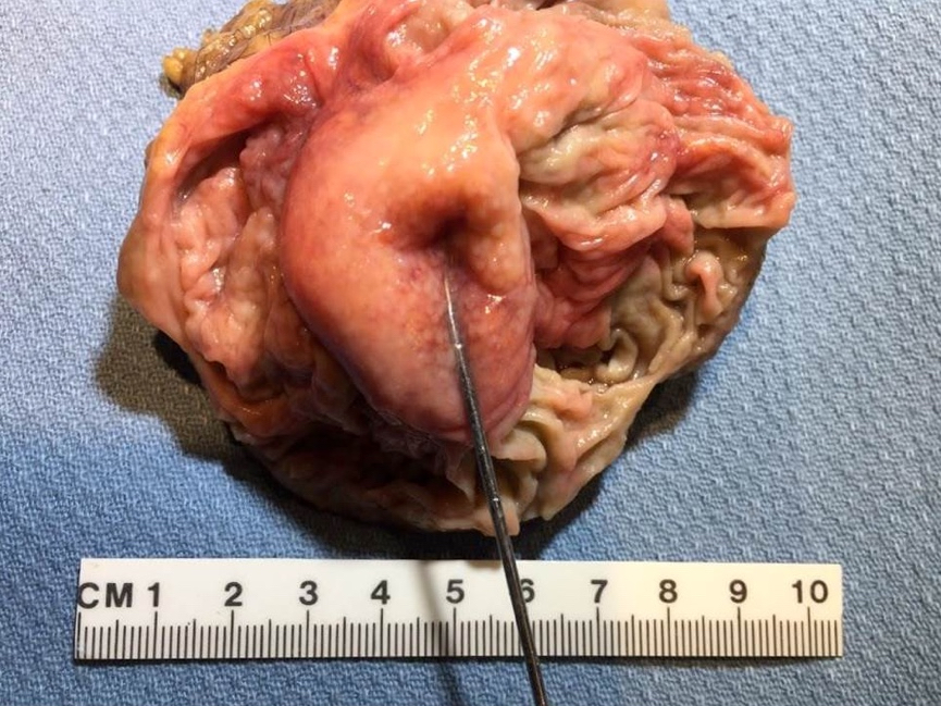
submucosal tumor
with overlying
normal mucosa

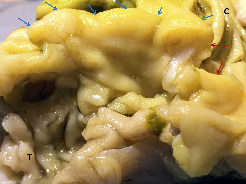
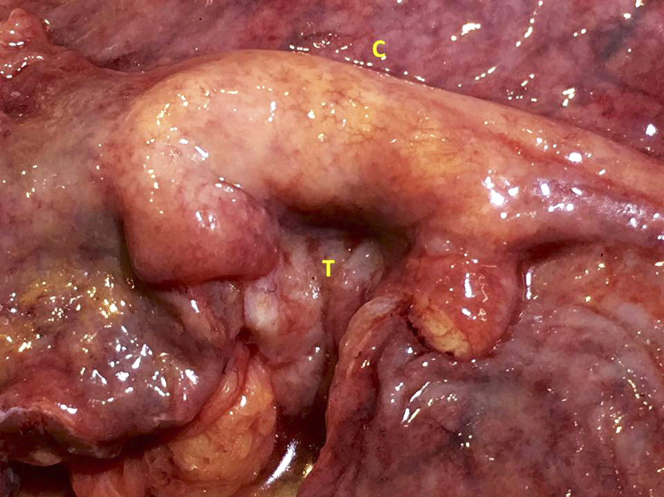
Jejunal tumor invading adherent transverse colon

Ileal tumor with ulceration
Contributed by Krutika S. Patel, M.B.B.S., M.D. and Annika L. Windon, M.D.
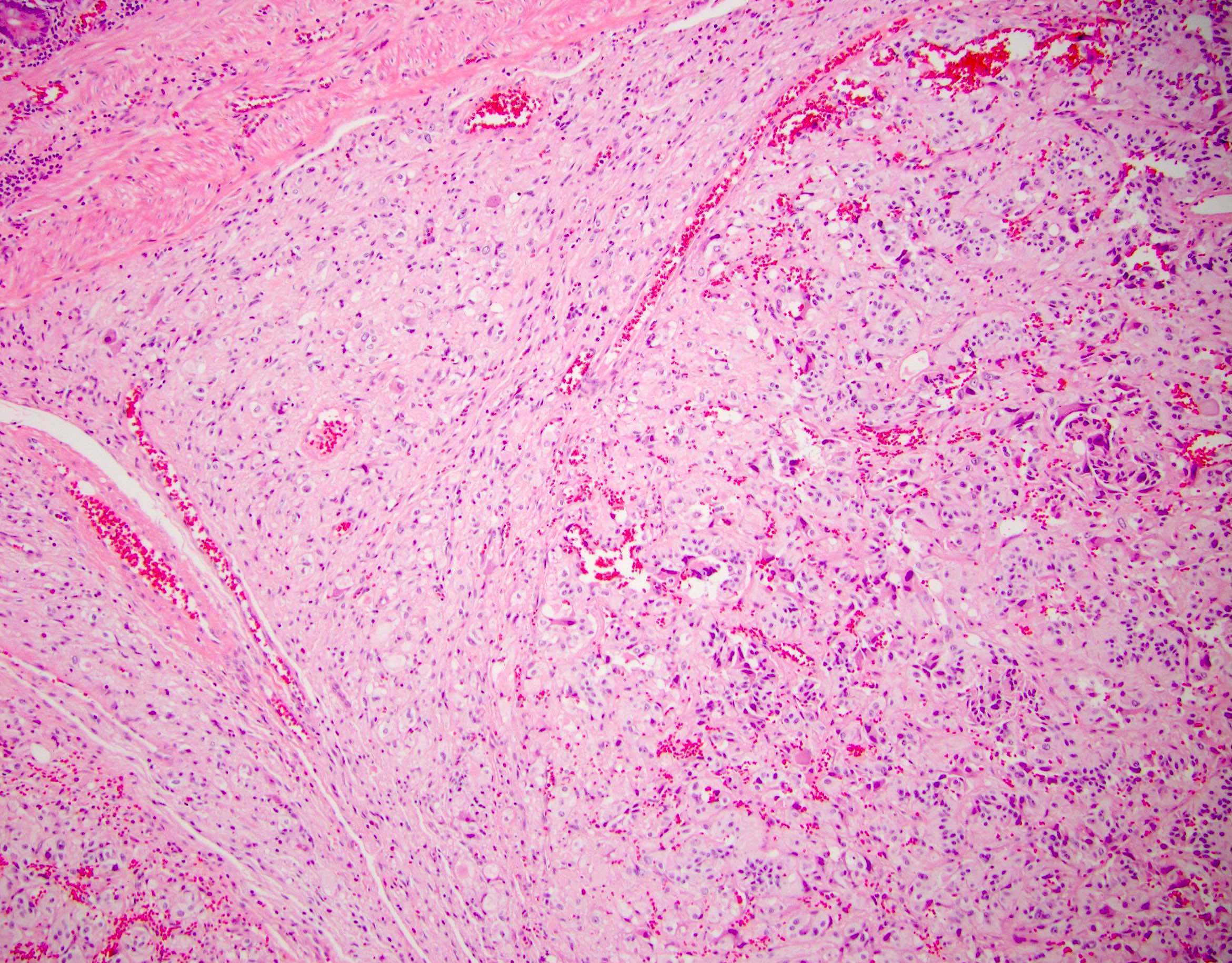
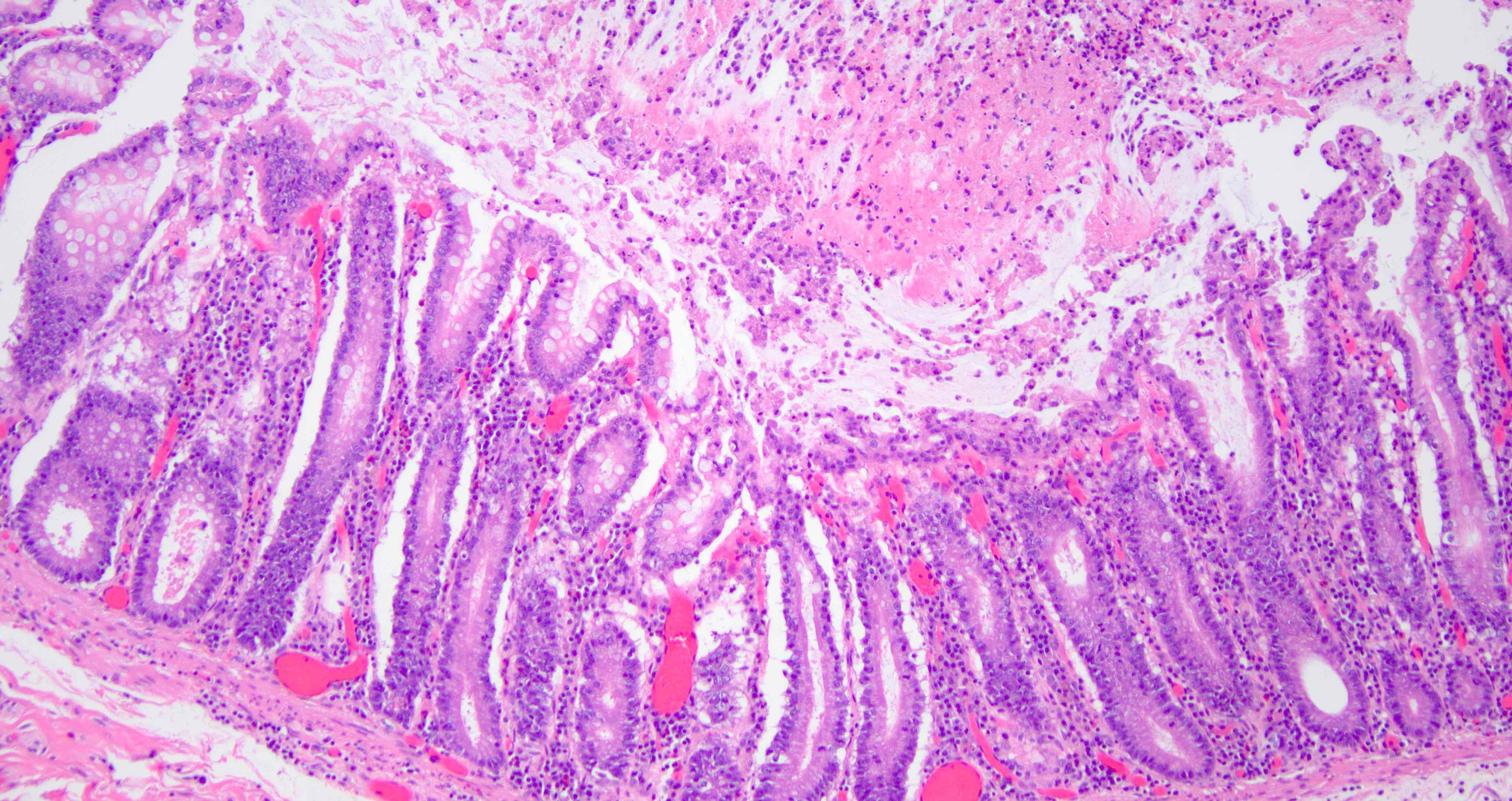
Well differentiated adenocarcinoma
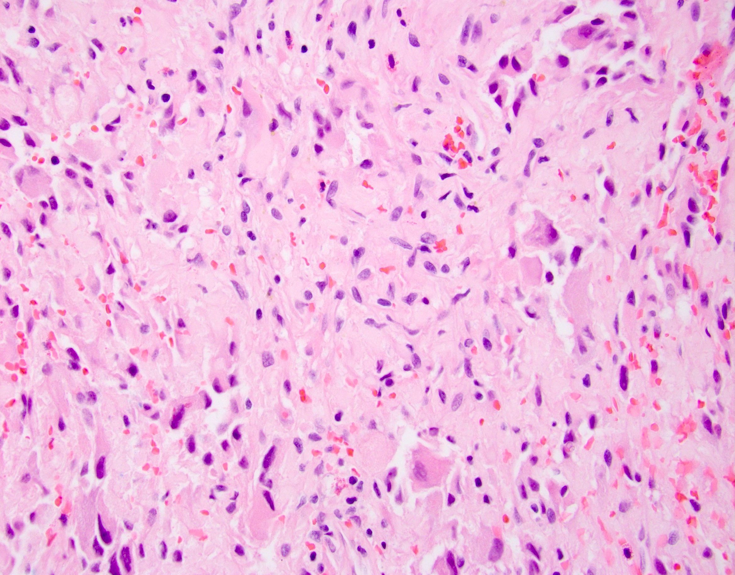
Moderately and poorly differentiated adenocarcinoma
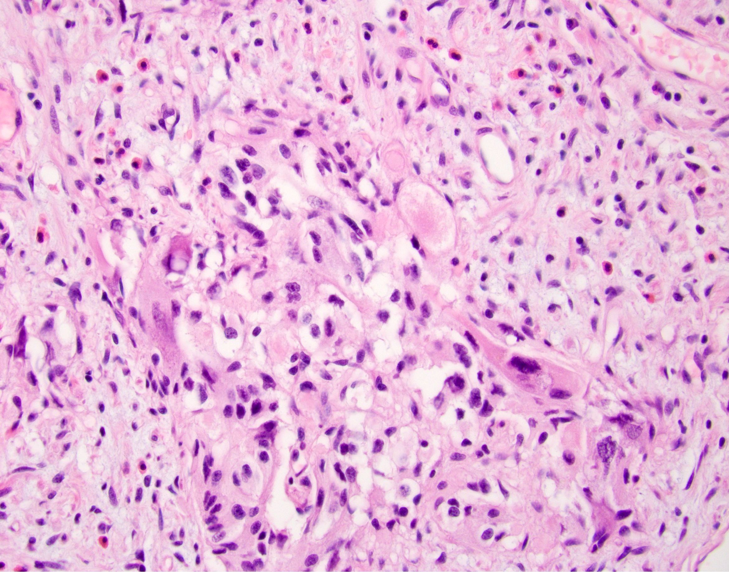
Mucinous adenocarcinoma
Lymph node metastasis
Higher stage carcinomas

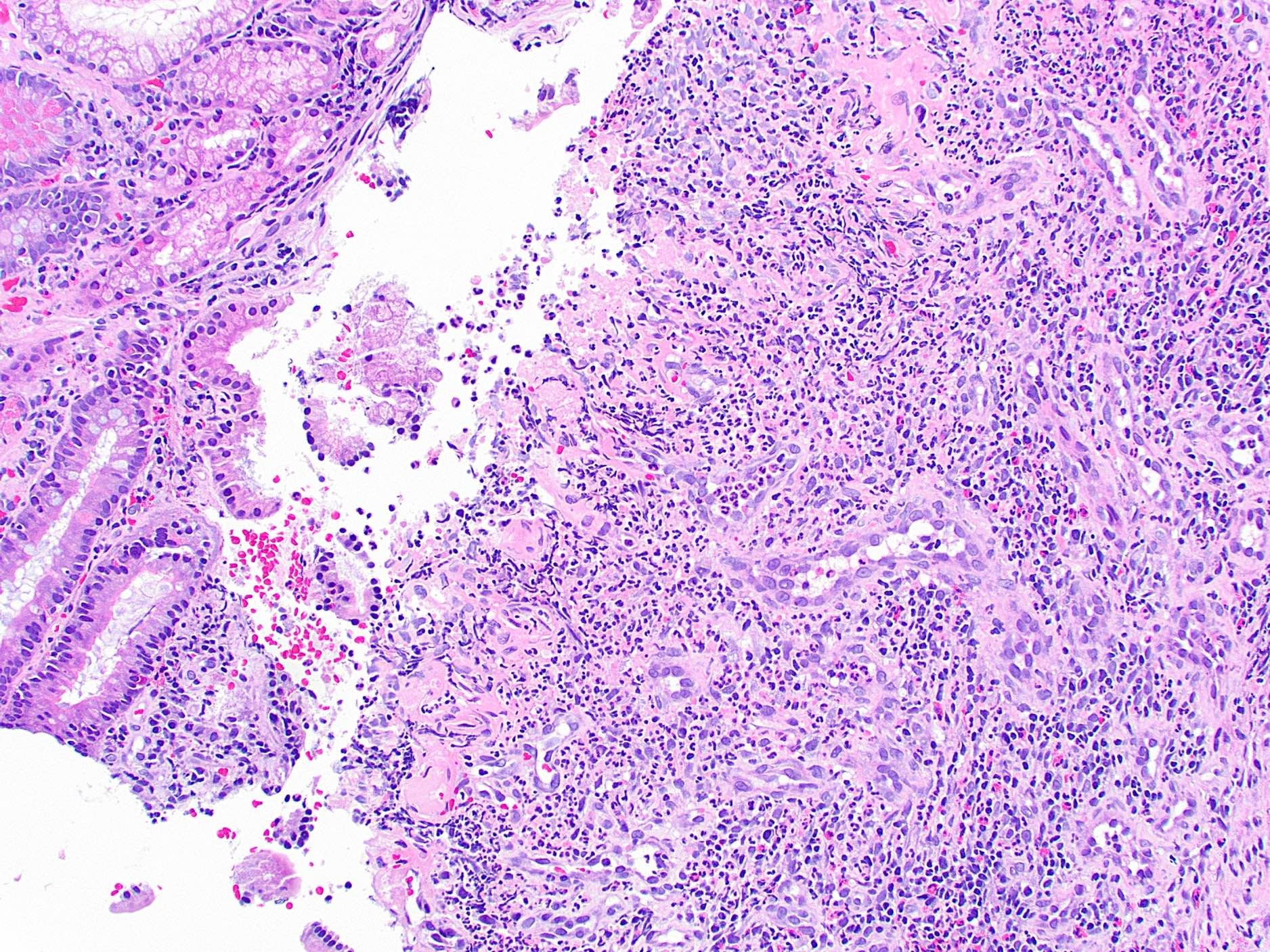
Ileal adenocarcinoma arising in Crohn's disease
Signet ring adenocarcinoma
Adenocarcinoma arising in a tubulovillous adenoma
Lymphovascular invasion
Perineural invasion
Cytokeratin 7
Cytokeratin 20
Images hosted on other servers:

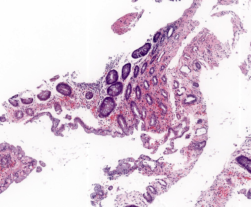
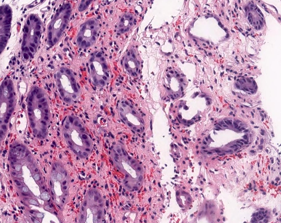
Duodenal adenocarcinoma and normal mucosa
Images hosted on other servers:


Endoscopy
Images hosted on other servers:
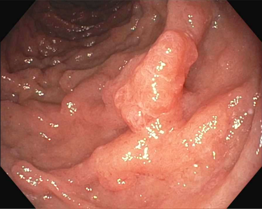
Raised lesion at ampulla of Vater
Contributed by Raul S. Gonzalez, M.D. and @Andrew_Fltv on Twitter
Ampullary adenoma with low grade dysplasia
Adenoma
Images hosted on other servers:

Ampullary adenoma with high grade dysplasia

Ampullary adenoma
Images hosted on other servers:

Pap
Images hosted on other servers:
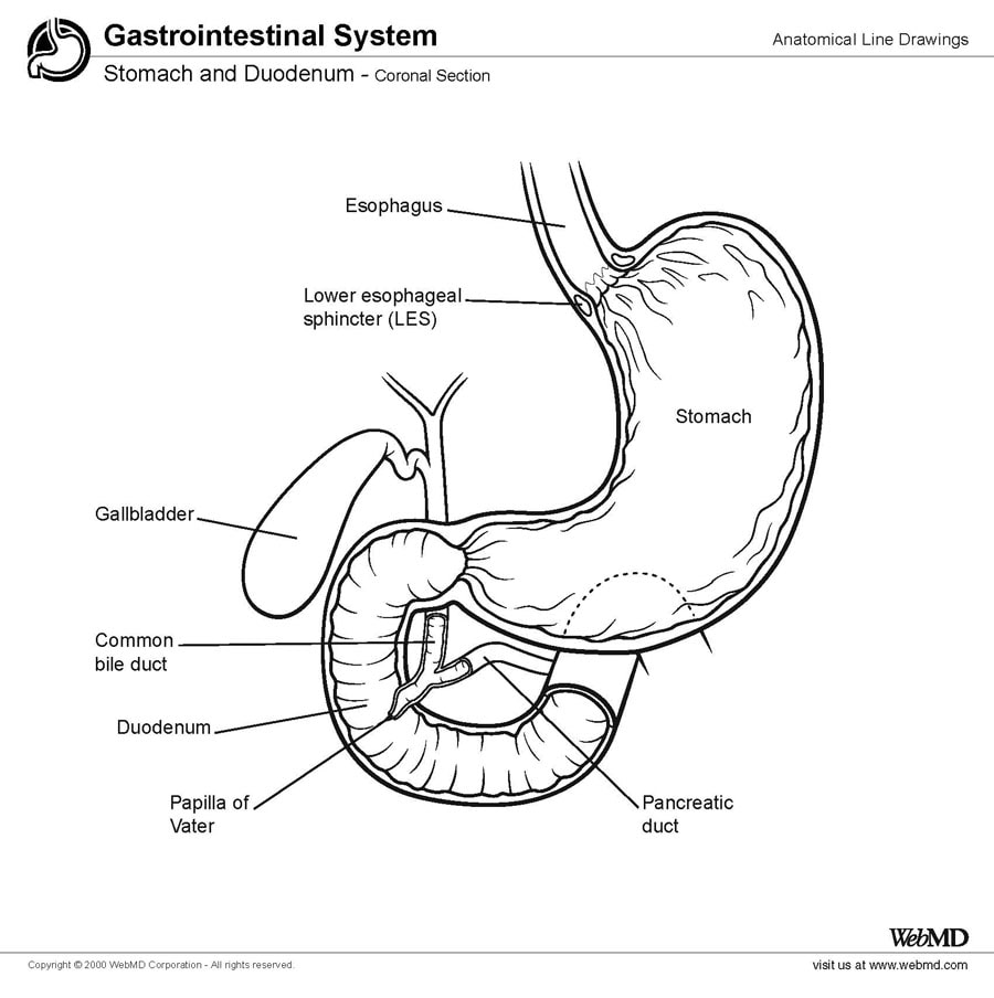
Duodenum

Local anatomy (called common duct)

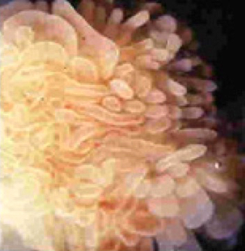
Papilla
Images hosted on other servers:


Ileum and terminal ileum
Contributed by Grigory Demyashkin, M.D., Ph.D. and Suhail Muzaffar, M.B.B.S.
6 - 8 week embryo

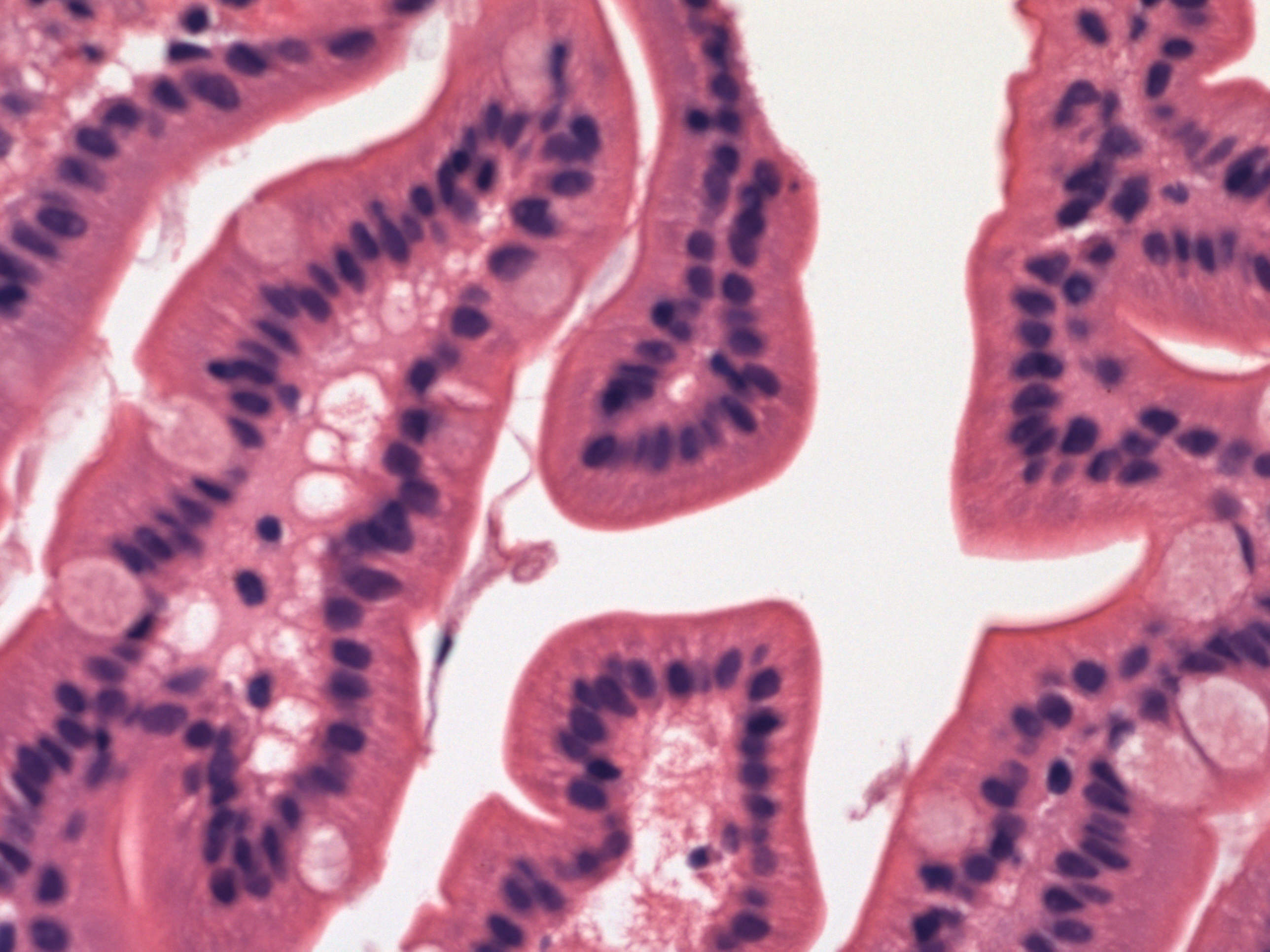
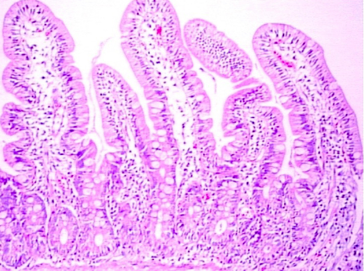
Small intestine - normal (duodenum)
Contributed by Archana Shenoy, M.D.
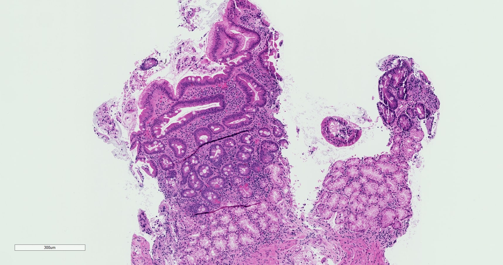
Duodenal biopsy
Colon biopsy
Images hosted on other servers:

Ulcers - site unspecified
Images hosted on other servers:

Duodenum showing cobblestone pattern
Contributed by Hany Al Khedr, M.D.
Duodenal bulb polyp endoscopy
Contributed by Divya Sharma, M.D.
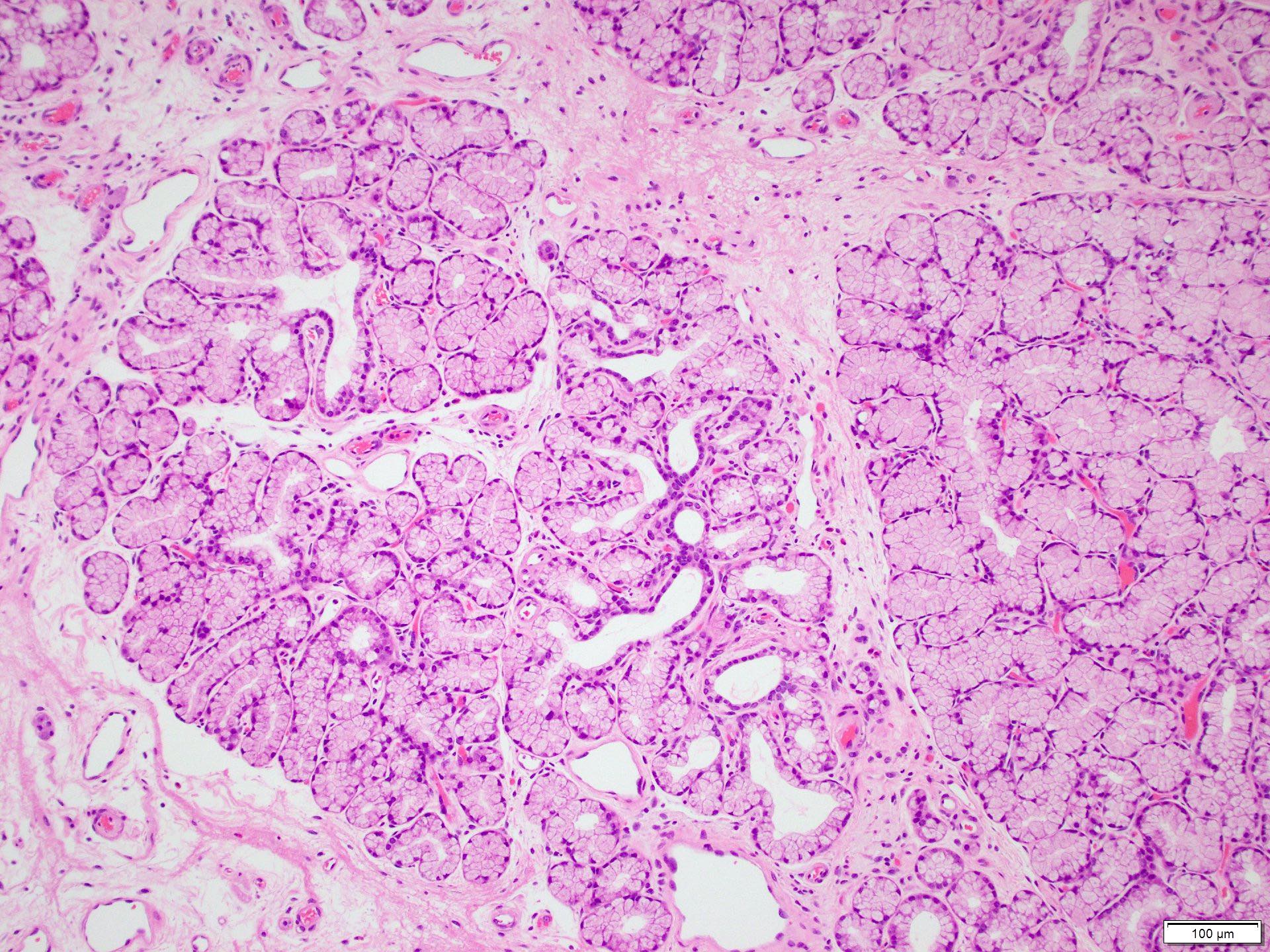
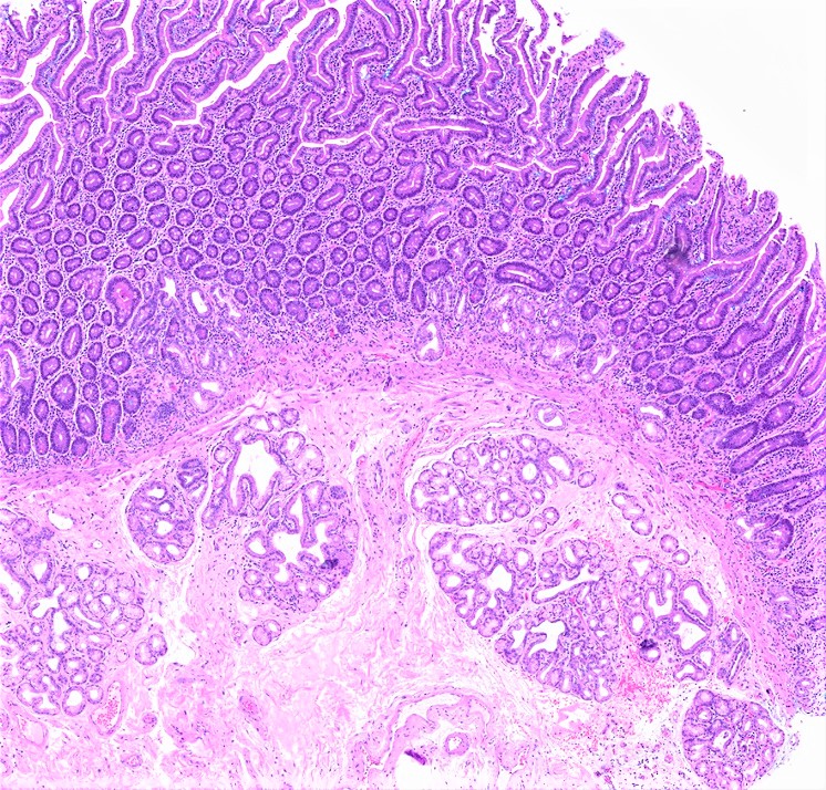
Brunner glands in submucosa
Glands within fibrous stroma
Bland cuboidal cells
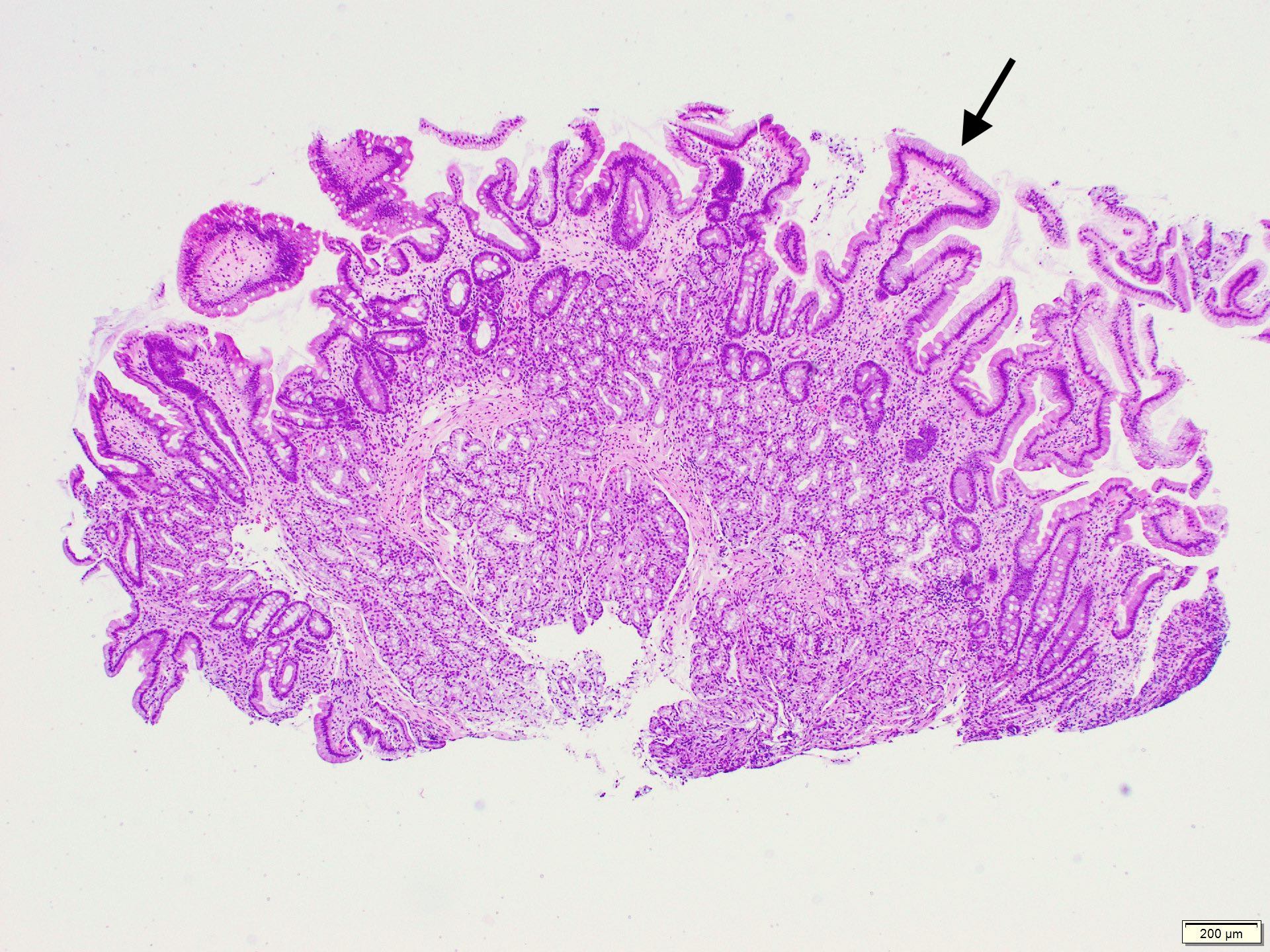
Foveolar metaplasia
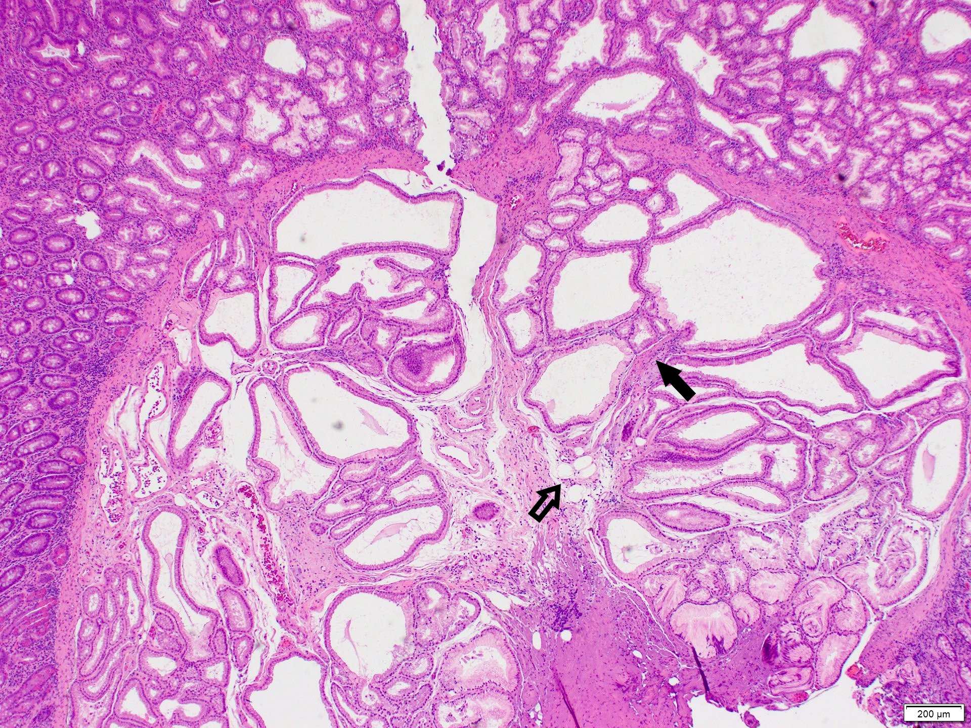
Dilated ducts
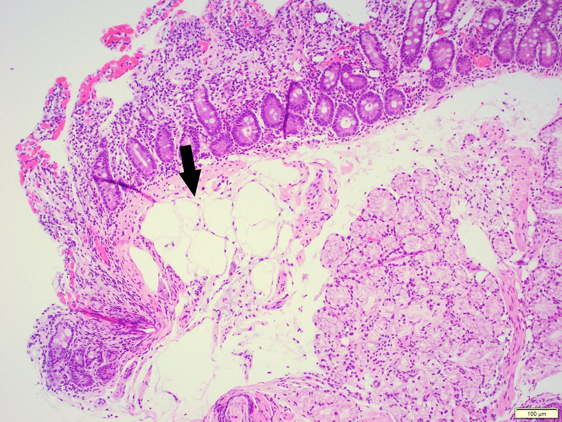
Mature adipocytes
Fibrous septa
Modified Marsh-Oberhuber classification of histologic findings in celiac disease:
| Marsh type | IEL/100 enterocytes: jejunum | IEL/100 enterocytes: duodenum | Crypt hyperplasia | Villi |
| 0 | < 40 | < 30 | Normal | Normal |
| 1 | > 40 | > 30 | Normal | Normal |
| 2 | > 40 | > 30 | Increased | Normal |
| 3a | > 40 | > 30 | Increased | Mild atrophy |
| 3b | > 40 | > 30 | Increased | Marked atrophy |
| 3c | > 40 | > 30 | Increased | Complete atrophy |
| 4 | > 40 | > 30 | Atrophic | Severe (flat) |
Images hosted on other servers:

Diagnostic flowchart
in case of suspected
gluten related disorder

Diagnostic approach of refractory disease
Contributed by Erdener Özer, M.D., Ph.D.

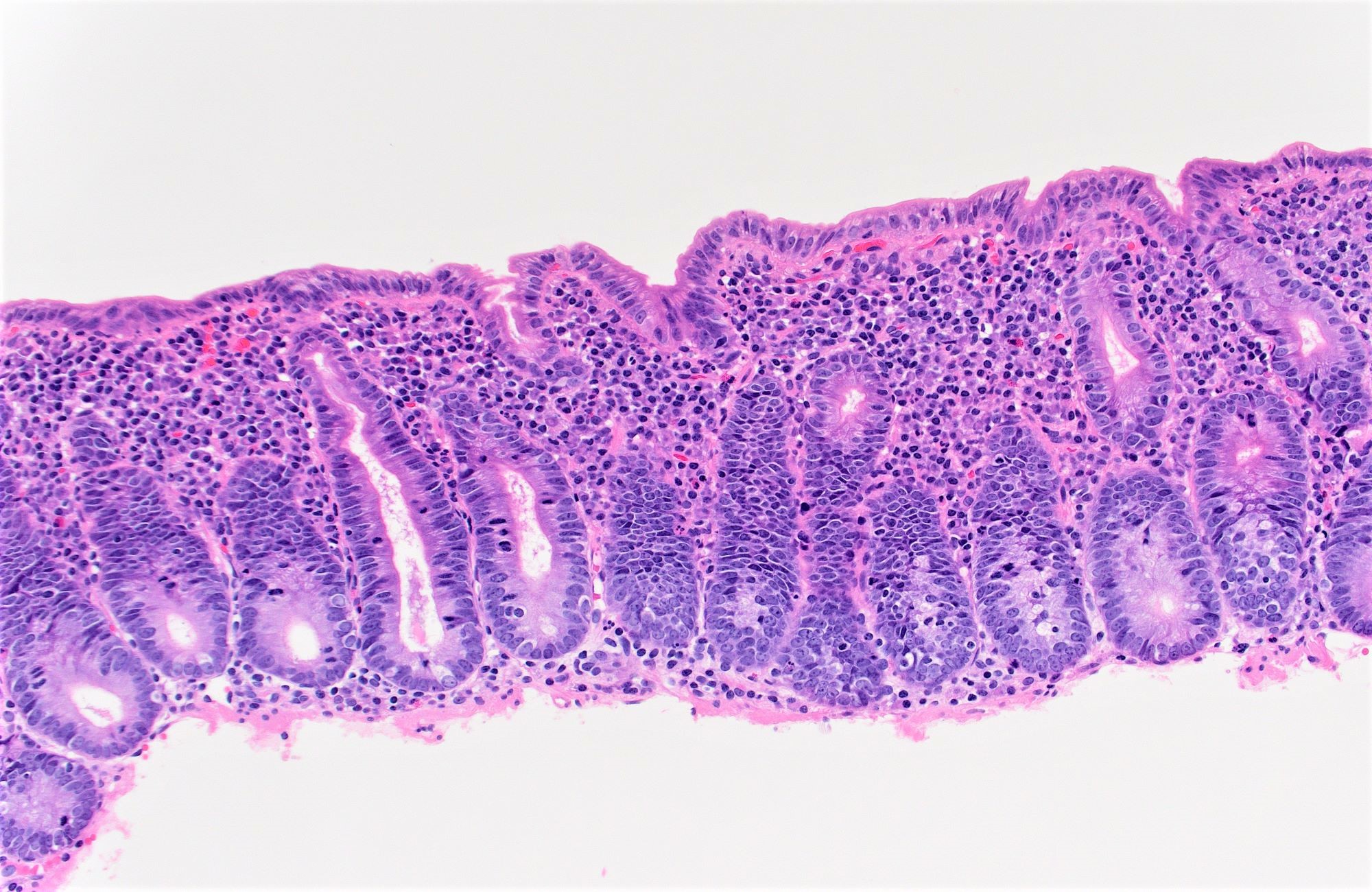
Normal versus flattened mucosa
Contributed by Erdener Özer, M.D., Ph.D., Arzu Ensari, M.D., Ph.D. and Case #127

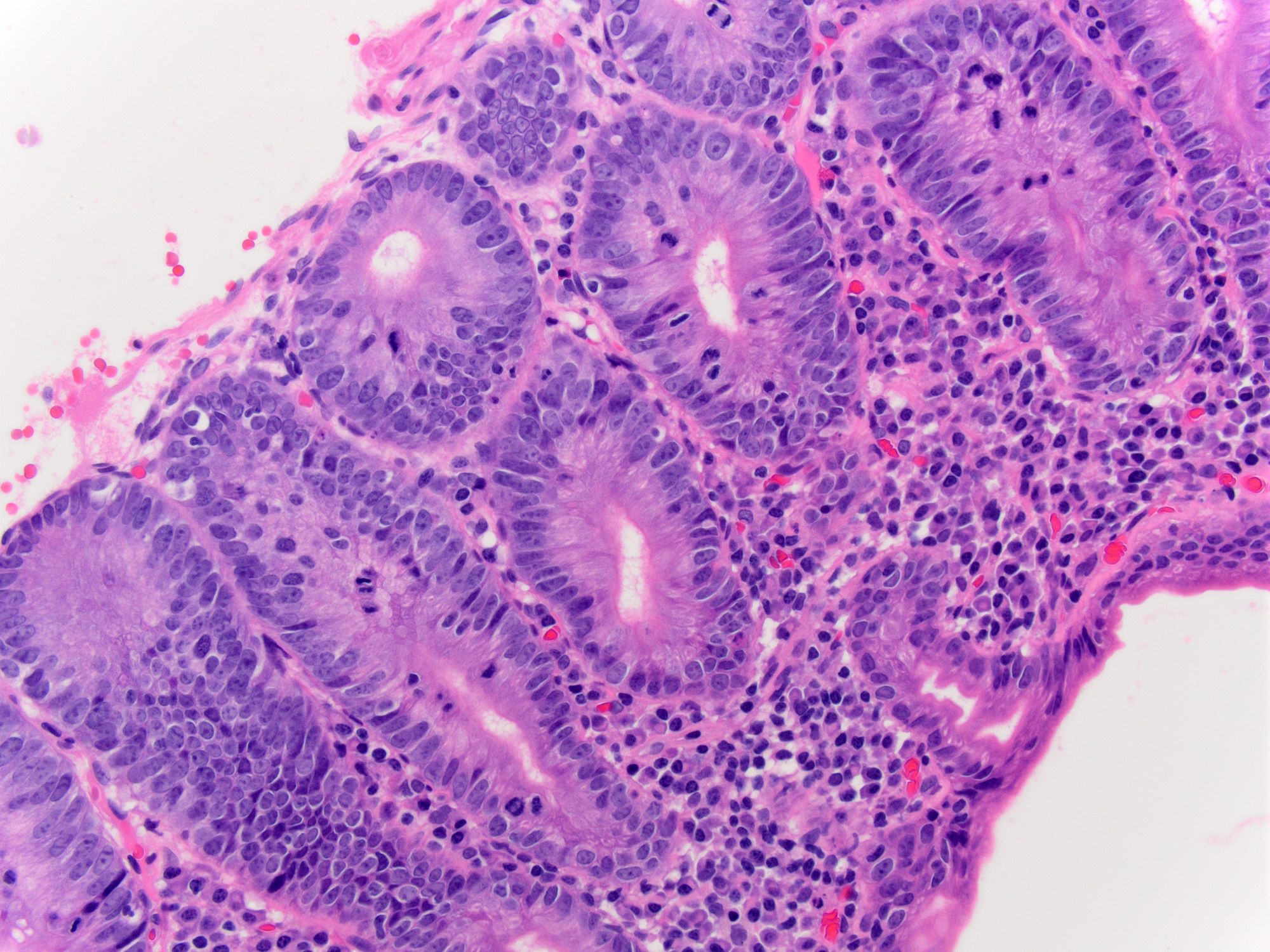
Normal versus Marsh type IIIb / IIIC
Response to gluten free diet
Collagenous sprue
56 year old woman
Images hosted on other servers:

RCD type 2
Celiac Disease: A Fairly Advanced Lecture for Primary Healthcare Providers
Case #398
H&E images
Contributed by @RaulSGonzalezMD on Twitter
Contributed by @RaulSGonzalezMD on Twitter (see original post here)">
Common variable immunodeficiency syndrome
Images hosted on other servers:

Longitudinal ulcers,
cobblestone appearance
and aphthous ulcers

Lymphoid follicles with red ring sign
Contributed by Elias Makhoul, D.O.
Small intestine with ulcer

Creeping fat

Stricture

Fistula
Images hosted on other servers:
Terminal ileum: mucosal (inflammatory) pseudopolyps
Terminal ileum: cobblestone change
Thickened bowel wall and fat wrapping
Contributed by Mary Wong, M.D., M.B.A.
Transmural inflammation with granulomas

Noncaseating granulomas
Pyloric metaplasia

Ulceration with overlying stricture
Case #240
Various images
Images hosted on other servers:


Various images
Case #240


H&E

Trichrome
Images hosted on other servers:

Jejunal diverticula
Contributed by Raul S. Gonzalez, M.D.
Endoscopy
Images hosted on other servers:

Acute duodenal ulceration
Contributed by Raul S. Gonzalez, M.D.
Gastroduodenal junction resection
Contributed by Raul S. Gonzalez, M.D.
Granulation tissue
Adjacent mucosa
Fibrinous ulcer
Helicobacter infection in duodenum
Severe duodenal peptic ulcer
Images hosted on other servers:

Mucin filled cysts (PAS diastase)
Images hosted on other servers:

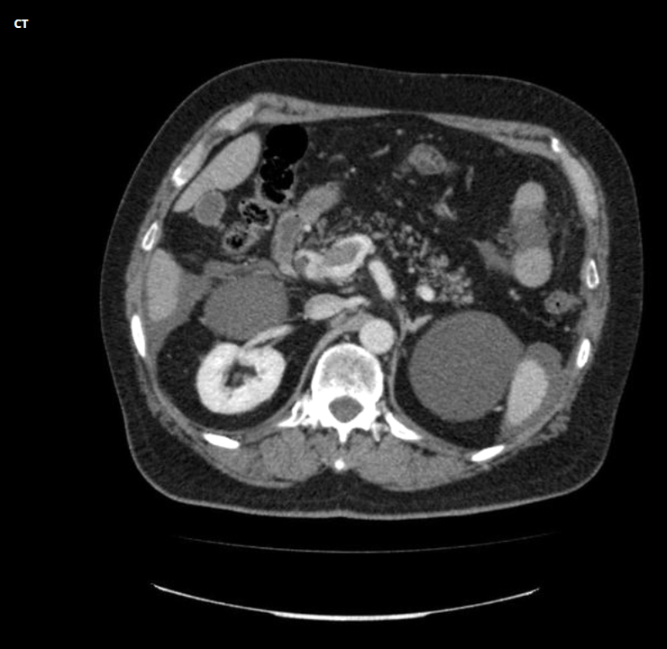
CT scan
Images hosted on other servers:

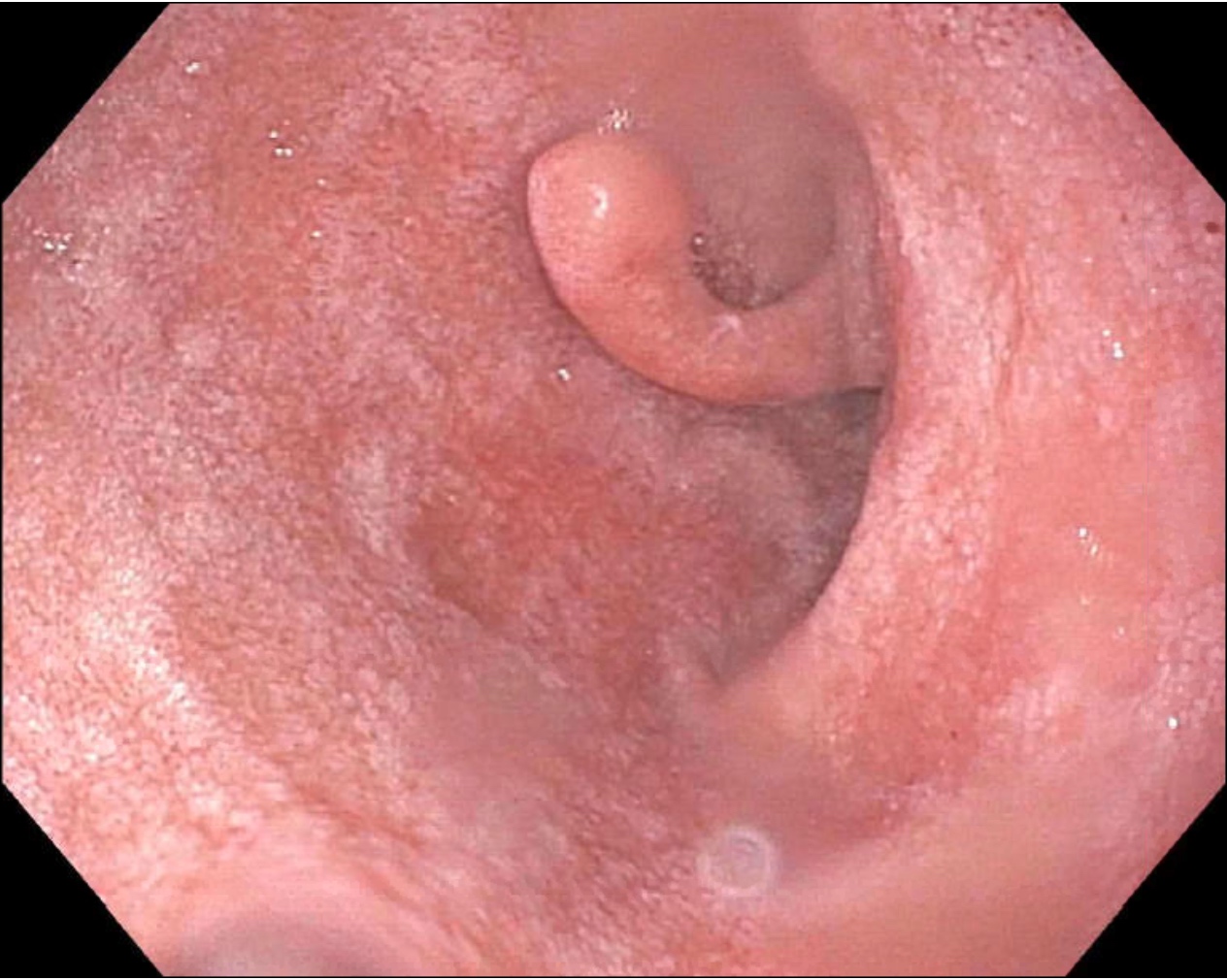
Periampullary submucosal tumor
Images hosted on other servers:

Tumor near ampulla
Contributed by Raul S. Gonzalez, M.D.

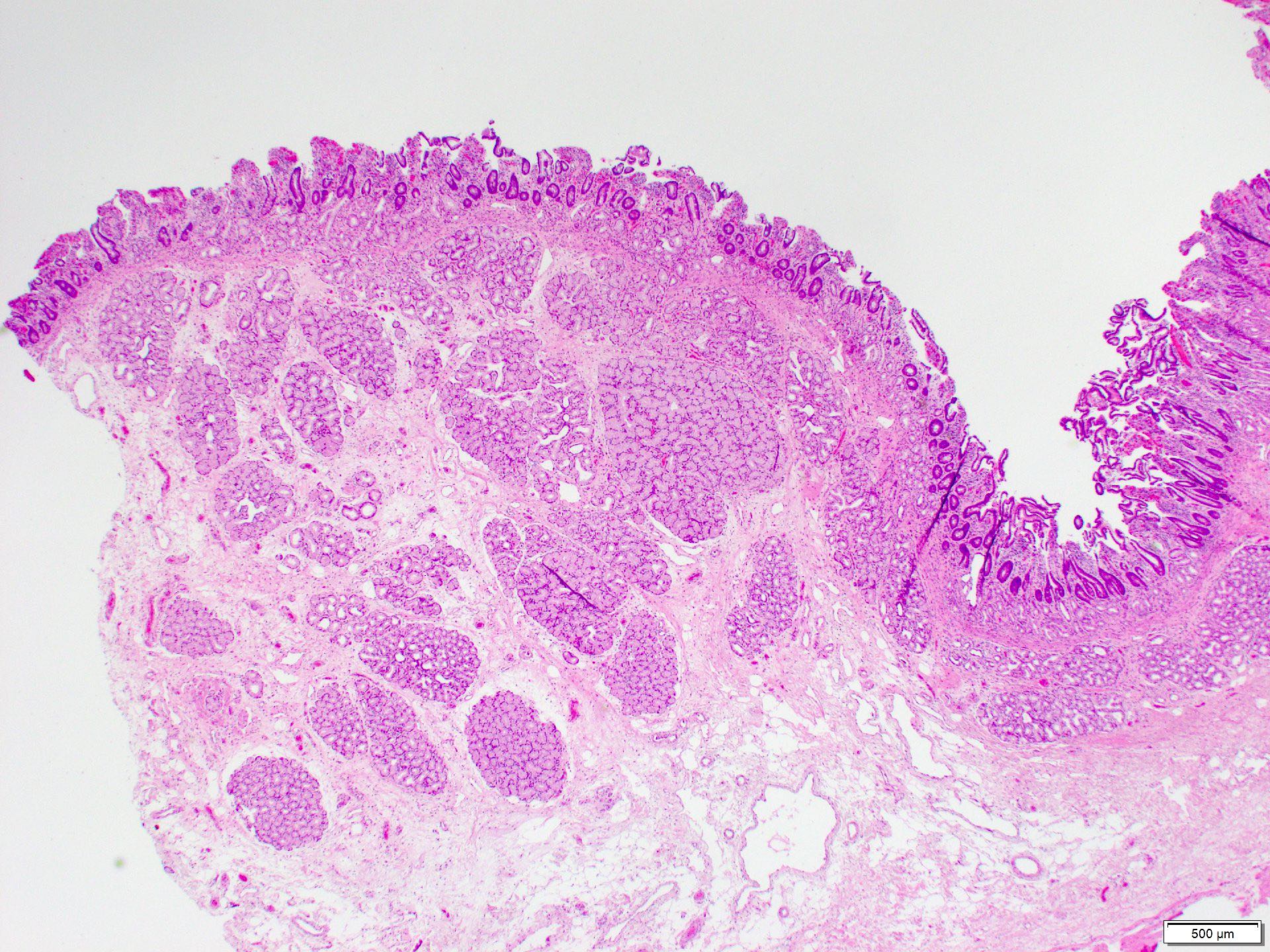
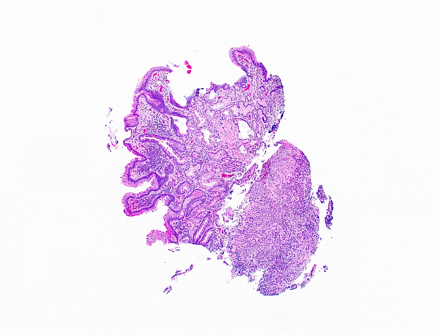
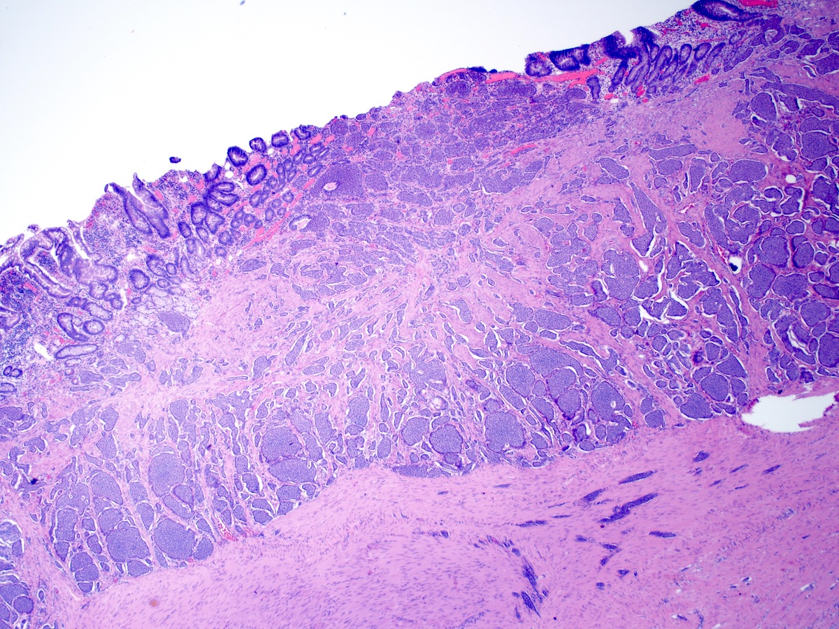
Submucosal tumor
Nests and trabeculae

Epithelioid cells
Spindled cells
Ganglion cells
Images hosted on other servers:

Life cycle
Images hosted on other servers:

Duodenal giardiasis on endoscopy
Contributed by Centers for Disease Control and Prevention
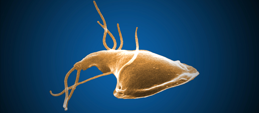
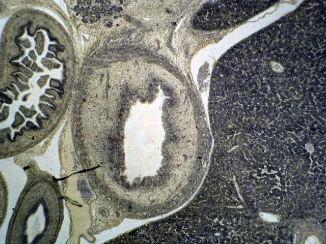
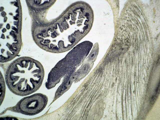
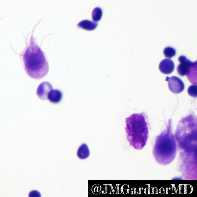
Giardia
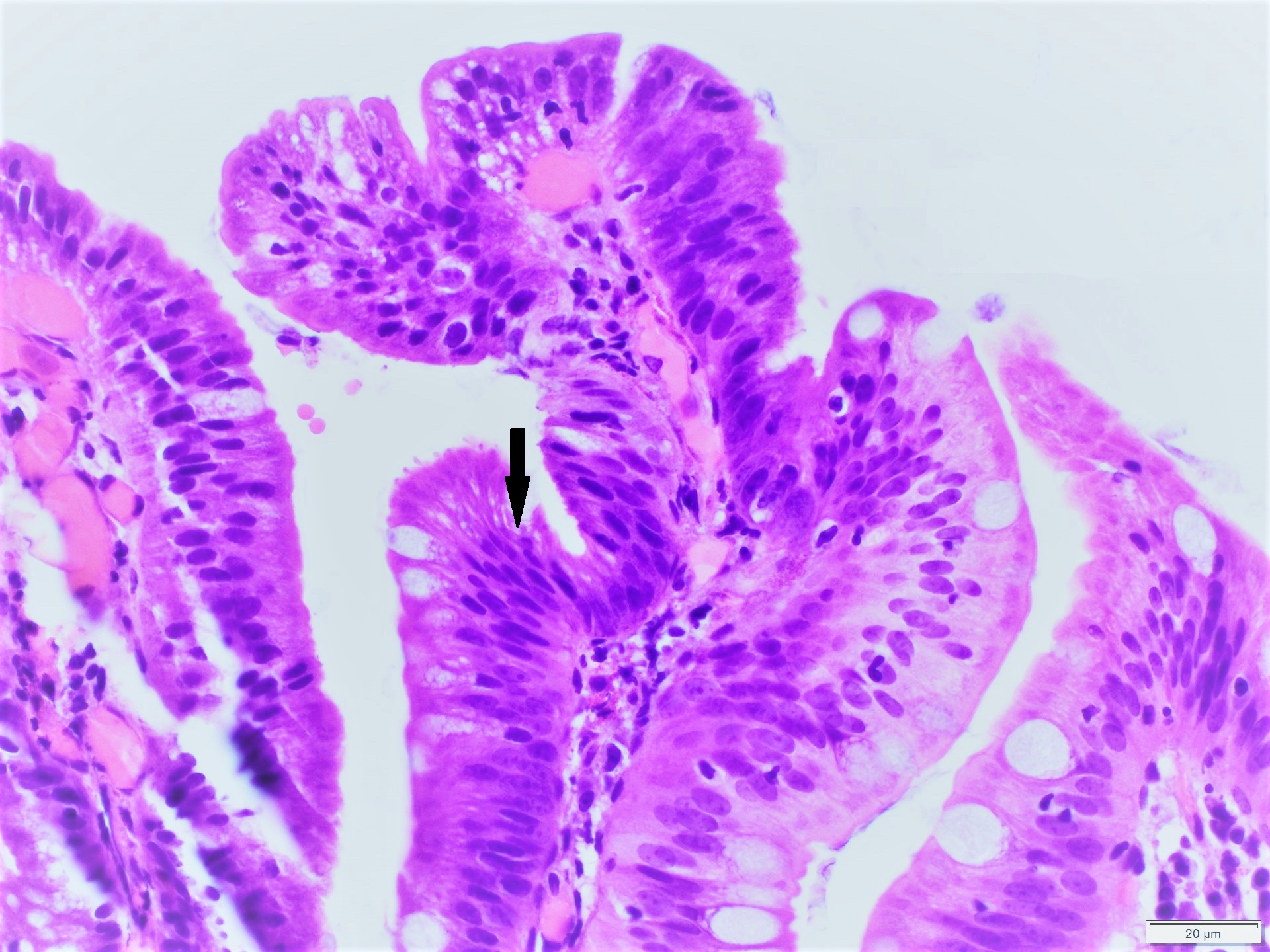
Contributed by Iresha Vithanage, M.B.B.S., M.D., Saroona Haroon, M.B.B.S., Bobbi Pritt, M.D.,
Emily Fernholz, MLS (ASCP), Suhail Muzaffar, M.B.B.S. and Bilal Karim Siddiqui, M.D. (Case #157)
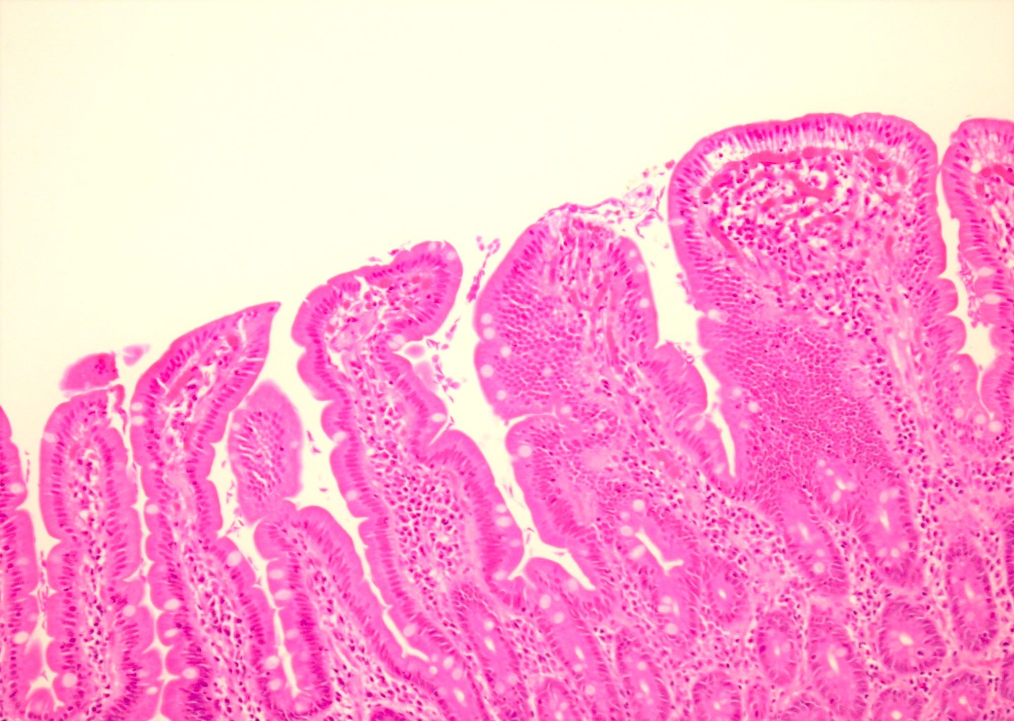
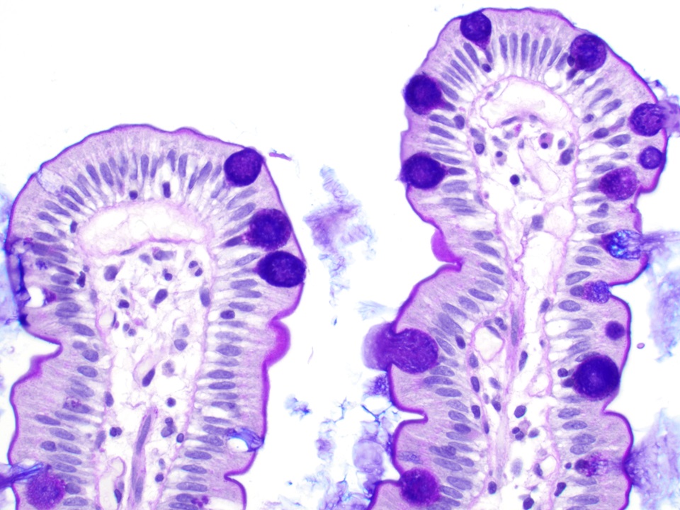
Normal villous morphology
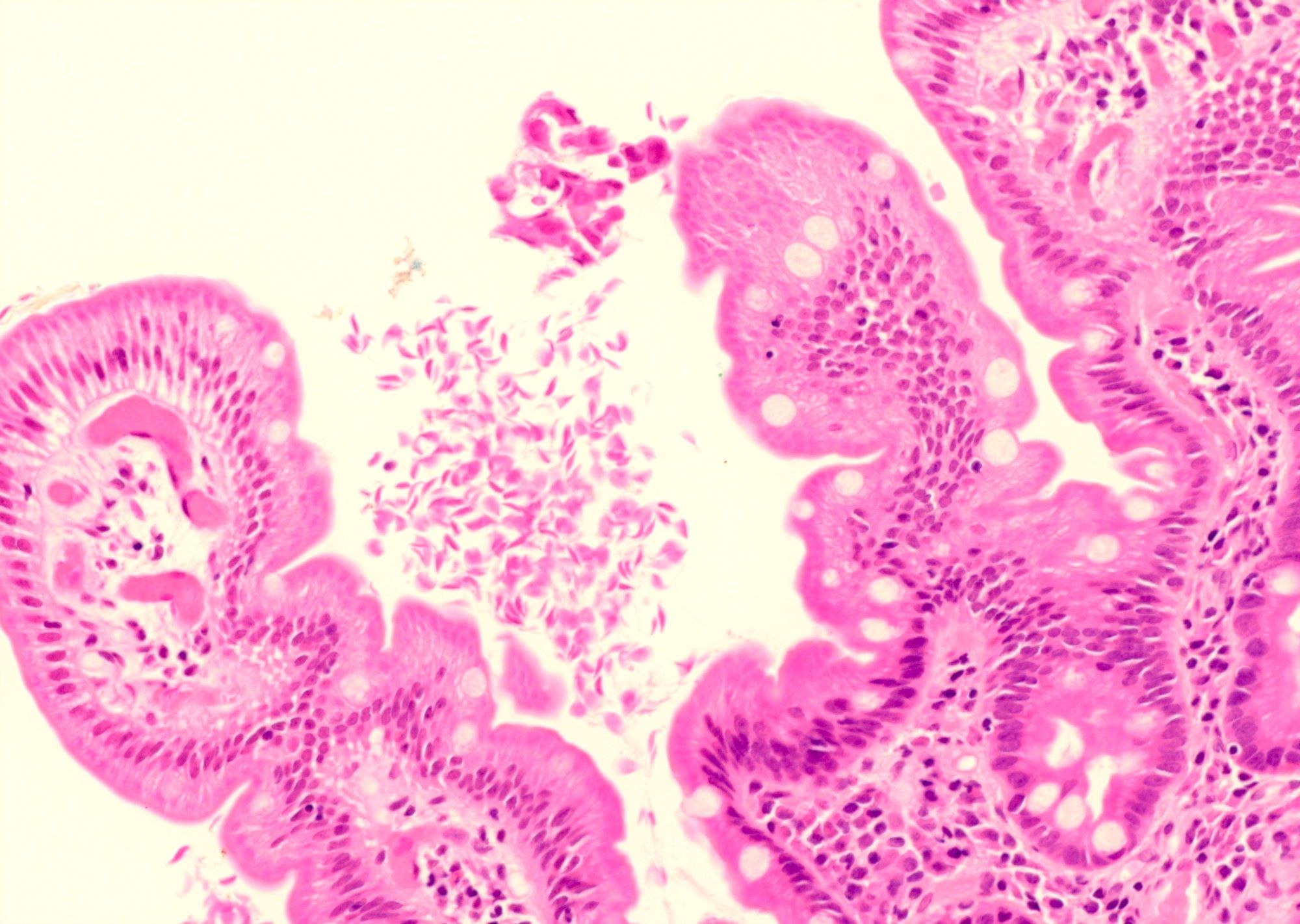
Giardia organisms in intervillous spaces
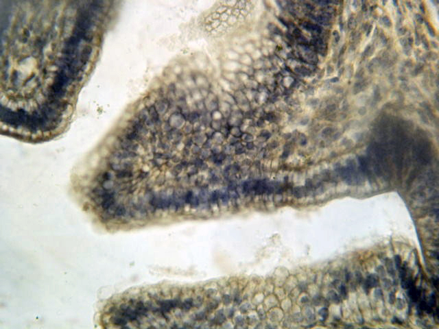
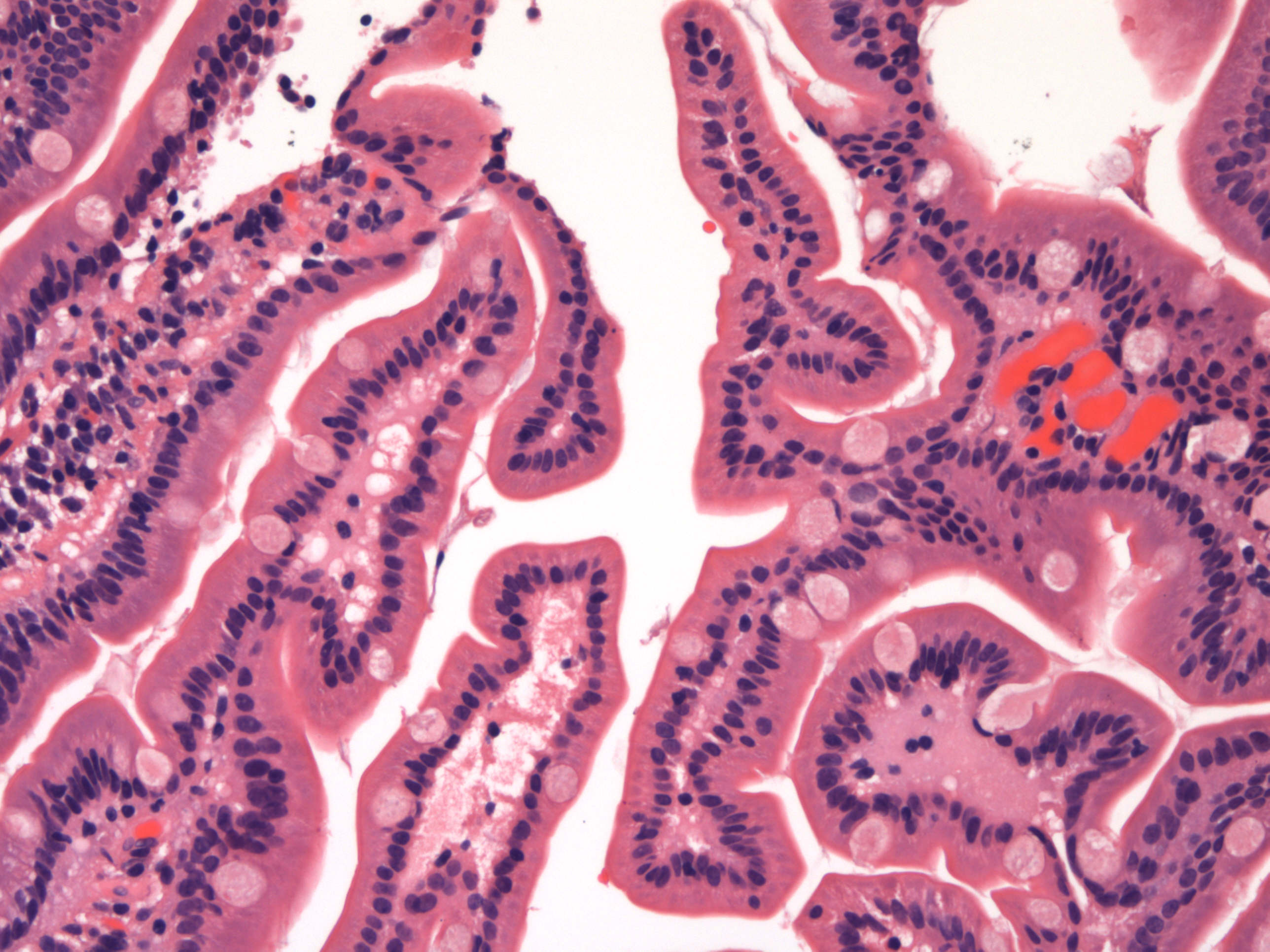
Kite and pear shaped Giardia
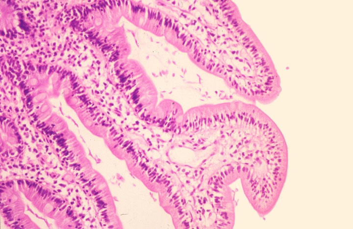
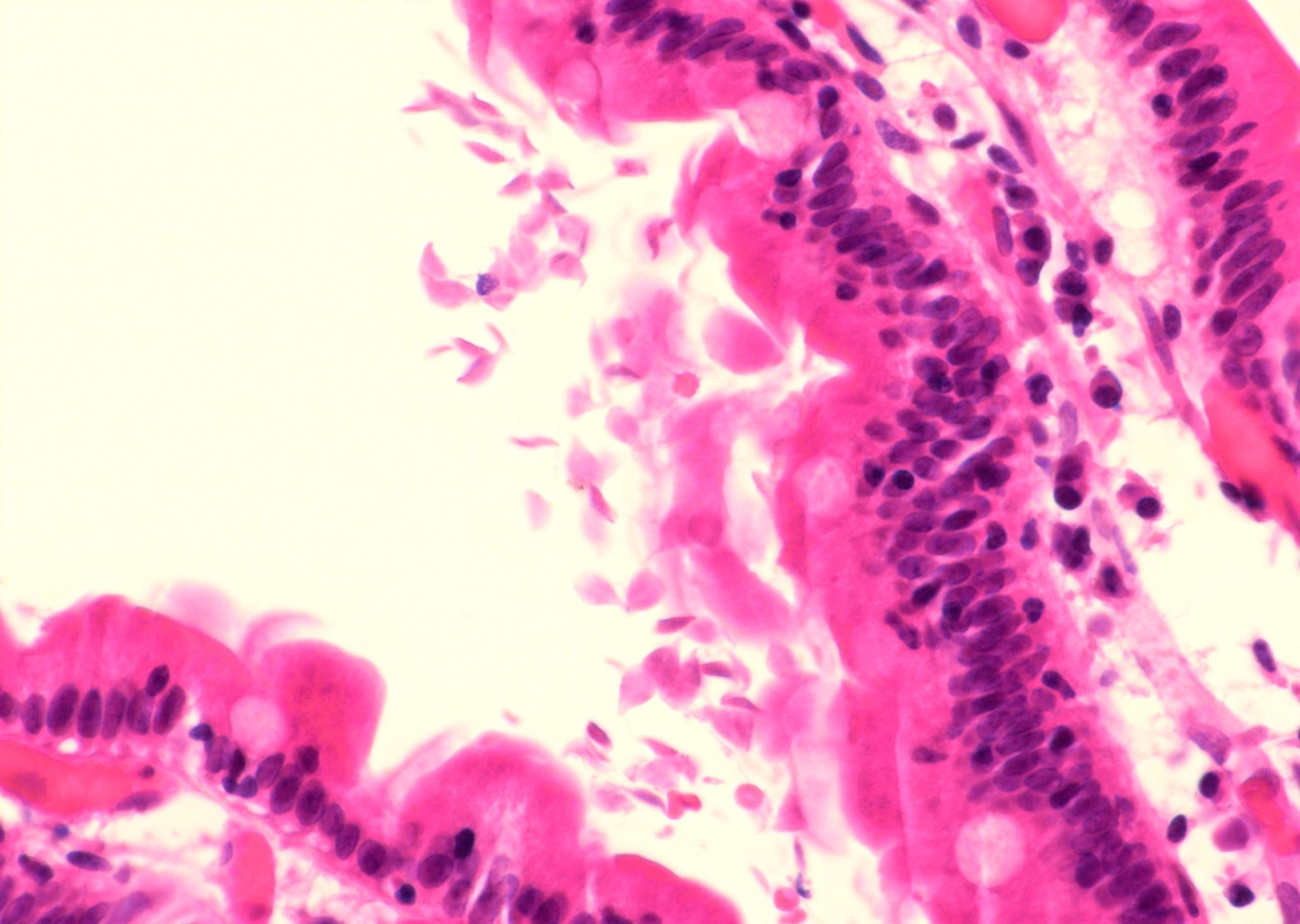
Giardia trophozoites
along surface of
foveolar epithelial cells
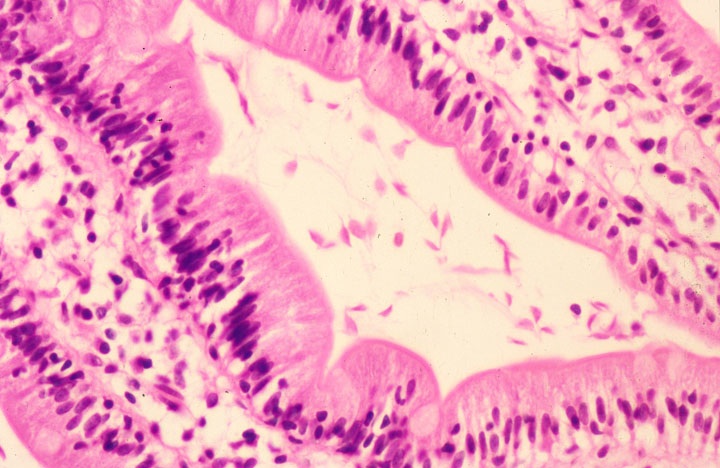
Sickle shaped trophozoites
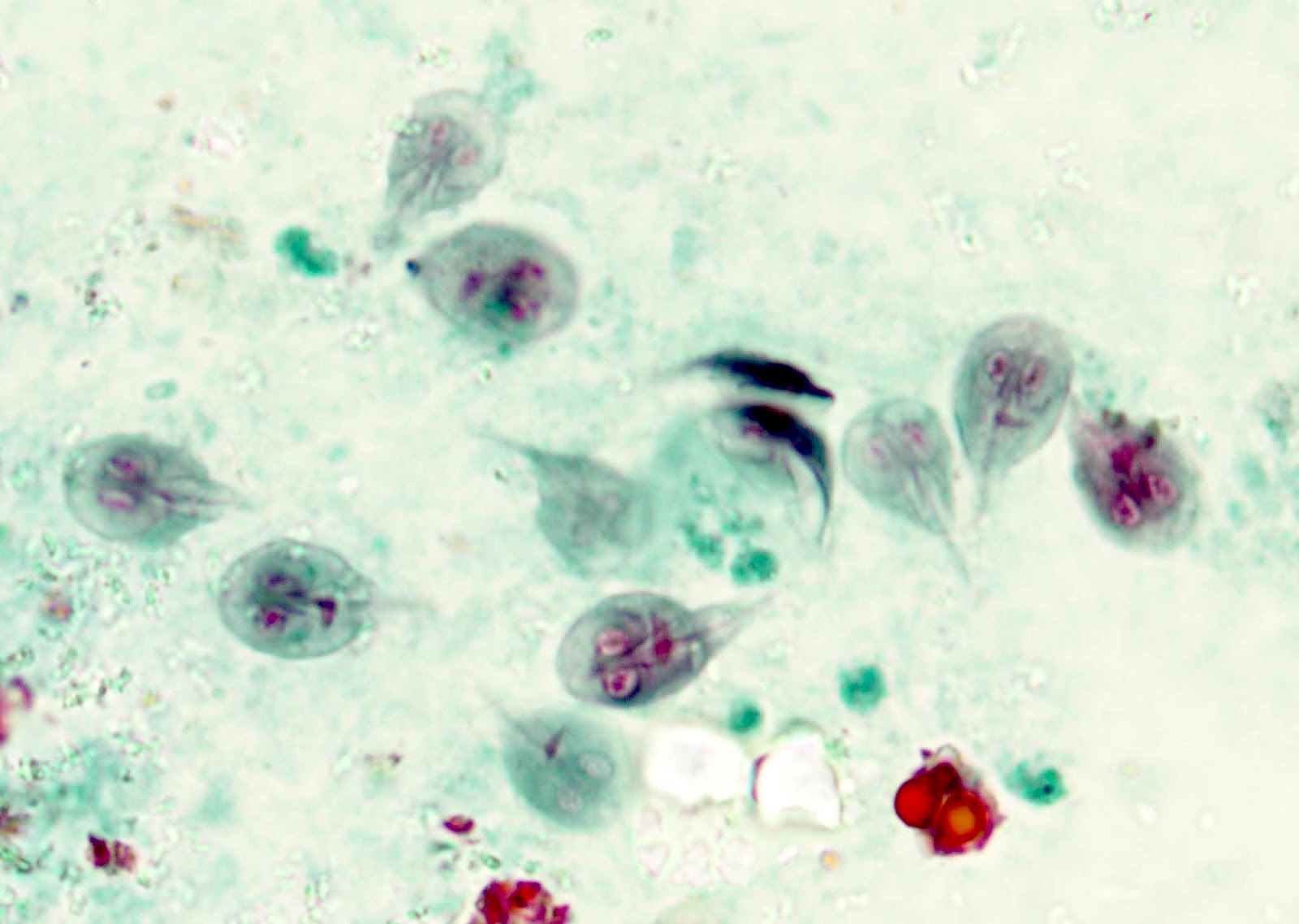
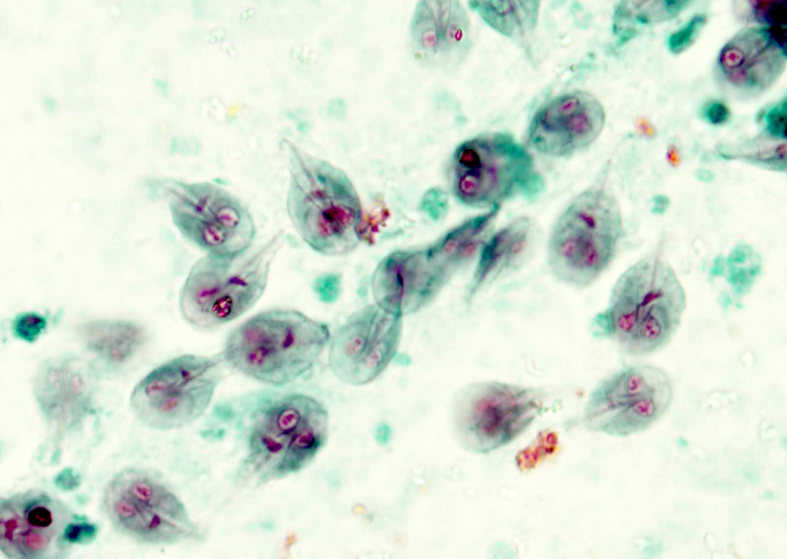
Trichrome stained stool specimen, Giardia duodenalis

Giardia trophozoites
Giardiasis tutorial
Images hosted on other servers:

Guidelines for risk assessment of primary GIST
Images hosted on other servers:

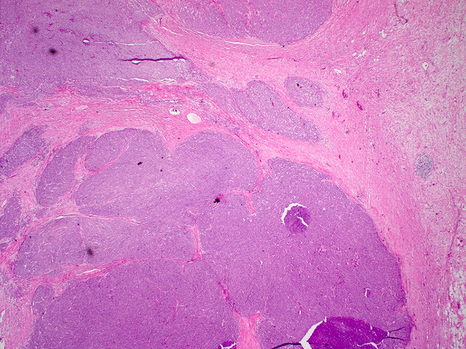
Duodenal GIST

Jejunal GIST

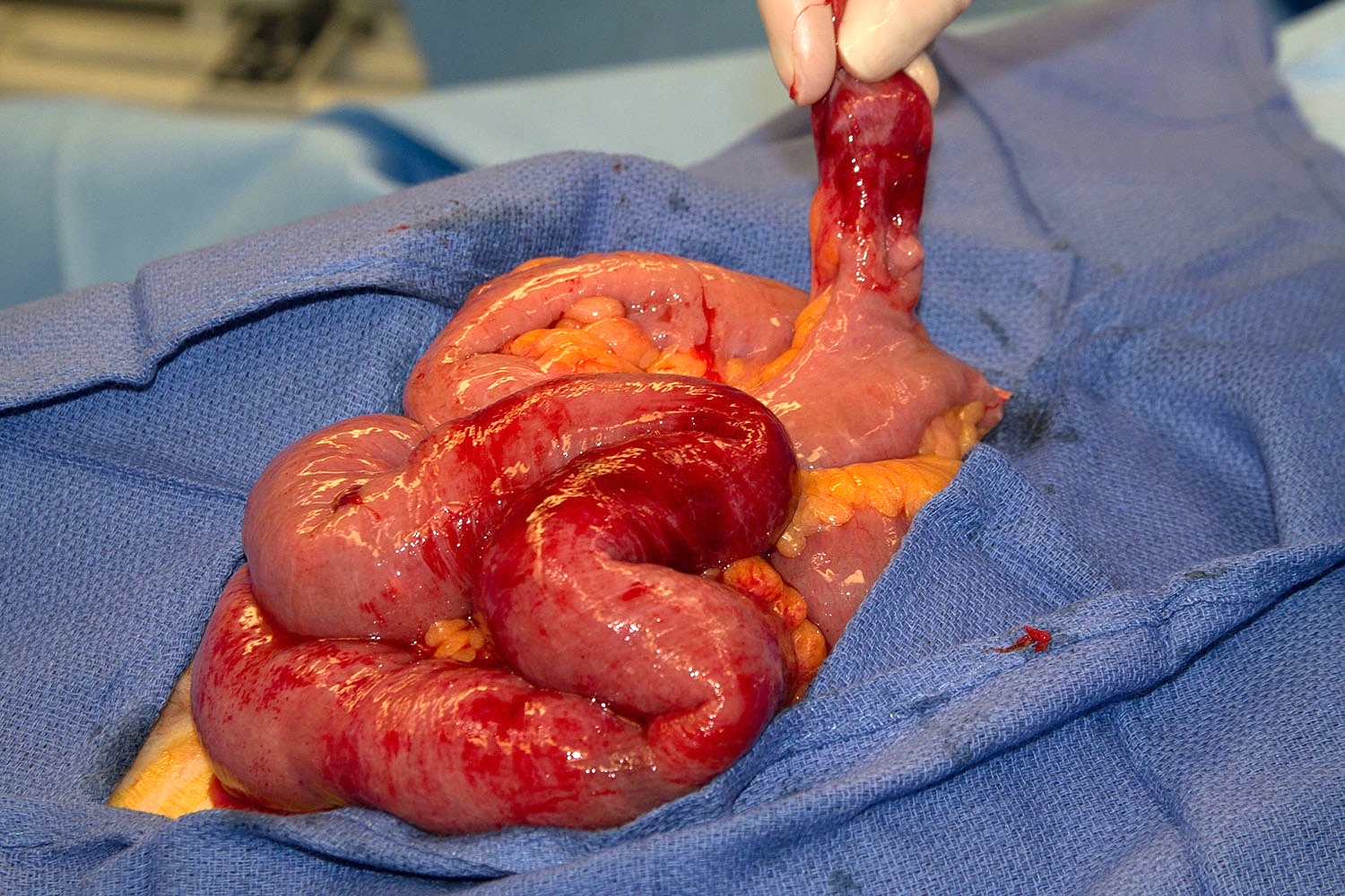
Ileoileal intussusception - small bowel GIST
Images hosted on other servers:

Small bowel GIST

Endoscopy of ulcerated mass
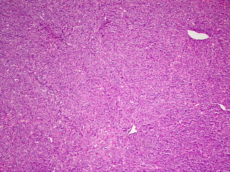
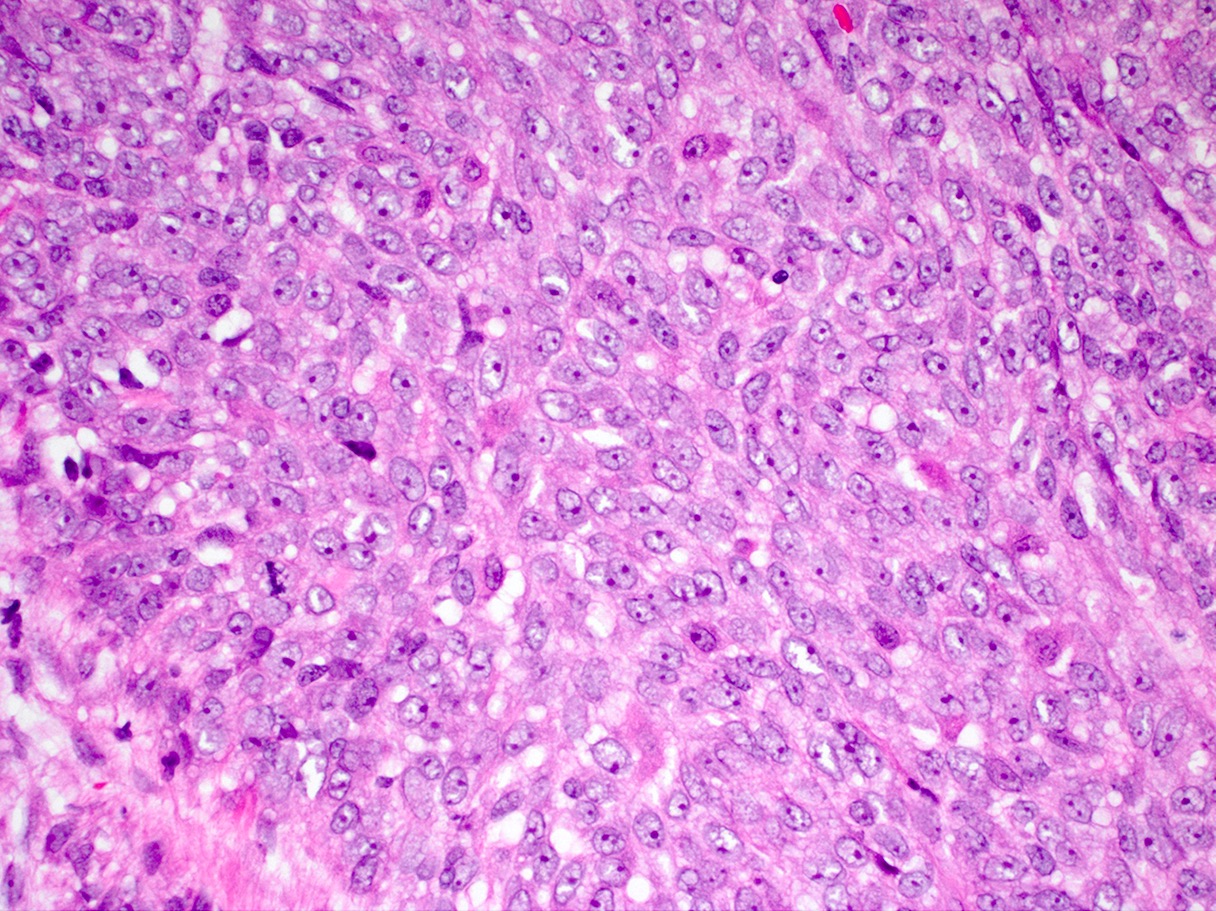
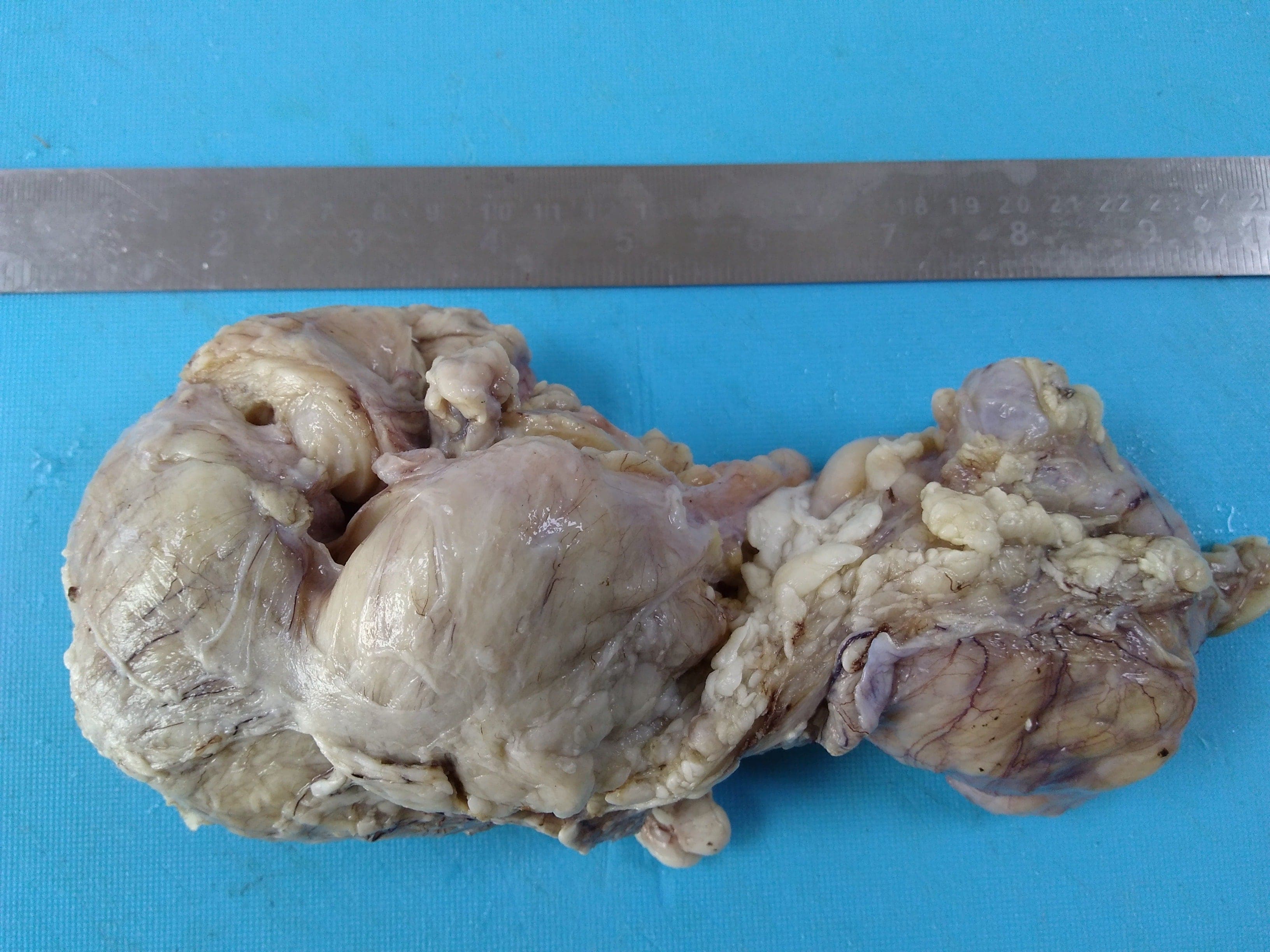
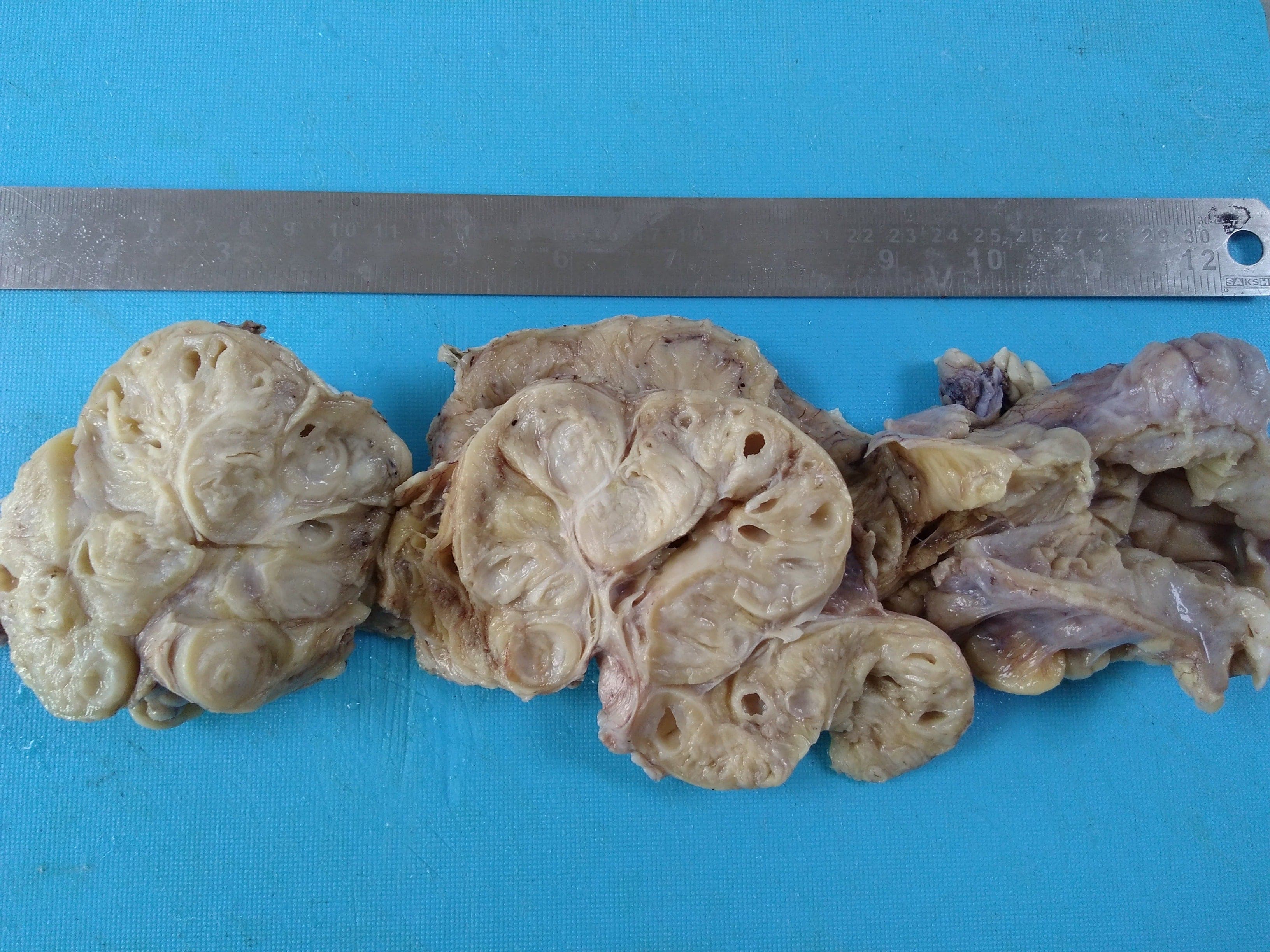
Contributed by Shefali Chopra, M.D.
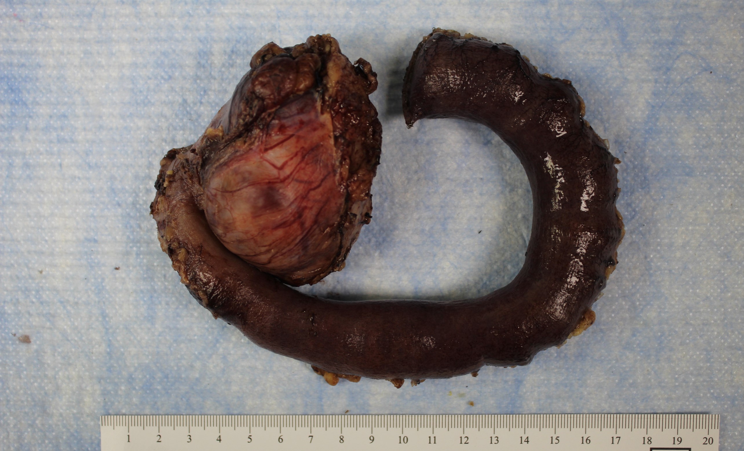
Adherent small bowel
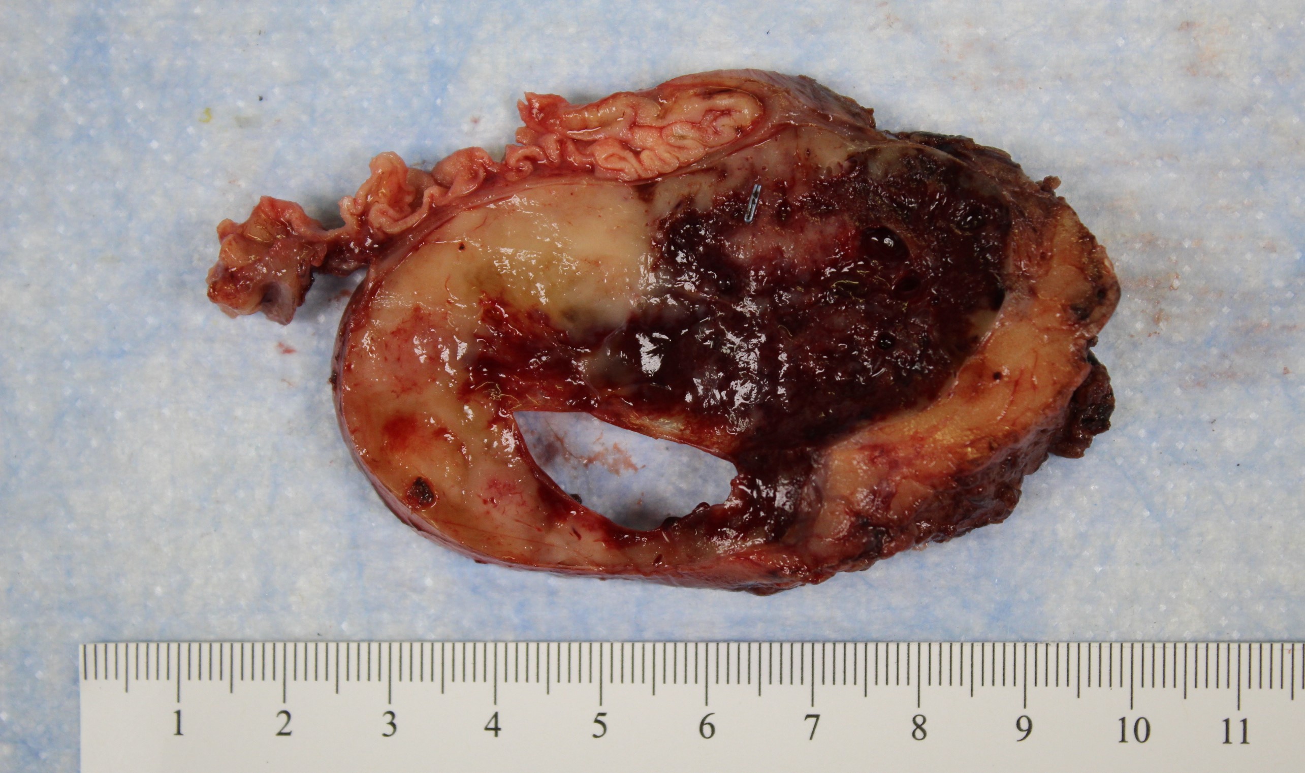
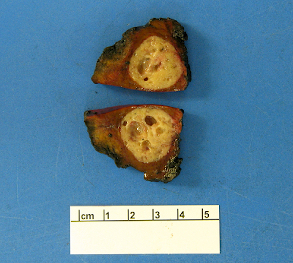
Hemorrhage & cystic change
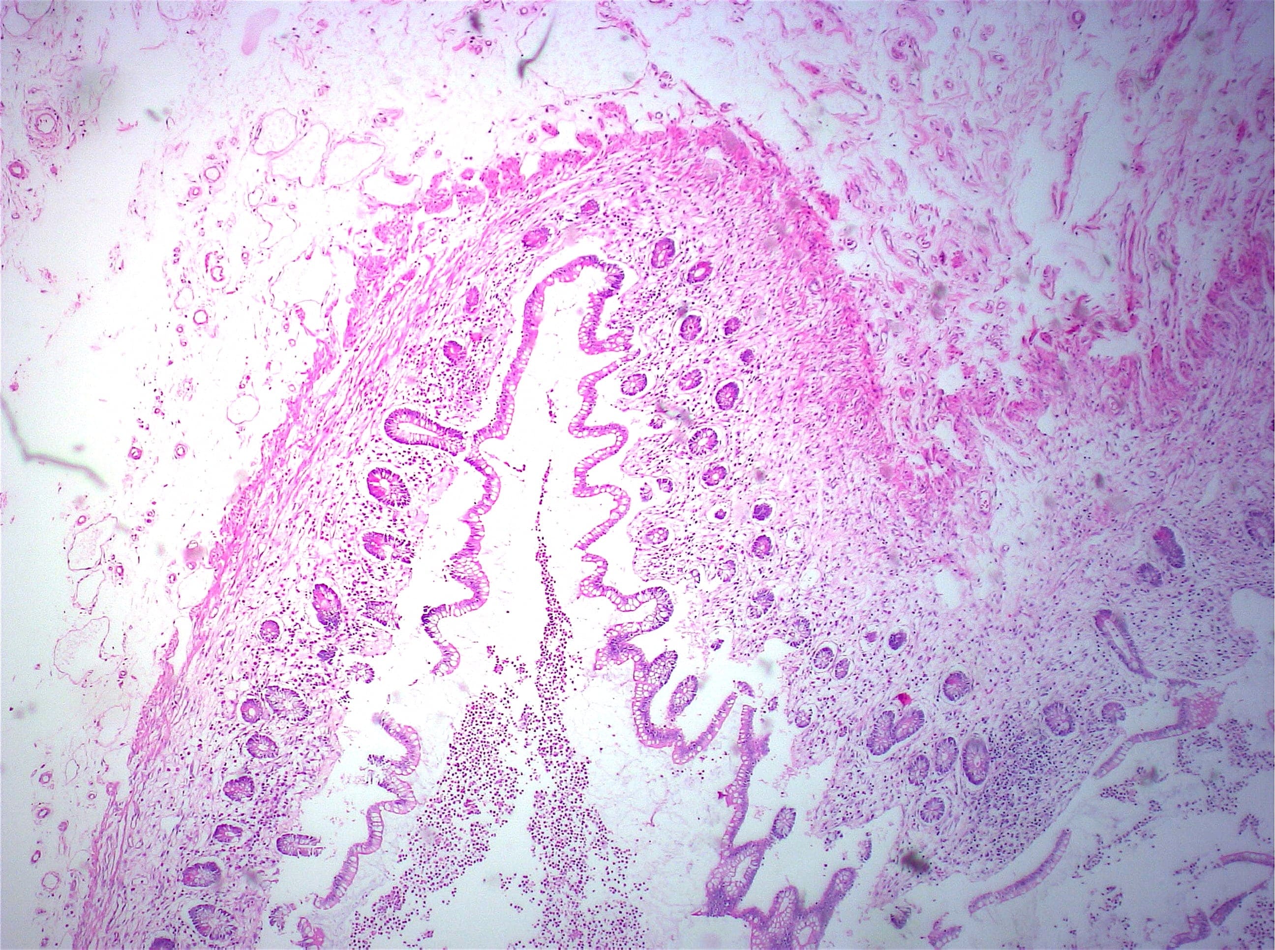
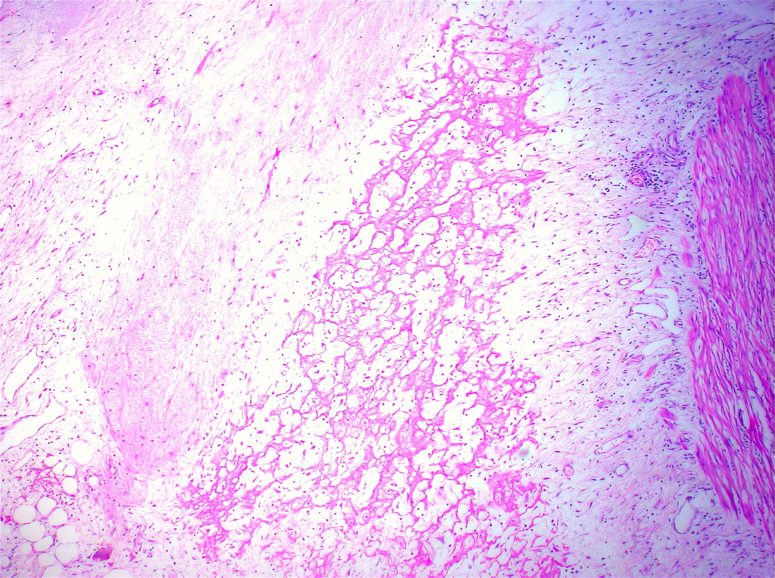
Contributed by Shefali Chopra, M.D.
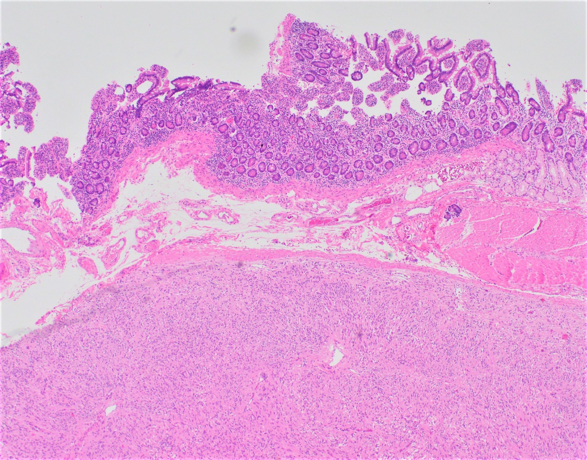
Relation to overlying mucosa
Skeinoid fibers
Spindle cell morphology
Therapy effect
CD117
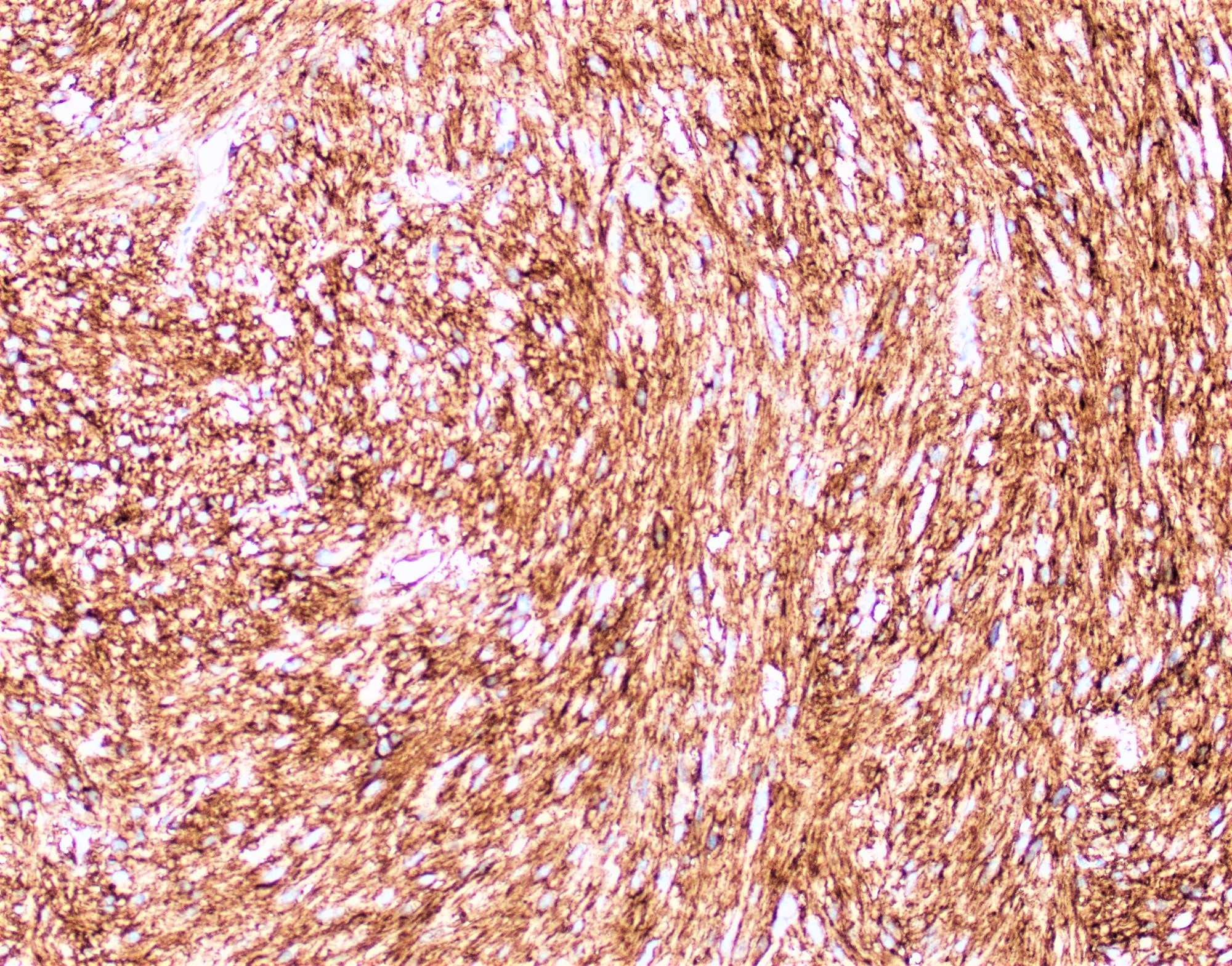
DOG1
Contributed by Shefali Chopra, M.D.
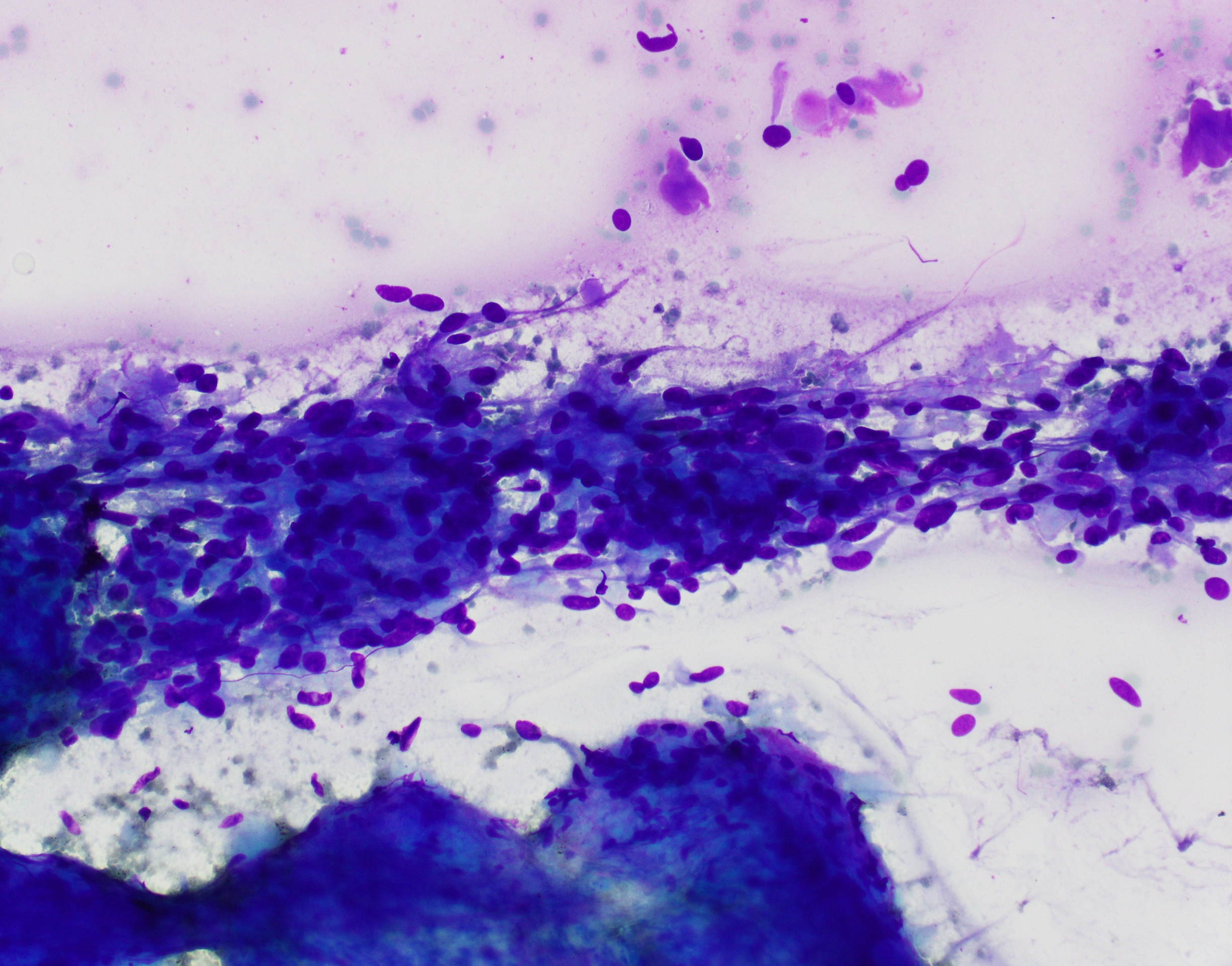
Diff-Quik, spindle cells
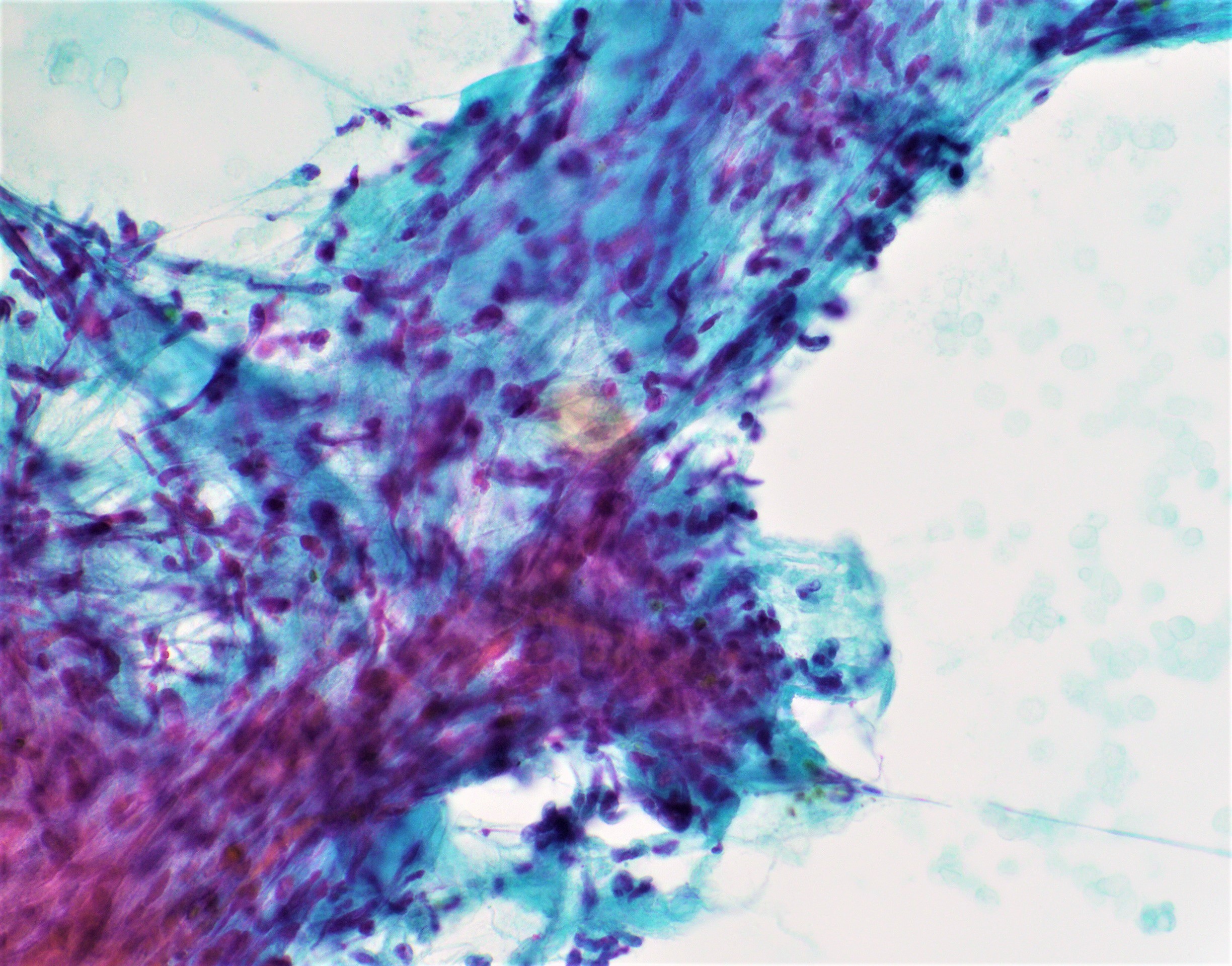
Pap, spindle cells
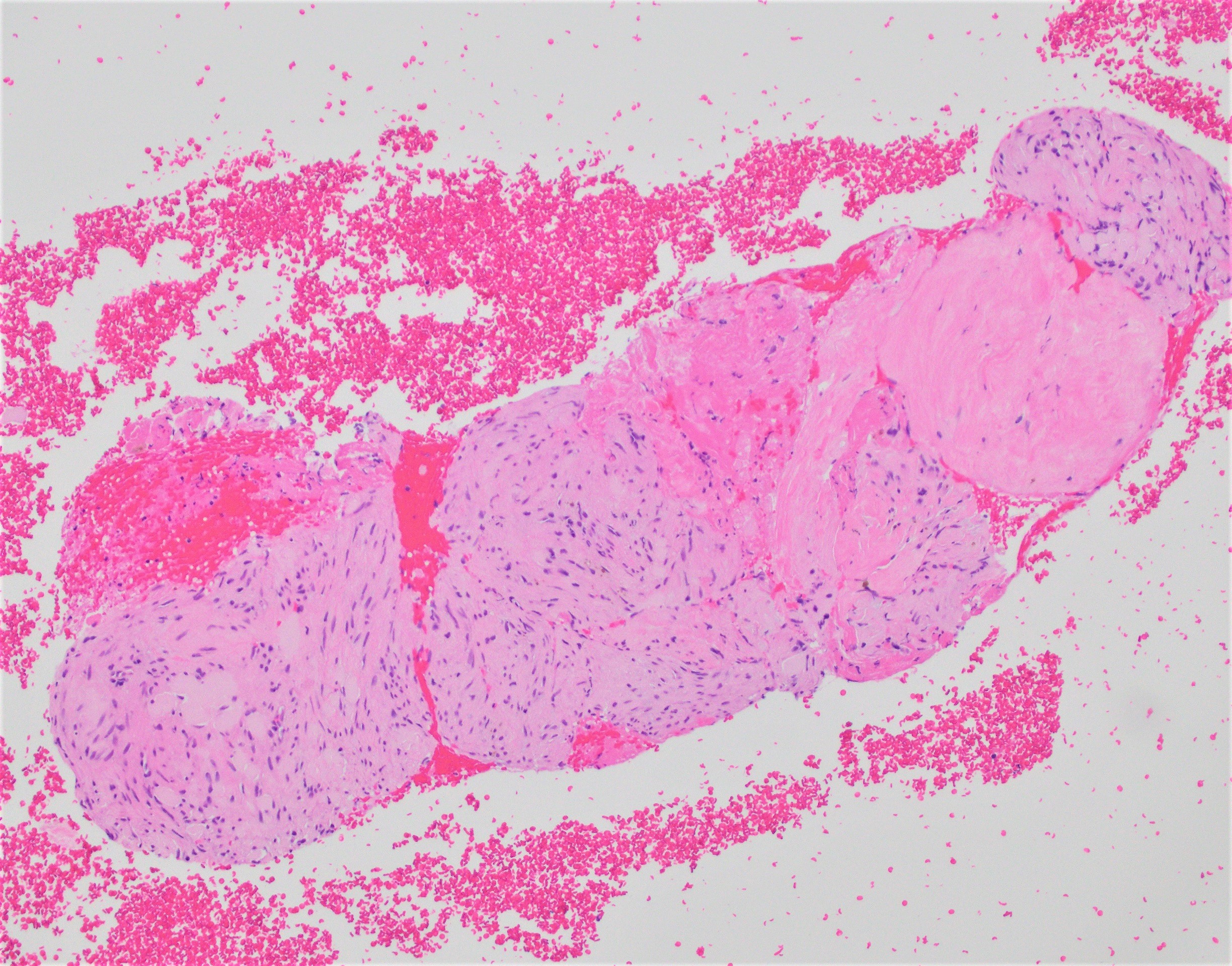
Cell block, spindle cells
Images hosted on other servers:

KIT mutations
Small bowel GIST
Images hosted on other servers:

33 year old man with HGM in ileum

12 year old girl with extensive HGM
Case #124
Intraluminal polyp
Images hosted on other servers:

33 year old man with HGM in ileum

14 year old boy with HGM in ileum
Contributed by Khalid Amin, M.D.
Ileal polypoid lesion
Gastric glands
Jejunal polypoid lesion
Underlying antral glands
Images hosted on other servers:

Ampulla of Vater and related structures

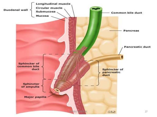
Ampulla, papilla and sphincters
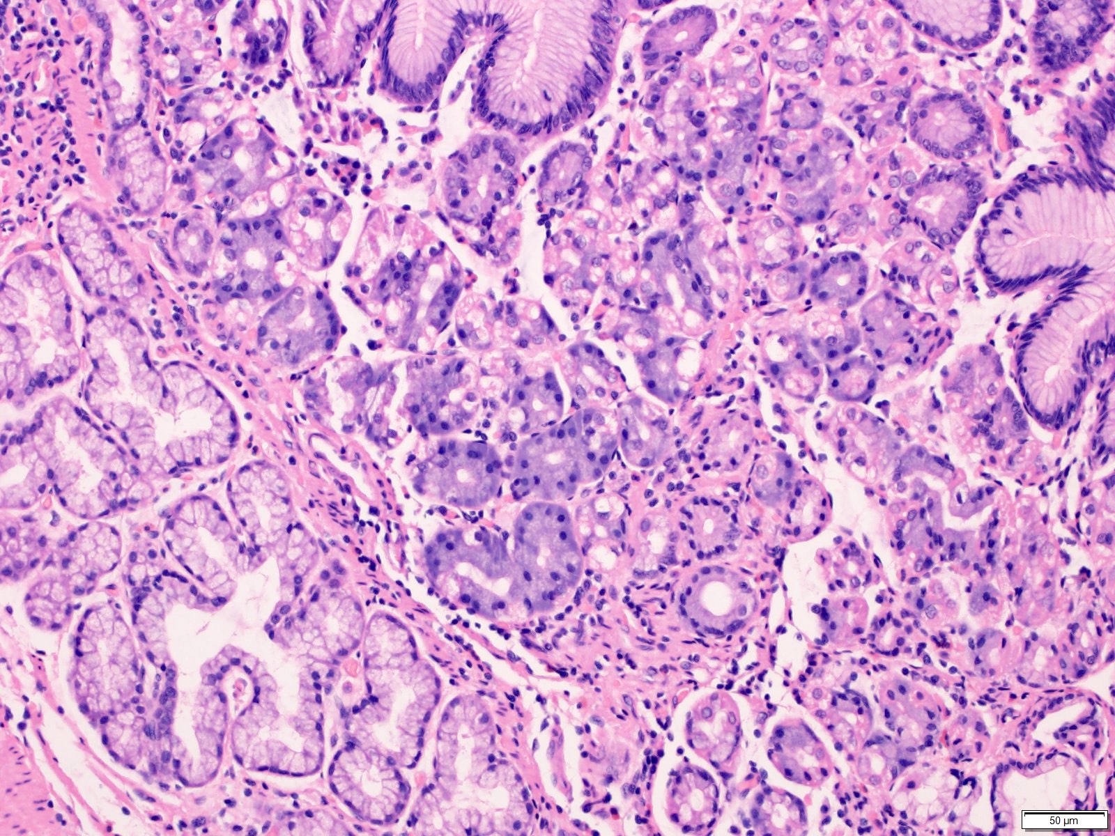
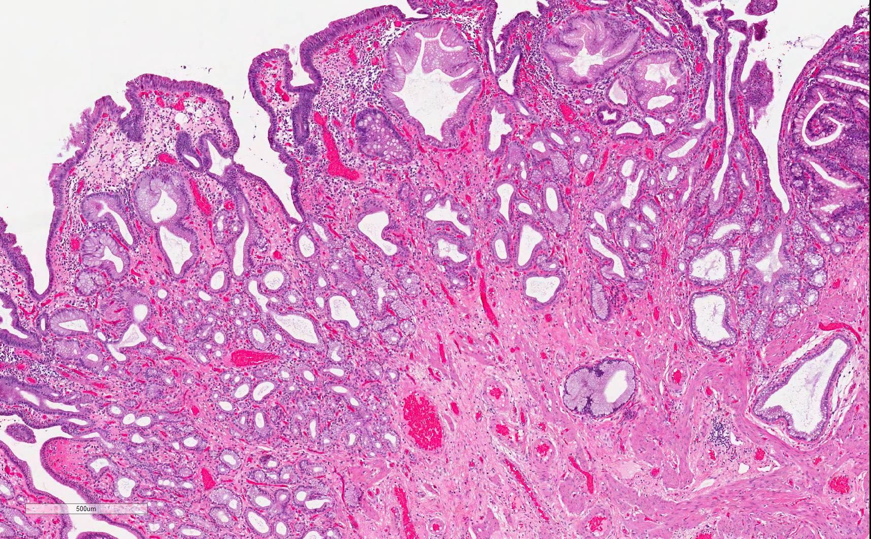
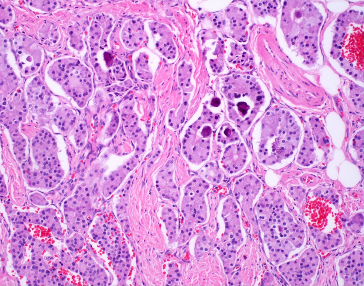
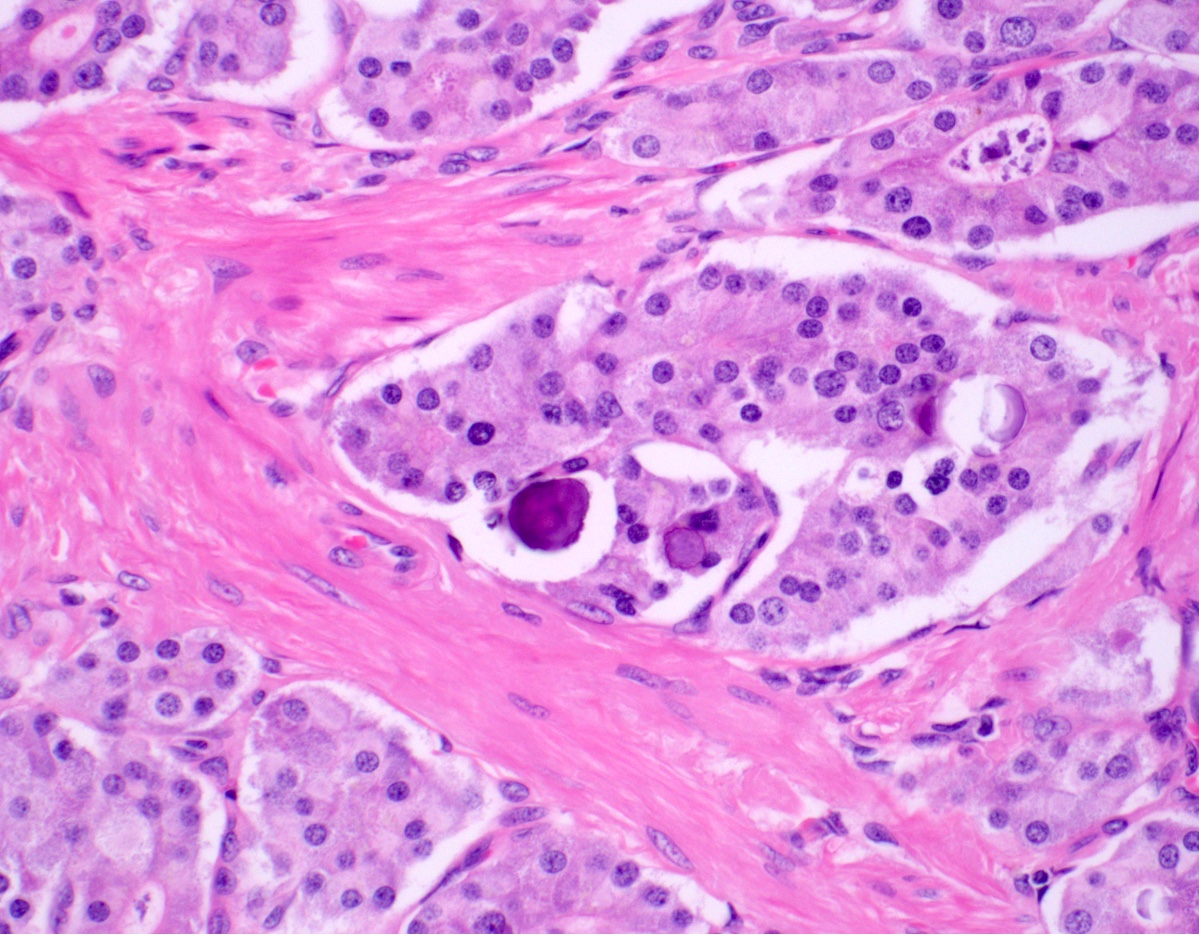
Contributed by Raul Gonzalez, M.D.
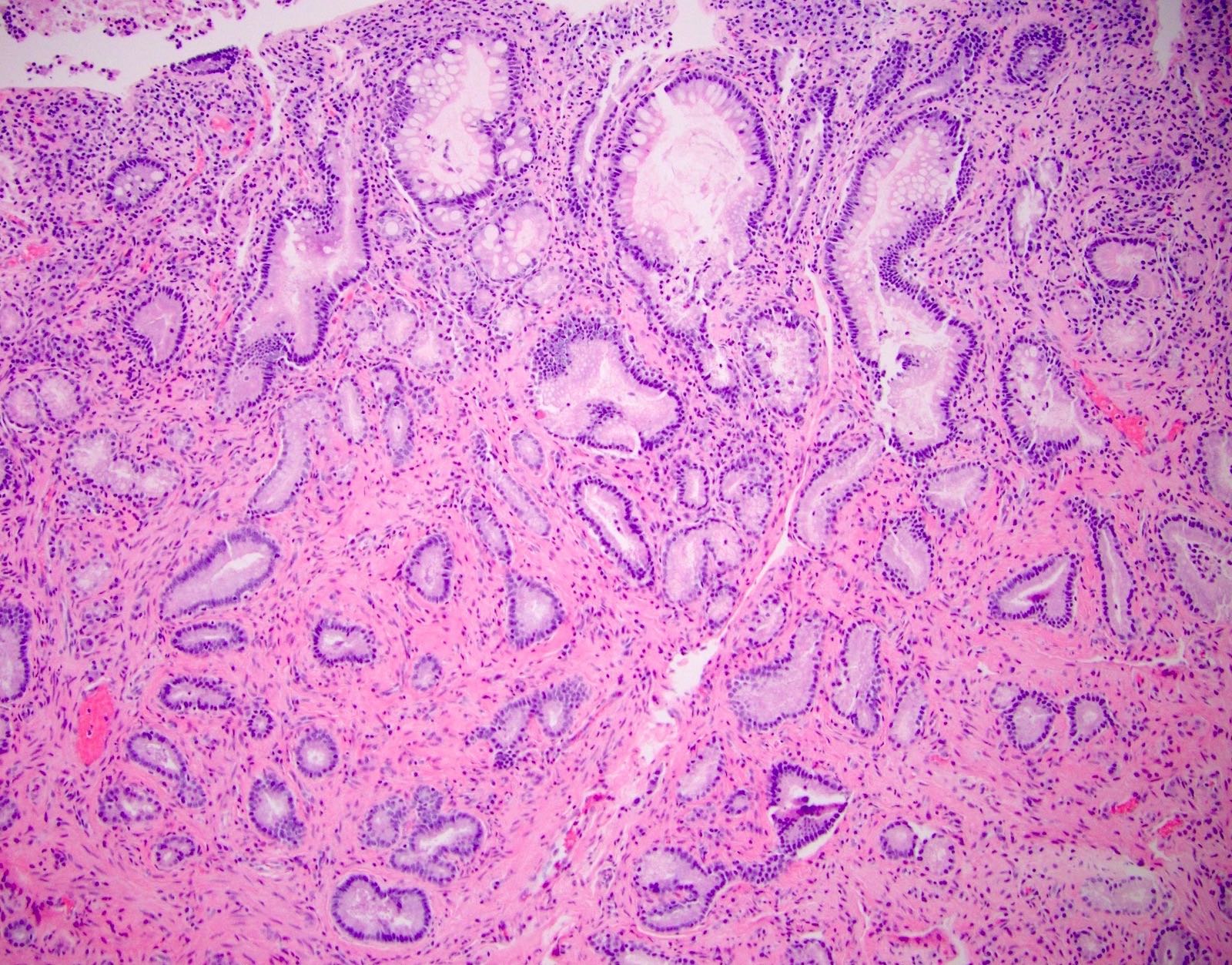
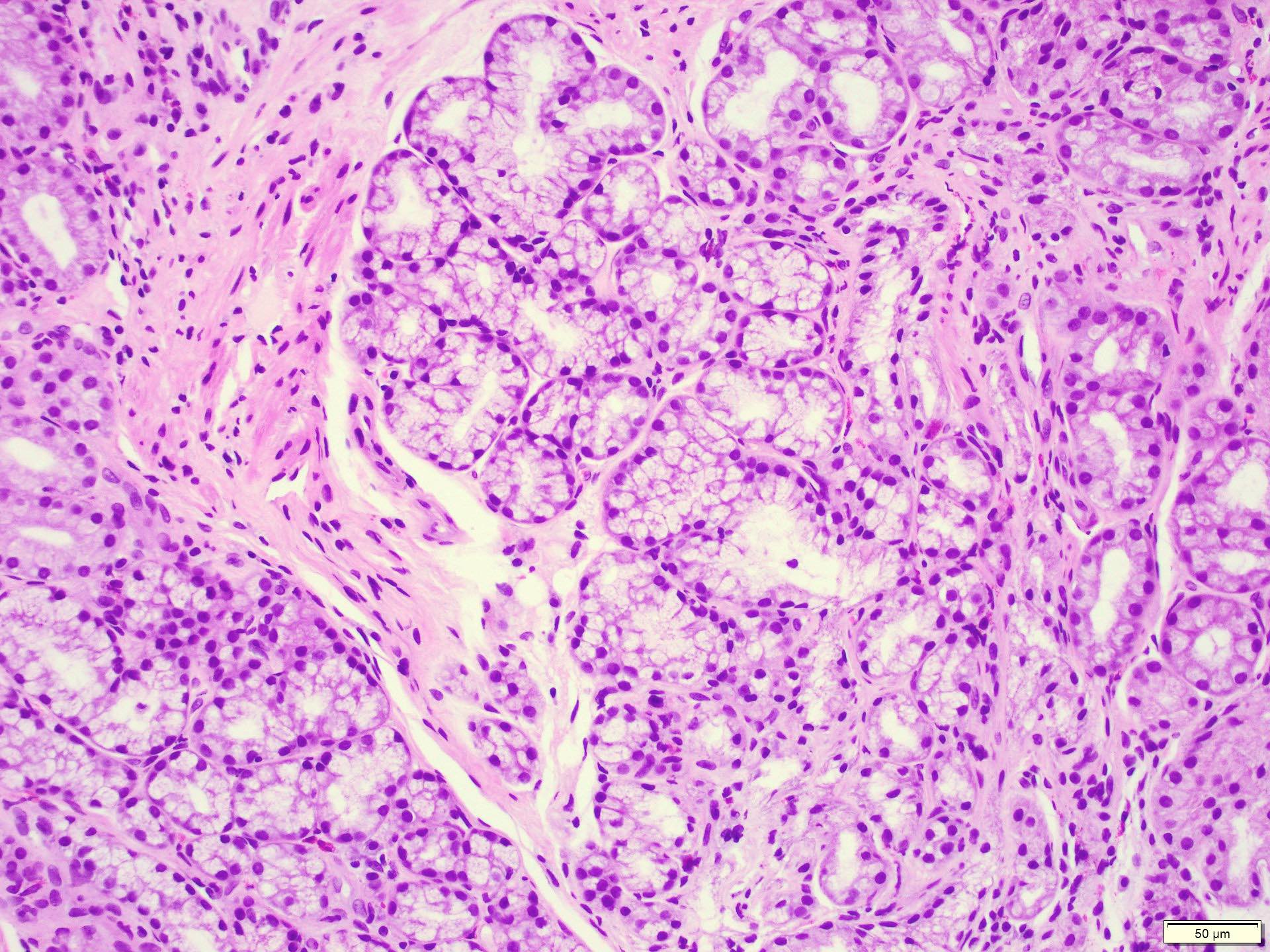
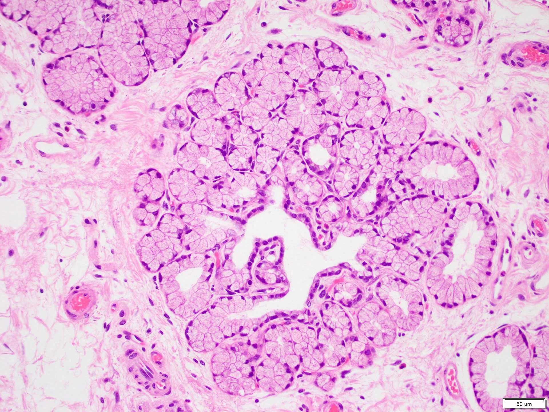
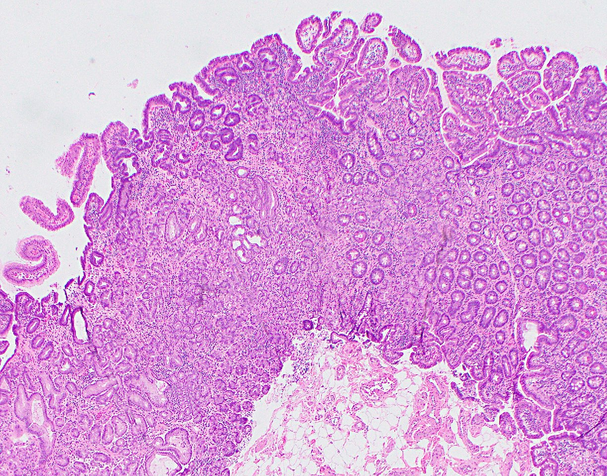
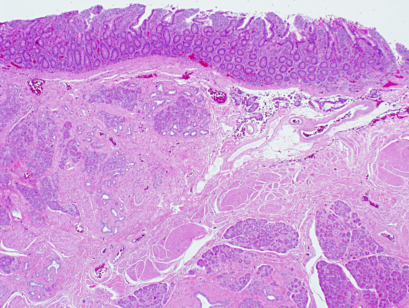
Normal ampullary mucosa and submucosa
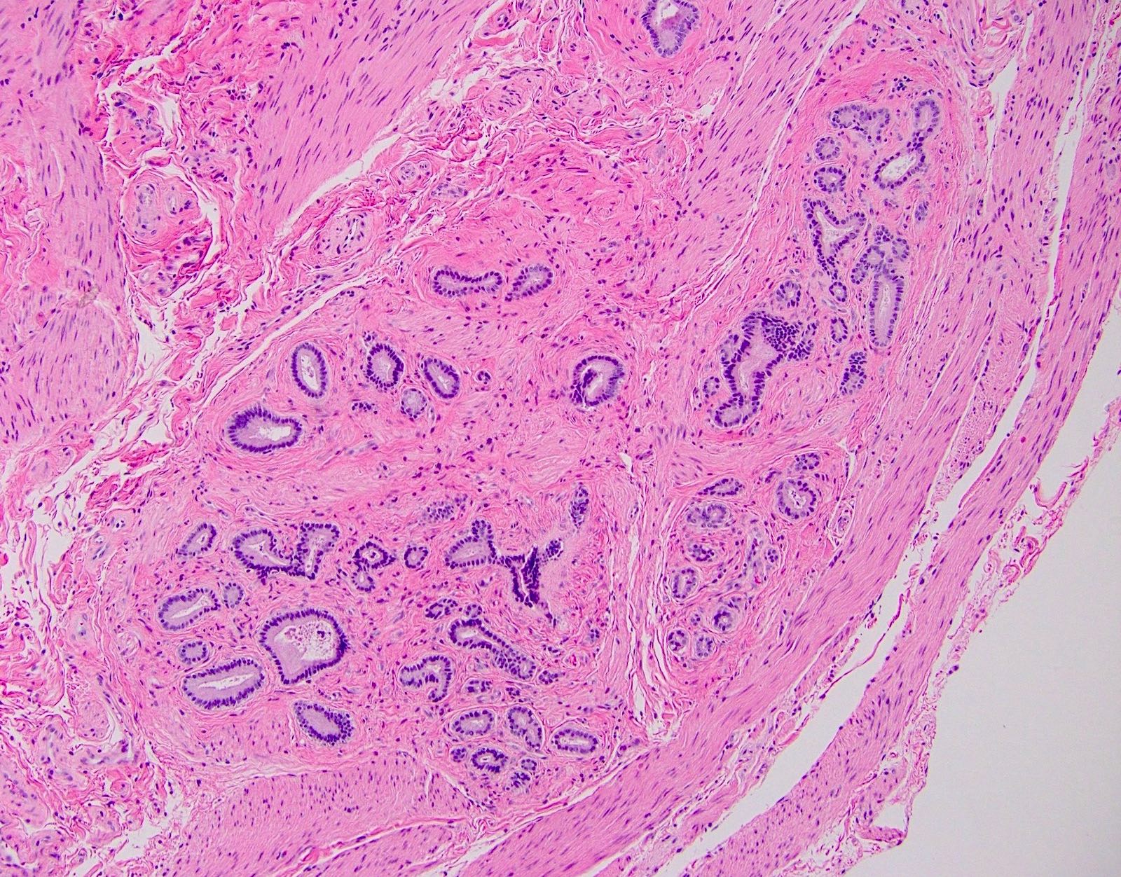
Ampullary submucosal glands / ductules
Contributed by Danielle Hutchings, M.D.
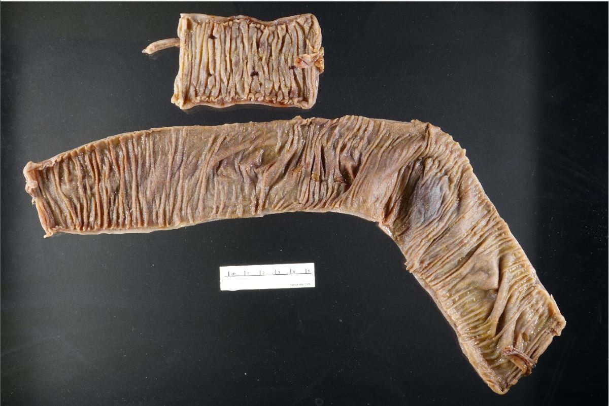
Jejunum
Contributed by Danielle Hutchings, M.D.
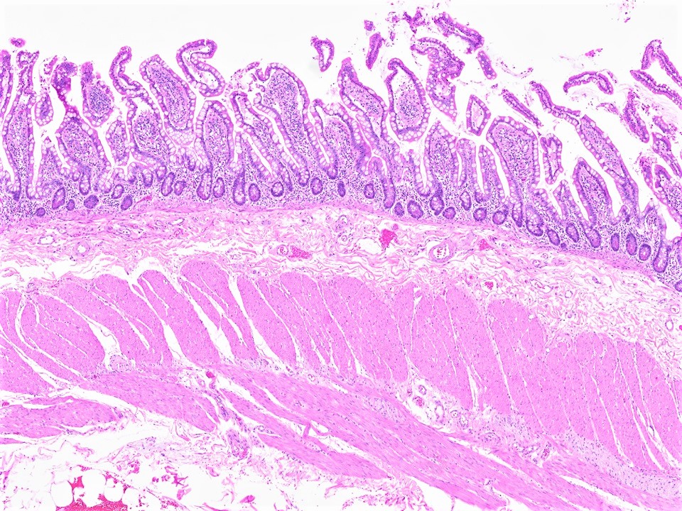
Layers of small intestine
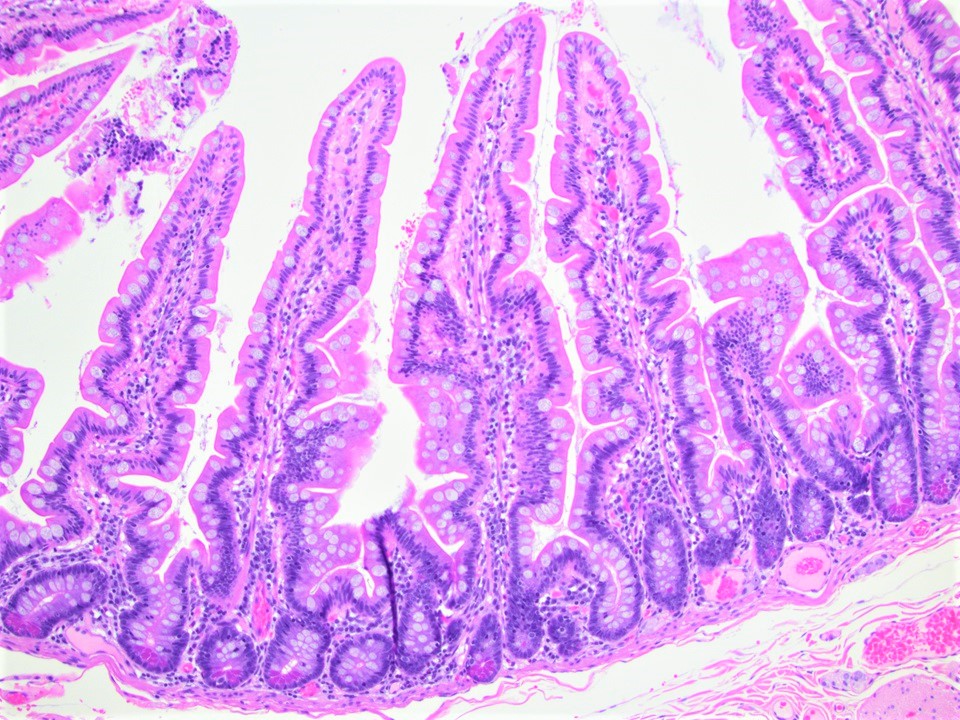
Villi
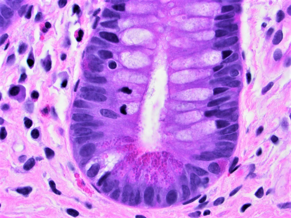
Paneth cells
Brunner glands
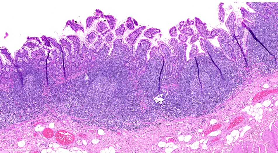
Peyer Patches
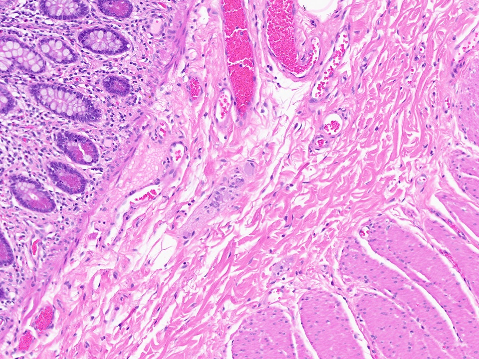
Submucosa
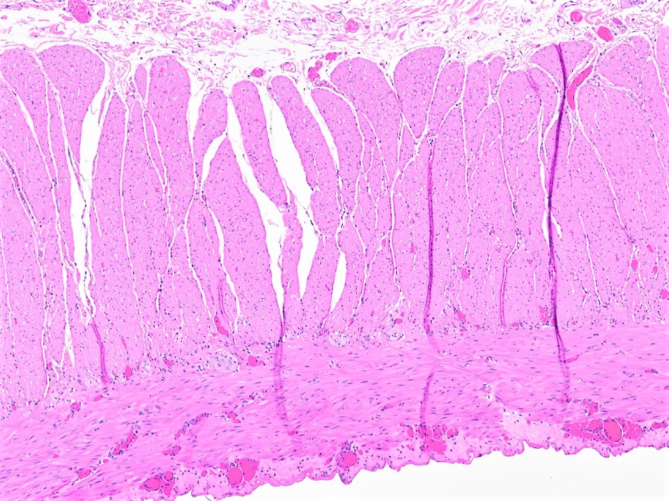
Muscularis propria
Microvilli and goblet cells
Images hosted on other servers:

Microvillus of small intestine
Small intestine: histology
Images hosted on other servers:

Duodenal hyperplastic polyp
Contributed by Hanni Gulwani, M.D.

CMV pouchitis: biopsy of postcolectomy pouch in 38 year old man with ulcerative colitis
Images hosted on other servers:

Various images
Contributed by Claudia Mendez, M.D.
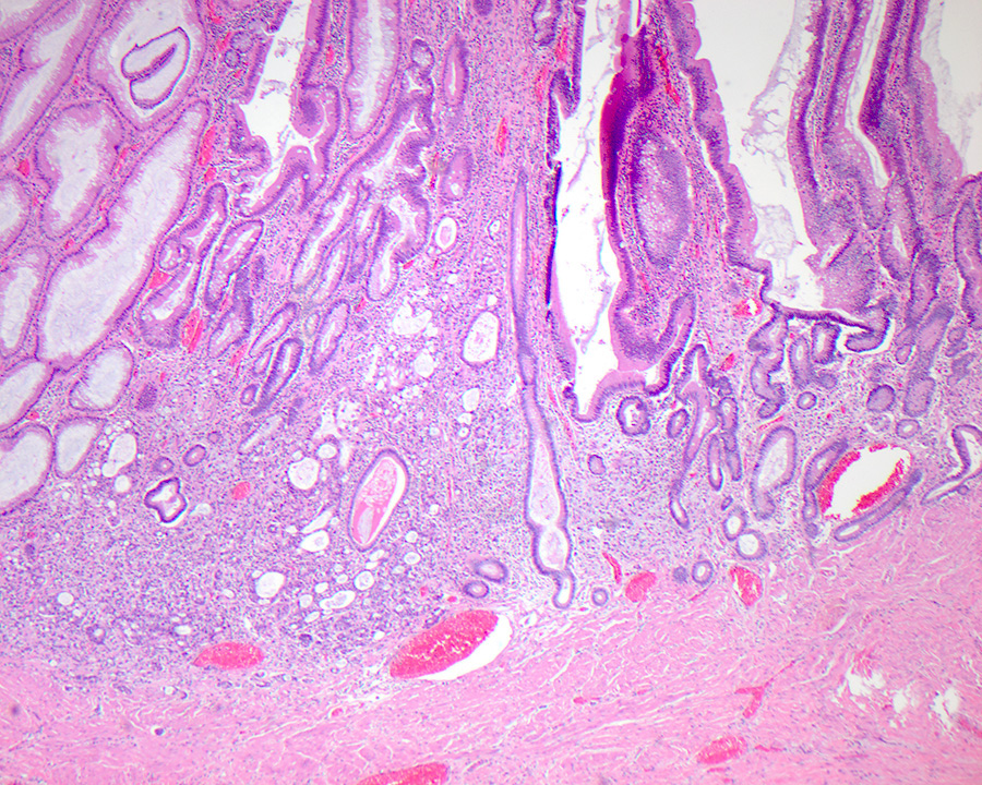
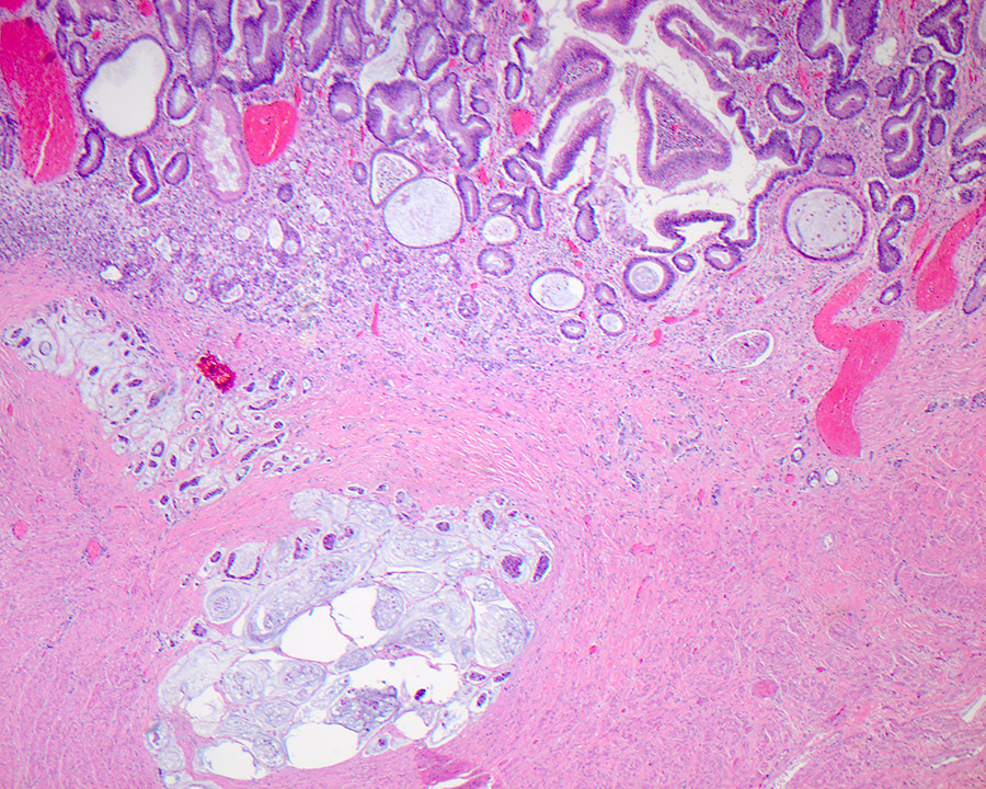
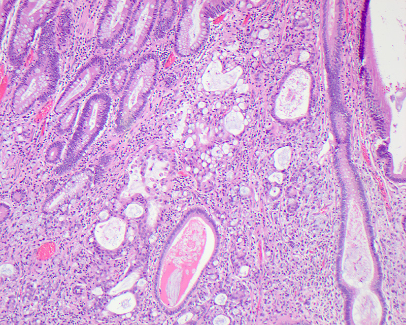
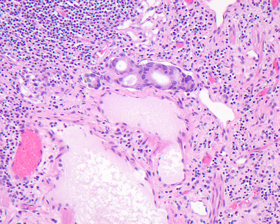
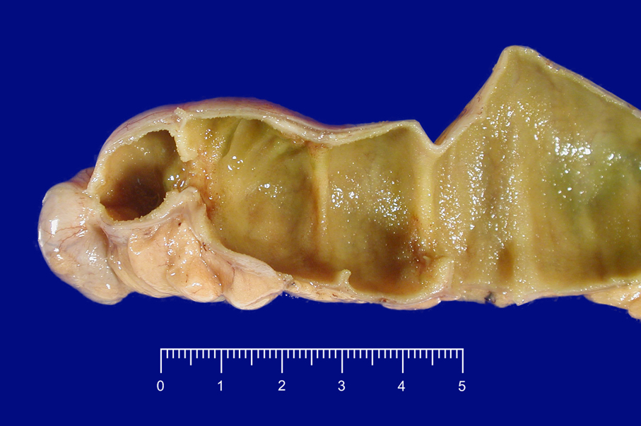
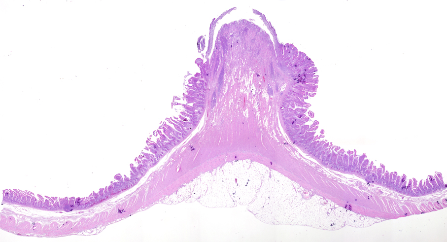
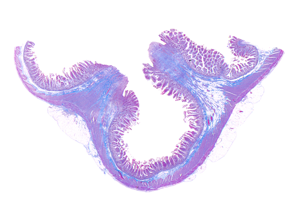
Inflammatory fibroid polyp
Contributed by Claudia Mendez, M.D.
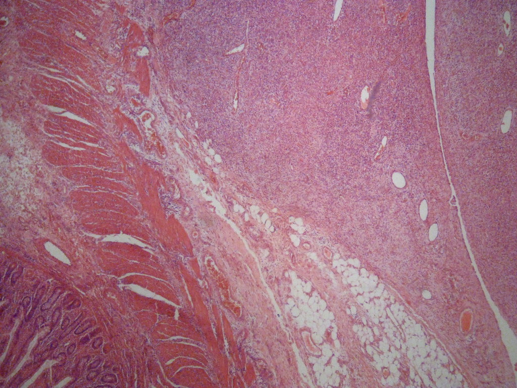
4x
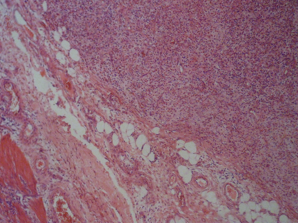
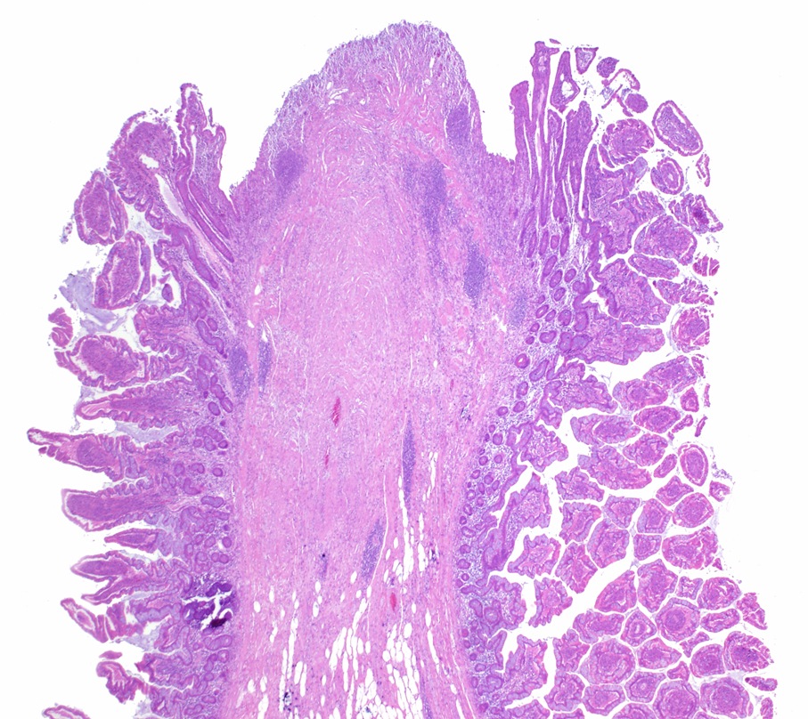
10x

40x
Images hosted on other servers:

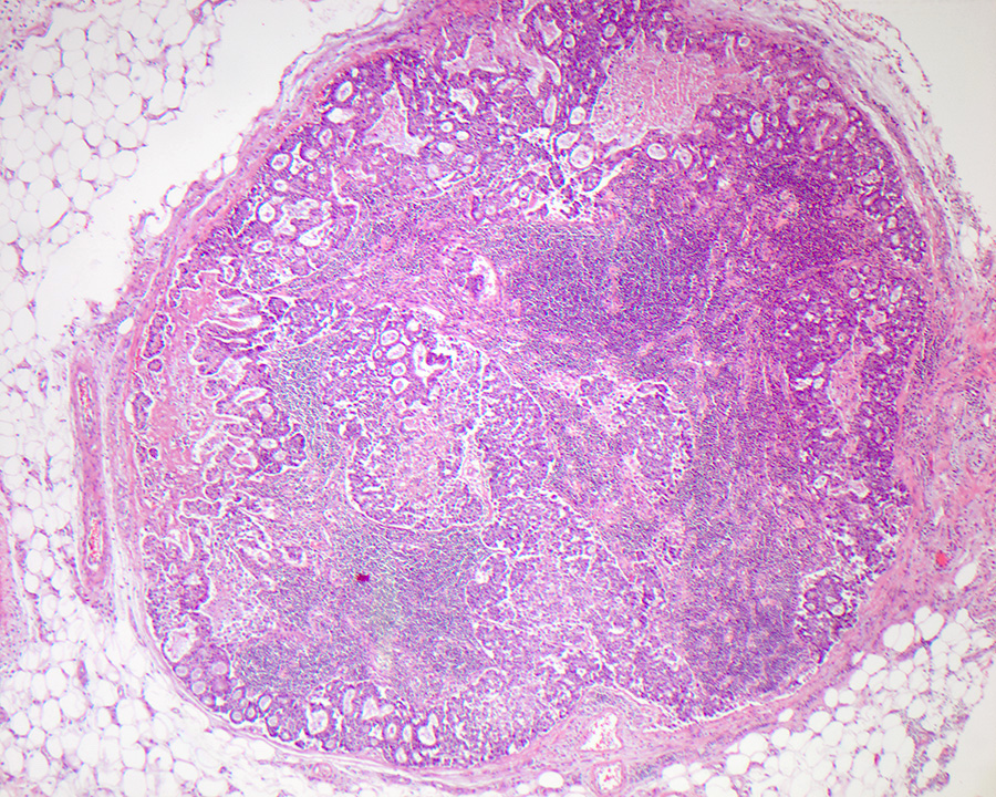
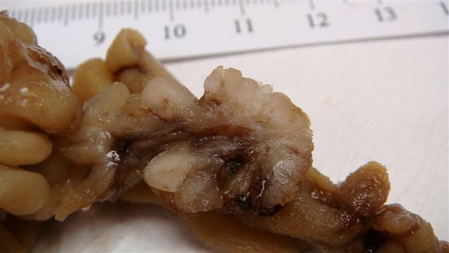
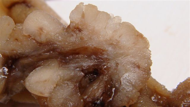
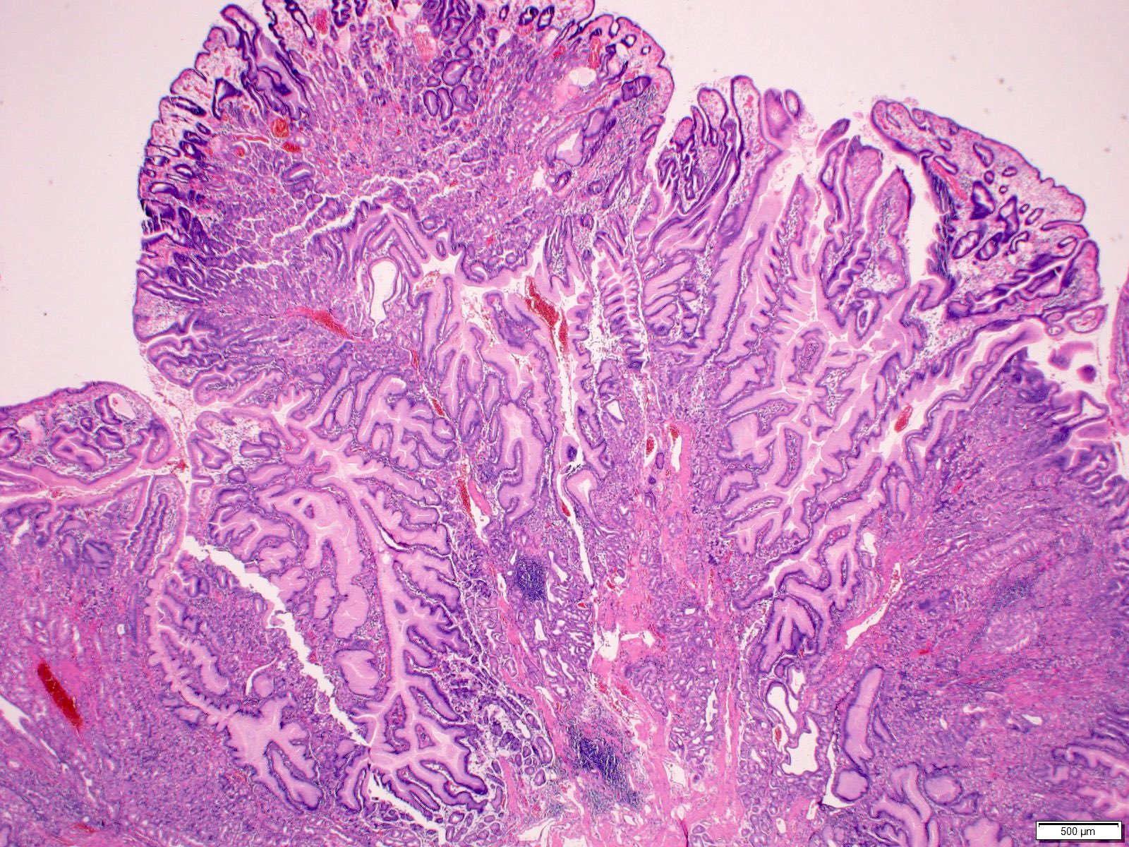
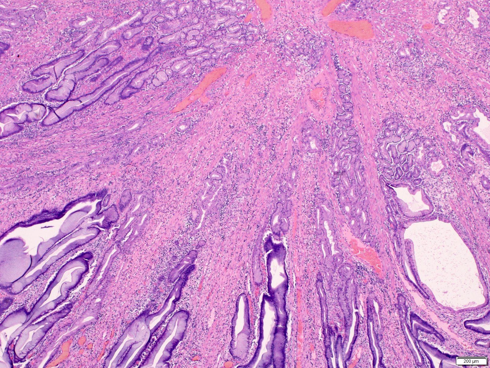
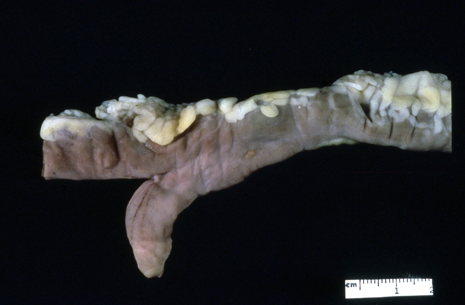
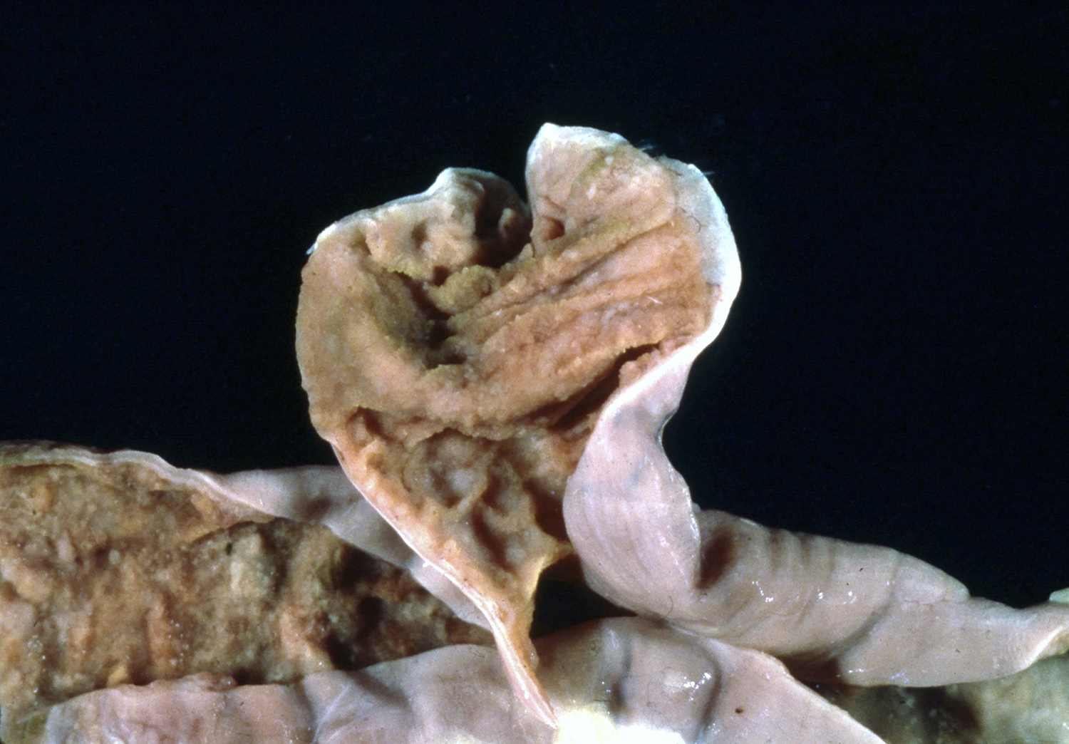
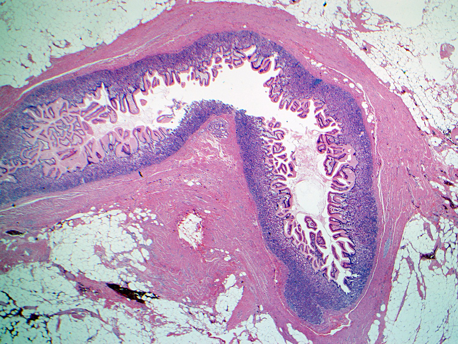
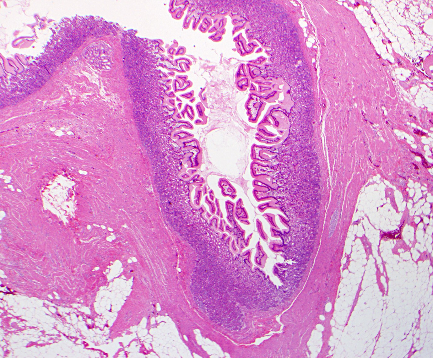
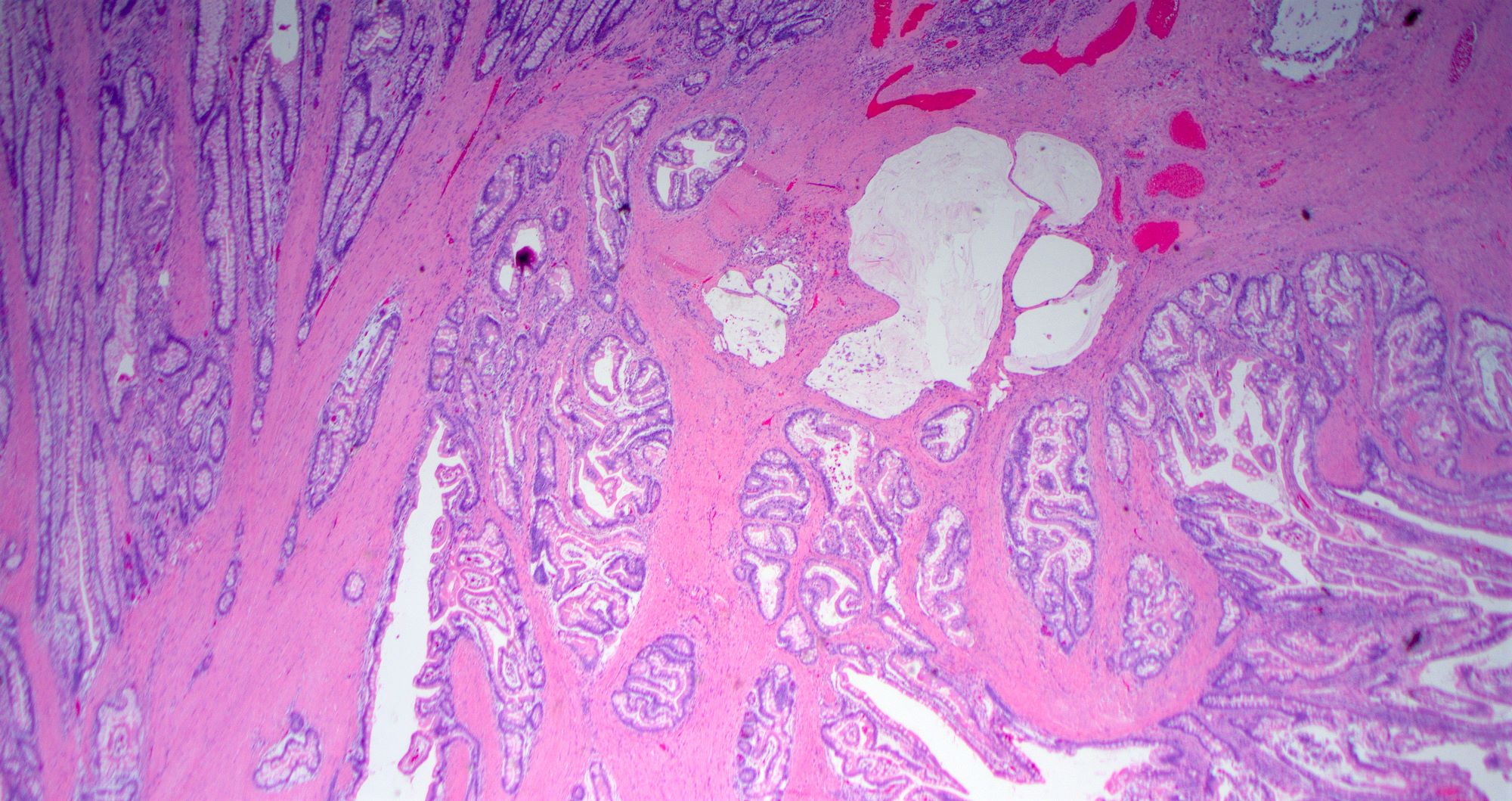
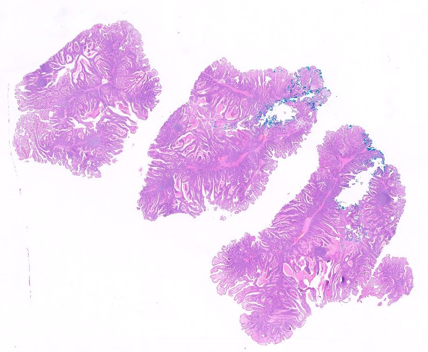
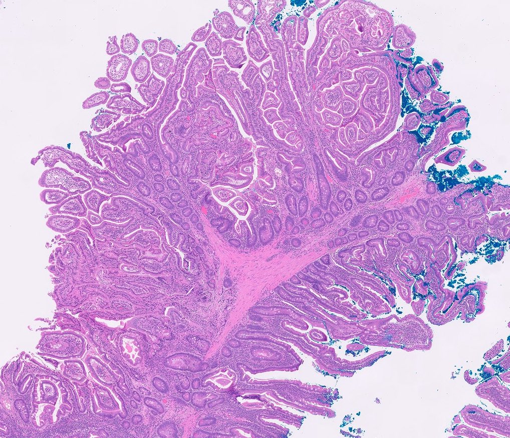
IAPN gross / histologic correlation
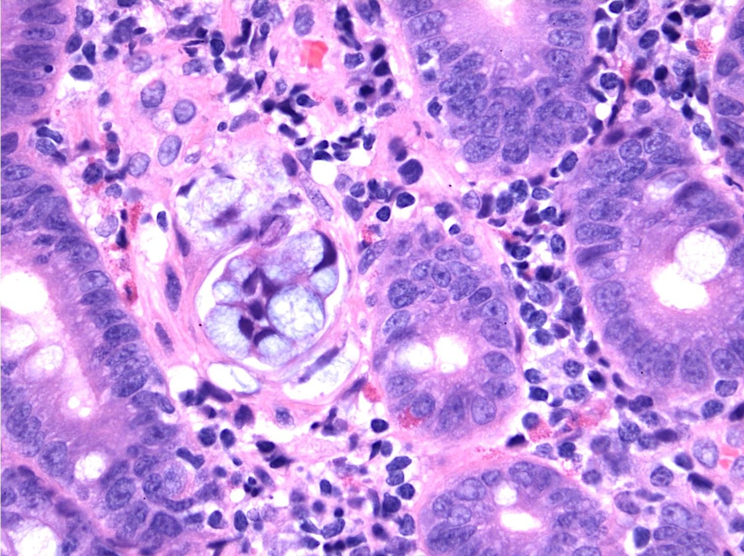
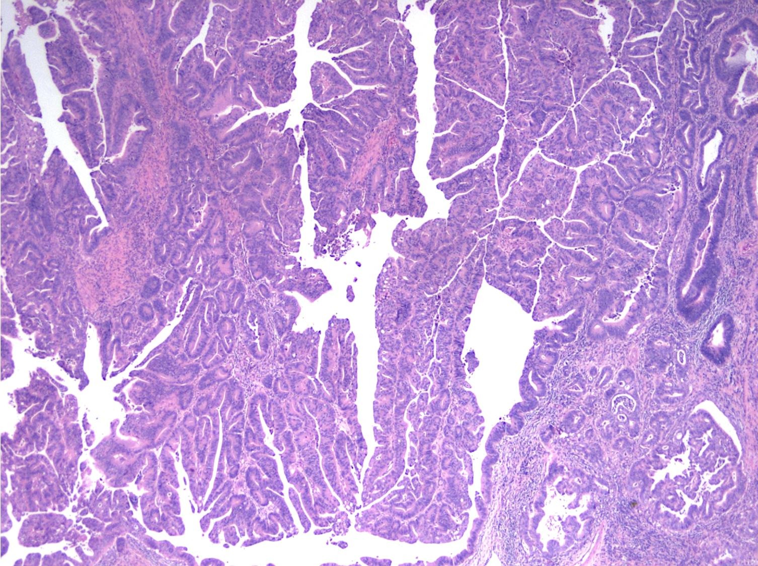
Contributed by Felicia D. Allard, M.D.
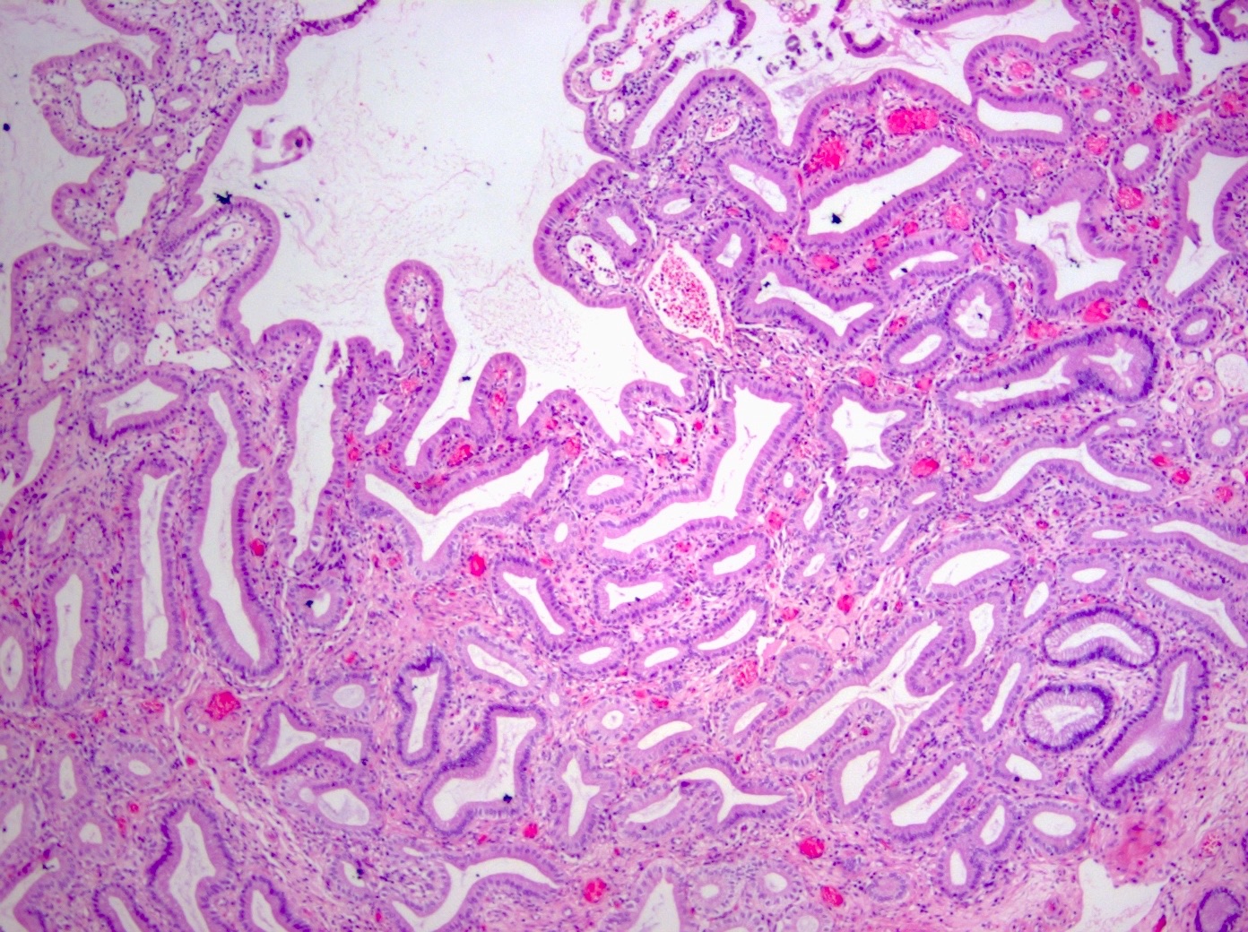
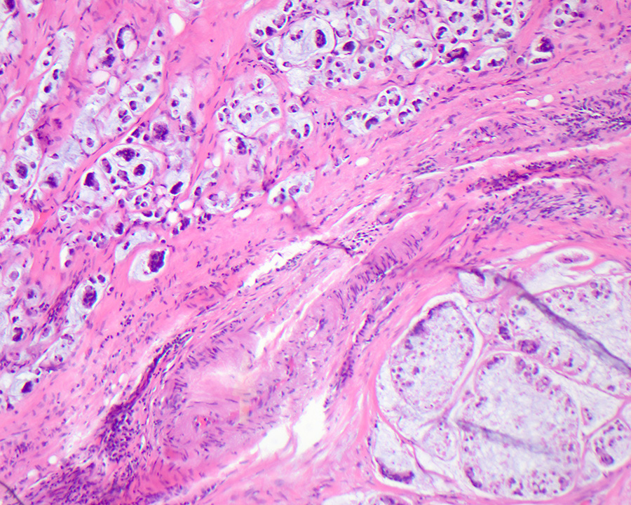
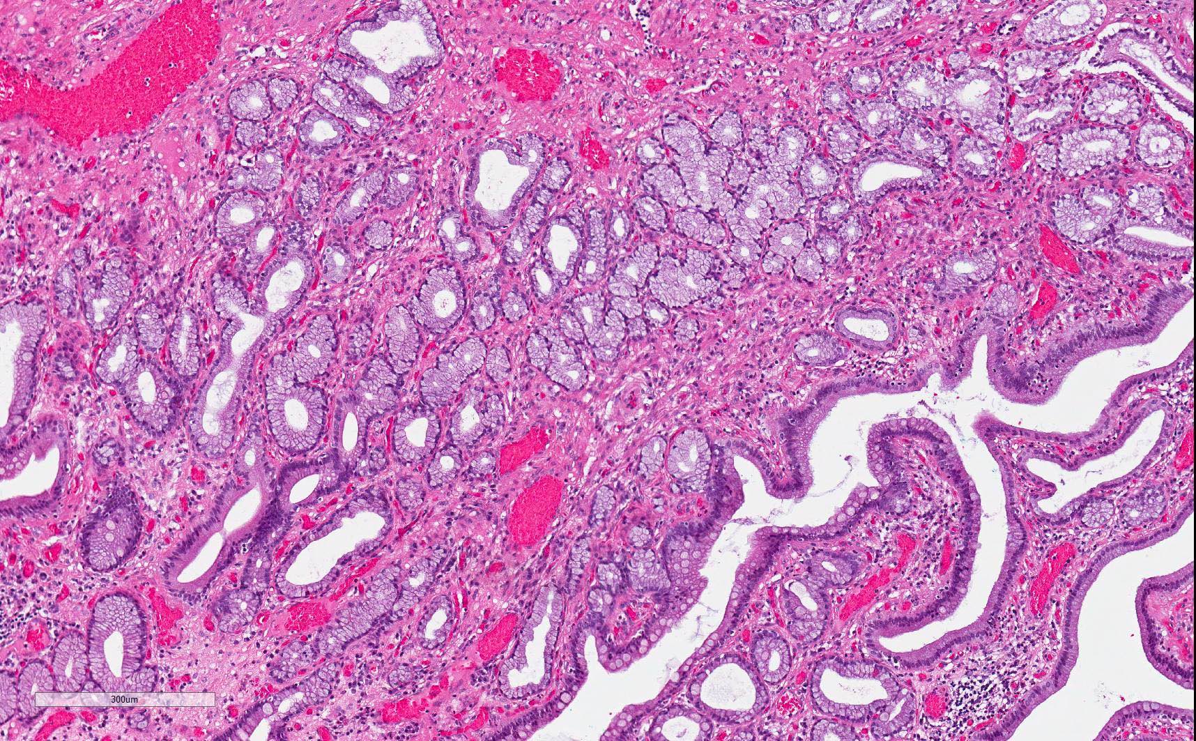
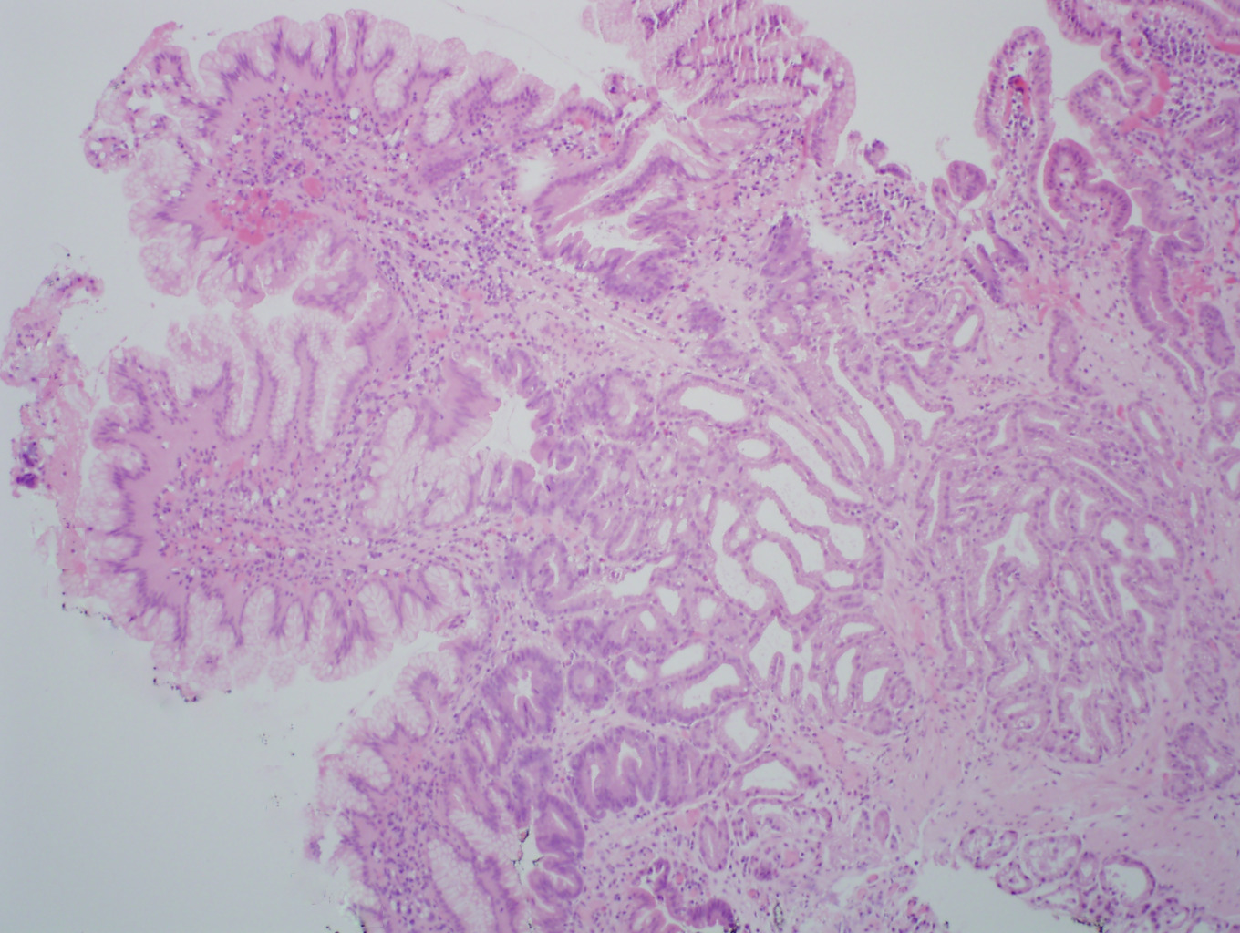
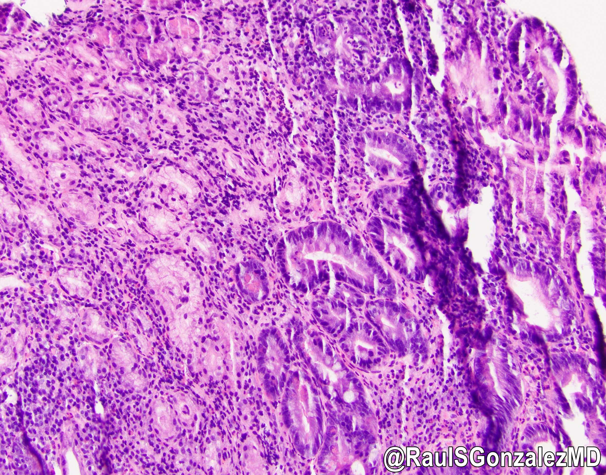
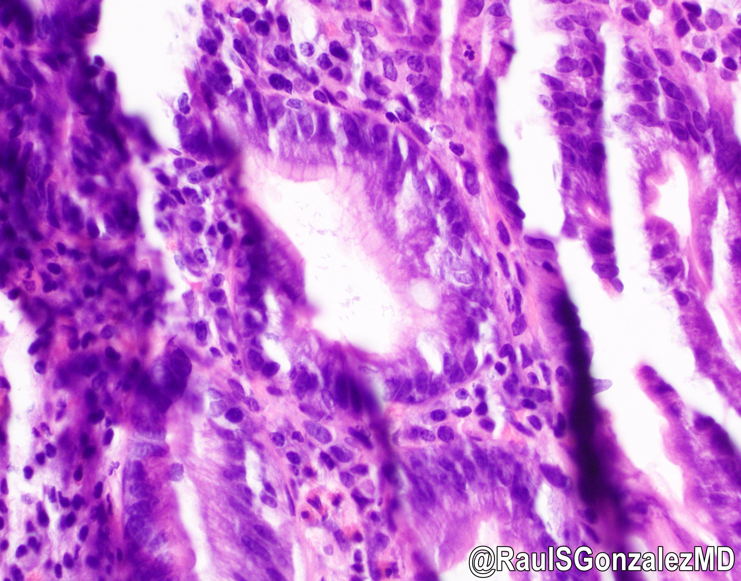
Pancreatobiliary type IAPN
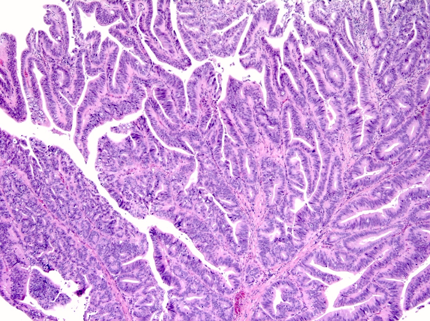
Intestinal type IAPN
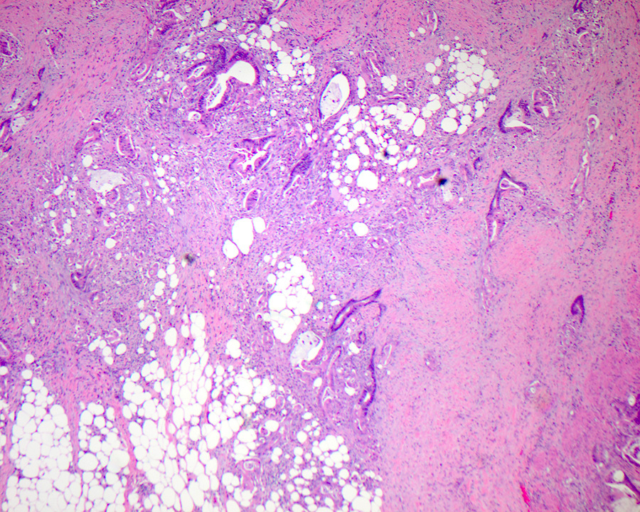
Paneth and goblet cells
Intestinal IAPN with high grade dysplasia
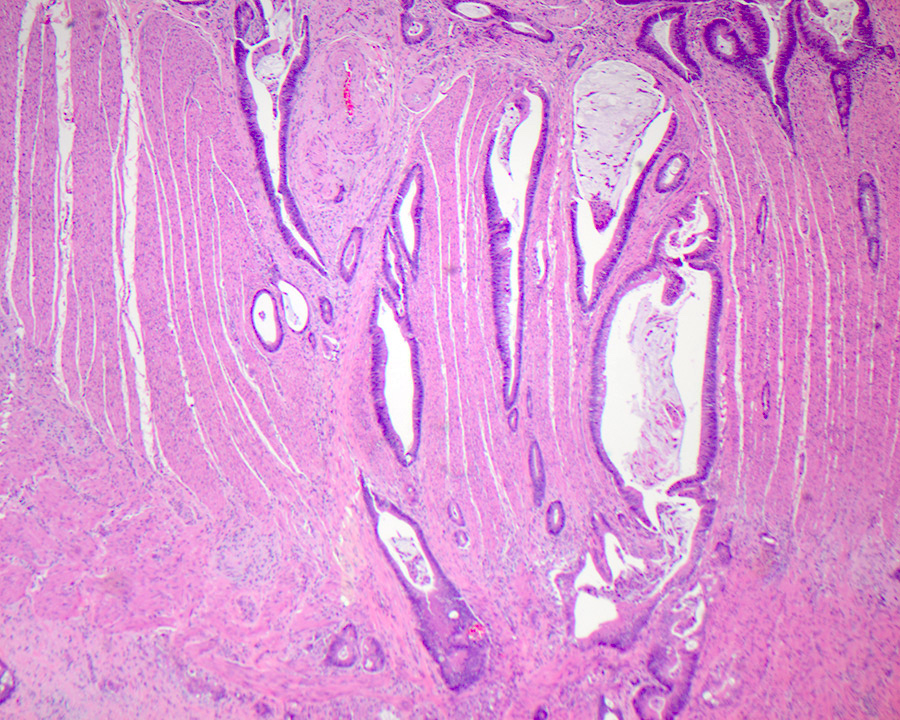
Pancreaticobiliary phenotype tubular growth
Images hosted on other servers:

IAPN phenotype immune profiles
Images hosted on other servers:

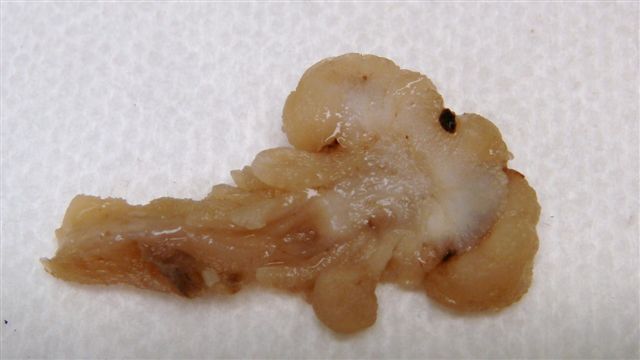
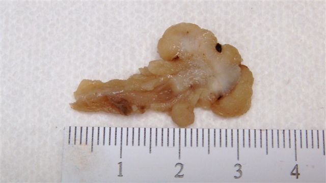
IAPN gross / histologic correlation
Contributed by Felicia D. Allard, M.D.
Pancreatobiliary type IAPN
Intestinal type IAPN
Paneth and goblet cells
Intestinal IAPN with high grade dysplasia
Pancreaticobiliary phenotype tubular growth
Images hosted on other servers:

Gentle reduction of the ileoileal intussusception
Contributed by Krutika S. Patel, M.B.B.S., M.D.
CT scan
Images hosted on other servers:

Infarcted small intestine

Marked hyperemia

Early ischemic enteritis

Resected ischemic jejunal segment
Contributed by Krutika S. Patel, M.B.B.S., M.D. and Madison Swaney, M.D.
Early ischemic changes
Ischemic changes
Images hosted on other servers:
Various images
Images hosted on other servers:


Various images
Images hosted on other servers:

Genotype phenotype correlations in JPS
Images hosted on other servers:


Magnetic resonance enterography of ileal polyp
Contributed by Milton Smith, M.D.
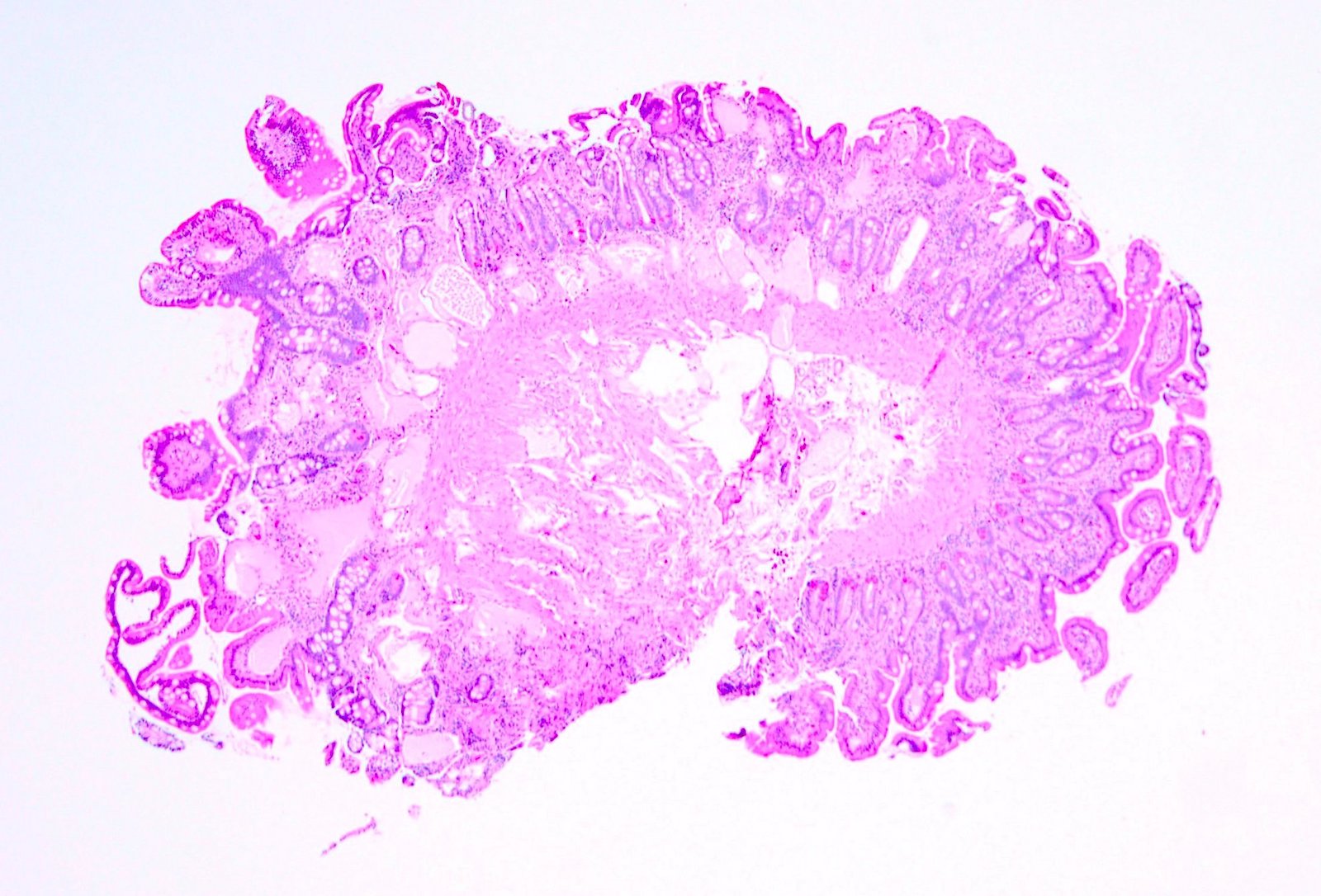
Juvenile polyps in duodenum
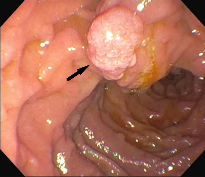
Large eroded juvenile polyp

Multiple juvenile polyps
Contributed by Divya Sharma, M.D., Rachel Sheridan, M.D. and Sara Szabo, M.D.

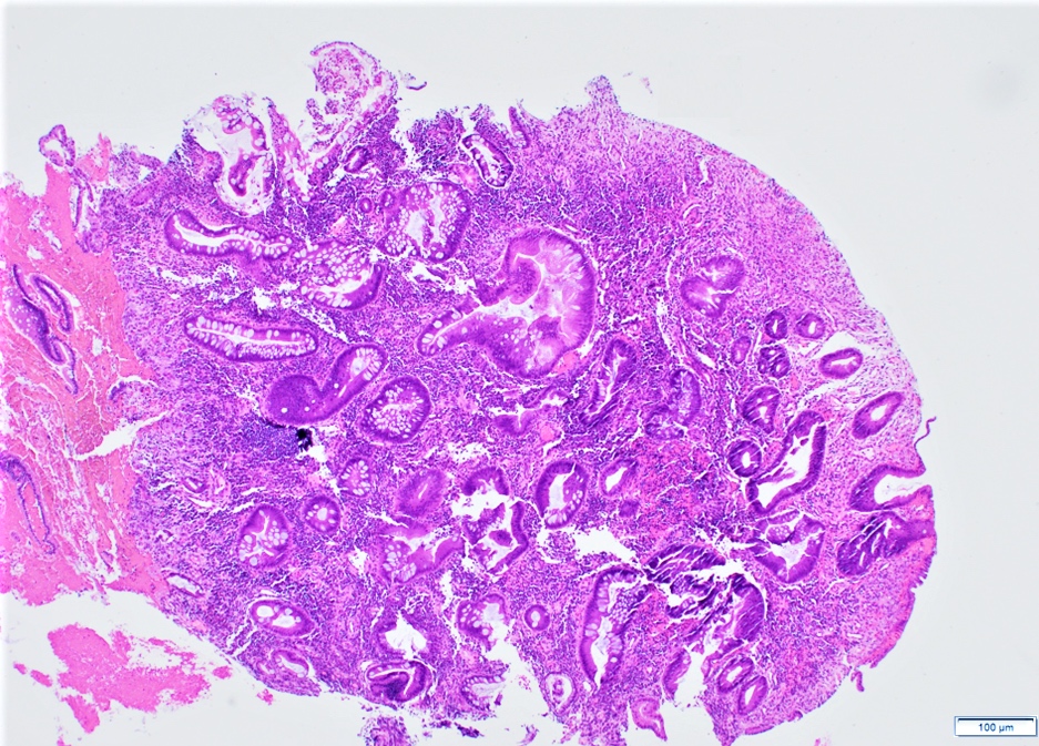
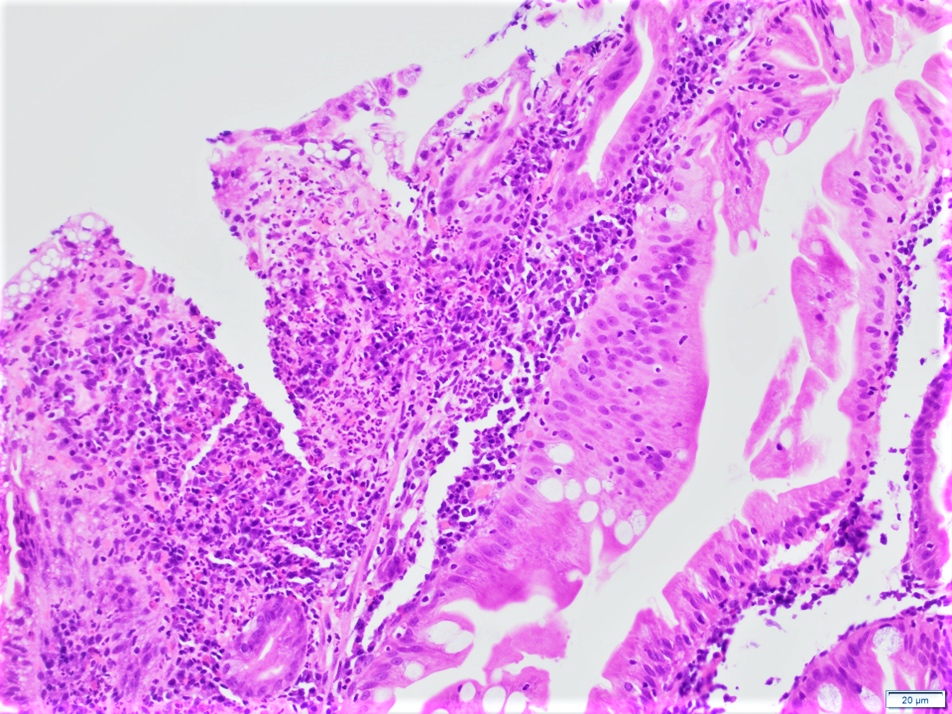
Surface erosion

Inflammatory stroma

Granulation tissue
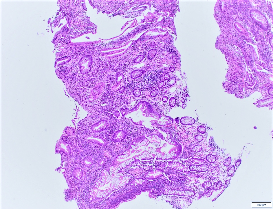
Branching glands

Glandular hyperplasia

Glandular hyperplasia
Slit-like serrations and inflammation

Finger-like lobulation of glands
Low grade dysplasia

Ileal polyp, surface erosion
Ileal polyp, dilated glands
AFIP images


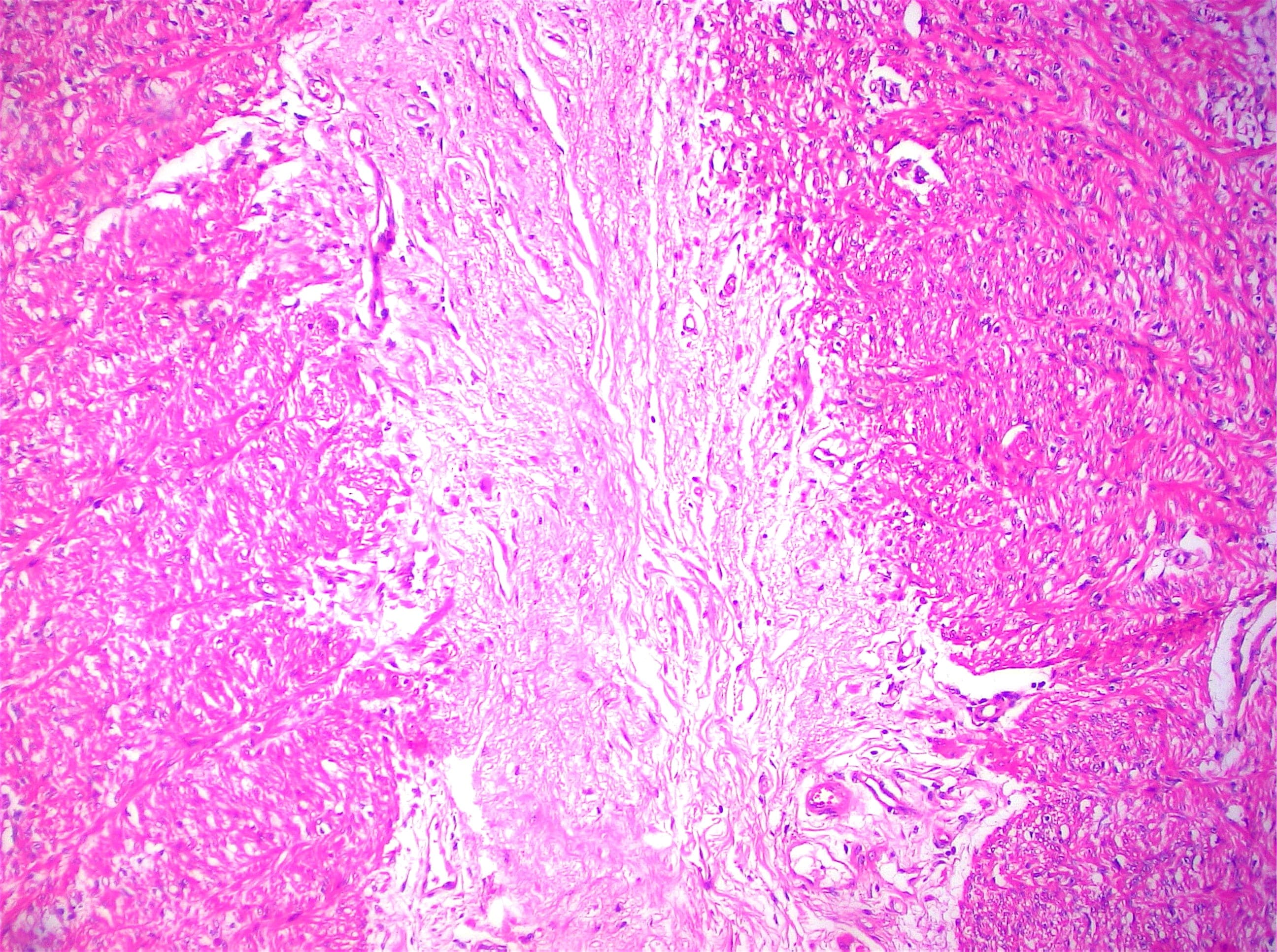
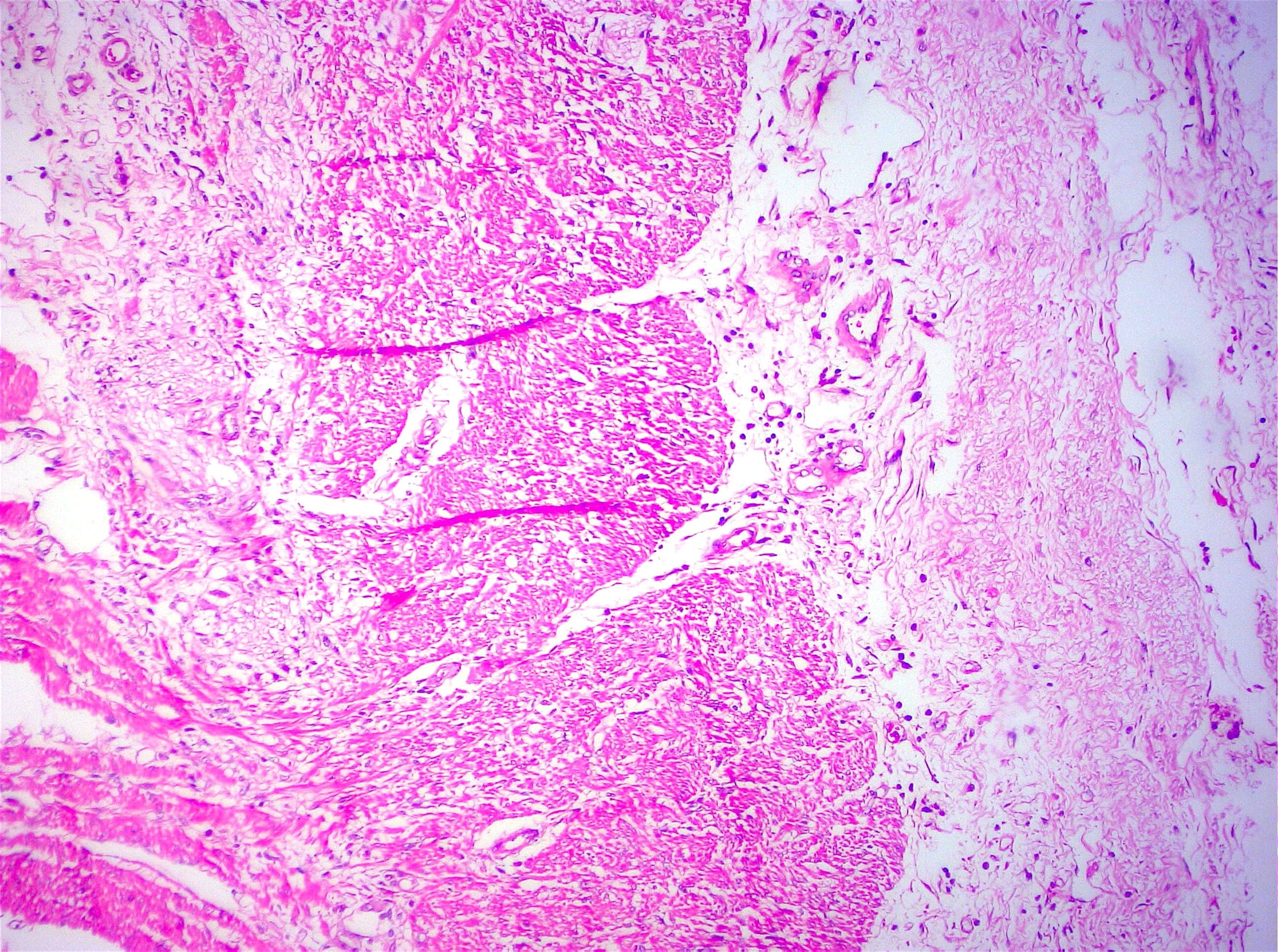
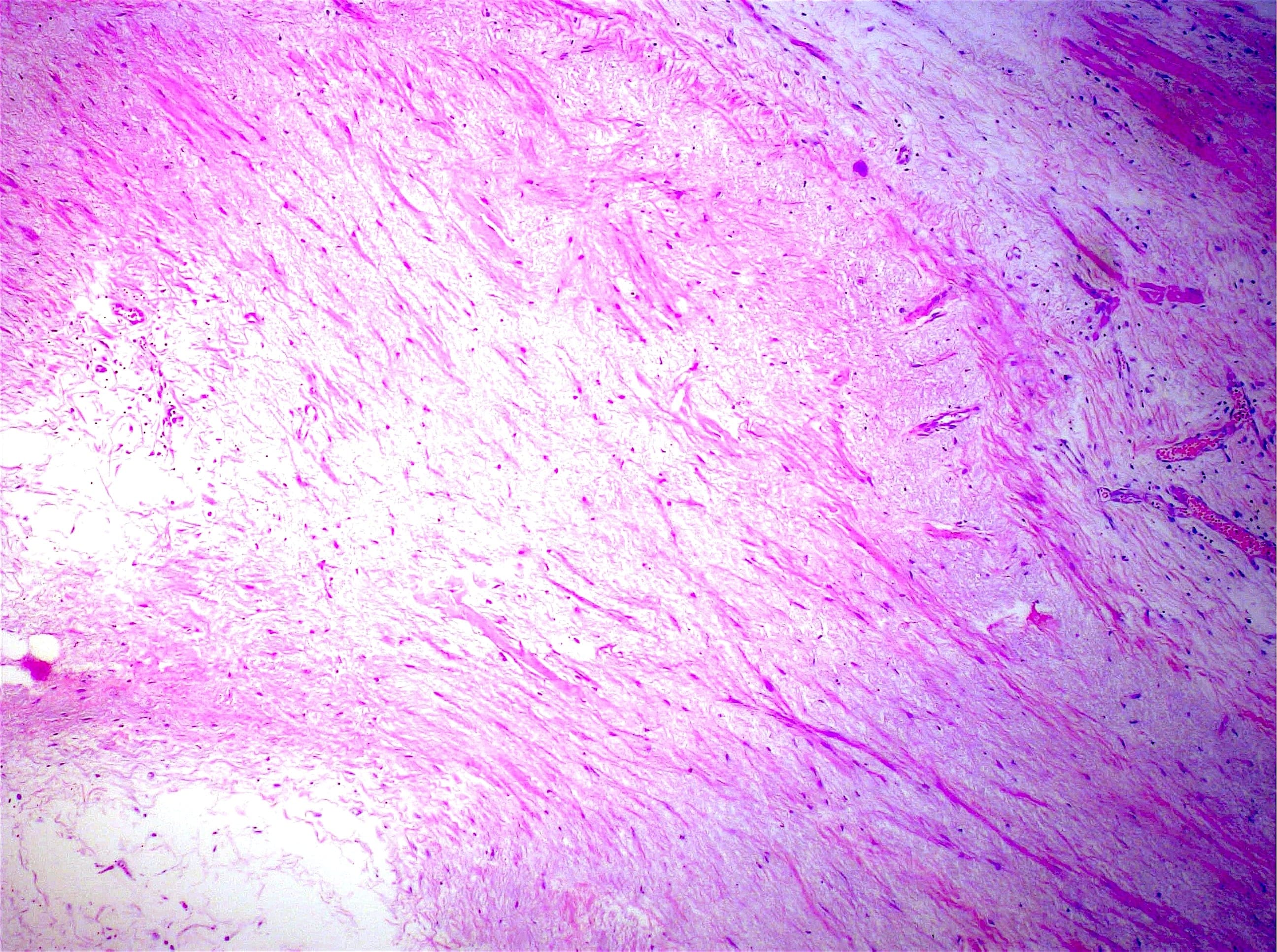
Esophageal leiomyomas
Contributed by @Andrew_Fltv on Twitter
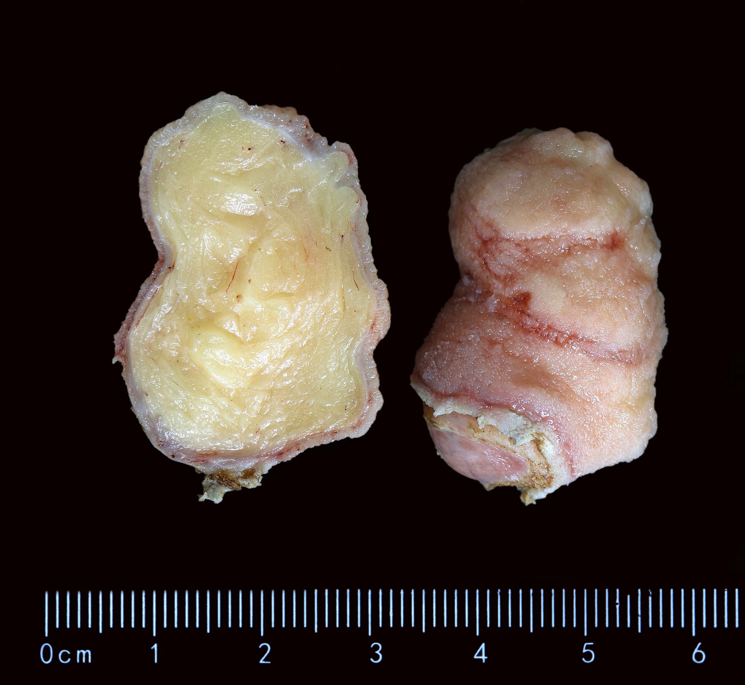
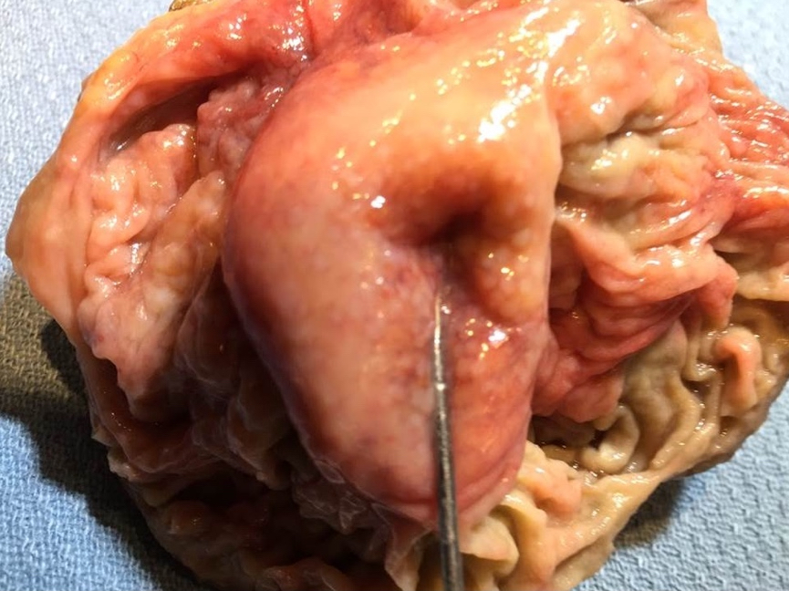
Lipoma
Images hosted on other servers:

Large pedunculated polypoid lipoma
Contributed by @Andrew_Fltv on Twitter

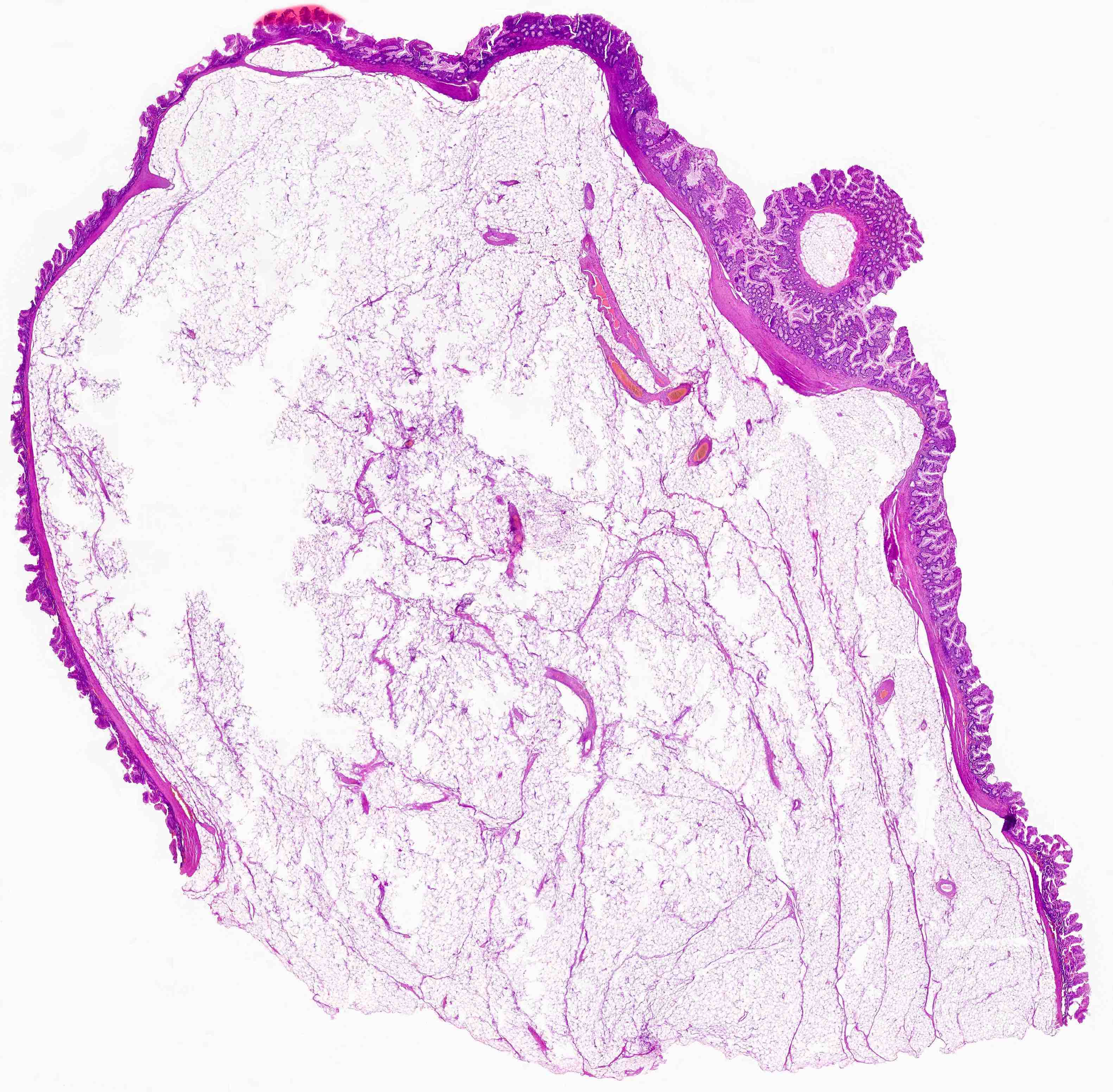
Lipoma

Lipoma
Images hosted on other servers:

Circumscribed collection of mature adipocytes
Images hosted on other servers:

Hypoechoic on ultrasound

CT with submucosal fat

Multidetector CT
Images hosted on other servers:

Smooth ileocecal lesion
Contributed by Jian-Hua Qiao, M.D.

Thickened ileocecal valve
Donut shaped ileocecal valve
Contributed by Jian-Hua Qiao, M.D.
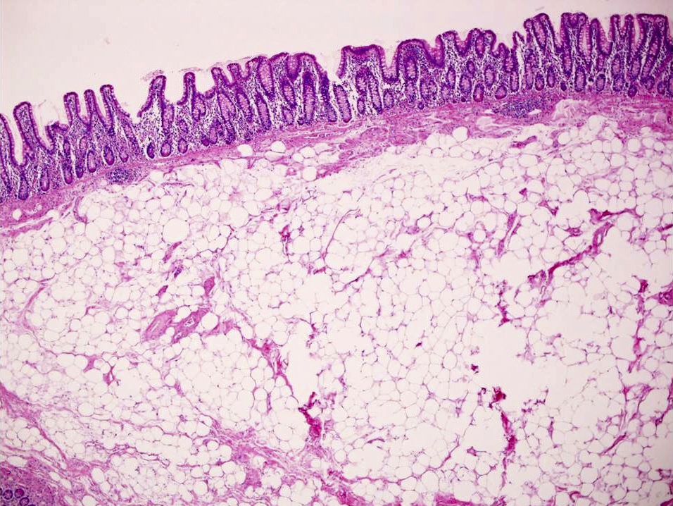
Unencapsulated submucosal adipose
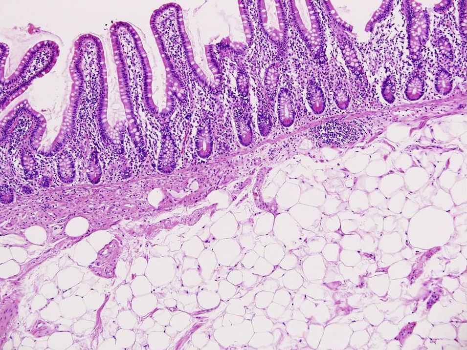
Villi resembling terminal ileum mucosa

Unencapsulated submucosal adipose

Well defined crypts resembling cecal mucosa
Images hosted on other servers:


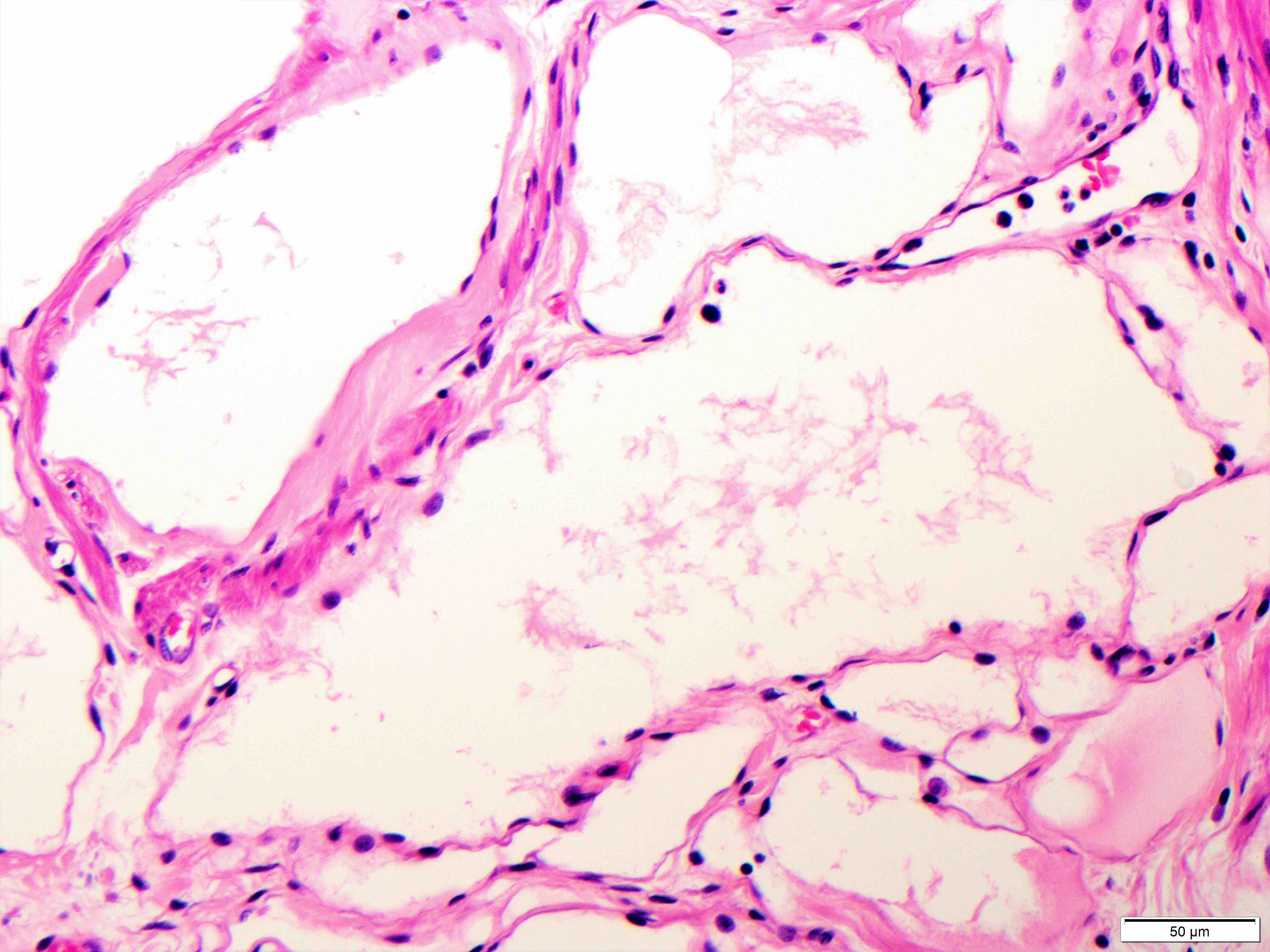
Markedly dilated lymphatics
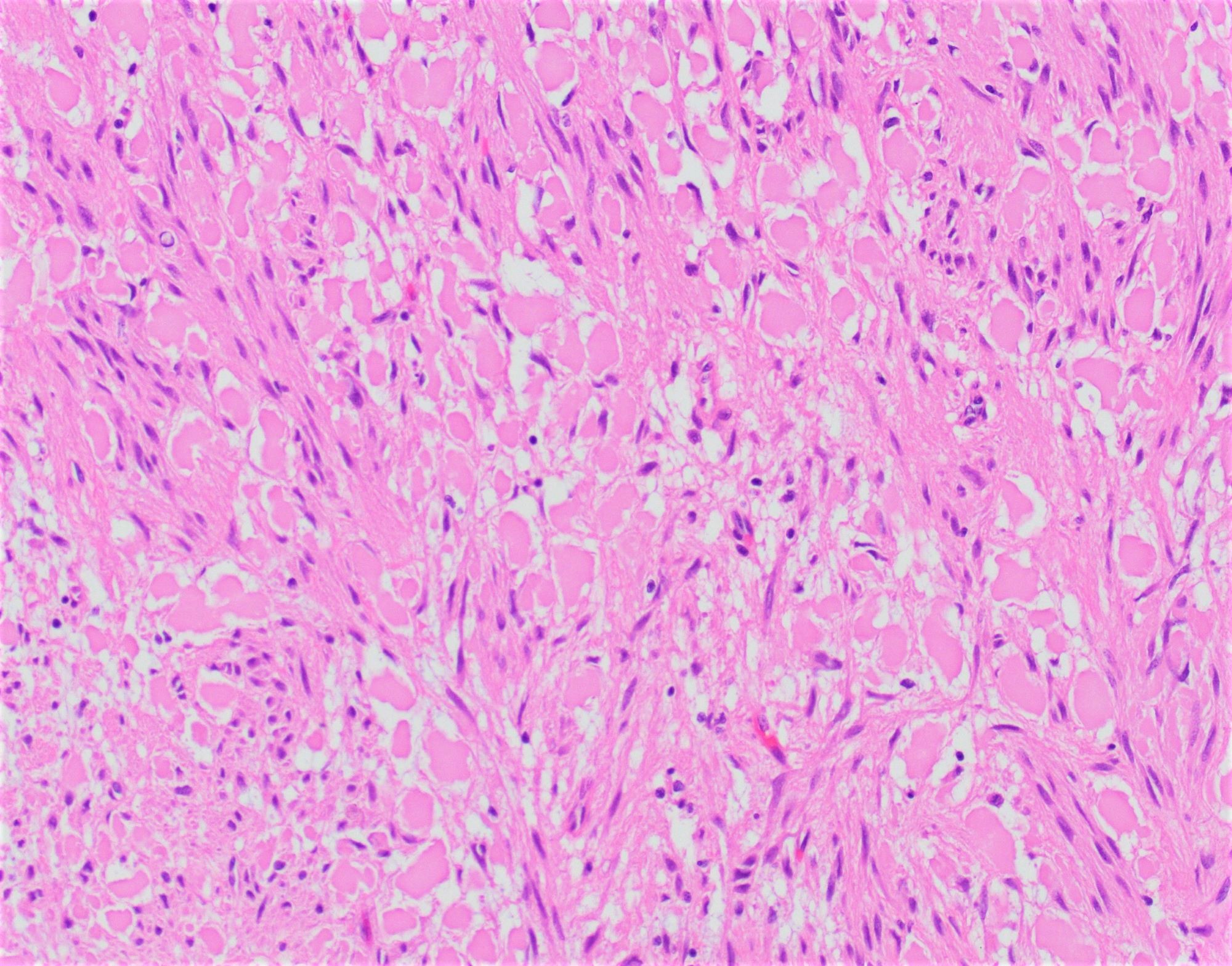
Contributed by Monika Vyas, M.D.
Ectatic lymphatics
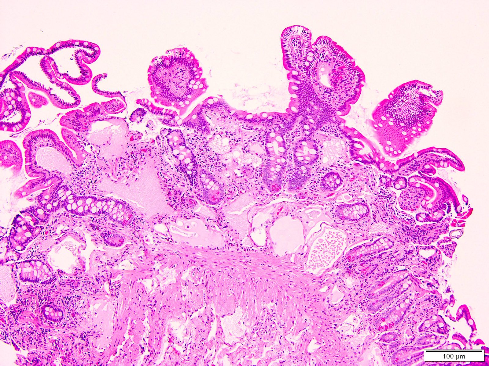
Dilated lymphatic channels

Polypoid lesion
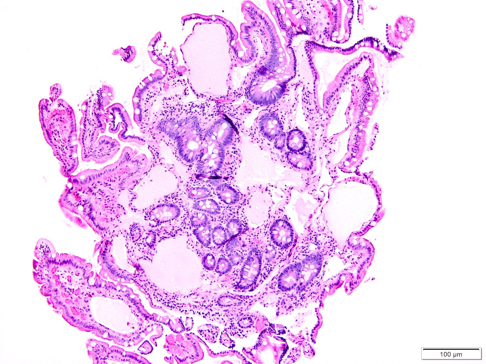
Irregularly dilated lymphatics
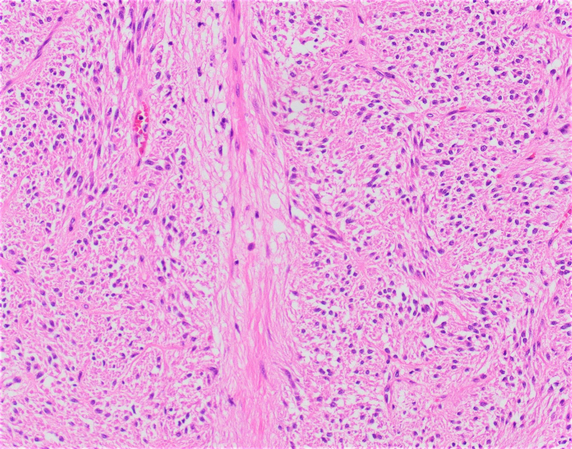
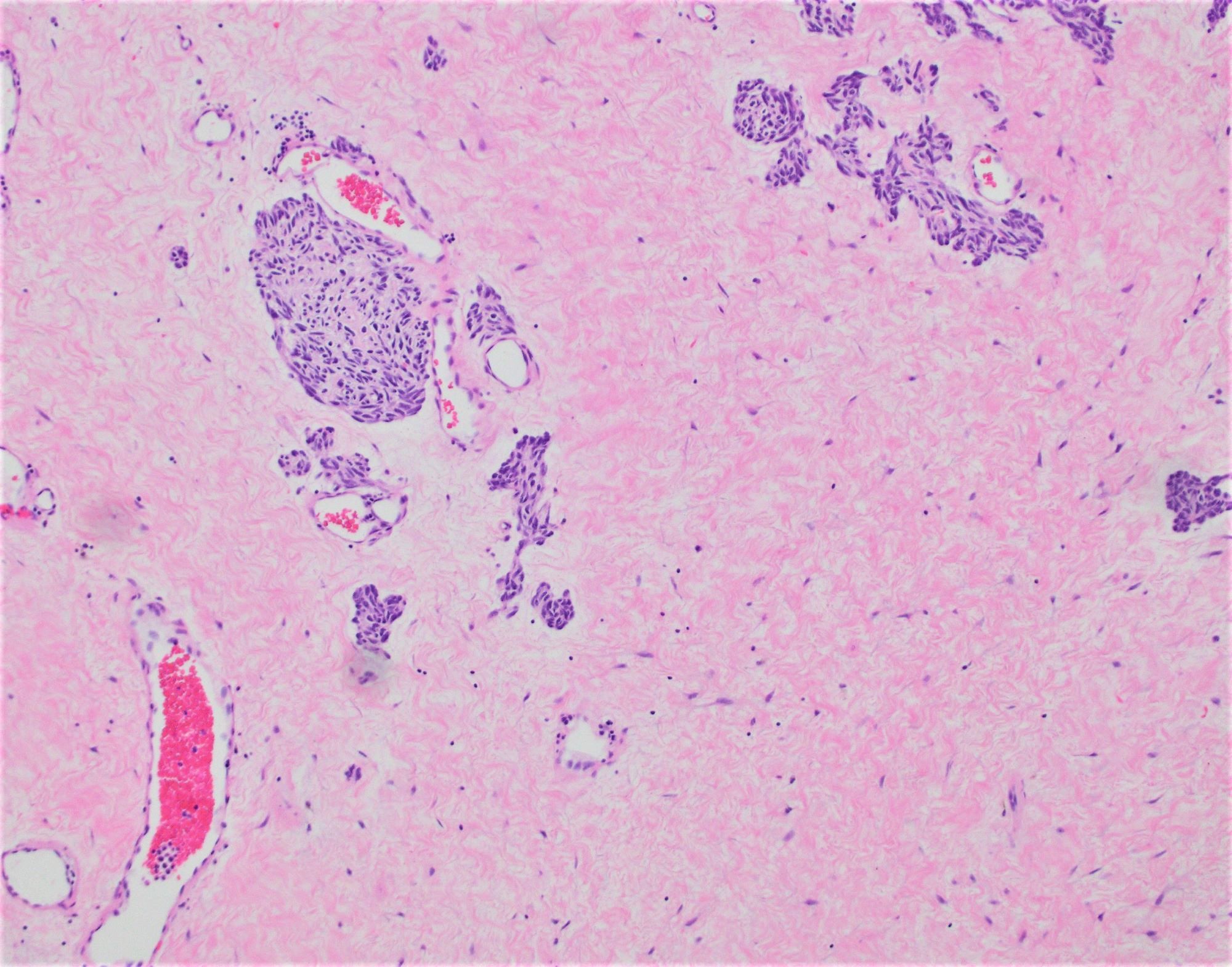
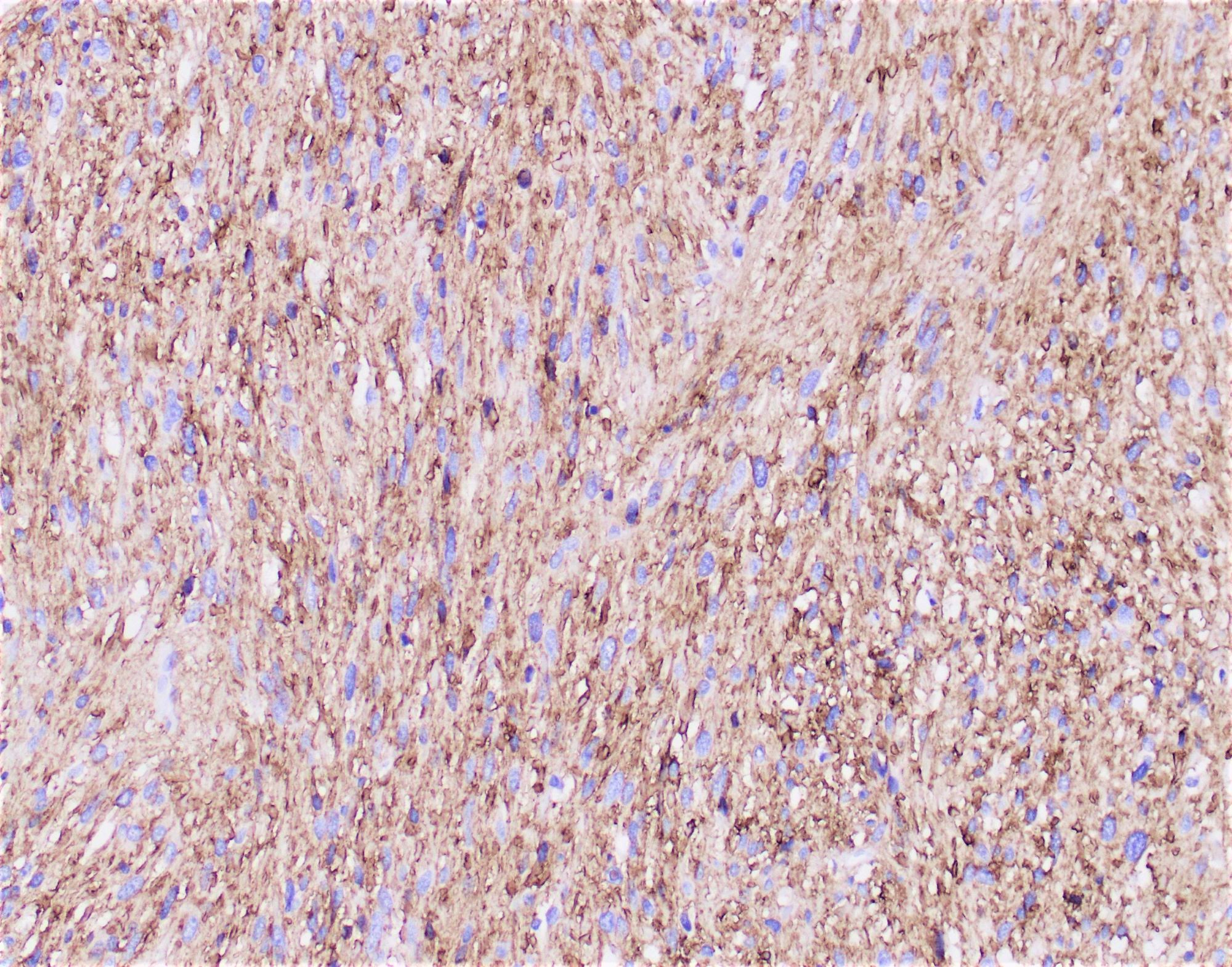
Contributed by Haider A. Mejbel, M.D.
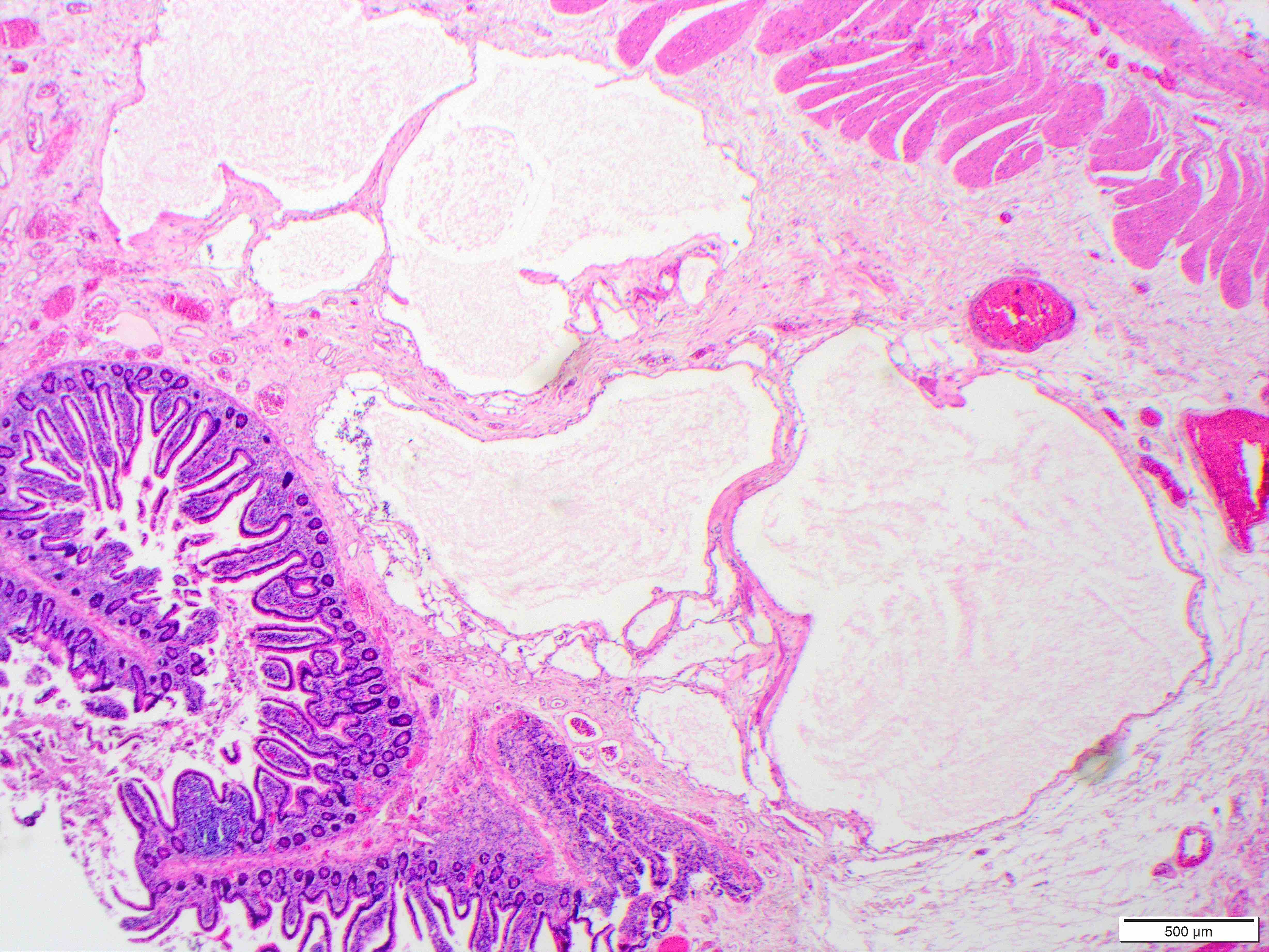
Lobular contour
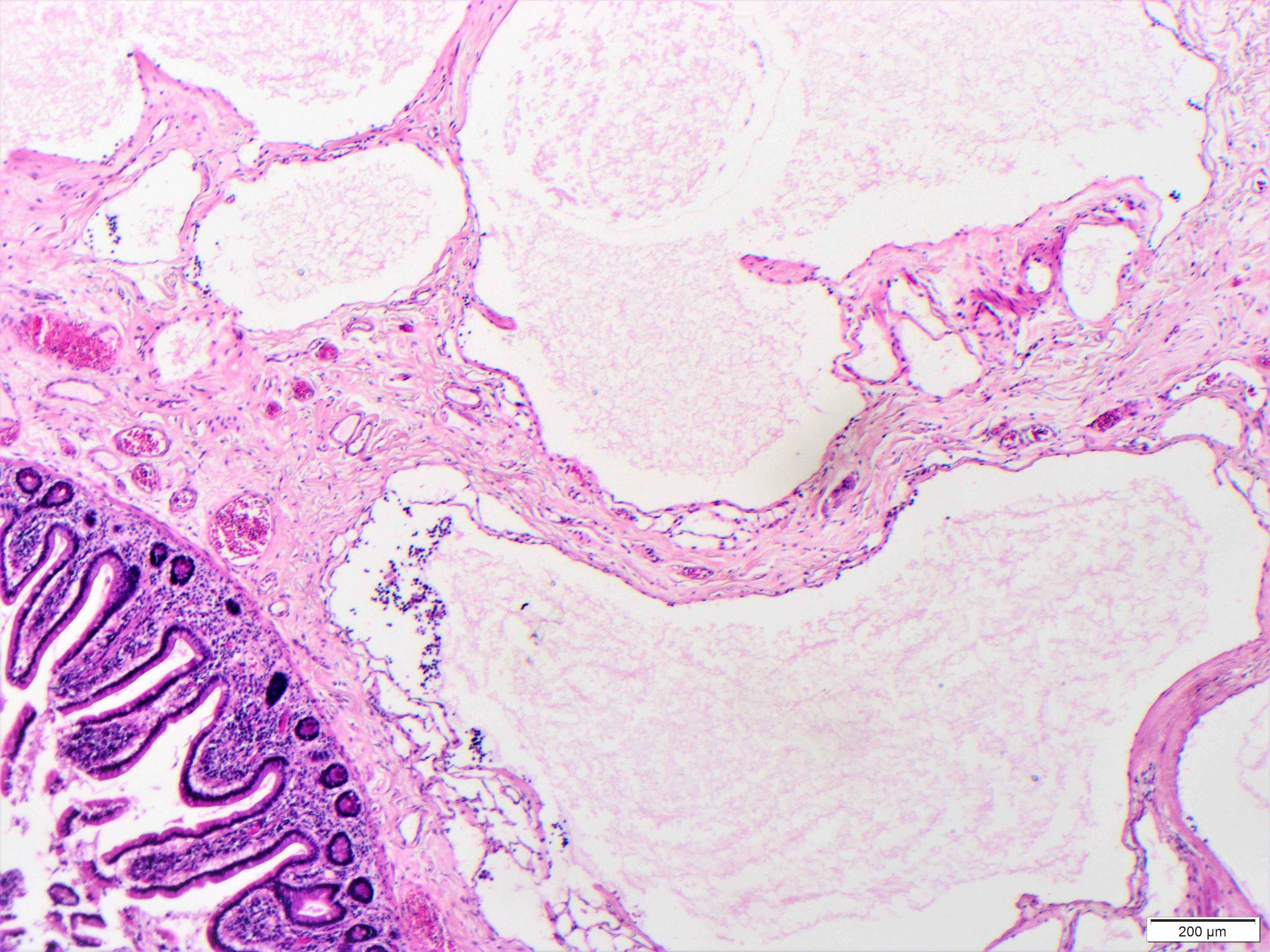
Dilated lymphatics
Endothelial cells
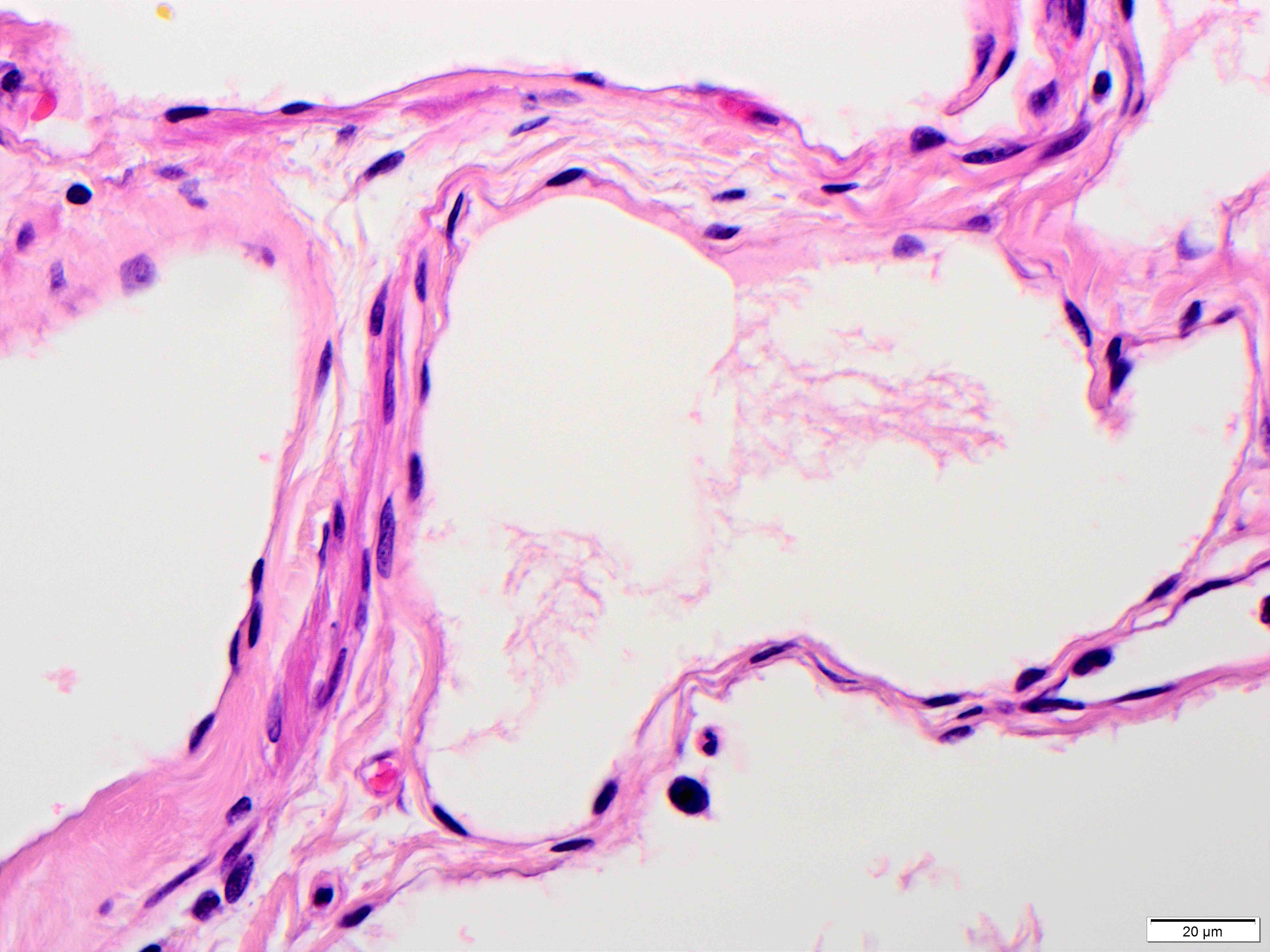
Nuclear uniformity
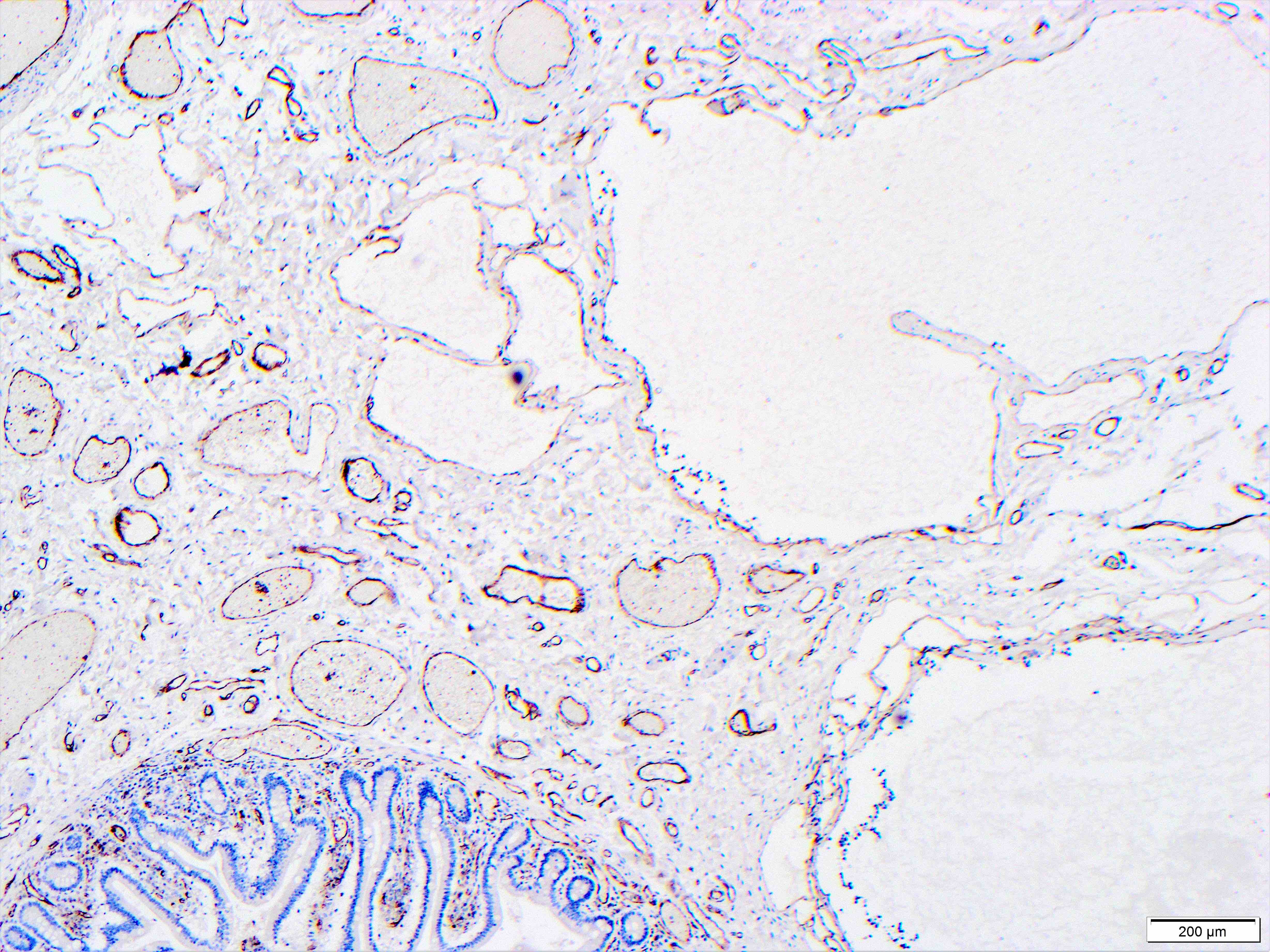
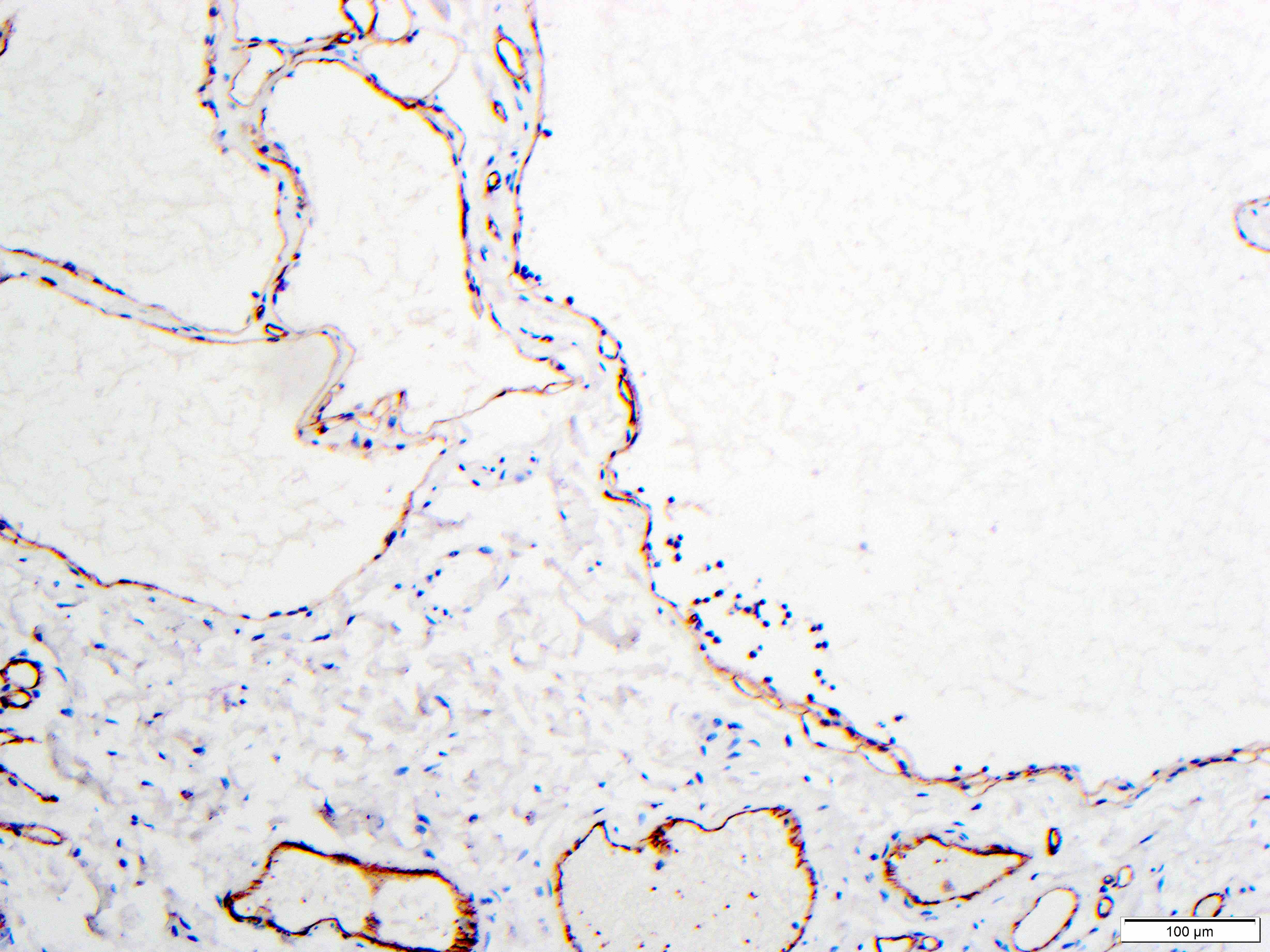
CD31 expression
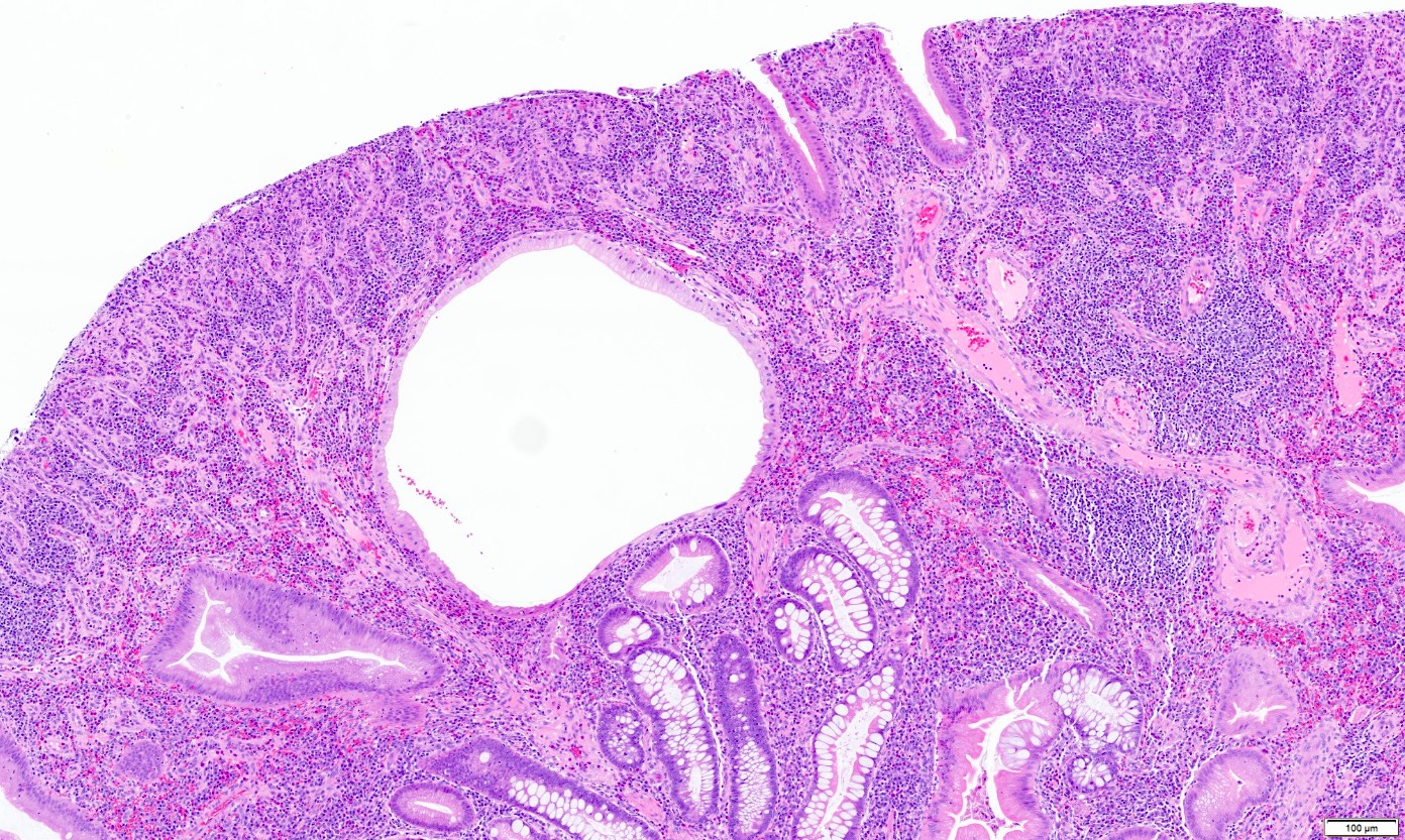
Contributed by Hanni Gulwani, M.D.
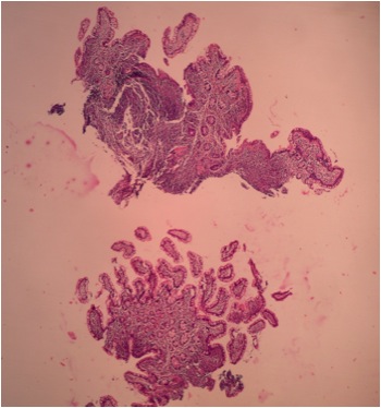
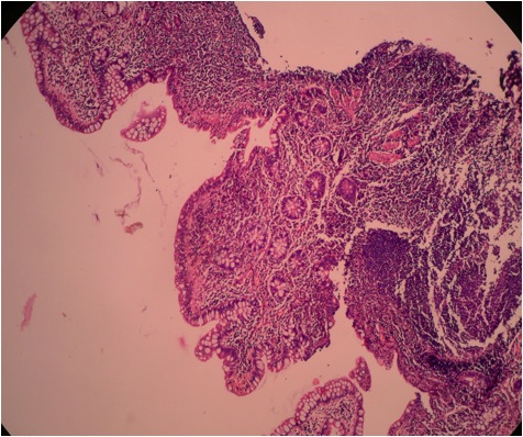
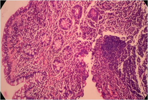
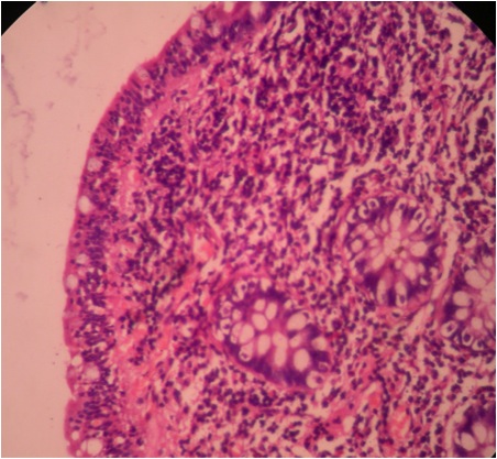
Lymphocytic ileitis: ileal biopsy from 28 year old man with chronic diarrhea
Images hosted on other servers:

Terminal ileum exhibiting
shows mucosal ulcers
due to nodular lymphoid
hyperplasia
Images hosted on other servers:


Various images
Contributed by Raul S. Gonzalez, M.D.
Low power
Intermediate power
High power
Images hosted on other servers:

Barium examination


Technetium 99m pertechnate scan (Meckel scan)
Contributed by Lizhi Zhang, M.D.
Giant Meckel diverticulum
Antimesenteric location
Luminal connection
Images hosted on other servers:

Meckel diverticulum with perforation
Contributed by Lizhi Zhang, M.D.
All layers of intestinal wall
Ectopic gastric mucosa
Ectopic pancreatic tissue
Case #106
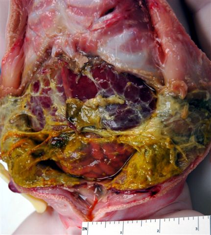
Abdominal cavity
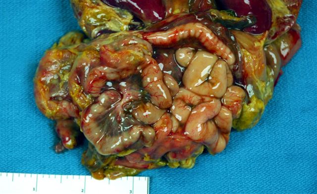
Small intestine
Case #106
Peritoneal surface
Images hosted on other servers:

TLR4 signaling in pathogenesis of necrotizing enterocolitis
Images hosted on other servers:


Various images
Images hosted on other servers:

Various images
Case #322


Mixed adenoneuroendocrine carcinoma
Line 2: CDX2 (left); chromogranin (middle); synaptophysin (right)
Images hosted on other servers:


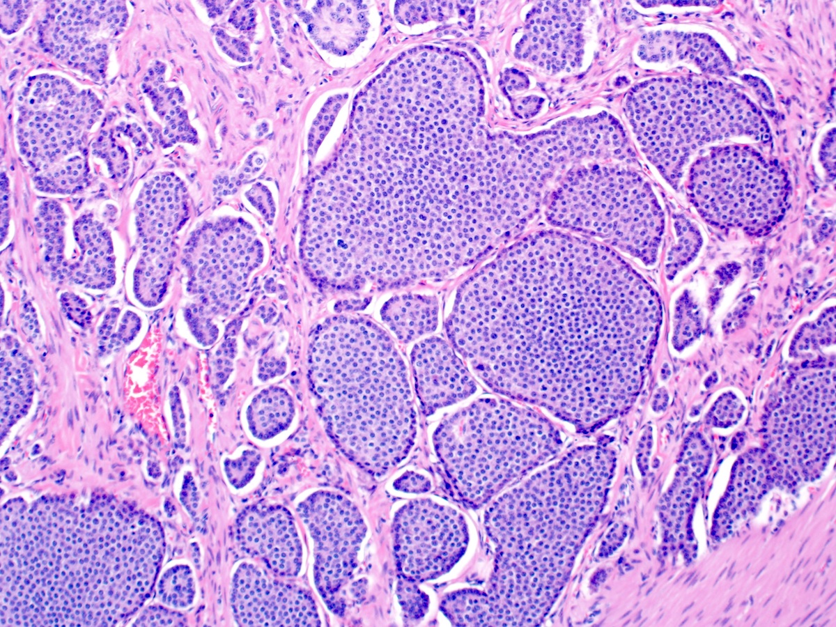
Large cell neuroendocrine carcinoma

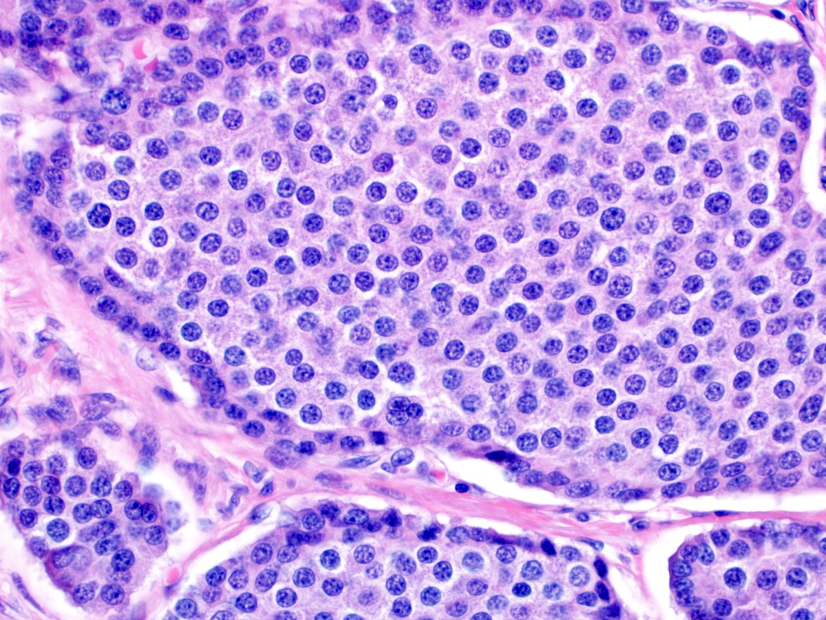
Small cell neuroendocrine carcinoma

Nests of small cells
with uniform round
nuclei with classic salt
and pepper chromatin
Images hosted on other servers:

PET / CT
Images hosted on other servers:


Ampullary NET on endoscopy
Contributed by Megan Kinn, D.O., M.S.
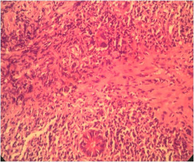
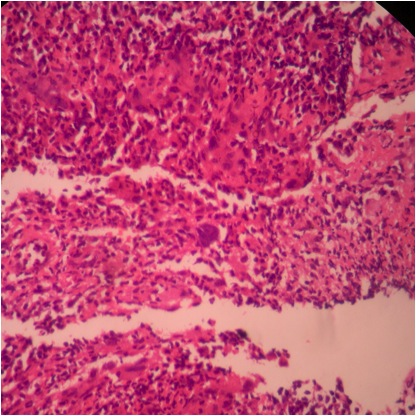
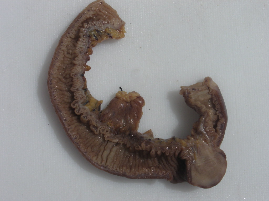
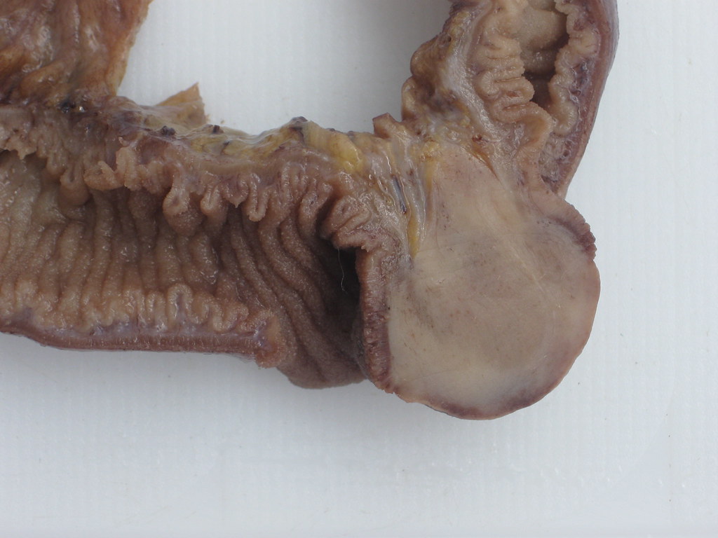
Ileal neuroendocrine tumor
Liver metastasis
Contributed by Megan Kinn, D.O., M.S.
Ileal NET extending to mucosa
Nests of tumor cells
Salt and pepper chromatin
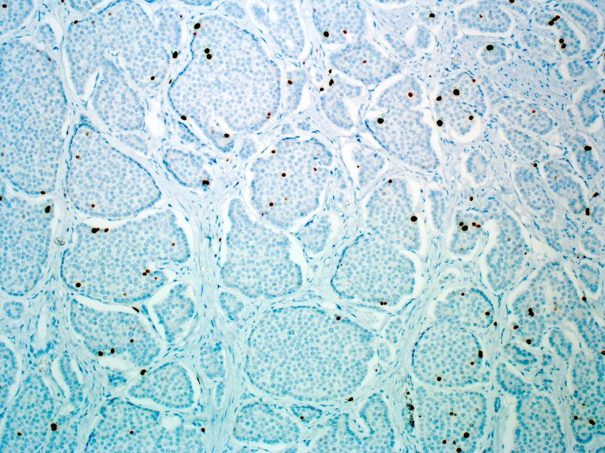
Ki67
Contributed by Wei Zheng, M.D., Ph.D.
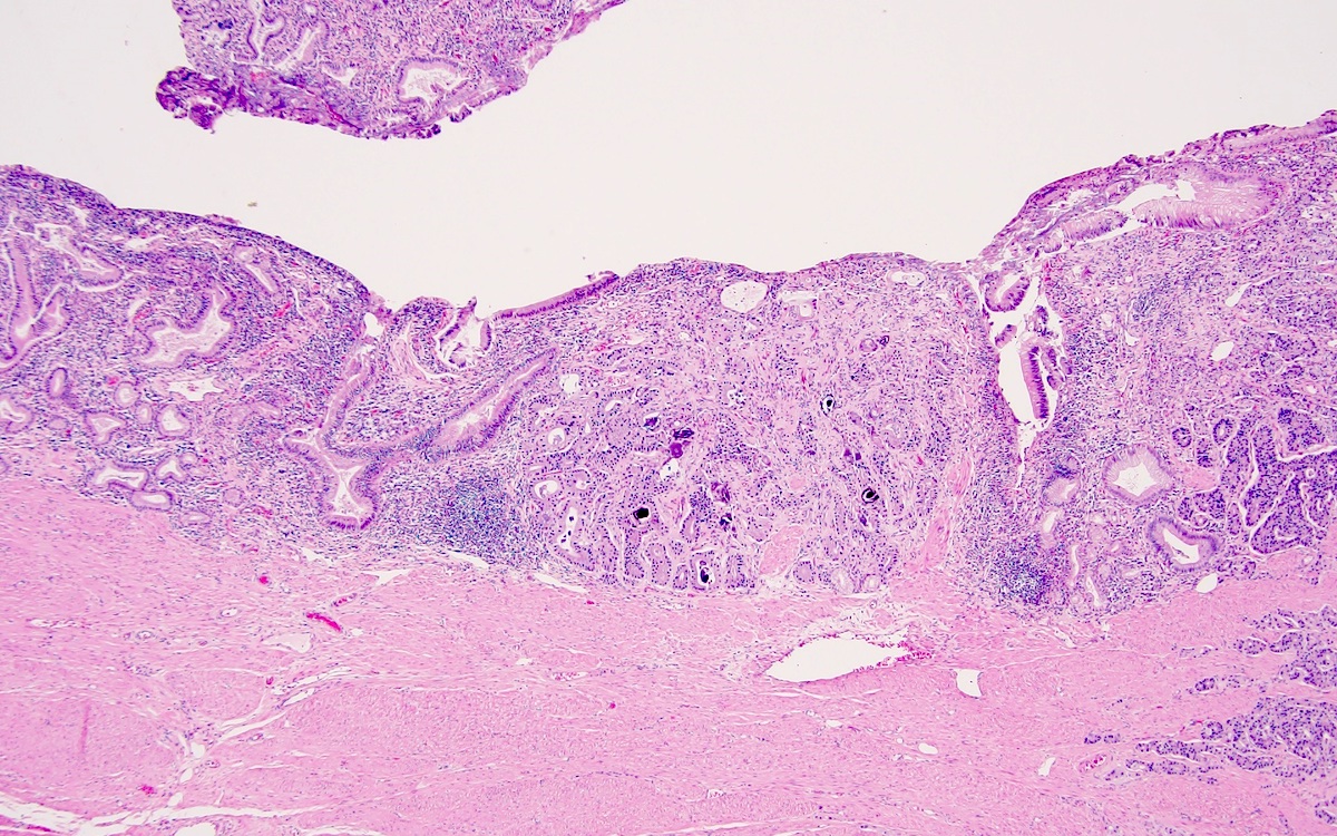
Ampullary NET
Trabecular and glandular architecture
Psammoma bodies
Images hosted on other servers:

Thickened bowel wall
Images hosted on other servers:

Normal colonoscopy

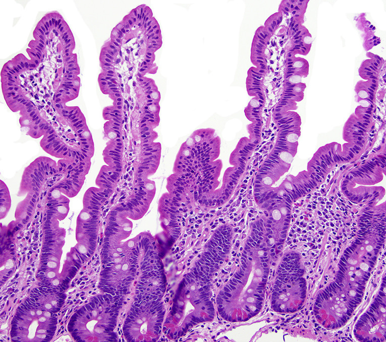
Duodenal villous atrophy

Duodenal fold attenuation
Contributed by David Hernandez Gonzalo, M.D.
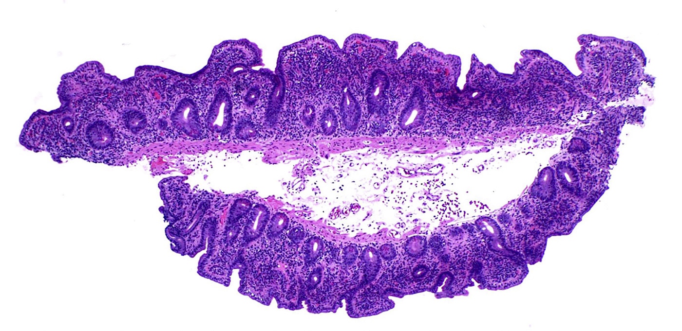
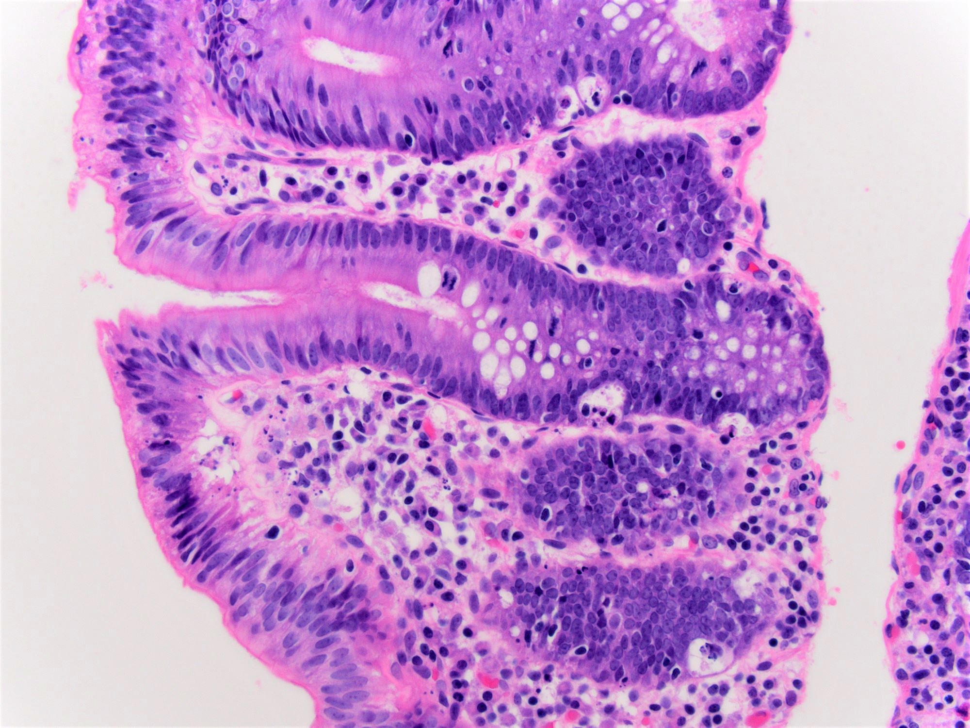
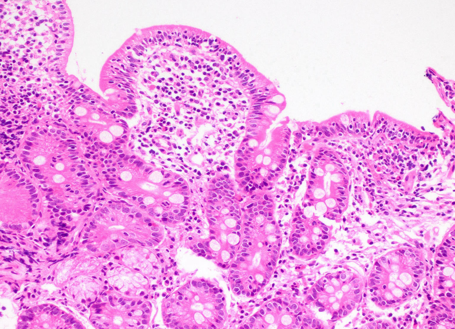
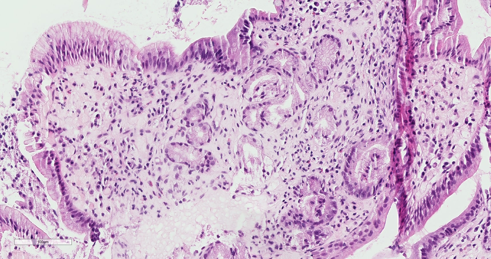
Moderate villous atrophy and crypt hyperplasia
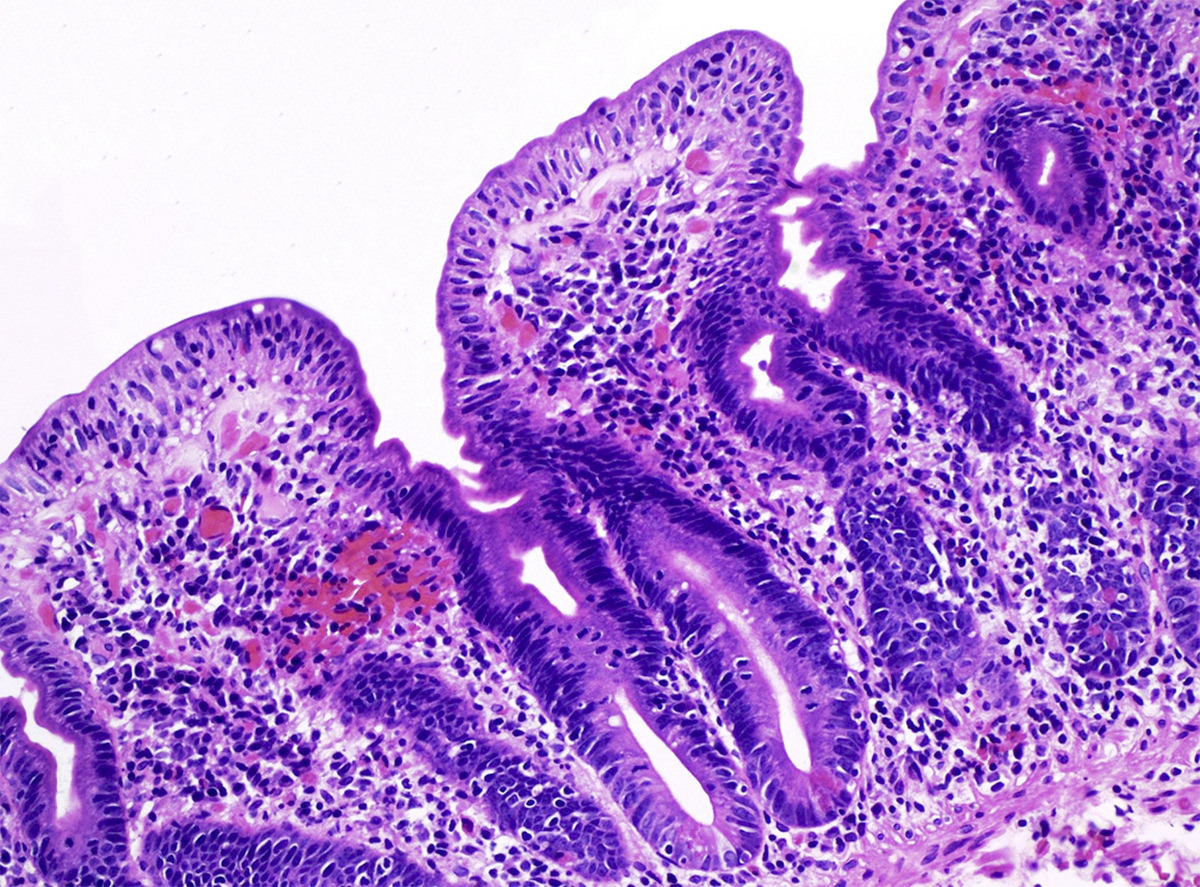
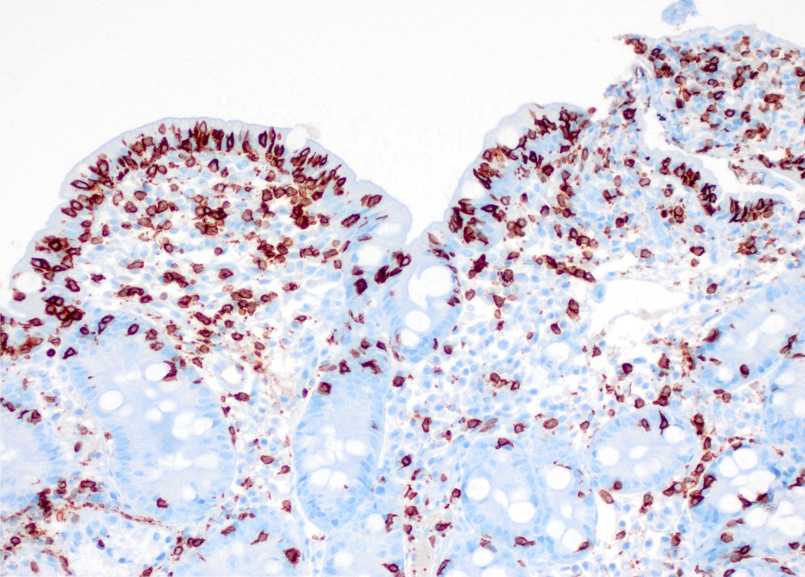
Increased intraepithelial lymphocytes
Focal active inflammation
Complete histologic recovery
Contributed by Mohamed Mostafa, M.D.
Surface foveolar metaplasia and Brunner gland hyperplasia
Foveolar metaplasia and lamina propria expansion
H. pylori associated peptic duodenitis
Surface foveolar metaplasia
Contributed by @RaulSGonzalezMD on Twitter
Contributed by @RaulSGonzalezMD on Twitter (see original post here)">
 Helicobacter pylori colonizing foveolar metaplasia in the duodenum? bit.ly/38K3WT0 #pathology #PathTwitter #PathOutPic"
Contributed by @RaulSGonzalezMD on Twitter (see original post here)">
Peptic duodenitis
Images hosted on other servers:

Axial T1 weighted image - enhancement of rounded PJP
Images hosted on other servers:

Hyperpigmented macules of the perioral area

Lobular duodenal polyp
Images hosted on other servers:

Duodenal dilatation, arborizing polyps

Arborizing polyps, pedunculated and sessile

Intraluminal polyp
Contributed by Krutika S. Patel, M.B.B.S., M.D. and @SueEPig on Twitter
Jejunal Peutz-Jeghers polyp
Tree-like arborization of smooth muscle

Distinct lobular configuration of epithelium
Bland epithelial component
Surface erosion
Epithelial misplacement
Peutz-Jeghers polyp
Small bowel, Peutz-Jeghers polyp
Images hosted on other servers:

Endoscopy
Images hosted on other servers:

Various images
Images hosted on other servers:

Various images
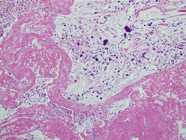
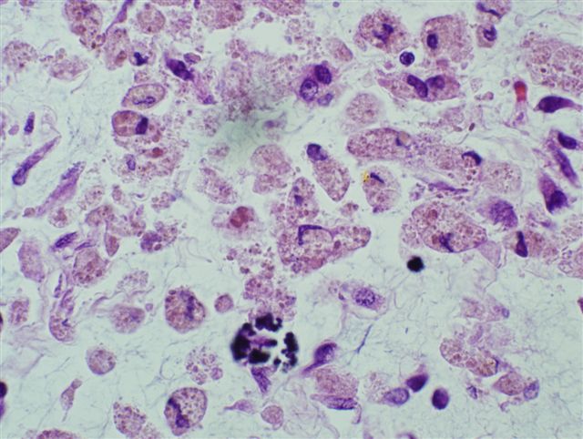
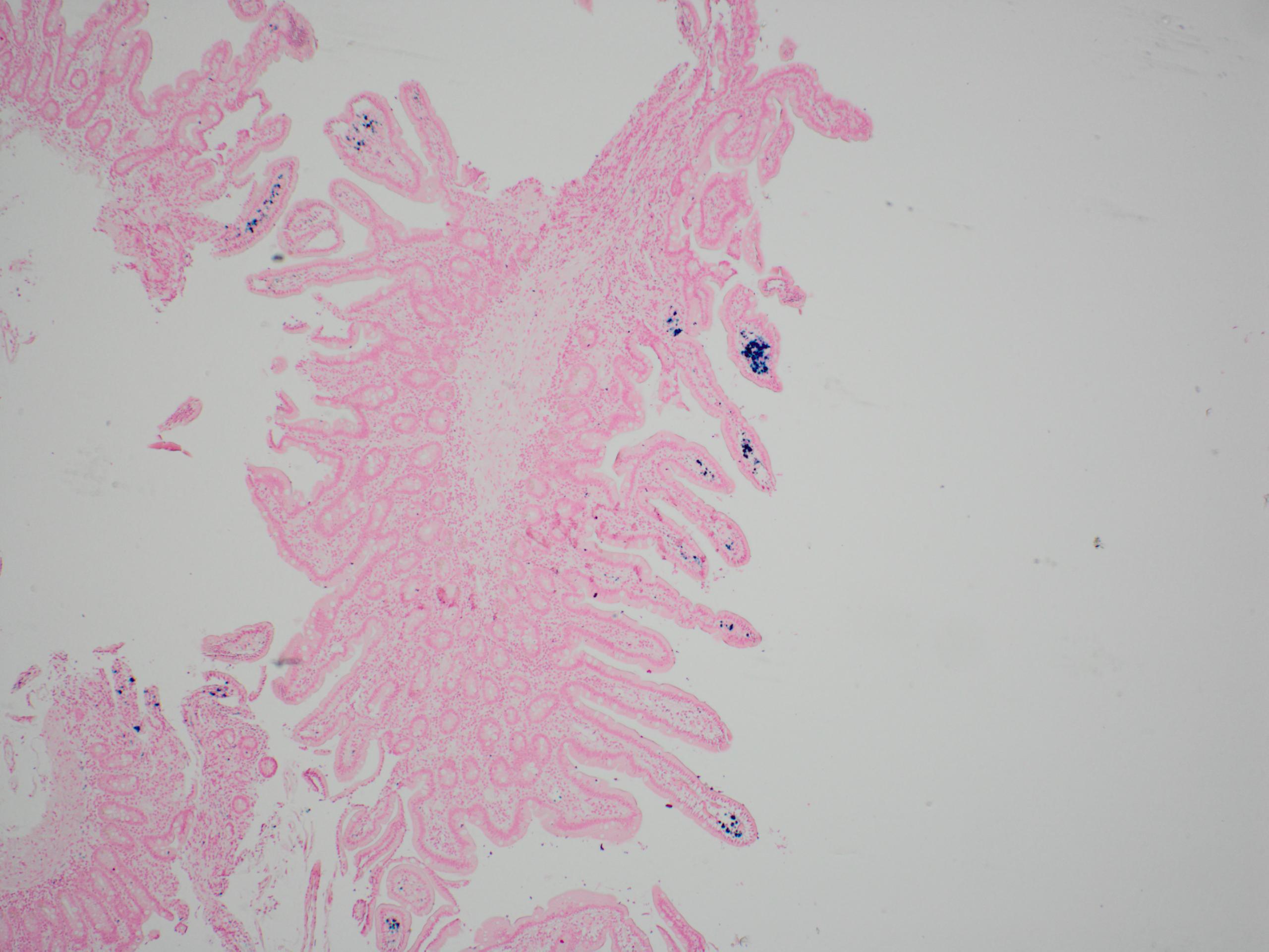
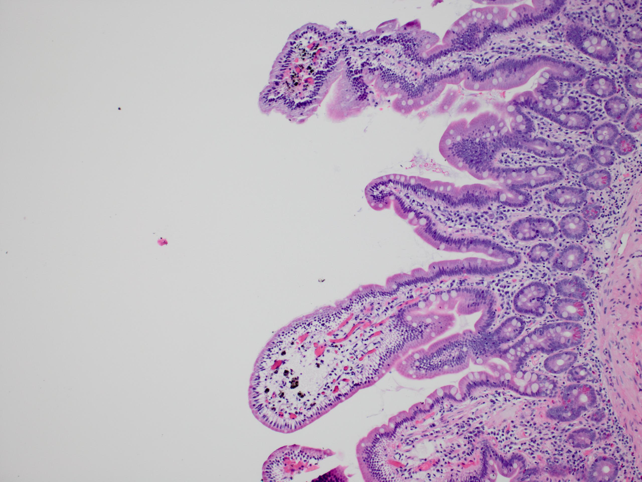
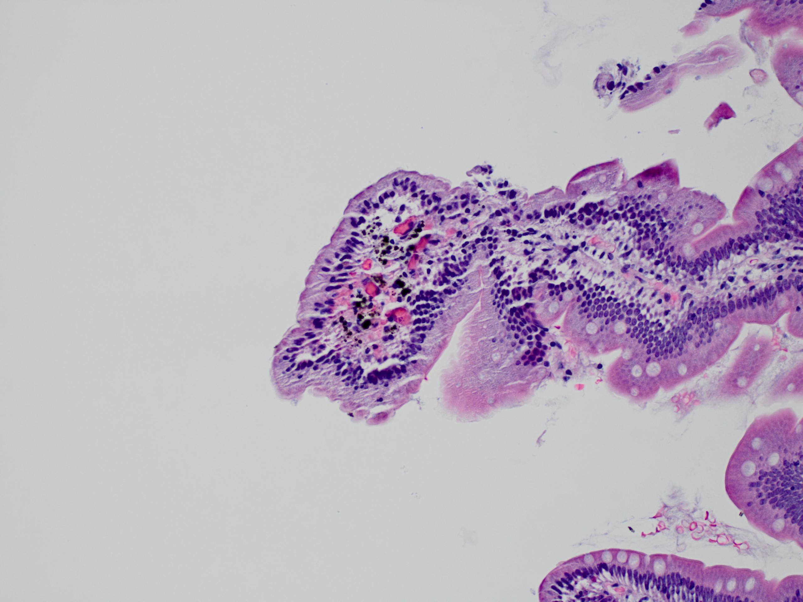
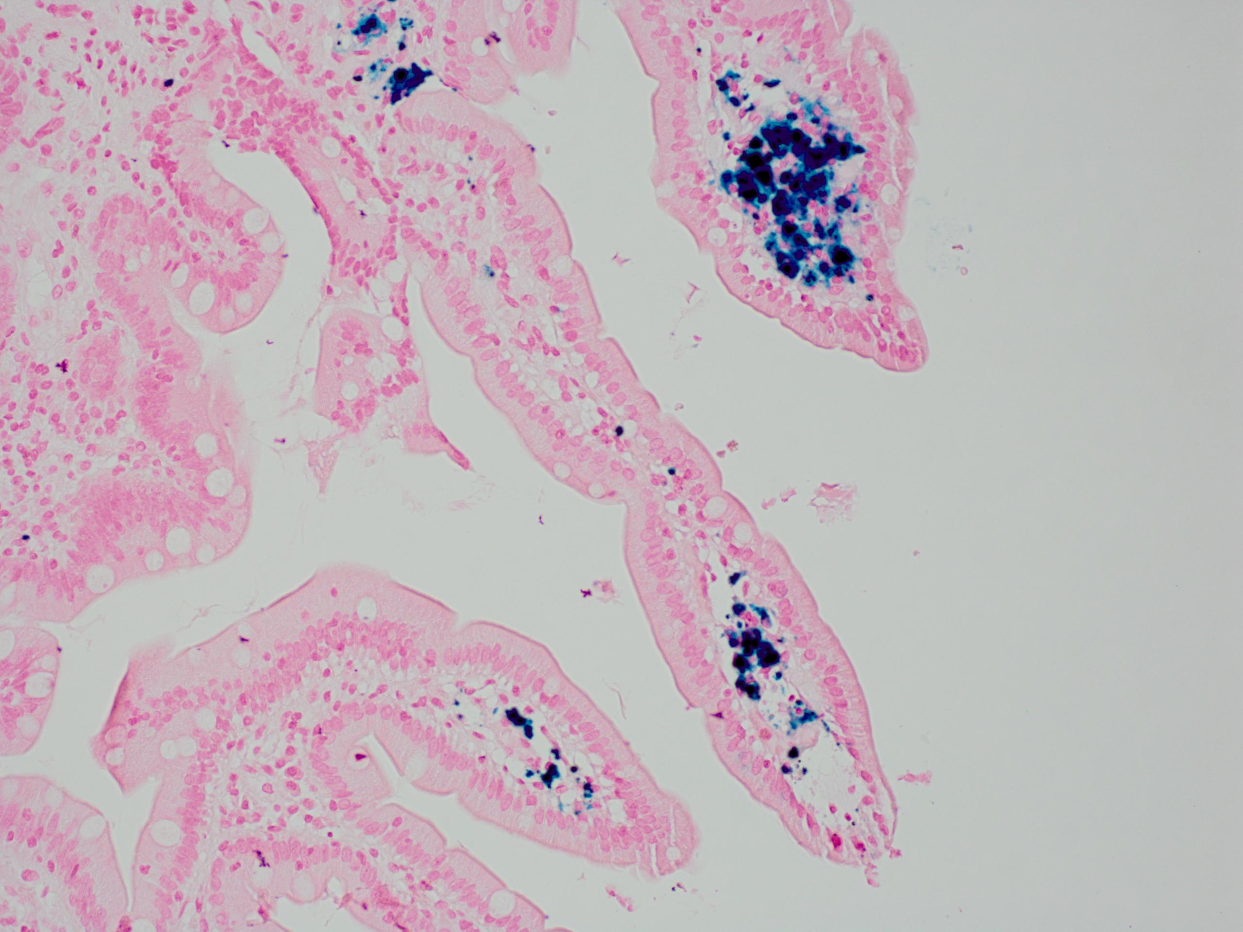
Contributed by @liverwei on Twitter
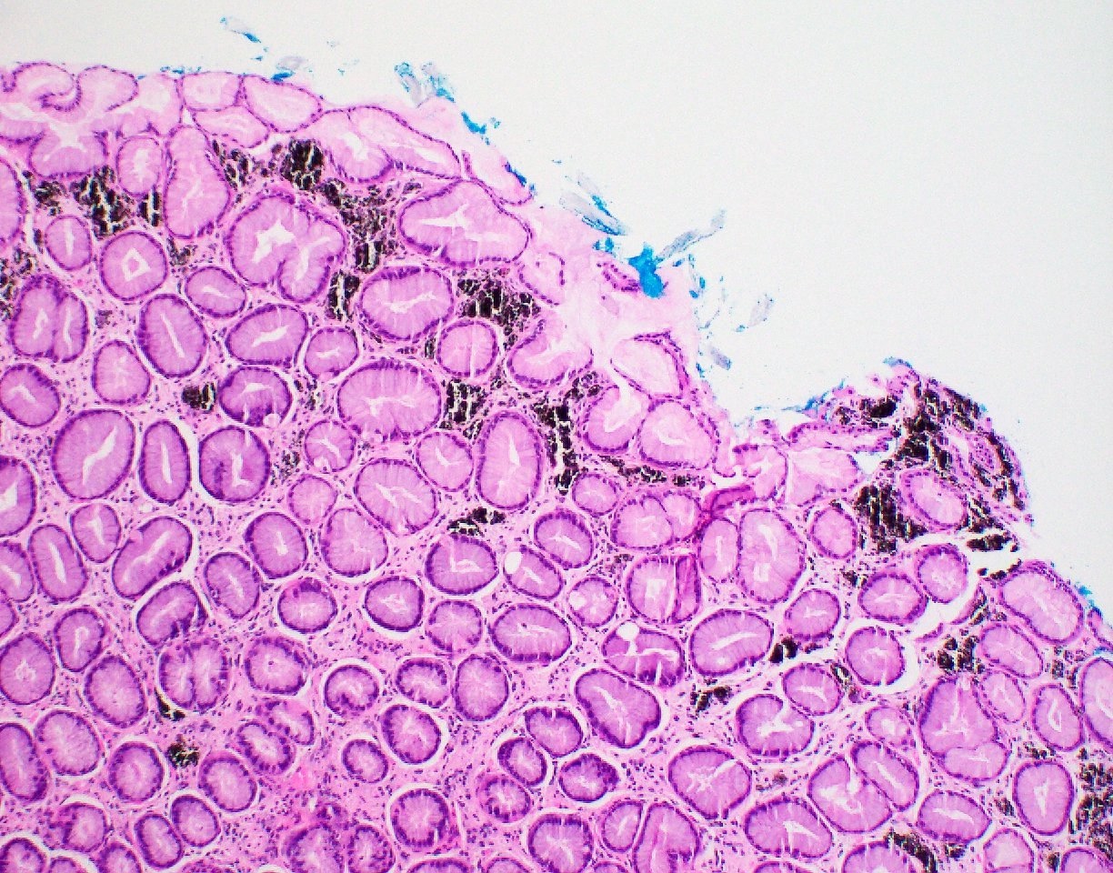
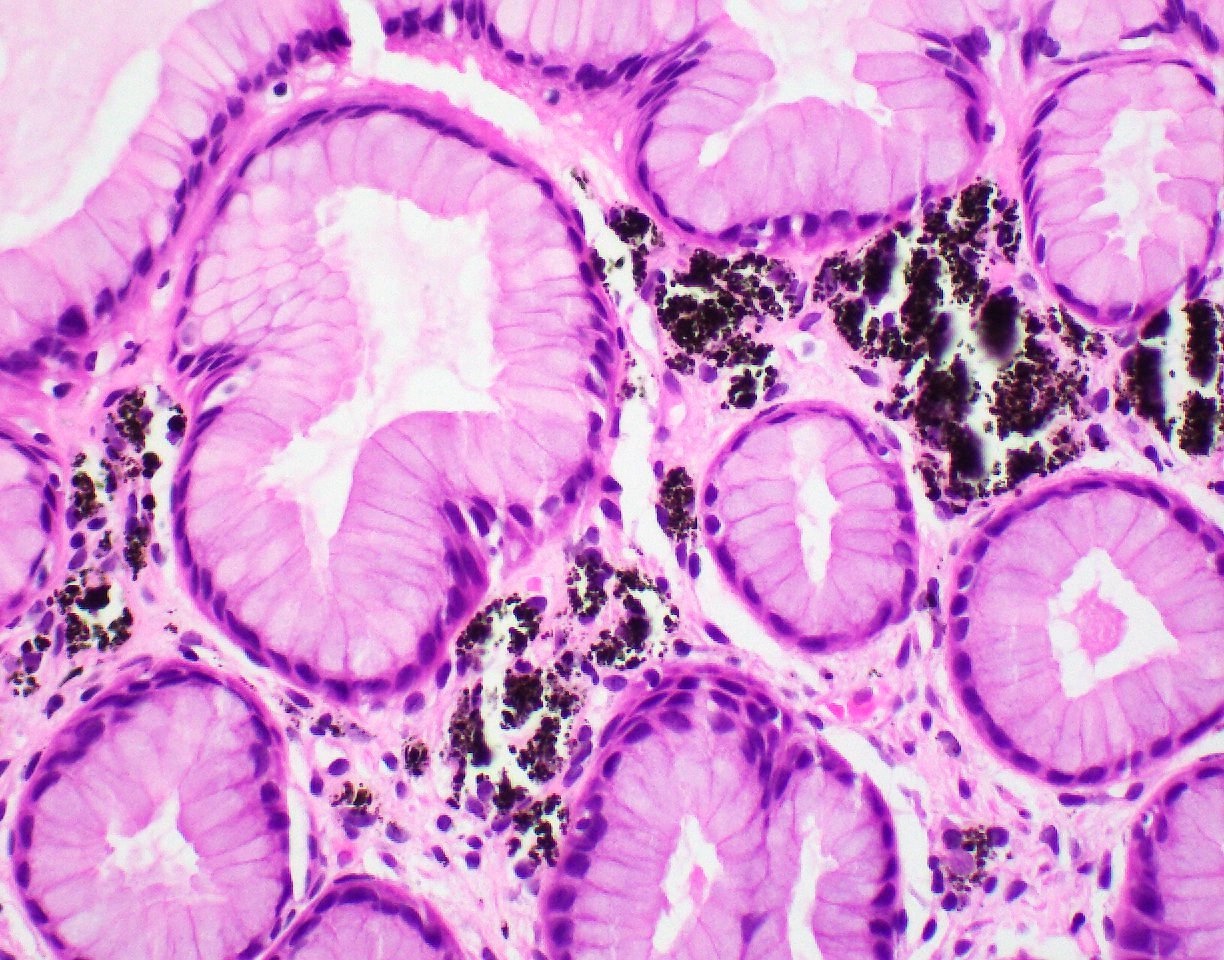
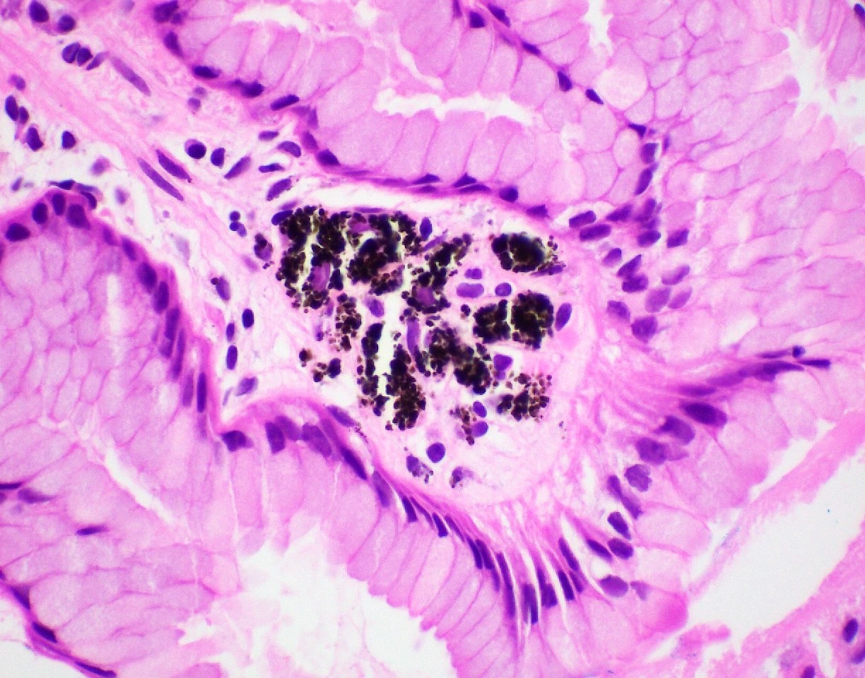
Pseudomelanosis duodeni
Case #231

Various images
Images hosted on other servers:


Various images
Images hosted on other servers:

Acute changes (mice)
Images hosted on other servers:

Acute cellular rejection (CT)

Acute cellular rejection (MRI)
Images hosted on other servers:

Endoscopic appearance of ACR
Contributed by Jen Rytych, M.D.

Mild ACR, mild inflammation

Focal villous blunting
Few apoptotic figures

Erosion and crypt dropout
Many apoptotic figures

Extensive ulceration
Images hosted on other servers:

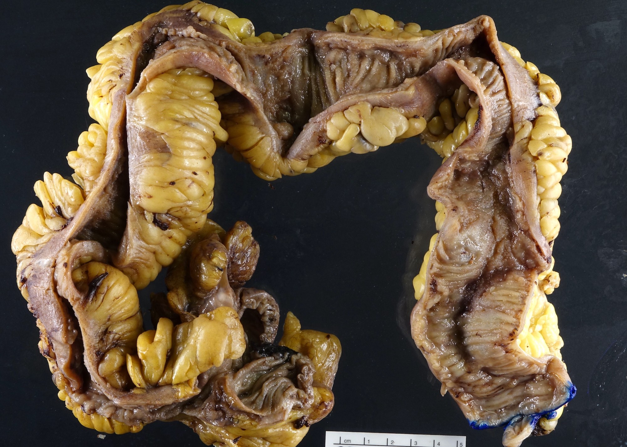
Encapsulating thick,
white adherent
membrane encasing
small bowel
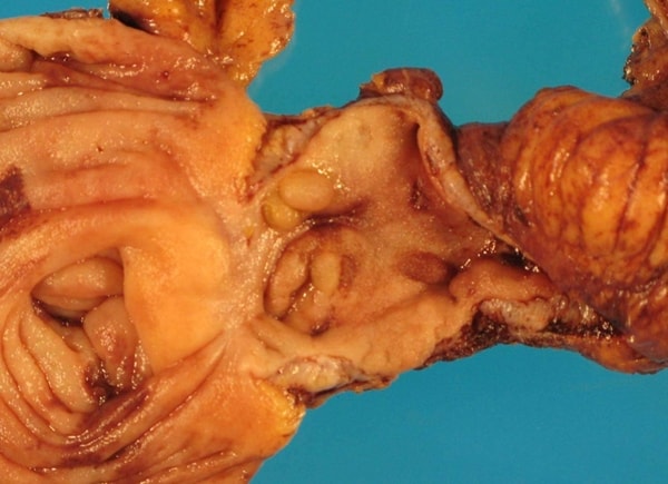
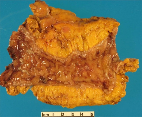
Contributed by Sajna V. M. Kutty, M.D. (Case of the Month #482)

Right colon limited resection for possible enteric duplication cyst in the distal ileum
Contributed by Sajna V. M. Kutty, M.D. (Case of the Month #482)
Images hosted on other servers:

Regional lymph nodes of the Ampulla of Vater
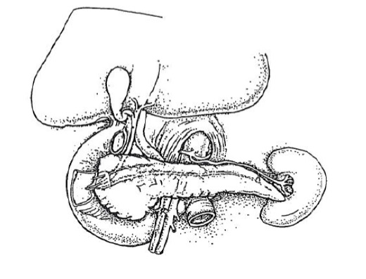
Pancreas / Ampulla of Vater staging
Images hosted on other servers:
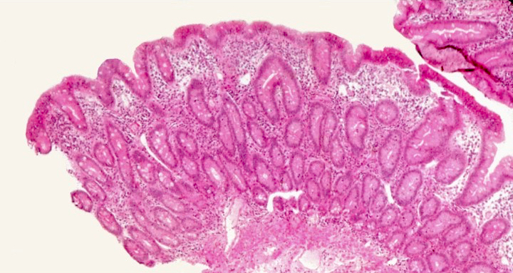
Mild backwash ileitis in moderately active cecal chronic ulcerative colitis resection specimen; mucosa
of distal ileum is edematous and villi are slightly wider and flatter than normal; most of the active colitis
stops abruptly at transition point between the two mucosae; the two most distal villi have the greatest
degree of injury, including blunting, edema and neutrophils in the lamina propria and surface epithelium

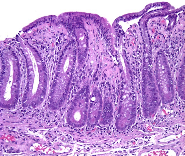
Moderate backwash ileitis

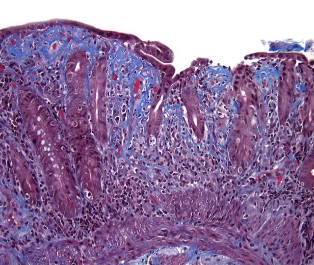
Severe backwash ileitis

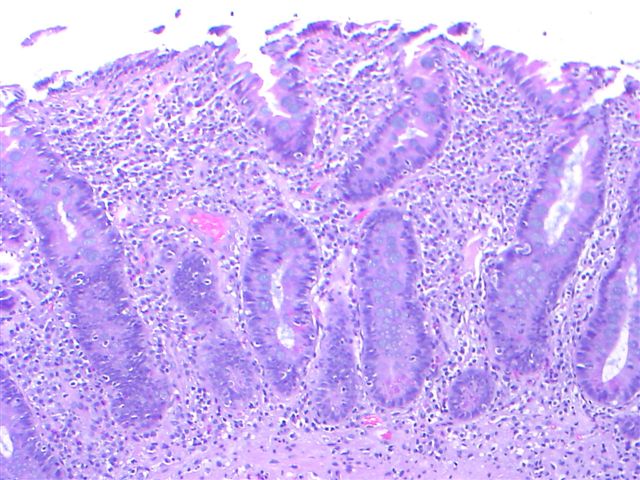
Terminal ileum inflammation: (A) grade
1, cryptitis; (B) grade 2, scattered
crypt abscesses; (C) grade 3, numerous
crypt abscesses; (D) grade 4, ulcer
Images hosted on other servers:

Contrast study
Images hosted on other servers:

Endoscopic findings
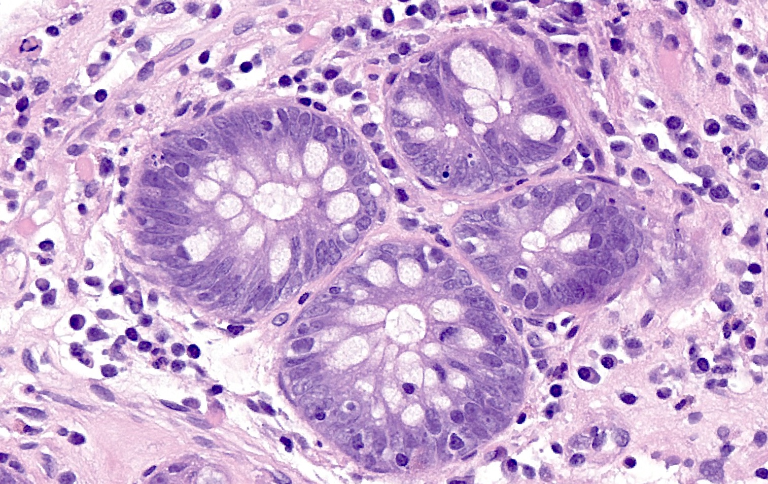
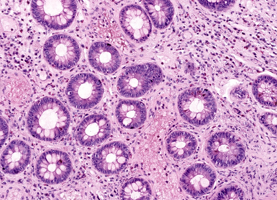
Contributed by John D. Paulsen, M.D. and Alexandros D. Polydorides, M.D., Ph.D.
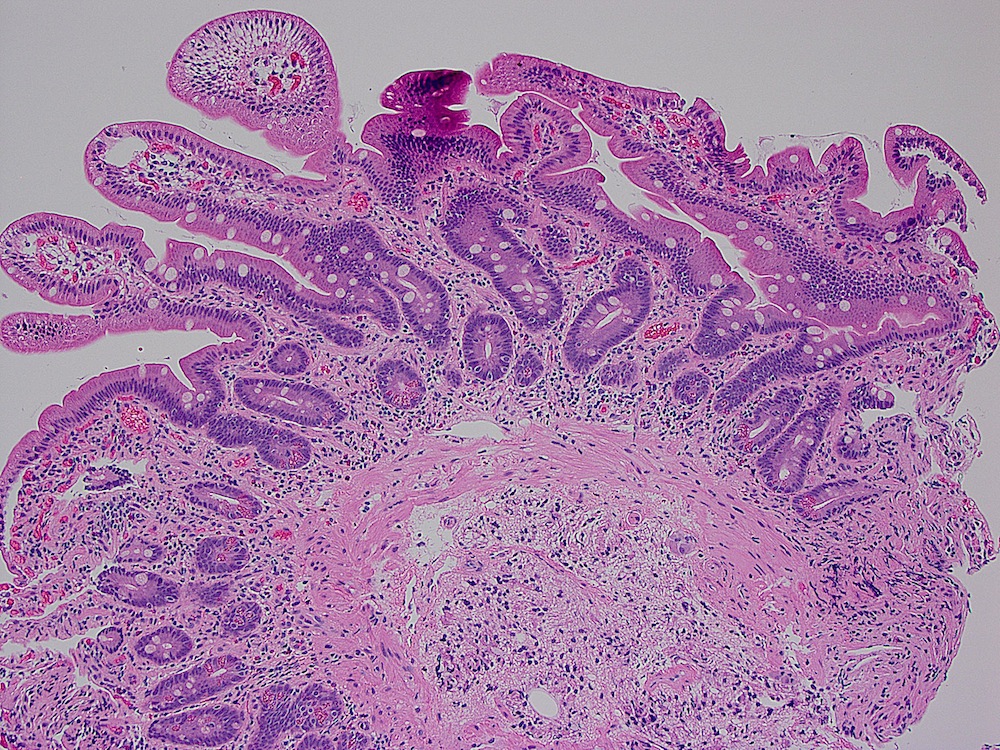
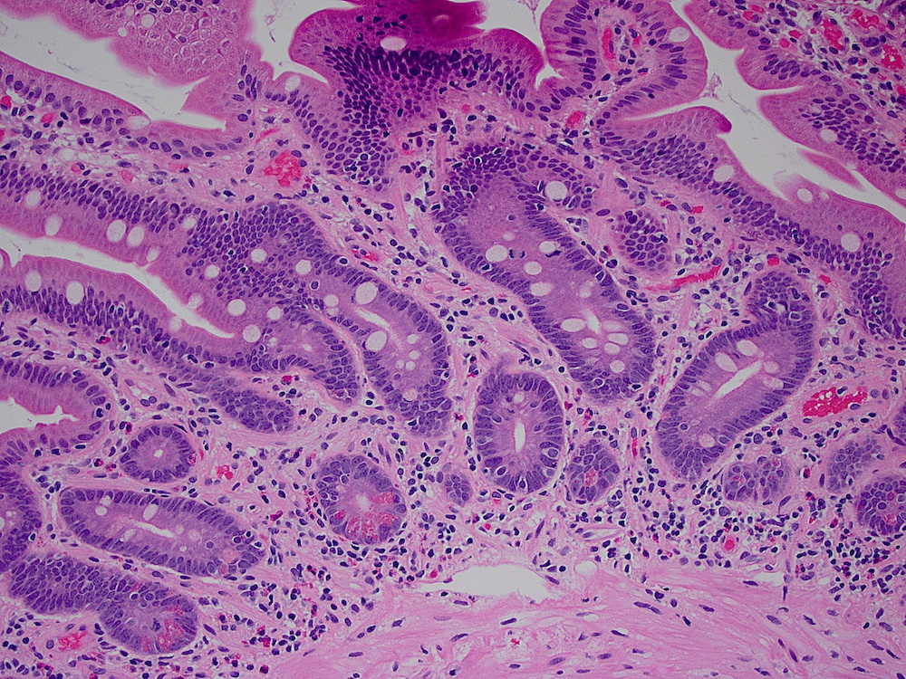
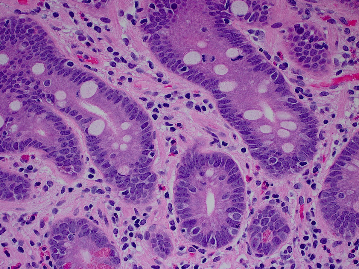
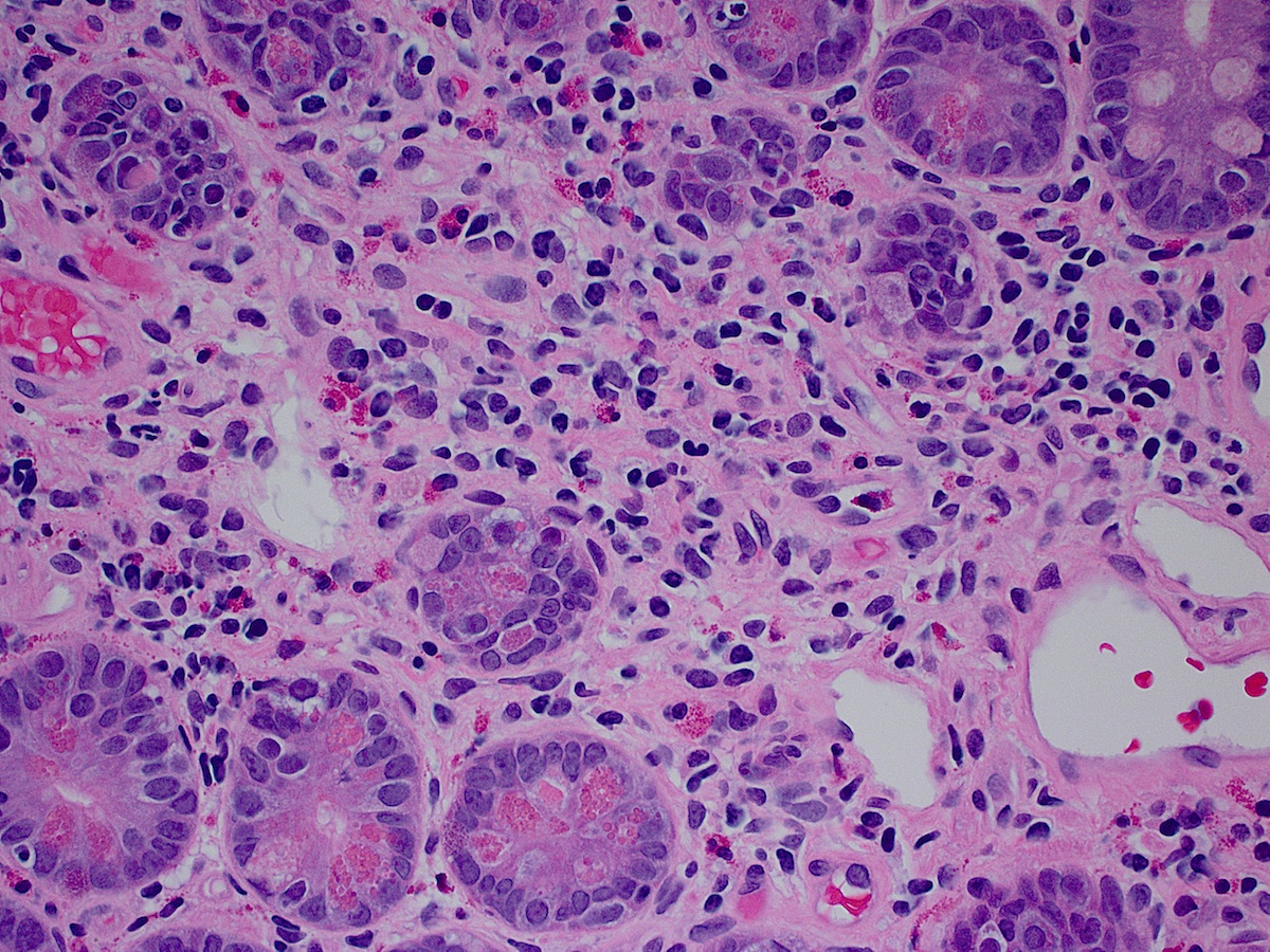
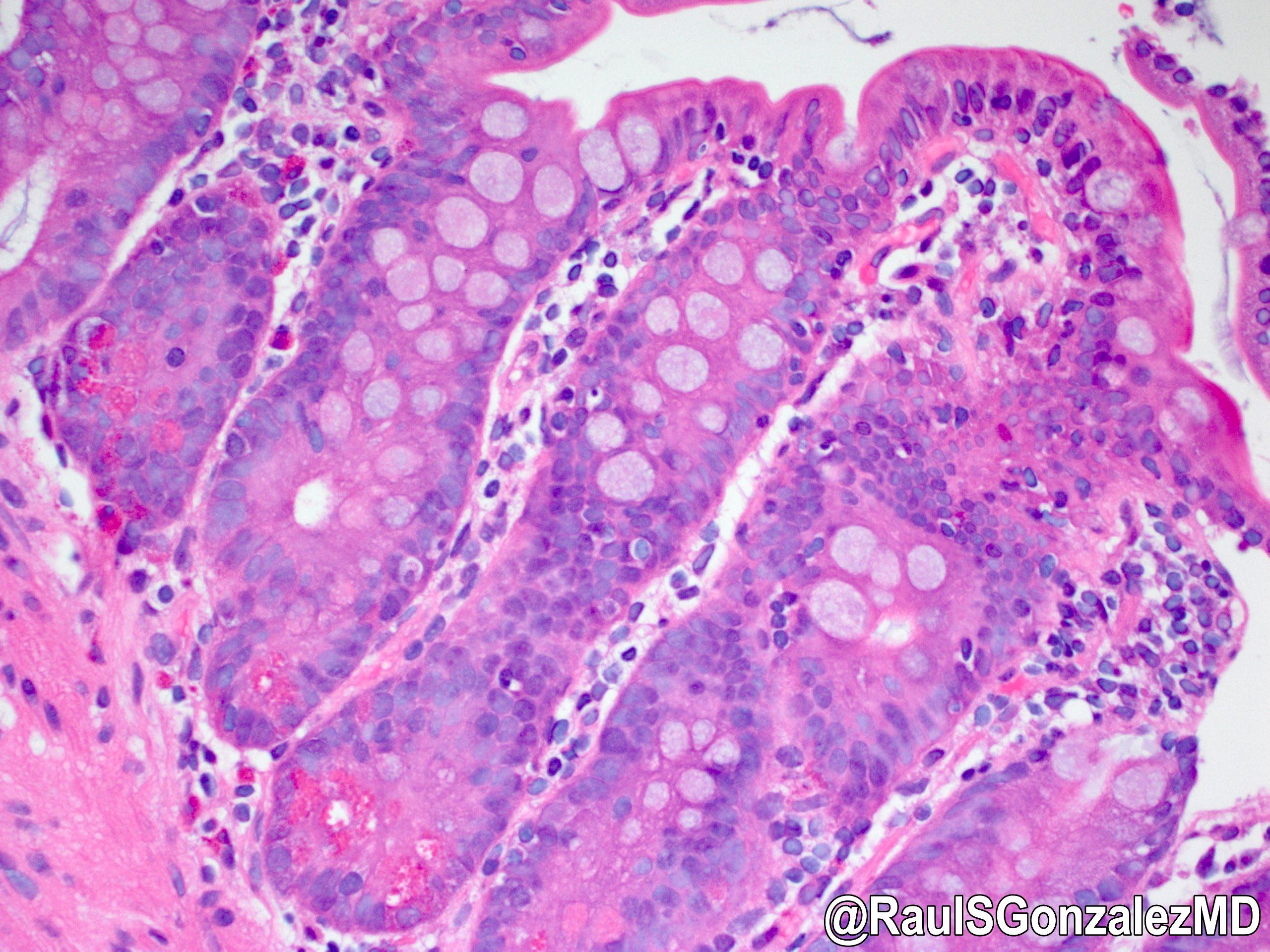
Lamina propria macrophage infiltrate
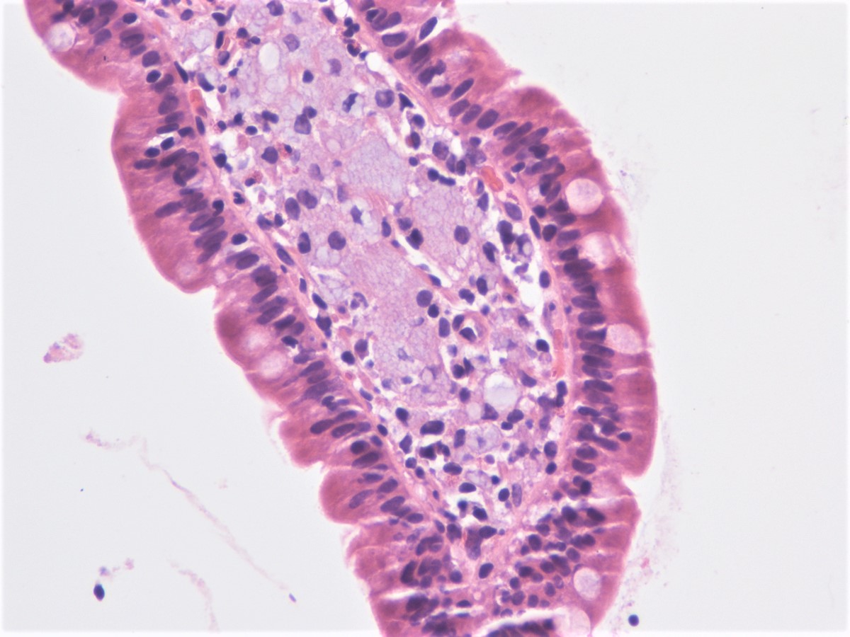
Foamy macrophages
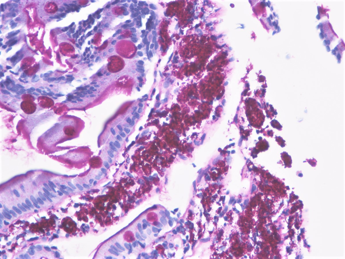
Positive PAS stain
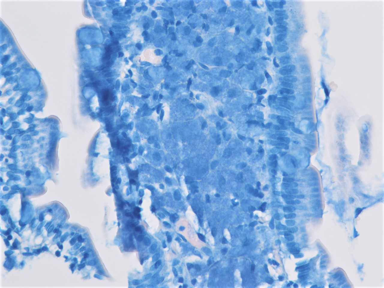
Negative AFB stain
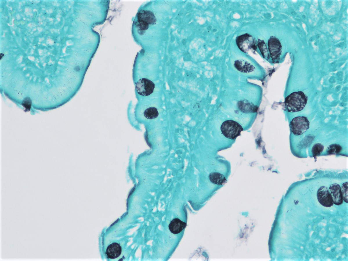
Negative GMS stain
Images hosted on other servers:

CSF cytospin with PAS
Images hosted on other servers:
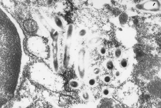

Whipple disease, duodenum
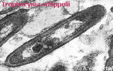
T. whipplei bacteria

Bacterium (arrowhead)
Images hosted on other servers:
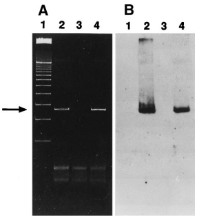
Gel electrophoresis of PCR products
Albores-Saavedra: 2016
Cheng: 2023
Greenson: 2019

IARC: 2019
Lamps: 2015
Montgomery: 2017
Montgomery: 2017
Montgomery: 2017
Odze: 2022
Srivastava: 2023
Yantiss: 2021
Find related Pathology books: GI, liver, molecular
