

Images hosted on other servers:

CD88: C5A and its effects
Images hosted on other servers:

CD82: endometrial carcinoma

CD82: oral cavity (normal and malignant)

CD82: breast carcinoma (D-F)

CD83: infantile hemangioma endothelium

CD83: decidua

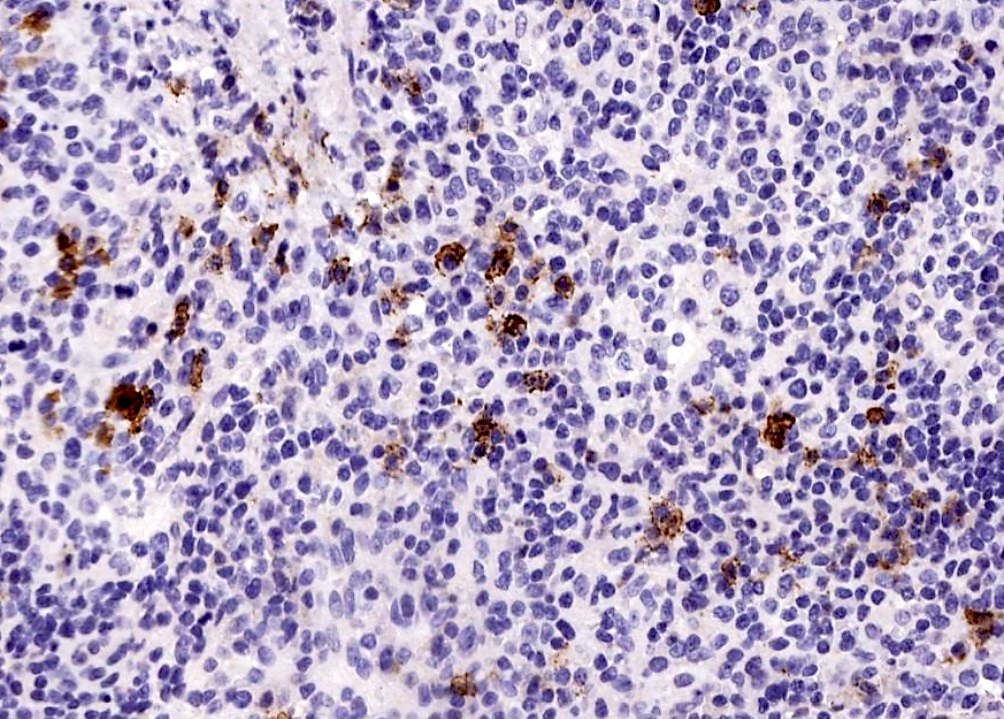
CD83+ dendritic cells in breast tumor


CD87: endometrial adenocarcinoma

CD87: pancreatic adenocarcinoma (figures B, D)


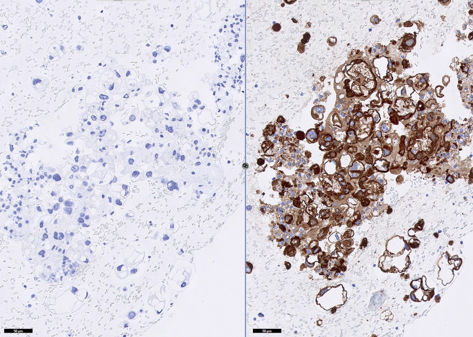
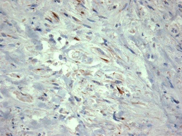
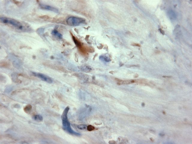
CD88: normal and Alzheimer brain
Contributed by Rola Saleeb, M.D., Ph.D.

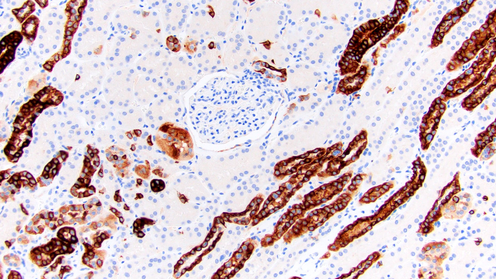
Normal ABCC2 in tubules

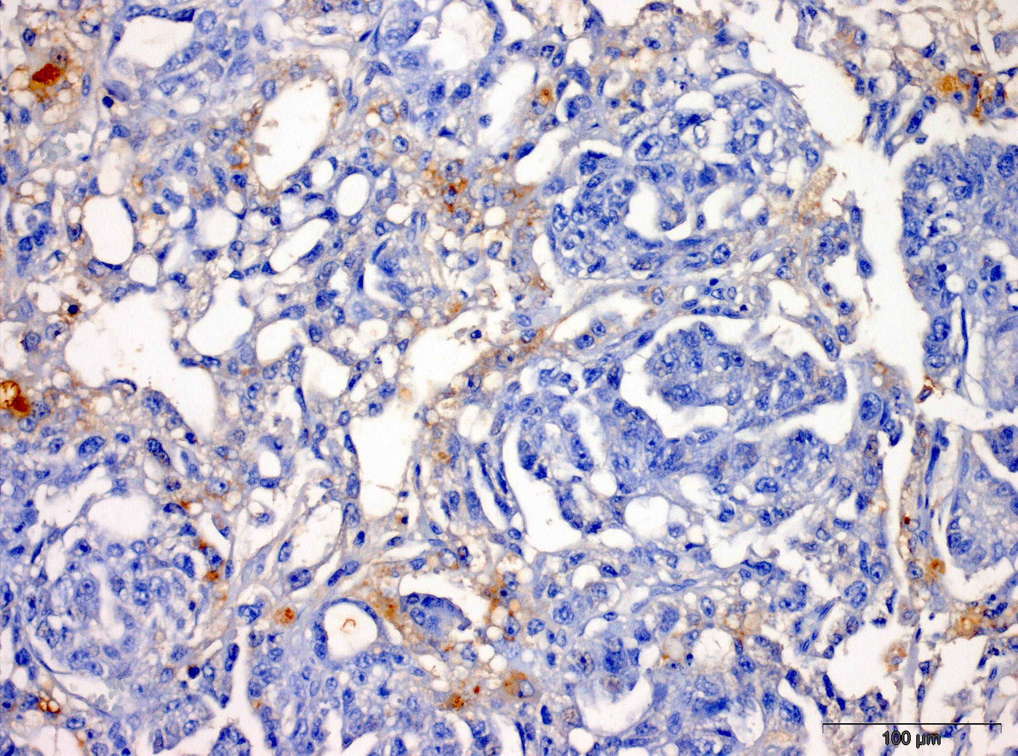
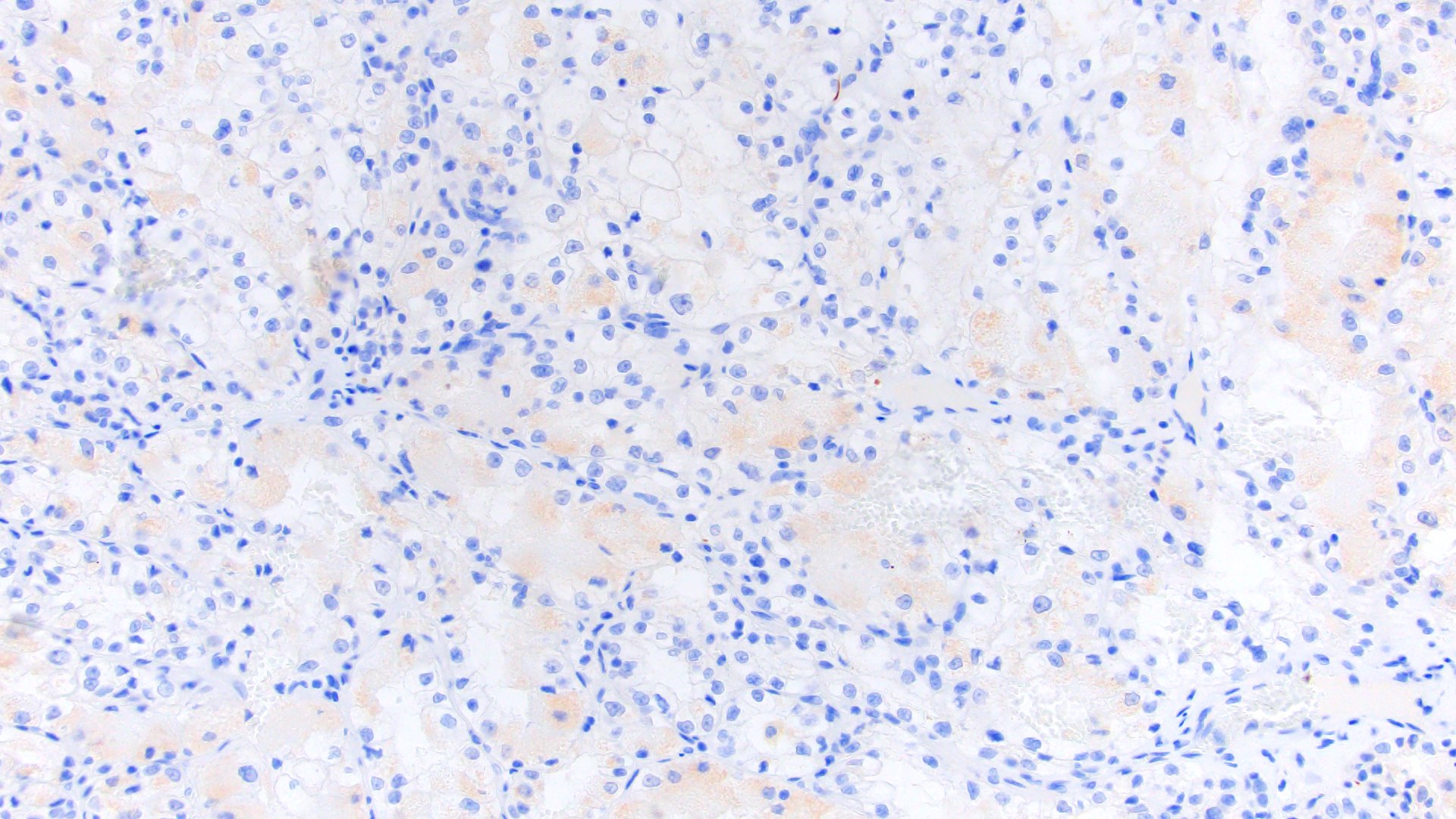
Negative ABCC2 in PRCC
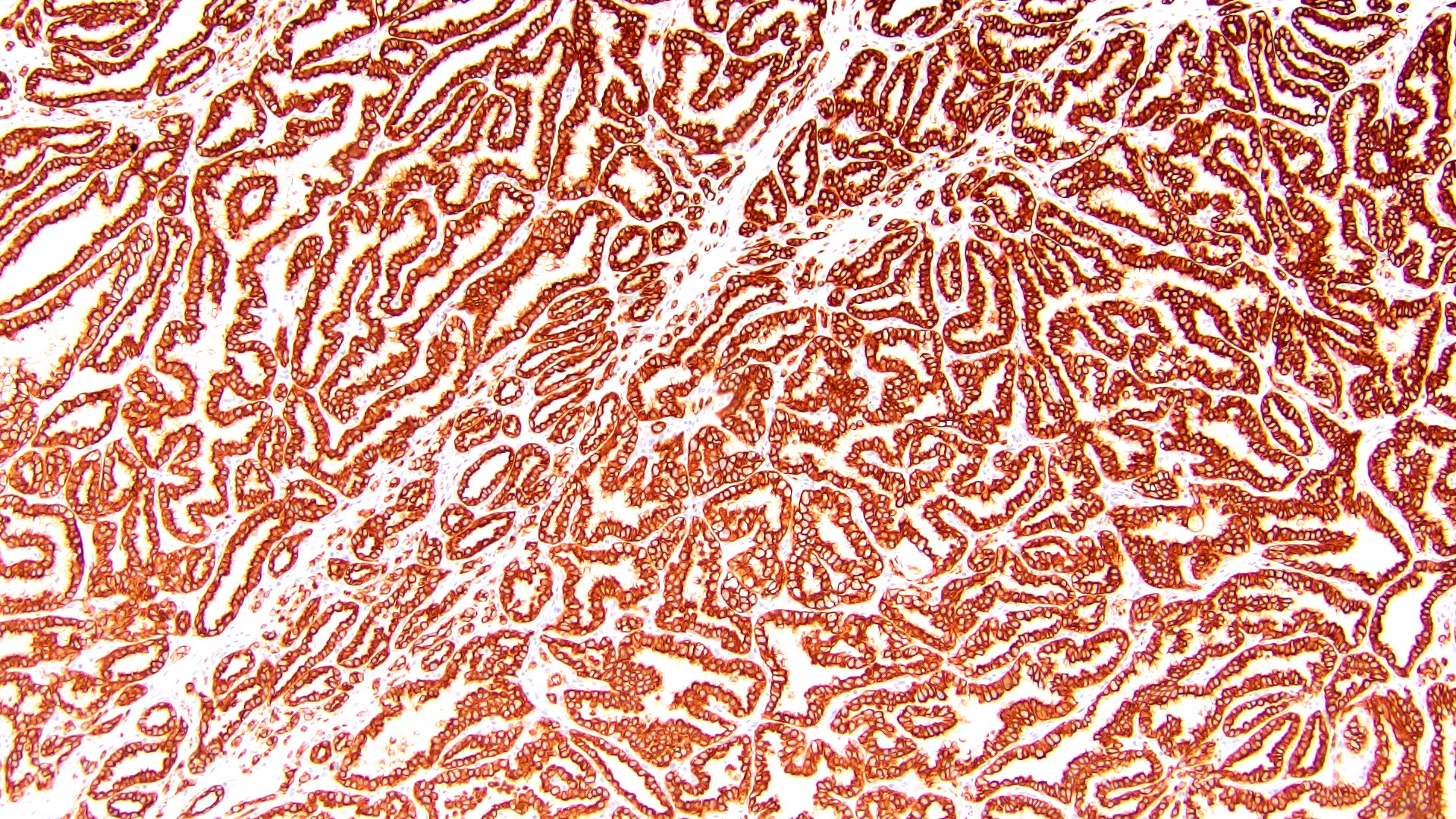
PRCC cytoplasmic

Cytoplasmic ABCC2 in PRCC

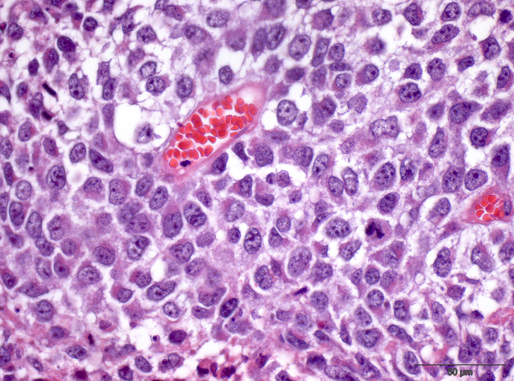
PRCC low

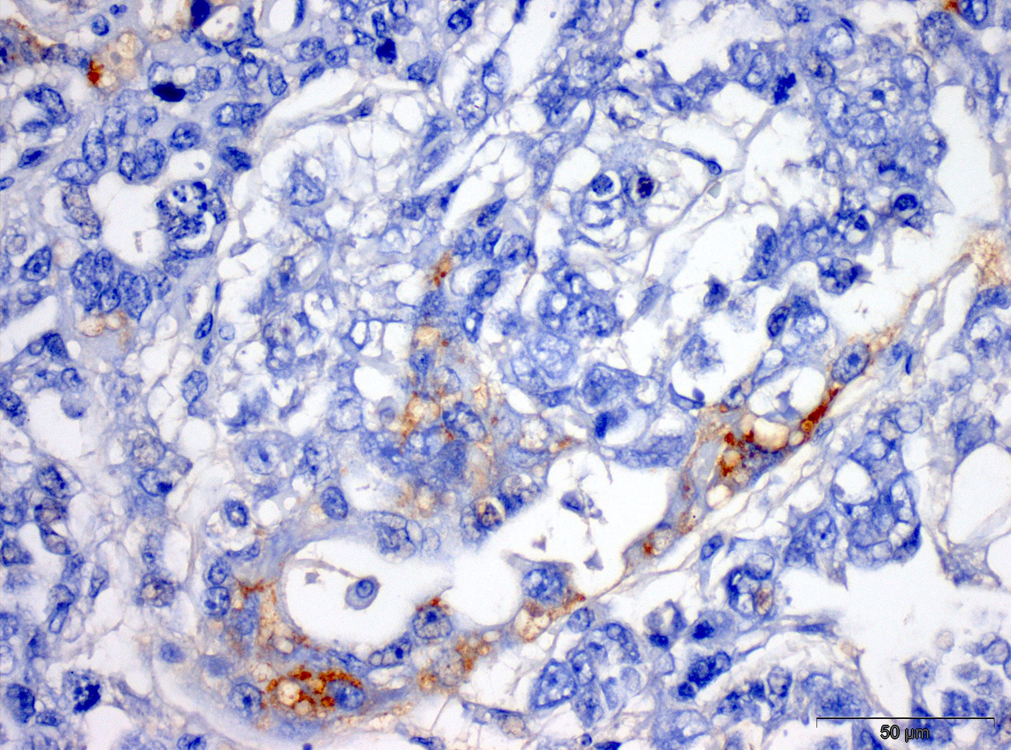
Low brush border pattern ABCC2

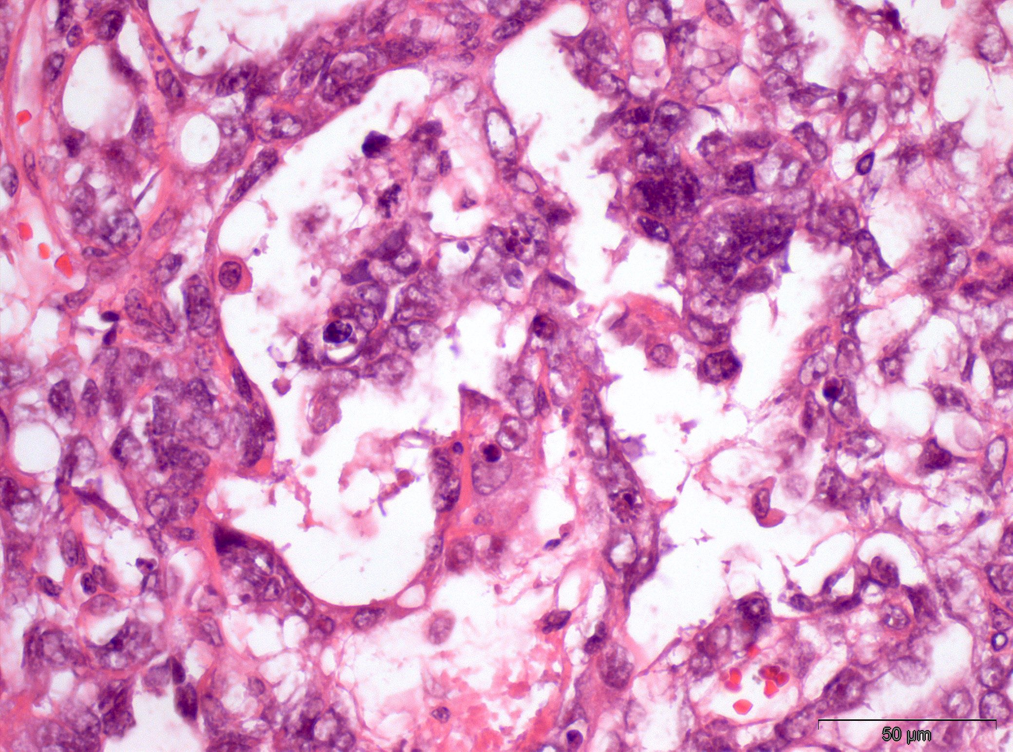
Papillary RCC, prominent atypia

High brush border ABCC2 expression
Images hosted on other servers:
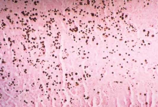
Cryptosporidium:

Oocysts: modified acid-fast stain

Stool specimen (Ziehl-Neelsen)

Oocysts: auramine-rhodamine stain
Isospora:

Acid-fast stain
Mycobacterium leprae:

Liver (Fite stain)
Mycobacterium tuberculosis:


Ziehl-Neelsen stains

Auramine stain of lung


Skin biopsies
Mycobacterium avium complex:


Site-unknown, breast and colon (Ziehl-Neelsen)
Nocardia:


Fite-Faraco modified acid fast stain of lung
Other:

Tuberculous lymphadenopathy (Ziehl-Neelsen)

Pleural fluid
Acid fast stain
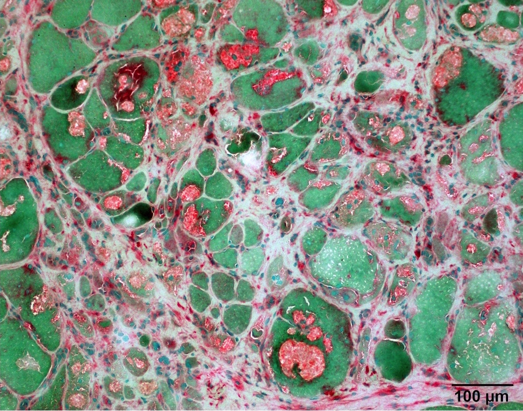
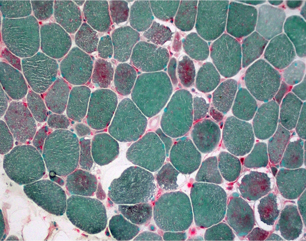
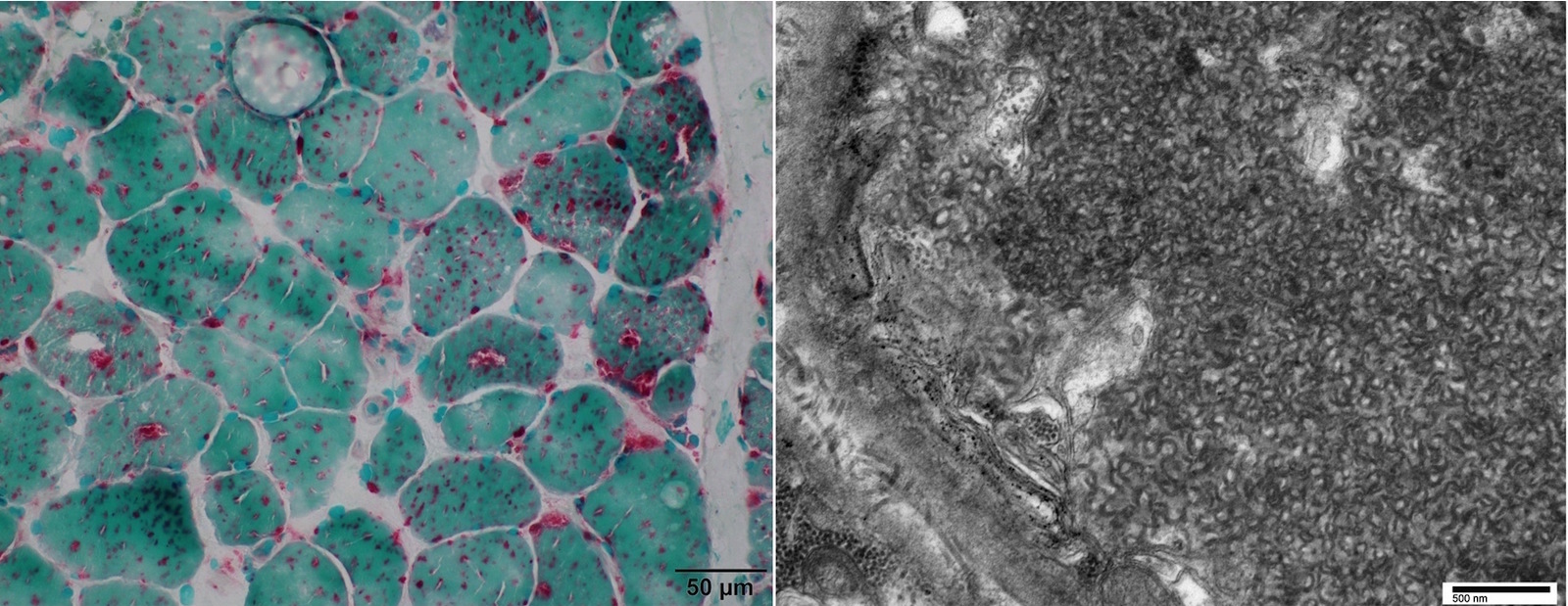
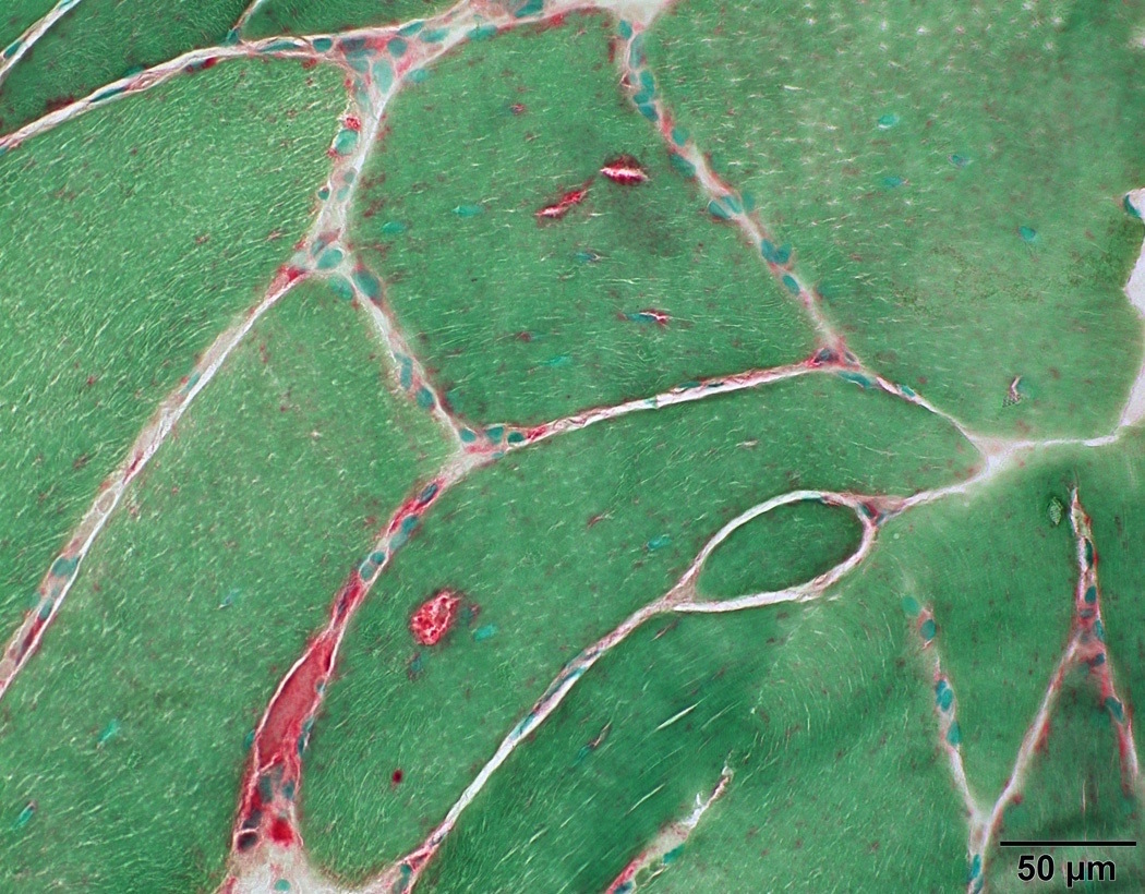
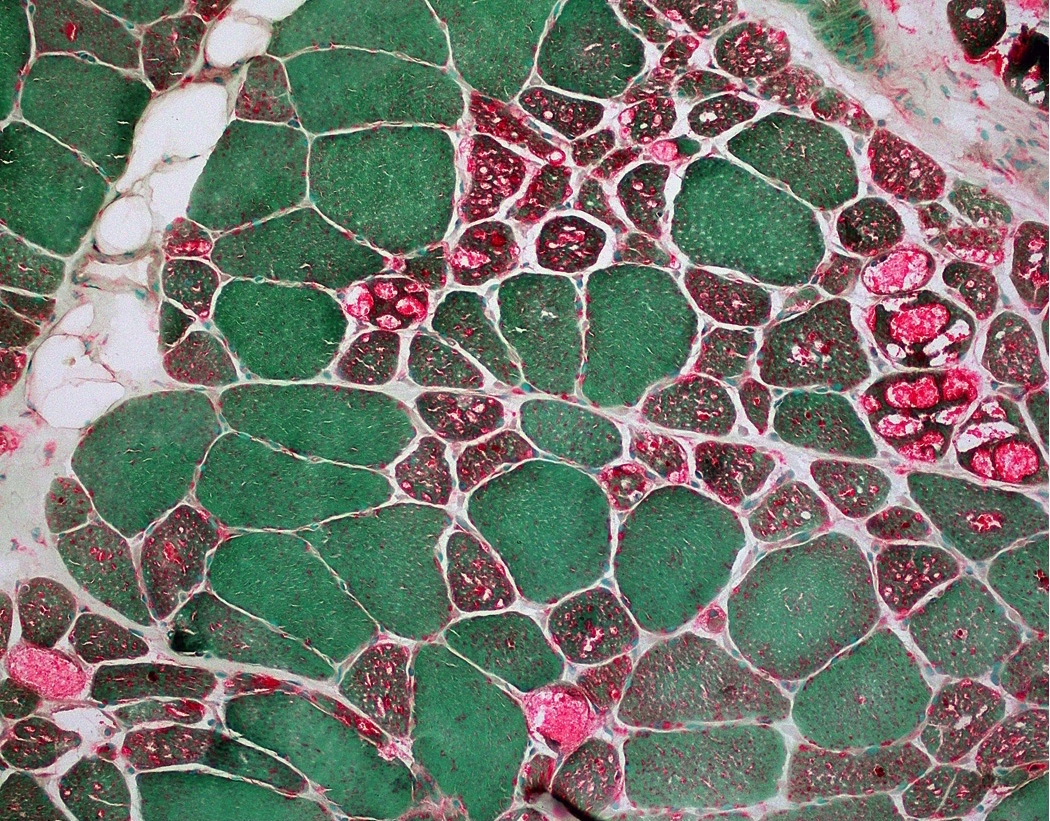
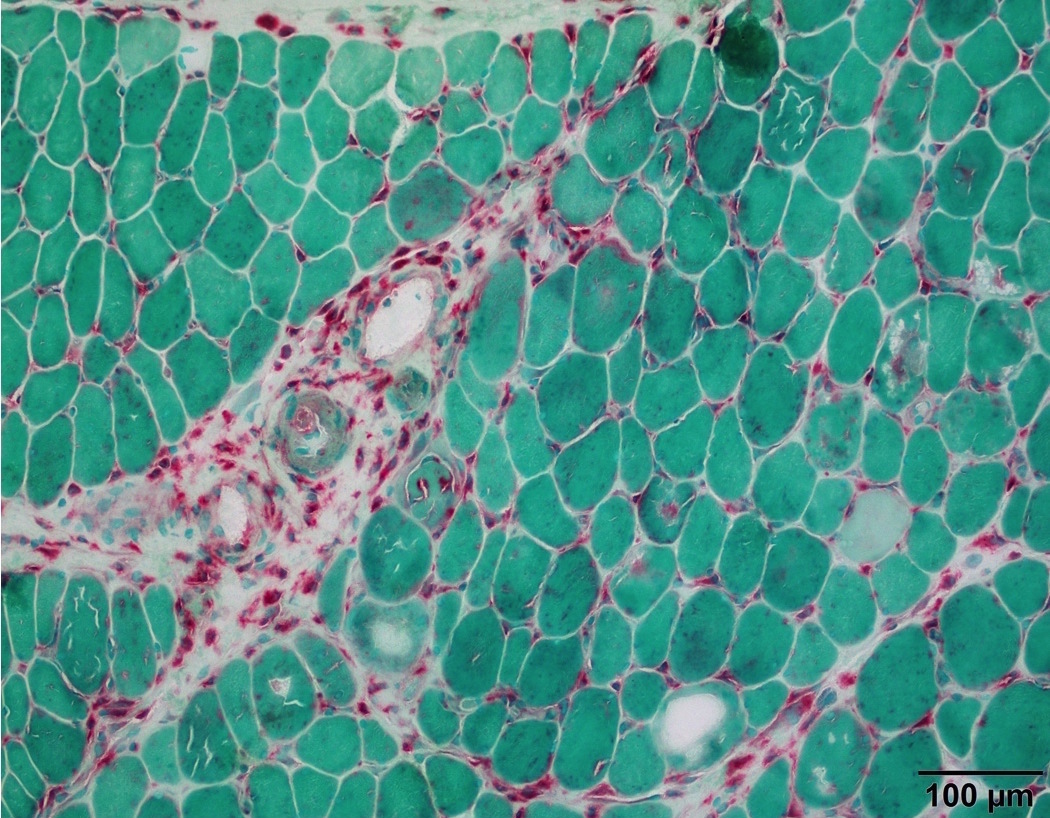
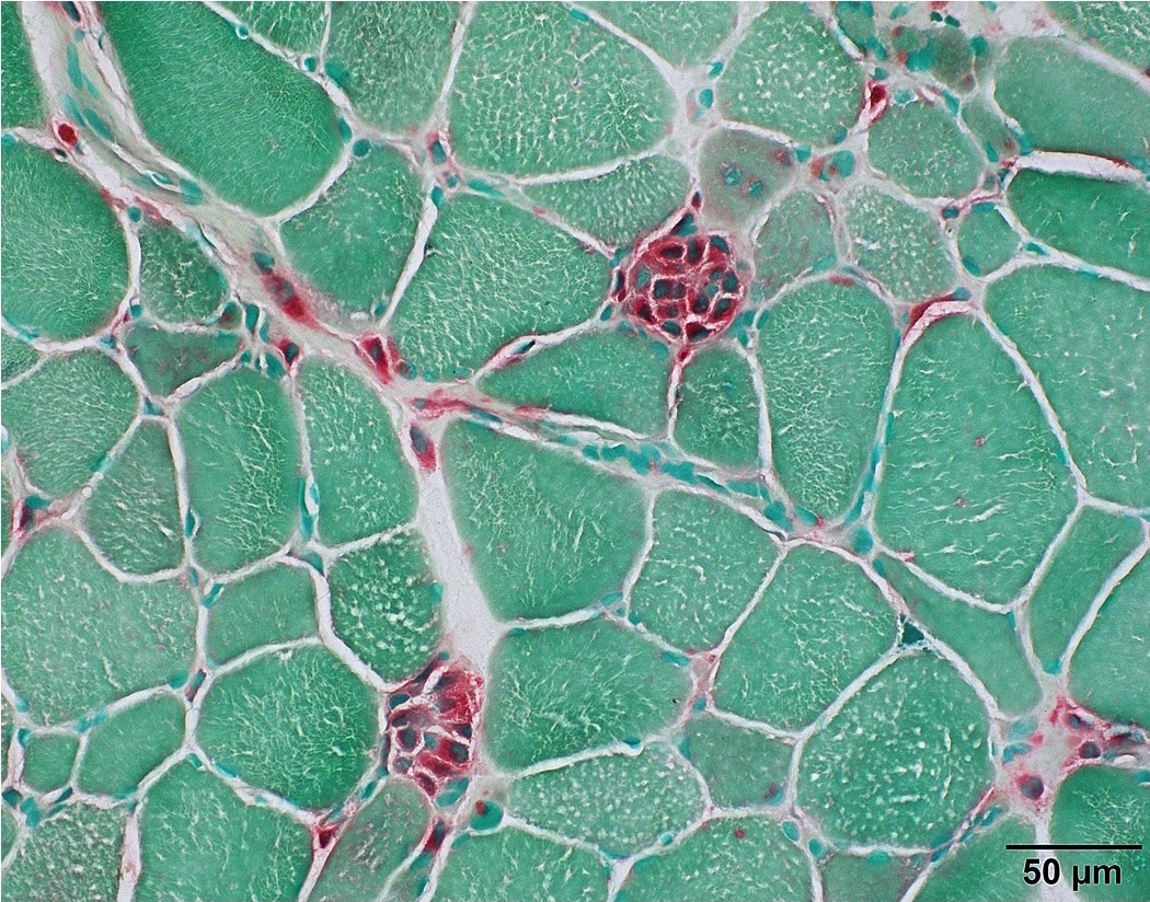
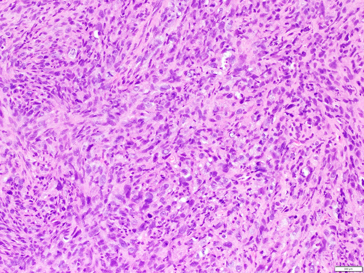
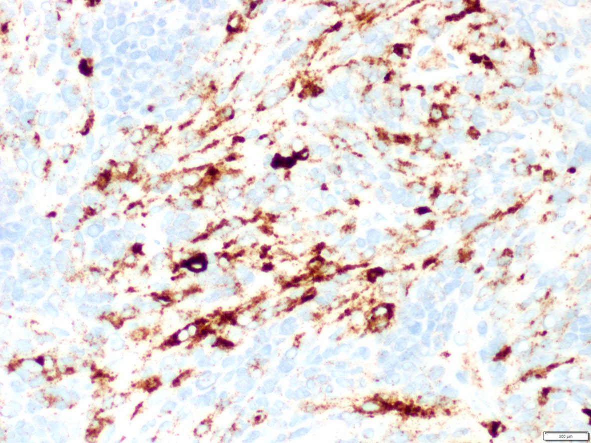
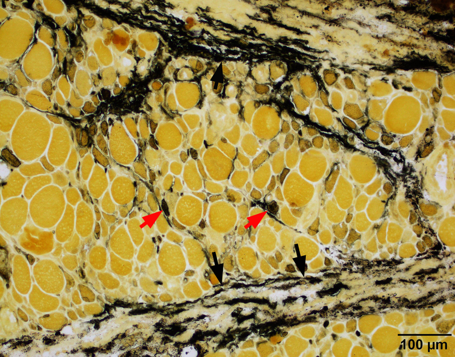
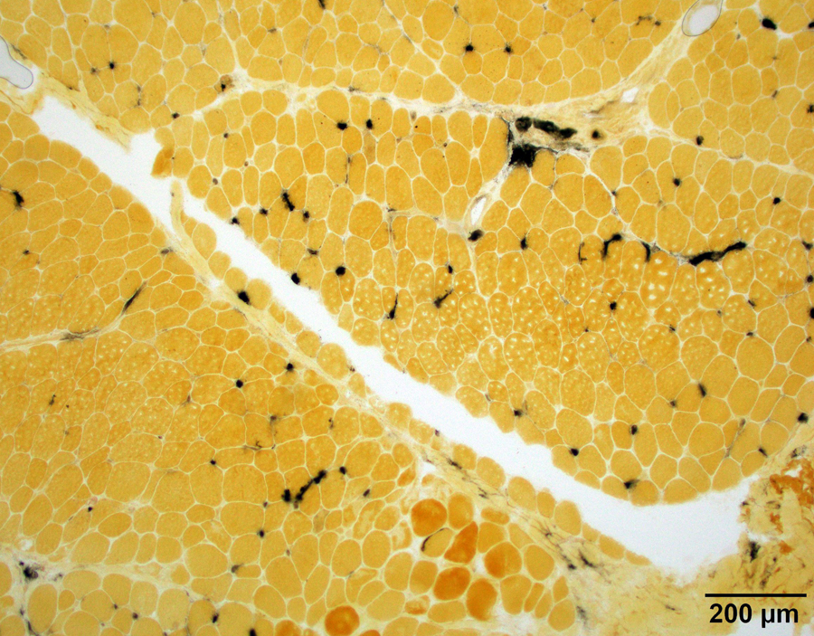
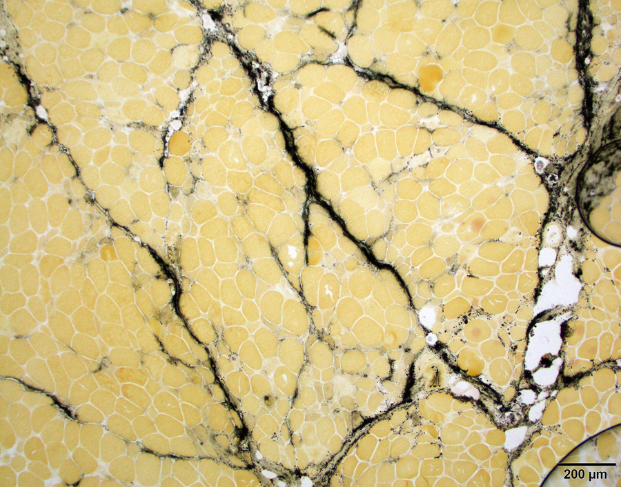
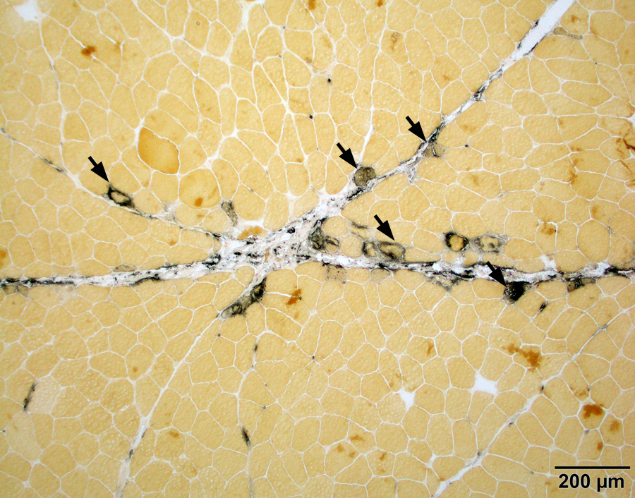
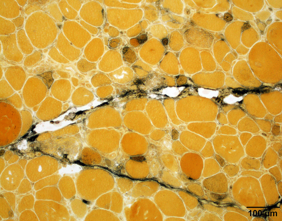
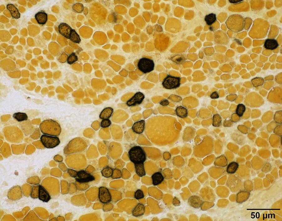
Contributed by Chunyu "Hunter" Cai, M.D., Ph.D.
Adult onset acid maltase deficiency
Central core myopathy
Cystinosis distal myopathy
Degenerating myofibers
Hydroxychloroquine myopathy
Inclusion body myositis
Infant onset acid maltase deficiency
Lupus myositis
Myophagocytosis
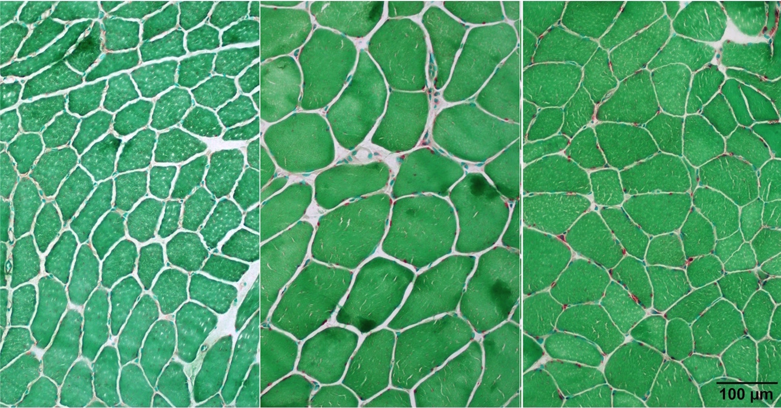
Normal muscle
Images hosted on other servers:

Helical structure of F-actin

G-actin to F-actin transition
Images hosted on other servers:

Various images
Images hosted on other servers:


Endometriosis

Myofibroblastoma: breast (fig d)


Myofibroblastoma: lymph node

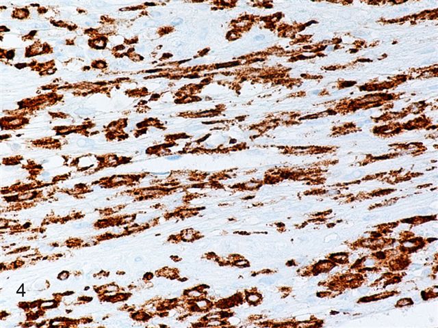
Rhabdomyo-sarcoma: CNS, embryonal (fig 4)

Rhabdomyo-sarcoma: oral cavity
Contributed by Kemal Kösemehmetoğlu, M.D.
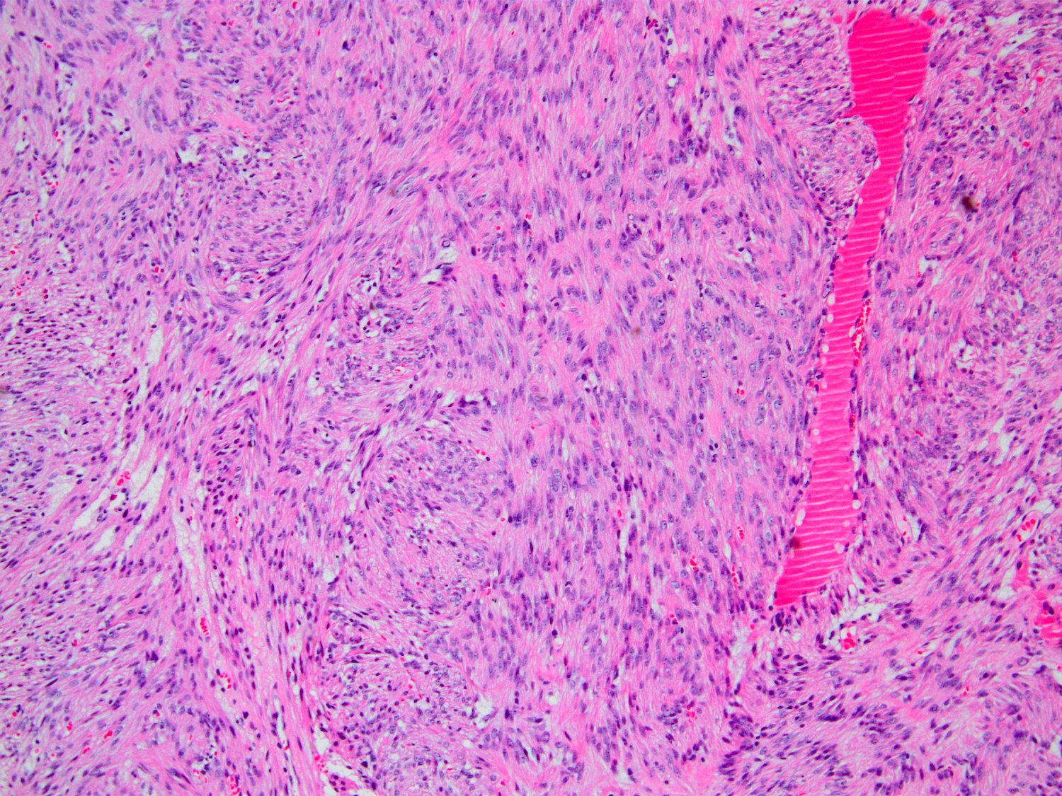
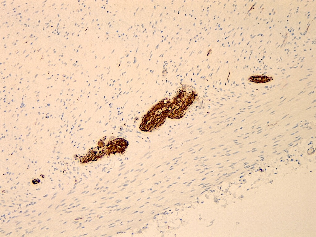
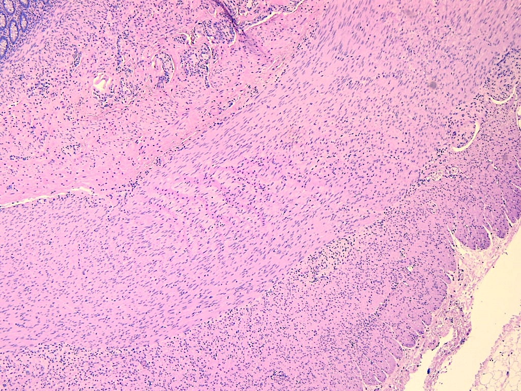
Leiomyoma
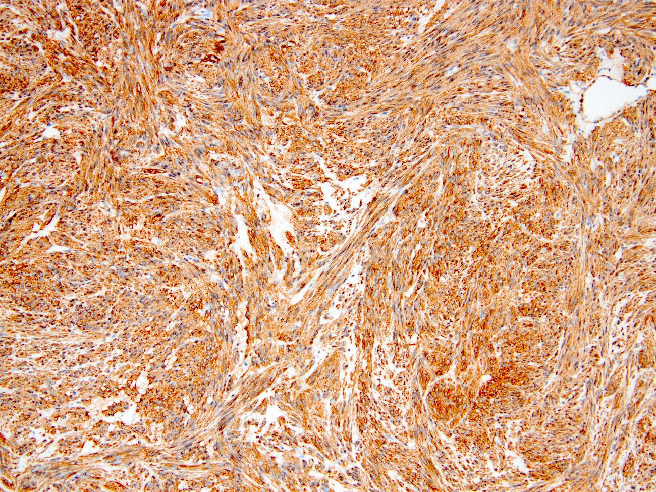
SMA expression in leiomyoma
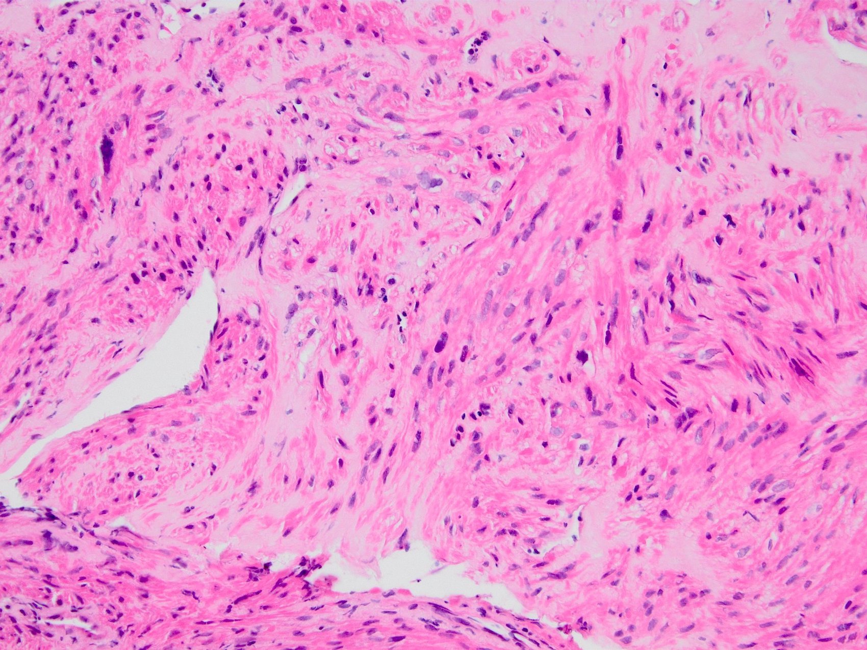
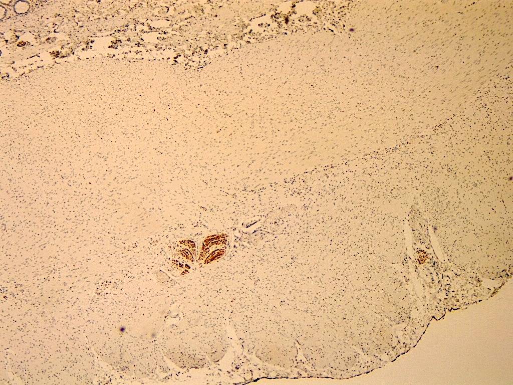
Leiomyosarcoma
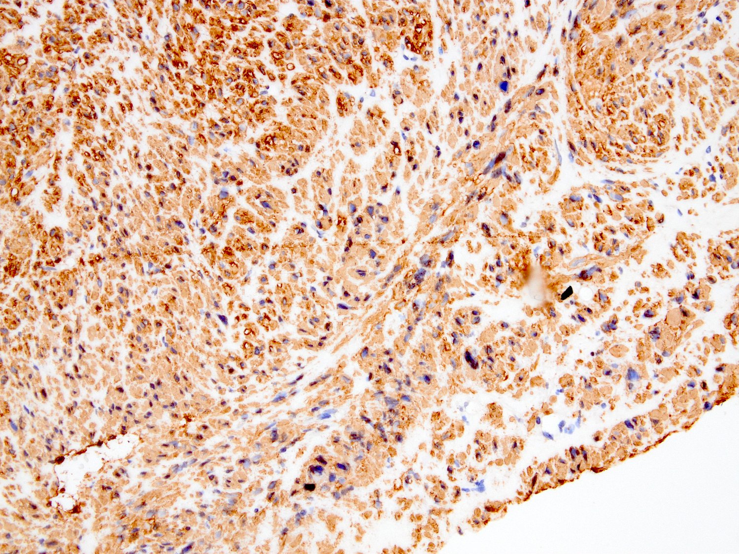
Diffuse cytoplasmic block SMA expression
Nodular fasciitis
SMA in nodular fasciitis
Myxofibrosarcoma
Focal SMA expression in myxofibrosarcoma

Glomus tumor

SMA in glomus tumor
Undifferentiated pleomorphic sarcoma
Focal SMA in undifferentiated pleomorphic sarcoma
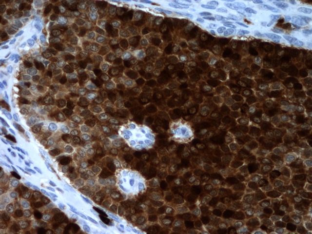
Inflammatory myofibroblastic tumor
Myofibroblastic type SMA in IMT

Atypical apocrine adenosis
SMA in myoepithelial layer of breast adenosis

Radial scar / complex sclerosing lesion
SMA in radial scar / complex sclerosing lesion
Sclerosing papilloma

Pseudoinvasion in sclerosing papilloma
SMA positive myoepithelial layer
Tram track (myofibroblastic) staining pattern
Block (smooth muscle-like) staining pattern
Contributed by Vikas Mehta, M.D., Maria M. Picken, M.D., Ph.D. and Cullen Lilley, M.S., M.A.

Small bowel adenovirus infection
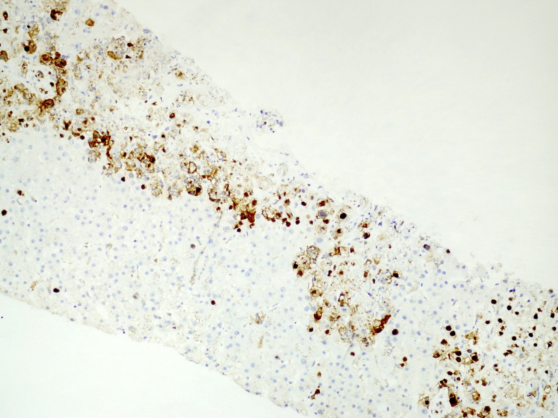
Coagulative hepatocyte necrosis

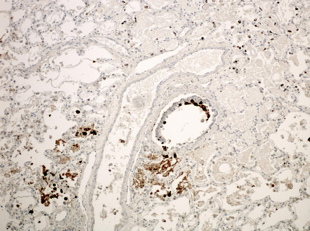
Lung biopsy
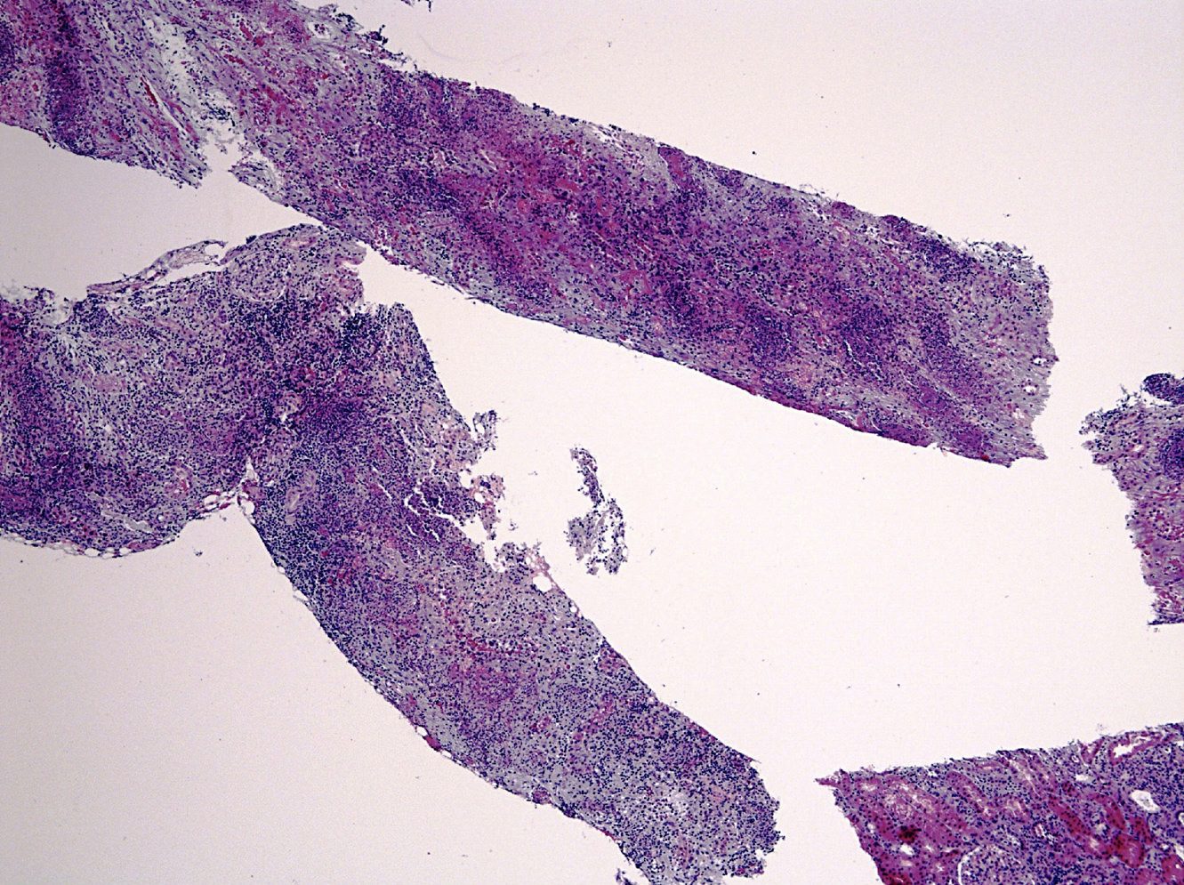
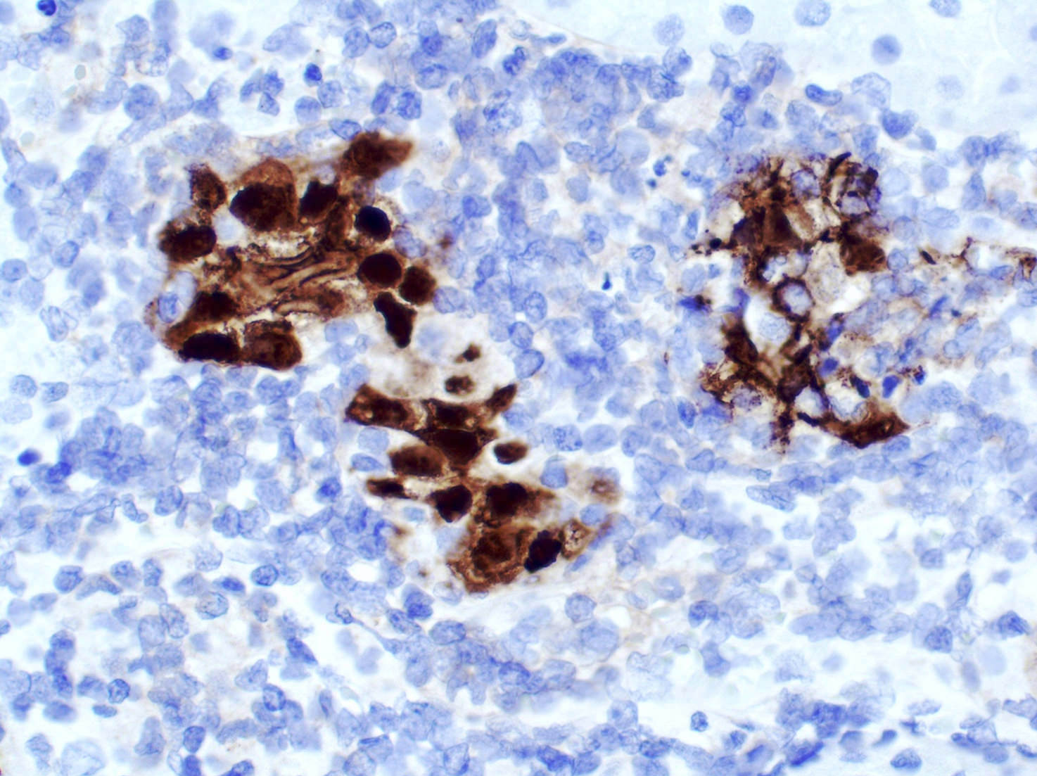
Renal parenchyma
Contributed by Maria M. Picken, M.D., Ph.D.

Crystalline array
Images hosted on other servers:

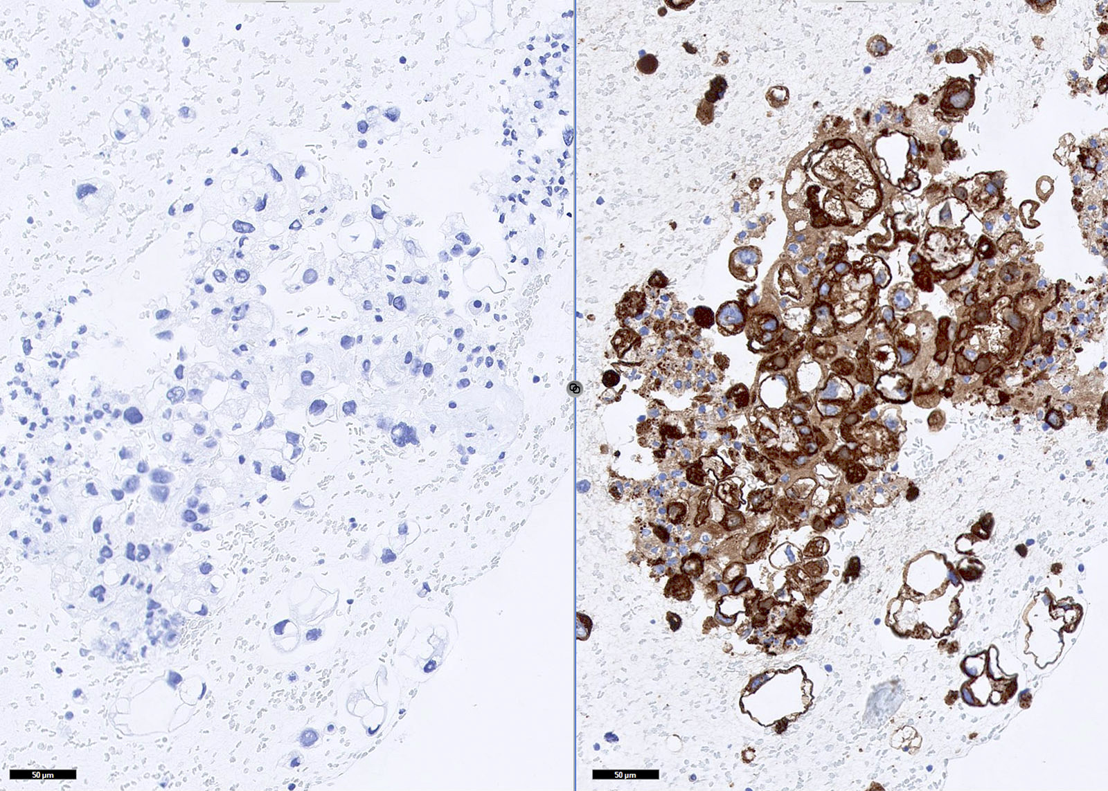
Left: normal liver; right: negative control

Liver and hepatocellular carcinoma
AFIP images and Cases #78, 94 and 110

Bladder: urothelial
carcinoma with
gland-like lumina
Esophagus
Mucin stains of Barrett mucosa


Mucin stains of Barrett mucosa
Heart: myxoma with glandular structures
Kidney: adenocarcinoma (AB-PAS)
Sacrum, Chordoma: stroma stains with Alcian blue
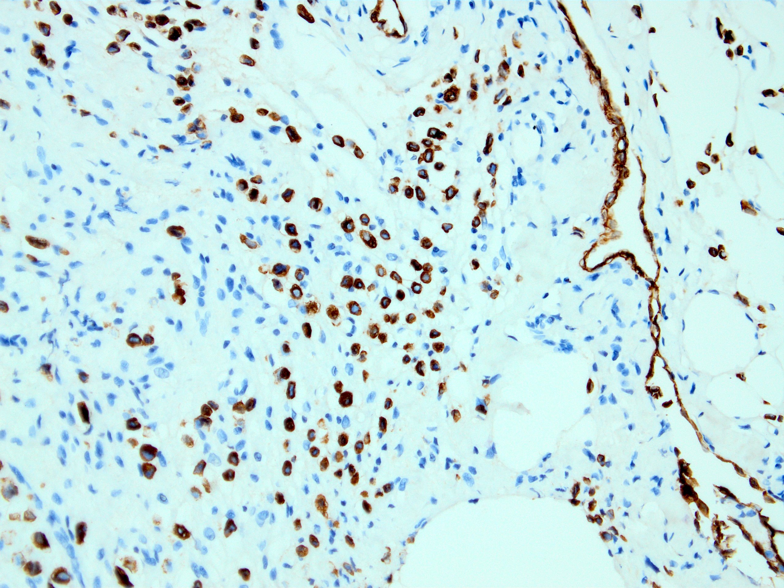
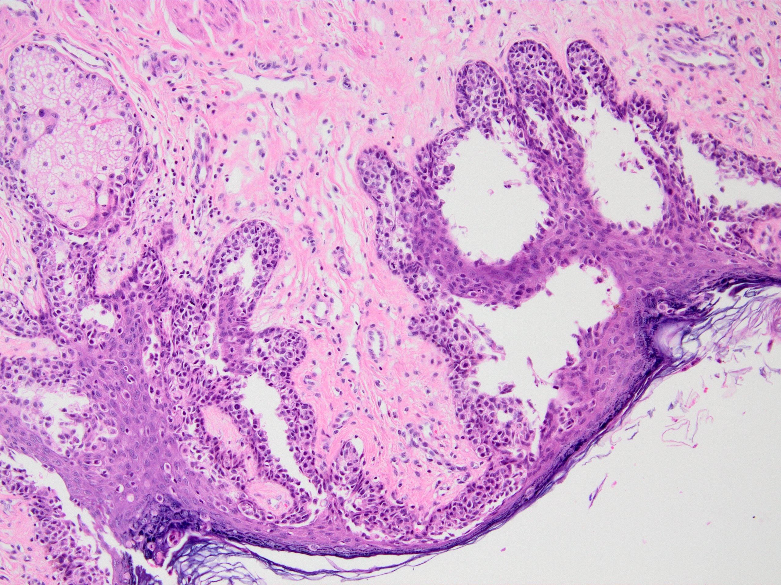
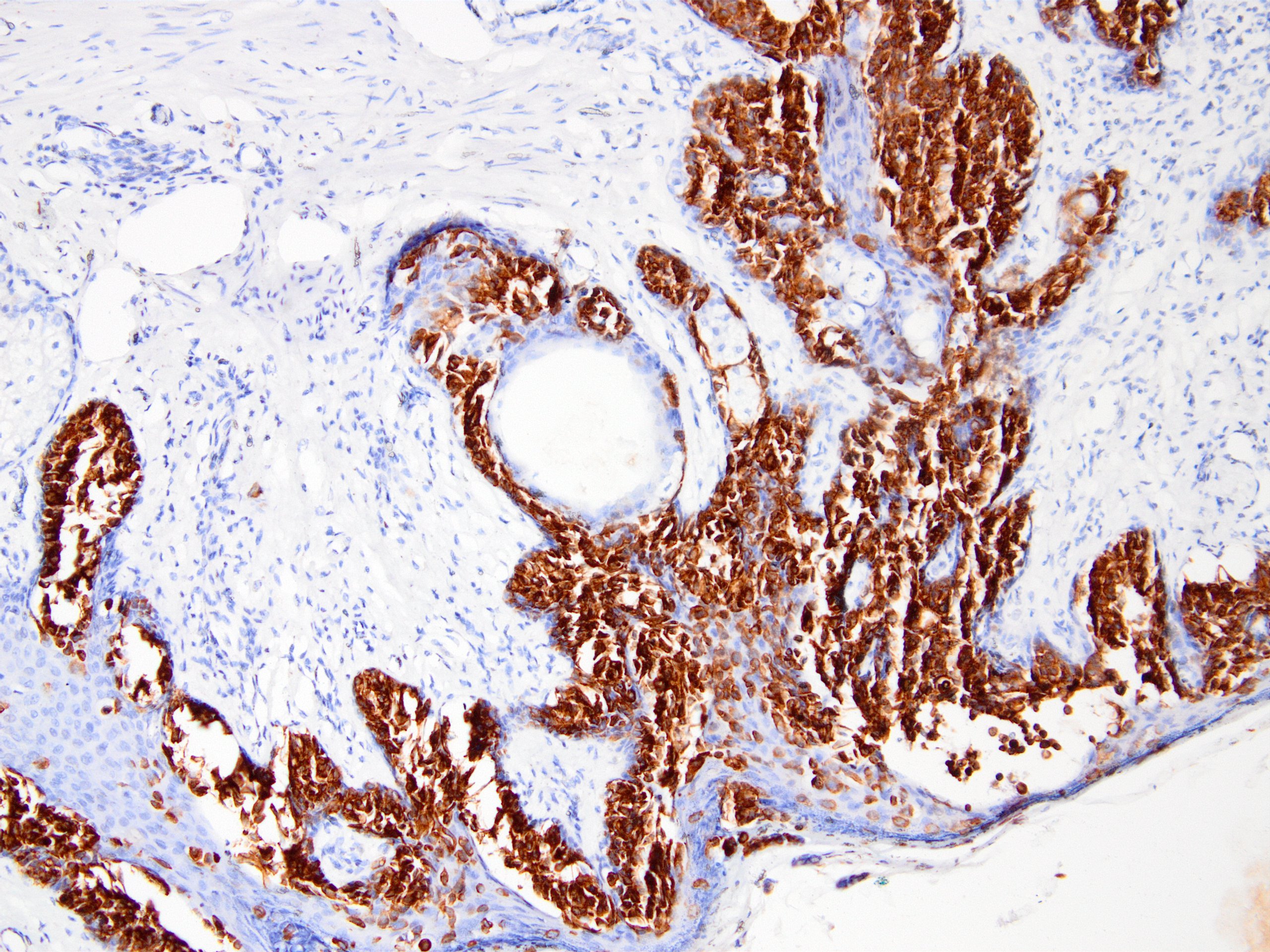
Scrotum: Paget disease
Thyroid gland: signet ring cell variant
Images hosted other servers:

Breast:
mucoepidermoid
carcinoma
(Alcian blue-PAS)

Esophagus, Barrett: GE junction

Eye: macular corneal dystrophy

Gallbladder: pyloric metaplasia

Kidney: mucinous
tubular and
spindle cell
carcinoma

Soft tissue: proliferative fasciitis - AB-PAS
Images hosted on other servers:

ALK signalling pathway
Contributed by A. Cristina Vargas, M.B.B.S., Ph.D., Patricia Guzman, M.D., Fiona Bonar, M.B.B.Ch., Alison Cheah, M.B.B.S. and Martin Jones, M.B.B.S.

ALK rearranged lung adenocarcinoma

ALK IHC in lung adenocarcinoma
Anaplastic large cell lymphoma
ALK IHC in ALCL
Anaplastic large cell lymphoma
ALK IHC in ALCL
Uterine IMT
ALK IHC on uterine IMT
ALK-EML4 spindle cell tumor

ALK-EML4 spindle cell tumor ALK
Undifferentiated pleomorphic sarcoma with ALK
Undifferentiated pleomorphic sarcoma-like ALK
Epithelioid fibrous histiocytoma

ALK IHC in epithelioid fibrous histiocytoma
FUS-TFCP2
rearranged
rhabdomyosarcoma
ALK IHC on
FUS-TFCP2
rhabdomyosarcoma
Contributed by A. Cristina Vargas, M.B.B.S., Ph.D.

ALK break apart FISH
Images hosted on other servers:


Break apart signal patterns for ALK rearrangement
Contributed by Chunyu "Hunter" Cai, M.D., Ph.D.

Normal muscle
Dermatomyositis Mi2
Dermatomyositis NXP2
Antisynthetase syndrome PL7
Antisynthetase syndrome Jo1
Necrotizing autoimmune myopathy HMGCR
Reducing body myopathy
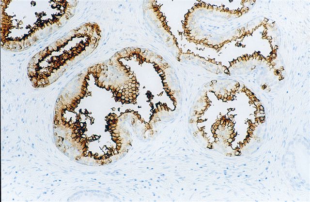
Contributed by João Lobo, M.D., Rui Henrique, M.D., Ph.D.

Prepubertal testicular yolk sac tumor
Prepubertal testicular yolk sac tumor, AFP
Prepubertal testicular yolk sac tumor
Prepubertal testicular yolk sac tumor, AFP
Metastatic yolk sac tumor
Metastatic yolk sac tumor, AFP

Testicular mixed germ cell tumor
Testicular mixed germ cell tumor, AFP
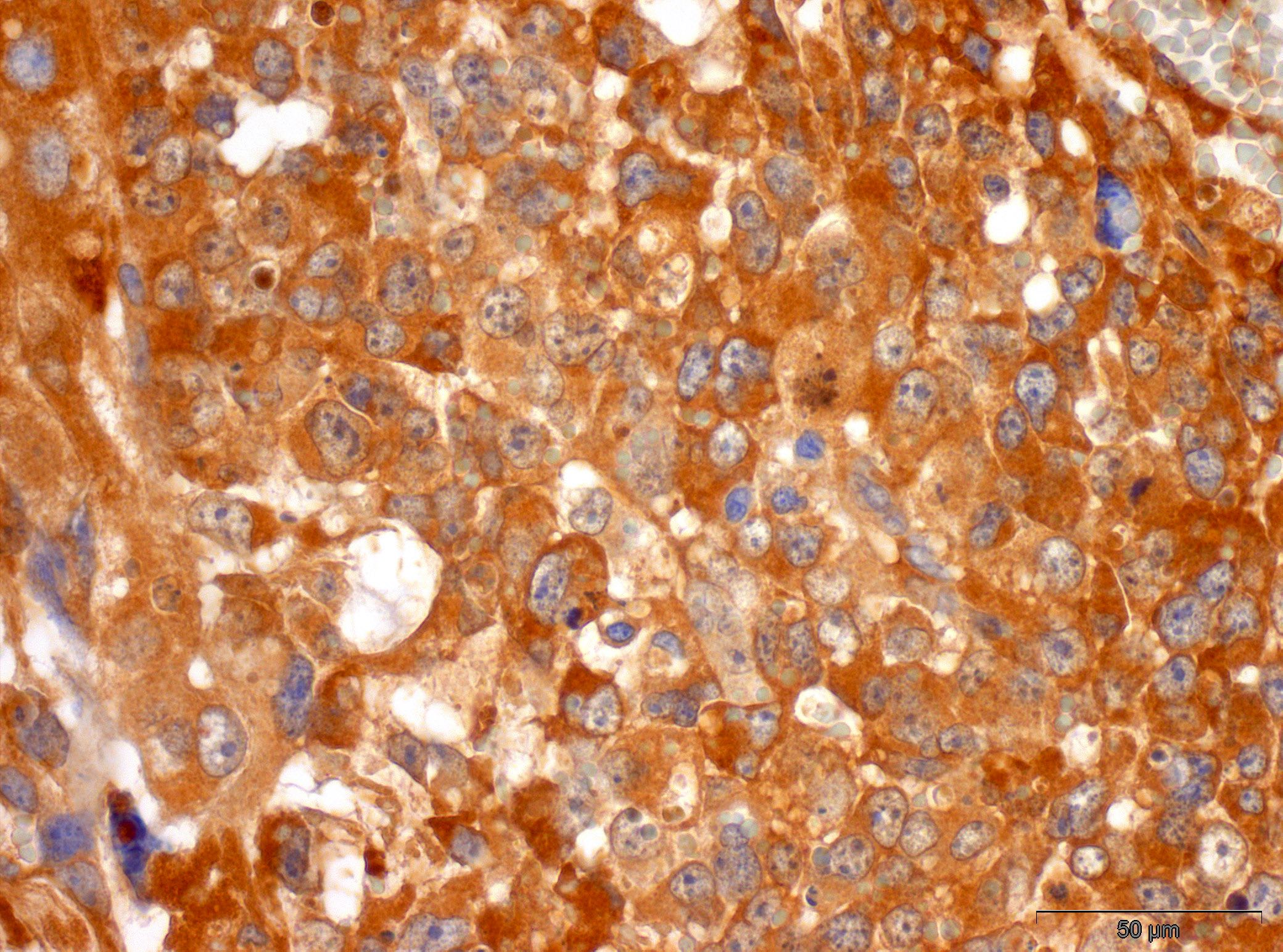
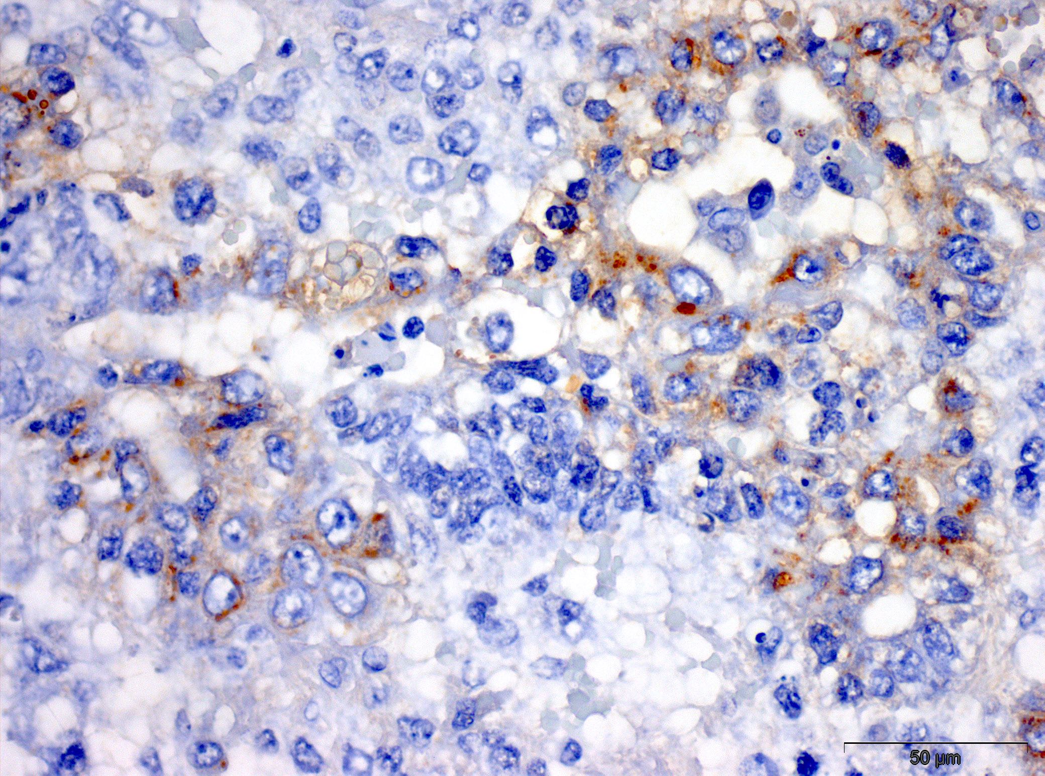
Fetal liver
Fetal liver, AFP
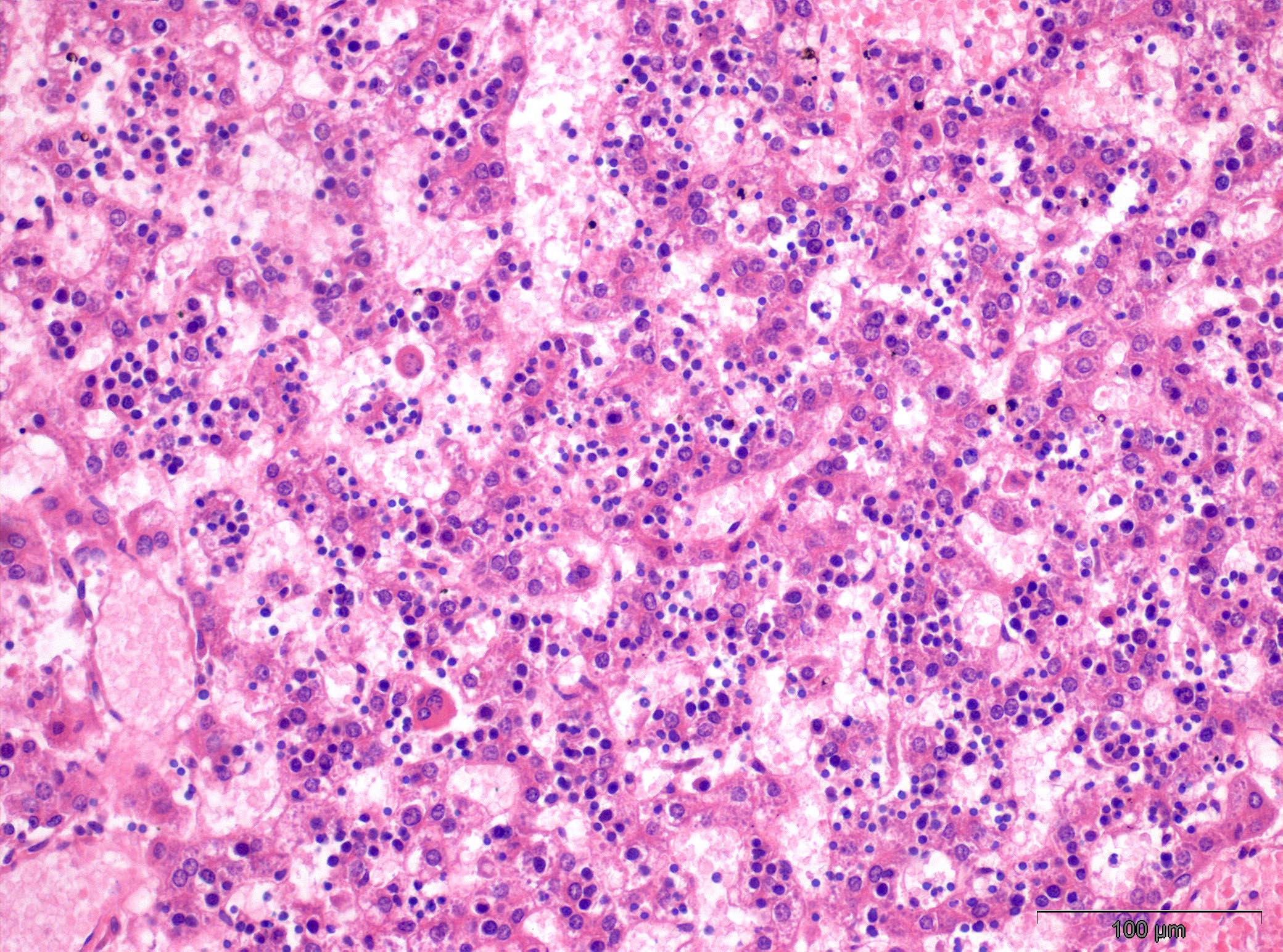
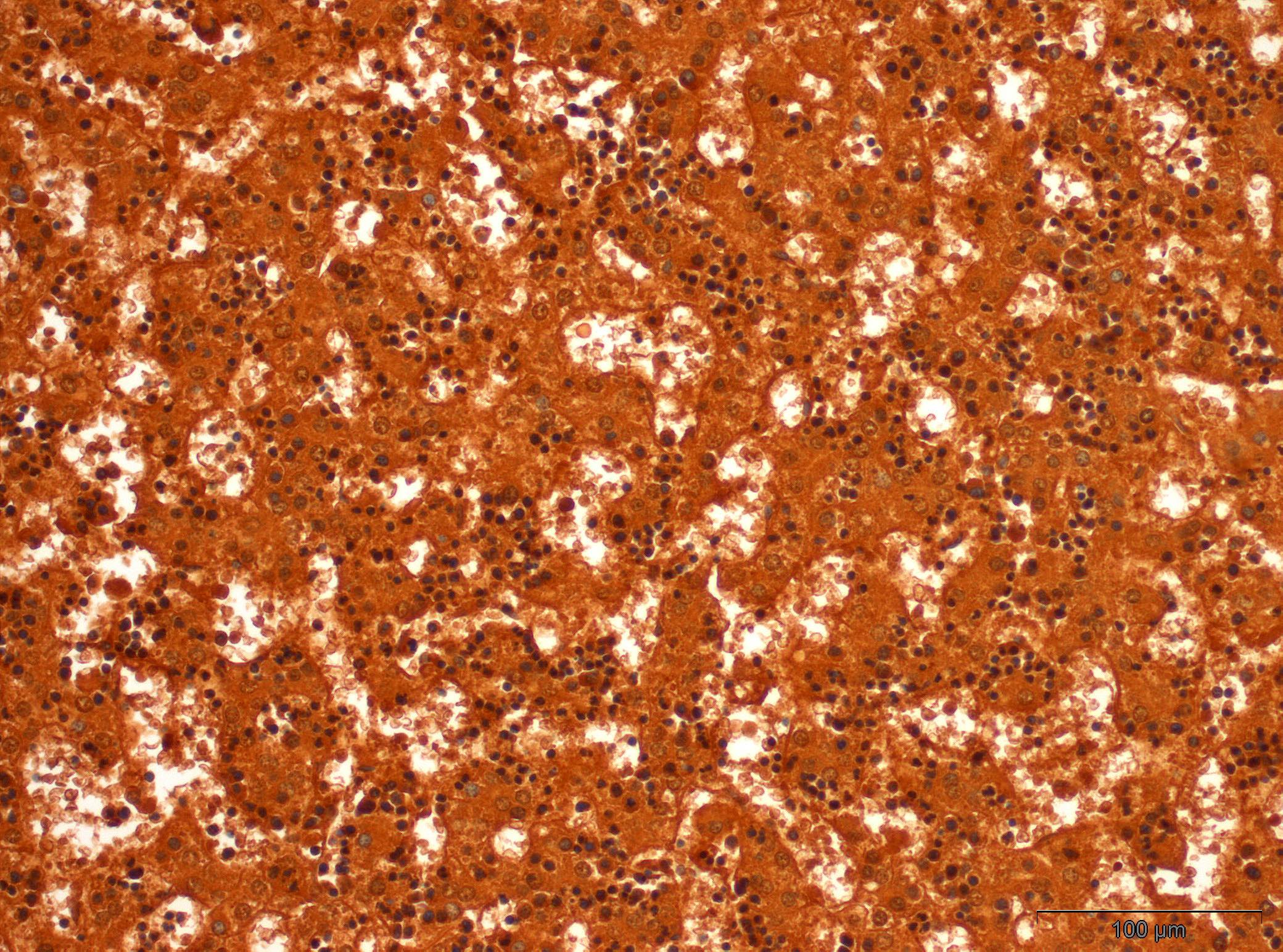
Hepatoblastoma

Hepatoblastoma, AFP
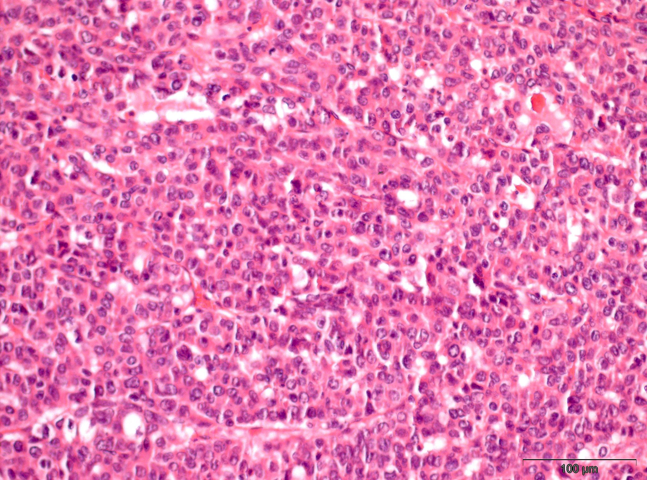
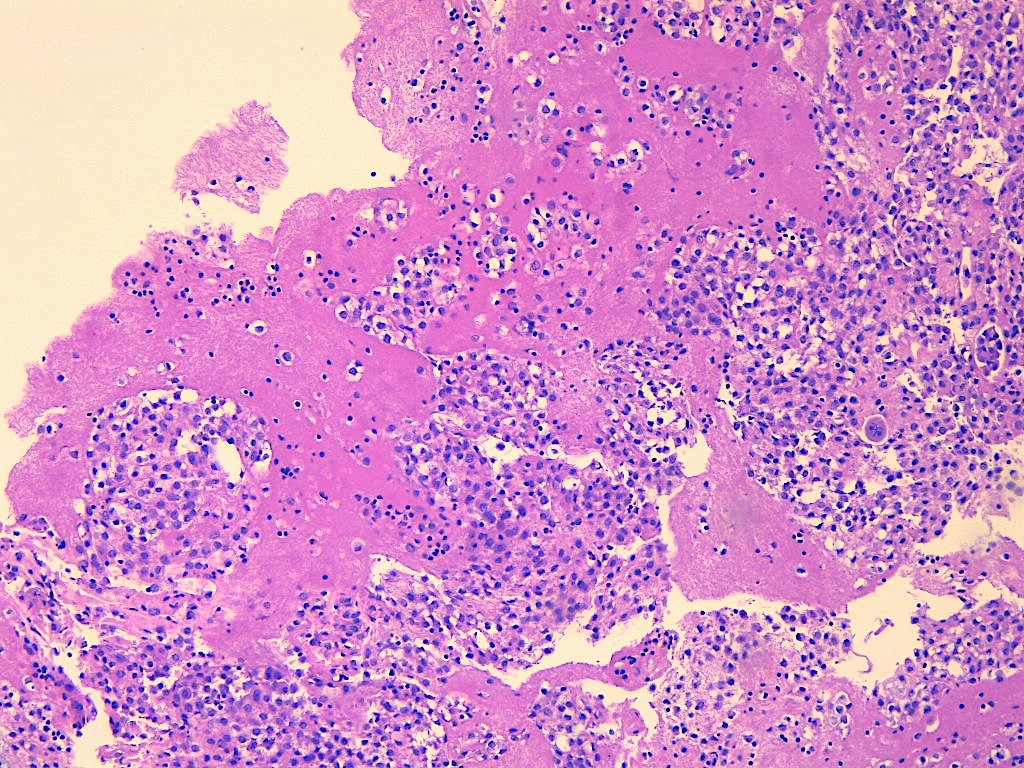
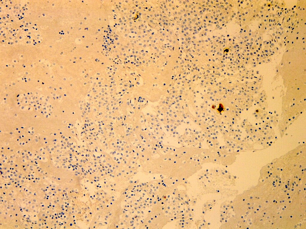
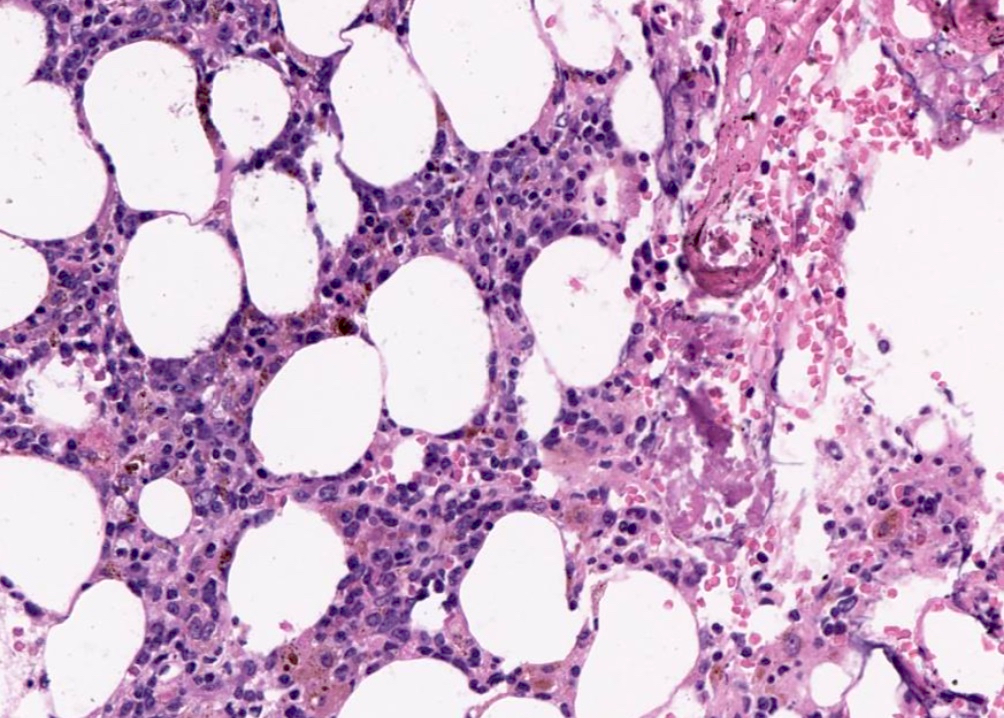
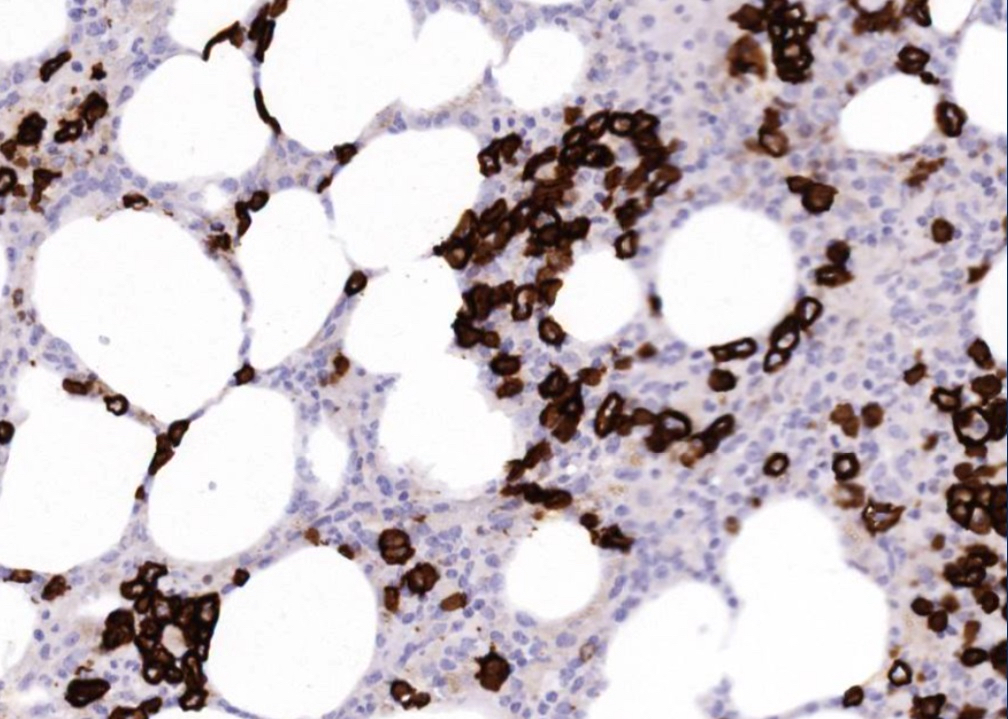
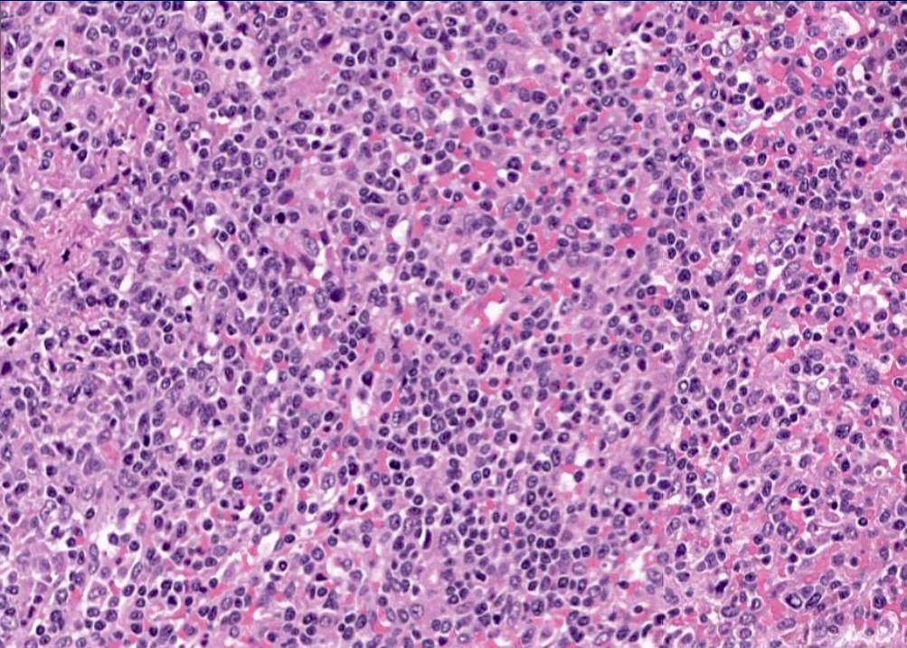
Contributed by Avani Pendse, M.D., Ph.D.

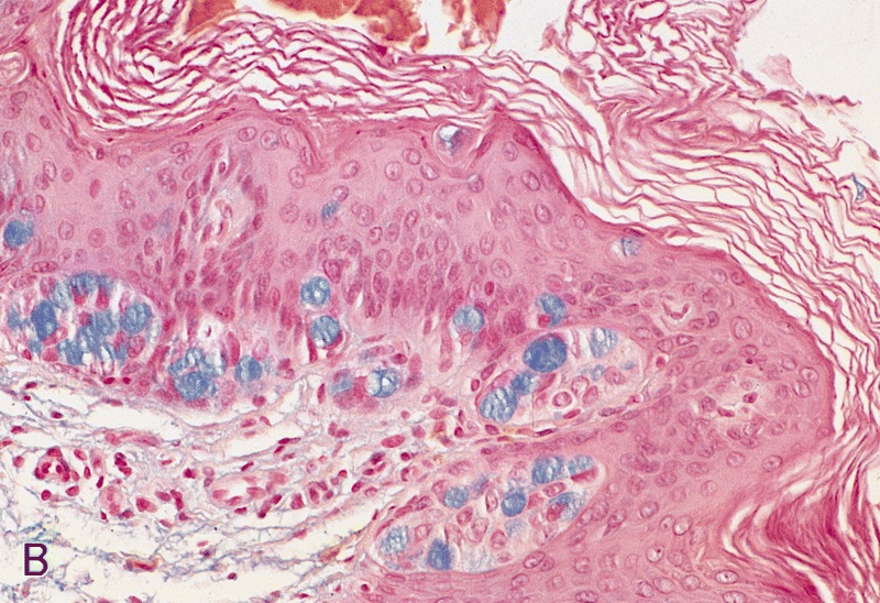
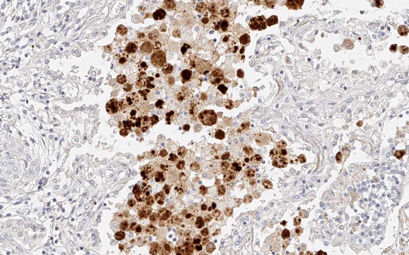
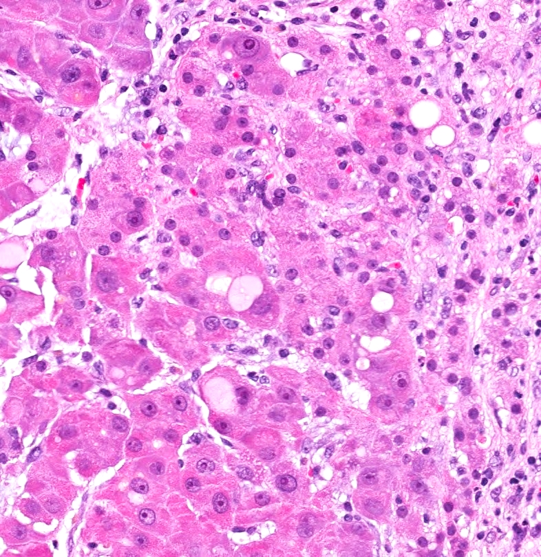
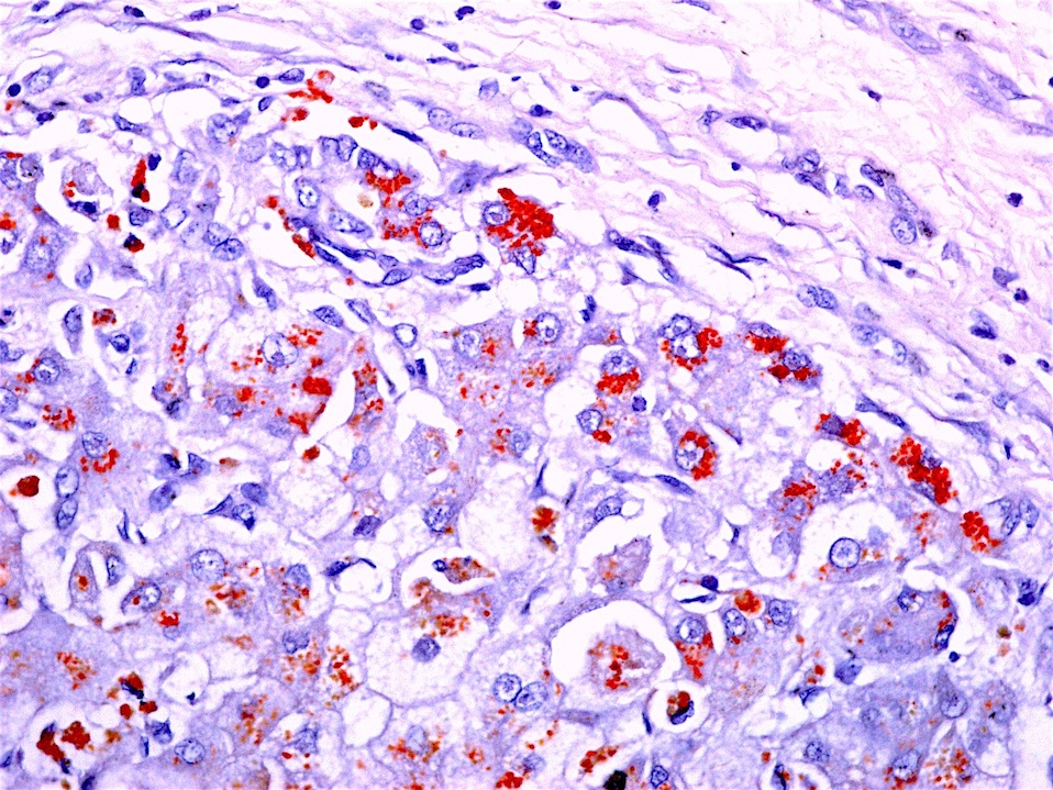
Eosinophilic intracytoplasmic globules
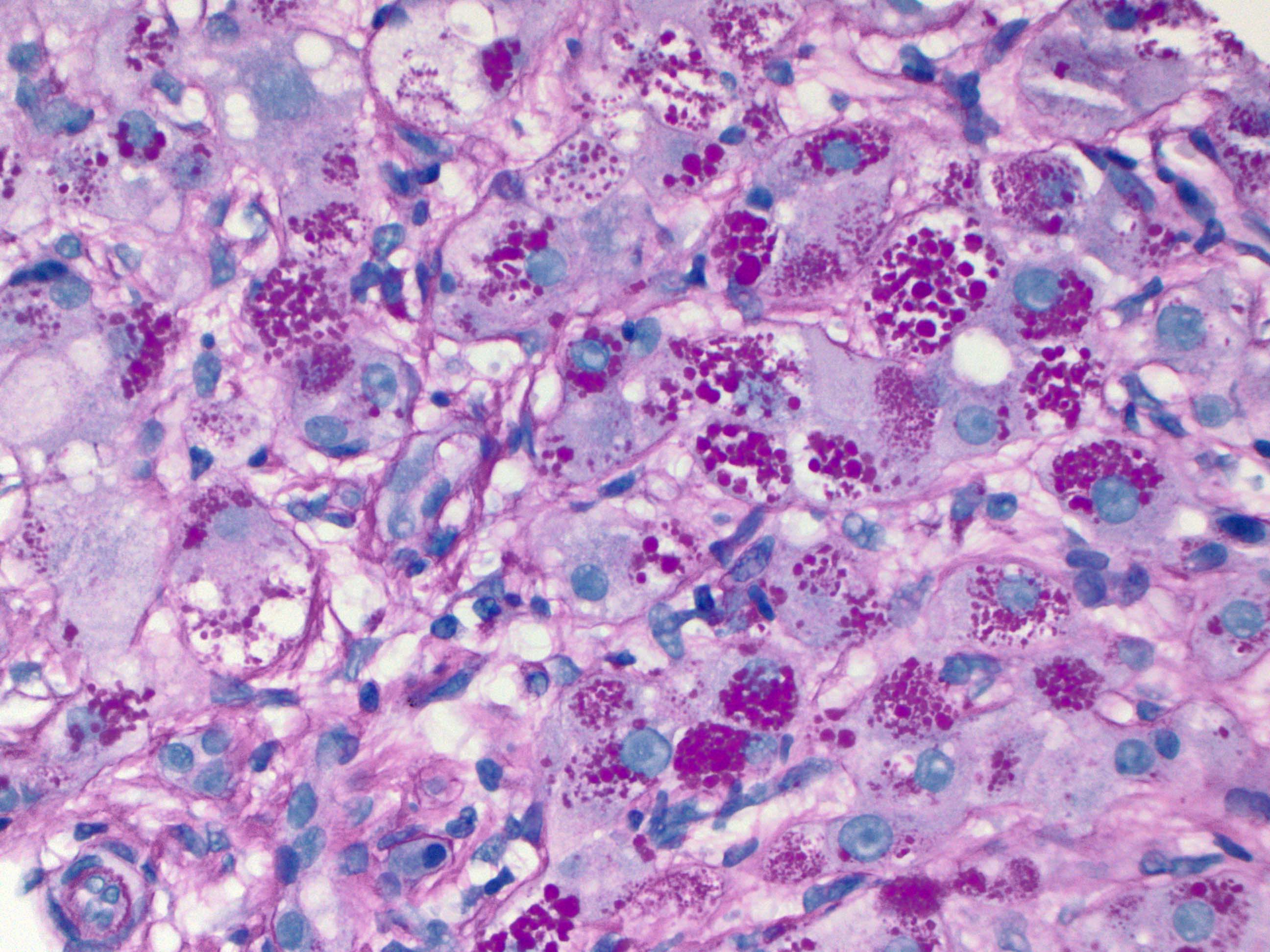
PASD positive intracytoplasmic globules
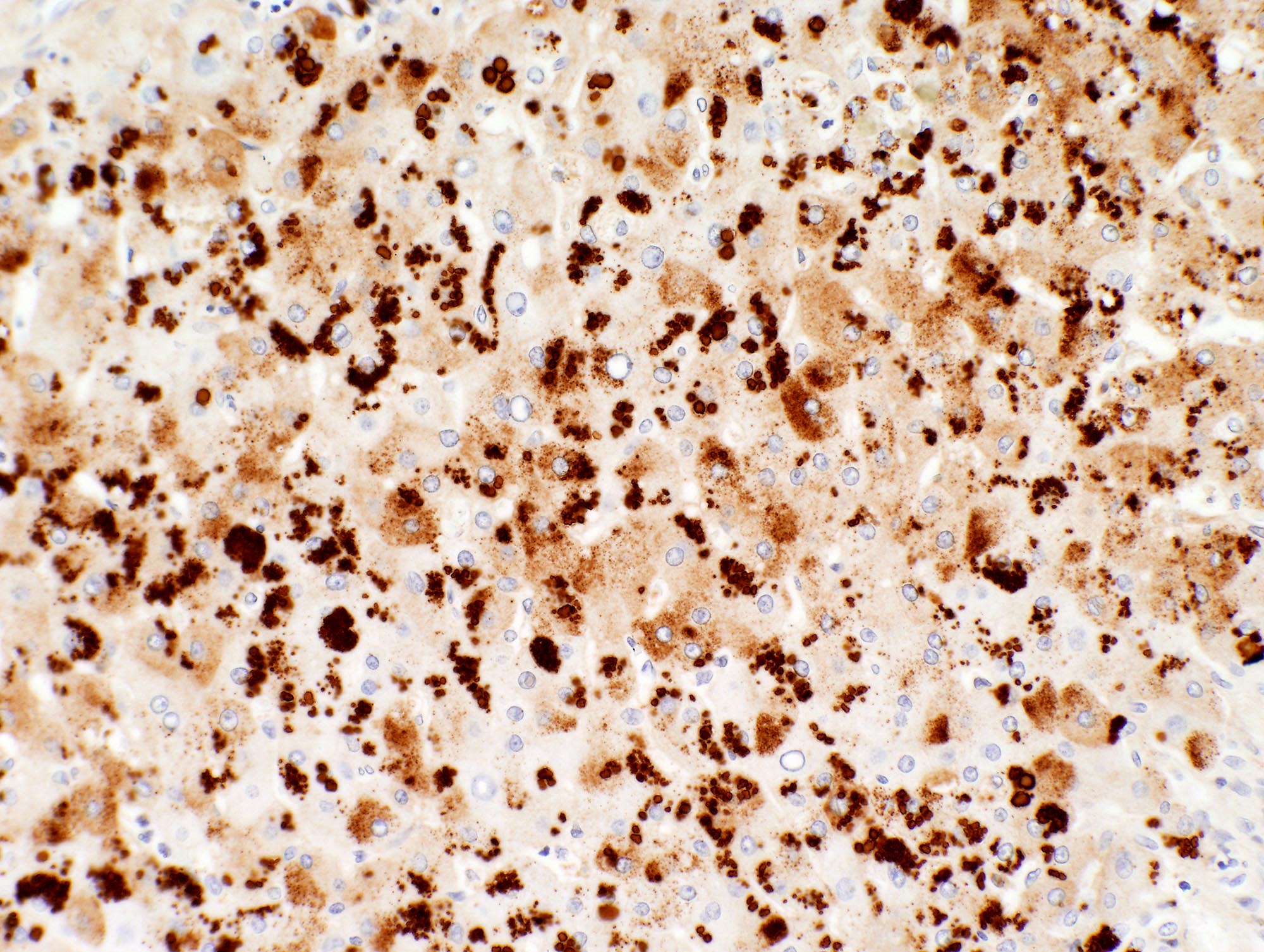
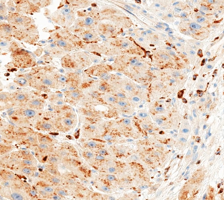
Alpha-1 antitrypsin IHC
Images hosted on other servers:

Breast: primary acinic cell carcinoma (fig F)

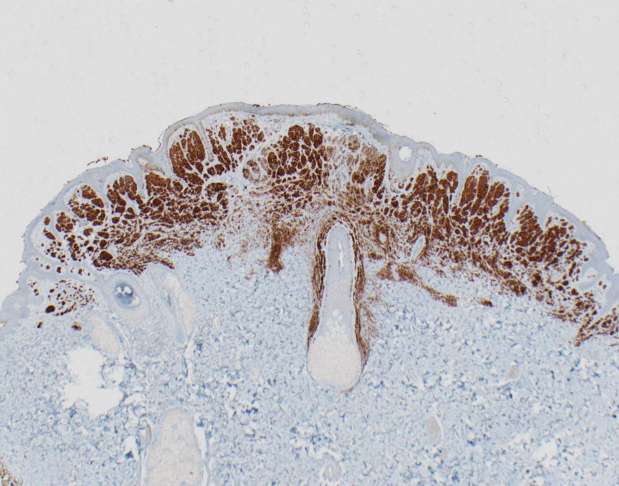
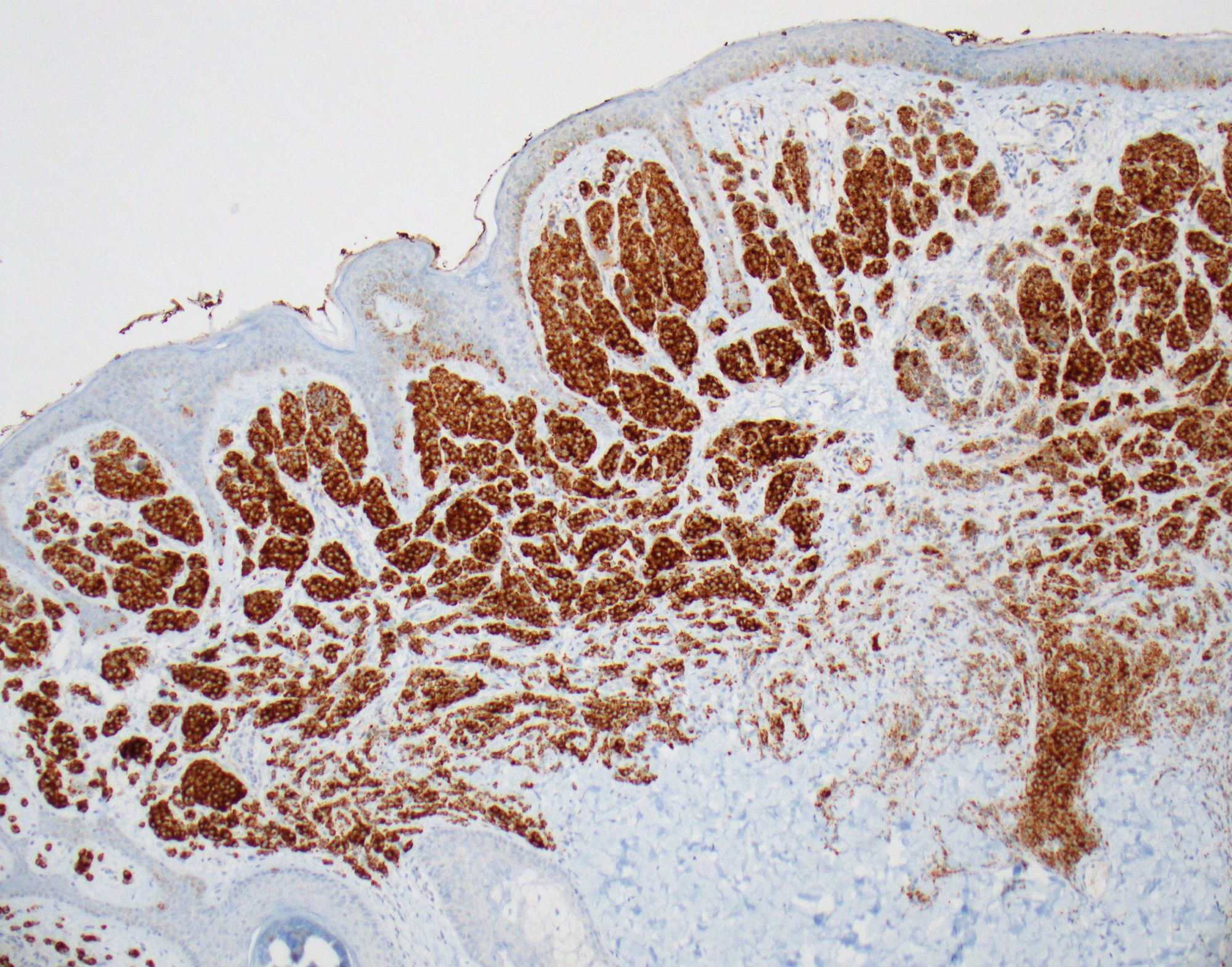
Skin: nevi and melanoma

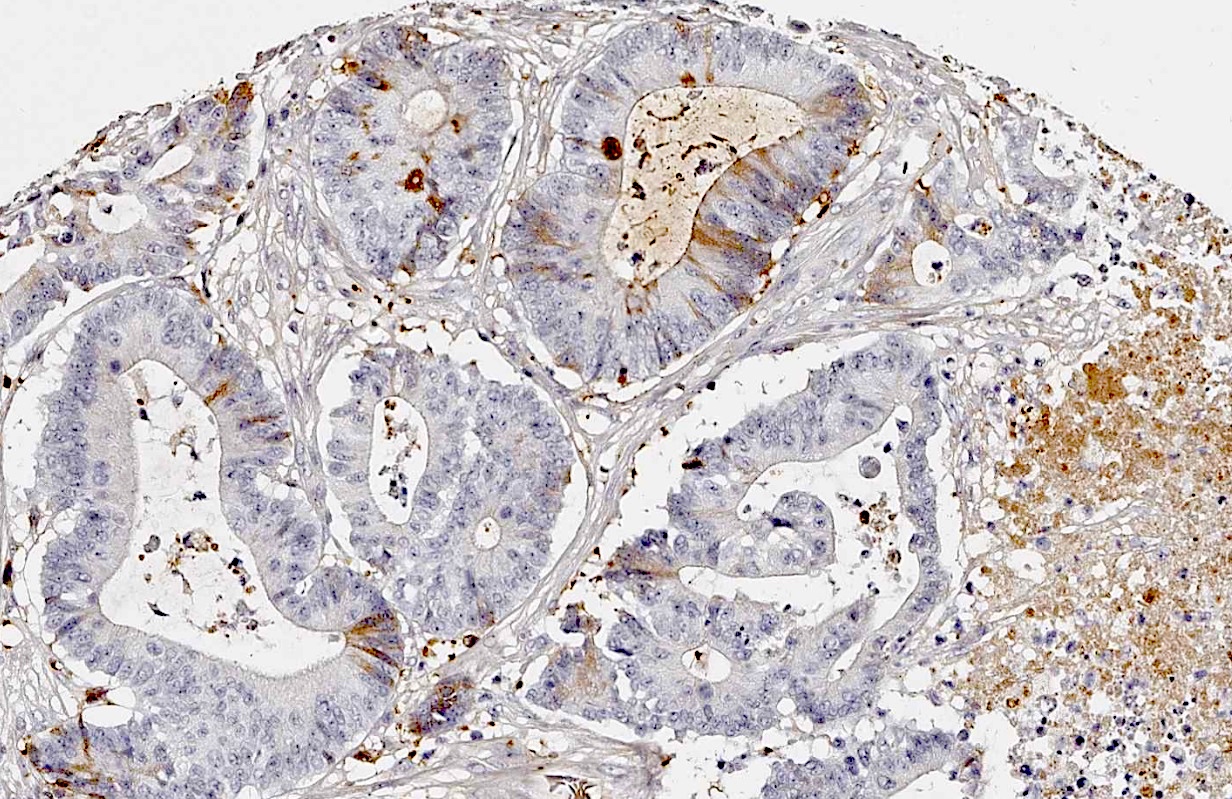
Stomach: hepatoid
adenocarcinoma
(fig C)
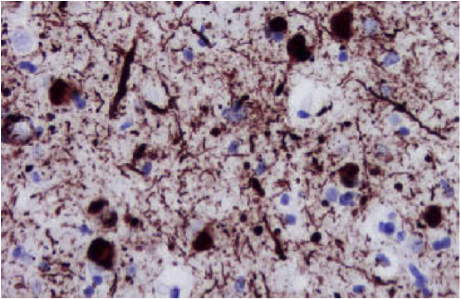
Contributed by Meenakshi Vij Gupta, M.D.

Lewy body in Parkinson disease
Cellular inclusions and neuritis plaques

Contributed by Jonathan Epstein, M.D., Debra Zynger, M.D.
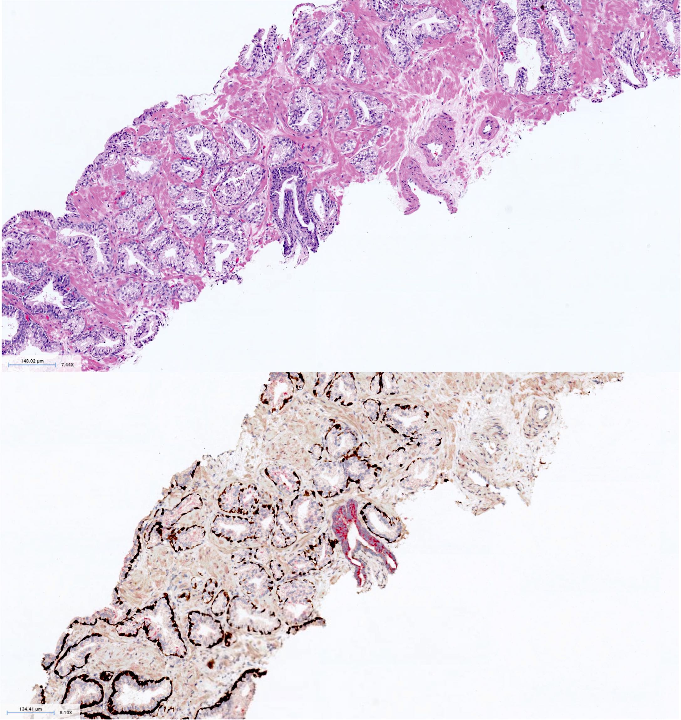
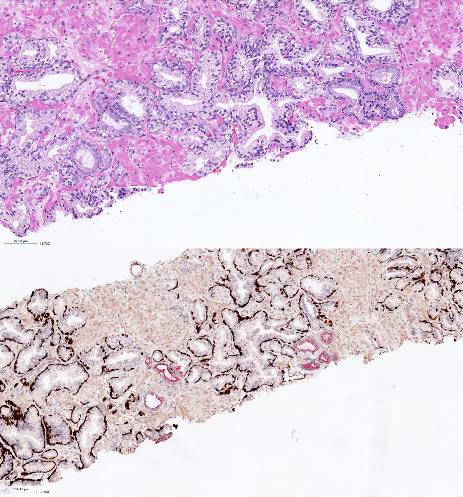
Prostate adenocarcinoma, triple stain
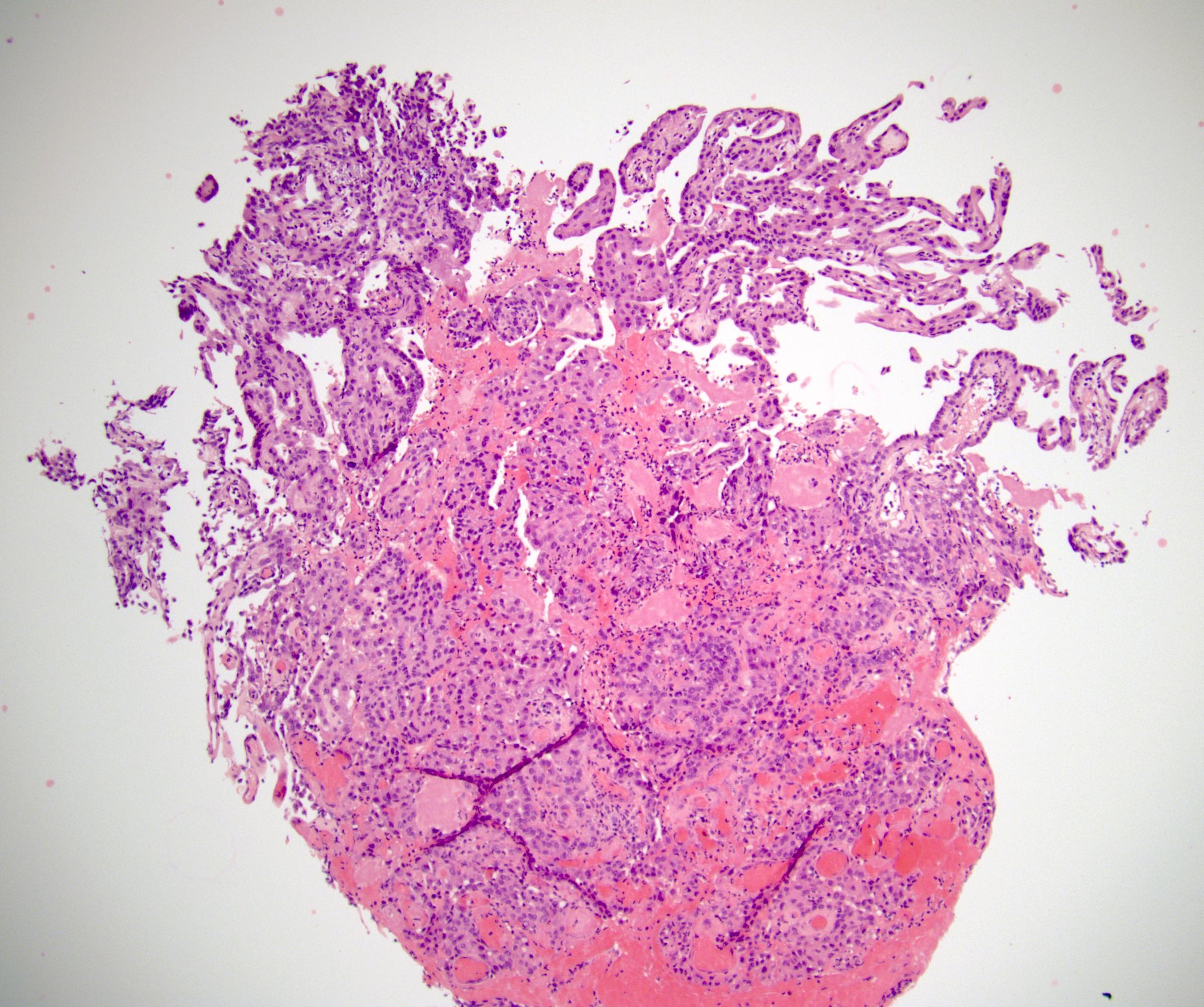
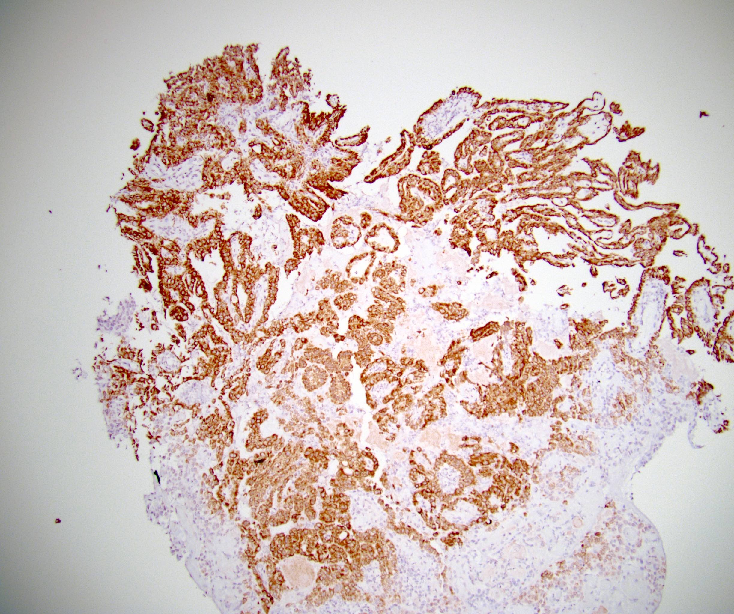
RCC papillary type 1 with AMACR
Images hosted on other servers:

Enhancement of
formic acid mediated
amyloid beta peptide
antigen retrieval

IHC of beta
amyloiddeposits
in the cerebral cortex
and blood vessels
Contributed by Monika Roychowdhury, M.D.
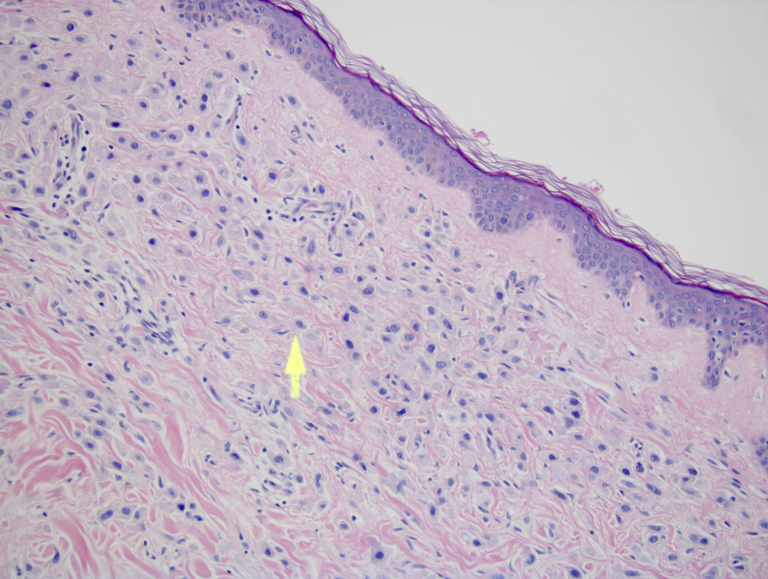
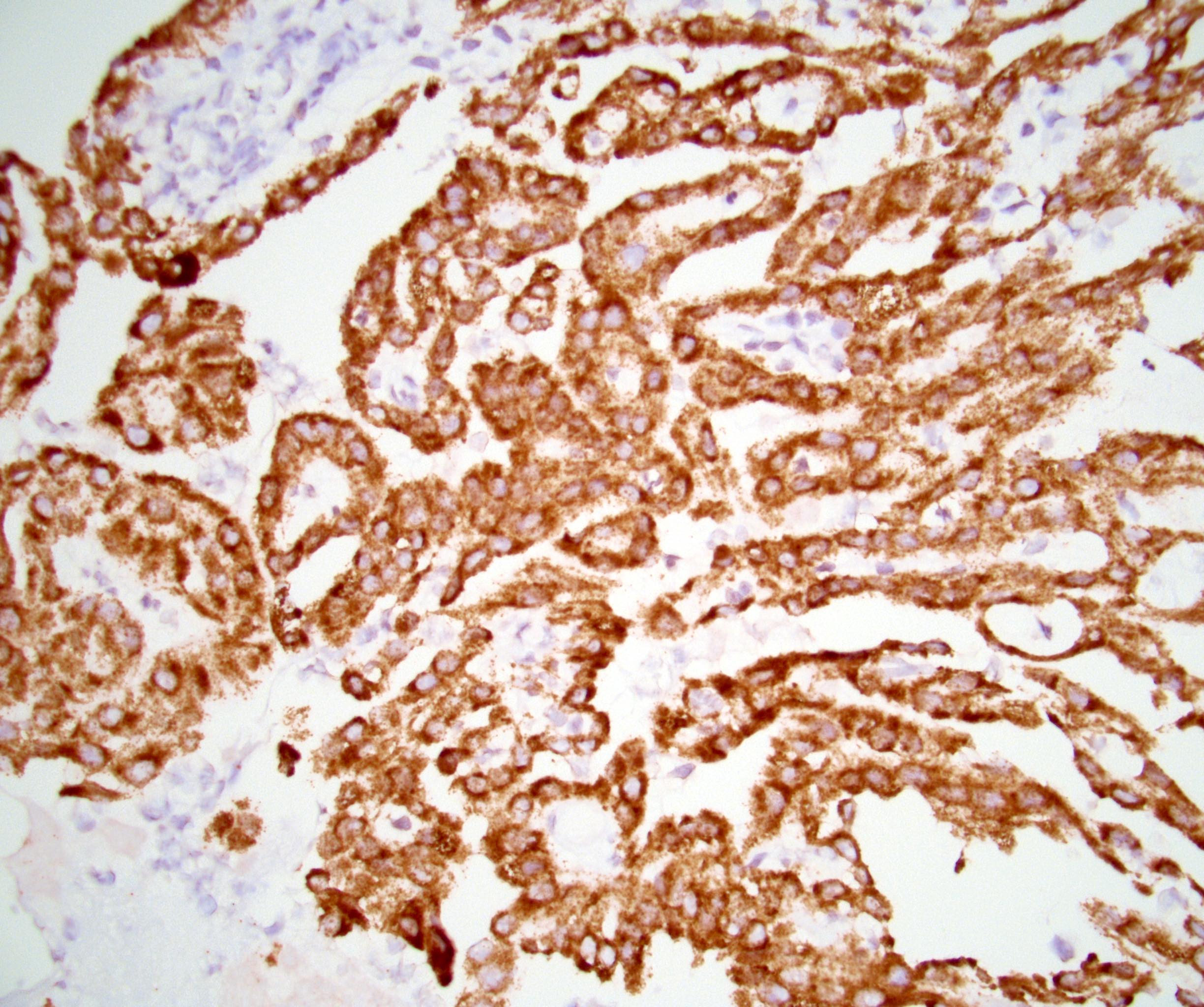
Skin metastatic breast carcinoma 20x
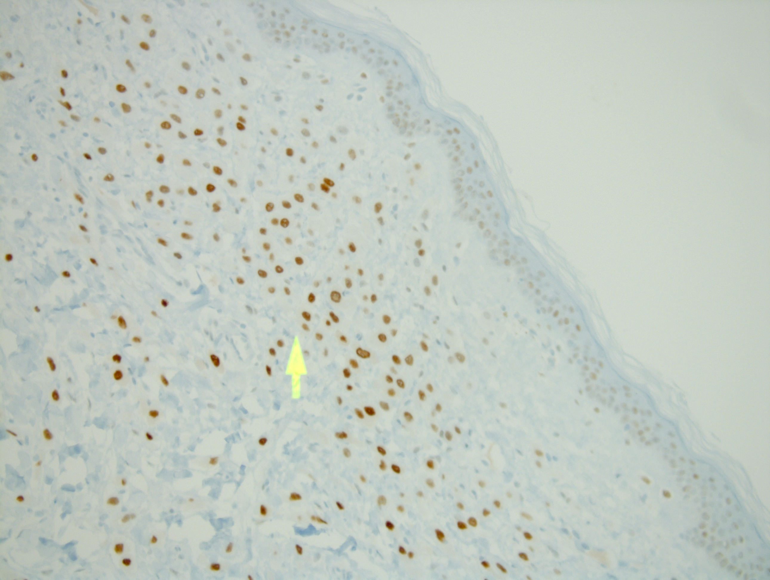
Androgen receptor
in metastatic breast
carcinoma 20x
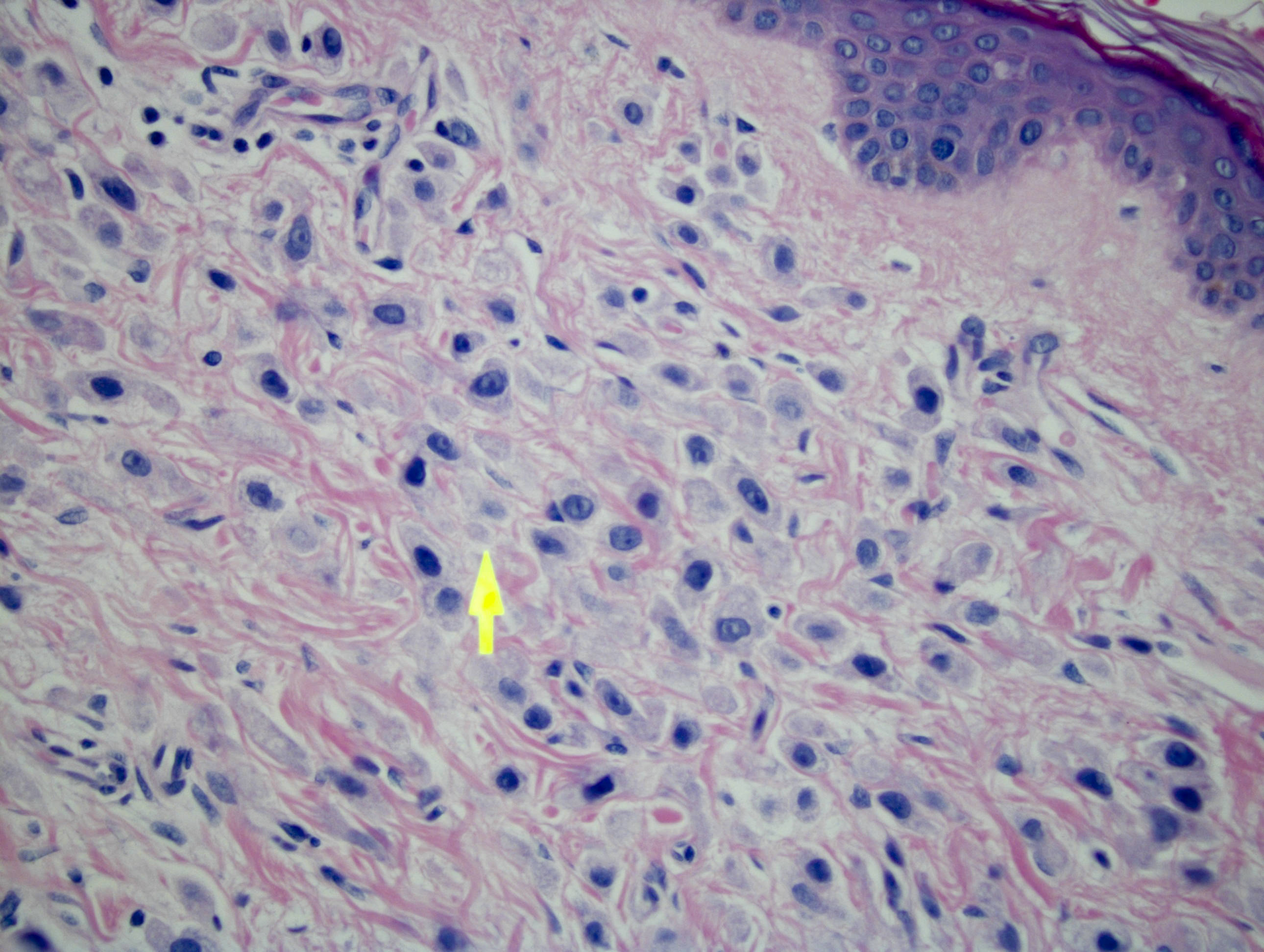
Skin metastatic breast carcinoma 40x
Androgen receptor
in metastatic breast
carcinoma 40x
Case #214
Salivary gland AR-
low grade cribriform
cystadenocarcinoma
Images hosted on other servers:

Breast: apocrine DCIS

Breast: apocrine metaplasia

Prostate carcinoma
metastatic to bone
Contributed by GenomeMe and Cell Marque Corporation
Colon (normal), clone IHC400

Liver (normal), clone IHC400

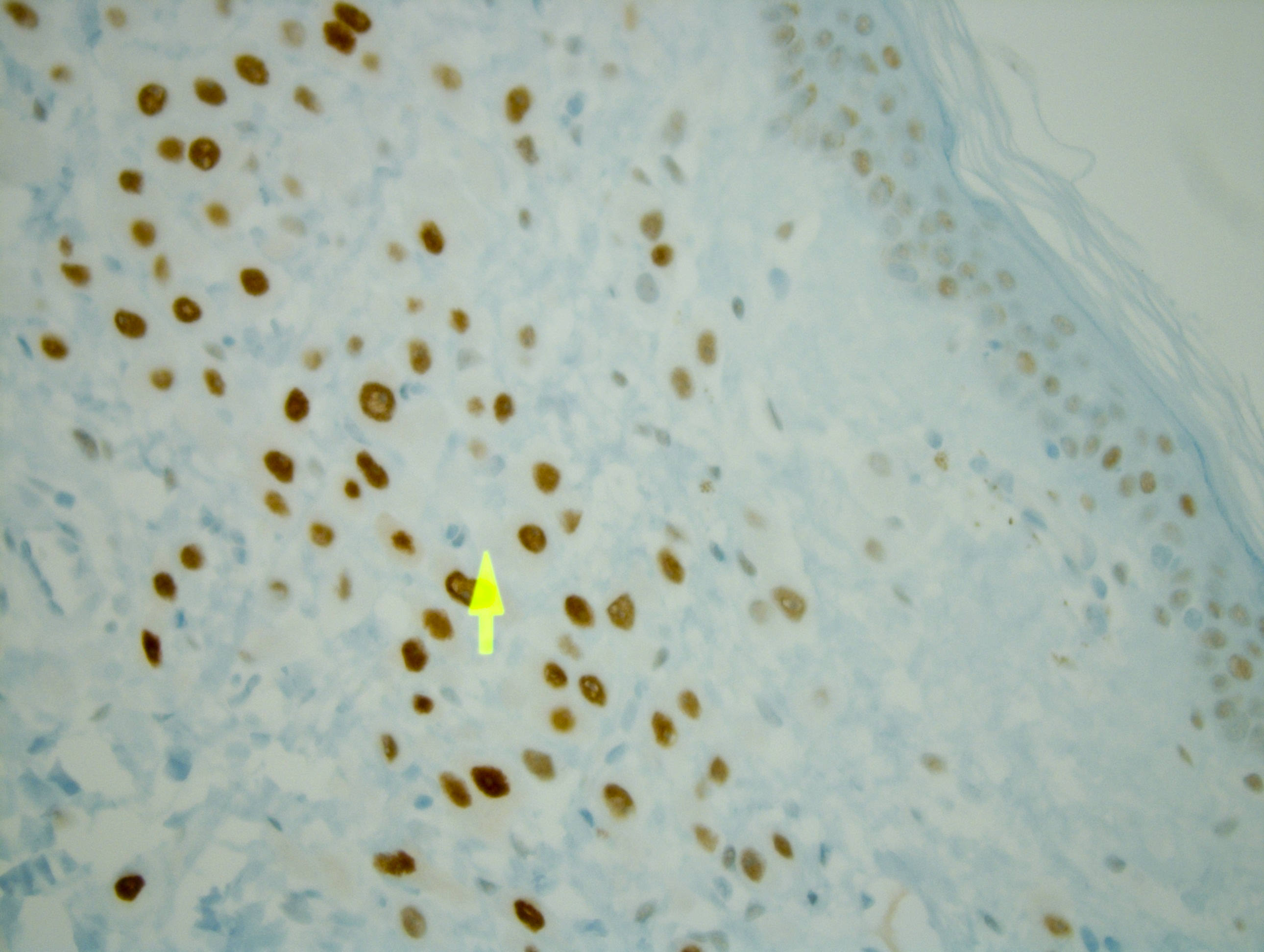
HCC - nuclear and cytoplasmic staining
Images hosted on other servers:


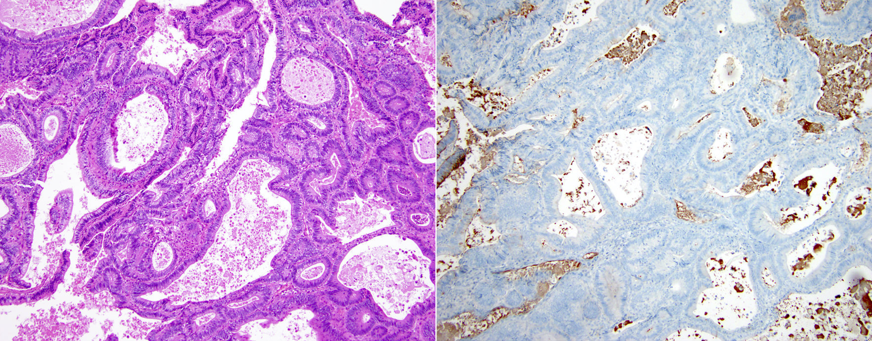
Hepatocellular carcinoma

Moderately differentiated HCC

Liver: benign and hepatocellular carcinoma

Metastatic colonic adenocarcinoma to liver


Cholangiocarcioma

Lung: TB related granulomas
Images hosted on other servers:

Gene map, regulation of chromatin structure
Images hosted on other servers:


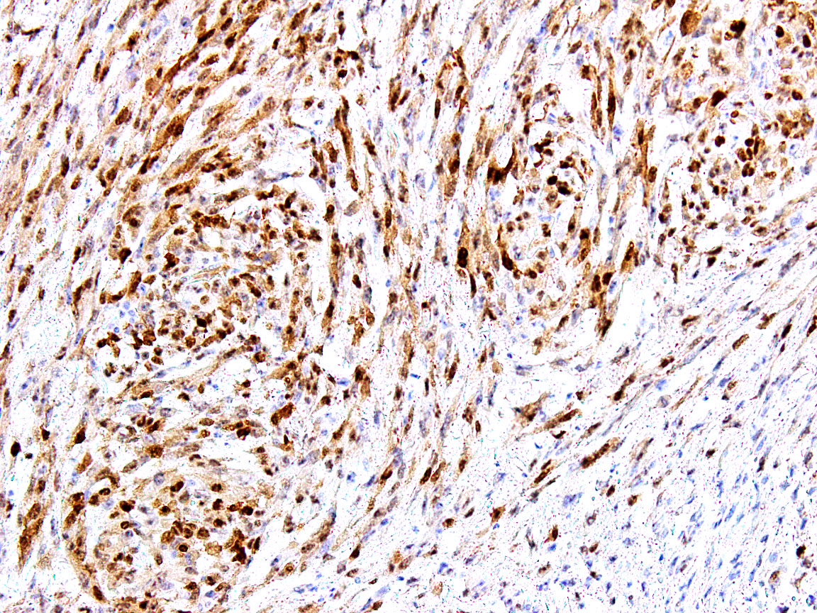
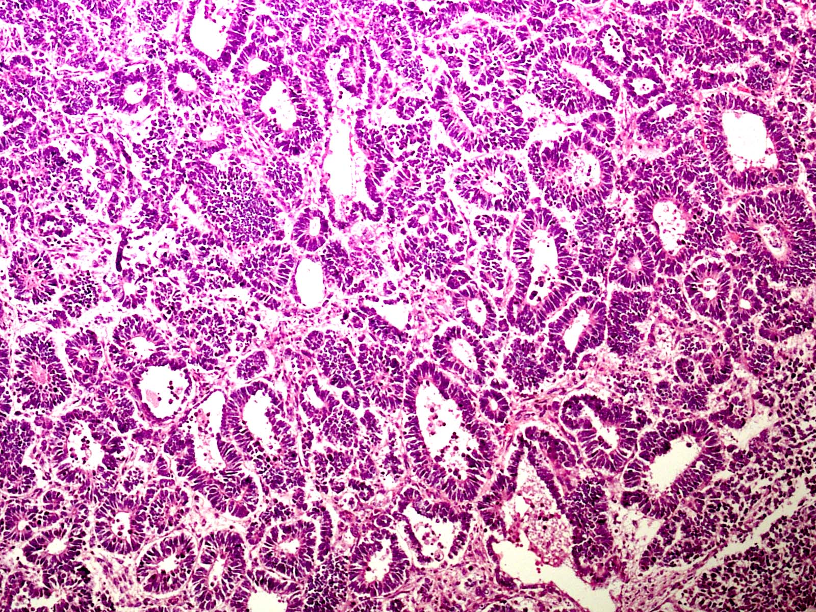
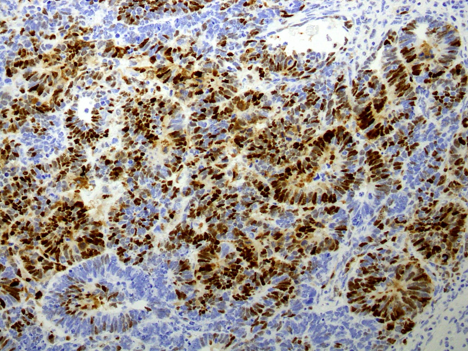
Cervix: borderline seromucinous tumors

Ovary: clear cell carcinoma (A, B)


Ovary: endometriotic cyst and associated well-differentiated endometrioid carcinoma

Stomach: gastric carcinoma

Uterus: endometrioid carcinoma

Uterus: endometriosis (G-I)
Images hosted on other servers:

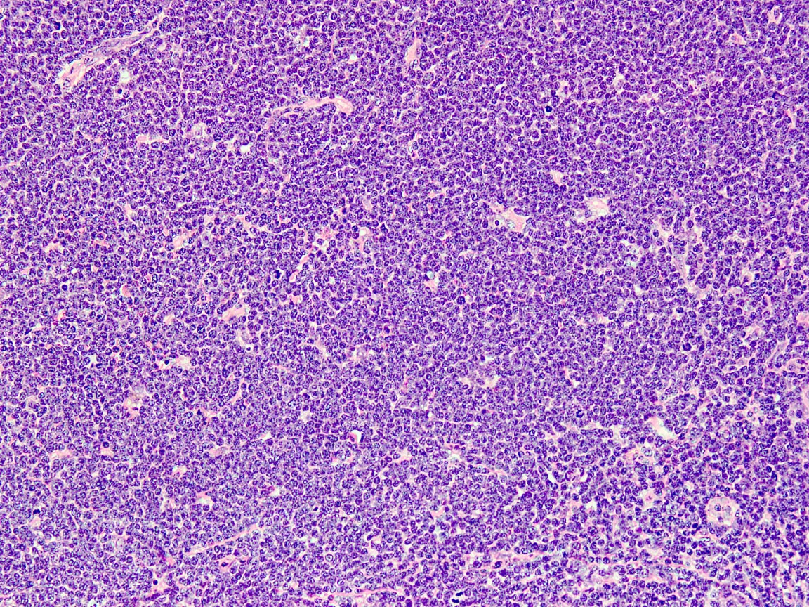
Neuroblastoma

Pancreas: ALT+ tumors

Pancreas: MEN1
microadenoma and
pancreatic neuroendocrine
tumor
Images hosted on other servers:

ATM in DNA
damage response

ATM signaling network
Contributed by Joo-Shik Shin, M.B.B.S., Ph.D.
Rectal adenocarcinoma
Images hosted on other servers:

Bronchioalveolar carcinoma
of lung: A-H&E, B-cytoplasmic
staining for CEA, C-cytoplasmic
staining for B72.3
Images hosted on other servers:

BAP1 protein localization


BAP1 interactions
Contributed by Gustav Stålhammar, M.D., Ph.D.
Uveal melanoma, BAP1 positive
Uveal melanoma, BAP1 negative
Uveal melanoma, BAP1 mixed
AFIP images

BCL2 translocation in follicular lymphoma
Images hosted on other servers:

2 main pathways to apoptosis
Contributed by Vladimir Osipov, M.D. (Case #171), Keith Kaplan, M.D. (Case #150) and AFIP
Secondary follicle

Follicular lymphoma
Burkitt lymphoma
Spindle cell epithelioma of vagina

Synovial sarcoma: right pericardium
Images hosted on other servers:
Various images

Collecting duct carcinoma-kidney

Follicular lymphoma


Follicular lymphoma of thyroid: t(14;18) negative / BCL2 negative (left) versus positive (right)

Solitary fibrous tumor
Contributed by Elena M. Fenu, M.D.
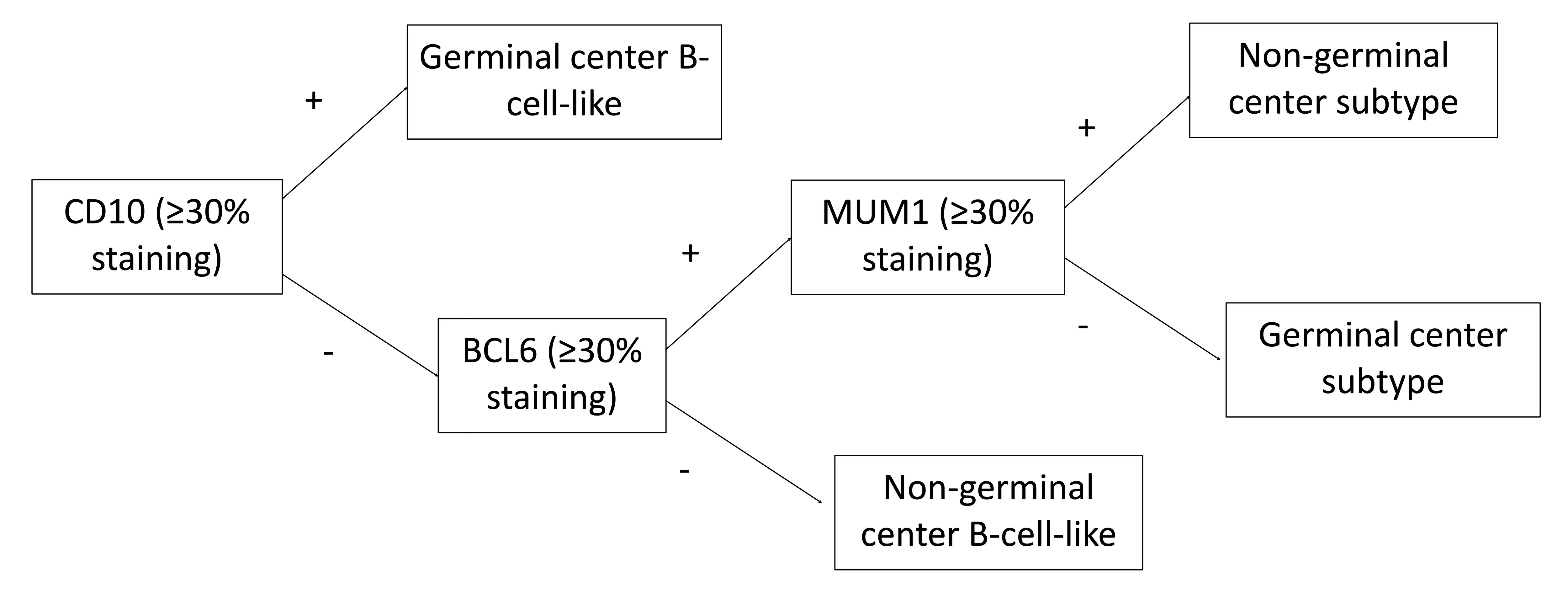
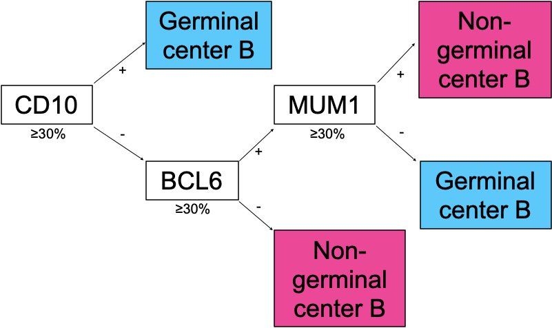
Staining algorithm for DLBCL
Contributed by Elena M. Fenu, M.D.
Reactive follicular hyperplasia
Follicular lymphoma, grade 1 - 2
Follicular lymphoma, grade 3B
Angioimmunoblastic T cell lymphoma
Burkitt lymphoma
T cell / histiocyte rich large B cell lymphoma
Nodular lymphocyte predominant Hodgkin lymphoma
Classic Hodgkin lymphoma
Contributed by Isidro Machado, M.D., Ph.D.
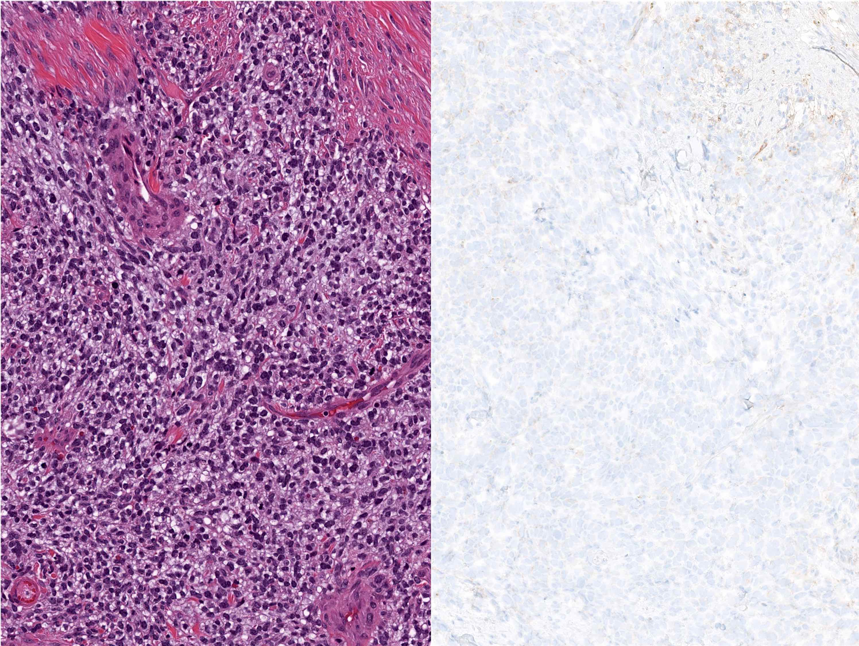
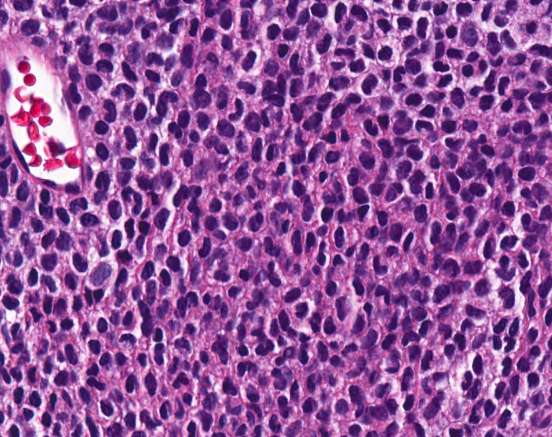
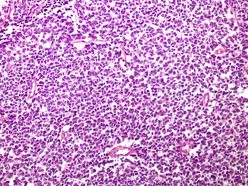
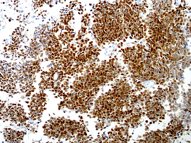
Undifferentiated round cell sarcoma

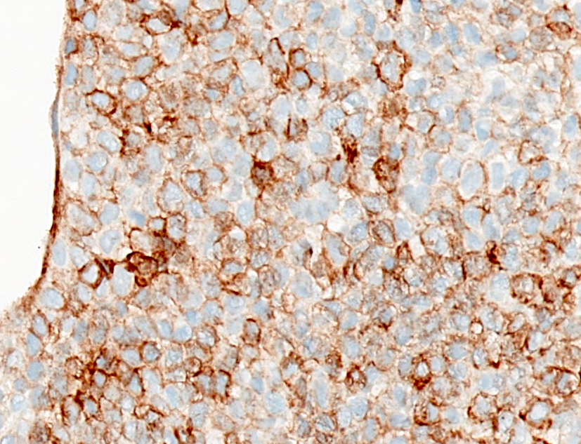
Undifferentiated round cell sarcoma, BCOR positive
Contributed by Jijgee Munkhdelger, M.D., Ph.D. and Andrey Bychkov, M.D., Ph.D.
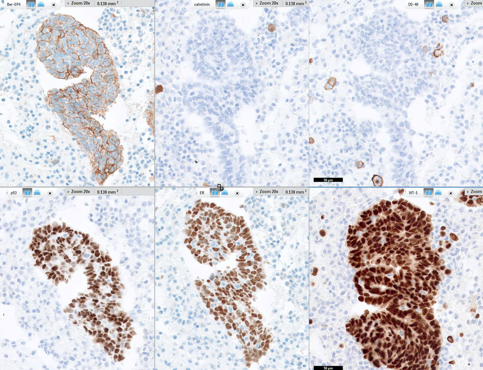
Ovarian serous carcinoma immunoprofile
Images hosted on other servers:

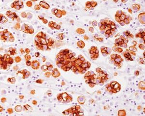
Basaloid squamous cell carcinoma is BerEP4+

Breast: ductal and
lobular carcinoma -
primary and
metastases
(EpCAM)


Breast cancer: increased EpCAM expression in metastases


Colon: loss of EPCAM expression in cancers associated with Lynch syndrome caused by heterozygous EPCAM germline deletions

Esophagus: normal
and Barrett metaplasia;
gastric mucosa
Unknown site: adenocarcinoma (MOC31)
Images hosted on other servers:


Reactive mesothelial cells (BerEP4 / MOC31 negative) versus metastatic adenocarcinoma (positive)
Images hosted on other servers:

Cadherins and catenins in signal transduction

Role of β catenin in cell
Images hosted on other servers:


Colon: serrated adenoma

Colon: normal and carcinoma

Liver: metastatic
colorectal carcinoma
in sentinel lymph
node and liver

Soft tissue: liposarcoma
Images hosted on other servers:
Cadherins and catenins in signal transduction
Role of beta catenin in cell
Contributed by Pamela Wirth, Ph.D. (source: University of Toronto and Human Protein Atlas)

Normal pancreatic tissue

Cervix

Breast carcinoma

Colorectal carcinoma

Lung cancer

Normal kidney
Images hosted on other servers:

Normal tonsil

Various B cell lymphomas
Images hosted on other servers:

MAPK signaling pathway
Contributed by Andrew Colebatch, M.B.B.S., Ph.D. and Andrey Bychkov, M.D., Ph.D.
BRAF V600E positive nevus
BRAF V600E positive melanoma
V600E mutant protein is diffusely expressed in tumor / cancer but not normal tissue

Diffuse cytoplasmic staining
Images hosted on other servers:

MAPK and AKT pathways

Resistance diagram
Images hosted on other servers:

Medullary thyroid cancer
Images hosted on other servers:
Normal mesothelium

Various images and quality control

Mucinous cystic neoplasm of mesentery
Contributed by Andrey Bychkov, M.D., Ph.D.
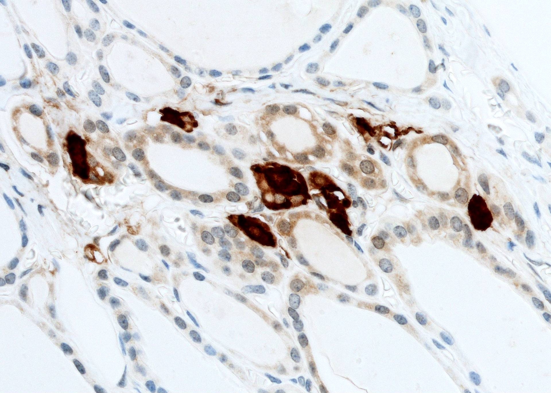
Cluster of C cells

Pericapillary location of C cells

Heavy background
in the immediate
stroma and follicles
Images hosted on other servers:

Thyroid

Single endocrine cell

Prostate
(figure 1)

HGPIN (figure 2)

Bone metastasis (figure 3)
Images hosted on other servers:
Malakoplakia of bladder (von Kossa calcium stain)
Images hosted on other servers:

Colon-pericrytal fibroblast sheath (fig A)

Leiomyosarcoma, soft tissue
Case #62
Sclerosing lobular hyperplasia
Images hosted on other servers:

Breast:
adenomyoepithelioma

MFH of bone
(fig B, F)
Contributed by Nick Baniak, M.D. and Sharon Bihlmeyer, M.D.
Sarcomatoid mesothelioma

Sarcomatoid mesothelioma (CK5)
Sarcomatoid mesothelioma (calretinin)

Ganglion cells in colon
No ganglion cells
Ovarian granulosa cell tumor (adult)
Contributed by Nick Baniak, M.D.
Lung adenocarcinoma
Mesothelioma
Contributed by Nick Baniak, M.D.

Clear cell RCC

Clear cell papillary RCC
Papillary RCC

Negative renal parenchyma
Images hosted on other servers:


Classification examples
Images hosted on other servers:


Bone: osteoclasts in osteoclastoma and normal

Intervertebral disc

Maxillary sinus: basaloid squamous cell carcinoma
Images hosted on other servers:

CCR2: lamina propria lymphocytes

CCR5: HIV entry into CD4+ cell via CCR5 co-receptor

CCR6: immature
dendritic cells
traffic to lung via
CCR6 / CCL20 axis
Images hosted on other servers:

CCR2: human glomeruli
CCR3:


Atherosclerosis

Eosinophils and airway epithelium

Lymph node: Hodgkin disease

Skin: cutaneous T cell lymphoma

Paraffin embedded malignant melanoma

Renal cell carcinoma
CCR4:

Lymph node: normal


Lymph node and peripheral blood: adult T cell leukemia / lymphoma

Skin: Sézary Syndrome
CCR6:

Lung: nonsmall cell lung carcinoma

Lung: sarcoidosis

Lymph node: melanoma

Spleen: memory T cells

Tonsils: inflamed
CCR7:

Breast: various carcinomas

Colorectum: T cells in advanced adenocarcinoma

Esophagus: squamous cell carcinoma and metastatic lymph node

Lymphoma T cell

Skin: melanoma and sentinel lymph node
CCR9:

Ovary: normal and carcinoma


Images hosted on other servers:

CD182: ovarian carcinoma


CD185: chronic H. pylori gastris (left); MALT lymphoma (right)

CD185: AIDS related non-Hodgkin lymphoma


CD185: synovium of nonrheumatoid arthritis (left) and rheumatoid arthritis (right)

CCR2: human glomeruli

CCR3: eosinophils and airway epithelium

CCR3: lymph nodes involved by Hodgkin disease

CCR3: cutaneous T cell lymphoma

CCR3: paraffin embedded malignant melanoma

CCR3: renal cell carcinoma

CCR4: lymph node normal


CCR4: lymph node (adult T cell leukemia / lymphoma)

CCR4: Sézary syndrome

CCR6: nonsmall cell lung carcinoma

CCR6: lung sarcoidosis

CCR6: lymph node melanoma

CCR7: various breast carcinomas

CCR7: T cells in advanced adenocarcinoma in colorectum

CCR7: esophageal SCC and metastatic lymph node

CCR7: lymphoma T cells

CCR7: skin melanoma and sentinel lymph node

CCR9: normal and carcinoma ovaries

CD247: normal expression

CD247: reduced expression

CD248: IHC detection of FB5 antigen
Images hosted on other servers:

CCR4:
peripheral blood
(adult T cell
leukemia / lymphoma)
Images hosted on other servers:

CD1d: antigen presentation

CD1d: viral evasion of antigen presentation

CD1d: intestinal epithelial CD1d expression

CD2: T cells and antigen presenting cells

CD9: structure
Images hosted on other servers:

CD1: primary and metastatic melanoma

CD1: Langerhans cells of anal mucosa

CD1b: leprosy patient

CD1c: marginal zone B cells (spleen)

CD1c: stomach: H. pylori+ mucosa

CD1c: thyroid gland

CD1c: immature dendritic cells

CD1d: HPV associated lesions (cervix)

CD1d: trophoblast (placenta)


CD1d: scalp skin

CD2: hydroa
vacciniforme-like
lymphoma

Brain (CD6-)

CD6: colon

Kidney (CD6-)

Skeletal muscle (CD6-)

CD6: tonsil

Uterus (CD6-)
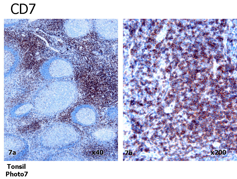
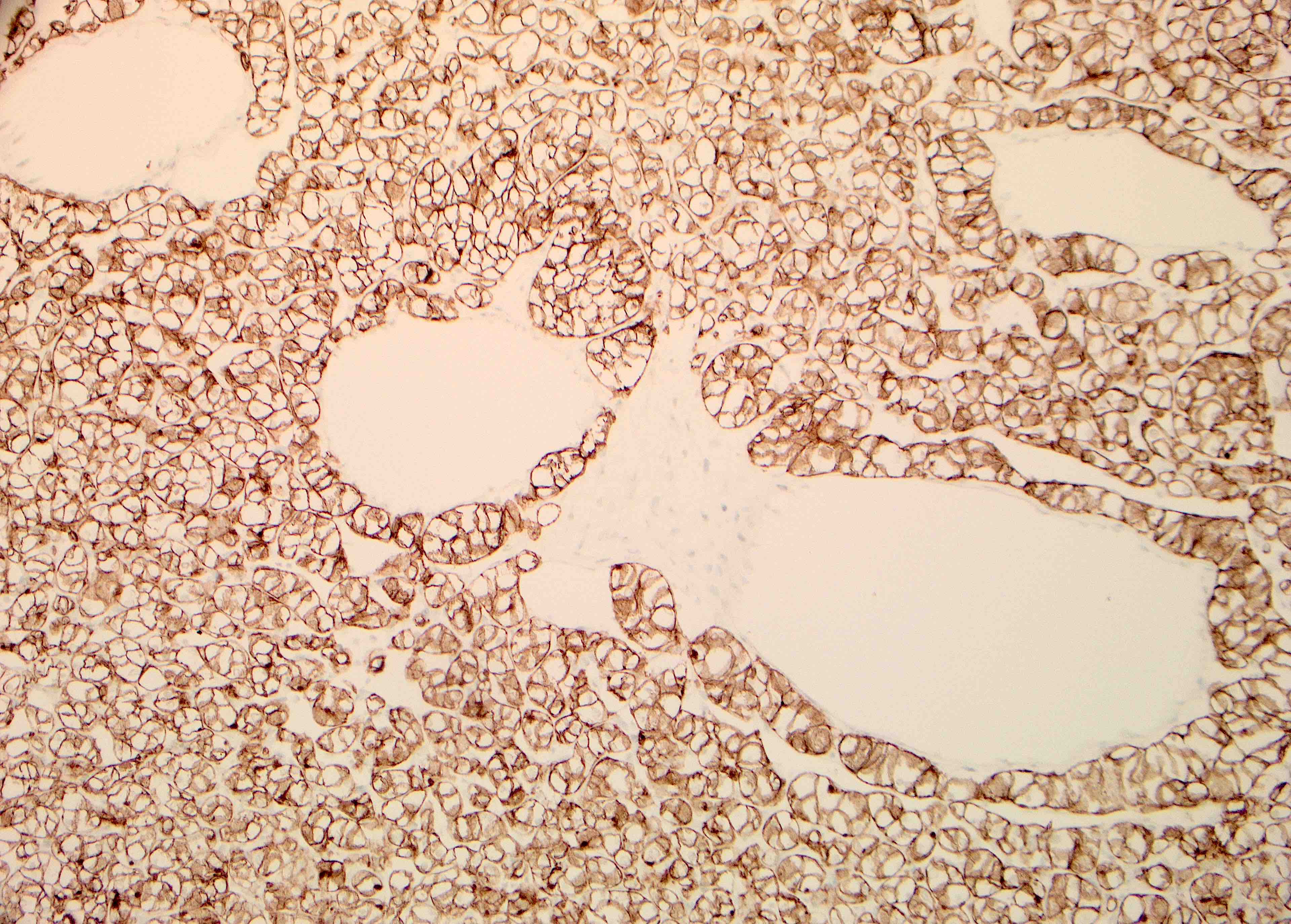
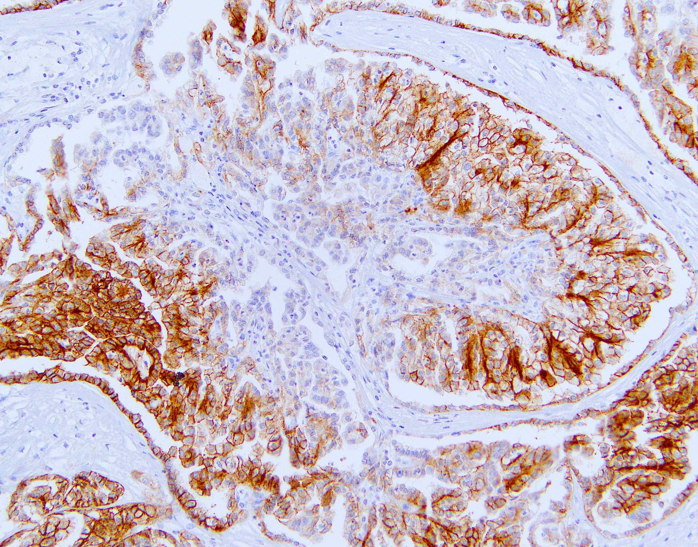
CD7: normal tonsil

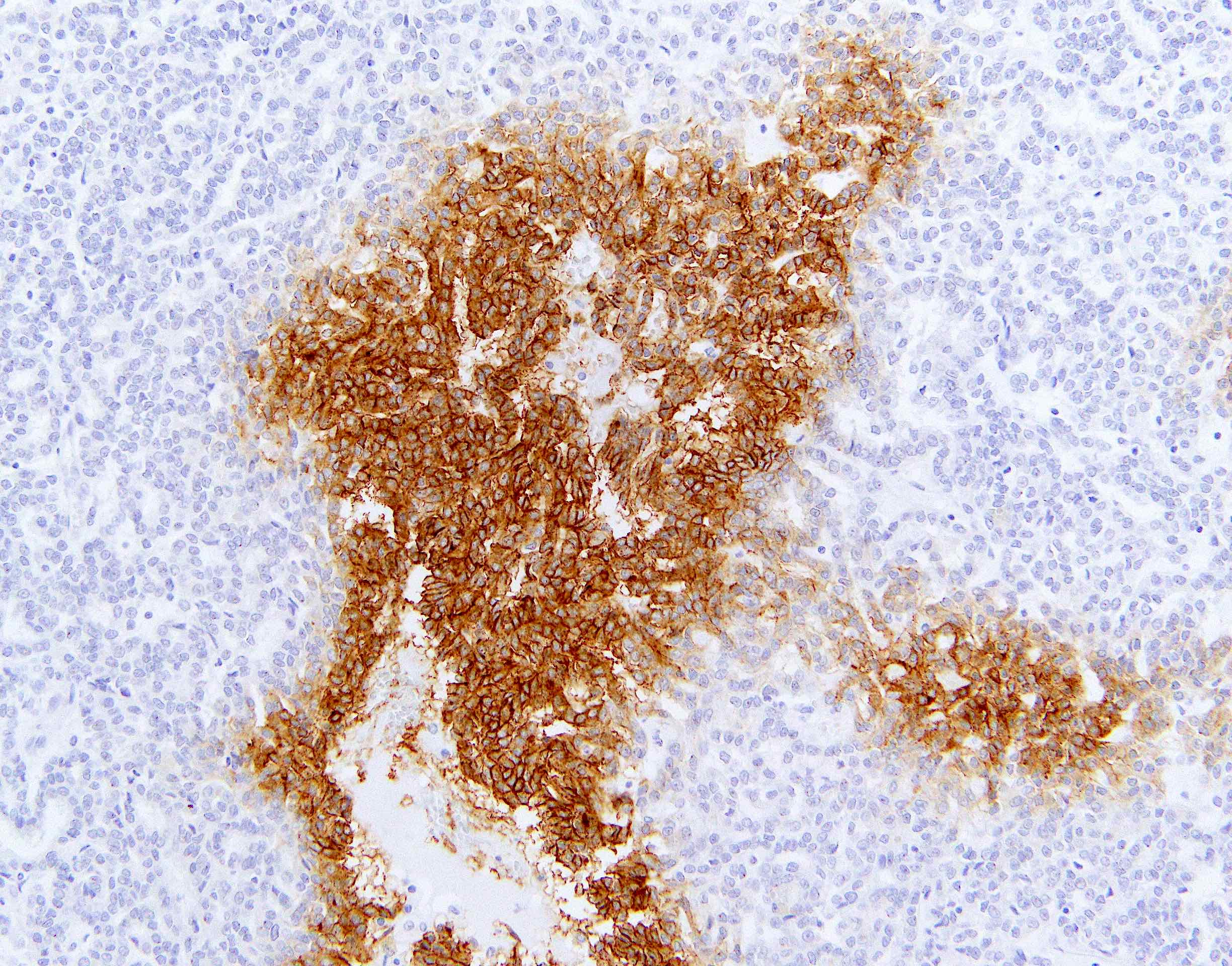
CD7: cutaneous T cell lymphoma

CD9: normal tissue (cerebrum)

CD9: normal tissue (cervical squamous epithelium)

CD9: cervical carcinoma


CD9: CNS nonneuroepithelial tumors

CD9: well differentiated adenocarcinoma (gallbladder)

CD9: mesothelioma

CD9: basal and
squamous cell
carcinoma and
actinic keratosis


CD9: small cell lung cancer
Image hosted on other servers:

CD1c: various cellular locations
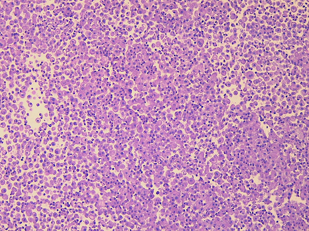
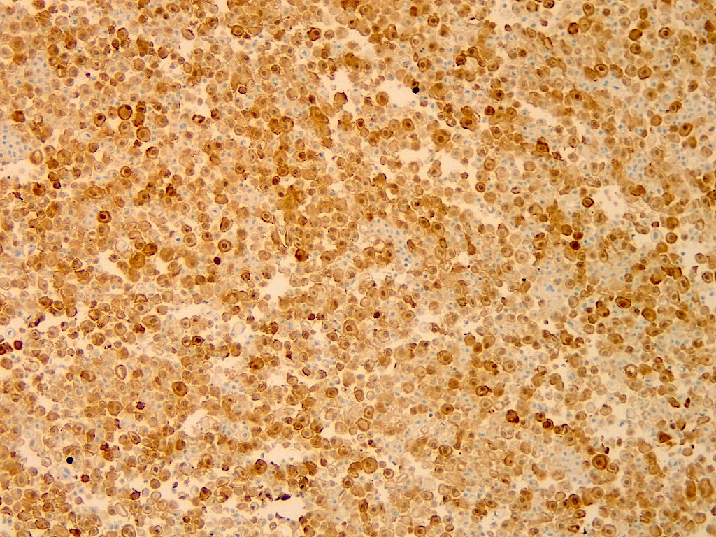
Contributed by Bradford Siegele, M.D., J.D.
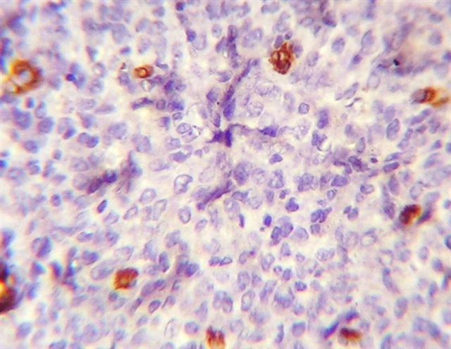
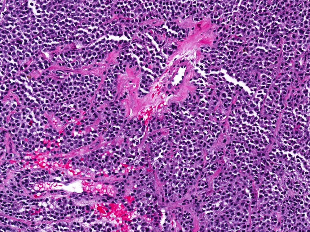
Lymphoblasts in B ALL / LBL
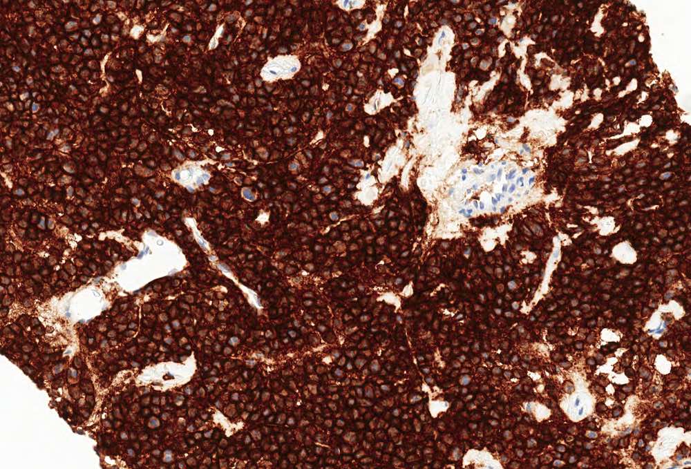
Lymphoblasts in B ALL / LBL, PAX5+
Lymphoblasts in B ALL / LBL, CD10+
Lymphoblasts in B ALL / LBL, TdT+
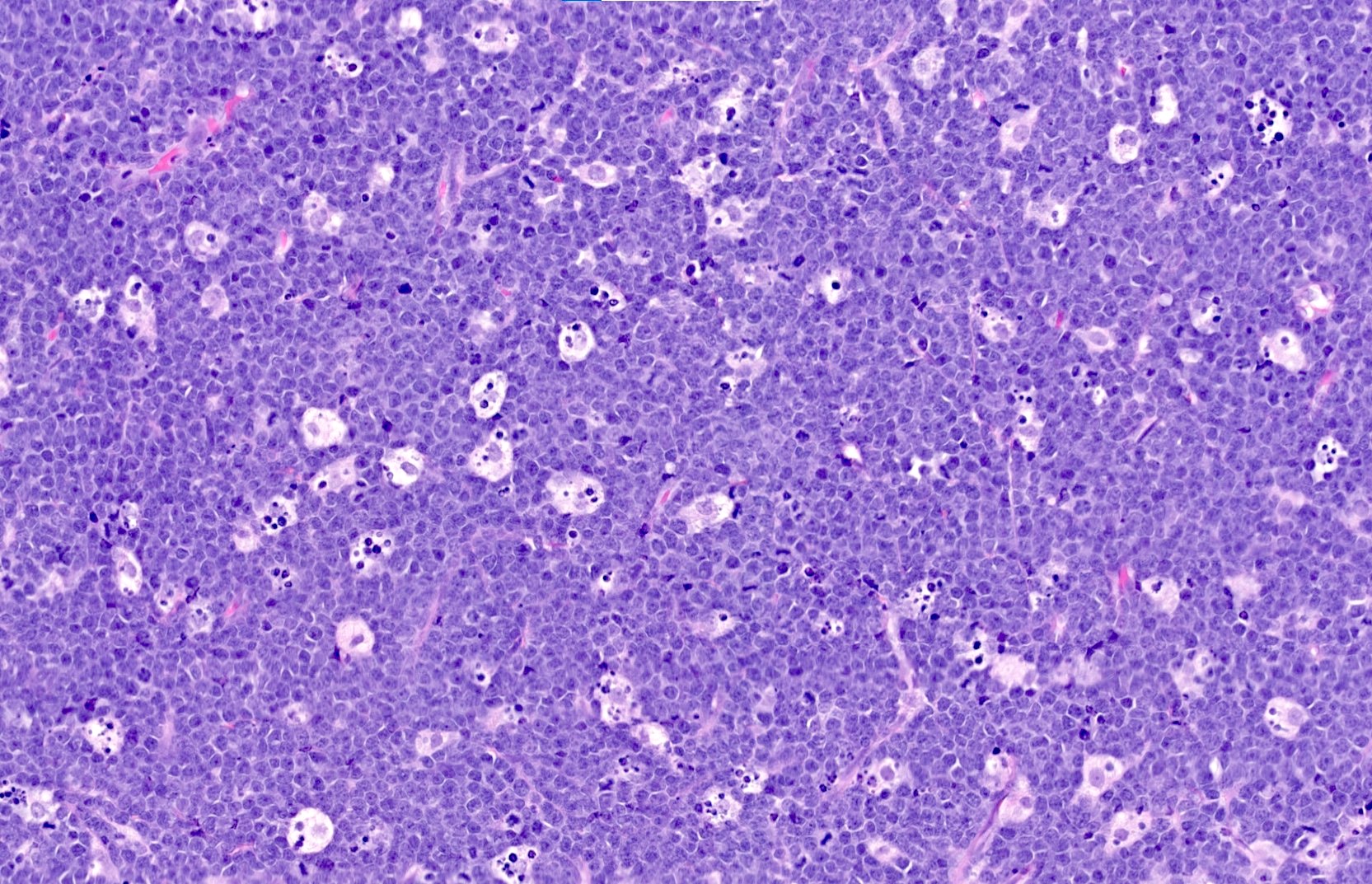
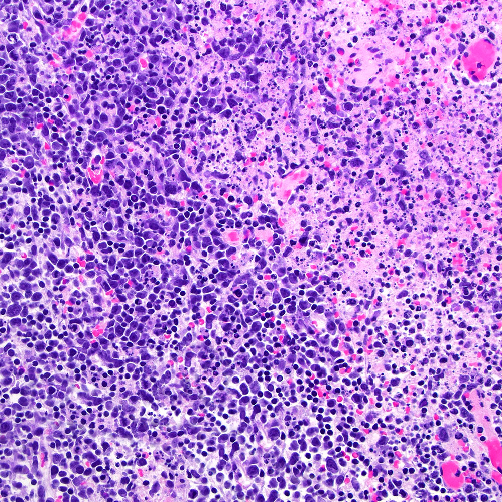
Starry sky of Burkitt lymphoma
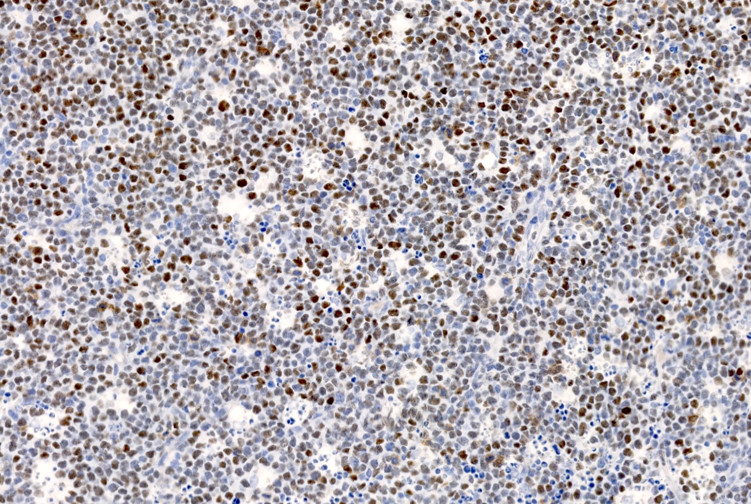
Burkitt lymphoma cells, CD20+
Burkitt lymphoma cells, CD10+
Burkitt lymphoma cells, BCL6+
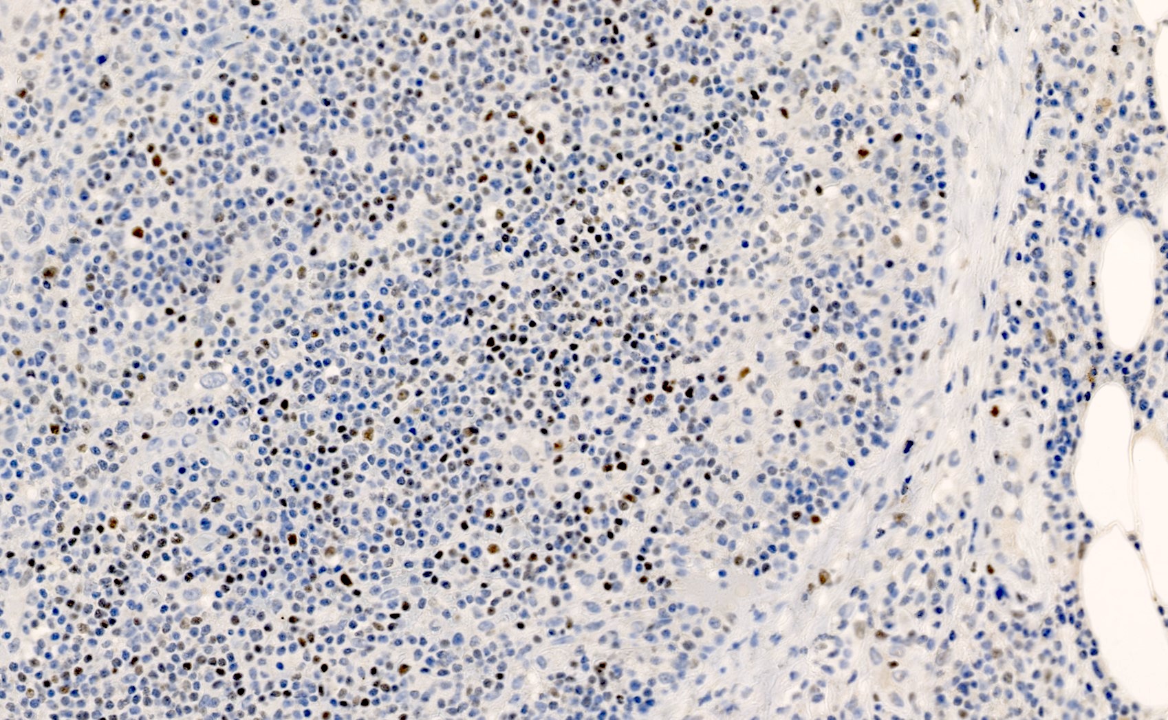
Burkitt lymphoma cells, TdT-
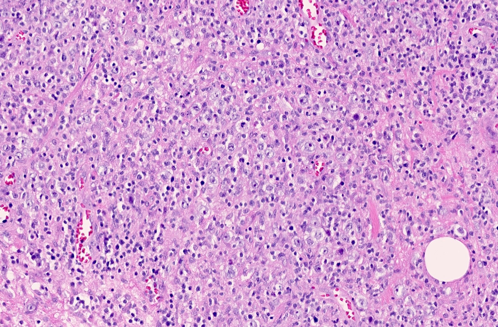
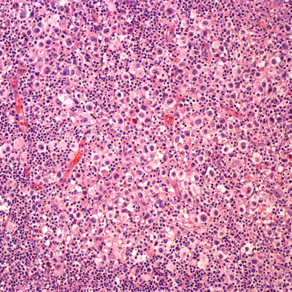
Large atypical cells of DLBCL
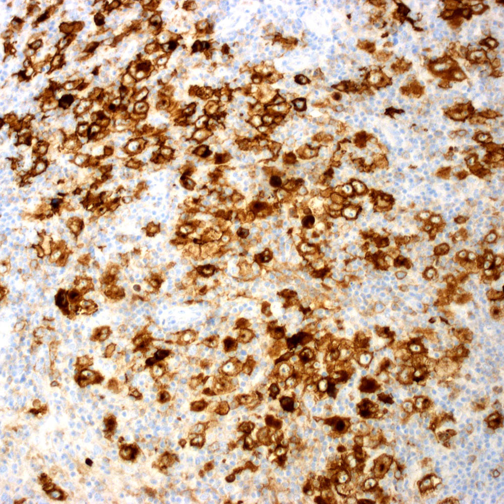
Large atypical cells of DLBCL, OCT2+
Large atypical cells of DLBCL, CD10+
Large atypical cells of DLBCL, BCL6+
Large atypical cells of DLBCL, MUM1 variable
Atypical cells and blood vessels of AITL
Atypical cells of AITL, CD3+

Atypical cells of AITL, CD4+
Atypical cells of AITL, CD8-
Atypical cells of AITL, CD10+
Atypical cells of AITL, ICOS+
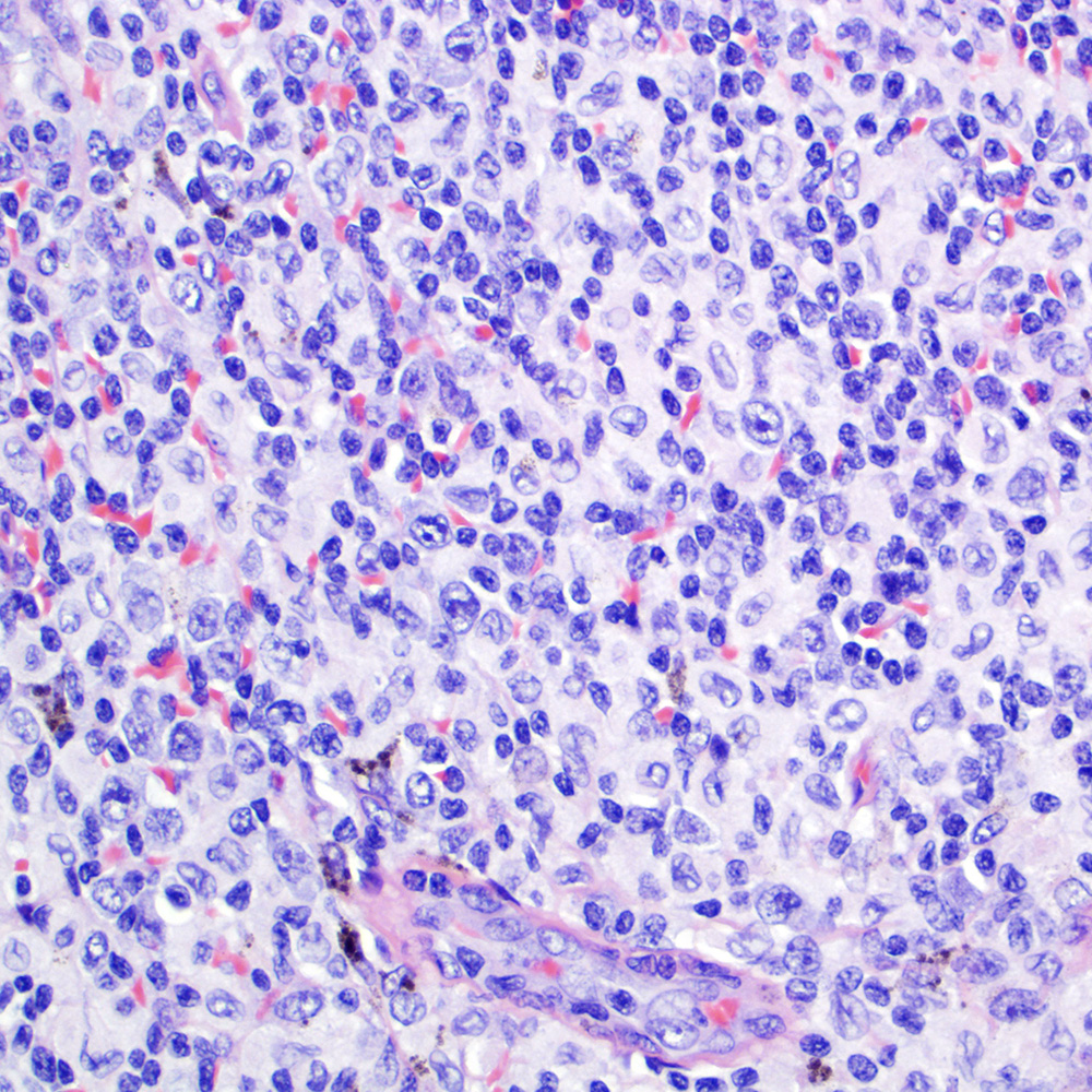
Nodules of atypical cells of FL
Nodules of atypical cells of FL, CD20+
Nodules of atypical cells of FL, CD10+
Nodules of atypical cells of FL, BCL2+
Nodules of atypical cells of FL, BCL6+
Atypical follicular
dendritic cell
meshworks of
FL, CD21+
Pseudopapillae of SPN
Pseudopapillae of SPN, CD10+
Pseudopapillae of SPN, alpha-1 antitrypsin+
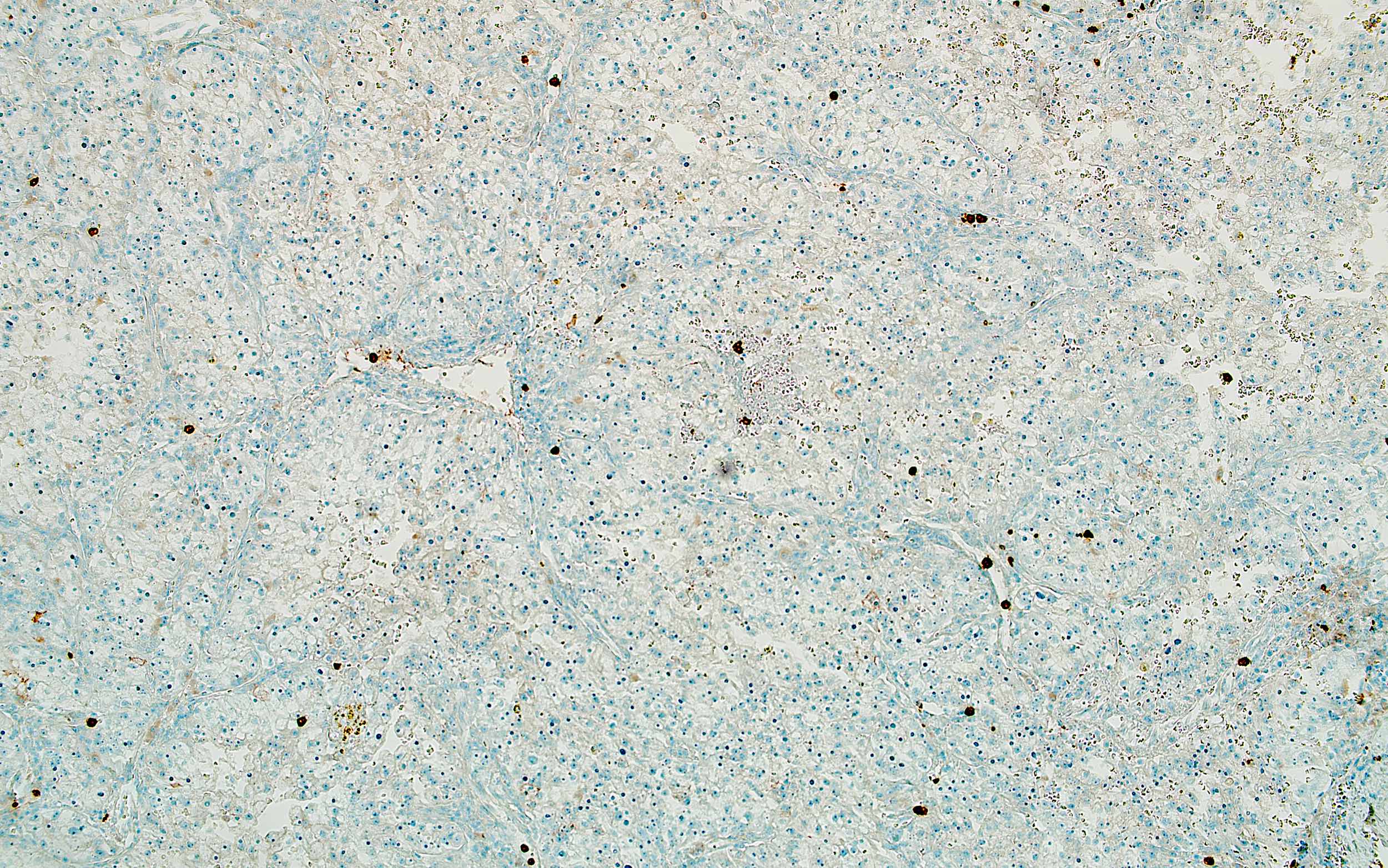
Endometriosis of sigmoid colon
Endometriosis of sigmoid colon, CD10+
Endometriosis of sigmoid colon, ER+
Moderately atypical spindle cells of ESS
Spindle cells of ESS, CD10+
Minimally atypical cells of ccRCC
Atypical cells of ccRCC, CD10+
Atypical cells of ccRCC, CAIX+
Atypical cells of ccRCC, vimentin+
Atypical cells of ccRCC, CK7-
Atypical cells of ccRCC, CD117-
Atypical cells of ccRCC, CD15-
Contributed by Jijgee Munkhdelger, M.D., Ph.D. and Andrey Bychkov, M.D., Ph.D.
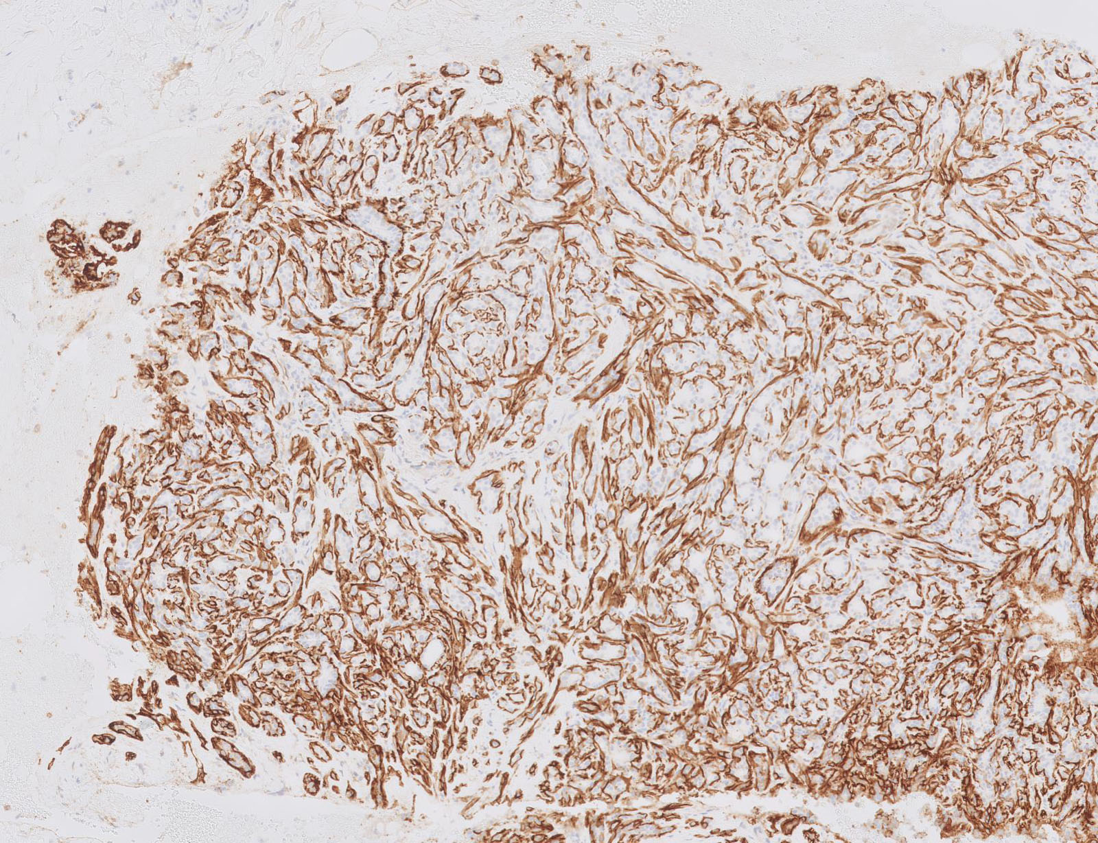
Myoepithelial layer on CD10
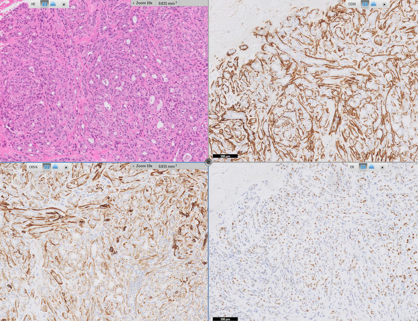
Breast tubular adenoma immunoprofile
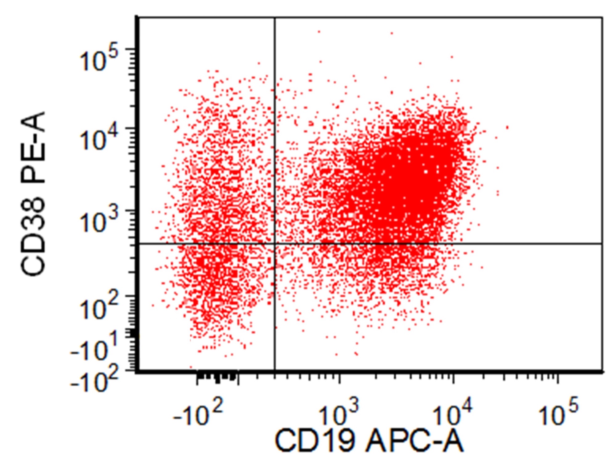
Contributed by Bradford Siegele, M.D., J.D. and Aashish Khatri, M.L.S., M.B.A.
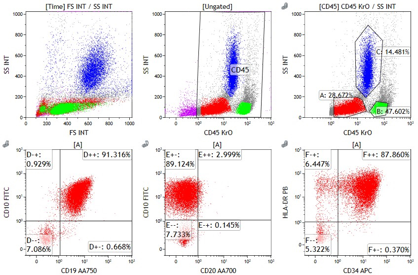
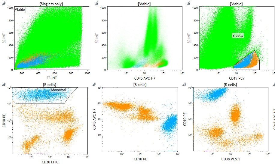
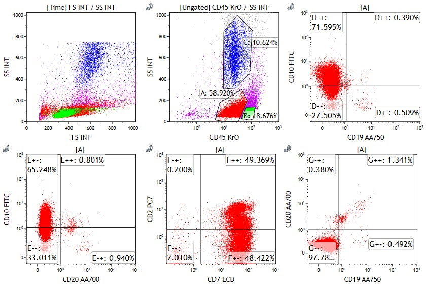
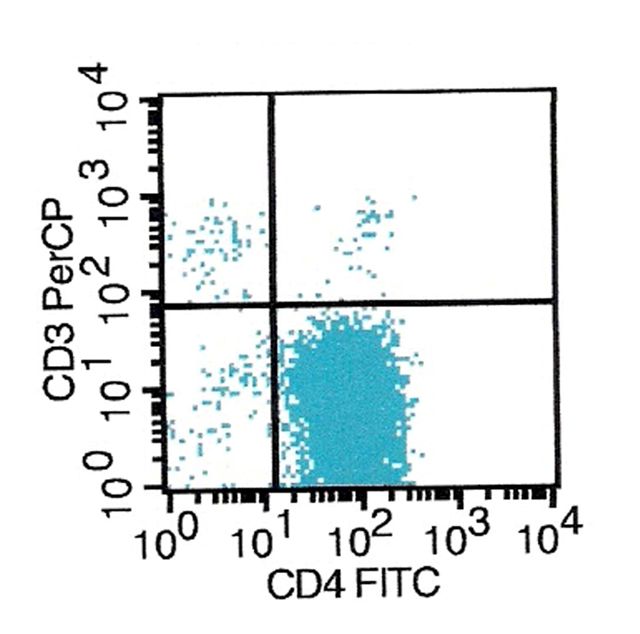
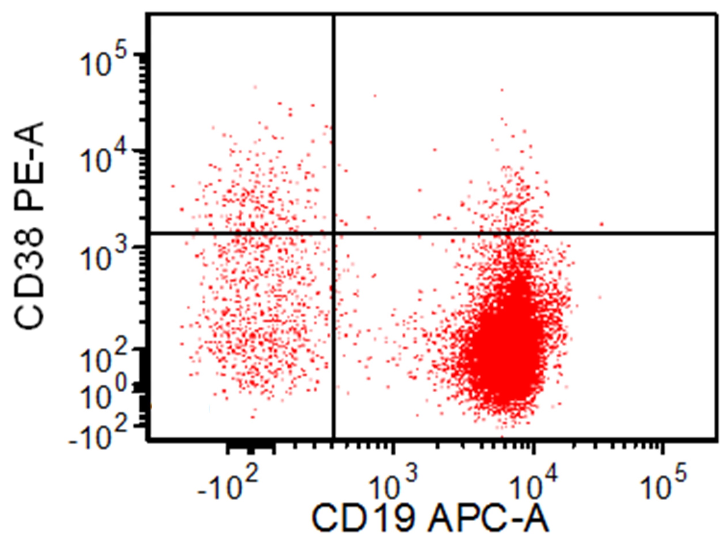
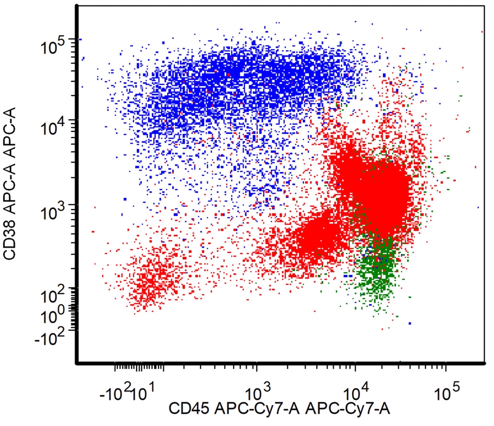
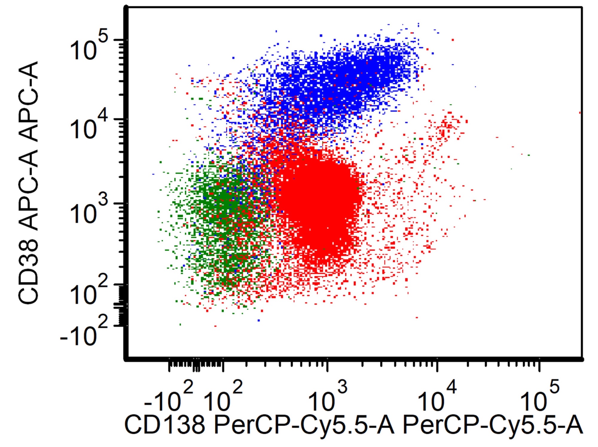
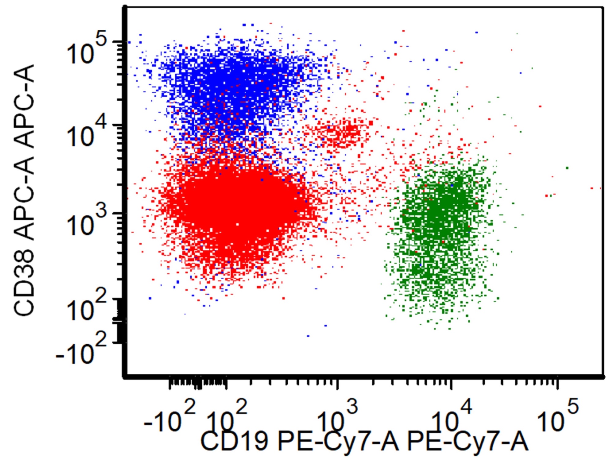
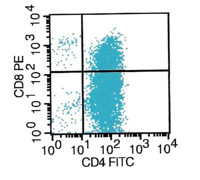
Flow cytometric analysis, B ALL / LBL
Flow cytometric analysis, T ALL / LBL
Images hosted on other servers:


Flow cytometric analysis, DLBCL
Images hosted on other servers:

CD14: toll-like receptor pathways

Membrane bound and serum forms of CD14
Images hosted on other servers:

CD11a: intestinal mucosa (fig F)

CD11a: eruptive papules with efalizumab therapy

CD11b: normal spleen

CD11b: gastric cancer

CD14: normal bone marrow (fig C)

CD14: acute monoblastic leukemia (fig F) (bone marrow)

CD14: acute myelomonocytic leukemia (fig F) (bone marrow)

CD14: microglia in Alzheimer patients (CNS)

CD14: biliary atresia (liver)

Anti CD15s
antibody blocks
infection of HL60
cells by HGE agent

CD15s expression correlates with HL60 cells

CD15u: colorectal cancer

CD15u: skin

CD18: fibrocytes
Images hosted on other servers:
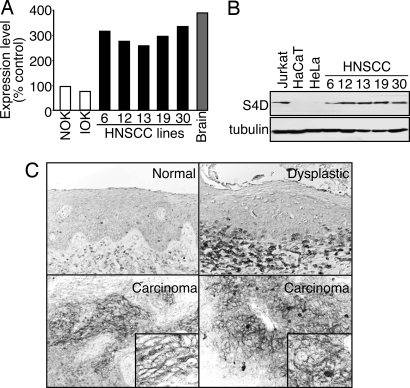
CD100: head and neck squamous cell carcinoma

CD107b: loss of staining in muscle and heart
Images hosted on other servers:

Domain structure of the αE(CD103)β7 integrin
Contributed by Maurice Richardson, M.D. and Moiz Vora, M.D.
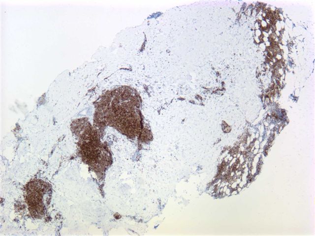
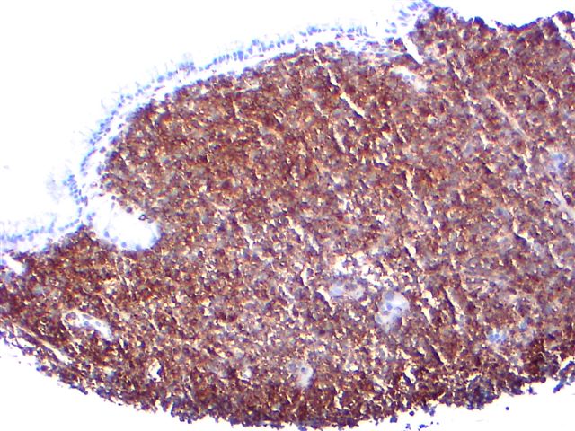
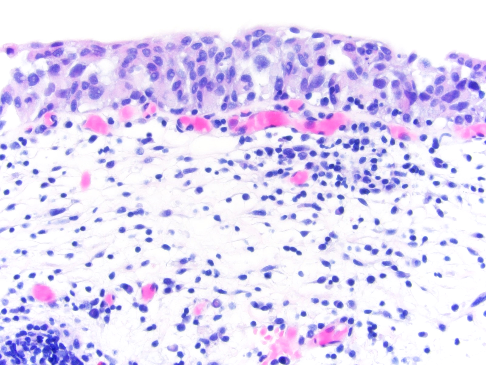
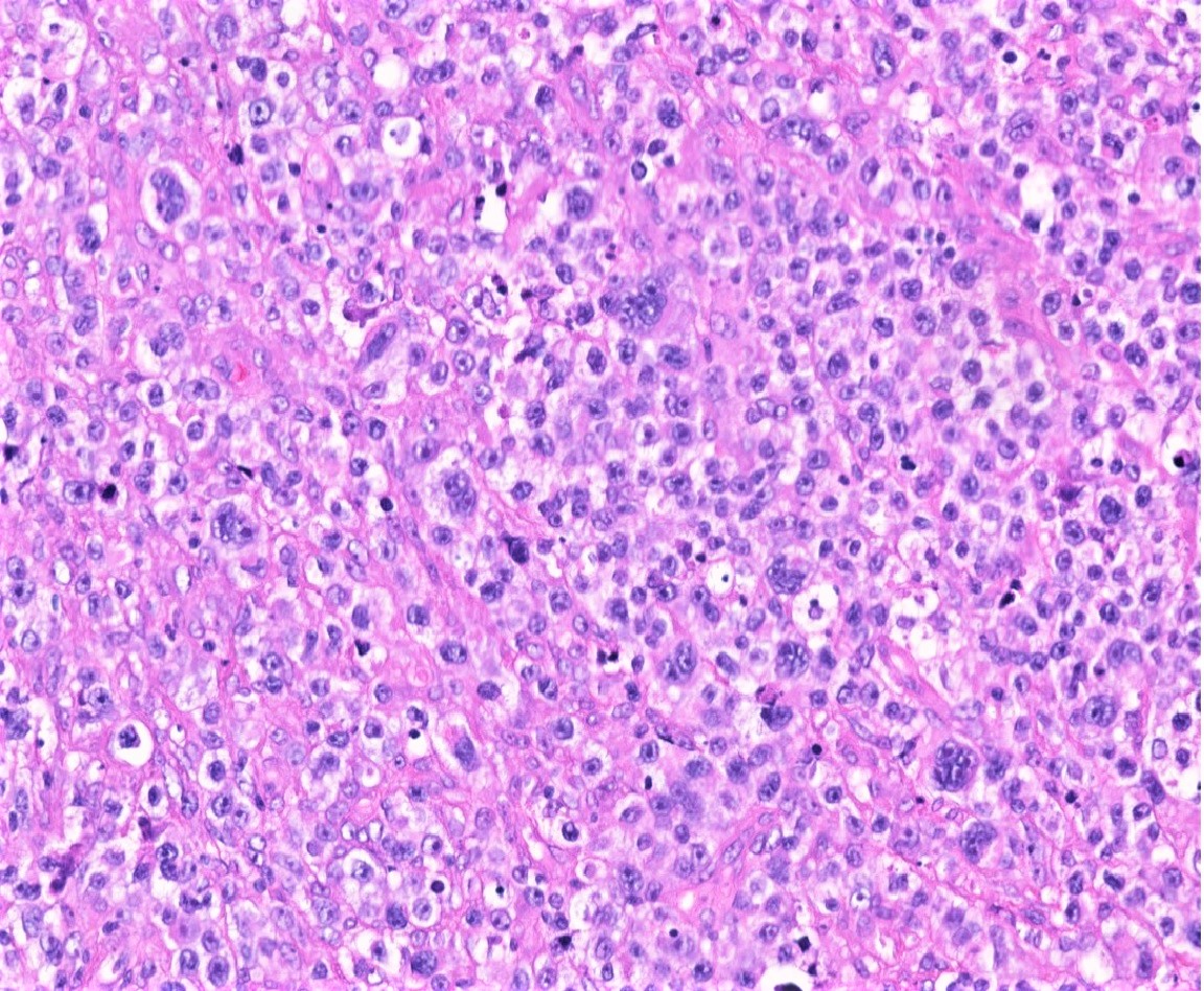
Diffuse lymphoid infiltrate
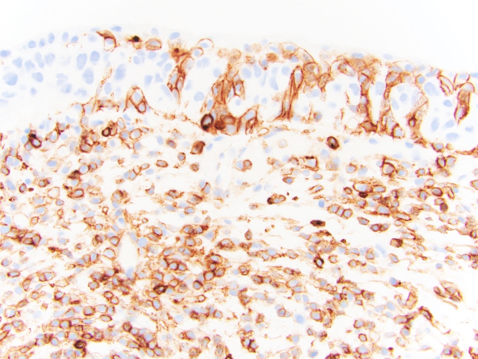
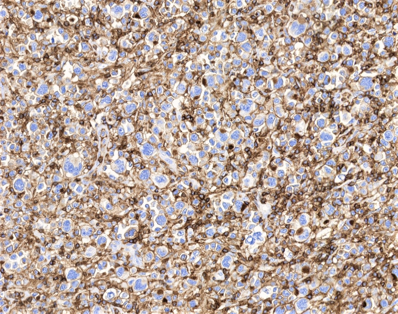
CD103+ neoplastic lymphocytes
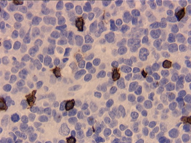
Intrasinusoidal lymphoid infiltrate

CD103+ neoplastic lymphocytes
Images hosted on other servers:

Hepatocellular carcinoma and normal liver

Lung - non small cell carcinoma (fig D)
Contributed by Debra L. Zynger, M.D. and Andrey Bychkov, M.D., Ph.D.
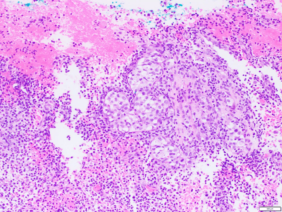
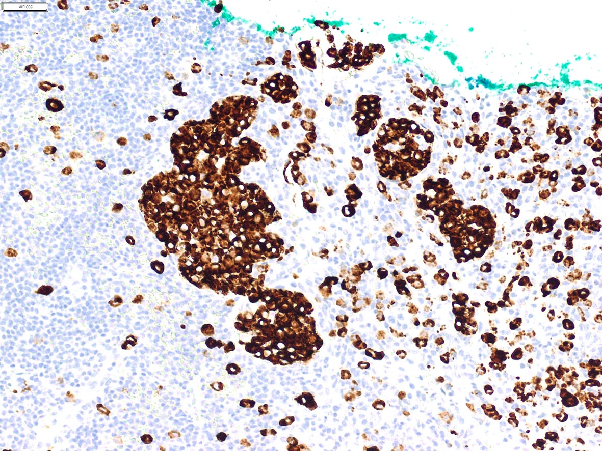
Seminoma with IHC
Testis
GIST
Contributed by Joseph Khoury, M.D.
Tonsil
Blastic plasmacytoid dendritic cell neoplasm
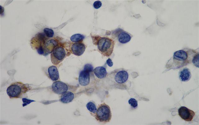
Contributed by Leica Microsystems
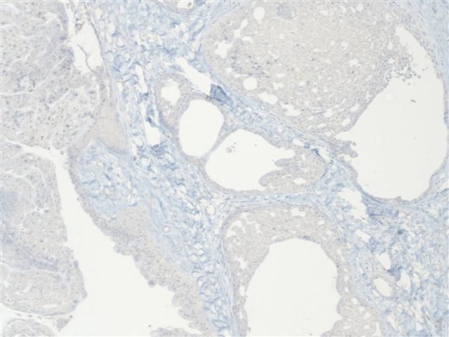
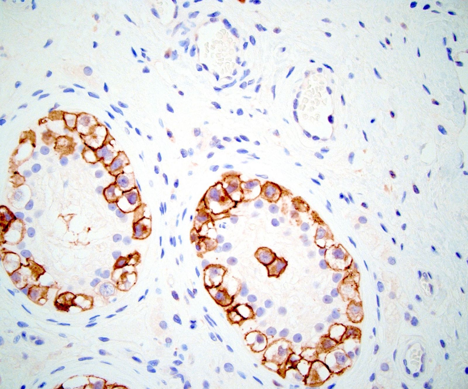
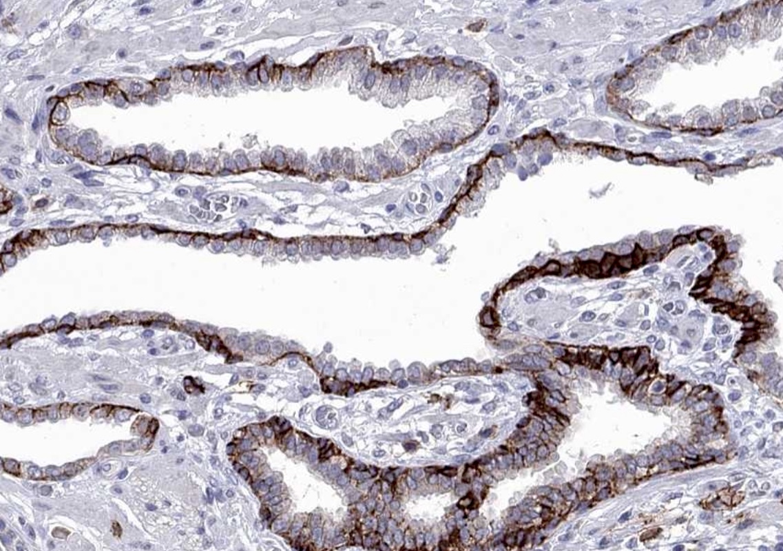
Prostate: normal
Images hosted on other servers:

CNS: glioblastoma & colon: carcinoma

Colon: normal


Liver: left-normal, right-hepatocellular carcinoma


Prostate: carcinoma

Skin (inflammatory), tonsil

Various tumors
Contributed by Pamela Wirth, Ph.D. (source: University of Toronto and Human Protein Atlas) and Yuri Tachibana, M.D.
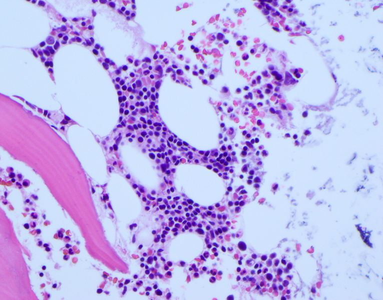
Bone marrow biopsy
Spleen
Breast carcinoma
Colon carcinoma
Liver carcinoma
Prostate carcinoma
Chronic endometritis
Contributed by Raghava Munivenkatappa, M.D. (Case #48) and AFIP
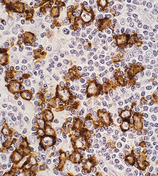
Paranuclear / Golgi staining pattern
Nodular lymphocyte predominant
Renal cell carcinoma: papillary type
Images hosted on other servers:

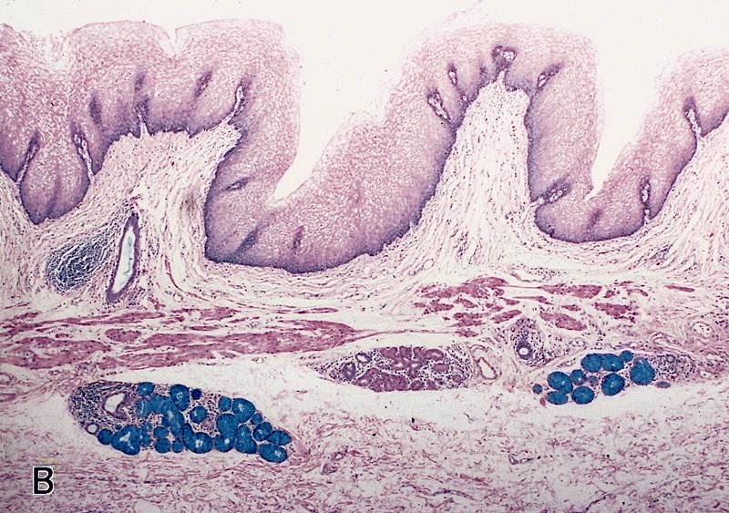
Small intestine: normal Paneth cells (fig B)


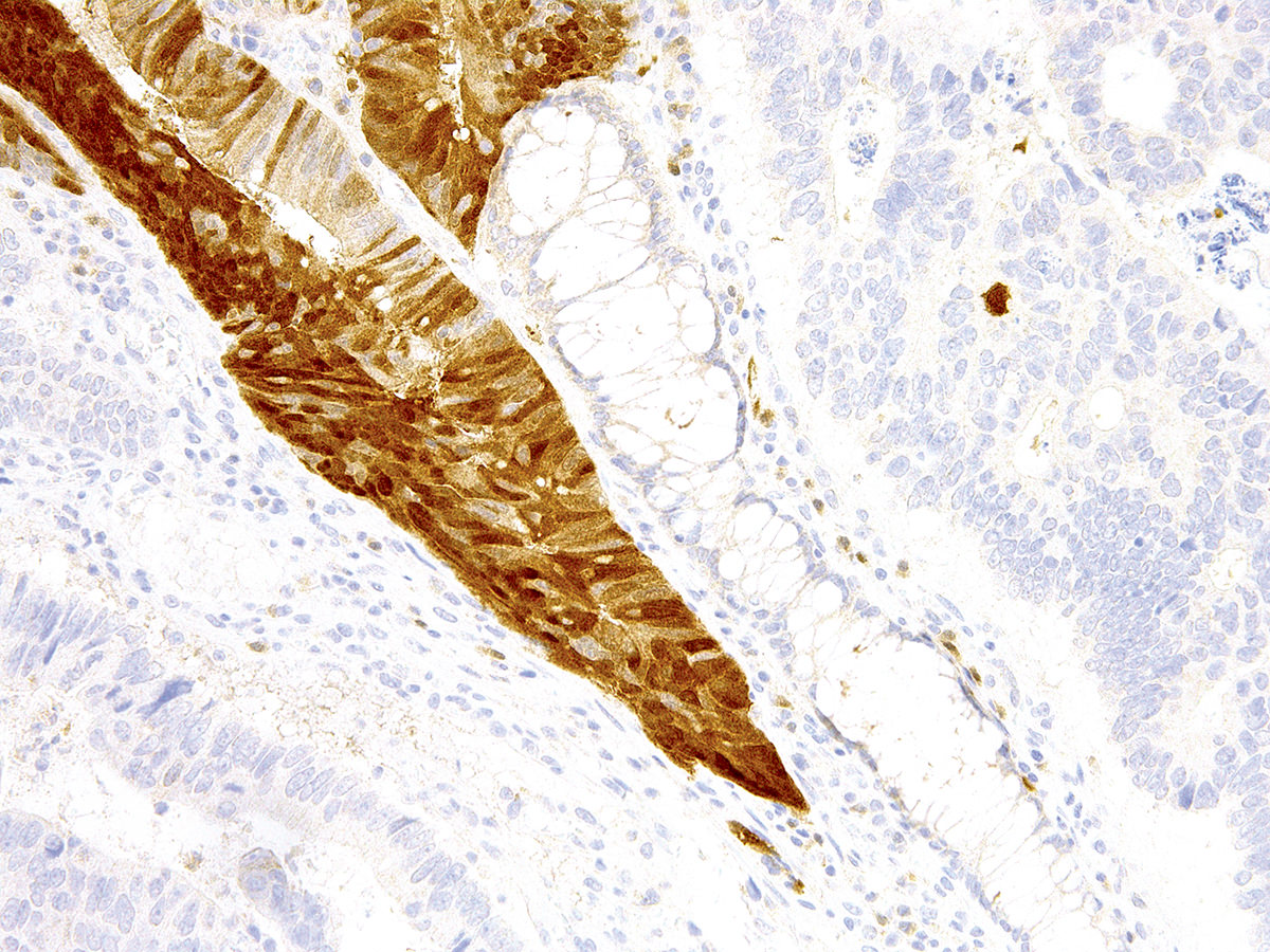
Colon: invasive colorectal carcinoma
Contributed by Charles J. Sailey, M.D.,
Borislav A. Alexiev, M.D. and
John C. Papadimitriou, M.D.
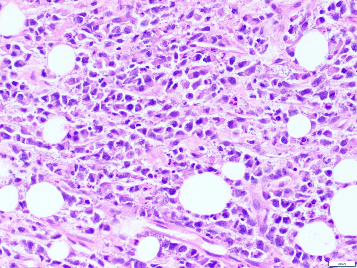
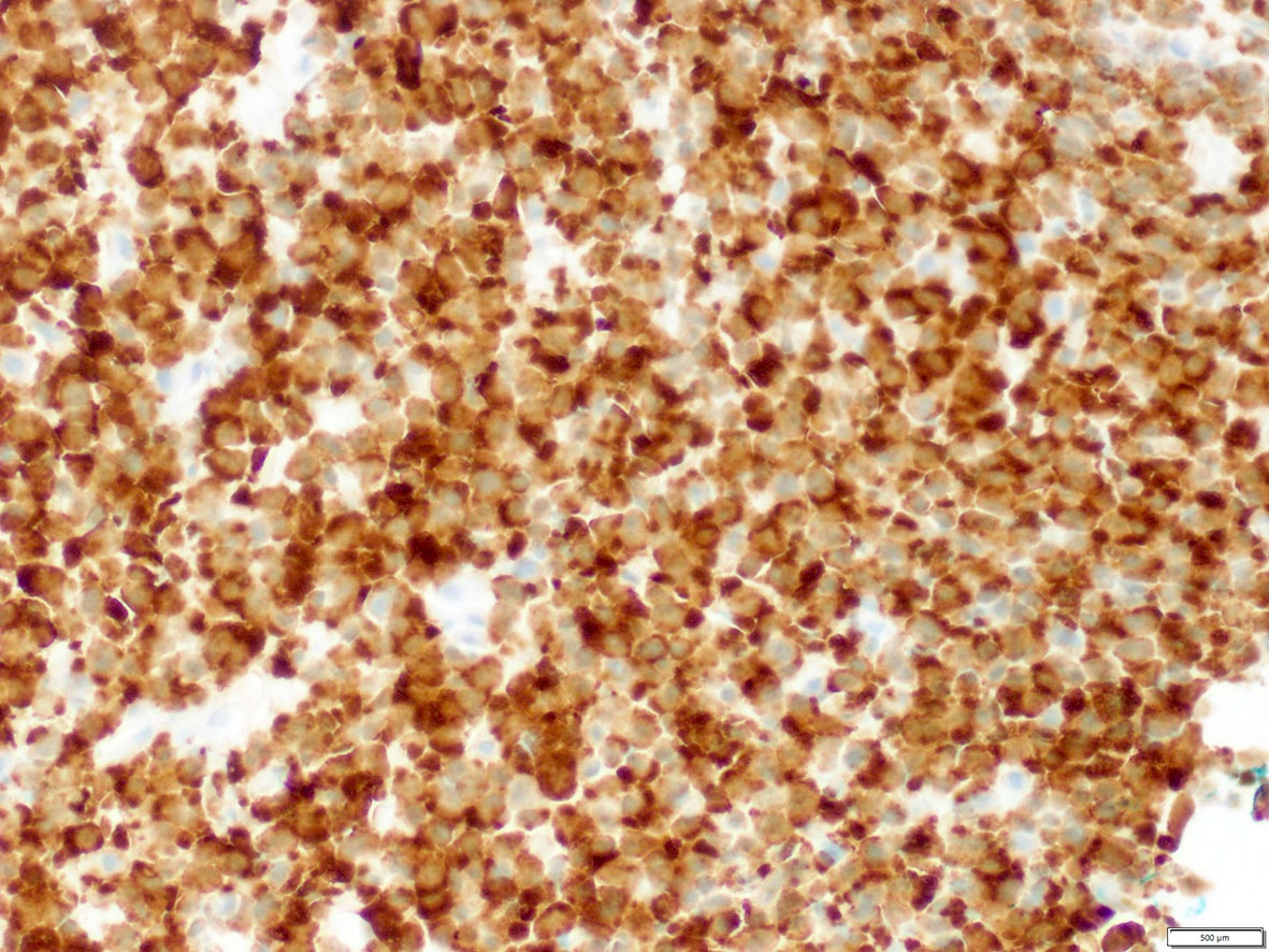
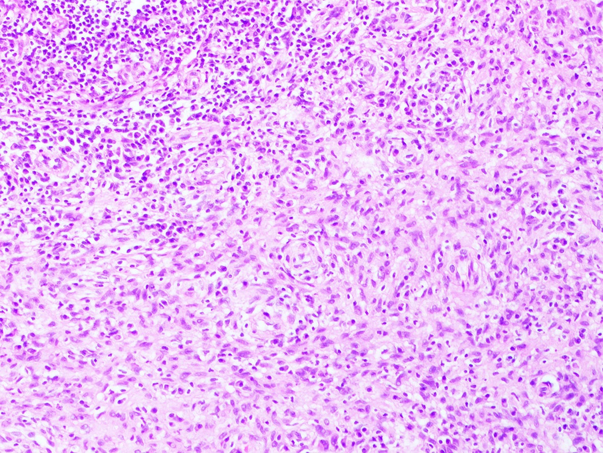
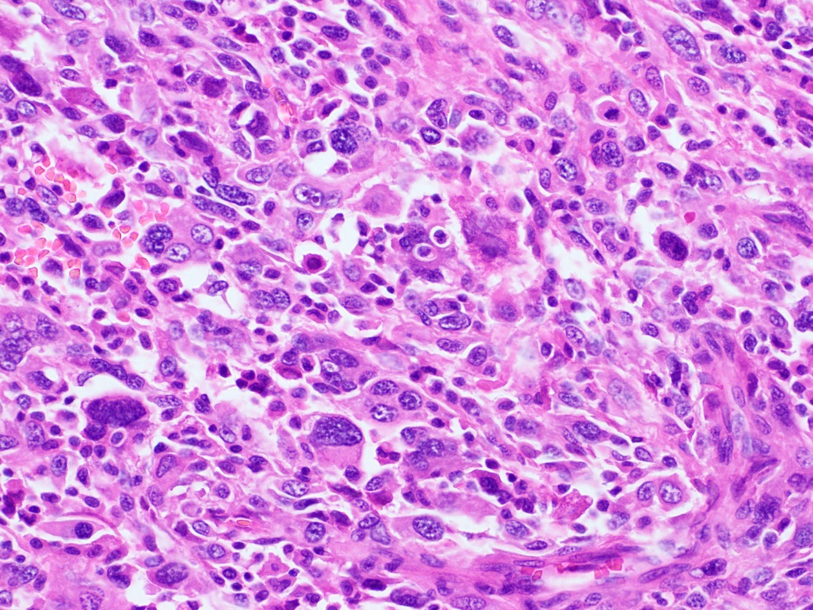
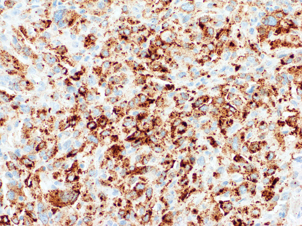
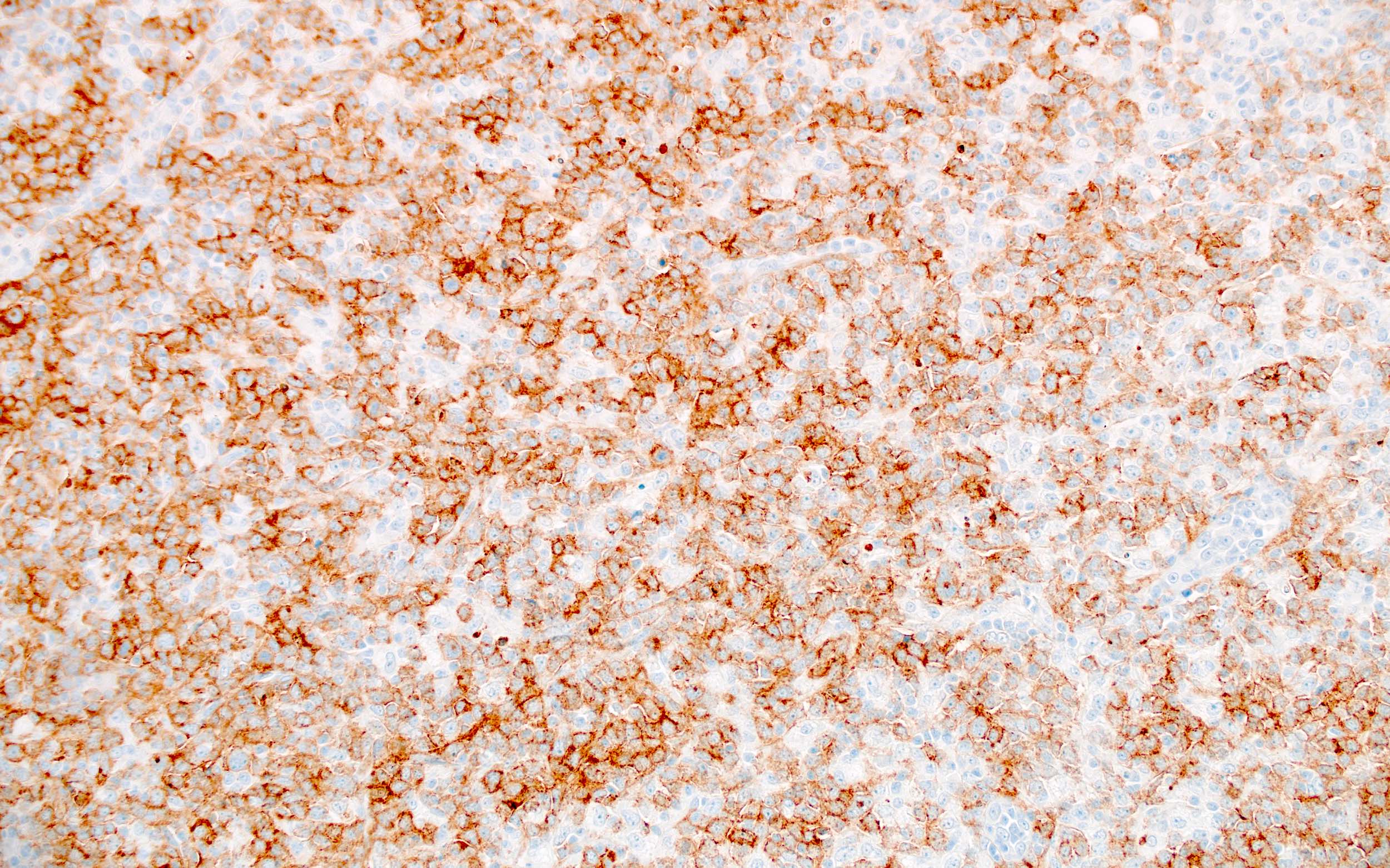
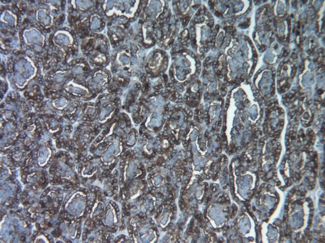
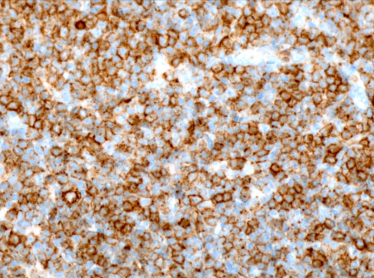
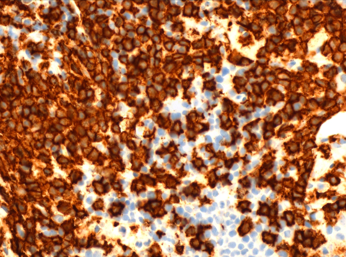
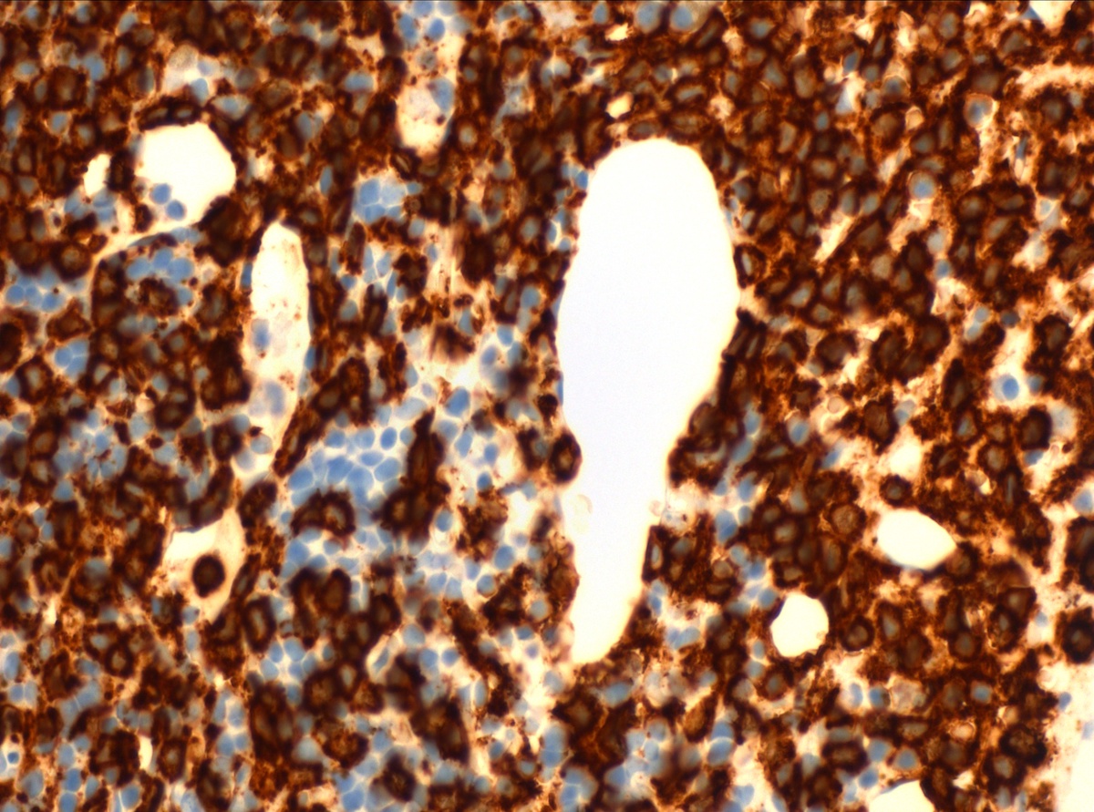
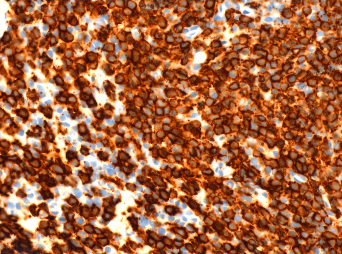
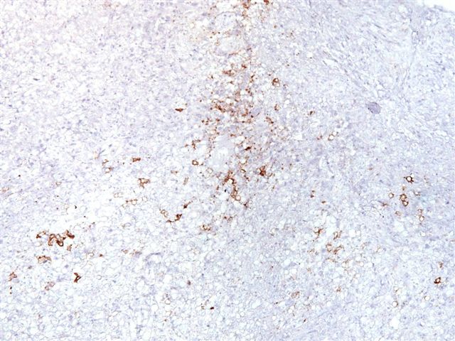
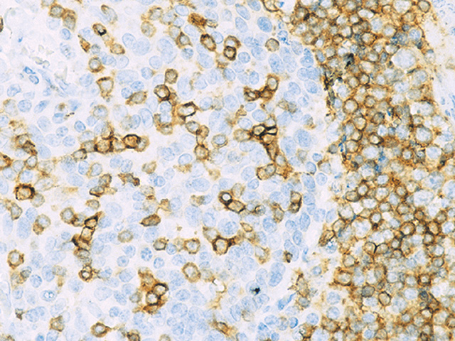
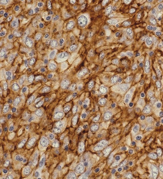
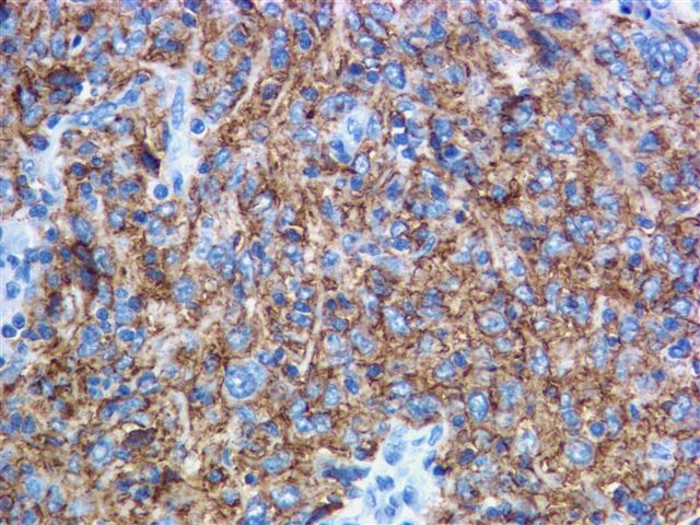
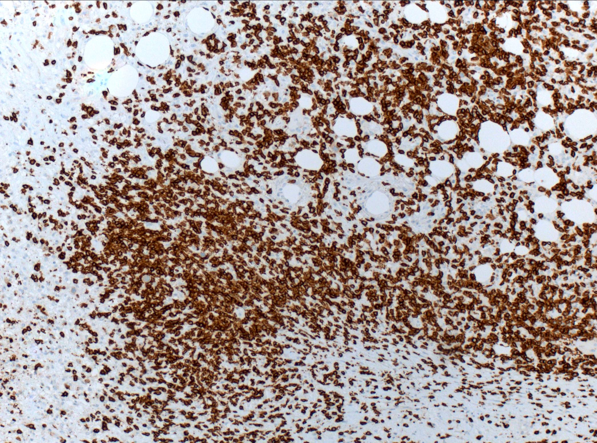
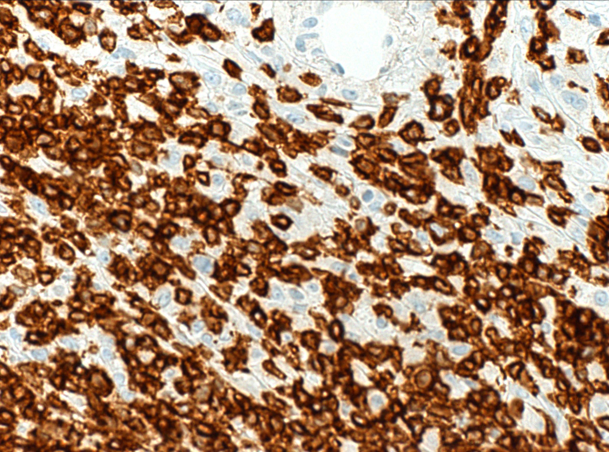
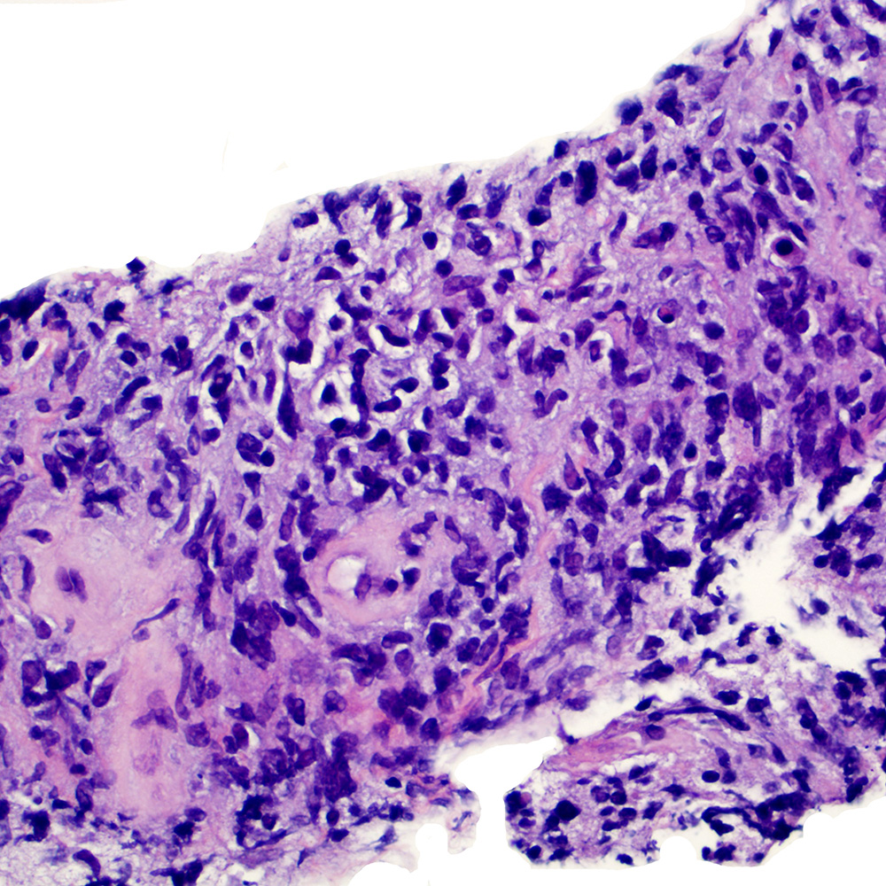
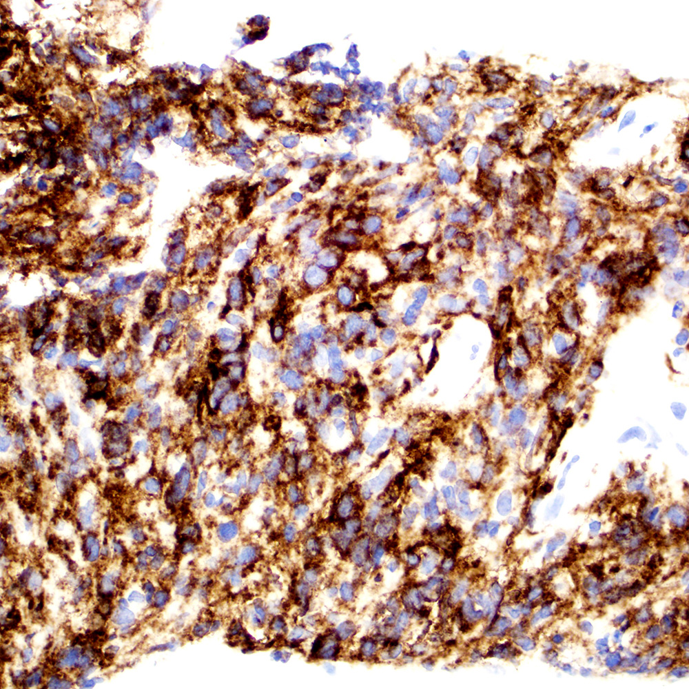
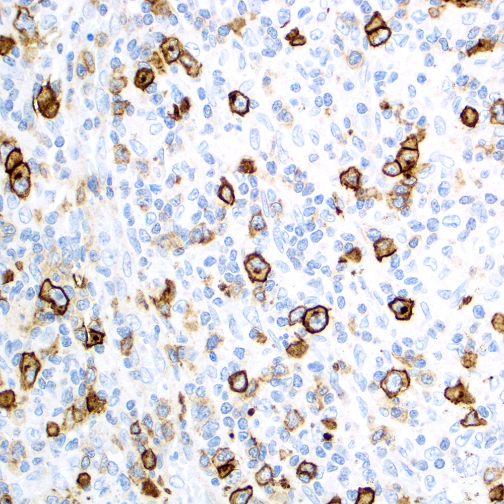
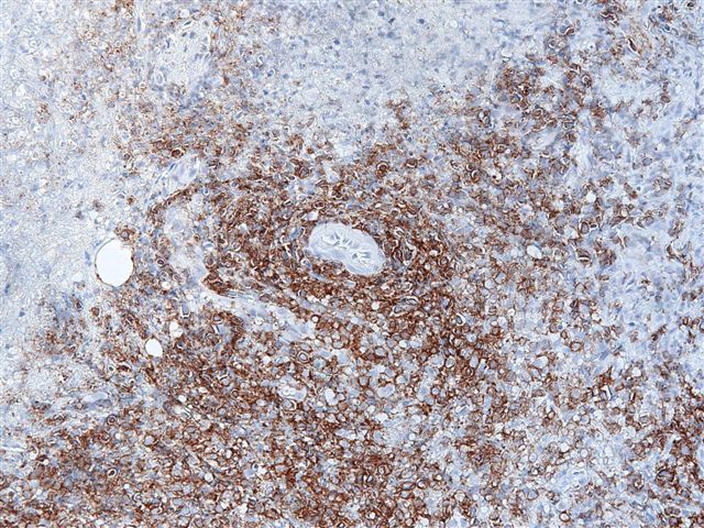
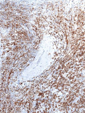
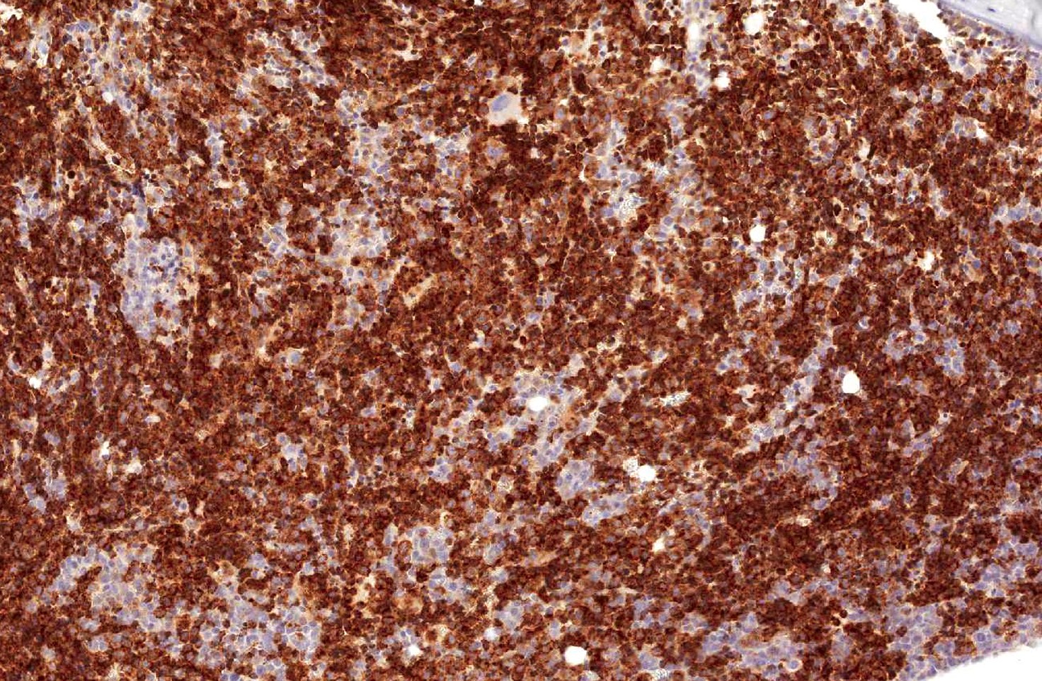
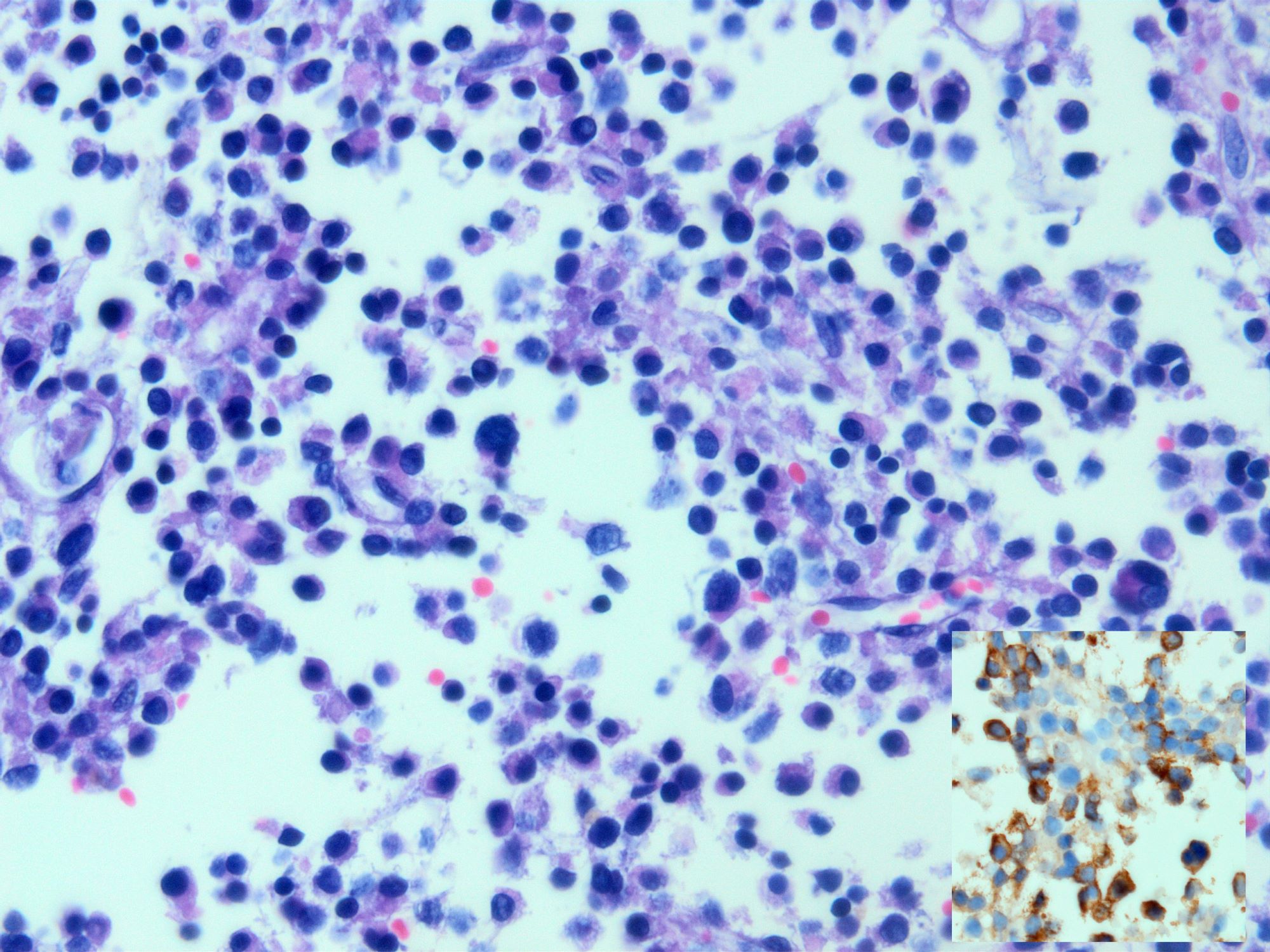
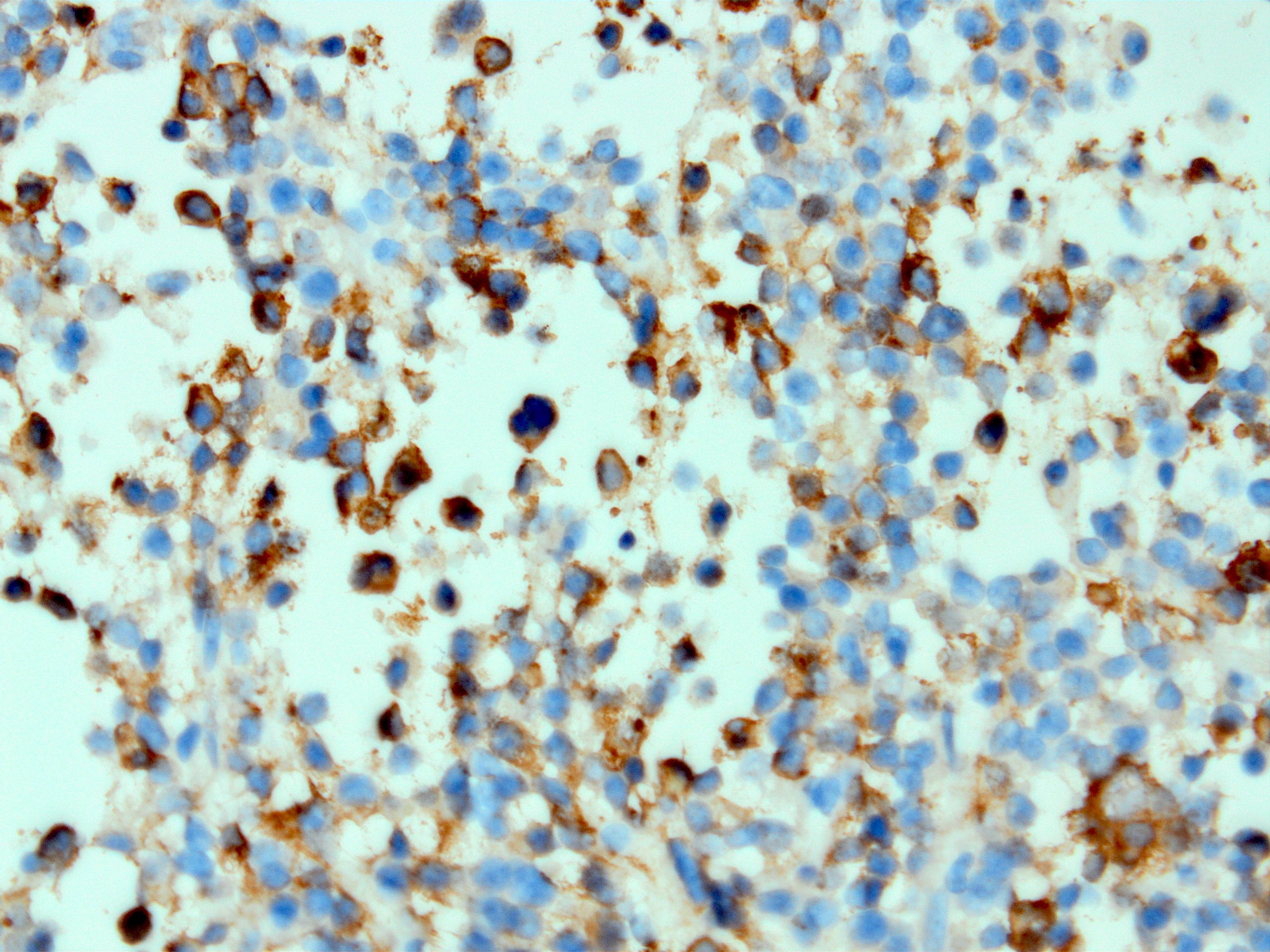
CD163: histiocytic sarcoma
Contributed by
Jijgee Munkhdelger, M.D., Ph.D.
and Andrey Bychkov, M.D., Ph.D.
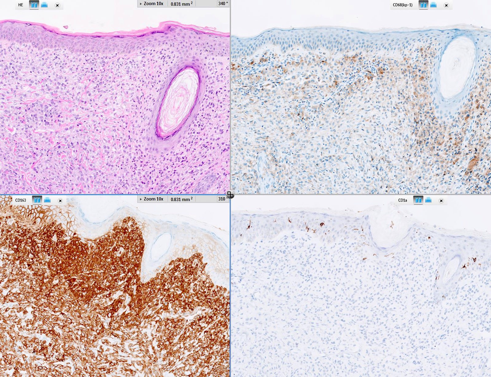
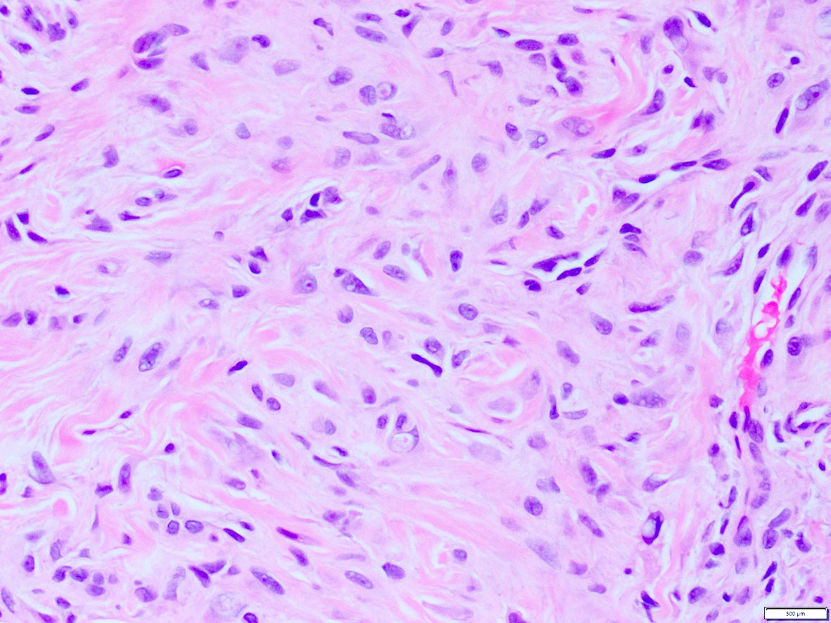
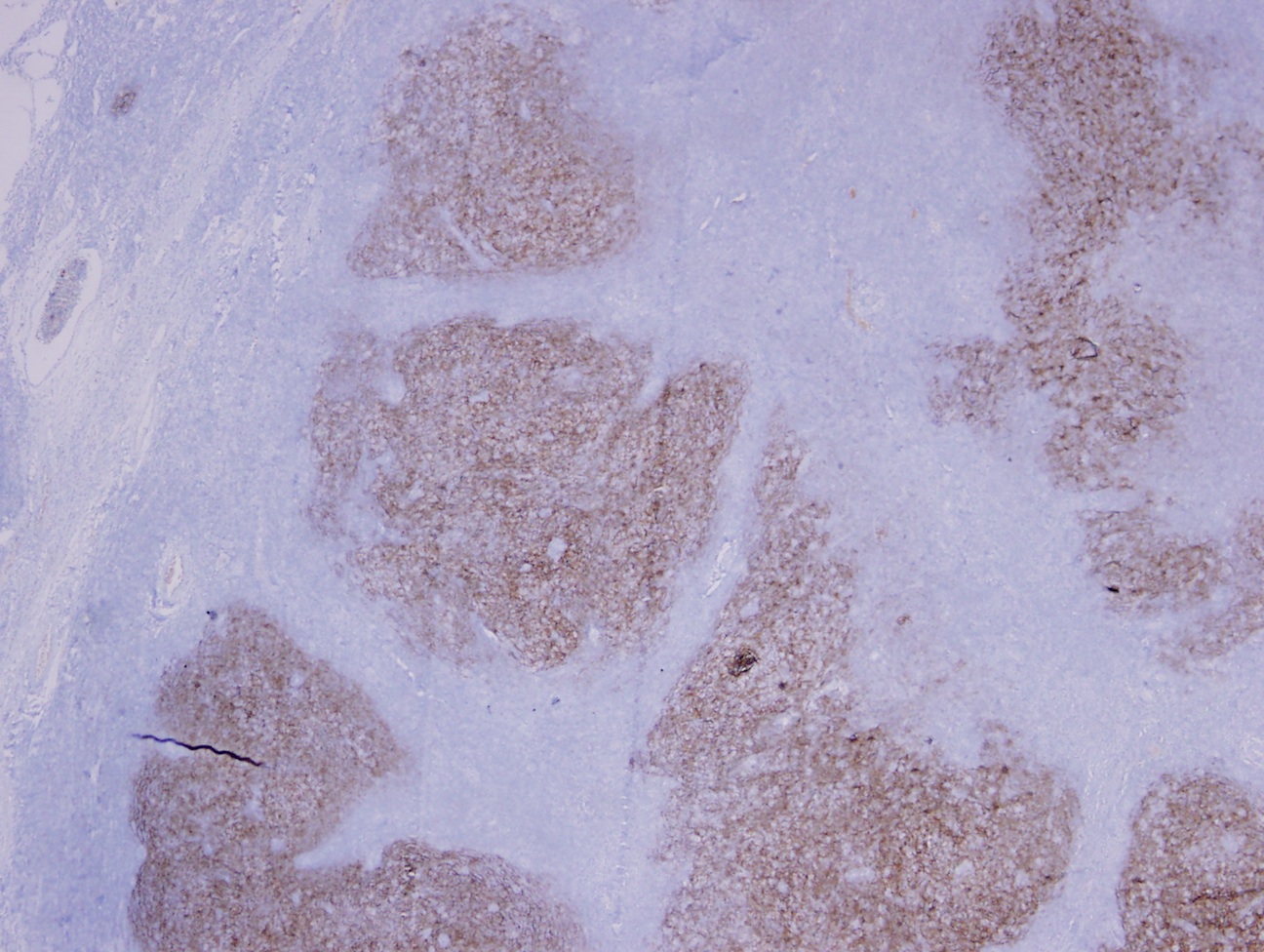
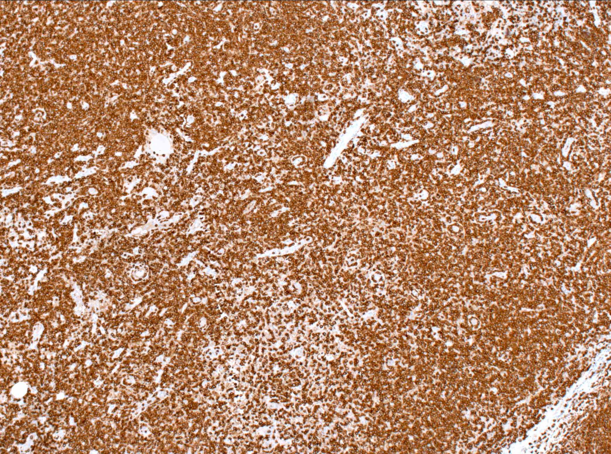
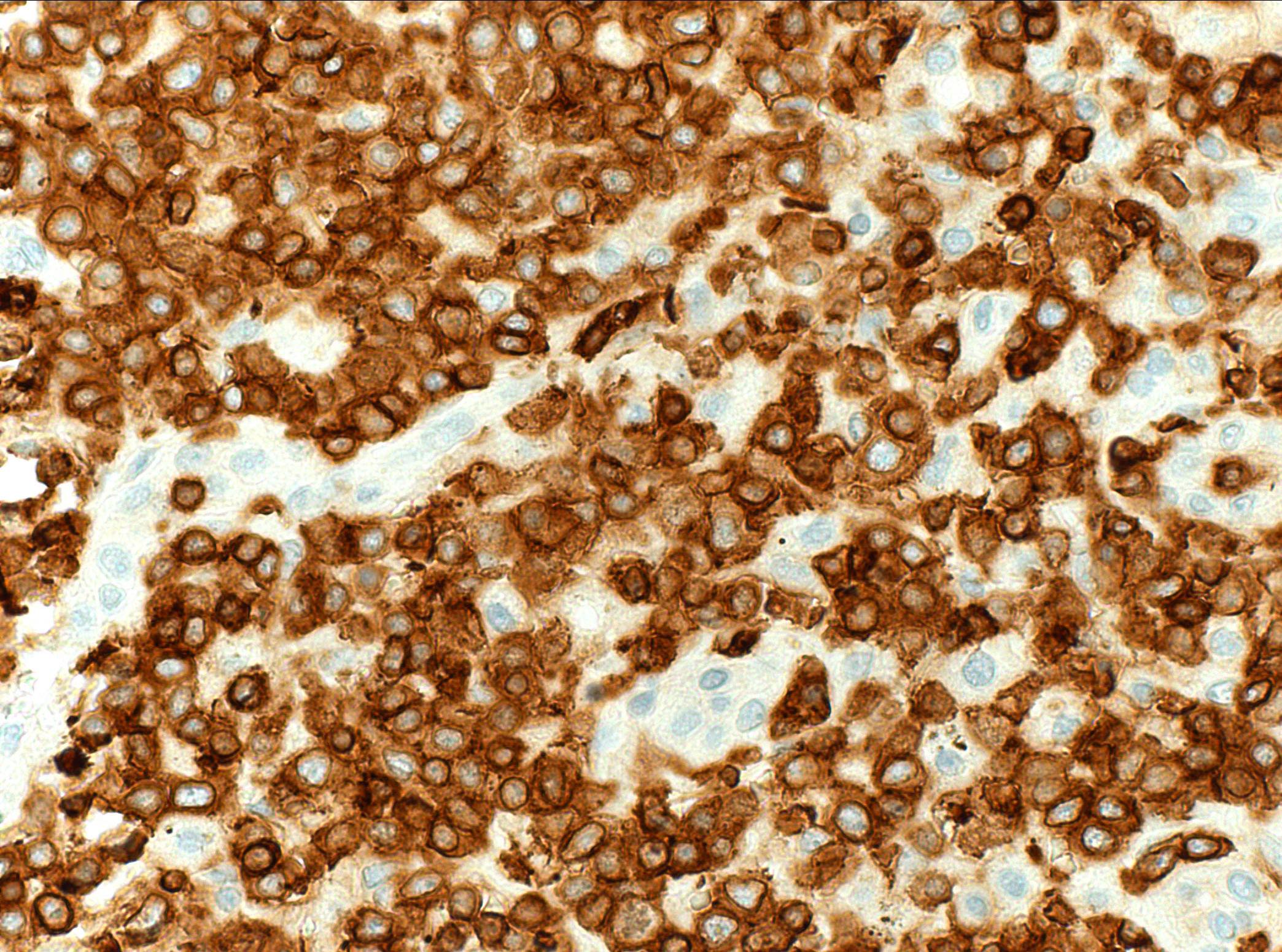
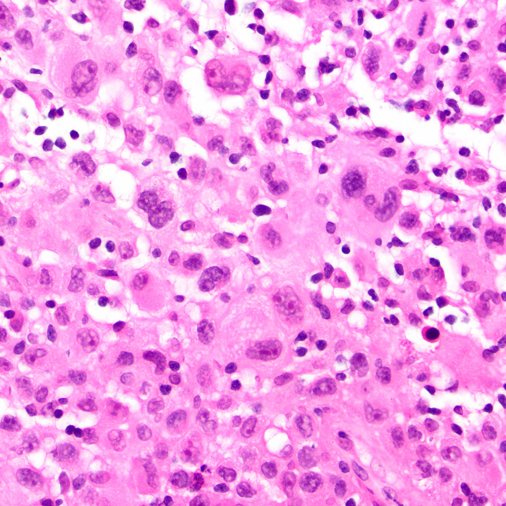
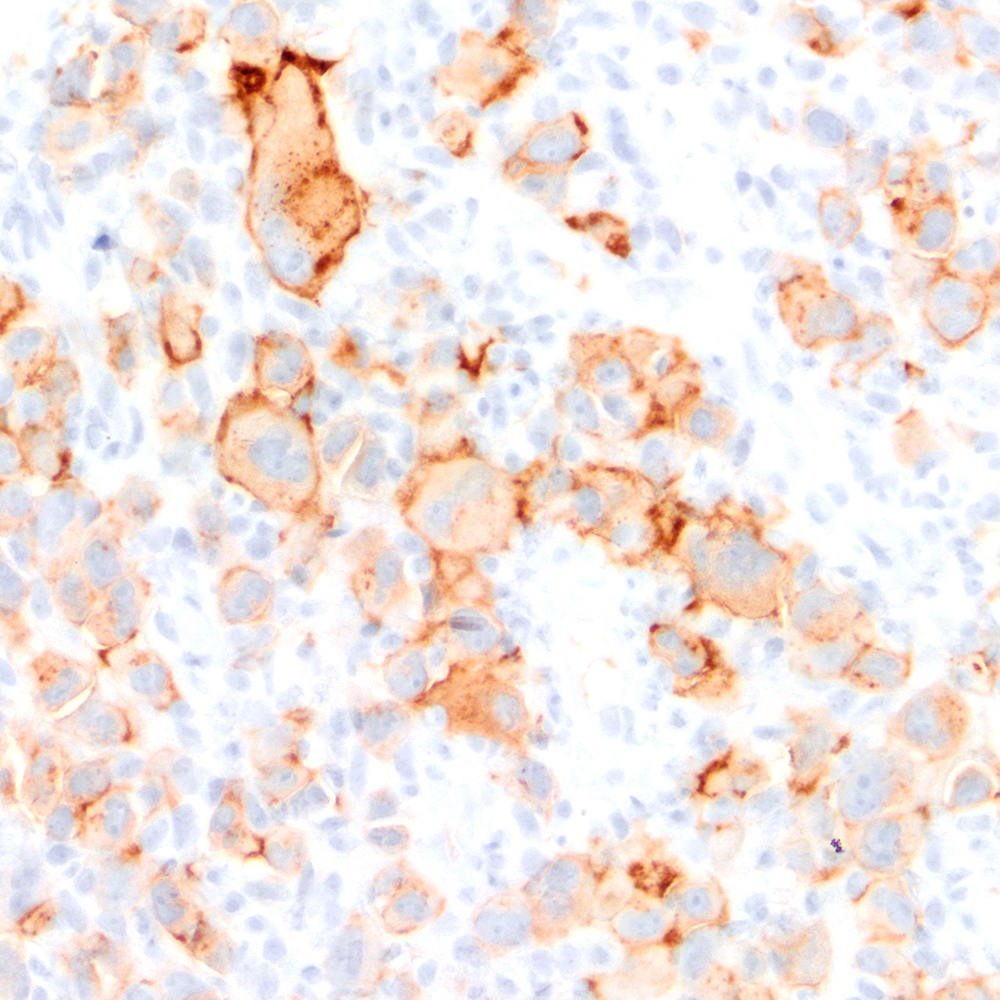
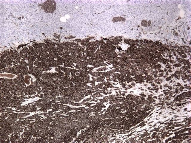
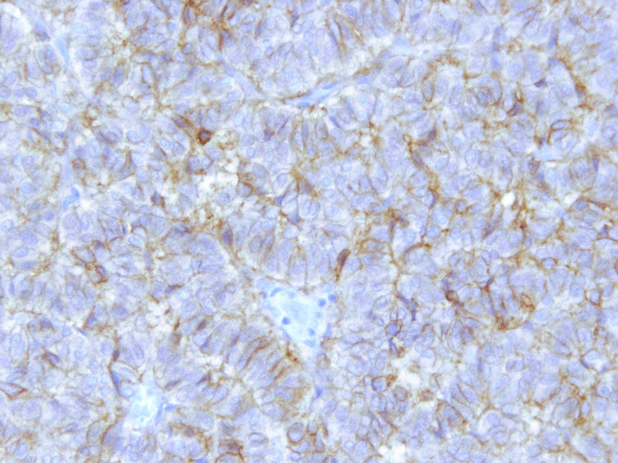
CD163: juvenile xanthogranuloma immunoprofile
Images hosted on other servers:

CD182: inflammatory infiltration
Images hosted on other servers:
CD182: ovarian carcinoma
CD185: chronic H. pylori gastris (left); MALT lymphoma (right)
CD185: AIDS related non-Hodgkin lymphoma
CD185: synovium of non-rheumatoid arthritis (left) and rheumatoid arthritis (right)
Images hosted on other servers:


CD19 structure and signaling
Images hosted on other servers:
Langerhans cell histiocytosis:

Brain


Lung: right is bronchioalveolar lavage

Skin
Other:

Esophagus: Barrett metaplasia

Oral cavity: gingiva - normal and diseased
Skin: mycosis fungoide
Case #72
Langerhans cell histiocytosis:
Skull - cytology
Images hosted on other servers:


Langerhans cell histiocytosis: Birbeck granules
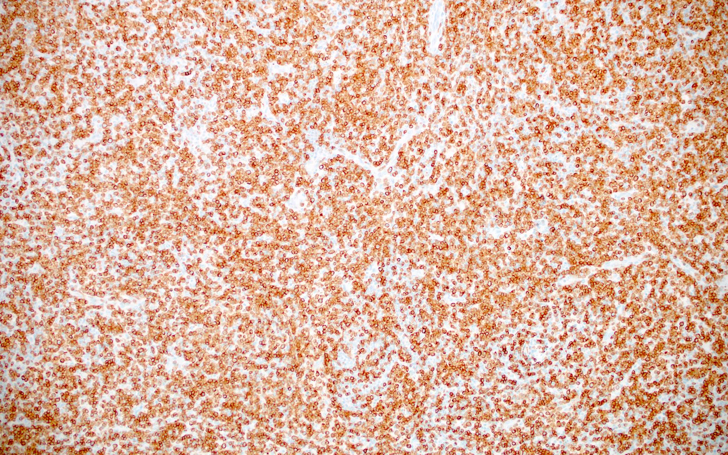
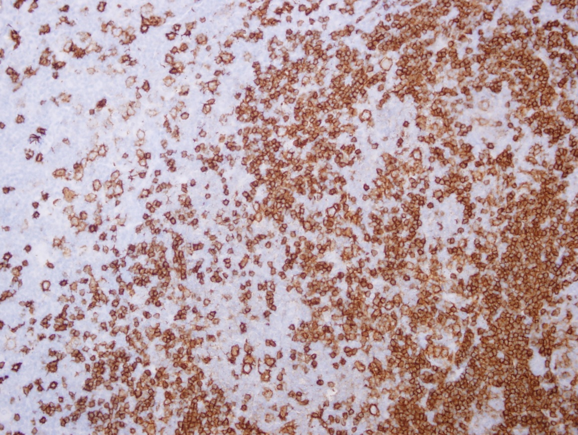
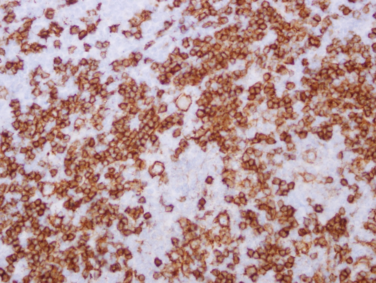
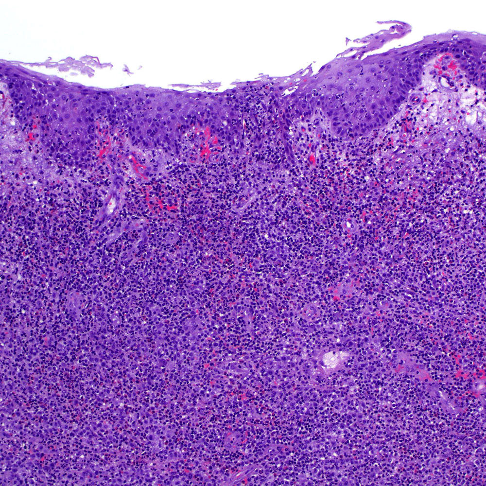
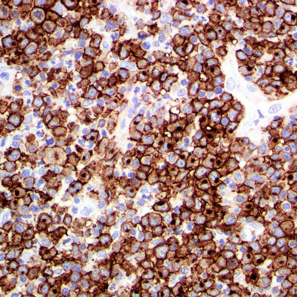
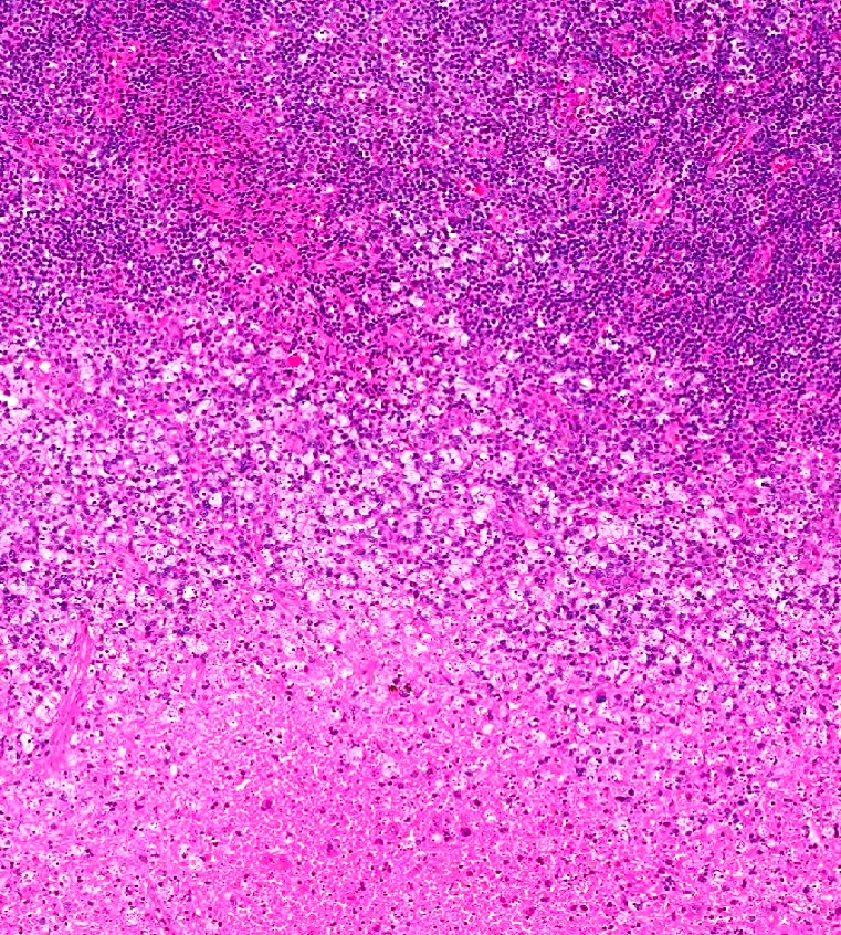
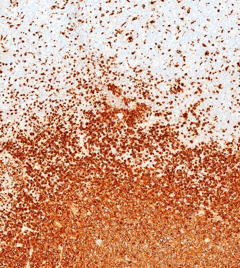
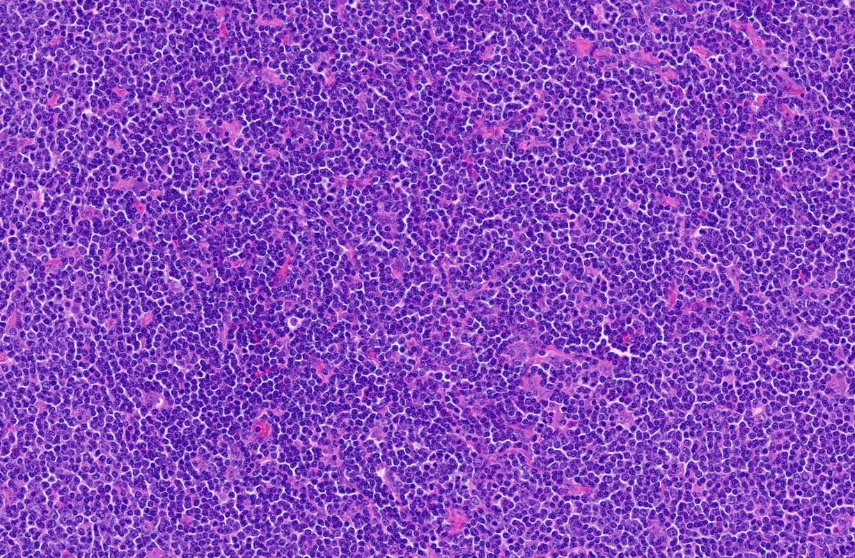
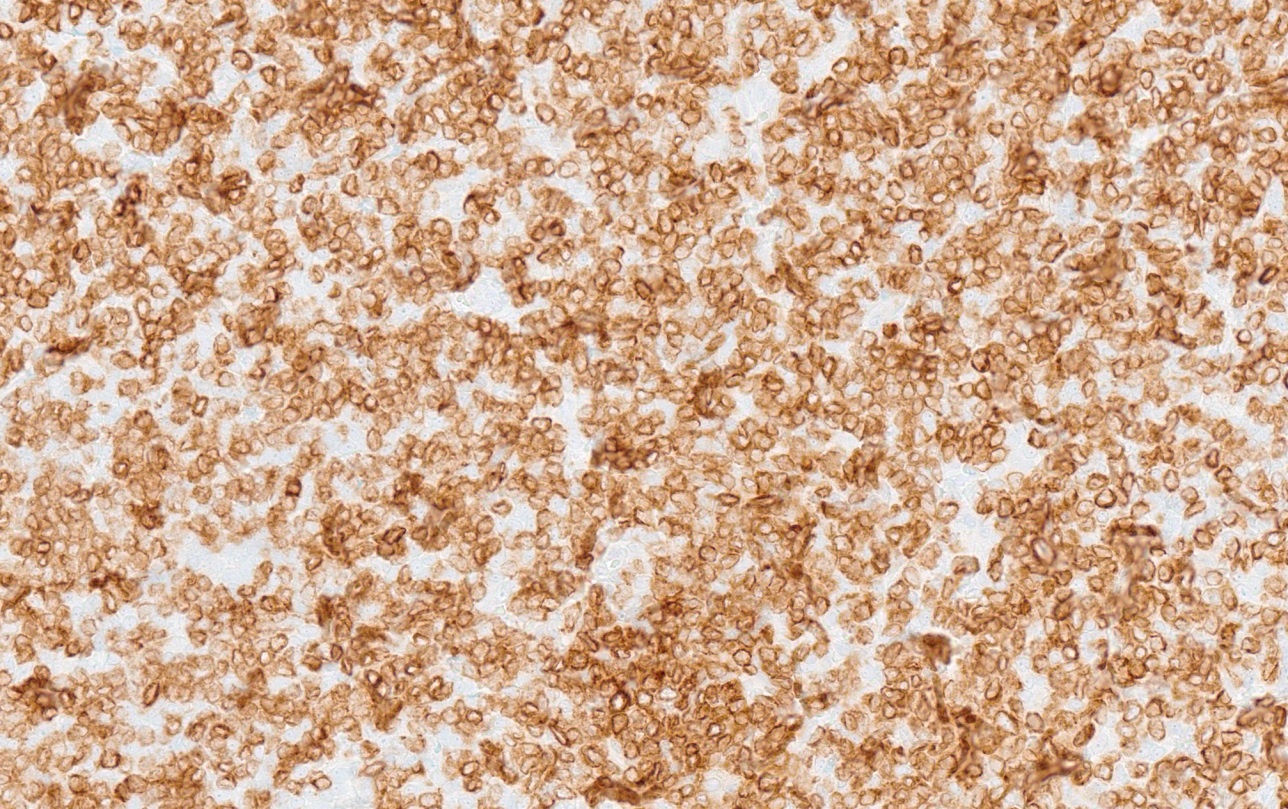
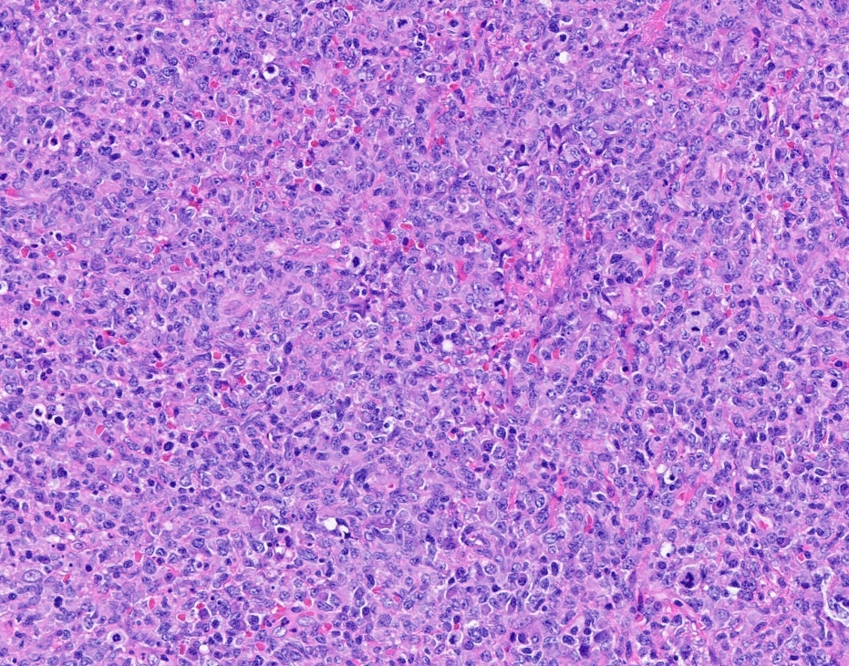
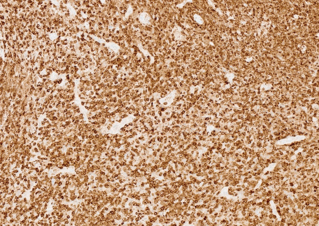
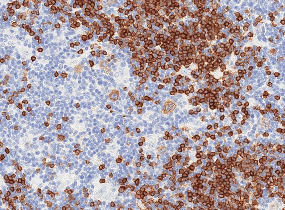
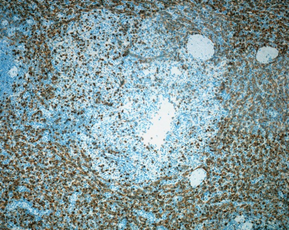
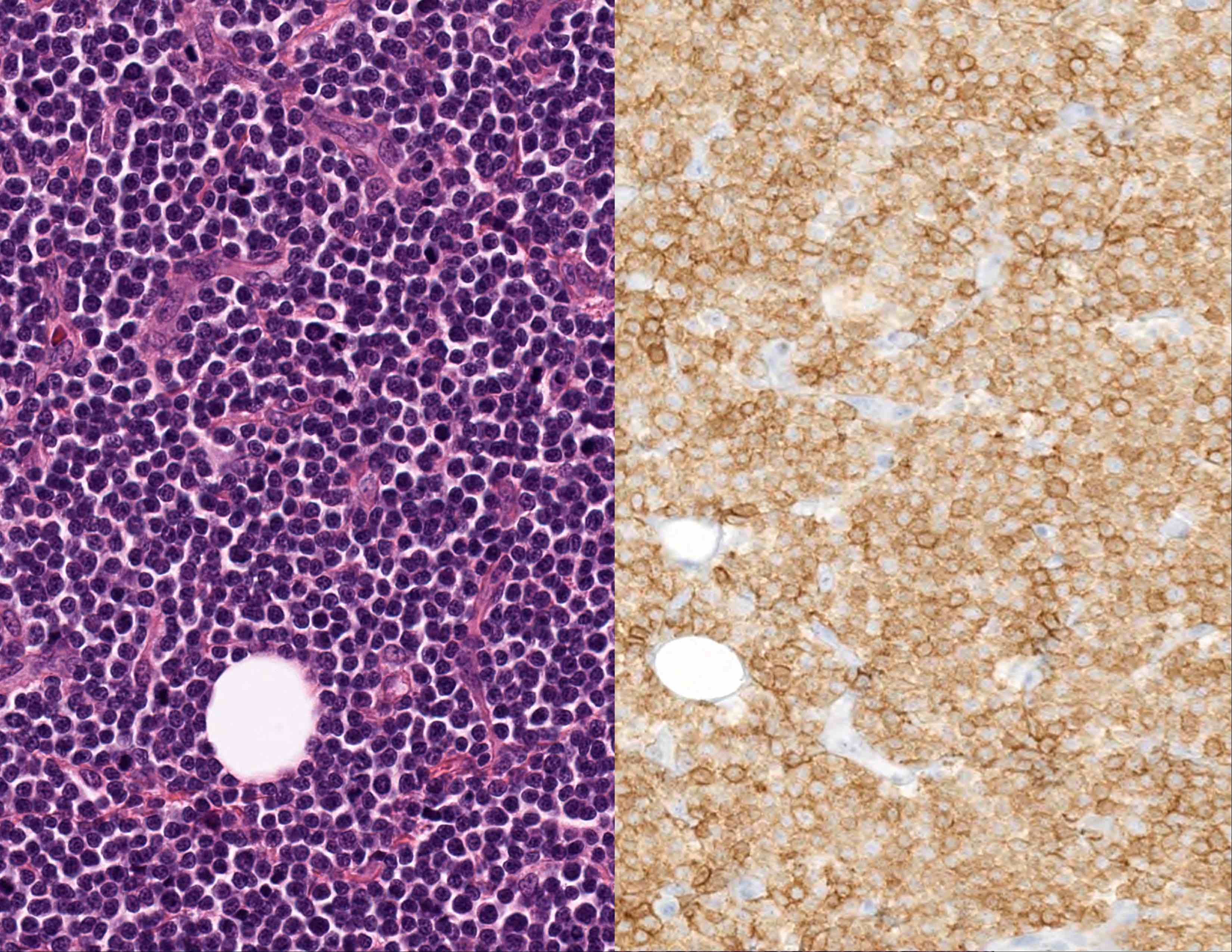
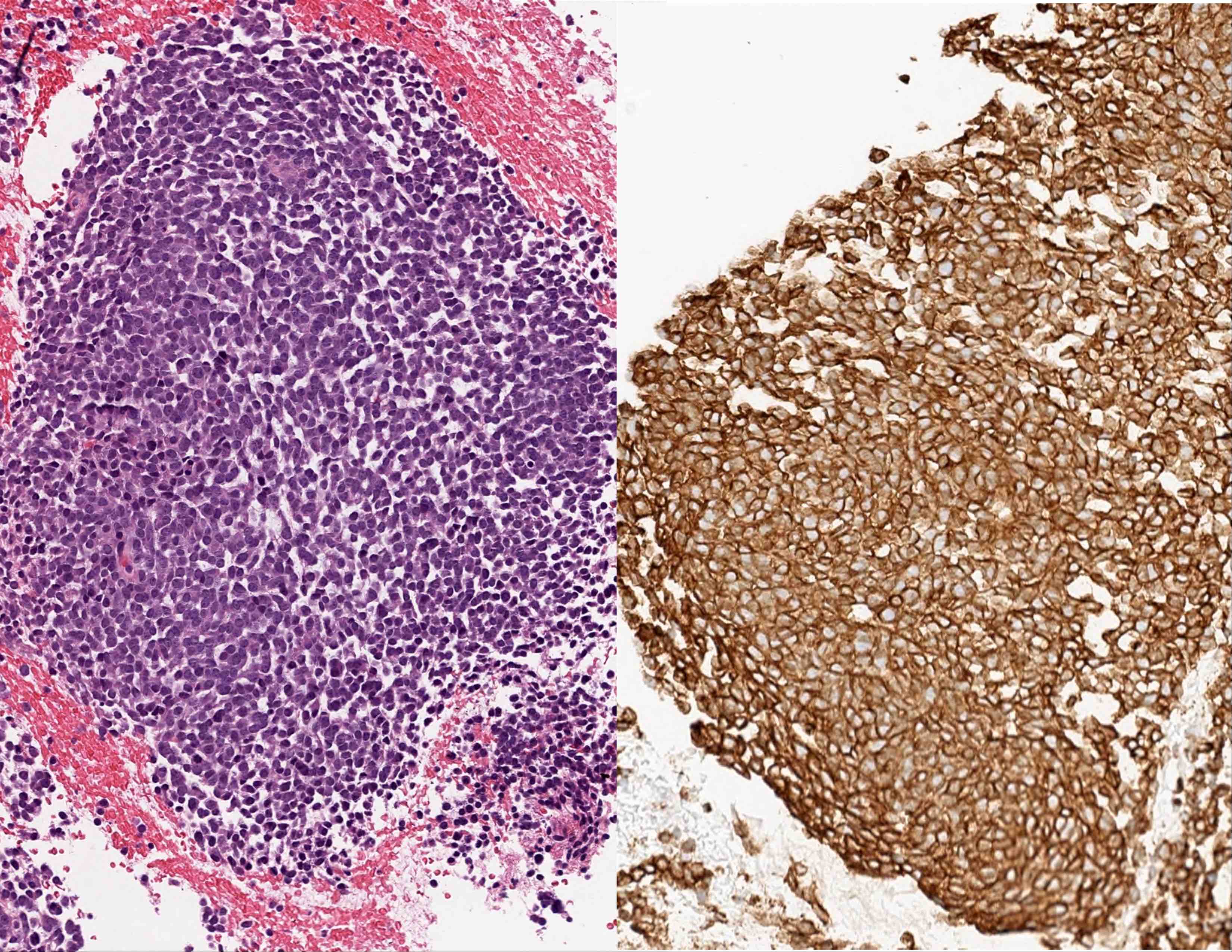
Contributed by Leonie Frauenfeld, M.D., Andrey Bychkov, M.D., Ph.D. and Kaveh Naemi, D.O.

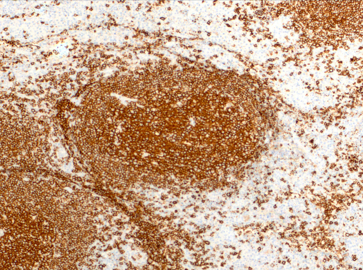
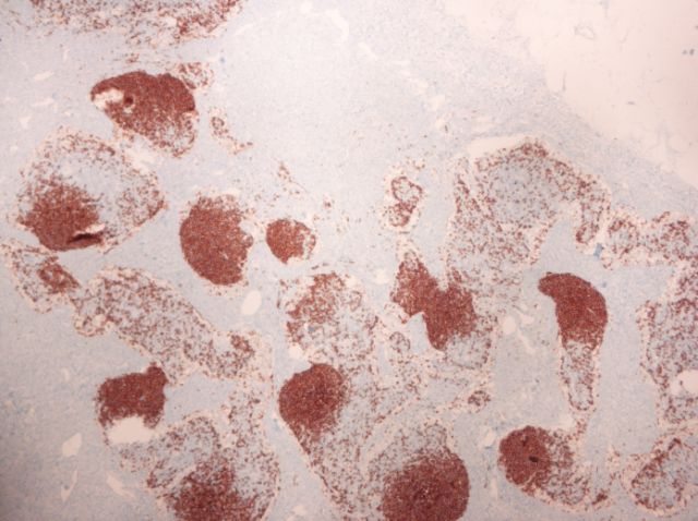
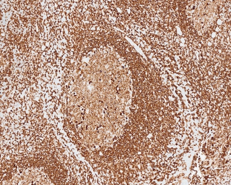
Germinal center with strong CD20 positive B cells
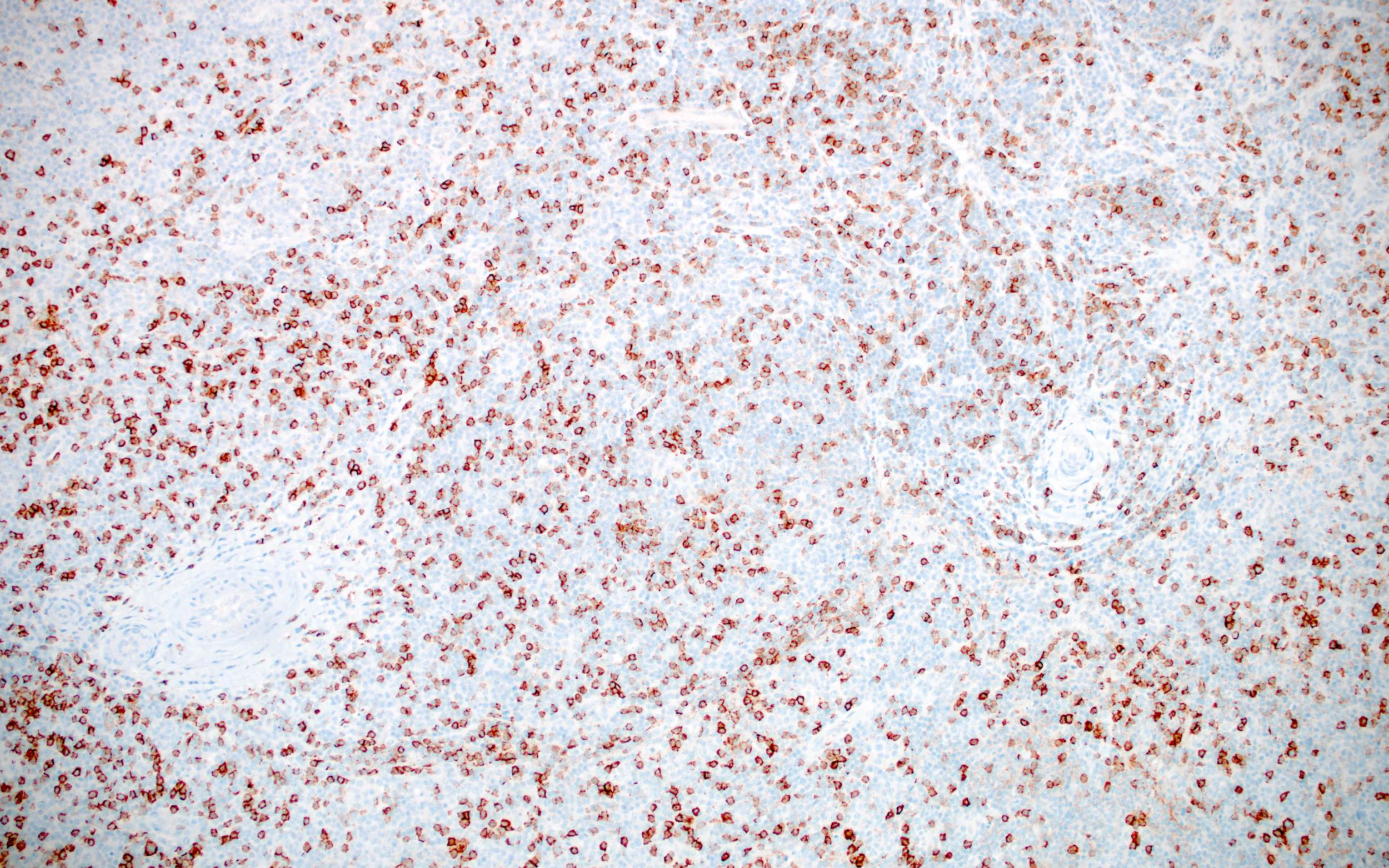
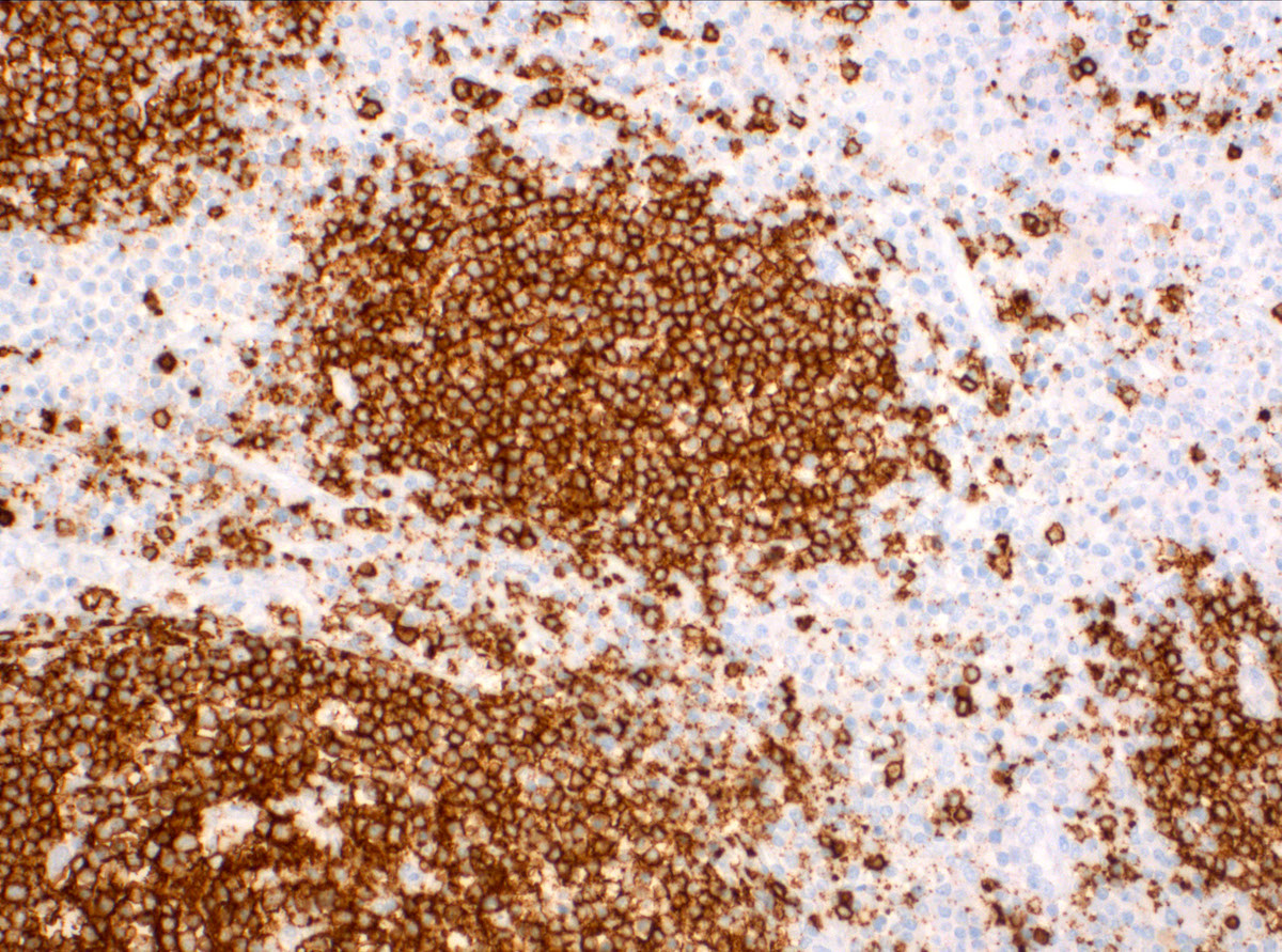
SLL / CLL infiltrate in a lymph node
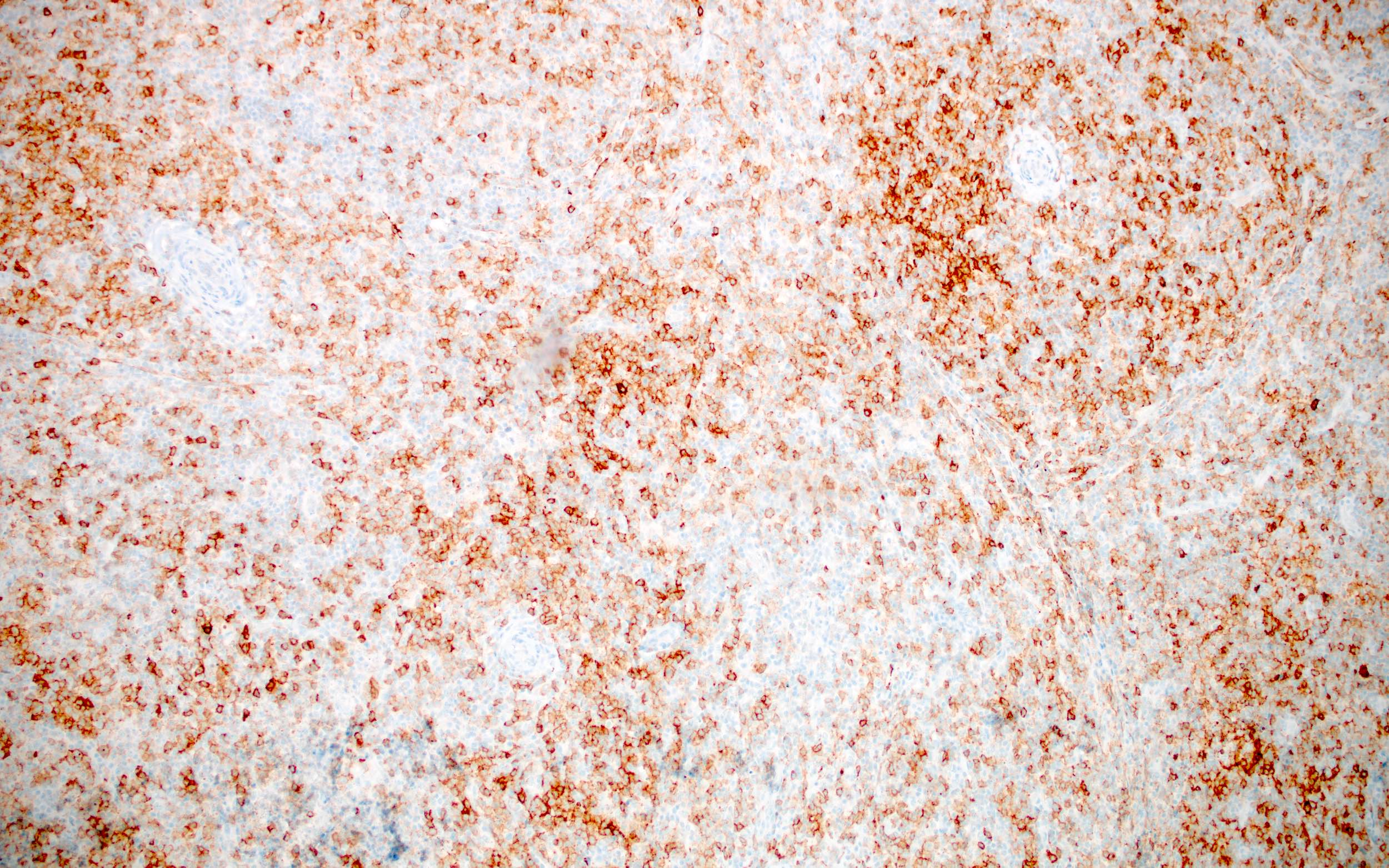
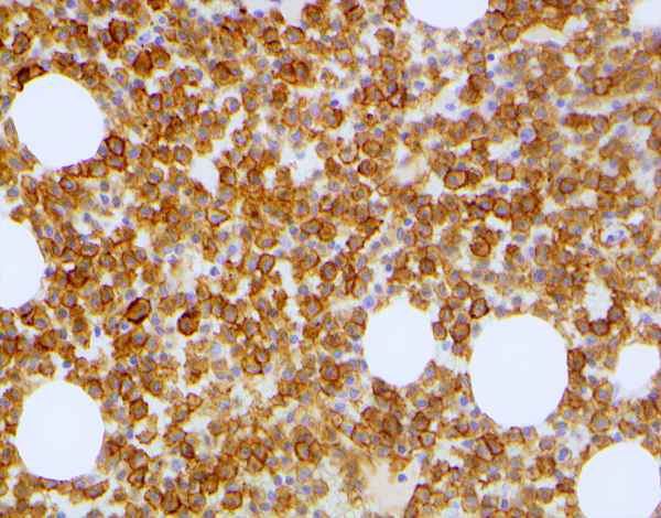
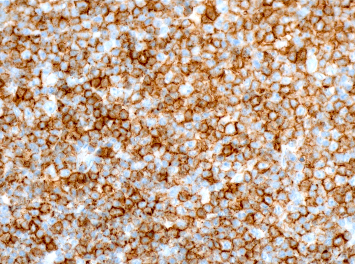
Hairy cell leukemia infiltrate in the bone marrow

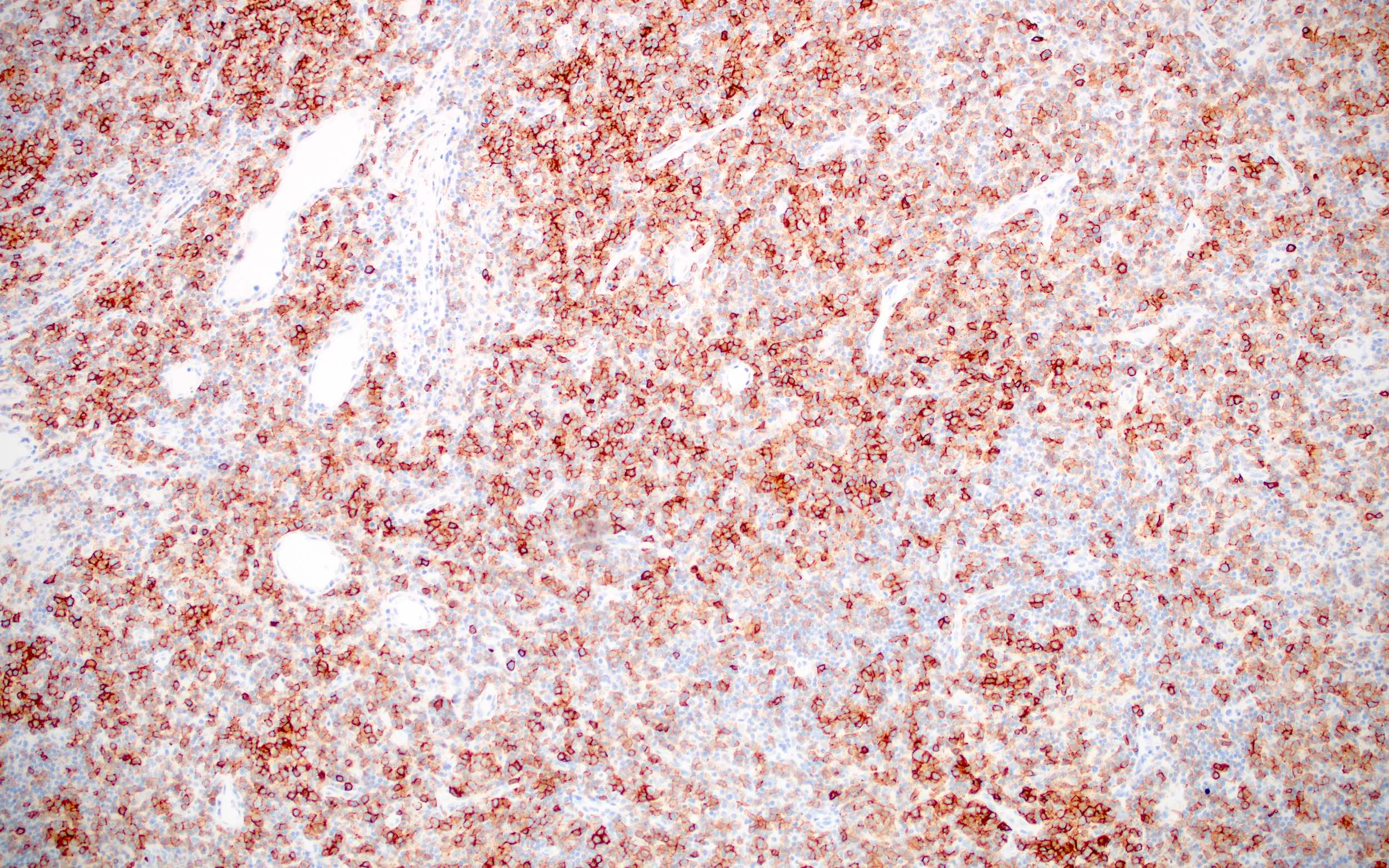
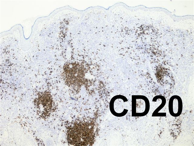
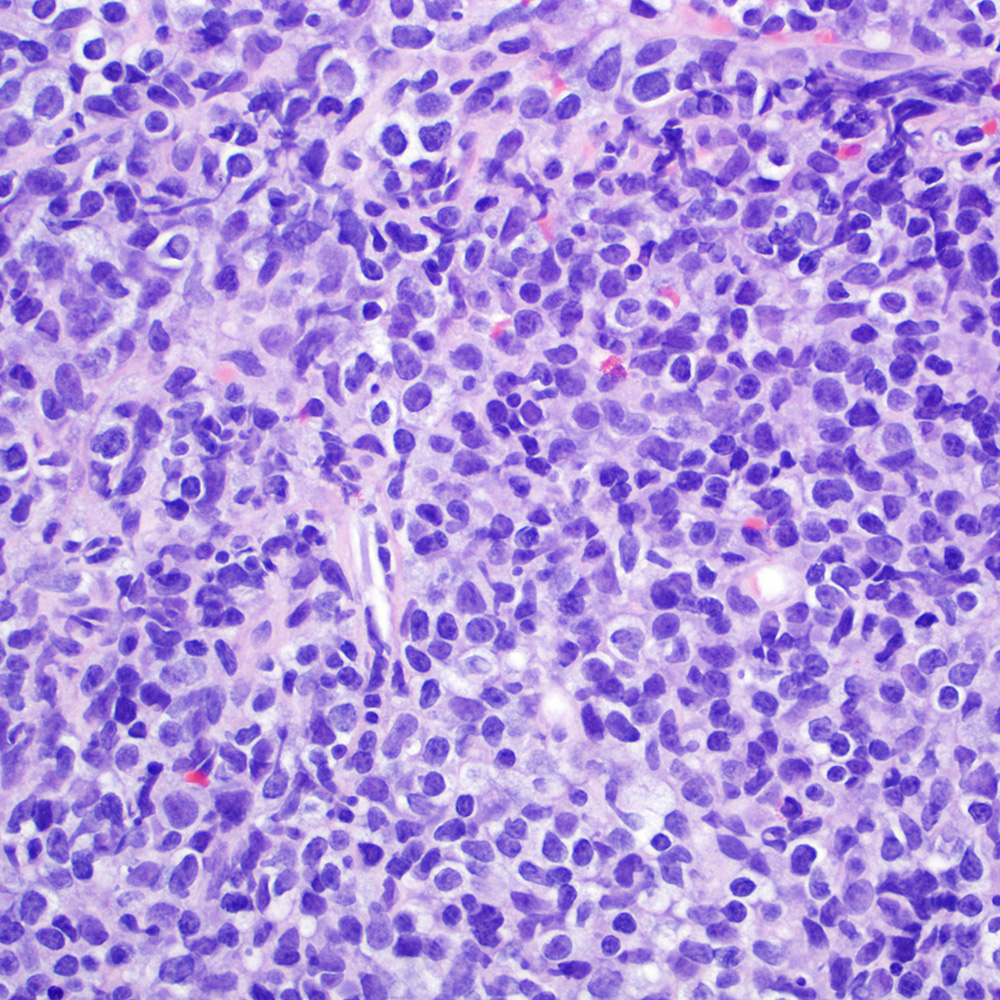
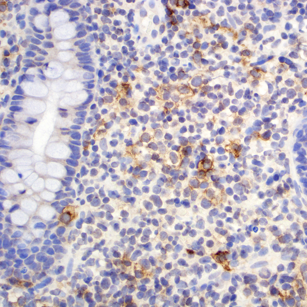
MALT lymphoma

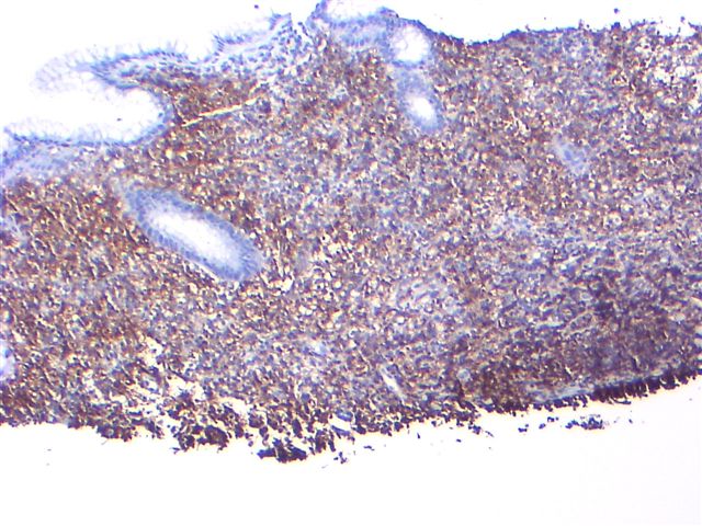
Burkitt lymphoma
of ileocecal valve
Cases #101, 118, 127, 130, 284 and AFIP images
Lymph node: angiomyomatous hamartoma
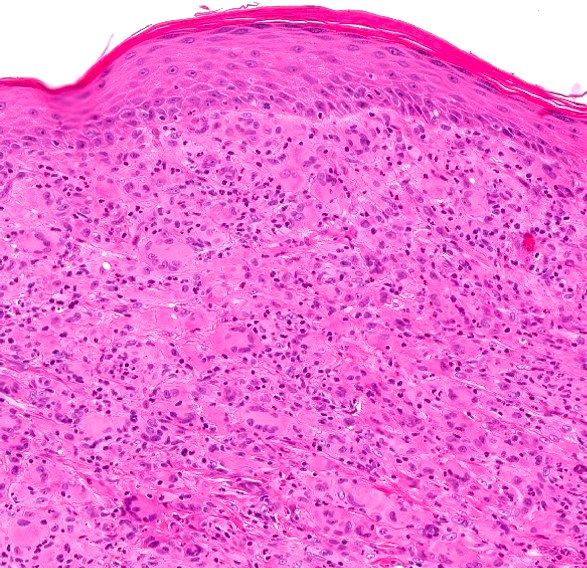
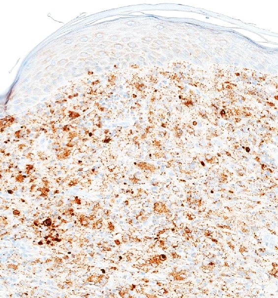
Skin: halo nevus
MALT lymphoma of stomach
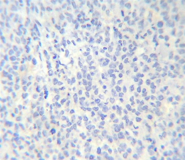
Myeloid sarcoma of bone (negative)
NK / T cell lymphoma, nasal type (tumor cells are negative)
T cell lymphoma
Hodgkin lymphoma, nodular lymphocyte predominant subtype
Images hosted on other servers:
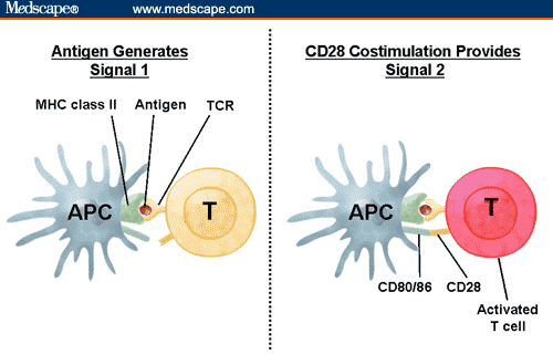
CD28: costimulation
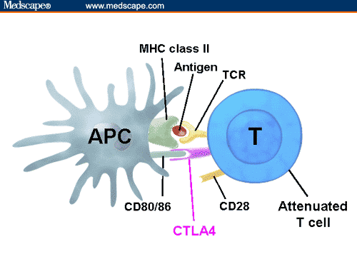
Competition between CTLA4 and CD28 for same ligands


CD28 costimulation is also required for proper memory cell activation
Images hosted on other servers:

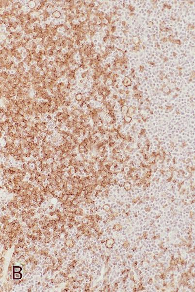
CD22: high grade B cell lymphoma, fig F

CD22: B-ALL, fig D

CD22: Burkitt lymphoma, fig D

CD22: normal tonsil

CD24: bladder carcinoma


CD24: colon, lung, ovary (left to right)

CD24: DCIS (breast)

CD24: extrahepatic bile duct carcinoma

CD24: salivary gland mucoepidermoid carcinoma

CD25: HTLV1+ adult T cell leukemia / lymphoma


CD25: mastocytosis

CD26: normal colon

CD26: proximal tubules (kidney)


CD26: prostate carcinoma


CD29: lung cancer
AFIP images and Cases #81, 179 and 284
Follicular dendritic cell sarcoma

Littoral cell angioma of spleen
Lymph node
Images hosted on other servers:

Mediastinum

Tonsil


Nodal marginal zone B cell lymphoma
Contributed by Mihaly Sulyok, M.D., Ph.D. and Christian M. Schürch, M.D., Ph.D.

CLL / SLL
Follicular lymphoma
Tonsilla
Images hosted on other servers:

CD247: T cell activation
Images hosted on other servers:
CD247: normal expression
CD247: reduced expression
CD248: IHC detection of FB5 antigen
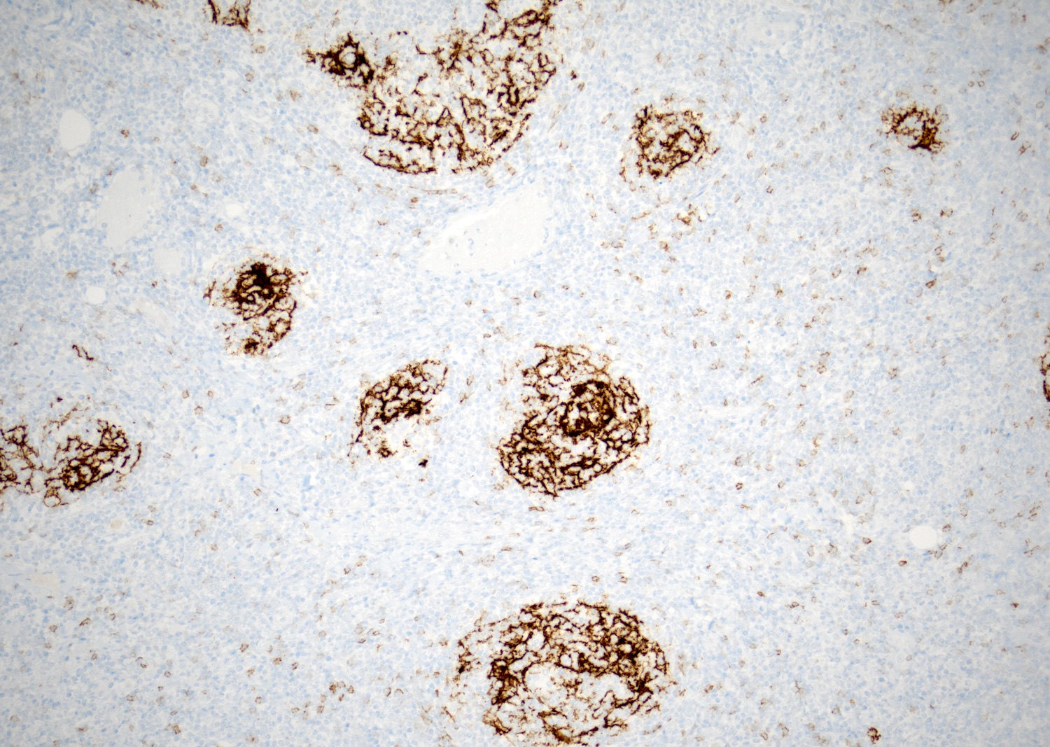
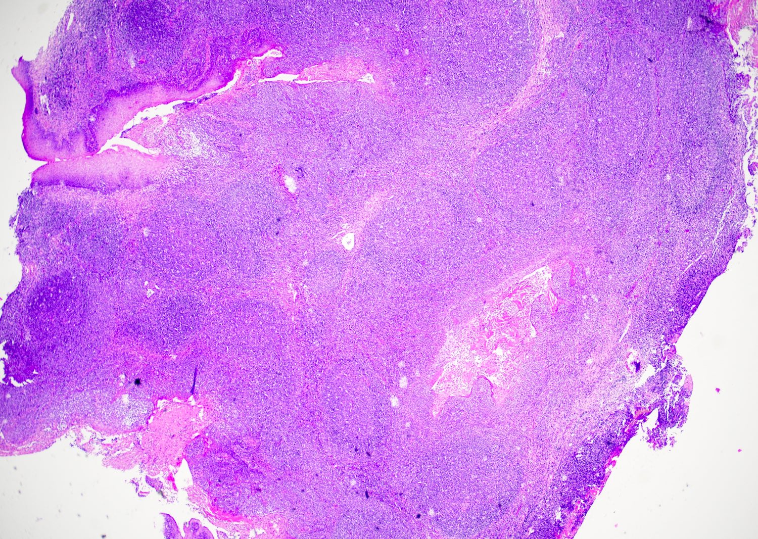
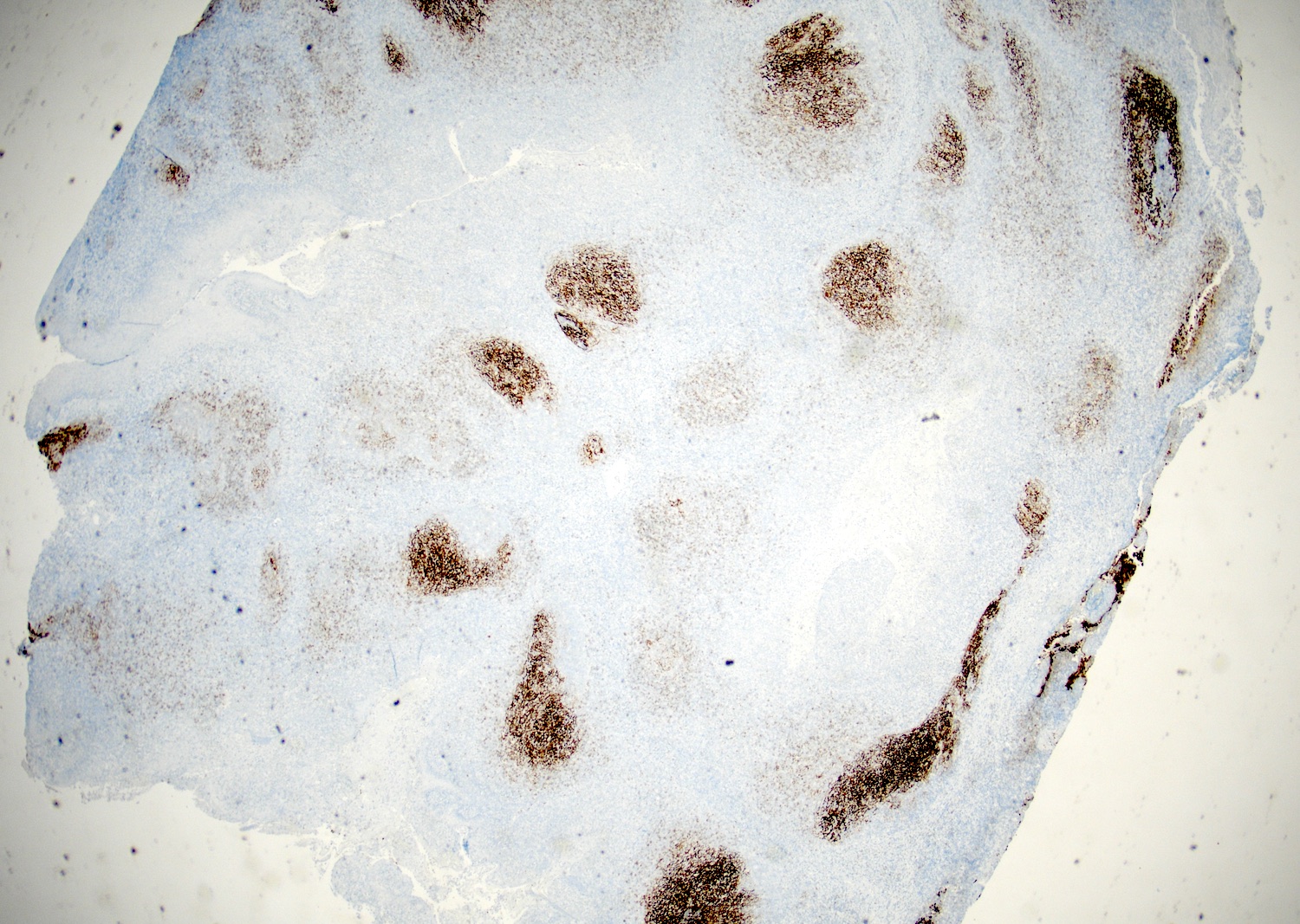
Contributed by Christian Schürch, M.D., Ph.D.
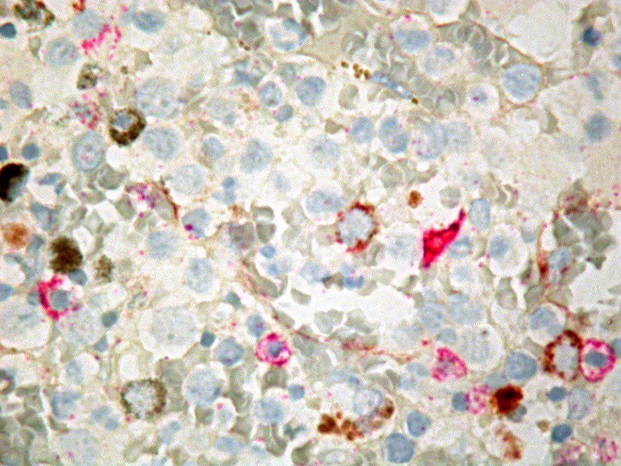
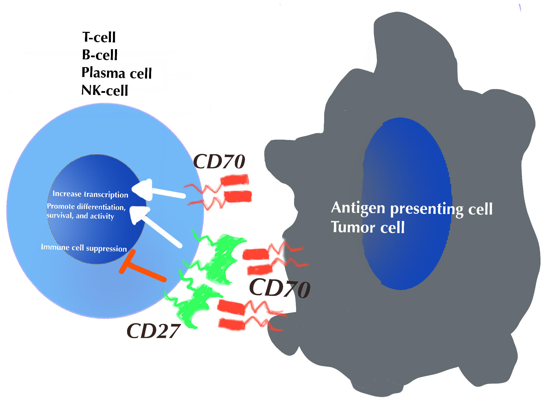
Immunohistochemical
double staining
(CD27 and CD34)
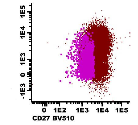
Contributed by Frido Bruehl, M.D. and Betty Gay, B.S.
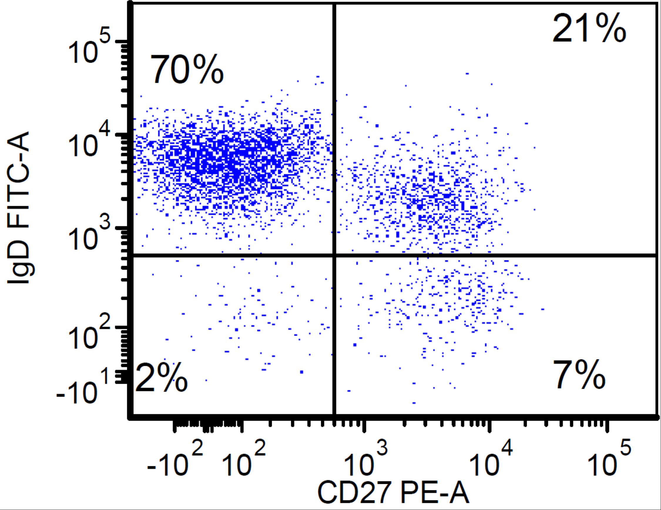
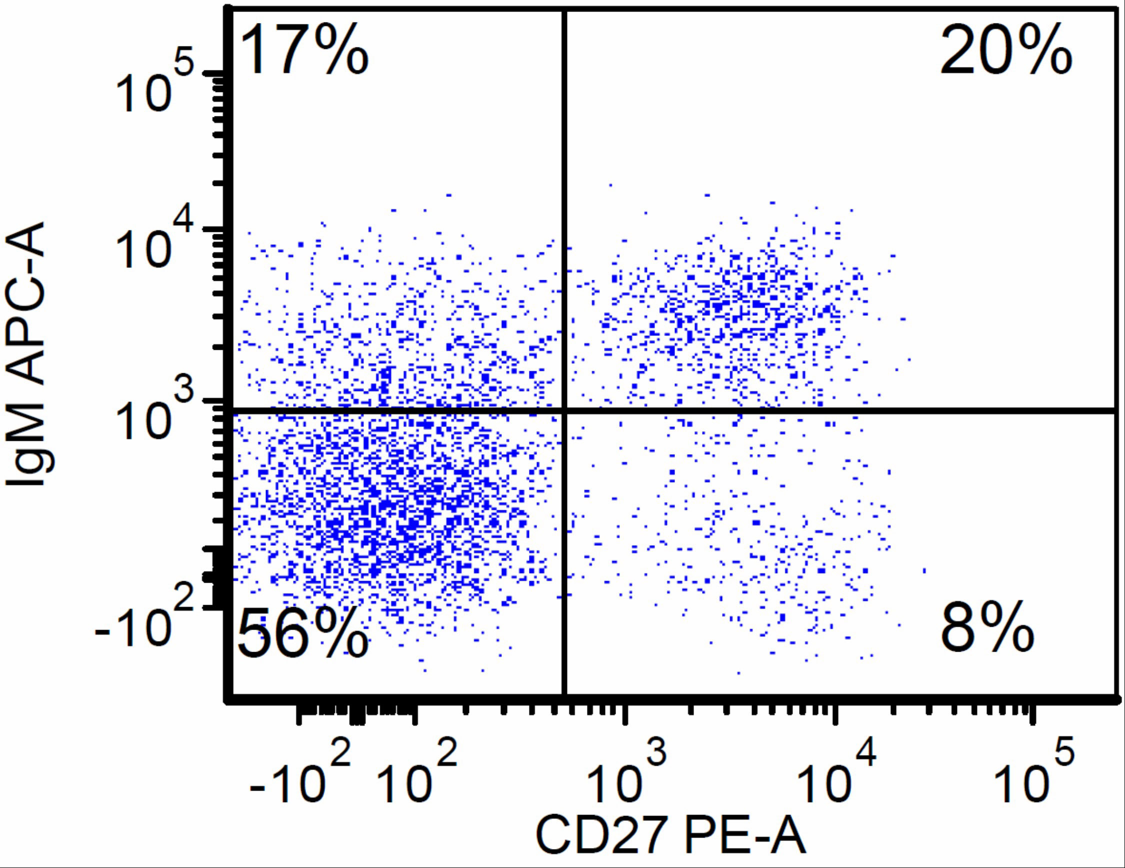
Flow cytometry of CVID patient
Contributed by Leonie Frauenfeld, M.D.
CD3+ mature T cells in lymphadenitis
CD3+ neoplastic cells in PTCL, TBX21
Case #36
Soft tissue: anaplastic large cell lymphoma
Contributed by Mario L. Marques-Piubelli, M.D. and Roberto N. Miranda, M.D.
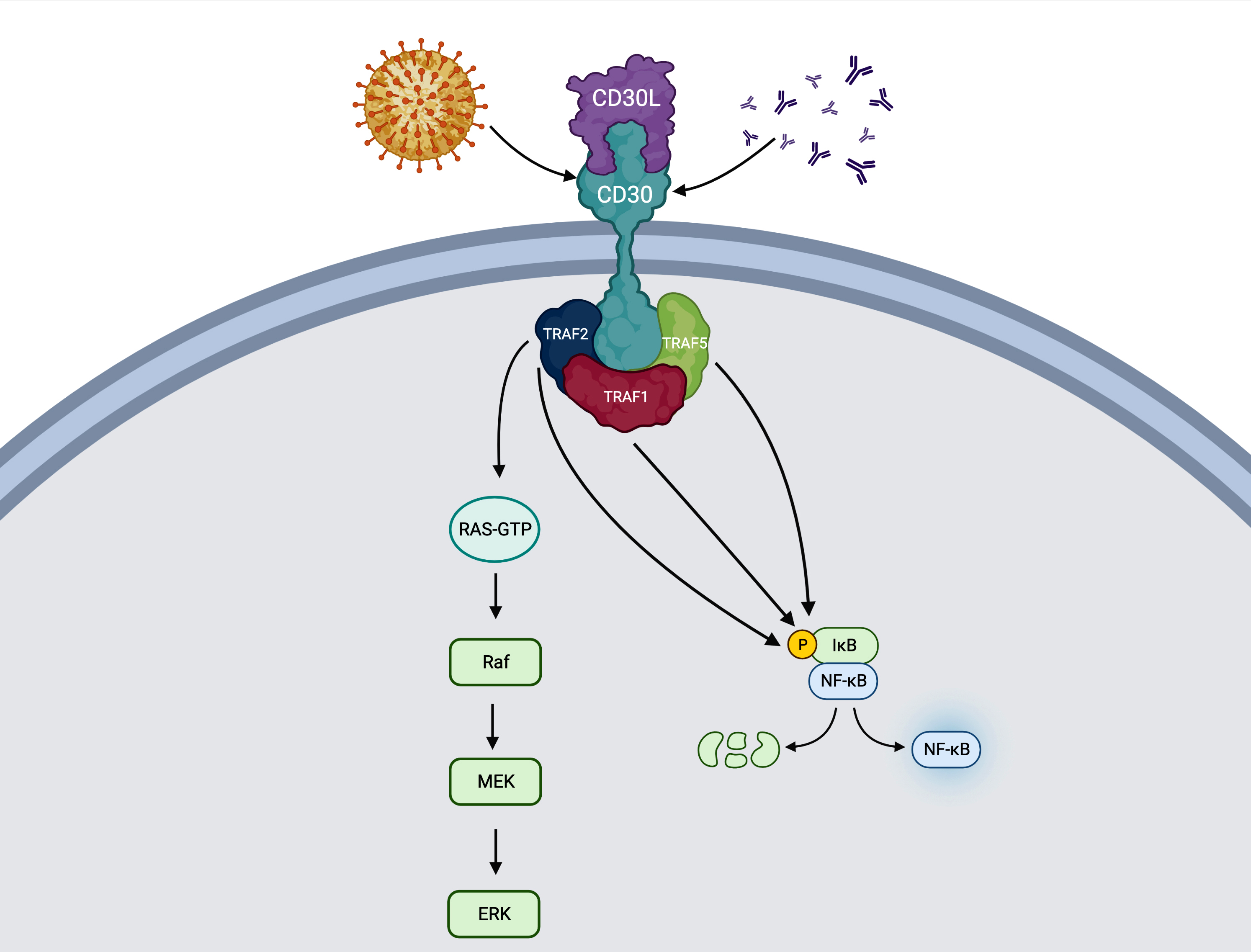
Activation of MAPK / ERK1 / ERK2 and NFκB pathways
Contributed by Roberto N. Miranda, M.D.
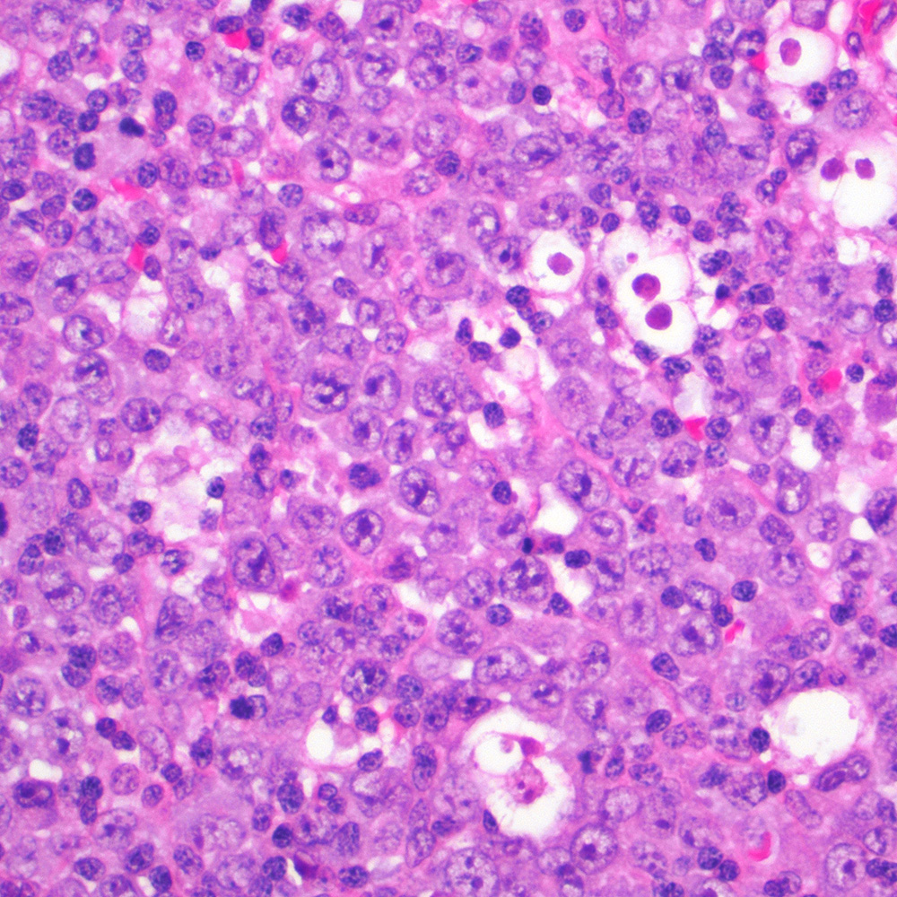
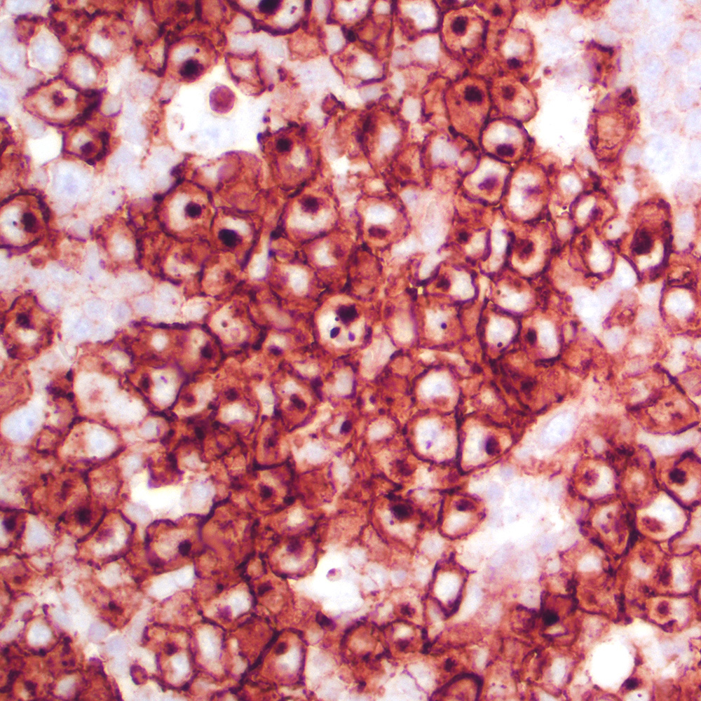
ALK+ anaplastic large cell lymphoma (ALCL) in lymph node
Primary cutaneous ALCL
Breast implant associated ALCL
Lymphomatoid papulosis type C
Peripheral T cell lymphoma, NOS, in colon
Primary mediastinal large B cell lymphoma
Mediastinal gray zone lymphoma
Nodular sclerosis classic Hodgkin lymphoma, syncytial variant

Kikuchi-Fujimoto disease
Dermatopathic lymphadenopathy
Images hosted on other servers:

CD36 and macrophage phagocytosis
Case #81
CD35: follicular dendritic cell tumor
Images hosted on other servers:

CD35: liver mass

CD39: donor liver
Cases #107, 187, 77
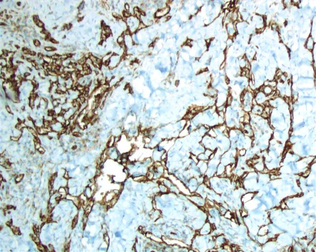
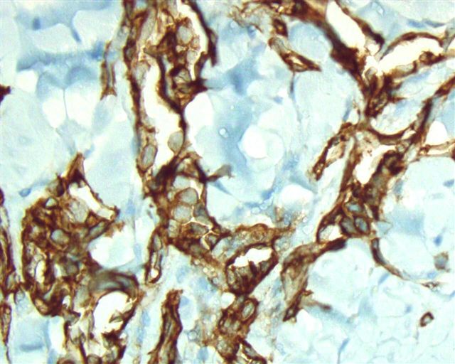
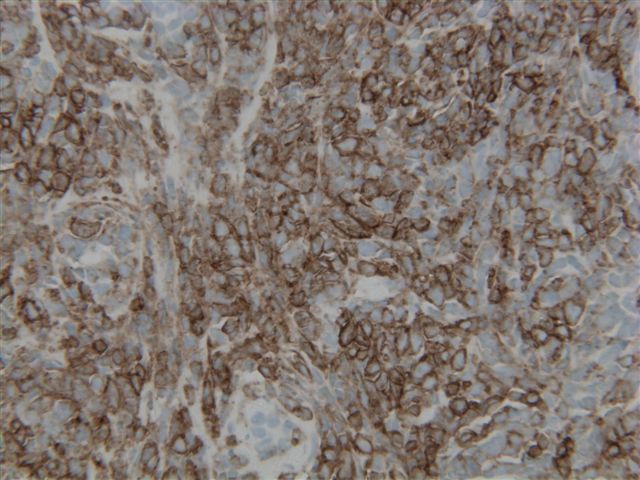
Retiform hemangioendothelioma
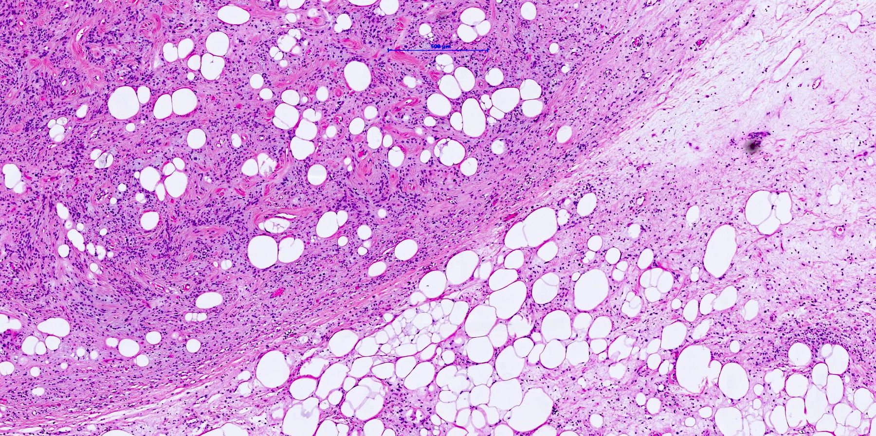
Kidney, glomus tumor
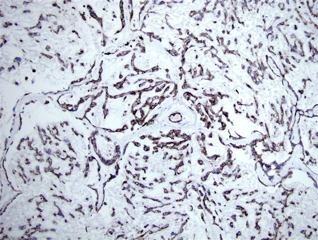
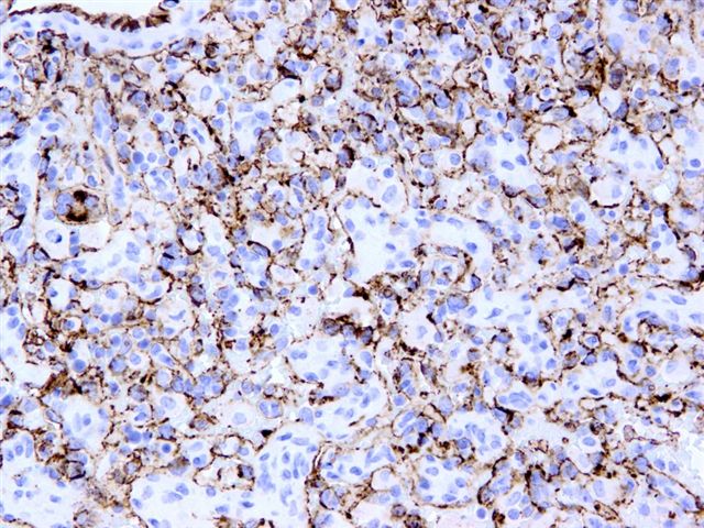
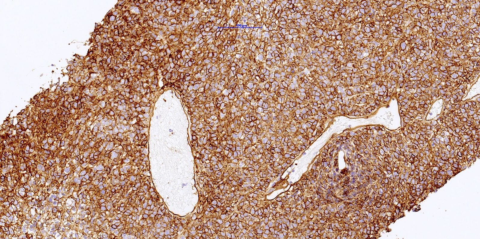
Epithelioid
hemangioendothelioma
Images hosted on other servers:
Normal structure:

Glomeruli (fig A)

Alveoli (fig A)

Spleen (fig A)

Liver (fig A)

Lymph nodes (fig A)

Skin (fig A)
Benign vascular tumors / processes:

Skin - diffuse dermal angiomatosis

Skin - epithelioid hemangioma
Malignant vascular tumors:

Kaposi sarcoma

Paratesticular
Lymphovascular invasion:

Breast

Colorectal carcinoma

Uterus, endometrial carcinoma
stage I

Uterine leiomyosarcoma


Images hosted on other servers:

CD4+ T cell and antigen presenting cell

CD4 acting as HIV receptor
Images hosted on other servers:

CNS: acute demyelinating disease (fig 12)

Joint: rheumatoid arthritis (fig B)

Lymph node: atypical paracortical hyperplasia (fig B)

Salivary gland: Sjögren syndrome (fig B)


Small bowel: T cell lymphoma (left: fig D; middle: fig D); right - stomach (fig C)

Soft tissue: inflammatory myopathy
Case #36
Soft tissue mass: anaplastic large cell lymphoma
Images hosted on other servers:

CD42a - d complex

CD42b: interactions

CD45RB expression changes during germinal center reaction
Images hosted on other servers:

CD40: soft tissue sarcomas

CD45RO: multiple myeloma plasma cell staining
Cases #140 and #127

Leukemia cutis
MALT lymphoma of stomach
Images hosted on other servers:

Mantle cell lymphoma
Anaplastic large cell lymphoma
Images hosted on other servers:

CD44 molecule structure with ligands
Contributed by John Yahya I. Elshimali, M.D. and Maria Tretiakova, M.D., Ph.D.
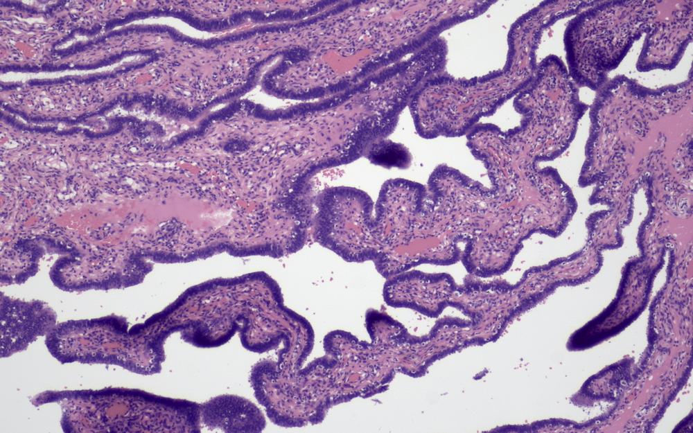
Epithelium of fallopian tube

Fallopian stromal cells
CD44 positive epithelial cells in fallopian tube
CD44 positive epithelial cells in fallopian tube
Positive CD44 expression in fallopian tube epithelium
Decidual and stromal cells in fallopian pregnancy positive for CD44
Normal bladder mucosa
CD44 - full thickness in normal bladder mucosa
Bladder CIS
CD44 - loss of CD44 expression
Pagetoid CIS vs reactive inflammation
CD44 - patchy loss of expression in pagetoid CIS
Images hosted on other servers:

CD44 transmembrane receptor function
Contributed by Elena M. Fenu, M.D.
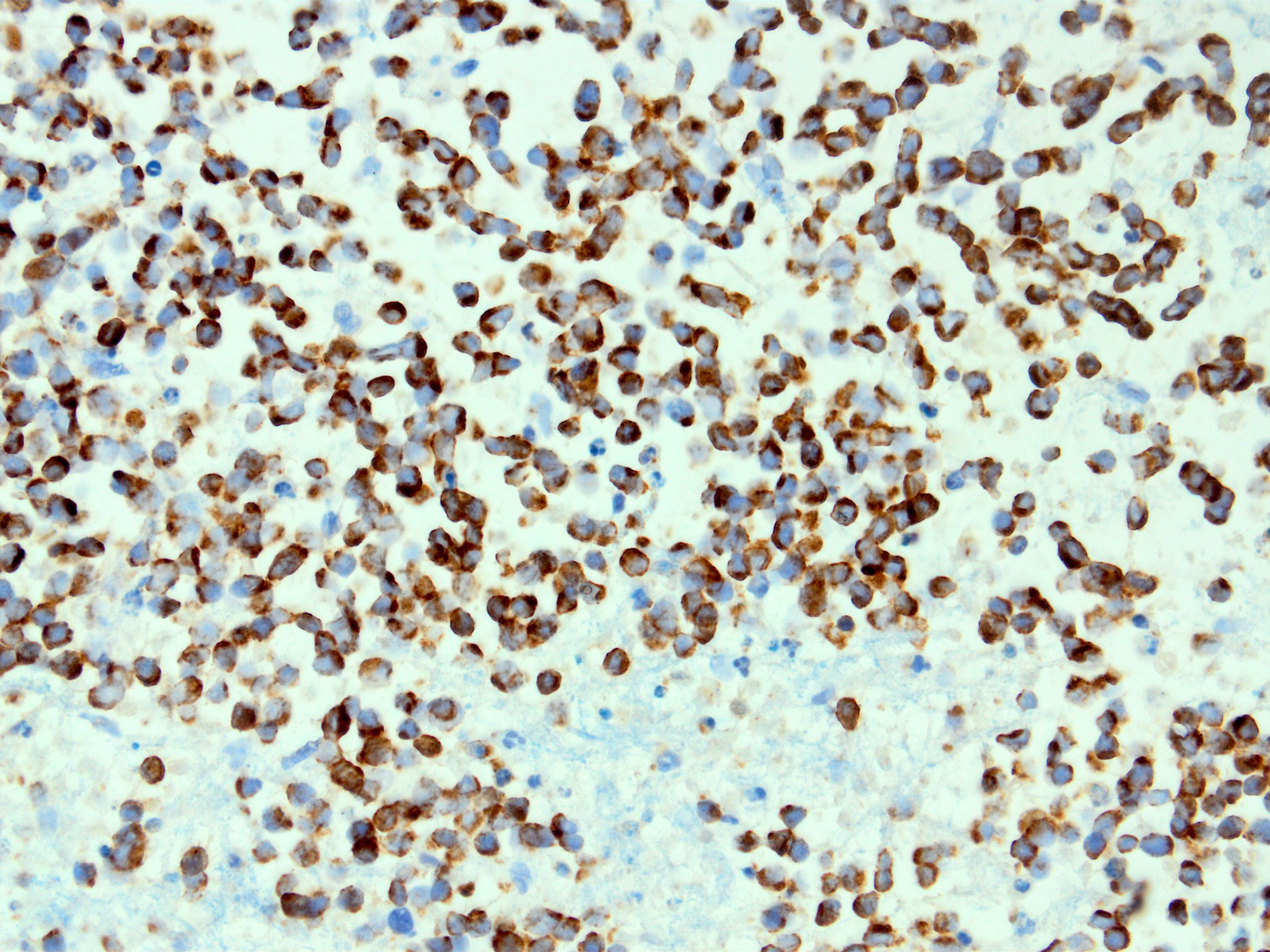
Anaplastic large cell lymphoma
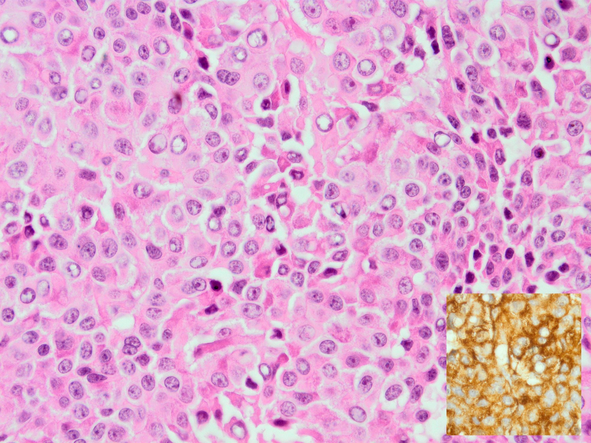
Diffuse large B cell lymphoma
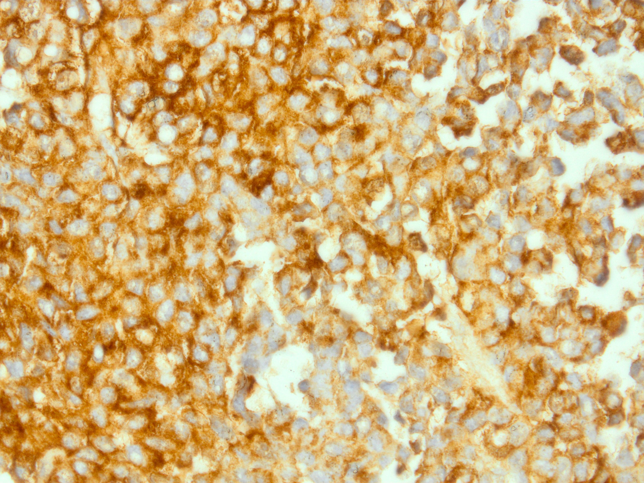
Plasmablastic lymphoma
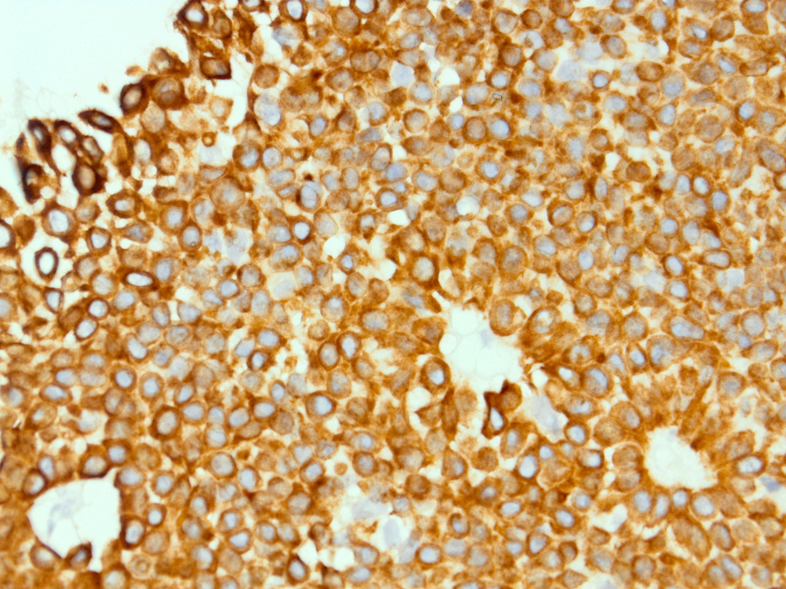
Leukemia cutis
Images hosted on other servers:

Intraepidermal T cells in fixed drug eruption lesions (fig d)

Papulonodular lesion show dermal, intraepithelial neoplastic lymphocytes
Contributed by Frido Bruehl, M.D.
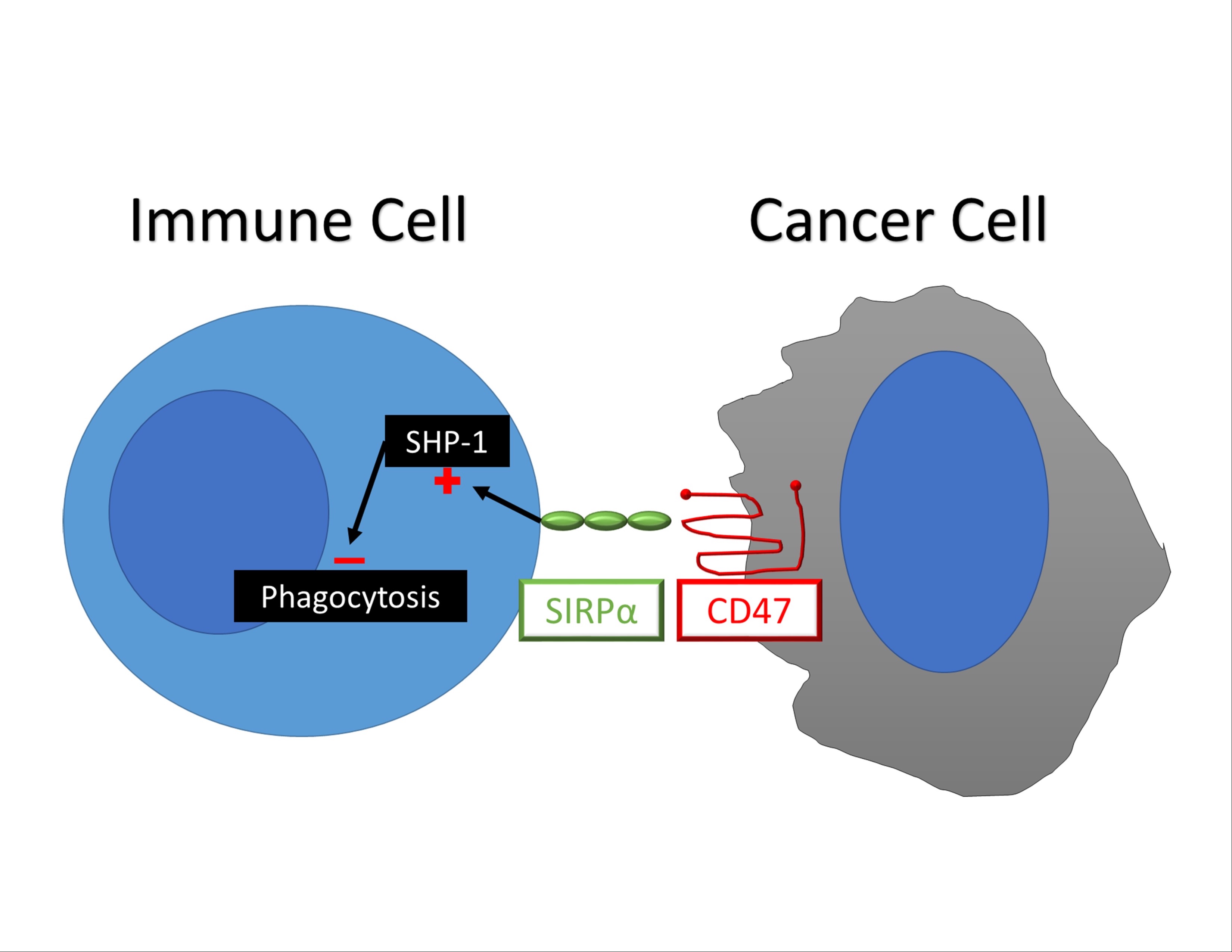
Proposed mechanism of action of CD47
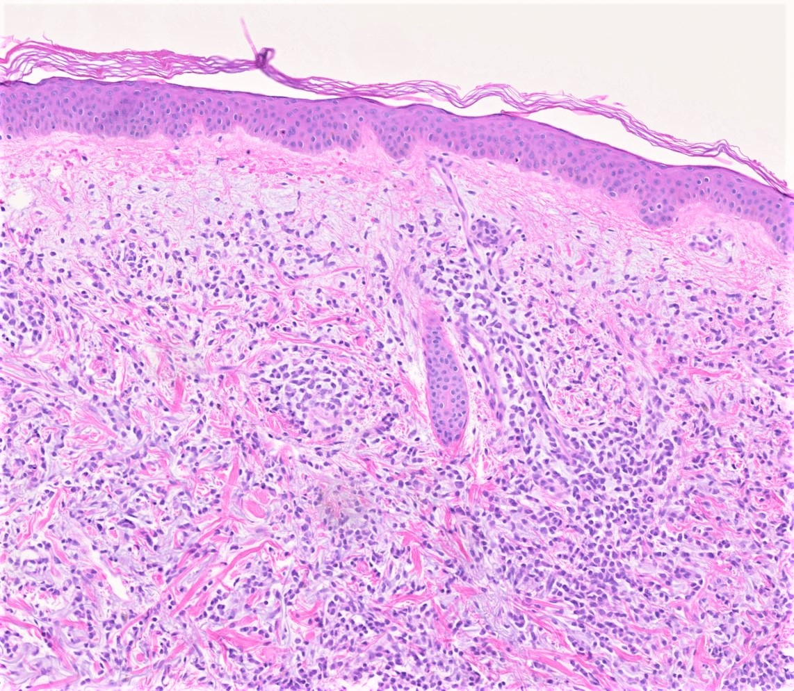
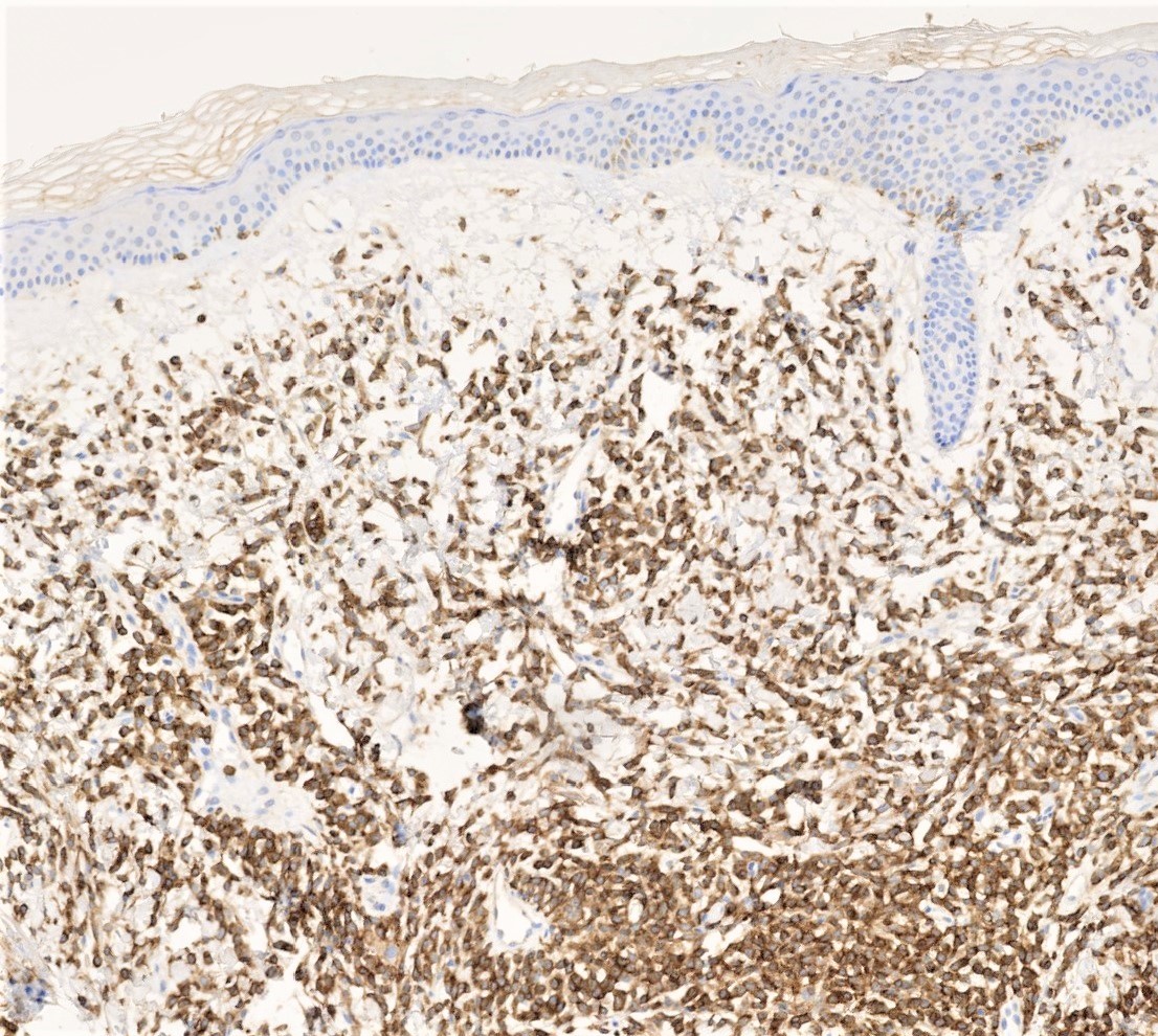
Contributed by Christian M. Schürch, M.D., Ph.D.
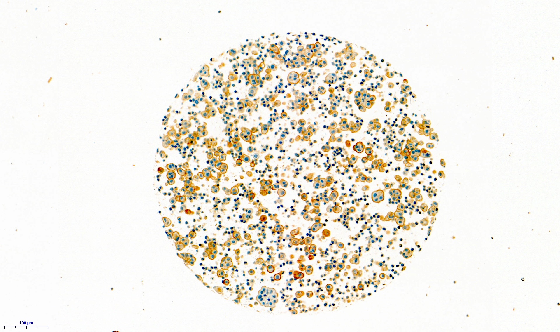
Mesothelioma, cell block, CD47
Contributed by Julia Braza, M.D. and Cases #281 and #17
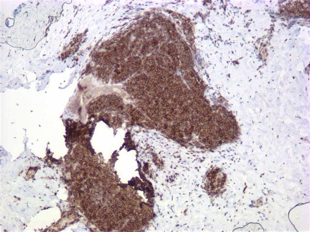
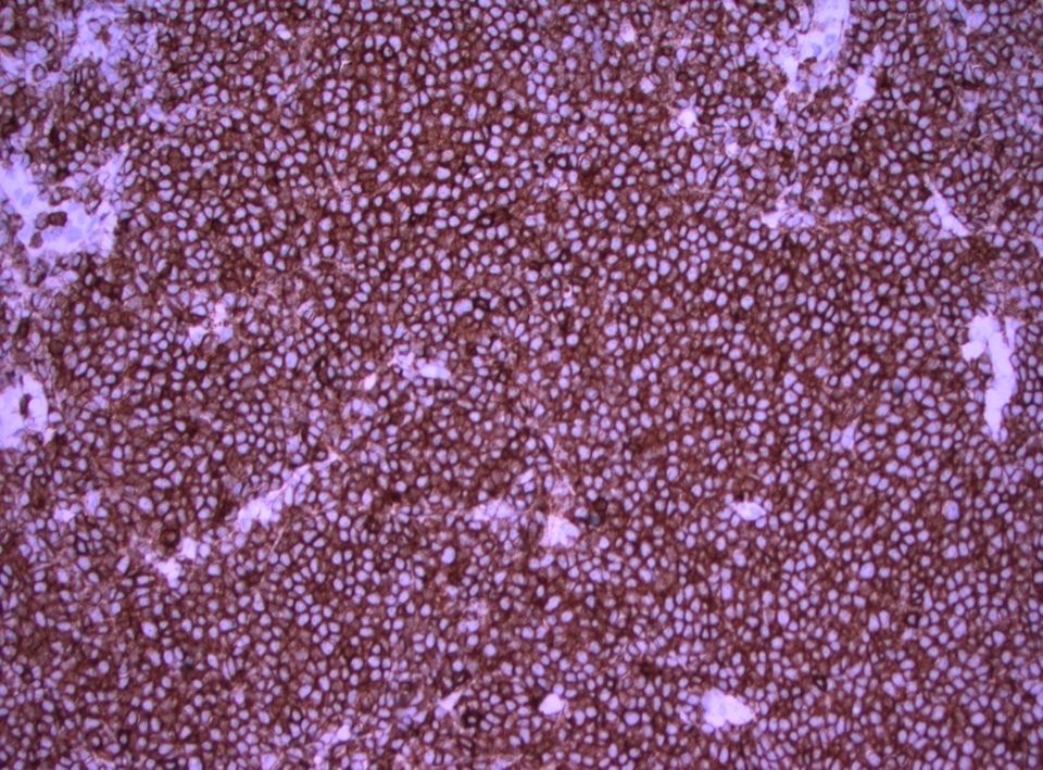
Breast CLL / SLL
Eyelid: mantle cell lymphoma
Thymic carcinoma,
nonkeratinizing
squamous cell
subtype
Images hosted on other servers:
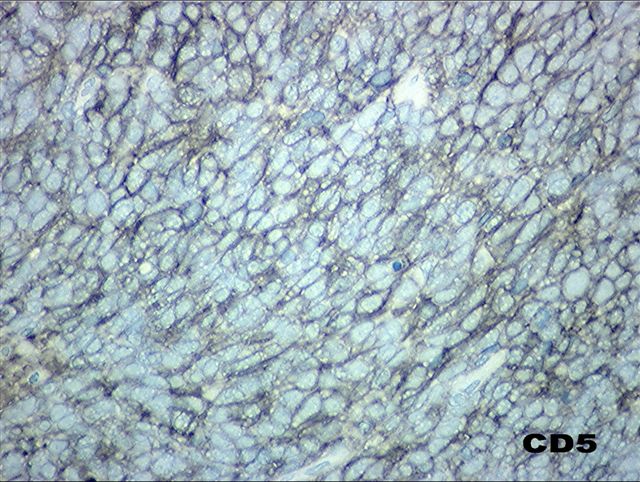
Tumors / tissue typically CD5+:

Lymph node: mantle cell lymphoma (fig B)

Lymph node: SLL
Tumors / tissue occasionally / rarely CD5+:

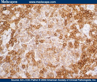
Lymph node: follicular lymphoma: typically CD5 stains only adjacent T cells (left); rarely tumor is CD5+ (right)
Images hosted on other servers:

CD50: normal
uterine cervix
and cervical
cancer tissues

CD52: cryostat
sections stained
with biotinylated
Campath-1H

CD52: paraffin
wax sections
stained with
Campath-1G


CD55: breast carcinoma staining

CD55: breast carcinoma staining

CD58: buccal
mucosa with
intense LFA 3+
staining

CD58: cardiac muscle with LFA-3+ staining

CD58: cerebellum, liver, skin

CD58: proximal tubule and distal tubule (kidney)

CD58: cells near
seminiferous tubule
boundary (testis)


CD59: diabetic and nondiabetic:
kidneys (left), nerves (right)
Images hosted on other servers:

Large granular lymphocytic leukemia (fig 11)

Papillary carcinoma of thyroid (fig B)

"Atypical cytology, cannot exclude papillary carcinoma"
Images hosted on other servers:

CD66: colon

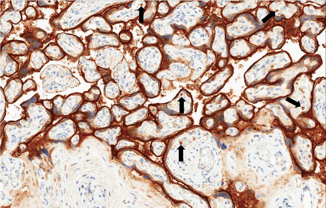
CD66: expression at maternal fetal surface

CD66: various breast lesions

Implanted
ovum (CD66f+,
syncytiotrophoblast)

Normal placenta (CD66f+, trophoblast)

Hydatidiform mole
(syncytiotrophoblast)

Invasive mole
(CD66f+, large
trophoblast cells)

Choriocarcinoma
of uterus (CD66f+,
syncytiotrophoblast)
Images hosted on other servers:

Superfamily of integrins
Contributed by Pamela Wirth, Ph.D. (sources: University of Toronto and Human Protein Atlas)
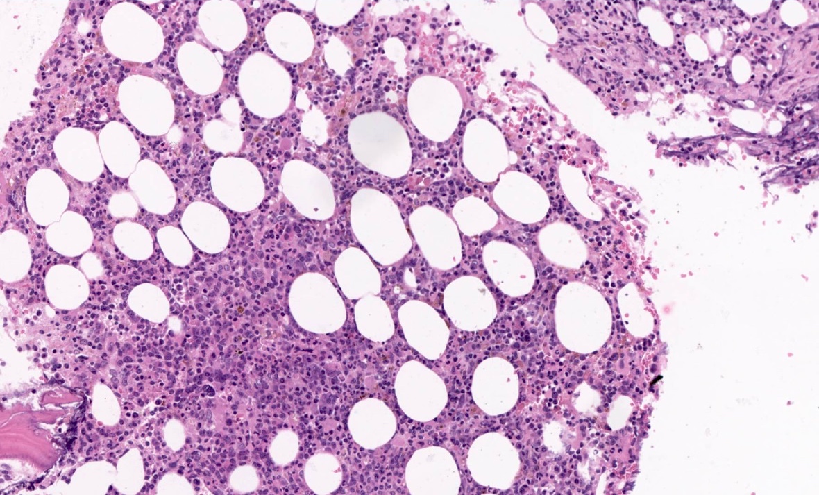
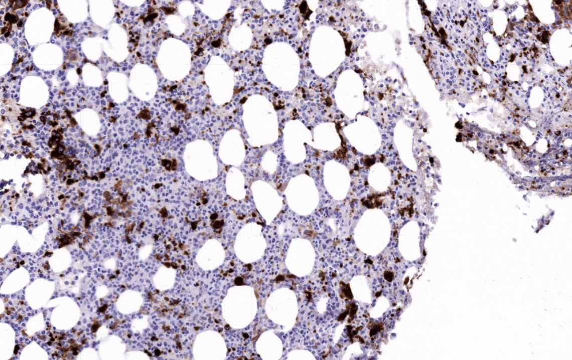
Bone marrow biopsy
Liver
cholangiocarcinoma
Kidney adenocarcinoma
Lung squamous cell carcinoma
Colon carcinoma
Images hosted on other servers:

Cellular neurothekeoma
Contributed by Frido Bruehl, M.D.
Fibrolamellar carcinoma of the liver
Kikuchi-Fujimoto disease of the lymph node
Reticulohistiocytoma of the skin
Contributed by Christian Schürch, M.D., Ph.D.
Bone marrow biopsy
Images hosted on other servers:

CD74: renal tumors (figs D-F)

CD74: multiple myelomas
(figs G-J)

CD74: increased expression in H. pylori+ epithelium

CD75: marginal zone lymphoma (aberrant expression)
Contributed by Luca Morelli, M.D. and Claudio Luchini, M.D., Ph.D.
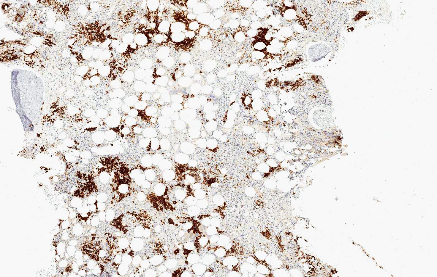
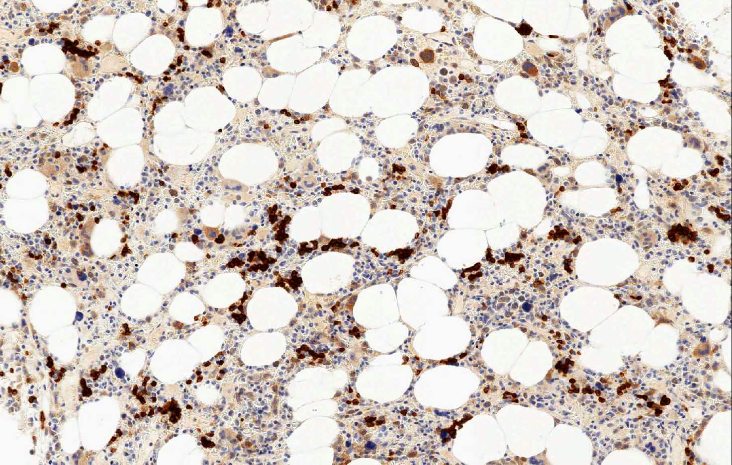
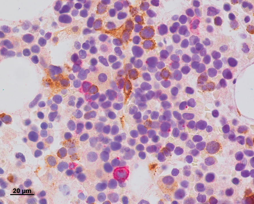
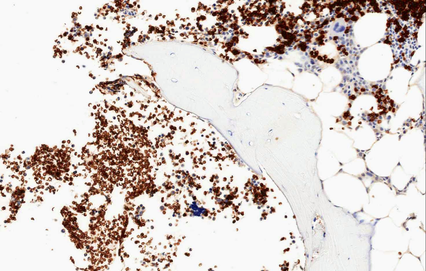
CD71 in normal bone marrow
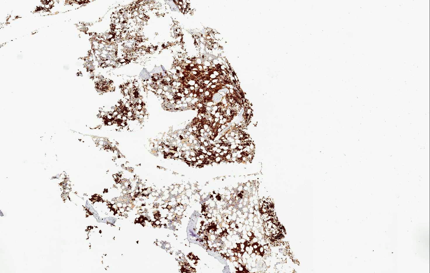

CD71 in bone marrow in myelodysplastic syndrome
CD71 in bone marrow in AML M6

CD71 in placental tissue
Glycophorin staining
Images hosted on other servers:


CD8+ T cell activation requires 2 signals
Case #179
Splenic littoral cell angioma, sinus lining cells are CD8-
Images hosted on other servers:

Bladder: follicular cystitis (fig D)

Skin: cutaneous CD8+ epidermotropic T cell lymphoma

Skin: lymphomatoid papulosis (fig B, C)
Case #36
Anaplastic large
cell lymphoma:
partial CD4 / CD8
coexpression
Images hosted on other servers:
Structure
Images hosted on other servers:
Normal tissue:
Cerebrum
Cervical squamous epithelium (fig A)
Various tumors:
Cervical carcinoma
CNS nonneuroepithelial tumors
Gallbladder: well differentiated adenocarcinoma
Mesothelioma
Skin: basal and
squamous cell
carcinoma and
actinic keratosis
Small cell lung cancer
Images hosted on other servers:

CD90: normal and diseased skin


CD93: various images

CD93: liver (figure 6B)

CD94: normal tonsil

CD97: normal oral mucosa

CD98: placenta (fig 4)
Images hosted on other servers:

Cytotoxic T lymphocyte pathway

Contributed by Frido Bruehl, M.D.

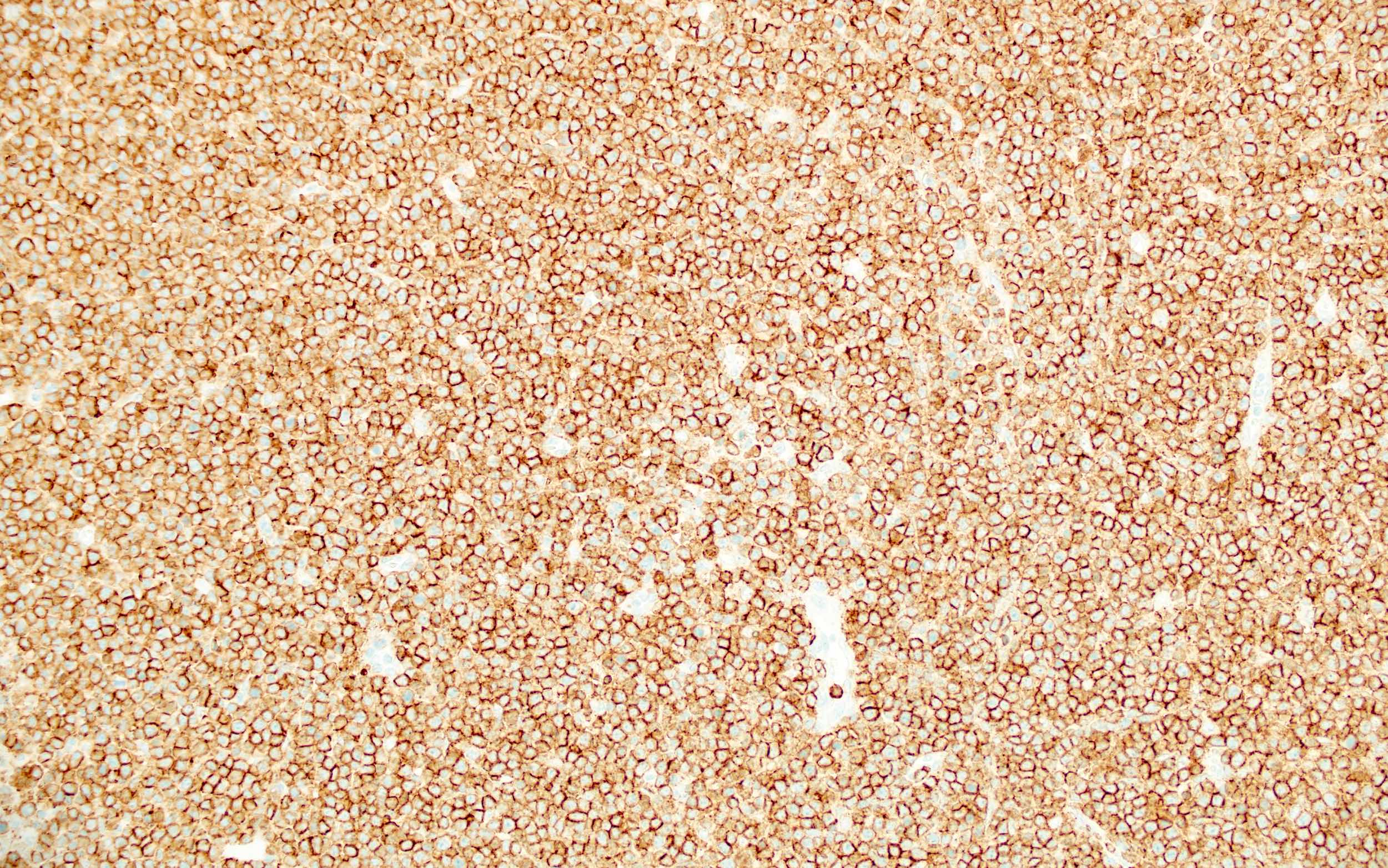
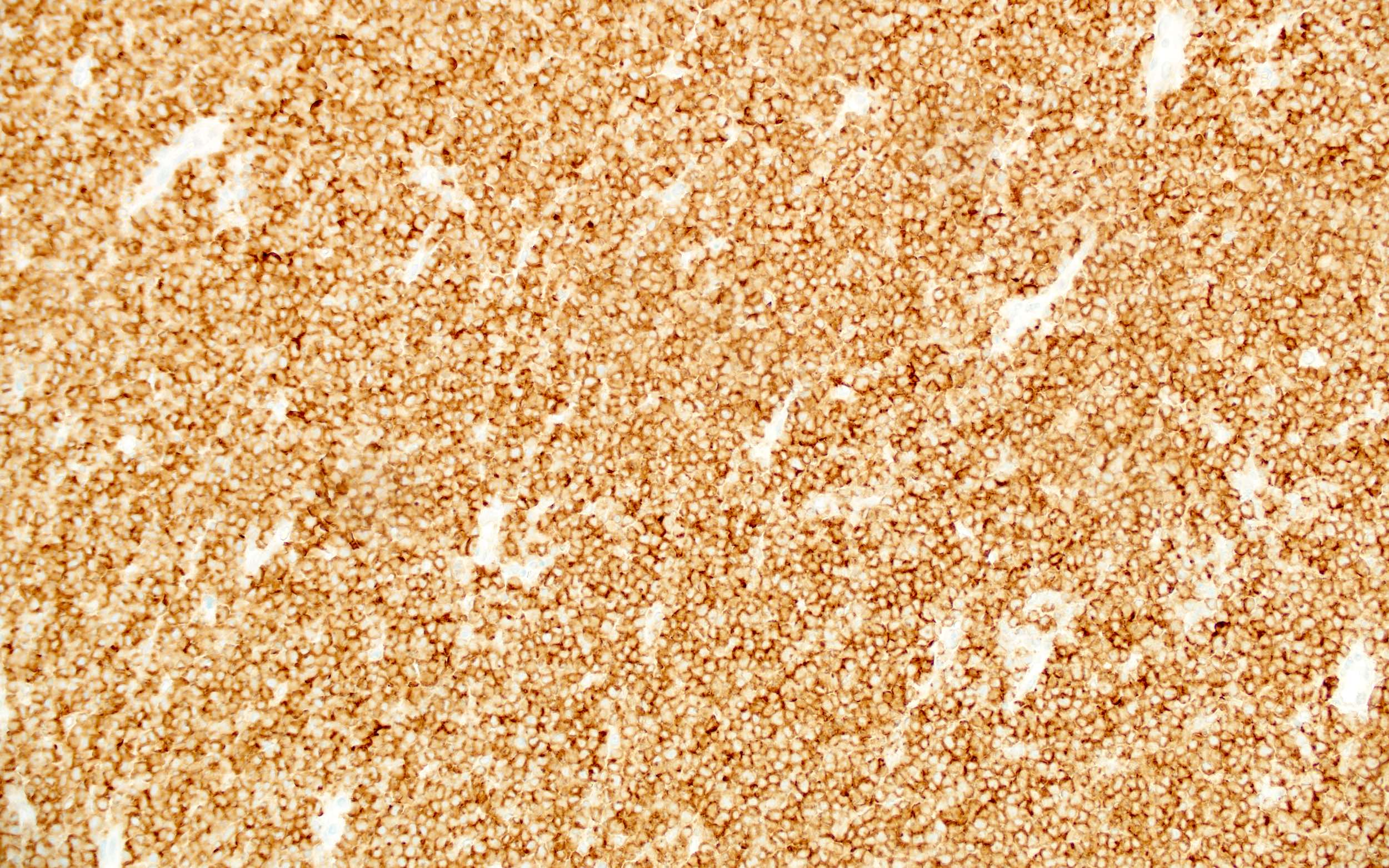
CD99 in Ewing sarcoma
CD99 in T cell acute lymphoblastic leukemia
CD99 in synovial sarcoma
CD99 in CIC-DUX rearranged sarcoma
EWSR1-NFATC2 rearranged sarcoma
EWSR1-NFATC2 rearranged sarcoma
Images hosted on other servers:

CDK4 binds with cyclin D

CDK4 regulation and activation
Contributed by Epitomics
Breast carcinoma
Images hosted on other servers:

Breast carcinoma


Liposarcoma - dedifferentiated (left), well differentiated (right)
Contributed by GenomeMe
Colon (normal), clone IHC402
Images hosted on other servers:

Colonic medullary carcinoma
Quality control of CDX2 staining
Contributed by Brandon Umphress, M.D. and Andrey Bychkov, M.D., Ph.D.

Porocarcinoma, ductal differentiation
Porocarcinoma with invasion into the dermis
Acrosyringeal unit
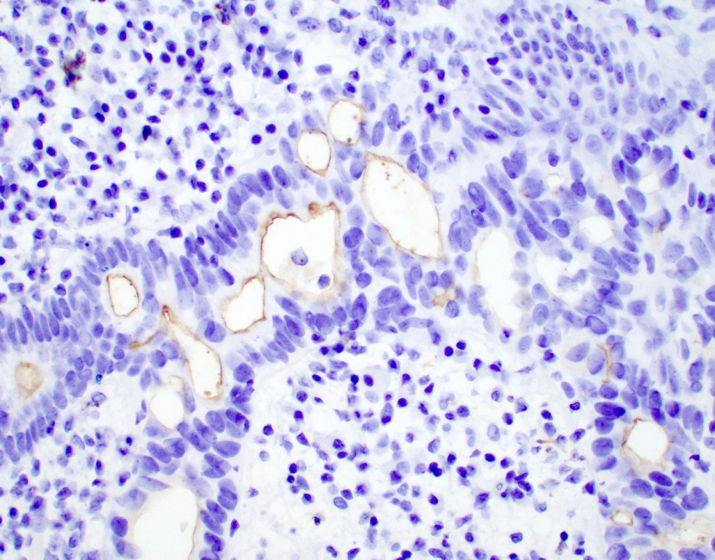
CEA expression in a poorly differentiated carcinoma
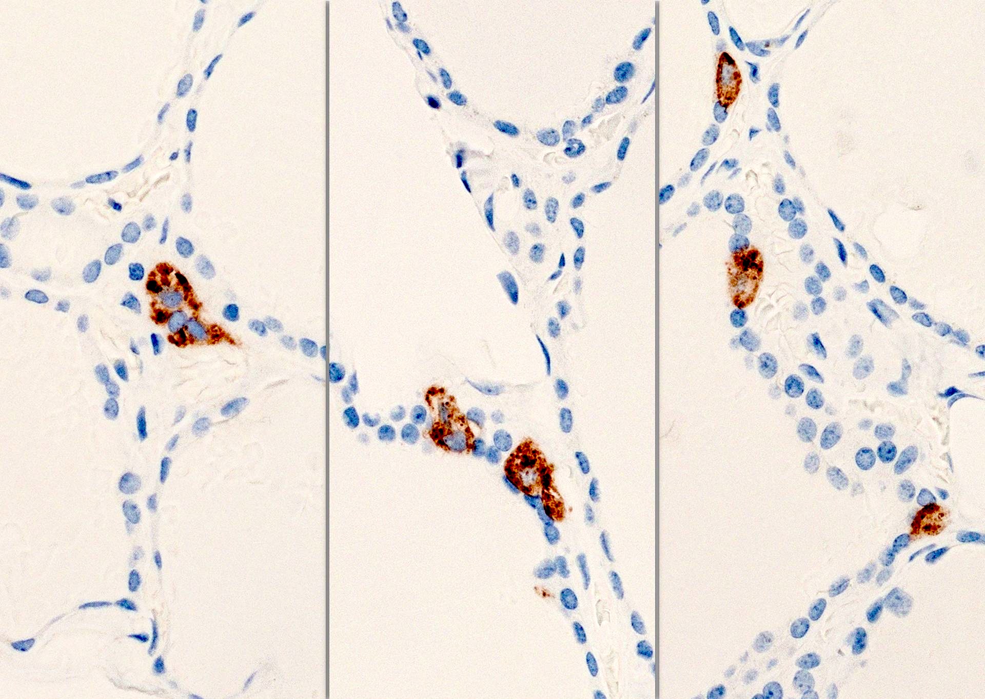
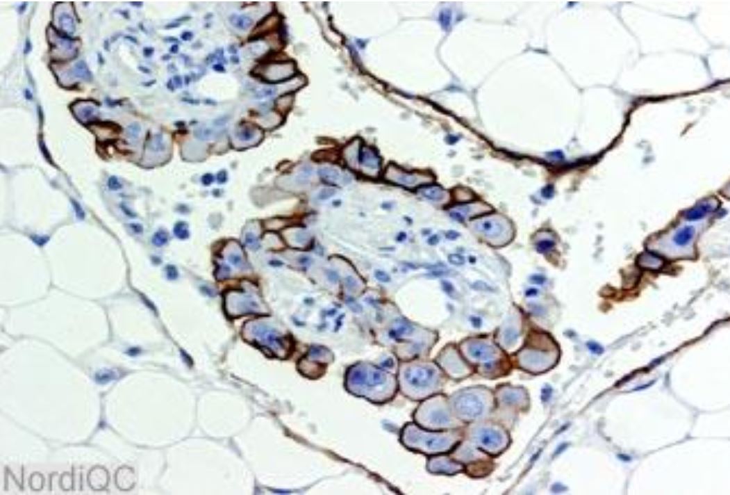
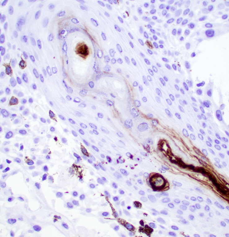
C cells in relation to thyroid follicle

Clustered C cells
Cases #195, #204, #108 and AFIP images

Bladder: paraganglioma
Kidney: carcinoid tumor

Parathyroid gland: ectopic tissue
Breast: neuroendocrine carcinomas
Thyroid: medullary carcinoma
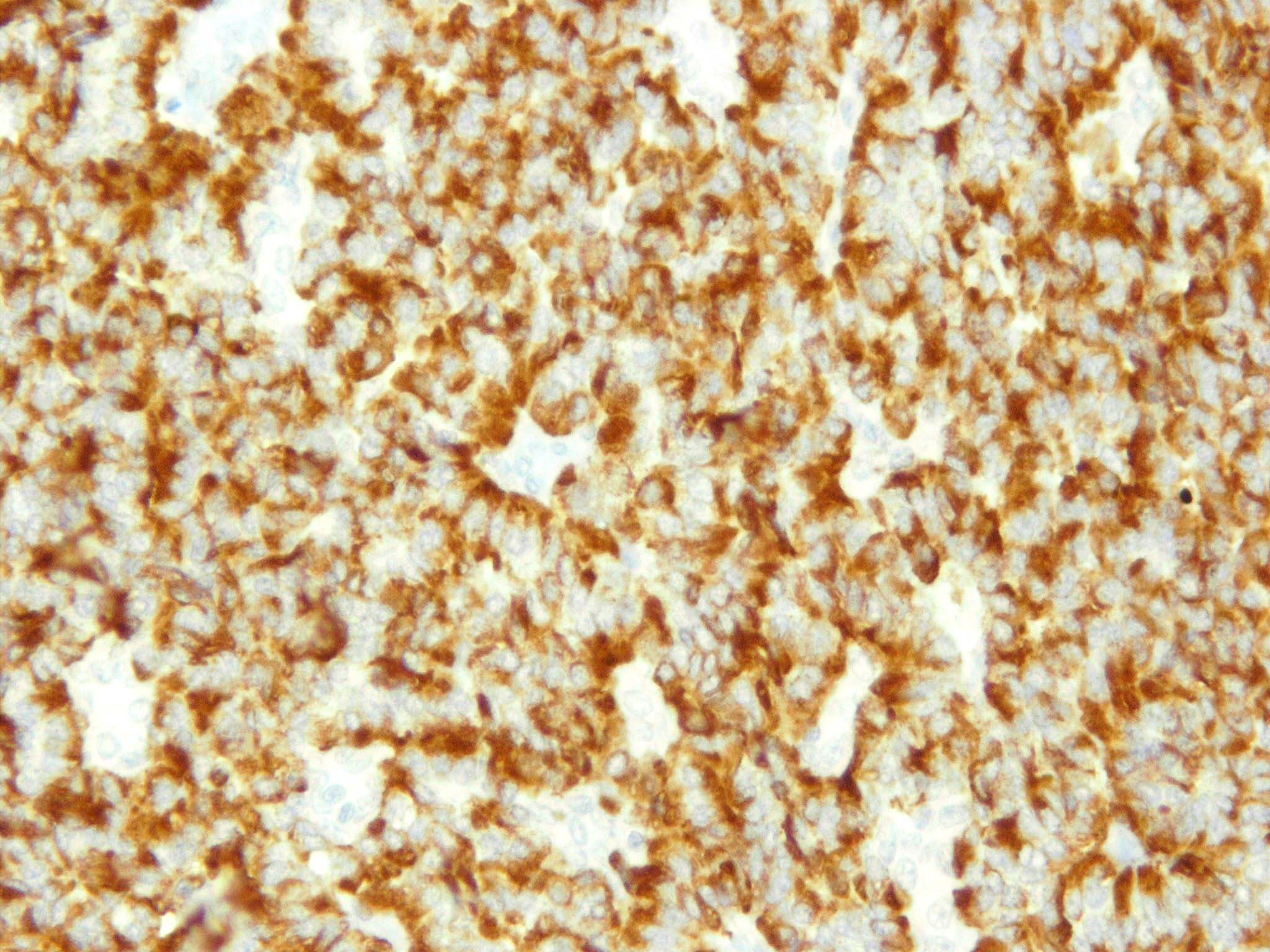
Contributed by Jijgee Munkhdelger, M.D., Ph.D. and Andrey Bychkov, M.D., Ph.D.
Typical carcinoid immunoprofile
Images hosted on other servers:
Adrenal medulla:
pheochromocytoma

Breast: small cell carcinoma

Heart: metastatic
pheochromocytoma
(right side)
Images hosted on other servers:

Claudin5: normal and malignant (breast)

Claudin5: serous papillary adenocarcinoma (ovary)


Claudin7: synovial sarcoma

Claudin18: normal lung and stomach (splice variant 2)


Claudin18: PanIN (left), infiltrating ductal carcinoma (right)

Claudin18: carcinomas
of stomach, esophagus,
ovary (splice variant 2)
Contributed by GenomeMe

Lung (normal), clone IHC549
Contributed by Wei Chen, M.D., Ph.D. and Saba Shafi, M.D.
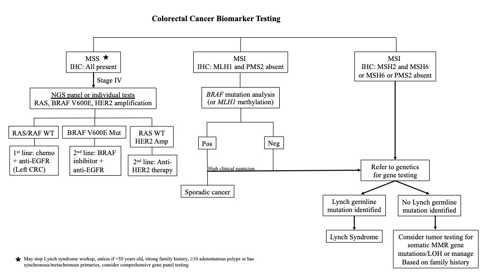
CRC biomarker testing algorithm
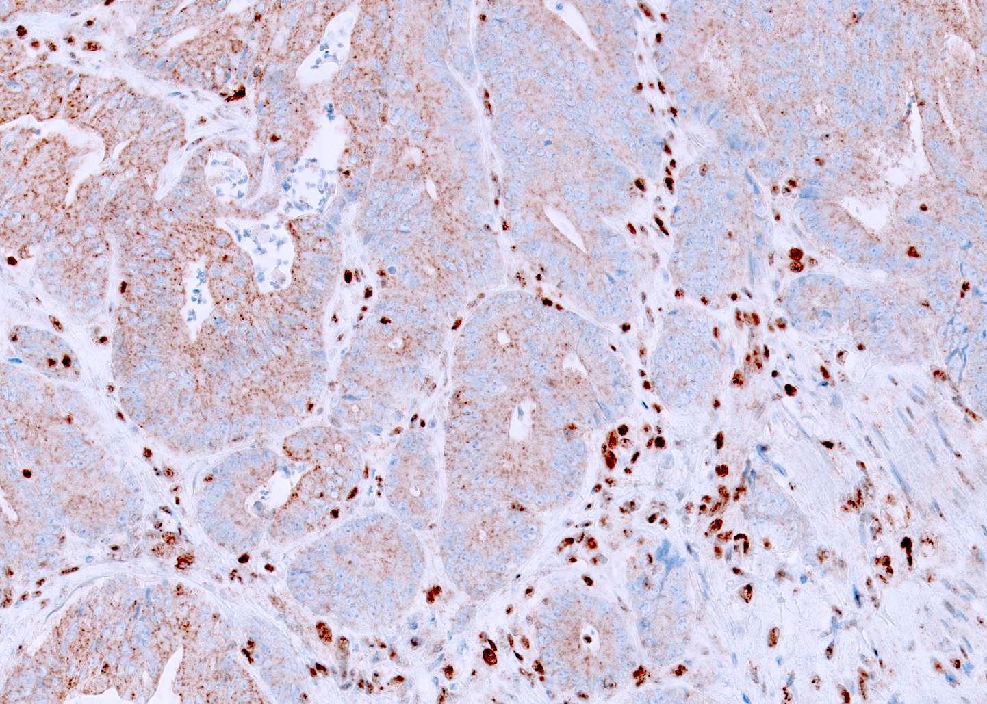
Contributed by Wei Chen, M.D., Ph.D.
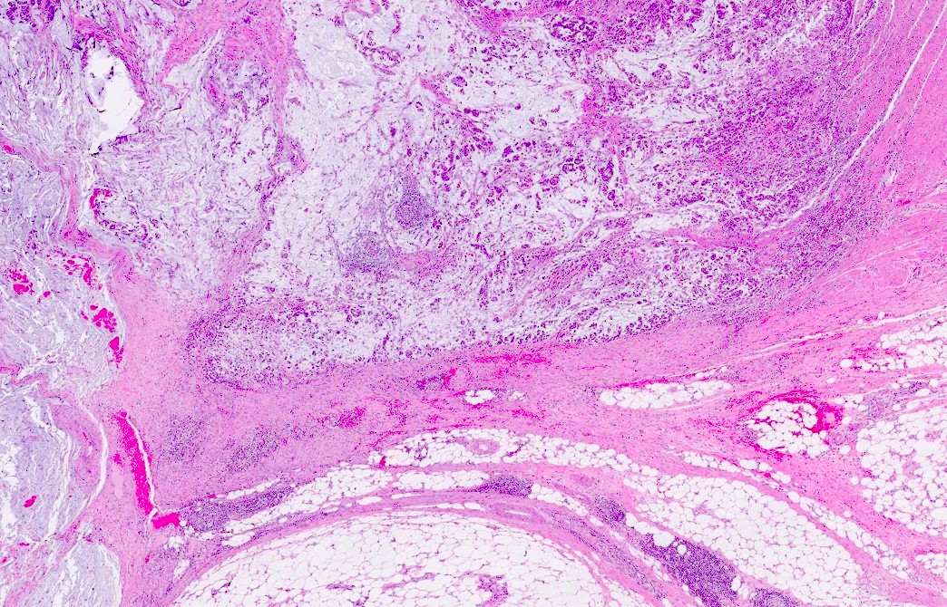
MSI CRC histomorphology
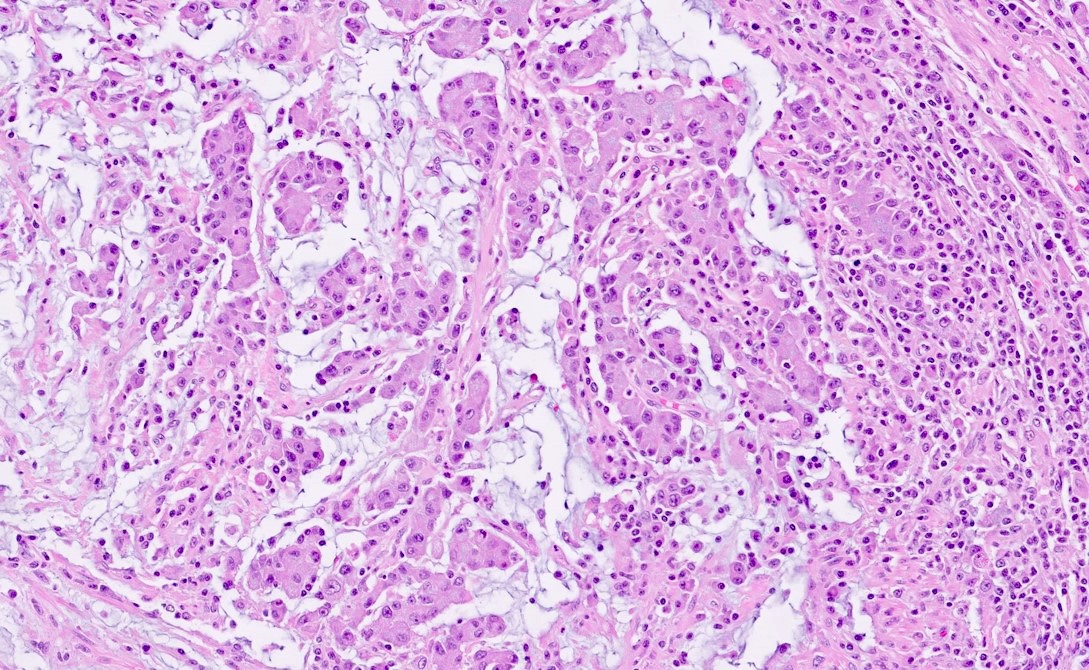
MSI CRC with TILs
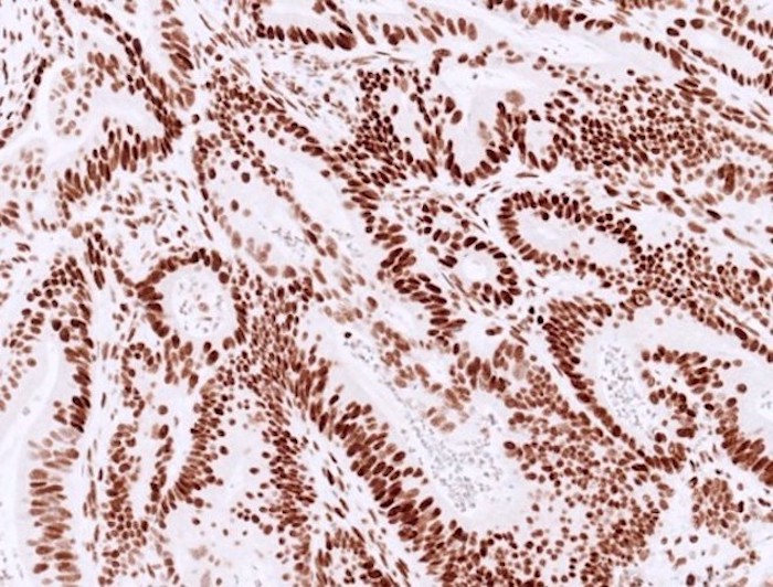
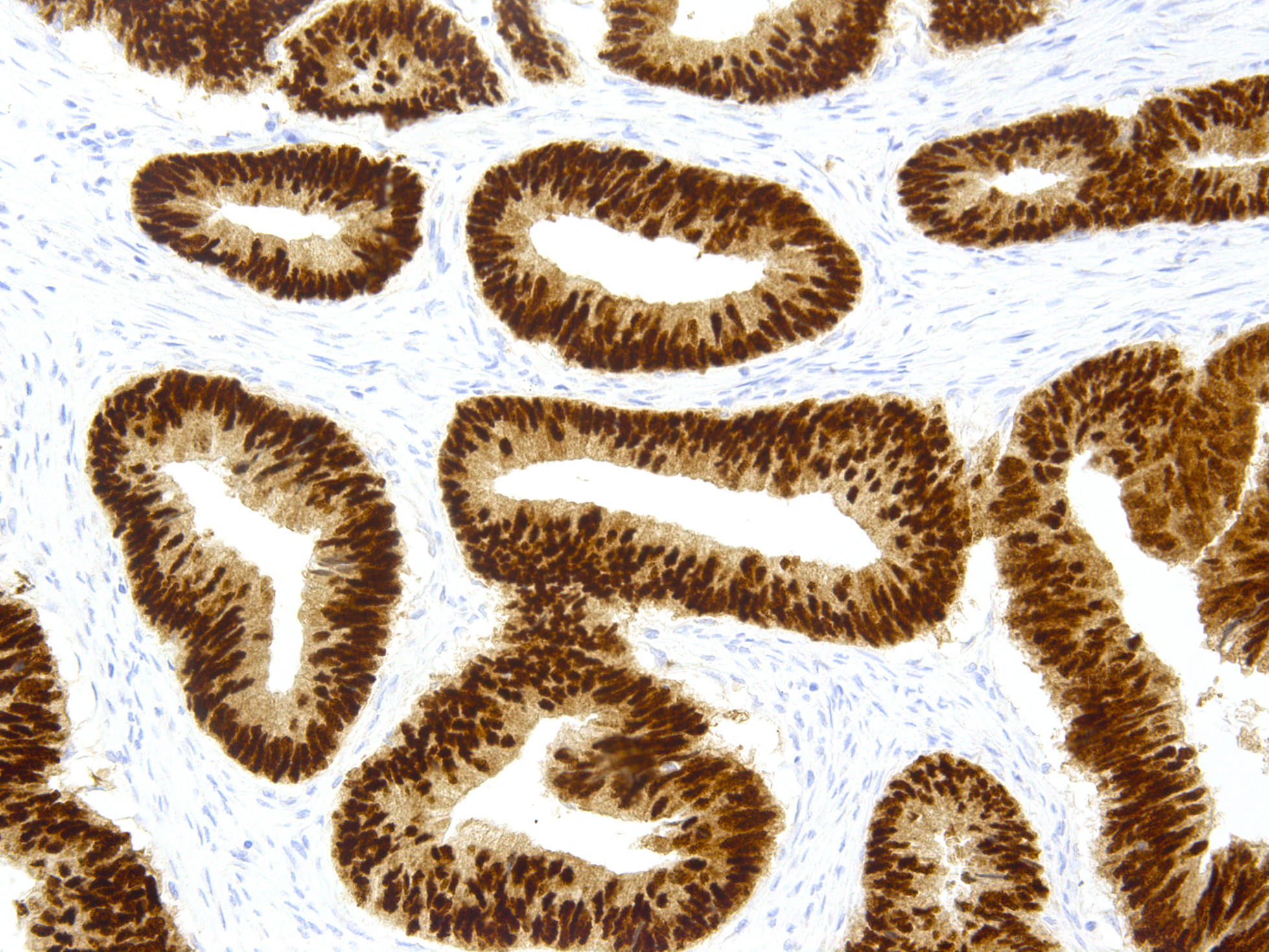
Normal staining (diffuse strong)
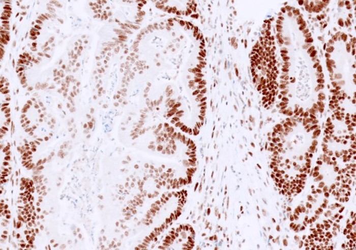
Normal staining (focal variability)
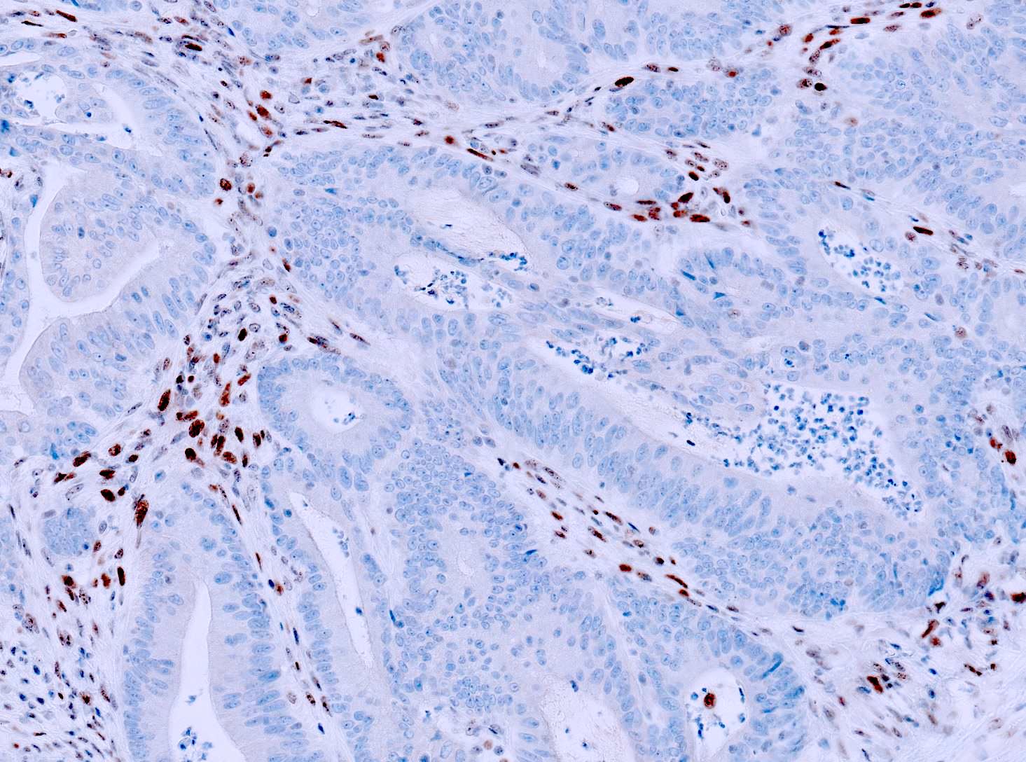
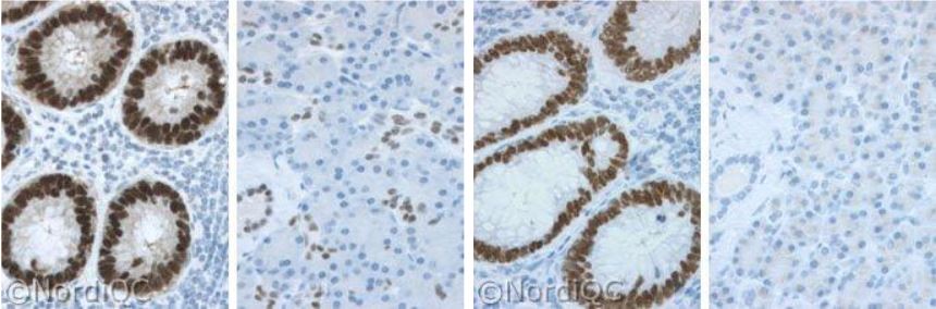
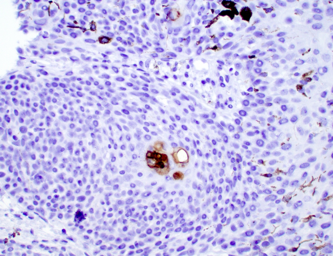
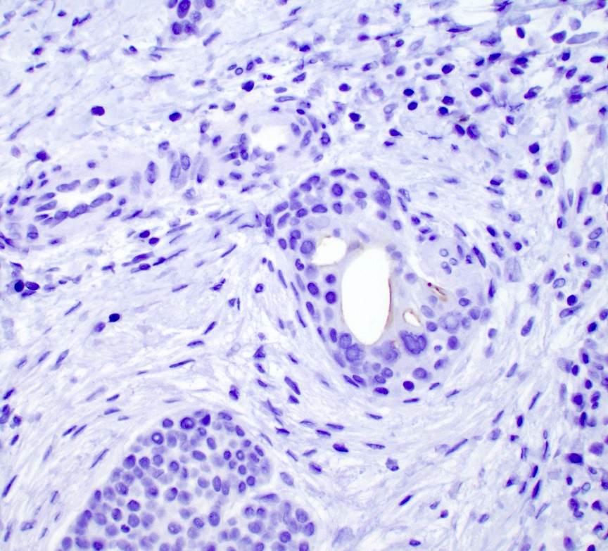
Absent / loss staining pattern
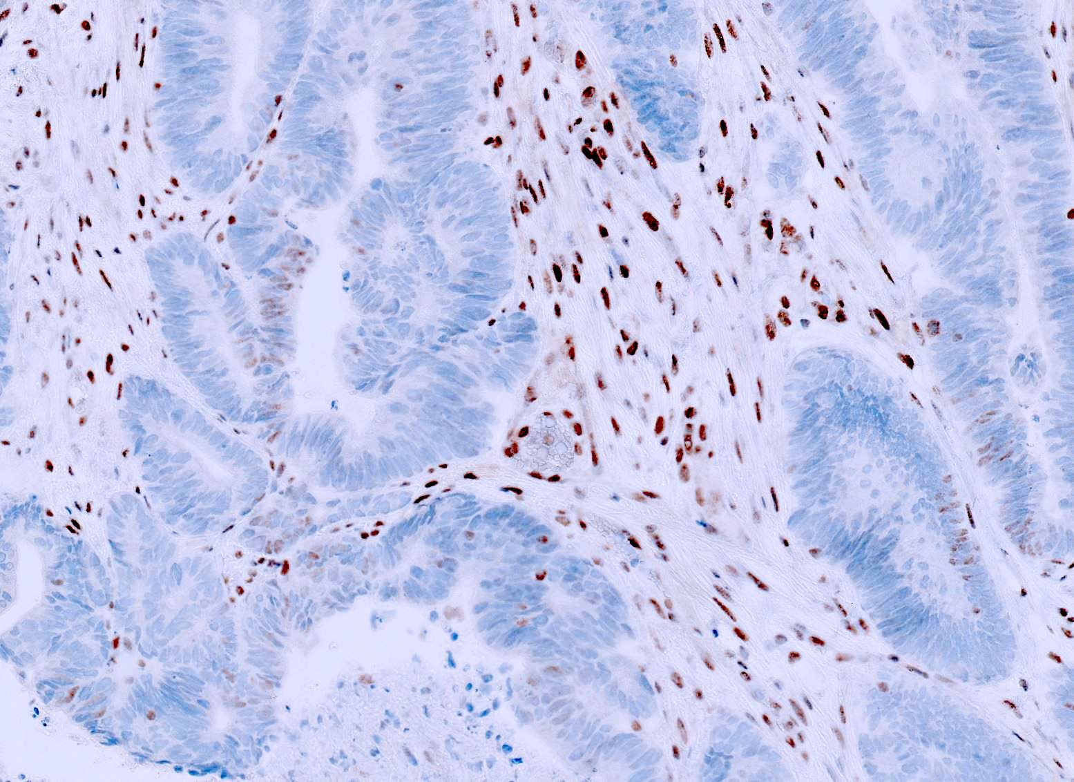
Tumor weaker than control
Cytoplasmic only staining
Contributed by Shrihari S. Kadkol, M.D., Ph.D.
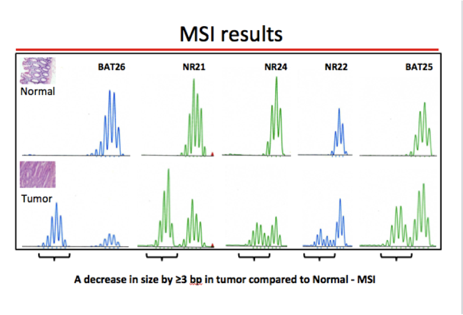
MSI electropherogram
Contributed by Christian M. Schürch, M.D., Ph.D. and Kenneth A. Iczkowski, M.D.
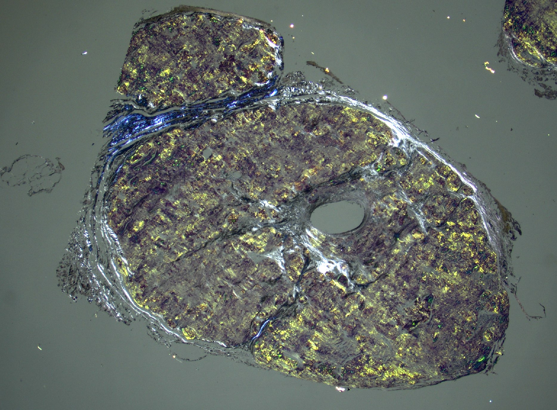

Amyloidosis
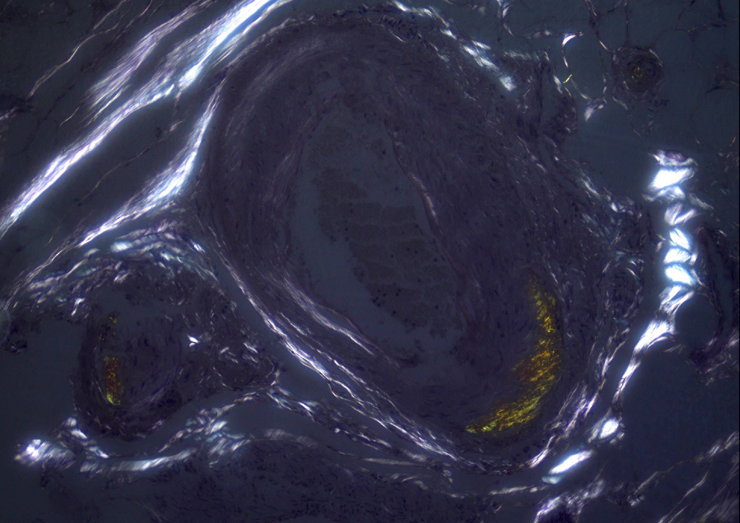

Transthyretin amyloidosis
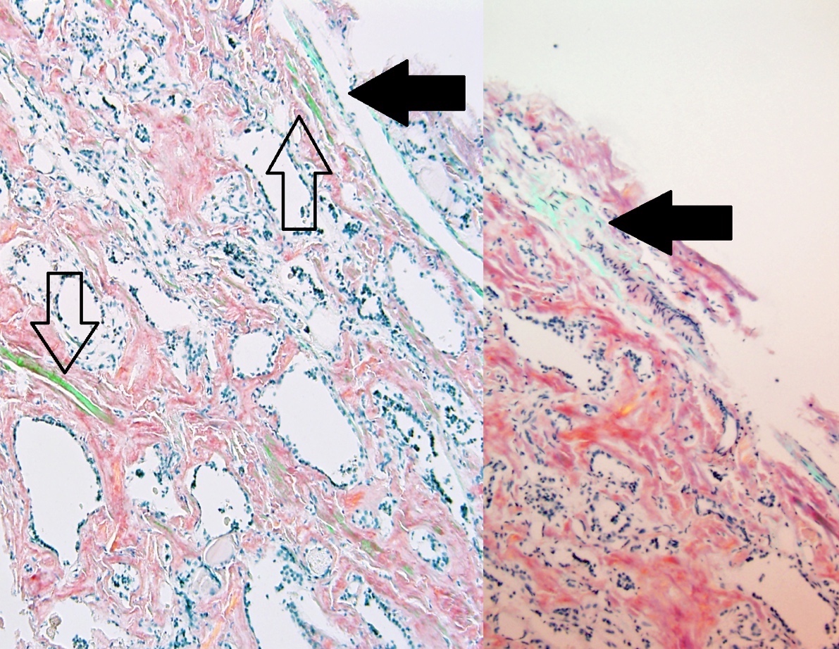
Distinguishing the correct color
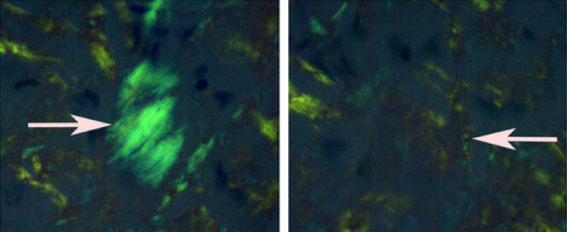
Rotation to visualize Congo red

Optimized AL amyloid imaging
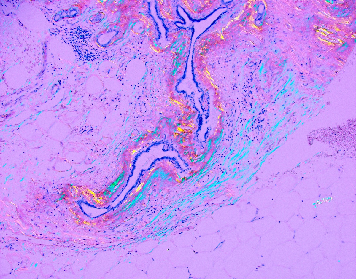
Breast amyloidosis
Case #388
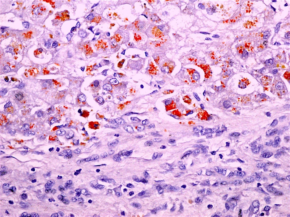
Copper stain in liver, Wilson's disease
Images hosted on other servers:

Wilson's disease
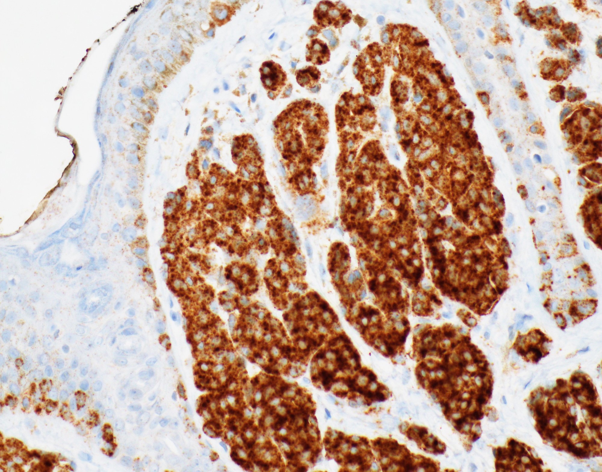
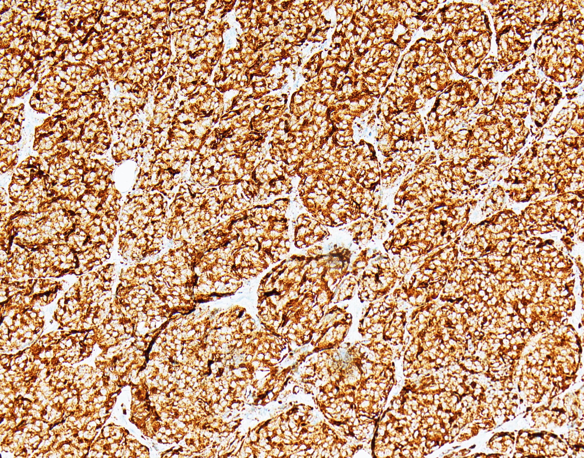
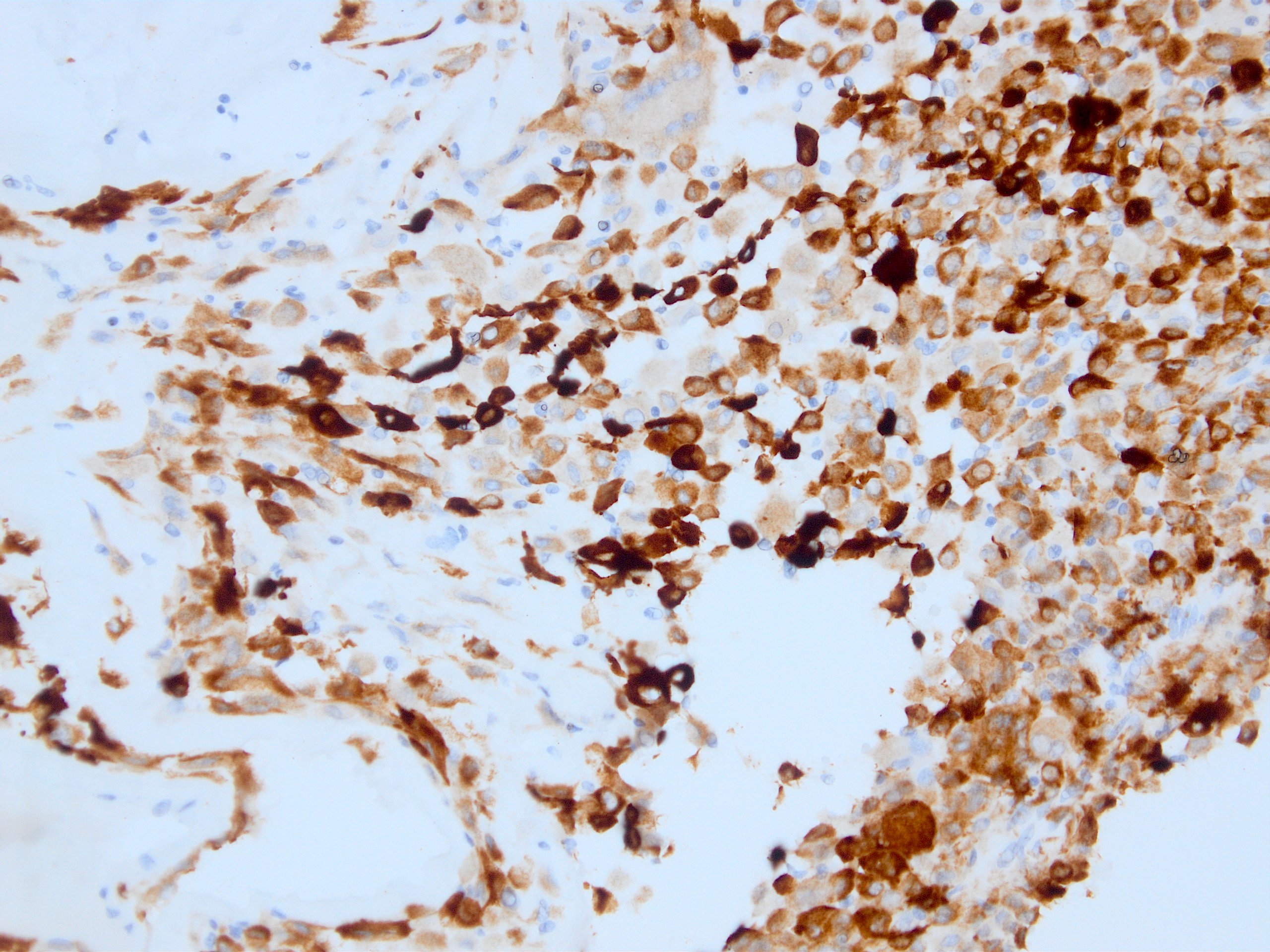
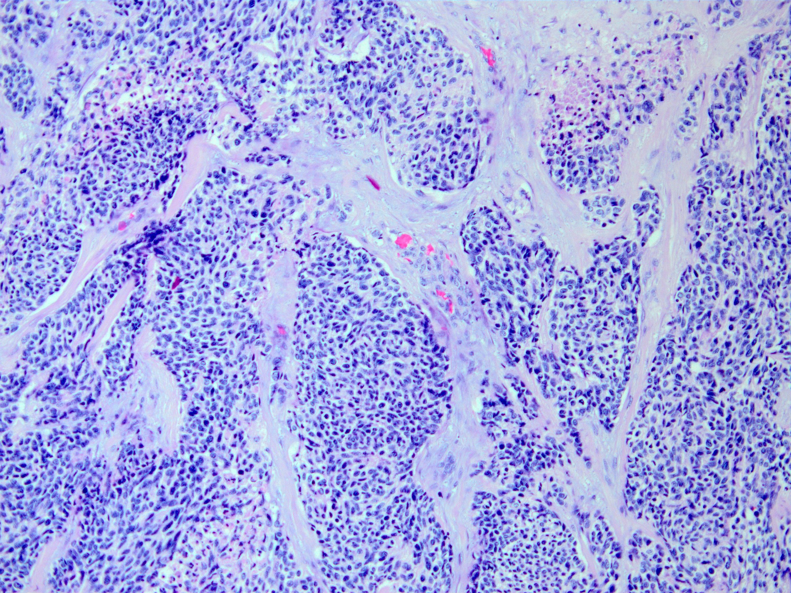
Contributed by Nasir Ud Din, M.B.B.S.
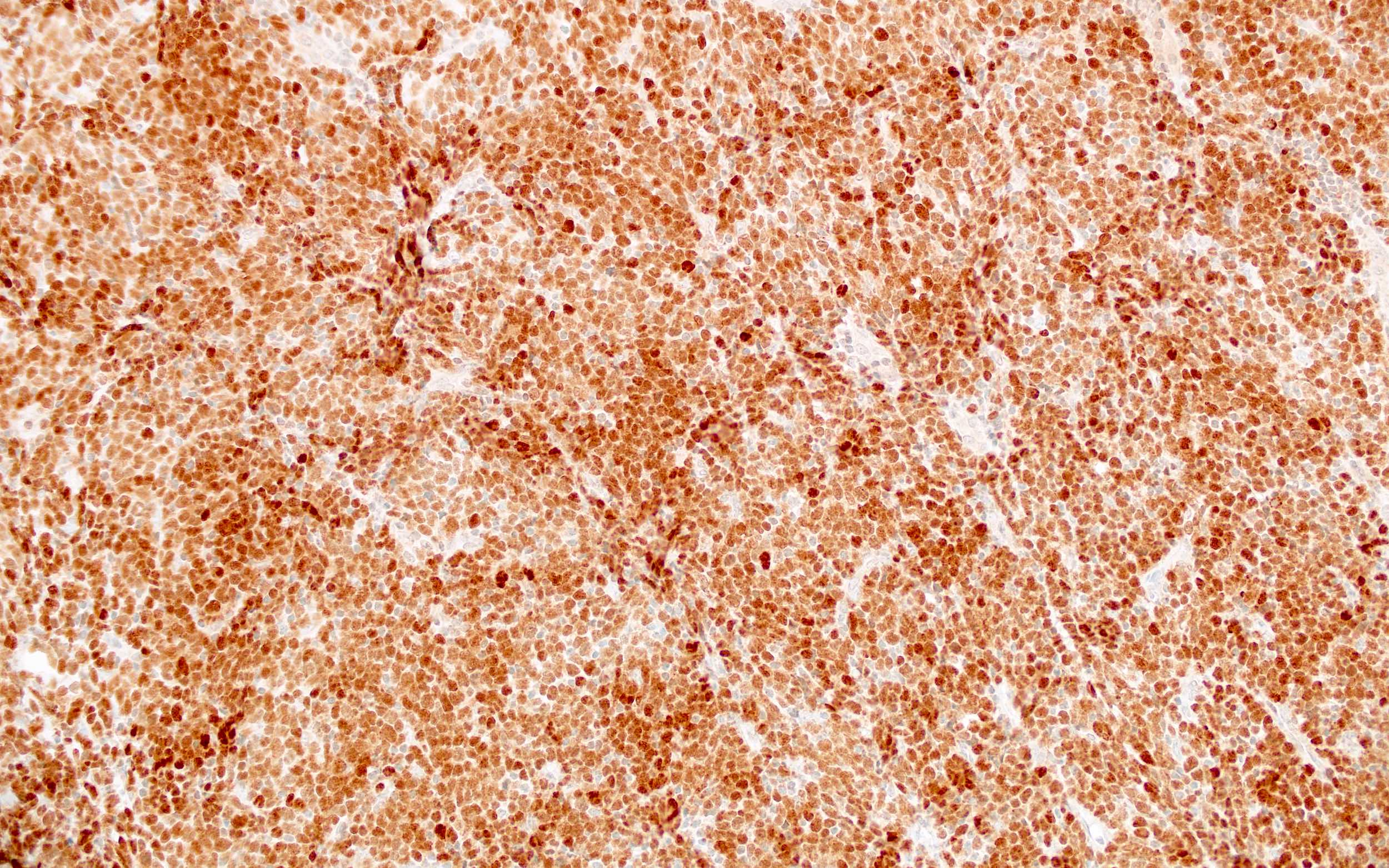
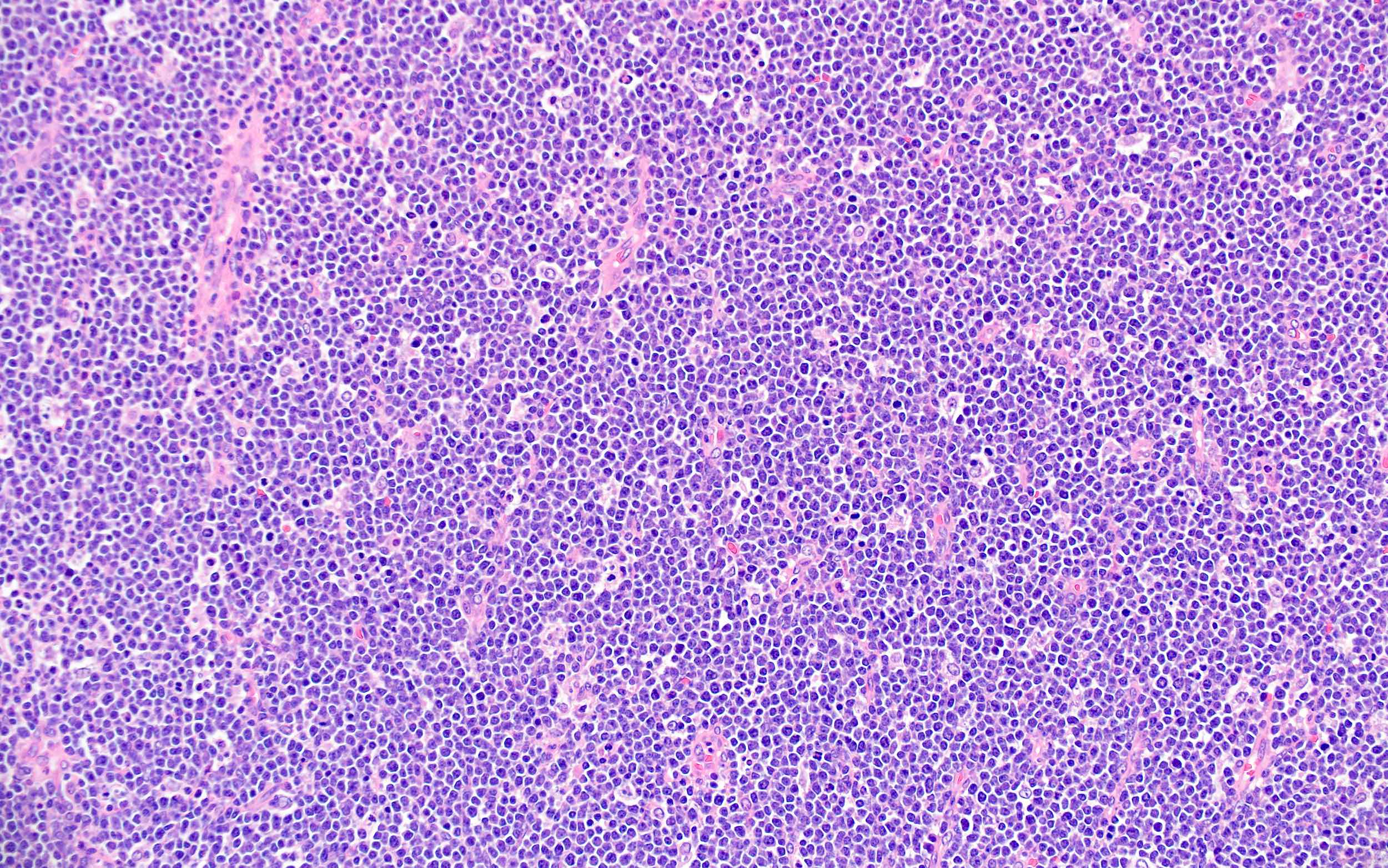
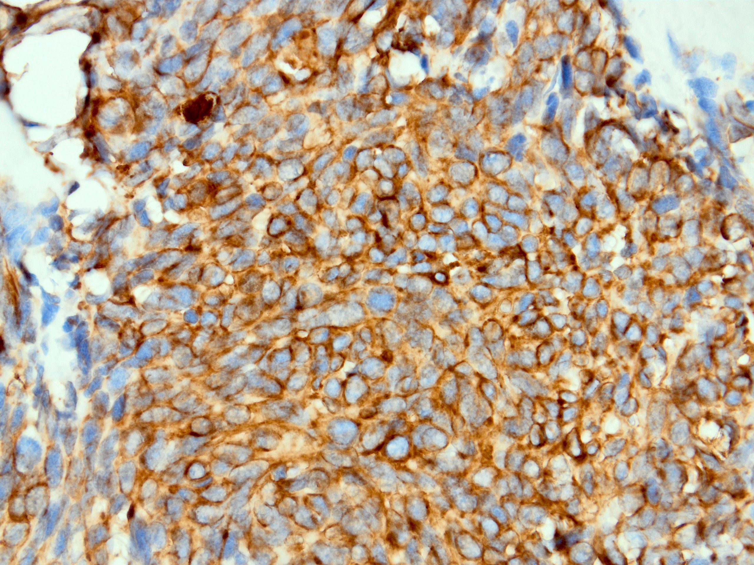
Ewing sarcoma
Ewing sarcoma cyclin D1
Endometrial stromal sarcoma
Endometrial stromal sarcoma cyclin D1
Wilms tumor
Wilms tumor cyclin D1
Mantle cell lymphoma
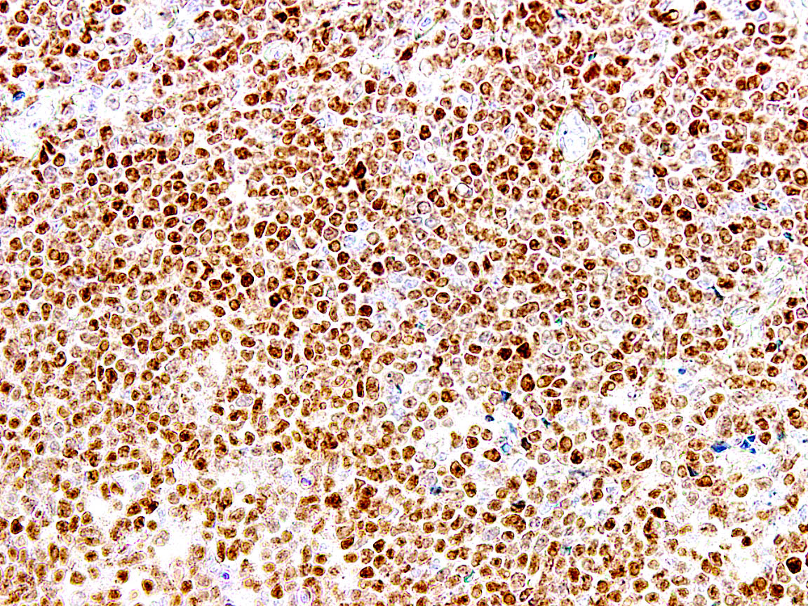
Mantle cell lymphoma cyclin D1
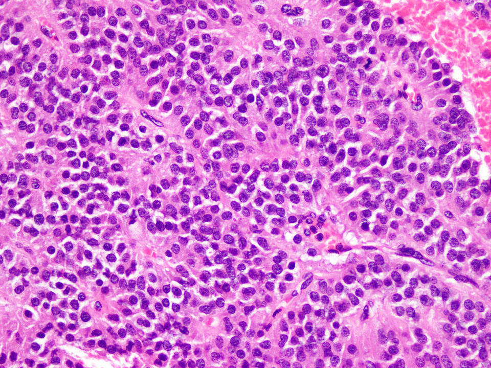
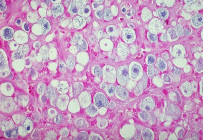
Solid pseudopapillary neoplasm
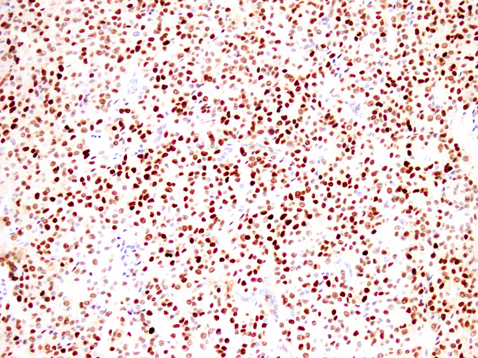
Solid pseudopapillary neoplasm cyclin D1
Images hosted on other servers:

Synthesis and assembly of COX subunits

Maturation and insertion of COX into the respiratory chain
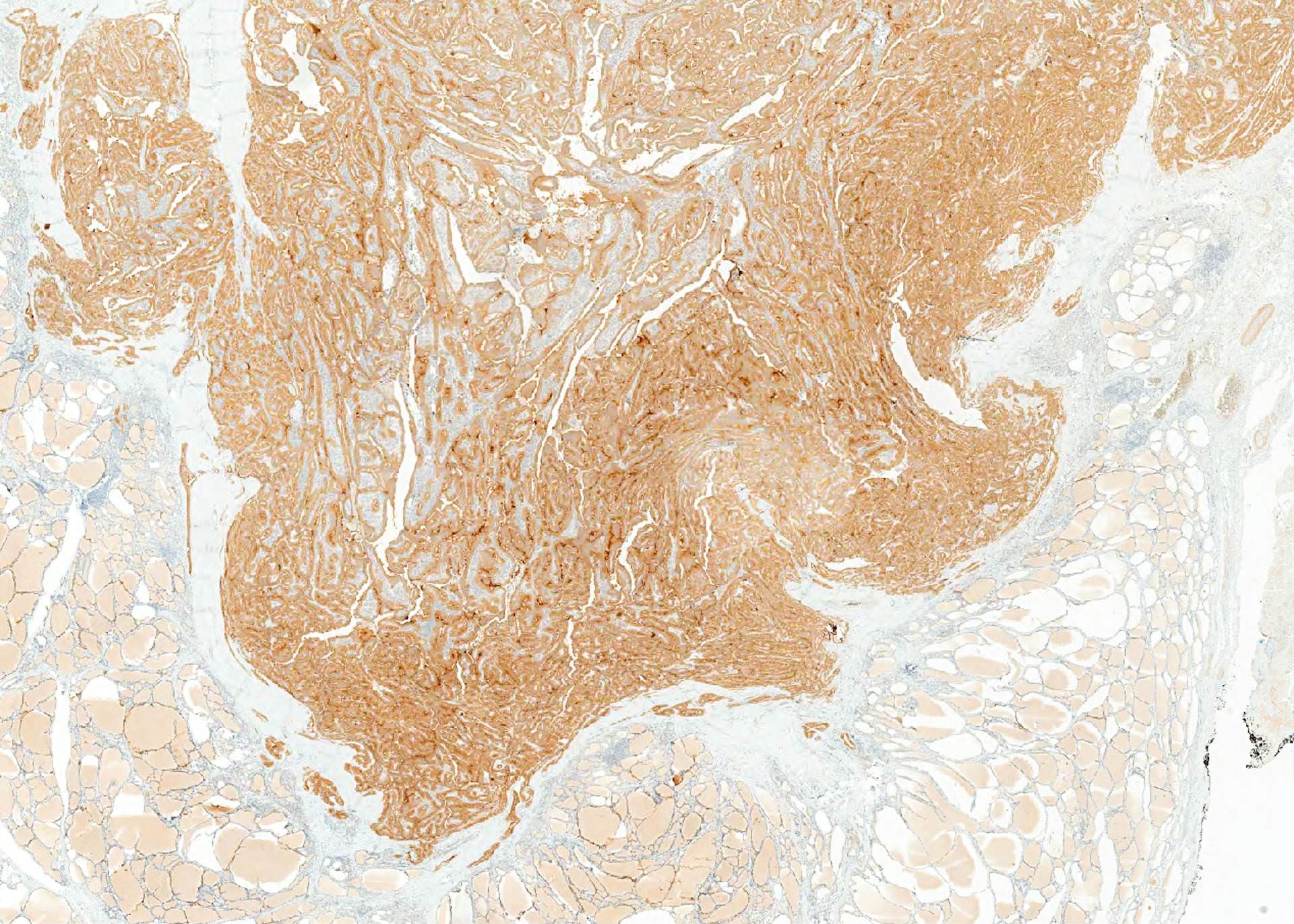
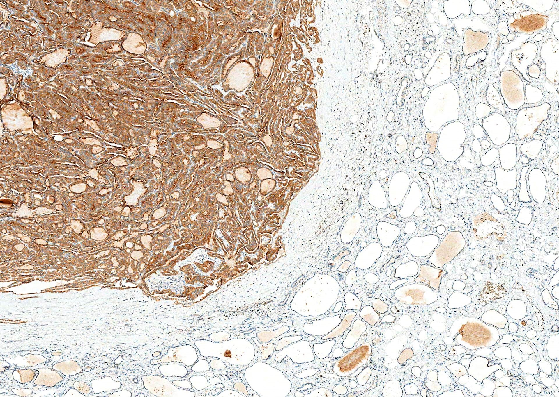
Contributed by Andrey Bychkov, M.D., Ph.D.
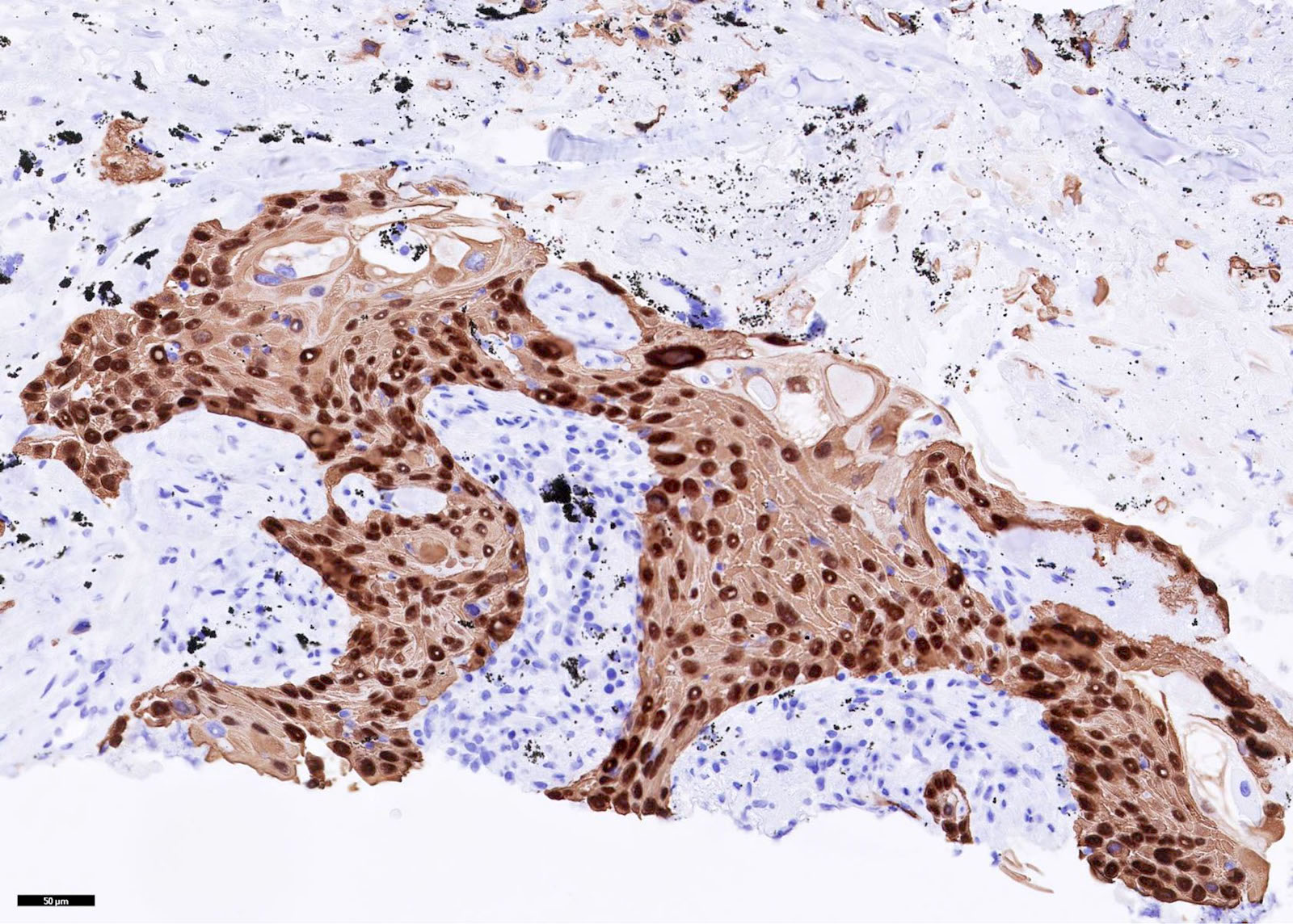
Lung SCC
Images hosted on other servers:

Breast carcinoma #1 is CK14+ (fig C)

Squamous cell carcinoma #1 oral (fig e/f)

Squamoid areas are CK14+ in urothelial carcinoma
Images hosted on other servers:

Breast myoepithelial hyperplasia (fig 1j)
Contributed by Leica Microsystems (Biosystems Division)
Colon (normal)
Images hosted on other servers:
Choroid plexus papilloma
Contributed by Andrey Bychkov, M.D., Ph.D.
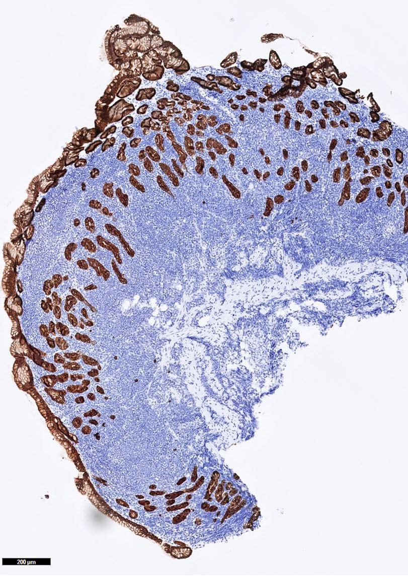
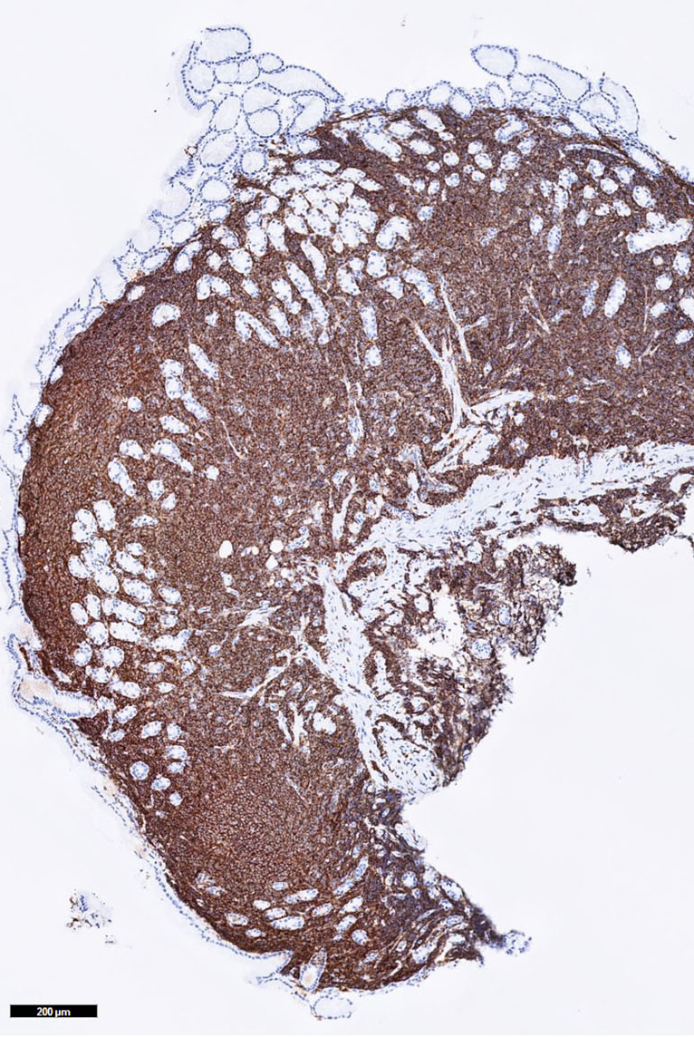
CK19 expression in PTC

CK19: prominent membranous and cytoplasmic staining
Images hosted on other servers:

Squamous cell carcinoma, oral (fig B)

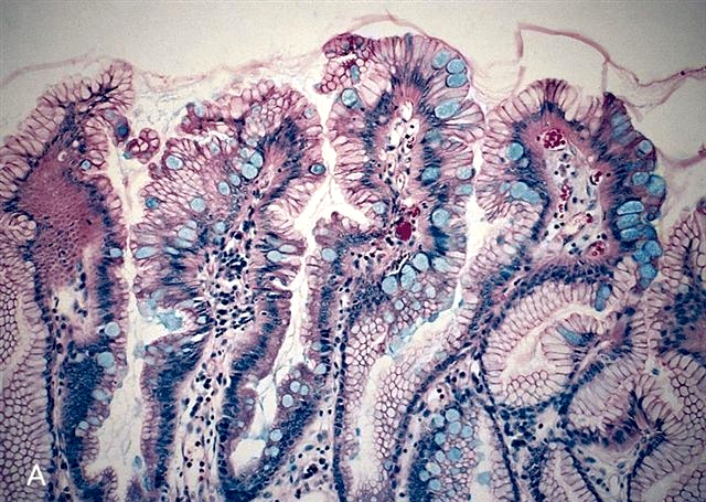
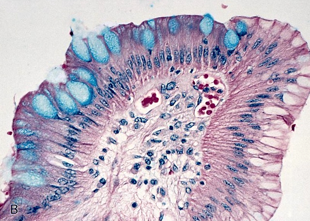
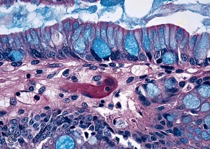
Stomach: complete intestinal metaplasia
Contributed by Leica Microsystems (Biosystems Division) and Andrey Bychkov, M.D., Ph.D.
Colon (normal):
CK20 (PW31) with
intense cytoplasmic
staining
Ovarian cancer

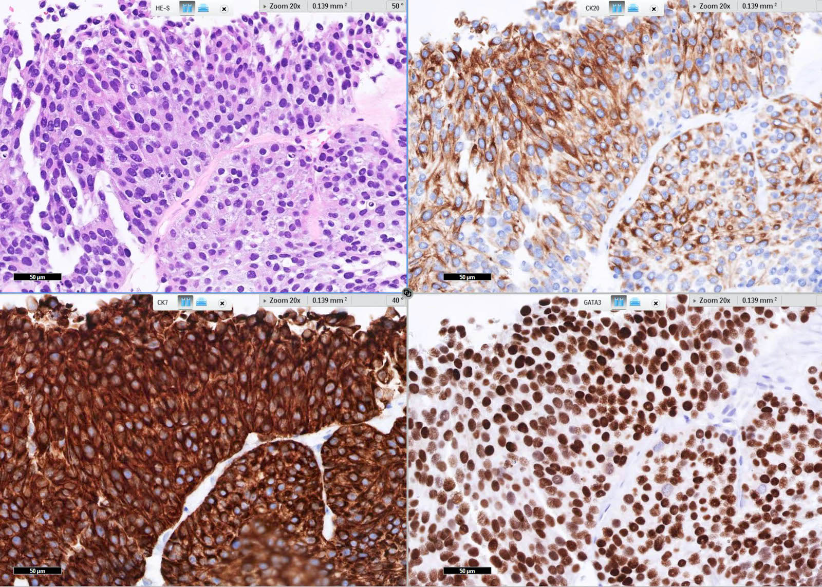
Urothelial carcinoma
Images hosted on other servers:


Colonic adenocarcinoma: #1-well differentiated; #2-poorly differentiated; #3-metastatic; #4-primary (fig G) and metastatic to lung (fig H)

PanIN lesions: CK20+ in grades 1, 2 and 3


Prostatic adenocarcinoma; #2 with positive staining of verumontanum

Tubular adenocarcinoma

Urothelial carcinoma, high grade
Contributed by Stephen J. Schultenover, M.D.
Thyroid anaplastic carcinoma
Images hosted on other servers:

Low grade bladder neoplasms
Contributed by Jijgee Munkhdelger, M.D., Ph.D., Andrey Bychkov, M.D., Ph.D. and Semir Vranić, M.D., Ph.D.

Breast tubular adenoma immunoprofile
Intraductal papilloma with DCIS immunoprofile
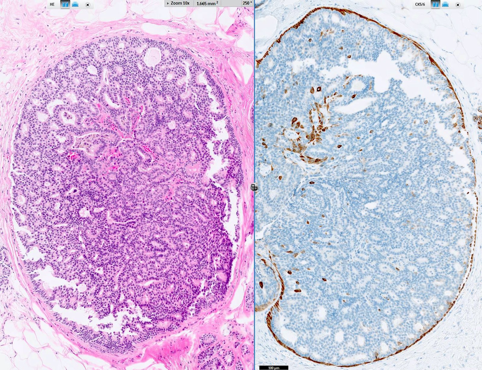
Intraductal papilloma with DCIS, CK5/6
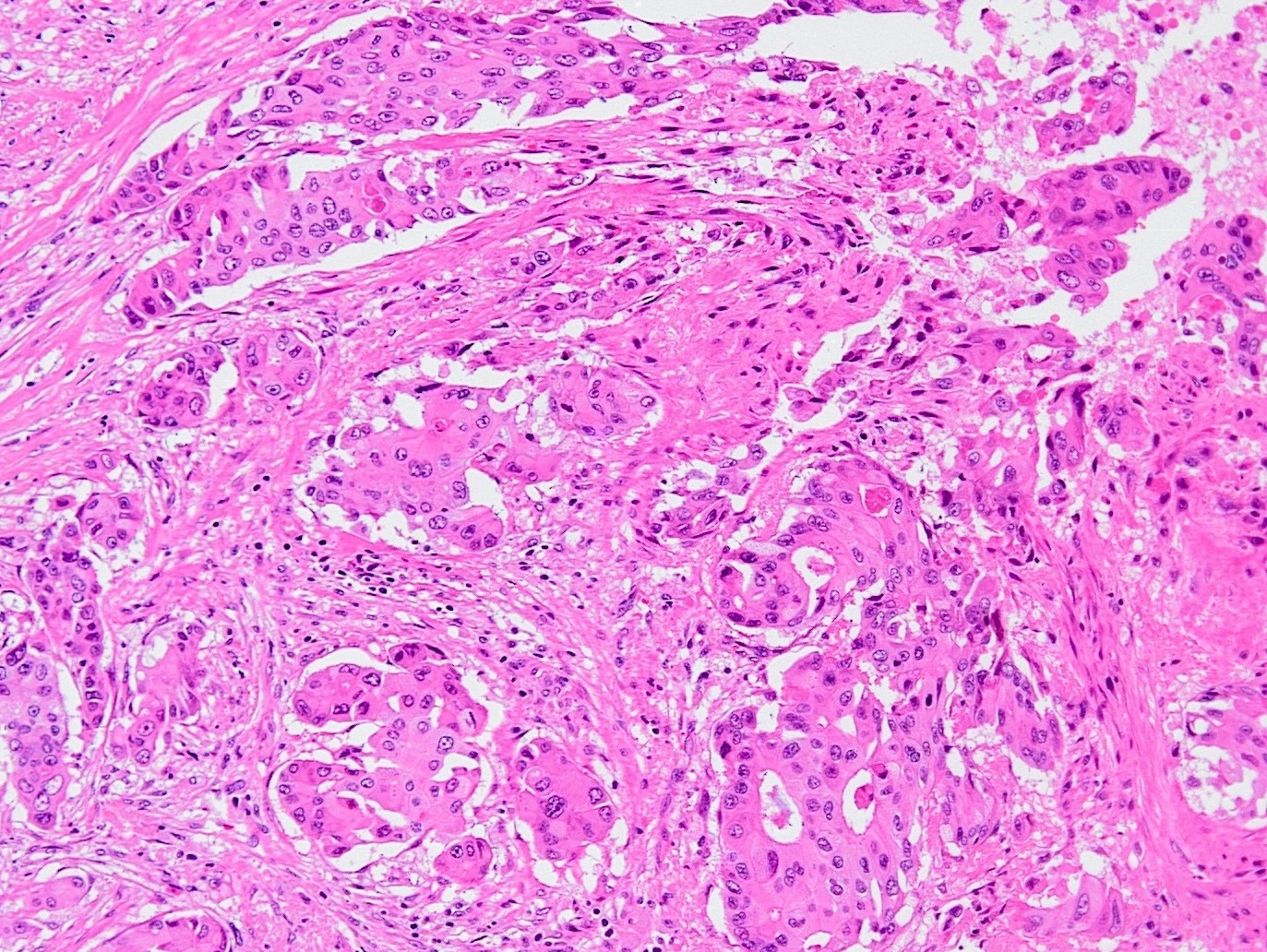
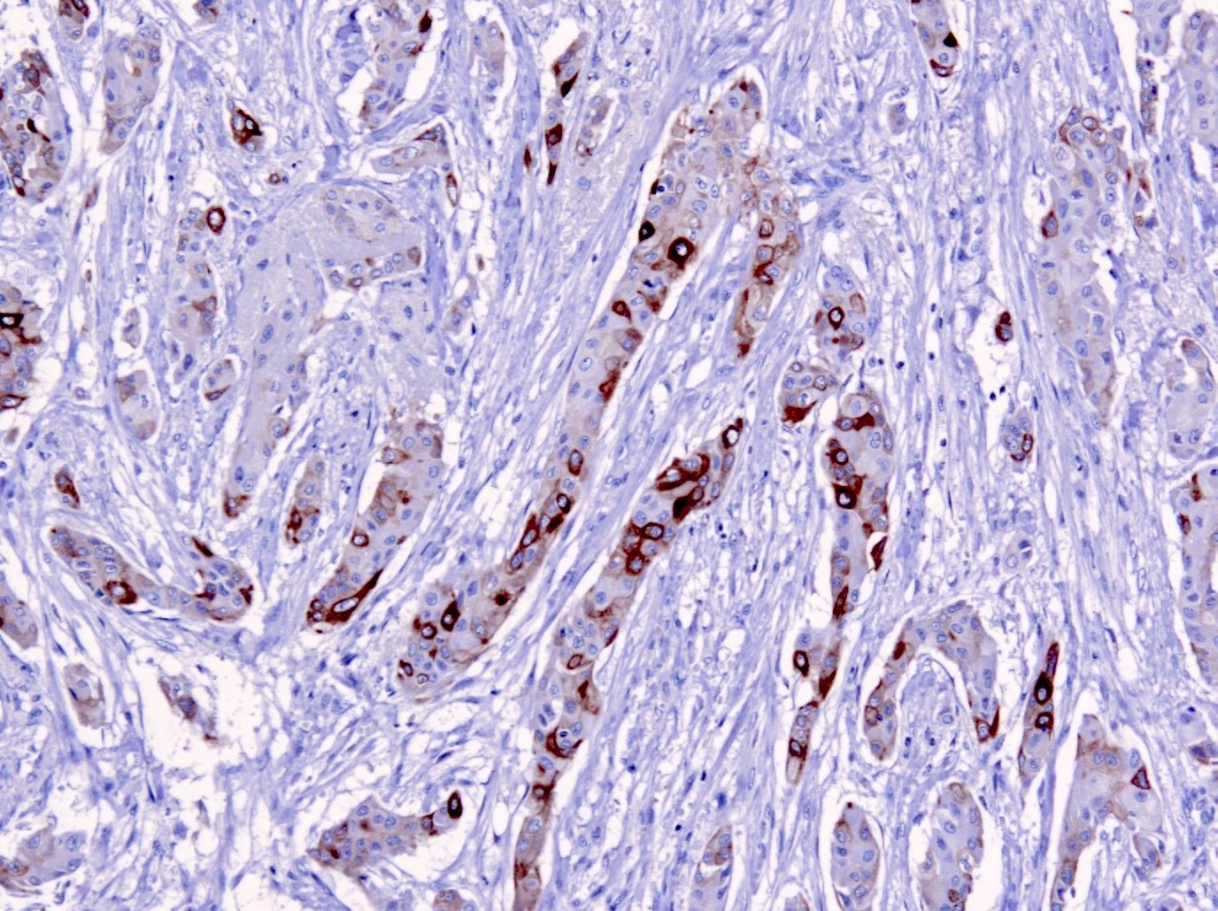
Basal-like triple negative breast cancer

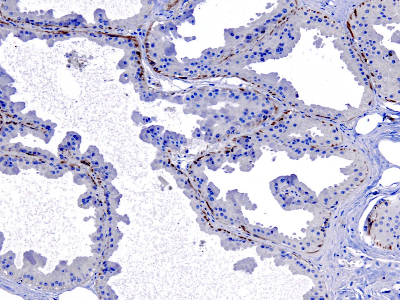
Apocrine metaplasia of the breast
Metastatic squamous cell carcinoma of the cervix
Apocrine carcinoma of the breast
Complex sclerosing lesion of the breast
Contributed by Kruti P. Maniar, M.D.
Cervical
squamocolumnar
junction
Cholangiocarcinoma
Colon

Liver portal tract
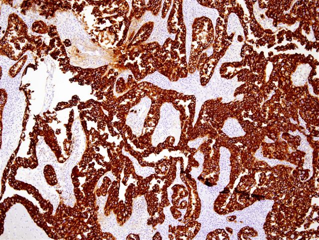
Lung adenocarcinoma
Ovarian seromucinous borderline tumor
Pancreas
Yolk sac tumor, glandular variant
Contributed by Andrey Bychkov, M.D., Ph.D., Eddie Fridman, M.D. (Case #194) and Leica Microsystems
Ovarian cancer
Urothelial carcinoma
Urethra: clear cell adenocarcinoma
Ureter (normal)
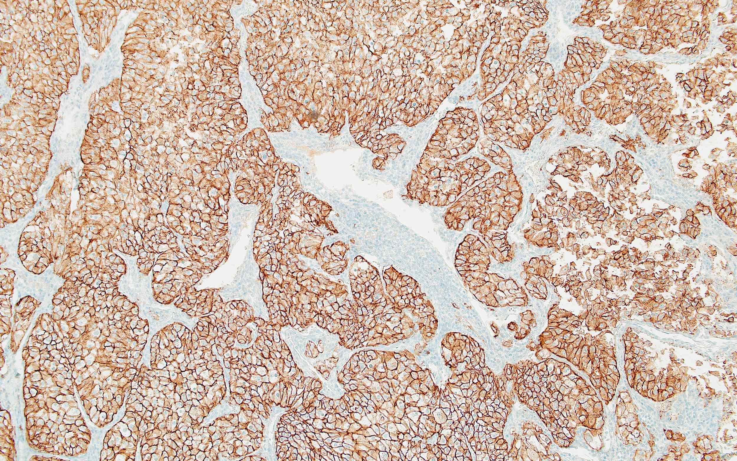
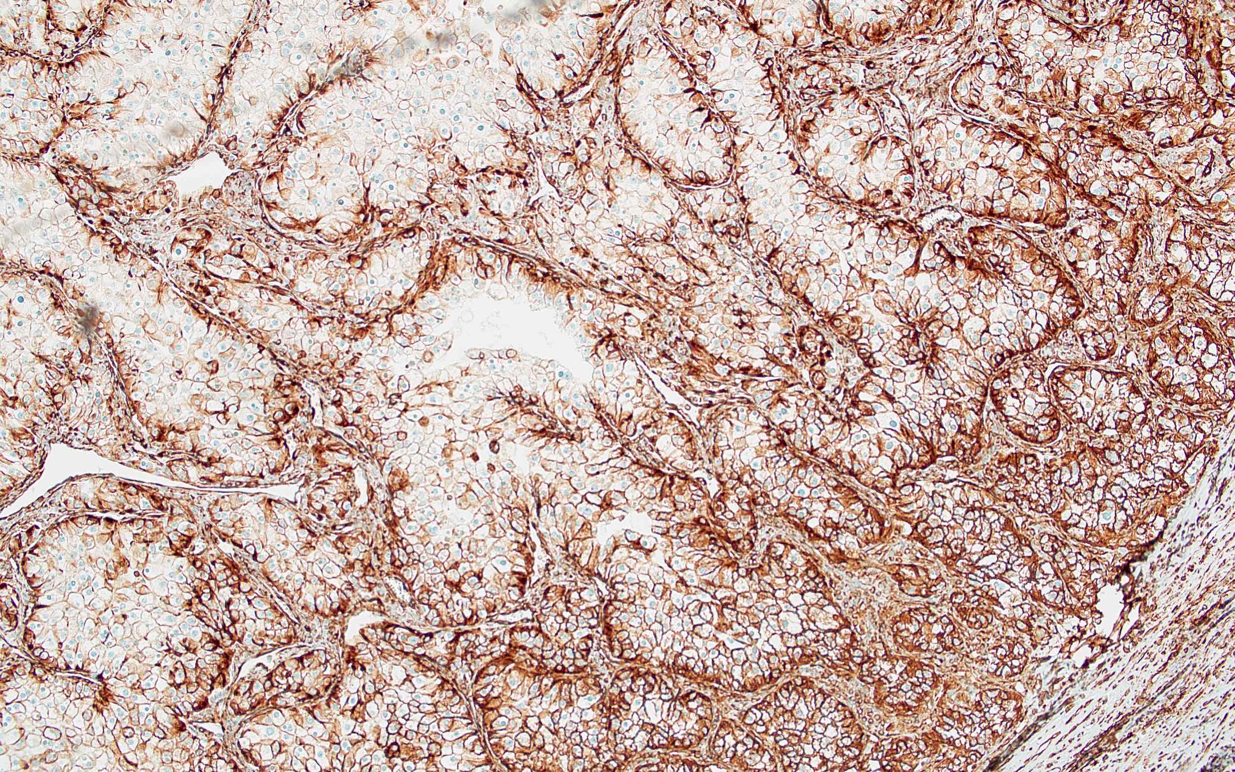
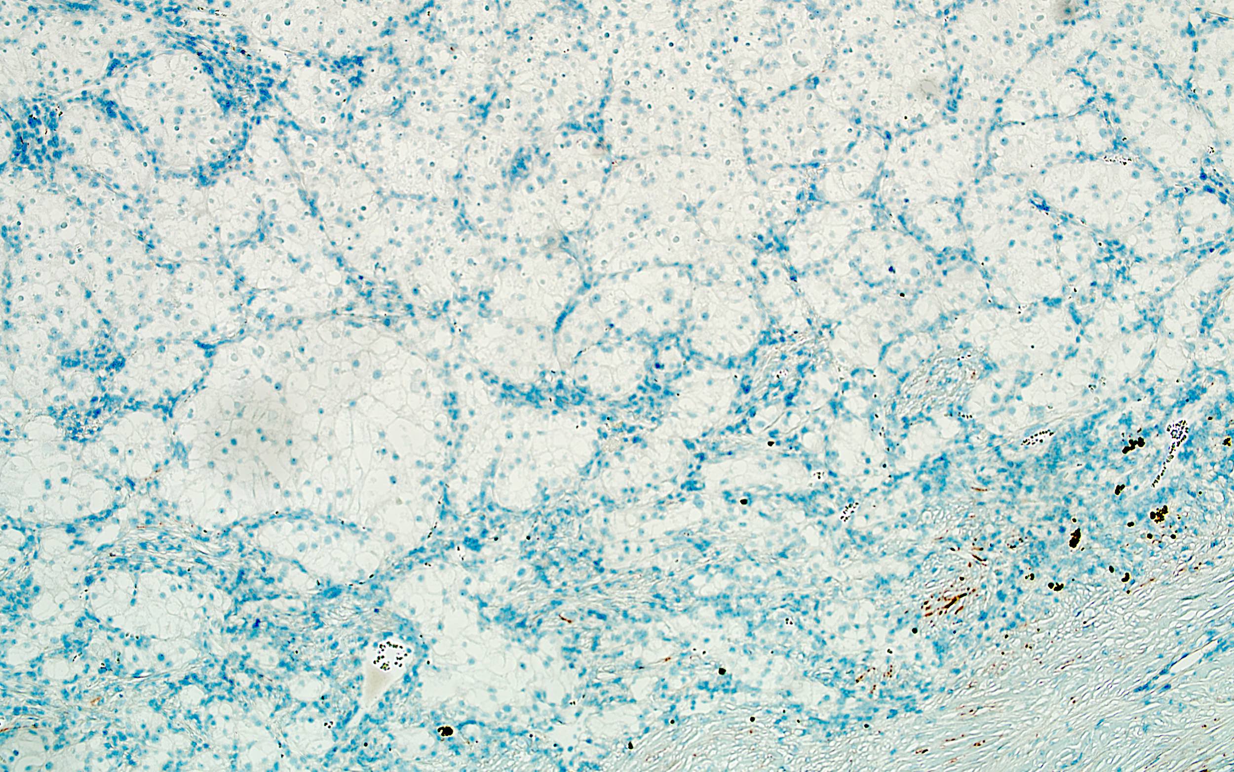
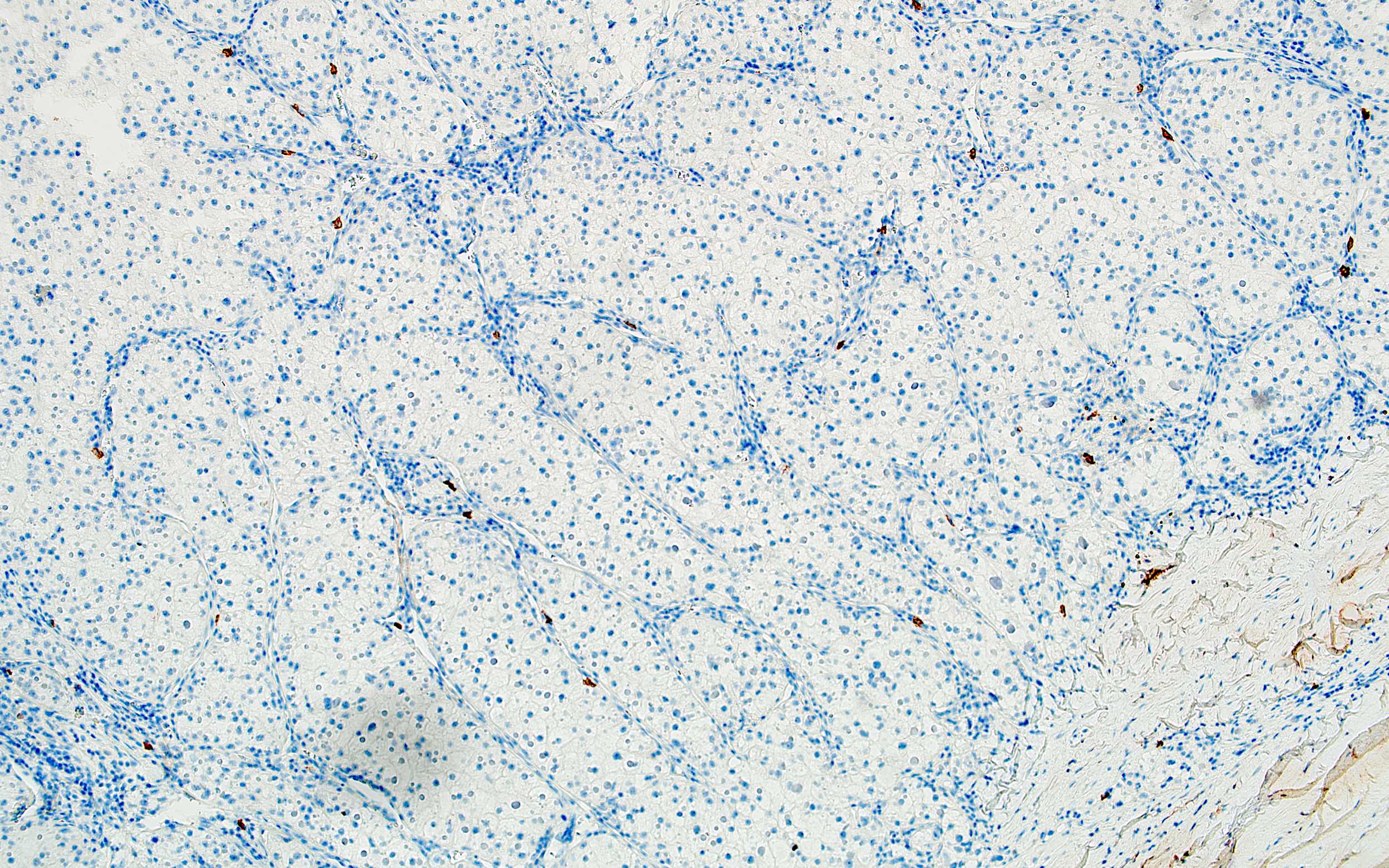
Contributed by Maria Tretiakova, M.D., Ph.D.
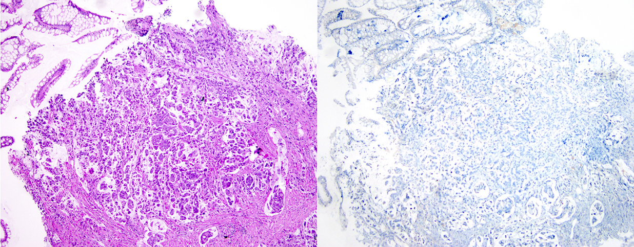
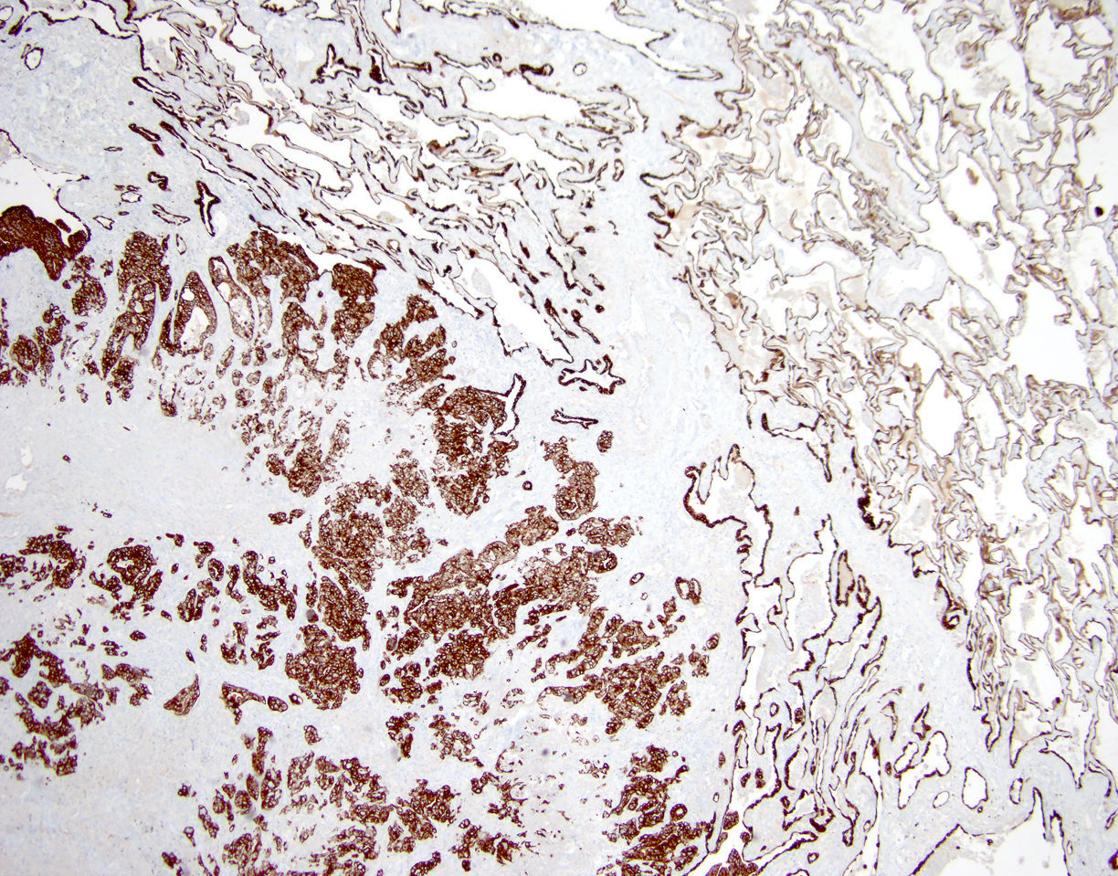
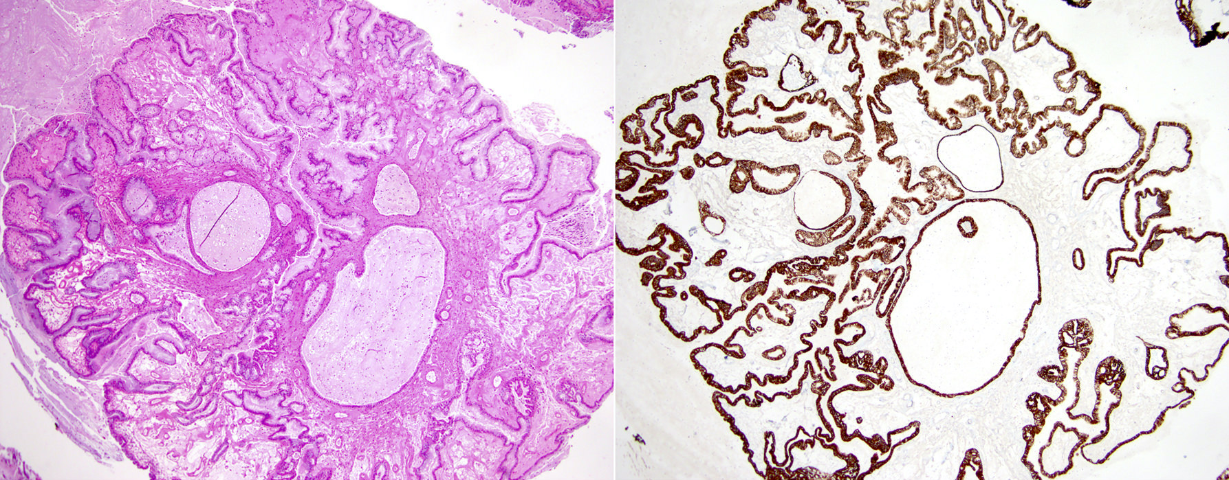
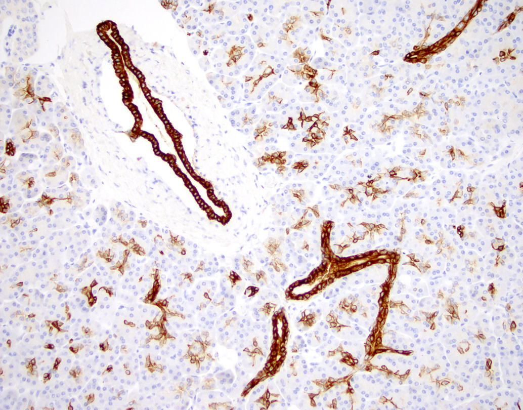
Normal kidney
Clear cell RCC
Clear cell papillary RCC
Papillary RCC, type1

Papillary RCC, type 2
Chromophobe RCC
Oncocytoma
Contributed by Leica Microsystems (Biosystems Division)
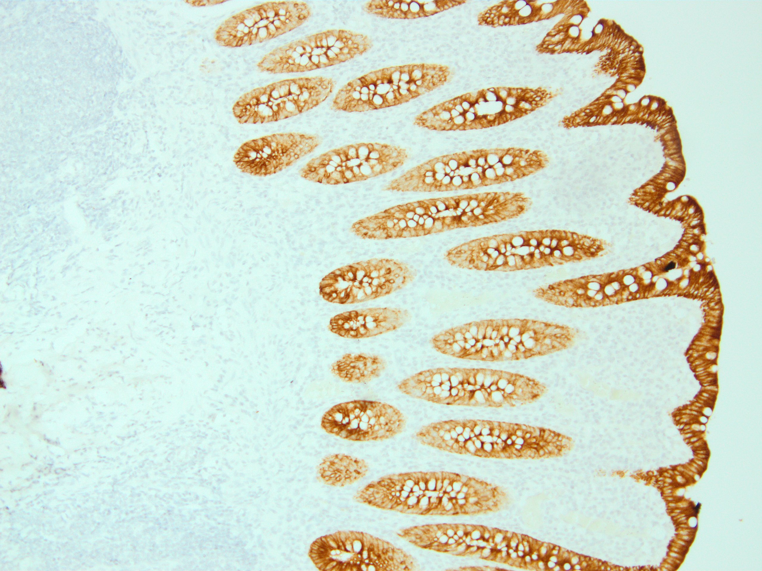
Colon (normal)
CK8 / 18 (5D3)
with intense cytoplasmic
and membranous staining
Images hosted on other servers:

Hepatocytes (residual)
are CK8+ in embryonal
sarcoma of liver

Liver disease (various)

Prostatic adenocarcinoma-top and benign prostate bottom

Squamous cell carcinoma-oral (fig C/D)
Contributed by Mieke R. Van Bockstal, M.D., Ph.D., Christine Galant, M.D., Ph.D. and Andrey Bychkov, M.D., Ph.D.
Serous carcinoma
Mammary spindle cell carcinoma
Mammary desmoid fibromatosis
Residual breast carcinoma
Ileal neuroendocrine tumor
Chordoma
Juvenile granulosa cell tumor
Sertoli-Leydig cell tumor
Uterine tumor resembling ovarian sex cord tumor (UTROSCT)
Normal liver
Normal lymph node
Gastric MALT lymphoma
Papillary thyroid cancer
Images hosted on other servers:

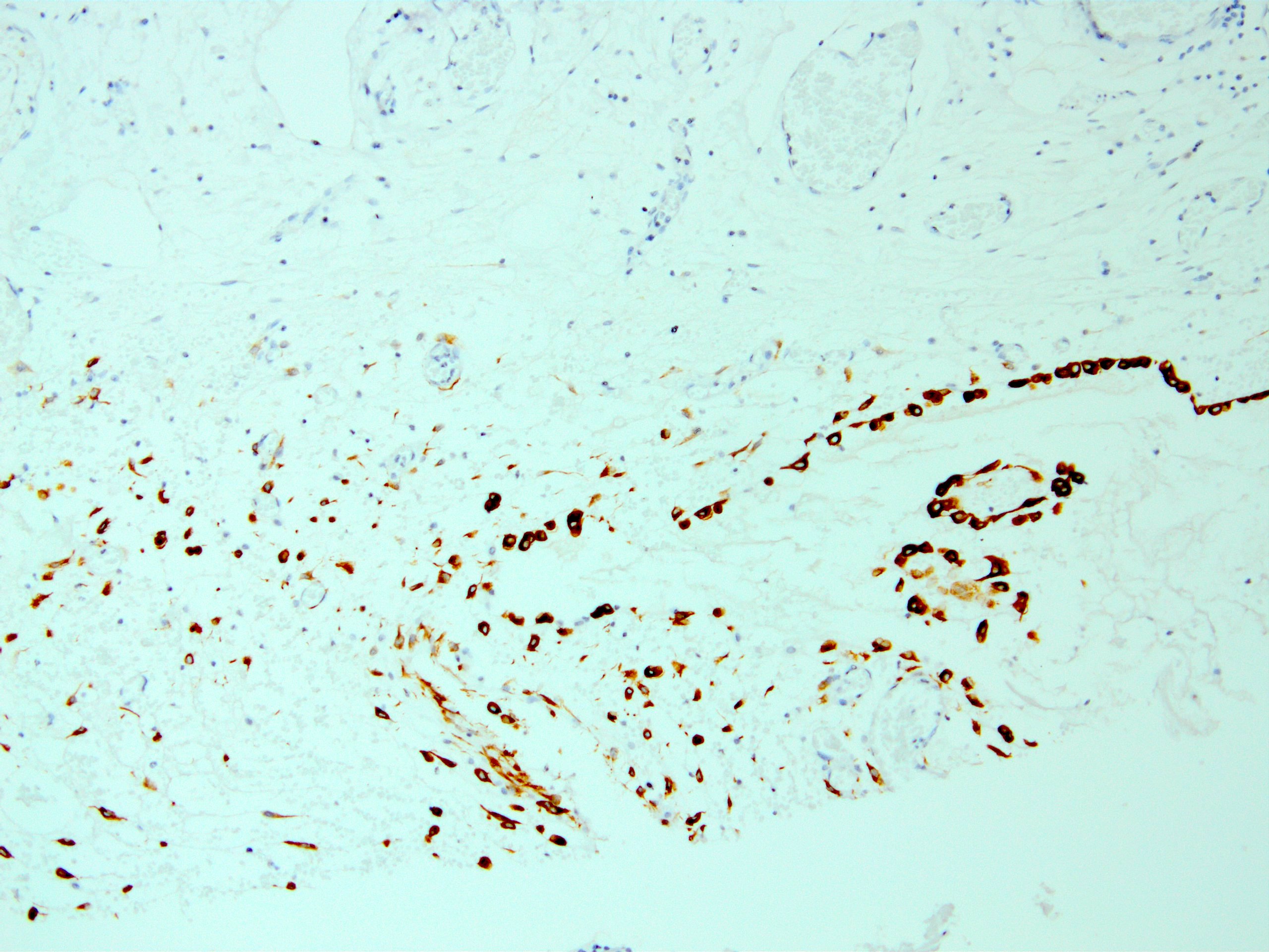
CK AE1 / AE3 highlights reactive mesothelial cells
Contributed by Kemal Kösemehmetoğlu, M.D.
Normal liver
Colon mucosa
Tonsil
Pancreas
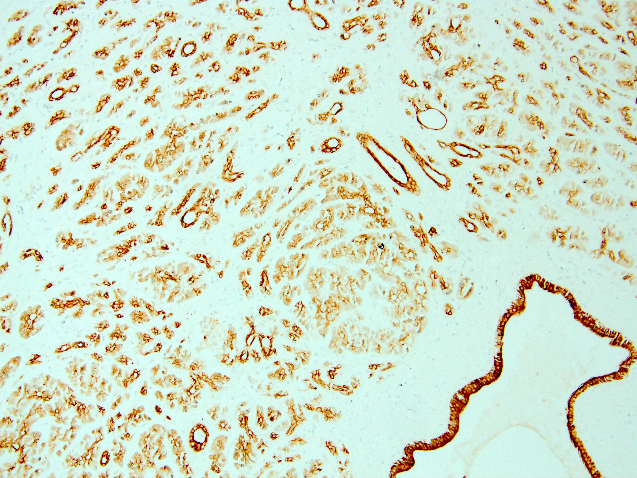
Peritoneum
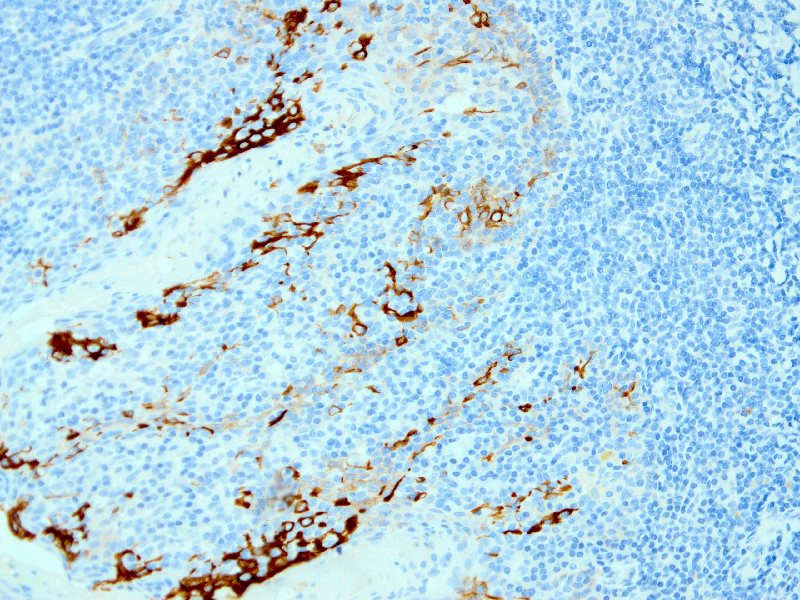
Dendritic cell staining in lymph node
Signet ring carcinoma of stomach
Signet ring carcinoma infiltrating omentum
Paget disease of nipple


Nipple duct adenoma
High grade neuroendocrine carcinoma
CAM 5.2 in Crooke cell adenoma

CAM 5.2 in sparsely granulated somatotroph adenoma
CAM 5.2 in sarcomatoid carcinoma
Inflammatory myofibroblastic tumor of the bladder

CAM 5.2 in epithelioid sarcoma (proximal variant)
CAM 5.2 in desmoplastic small round cell tumor
Case #78
Cardiac myxoma with glandular differentiation
Images hosted on other servers:

Anaplastic thyroid carcinoma is MNF116+ (fig A)
