

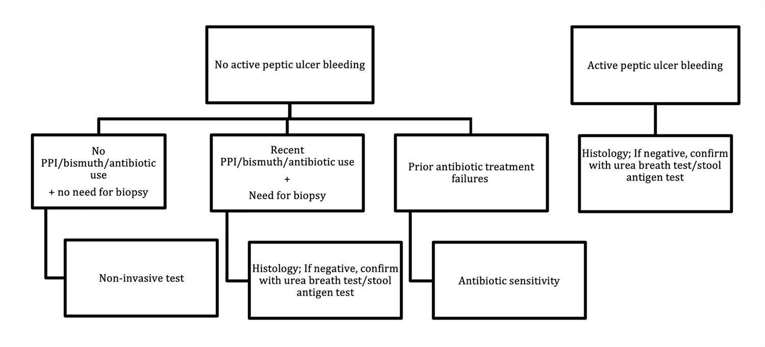
Images hosted on other servers:

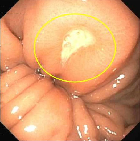
Gastric ulcer
Images hosted on other servers:


Various images
Images hosted on other servers:

CT scan of the stomach
Images hosted on other servers:

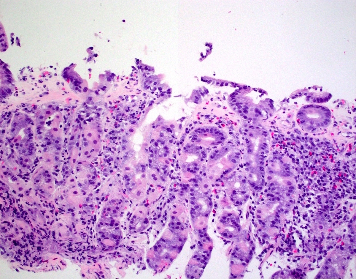
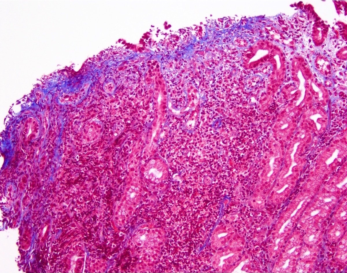
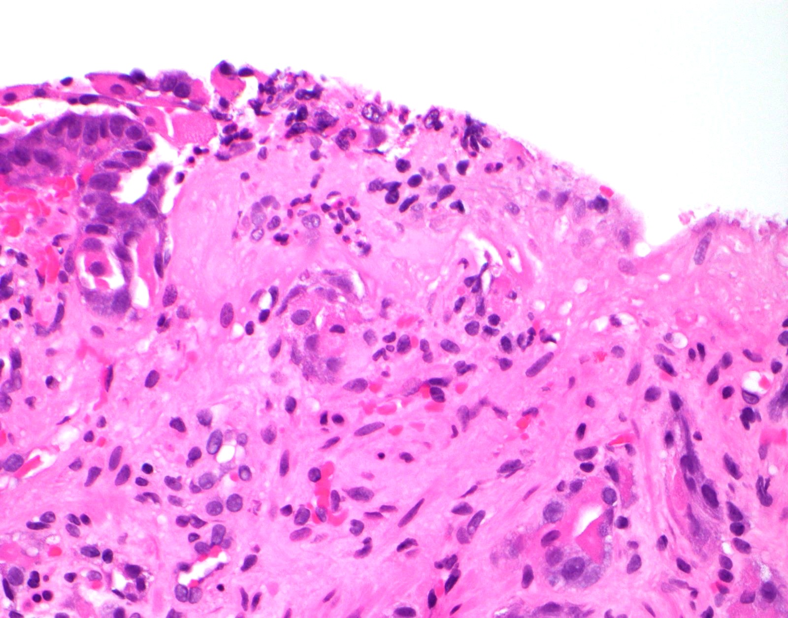
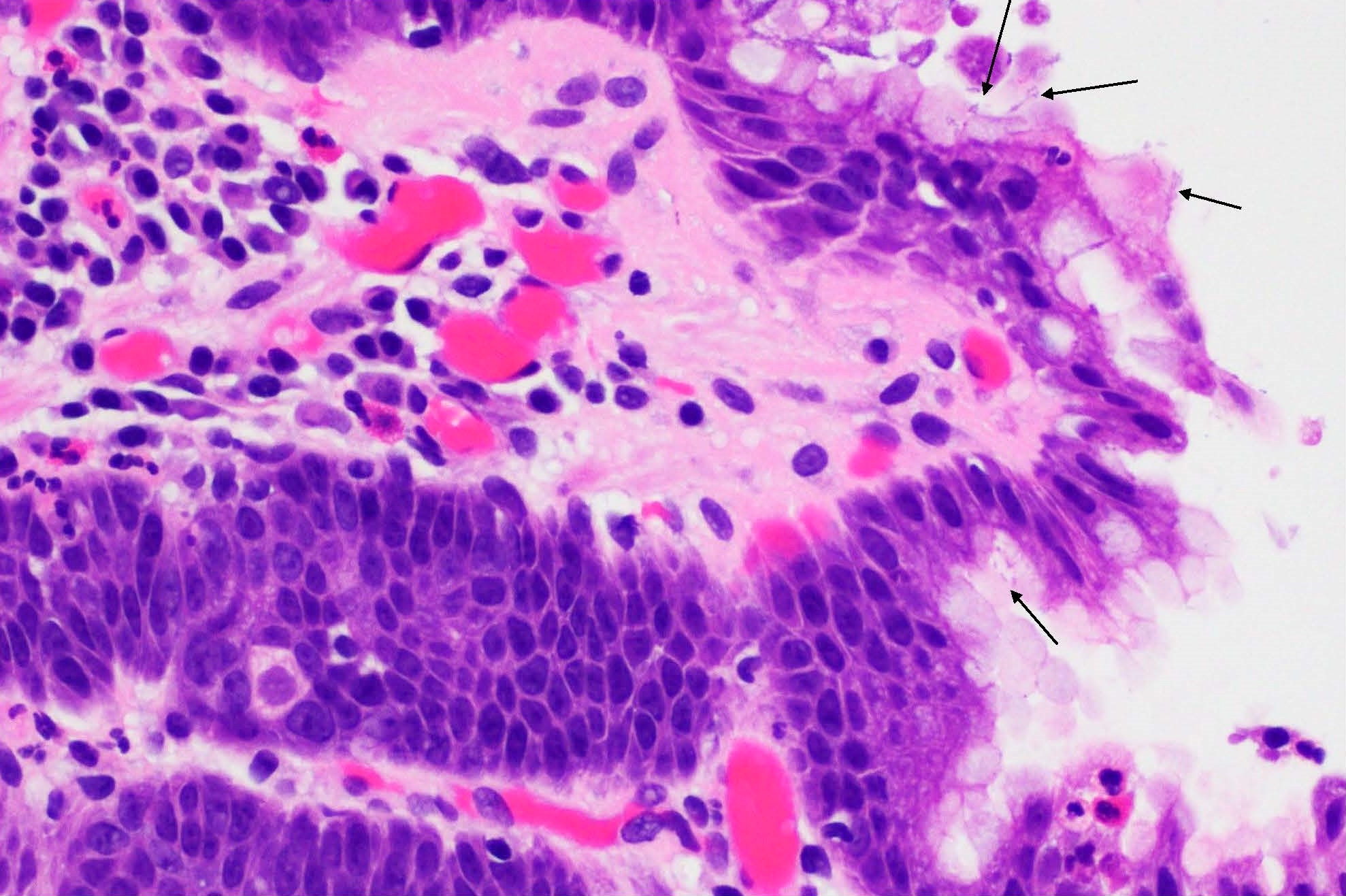
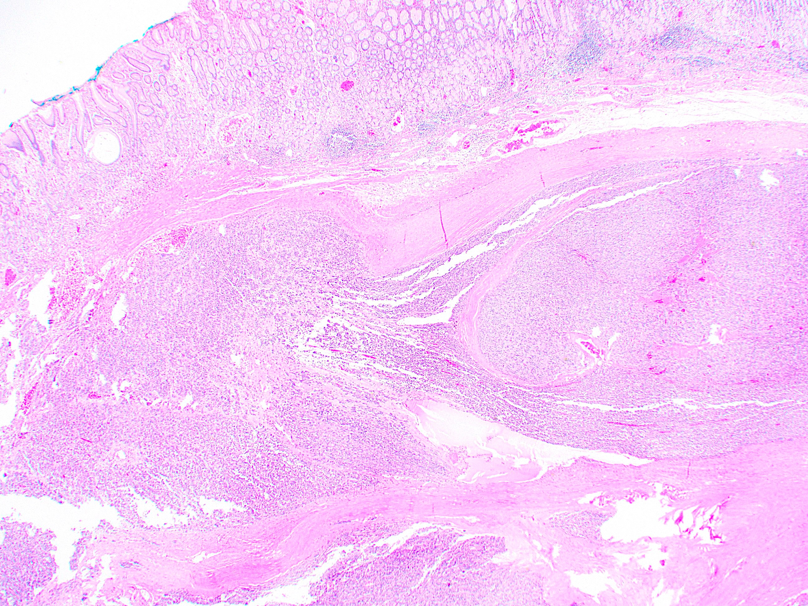
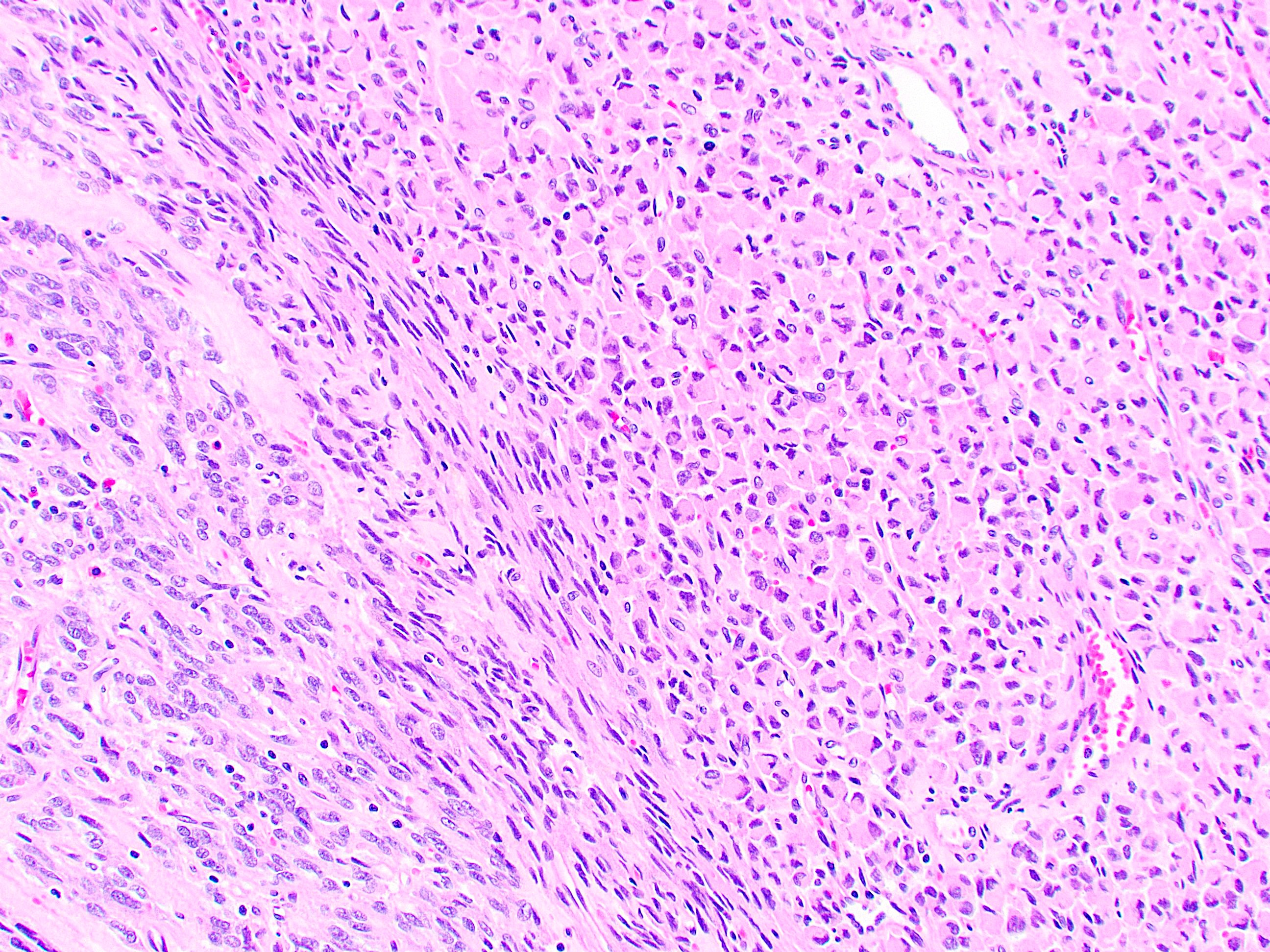
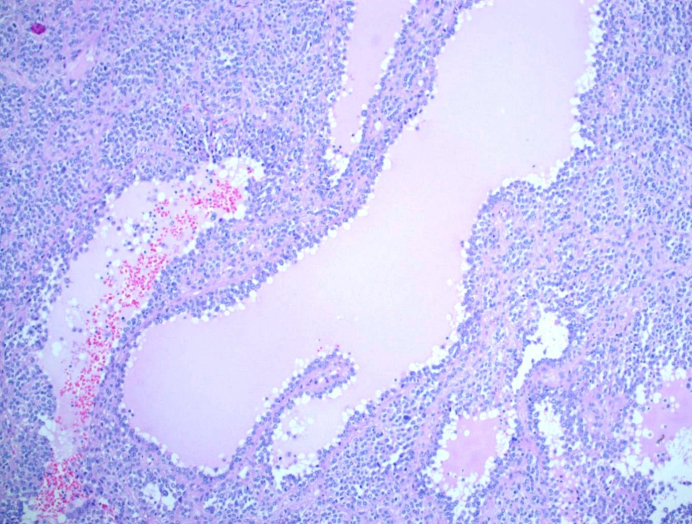
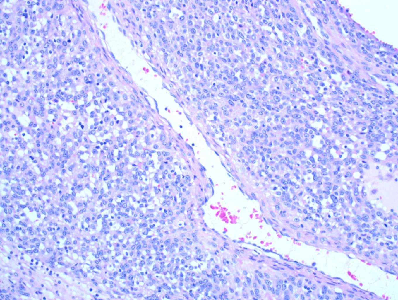
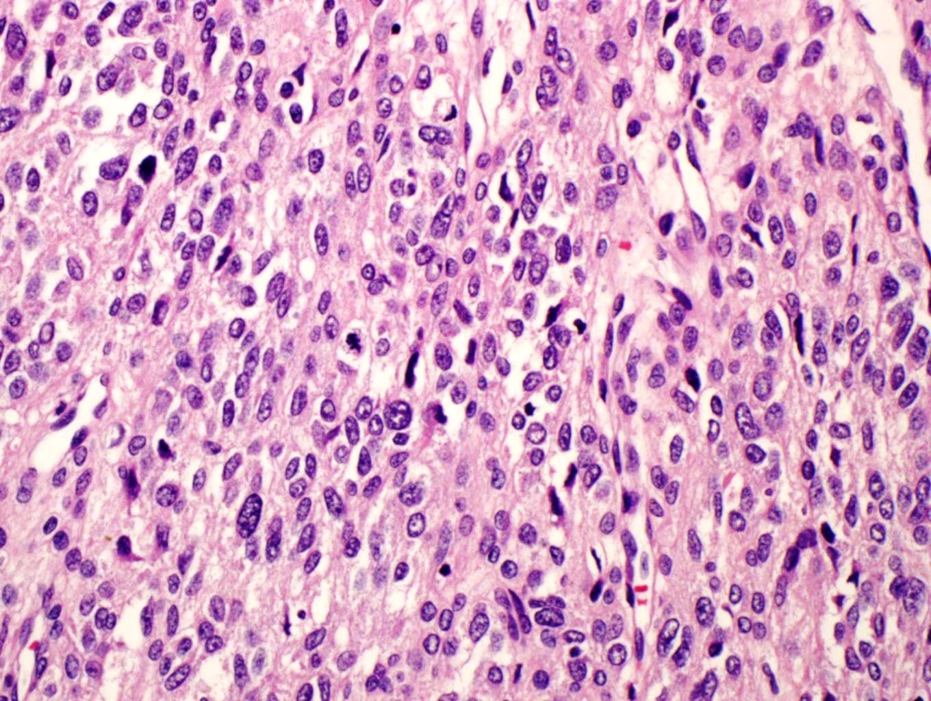
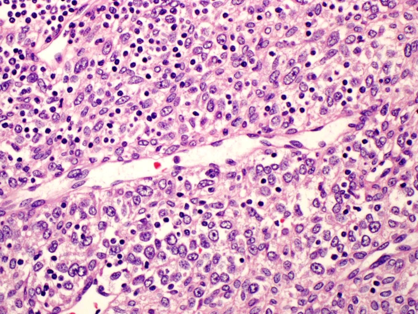
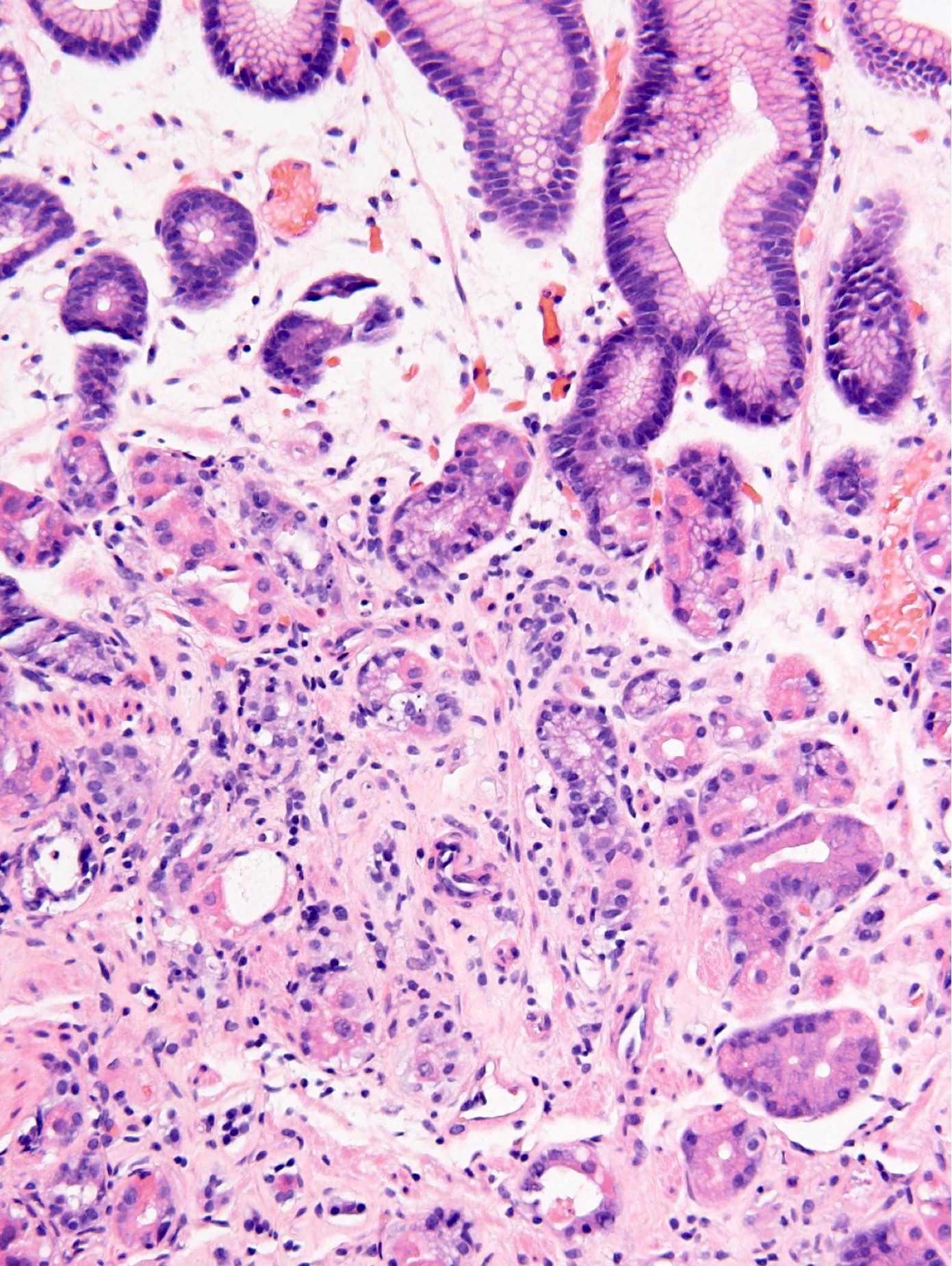
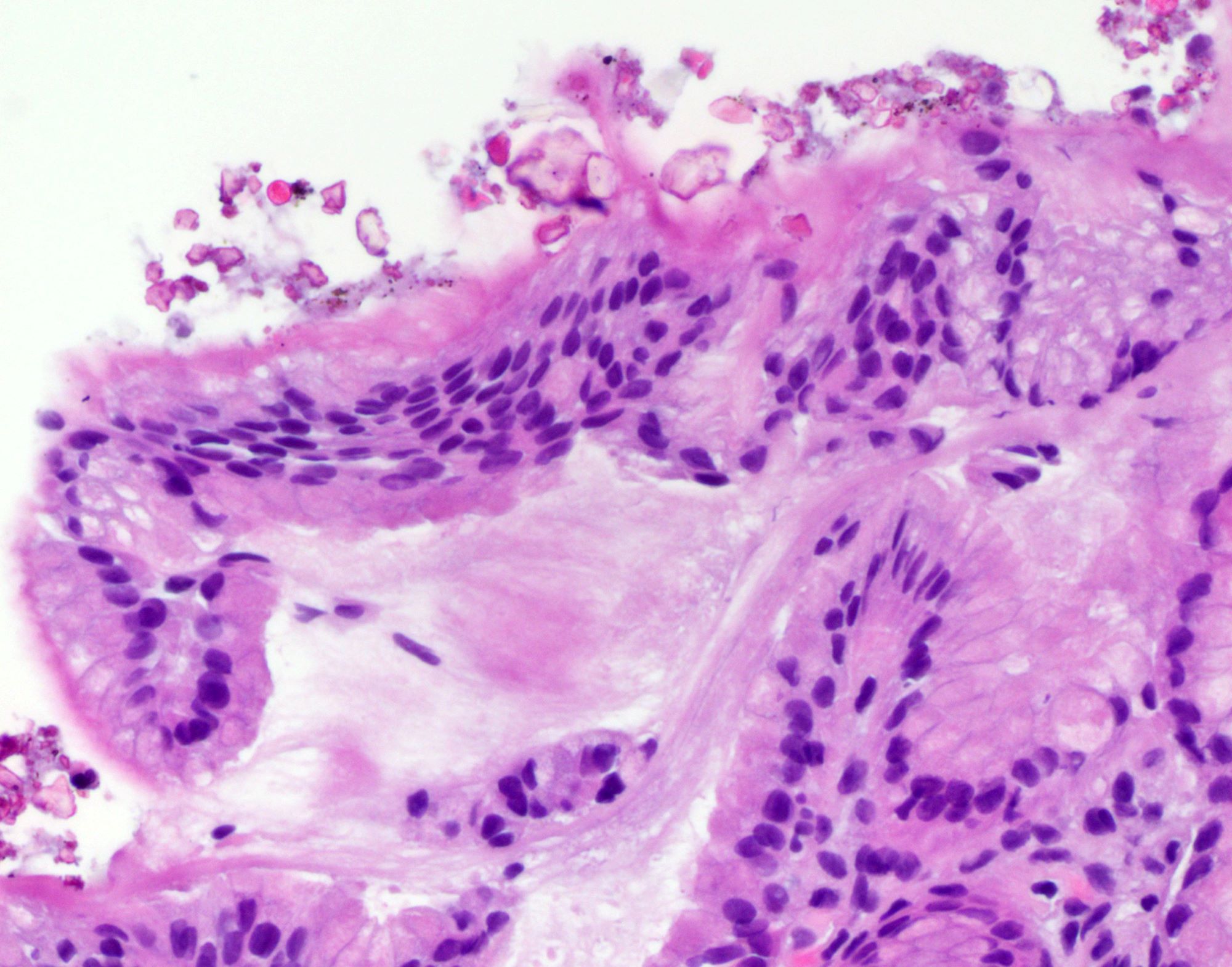
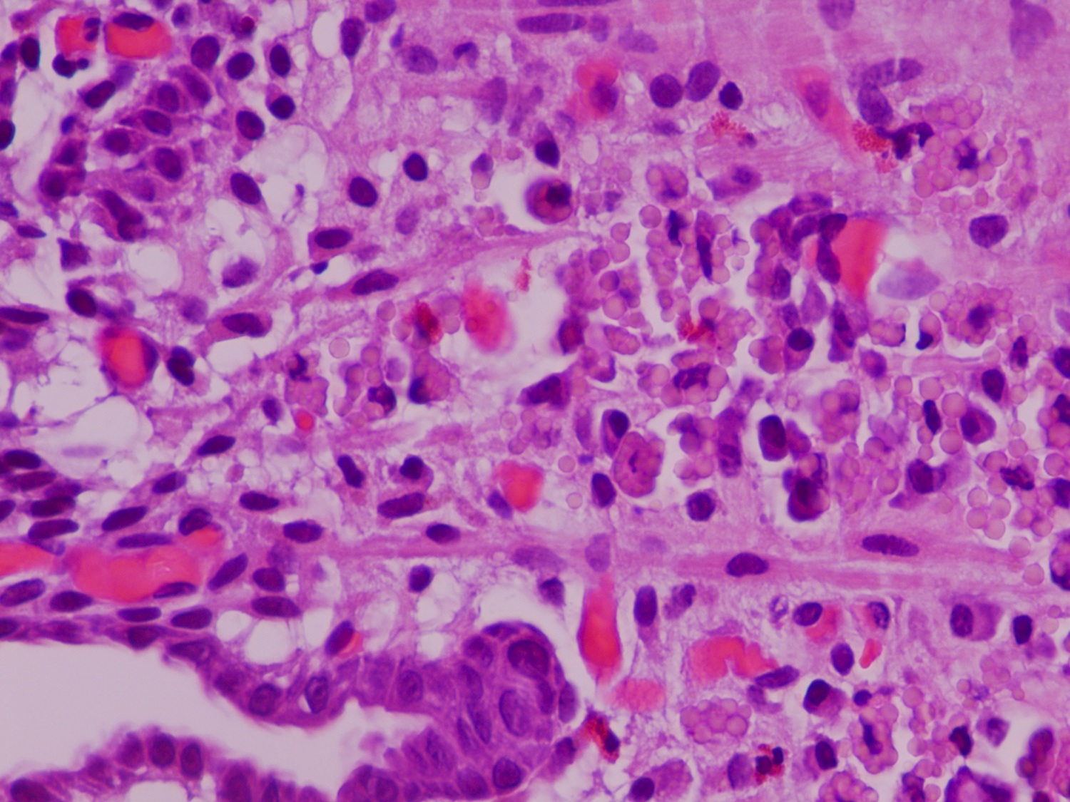
Acute phlegmonous gastritis
Images hosted on other servers:

Acute phlegmonous gastritis

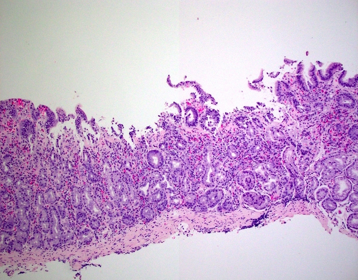
Diffusely hyperemic gastric mucosa

Areas of gastric hemorrhage depicting gastric erosions
Contributed by Supriya Srivastava, M.D., Ph.D. and Xiaoyan Liao, M.D., Ph.D.
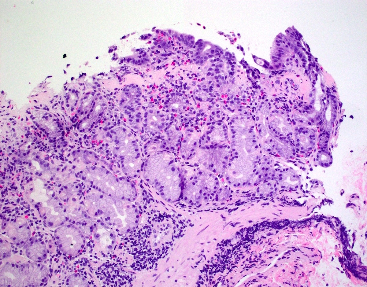
Acute gastric erosion
Acute gastritis
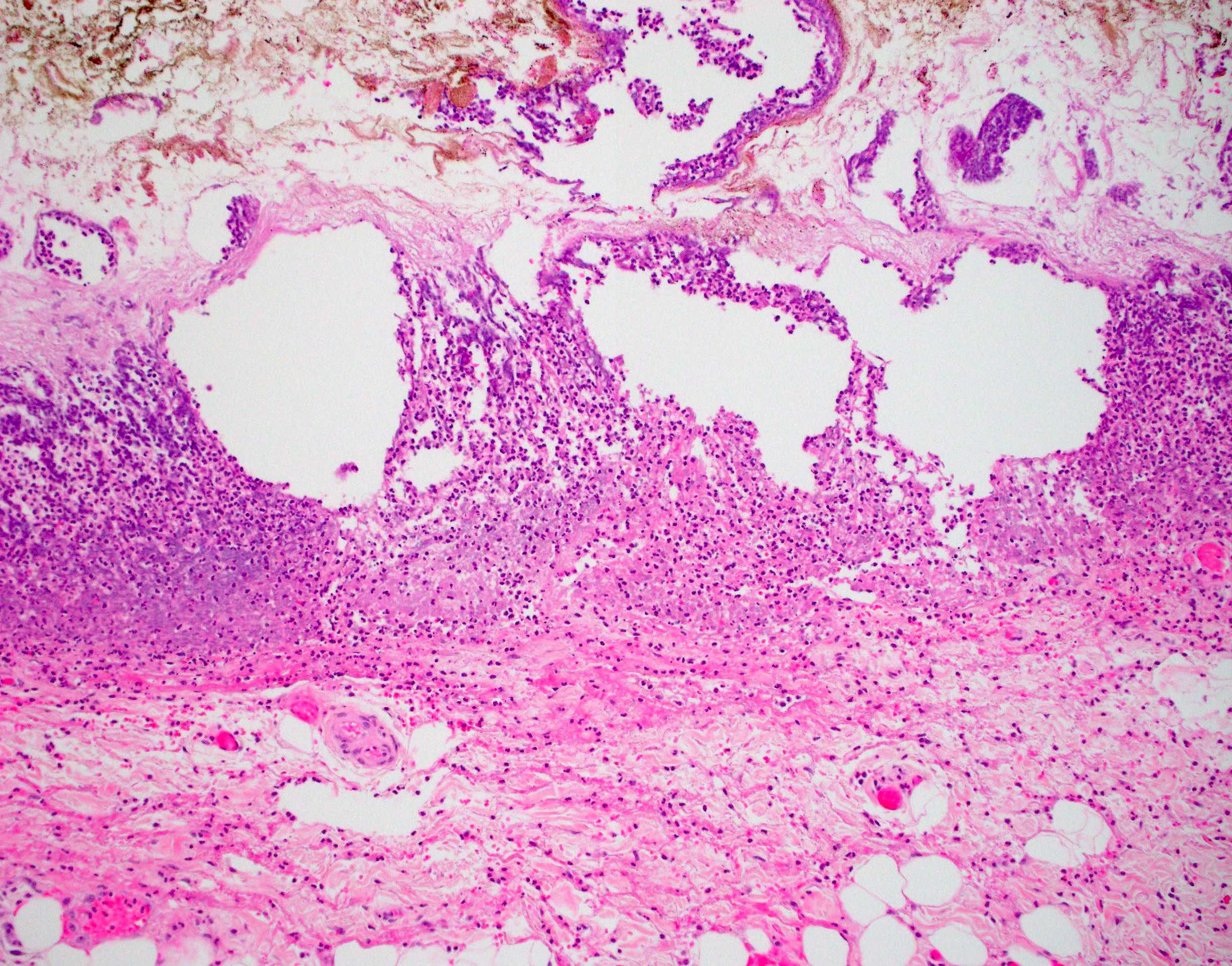
Acute phlegmonous gastritis
Contributed by Cindy Wang, M.D.
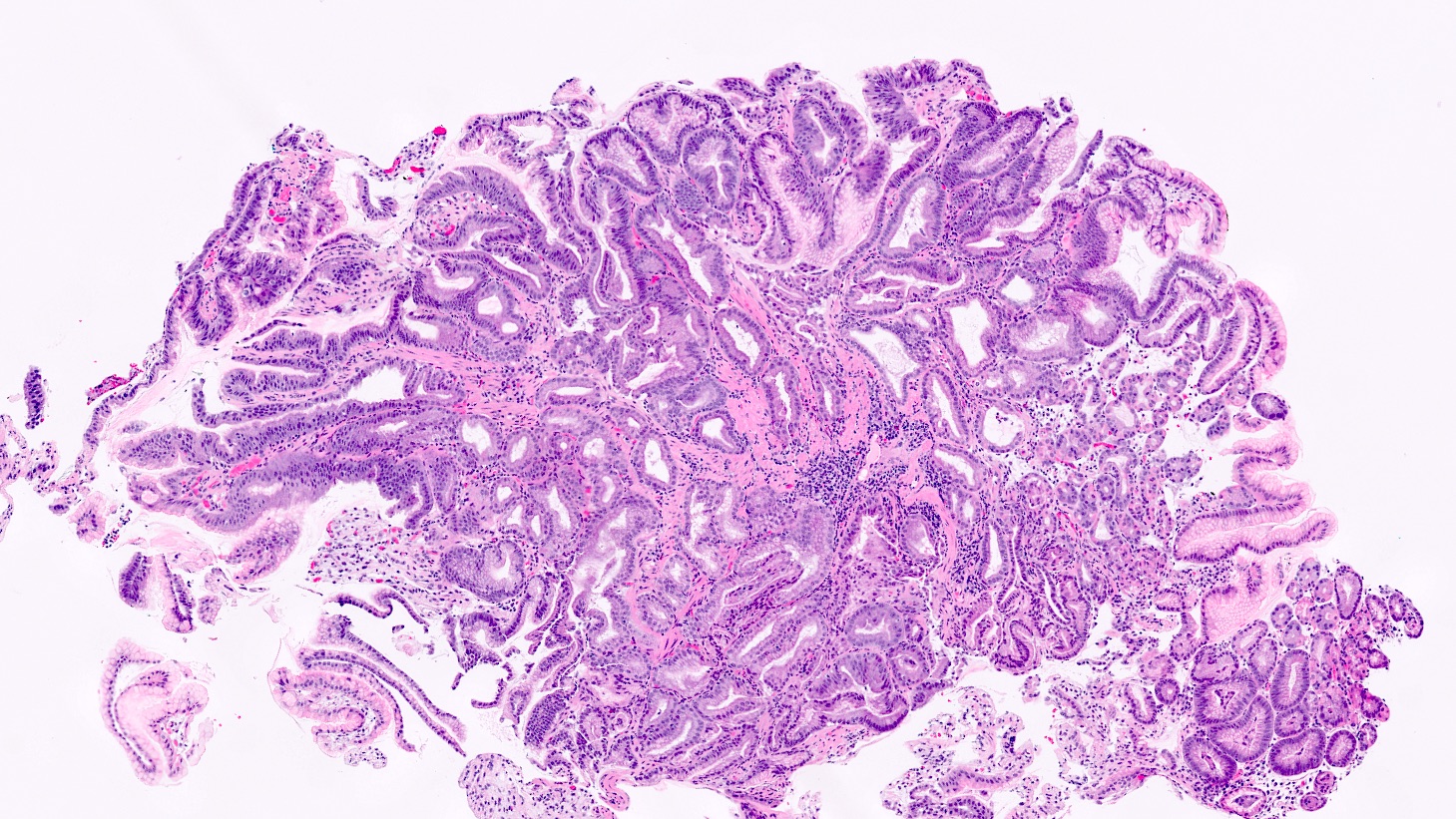
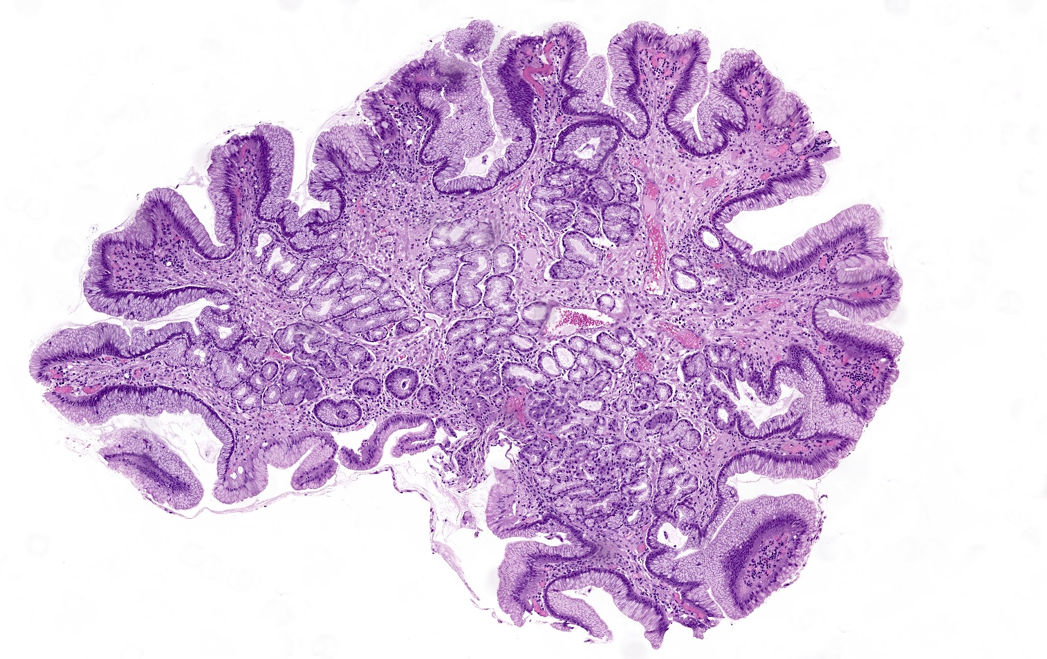
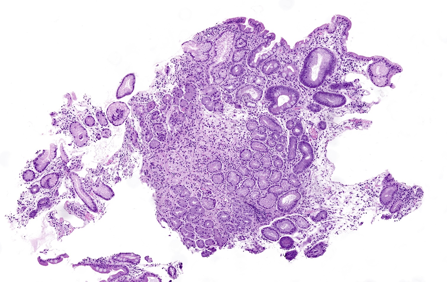
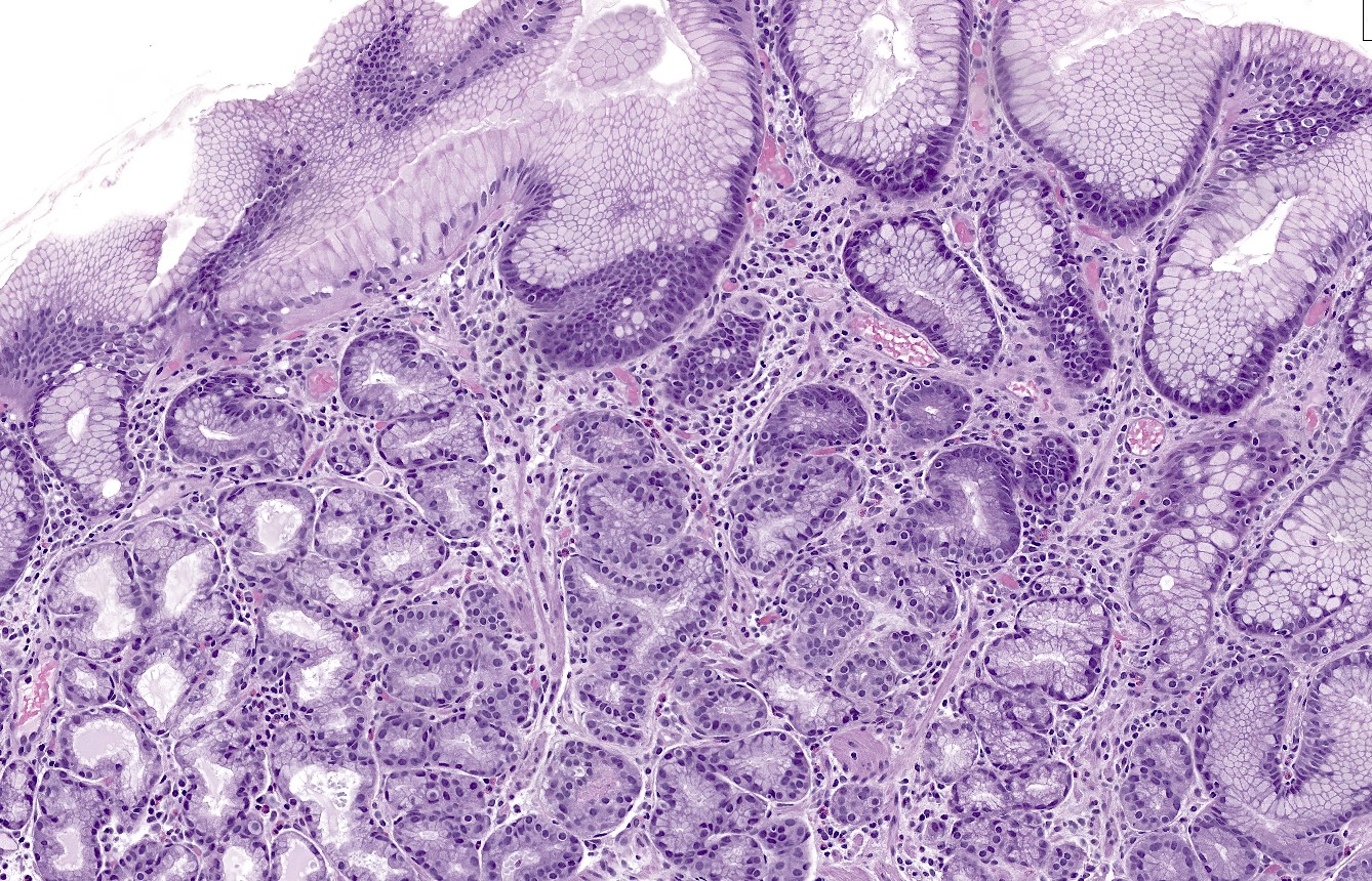
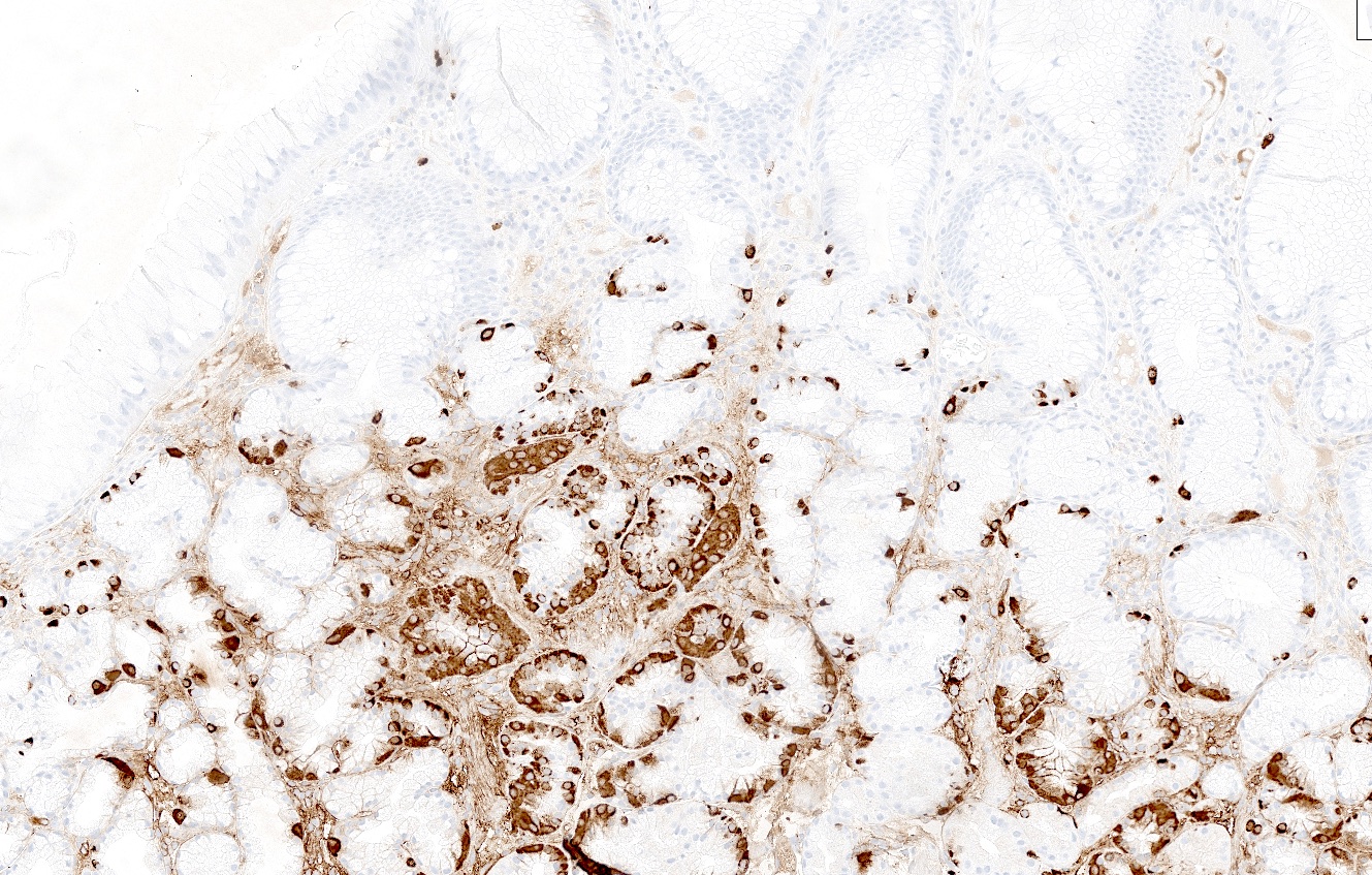
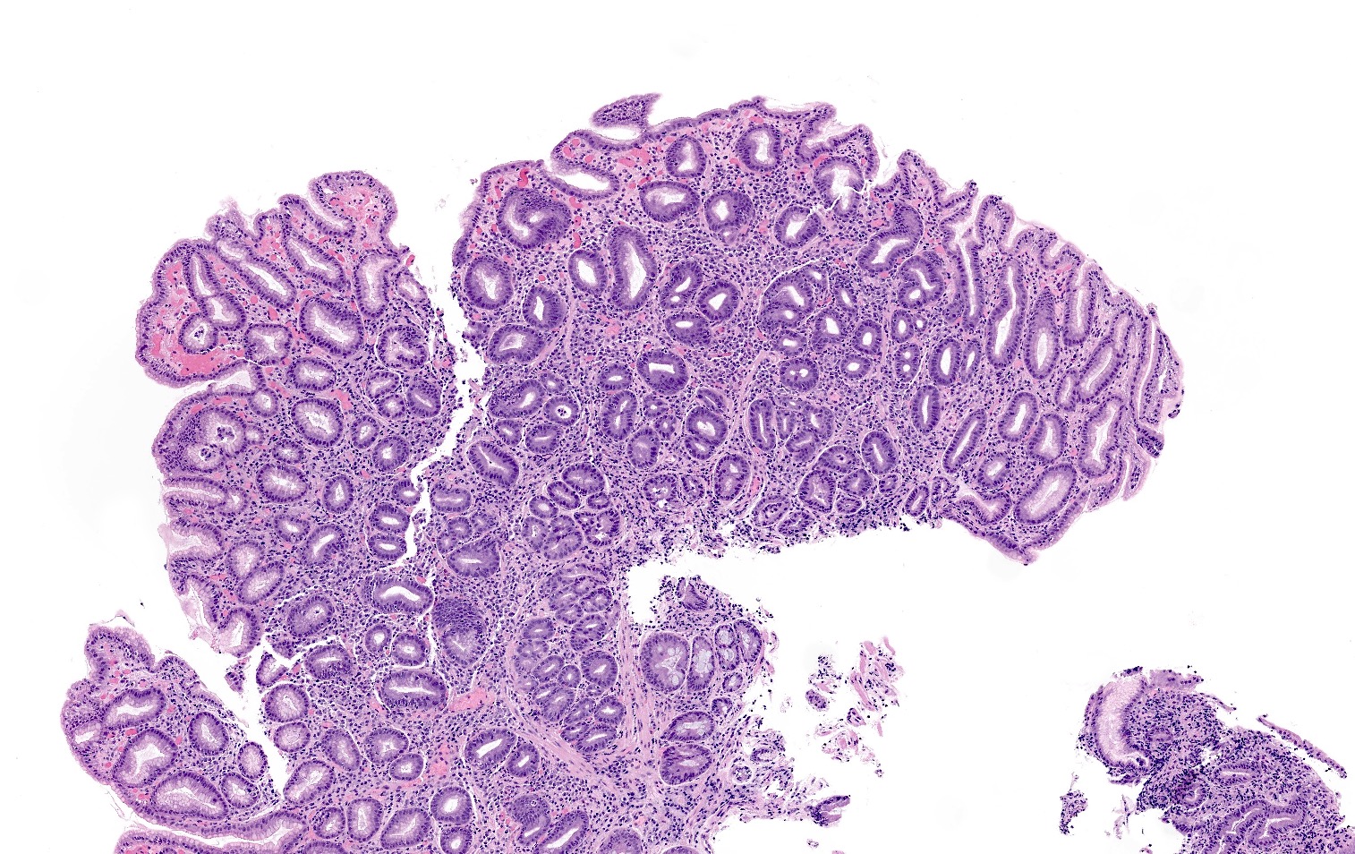
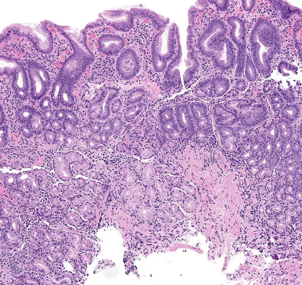
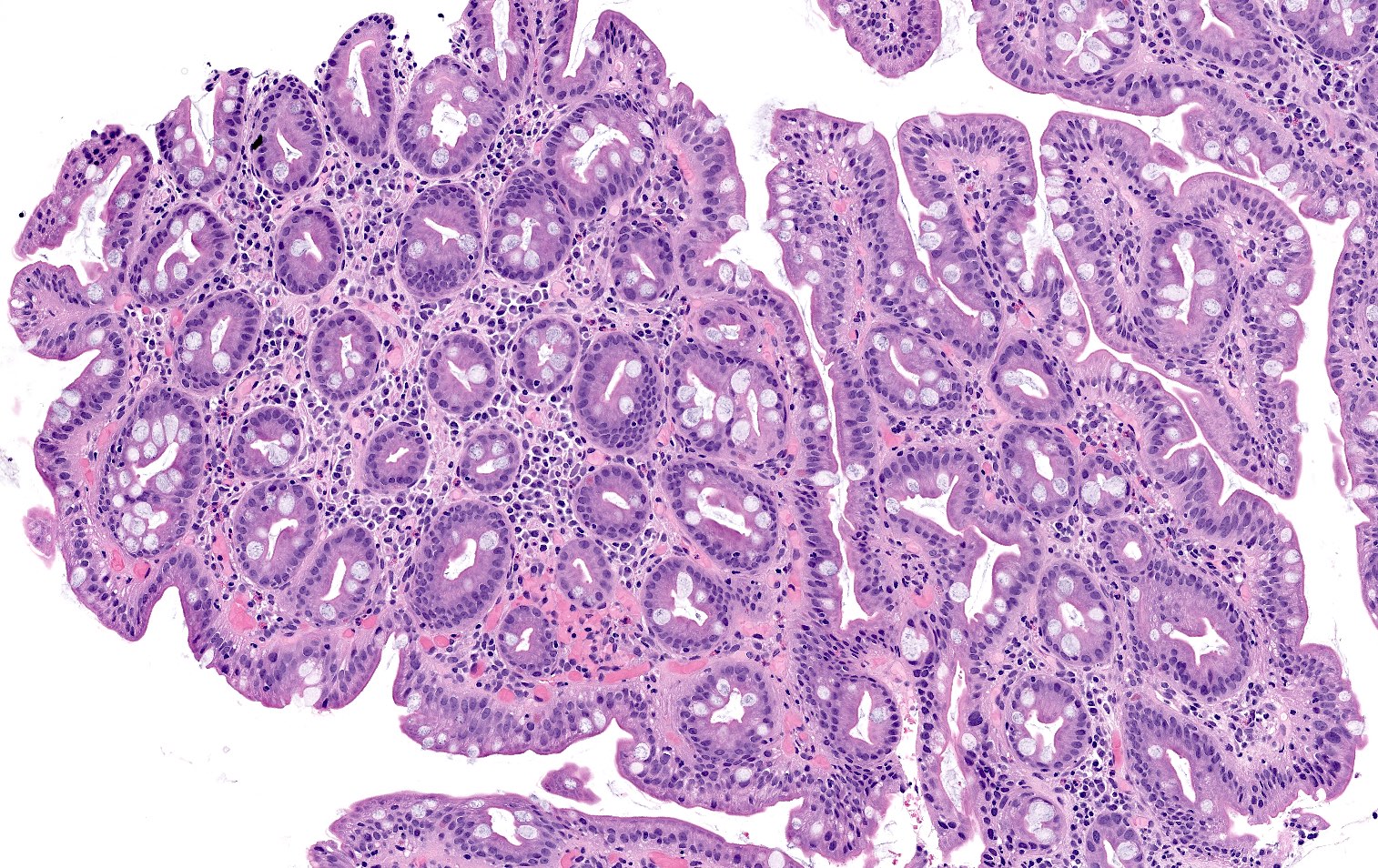
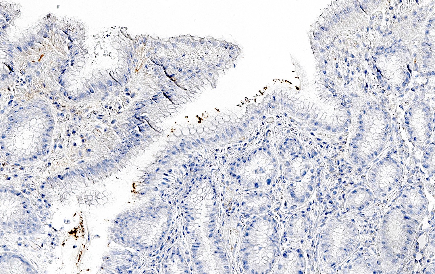
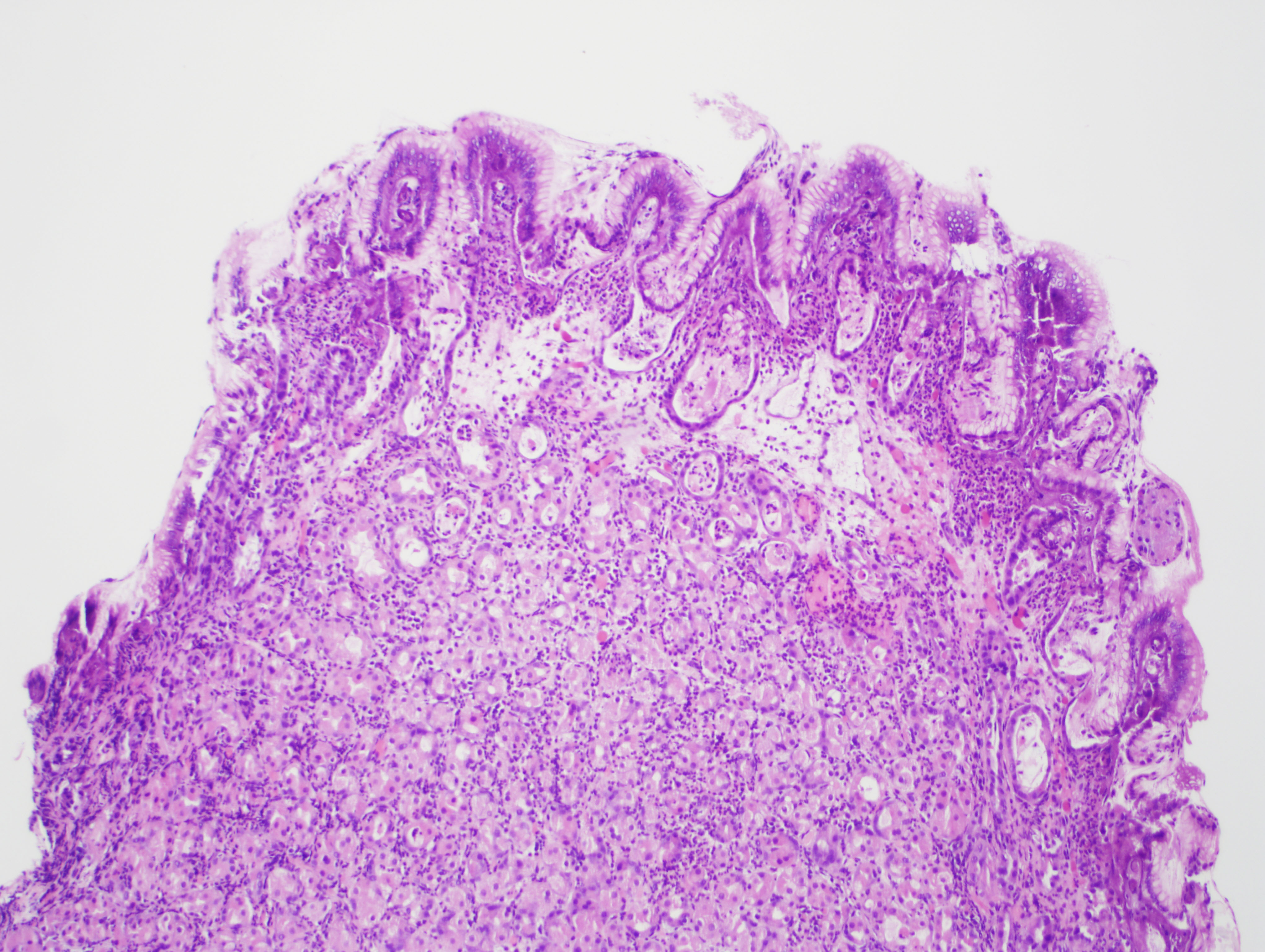
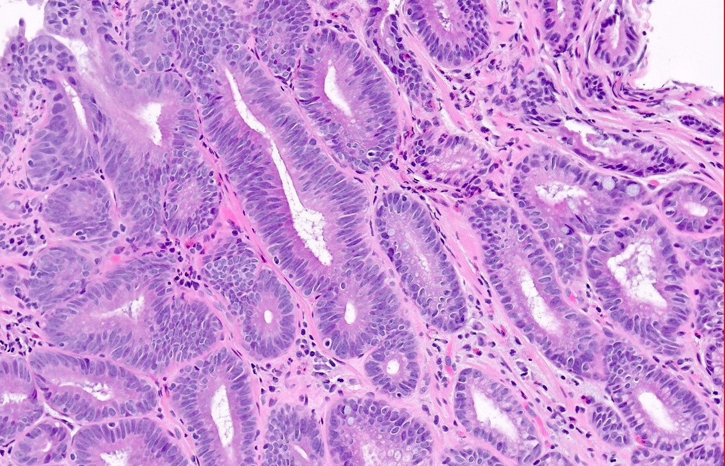
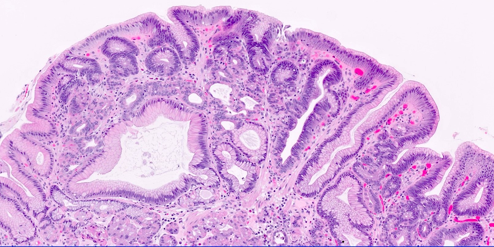
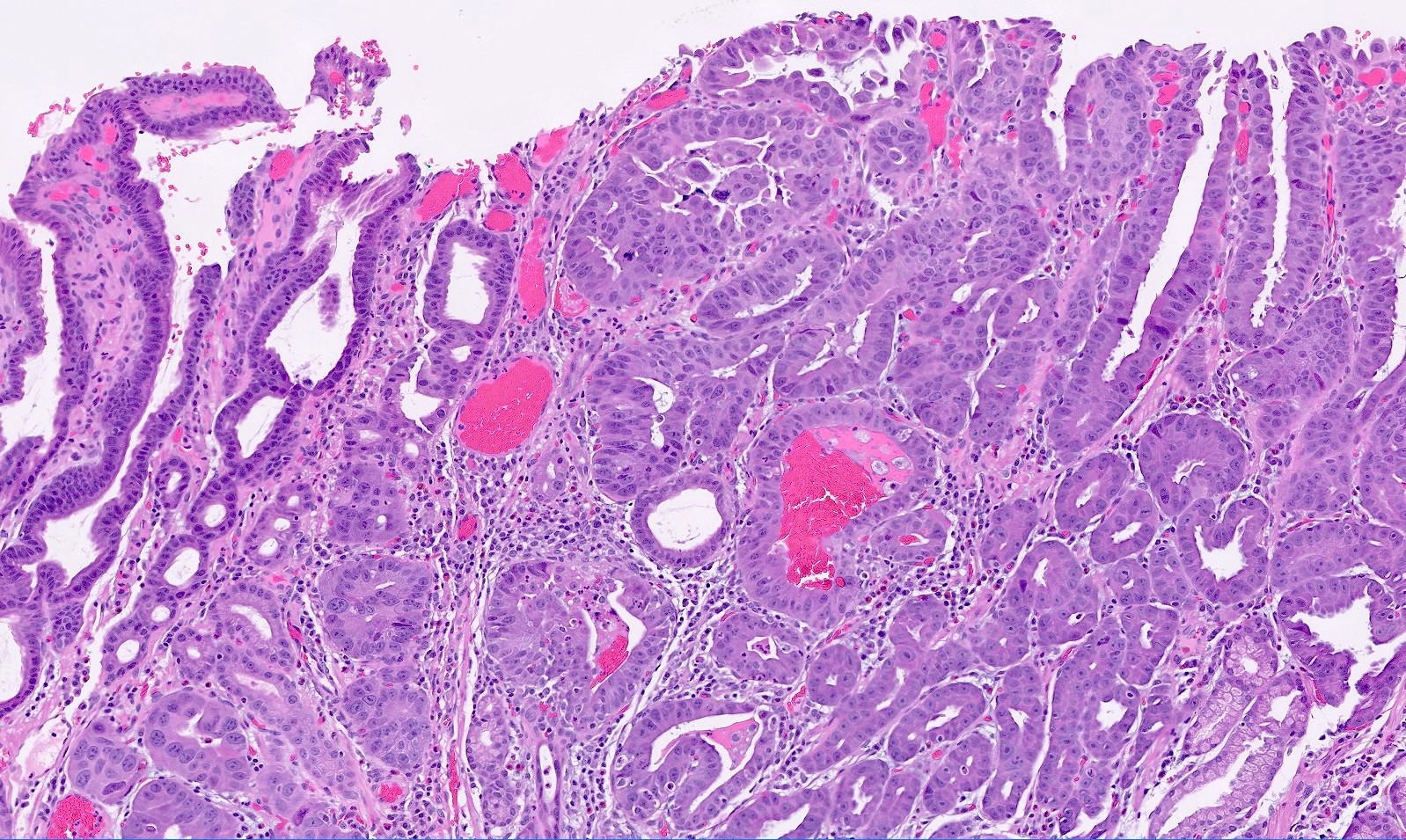
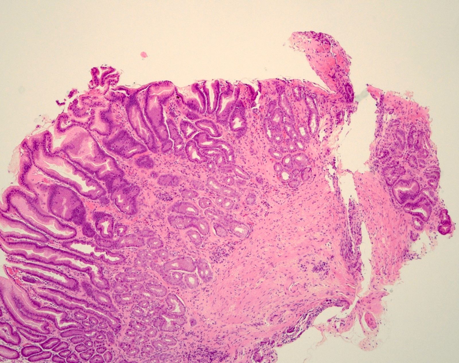
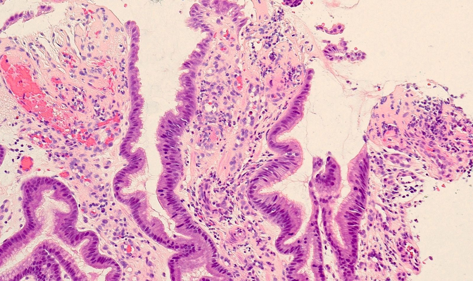
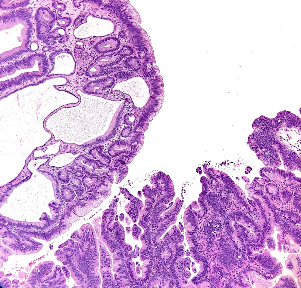
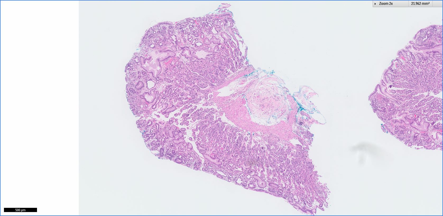
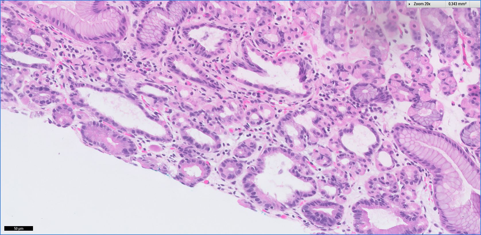
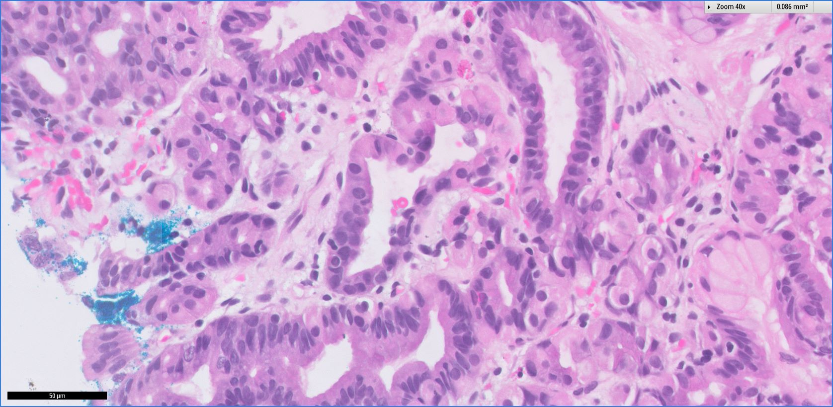
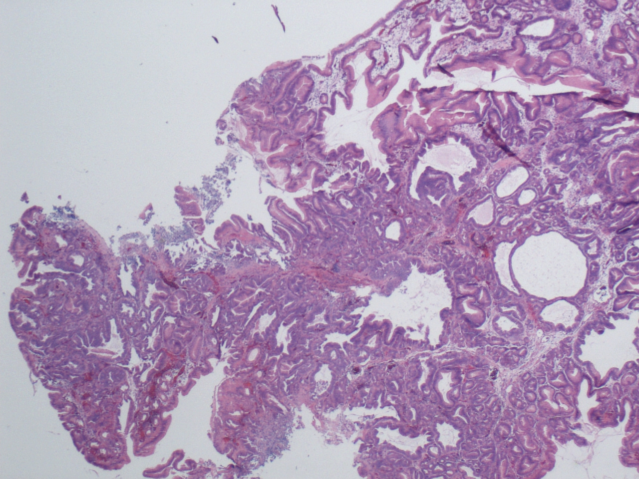
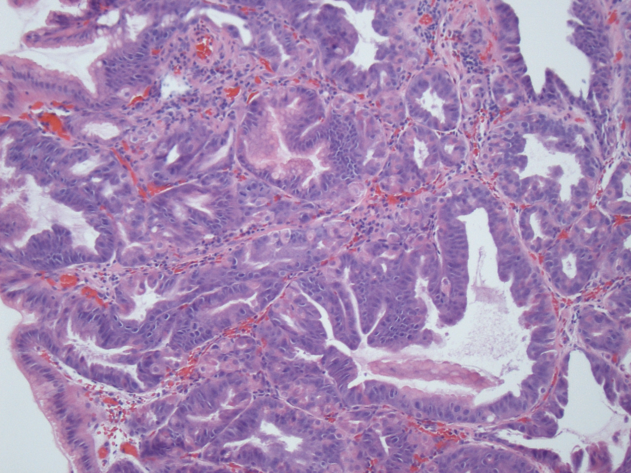
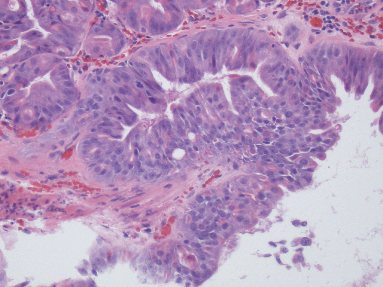
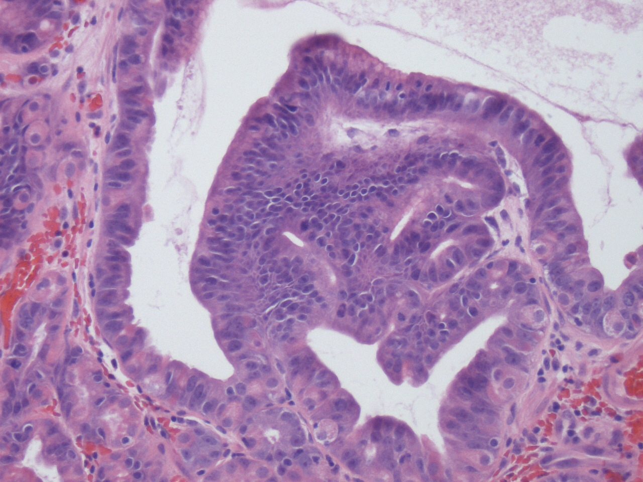
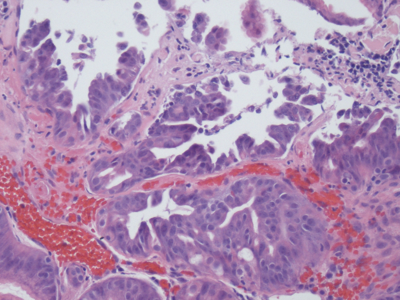
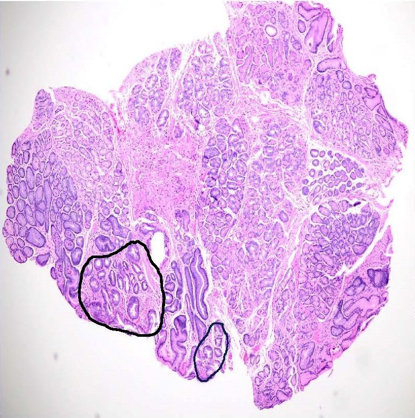
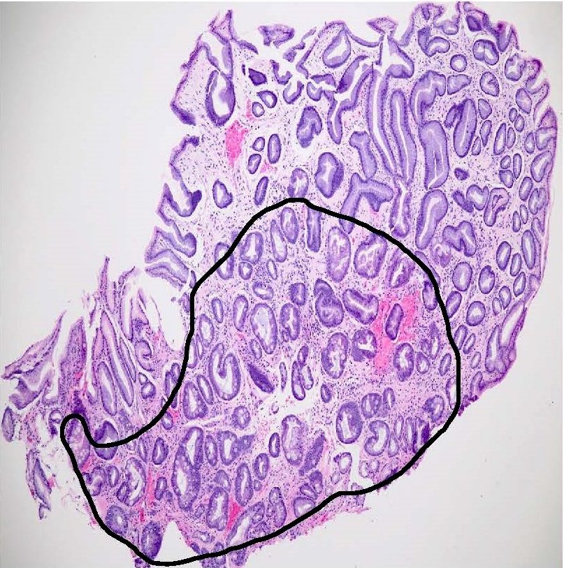
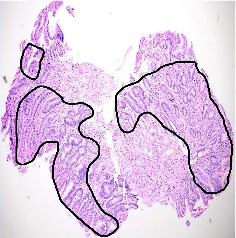
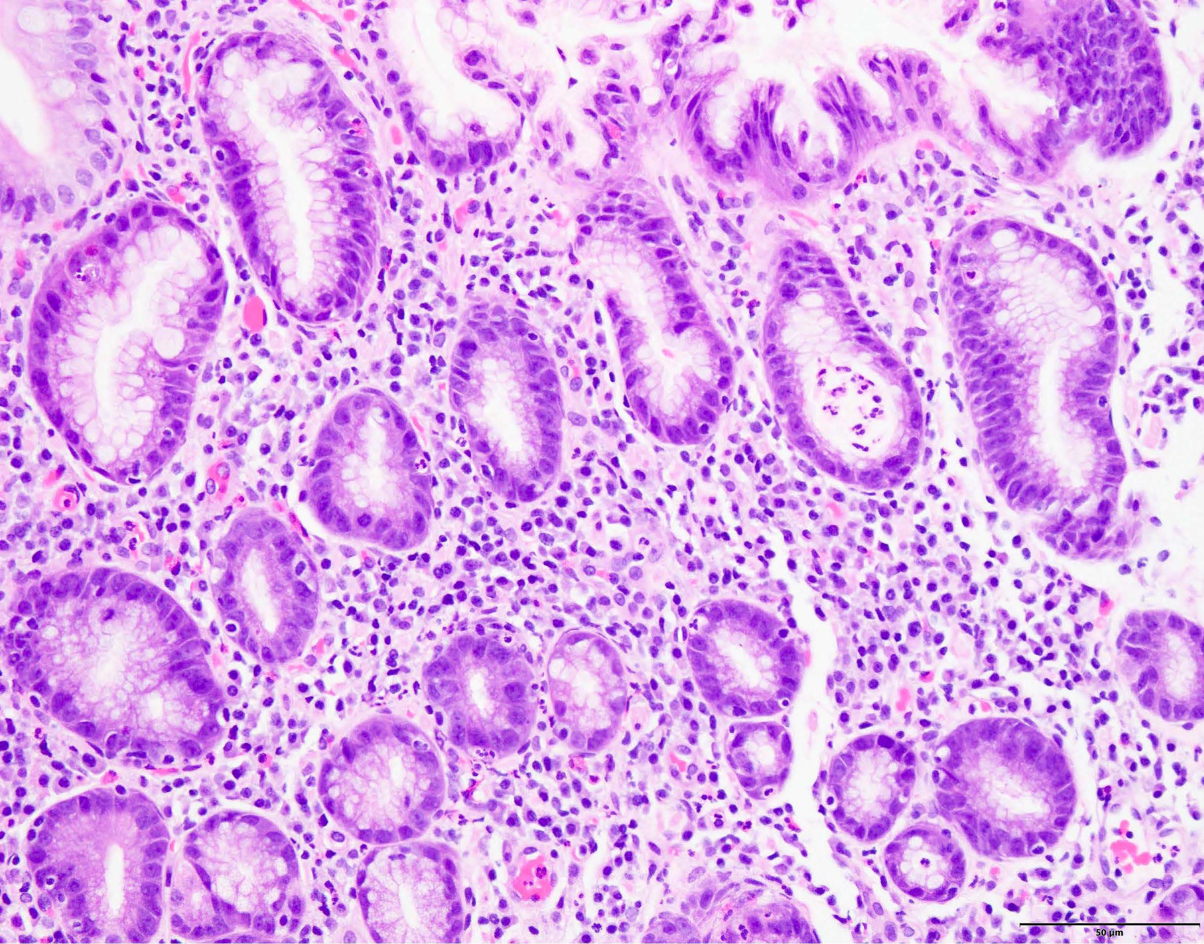
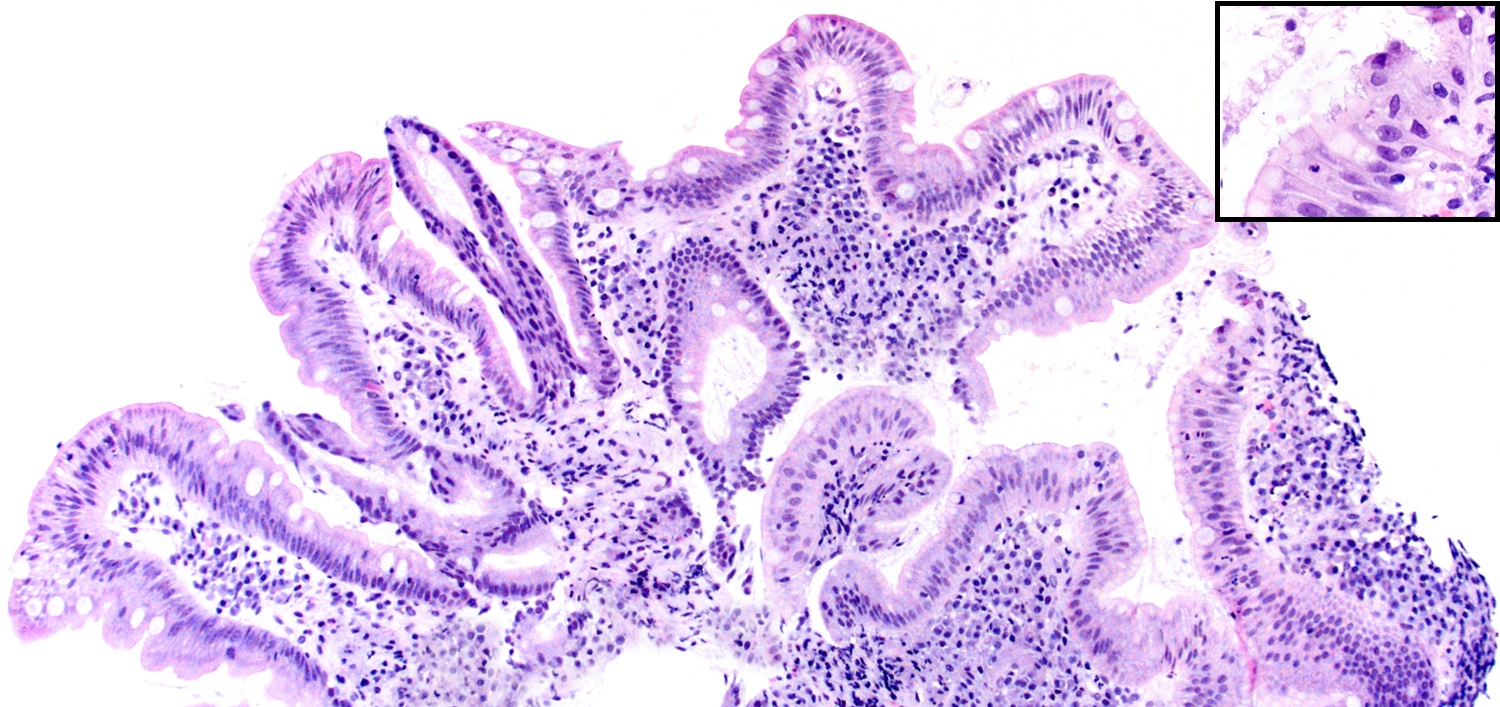
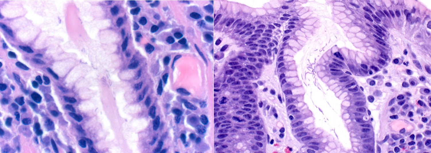
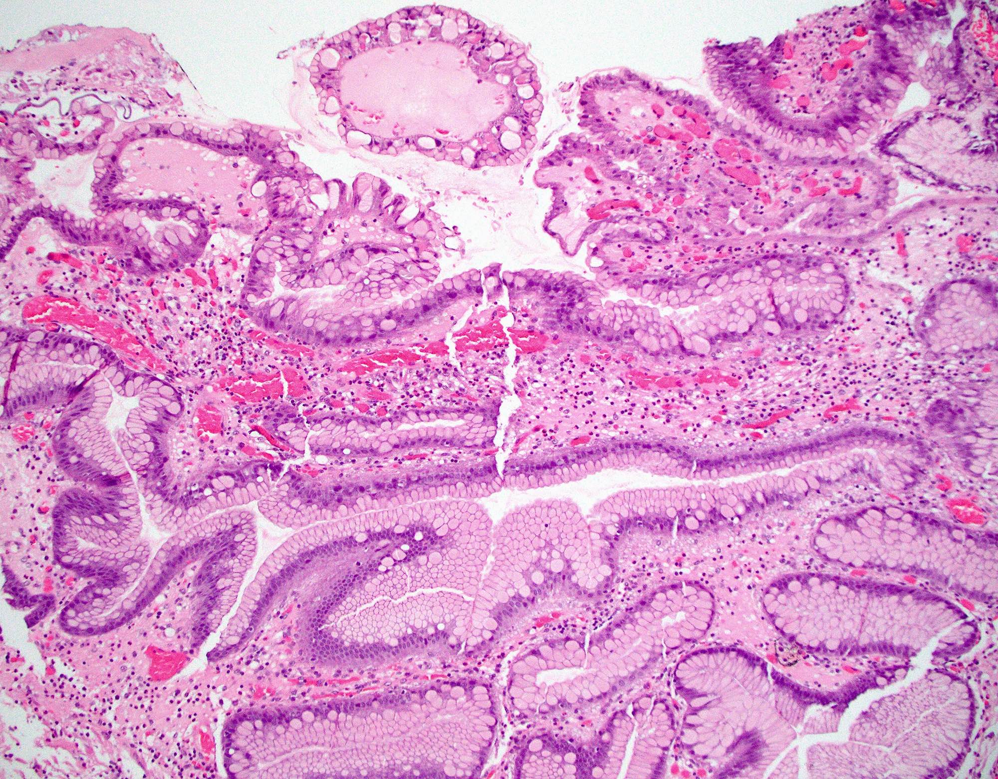
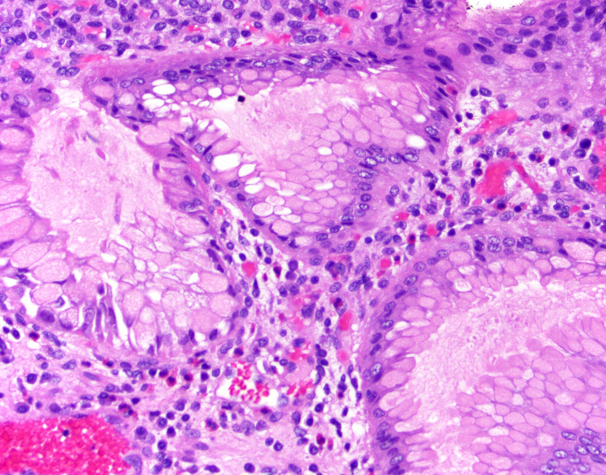
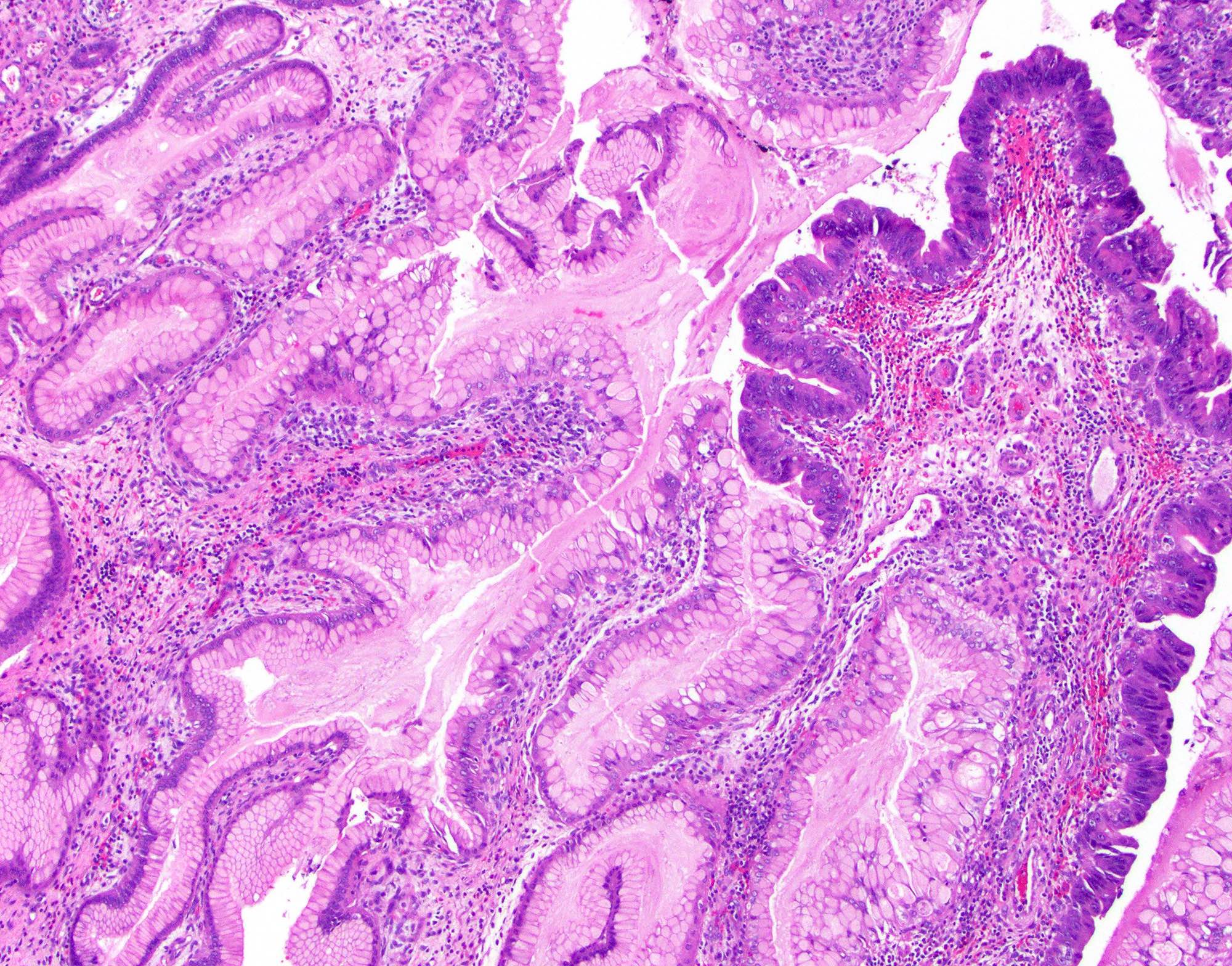
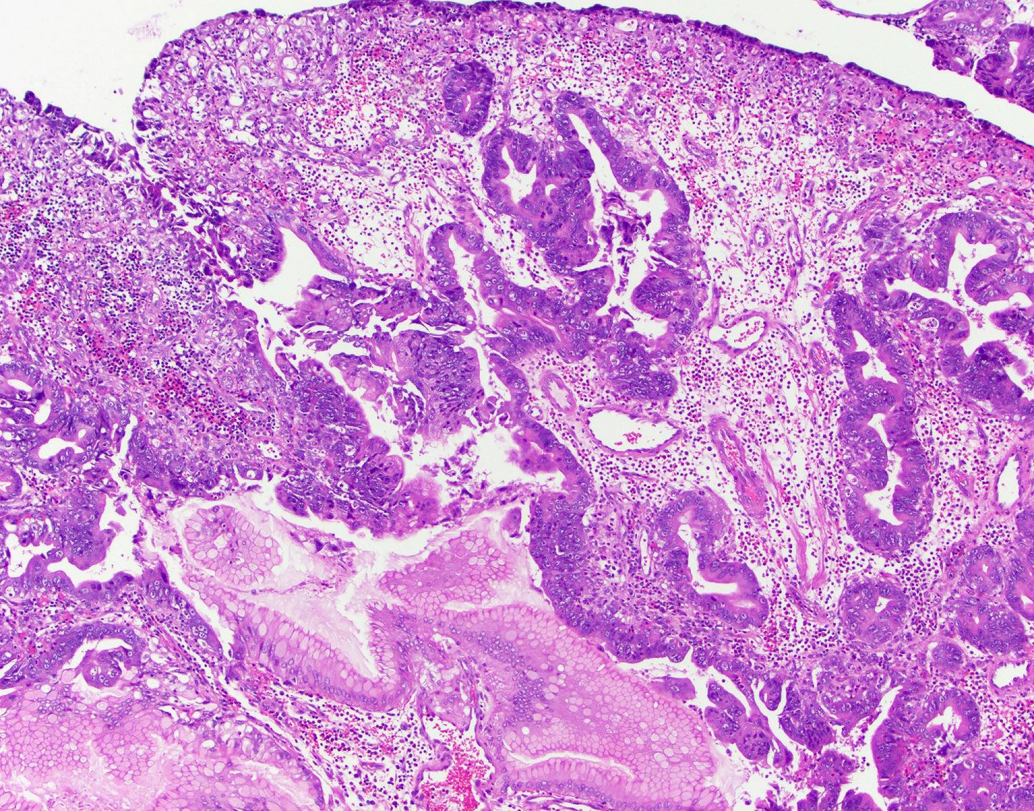
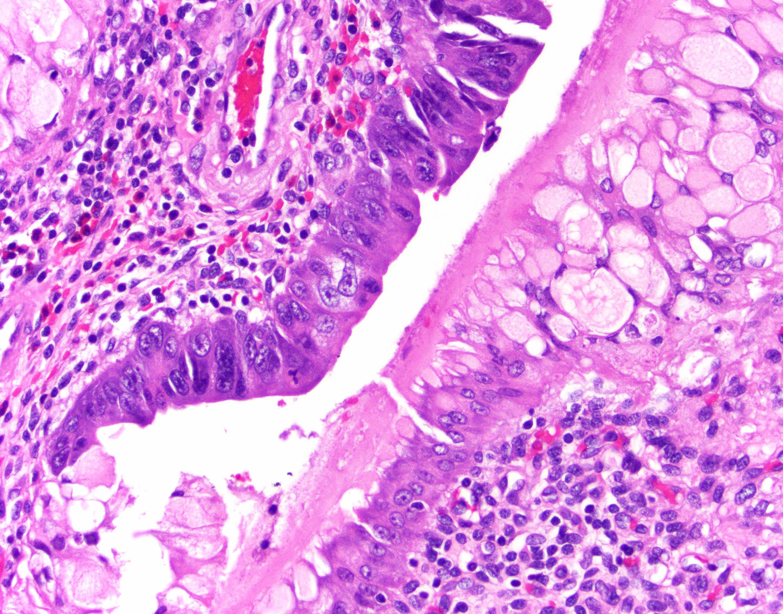
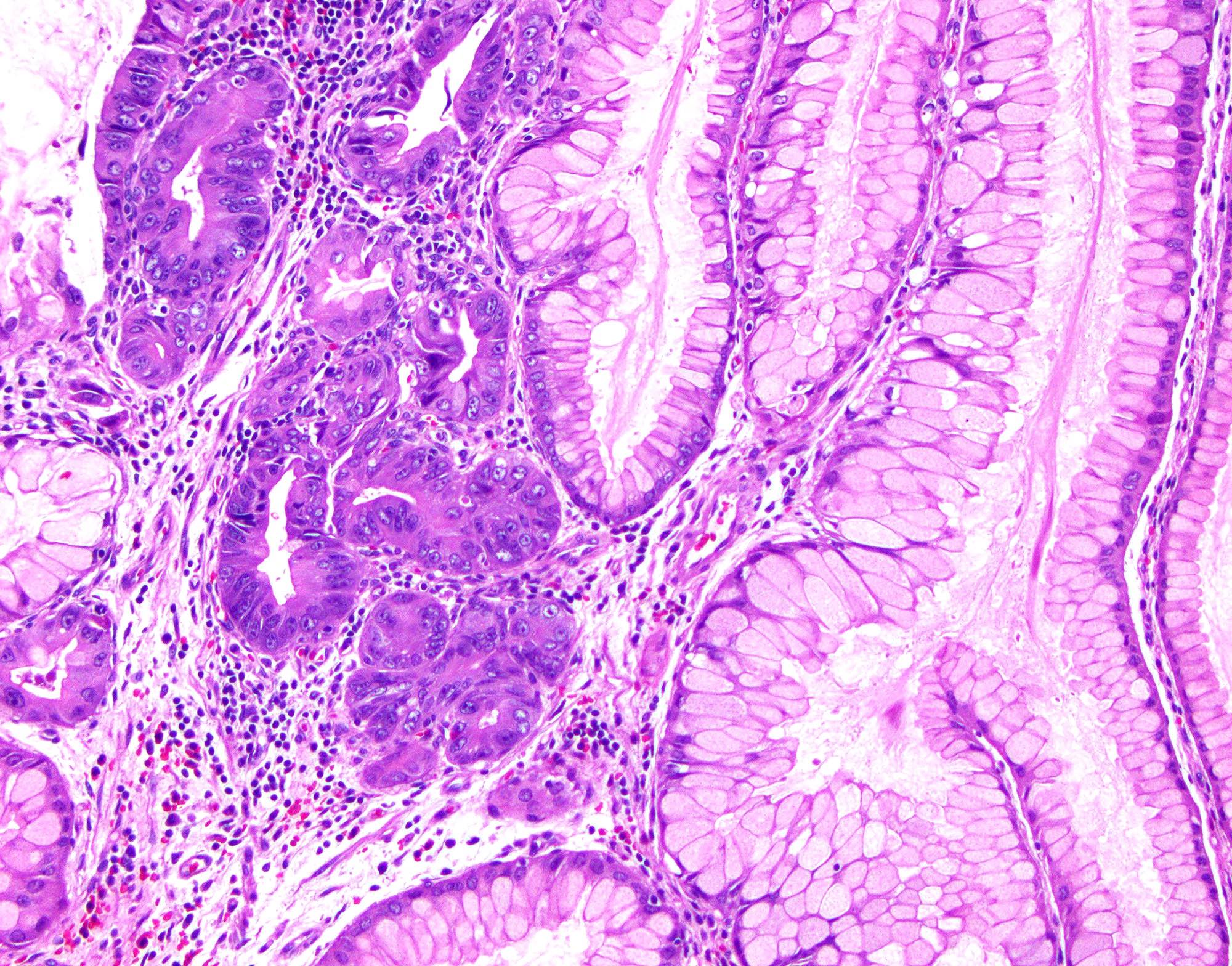
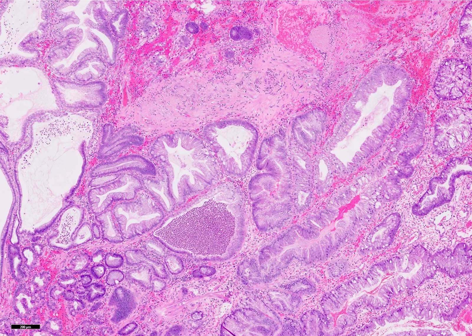
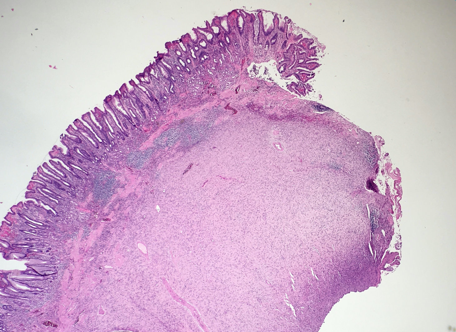
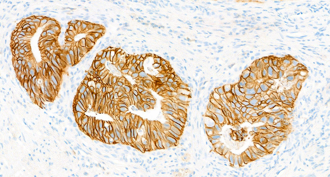
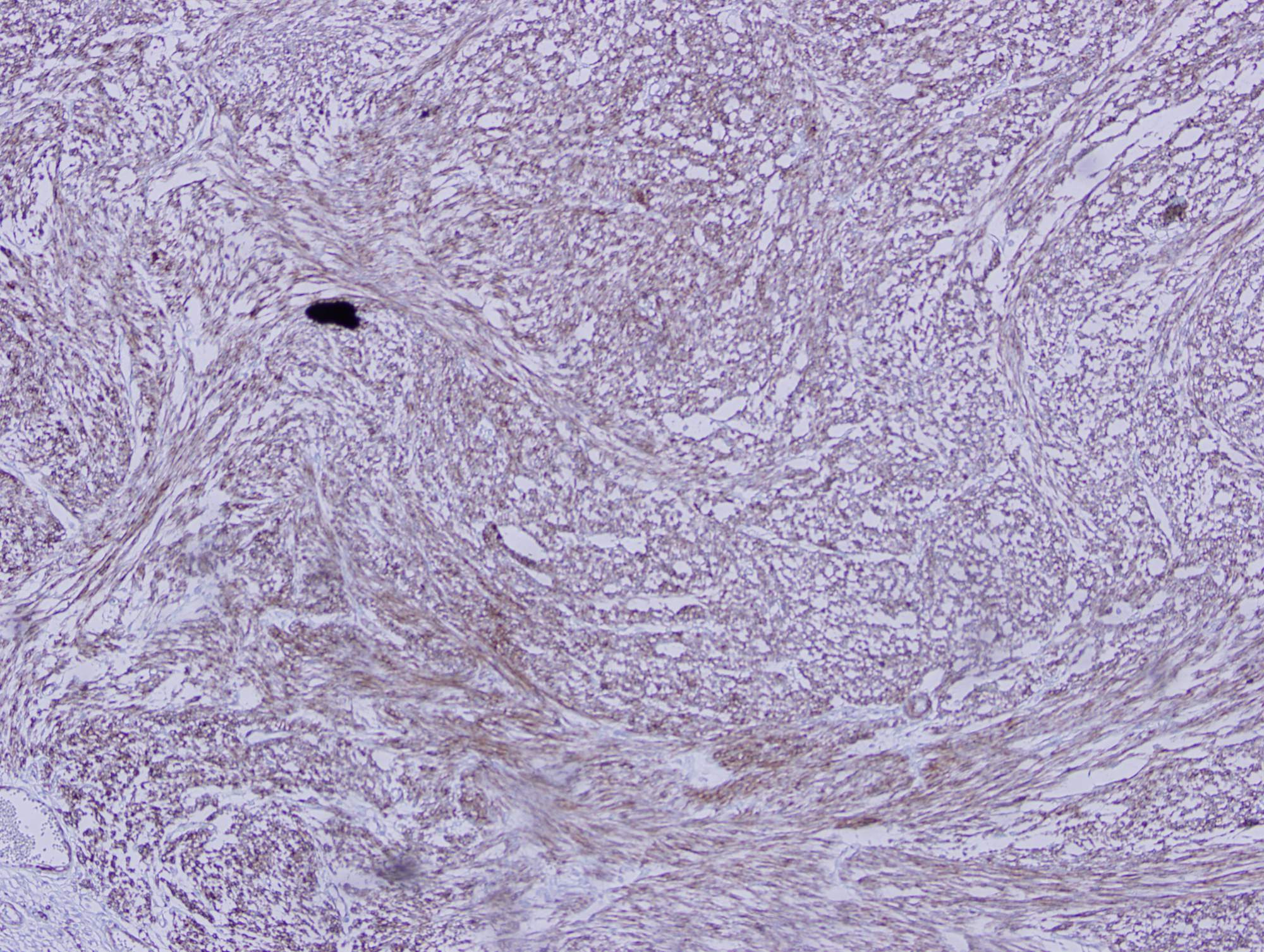
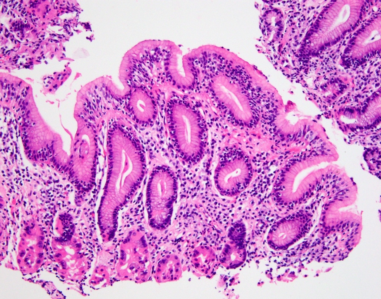
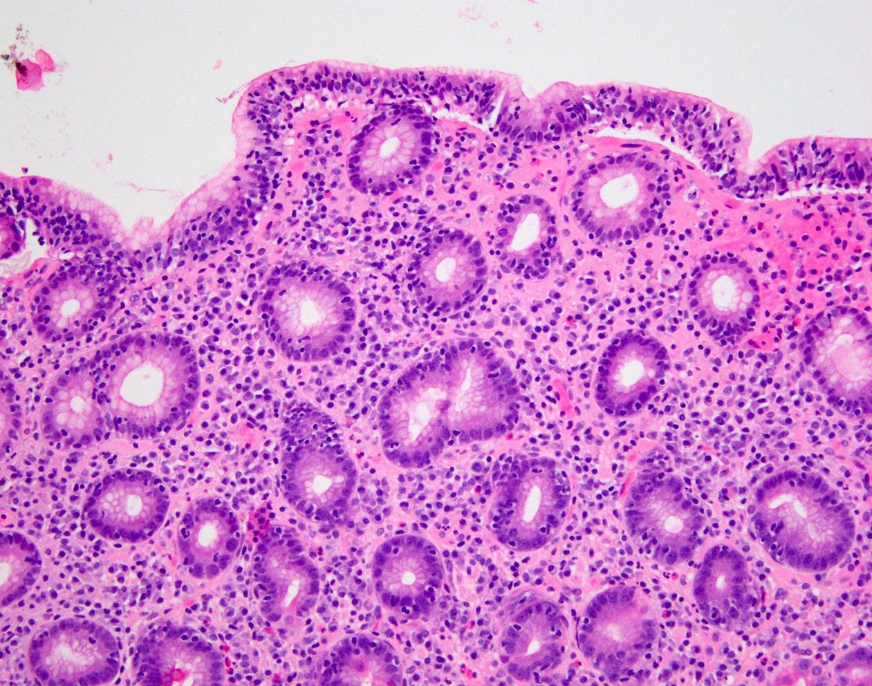
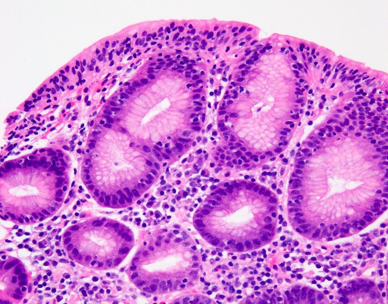
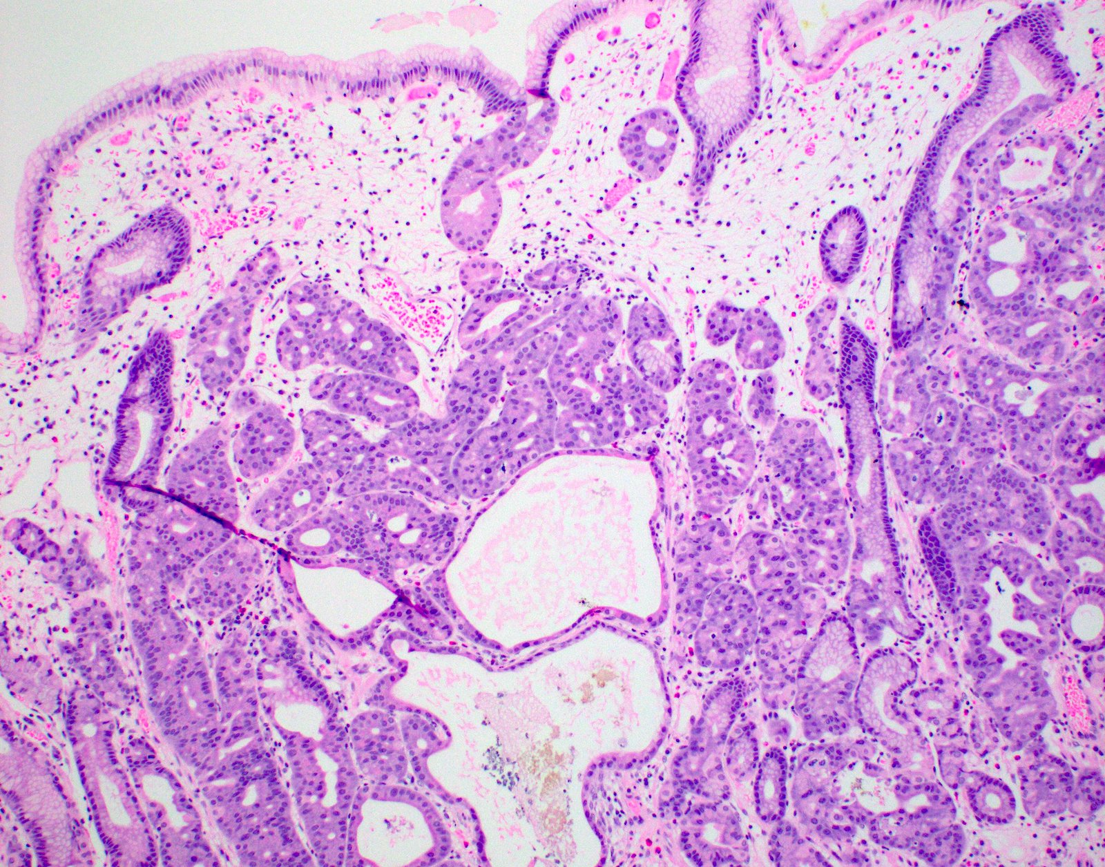
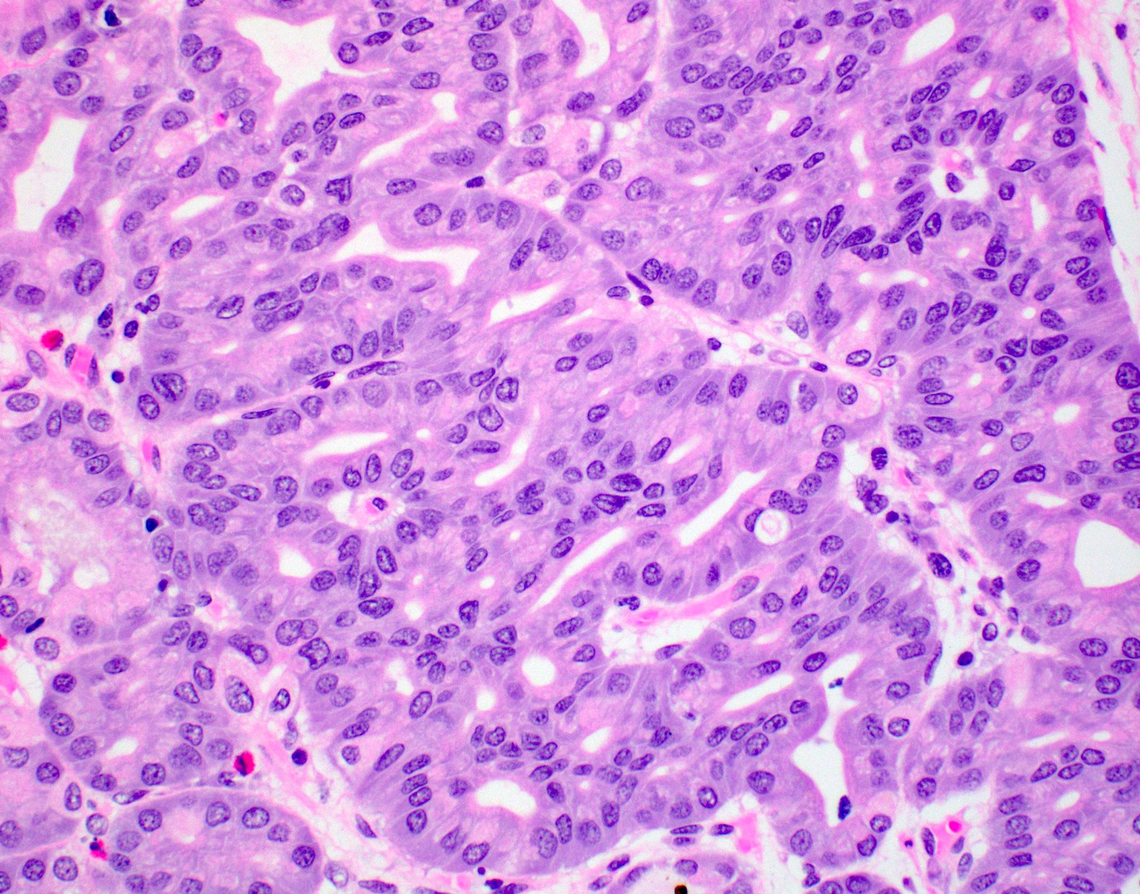
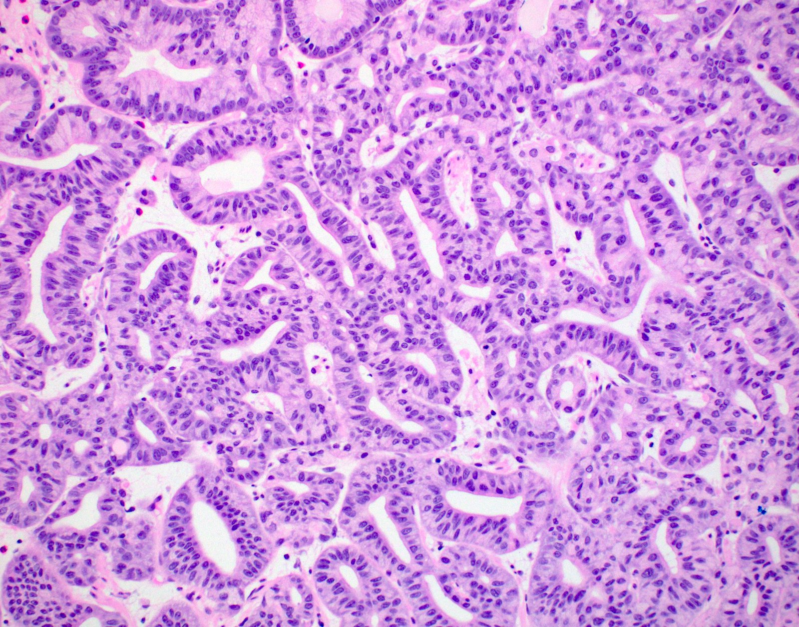
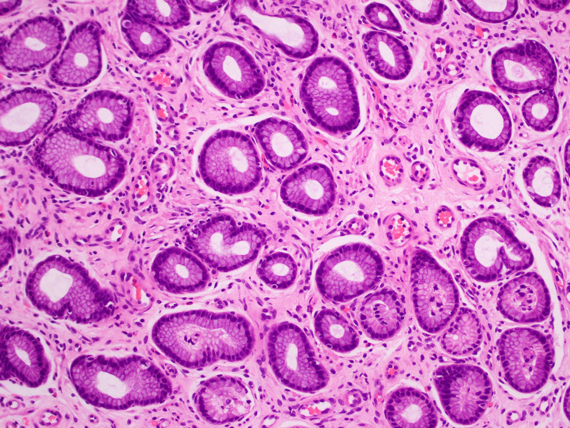
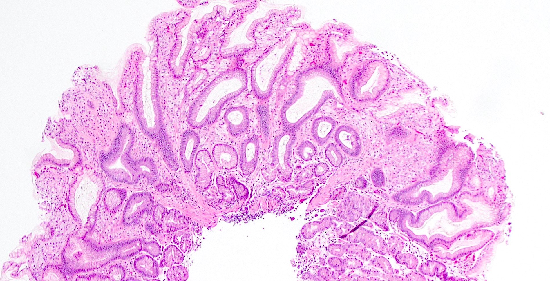
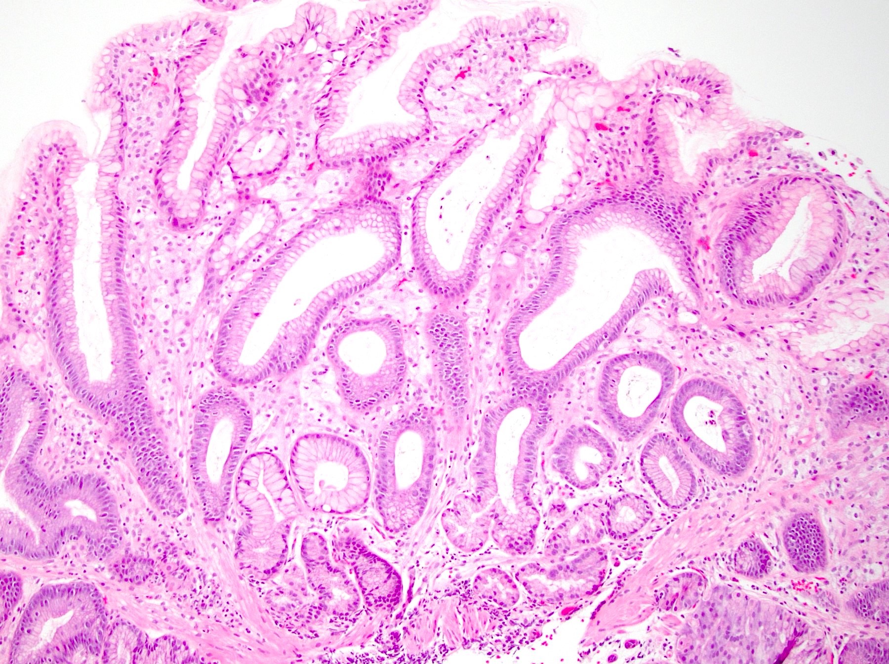
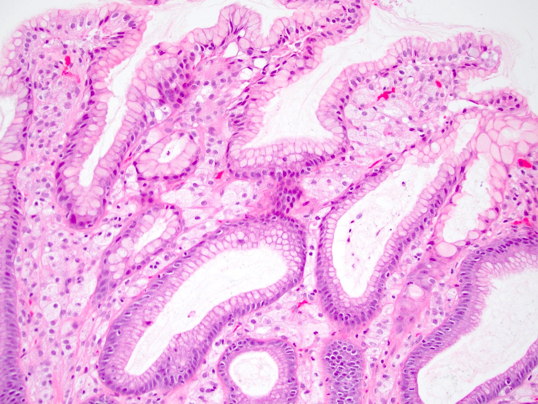
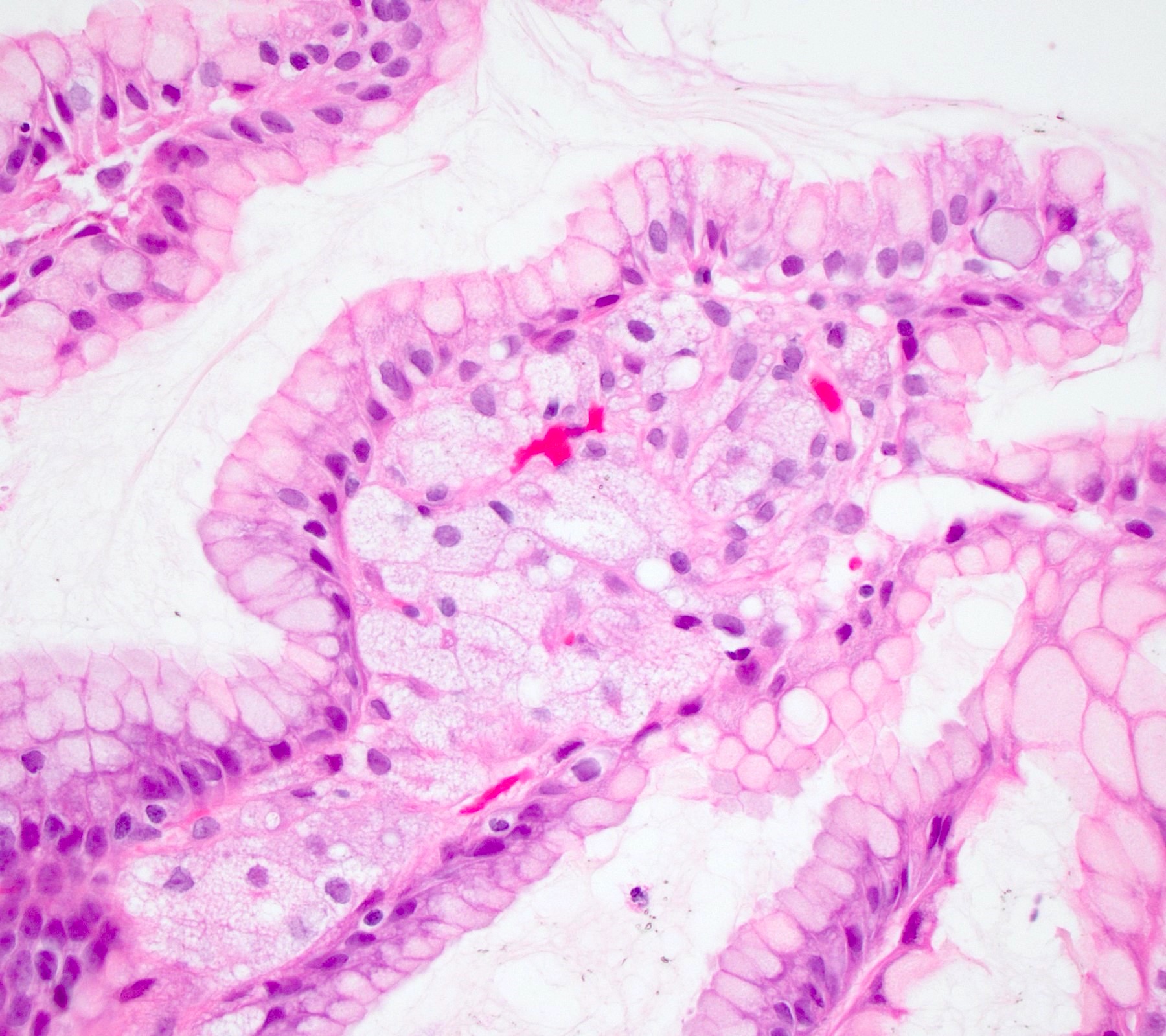
Foveolar type adenoma
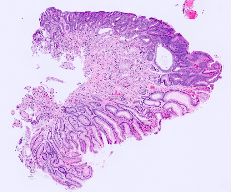
Intestinal type adenoma
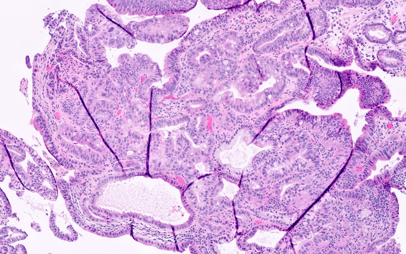
Hybrid phenotype
Contributed by Leon Metlay, M.D.
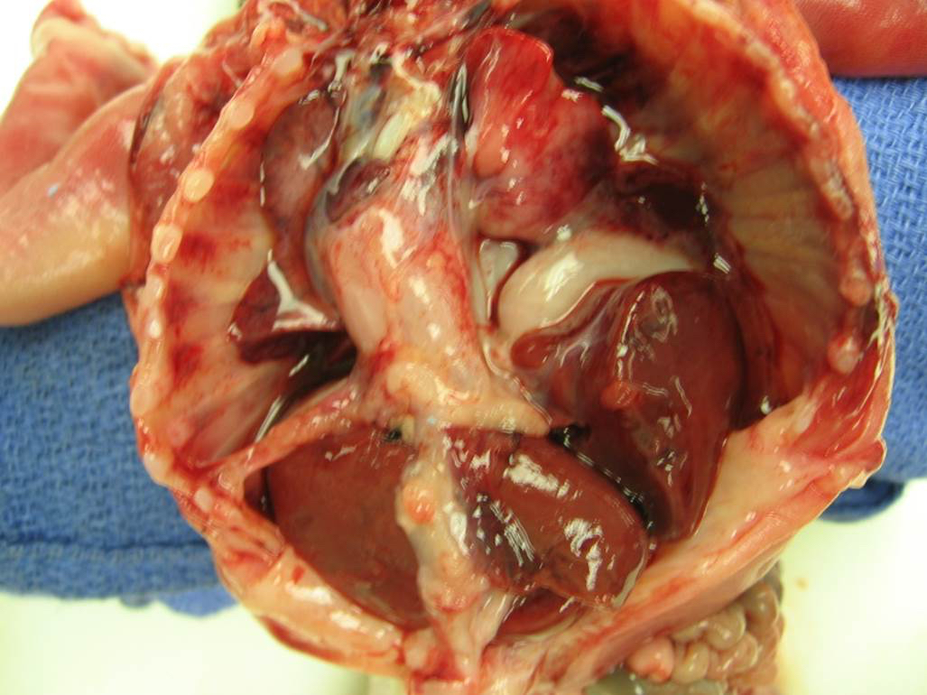
22 week fetus with congenital diaphragmatic hernia
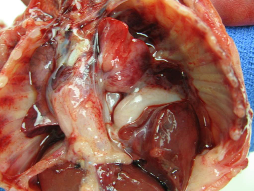
Liver and stomach in chest, heart shifted to right, both lungs small
Hypoplastic left lung and left heart
Hypoplastic right lung
Groove in liver, where diaphragm edge was
Images hosted on other servers:

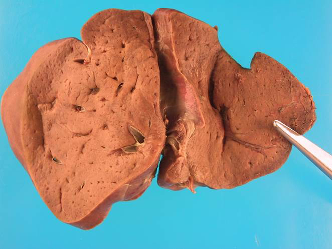
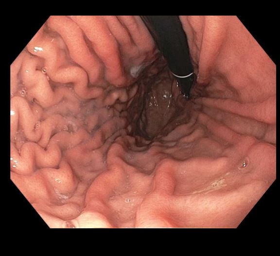
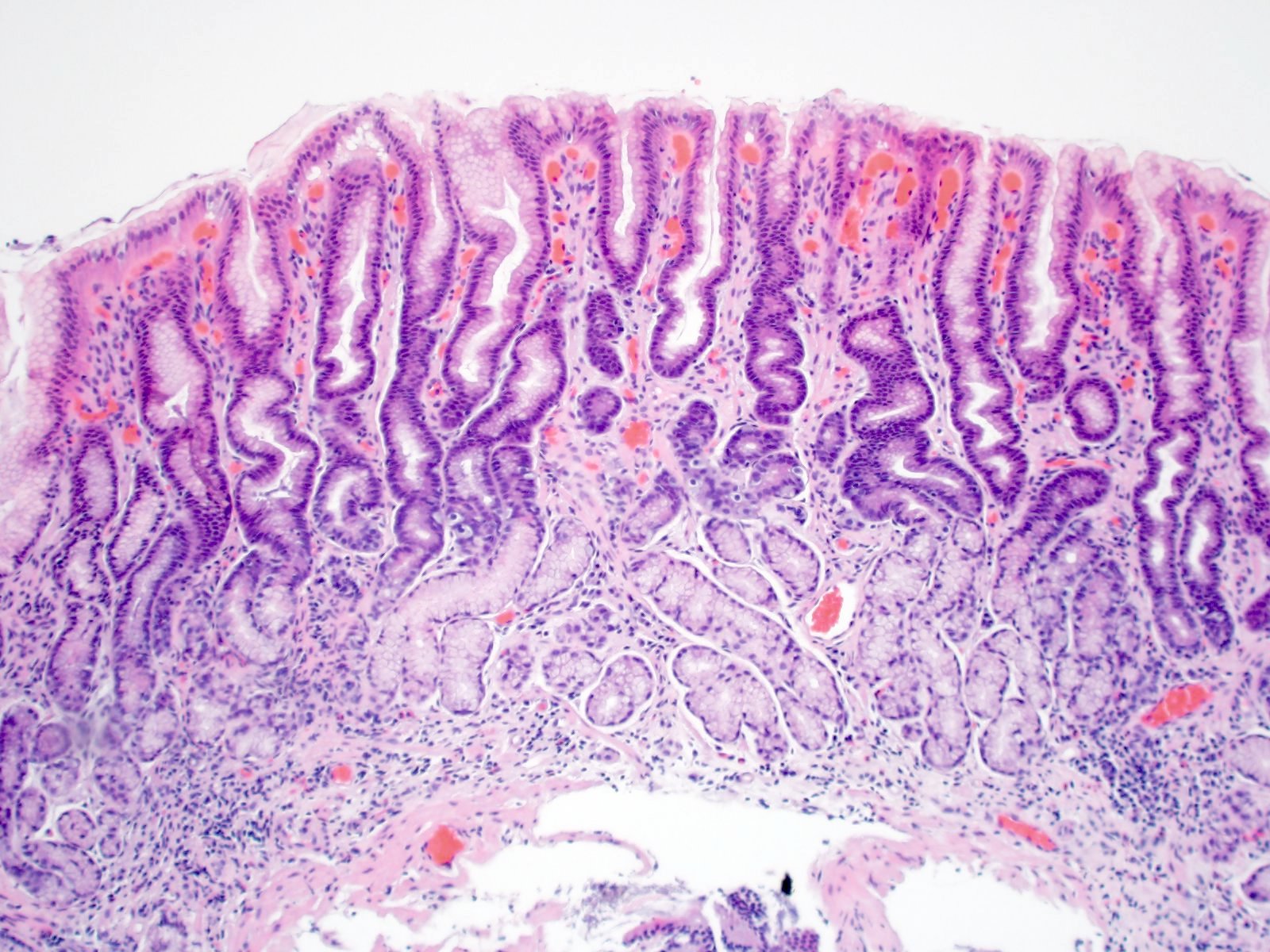
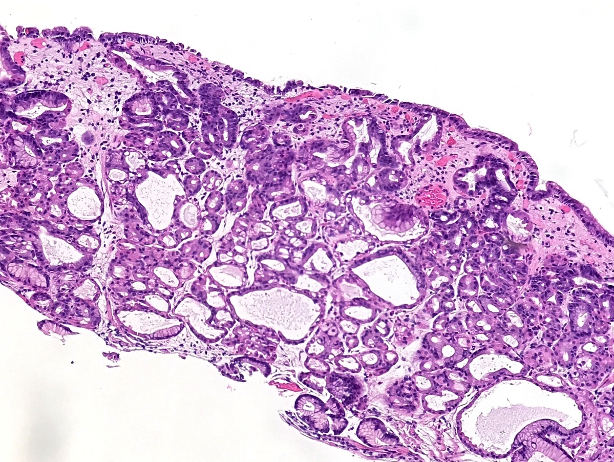
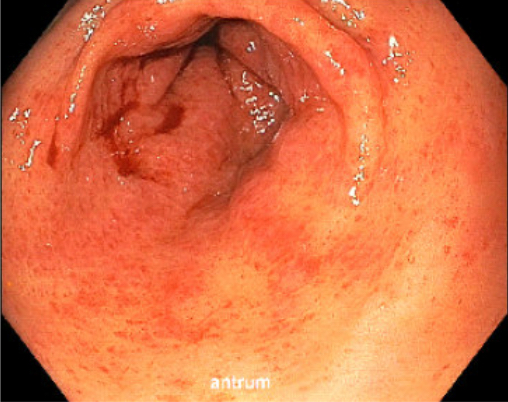
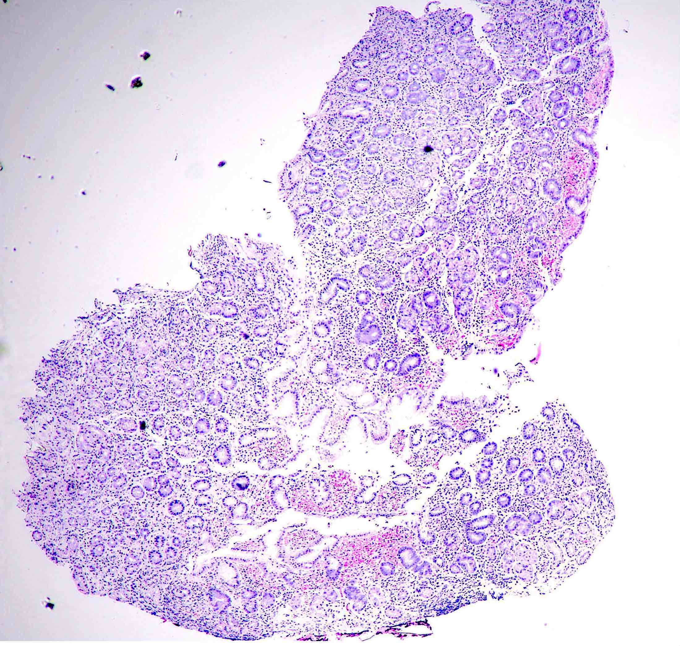
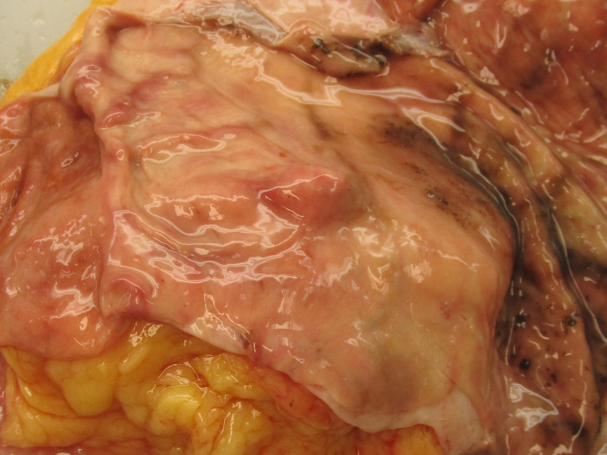
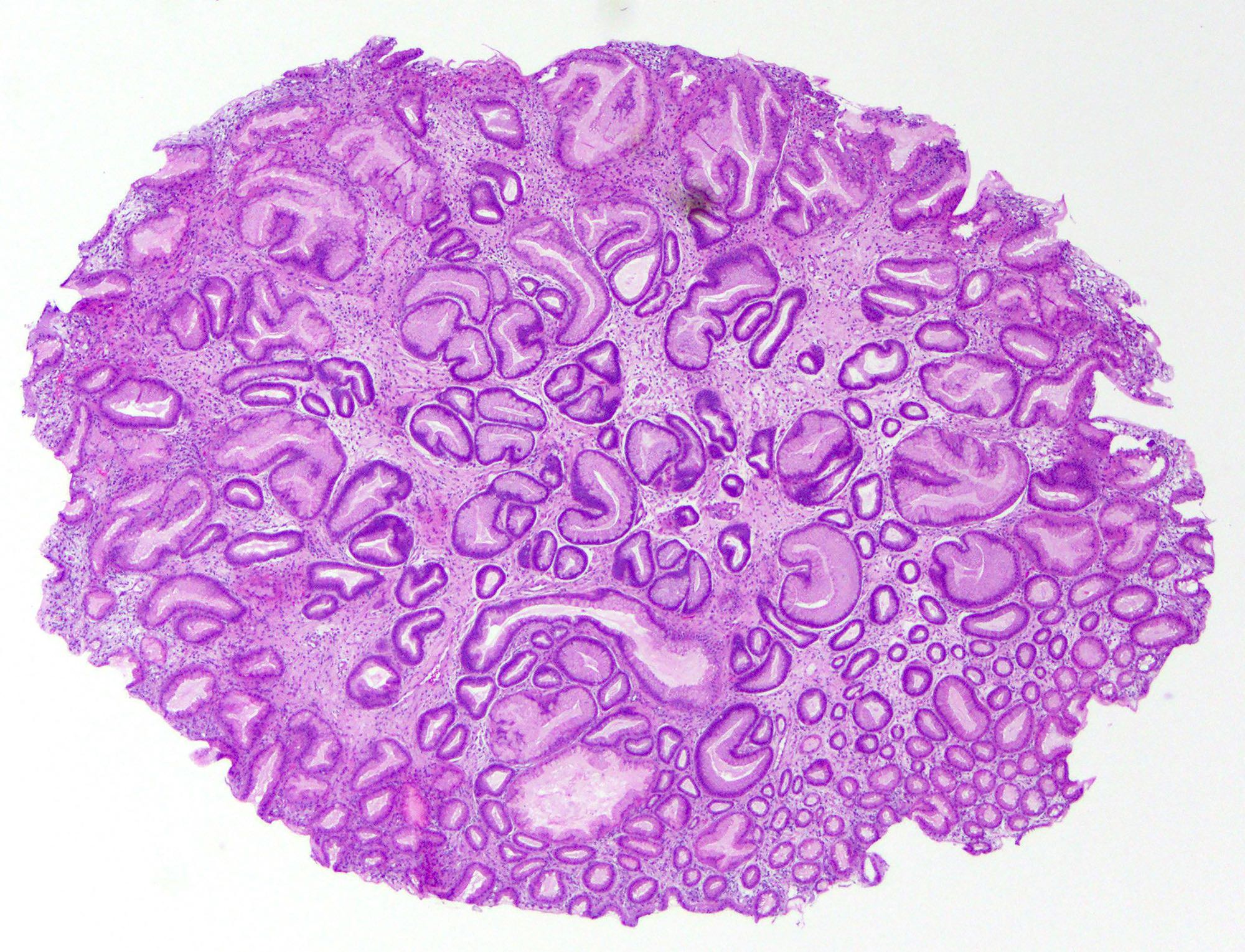
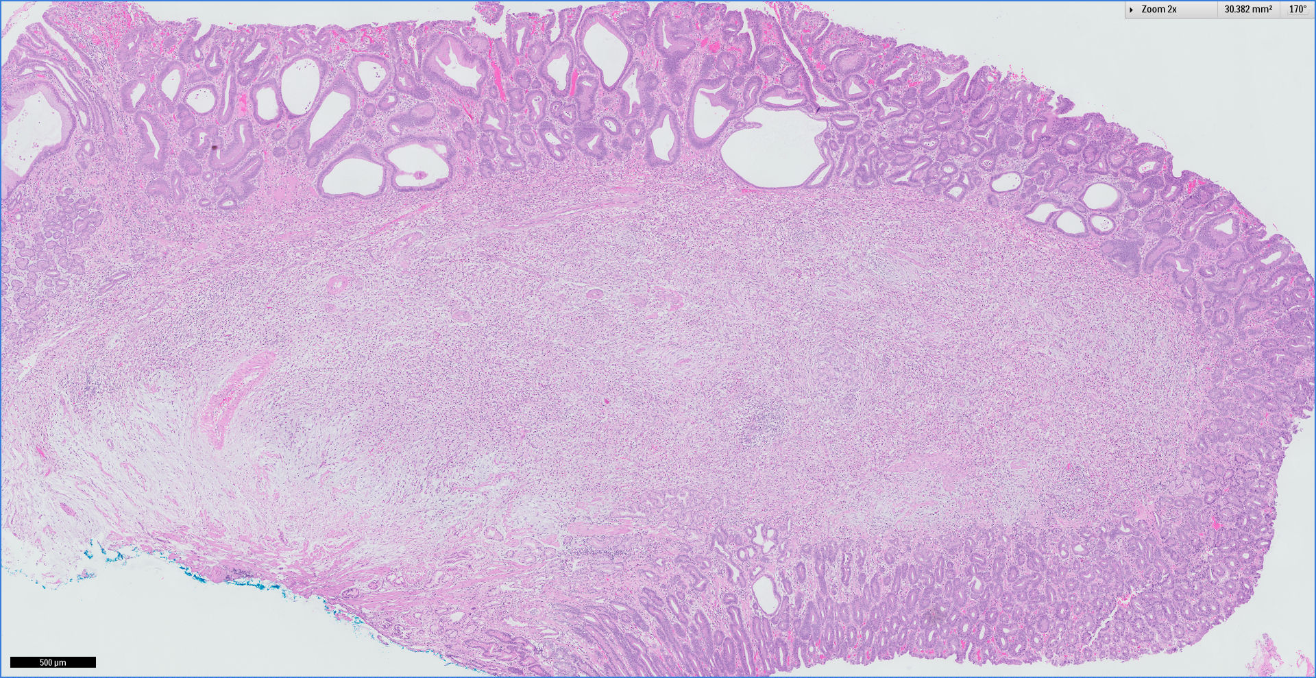
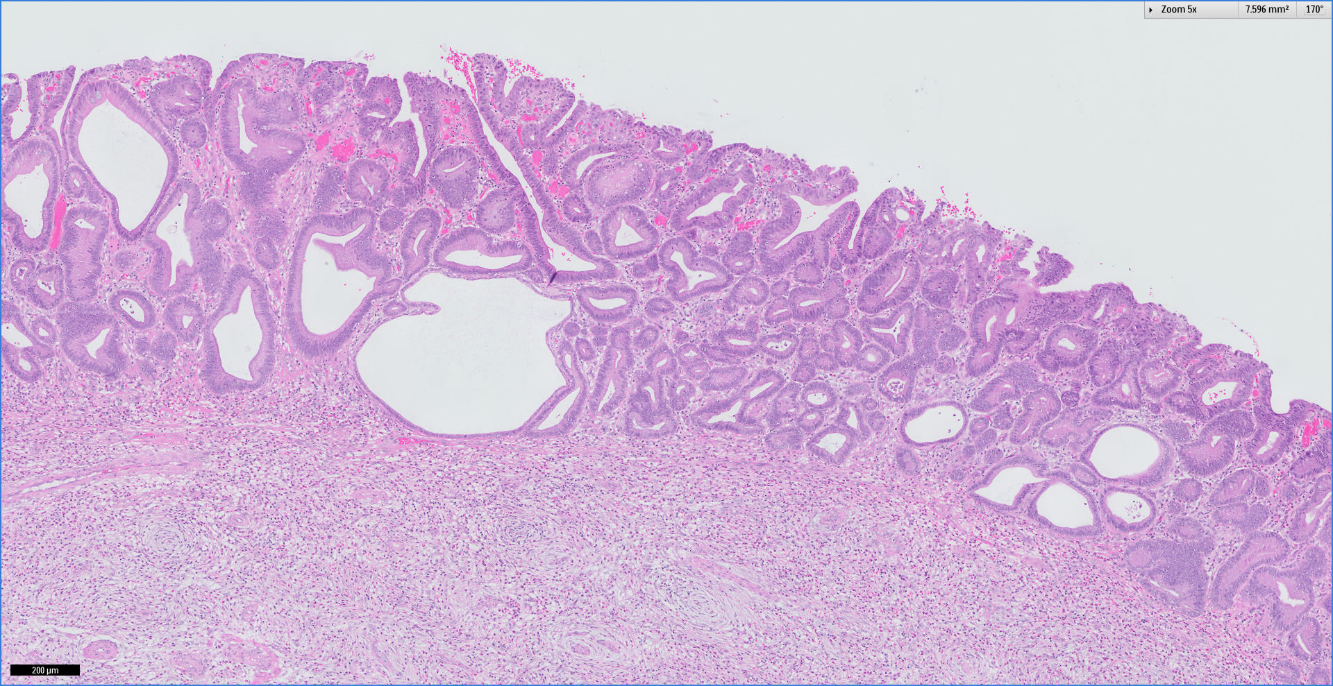
Normal appearance of the stomach, opened along the greater curvature

Normal appearance of the gastric antrum extending to the pylorus at the right of center
Contributed by Kelsey E. McHugh, M.D.
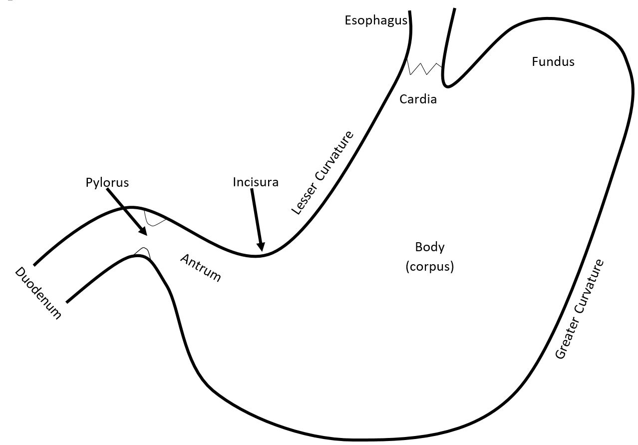
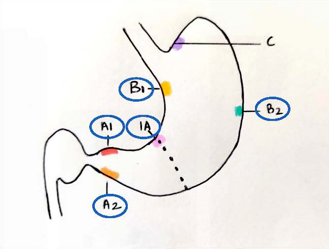
Anatomic regions
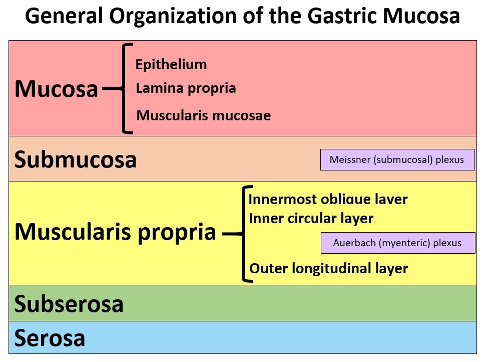
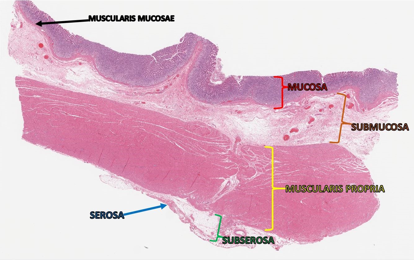
Layers of gastric wall
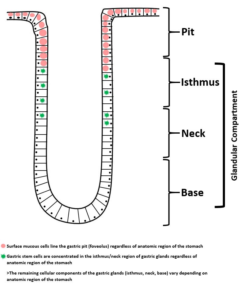
Gastric mucosa
Contributed by Kelsey E. McHugh, M.D.
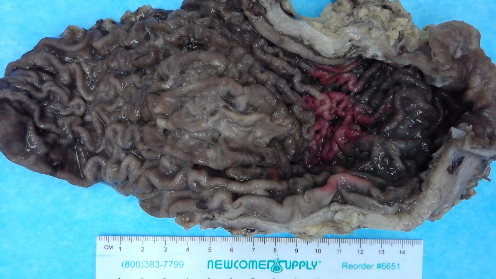
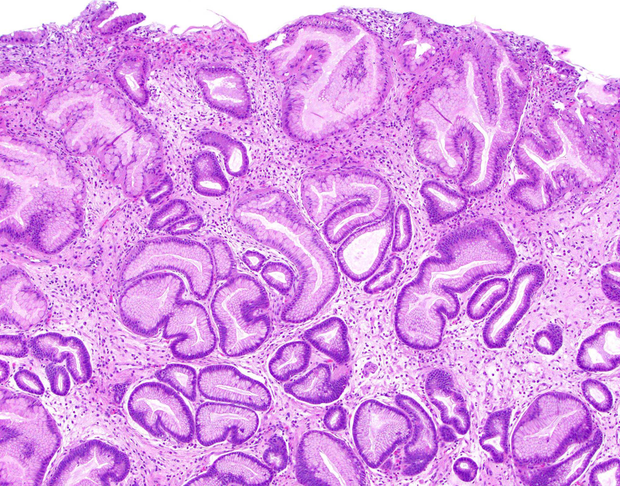
Gastric body with
unremarkable
rugal folds
Contributed by Leon Metlay, M.D. and Kelsey E. McHugh, M.D.
Anatomy:
Formalin fixed gastric body
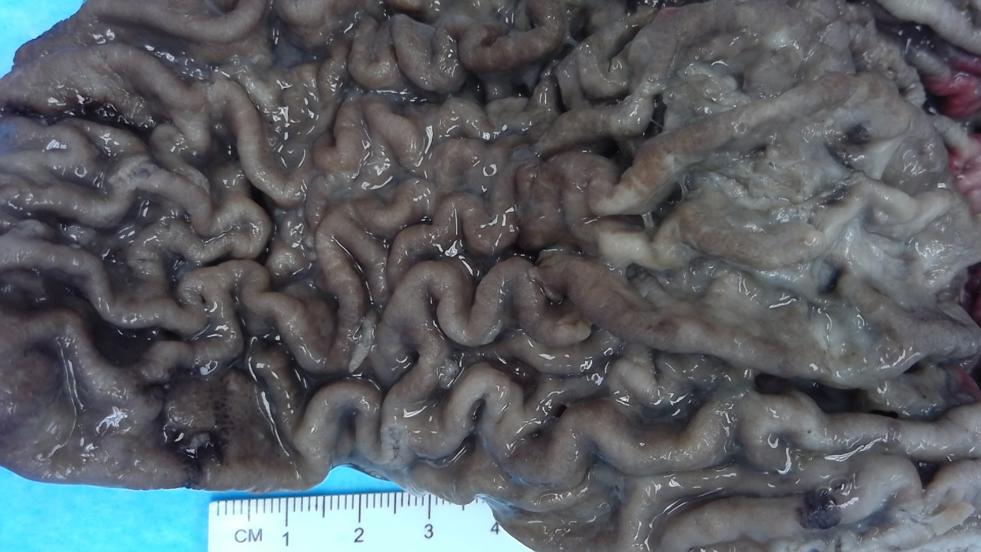
Formalin fixed gastric rugal folds
Images hosted on other servers:
Anatomy:
Normal appearance of the stomach
Normal appearance of the gastric antrum


Total gastrectomy anatomic regions
Contributed by Kelsey E. McHugh, M.D.
Gastric wall layers
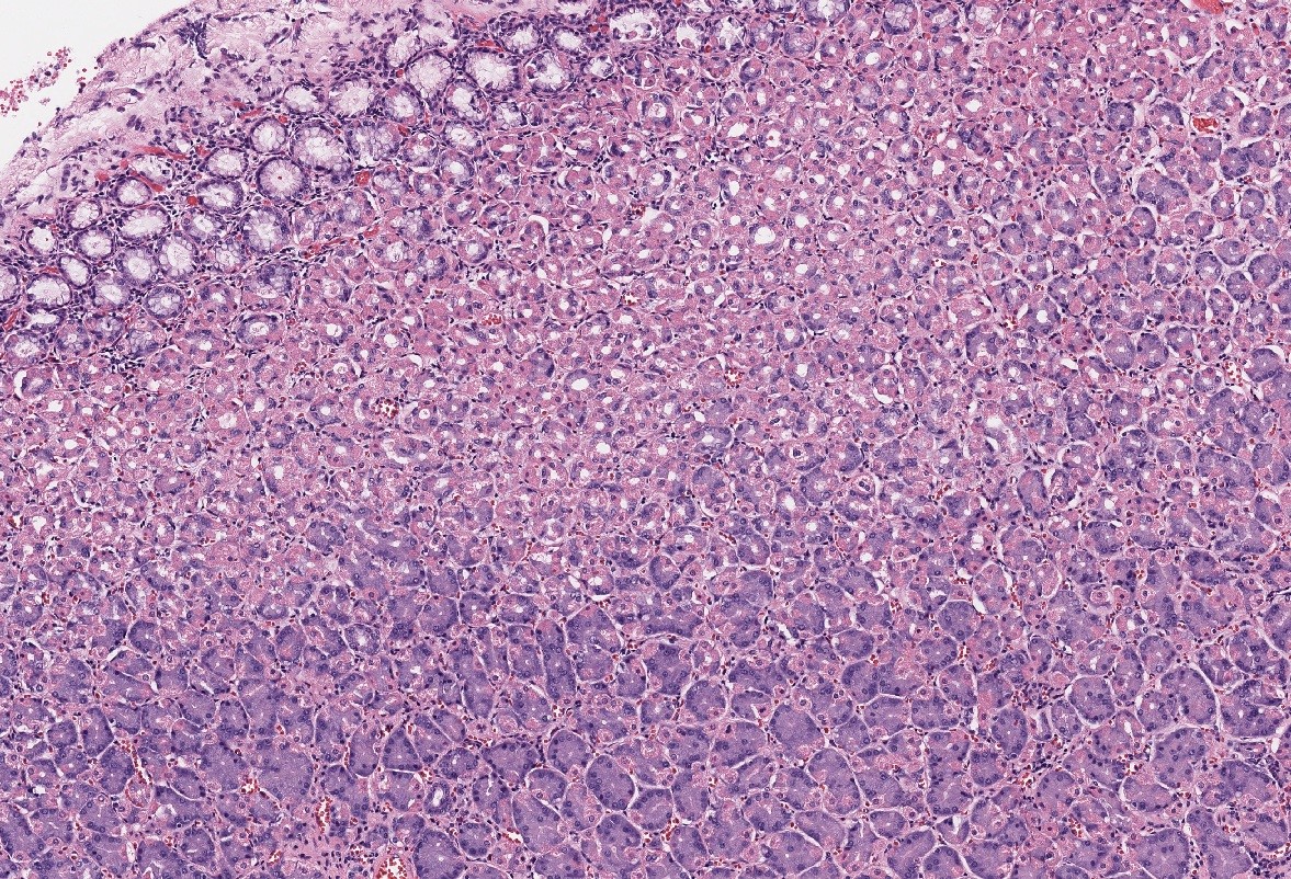
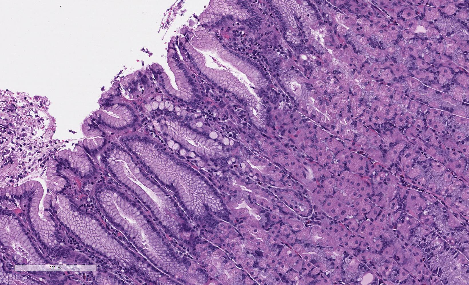
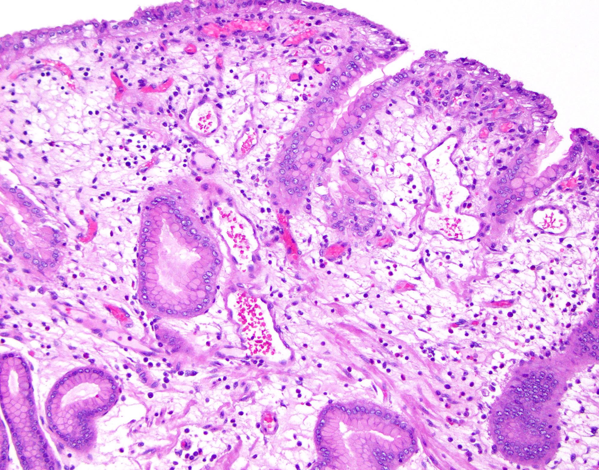
Oxyntic mucosa
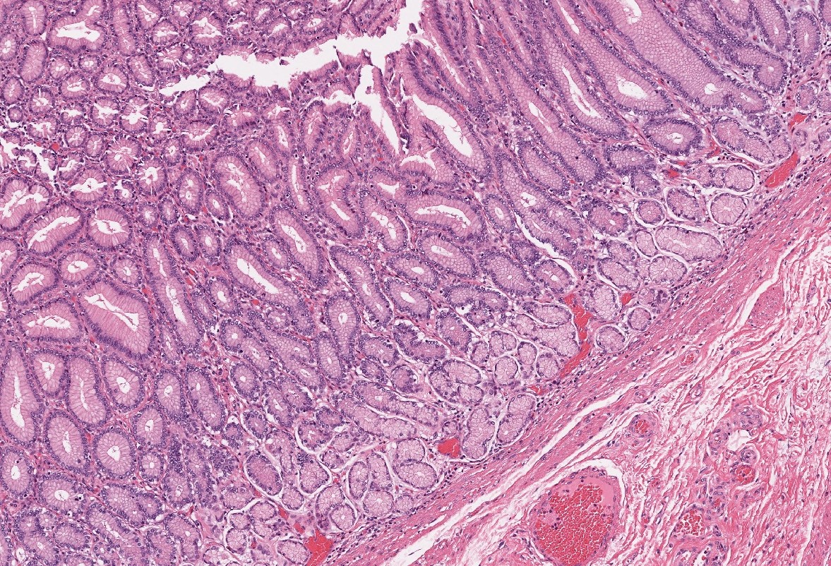
Antral mucosa
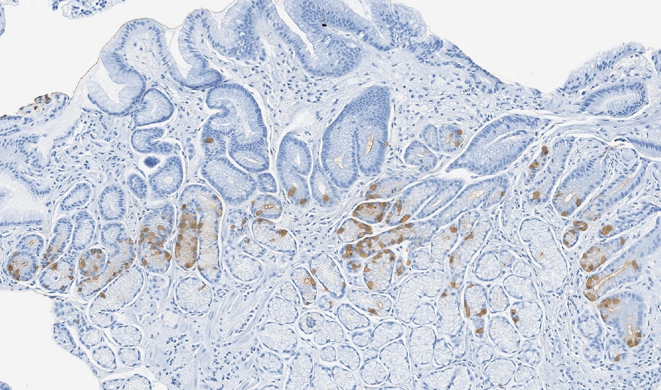
Gastrin immunostain
Cardia mucosa
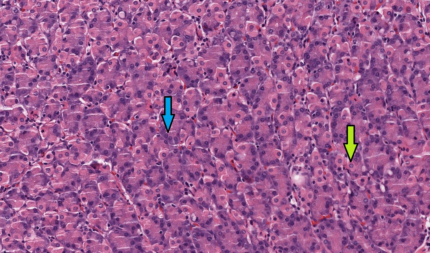
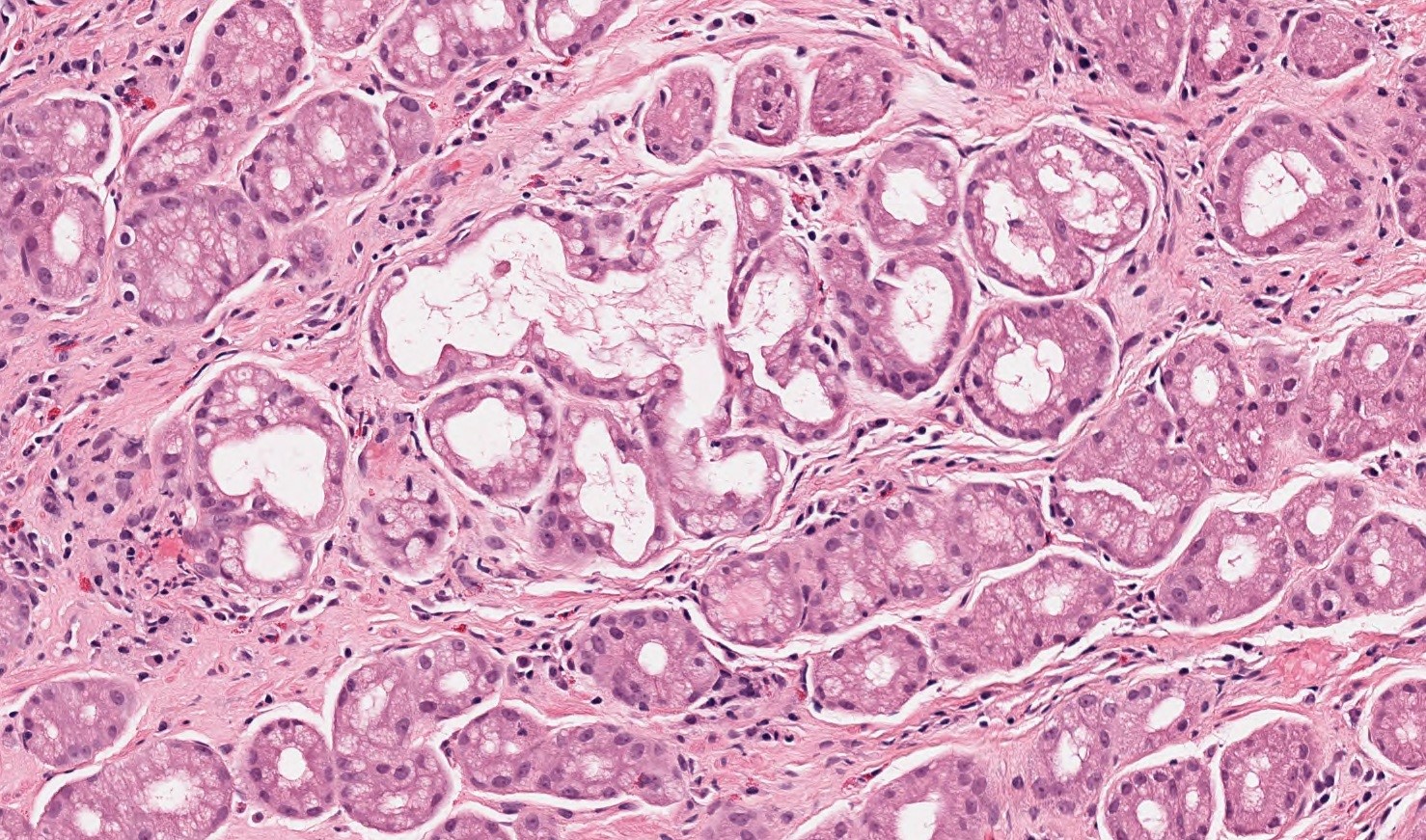
Parietal and chief cells
Gastric pits
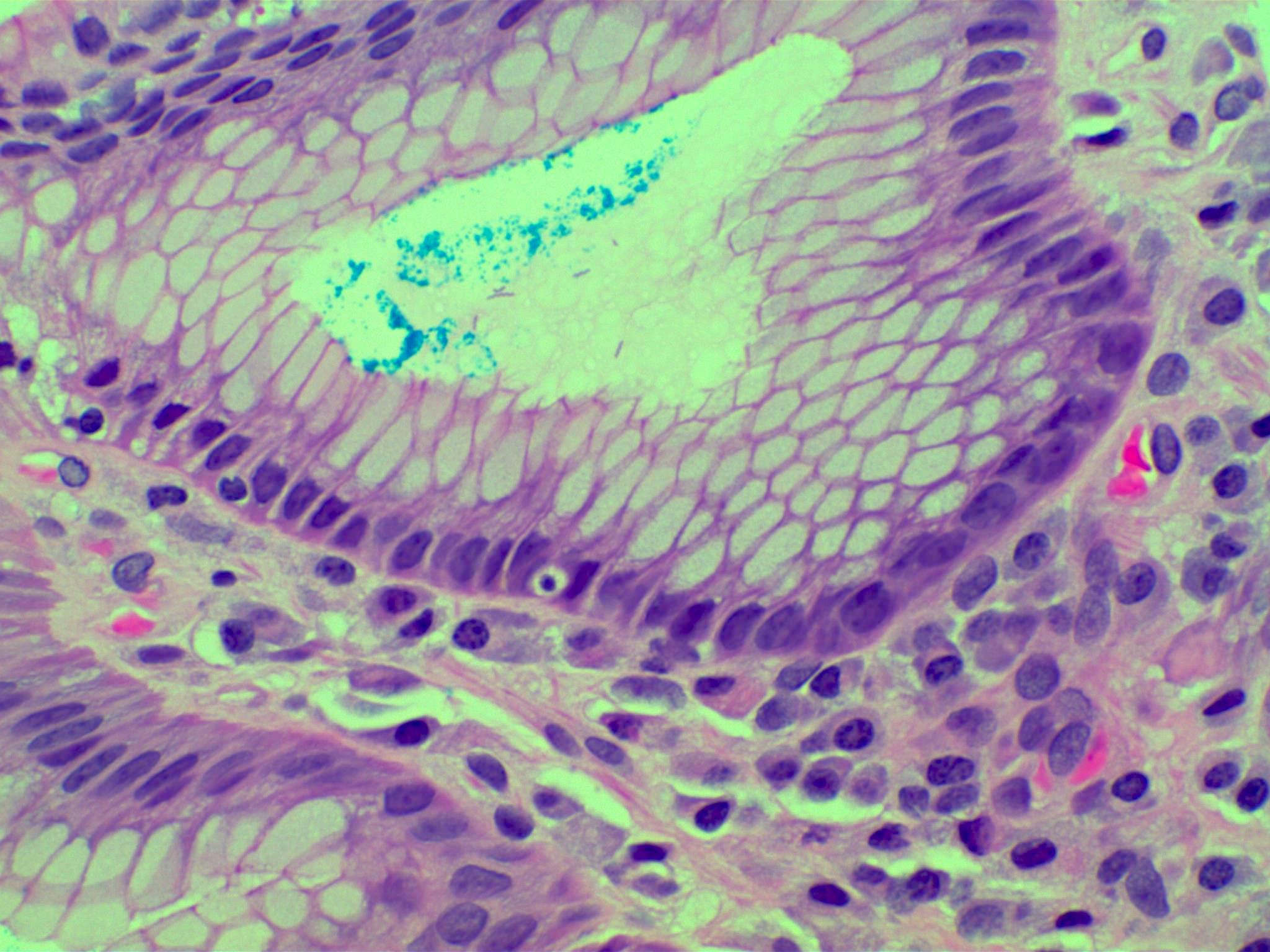
Pseudo-signet ring cell artifact
Distended foveolar cells
PAS / AB in distended foveolar cells
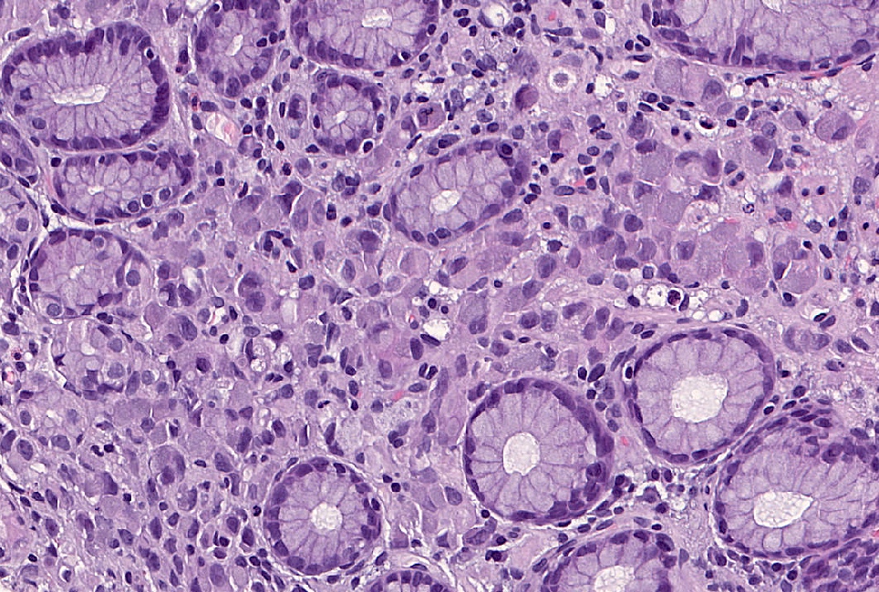
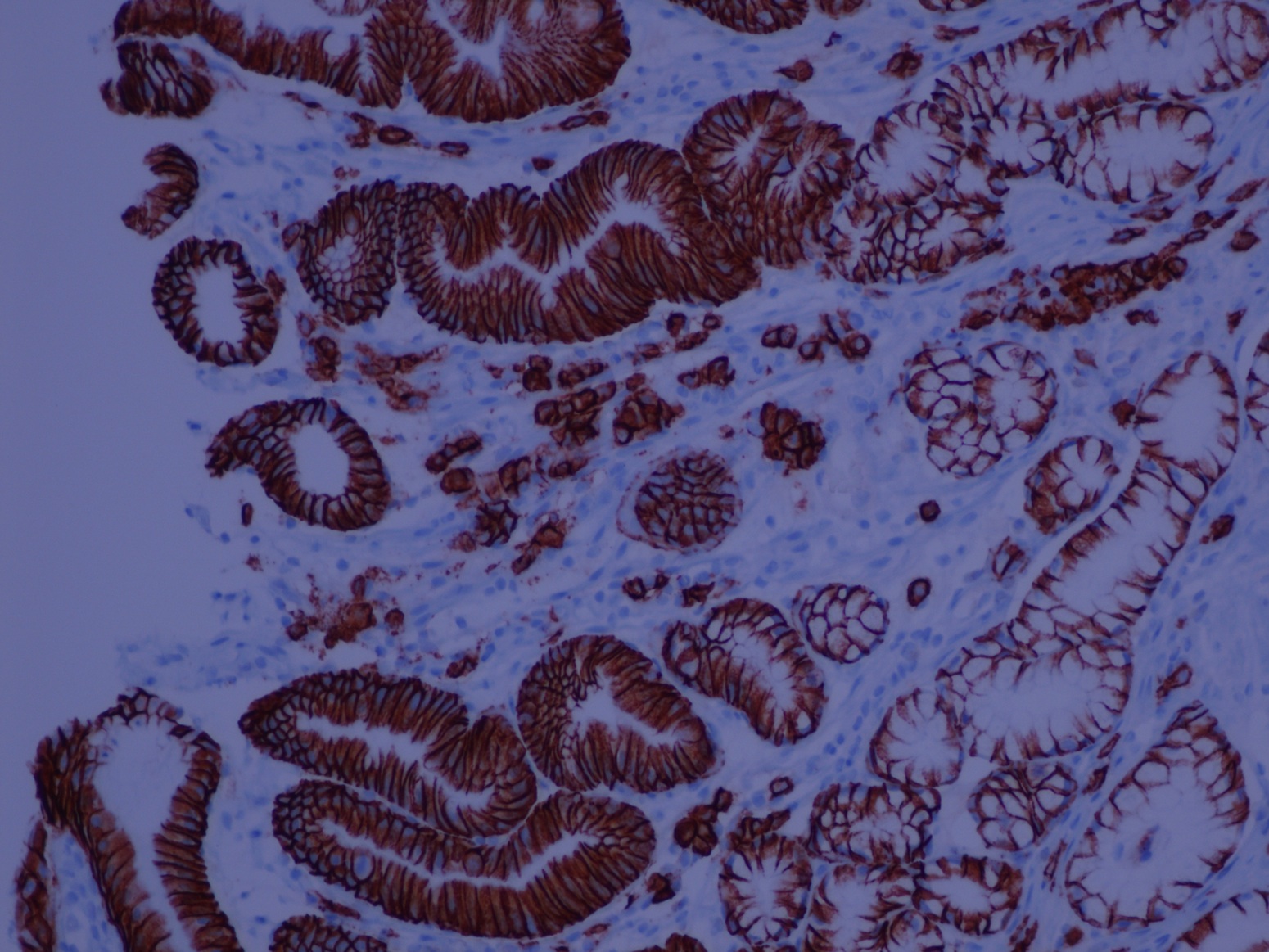
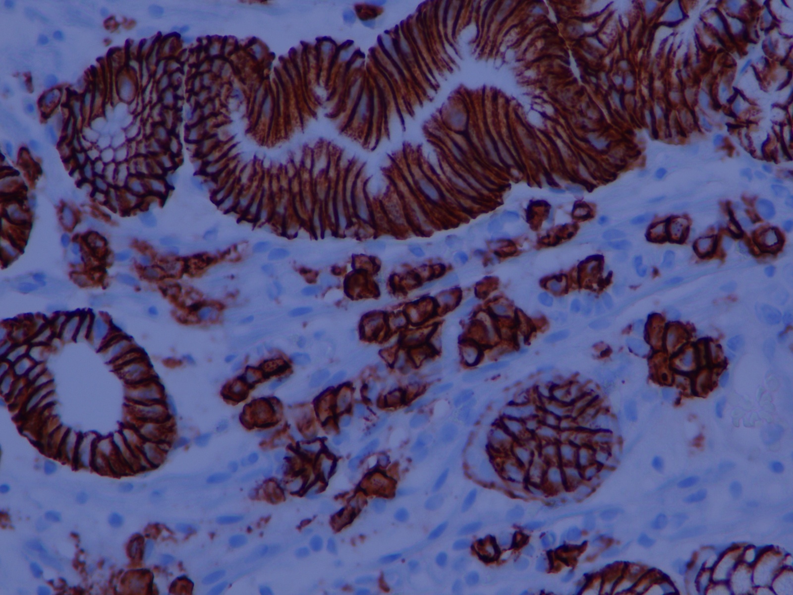
Neuroendocrine cells in gastric antrum
Contributed by Kelsey E. McHugh, M.D.
Surface foveolar epithelium
Parietal cells
Chief cells
Contributed by Raul Gonzalez, M.D.
Gastric arteriovenous malformation
Images hosted on other servers:

Barium Xray
Contributed by Kirbylee Nelson, M.D.
Prominent submucosal vessels
Loss of rugal folds
Contributed by Jen Rytych, M.D. and Joshua A. Hanson, M.D.
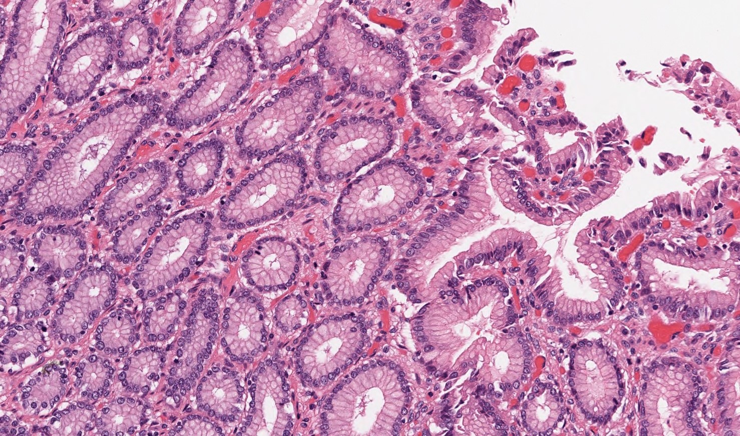
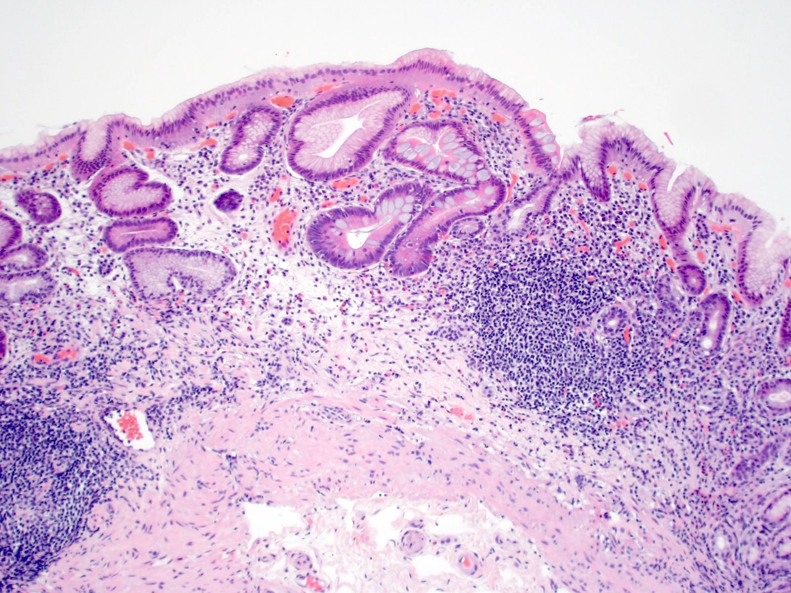
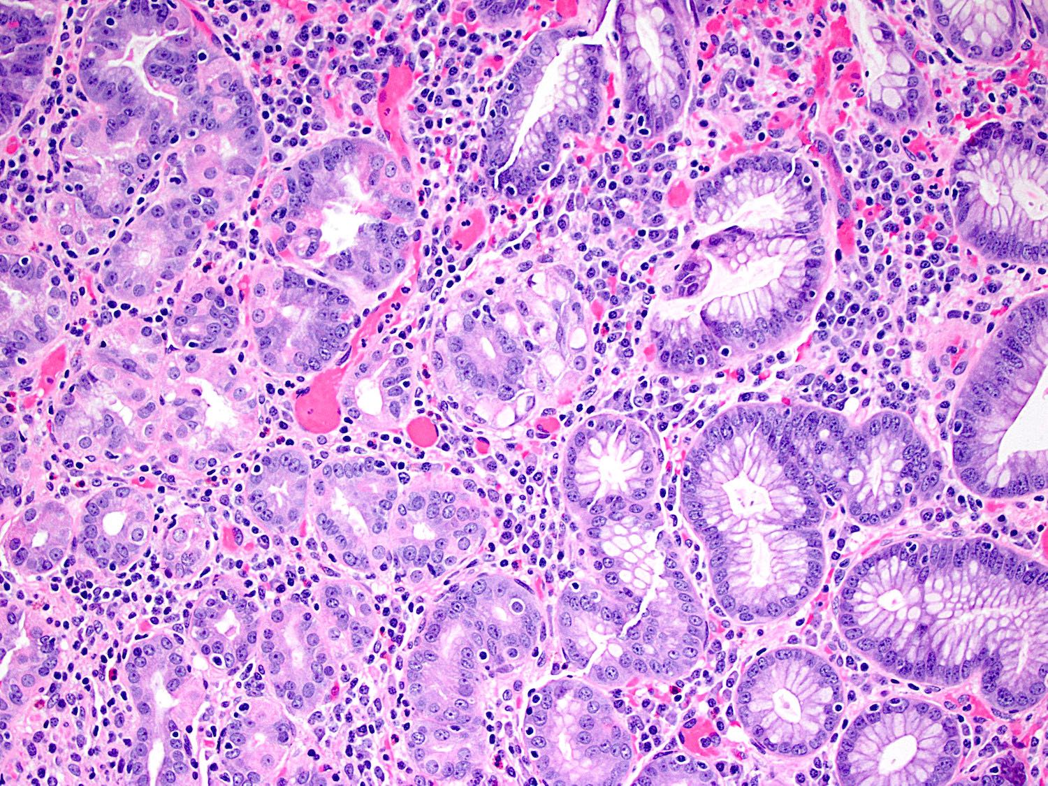
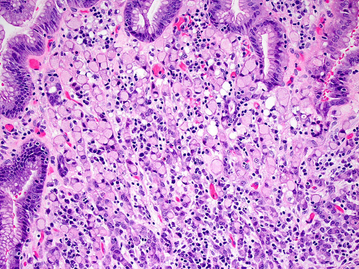
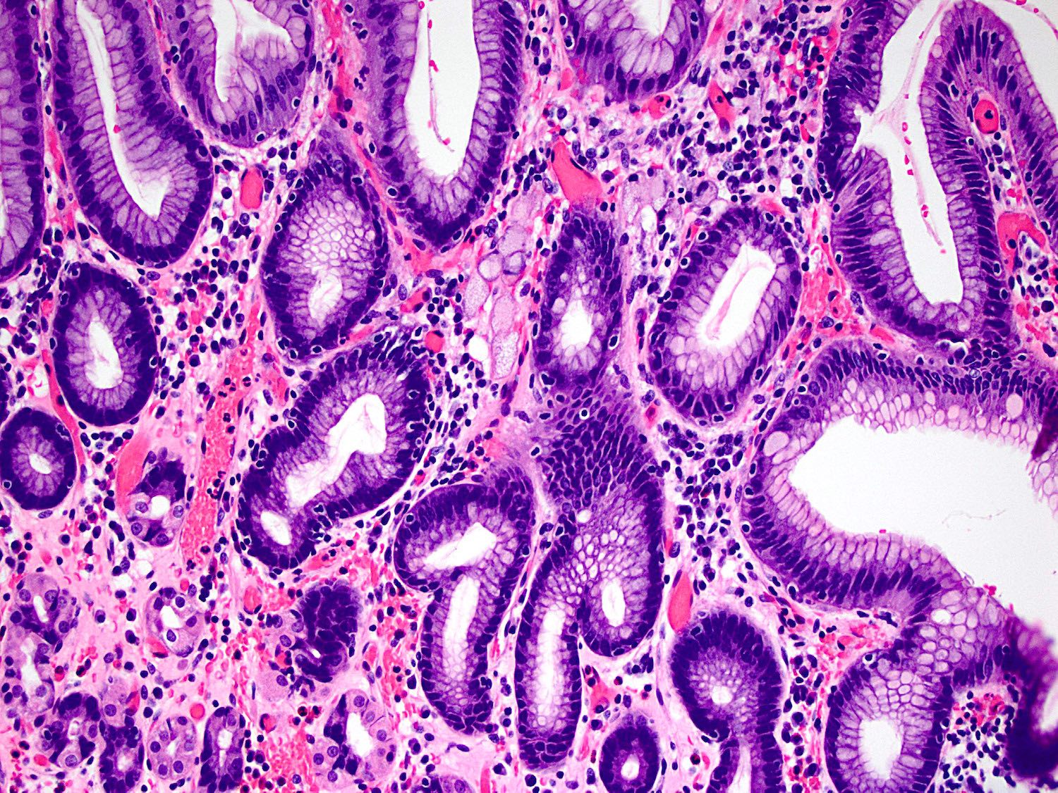
Atrophic mucosa
Intestinal metaplasia
ECL cell hyperplasia
Atrophic mucosa and inflammation
Pseudopyloric metaplasia
Intestinal metaplasia
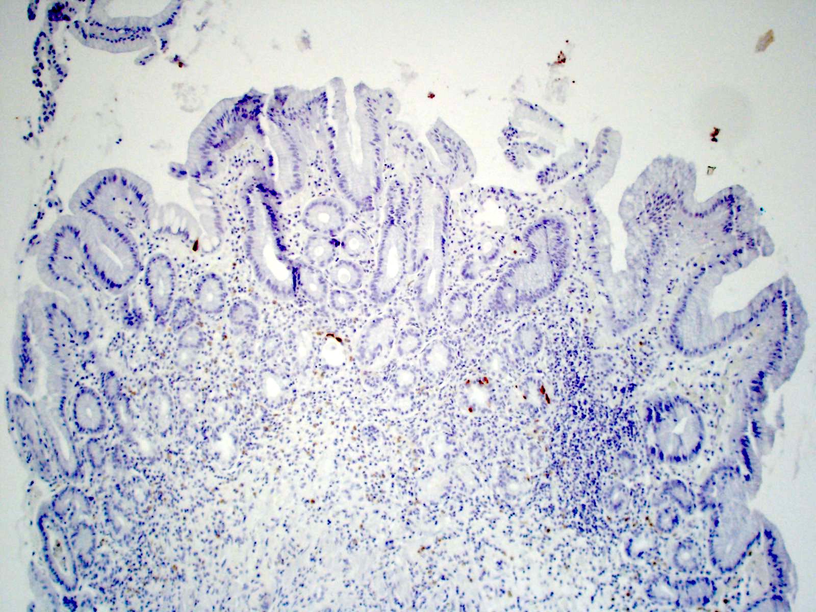
Positive H. pylori stain
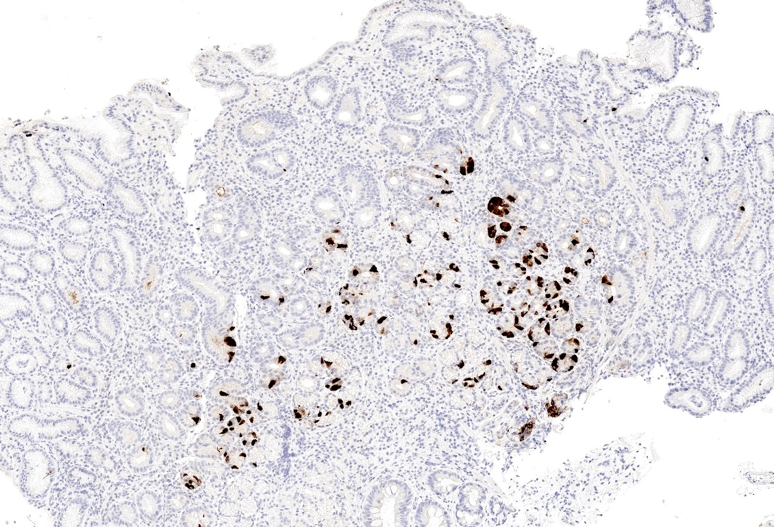
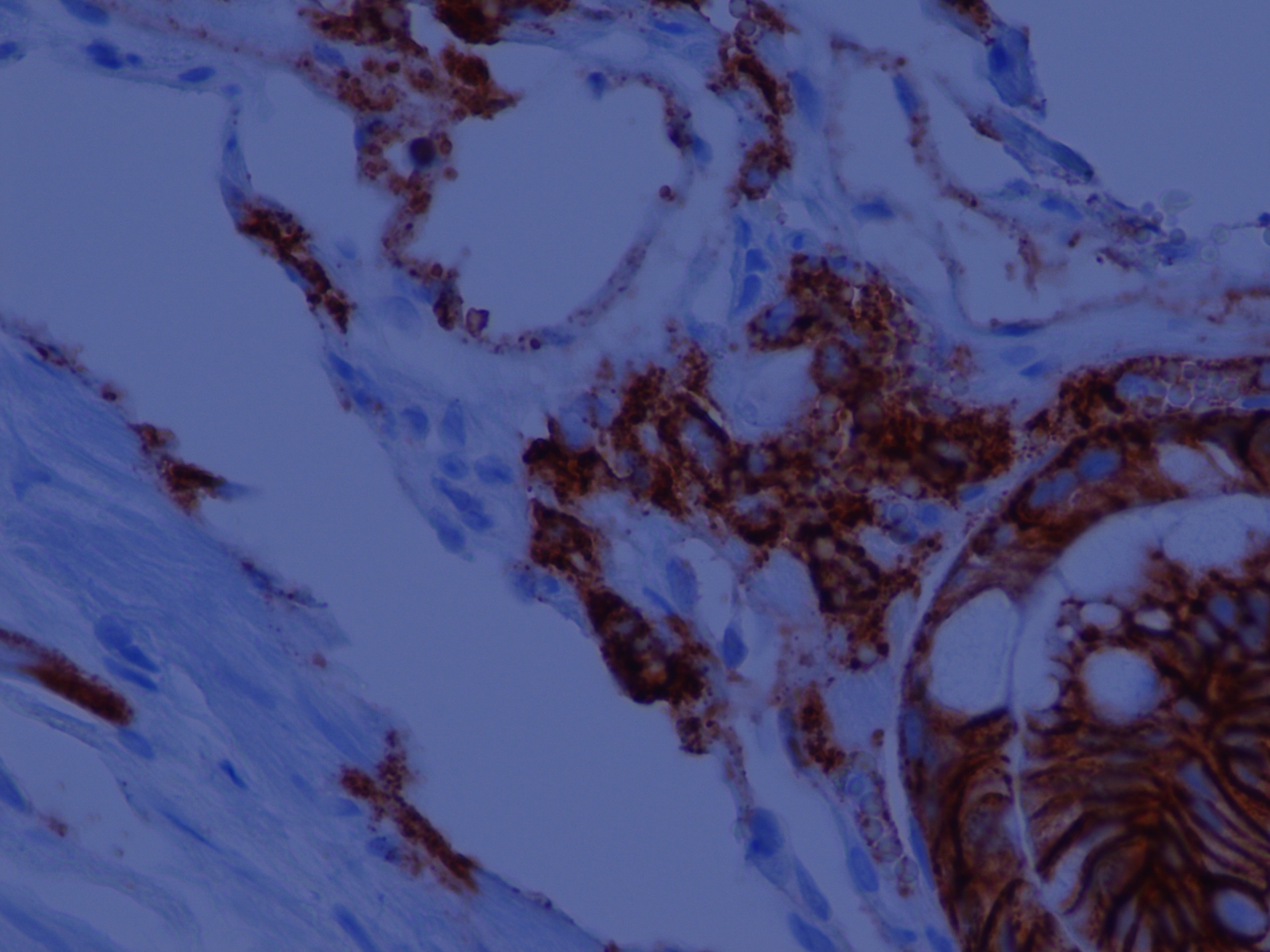
Positive gastrin stain
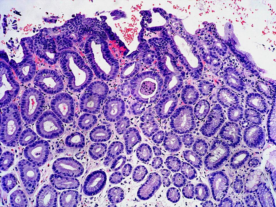
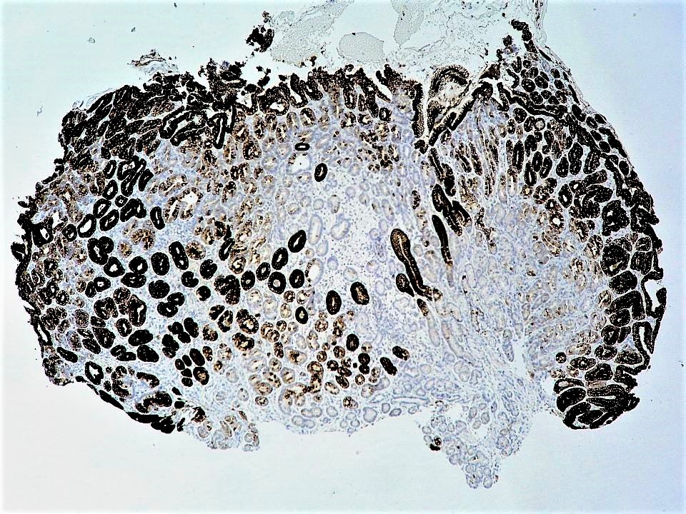
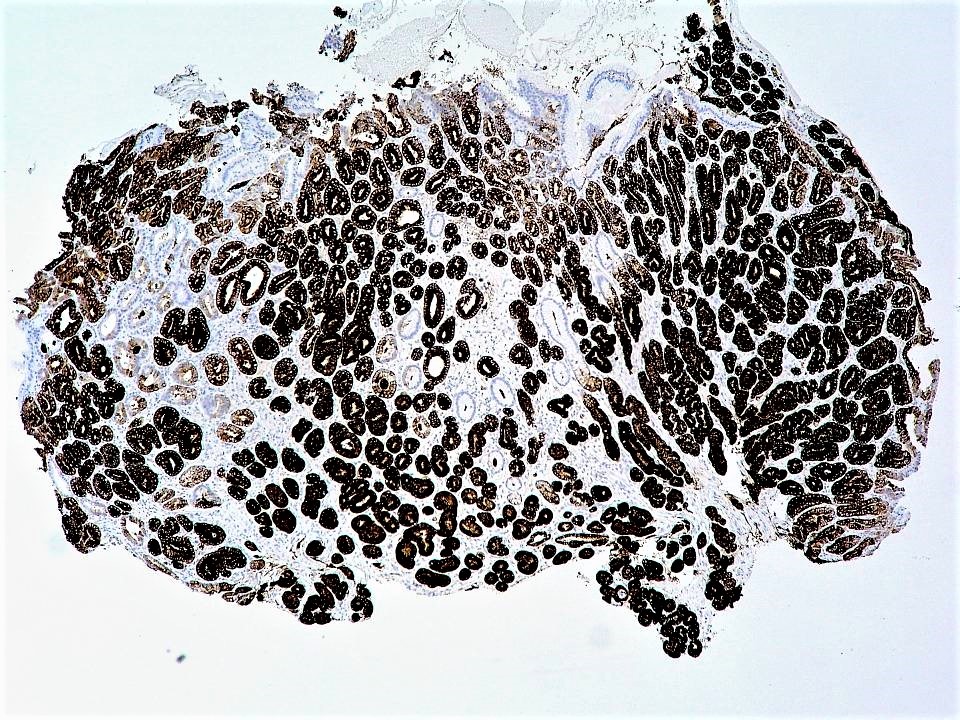
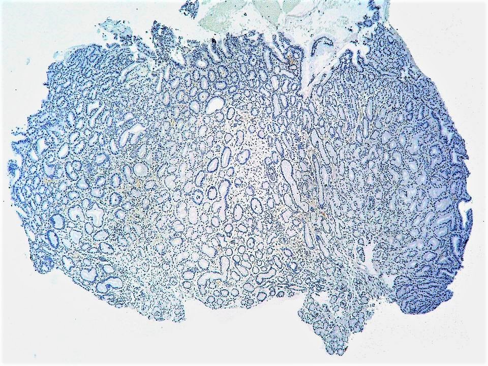
Atrophic gastric body
Gastrin stain, body
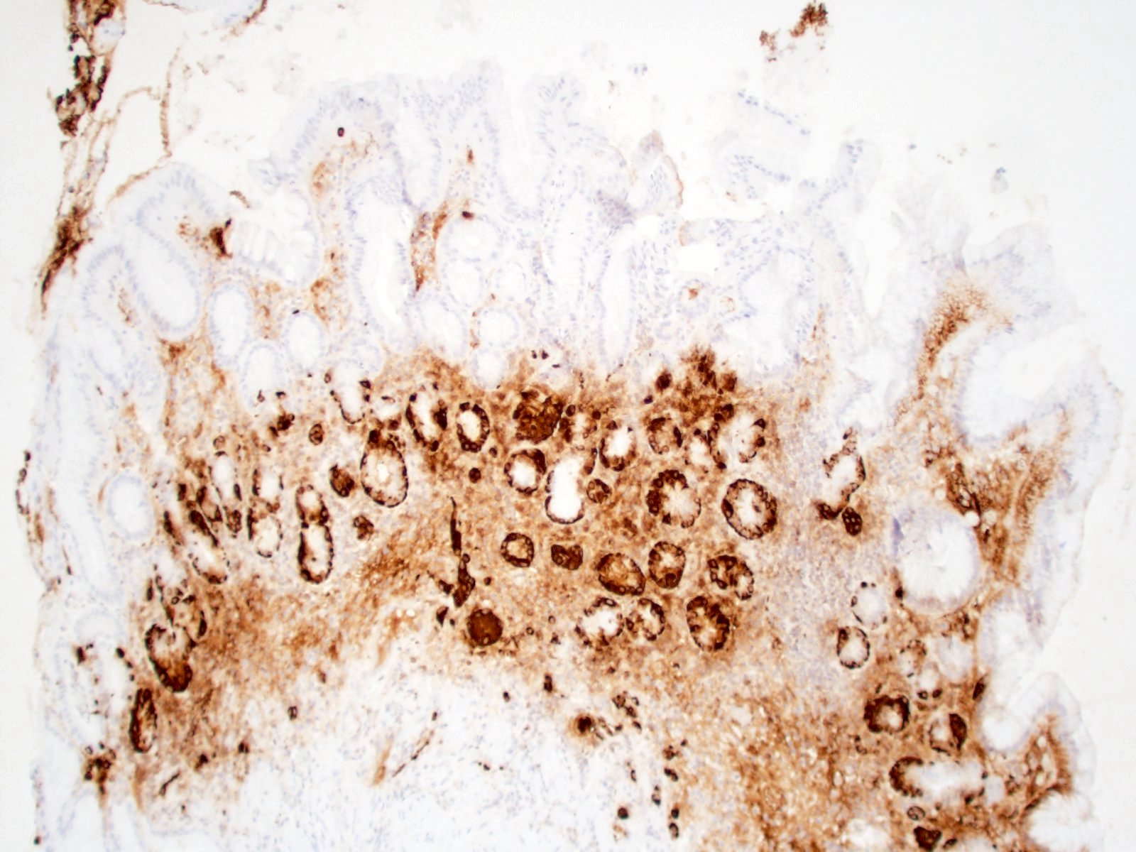
Chromogranin stain, body
Antrum in autoimmune gastritis

Gastrin stain, antrum
Histology of atrophic gastritis
Histology of autoimmune atrophic gastritis
Images hosted on other servers:

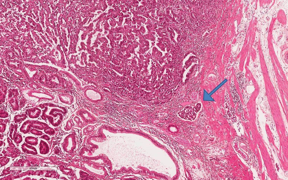
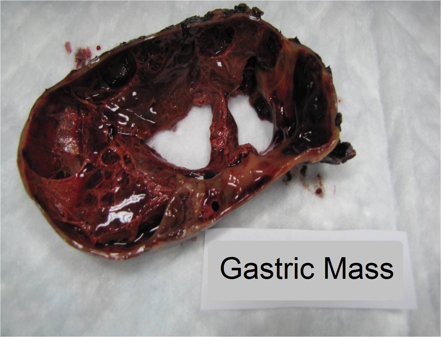
Gastric tumor infiltrating the diaphragm
Contributed by Carolina Martinez Ciarpaglini, M.D., Ph.D.
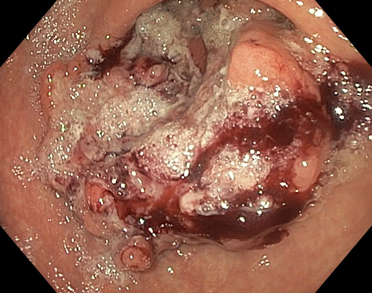
Hemorrhagic gastric mass
Images hosted on other servers:


Ulcerated gastric mass

Early gastric cancer
Contributed by Clara Alfaro, M.D., Ph.D. and Carolina Martinez Ciarpaglini, M.D., Ph.D.
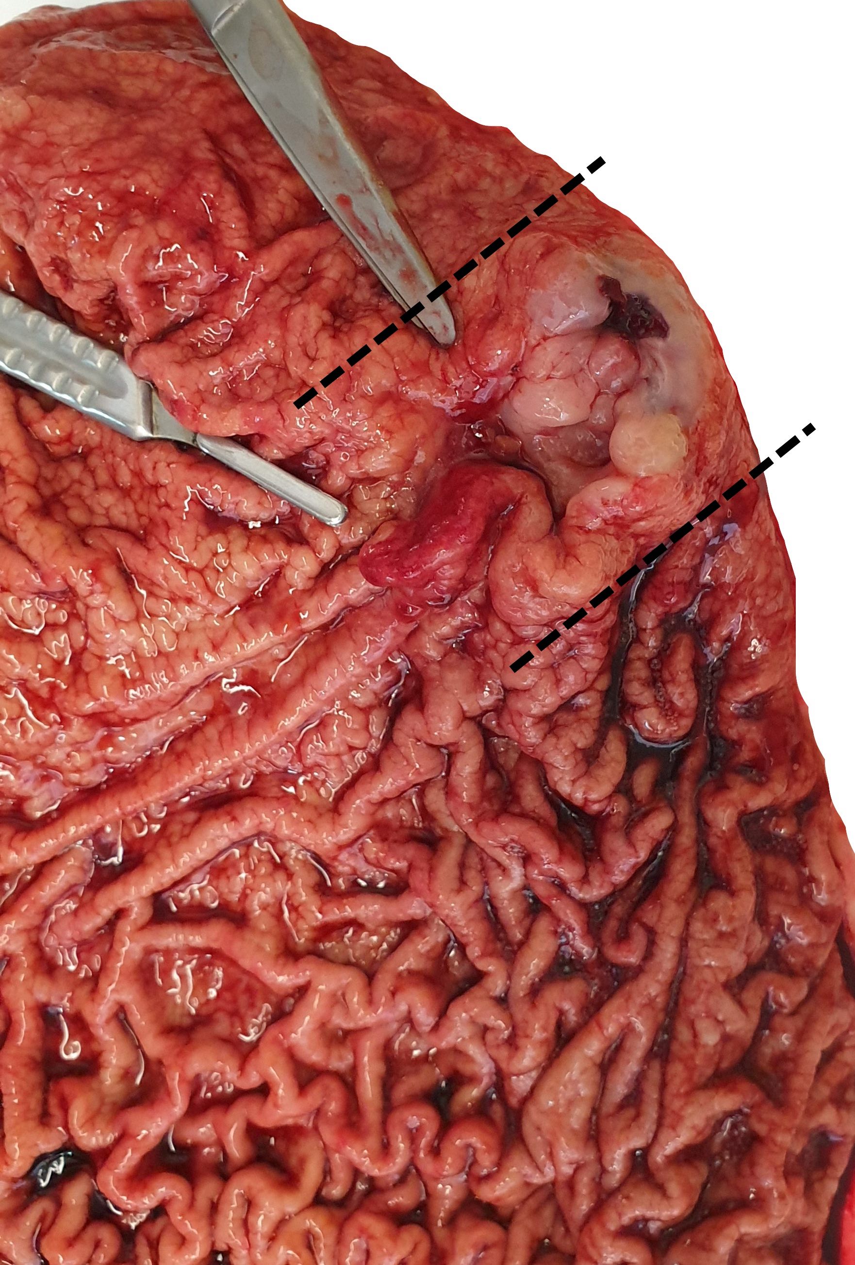
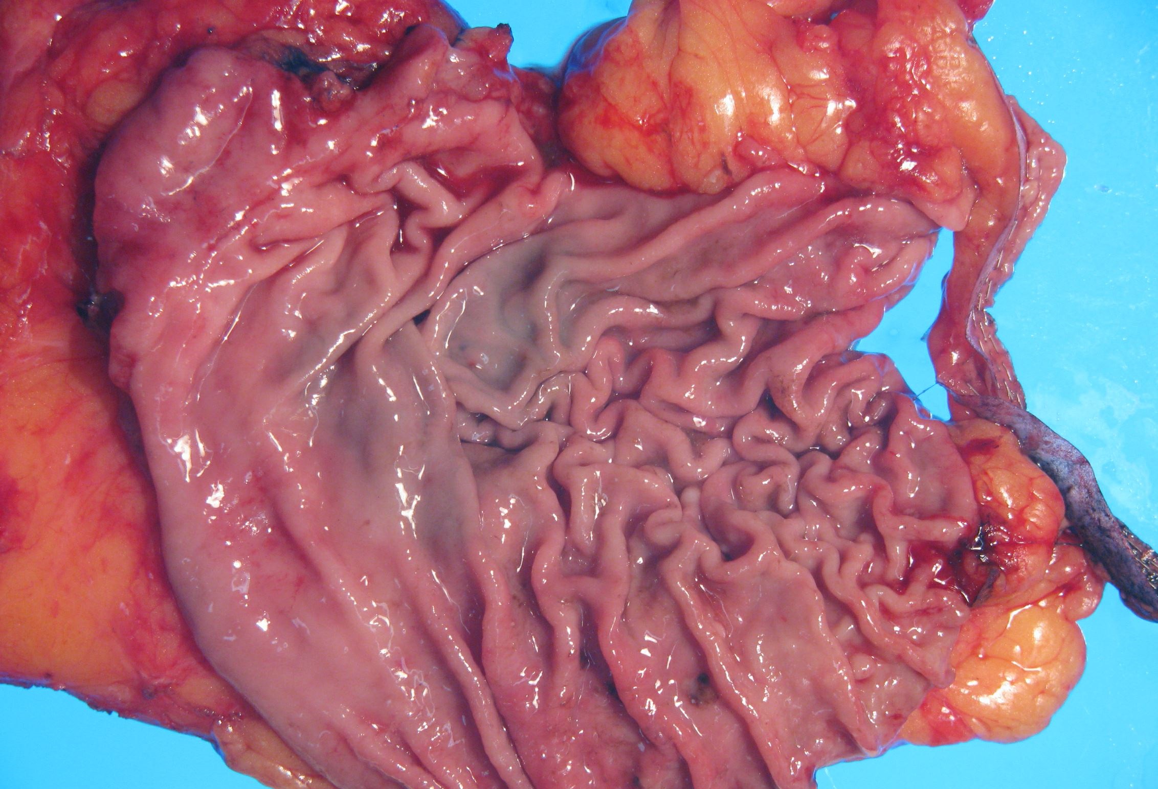
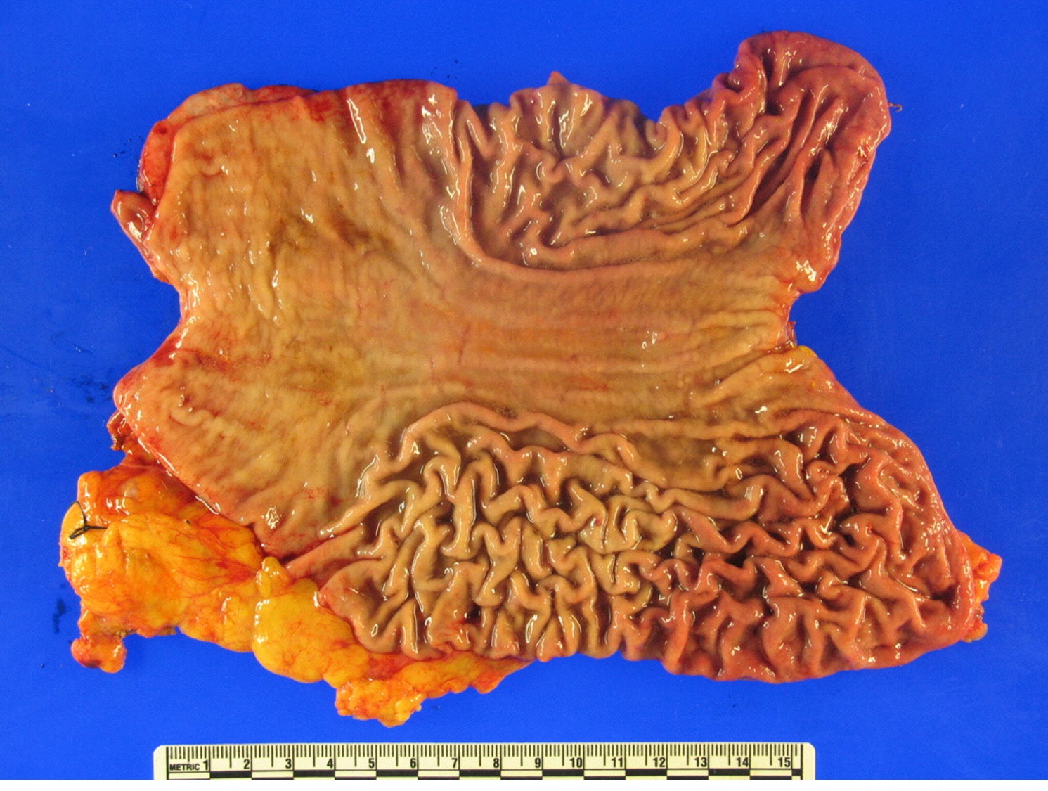
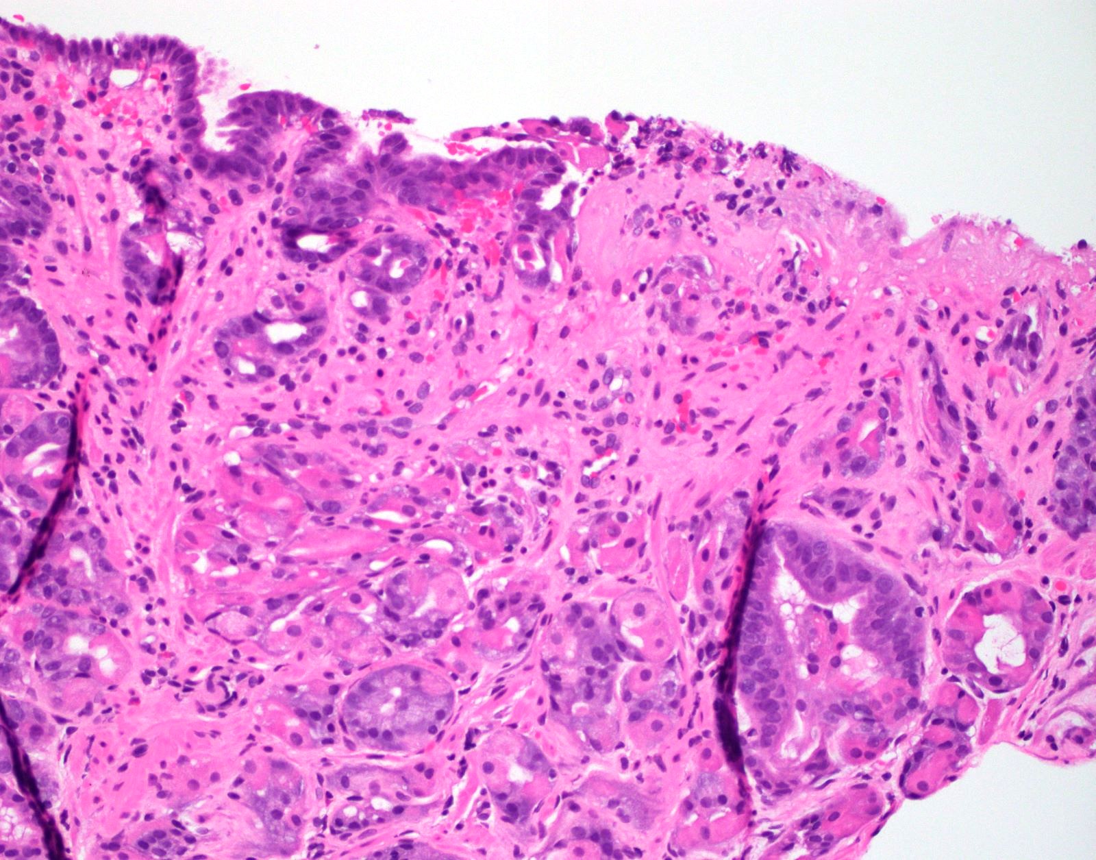
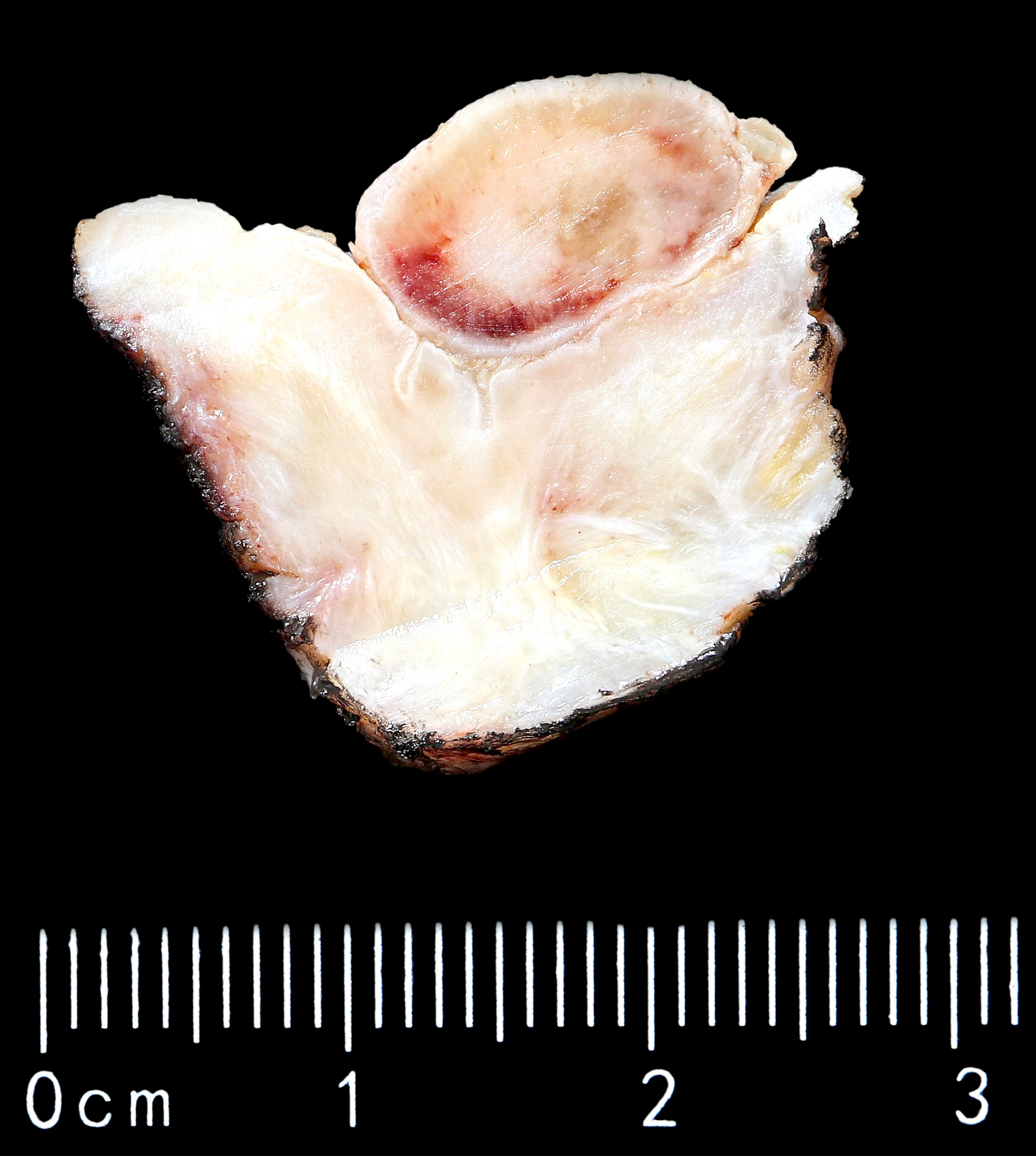
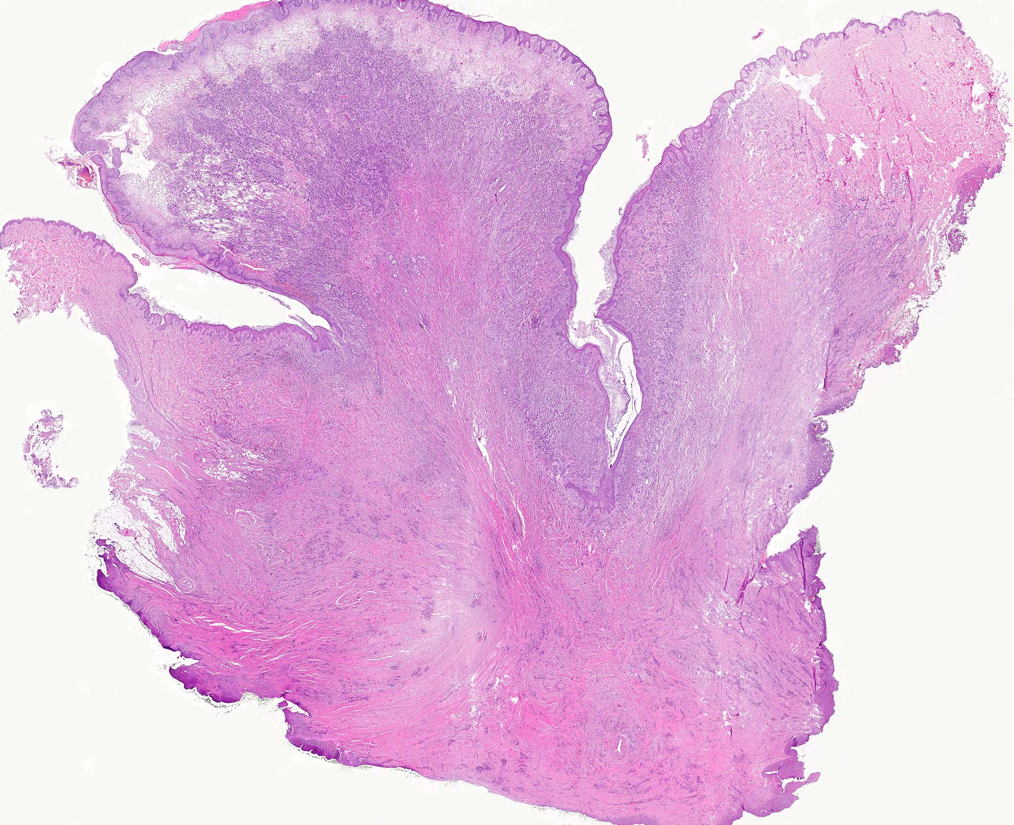
Mass in the gastric body
Linitis plastica
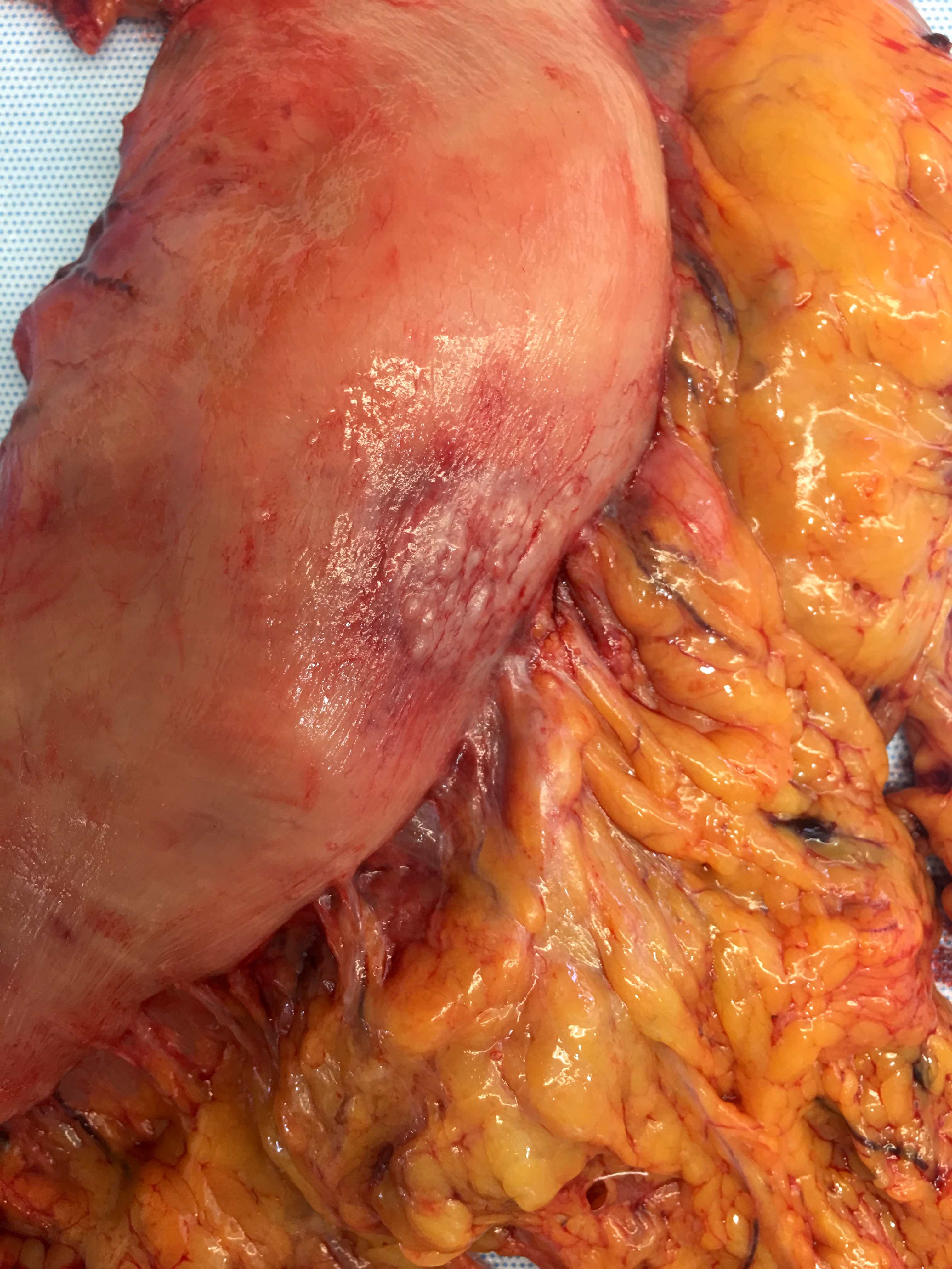
Serosal retraction
Giant distal gastric mass
Contributed by Carolina Martinez Ciarpaglini, M.D., Ph.D.
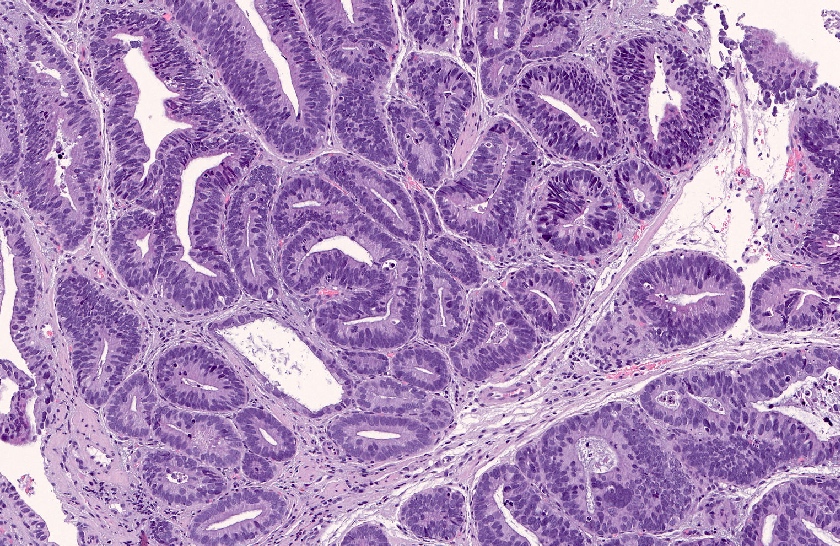
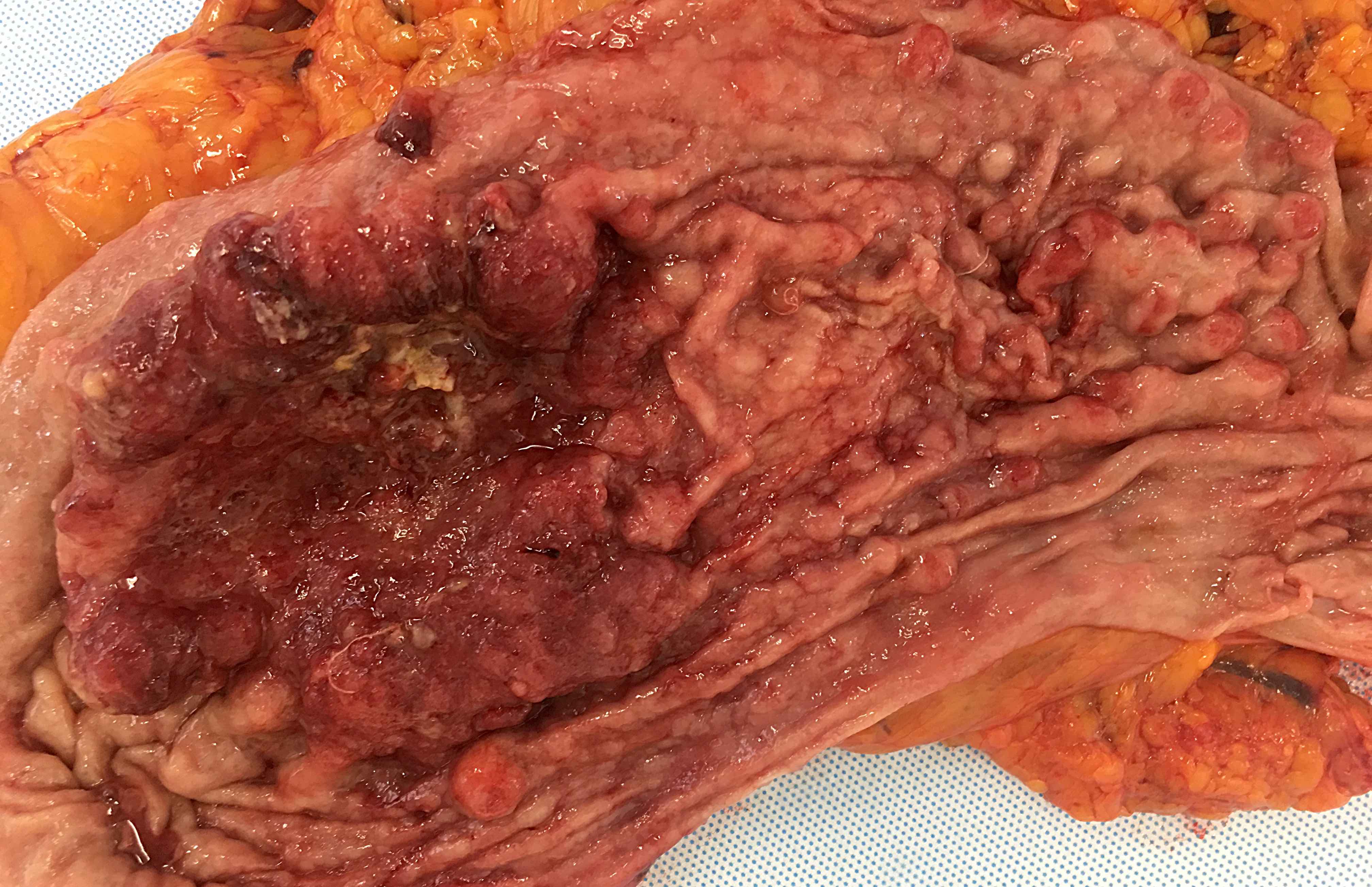
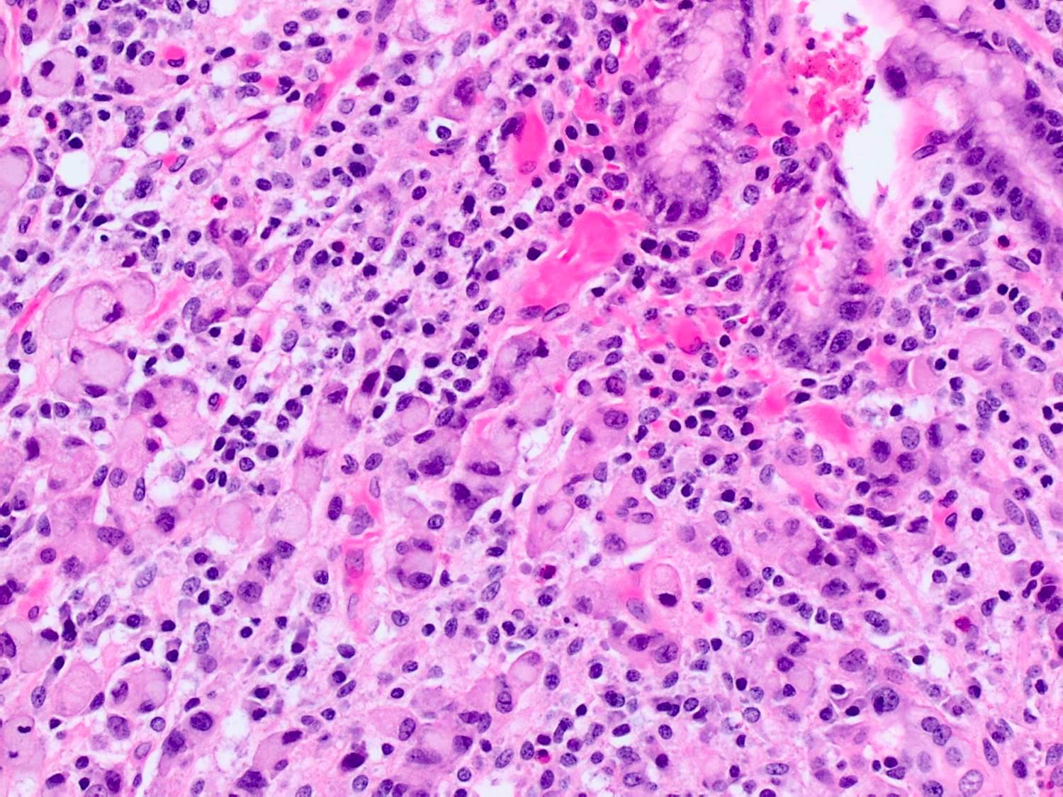
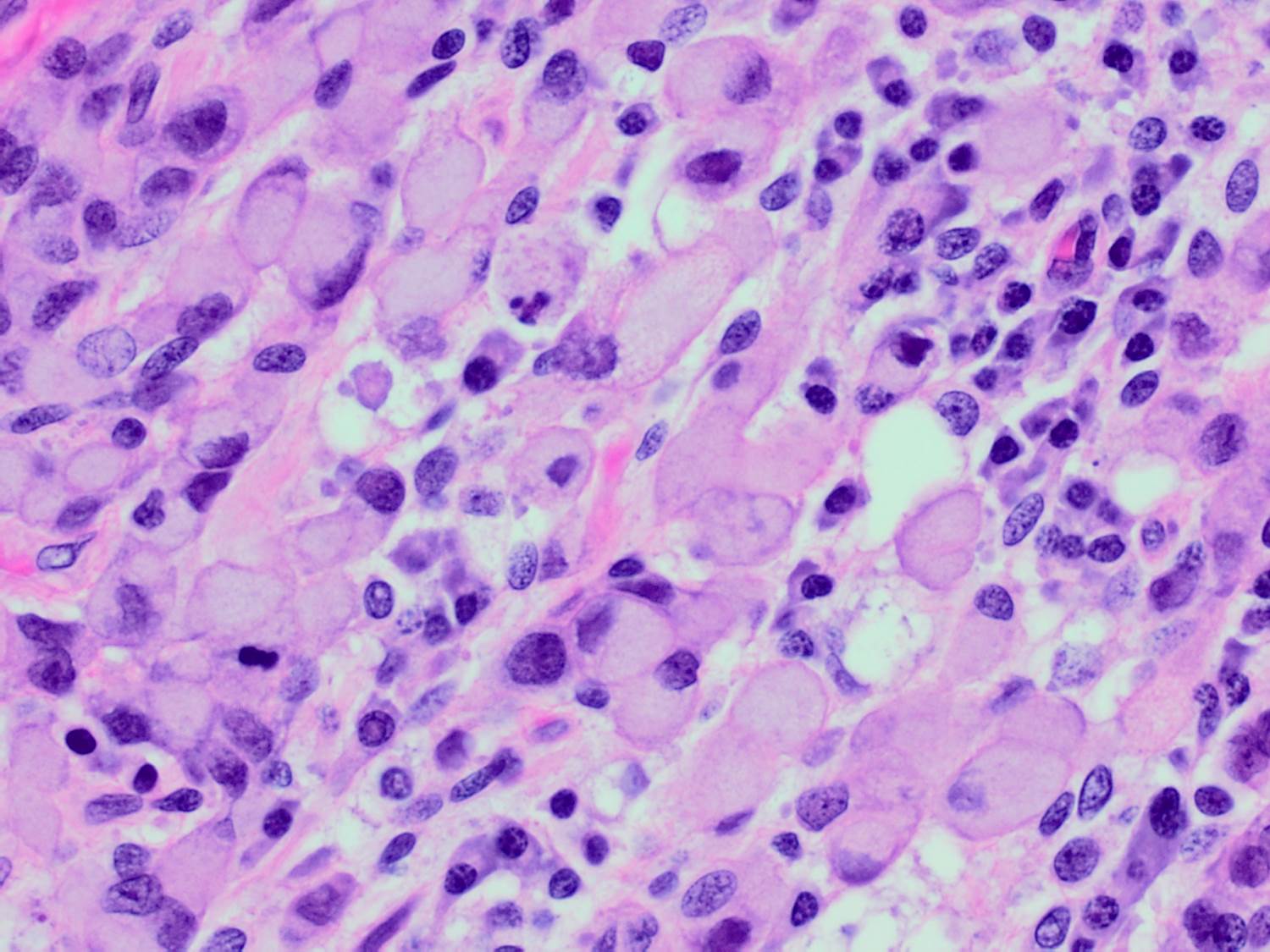
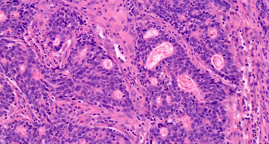
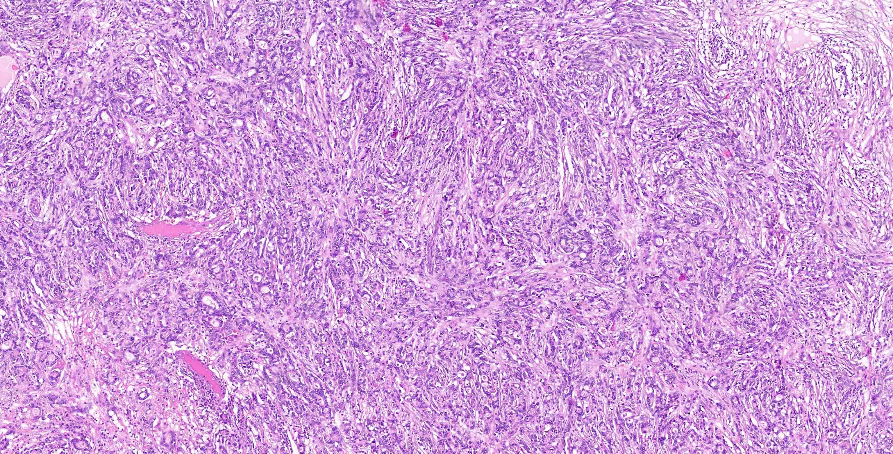
Gastric adenocarcinoma intestinal subtype
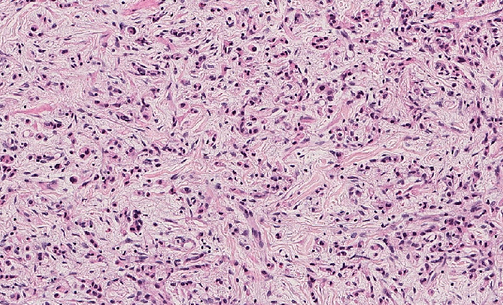
Diffuse adenocarcinoma
Poorly cohesive adenocarcinoma with signet ring cell phenotype
Contributed by Andrey Bychkov, M.D., Ph.D. and Jijgee Munkhdelger, M.D., Ph.D.
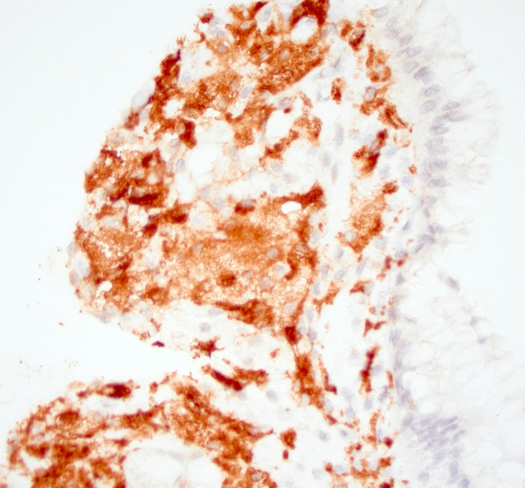
CMV gastritis ulcer

CMV gastritis IHC

CMV infected cells

Classic cytomegalic cell
Images hosted on other servers:

Duodenal biopsy
Contributed by Matthew Morrow, M.D.
Gastric nodularity

Gastric mucosa with mild nodularity
Images hosted on other servers:

Nodular gastric mucosa
Contributed by Matthew Morrow, M.D.

Increased subepithelial collagen with lamina propria chronic inflammation

Denudation of surface gastric epithelium
Thickened subepithelial collagen
Collagenous gastritis 100x
Abundant eosinophils 40x
Images hosted on other servers:


Thickened subepithelial collagen

Lamina propria inflammation
Image hosted on other server:

Gastric and colonic polyps
Images hosted on other servers:

Gastric wall thickening, incomplete distention
Contributed by Gagandeep Kaur, M.D. and Monika Vyas, M.D.
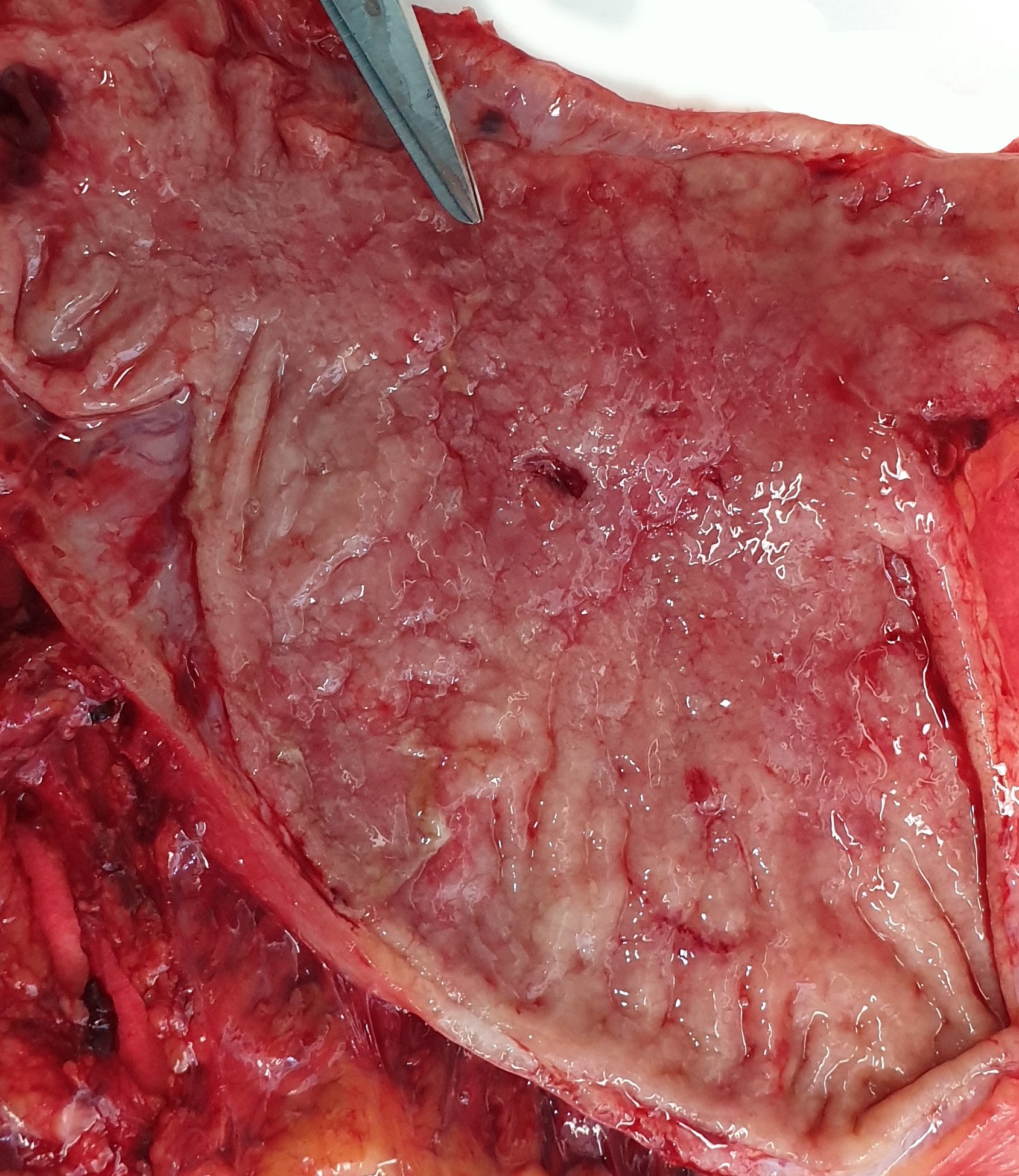
Grossly no residual tumor
Negative for gross lesion
Images hosted on other servers:

Marked thickening of gastric wall with loss of rugae

Thickening of gastric wall
Contributed by Gagandeep Kaur, M.D. and Monika Vyas, M.D.
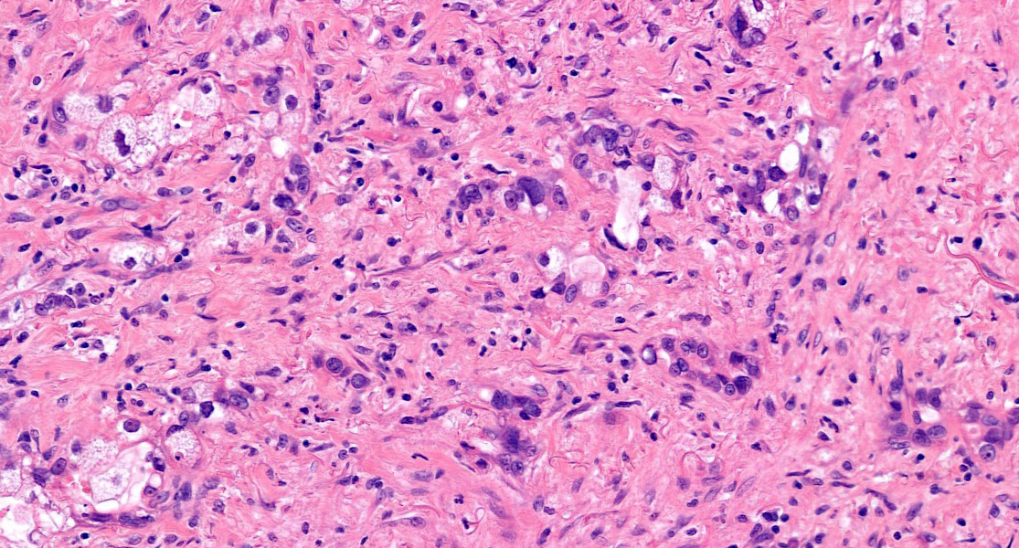
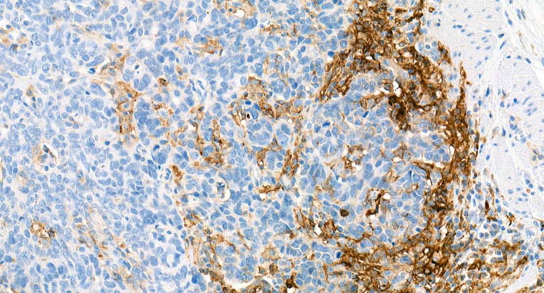
Diffuse gastric carcinoma associated with chronic gastritis
Signet ring cells
Peritoneal involvement
CK stain highlighting perineural invasion
Early diffuse gastric carcinoma

In situ carcinoma
Pagetoid spread of signet ring cell carcinoma
Contributed by Feng Yin, M.D., Ph.D.
Doxycycline associated mucosal injury


Eosinophilic capillary degenerative changes
Contributed by Kyra Berg, M.D.
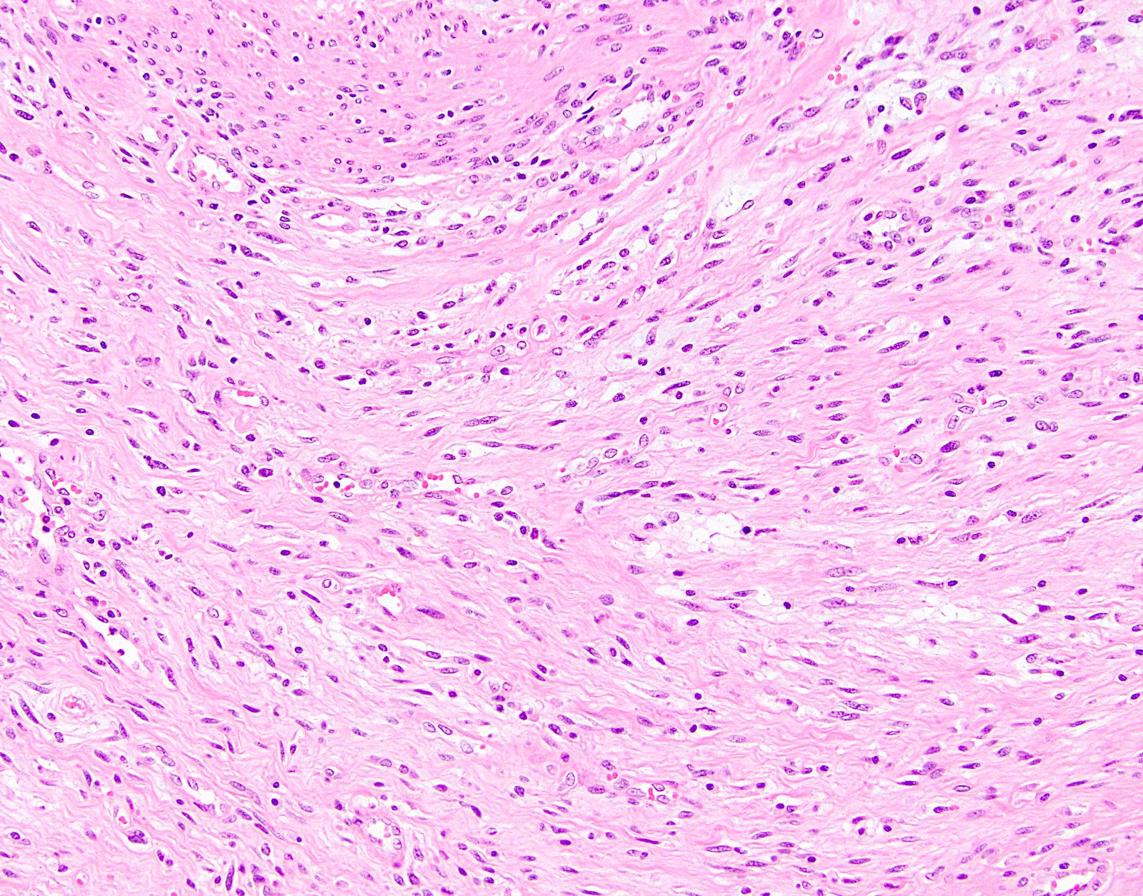
Elongated and hyperchromatic nuclei
Elongated nuclei with visible nucleoli
Loss of polarity
Increased cellularity
Images hosted on other servers:

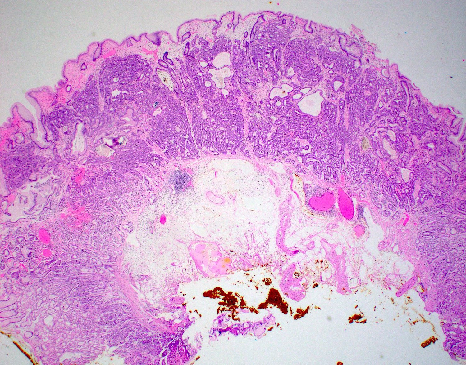
EUS, mucosal gastric cancer
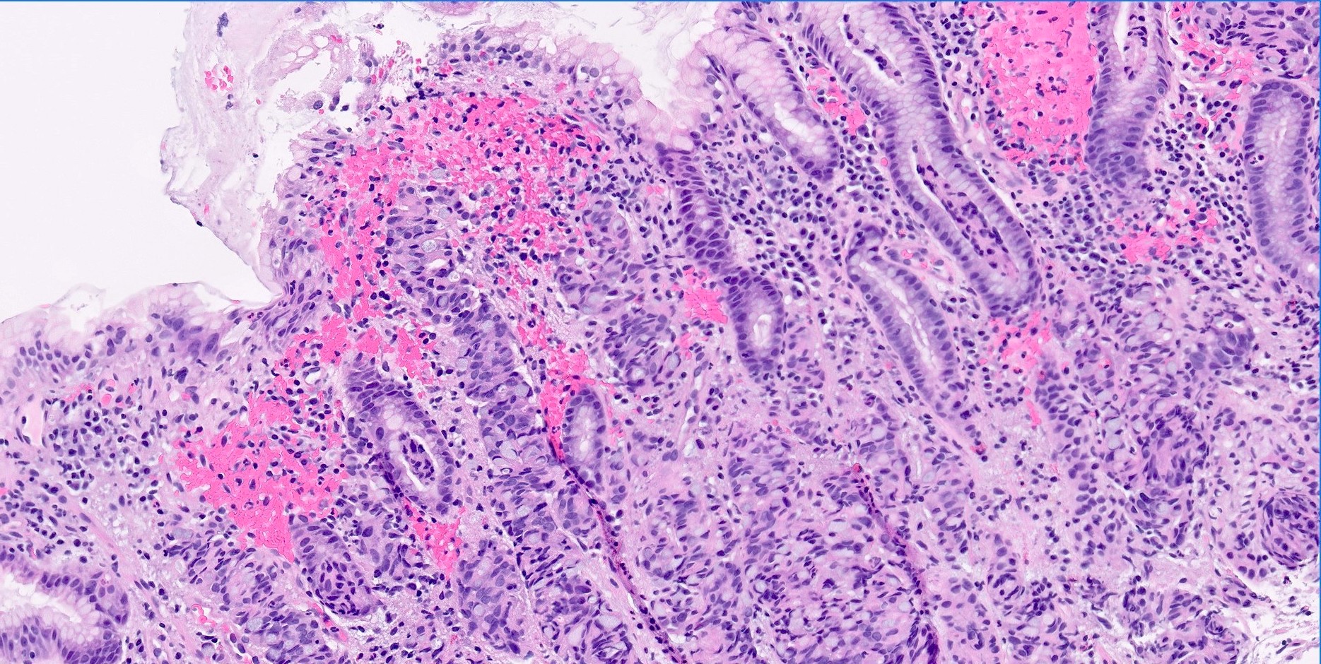
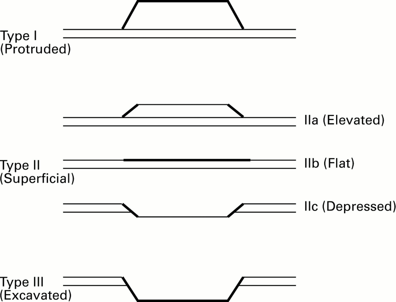
Images hosted on other servers:

Endoscopy, type 0 lesions
Contributed by Guedj Nathalie, M.D., Ph.D.

Type 0-IIc
Contributed by Guedj Nathalie, M.D., Ph.D.
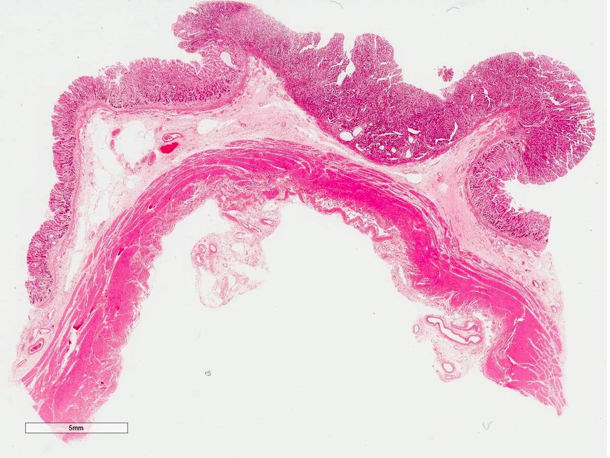
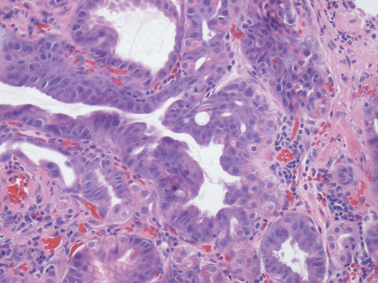
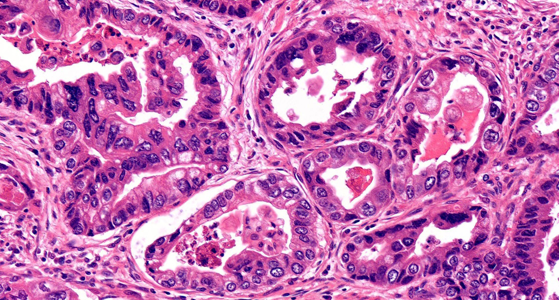
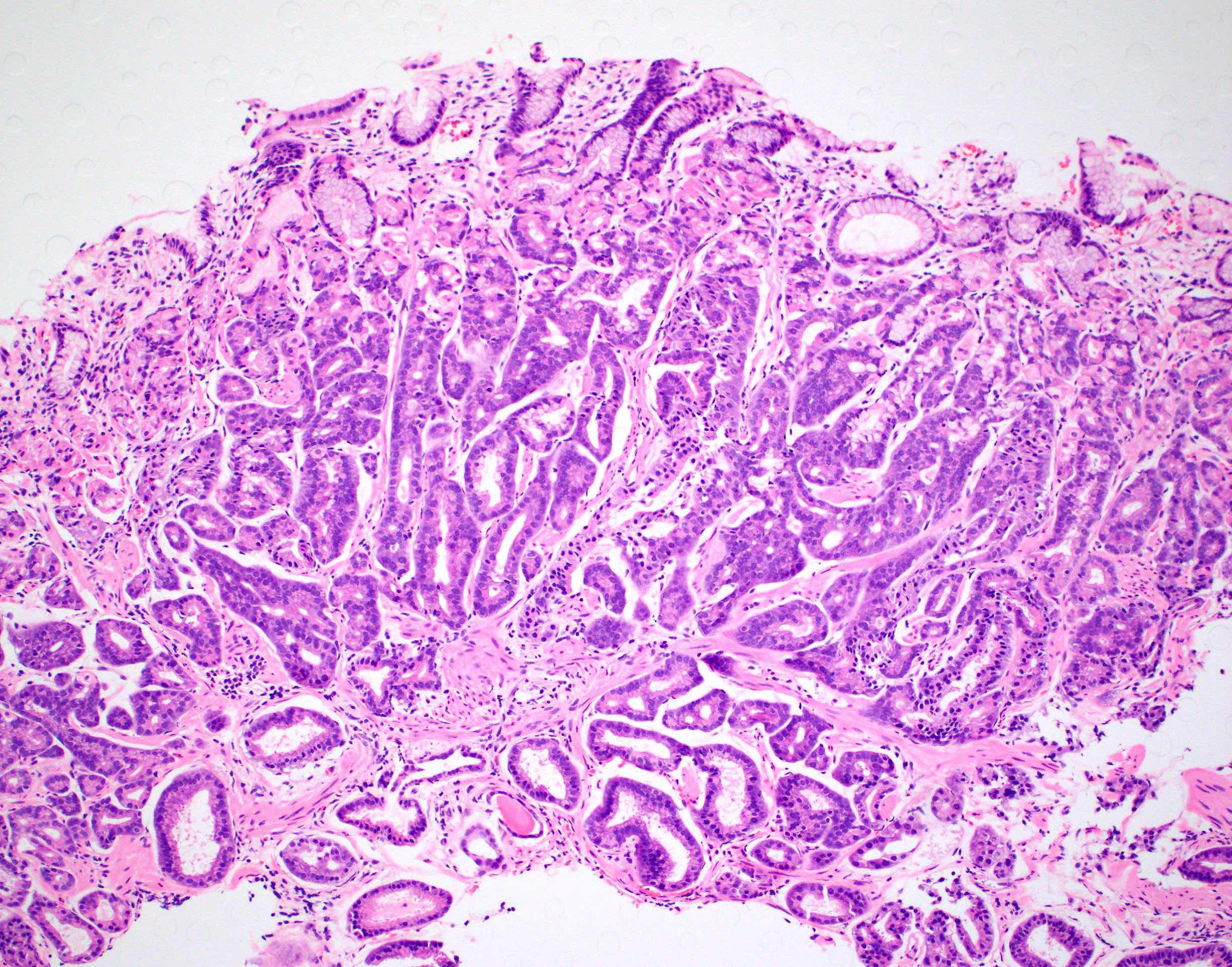
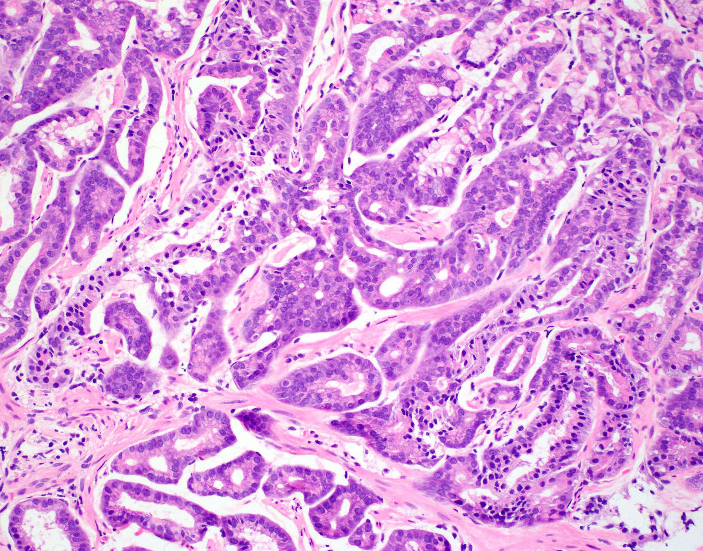
Gastric adenocarcinoma
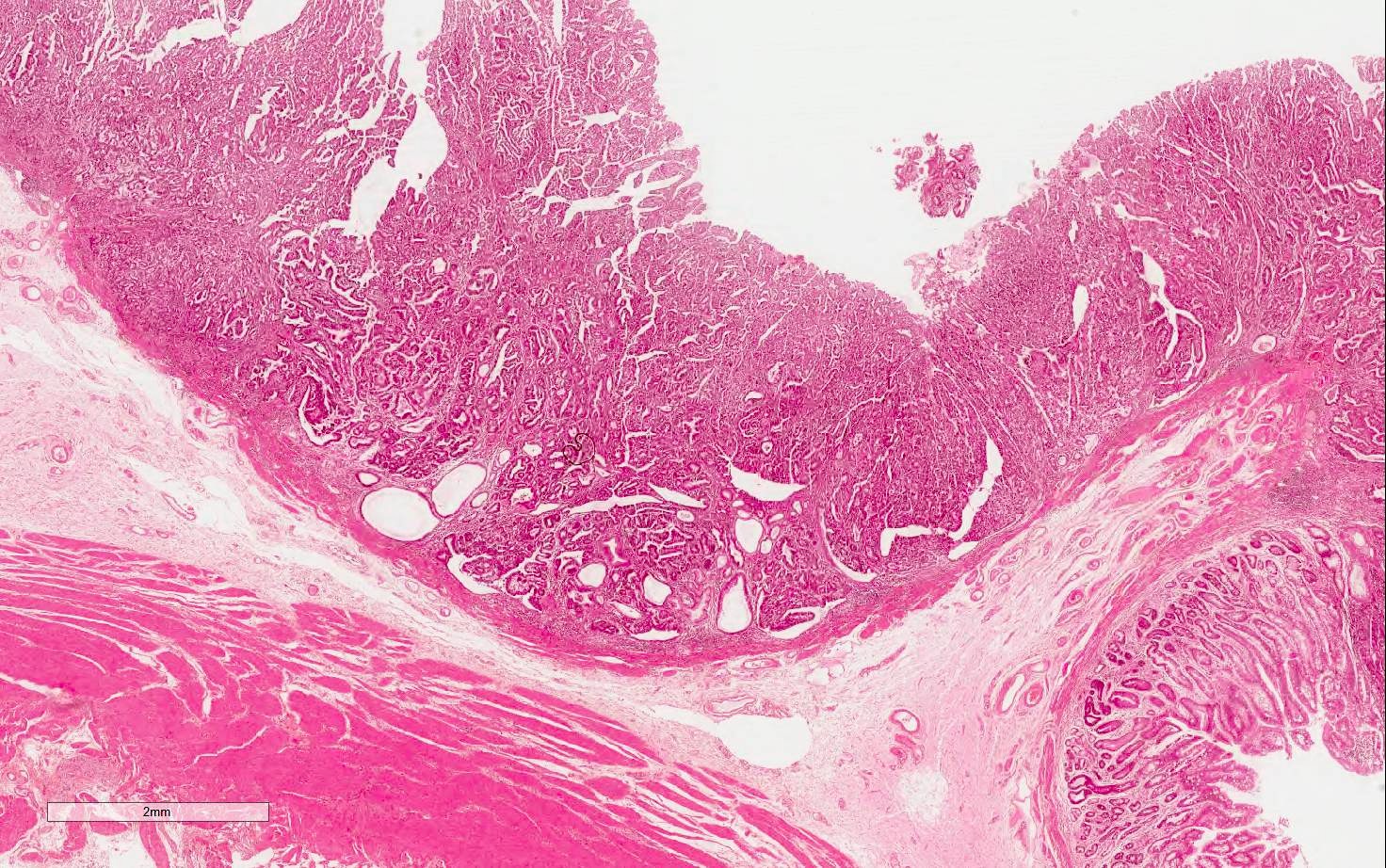
Tubular adenocarcinoma

Cytologic and nuclear atypia
Angioinvasion
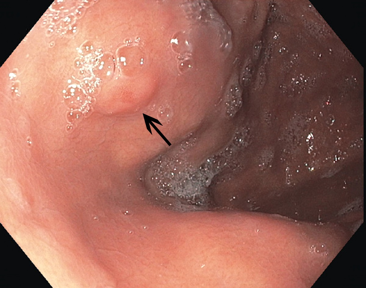
Early gastric carcinoma
Images hosted on other servers:


Endoscopy
Contributed by Iresha Vithanage, M.B.B.S., M.D. and Xiaoyan Liao, M.D., Ph.D.
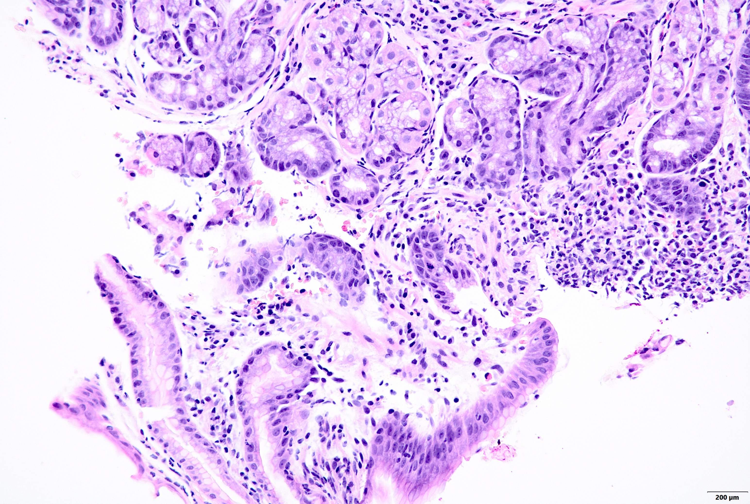
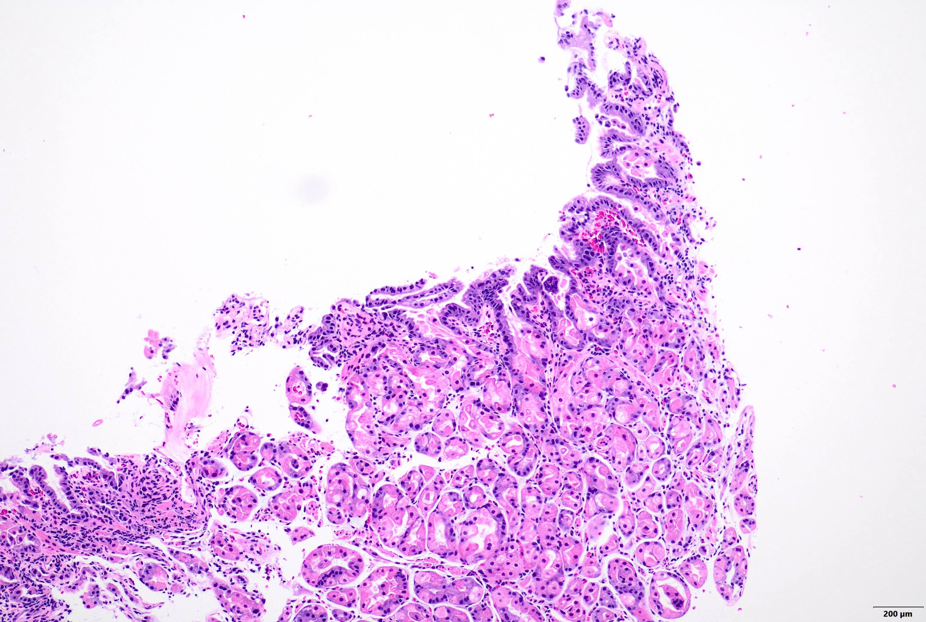
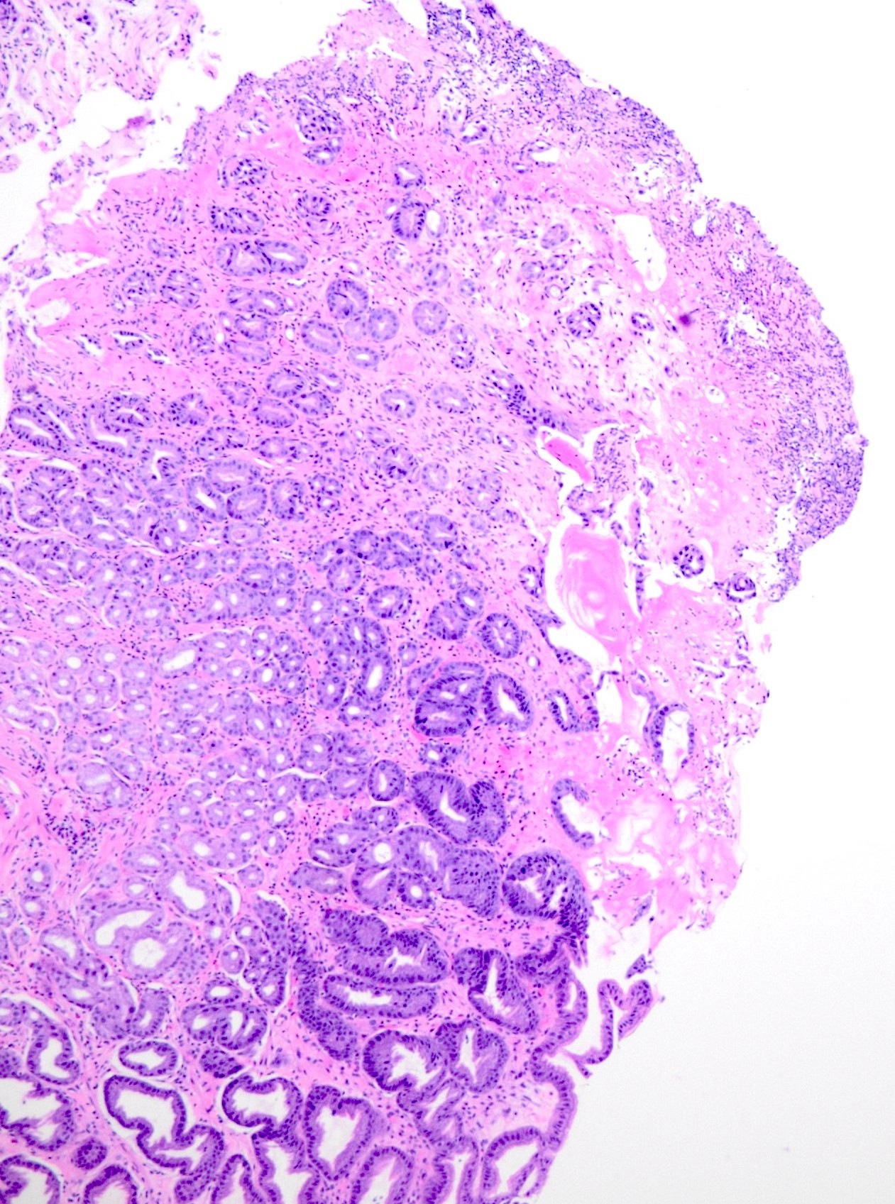
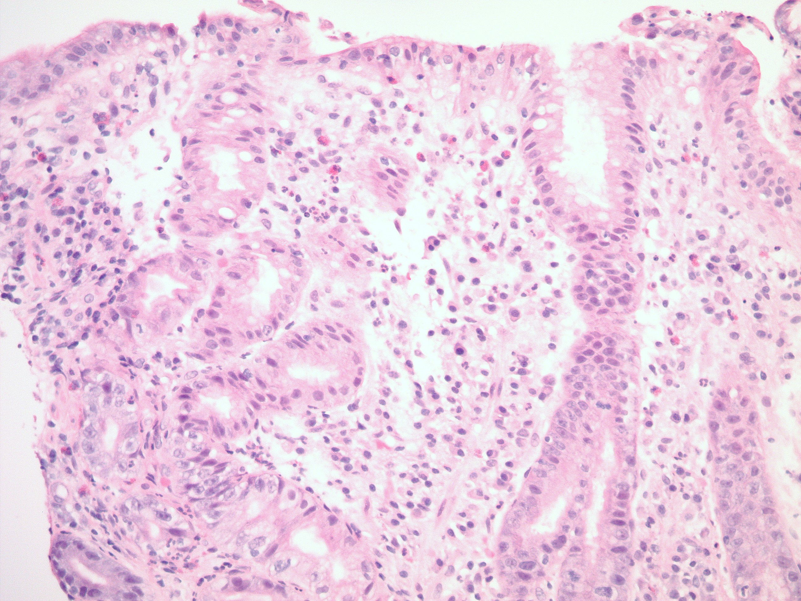
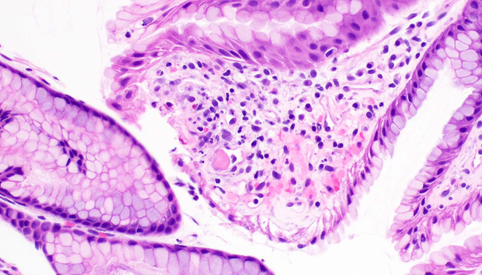
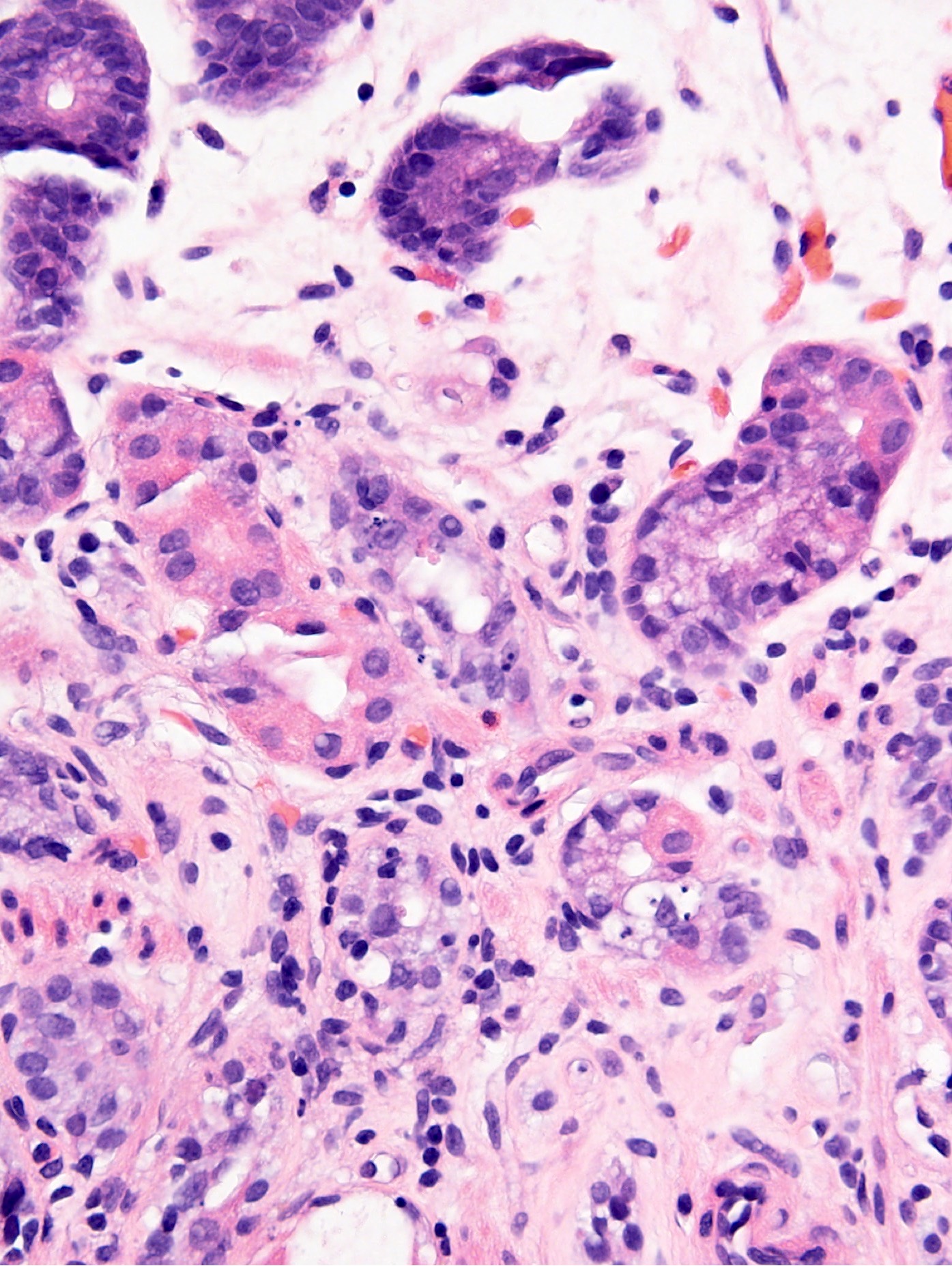
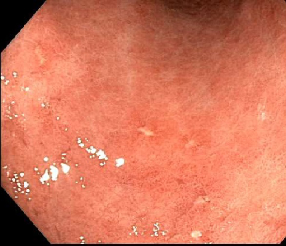
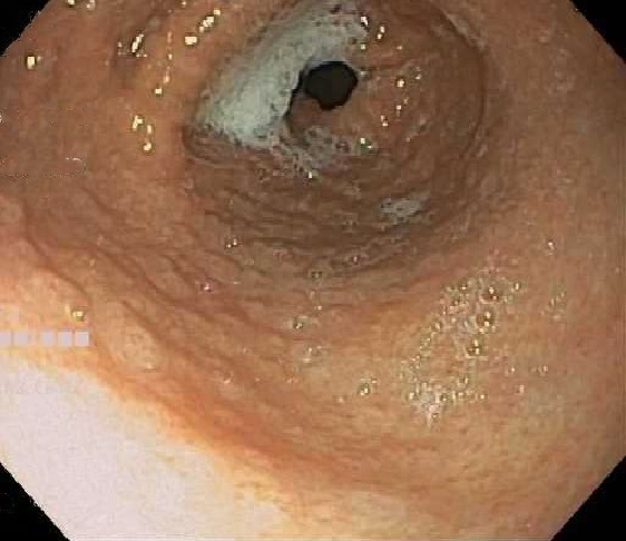
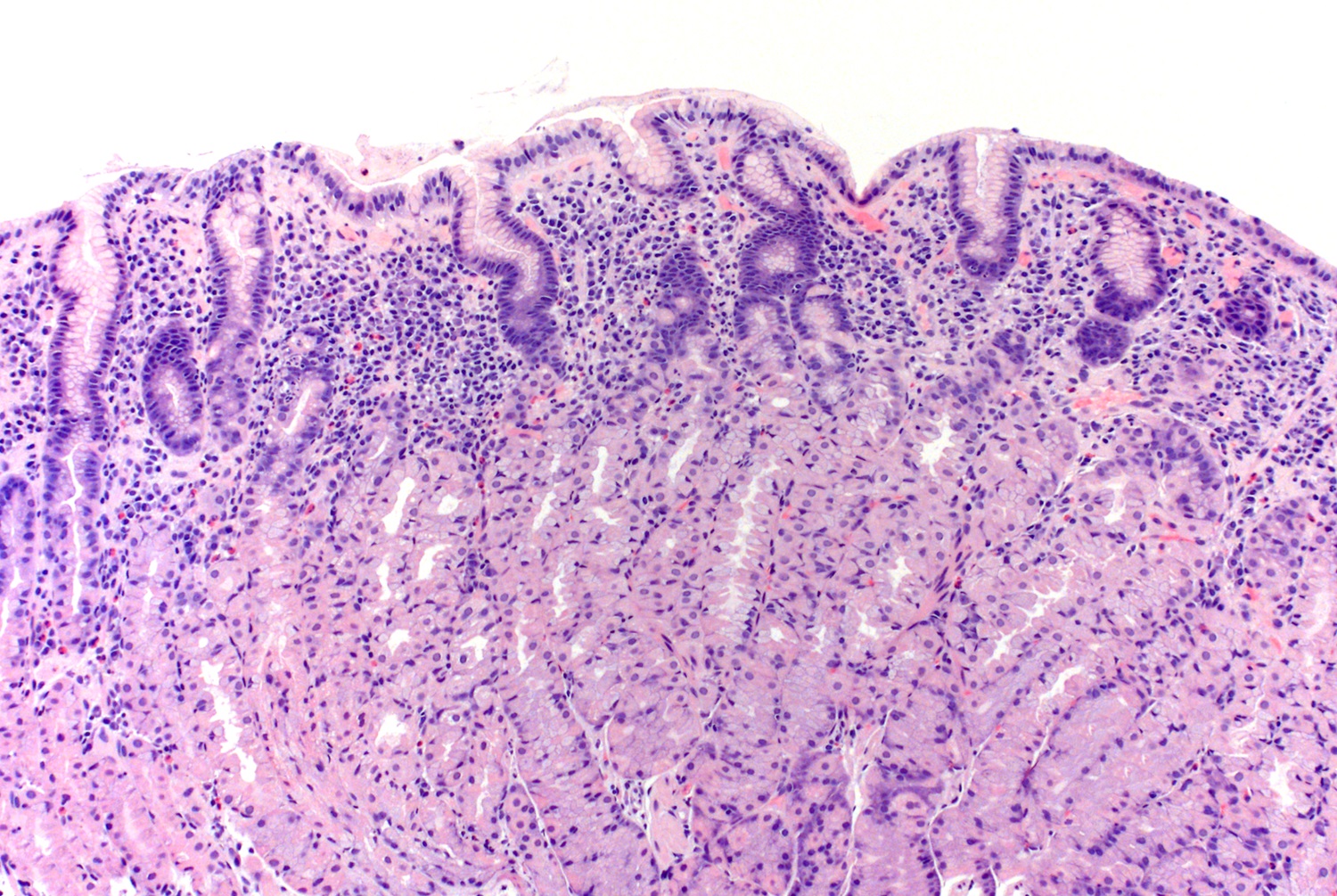
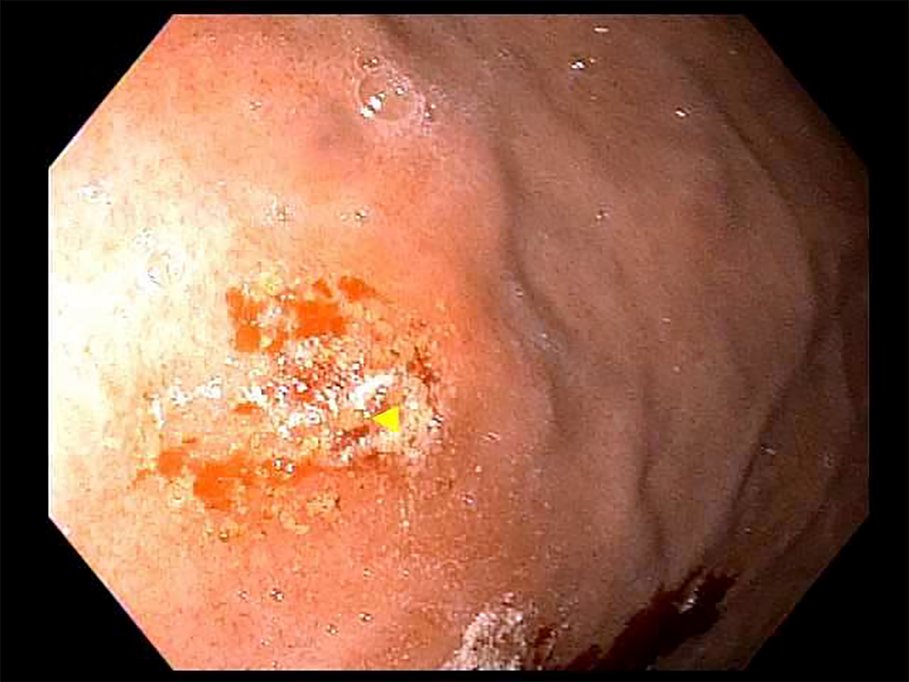
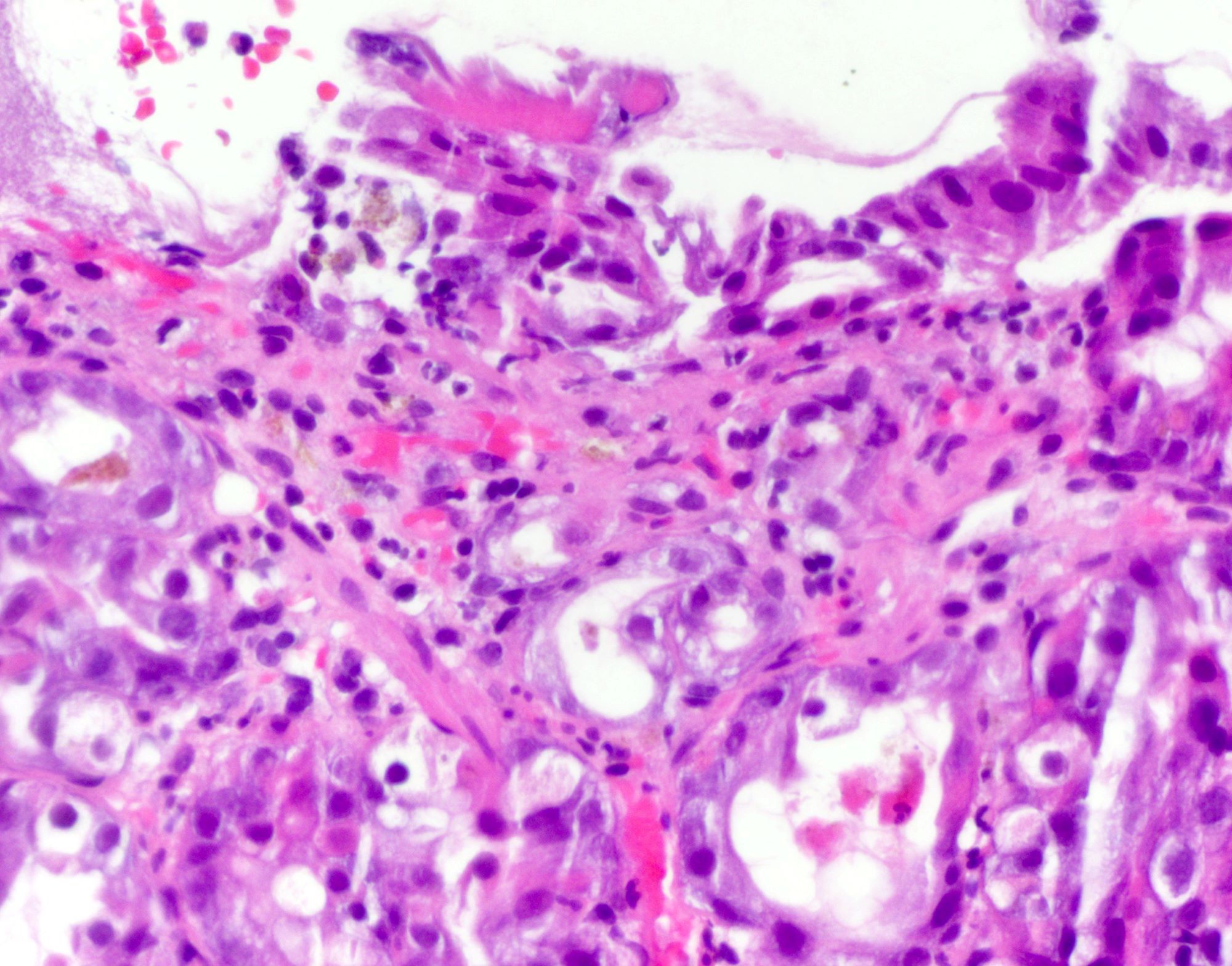
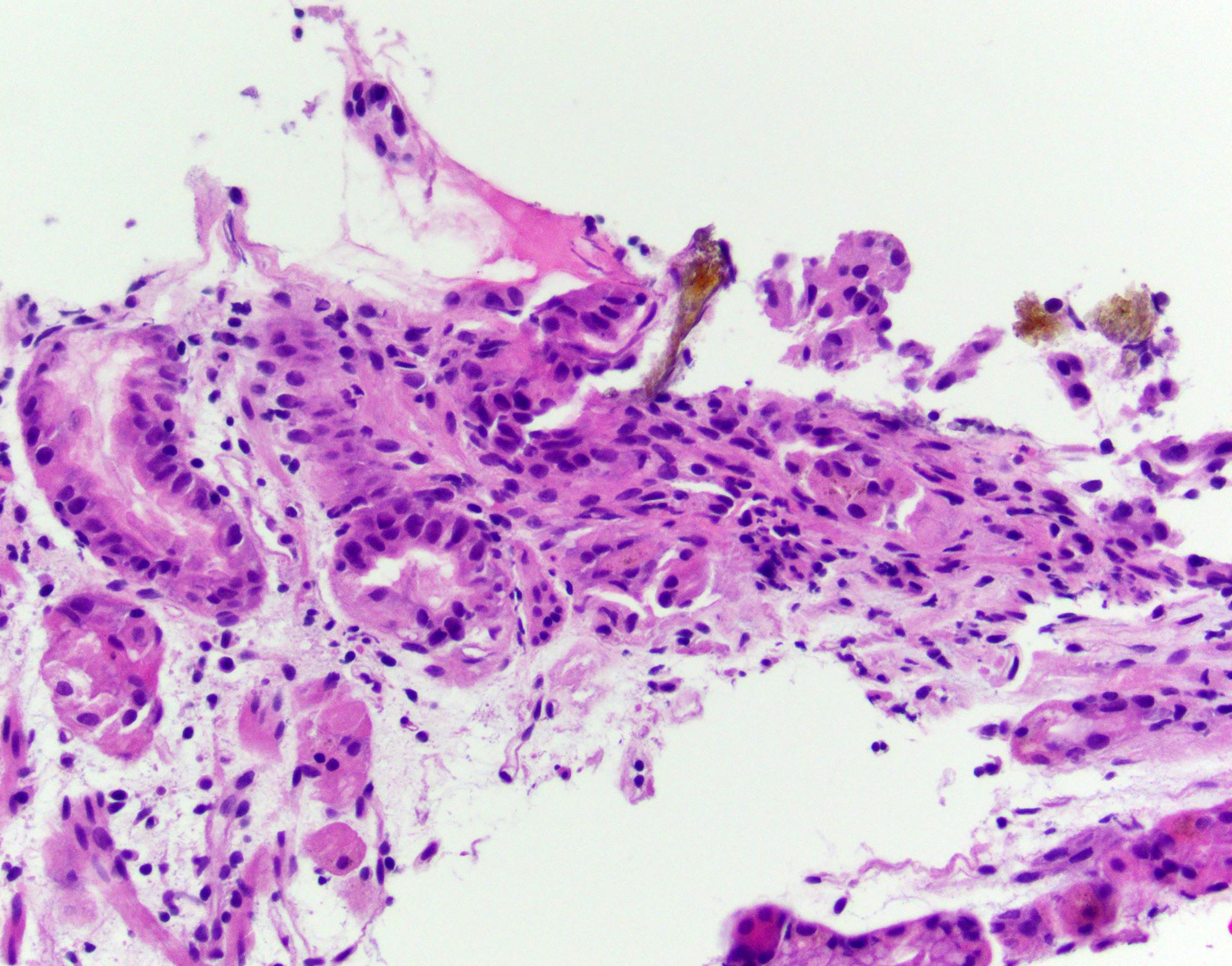
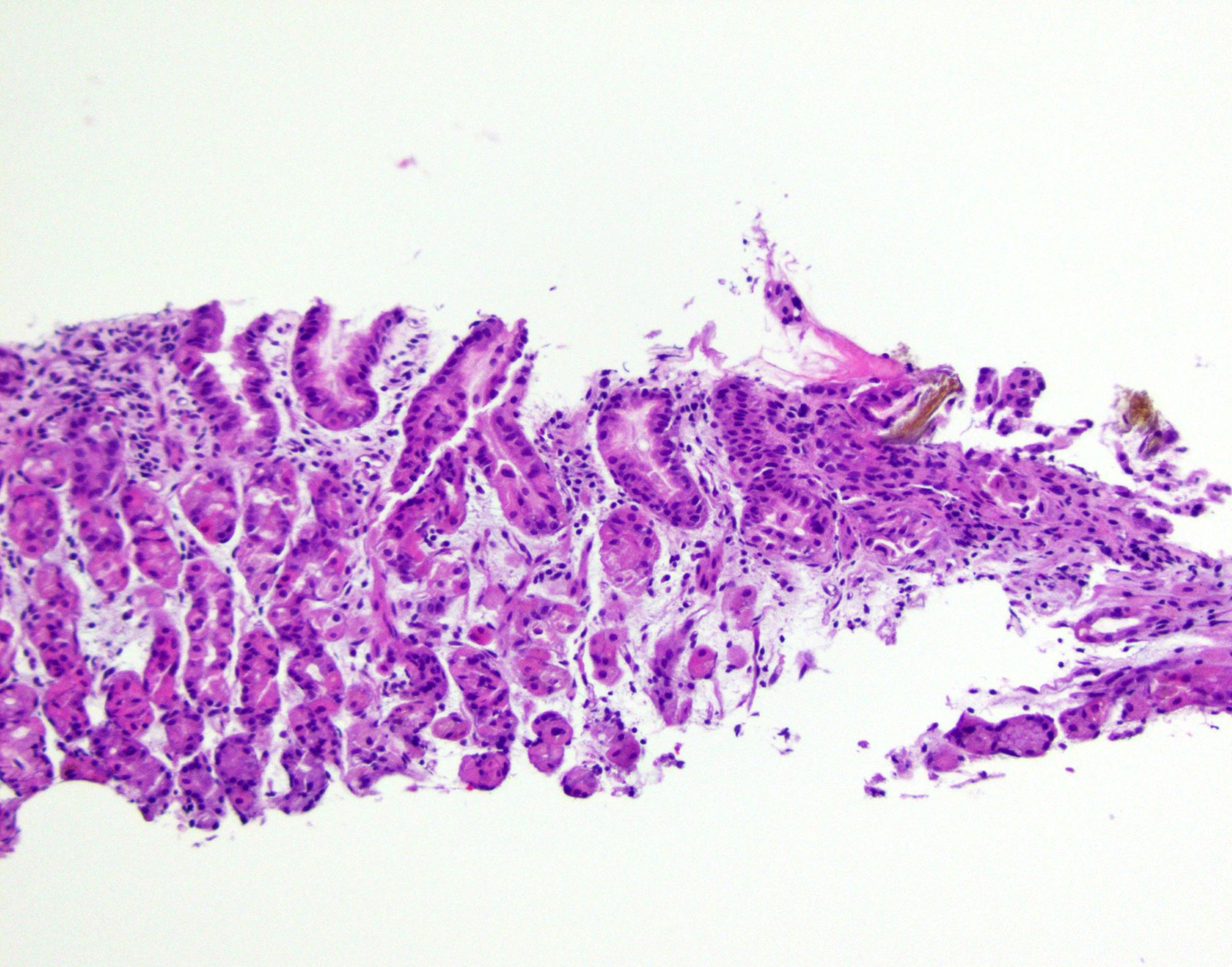
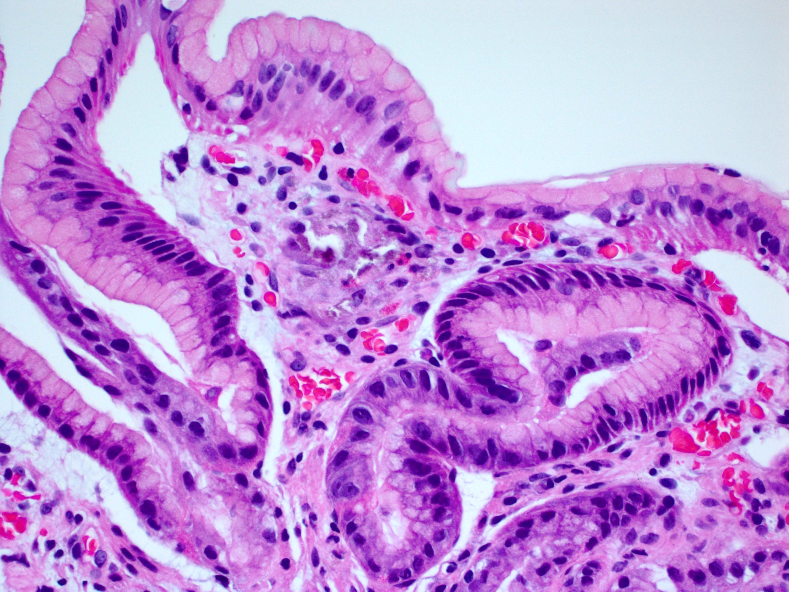
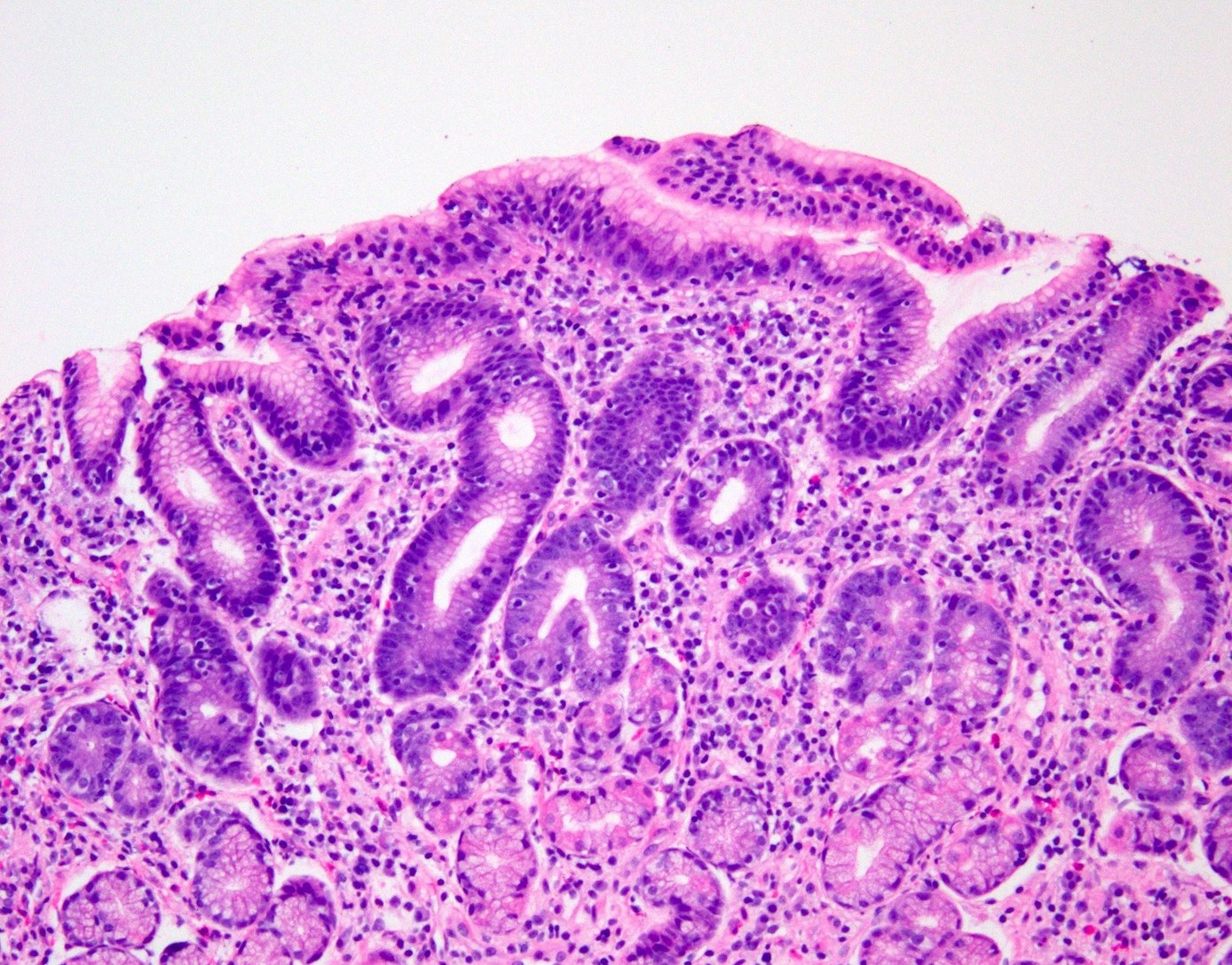
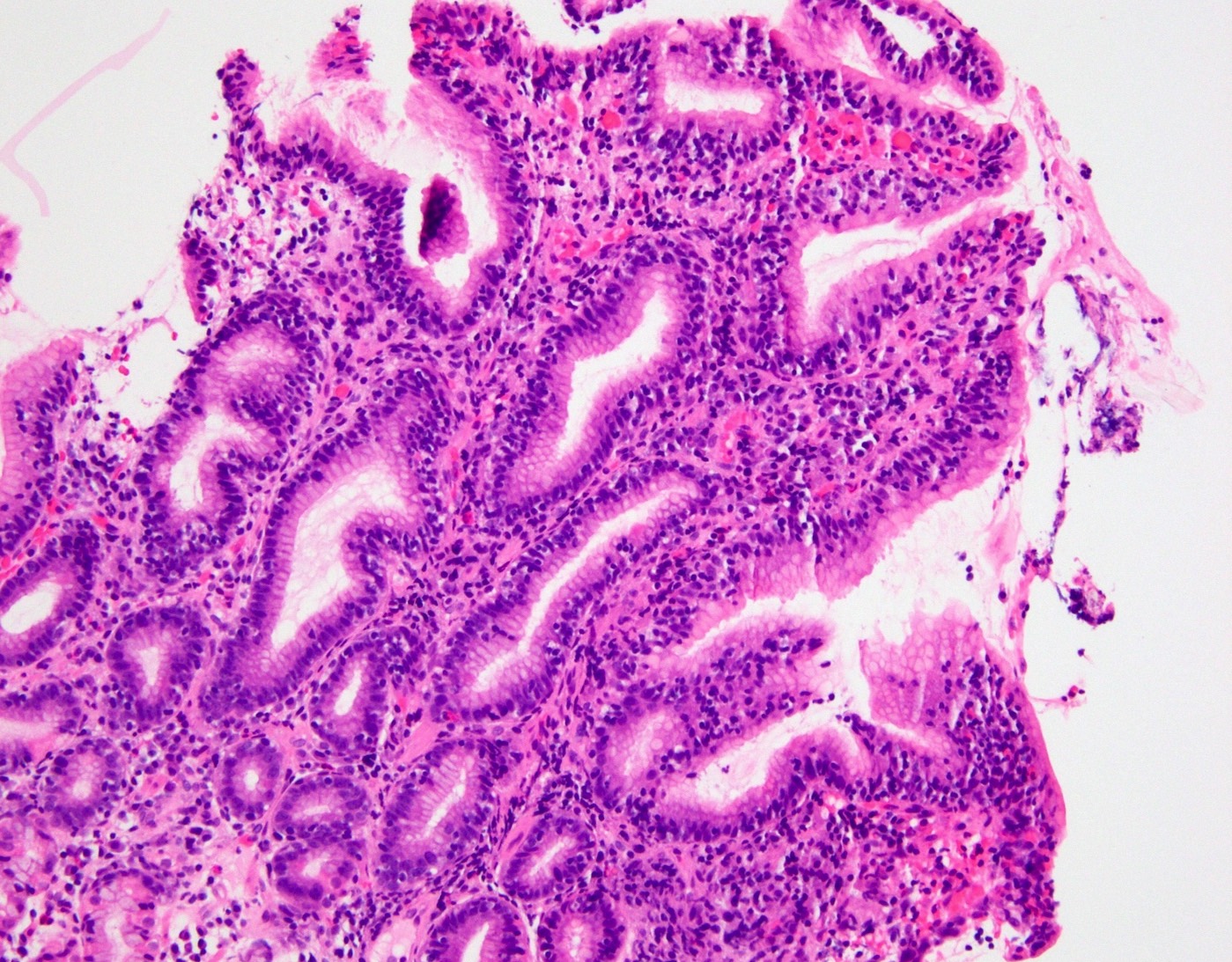
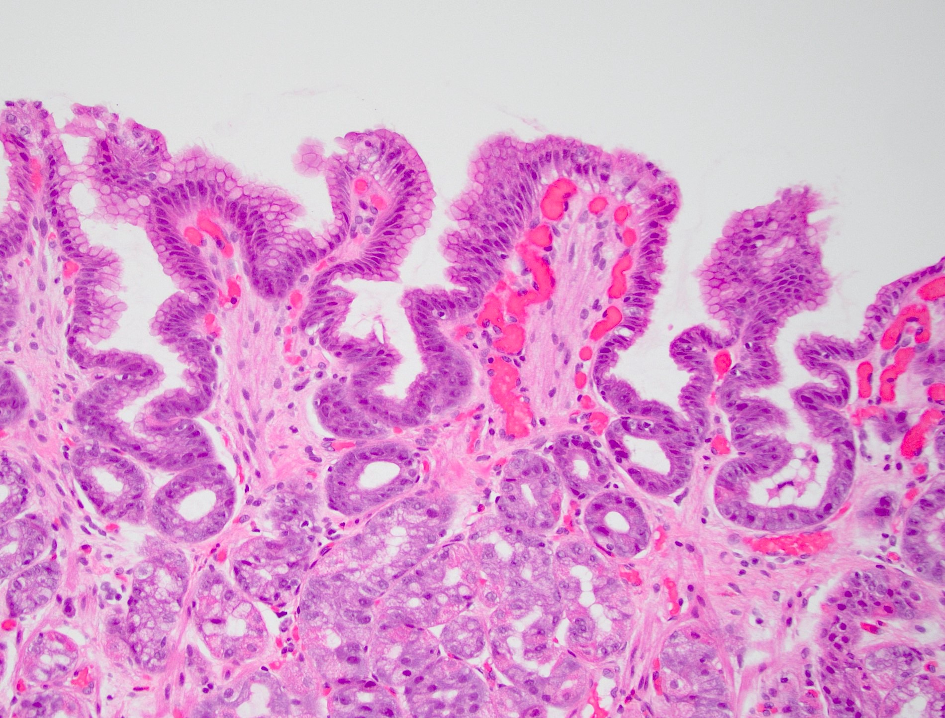
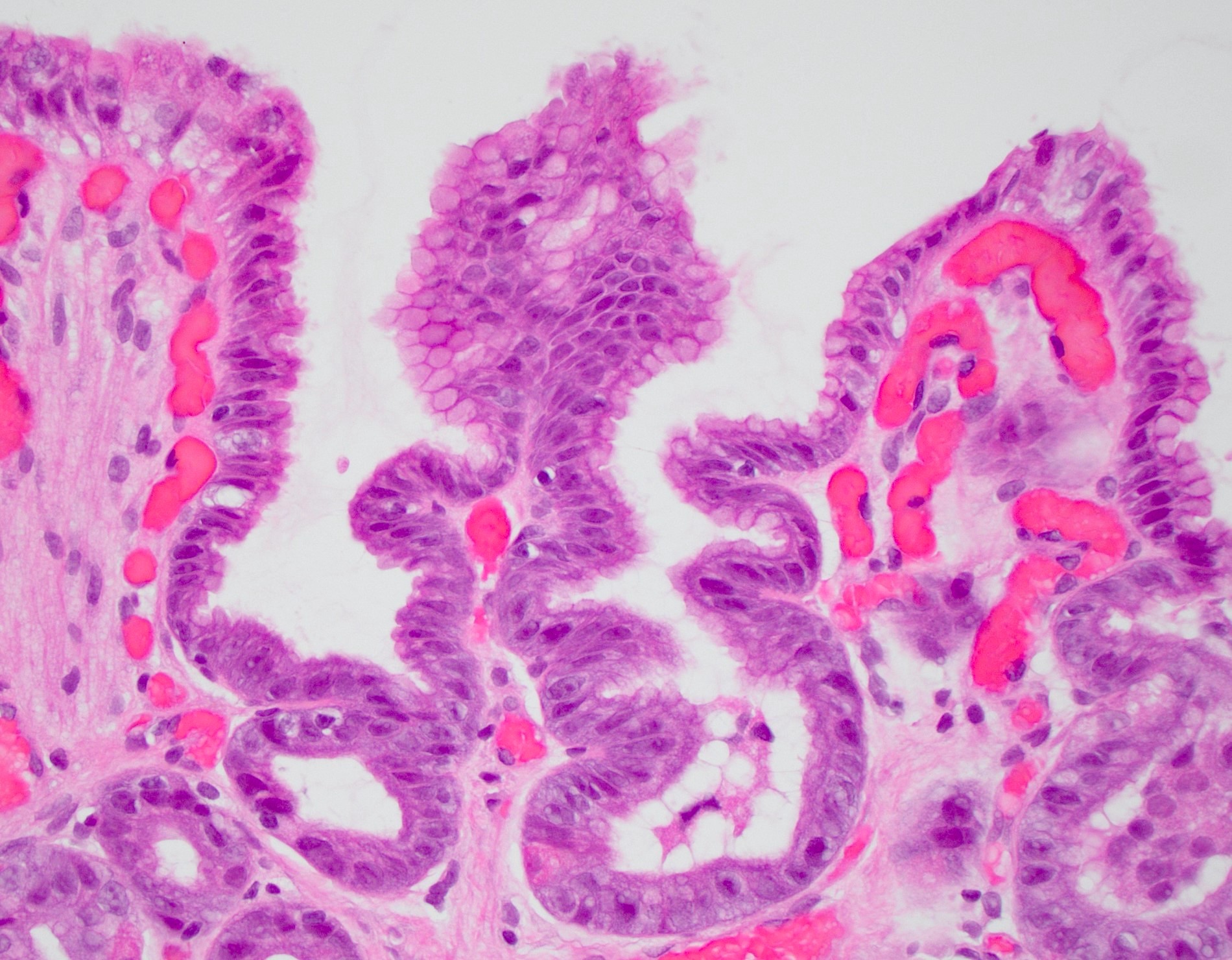
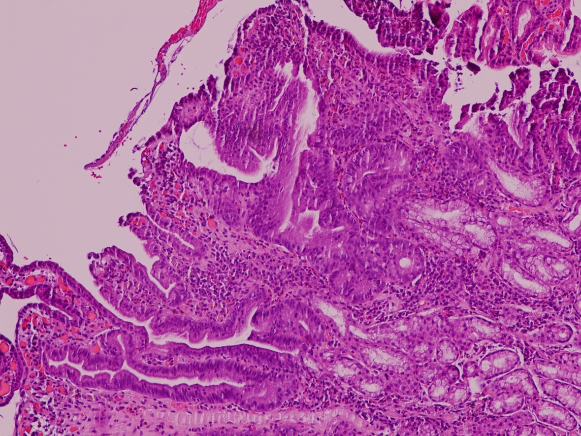
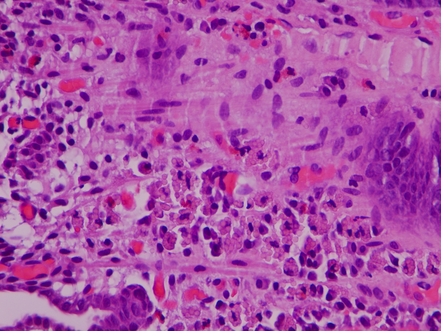
Gastric mucosa with erosive gastritis


Surface erosion and inflammation
Acute erosive gastritis
Contributed by Kwun Wah Wen, M.D., Ph.D.
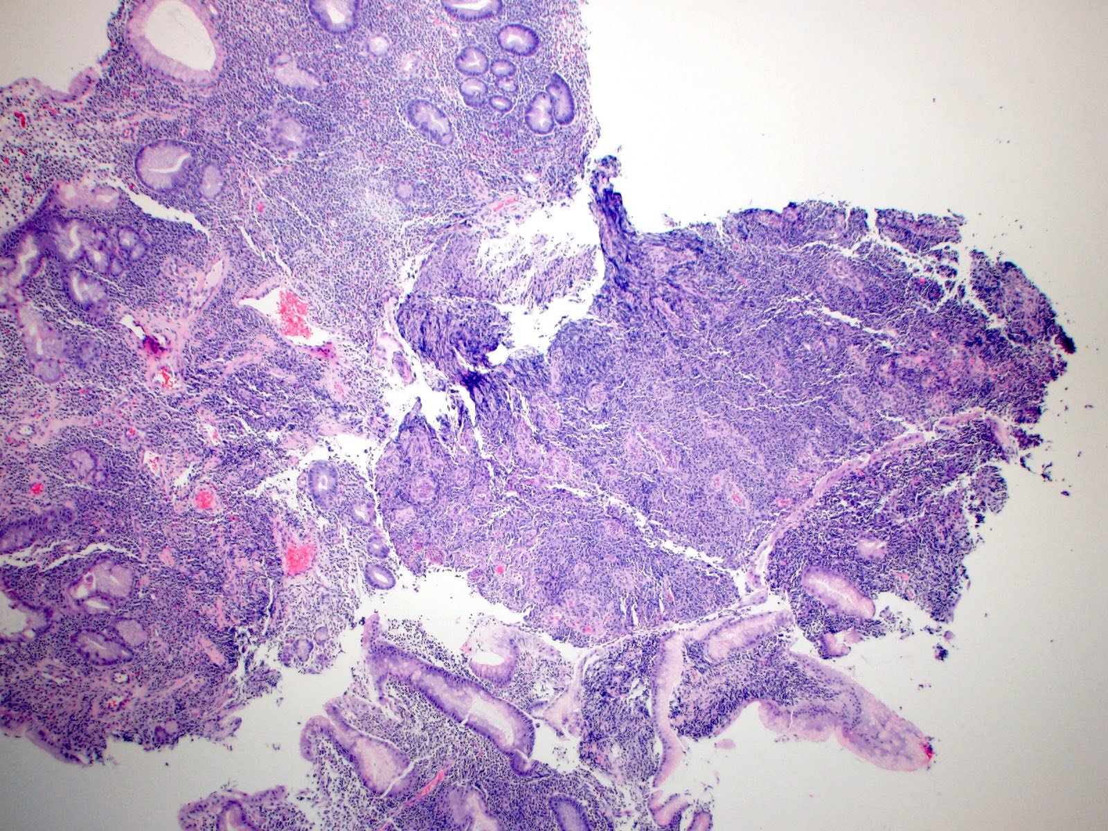
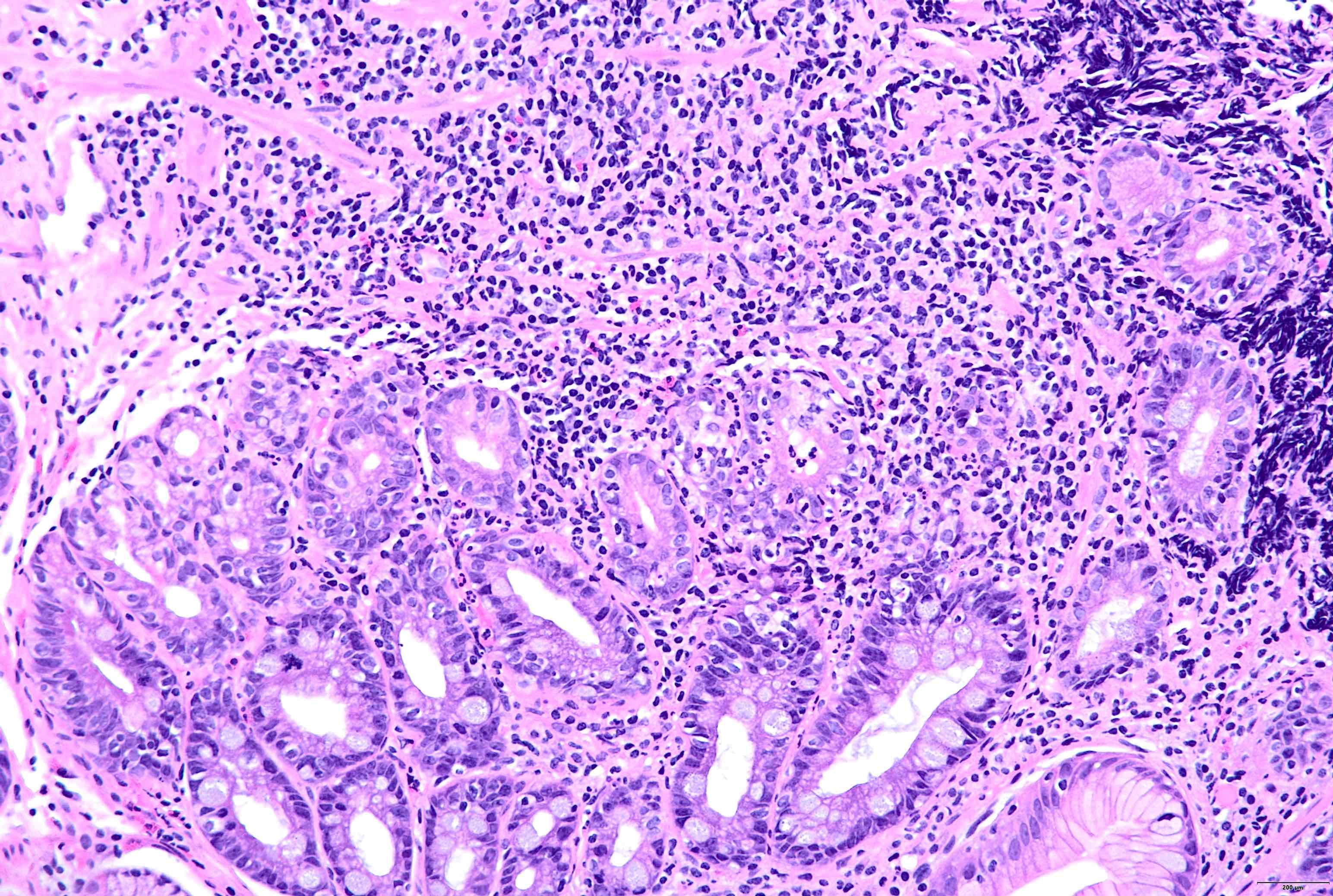
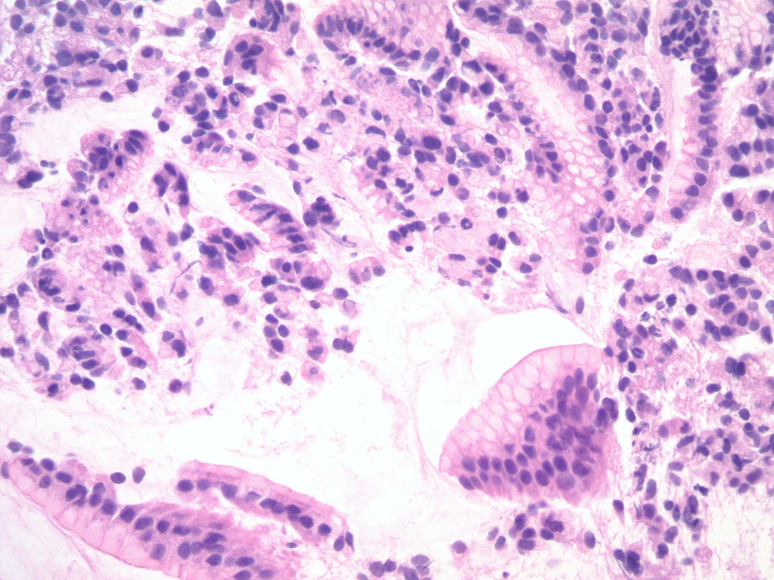
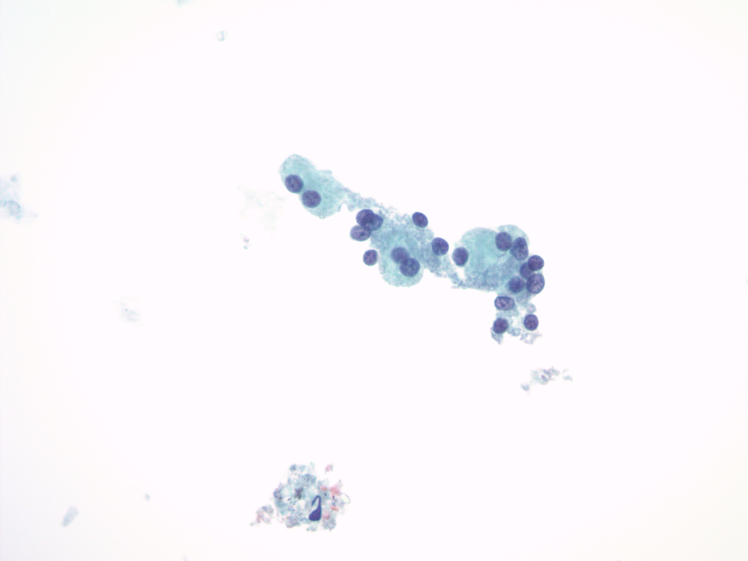
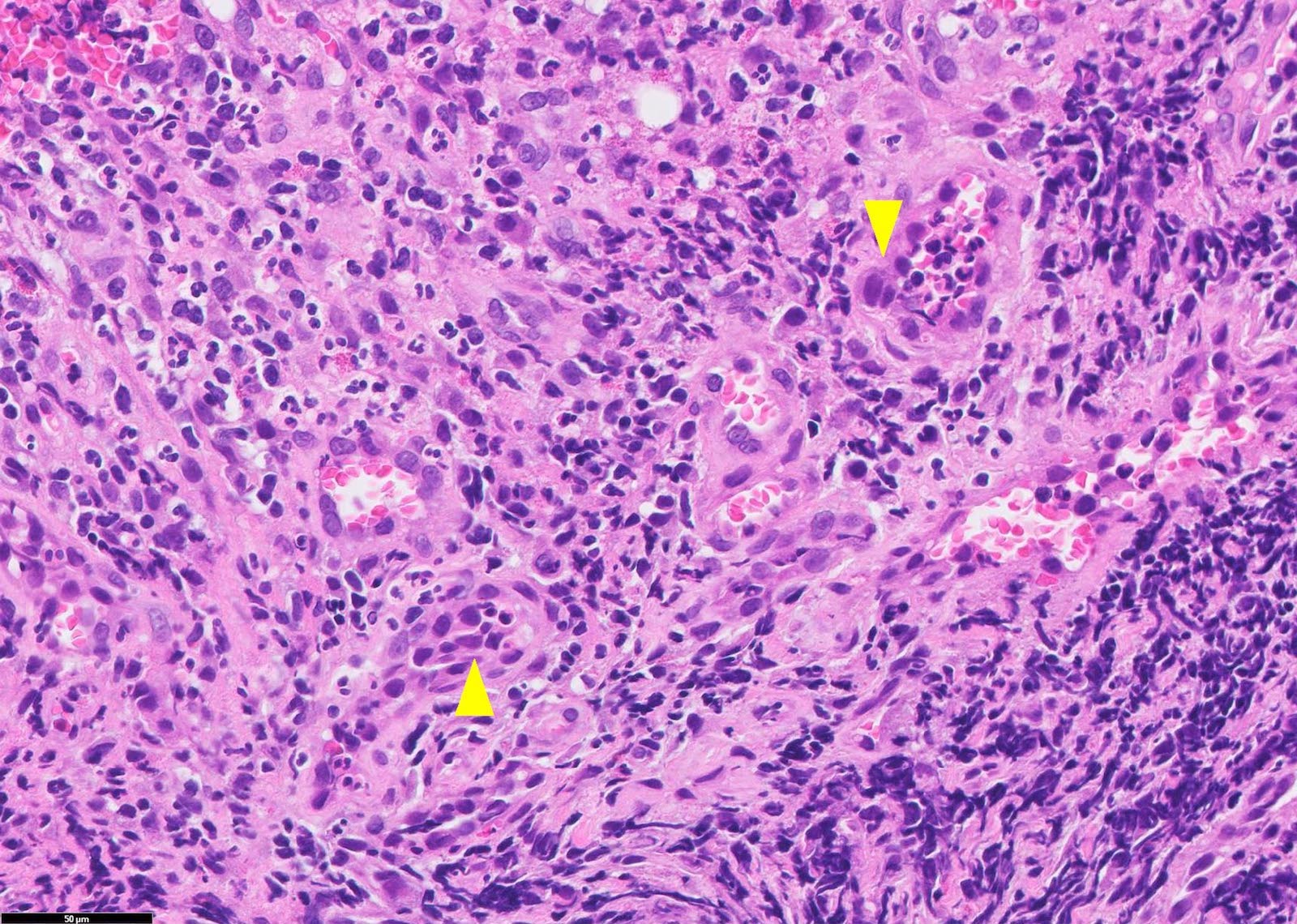
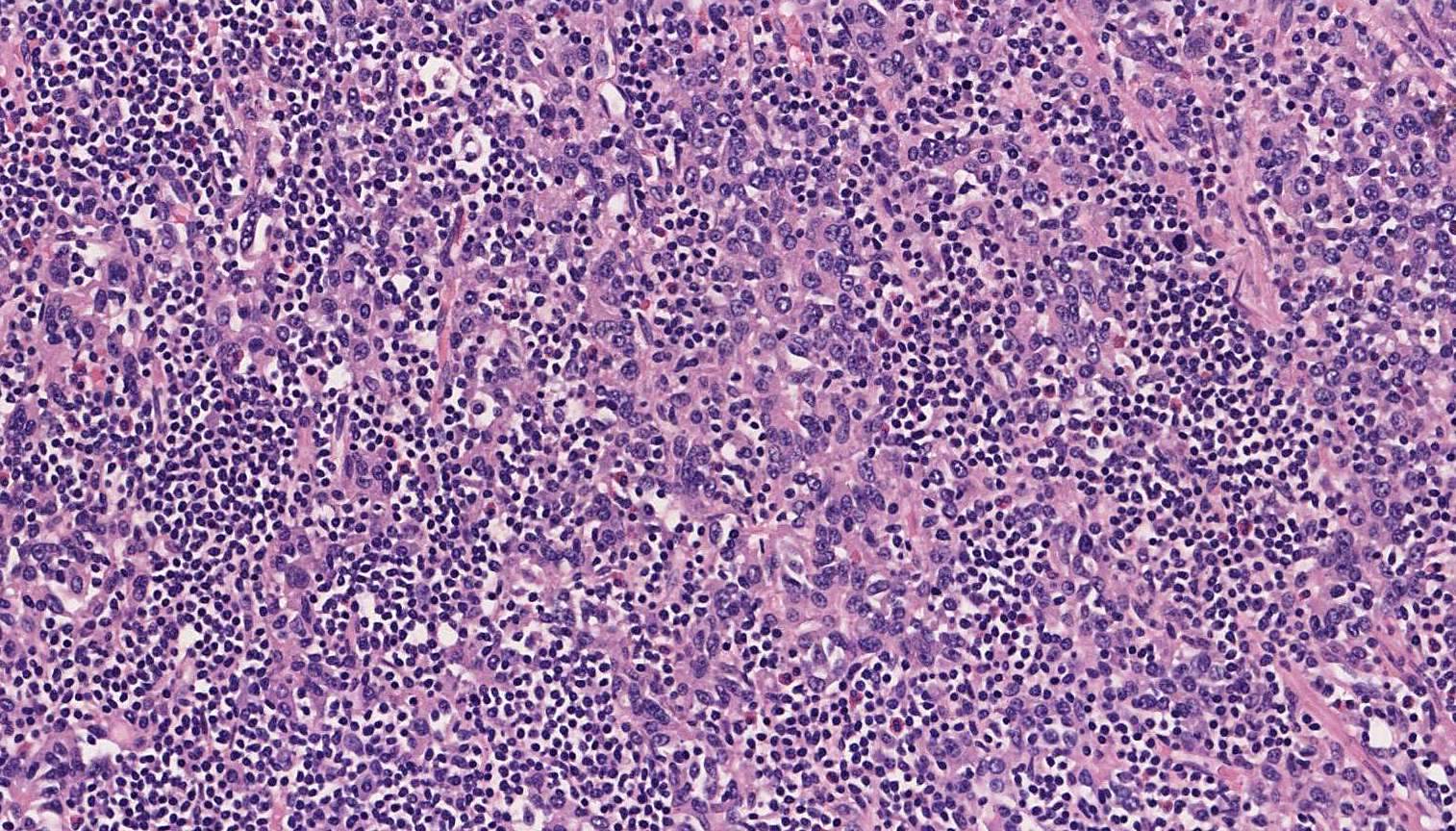
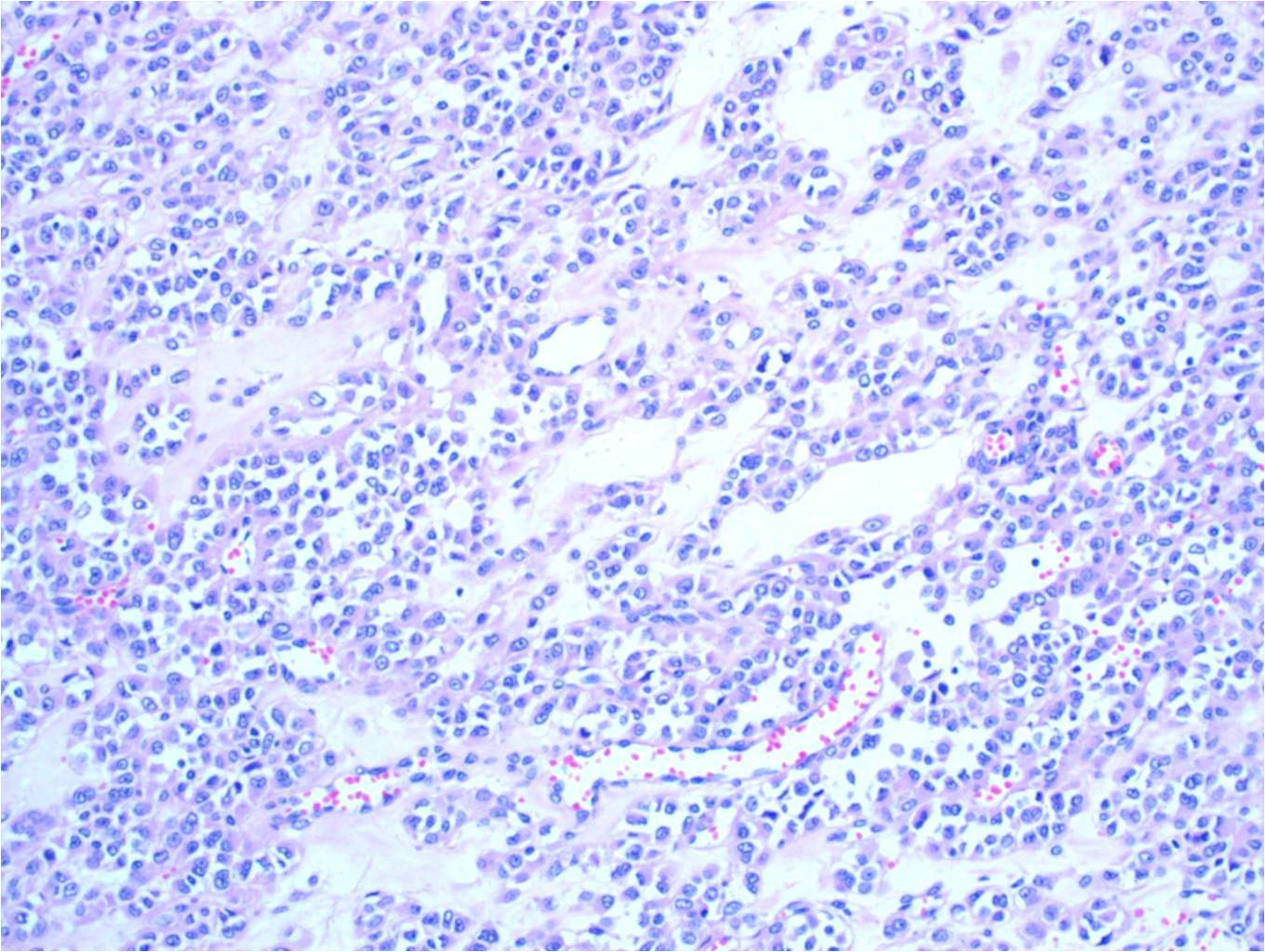
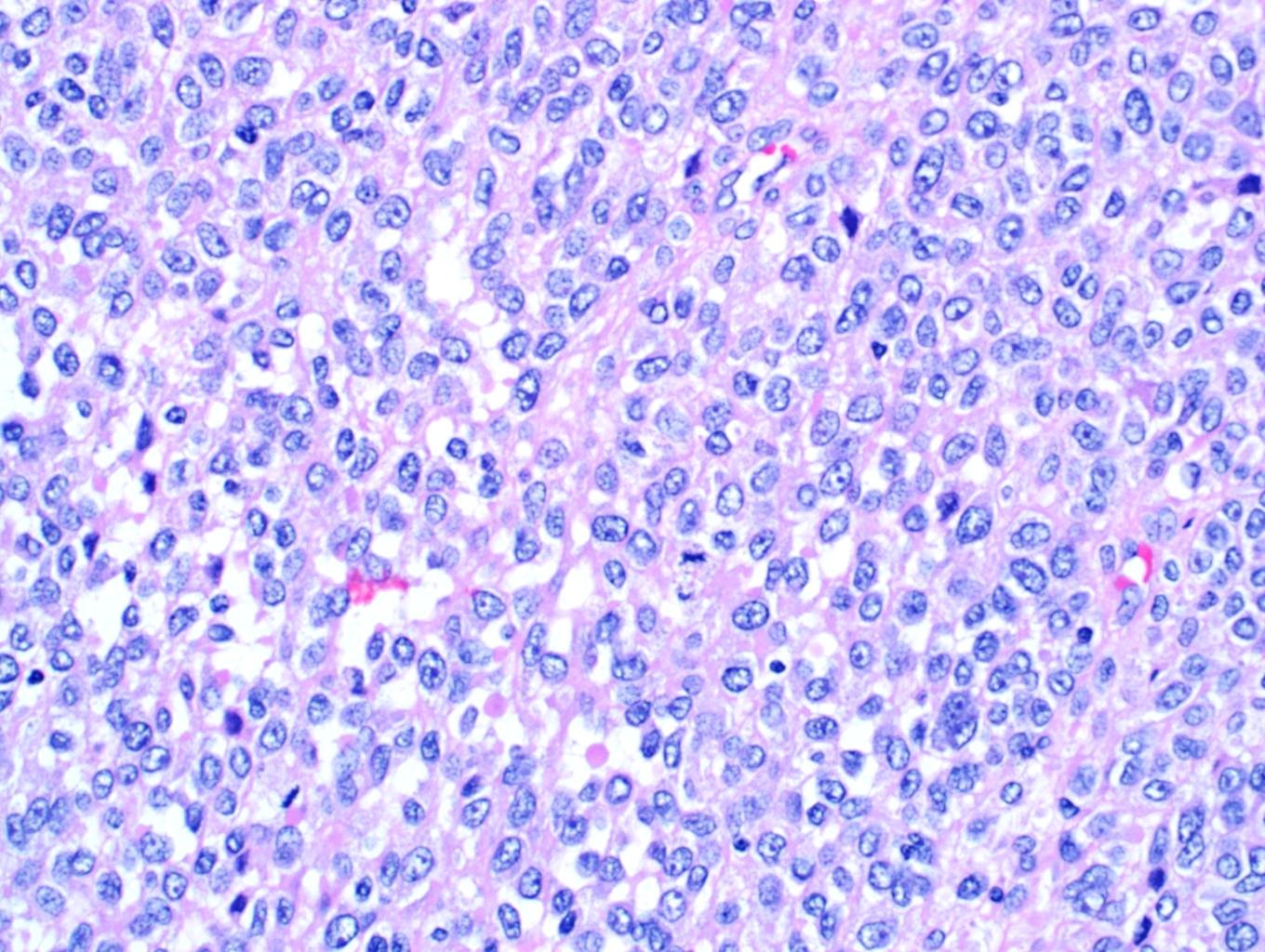
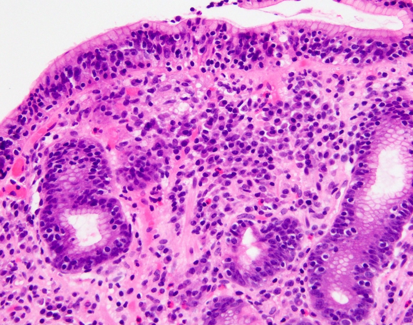
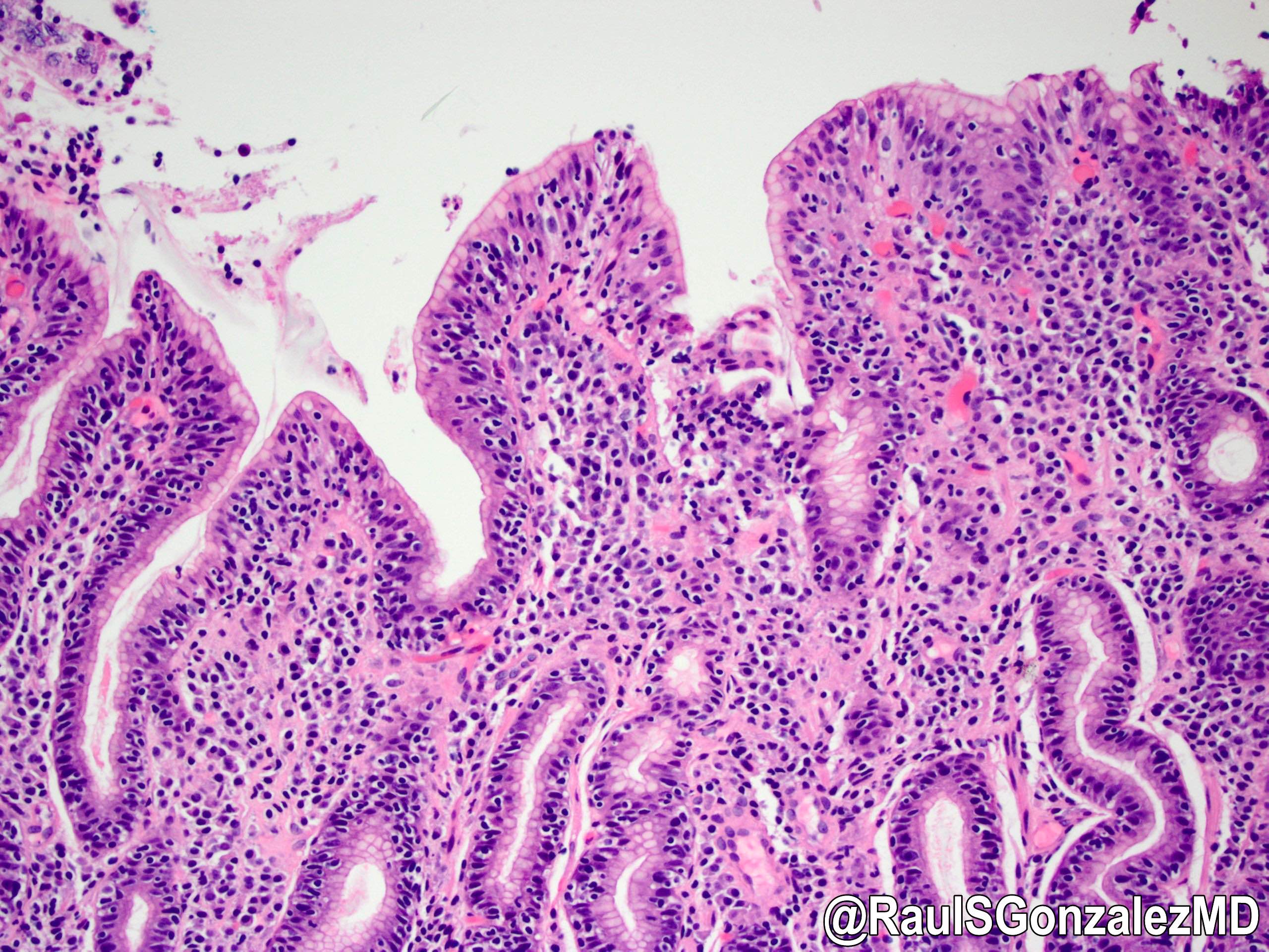
Dense lymphoplasmacytic infiltrate
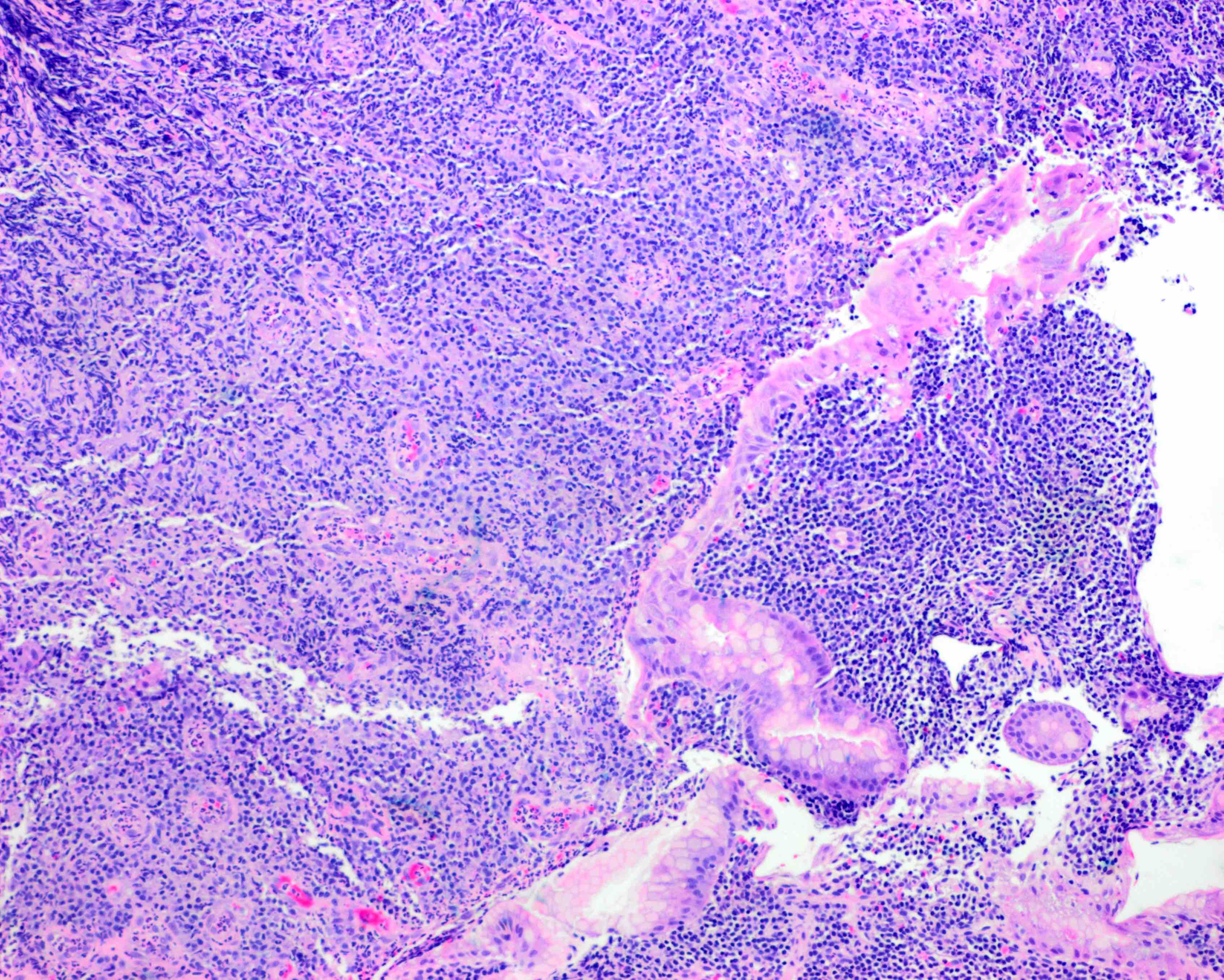
Destruction of gastric glands
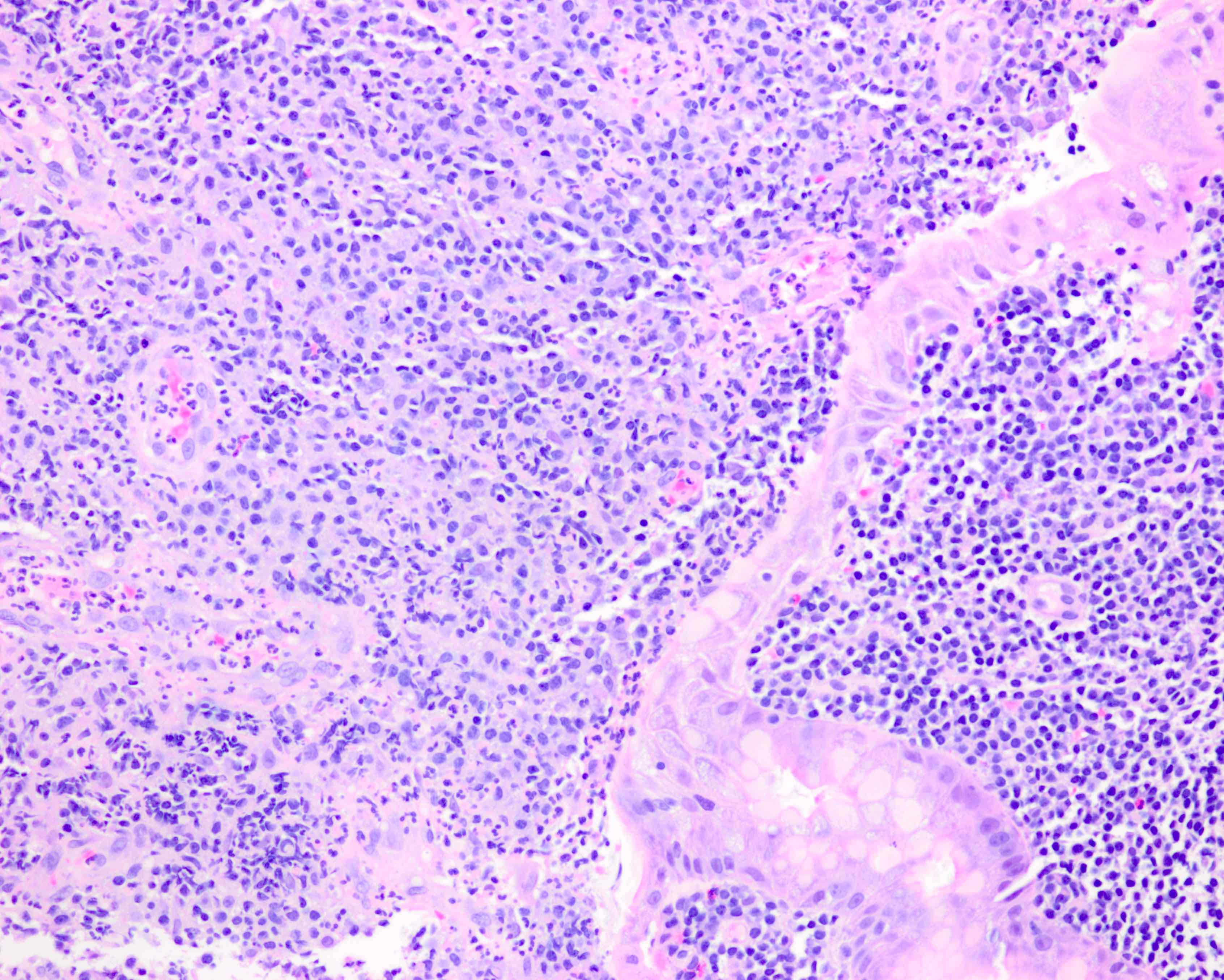
Focus of active inflammation
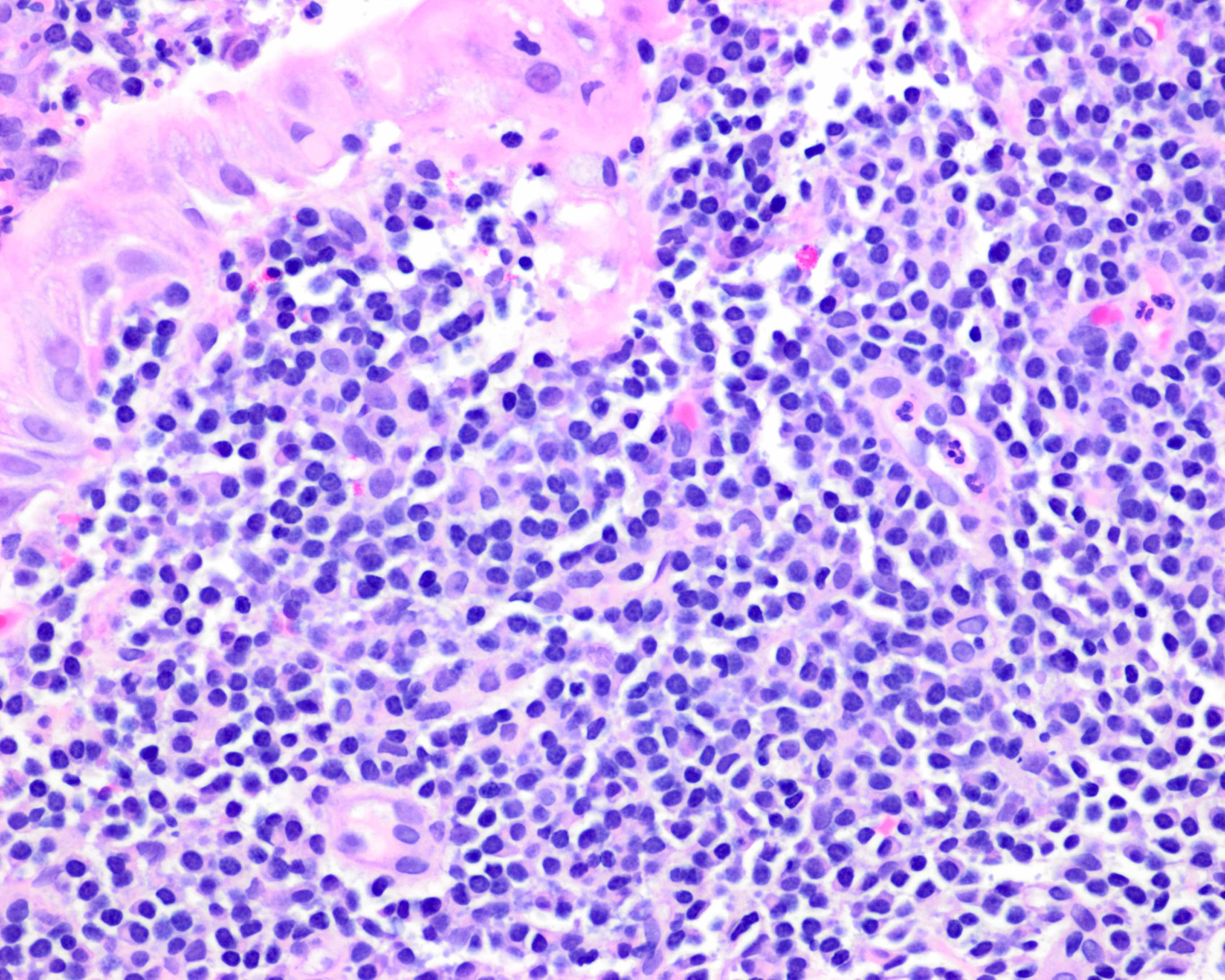
Marked plasmacytic differentiation
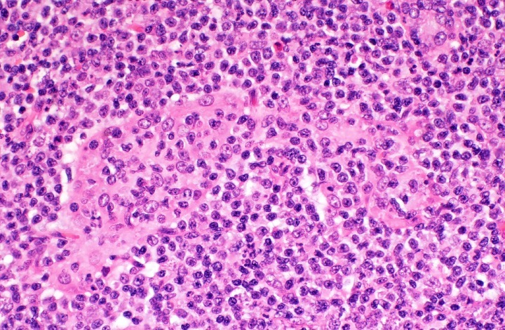
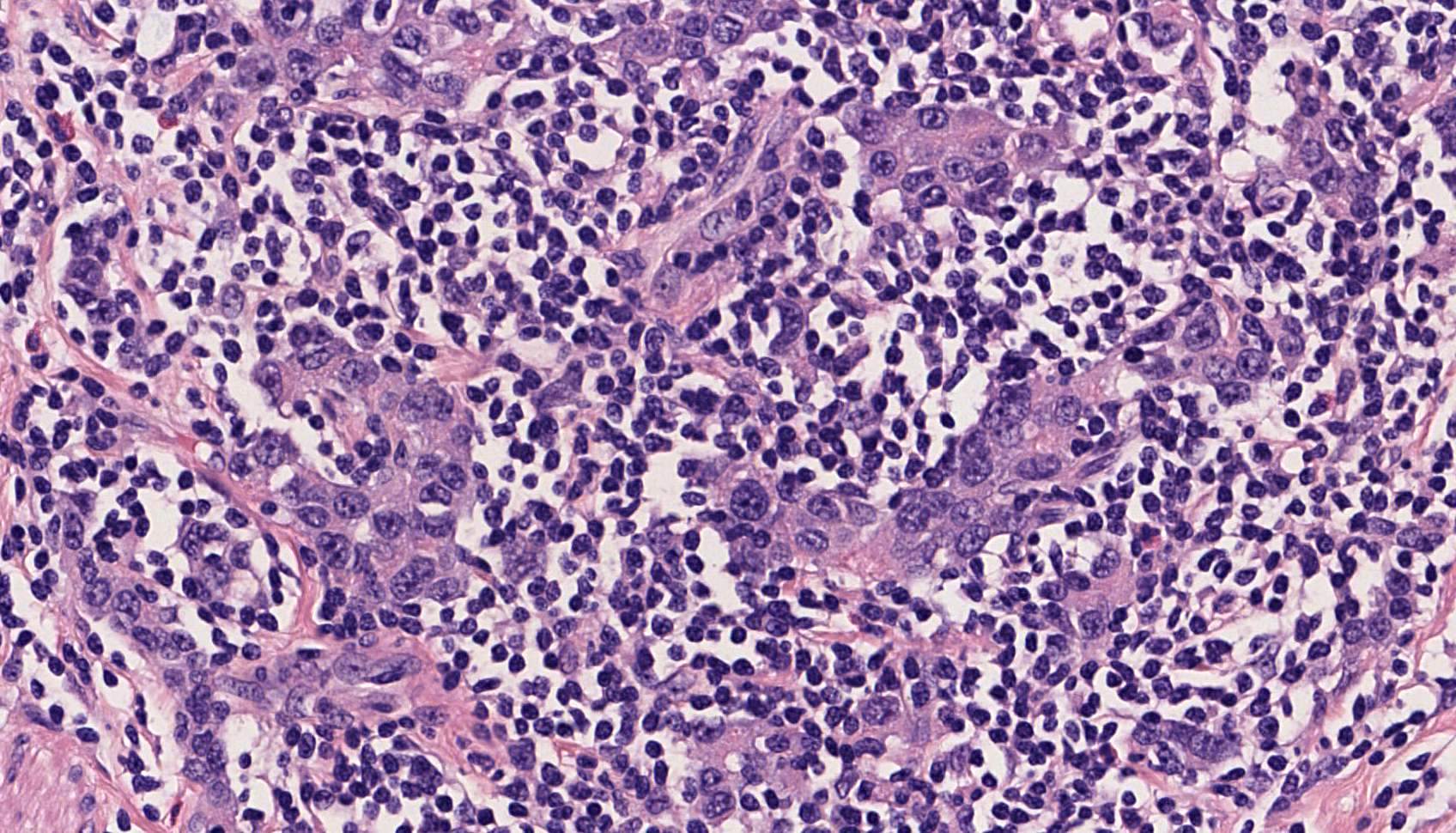
Lymphoepithelial lesion
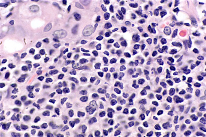
Cytologic features of lymphoma cells
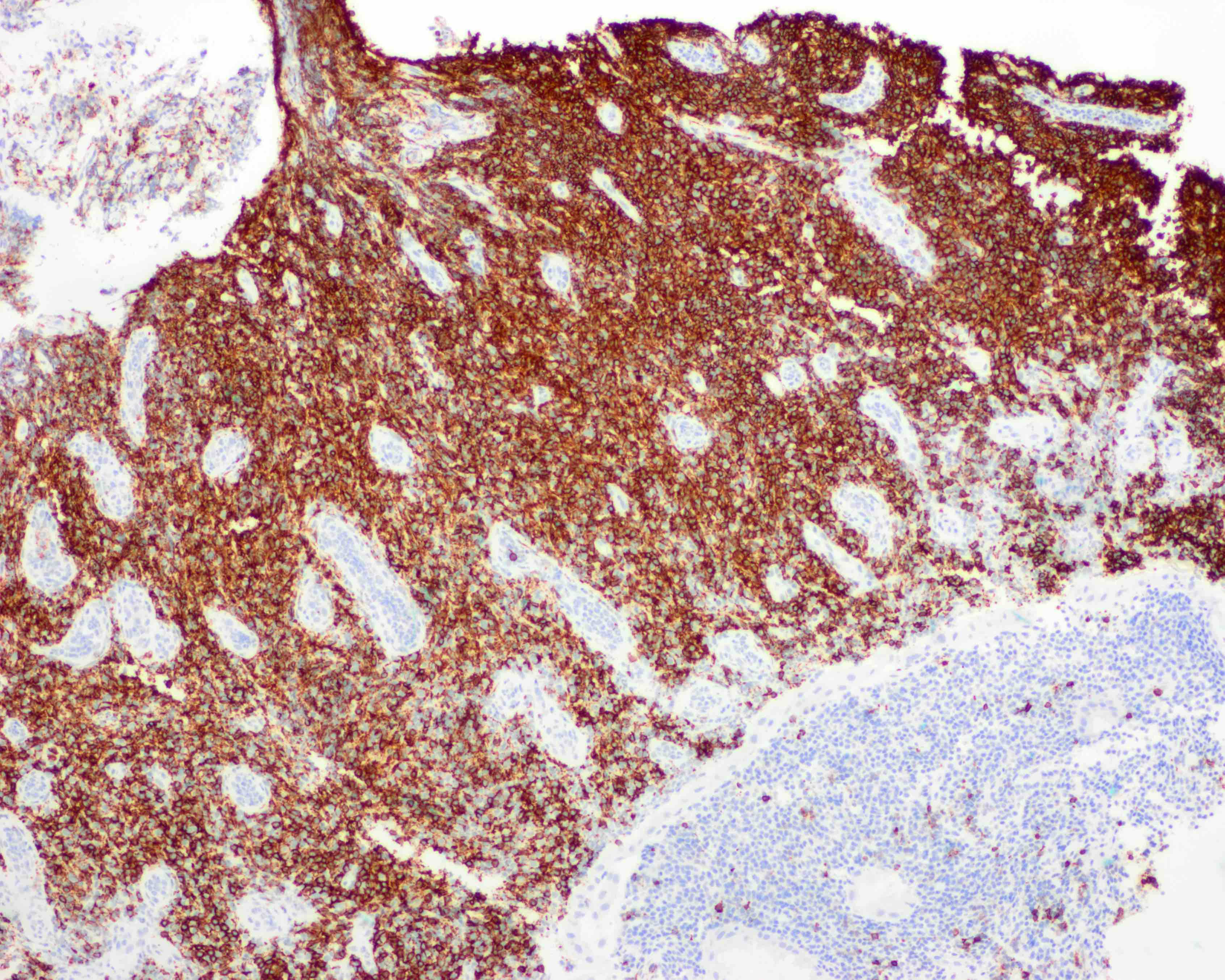
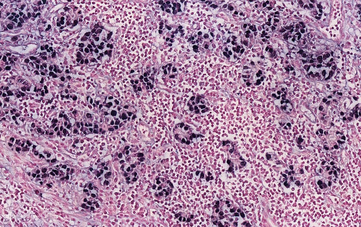
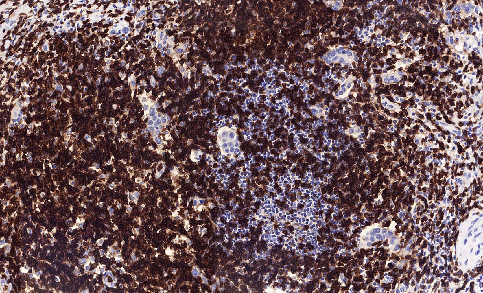
Diffusely positive for CD20
Numerous kappa restricted cells (κ:λ = > 8:1)
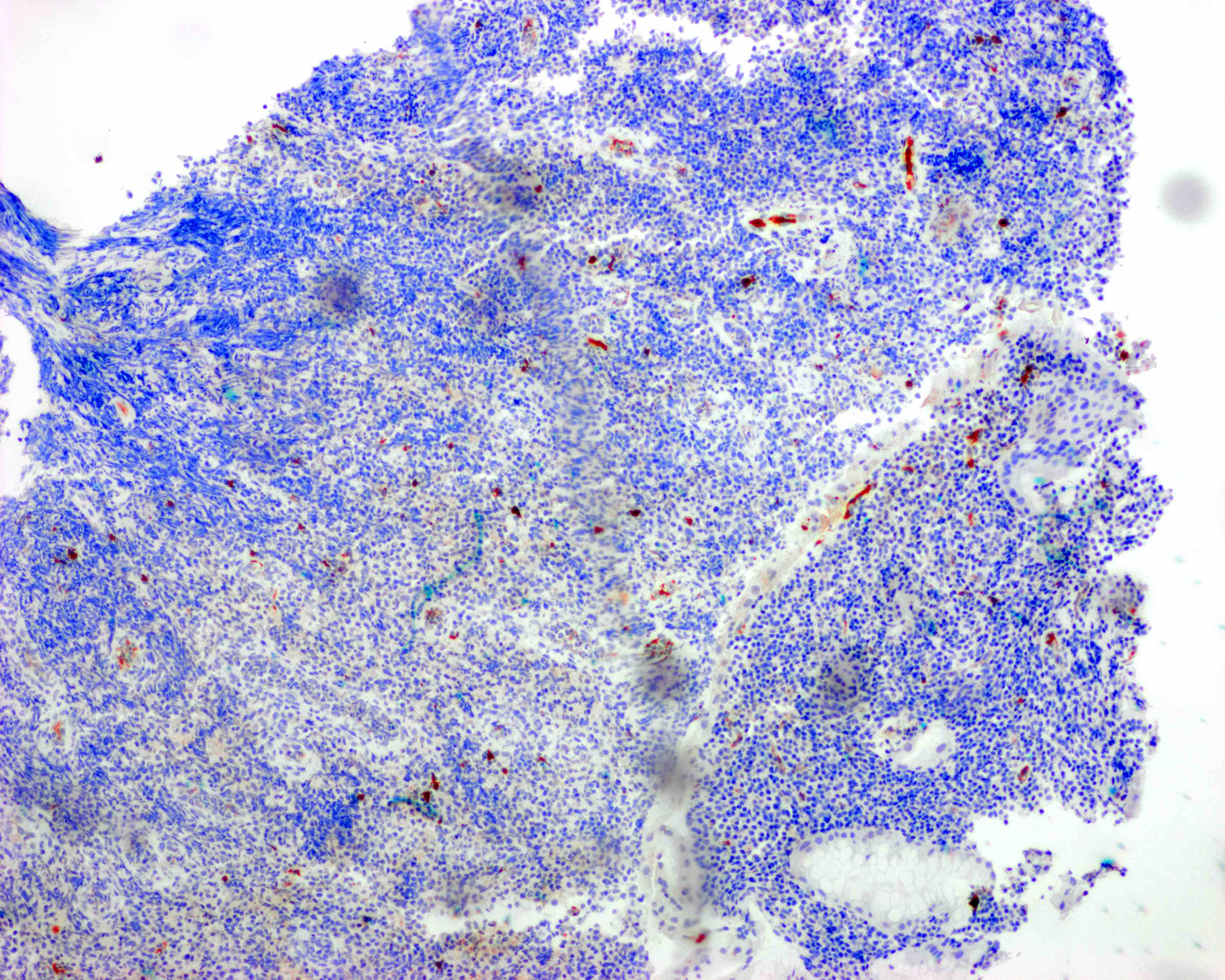
Rare lambda positive cells (κ:λ = > 8:1)

Low proliferative index
Images hosted on other servers:

t(11;18)(q21;q21) translocation
Images hosted on other servers:

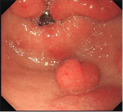
Pedunculated lesion

Multiple polyps
Contributed by Naziheh Assarzadegan, M.D.

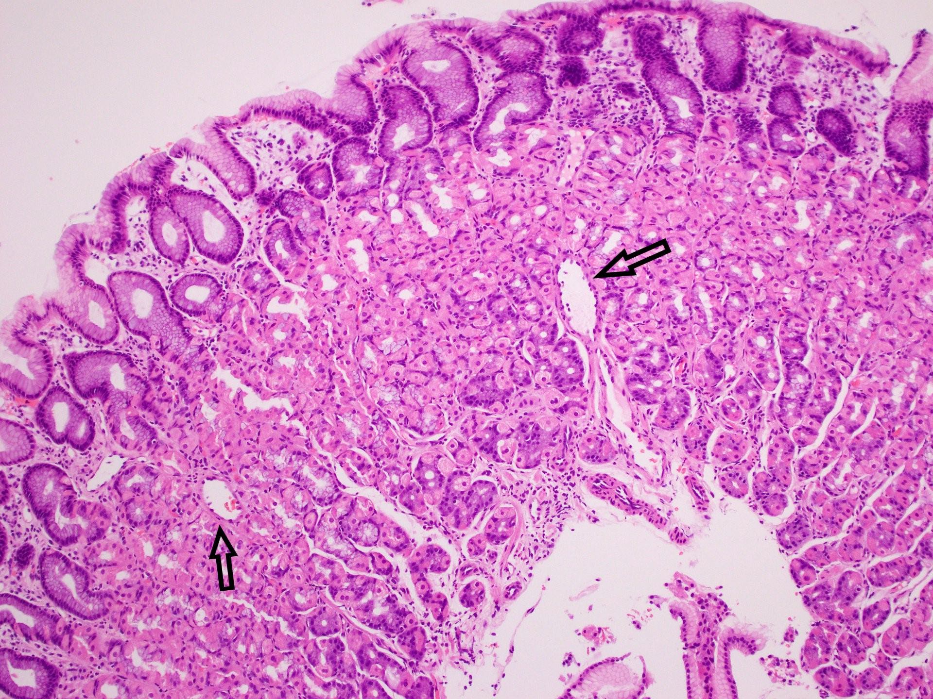
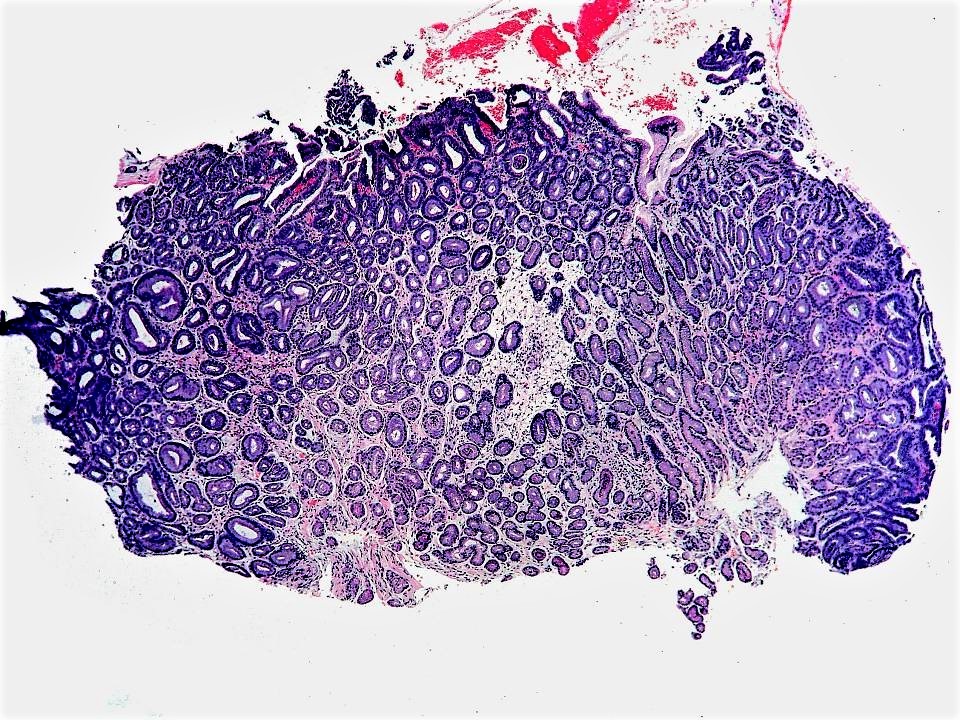
Cystically dilated glands
Fundic gland polyp with dysplasia
Case #389
Specialized gastric mucosa

Scattered dilated fundic type glands

Nuclear enlargement, hyperchromasia and stratification
Fundic gland polyp with dysplasia
Contributed by Tony El Jabbour, M.D.
Diffuse punctate pattern
Images hosted on other servers:

Striped pattern

Diffuse punctate pattern

Nodular pattern
Contributed by Hwajeong (Jenny) Lee, M.D.

Antrum with reactive changes
Fibrin thrombus
Histopathology of gastric antral vascular ectasia
Contributed by Carolina Martinez-Ciarpaglini, M.D., Ph.D.
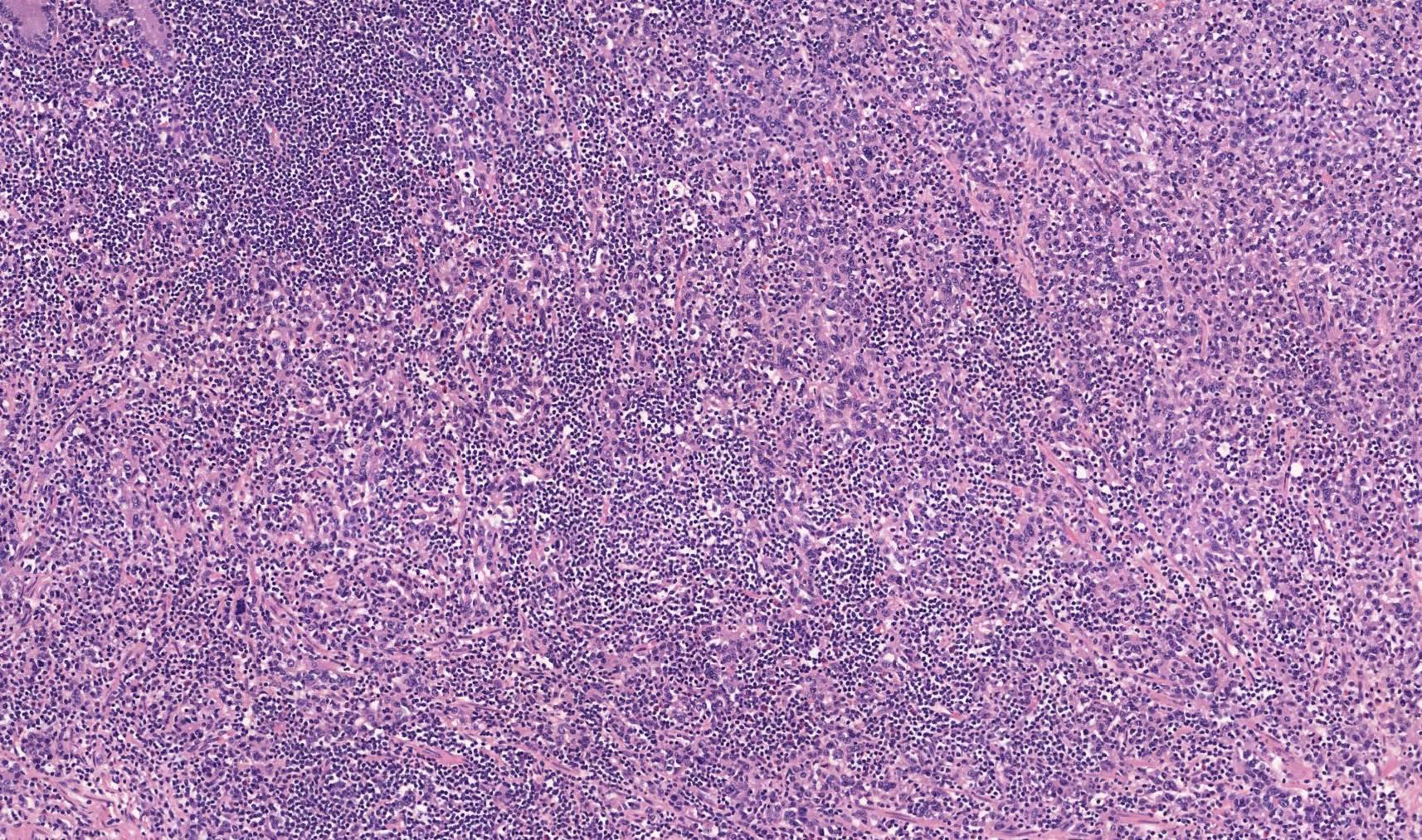
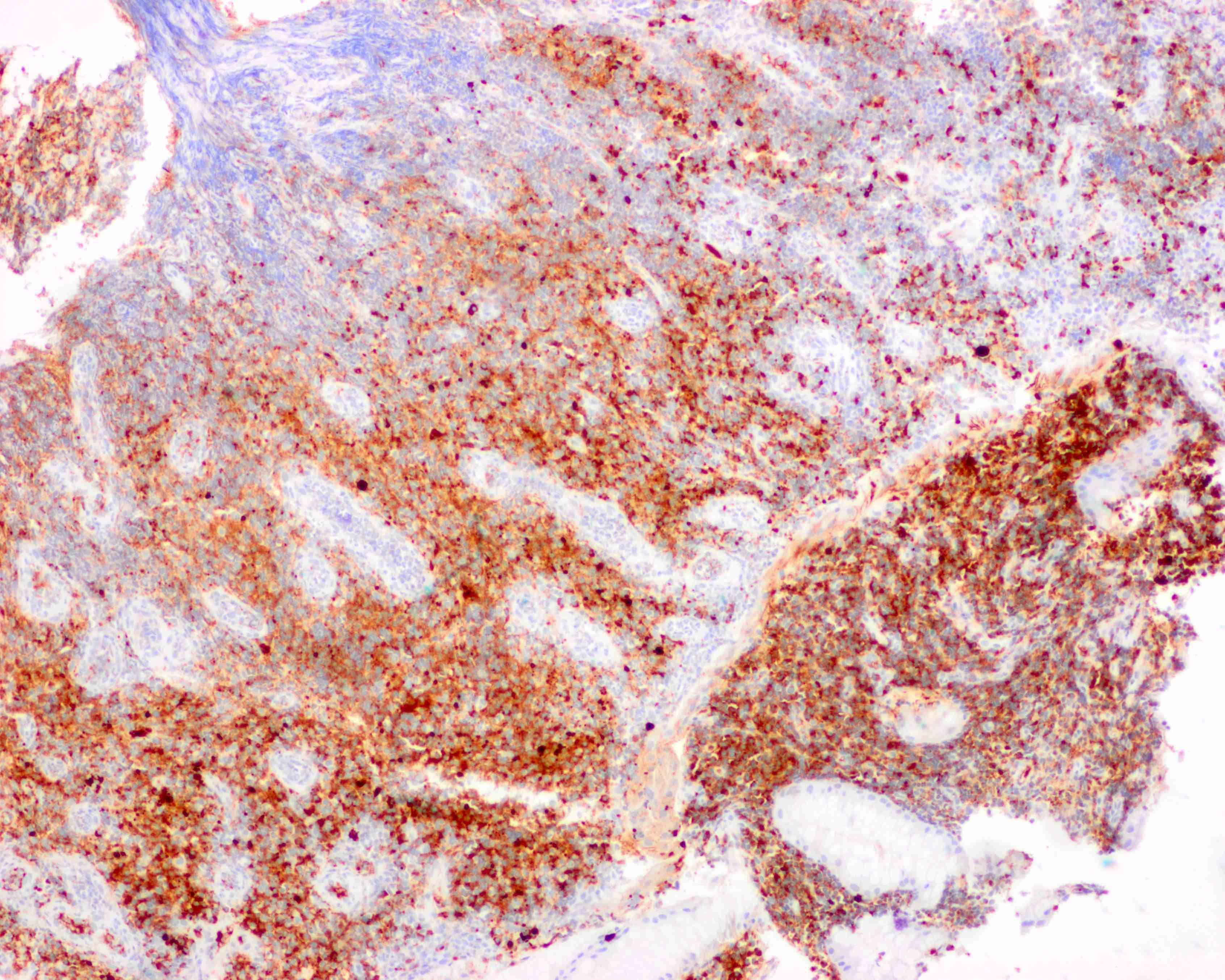
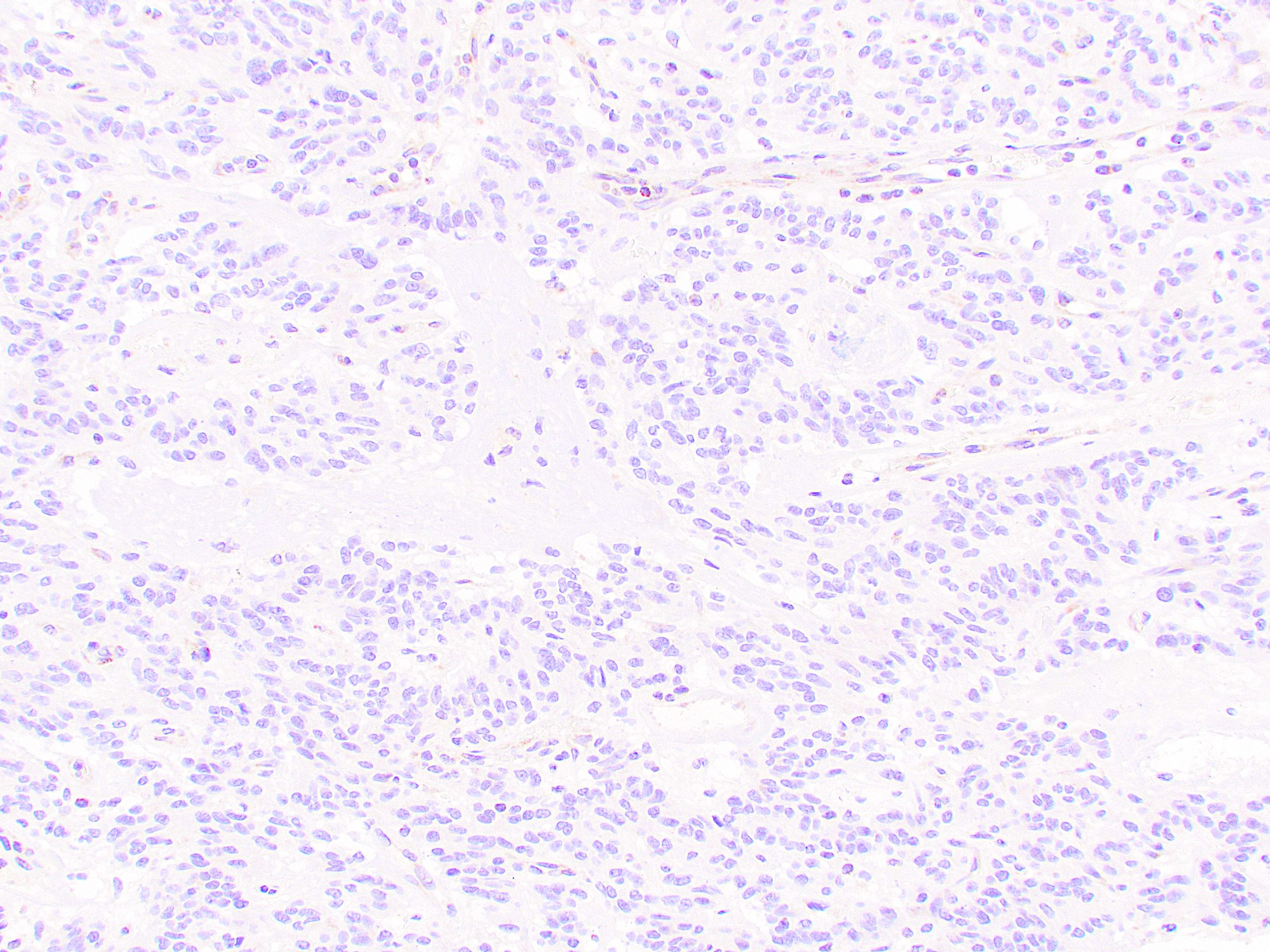
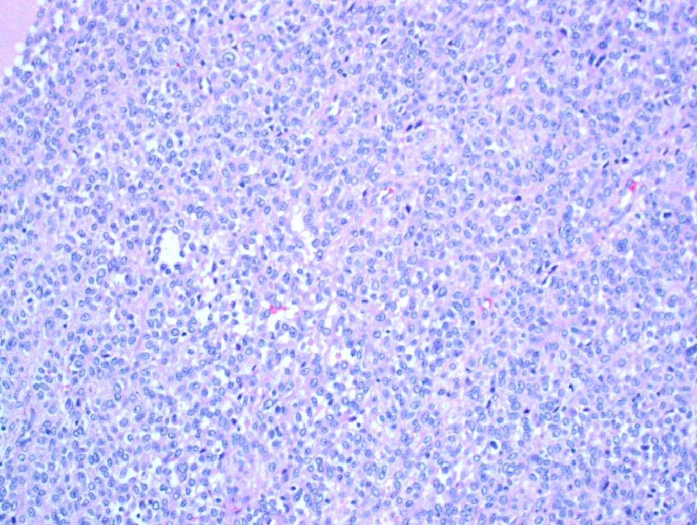
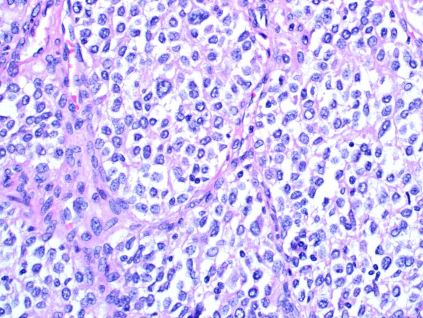
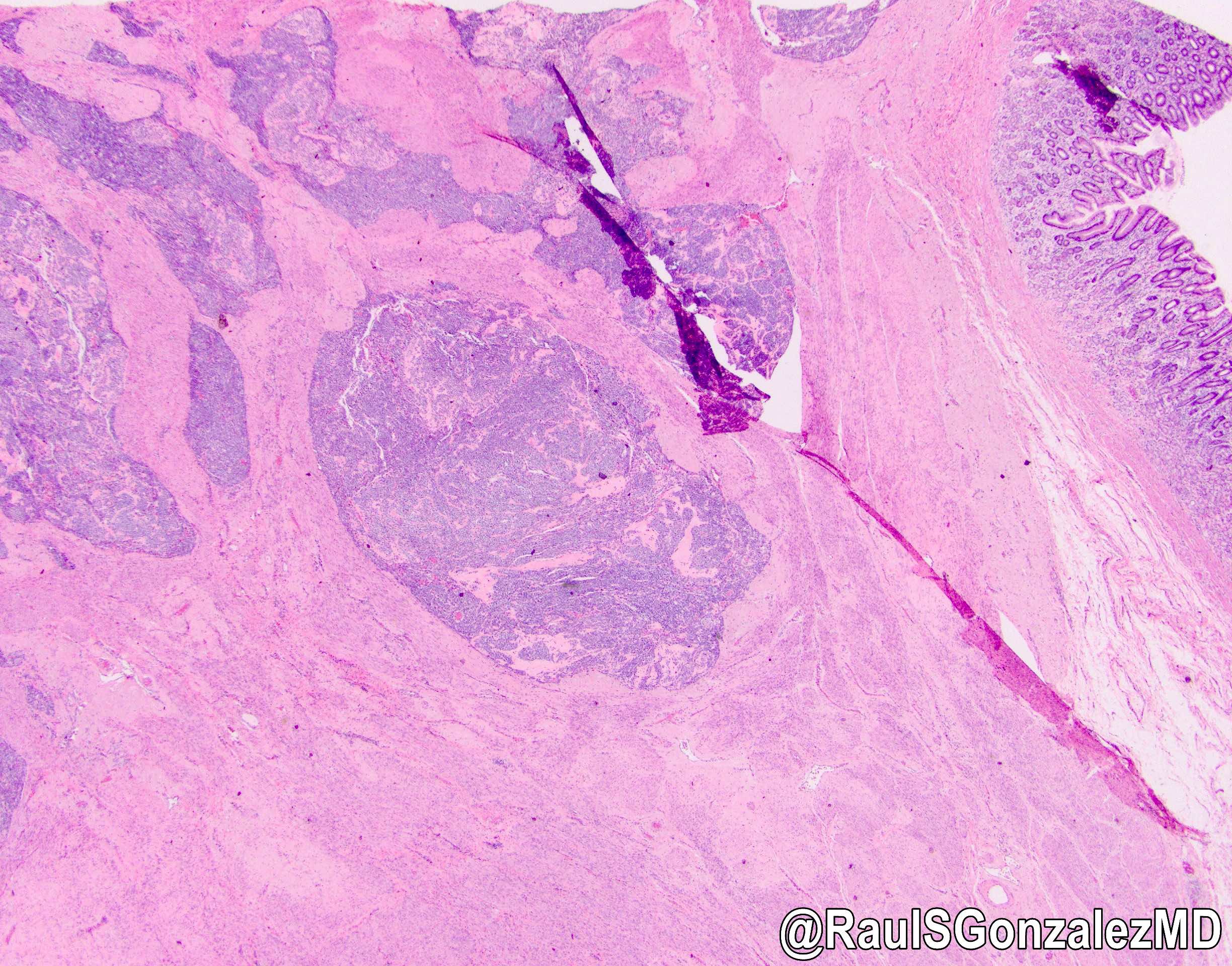
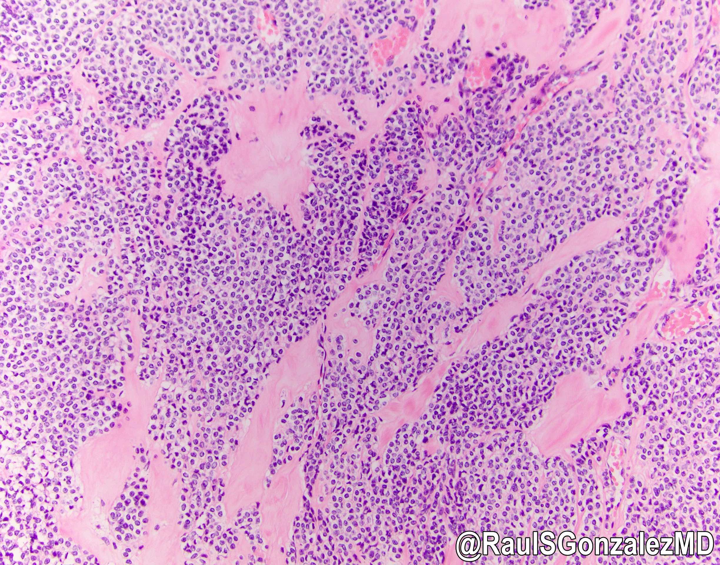
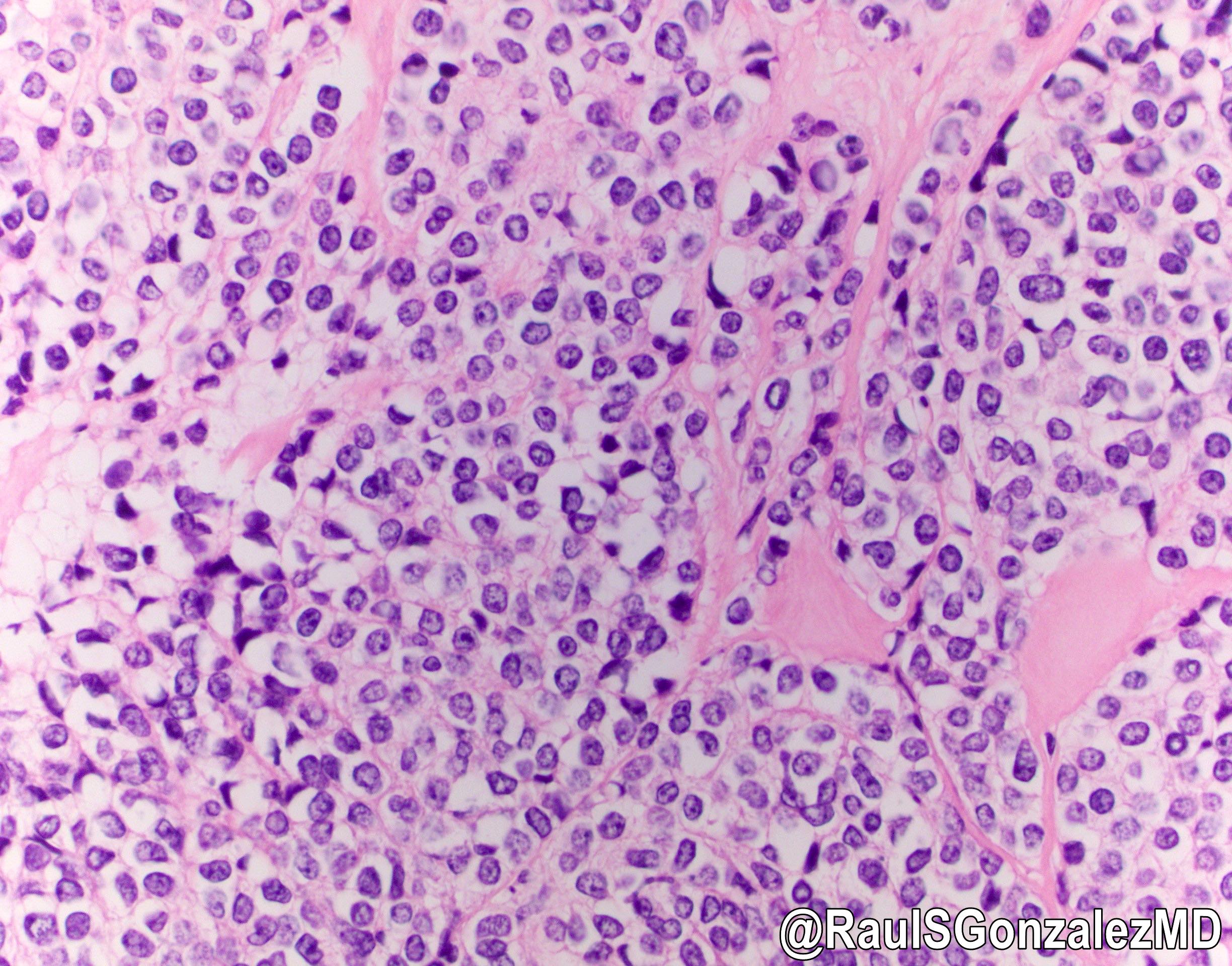
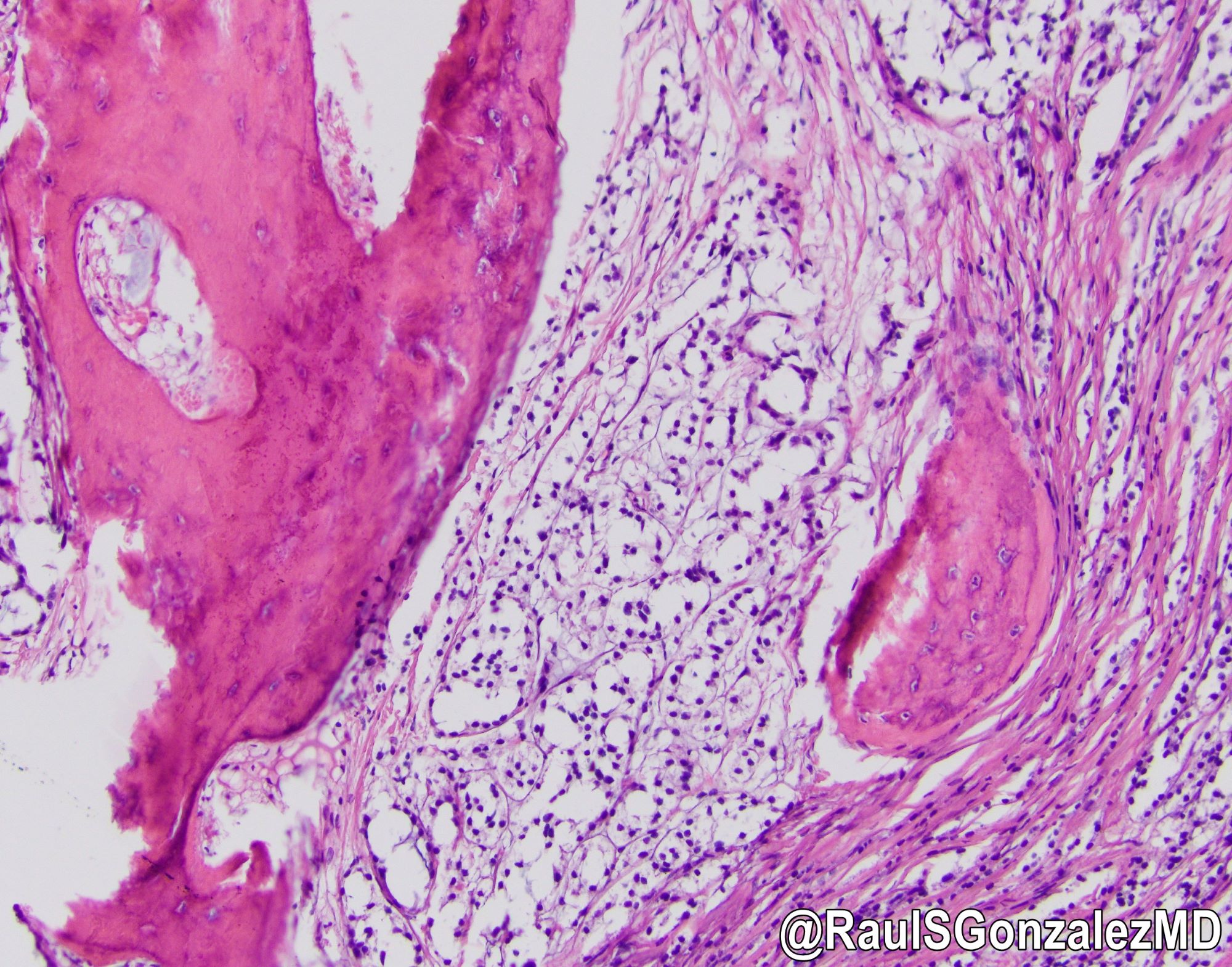
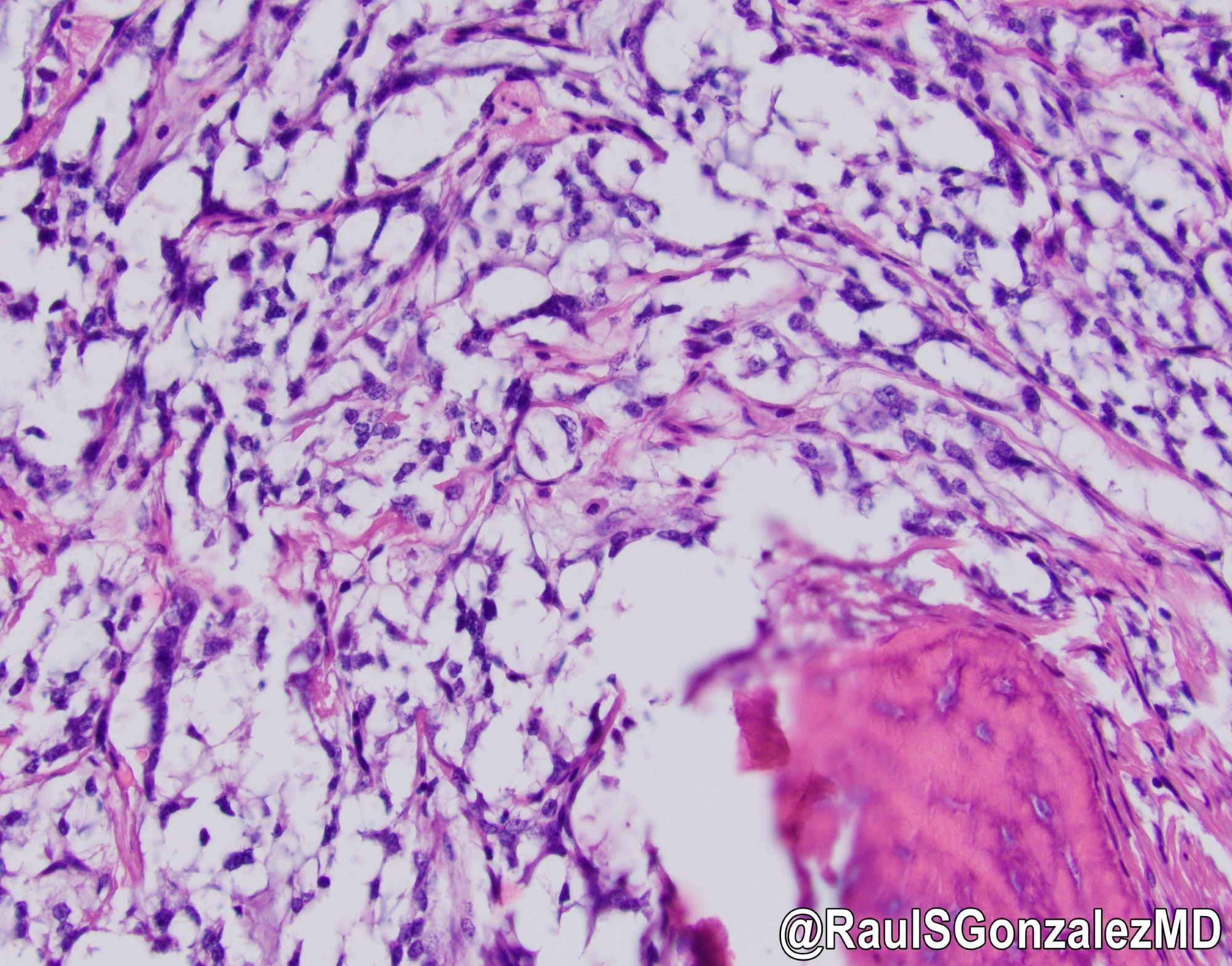
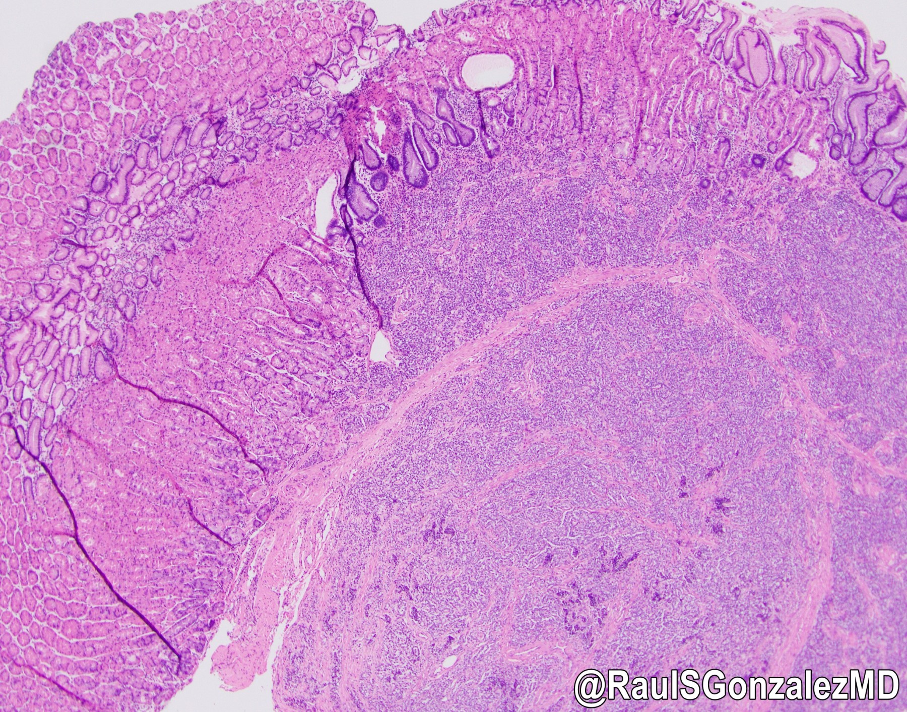
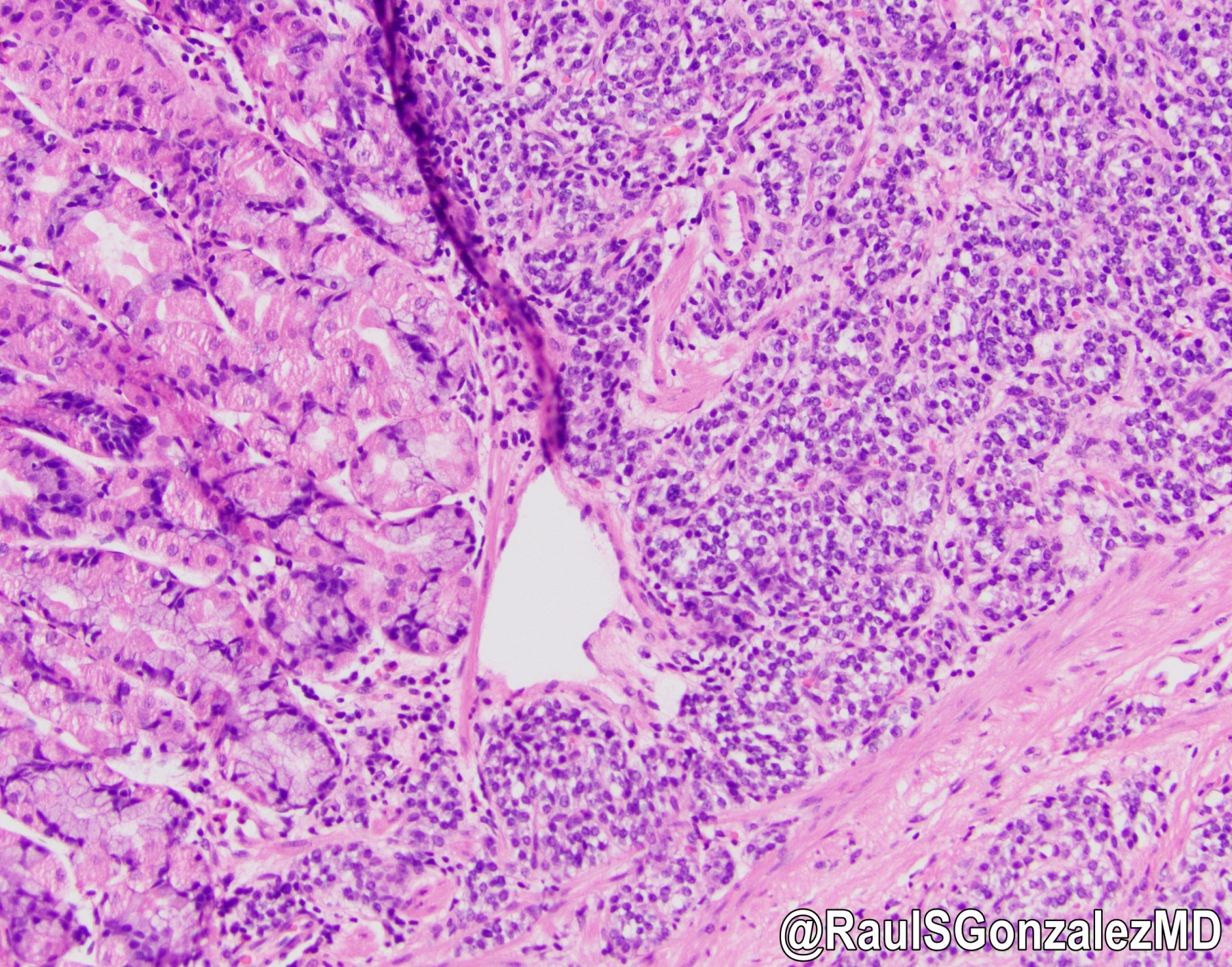
Dense lymphoid infiltrate
Trabecular pattern
Tumor cells (high power)

Primitive tubular pattern
EBER expression

Cytokeratin expresion
T cell infiltrate (CD3)
Contributed by @RaulSGonzalezMD on Twitter
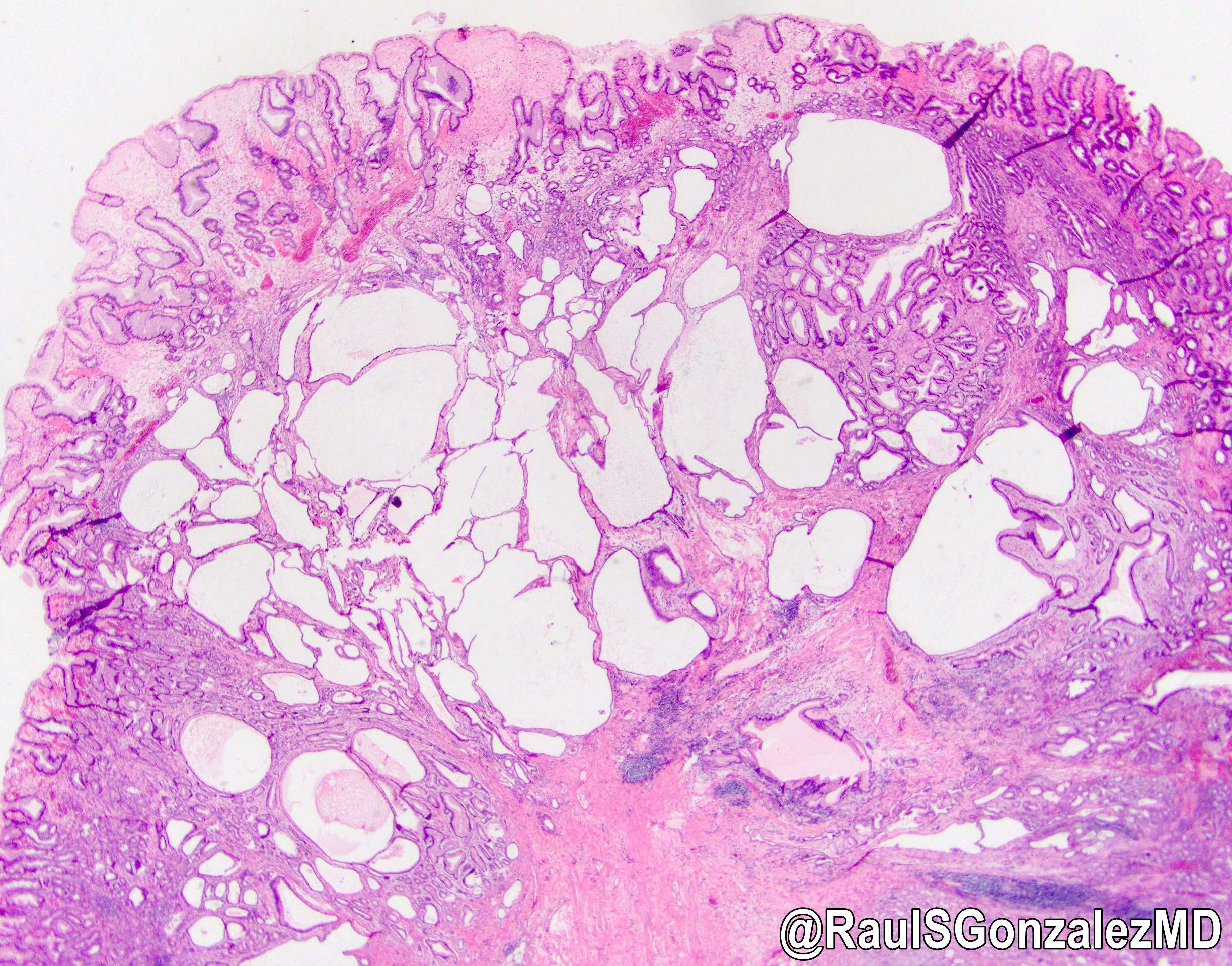
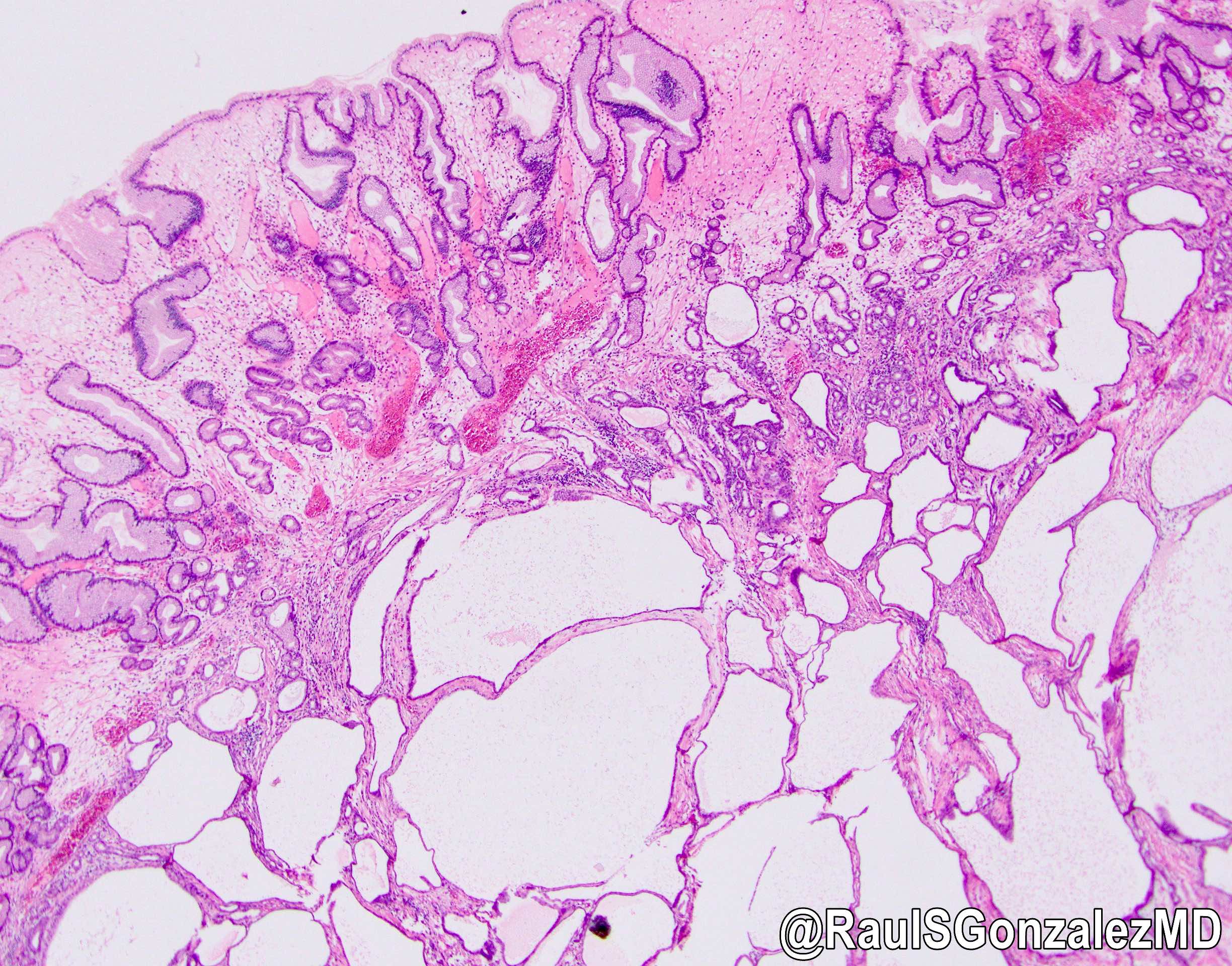
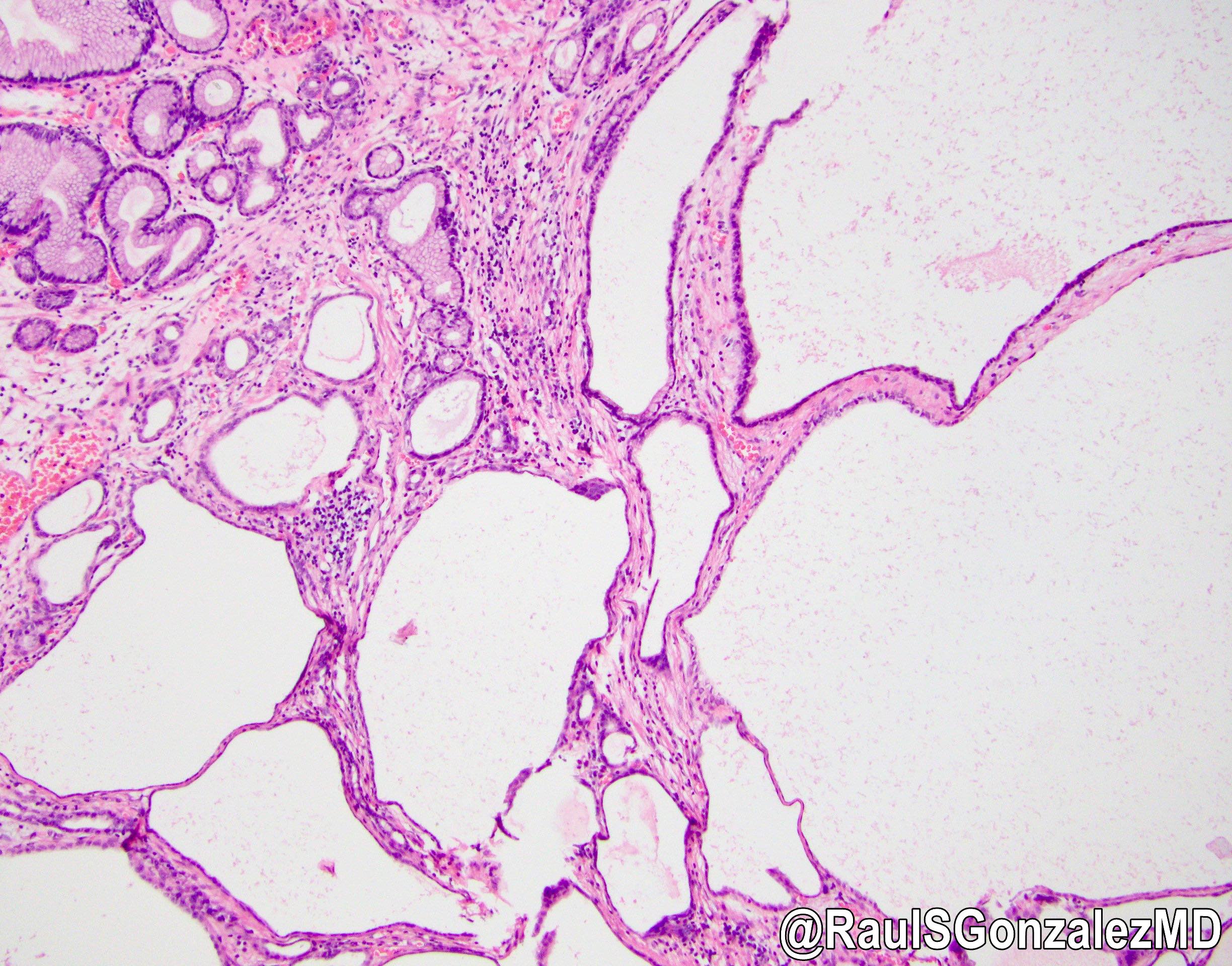
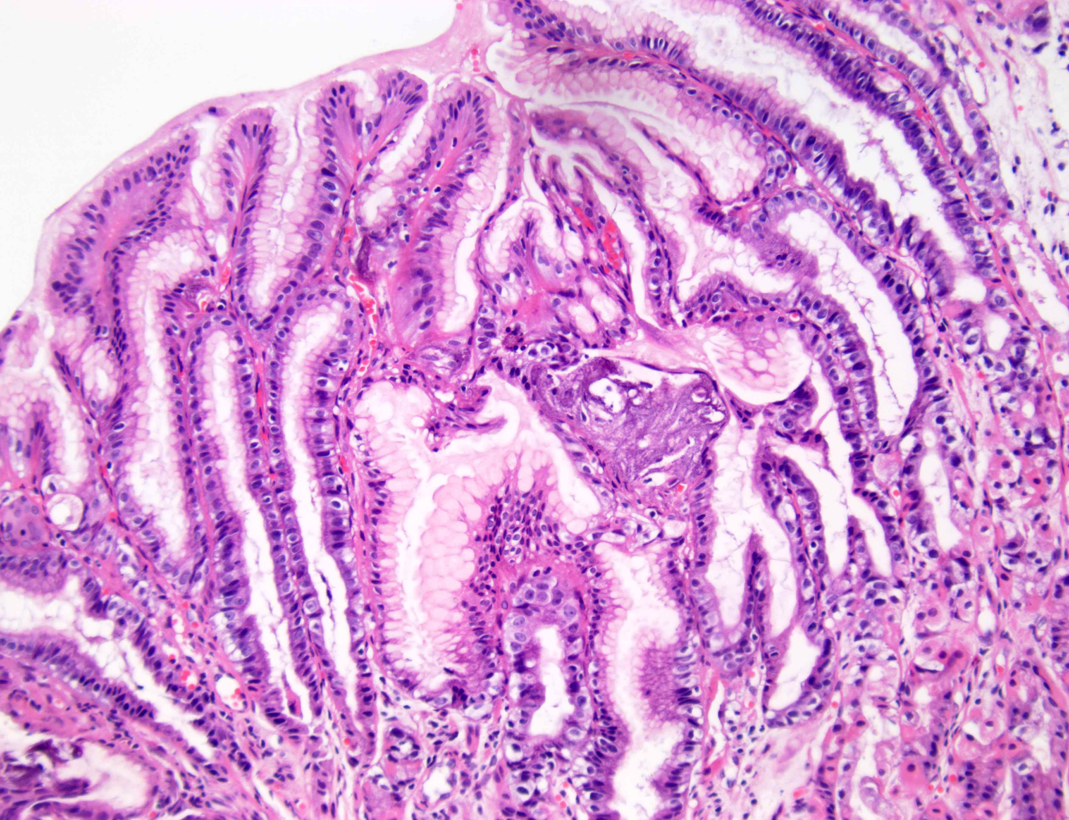
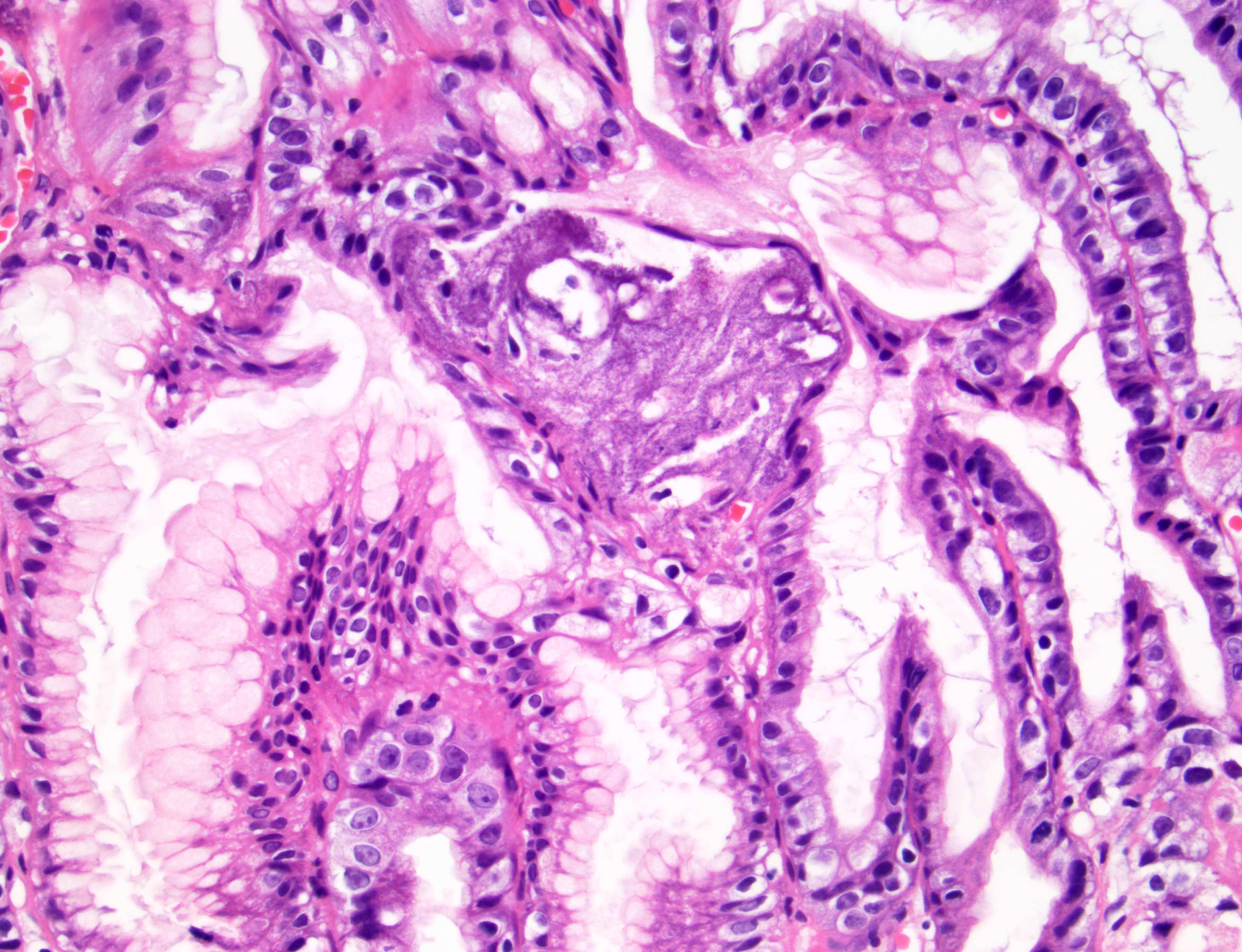
Gastritis cystica polyposa
Image hosted on other server:

Elongated, tortuous
foveolae and hyperplastic
and cystically dilated
underlying pyloric gland
Contributed by Supriya Srivastava, M.D., Ph.D.
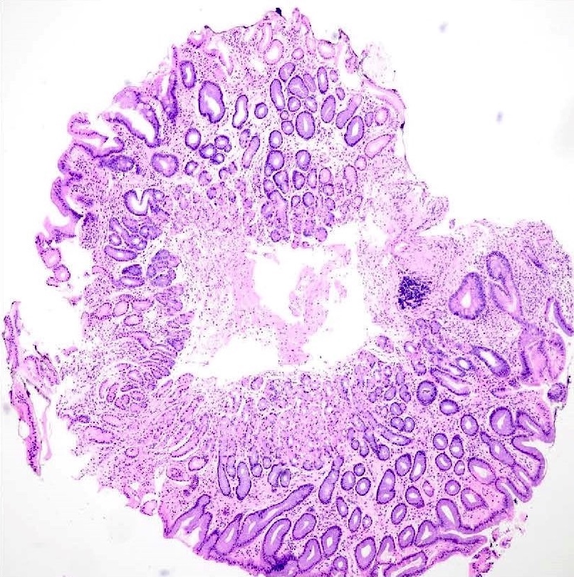
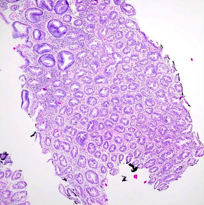
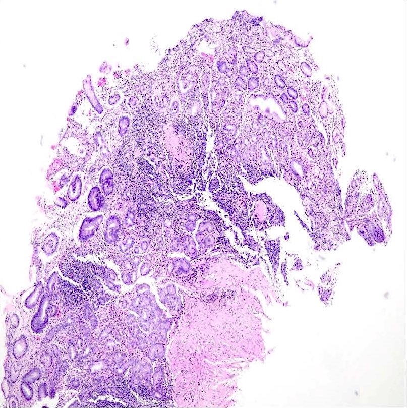
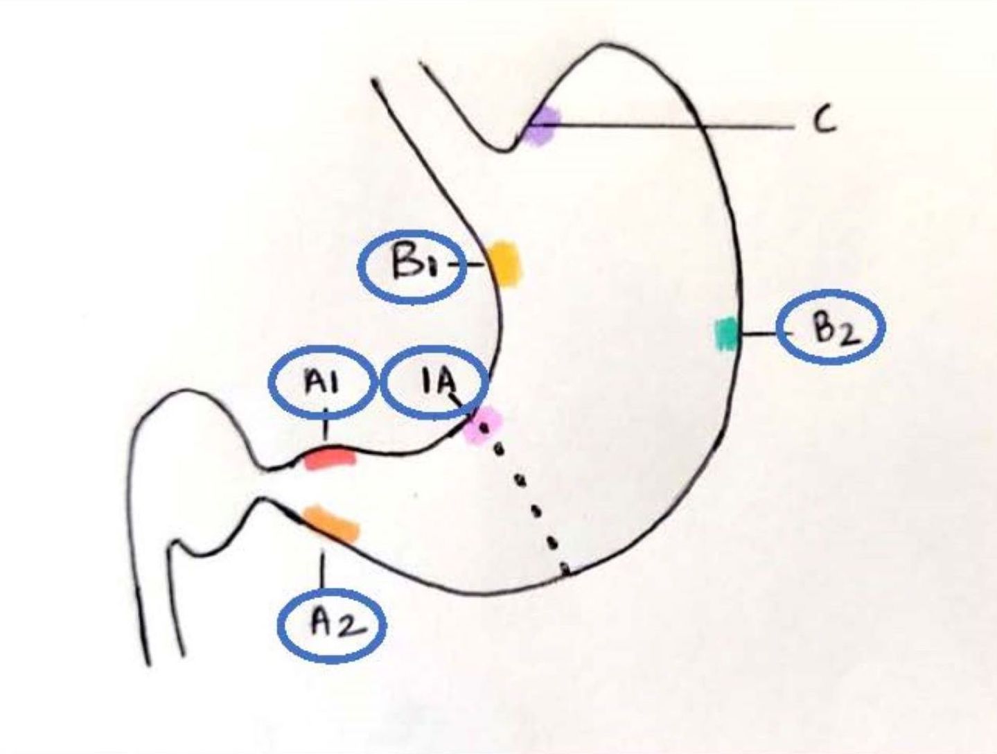
Updated Sydney protocol
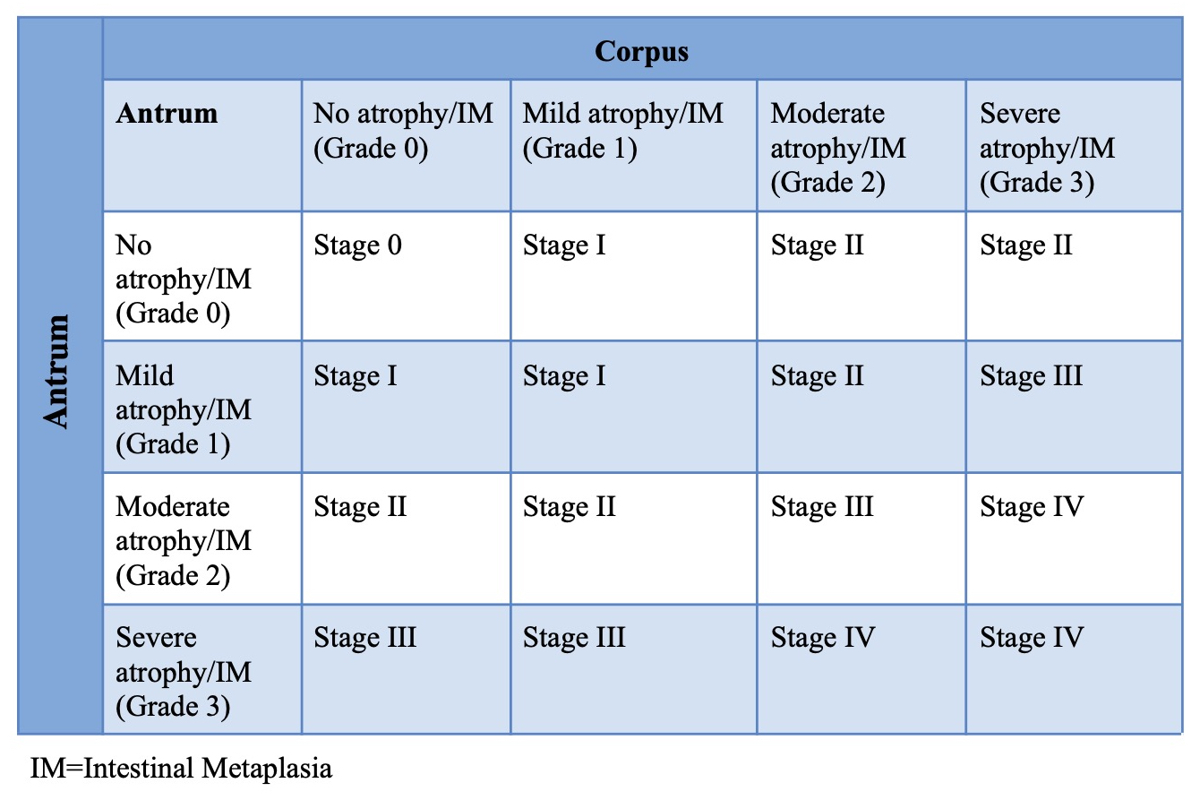
OLGIM staging
Contributed by Supriya Srivastava, M.D., Ph.D.
Mild chronic gastritis
Moderate chronic gastritis
Marked chronic gastritis
Mild intestinal metaplasia
Moderate intestinal metaplasia
Marked intestinal metaplasia
Chronic active gastritis
H. pylori gastritis
Autoimmune gastritis
H. heilmanii gastritis
Images hosted on other servers:


CT and MRI with multilobulated cystic mass
Images hosted on other servers:

Lobulated stomach mass
Images hosted on other servers:

Solid and cystic mass

Variegated mass

Transmural mass
Contributed by Raul S. Gonzalez, M.D. and Rondell P. Graham, M.B.B.S.
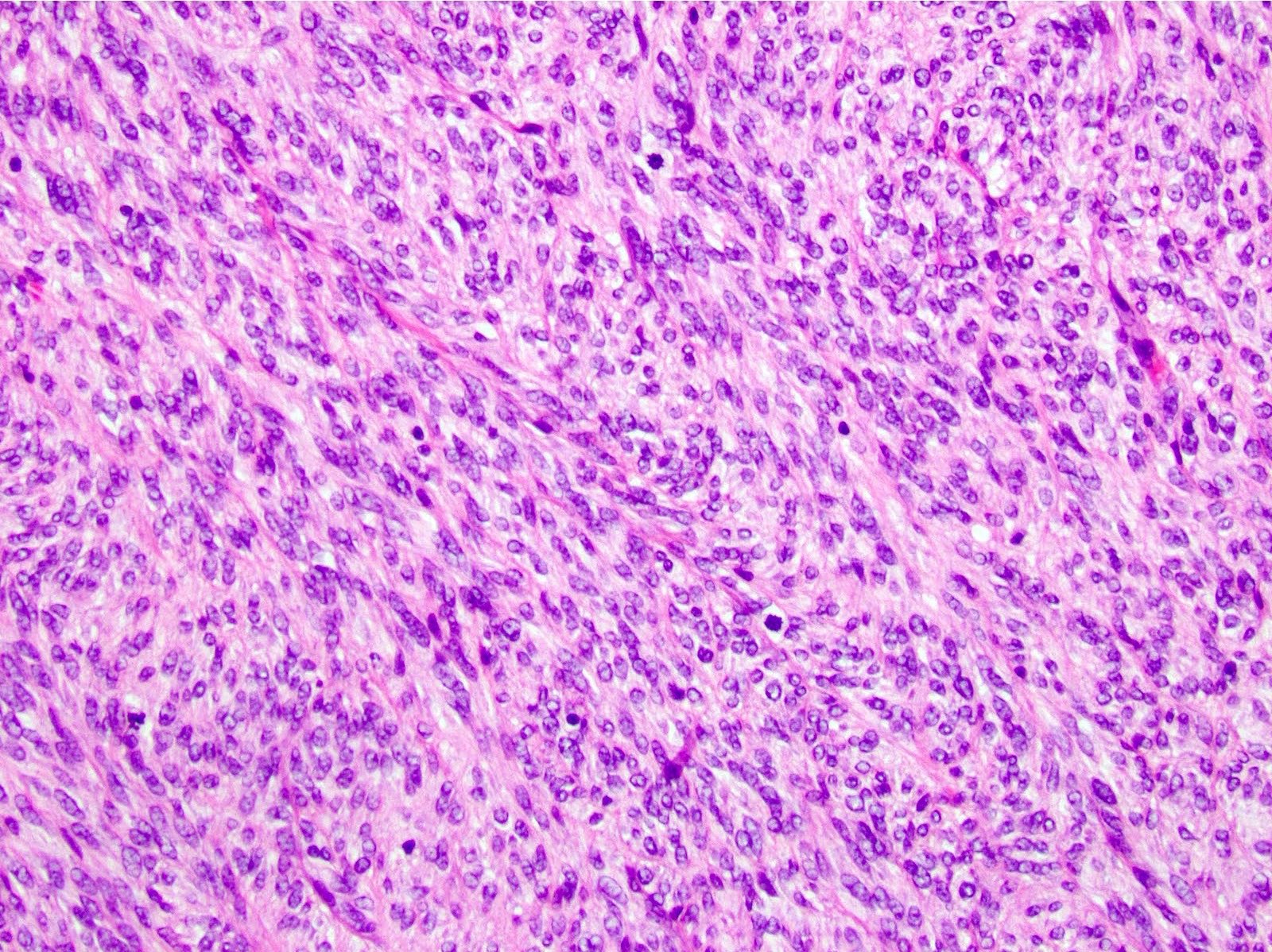
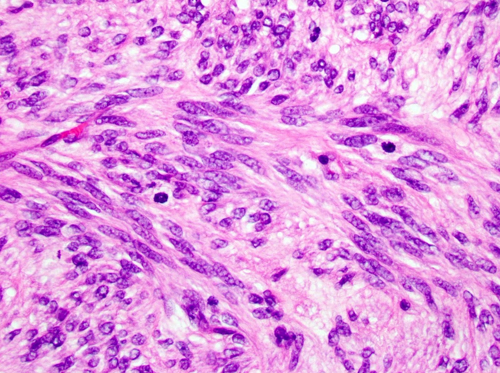
Invasive tumor
Epithelial component
Nests of epithelioid cells
Spindle cell component
Images hosted on other servers:

Desmosomes and microvilli between tumor cells
Images hosted on other servers:

RT-PCR, MALAT1::GLI1 fusion and sequencing

GLI1 FISH
breakapart and
MALAT1::GLI1
fusion FISH
Images hosted on other servers:


Gastric GIST
Images hosted on other servers:

Endoscopy

Intraoperative mass

Gastroscopy
Contributed by Riki Turri, PA (ASCP)
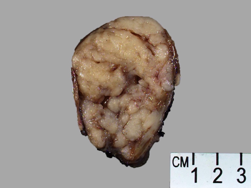
Stomach GIST, cross section
Contributed by Phoenix D. Bell, M.S., M.D. and Jennifer Findeis-Hosey, M.D.
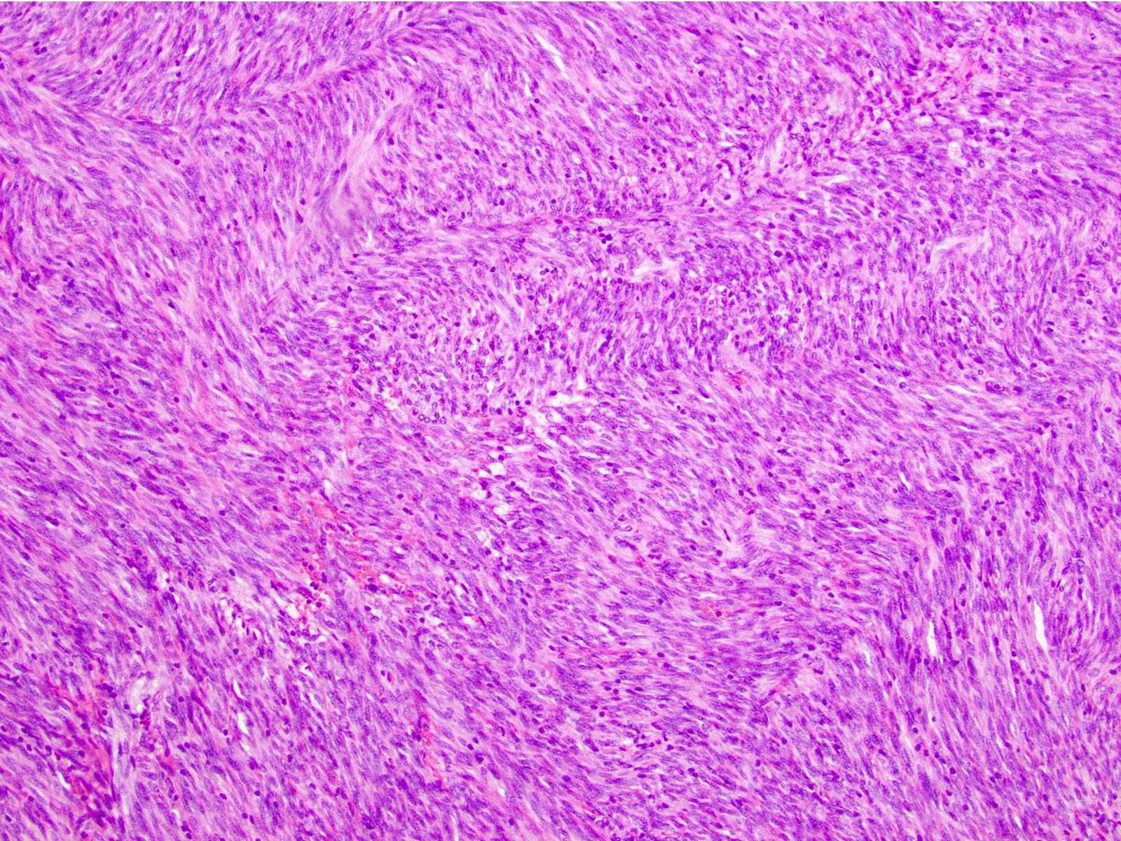
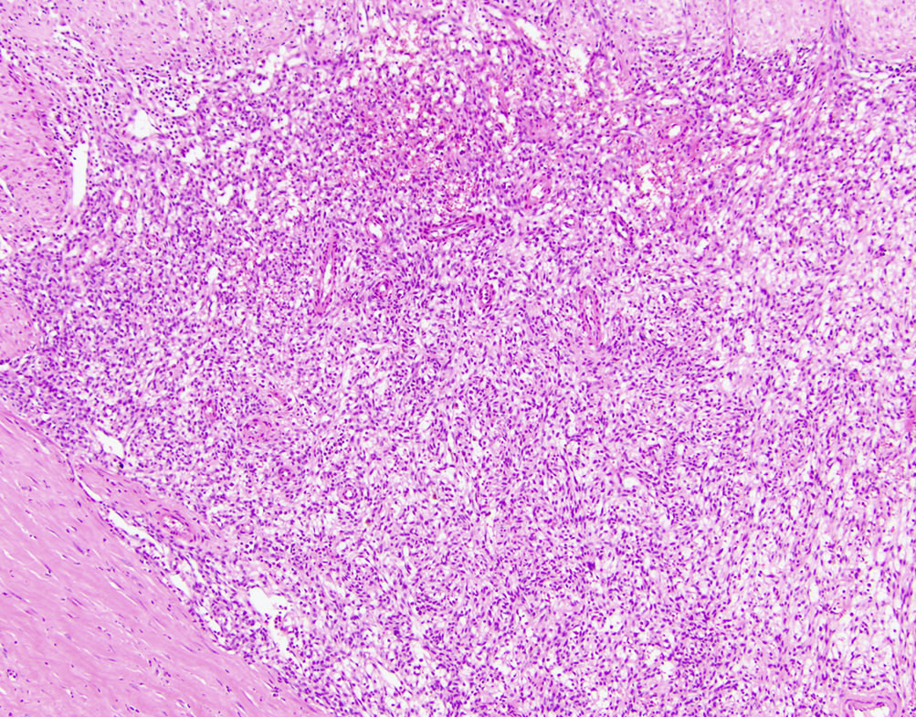
Spindle cell type
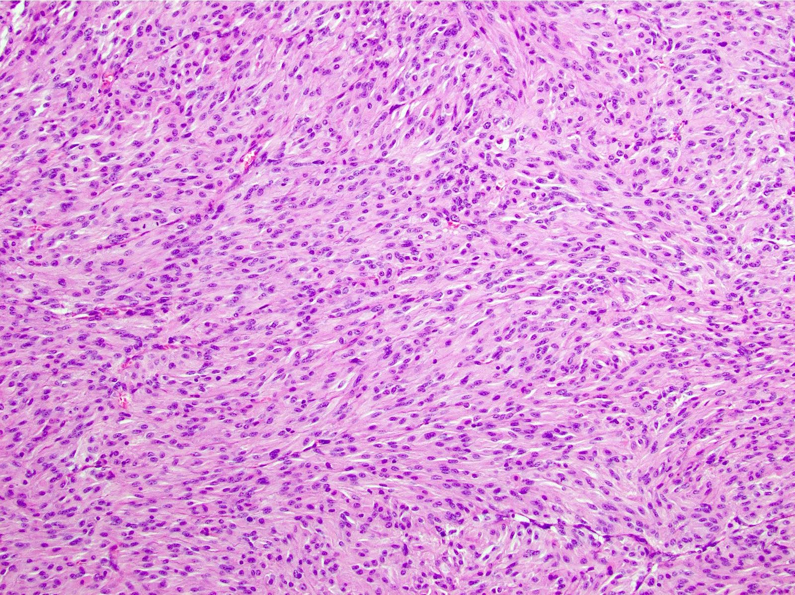
Stomach GIST (mixed type)
Evidence of mitotic activity
Prominent paranuclear vacuoles
Stomach GIST, epithelioid type
Stomach GIST, spindle cell type
Contributed by Andrey Bychkov, M.D., Ph.D.
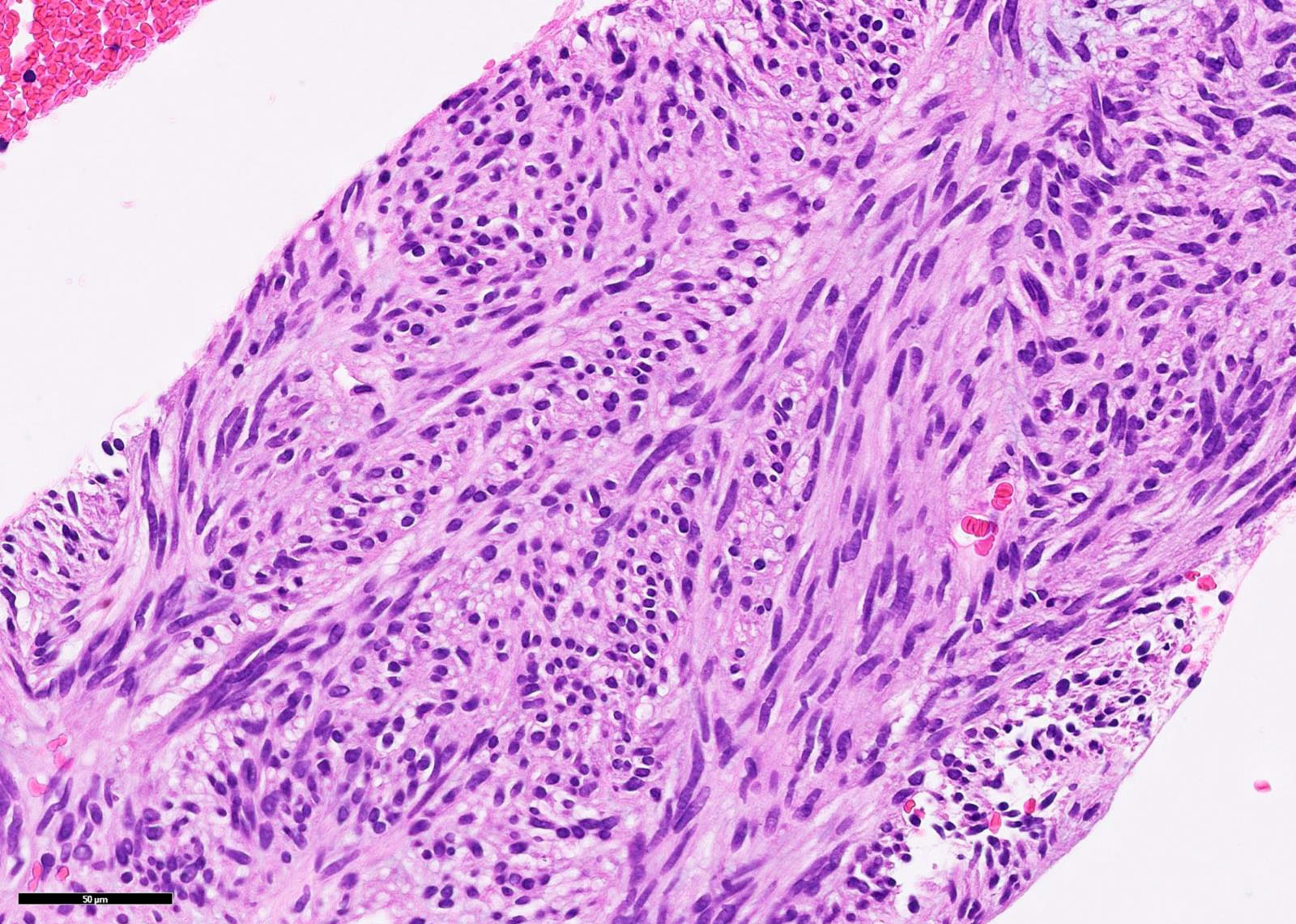
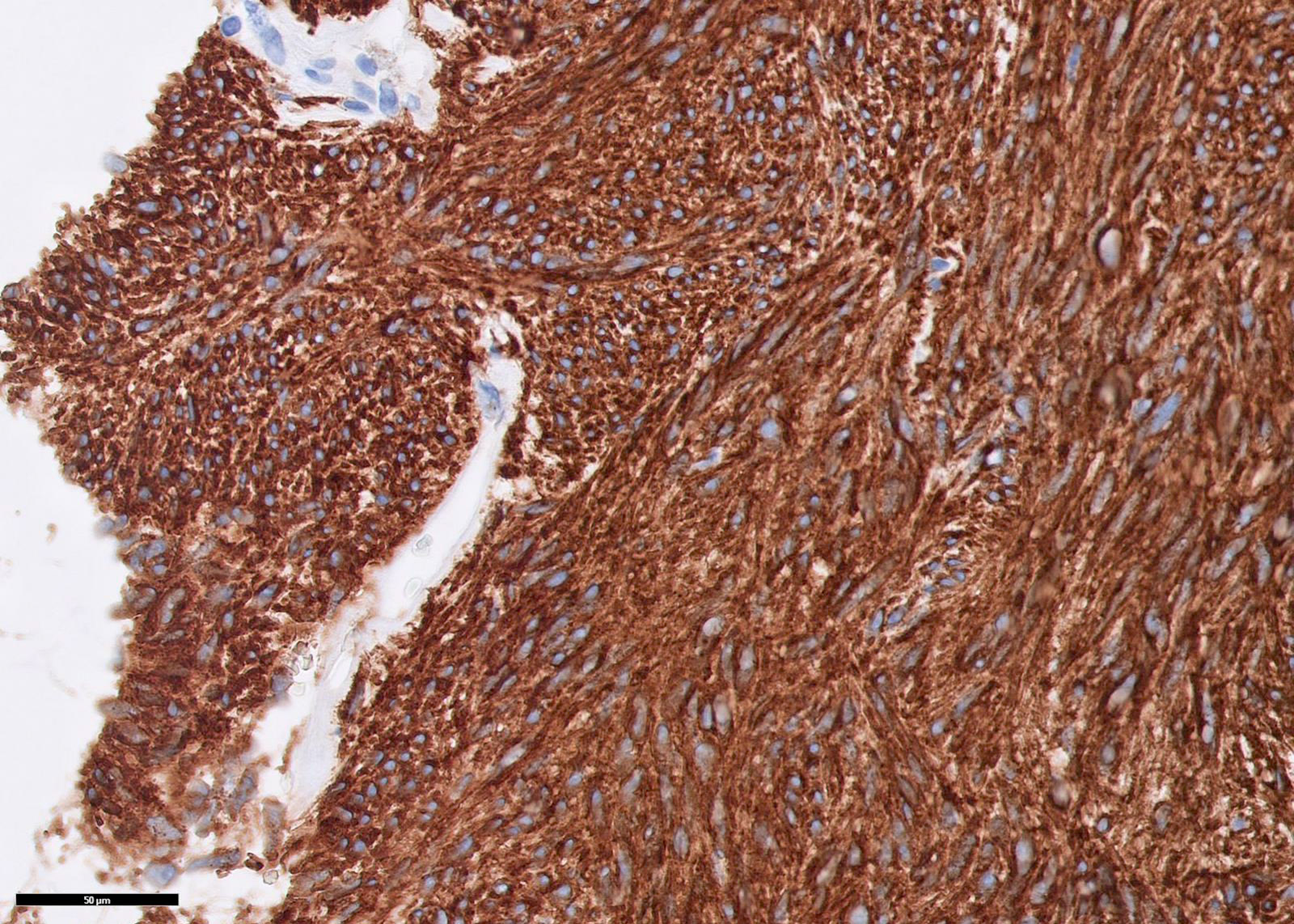
Core biopsy
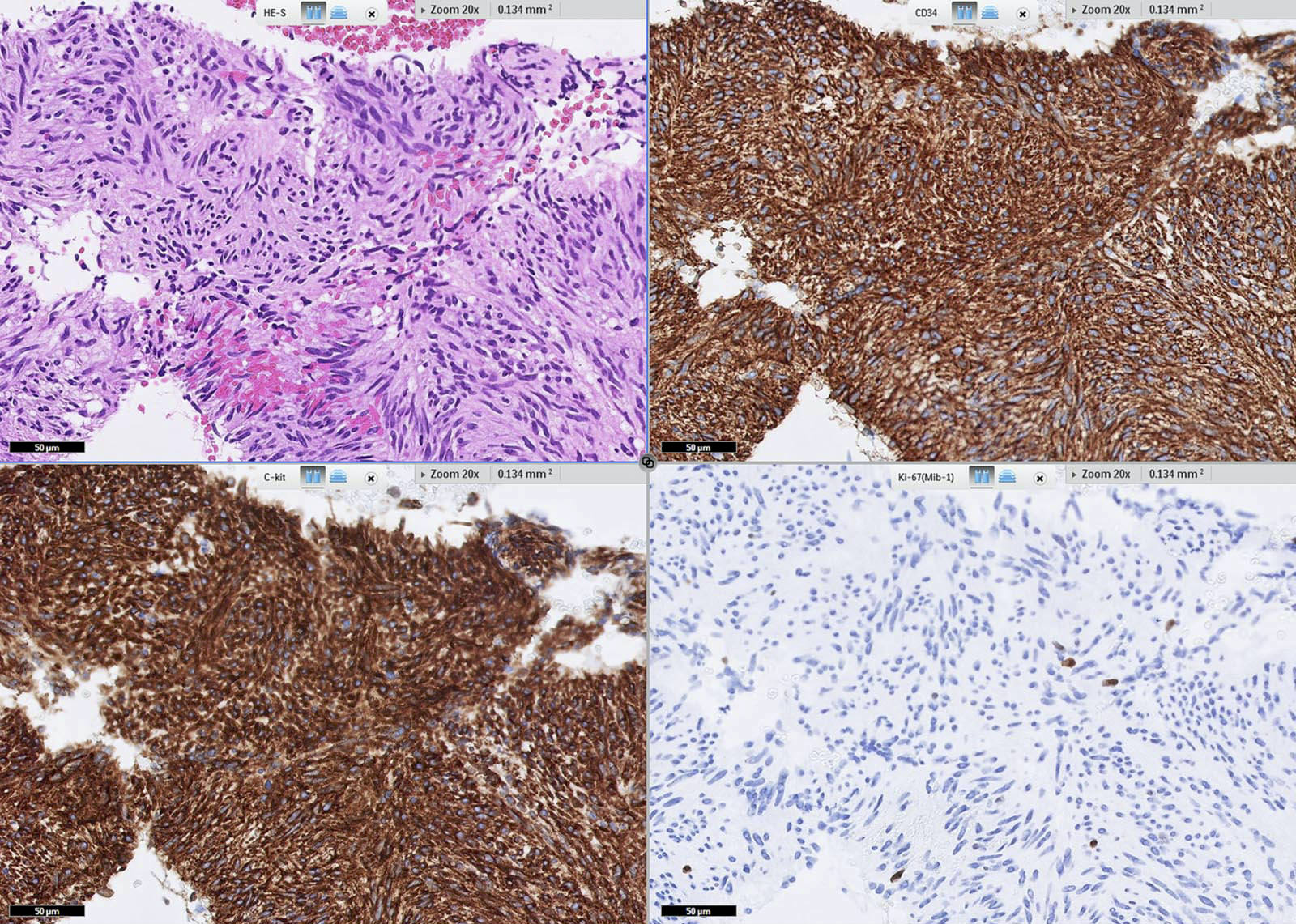
Core biopsy with IHC
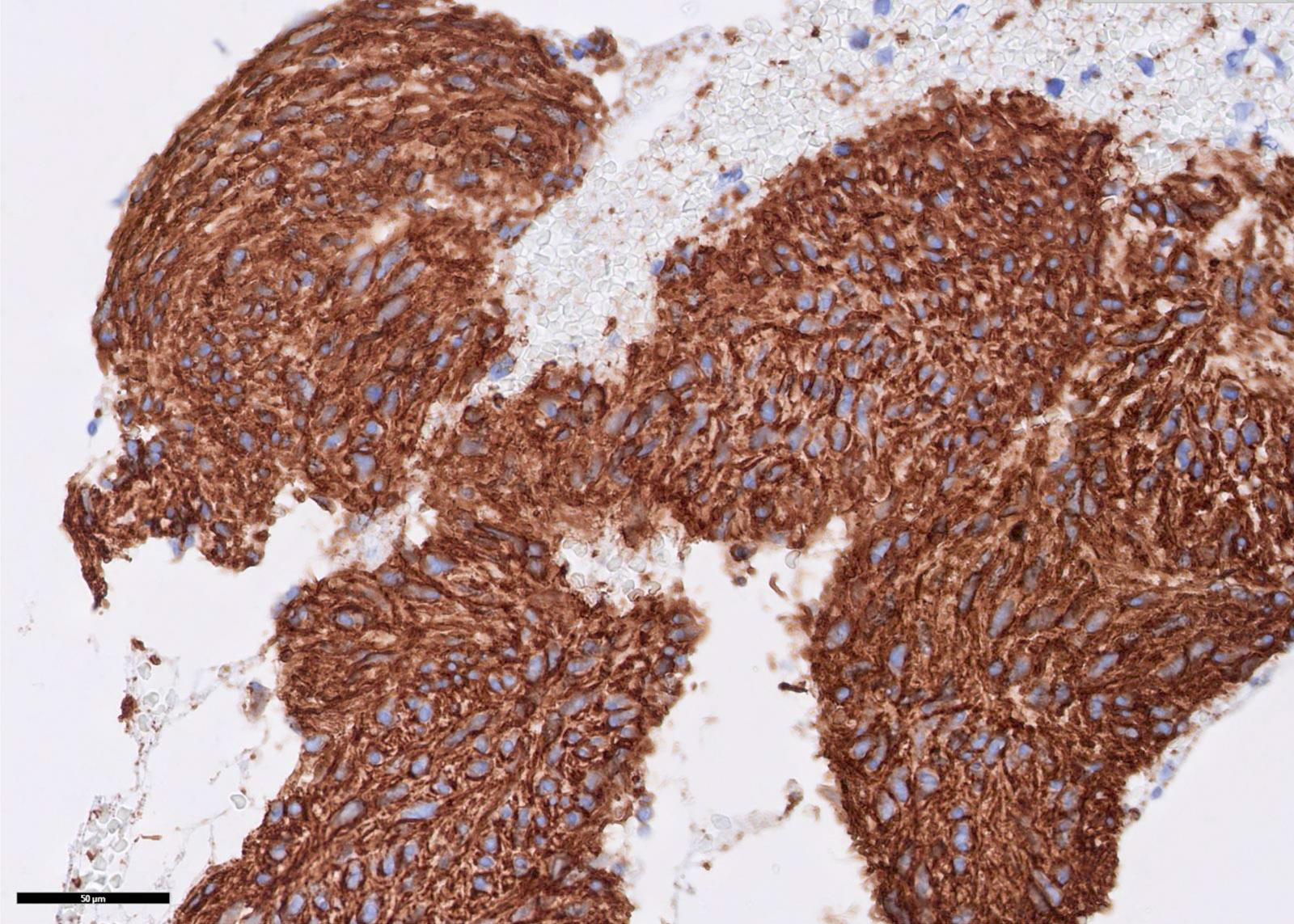
CD34
C-kit / CD117
Contributed by Raul S. Gonzalez, M.D. (Case #523)
SDH deficient GIST
DOG1
SDHB
Contributed by Andrey Bychkov, M.D., Ph.D.
Highly cellular
Spindled
Images hosted on other servers:

c-kit mutations

Case #393


Contributed by @RaulSGonzalezMD on Twitter
Glomus tumor
Images hosted on other servers:

Epithelial cells (left), Calponin+ (right)

Solid growth pattern

Glomus cells and tumor
Contributed by Heidi D. Lehrke, D.O.
Grade 3 GVHD, glandular apoptosis and crypt drop out, several apoptotic figures seen in field
Images hosted on other servers:

Hepatic GVHD


Various images
Chronic graft versus host disease and the gastrointestinal tract
Contributed by Chungja C. Shim, M.D.
Gastric biopsy
Contributed by Tanner Storozuk, M.D. and Namrata Setia, M.D.
Erosions
Ulcer
Nodular mucosa
Contributed by Tanner Storozuk, M.D. and Namrata Setia, M.D.
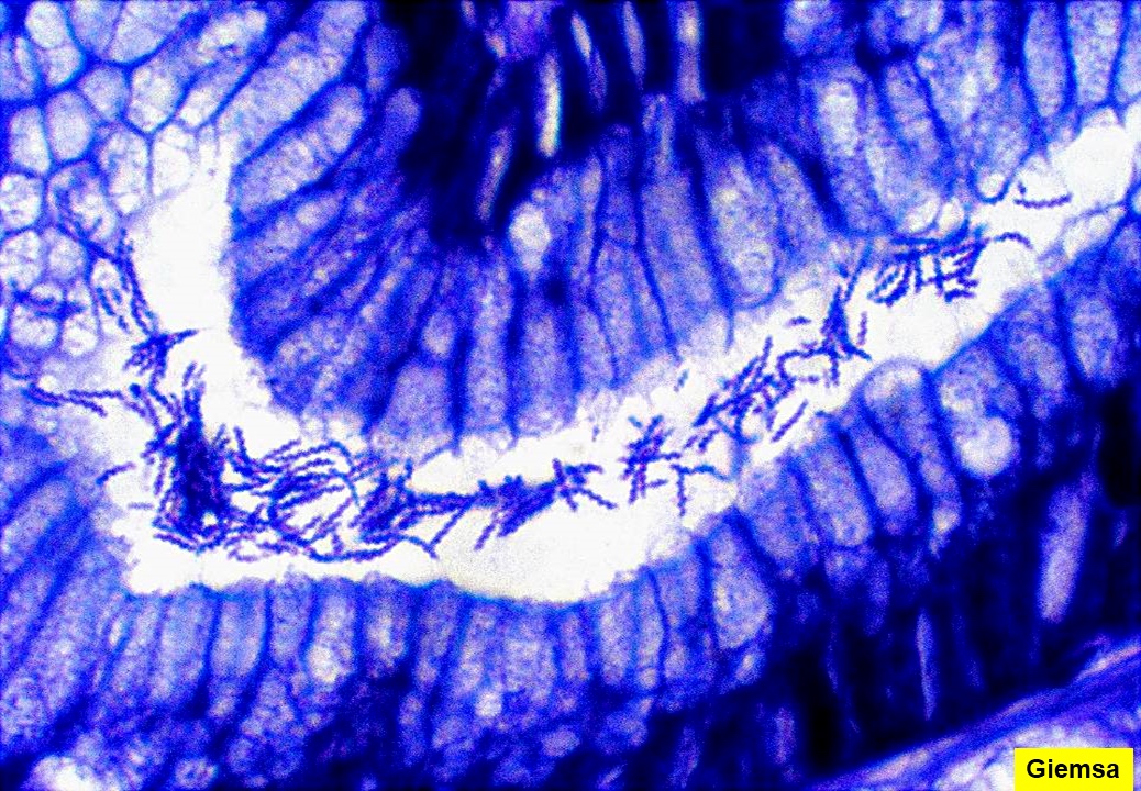
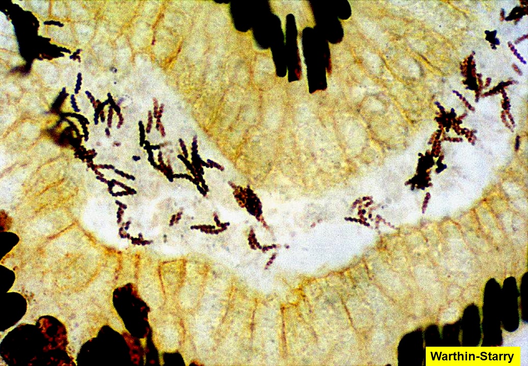
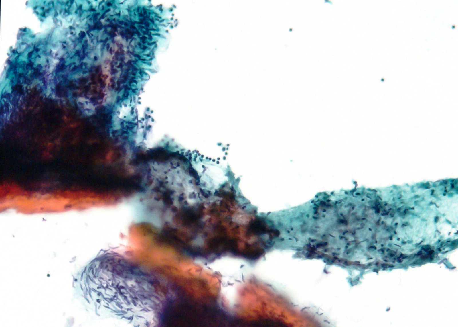
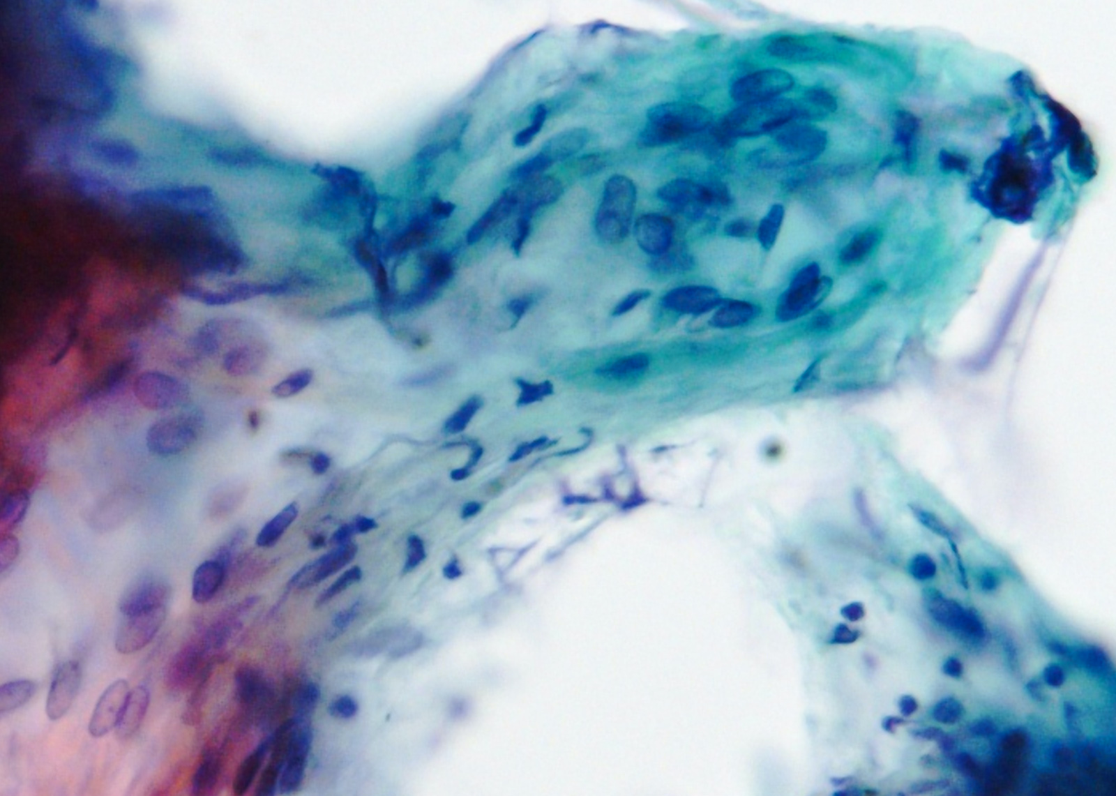
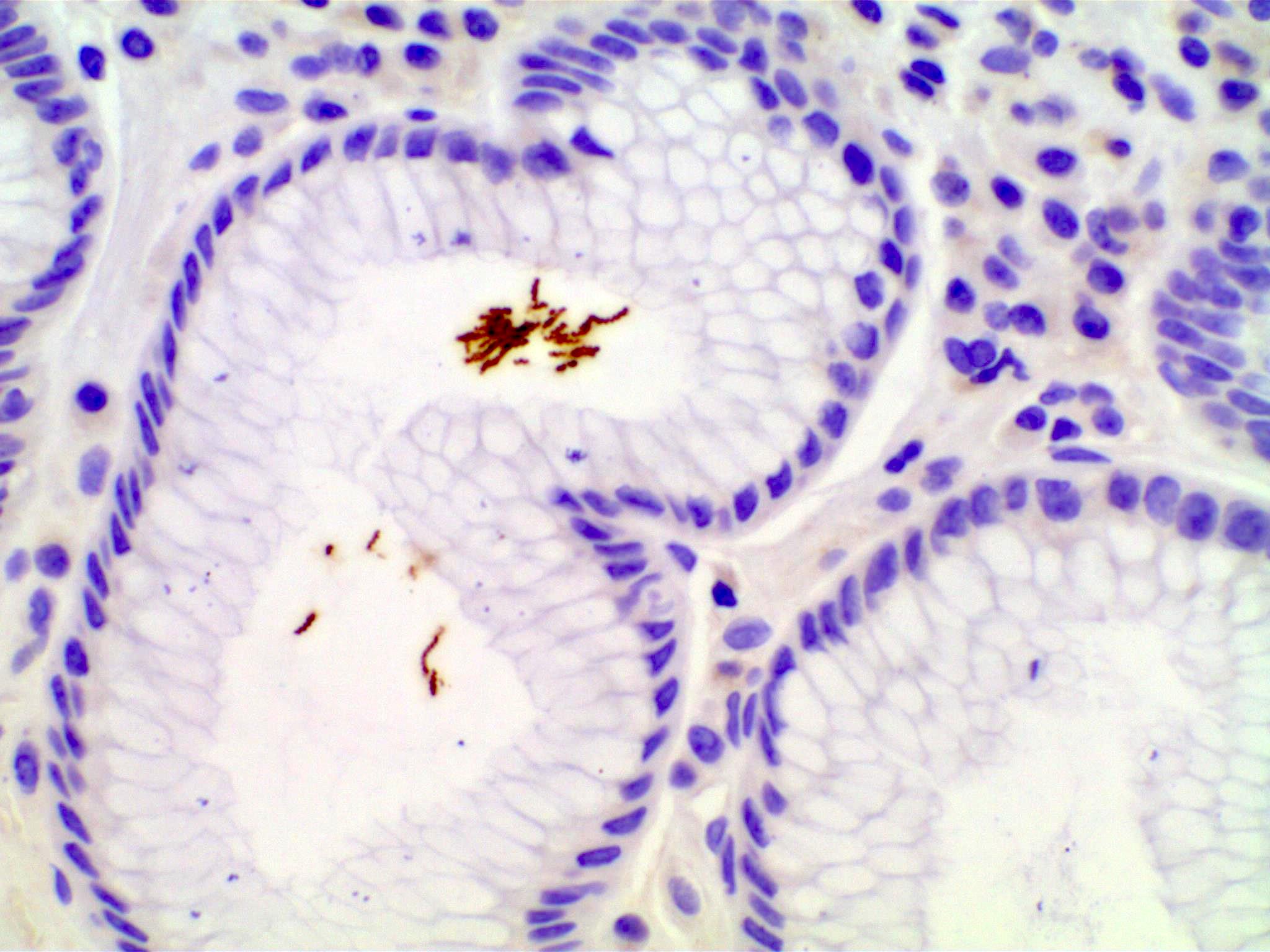
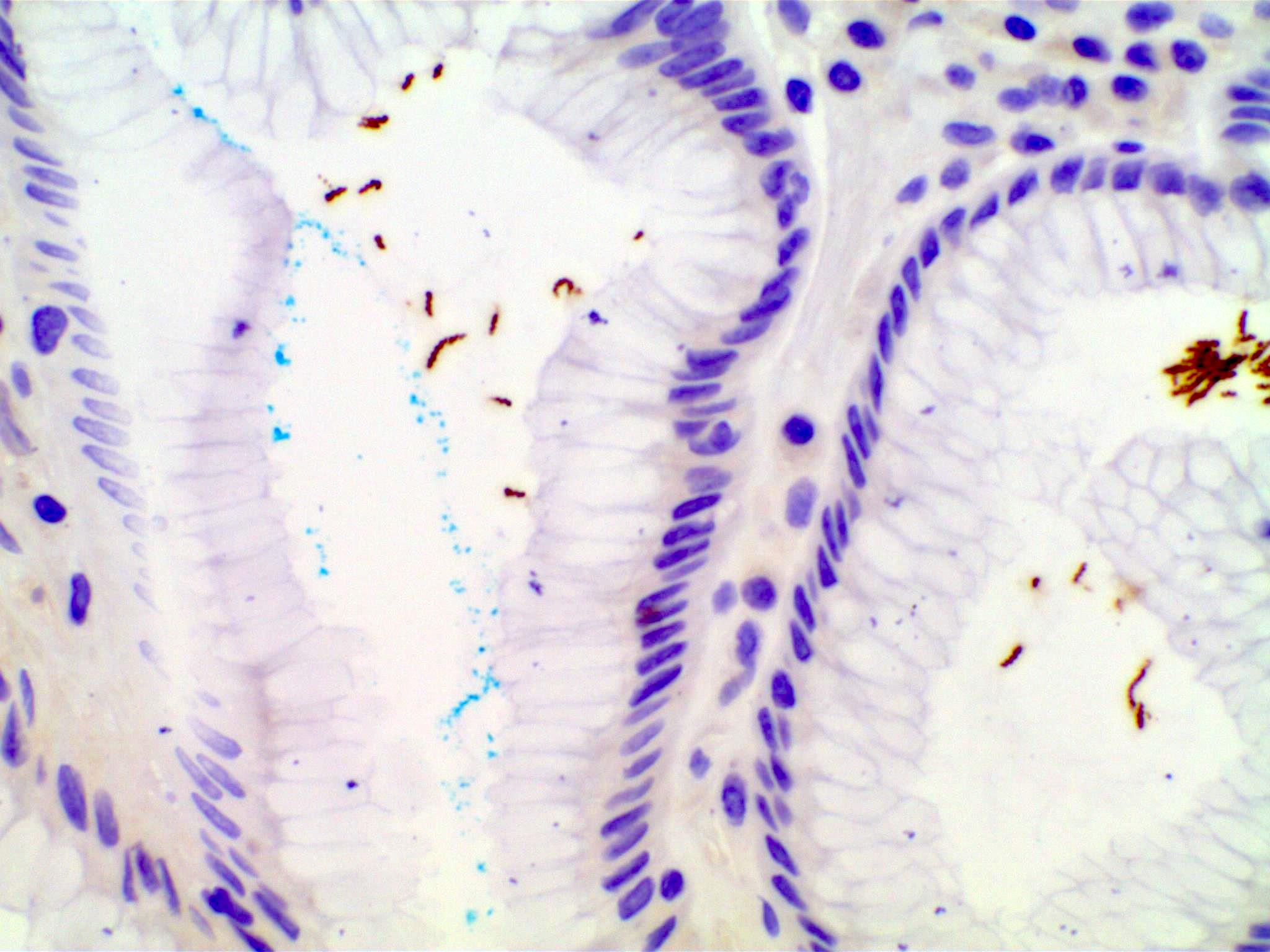
H. pylori in body

H. pylori in antrum
H. pylori in duodenum
Histology of H. pylori (left) versus H. heilmannii (right)
Immunostain for H. pylori

Stomach body H. pylori gastritis
Histopathology, H. pylori gastritis
Basic pathogenesis of H. pylori infection
Images hosted on other servers:

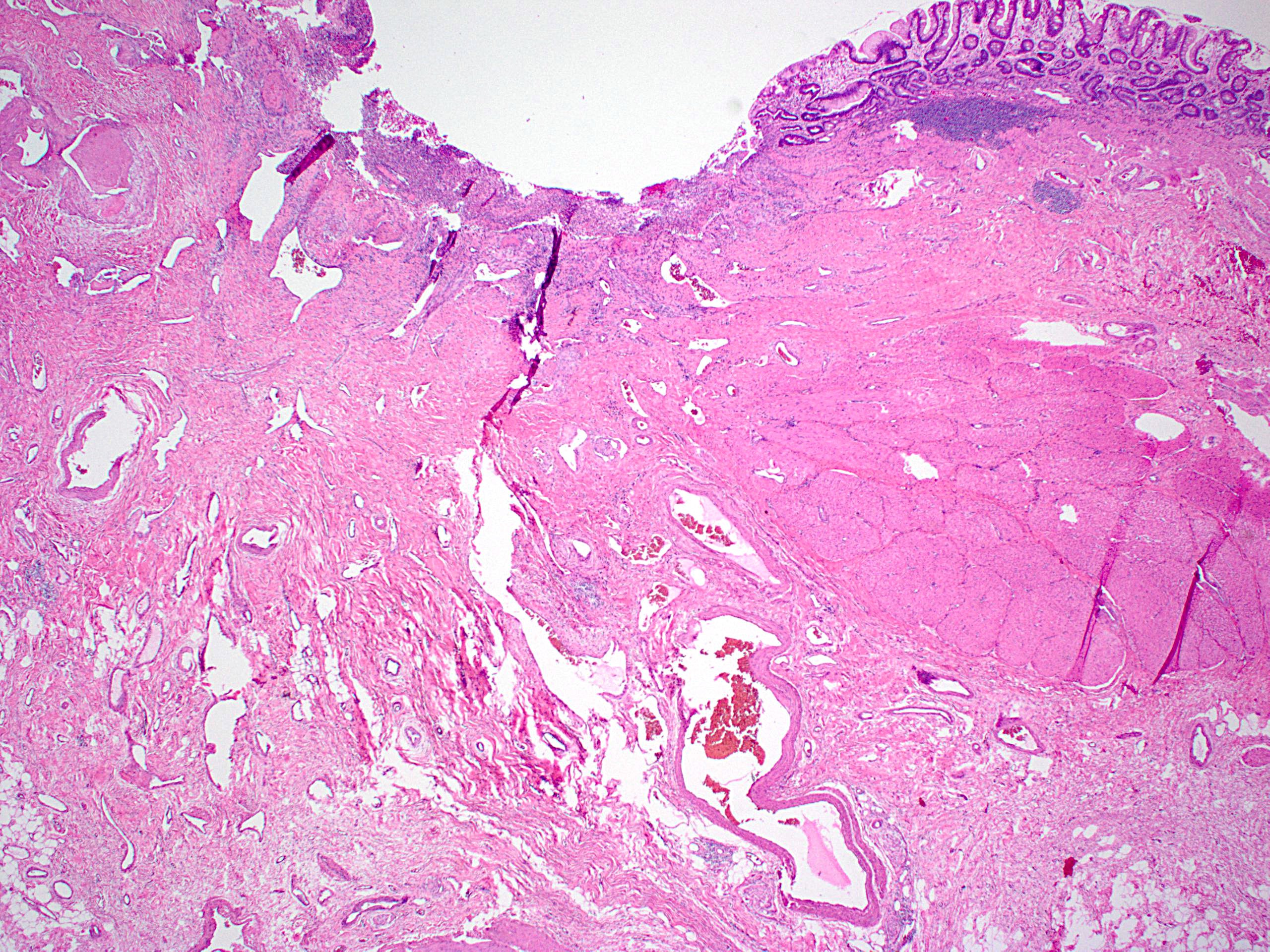
Total gastrectomy specimen
Contributed by Runjan Chetty, M.B.B.Ch., Ph.D. and Altaf Taher, M.B.B.S., M.D.
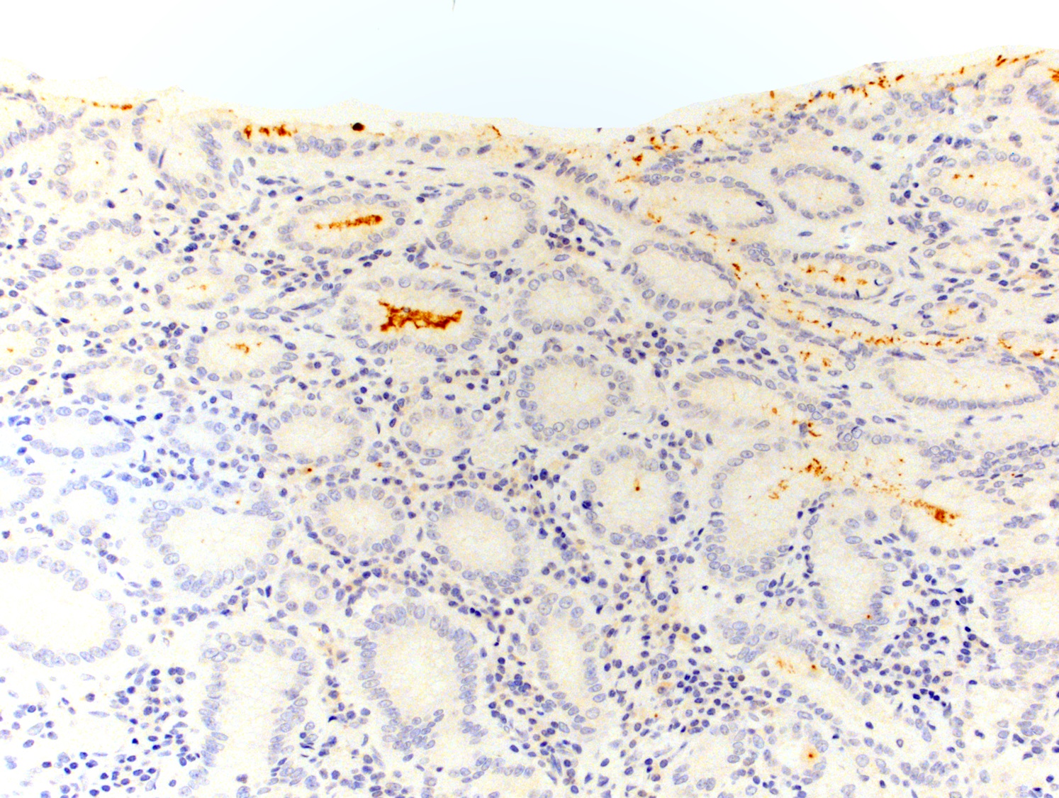
Signet ring carcinoma in situ
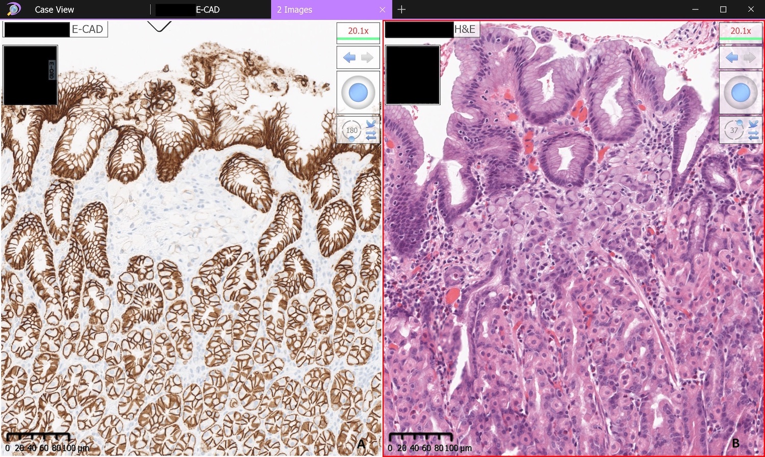
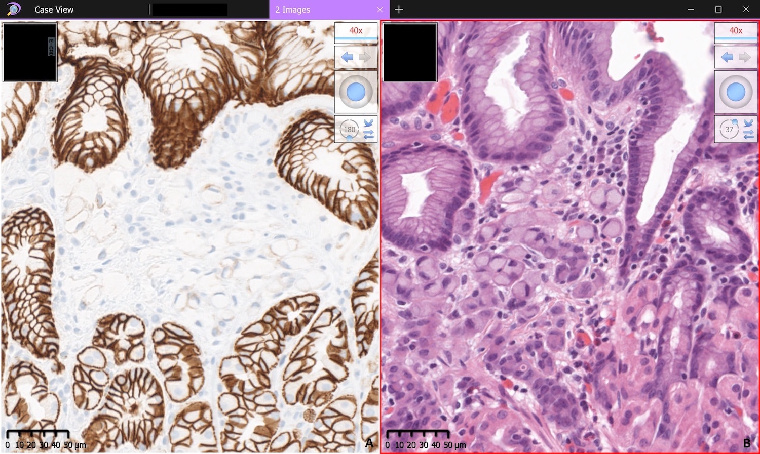
Loss of membranous E-cadherin staining
Contributed by Raul S. Gonzalez, M.D.
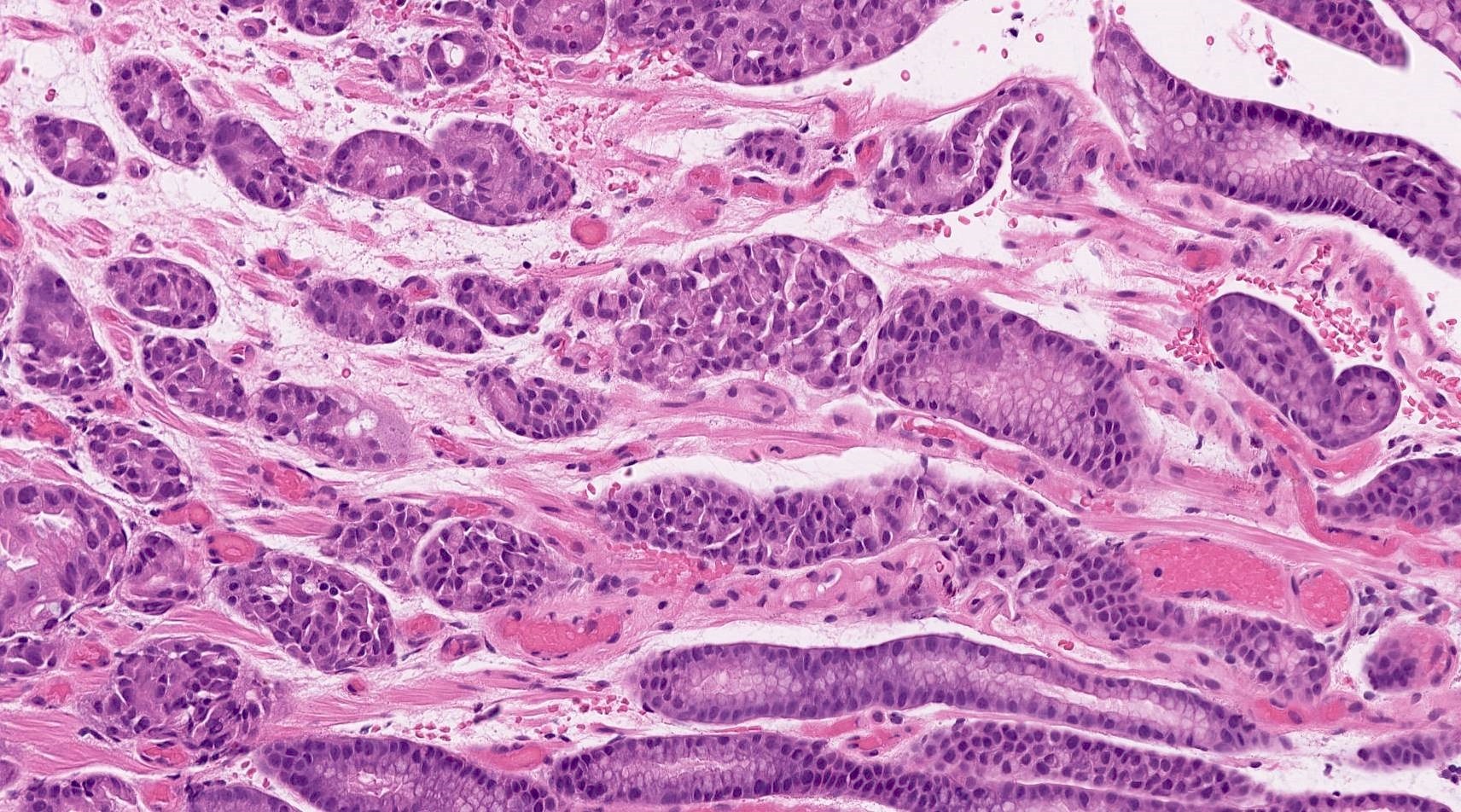
Signet ring carcinoma in situ
Obvious poorly cohesive carcinoma
Subtle poorly cohesive carcinoma
Contributed by Dennis J. Chute, M.D.

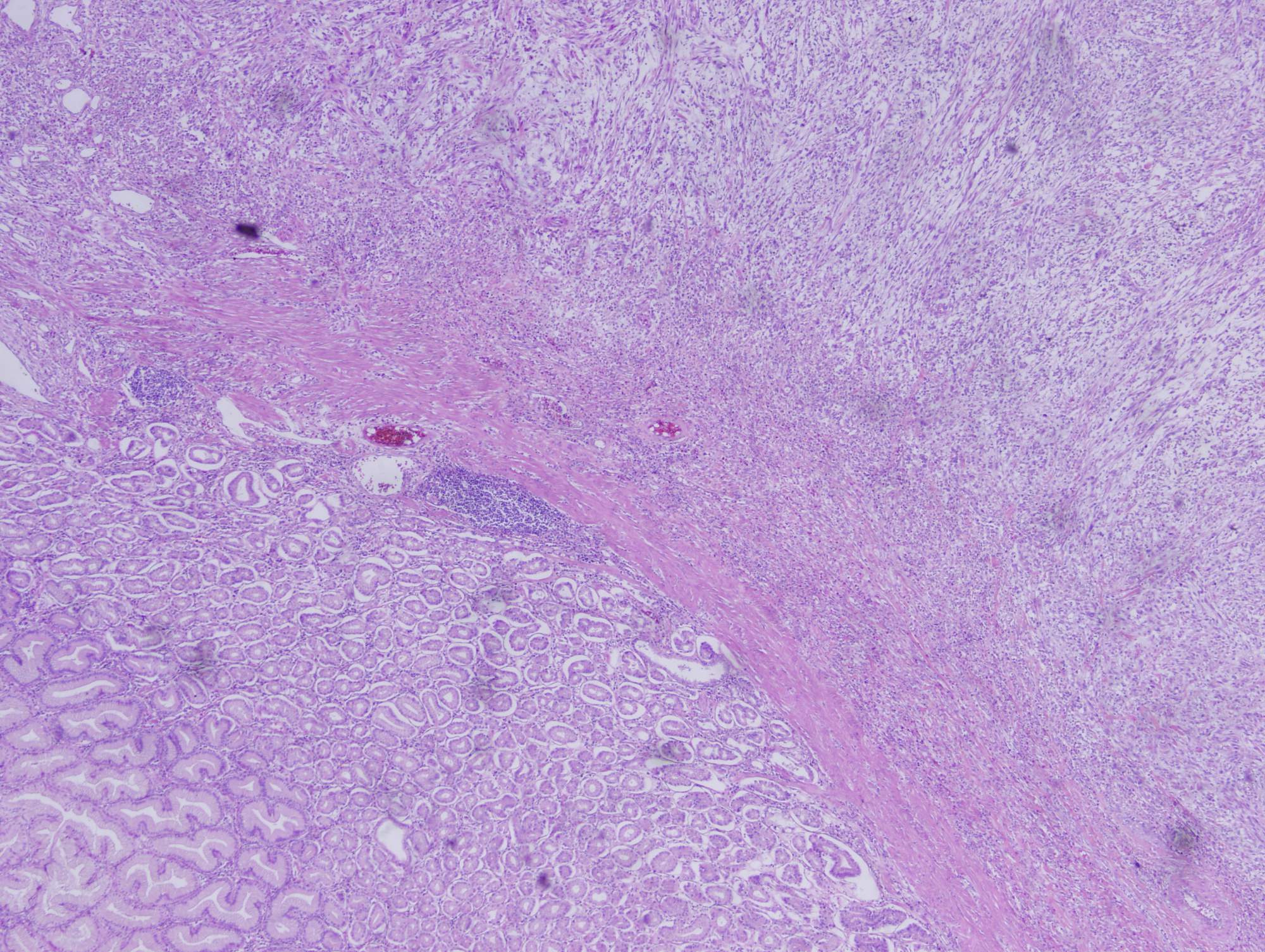
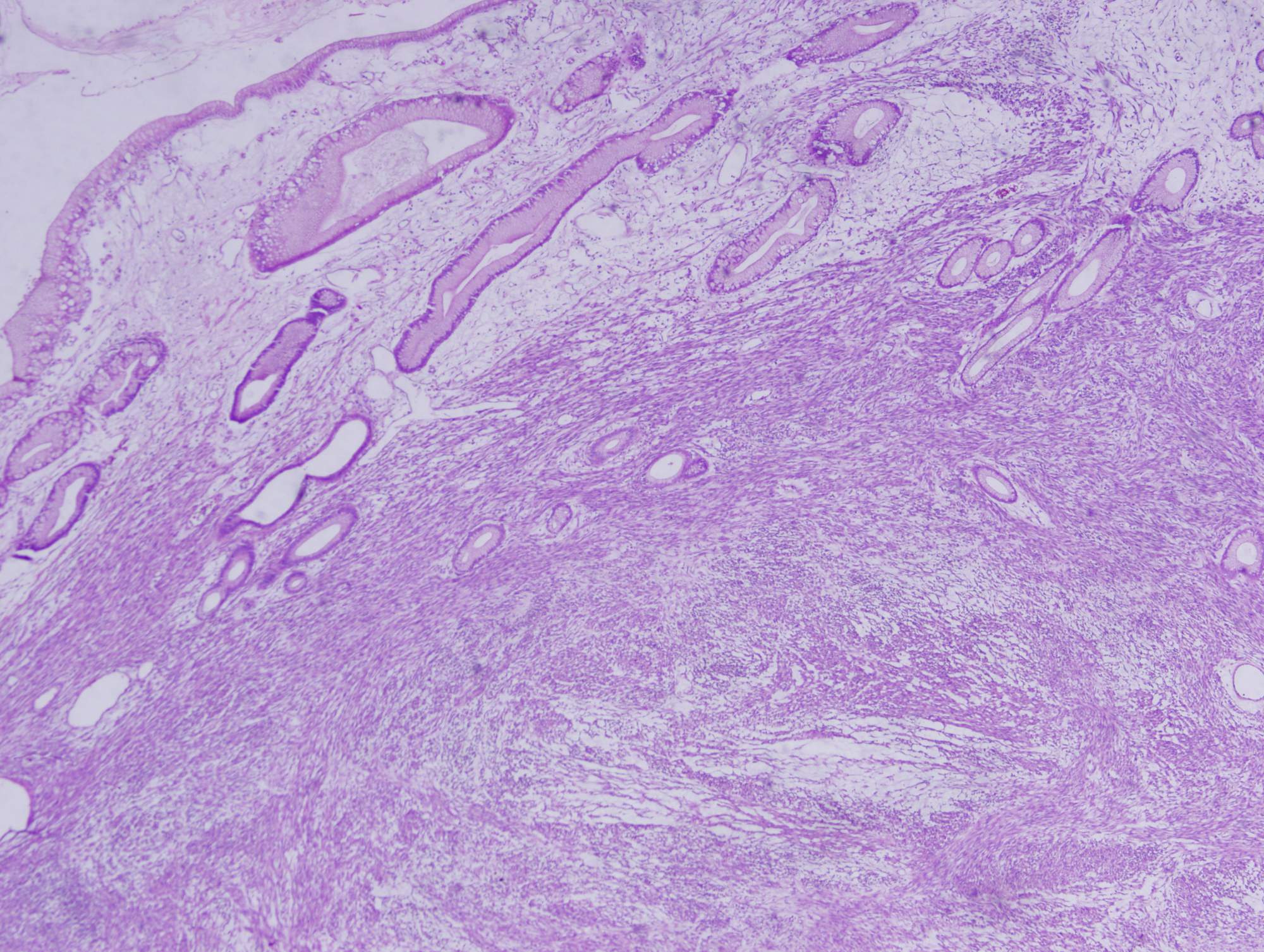
Heterotopic pancreas in annulus
Contributed by Dennis J. Chute, M.D.

Heterotopic exocrine pancreas in annulus
Contributed by Monica T. Garcia-Buitrago, M.D., Omar Aljuboori, M.B.B.S. and Andrey Bychkov, M.D., Ph.D.
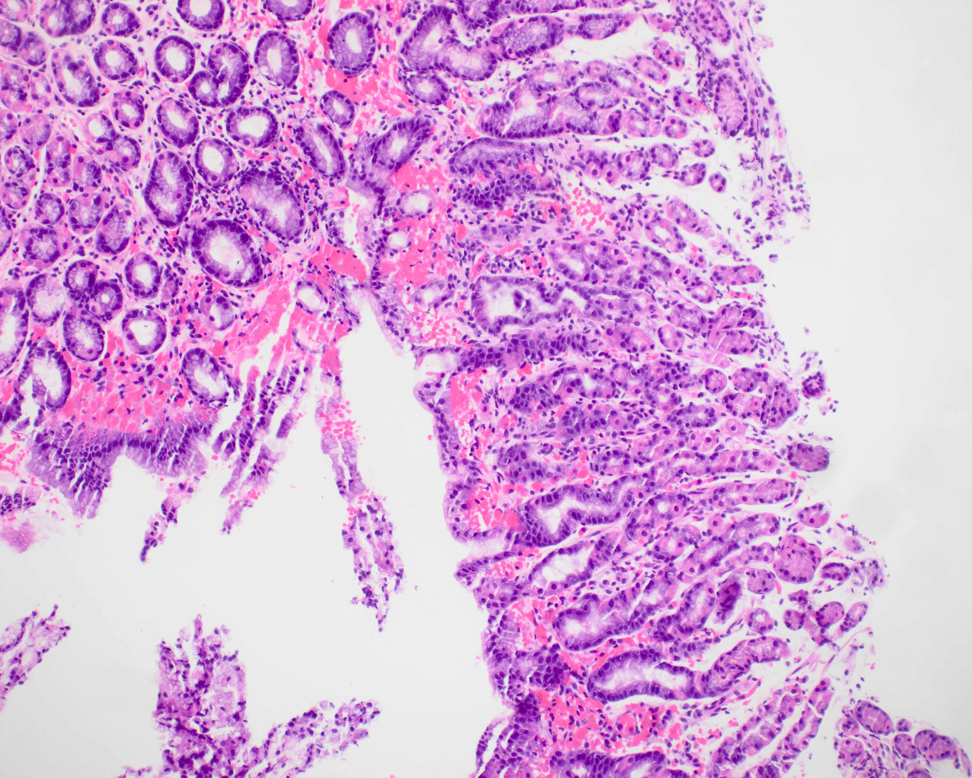
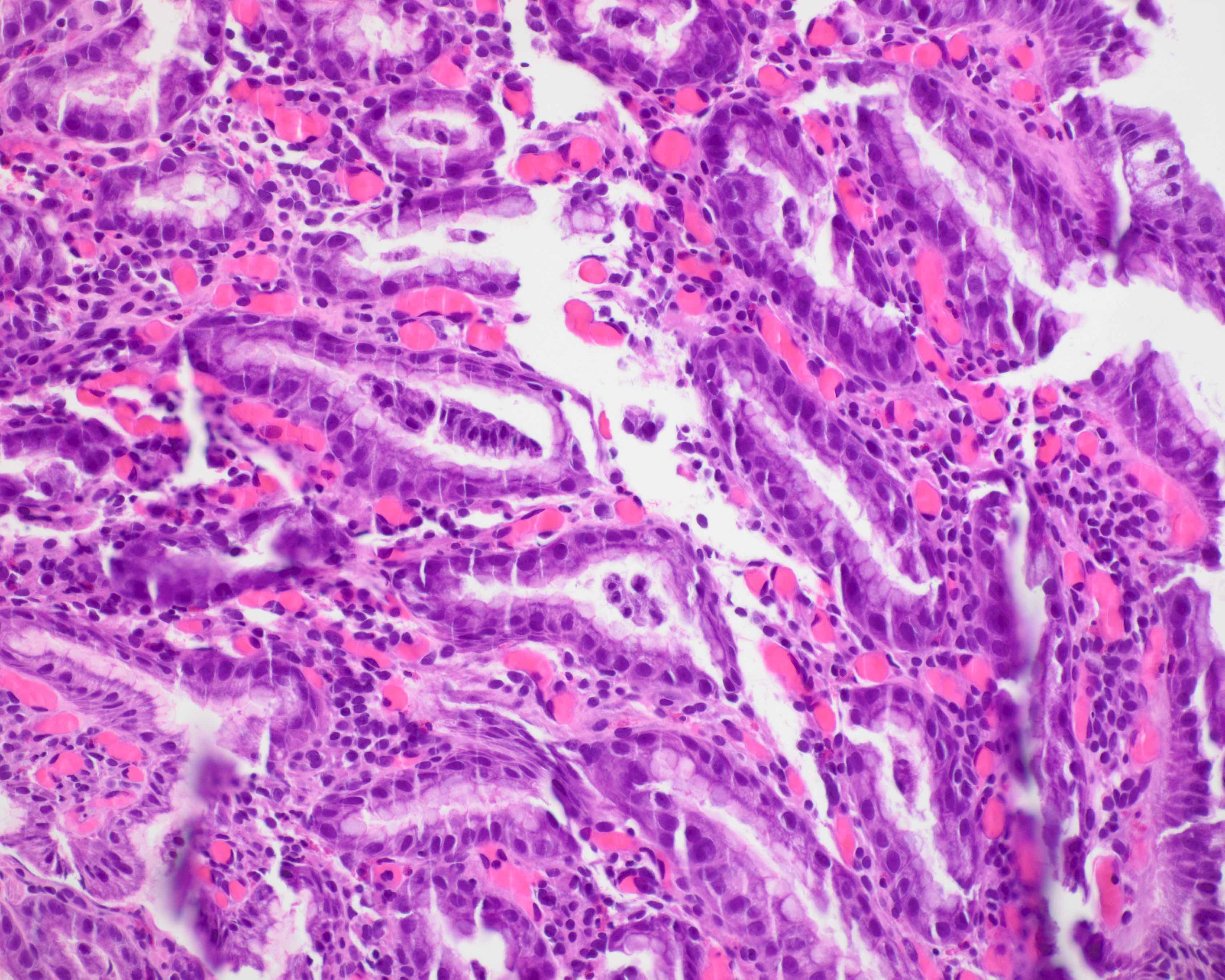
Dilated gastric pits
Tortuous gastric pits
Edematous lamina propria
Pseudogoblet cells
Dysplasia
Dysplastic polyp
Dysplasia
Adenocarcinoma

Polypoid lesion

Hemorrhagic and ulcerated
Cystic hyperplastic glands
Images hosted on other servers:

Focal lesion in
atral mucosa
resembling active
Crohn's disease
Contributed by Naziheh Assarzadegan, M.D.
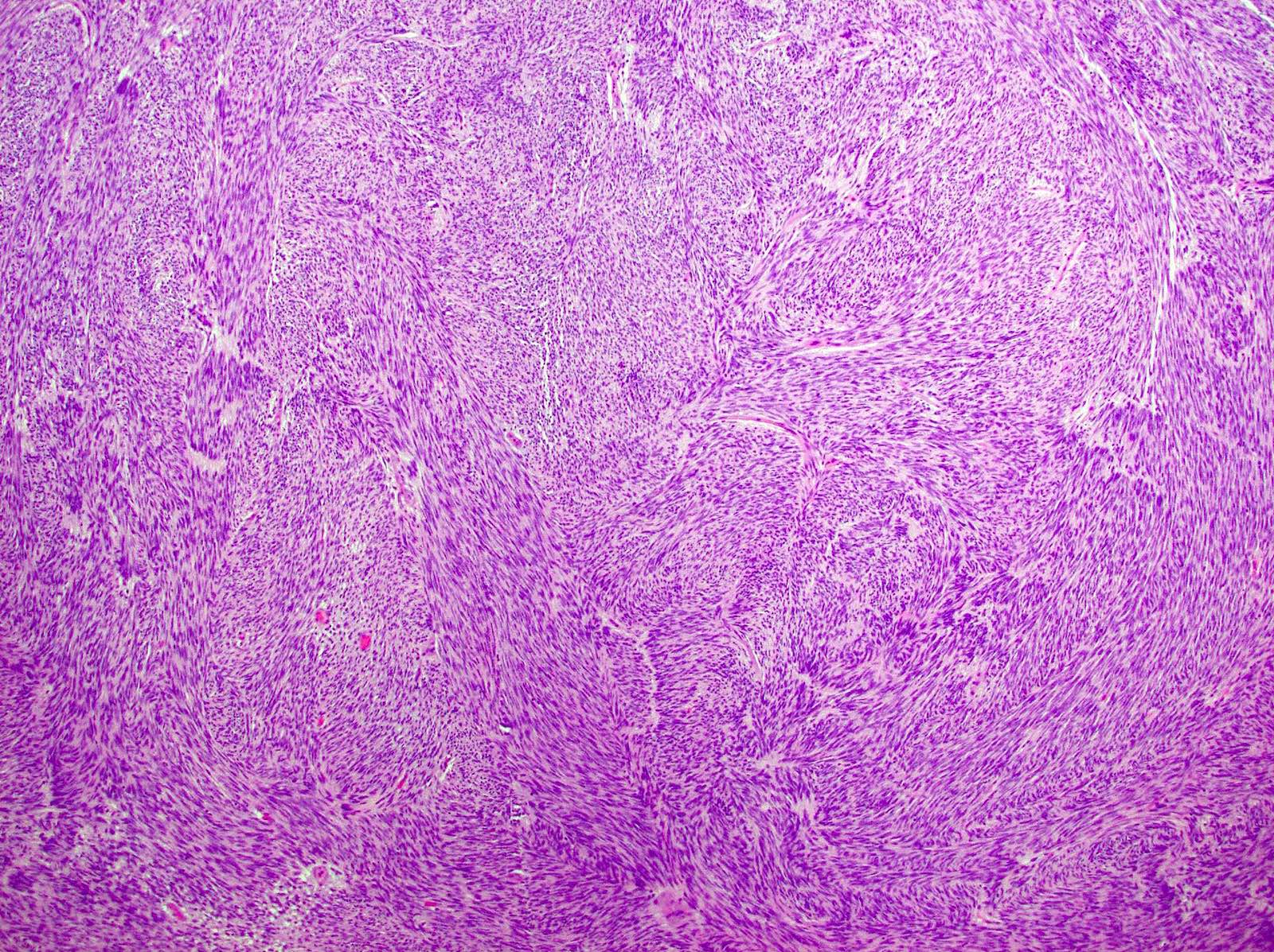
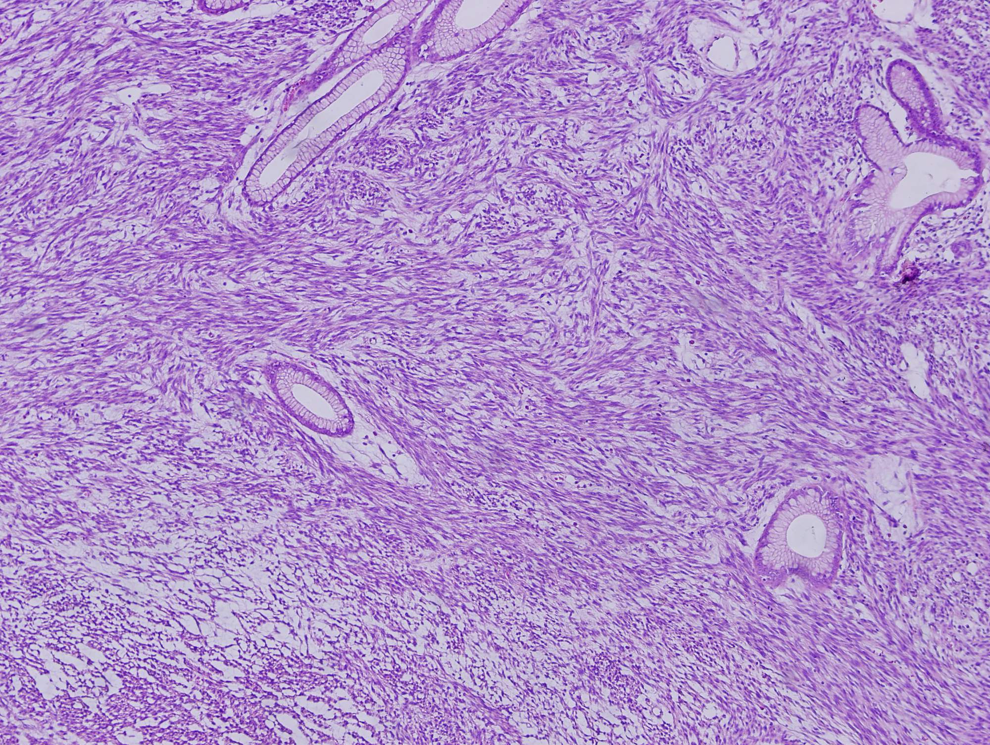
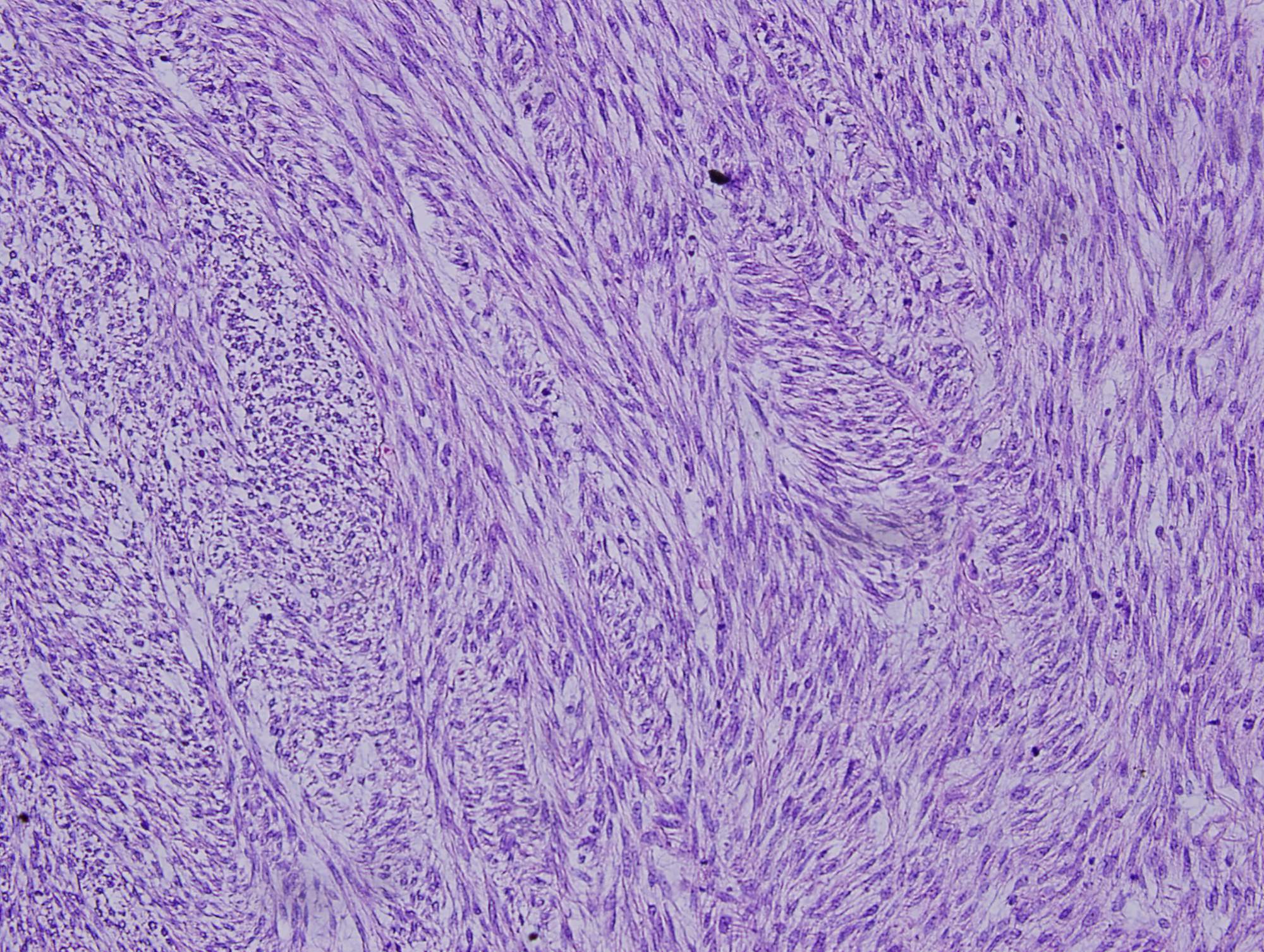
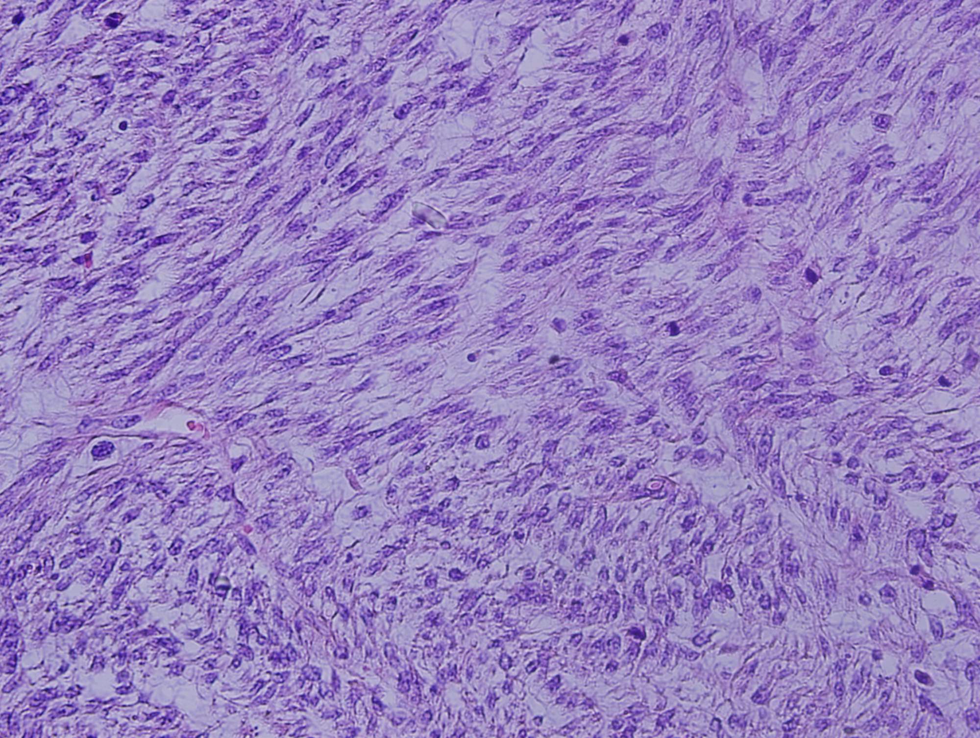
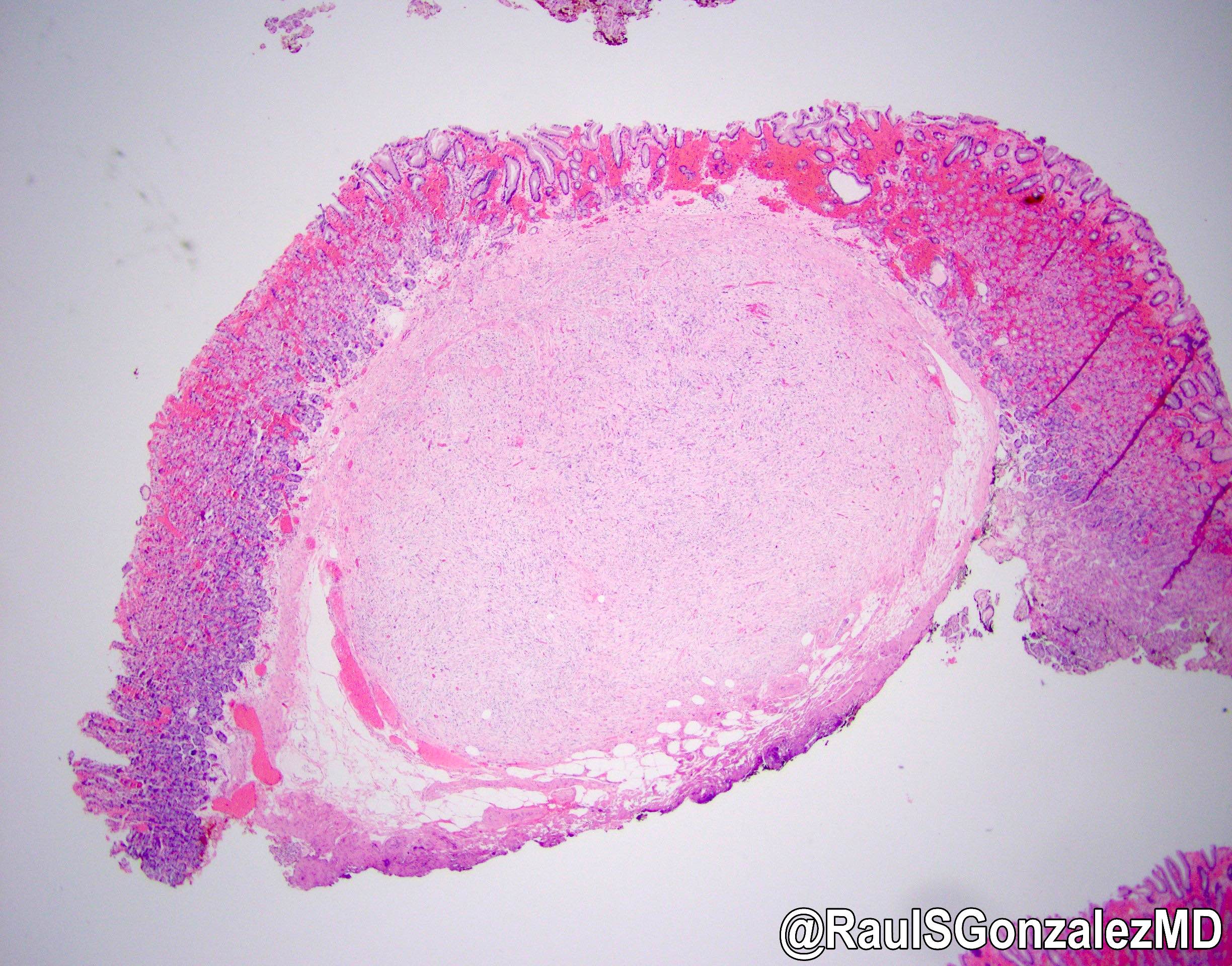
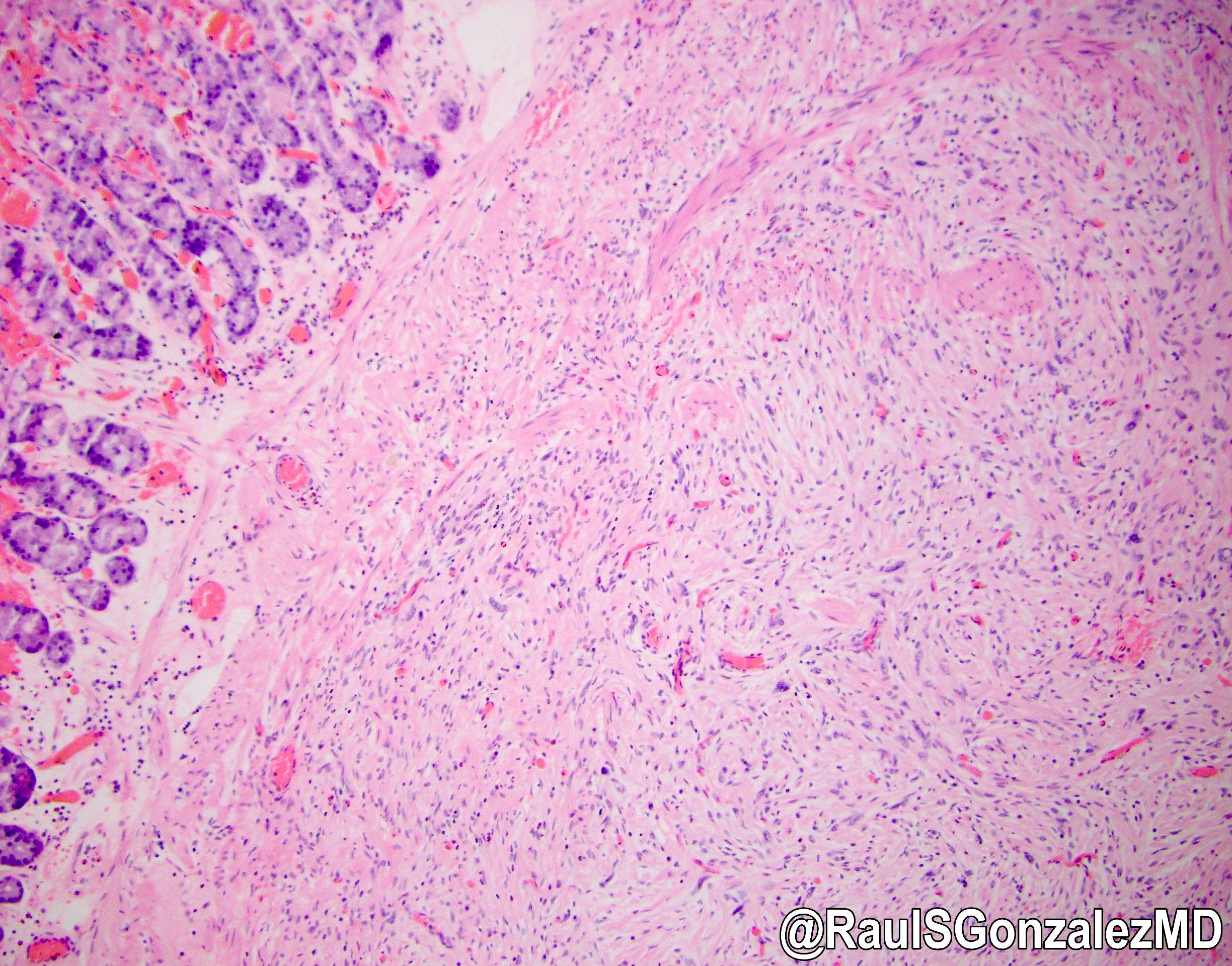
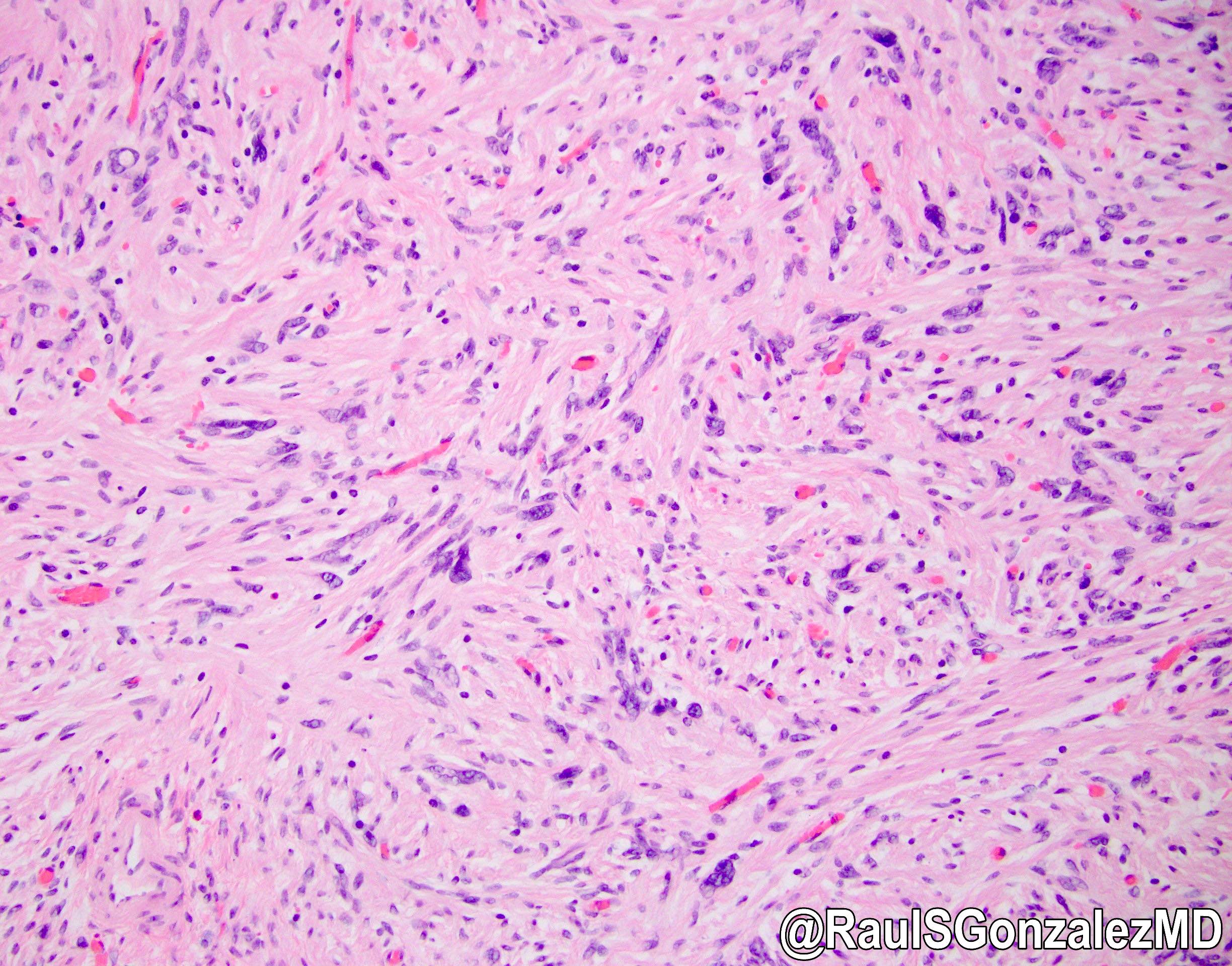
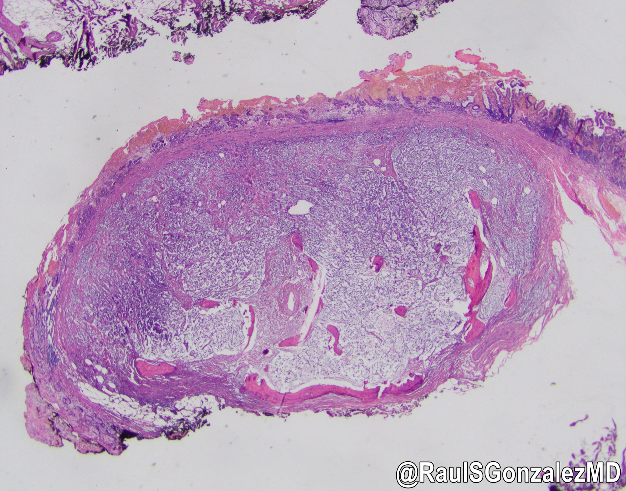
Submucosal mass

Spindle and stellate stromal cells
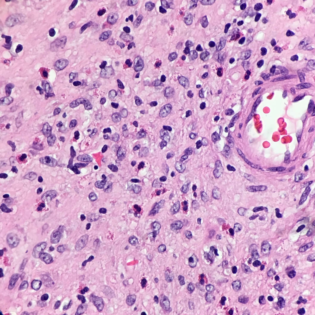
Eosinophils
Case #421
Polypoid mass expanding the submucosa

Mixed inflammatory infiltrate in fibromyxoid stroma, rich in eosinophils

Loose, fibromyxoid spindled stroma
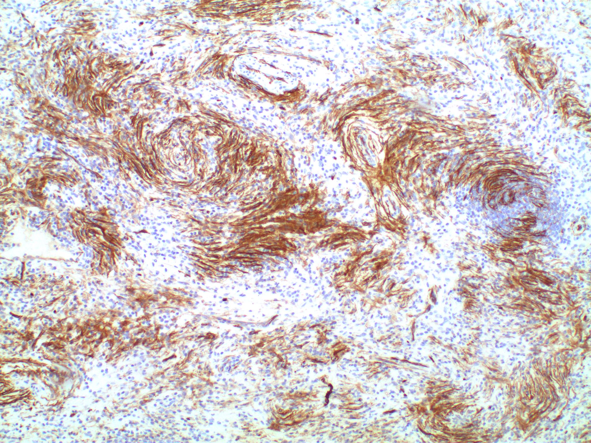
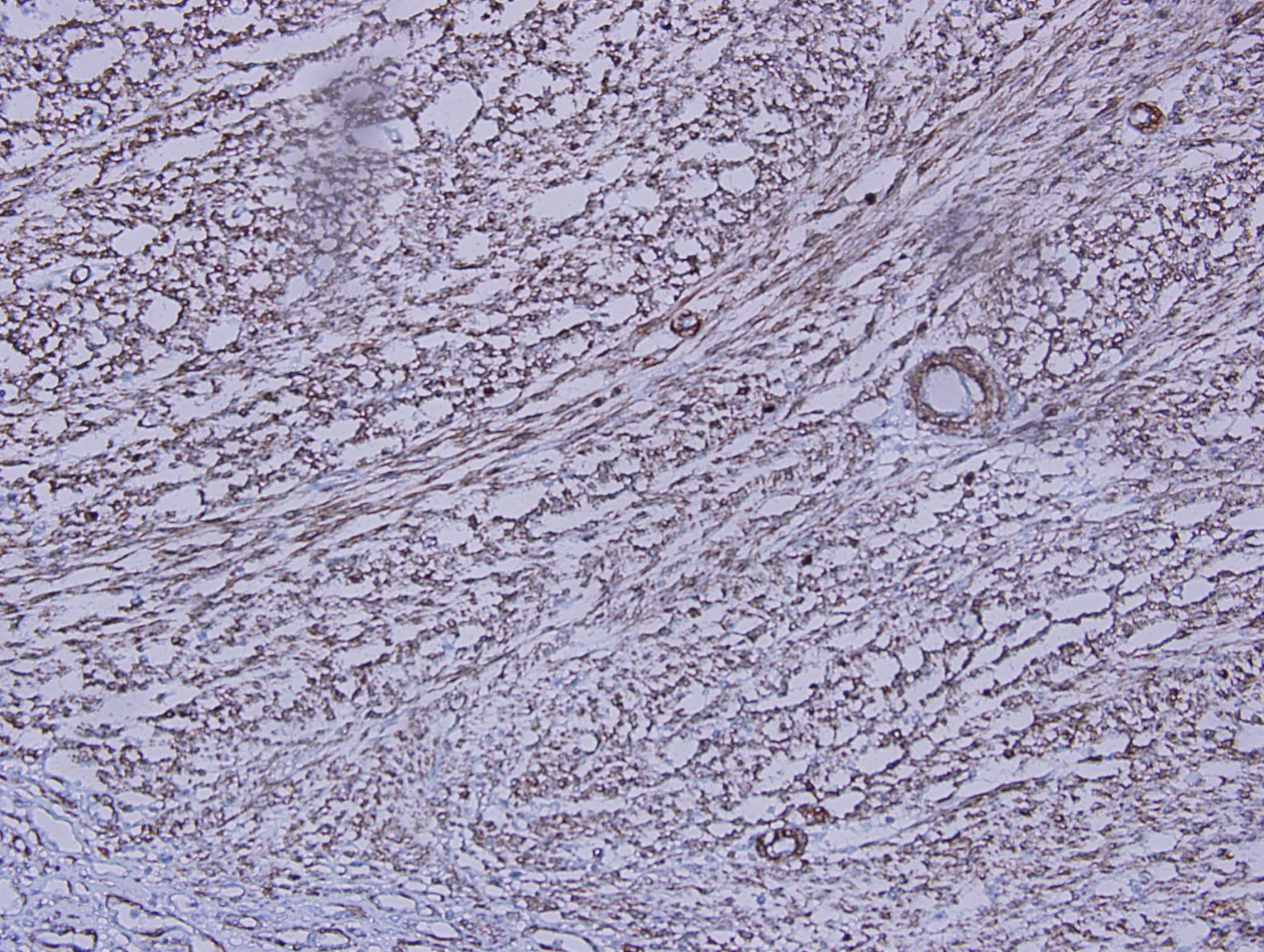
CD34+ stromal cells with onion skinning
Stroma is CD117-,
with scattered
CD117+ mast cells
Contributed by Supriya Srivastava, M.D., Ph.D.
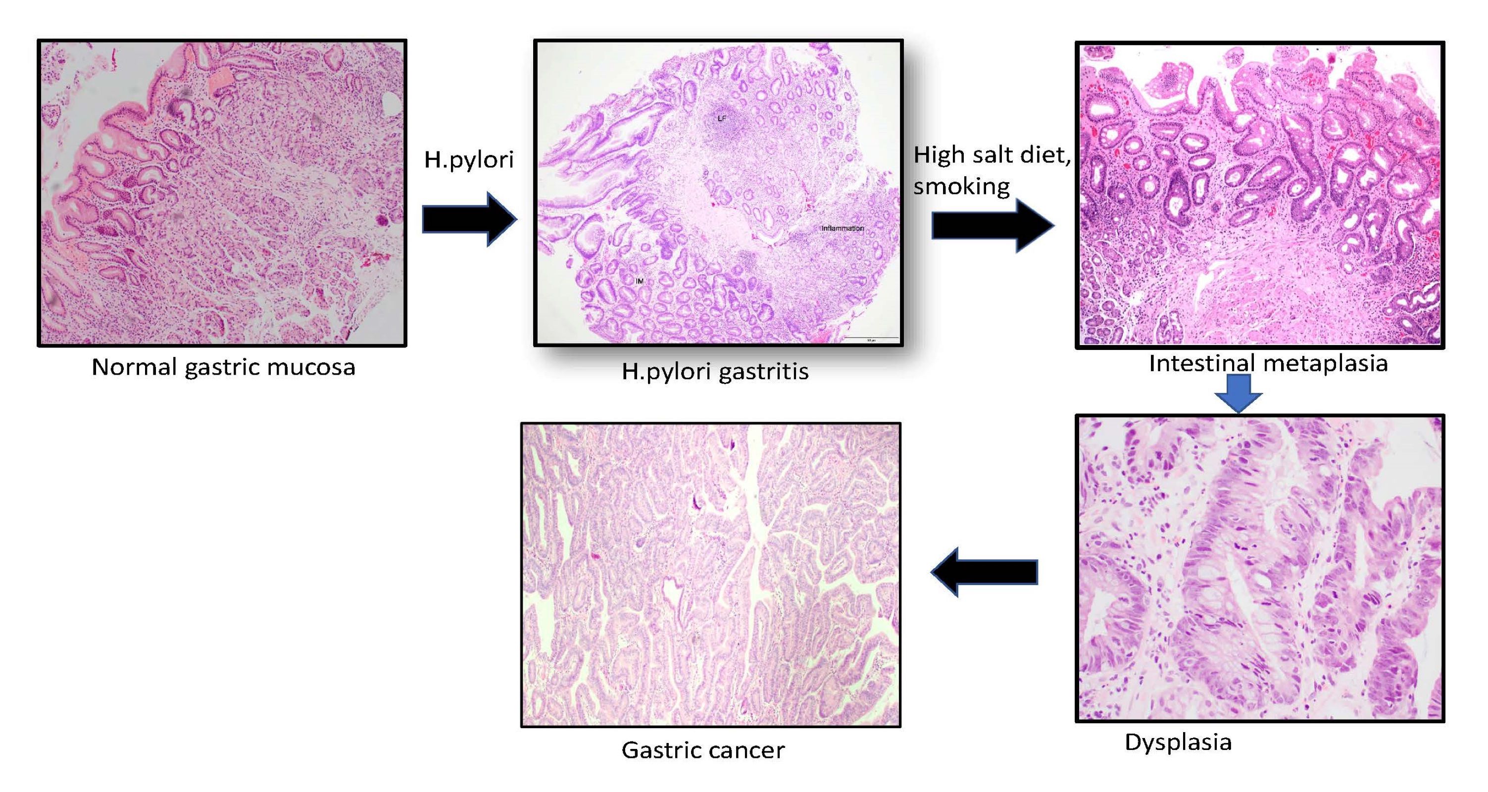
Correa pathway
Updated Sydney protocol
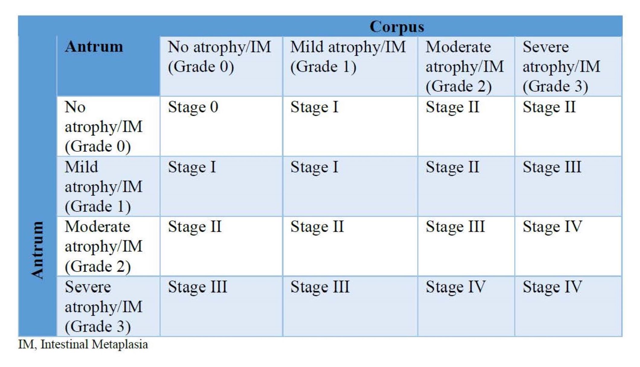
OLGA / OLGIM staging
Images hosted on other servers:


High resolution white light endoscopy

Whitish mucosal patches
Contributed by Supriya Srivastava, M.D., Ph.D.

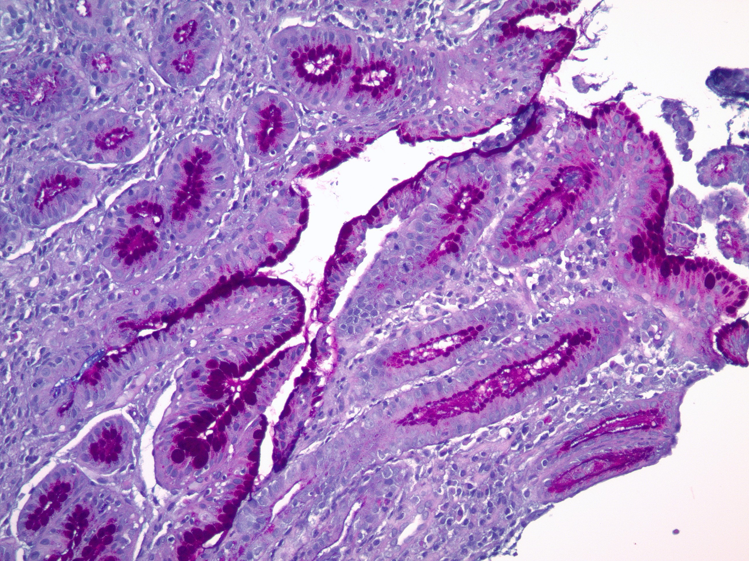
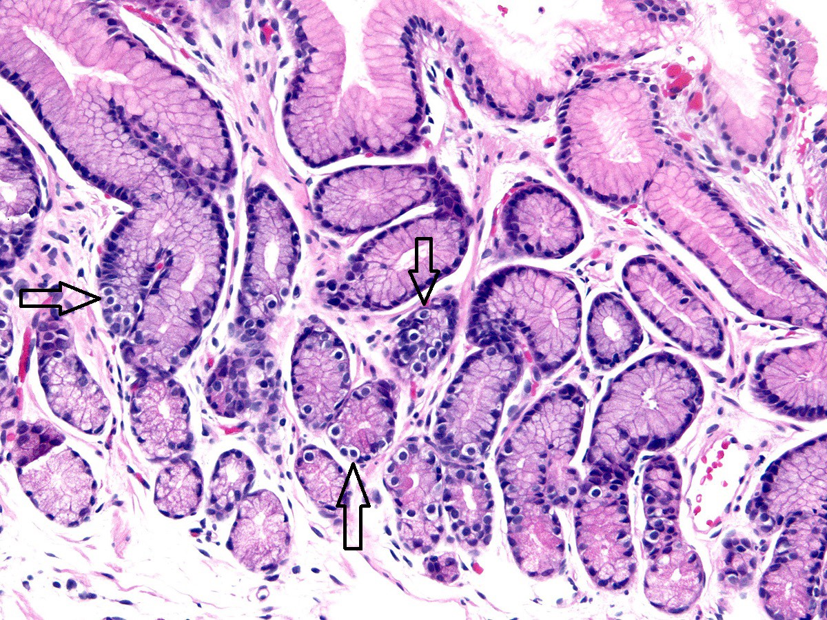
Goblet cells and H. pylori
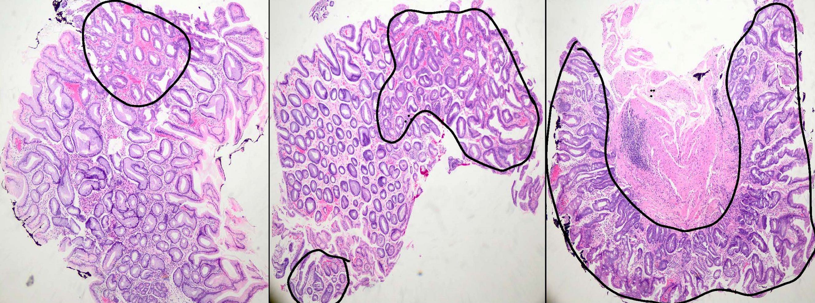
Intestinal metaplasia grading
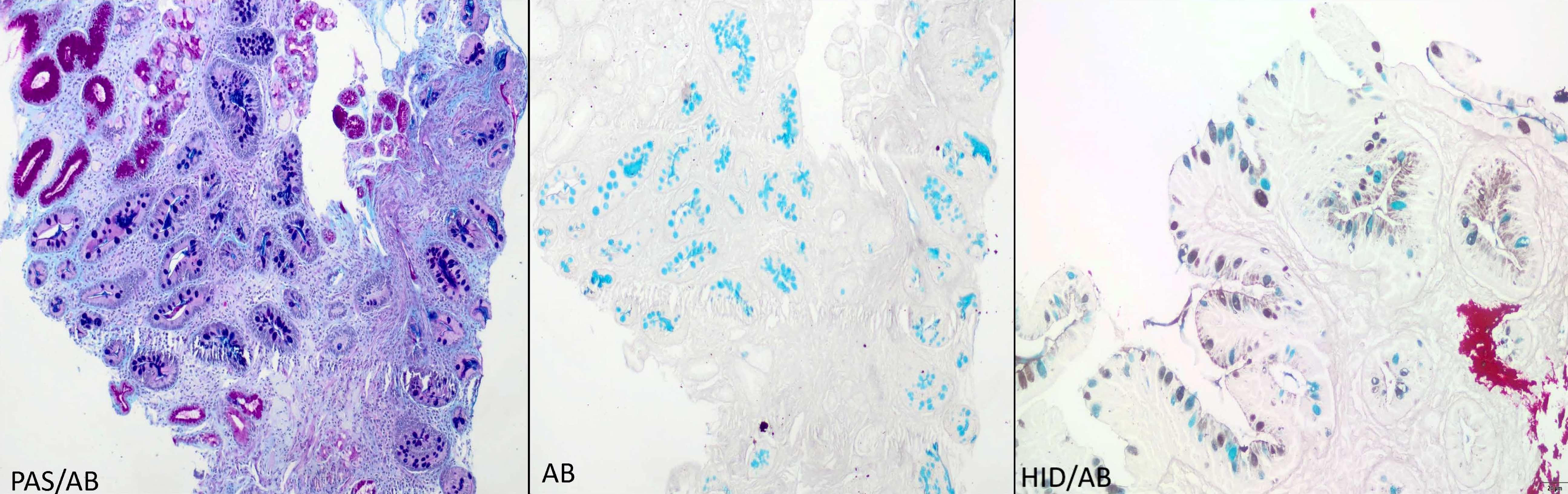
PAS / Alcian blue and HID / Alcian blue
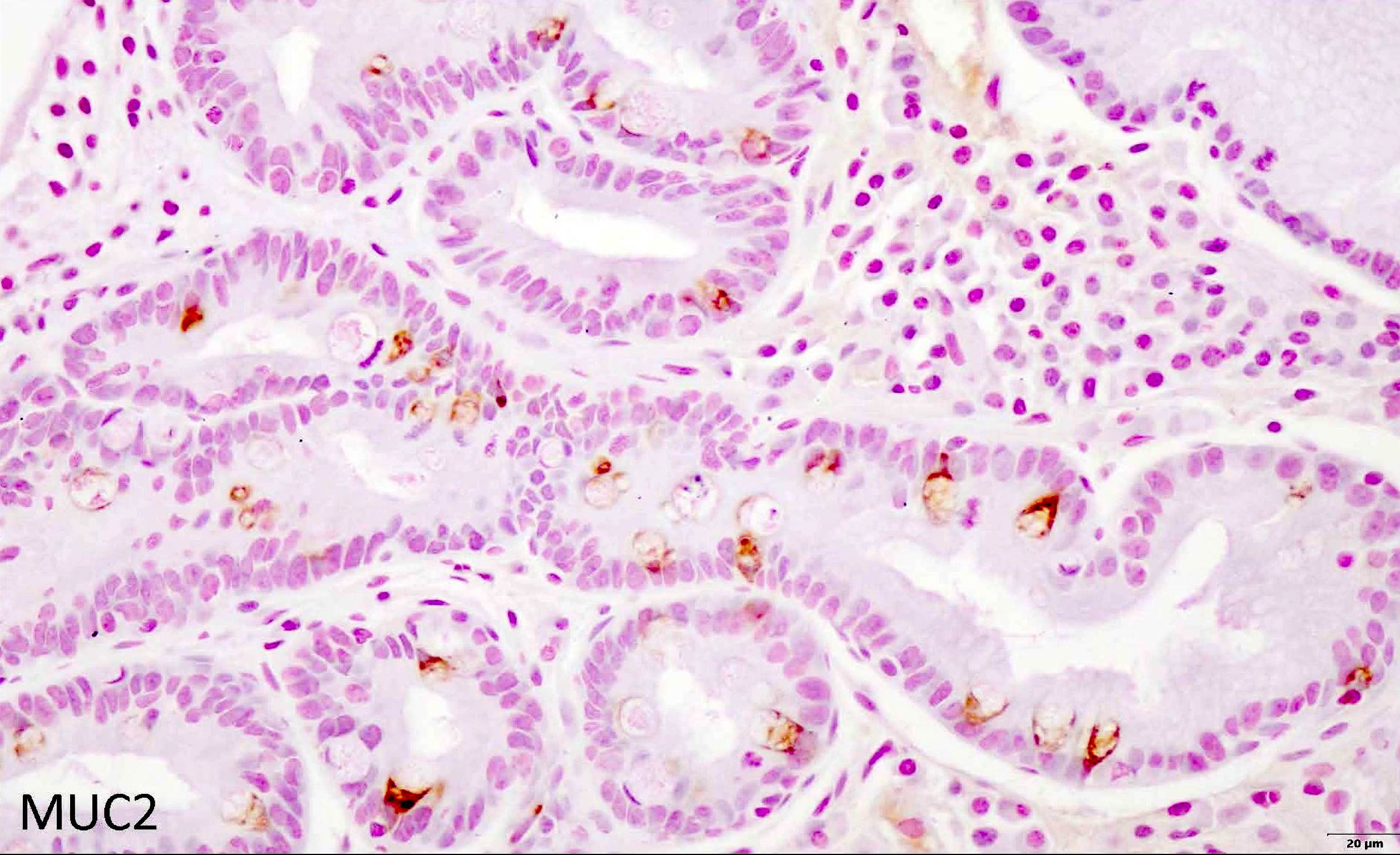
MUC2

CDX2
Images hosted on other servers:

Areas of intestinal metaplasia of the stomach
Images hosted on other servers:

Ulcerating mass and lymphadenopathy
Images hosted on other servers:

Gastric cancer at endoscopy
Contributed by @Andrew_Fltv on Twitter
 Contributed by @Andrew_Fltv on Twitter (see original post here)">
Intestinal type adenocarcinoma
Images hosted on other servers:

Gastric cancer resection specimen
Contributed by Adrian C. Bateman, M.B.B.S., M.D.
Tubular adenocarcinoma
Tumor regression
HER2
PDL1
Contributed by @Andrew_Fltv on Twitter
 Contributed by @Andrew_Fltv on Twitter (see original post here)">
Intestinal type adenocarcinoma
Contributed by Monica Garcia-Buitrago, M.D.
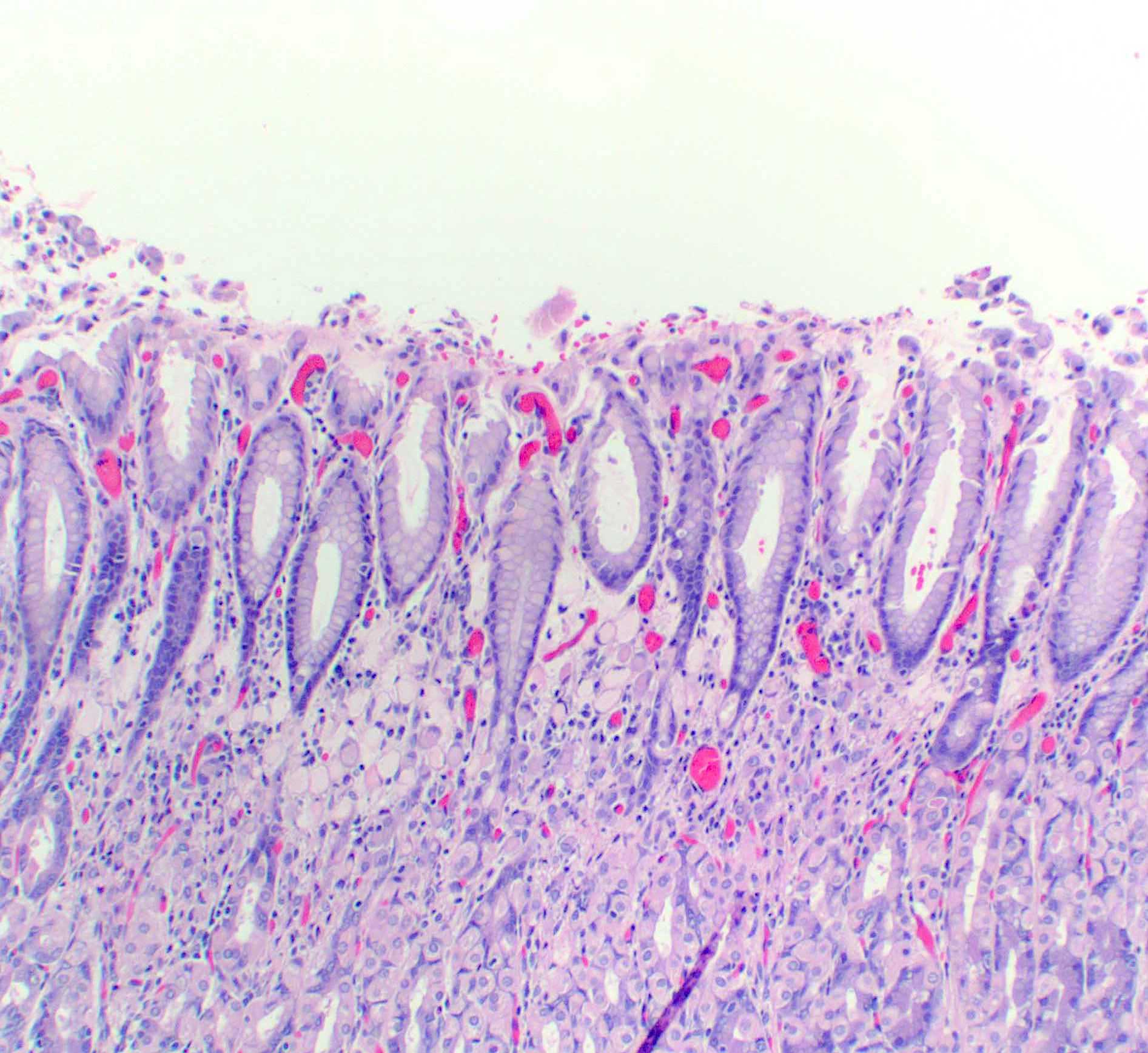
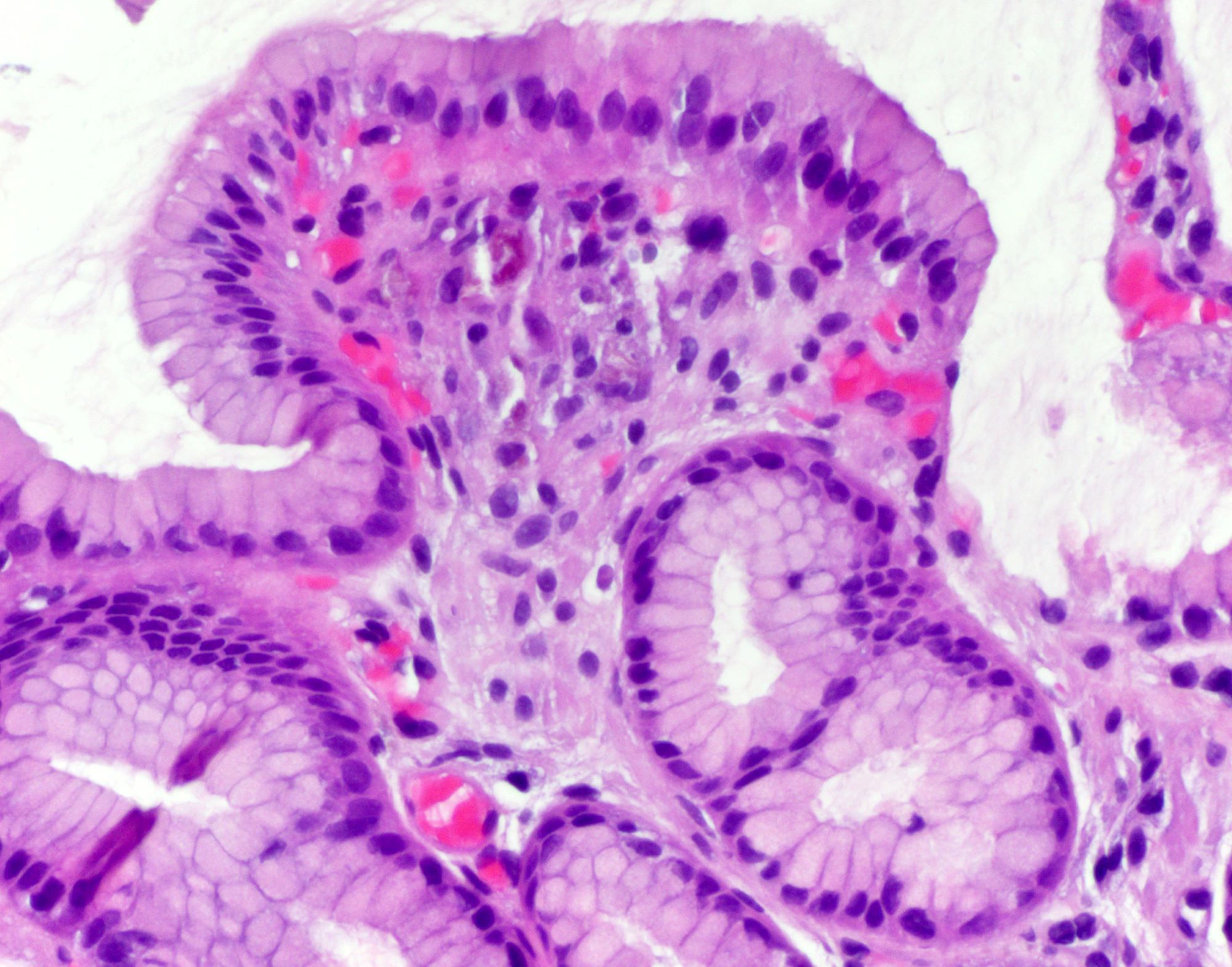
Gastric mucosa
Contributed by Monica Garcia-Buitrago, M.D. and Domenika Ortiz, M.D.
Brown crystalline iron deposition
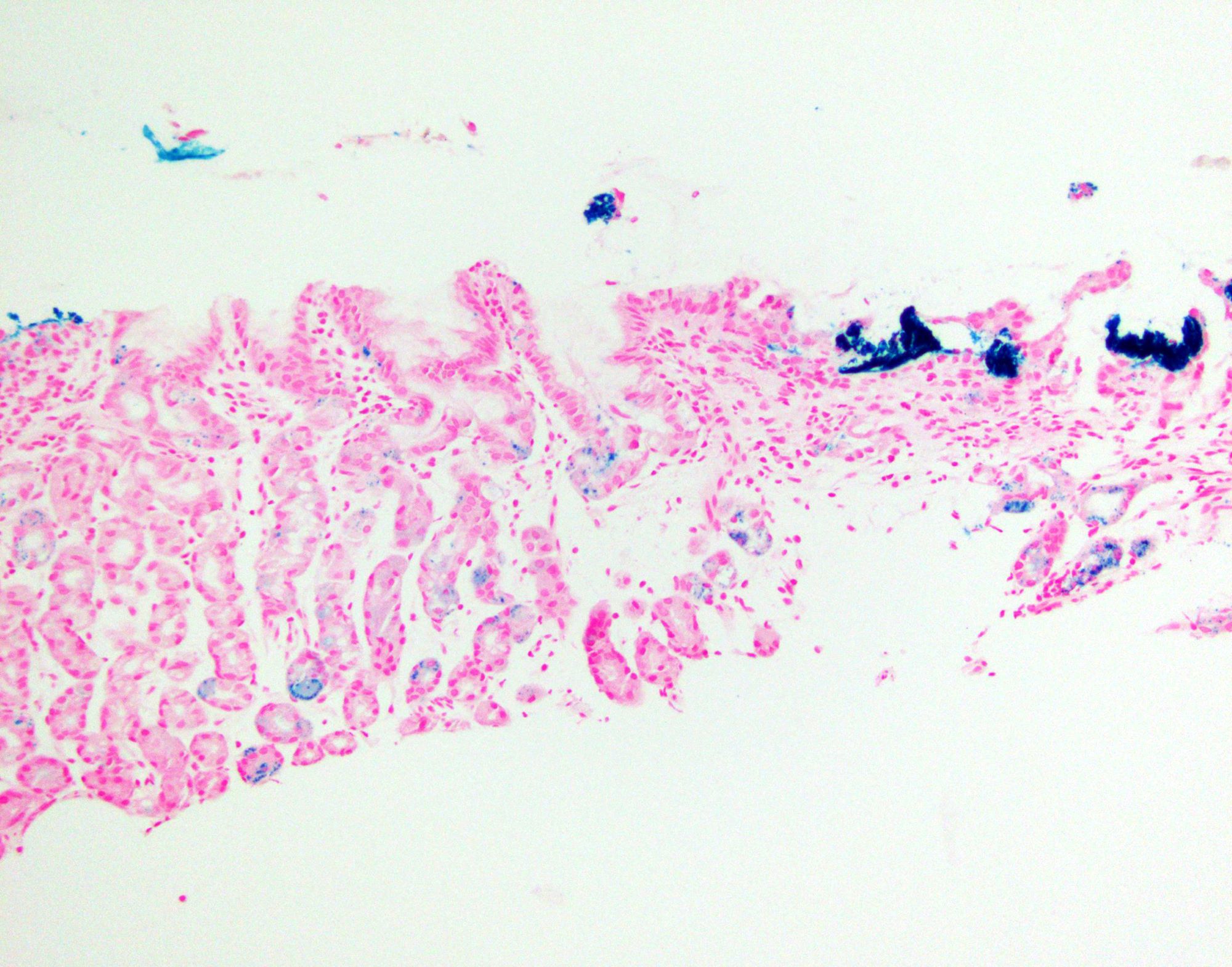
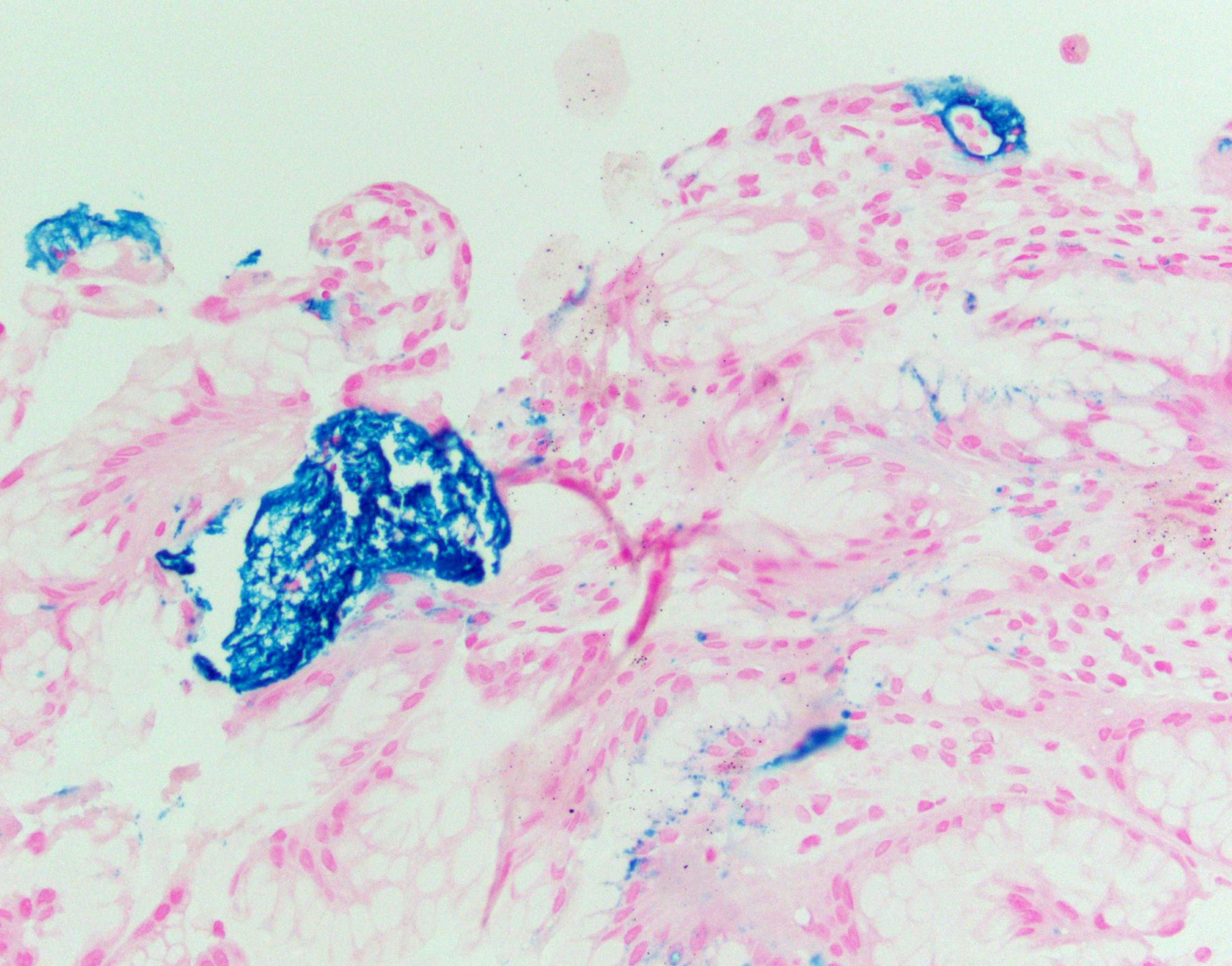
Prussian blue stain
Mucosal calcinosis
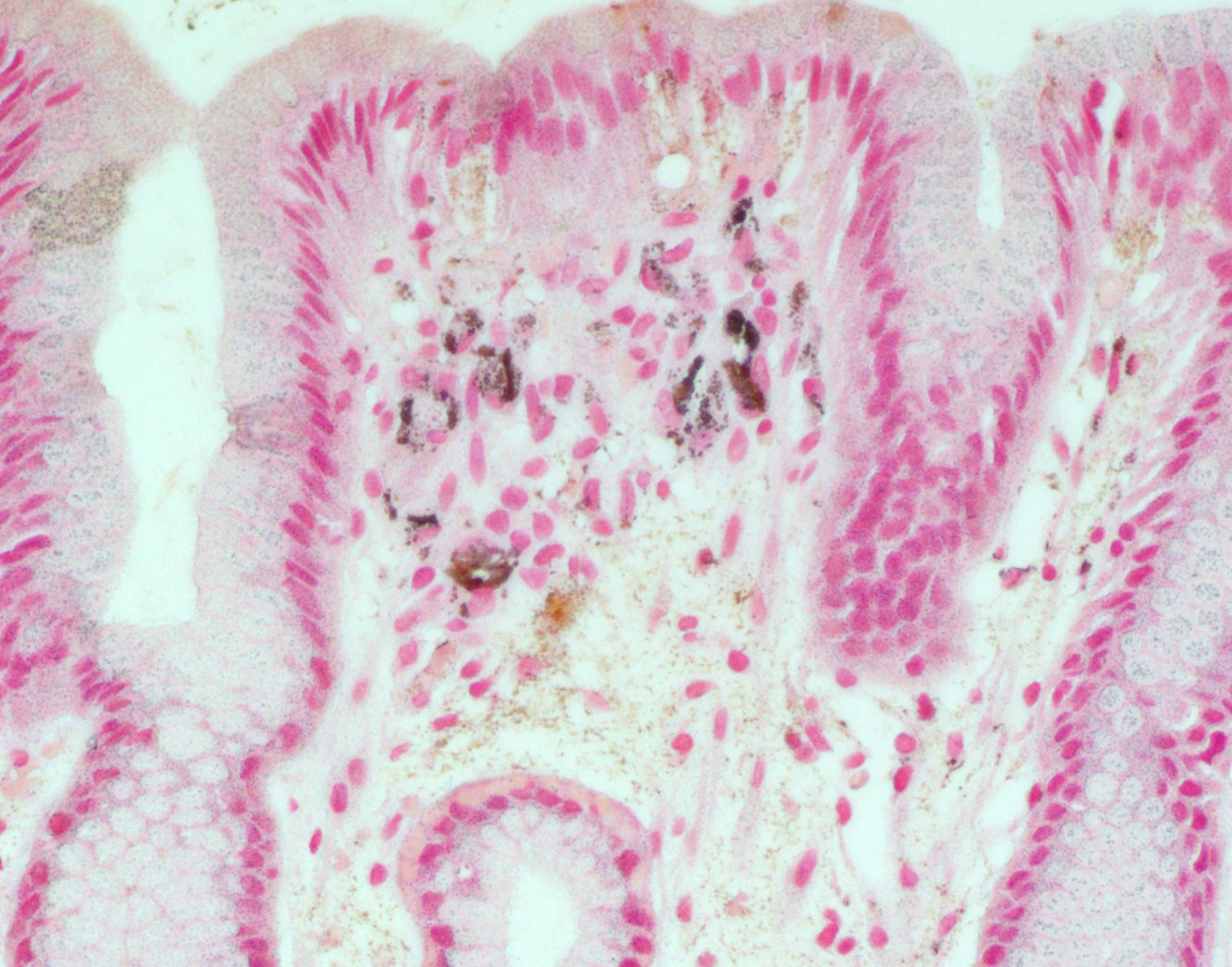
Mucosal calcinosis von Kossa stain
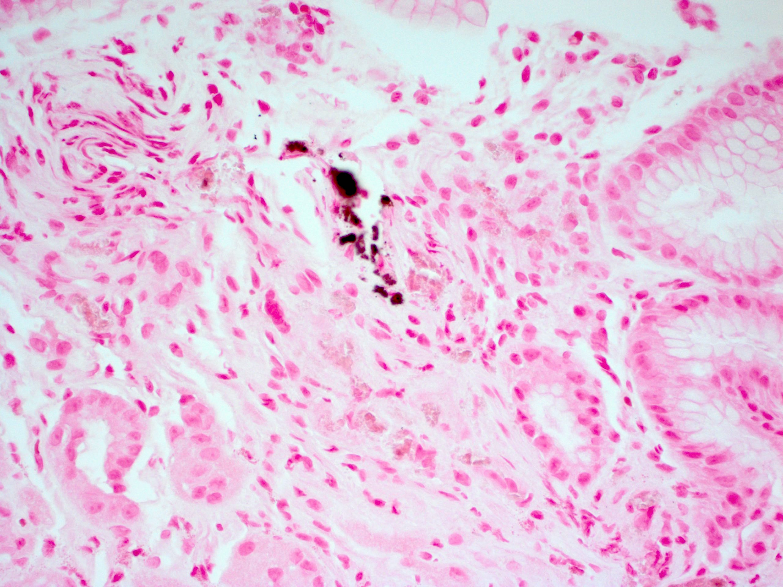
Nonspecific pill gastritis
Image hosted on other server:

Gastric juvenile polyp
Case #442


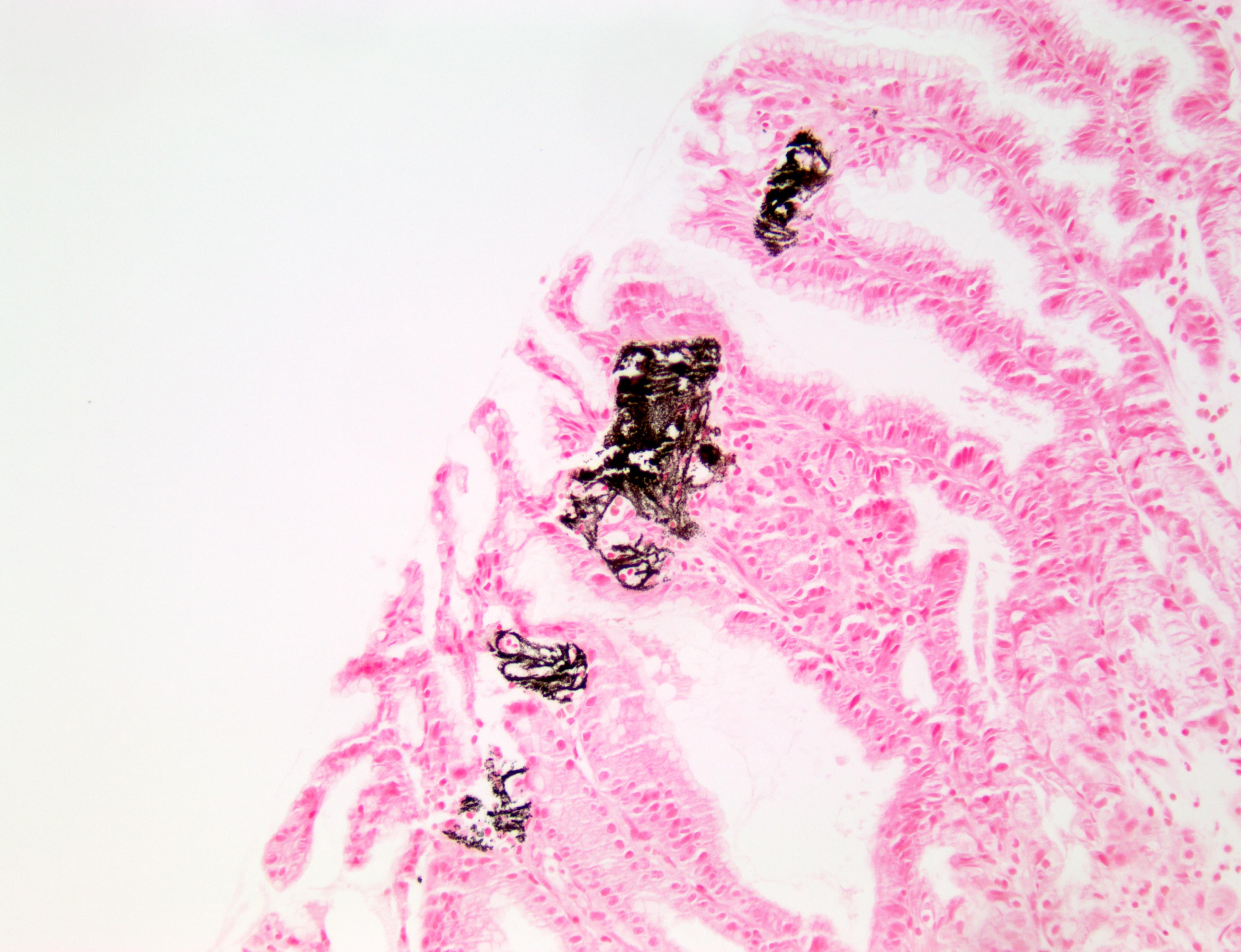
Lanthanum carbonate in stomach
Von Kossa
Images hosted on other servers:

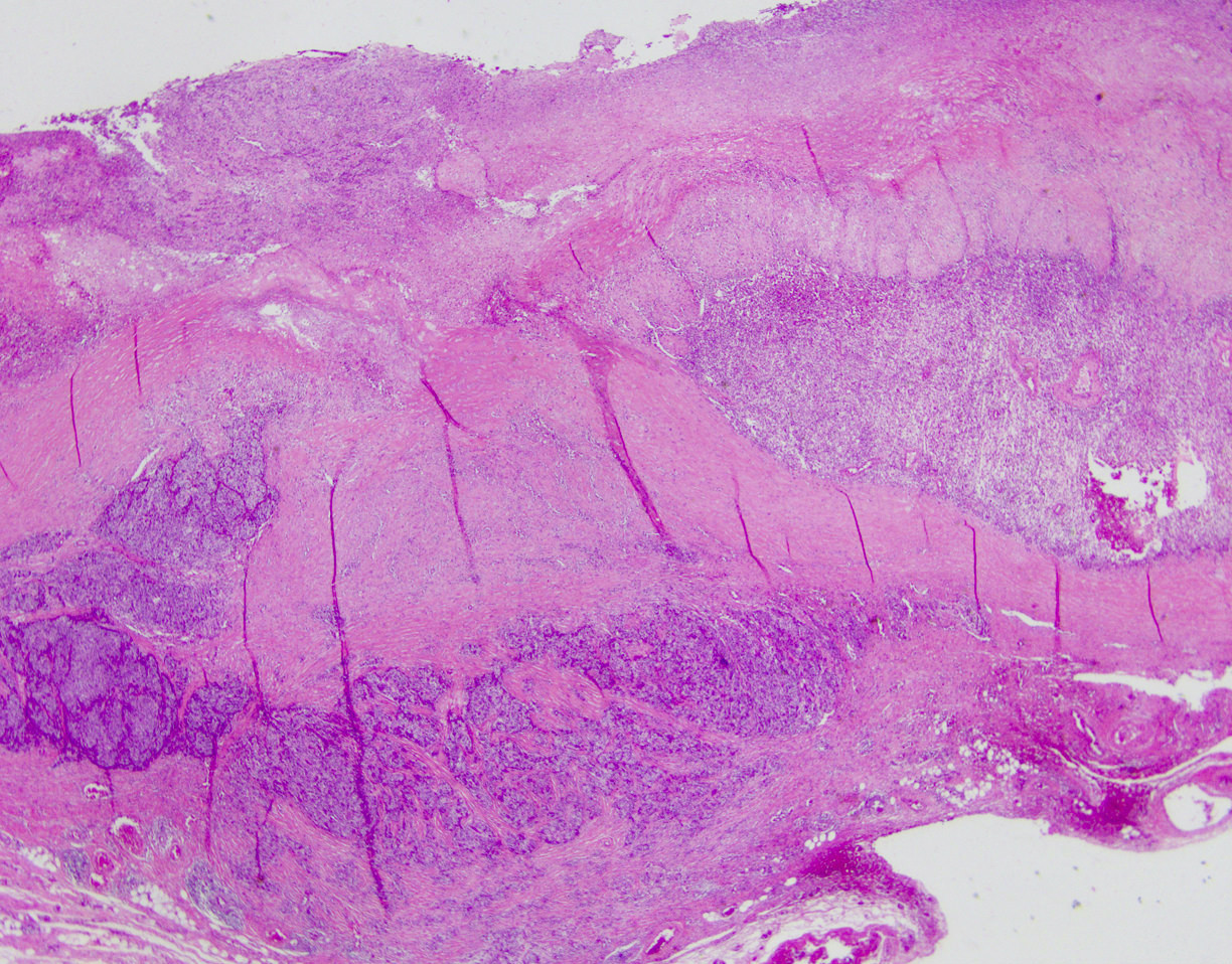
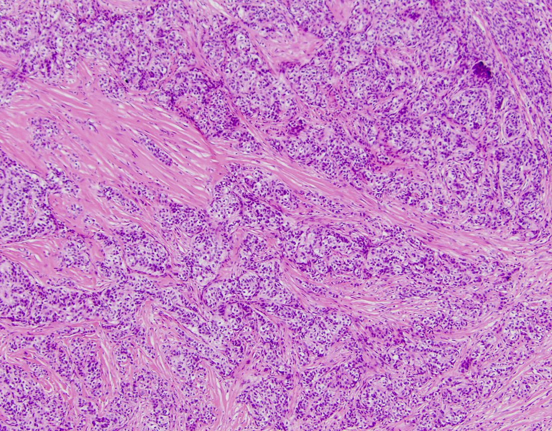
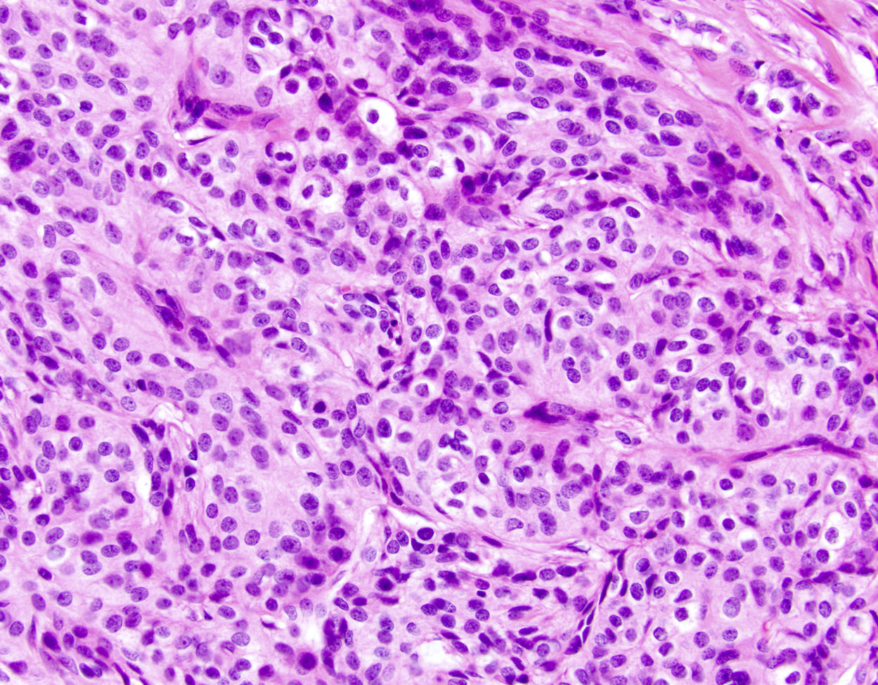
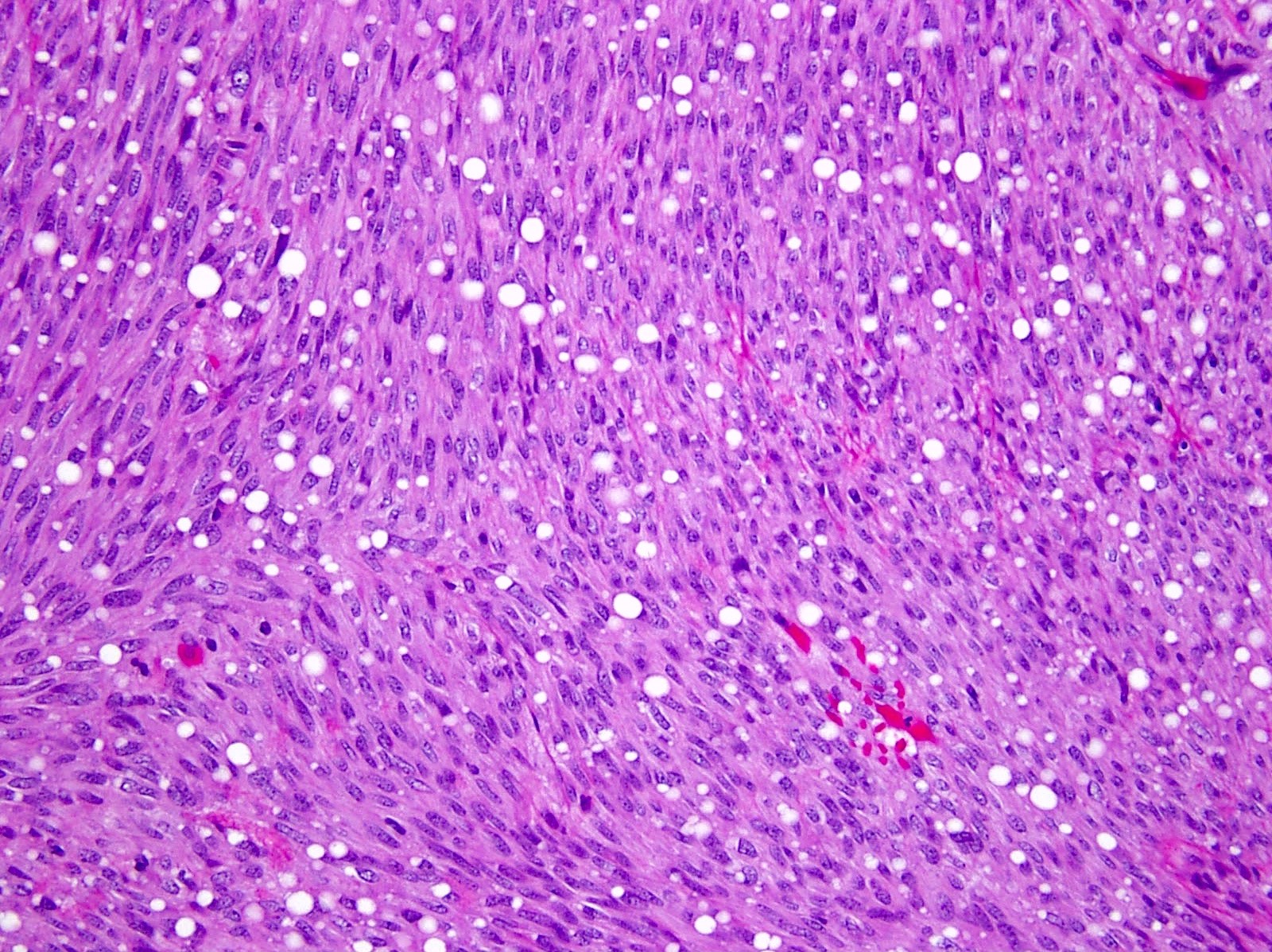
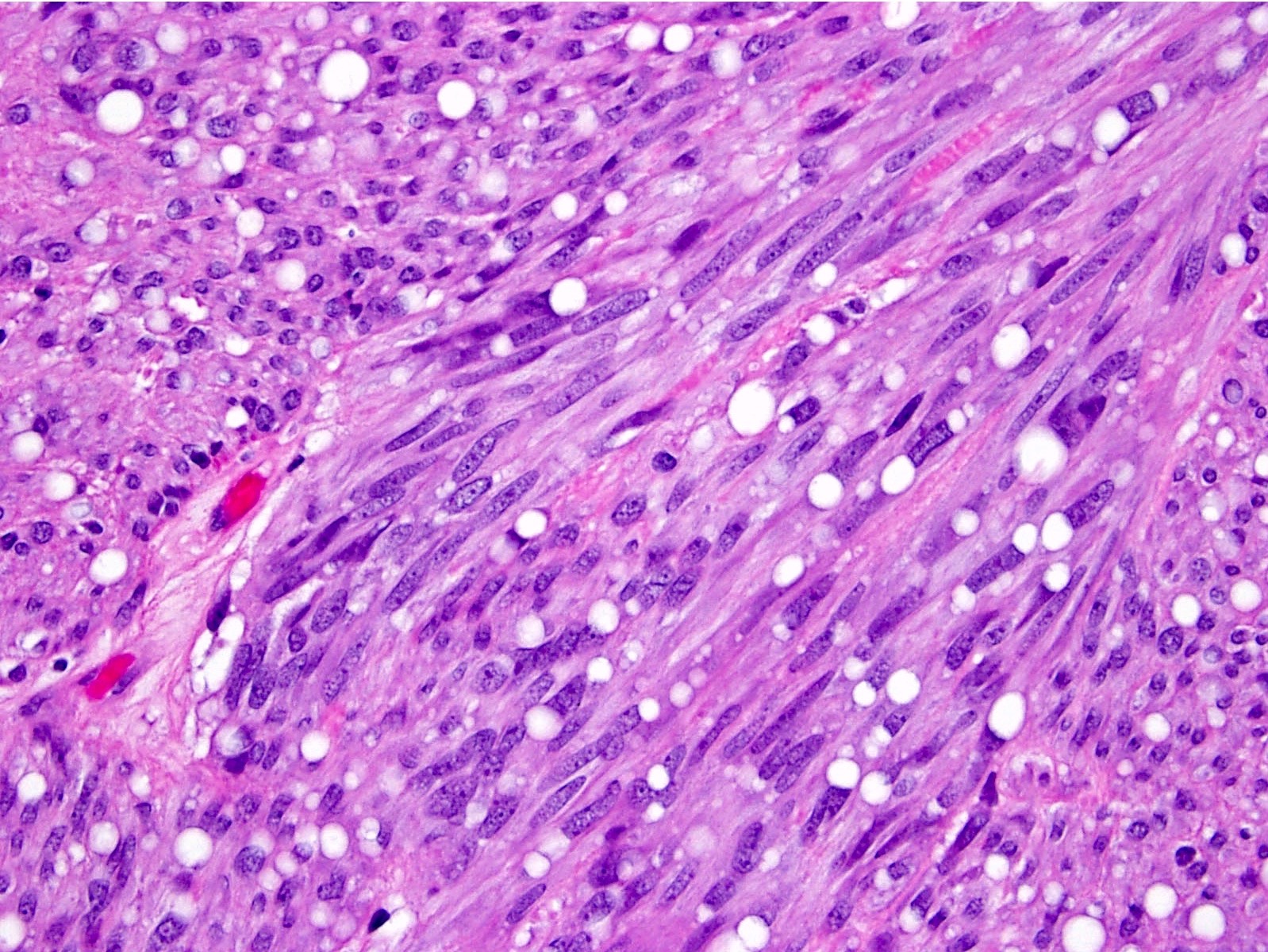
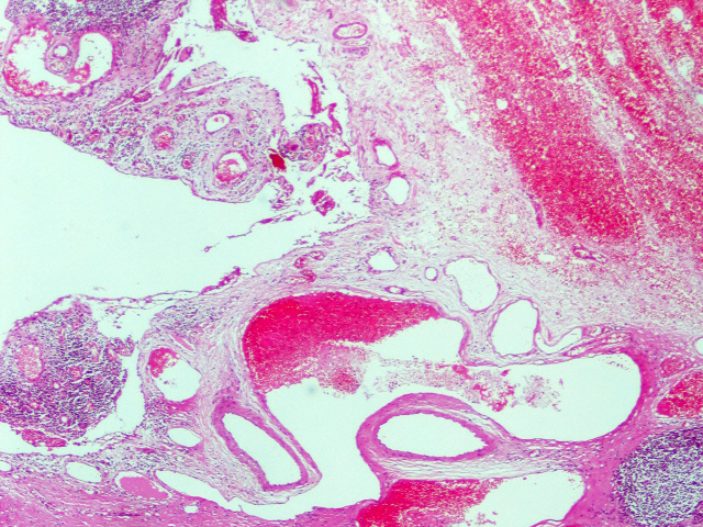
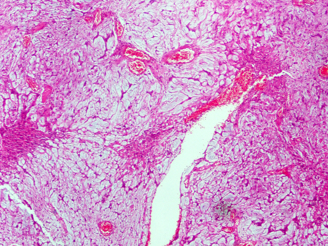
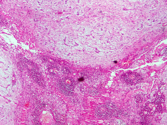
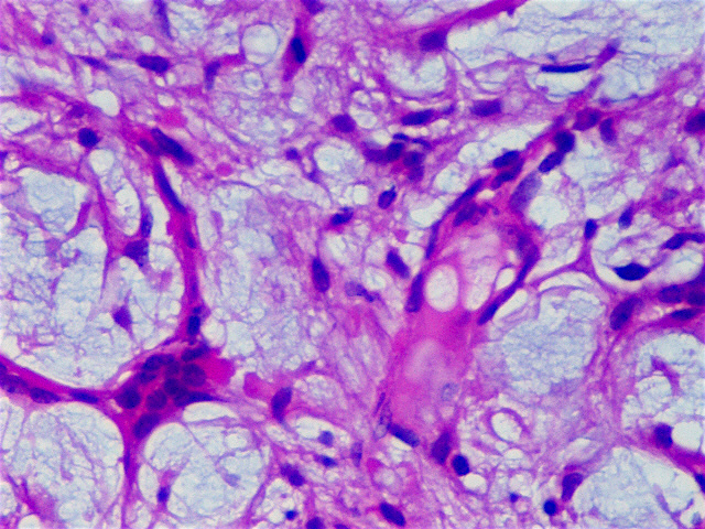
Large gastric leiomyosarcoma (> 8 cm)
Contributed by Raul S. Gonzalez, M.D.
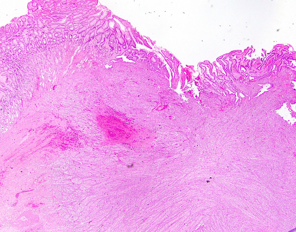
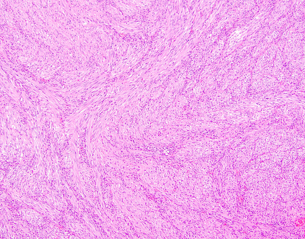
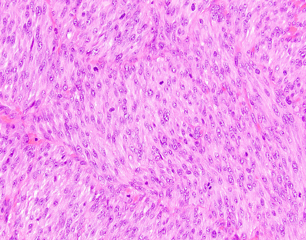
Leiomyosarcoma
Case #438
H&E
SMA
MSA

Desmin

Ki67
Images hosted on other servers:

Varioliform gastritis:
multiple nodules with
central mucosal
atrophy / erosion
Contributed by Matthew Morrow, M.D.
Increased intraepithelial lymphocytes
Increased intraepithelial lymphocytes, expanded lamina propria
Biopsy from area of nodular gastric mucosa
Contributed by @RaulSGonzalezMD on Twitter
Contributed by @RaulSGonzalezMD on Twitter (see original post here)">
Lymphocytic gastritis
Contributed by Matteo Fassan, M.D., Ph.D.

HER2 3+ positive gastric adenocarcinoma

EBER positive gastric adenocarcinoma
MLH1 negative gastric adenocarcinoma
Contributed by Robin Pike, M.D.
Granular material in lamina propria
Positive von Kossa stain
Contributed by Natalia Liu, M.D. and Hanlin L. Wang, M.D., Ph.D.
Flat raised nodule
Contributed by Natalia Liu, M.D. and Hanlin L. Wang, M.D., Ph.D.
Low power architecture
Irregular glands

Predominantly chief cell differentiation
Contributed by @liverwei on Twitter
Oxyntic gland polyp
Oxyntic gland polyp
Images hosted on other servers:


Various images
Images hosted on other servers:

Gastric Peutz-Jeghers polyps
Contributed by Yujun Gan, M.D., Ph.D. and Xiuli Liu, M.D., Ph.D.
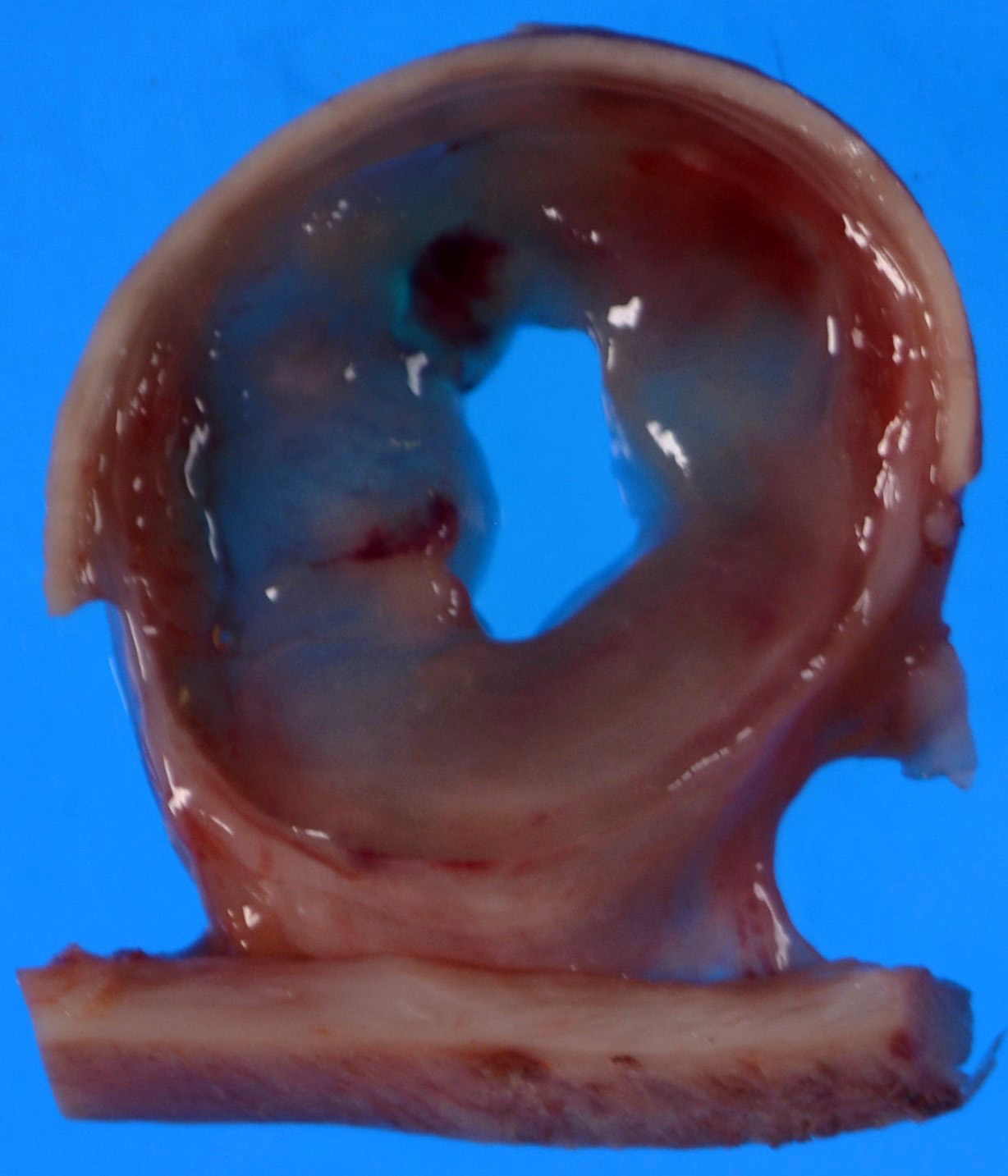
Large gastric antral mass

Large mural hypoechoic mass
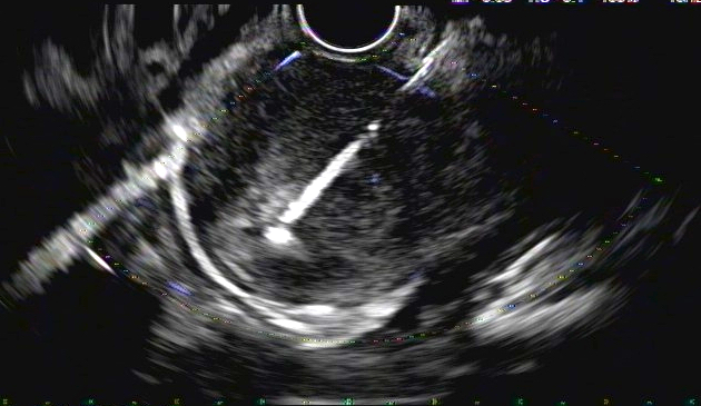
FNA of the mass
Contributed by Yujun Gan, M.D., Ph.D. and Xiuli Liu, M.D., Ph.D.
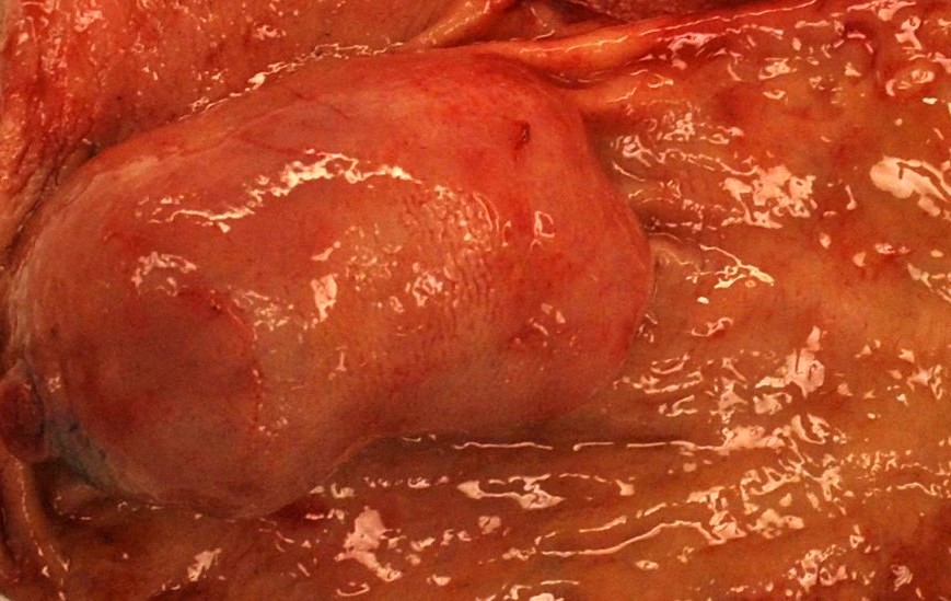
Antral mass
Myxoid and cystic degeneration
Contributed by Yujun Gan, M.D., Ph.D. and Xiuli Liu, M.D., Ph.D.
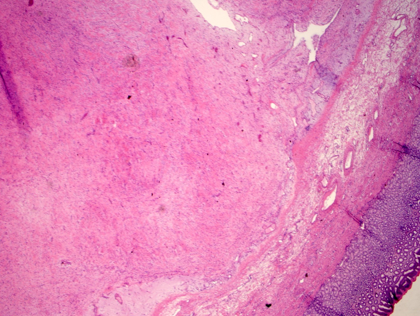
Hypocellular and myxoid tumor

Bland spindle cell proliferation
Fibrillary stroma

Spindle and myxoid tumor
Smooth muscle actin immunoreactivity
Contributed by Raul S. Gonzalez, M.D.
Plexiform architecture
Bland myxoid tumor
Images hosted on other servers:

Small, round
lymphocyte-like
cells with hyper-
chromatic nuclei
Images hosted on other servers:

Gastric serpentine mucosal folds
Contributed by Michael Schoech, M.D.
Endoscopy: mosaic-like pattern
Contributed by Divya Sharma, M.D.
Congestion and reactive gastropathy
Congested blood vessels
Lamina propria fibrosis

Ectatic blood vessels
Contributed by Supriya Srivastava M.D., Ph.D.
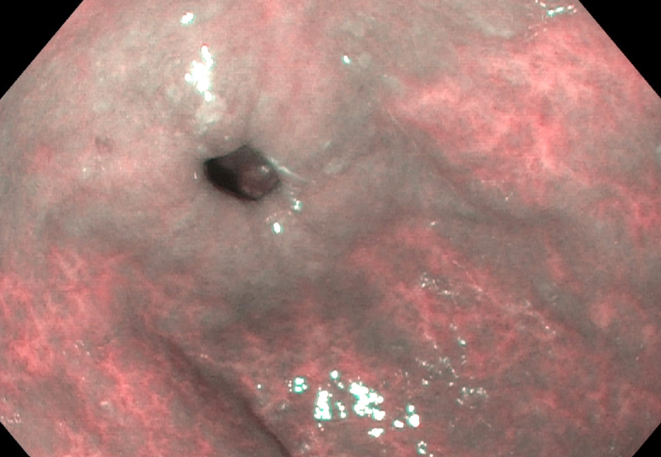
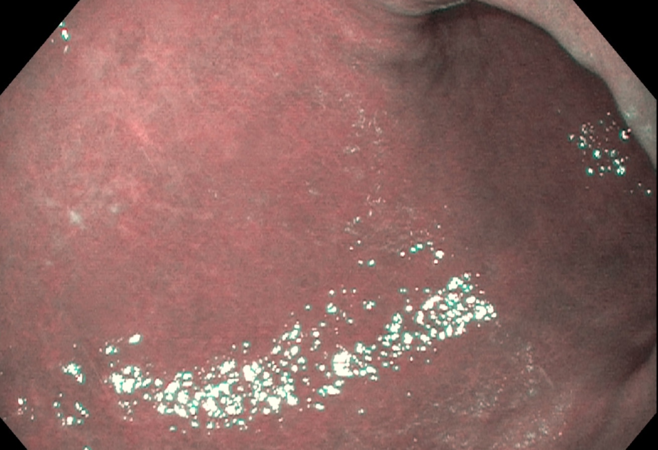
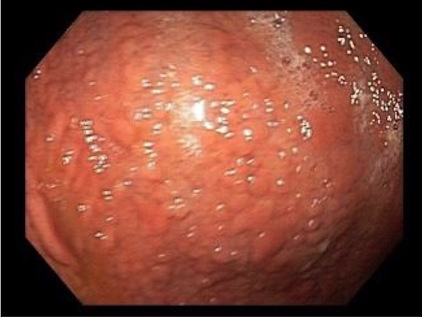
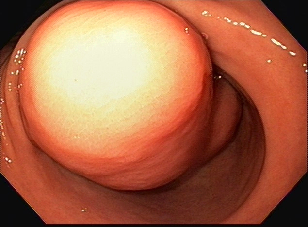
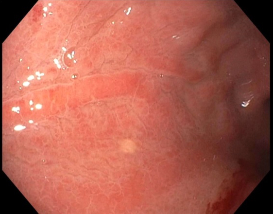
Endoscopic appearance
Images hosted on other servers:

Endoscopy
Images hosted on other servers:

Intraoperative image
Contributed by Supriya Srivastava M.D., Ph.D.
Antral polyp
PGA without dysplasia
MUC5AC staining
MUC6 staining
MUC2 staining
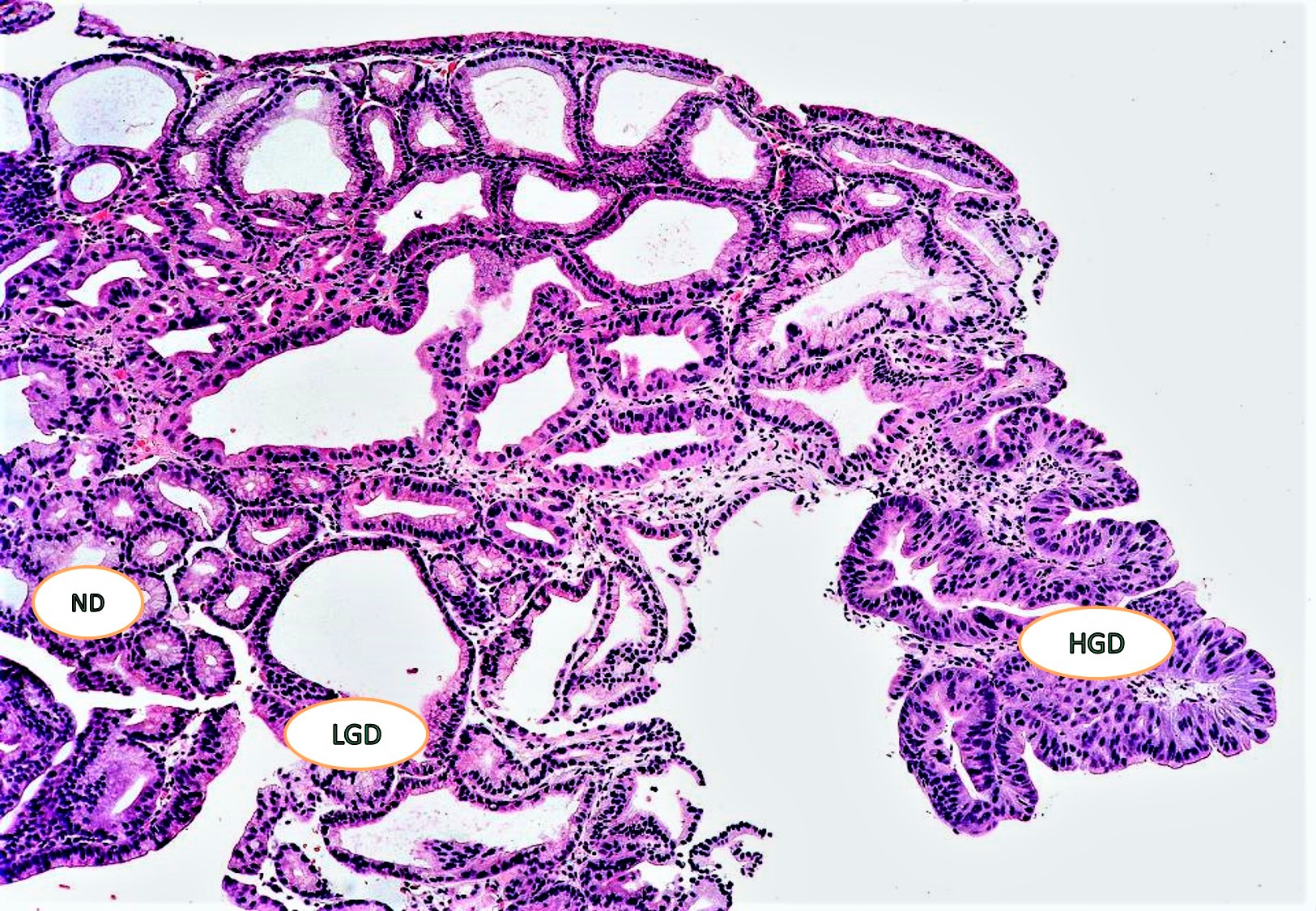
PGA with dysplasia
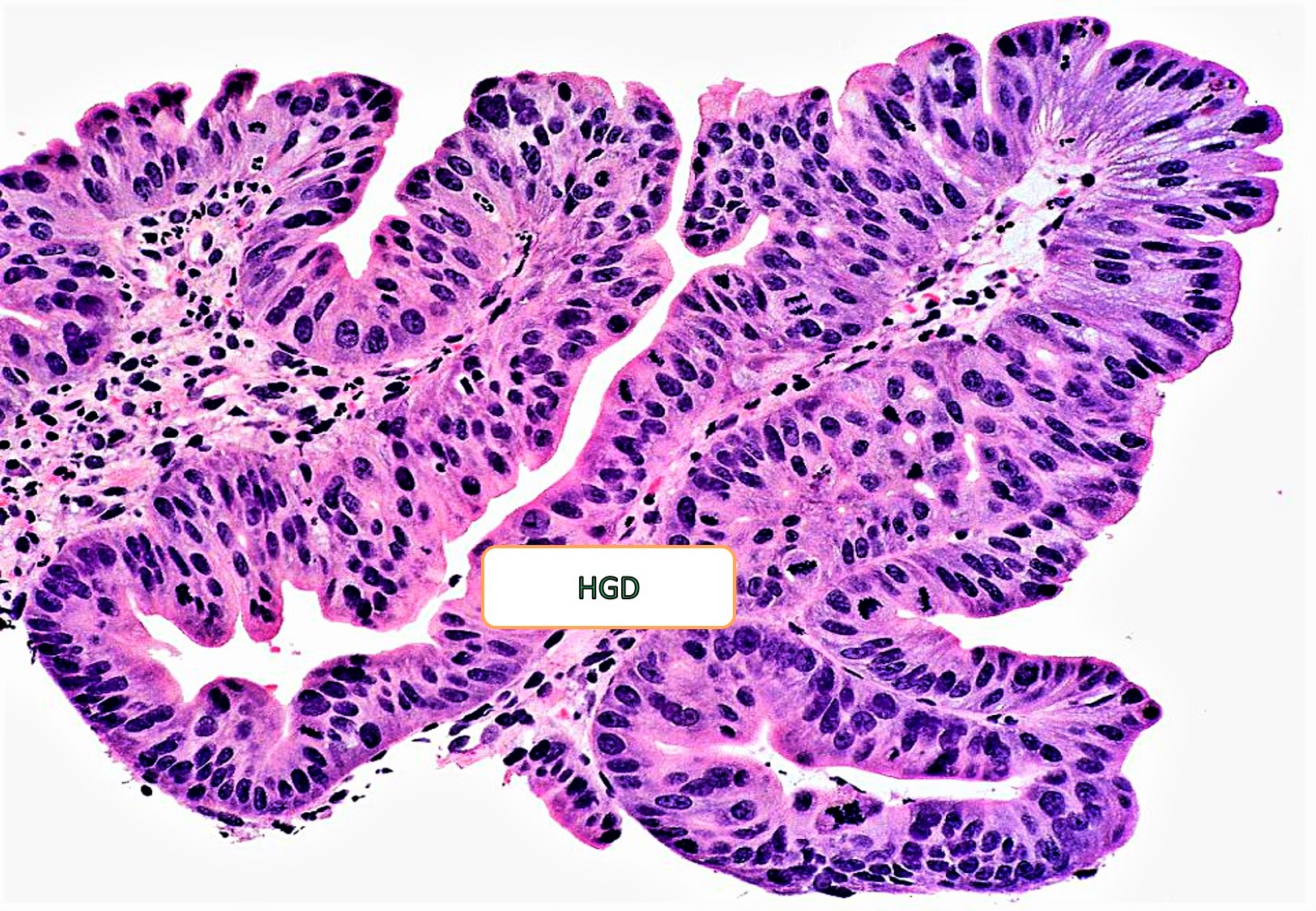
High grade dysplasia
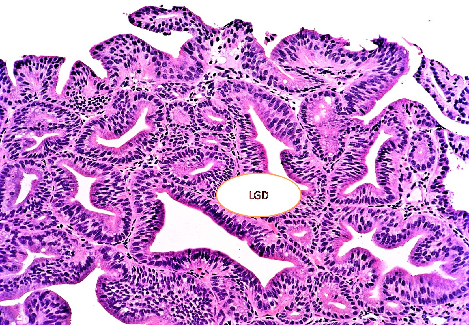
Low grade dysplasia
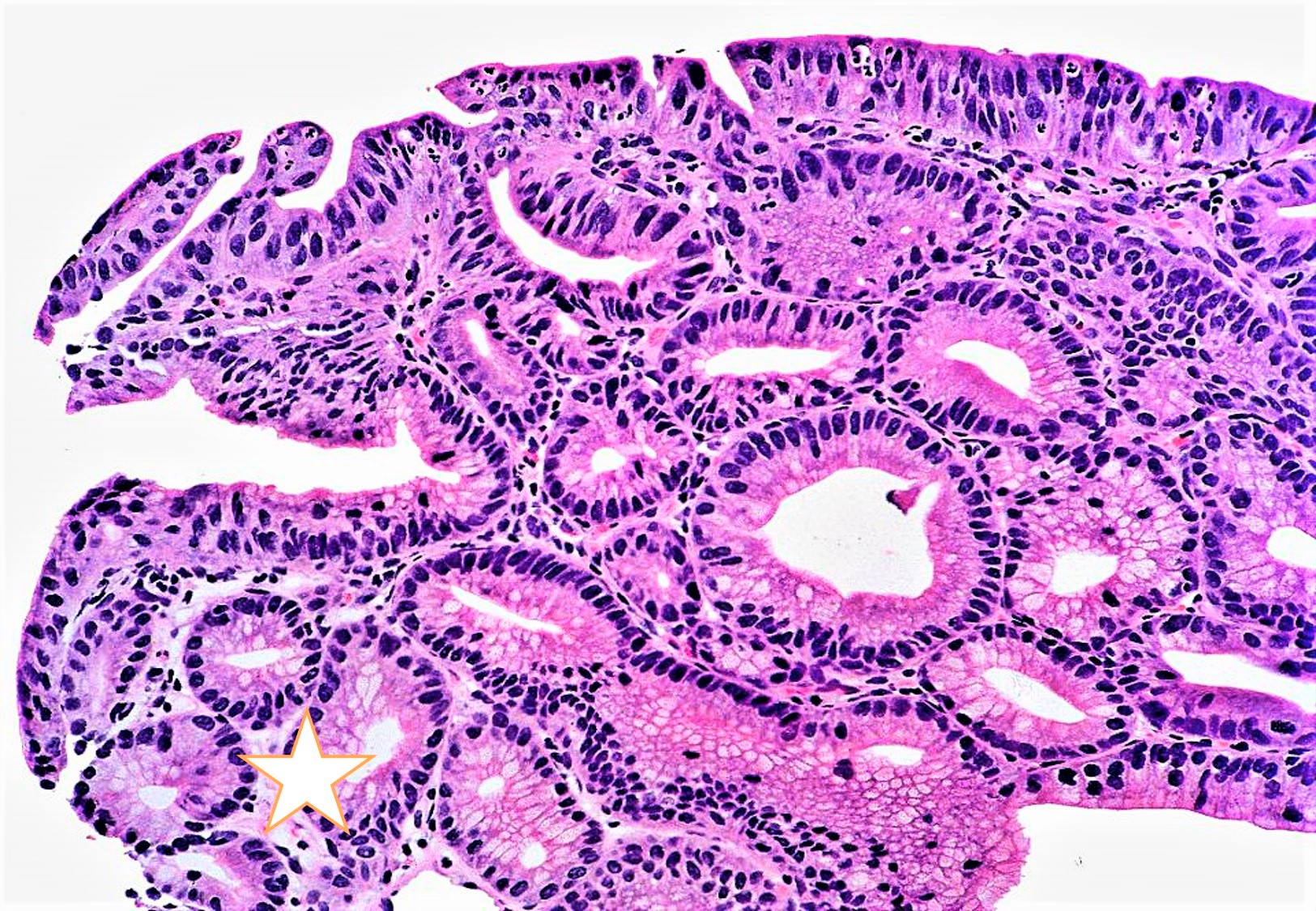
PGA with and without dysplasia
Image hosted on other server:

Thickened muscularis propria
Images hosted on other servers:

Bile with erythematous mucosa

Erythema / edema
after choledocho-
duodeno
anastomosis
Contributed by Aaron R. Huber, D.O.
Reactive epithelium
Epithelial injury without inflammation
Gastritis versus gastropathy
Images hosted on other servers:

Large, irregular and deep
ulcerated mass lesion
with central ulceration
at the incisura angularis
Case #410

H&E
Positive for CD138 stain
Contributed by @RaulSGonzalezMD on Twitter and Cases #455 and #347
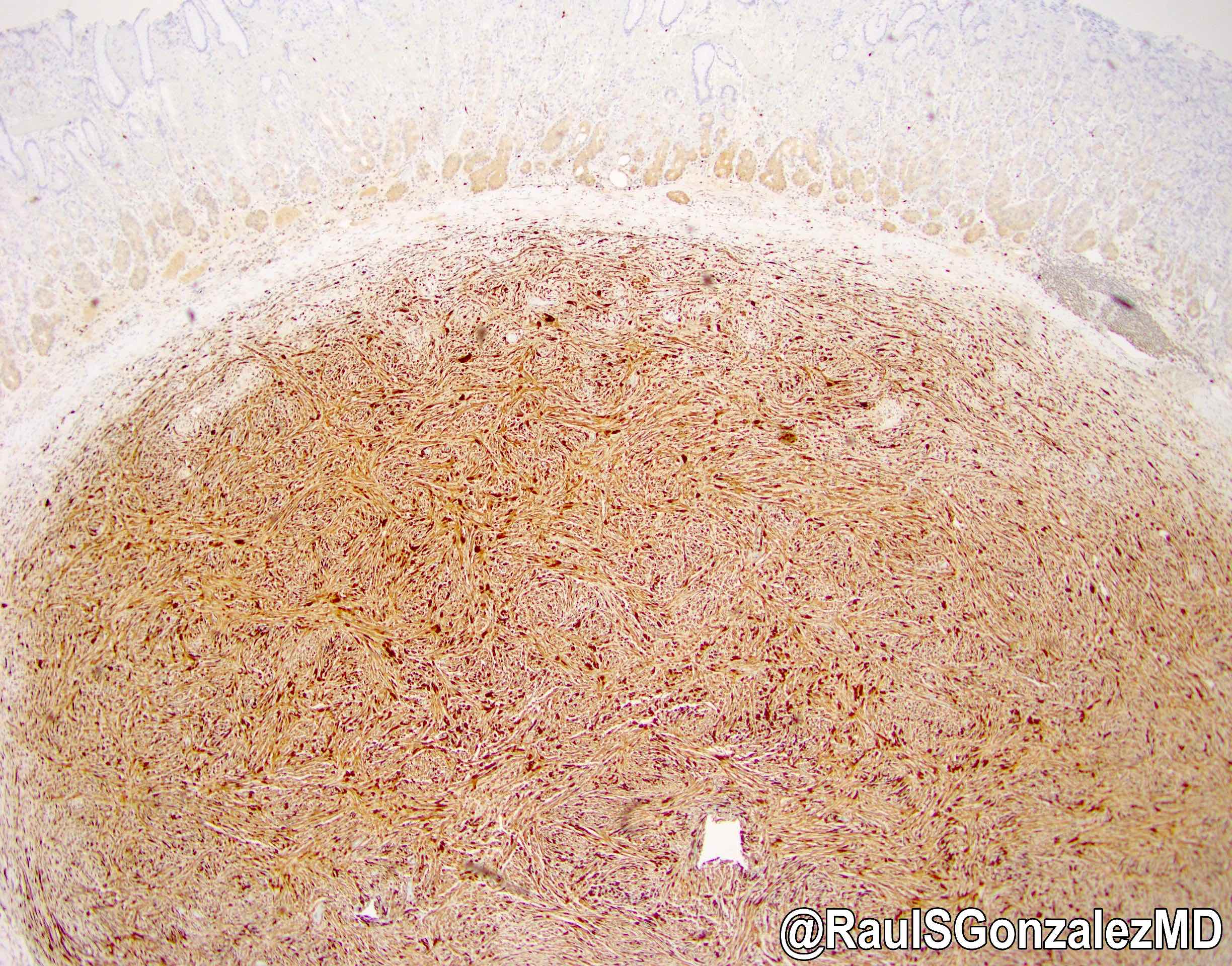
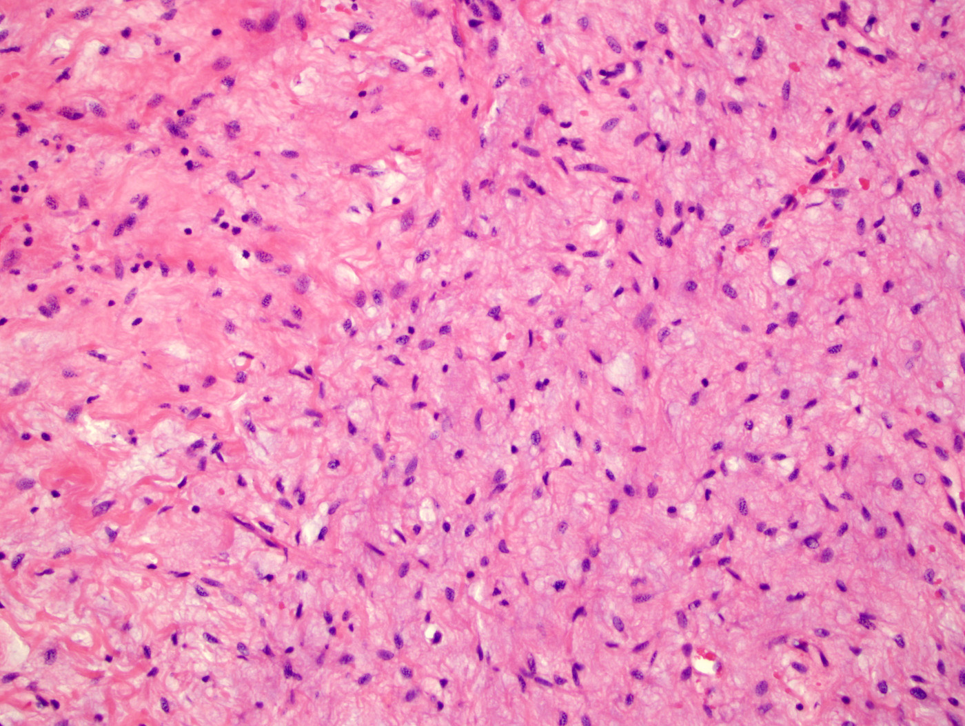
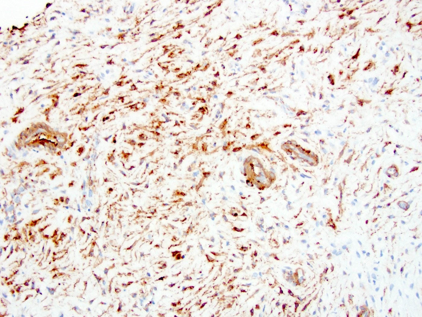
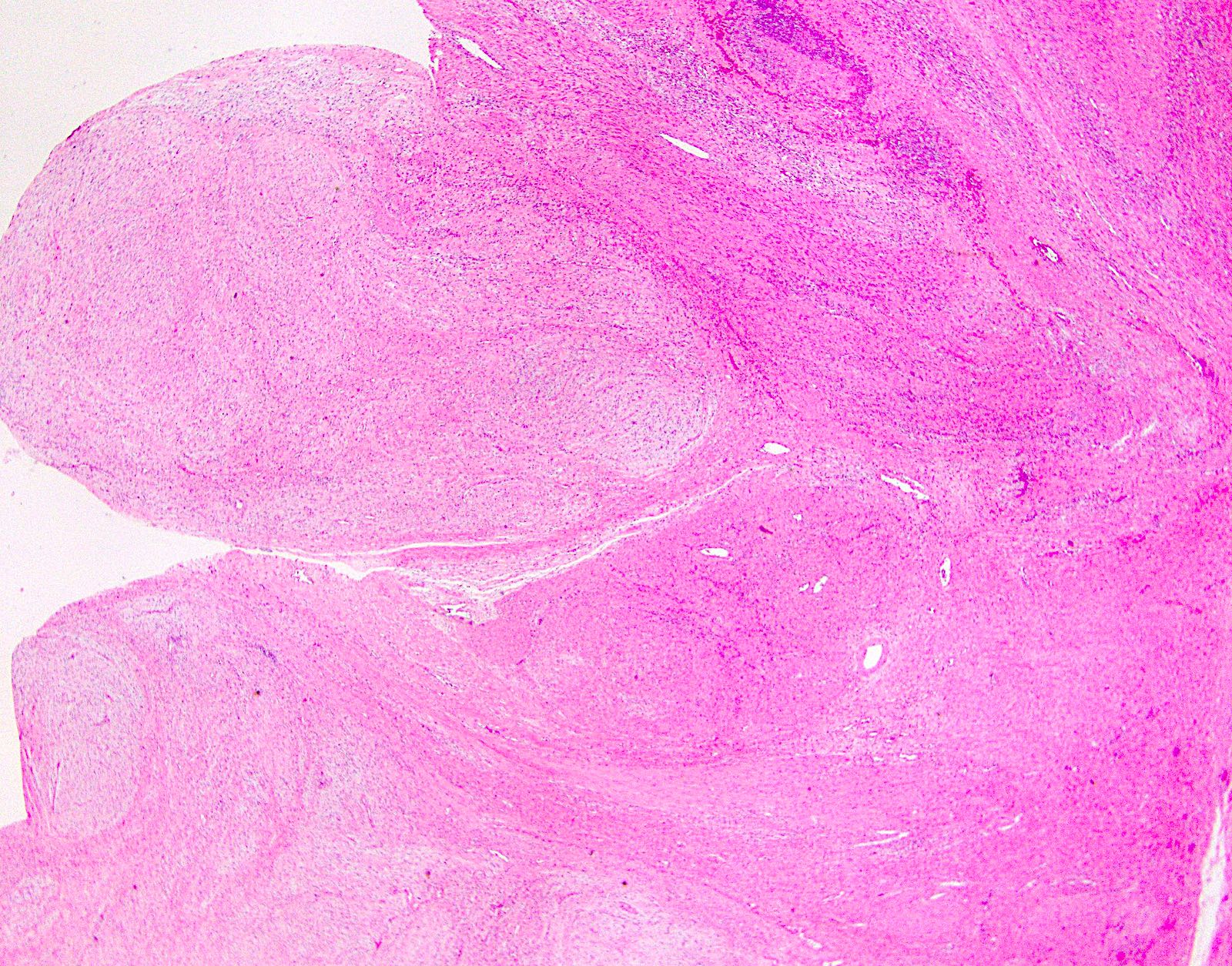
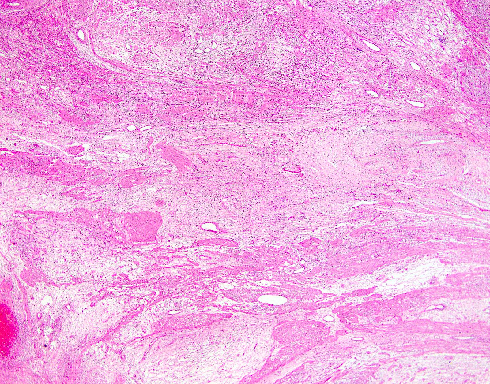
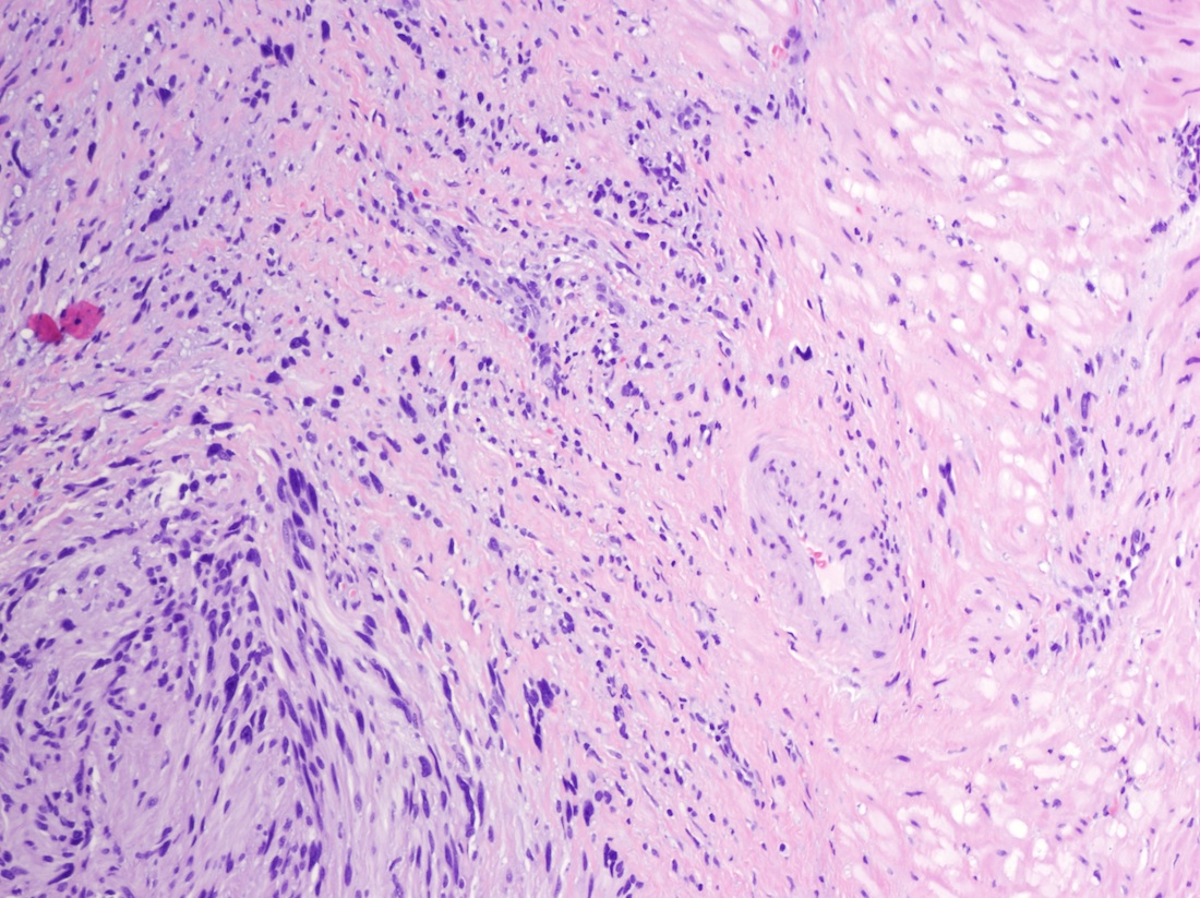
Schwannoma
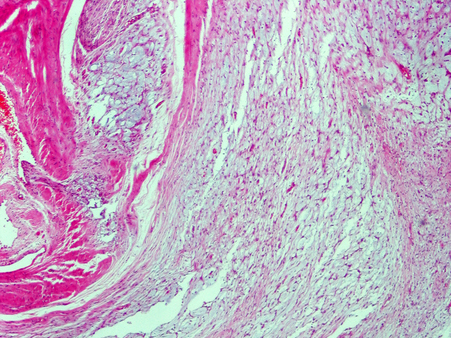


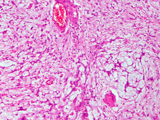

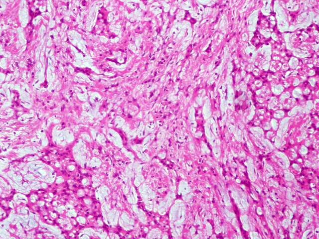
Microcystic variant of schwannoma in stomach
S100, microcystic variant

CD117, microcystic variant
46 year old woman
Images hosted on other servers:

Polyps and atrophic gastritis
Contributed by Dana Razzano, M.D. and @RaulSGonzalezMD on Twitter
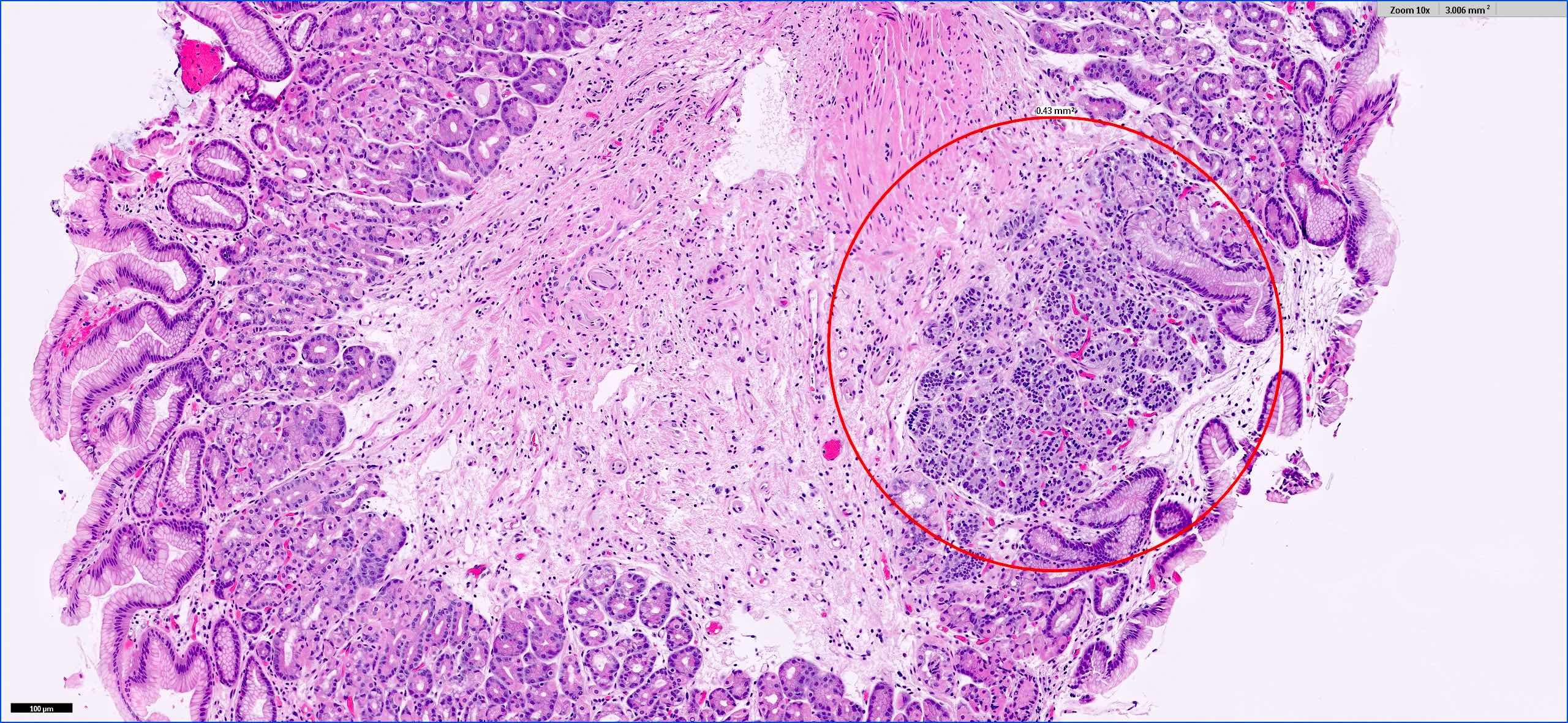

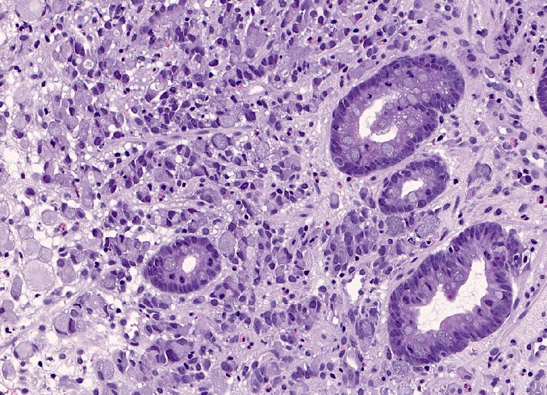
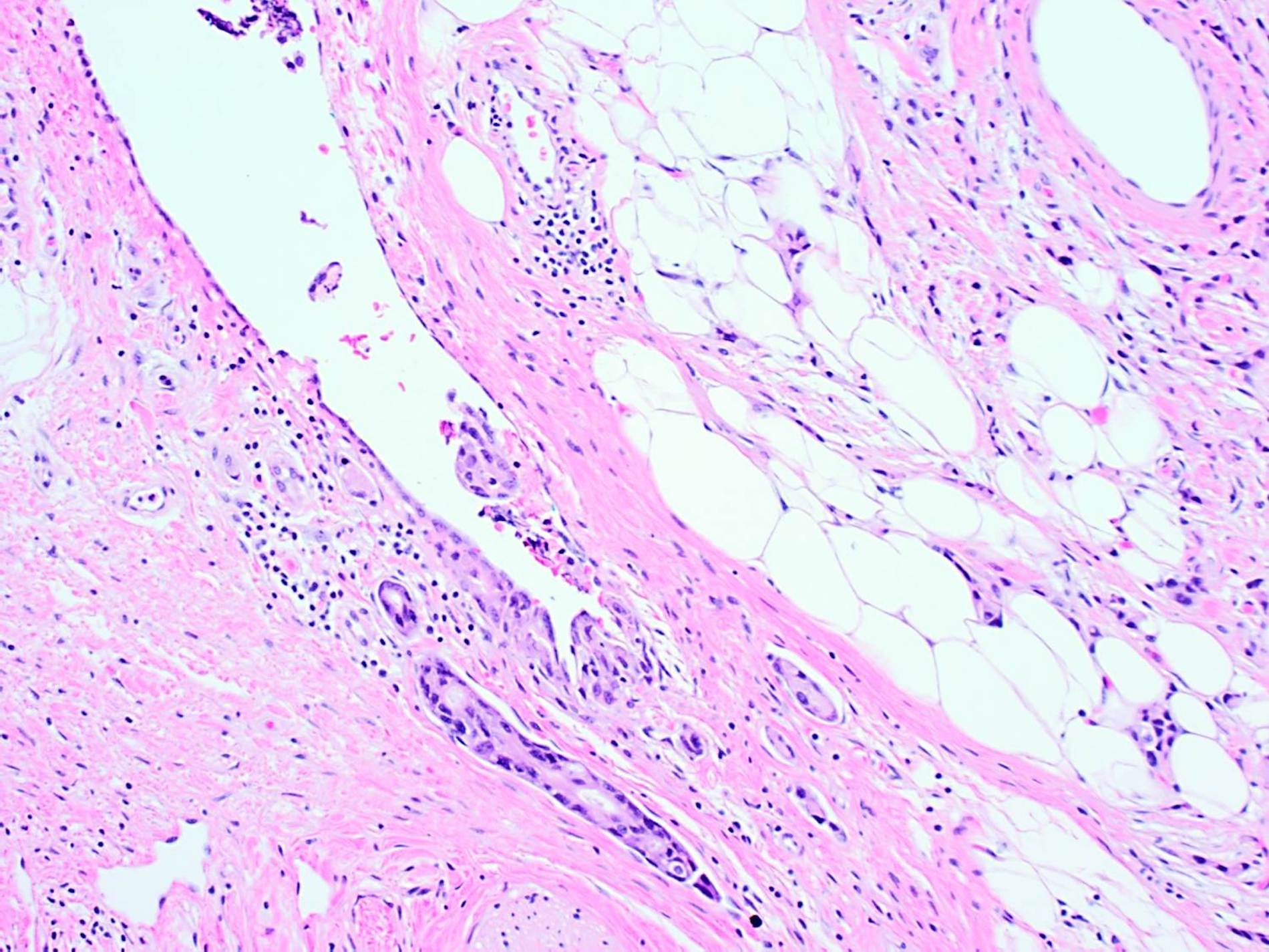
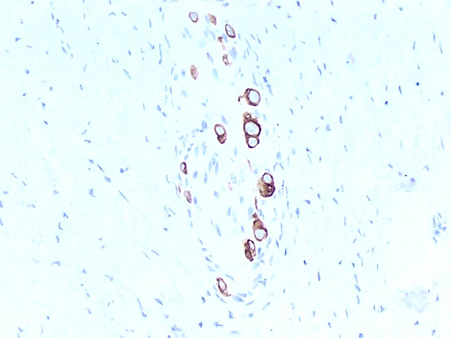
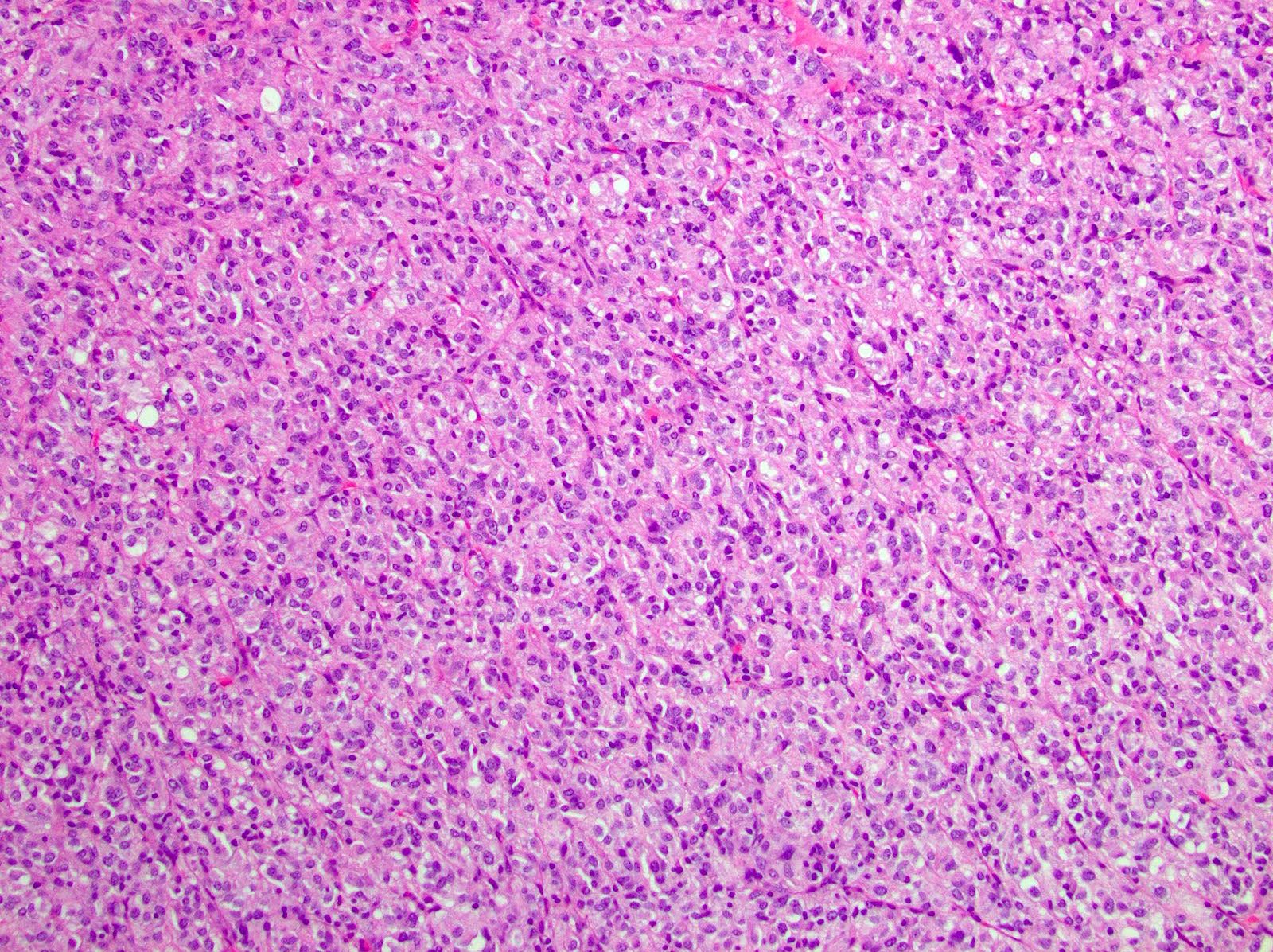
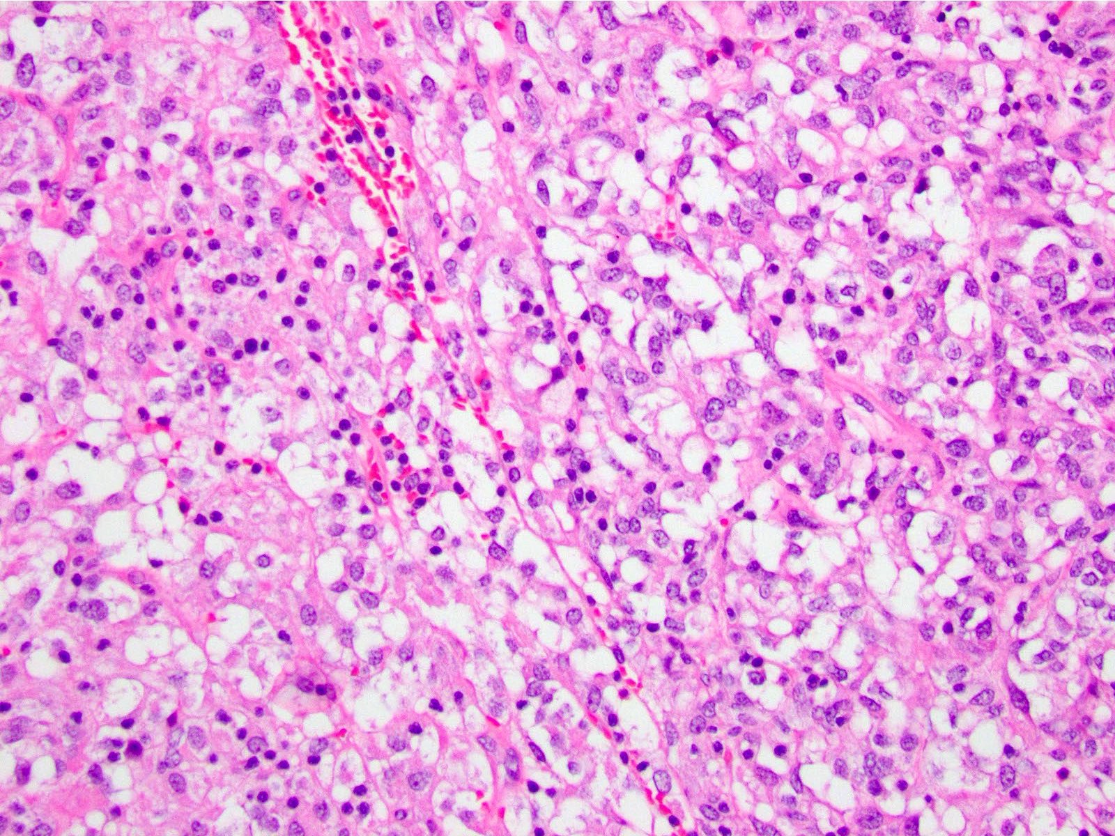
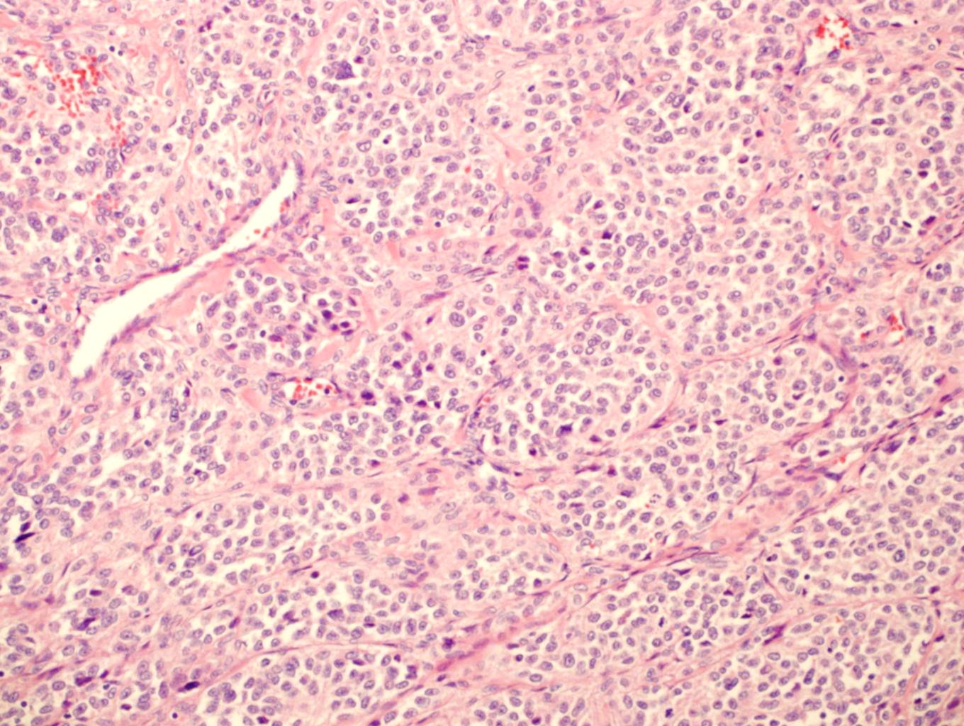
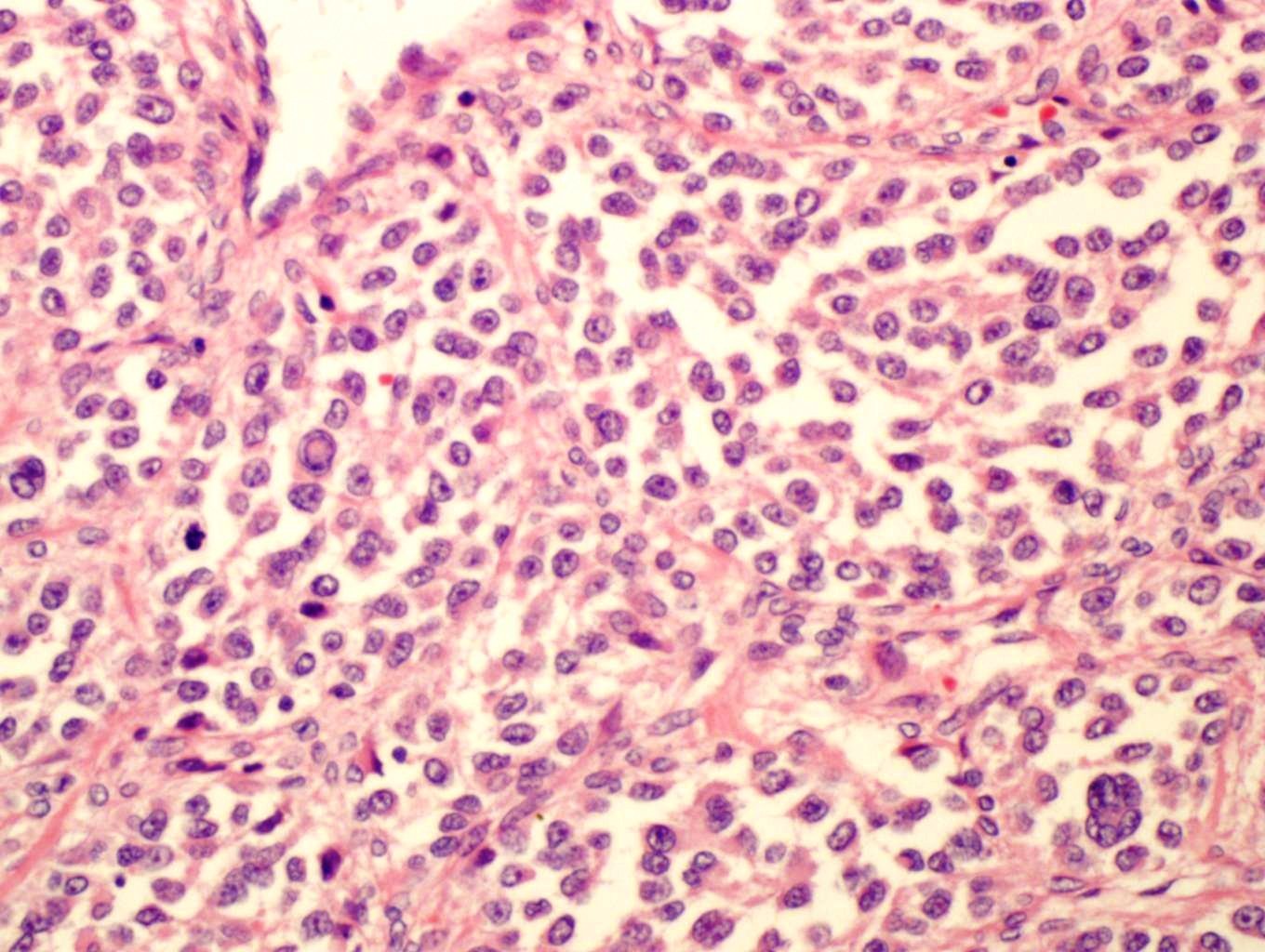
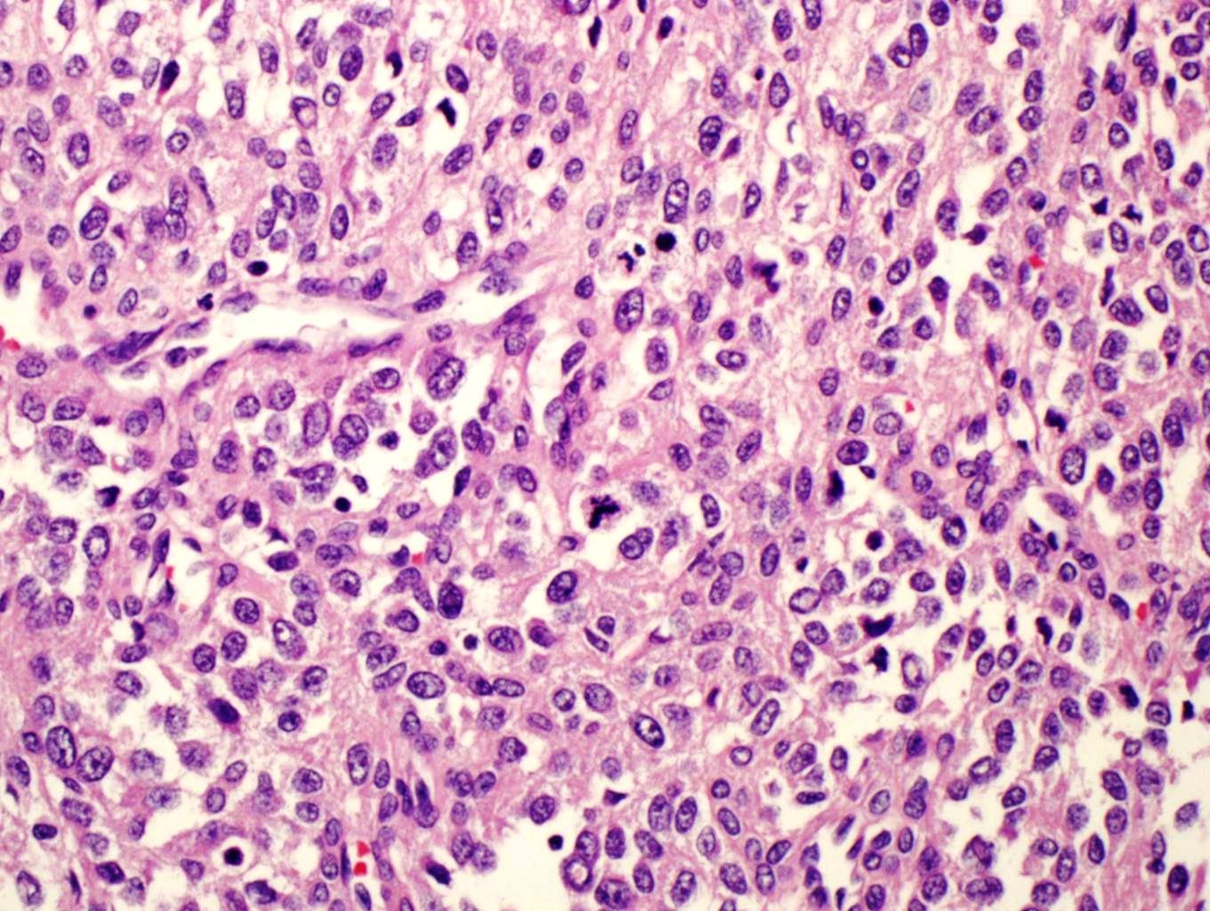
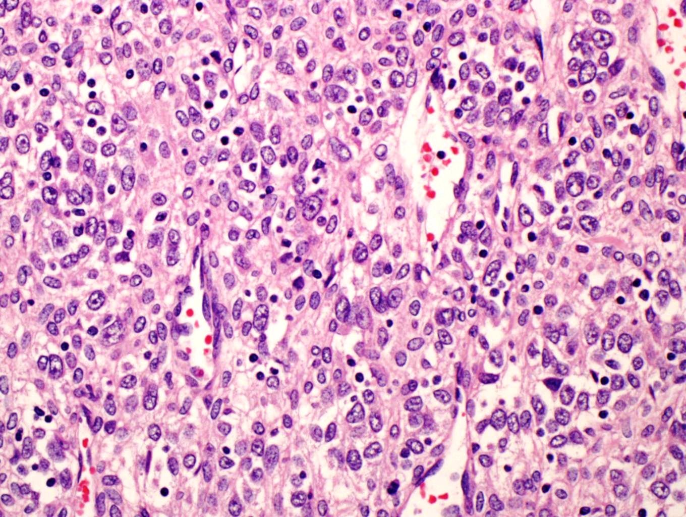
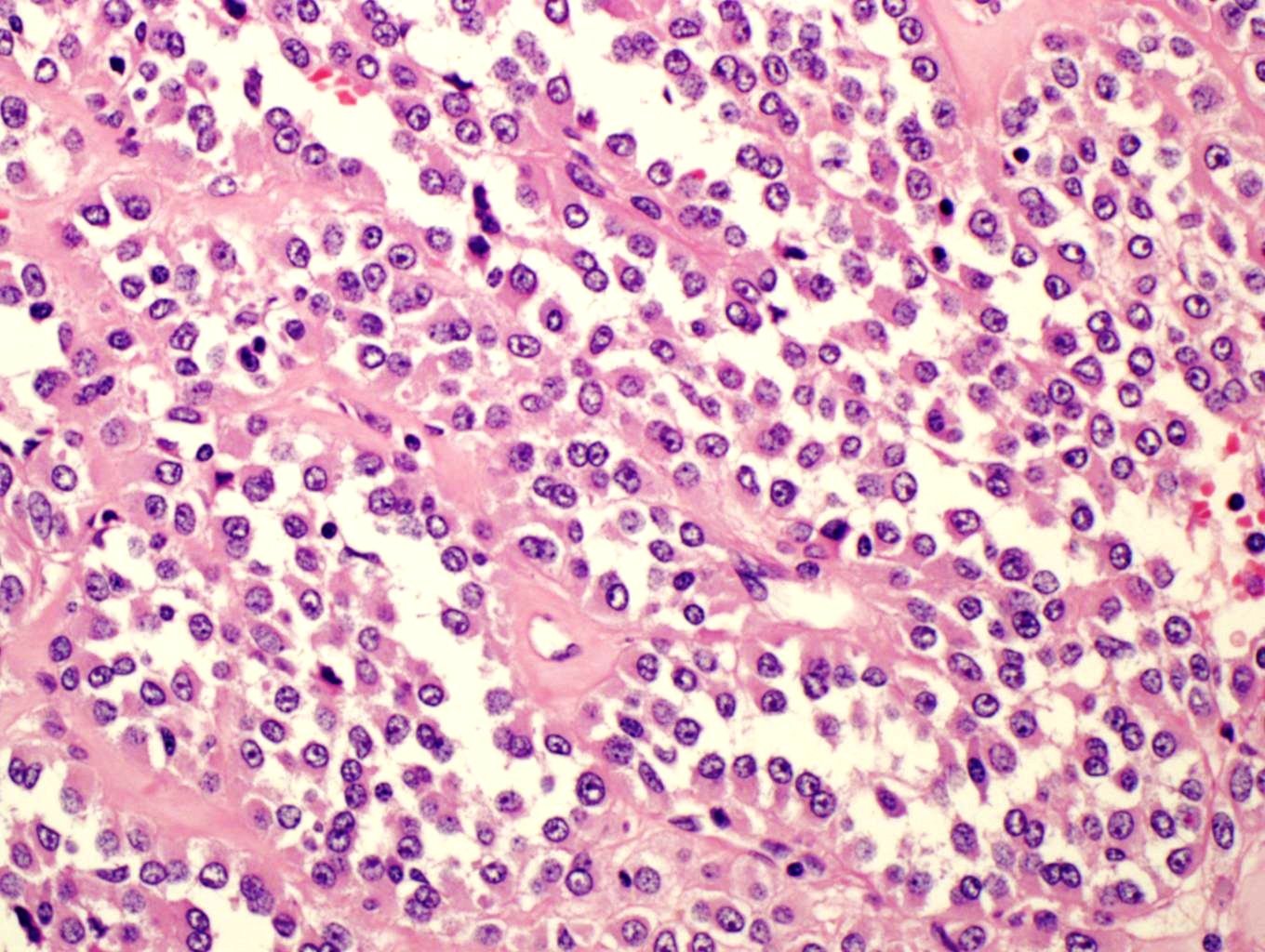
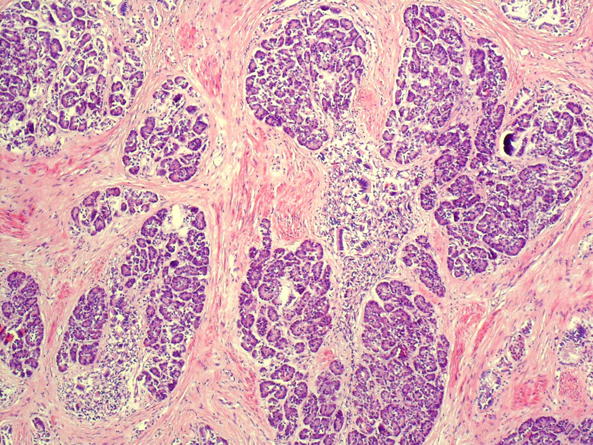
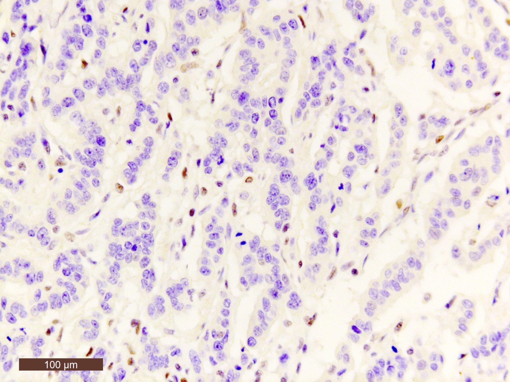
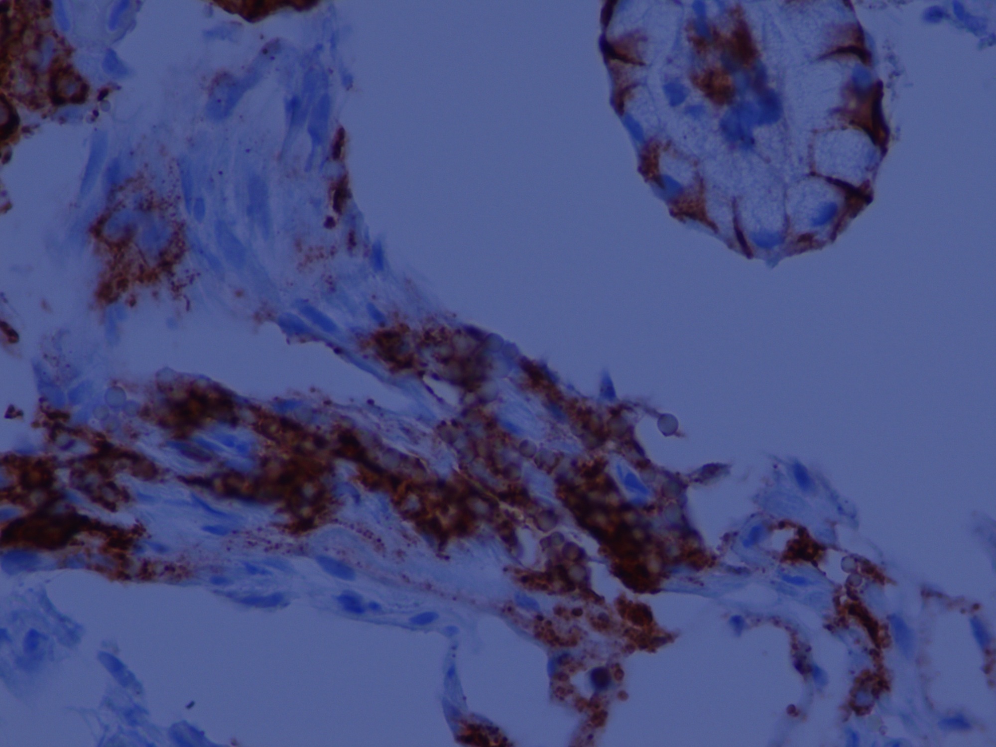
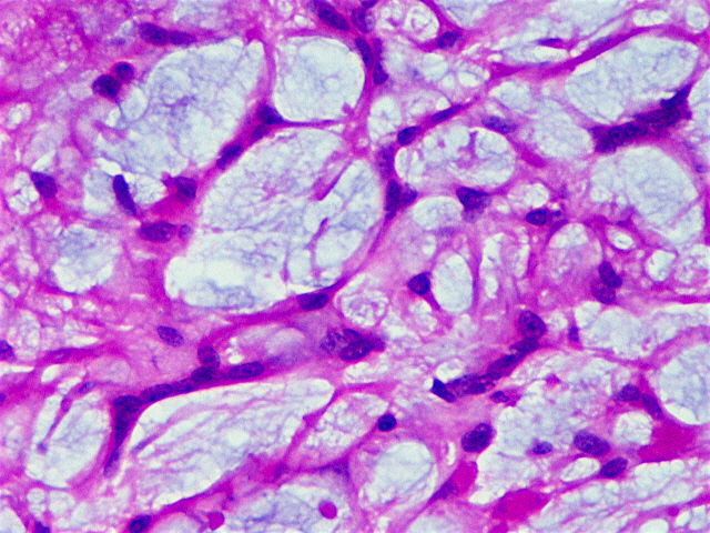
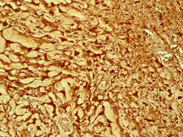
Nests of neuroendocrine tumor cells
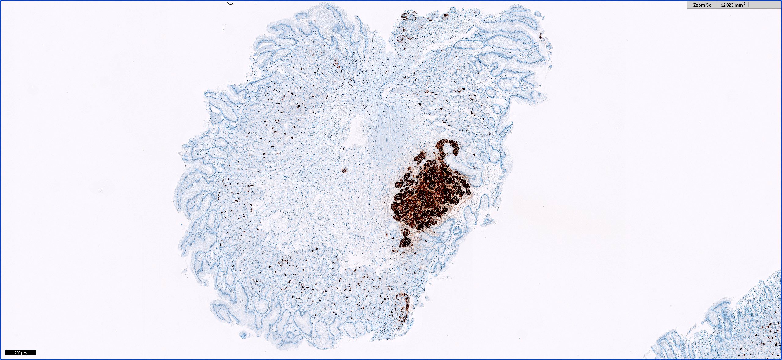
Chromogranin stain
Well differentiated neuroendocrine tumor
Well differentiated neuroendocrine tumor
Neuroendocrine nuttiness in the digestive system - Dr. Raul S. Gonzalez
GI neuroendocrine tumors classification - Dr. Vikram Deshpande
Images hosted on other servers:

Endoscopic yellow plaque-like lesion

Endoscopic yellow nodule and plaque-like lesion
Contributed by Aaron R. Huber, D.O.
Foamy macrophages within the lamina propria
CD68
Arnold: 2019

IARC: 2019
Lamps: 2015
Lauwers: 2021
Montgomery: 2017
Montgomery: 2018
Montgomery: 2017
Noffsinger: 2017
Odze: 2022
Srivastava: 2023
Yantiss: 2021
Find related Pathology books: GI, liver
