

Images hosted on other servers:

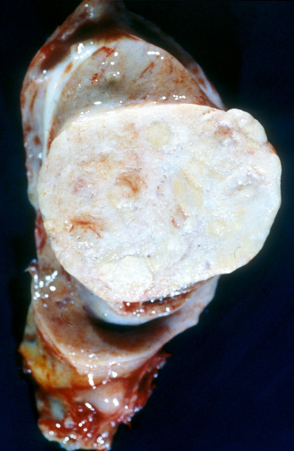
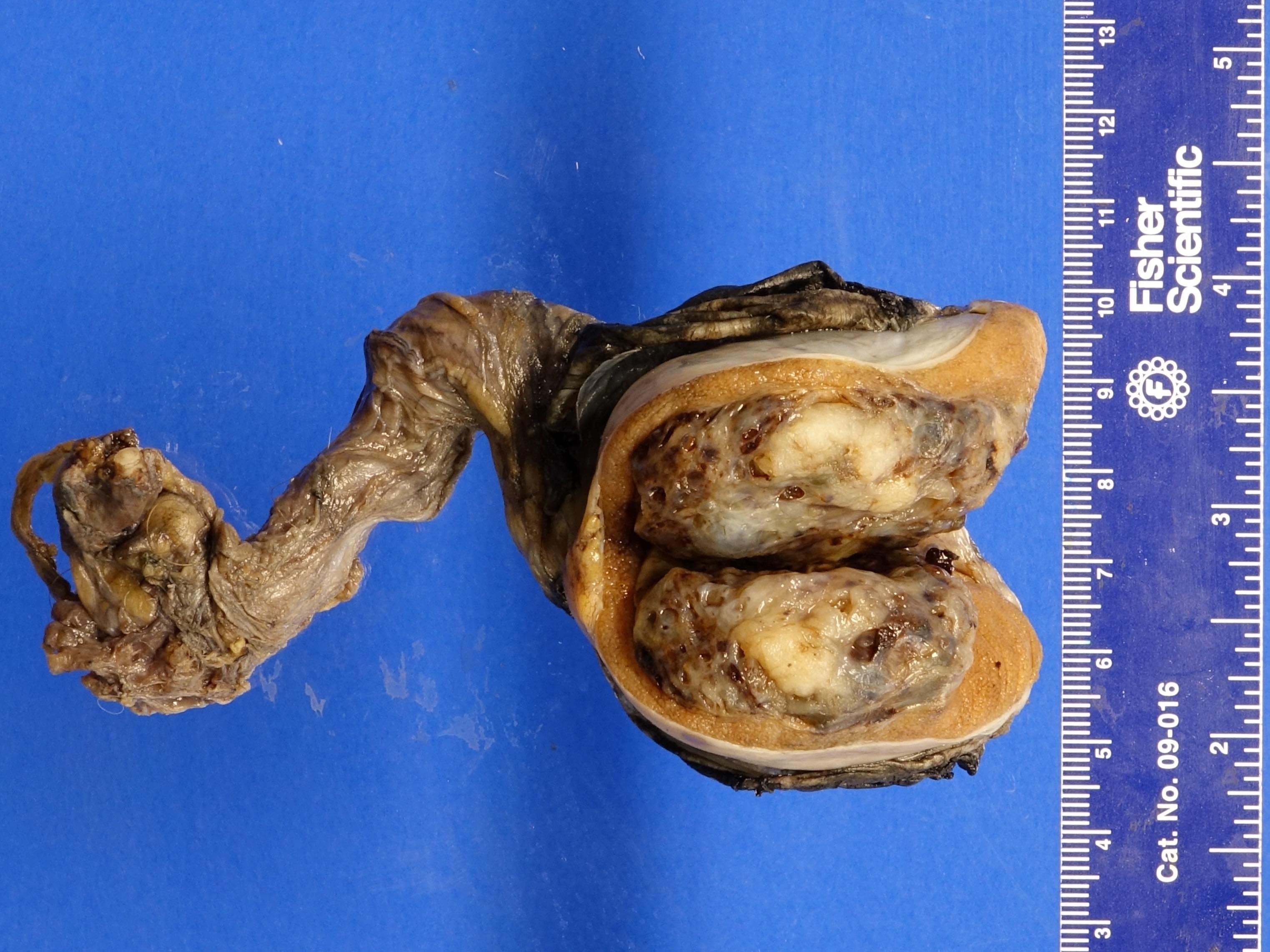
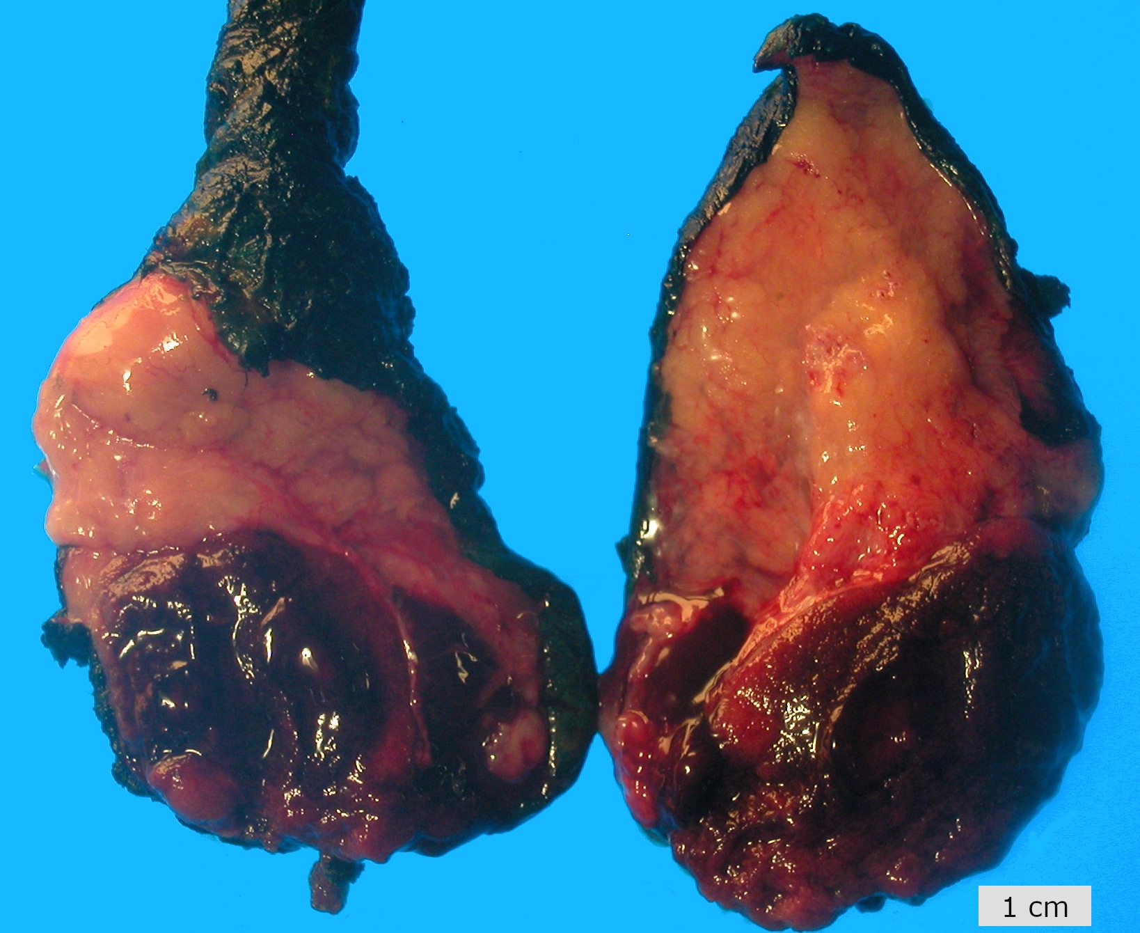
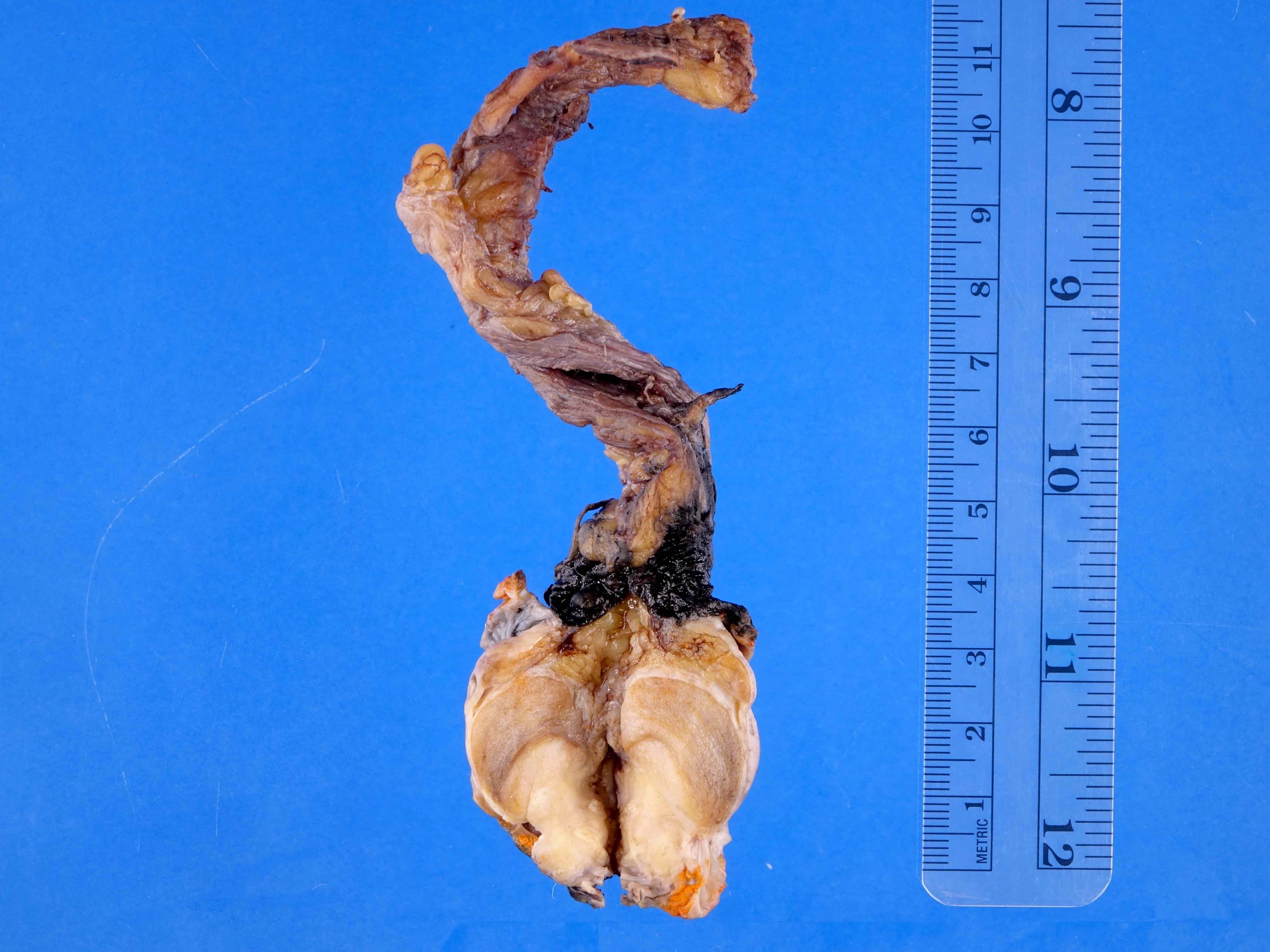
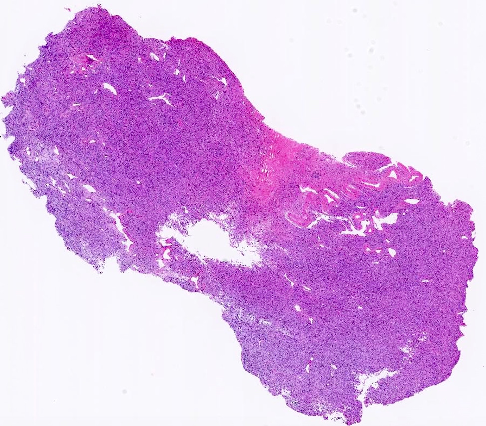
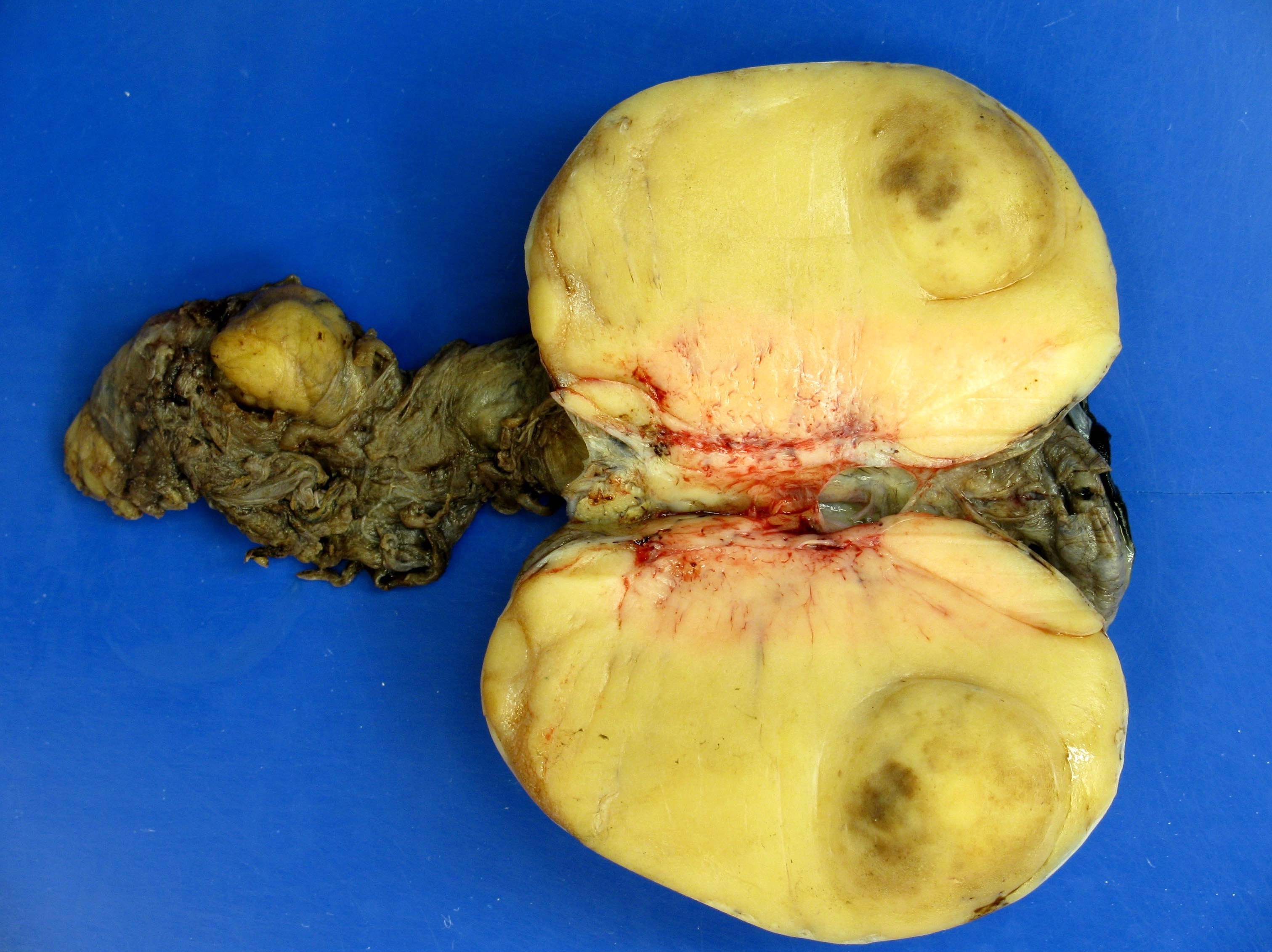
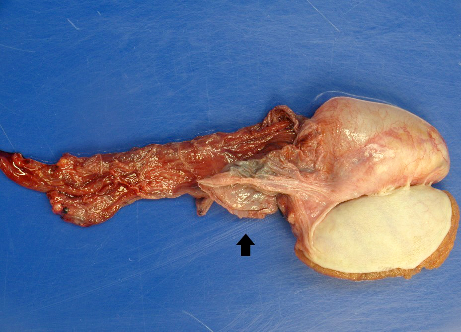
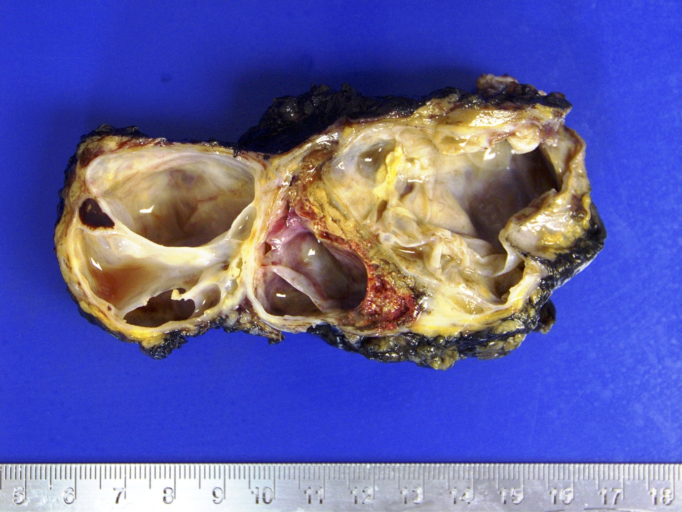
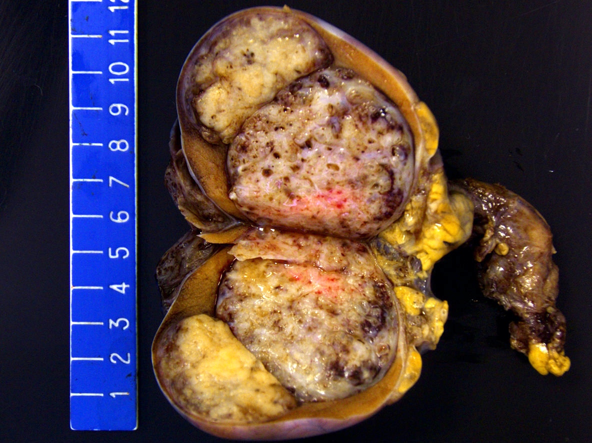
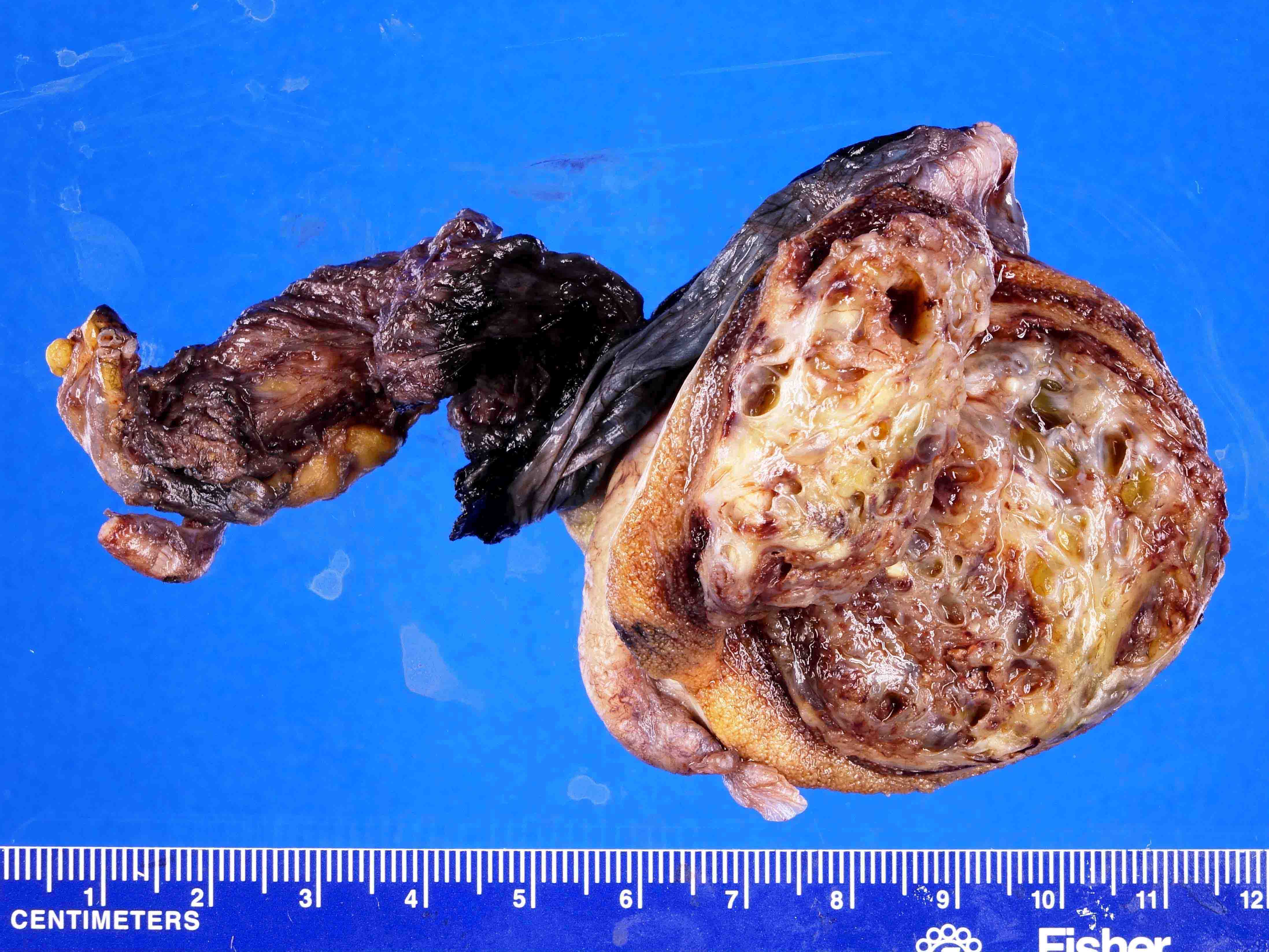
Mass in testicular hilum

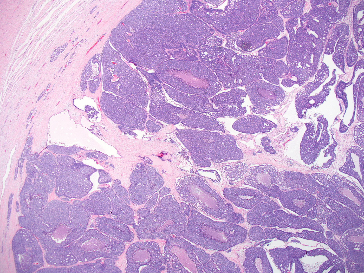
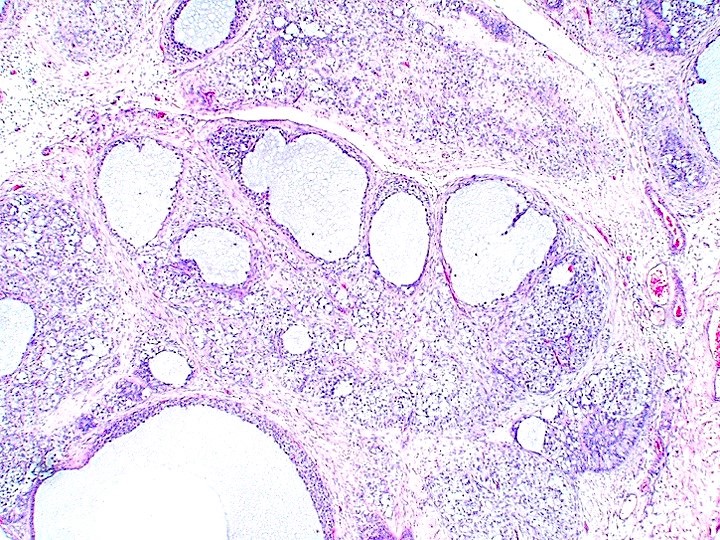
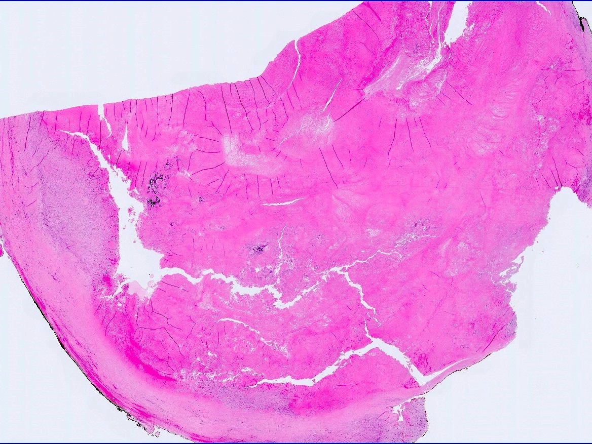
Solid mass with cystic areas
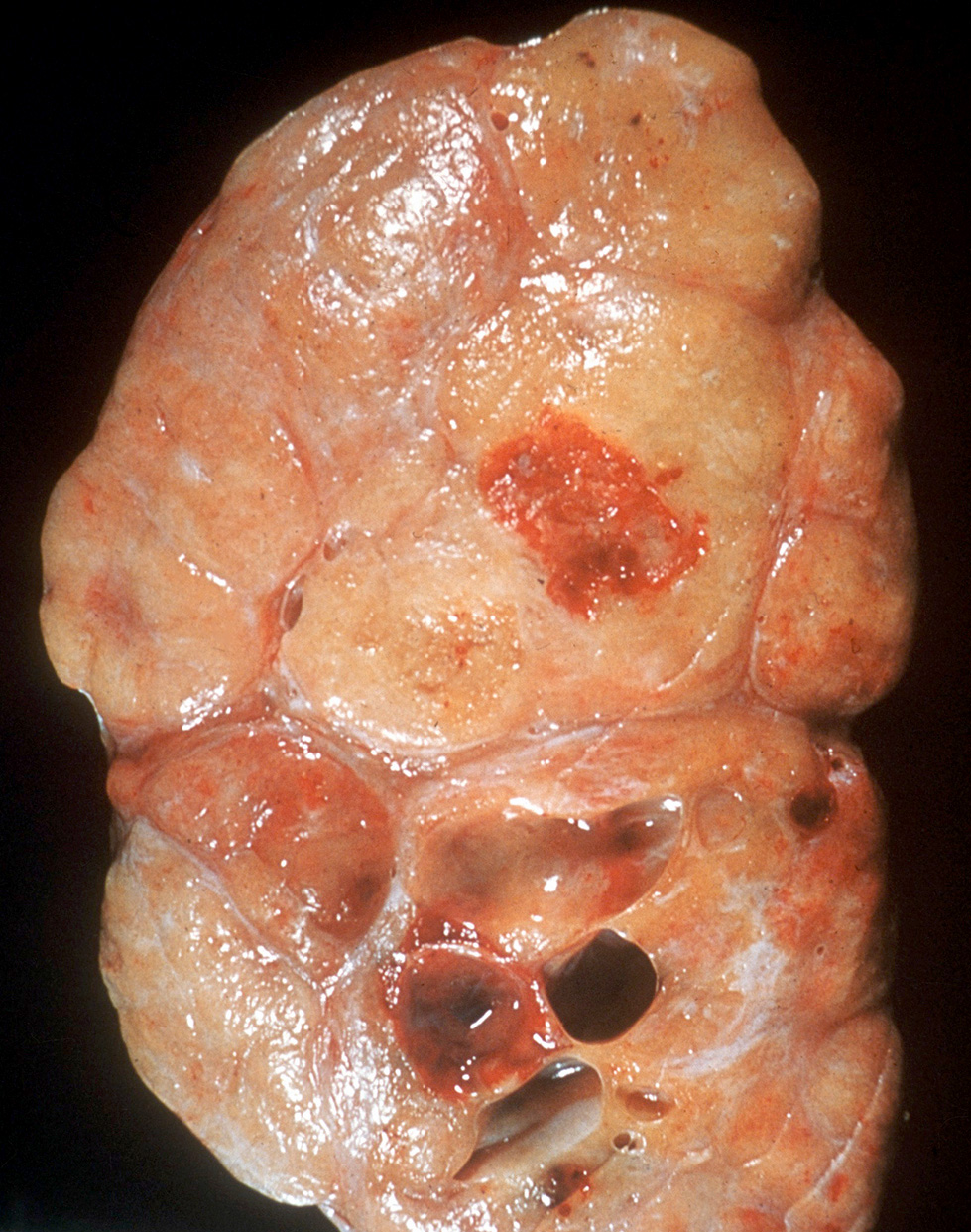
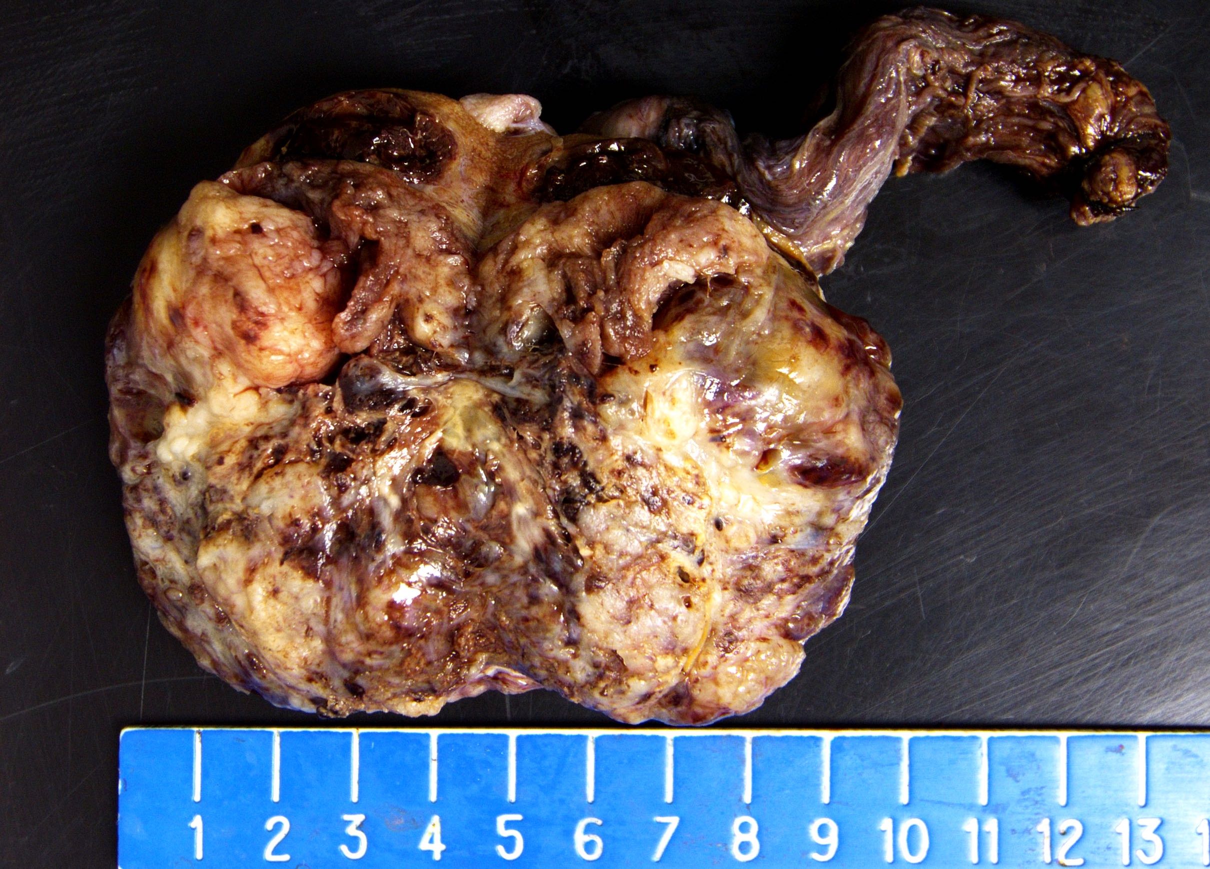
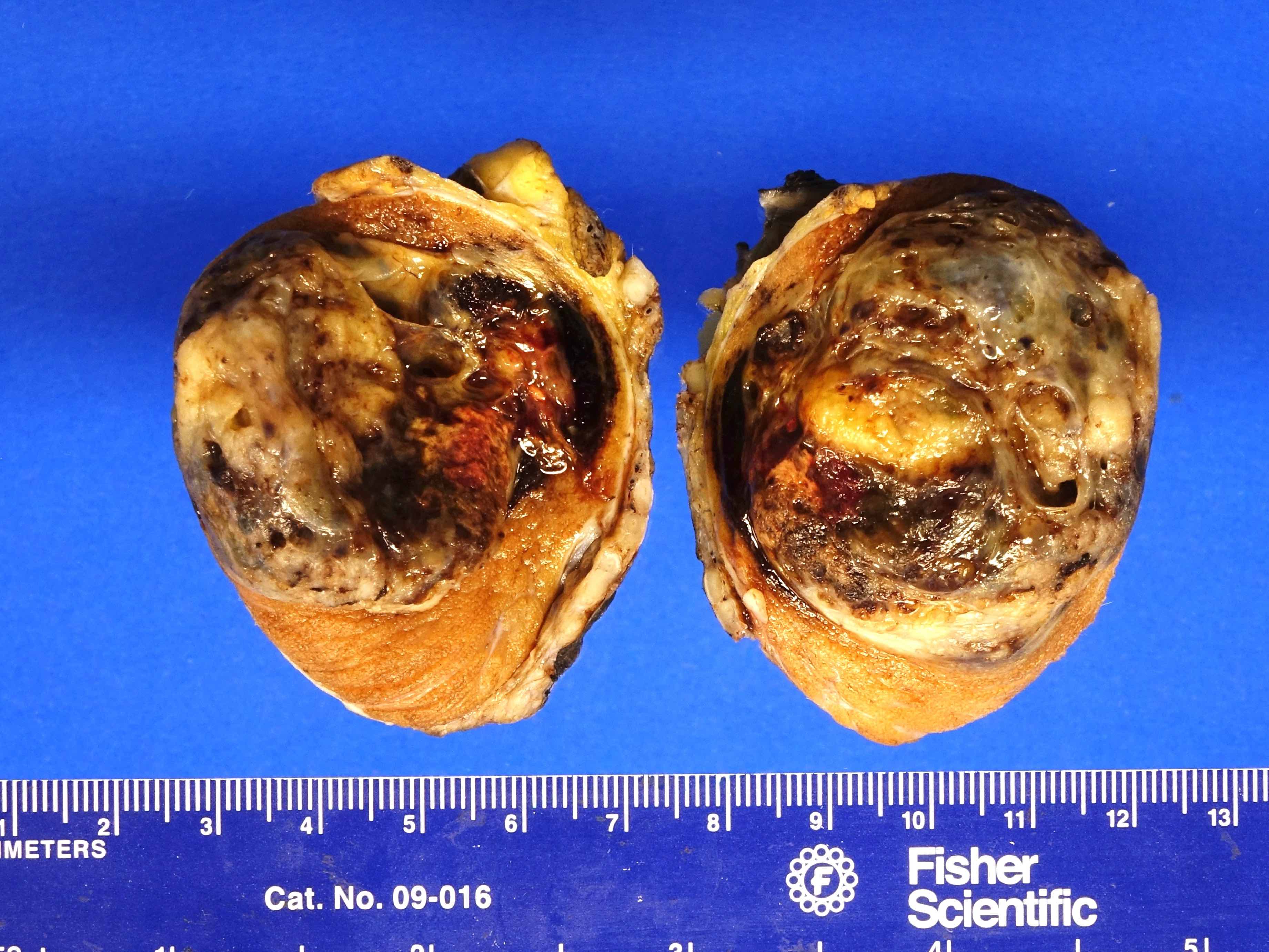
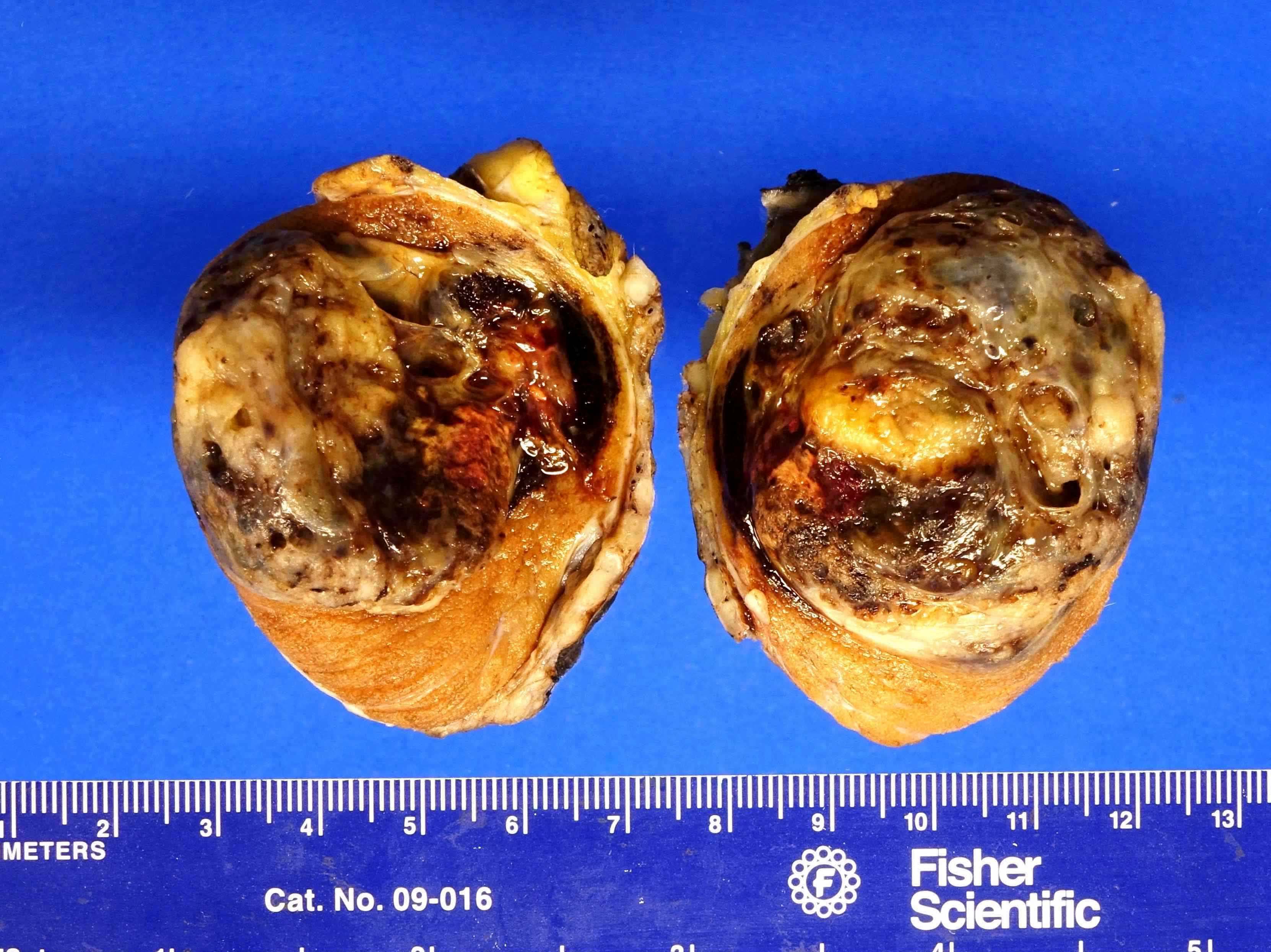
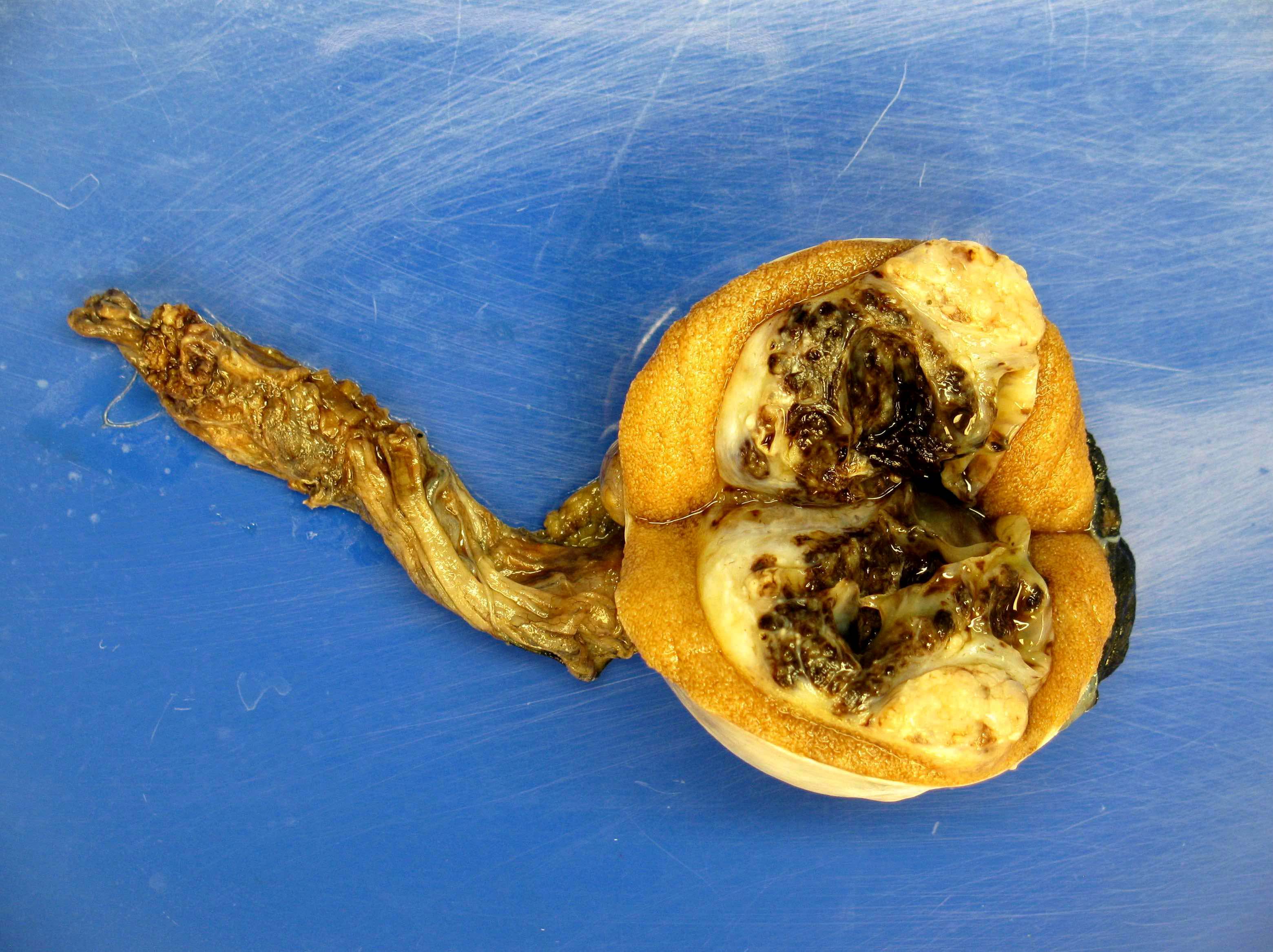
Contributed by Rafael E. Jimenez, M.D.
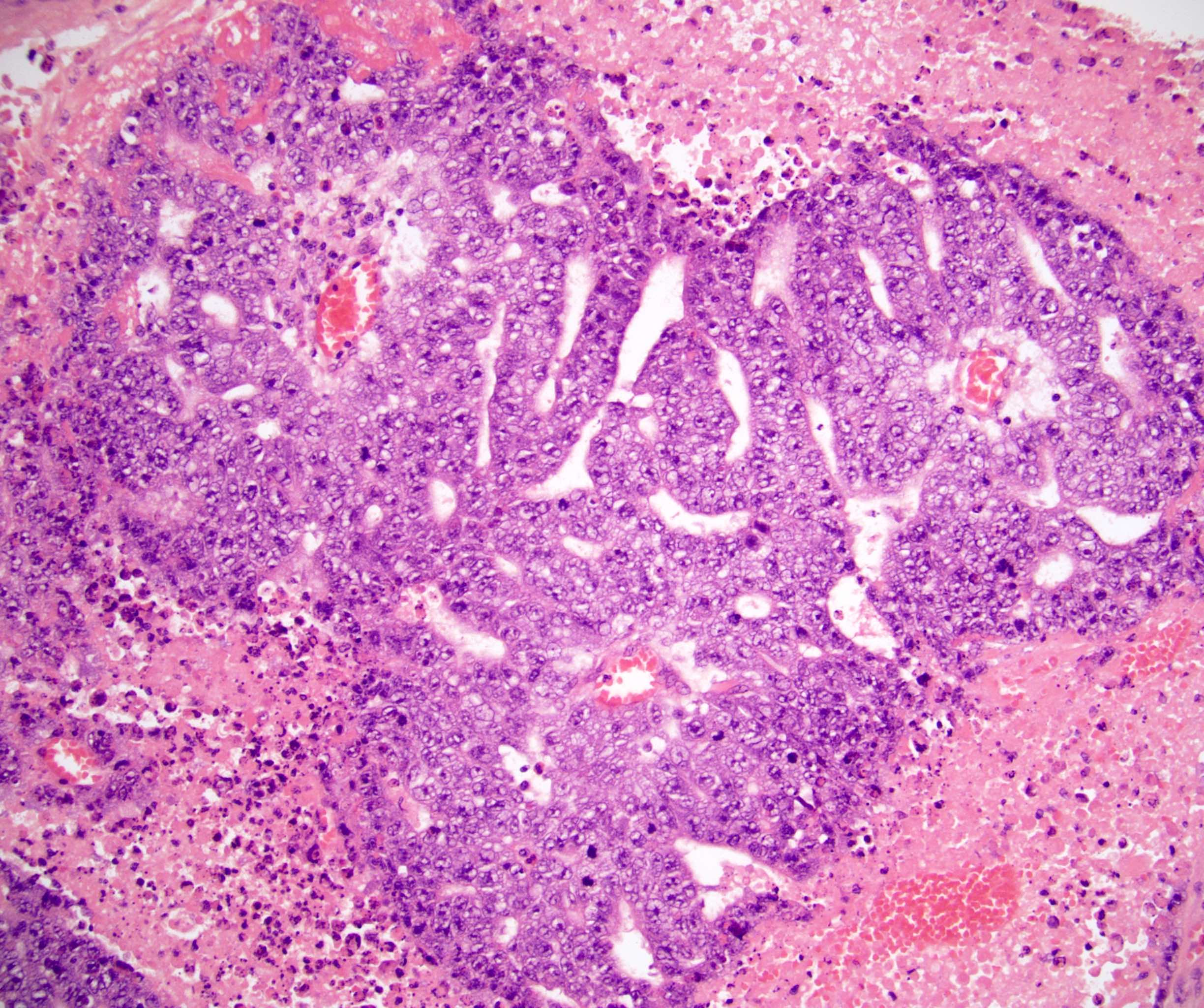
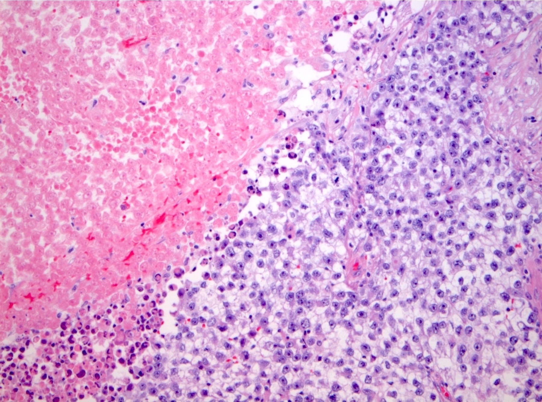
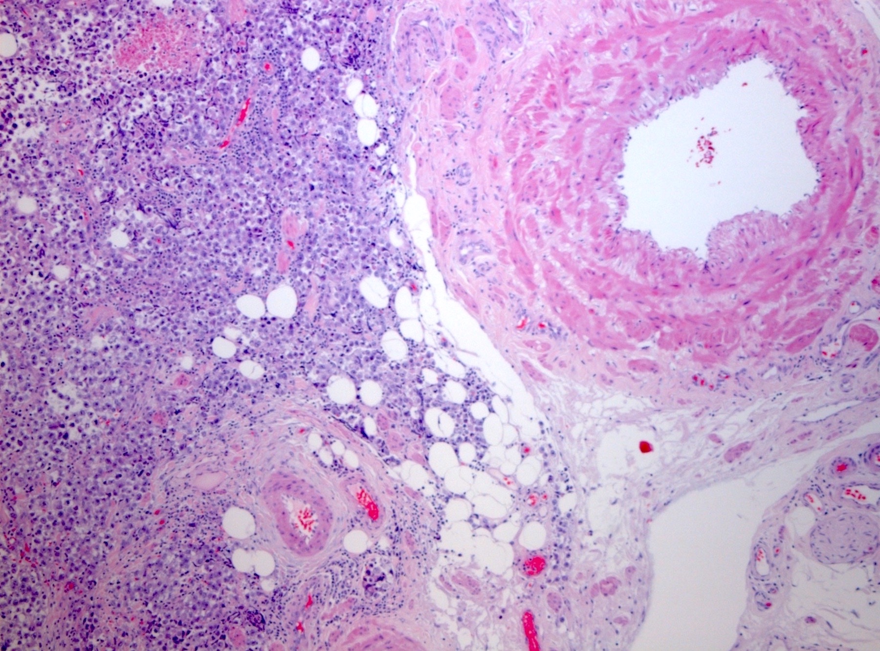
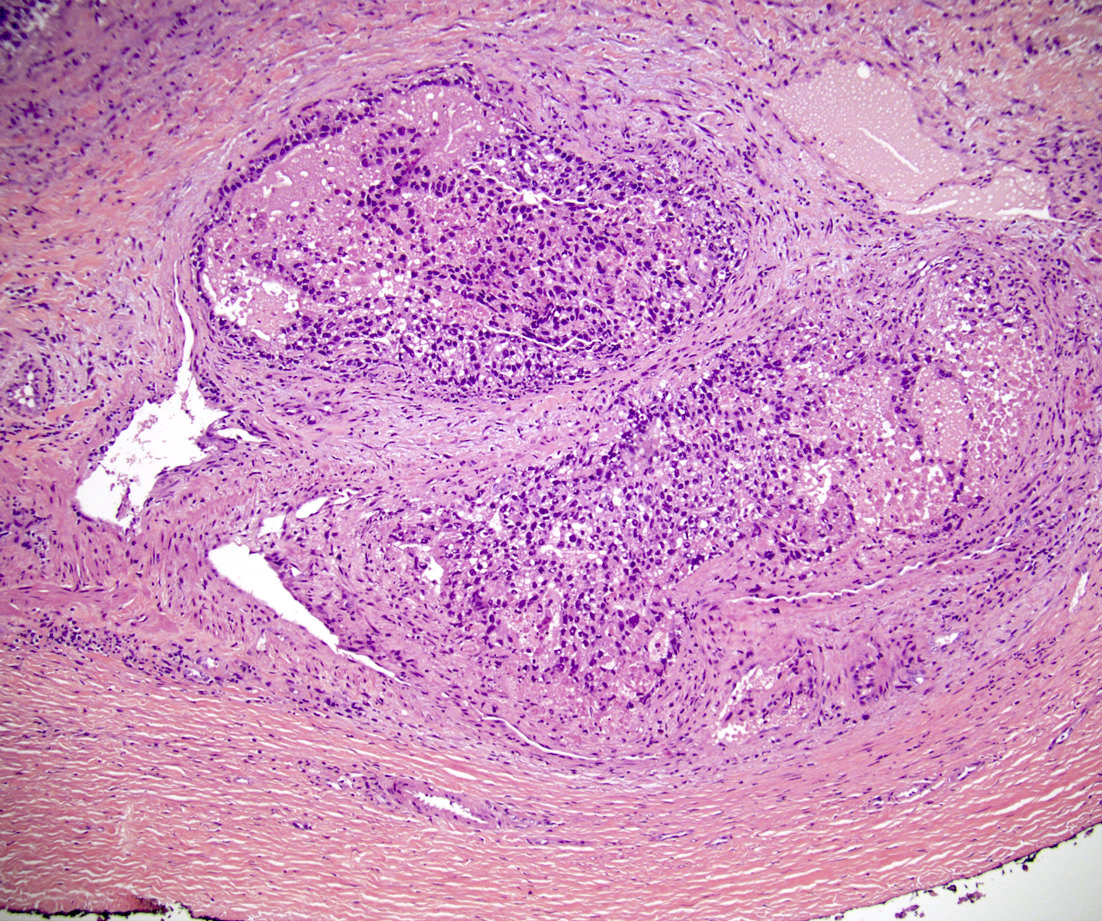
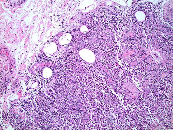
Areas of central necrosis
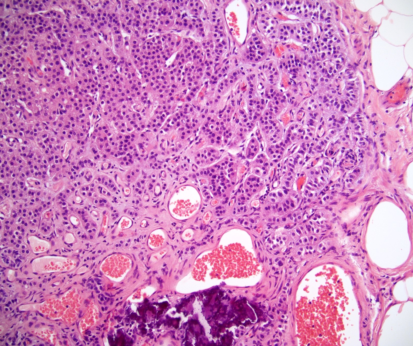
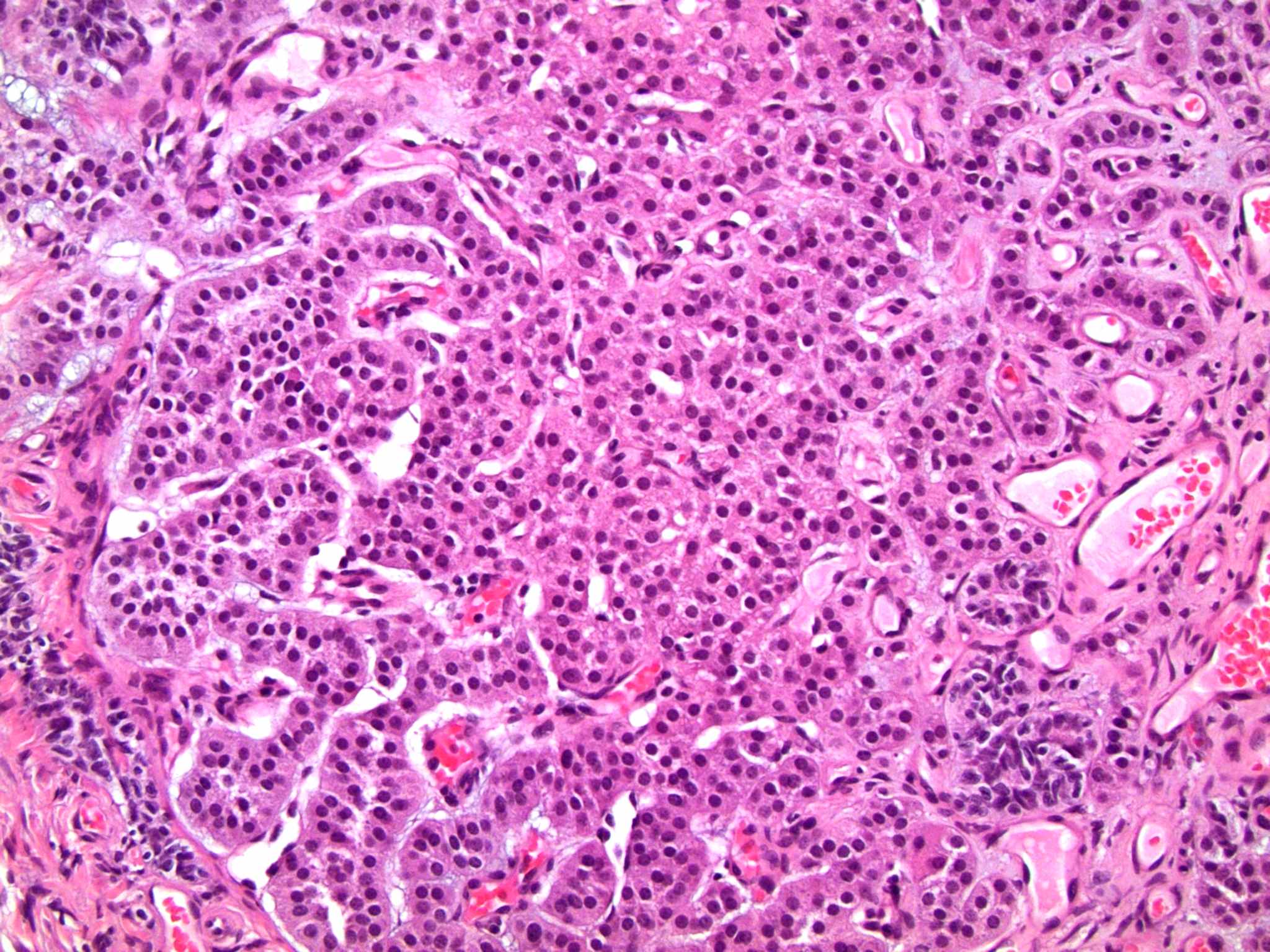
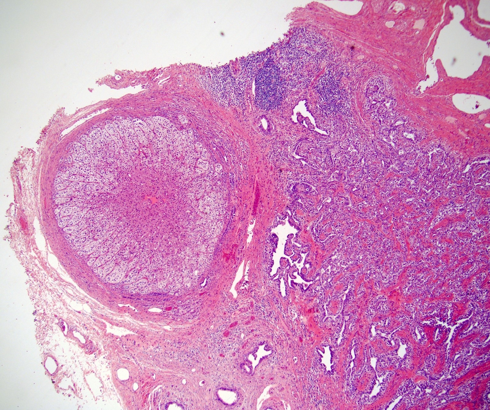
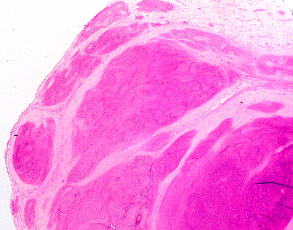
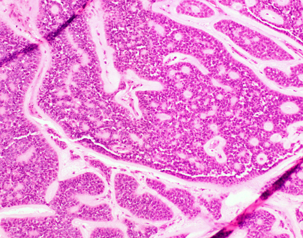
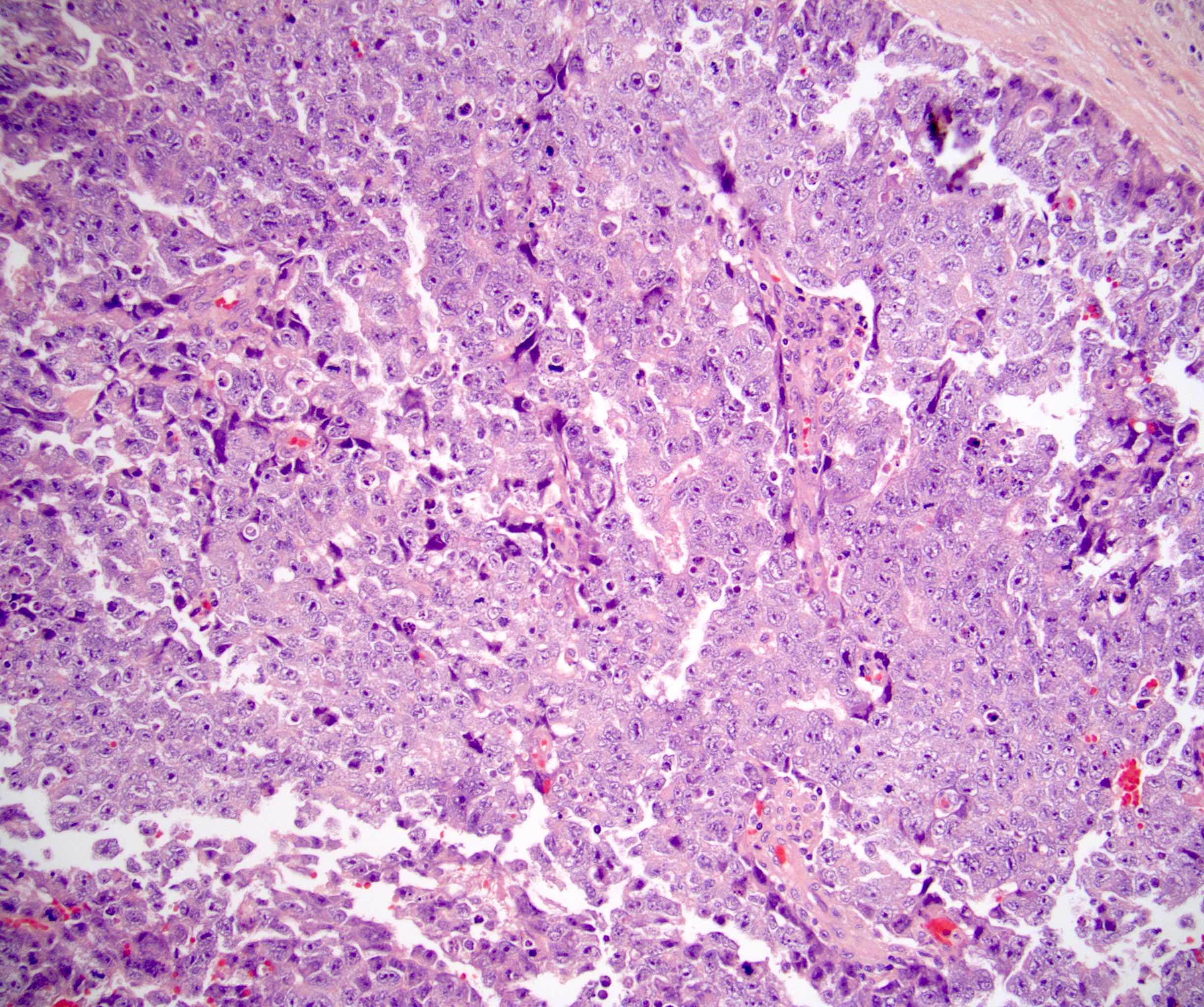
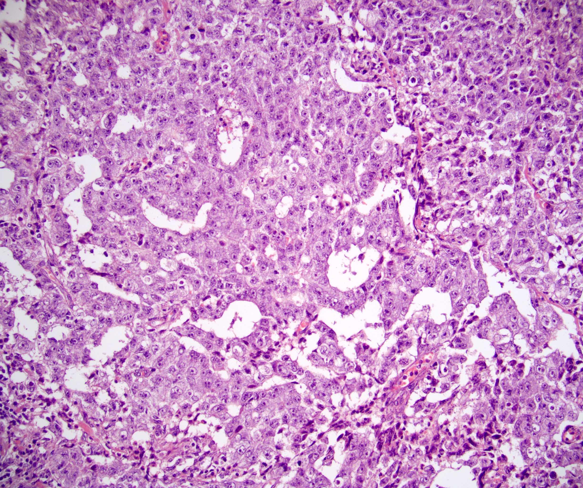
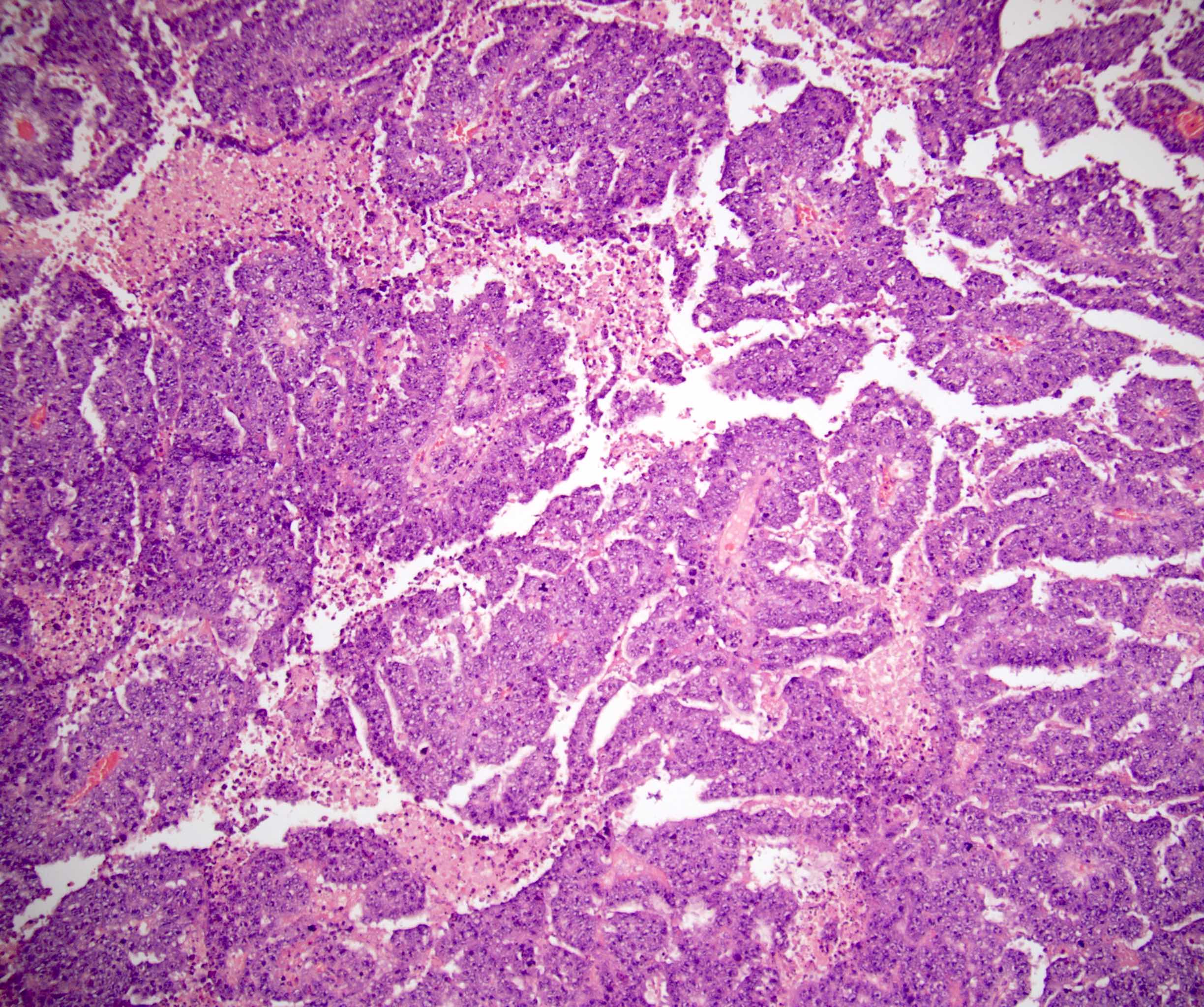
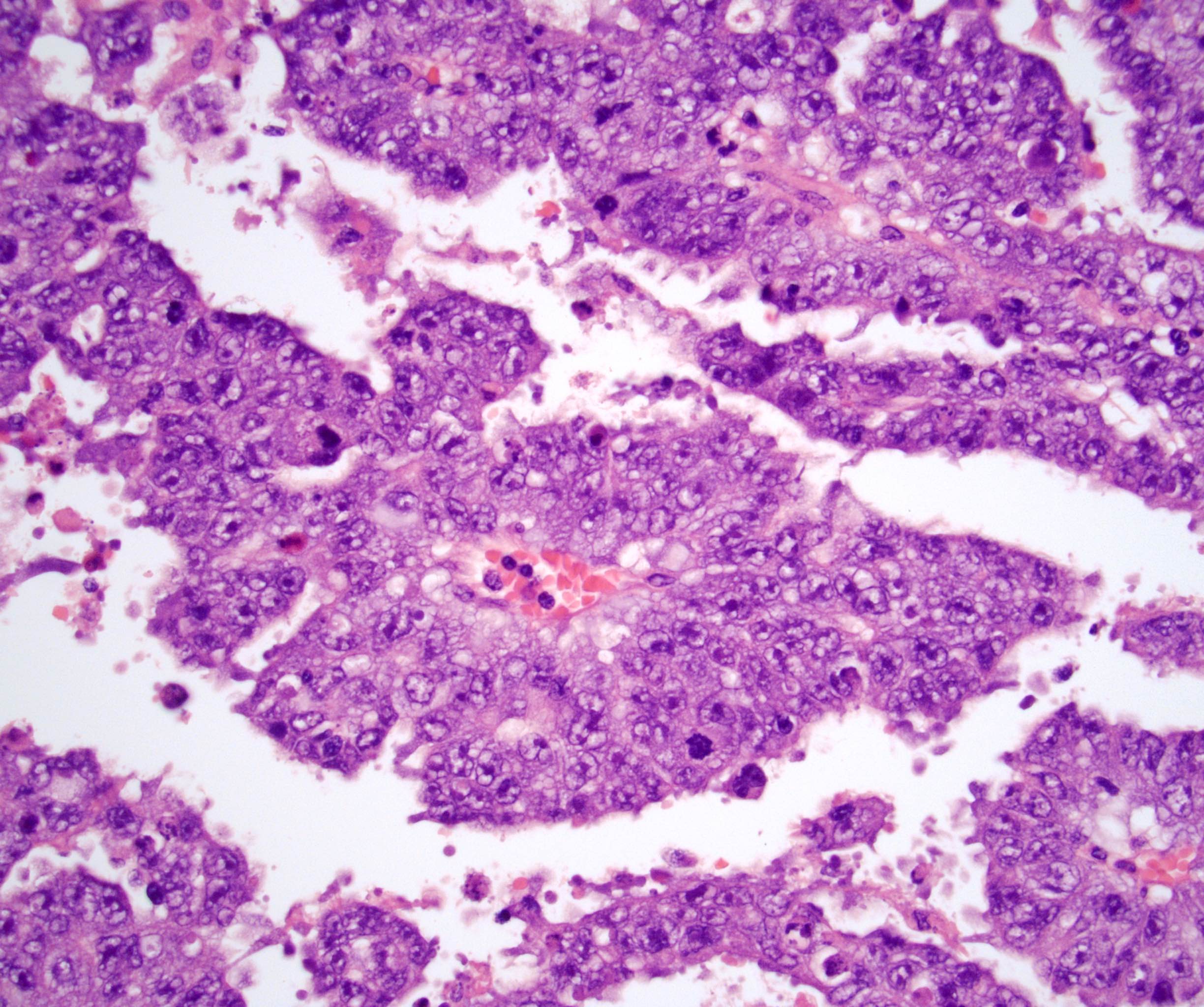
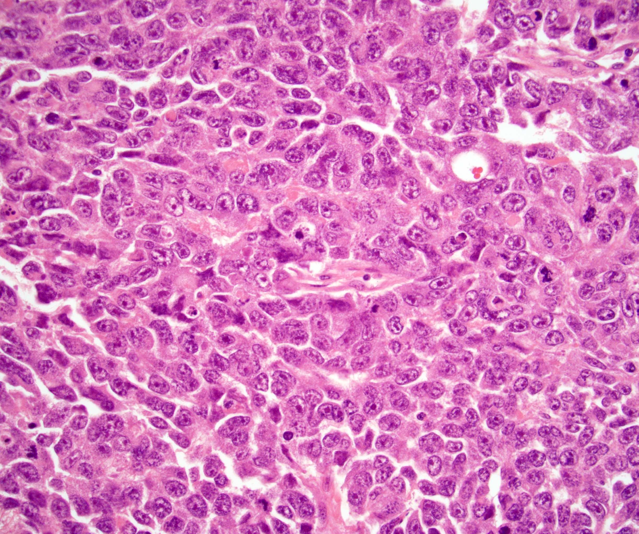
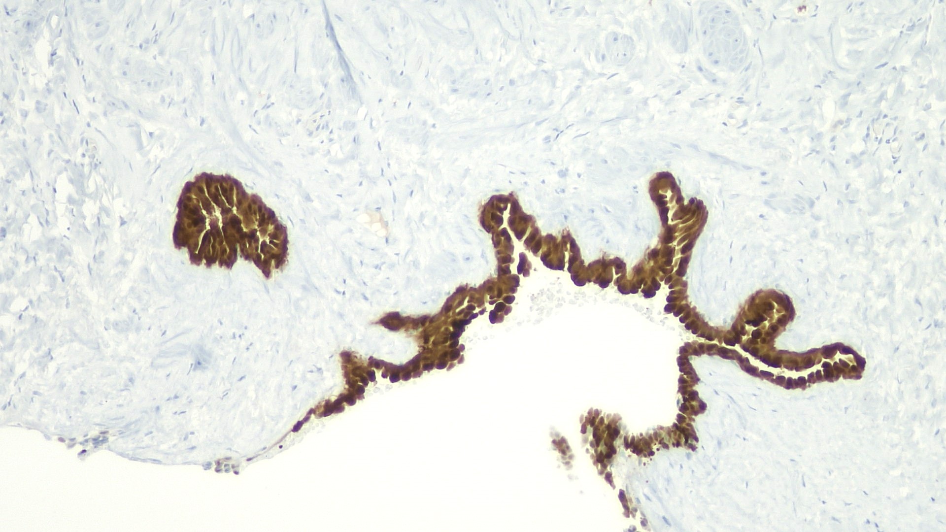
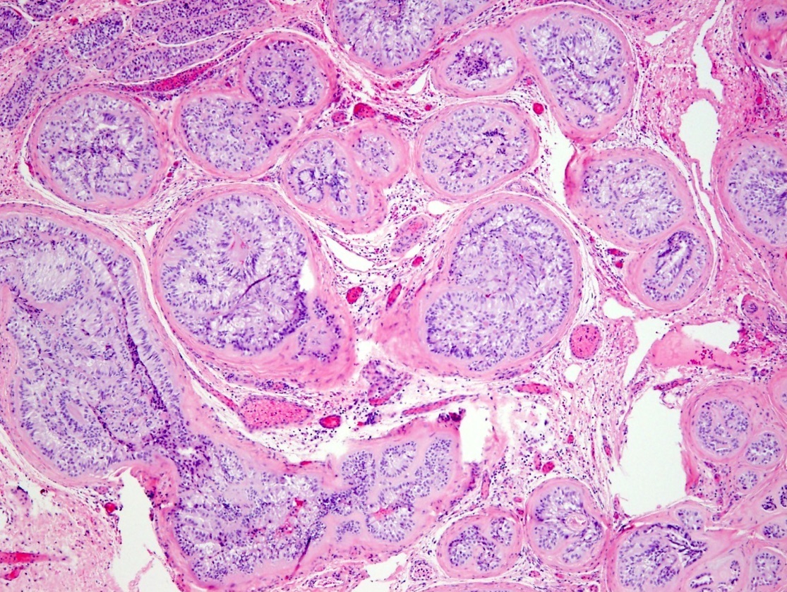
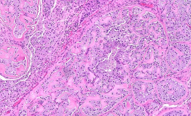
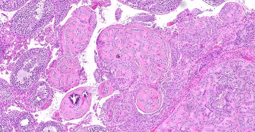
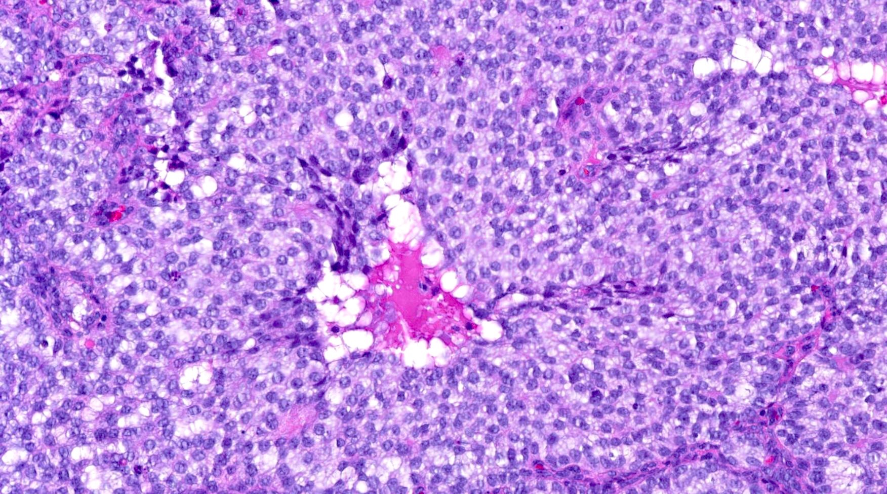
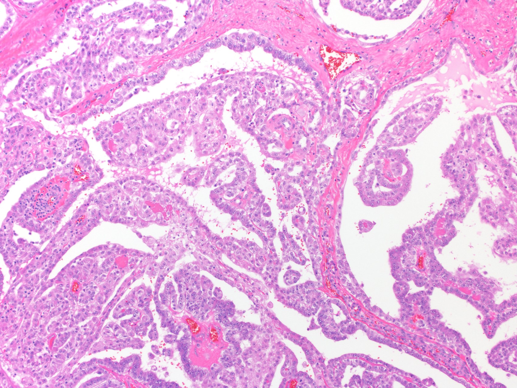
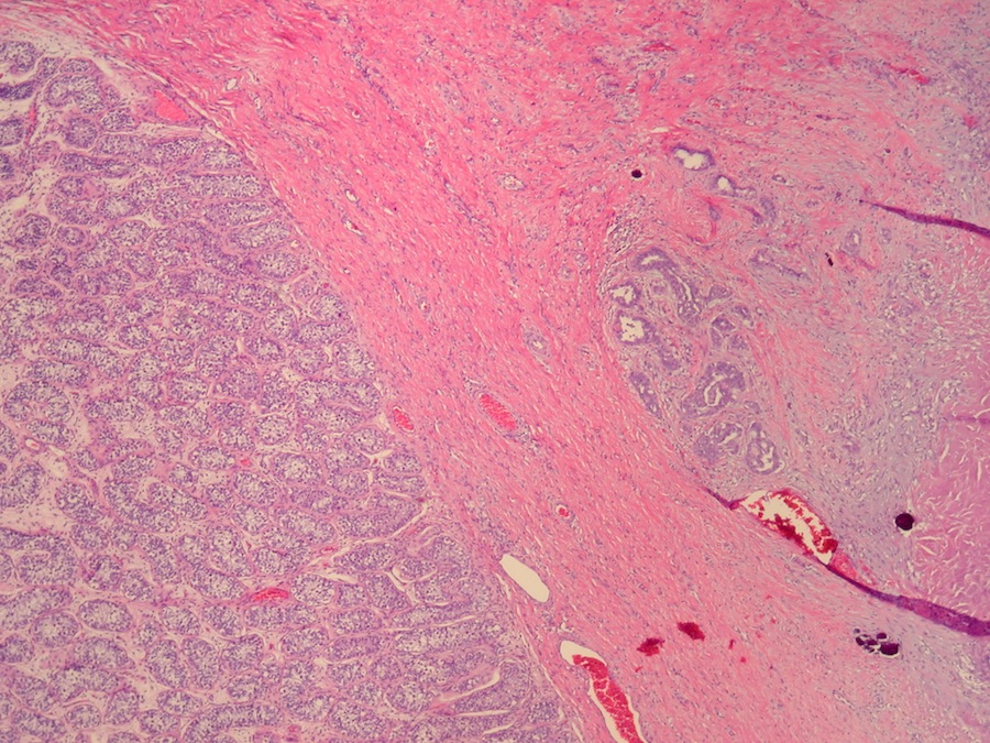
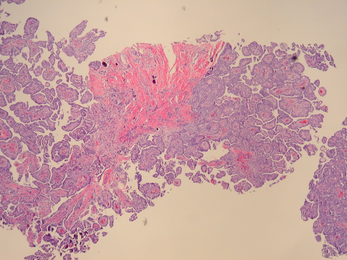
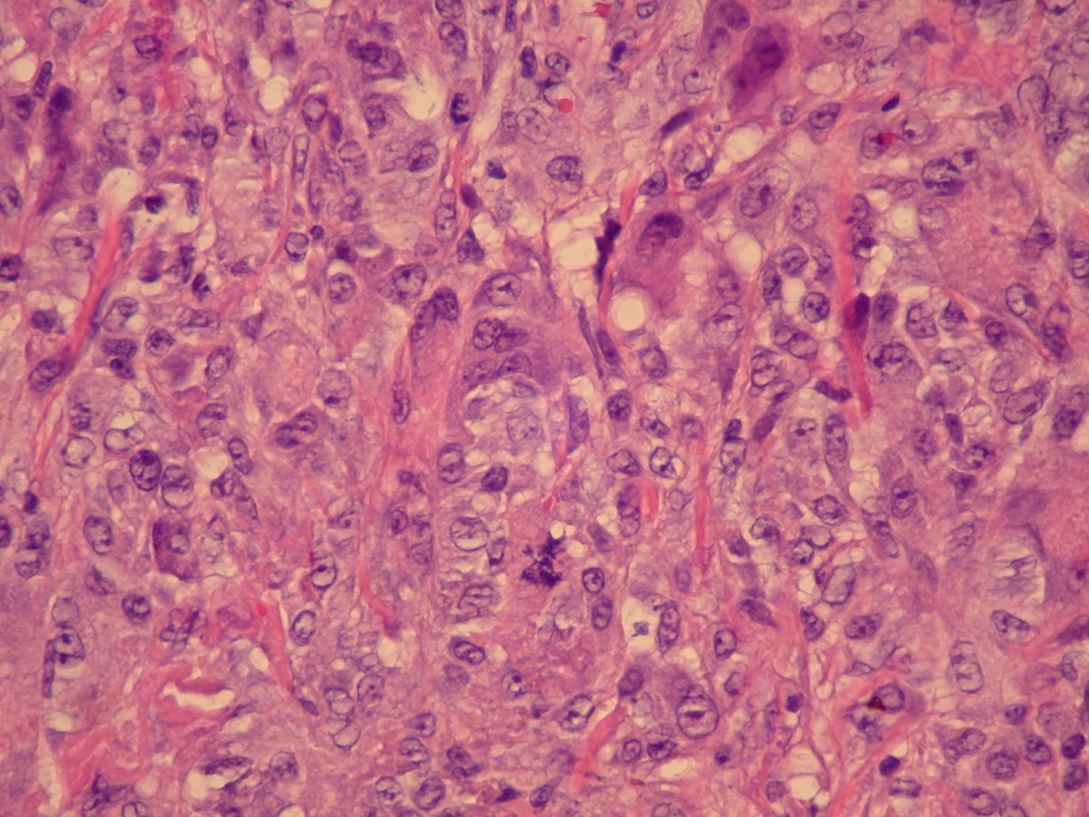
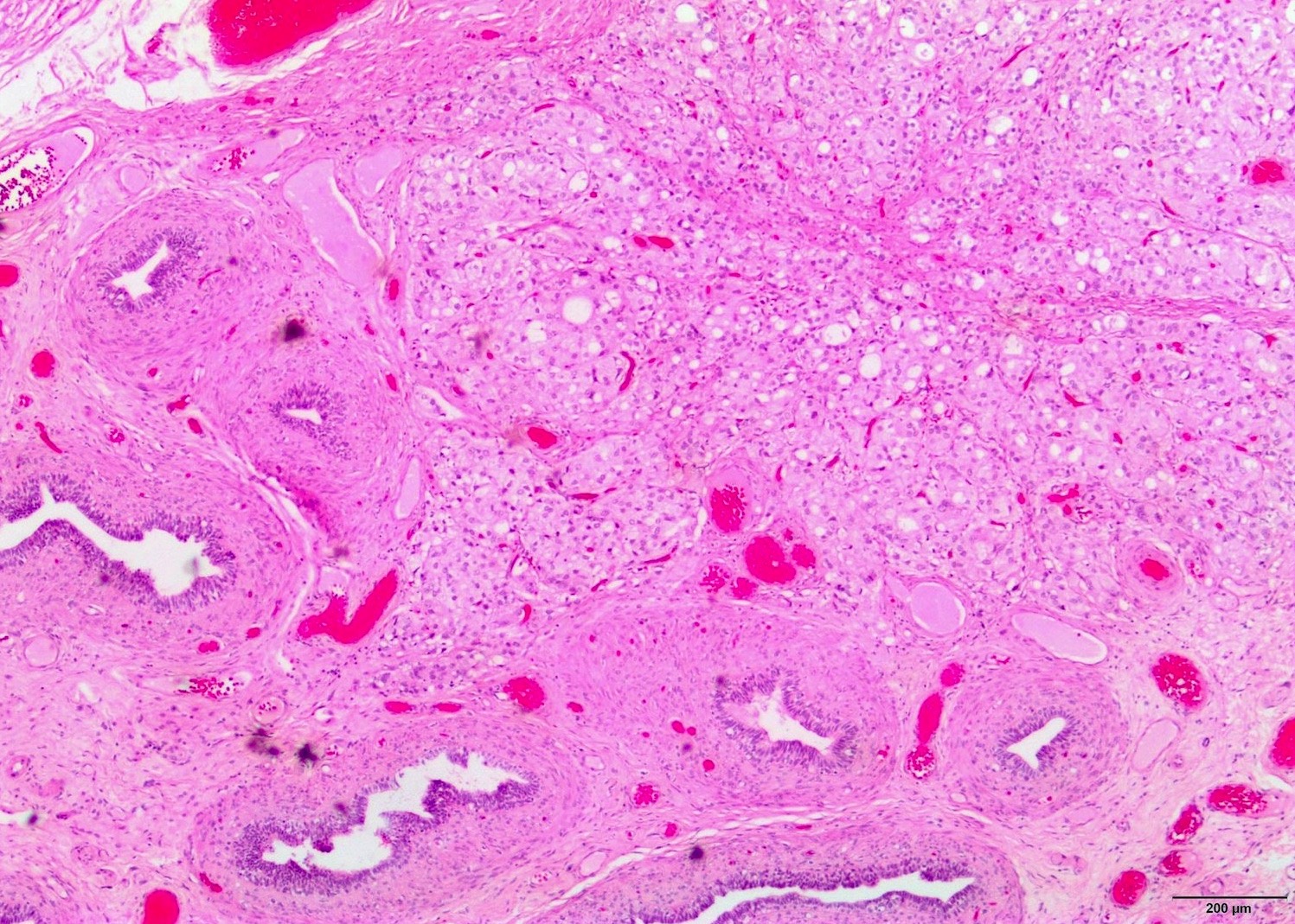
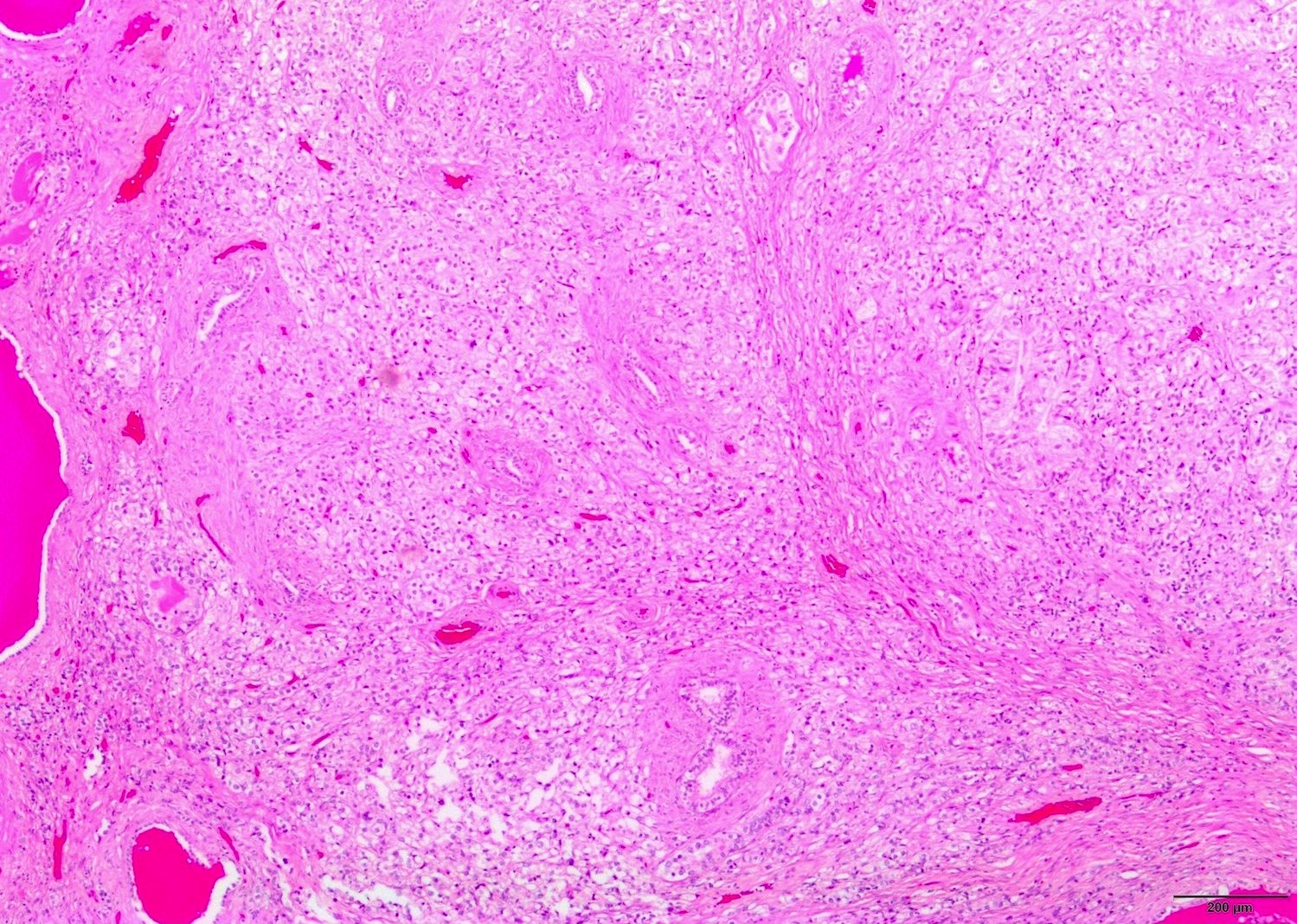
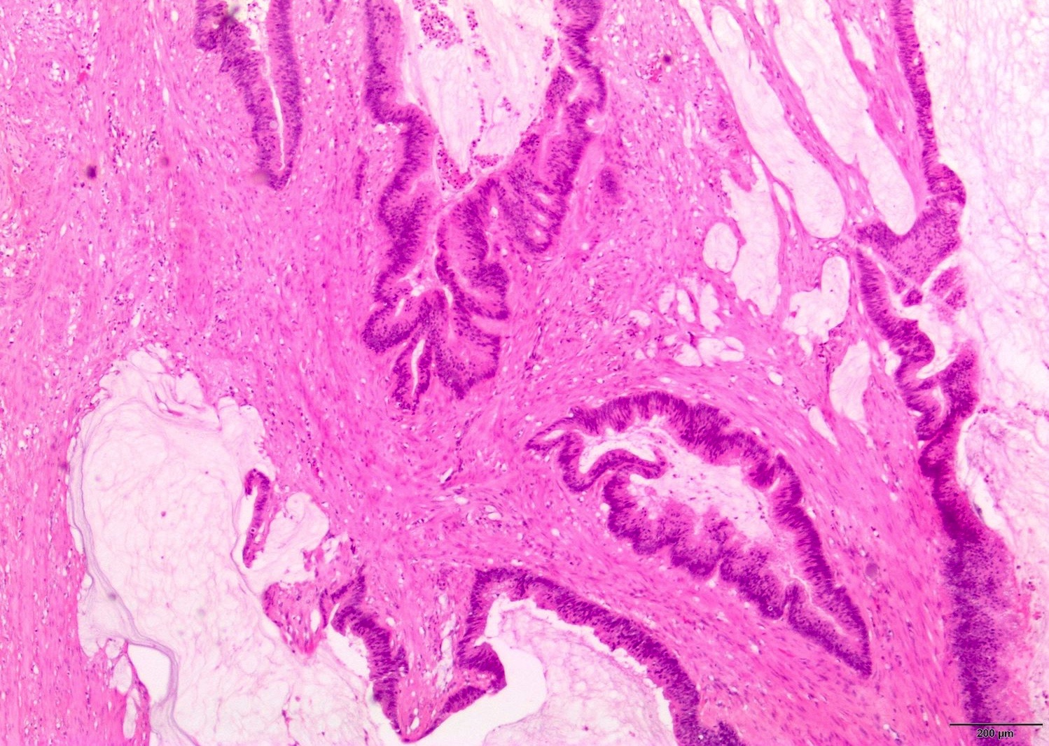
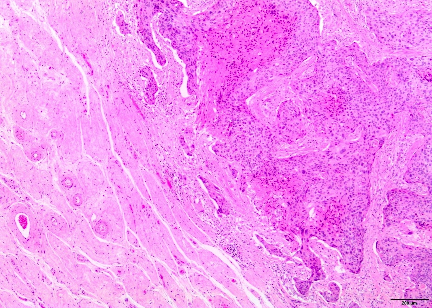
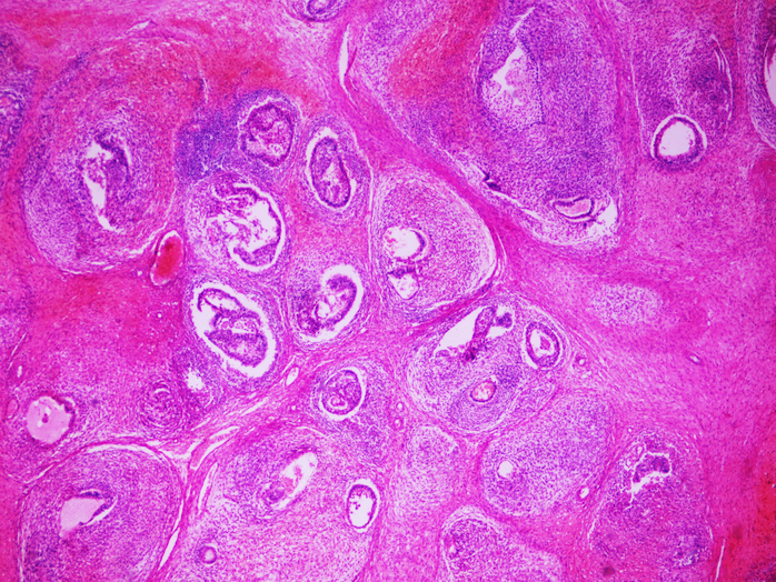
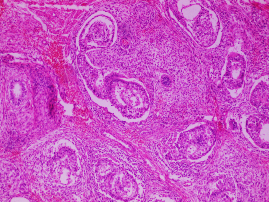
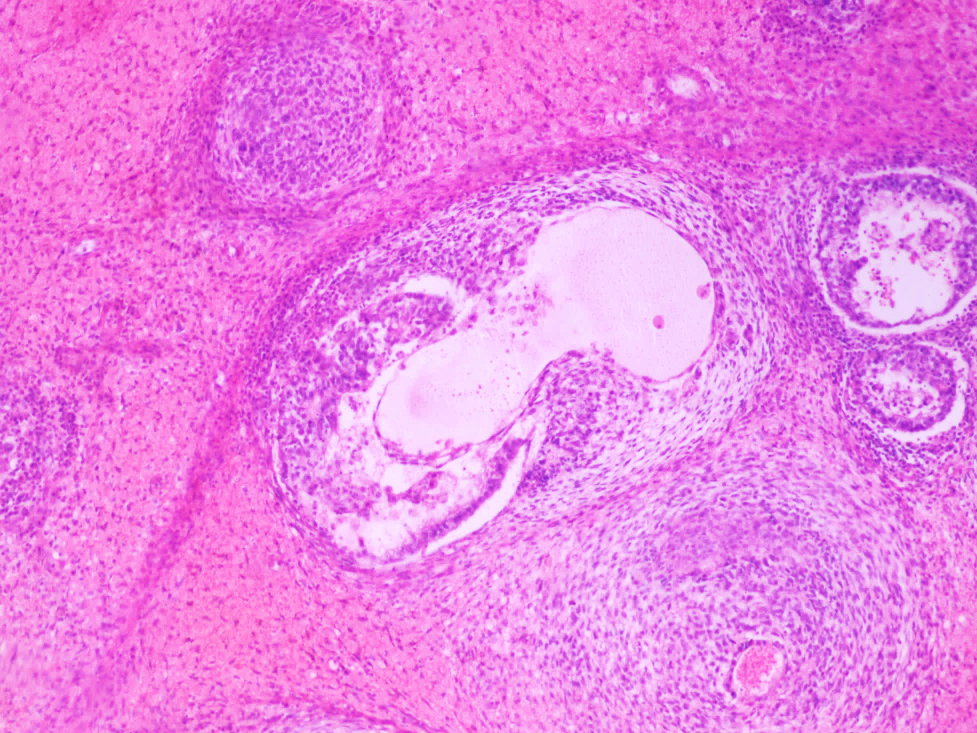
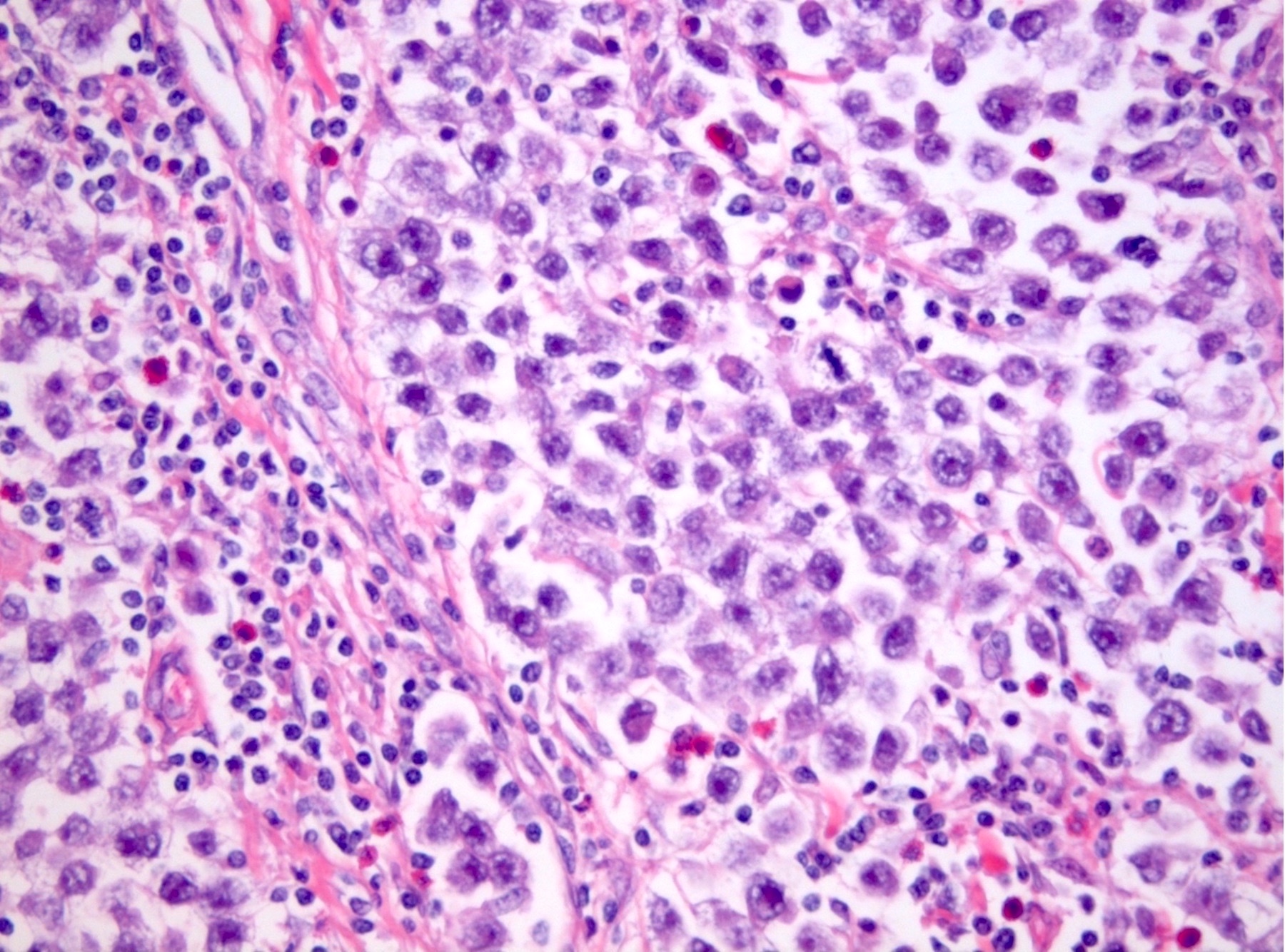
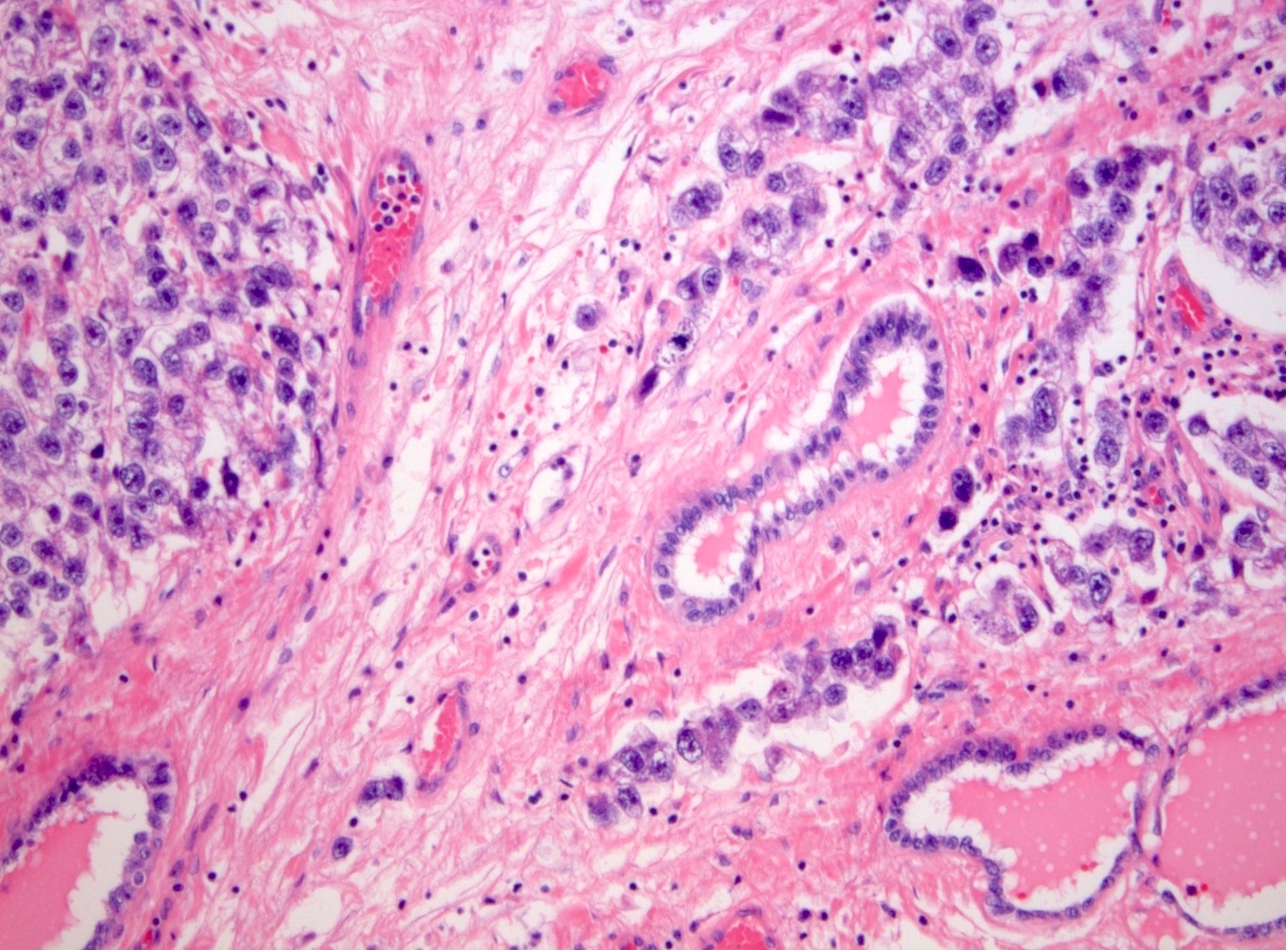
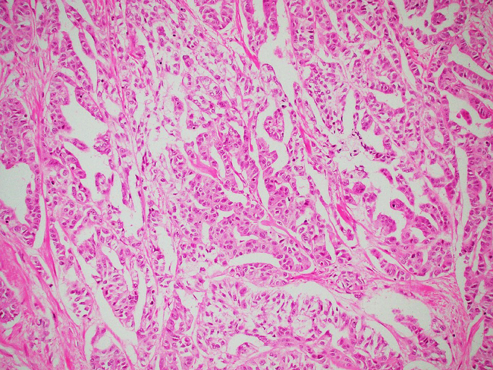
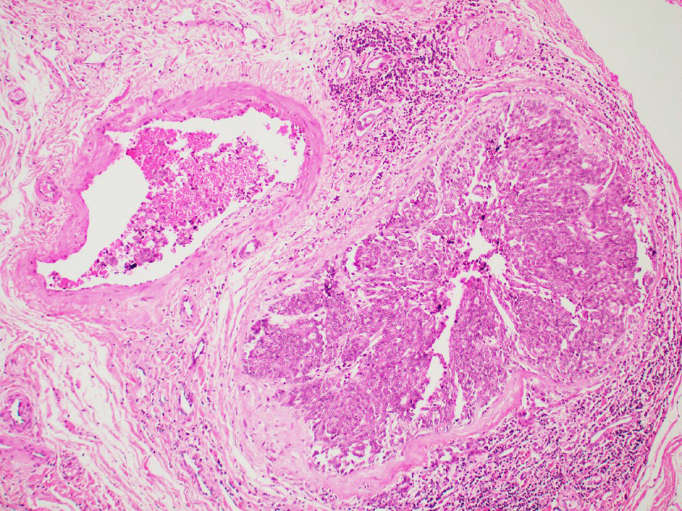
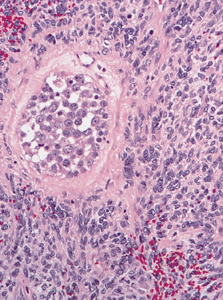
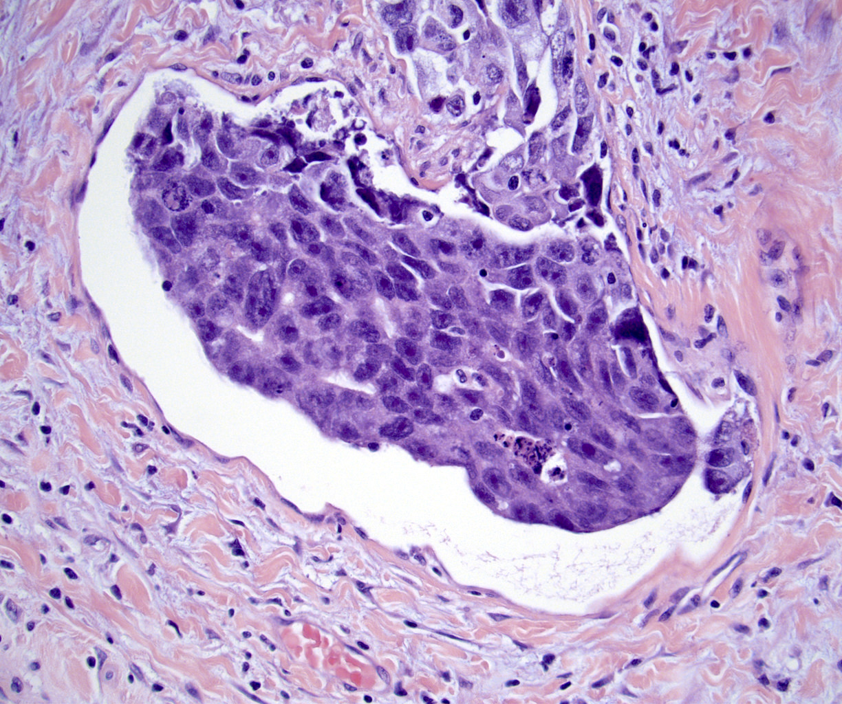
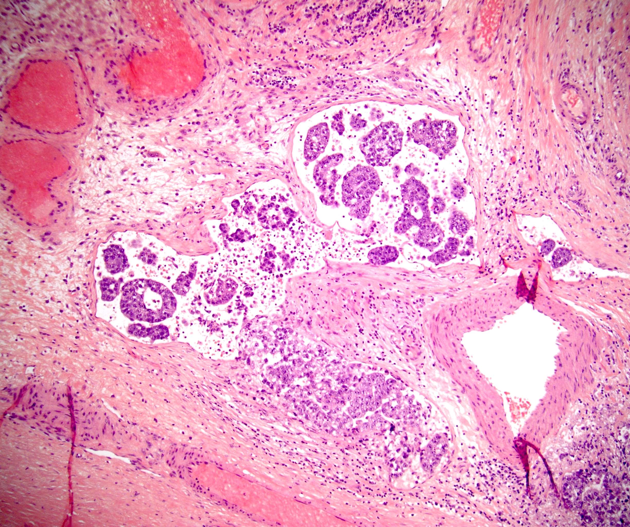
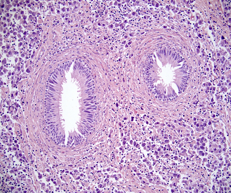
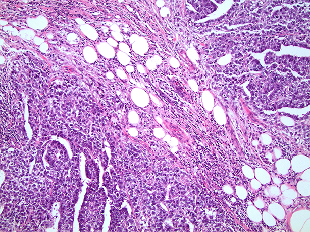
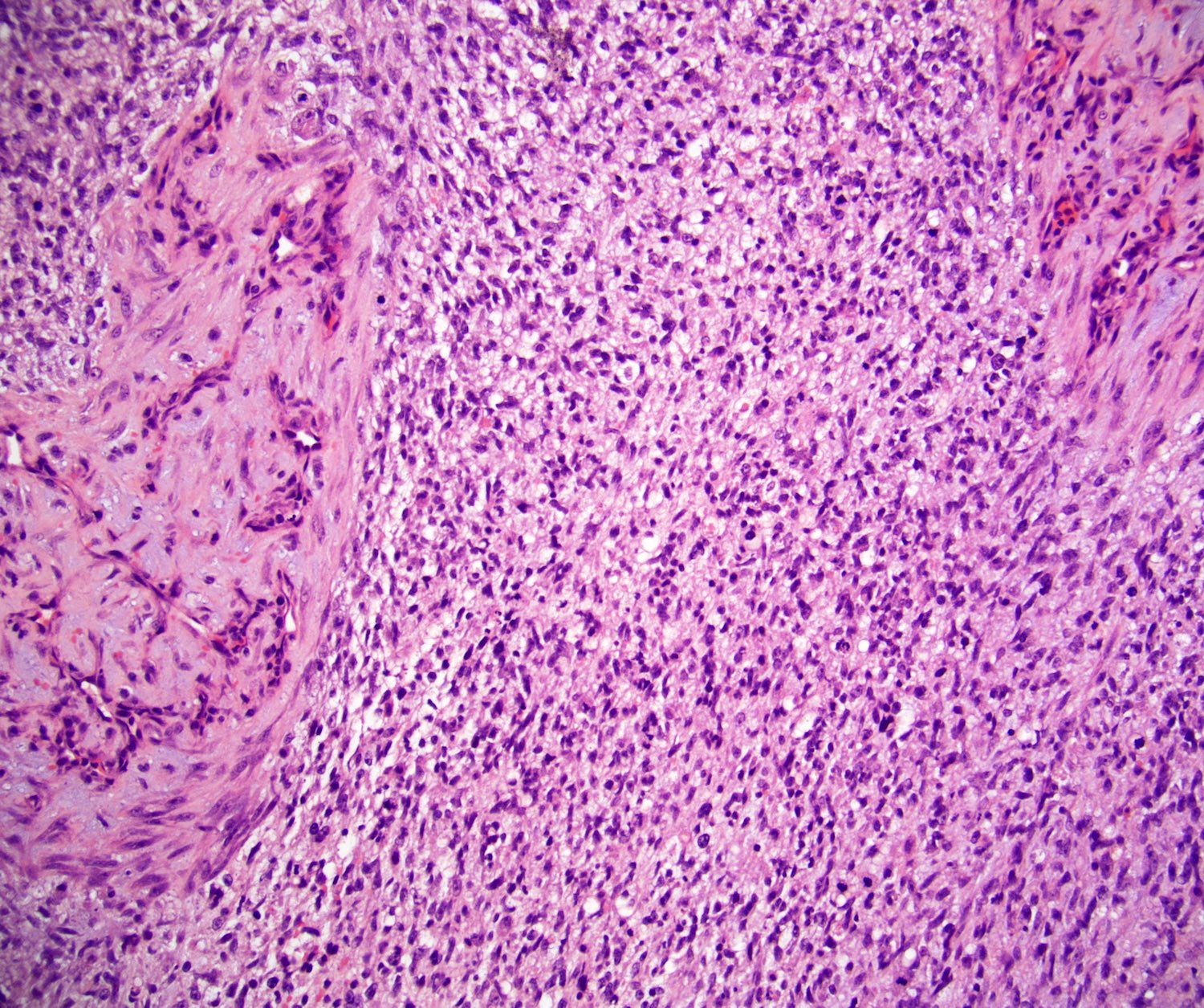
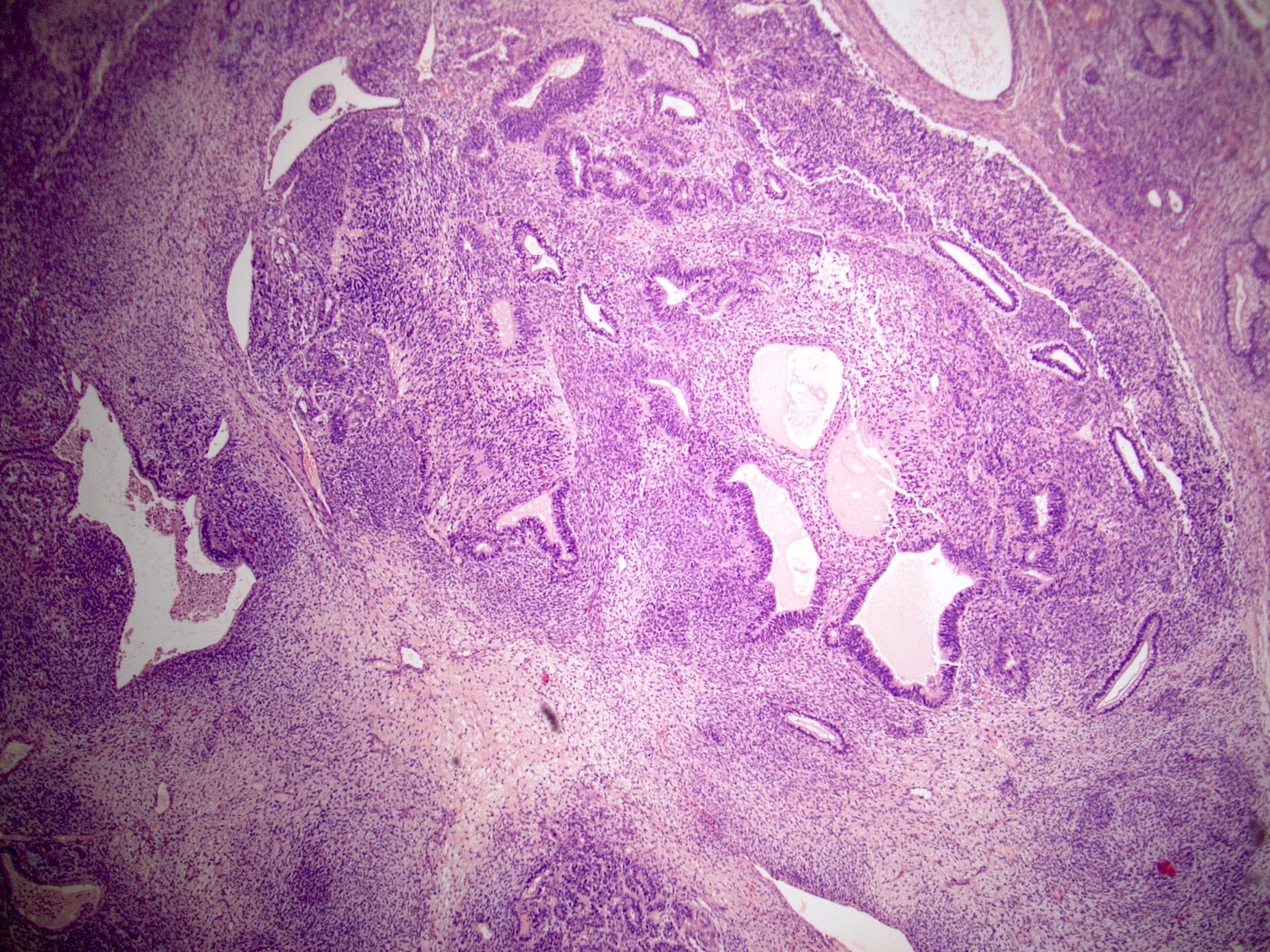
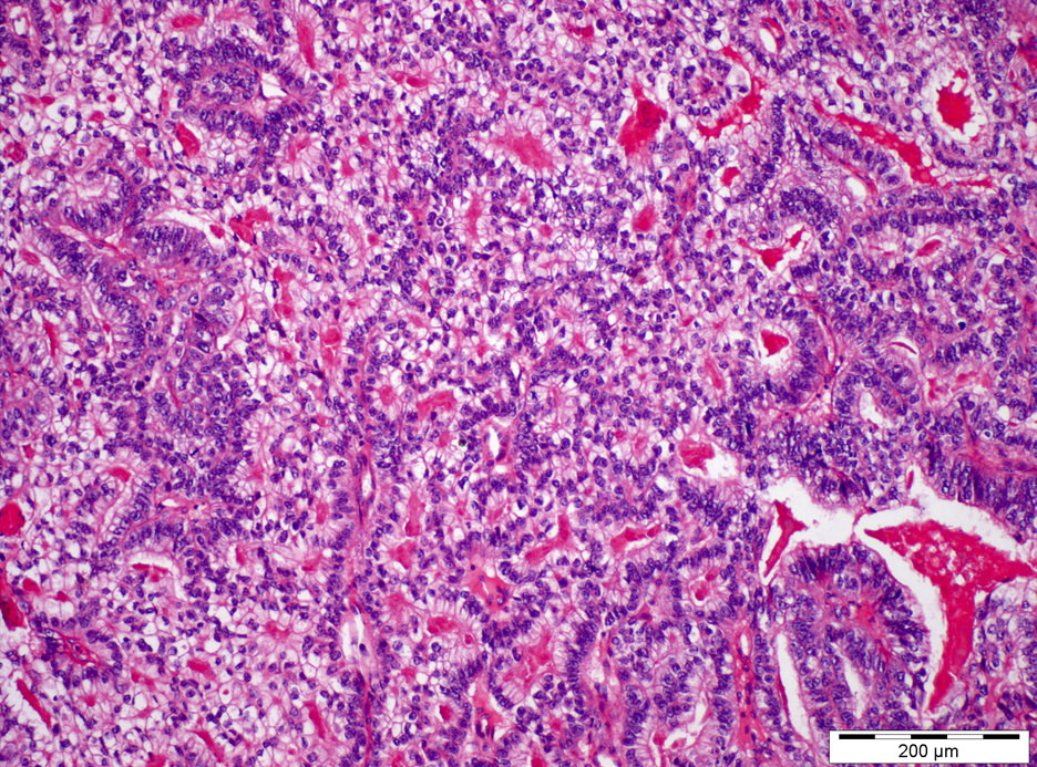
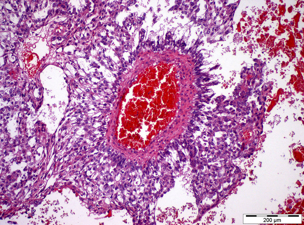
Complex architecture
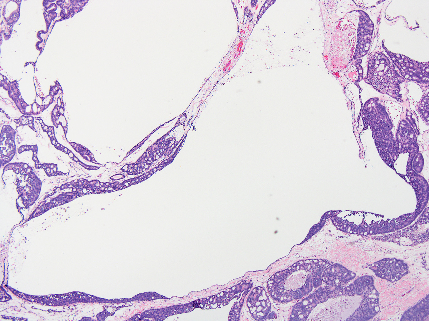
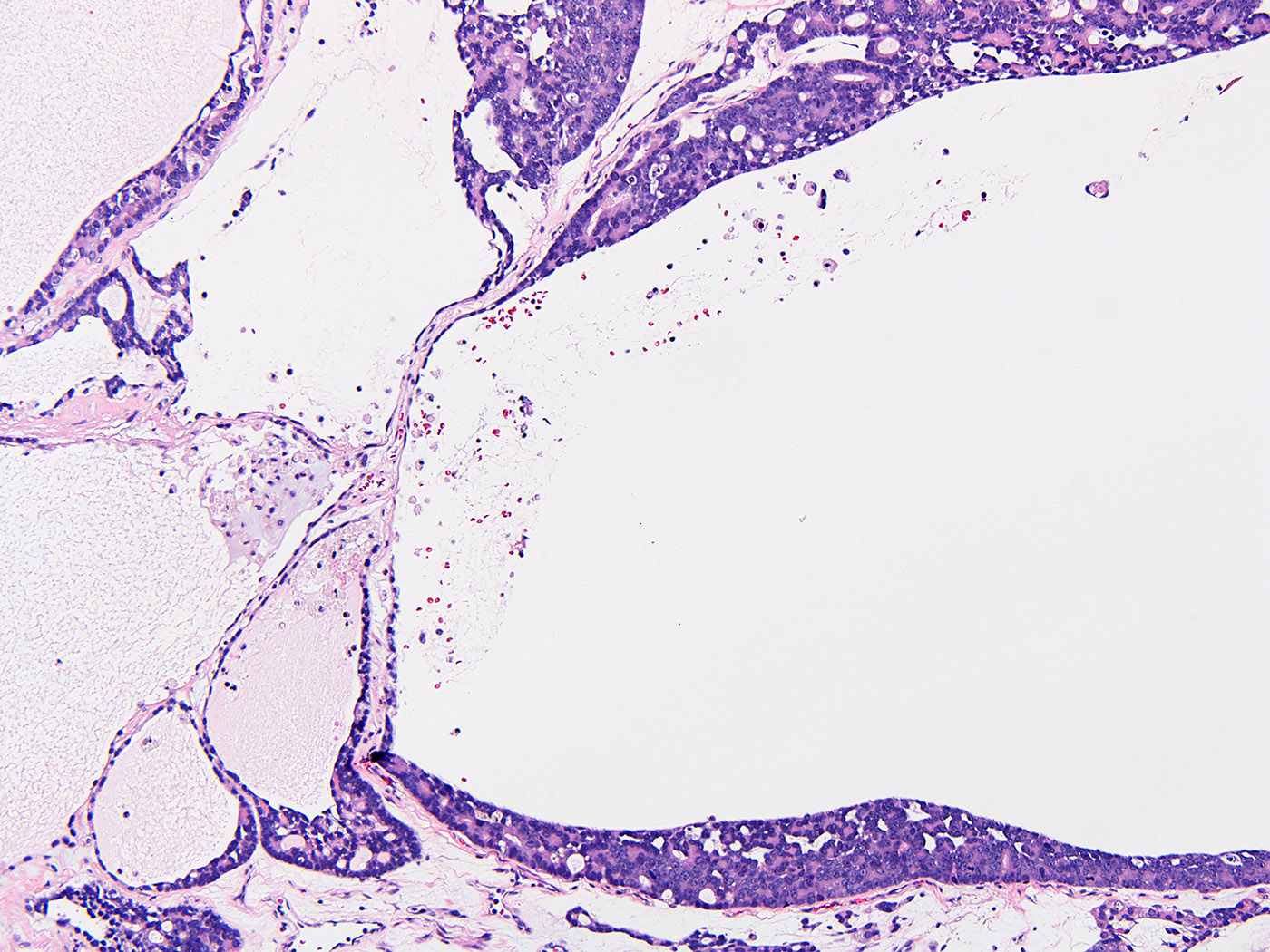
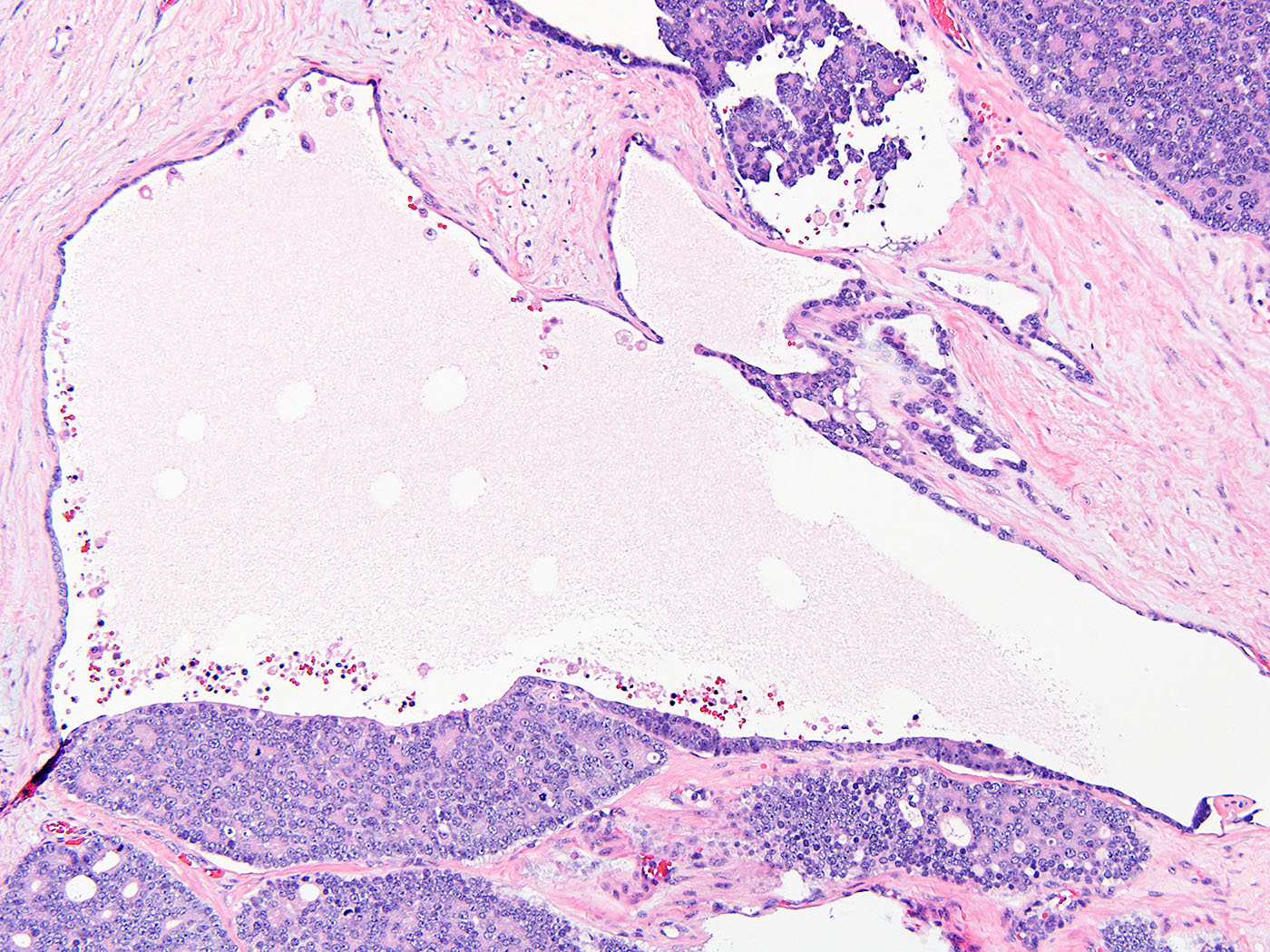
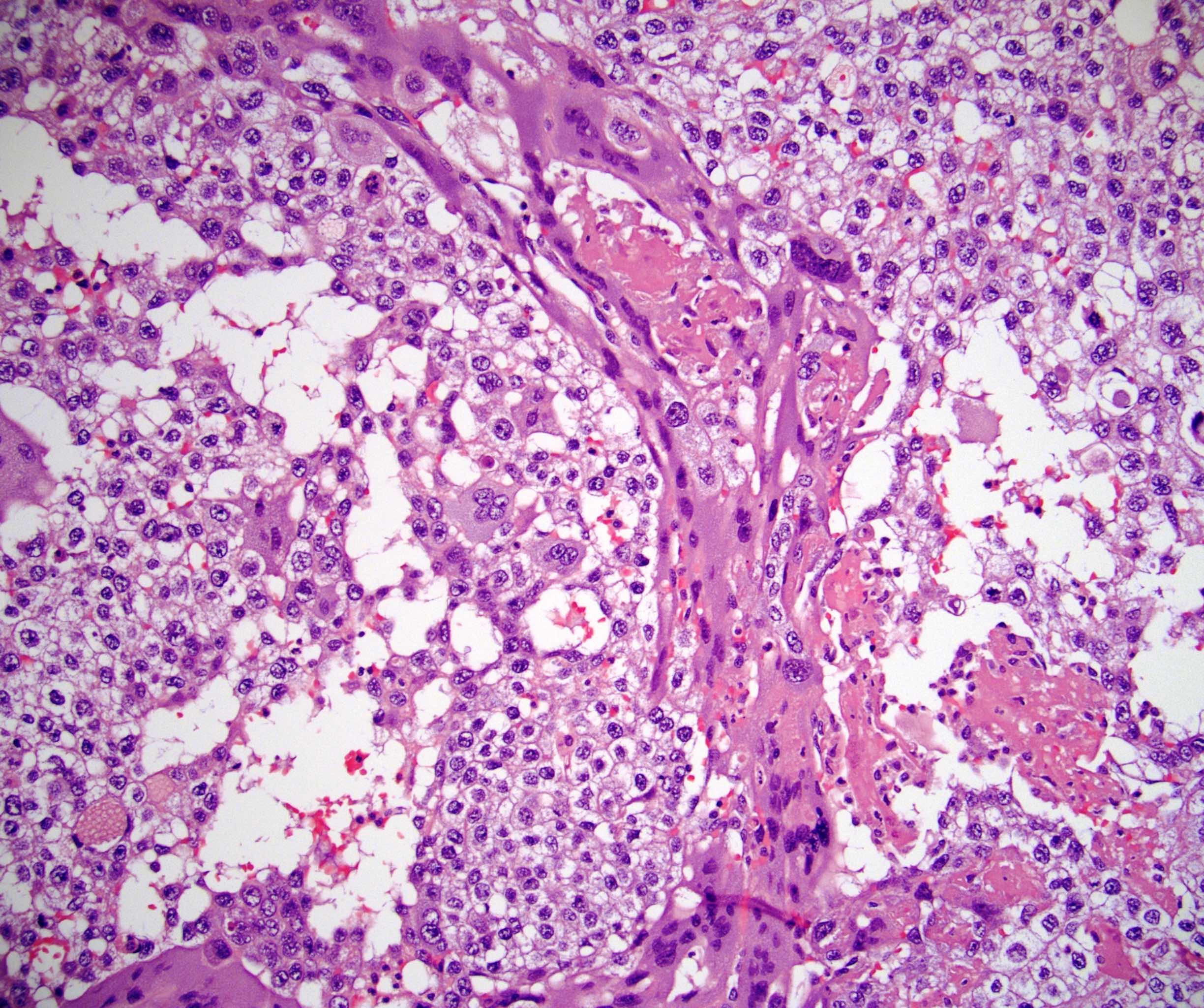
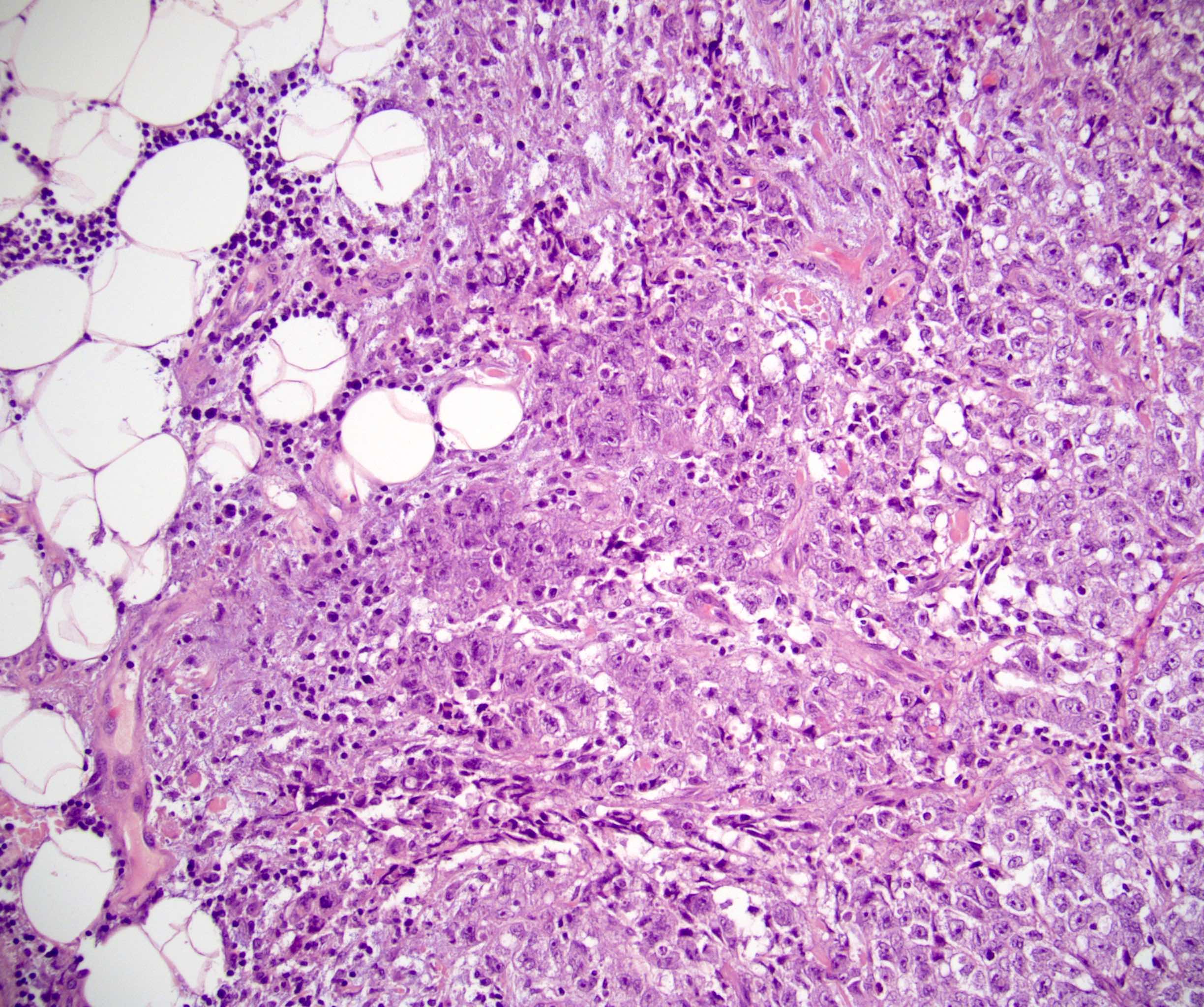
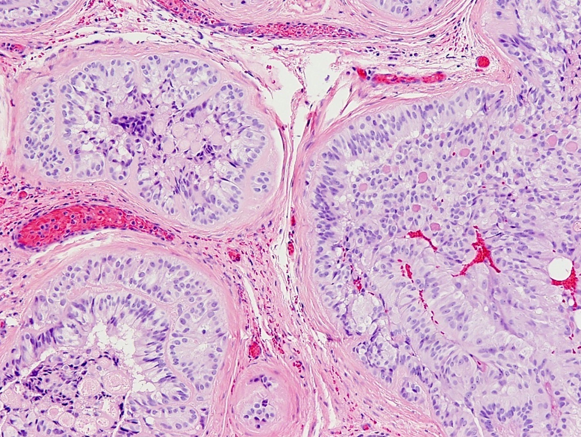
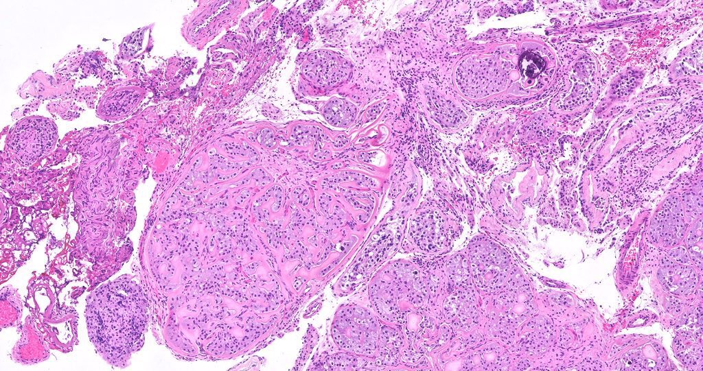
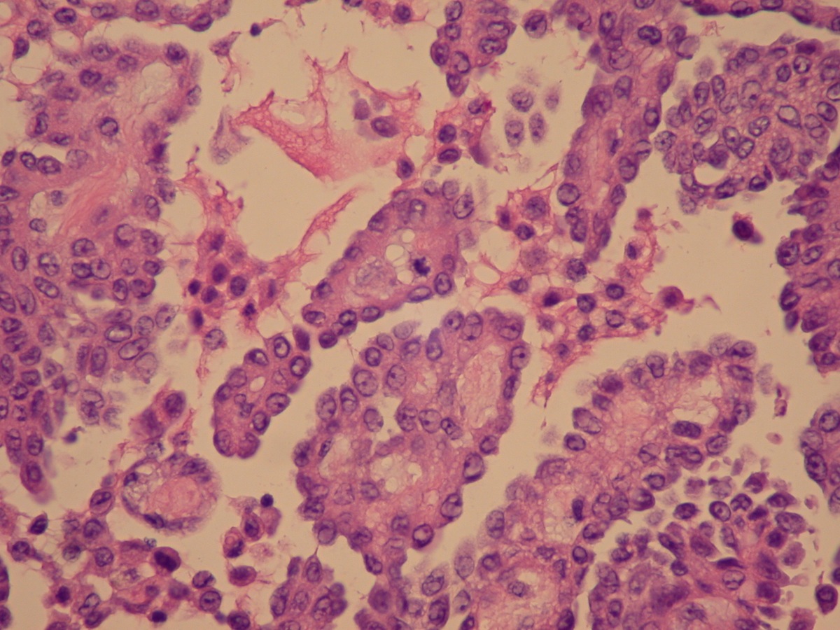
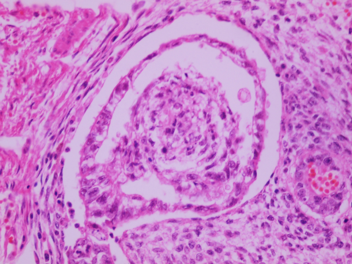
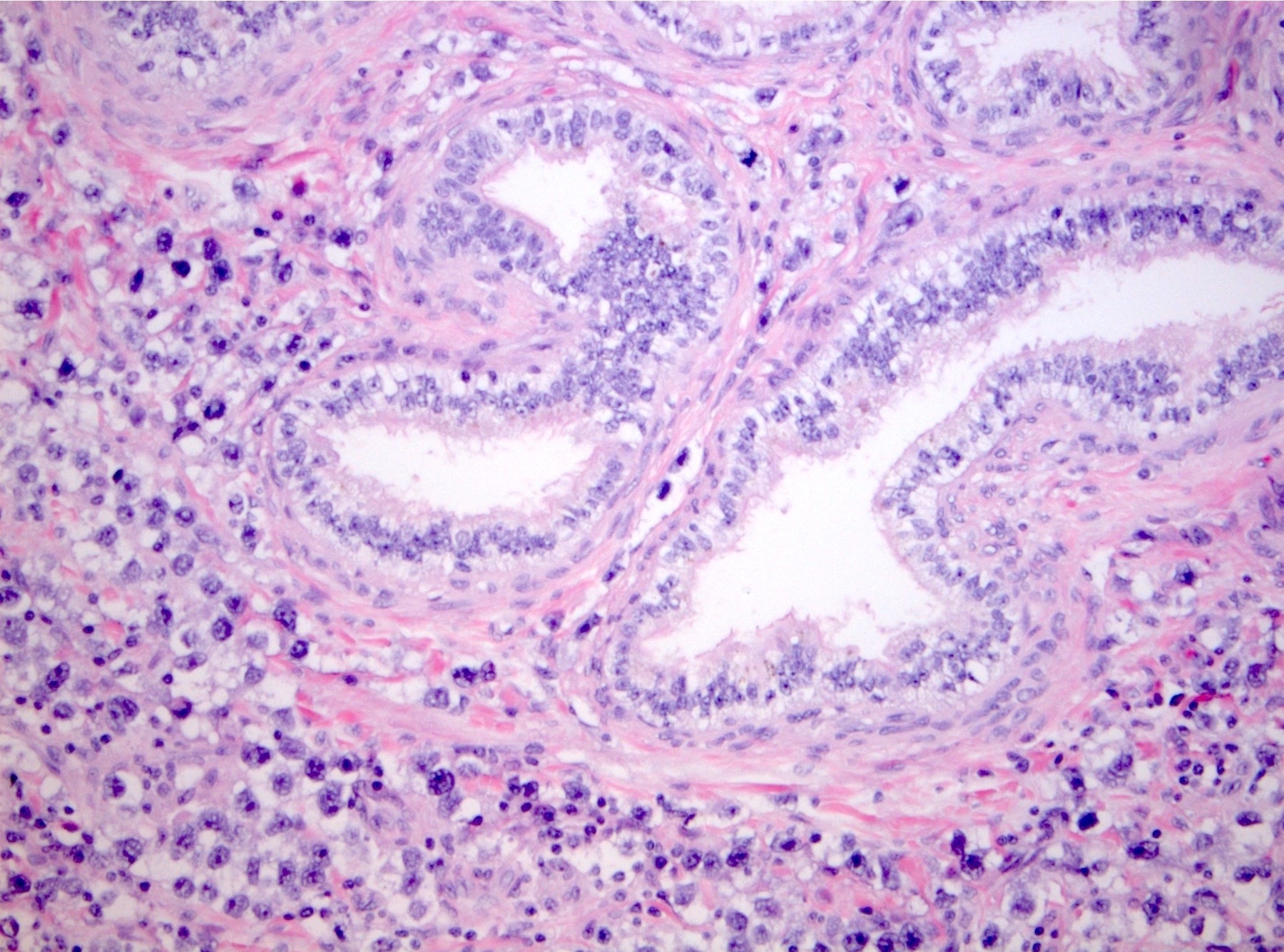
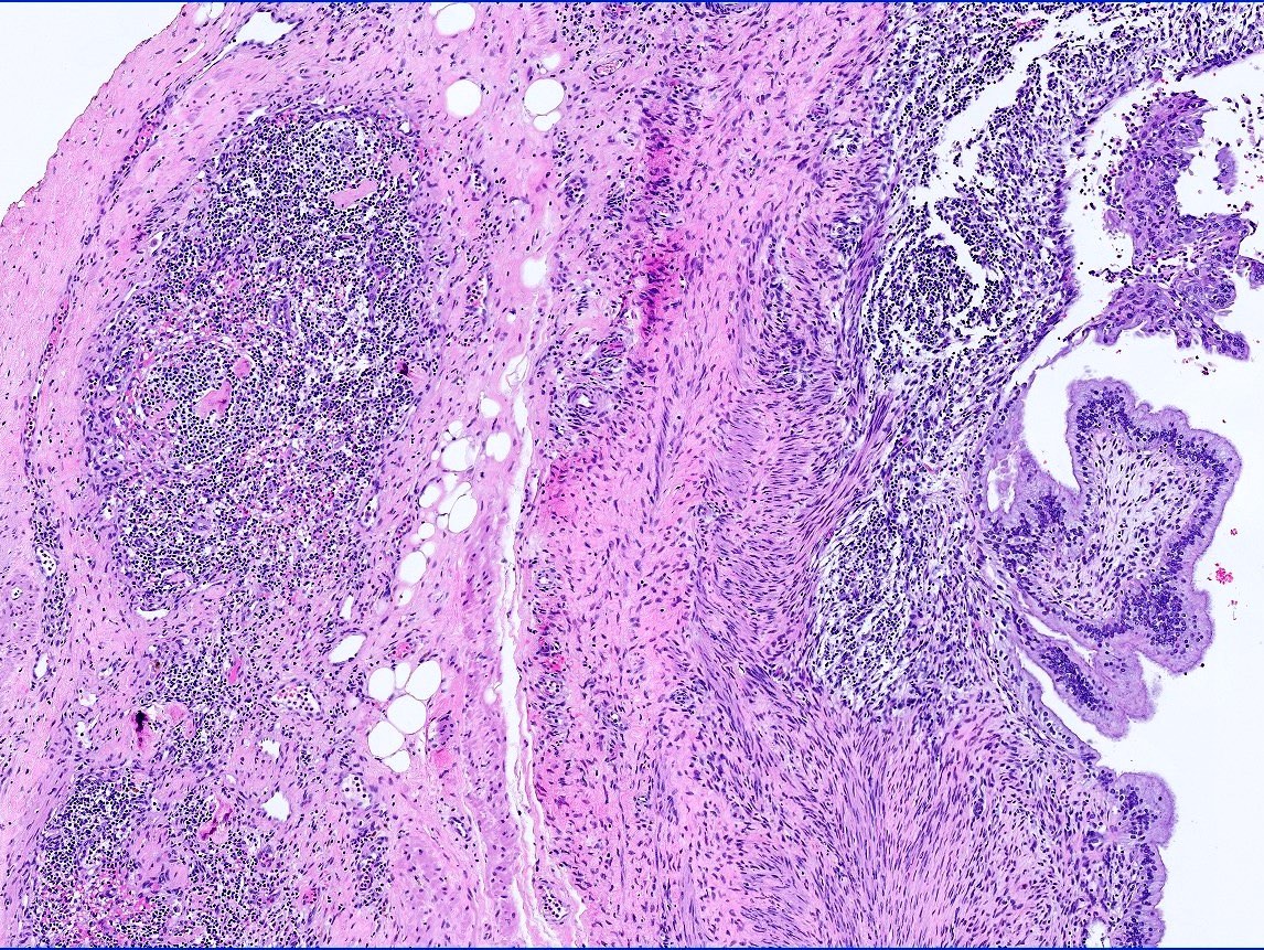
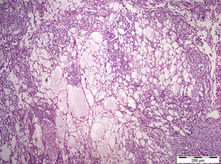
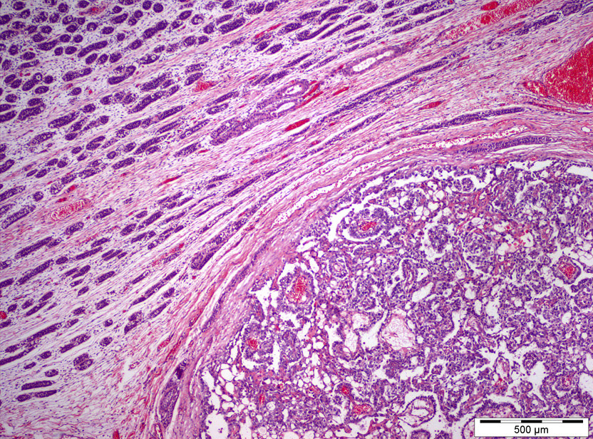
Transition of epithelium
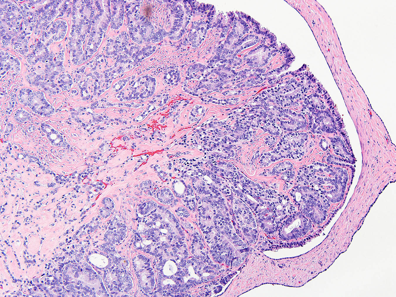
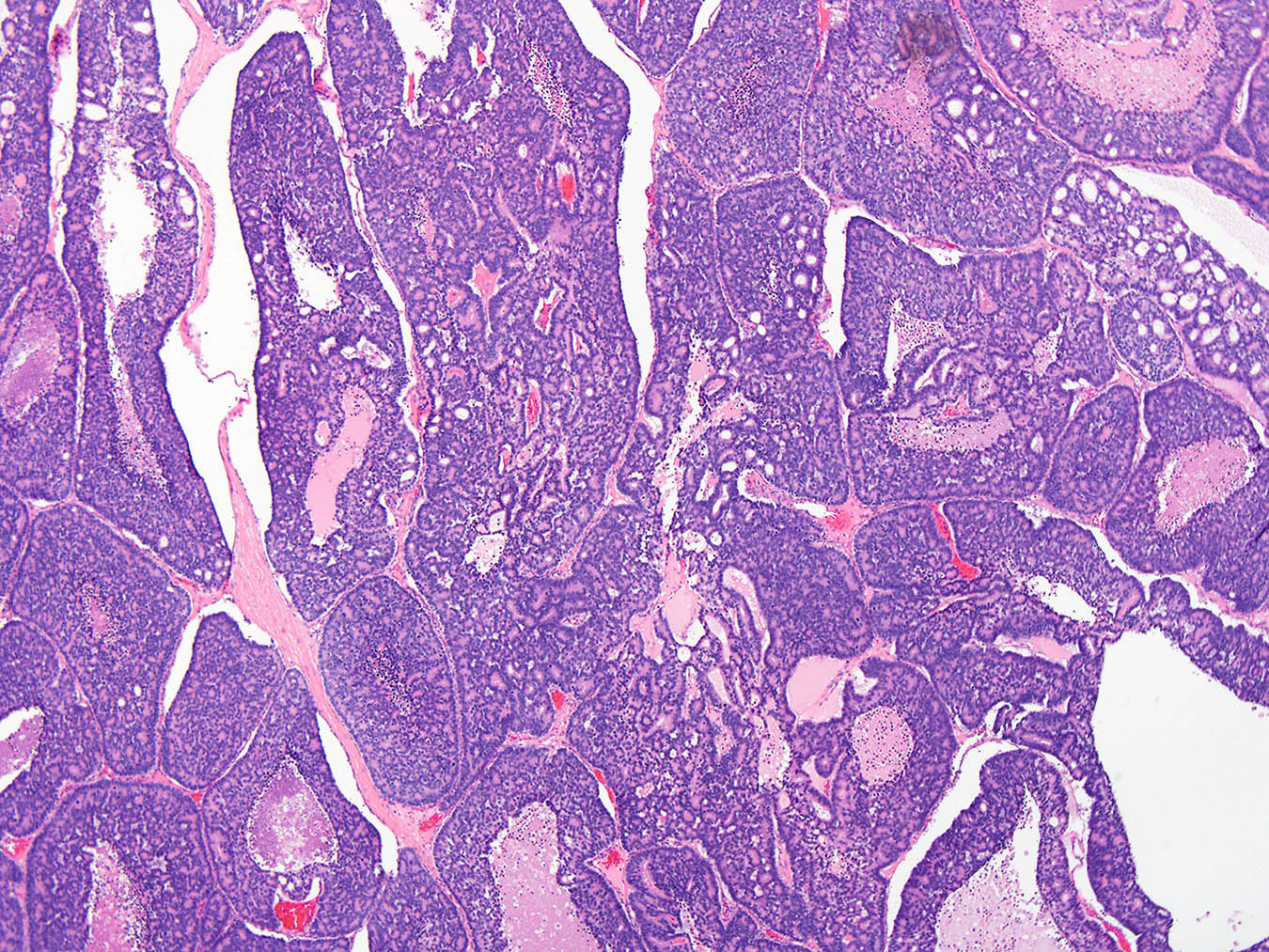
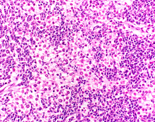
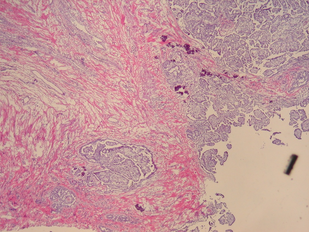
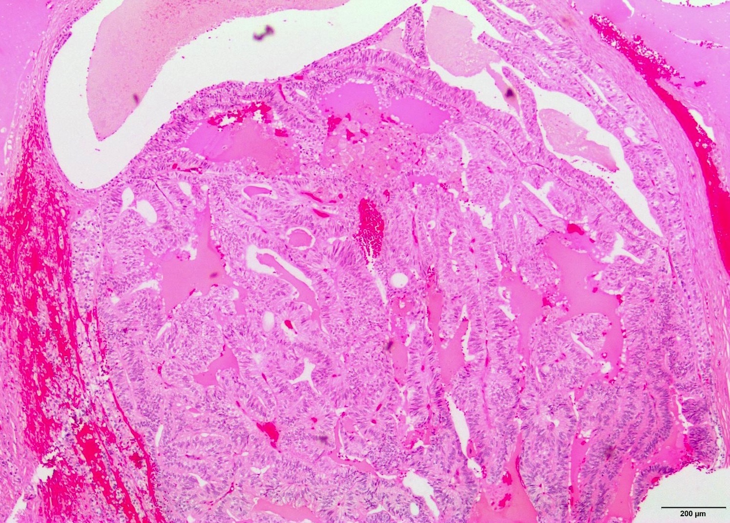
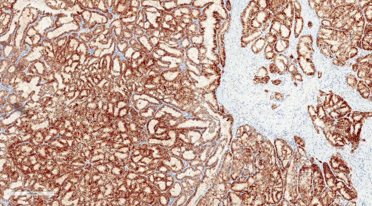
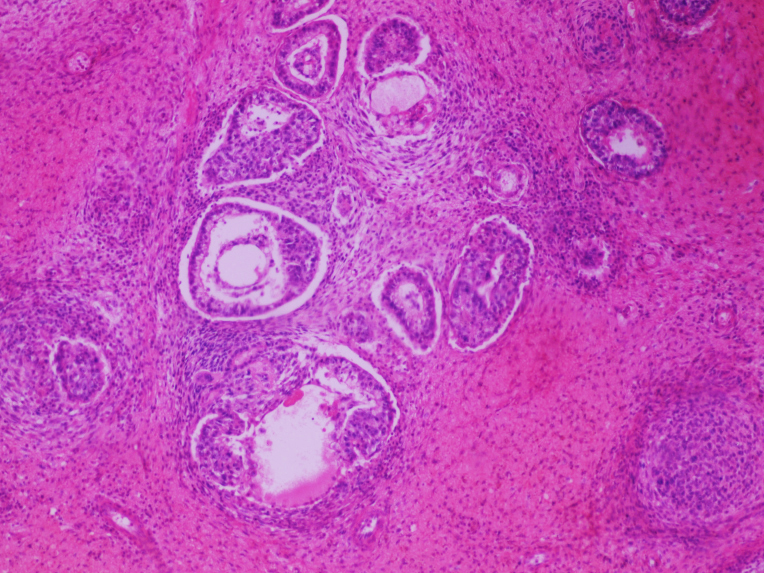
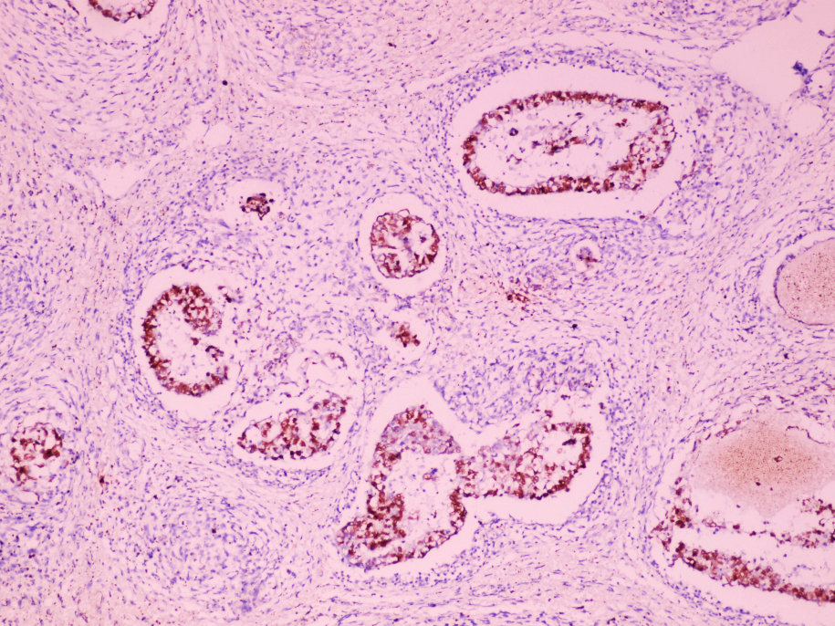
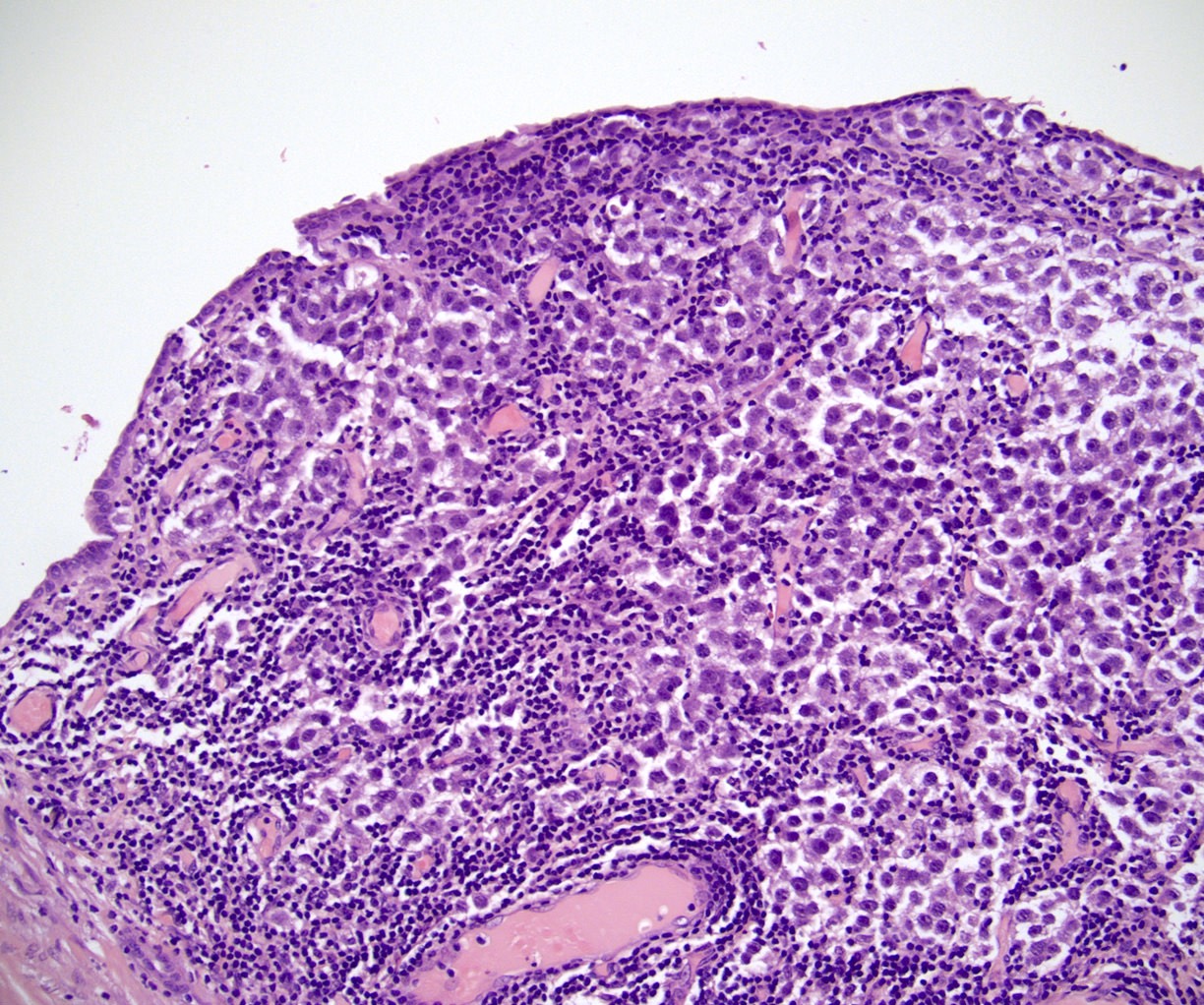
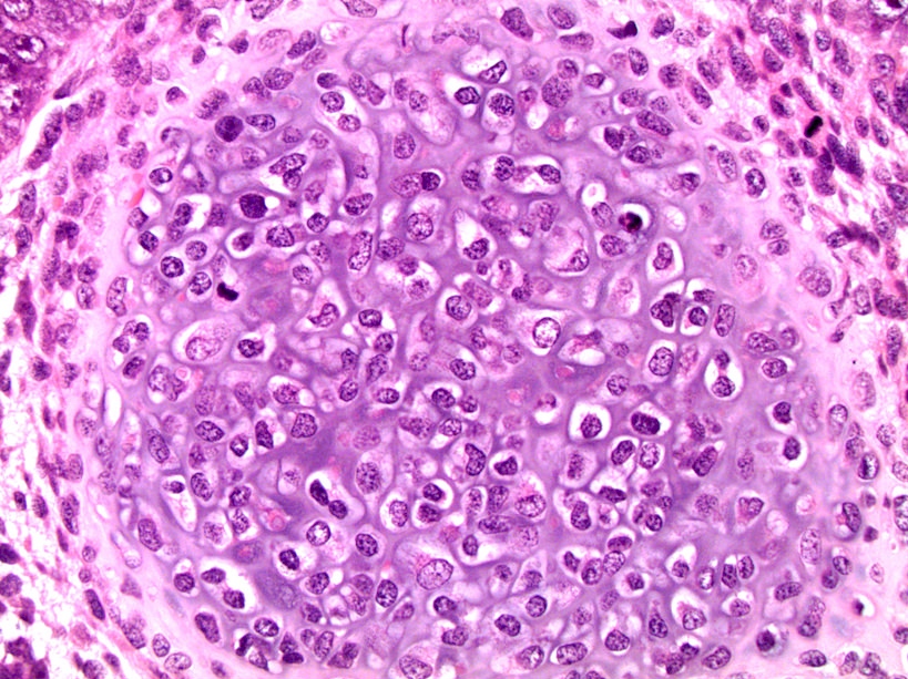
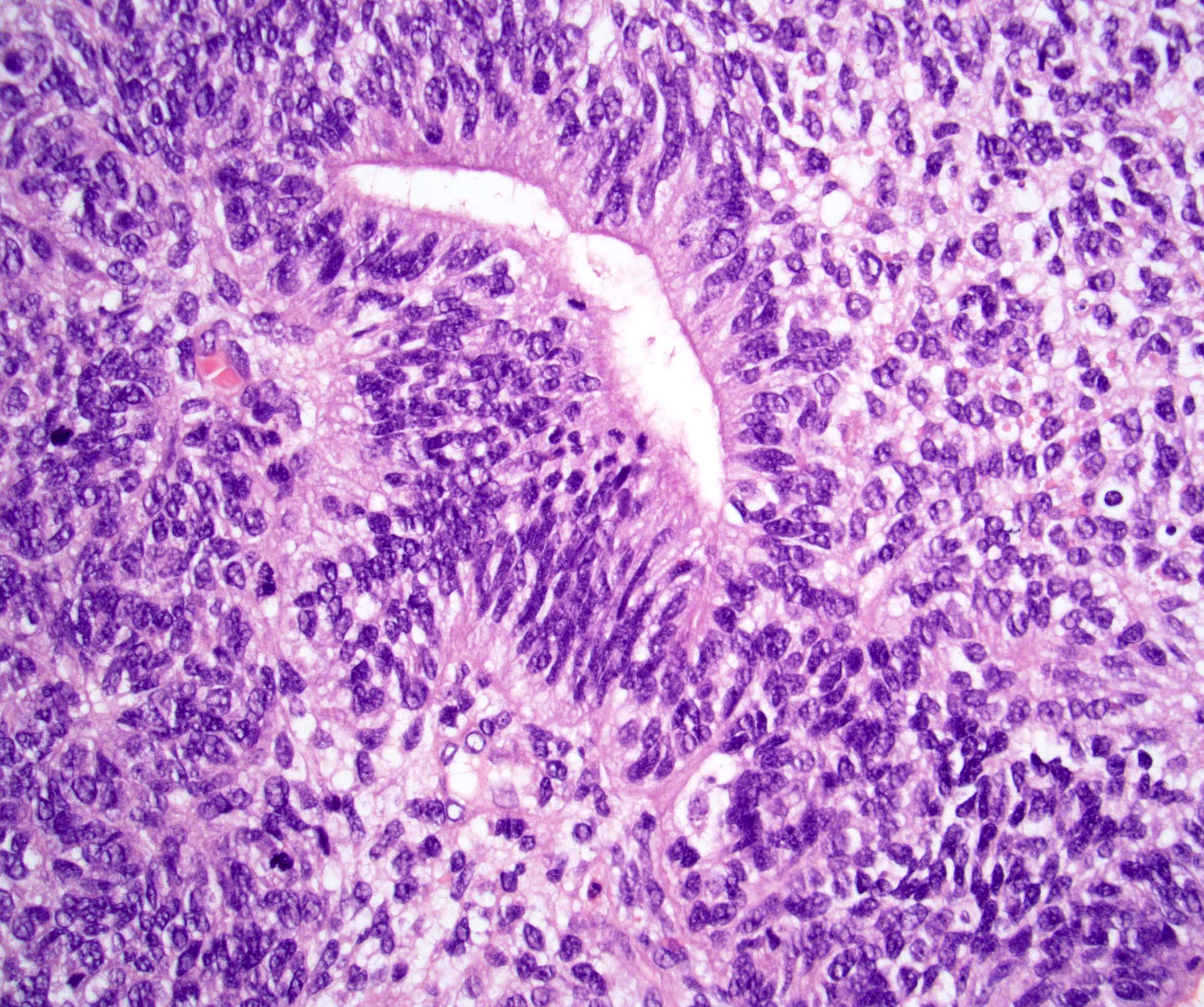
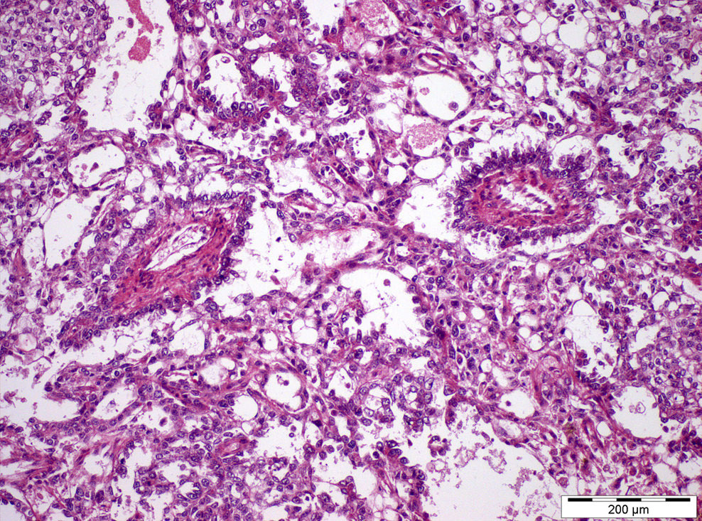
Complex tubuloglandular pattern
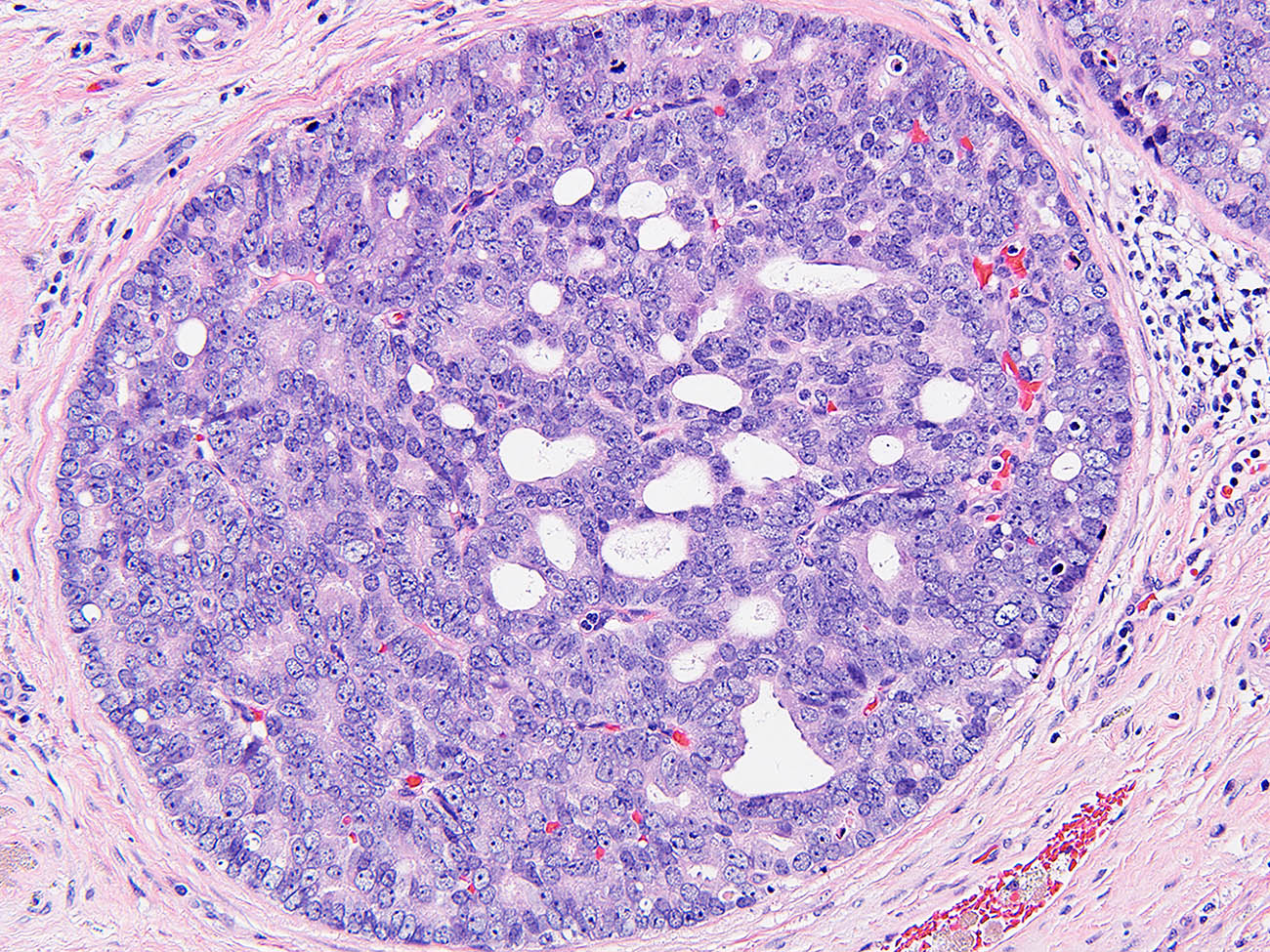
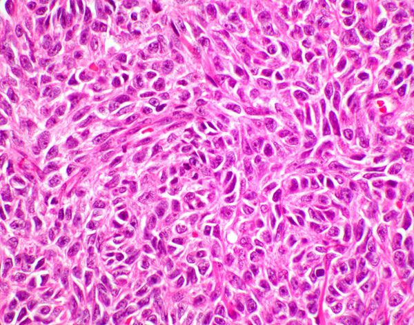
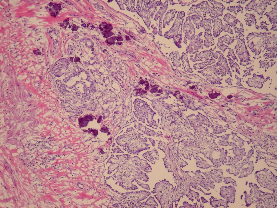
Cribriform pattern

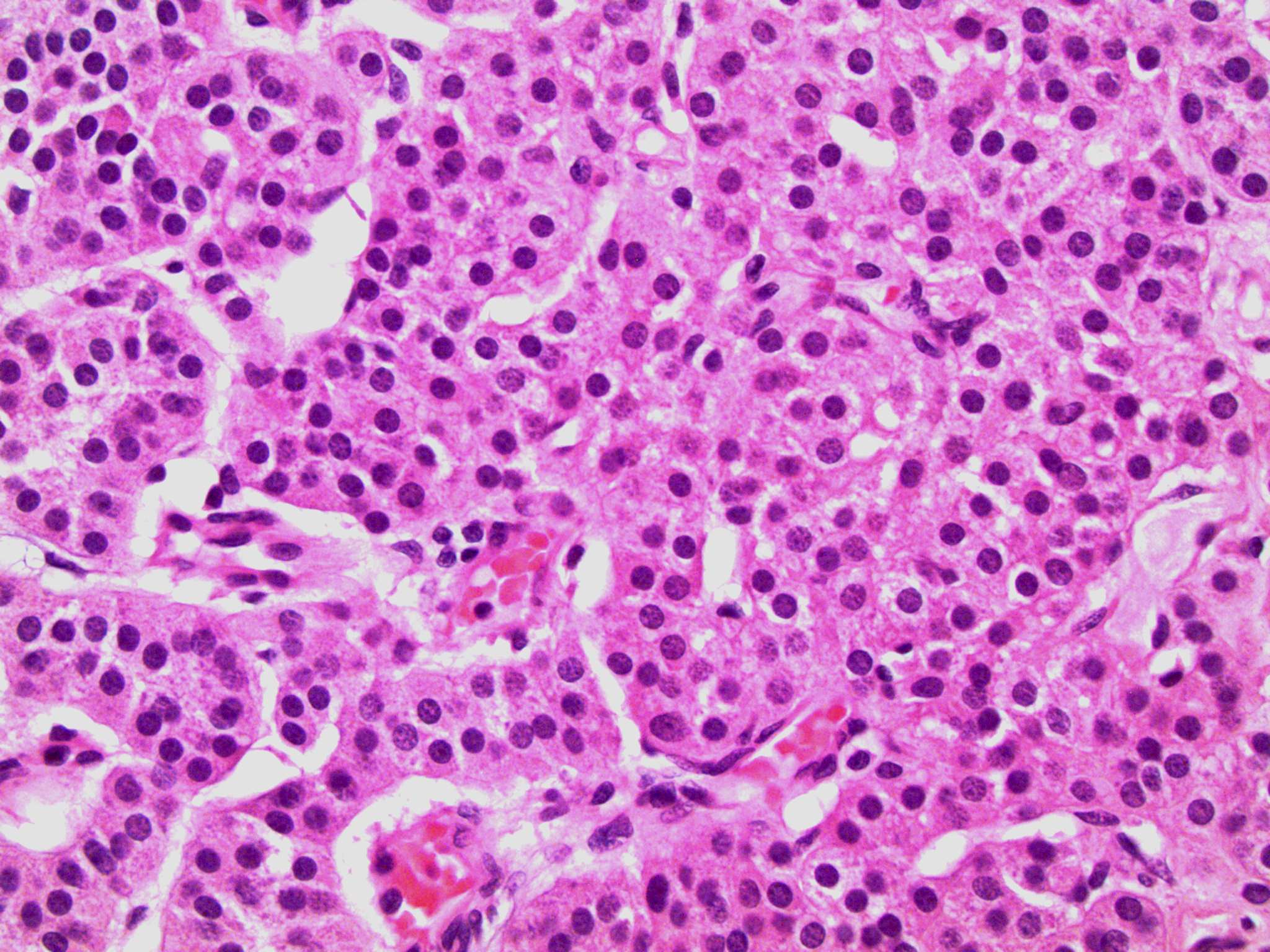
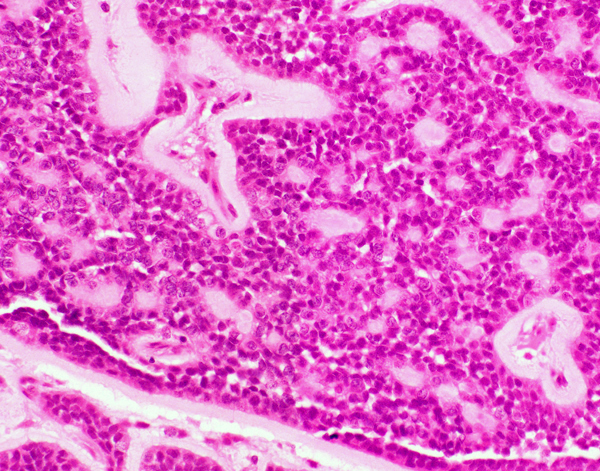
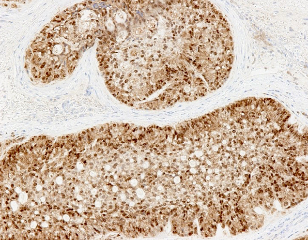
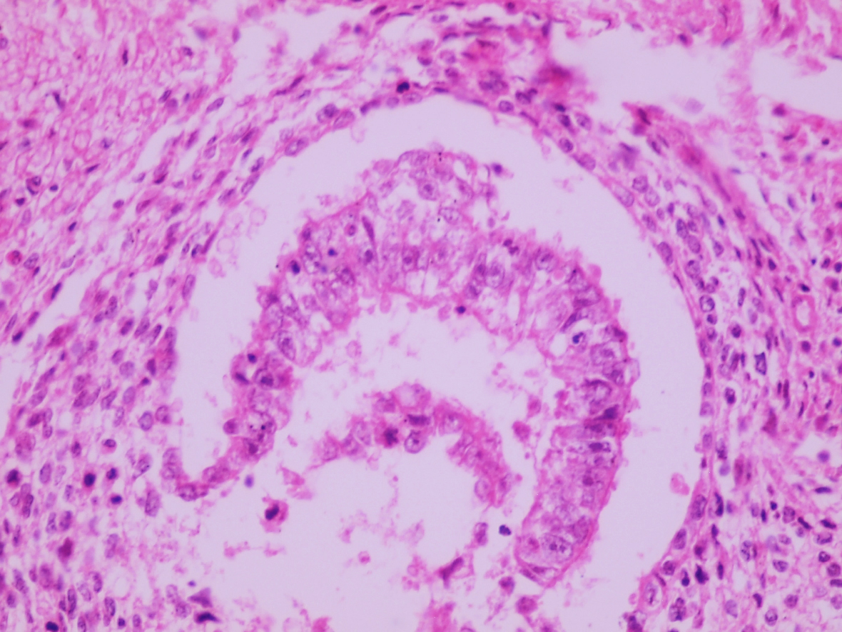
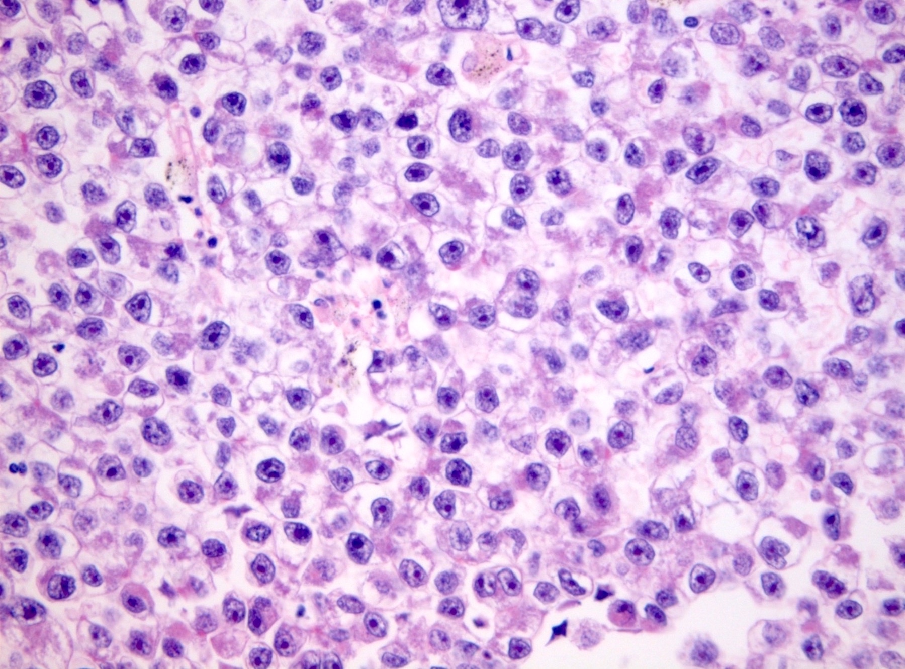
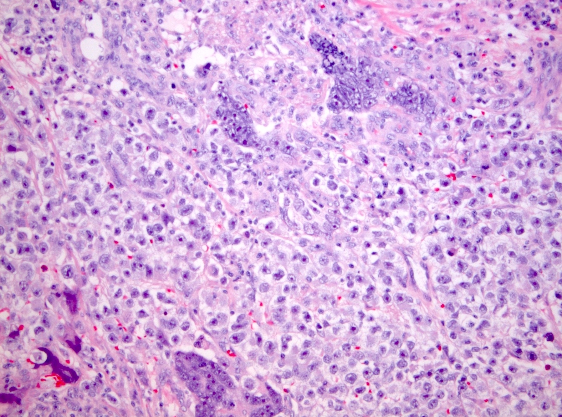
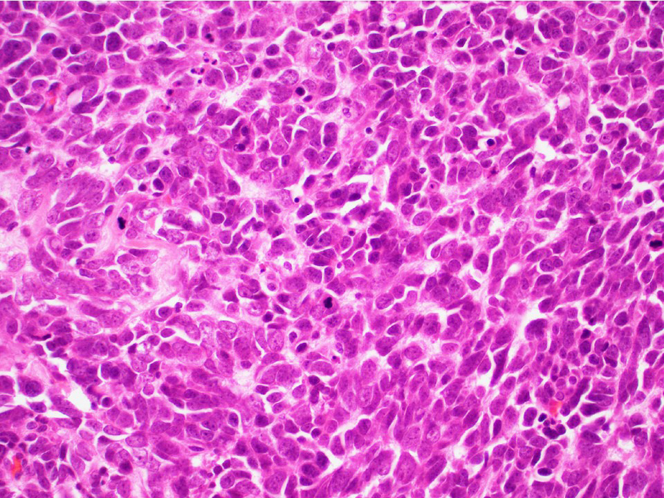
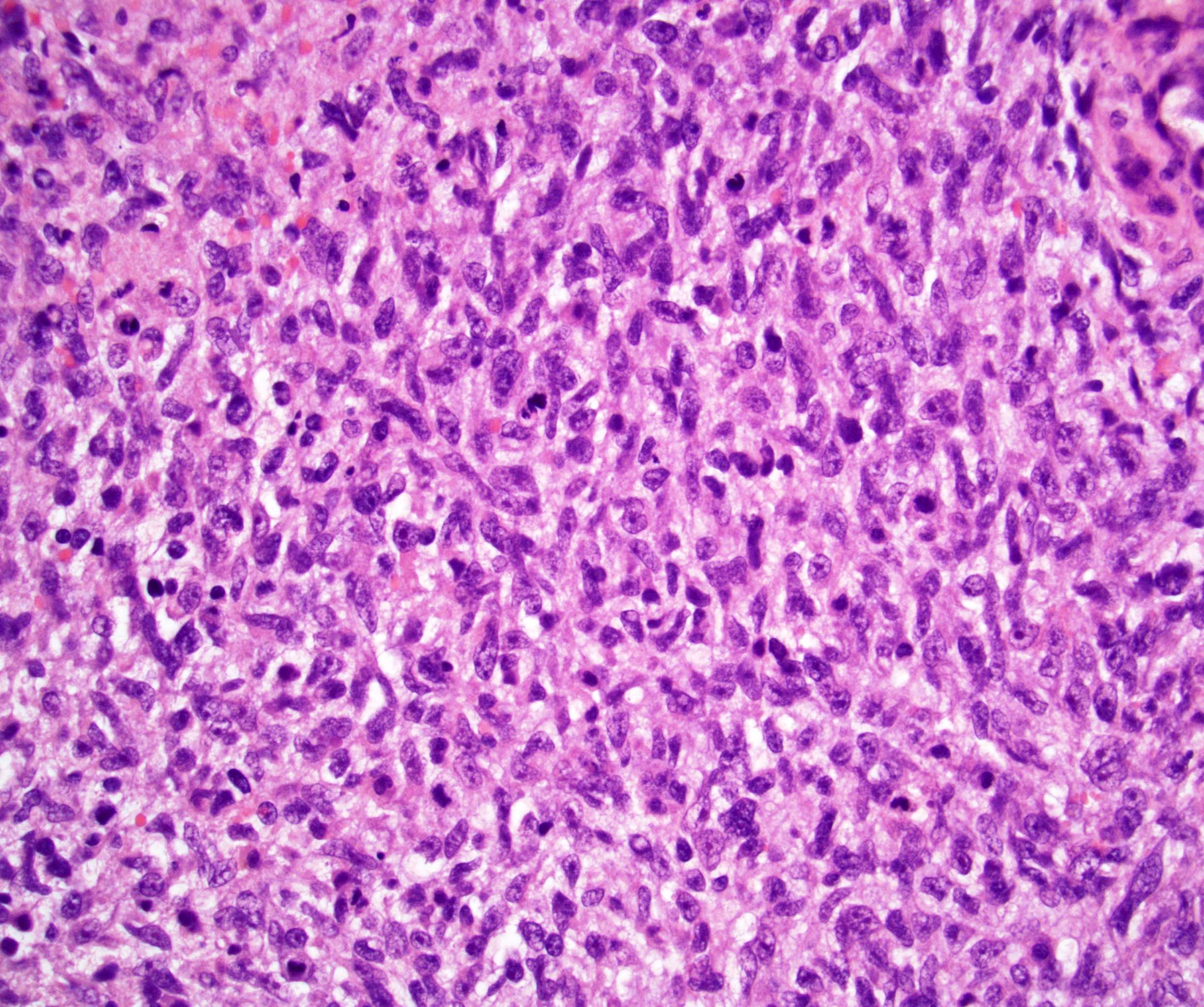
High grade cytomorphology
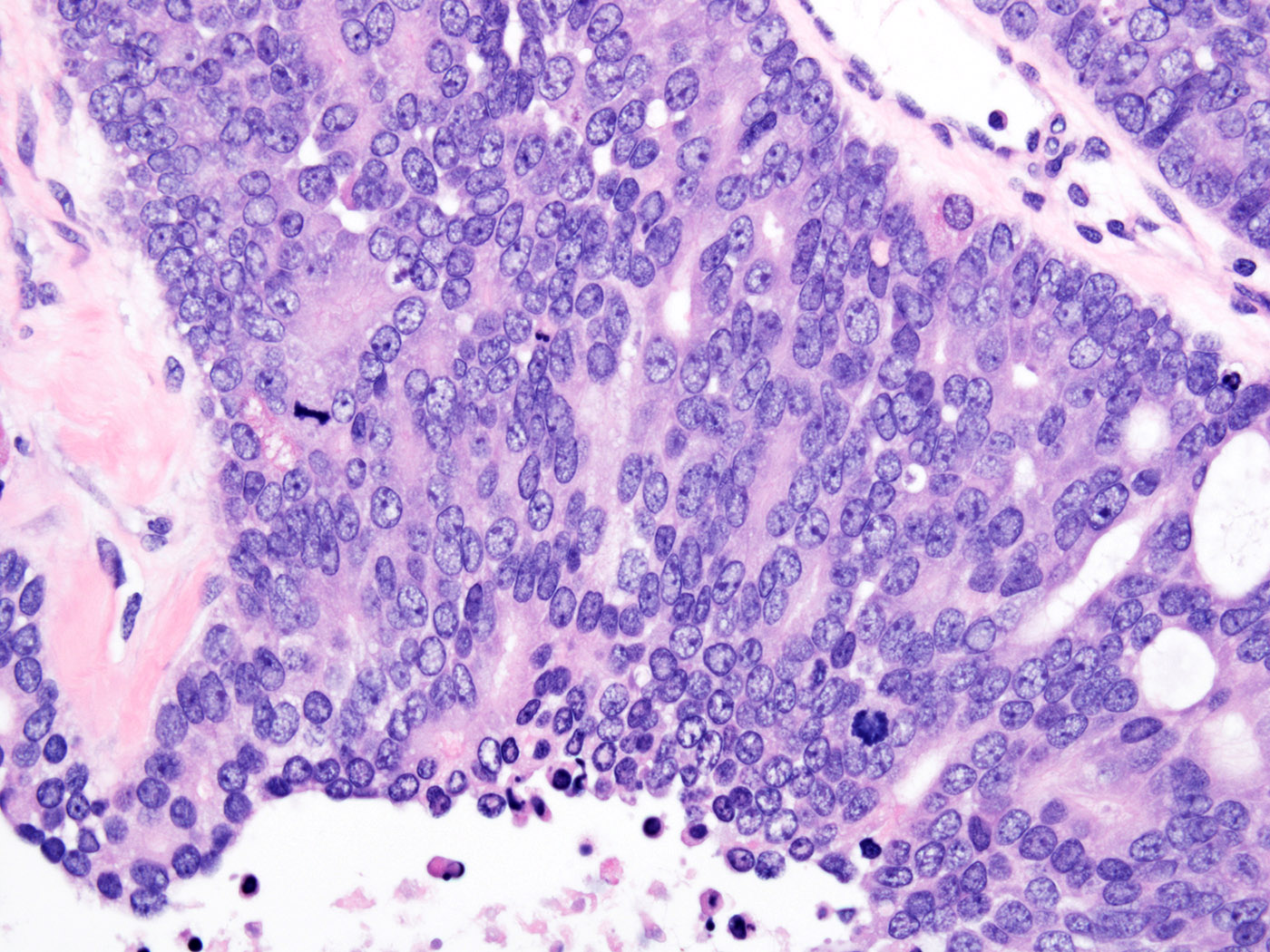
Increased mitotic activity
Images hosted on other servers:

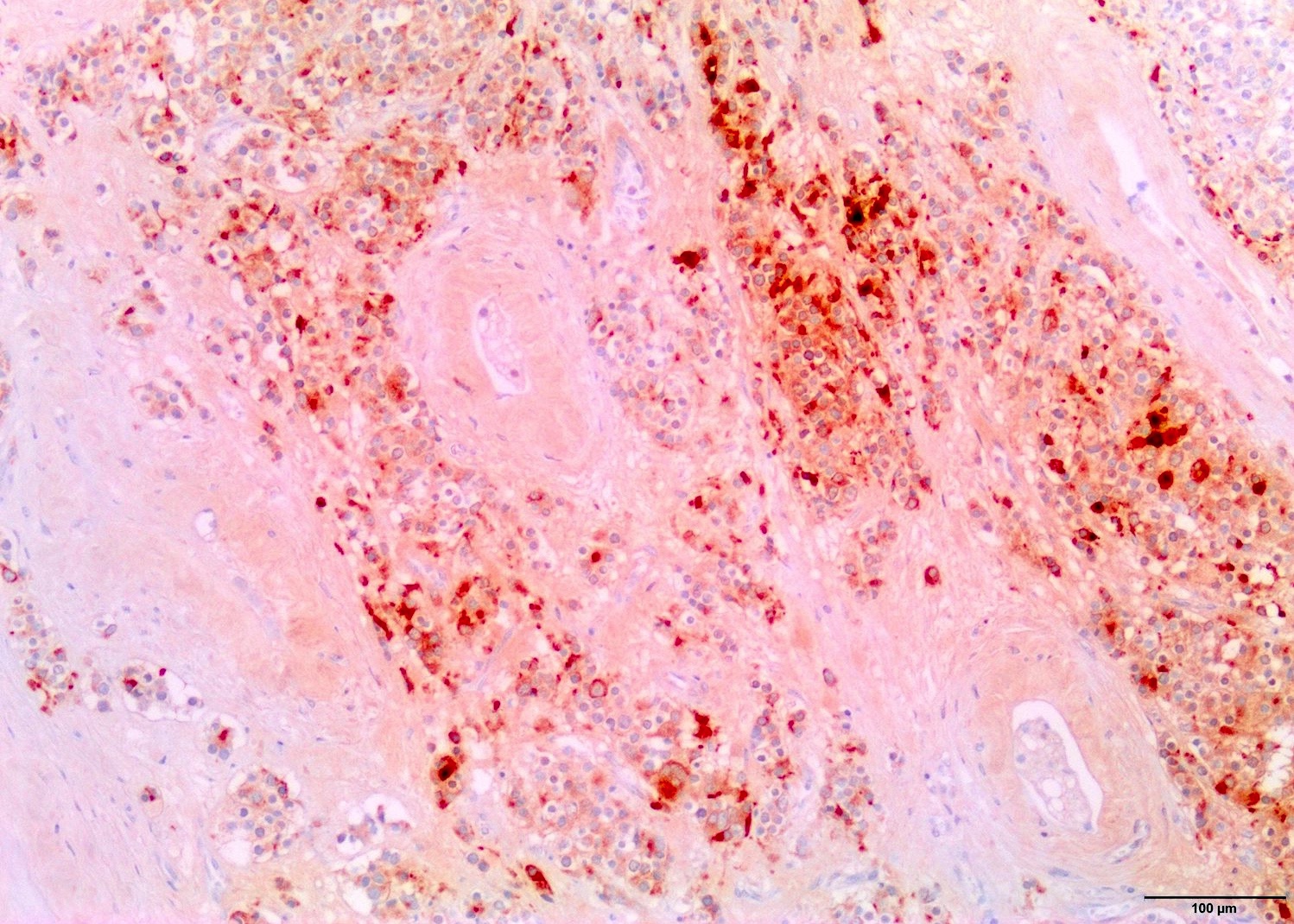
Adenocarcinoma of rete testis
Images hosted on other servers:

Coronal MR

Ultrasound
Contributed by Debra L. Zynger, M.D.
Paratesticular tumor
Images hosted on other servers:

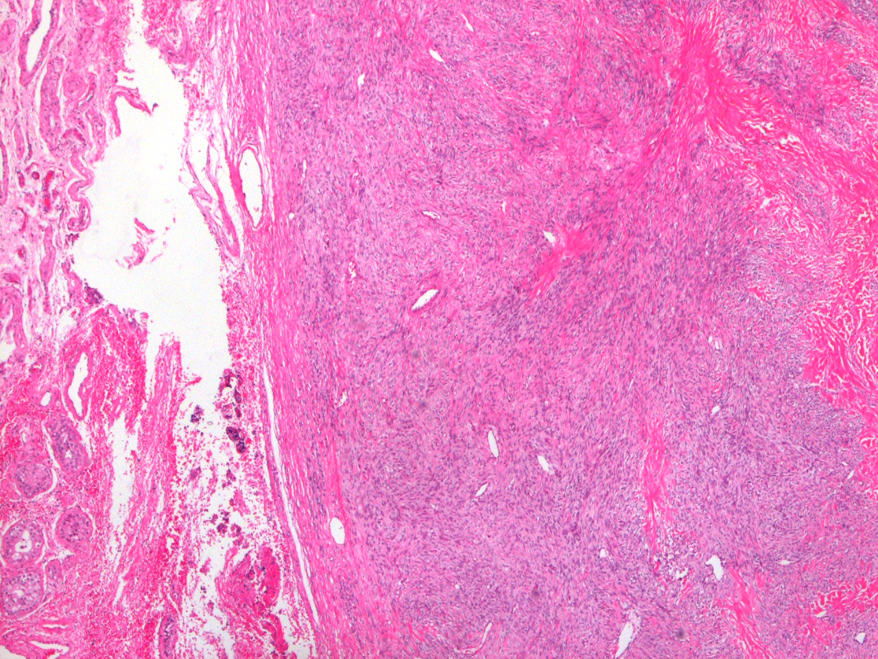
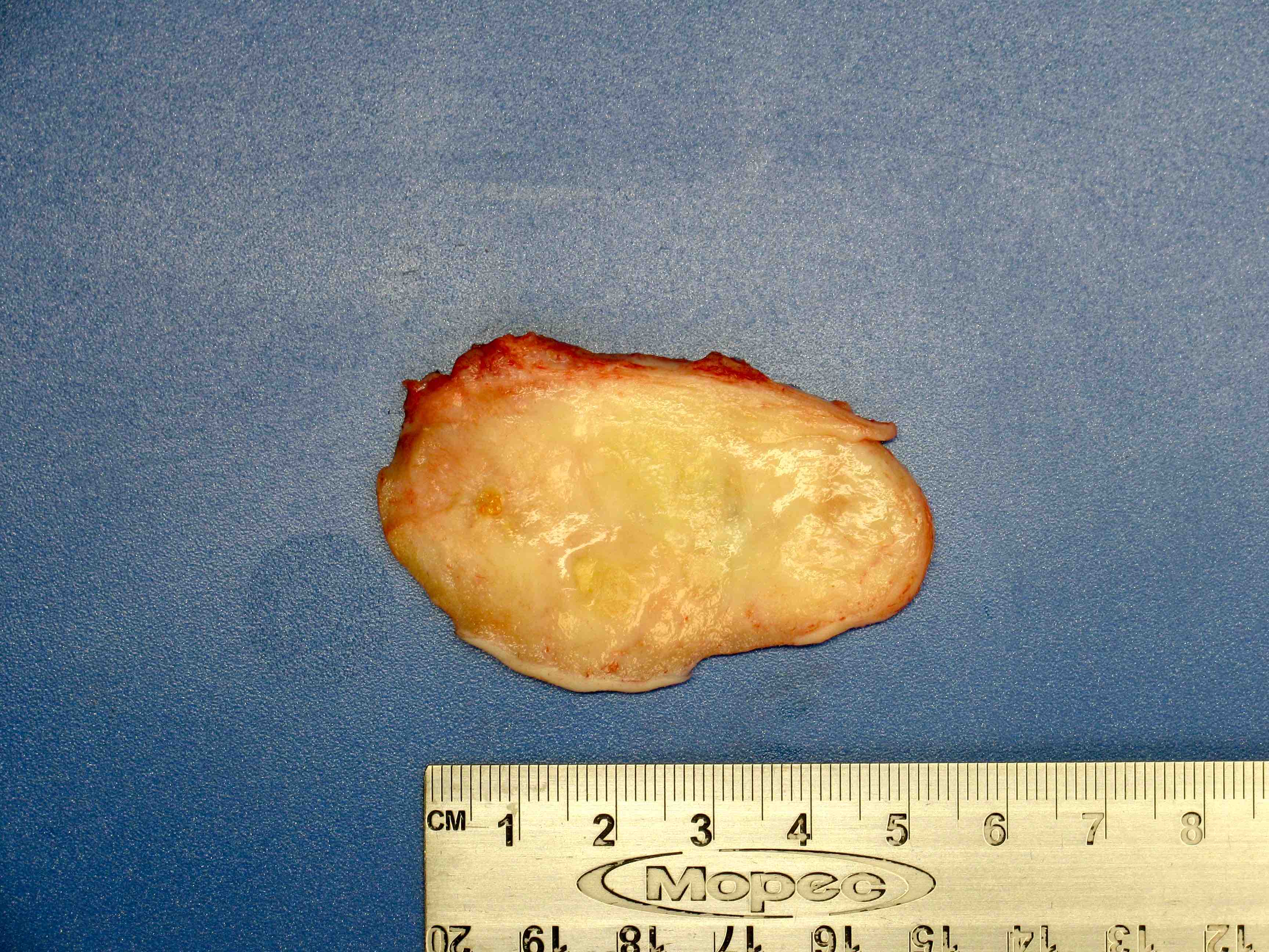
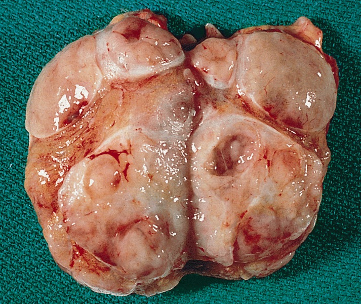
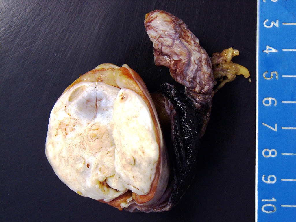
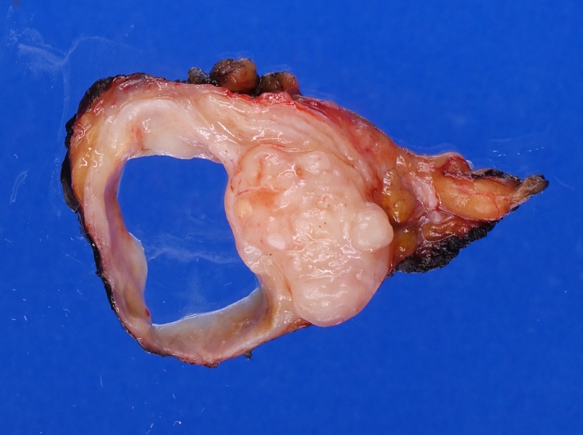
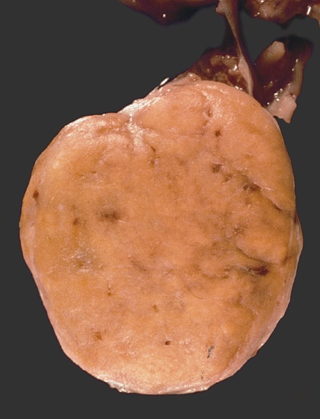
Well circumscribed tumor
Contributed by Sarah Findeis, M.D., Stephanie J. Conrad, M.D., Ming Zhou, M.D.,
the Genitourinary Pathology Society (GUPS) and @katcollmd on Twitter
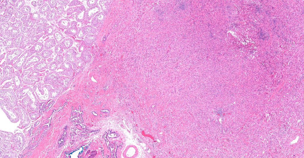
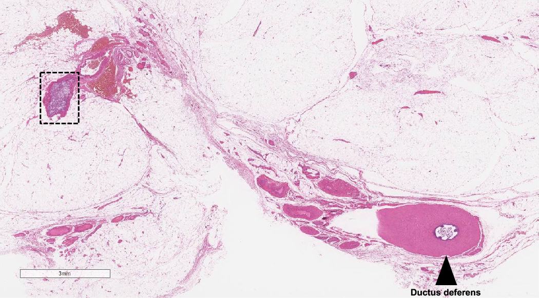
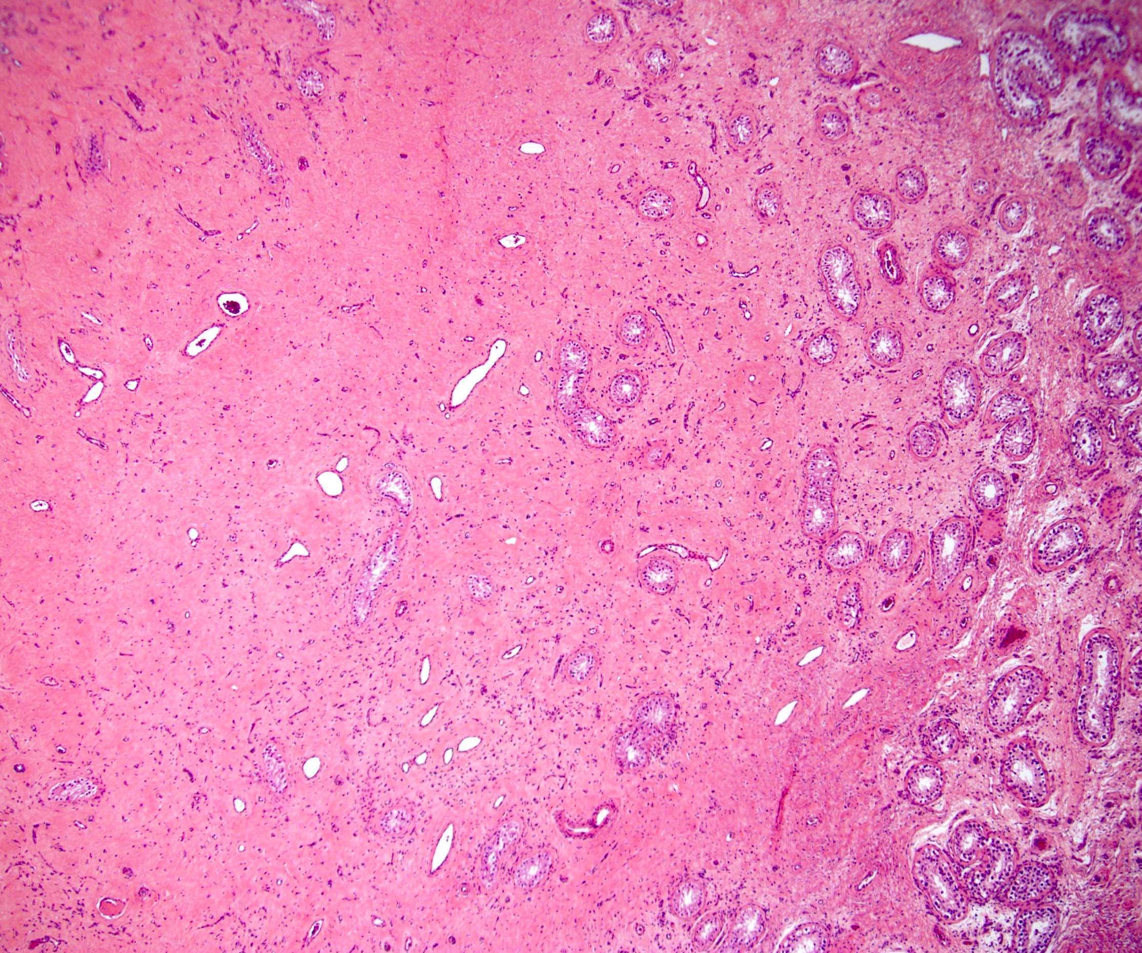
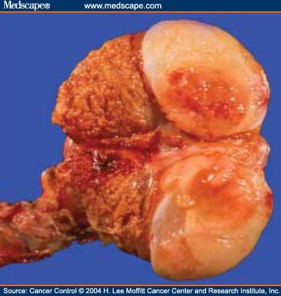
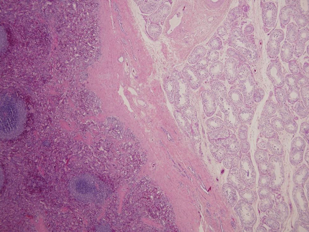
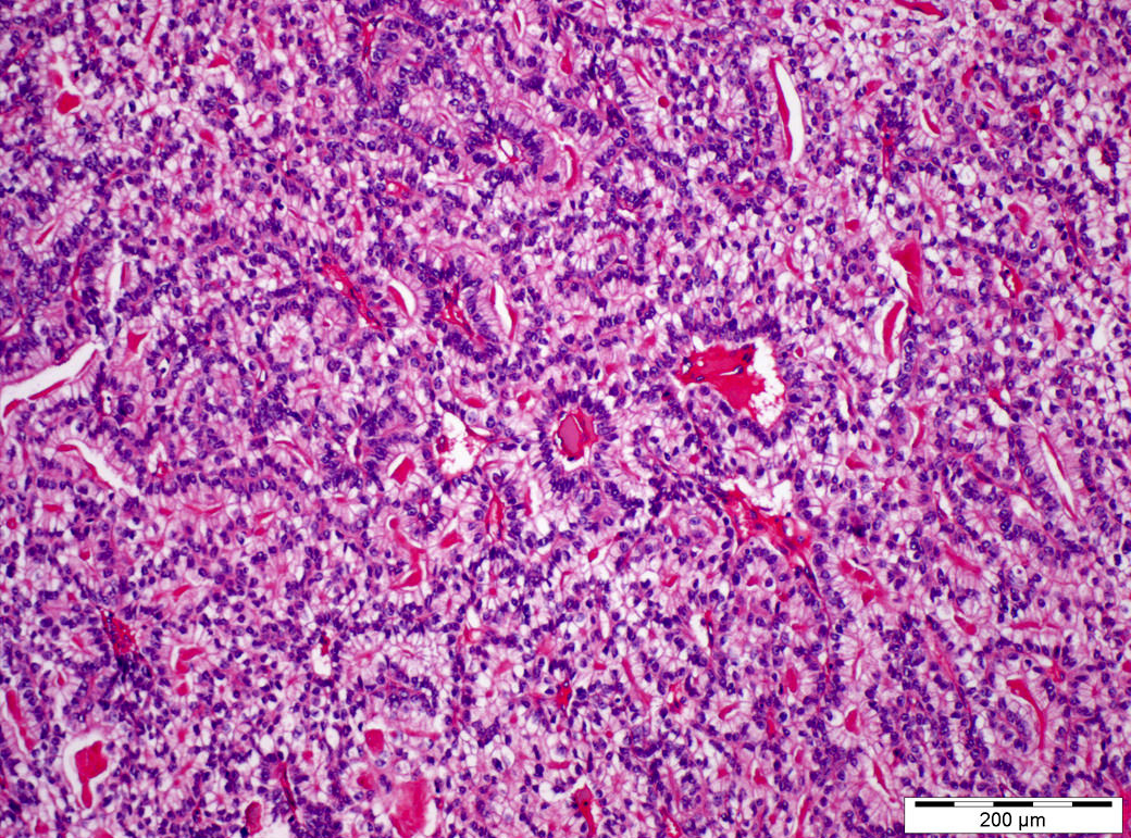
Interface with testis
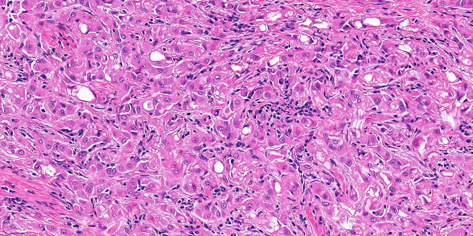
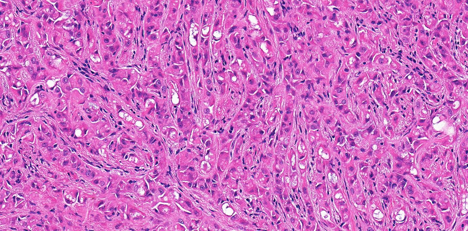
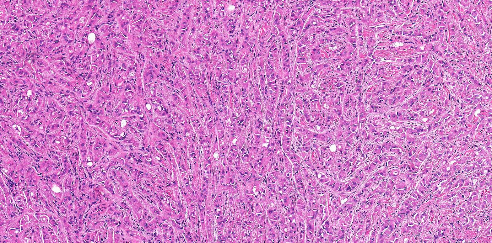
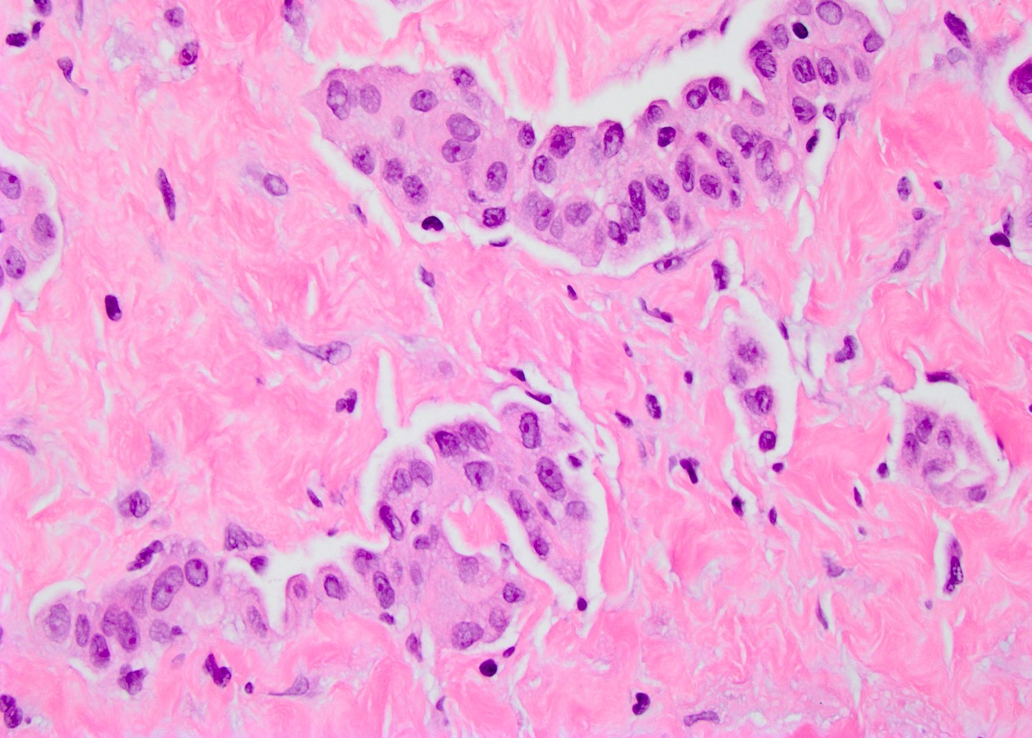
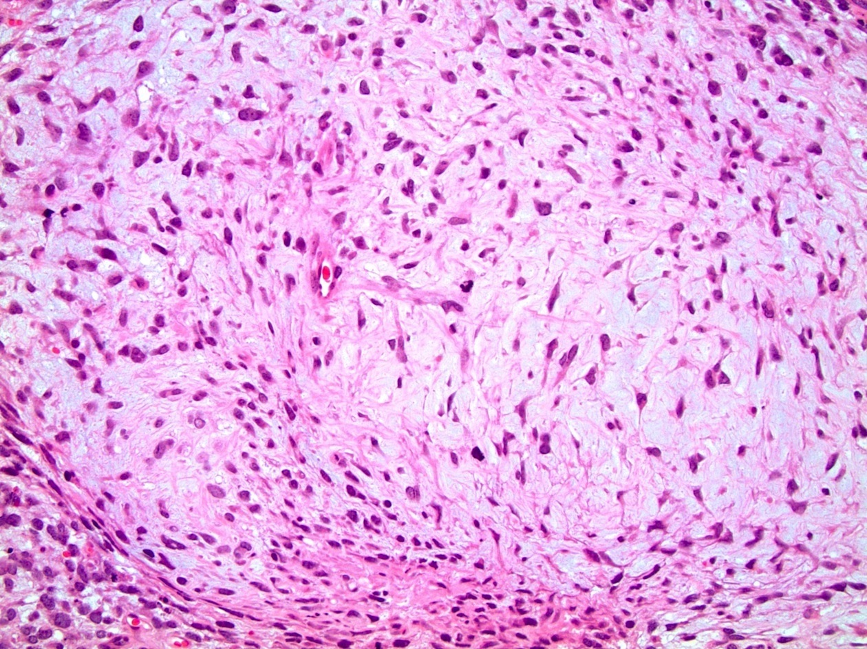
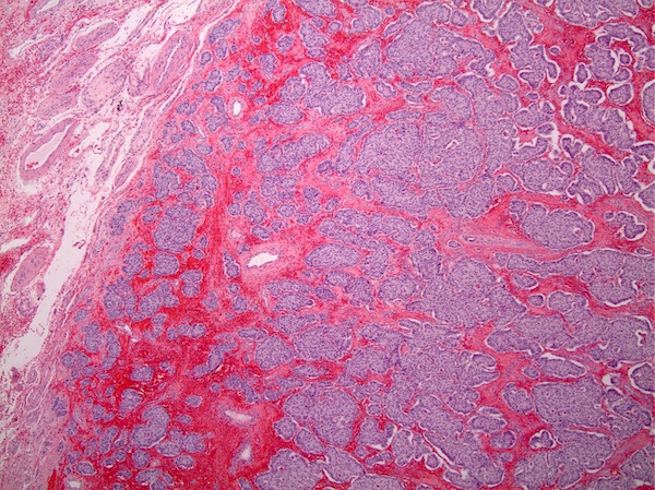
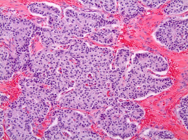
Cords and trabeculae pattern
Eosinophilic cells
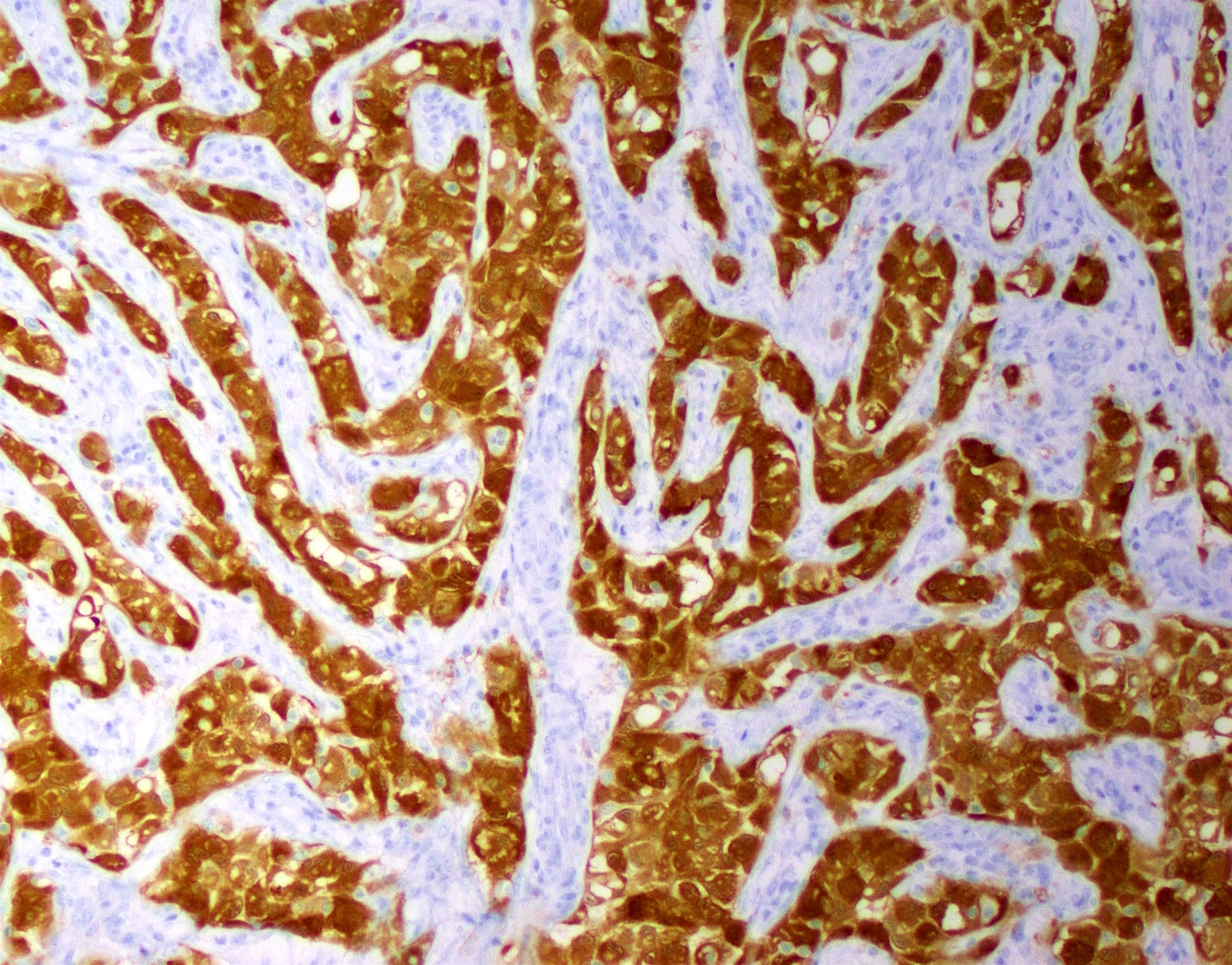
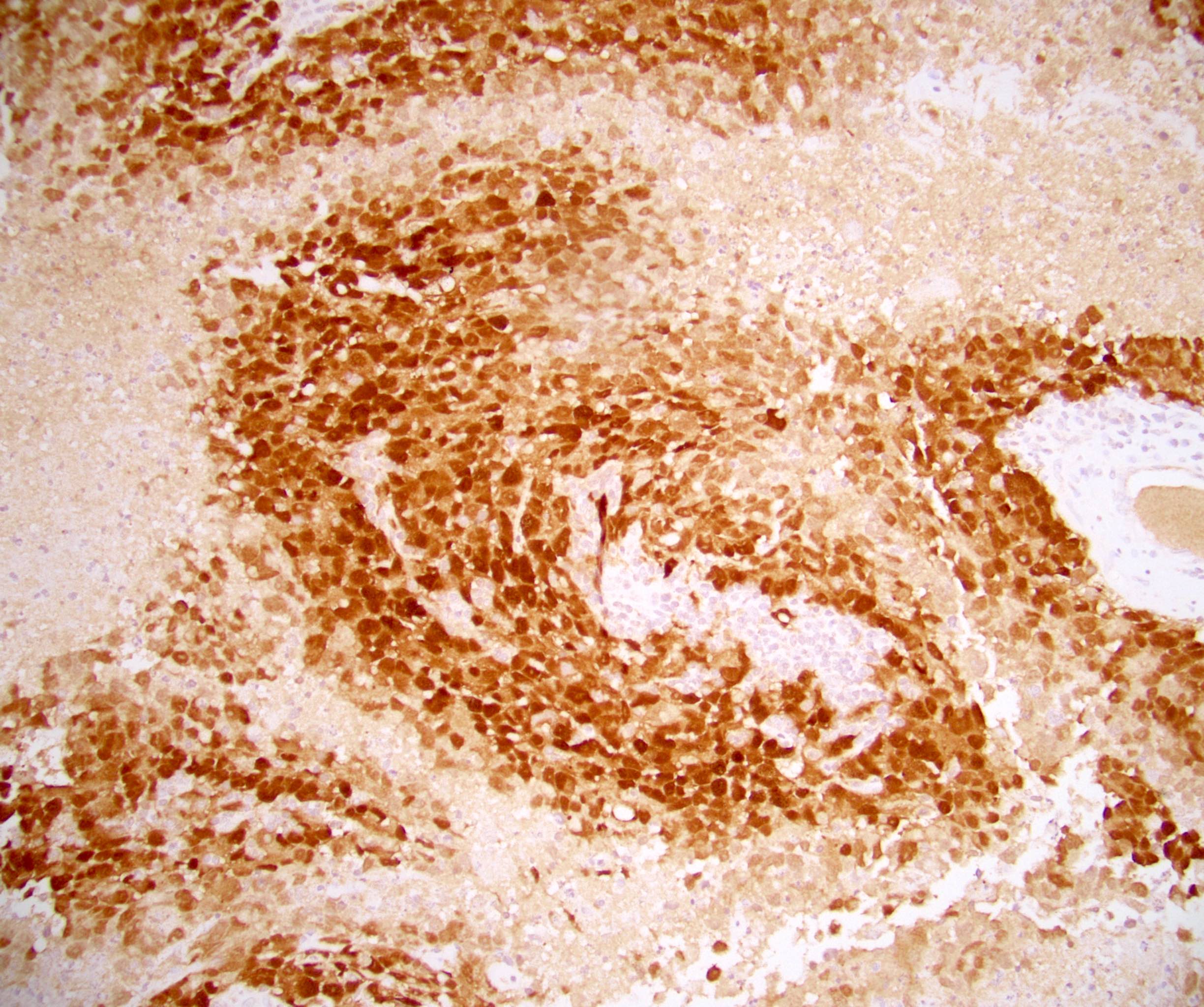
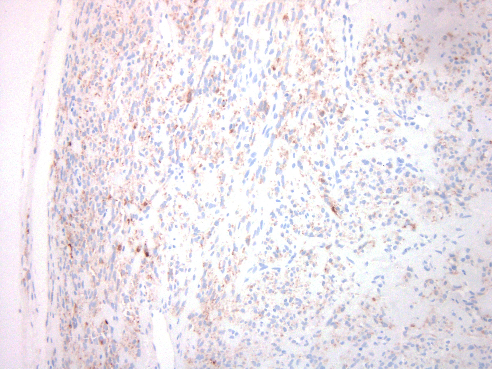
Calretinin
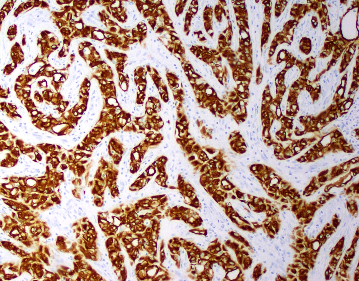
CK AE1/3
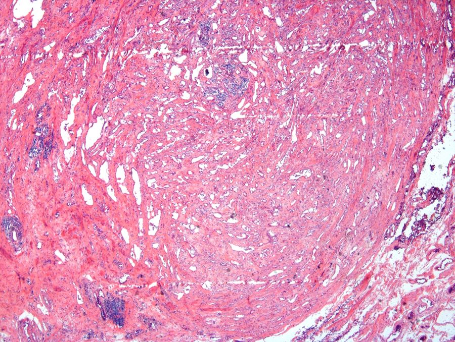
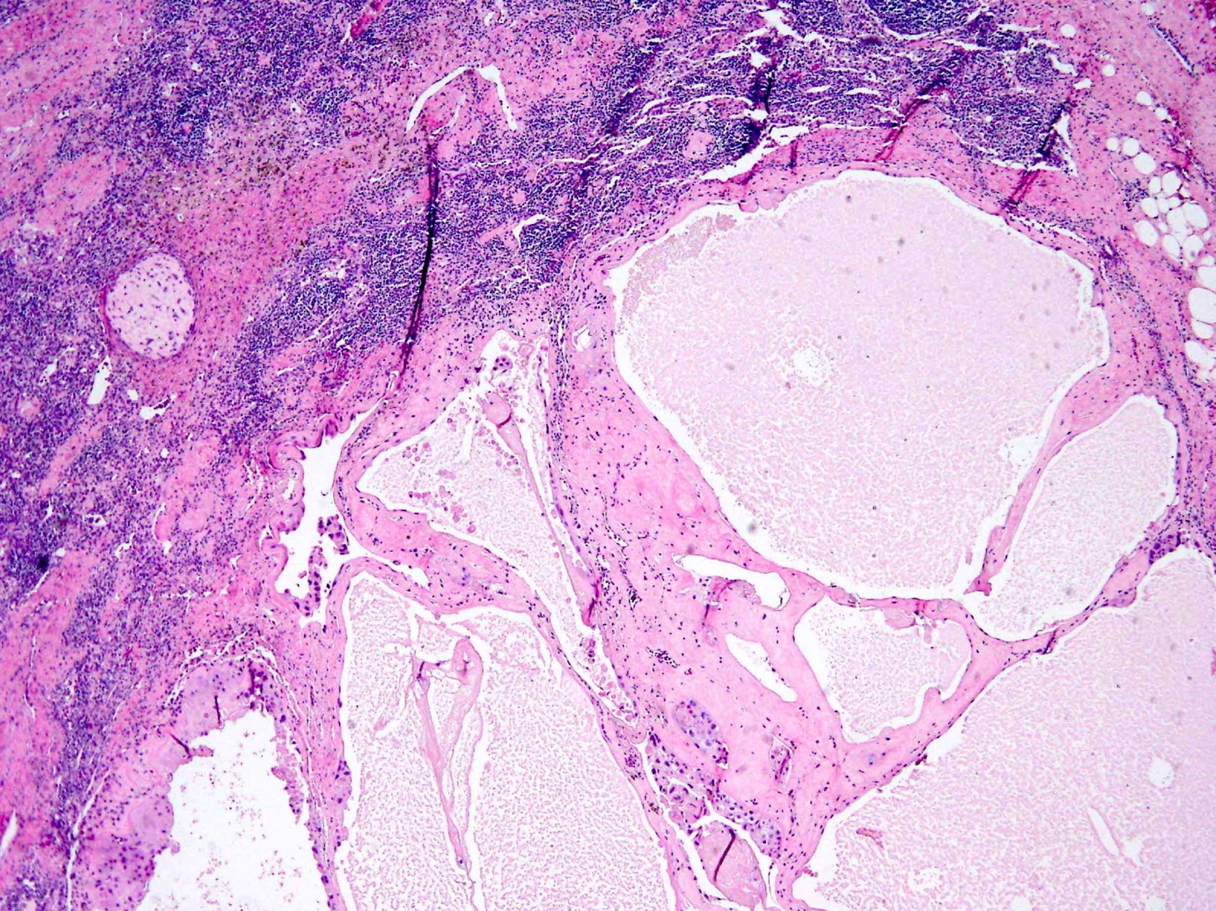
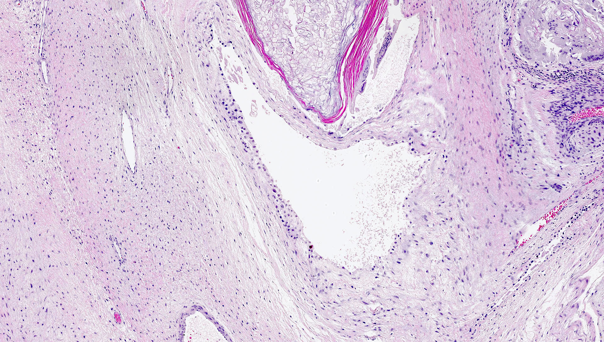
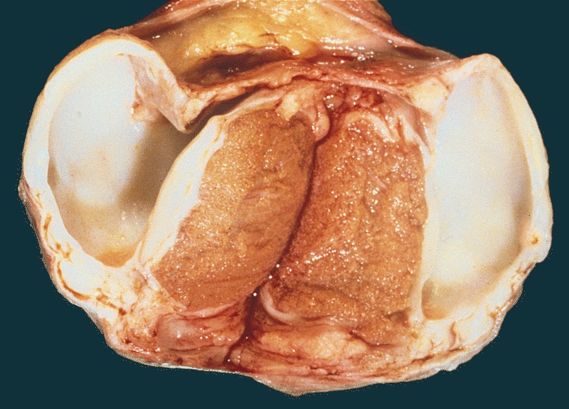
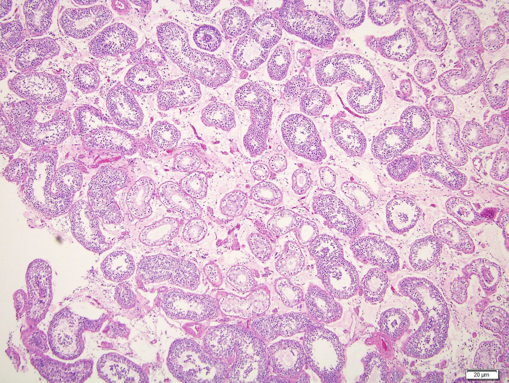
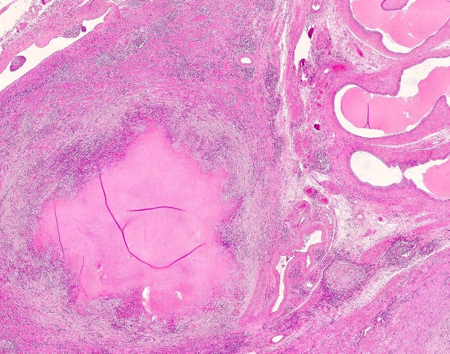
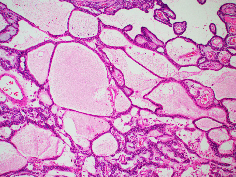
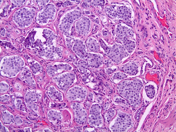
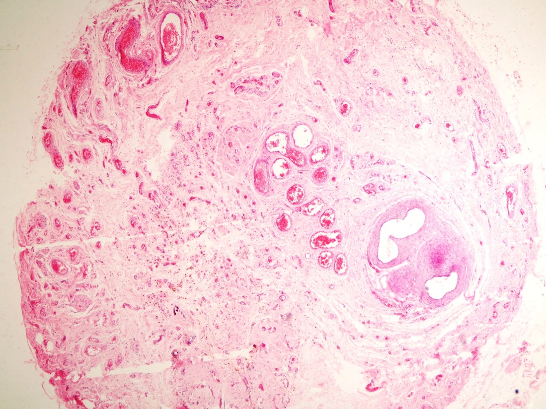
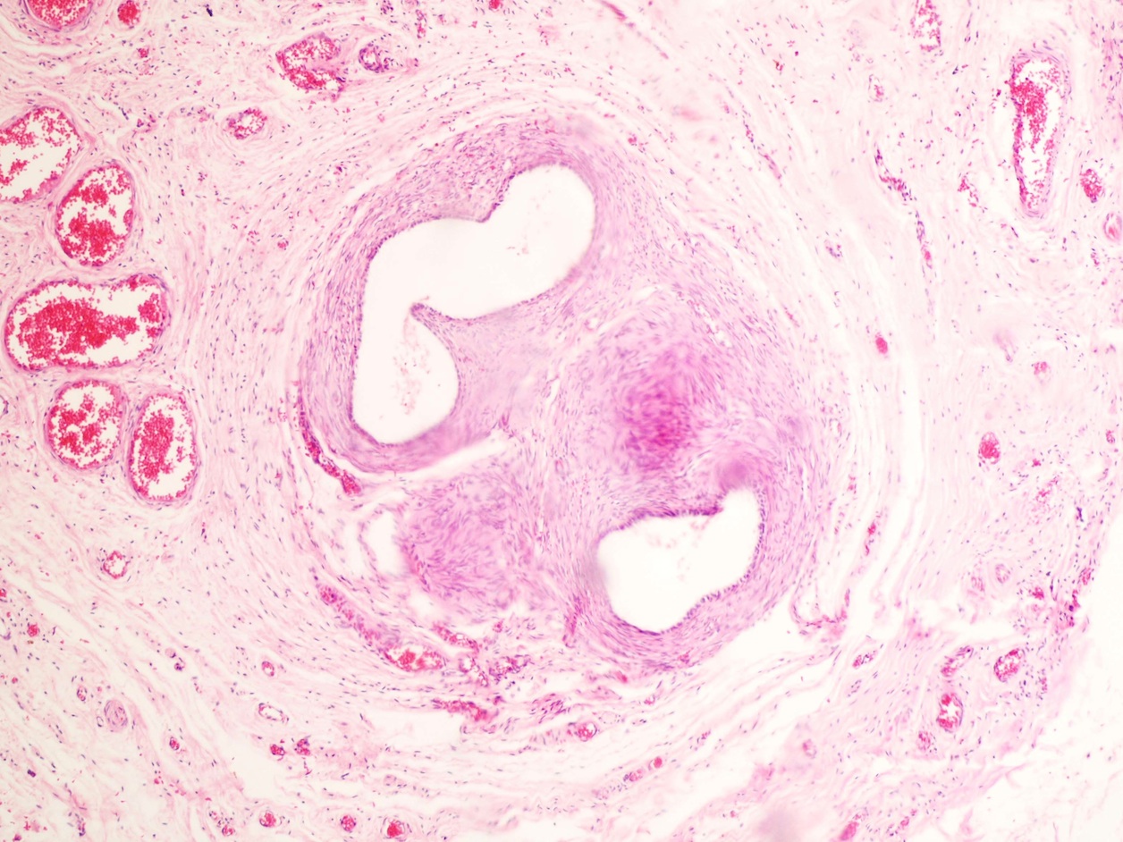
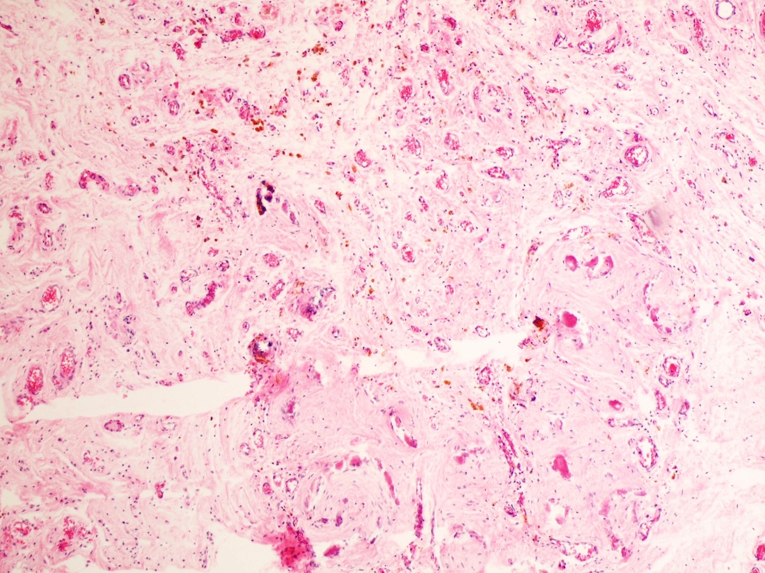
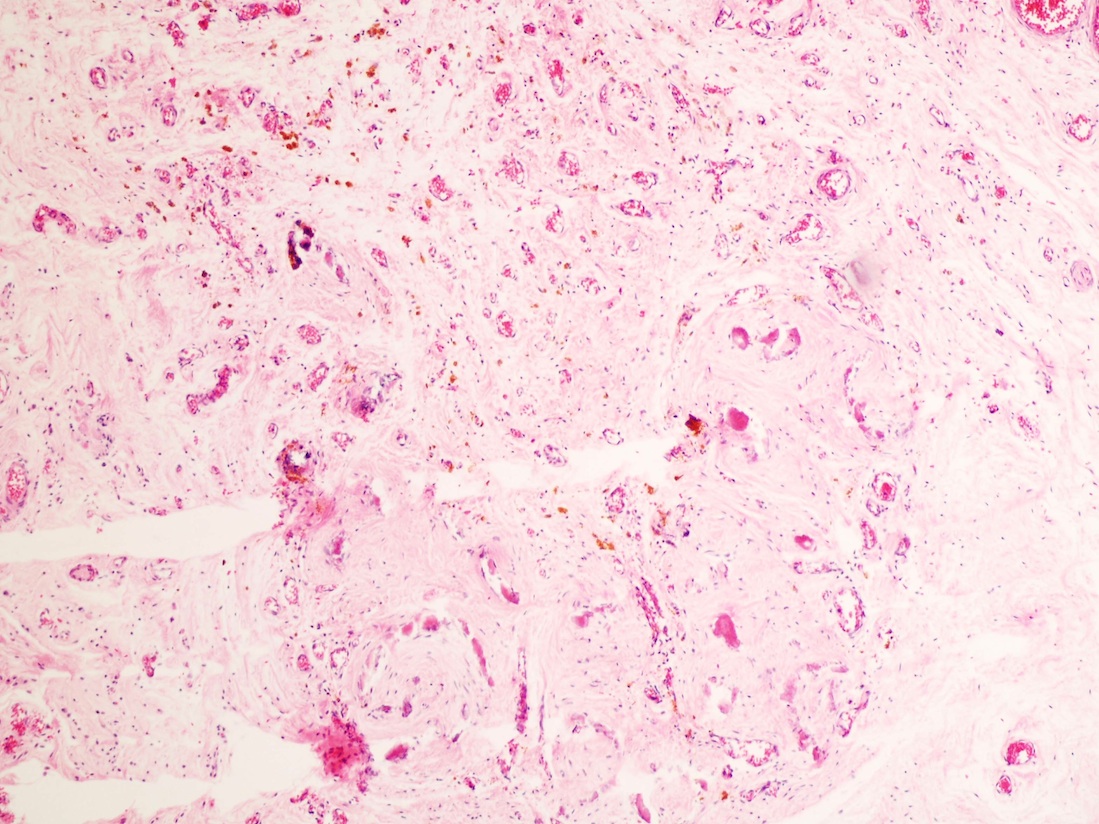
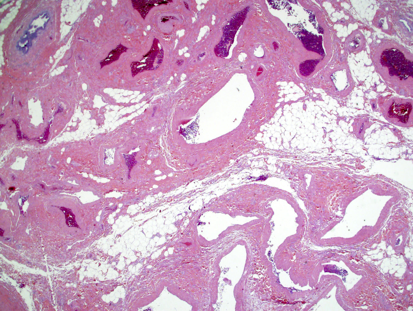
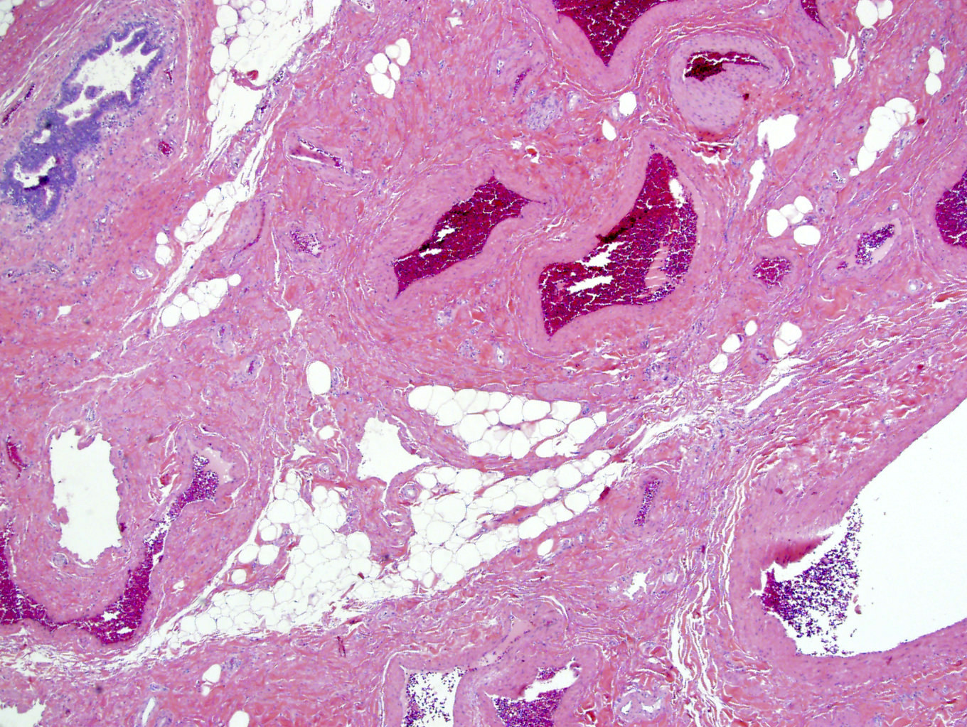
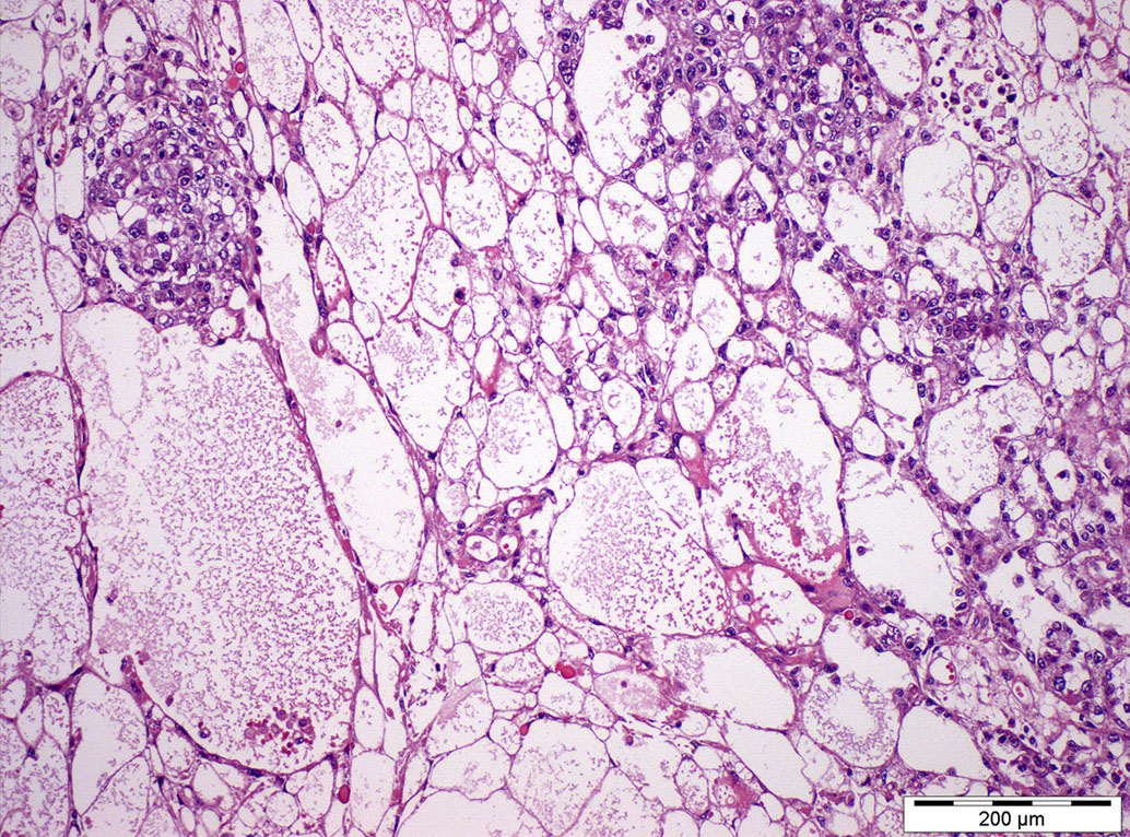
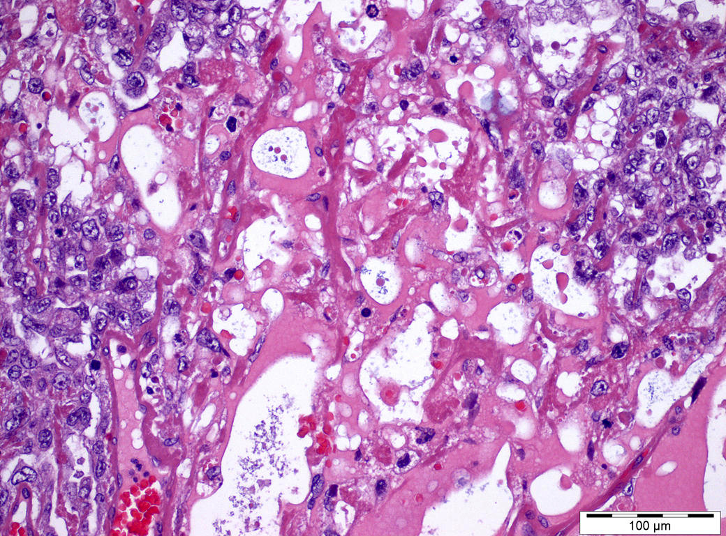
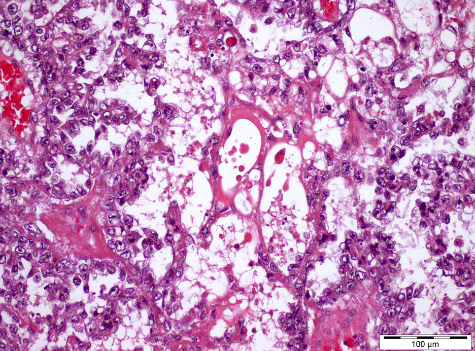
Circumscribed tumor
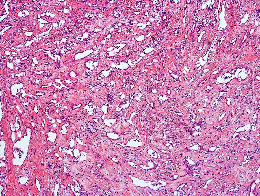
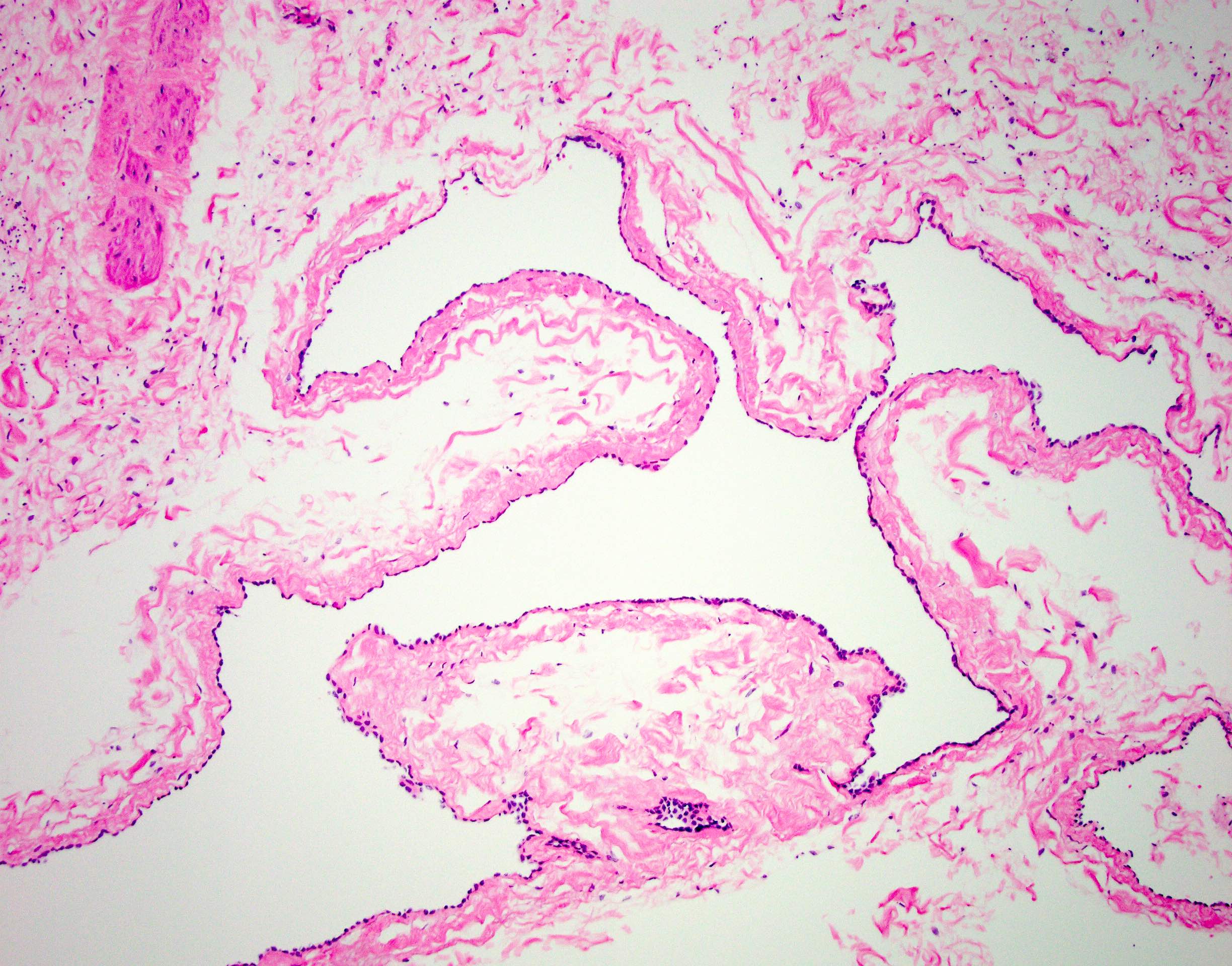
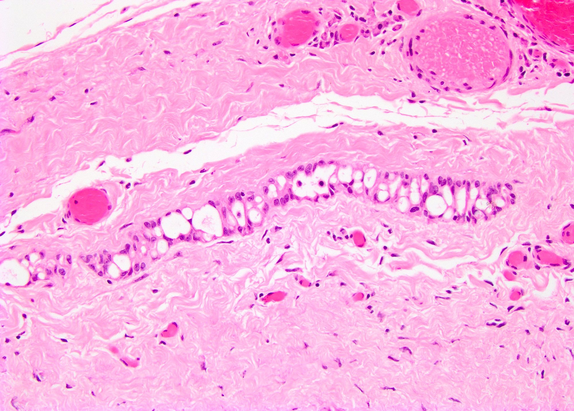
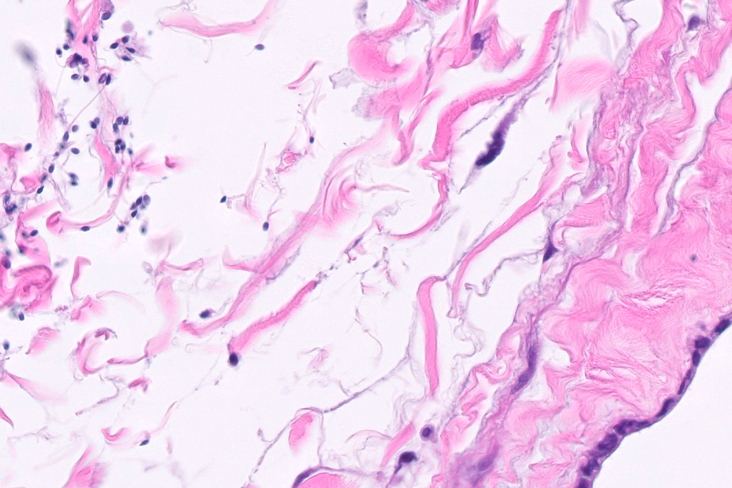
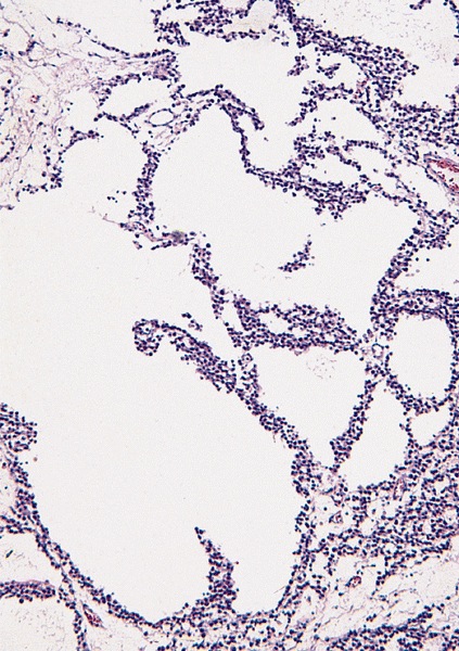
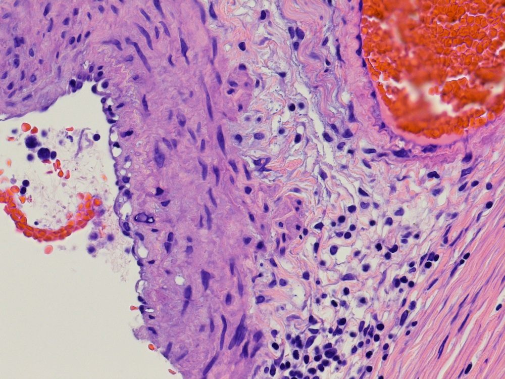
Dilated cystic spaces
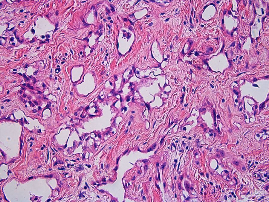
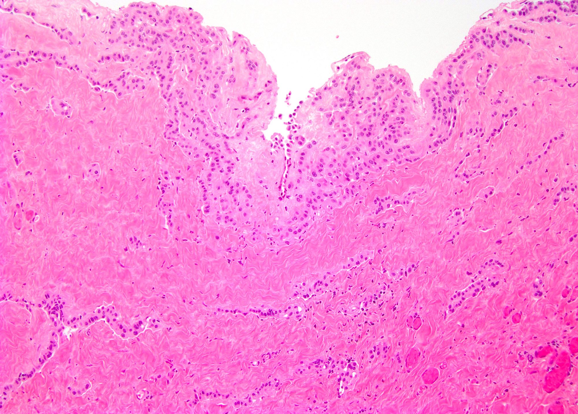
Vacuolated and attenuated epithelium
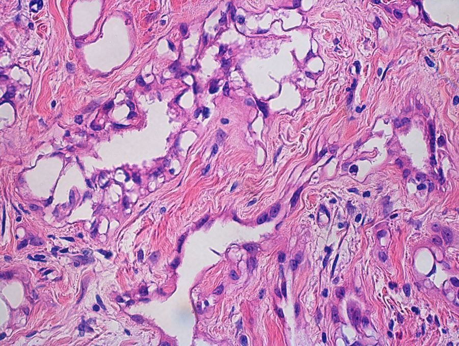
Bland nuclei
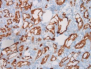
Calretinin
Adenomatoid tumor
Adenomatoid tumor
Adenomatoid tumor
Adenomatoid tumor
Images hosted on other servers:

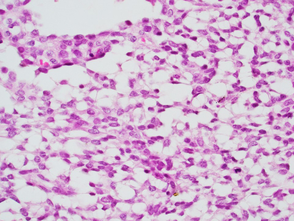
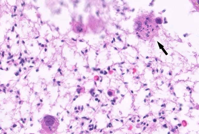
Moderate cellularity

Monolayered sheets of cells
Contributed by Yuto Yamazaki, M.D., Ph.D., Hironobu Sasano, M.D., Ph.D., Debra L. Zynger, M.D. and Sean R. Williamson, M.D.
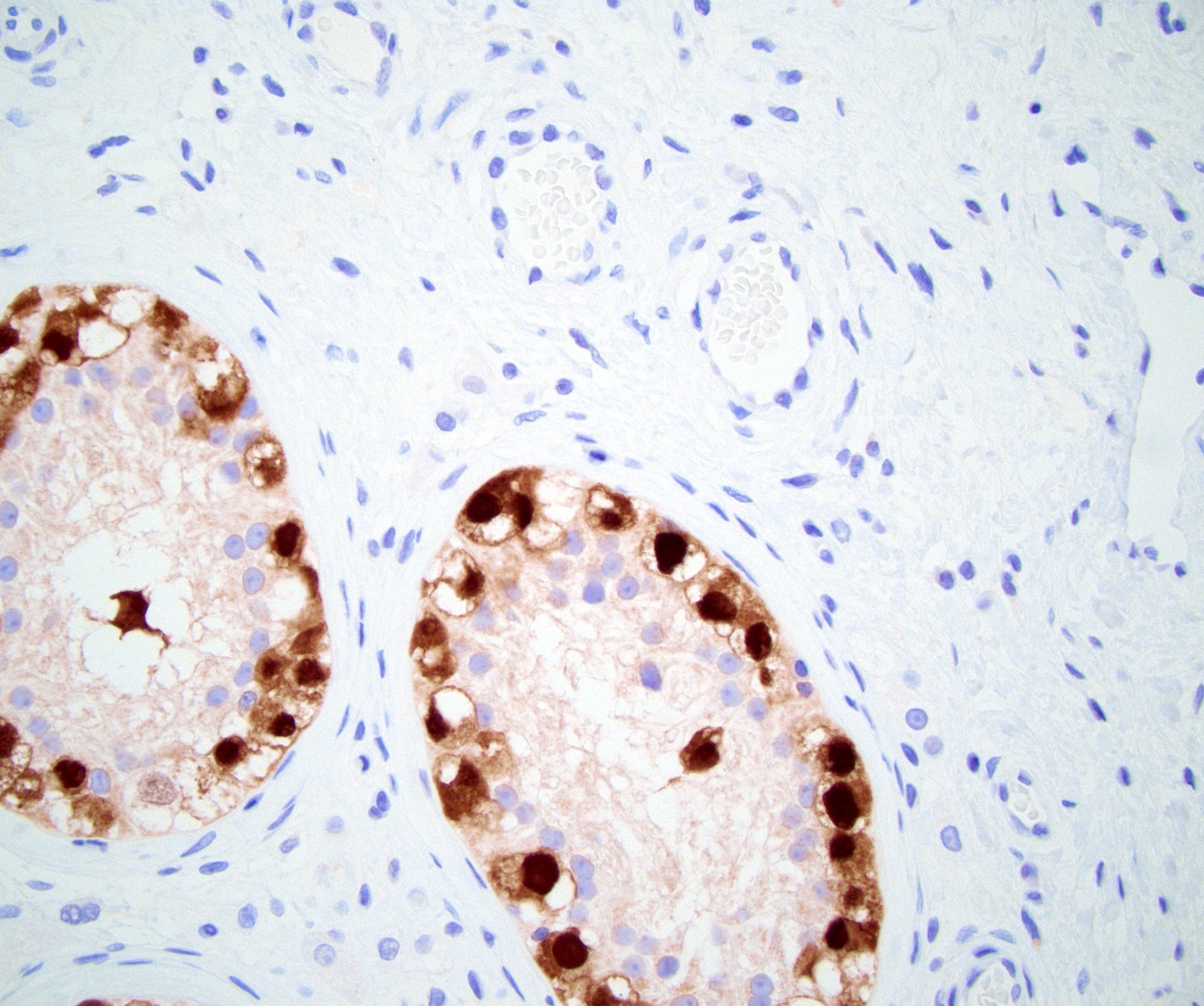
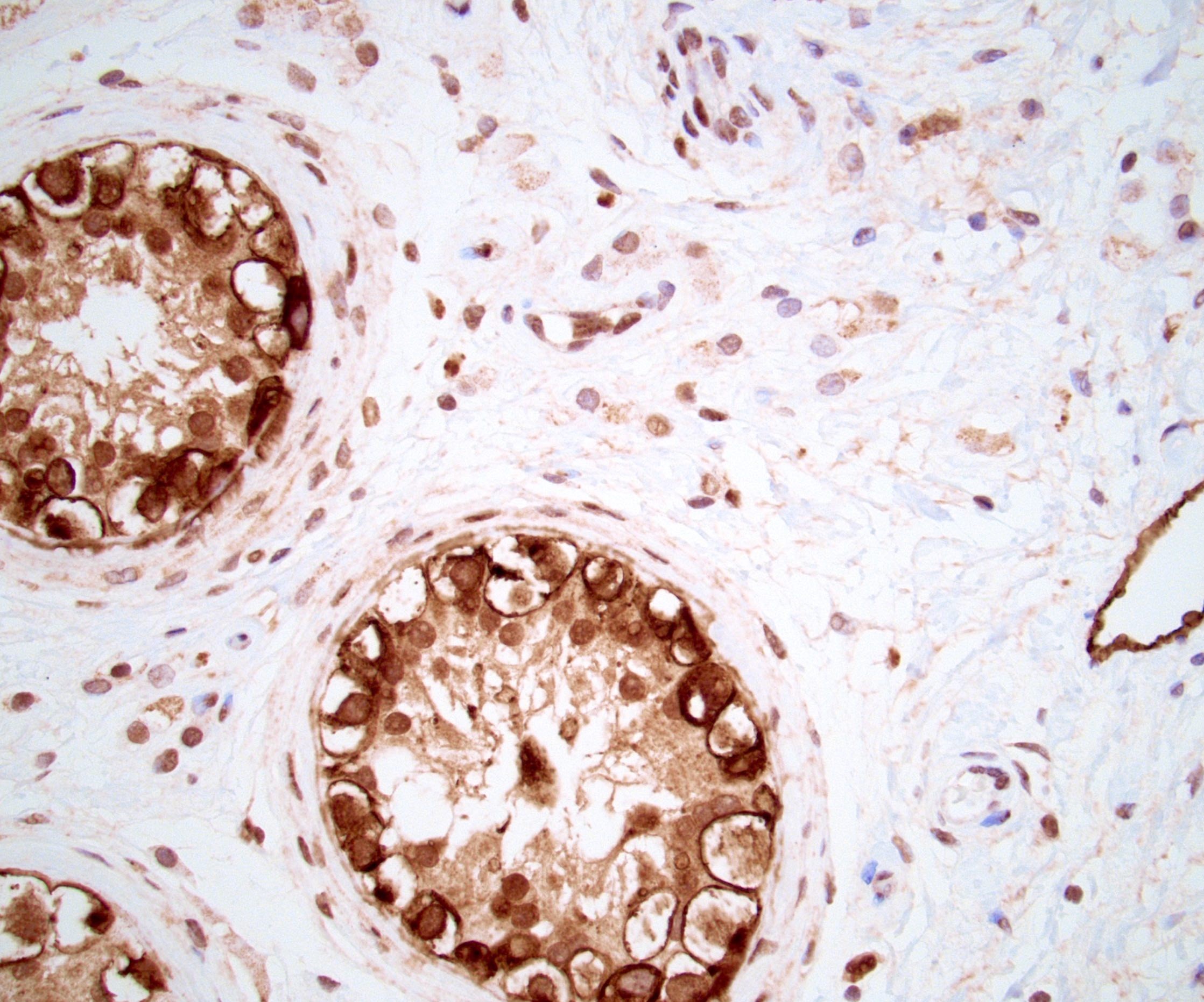
Spermatic cord adrenal rest and SF1
Spermatic cord adrenal rests
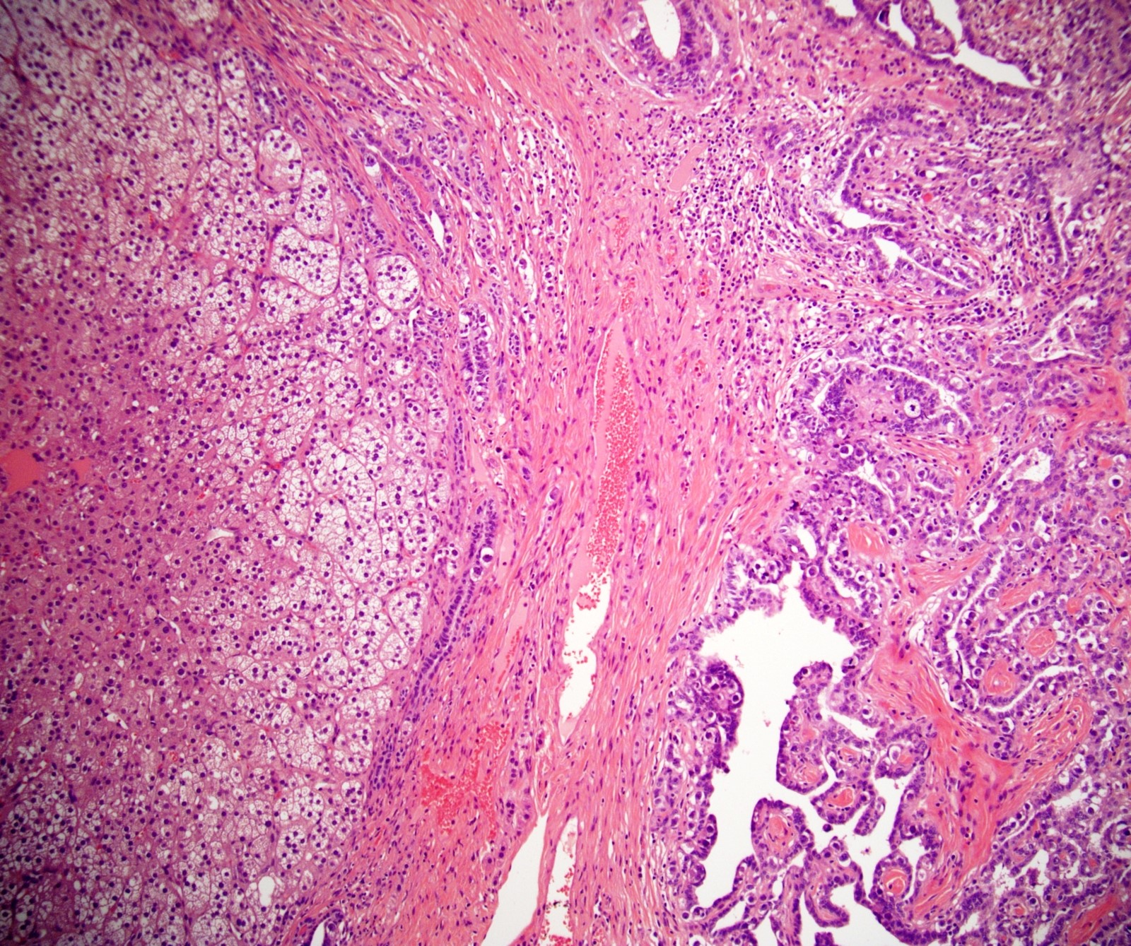
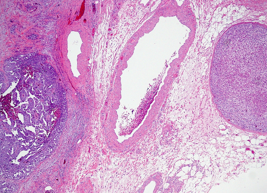
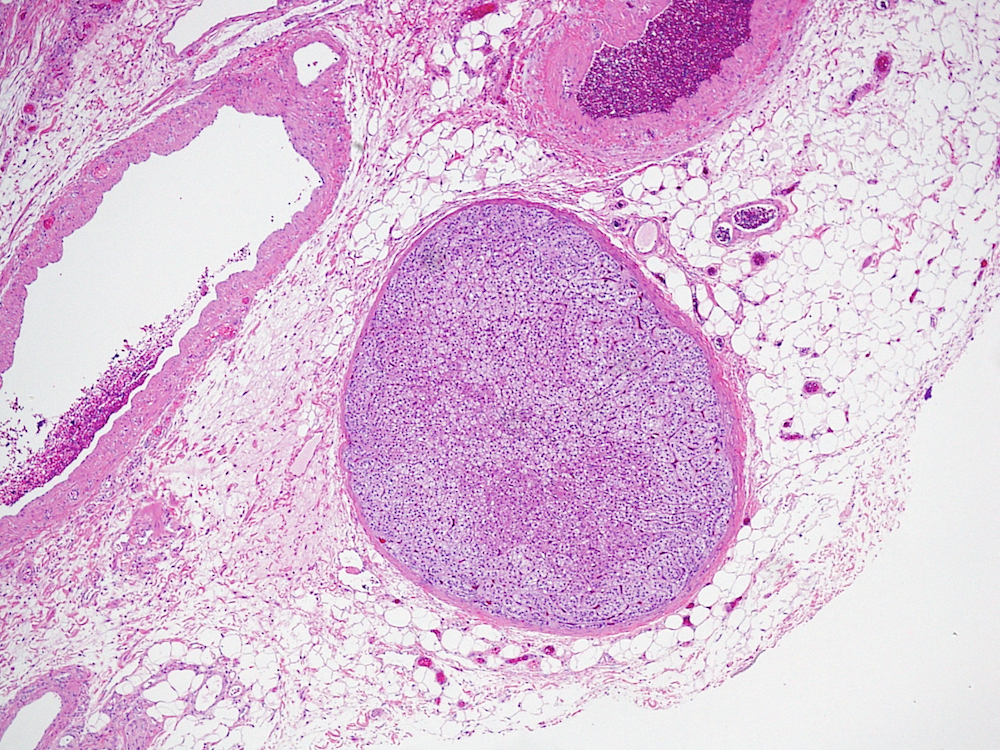
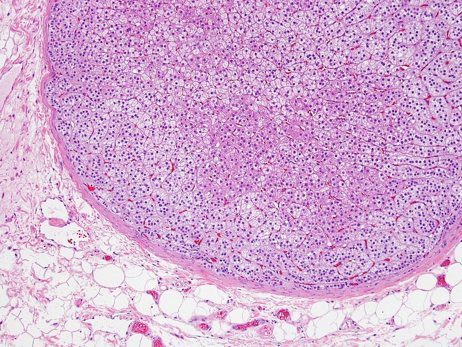
Paratesticular adrenal cortical rest
Images hosted on other servers:

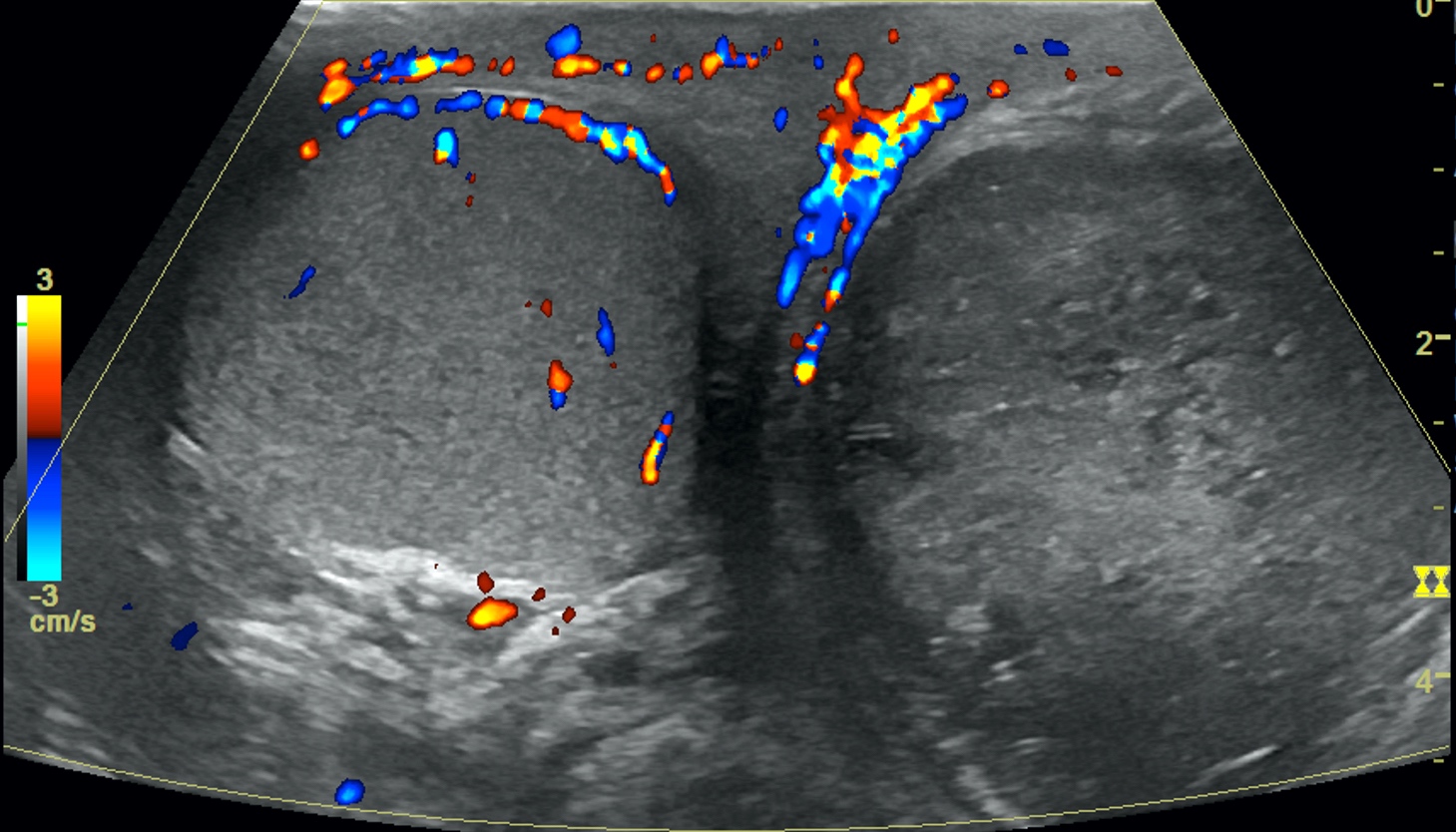
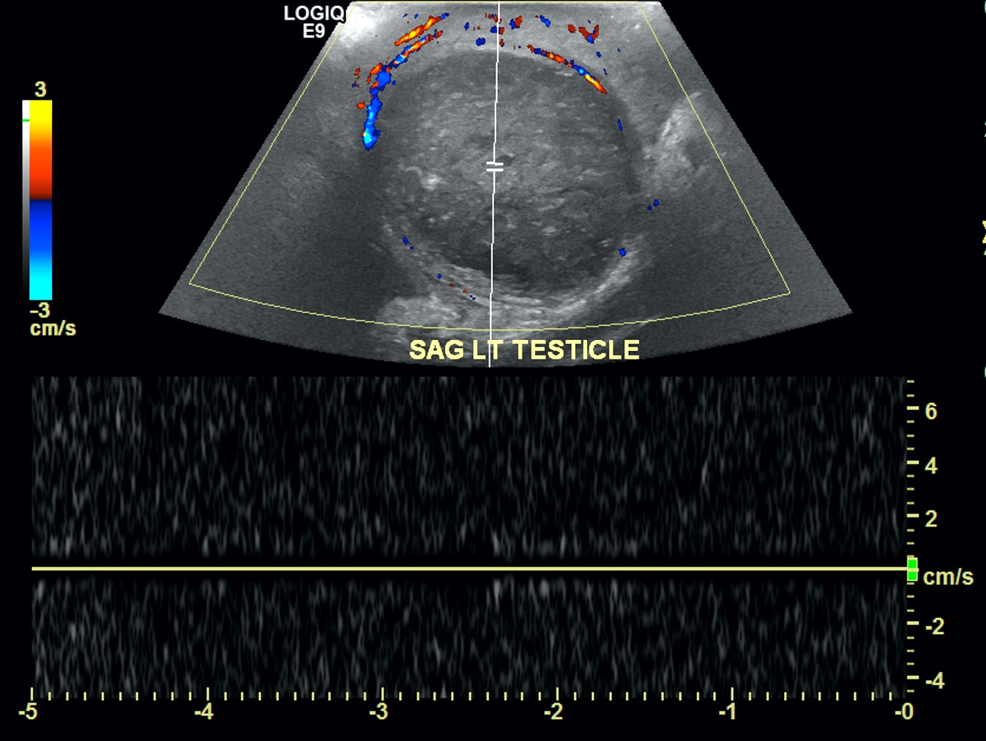
Hypoechoic lesion

Hypoechoic lesion /
increased vascularity

Increased vascularity
Contributed by Kristine M. Cornejo, M.D.
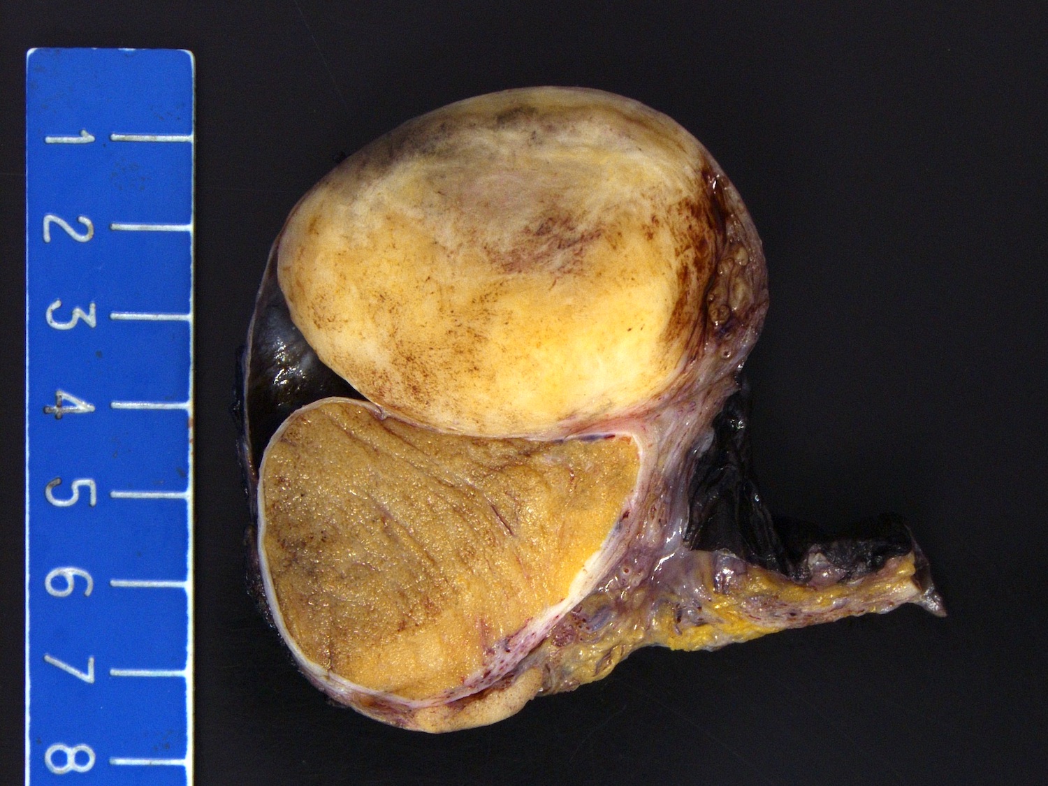
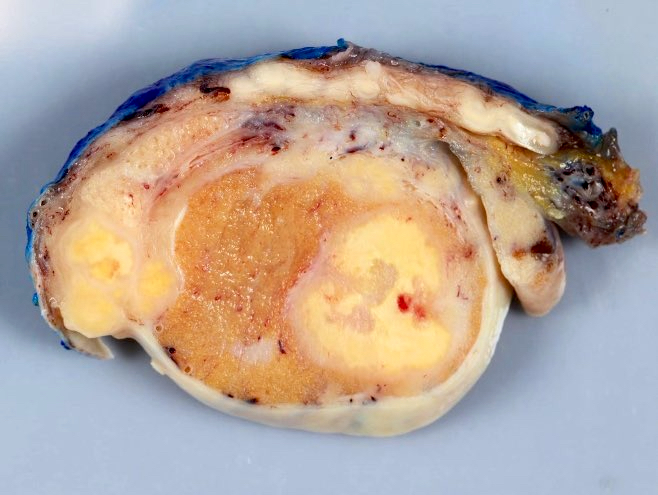
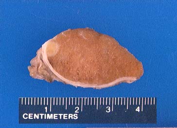
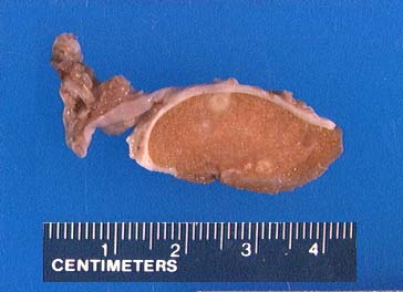
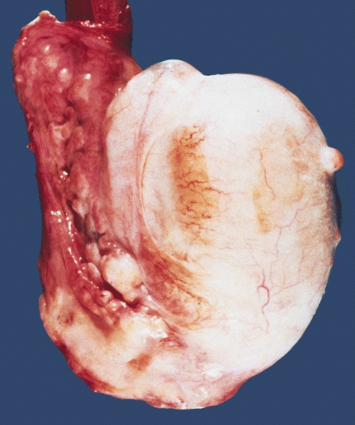
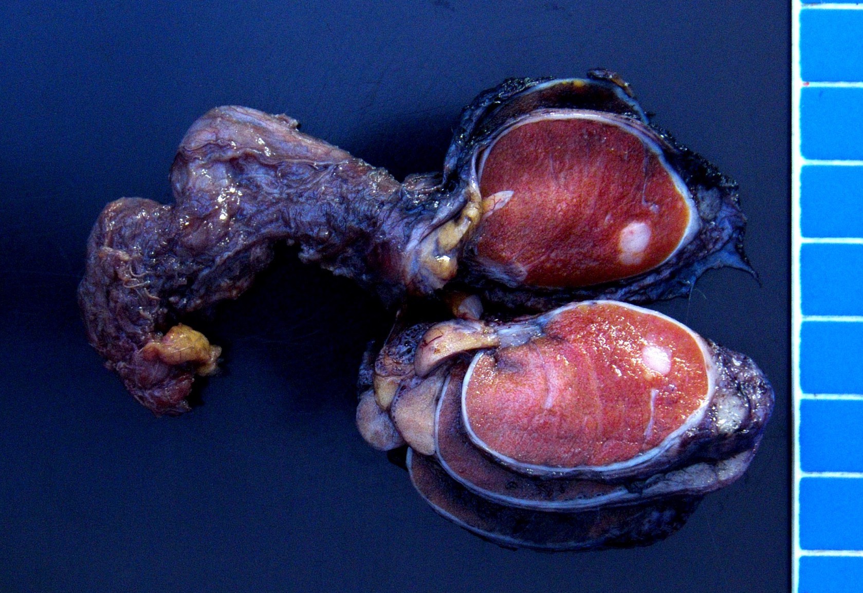
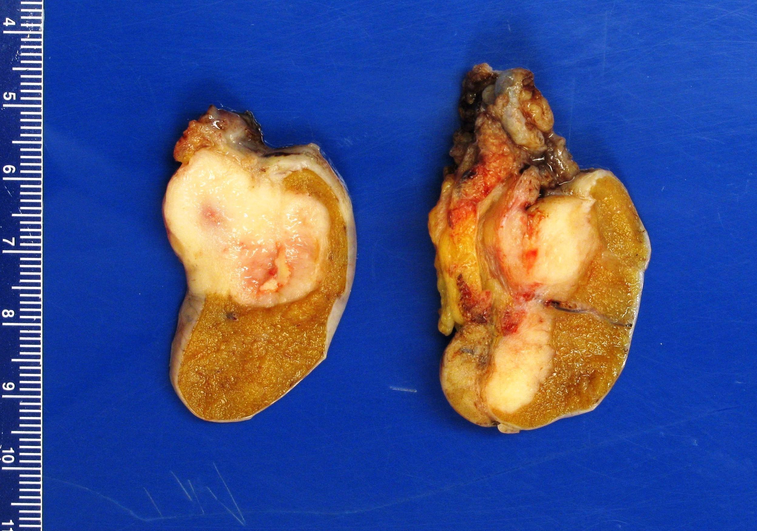
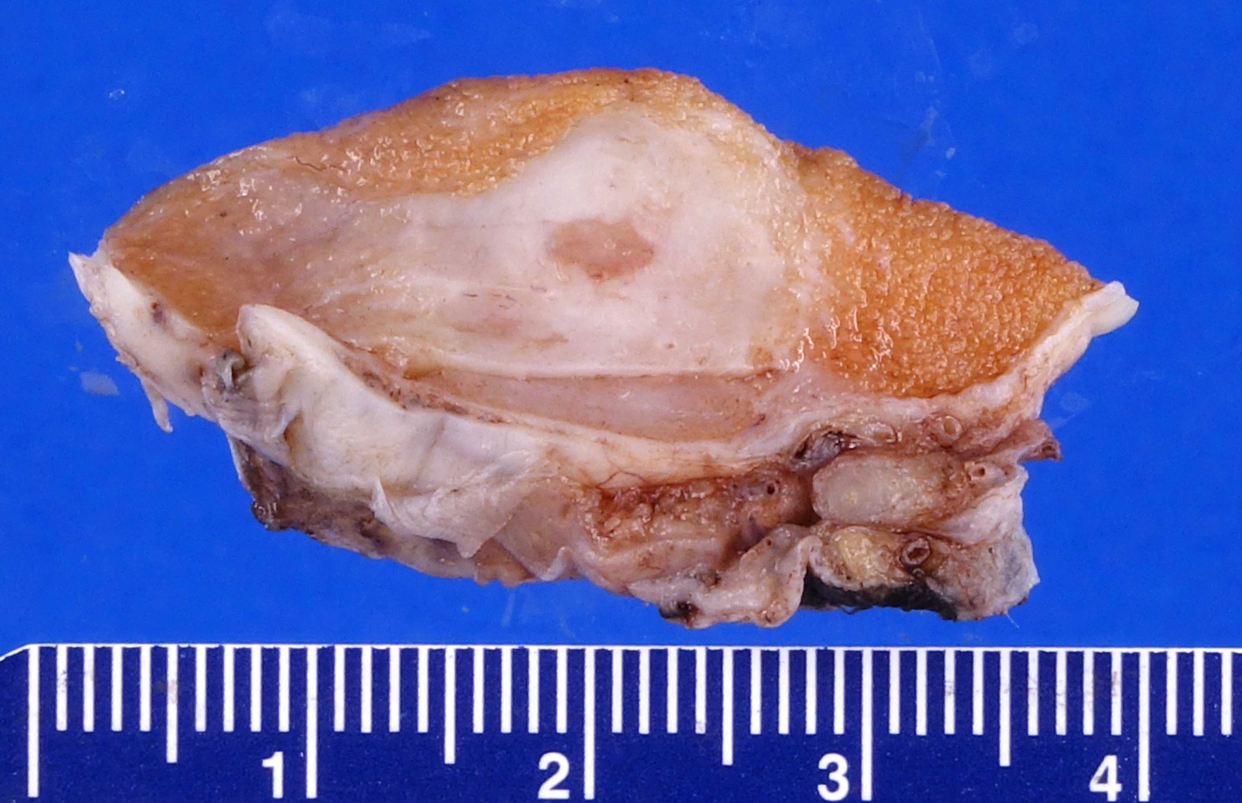
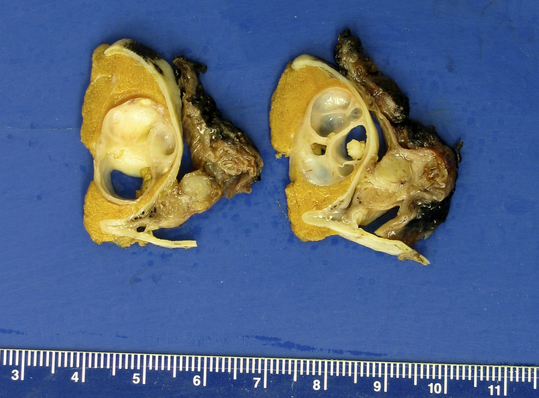
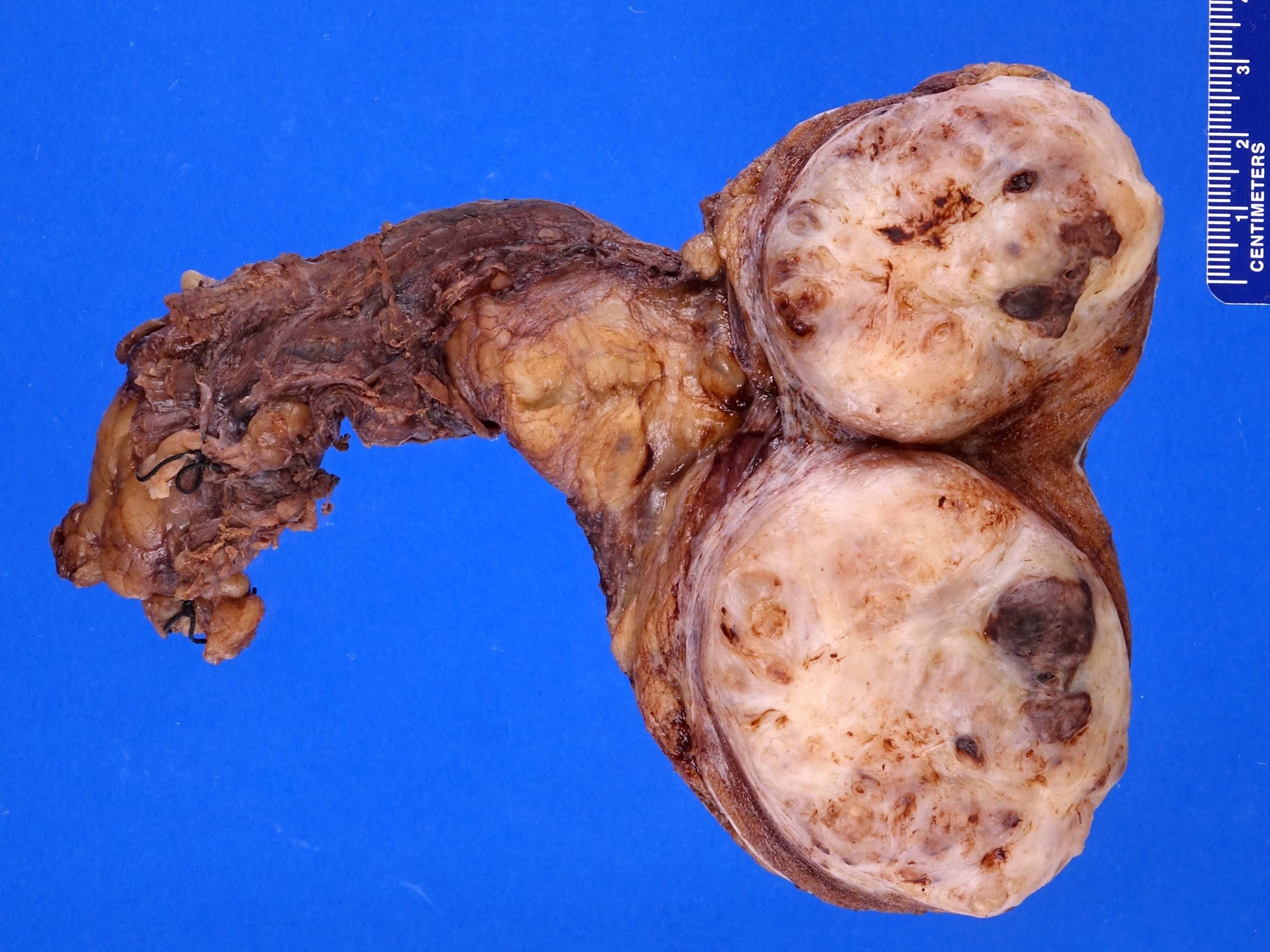
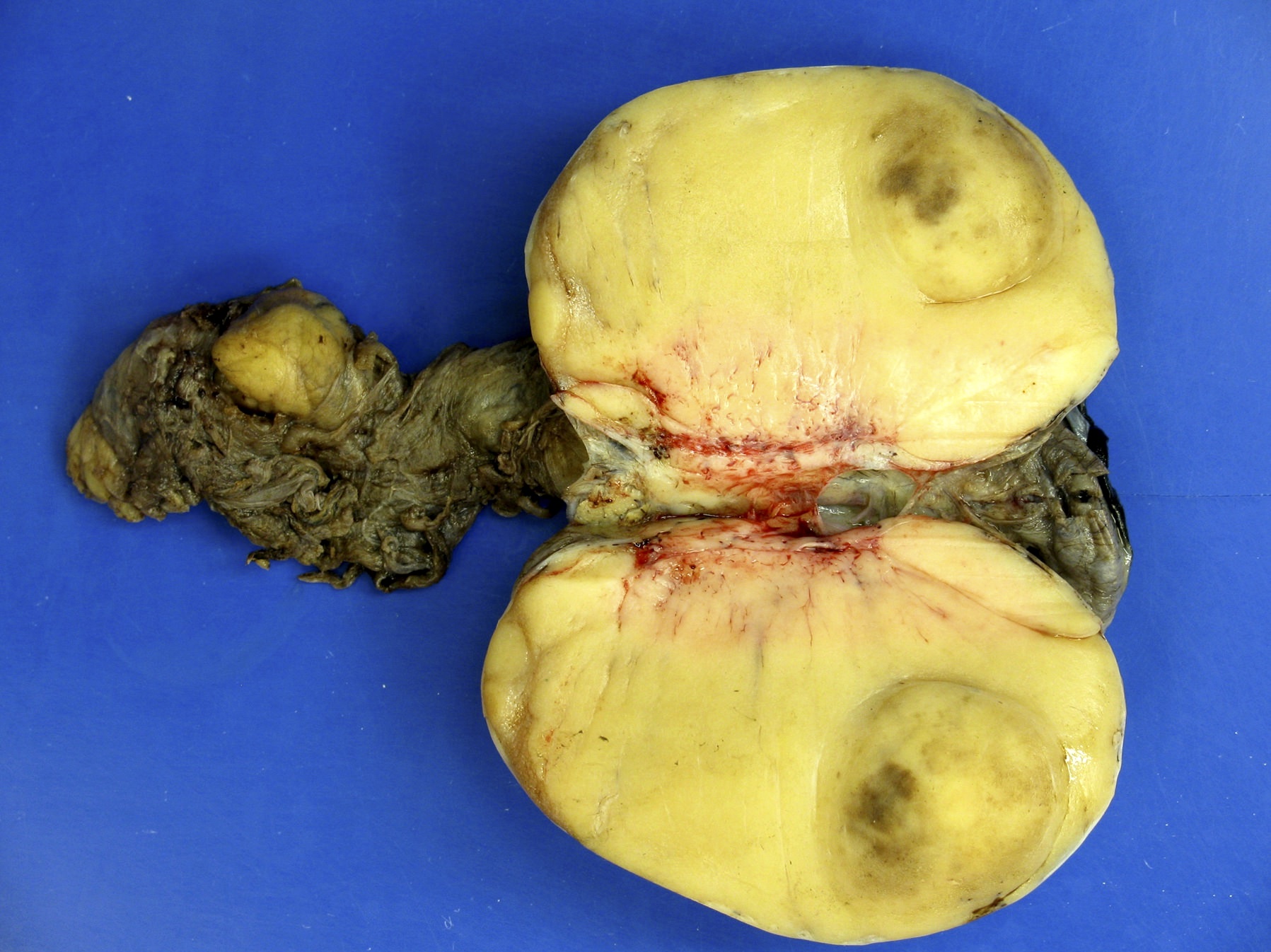
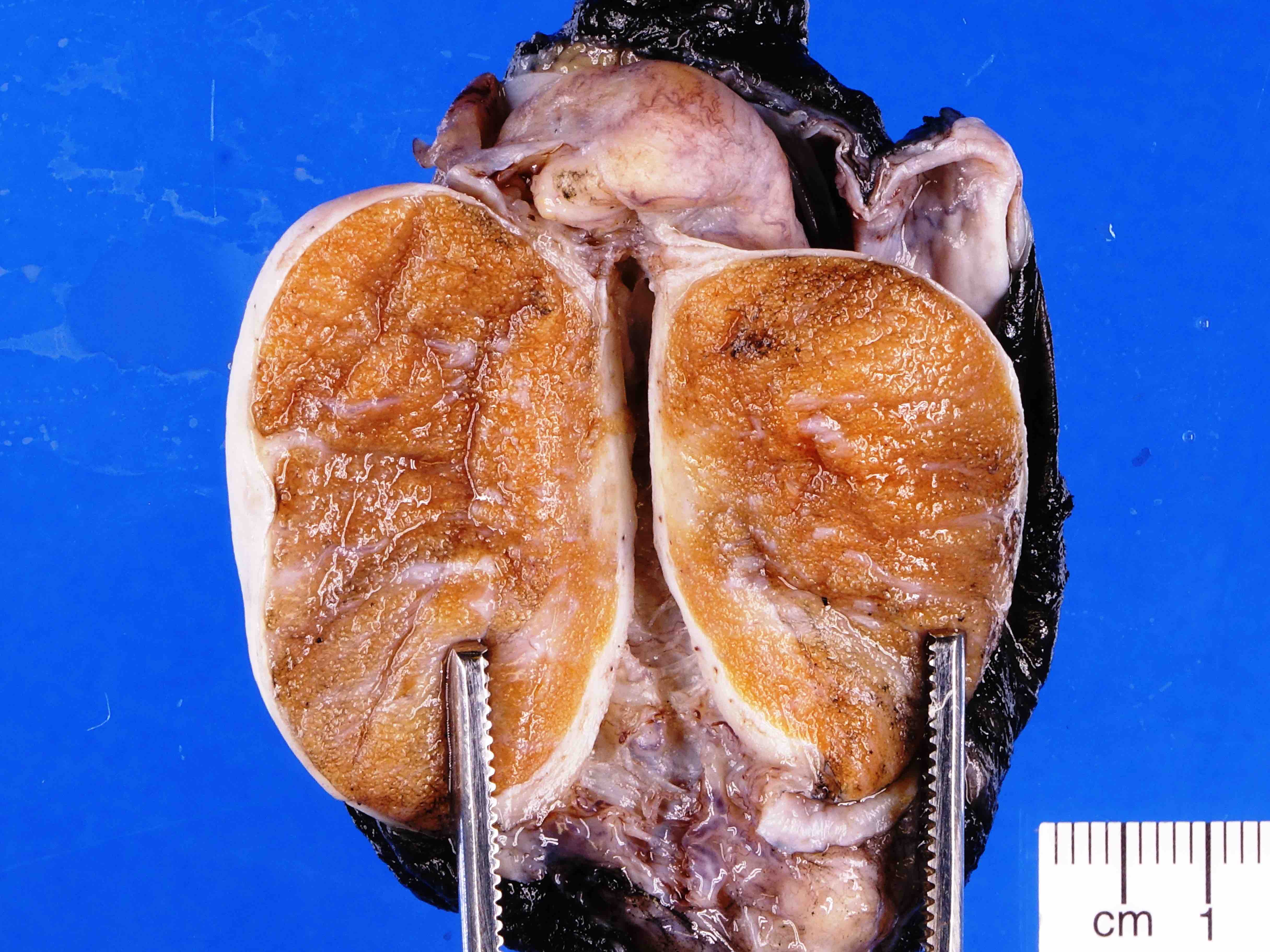
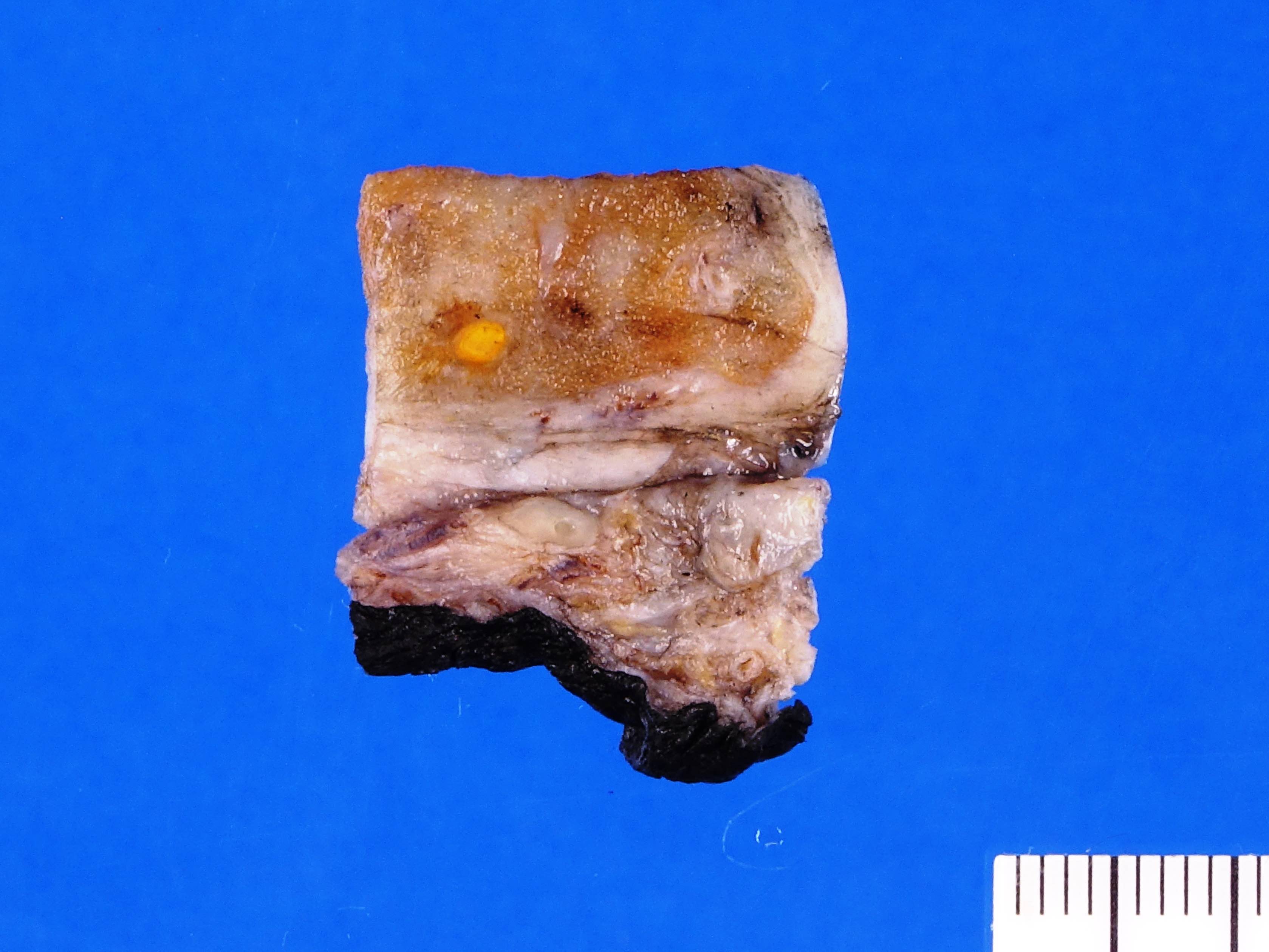
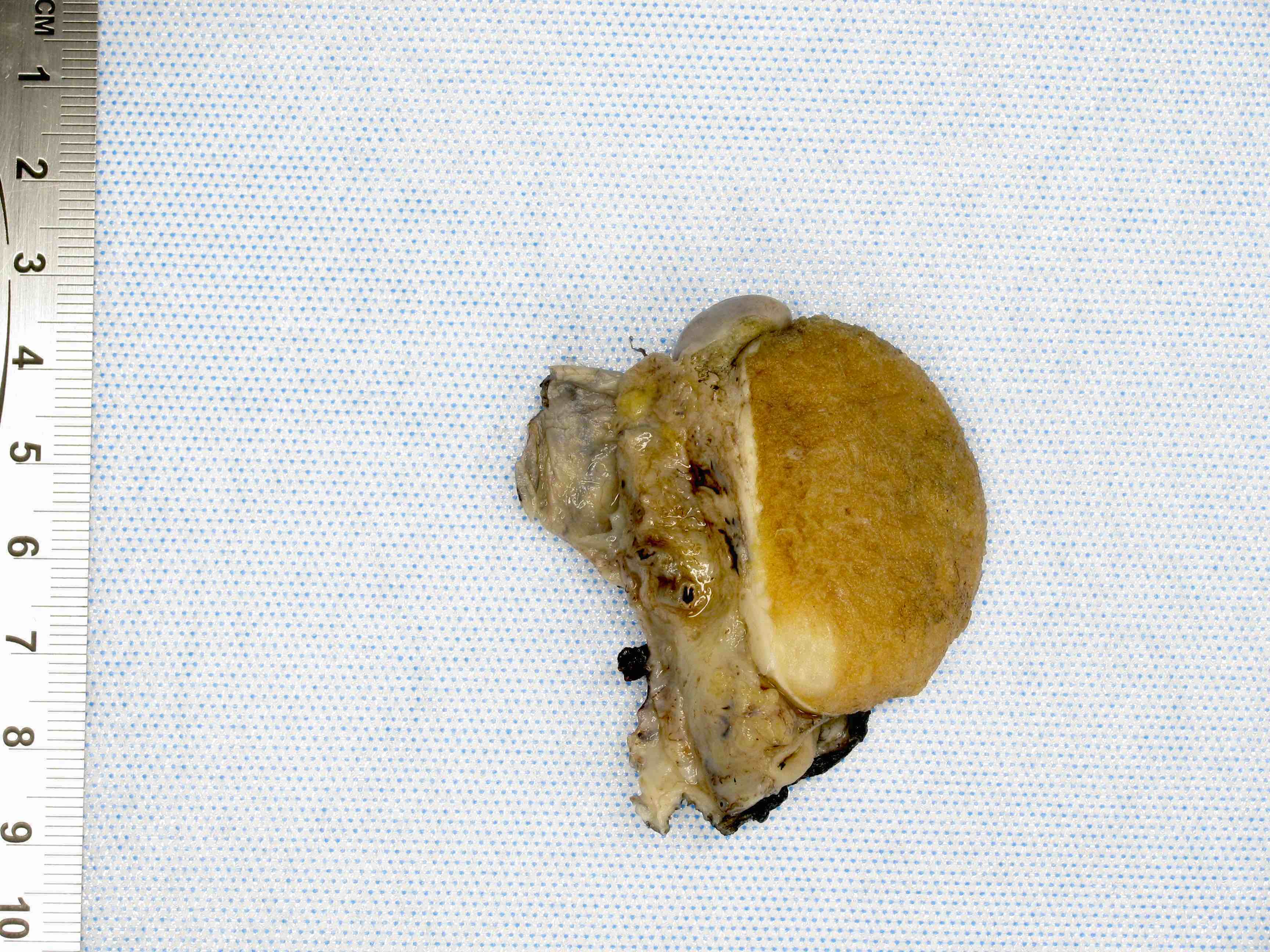
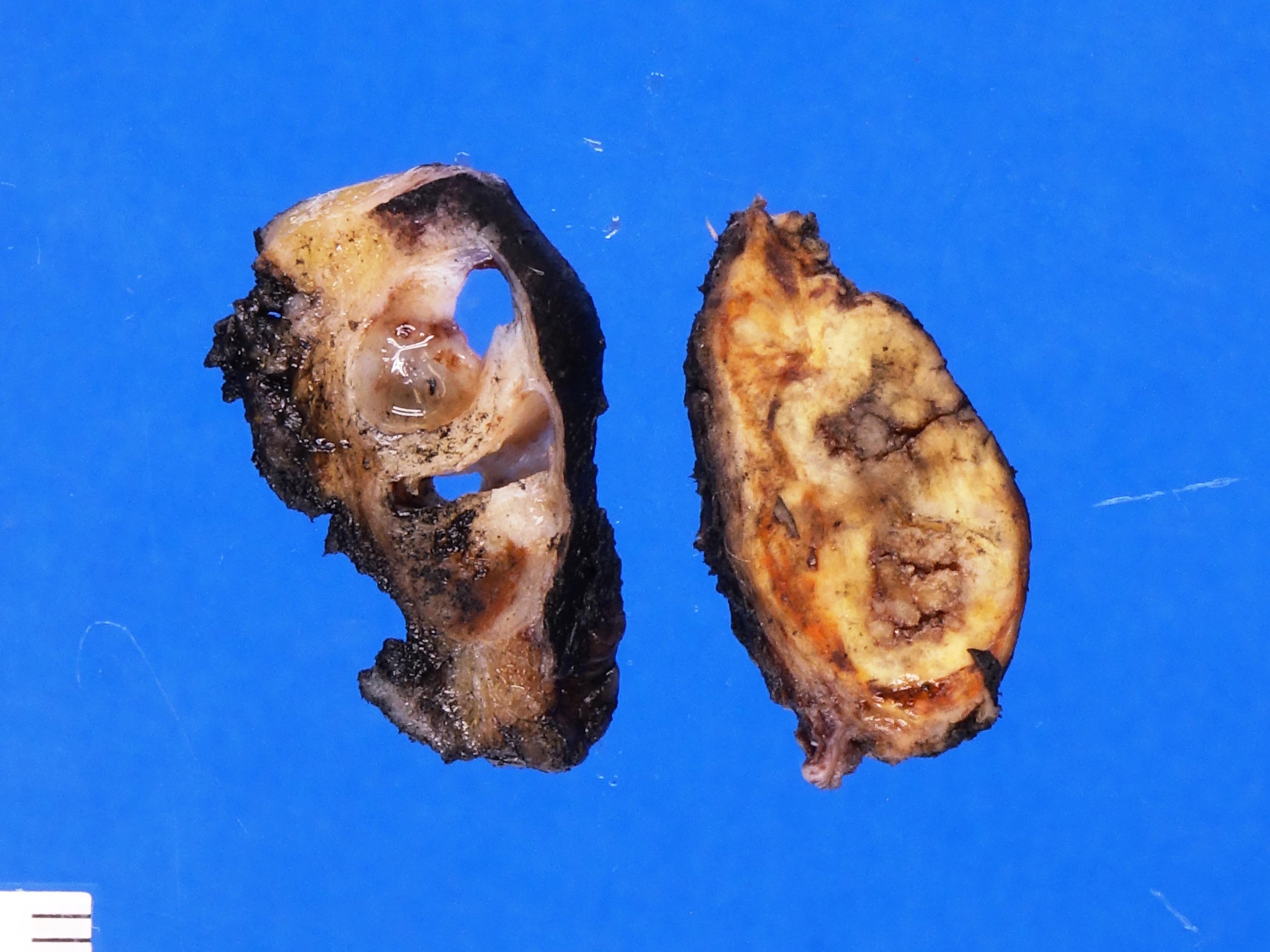
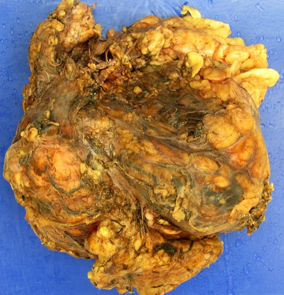
Tan-yellow solid mass
Lobulated tan-yellow mass
Contributed by Kristine M. Cornejo, M.D.
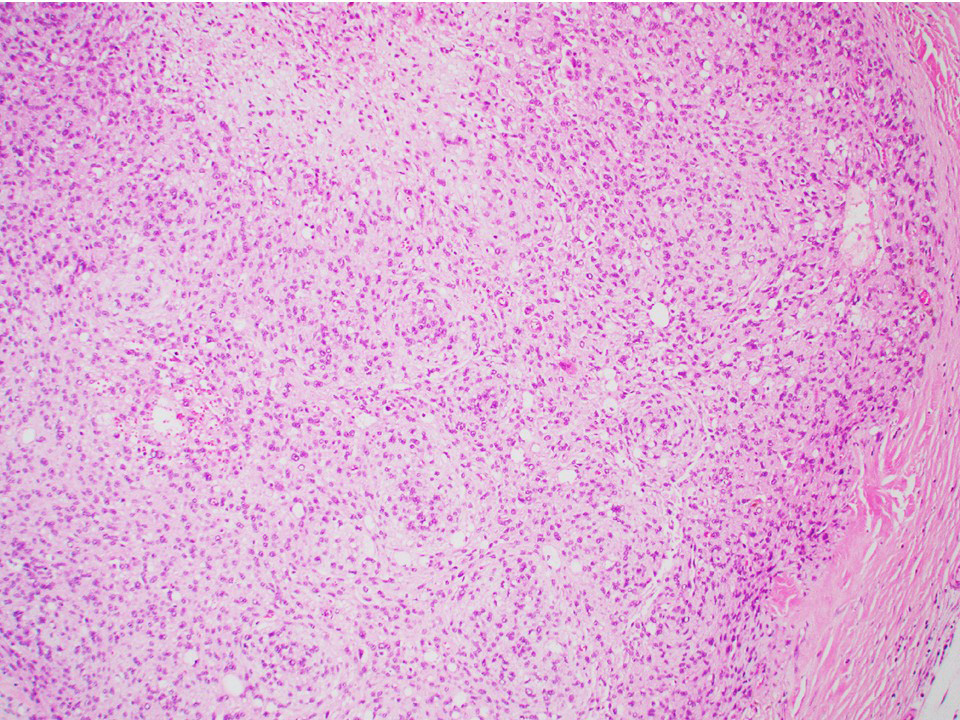
Nodular appearance
Nuclear grooves
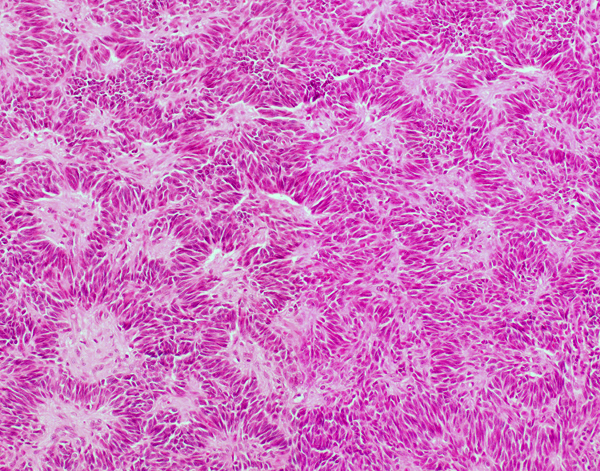
Diffuse pattern
Spindled pattern
Corded pattern
Gyriform / watered silk pattern
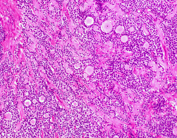
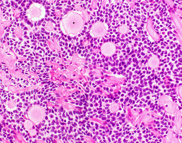
Microfollicular pattern (Call-Exner bodies)
Insular pattern
Palisading pattern
Pseudopapillary pattern
Herringbone / fascicular pattern
Luteinized cells
Inhibin A stain
Images hosted on other servers:


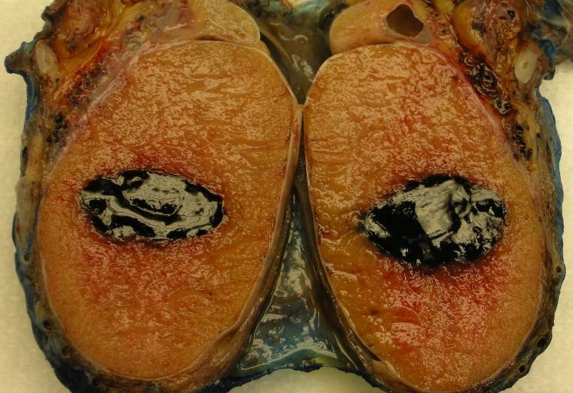
Scrotum

Transverse section

Right testis

Vertical section

Transverse section

Section of genital cord

Spermatic cord

Subcutaneous inguinal ring

Cremaster

Spermatic veins

Section of epididymis of guinea pig

Fundus of bladder
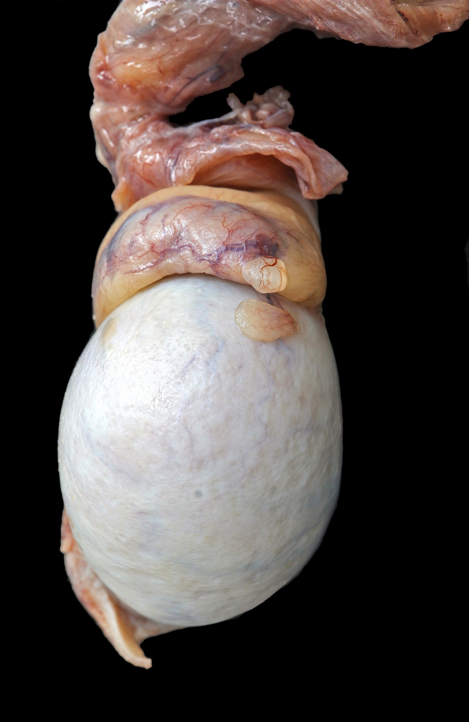


Contributed by Asmaa Gaber Abdou, M.D. and @Andrew_Fltv on Twitter
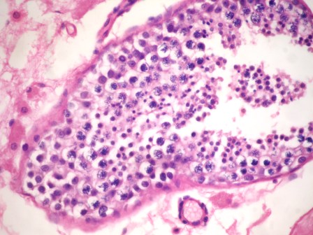


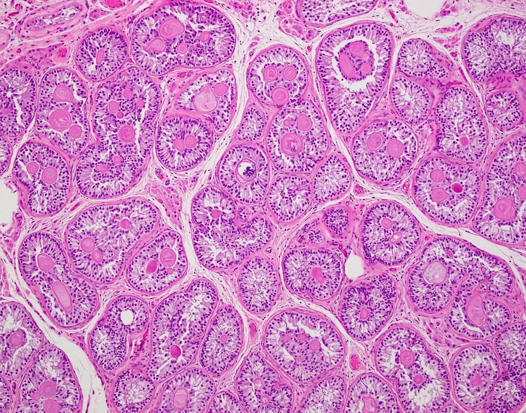
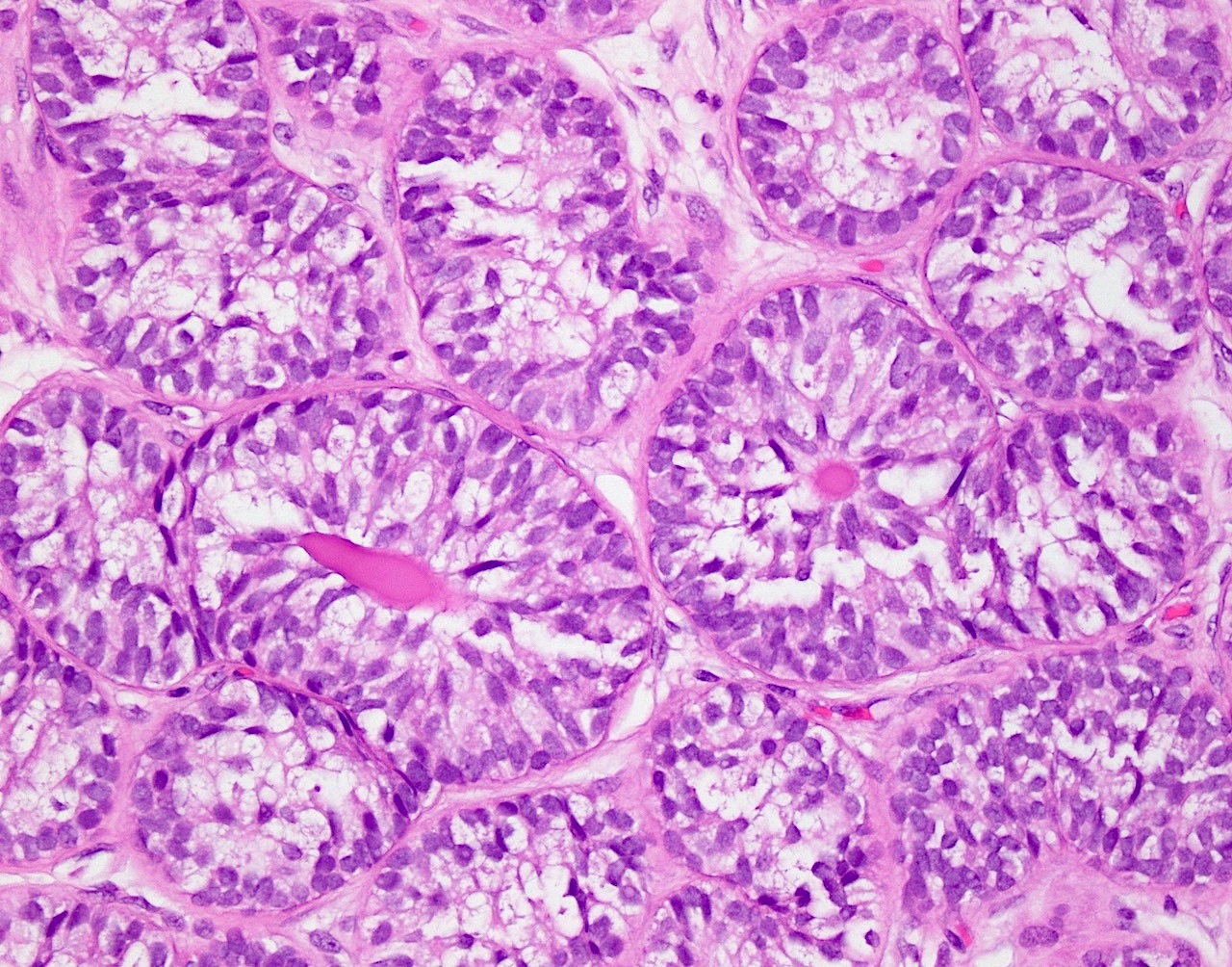
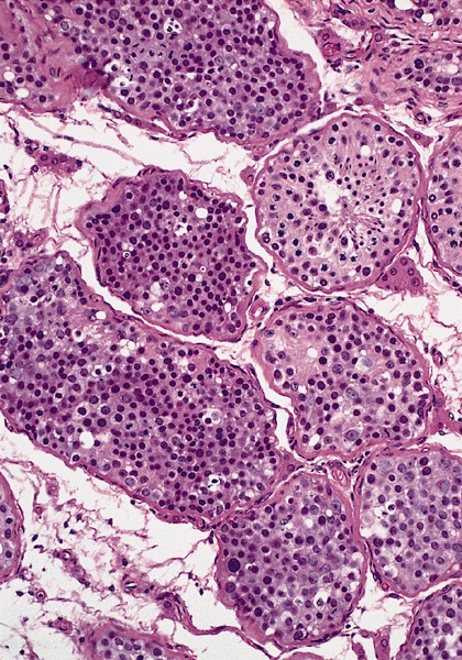
Testis histology
Images hosted on other servers:

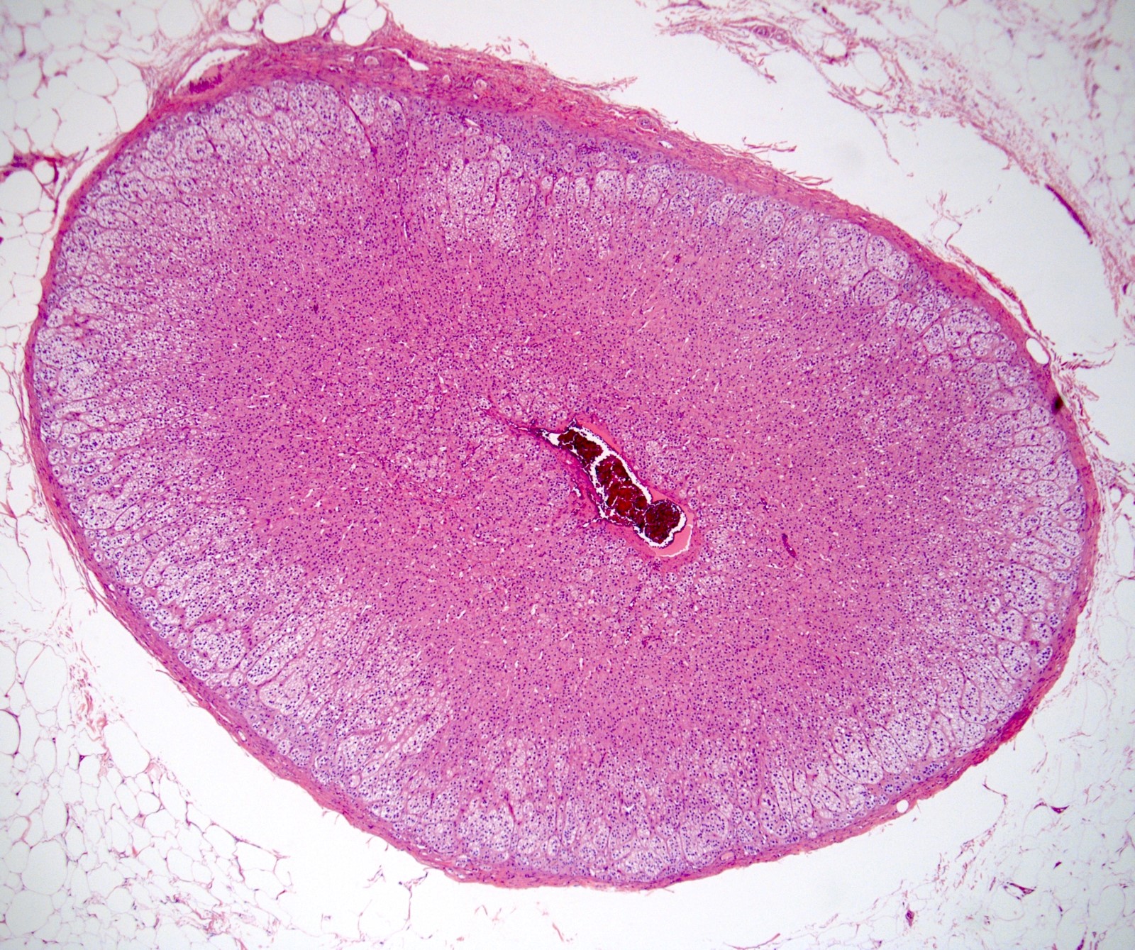
Normal testis

Efferent ducts

Leydig cells

Seminiferous tubules
Various images of epididymis

Various images of spermatic cord
Contributed by @Andrew_Fltv on Twitter

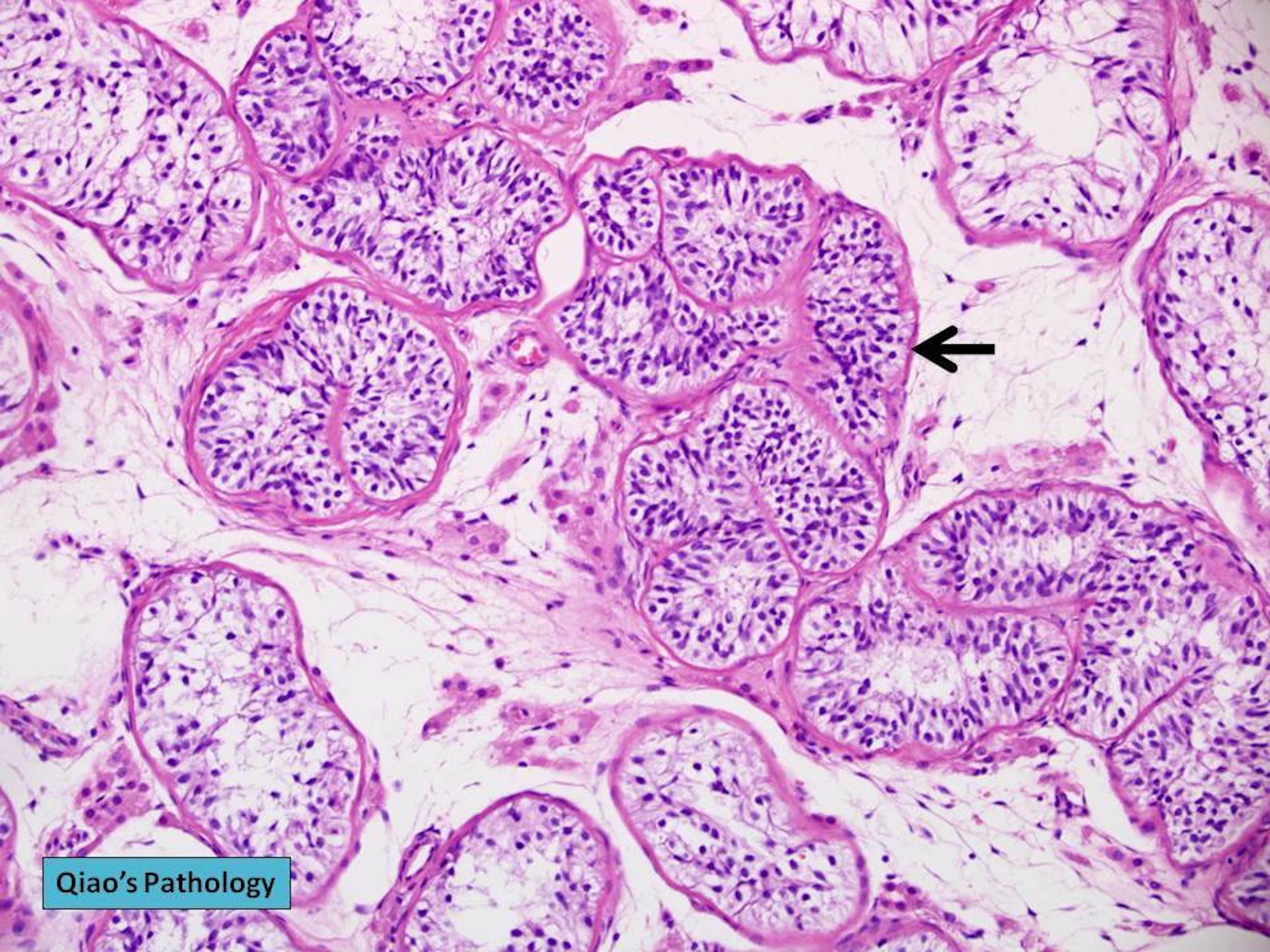
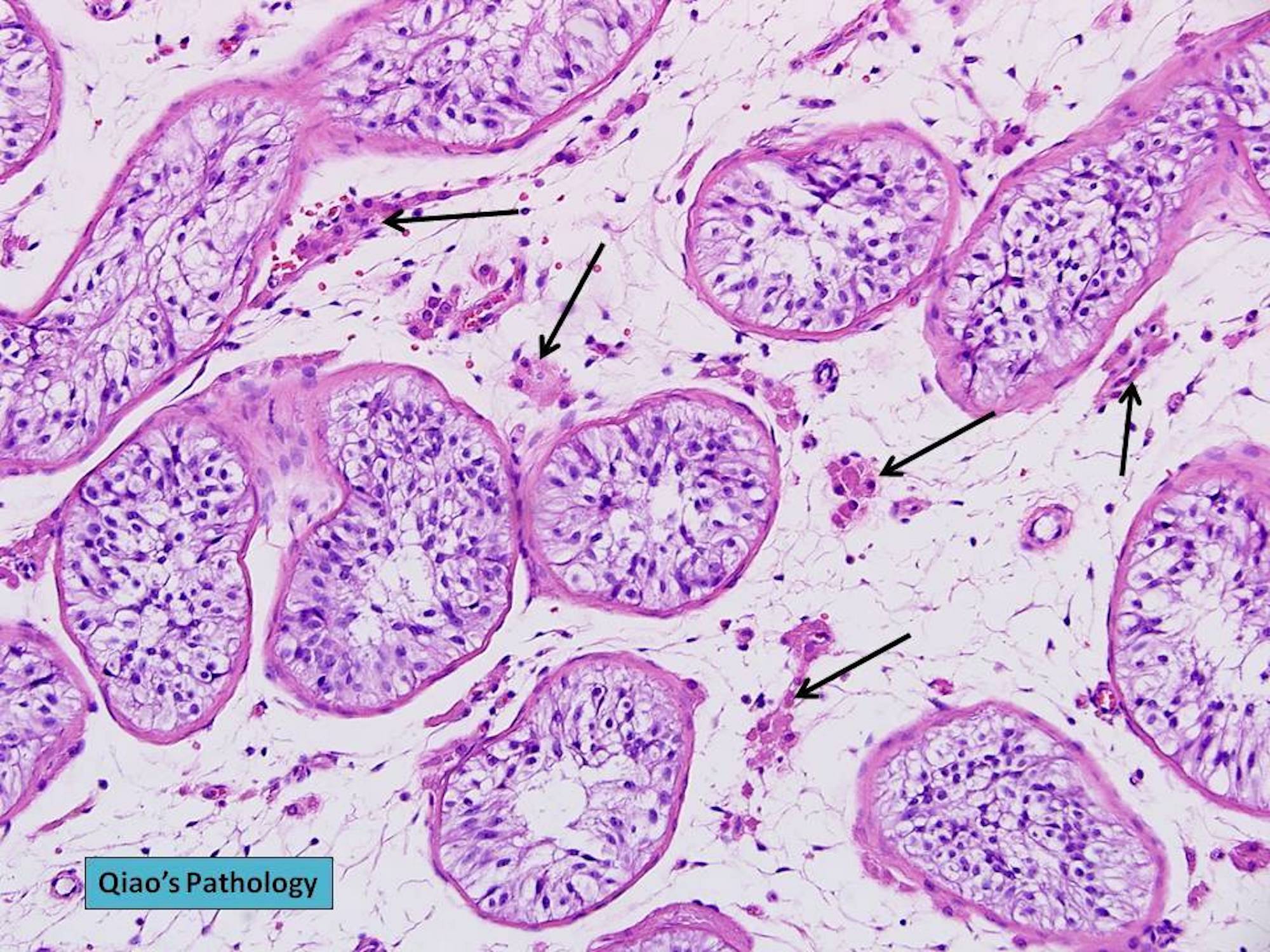
Testicular atrophy
Images hosted on other servers:

Comparison to normal testis
Contributed by Patricija Zot, M.D., Rafael E. Jimenez, M.D., @ThatGlassTho on Twitter and @Andrew_Fltv on Twitter
Seminiferous tubules replaced by collagen deposition
Retained germ cells


Atrophy


Testicular atrophy
Images hosted on other servers:

CT with testicular mass

Testis ultrasound with calcification

Chest Xray with nodules

Chest Xray in choriocarcinoma syndrome

Chest CT with cannonball metastasis

MRI of brain

Abdominal CT with retroperitoneal mass

Abdomen CT with liver metastasis
Contributed by Debra L. Zynger, M.D. and Andres Matoso, M.D.
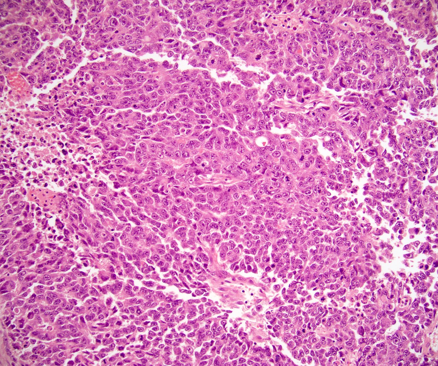
Mixed GCT with predominate choriocarcinoma
Mixed GCT with minor component of choriocarcinoma

Large hemorrhagic tumor
Contributed by Debra L. Zynger, M.D.
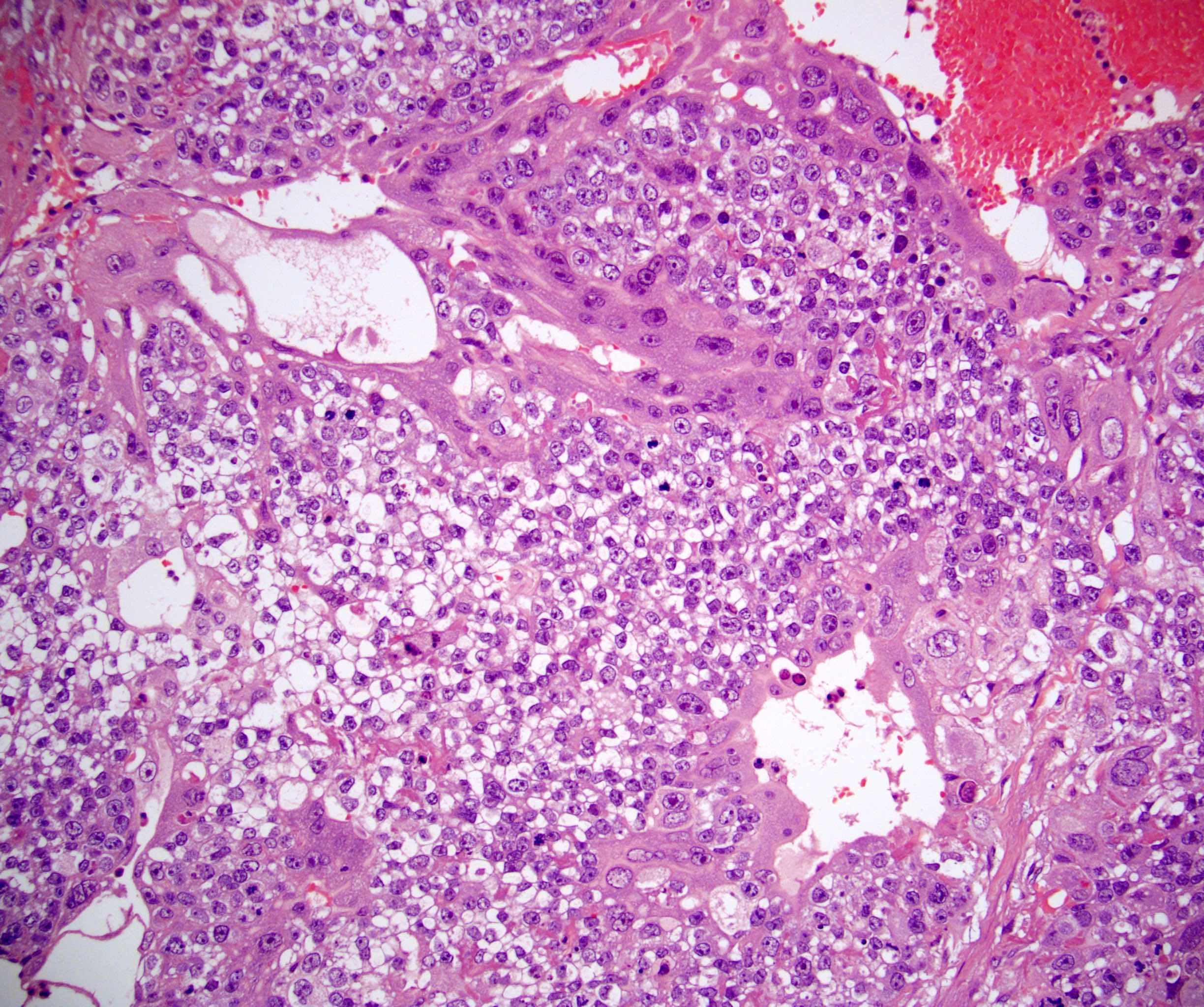
2 cell types
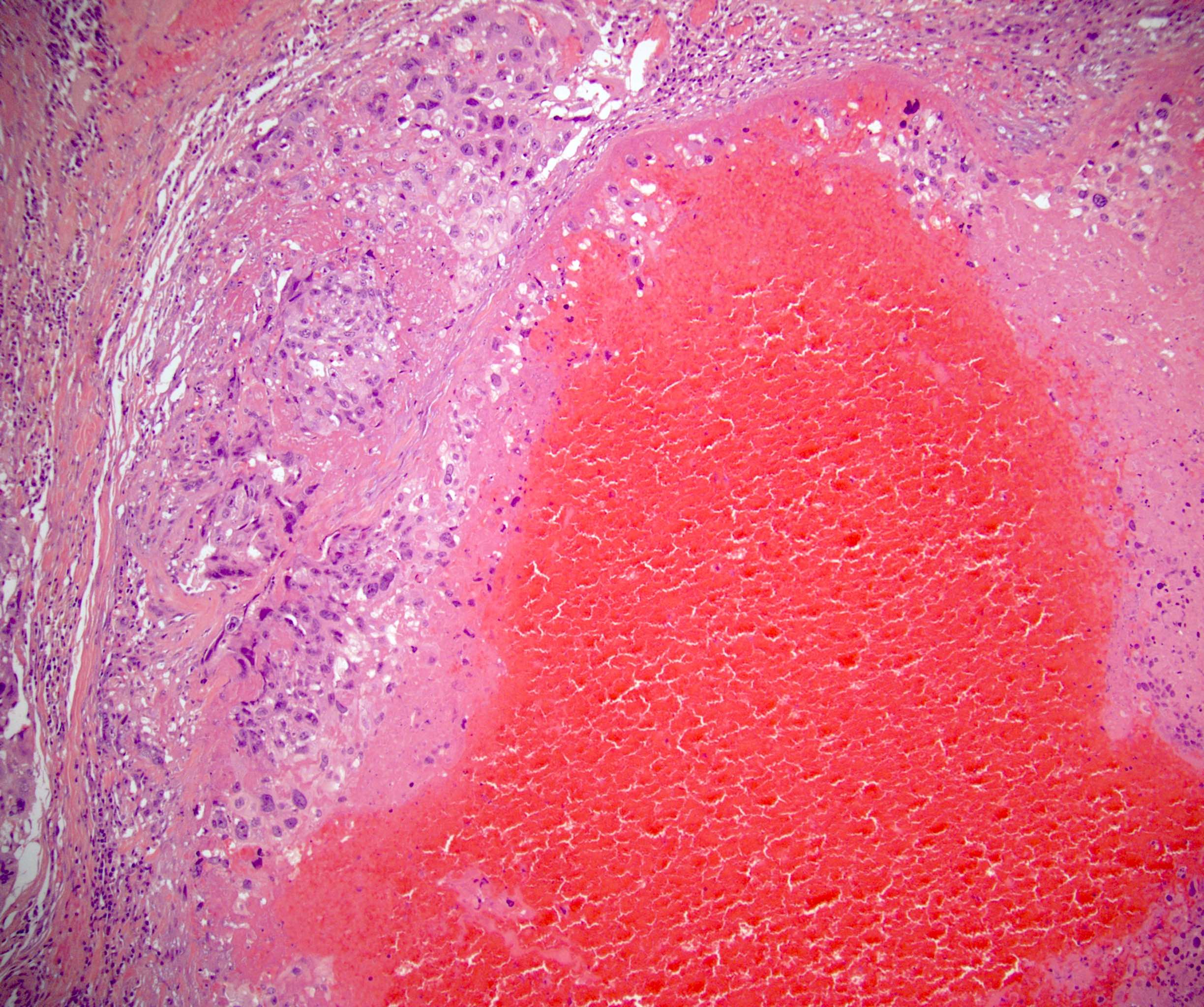
Hemorrhage
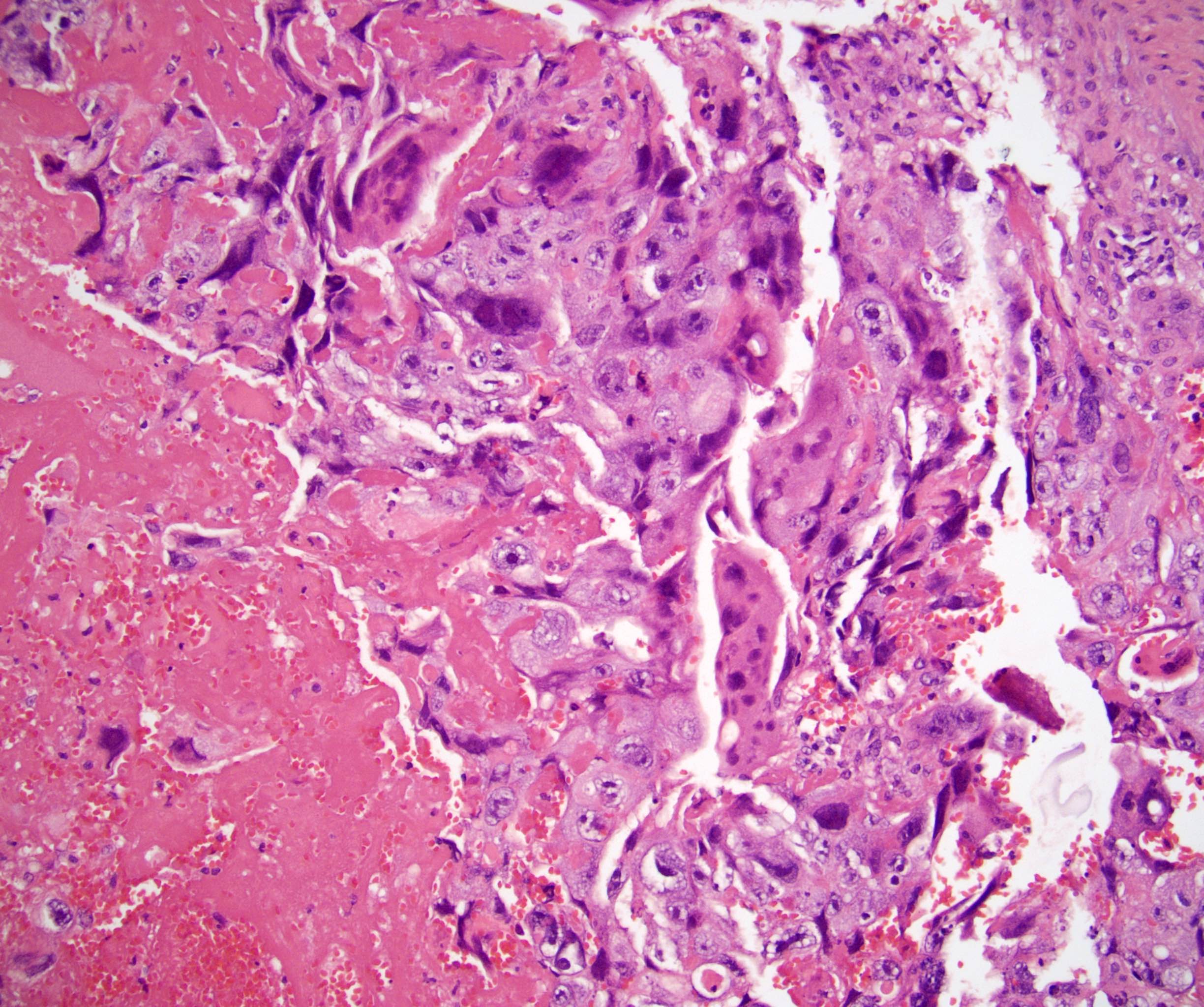
Syncytiotrophoblasts
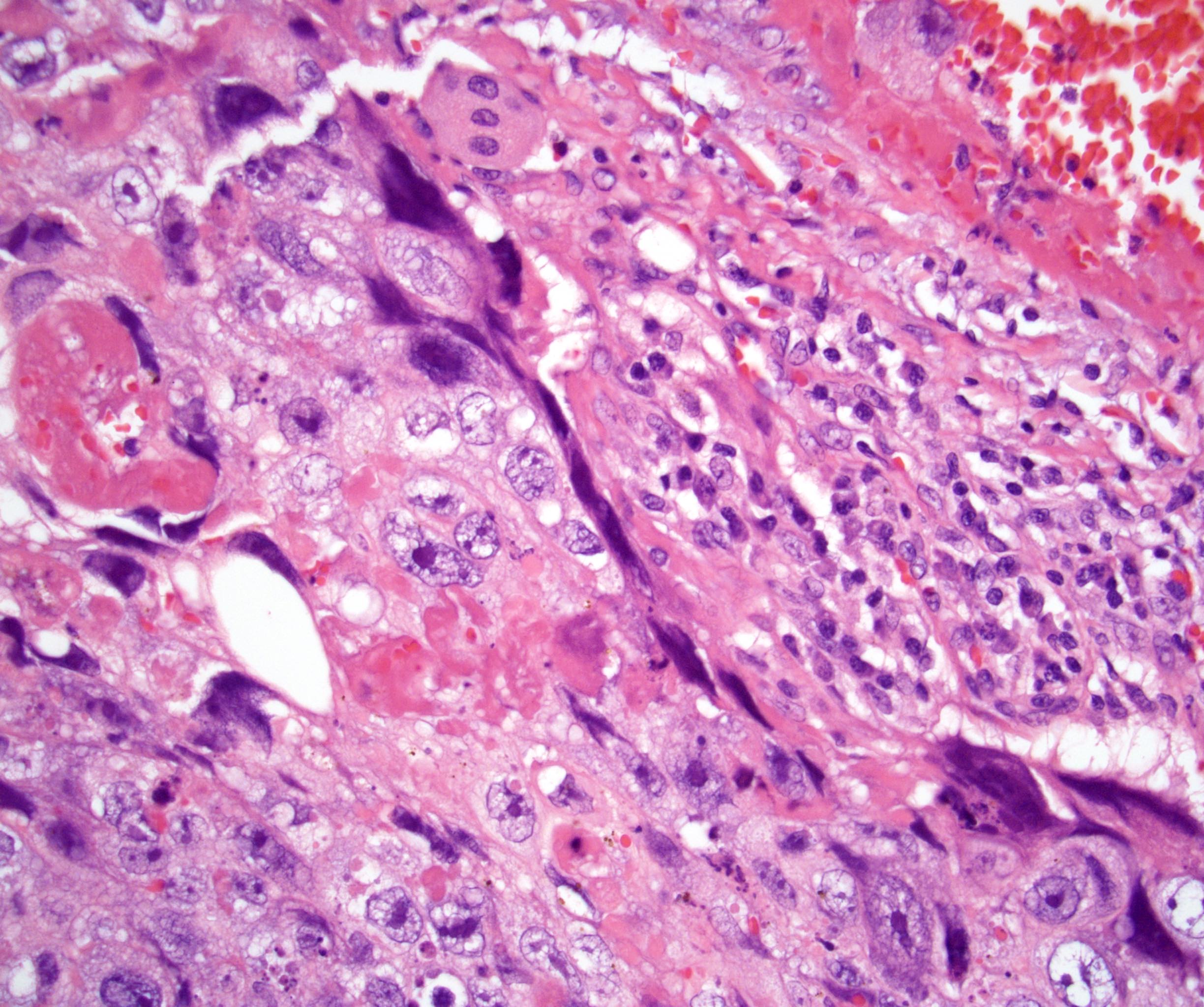
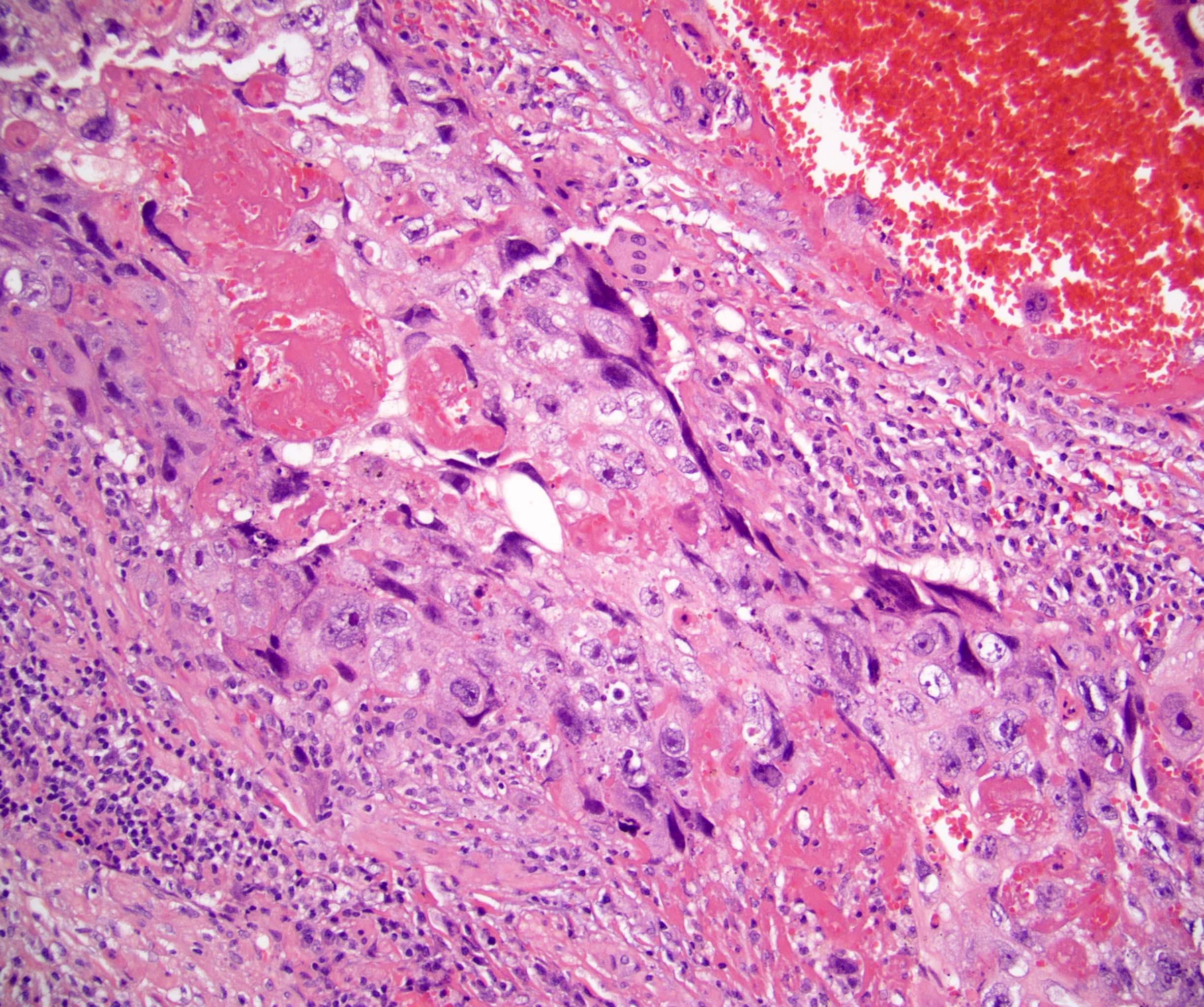
Syncytiotrophoblast rim
Mononucleated trophoblasts
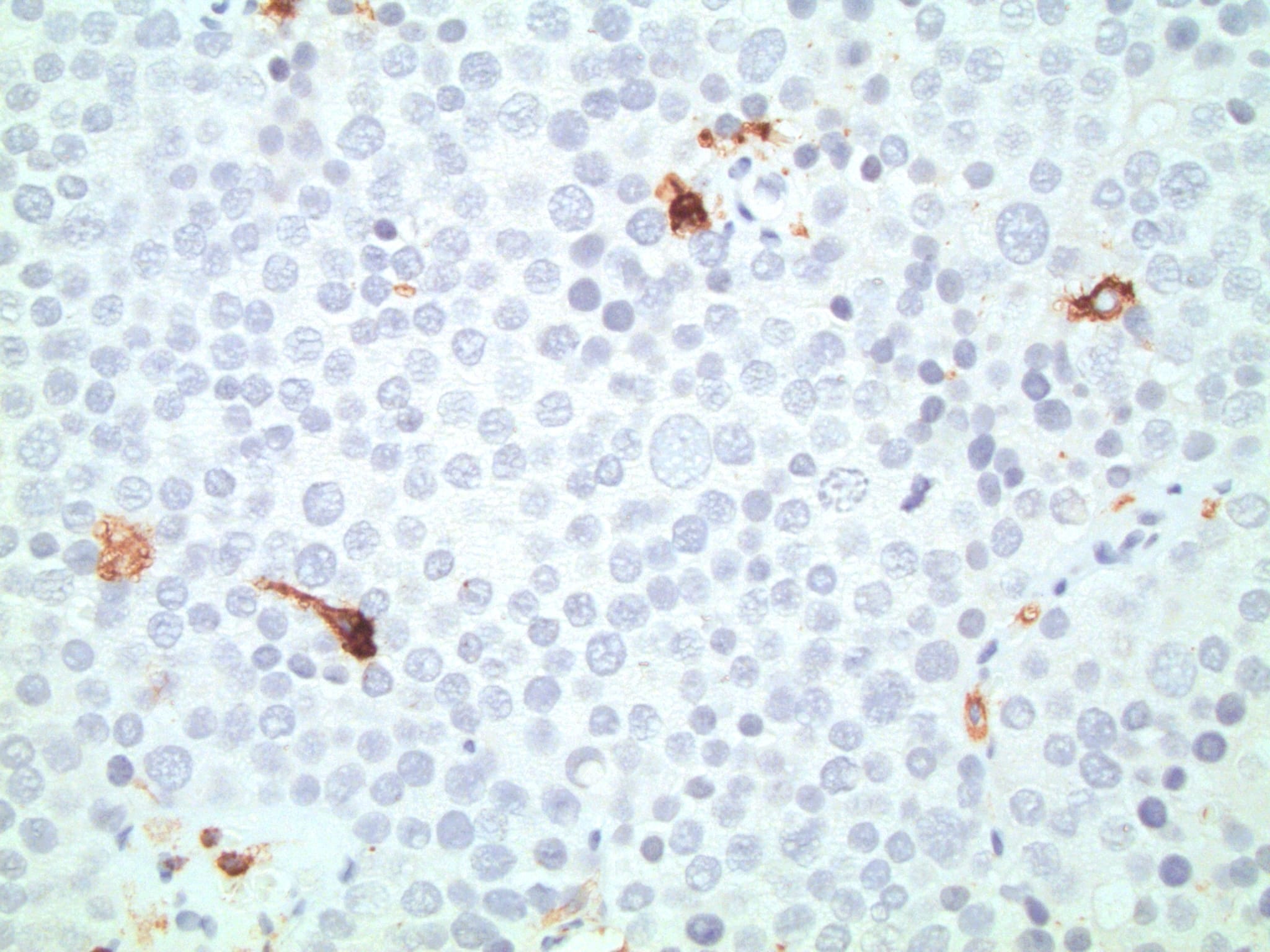
Choriocarcinoma with IHC
Beta hCG
Inhibin
CK7
GATA3
OCT 3/4
Images hosted on other servers:

Intra-abdominal mass

Abdominal testis

Mass with right gonadal vein

Scrotum with cryptorchidism

Abdominal wall ectopic testis
Contributed by Debra L. Zynger, M.D.
Cryptorchid testicle
Contributed by Debra L. Zynger, M.D.
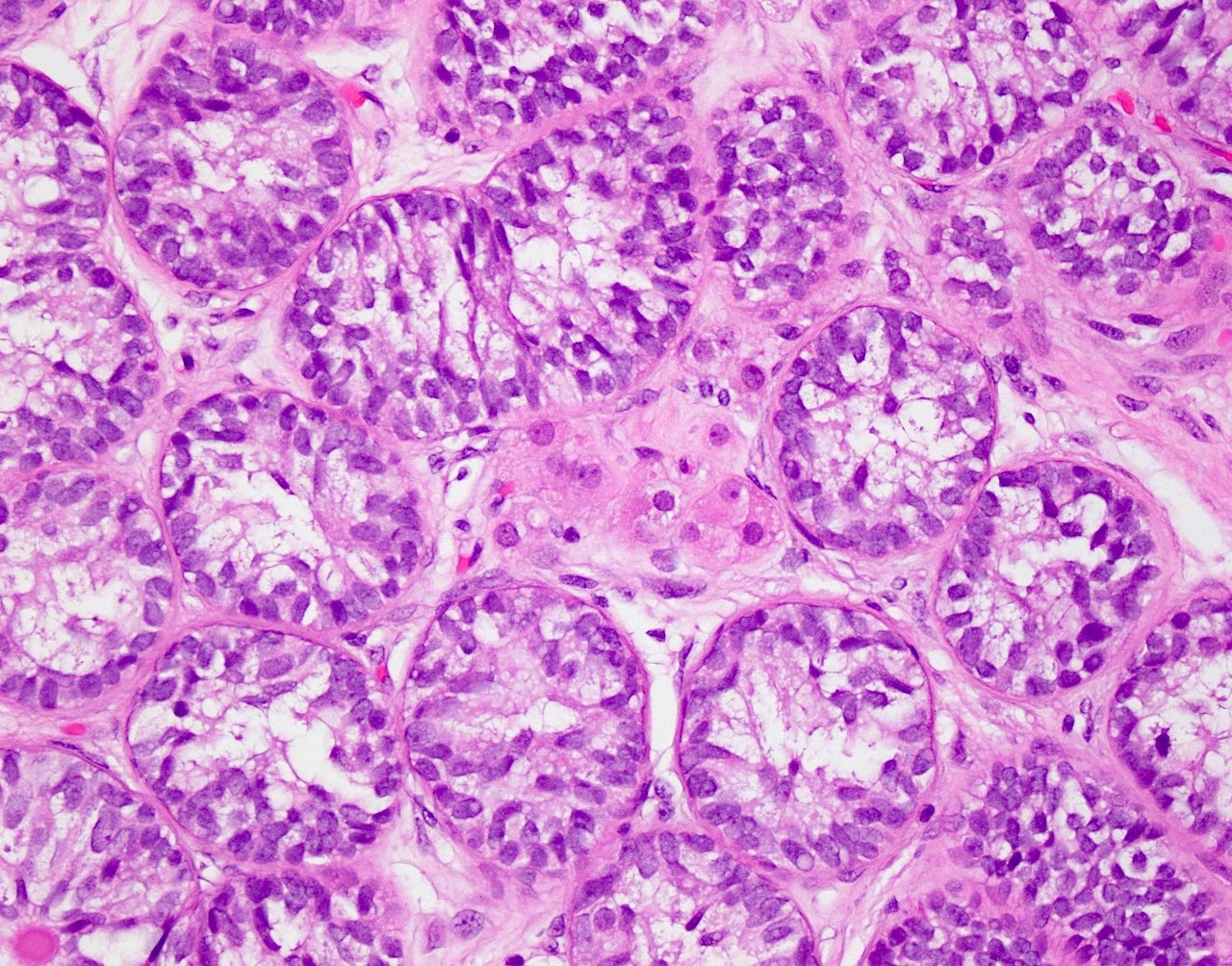
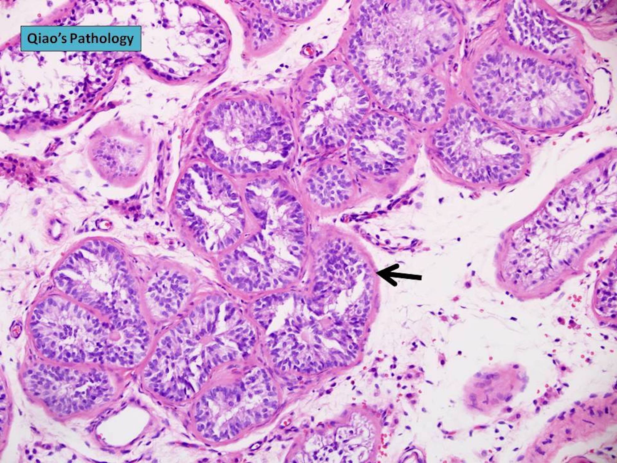
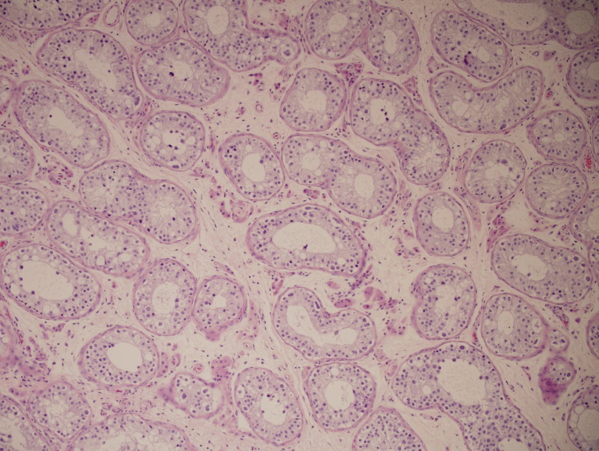
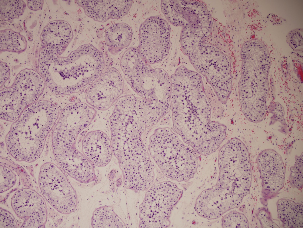
Sertoli cell only tubules
Absent spermatogenesis

Peritubular fibrosis
Sertoli cell nodule
Retained Leydig cells

Granular cell change within Sertoli cells
Microliths
Contributed by Vikas Mehta, M.D.
Cyst, fibrinoid material

Intermediate type trophoblasts

Intermediate trophoblasts, smudged chromatin

Solid nest, fibrinoid material

βhCG positivity
Images hosted on other servers:

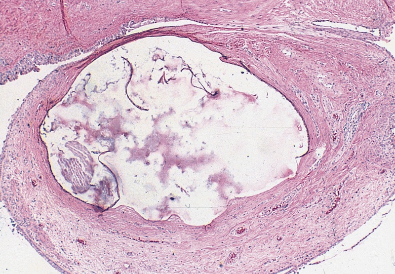
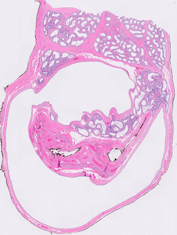
Cyst of testis (focal dilatation of seminiferous tubule)

Cystic dilatation (tubular ectasia) of rete testis

Epididymis cyst

Cystic dilatation (tubular ectasia) of epididymis

Mesothelial cyst in tunica albuginea

Hydrocele

Chronic hydrocele with multiloculated appearance

Spermatic cord cyst
AFIP images
Cyst of tunica albuginea
Images hosted on other servers:

Encysted hydrocele

Multiple paratesticular, unilocular, thin walled cysts
Contributed by Stephanie Siegmund, M.D., Ph.D. and AFIP
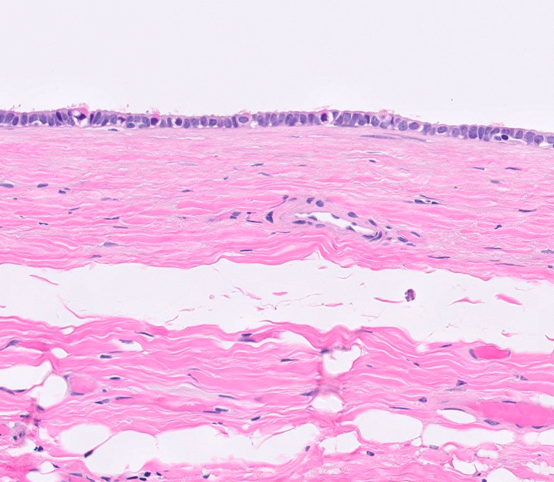
Mesothelium lined cystic cavity

Mesothelial lining of hydrocele


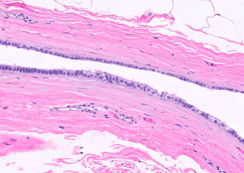
Cystic dilatation of epididymis

Cystic dilatation of epididymis

Spermatocele

Ruptured spermatocele
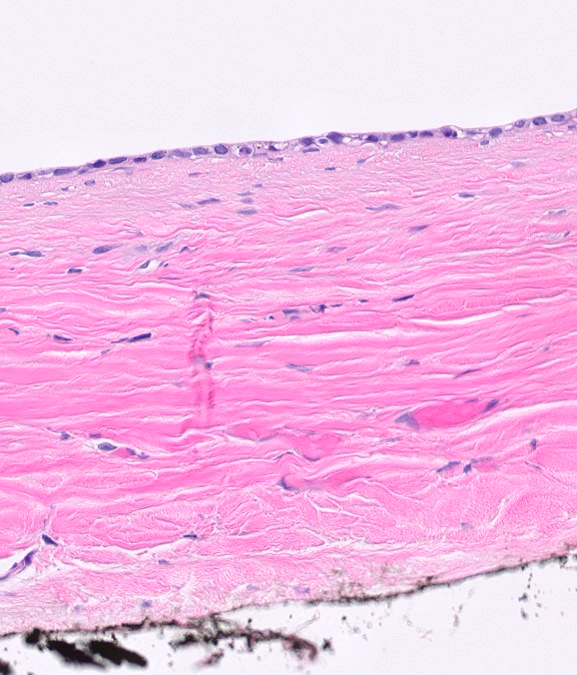
Cyst of tunica albuginea
Images hosted on other servers:

Calcified mass
Images hosted on other servers:

Cryptorchid testis with embryonal carcinoma
Contributed by Debra L. Zynger, M.D.

Mixed GCT with predominance of embryonal carcinoma

Mixed GCT with minor component of embryonal carcinoma
Contributed by Debra L. Zynger, M.D.
Solid growth
Glandular growth
Papillary growth
Cellular overlap

Pleomorphic

Admixed with yolk sac tumor

Intratubular

Lymphovascular invasion
Spermatic cord invasion
OCT 3/4
CD30
PLAP

CD117
D2-40
AE1 / AE3
Contributed by Debra L. Zynger, M.D. and @ThatGlassTho on Twitter
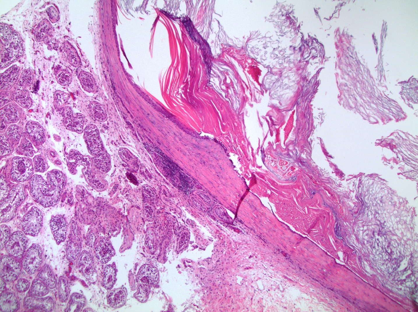
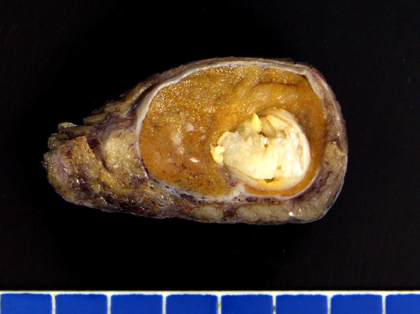
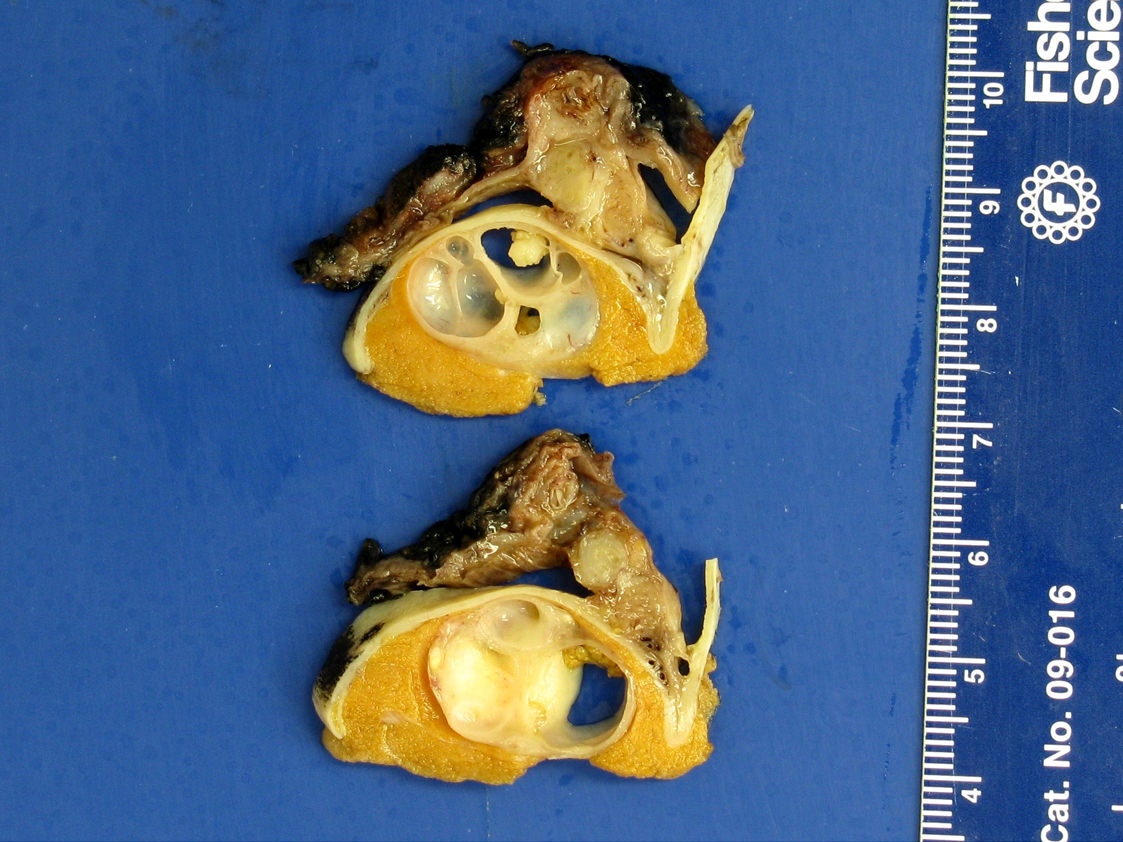
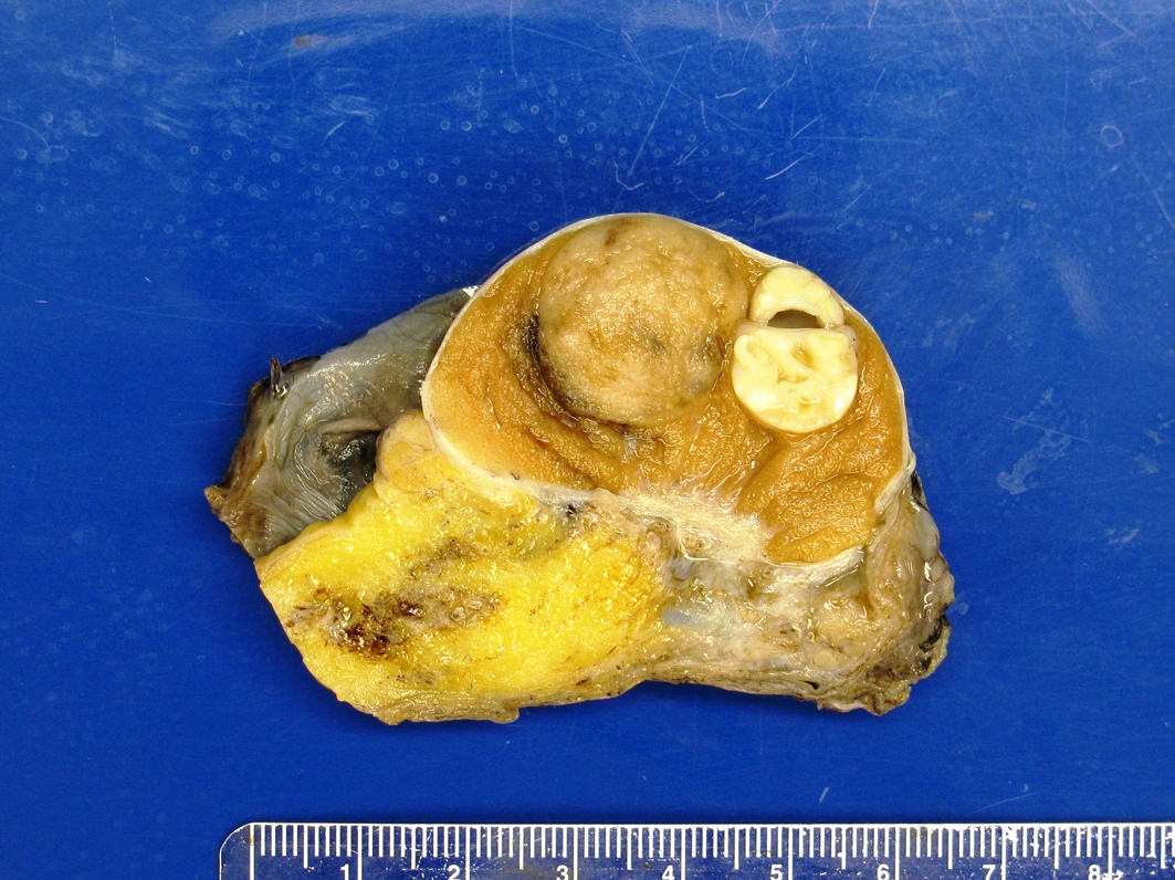
Testicular epidermoid cyst
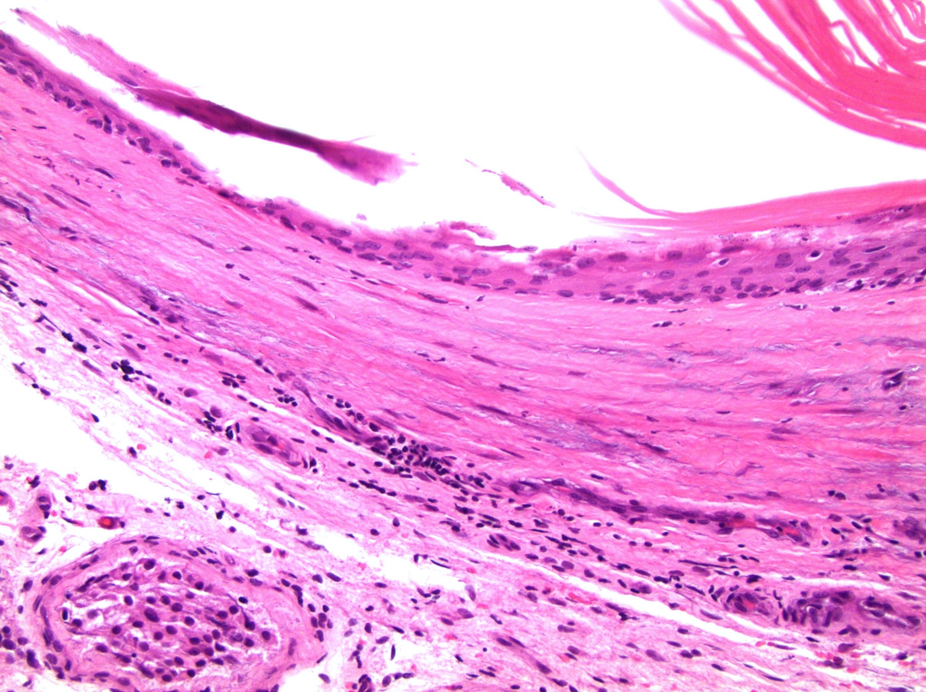
Cyst wall with adjacent normal seminiferous tubule
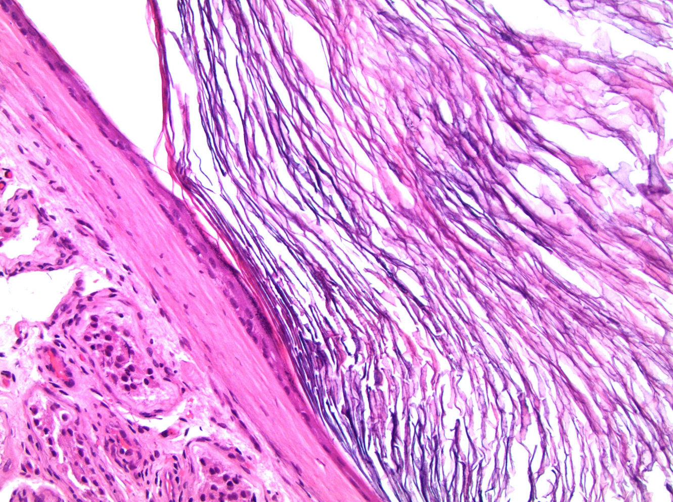
Lamellar keratin within the cyst
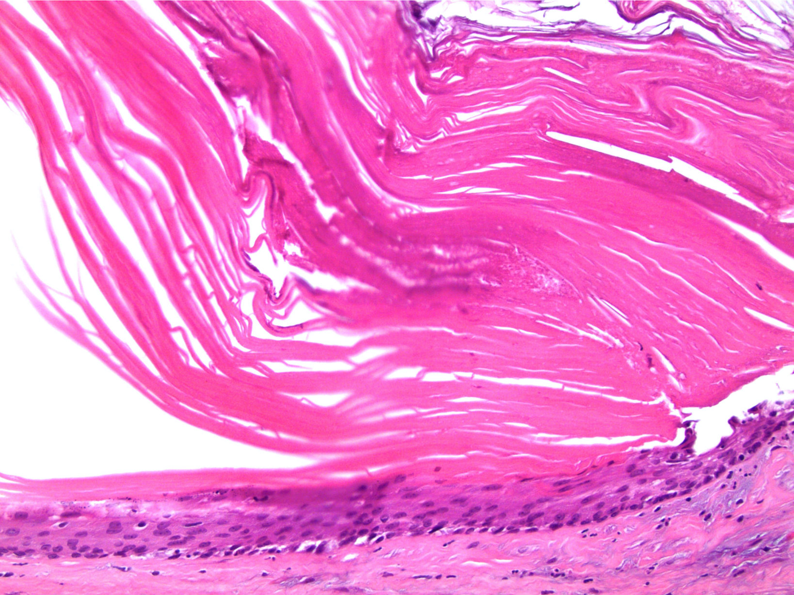
Stratified squamous epithelium with adjacent keratin

Teratoma - epidermoid cyst
Images hosted on other servers:

Normal FISH for 12p
Contributed by Veena Maheshwar, M.D., Kiran Alam, M.D., Anshu Jain, M.D.

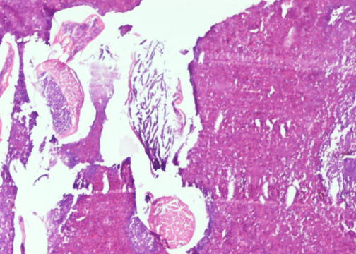
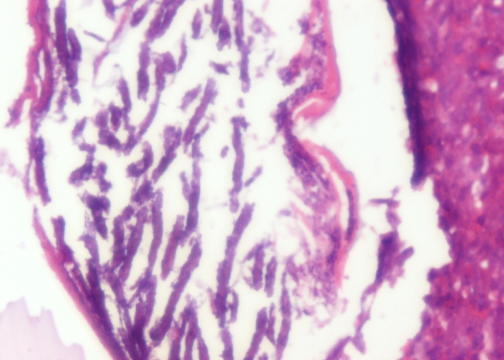
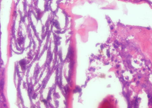
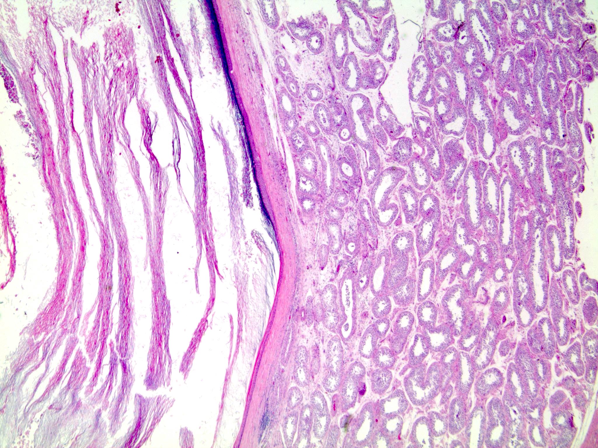
25 year old man with epididymal mass: filiarial epididymitis
Contributed by Debra L. Zynger, M.D.


Testicular epithelial trophoblastic tumor
Images hosted on other servers:

Well circumscribed intratesticular tumor
Contributed by Rafael E. Jimenez, M.D., M.H.A.

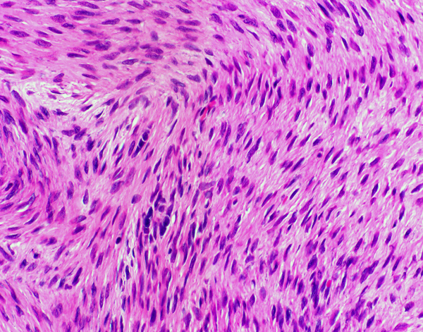
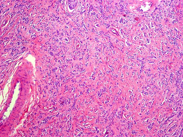
Bland nuclei

Fascicular pattern

Encapsulated tumor
Collagenized stroma
Inhibin
SF1
Images hosted on other servers:

MRI: polycyclic mass
Images hosted on other servers:

Intrascrotal mass
Images hosted on other servers:

Pedunculated mass

Well demarcated margins
Images hosted on other servers:

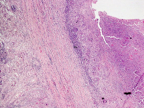
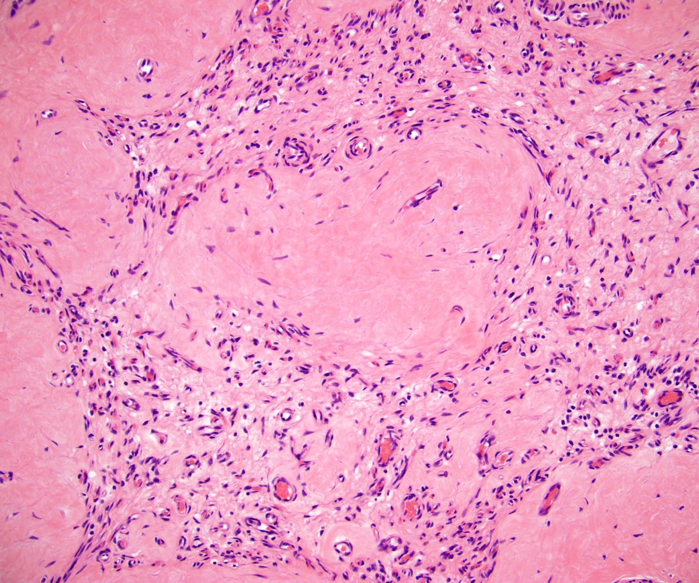
Dense fibrotic tissue

Collagen rich hyalinized fibrotic tissue

CD3

IgG4

CD31
Images hosted on other servers:

GCNIS derived adult germ cell tumors
Contributed by Stephanie Siegmund, M.D., Ph.D., Maria Tretiakova, M.D., Ph.D., Alexander Subtelny, M.D., Ph.D. and Michelle Hirsch, M.D., Ph.D.

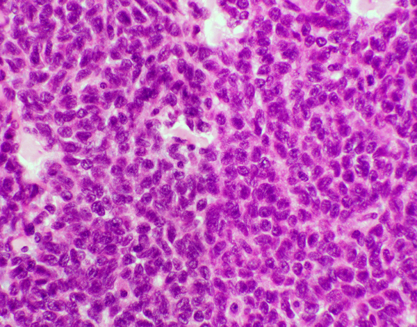
Prominent, large atypical gonocytes


Pagetoid GCNIS into rete testis
Normal prepubertal testis

GCNIS with adjacent microinvasion
Podoplanin /
D2-40

Membranous
D2-40 IHC

Membranous KIT IHC

Nuclear OCT 3/4 IHC
Images hosted on other servers:

Representative aneuploidy and chromosomal abnormalities
Images hosted on other servers:


Gonadoblastoma and dysgerminoma in gonadal dysgenesis
Images hosted on other servers:

Gonadoblastoma


Female patient
Contributed by Yale Rosen, M.D. and @SueEPig on Twitter
TB orchitis
Granulomatous orchitis
Images hosted on other servers:

Brucellosis
Contributed by Sean R. Williamson, M.D. and @SueEPig on Twitter
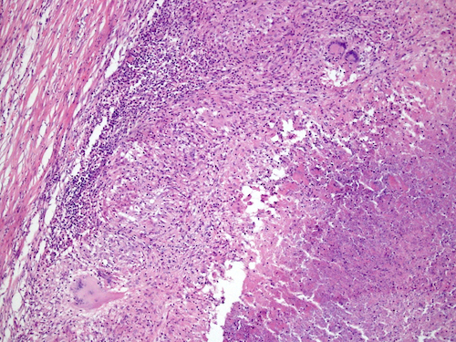
Tuberculosis involving testis and paratestis
Granulomatous orchitis
Contributed by Debra L. Zynger, M.D.

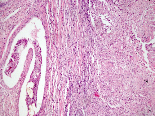
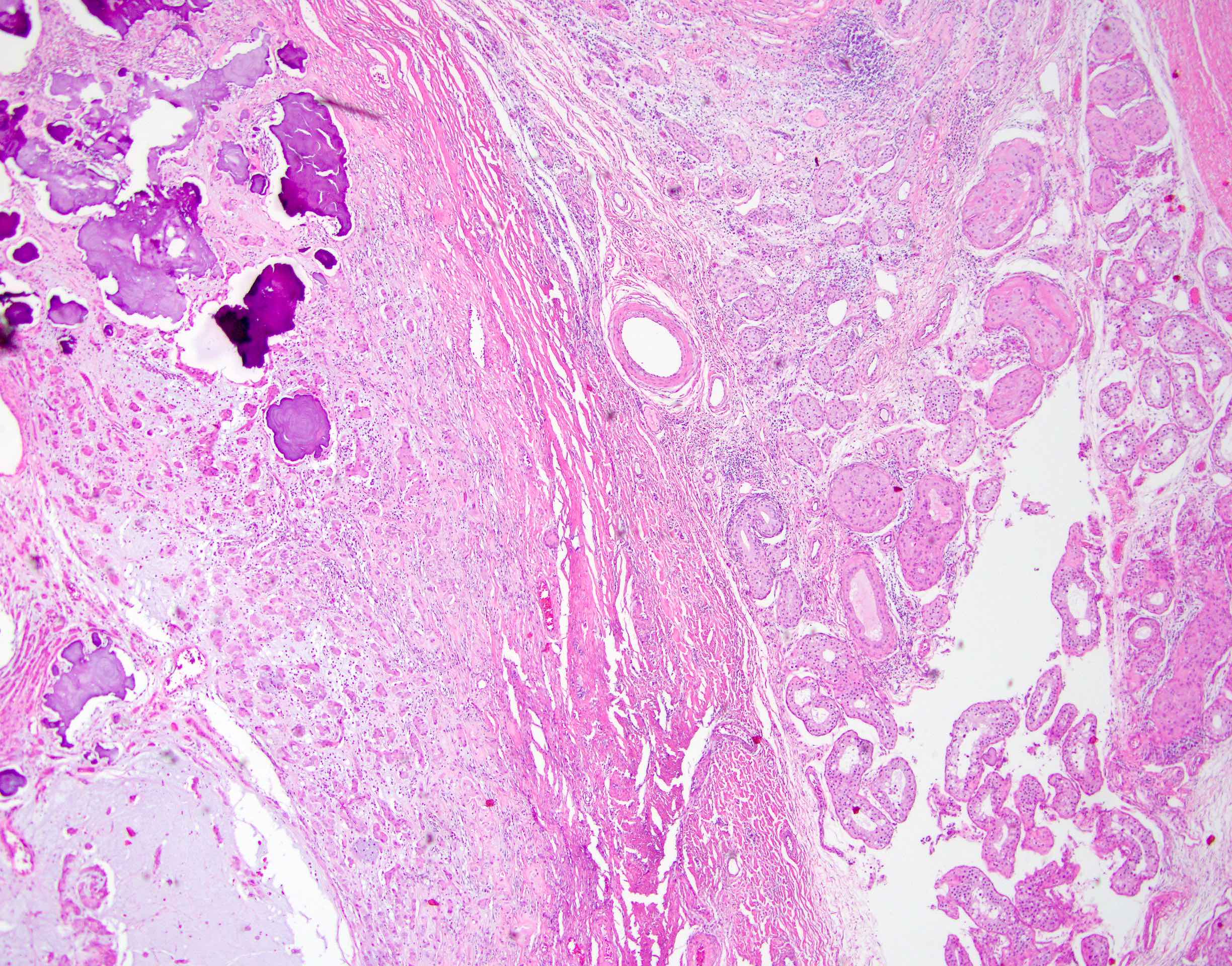
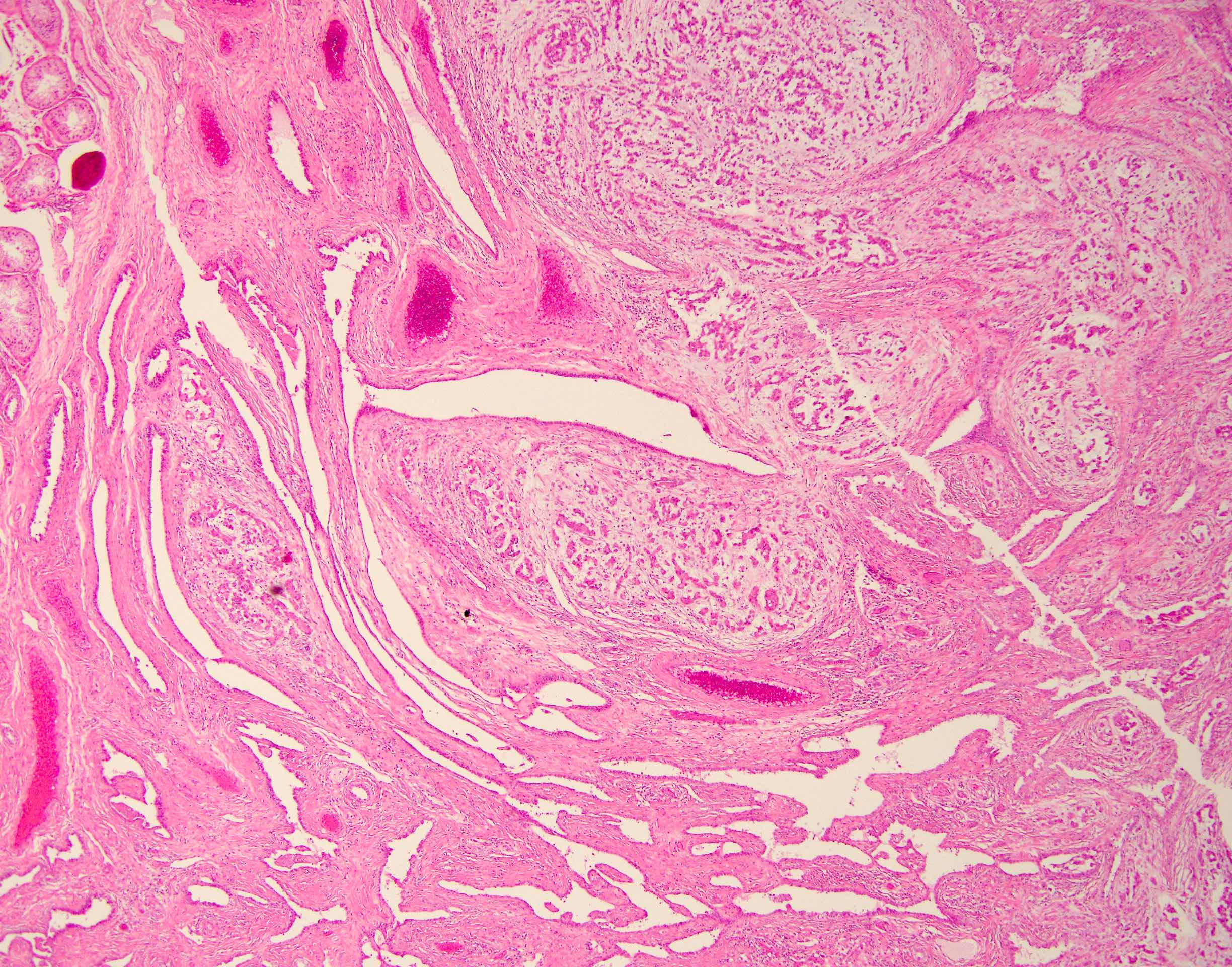
Entrapped glands
Cords, glands and small nests
Gland formation
Entrapped mesothelium

WT1
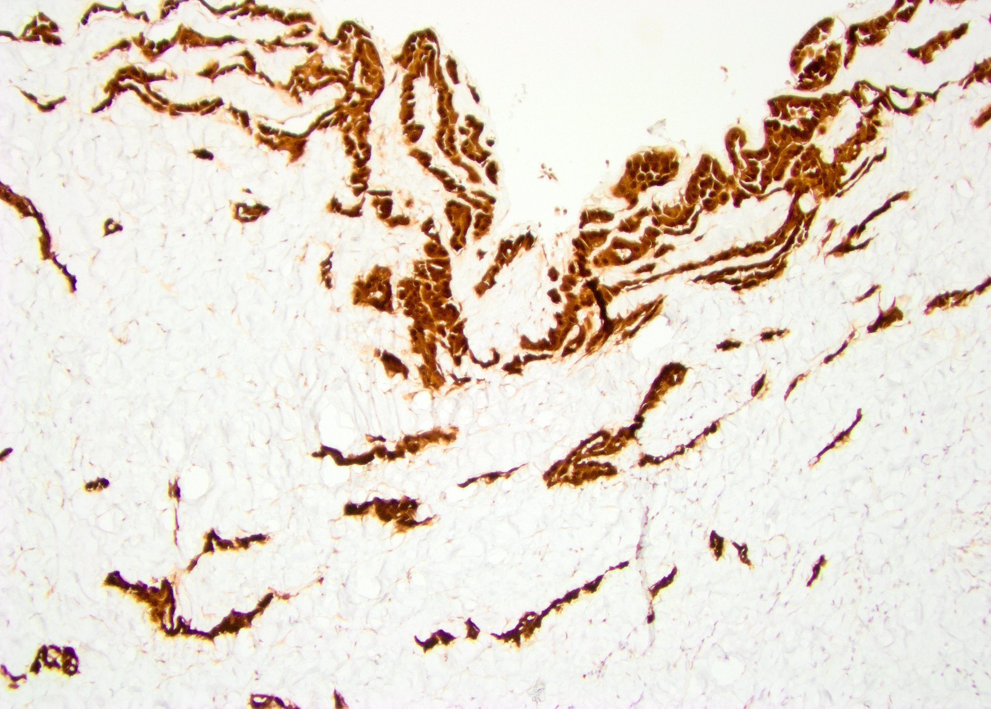
Calretinin
Images hosted on other servers:

Scrotal MRI: cystic lesion

Encysted hydrocele
Images hosted on other servers:

Abdominoscrotal hydrocele
Contributed by Francesca Sanguedolce, M.D., Ph.D.
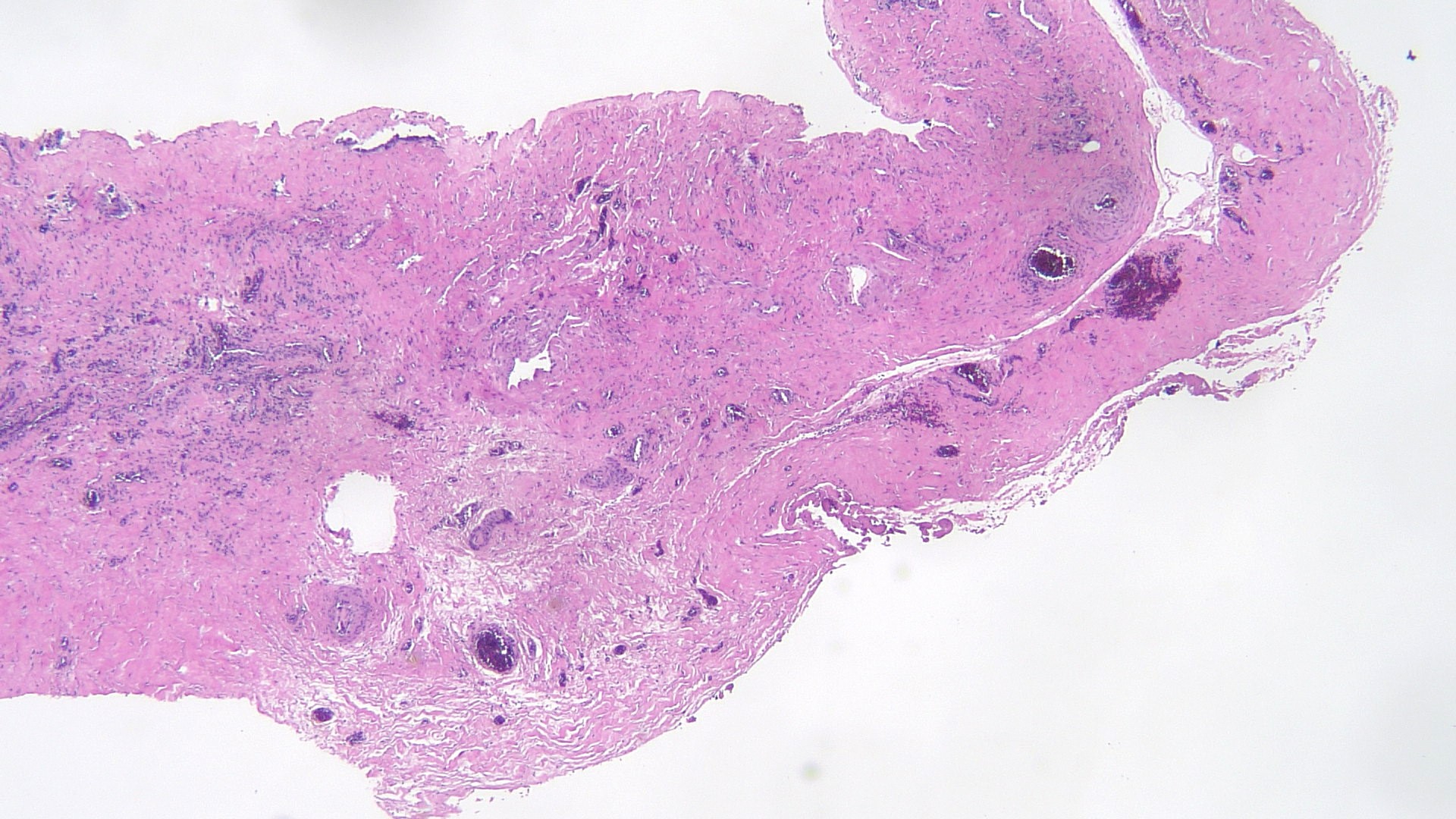
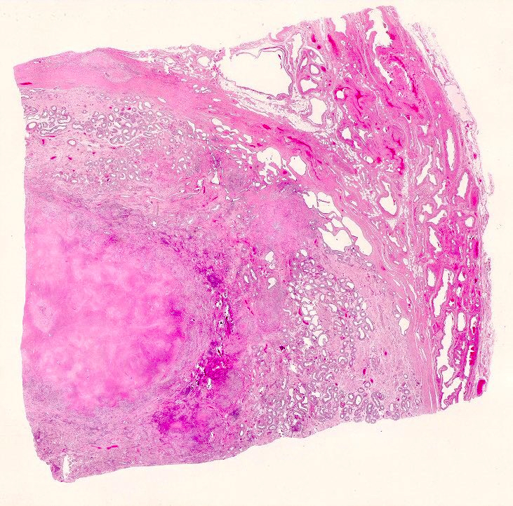
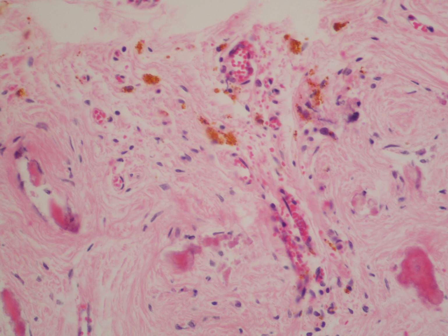
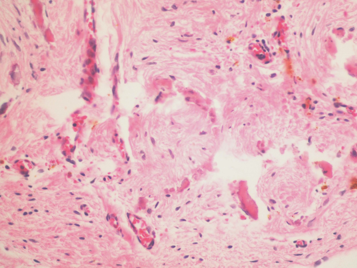
Connective tissue, mild inflammation
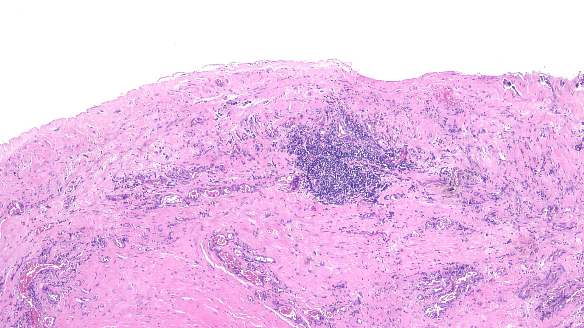
Fibrosis, moderate inflammation
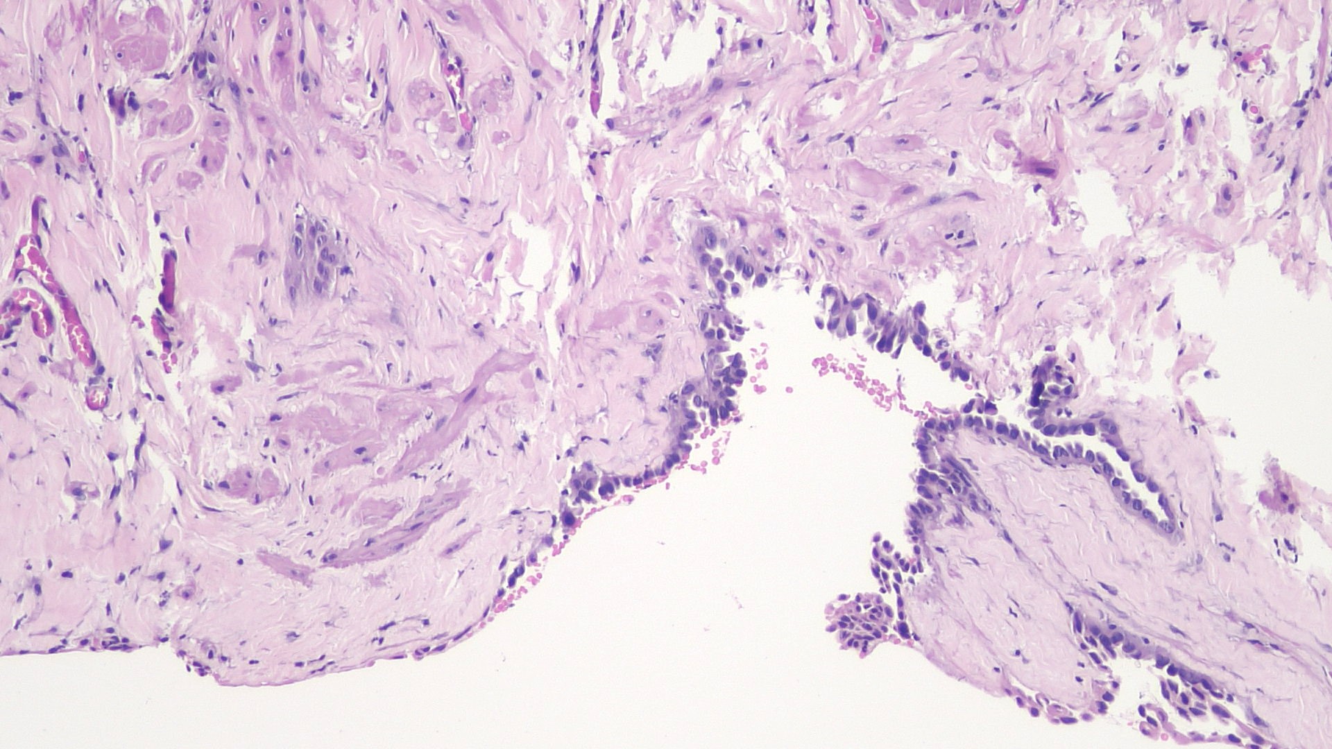
Mesothelial lining
Calretinin
Contributed by Vikas Mehta, M.D. and @katcollmd on Twitter
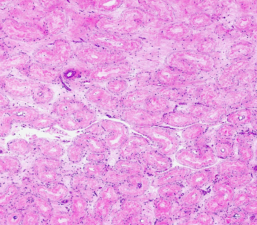
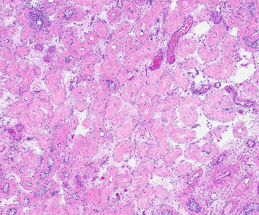
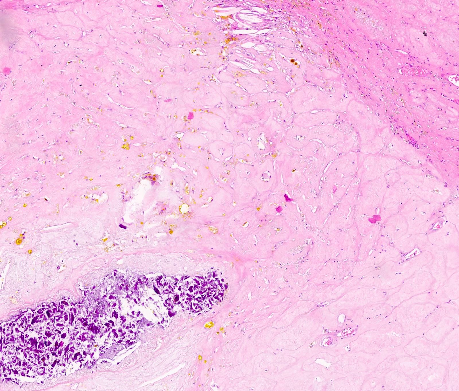
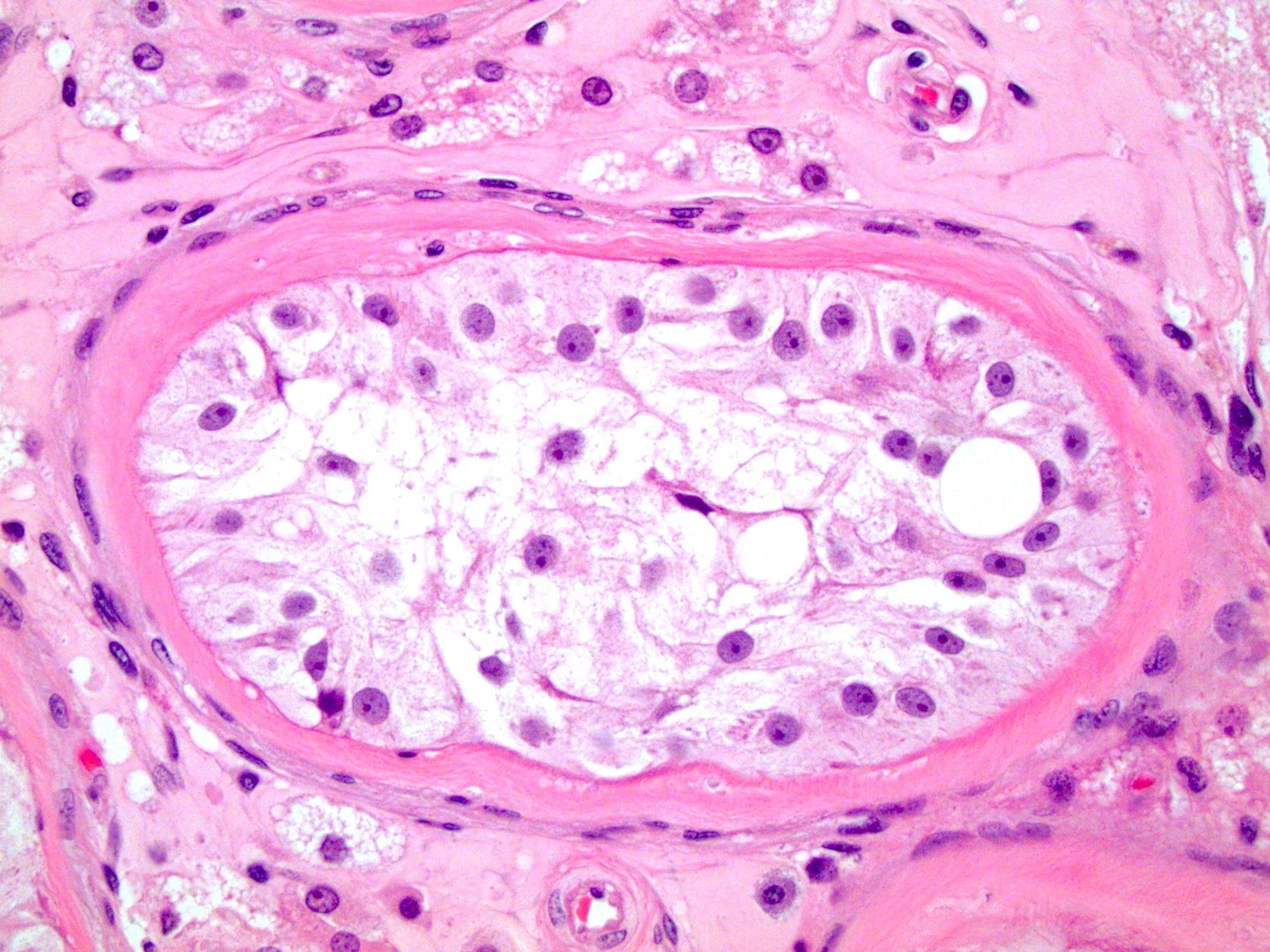
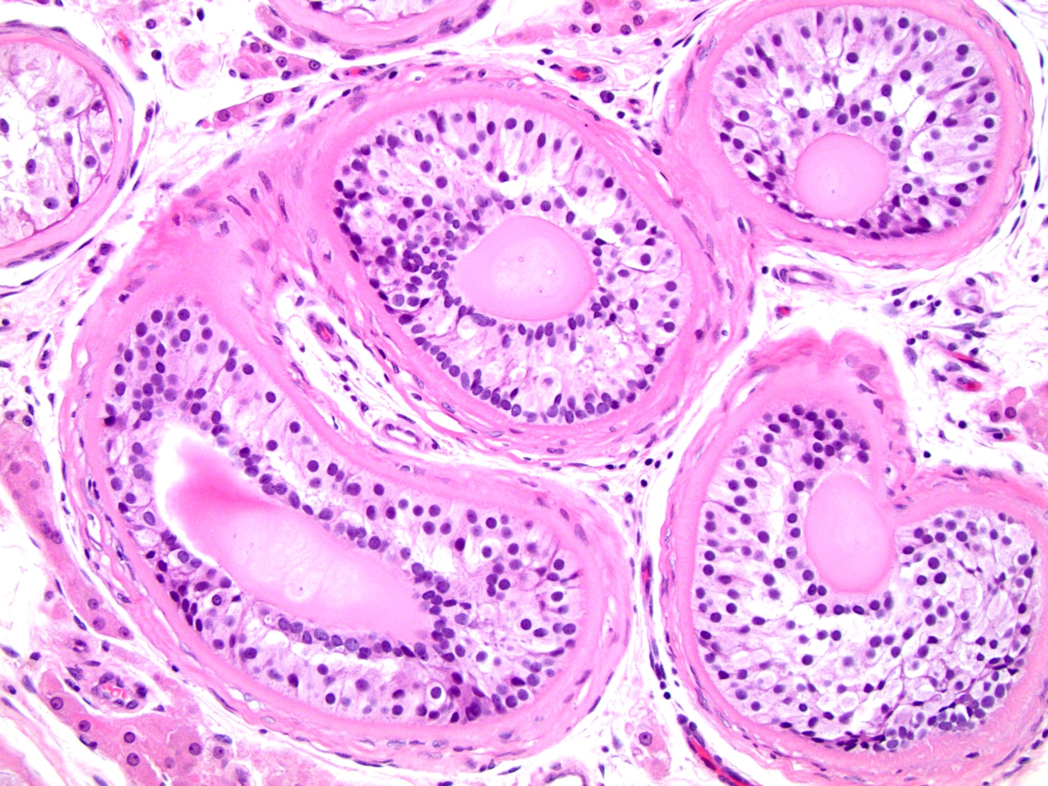
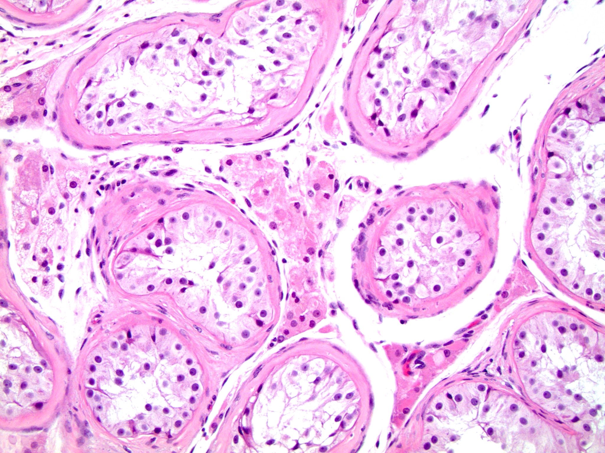
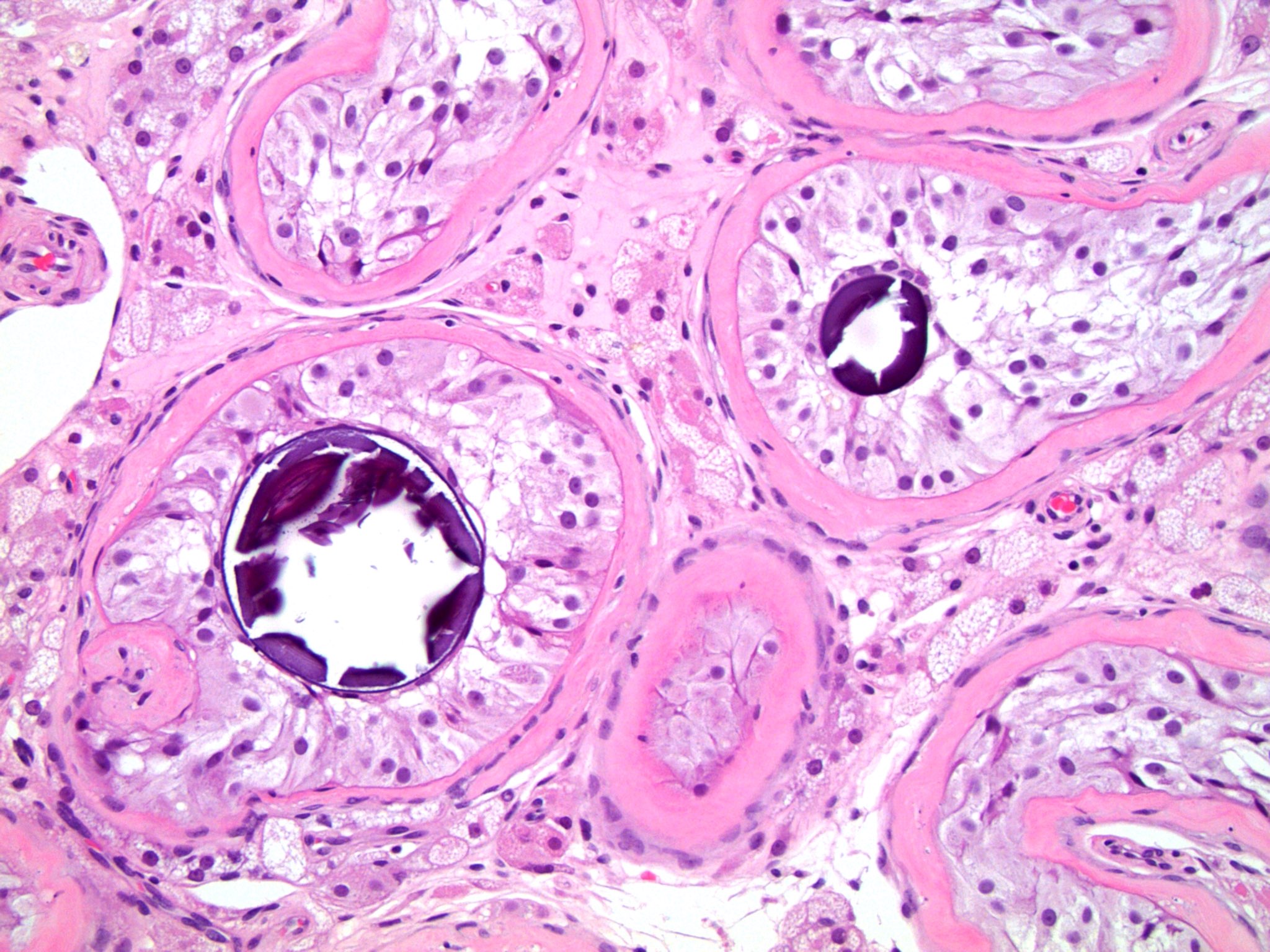
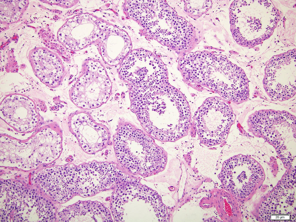
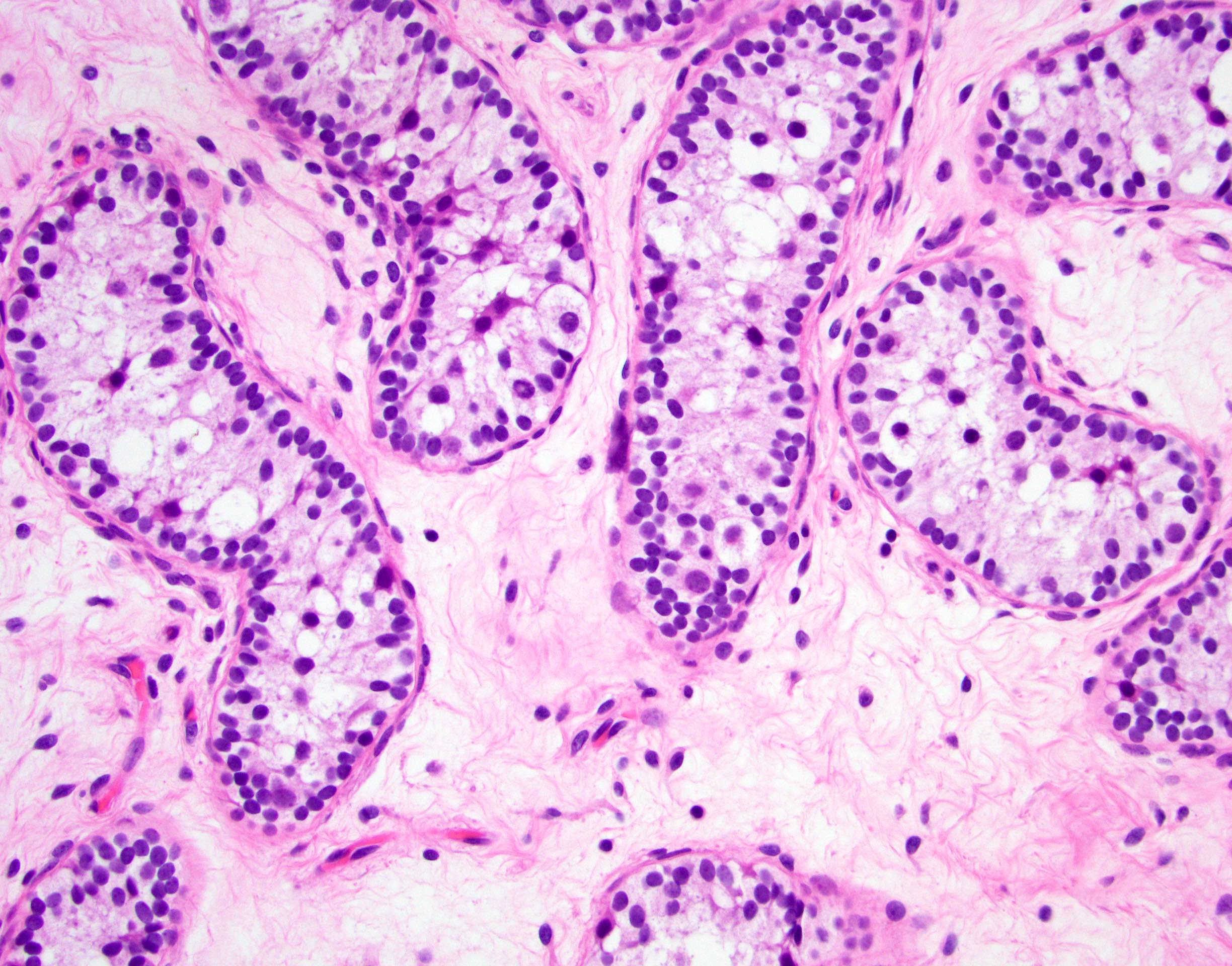
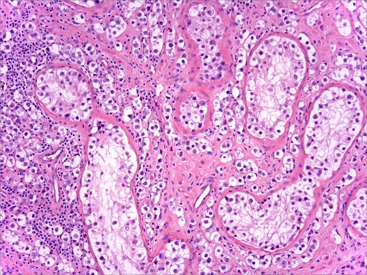
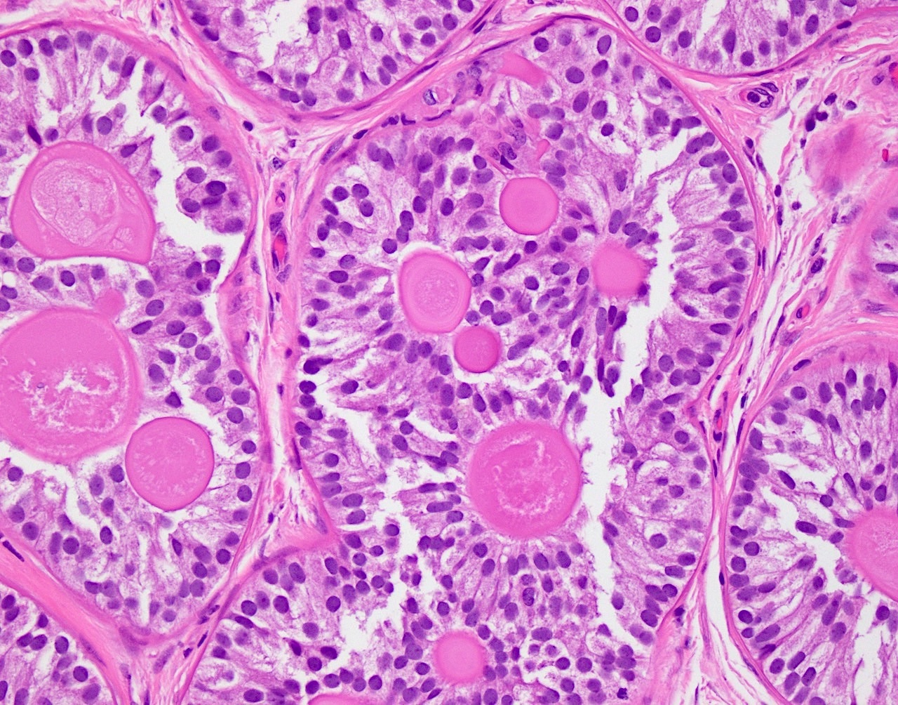
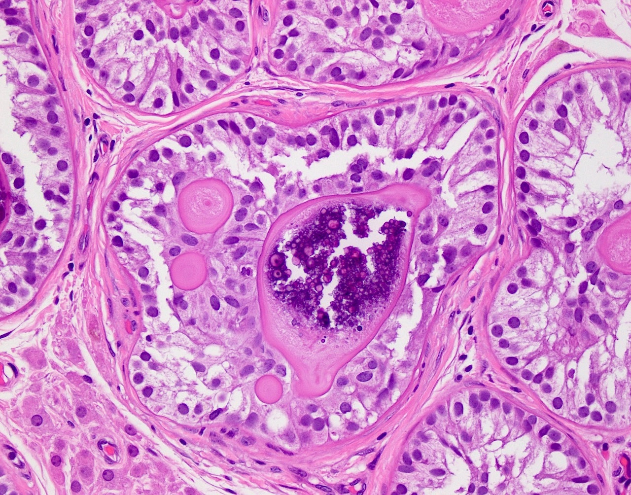
Seminiferous tubules
Basement membrane deposition
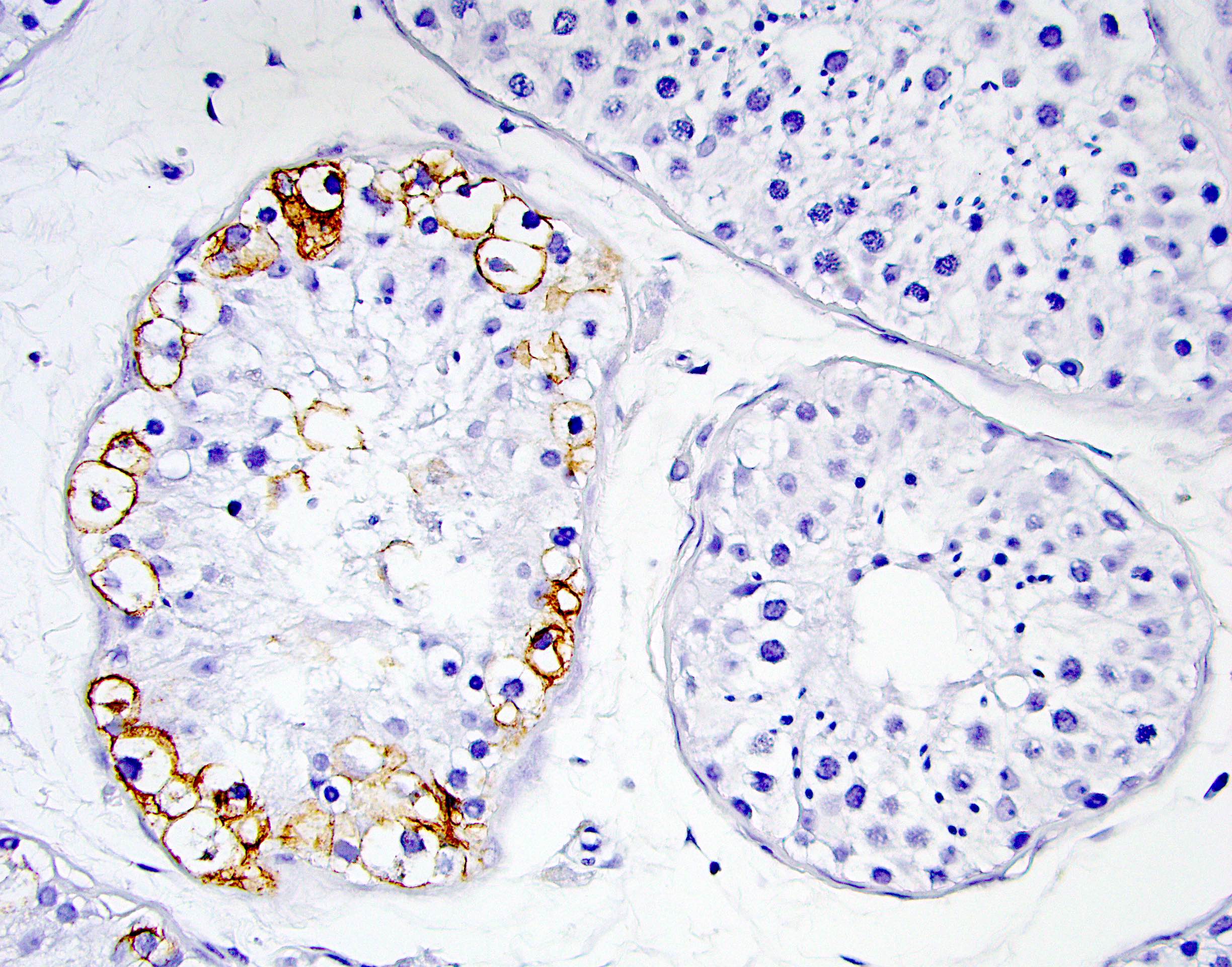
Calretinin
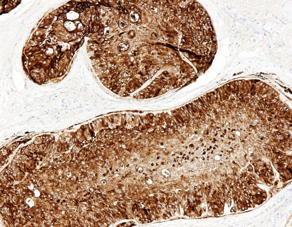
Inhibin A
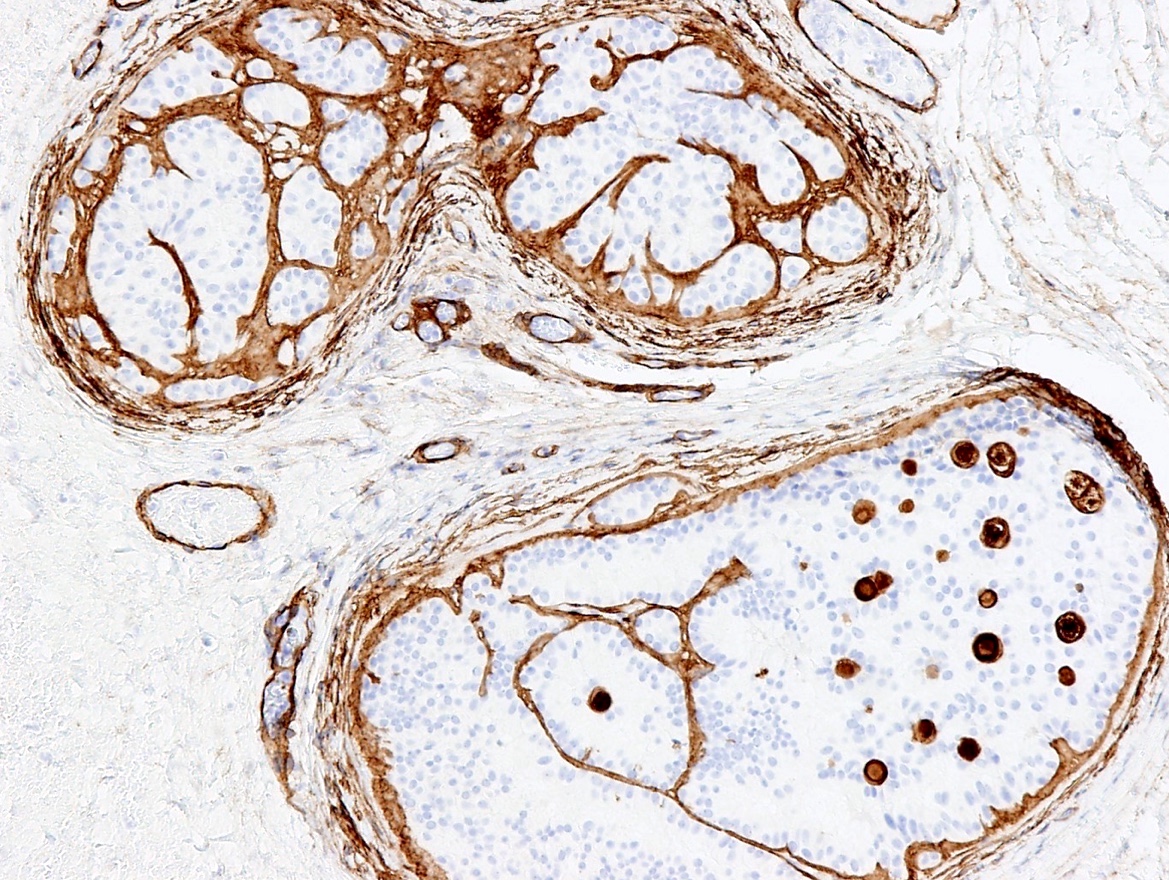
Collagen IV
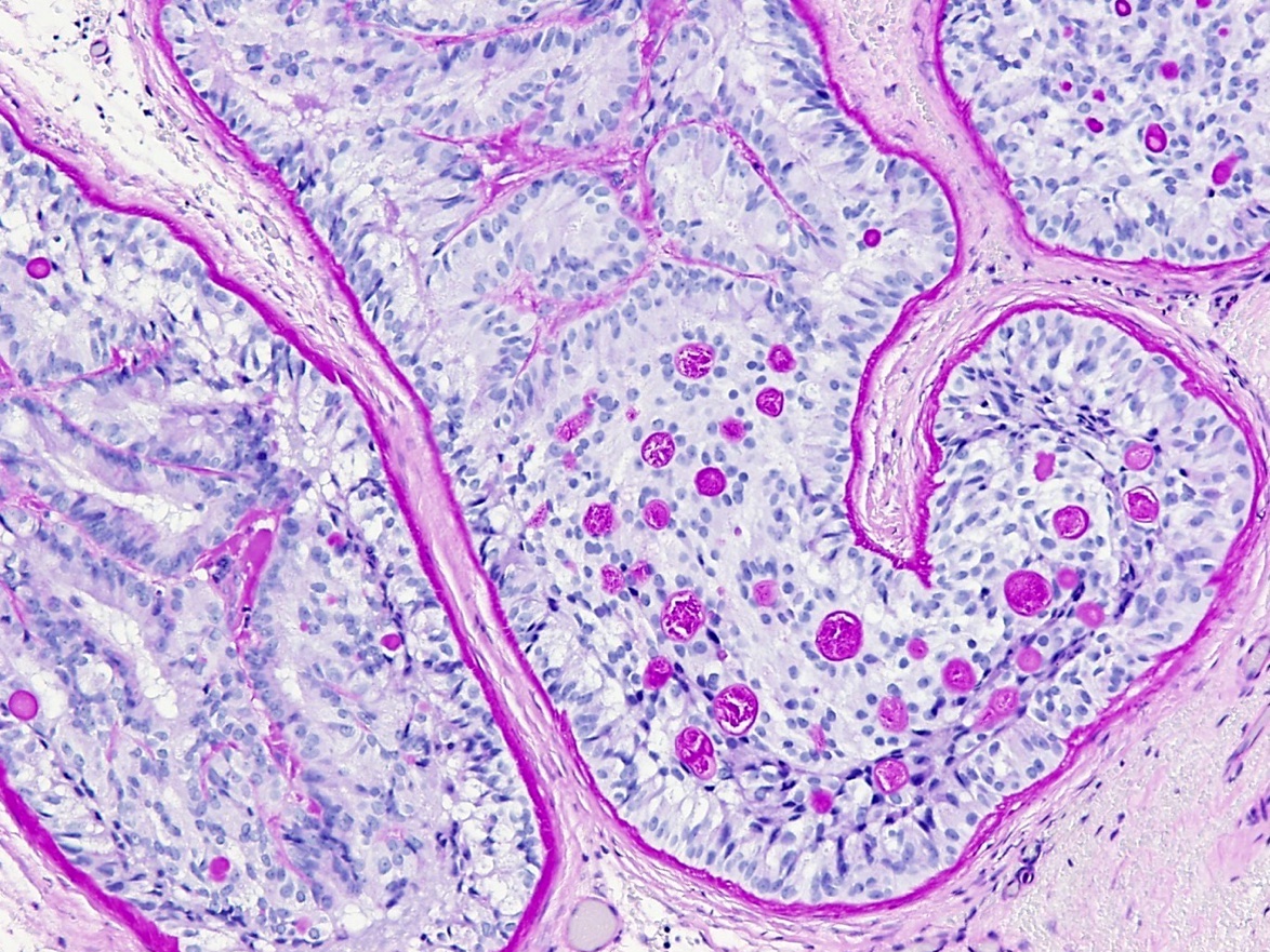
PAS
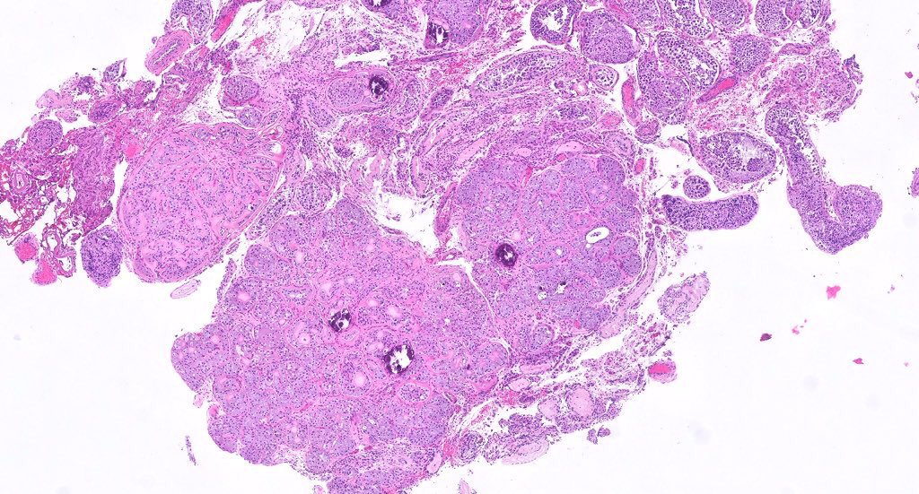
Intratubular large cell hyalinizing Sertoli cell neoplasia
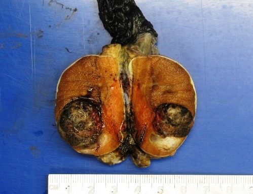
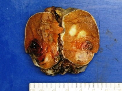
Images hosted on other servers:

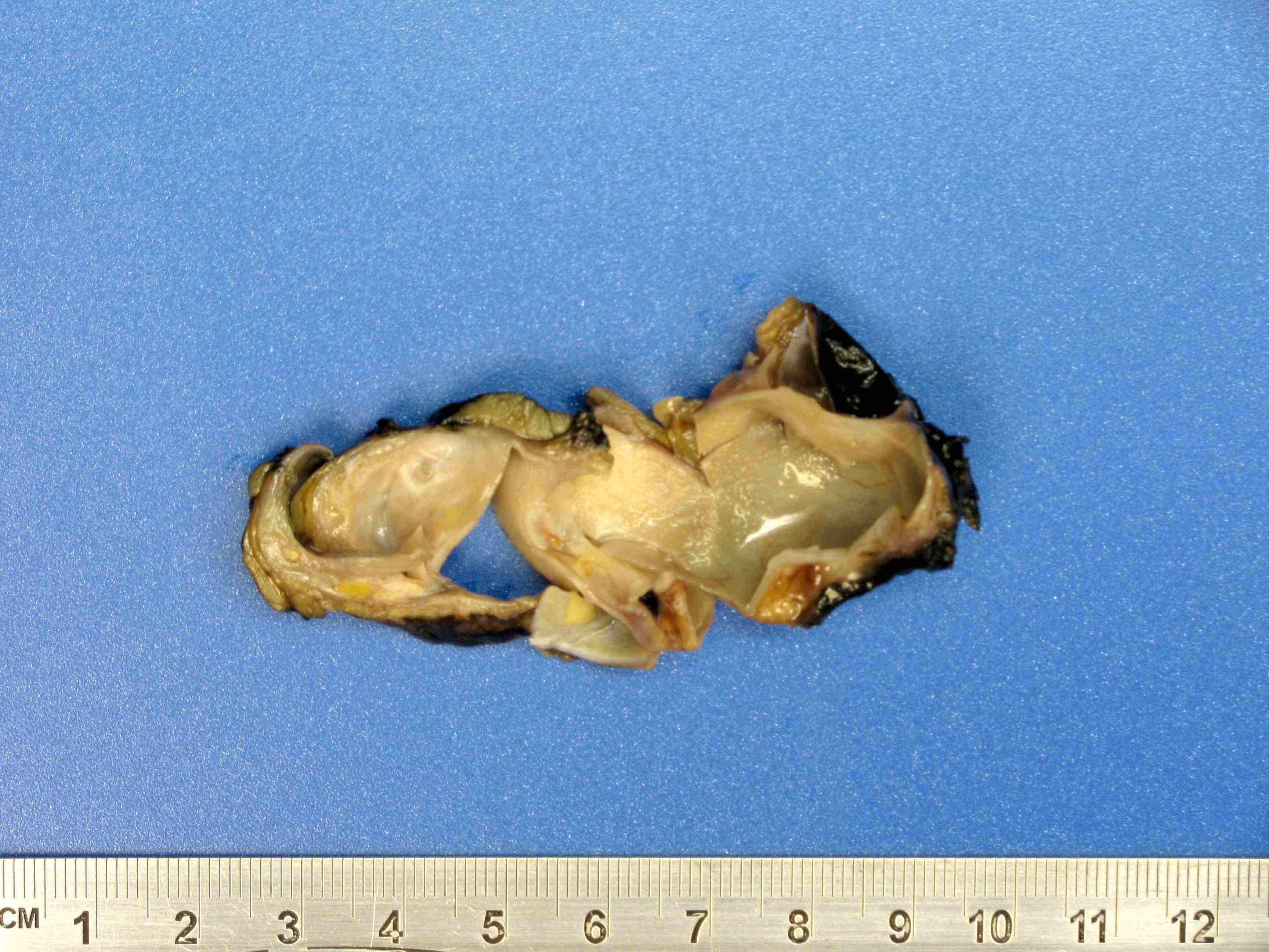
Well encapsulated cystic mass
Images hosted on other servers:

Testicle after surgery
Contributed by Vikas Mehta, M.D.

Variable sized follicles
Follicles with bland cells

Tumor cells with eosinophilic cytoplasm
Round to oval hyperchromatic nuclei
Contributed by Sara Vargas, M.D.
Ill defined testicular nodules
Images hosted on other servers:

Firm, white tan nodules

Tumor
Contributed by Stephanie Siegmund, M.D., Ph.D.

Calcifications, neutrophils, myxoid stroma
Focus of intratubular tumor
Tumor involving rete testis
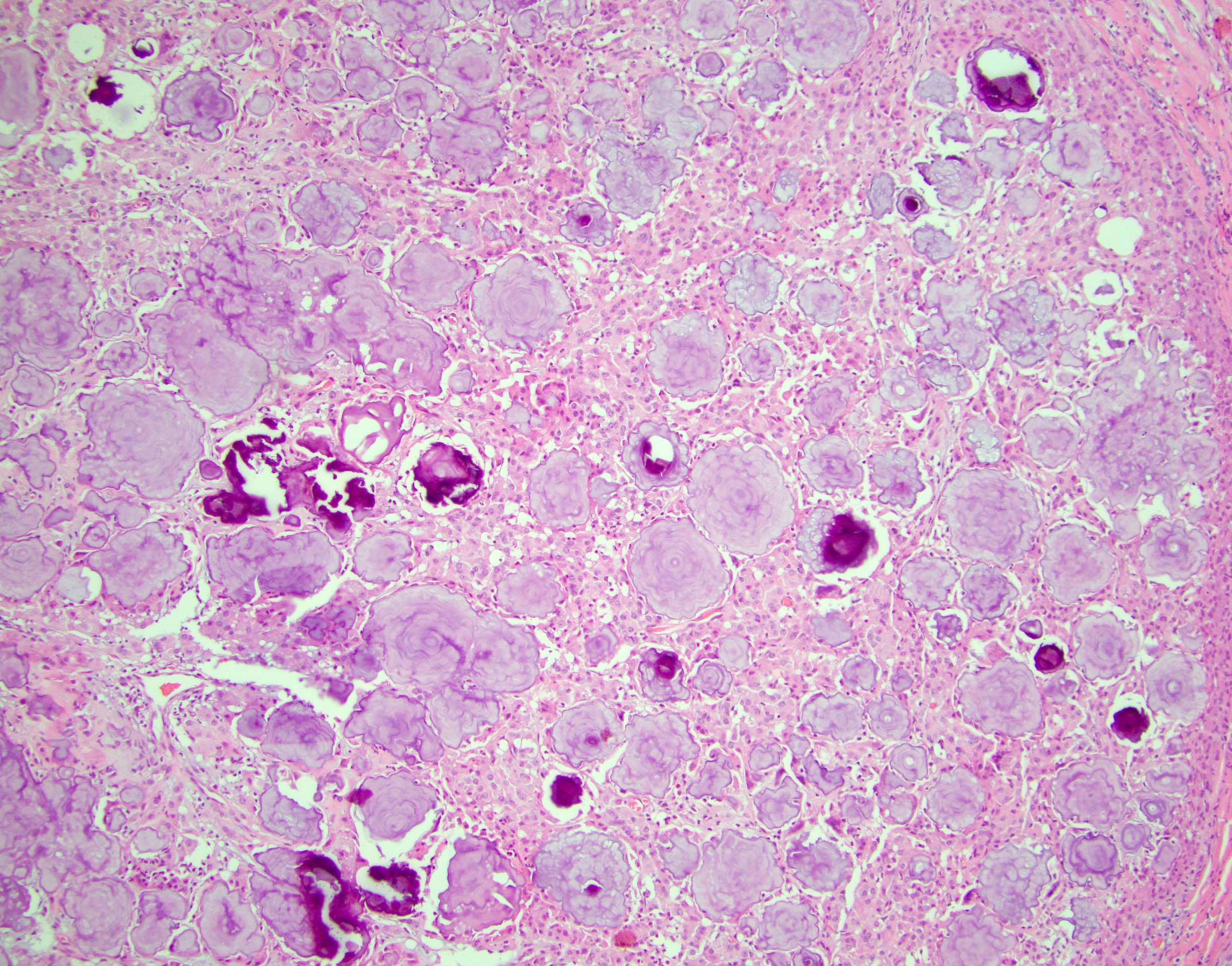
Mulberry calcifications
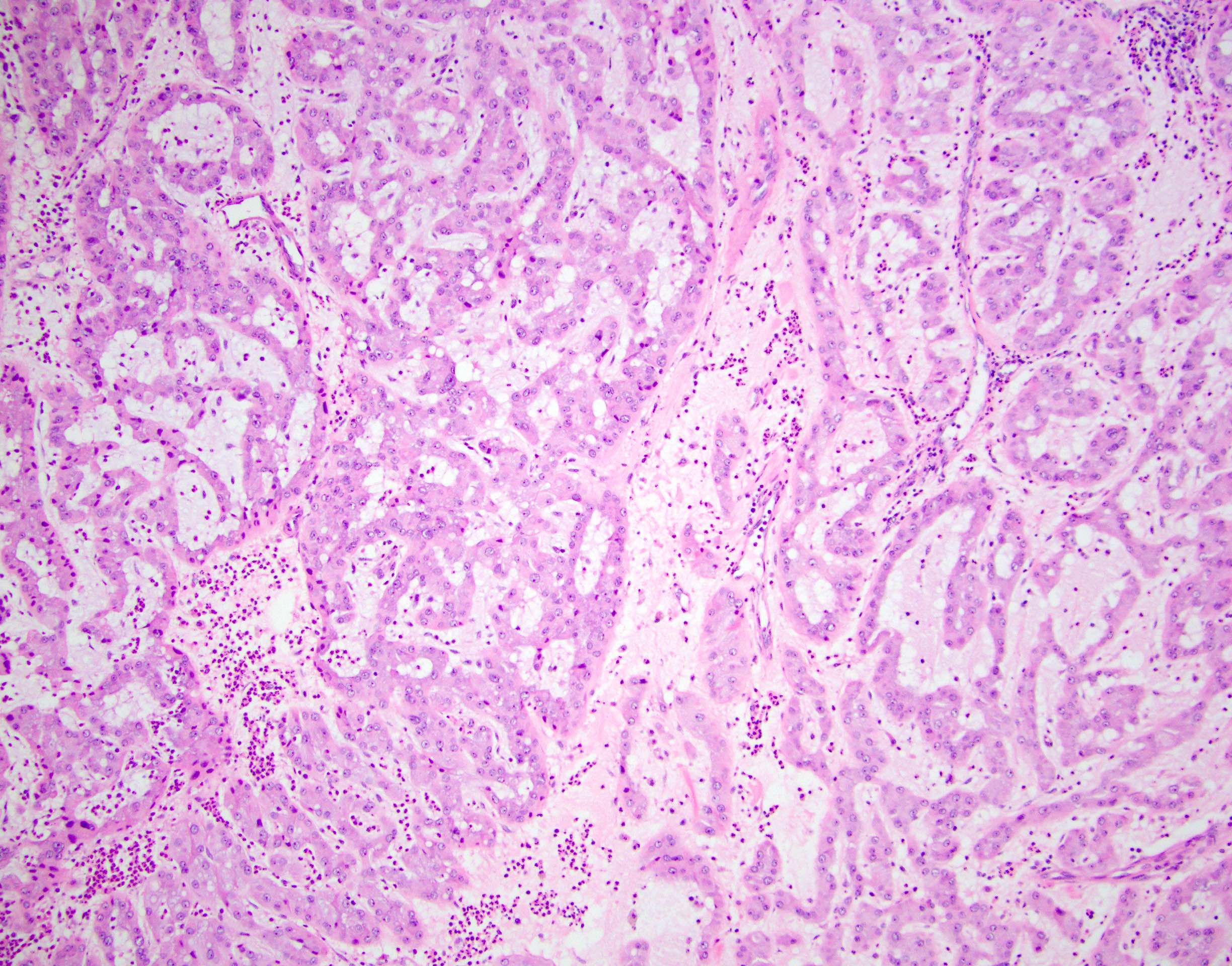
Trabecular architecture
Focal nuclear pleomorphism
Neutrophilic infiltrate
Minimal cytologic atypia
Mulberry calcifications
PRKAR1A loss
Images hosted on other servers:

Mass posterior superior to right testis

Huge heterogeneous enhancing mass

Heterogenous echogenicity
Images hosted on other servers:

Ulcerated and
fungating tumor
involving scrotum
Images hosted on other servers:

Well defined huge solid mass
Case #254
Various images
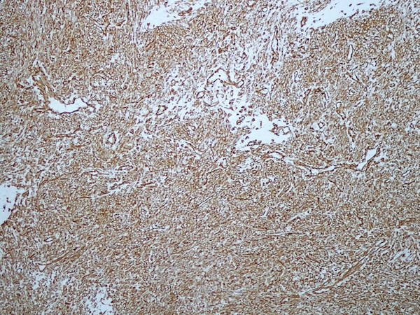
Vimentin
Desmin

Alpha smooth muscle actin
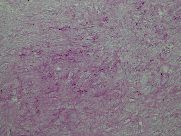
PAS
PLAP
Images hosted on other servers:


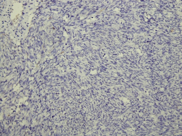
Pattern of spindle cells - H&E


Mitotic activity and atypical nuclei


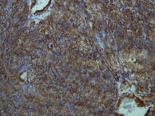
Desmin+, SMA+


Desmin+

SMA+
Images hosted on other servers:

CT scan: circumscribed mass

MRI: solid enhancing mass

Ultrasound: mixed echogenic mass
Contributed by Debra L. Zynger, M.D.
Small tumor
Nodular tumor
High risk tumor
Images hosted on other servers:

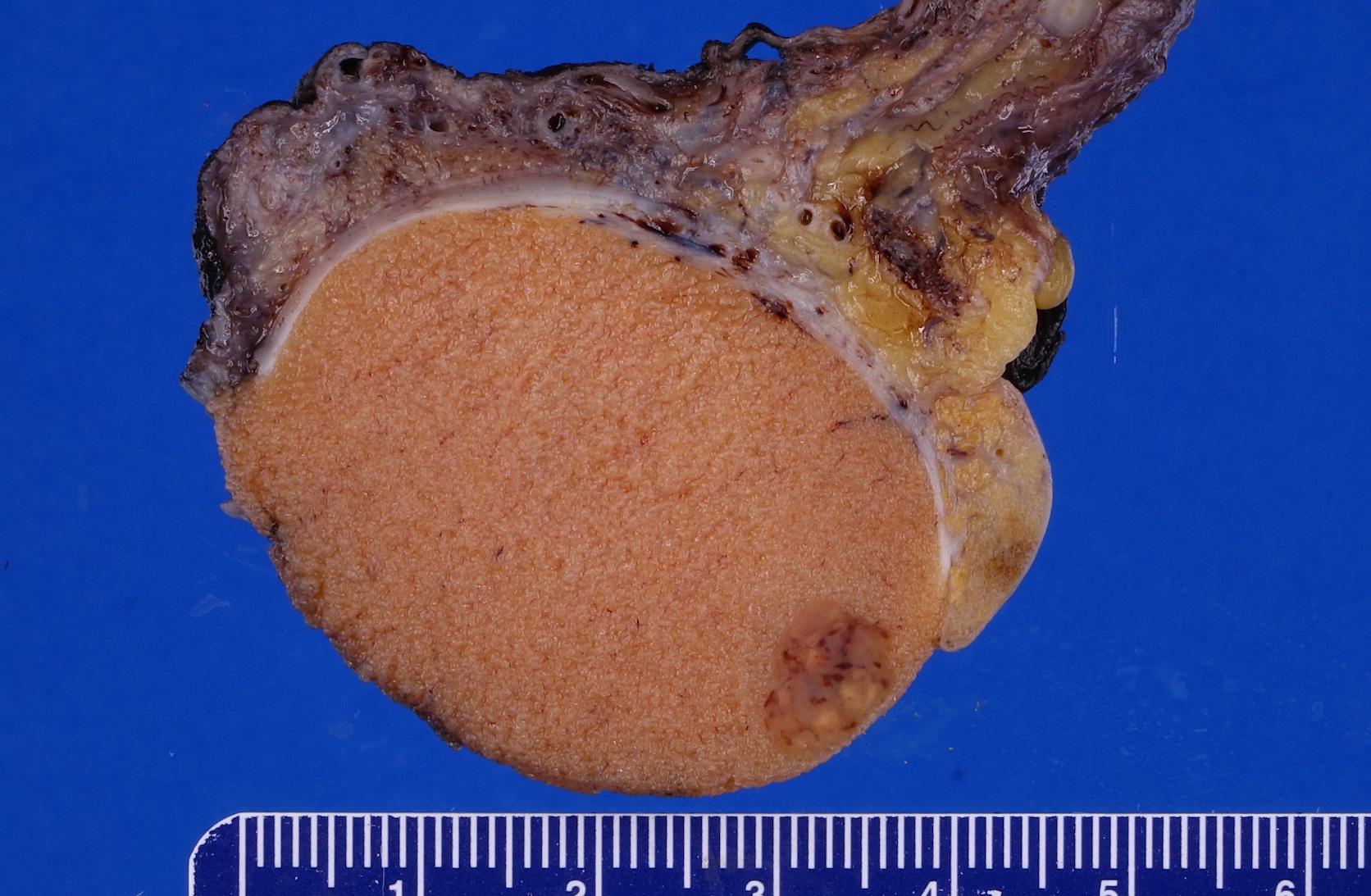
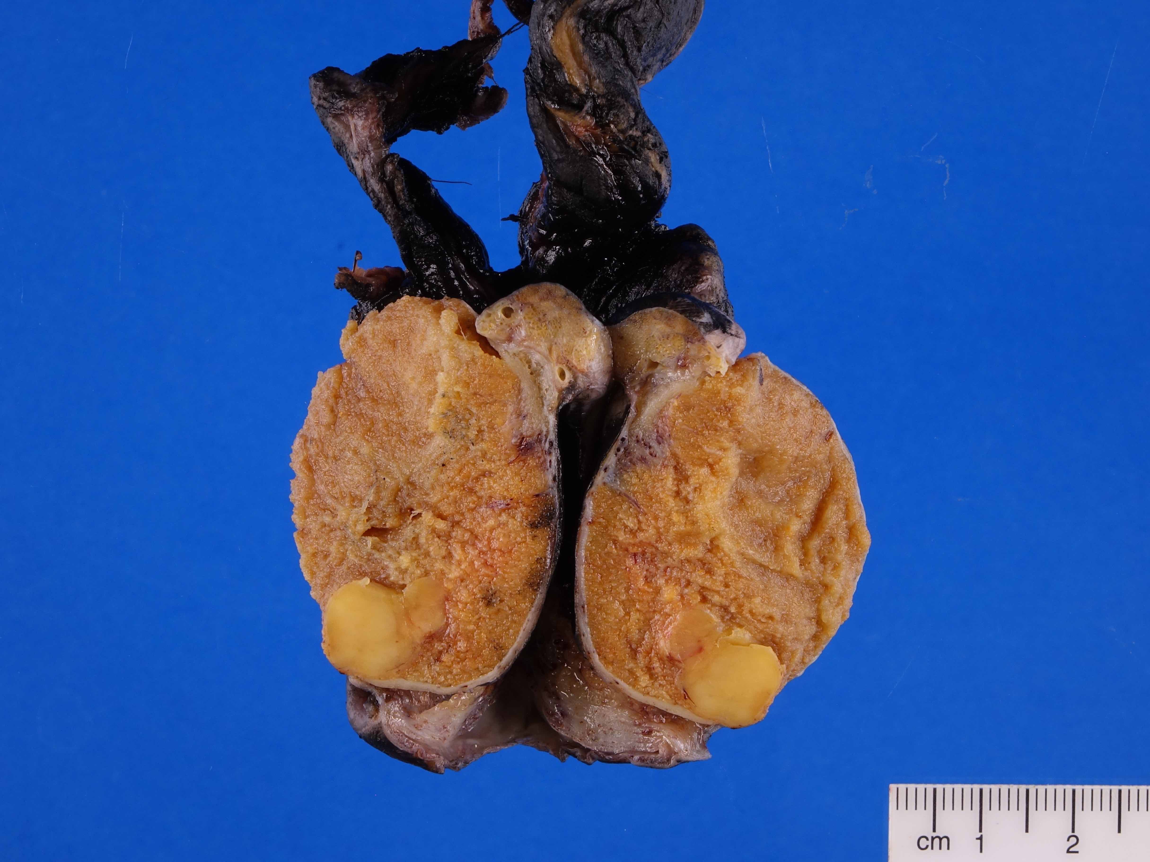
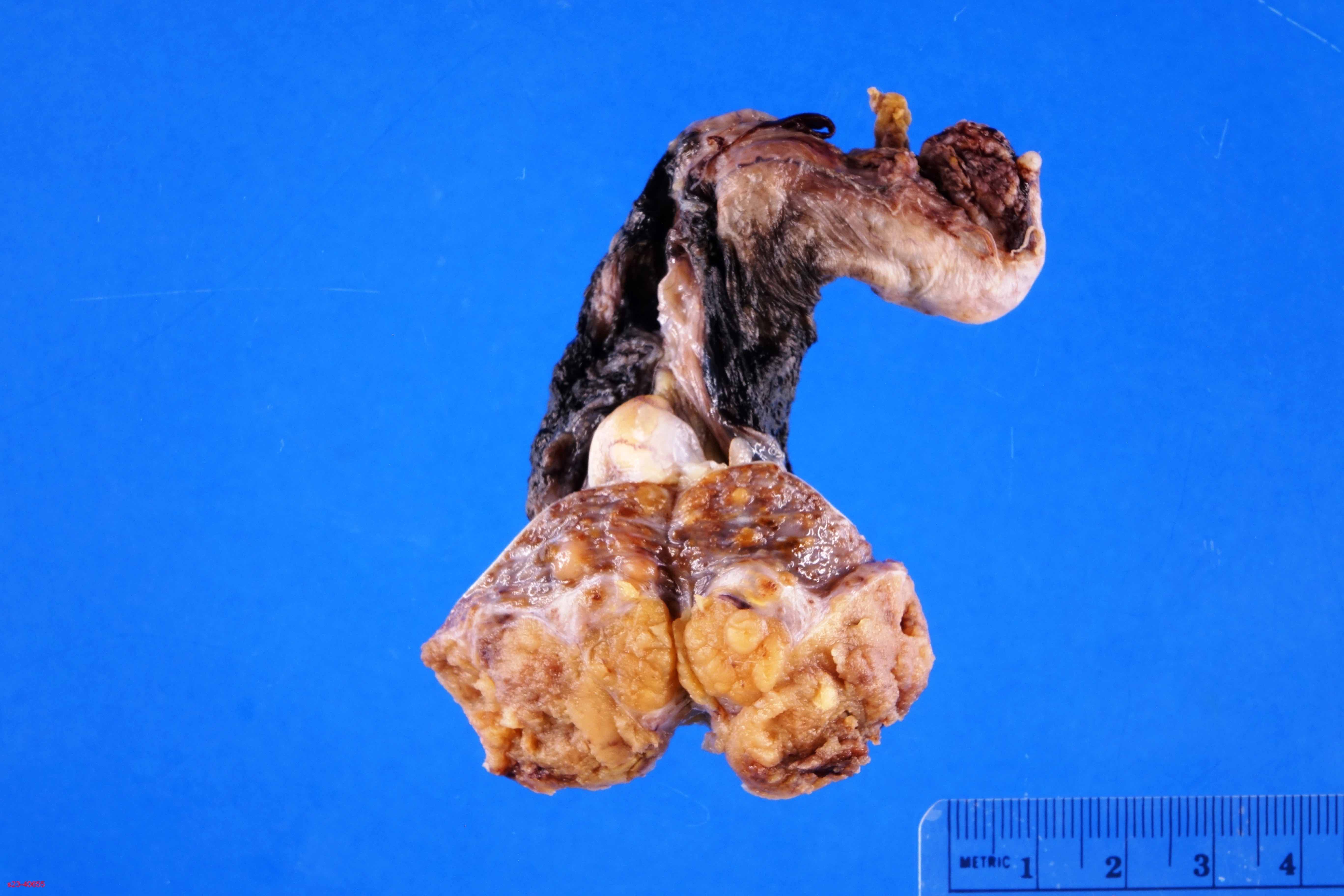
Circumscribed brown tumor
Contributed by Manju Aron, M.D. and Kristine Cornejo, M.D.
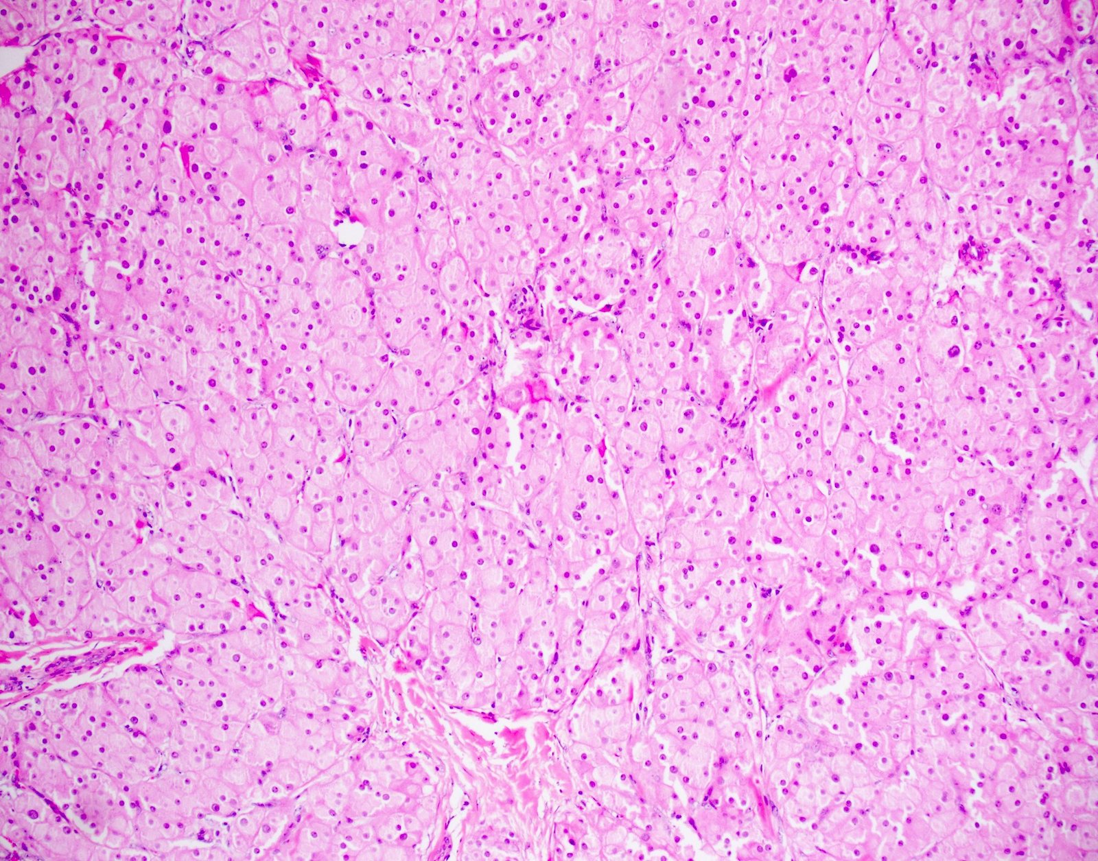
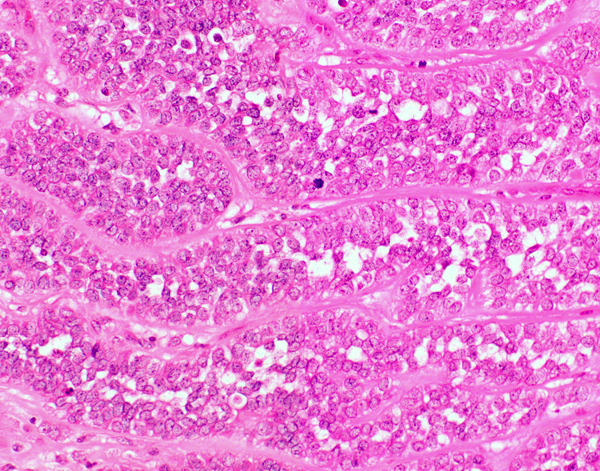
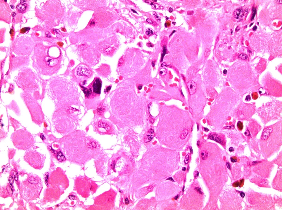
Polygonal cells
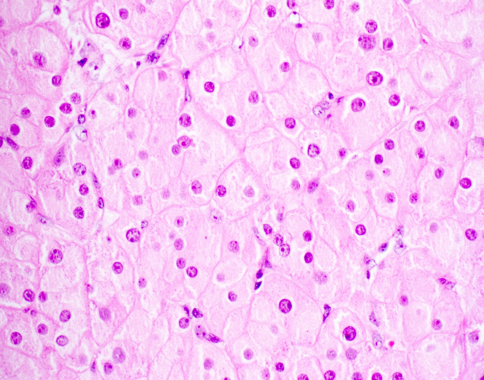
Eosinophilic cells

Reinke crystals
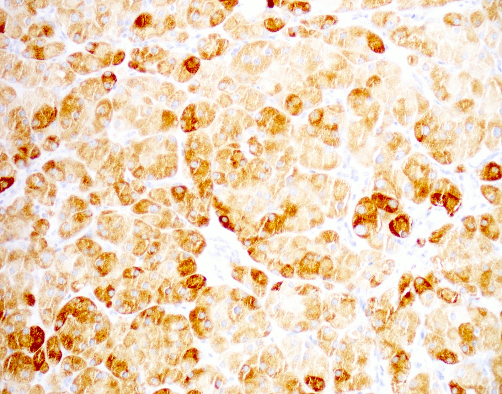
Inhibin positive

SF1 positive
Images hosted on other servers:

Nonhomogeneous right scrotal mass

No evidence of tissue infiltration

Large heterogenous extratesticular mass

CT of paratesticular liposarcoma presenting as hernia
Images hosted on other servers:

Scrotal mass at presentation

Tumor after chemotherapy
Images hosted on other servers:

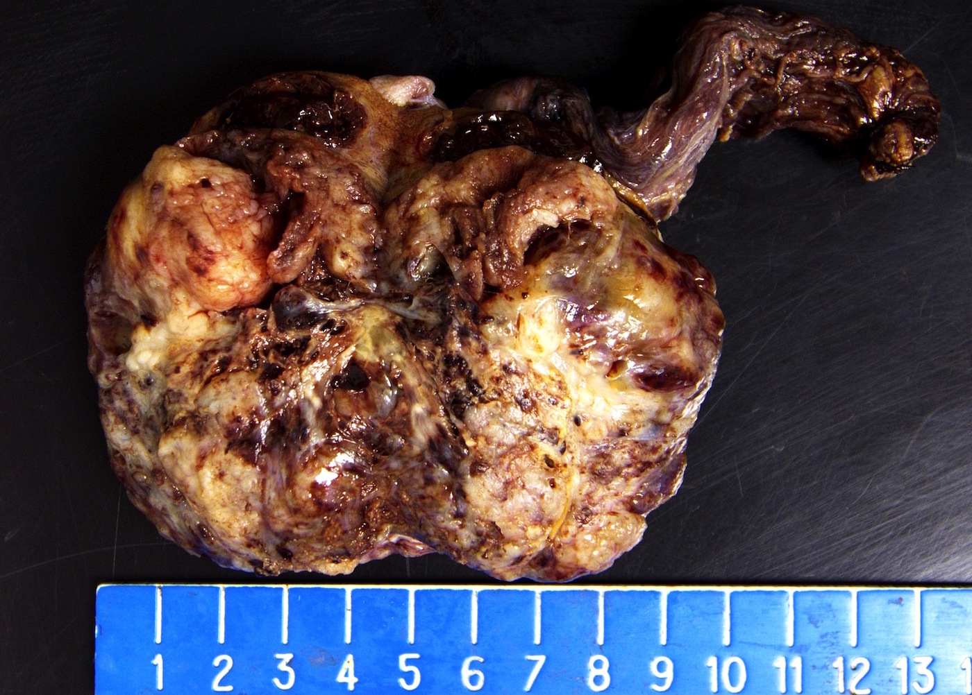
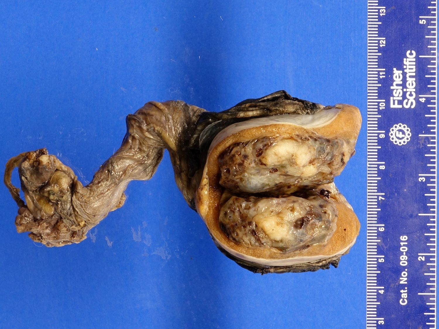
Tumor mass

Yellow and myxoid areas

White yellow to red brown cut surface, marked necrosis

Right paratesticular mass with solid, yellow white, fatty, myxoid areas

Grossly firm tumor
Images hosted on other servers:
Well differentiated liposarcoma:


Atypical spindle cells and lipoblasts


Mature adipocytes and lipoblasts
Well differentiated with dedifferentiated component:

Hypercellular stroma with atypical adipocytes

Atypical adipocytes with enlarged nuclei
Dedifferentiated with leiomyosarcomatous component:

H&E

MDM2+, CDK4+, alpha smooth muscle actin+, desmin+
Myxoid liposarcoma:

Lipoblasts contain lipid vacuoles

Plexiform arrangement of capillaries
Images hosted on other servers:

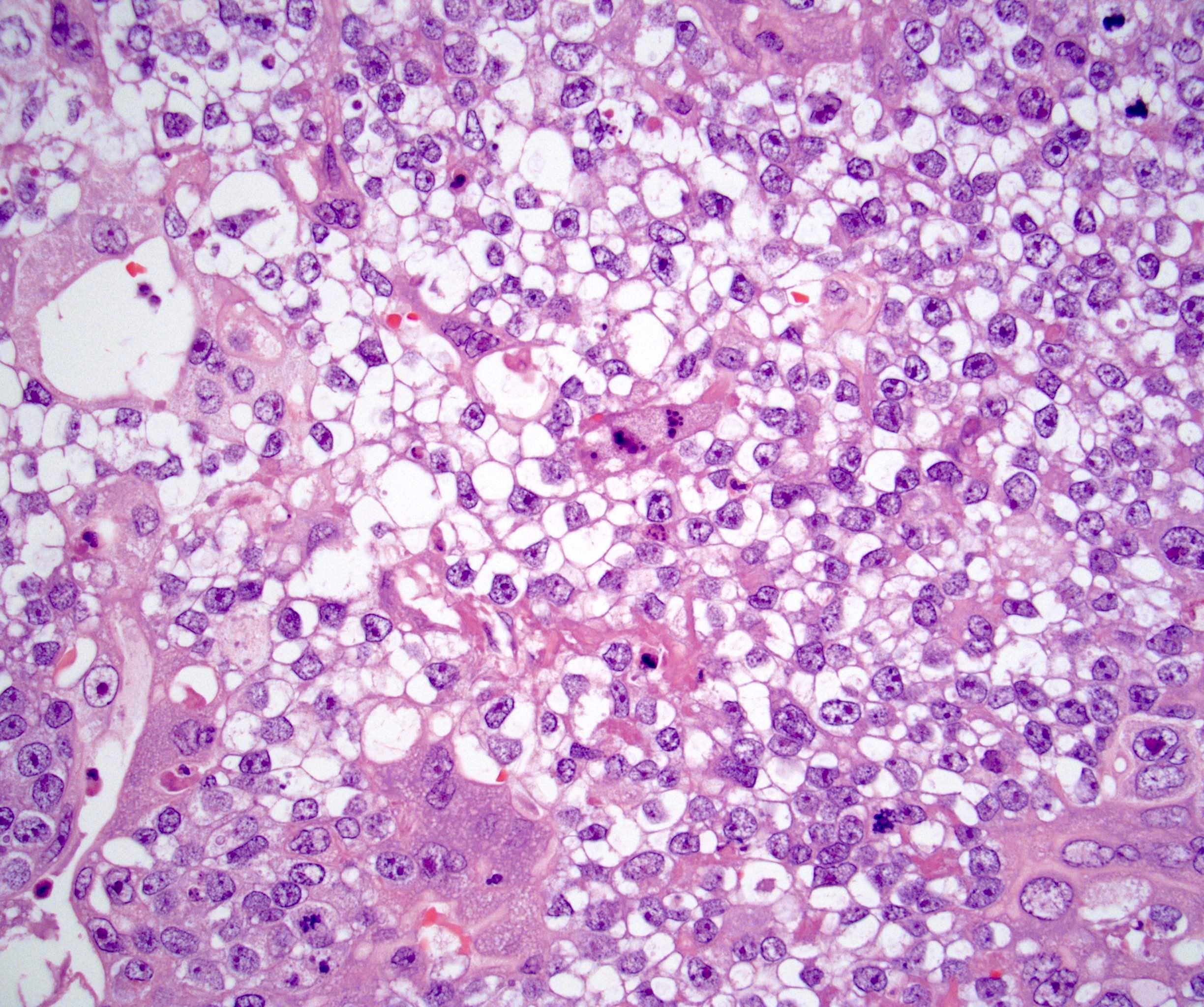
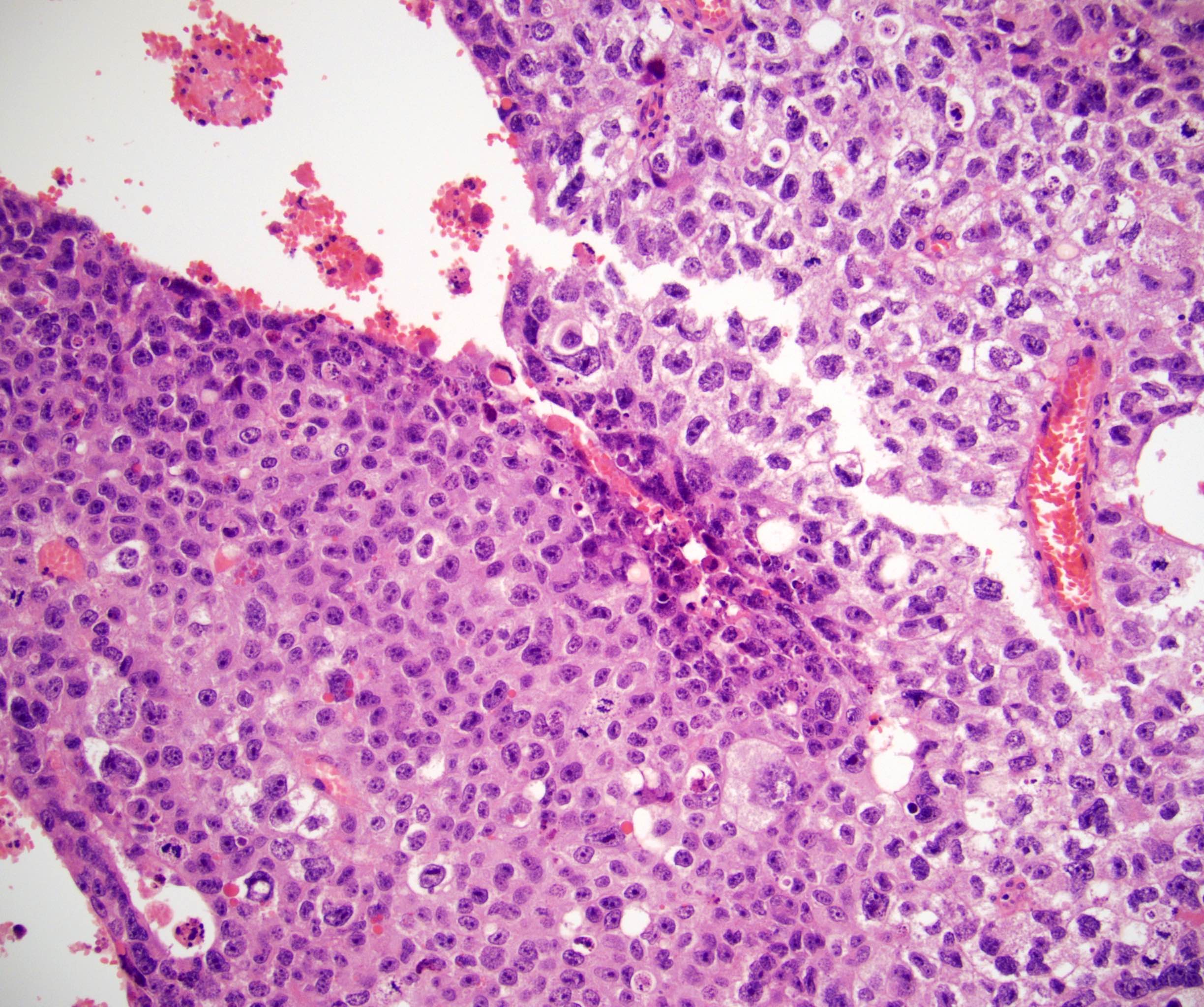
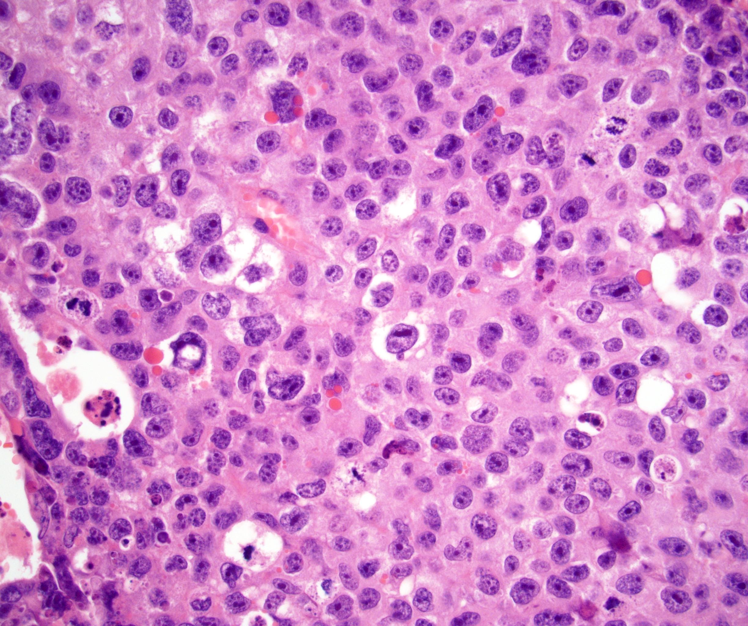
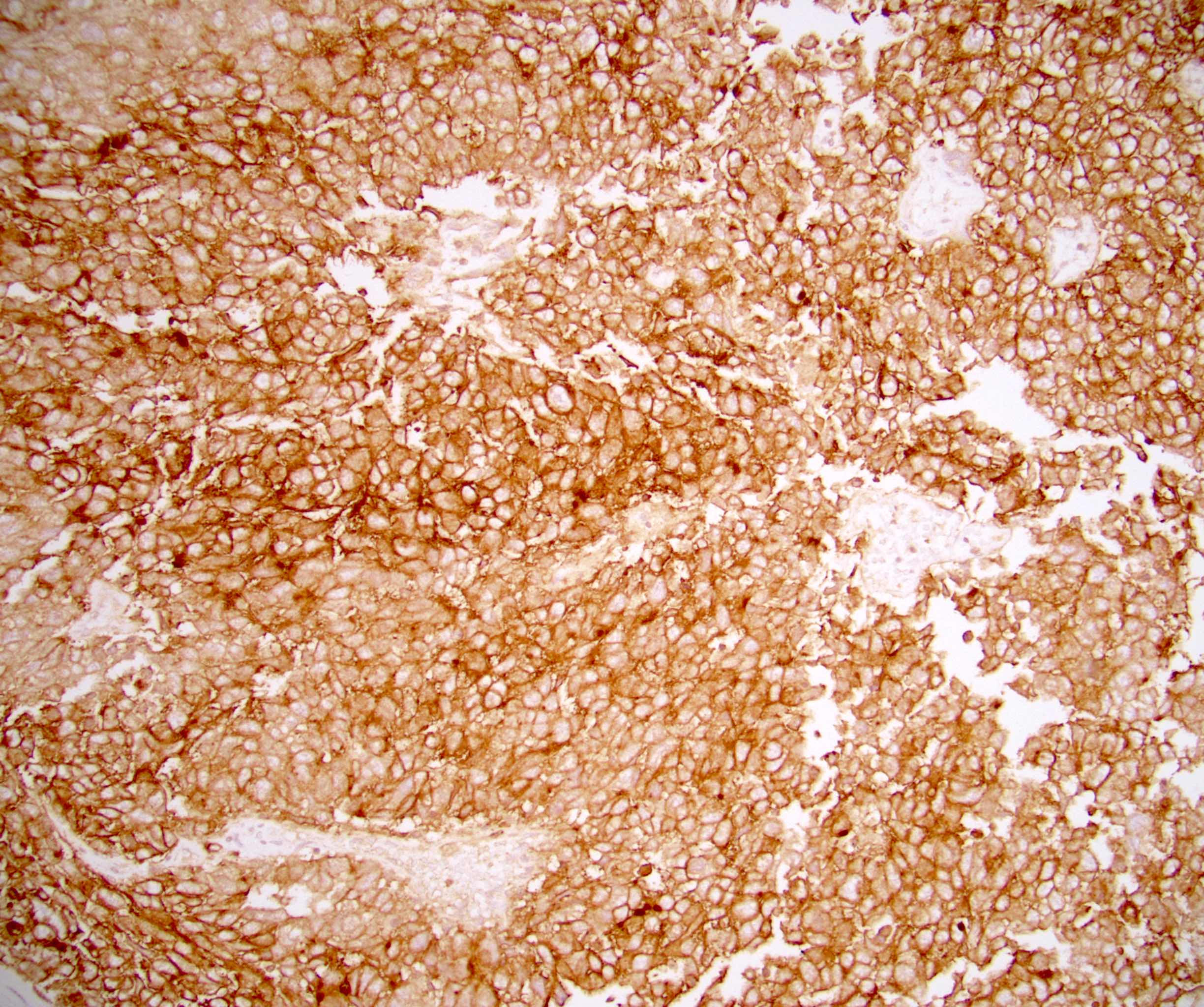
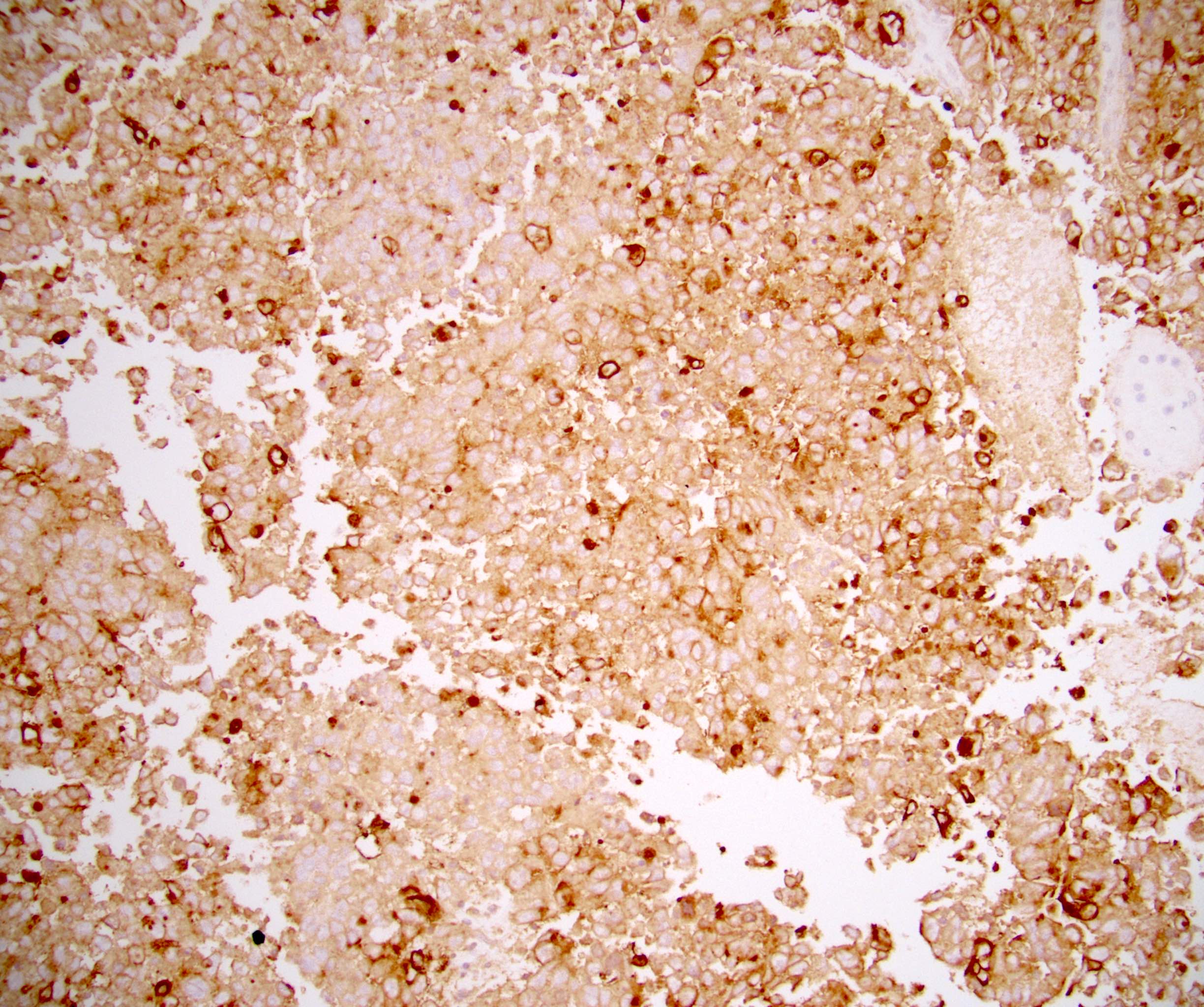
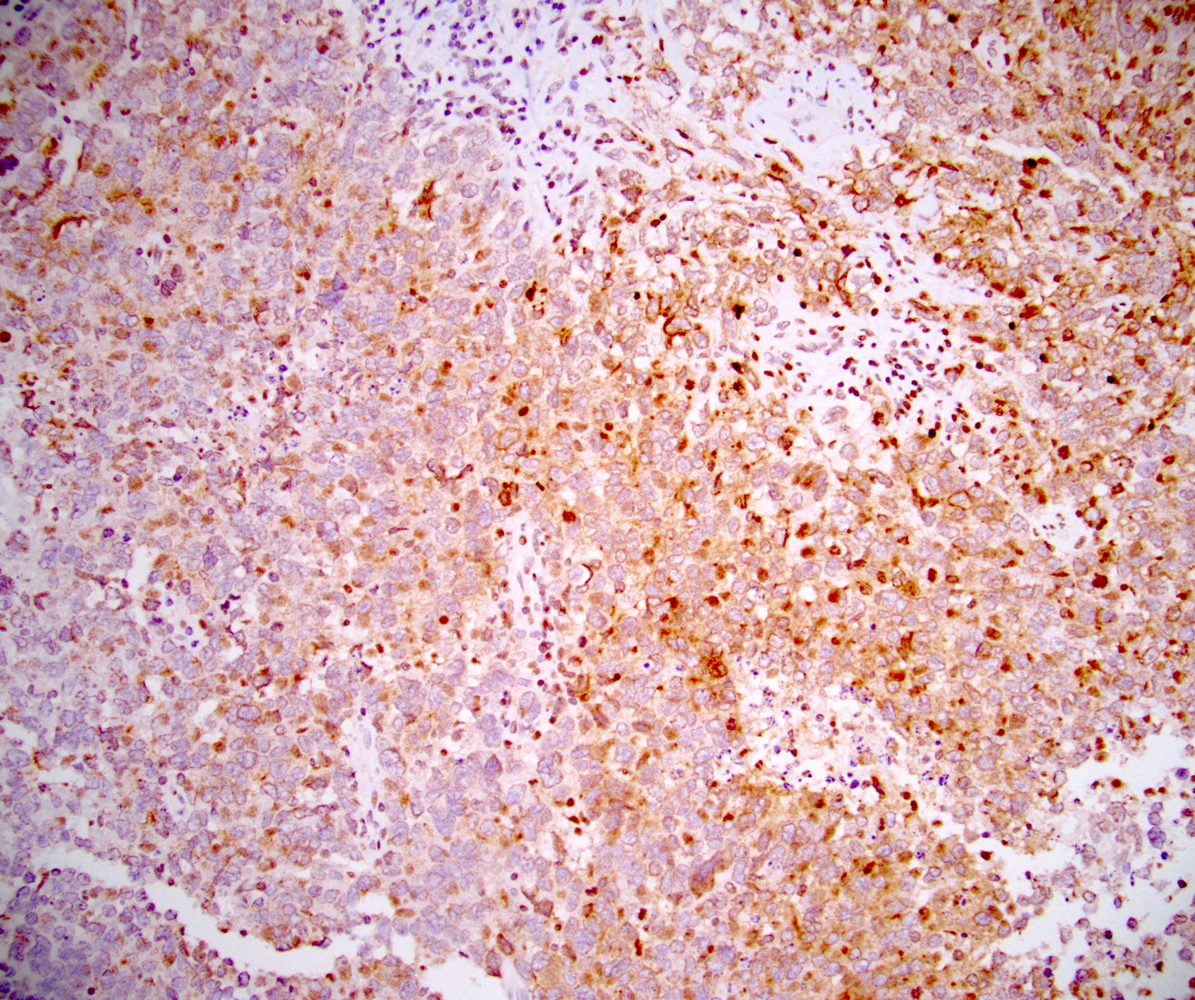
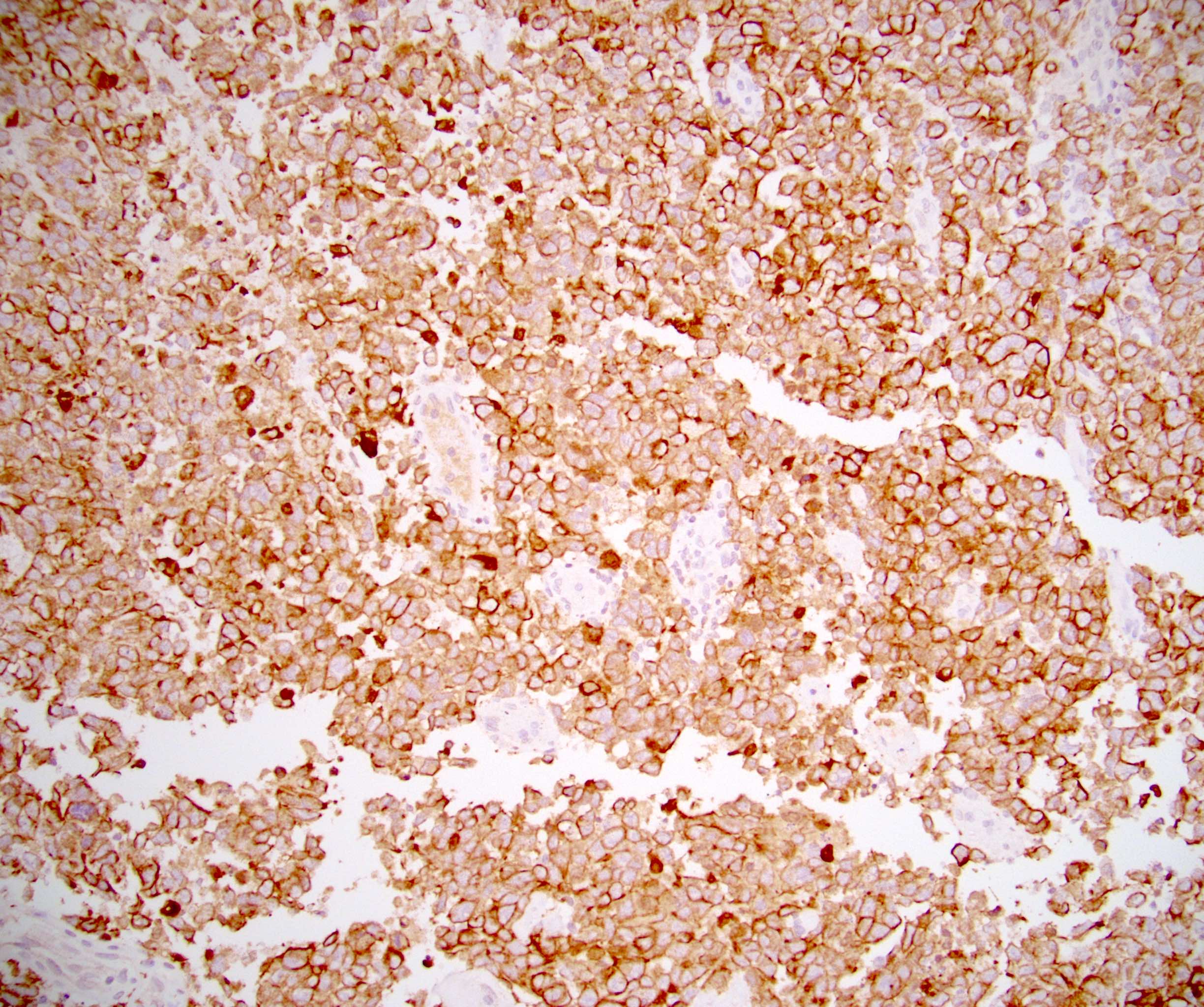
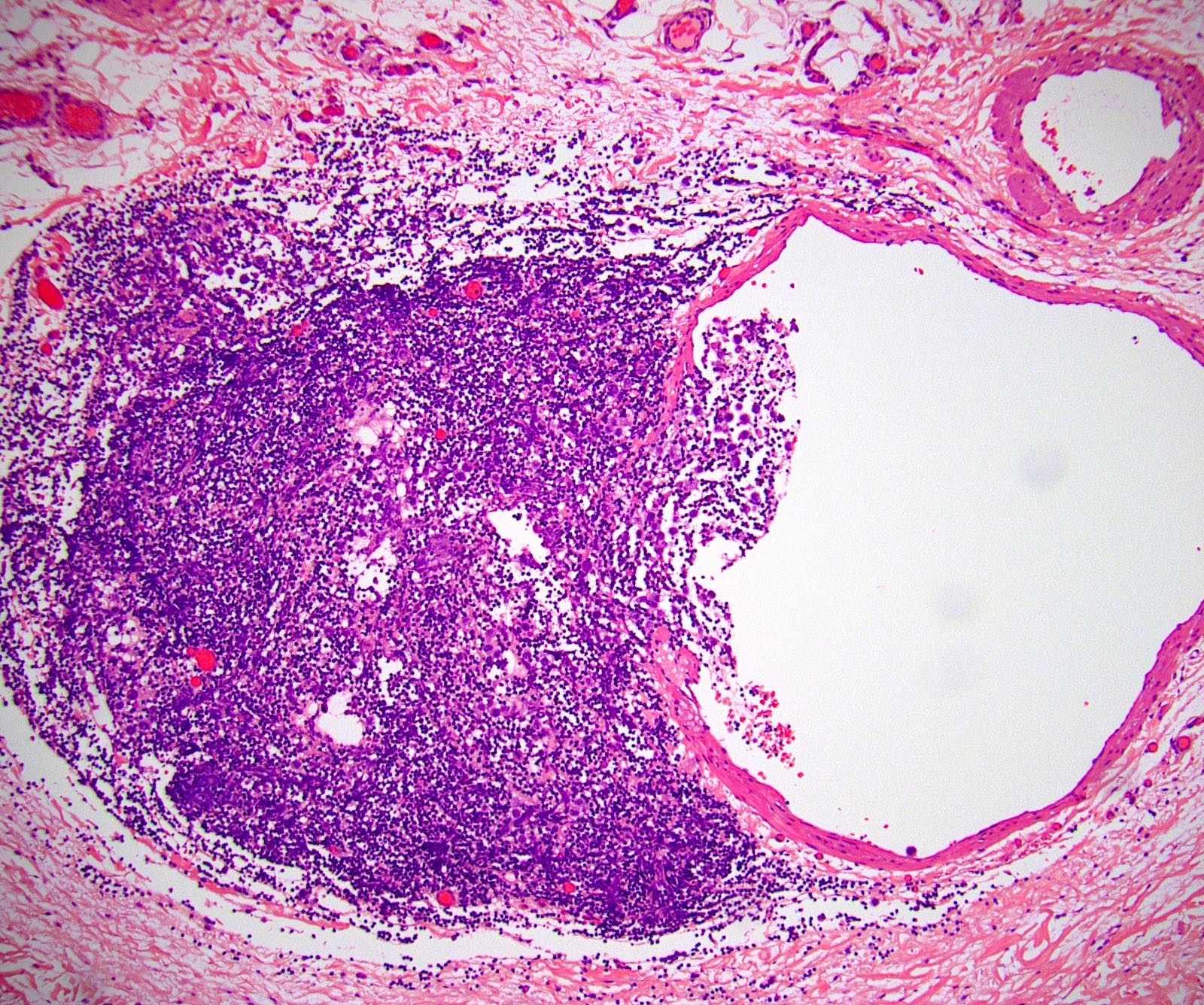
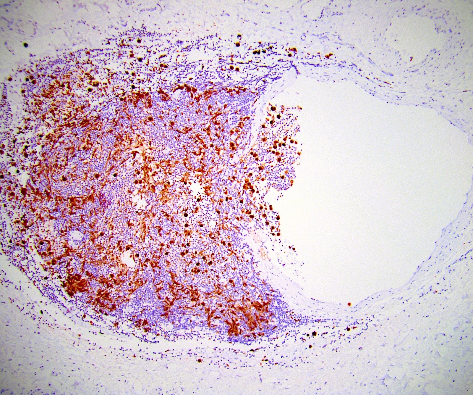
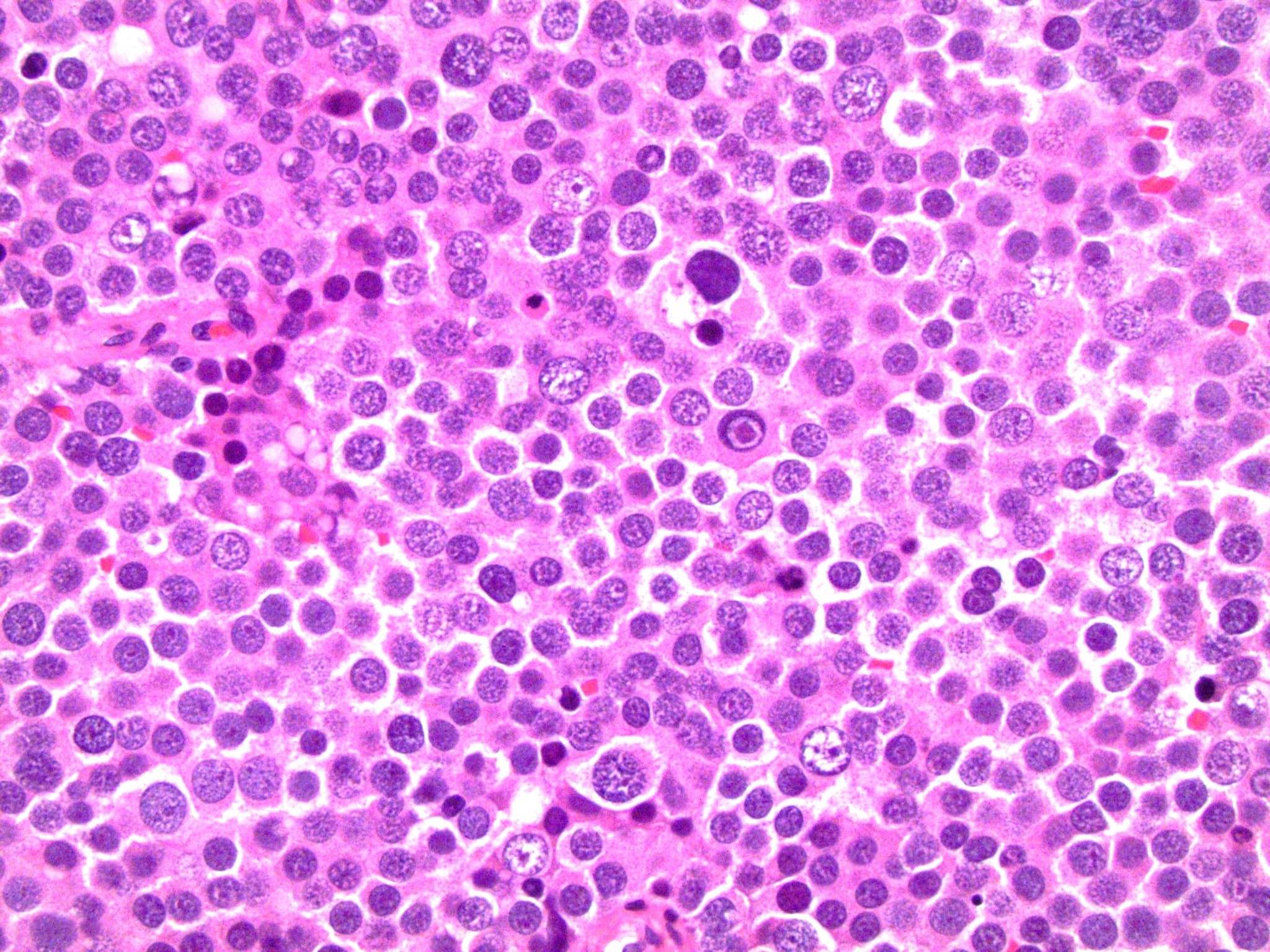
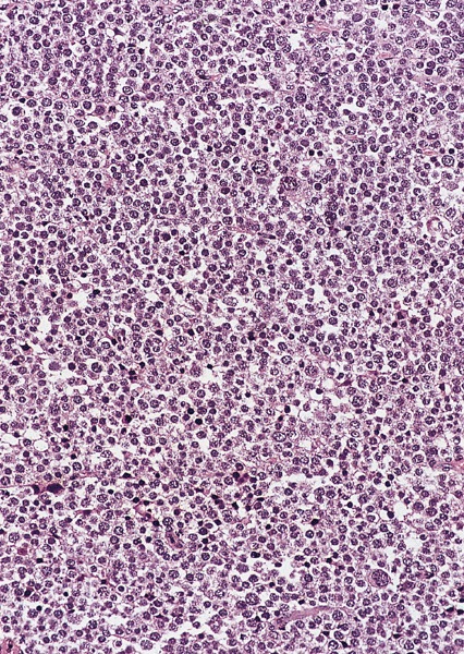
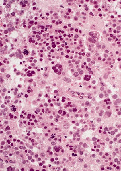
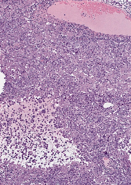
Diffuse large B cell lymphoma
Images hosted on other servers:


Diffuse large B cell lymphoma


Peripheral T cell lymphoma
Images hosted on other servers:


Maxillary tumors
Images hosted on other servers:


Maxillary tumor: H&E and stains
Images hosted on other servers:

Increased thickness of the left tunica vaginalis

Small nodules

Border indistinctness in the right scrotum
Case #261


AFIP images
Tunica vaginalis
Images hosted on other servers:

Radical orchiectomy specimen

Nodular mass at spermatic cord

Extracted tumor adhered strongly to scrotum skin
Contributed by Jennifer Gordetsky, M.D.

Mesothelioma
Case #261

Images hosted on other servers:

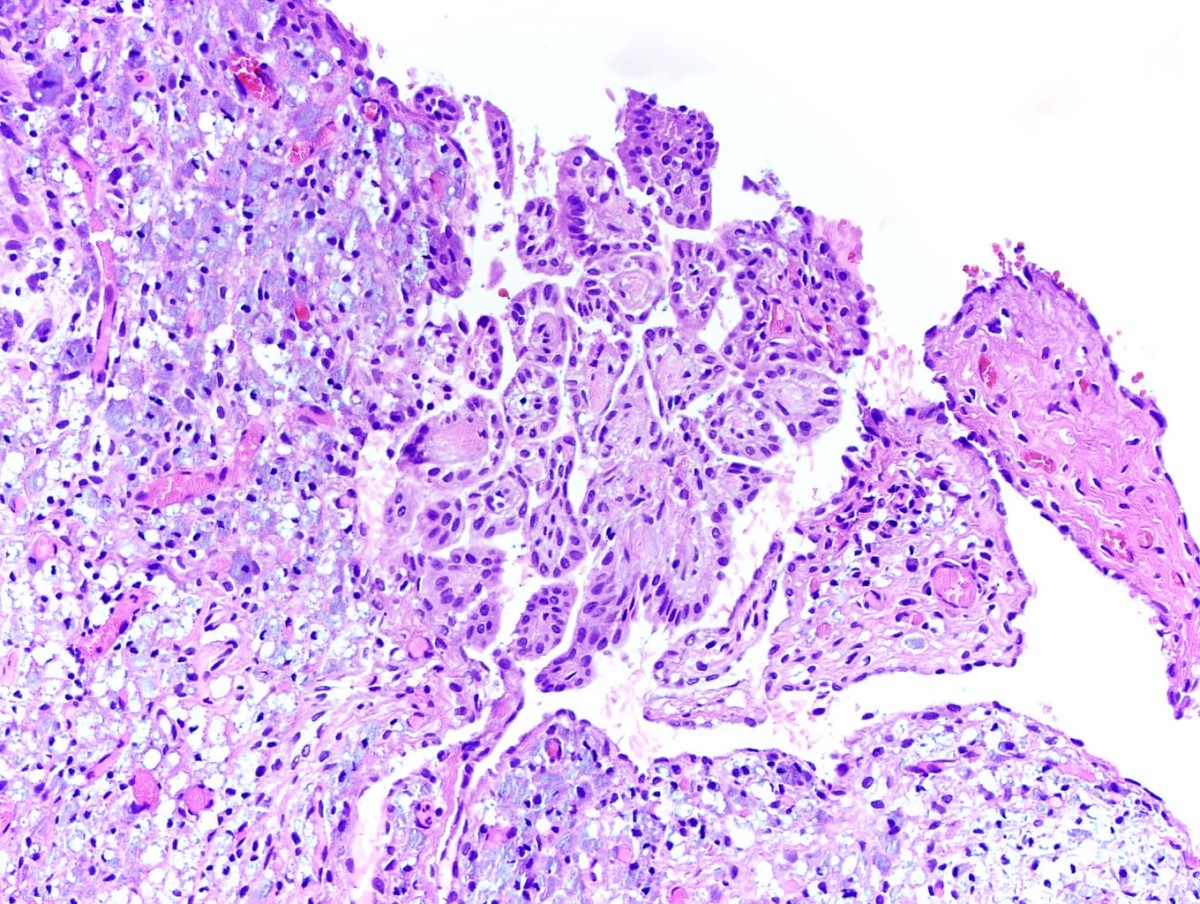
Epithelial tumor cells

Glandular pattern in desmoplastic stroma

Malignant mesothelial tubular structures

Biphasic exophytic nodule

Papillary structures

Mesothelioma of uncertain malignant potential

Cells are rounded with nuclear atypia
Images hosted on other servers:

Renal cell carcinoma metastasis to testis

Lung adenocarcinoma metastasis to testis

Colorectal adenocarcinoma metastasis to testis
Contributed by Debra L. Zynger, M.D.
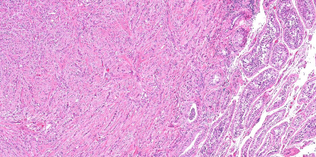
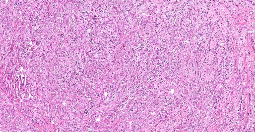
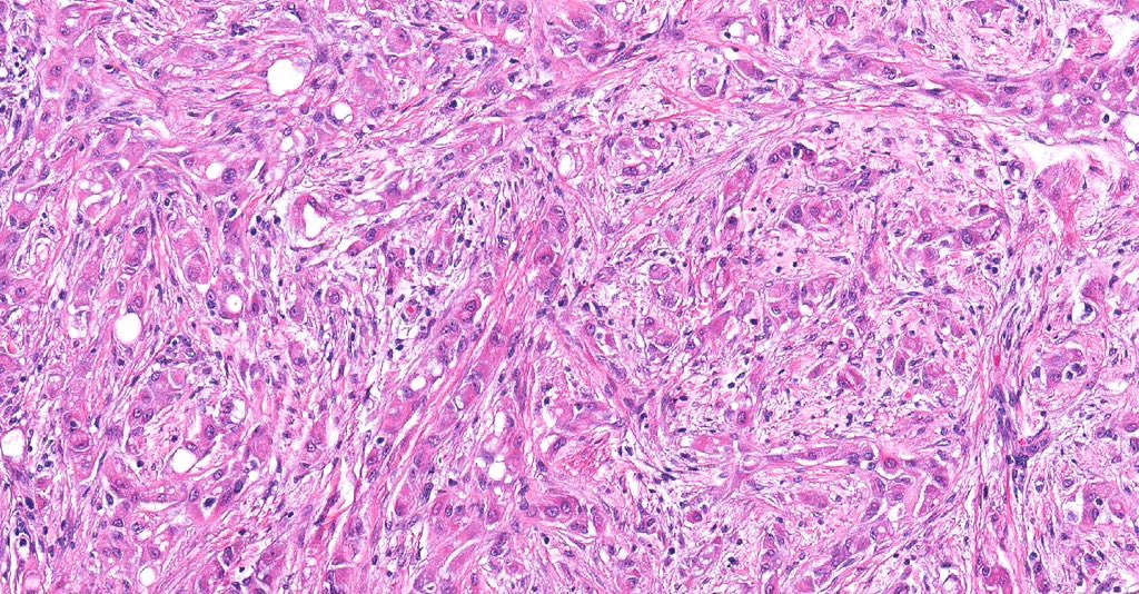
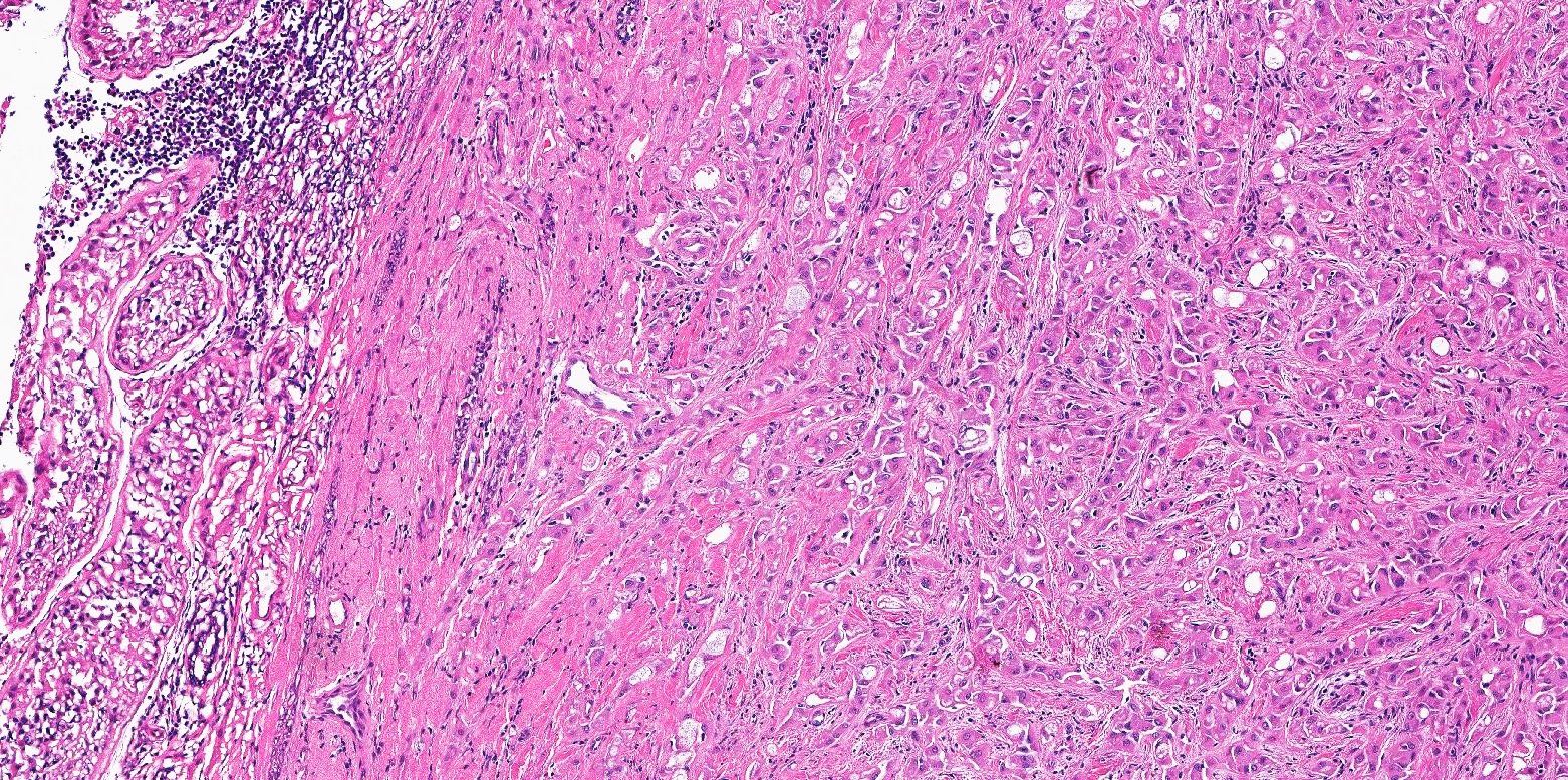
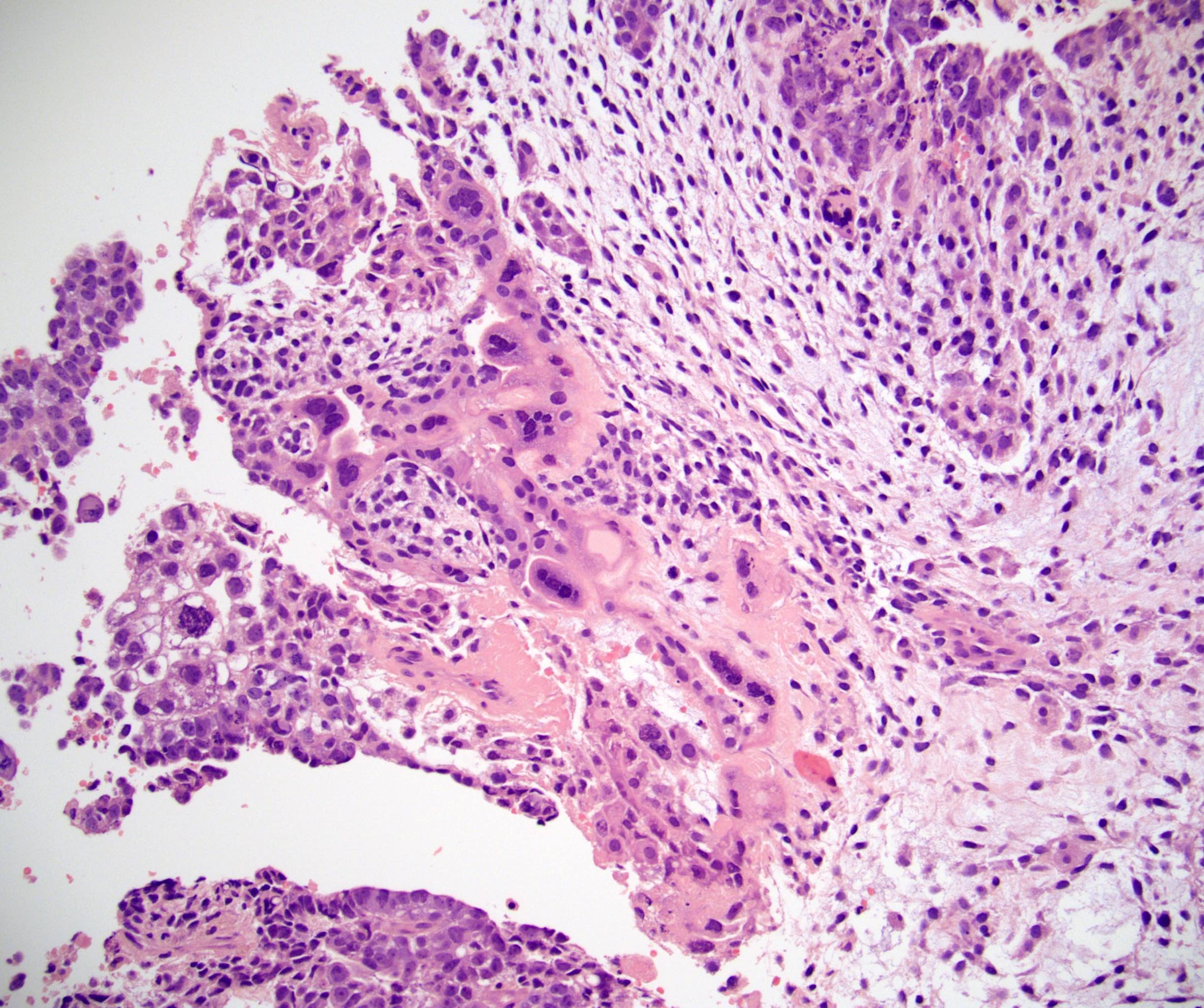
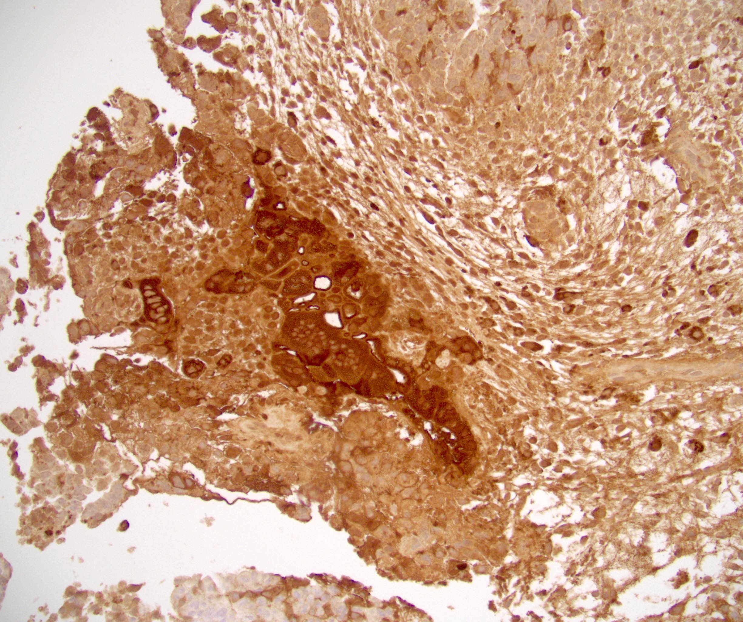
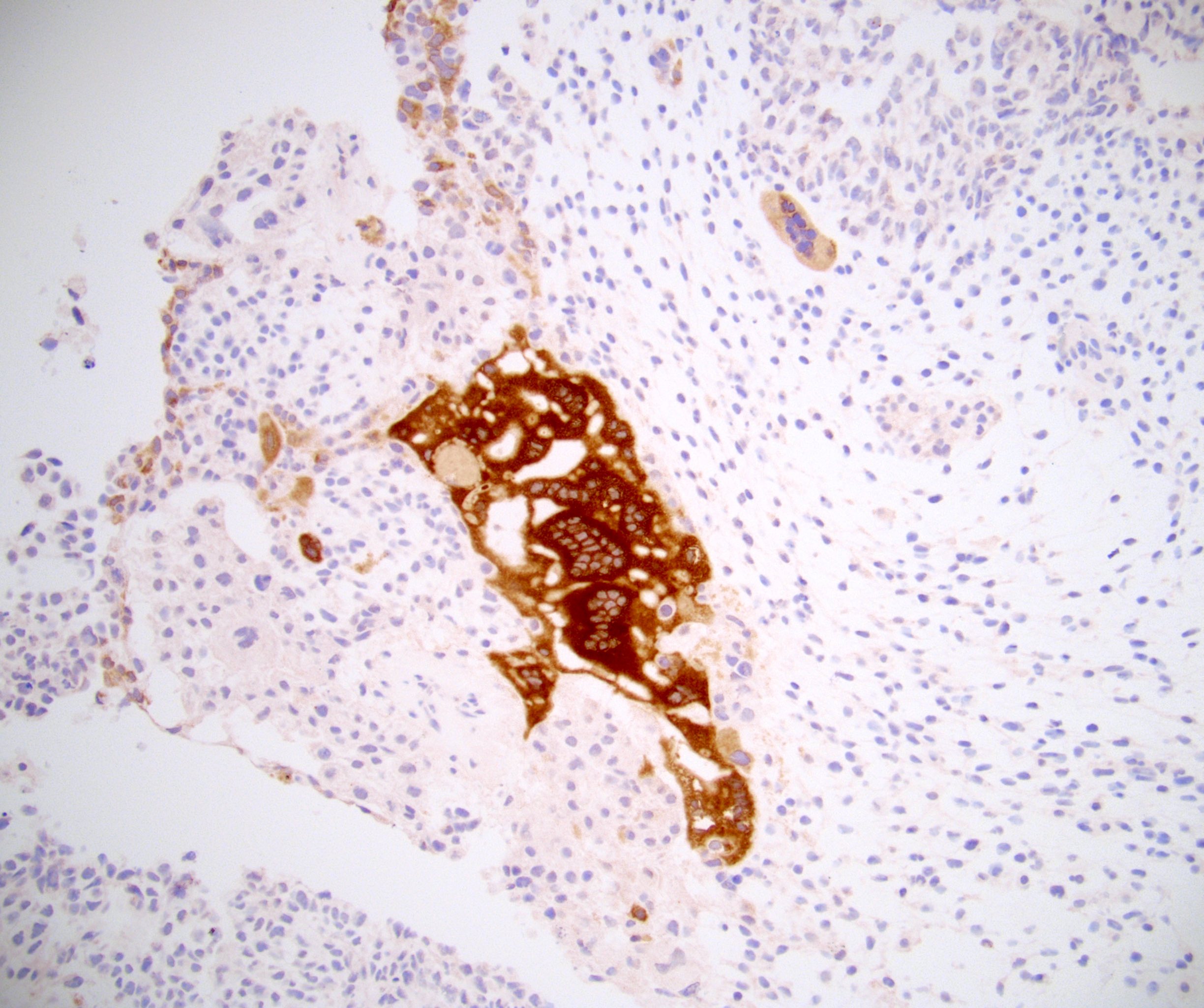
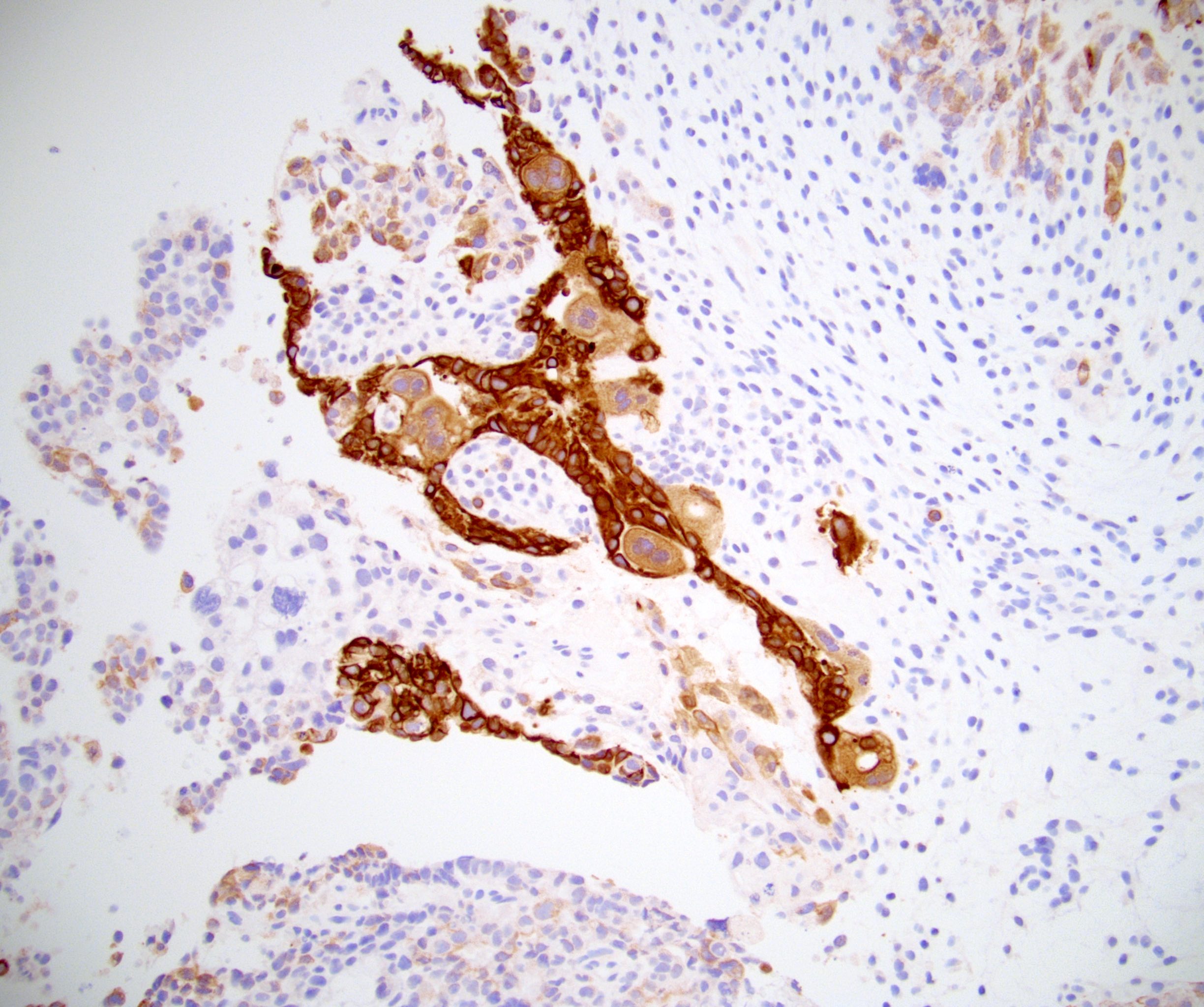
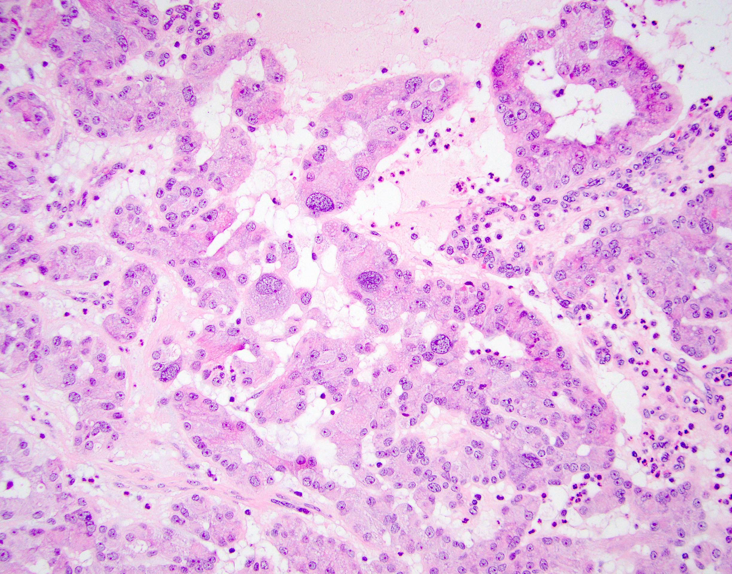
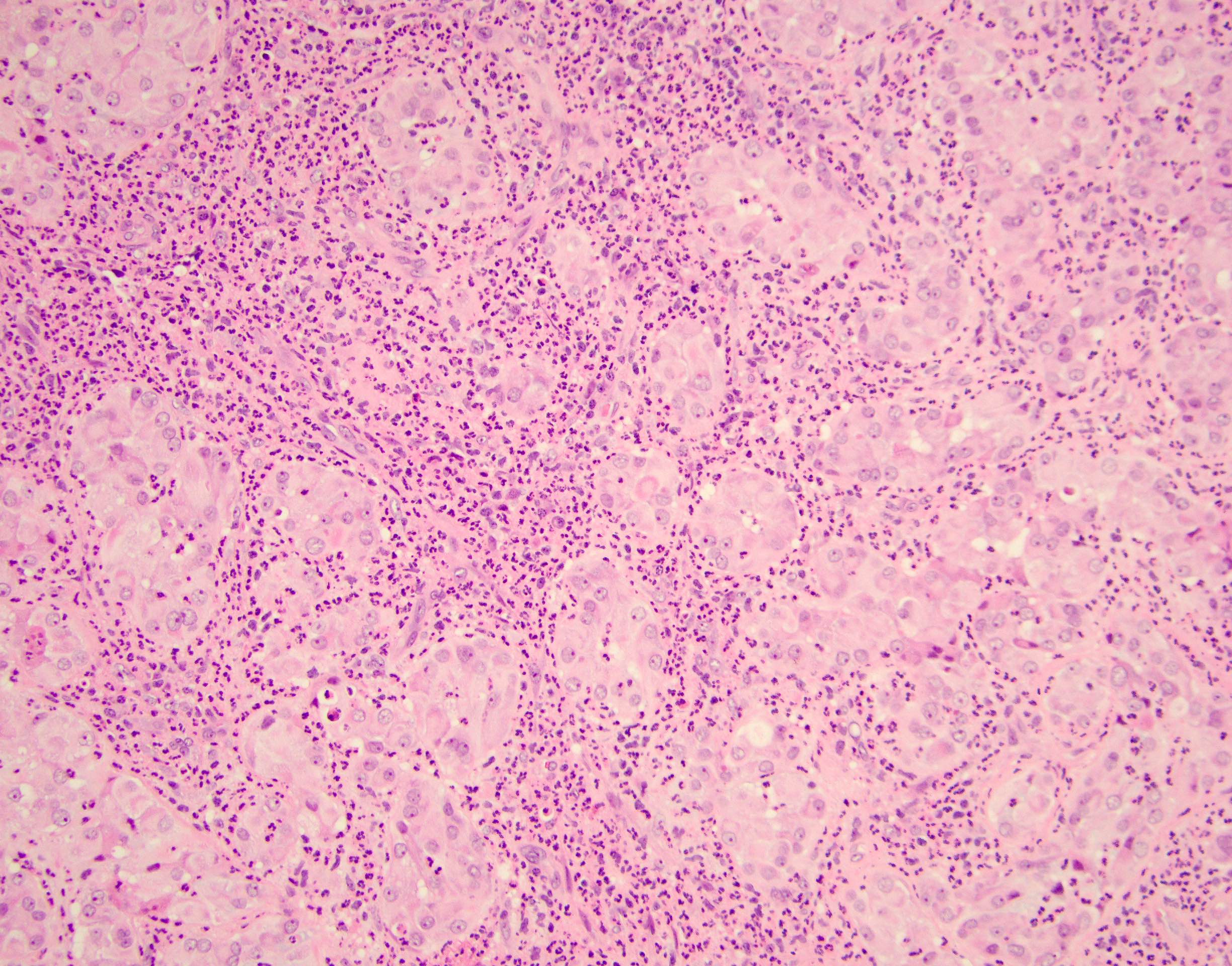
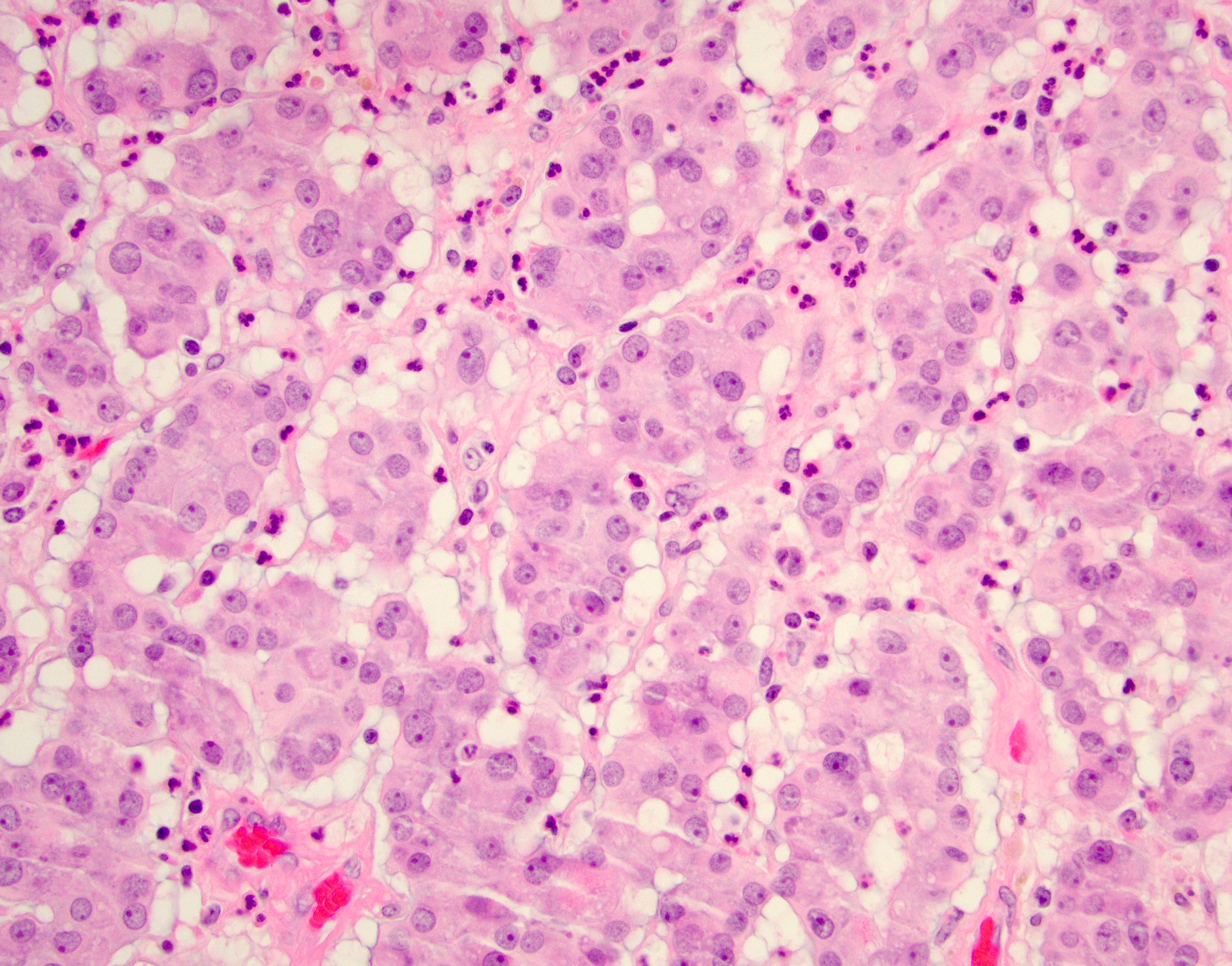
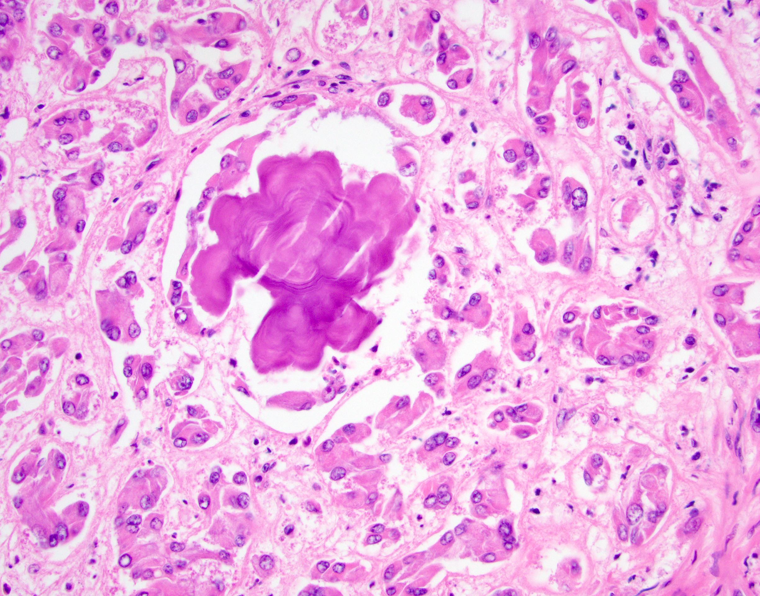
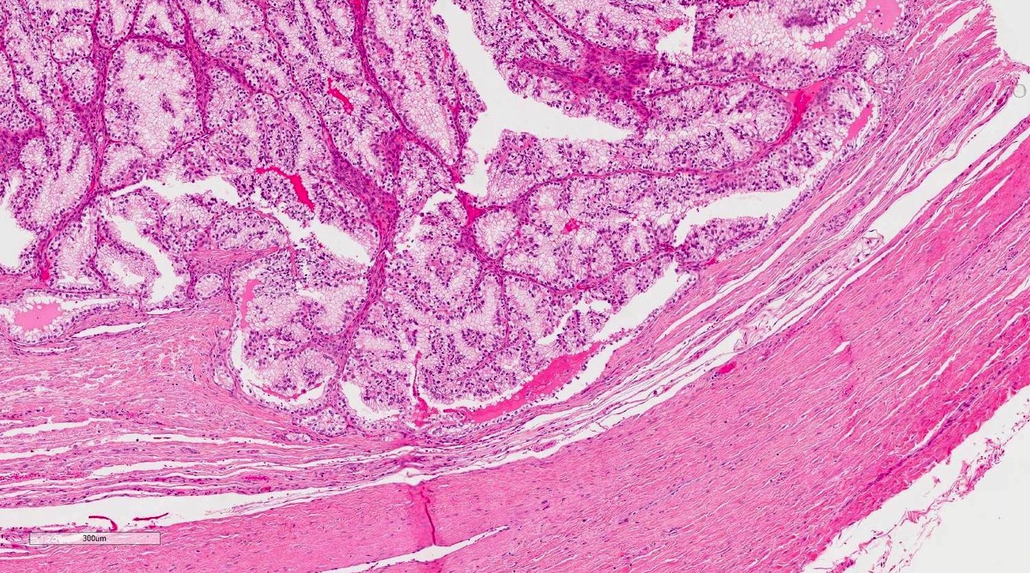
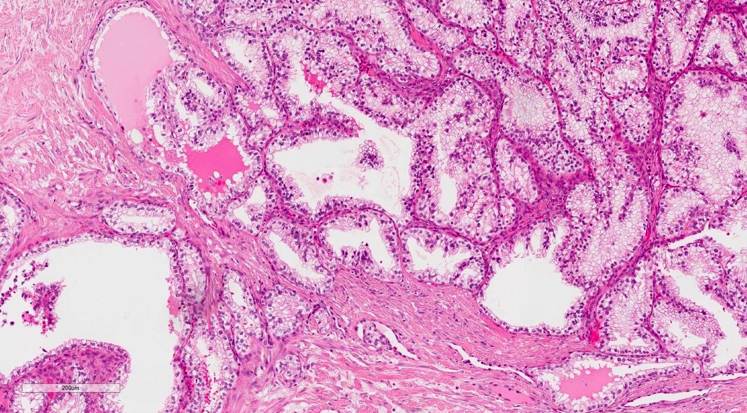
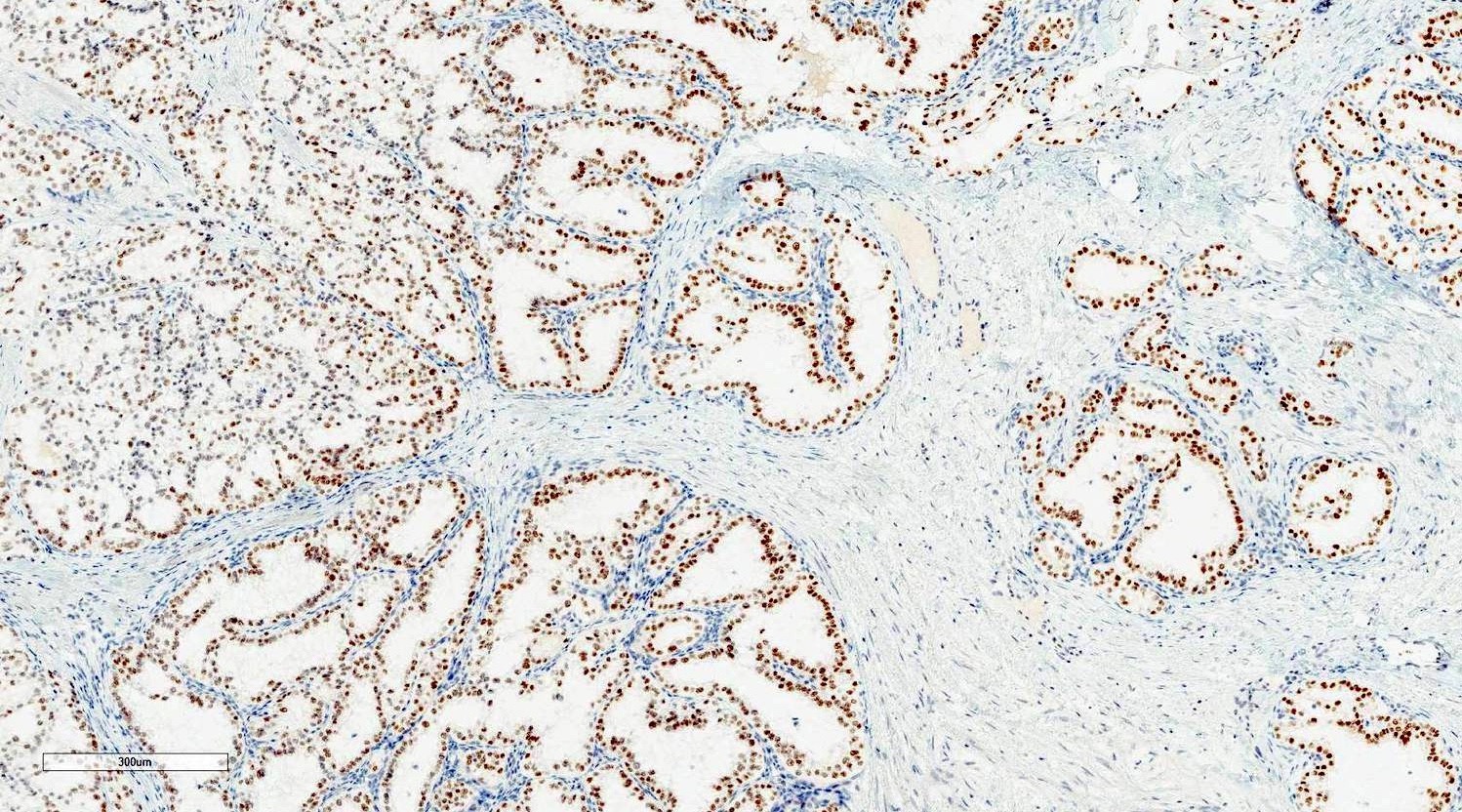
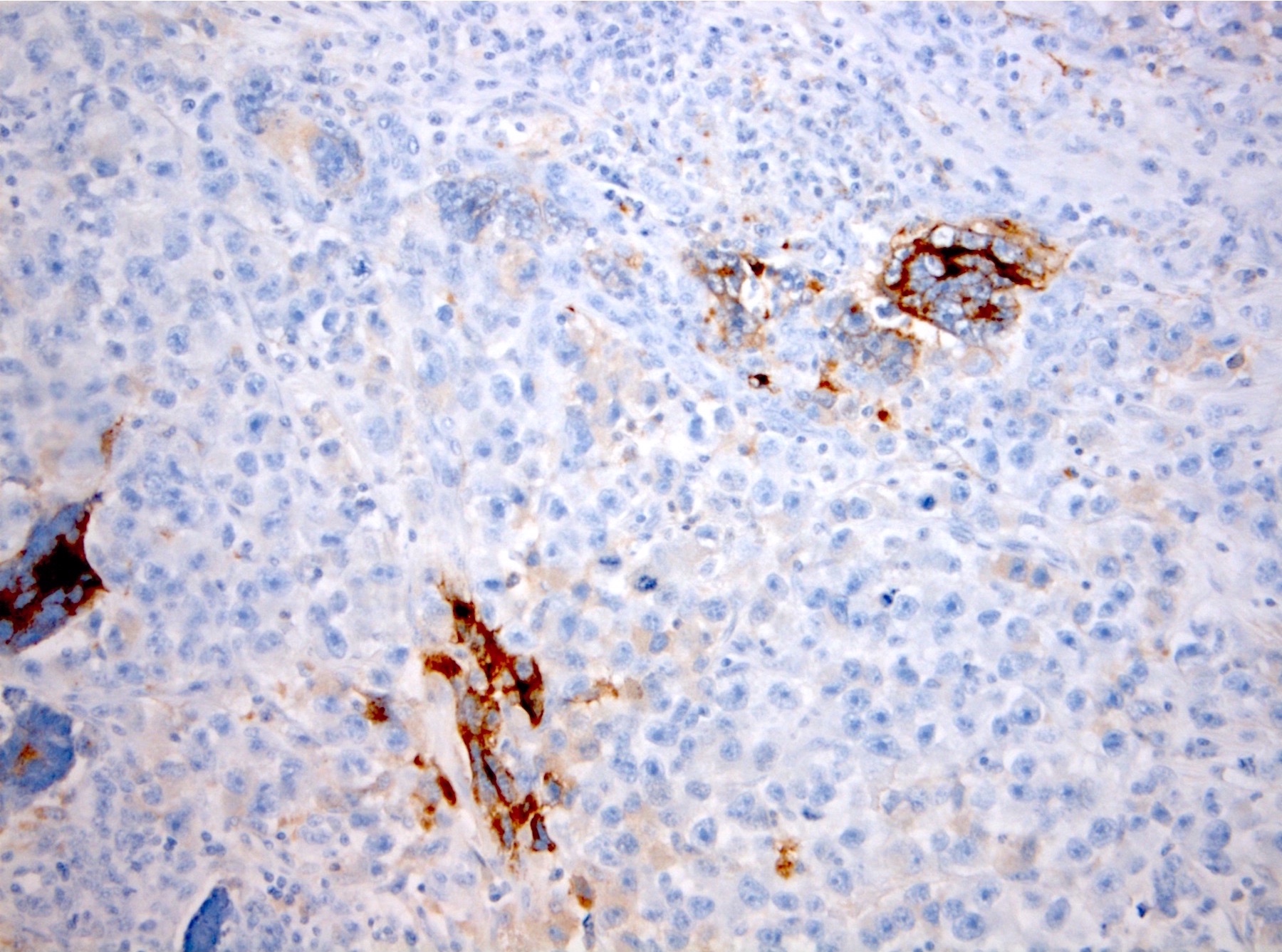
Metastatic prostatic adenocarcinoma
Images hosted on other servers:

Metastatic colorectal adenocarcinoma


Metastatic renal cell carcinoma
Contributed by Gang Wang, M.D., Ph.D.
Metastatic prostatic adenocarcinoma
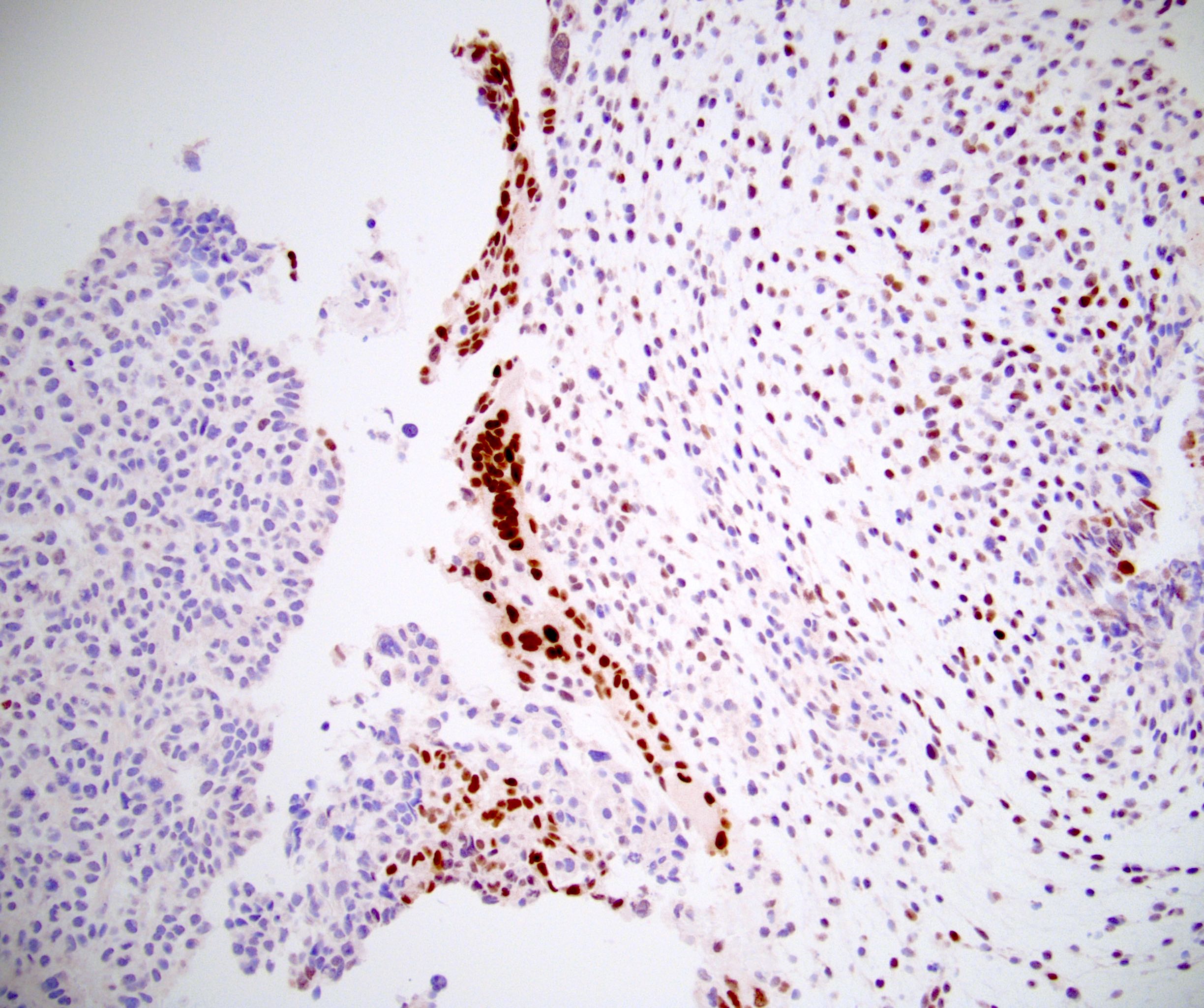
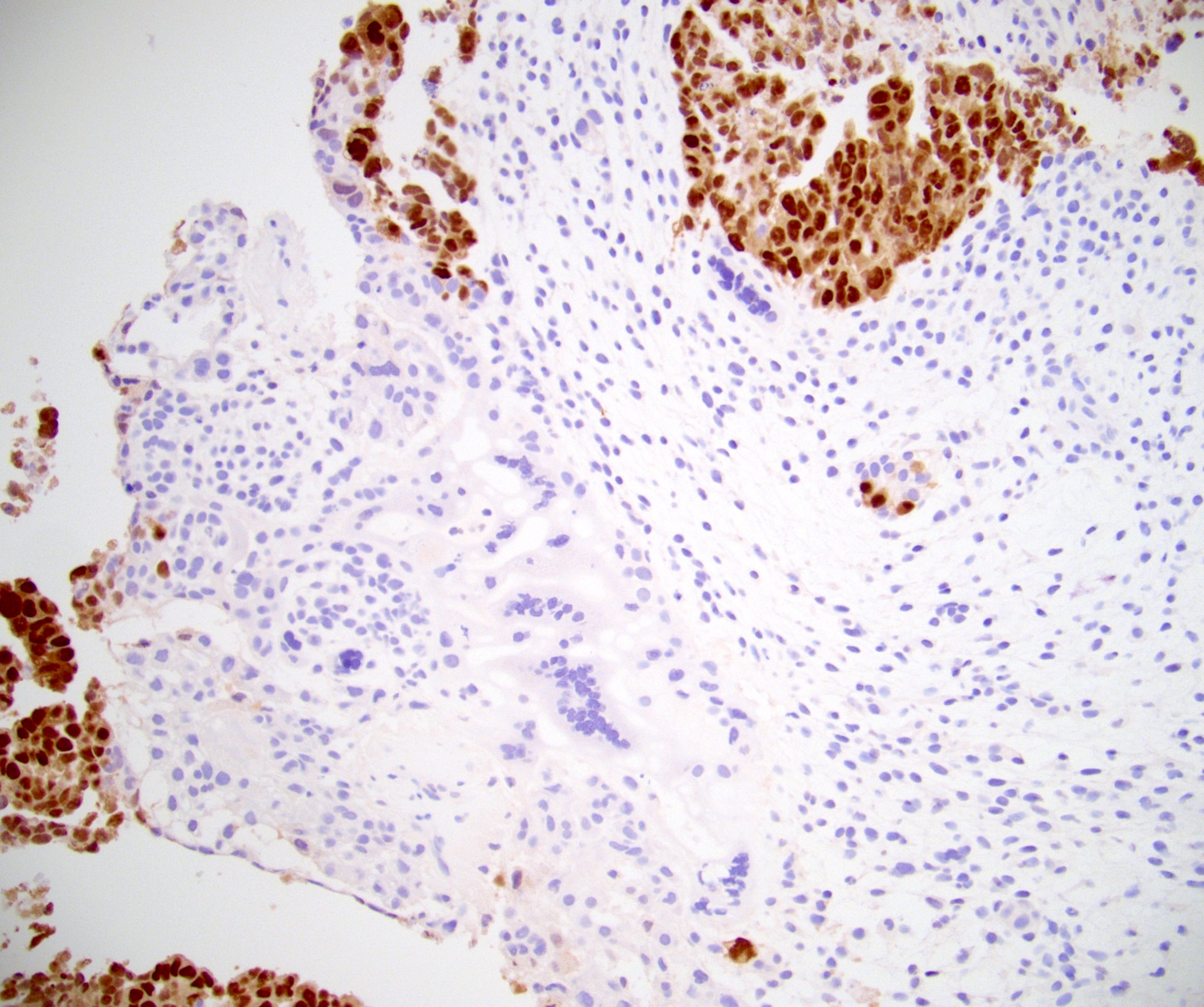
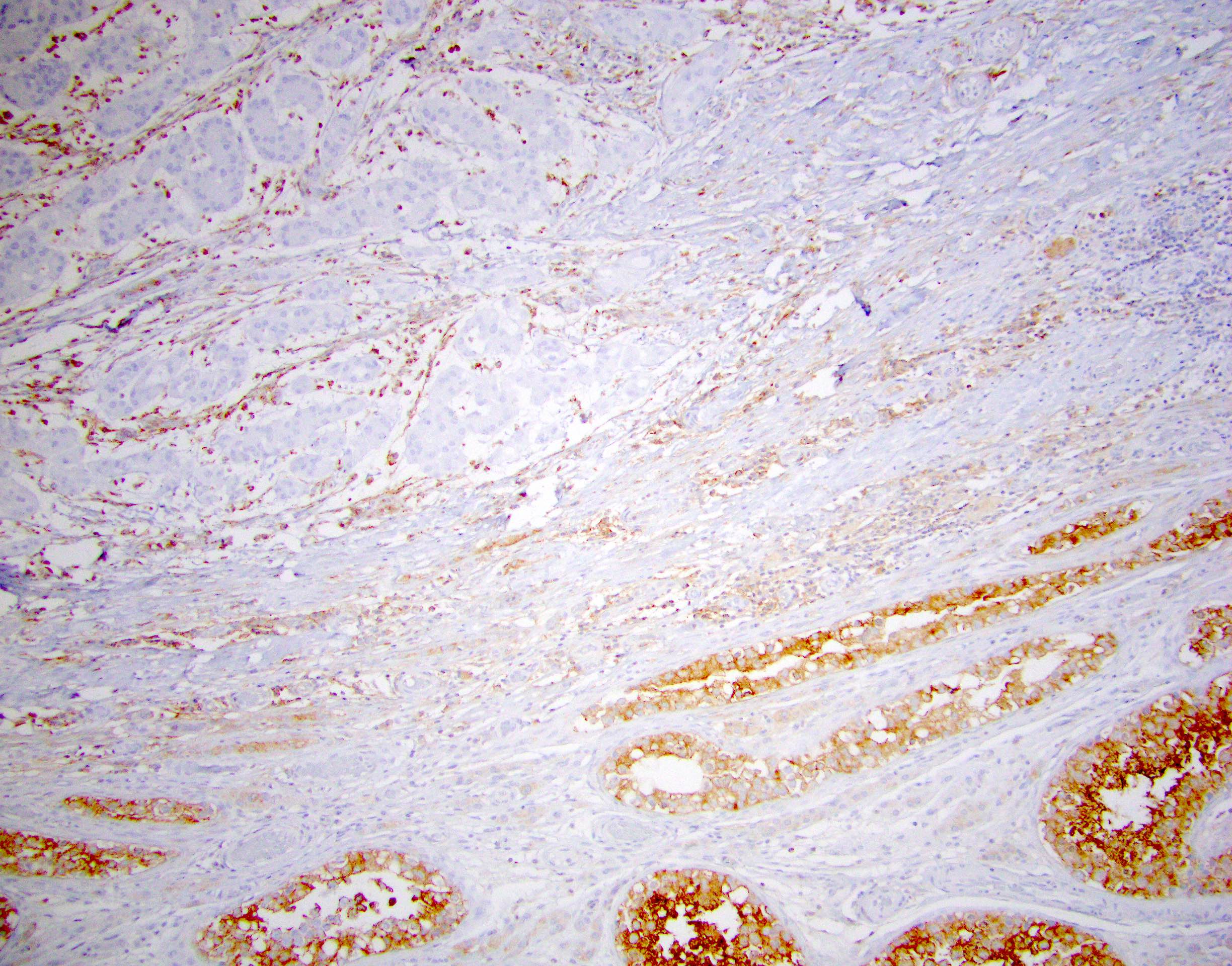
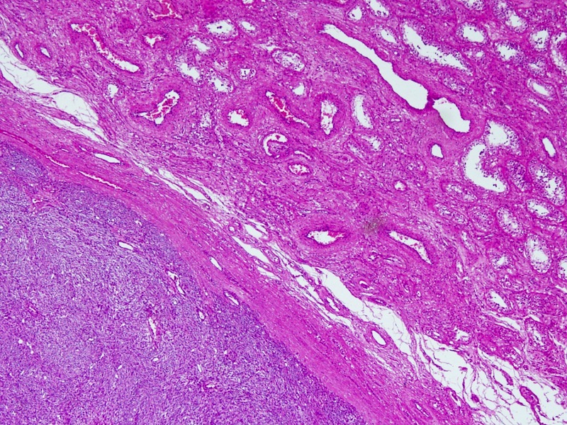
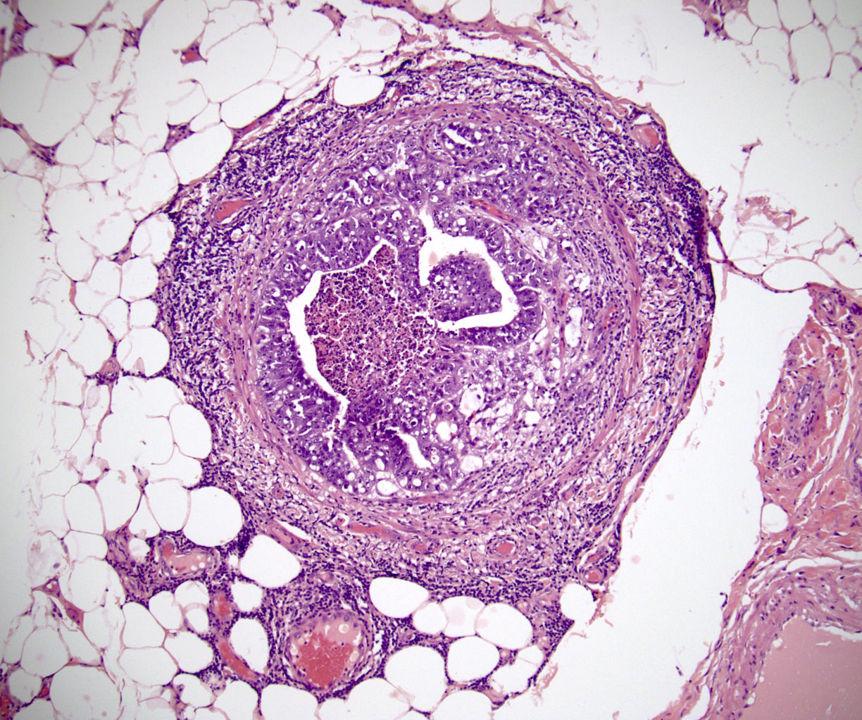
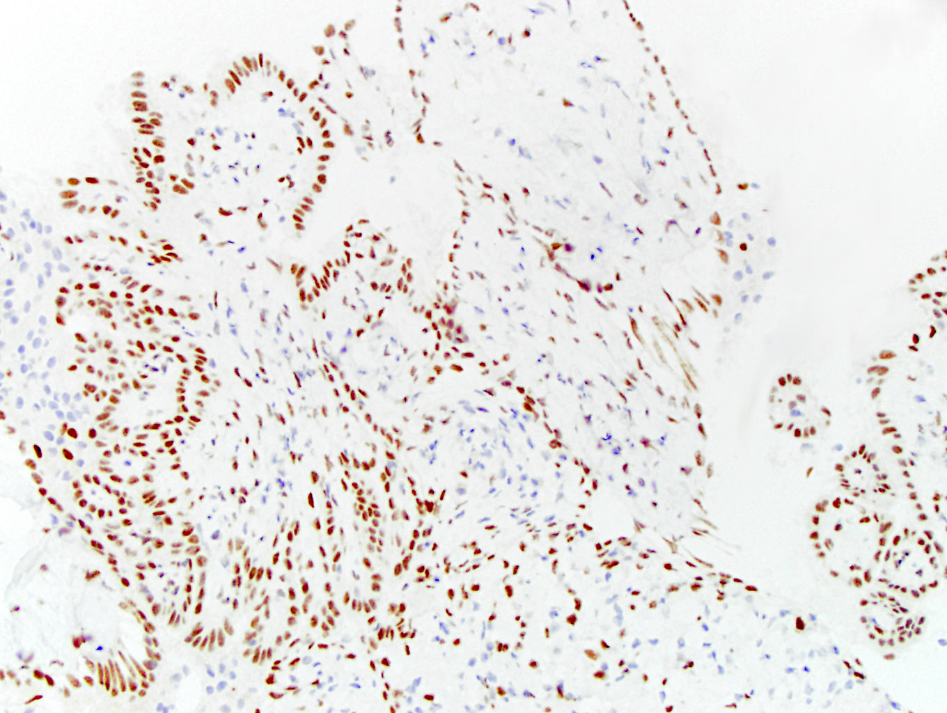
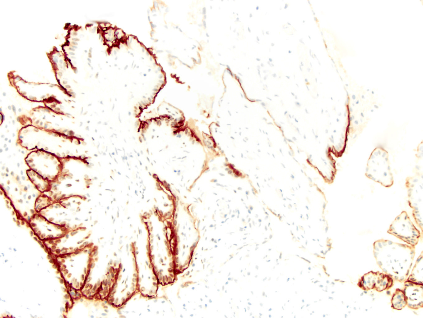
Involvement of the tubular system
Cribriform pattern
most frequent,
epididymis
involvement
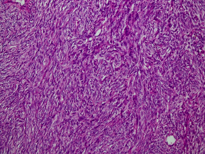
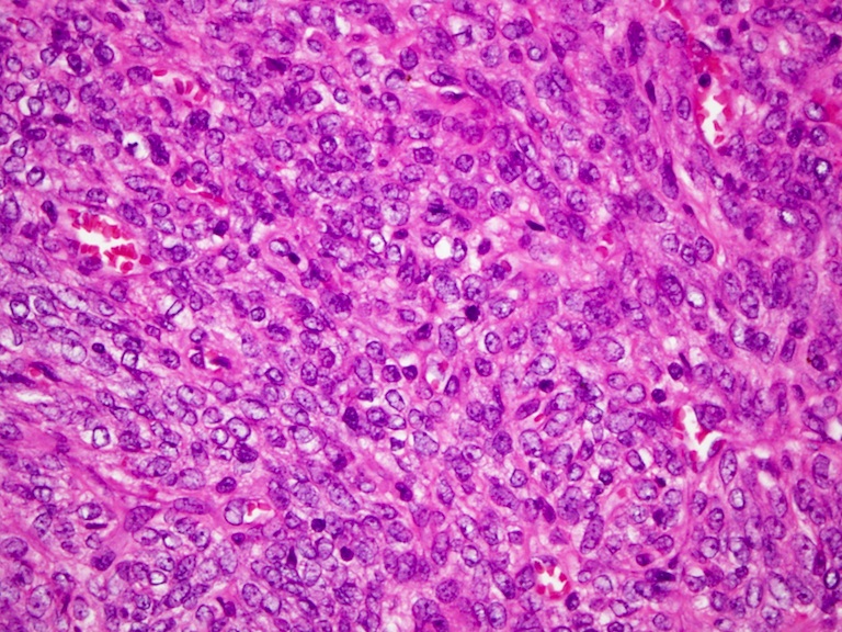
Solid growth pattern
Metastatic ductal adenocarcinoma
Positive for PSA
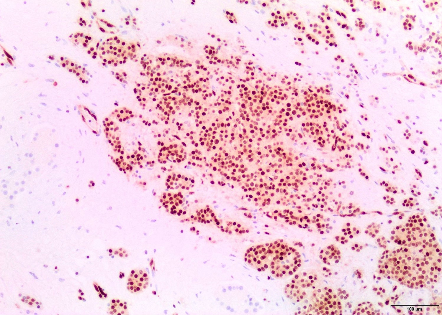
Positive for ERG
Metastatic renal cell carcinoma, clear cell type
Broad pushing borders
Mixed patterns
Positive for PAX8
Positive for CAIX
adenocarcinoma
Cystic spaces containing mucin
cell carcinoma
Infiltrating and anastomosing nests

Positive for p40
Images hosted on other servers:

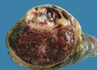
Well circumscribed lower pole testicular tumor
Images hosted on other servers:

Tumor is compressing adjacent tunica

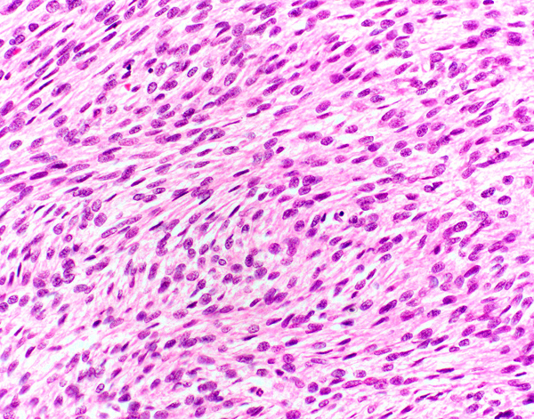
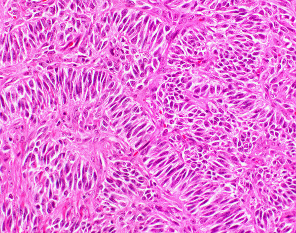
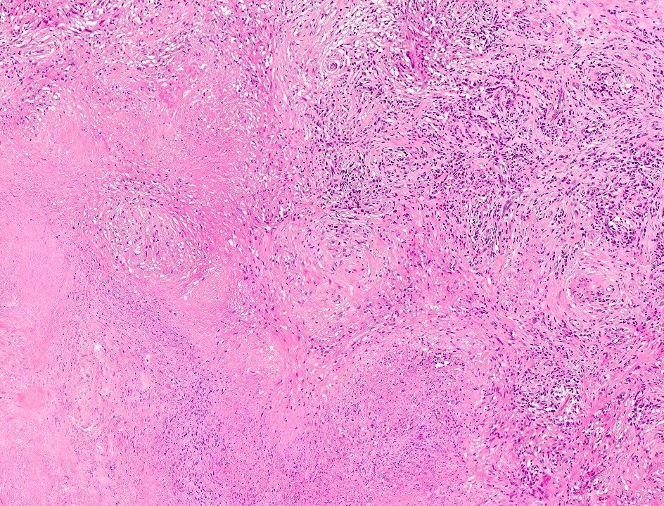
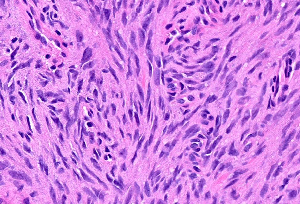
Fascicular and storiform spindle cell arrangement

Insular arrangement of cells in hyaline stroma

Irregular ovoid nuclei with longitudinal grooves
Images hosted on other servers:

Area of curvilinear calcification
Images hosted on other servers:


Various images
Images hosted on other servers:

Right testicular borderline mucinous tumor
Images hosted on other servers:

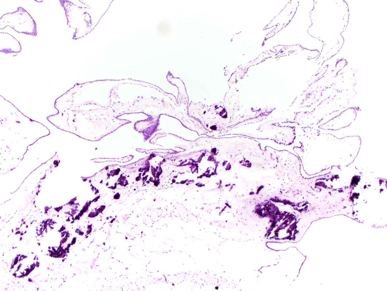
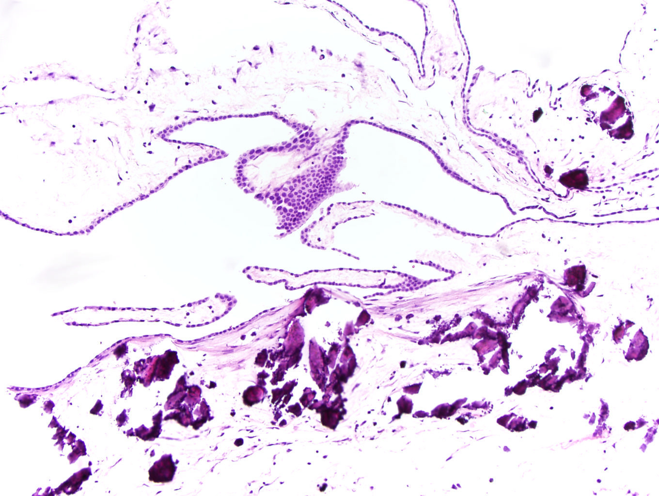
Cyst wall irregularly thickened and fibrotic

Mucinous epithelium
resembling endocervical
type cells
Images hosted on other servers:

Ultrasound features
Contributed by Vikas Mehta, M.D.
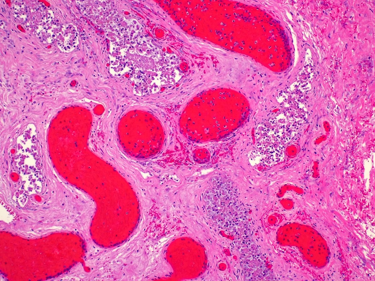
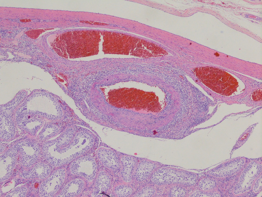
Ectatic blood vessels

Spindle arrangement of tumor cells

SF1 positivity
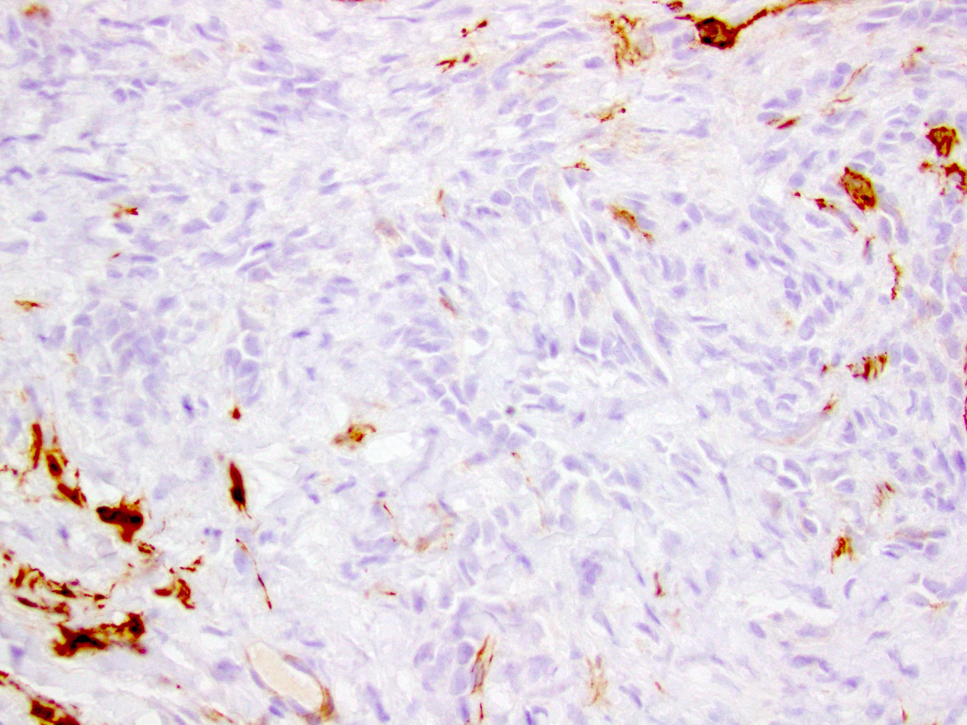
h-caldesmon negativity
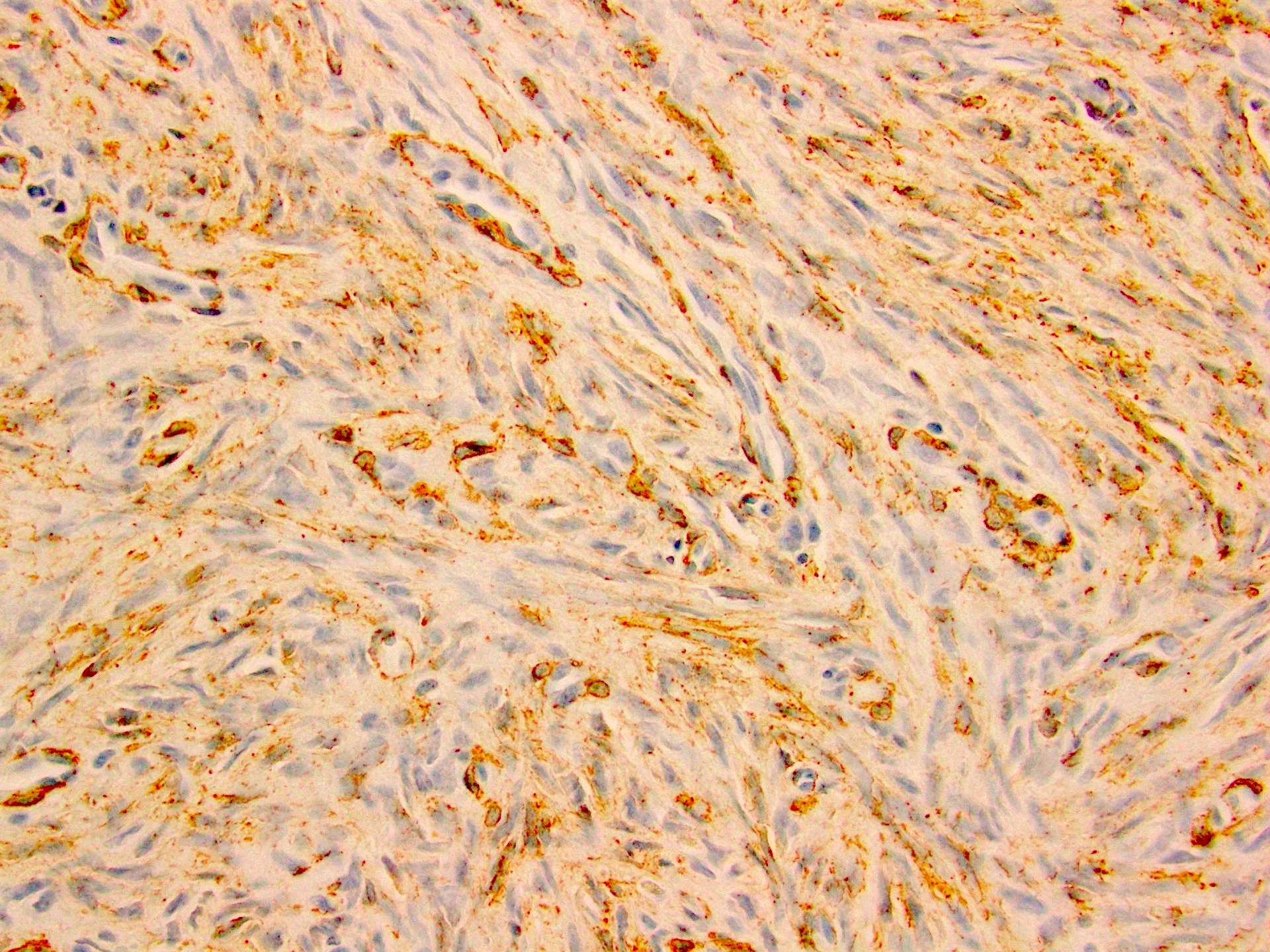
SMA positivity

S100 positivity
Images hosted on other servers:

Complex echo pattern
Images hosted on other servers:

Epididymal tumor

Microcystic with hemorrhage
Images hosted on other servers:

Cystic space with papillary excrescences

Cilia

Case with clear cells

CK7

CD10
Contributed by Delia Perez-Montiel, M.D.
40 year old man
Contributed by Delia Perez-Montiel, M.D.
Images hosted on other servers:

Tumor in undescended testis
Images hosted on other servers:

Spindle cell proliferation
Images hosted on other servers:

Scrotal ultrasound

Regressed
primary with
retroperitoneal
metastasis
Contributed by Debra L. Zynger, M.D.
Complete regression (pT0)

Germ cell neoplasia in situ only (pTis)

Partial regression (pT1)
Contributed by Debra L. Zynger, M.D.
Scar

Acellular matrix
Small vessels

Hemosiderin

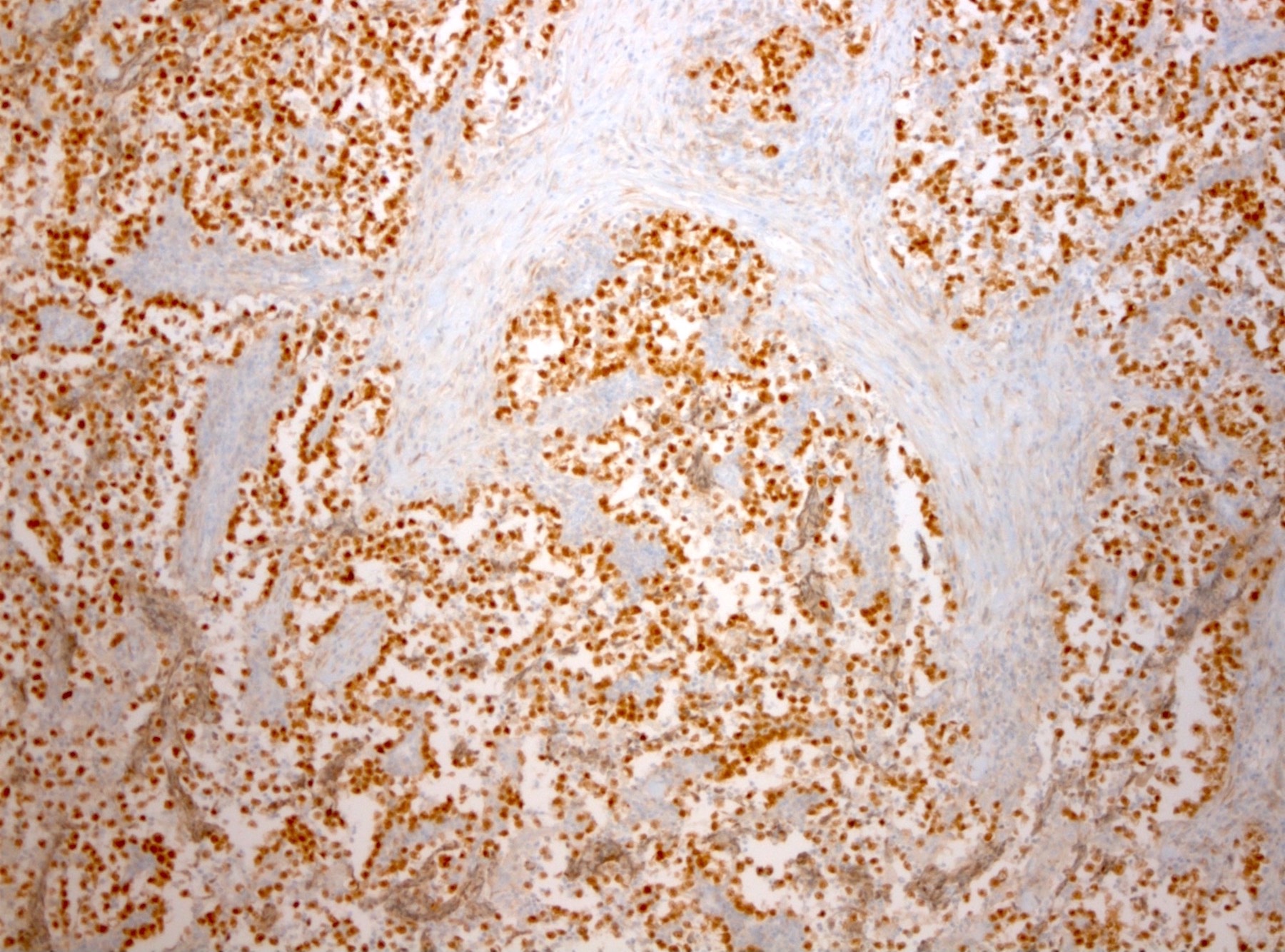
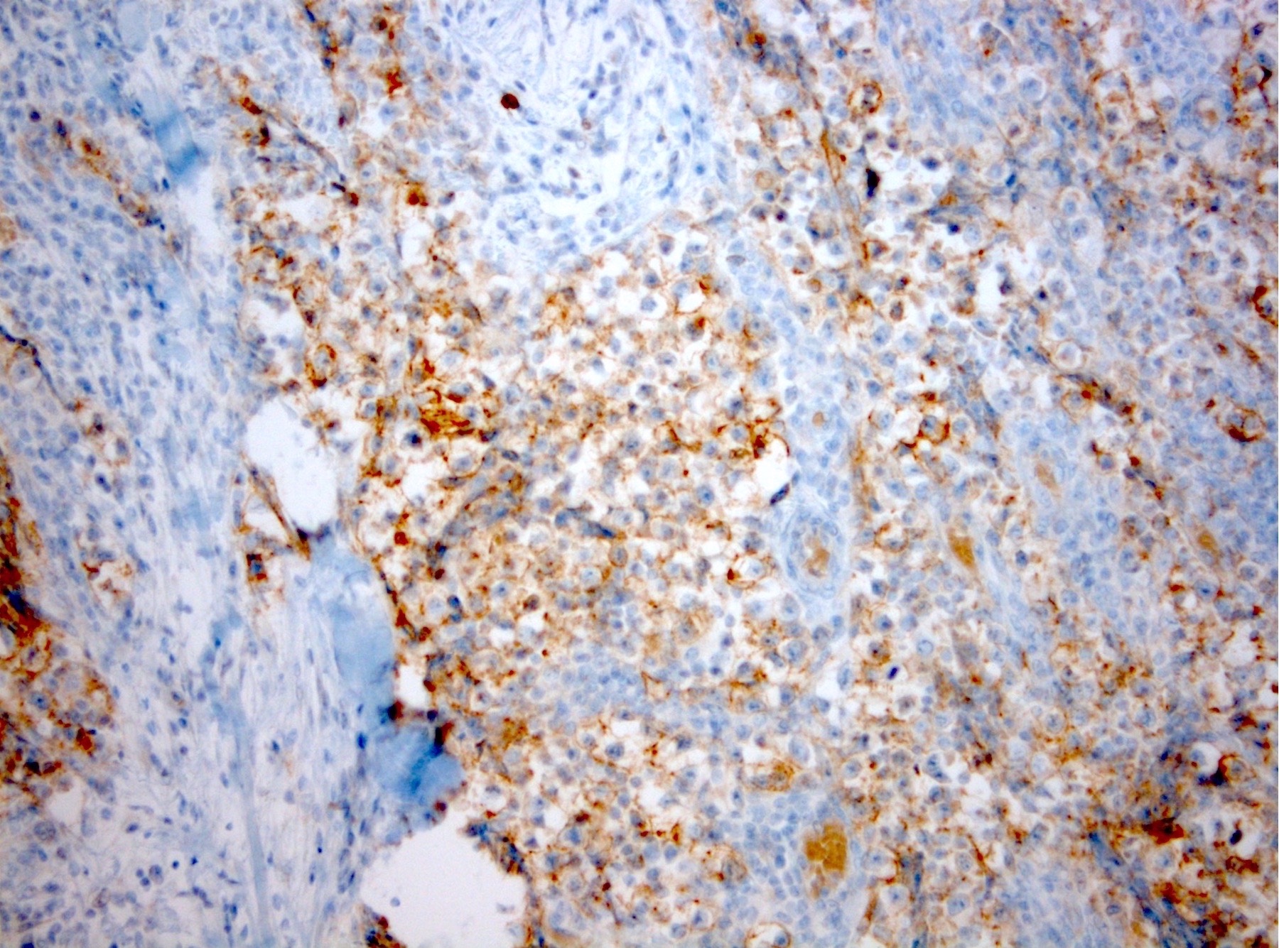
Germ cell neoplasia in situ
OCT3 / 4
D2-40

CD117
Images hosted on other servers:

Ultrasound with testicle mass

Parietal metastasis
Contributed by Debra L. Zynger, M.D.
pT1a
pT1b
pT2
Images hosted on other servers:

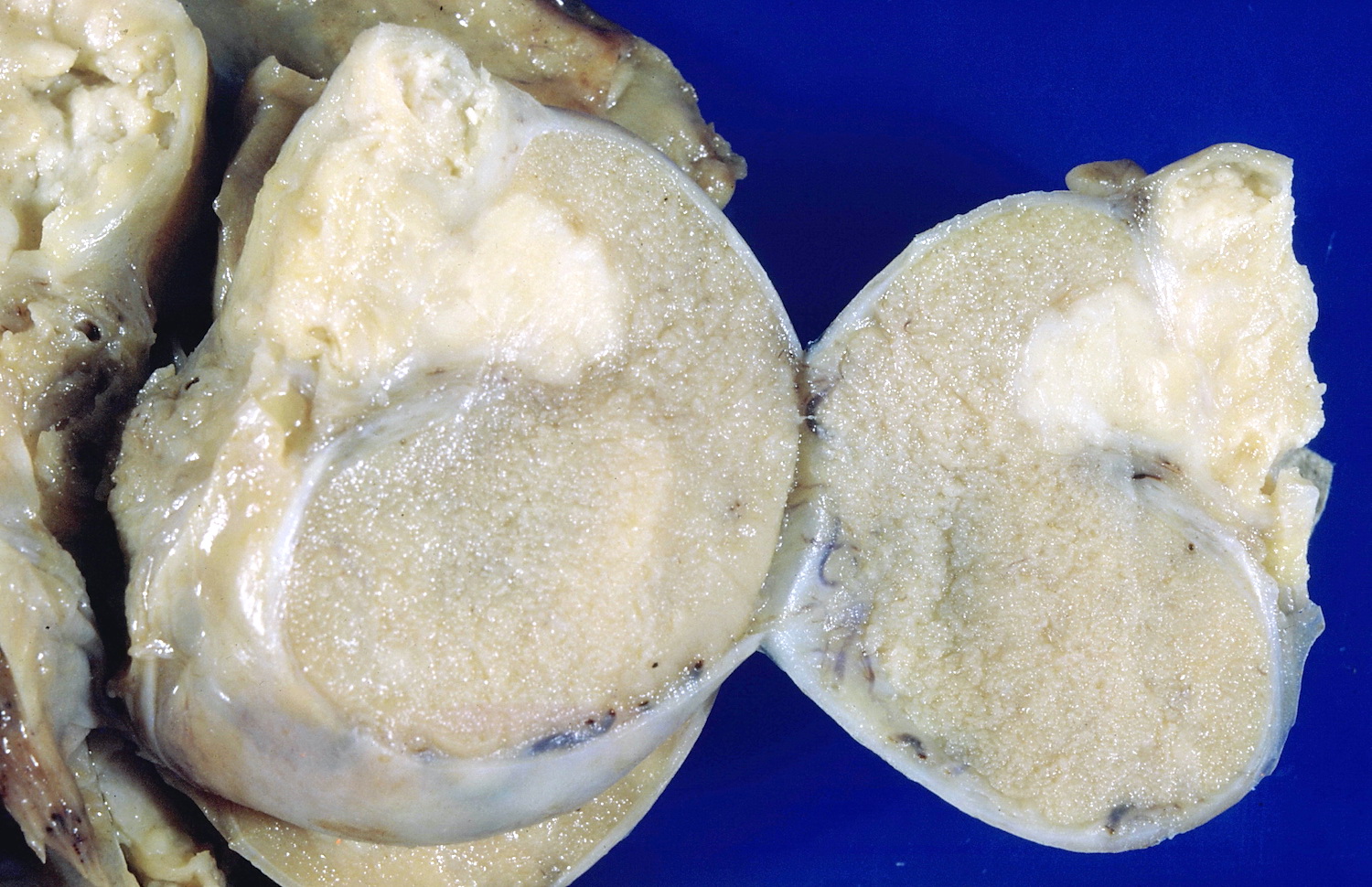
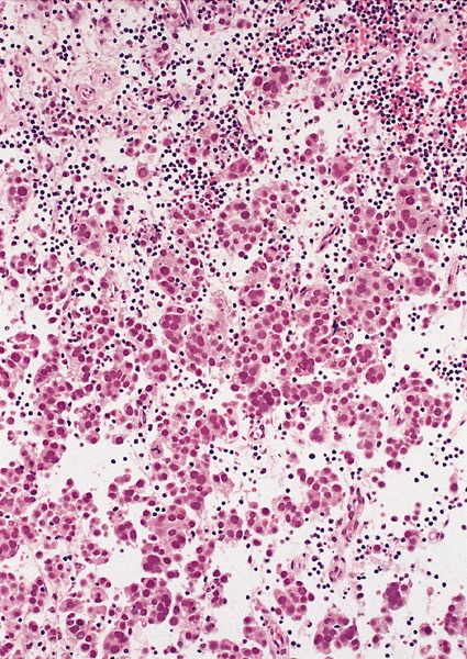
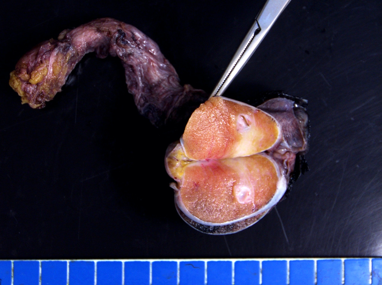
Very large seminoma

Seminoma

Lobulated soft tan to brown tissue
Contributed by Michelle Downes, M.D. and Debra L. Zynger, M.D.

Fibrous septae
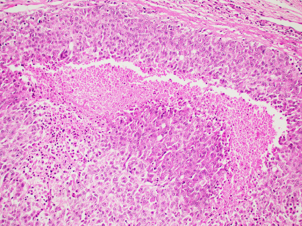
Admixed lymphocytes
Large cells with clear cytoplasm
Coagulative necrosis
OCT 3/4
CD117
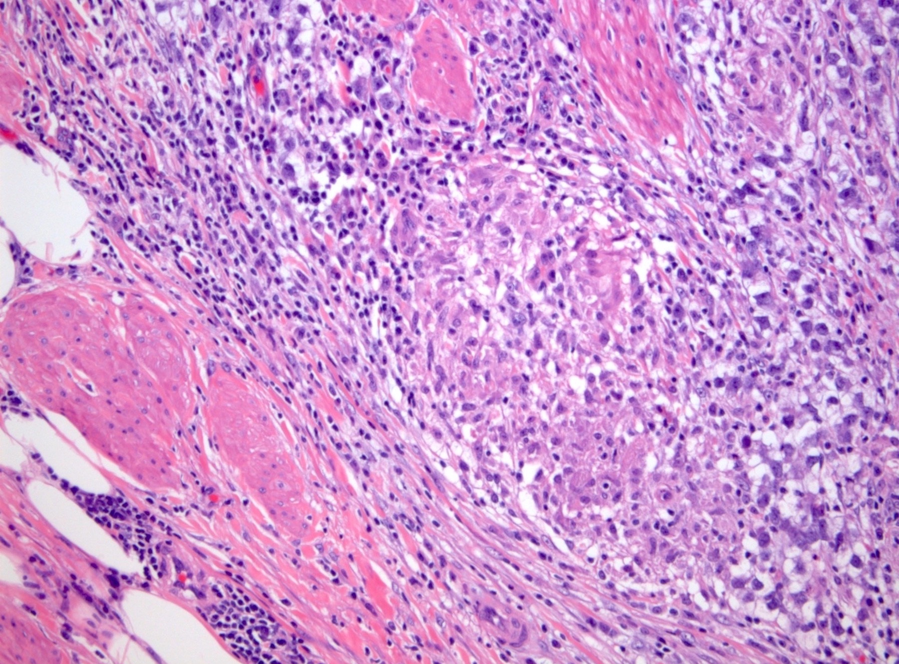
Granulomatous inflammation

Granuloma
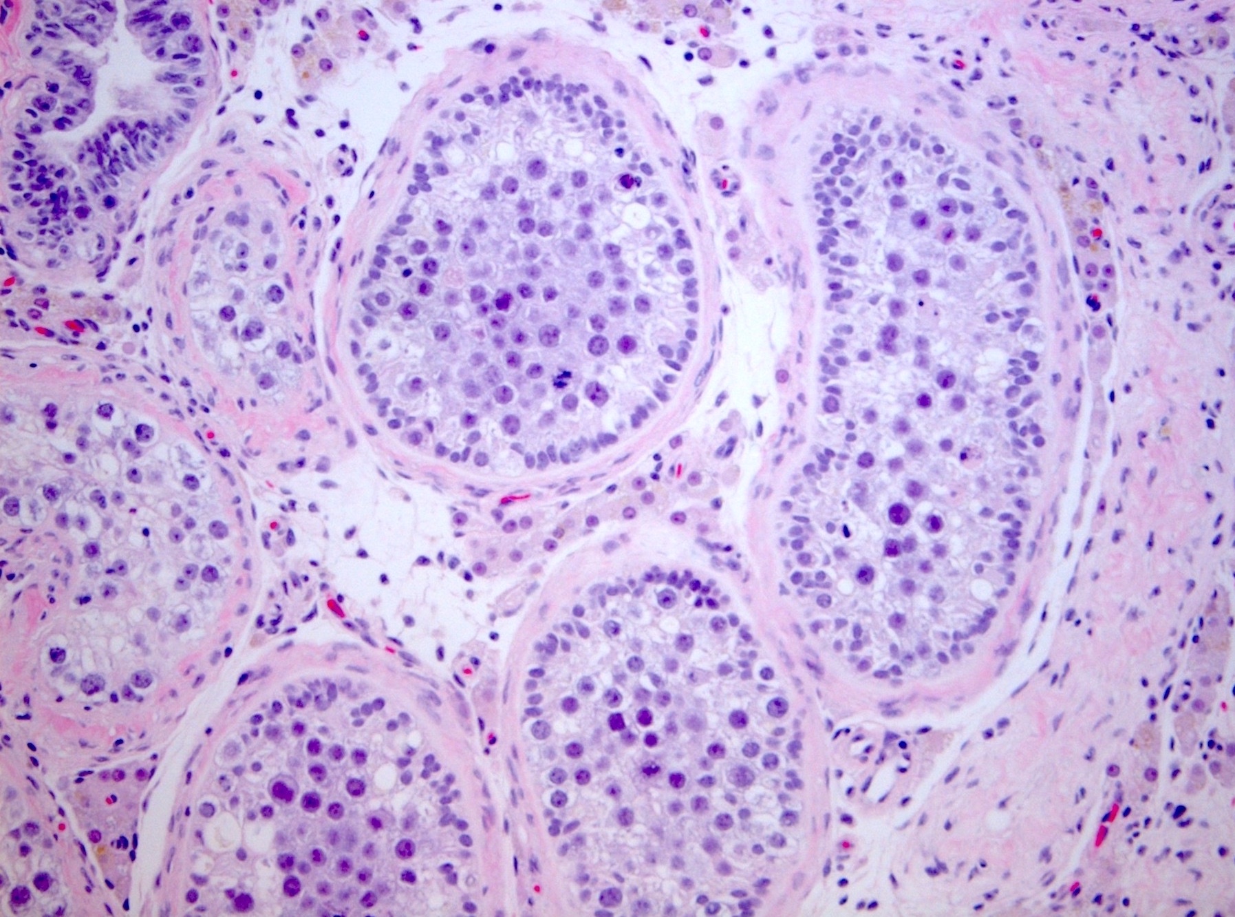
Intratubular seminoma
Rete testis invasion

Rete testis pagetoid spread
Hilar fat invasion
Epididymal invasion
Syncytiotrophoblasts
hCG
Discontinuous spermatic cord invasion
Contributed by Michelle Downes, M.D. and Debra L. Zynger, M.D.
Intraoperative touch prep
Large cells with prominent nucleoli
Tigroid pattern
Images hosted on other servers:

Intratesticular mass
Images hosted on other servers:

WT1+
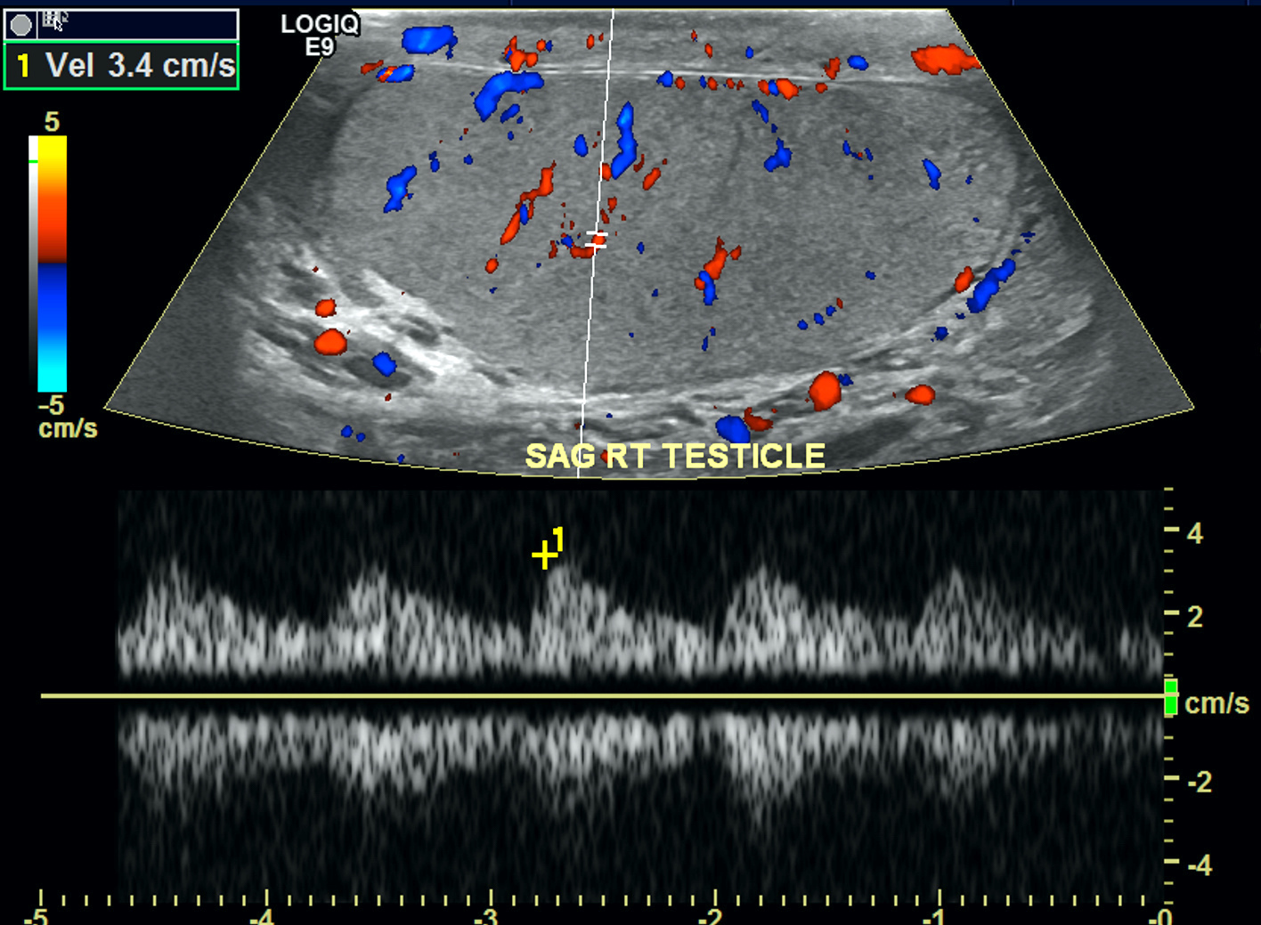
Contributed by Raman Danrad, M.D.

Ultrasound of left testis
Images hosted on other servers:

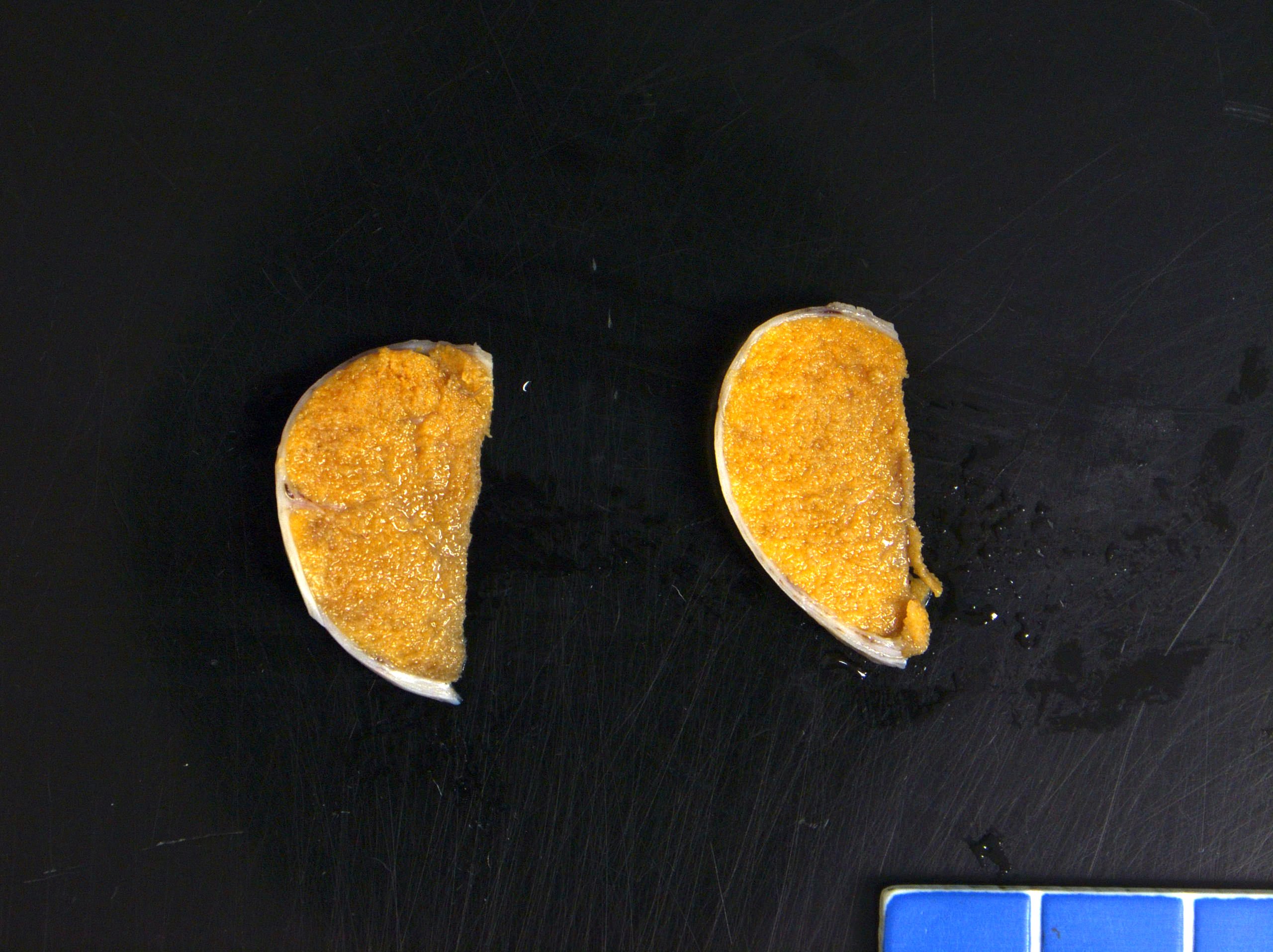
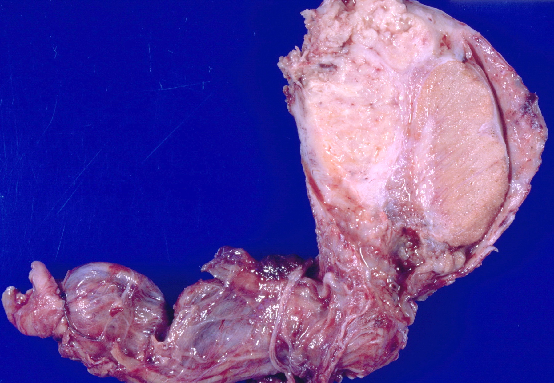
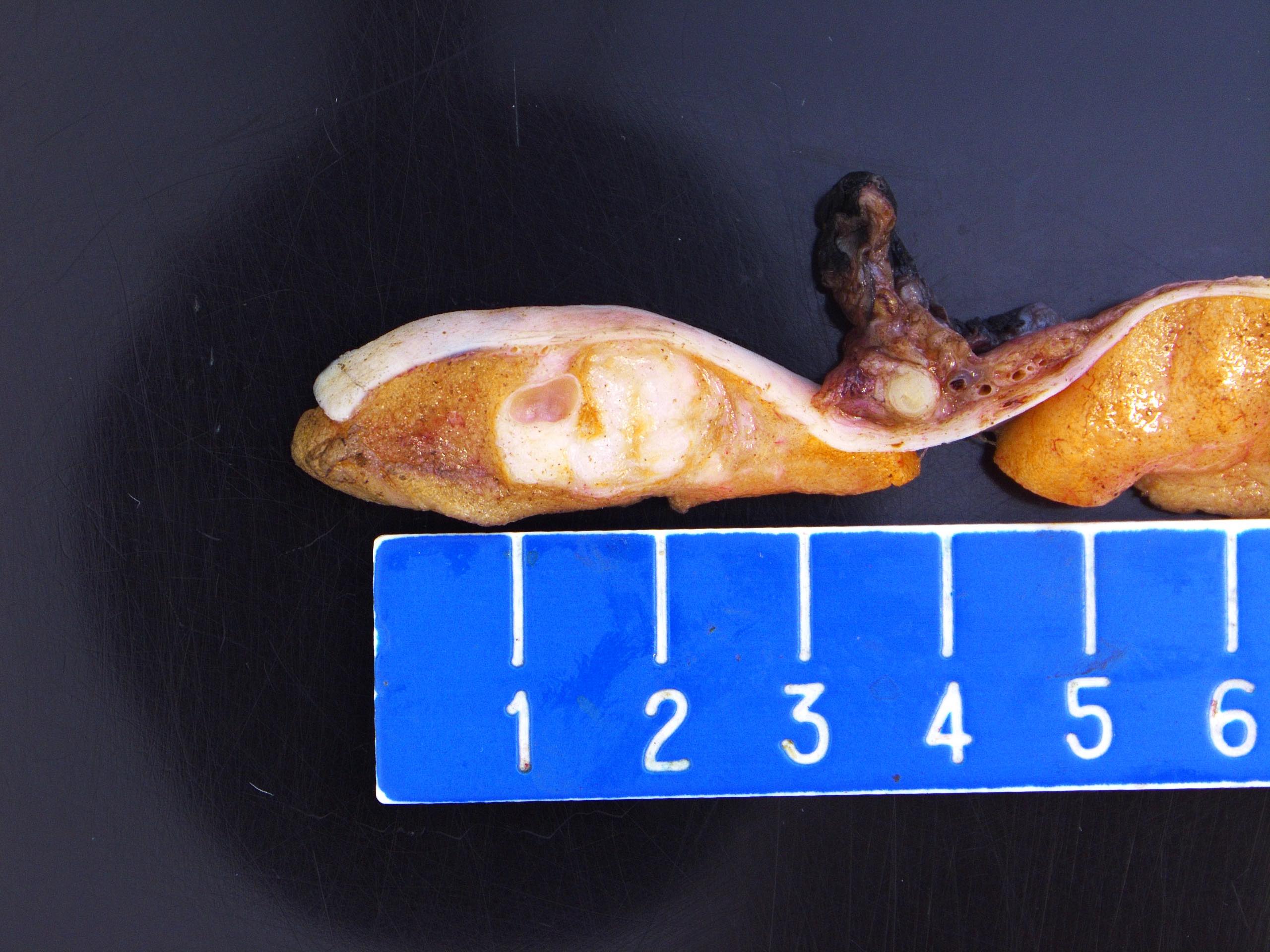
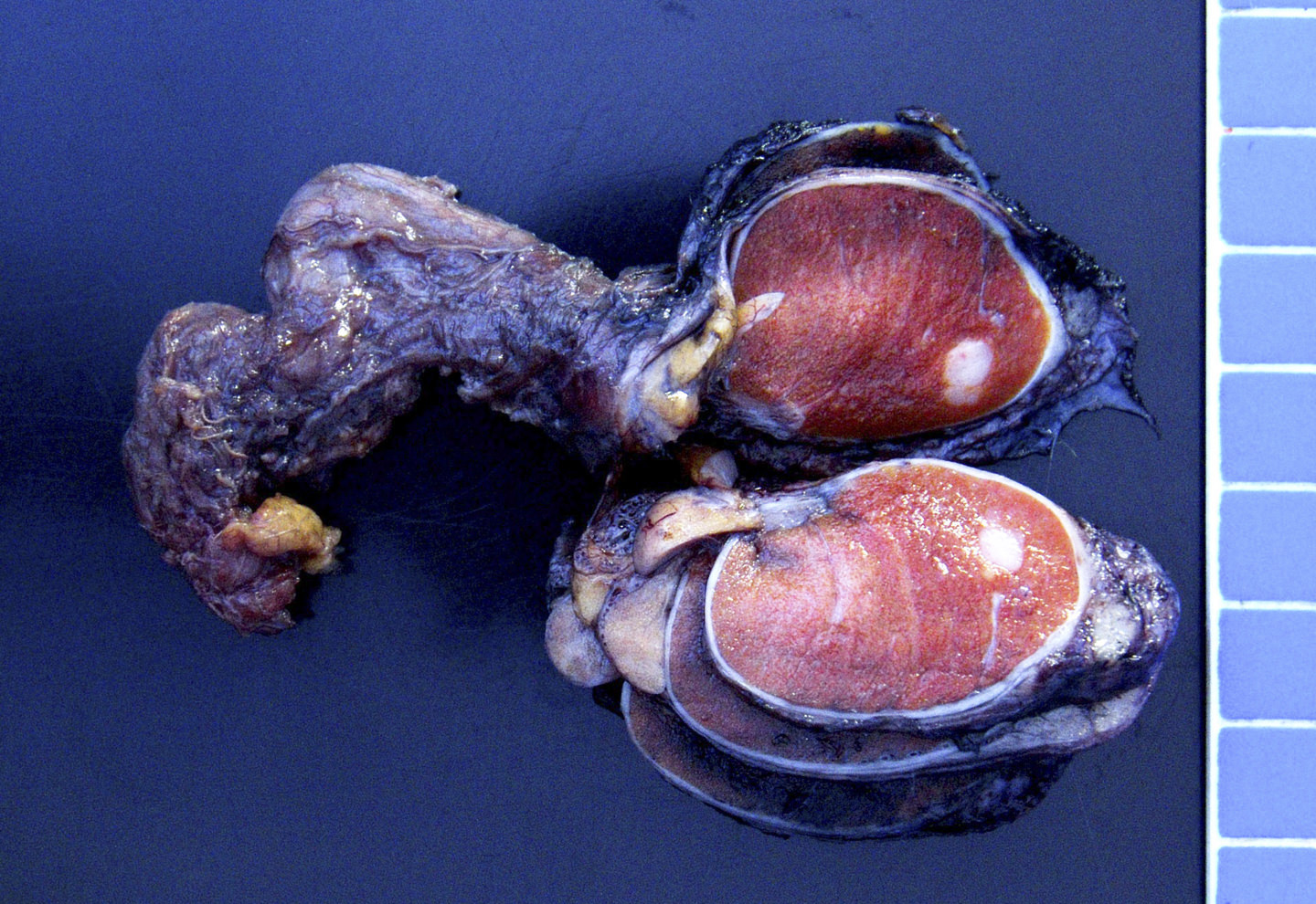
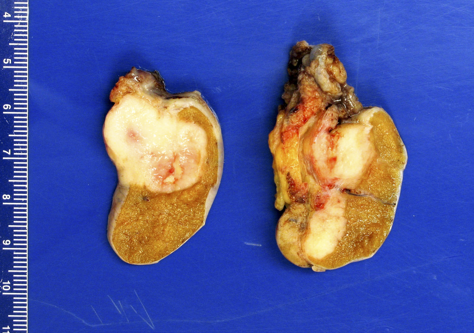
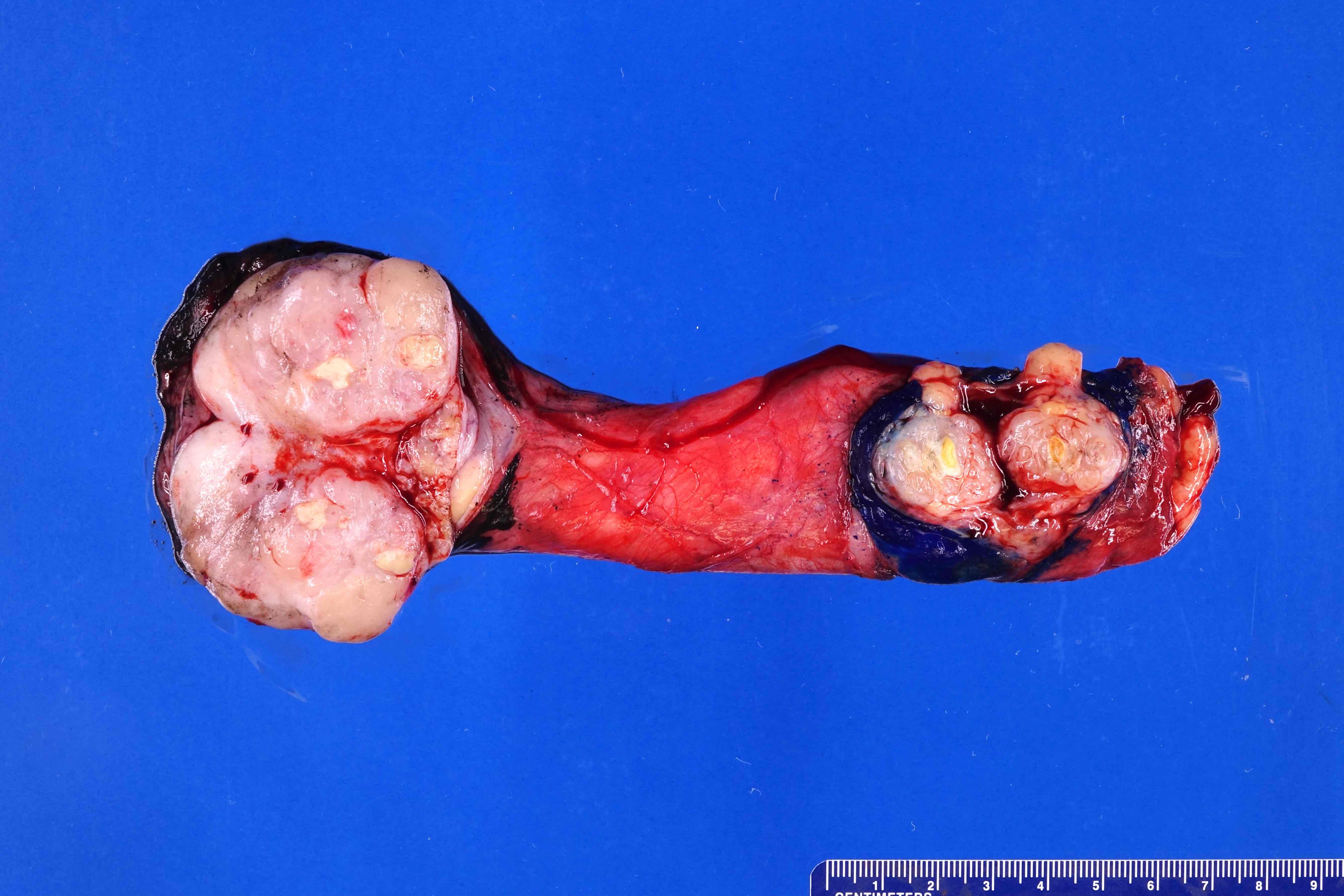
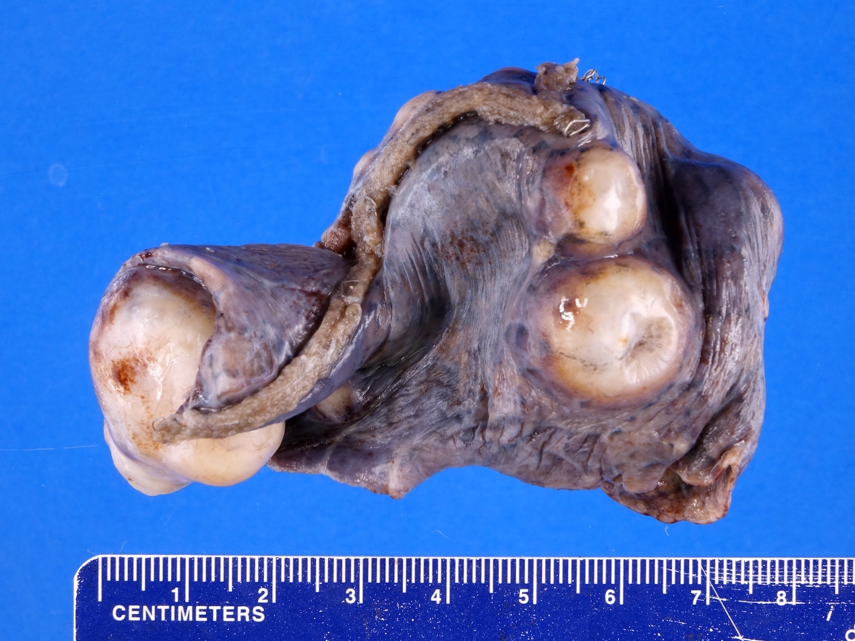
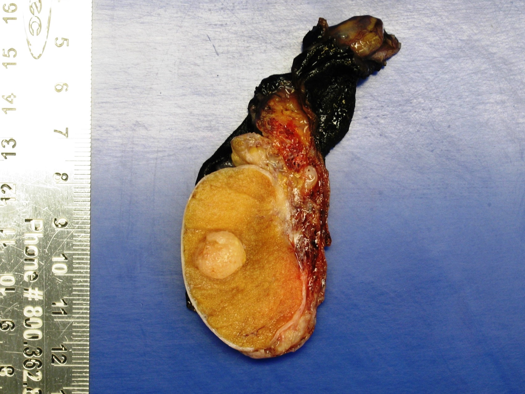
Demarcated, bulging, pale yellowish nodules

Sharply demarcated, yellowish white nodule
Contributed by Ritu Bhalla, M.D. and Jian-Hua Qiao, M.D.
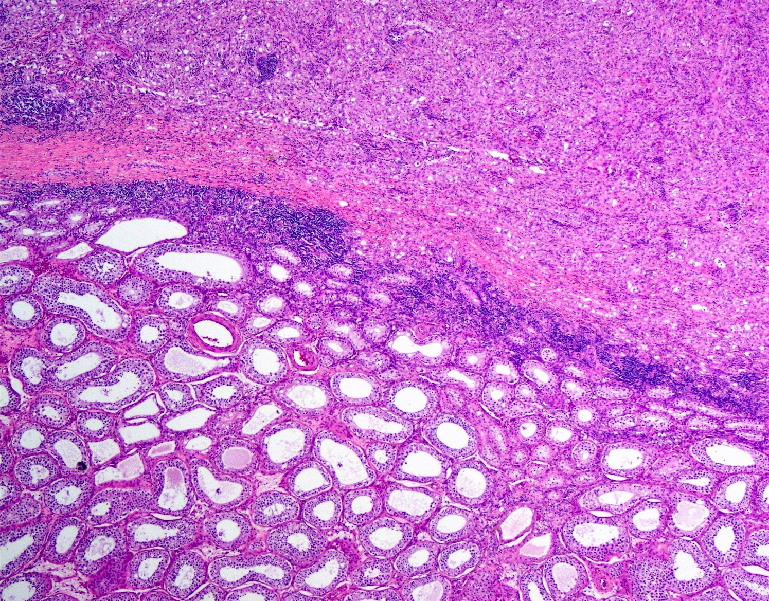
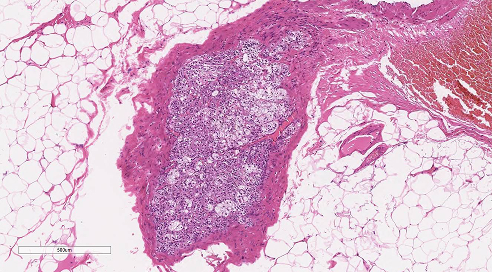
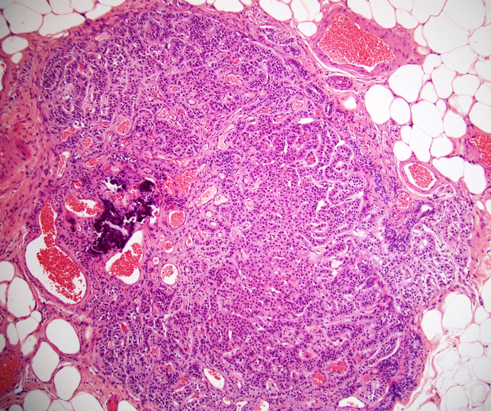
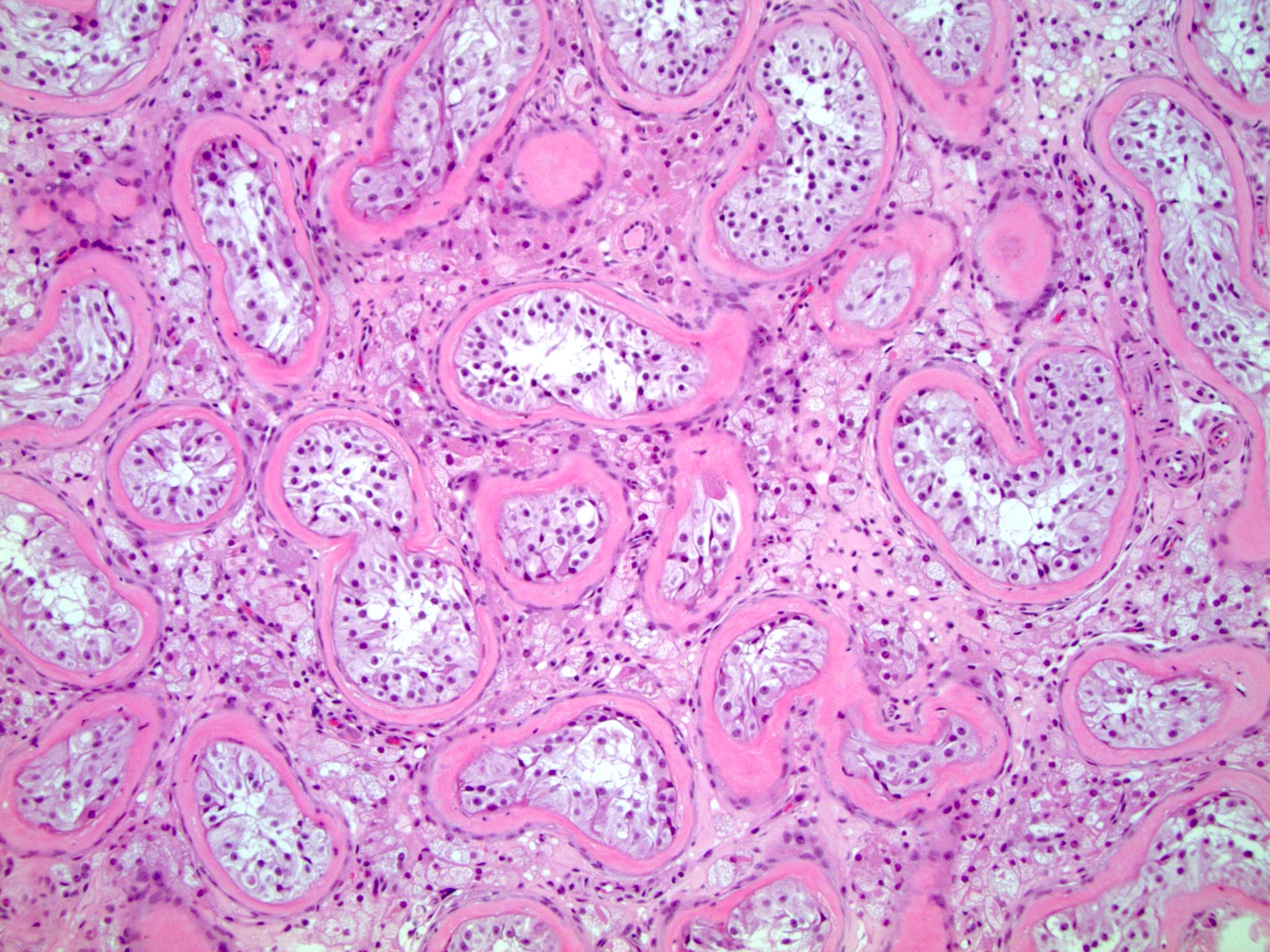
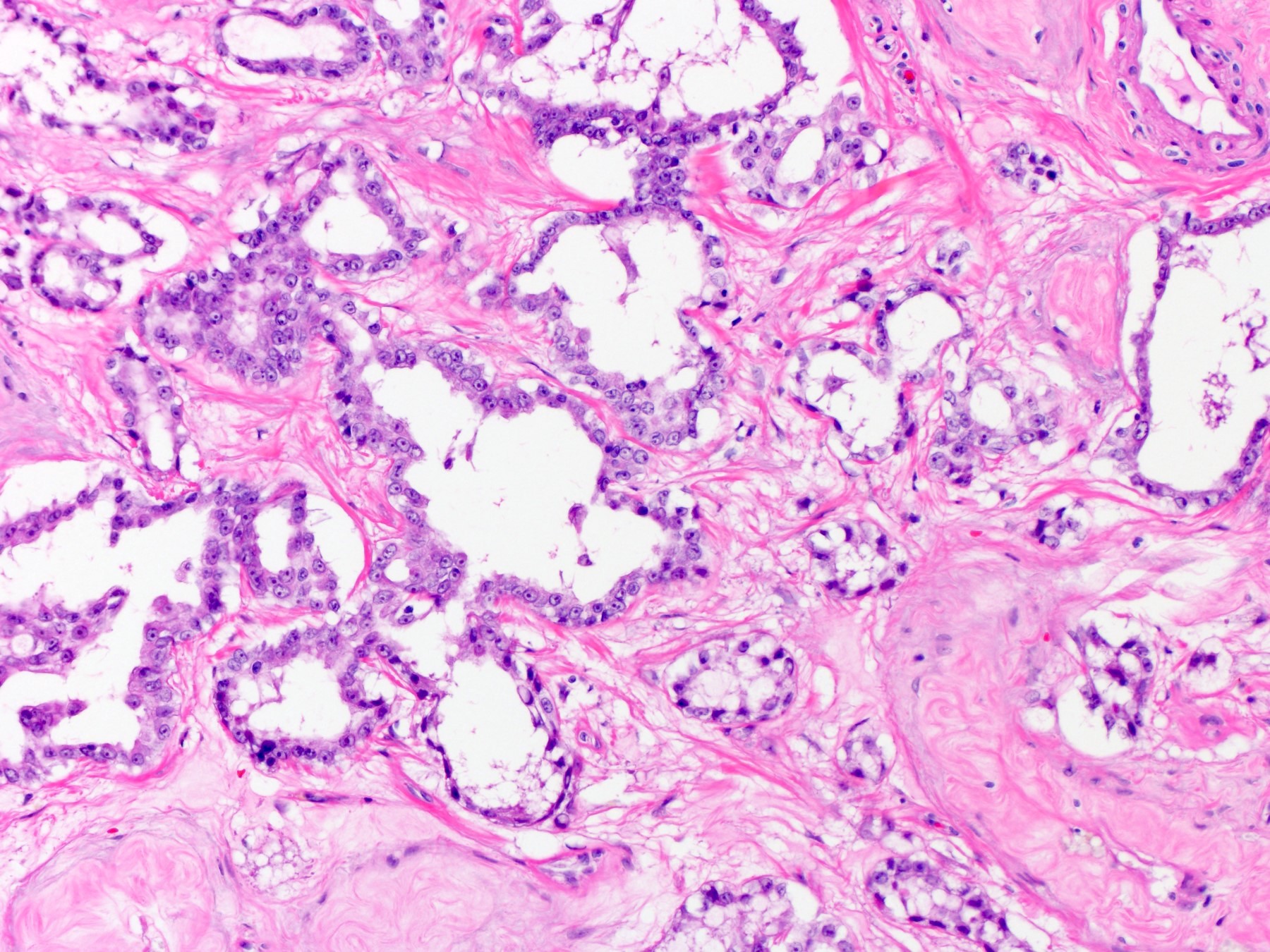
Unencapsulated nodule
Seminiferous tubule proliferation
Focal Leydig cells
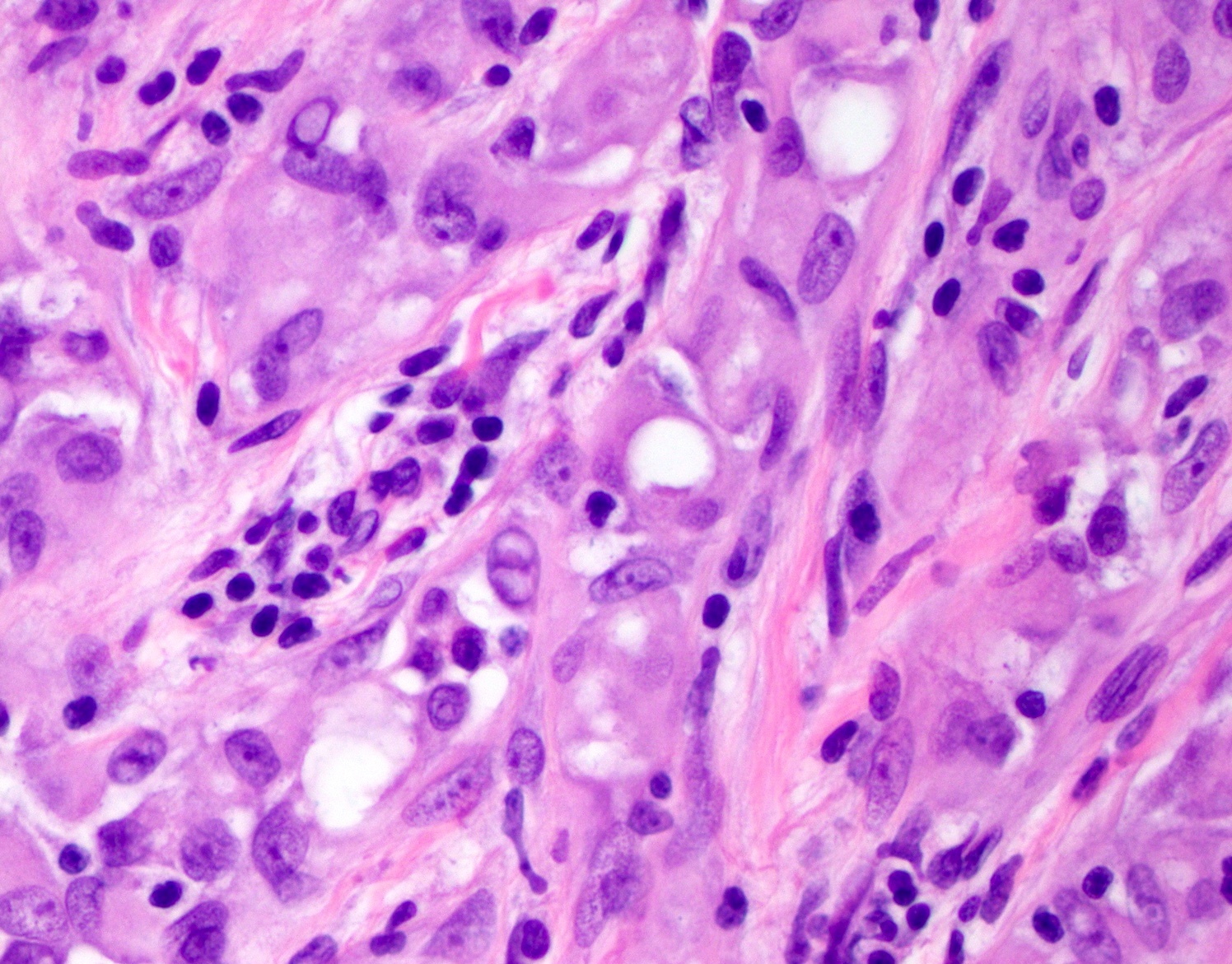
Hyaline basement membrane deposits
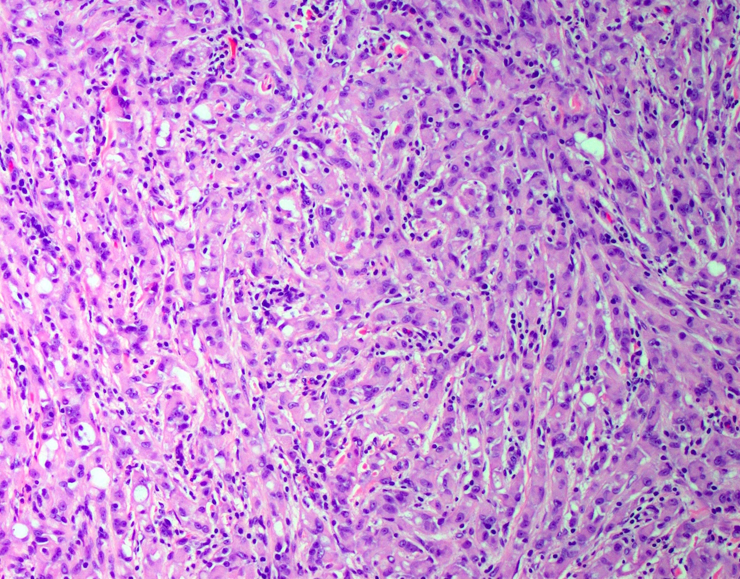
Pseudostratified Sertoli cells
Arrangement of Sertoli cells
Calcification

Hyperplastic Sertoli cells
Atrophic seminiferous tubules
Persistent immature tubules
Hyalinization of basement membrane
Atrophic seminiferous tubules
Images hosted on other servers:

Thickened, multilamellar basement membranes

Hyaline core
Contributed by Asmaa Gaber Abdou, M.D.

Sertoli cell only and Leydig cell hyperplasia
Images hosted on other servers:

Hypoechoic lesion
Images hosted on other servers:

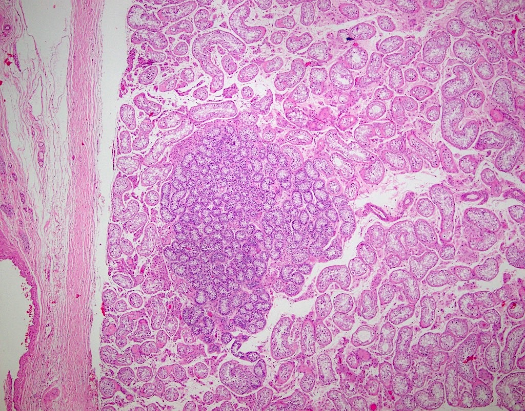
Tumor in lower pole
Contributed by Thomas Ulbright, M.D. and Case #18
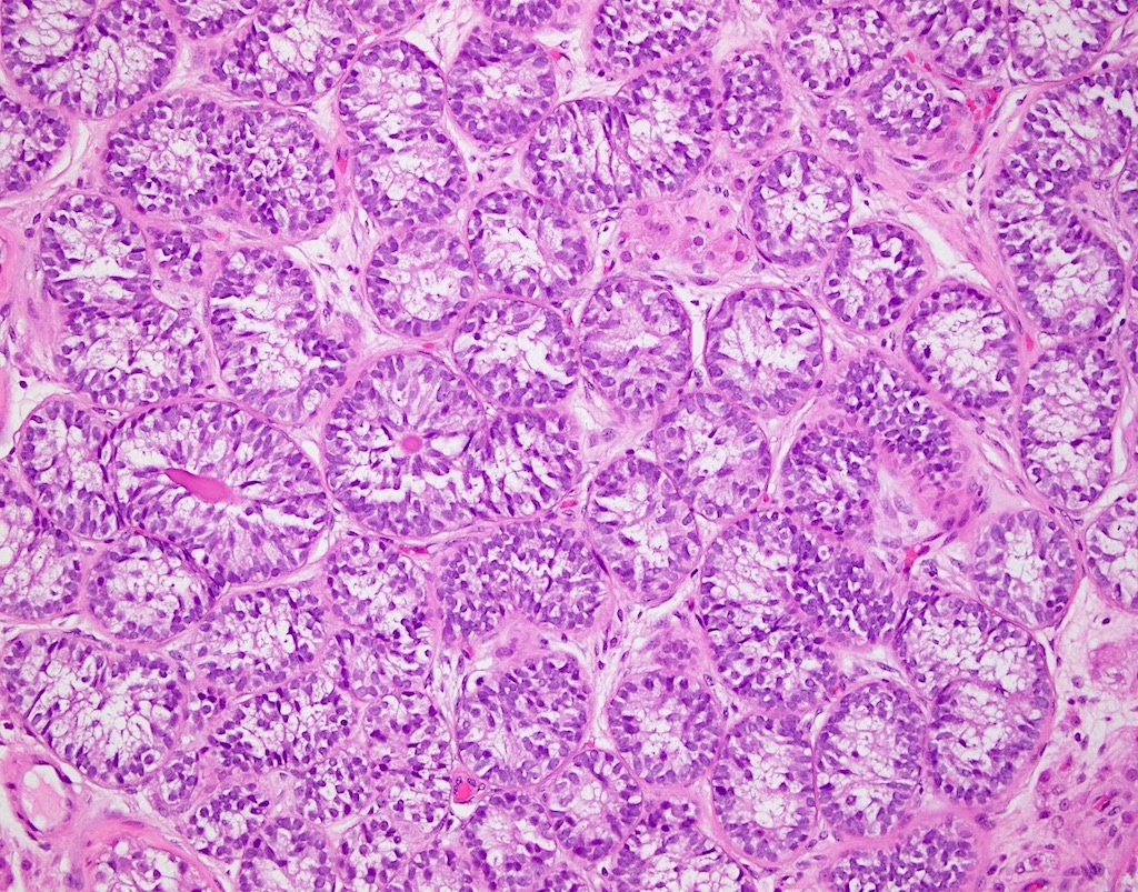
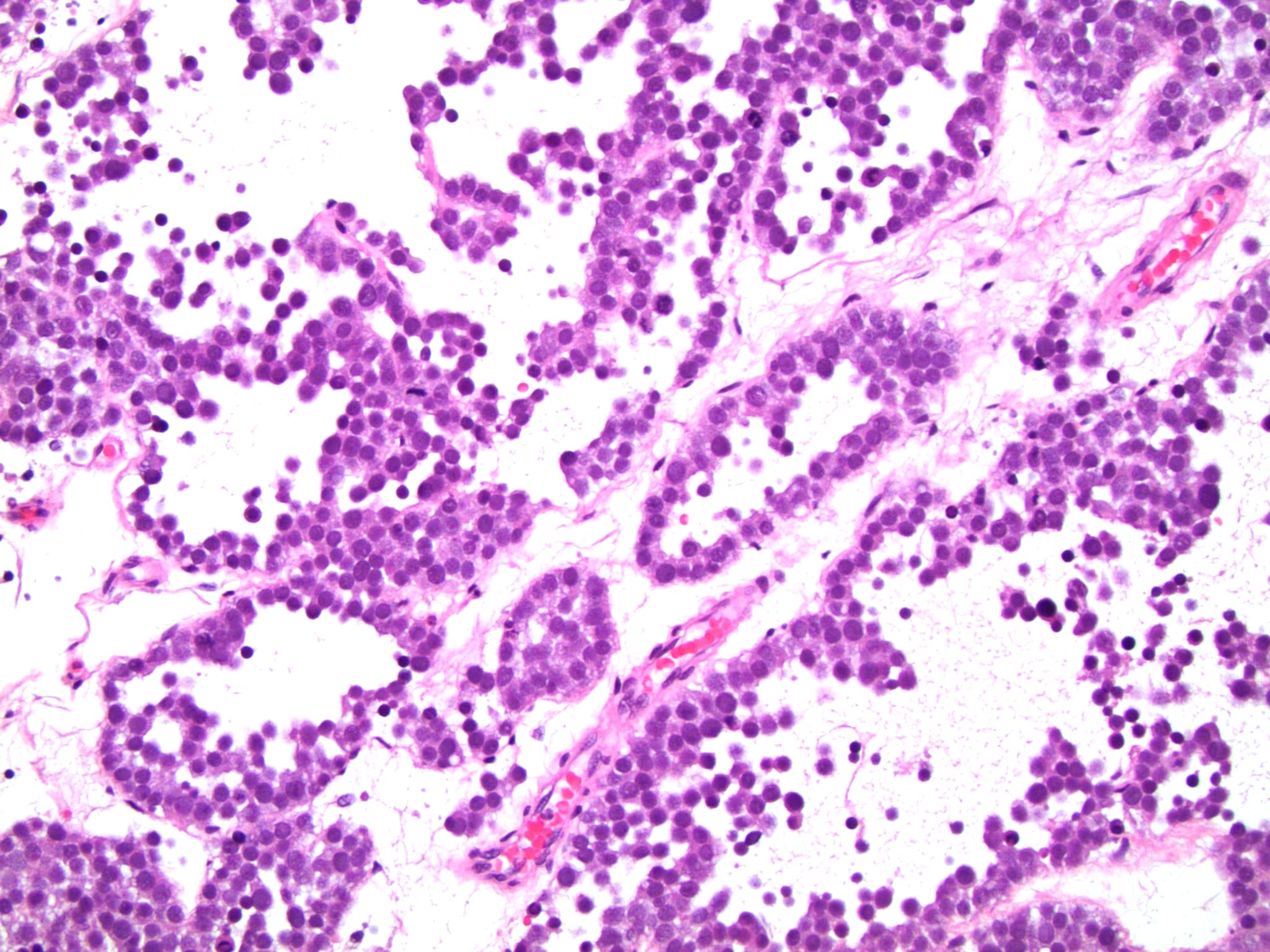
Architectural patterns
Cords
Tubules
Tubopapillary
Macrocystic
Microcystic
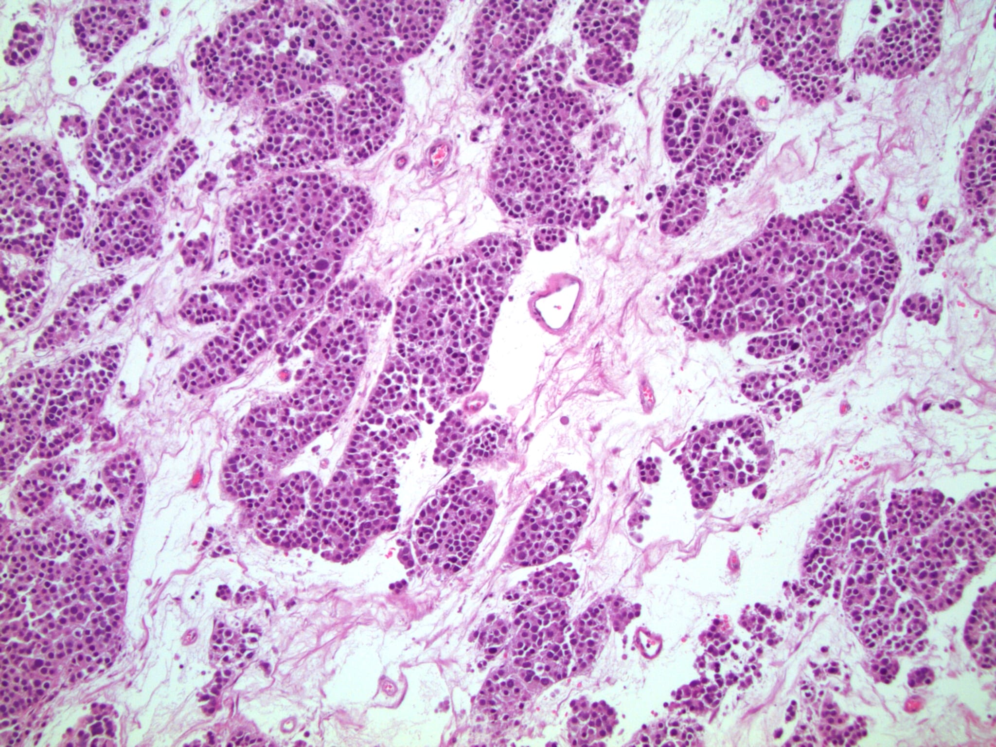
Architectural patterns
Whorled
Trabecular
Sclerosing Sertoli cell tumor

Various images
Cells
Abundant clear cytoplasm (most common)
Heavily lipidized
Malignant
Marked cytologic atypia
Tumor necrosis
Lymphovascular invasion
High N/C ratio,
increased
mitotic / apoptotic
activity
Contributed by Indiana University School of Medicine
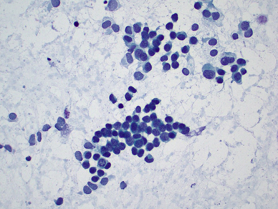
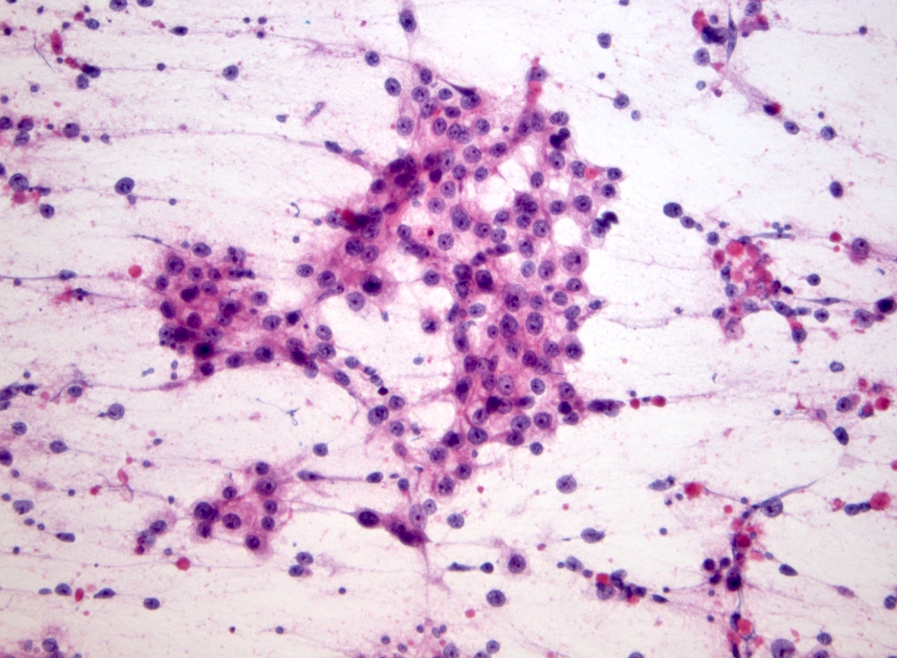
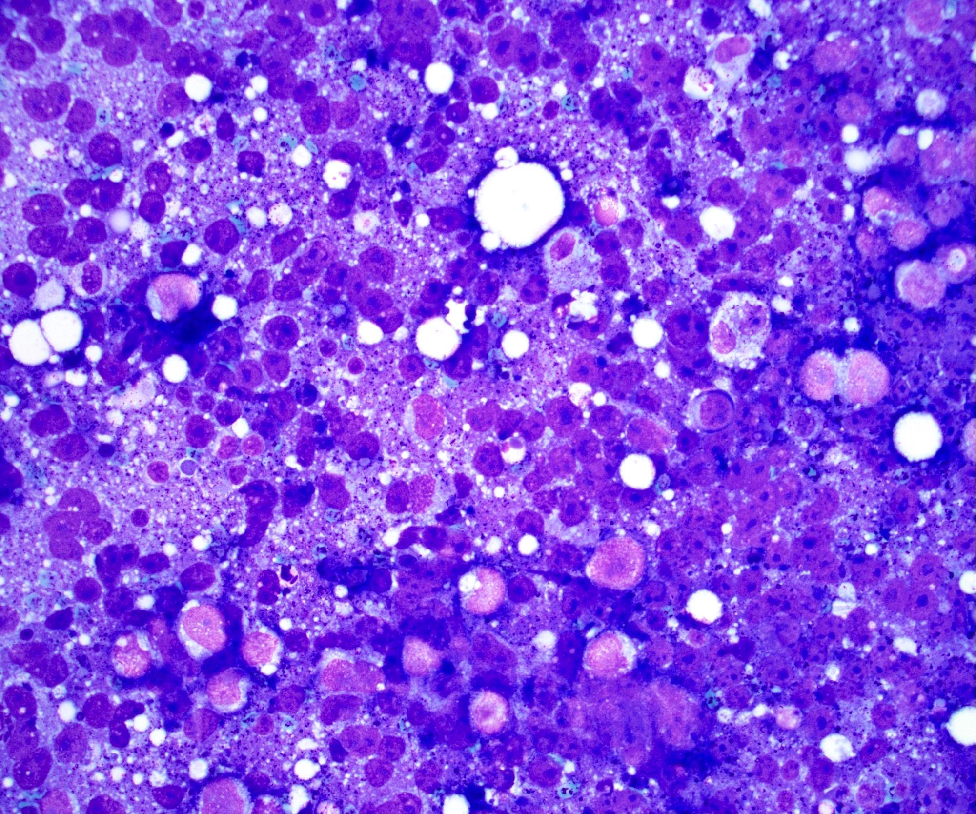
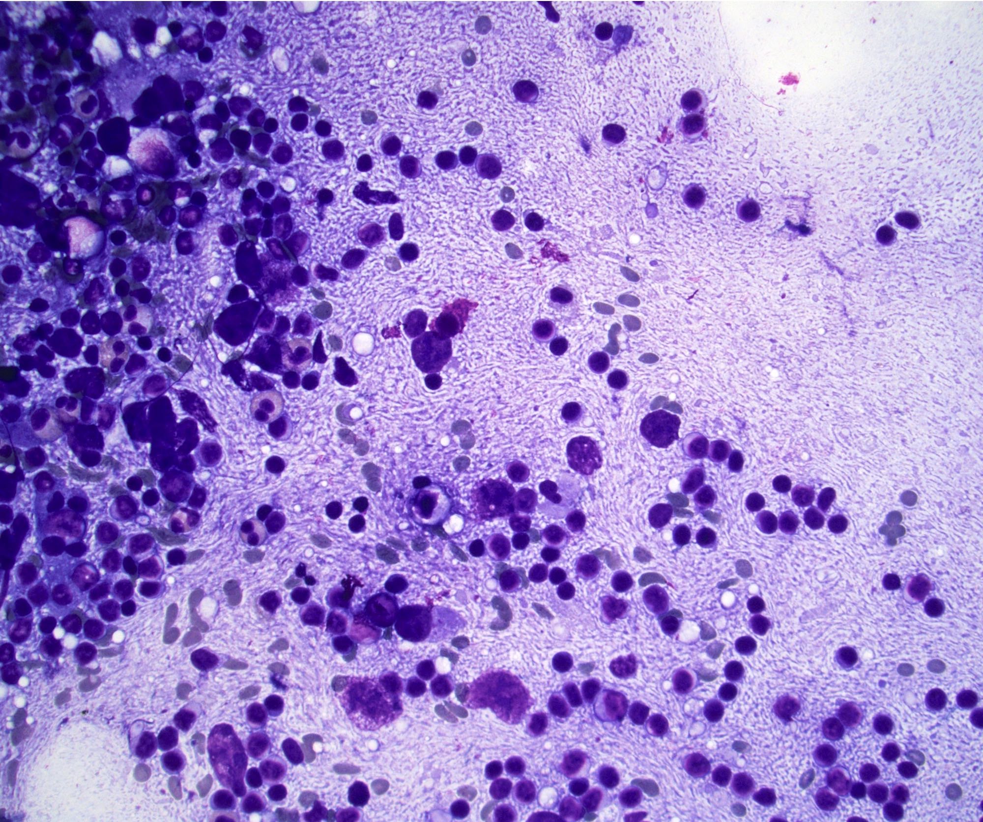
Tubular formations, monotonous small cells (Pap)

Papillary groups and discohesive cells (Pap)
Images hosted on other servers:

Well circumscribed multicystic mass
Images hosted on other servers:

Columnar epididymal type epithelium

Cystic duct

Mass blends with ducts in epididymal tail
Images hosted on other servers:


Ultrasound images
Contributed by Anil Parwani, M.D., Ph.D., M.B.A.
Orchiectomy
with spermatocele
Contributed by Anil Parwani, M.D., Ph.D., M.B.A.
Spermatocele with adjacent epididymis
Cuboidal epithelium
Ciliated columnar epithelium
Flattened epithelium
Aggregate of spermatozoa
Macrophages containing spermatozoa
Images hosted on other servers:

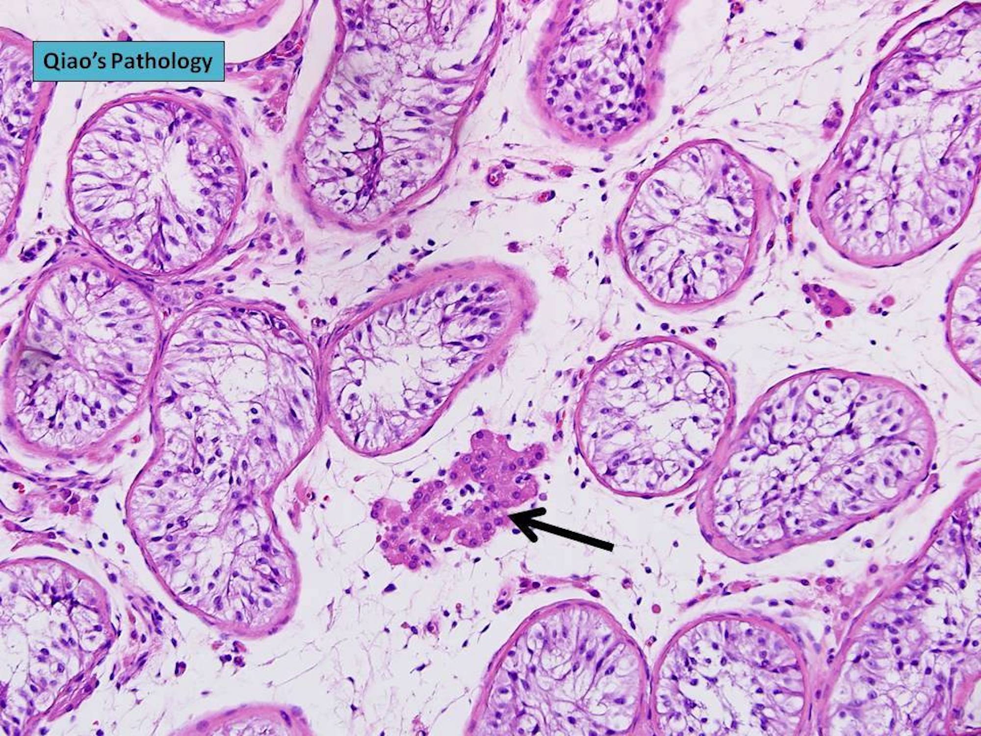
ST (arrow) and testis (*)
AFIP images
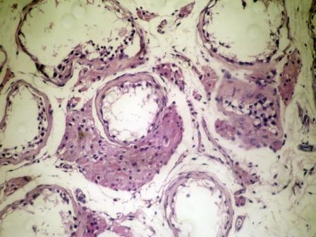
Mucoid tumor
Images hosted on other servers:

ST (arrow)
Cystic and mucoid tumor
Contributed by Maurizio Colecchia, M.D., Case #448 and AFIP images
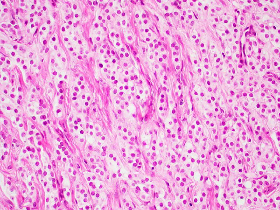
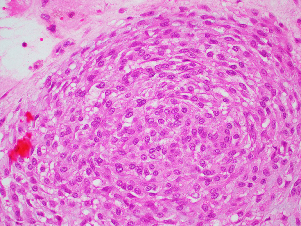
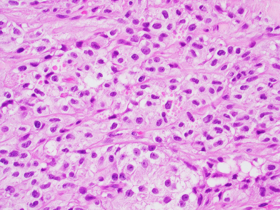
Lobulated architecture

Tripartite cytology
Prevalent intermediate sized cells
Mitoses and apoptotic bodies
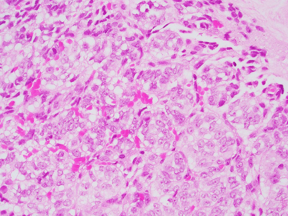
Short fibrous bands
Spireme-like chromatin
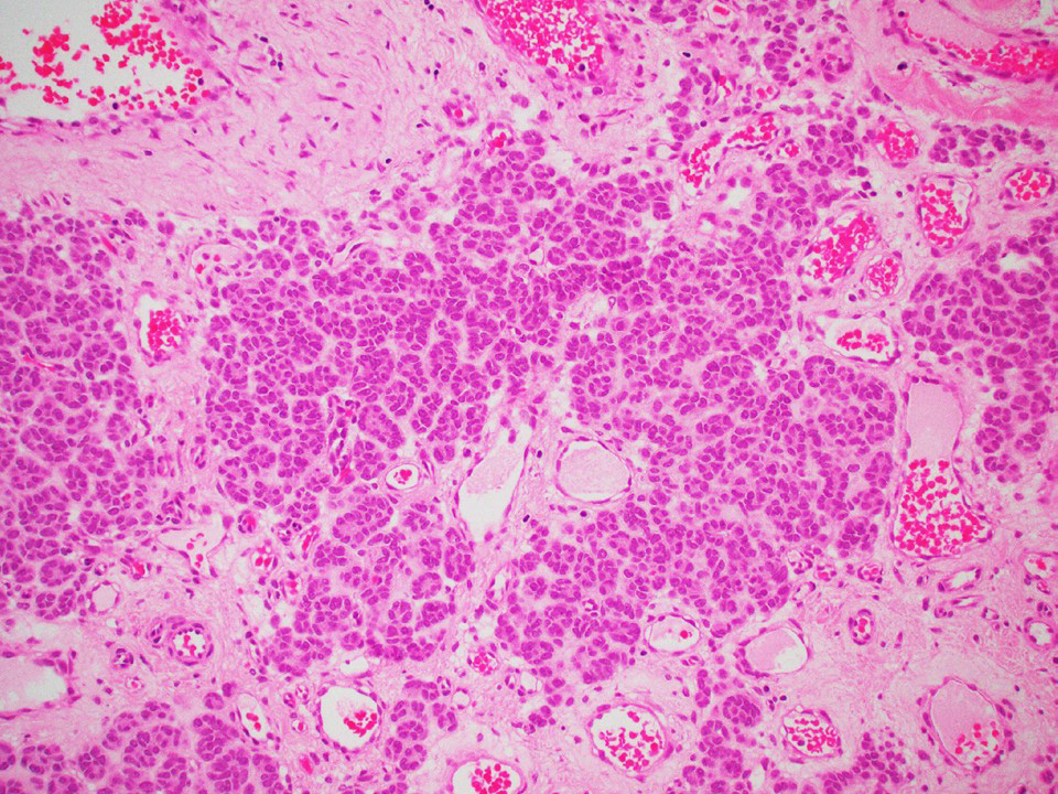
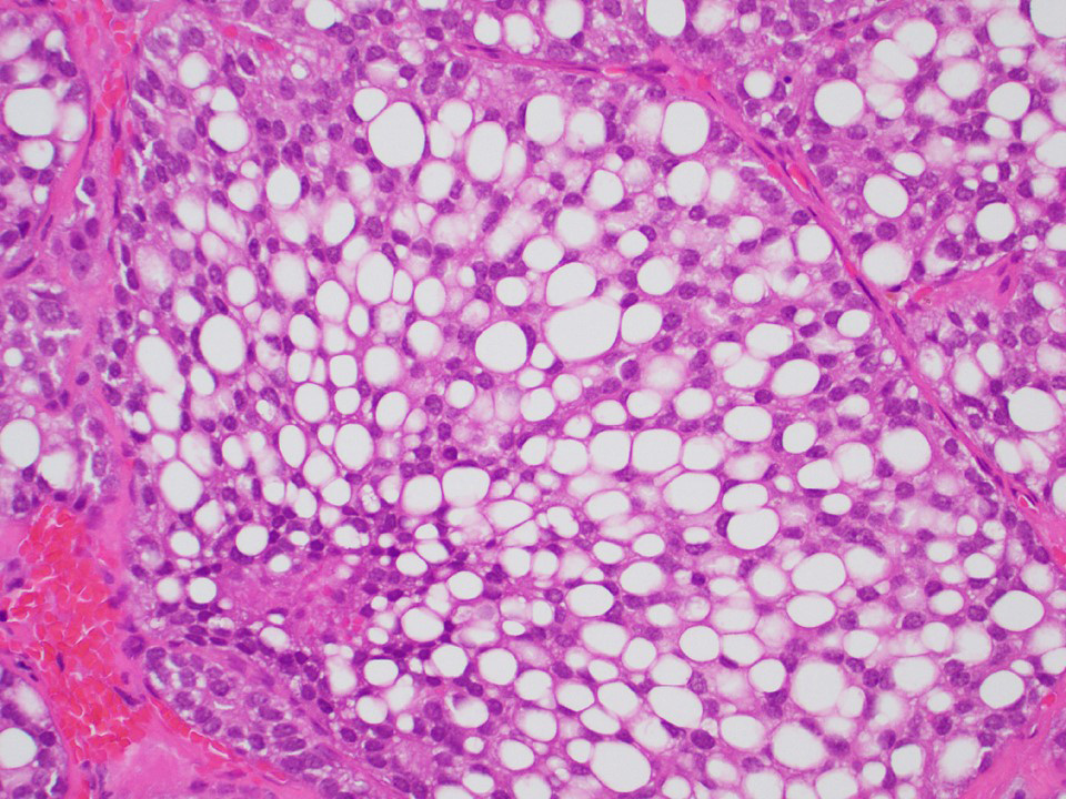
Edema and microcysts
Edema and nests

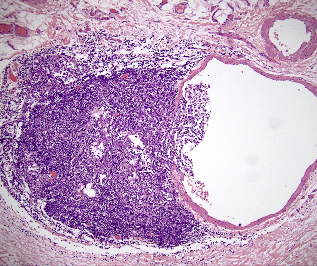
Intratubular growth
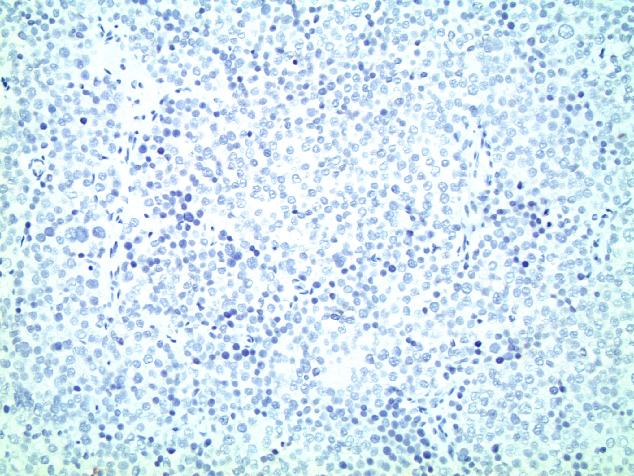
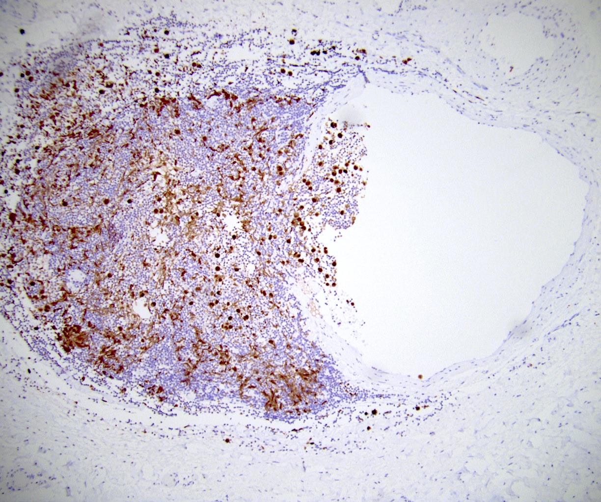
OCT3/4
CD45
Diffuse growth pattern

Focal pseudoglandular pattern
Cystic pattern

Polymorphic cell population
Small nests, trabeculae, clusters and single cells

Filamentous chromatin
Normal spermatogenesis

Desmin+
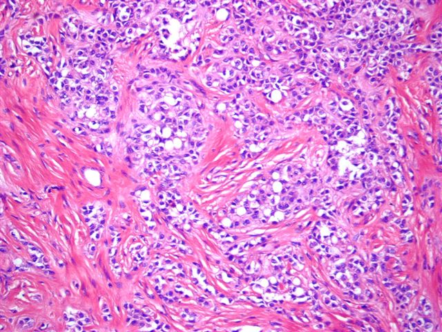
Tumor nests and
cords mixed with
lymphocytes
Prominent
granulomatous
reaction and
lymphoid infiltrate
Intratubular component
AFIP images
Intercellular bridge
Nest of spermatocytic seminoma
Case #309


Contributed by Debra L. Zynger, M.D.

Complete regression (pT0)
Germ cell neoplasia in situ only (pTis)
Partial regression (pT1)
Teratoma (pT1)
Teratoma and EC (pT1)
Seminoma (pT1a)
Seminoma (pT1b)

Mixed germ cell tumor (pT2)
Seminoma (pT2)
No residual testis tumor (ypT0)

No residual node tumor (ypN0)
Lymph node with residual tumor (ypN1 - 3)
Discontinuous spermatic cord (pM1)
Contributed by Debra L. Zynger, M.D.
Lymphovascular invasion (pT2)

Lymphovascular invasion (pT2)

Tunica vaginalis invasion (pT2)
Epididymal invasion (pT2)
Hilar soft tissue invasion (pT2)

Lymph node metastasis (pN1)
Post treatment node (ypN0)
Post treatment node (ypN1)
Discontinuous spermatic cord invasion (pM1)
Images hosted on other servers:

Testicular teratoma diagnosed in utero

Demonstrating vascularity of testicular teratoma with calcifications
Contributed by Debra L. Zynger, M.D.
Teratoma (100%)
Mixed GCT
Contributed by Debra L. Zynger, M.D.
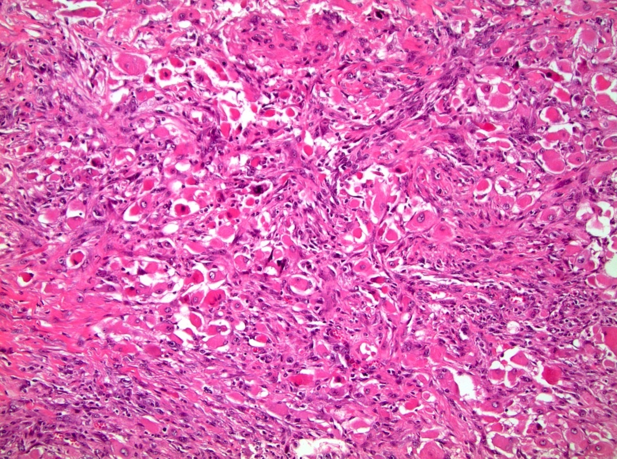
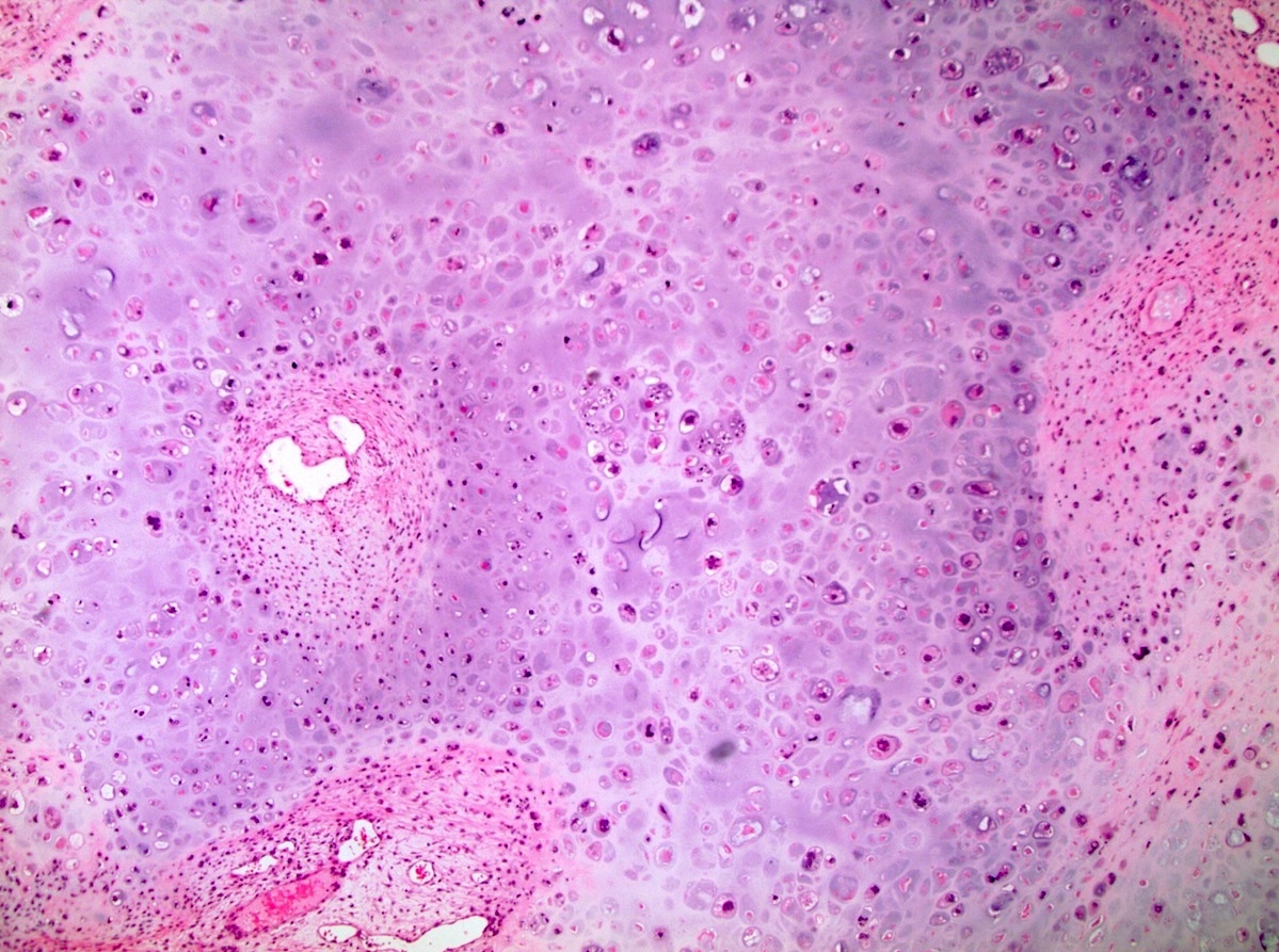
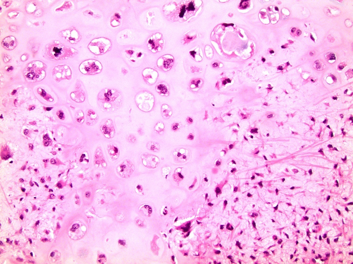
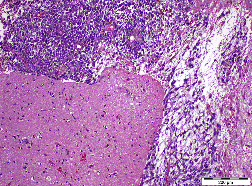
Cartilage surrounded by mitotically active spindle cells

Gastrointestinal epithelium

Squamous epithelium

Cellular mesenchyme
Loose mesenchyme

Neuroectoderm
Contributed by Christopher Dall, M.D. and Debra L. Zynger, M.D.
Retroperitoneal sarcoma, NOS
Lung rhabdo-
myosarcoma
Iliac leiomyosarcoma
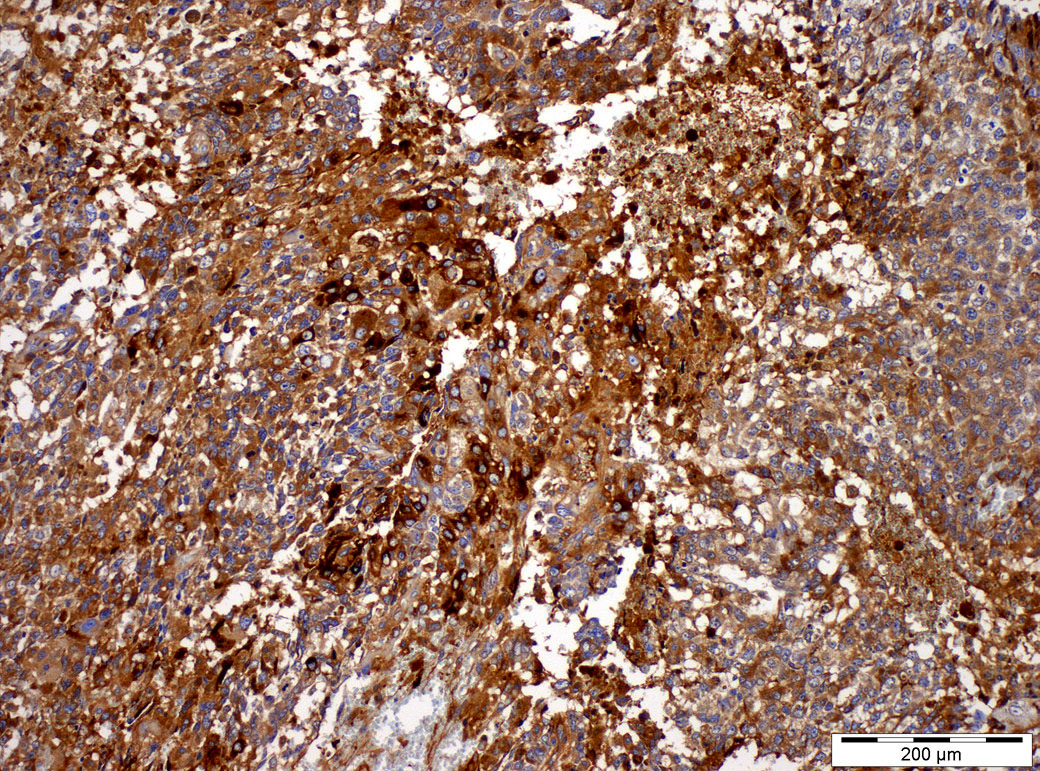
Embryonic type neuroectodermal tumor
Contributed by Christopher Dall, M.D. and Debra L. Zynger, M.D.
Metastatic rhabdomyosarcoma
Metastatic chondrosarcoma
Metastatic sarcoma, NOS
Embryonic type neuroectodermal tumor within testis
AFIP images
Solid tan mass
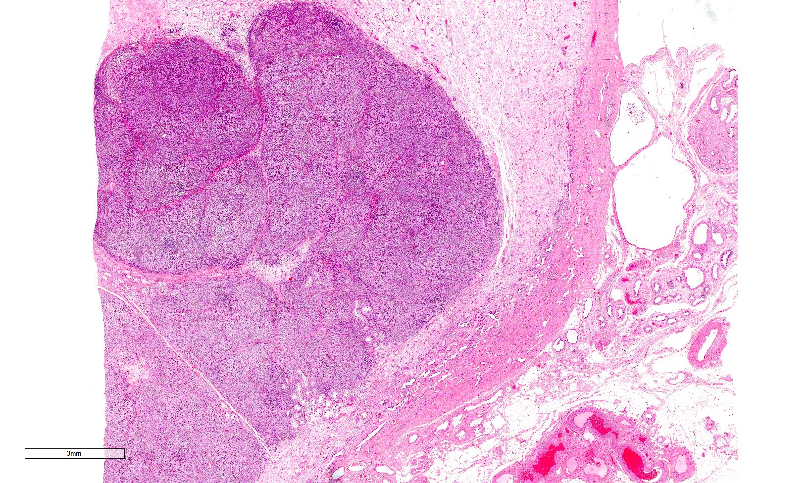
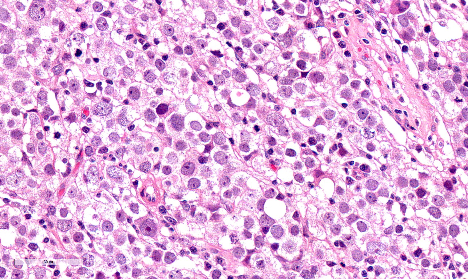
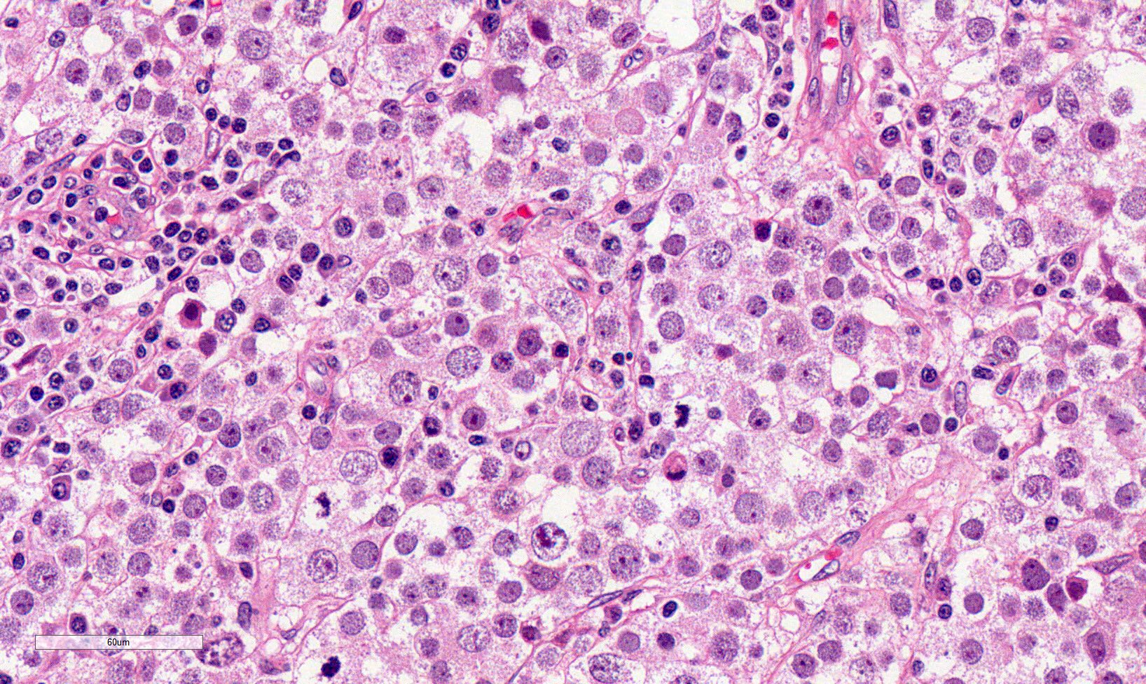
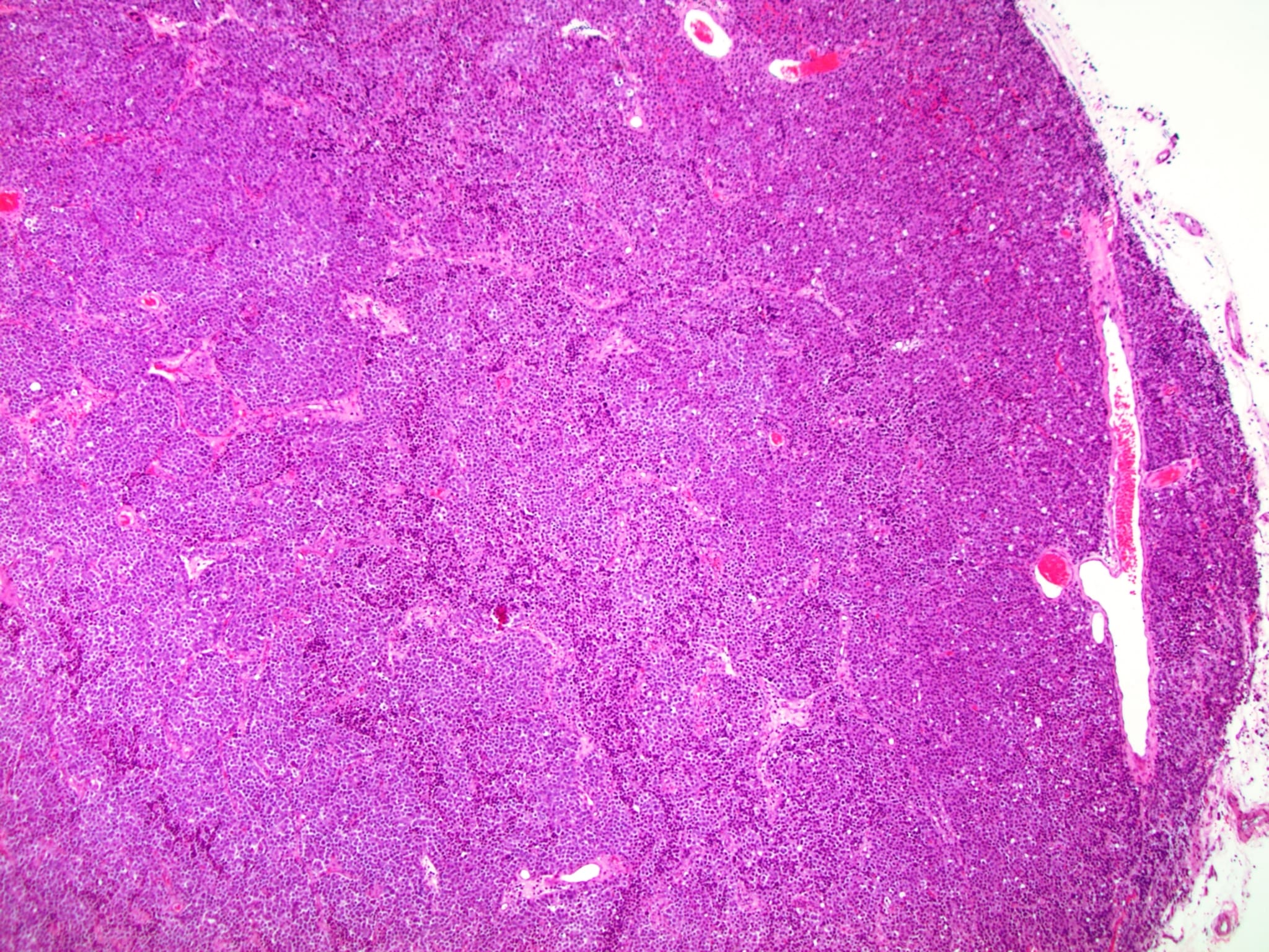
Contributed by Sounak Gupta, M.B.B.S, Ph.D. and Rafael E. Jimenez, M.D.


Epidermoid cyst and NET

Retraction artifact
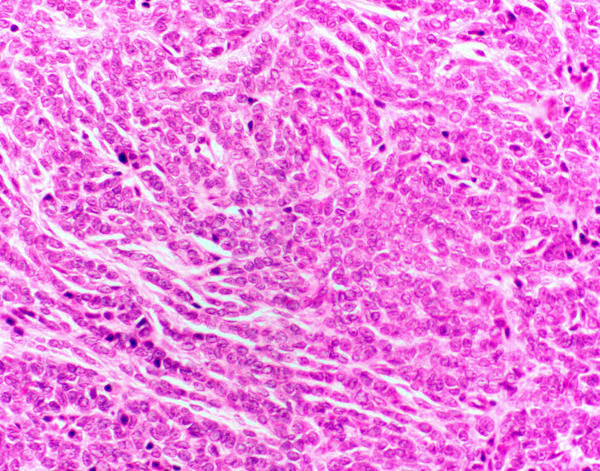
Multiple solid nests
Insular growth pattern
Nests of tumor cells
Testicular NET
Images hosted on other servers:

Dexamethasone treatment

Large TTAGS
Images hosted on other servers:

Well circumscribed,
noncapsulated, solid
and lobulated brown lesion
Images hosted on other servers:

H&E

H&E, IHC, CD56

H&E
Case #279

Images hosted on other servers:

Bell clapper deformity
leading to intravaginal
testicular torsion
Contributed by Raman Danrad, M.D.
Ultrasound of bilateral testis
Ultrasound of right normal testis
Ultrasound of left torsed testis
Images hosted on other servers:

Twisting of spermatic cord
Images hosted on other servers:

Smooth external surface of torsed testis

Dark red hemorrhagic cut surface
Contributed by Ritu Bhalla, M.D.
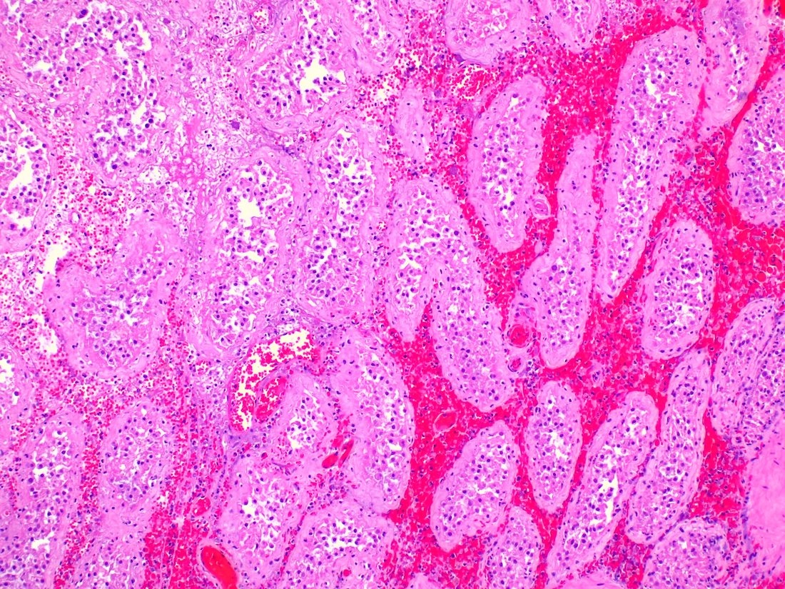
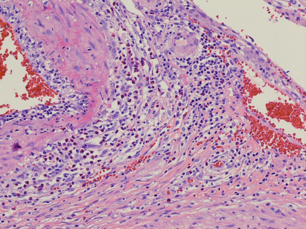
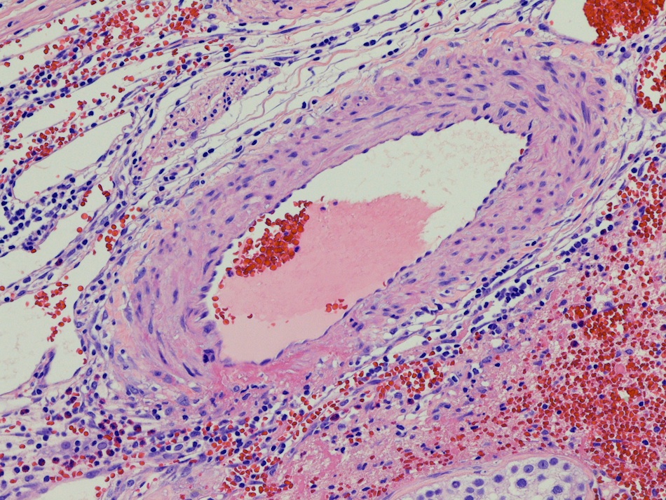
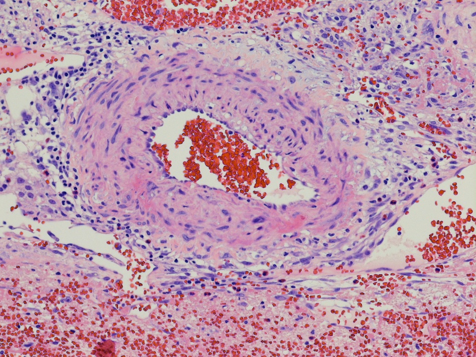
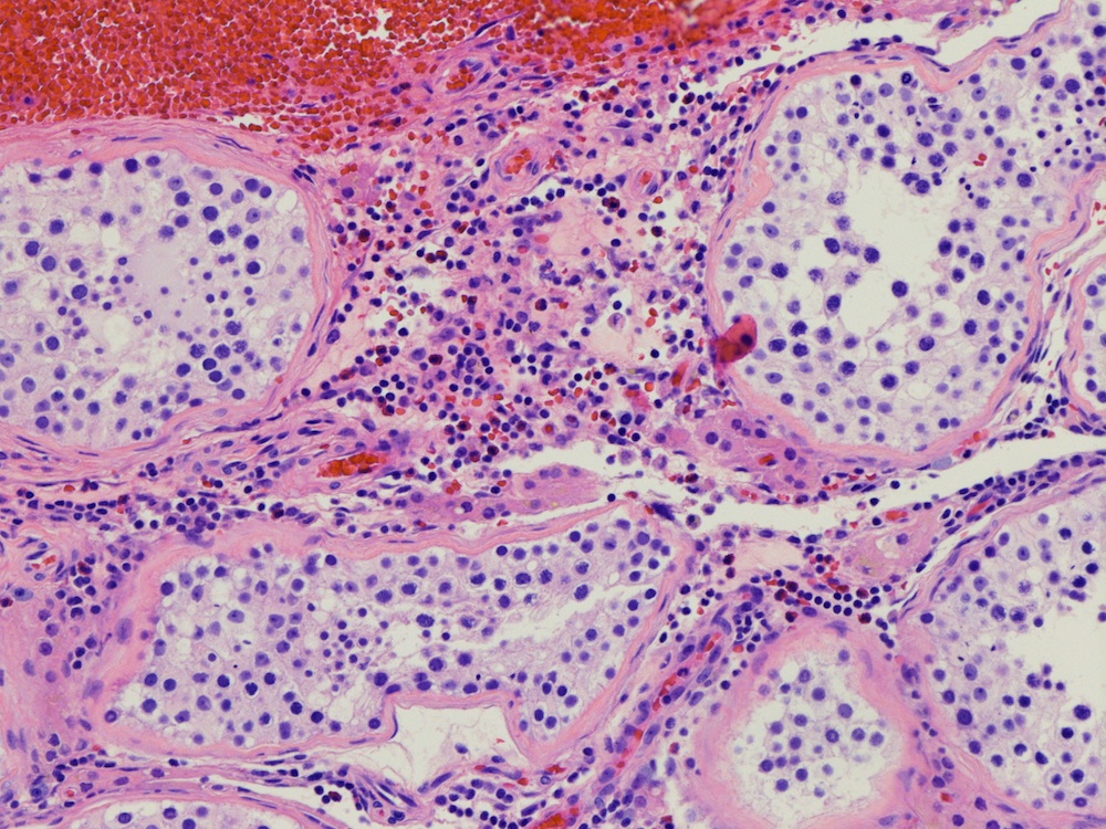
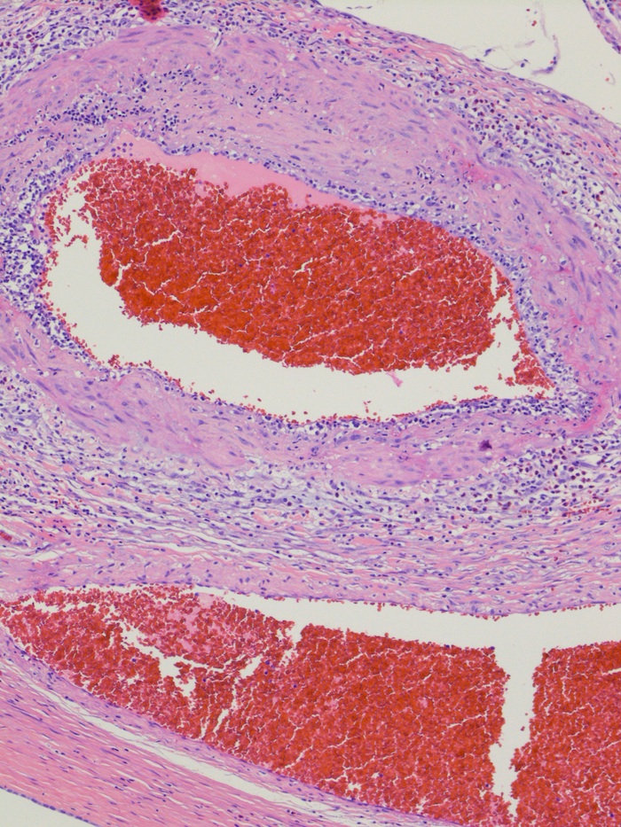
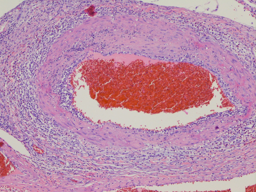
Congestion
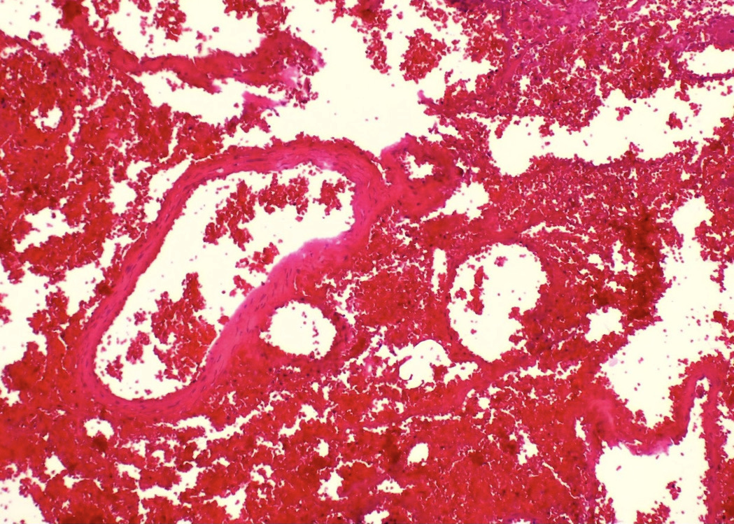
Hilar hemorrhage
Sloughing of cells
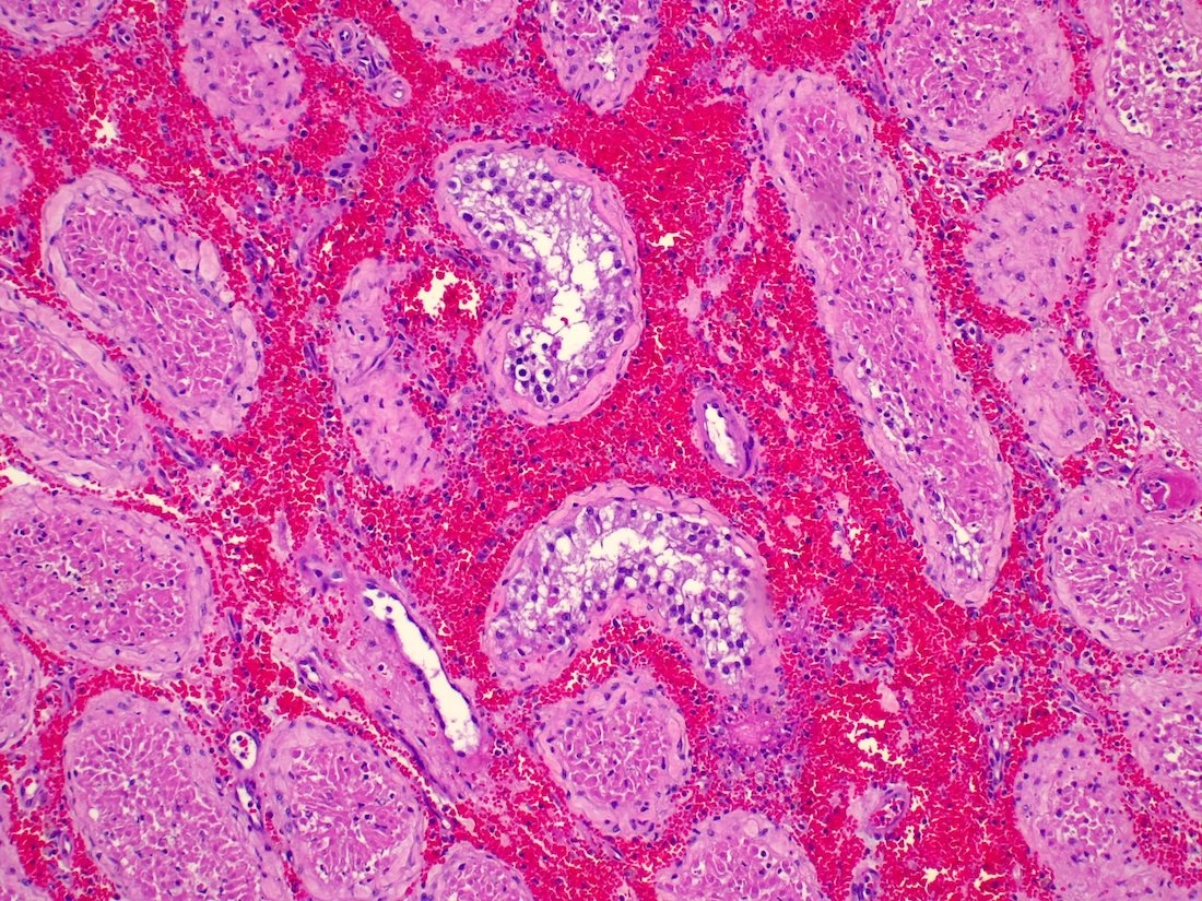
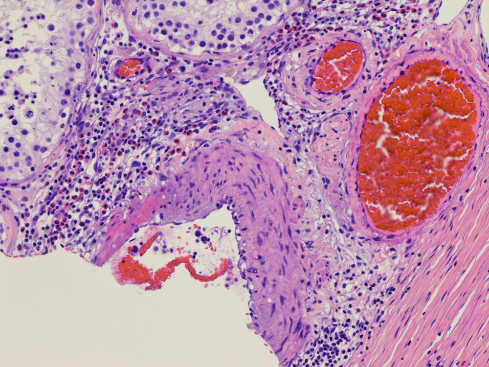
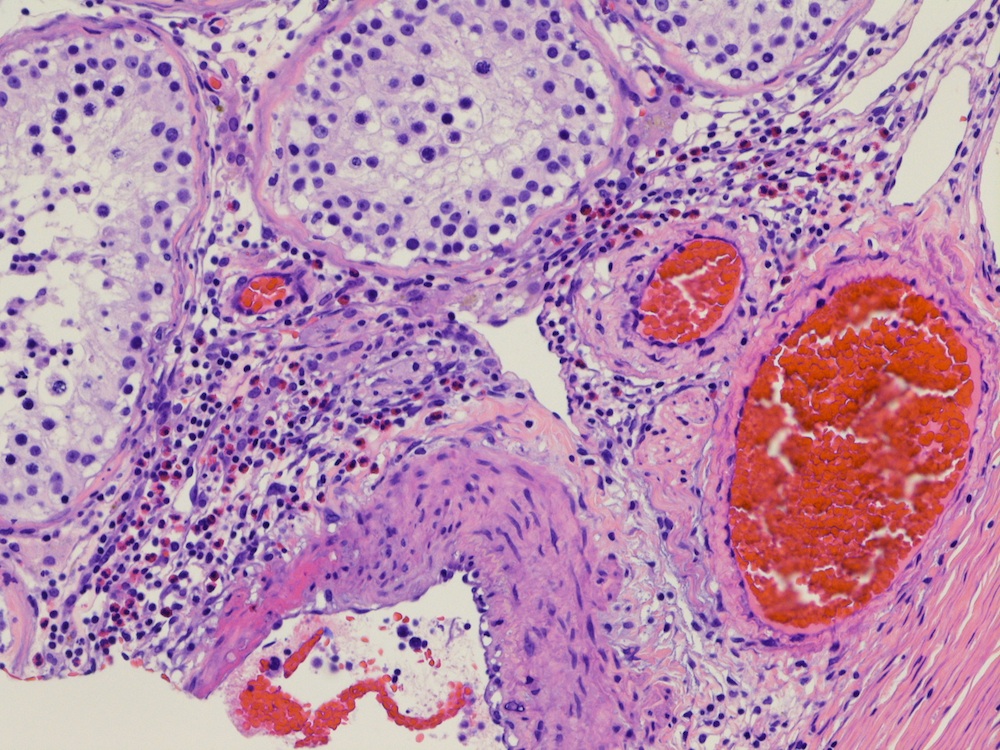
Interstitial hemorrhage
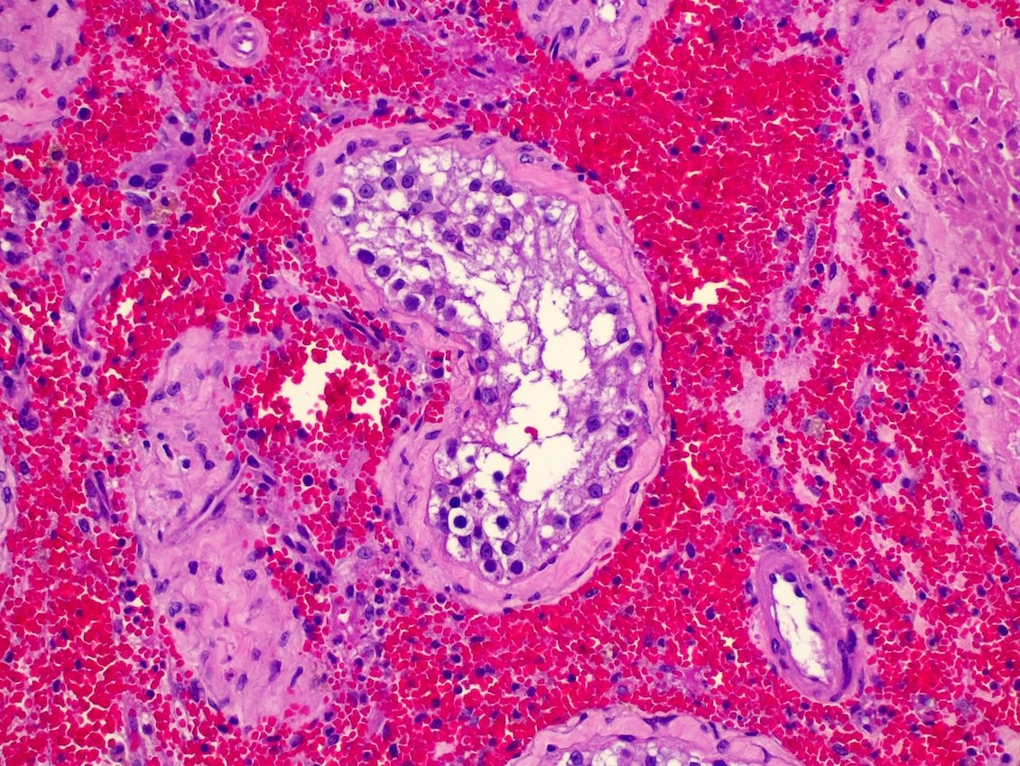
Residual germ cells
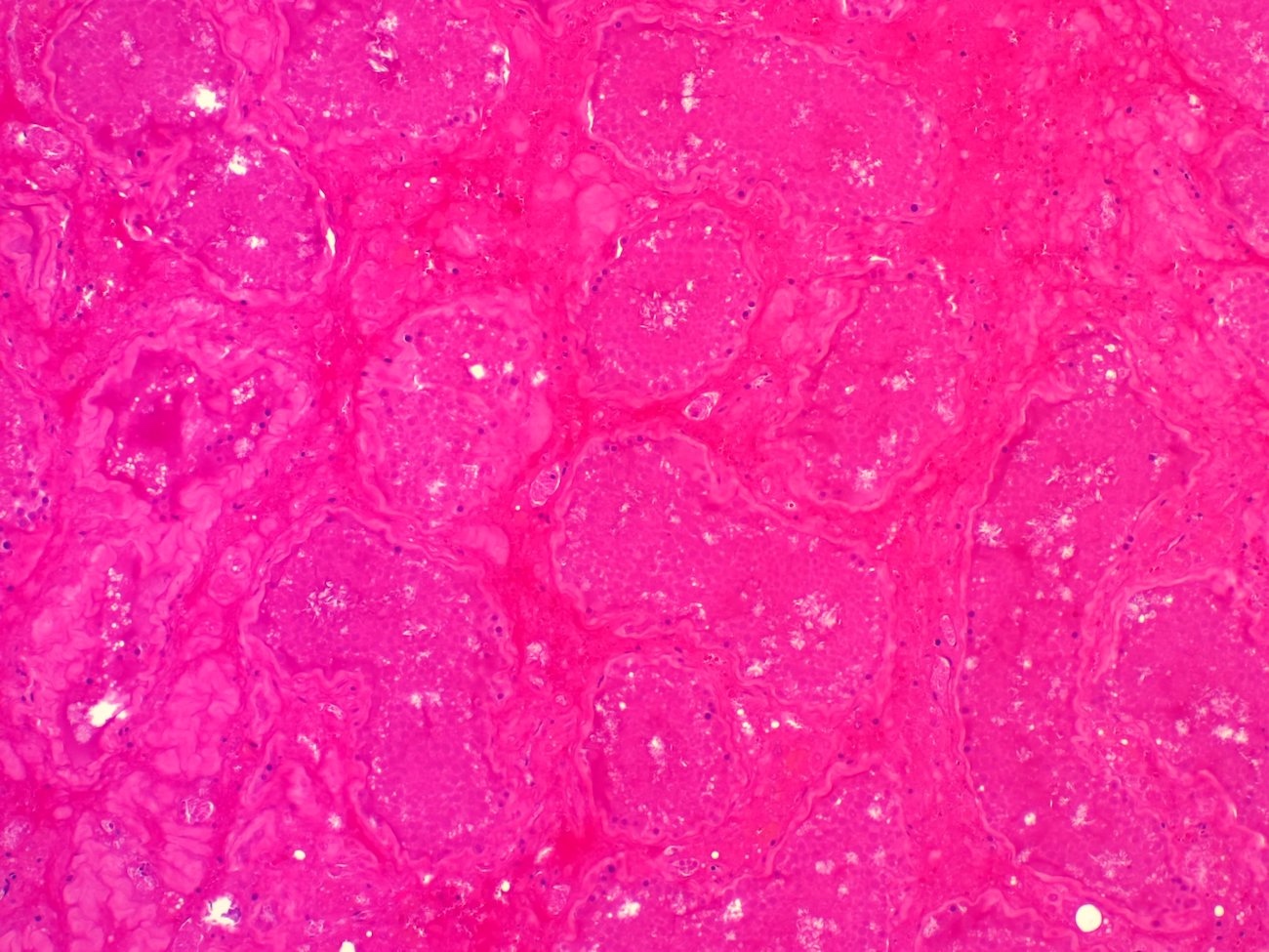
Infarcted tubules
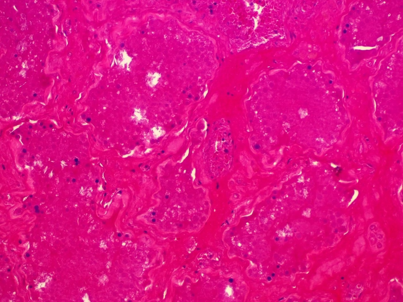
Necrotic parenchyma
Images hosted on other servers:

Left sided grade 3 varicocele

Color Doppler ultrasonography of varicocele
Contributed by Kenneth A. Iczkowski, M.D.

Orchiectomy specimen in a 30 year old with atrophic testis and varicocele
Case #328


Testicular heterogeneous hypoechoic lesion
Case #328
Case #328


Images hosted on other servers:


Vasitis nodosa
Contributed by Aida Valencia, M.D. and Jennifer Gordetsky, M.D.
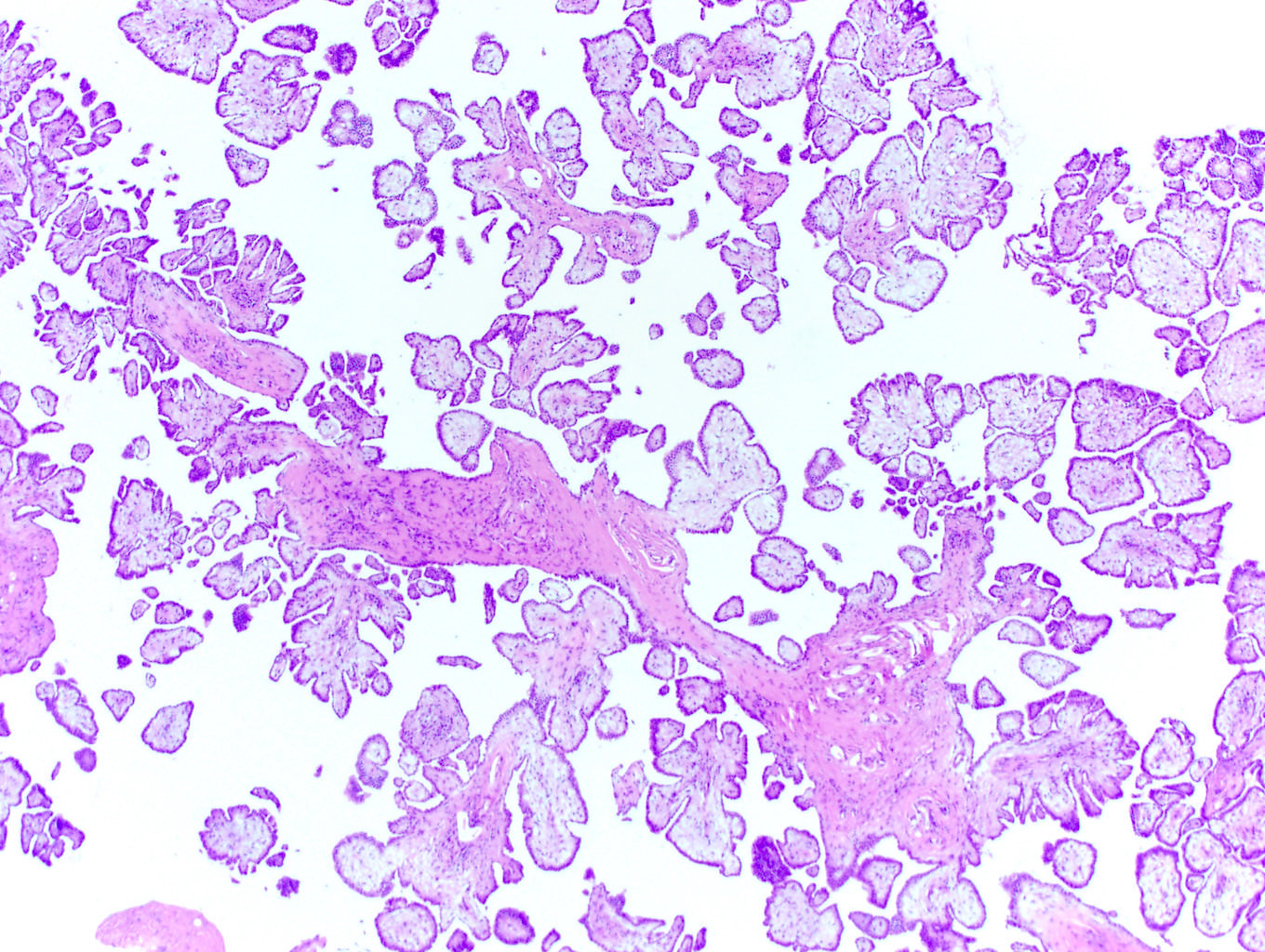
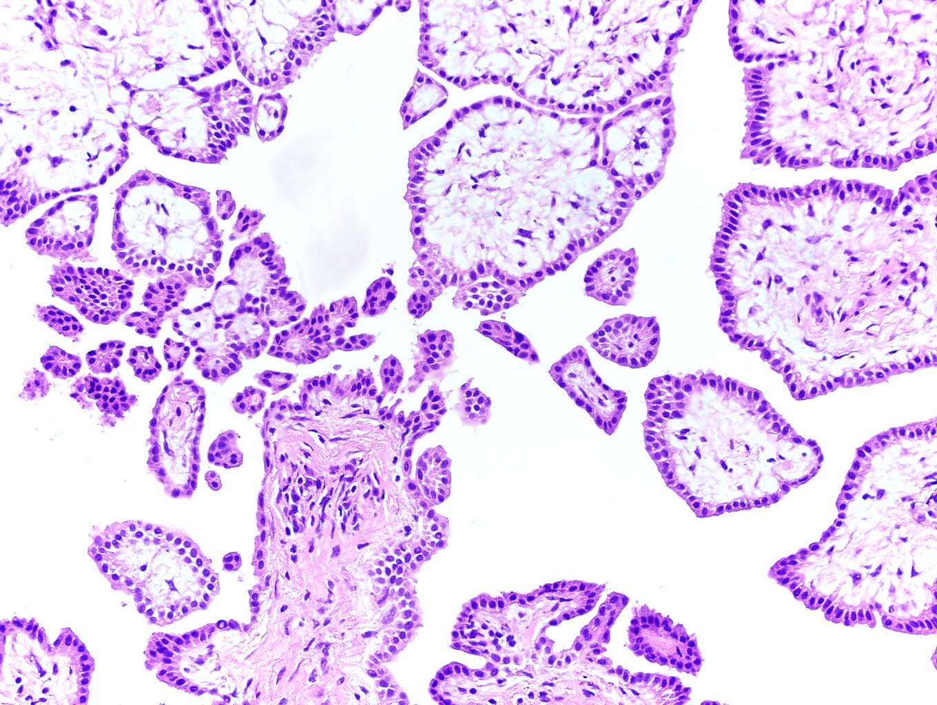
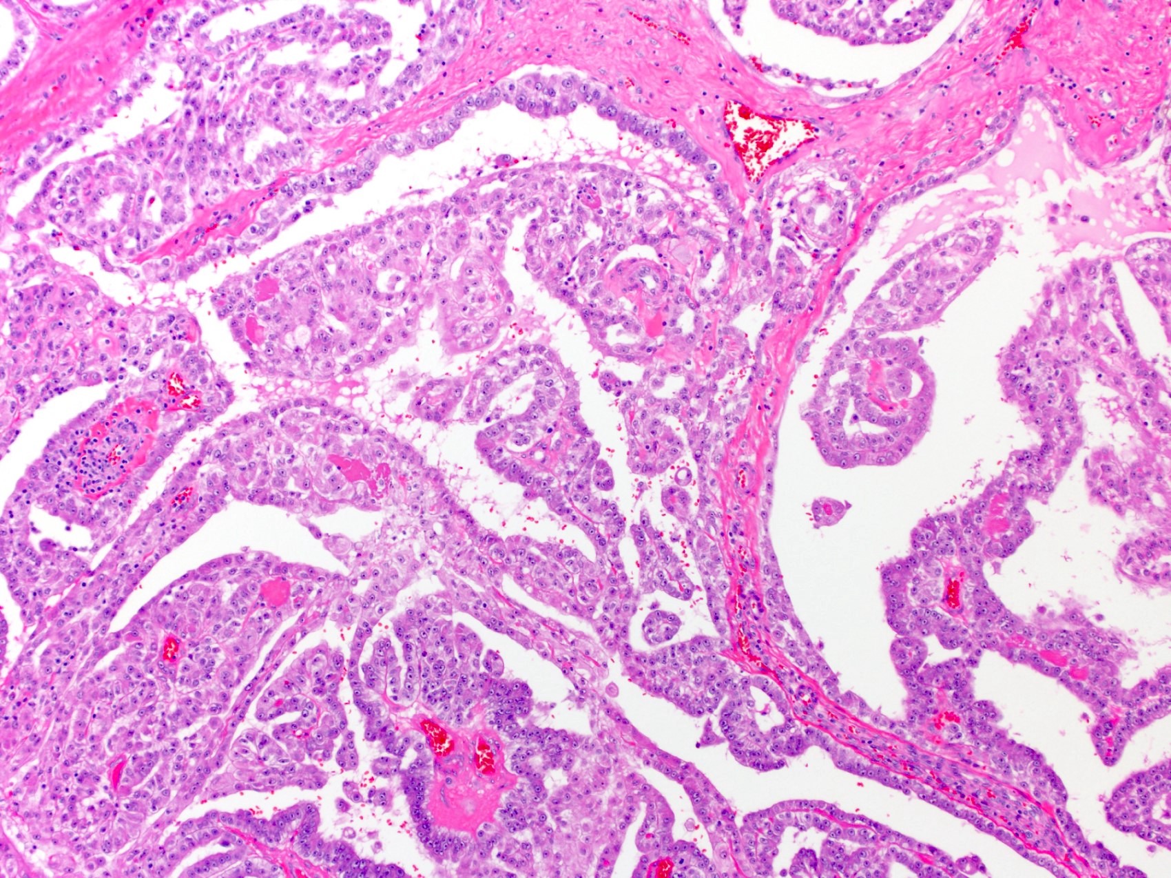
Numerous fibrovascular cores
Background macrophages
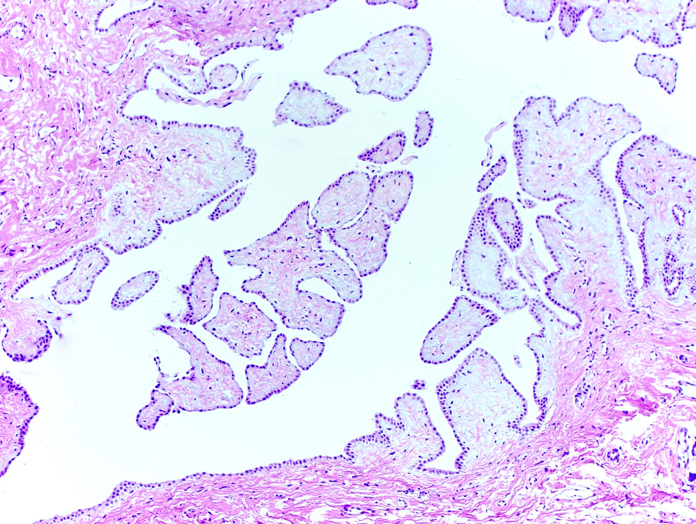
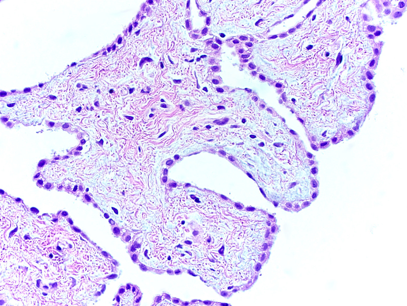
Broad papillae
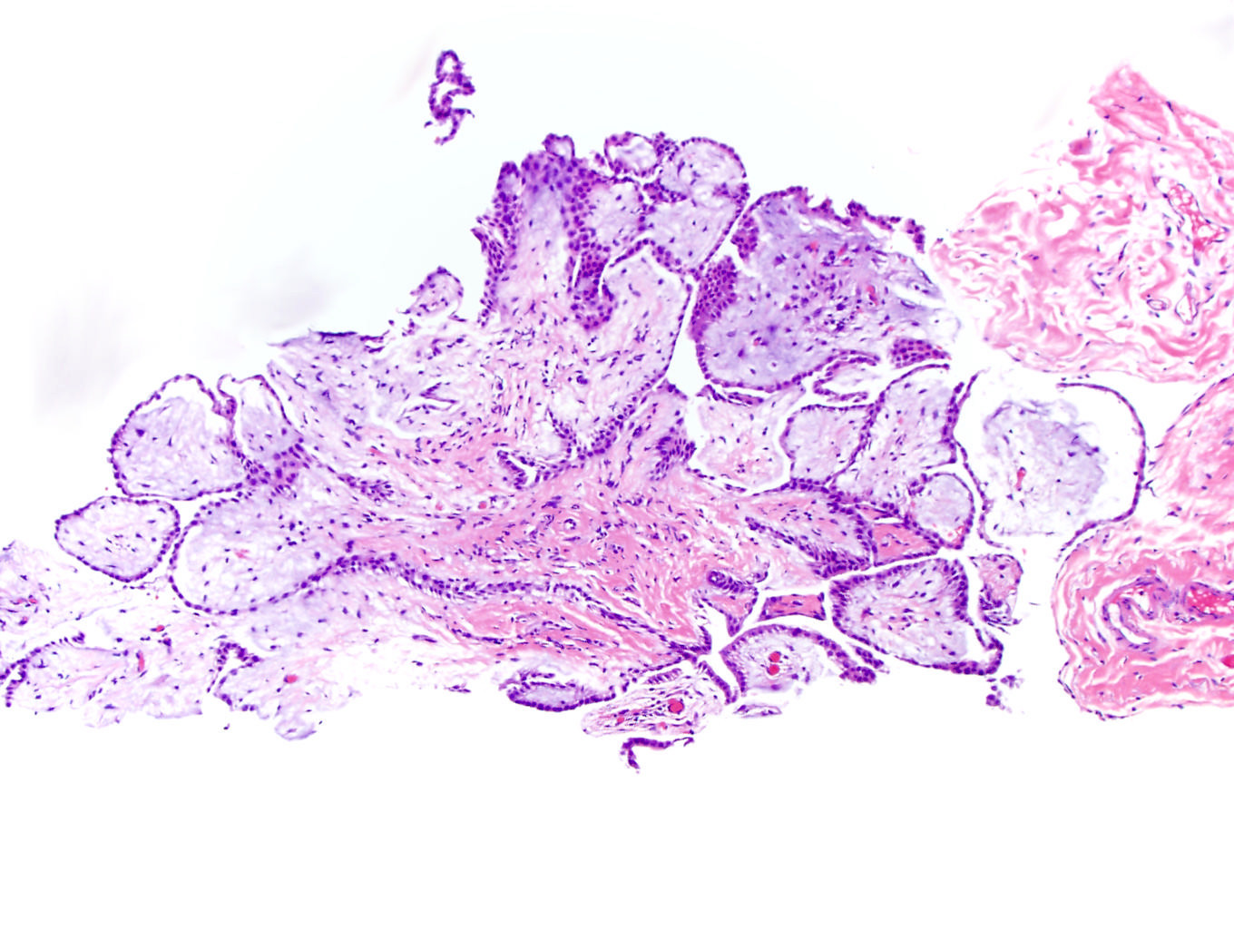
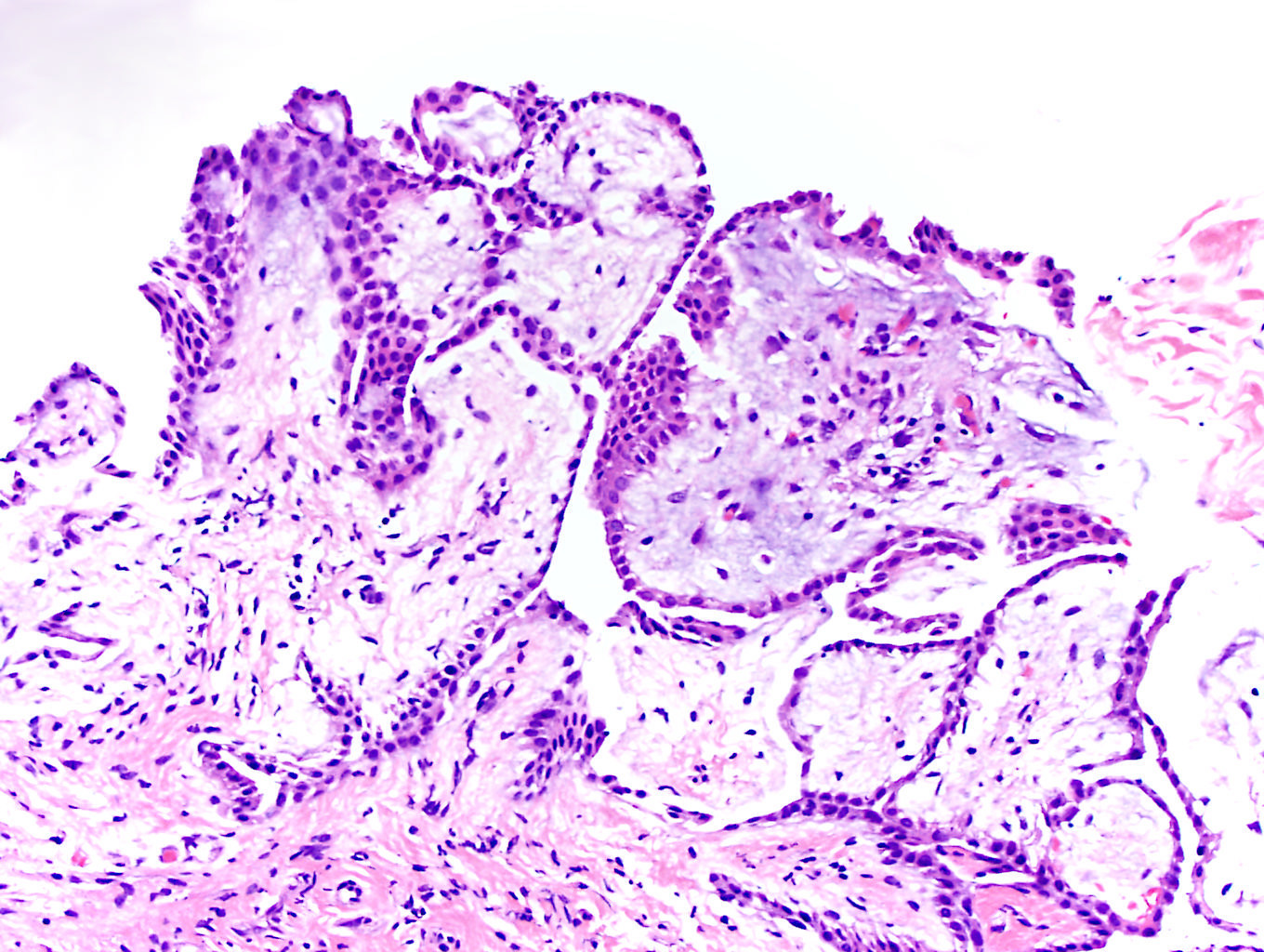
Papillae with myxoid stroma
Nonpsammomatous calcifications
Calretinin
WT1
D2-40
Mesothelioma
Images hosted on other servers:

TRAF7 and CDC42 somatic mutations
Contributed by Debra L. Zynger, M.D.
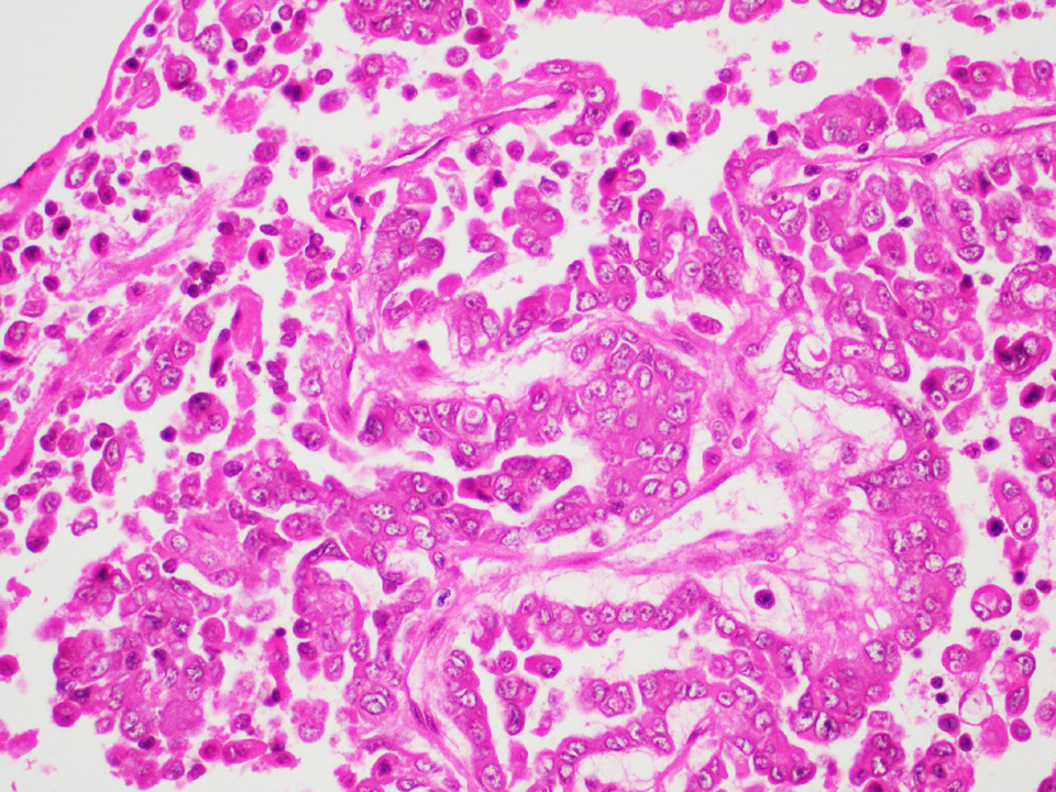
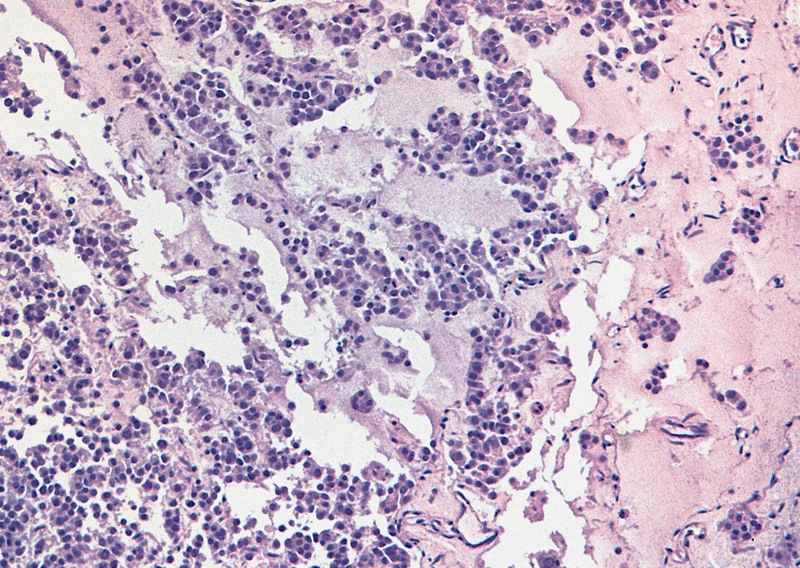
Mixed germ cell tumor, postpubertal with varying % of yolk sac tumor
Contributed by João Lobo, M.D. and Rui Henrique, M.D., Ph.D.
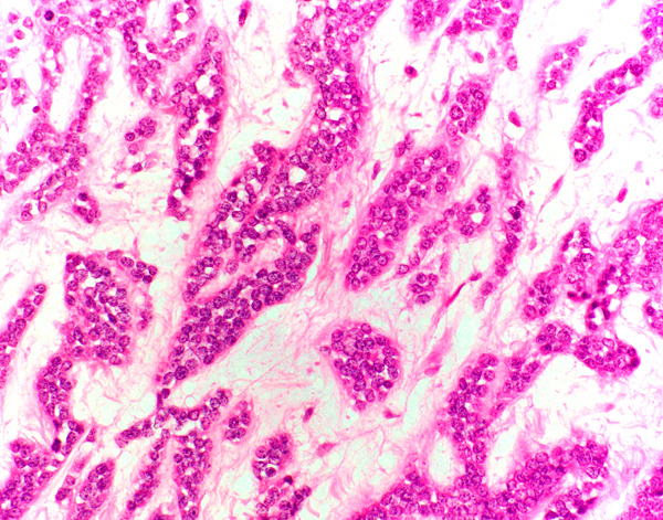
Microcystic pattern
Hyaline globules
Glandular pattern
Schiller-Duval bodies
Metastasis
Amin: 2022
Behnke: 2018

Cheng: 2019
Colecchia : 2016
IARC: 2022

Manuel: 2019
Nistal: 2017

Ulbright: 2014
Ulbright: 2022
VandenBussche: 2022

Wobker: 2021

Yang: 2020
Zhou: 2022
Zhou: 2022
Find related Pathology books: cytopathology, GU/adrenal
