


Images hosted on other servers:

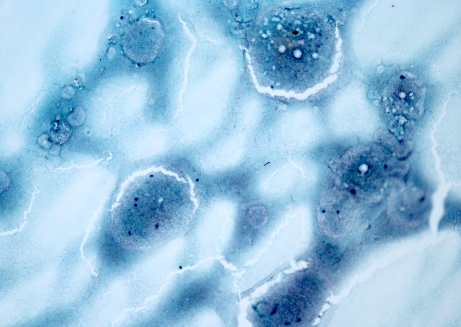
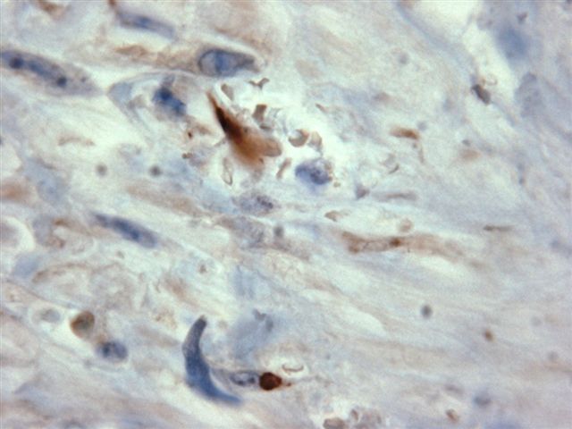
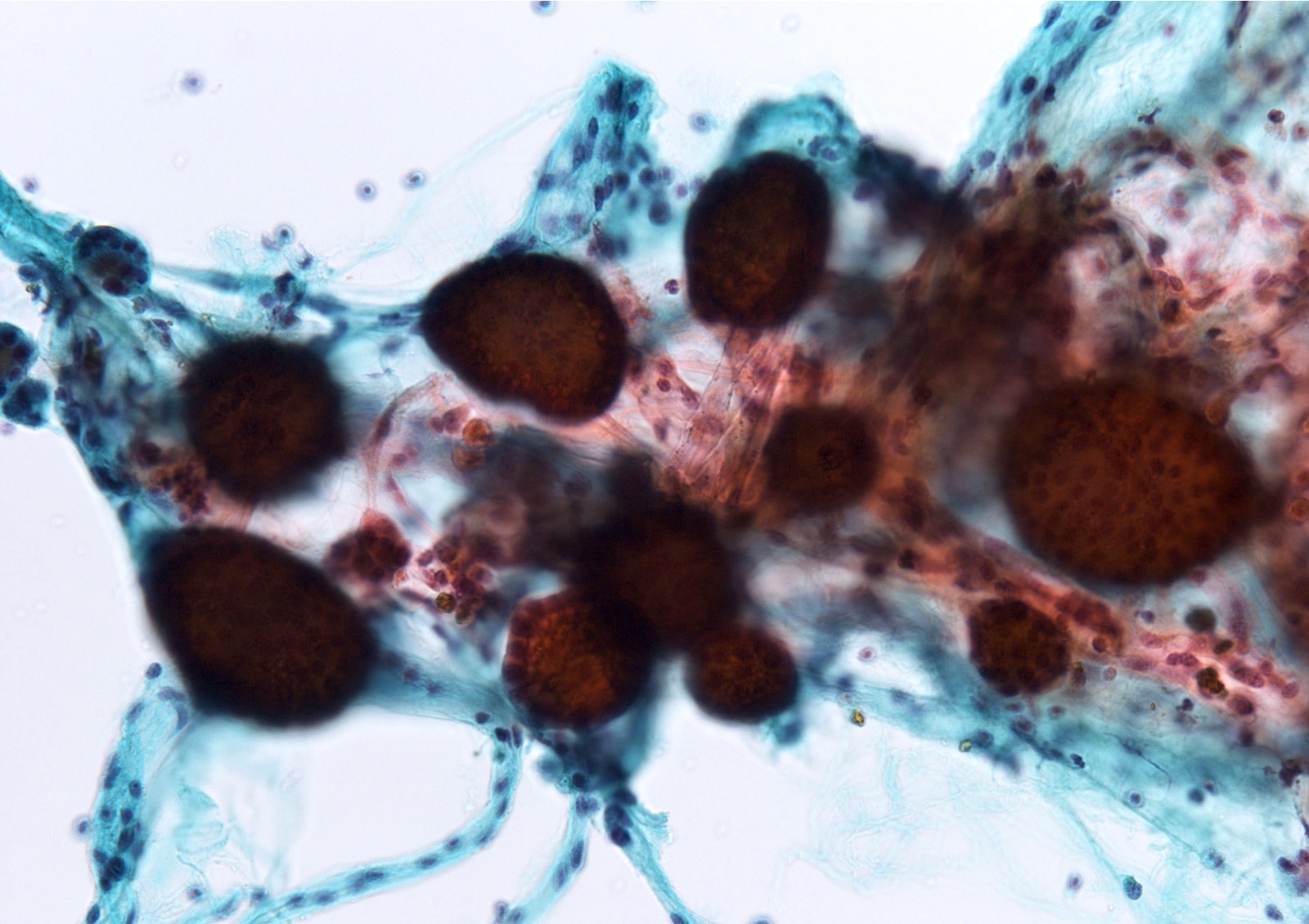
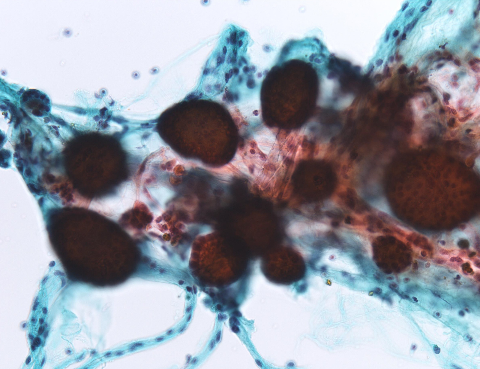
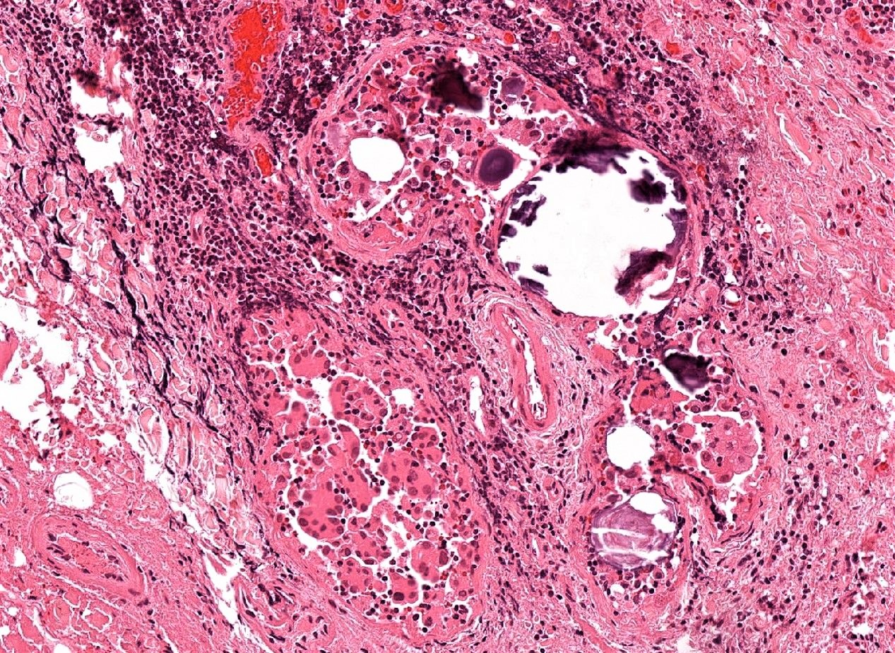
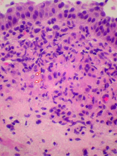
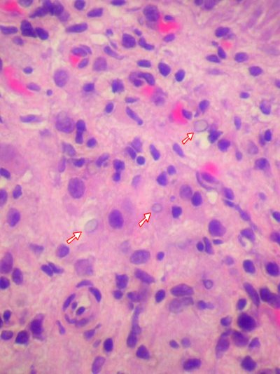
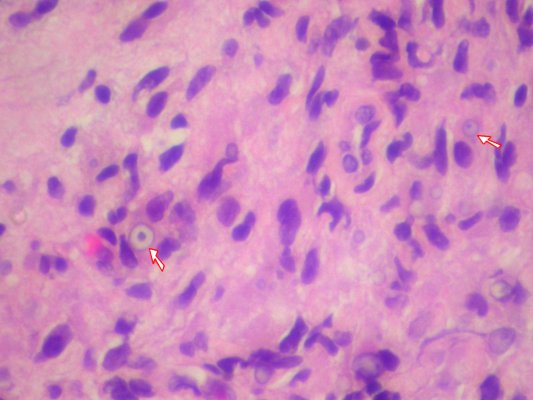
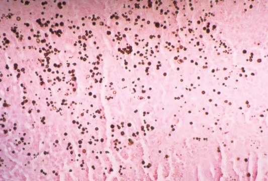
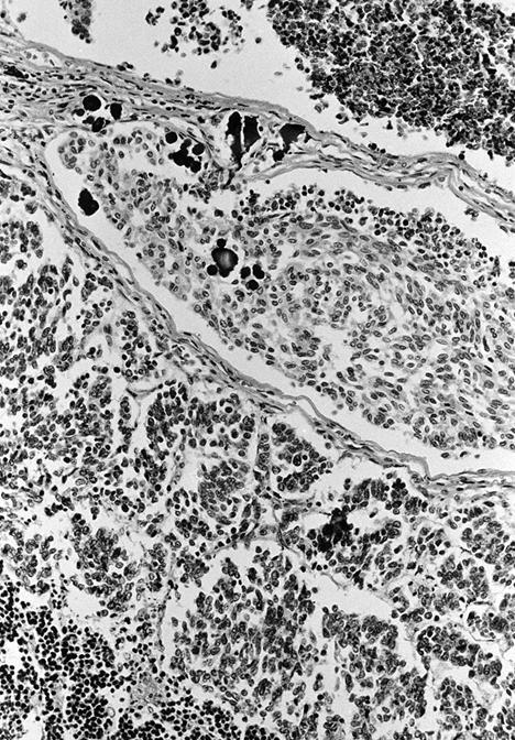
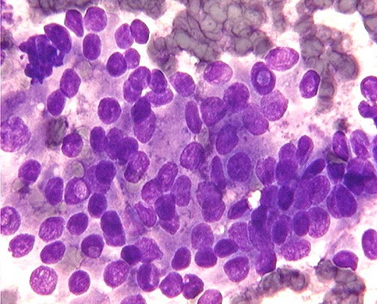
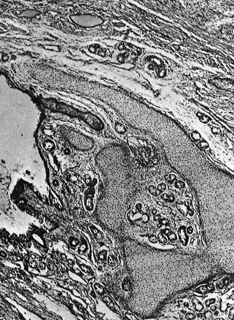
Aspergillus septic foci
Images hosted on other servers:

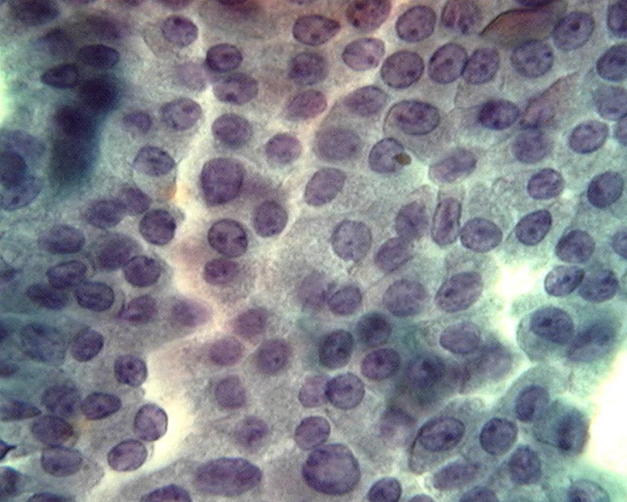
Aspergillus

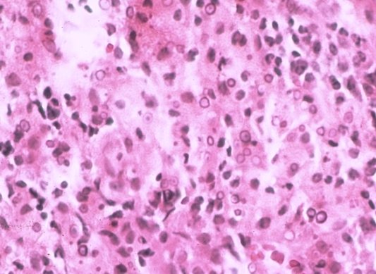
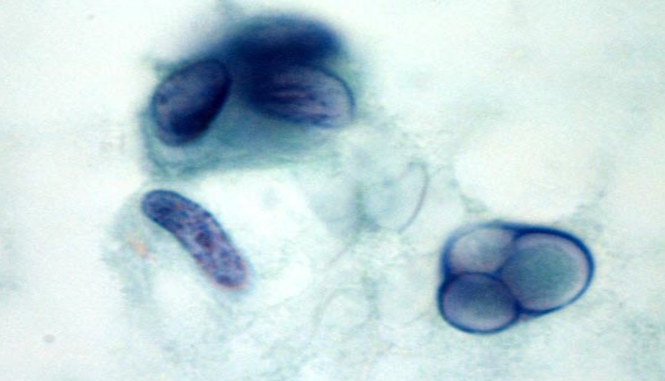
Nocardia
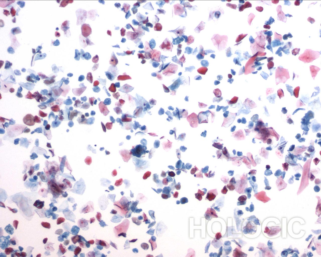
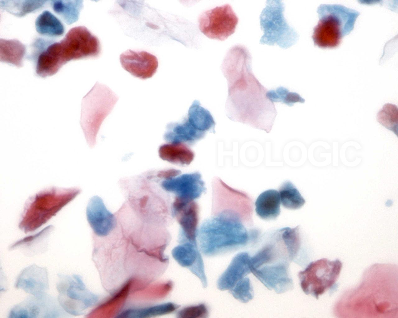
Contributed by Ayana Suzuki, C.T.


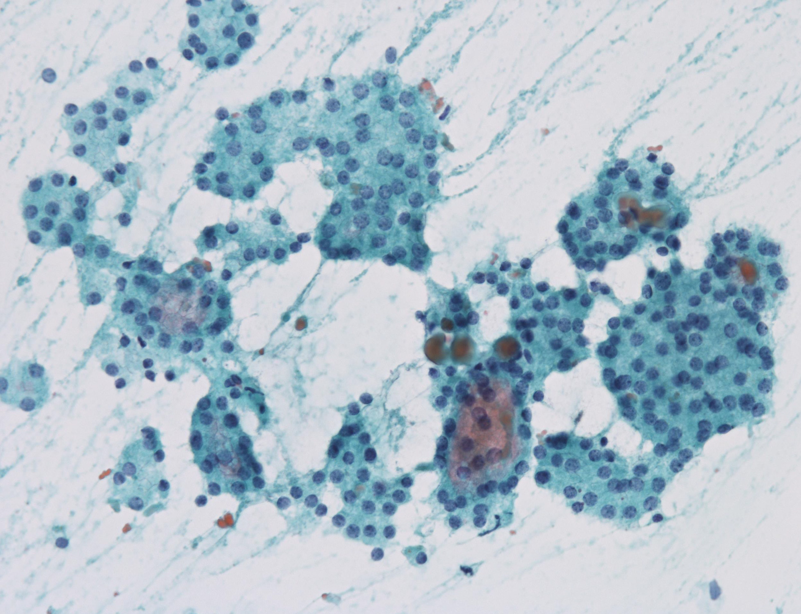
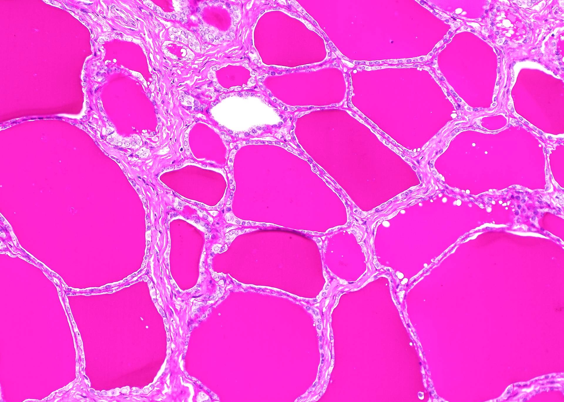
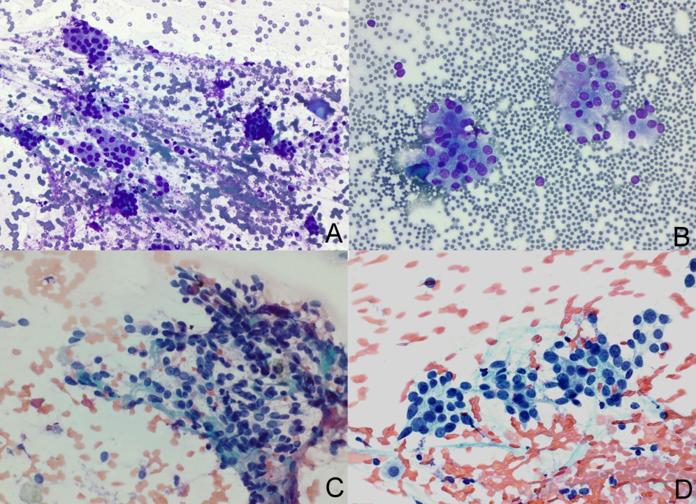
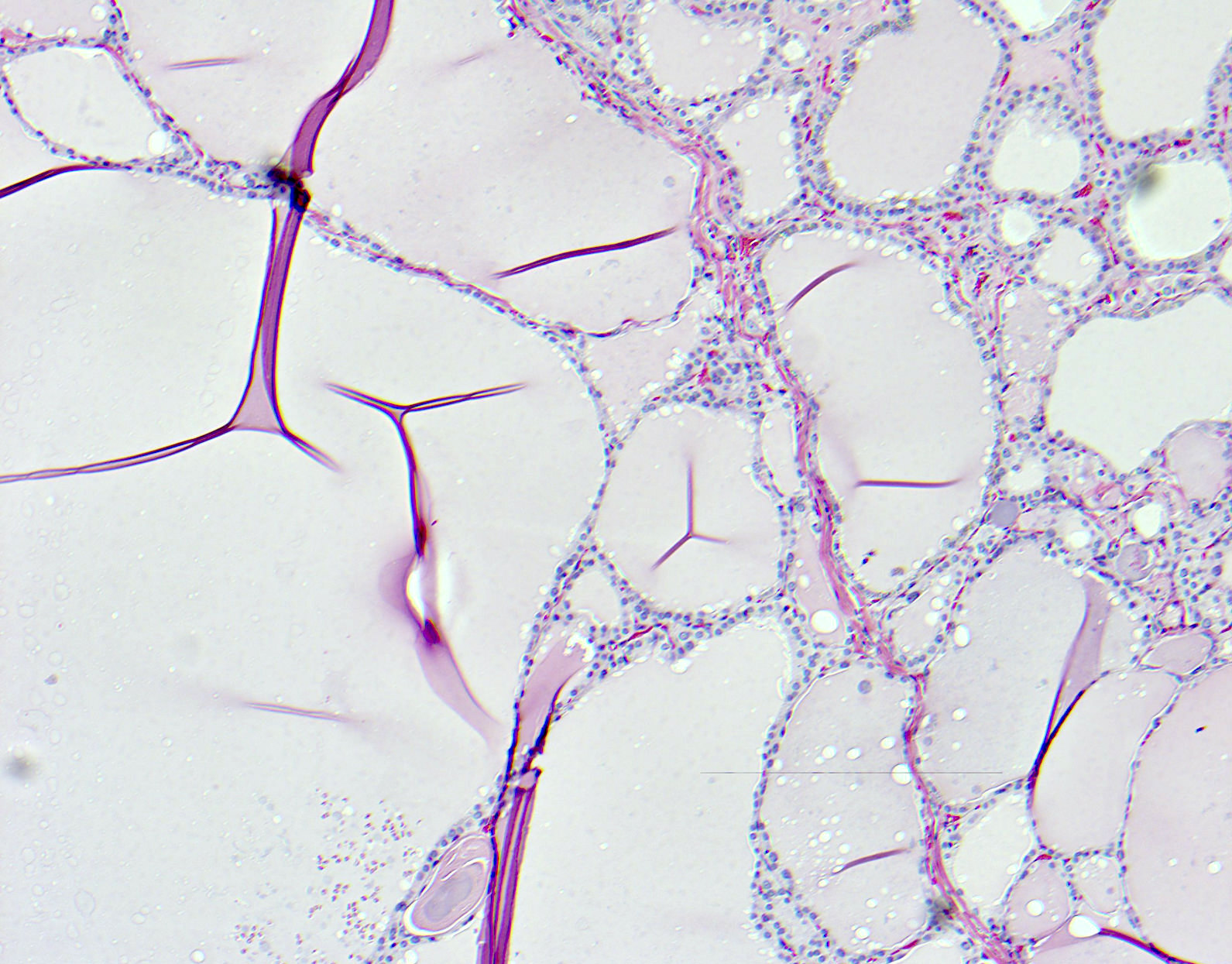
Adequate with macrofollicles
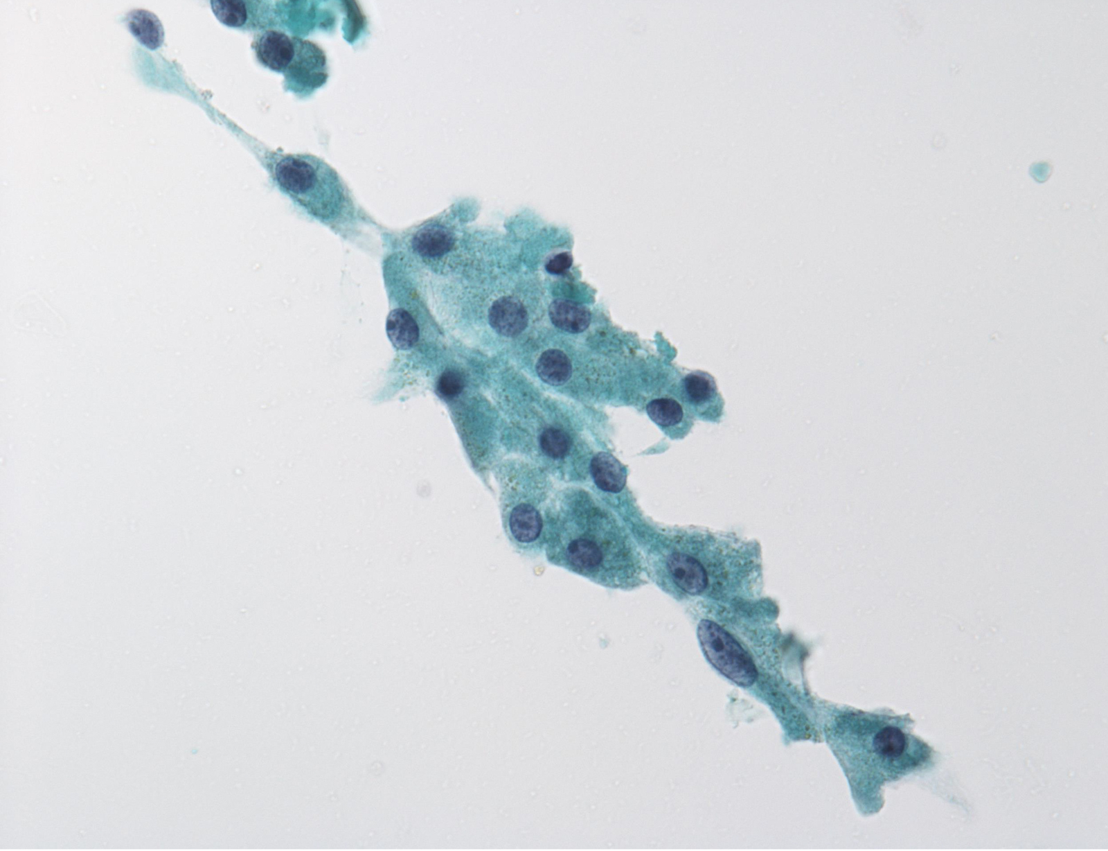
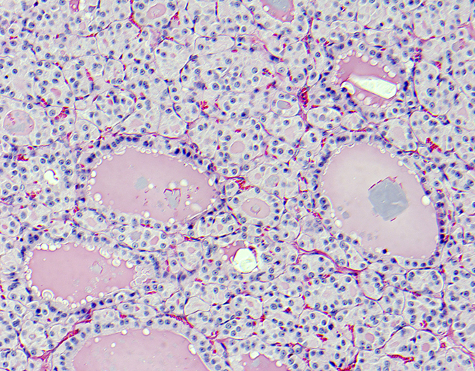
Adequate with thick colloid only
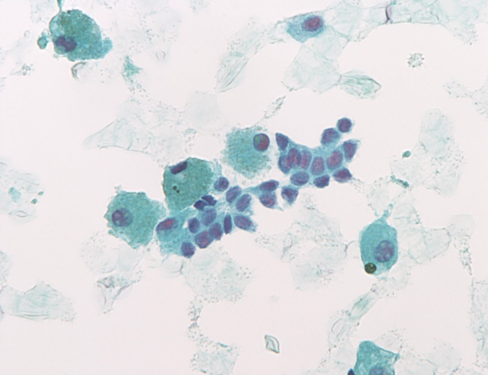
Adequate with atypical cells
Adequate with thyroiditis
Inadequate / blood obscured

Nondiagnostic dried or acellular
Images hosted on other servers:

Inadequate / blood obscured


Nondiagnostic dried or acellular
Thyroid fine needle aspiration and smearing techniques
Essential thyroid cytopathology
Scroll to see all images:
Contributed by Andrey Bychkov, M.D., Ph.D.
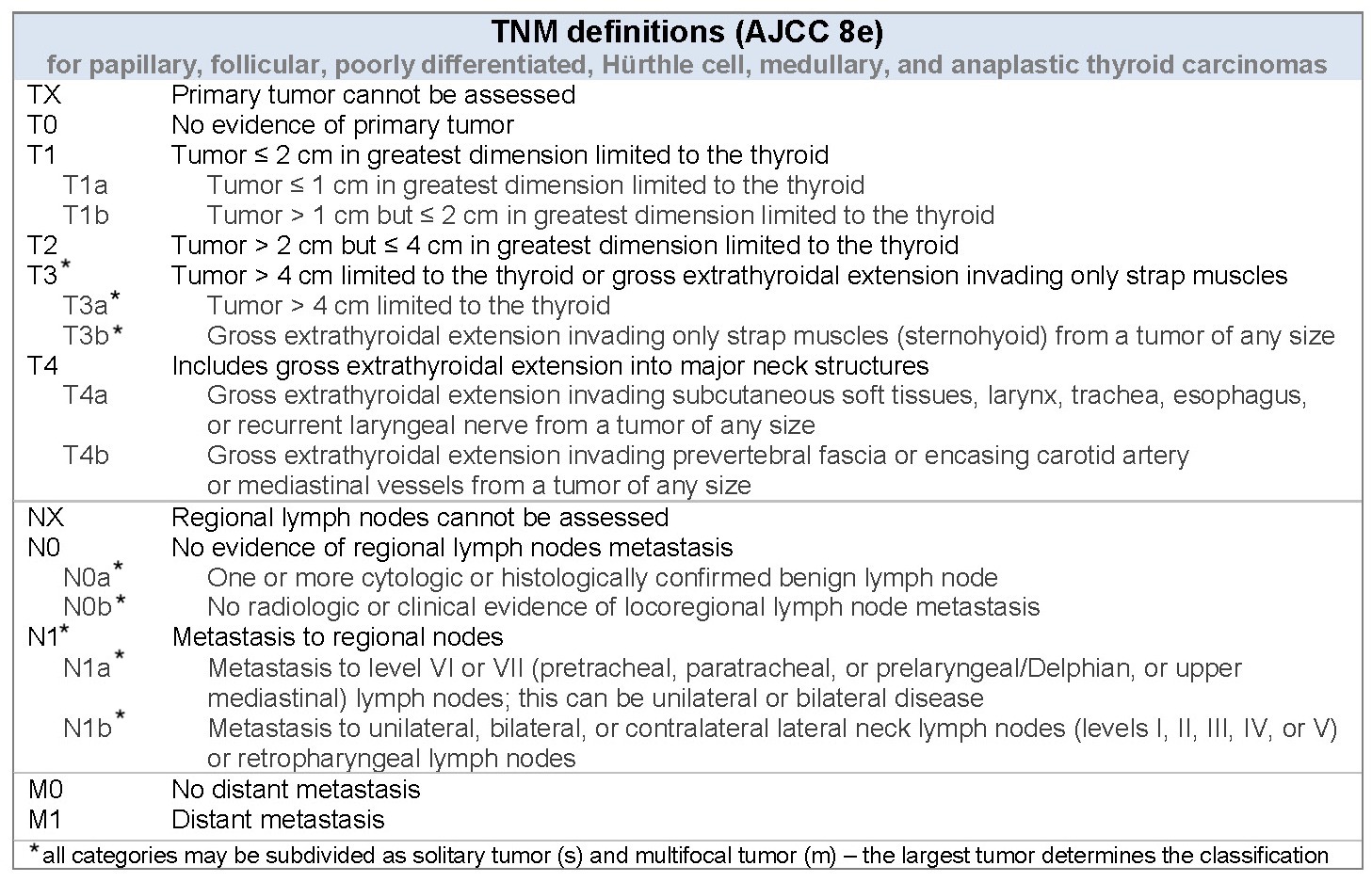

AJCC / TNM charts
Images hosted on other servers:
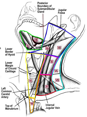

Levels of the cervical lymph nodes
TNM sketches
T1N0
T2N0
T3N0 and T1N1
T3N1
T1M1
T2M1
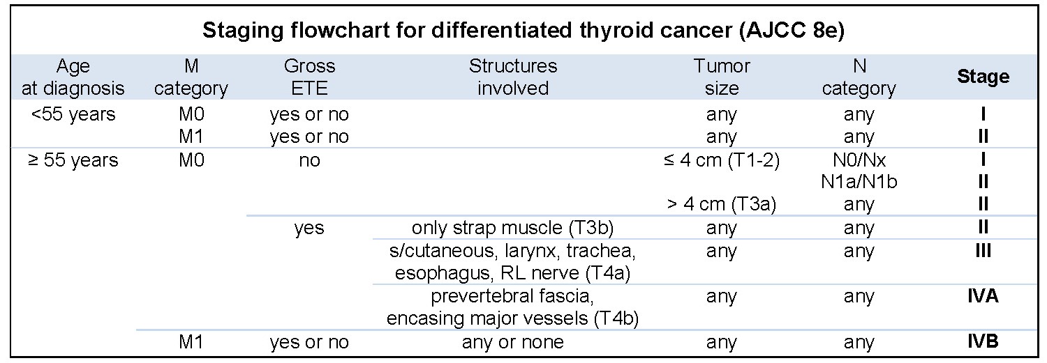
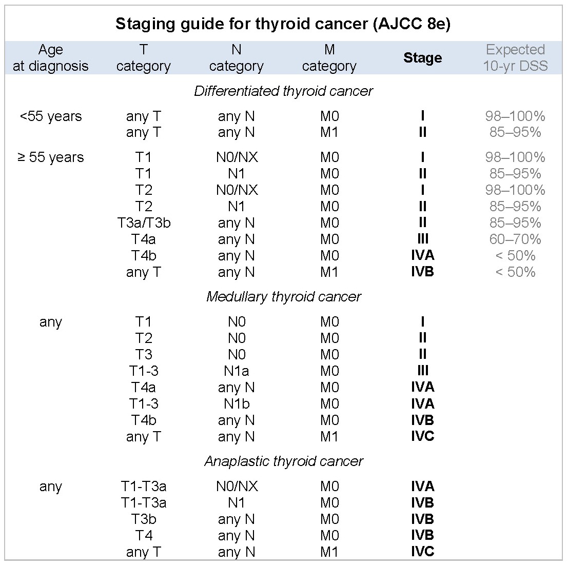
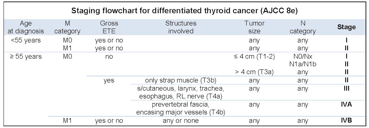
Staging charts

Stage I, < 55 yo

Stage I, 55 yo

Stage II, < 55 yo


Stage II, > 55 yo

Stage III

Stage IVA

Stage IVB

Anaplastic cancer, stage IVA


Anaplastic cancer, stage IVB

Anaplastic cancer, stage IVC

Medullary cancer, stage I

Medullary cancer, stage II

Medullary cancer, stage III

Medullary cancer, stage IVA

Medullary cancer, stage IVB

Medullary cancer, stage IVC
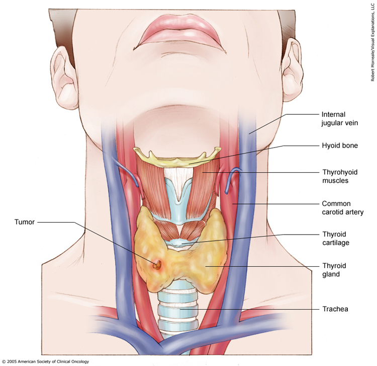
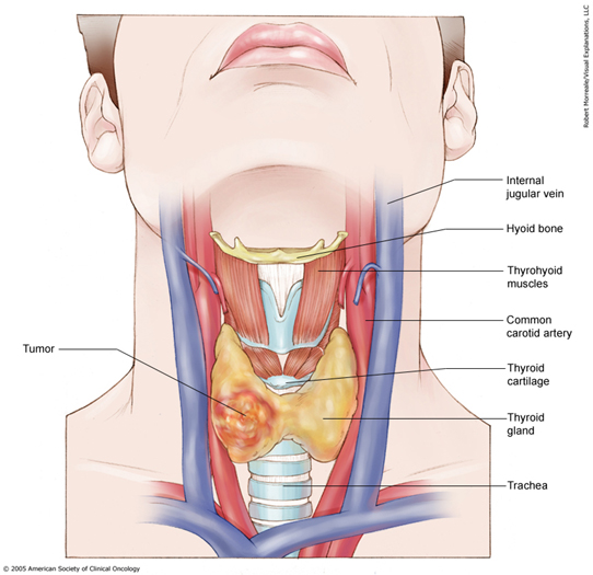
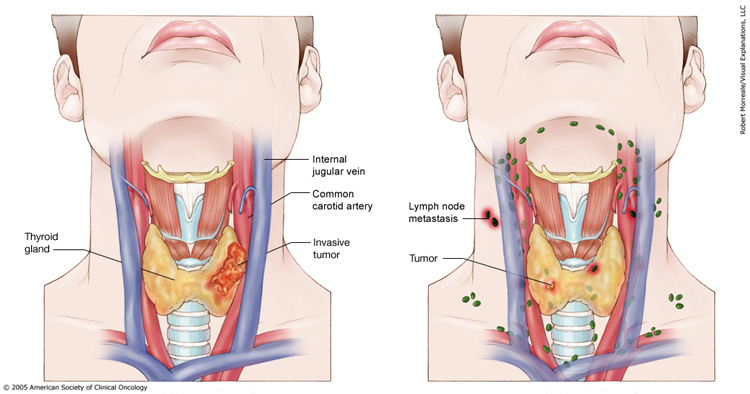
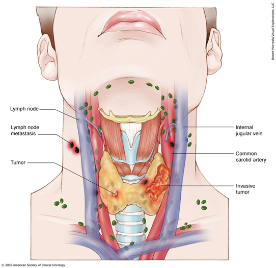
Advanced thyroid carcinoma
Thyroid carcinoma
Staging thyroid cancer
Staging of medullary thyroid cancer
Staging animation
Contributed by Mark R. Wick, M.D.

Amiodarone thyroiditis
Images hosted on other servers:

Amiodarone induced thyrotoxicosis
AFIP images
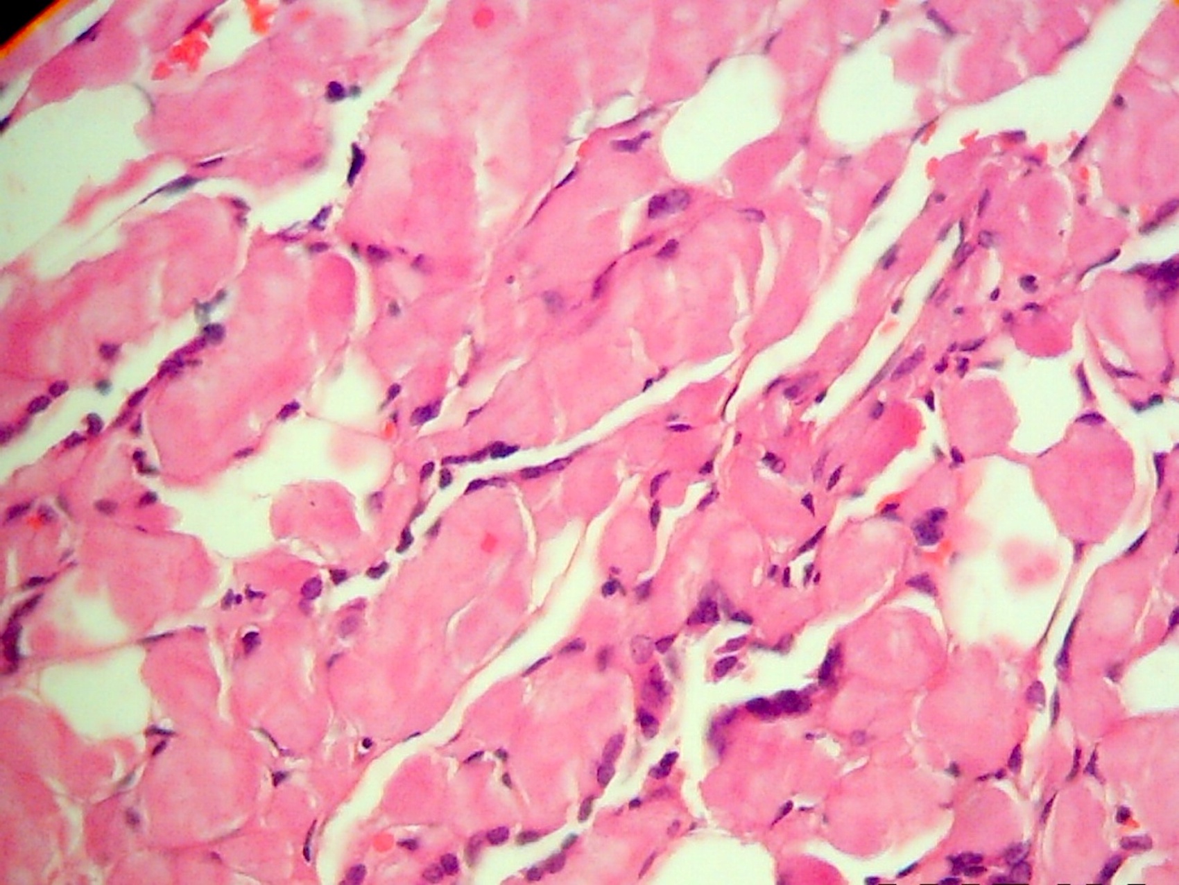
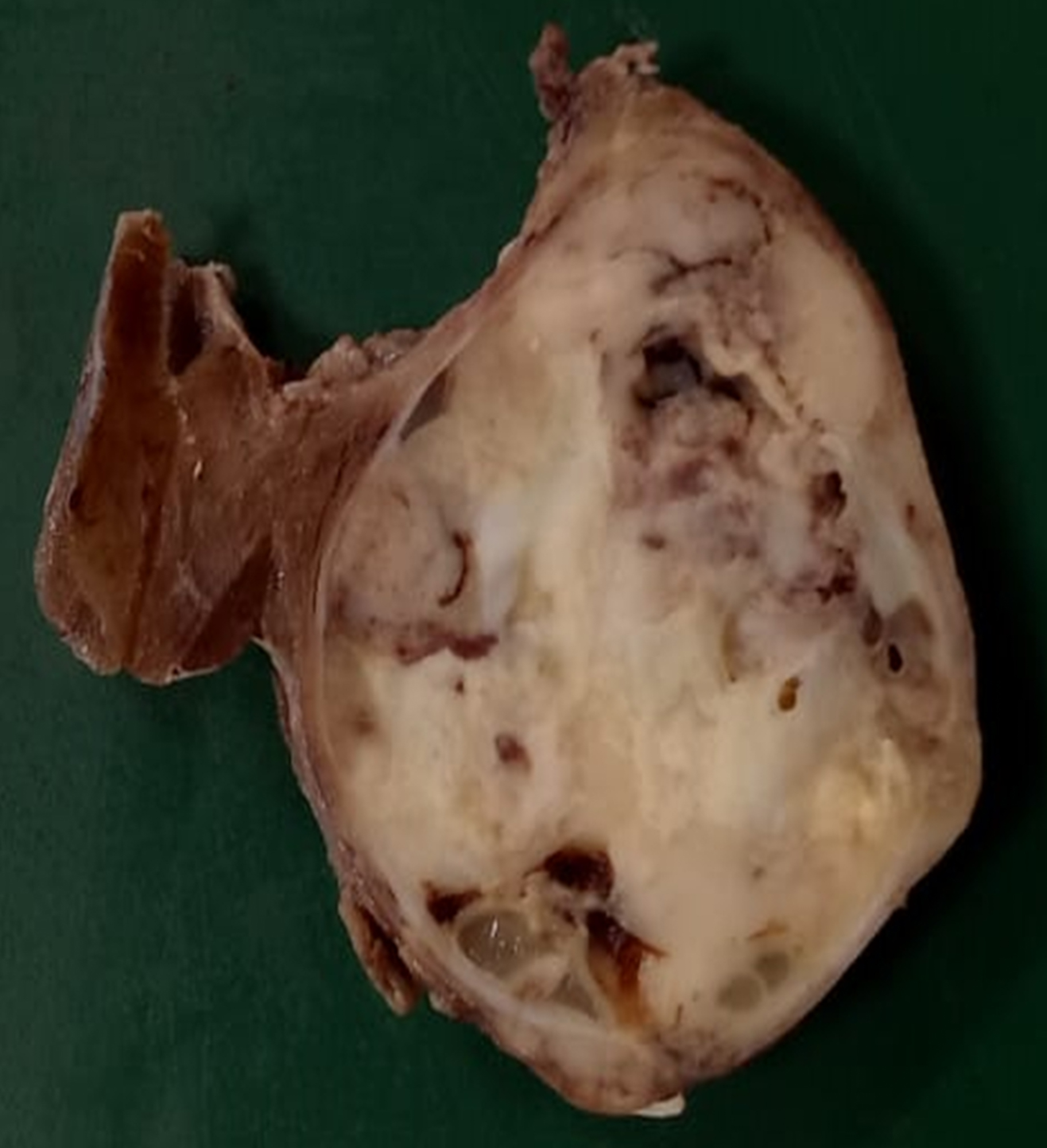
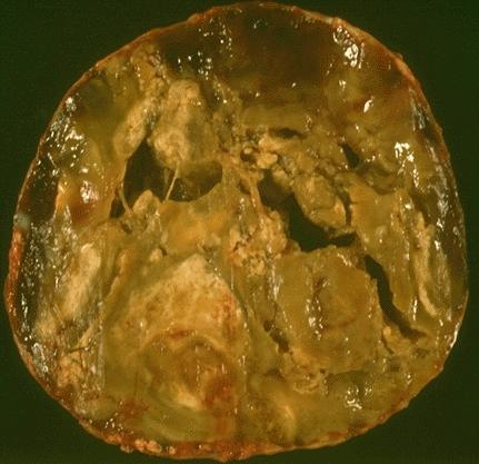
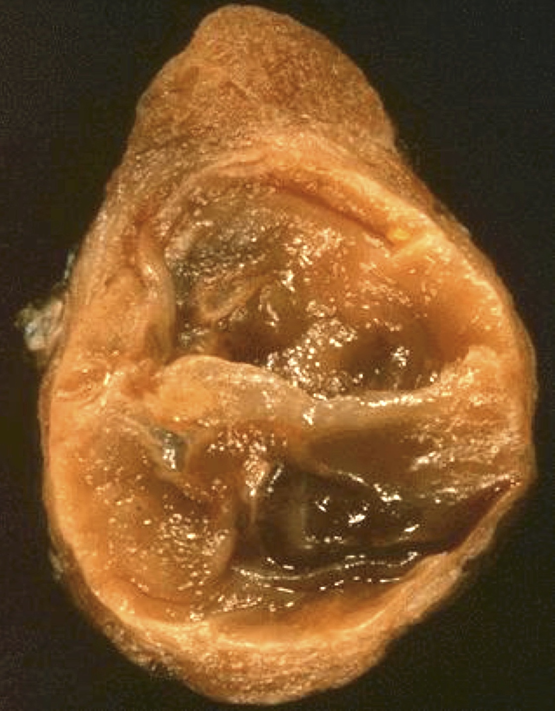
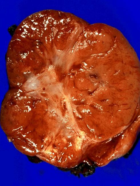
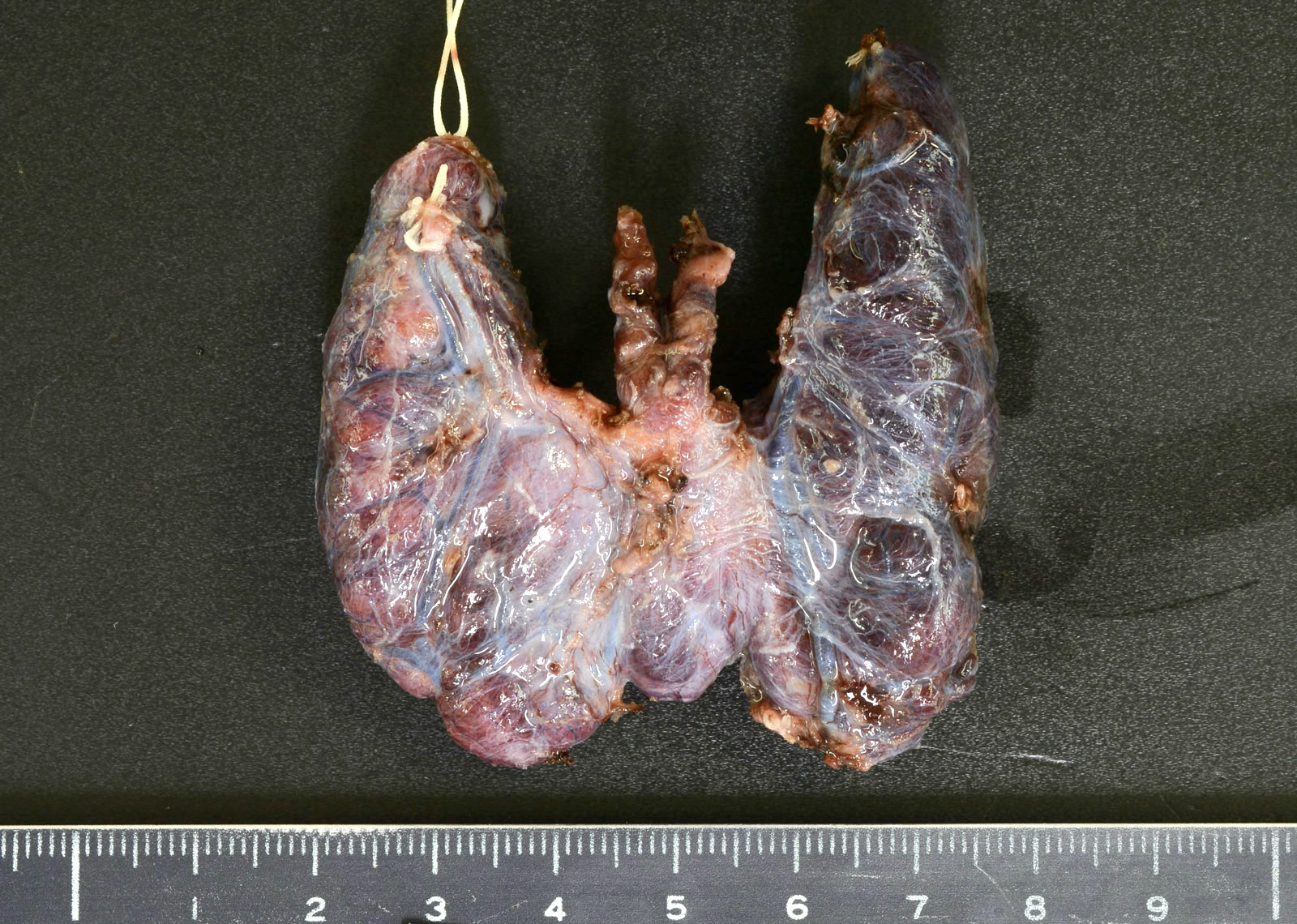
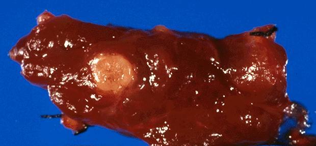
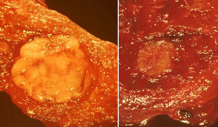
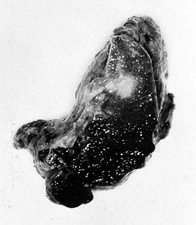
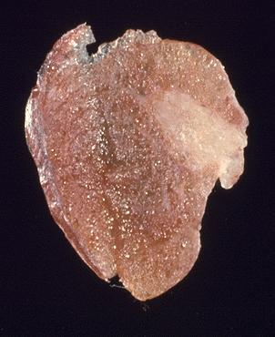
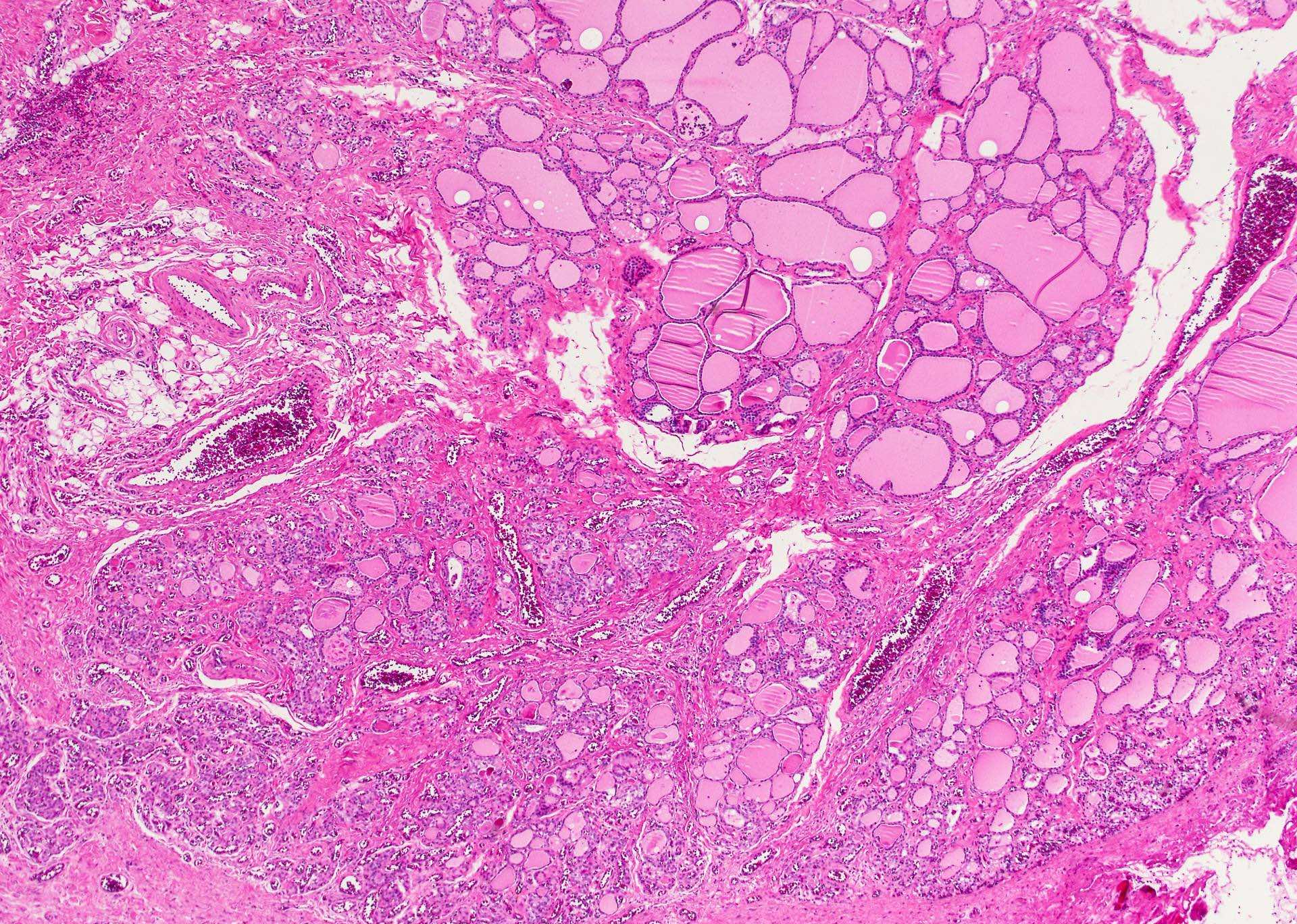
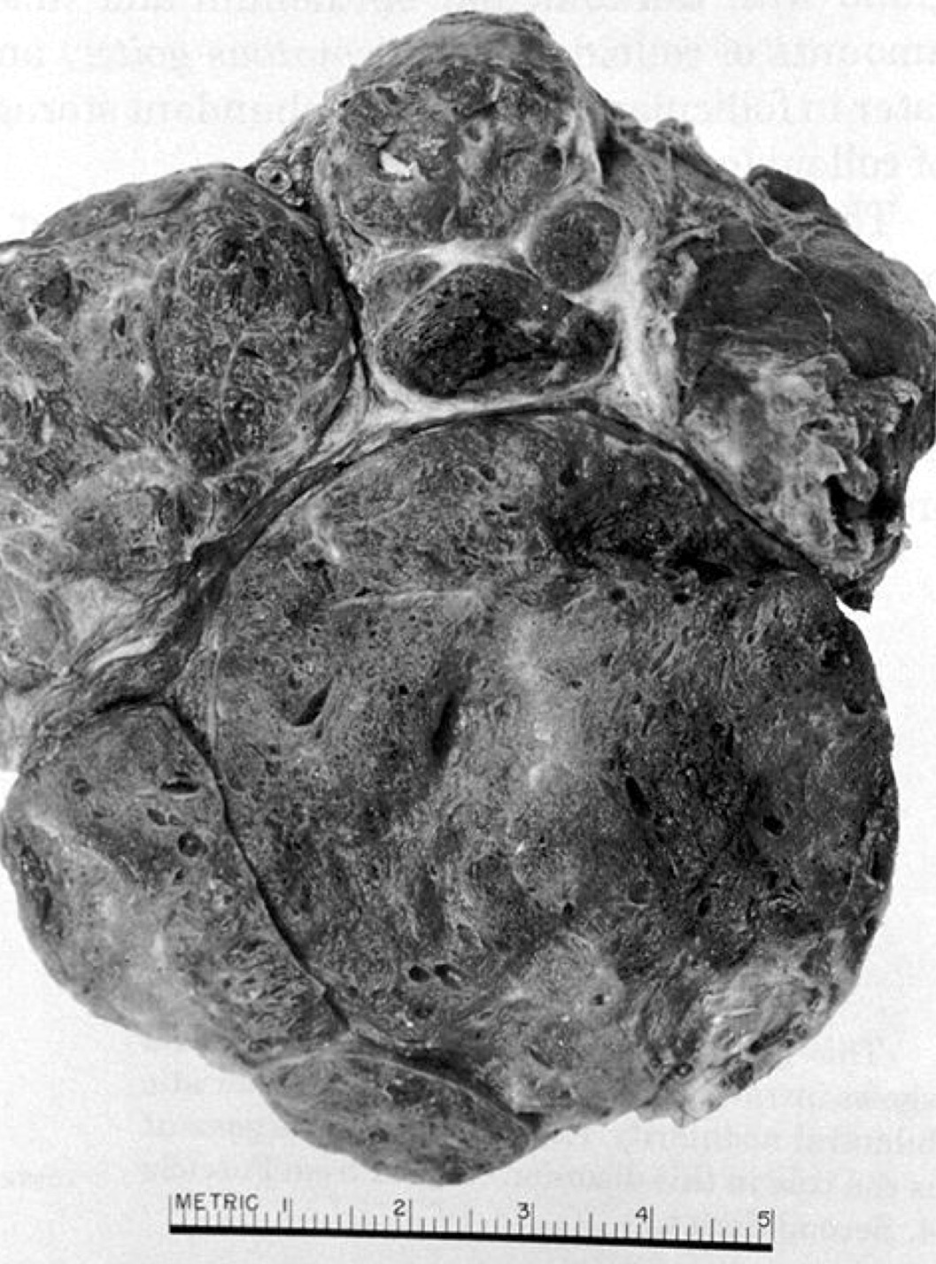
Enlarged and bosselated with salmon cut surface
Images hosted on other servers:

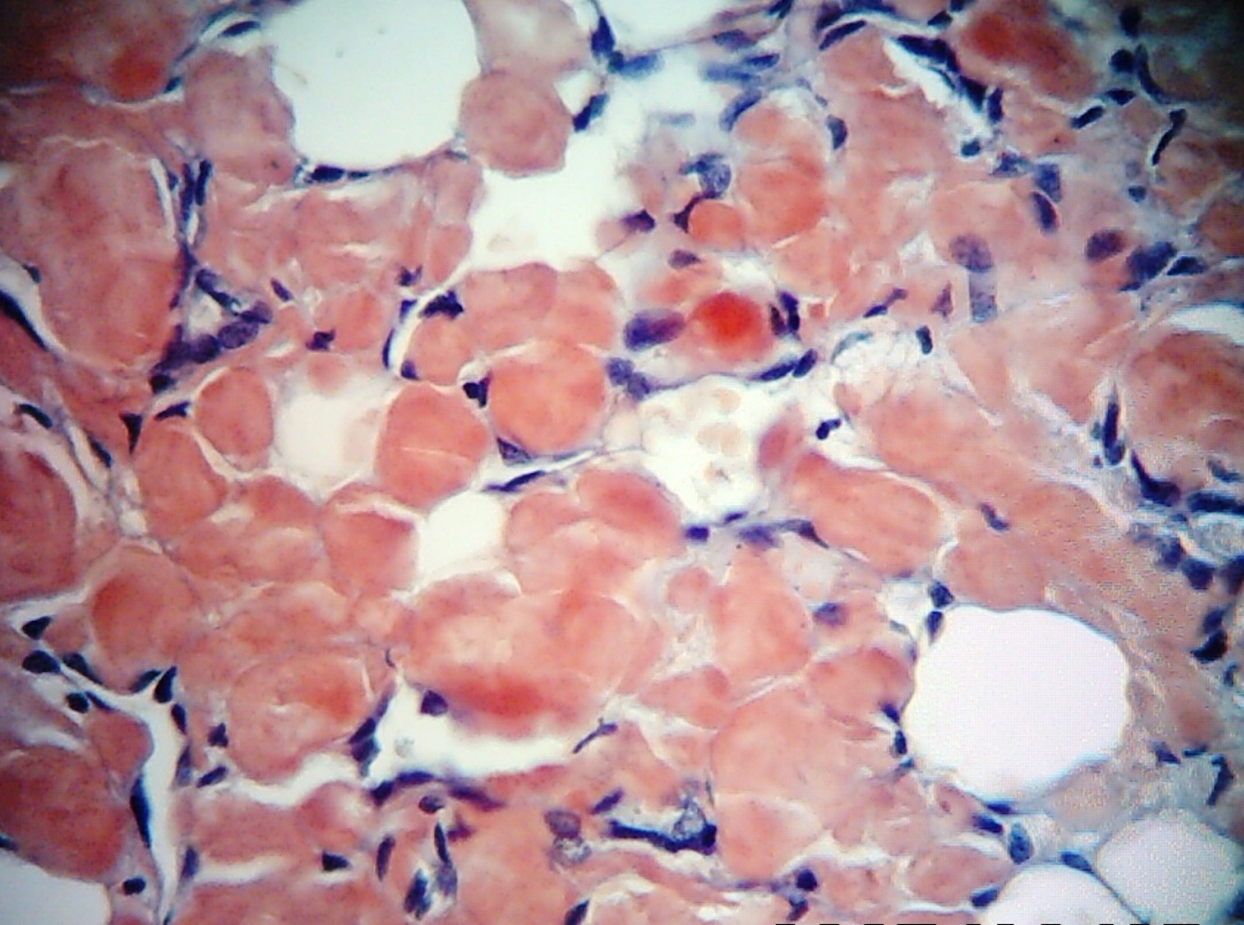
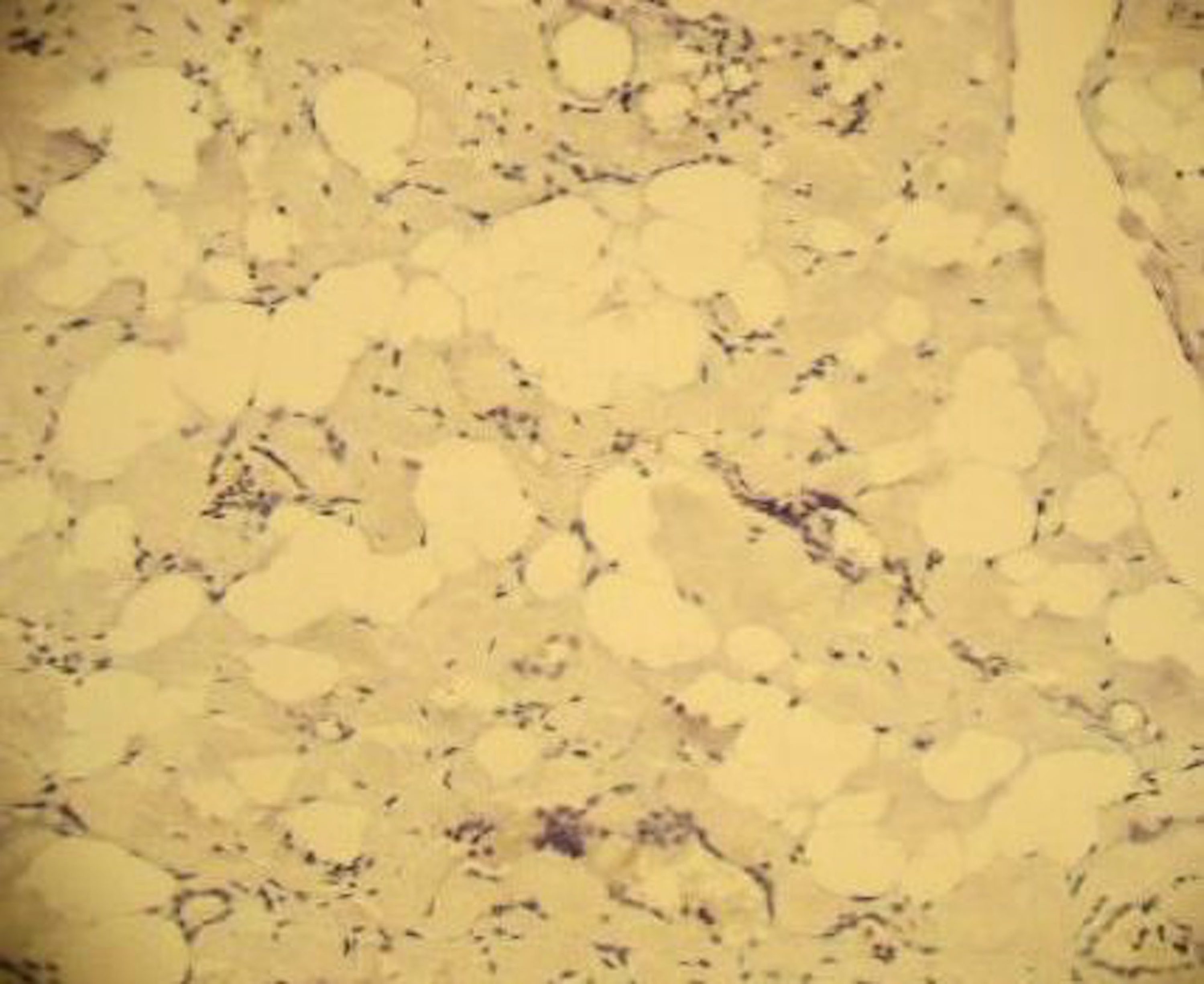
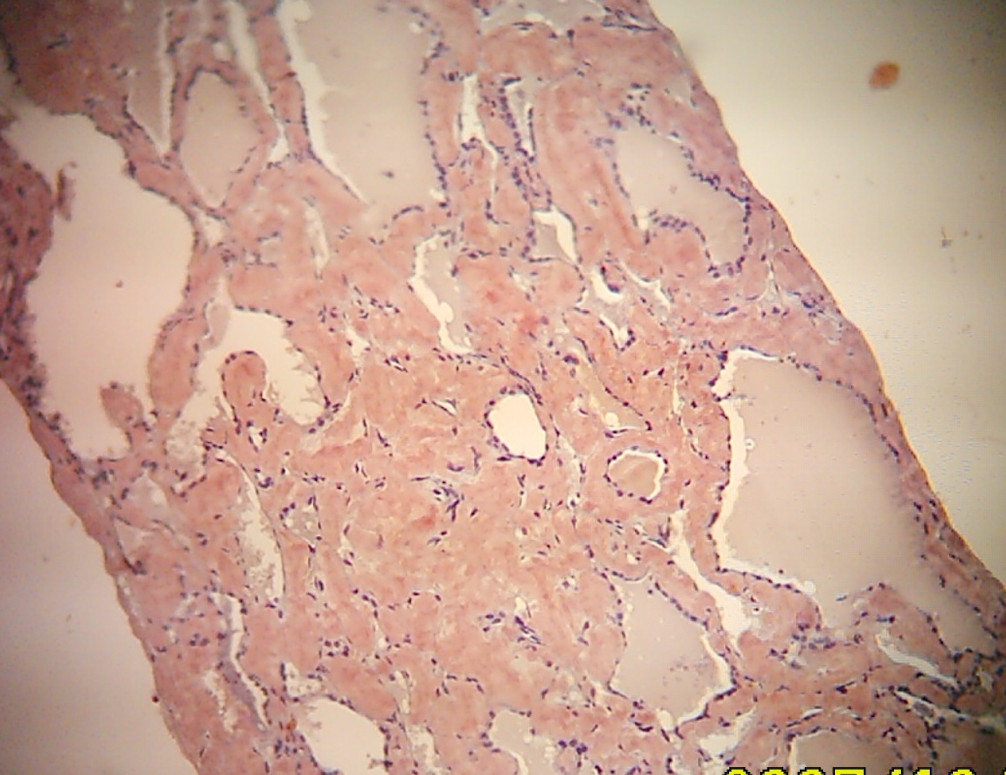
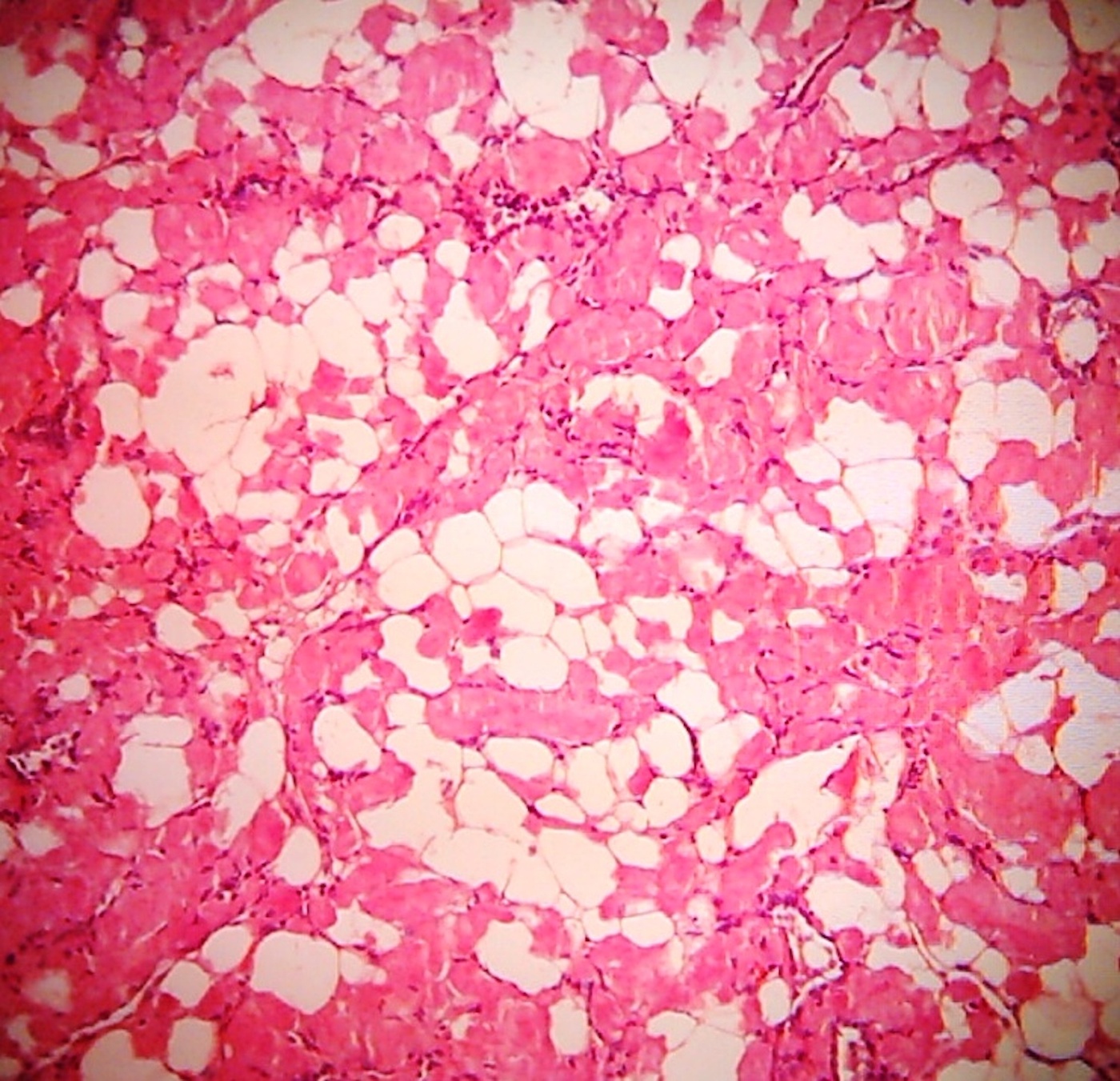
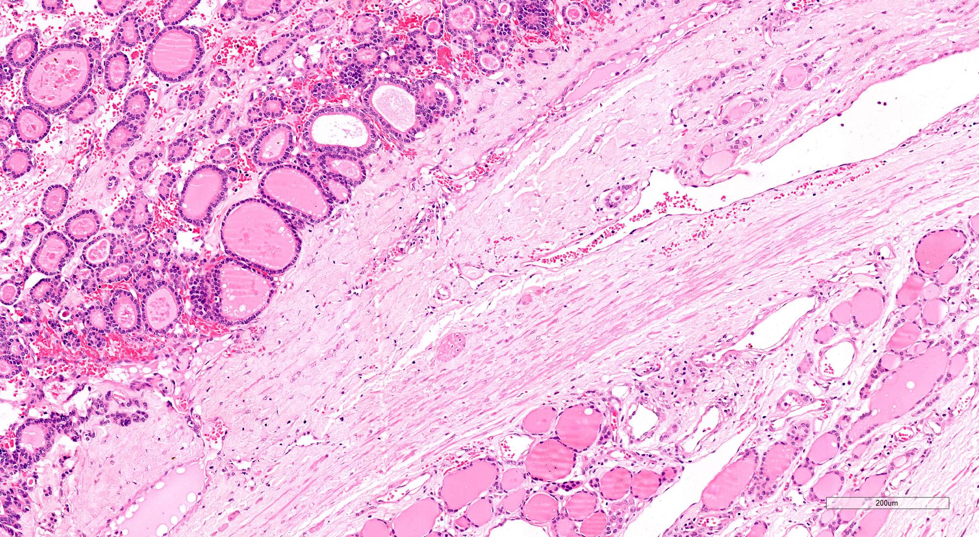
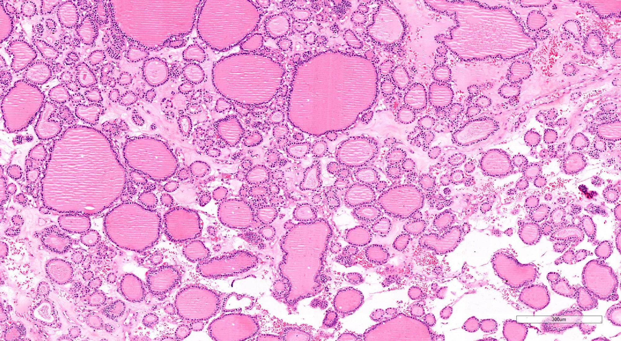
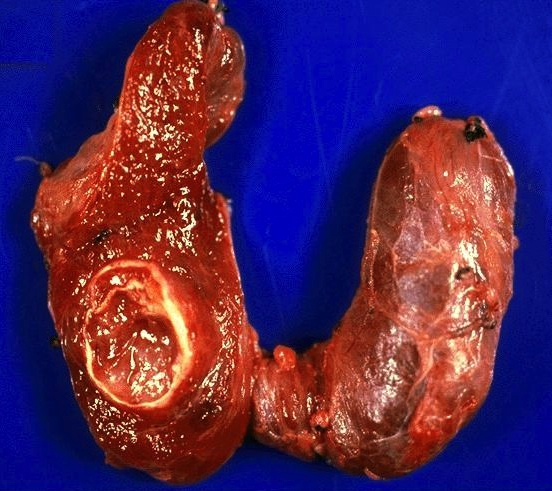
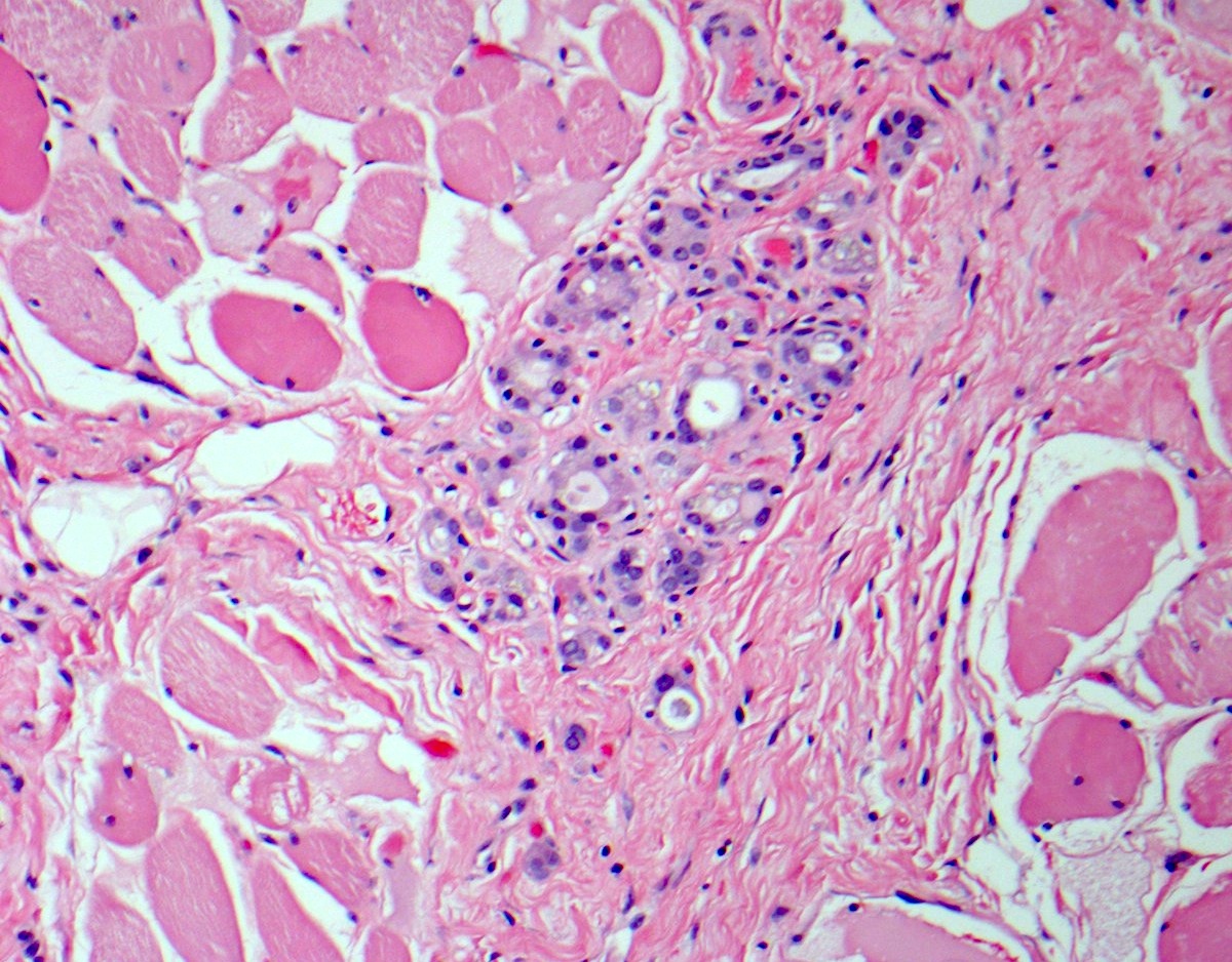
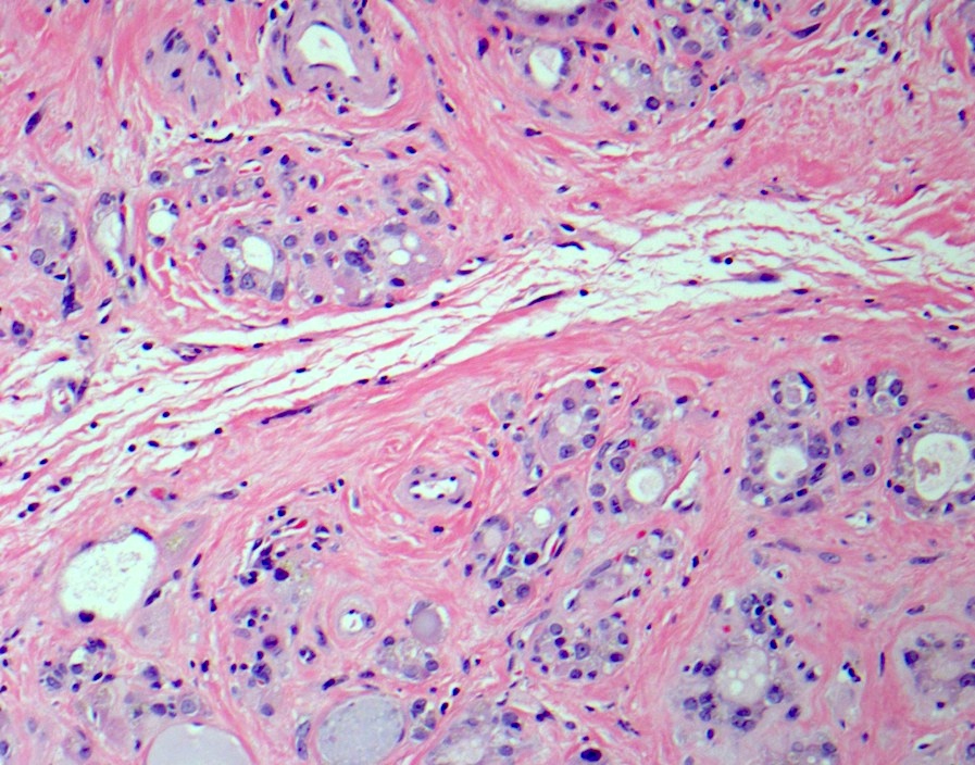
Lipomatosis and amyloidosis
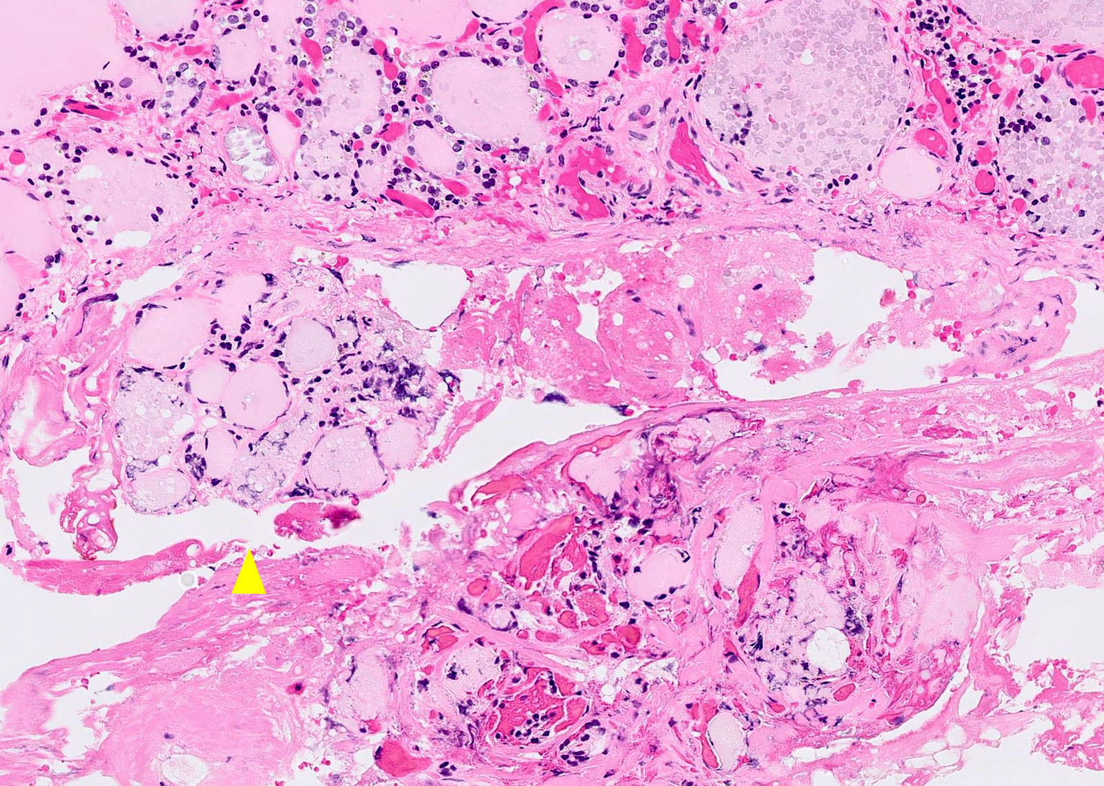
Contributed by Nazar M. T. Jawhar, M.D.
Various images
Case #247 and AFIP images

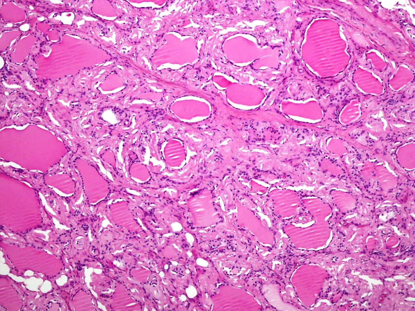
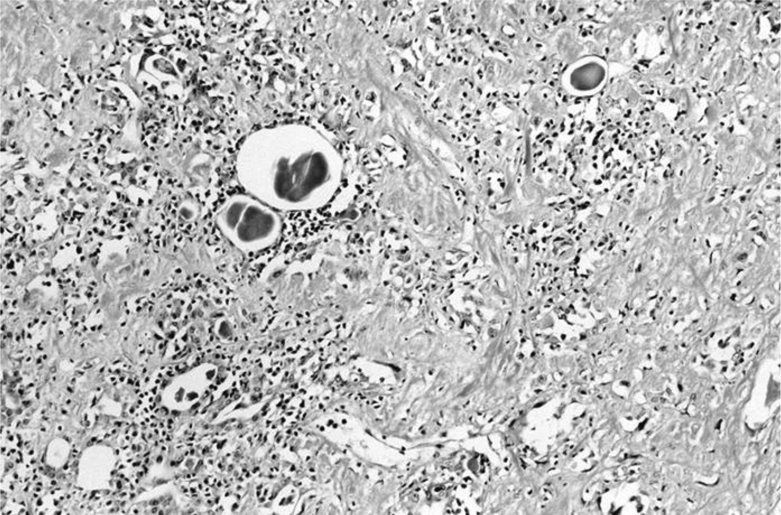
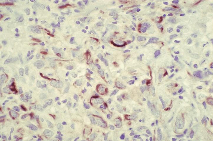
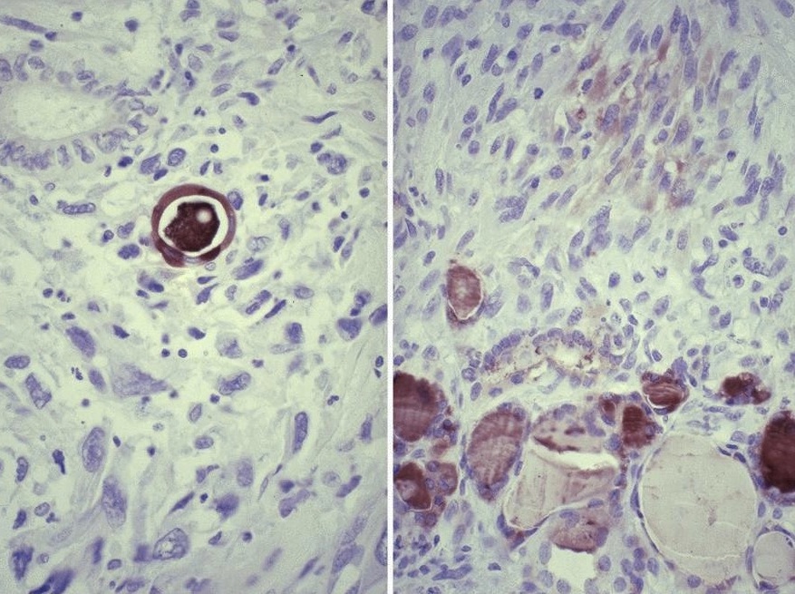
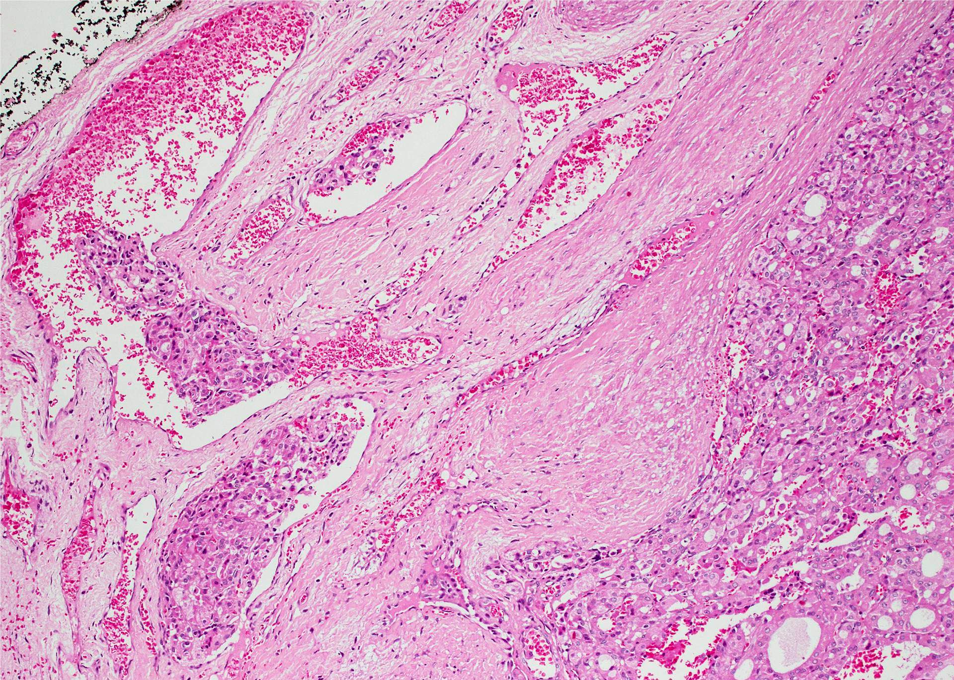
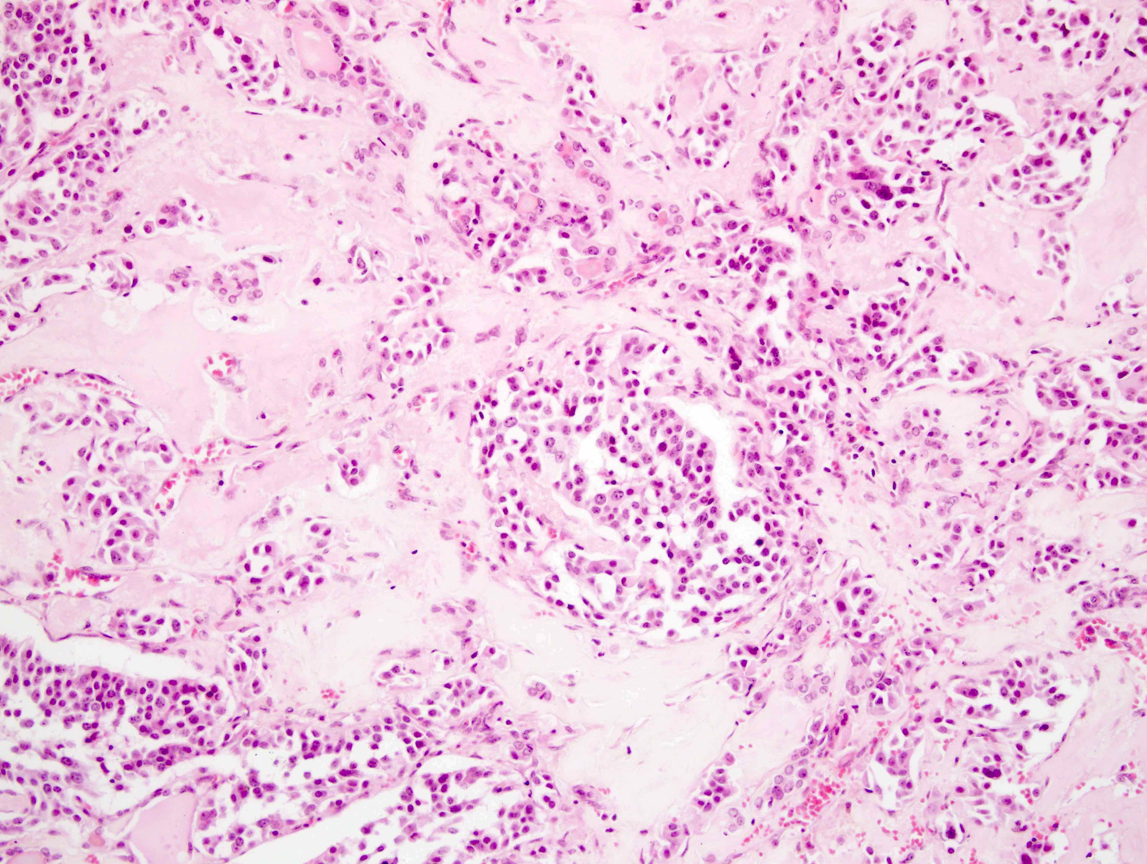
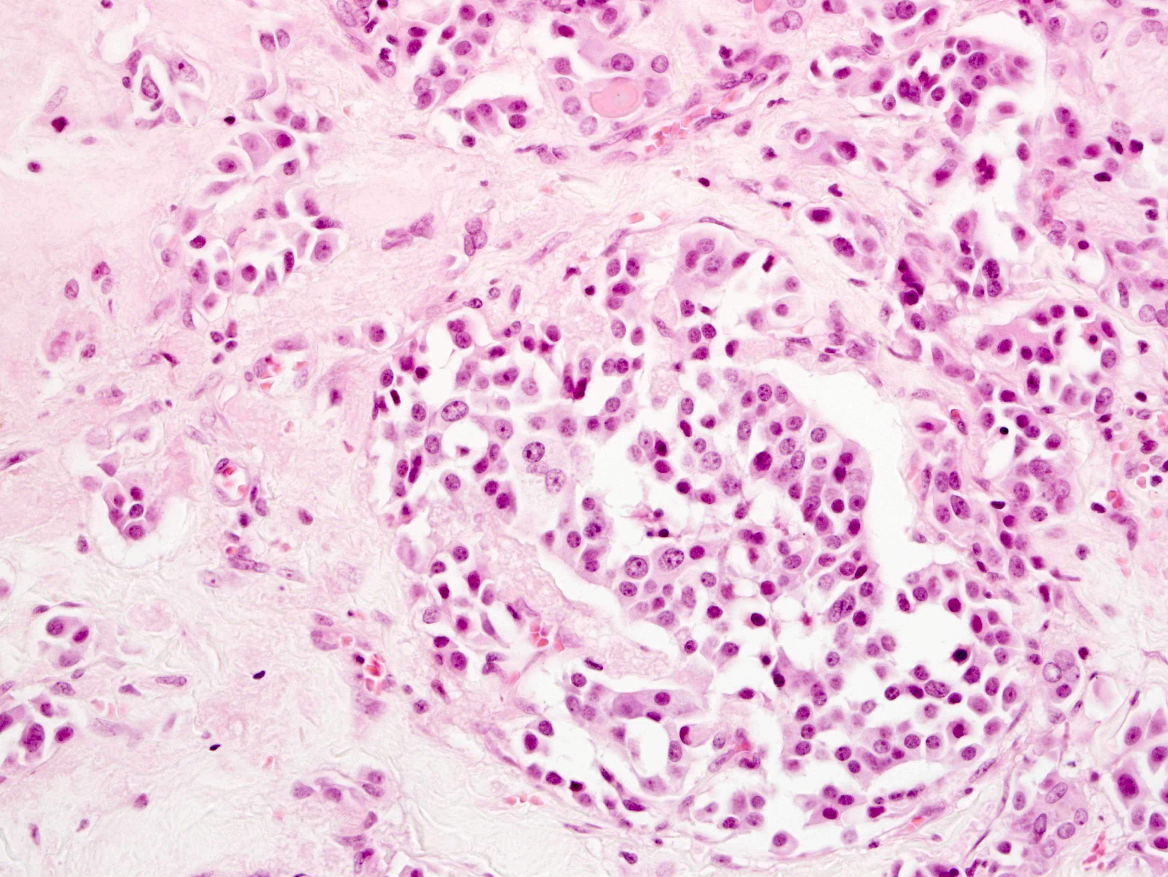
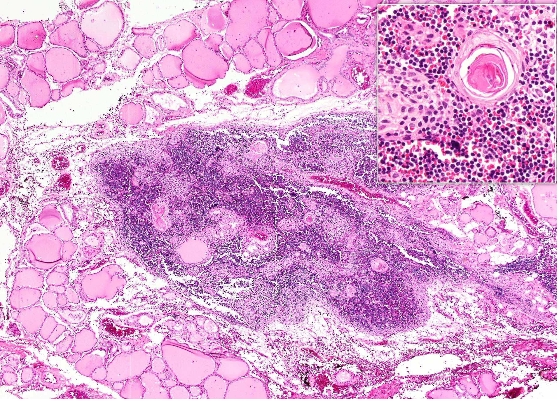
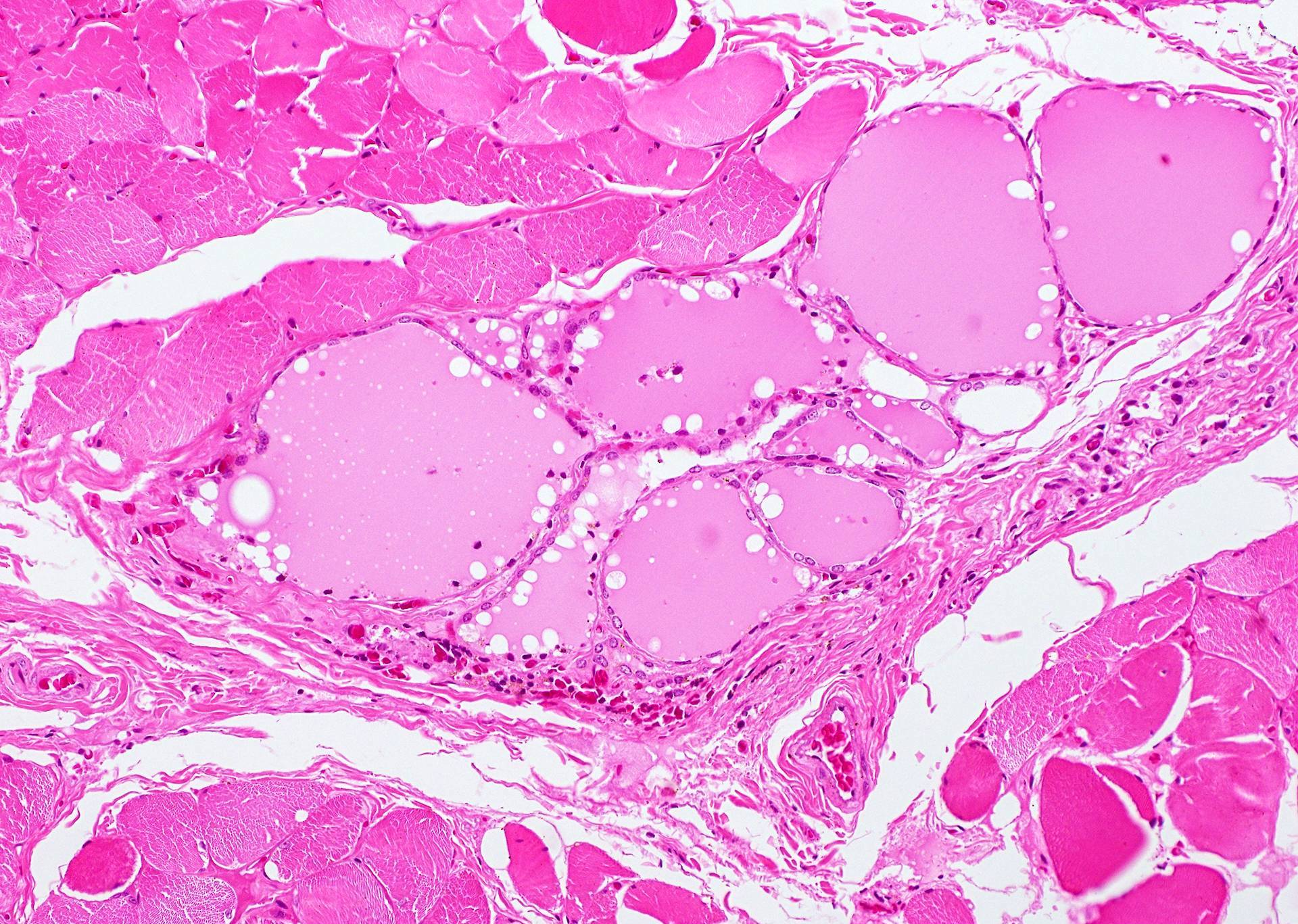
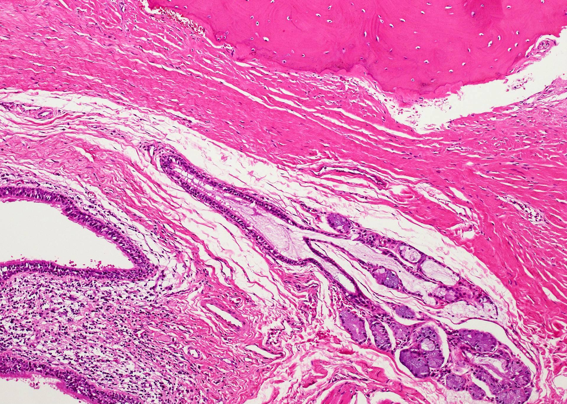
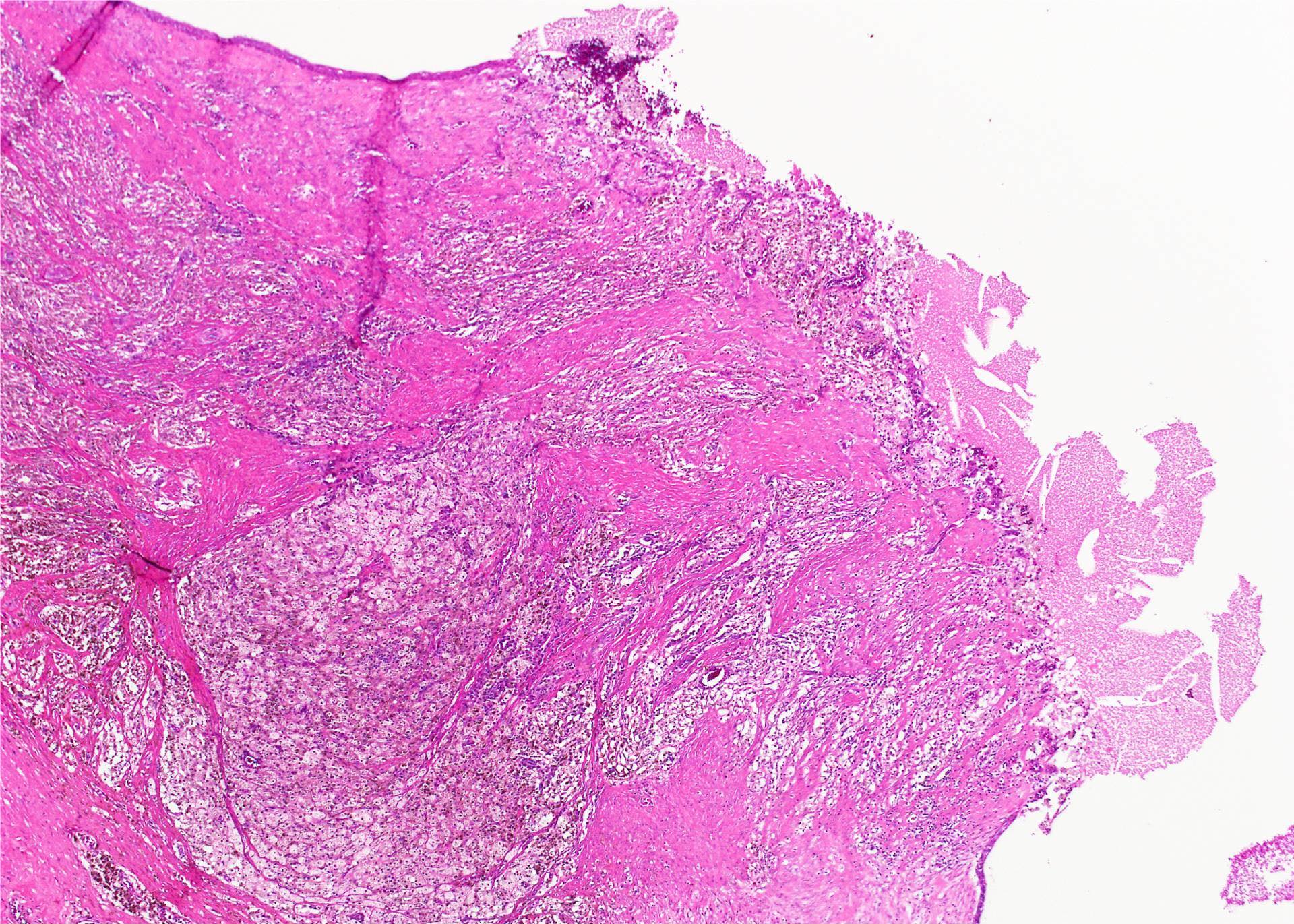
Amyloid and inflammatory cells
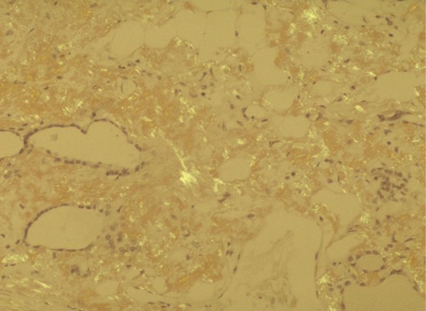
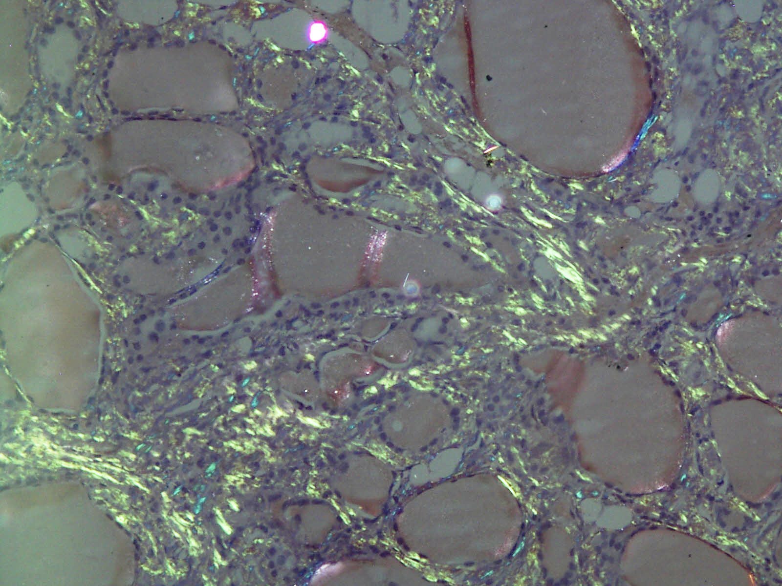
Apple green birefringence
Images hosted on other servers:

Birefringent apple green staining under polarized light
Images hosted on other servers:

Molecular landscape

Treatment of stage IVA - B

Treatment of stage IVC
Images hosted on other servers:

Neck CT, FDG PET scan, sonography


Neck CT

Neck CT

CT for staging

FDG PET before / after therapy
Images hosted on other servers:

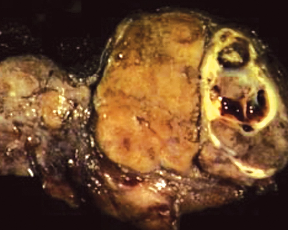
Fungating neck mass
Contributed by Shipra Agarwal, M.D., Mark R. Wick, M.D. and AFIP
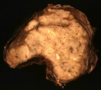
Variegated, fleshy
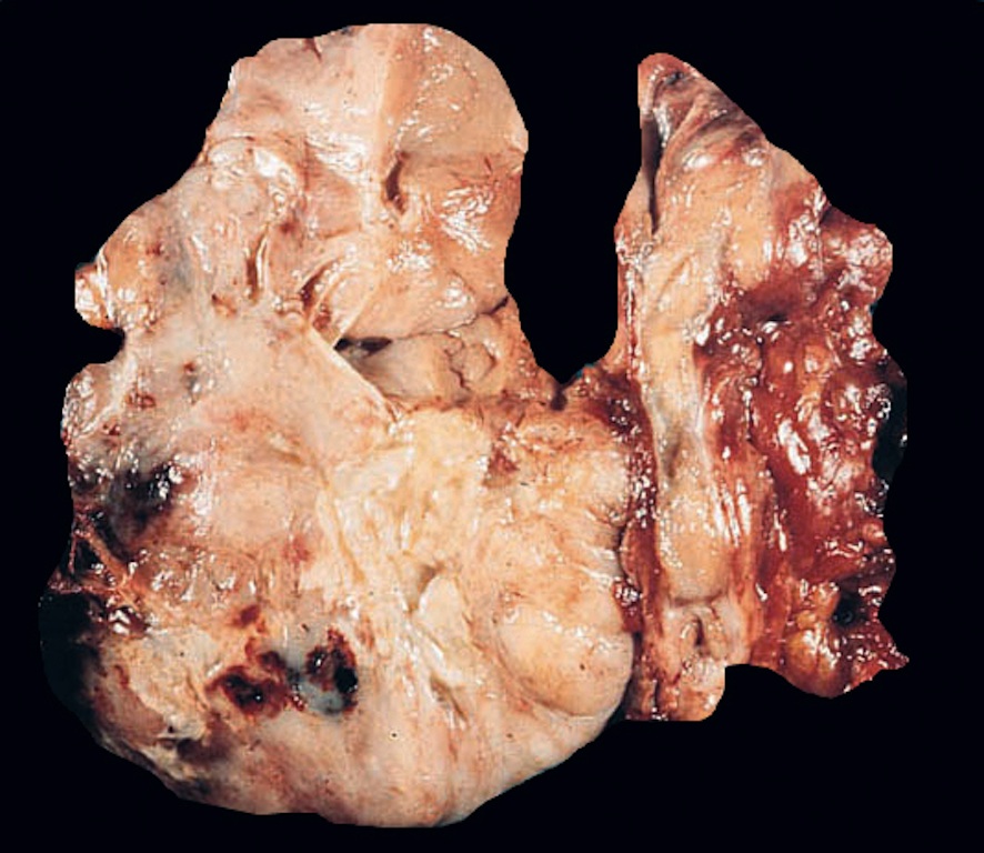
Large, tan, fleshy mass with hemorrhage
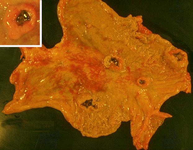
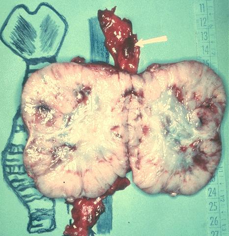
Metastases to stomach with ulcerated center
Images hosted on other servers:

Diffusely infiltrative, fleshy

Gray-white, fleshy
Contributed by Shipra Agarwal, M.D., Shuanzeng Wei, M.D., Ph.D., Stephen J. Schultenover, M.D. and AFIP
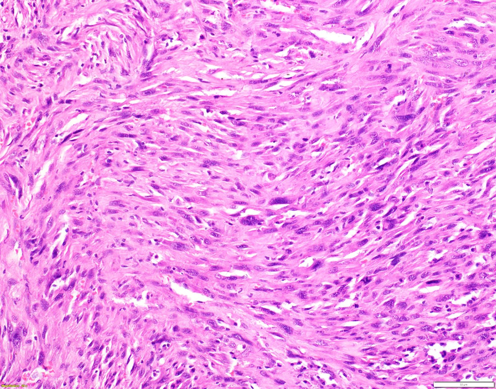
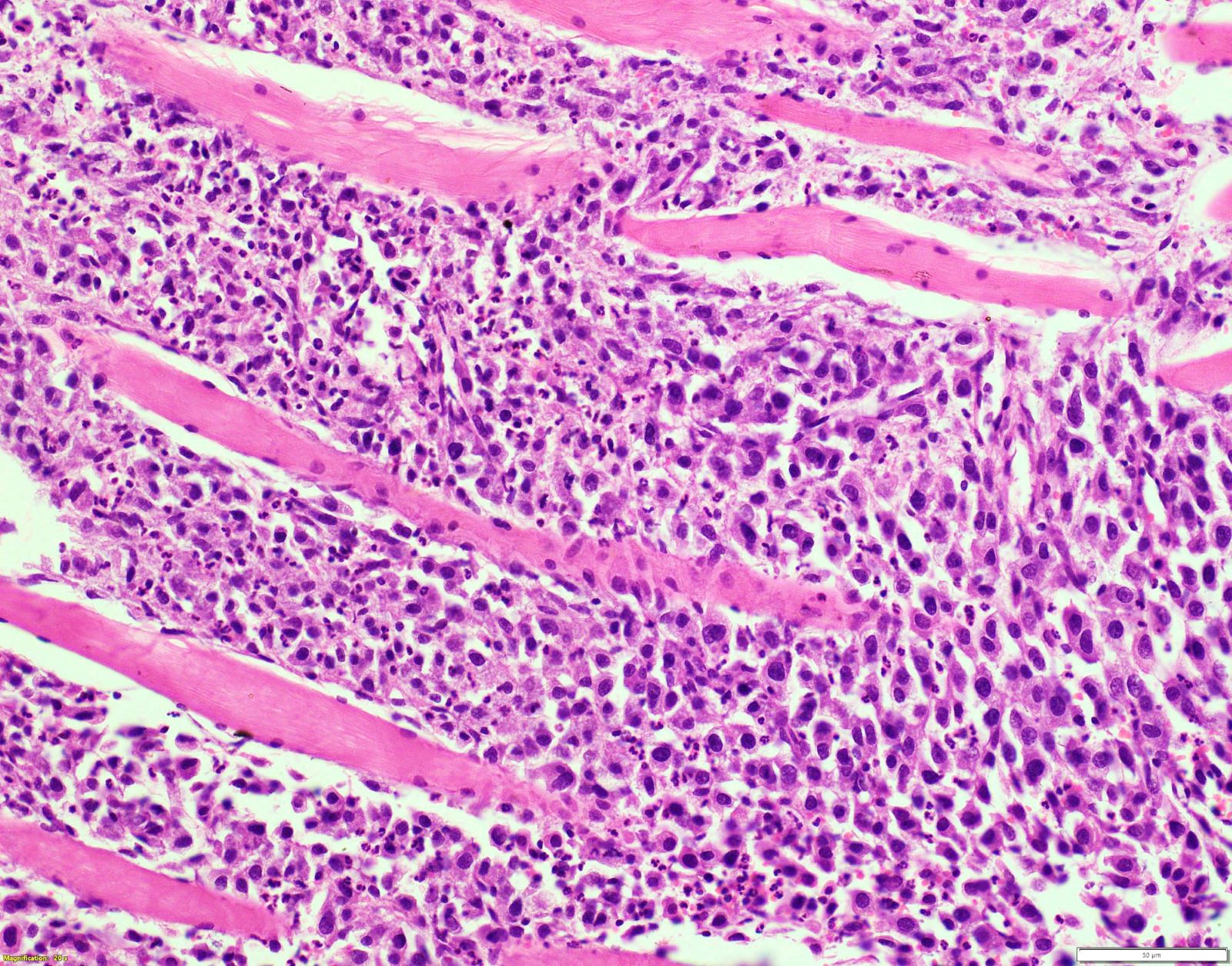
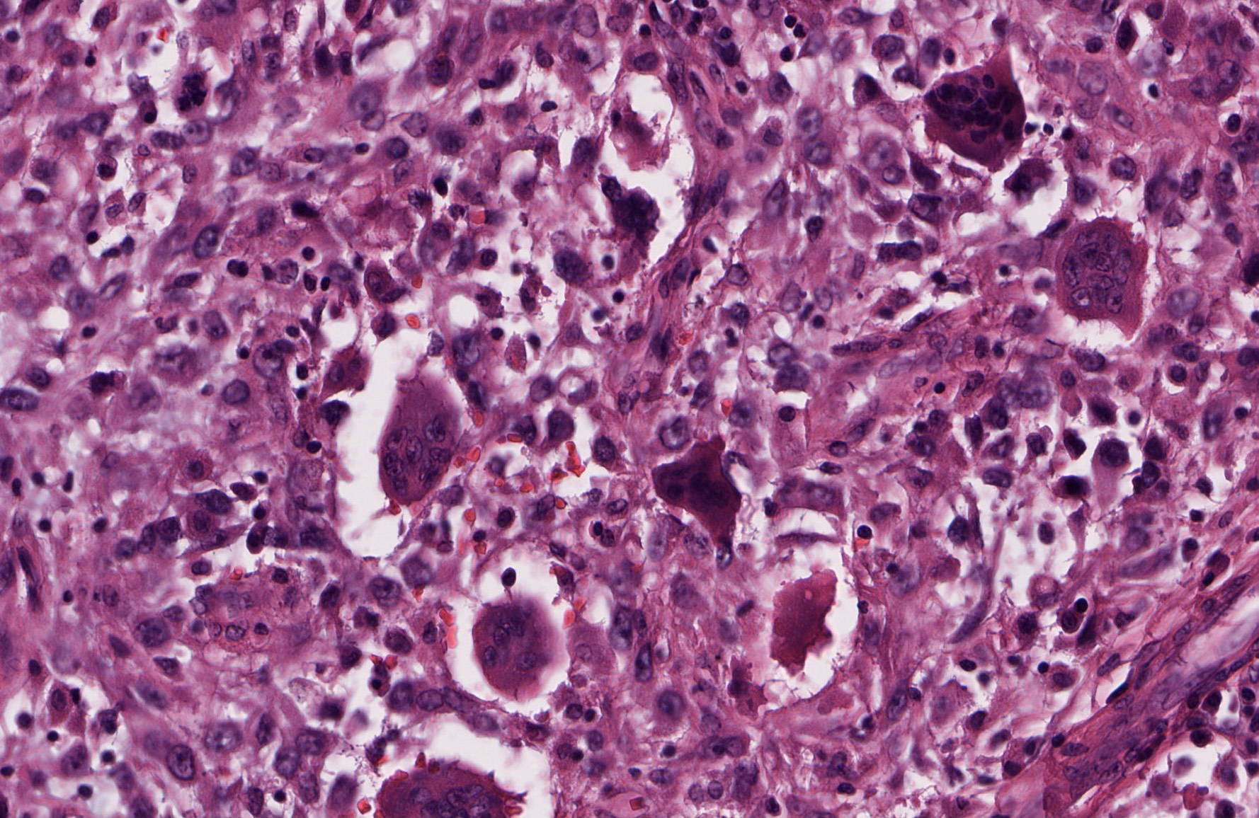
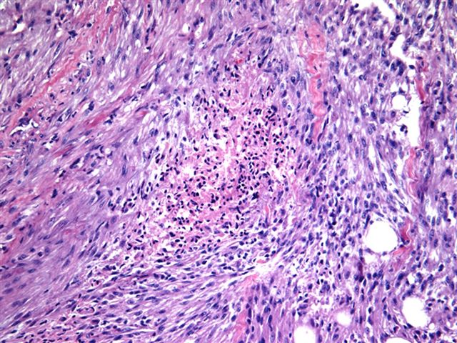
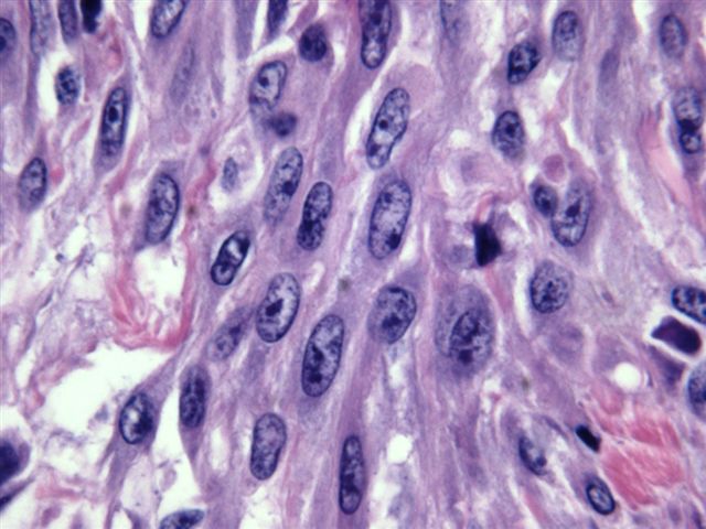
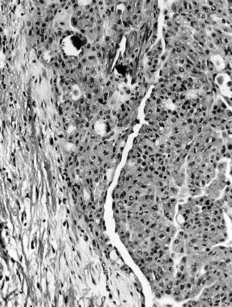
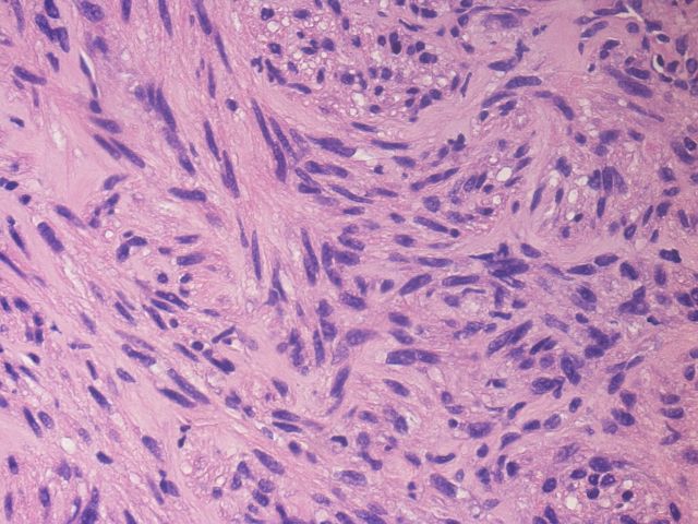
Malignant spindle cells
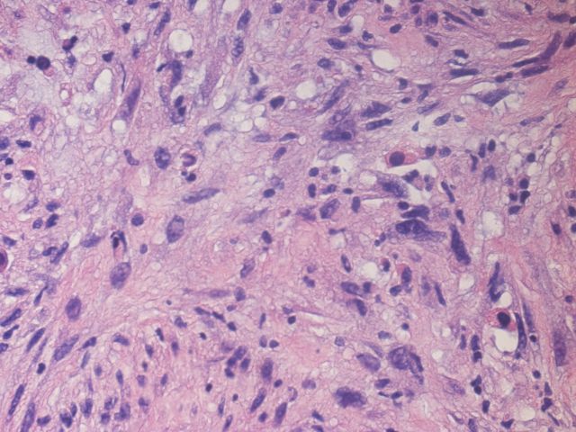
Nuclear hyperchromasia and pleomorphism

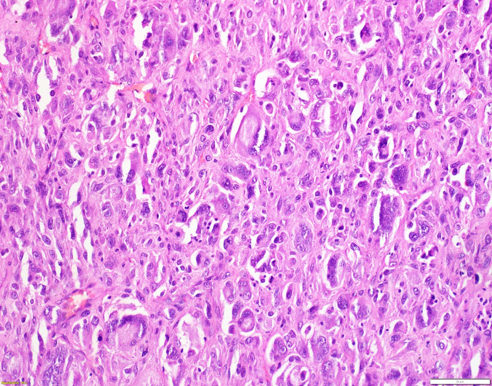
Squamoid cells
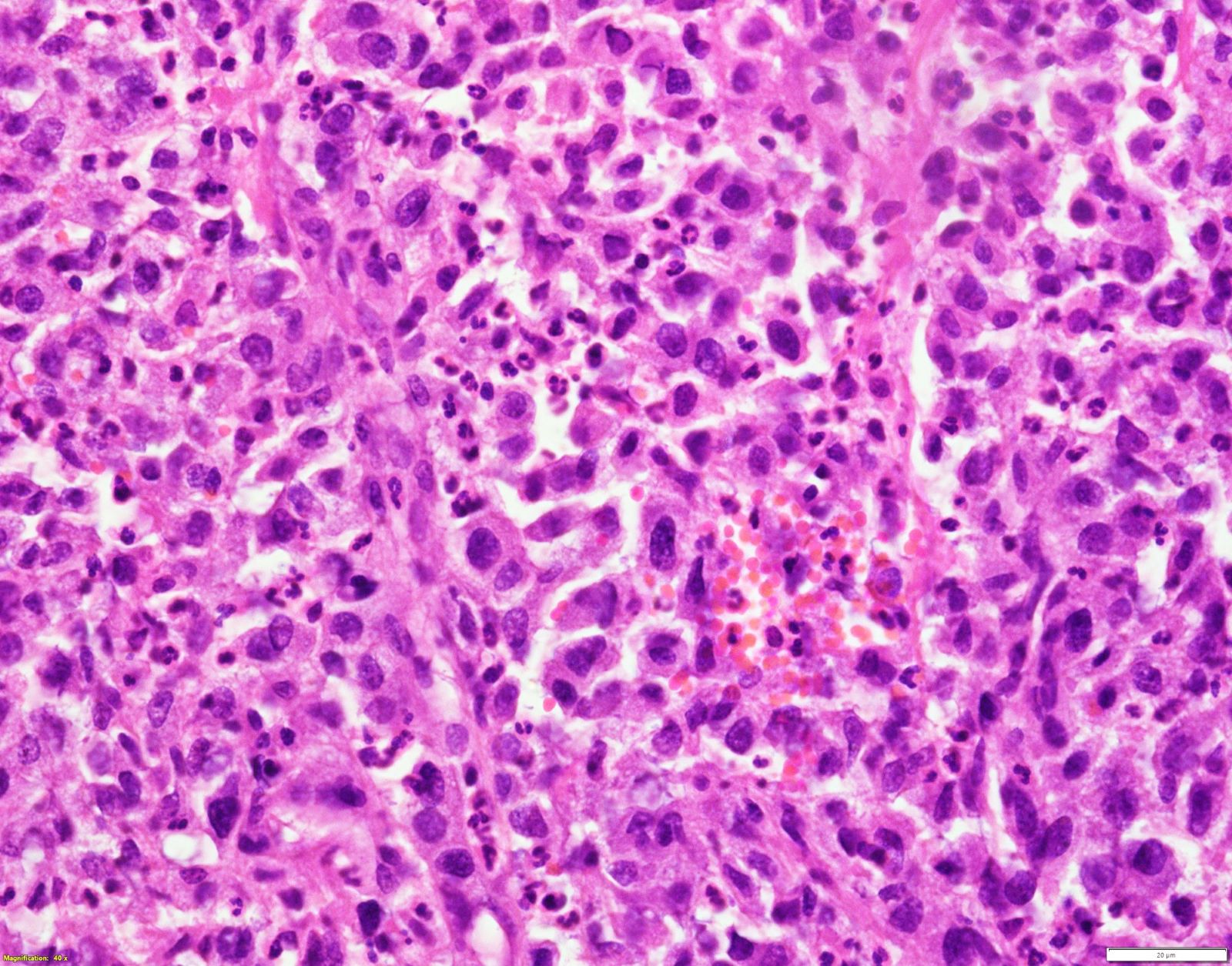
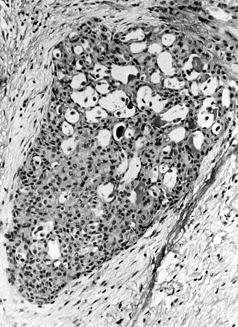
Tumor giant cells
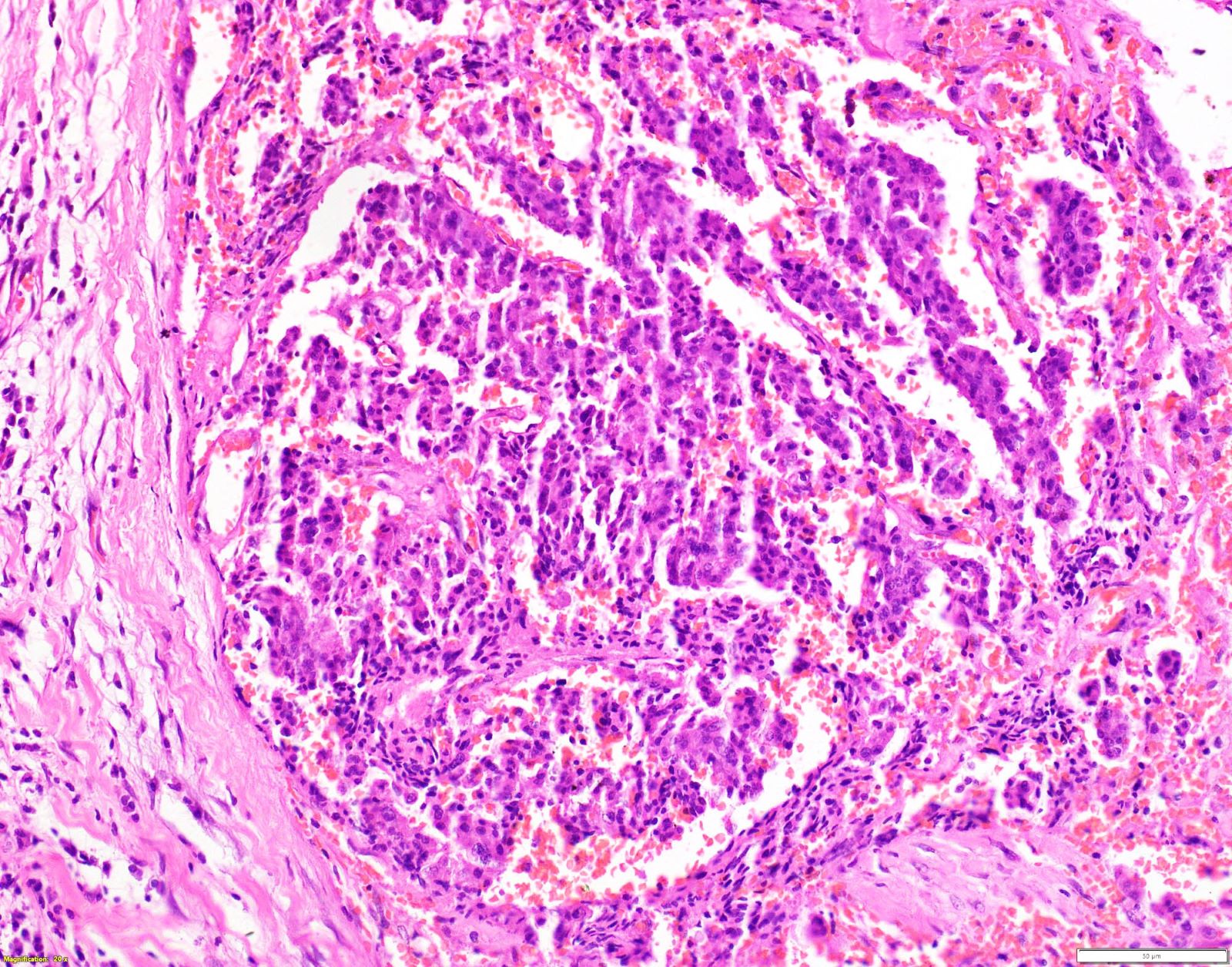
Angiomatoid pattern
Neck muscle infiltration
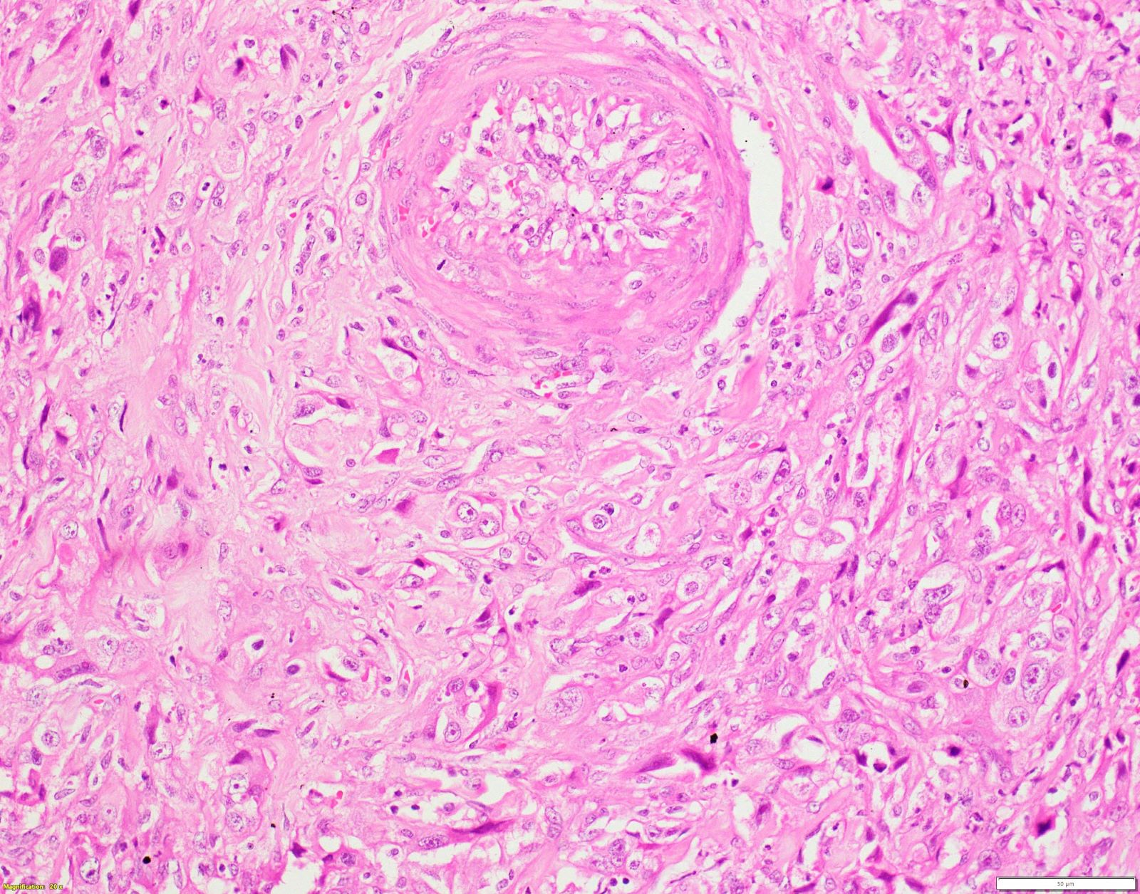
Angioinvasion
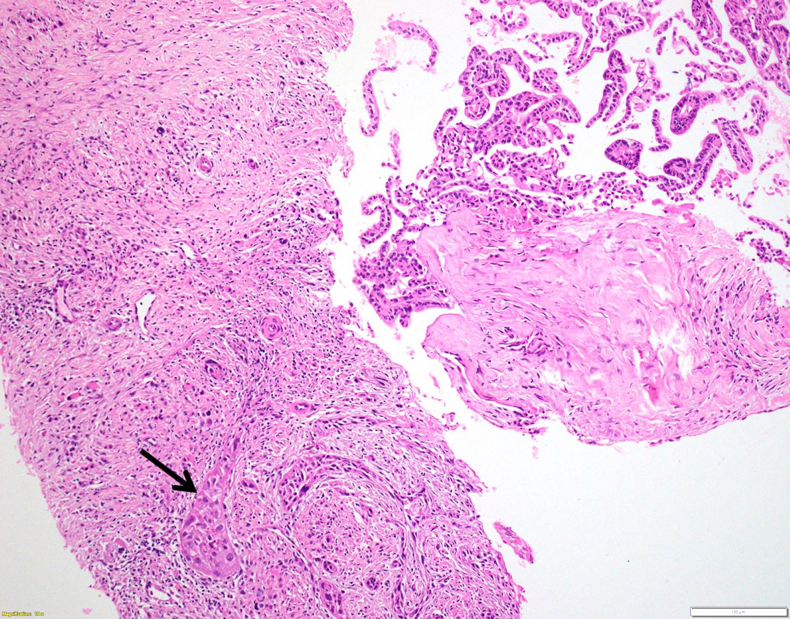
Associated papillary thyroid carcinoma
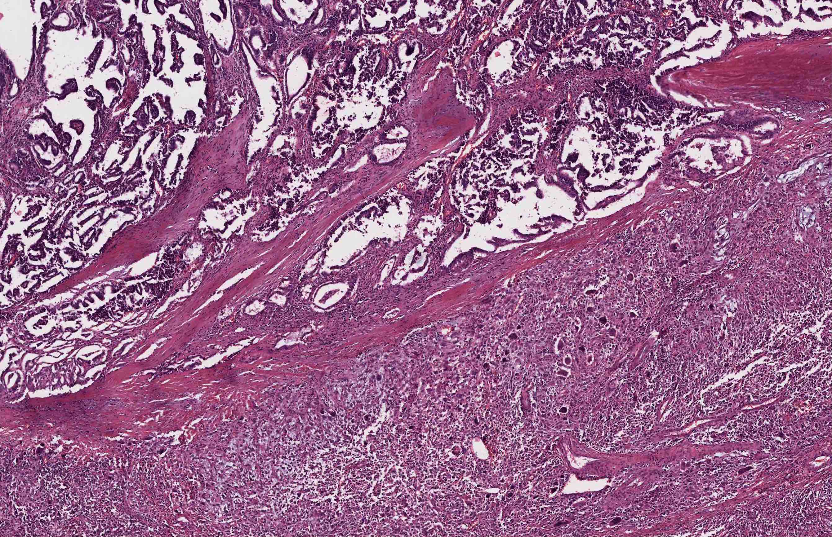
Anaplastic carcinoma and adjacent papillary thyroid carcinoma
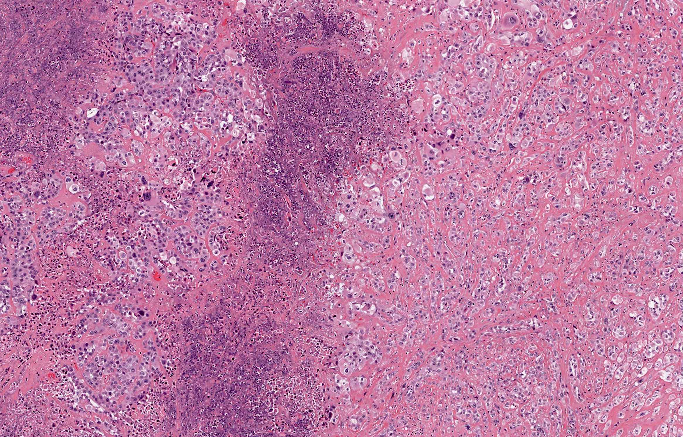
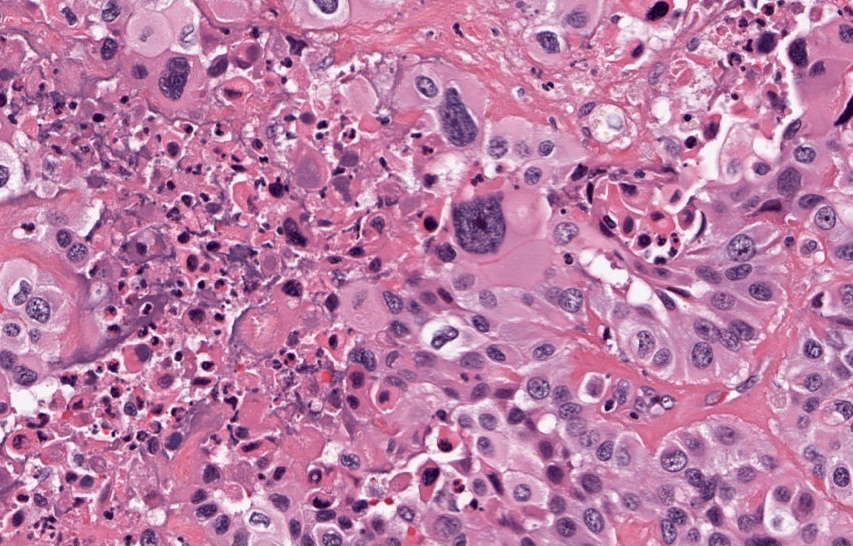
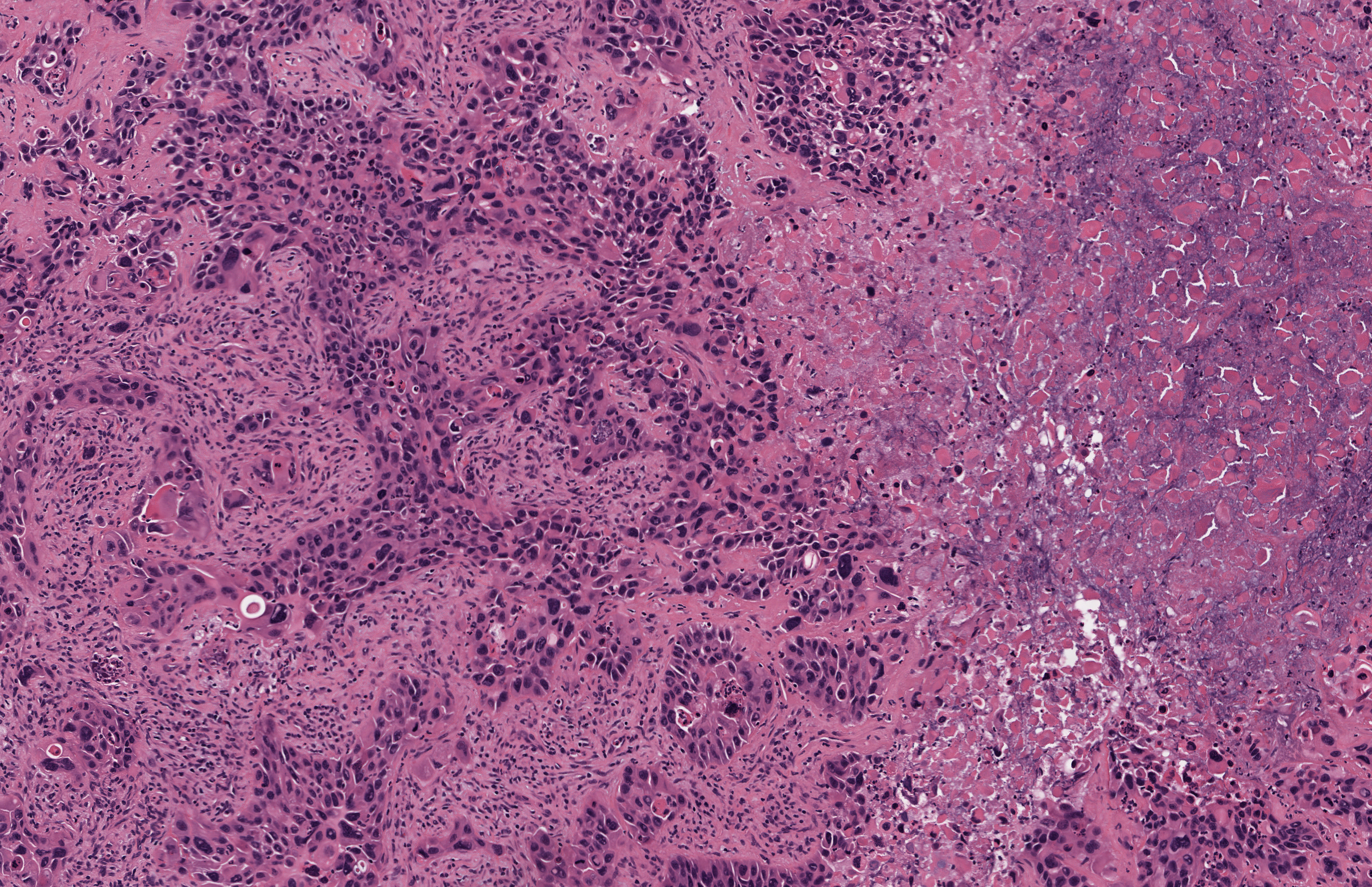
Anaplastic carcinoma with necrosis and inflammation
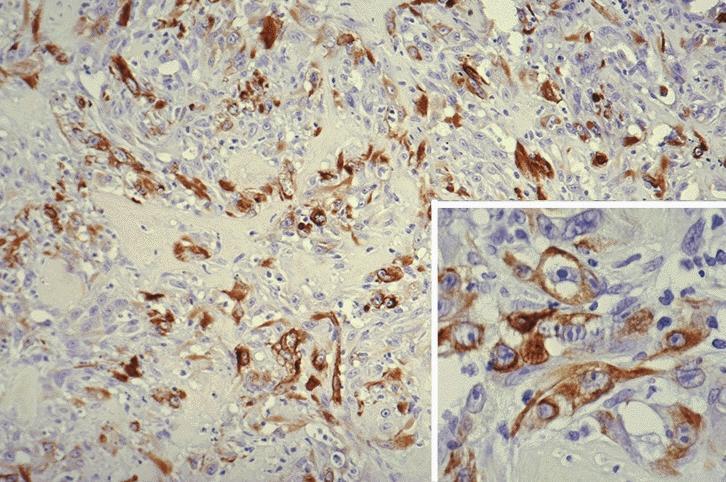
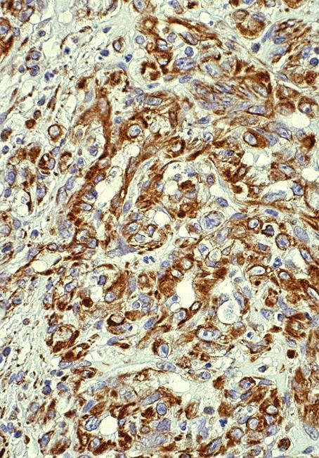
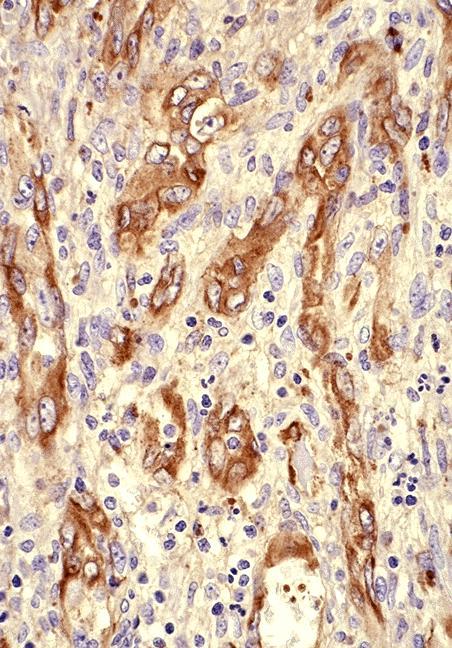
Malignant spindle cells
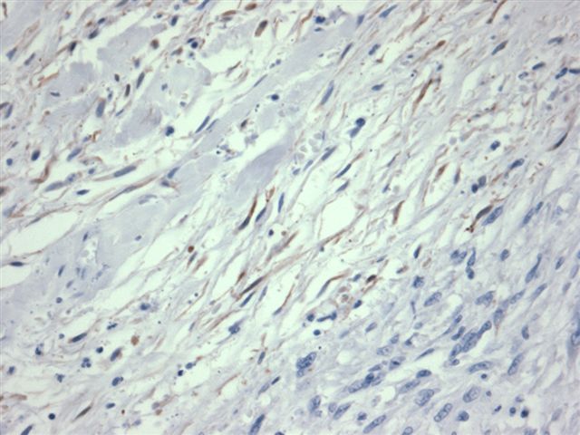
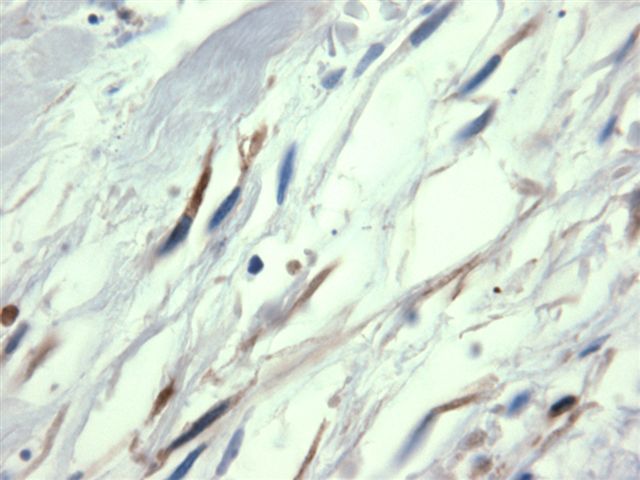
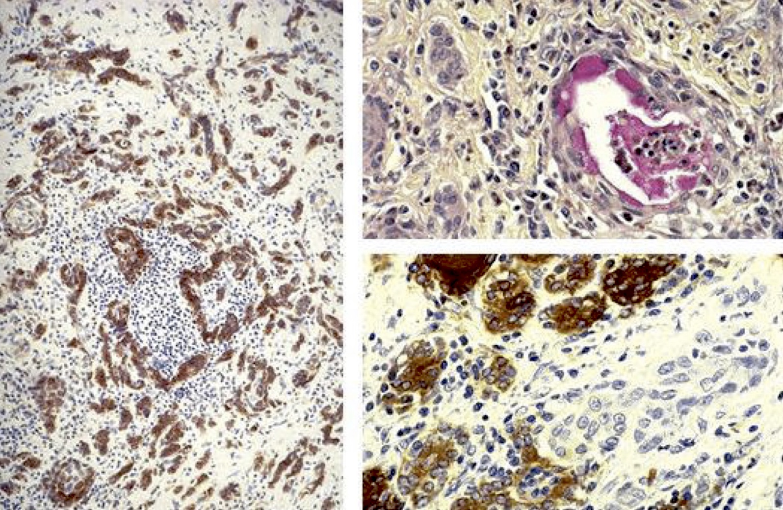
AE1 / AE3
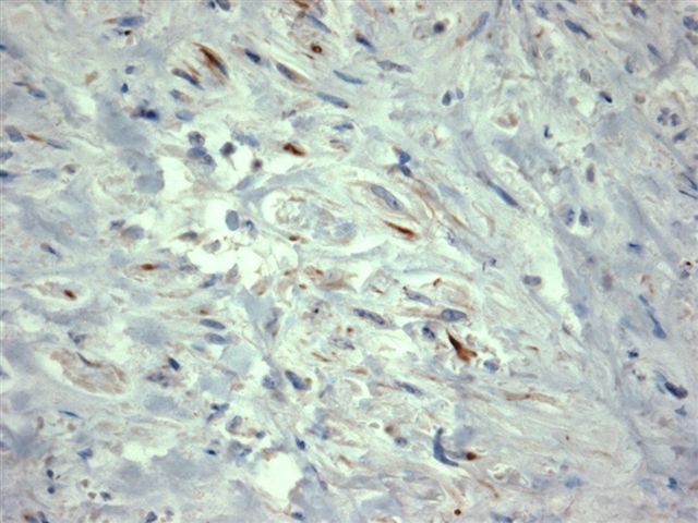
CK903
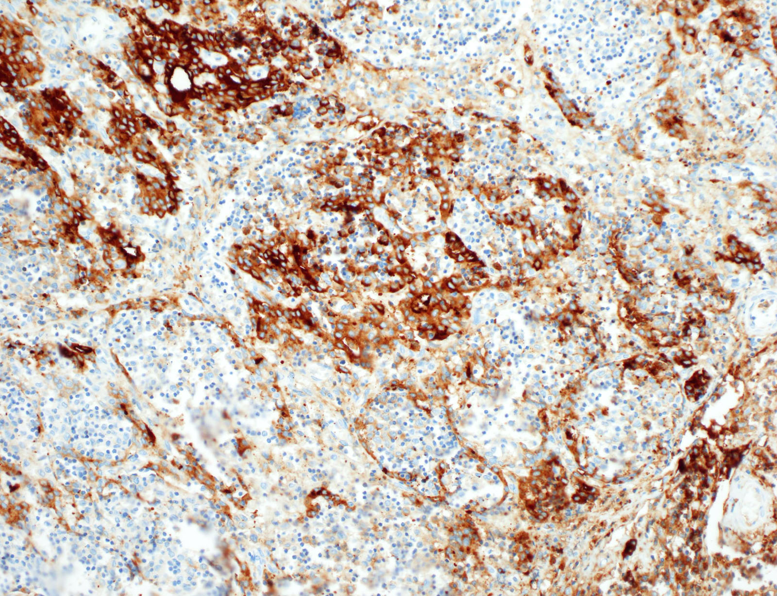
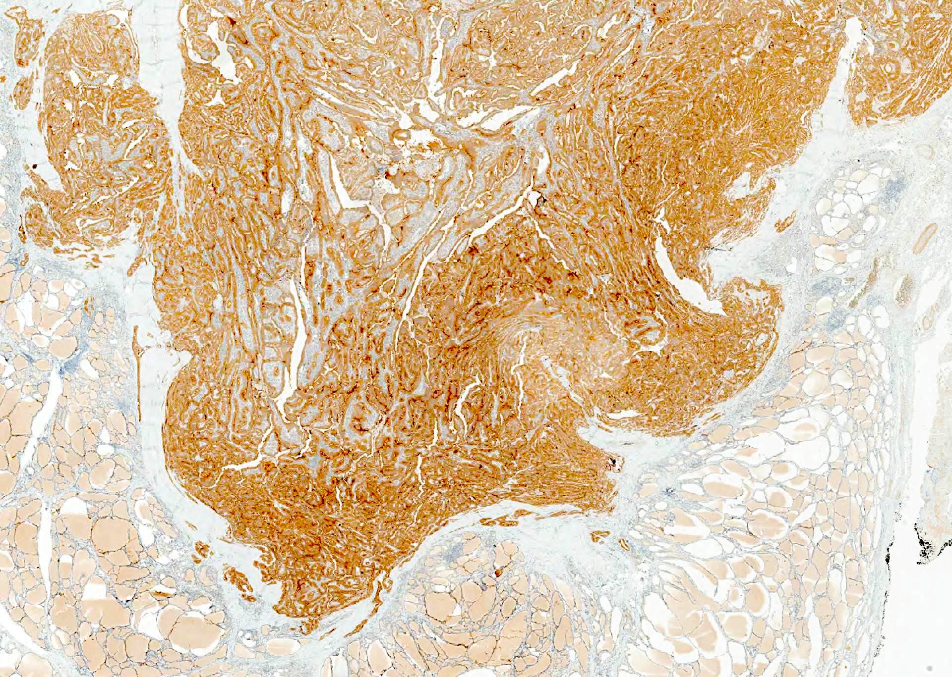
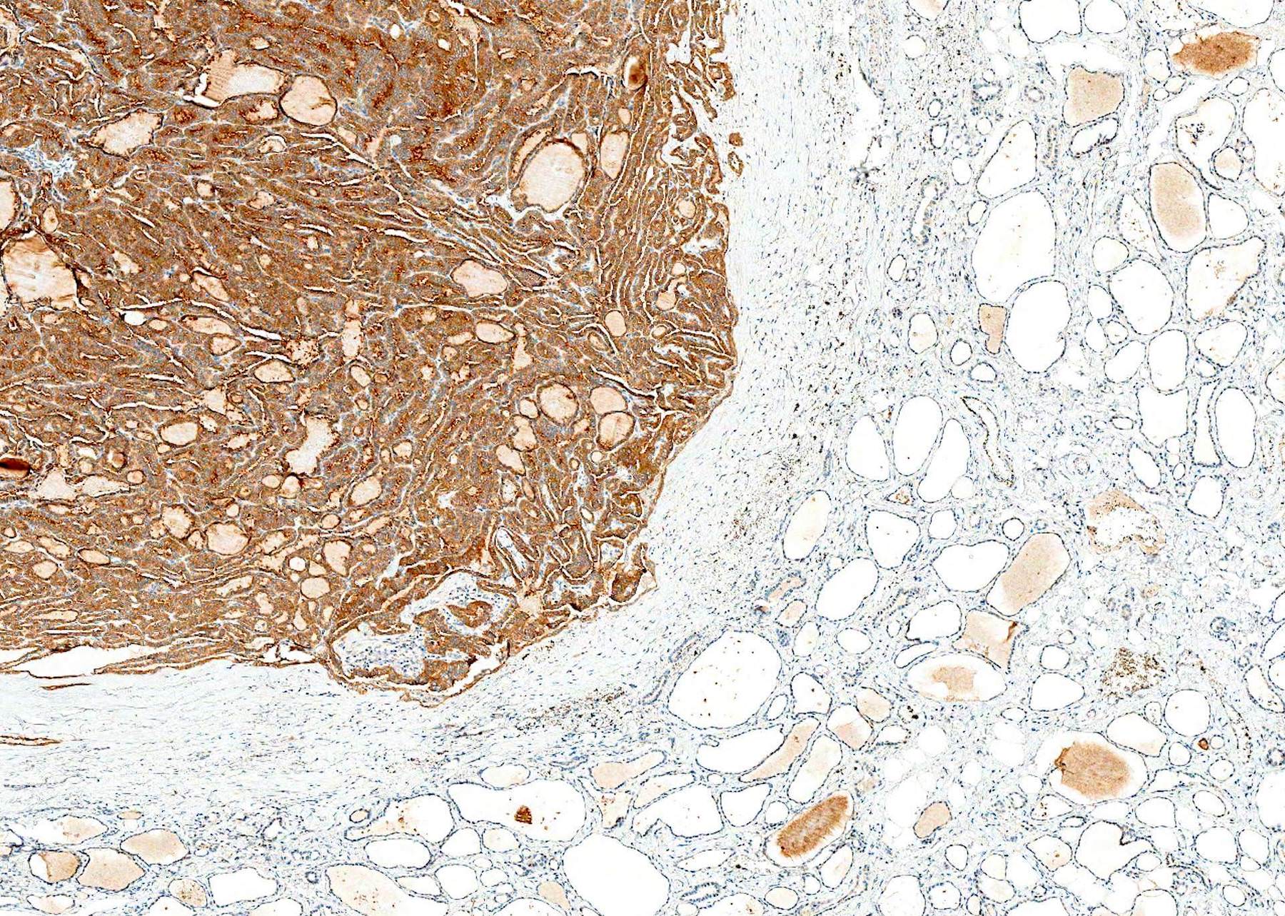
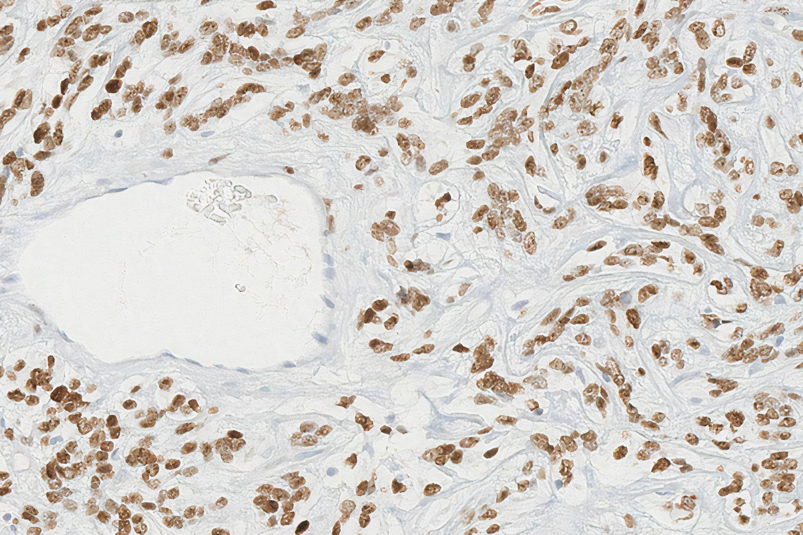
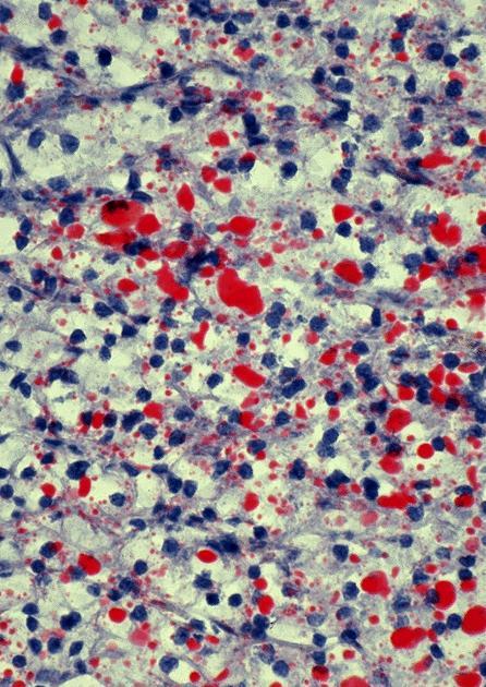
Keratin stains many mesenchymal-like tumor cells
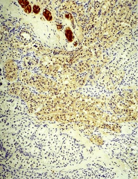
Thyroglobulin stain
Contributed by Shipra Agarwal, M.D. and Ayana Suzuki, C.T.
Necroinflammatory
background
Atypical mitoses
Epithelioid pattern
Sarcomatoid variant

Differentiated thyroid carcinoma component

Pleomorphism
Images hosted on other servers:

Hierarchical clustering

Genetic alterations

Involved molecular pathways

Recurrent MSK-IMPACT derived copy number alterations

Increased mutation burden
Cytology of ATC
Management of ATC
Images hosted on other servers:

Thyroidectomy
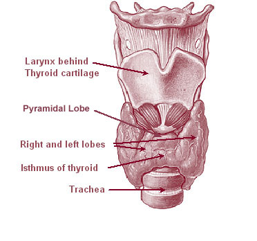
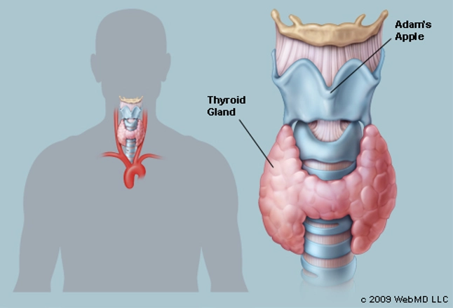
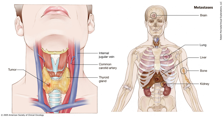
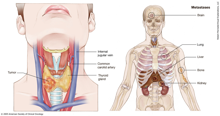
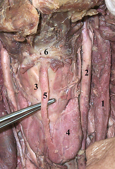
Relationship to other structures
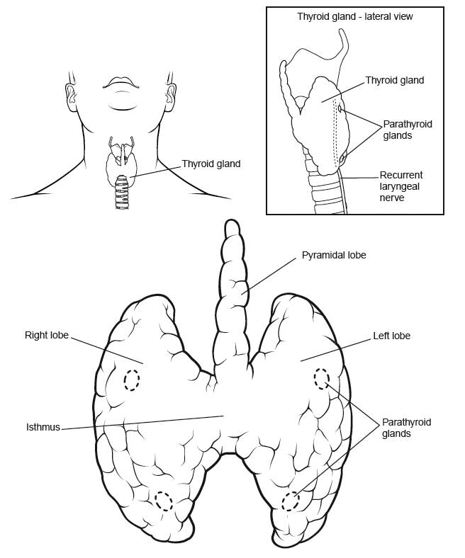
Pyramidal lobe

Development of inverted U shaped thyroid gland


Anatomical variations
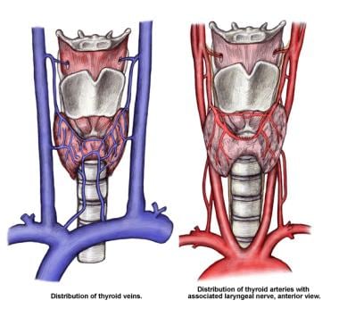


Blood supply
Images hosted on other servers:
On surgery

In situ

Inverted U shaped thyroid

W shaped thyroid

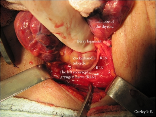
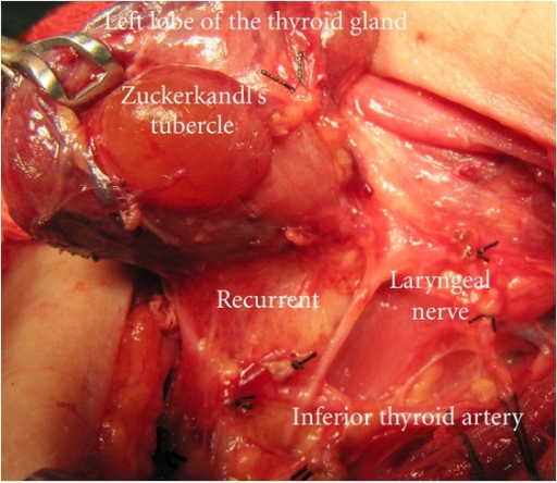
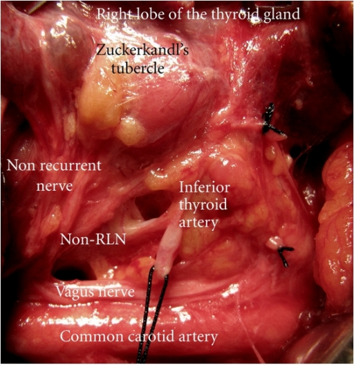
Zuckerkandl tubercle
Images hosted on other servers:

Surgical specimen

Levator glandulae thyroideae
Contributed by Andrey Bychkov, M.D., Ph.D.
Pyramidal lobe
Thyroid anatomy
Images hosted on other servers:

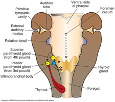
Embryological pathway of parathyroid migration

Anatomy
Contributed by Truong Phan Xuan Nguyen, M.D.
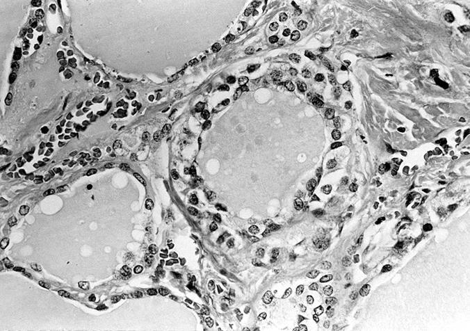
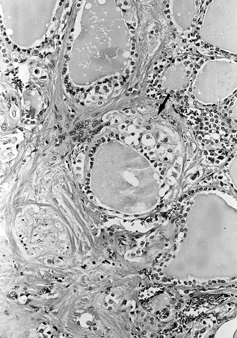
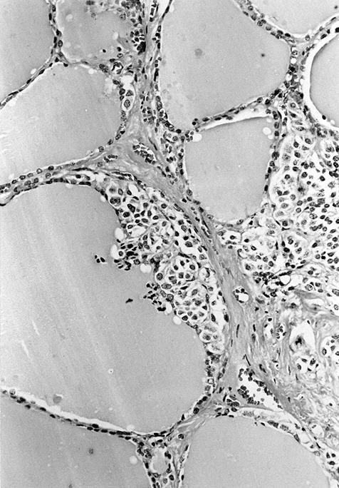
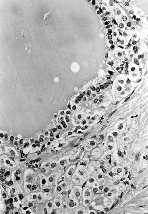
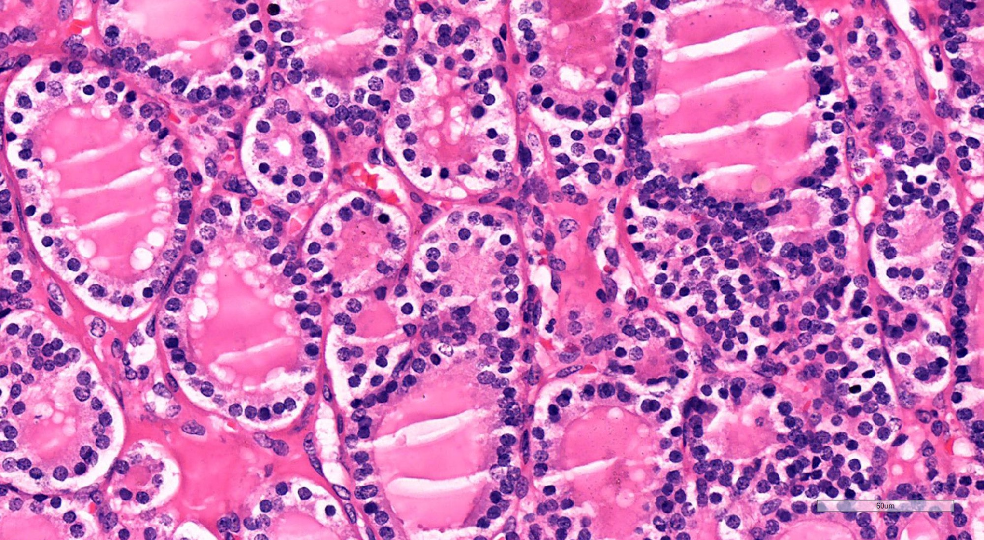
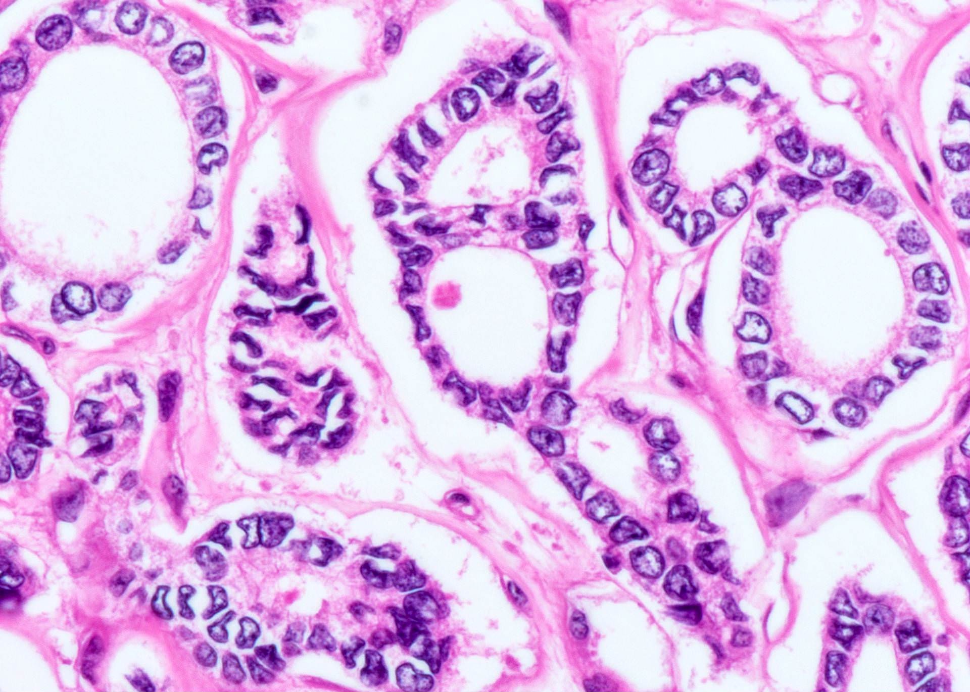
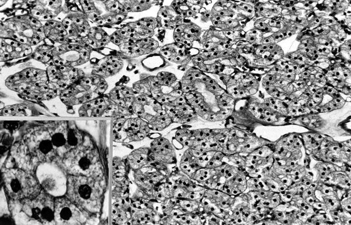
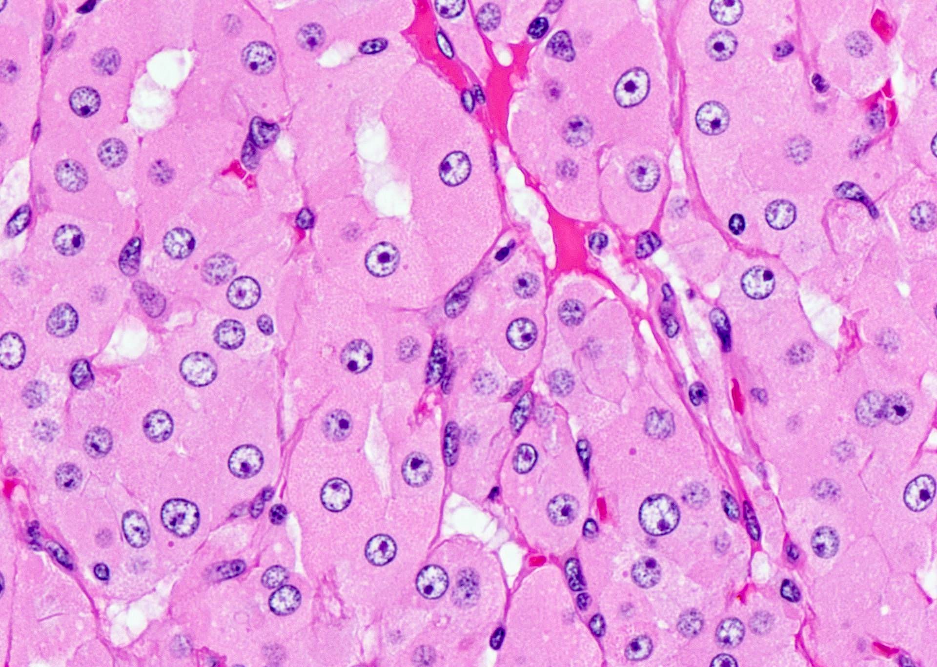
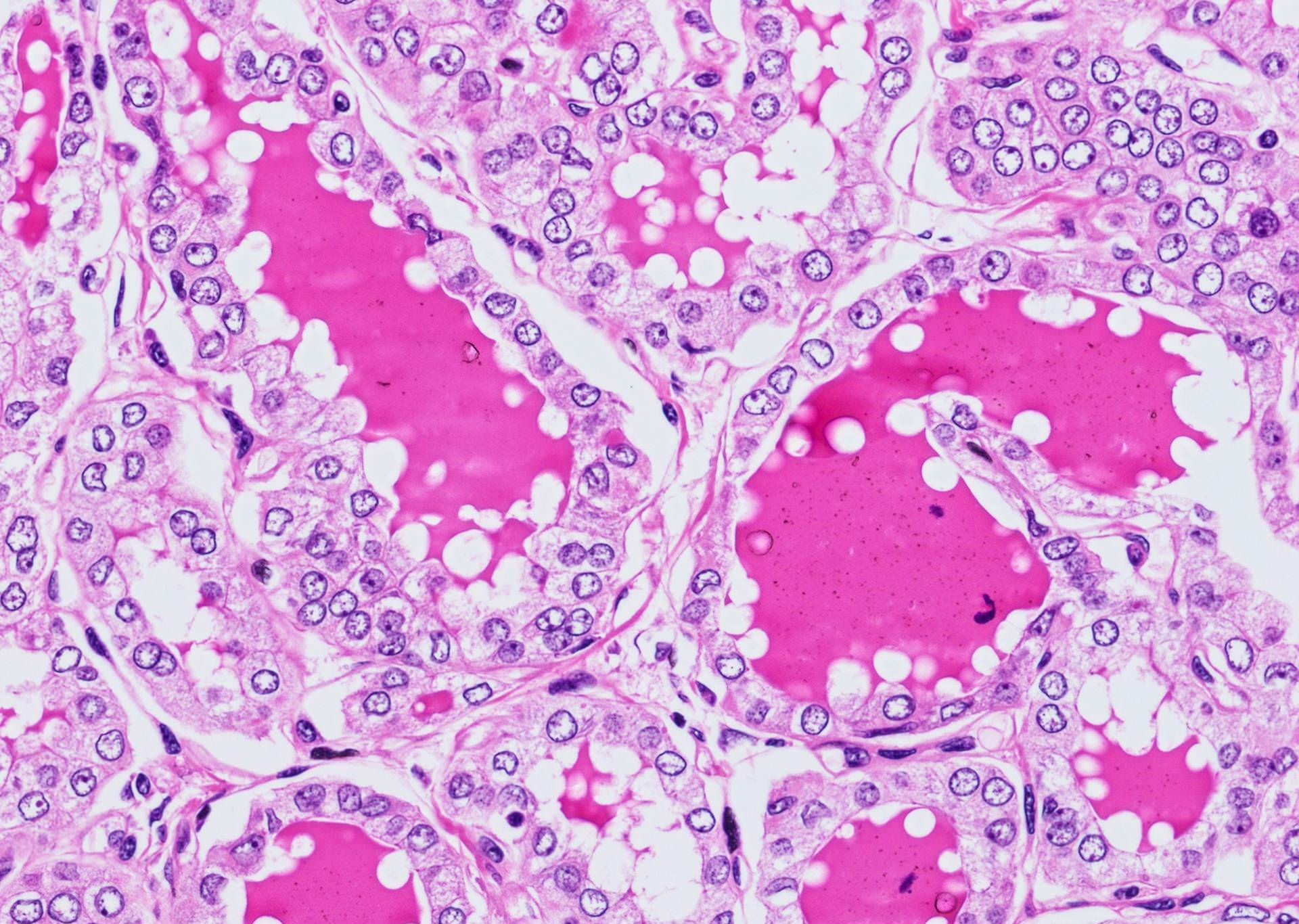
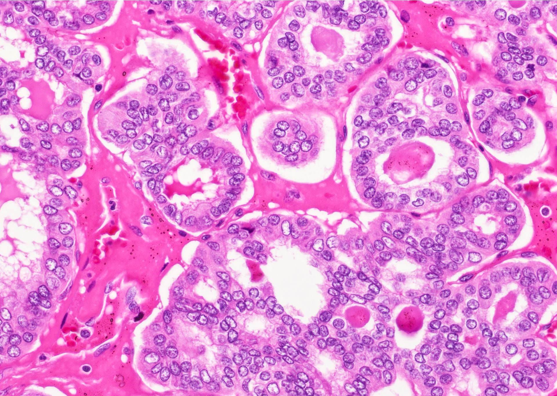
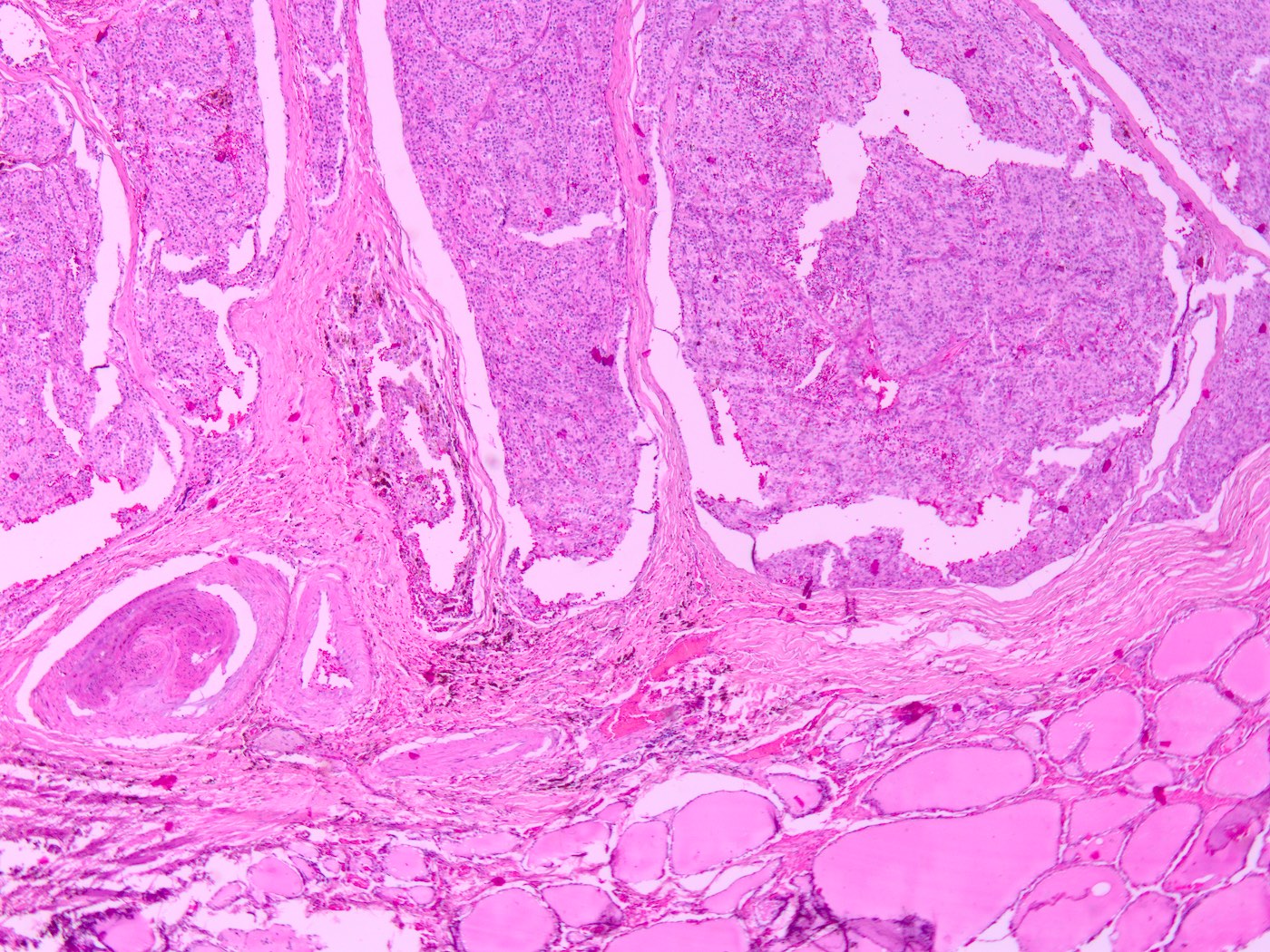
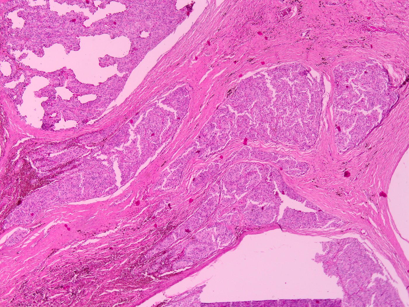
Normal parathyroid gland
Contributed by Truong Phan Xuan Nguyen, M.D.
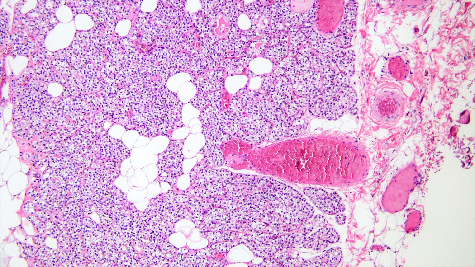
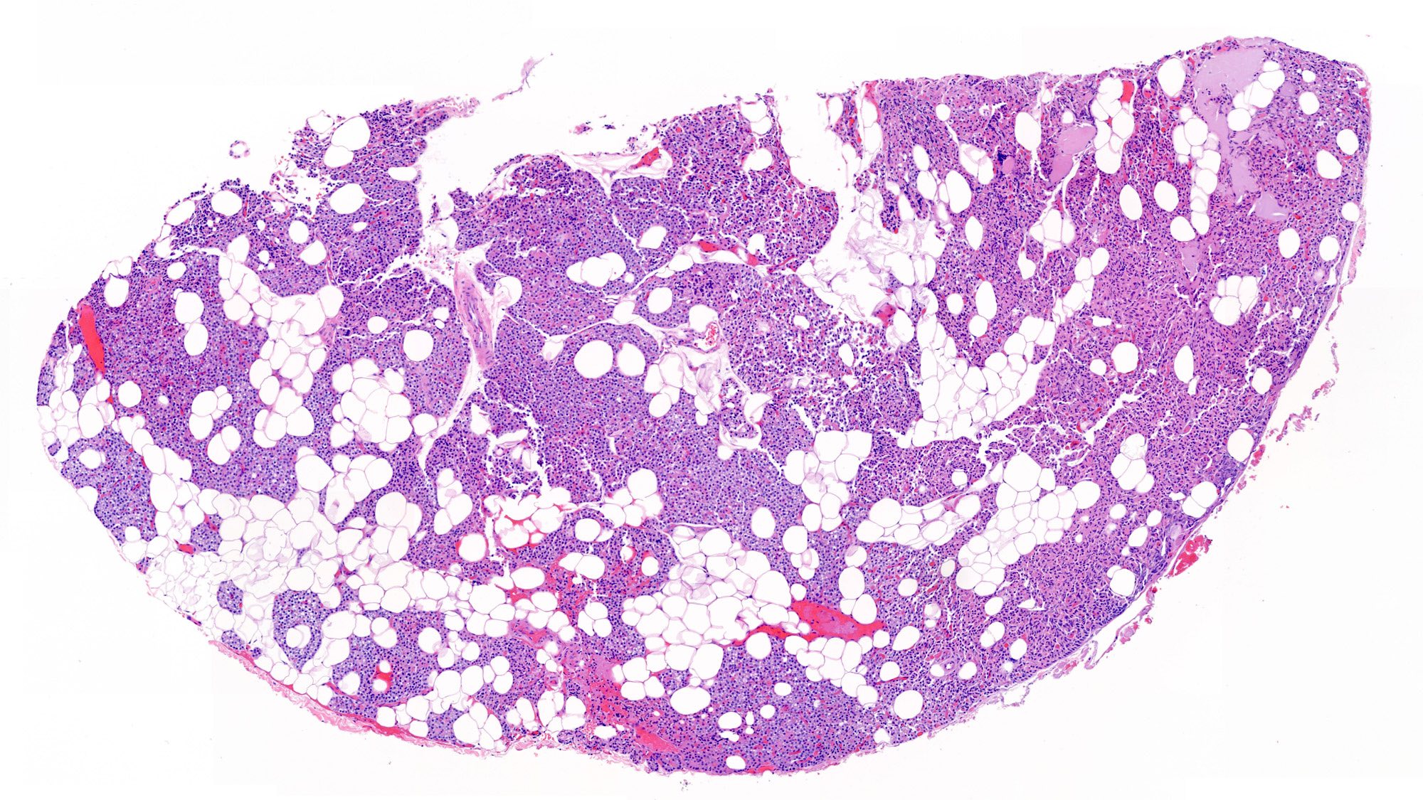
Normal parathyroid gland
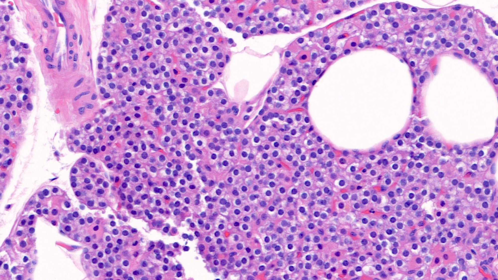
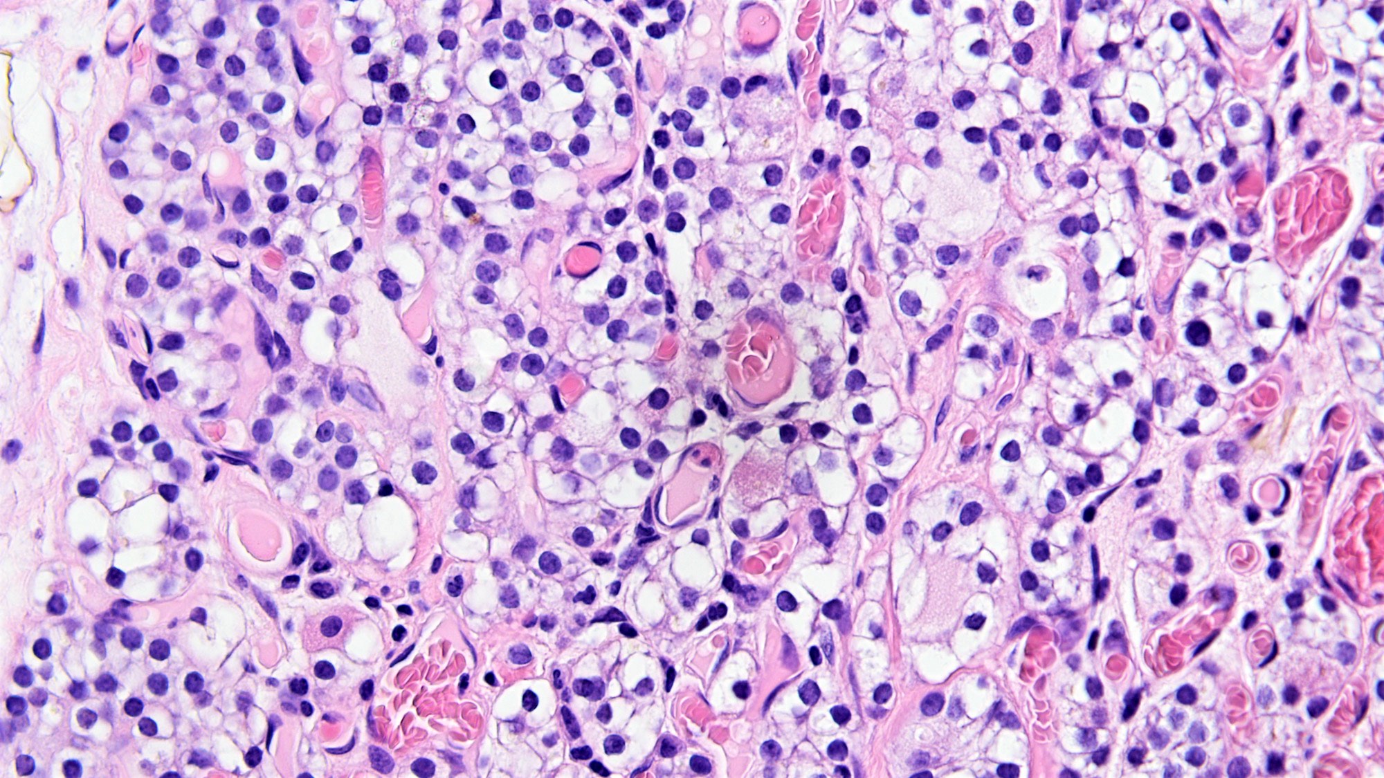
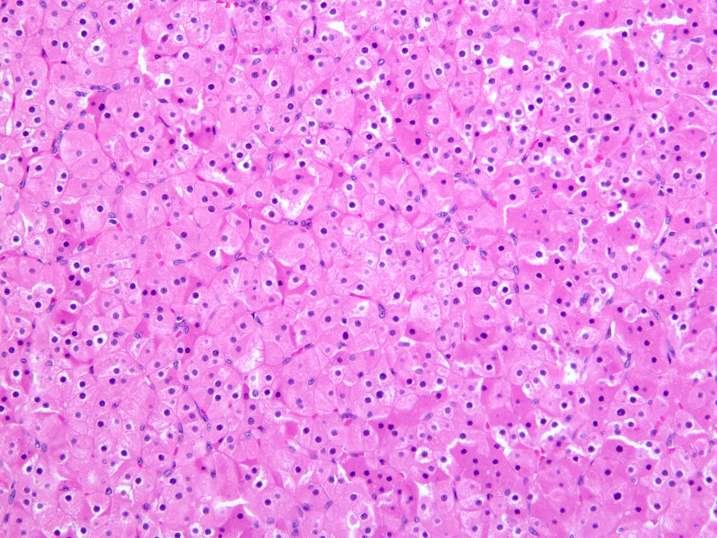
Chief cells

Oxyphil cells
Transitional cells
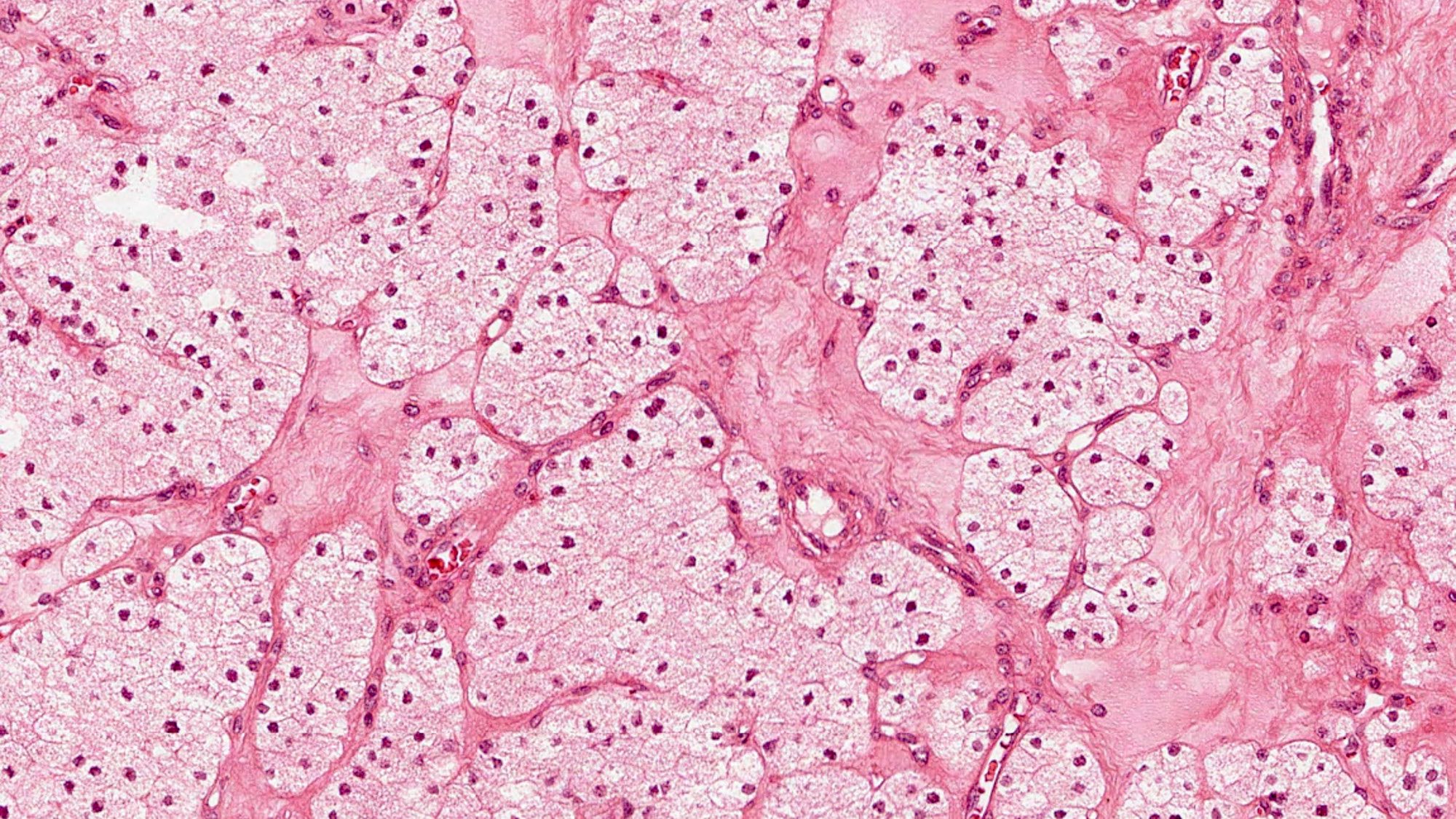
Water clear cells
Colloid type material, without oxalate crystals
Images hosted on other servers:

Parathyroid gland
Histology of parathyroid
AFIP images
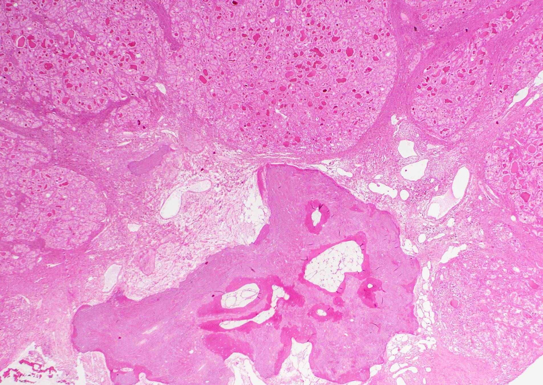
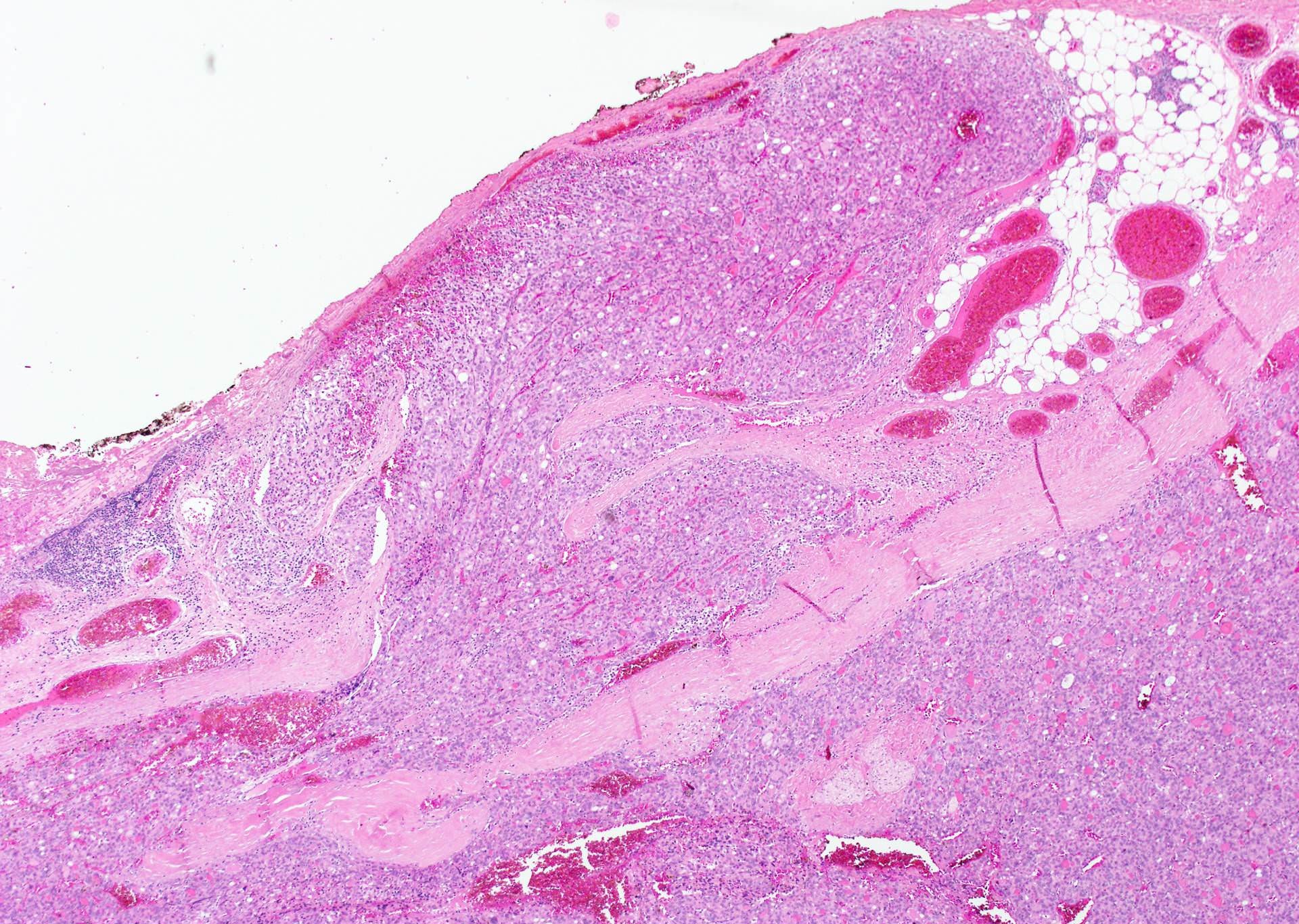
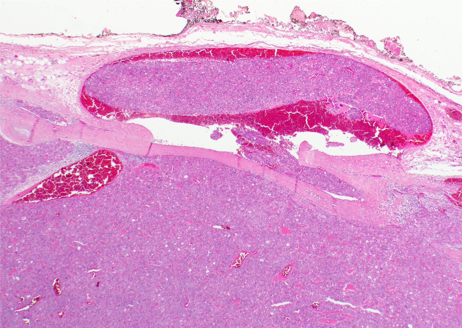
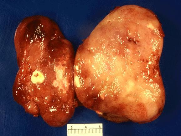
Necrotic and hemorrhagic tumor
AFIP images

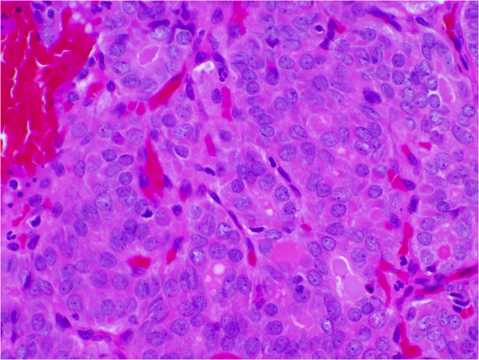
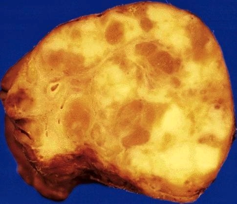
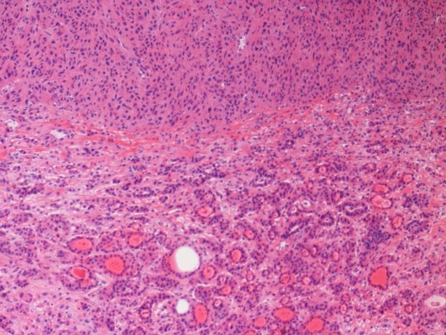
Solid and cellular areas

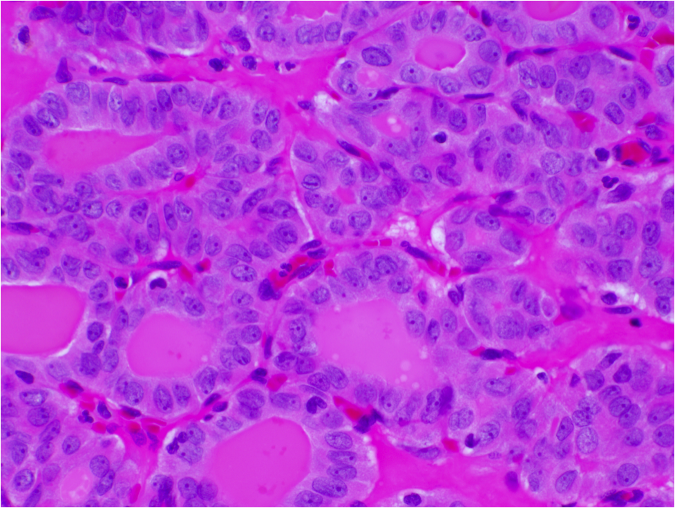
Abortive vascular lumina
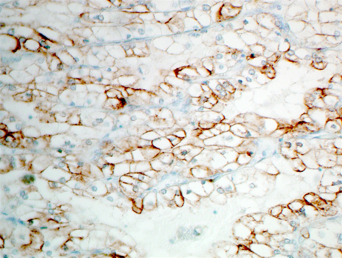
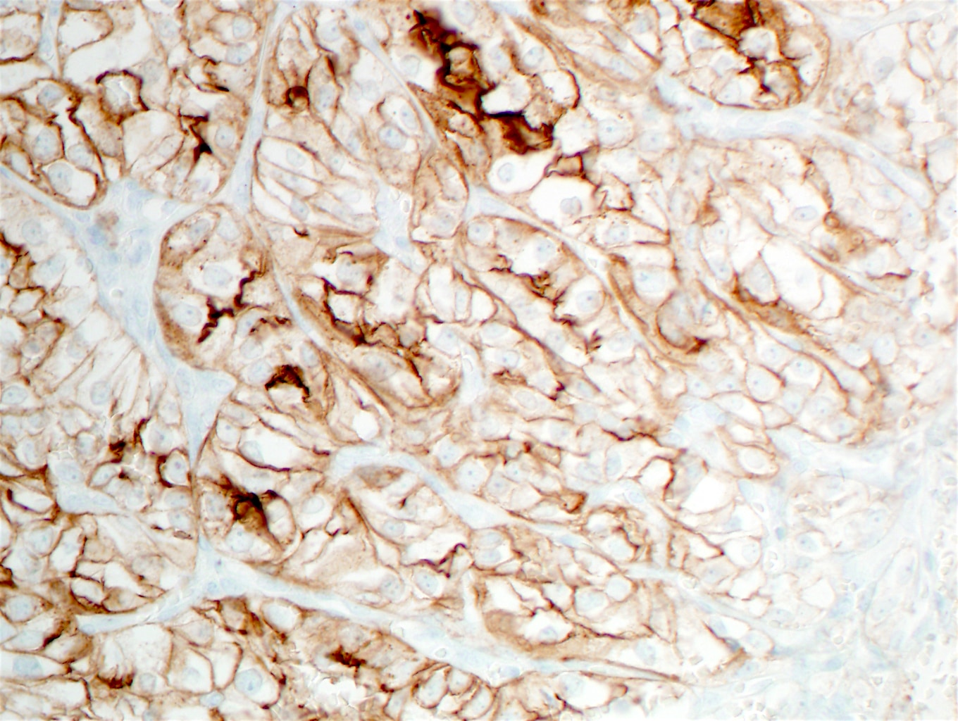
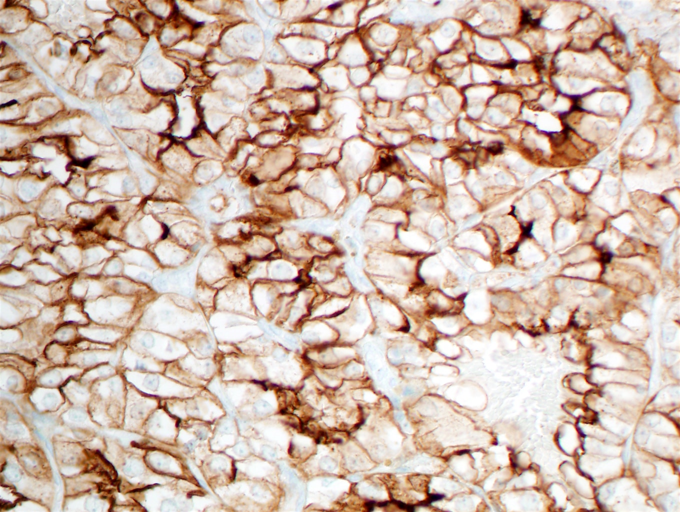
Keratin+ tumor cells
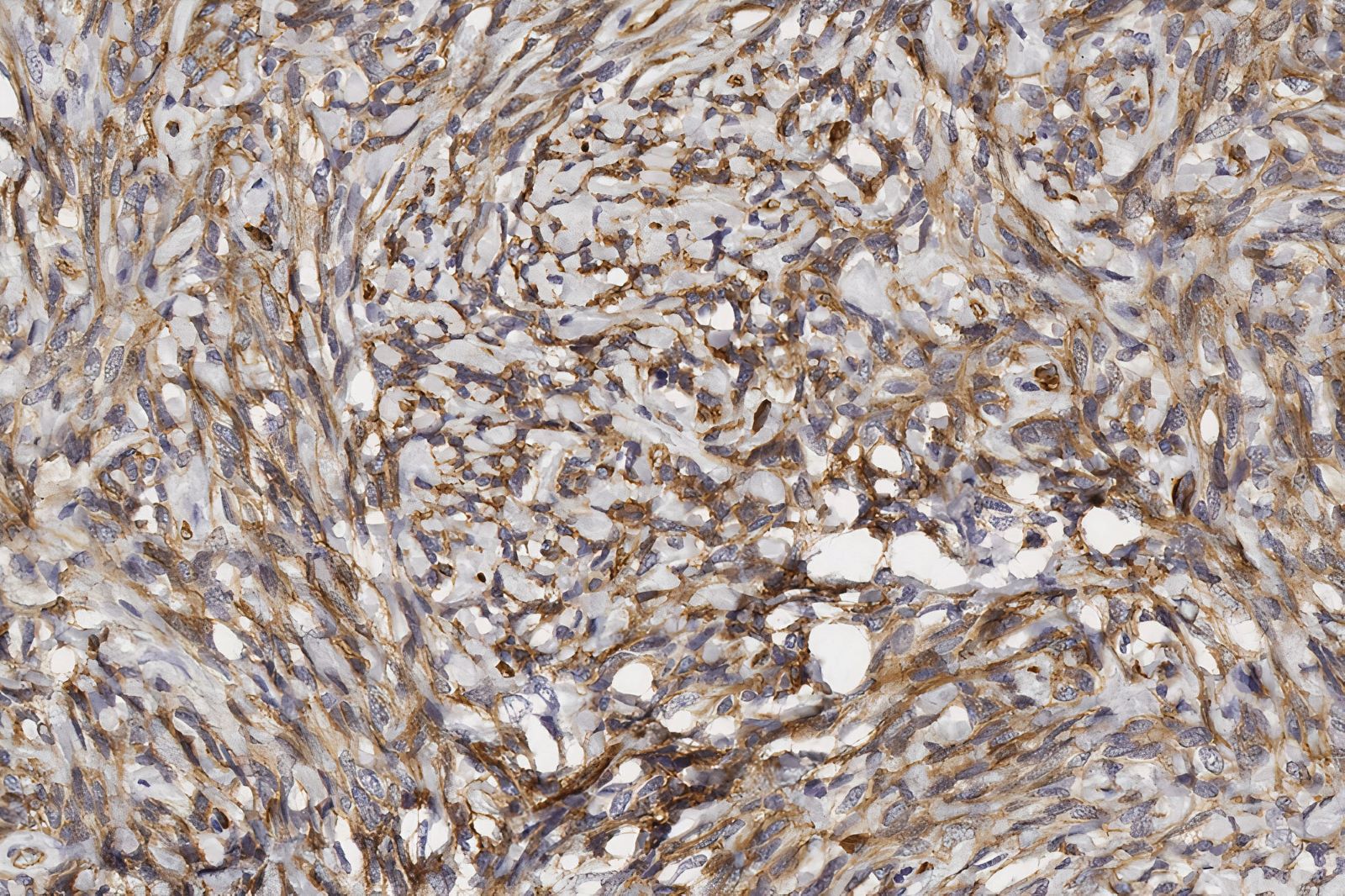
Vimentin+
Plump endothelial, Factor VIII+
Ulex europaeus I lectin+
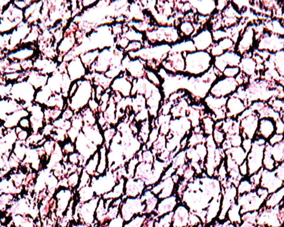
Type IV collagen stains basal lamina
Images hosted on other servers:

PAX8 mutation in thyroid hypoplasia
Images hosted on other servers:


Phenotype of congenital hypothyroidism


Hemiagenesis on CT


Hemiagenesis on scintiscan


Hemiagenesis on sonography
Images hosted on other servers:

Hashimoto thyroiditis in
thyroid hemiagenesis
Contributed by Ayana Suzuki, C.T.

Cytologic atypia
Architectural atypia
Hürthle cell atypia
Atypical lymphocytes
Images hosted on other servers:


Cytologic atypia


Atypical cyst lining cells

Architectural atypia

Atypical lymphocytes
Atypical thyroid FNA
Head and tail of the Bethesda system for thyroid
Thyroid cytology - Bethesda classification
Contributed by Ayana Suzuki, C.T.
Isoechoic nodule
Hypoechoic nodule
Large cyst
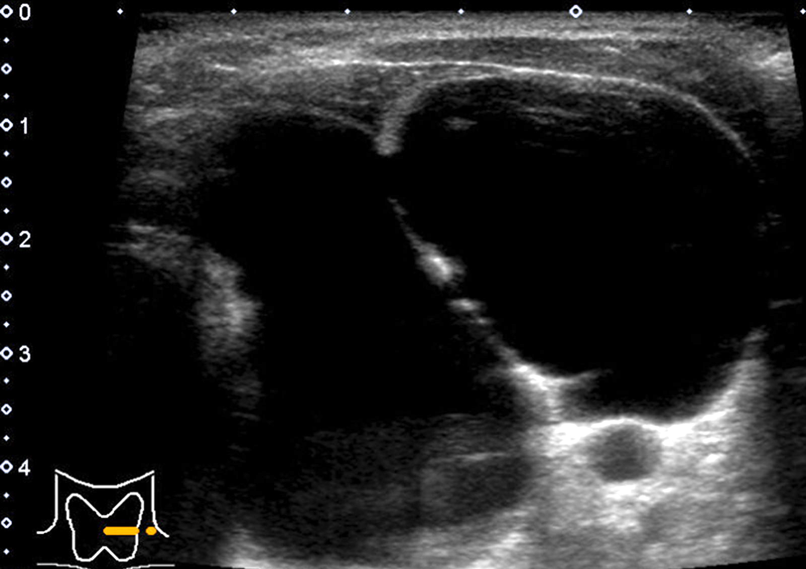
Large multilocular cyst
Contributed by Ayana Suzuki, C.T.
Colloid nodule

Adenomatous nodules
Hashimoto thyroiditis
Granulomatous thyroiditis

Thyroglossal duct cyst
Thyroid cytology: colloid nodule
Essential thyroid cytopathology
Contributed by Andrey Bychkov, M.D., Ph.D.
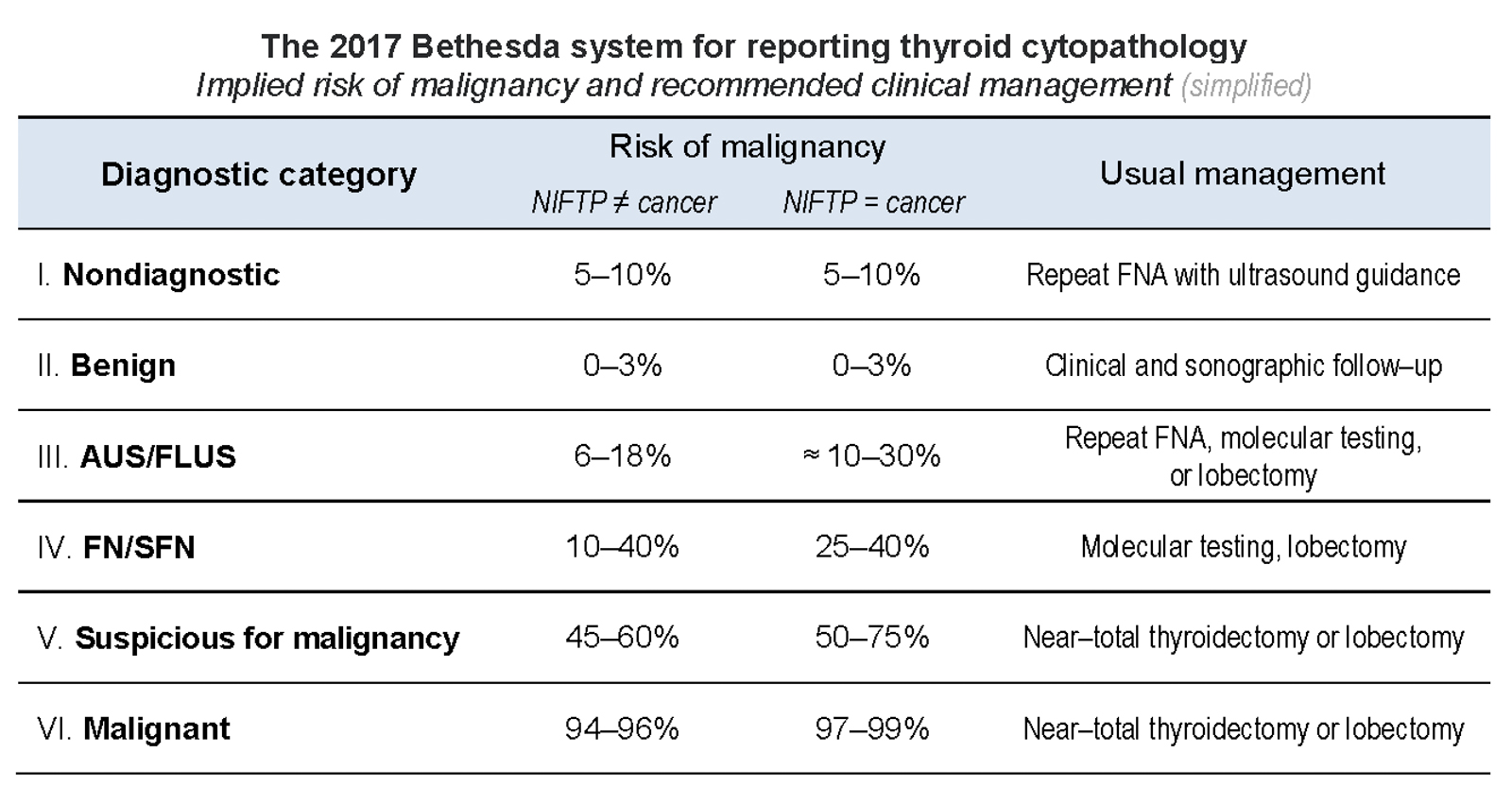
TBS 2017
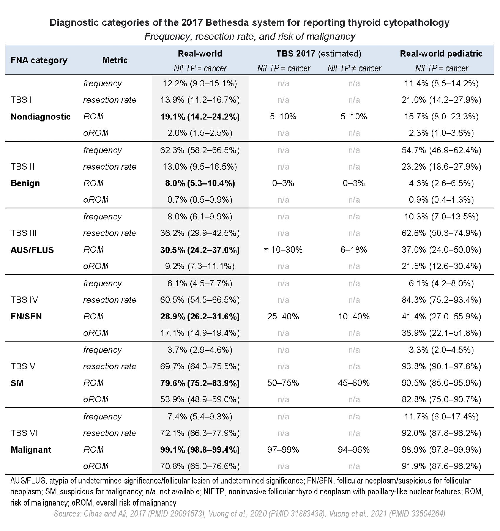
TBS metrics
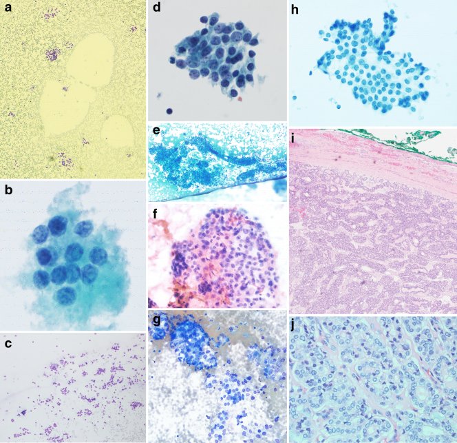
Contributed by Ayana Suzuki, C.T.
Unsatisfactory:
Hemorrhagic background
Muscle
Respiratory epithelium
Air dried smear
Cyst fluid only
Benign:

Watery colloid
Cracking colloid
Follicular clusters
3D structures
follicular lesion of undetermined significance:
FLUS - cellular
FLUS - architectural
FLUS - Hürthle
Atypical lymphocytes
suspicious for a follicular neoplasm:
Microfollicles
FN - Hürthle
Suspicious for malignancy:
Suspicious for papillary thyroid carcinoma
Suspicious for lymphoma

Hyalinizing trabecular tumor
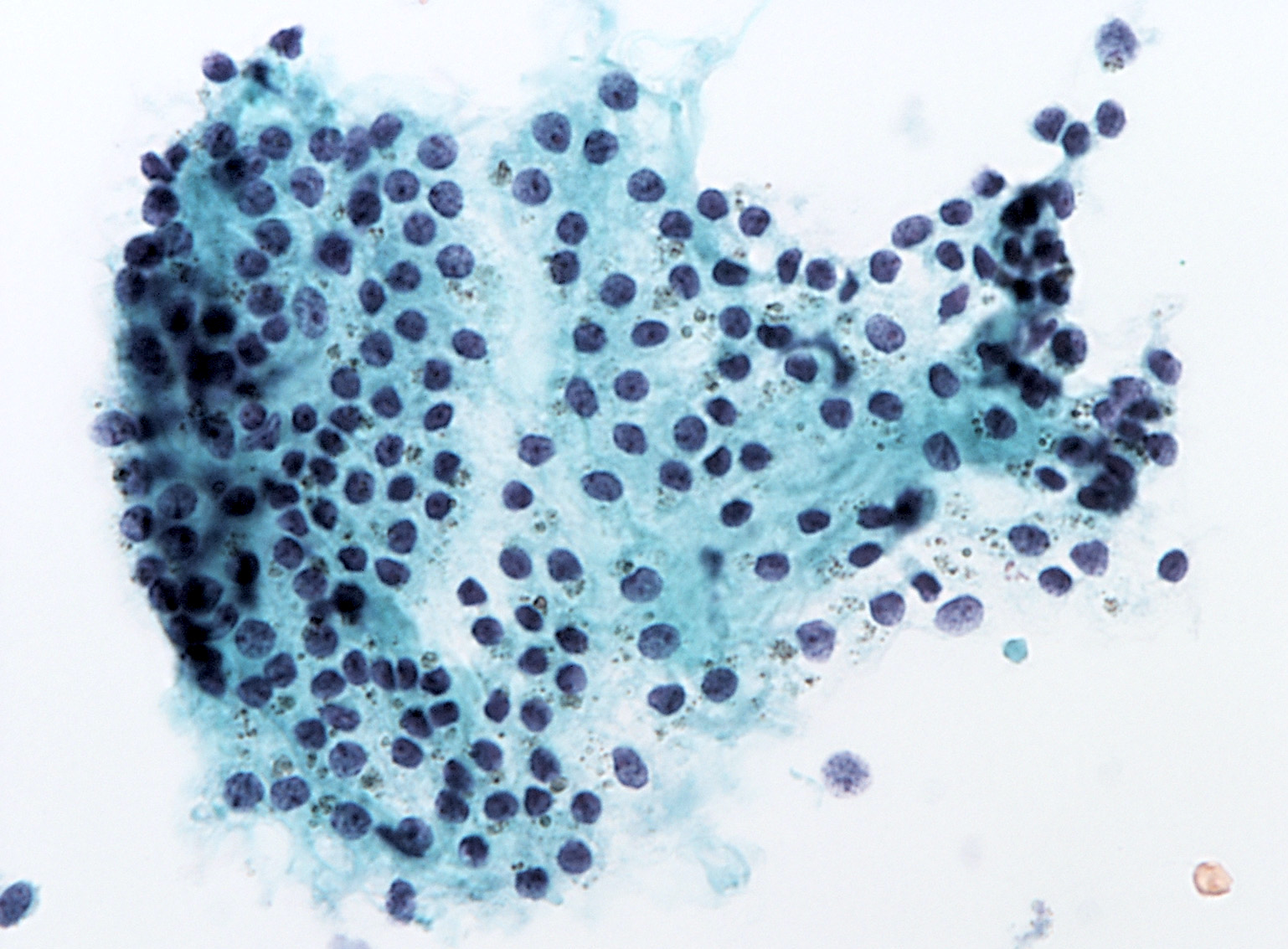
Malignant:
Papillary carcinoma

Medullary carcinoma
Insular carcinoma
Anaplastic carcinoma
Lymphoma
Algorithmic approach to thyroid FNA
Head & tail of the Bethesda system
Thyroid cytology: approach
Thyroid cytology: cases
Thyroid cytology: ND/UNS, benign and FN/SFN
Thyroid cytology: malignant, SUS and AUS/FLUS
Thyroid cytopathology
Case #98
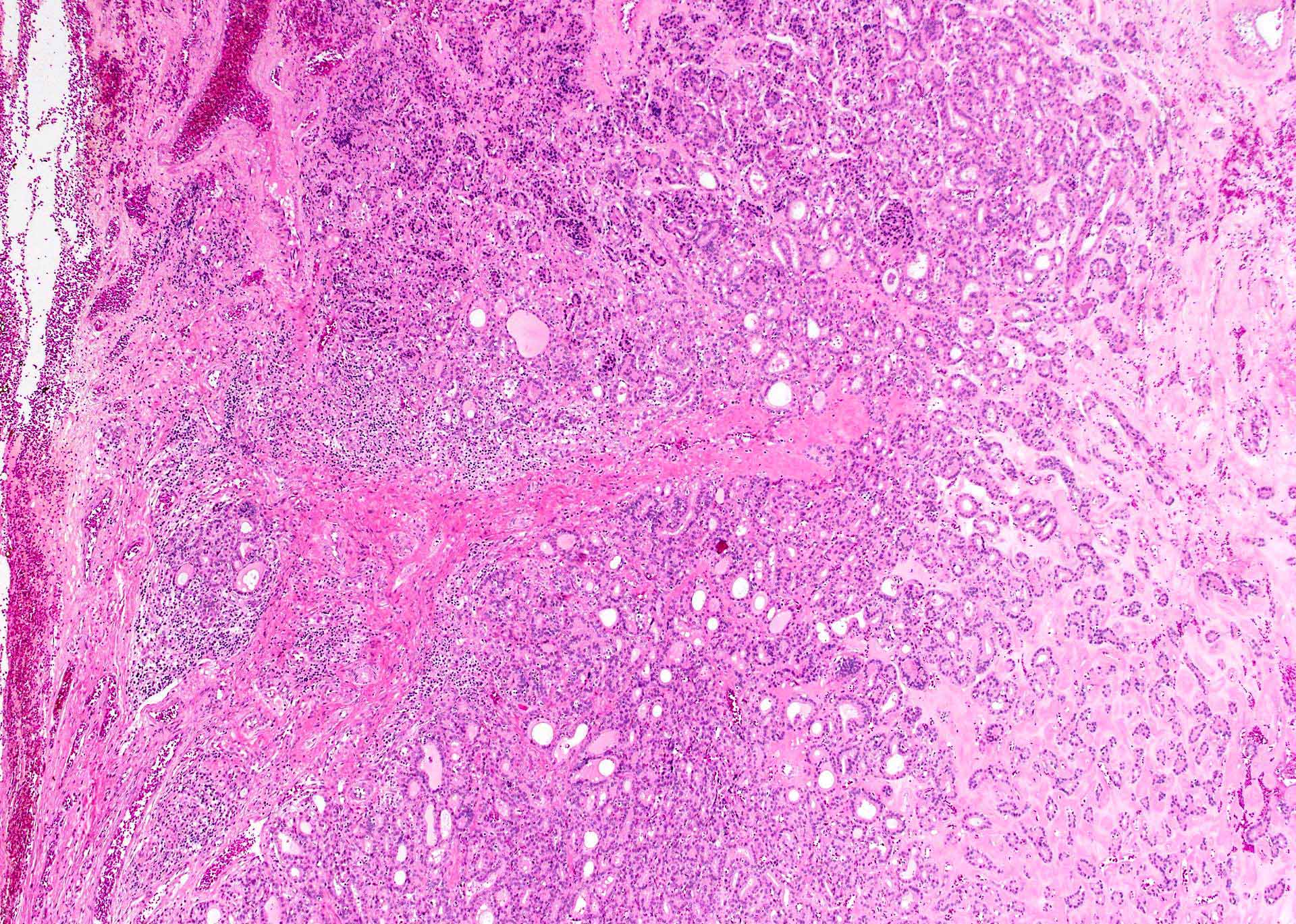
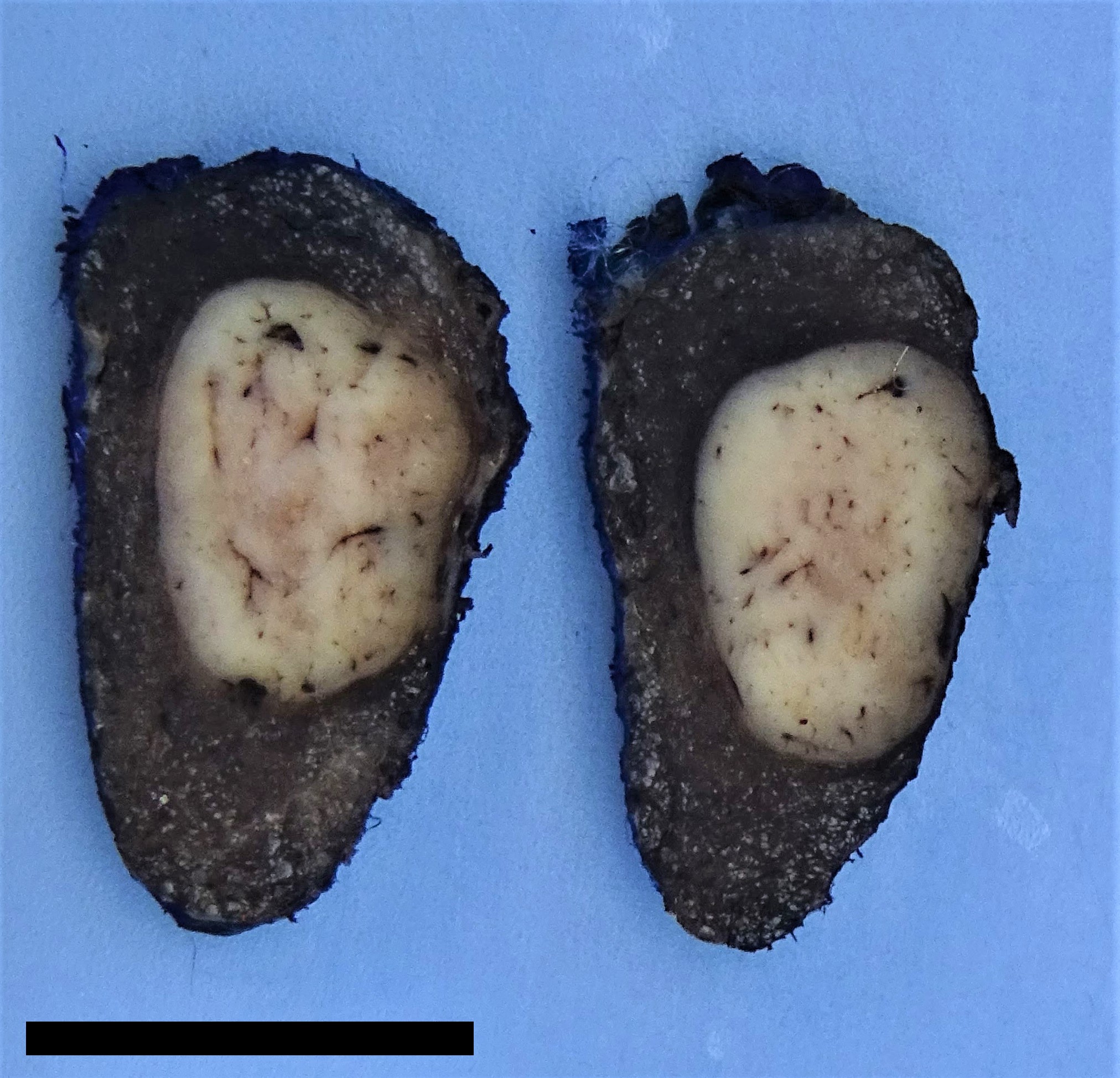
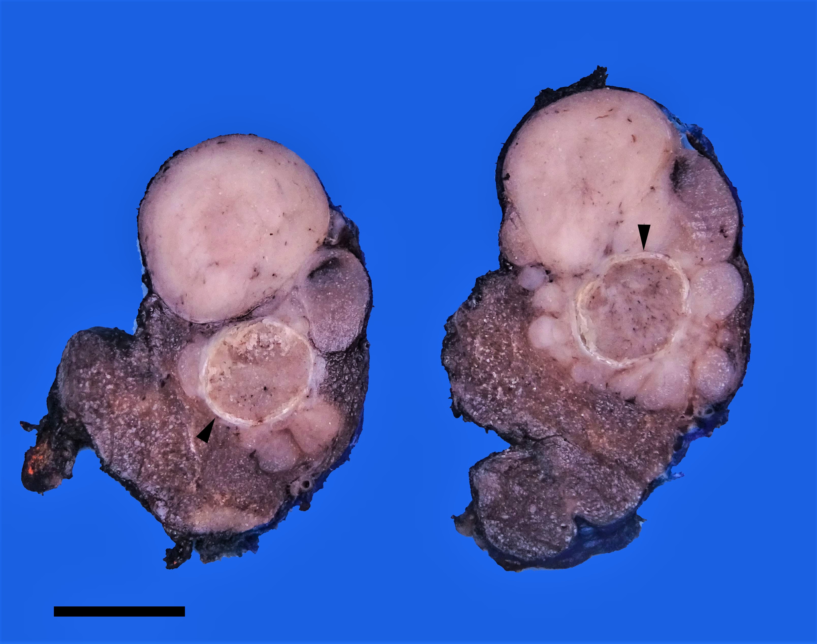
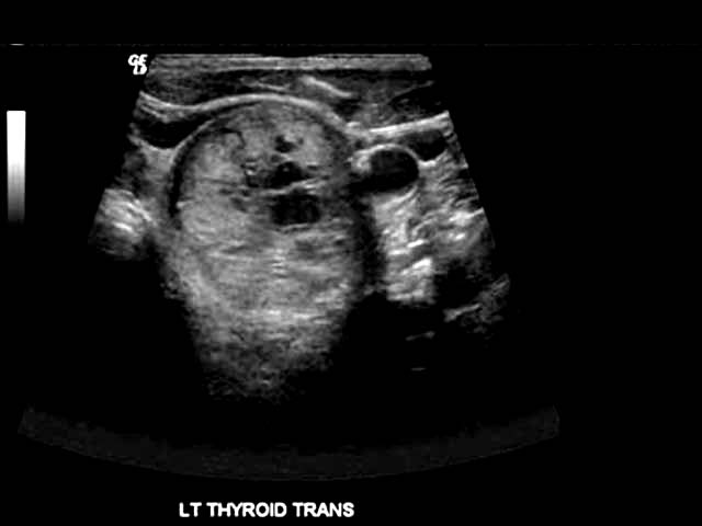
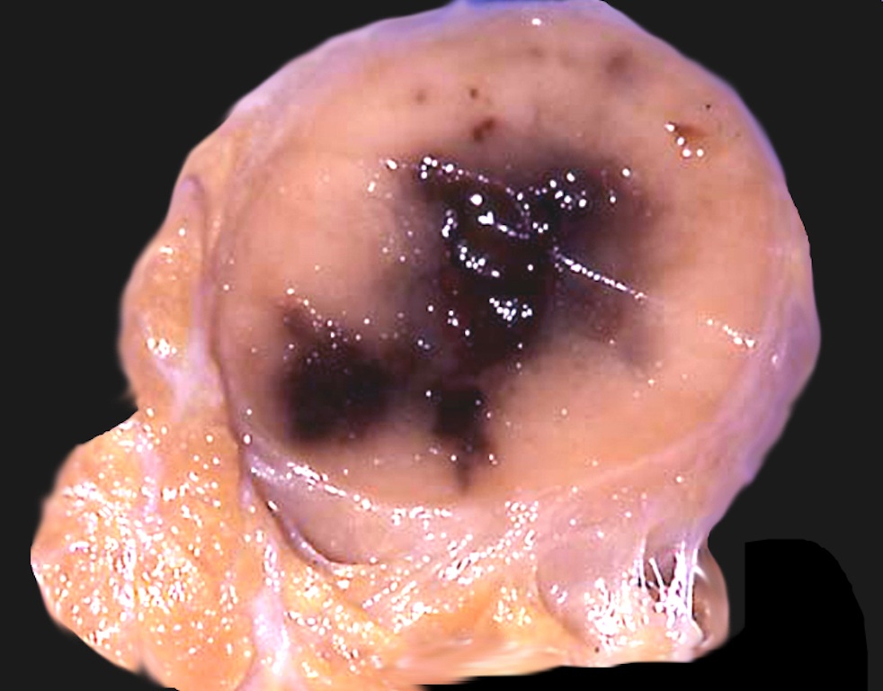
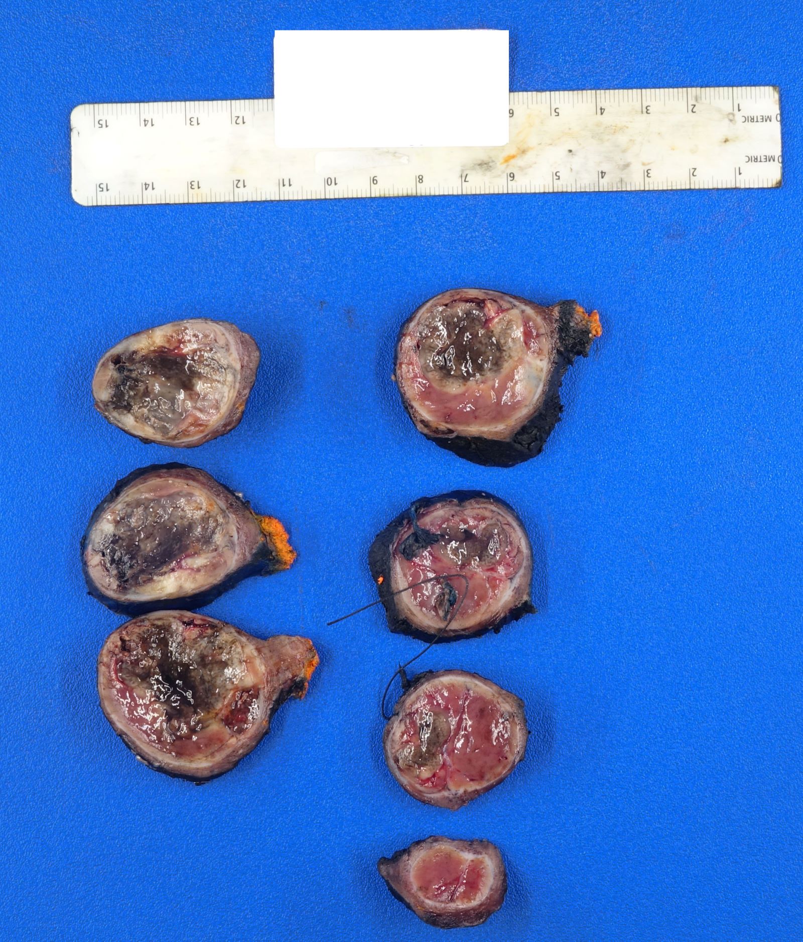
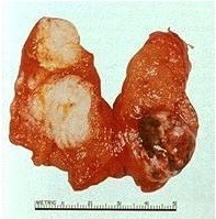
Thyroid gland with black pigment
Images hosted on other servers:

Black thyroid and follicular adenoma

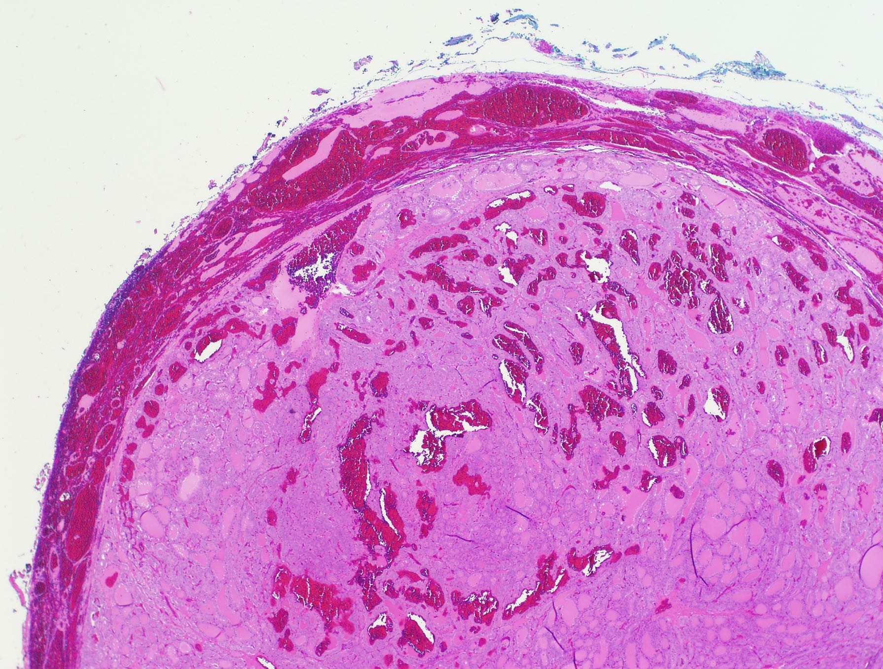
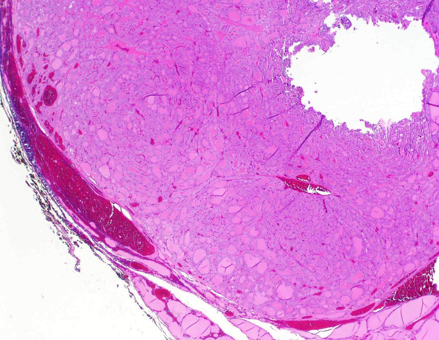
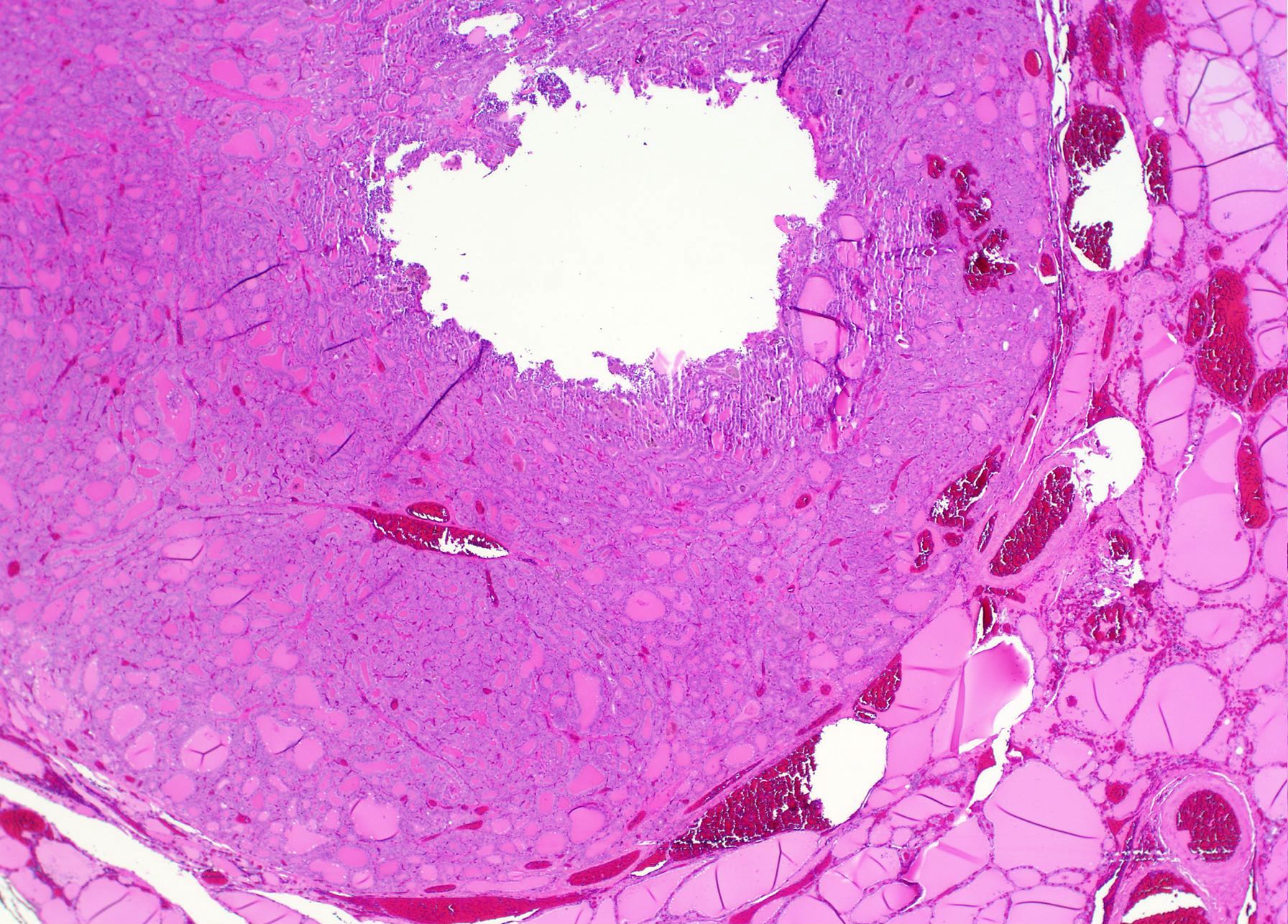
Cut surface

Coal black coloration
Contributed by Andrey Bychkov, M.D., Ph.D. and Mark R. Wick M.D.
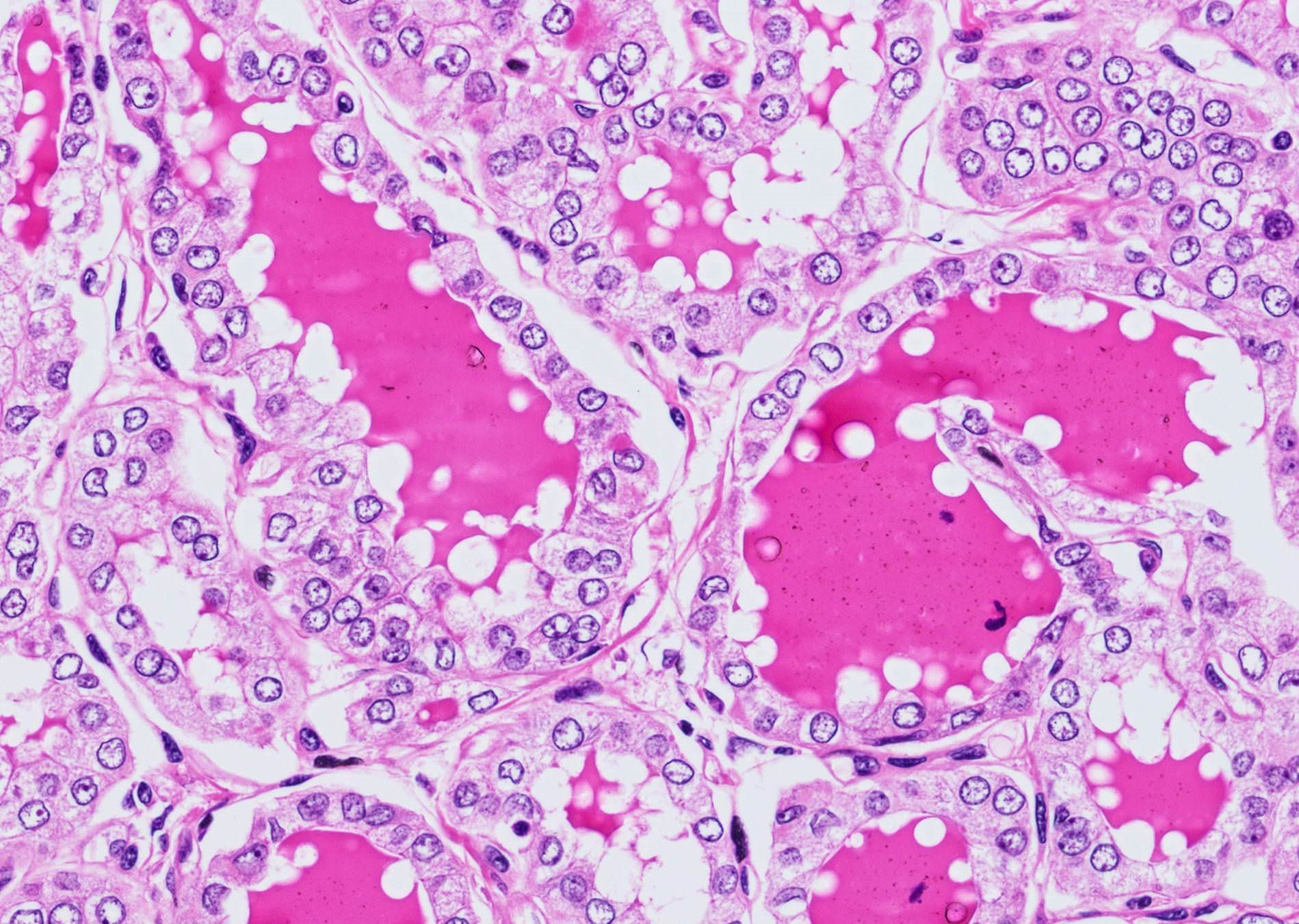
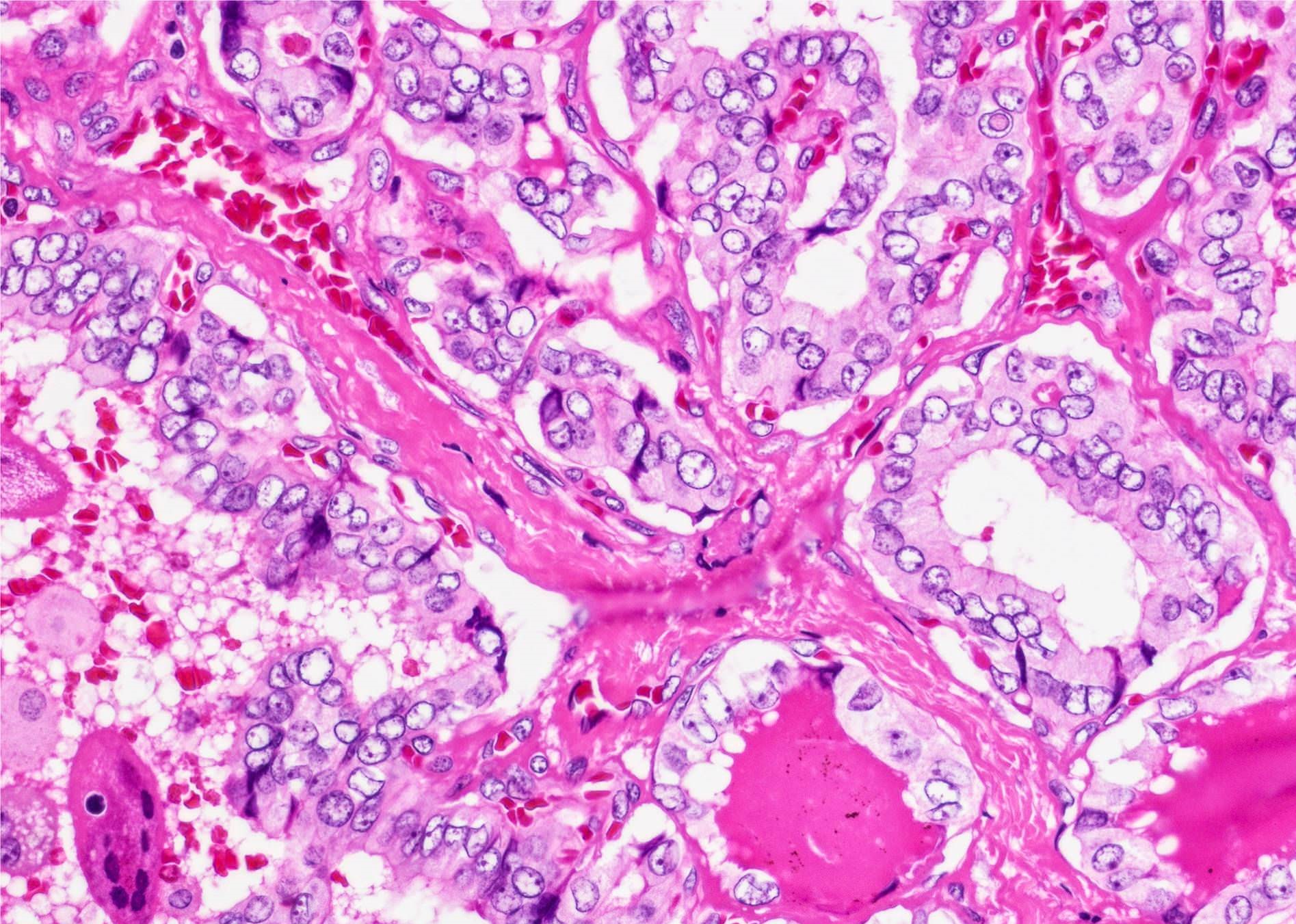
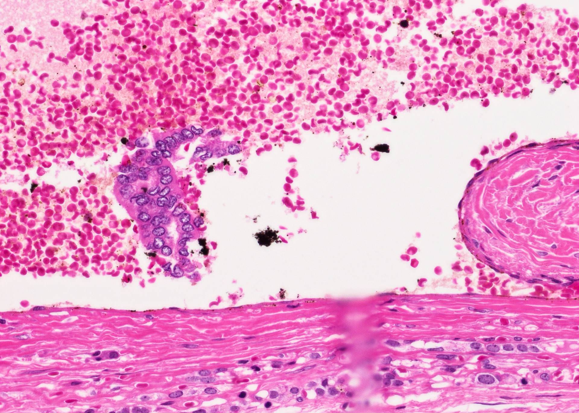
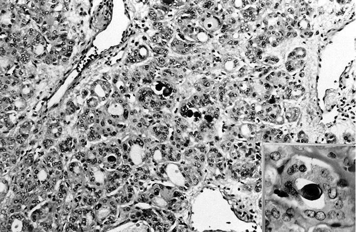
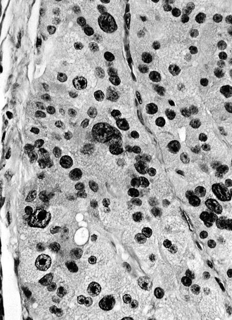
Perinuclear brown pigment
Minocycline induced pigmentation
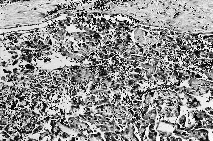
Images hosted on other servers:

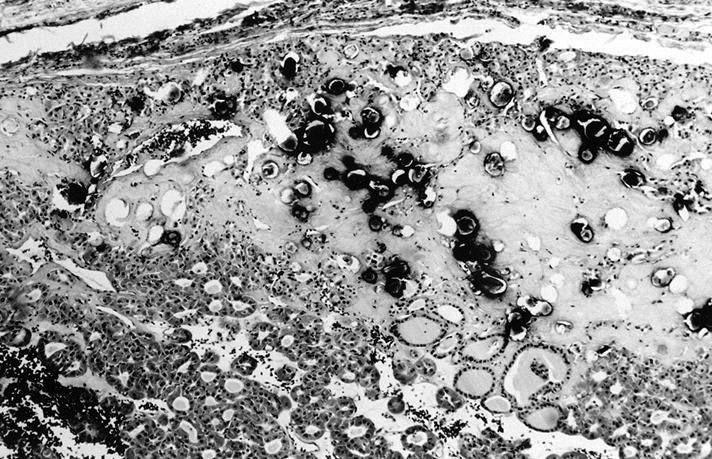
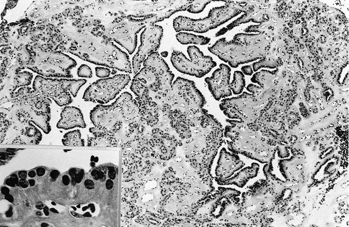
Pigmented cells and colloid

Fontana-Masson
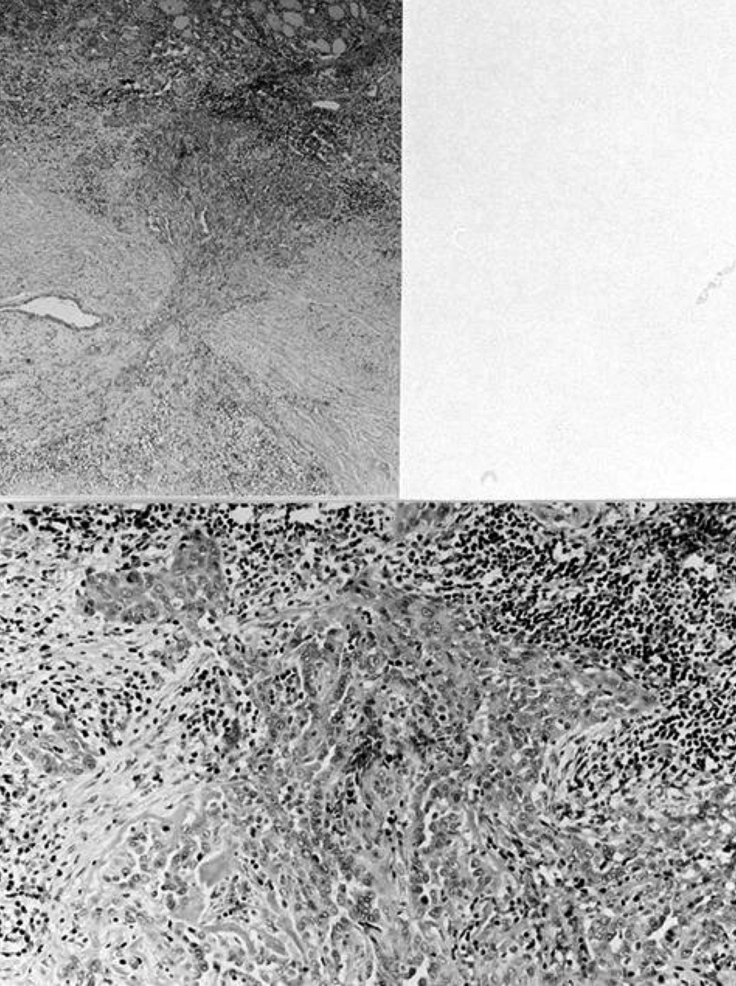
Images hosted on other servers:

Pigment granules in cytoplasm and colloid space

Pigment deposits and Xray spectrogram

Experimental black thyroid, dog

Experimental black thyroid, monkey
Images hosted on other servers:
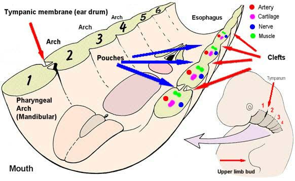
Branchial apparatus
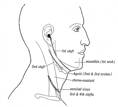
Branchial cleft anomalies location

Anatomical relations
Images hosted on other servers:

Cystic lesion in left parotid gland

CT fistulogram

CT fistulogram

Infrahyoid cyst
Images hosted on other servers:

Fistula opening (1st branchial cleft anomaly)

First branchial cleft fistula opening

Bilateral fistula opening

Saliva coming out, third branchial fistula

Fistula opening above sternocleidomastoid muscle

Sinus originating from pyriform sinus

With cervical abscess

Fistulous tract in right pyriform sinus
Images hosted on other servers:

Fistula tract

Fistula tract ending in parotid gland

Collaural fistula

Third branchial fistula

Fistula tract exiting pharynx

Fourth branchial fistula
Contributed by Andrey Bychkov, M.D., Ph.D.
PTC: cystic node mimics branchial cyst
Hashimoto thyroiditis with lymphoepithelial cyst
Images hosted on other servers:

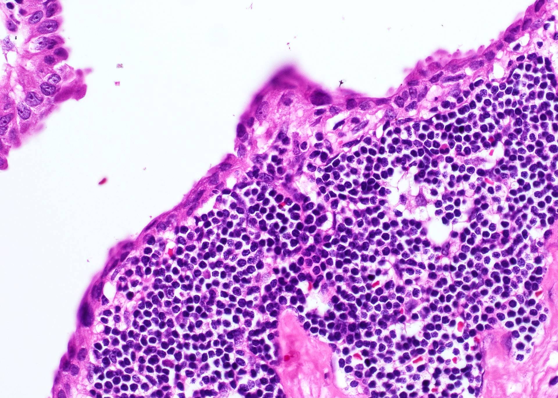
Cyst lined by stratified squamous epithelium

Sinus tract in parotid gland

Cyst wall with chronic inflammation

Fibrotic wall of a cyst

Lymphoid aggregates in cyst wall

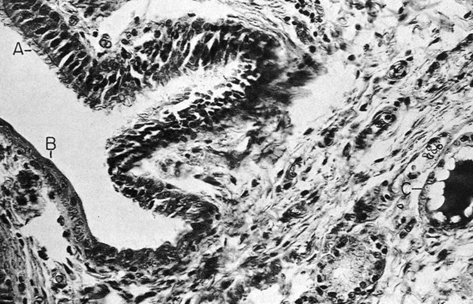
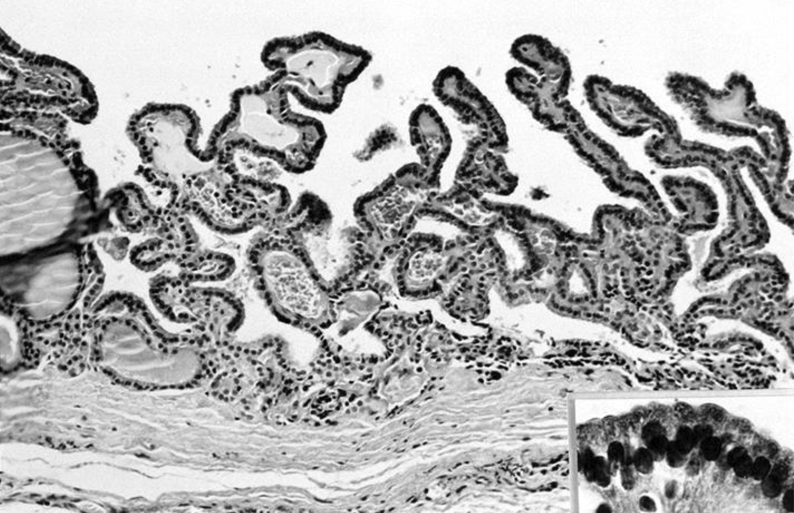
Ciliated pseudostratified columnar epithelium
Images hosted on other servers:
Squamous cells, keratin debris
High power

Pap stain
Inflammation
Cholesterol crystals
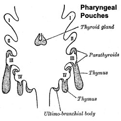
Pharyngeal pouches
Contributed by Mark R. Wick, M.D.

Various images
AFIP images
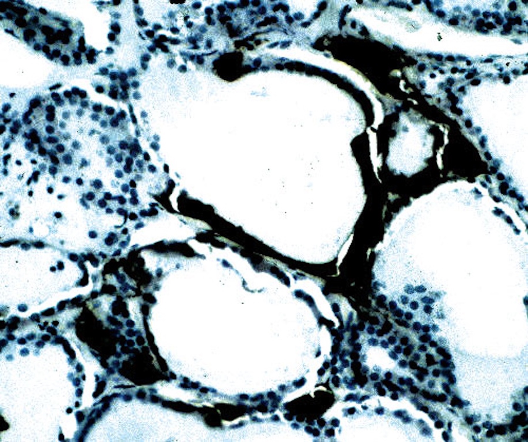
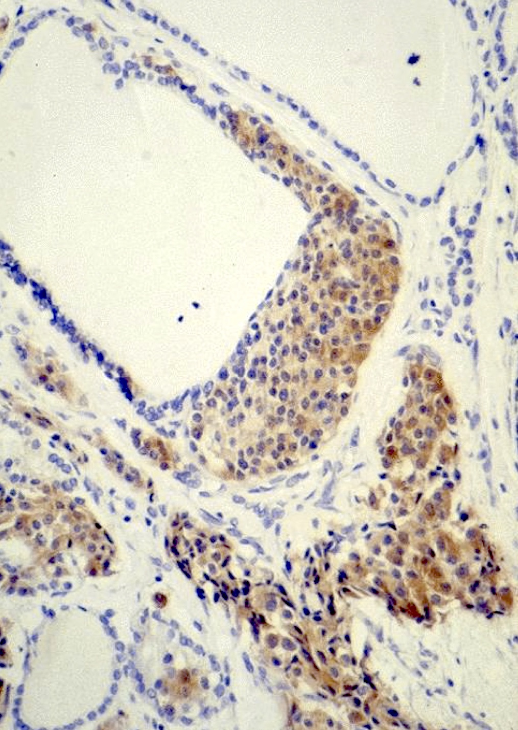
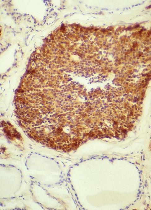
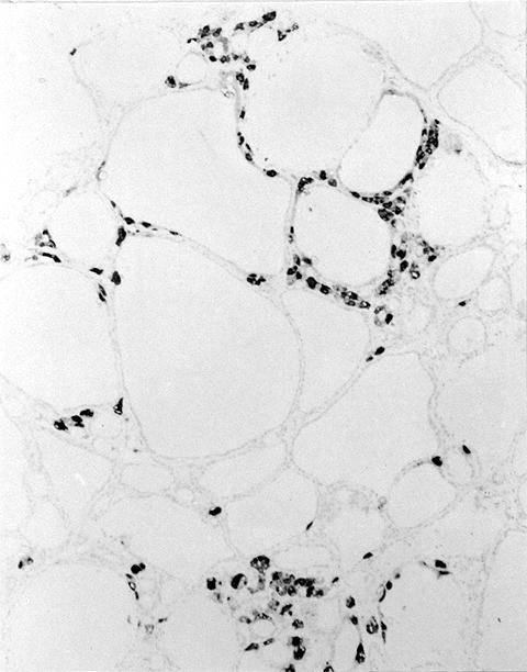
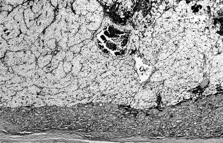
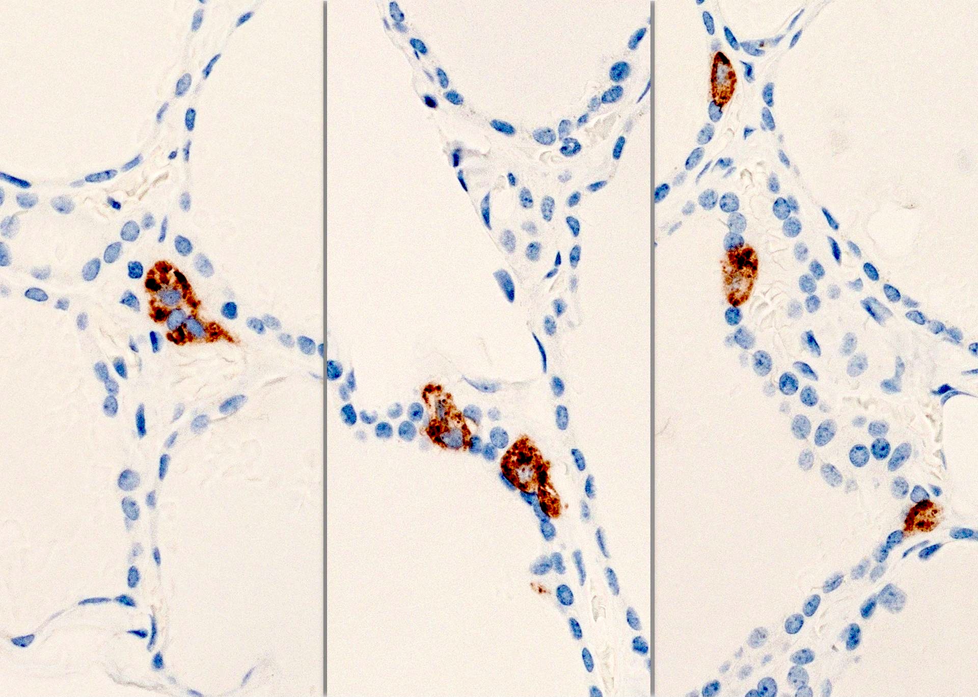
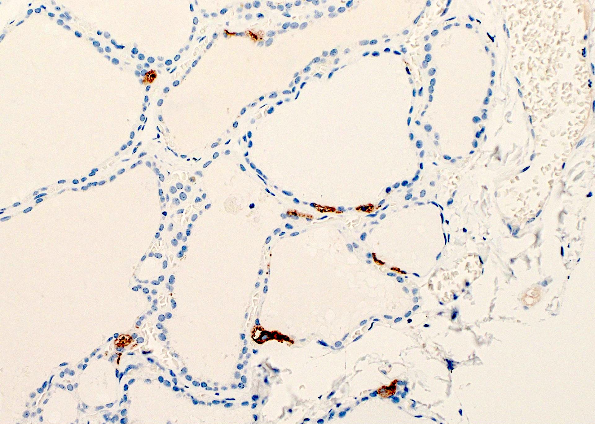
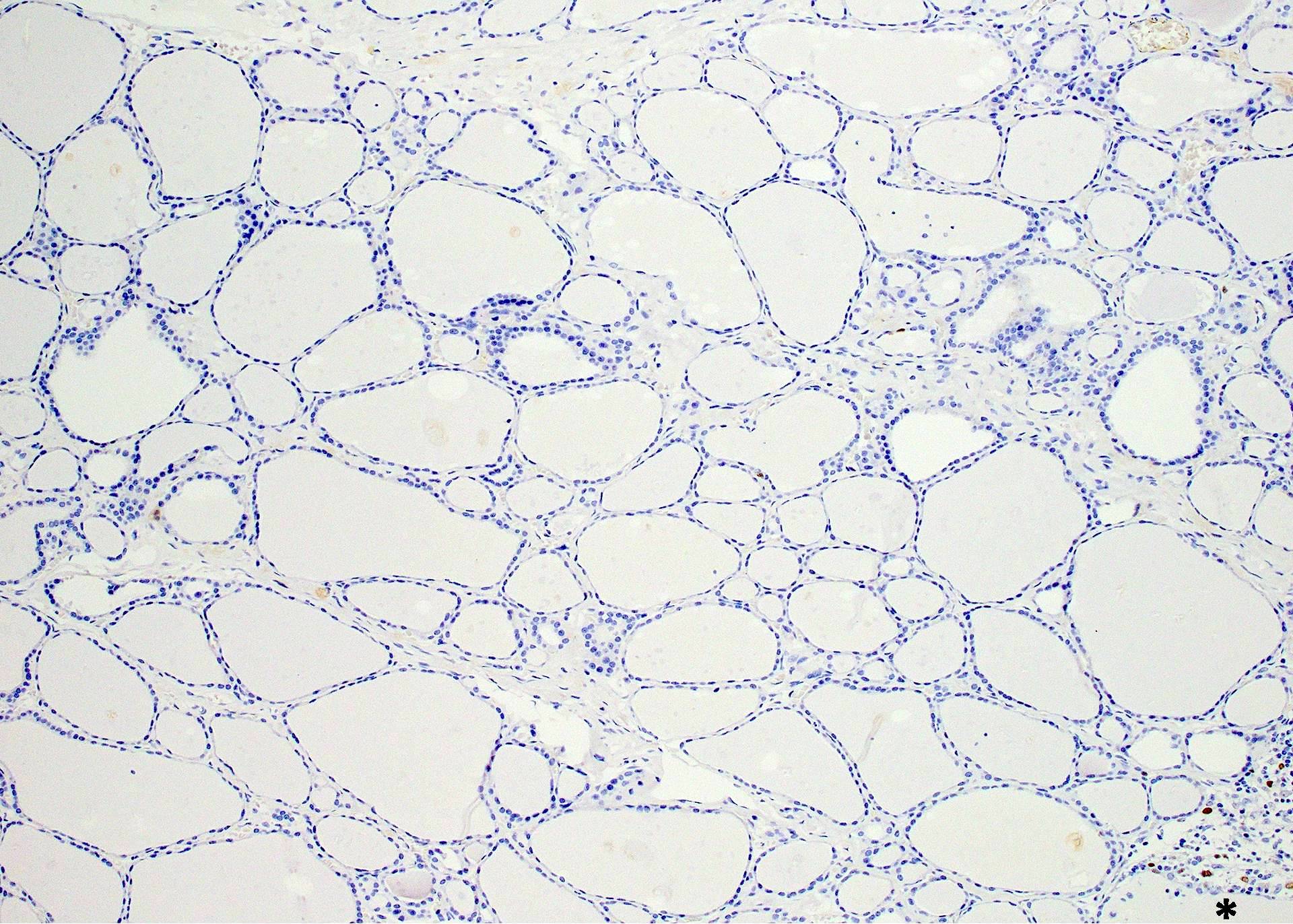
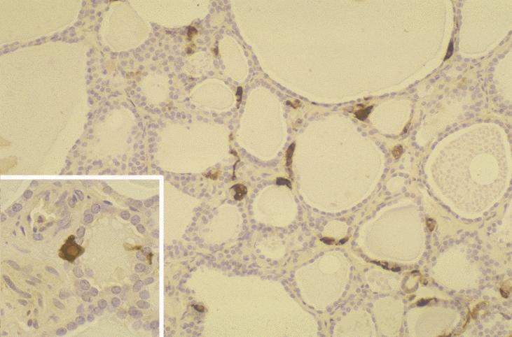
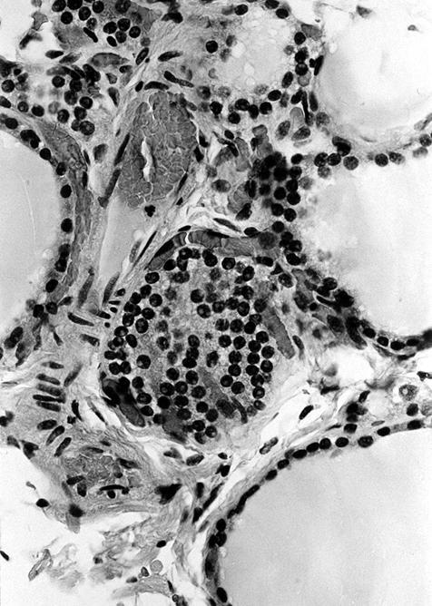
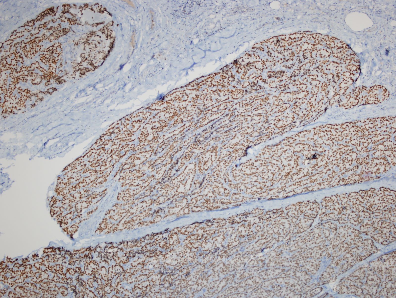
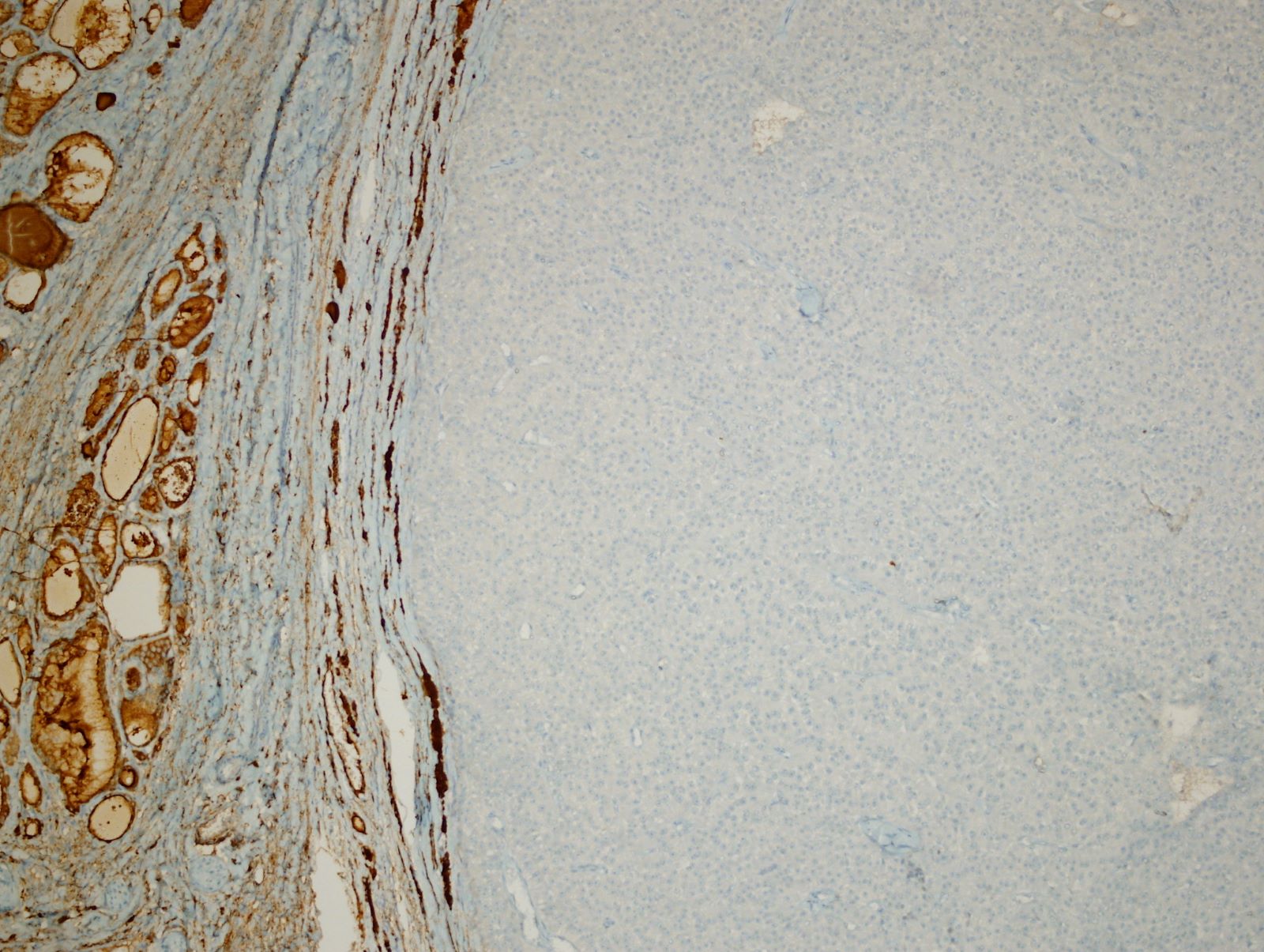
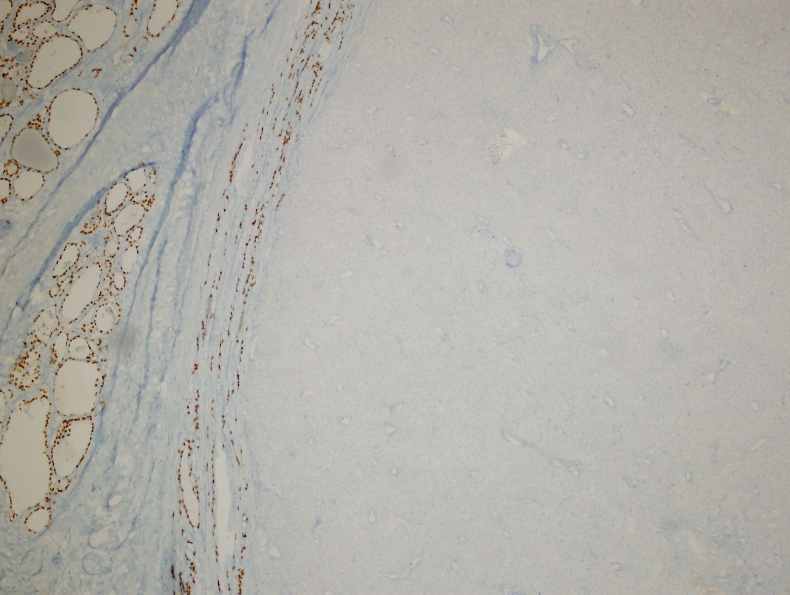
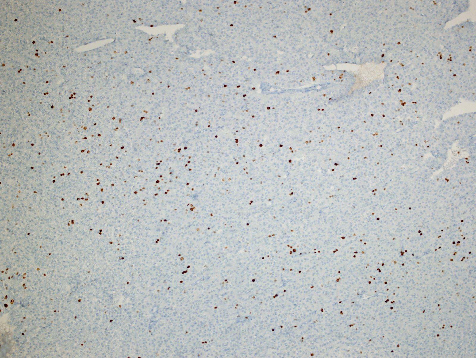
Calcitonin staining:
C cells highlighted
Nodular CCH
C cells adjacent to follicular carcinoma
MEN2A patients:
Circumferential proliferation
Focal proliferation of C cells
Eccentric intrafollicular proliferation
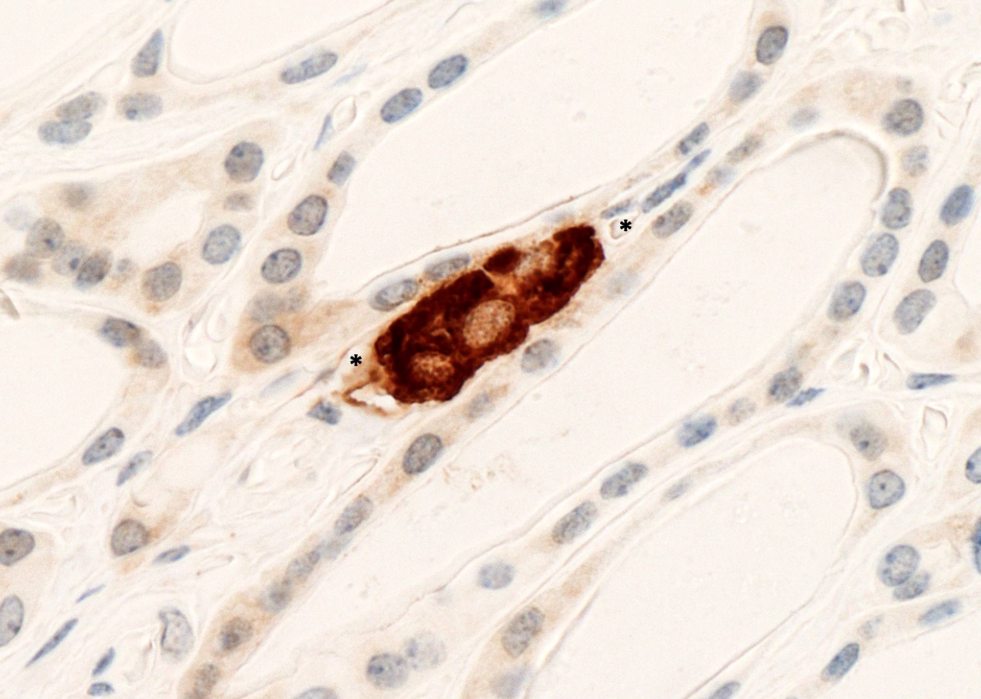
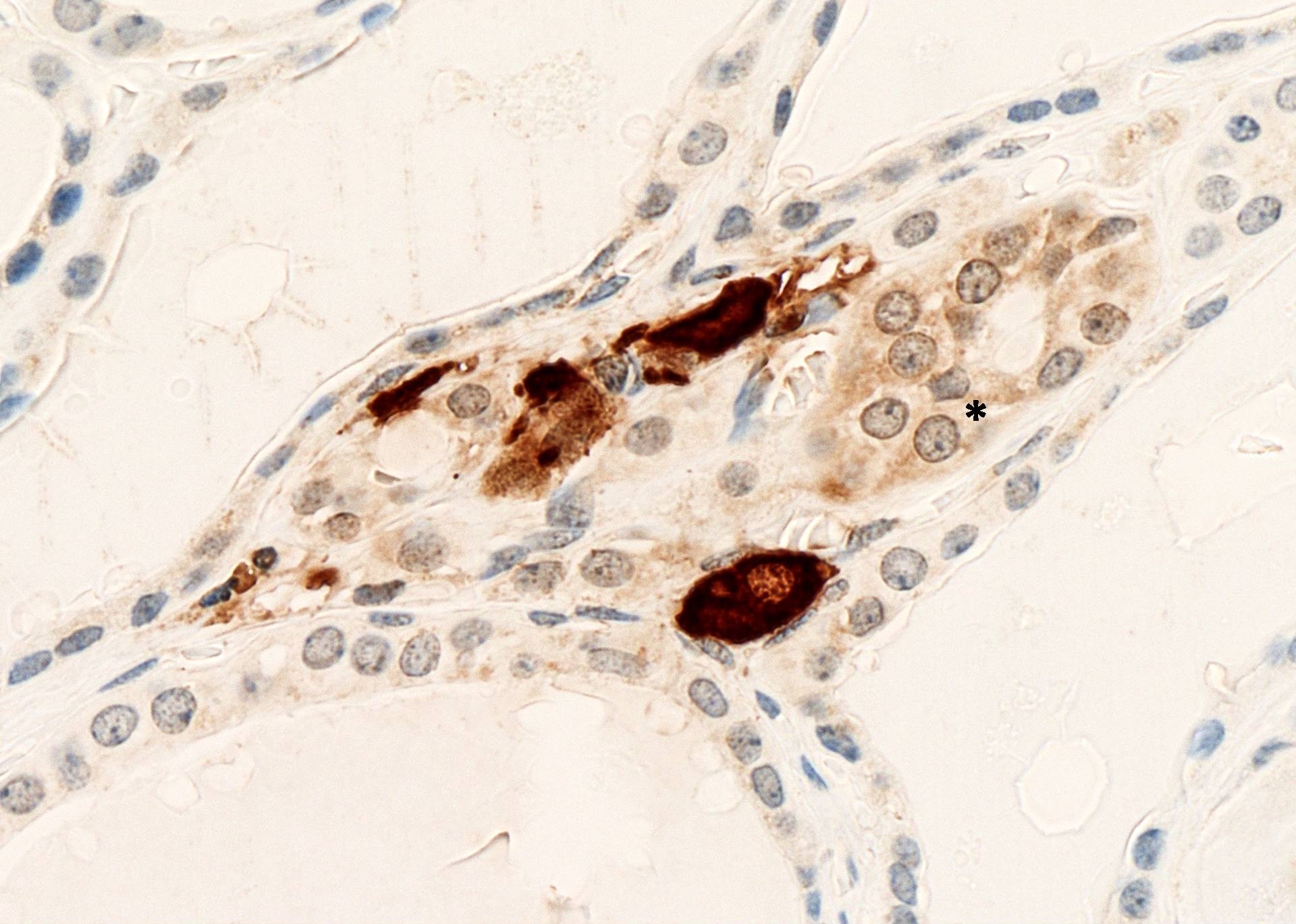
MEN2A patients with early medullary carcinoma:
Group of C cells extends into interstitium
Images hosted on other servers:

Calcitonin staining, C cells form ring around follicle

Associated with nodular goiter

MEN2A patient

High power
Images hosted on other servers:

Eggshell calcified retrosternal thyroid
Images hosted on other servers:

Muddy appearance
Contributed by Andrey Bychkov, M.D., Ph.D.
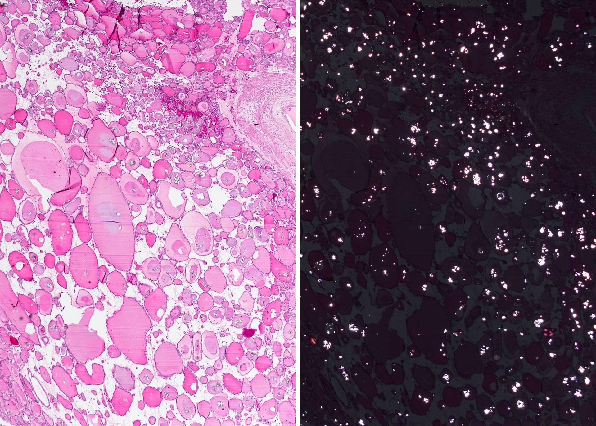
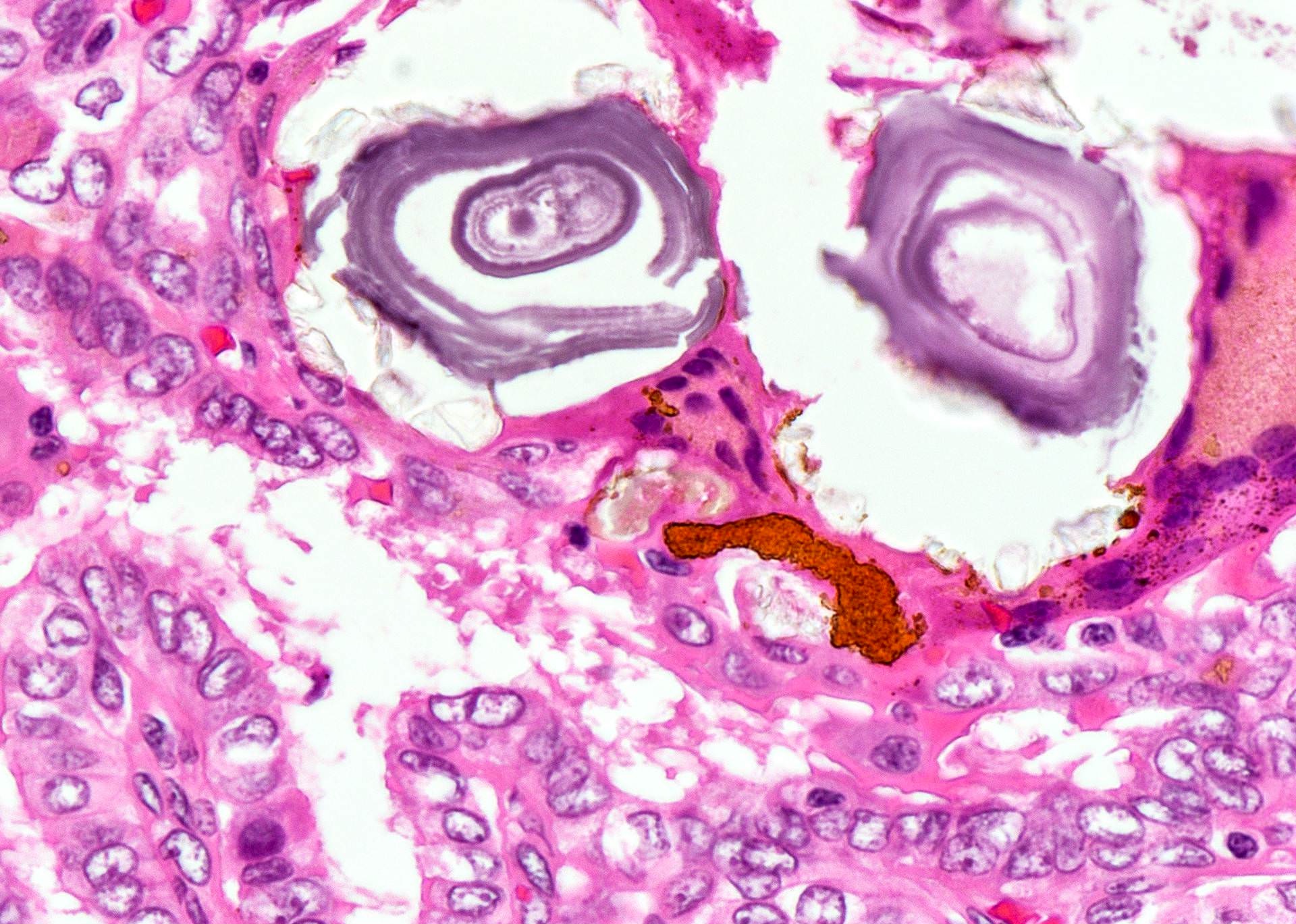
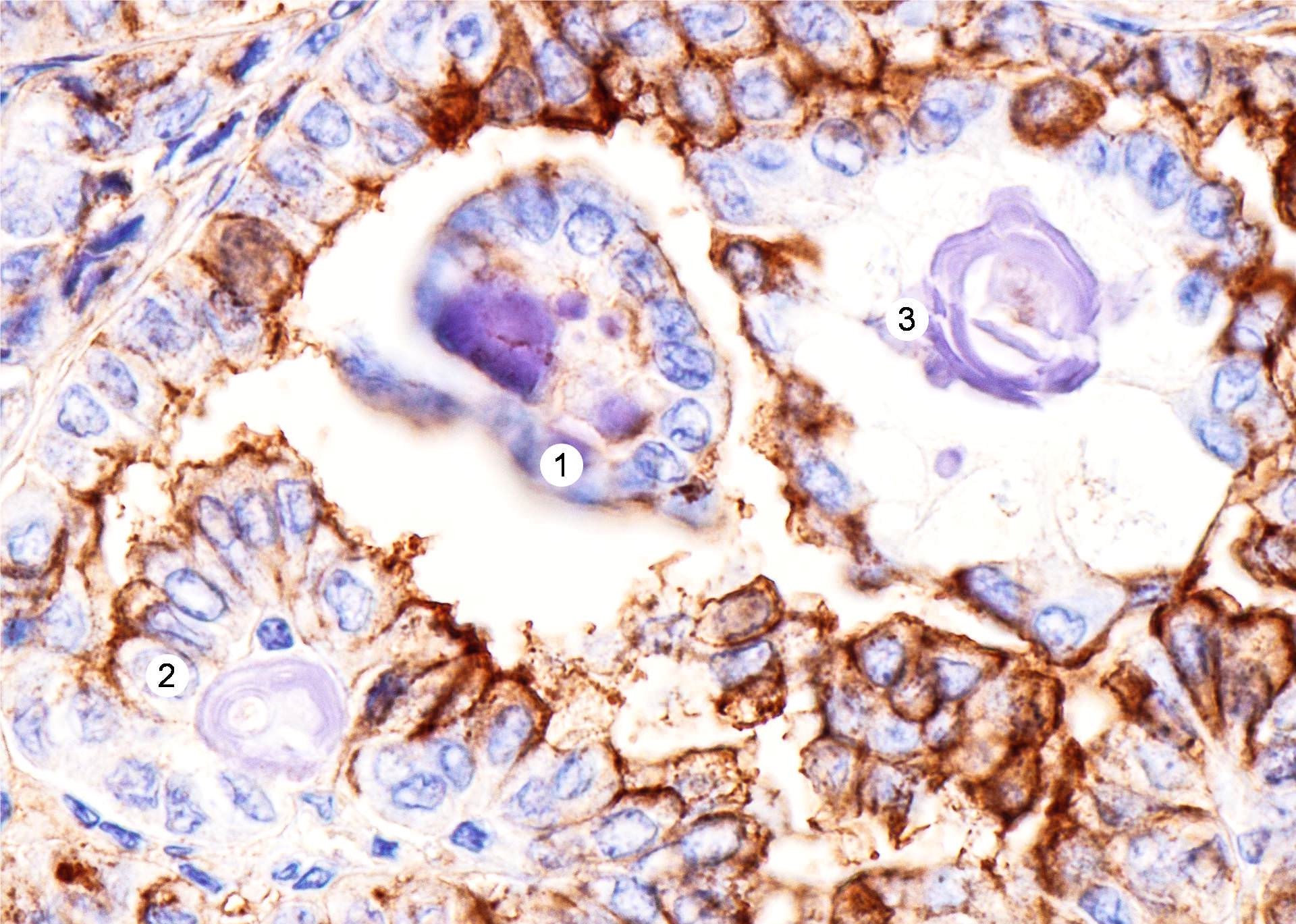
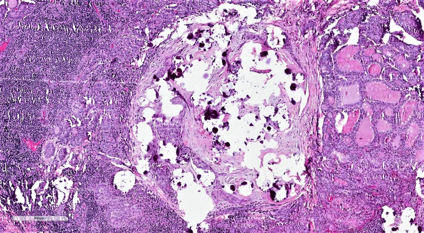
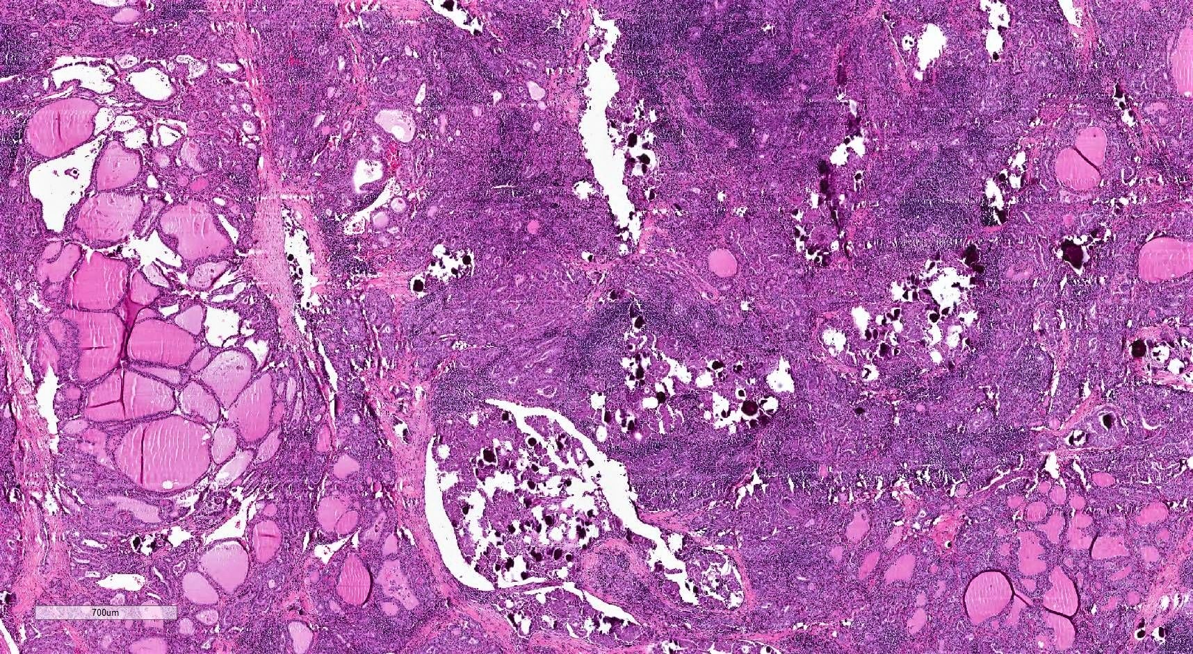
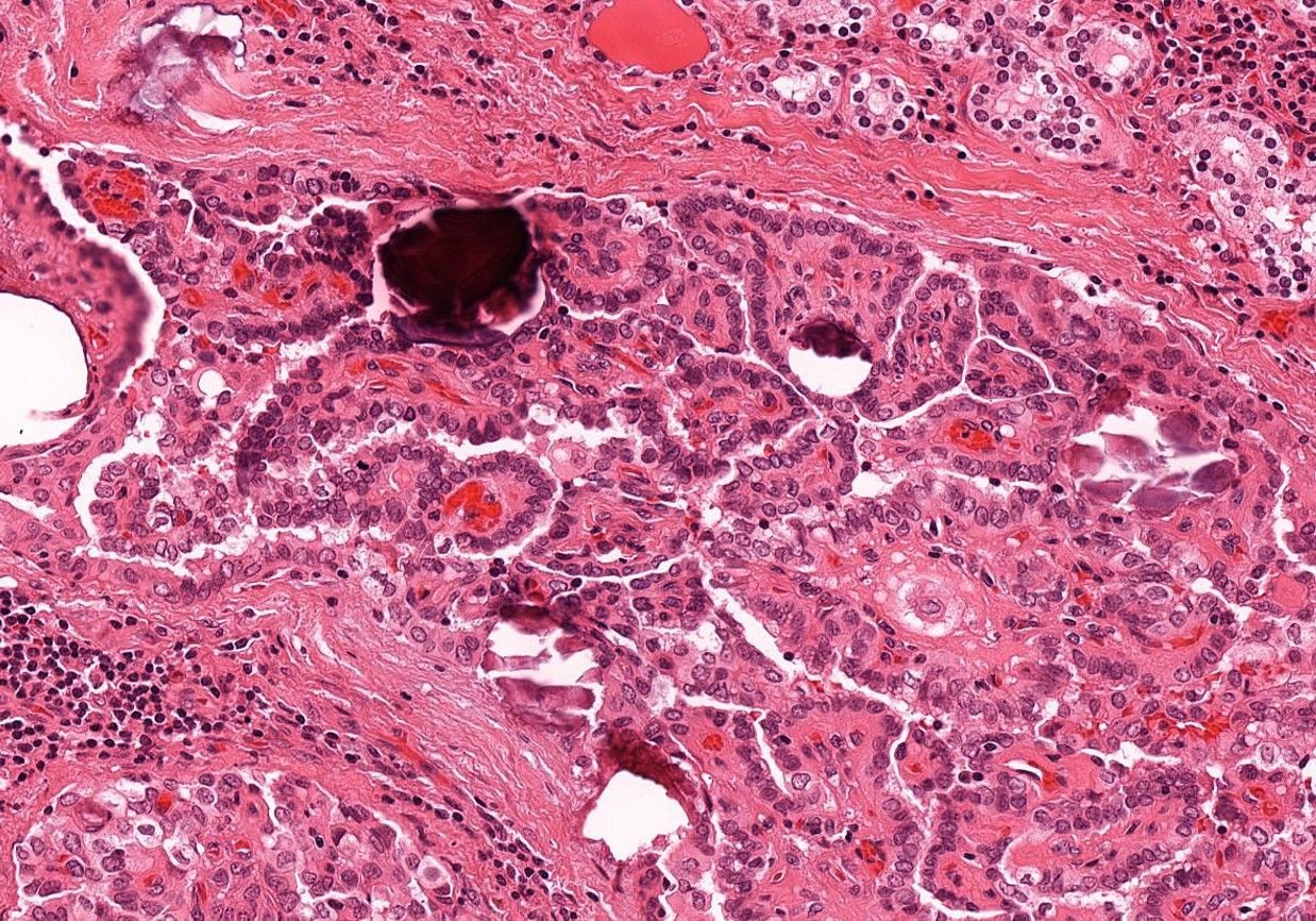
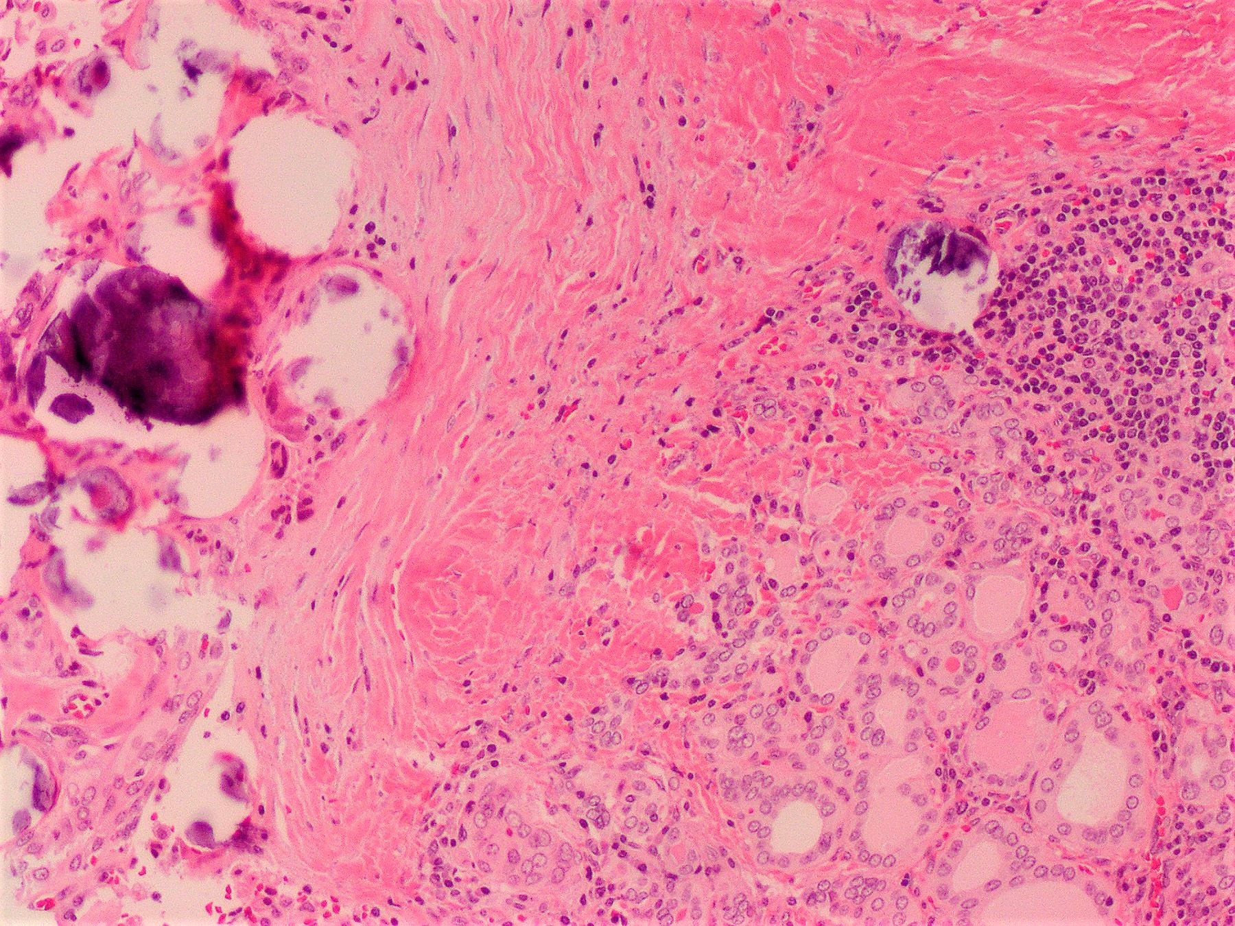
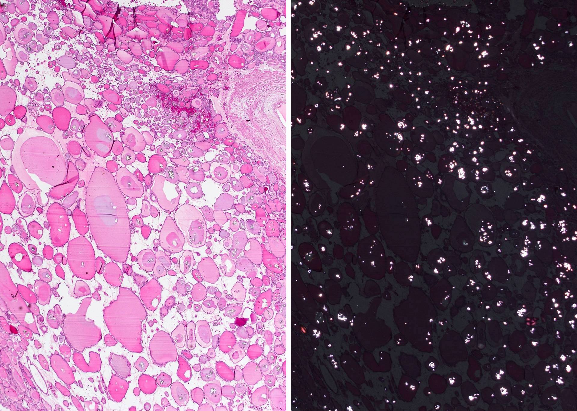
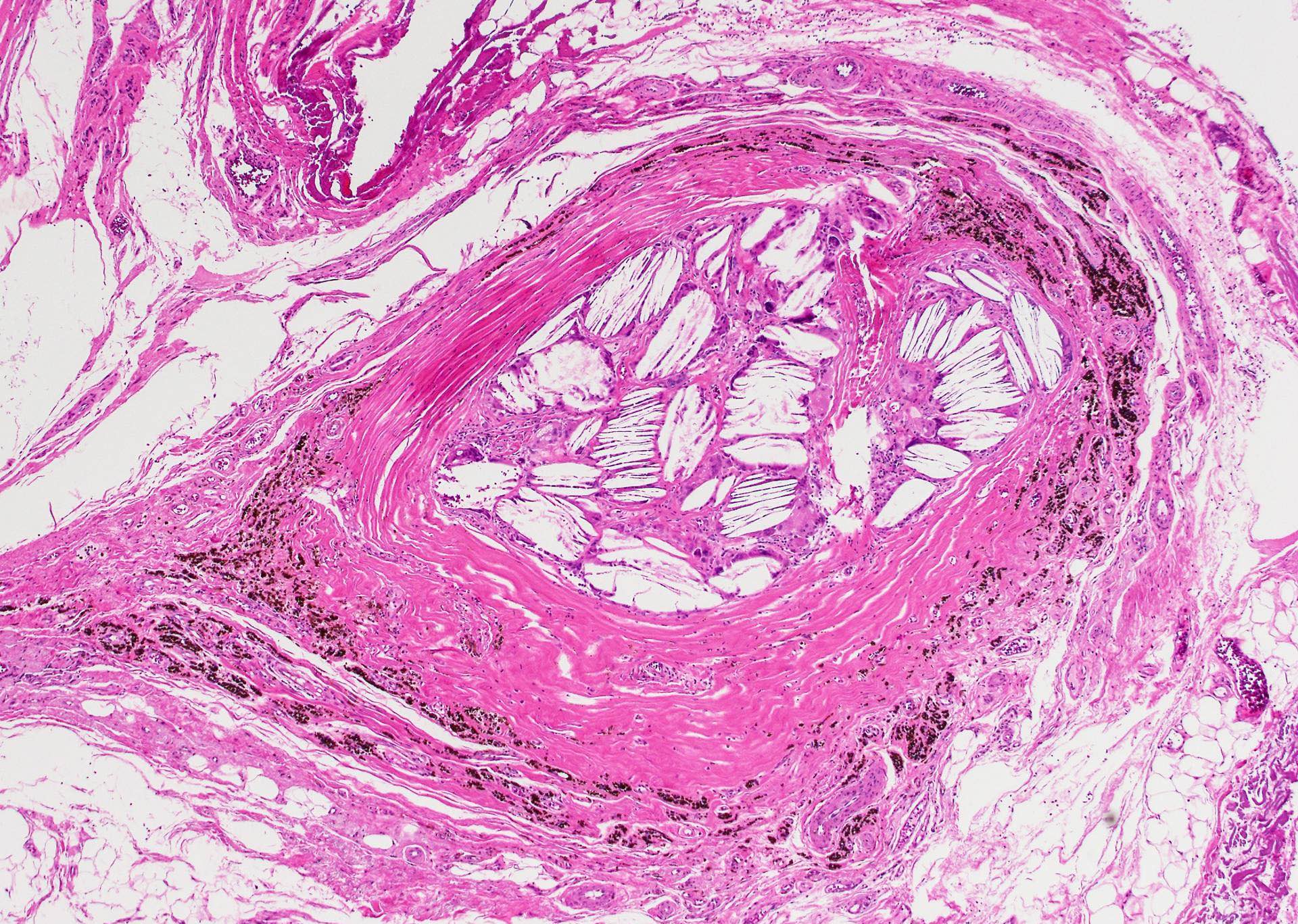
Calcium oxalate crystals (abundant)
Calcium oxalate crystals
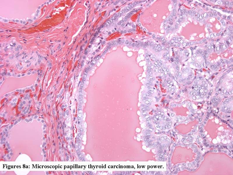
Papillary thyroid carcinoma
PTC follicular variant with ossification
Concentric layers of calcium deposition in tumor stroma
Psammoma bodies, laminated calcifications
Evolution in psammoma body in PTC
Psammoma bodies in benign appearing thyroid
Images hosted on other servers:

Extensive dystrophic calcification


Osseous metaplasia with fatty marrow

Calcification induced artifact
Pediatric thyroid nodules by A. Wassner and A. Bauer (2020)
Contributed by Julie Guilmette, M.D.
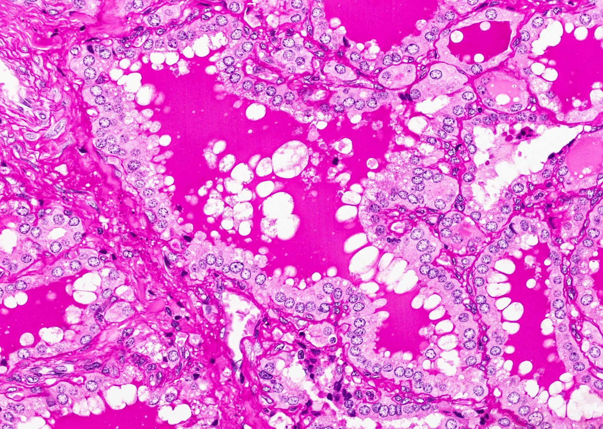
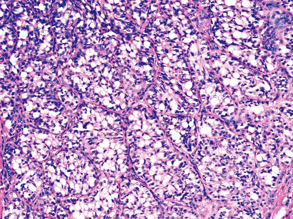
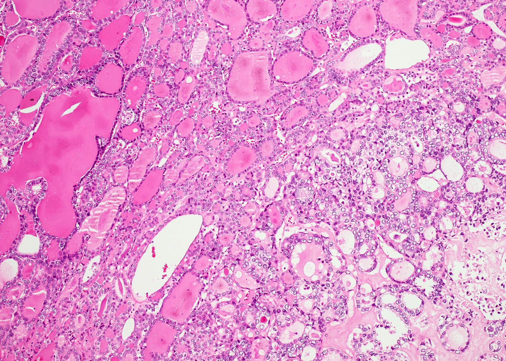
Predominantly follicular architecture
Clear cell features
Nuclear characteristics
HBME1 stain
AFIP images

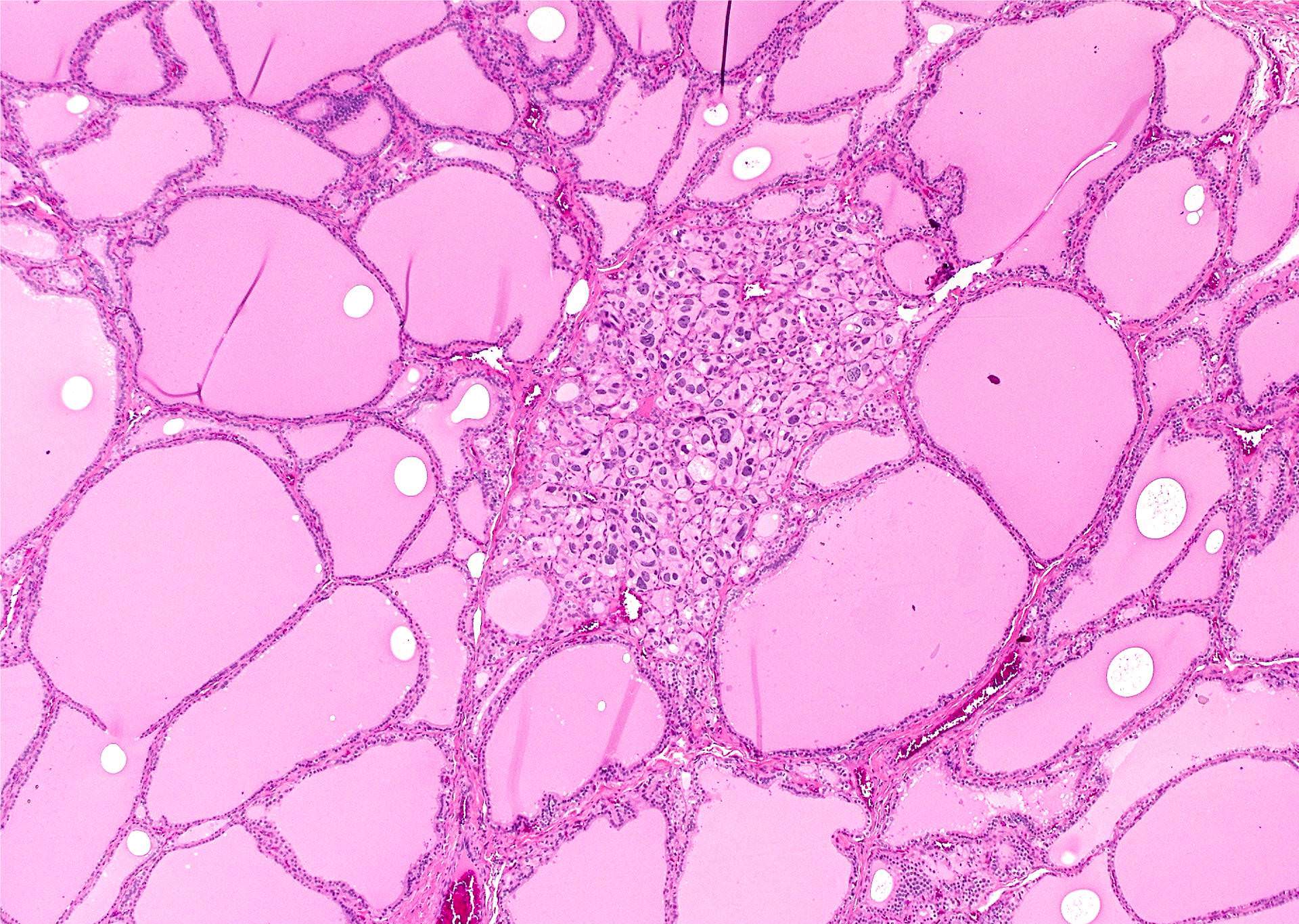
Nodular hyperplasia with clear cell change
Contributed by Andrey Bychkov, M.D., Ph.D. and AFIP

Papillary thyroid carcinoma metastatic to cervical lymph node
Hashimoto thyroiditis with clear cells

Nodular hyperplasia
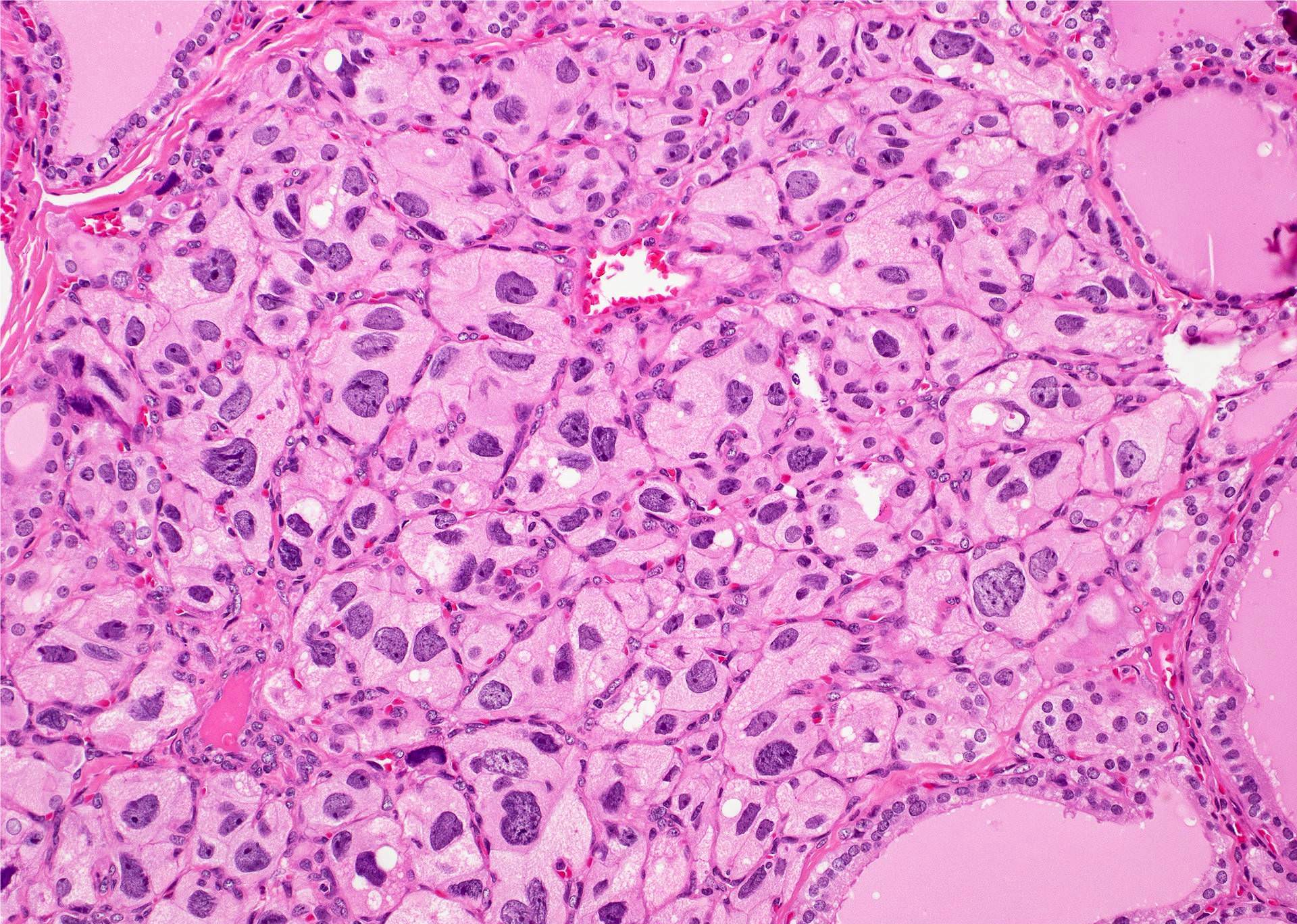
Clear cell change
Clear cell change
(follicular variant)

Follicular carcinoma

Follicular neoplasm with clear cell change
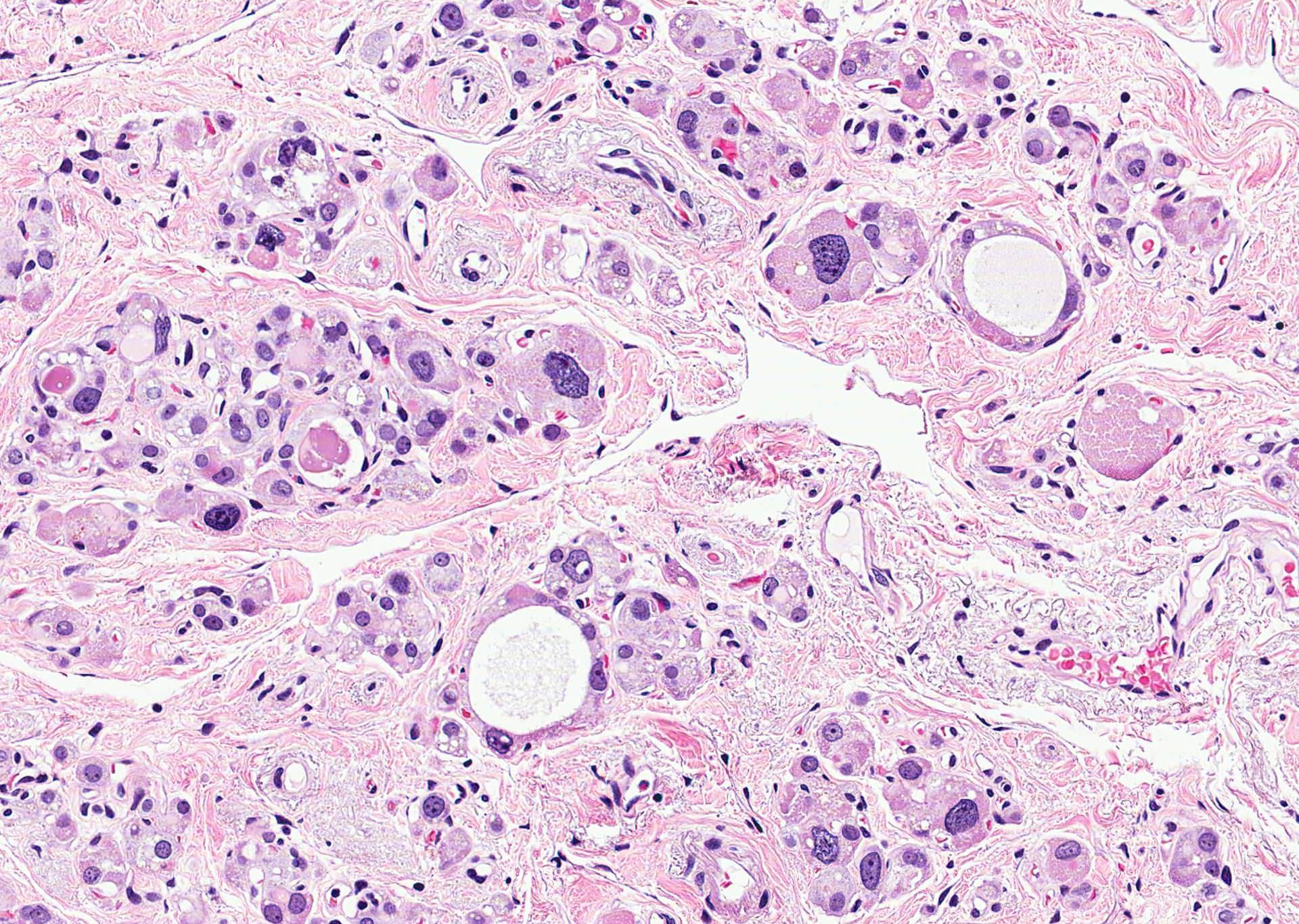
Signet ring follicular adenoma
Sharp demarcation between clear and oncocytic cells
Gradual transition from oncocytic to clear cells
Both patterns exist in same cell

Clear cell type
Adenoma is thyroglobulin+
AFIP images

Signet ring follicular adenoma

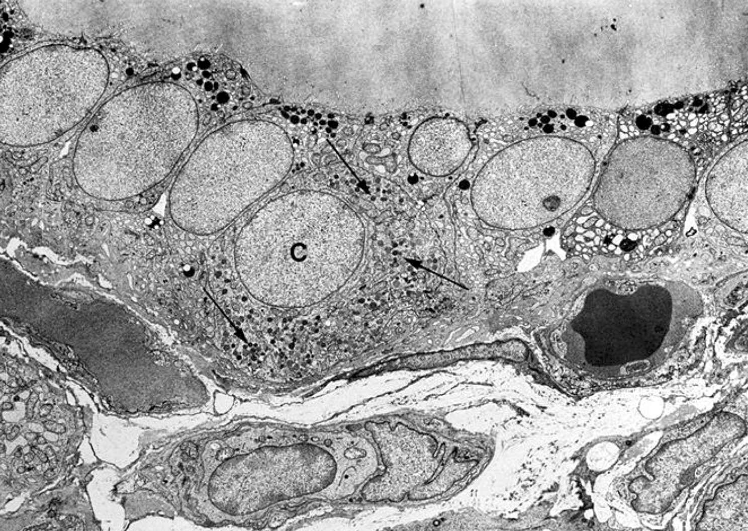
Marked dilation of cisternae
Images hosted on other servers:


Ultrasound
Images hosted on other servers:


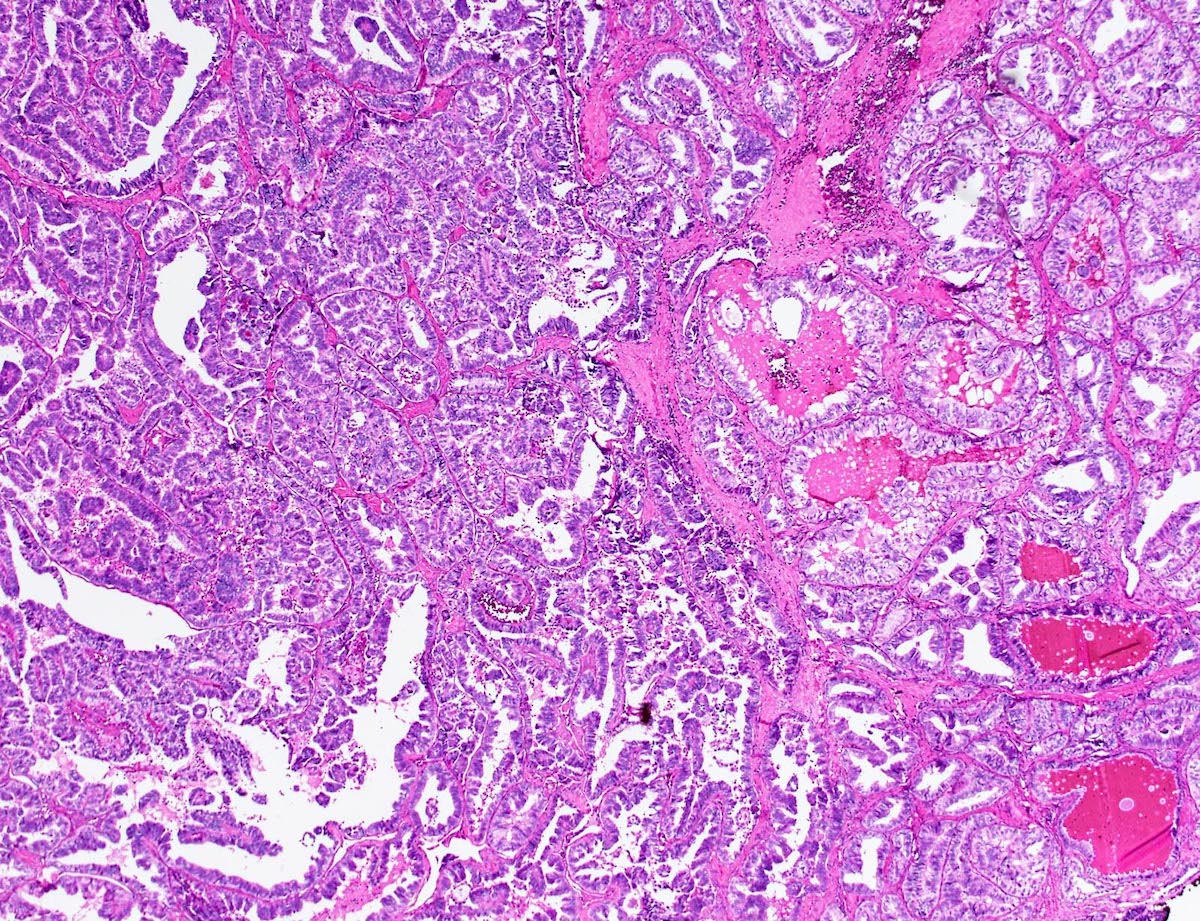
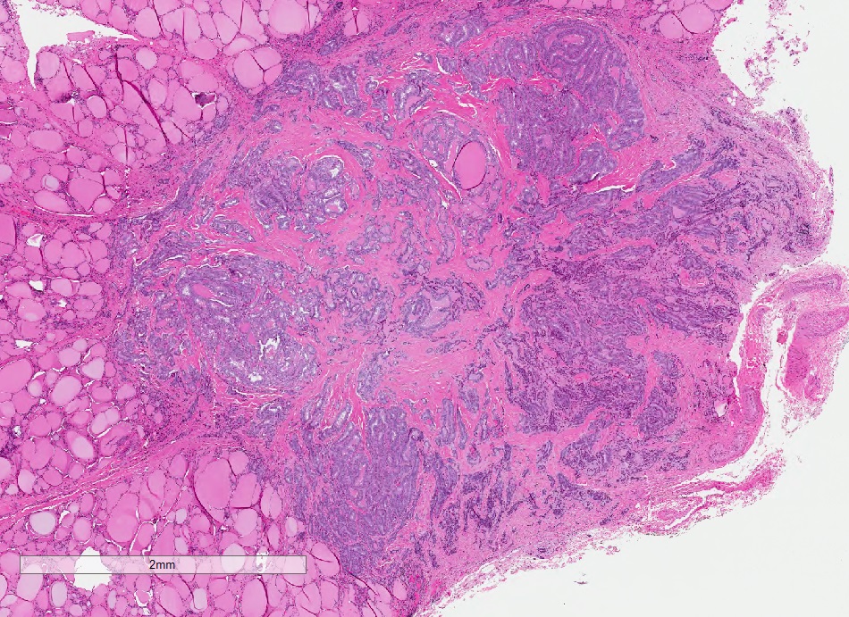
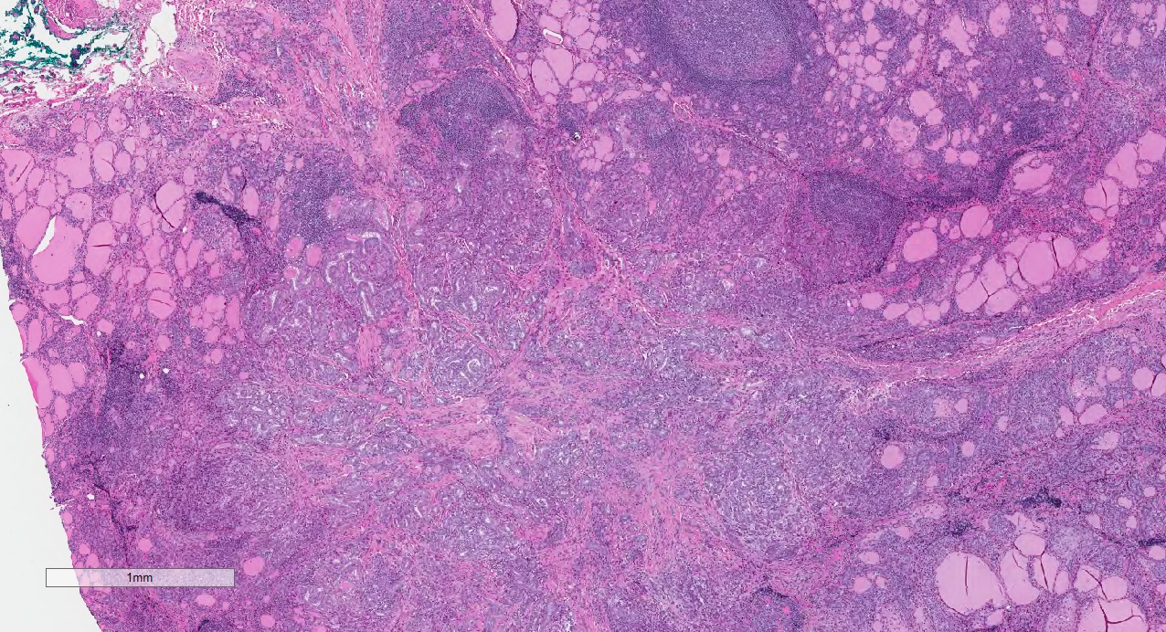
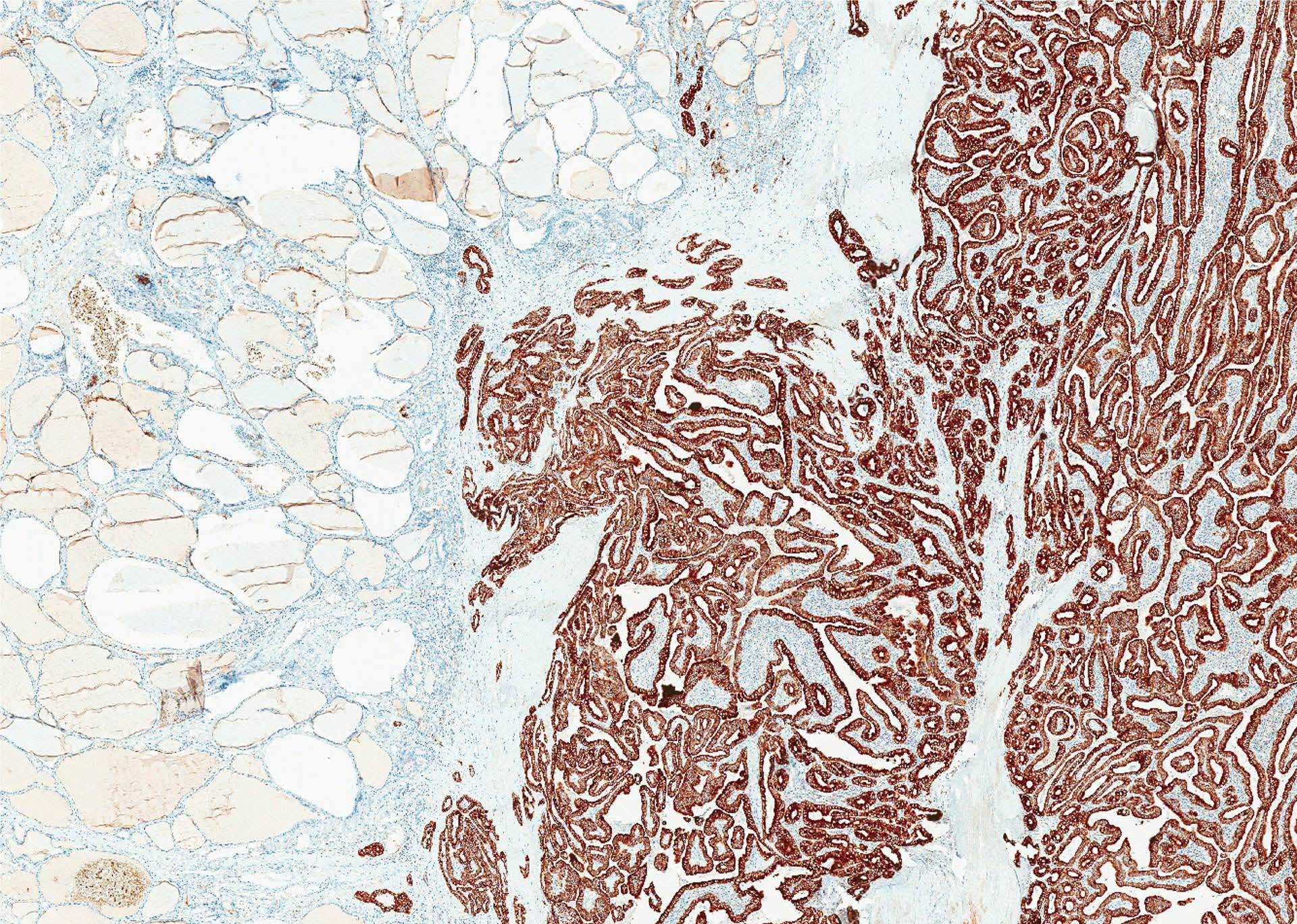
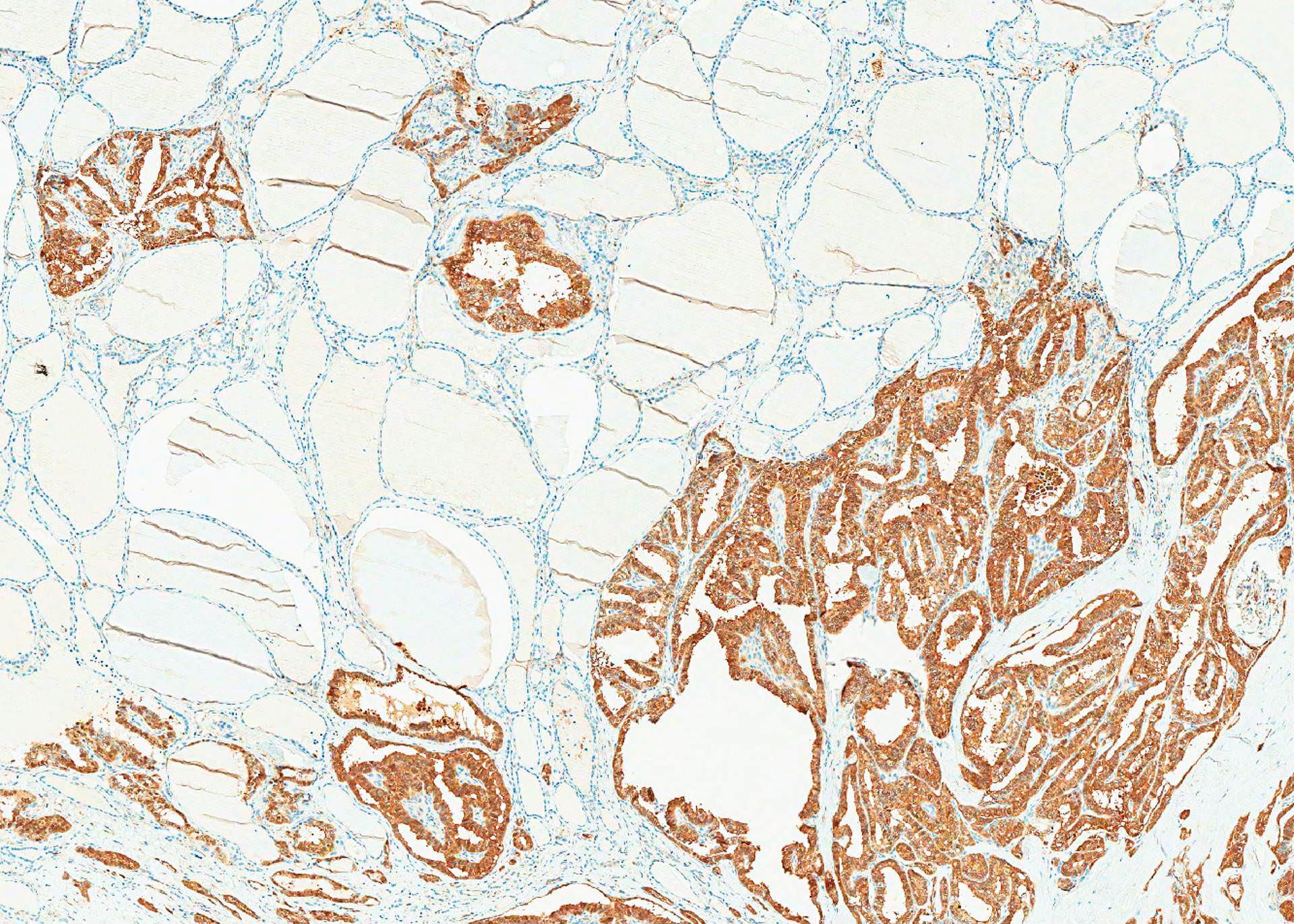
Circumscribed and infiltrative
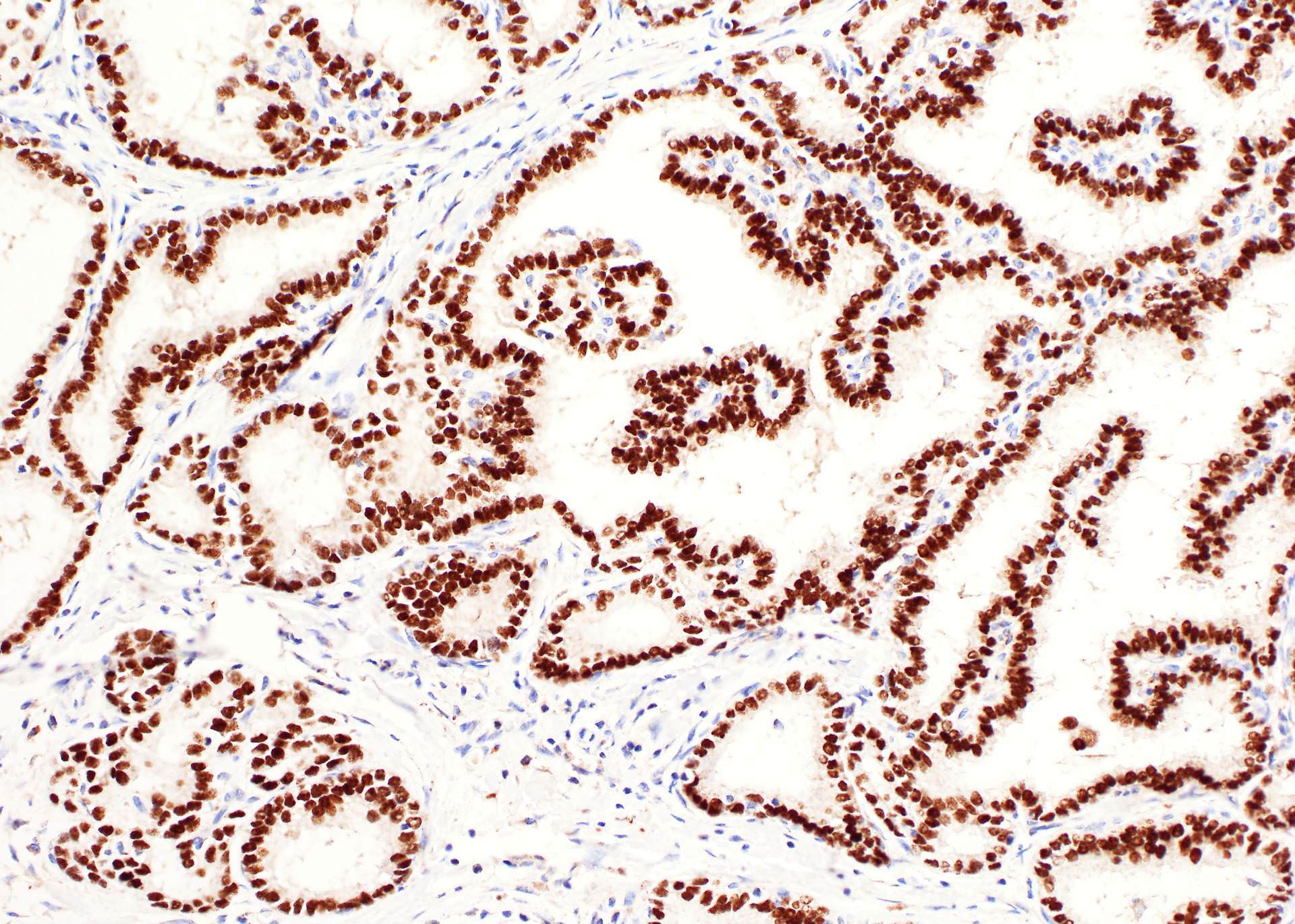
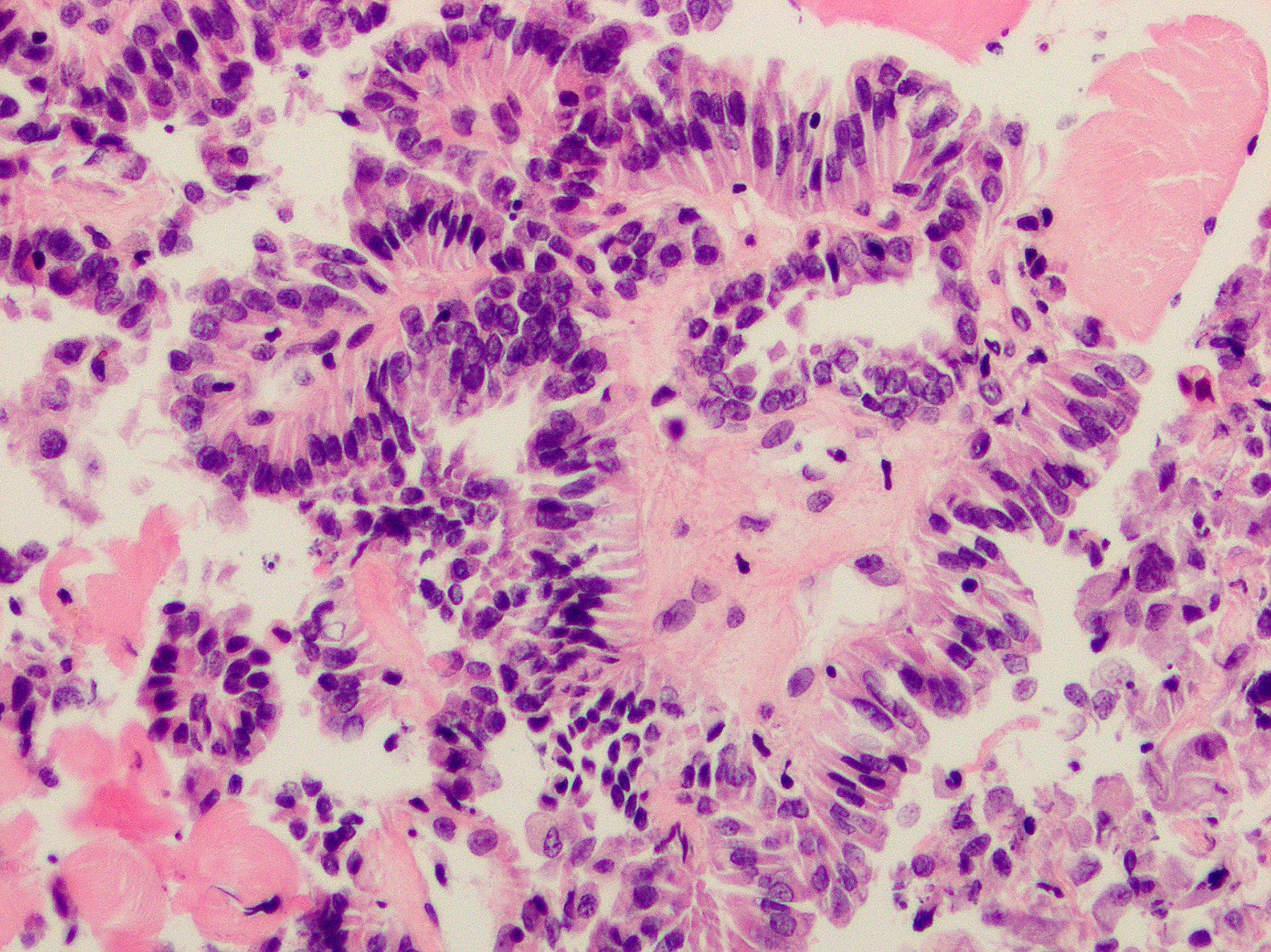
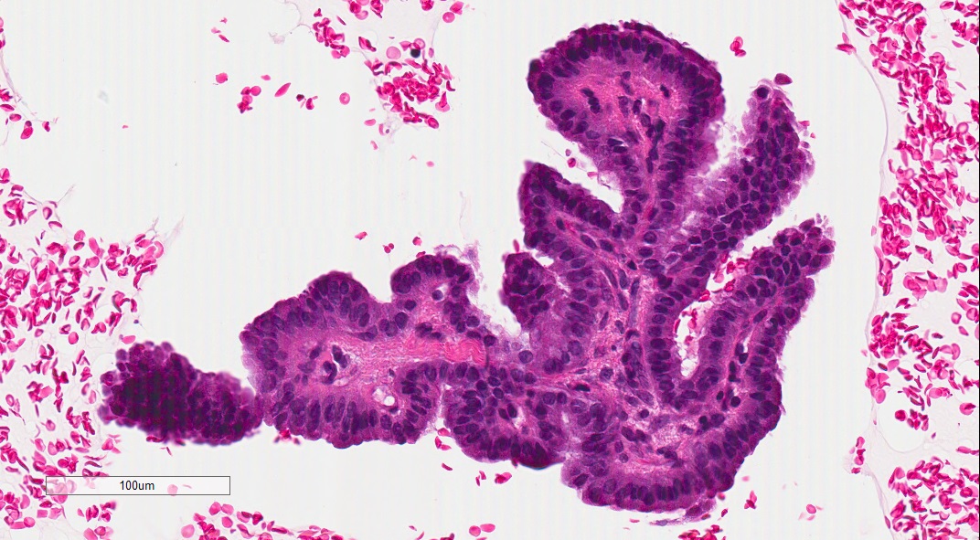
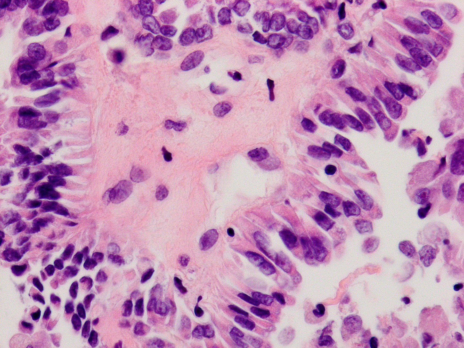
Contributed by Andrey Bychkov, M.D., Ph.D.
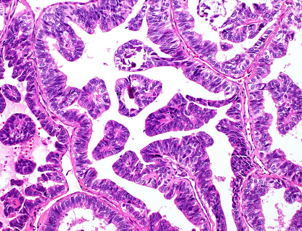
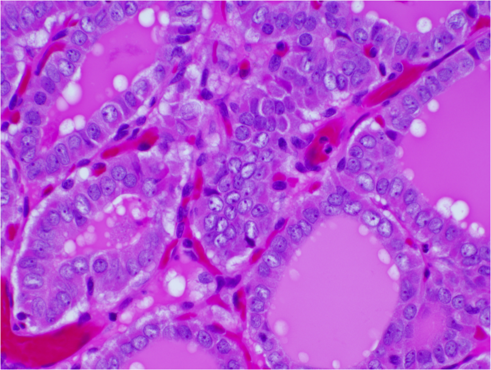
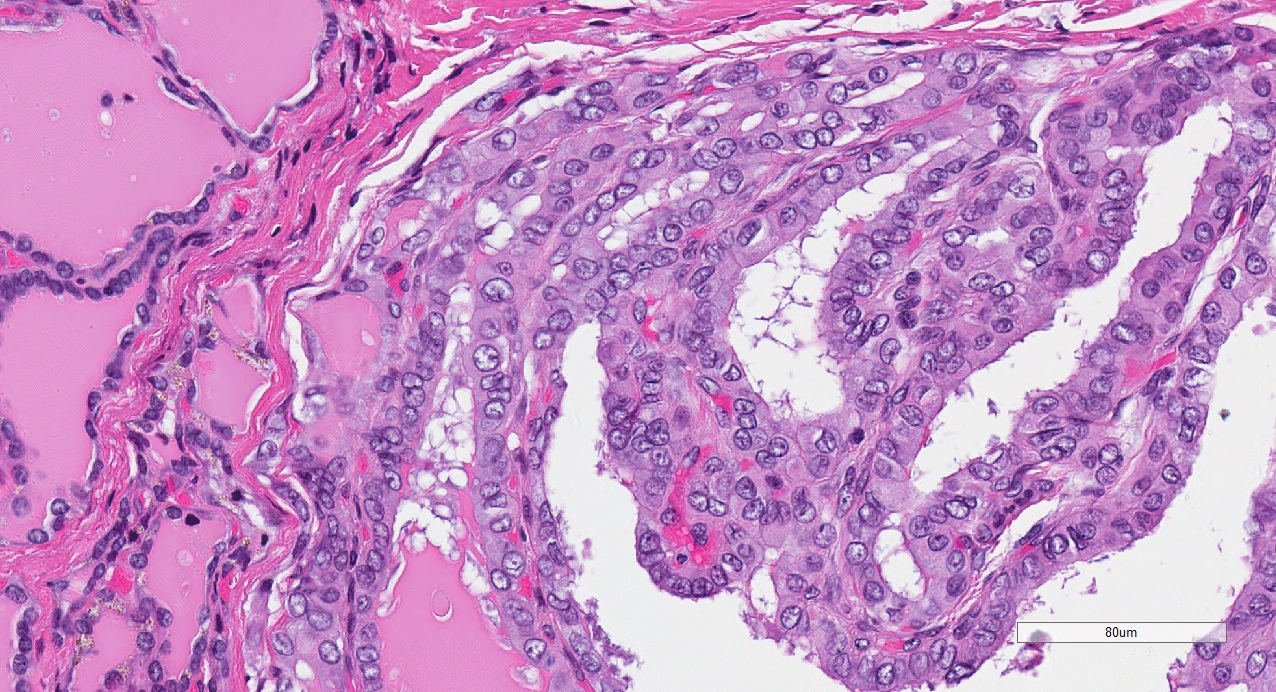
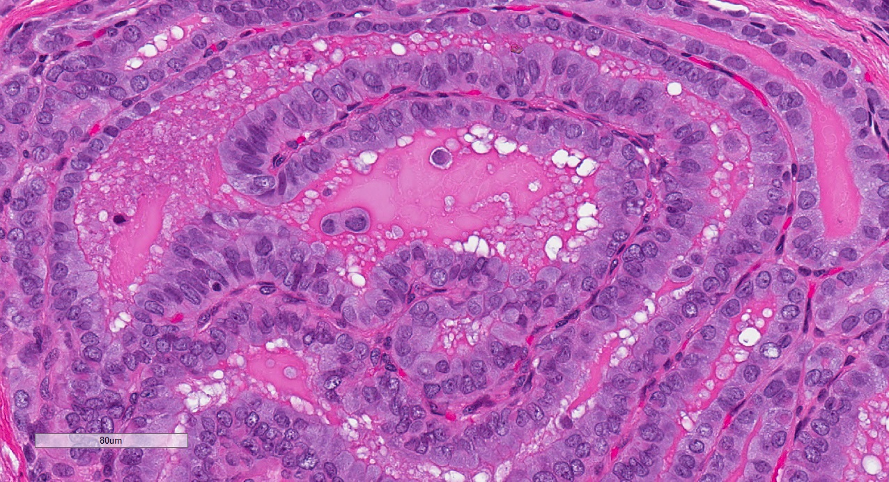
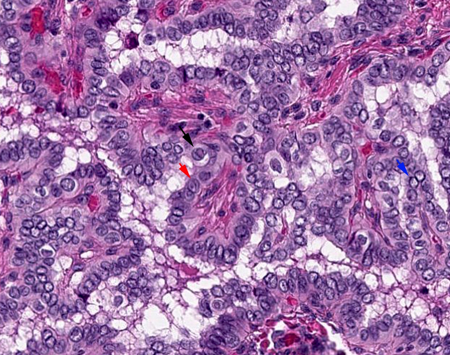
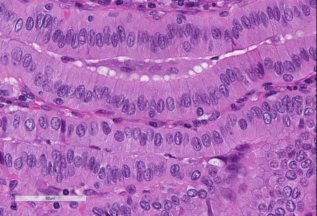
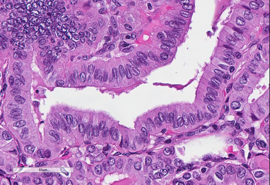
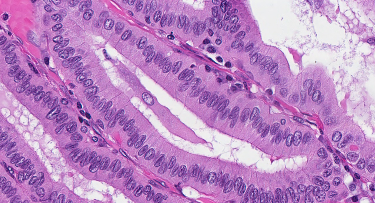
Papillary pattern
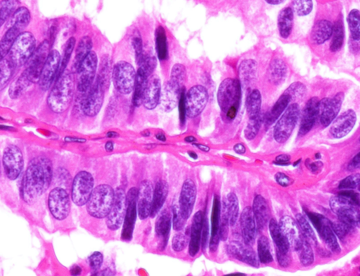
Pseudostratification
Tightly packed

Invasive

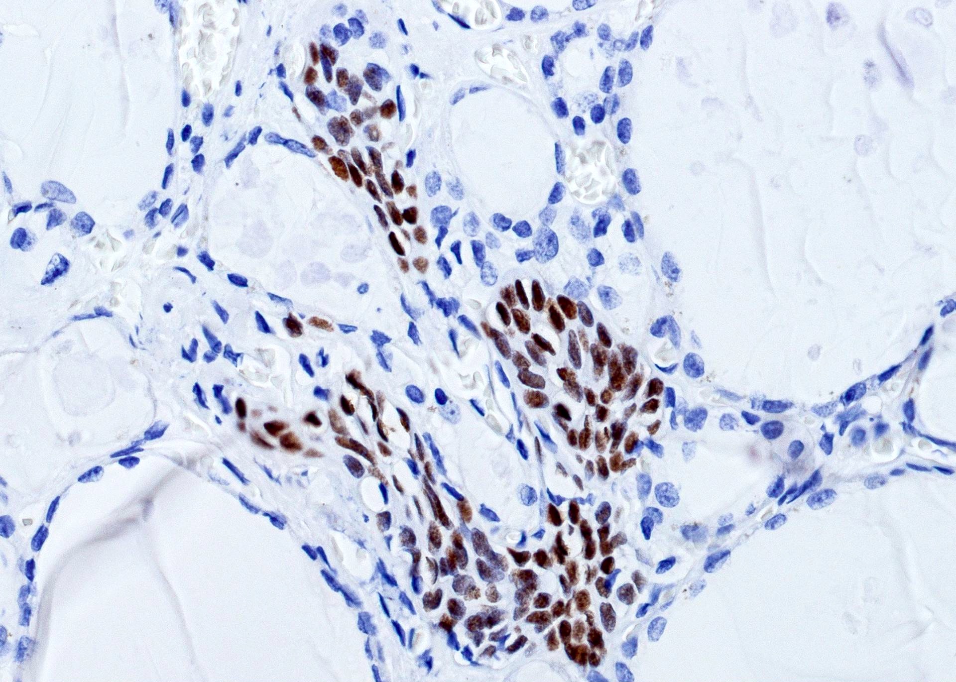
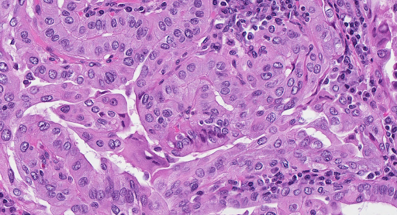
High Ki67
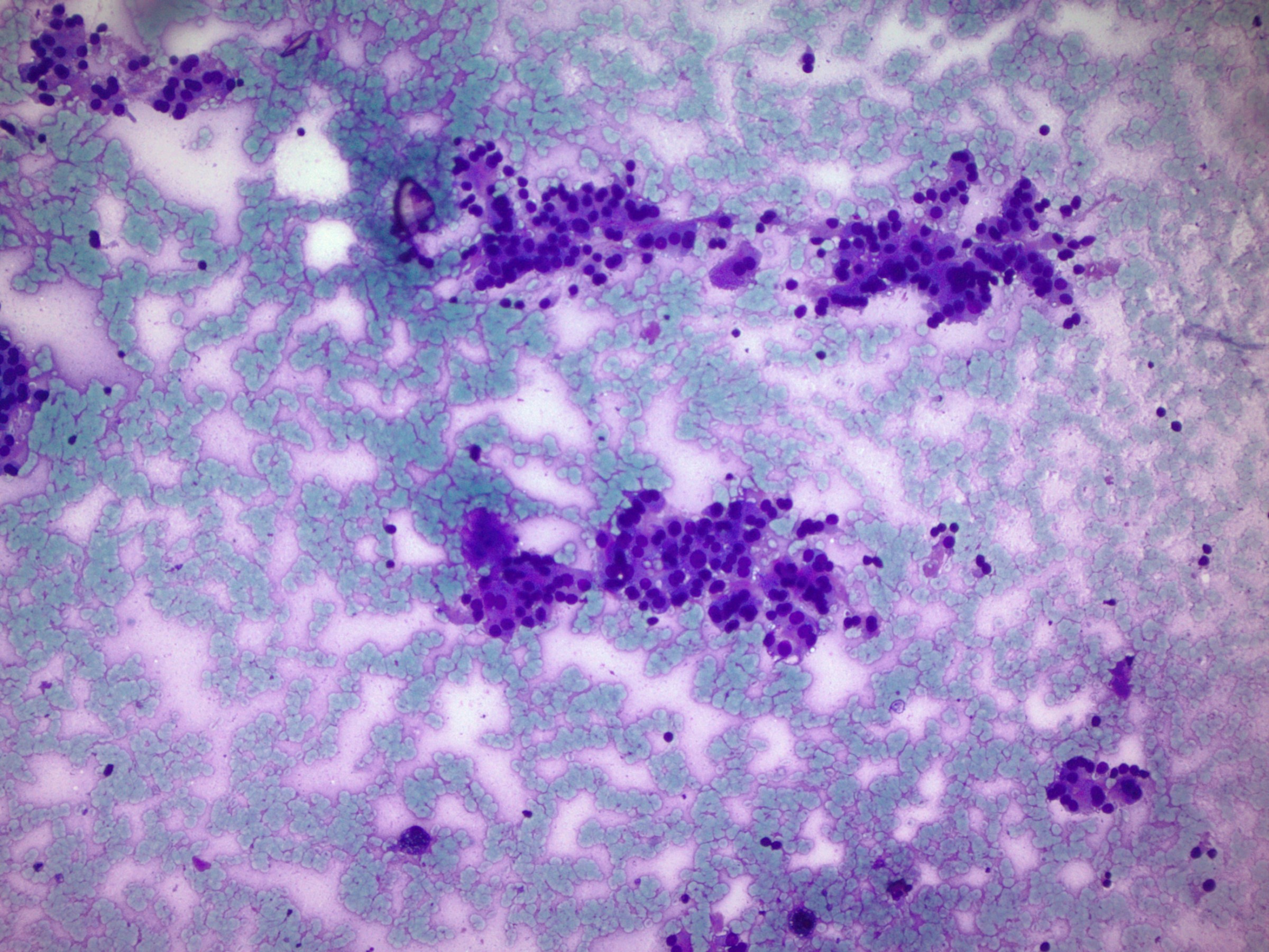
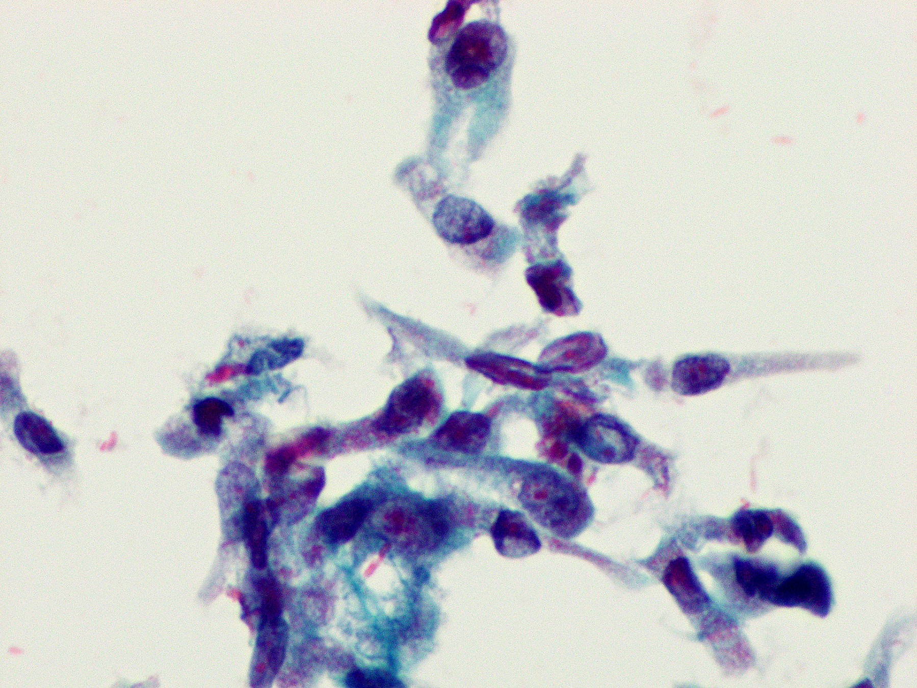
Contributed by Ayana Suzuki, C.T.
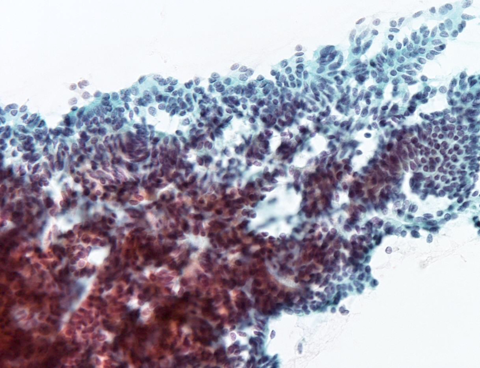
Papillary fragments
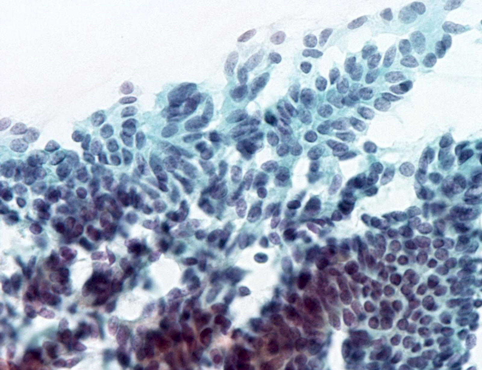
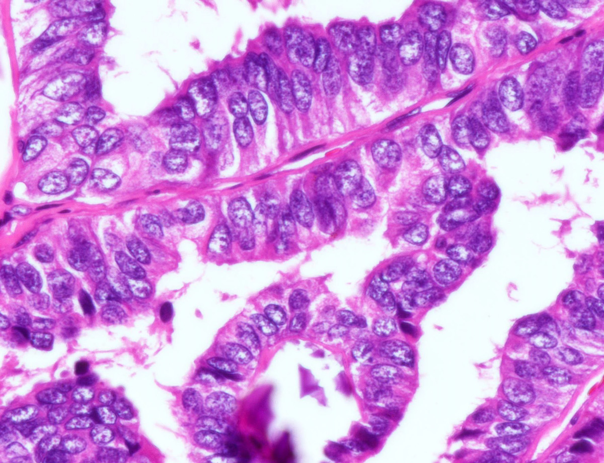
Nuclear features

Images hosted on other servers:


Heterogeneous, hypoechoic mass without calcifications
Images hosted on other servers:

Multifocal tumors throughout the entire thyroid
Contributed by Jonathan K. Lai, M.D., Drs. Safa Alshaikh and Aalaa Mohammed (Case #470) and Dr. Bin Xu (Case #528)
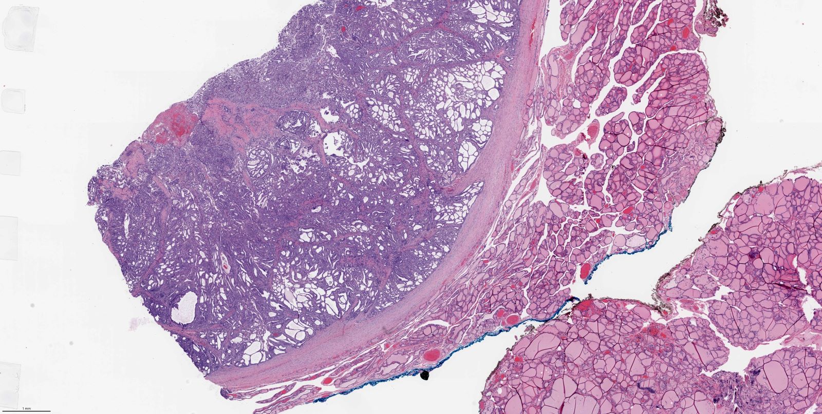
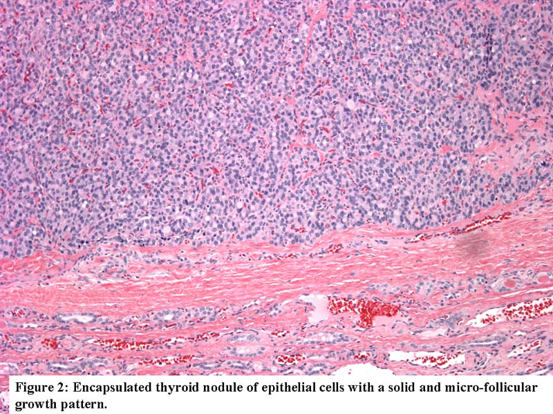
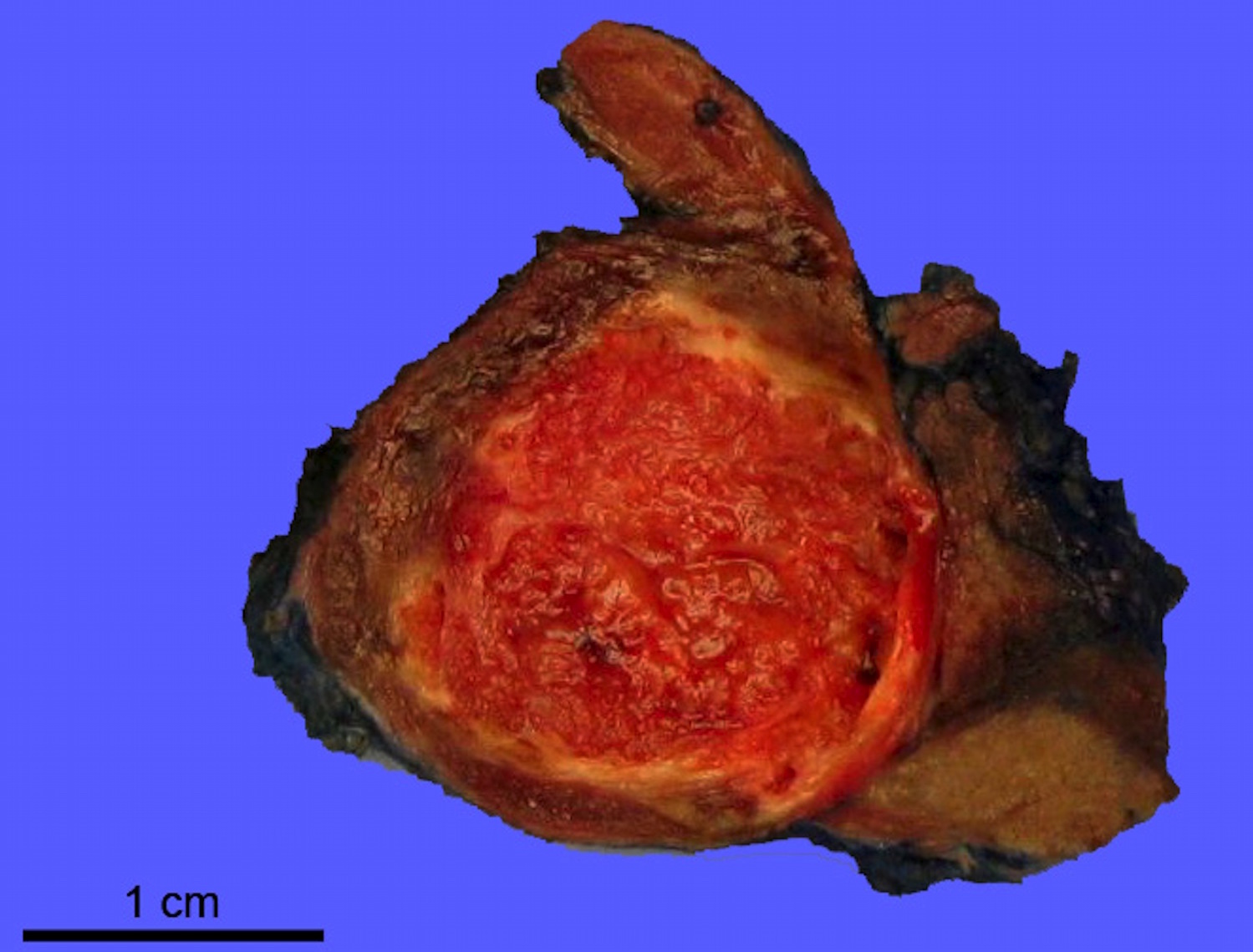
Encapsulated thyroid nodule
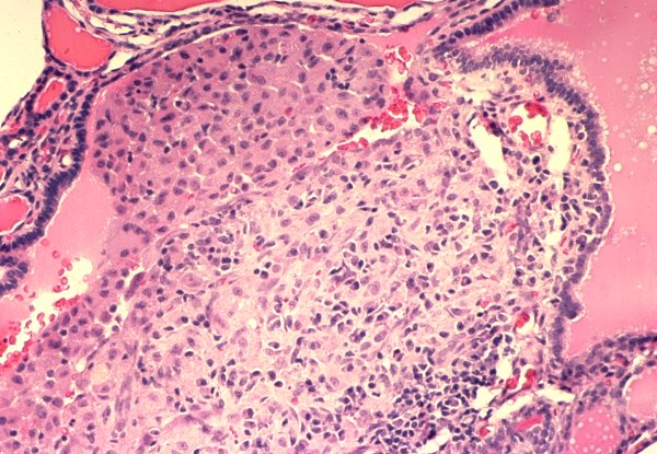
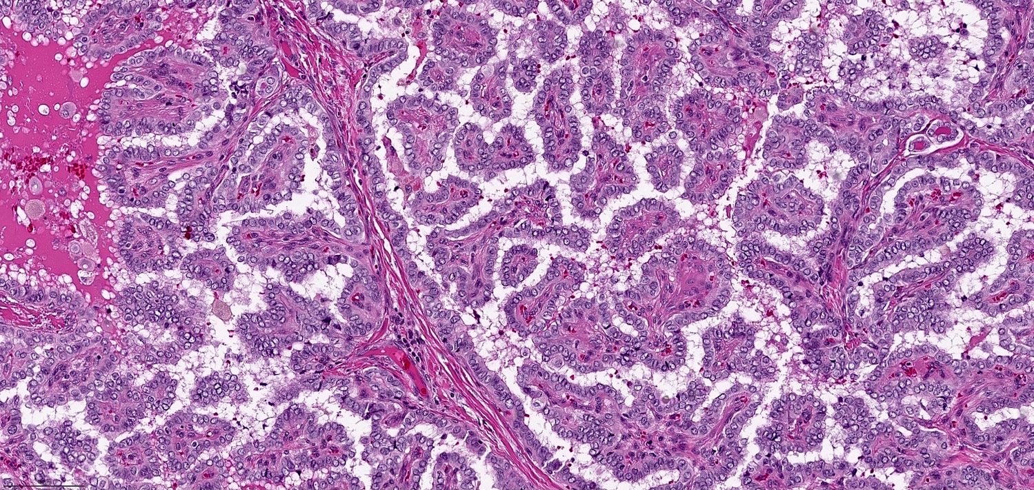
Complex architecture

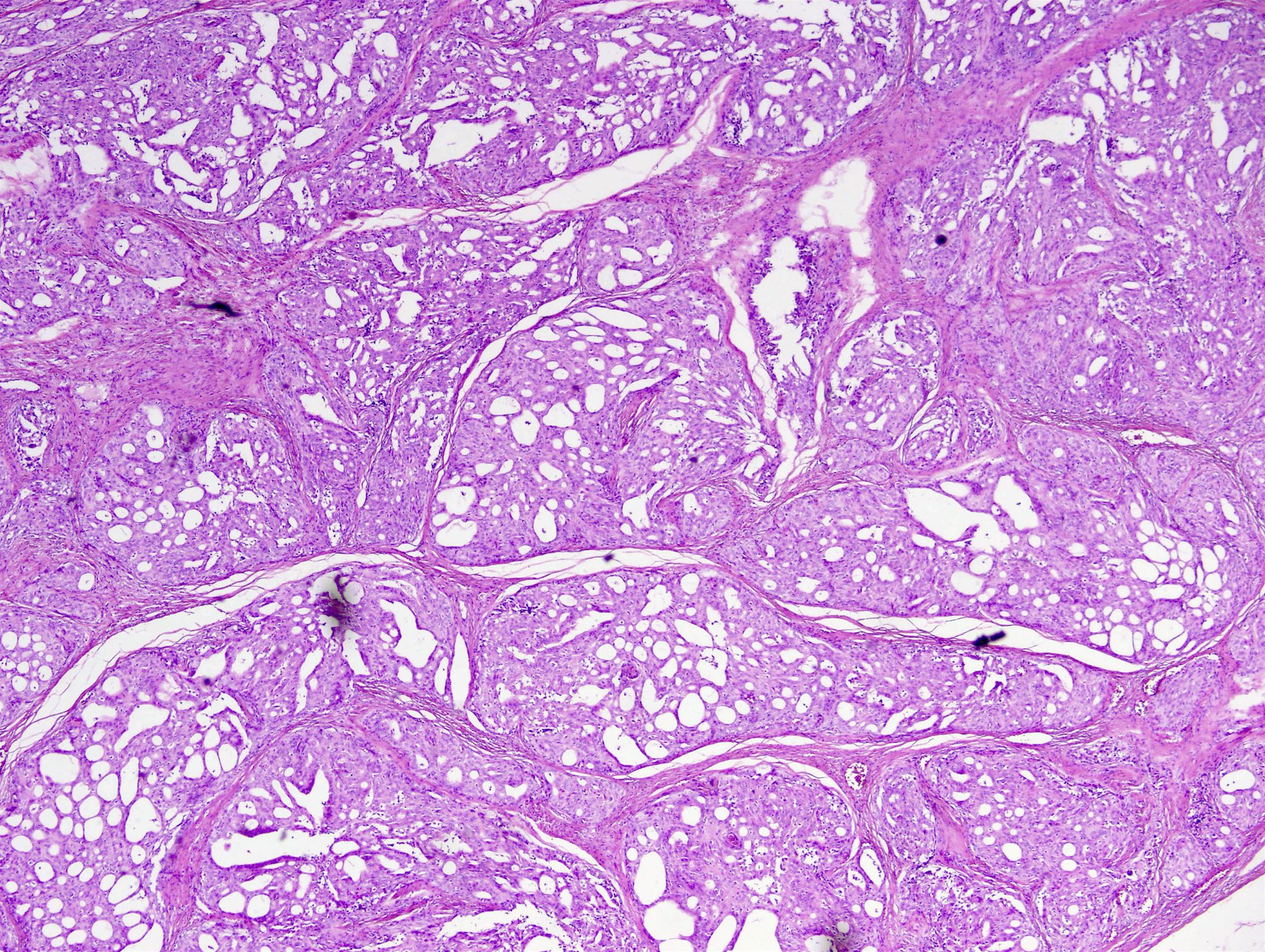
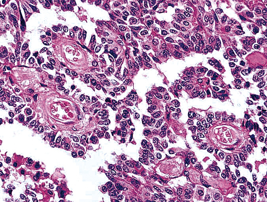
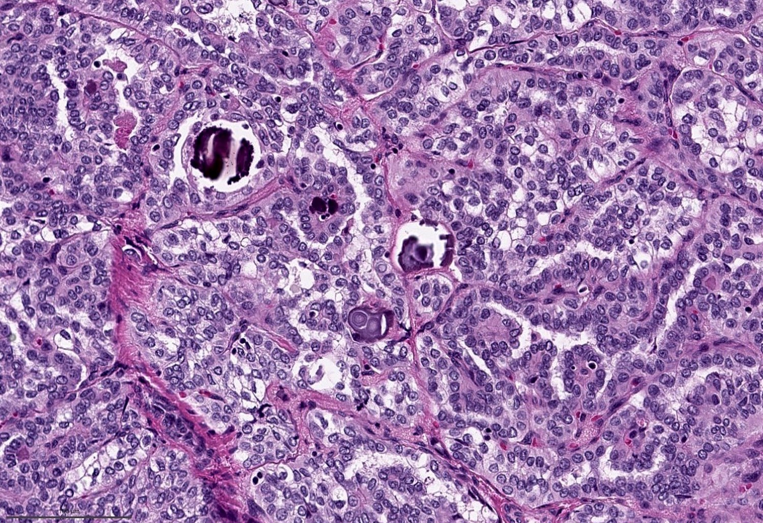
Cribriform-morular architecture

Trabecular pattern

Sieve-like spaces and morules
Squamoid morules

Squamoid morules
Hallmark feature


19 year old woman with right sided neck swelling
Papillary and cribriform architecture
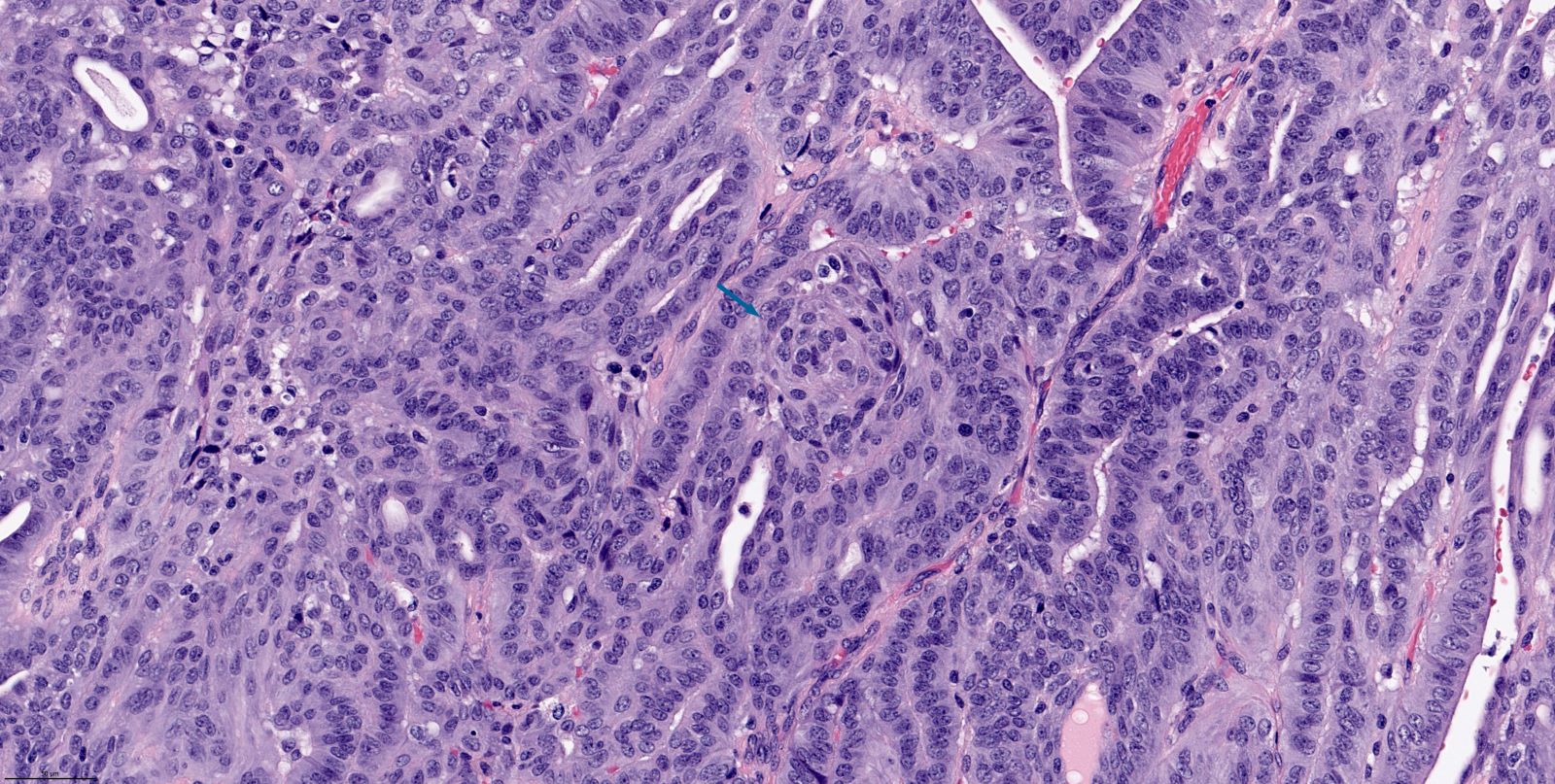
Scattered squamous morules

Scattered mitotic figures

Multifocal tumor necrosis
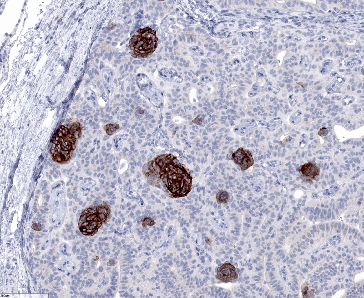
CK5/6


TTF1
TTF1


PAX8
Thyroglobulin
Beta catenin
Beta catenin
Estrogen receptor
CD10
CDX2
Contributed by Drs. Safa Alshaikh and Aalaa Mohammed (Case #470)
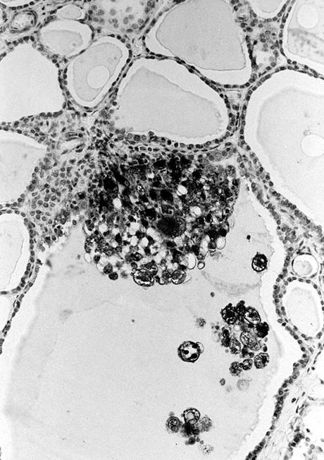
May-Grünwald-Giemsa

Pap stain
Images hosted on other servers:


Hypercellular, morular or papillary fragments
Contributed by Andrey Bychkov, M.D., Ph.D. and AFIP

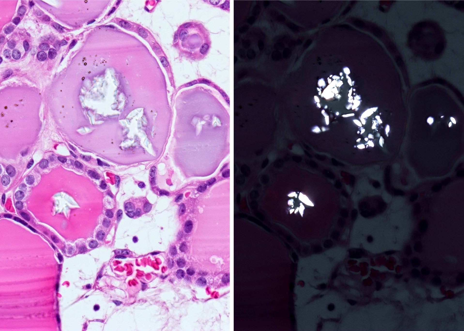
Abundant calcium oxalate crystals

Follicular adenoma: calcium oxalate crystals
Calcium oxalate crystals in the colloid
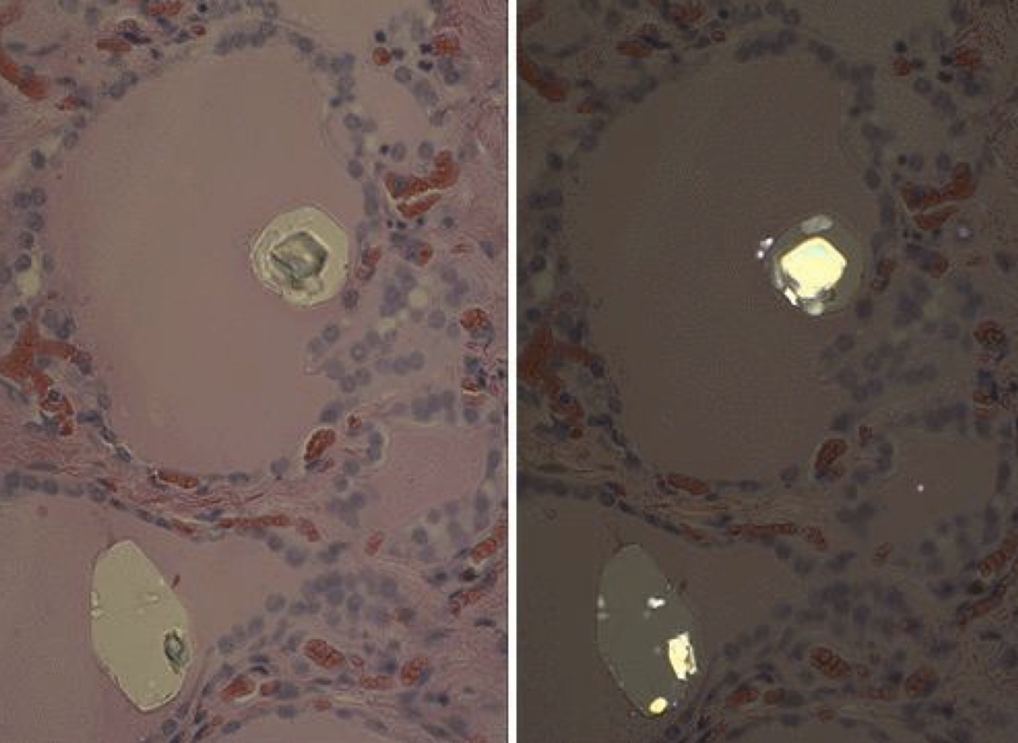
Calcium oxalate crystals
Images hosted on other servers:

Calcium oxalate crystallite

Spherical apatite crystallites
Contributed by Andrey Bychkov, M.D., Ph.D.

Infiltrative growth
Images hosted on other servers:

53 year old man with metastatic carcinoma
Images hosted on other servers:


Ultrasonographic features
AFIP images
Diffuse growth and fibrosis
Images hosted on other servers:

Fibrotic cut surface
Contributed by Livia Florianova, M.D., M.Sc. and Marc Pusztaszeri, M.D.
Lymphocytic infiltrate
Papillary pattern
Dense fibrosis
Dense sclerosing fibrosis
Infiltrative growth
Nuclear features
Psammoma bodies and sclerosis
Solid pattern
Squamous morule
Lymphovascular invasion
Lymph node metastasis
Contributed by Andrey Bychkov, M.D., Ph.D.
Multiple tumor nodules
Fibrosis and lymphocytic infiltration
Large psammoma bodies
Prominent sclerosis
Thick fibrous bands
Extensive squamous metaplasia
Contributed by Livia Florianova, M.D., M.Sc., Marc Pusztaszeri, M.D. and Manon Auger, M.D.C.M.
3D ball-like clusters
Protruding hobnail cells
Squamous metaplasia
Cytological atypia
3D ball-like clusters
Psammoma body
Cytological and architectural atypia
Cytological atypia
Cytological and architectural atypia
Psammoma bodies and lymphocytes
Nuclear features
Psammoma bodies and lymphocytes
Images hosted on other servers:

Pathophysiology

Mode of inheritance

Pedigree

Chromosome 22 abnormalities


Genetic mapping
Images hosted on other servers:

Absent thymus on CT

Subglottic stenosis on CT

Basal ganglia and periventricular calcification
Images hosted on other servers:
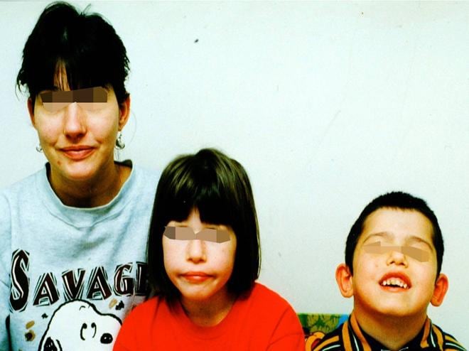


Facial dysmorphism
Images hosted on other servers:

Hypoplastic thyroid in mice


Various malformations in mice
Images hosted on other servers:


Deletion of 22q11.2 on FISH
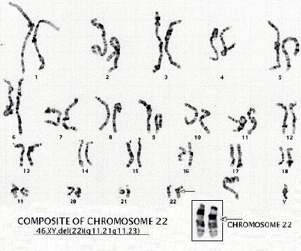

Karyotype

Various techniques
DiGeorge syndrome
Pharyngeal apparatus
Images hosted on other servers:
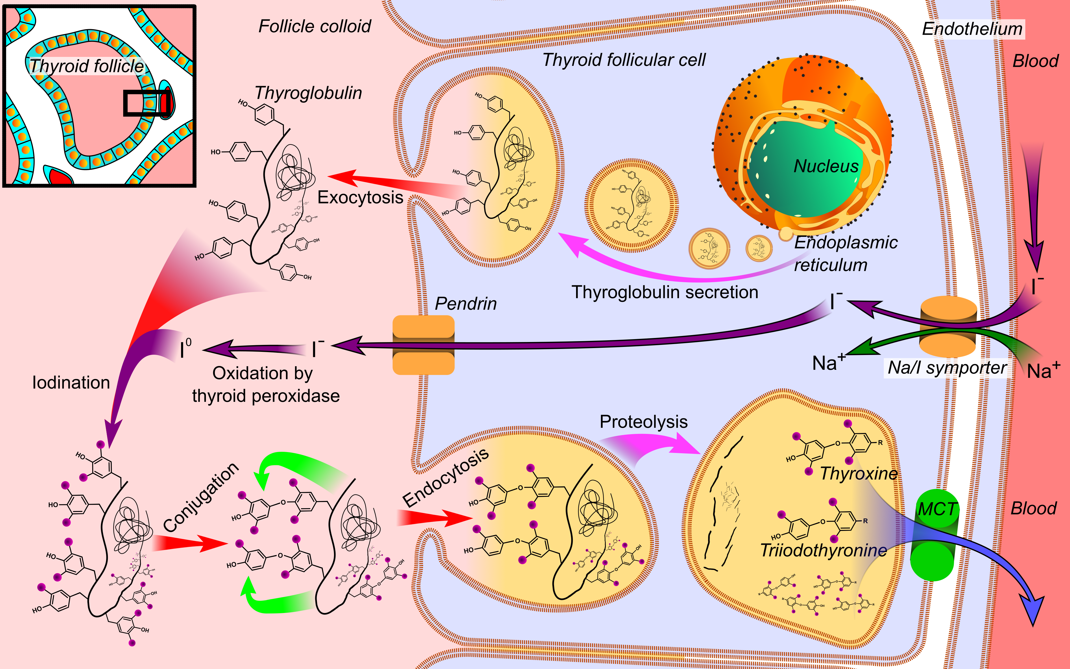


Thyroid hormone synthesis

Mechanism of
dyshormonogenesis

Congenital hypothyroidism

Genetic causes of
dyshormonogenesis

Thyroid imaging in congenital hypothyroidism

Scintigraphic classification of dyshormonogenesis


Pedigrees
Images hosted on other servers:

Ultrasound of fetal goiter

Ultrasound of 26 year old man

Various defects
on scintigraphy
(NIS, TPO and PDS)

Scintigraphy
Images hosted on other servers:

Pendred syndrome
AFIP images

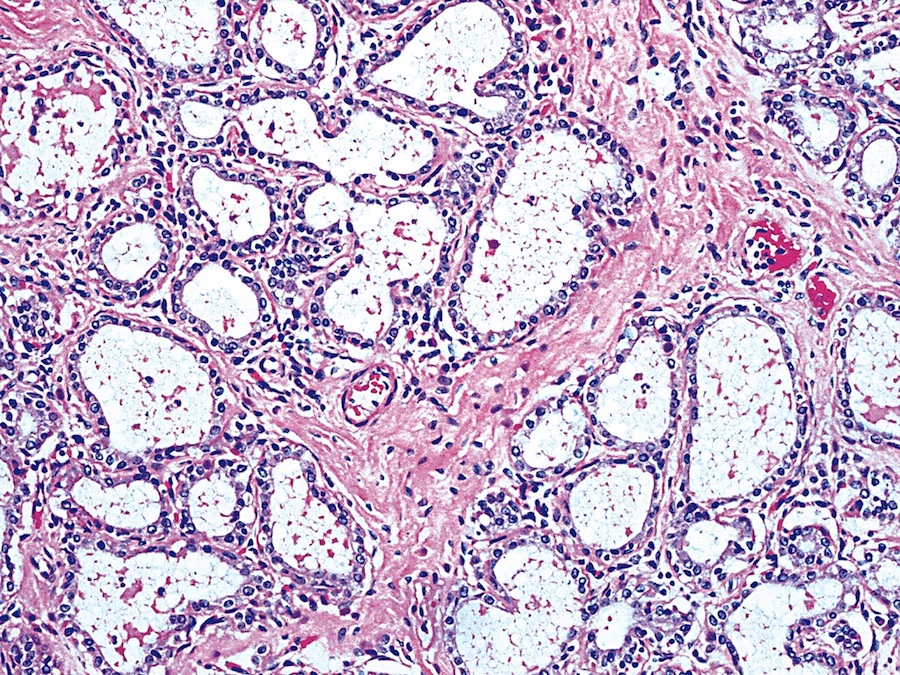
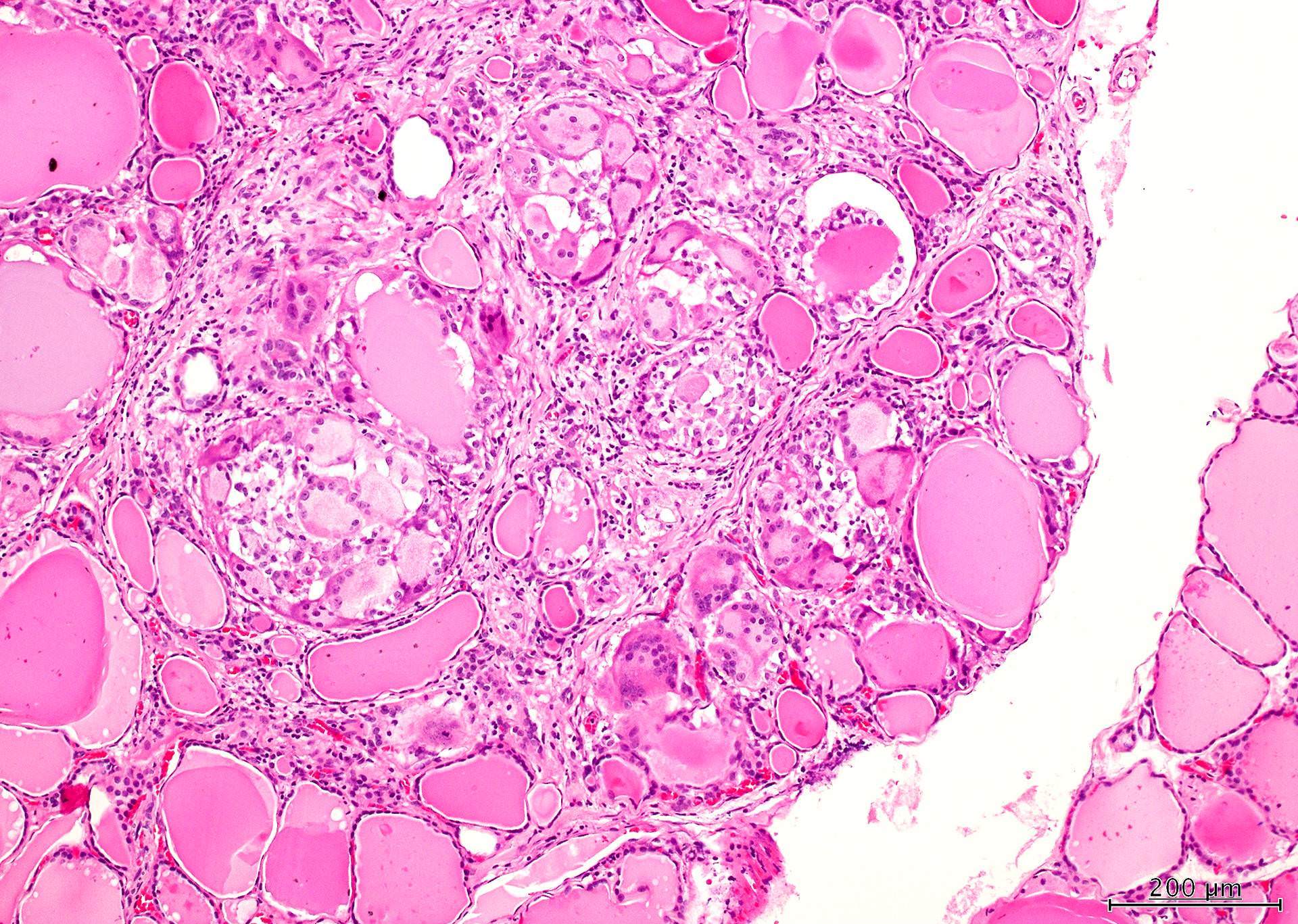
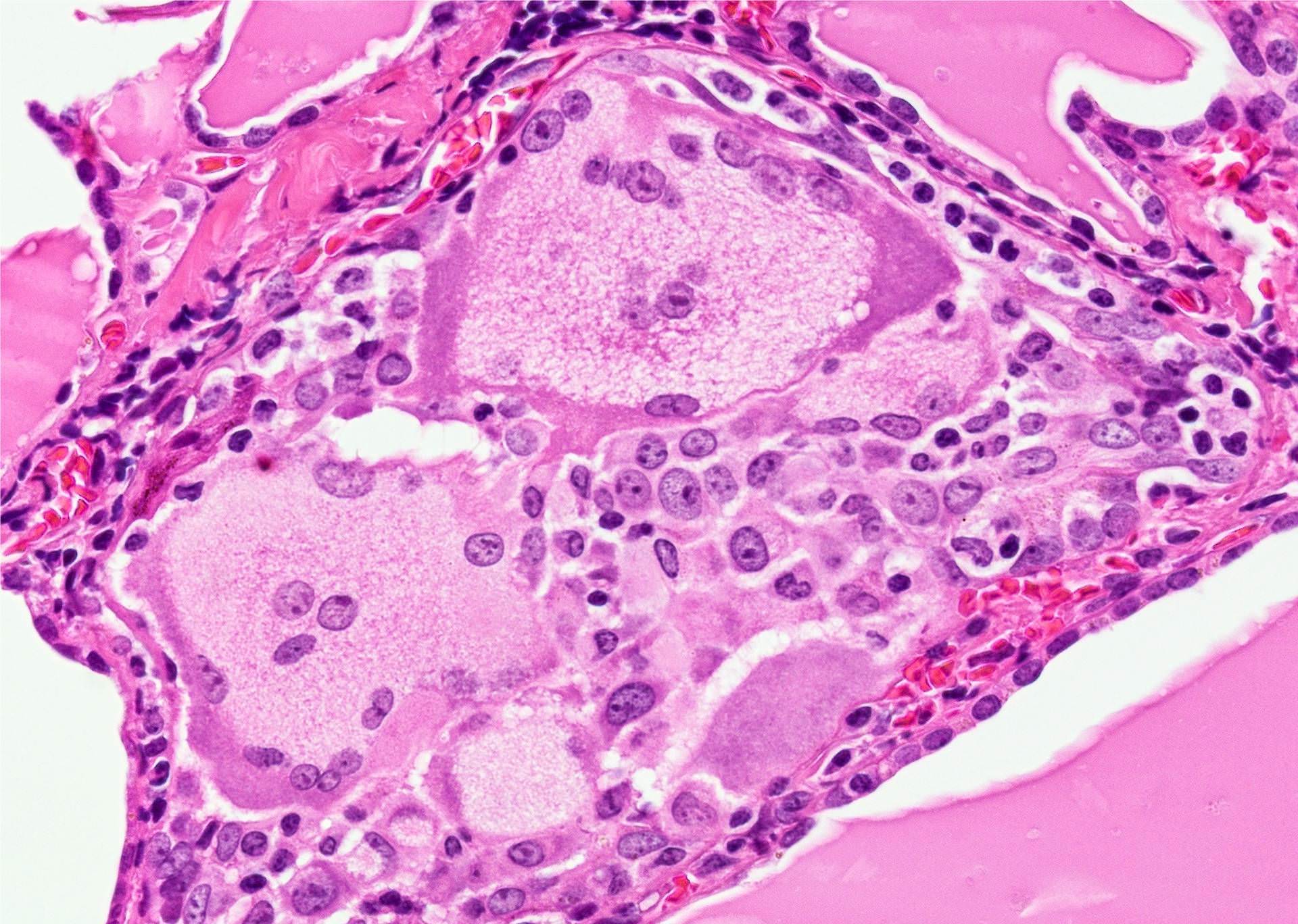
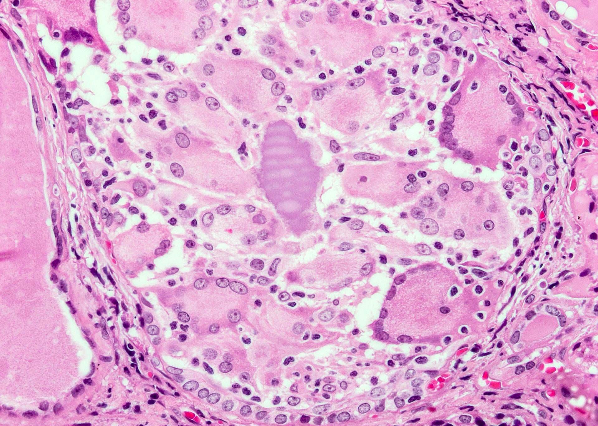
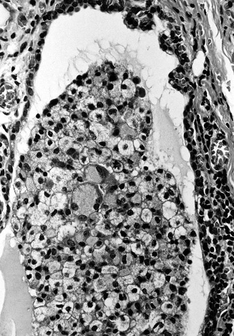
Marked nodular hyperplastic changes
Images hosted on other servers:


Enlarged multinodular thyroid

Excision

Follicular adenoma in a goiter
Scroll to see all images:
Contributed by Mark R. Wick, M.D. and AFIP

Various images
Resembles nodular hyperplasia
Hyperplastic nodules showing solid growth pattern

Marked fibrosis causes apparent increased lobularity
Images hosted on other servers:


Multinodularity

Fibrosis


Hypercellular nodule
Papillarity


Hyperplastic nodule


Scant colloid

Atypia

IHC in Pendred syndrome


Follicular adenoma in dyshormonogenetic goiter
Papillary microcarcinoma in dyshormonogenetic goiter


Papillary carcinoma in goiter
Images hosted on other servers:


Structure of TPO mutations

DUOXA2 mutation
Contributed by Andrey Bychkov, M.D., Ph.D. and Case #108

Aberrant parathyroid

Various images
Cytokeratin cocktail

Chromogranin
Synaptophysin
Parathyroid hormone
Images hosted on other servers:

Thymic tissue contains a mass of parathyroid cells
Images hosted on other servers:

Thyroid descent

Locations
Images hosted on other servers:

Neck ultrasound


Neck CT


Radionuclide scan

Abdominal MRI

Head MRI


Chest CT

Intracardiac thyroid
Images hosted on other servers:


Neck mass
Images hosted on other servers:

Submental mass

Ectopic thyroid, lateral neck

Intratracheal thyroid tissue, hyperplasia

Mediastinal mass

Cystic mass, adrenal gland
Contributed by Andrey Bychkov, M.D., Ph.D.

Perithyroid thyroid follicles
Images hosted on other servers:

Benign conditions

Ectopic vs. orthotopic thyroid

Posterior mediastinal mass


Adrenal mass


Adrenal mass, IHC


Adenoma, ectopic thyroid


PTC arising in ectopic thyroid


Anaplastic carcinoma
Images hosted on other servers:

Follicular cells
Summary table:
| Event | Week | Day |
| Specification of the thyroid domain in the ventral endoderm | week 3 | E20 - 22 |
| Thyroid bud (medial anlage) formation | weeks 3 - 4 | E20 - 24 |
| Thyroid bud begins migration | week 4 | E24 - 28 |
| Formation of the lateral anlagen (ultimobranchial bodies) | weeks 4 - 7 | |
| Migration of lateral anlagen | weeks 5 - 7 | |
| Thyroglossal duct disappears | weeks 5 - 6 | E30 - 40 |
| Thyroid migration is complete | weeks 7 - 8 | E45 - 50 |
| Fusion with ultimobranchial bodies | weeks 7 - 9 | E44 - 60 |
| Onset of folliculogenesis | week 10 | E70 |
| Release of thyroid hormone | weeks 10 - 12 | E80 |
Images hosted on other servers:

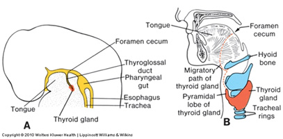
Embryonic development of thyroid gland
Branchial arches
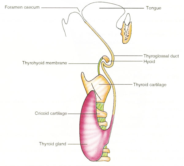
Thyroglossal duct

Relation to vessels


Interplay of transcription factors
AFIP images


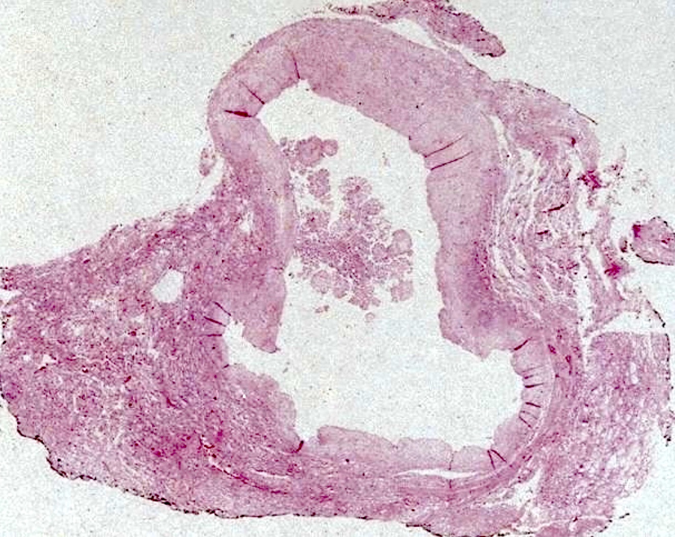
Human embryo 14 weeks
Images hosted on other servers:
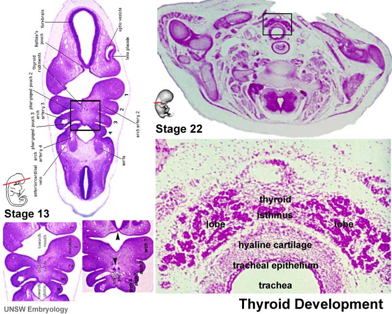
Stages 13 and 22

Mouse model

TTF1 expression
Thyroid development
Development of pharyngeal area
AFIP images

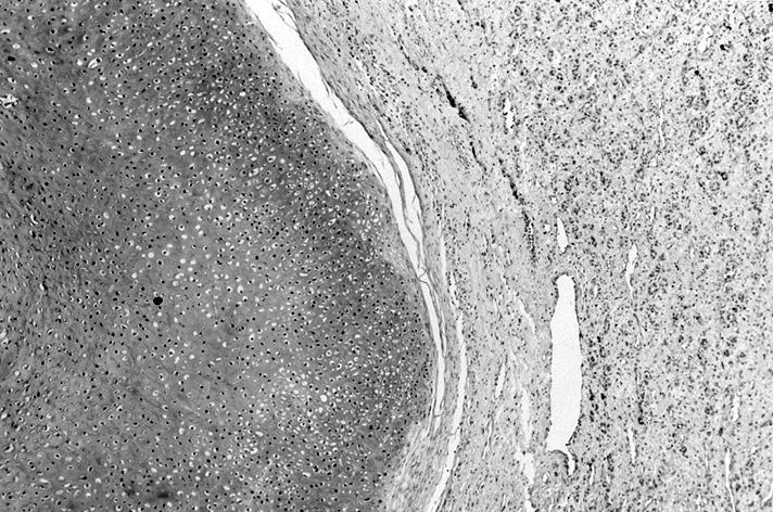
Tumor surrounded by fibrous capsule
Contributed by Andrey Bychkov, M.D., Ph.D. and AFIP

Papillary carcinoma encapsulated variant

Transcapsular invasion
Thickened capsule is violated by tumor growth

Typical papillary appearance
Contributed by Andrey Bychkov, M.D., Ph.D.
Enlarged vesicular nuclei with irregular membranes

Evident major and minor diagnostic features

Minor diagnostic features

Free floating of small tumor fragment in vascular lumen

Small piece of tumor floats in vascular lumen
Images hosted on other servers:

lesion with nuclear
features of papillary
carcinoma (inset)

Capsular invasion

3 cm tumor in 51 year old man (fig. 1)
Images hosted on other servers:

Ultrafast Pap stain

Ultrafast Pap staining of
follicular adenoma and
follicular variant
Images hosted on other servers:

Endemic multinodular goiter
Images hosted on other servers:

Endemic multinodular goiter
Contributed by Ayana Suzuki, C.T.
Watery colloid

Cracking colloid

Follicular clusters

3D structures
Paravacuolar granules
Contributed by Andrey Bychkov, M.D., Ph.D.
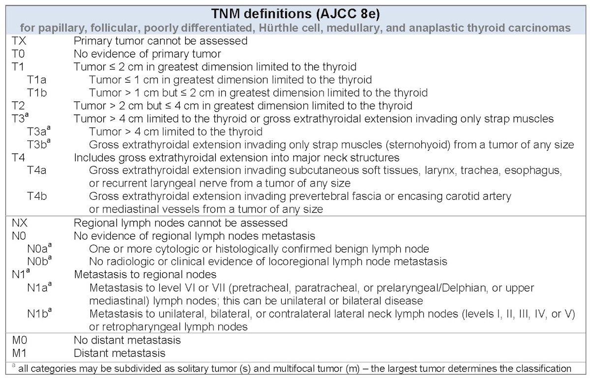
AJCC / TNM charts

ATA initial stratification
WHO classification
Images hosted on other servers:


Levels of the cervical lymph nodes

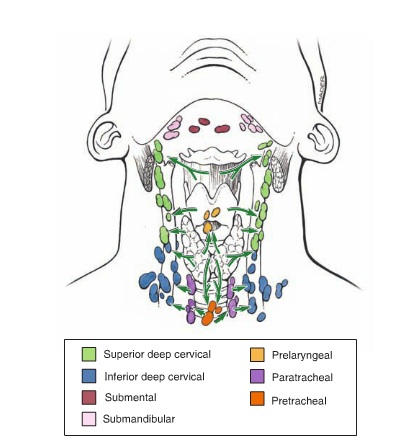
Pattern of nodal metasatasis

Extranodal
Contributed by Chan Kwon Jung, M.D., Ph.D. and Andrey Bychkov, M.D., Ph.D.

Papillary thyroid carcinoma - lymphatic invasion


Vascular invasion


Vascular invasion

Psammoma bodies

Microscopically positive margin
Extrathyroidal extension

Extrathyroidal extension

Extrathyroidal extension
Extrathyroidal extension

Extranodal extension
Extrathyroidal extension by J. Hernandez-Prera (2020)
Images hosted on other servers:


Ultrasound


Ultrasound elastography

Color doppler ultrasound
Images hosted on other servers:

Well demarcated, solid

Fibrotic nodule
Contributed by Ayana Suzuki, C.T.
Mixed epithelial-mesenchymal tumor


Fibromatosis type stroma
Beta catenin
Images hosted on other servers:

Stromal fragment
Contributed by Rachel Jug, M.B.B.Ch.
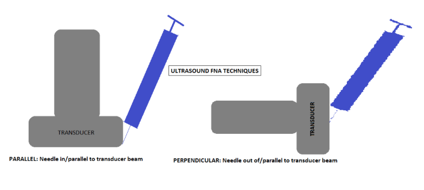
Ultrasound FNA techniques
Images hosted on other servers:

Risk based on appearance and nodule characteristics

Technique

Short, rapid strokes
with only slight
changes in direction
Contributed by Andrey Bychkov, M.D., Ph.D. and AFIP
FNA induced alterations in thyroid nodule
Pseudo angiosarcomatous pattern
Fine needle
induced changes
in follicular
adenoma
FNA biopsy techniques
Thyroid FNA and smearing techniques
Superficial FNA technique
Images hosted on other servers:

Patchy lymphocytic inflammation

Small and large lymphocytes,
occasional plasma cells
Images hosted on other servers:

Coarsely
condensed
chromatin at
nuclear periphery
Images hosted on other servers:

Diagnostic capsular invasions

Incomplete / questionable capsular invasions
Images hosted on other servers:


Ultrasound

Ultrasound


Color Doppler sonogram


Color Doppler sonogram
Contributed by Andrey Bychkov, M.D., Ph.D., Mark R. Wick, M.D. and AFIP
Encapsulated thyroid nodule
Circumscribed thyroid nodule
Encapsulated, homogeneous tan cut surface

Bisected adenoma has fresh hemorrhage
Marked necrosis, hemorrhage and cystic change
Marked cystic degeneration
Images hosted on other servers:


Well circumscribed tumor

Well encapsulated

Central scar

Cystic and partially necrotic tumor
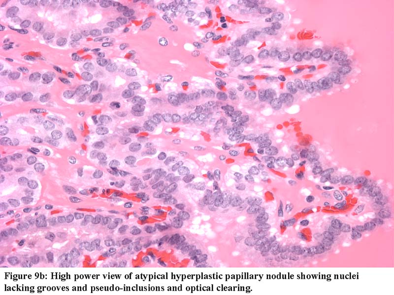
Contributed by Shipra Agarwal, M.D., Andrey Bychkov, M.D., Ph.D., Mark R. Wick, M.D., Asmaa Gaber Abdou, M.D. and AFIP
Capsule & compressed thyroid
Mixed micro and normofollicular pattern
Hyperchromatic, small round nuclei
Abundant crystals of calcium oxalate

Calcium oxalate crystals

Processing artifact with distorted nuclei
Distorted nuclei due to technical / processing artifact

Tissue degeneration
Circumscribed thyroid nodule
Cellular follicular adenoma

Cellular follicular adenoma

Trabecular type
Trabecular type
Thin and uniform fibrous capsule
Marked fibrosis and stromal hyalinization
Marked hyaline thickening of vessel walls
Marked fibrosis, hyalinization and calcium deposition
Marked vascularization
Capsular vessel with smooth muscle cells

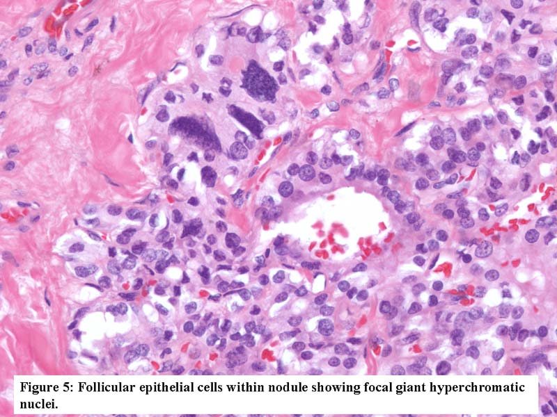
Bizarre nuclei
Large, extremely irregular nuclei
With papillary hyperplasia
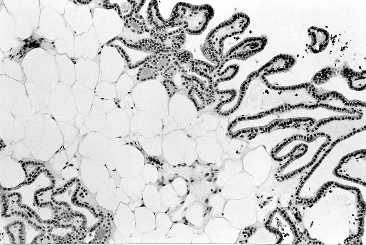
With papillary hyperplasia and adipose metaplasia
Cartilaginous metaplasia (adenochondroma)
Prominent, clear cell change

Squamous metaplasia
Mucin production
Thyroglobulin+
Alcian blue+
Solid, trabecular, microfollicular, macrofollicular patterns
Left: macrofollicular, right: solid pattern
Signet ring
Markedly cellular with irregular growth
Well formed follicles merge with solid pattern
Spindle cells mix with round cells
Not invasion:
Fine needle
induced changes
resemble invasion
Contributed by Shipra Agarwal, M.D. and Ayana Suzuki, C.T.
Cellular aspirate
Microfollicles and small round nuclei

Microfollicles
Images hosted on other servers:


Microfollicular


Microfollicular

Microfollicular

FNA

Round hyperchromatic nuclei

FNAC

Thyroid neoplasms
AFIP images
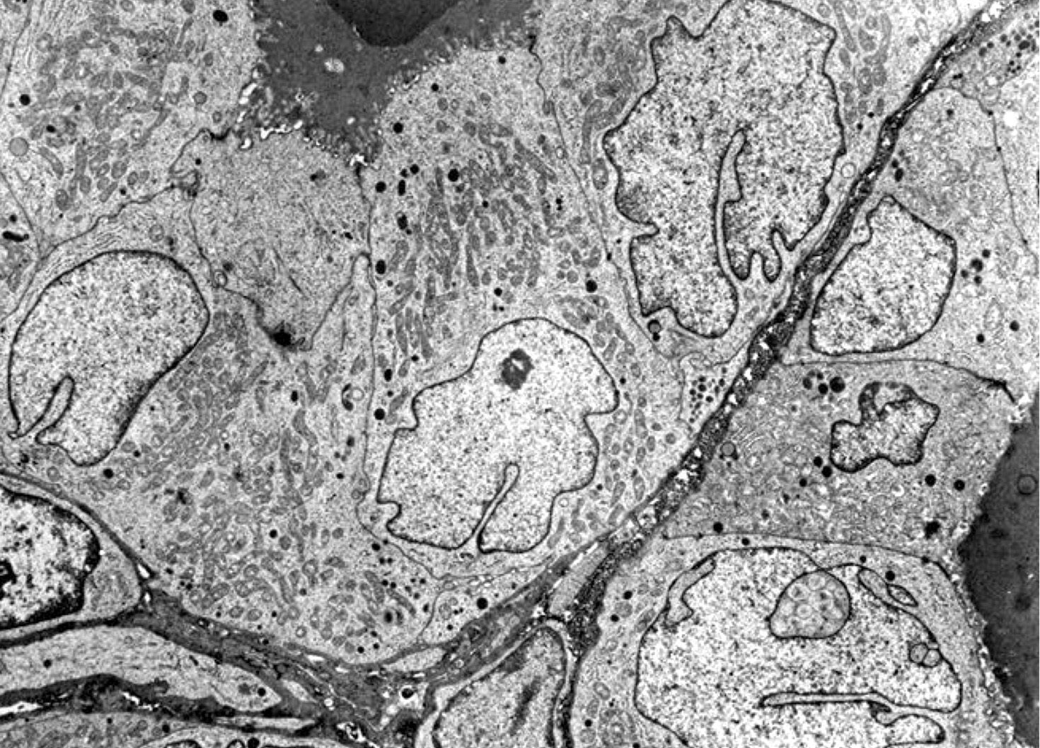
Abundant dilated endoplasmic reticulum
Microvilli project into well developed lumina
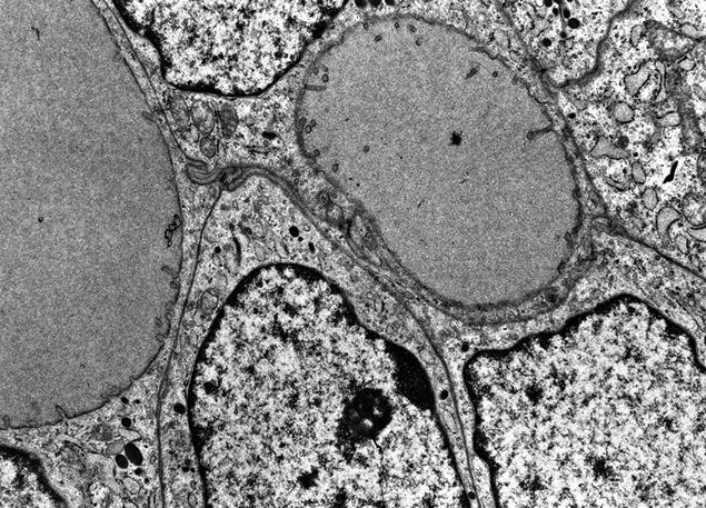
Signet ring follicular adenoma
Images hosted on other servers:

Gene expression analysis

CNV landscape of thyroid tumors

Driver mutations in thyroid tumors

Driver mutations and pathway analyses

Microarray and qRT PCR

Expression ratios of CRABP1, FABP4 and HMGA2

Clinicopathological features and mutation spectrum


Hierarchical clustering
Solitary thyroid nodule
Thyroid: compare and contrast
Histopathology thyroid: follicular adenoma (microfollicular)
Contributed by Ayana Suzuki, C.T. and Mark R. Wick, M.D.
Microfollicle
Images hosted on other servers:
Microfollicle


Follicular crowding

Trabecular pattern
Head and tail of the Bethesda system for thyroid
Thyroid cancer: fine needle aspiration, malignant or indeterminate results
Images hosted on other servers:

Schematic drawing for capsular invasion

Schematic drawing for vascular invasion
Contributed by Andrey Bychkov, M.D., Ph.D., Wafaey Fahmy Badawy Mohamed, M.D., Mark R. Wick, M.D. and AFIP
Minimal capsular invasion

Focal invasion

47 year old woman with follicular thyroid carcinoma and multinodular goiter
Various images
Minimally invasive follicular carcinoma

Indistinguishable from adenoma
Images hosted on other servers:

Apparently encapsulated


Widely invasive

Fig A: multiple white tan
nodules in thyroid tumor
Fig B: scalp metastases
show erosion through skull
Contributed by Andrey Bychkov, M.D., Ph.D., Mark R. Wick, M.D. and AFIP
False angioinvasion

Capsular vessel with tumor
Vascular invasion

Tumor in vascular space

Transcapsular penetration

Invasion through tumor capsule
Propagation of tumor embolus
Extensive necrosis

Tumor necrosis
Unusual brisk mitotic activity
TTF1: bone metastasis

Moderate pleomorphism
Mucin production
Capsular invasion

Metastases to iliac bone
Thick, irregular capsule
Images hosted on other servers:

Tumor, normal parenchyma

Insular type

Foci of tumor
beyond the border

Van Gieson stain

Penetration of former capsule

Not capsular invasion
Contributed by Ayana Suzuki, C.T., Xiaoyin "Sara" Jiang, M.D. and Jose Mellado, M.D.
Microfollicles
Follicular carcinoma


Follicular carcinoma, microfollicules with nuclear enlargement
AFIP images
Follicular cells converge toward central lumen
Contributed by LeicaBiosystems, Amsterdam

PPARG (3p25)
Thyroid carcinoma: gross and micro
Histopathology thyroid: follicular carcinoma
Images hosted on other servers:

Follicular variant
Contributed by Bin Xu, M.D., Ph.D.
Encapsulated follicular variant
Infiltrative follicular variant
Contributed by Bin Xu, M.D., Ph.D. and Andrey Bychkov, M.D., Ph.D.
Follicular architecture & nuclear features
Infiltrative follicular variant
Encapsulated follicular variant
Ossification
Contributed by Ayana Suzuki, C. T.
Hypercellular aspirate
Microfollicular and trabecular pattern
Microfollicles
Irregular nuclei
Nuclear inclusions
Images hosted on other servers:

Cellular aspirates;
some microfollicular
aggregates, no colloid

Acinar arrangement with intranuclear inclusion

Grape-like nuclei of papillary thyroid carcinoma

Microfollicles in thyroid FNA smears

Comparison of a
tissue proven follicular
adenoma and variant
of papillary carcinoma
AFIP images
Nuclear features
Images hosted on other servers:

Follicular variant of papillary carcinoma
Images hosted on other servers:

Exophalmos, lid retraction

Rodney Dangerfield
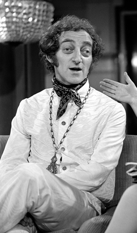
Marty Feldman
Images hosted on other servers:

Markedly enlarged gland
Scroll to see all images:
Contributed by Swati Satturwar, M.D., Mark R. Wick, M.D. and Andrey Bychkov, M.D., Ph.D.
Benign thyroid follicles
Radiation changes with stromal fibrosis
Various images
Active tall epithelium with light vacuolated cytoplasm
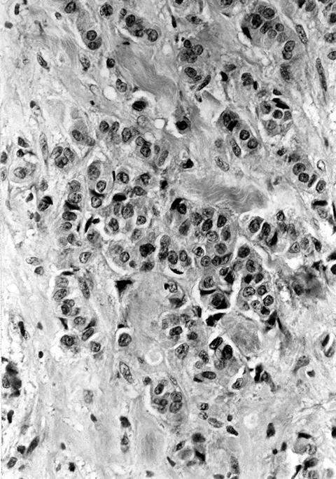
Small nodule of atypical cells with bizarre nuclei
Large
eosinophilic cells
with prominent
bizarre nuclei

Compensatory nodular hyperplasia
Prominent nuclear atypia / pleomorphism
NIS membranous expression
AFIP images
Diffuse hyperplasia with well developed papillae
Follicular cells lining papillae are tall and columnar
Hyperplastic small follicles
Images hosted on other servers:


Prominent infoldings of hyperplastic epithelium

Epithelium is tall columnar, colloid shows scalloping

No nuclear changes of papillary thyroid carcinoma

Various images

Numerous plasma cells

Follicles with pale colloid

Irregular follicles,
pseudopapillary
epithelium and
sparse colloid


Colloid has scalloped margins


Lobulated parenchyma with irregular follicles
Images hosted on other servers:

Graves disease: flame cells
Graves gross and micro
Thyroid: compare and contrast
Histopathology thyroid: Graves disease
Images hosted on other servers:


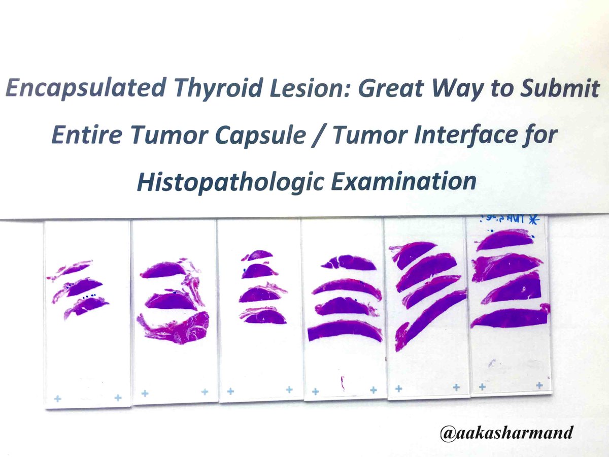
Ink color scheme


How to serially section
Contributed by Andrey Bychkov, M.D., Ph.D.
Orientation of specimen by surgeon
Sampling encapsulated lesion
Detecting capsular invasion
Sampling papillary carcinoma
Gross appearance of main thyroid lesions: follicular adenoma and papillary carcinoma
Images hosted on other servers:
Orientation of specimen by surgeon
Sampling encapsulated lesion
Grossing by Weill Cornell
Grossing by Gross Cutting Room
Contributed by Ayana Suzuki, C.T.
Isoechoic nodule
Images hosted on other servers:
Ultrasound
Ultrasound
Contributed by Mark R. Wick, M.D.
Diffusely enlarged thyroid lobe
Images hosted on other servers:
Atrophic gland
Nodular gland
Contributed by Andrey Bychkov, M.D., Ph.D. and Shipra Agarwal, M.D.
Lymphoid follicles with germinal centers
Diffuse
lymphoplasmacytic
infiltration
Squamous metaplasia: p63+ cells in many follicles
Evolution of squamous metaplasia
Intense immunostaining
Lobulation of thyroid tissue by fibrotic bands
Vesicular nuclei of thyroid follicles similar to PTC nuclei
Aggregation of lymphoid follicles mimic thyroid nodule
Hashimoto thyroiditis with lymphoepithelial cyst
Nodular Hashimoto thyroiditis
Papillary microcarcinoma and Hashimoto thyroiditis
Oncocytic cell nodule
Contributed by Shipra Agarwal, M.D. and Ayana Suzuki, C.T.
Cellular aspirate
Crushed cells

Lymphocytes and oncocytes
Images hosted on other servers:
Oncocytic cells with atypical nuclei
Sheet of follicular cells with oncocytic change mixed with benign lymphoid cells
Resembles lymphoma
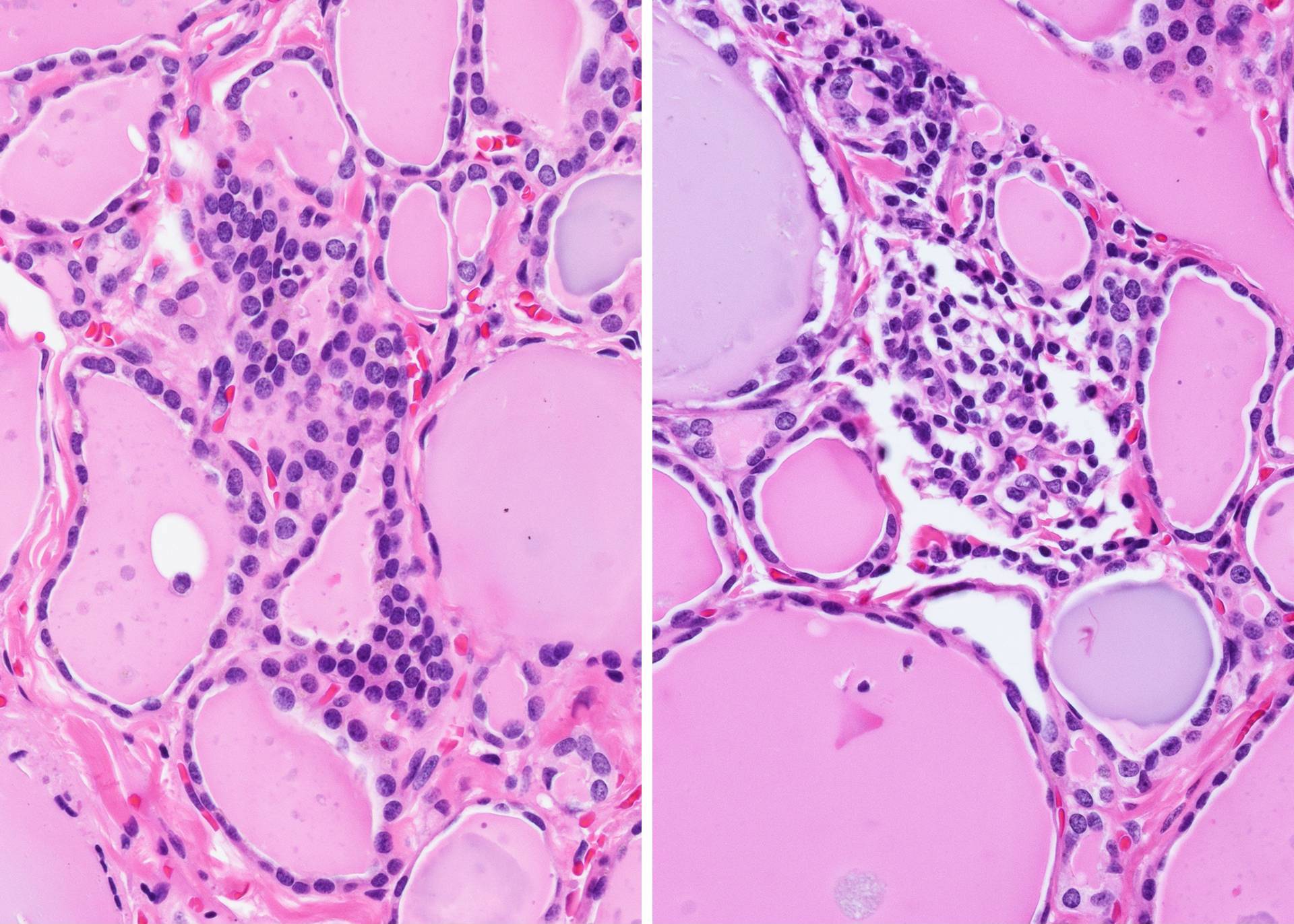
Grade I: mild lymphocytic inflammatory infiltrate
Grade II: moderate lymphocytic inflammation
Grade III: marked
inflammation with
polymorphous
lymphocytes
ThinPrep versus Pap stain

Lymphocytes and oncocytes


Follicular destruction by lymphocytes

Crushed cells
Grade 1, 2 and 3 thyroiditis
Thyroid: compare and contrast
Histopathology thyroid: Hashimoto thyroiditis
AFIP images
Metaplastic squamous follicles

Dilated lumen of metaplastic follicle
Various images
Contributed by Andrey Bychkov, M.D., Ph.D.
Perinuclear brown pigment
Oncocytes
Sanderson polster
Thyroid follicular epithelium
Follicular colloid
Normofollicular thyroid
Macrofollicular thyroid
Pyramidal lobe

Microfollicular thyroid

Pericapsular fat
Adipose metaplasia
Perithyroid thyroid follicles
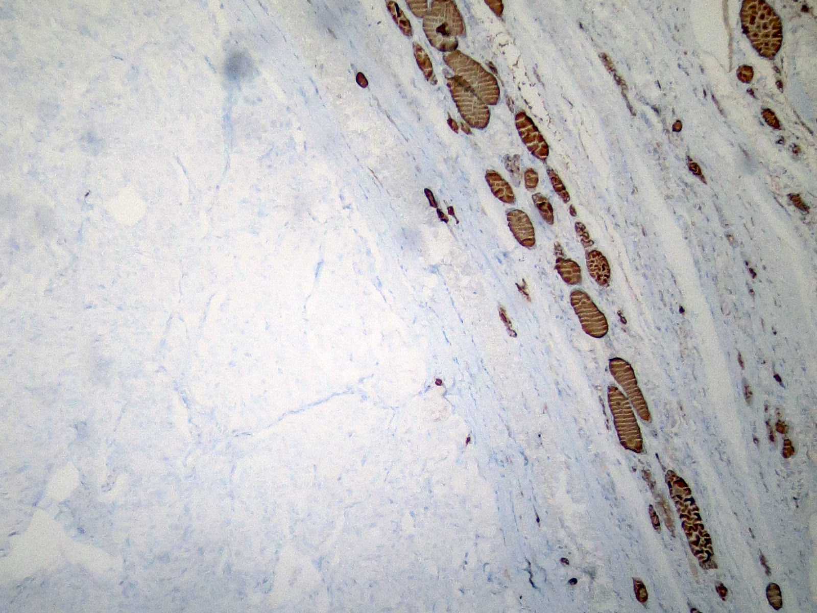
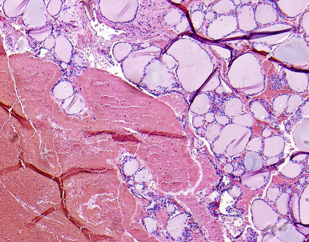
Intrathyroidal muscle
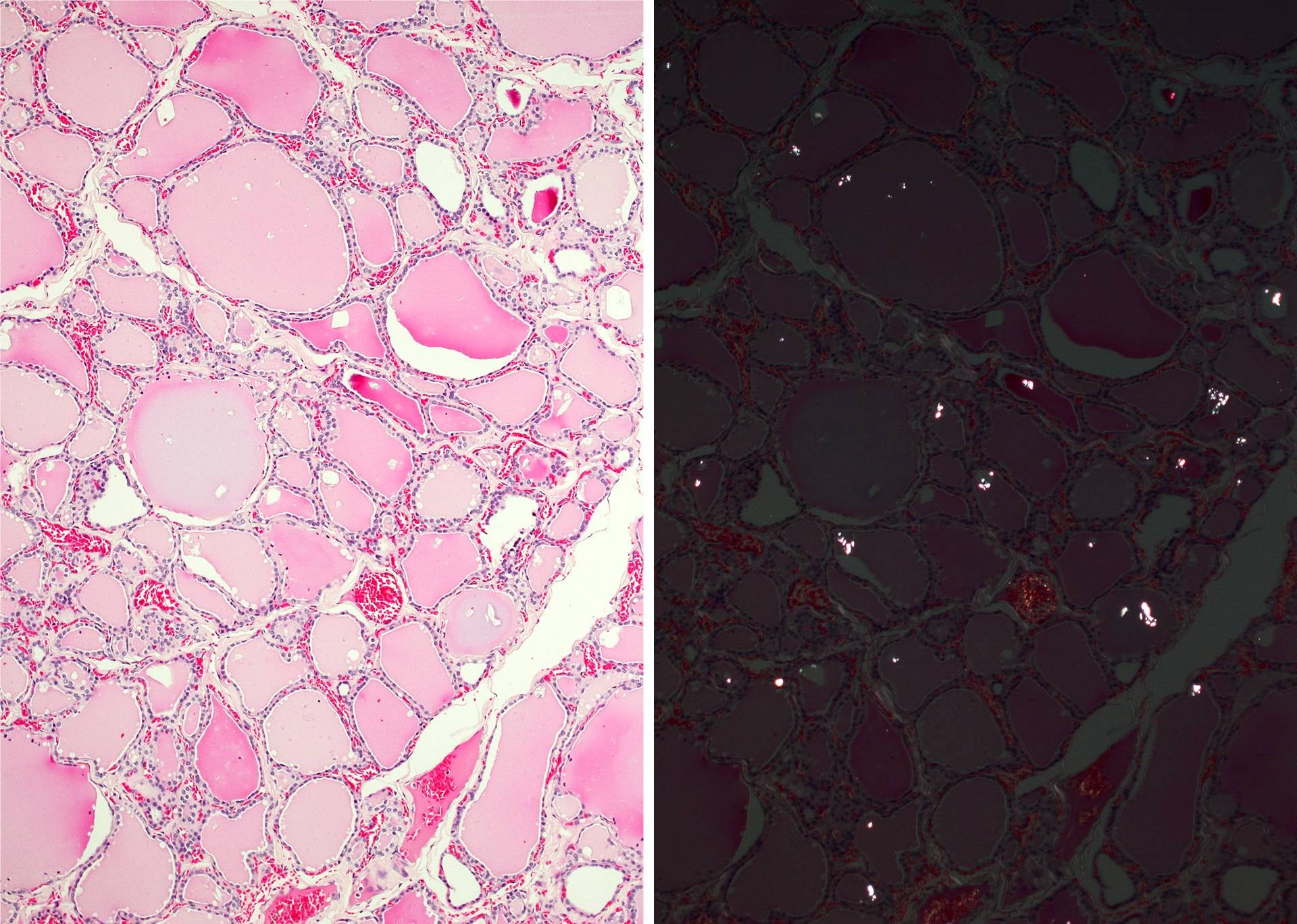
Calcium oxalate crystals in the colloid
PAS staining
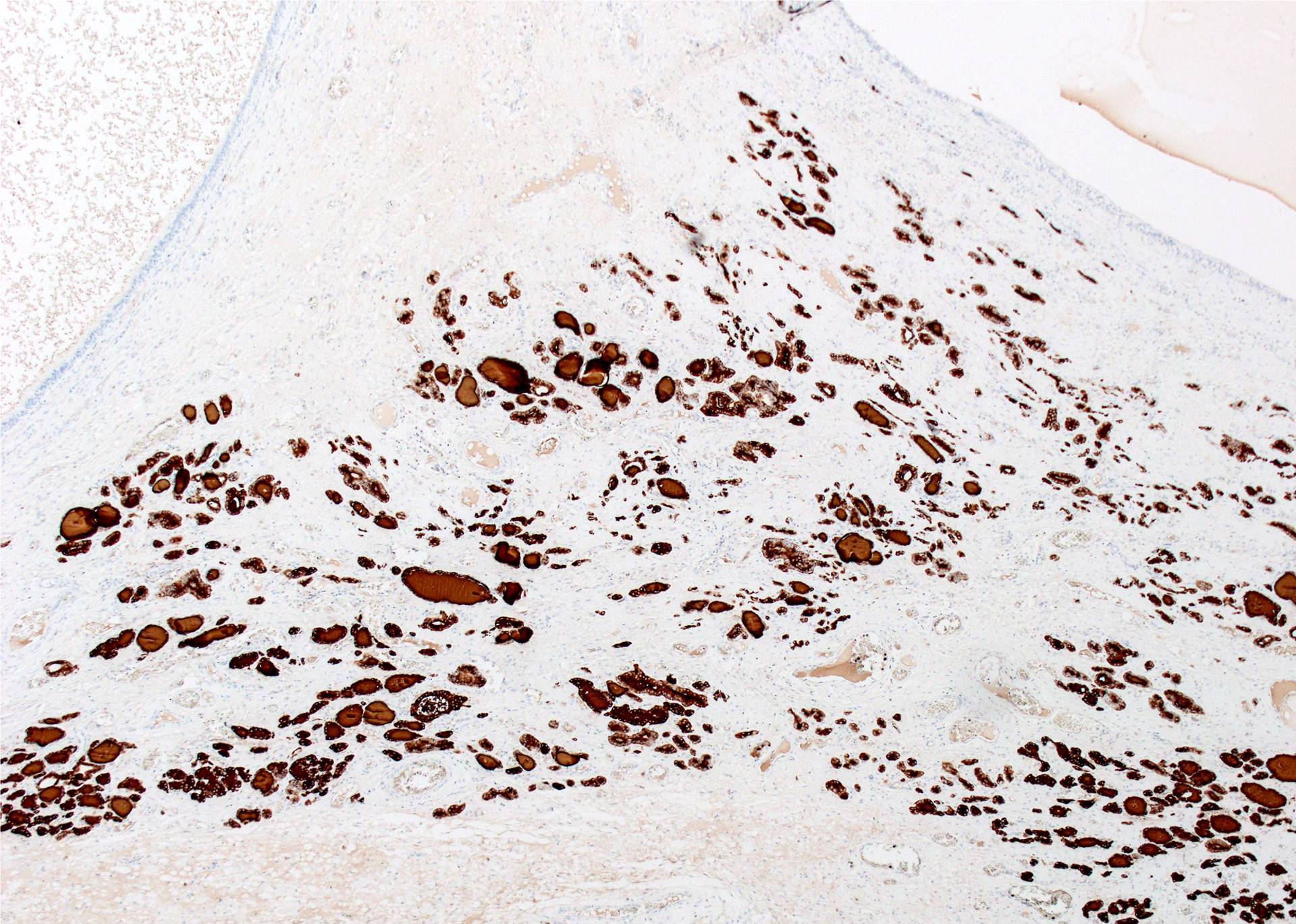
Thyroglobulin expression
Mucicarmine staining
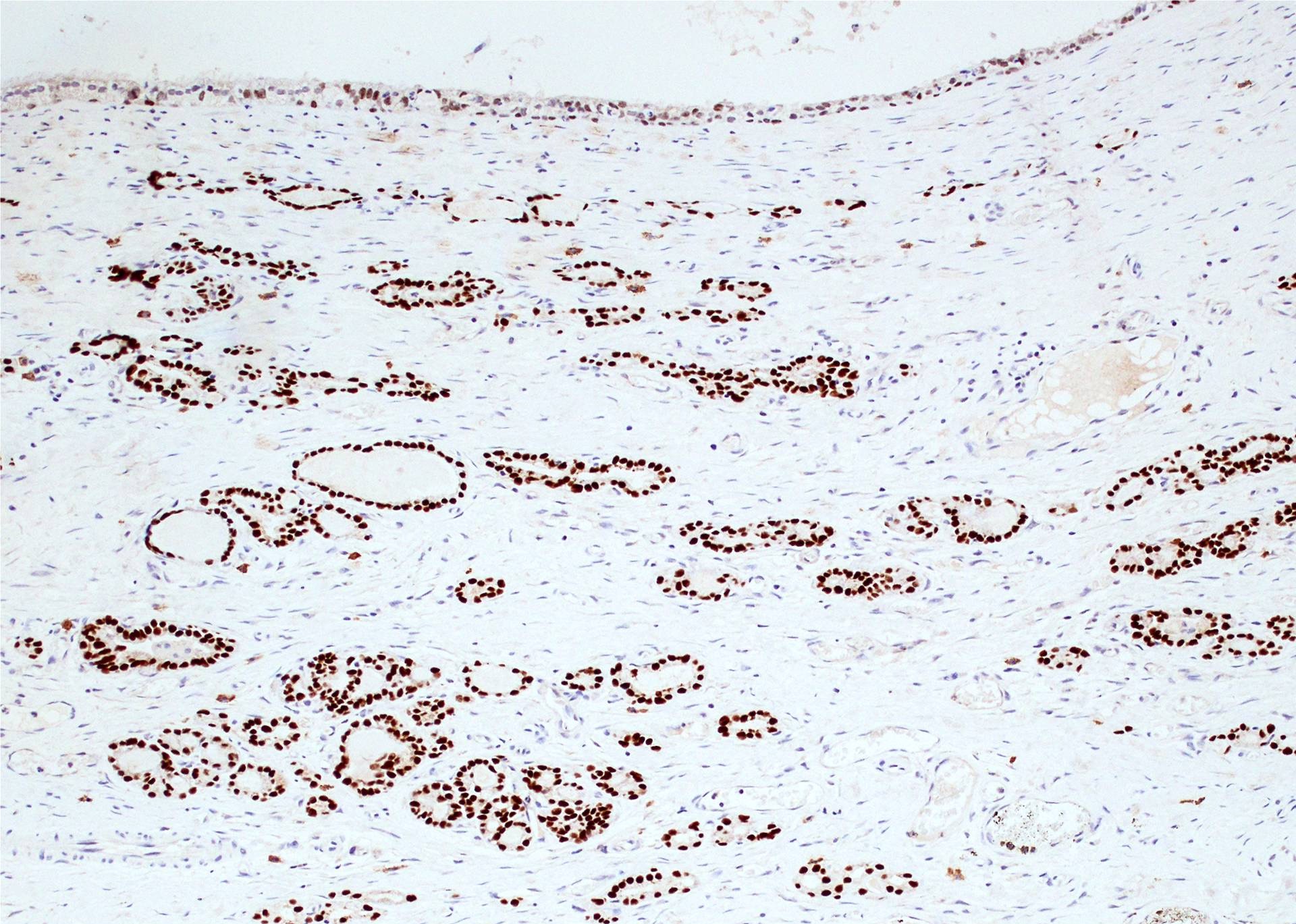
TTF1 expression
PAX8 expression
TTF2 nuclear expression
NIS membranous expression
Cluster of C cells
Location of C cells in relation to follicle
Pericapillary location of C cells
Clustered distribution of C cells
Calcitonin IHC
Proliferation in adult thyroid
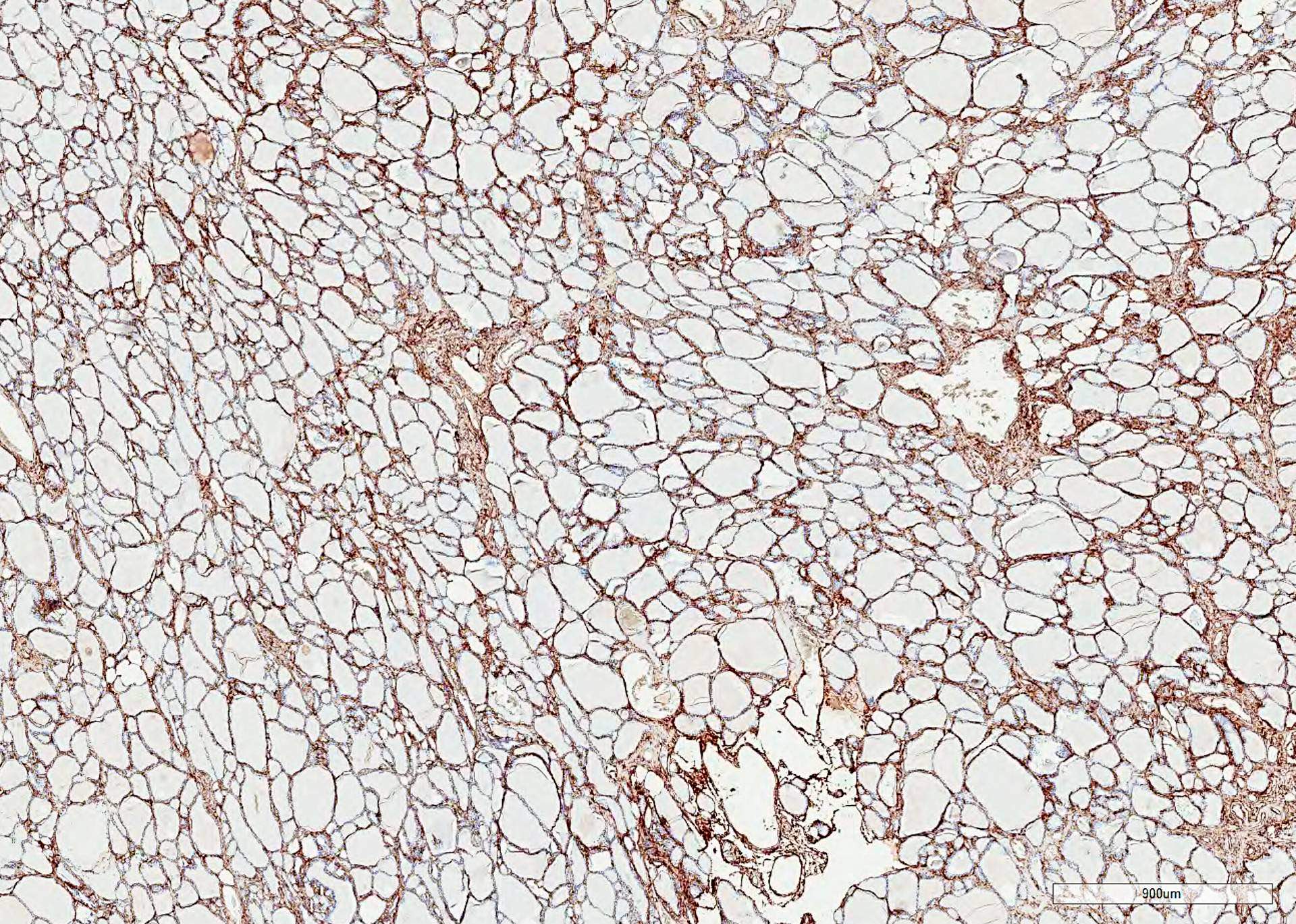
Prominent vascular network
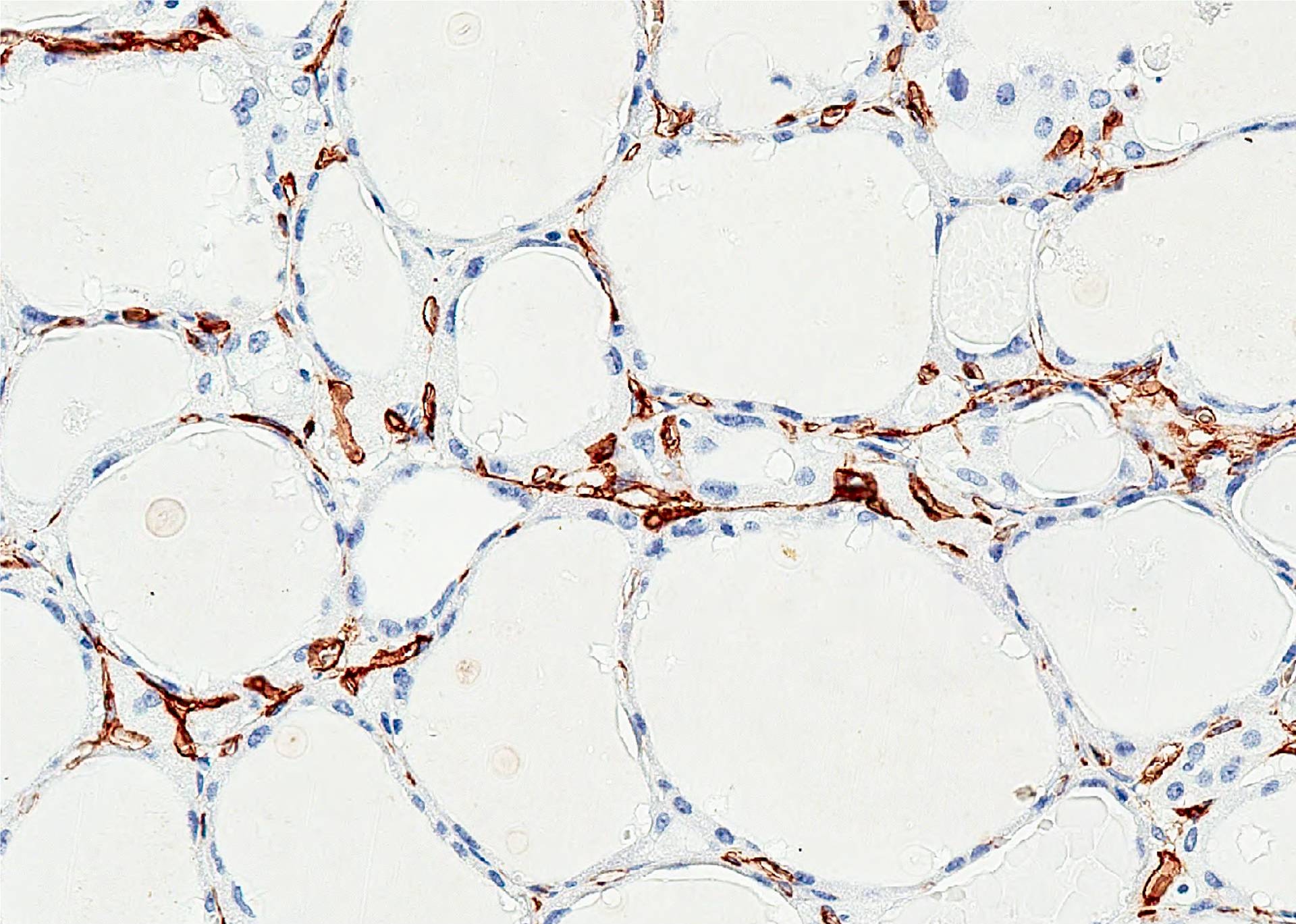
Capillary network
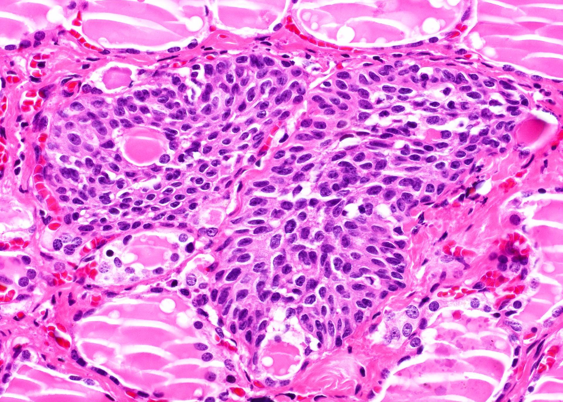
Solid cell nests
Histologic mimics of SCN
Solid cell nests: p63
Solid cell nests: thyroglobulin-
AFIP images
stain); cells are
polyhedral and
spindled
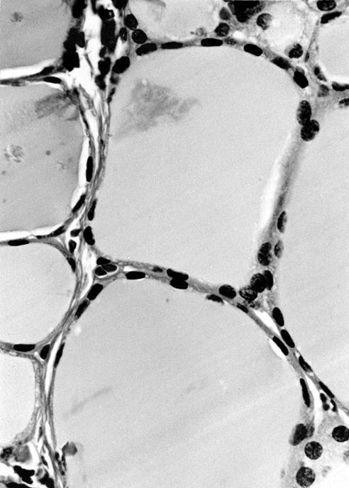
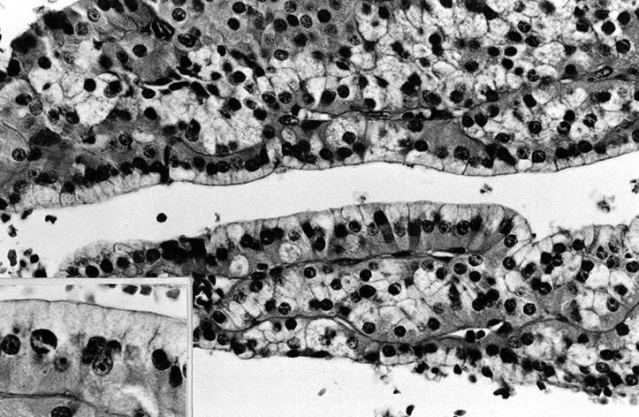
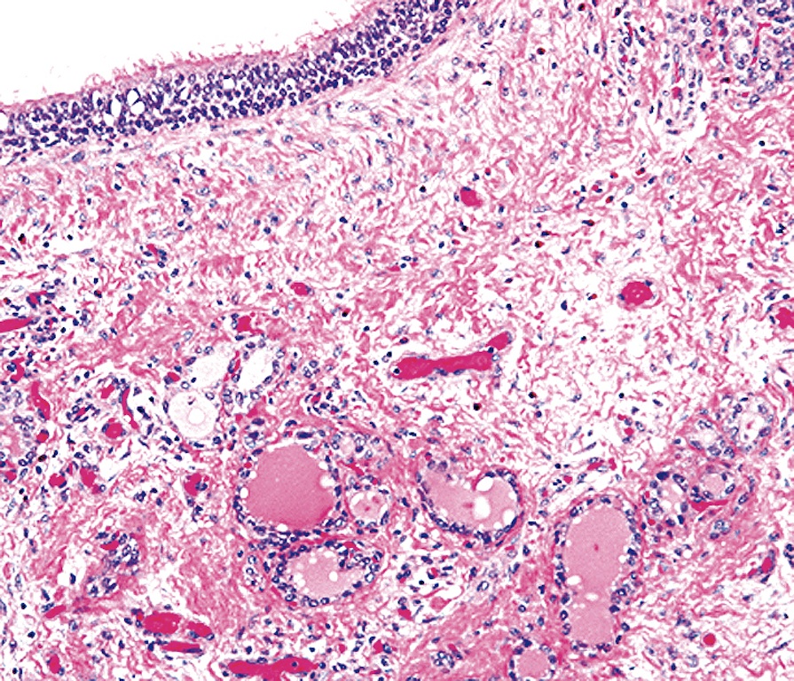
Follicles lined by flattened epithelium
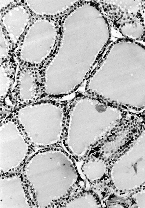
Normal adult follicles are round to oval
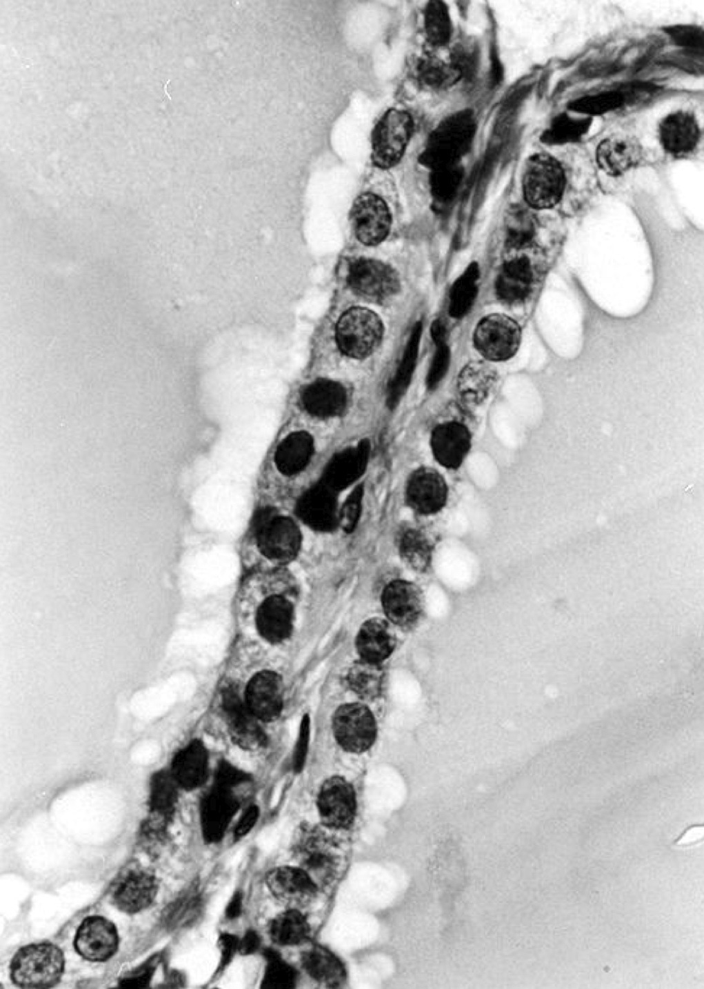
Follicles lined by cuboidal epithelium
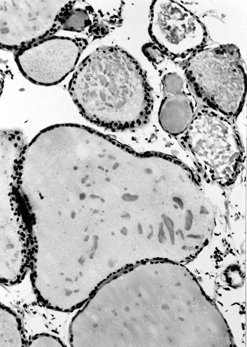
Colloid is flocculent - most likely artifactual, not significant
Tangential section
of normal thyroid
follicle may resemble
C cell hyperplasia
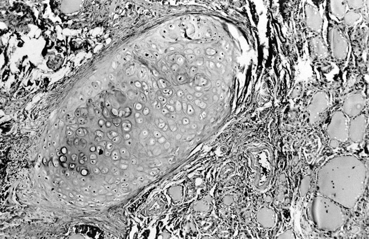
Ectopic cartilage

Skeletal muscle

Squamous metaplasia
Images hosted on other servers:


Follicles lined by flattened epithelium and filled with colloid

Rich vascular supply
Thyroid and parathyroid glands


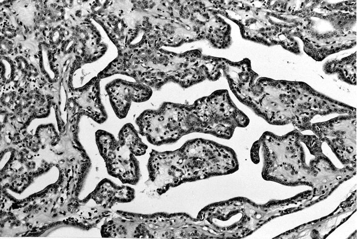
Papillary structures

C cells (calcitonin stain)
AFIP images
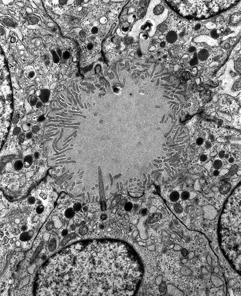
Follicles have luminal microvilli
Intrafollicular C cell
Images hosted on other servers:
Follicular cells
By John R. Minarcik, M.D.
Images hosted on other servers:

Summary of patients
Contributed by Andrey Bychkov, M.D., Ph.D.
Edematous papillae lined by discohesive epithelium

Oncocytic epithelium with loss of cohesiveness
Hobnail cells with apically placed nuclei

Apical decapitation of cells
Images hosted on other servers:


PTC hobnail variant on different magnifications


Group 2 papillary carcinoma of the thyroid

IHC
Images hosted on other servers:


Conventional smear

LBP
Images hosted on other servers:

Encapsulated gray white nodular lesion

Gray white with one area showing cystic degeneration
Contributed by Virginia A. Livolsi, M.D.
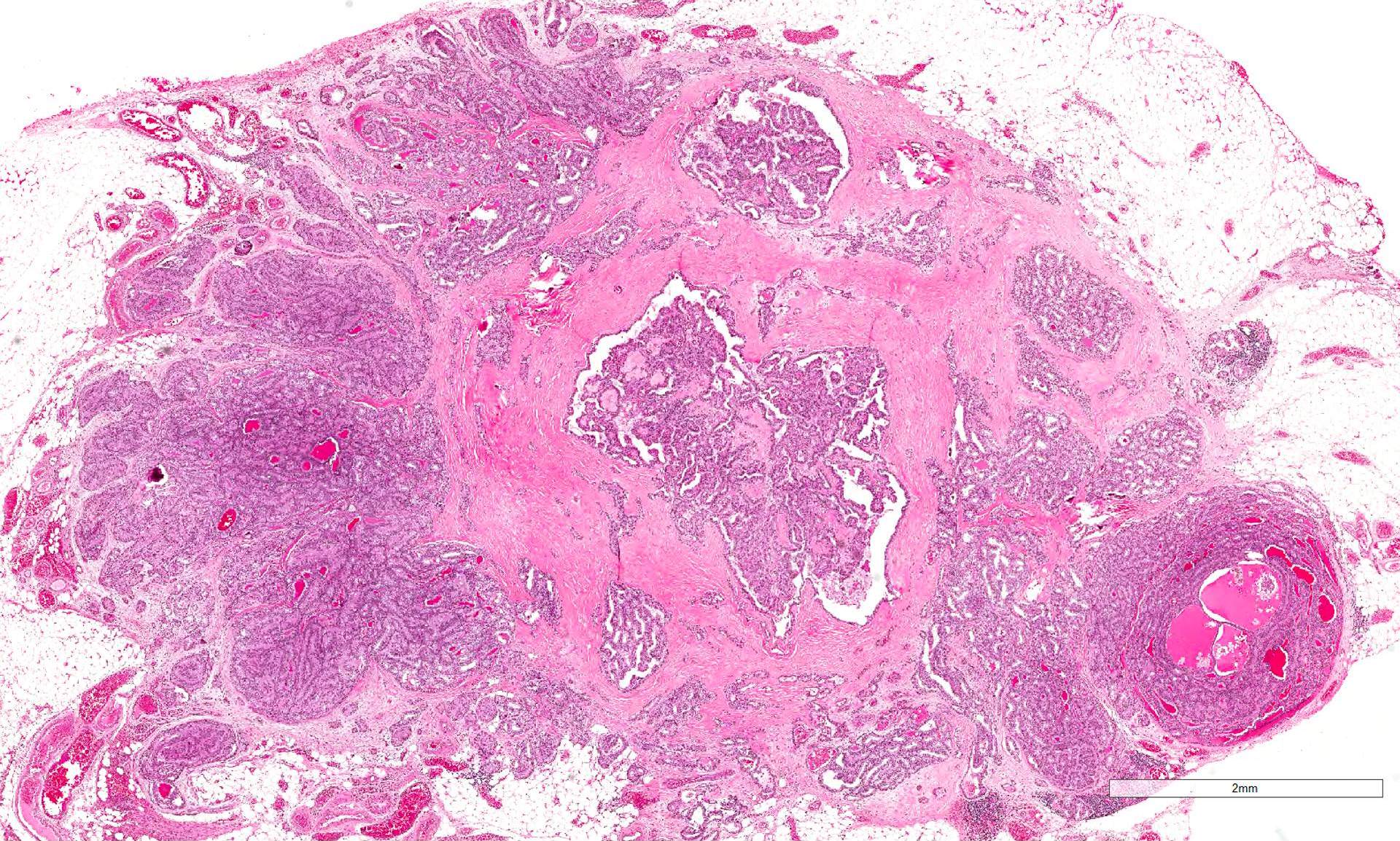
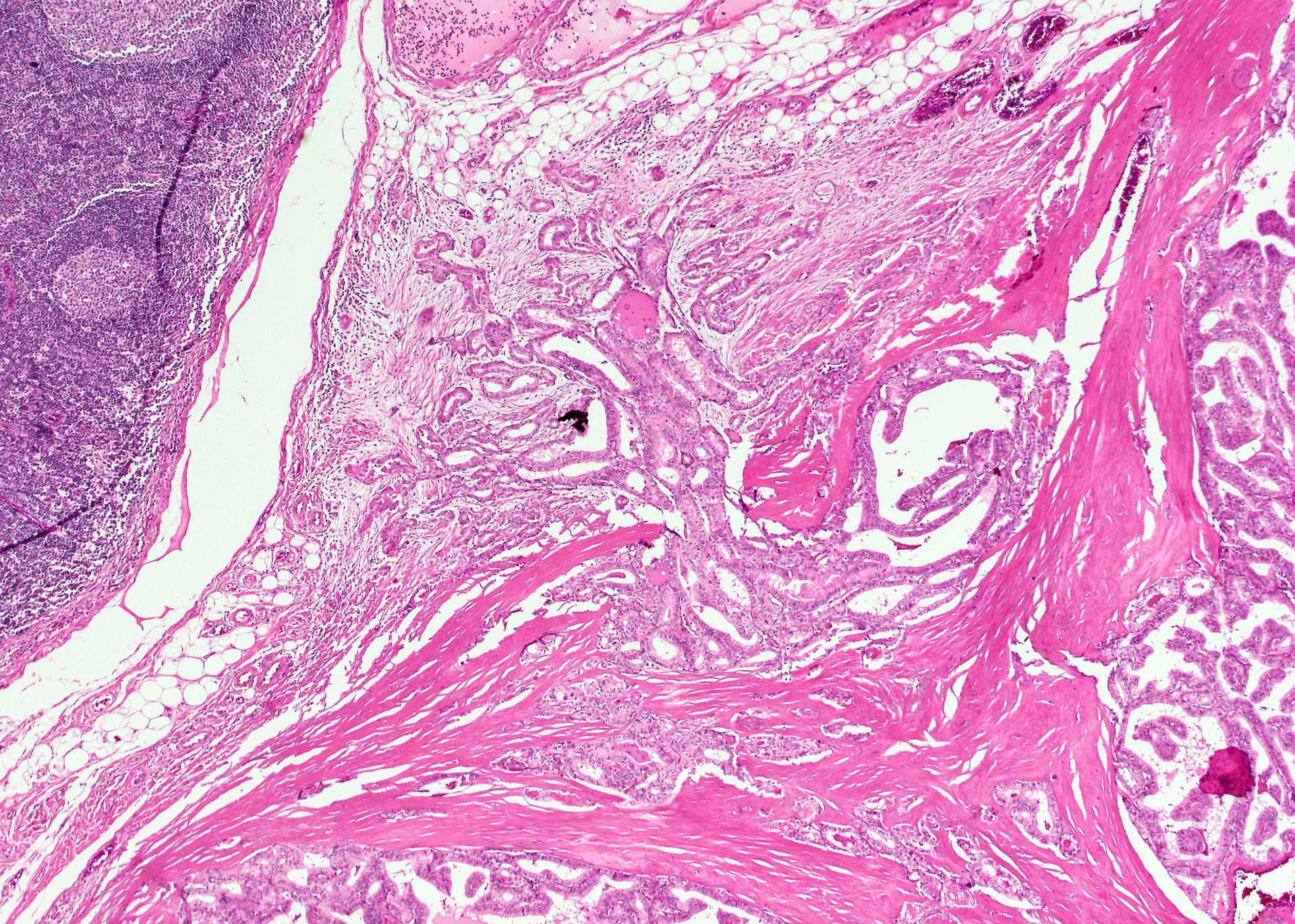
HTT thinly encapsulated
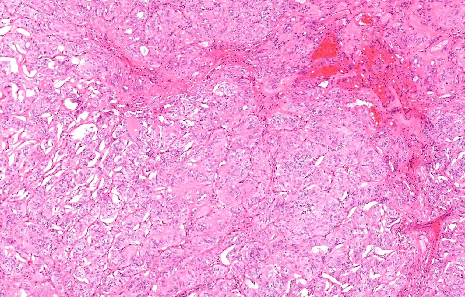
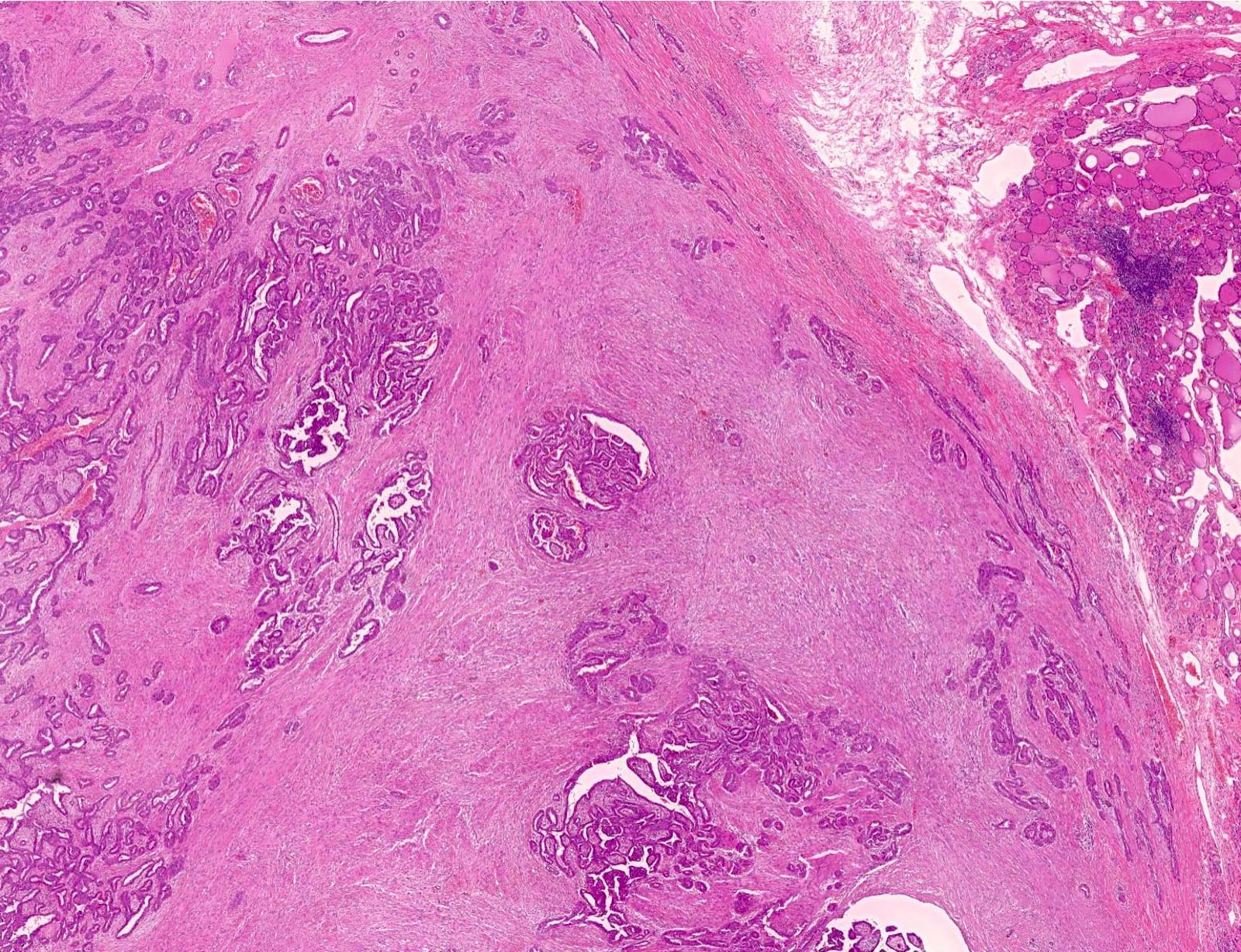
Solid trabecular growth
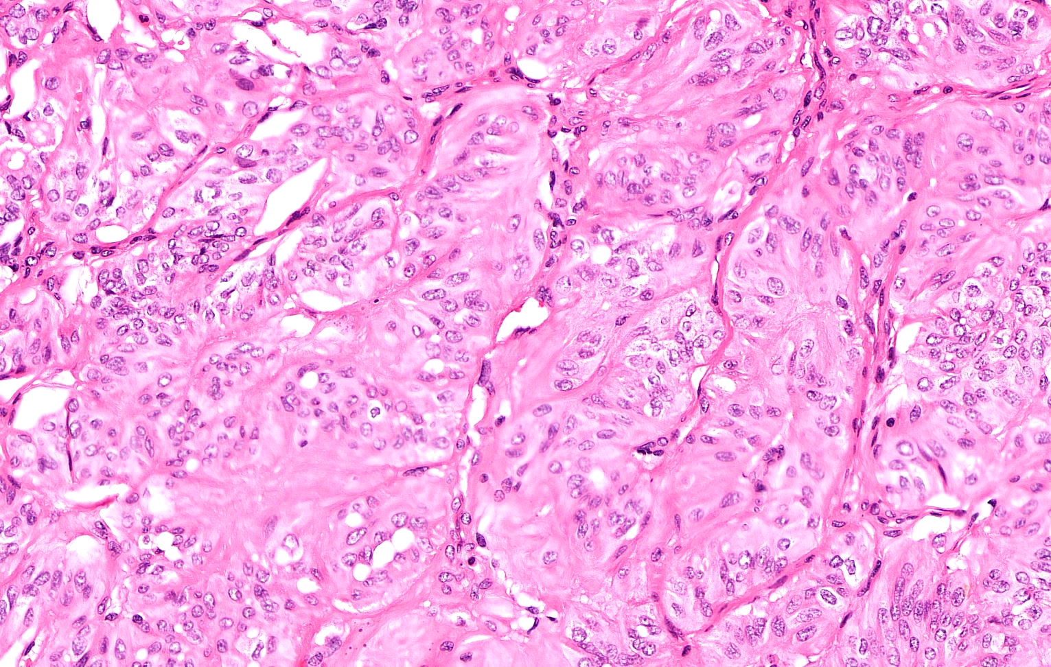
Trabecular pattern noted
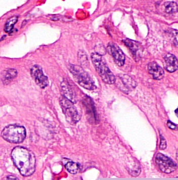
Elongated nuclei
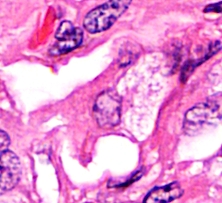
Prominent intranuclear inclusion
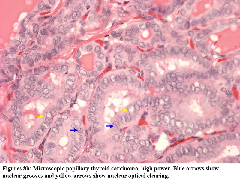
Nuclear clearing and grooves
Yellow bodies
Calcification in tumor stroma
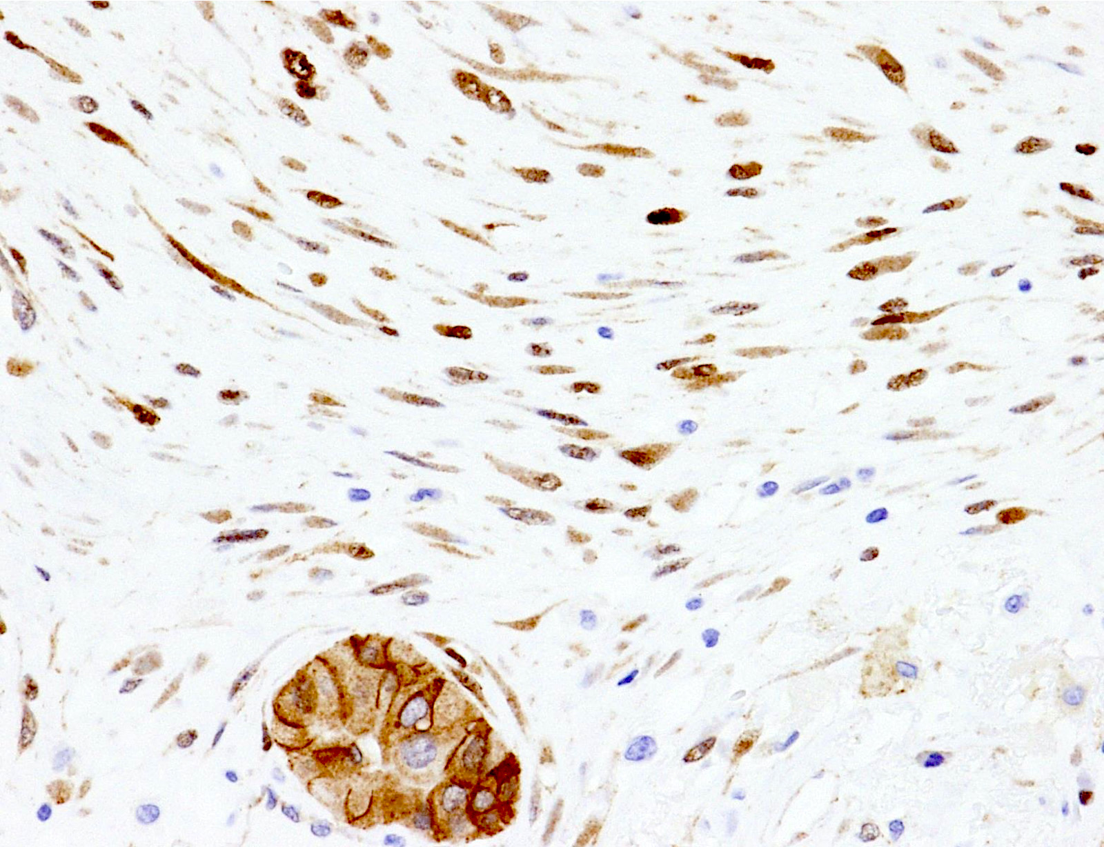
Thyroglobulin stain variable
TTF1 decorates nuclei
Ki67 decorates membranes
Contributed by Ayana Suzuki, C.T.
Nuclear pseudoinclusions
Images hosted on other servers:

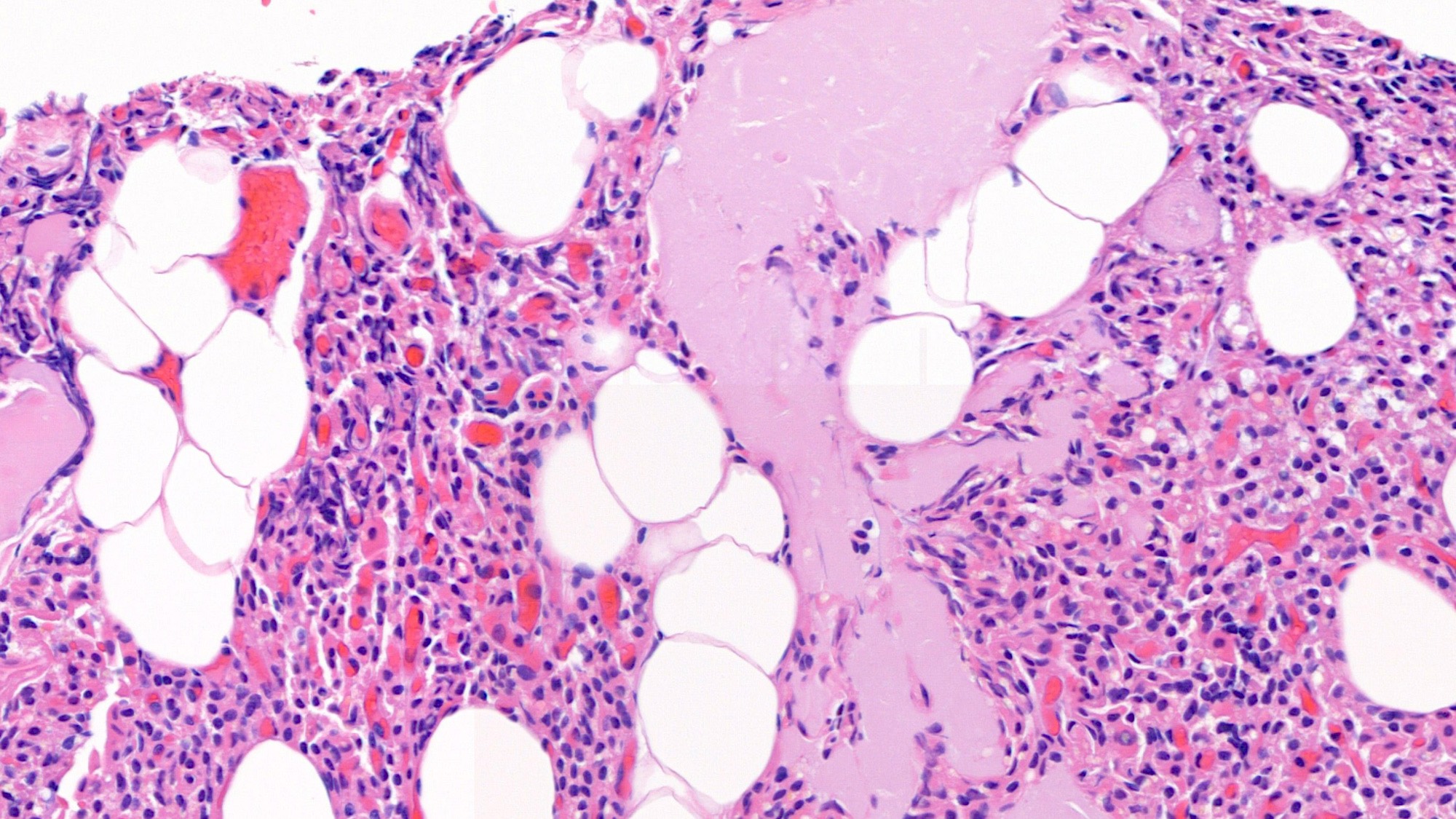
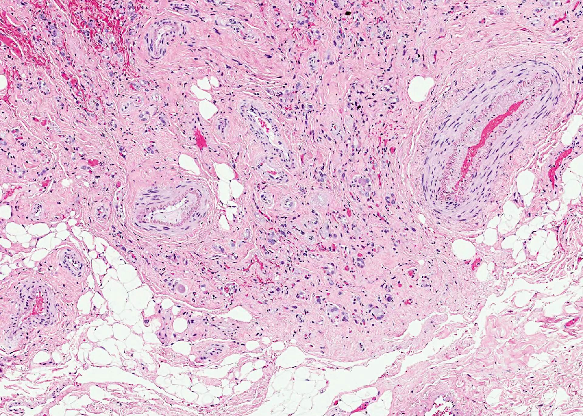
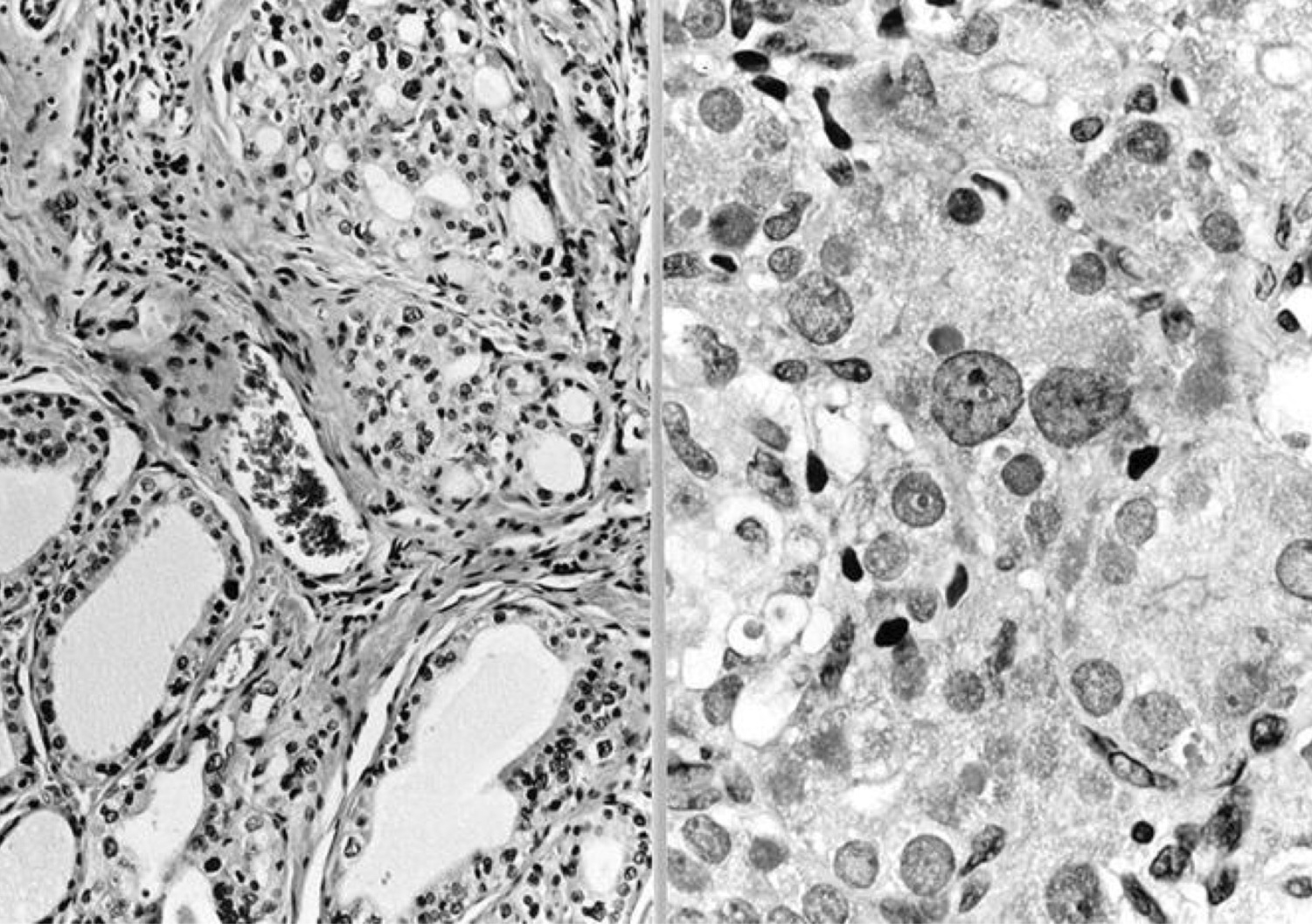
Calciphylaxis
Images hosted on other servers:


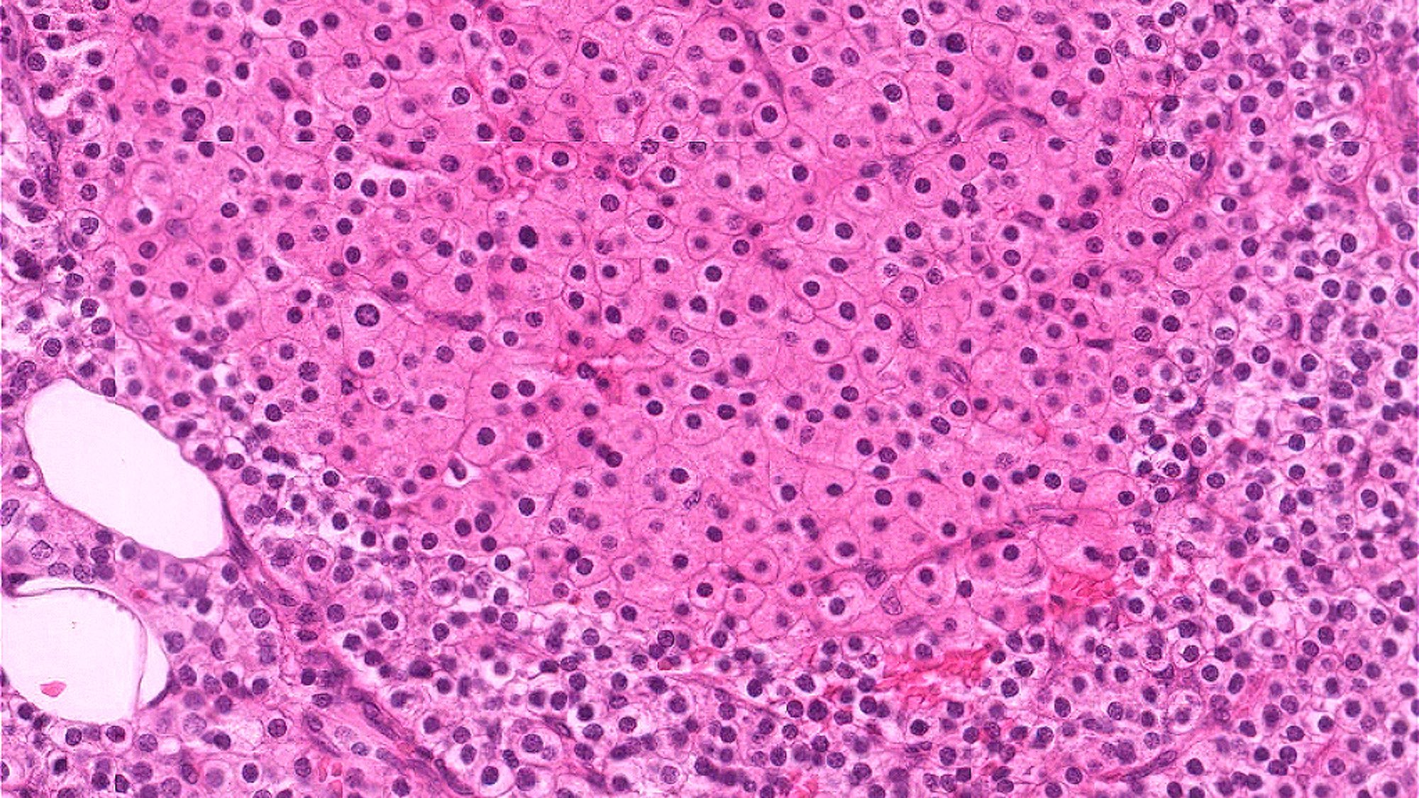
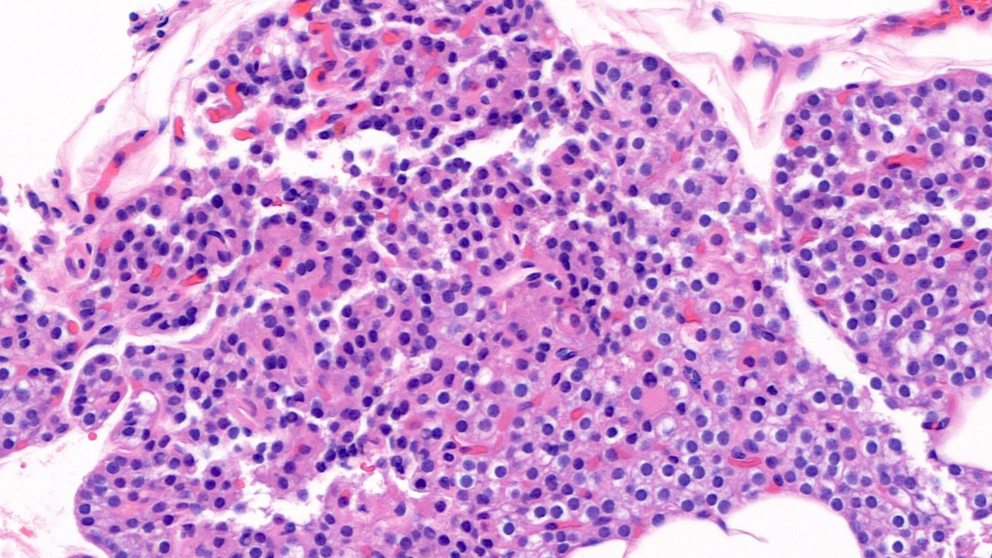
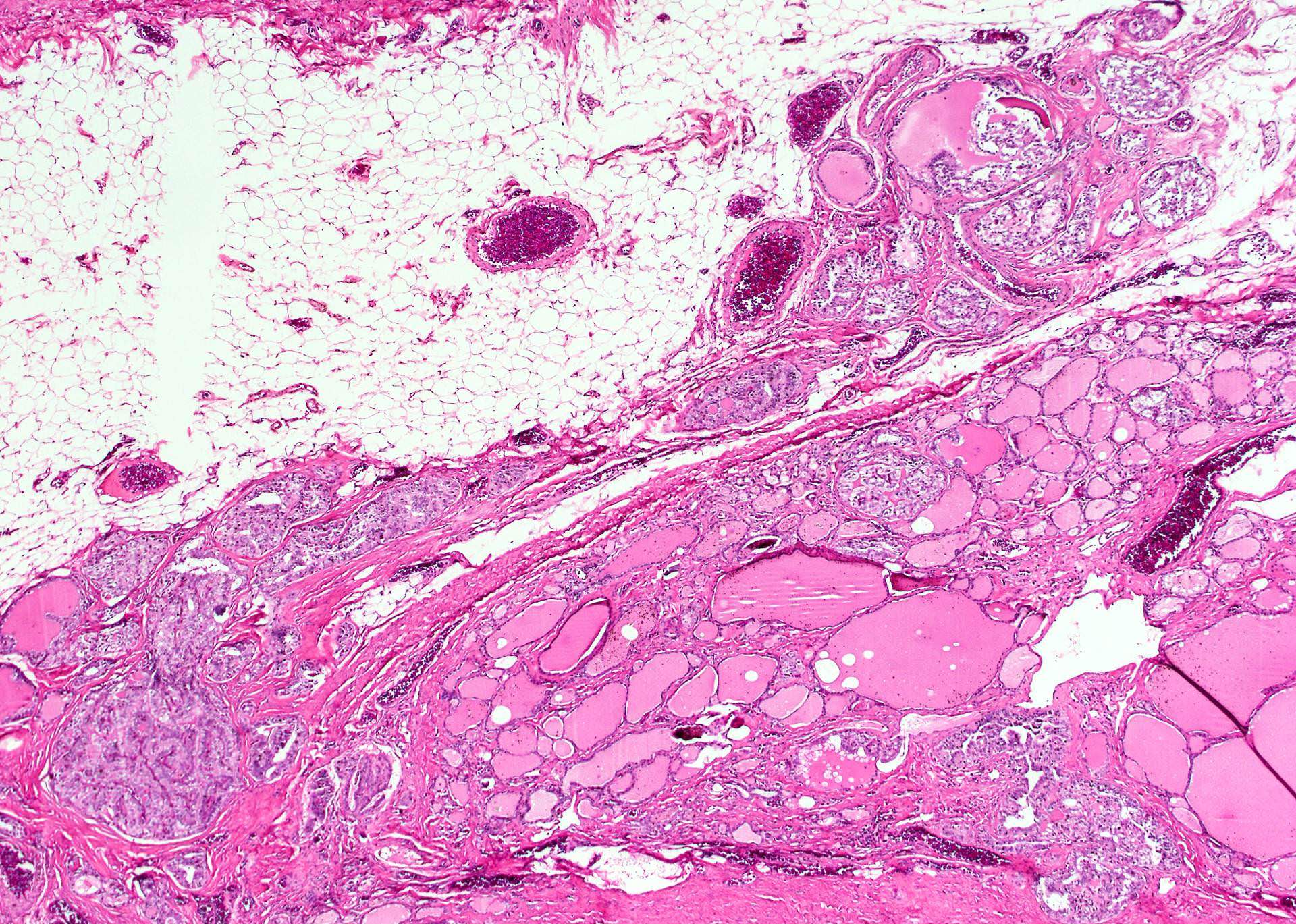
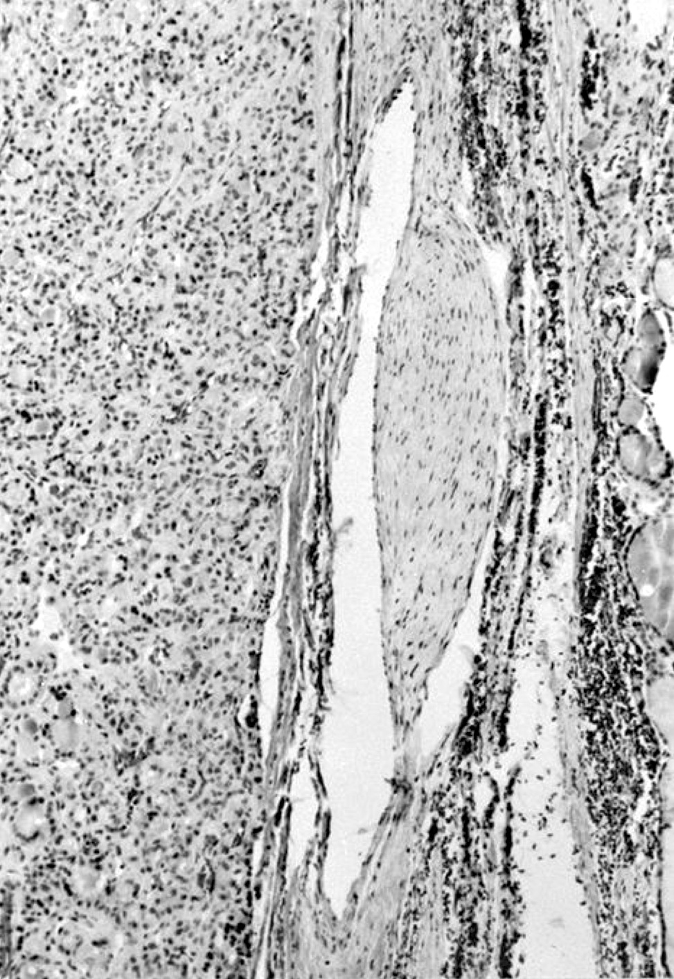
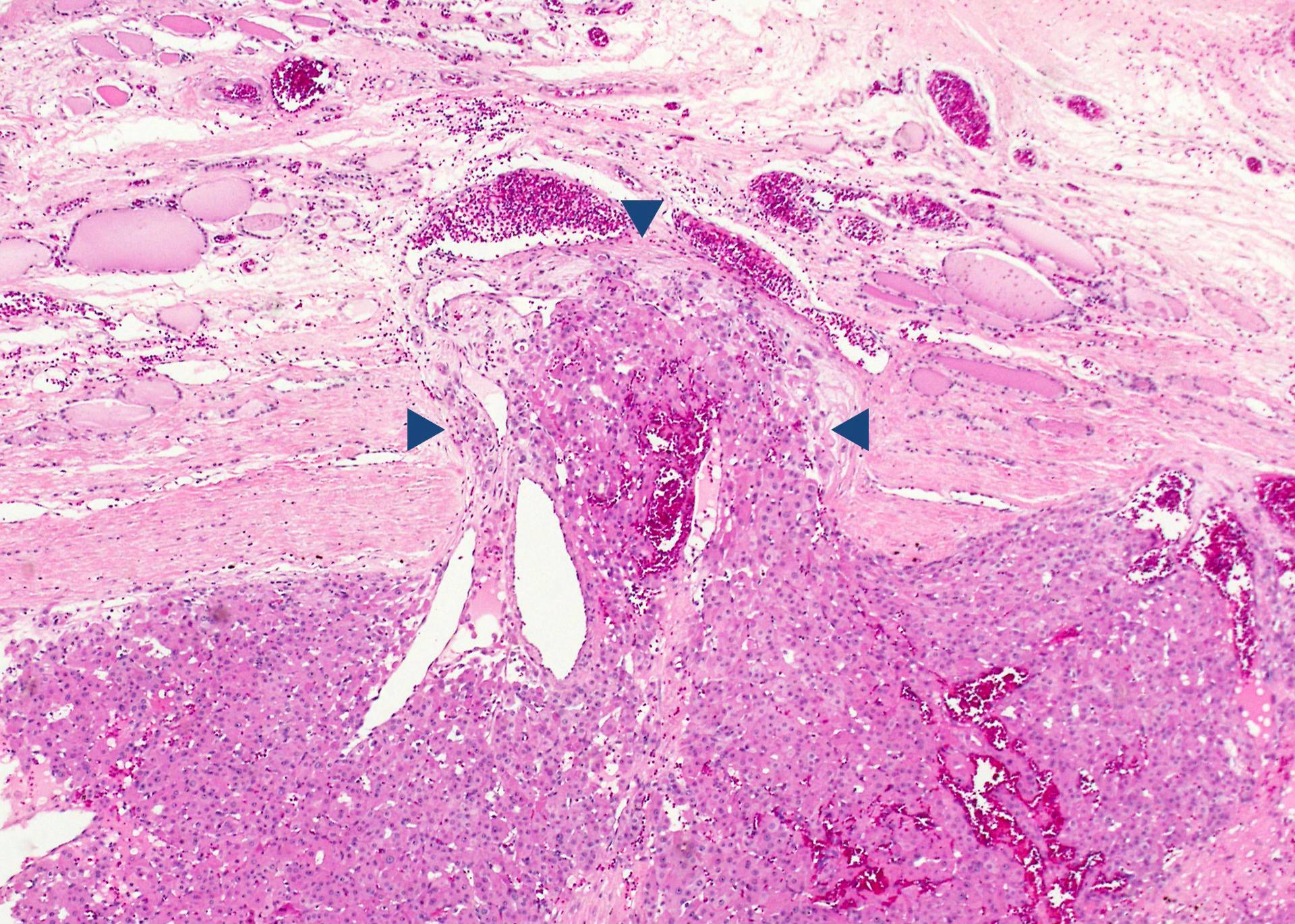
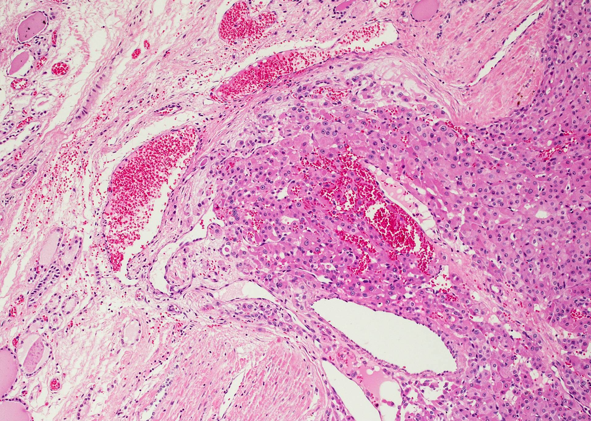
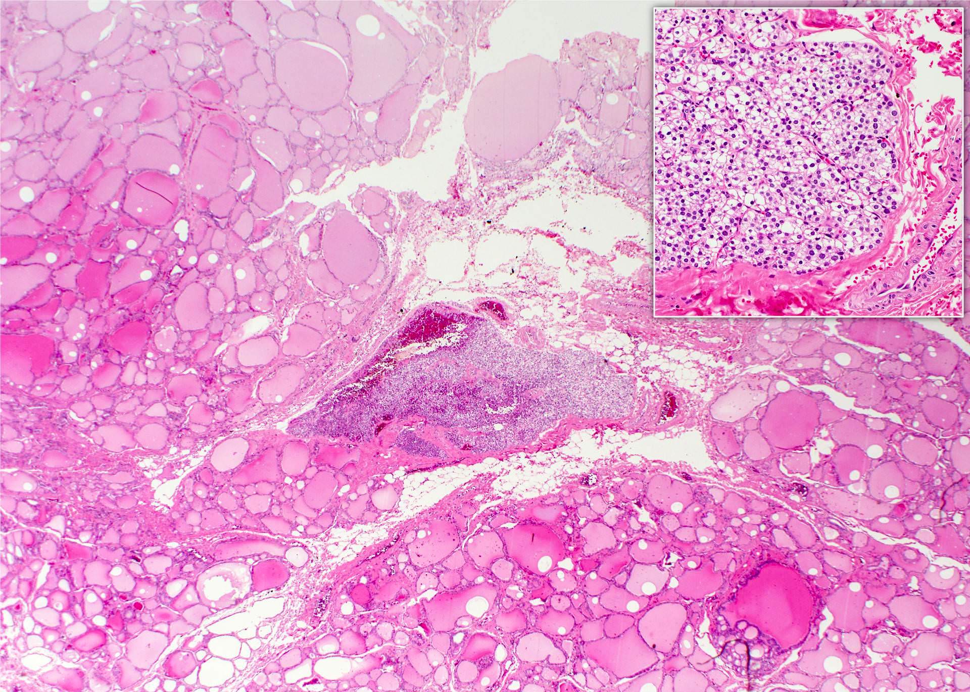
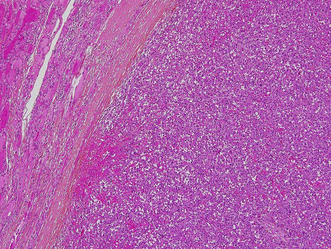
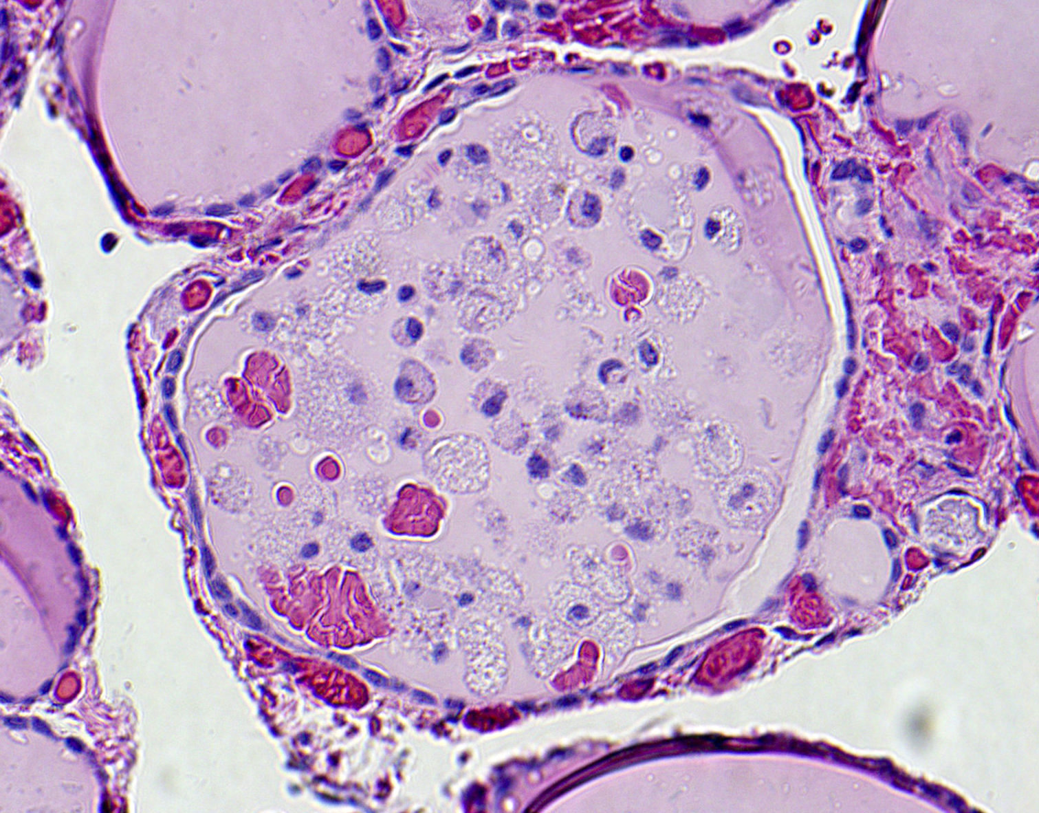
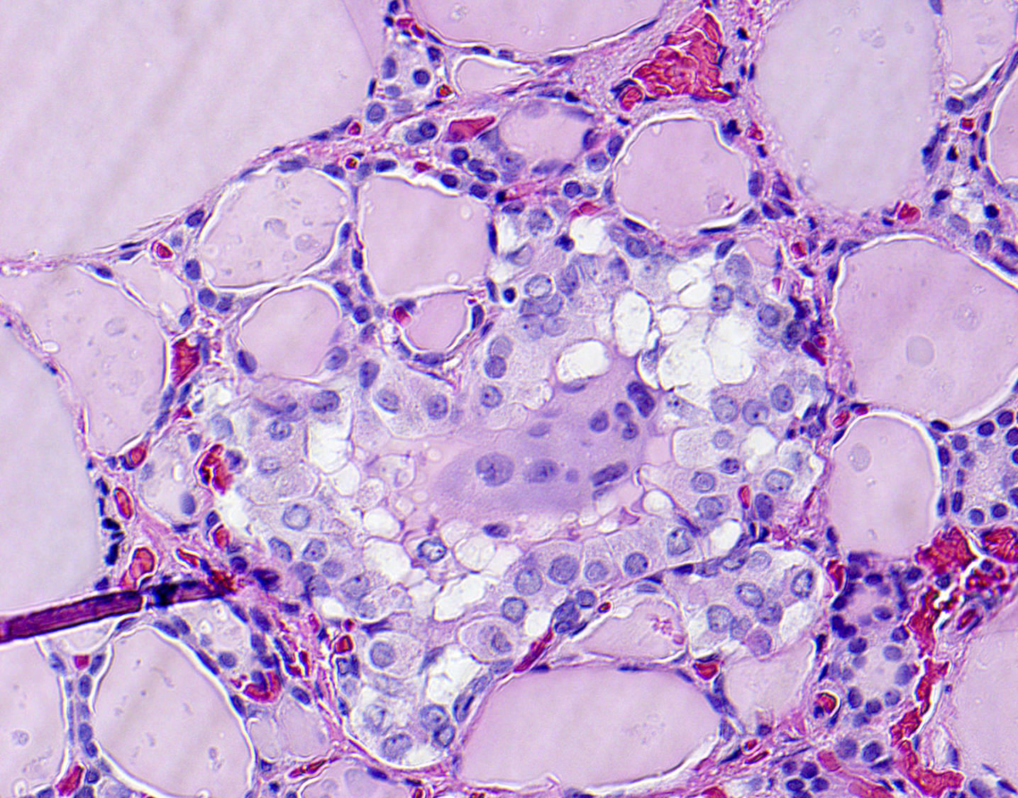
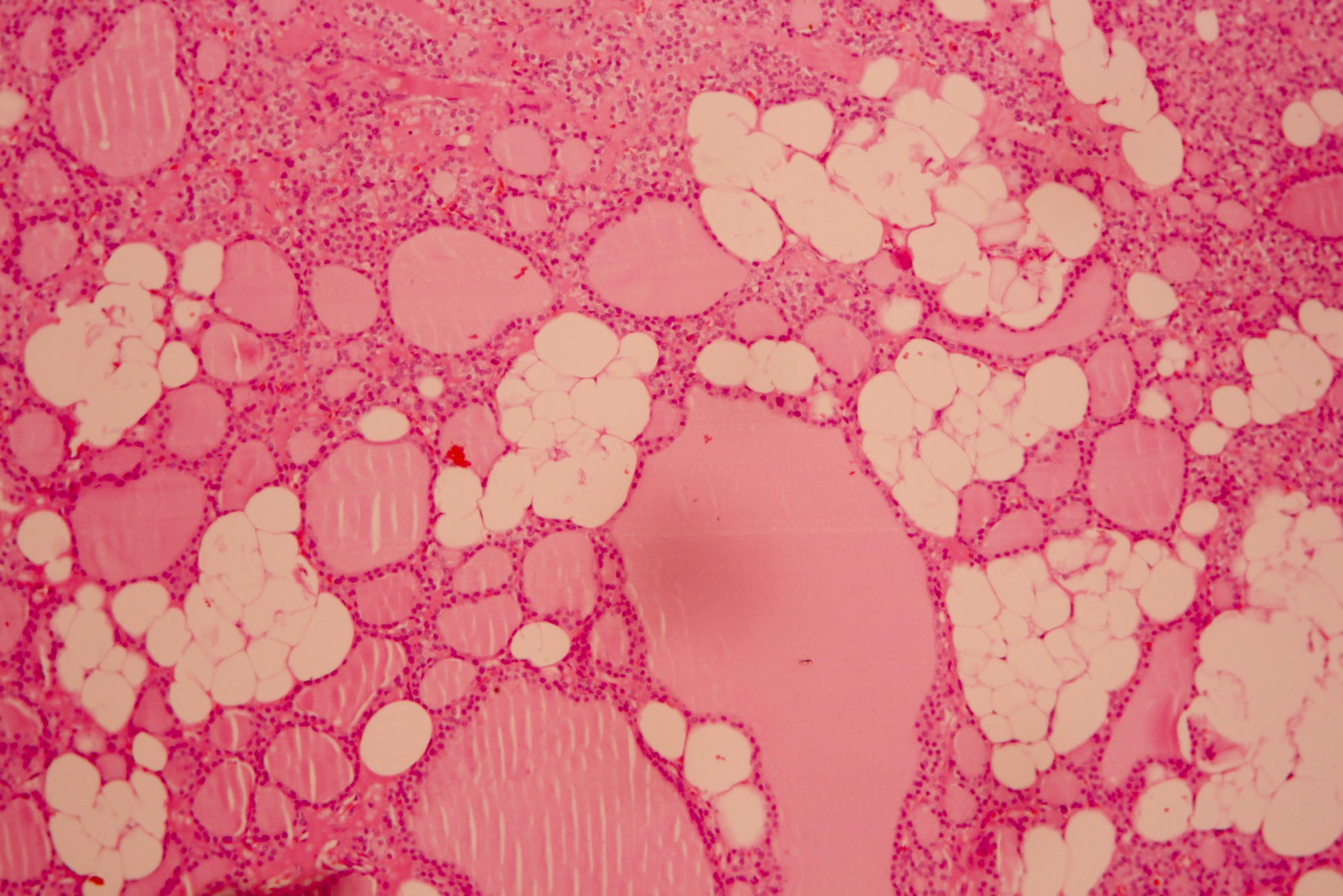
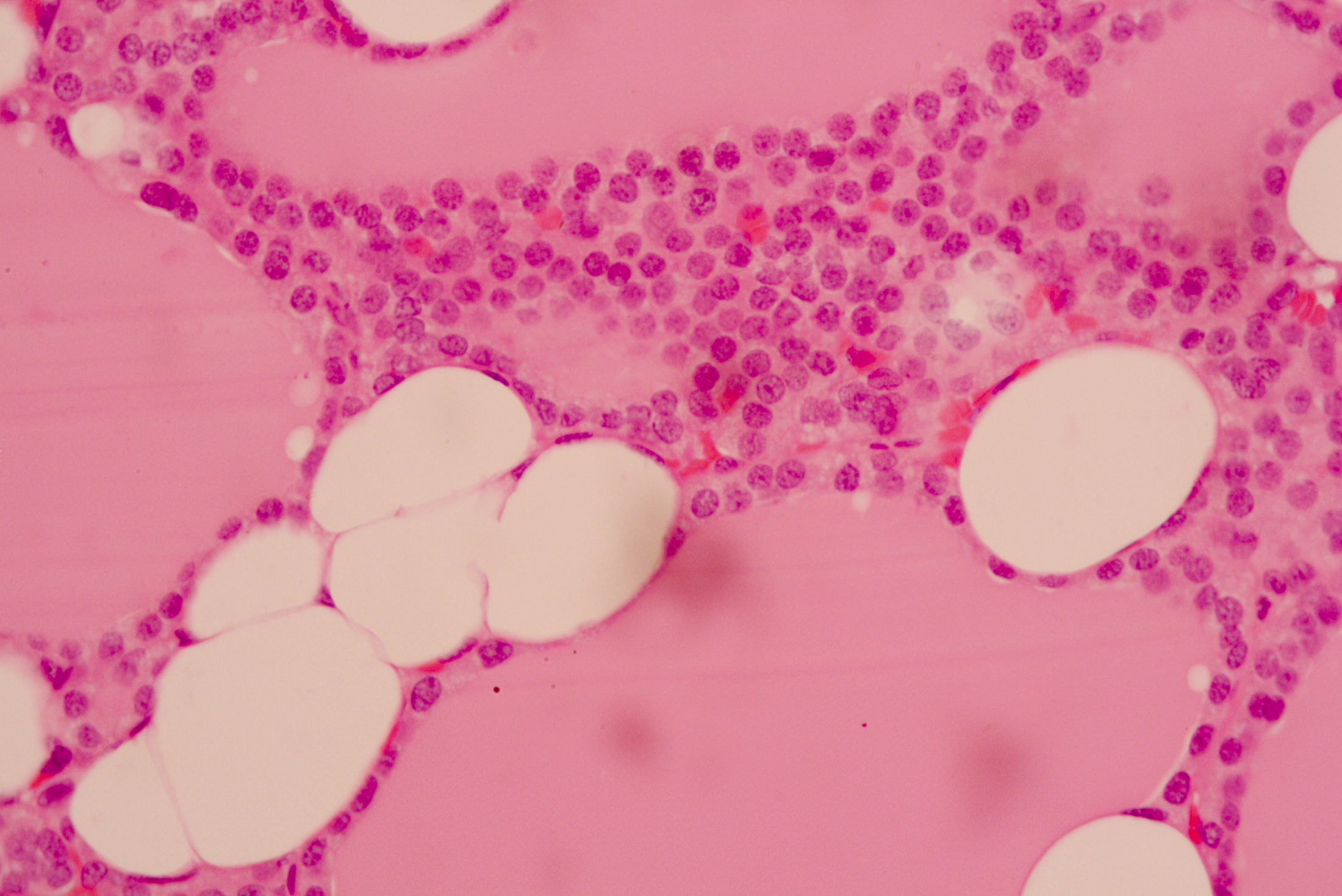
Gland composed mainly of chief cells with a rim of normal parathyroid tissue, no stromal fat

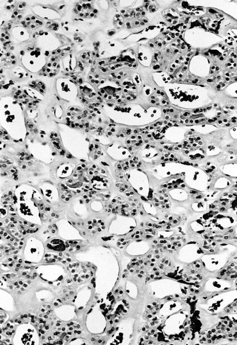
Chief cell hyperplasia and oncocytic cells

Calciphylaxis: ischaemic skin changes
Contributed by Andrey Bychkov, M.D., Ph.D.
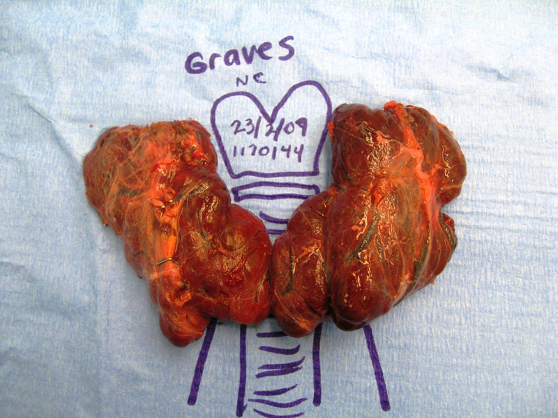
Enlarged thyroid
Images hosted on other servers:


Diffuse hyperplasia of thyroid gland
Histopathology Thyroid - Graves Disease
Endocrinology overview
Images hosted on other servers:

CT scan

Ultrasound
Images hosted on other servers:

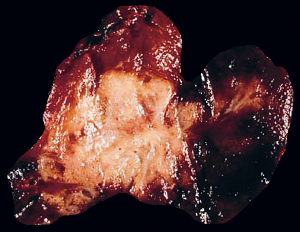
Lobulated, solid and tan colored

Tumor and trachea
Contributed by Shuanzeng Wei, M.D., Ph.D. and Andrey Bychkov, M.D., Ph.D.
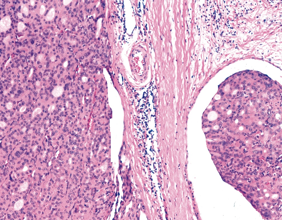
Tumor nests and fibrosis
Central necrosis
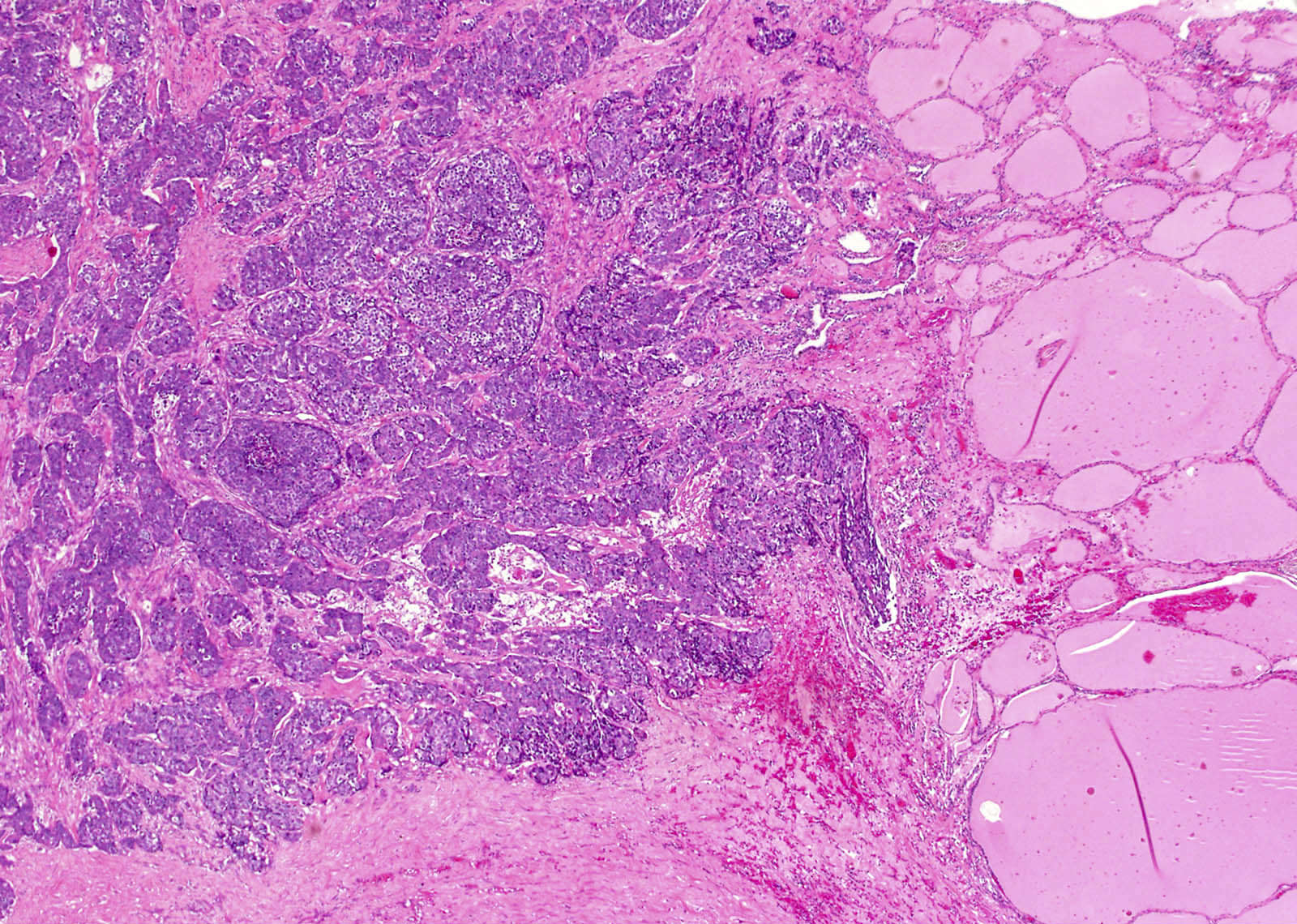
Tumor and thyroid tissue
Fibrous bands
Squamoid cells
Invasive growth
Images hosted on other servers:

Sheets and clusters with keratin
Discohesive polygonal to ovoid cells
Contributed by Andrey Bychkov, M.D., Ph.D. and Rachel Jug, M.B.B.Ch., B.A.O.
Vesicular nuclei with irregular membranes

Evident major and minor diagnostic features
Dark colloid with frequent scalloping and clefting
Free floating of small tumor fragment in vascular lumen
Small piece of tumor floats in vascular lumen
Capsular invasion and extrathyroidal extension
Tumor plug is associated with thrombus
Overlapping, elongation and a single mitotic figure
Irregular nuclear contours, grooves, pseudoinclusions
Clear chromatin with margination
Contributed by Xiaoyin "Sara" Jiang, M.D.
Microfollicles and nuclear enlargement
Images hosted on other servers:


Risk factor model
Images hosted on other servers:

Ultrasound

MRI

FDG-PET
Contributed by Somboon Keelawat, M.D. and Jijgee Munkhdelger, M.D., Ph.D.
Discrete nodule
Eosinophils
Follicular stuffing

LCH cells
LCH cells and eosinophils
Langerin
CD1a
Thyroglobulin
Images hosted on other servers:


FNA smear

S100 and CD1a
Images hosted on other servers:


Birbeck granules
Images hosted on other servers:

Sonogram and CT
Contributed by Andrey Bychkov, M.D., Ph.D.
Hashimoto thyroiditis with lymphoepithelial cyst

Lymphoepithelial cyst of thyroid
Images hosted on other servers:

H&E, p63, Ki67
Images hosted on other servers:

FNA
AFIP images
Diffuse large cell lymphoma: fish flesh cut surface
Hodgkin lymphoma:
nodular sclerosing
subtype
Contributed by Mark R. Wick, M.D.
Large cell type, reticulin stain
AFIP images
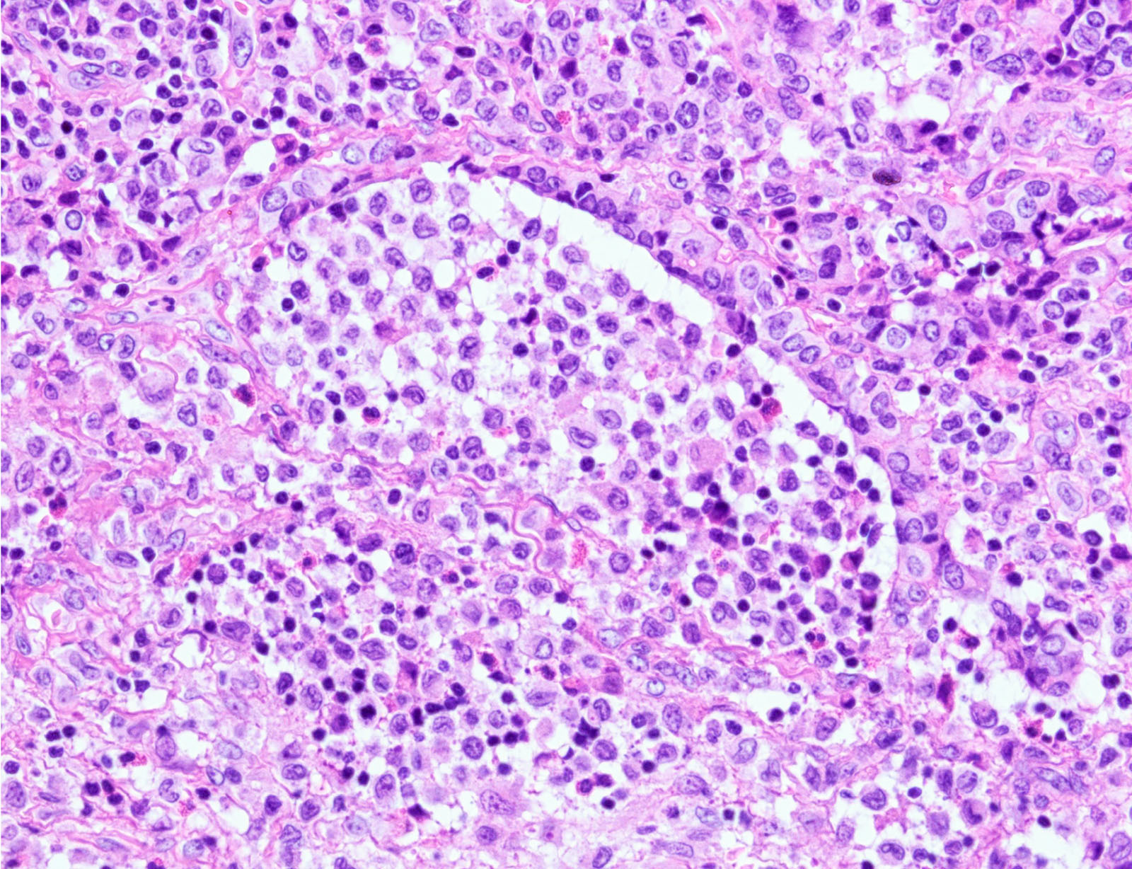
Diffuse large B cell lymphoma:

Tumor cells

Follicle in lower right

Fibrous bands

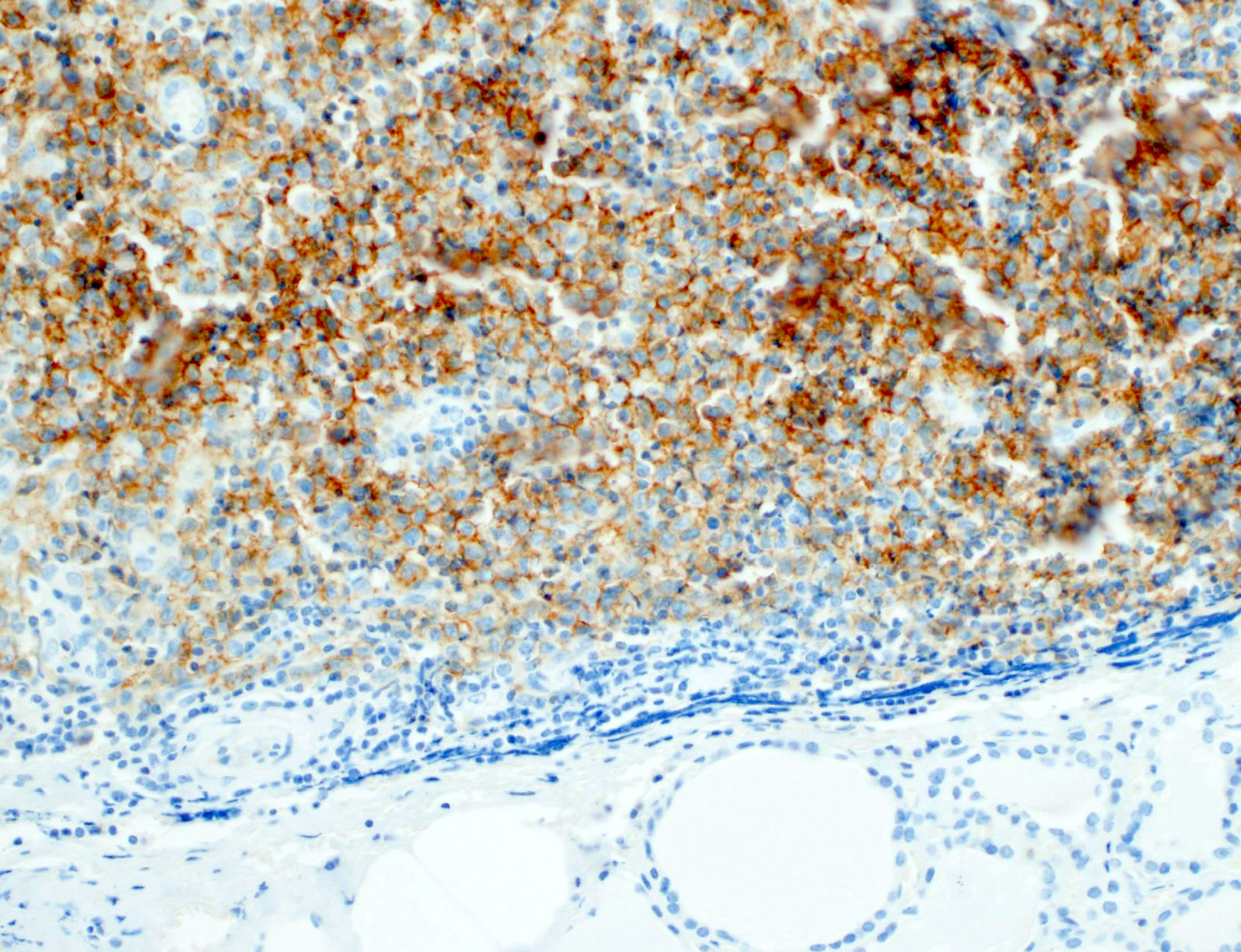
Tumor cells are CD45 (LCA)+

Keratin, thyroglobulin
Hodgkin lymphoma:

Nodular sclerosing subtype
Follicular lymphoma:

Residual thyroid follicles
Images hosted on other servers:

Large pleomorphic cells

Hodgkin lymphoma: nodular sclerosis type

Hodgkin lymphoma: CD30+

Follicular lymphoma: morphology

Follicular lymphoma: negative for bcl2 and IGH-BCL2

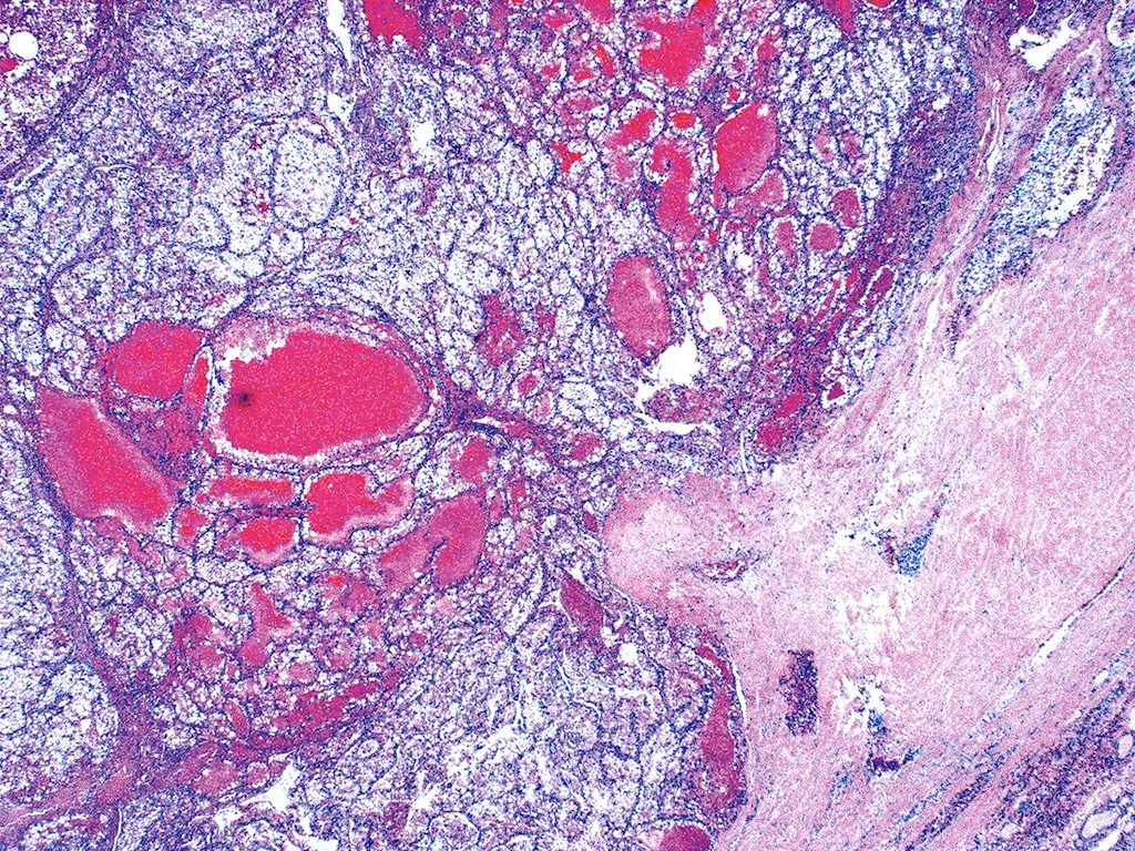
Follicular lymphoma: positive for bcl2 and IGH-BCL2
Contributed by Ayana Suzuki, C.T. and Mark R. Wick, M.D.

DLBCL
Large cell type
Images hosted on other servers:

Intermediate grade lymphoma

MALT lymphoma

Hodgkin lymphoma: Reed-Sternberg cell
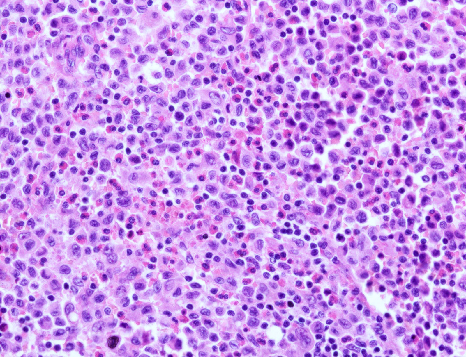
Diffuse large B cell lymphoma:

Large and irregular lymphoid cells

Misdiagnosed as anaplastic carcinoma

High grade lymphoma

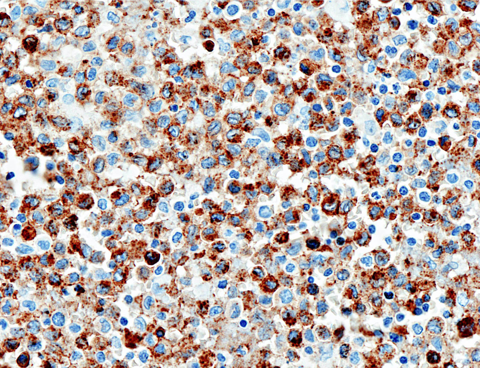
CD45 / LCA+, CD20+, keratin-
Images hosted on other servers:
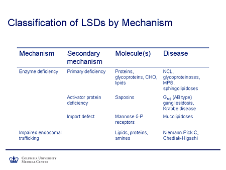
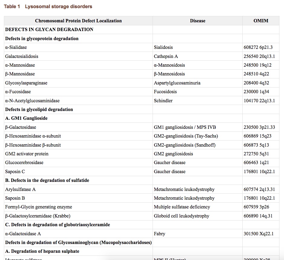
Classification
Images hosted on other servers:

Skeletal deformities in mucopoly-saccharidoses
Images hosted on other servers:
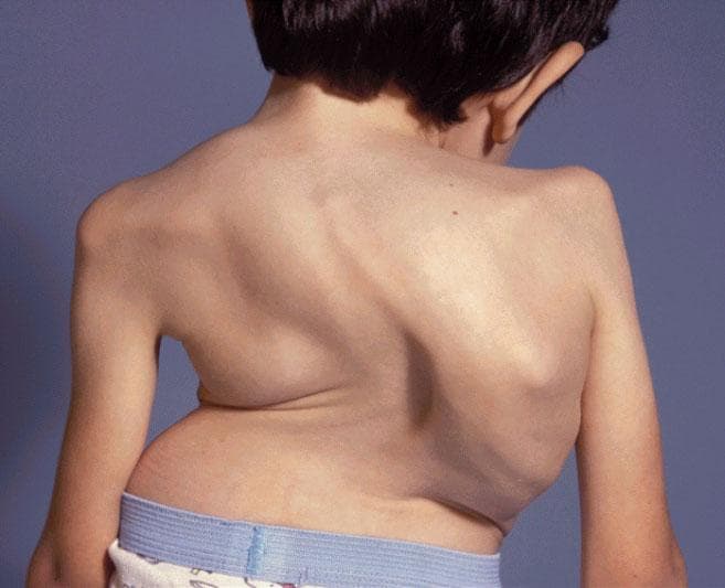
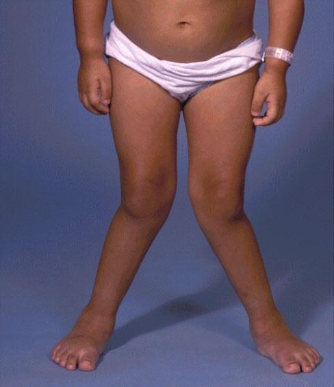
Skeletal deformities in mucopolysaccharidoses
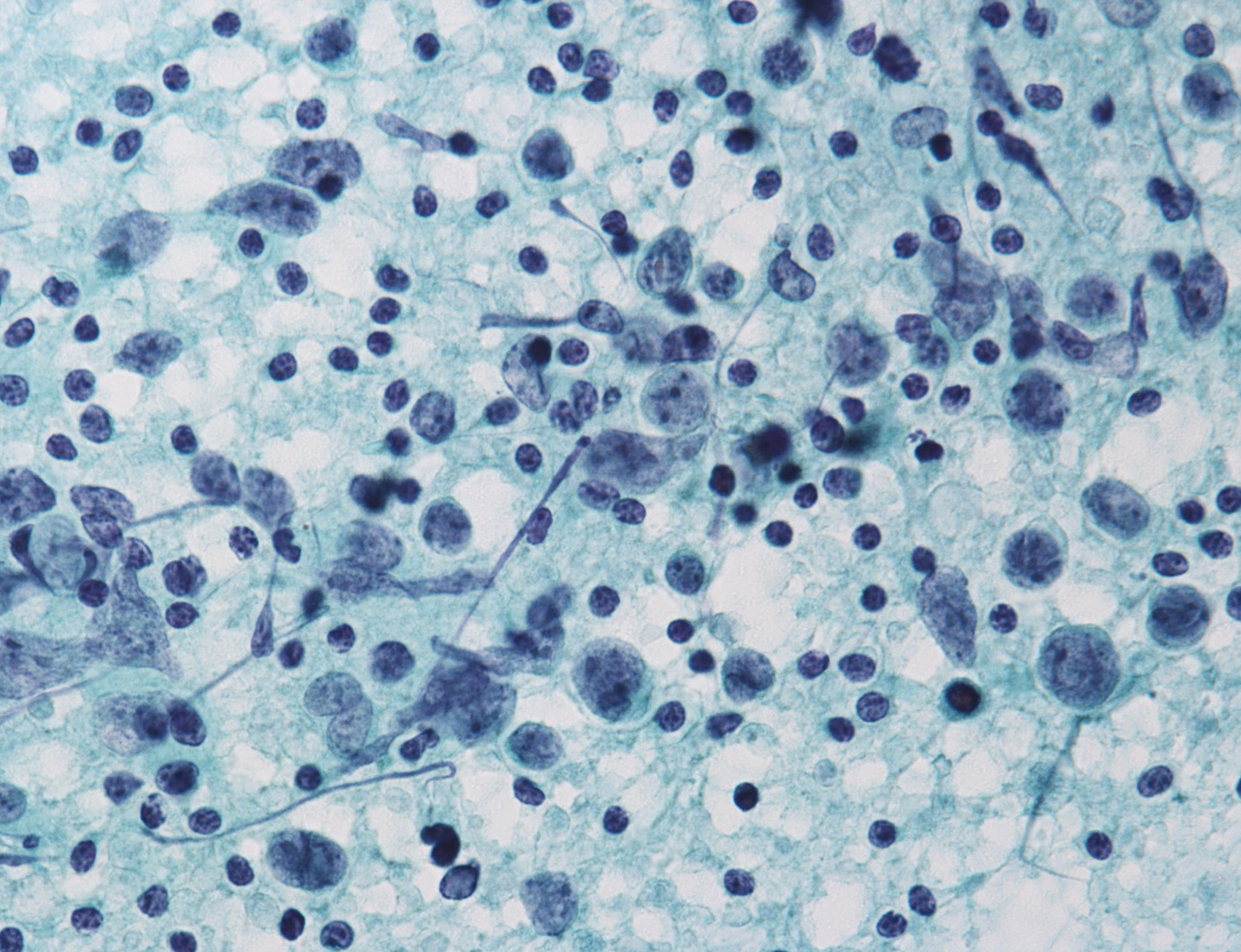
Case #164
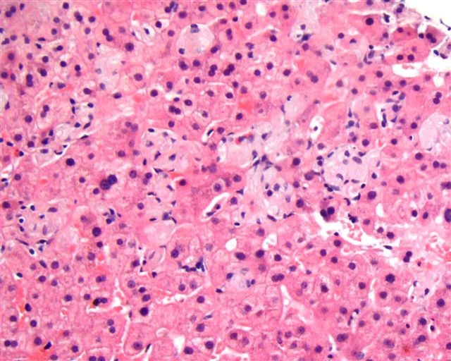
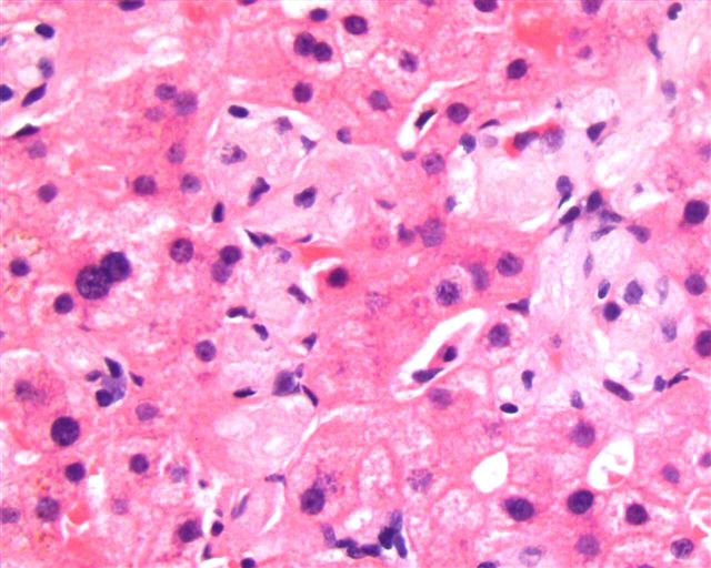
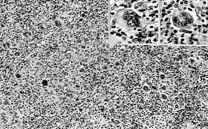
Liver biopsy in Gaucher disease (H&E, PAS)
Images hosted on other servers:


Hepatic glycogenosis (H&E, PAS, Masson)


Niemann-Pick disease (hepatic steatosis)
Images hosted on other servers:


Gaucher cell in bone marrow
Images hosted on other servers:

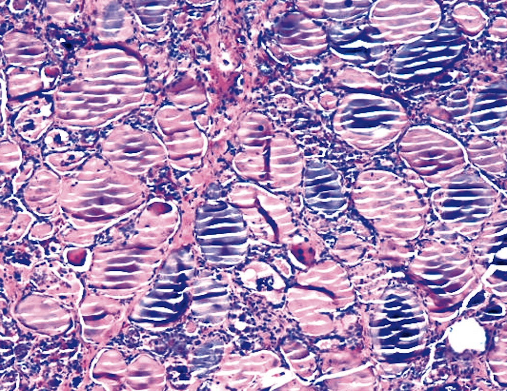
Lamellated "zebra" bodies in Fabry disease (fig 10)

Leukocyte in mannosidosis
Histopathology of glycogen storage disease (heart, liver)
Primer in inherited metabolic disorders (2013)
Mechanism of LSDs (2014)
Lecture series on LSDs by Excellence in Pediatrics Institute (2014 - 2016)
Contributed by Shuanzeng Wei, M.D., Ph.D.

Nodal metastasis, macrofollicular variant PTC
Nodal metastasis, macrofollicular variant PTC
Images hosted on other servers:
Malakoplakia (arrows at Michaelis-Gutmann bodies), bladder
Michaelis-Gutmann
bodies; von Kossa
calcium stain
Contributed by Ayana Suzuki, C.T., Andrey Bychkov, M.D., Ph.D.
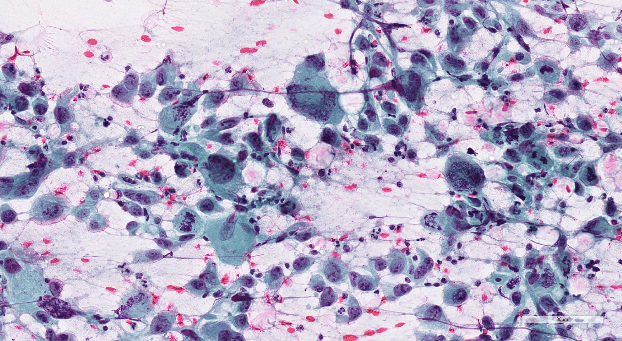
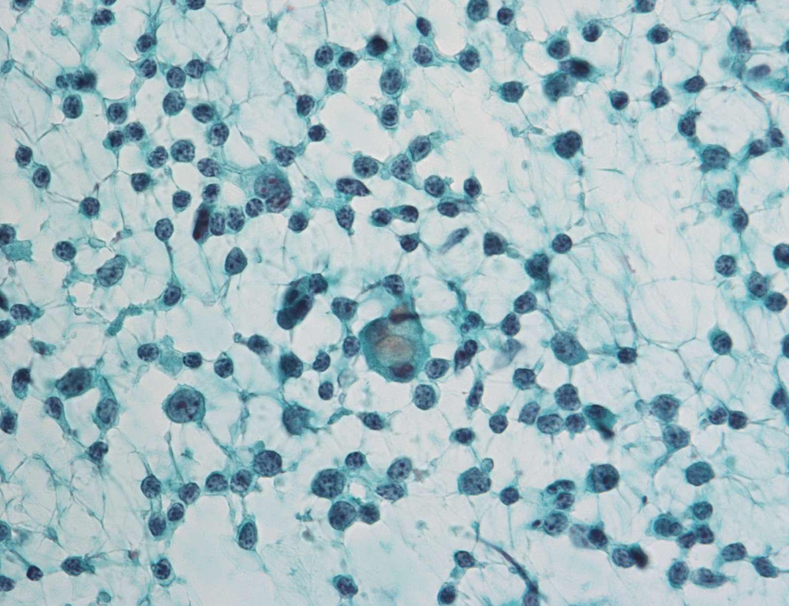
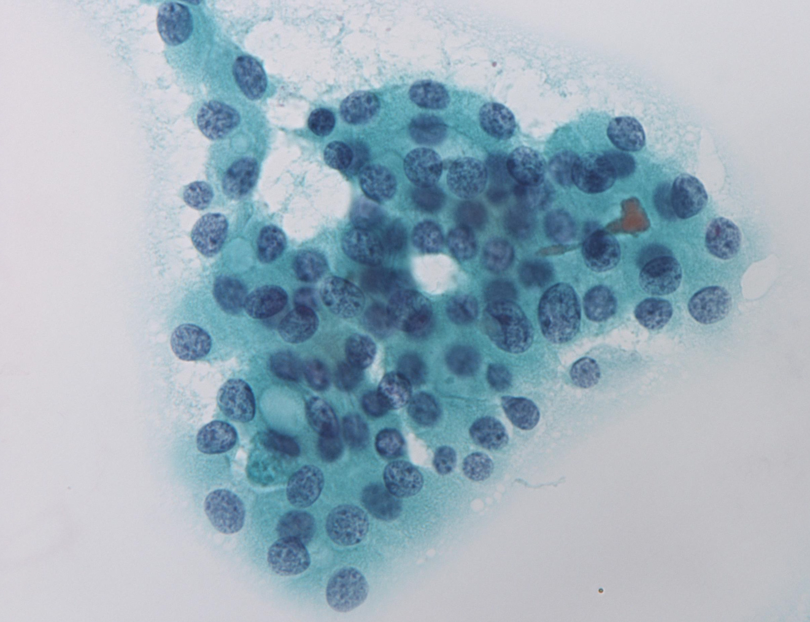
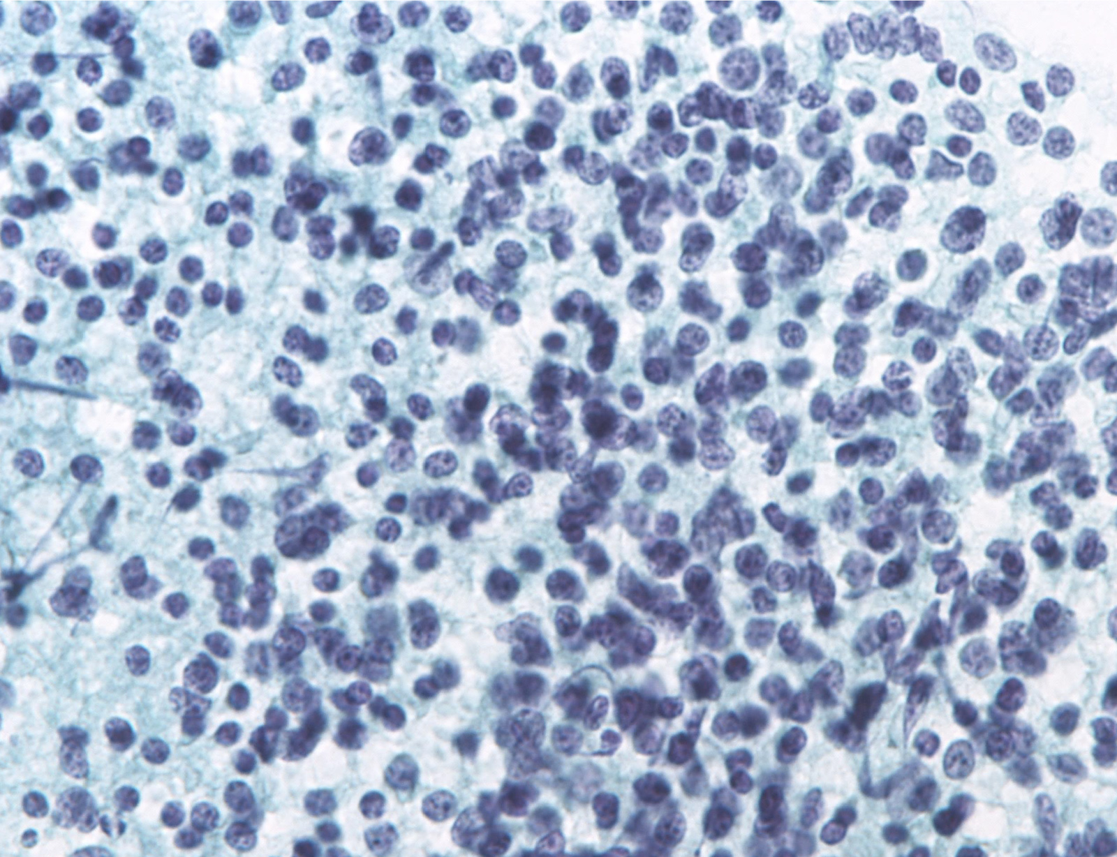
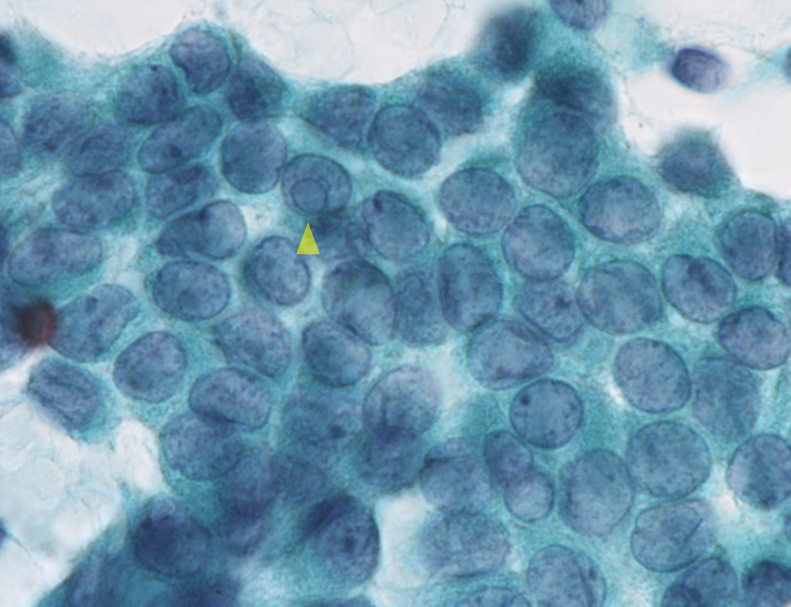
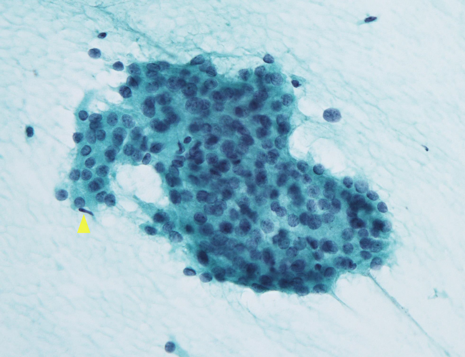
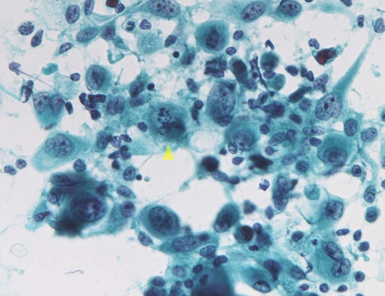
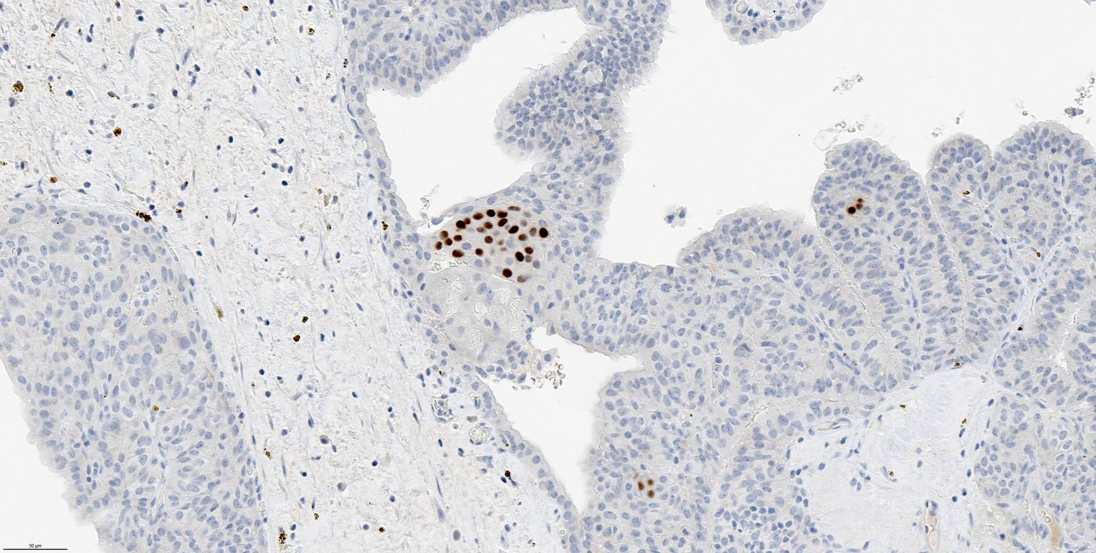
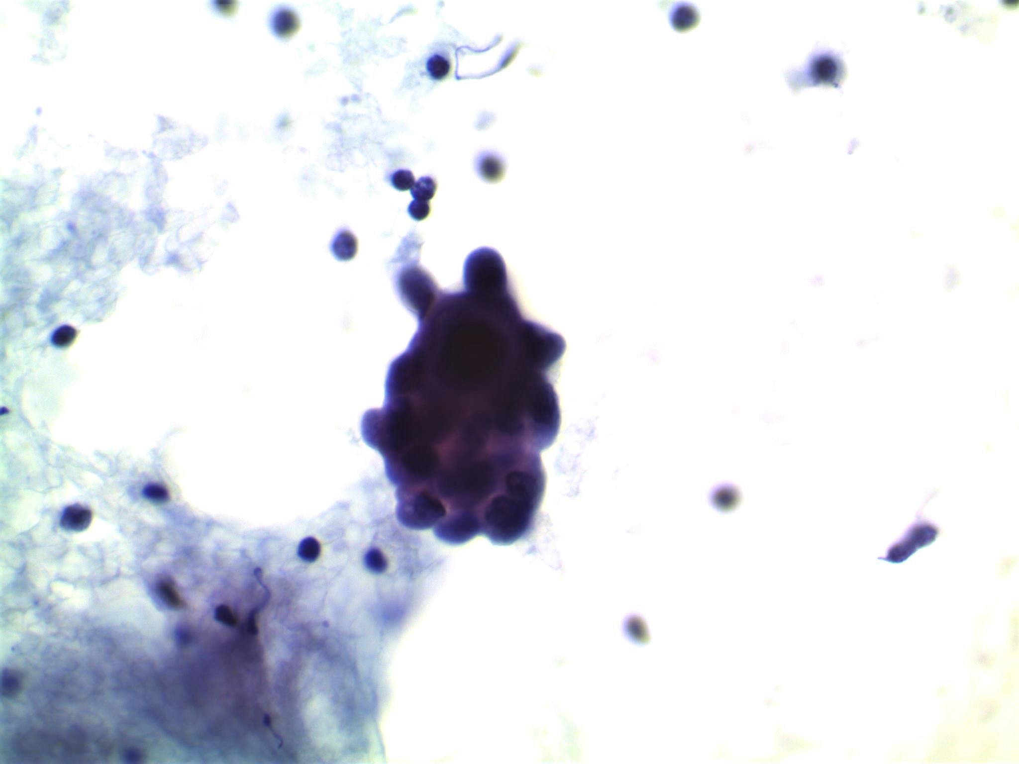
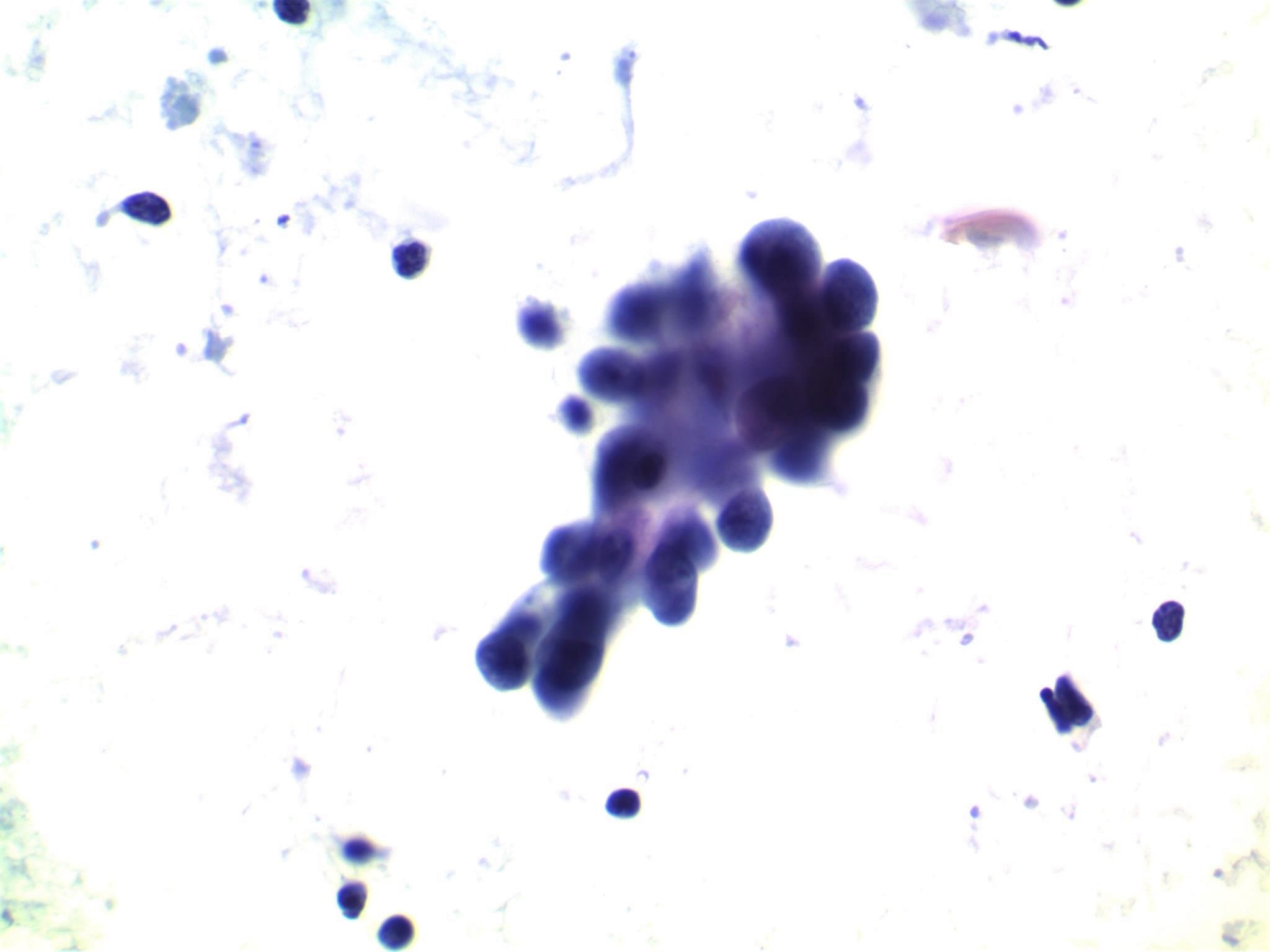
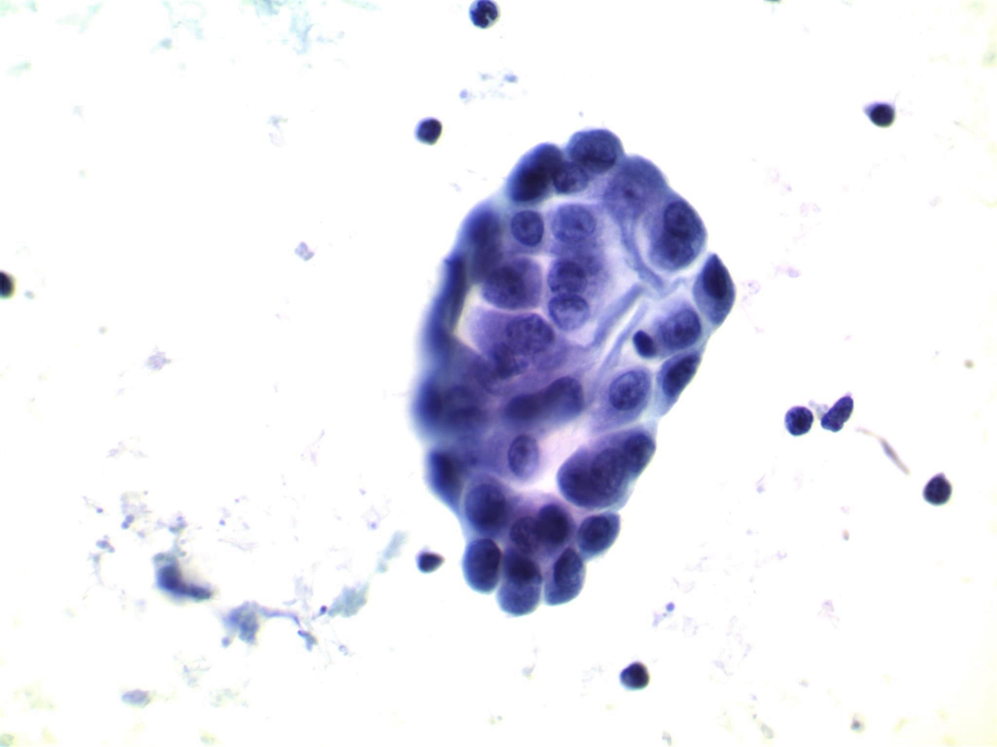
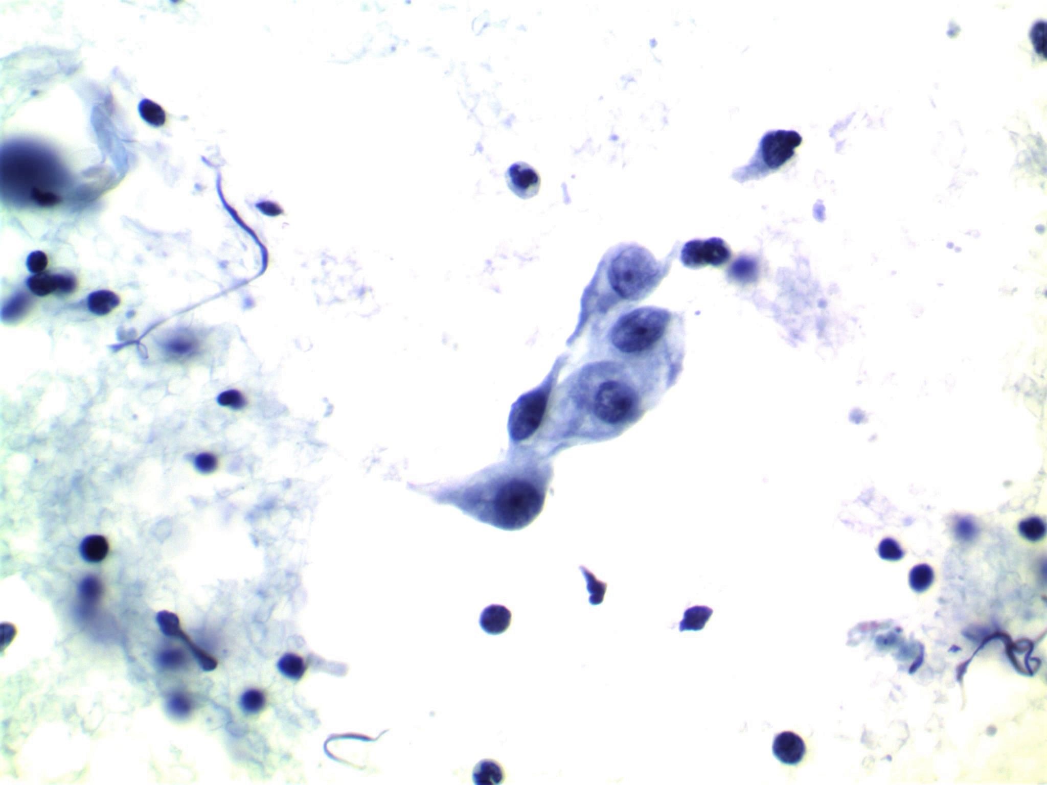
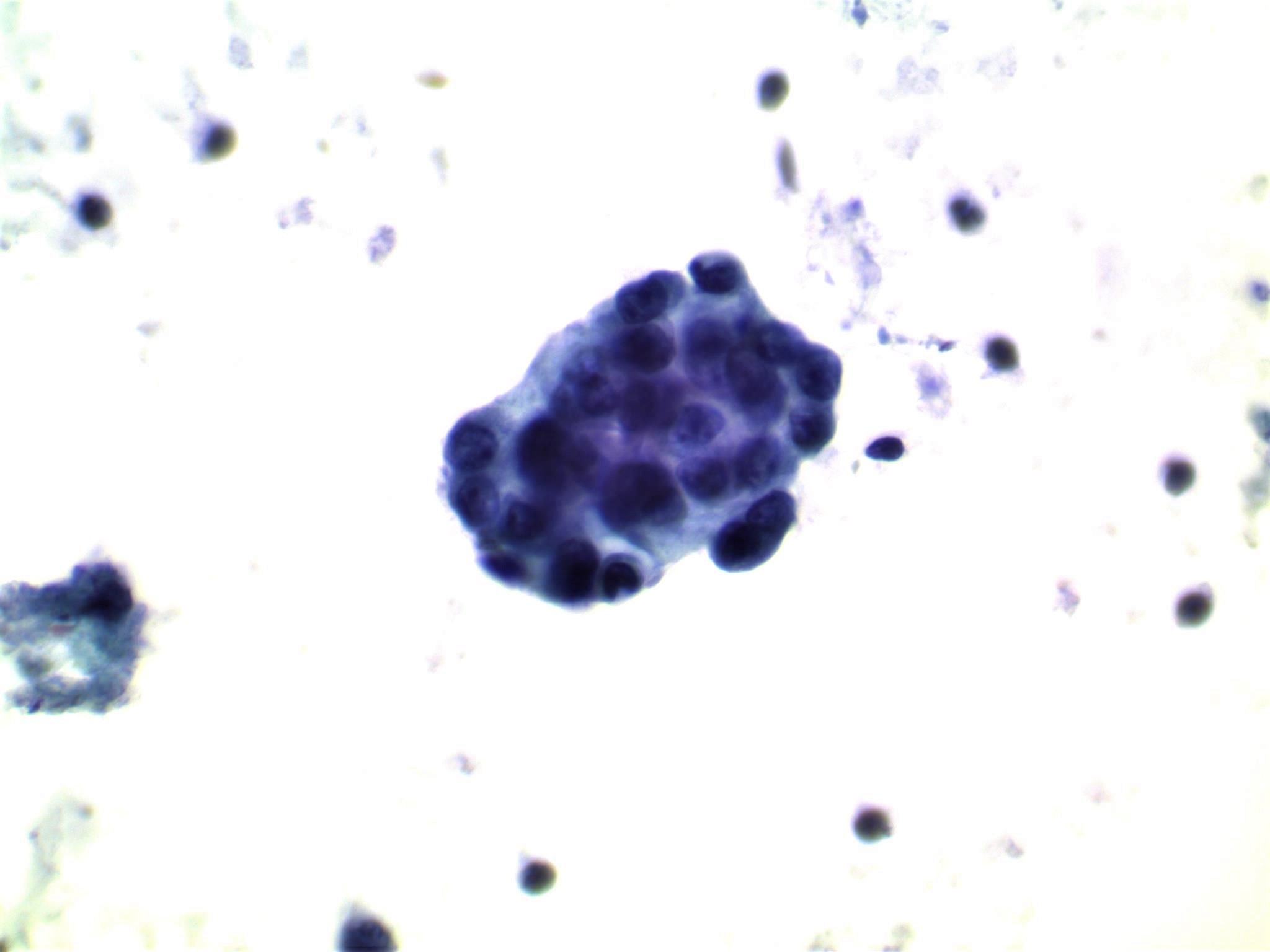
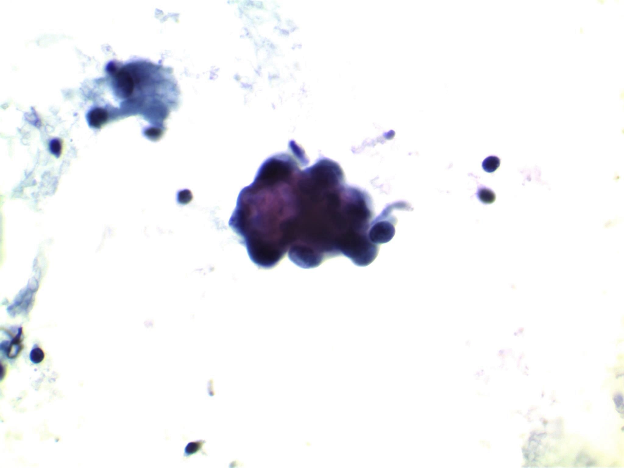
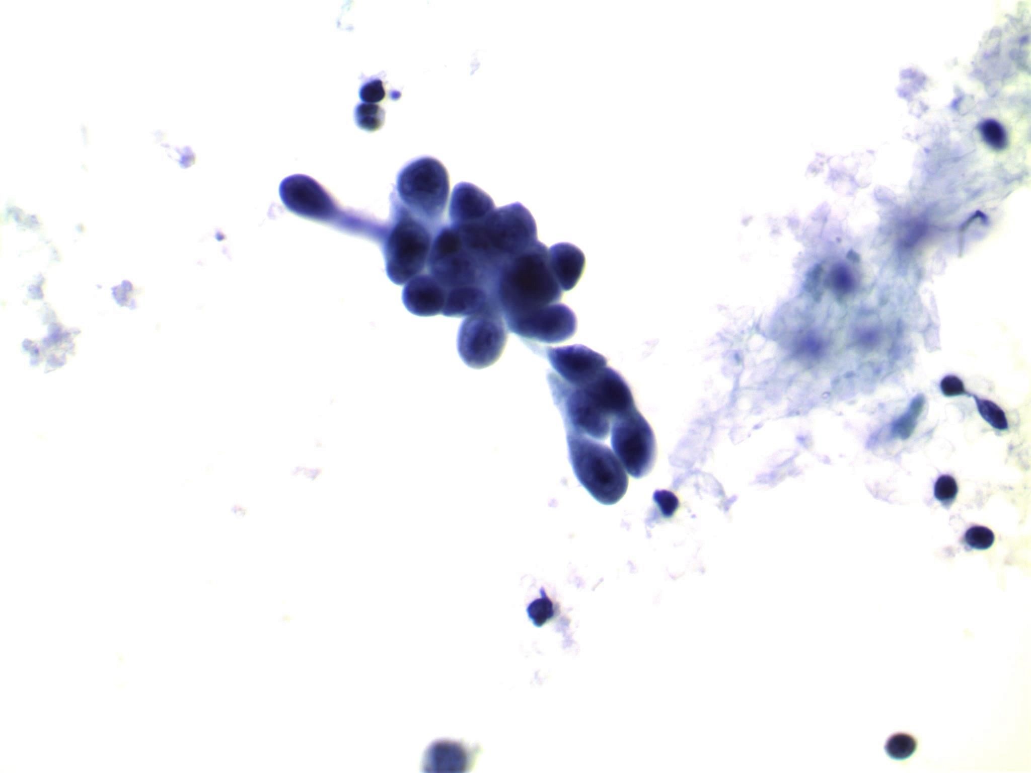
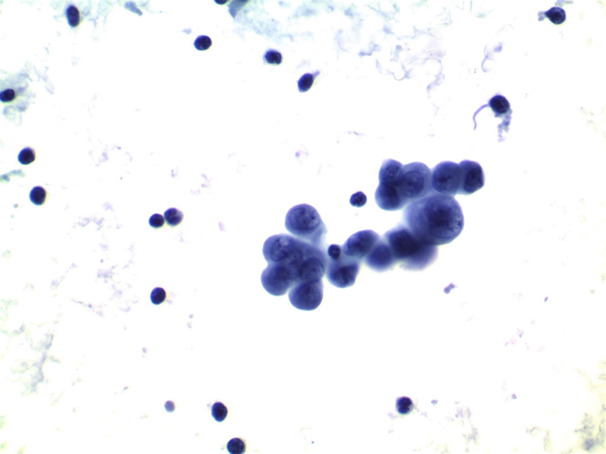
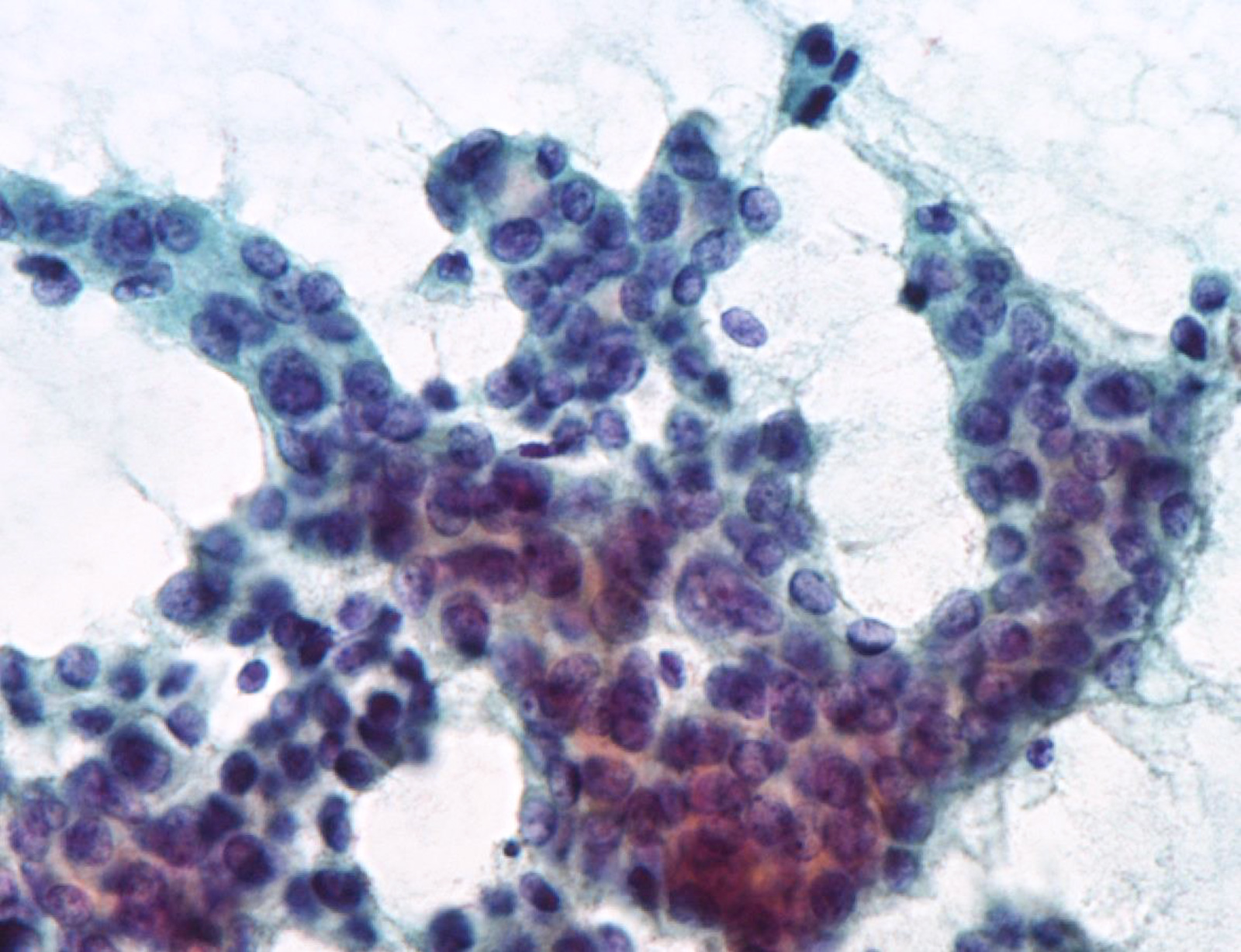
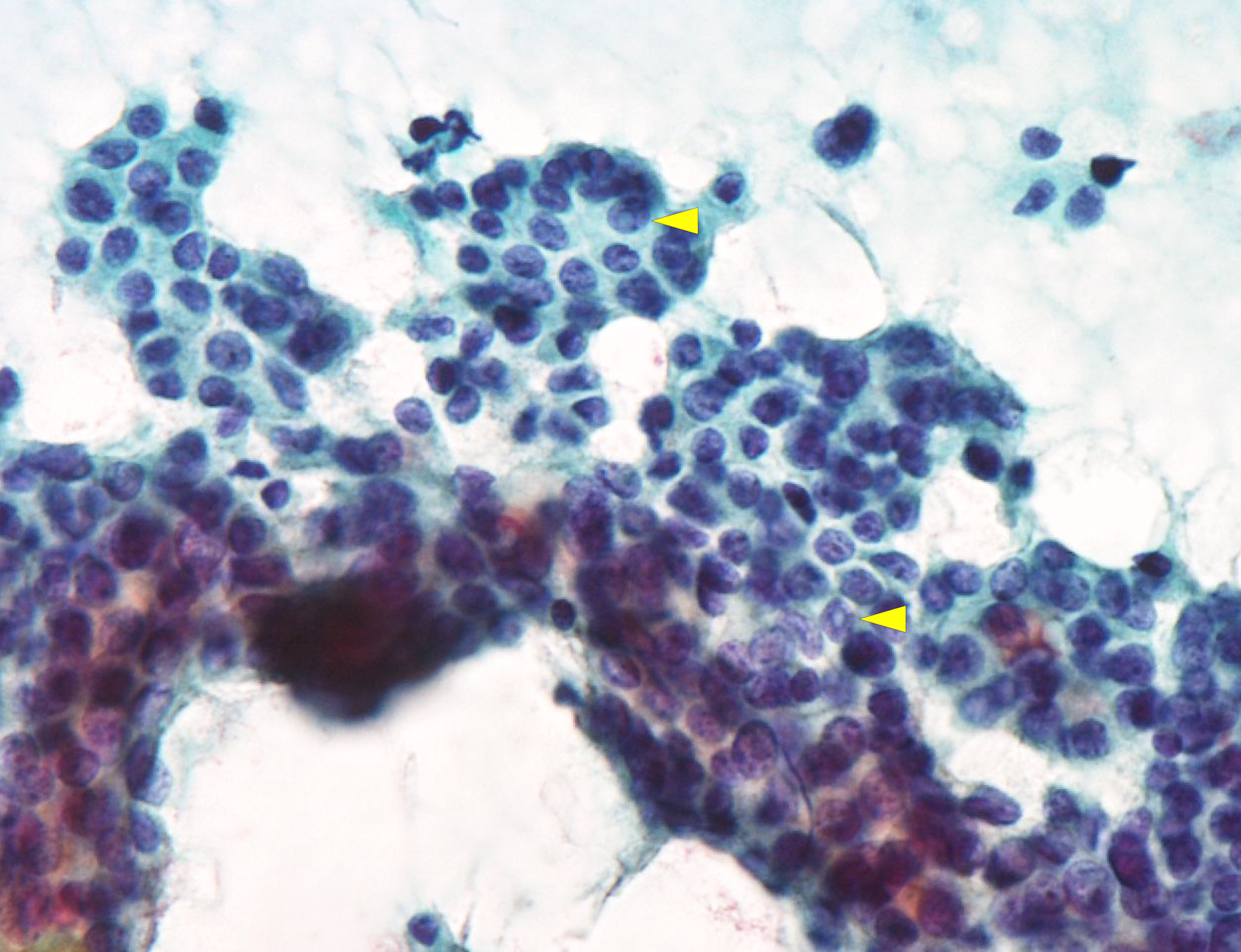
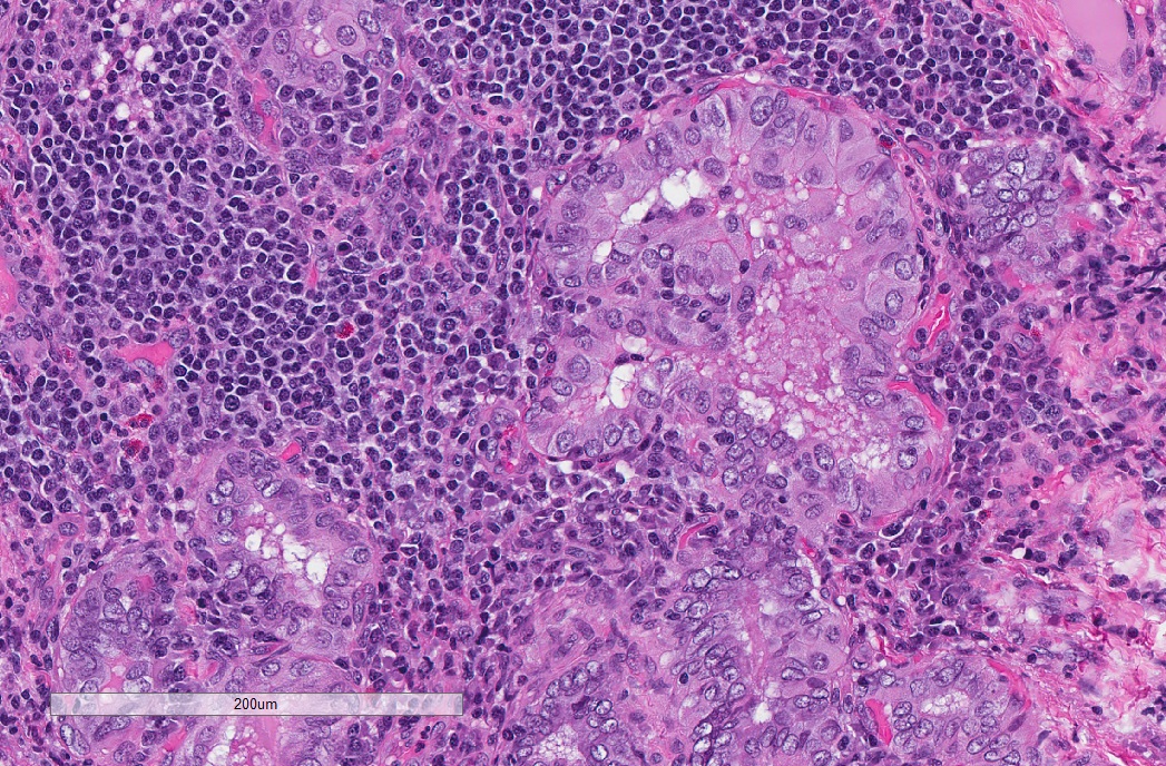
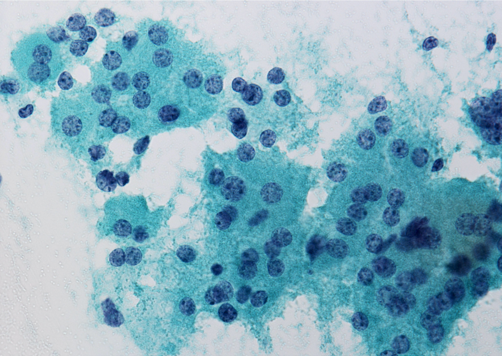
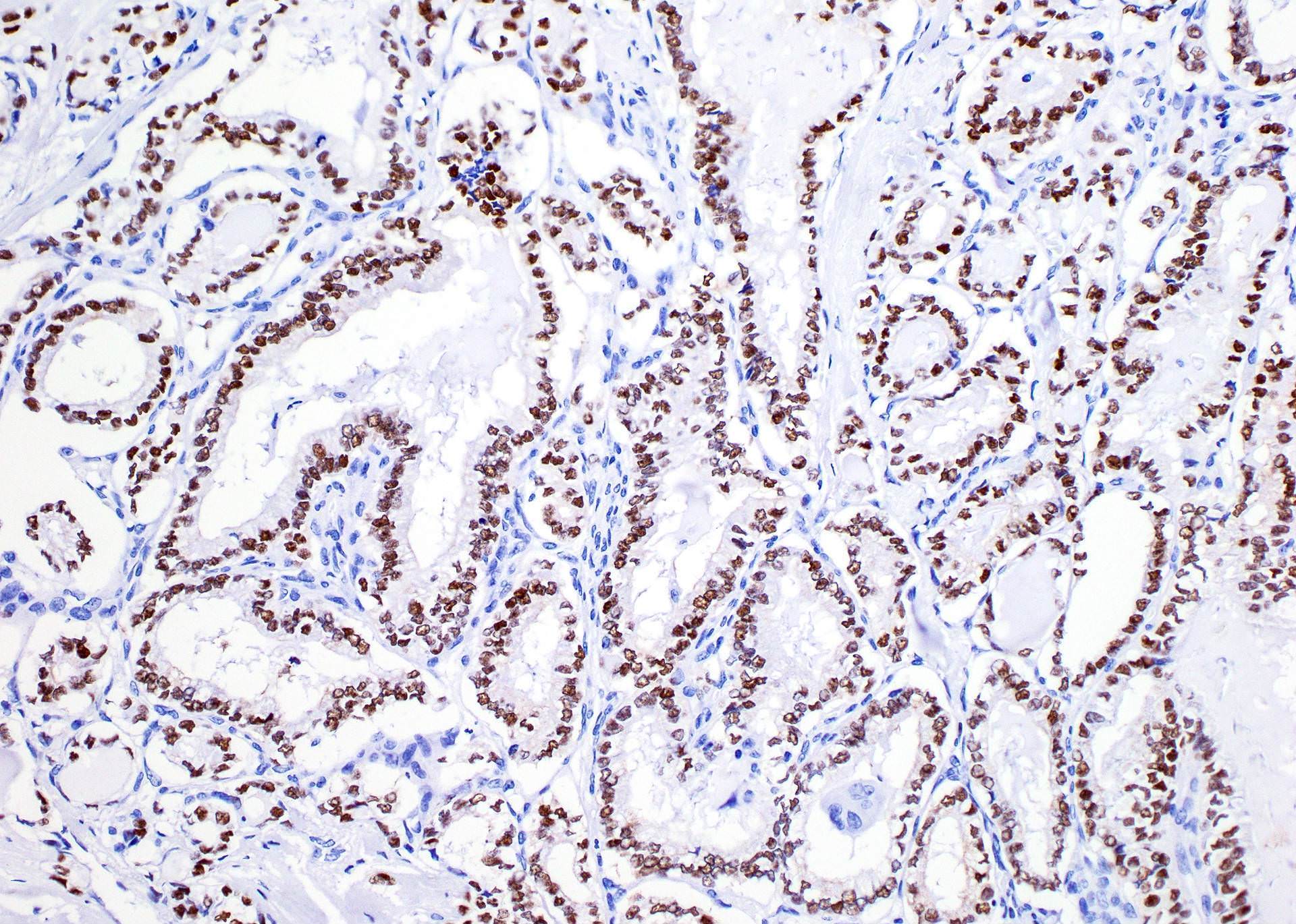
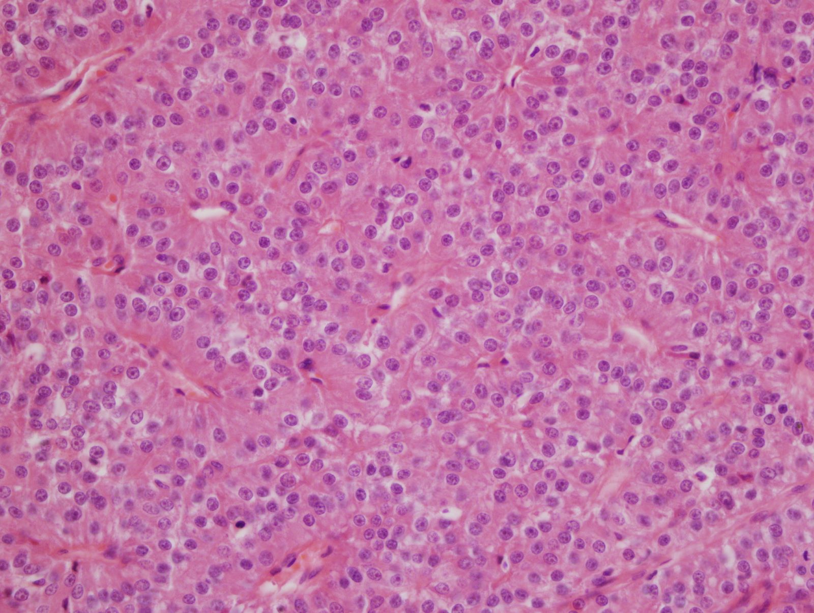
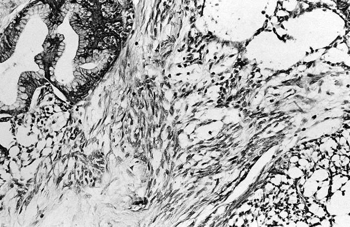
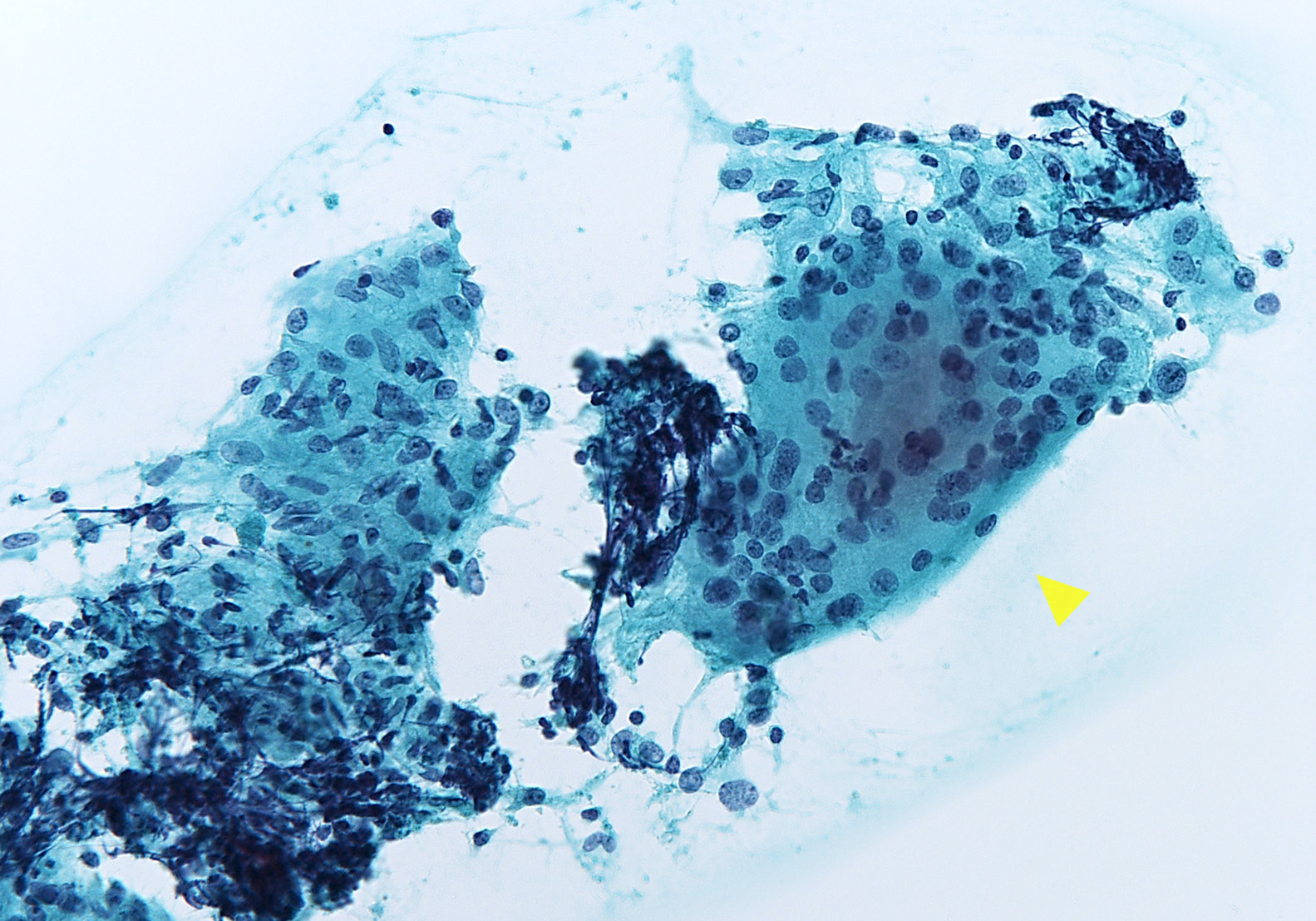
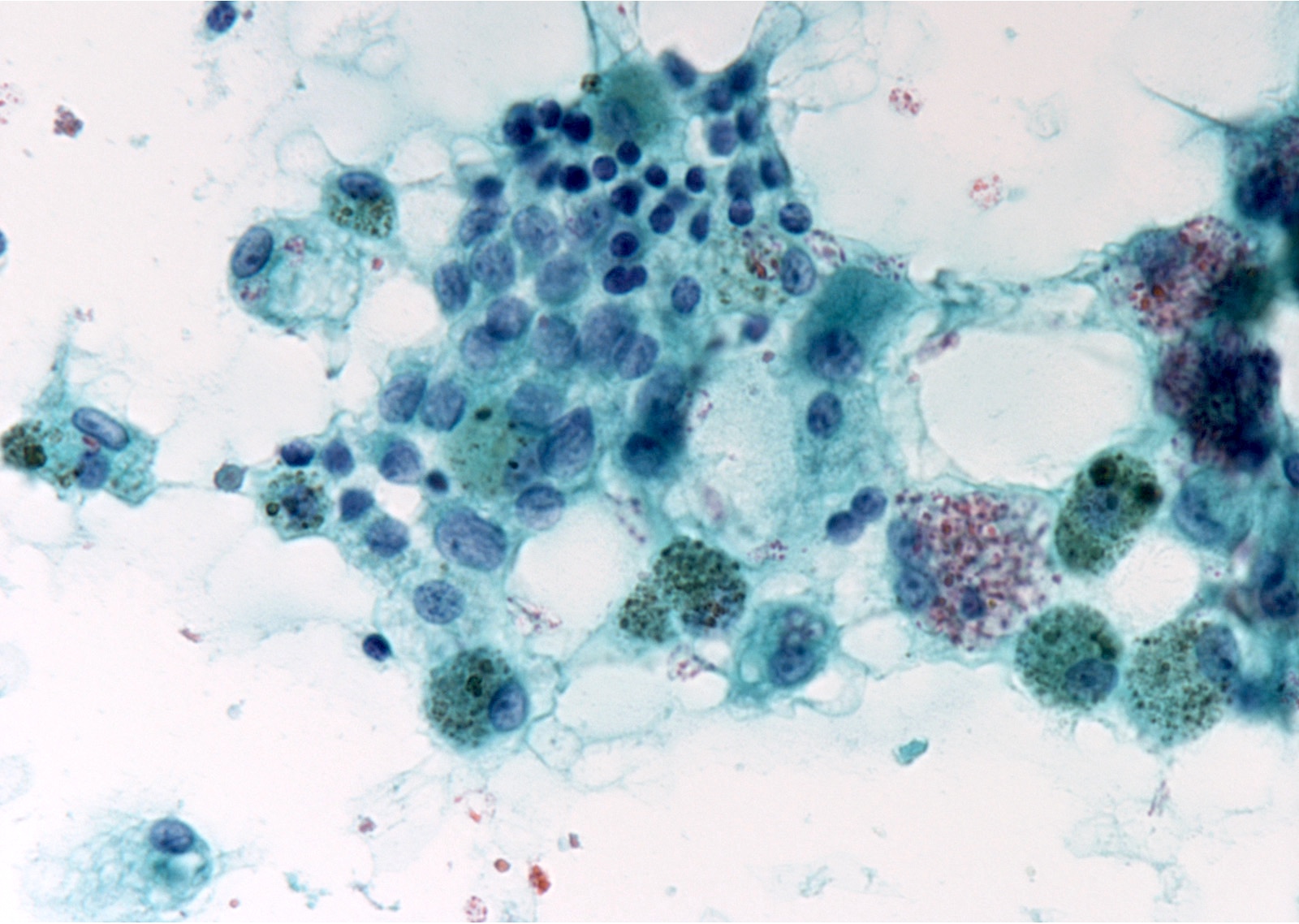
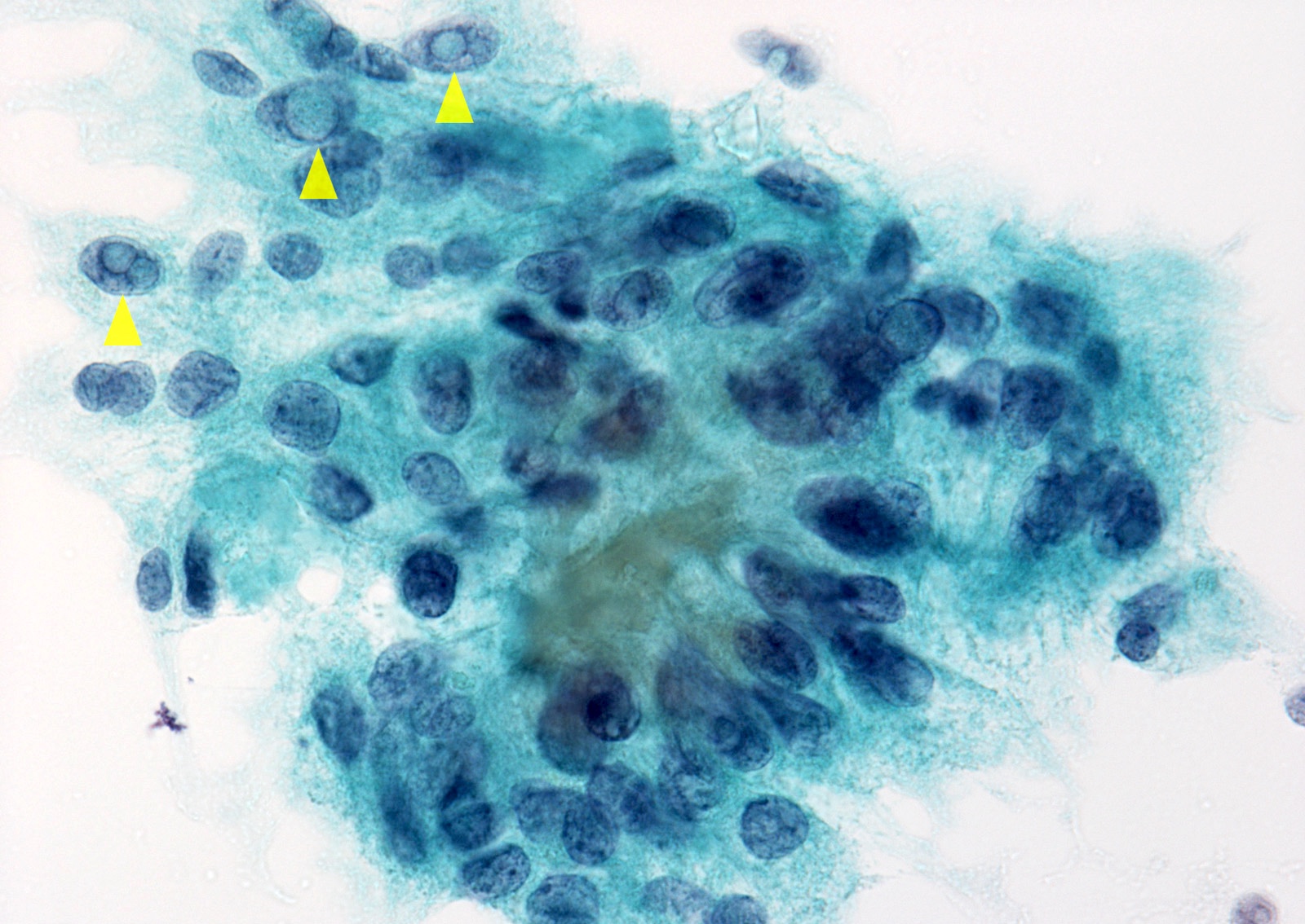
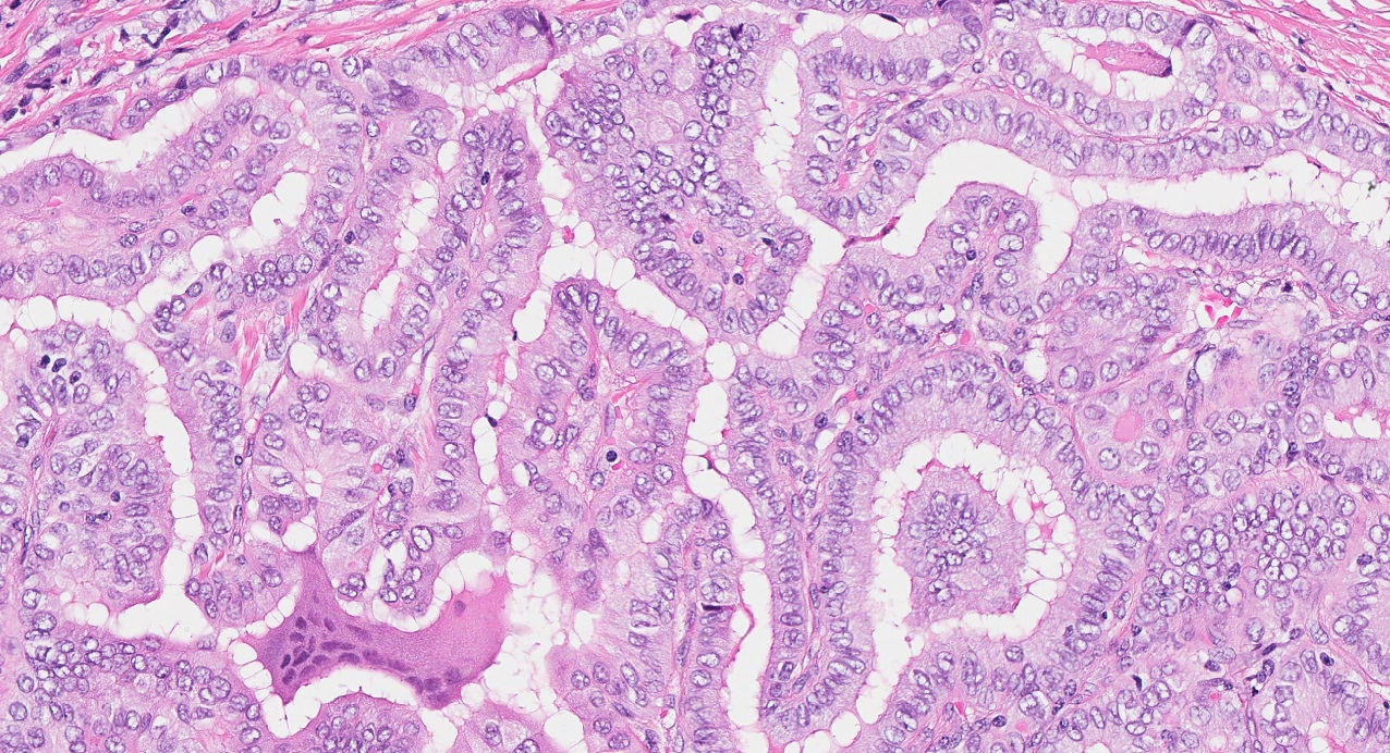
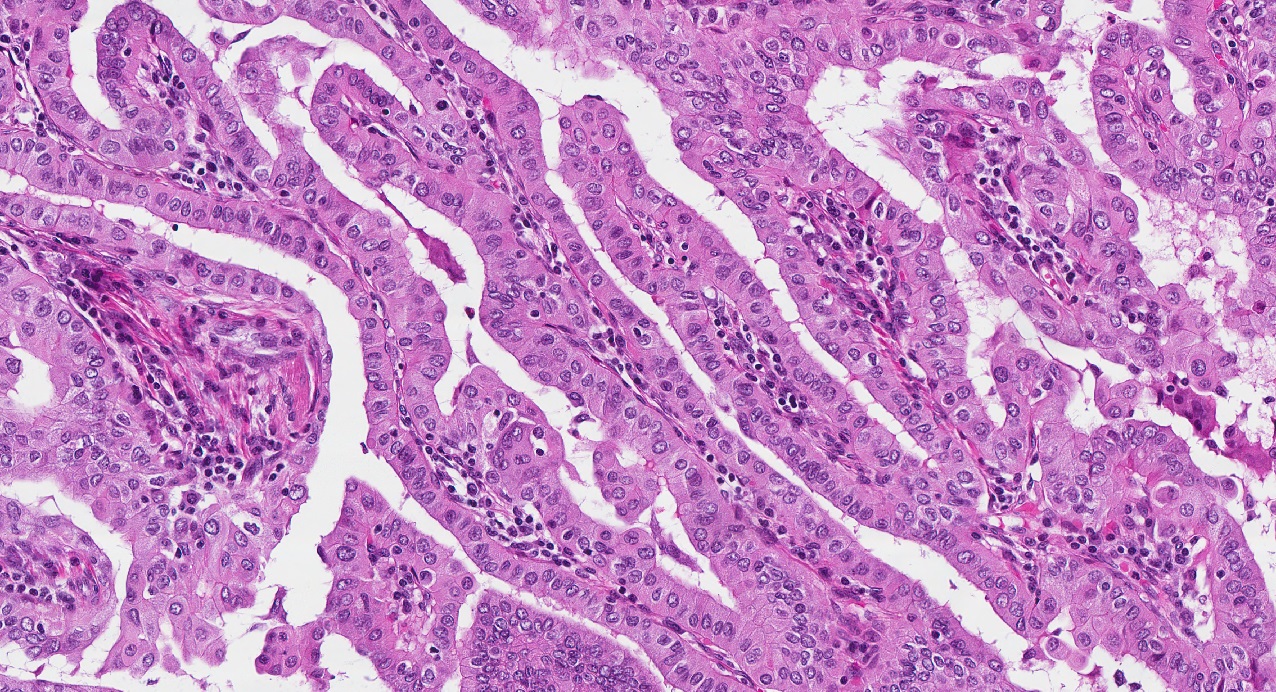
Papillary thyroid carcinoma
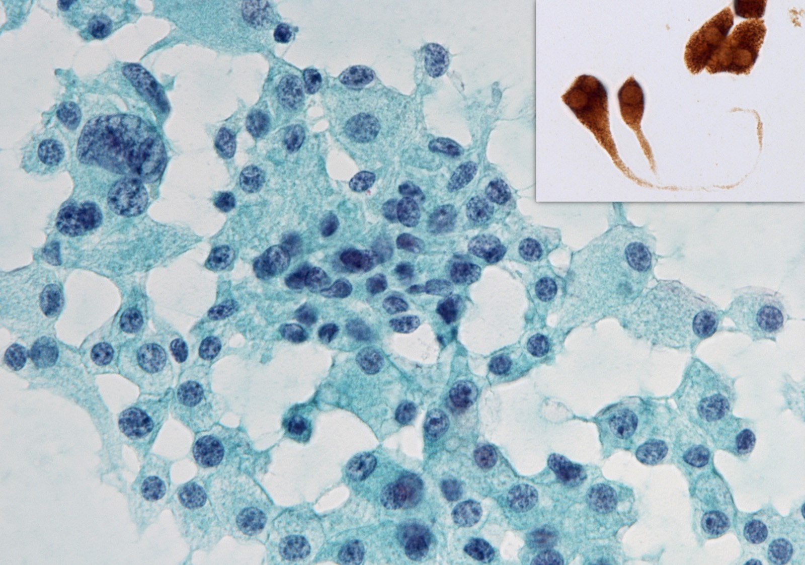
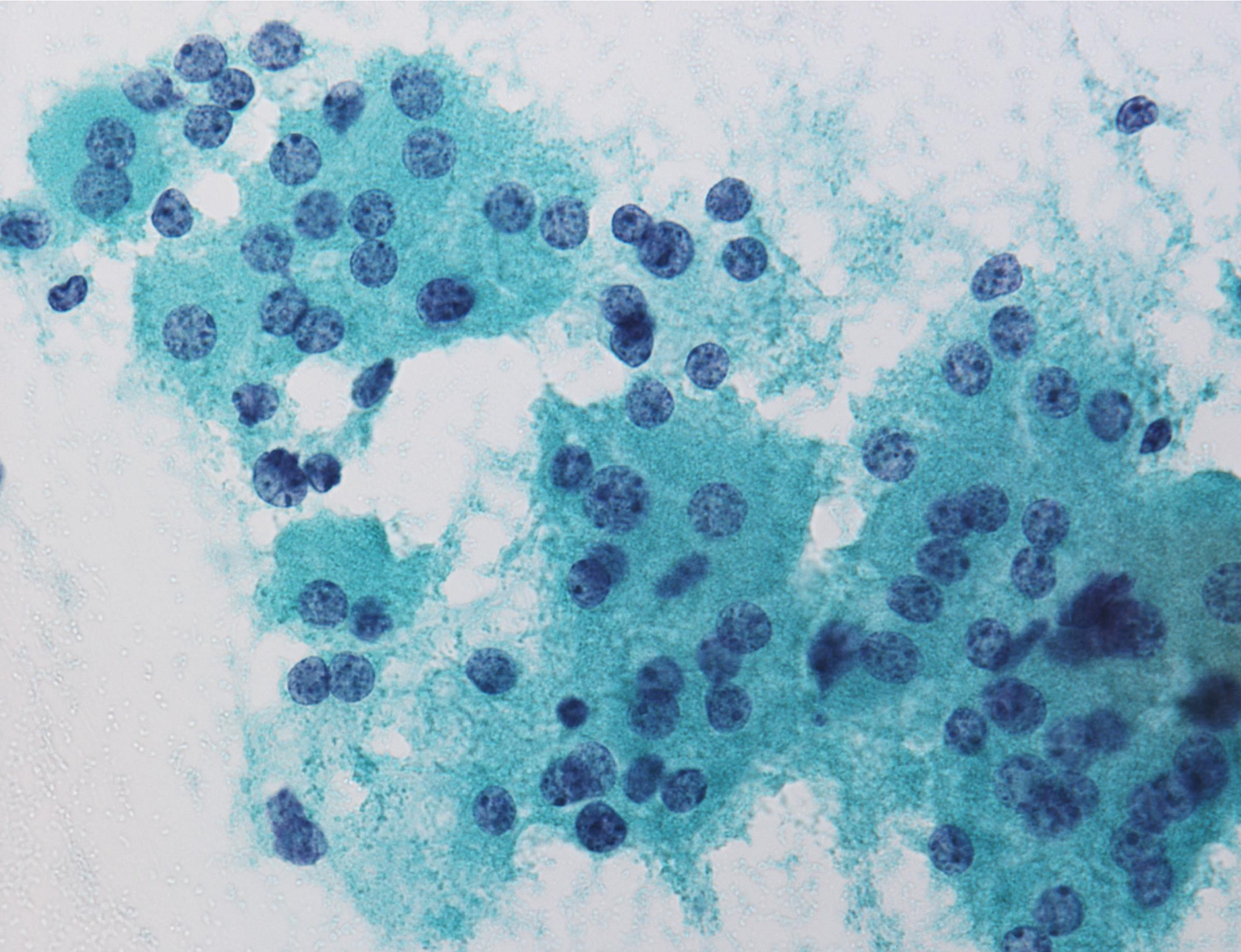
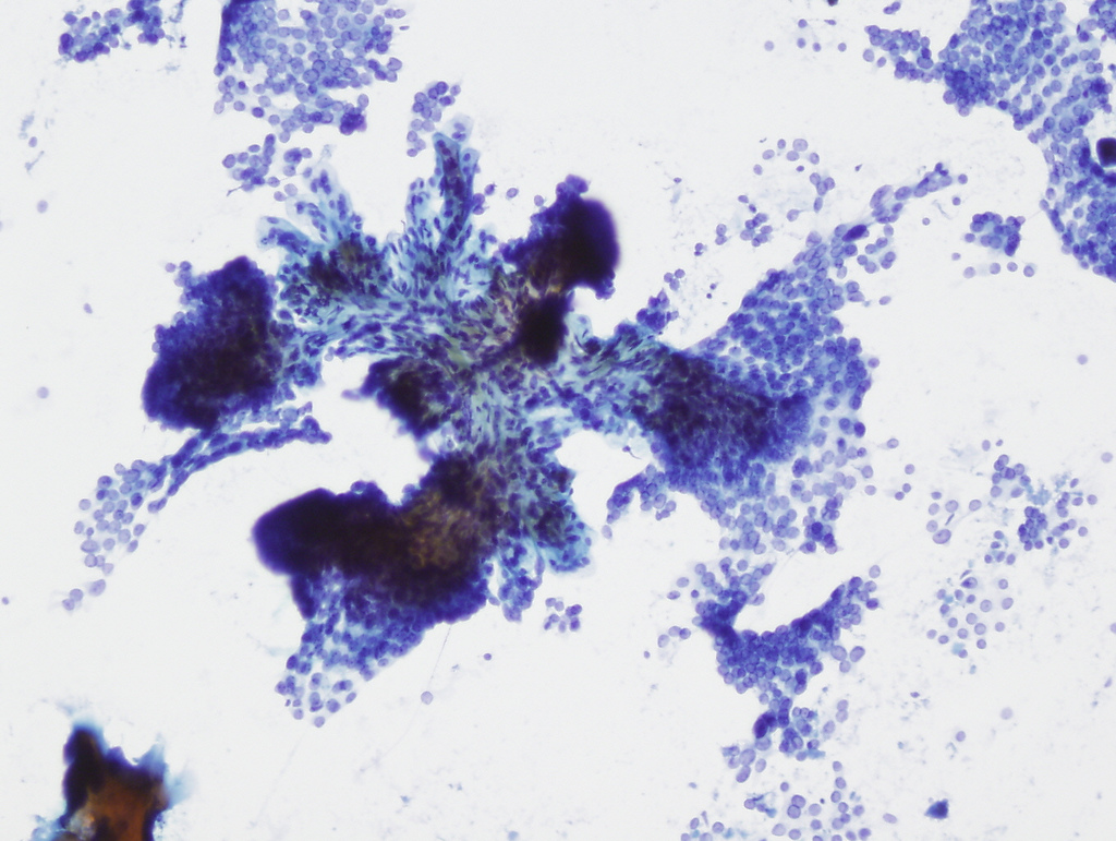
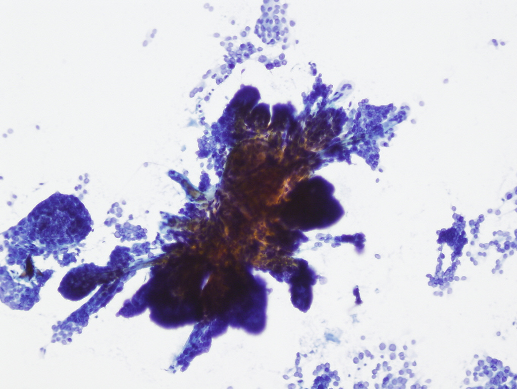
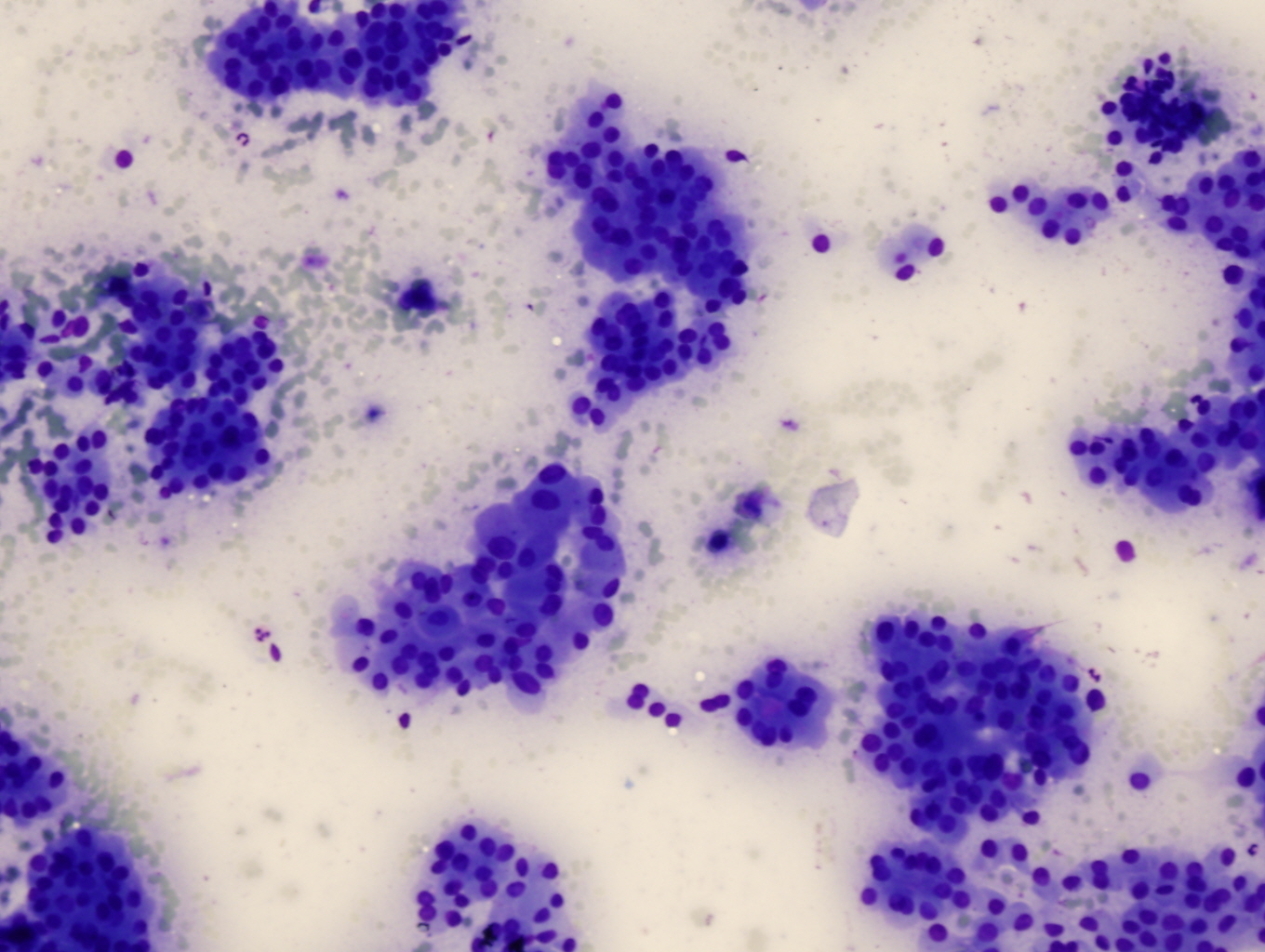
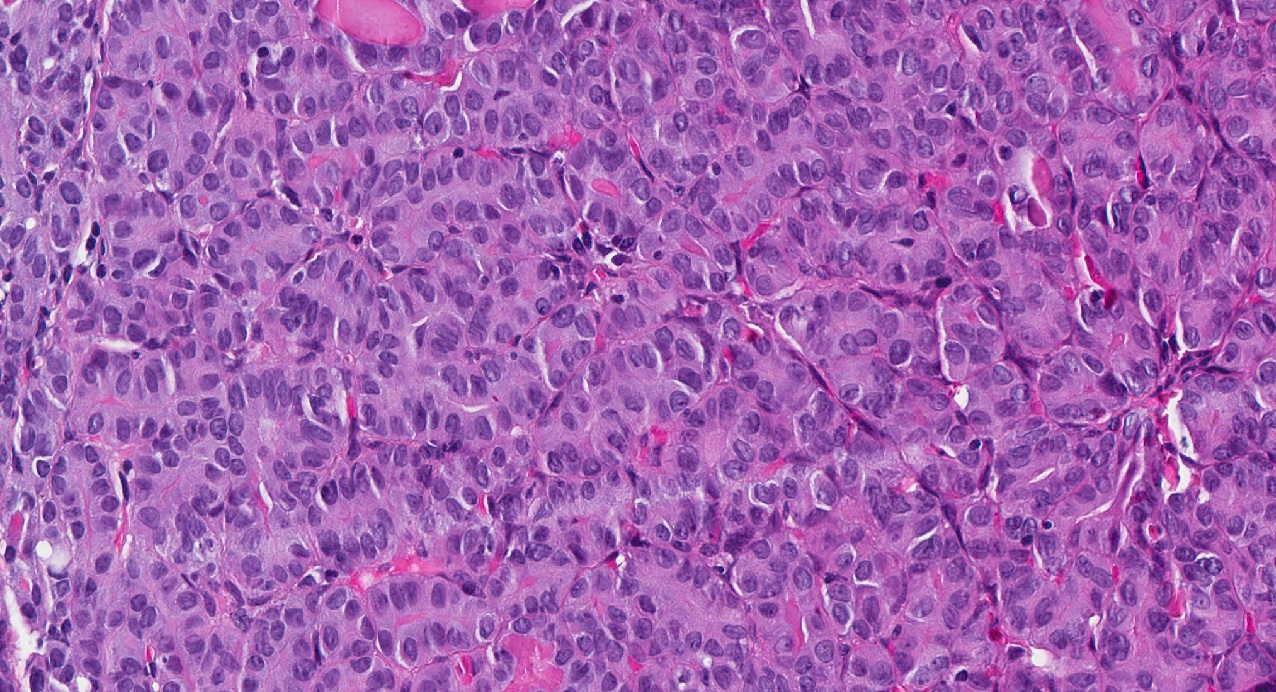
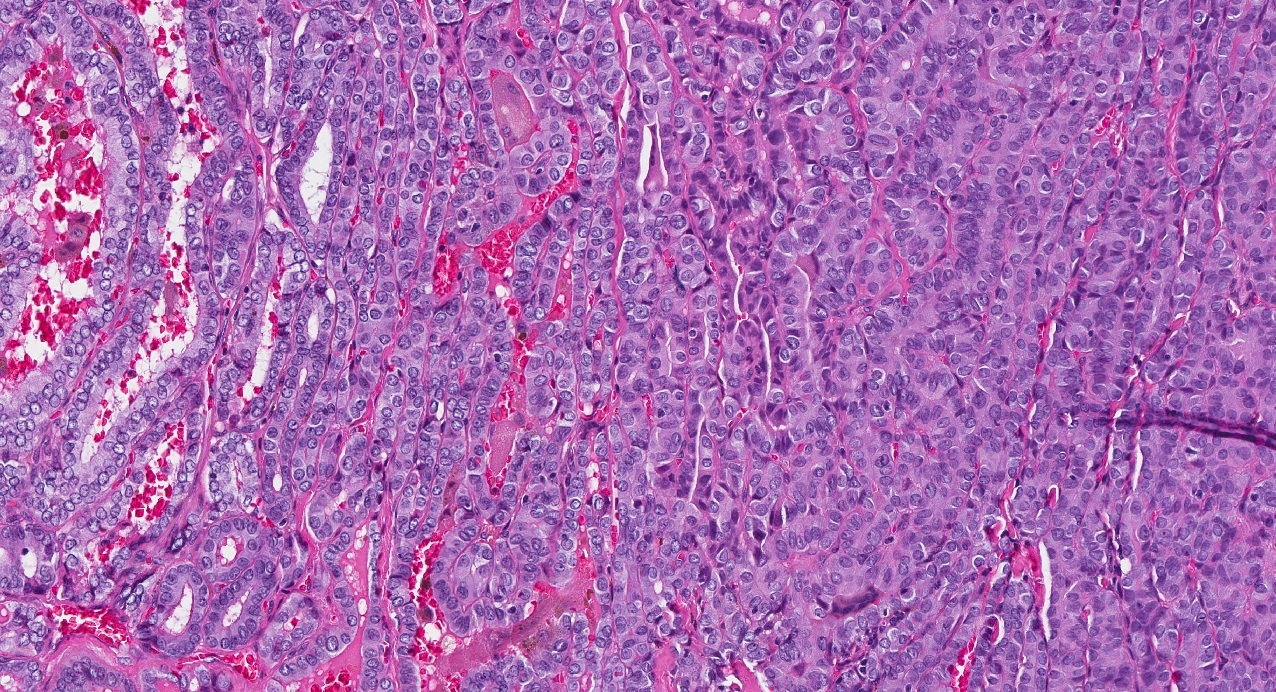
Medullary thyroid carcinoma

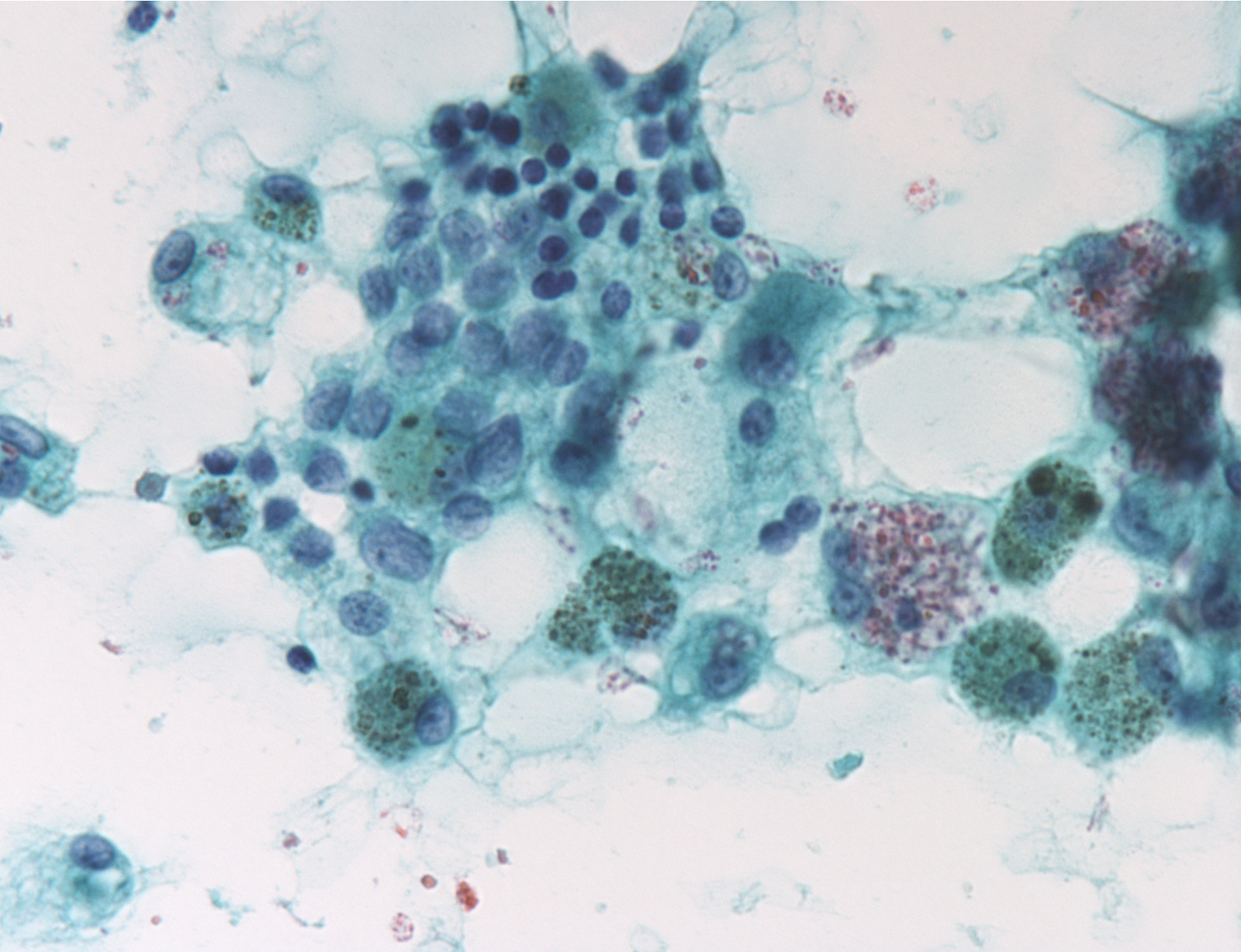
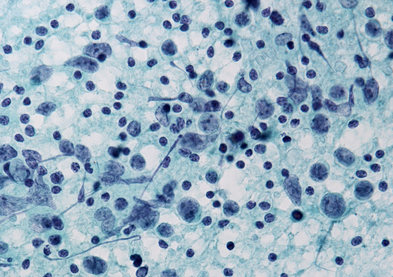
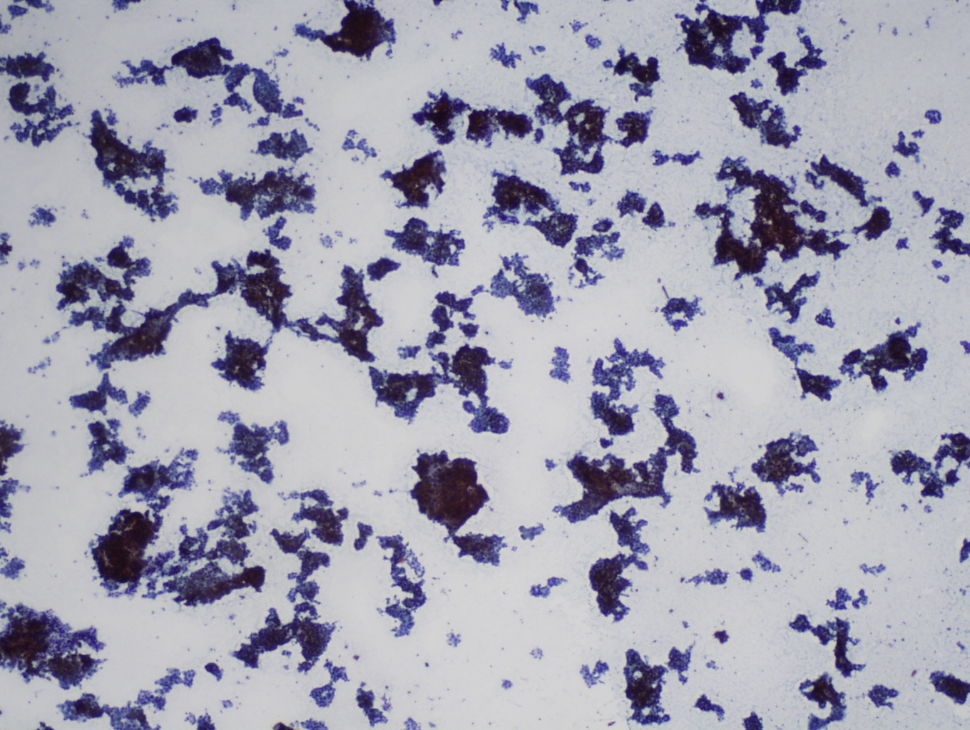
Poorly differentiated thyroid carcinoma
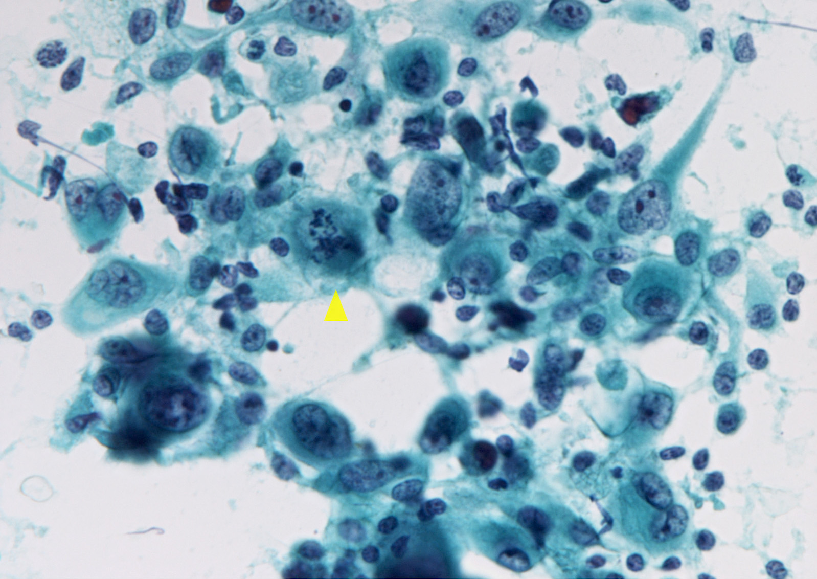
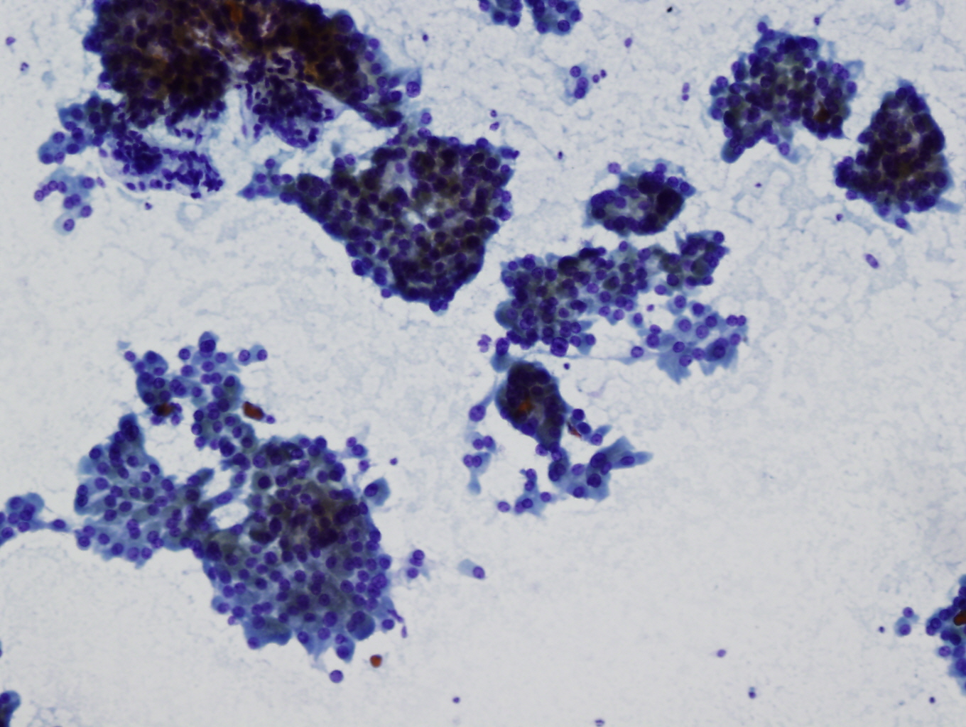
Anaplastic thyroid carcinoma
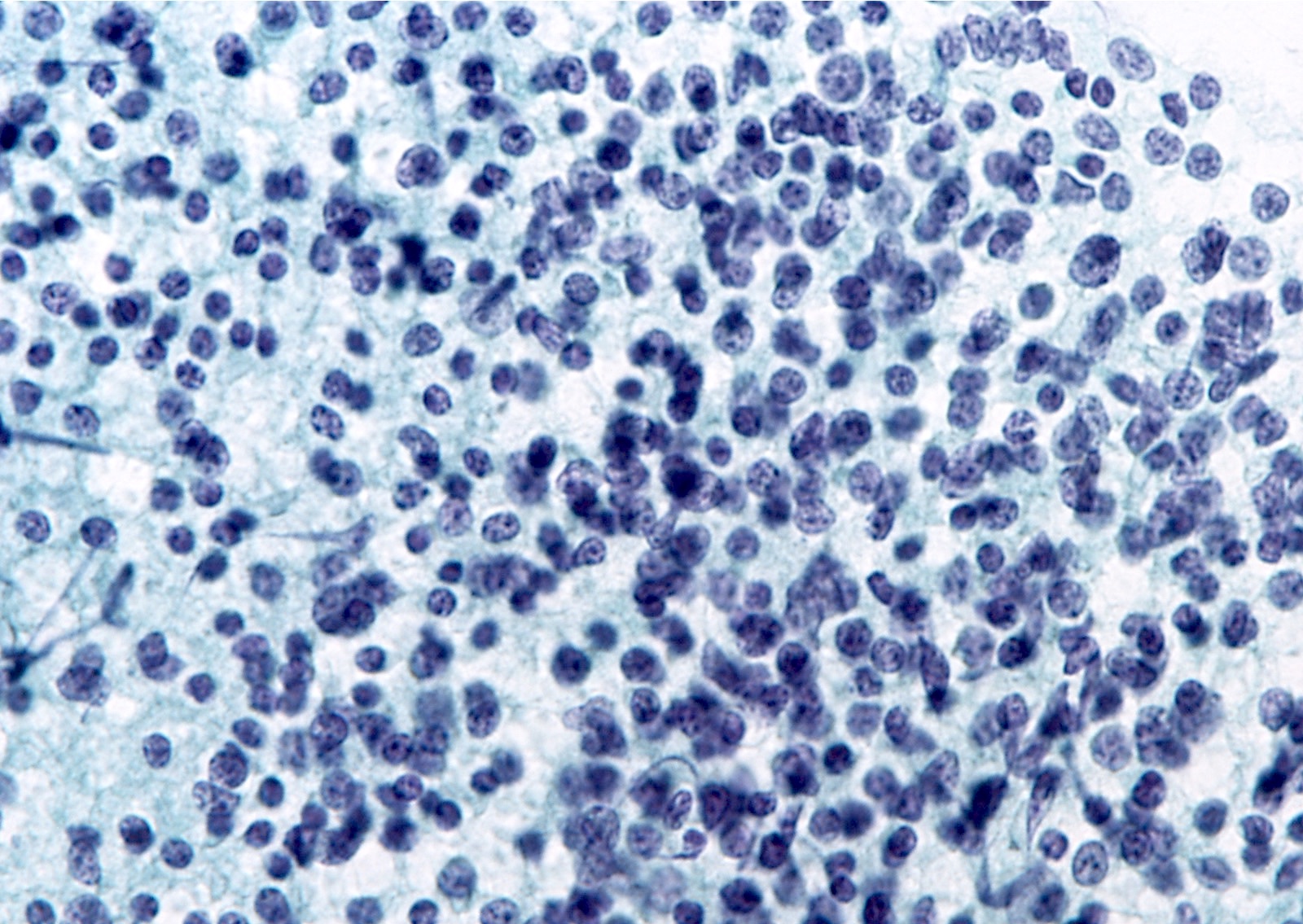
Primary thyroid lymphoma
Thyroid cytopathology
Contributed by Mark R. Wick, M.D.
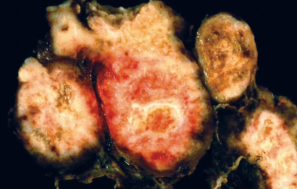
Multifocal in MEN 2
AFIP images
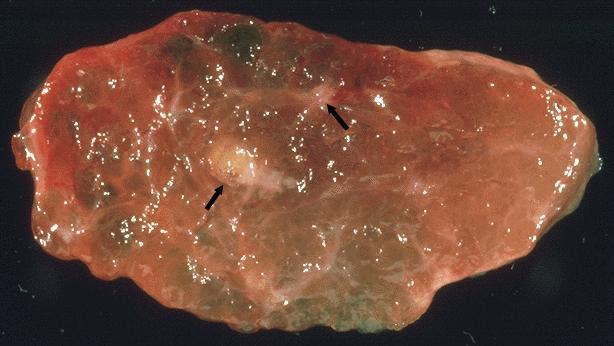
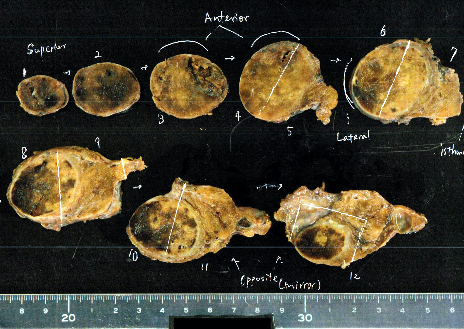
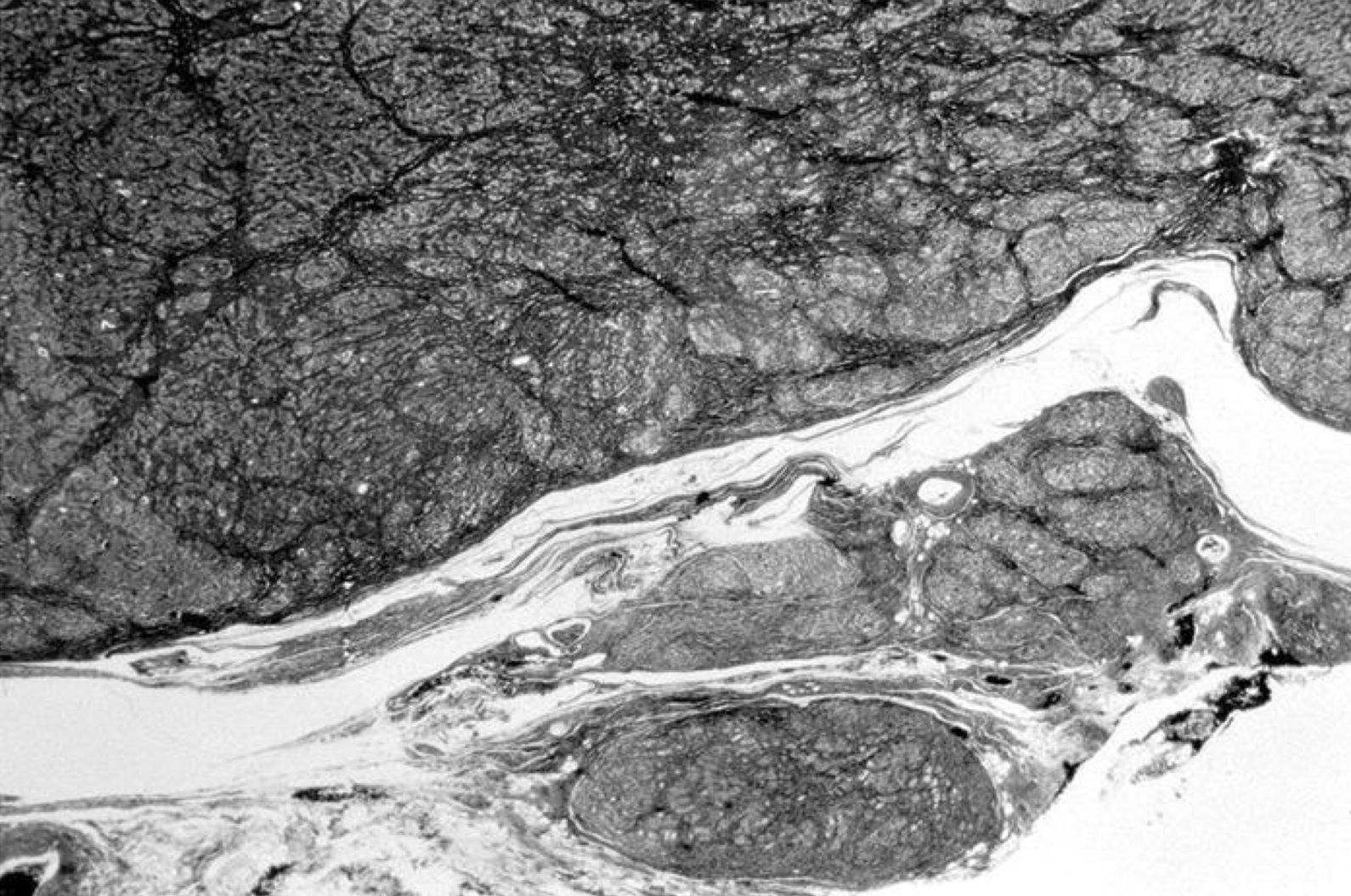
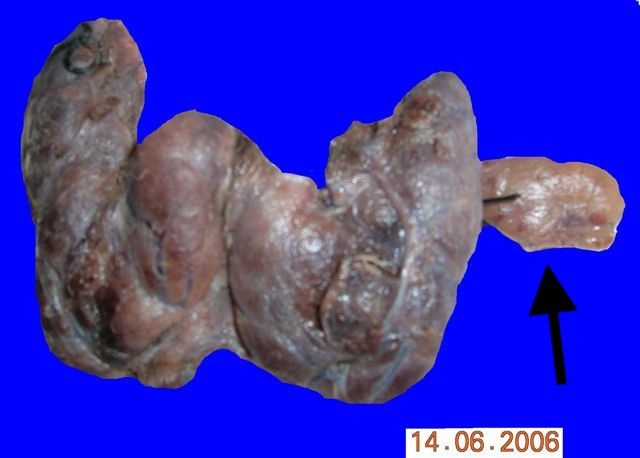
MEN 2A patient with multiple tumor foci (arrows)
Nonfamilial tumor replaces entire left lobe and isthmus
Nodal metastasis
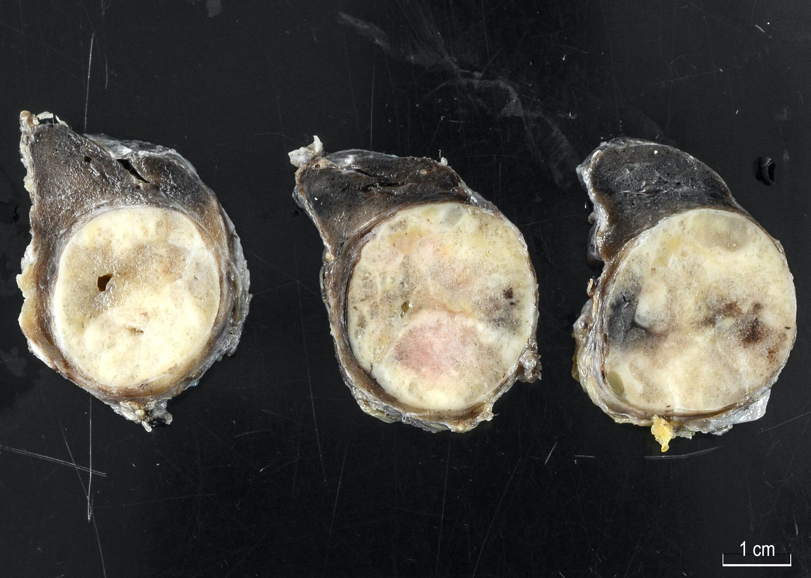
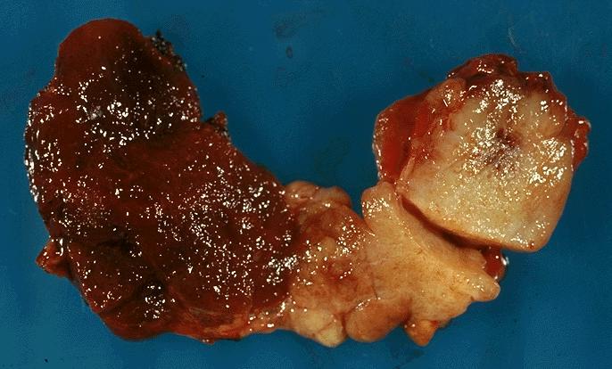
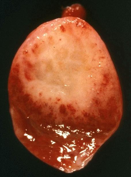
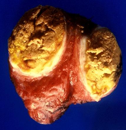
MEN 2A patient with single tumor focus
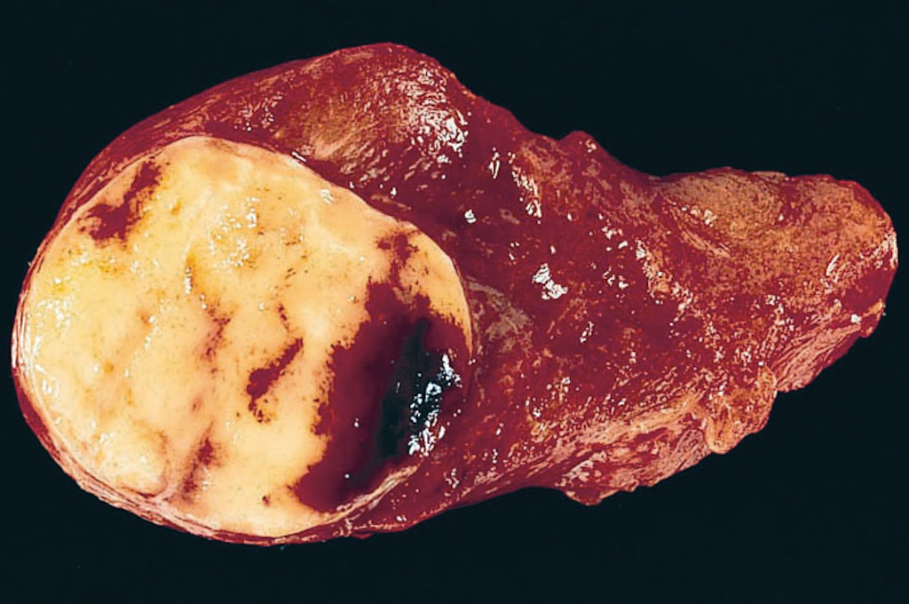
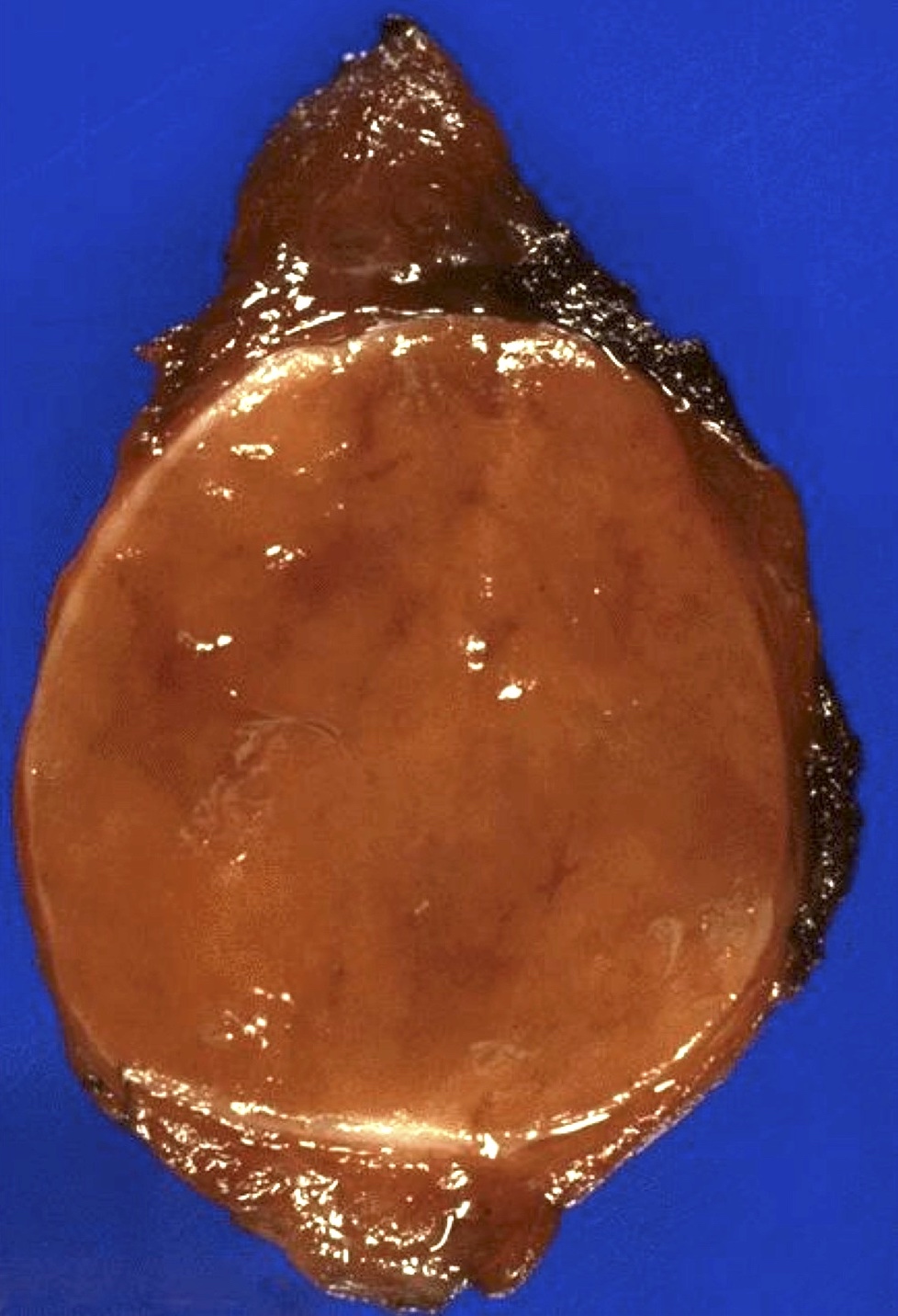
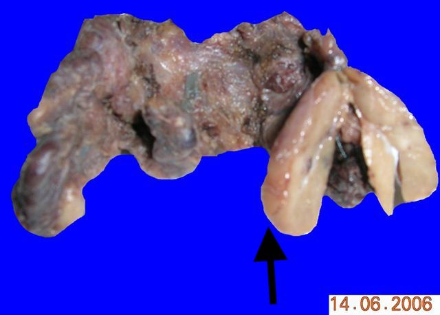
Left lobe tumor: well circumscribed, right lobe tumor: finely granular
Images hosted on other servers:

Large tumor with
hemorrhage, necrosis
and cystic degeneration
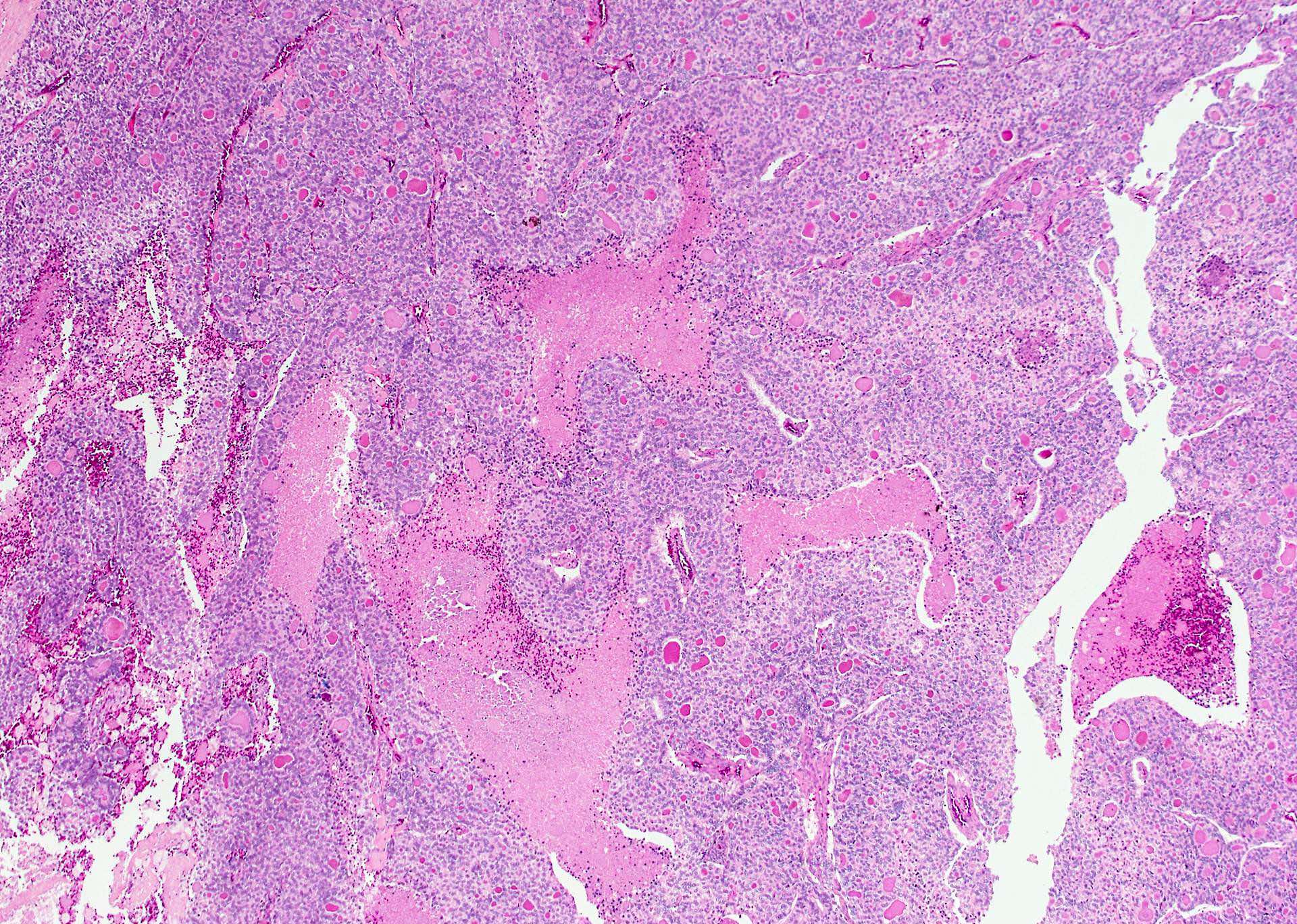
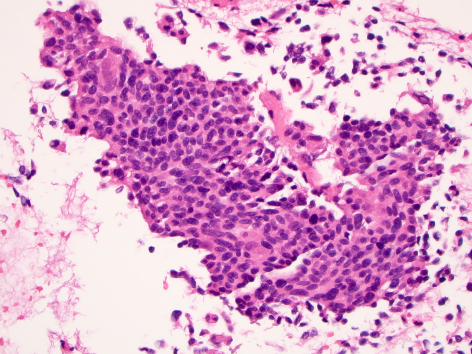
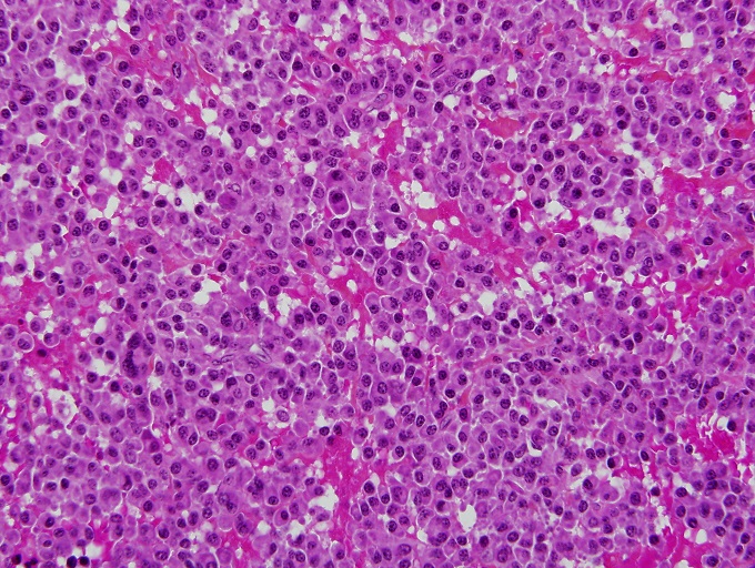
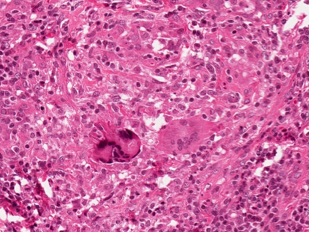
Contributed by Shuanzeng Wei, M.D., Ph.D., Joseph Christopher Castillo, M.D. and Mark R. Wick, M.D.

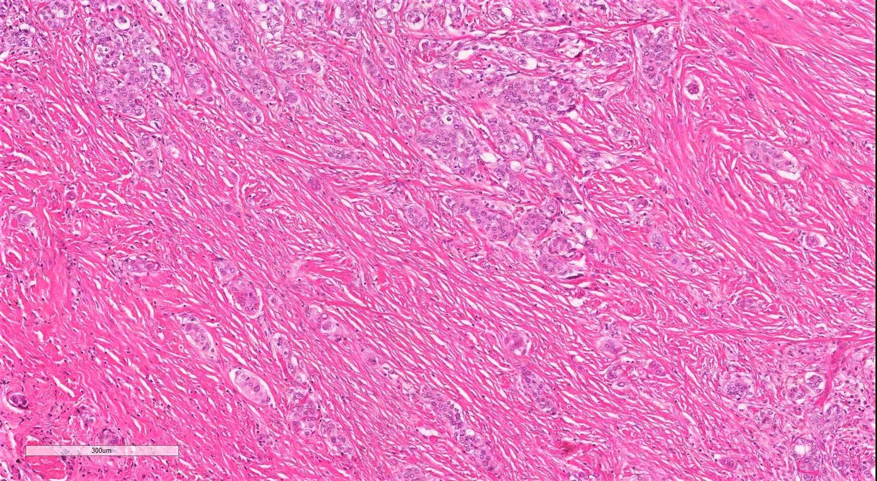
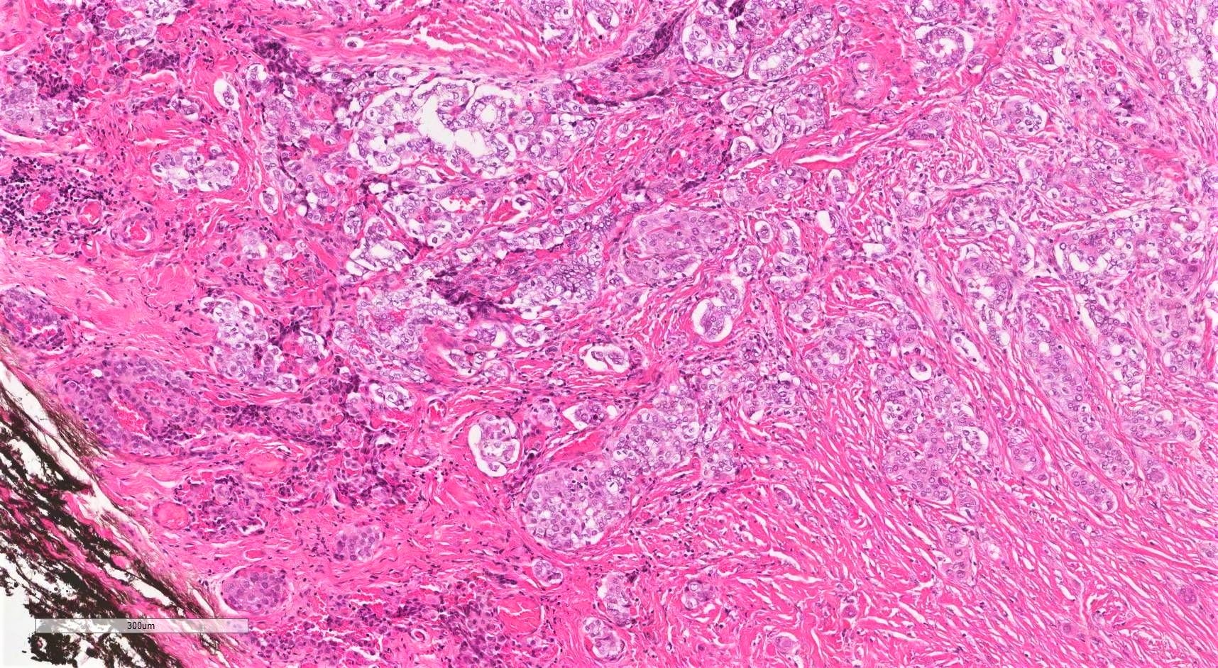
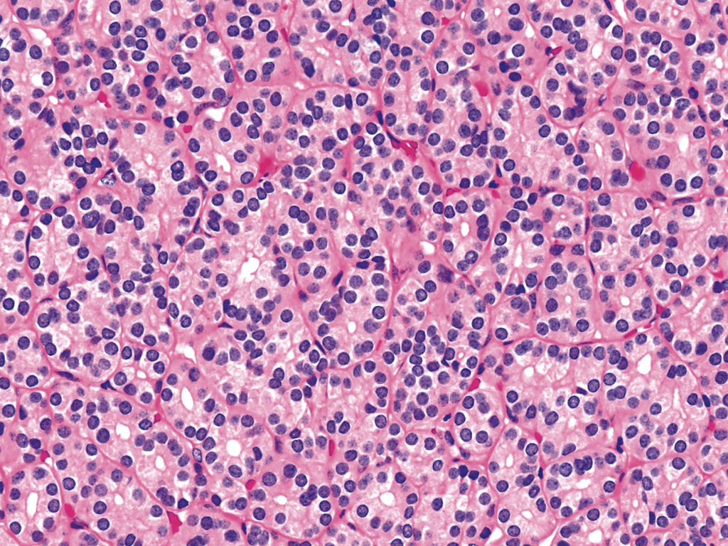
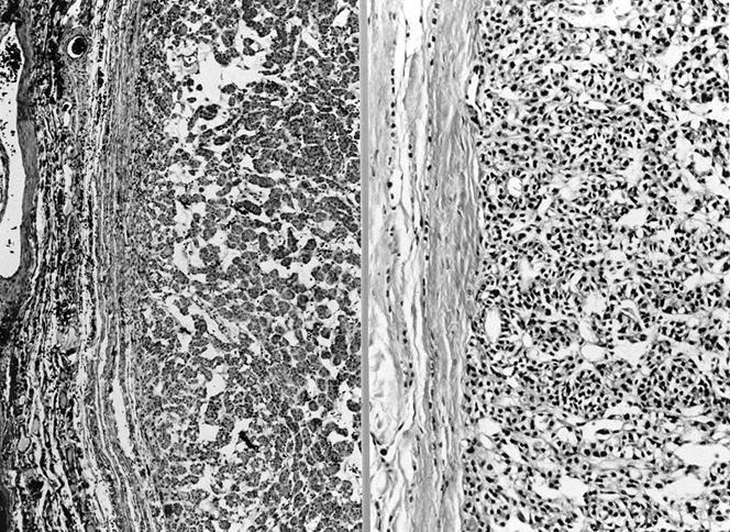
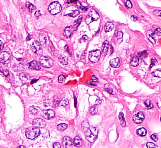
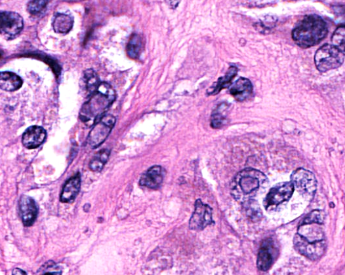
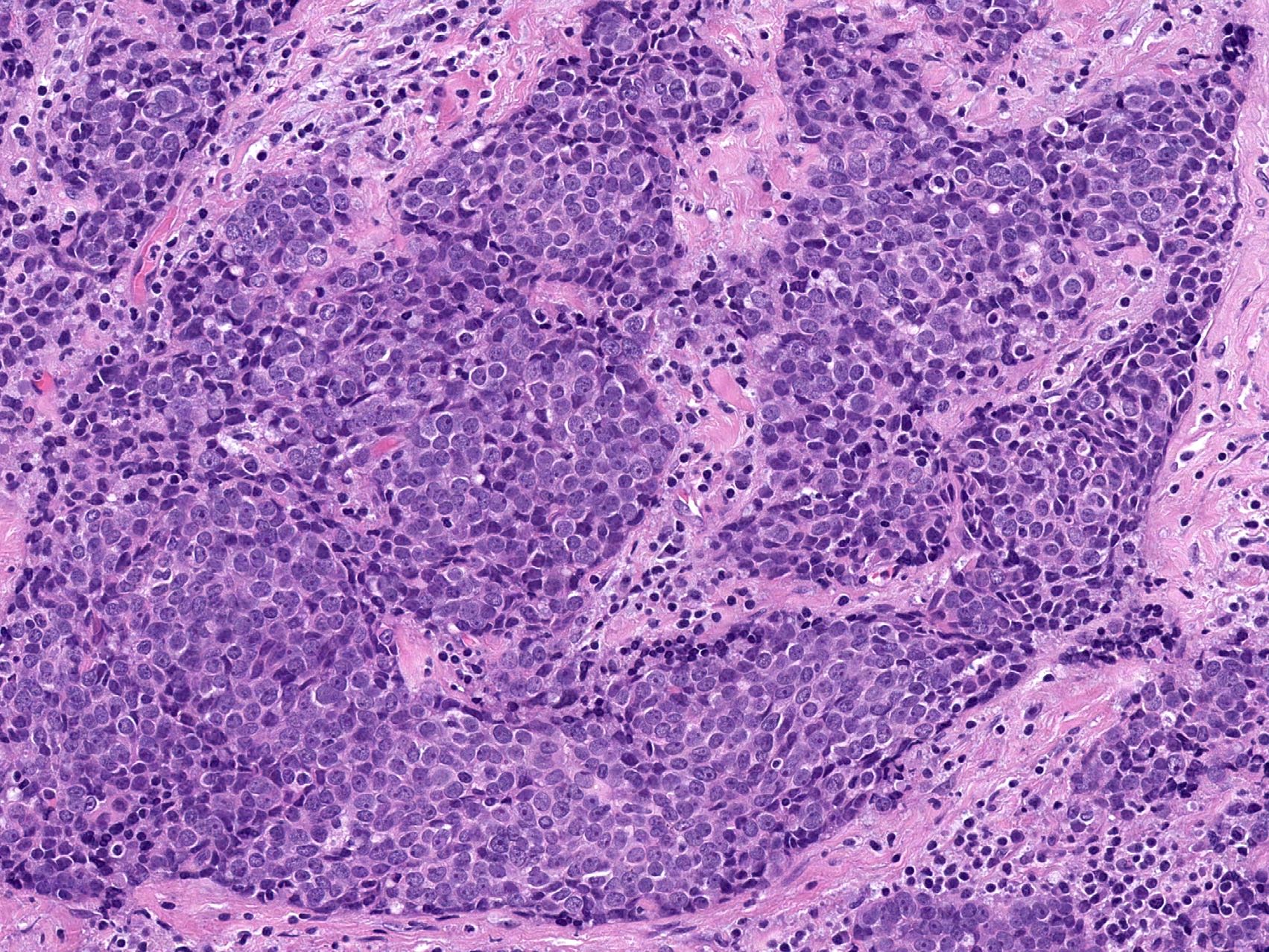
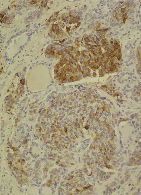
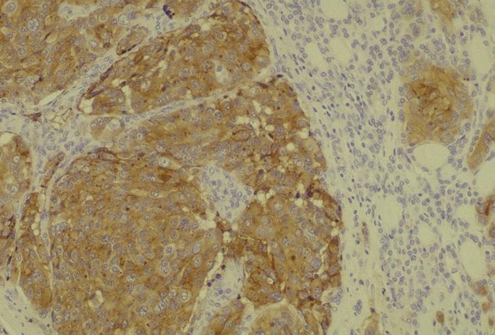
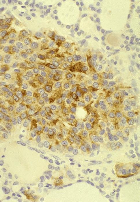
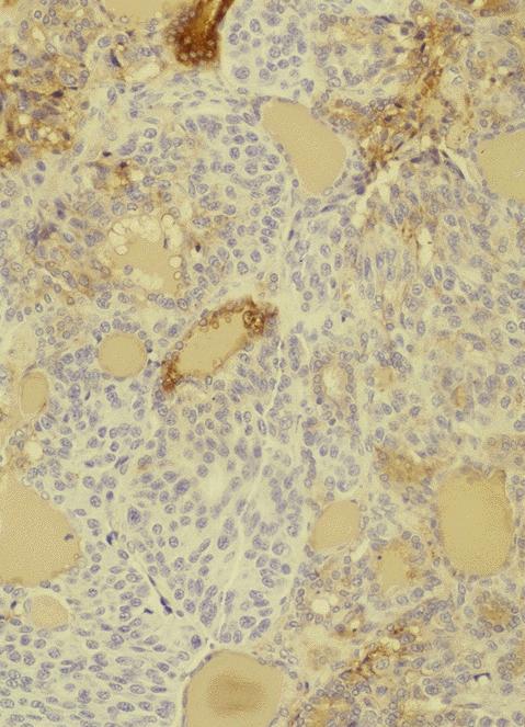
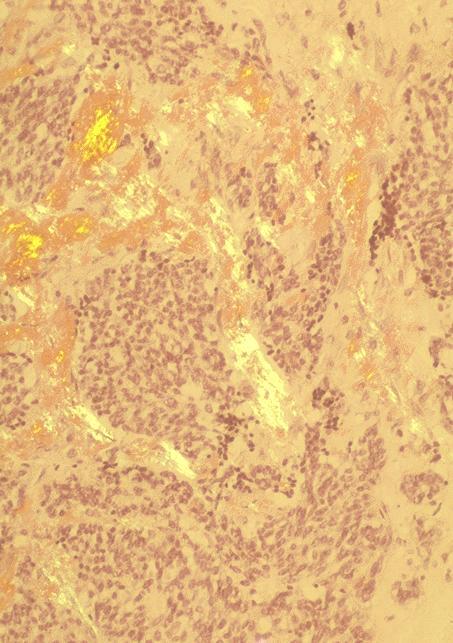
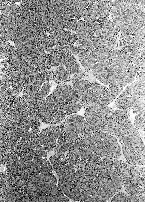
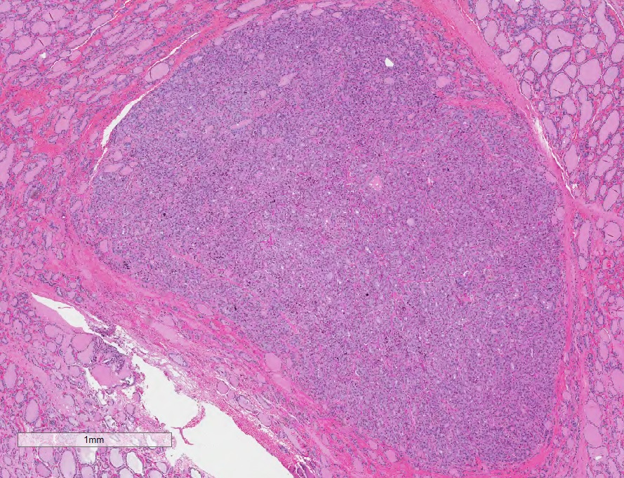
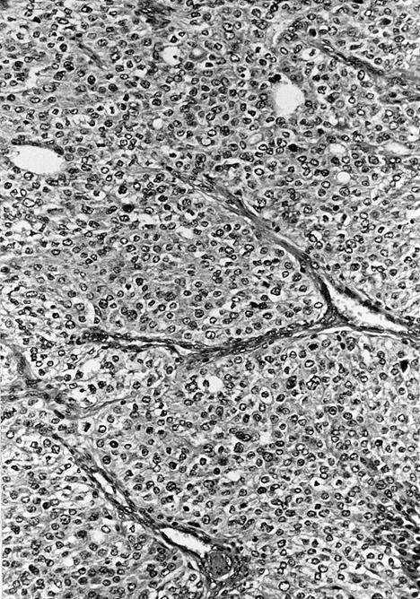
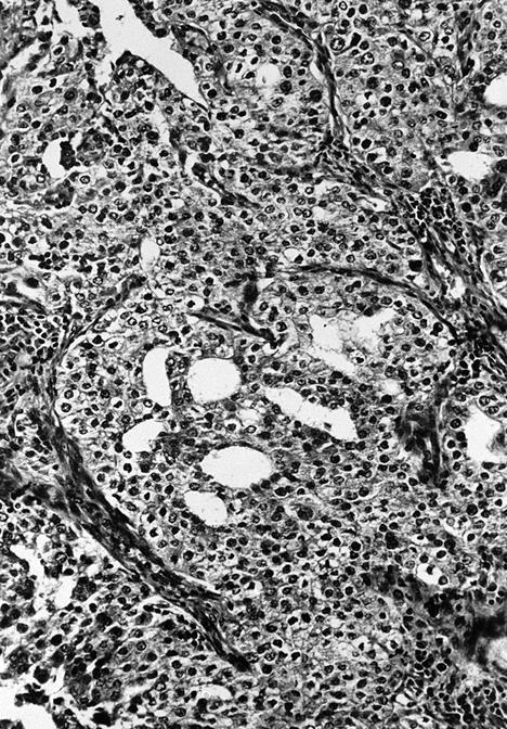
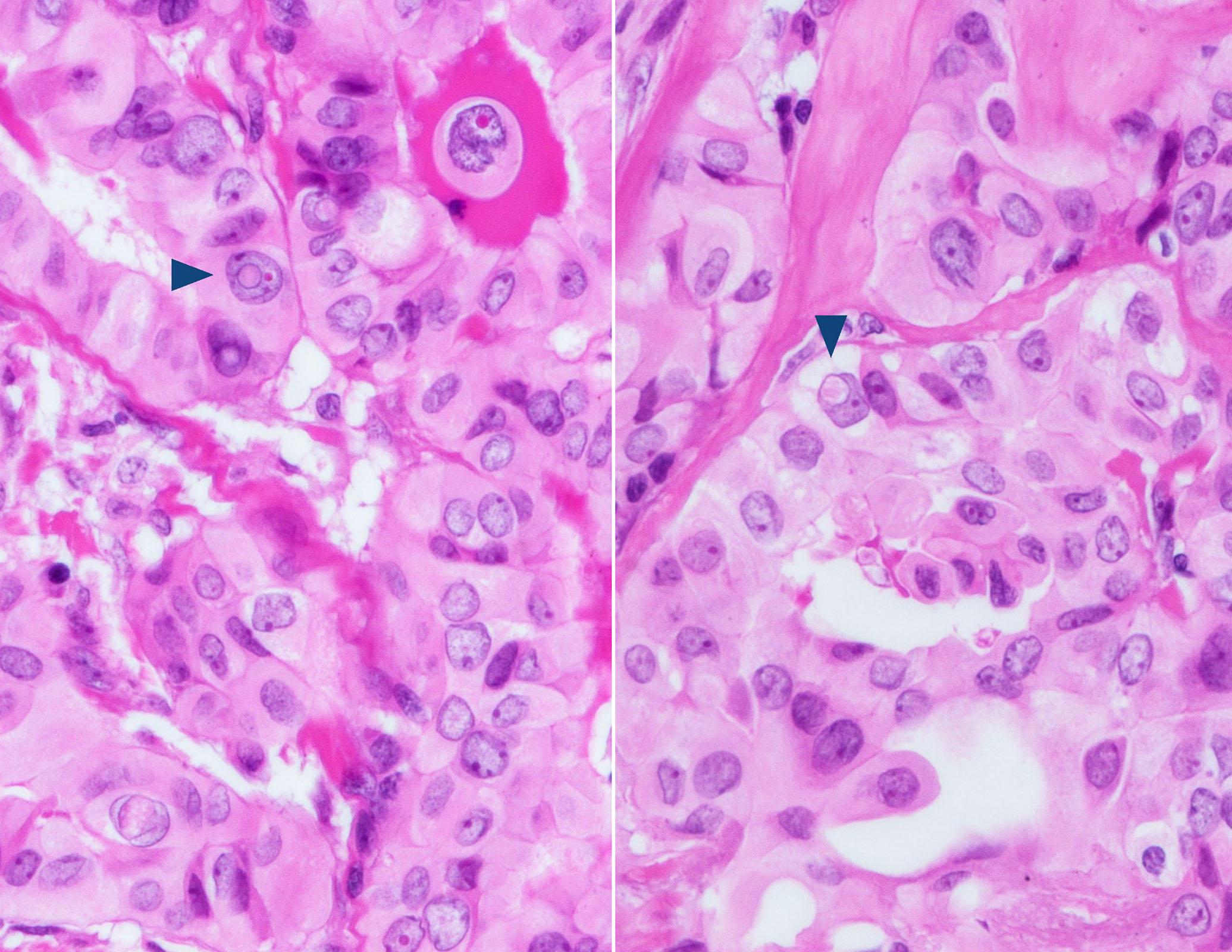
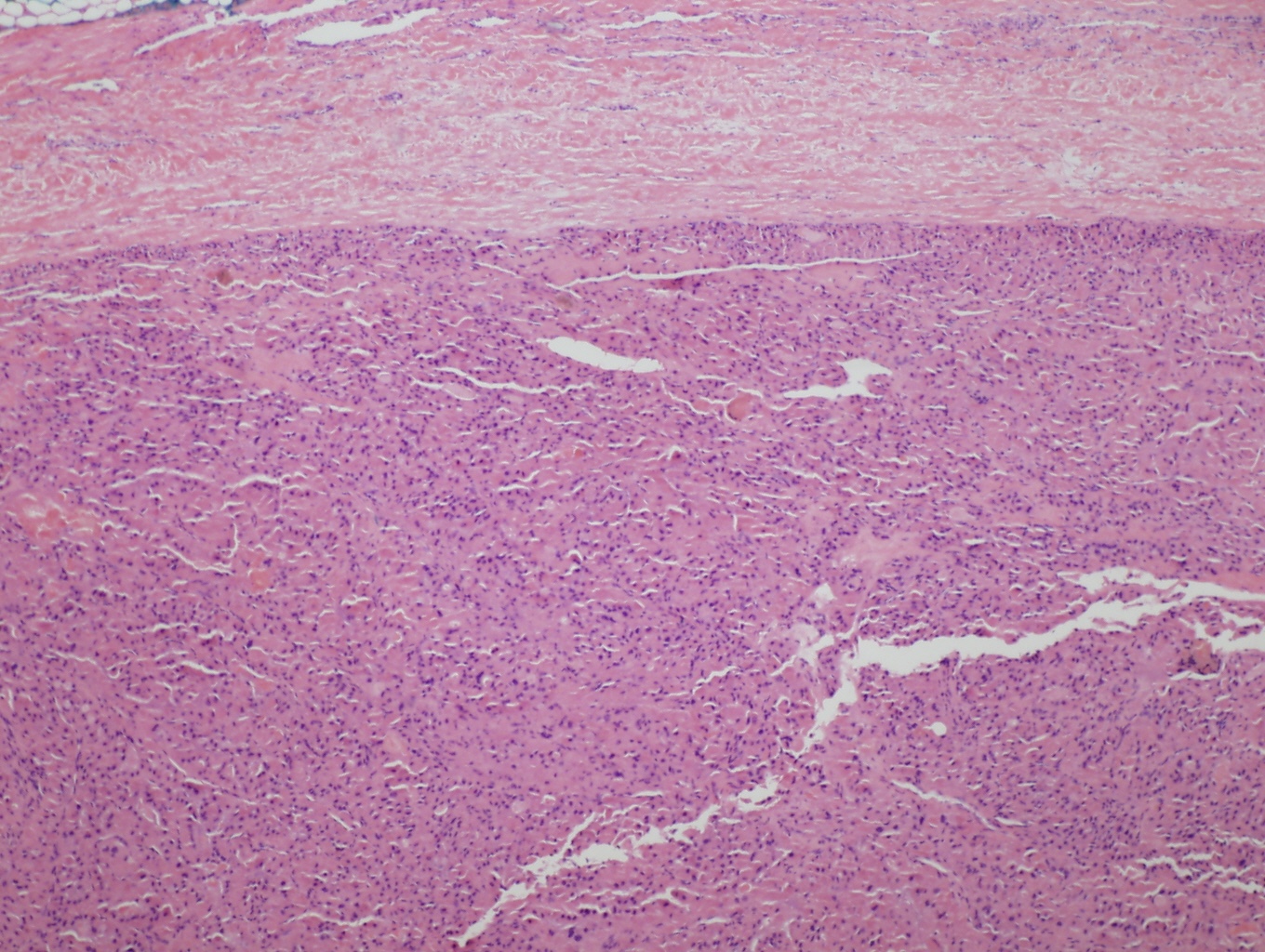
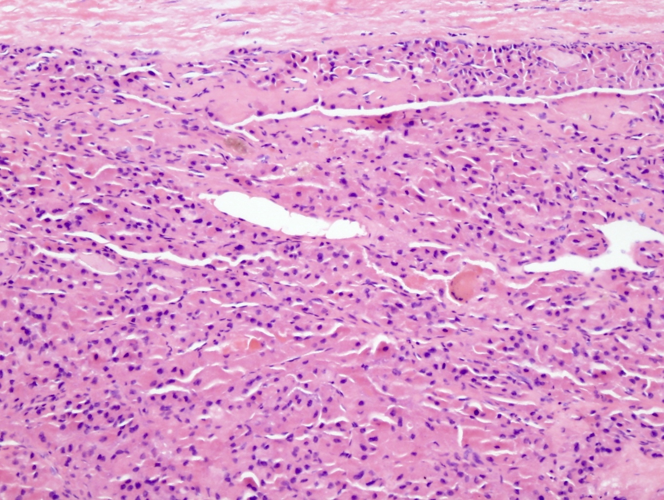
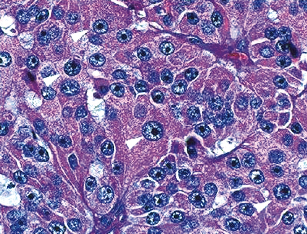
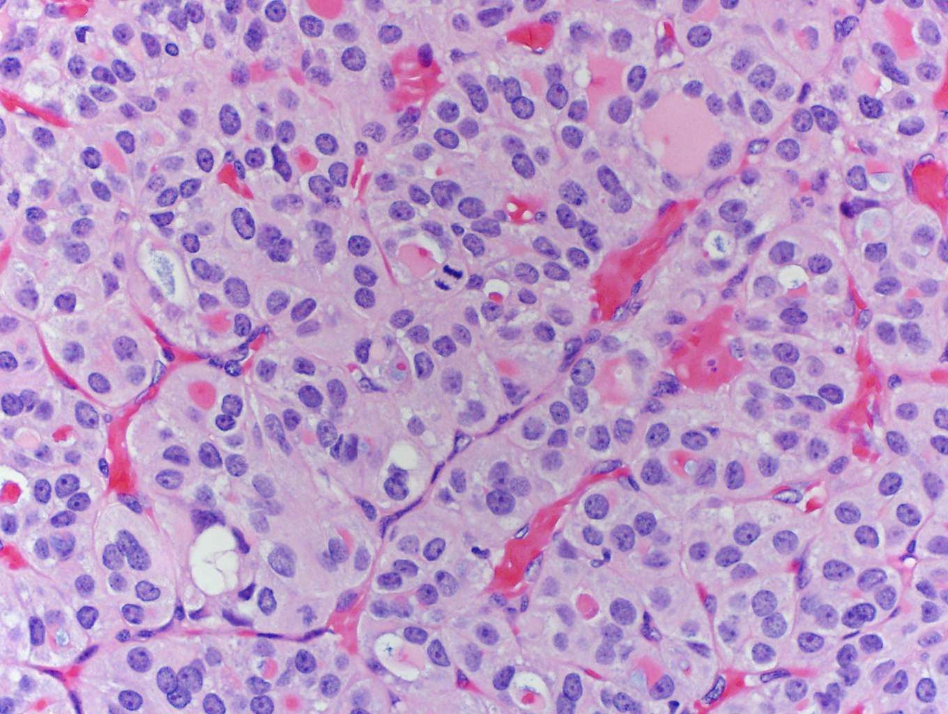
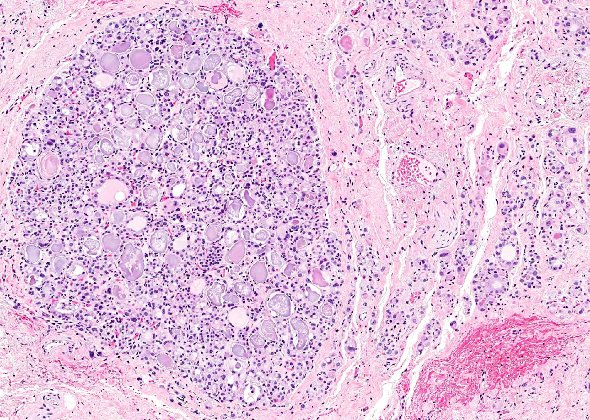
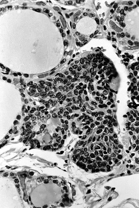
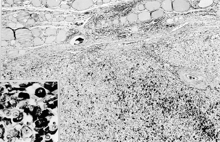
Low power
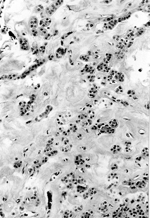
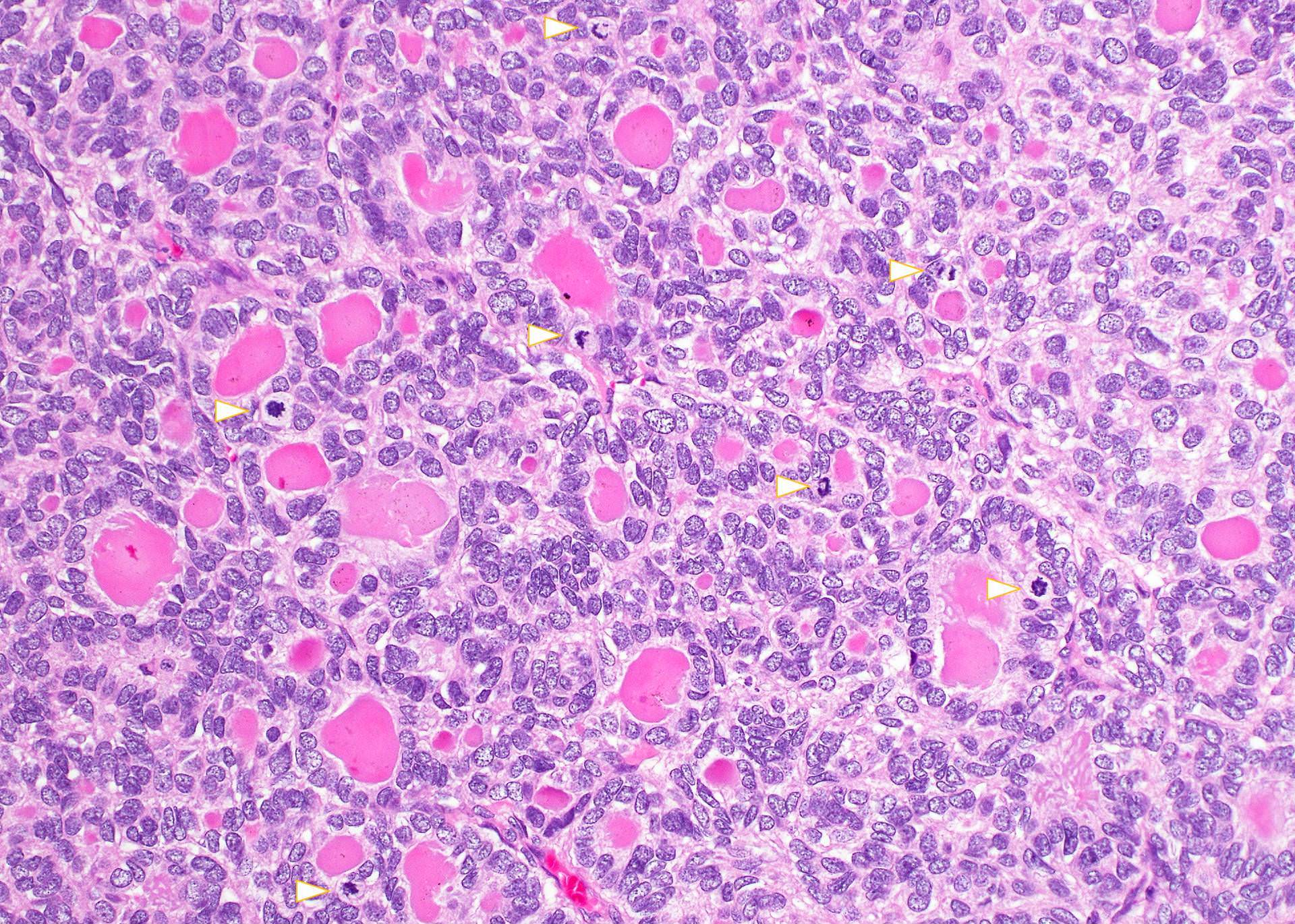
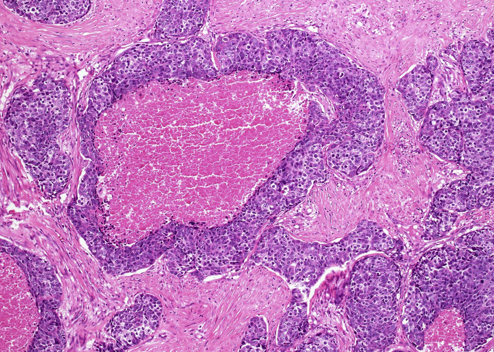
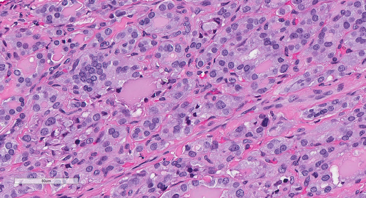
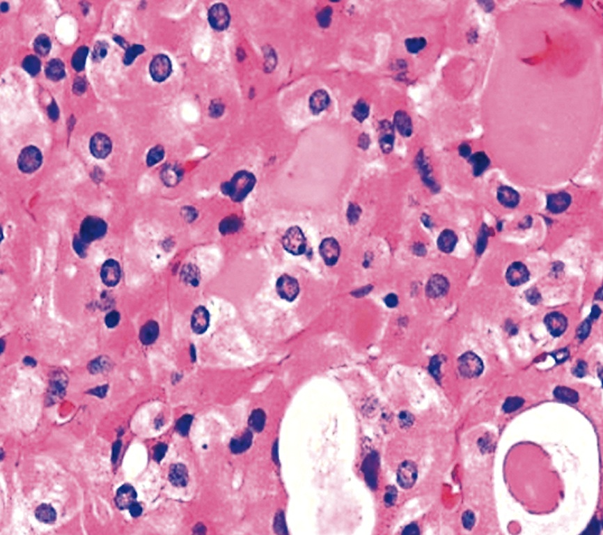
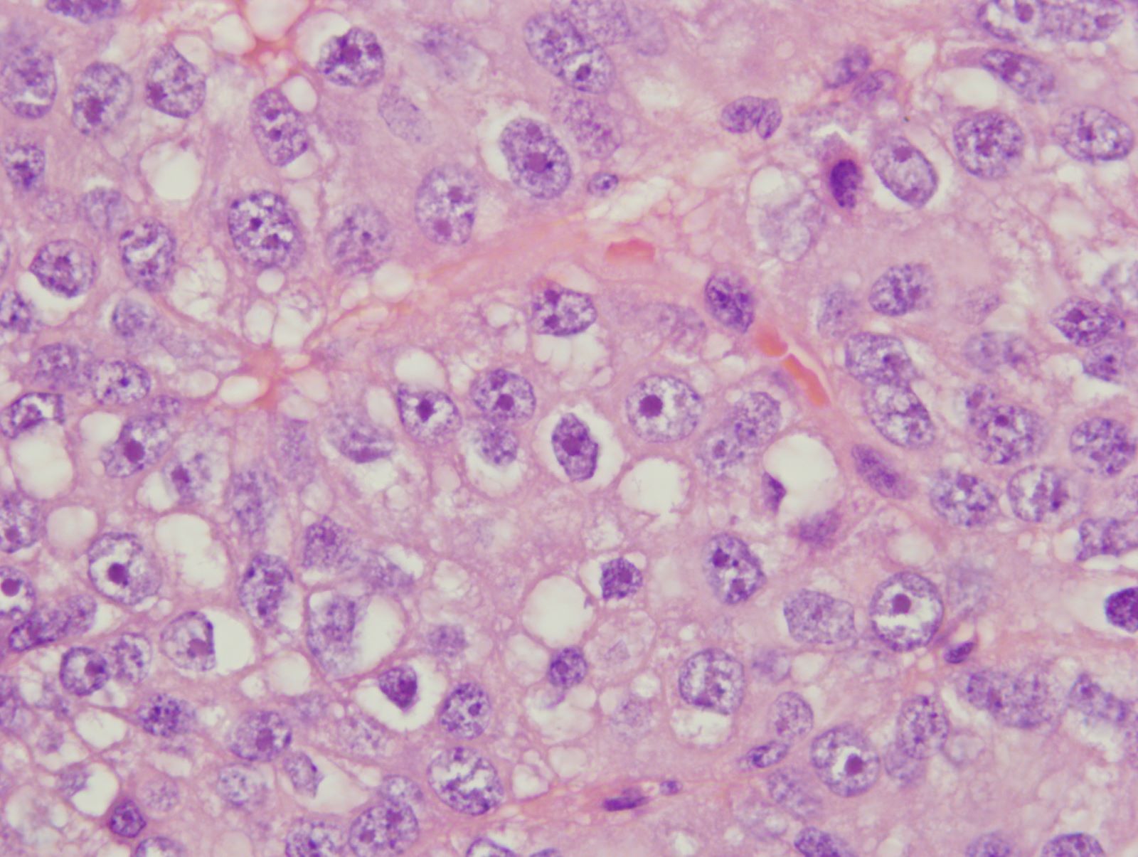
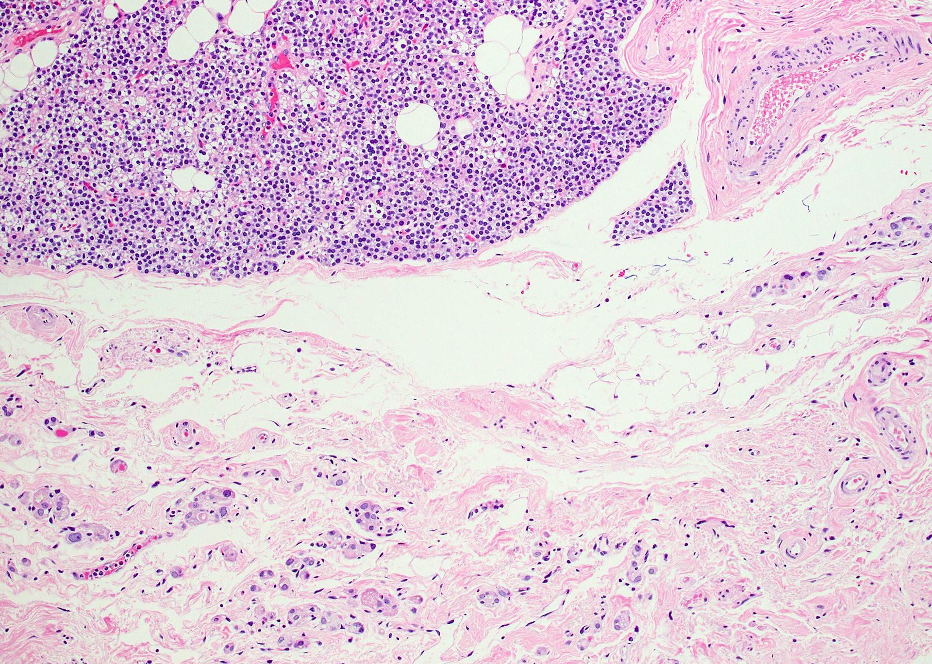
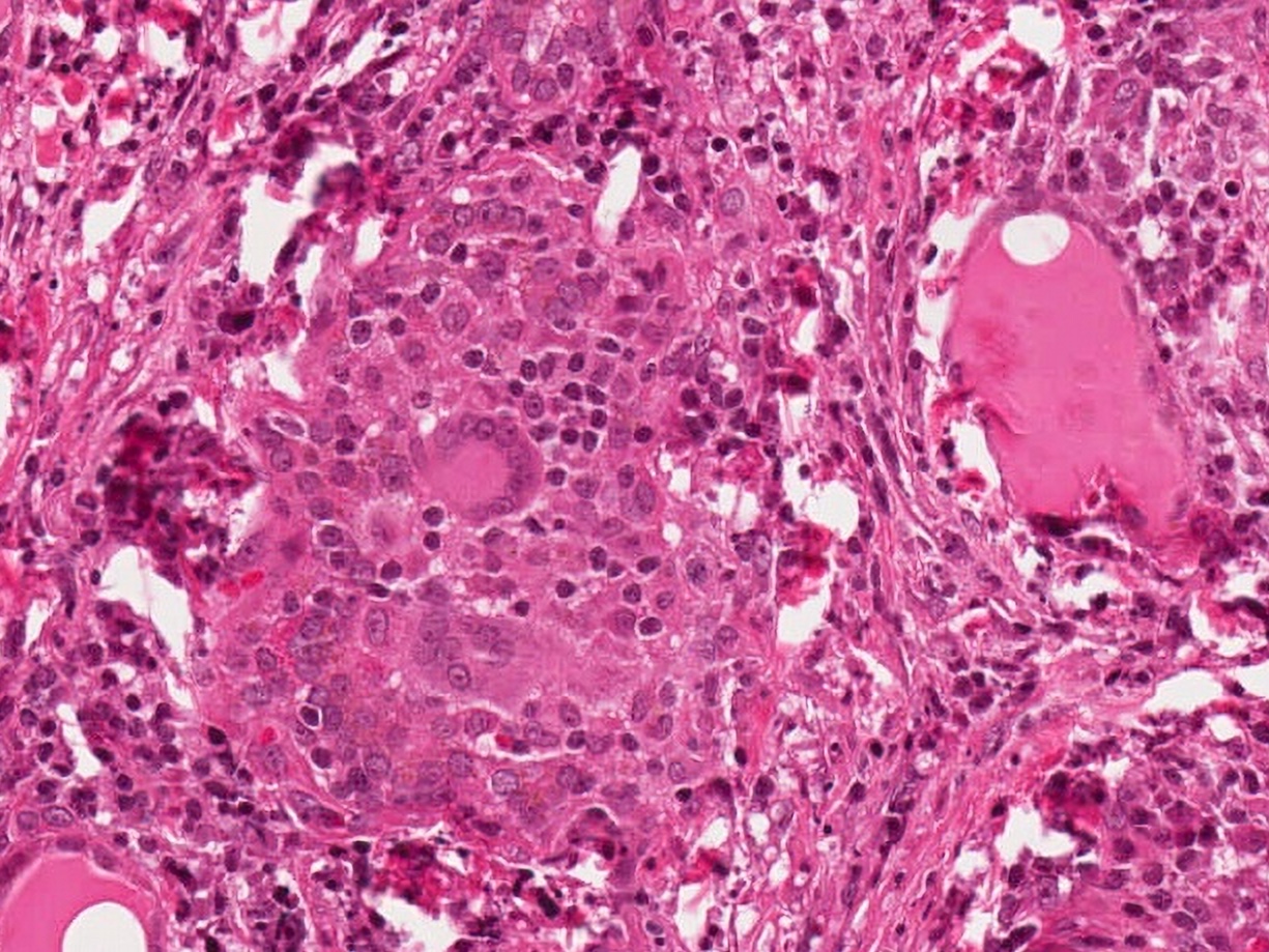
Background of amyloid
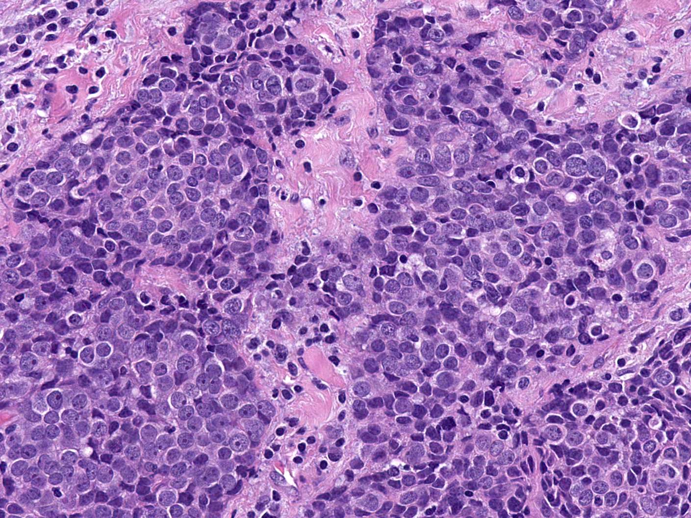
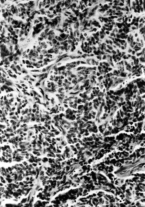
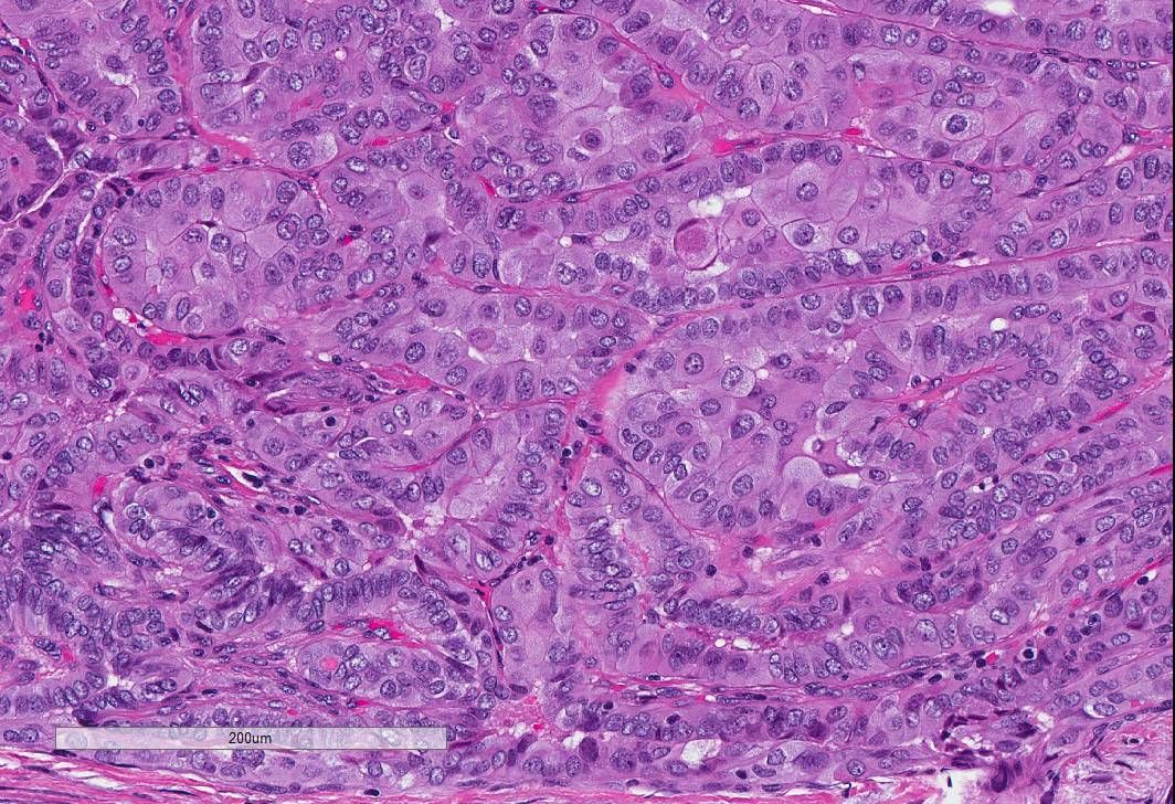
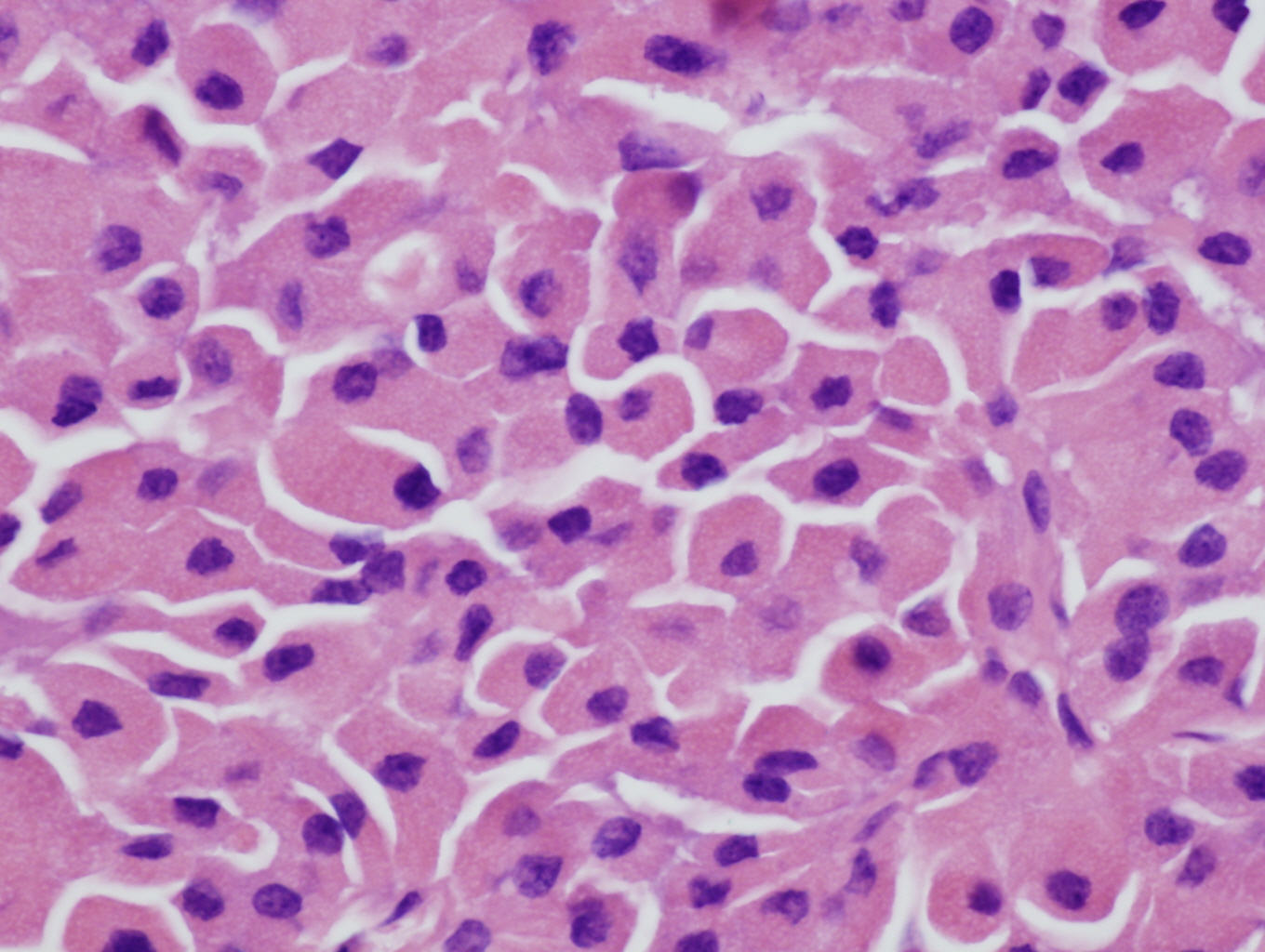
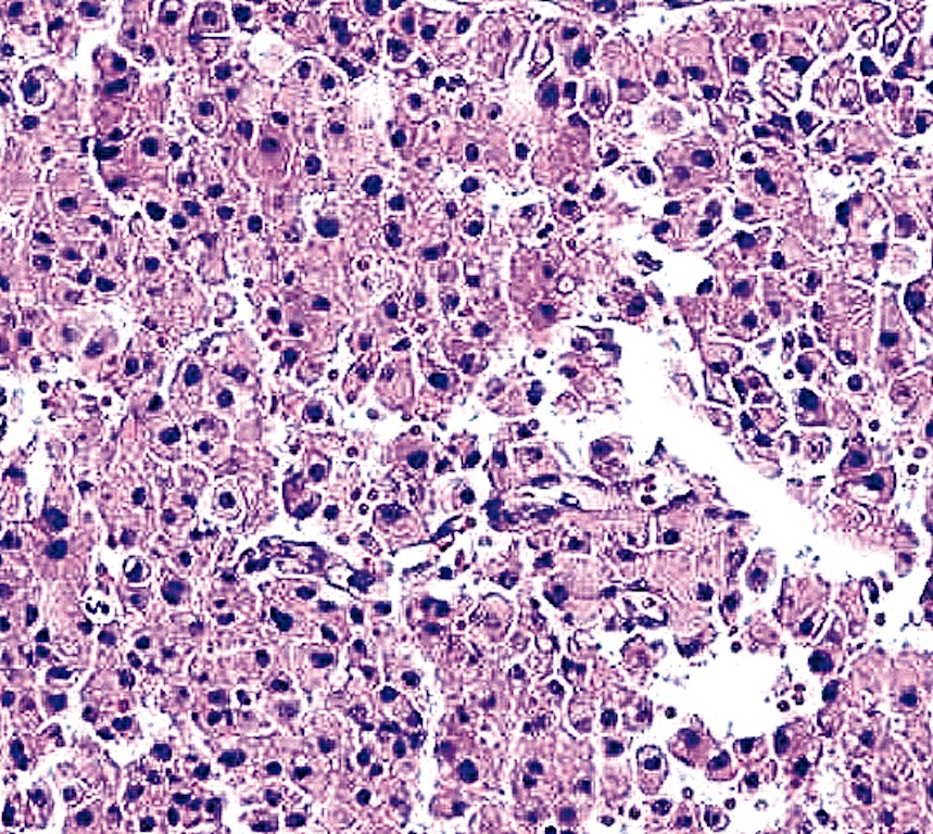
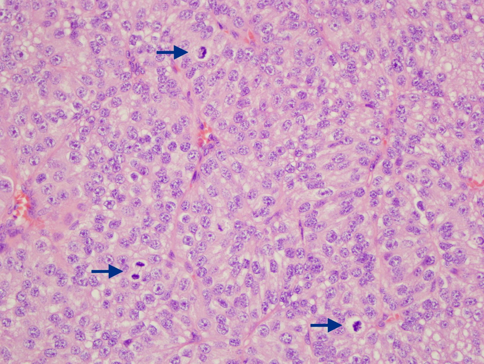
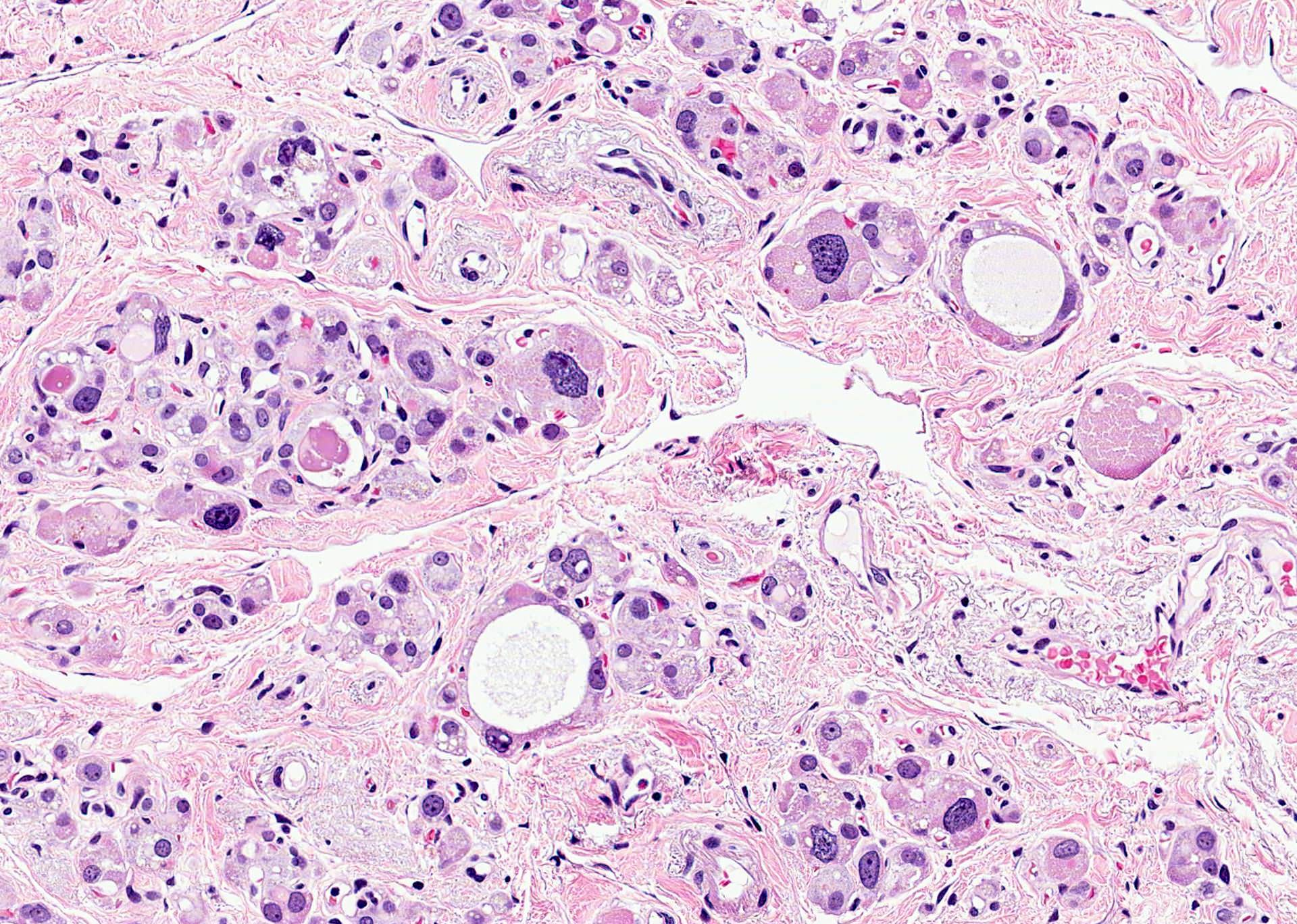
Tumor cells with finely stippled chromatin

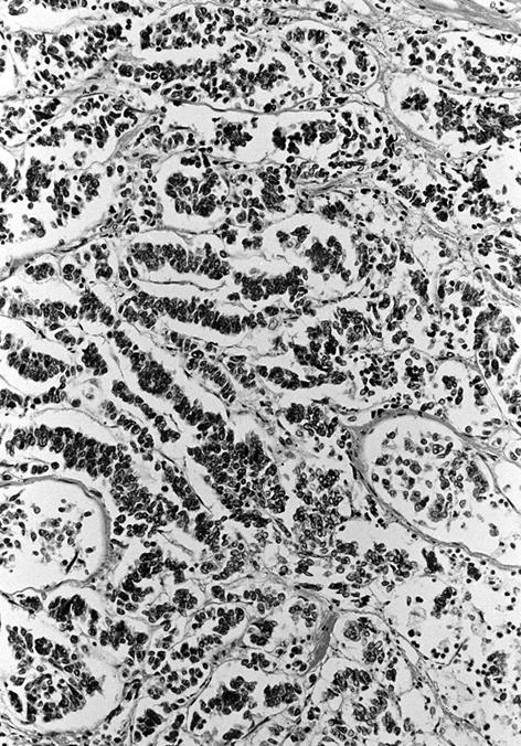
H&E
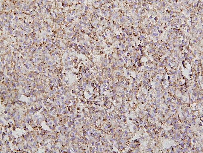
H&E
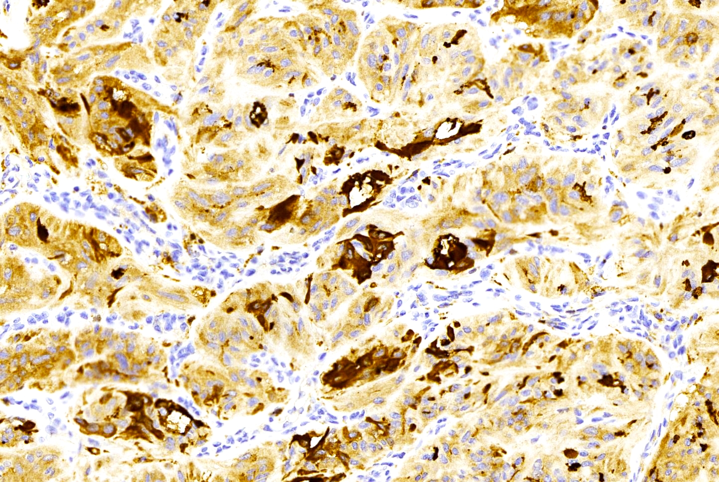
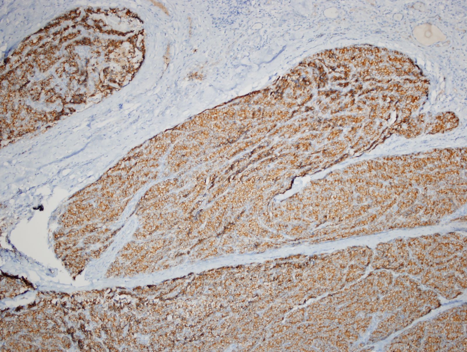
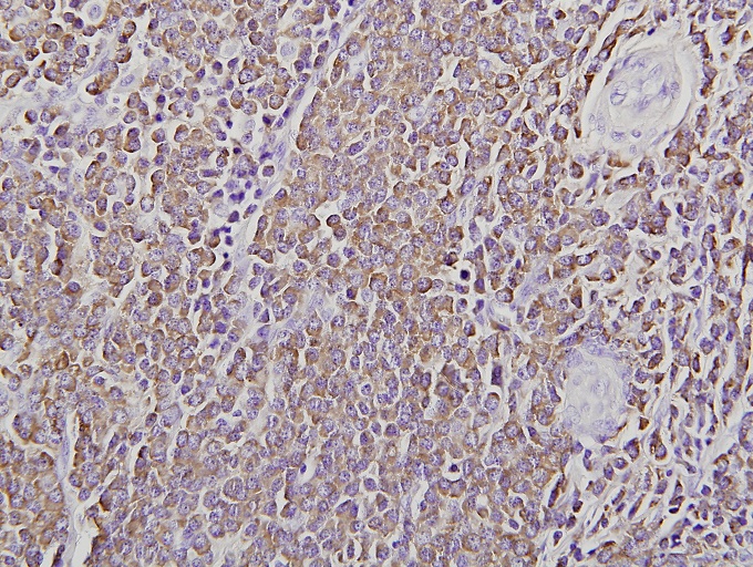
Chromogranin
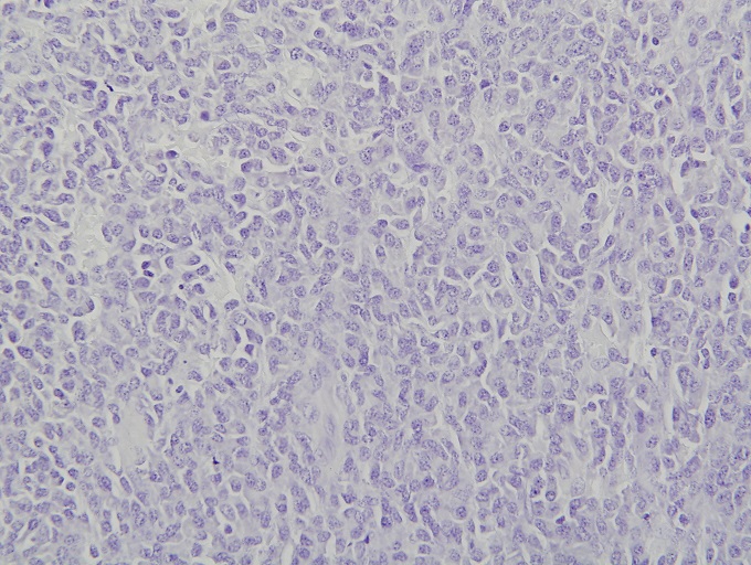
Thyroglobulin
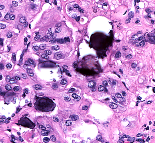
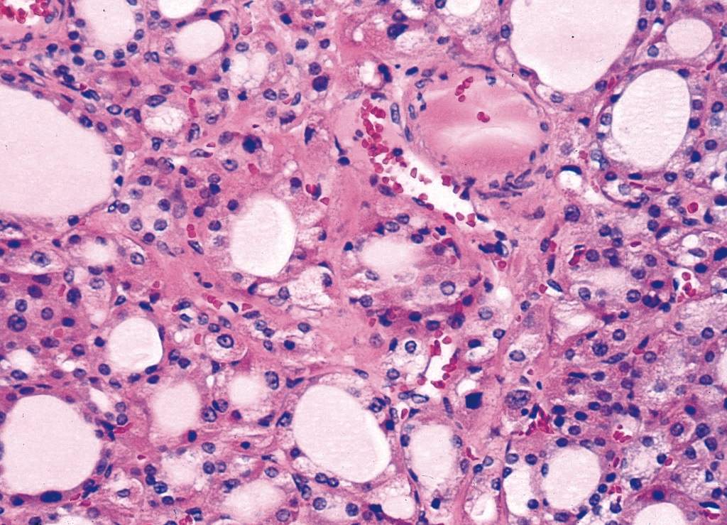
Pseudopapillary variant
Signet ring cell variant
AFIP images
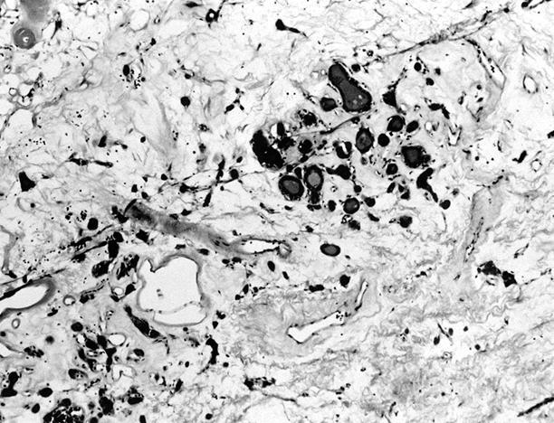
With melanin pigment
Stains
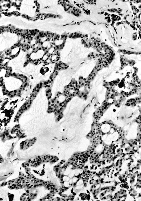
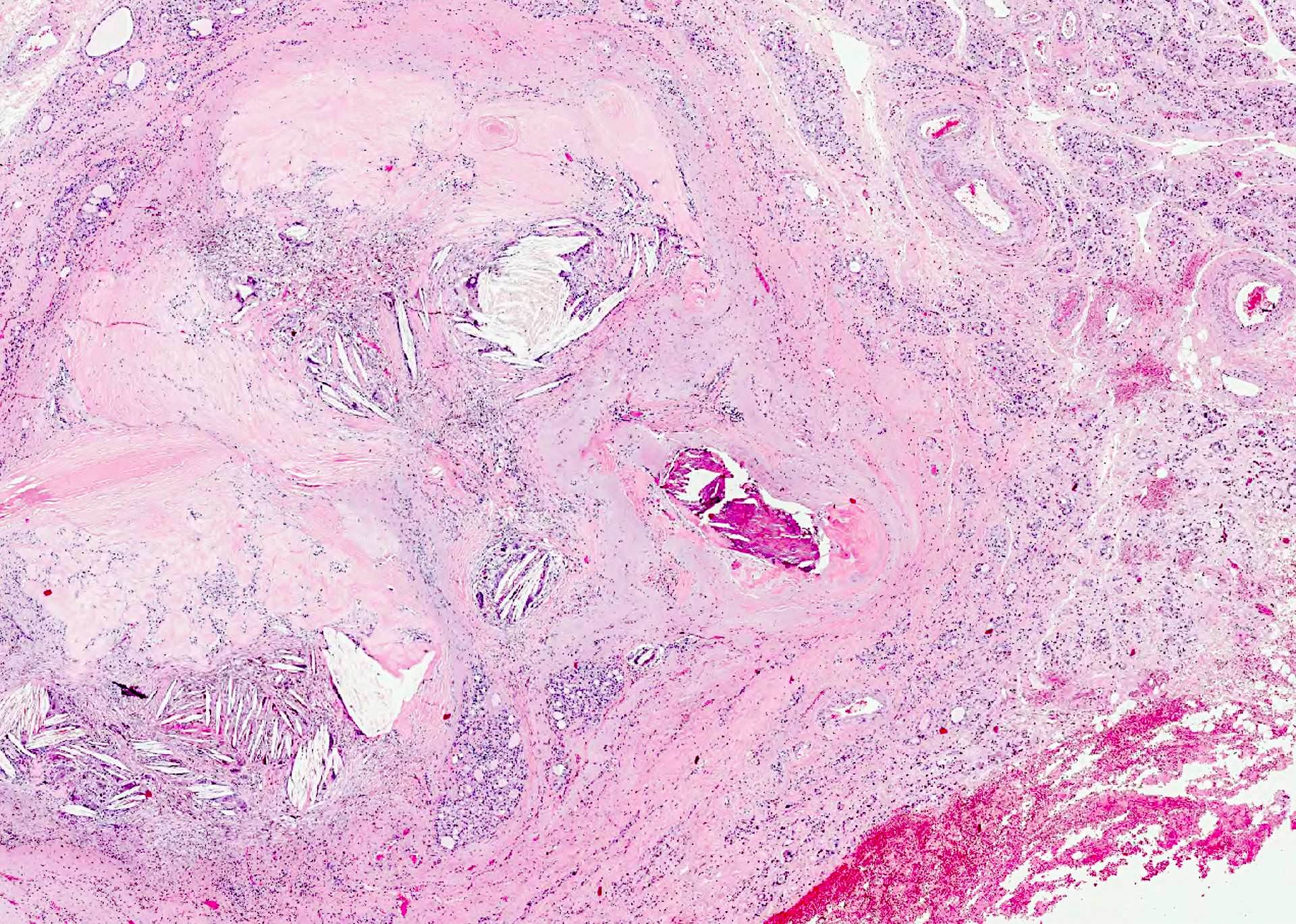
Amyloid
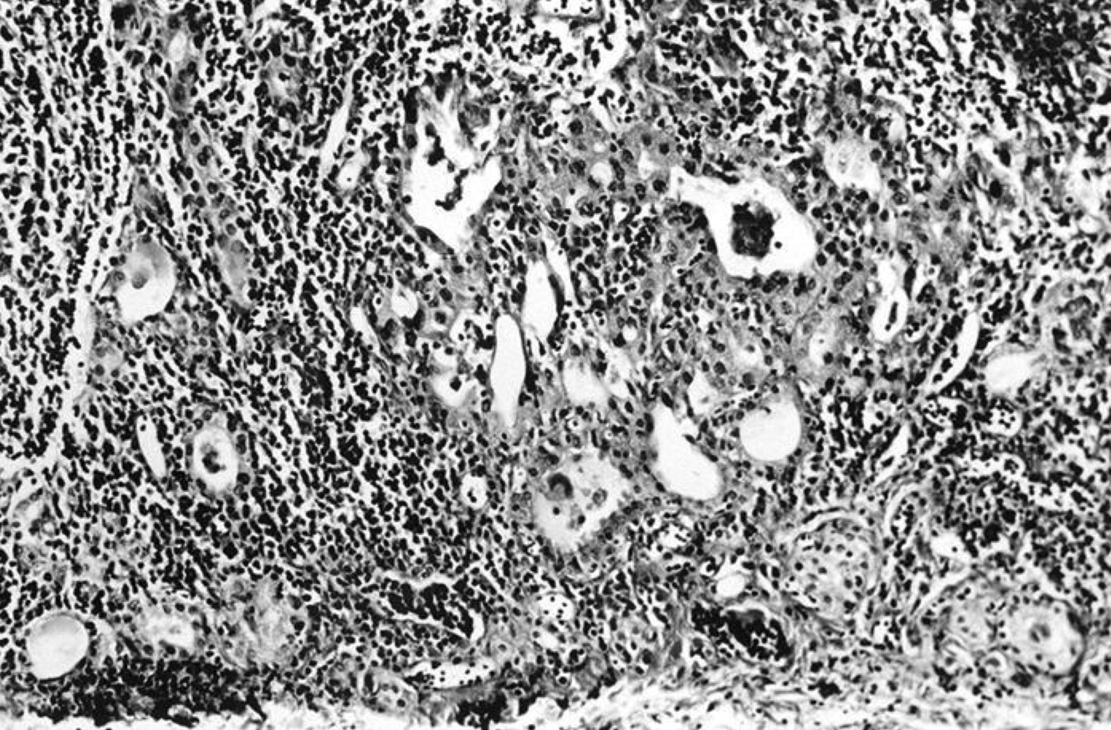
MEN 2A patients with early medullary carcinoma
Paraganglioma-like pattern
Small cell variant

Tubular (follicular) variant
Images hosted on other servers:

Oncocytic variant

Papillary variant
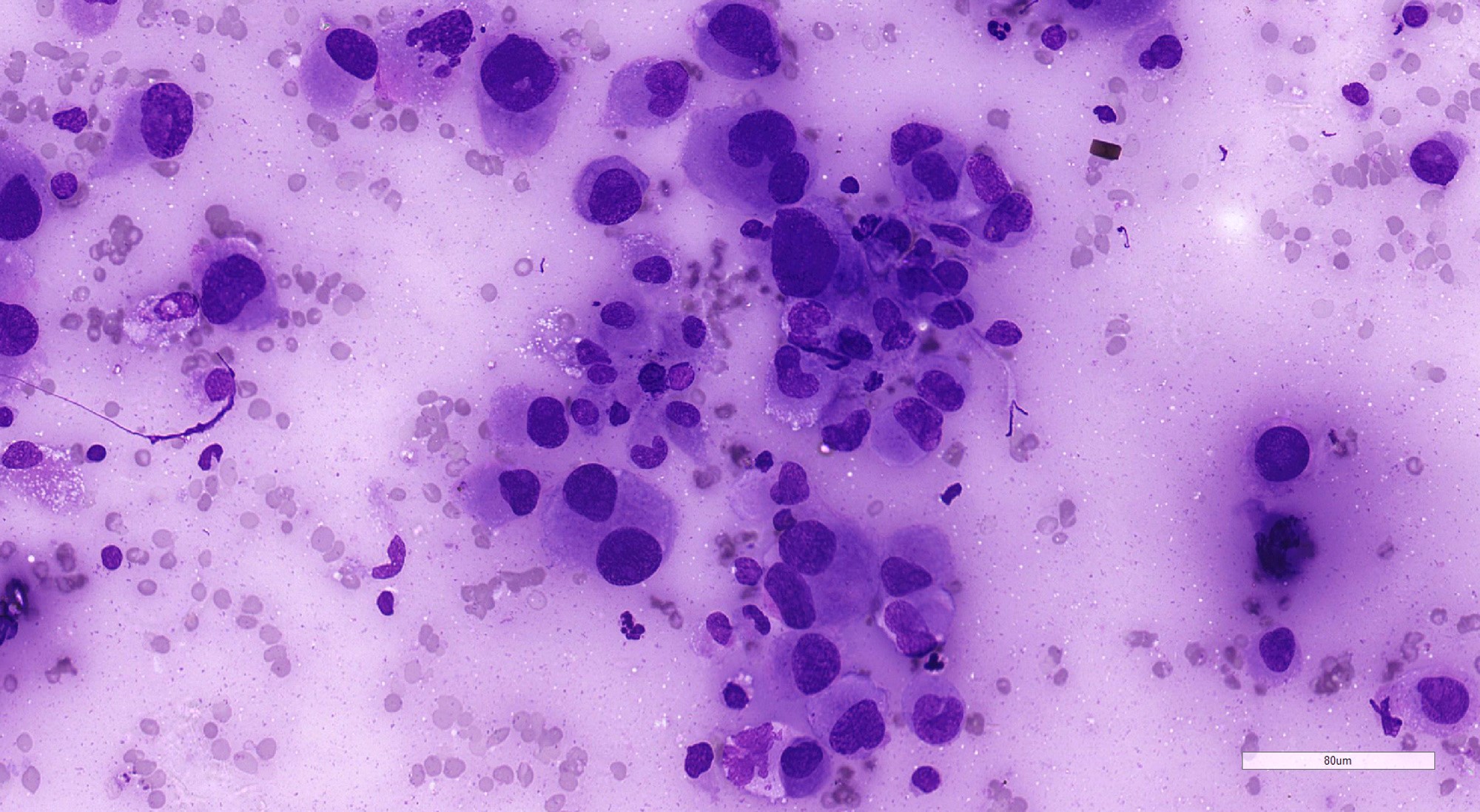
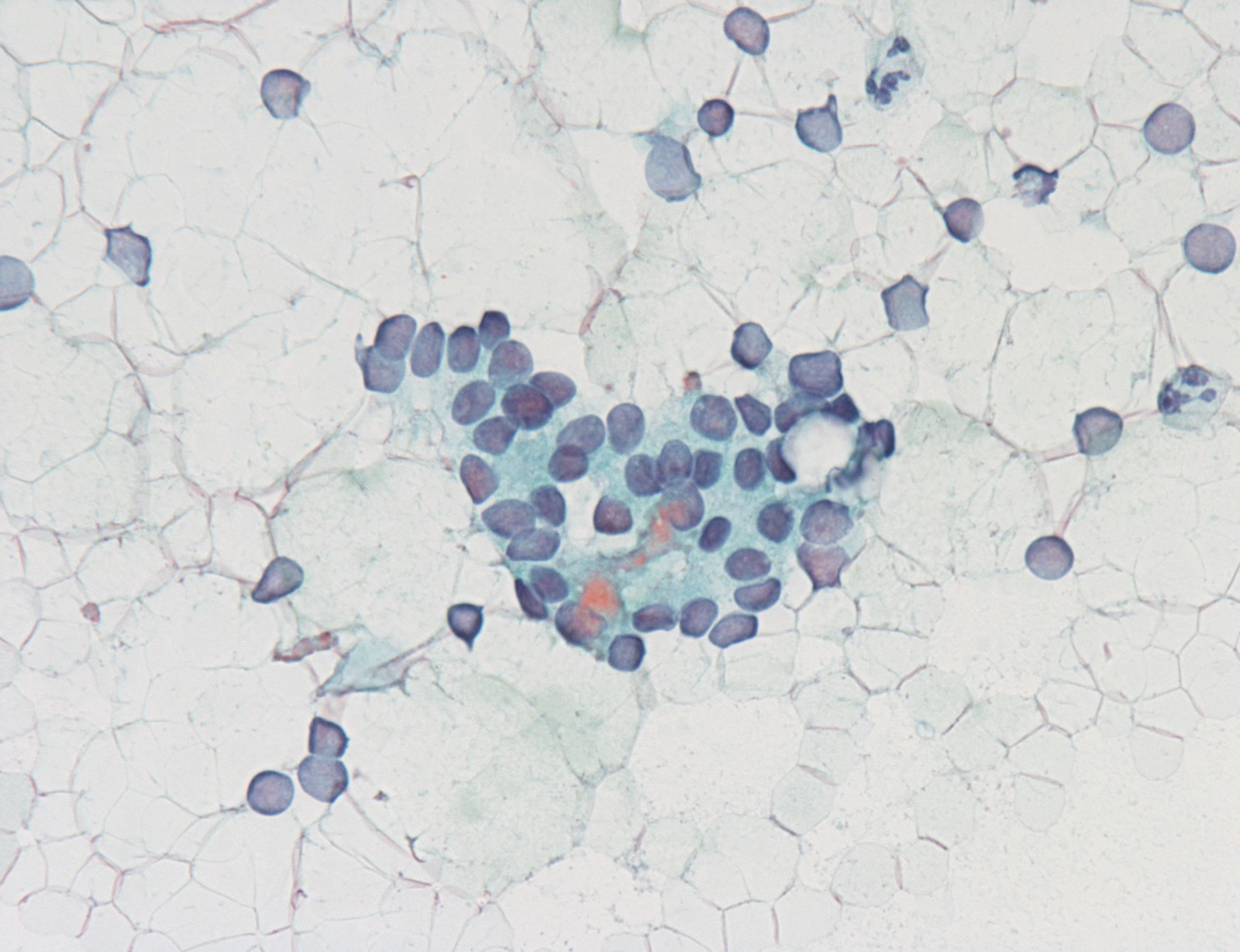
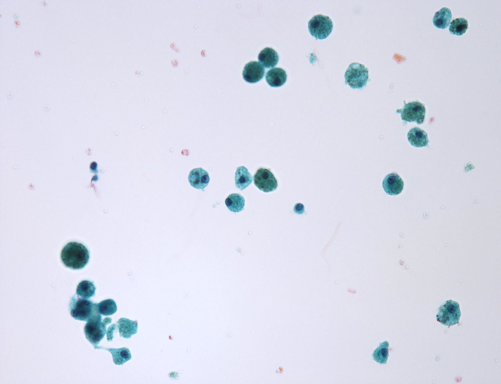
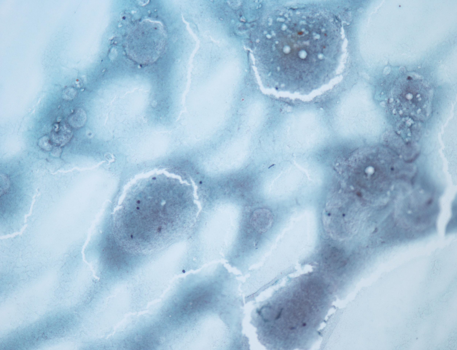
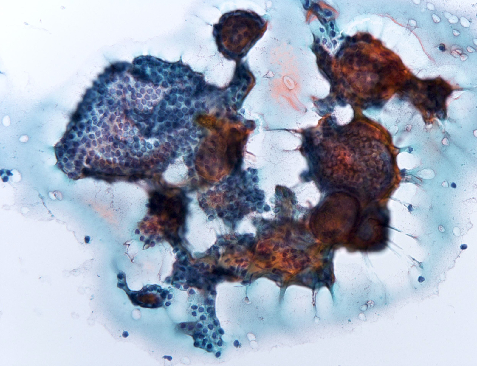
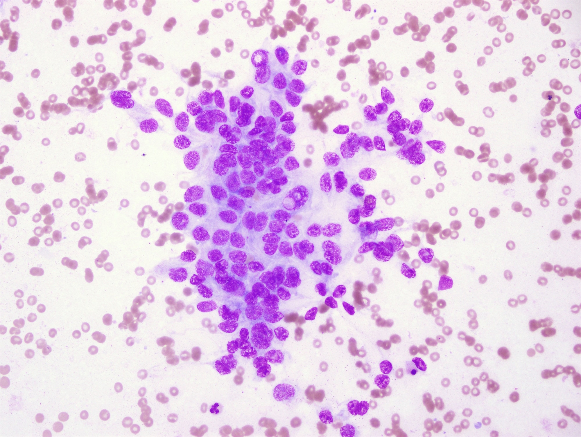
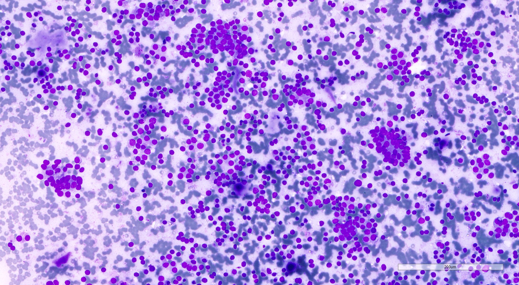
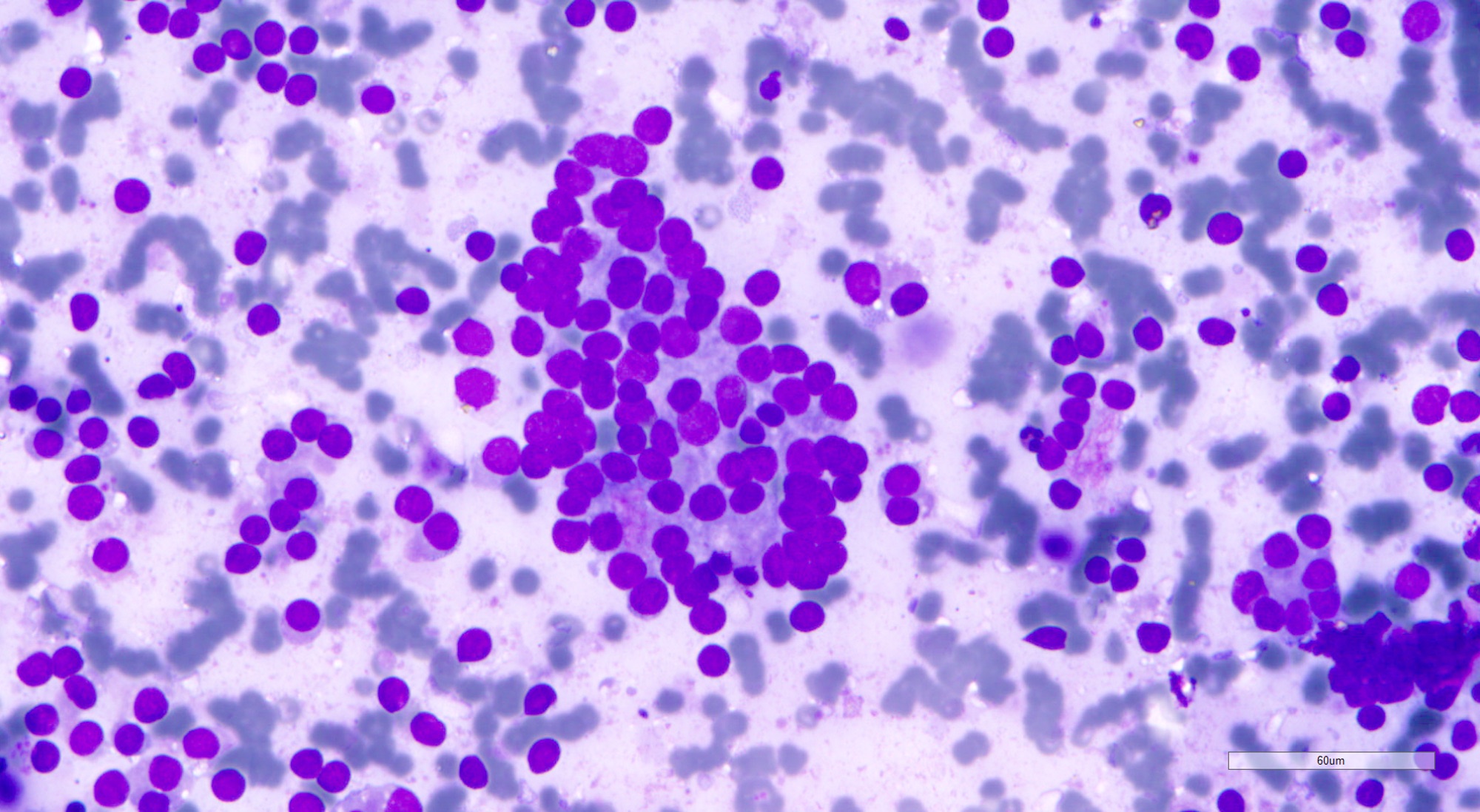
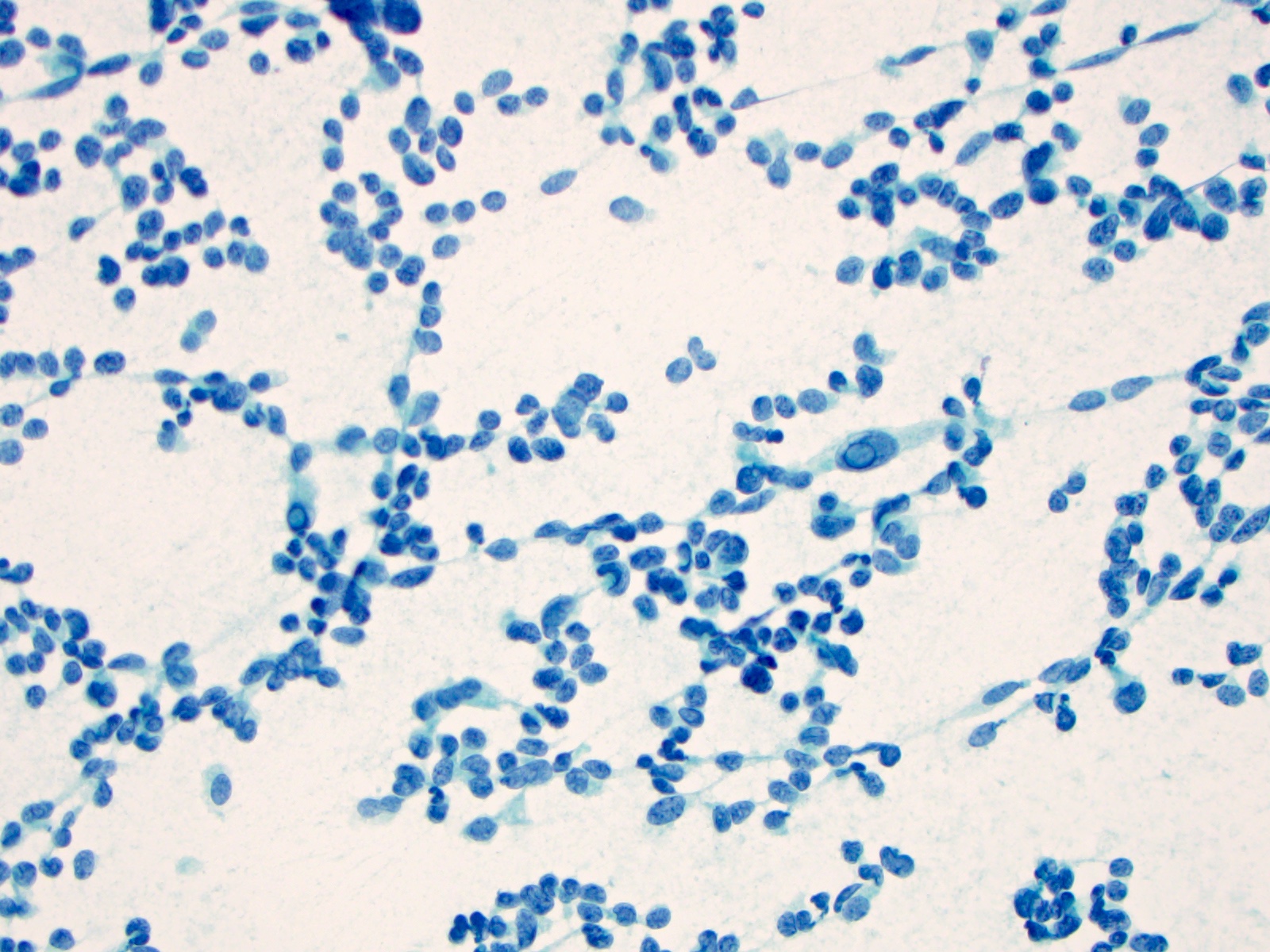
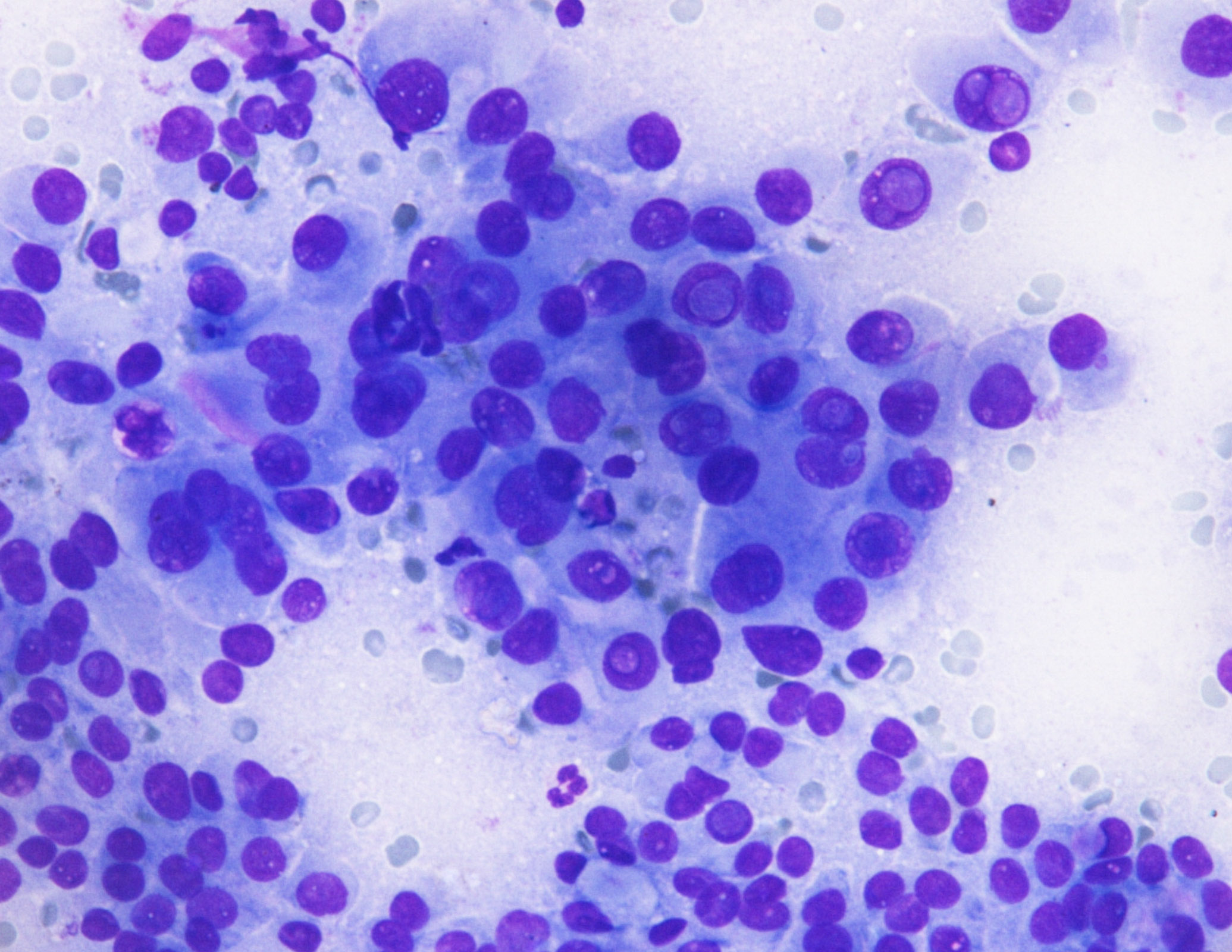
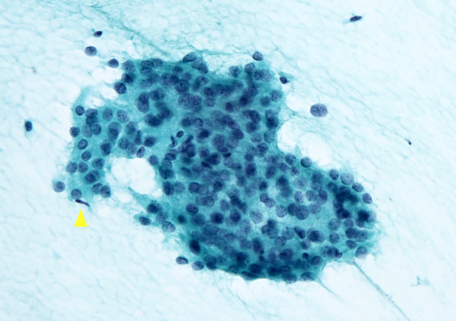
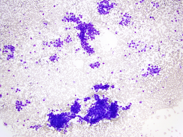
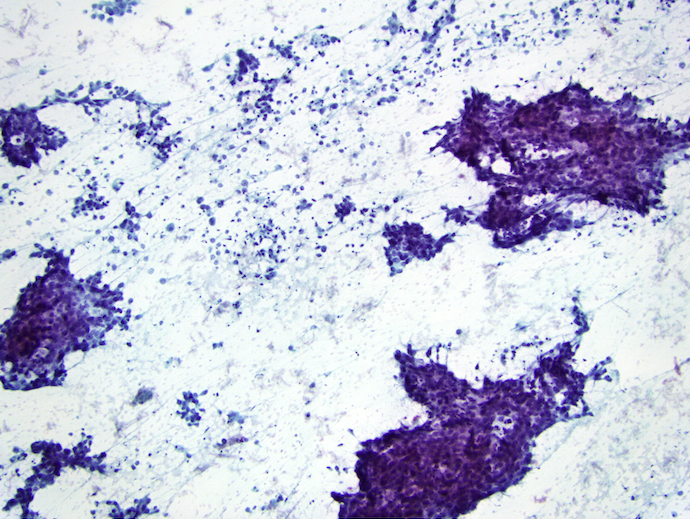
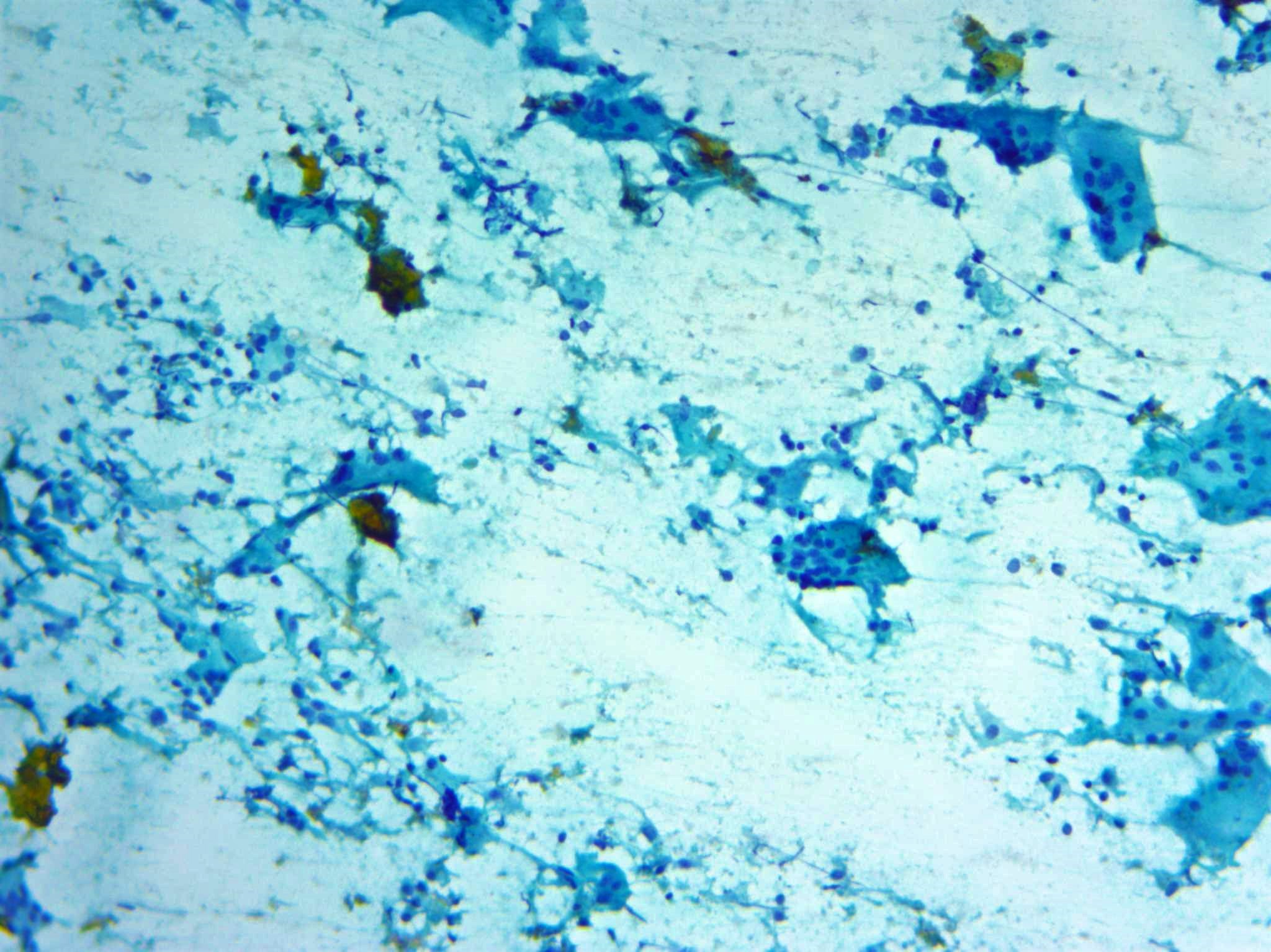
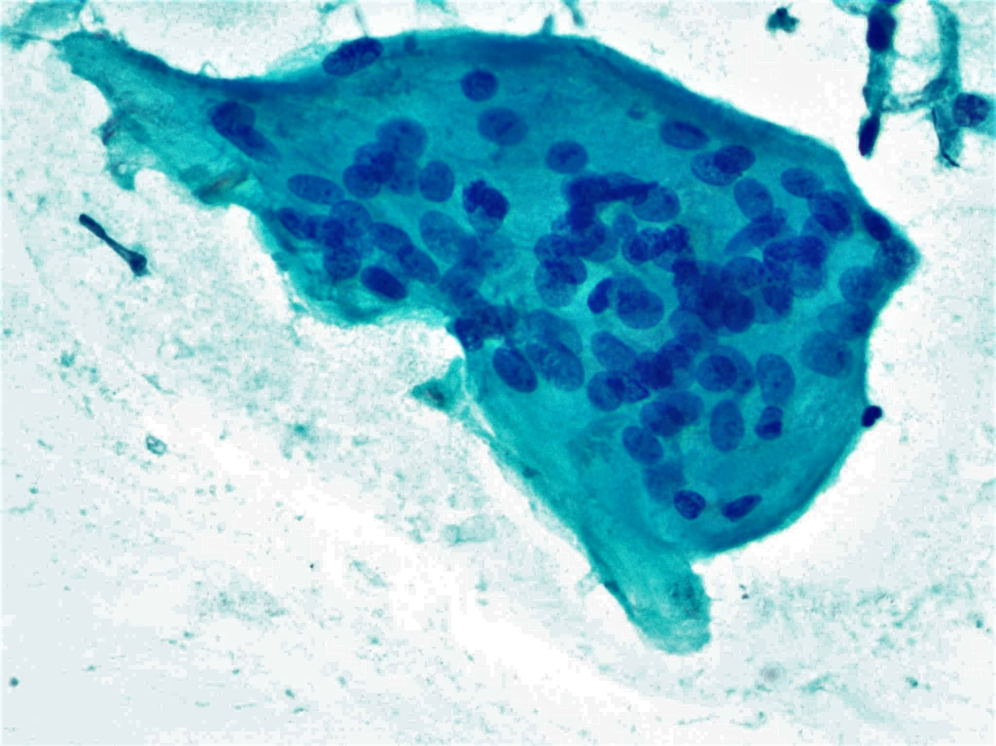
Contributed by Ayana Suzuki, C.T. and Shuanzeng Wei, M.D., Ph.D.

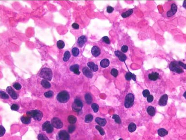
Salt and pepper chromatin
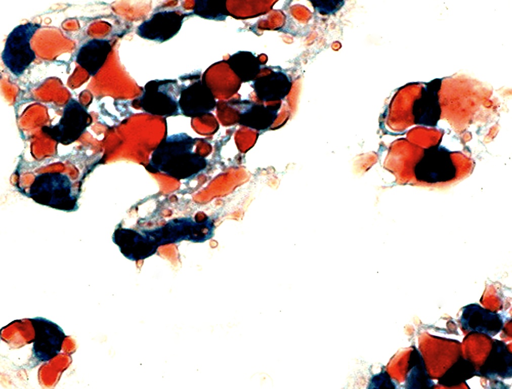
Cellular specimen
(Diff-Quik stain)

Plasmacytoid tumor cells
(Diff-Quik stain)

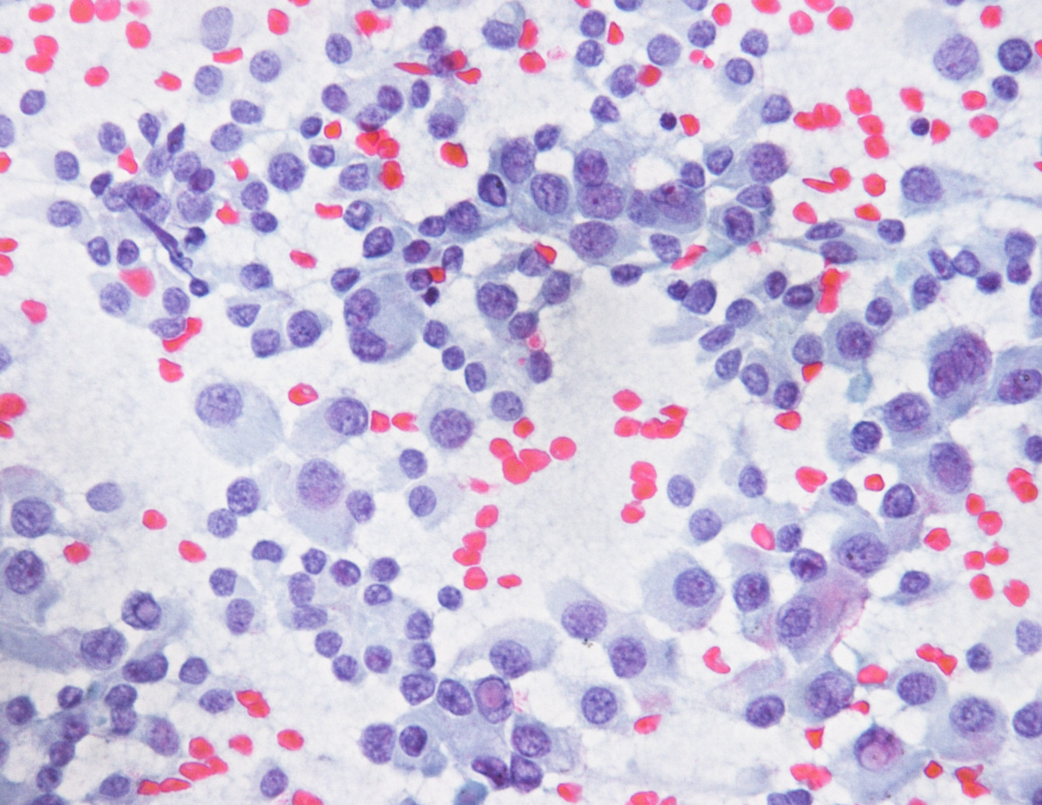
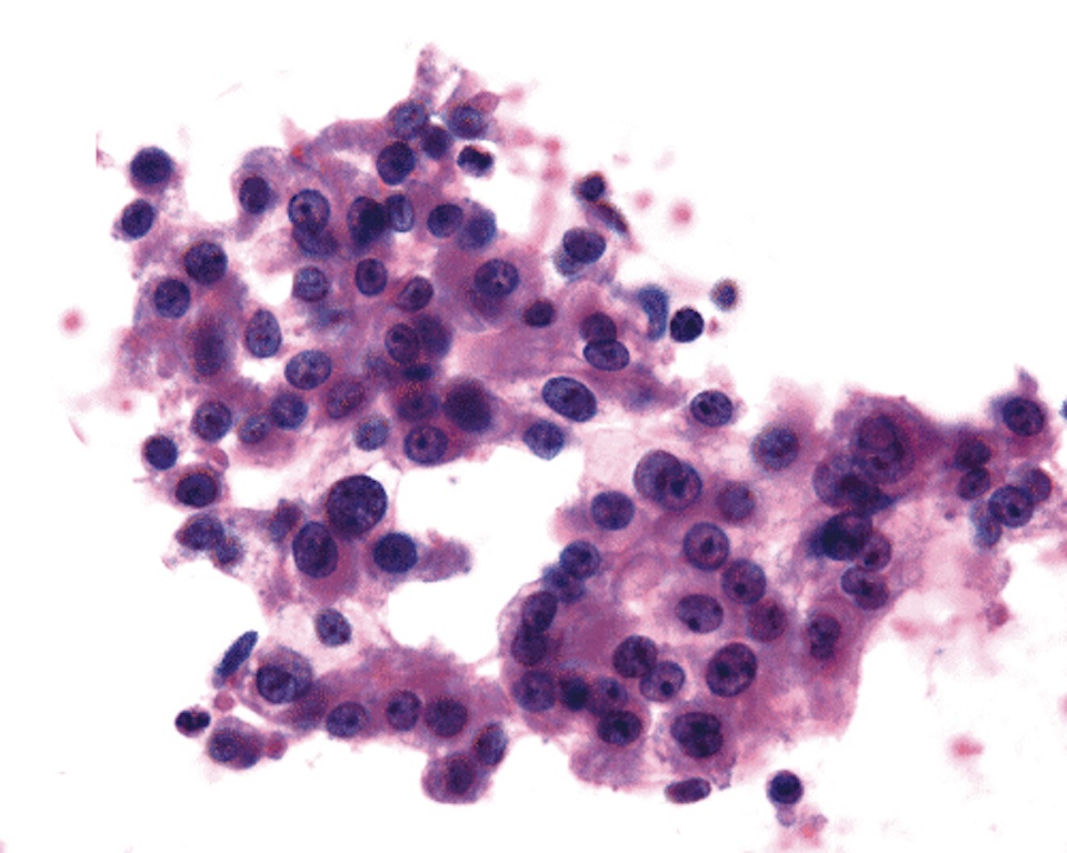
Variable sized plasmacytoid tumor cells
(Pap stain)

Tumor cells with intranuclear pseudoinclusions (Pap stain)
Diff-Quik
Pap
Cell block
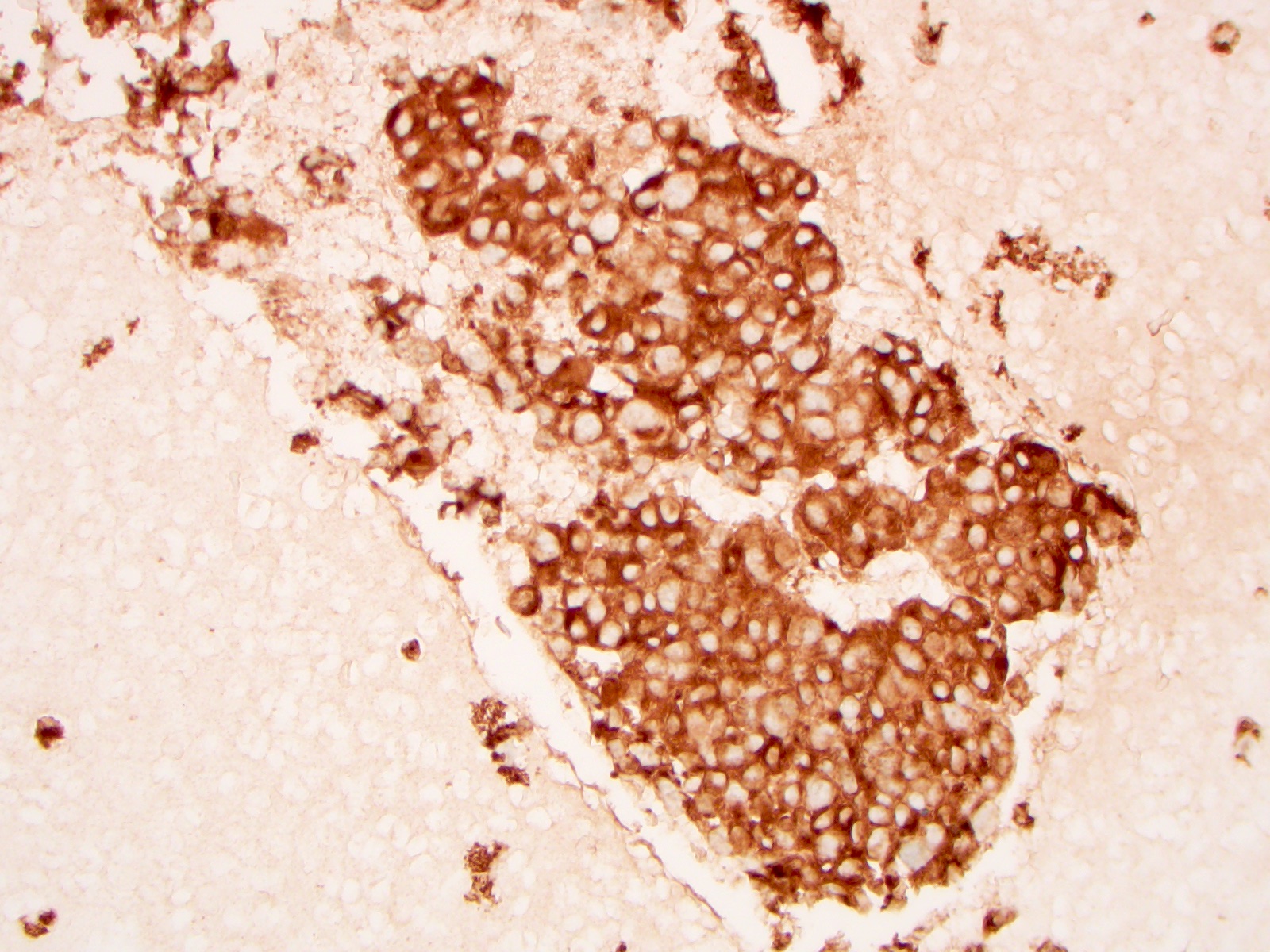
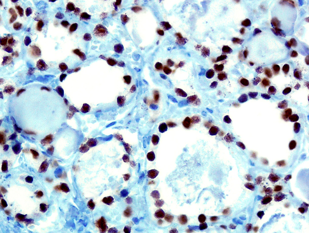
Calcitonin
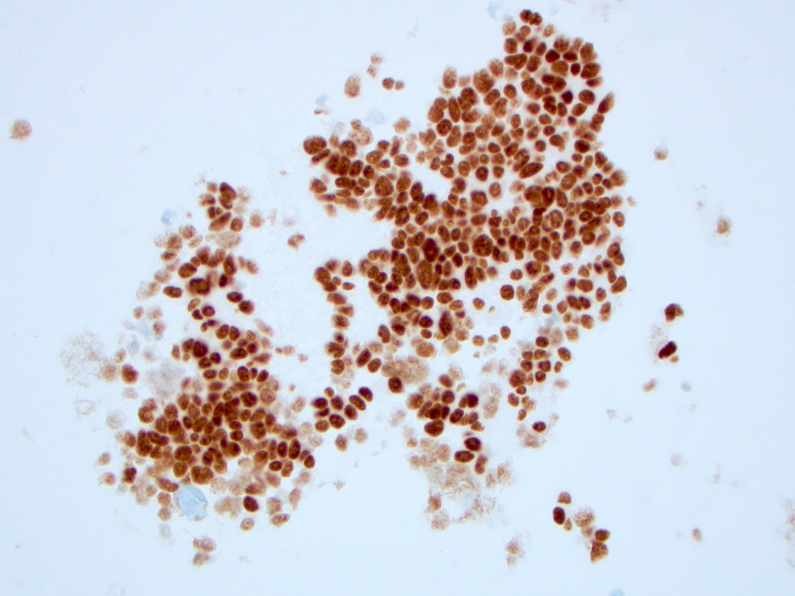
TTF1
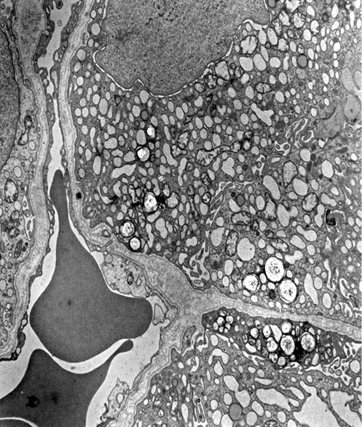
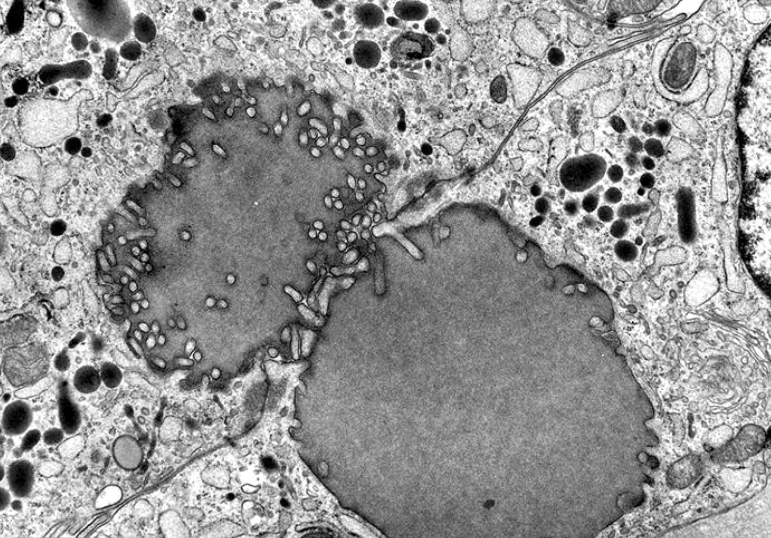
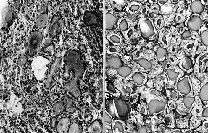
AFIP images

Amyloid deposits

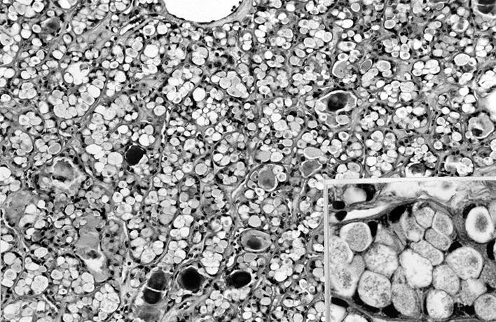
Numerous large type I granules in some cells

Tumor cells with sparse granules
Medullary thyroid carcinoma FNA
Medullary thyroid carcinoma
Images hosted on other servers:

Incidence and prognosis
Images hosted on other servers:

Ultrasound
AFIP images

Well circumscribed tumor of < 2 mm
Small irregular fibrotic tumor
Contributed by Nadine Demko, M.D.C.M., M.Sc., Livia Florianova, M.D., M.Sc. and Marc Pusztaszeri, M.D.
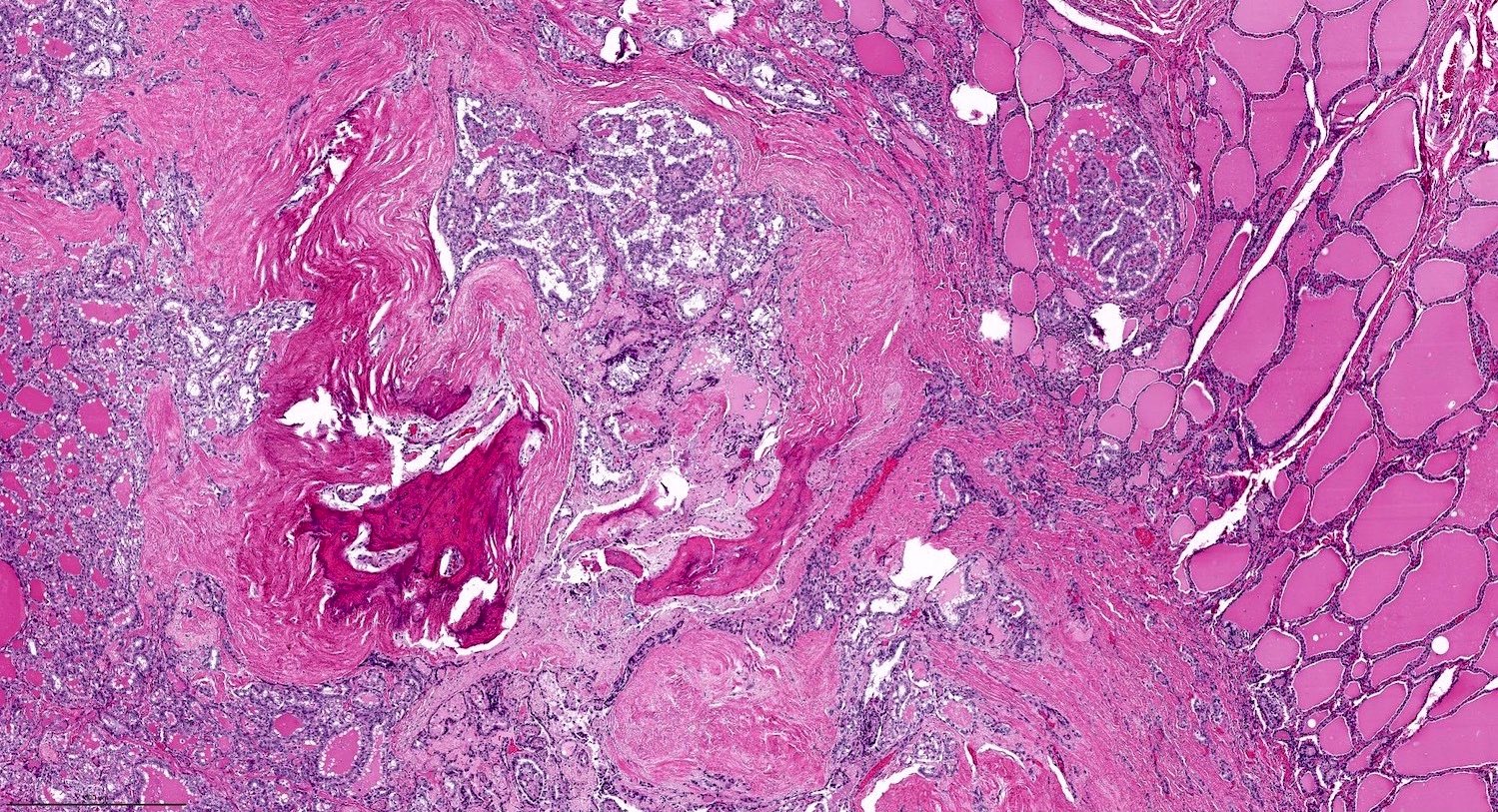
Follicular architecture
Nuclear features
Irregular contour with dystrophic calcification
Nuclear features
Infiltrative pattern
Tall cell features
Chronic thyroiditis background
Follicular architecture
Nuclear features
Tall cell features
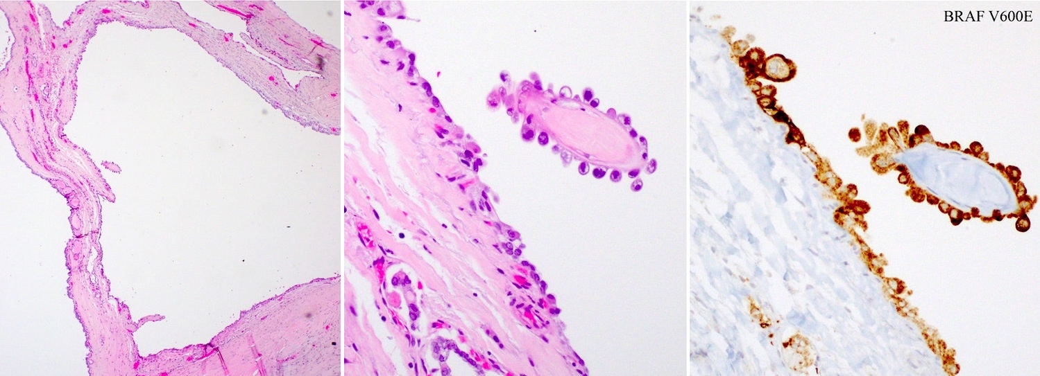
Minimal extrathyroidal extension

Infiltrative pattern
Nuclear features

BRAF V600E
Contributed by Andrey Bychkov, M.D., Ph.D.
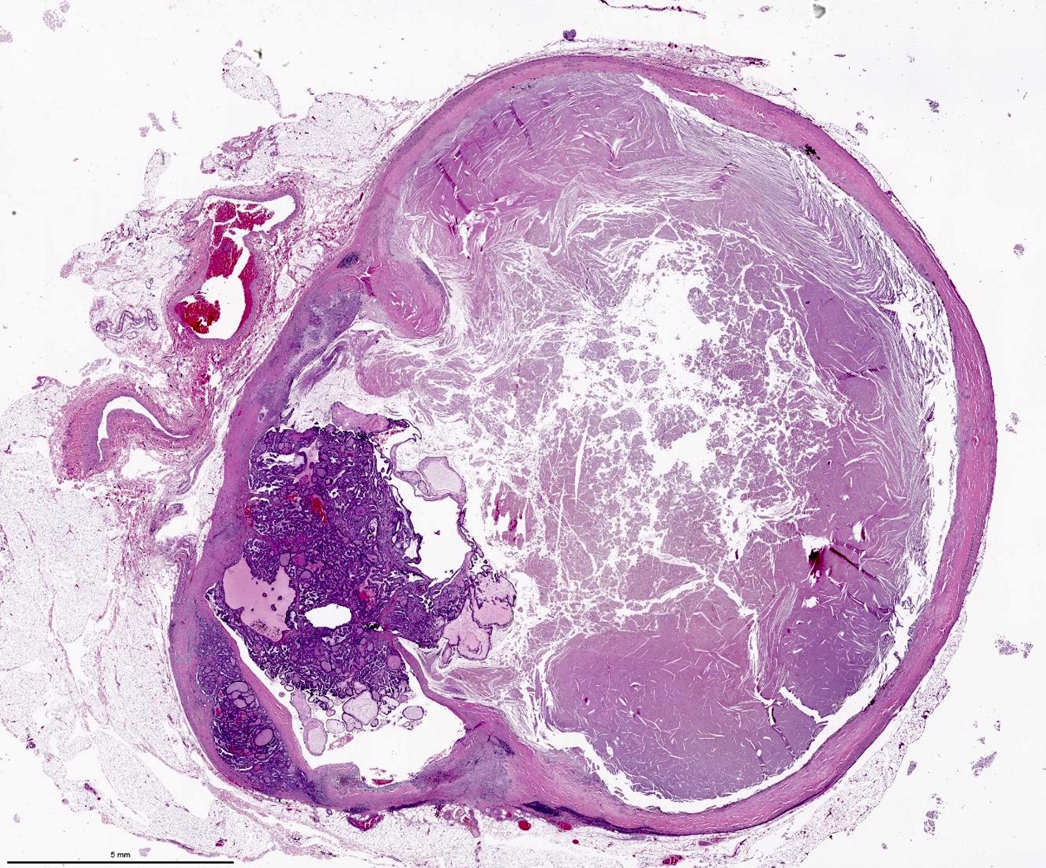
Incidental finding in multinodular goiter
Encapsulated papillary microcarcinoma
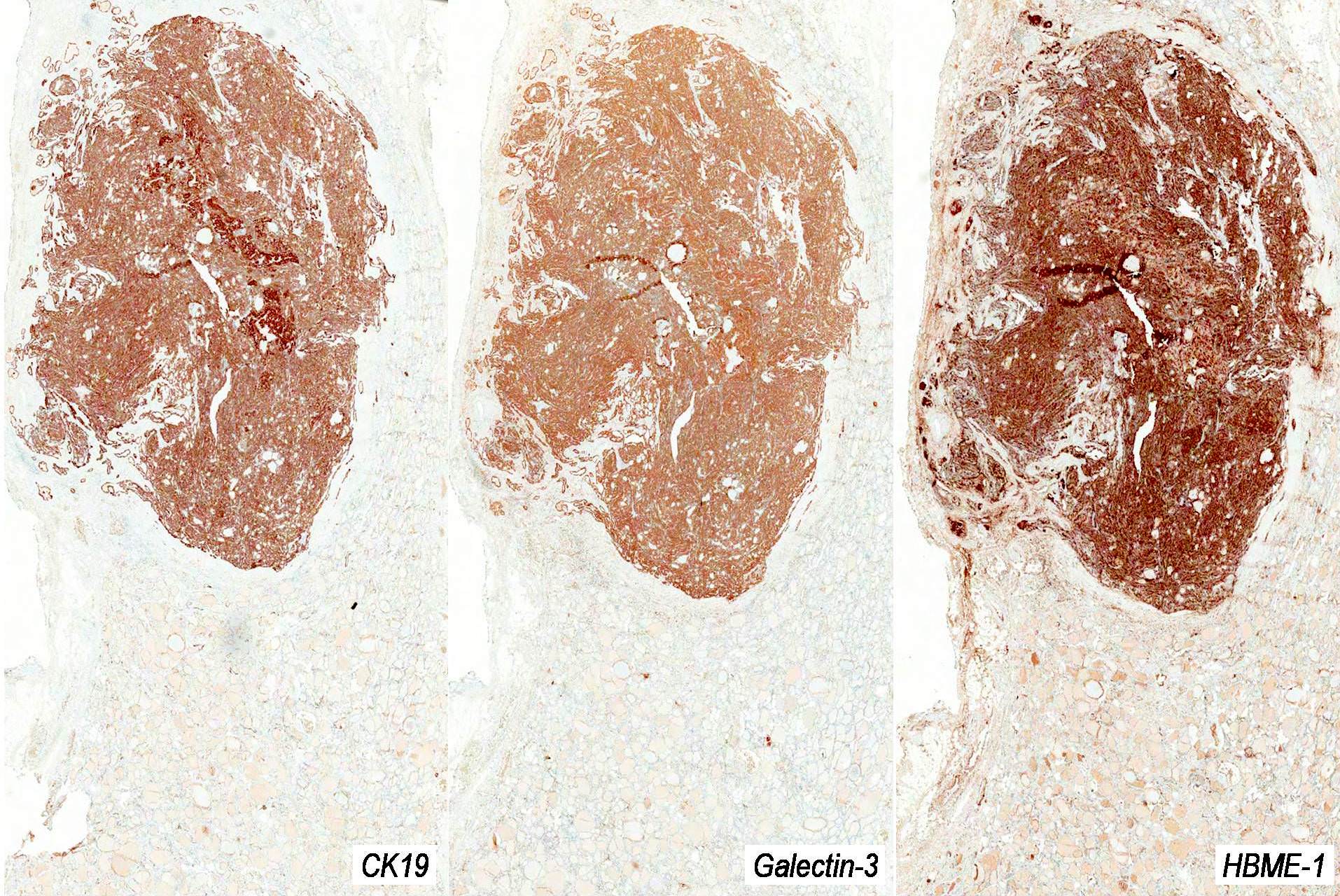
CK19, galectin3 and HBME1
Contributed by Nadine Demko, M.D.C.M., M.Sc., Livia Florianova, M.D., M.Sc. and Marc Pusztaszeri, M.D.
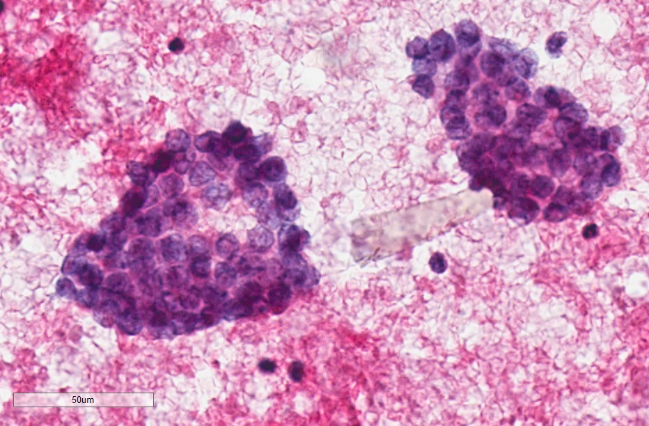
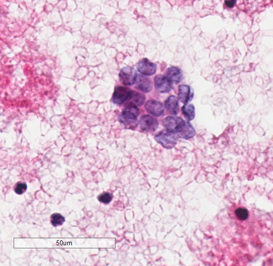
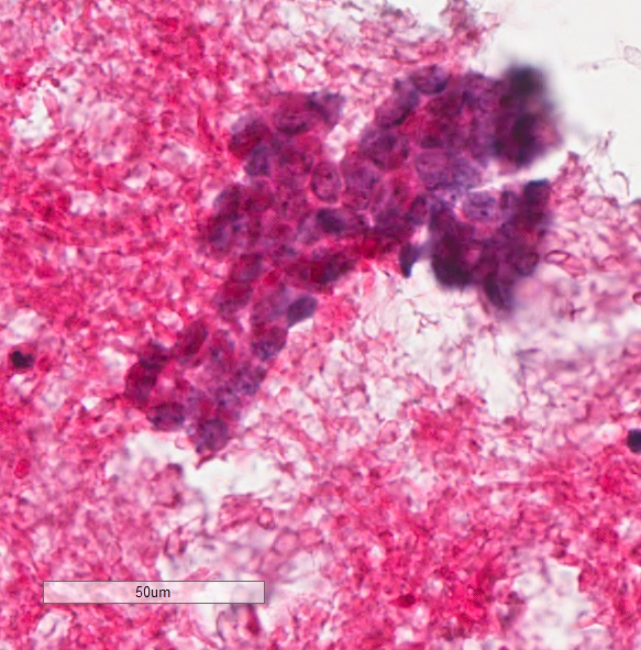
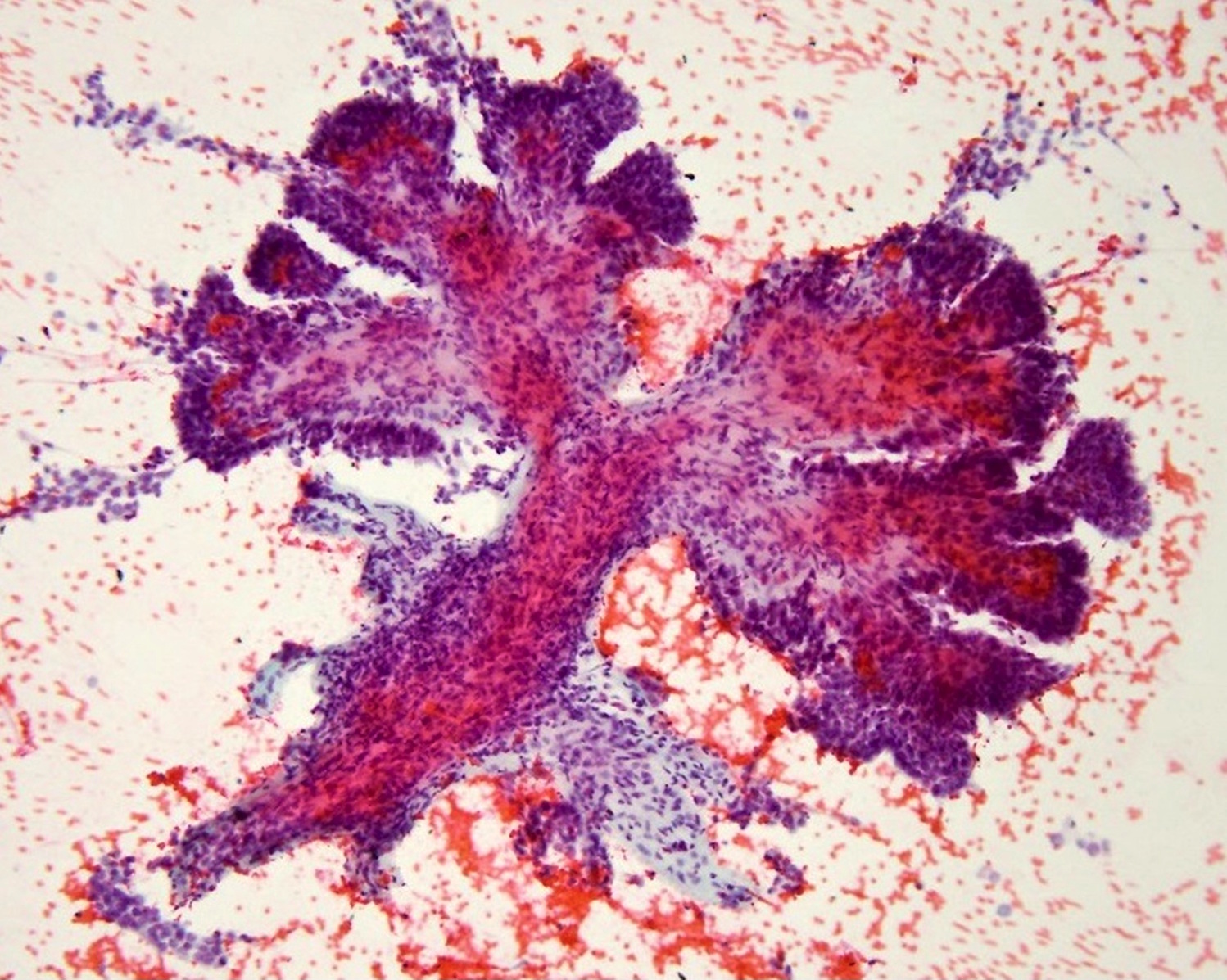
Cytological features
Contributed by Grace C.H. Yang, M.D.
Pap stained slides
Active surveillance
AFIP images
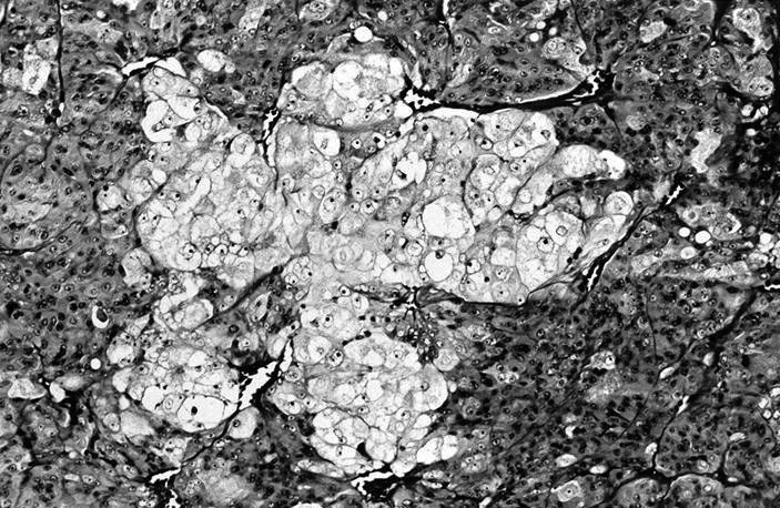
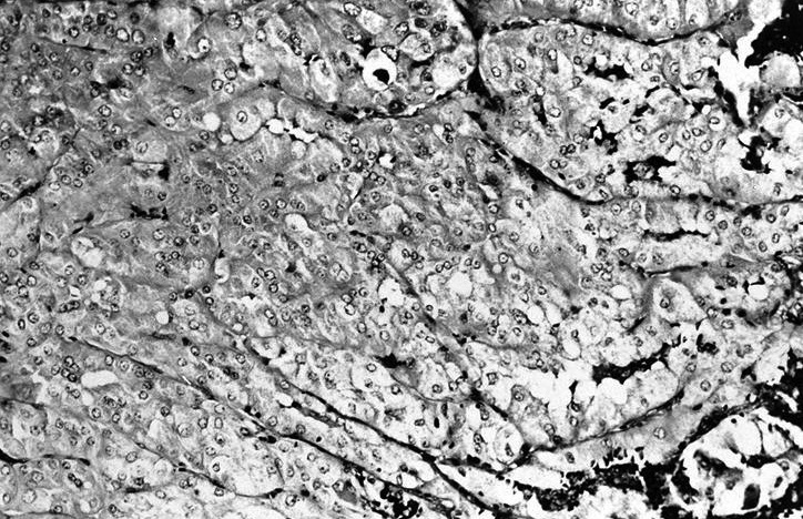
Tumor cells show lobular growth pattern
Scattered early follicle-like areas
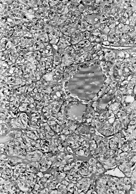
Tumor cells form colloid containing follicles
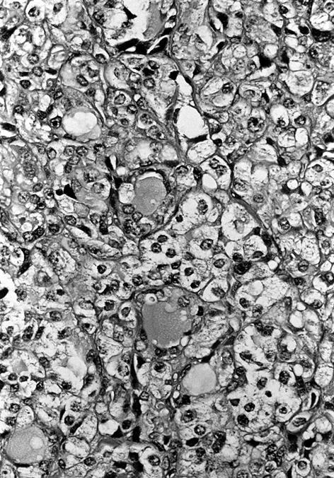
Many cells have clear cytoplasm
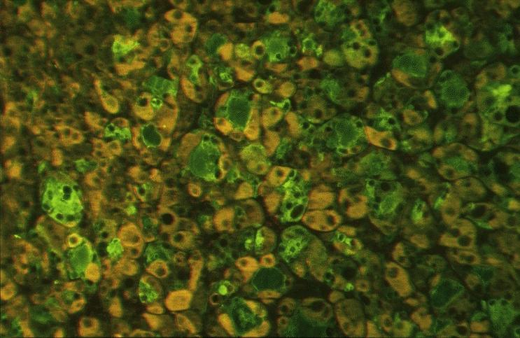
Immunofluorescent staining
Images hosted on other servers:


Molecular pathways in thyroid cancer development

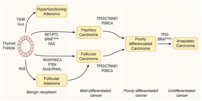

Stepwise mechanism of thyroid neoplasia

Progress in identifying
mutational markers
in thyroid cancer


MAPK pathway

PI3K-AKT pathway


Genomic landscape of PTC

Gene mutations in thyroid tumors

Gene fusions in PTC

BRAF vs. RAS in PTC

BRAF V600E and tumor recurrence

BRAF mutation and iodine uptake

TERT mutations

miRNAs in thyroid cancer

Drug targets in thyroid cancer
Images hosted on other servers:


ALK fusions

β-catenin (nuclear) in cribriform-morular PTC associated with FAP syndrome
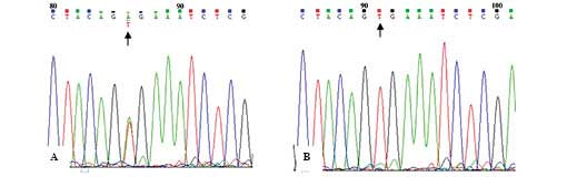
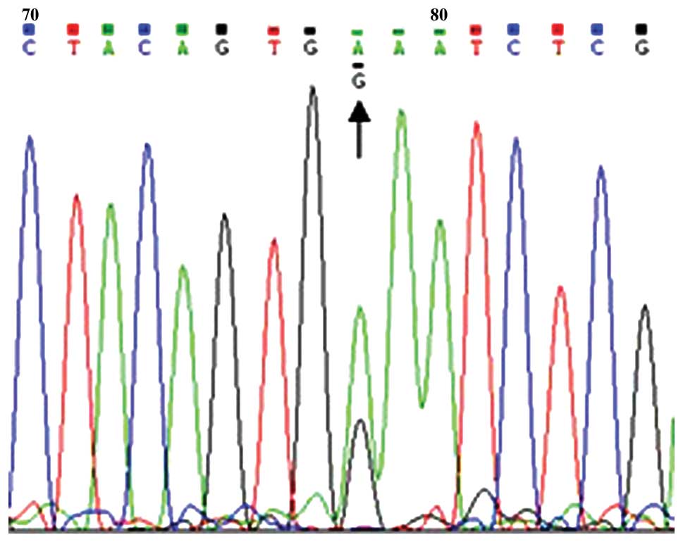
BRAF V600E (sequencing)

BRAF V600E (MALDI-TOF)
BRAF K601E
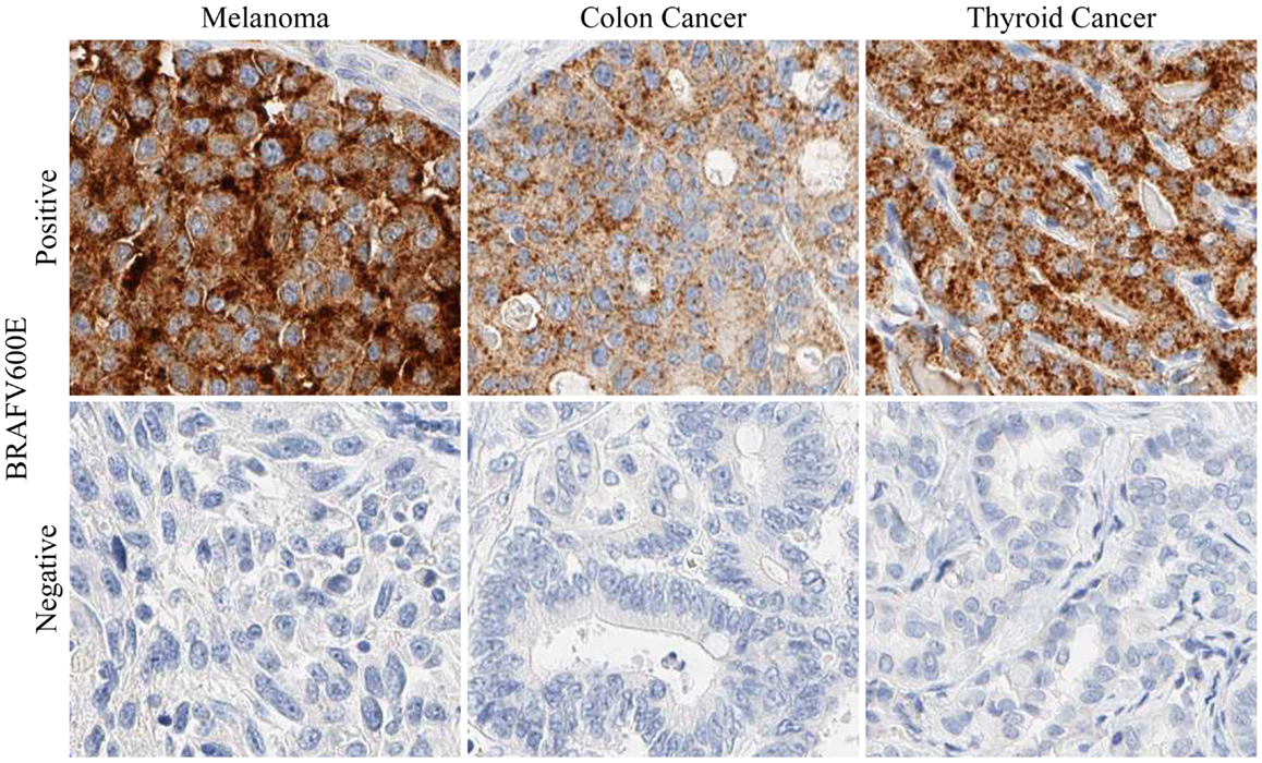
BRAF V600E / VE1

KRAS mutations
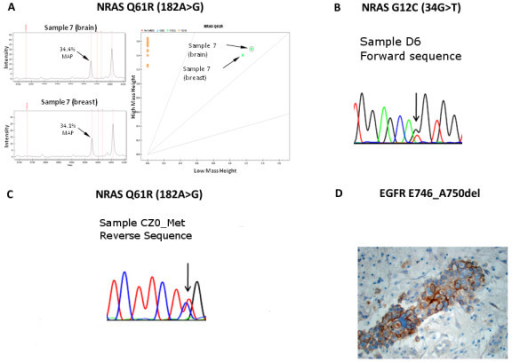
NRAS mutations (sequencing)

NRAS mutations (pyrosequencing)

PPARγ IHC in FTC (A)

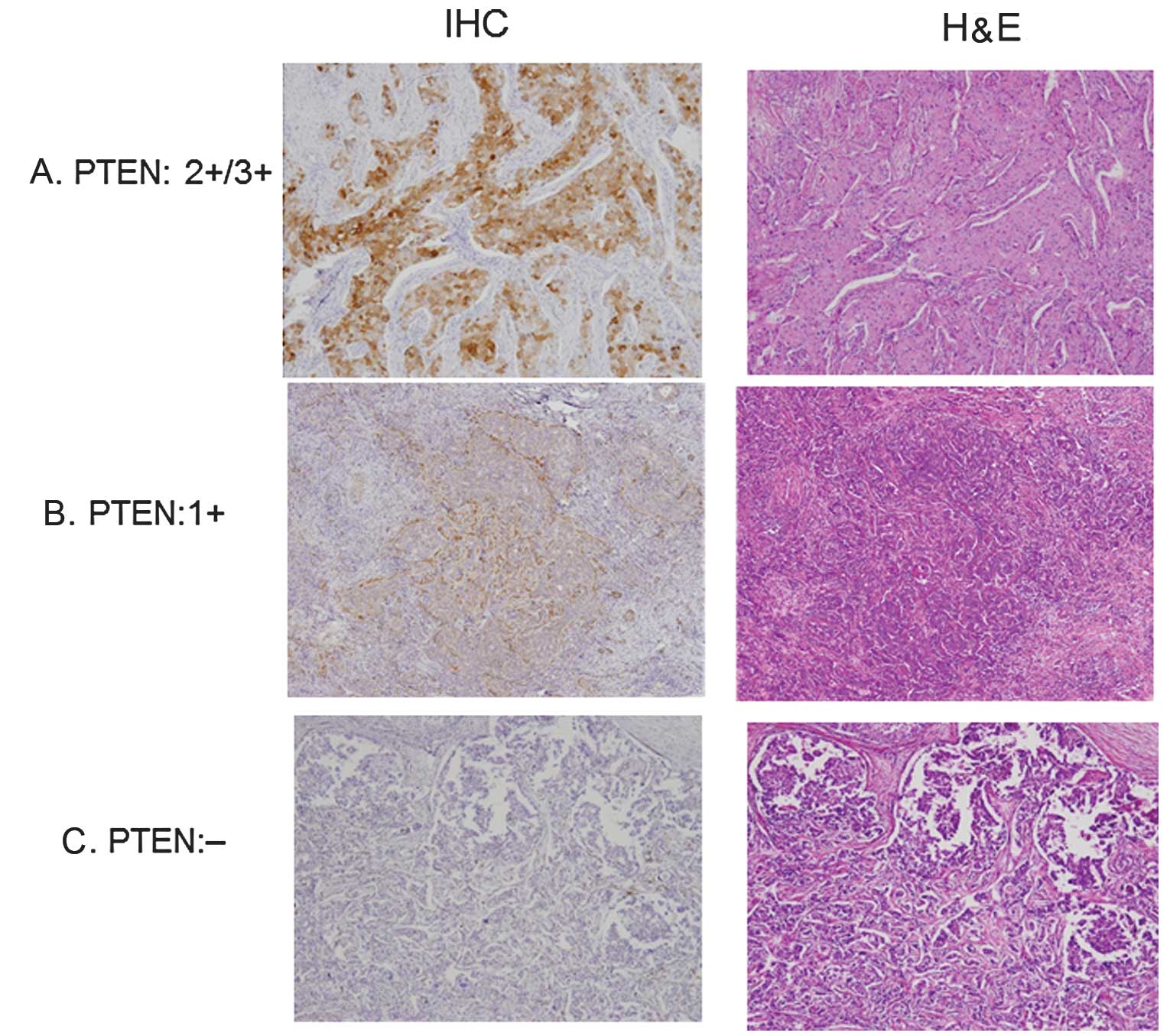
PTEN IHC (A - C)

PTEN loss in Cowden syndrome
PTEN evaluation (breast cancer)
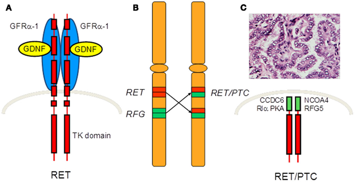
RET / PTC rearrangements


TERT promoter mutations
Genetics and genomics of thyroid neoplasms (2013) by Dr. Electron Kebebew, NCI
Integrated genomic characterization of papillary thyroid carcinoma (2014) by Prof. Tom Giordano, University of Michigan
Advances in molecular pathogenesis of thyroid cancer (2014) by Prof. Pilar Santisteban, Universidad Autónoma de Madrid
Molecular influence in thyroid cancer (2014) by The American Head and Neck Society
Understanding the biology of medullary thyroid cancer (2015) by Prof. Gilbert Cote, MD Anderson Cancer Center
Molecular targeted therapeutics for medullary thyroid cancer (2015) by Dr. Ann Gramza, NCI
MAPK pathway
MAPK pathway
PI3K-AKT pathway
PI3K-AKT pathway
PI3K-AKT pathway
Images hosted on other servers:

Progress in identifying
mutational markers
in thyroid cancer

Gene mutations in thyroid tumors


Genomic landscape of PTC

Gene fusions in PTC

BRAF mutation and iodine uptake

BRAF V600E and tumor recurrence
TERT mutations
Drug targets in thyroid cancer
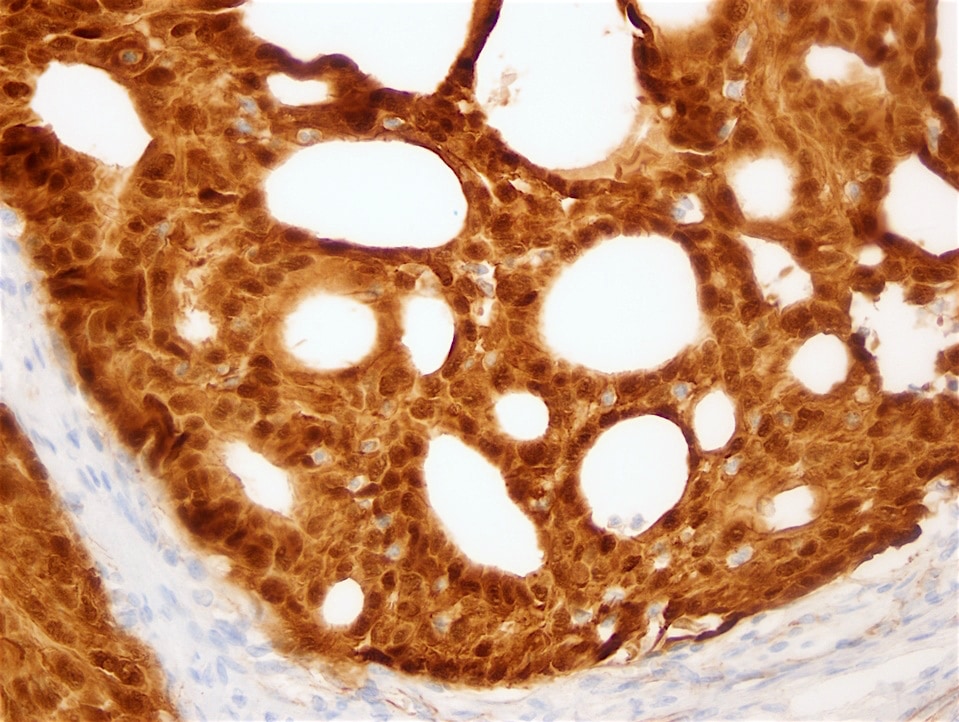
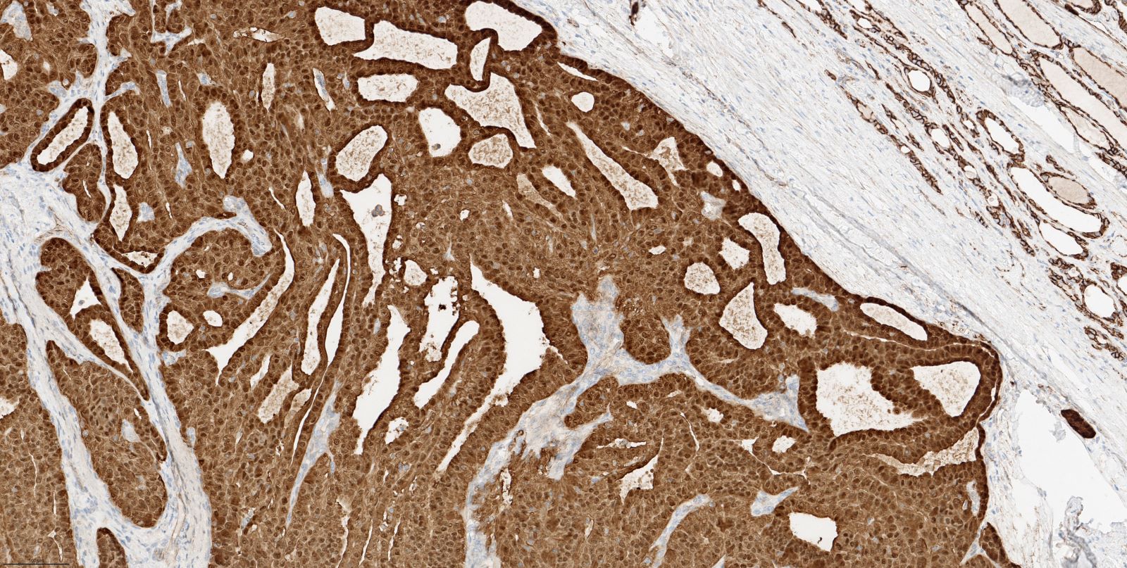
Contributed by Andrey Bychkov, M.D., Ph.D.
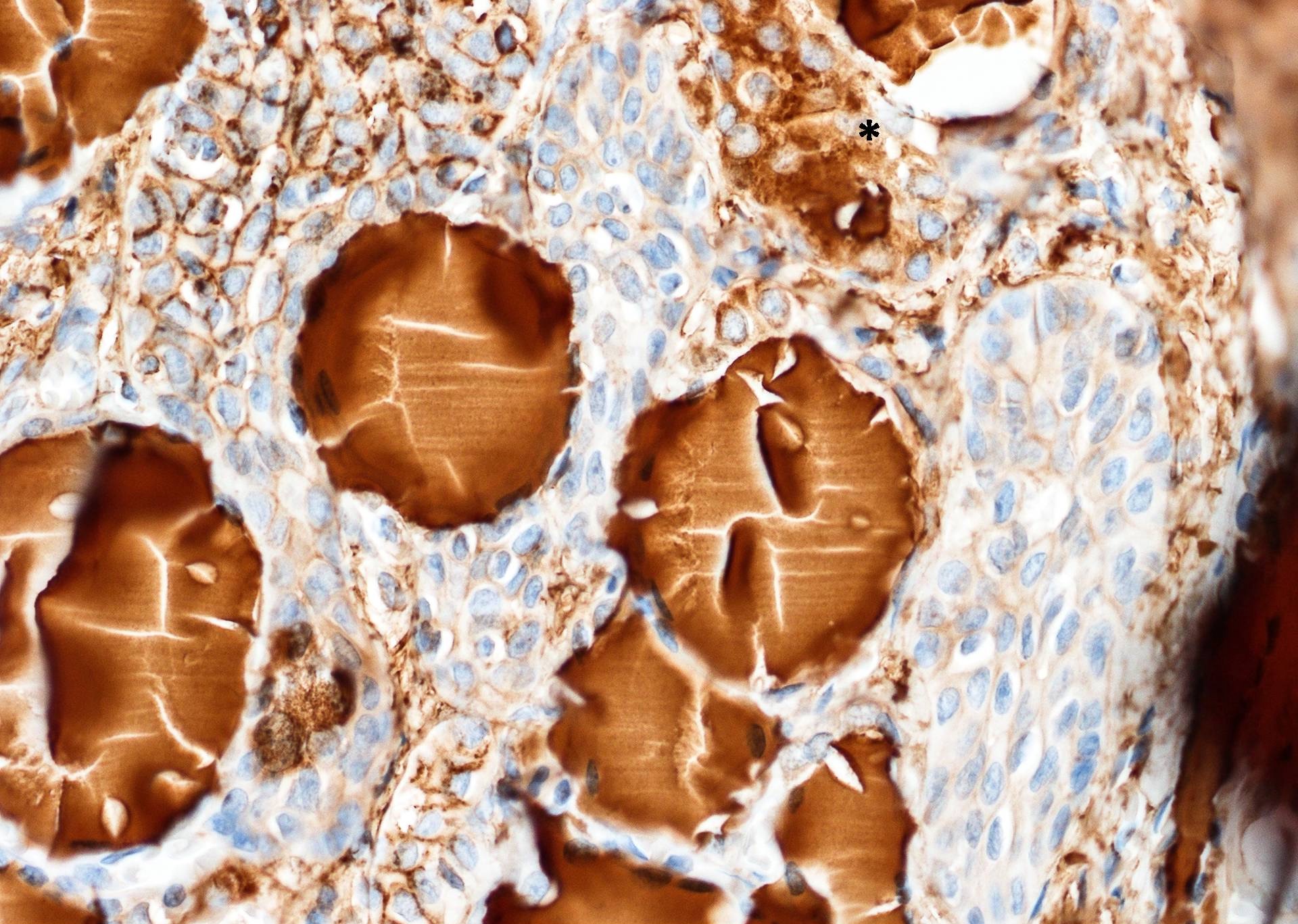
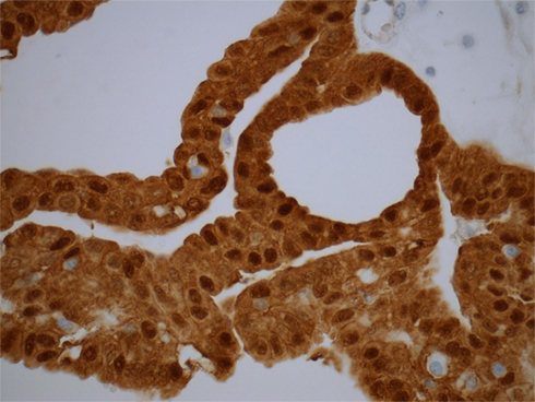
V600E mutant protein is diffusely expressed in tumor / cancer, but not normal tissue

Diffuse cytoplasmic staining
Images hosted on other servers:


ALK fusions

β-catenin (nuclear) in cribriform-morular PTC associated with FAP syndrome
BRAF V600E (sequencing)
BRAF V600E (MALDI-TOF)
BRAF K601E
BRAF V600E / VE1
KRAS mutations
NRAS mutations (sequencing)
NRAS mutations (pyrosequencing)
PPARγ IHC in FTC (A)
PTEN IHC (A - C)
PTEN loss in Cowden syndrome
PTEN evaluation (breast cancer)
RET / PTC rearrangements
TERT promoter mutations
Molecular influence in thyroid cancer (2014)
Biomarkers in thyroid cancer (2015)
Drugs in development for refractory thyroid cancer (2015)
Molecular targeted therapeutics for medullary thyroid cancer (2015)
Images hosted on other servers:

Overview of platforms and workflow

Proposed clinical algorithm

NPV and PPV of ThyroSeq v2.1

Genetics of thyroid tumors

Diagnostic performance
ThyroSeq testing by Y. Nikiforov (2020)
ThyGenX / ThyraMIR panel by Alidad Mireskandari (2018)
Images hosted on other servers:

Large tumor with
internal hypodensity
lesion
Images hosted on other servers:

White tan cut surface
AFIP images
Solid nest with mucin producing foci
Microcystic pattern of mucin producing cells
Images hosted on other servers:


Partially solid, glandular and papillary

Squamoid nests and
mucin secreting
component

Transition from follicular
variant of papillary carcinoma
to mucoepidermoid carcinoma

Immunohistochemistry
of TTF1 and
thyroglobulin
Contributed by David Poller, M.D.
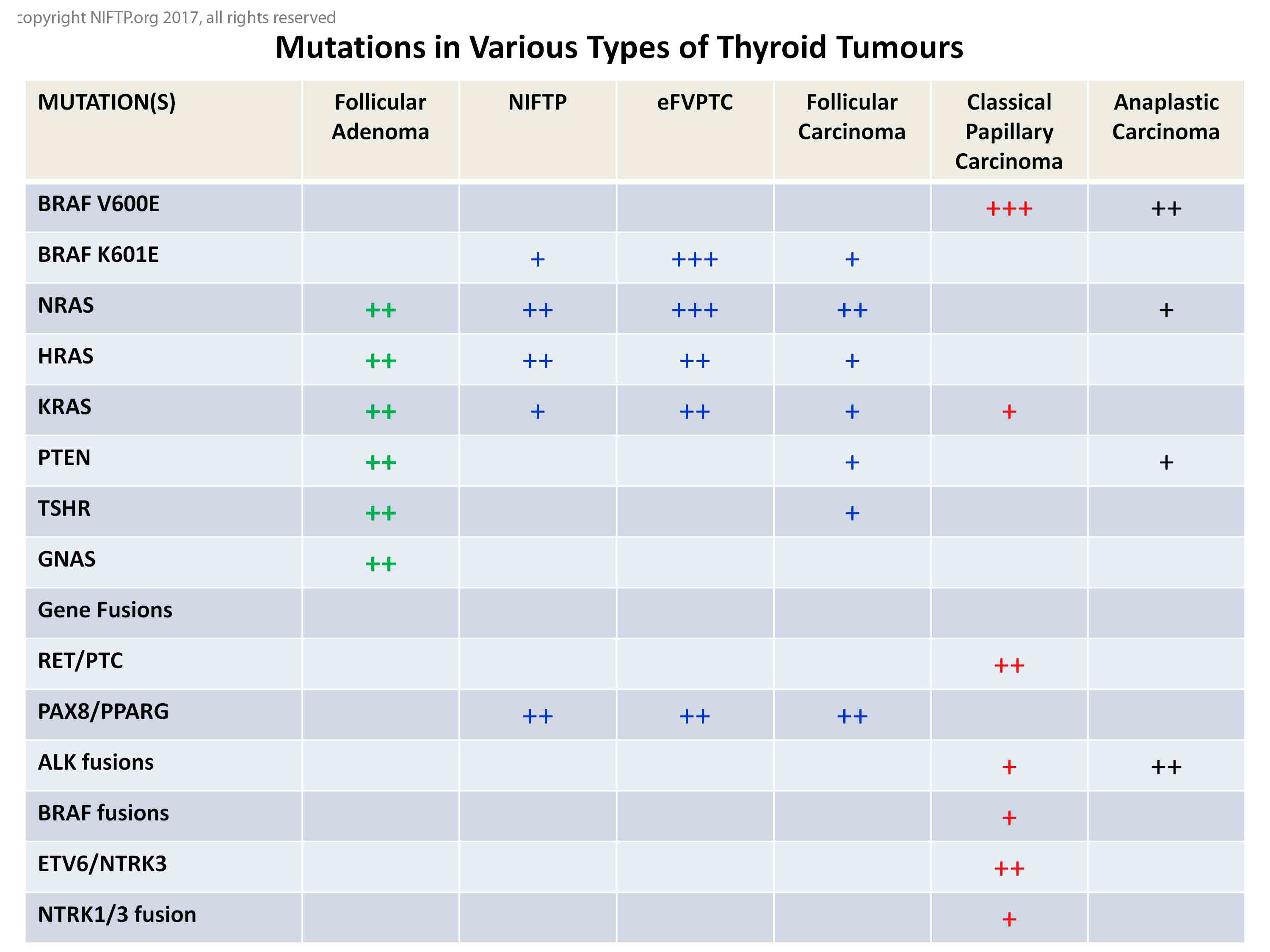
Mutations in various types of thyroid tumors
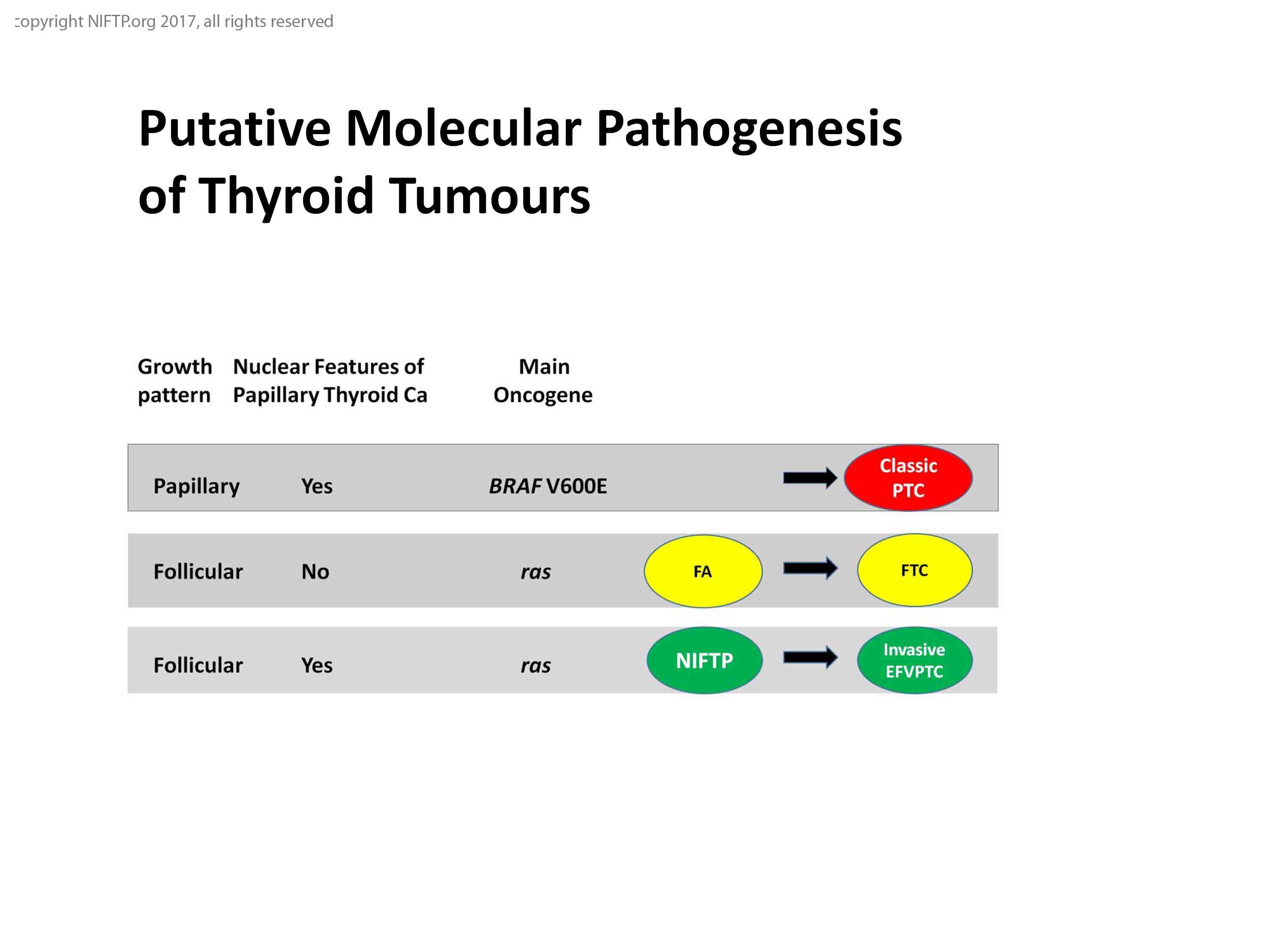
Putative molecular pathogenesis of thyroid tumors
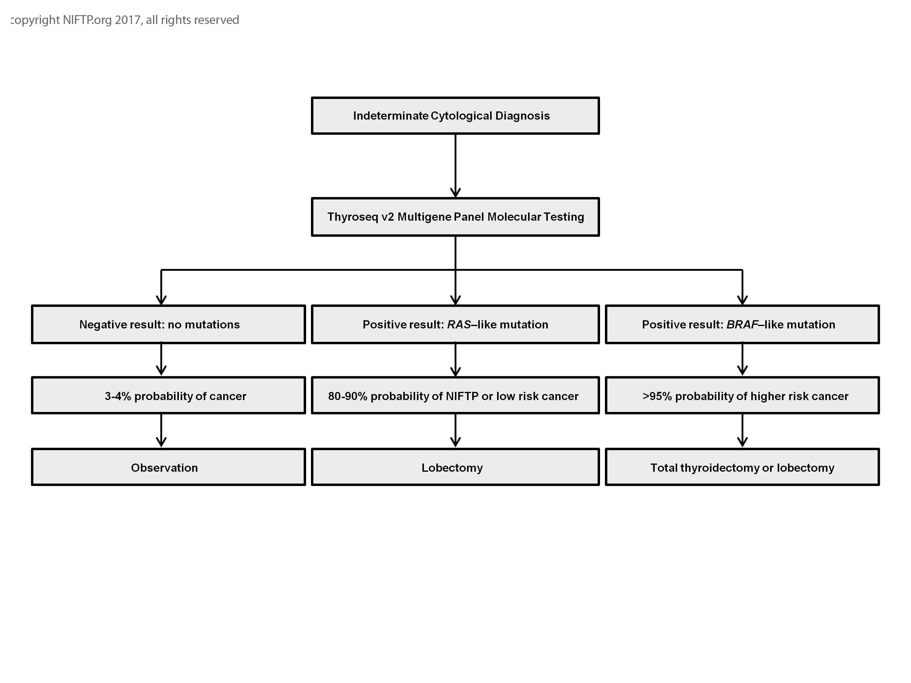
Indeterminate cytological diagnosis
Images hosted on other servers:
NIFTP incidence
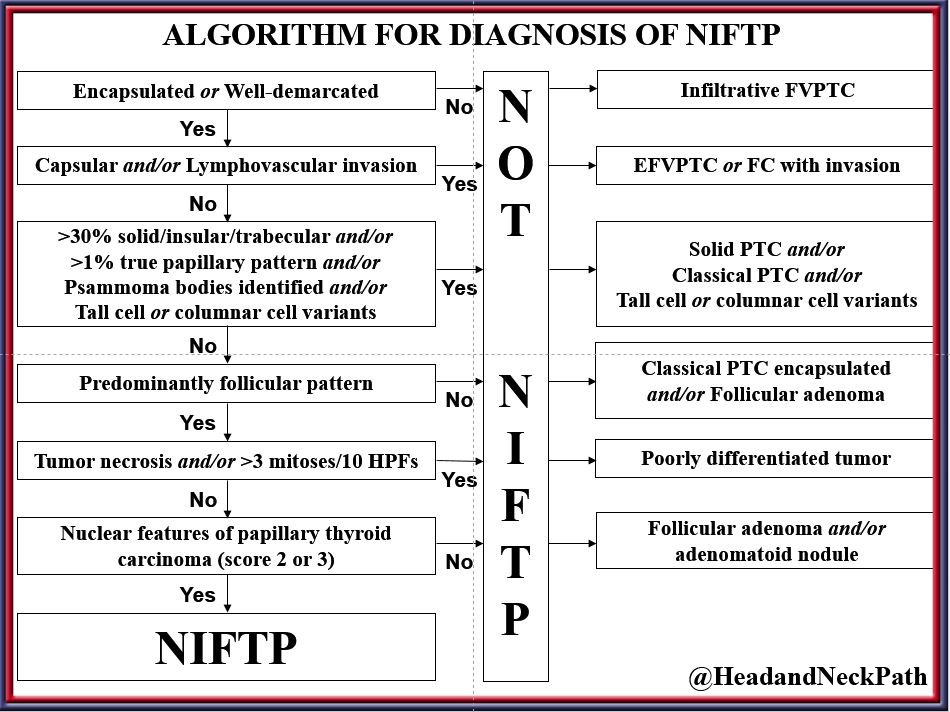
Diagnostic algorithm

History of NIFTP
Images hosted on other servers:


Ultrasonography
Images hosted on other servers:

Gross and histology of EFVPTC
Contributed by Andrey Bychkov, M.D., Ph.D. and Rachel Jug, M.B.B.Ch.

Circumscribed follicular patterned nodule
Tumor interface with a thin capsule

Microfollicular pattern
Major and minor diagnostic features
Dark colloid with scalloping and clefting

Free floating tumor fragment in vessel
Small piece of tumor floats in vascular lumen

Processing artifact with distorted nuclei

Distorted nuclei due to artifact

Vesicular-like nuclei due to tissue degeneration

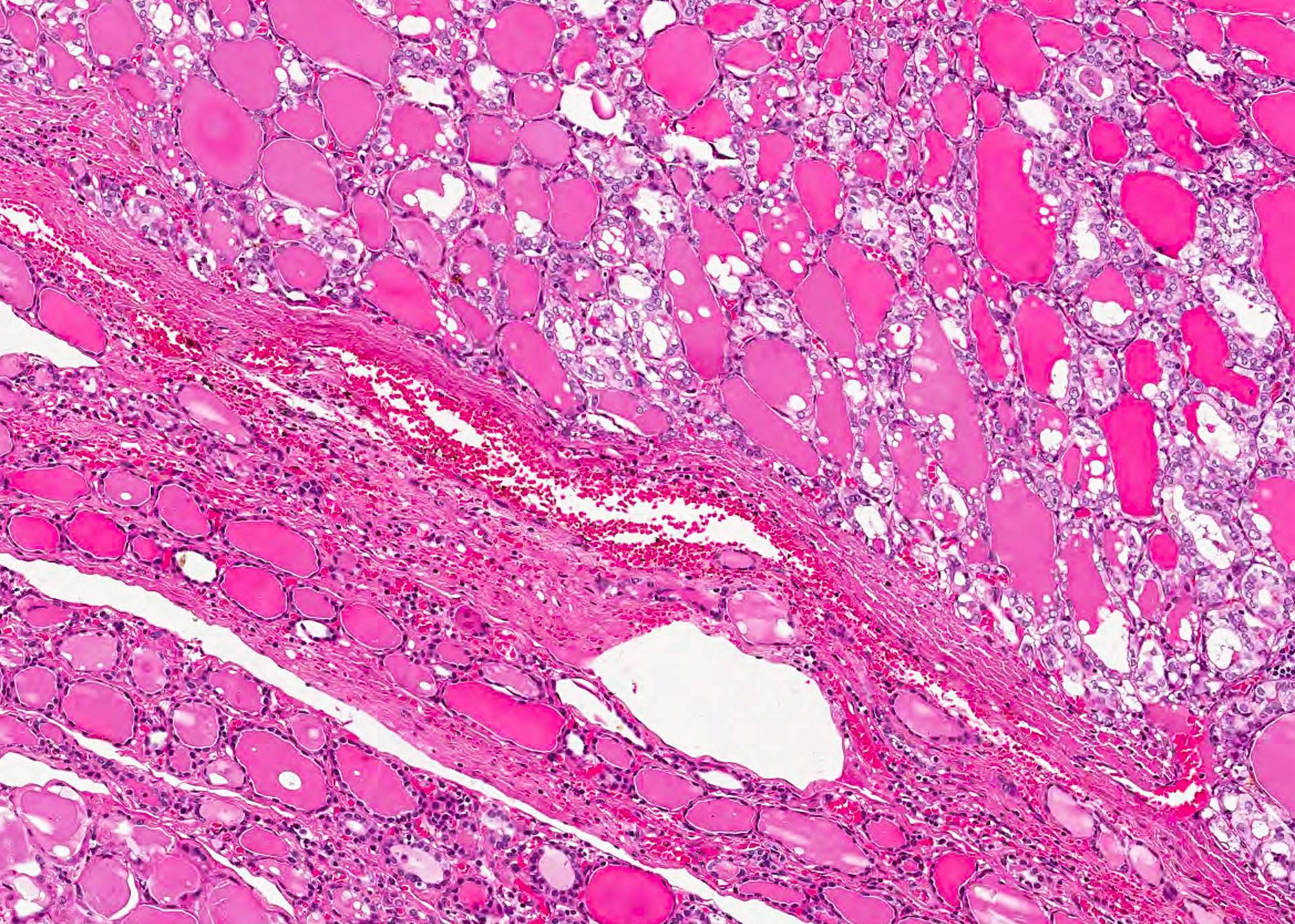
Clear circumscription of the encapsulated lesion with a predominantly follicular growth pattern
Clear circumscription of the encapsulated lesion with a predominantly follicular growth pattern

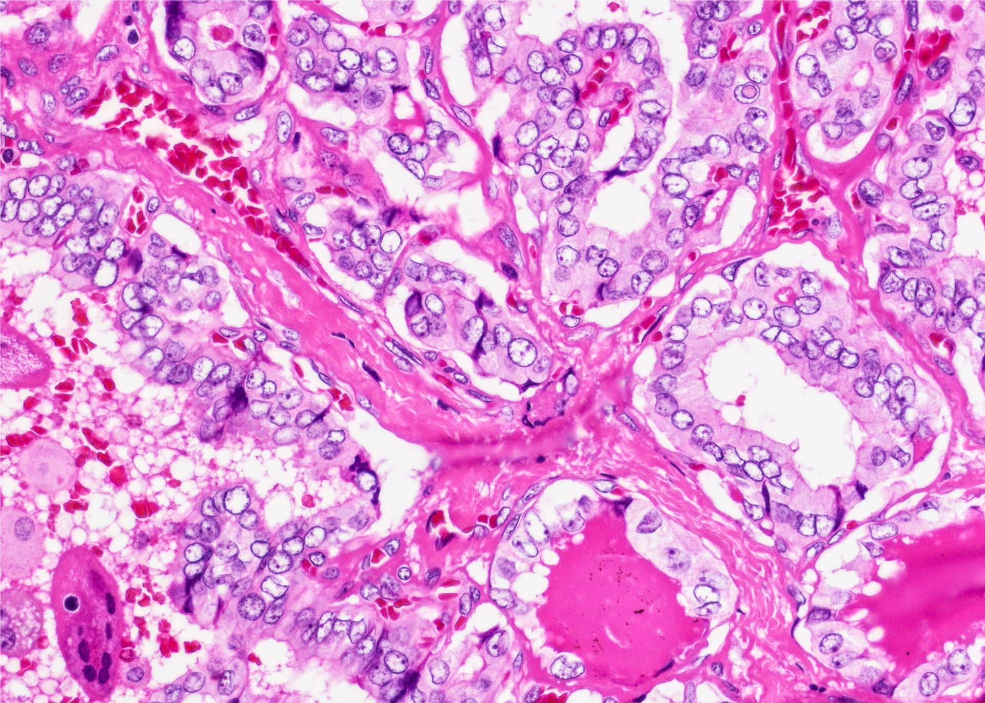
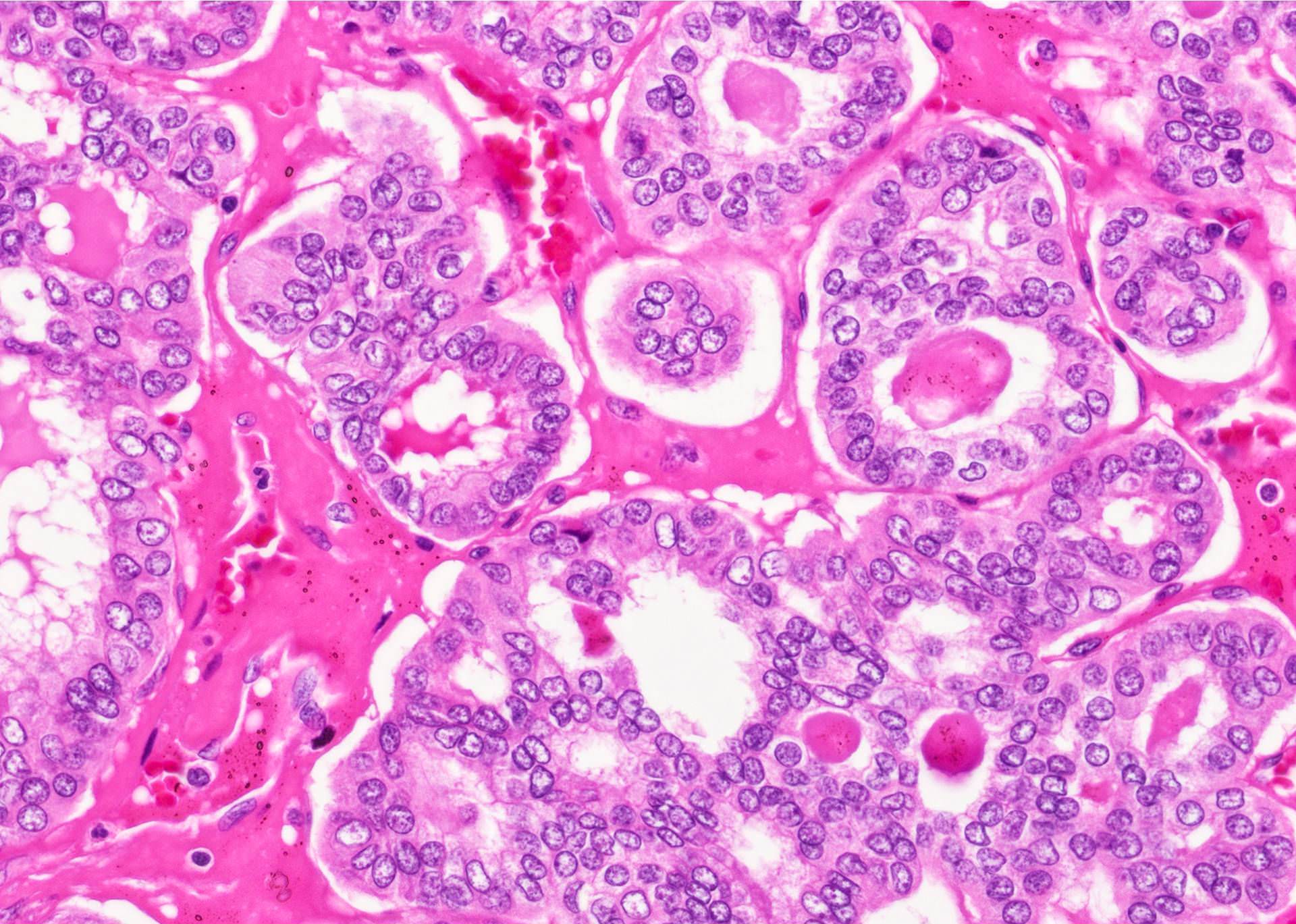
Nuclear features of PTC
Images hosted on other servers:

Noninvasive follicular thyroid neoplasm

Low power

Medium power

5.6 cm NIFTP


High power
NIFTP vs HCC

NIFTP vs PDTC

NIFTP vs FTC
NIFTP vs FVPTC


Encapsulated follicular patterned lesion


Nuclear features
Images hosted on other servers:


Microfollicular pattern, nuclear enlargement
Microfolliclar pattern
Microfollicles, nuclear grooves
Enlarged nuclei
Nomenclature change for thyroid tumors: NIFTP (2017) by Prof. Yuri Nikiforov, University of Pittsburg
NIFTP by R. Ghossein (2020)
Diagnostic dilemmas in NIFTP by N. Cipriani (2020)
Images hosted on other servers:

Ultrasound
Contributed by Andrey Bychkov, M.D., Ph.D.

Nucleoli and pseudoinclusions
Oncocytic cells with pseudoinclusions
Contributed by Shipra Agarwal, M.D.

Loose clusters of oncocytic cells

Predominant oncocytic cells
Intranuclear inclusions
Contributed by Mark R. Wick, M.D. and AFIP

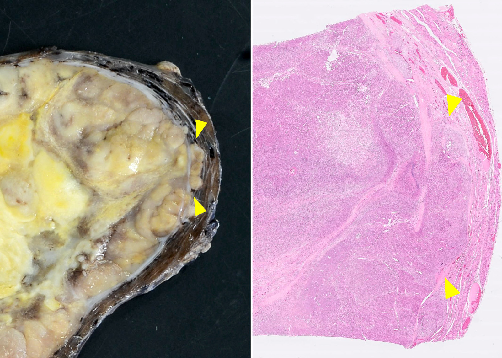
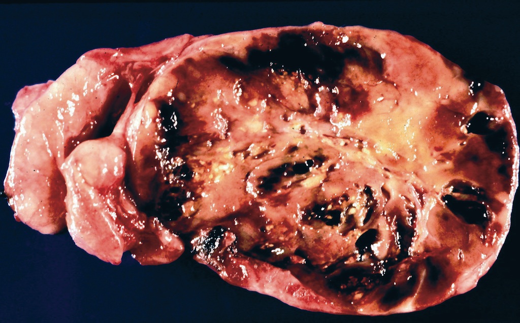
Carcinoma
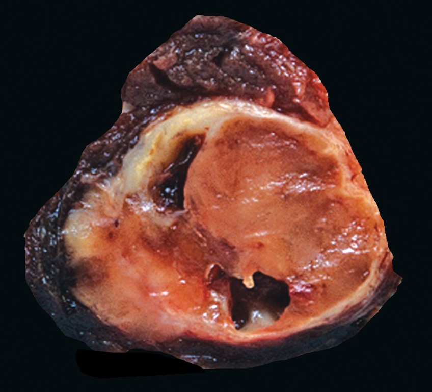
Adenoma with massive infarct
Carcinoma has focal capsular invasion
Contributed by Shuanzeng Wei, M.D., Ph.D., Andrey Bychkov, M.D., Ph.D., Grace C.H. Yang, M.D. and Mark R. Wick, M.D.
Vascular invasion

Follicular thyroid carcinoma (oncocytic variant)
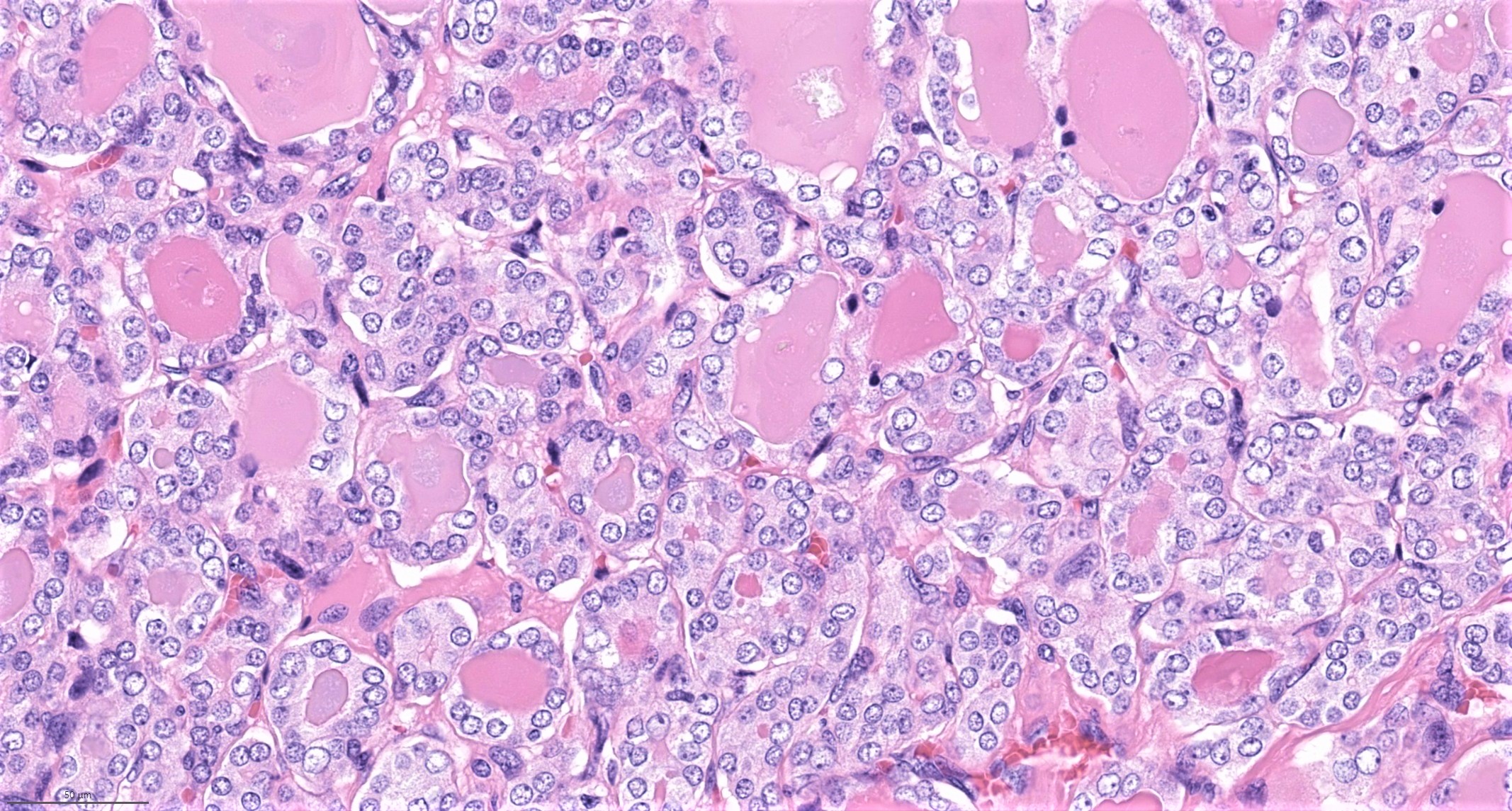
Oncocytes
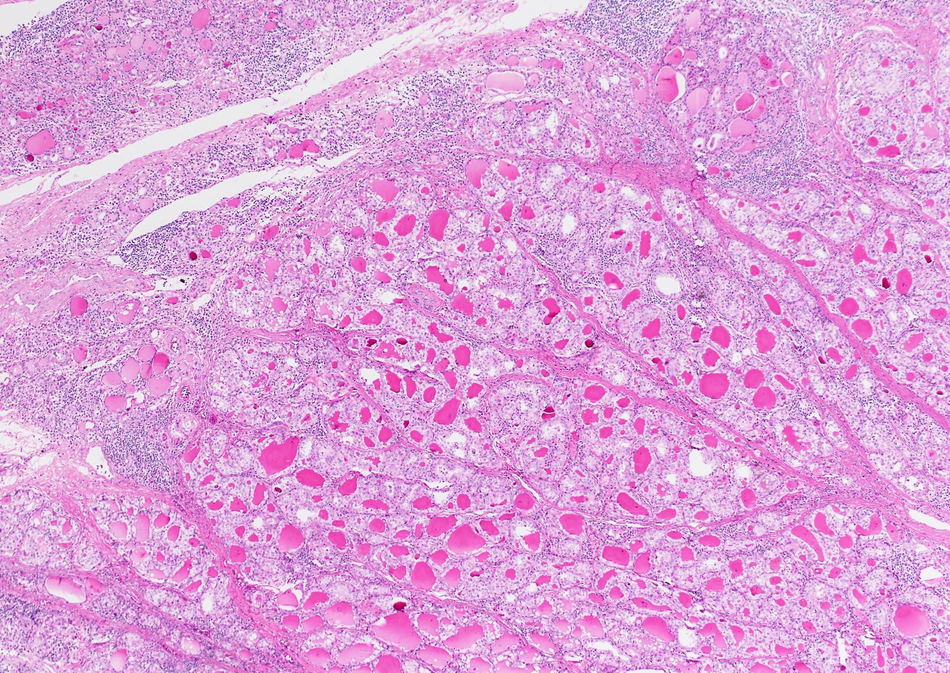
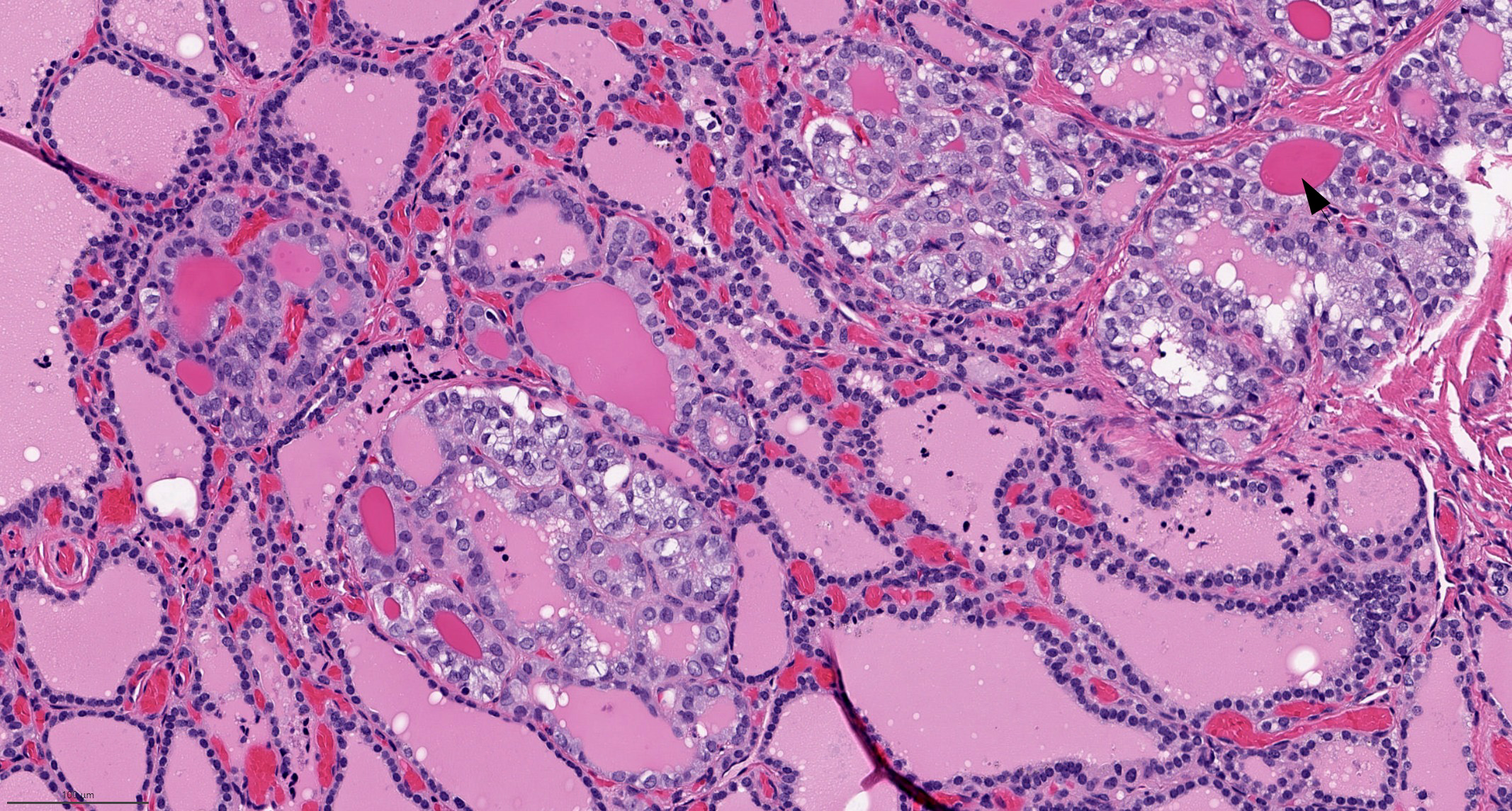
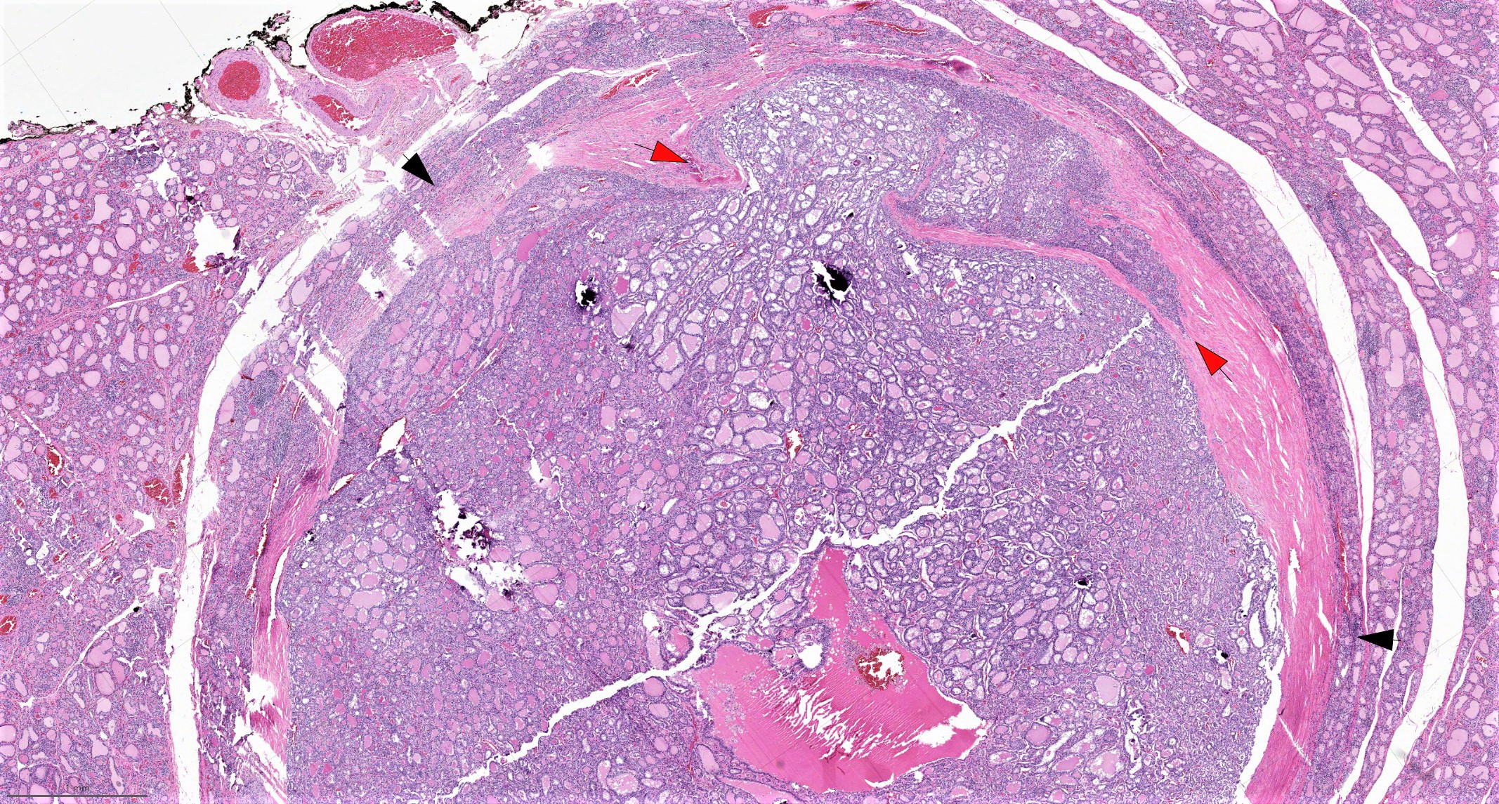
Hürthle cell adenoma
Hürthle cell adenoma
Adenoma with atypia
Hürthle cell carcinoma
Hürthle cell carcinoma
AFIP images
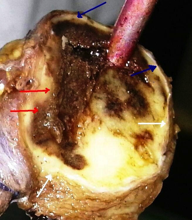
Adenoma:
Oncocytic adenoma has follicular pattern
Massive infarct due to fine needle biopsy

Pseudo-angiosarcomatous pattern
Well developed papillary growth pattern
Cytoplasm has fine,
homogeneously
distributed
granularity
Psammomatous-like calcifications in follicular lumina
Tumor has hyalinized area near capsule
Tumor has focal cells with large hyperchromatic nuclei
Focal papillary formations
Clear cell change:
Gradual transition from oncocytic to clear cells
Both patterns exist in same cell
Sharp demarcation
between clear
and oncocytic cells
Carcinoma:
Capsular invasion
Vascular invasion
Trabecular pattern
Multinodular pattern
Nesting pattern resembles insular carcinoma
Pseudopapillary formations due to tangential sectioning
Pulmonary metastasis
of tumor with
trabecular
growth pattern
Minimally invasive Hürthle cell carcinoma:
Fine needle biopsy induced necrosis
Images hosted on other servers:
Carcinoma:

Capsular invasion


Oncocytes with abundant eosinophilic granular cytoplasm (far right image is with intraluminal calcifications)

No capsular invasion evident

Tumor in internal jugular vein

Various images

Microfollicles

Metastasis to breast

Cells have
eosinophilic
cytoplasm and
prominent nucleoli
Minimally invasive Hürthle cell carcinoma:

Intracapsular vascular invasion

Vascular invasion

Distant metastasis to femur
Contributed by Ayana Suzuki, C.T. and Grace C.H. Yang, M.D.
Follicular neoplasm, Hürthle cell type
Hürthle cell adenoma
Images hosted on other servers:


Microfollicles of carcinoma
Contributed by Mark R. Wick, M.D. and AFIP
Carcinoma
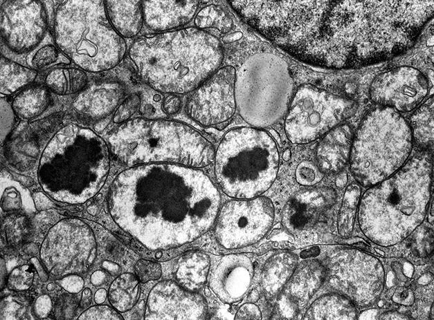
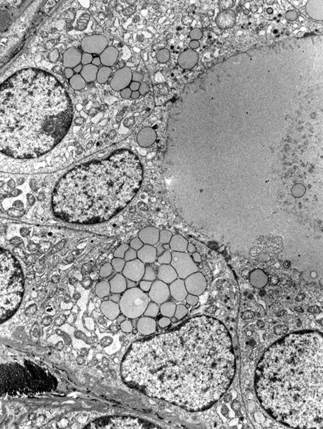
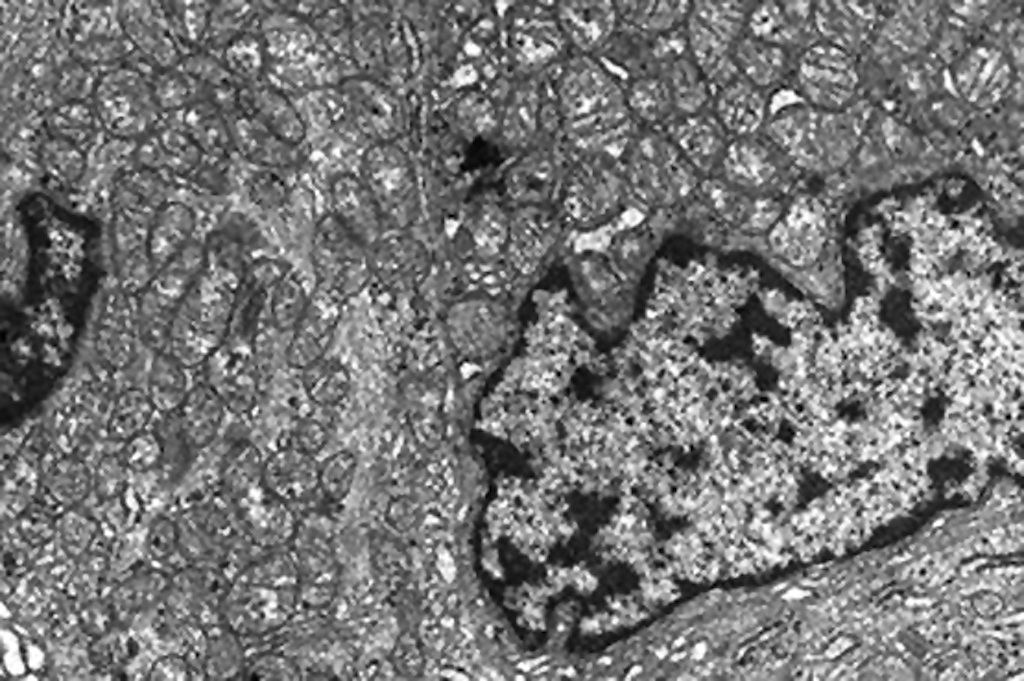
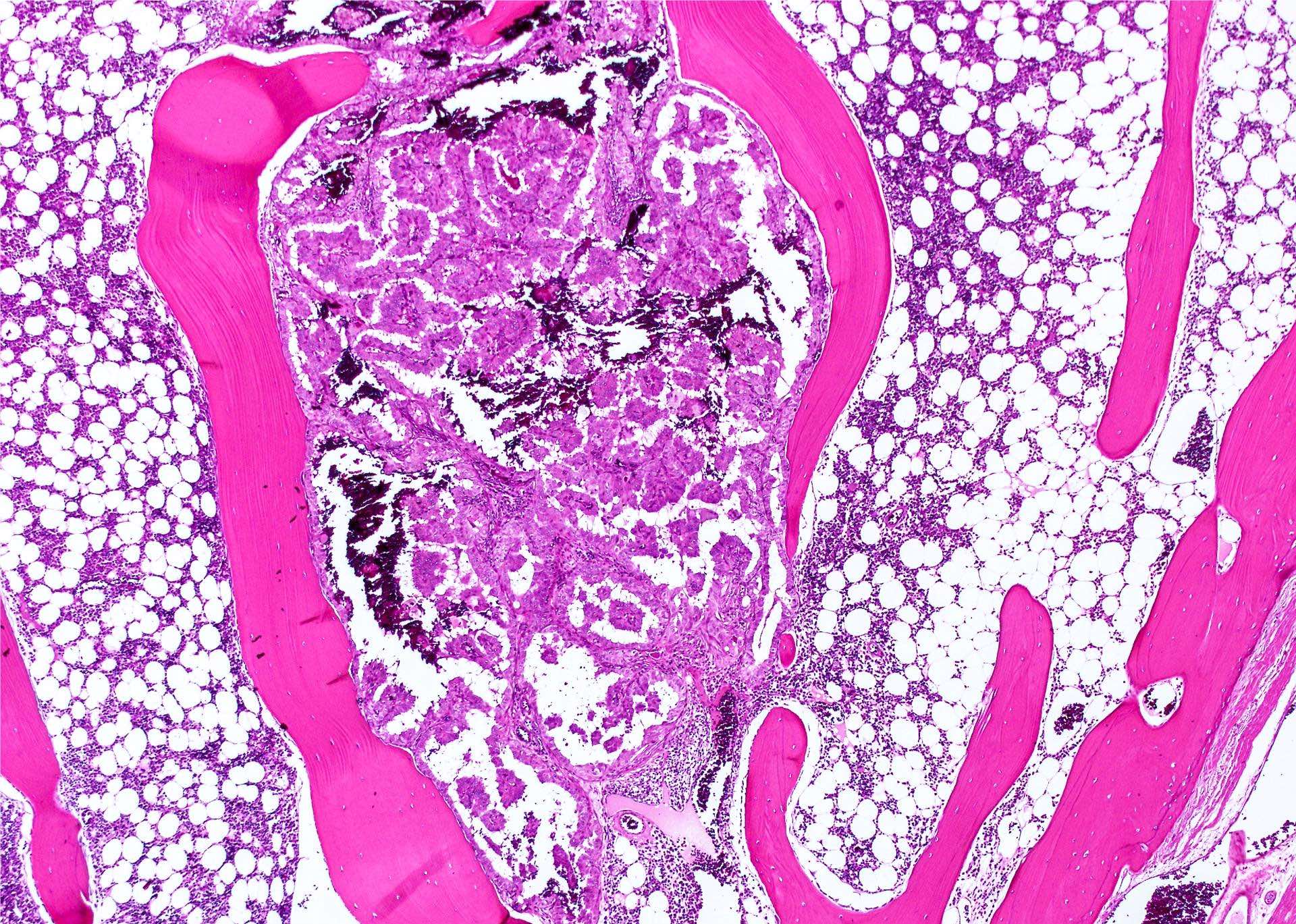
Dilated mitochondria have reduced cristae
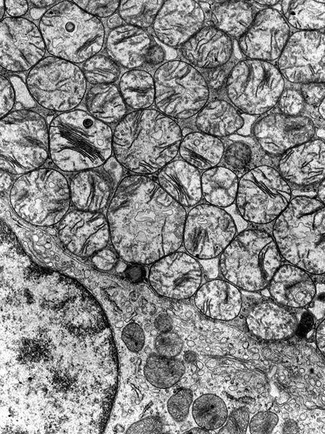
Cytoplasm packed
with large mitochondria
with myelin figures
Oncocytic lesions by Z. Baloch (2020)
Contributed by Ayana Suzuki, C.T.
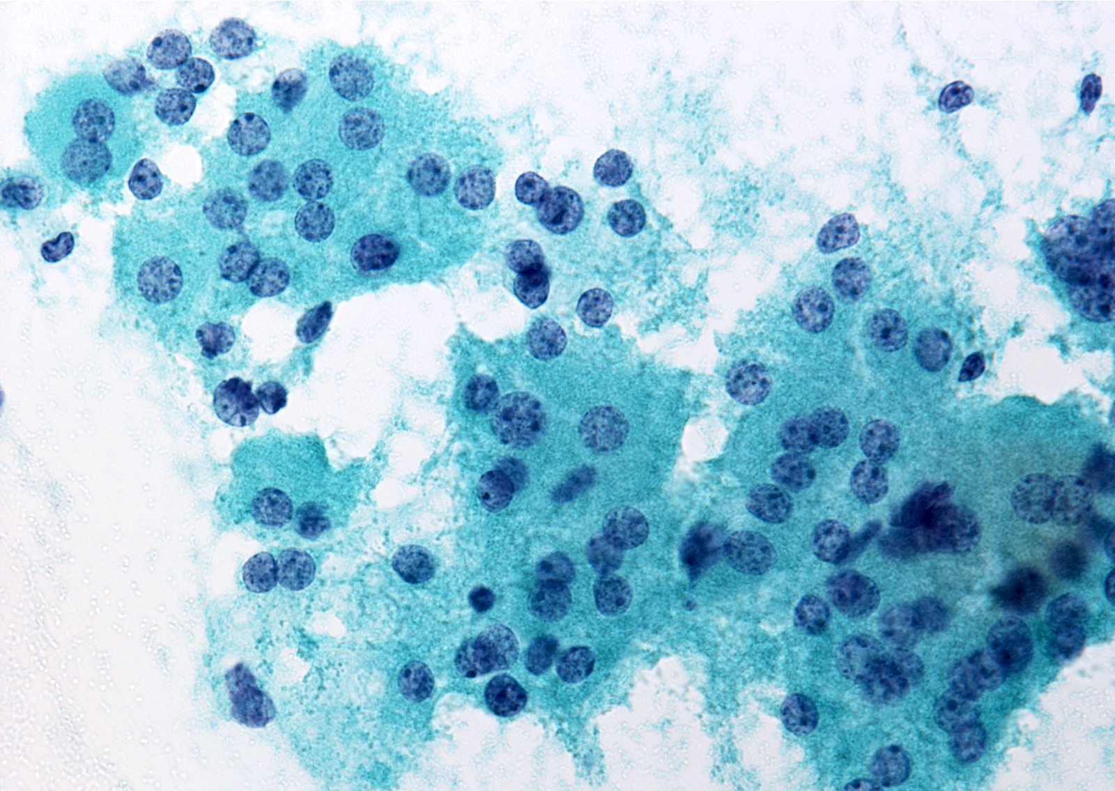
Follicular neoplasm, Hürthle cell type
Hürthle cell clusters
Oncocytes and histiocytes
Contributed by Grace C.H. Yang, M.D.
Hürthle cell adenoma, Diff-Quik
Hürthle cell adenoma, Pap stain
Hürthle cell adenoma, Pap stain
Case 1
Case 2
Oncocytic lesions by Z. Baloch (2020)
Contributed by Ayana Suzuki, C.T.
Parathyroid cyst: large multilocular cyst
Contributed by Ayana Suzuki, C.T.
Parathyroid cyst: clear fluid
Images hosted on other servers:

Parathyroid cyst: islands of parathyroid tissue

CMV
parathyroiditis in
immunosuppressed
child
Contributed by Andrey Bychkov, M.D., Ph.D. and AFIP

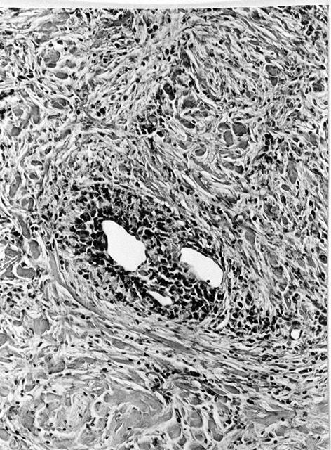
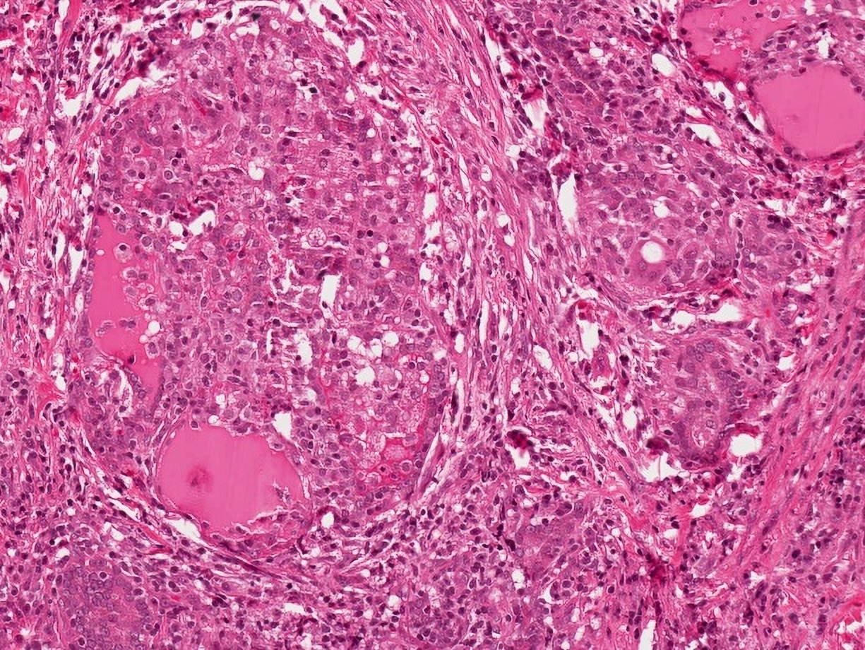
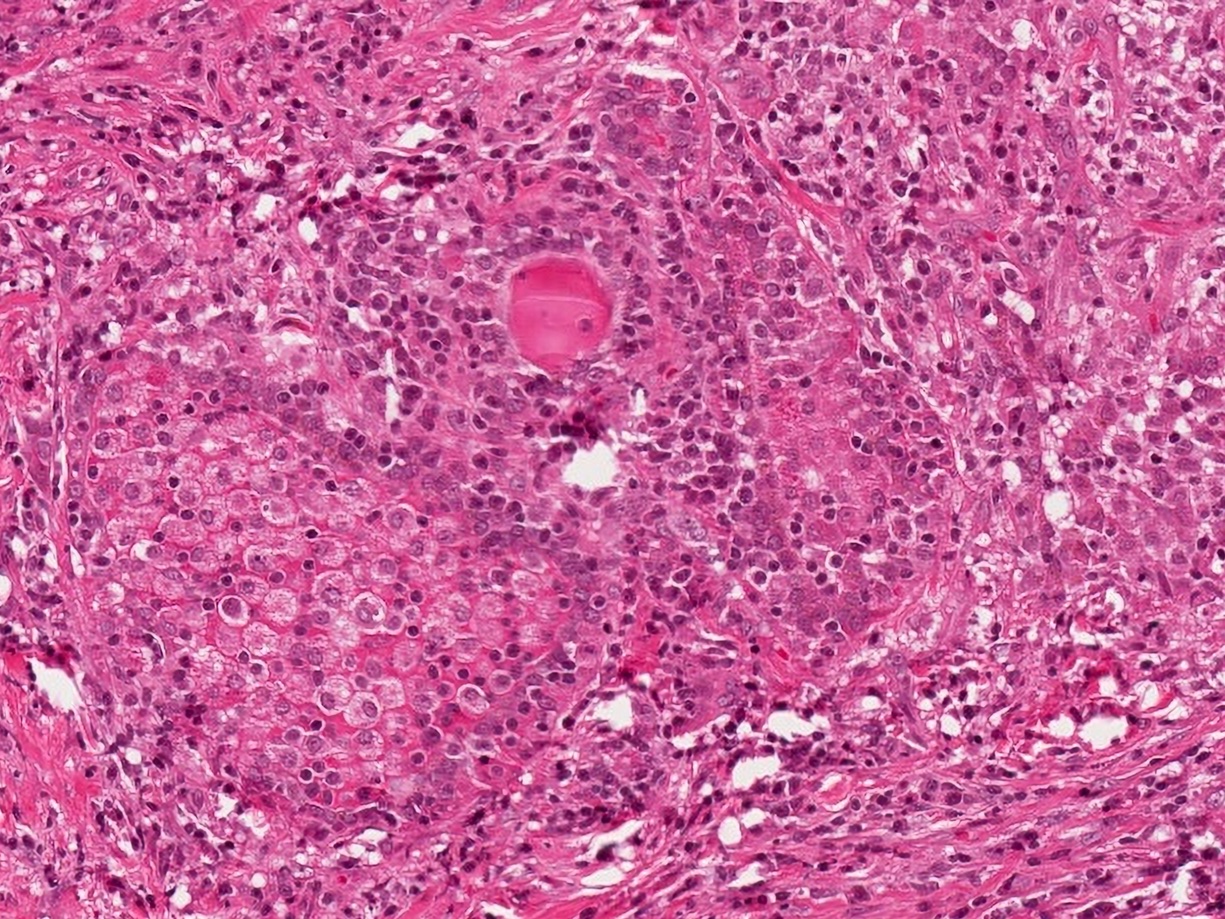
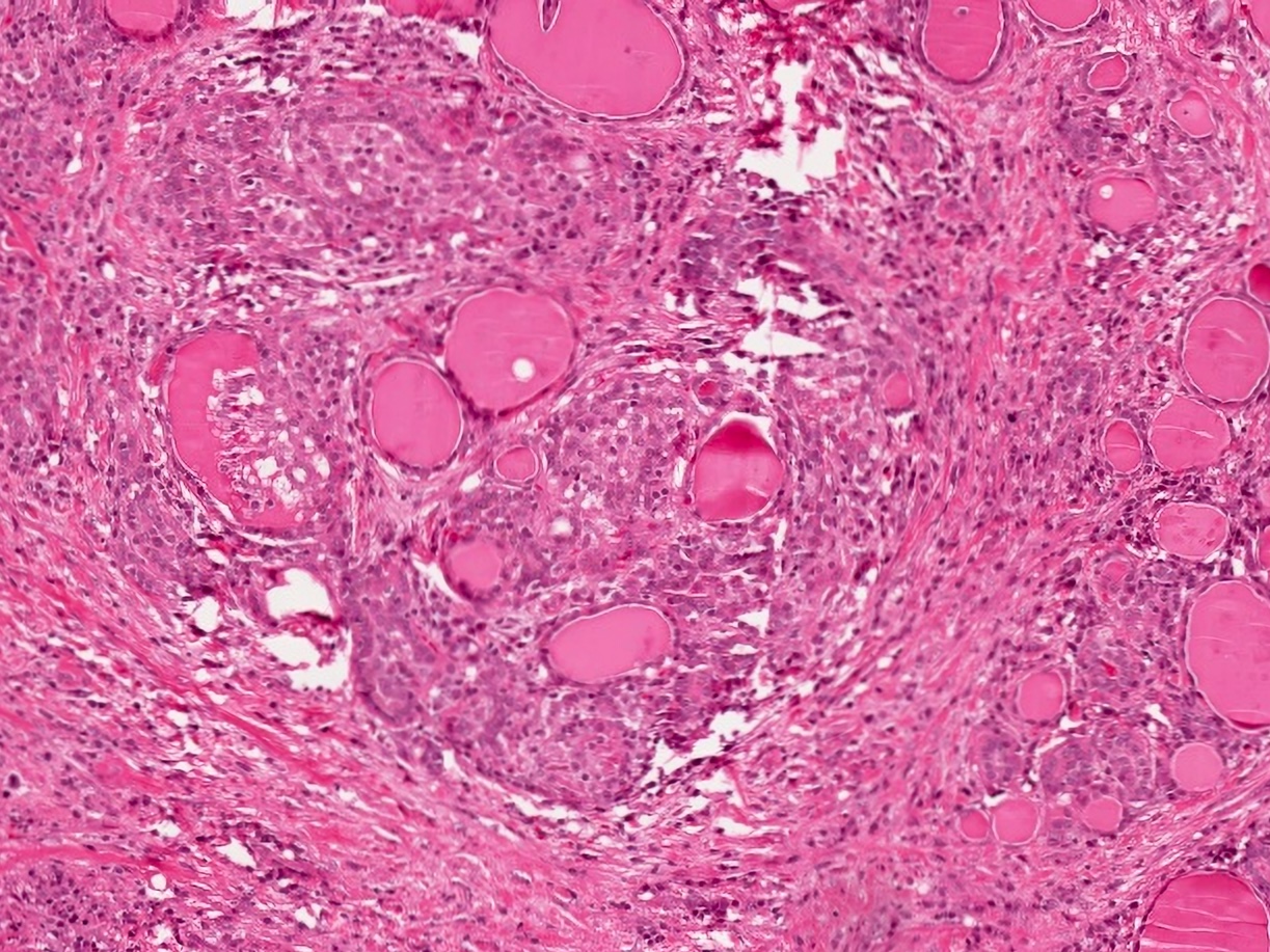
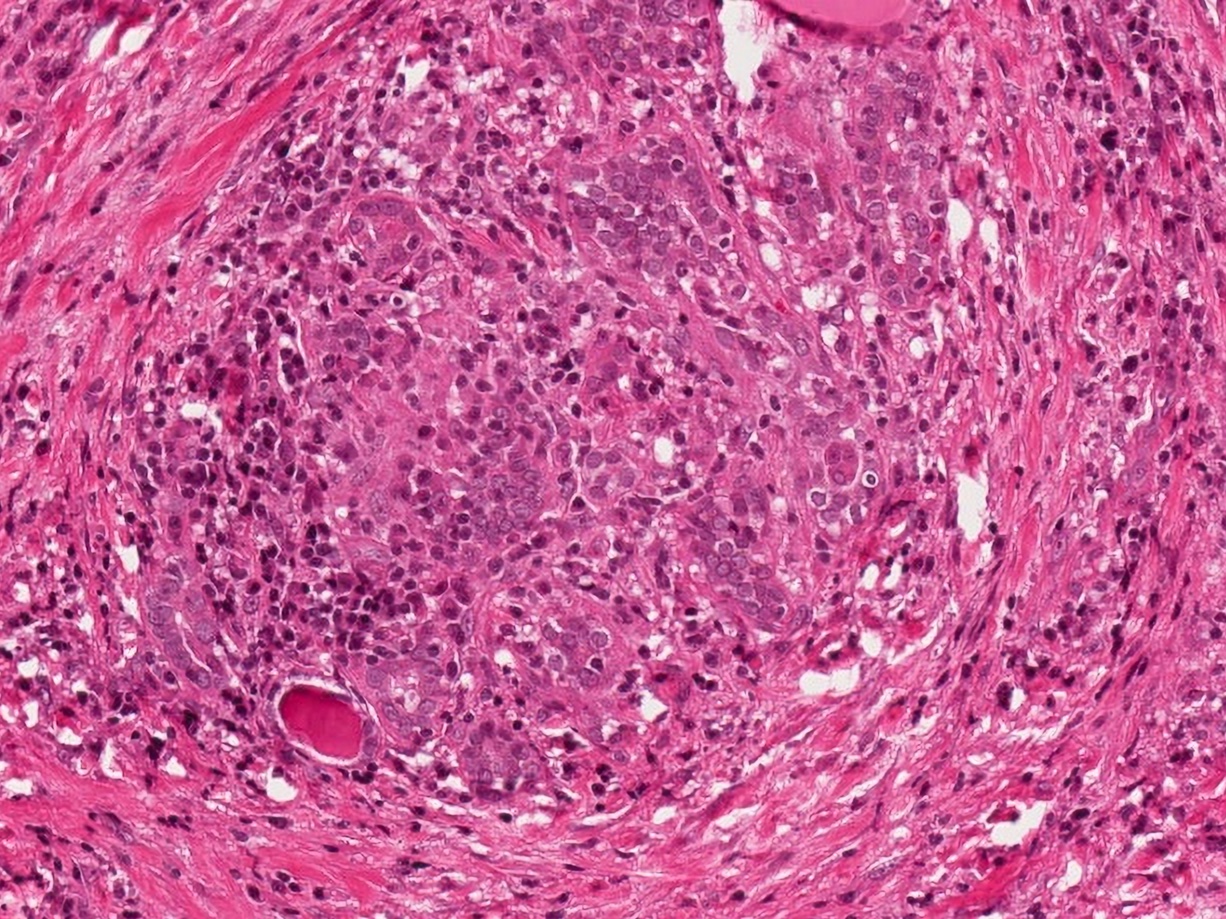
Superficially
located nodule
Intrafollicular
granulomatous
reaction
Granulomatous folliculitis
Multinucleated giant cells
Histiocytes within follicle
Histiocytes are strongly lysozyme+
Images hosted on other servers:
Granuloma
Images hosted on other servers:

ATA nodule sonographic patterns
Contributed by Bin Xu, M.D., Ph.D.
Thyroid mass
Contributed by Bin Xu, M.D., Ph.D.
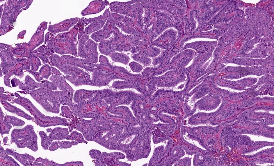
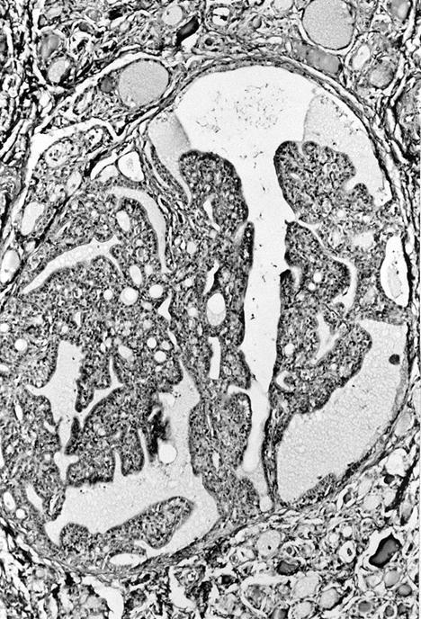
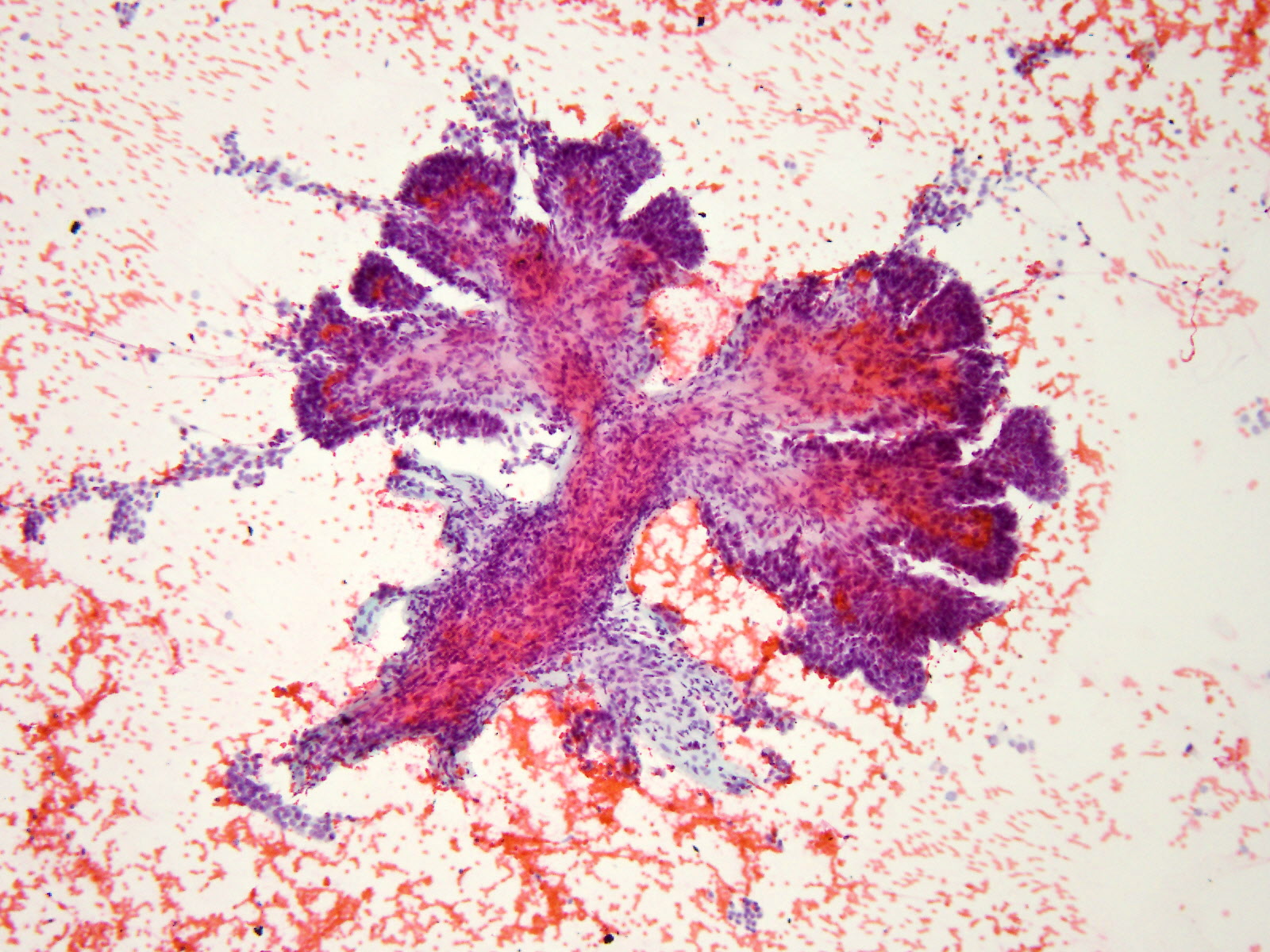
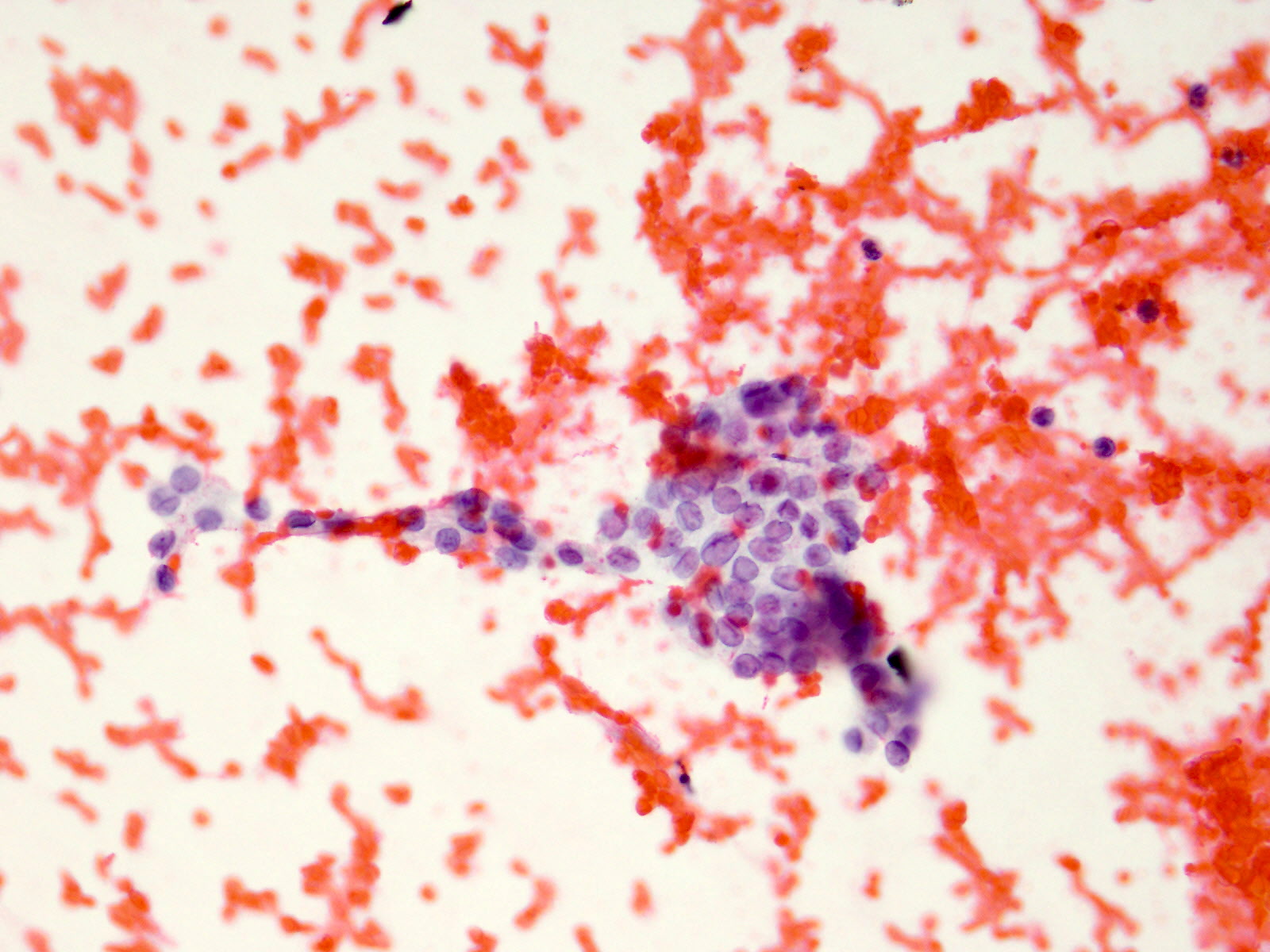
Papillary projections
Nuclear features
Psammoma body
Cystic change with BRAF V600E
Osseous metaplasia
Contributed by Bin Xu, M.D., Ph.D.
Papanicolaou stain
Images hosted on other servers:

Invasion of trachea

Enhancing nodular lesion

Diffuse lung metastases
Images hosted on other servers:

Cystic neck mass

Bulging neck mass
Contributed by Bin Xu, M.D., Ph.D., Andrey Bychkov, M.D., Ph.D. and Kseniya Korchagina, M.D.
PTC in thyroglossal duct

Peritumoral fibrosis
Grossing and sampling
Extrathyroidal extension

Cystic change
Contributed by Andrey Bychkov, M.D., Ph.D.
Nuclear features
Nuclear pseudoinclusions
Pseudonuclear inclusions and large vesicular nucleus

Nuclear enlargement, crowding / overlapping
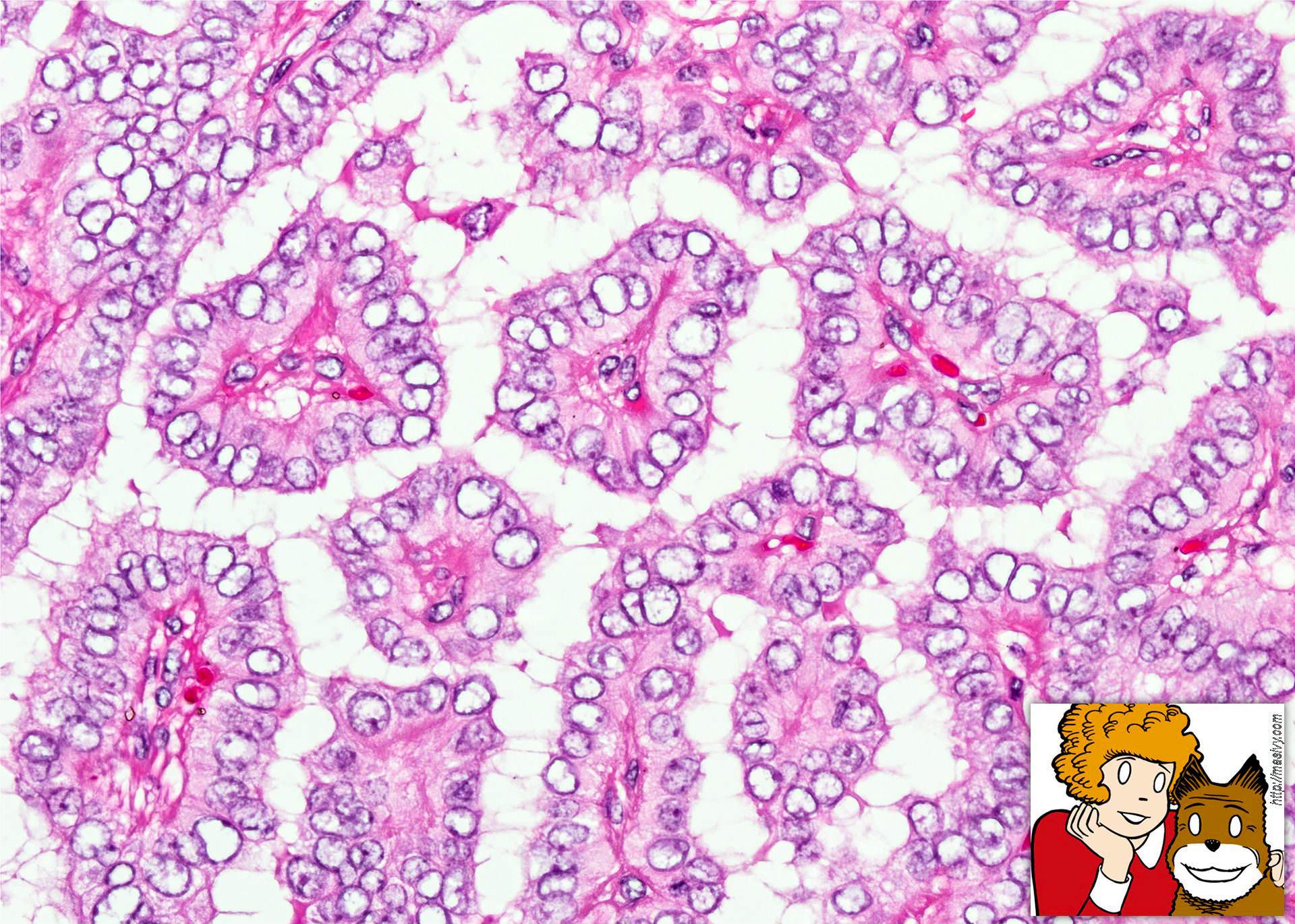
Orphan Annie eye nuclei
Architectural patterns
True papillae with multiple branching

Arborizing papilla
Other histologic features
Psammoma bodies
Psammoma body (HBME1)
Giant cells
Different kinds of giant cells
Deposits in perithyroidal fat
Residual tumor in thyroid bed
Clear cell change
Cystic degeneration

Submucosal spread
Bone involvement
Lymph node replaced by metastasis

Large psammoma bodies

Ossification
Concentric calcium deposition
PTC with Hashimoto thyroiditis
Follicular adenoma, mimic of PTC
Stains

CK19, Galectin3, HBME1
CK19
Galectin3

HBME1


BRAFV600E
TTF1
PAX8
Contributed by Andrey Bychkov, M.D., Ph.D.
Papanicolaou stain

Papillary thyroid carcinoma, imprint cytology
AFIP images
Tall cuboidal cells
Surface microvilli
Images hosted on other servers:

TCGA

BRAF mutation
Papillary thyroid carcinoma: cystic
Thyroid carcinoma
Thyroid: compare and contrast
Histopathology thyroid: papillary carcinoma
Histopathology thyroid: Hashimoto thyroiditis, papillary carcinoma
Integrated genomic characterization of papillary thyroid carcinoma
An alphabet soup of thyroid neoplasms
Images hosted on other servers:

Neck ultrasonography

Color flow Doppler

Tracer uptake, lateral neck


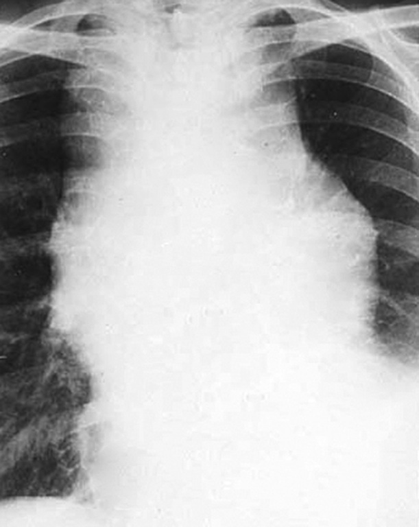
Radiography of mediastinal mass
AFIP images

Nodular hyperplasia
Images hosted on other servers:

Mediastinal thyroid mass
Contributed by Andrey Bychkov, M.D., Ph.D. and AFIP
2 perithyroidal lymph nodes
Specimen with signs of Hashimoto thyroiditis

Lymphoid follicles
Patient with Hashimoto

Hyperplastic nodule
Images hosted on other servers:

Parasitic nodule vs lymph node

Thyroid follicles

TTF1, Thyroglobulin
Images hosted on other servers:

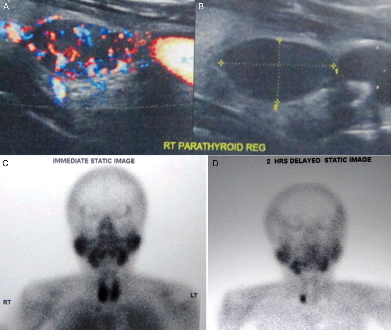
99mTc MIBI scintigraphy
Images hosted on other servers:

Giant parathyroid adenoma (intraoperative)
Contributed by Mona Kandil, M.D and @Andrew_Fltv on Twitter
Parathyroid adenoma (black arrow) and thyroid
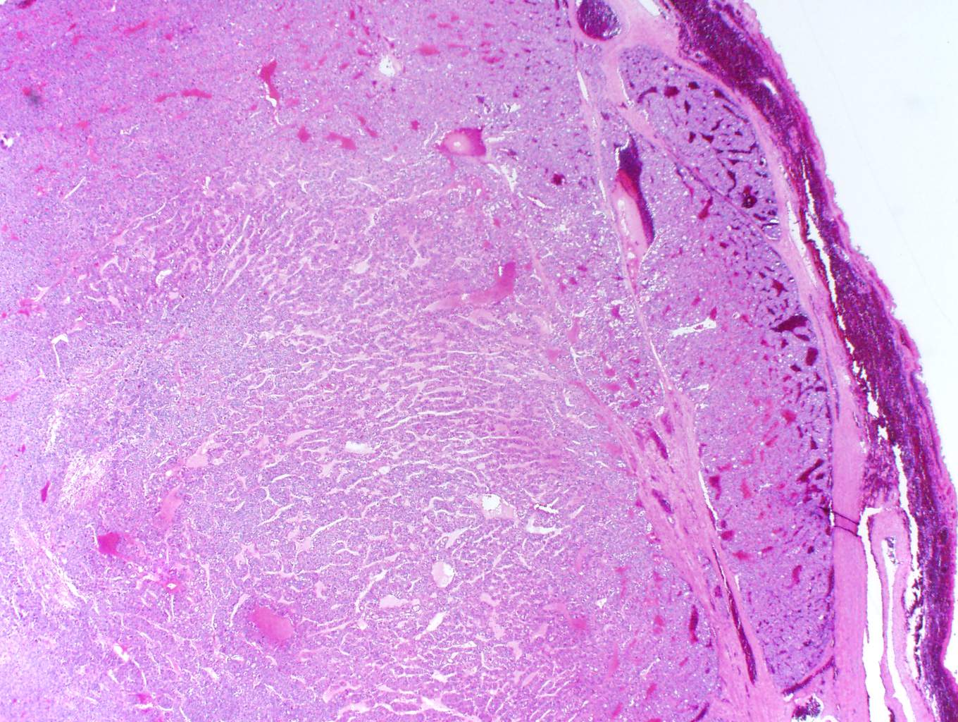
Parathyroid adenoma
Images hosted on other servers:

Right: parathyroid adenoma;
left: thymoma
Contributed by Diana Murro Lin, M.D. and @Andrew_Fltv on Twitter
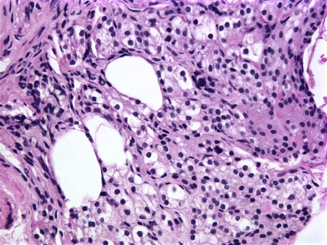
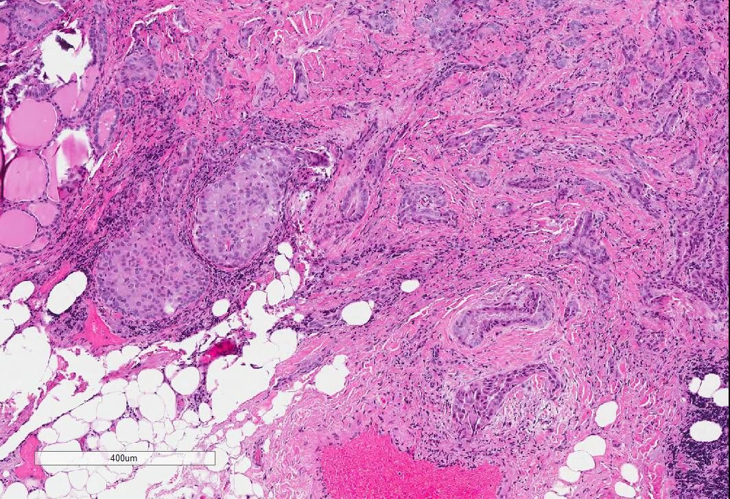
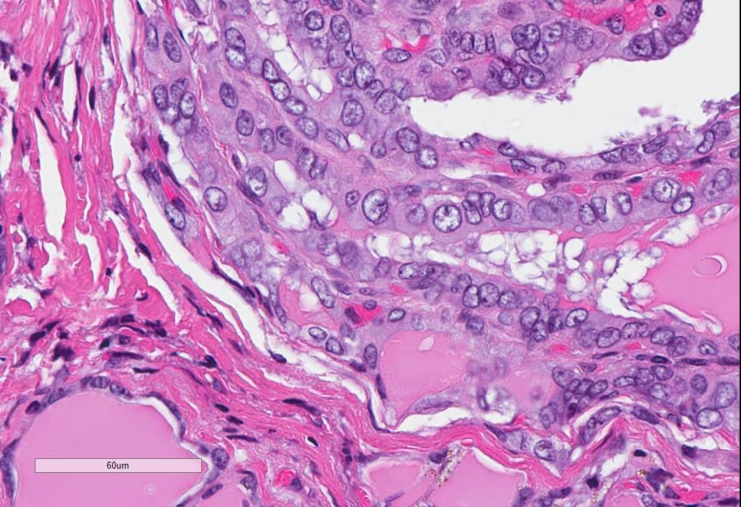
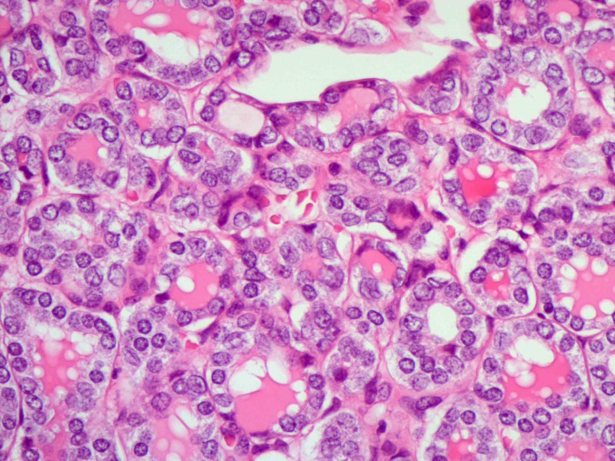
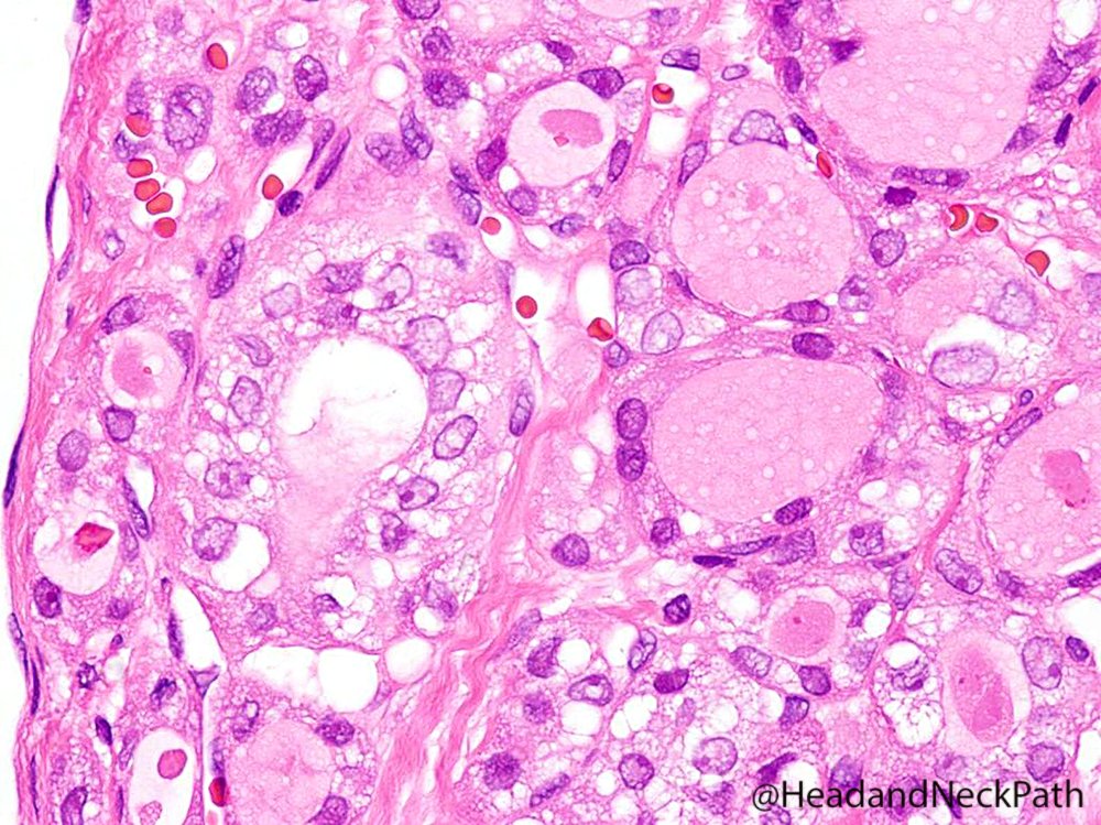
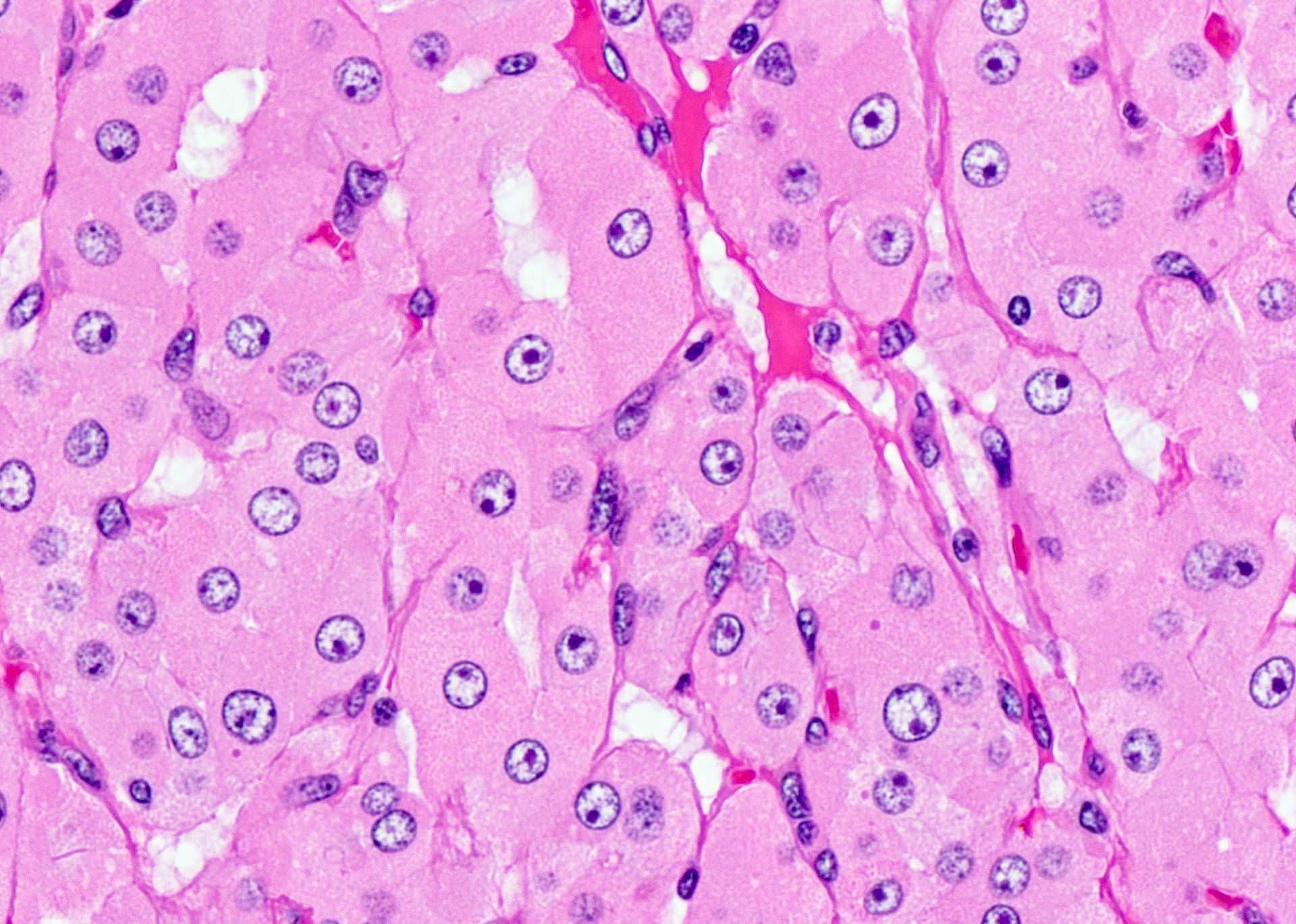
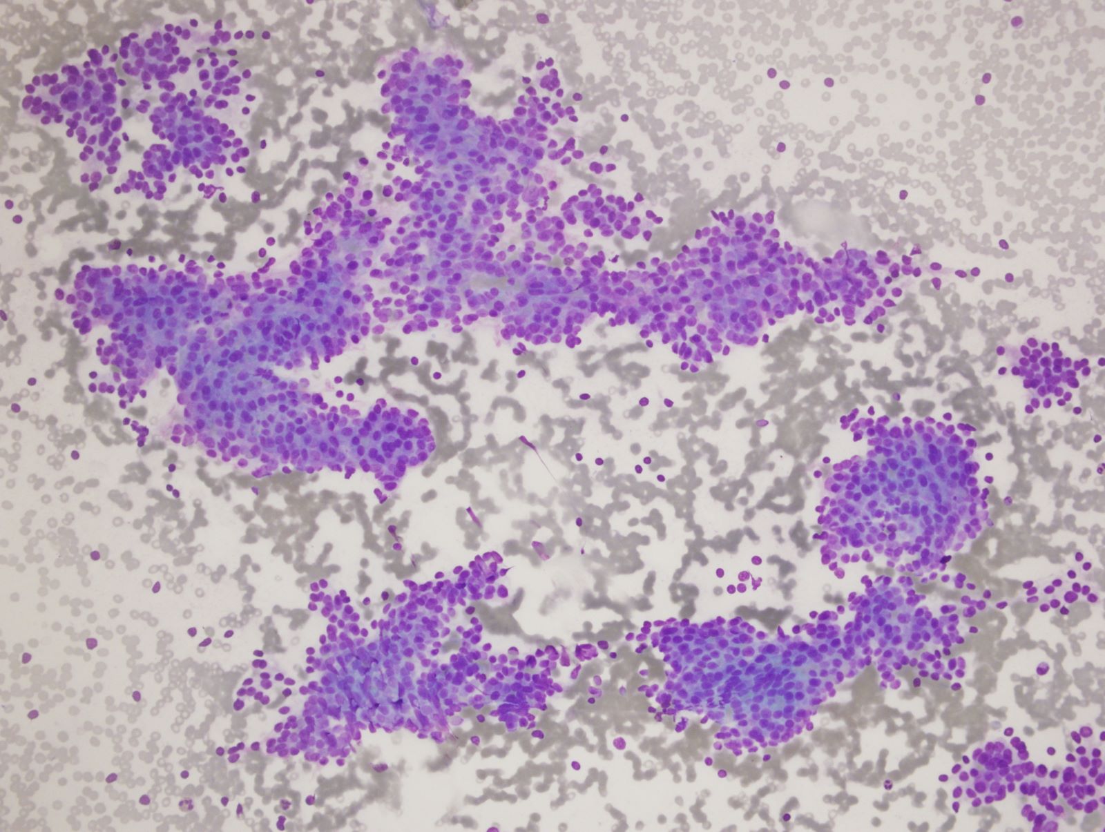
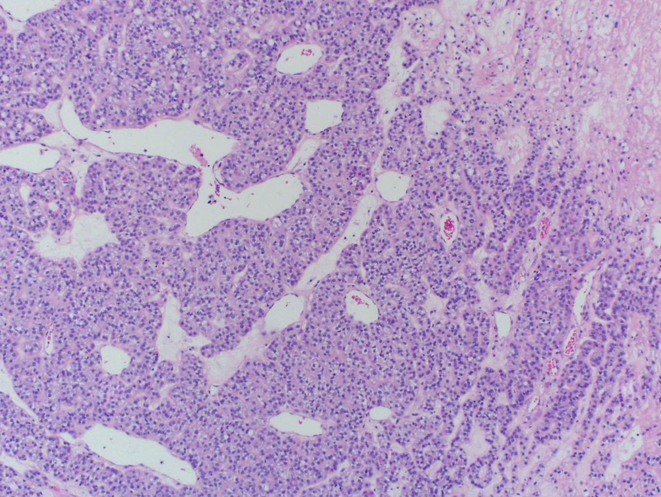
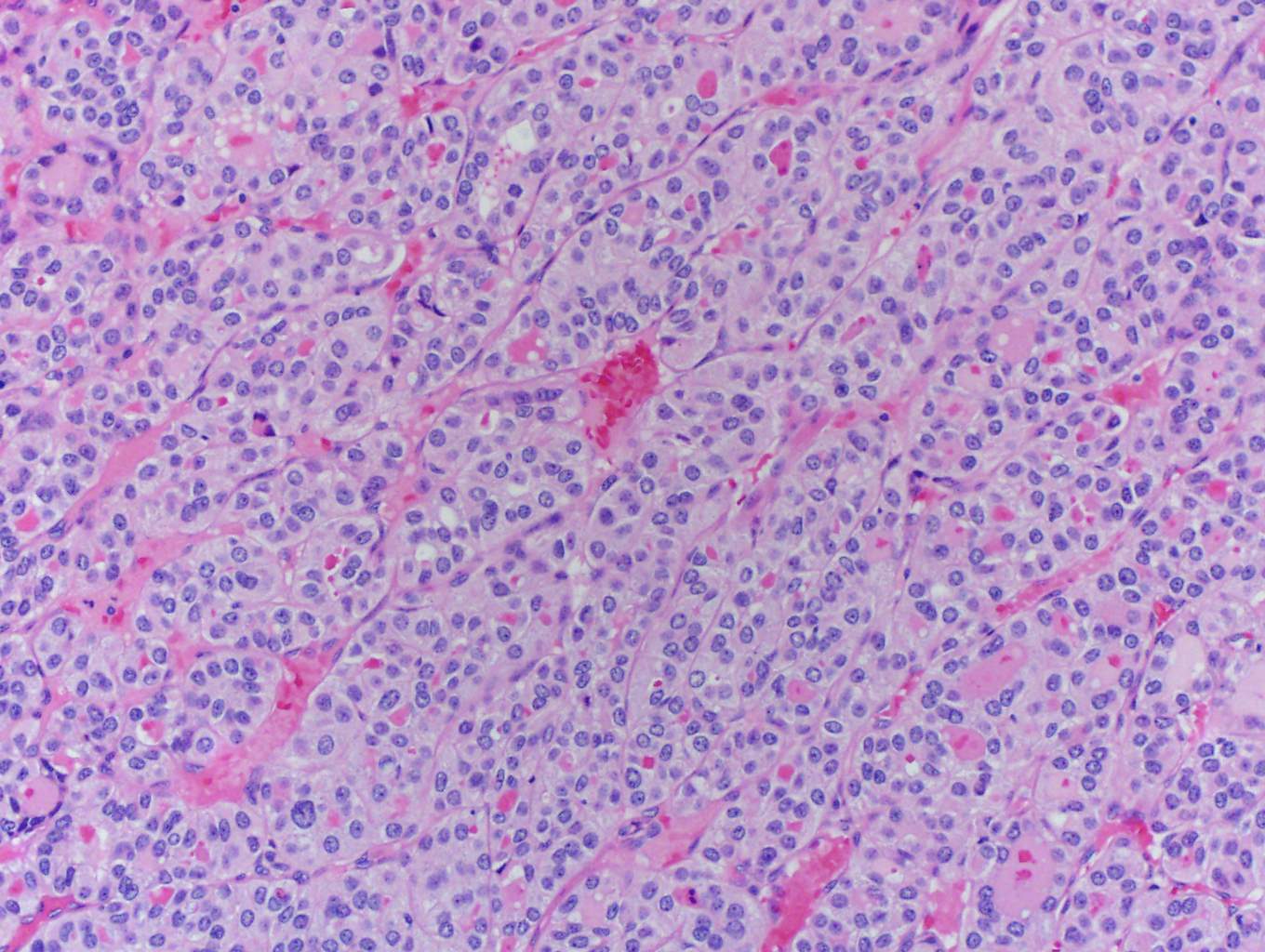
Chief cells
Oxyphil adenoma
Oxyphil cells
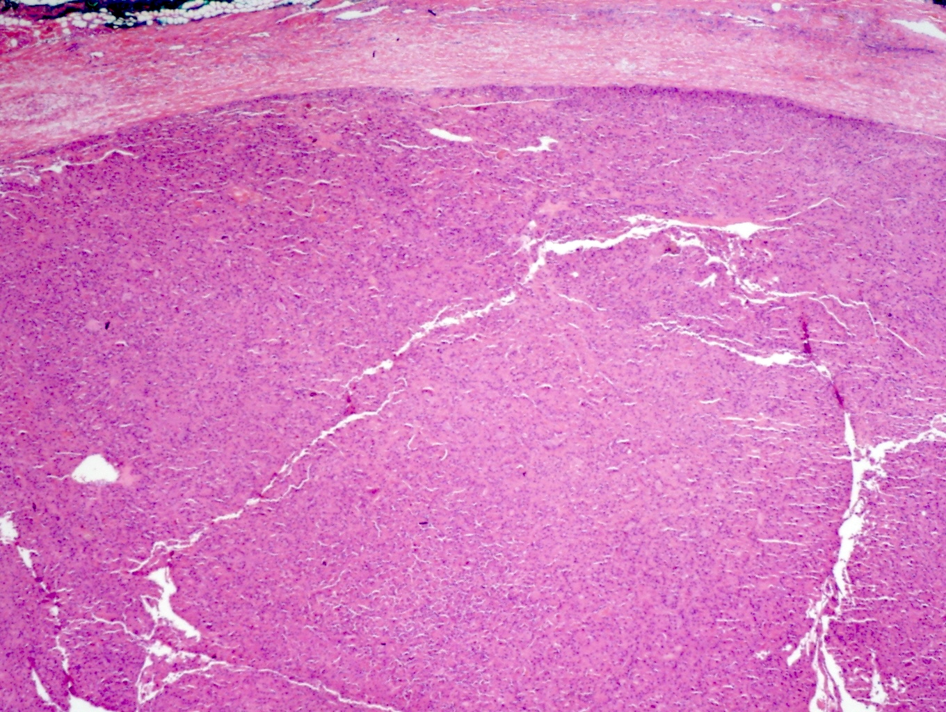
Atypical adenoma and thyroid
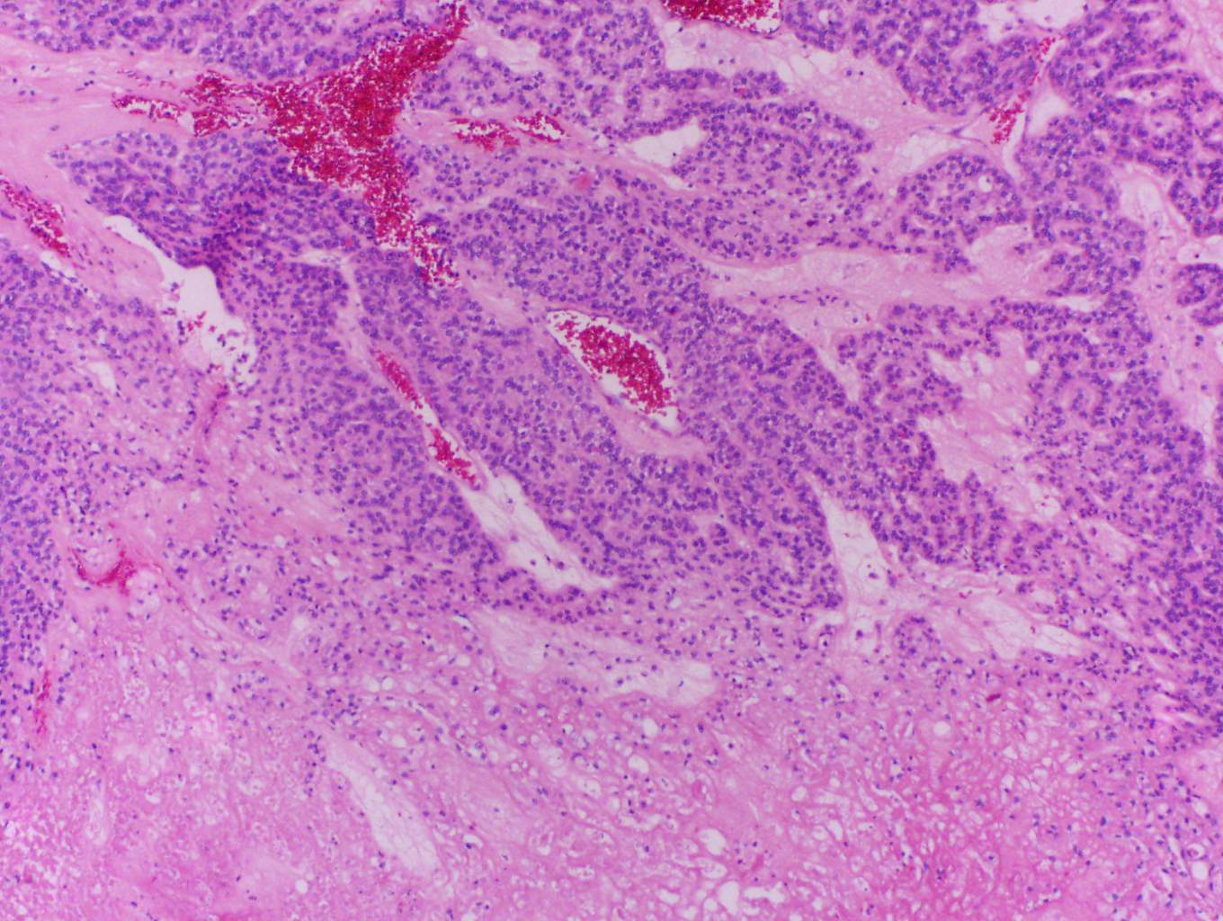
Atypical adenoma fibrous bands

Parathyroid adenoma

Parathyroid adenoma
Contributed by Ayana Suzuki, C.T.
Sheet of chief cells
Images hosted on other servers:

Microfollicular and trabecular arrangement
Oncocytic adenoma
Parathyroid pathology
Images hosted on other servers:

Xray lytic lesion

Sestamibi scan

Bone scan

Tc MIBI scintigraphy and CT

Tc MIBI SPECT / CT

99mTc MIBI SPECT / CT and MRI
Images hosted on other servers:

Intraoperative dissection
Images hosted on other servers:

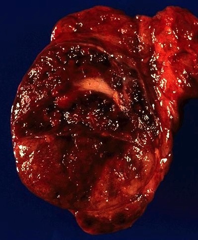
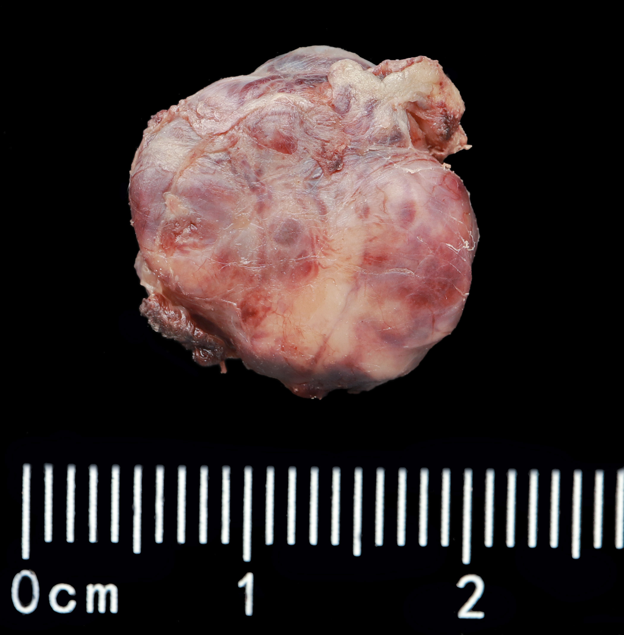
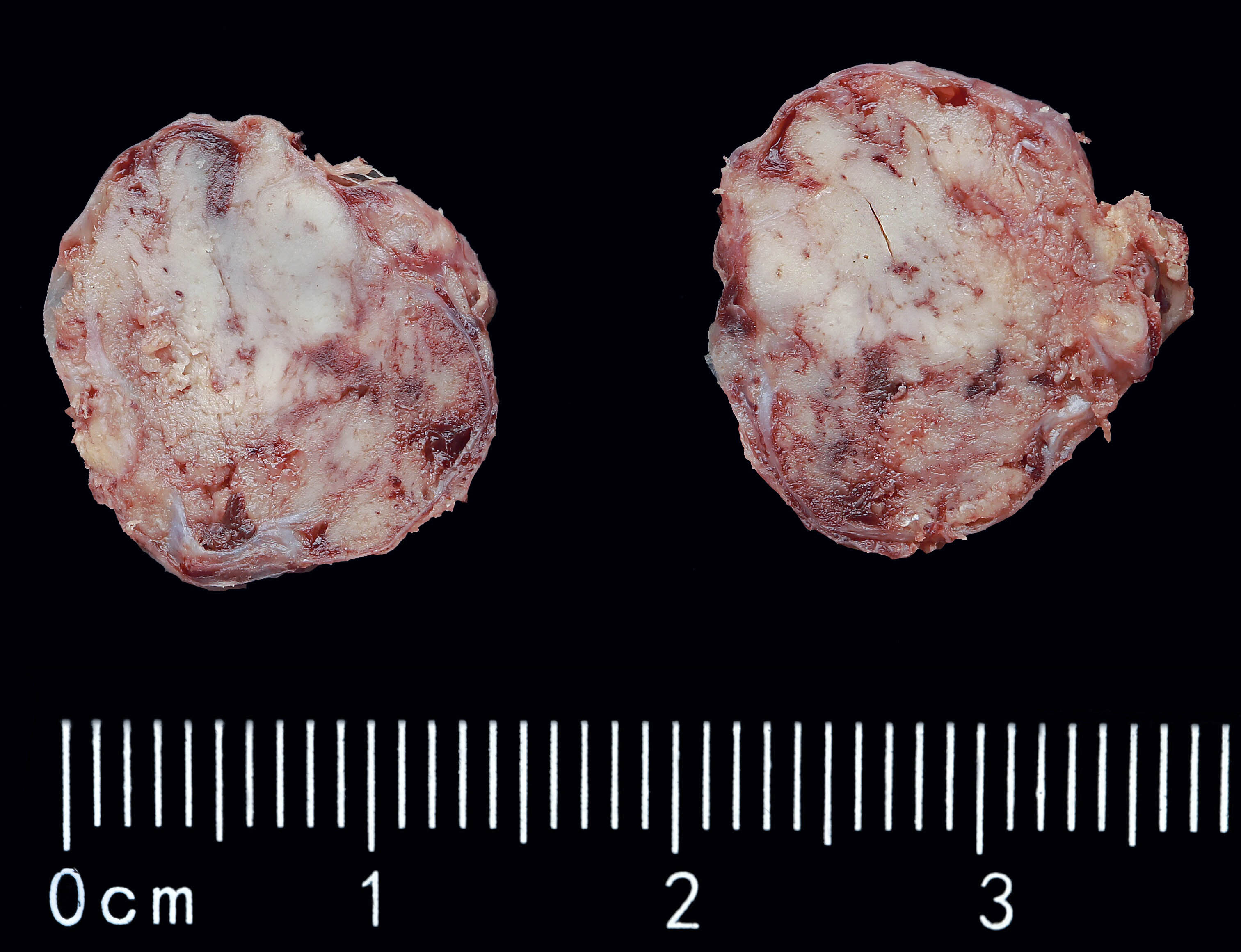
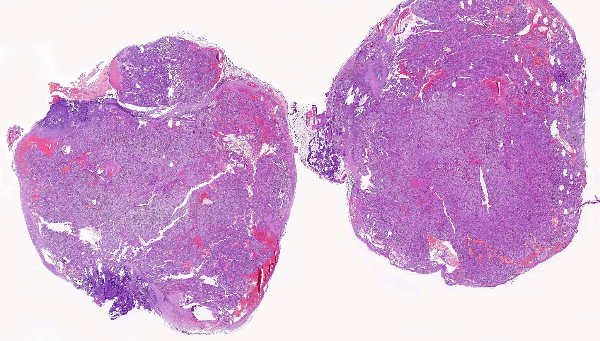
Large size and irregular cut surface
Contributed by Mehmet Kefeli, M.D.
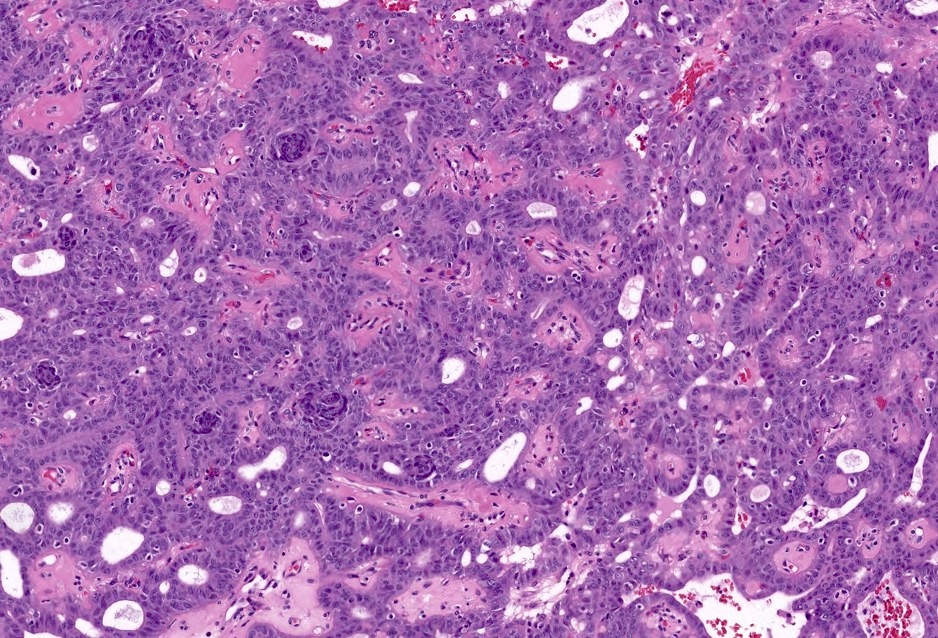
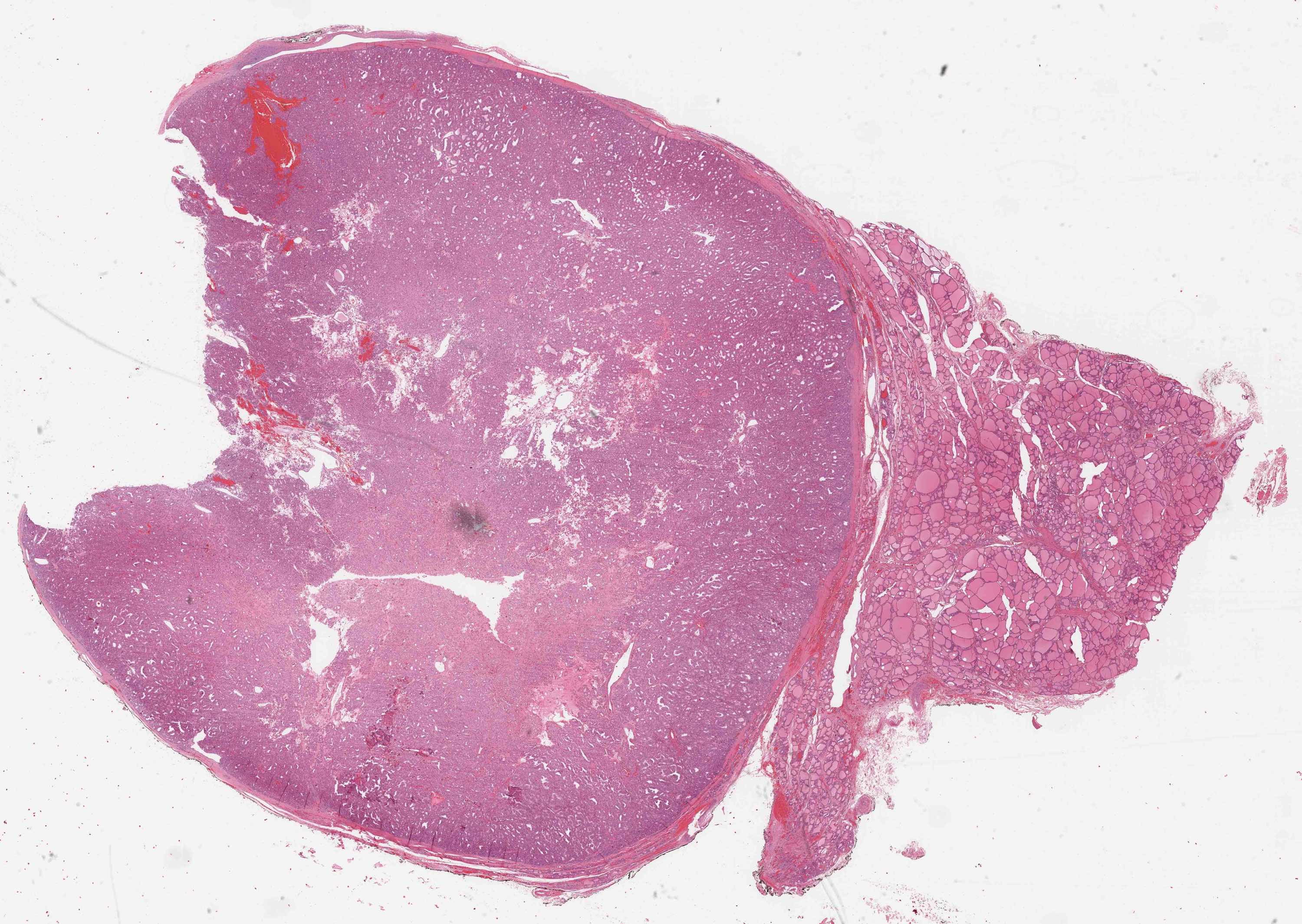
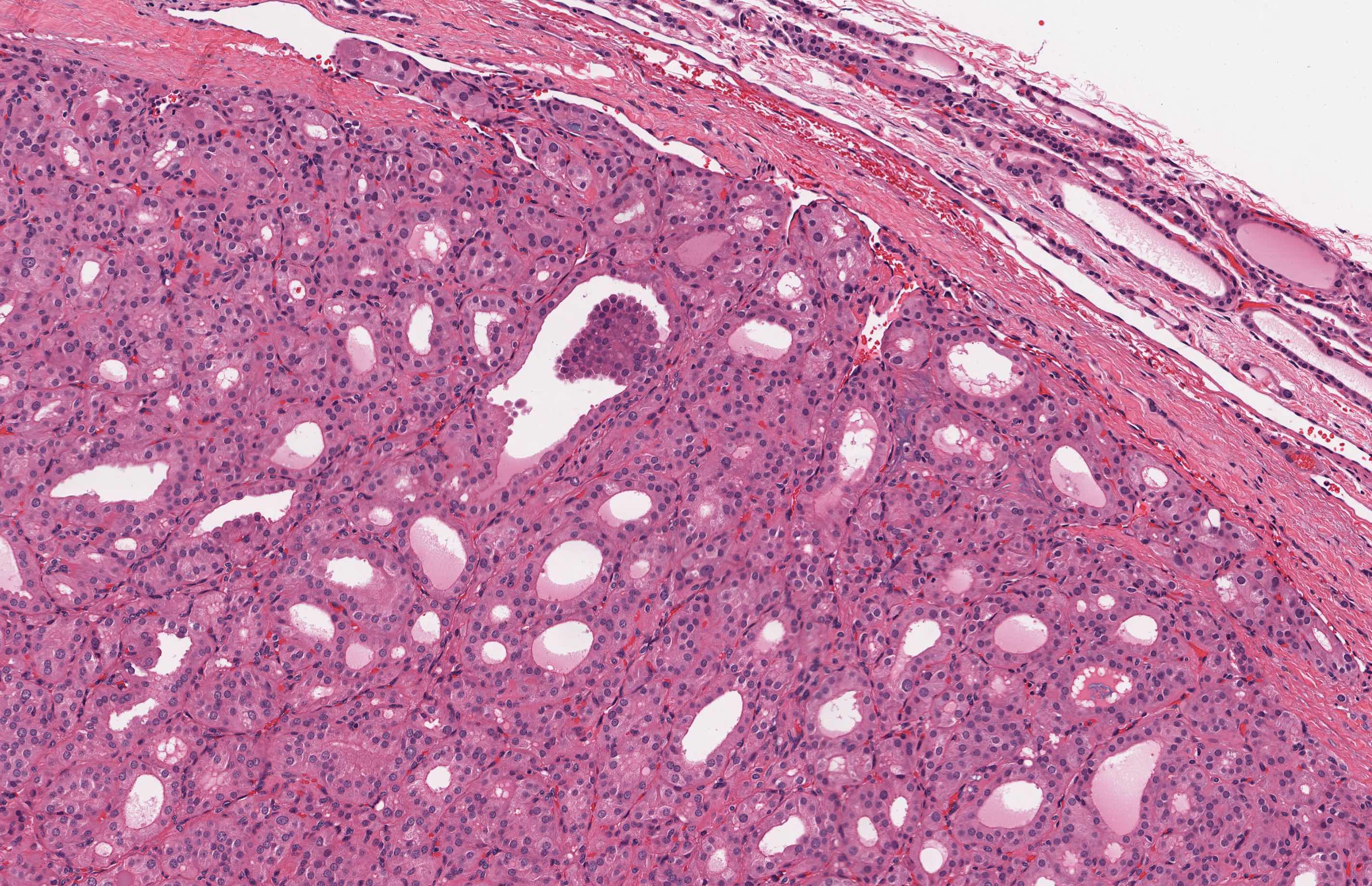
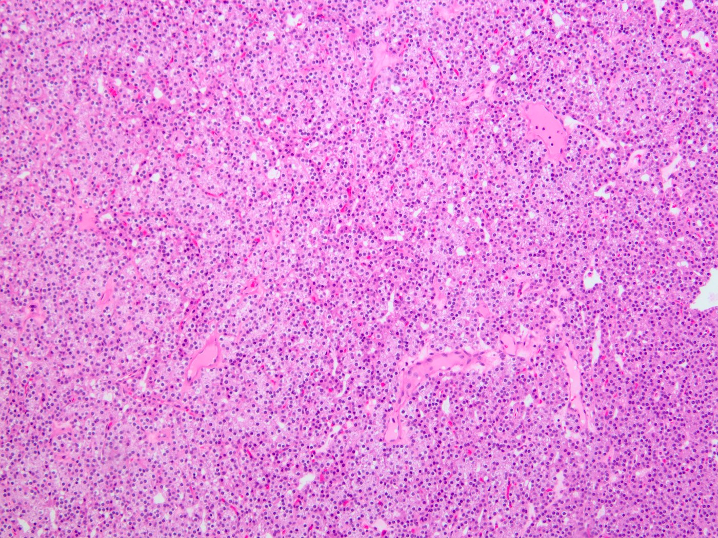
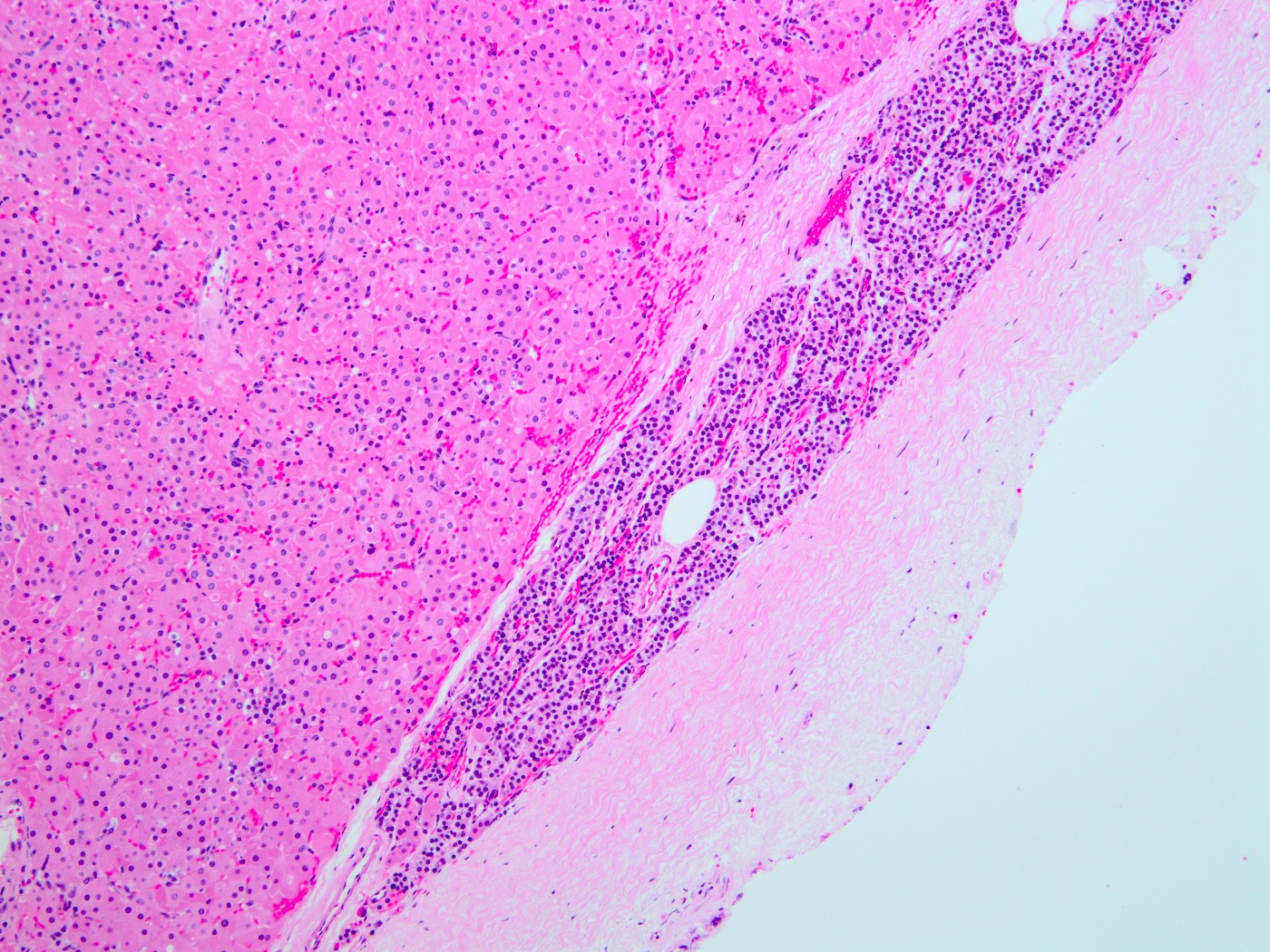
Solid and nodular growth
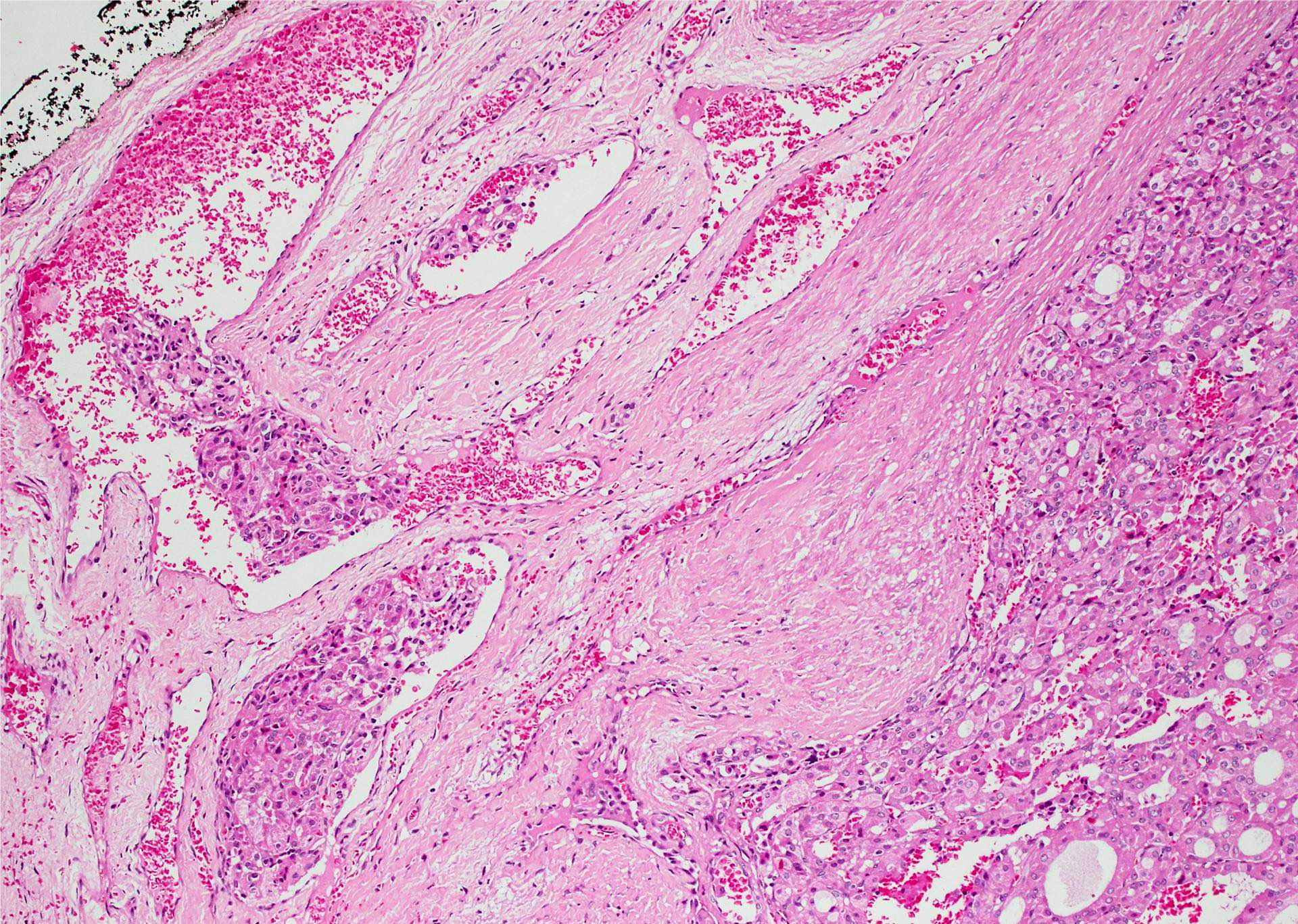
Skeletal muscle invasion
Uniform cytomorphology
Numerous mitoses
Prominent nucleoli
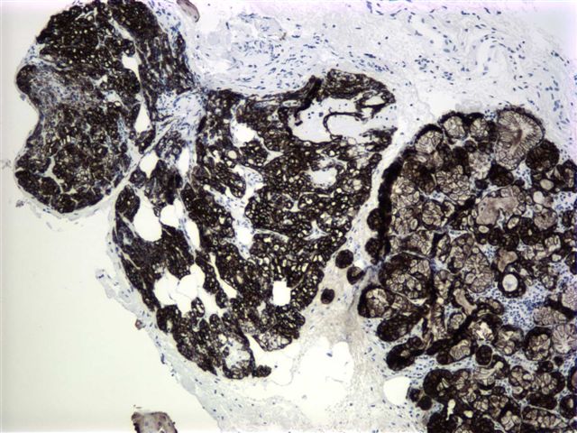
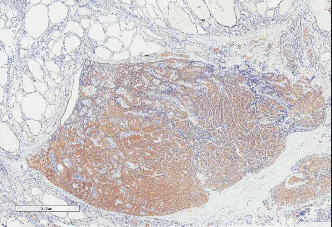
PTH
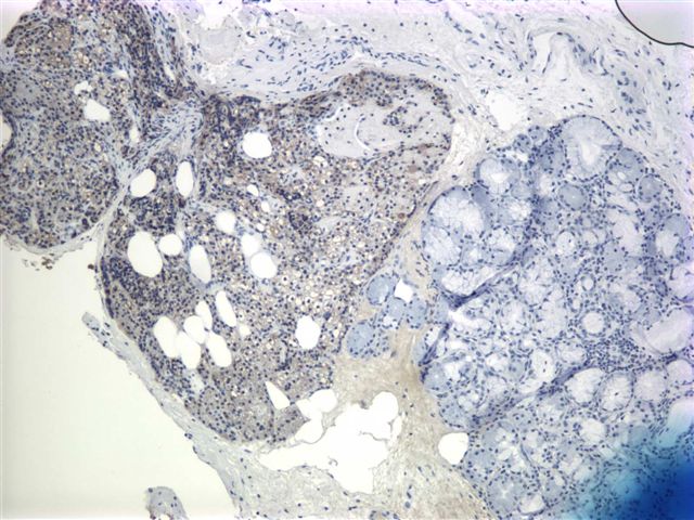
GATA3
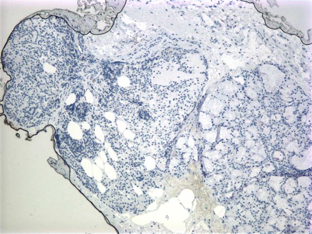
Thyroglobulin
TTF1
Ki67
Contributed by Mehmet Kefeli, M.D.

Cohesive clusters and sheets of cells
Parathyroid pathology
Images hosted on other servers:

3 and a half glands removed
Images hosted on other servers:

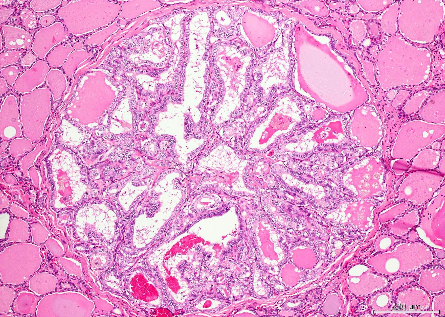
Nodular parathyroid hyperplasia (figures D / E)
Images hosted on other servers:

Sonogram
Intrathyroidal PT adenoma

MIBI scan

Technetium sestamibi scan

Parathyroid carcinoma

Parathyroid glands

Coronal oblique slices

Right sided neck mass
Images hosted on other servers:

Excised tissue

Resected thyroid

Parathyroid adenoma

Thyroid lobe


Intrathyroidal PT adenoma

Enlarged right thyroid lobe
Contributed by Andrey Bychkov, M.D., Ph.D.
Parathyroid histology
Images hosted on other servers:

Ectopic thymus and PT within thyroid

Surrounded by thyroid parenchyma

PT hyperplasia


Intrathyroidal PT cystic adenoma


Intrathyroidal PT adenoma

Intrathyroidal PT carcinoma
Images hosted on other servers:

Common and uncommon FNA features

FNA
Sestamibi Scans and How to See Your Parathyroid Glands
Images hosted on other servers:

Multinodular

Cut surface
Images hosted on other servers:

Plasma cells, lymphocytes and fibrous tissue

Clumps of plasma cells

Plasma cells around follicular epithelium

CD79a

Kappa and lambda light chains
Images hosted on other servers:

Gray, firm, fleshy

Thyroidectomy specimen
Case #394
10×

20×

40×

CD79a
CD138
Kappa
Lambda
Images hosted on other servers:

Mature and immature plasma cells


H&E, CD138+

(A) Kappa light chain+
(B) Lambda light chain-
Images hosted on other servers:

Intranuclear inclusion

Congo red+
Contributed by Mark R. Wick, M.D. and AFIP images
Various images
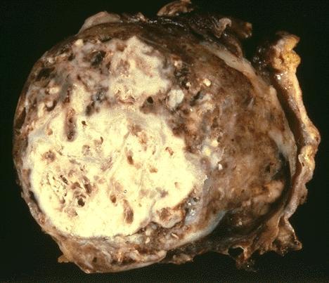
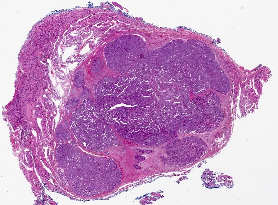
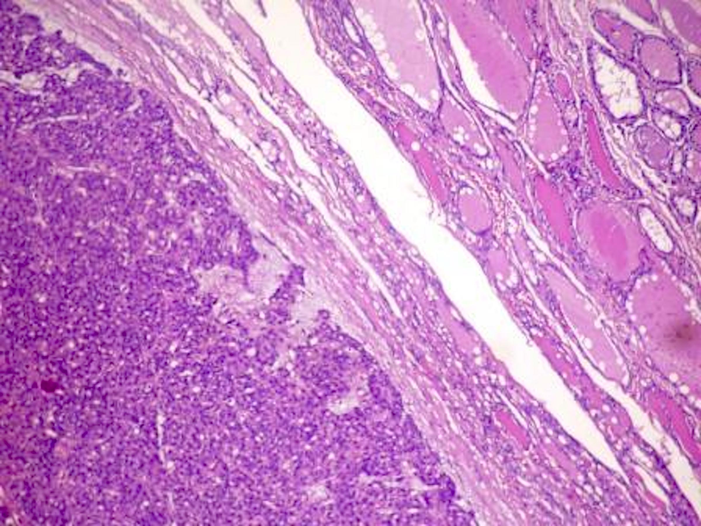
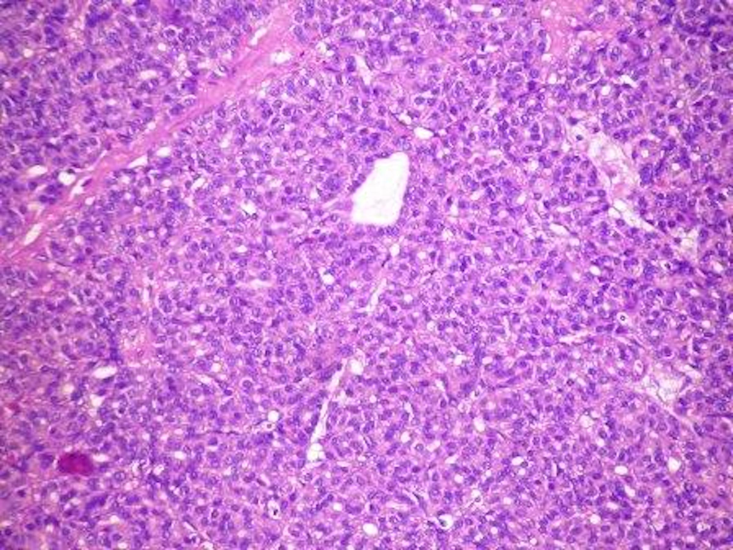
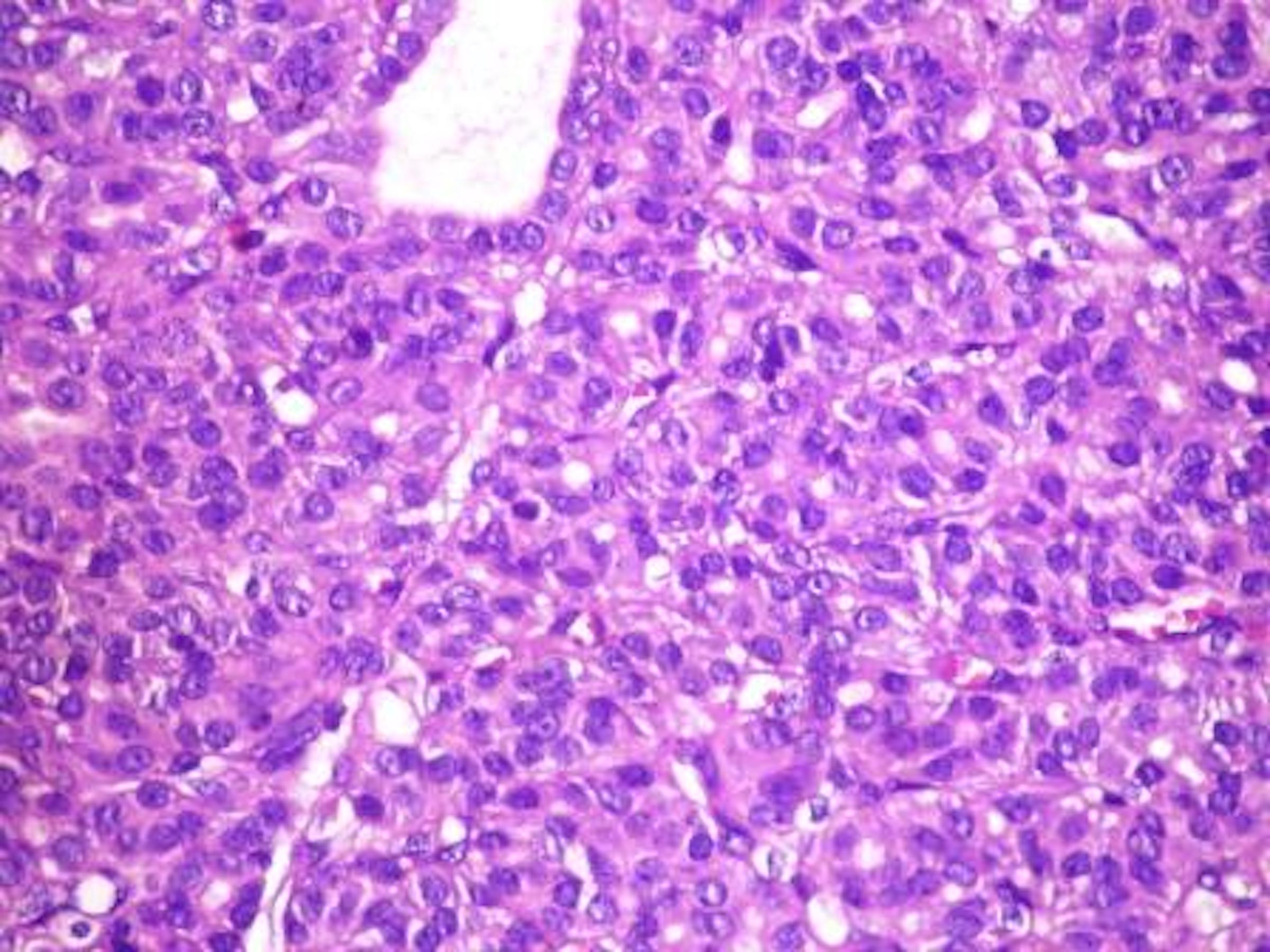
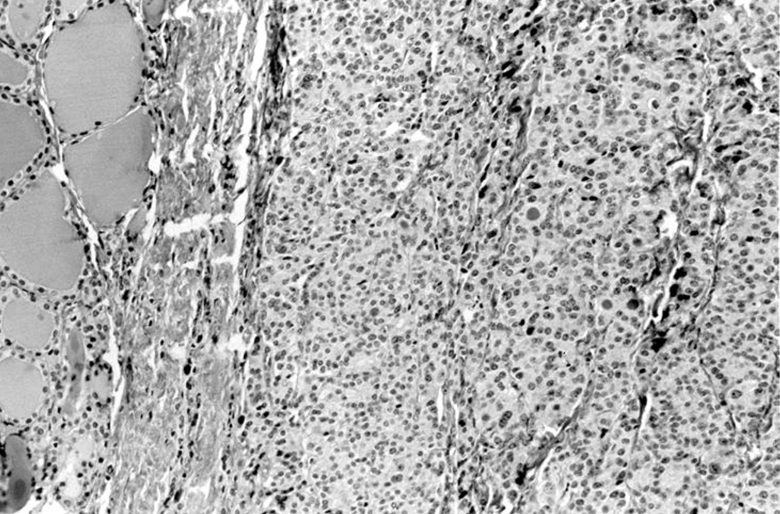
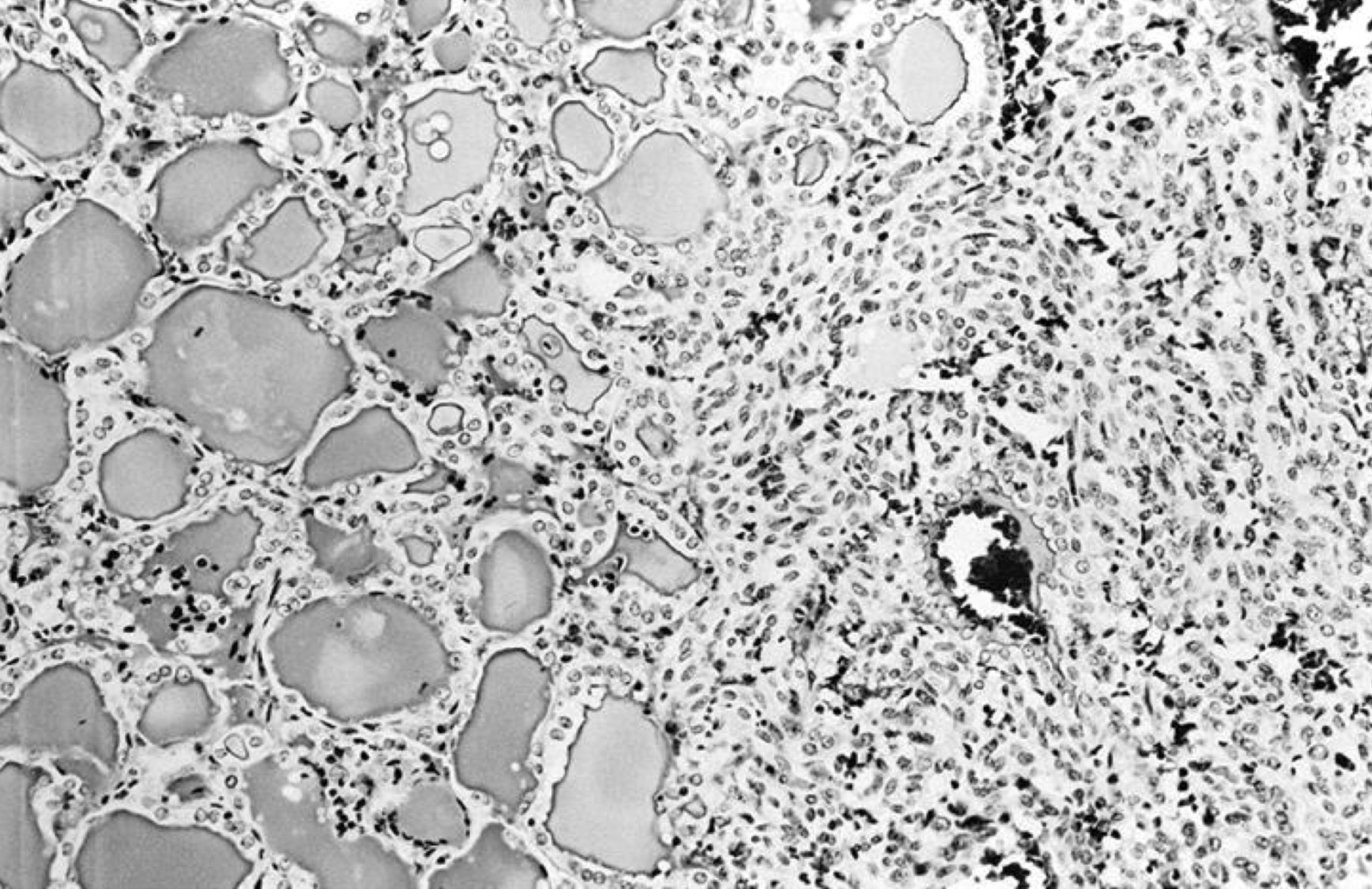
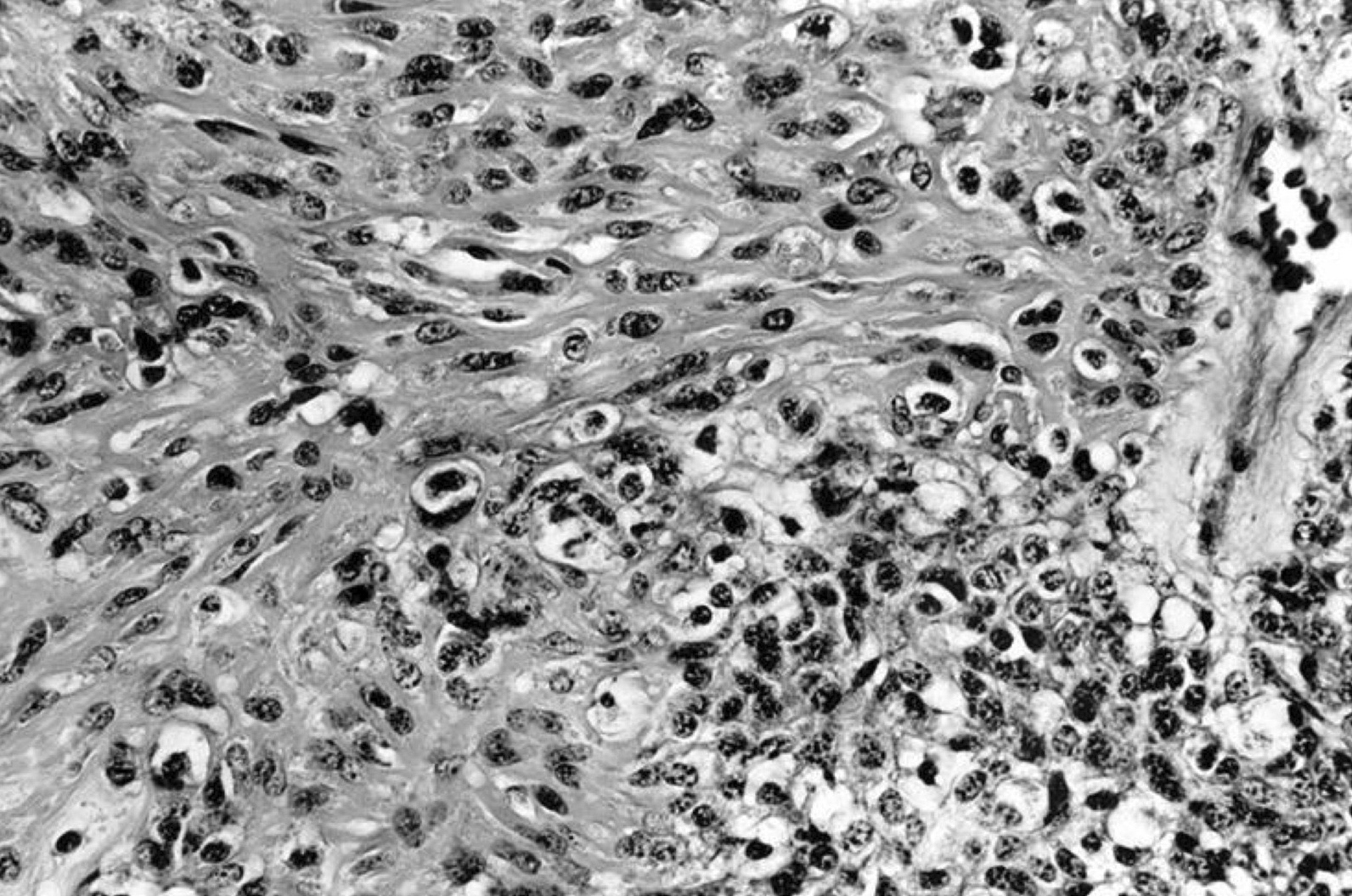
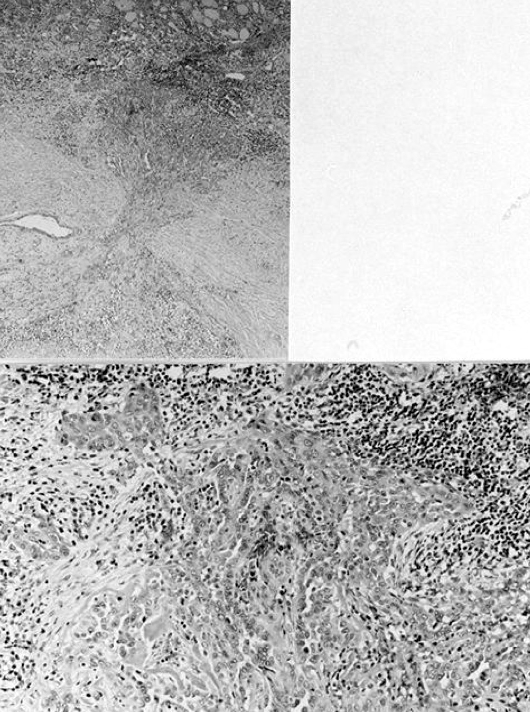
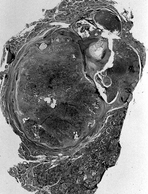
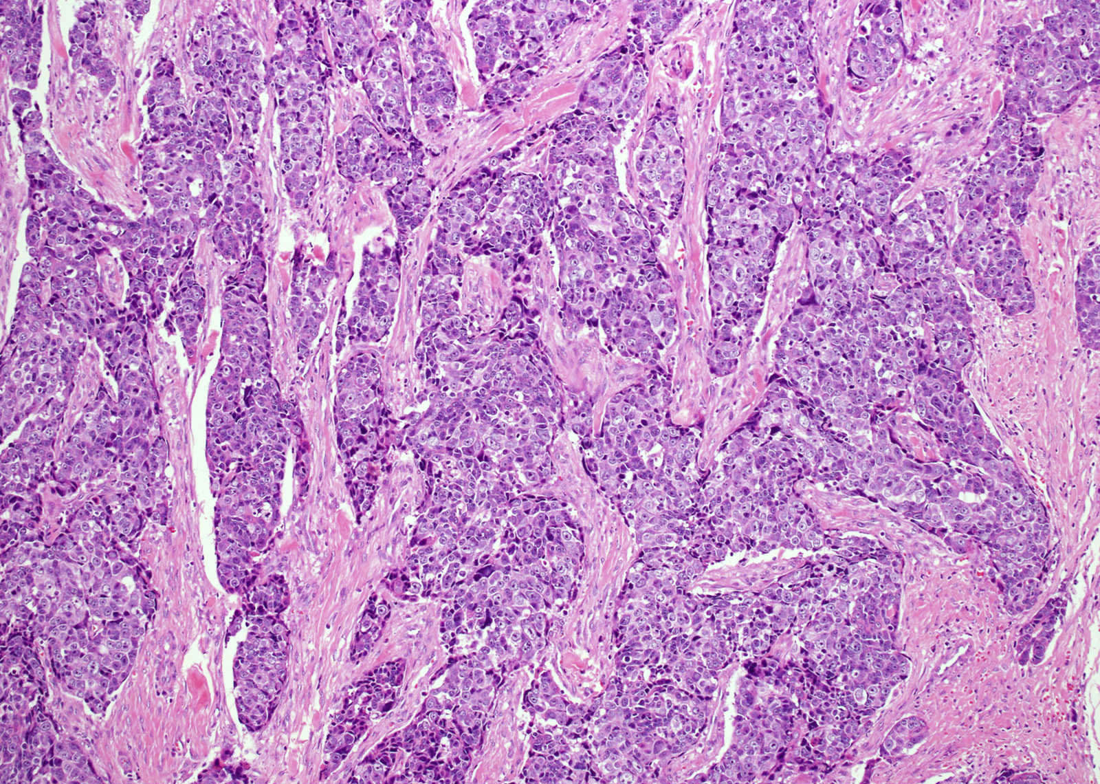
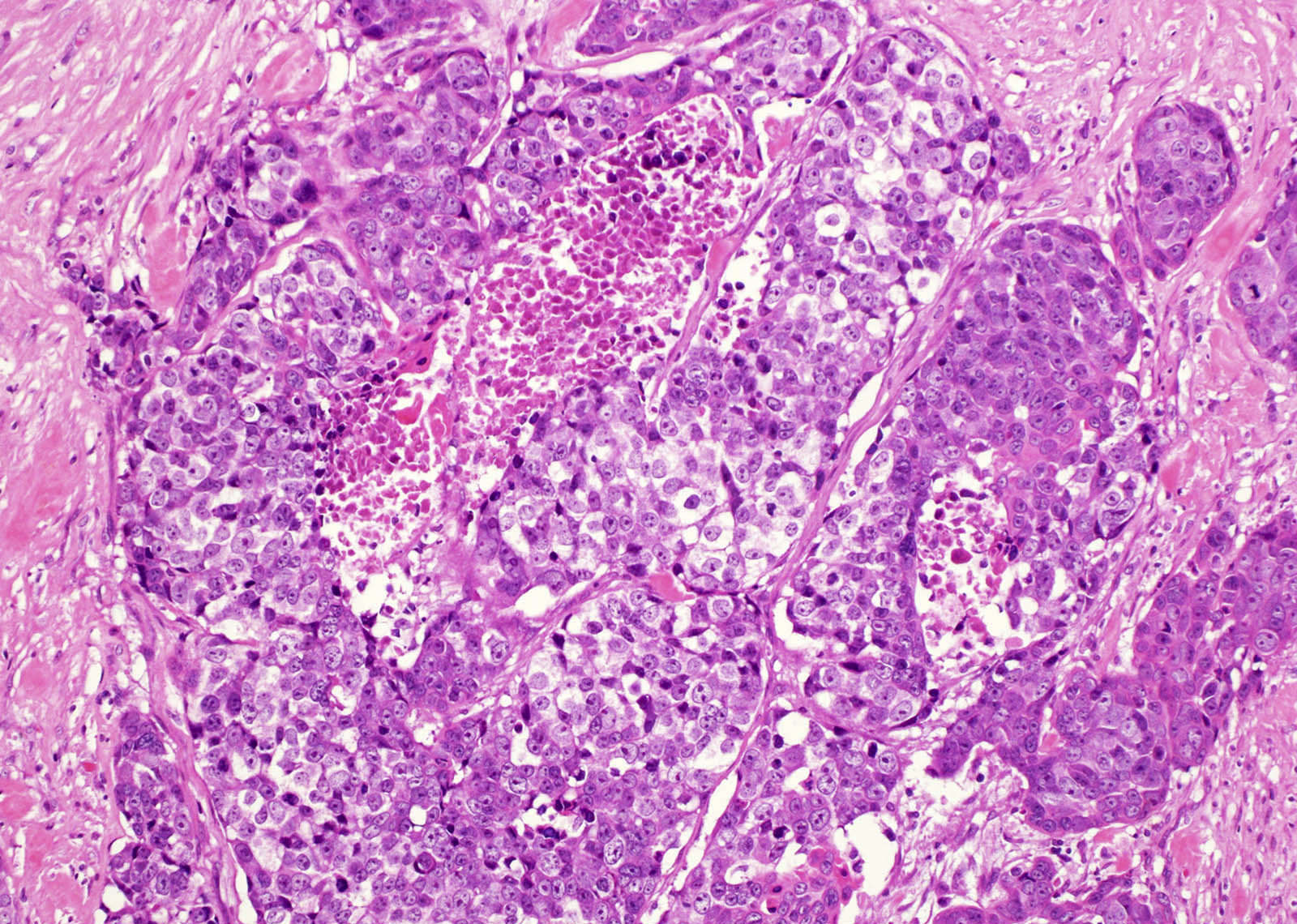
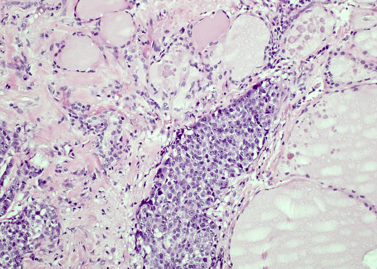
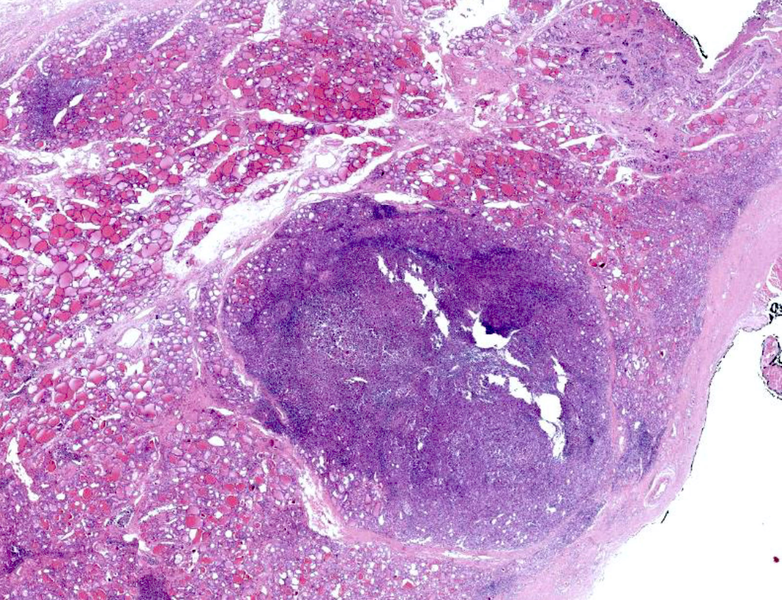
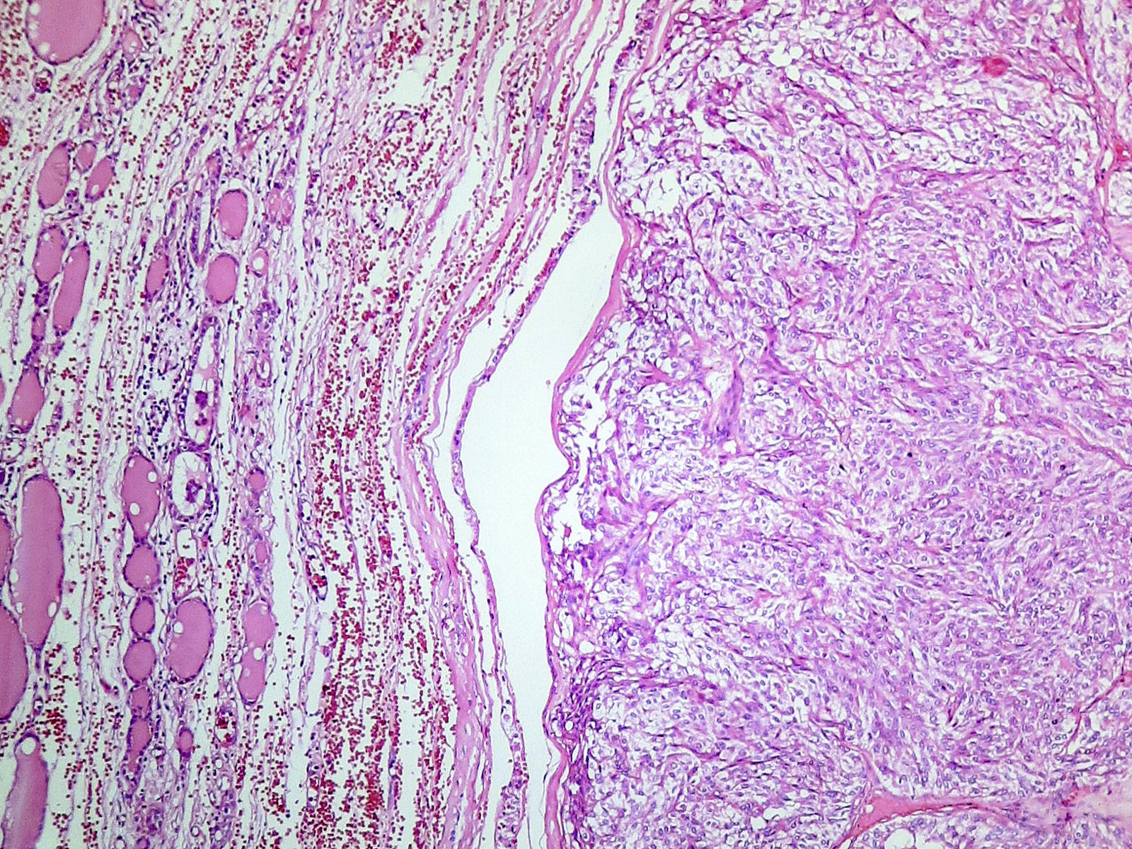
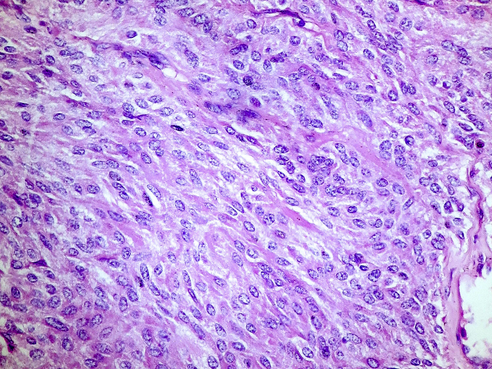
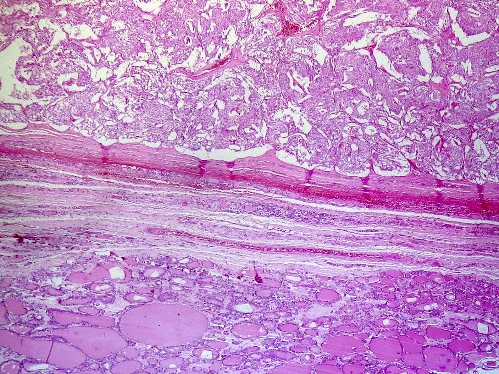
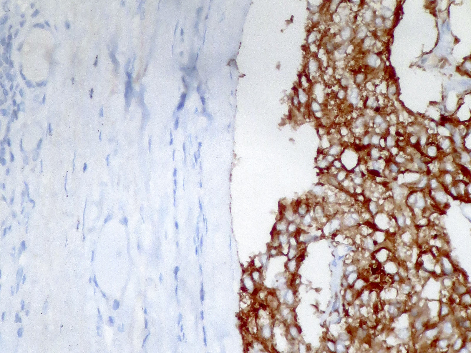
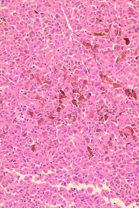
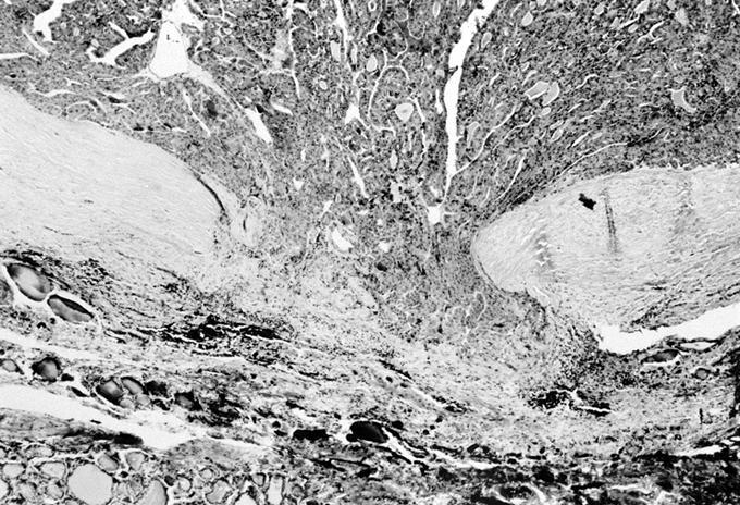
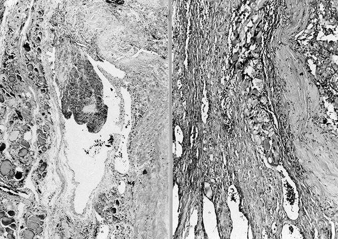
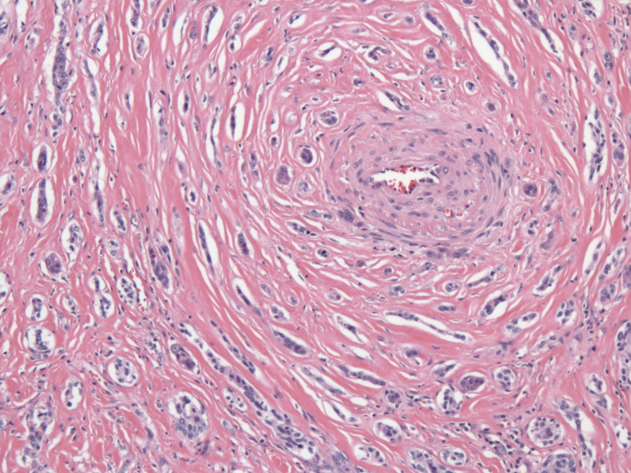
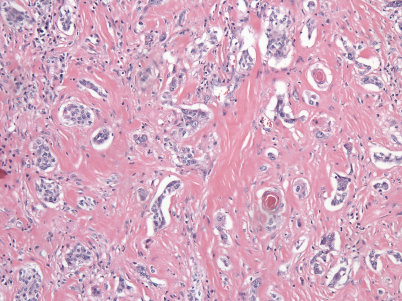
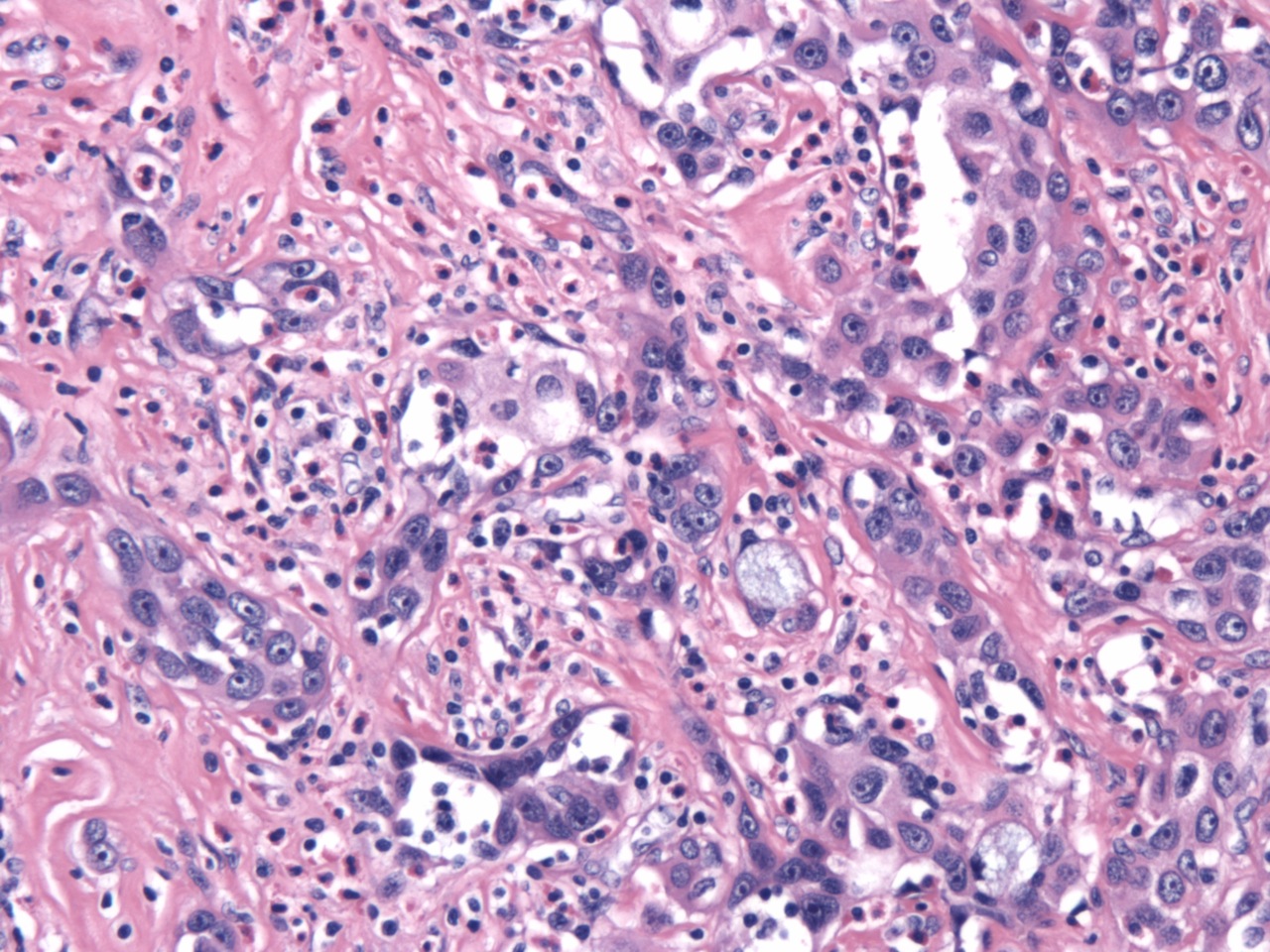
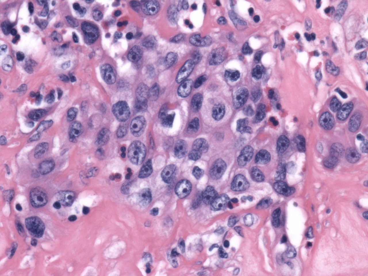
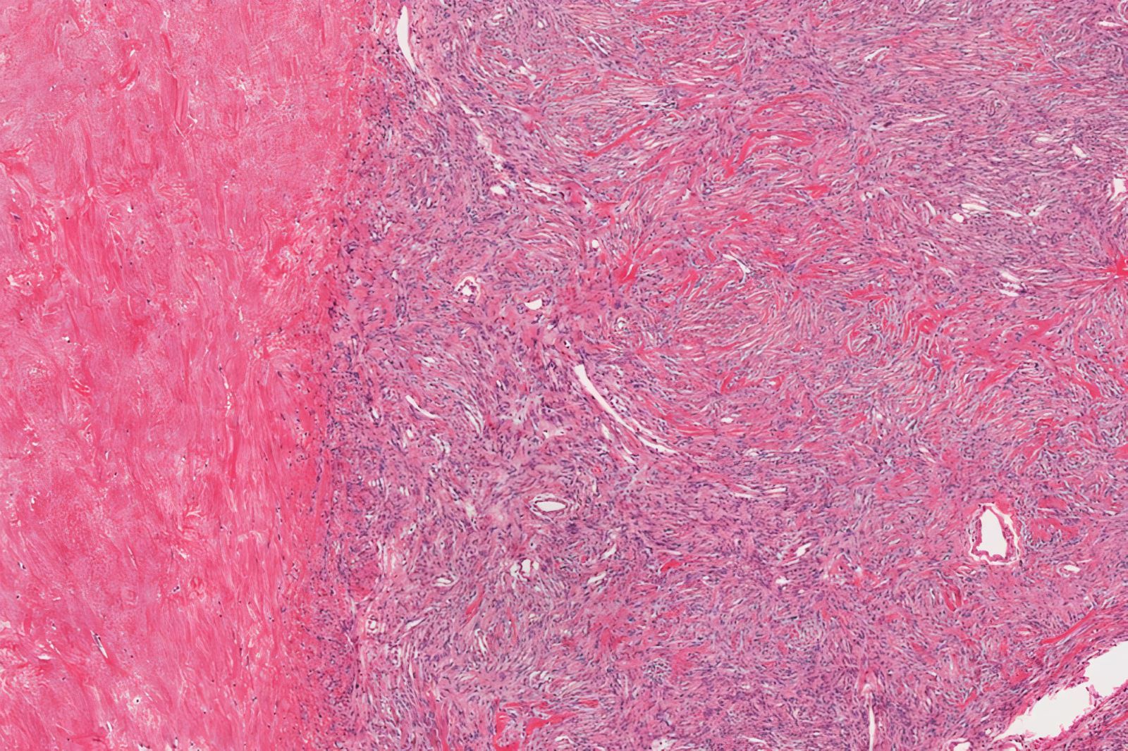
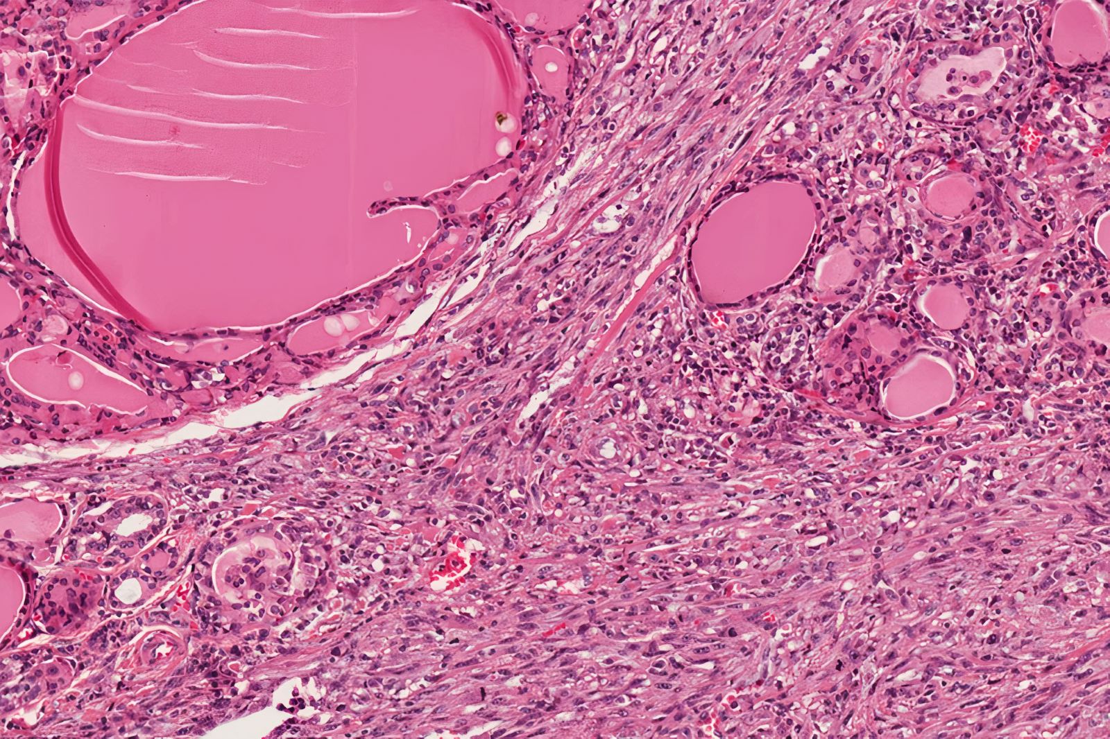
Poorly differentiated thyroid carcinoma
Massive cervical lymph node metastasis
Images hosted on other servers:

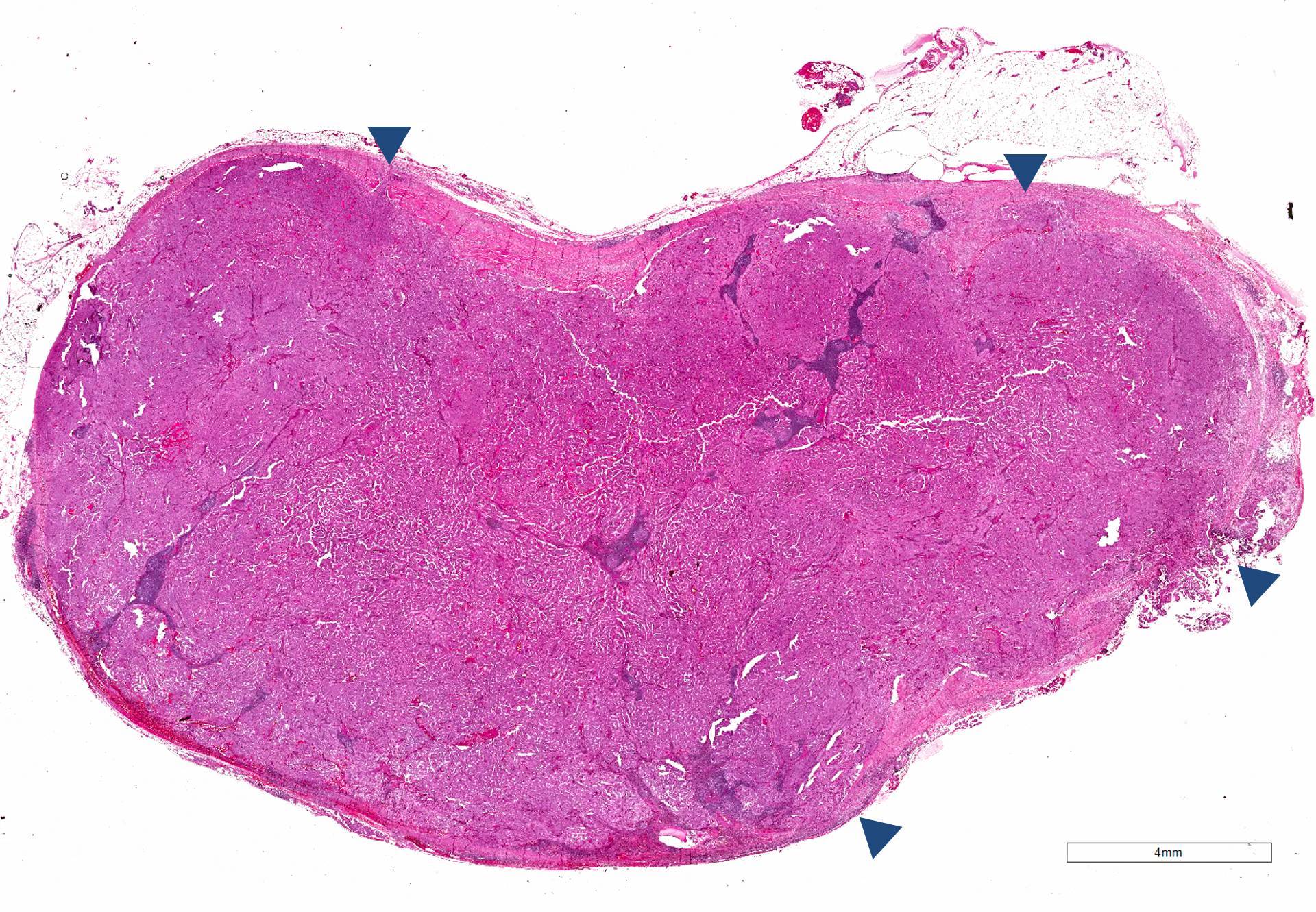
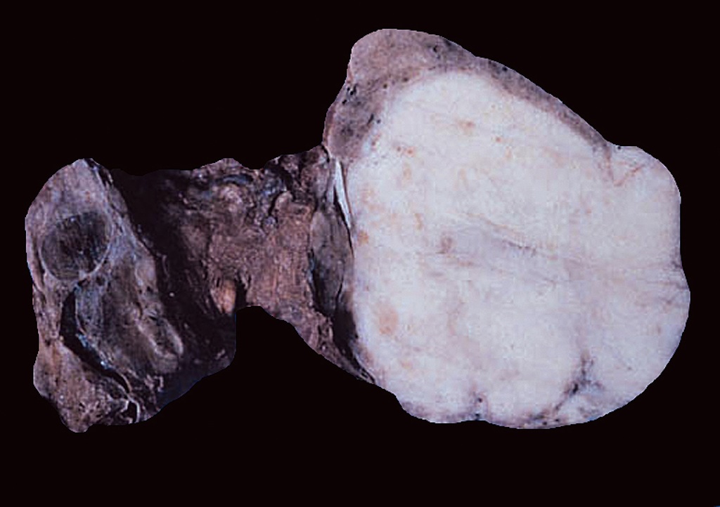
A well demarcated tumor

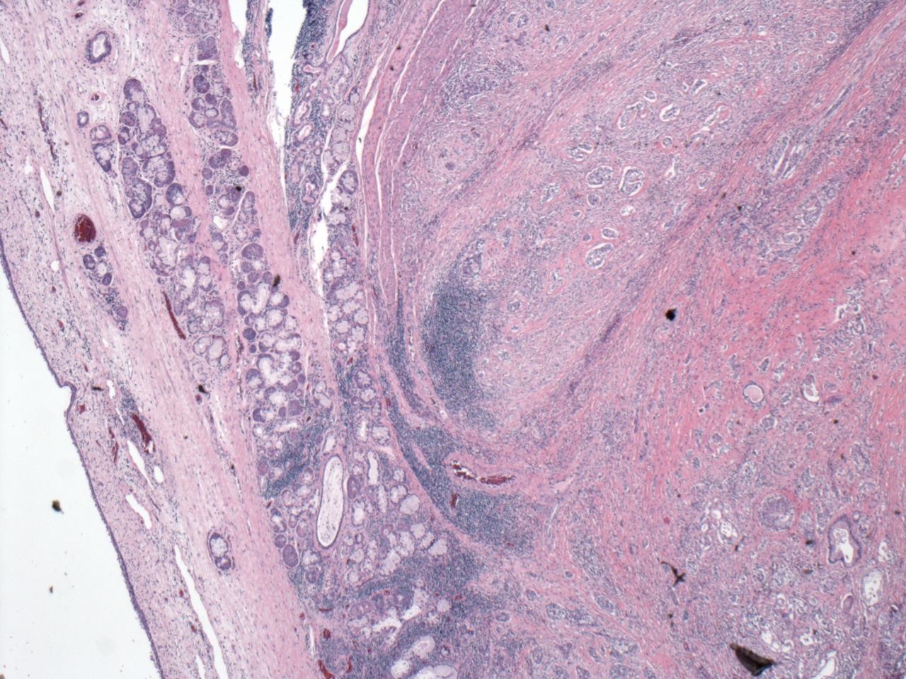
Tumor with invasive growth pattern
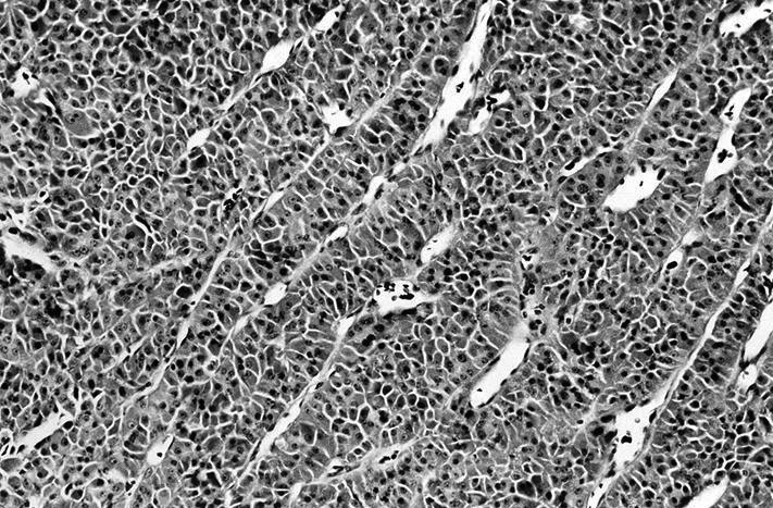
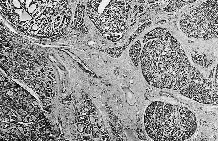
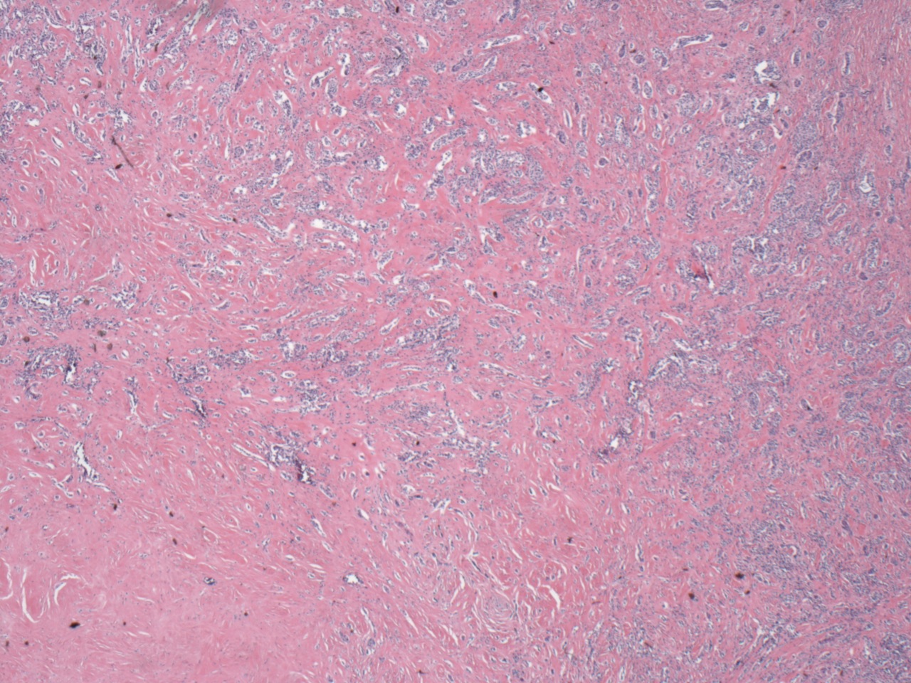
Contributed by Shuanzeng Wei, M.D., Ph.D., Andrey Bychkov, M.D., Ph.D.
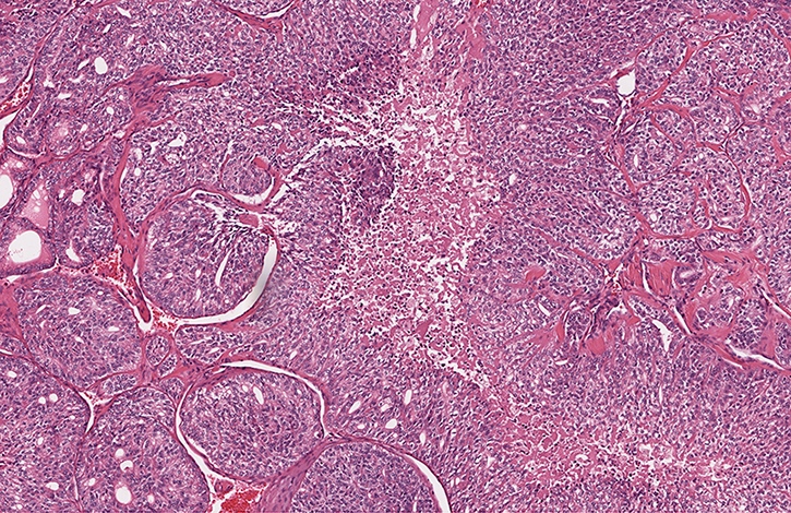
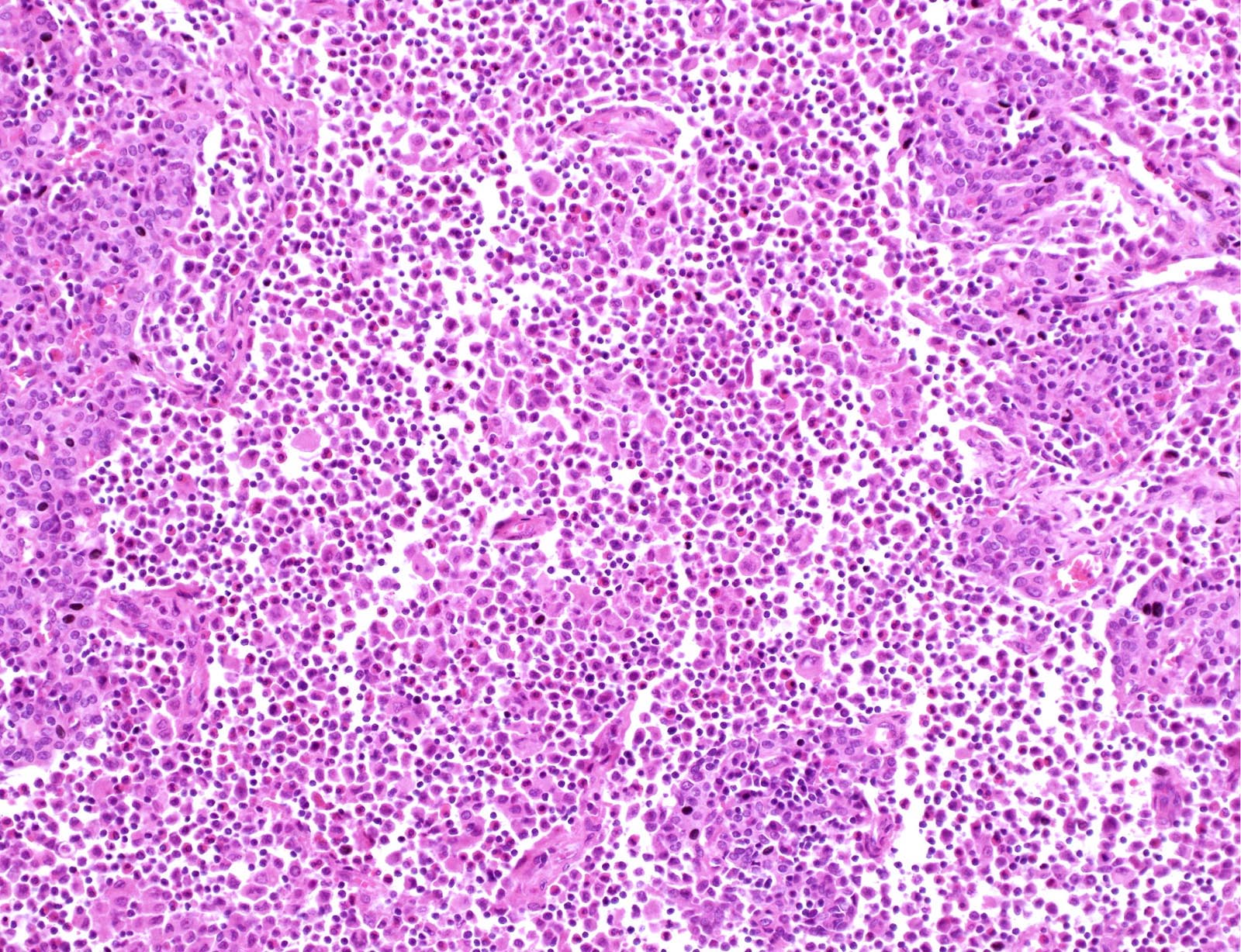
Nests of tumor with necrosis
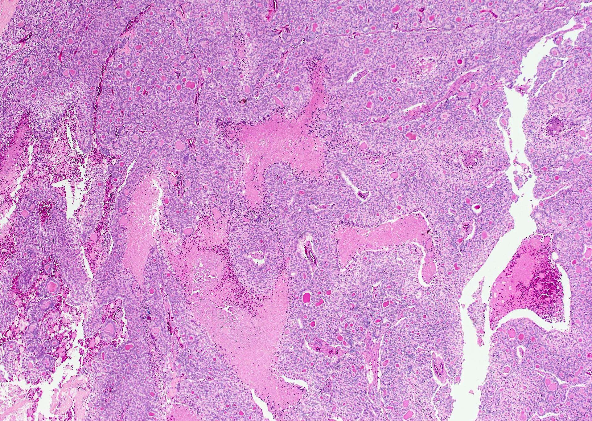
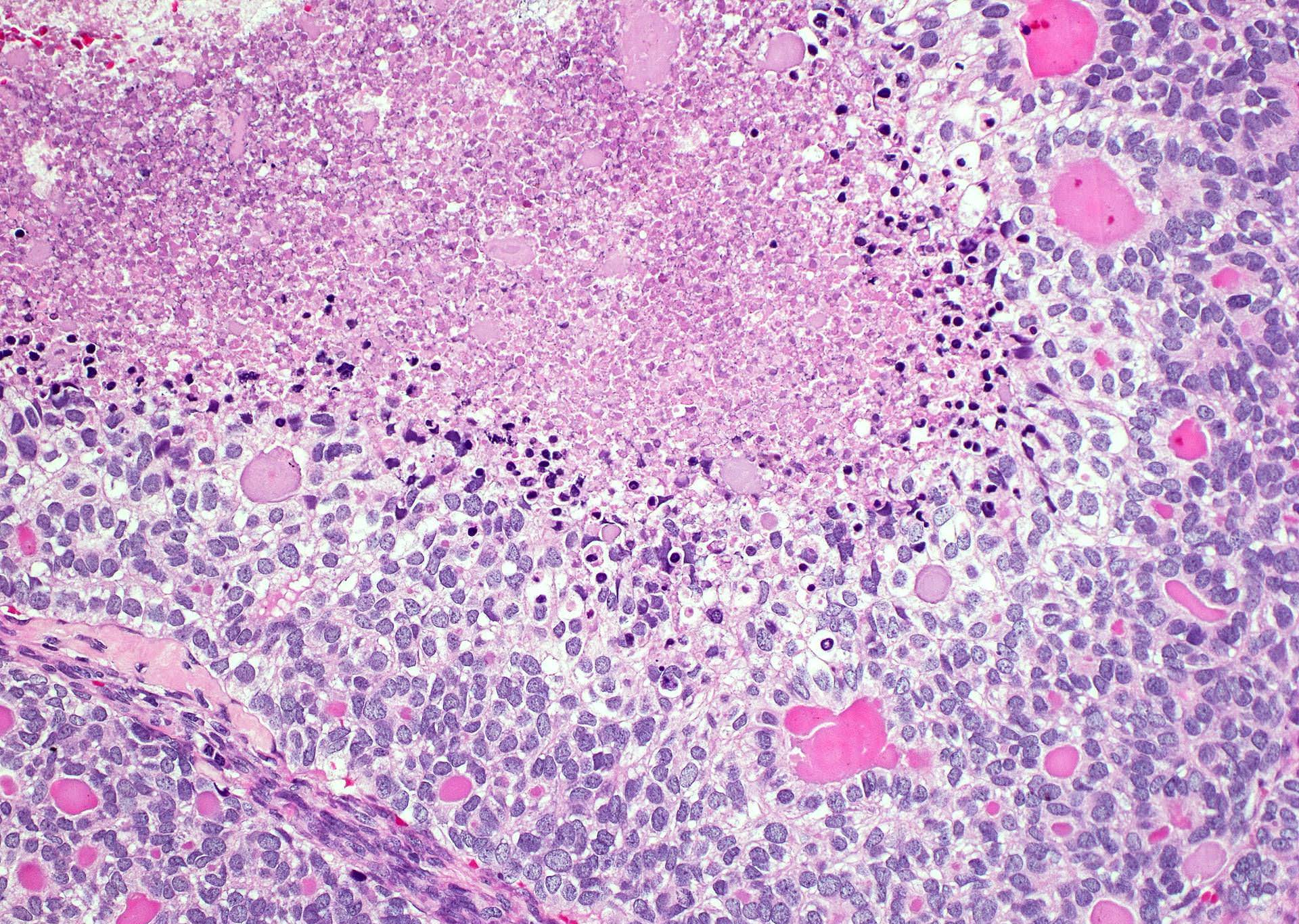
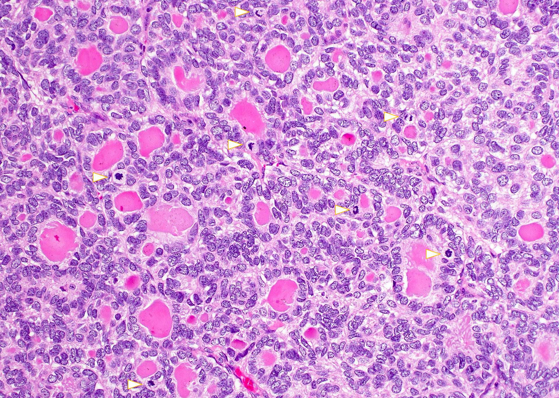
Follicular carcinoma with high grade progression
Case #435
Various images
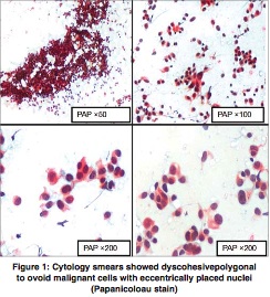
Contributed by Ayana Suzuki, C.T. and Shuanzeng Wei, M.D., Ph.D.
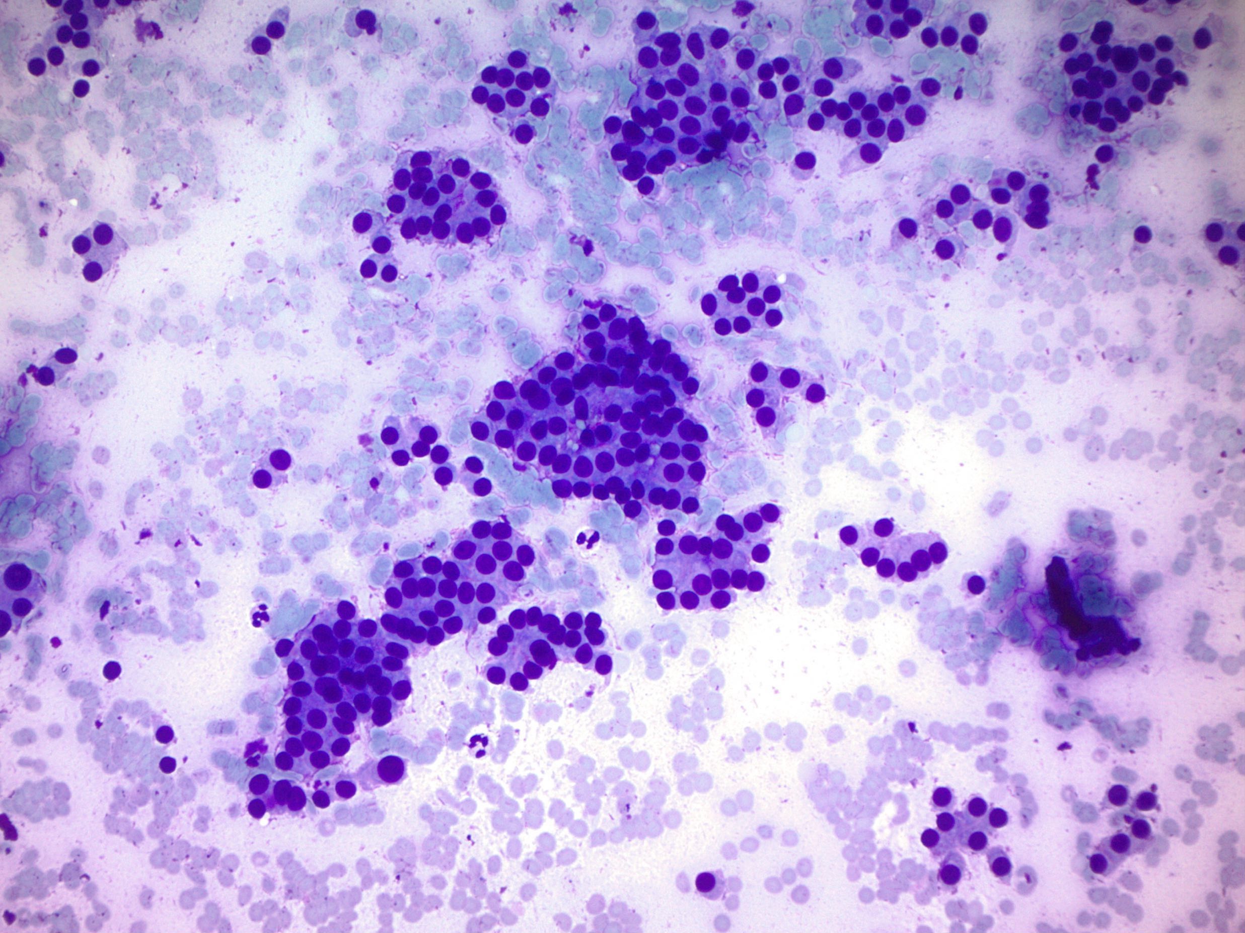
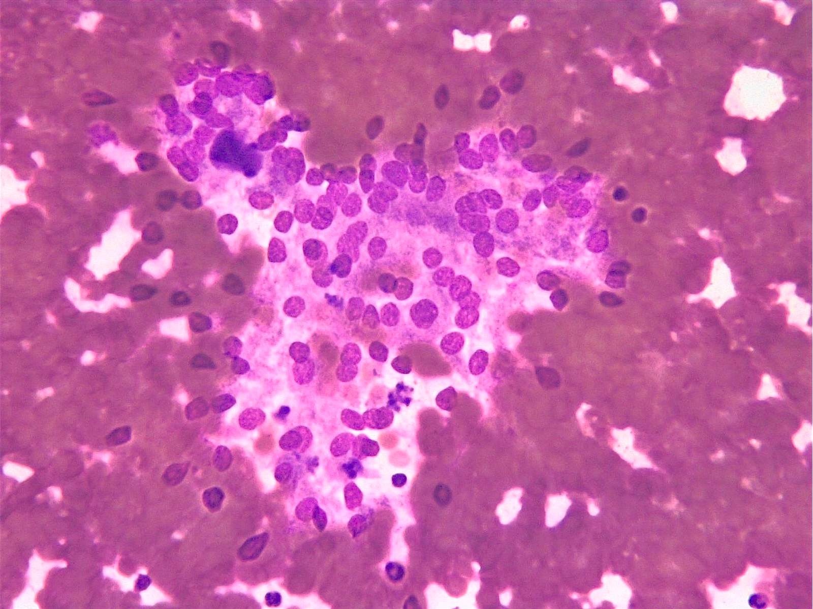
Insular pattern

Diff-Quik stain
Pap stain
Corresponding histology shows mitosis and necrosis
Images hosted on other servers:

Nesting pattern of cells

Overlapping cells with round, regular nuclei

Large clusters and single cells

Cellular nests of loosely cohesive cells

Overlapping cells with mild atypia

Small microfollicle of tumor cells

Vacuolated cytoplasm with round nuclei
Poorly differentiated thyroid carcinoma by M. Brandwein (2020)
Images hosted on other servers:

Gross and microscopic findings
Images hosted on other servers:

Fragments of fibrocollagenous tissue
Contributed by Andrey Bychkov, M.D., Ph.D. and AFIP
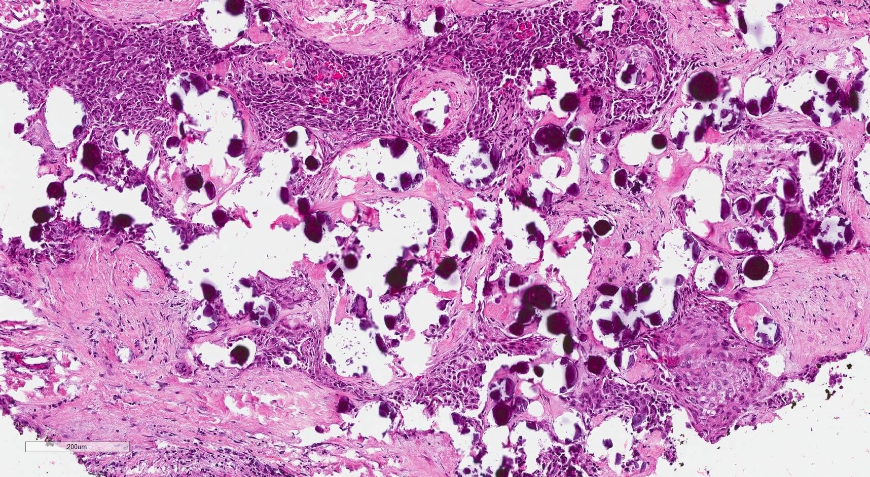
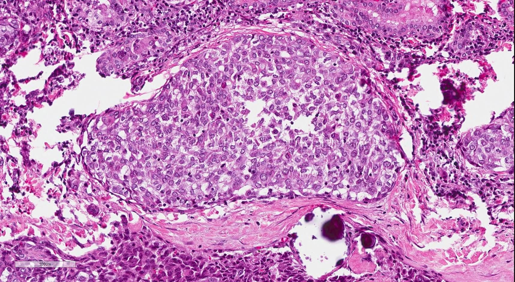
Small nodule of atypical cells with bizarre nuclei

Large eosinophilic cells
Small regenerative nodule
Compensatory nodular hyperplasia
Prominent nuclear atypia / pleomorphism
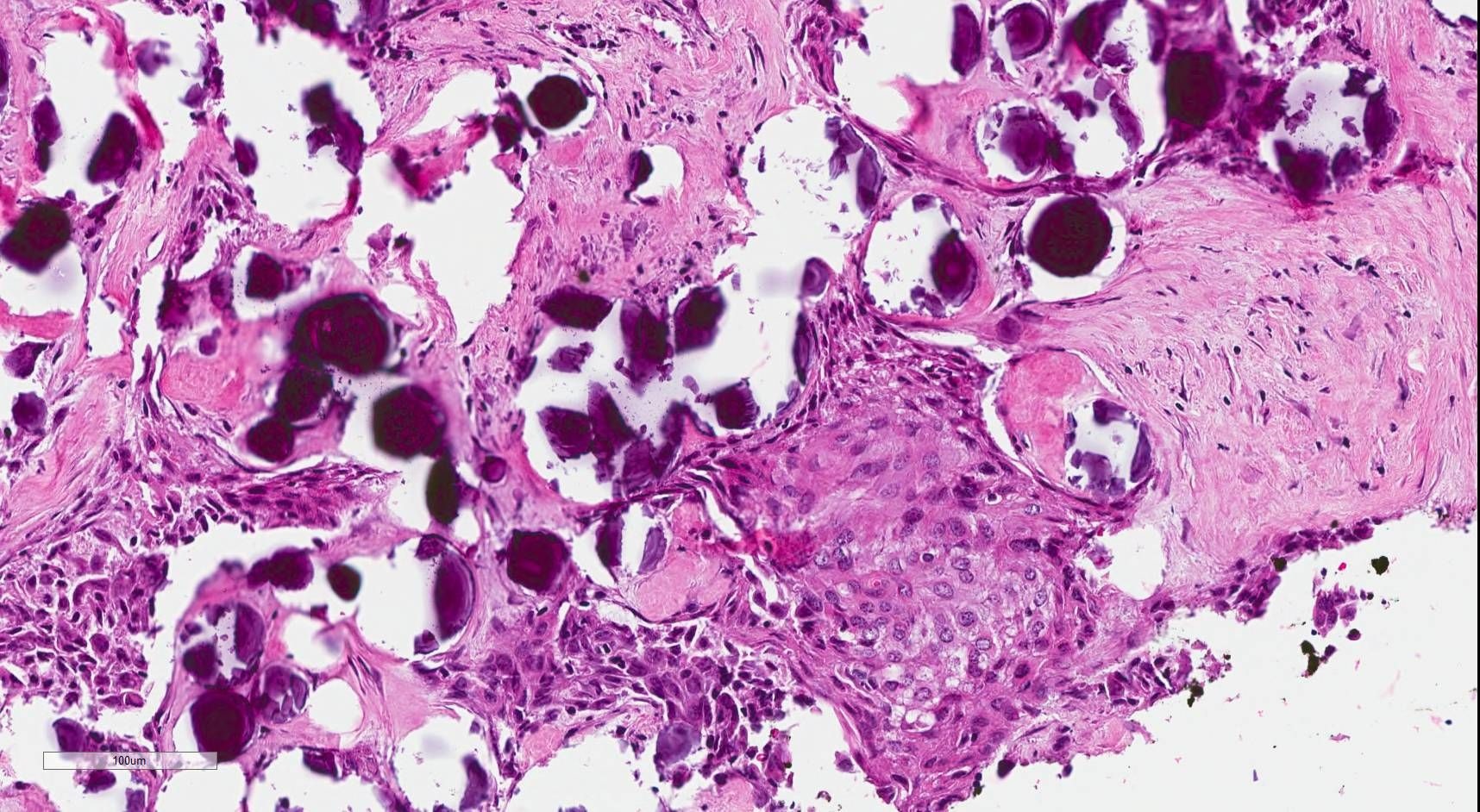
Postnecrotic hyalinosis of colloid nodule
Intact parathyroid versus highly atrophic thyroid follicles
Hyalinized vessel wall
Bizarre nuclear changes
Papillary changes
Images hosted on other servers:


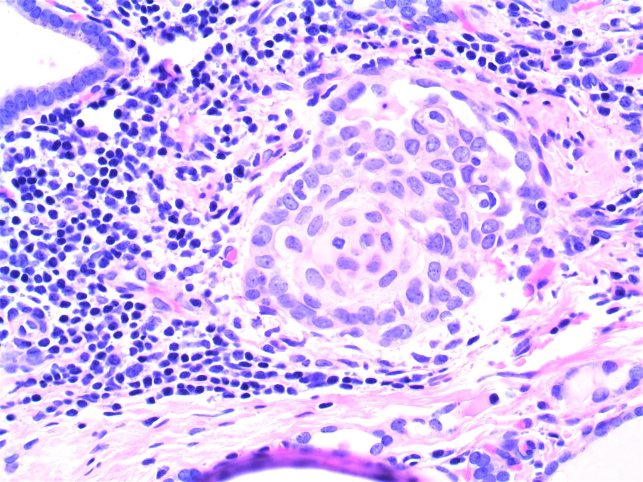
Hyperplastic epithelium and atypia
AFIP images
Atrophic thyroid follicles

Resembles papillary microcarcinoma

Follicles within scar irregular
Heavy inflammatory infiltrate, venous wall
Images hosted on other servers:


Massive scarring and lymphohistiocytic infiltrate


Obliterating phlebitis
Images hosted on other servers:


Leiomyosarcoma: nodule with heterogeneous uptake
Images hosted on other servers:

Synovial sarcoma:
well demarcated
lobulated, tan
solid mass
Images hosted on other servers:
Leiomyosarcoma:

Compactly cellular

Cigar shaped nuclei

Invades thyroid parenchyma

CK-

h-Caldesmon

Desmin

ER-

TTF1-

Chromogranin-, synaptophysin-

Liposarcoma


Synovial sarcoma: biphasic growth; cytokeratin & vimentin


UPS / MFH: residual thyroid follicles; intersecting bundles of atypical spindle cells
Images hosted on other servers:

Synovial sarcoma:
spindled and
epithelioid cells
Images hosted on other servers:

Synovial sarcoma:
solid nests of
epithelial cells
Images hosted on other servers:

Synovial sarcoma:
SYT-SSX fusion
gene transcript
Contributed by Mark R. Wick, M.D. and AFIP
Mucoepidermoid
carcinoma,
sclerosing type

Well circumscribed
tumor (not typical)
with dense fibrosis
Case #230 and AFIP


Small irregular strands of tumor cells
Images hosted on other servers:


Various images
Images hosted on other servers:

Epithelial pearl-like structure

Cell block

Cell block, p40
Images hosted on other servers:

ETV6-NTRK3 fusion
Images hosted on other servers:

Morphology

MASC vs. PTC

Immunophenotype
Images hosted on other servers:

FISH (separate signals)
AFIP images
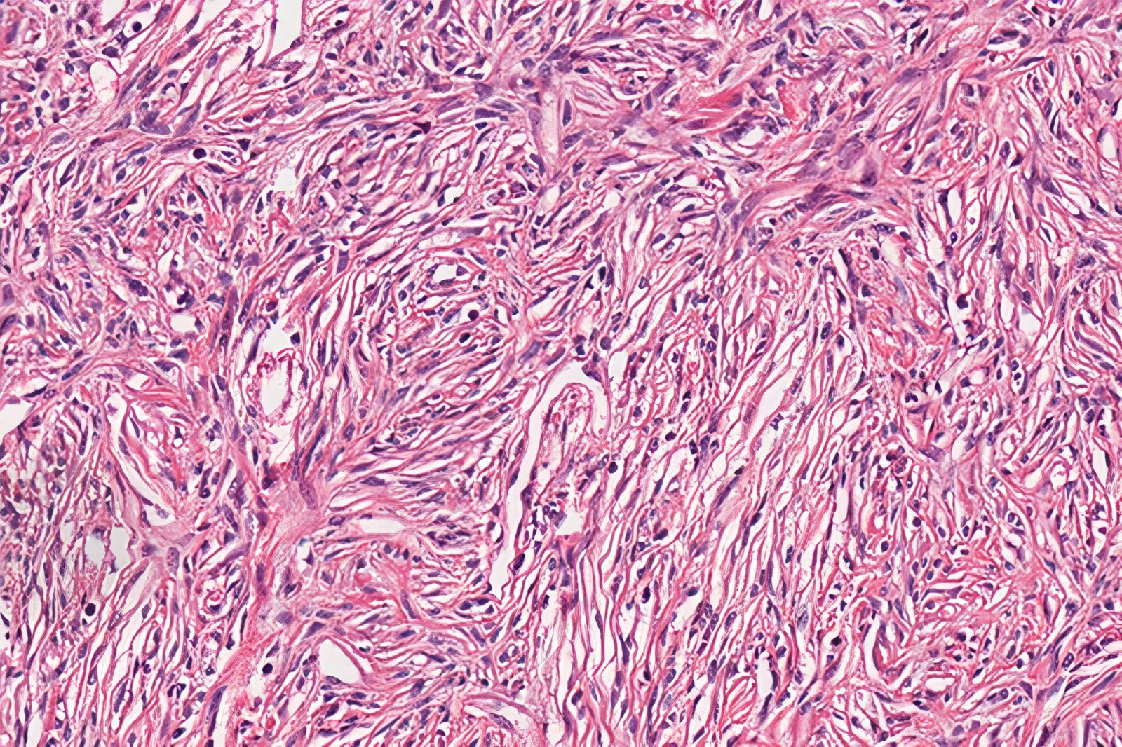
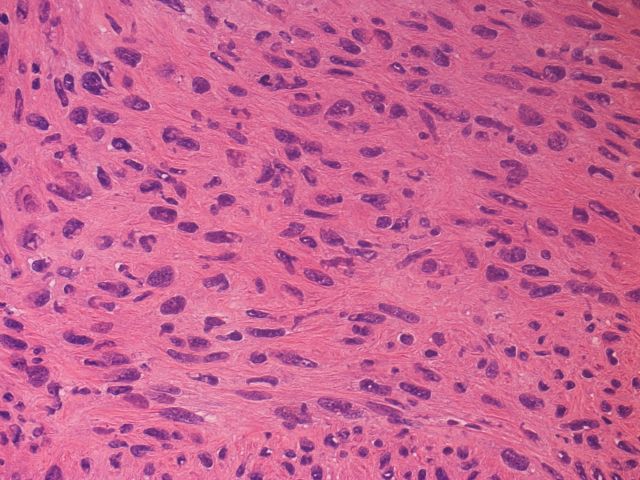
Spindled cells with mesenchymal appearance
Images hosted on other servers:

Solid architecture
traversed by
delicate fibrous
septa

Classic nuclear features of papillary thyroid carcinoma

Fig 3D: RET staining
Images hosted on other servers:

Solid aggregates of epithelial follicular cells

Monolayered epithelial cellular sheets

Enlarged nuclei
with fine chromatin,
occasional nuclear
grooves
Contributed by Andrey Bychkov, M.D., Ph.D. and AFIP
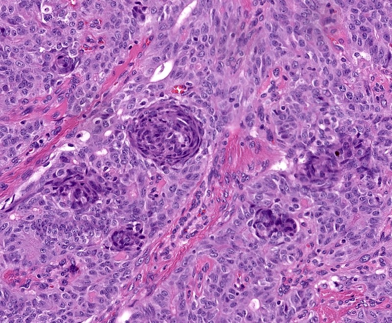
Solid cell nests
Histologic mimics of SCN
Solid cell nests: p63
Solid cell nests: thyroglobulin-
Solid cell nests

Nested pattern
Cystic SCN

Clear cells
Occasional nuclear grooves
Type 1 and 2 solid cell nests
Images hosted on other servers:

Admixture of main and C cell

High mag of solid cell nests

Various stains

Galectin 3, CEA

Conventional solid cell nests
Images hosted on other servers:

4 variable risk stratification
Images hosted on other servers:

Ultrasound: heterogeneous, hyperechoic solitary nodule

CT: heterogeneous enhanced lesion
Images hosted on other servers:

Solid, well circumscribed white nodule

Encapsulated, tan-pink to tan-white mass
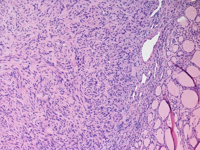
Contributed by Truong Phan Xuan Nguyen, M.D.
Well defined border
Entrapped thyroid follicles
Patternless proliferation of spindled cells
Staghorn-like vessels
Collagenous stroma
STAT6
CD34
Images hosted on other servers:

Scattered spindle-like cells with fusiform nuclei
Images hosted on other servers:

NAB2::STAT6 gene fusion variants
Images hosted on other servers:

Multiple hypoechoic lesions
Images hosted on other servers:

Hemangioma

Metastatic squamous cell carcinoma
Thyroid cytology: colloid nodule
Solitary thyroid nodule
Thyroid: compare and contrast
Images hosted on other servers:

Ulceroproliferative
growth

Recurrent
ulceroproliferative
growth

Mass pressuring;
infiltrating trachea;
surrounding soft tissue
Images hosted on other servers:

Enlarged left lobe

White tumor with central necrosis
Contributed by Shuanzeng Wei, M.D., Ph.D.

Squamous cell carcinoma with necrosis
Images hosted on other servers:

Infiltrating squamous cell carcinoma
Images hosted on other servers:

Atypical squamoid cell
Images hosted on other servers:

Thyroid function
Contributed by Ayana Suzuki, C.T.
Hypoechoic nodule
Images hosted on other servers:

Hypoechoic nodule with poorly defined margin

High 18F-FDG uptake in enlarged right thyroid lobe
Contributed by Truong Phan Xuan Nguyen, M.D.
Follicular damage
Granuloma
Multinucleated giant cells
Contributed by Truong Phan Xuan Nguyen, M.D. and Ayana Suzuki, C.T.
Cellular smear
Multinucleated giant cells

Epithelioid cells
Giant cell
Contributed by Ayana Suzuki, C.T.
Suspicious for PTC
Hyalinizing trabecular tumor

Suspicious for lymphoma
Images hosted on other servers:


Suspicious for lymphoma


Suspicious for medullary thyroid carcinoma
Suspicious for papillary thyroid carcinoma


Suspicious for metastatic carcinoma
Images hosted on other servers:

Tall cell variant in 19 year old woman
Contributed by Andrey Bychkov, M.D., Ph.D.
Entire lobe
Muscular invasion
Contributed by Livia Florianova, M.D., M.Sc. and Marc Pusztaszeri, M.D.

Tram track pattern
Papillary architecture
Elongated papillae
Solid architecture
Trabecular architecture
Tall cells
Oncocytic-like cytoplasm
Extrathyroidal extension
Microcarcinoma with infiltrative borders
Nuclear features
HBME-1
Galectin 3
Cytokeratin 19
BRAF V600E
Contributed by Andrey Bychkov, M.D., Ph.D.
Long papillae with tall cells
Tram track pattern
Contributed by Papanicolaou Society and the Bethesda System for Reporting Thyroid Cytopathology, Manon Auger, M.D., C.M., Livia Florianova, M.D., M.Sc. and Marc Pusztaszeri, M.D.

Giemsa
Papanicolaou

Soap bubble nuclear pseudoinclusions
Tall cells
Papillae (cell block)
Tall cells (cell block)
High grade and poorly differentiated and anaplastic thyroid carcinoma
Ultrasound of tall cell variant of PTC
Images hosted on other servers:

Heterogeneously hypoechoic nodule

Large cystic fat-containing mass
Images hosted on other servers:

Well encapsulated, mixed solid and cystic mass

Lymph node involvement
AFIP images
Newborn infant
Images hosted on other servers:

Benign thyroid teratoma

Cartilage

Primitive neuroepithelial cells
Images hosted on other servers:

Thymus descending path
Images hosted on other servers:

Ectopic thymic tissue

Sonographic images

Intrathyroidal ectopic thymic tissue


Longitudinal US images

Hyperechoic nodule


CT scan


RAI scan
AFIP images
Intrathyroidal thymus gland
Images hosted on other servers:

Nodule < 1 cm
Contributed by Andrey Bychkov, M.D., Ph.D.
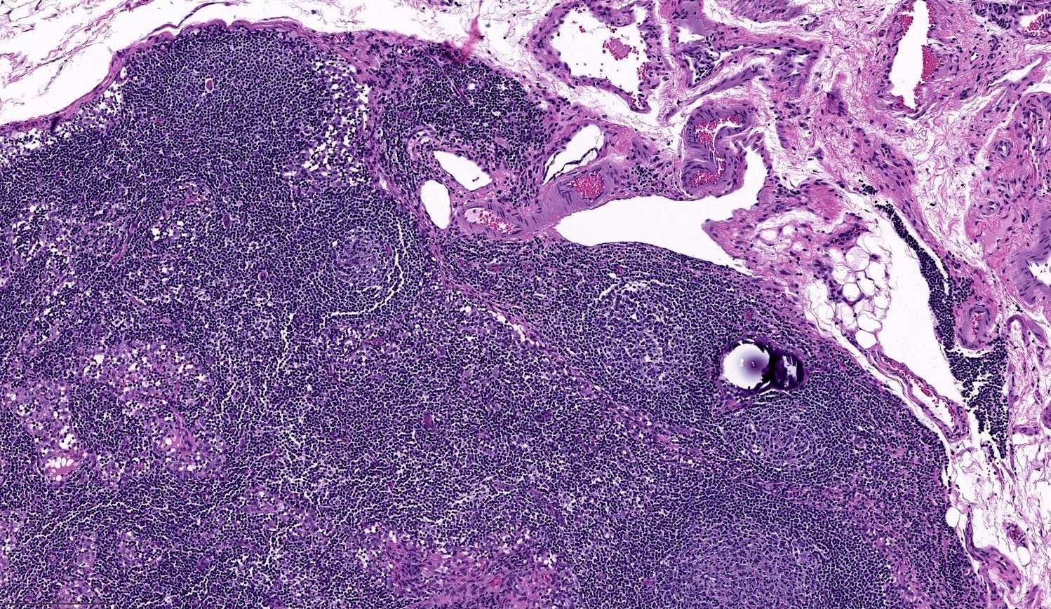
Hassall corpuscle
Images hosted on other servers:


Intrathyroidal thymic tissue


Degenerated Hassall corpuscle

Ectopic thymus and parathyroid

Subcapsular intrathyroidal thymus (E)


Subcapsular intrathyroid thymus, rats

Ectopic intrathyroidal thymoma
Images hosted on other servers:

Hassall corpuscle (D)

CK+ epithelial cells

Lymphocytes
Images hosted on other servers:

Neck ultrasound

Multicystic lesion

Calcification

Infrahyoid lesion

Low density

Axial and transverse

Cystic swelling

Thick septae

Solid component
Images hosted on other servers:


Neck mass


Intraoperative
Images hosted on other servers:

Thyroidectomy

Total resection

Thyroglossal duct

Thyroglossal cyst

Modified neck dissection

Sistrunk procedure

PTC in TGD cyst
Scroll to see all images:
Contributed by Andrey Bychkov, M.D., Ph.D.
Cystic papillary carcinoma

Papillary structures
Images hosted on other servers:


Papillary carcinoma

TGDC wall

Papillary pattern

Overcrowding nuclei

Columnar epithelium

Cystic neoplasm

Squamous epithelium

Papillary appearance

Irregular nucleus

Microcalcifications

Papillary carcinoma

Normal cyst wall lining


PTC tall cell variant

Squamous cell carcinoma

Mucoepidermoid tumor

TTF1, thyroglobulin

Thyroglobulin


CK19
Images hosted on other servers:

Papillary carcinoma in TGD cyst aspirate
Images hosted on other servers:
Thyroglossal tract

Thyroglossal duct cyst

Management algorithm


Sistrunk procedure
Contributed by Ayana Suzuki, C.T.
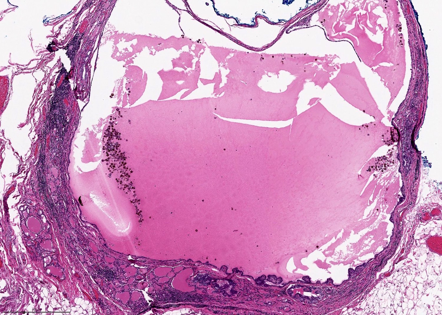
Large cyst
Images hosted on other servers:


Neck ultrasound


Neck CT


Neck CT


Scintigraphy
Contributed by Ayana Suzuki, C.T.
Flocculated appearance
Images hosted on other servers:


Neck mass
TGD cyst in children


Intra-operative
AFIP images
TGD cyst
Images hosted on other servers:
In situ
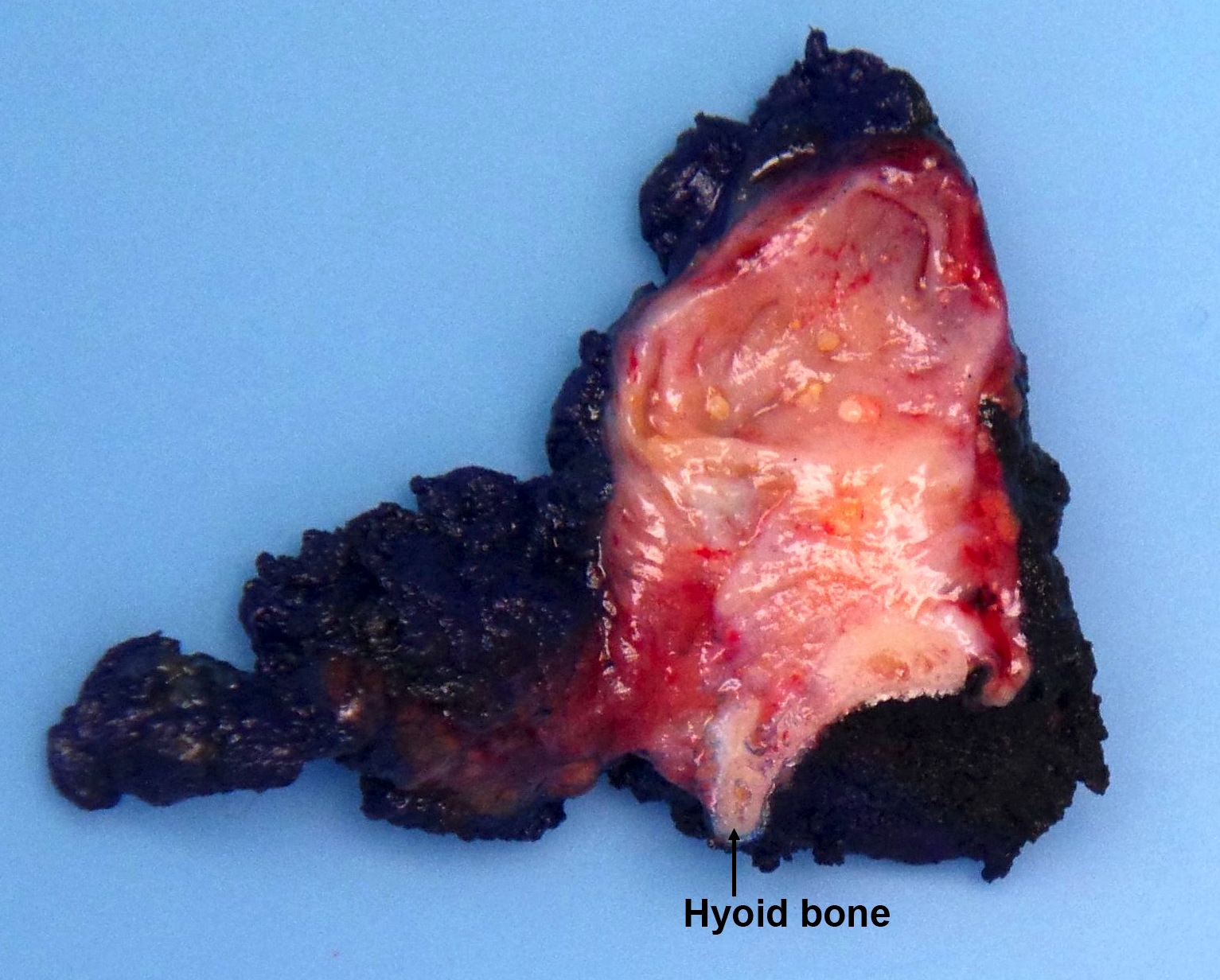
Contributed by Andrey Bychkov, M.D., Ph.D., Mark R. Wick, M.D. and AFIP
Perihyoid cystic and arborized tubular structure
Perihyoid cystic structures
Thyroglossal duct penetrates hyoid bone
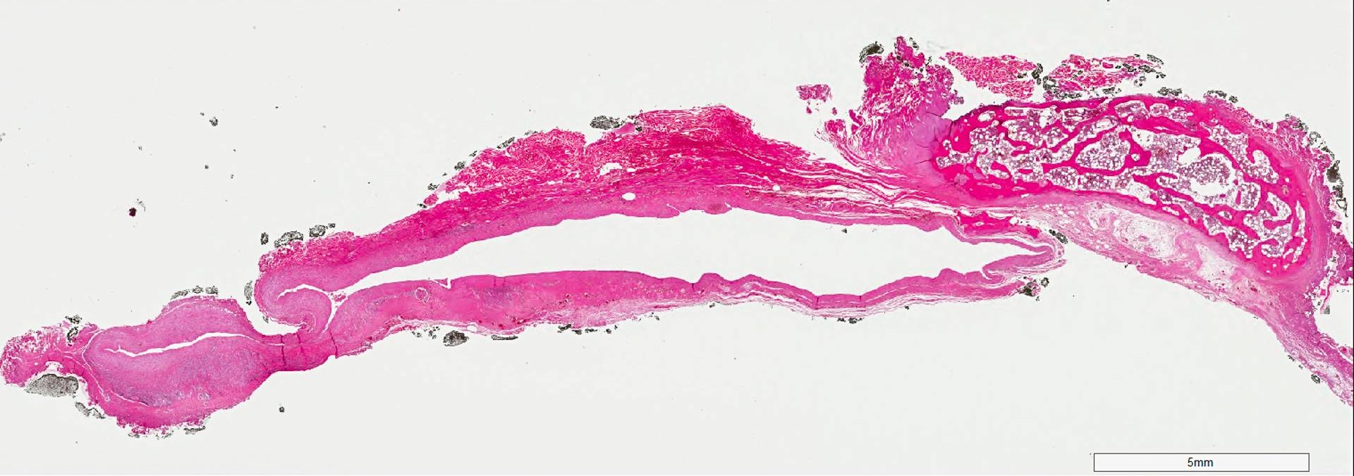
Denuded TGD, perihoid location
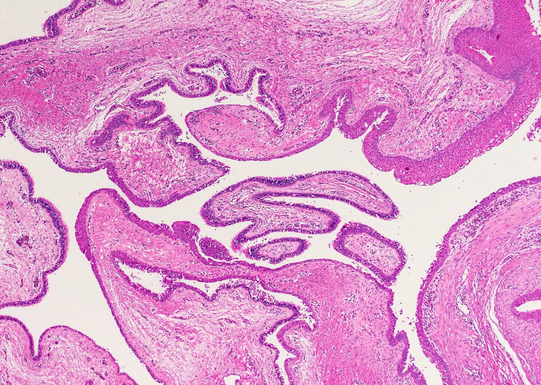
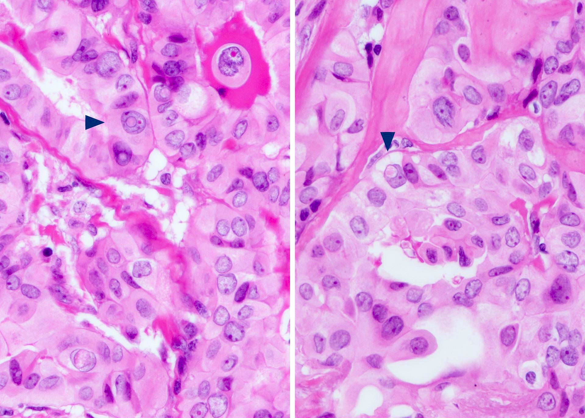
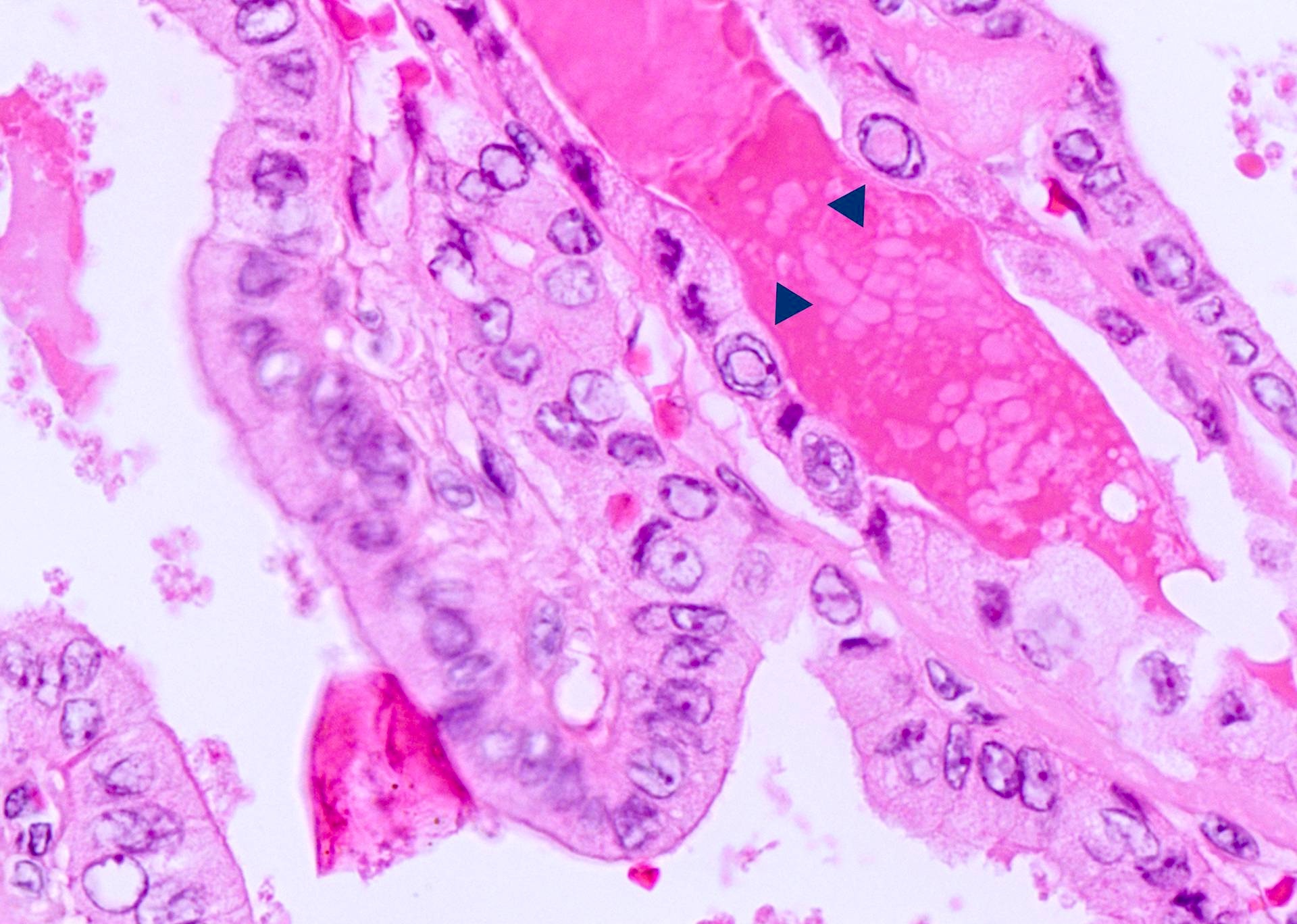
Variable epithelial lining
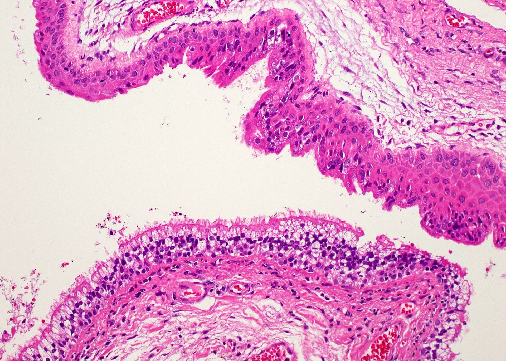
Ciliated versus squamous epithelium
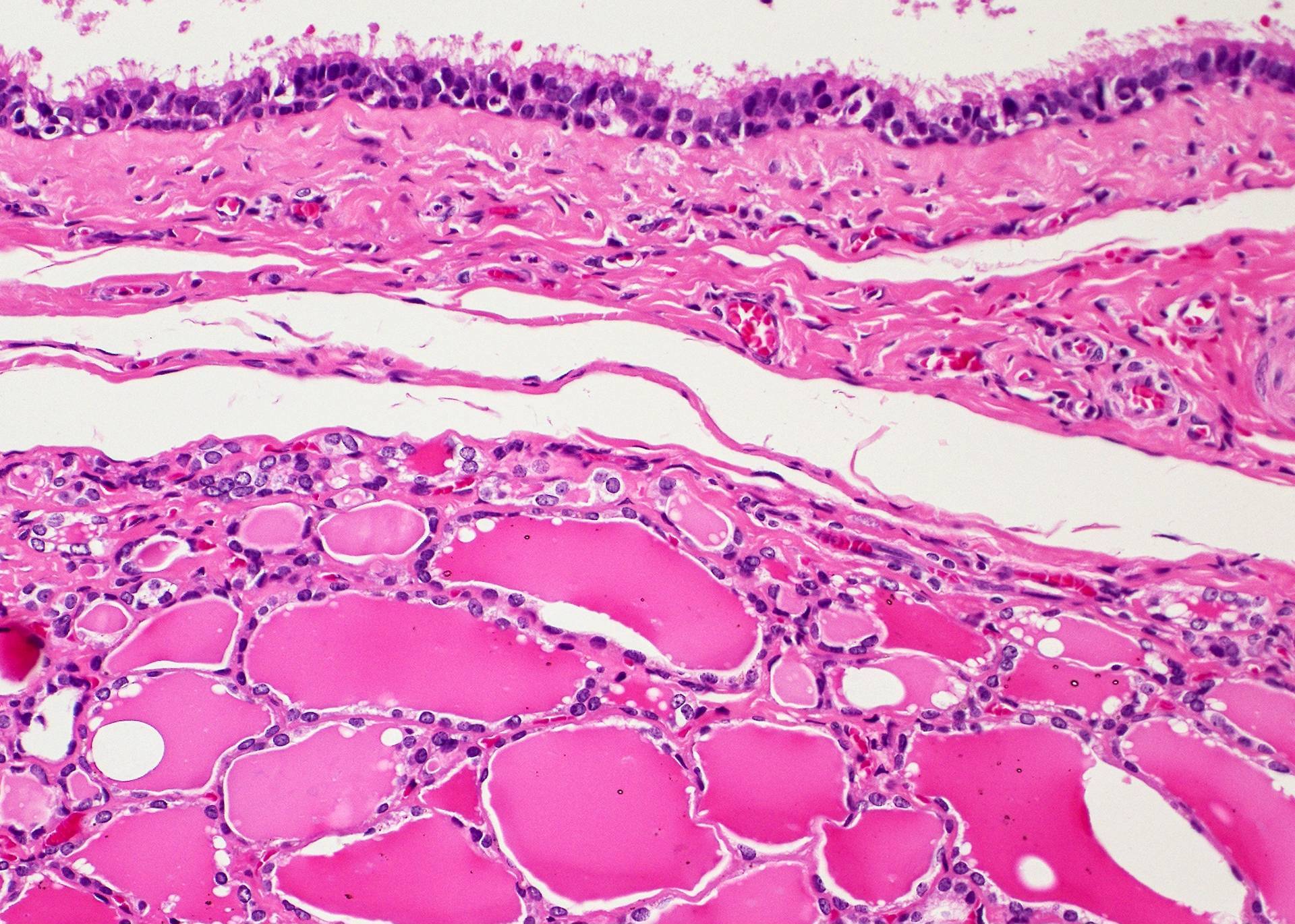
Thyroid follicles under ciliated epithelium
Thyroid follicles of various size
Thyroid follicles embedded within skeletal muscles
Seromucous glands
Rupture of the cyst wall
Cyst rupture
Thyroglobulin expression by thyroid follicles
Strong TTF1 expression
Thyroglossal duct cyst
Epithelium
Papillary changes
Images hosted on other servers:

Squamous epithelium


Pseudostratified

Cuboidal epithelium

Squamous stratified epithelium

Cyst wall

Epithelial lining


Respiratory epithelium


Thyroid follicles


Thyroglossal tract cyst

Cyst wall

With mucocele
Contributed by Andrey Bychkov, M.D., Ph.D., Ayana Suzuki, C.T., Ram Kumar Kurpad R, M.B.B.S., M.D. and Y. C. Spoorthy Rekha, M.B.B.S., M.D.

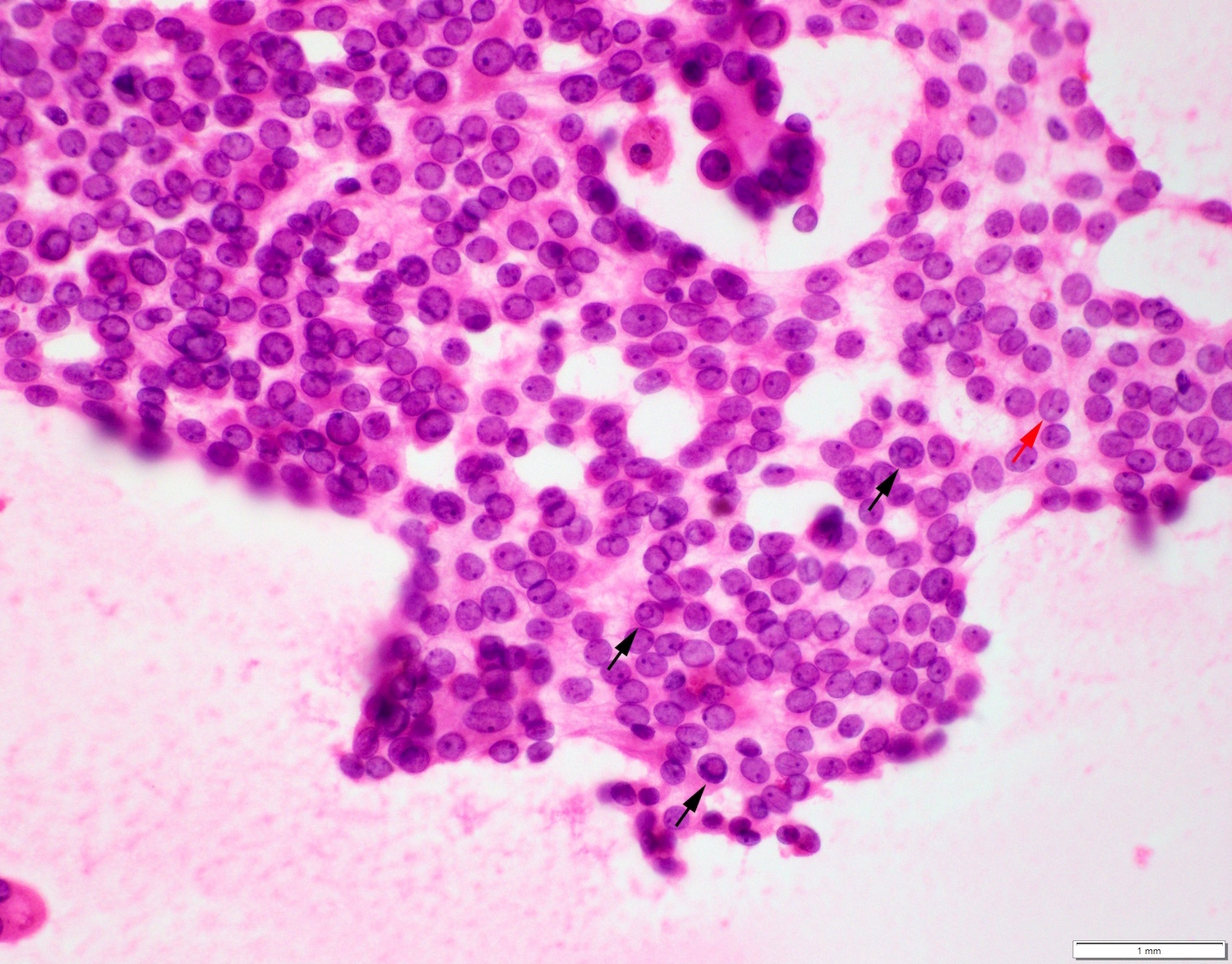
Sparsely cellular aspirate
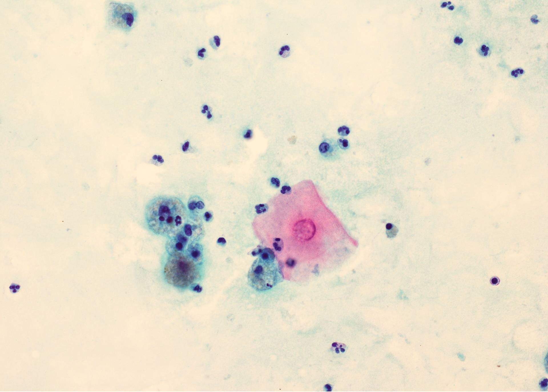
Benign squamous cell

Histiocytes and squame
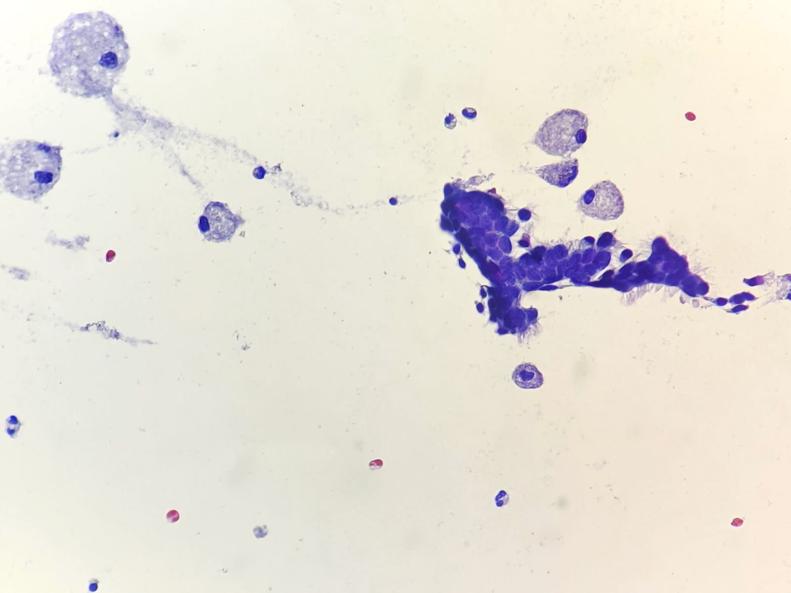
Ciliated epithelium and macrophages
Ciliated epithelium

Foamy macrophages
Images hosted on other servers:
Squames


Neutrophils and macrophages
Epithelial sheet
Sistrunks Procedure for Thyroglossal Duct Cyst
Contributed by Andrey Bychkov, M.D., Ph.D.
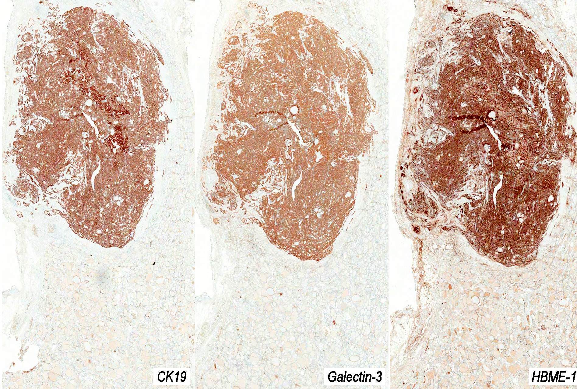
Diffuse positive staining for CK19, Galectin3 and HBME1
Thyroid carcinoma
Advanced thyroid carcinoma
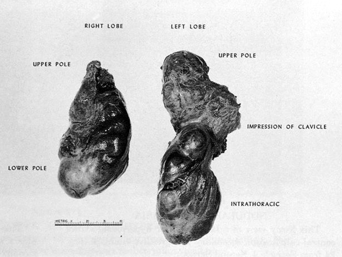
Contributed by Mark R. Wick M.D.
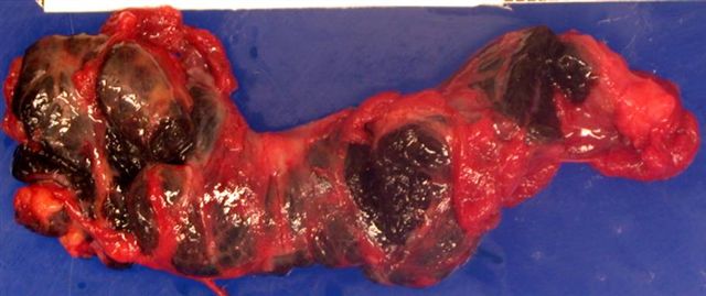
Intrathoracic (ectopic) goiter
Images hosted on other servers:

Large goiter


Giant goiter
Contributed by Swati Satturwar, M.D. and Andrey Bychkov, M.D., Ph.D.
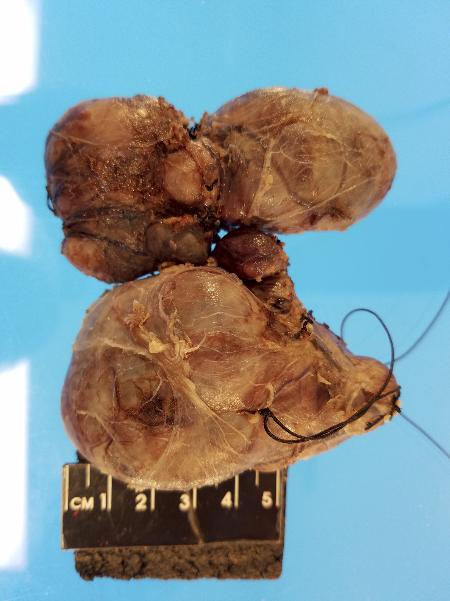
External surface

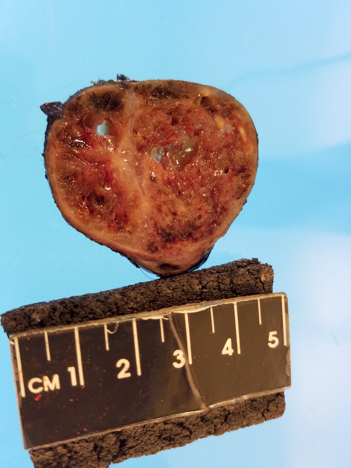
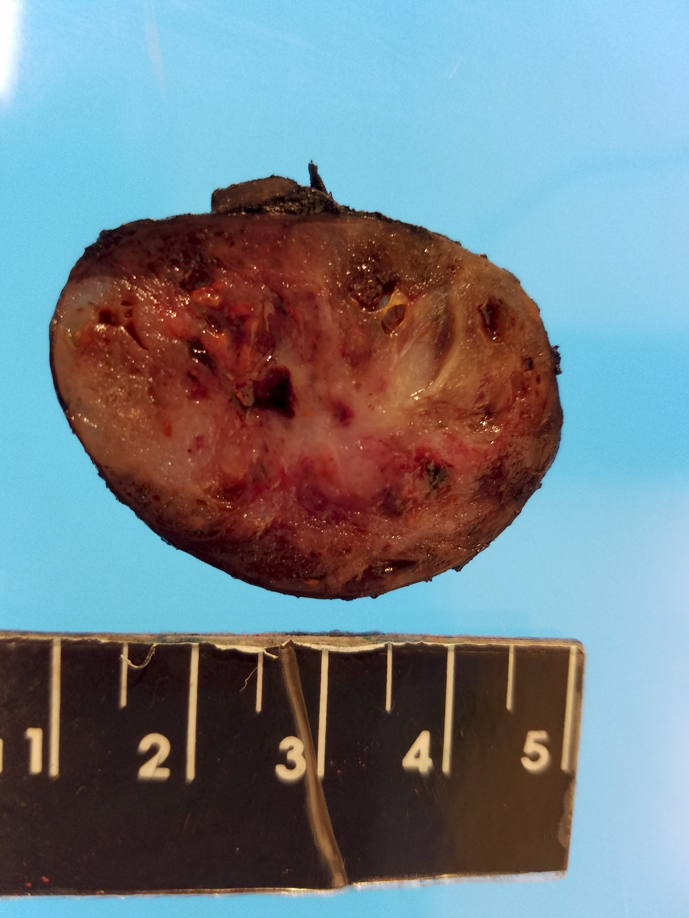
Cut surface
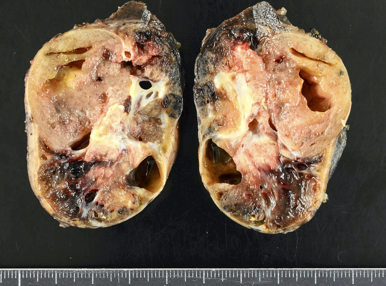
Nodular thyroid
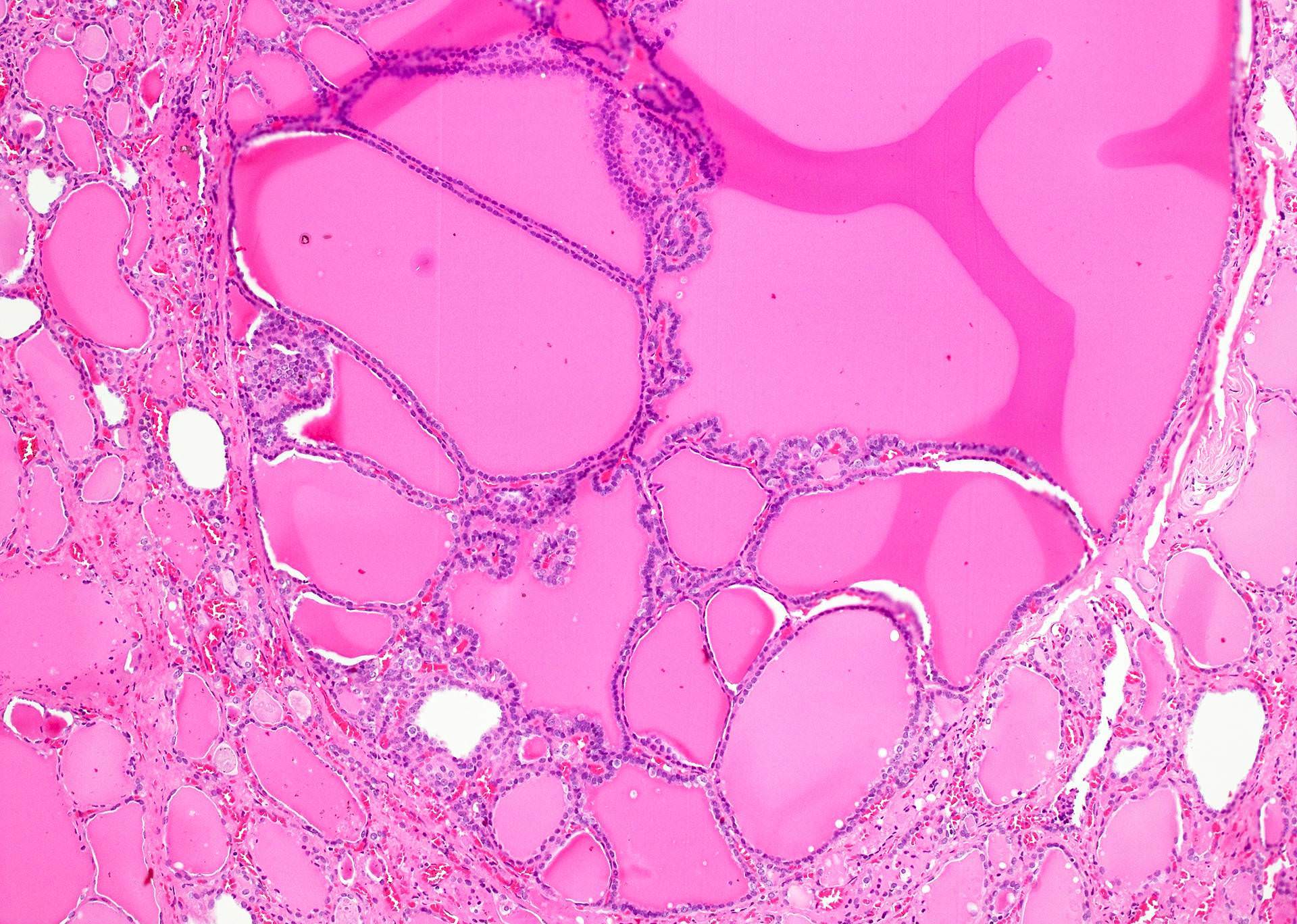
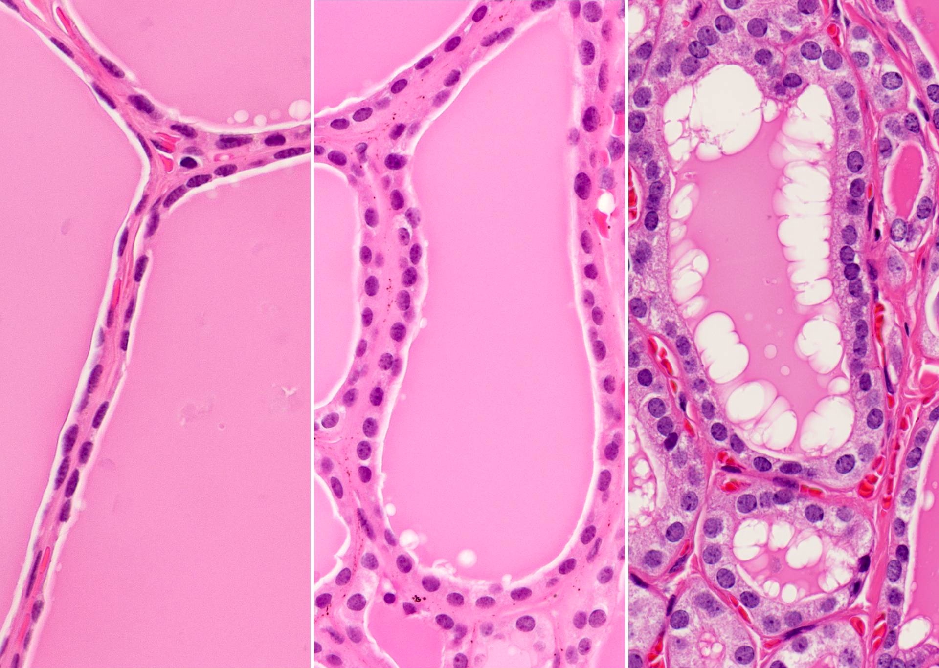
AFIP images
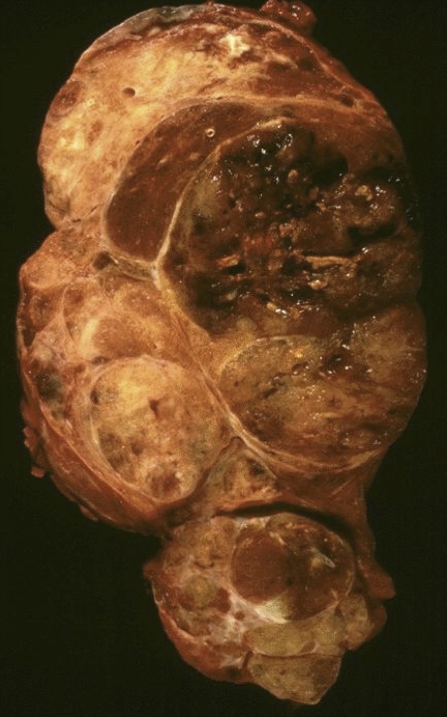
Various sized nodules
Nodular hyperplasia
Numerous poorly circumscribed nodules
Nodular hyperplasia with clear cell change
Images hosted on other servers:

Colloid cyst


Colloid goiter


Nodular goiter

Retrosternal goiter
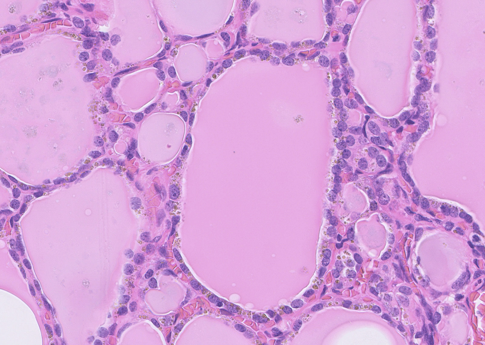
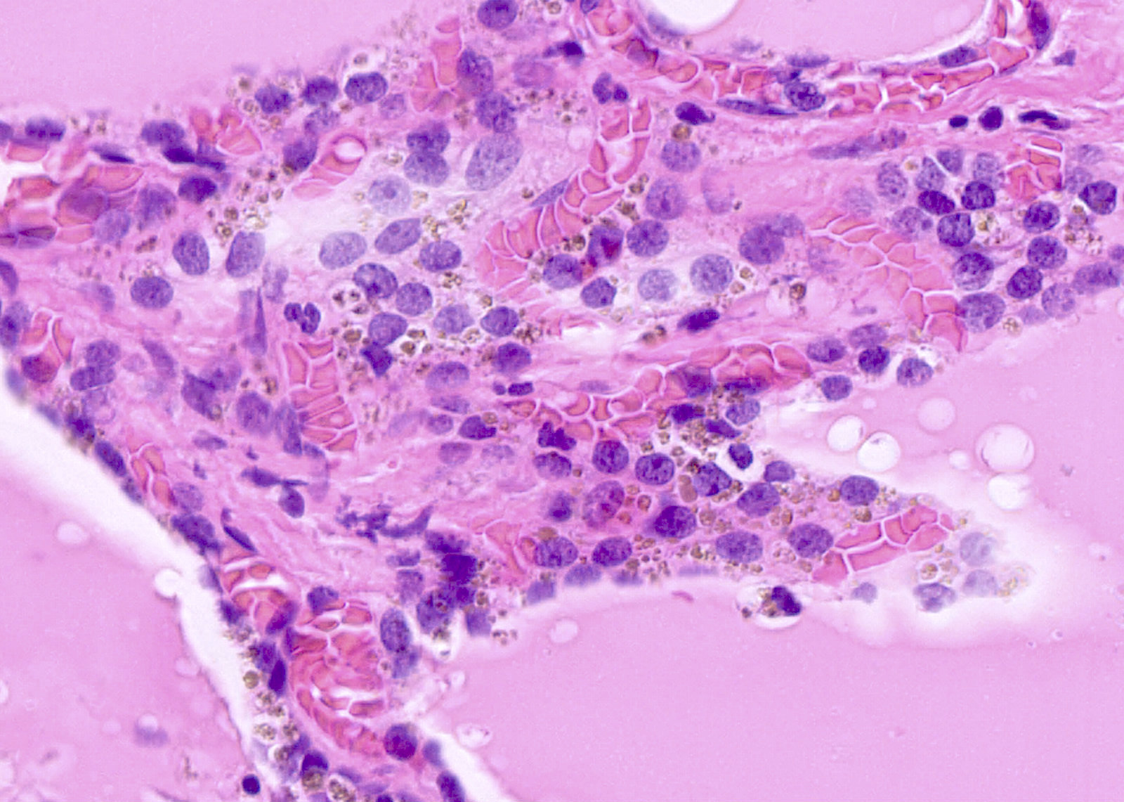
Contributed by Swati Satturwar, M.D., Andrey Bychkov, M.D., Ph.D. and Rajeshwari K. Muthusamy, M.D.
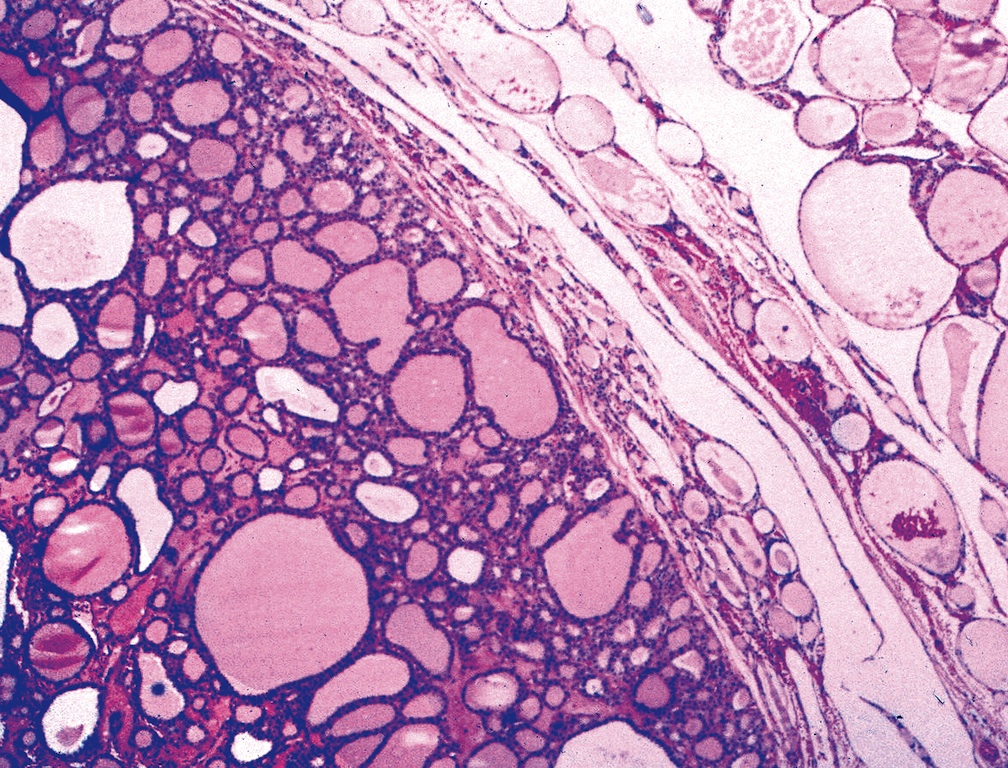
Dilated follicles
Nodule
Degenerative changes
Reactive fibrosis

False angioinvasion
Sanderson polsters
Adipose metaplasia
AFIP images

No capsule identified
Sanderson polster
With hypercellular focus
With adipose metaplasia of stroma
Papillary area
Clear cell change
Focal squamous metaplasia
Contributed by Ayana Suzuki, C.T.
Watery colloid
Cracking colloid
Follicular clusters
3D structures
Paravacuolar granules
Cyst fluid only
Images hosted on other servers:

Watery colloid and focal dense colloid

Large amounts of background colloid
Thyroid multinodular goiter
Histopathology thyroid: nodular goiter
Histopathology thyroid: colloid goiter
Contributed by Andrey Bychkov, M.D., Ph.D.
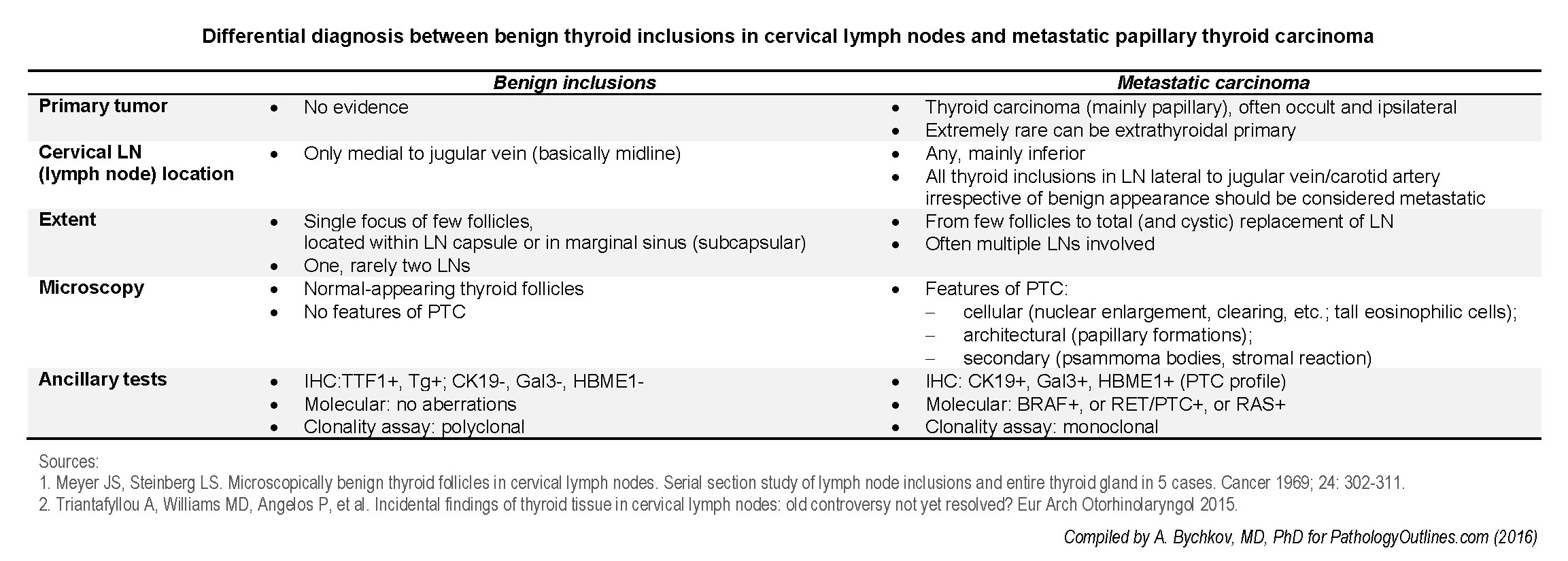
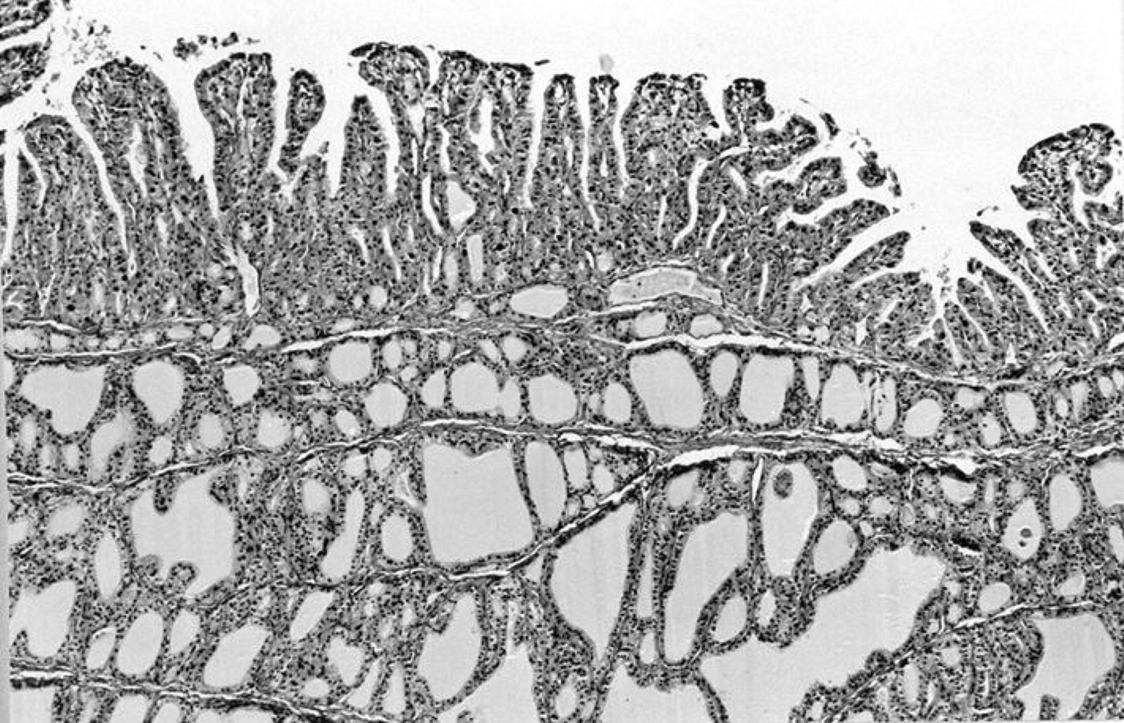
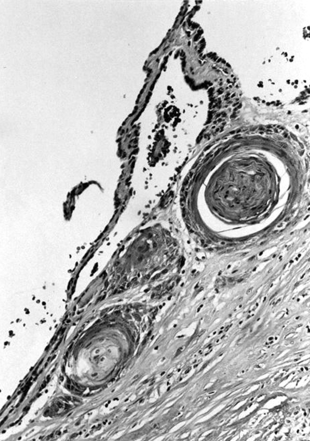
Differential diagnosis
AFIP images

Ectopic thyroid follicles
Images hosted on other servers:

Subcapsular nodal thyroid inclusion

Lymph node with metastatic papillary thyroid carcinoma


Thyroglobulin+ lymph node (potential pitfall)
Images hosted on other servers:


CT scan

Sonography

PET - CT

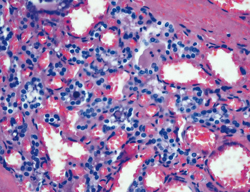
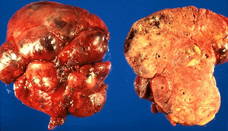
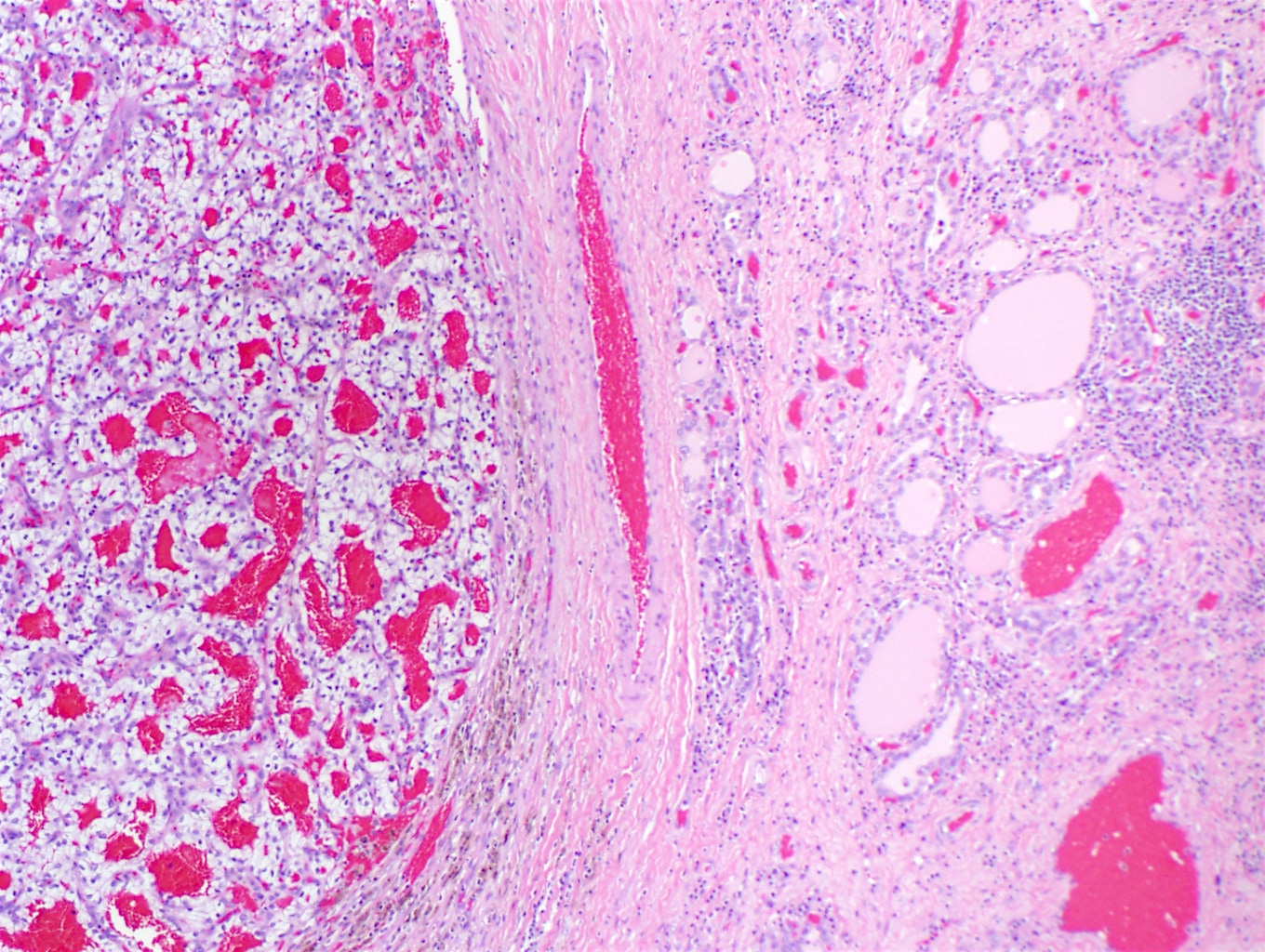
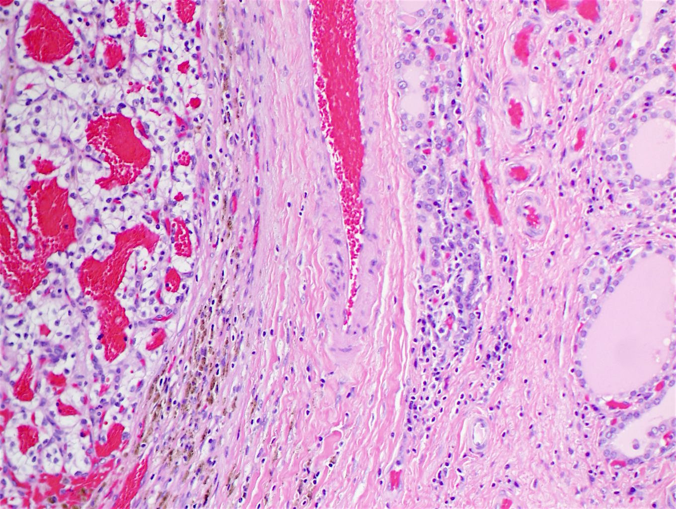
Metastatic renal cell carcinoma
Contributed by Mark R. Wick, M.D., Jose G. Mantilla, M.D. (Case #513) and AFIP

Metastatic renal cell carcinoma
Melanoma in OFA
Melanoma metastatic from skin primary
Images hosted on other servers:

Invasion by squamous cell carcinoma of larynx

Renal papillary carcinoma
Metastatic renal cell carcinoma


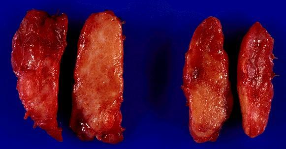
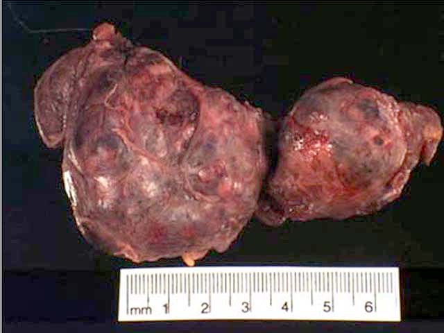
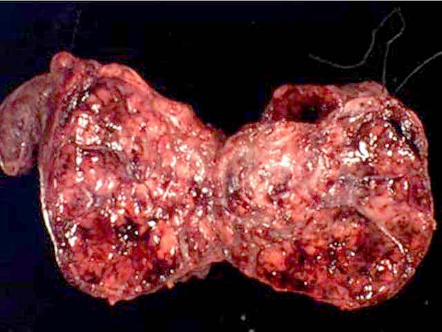
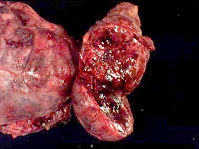
Surgical specimens

Surgical specimens
Scroll to see all images:
Contributed by Mark R. Wick, M.D.
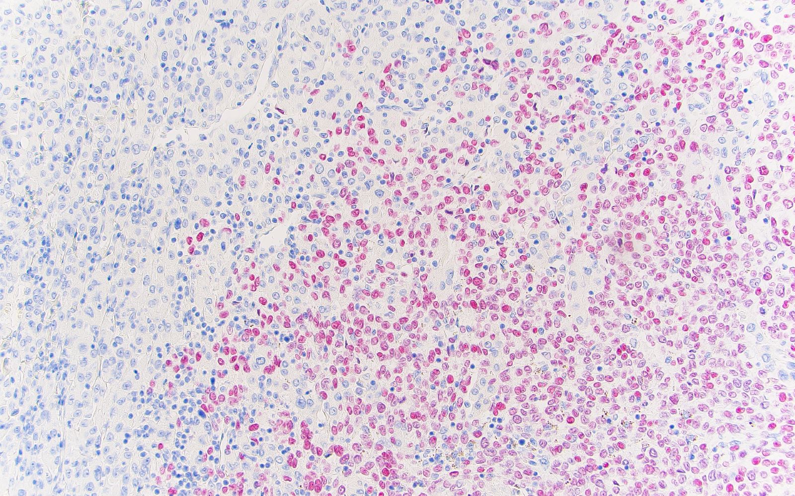
Metastatic renal cell carcinoma

CD10, Keratin 8, RCC, thyroglobulin
Contributed by Jose G. Mantilla, M.D. (Case #513)
HCA capsule
Interface between components

Pigmented component
Hürthle cell component
Melanoma in OFA interface
Melanoma in OFA SOX10
Case #461


Clear cell RCC metastatic to the thyroid
CD10, 400x
RCC, 400x
Case #342
Metastatic leiomyosarcoma (frozen section slides)

Metastatic leiomyosarcoma (permanent sections)
AFIP images
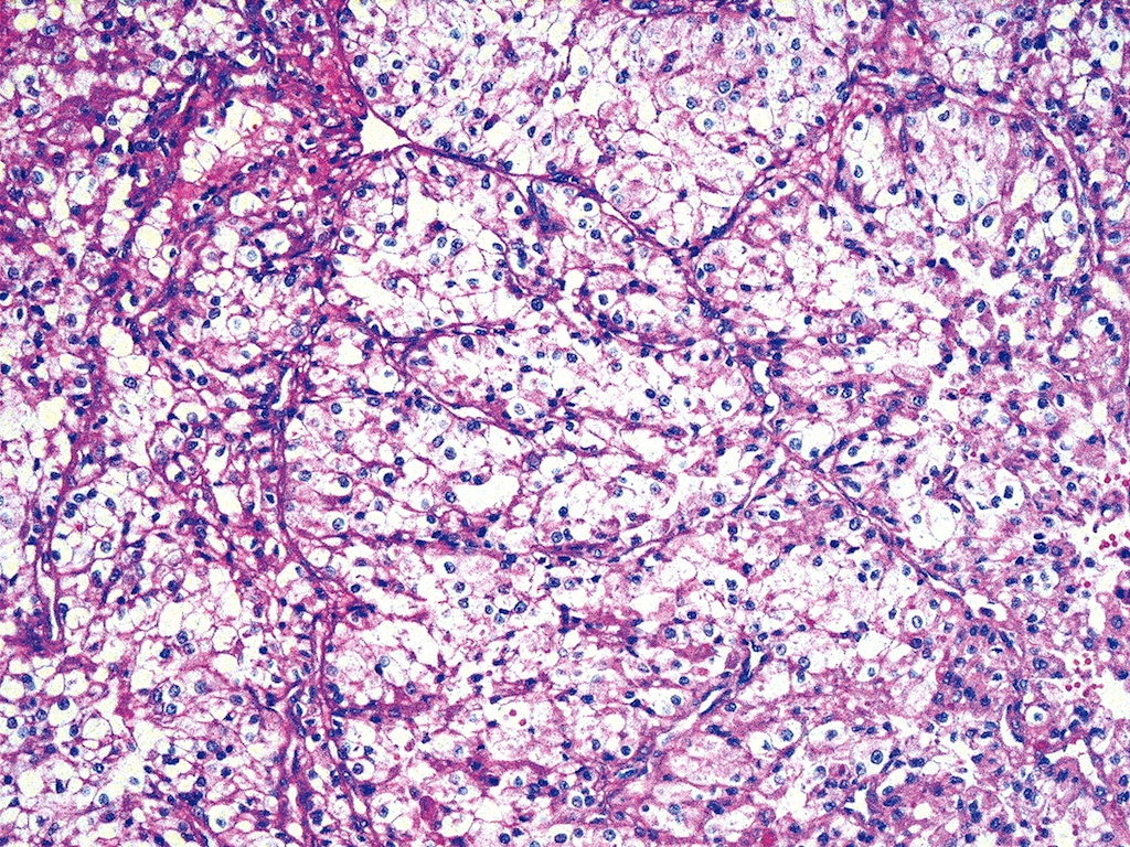
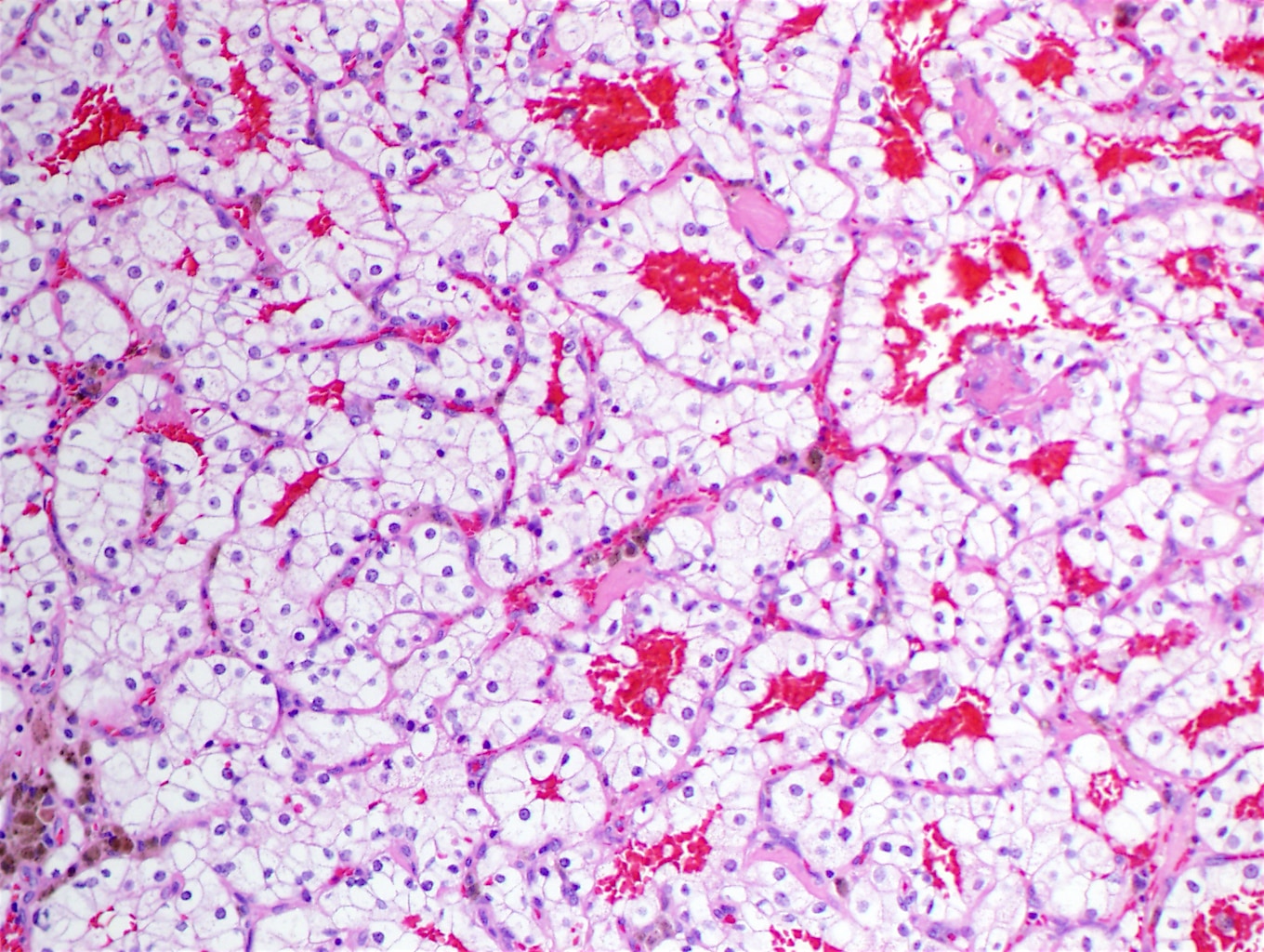
Clear cell type
Cytoplasm is abundant and completely clear
Oil Red O stains cytoplasm strongly
False positive thyroglobulin staining
Breast carcinoma:
Lobular carcinoma and entrapped thyroid follicles
ER stains breast tumor but not papillary carcinoma

Falsely positive thyroglobulin staining
Lung:
Carcinoid tumor with well defined nesting pattern
Carcinoma metastatic to mediastinal thyroid gland
Skin:
Melanoma
Images hosted on other servers:

From rectum

Metastatic HCC in the thyroid gland

HCC stains for AFP


Clear cell type

Metastasis to goiter

Concurrent RCC and thyroid carcinoma

Central lesions inside
hyperplastic adenomatoid
nodules

FNA, histology and stains

CD10 and H&E

Tumor to tumor metastases


Lung-small cell carcinoma

Lung-squamous cell carcinoma

Melanoma

Parotid adenoid cystic carcinoma

Rectal carcinoma
to poorly
differentiated
thyroid carcinoma
Contributed by Mark R. Wick, M.D.
FNA: metastatic renal cell carcinoma
Images hosted on other servers:

Kidney: metastatic renal cell carcinoma, clear cell type

Kidney: bloody background with clusters of atypical cells; nuclei are somewhat pleomorphic with prominent nucleoli

Lung squamous cell carcinoma

Melanoma

Melanoma - HMB45


Parotid gland adenoid cystic carcinoma
Contributed by Rachel Jug, M.B.B.Ch.
Ultrasound FNA techniques
Images hosted on other servers:

Risk of thyroid cancer

ATA algorithm for
patients with
thyroid nodules

ATA association

ACR TI-RADS chart
Comparison of systems
Typical appearances of diffuse thyroid diseases
| Thyroid disorder | Grayscale ultrasound | Color Doppler | Key features |
|---|---|---|---|
| Graves thyroiditis | Enlarged, mildly hypoechoic, heterogeneous | Markedly ↑ | Markedly hyperemic; proptosis; hyperthyroid; + antithyroid antibodies |
| Hashimoto thyroiditis | Enlarged, heterogeneous with lobular margins; hypoechoic and micronodular, septal lines | Highly variable: both ↑ and ↓ flow possible | + Antithyroid antibodies, hypothyroidism; cervical adenopathy |
| Subacute lymphocytic thyroiditis (painless) | Hypoechoic | Insufficient data | + Antithyroid antibodies; postpartum; transient |
| De Quervain thyroiditis (subacute granulomatous) | Painful patchy areas of hypoechogenicity | ↓ in the hypoechoic patch | Thyroid pain over area of hypoechogenicity; ↑ erythrocyte sedimentation rate (ESR) |
| Acute suppurative thyroiditis | Abscess or infected linear tract in the thyroid | Normal background; no flow within an abscess | Acute presentation with signs of infection and pain; ↑ ESR; possible pyriform sinus fistula |
| Riedel thyroiditis | Large hypoechoic thyroid with coarse parenchyma | Insufficient data | Large, rock hard gland; encases adjacent structures |
| Medication induced (i.e., amiodarone induced thyrotoxicosis [AIT]) | Type 1: abnormal thyroid; type 2: normal thyroid | Type 1: ↑; type 2: absent | History of current or recent amiodarone use; hyperthyroid |
| Atrophic thyroiditis | Small, hypoechoic thyroid | ↓ | + Antithyroid antibodies; usually hypothyroid |
| Radiation thyroiditis | Small, hypoechoic thyroid | Variable | Known external beam or I131 administration |
| Thyroid lymphoma | Large, ill defined, markedly hypoechoic nodules or masses with ↑ through transmission on background of Hashimoto thyroiditis | ↓ in the hypoechoic mass | Rapidly enlarging neck mass in patient with history of Hashimoto, with or without adenopathy |
| Multinodular goiter | Closely opposed or interspersed, similar appearing nodules replace parenchyma, coarse calcifications, variable cystic changes in nodules | Variable | Confluent nodules in a normal or enlarged thyroid; with or without abnormal thyroid function tests |
Images hosted on other servers:

Calcified nodule

Complex cystic thyroid nodule

Multinodular goiter

17 year old girl with Graves disease

35 year old woman
with diffuse
Hashimoto
thyroiditis

50 year old woman
with colloid
multinodular
goiter

Hypoechoic nodule

Microcalcifications in a thyroid nodule

Intranodular vascularity in thyroid nodule

Oval shaped thyroid nodule
Fine needle aspiration (FNA) biopsy techniques - Dr. Britt Marie Ljung playlist
Neck ultrasonography basics
Thyroid ultrasound course
Contributed by Ayana Suzuki, C.T.

Hemorrhagic background

Muscle

Respiratory epithelium
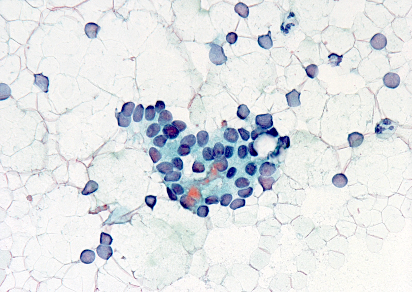
Air dried smear
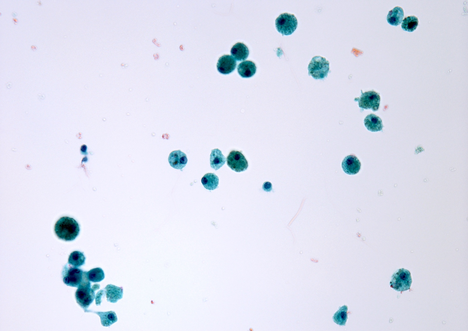
Cyst fluid only
Images hosted on other servers:


Hemorrhagic background

Muscle


Air dried smear


Cyst fluid only

Poor staining
Thyroid FNA and smearing techniques
Essential thyroid cytopathology
Head and tail of the Bethesda system for thyroid
Thyroid cytology - Bethesda classification
Images hosted on other servers:


Ultrasound
Images hosted on other servers:
Solid, gray-white
Contributed by Andrey Bychkov, M.D., Ph.D.
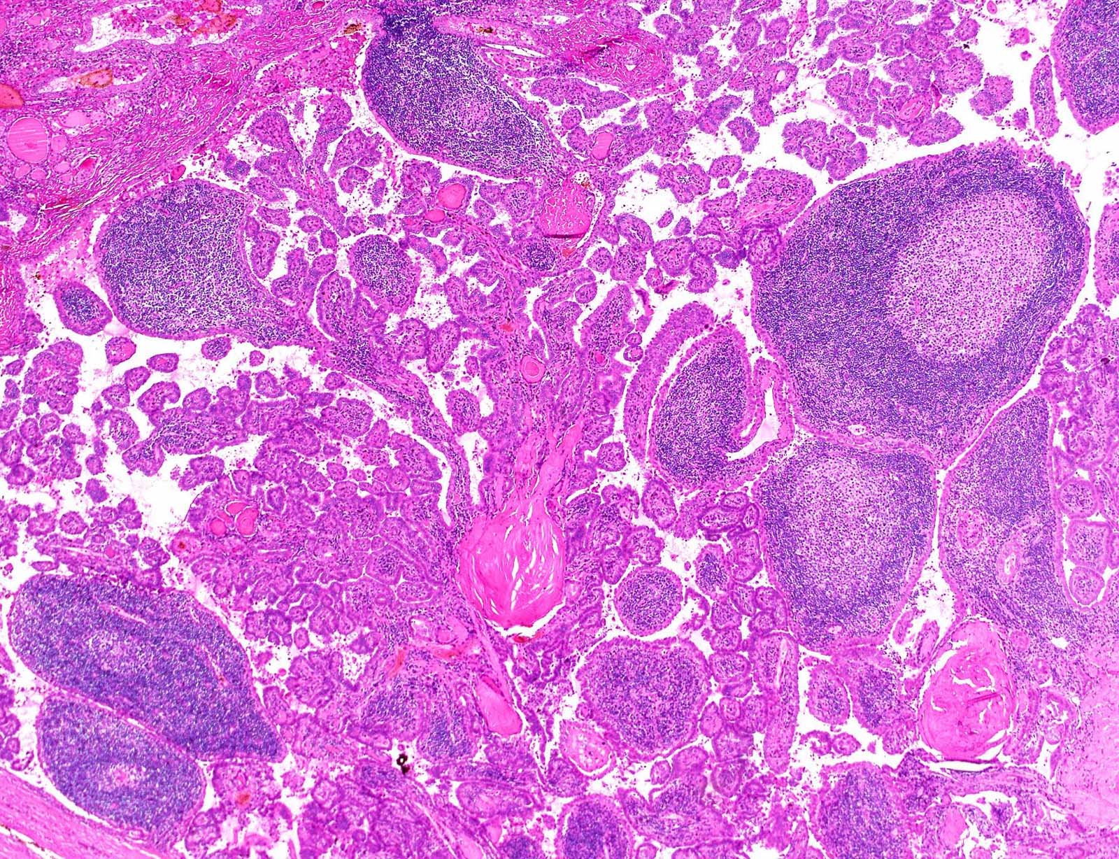
Enlarged papillae
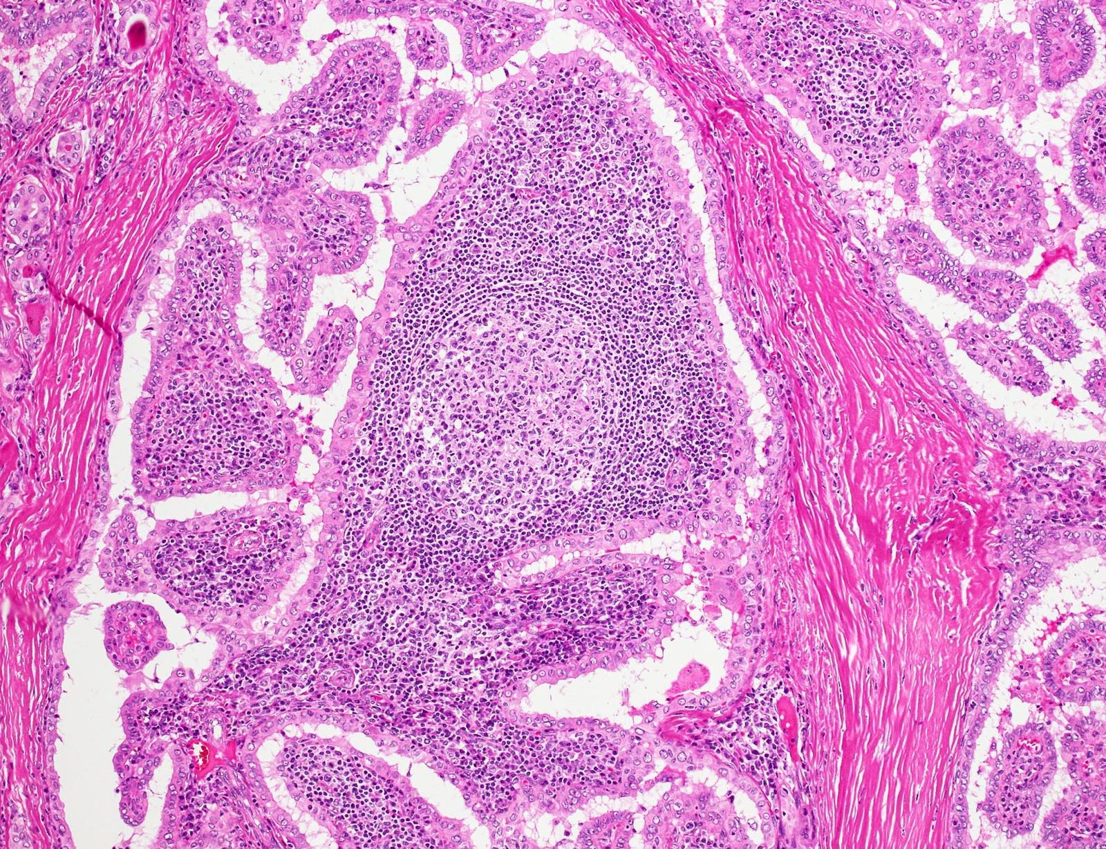
Lymphoid follicle
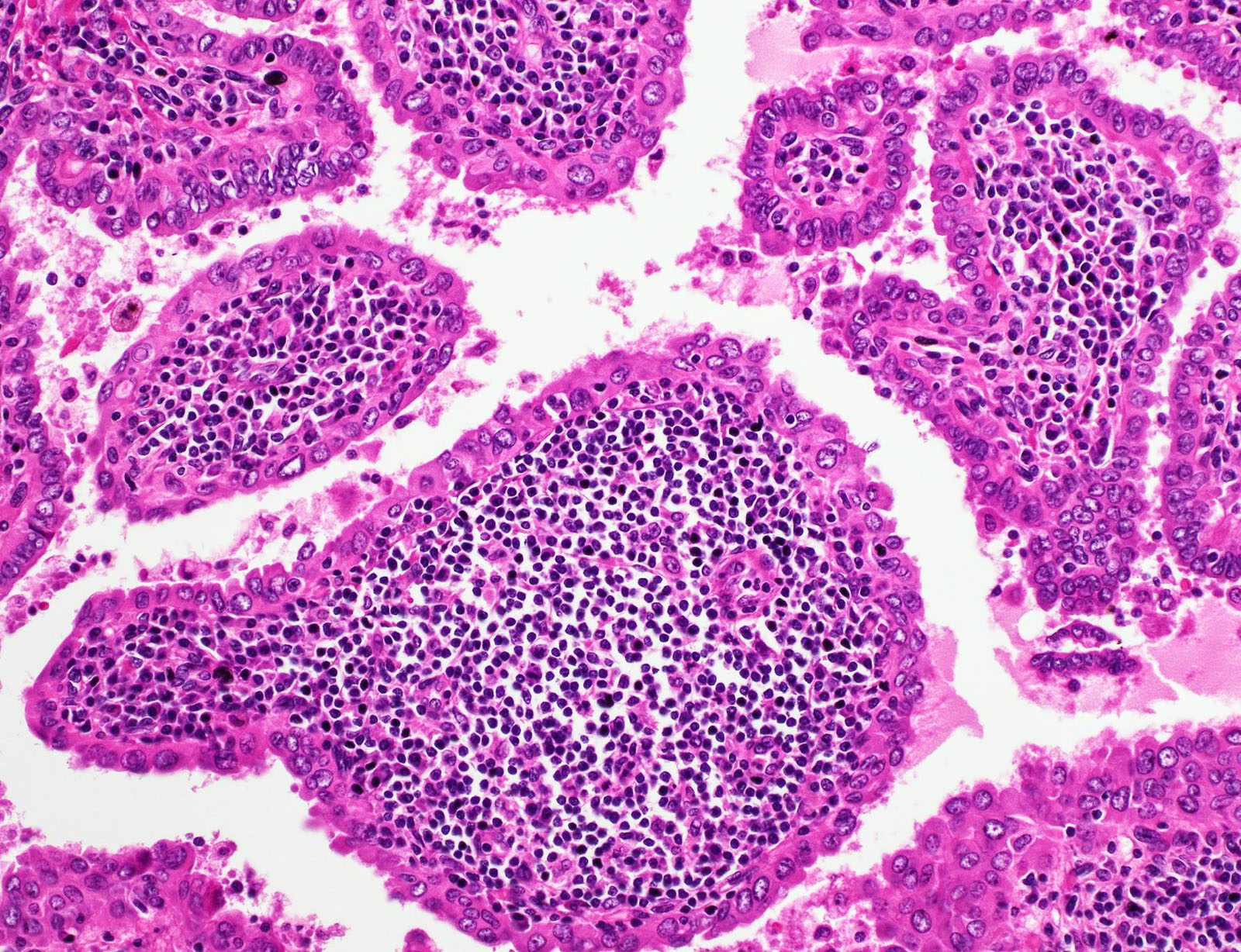
Oncocytic epithelium
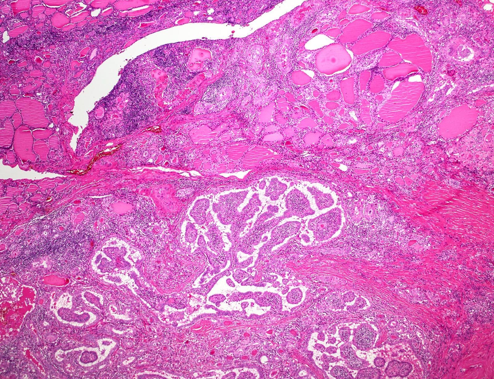
Background thyroiditis
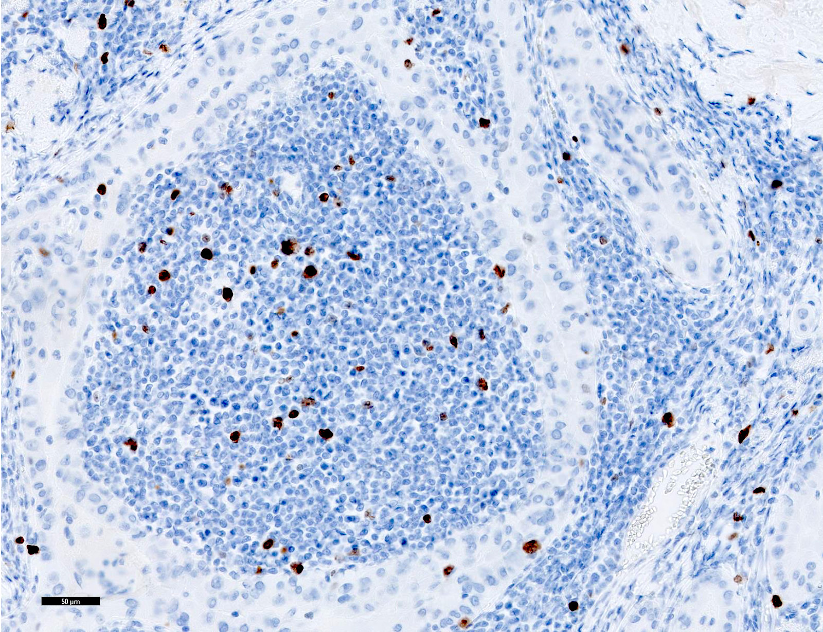
Ki67
Contributed by Shipra Agarwal, M.D.
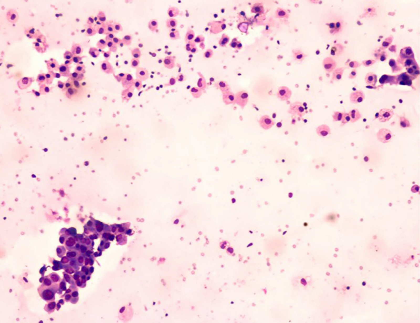
Inflammatory background
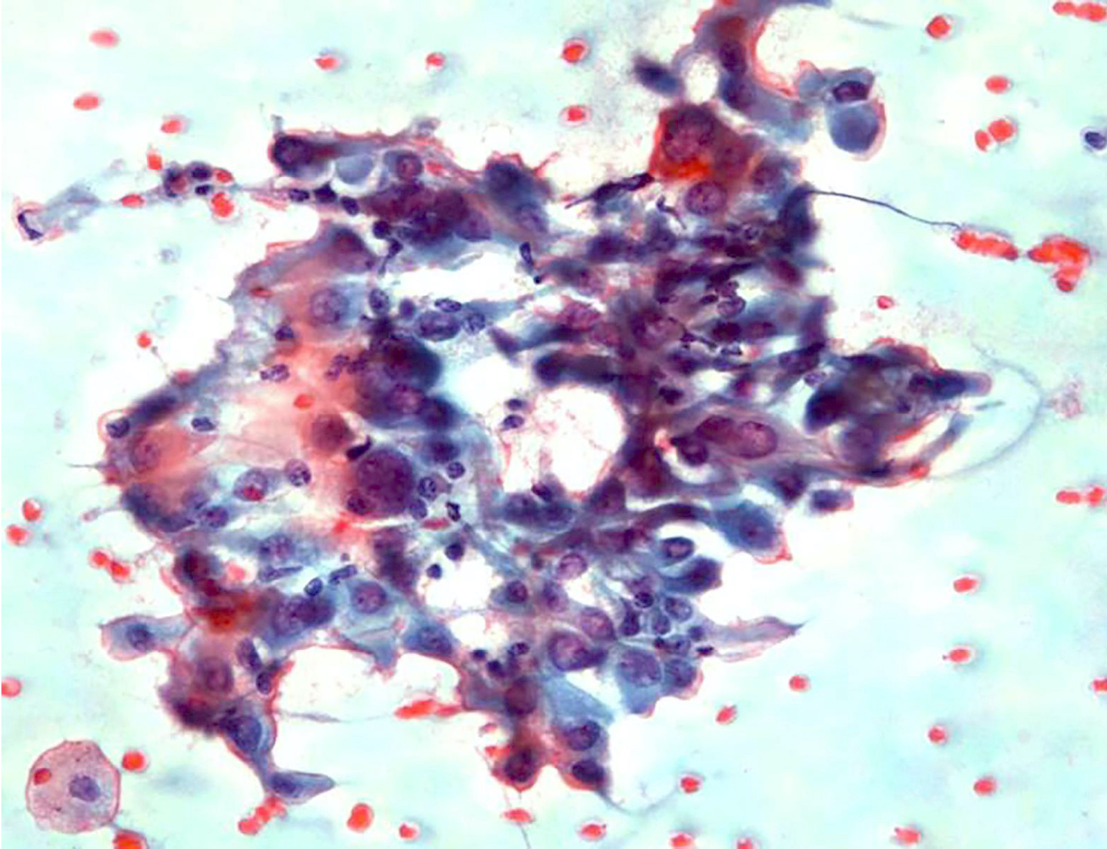
Lymphocytic permeation
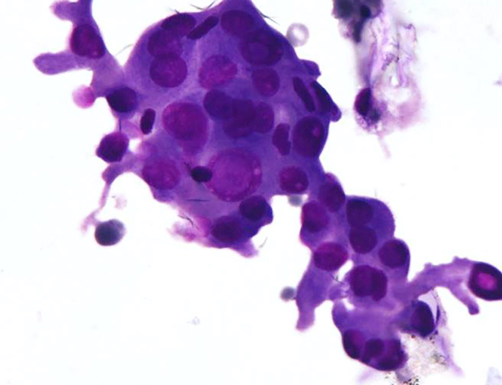
Oncocytic cells
Images hosted on other servers:

Liquid based cytology and conventional smear
Images hosted on other servers:

Schematic drawing for capsular invasion

Schematic drawing for vascular invasion
Contributed by Shuanzeng Wei, M.D., Ph.D.
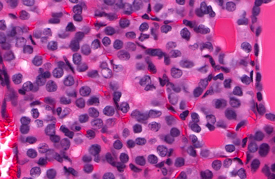
Questionable nuclear features of papillary carcinoma
Images hosted on other servers:

Cytological features of WDT-UMP
Contributed by Andrey Bychkov, M.D., Ph.D.
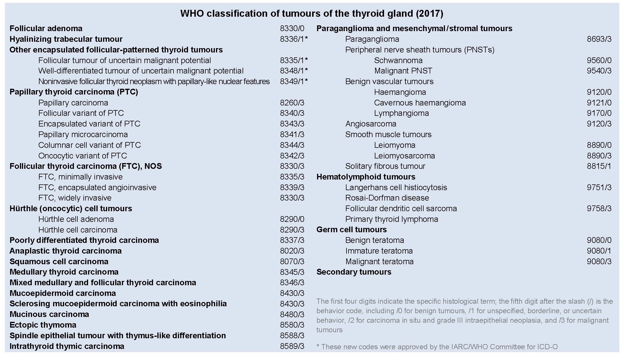
WHO classification: thyroid tumors
Adeniran: 2016
Ali: 2023
Boerner: 2016
Braverman: 2020

Duick: 2018
Gill: 2022
Gnepp: 2021
IARC: 2017
Jiang: 2023
Kakudo: 2019
Kini: 2015
Mete: 2016
Mody: 2022
Nikiforov: 2019
Nosé: 2022
Rosai: 2015
Stelow: 2020
Thompson: 2022
Wartofsky: 2016
Wenig: 2024
Find related Pathology books: cytopathology, GI, GU/adrenal, head & neck/endocrine

