

Contributed by C. Blake Gilks, M.D. and @MirunaPopescu13 on Twitter
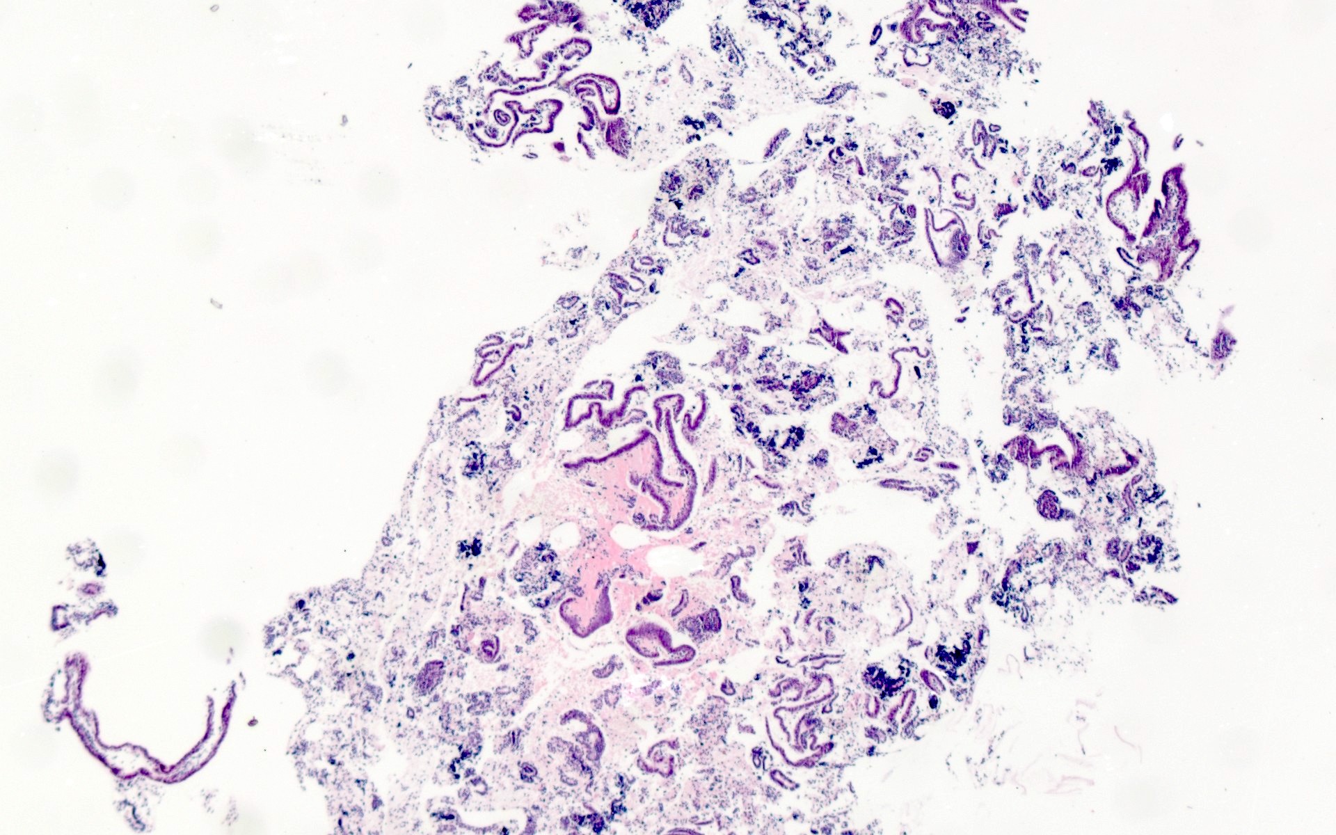
Nondiagnostic sample
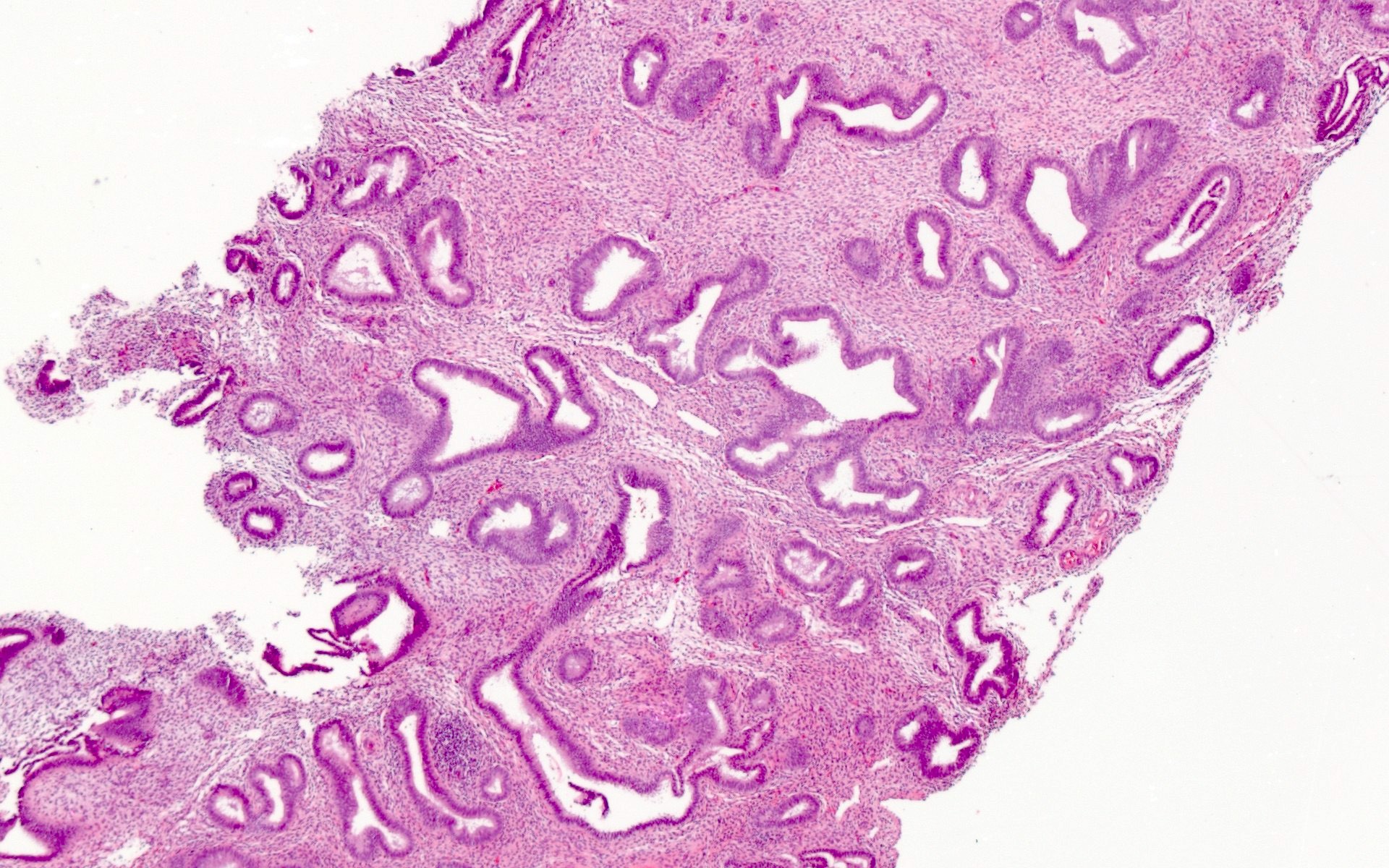
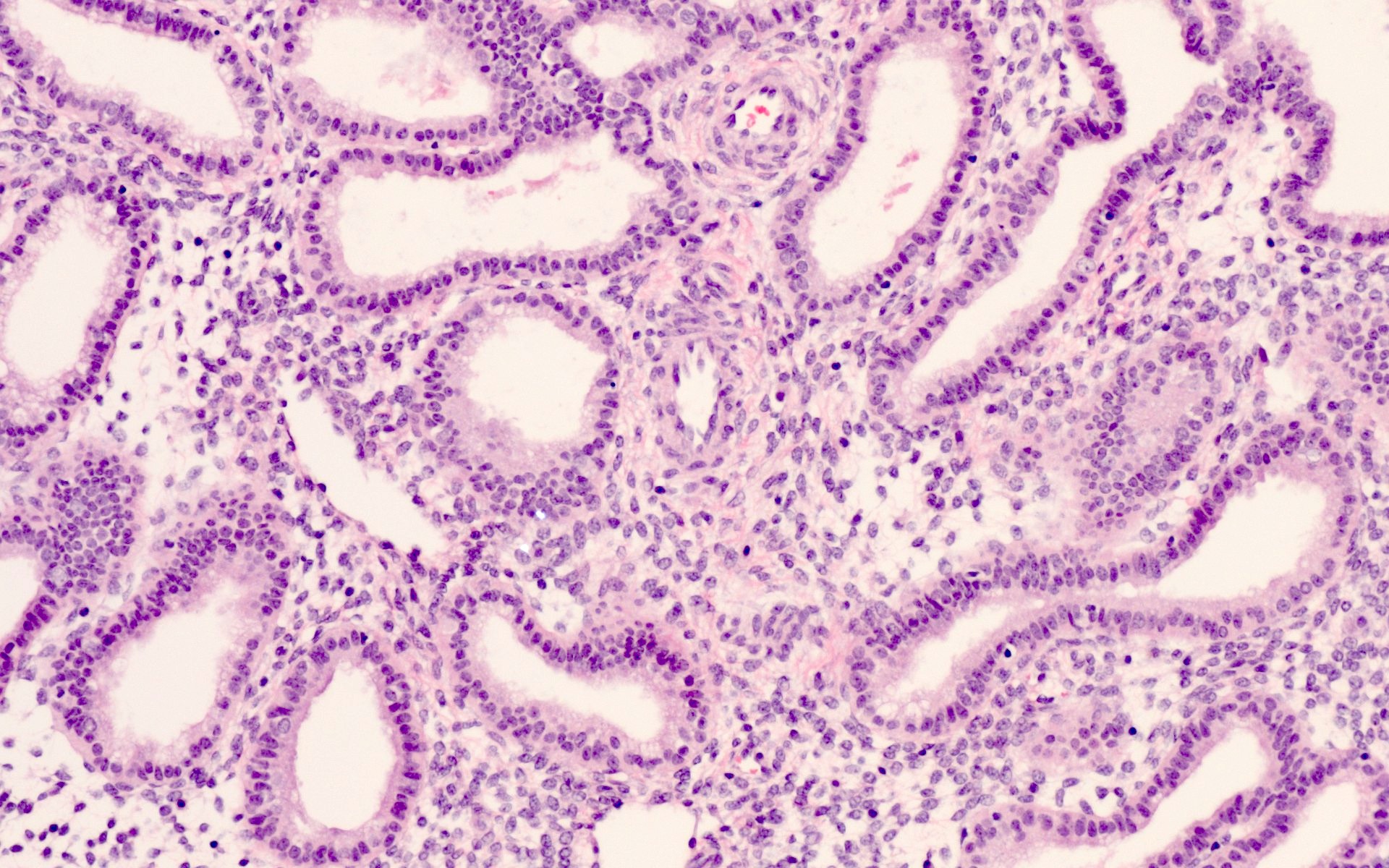
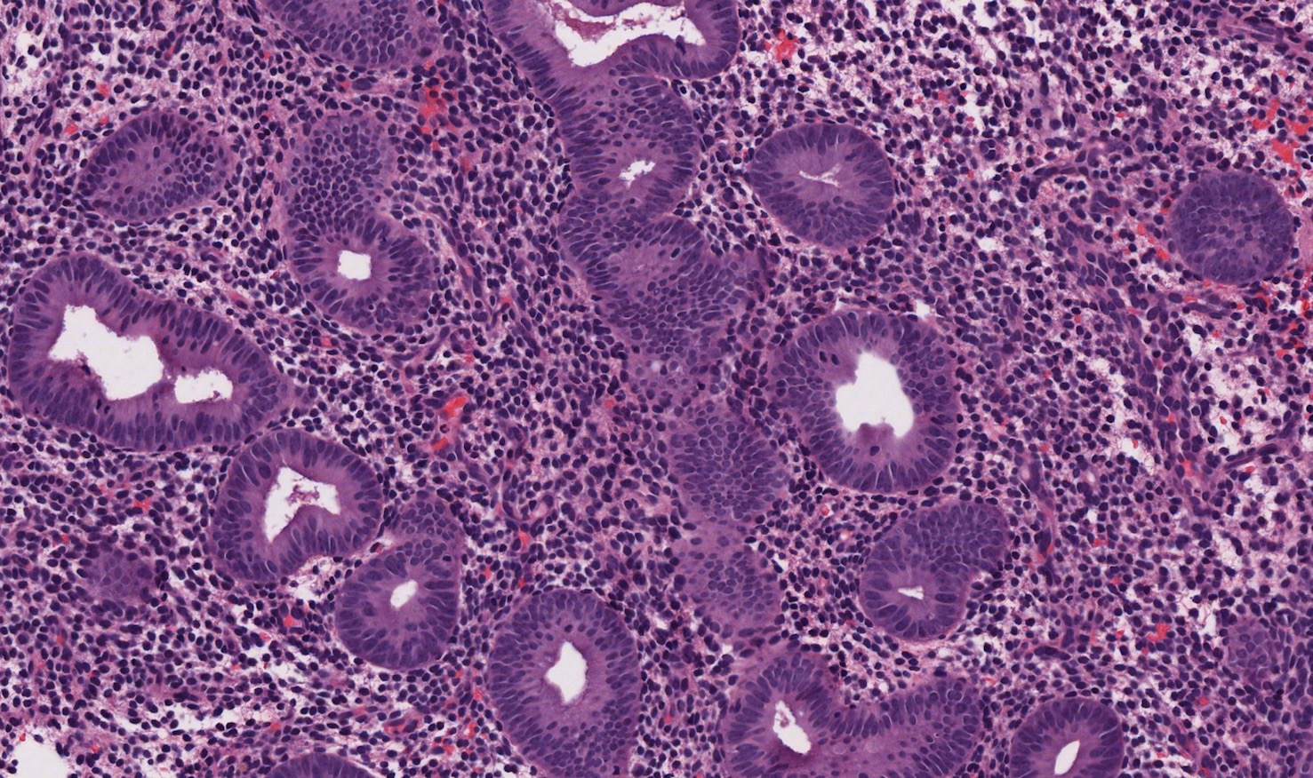
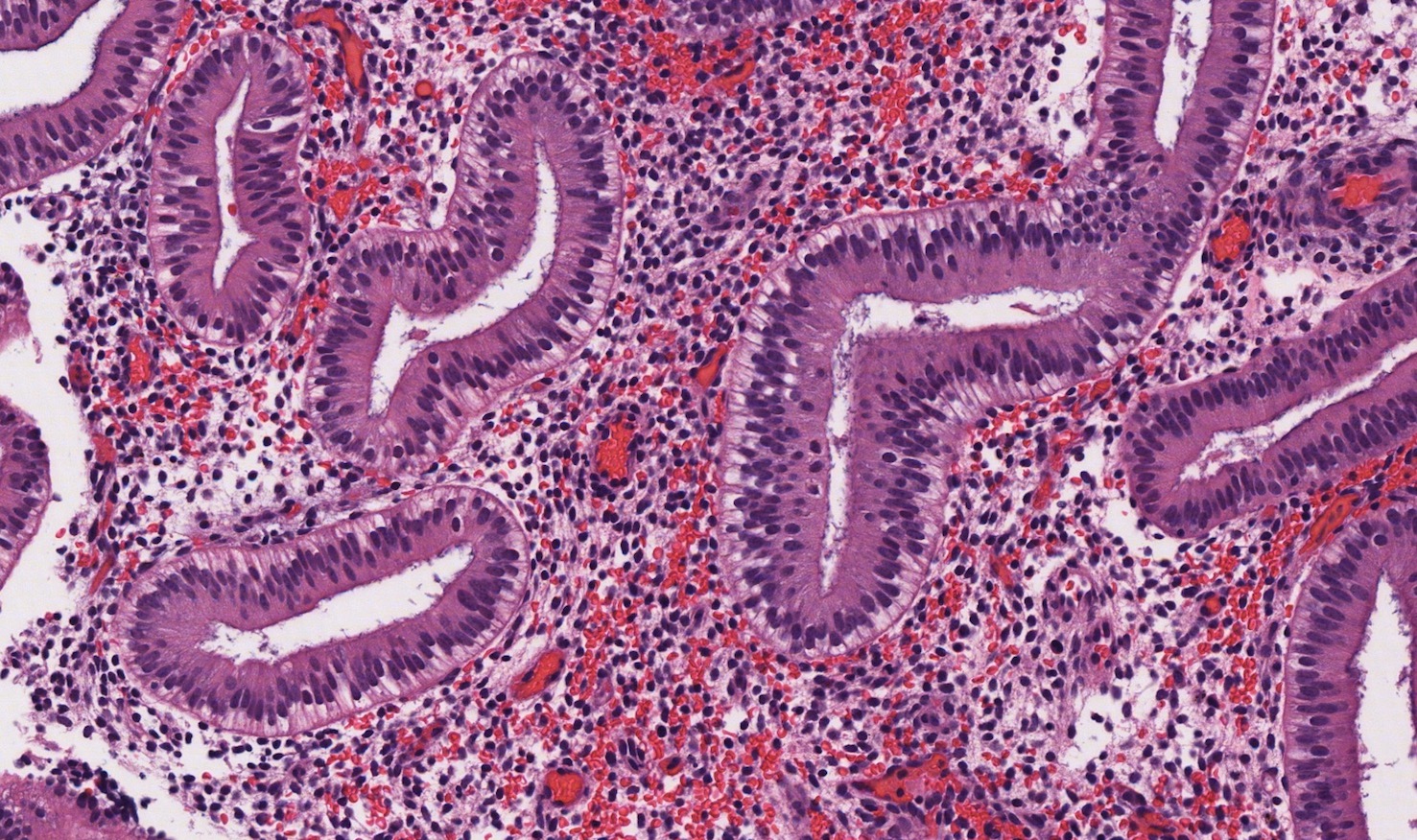
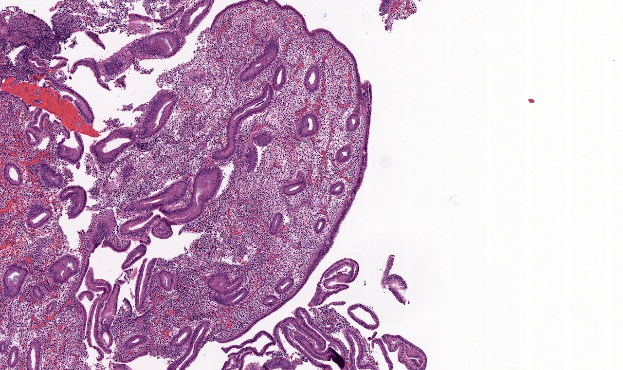
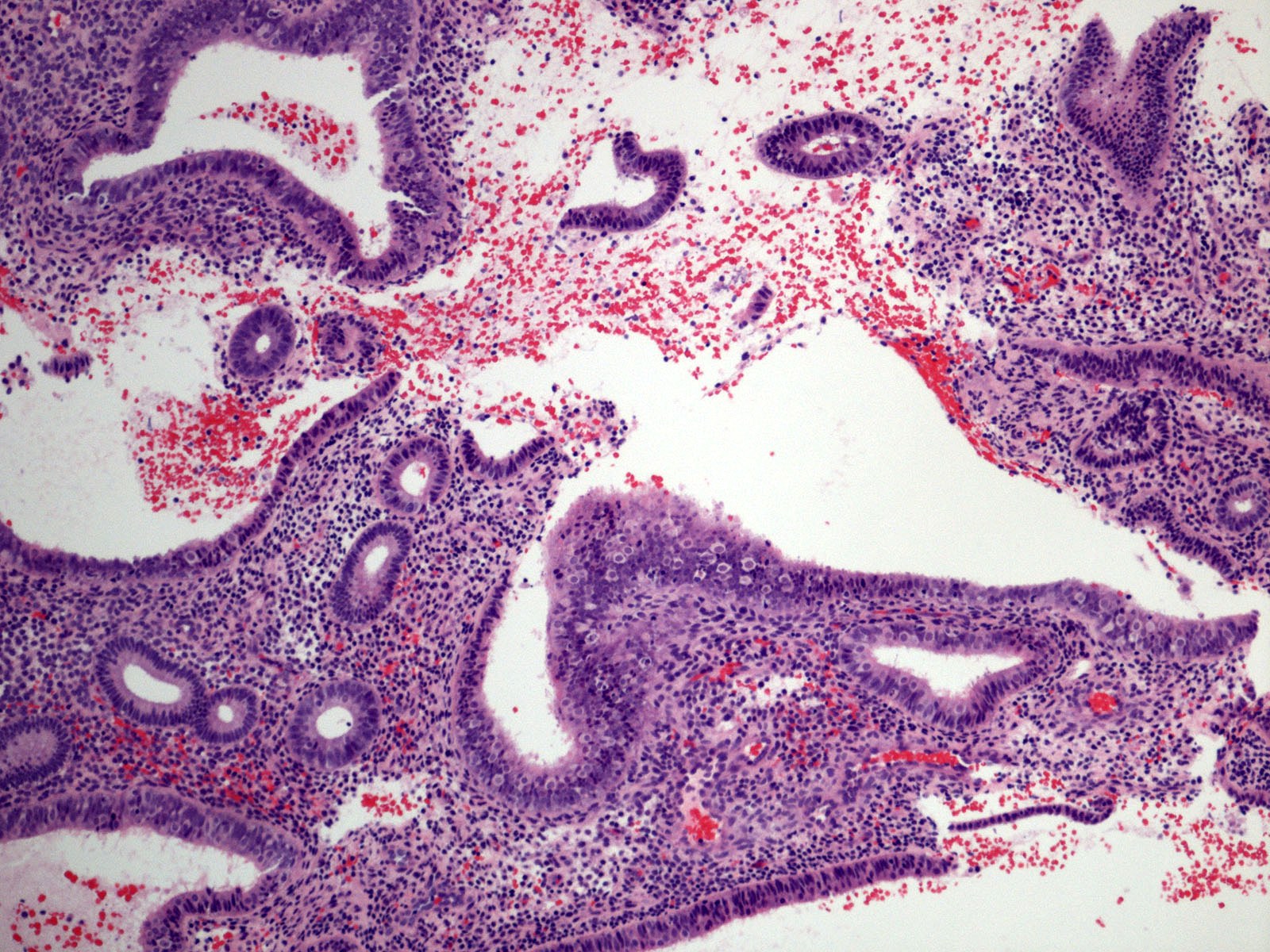
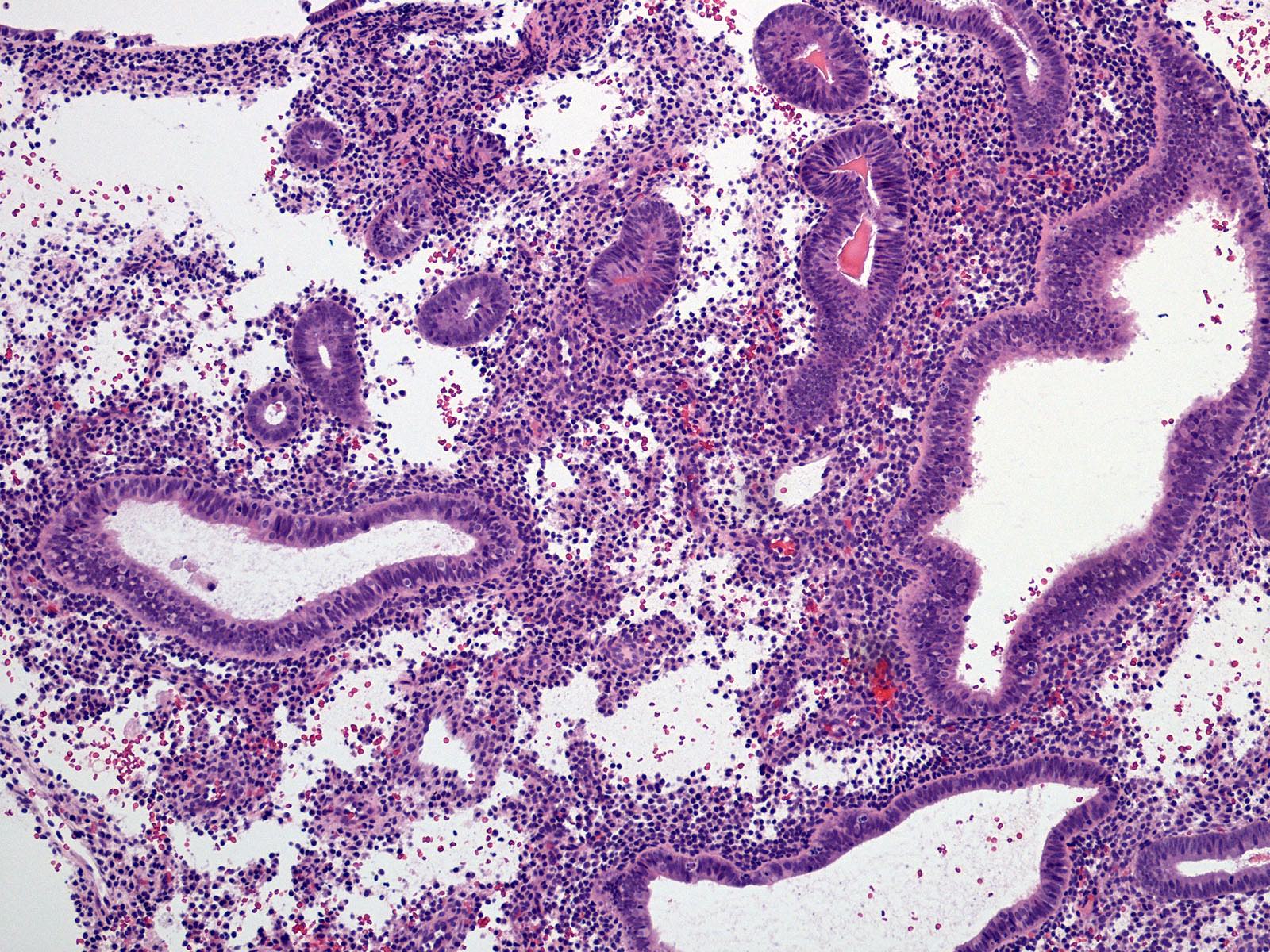
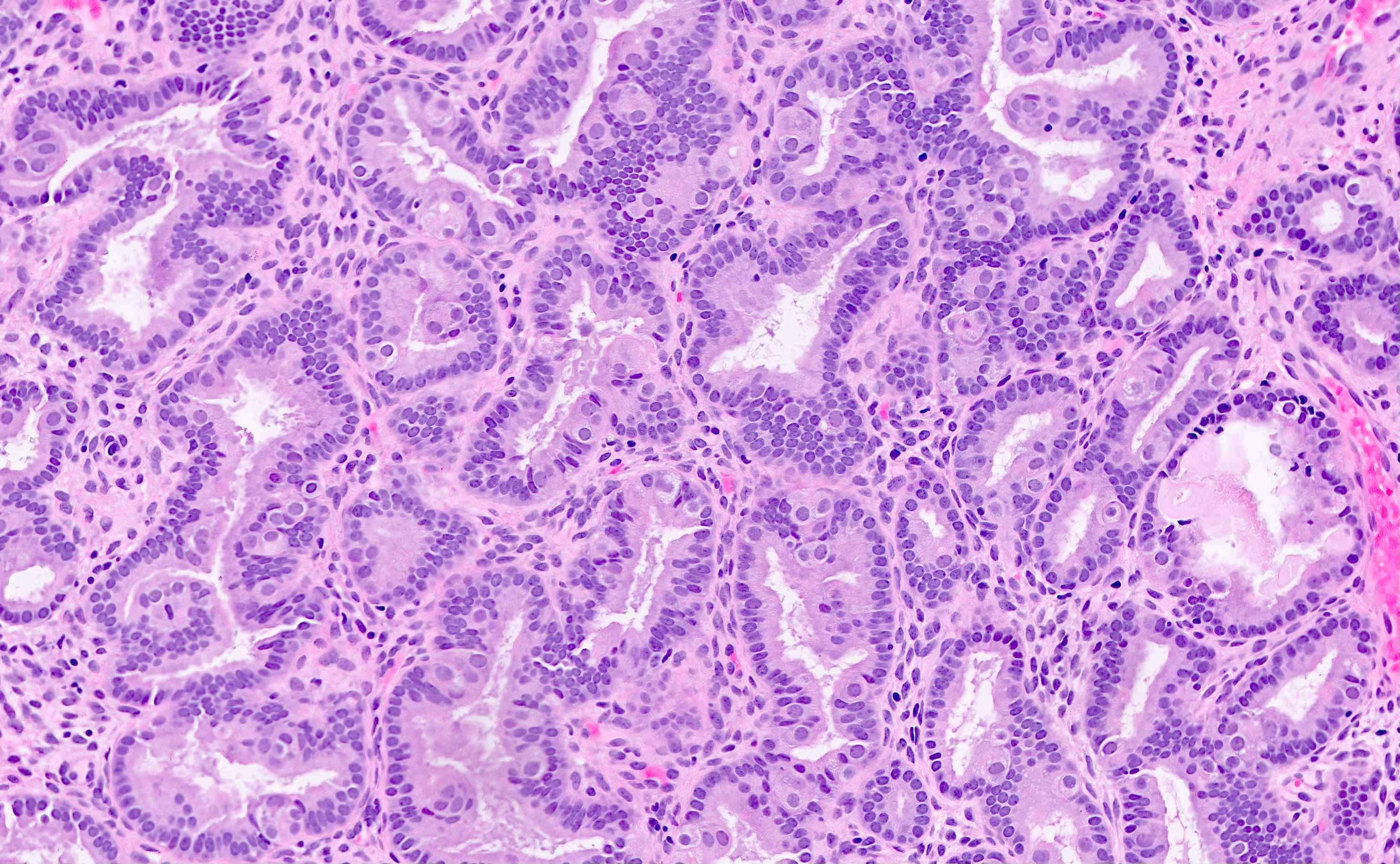
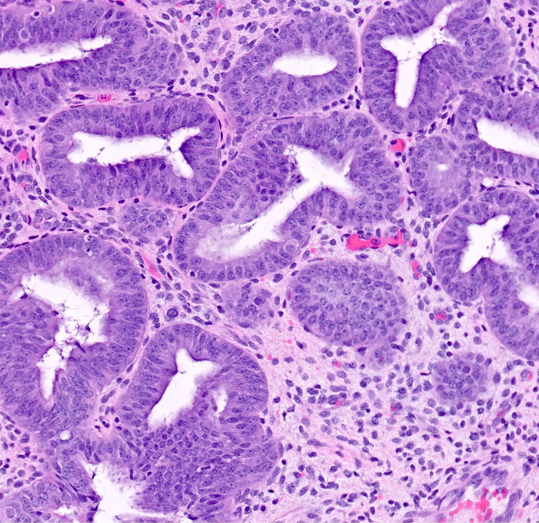
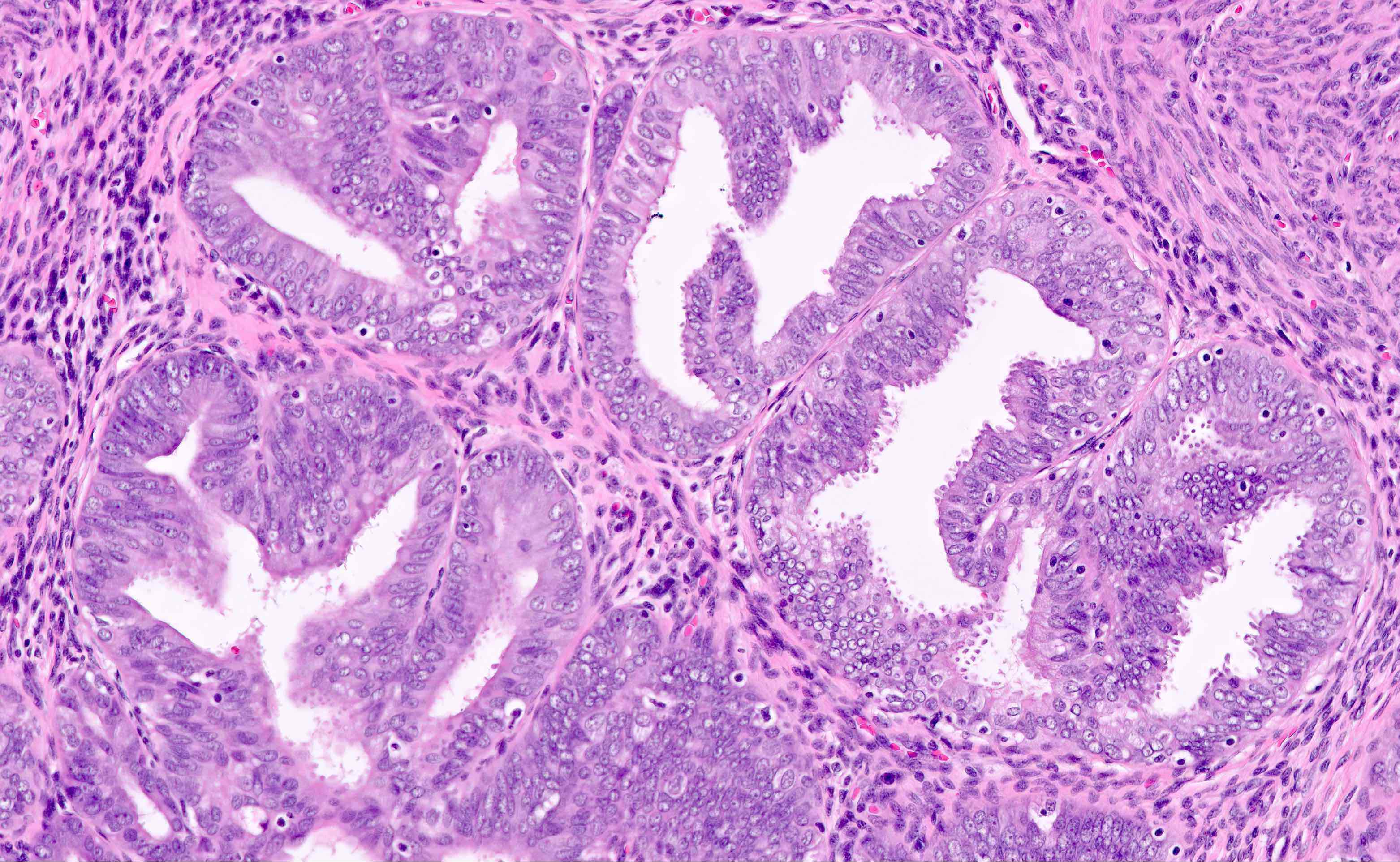
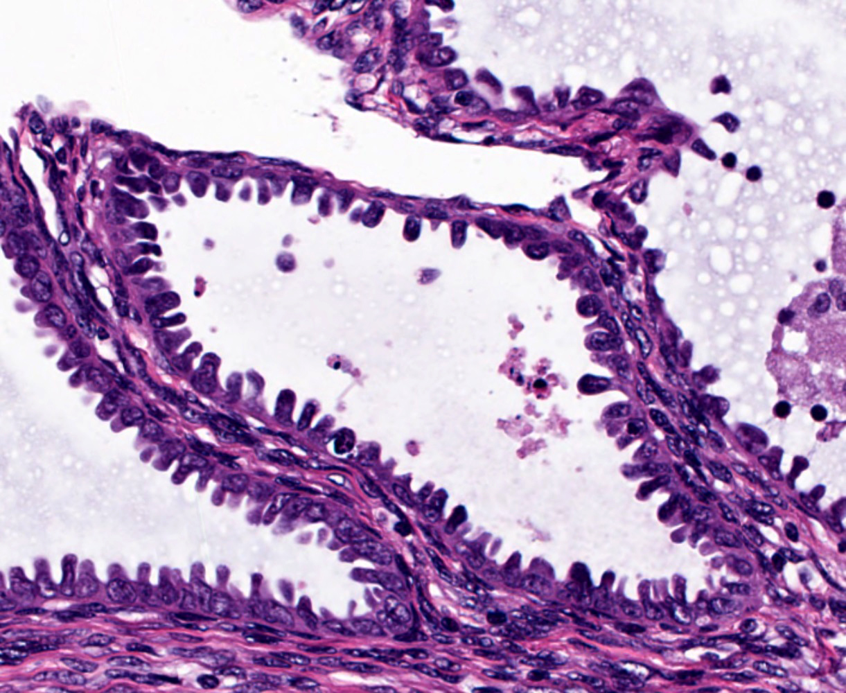
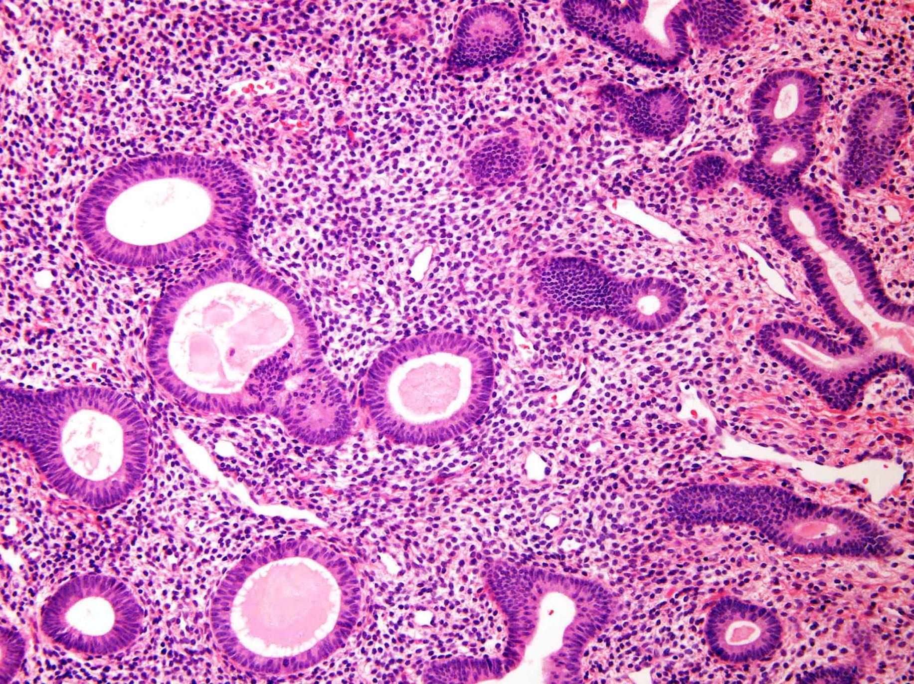
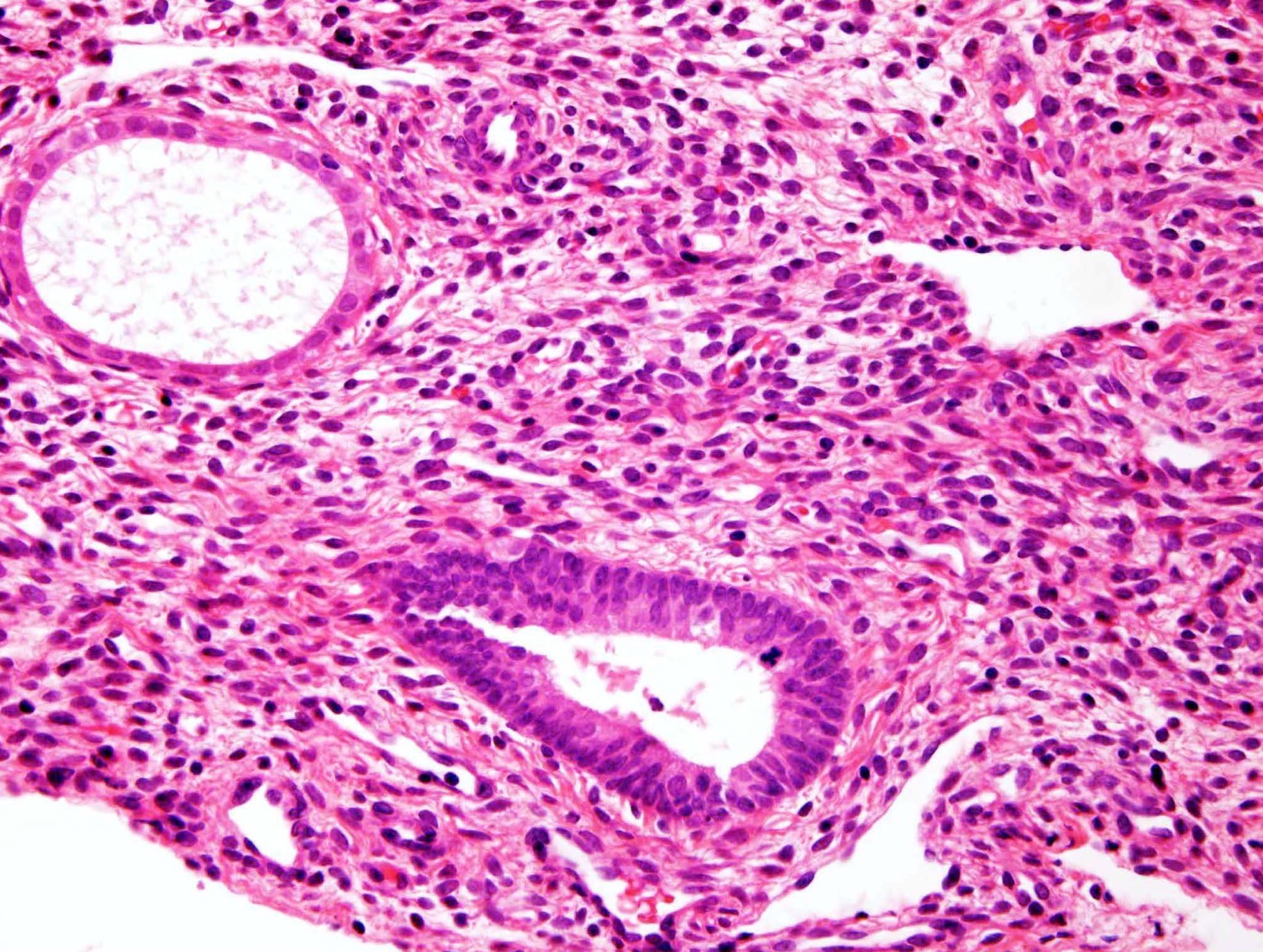
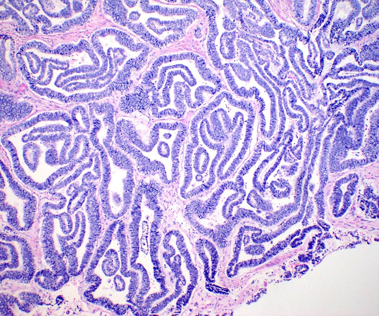
Proliferative endometrium
Proliferative endometrium
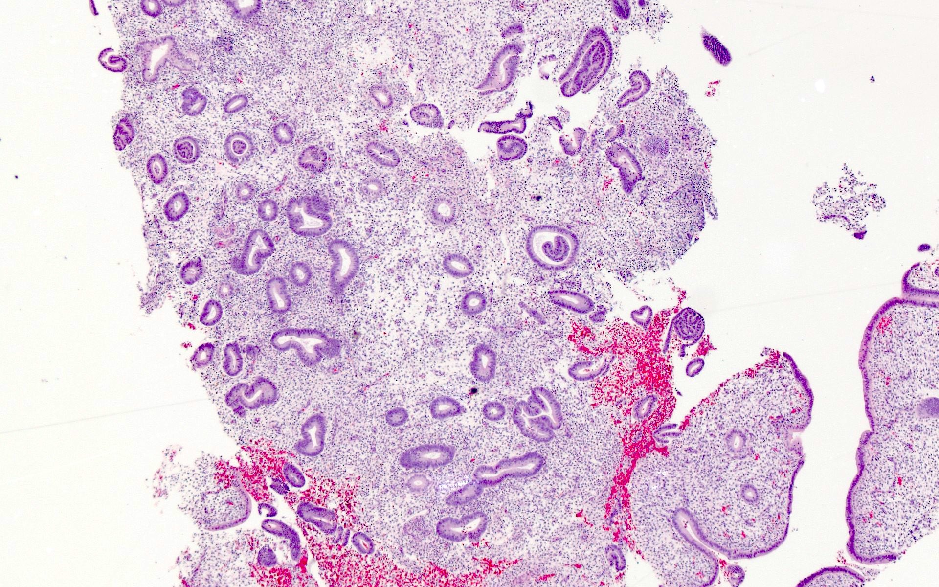
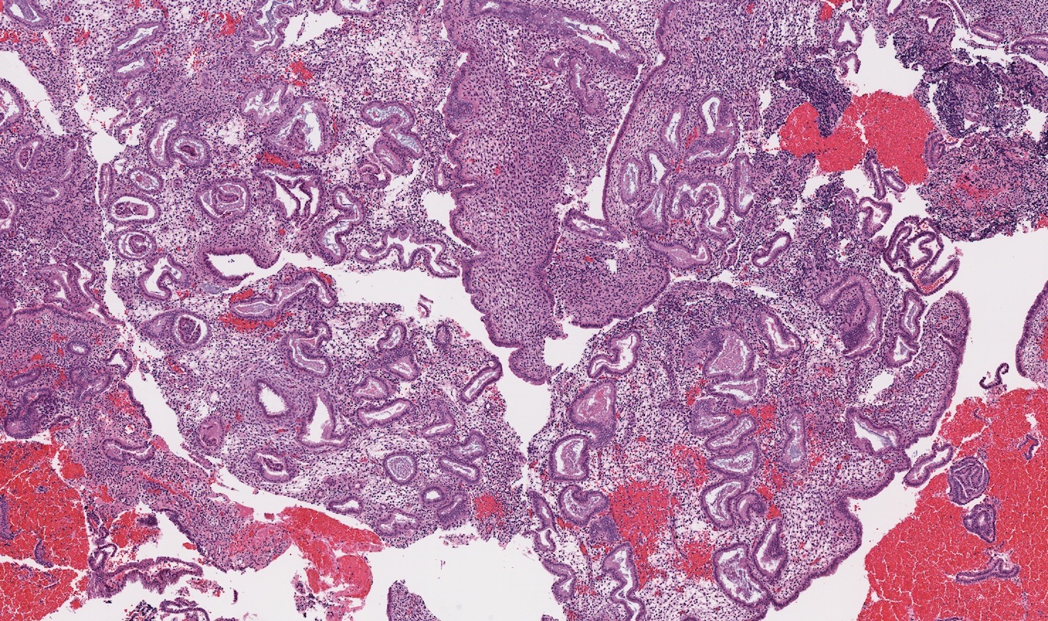
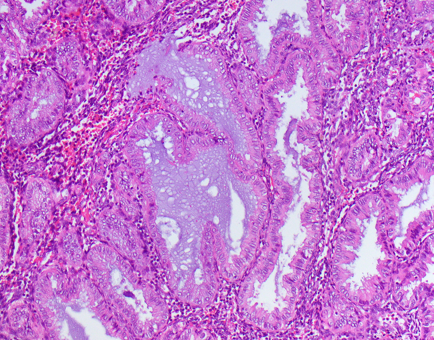
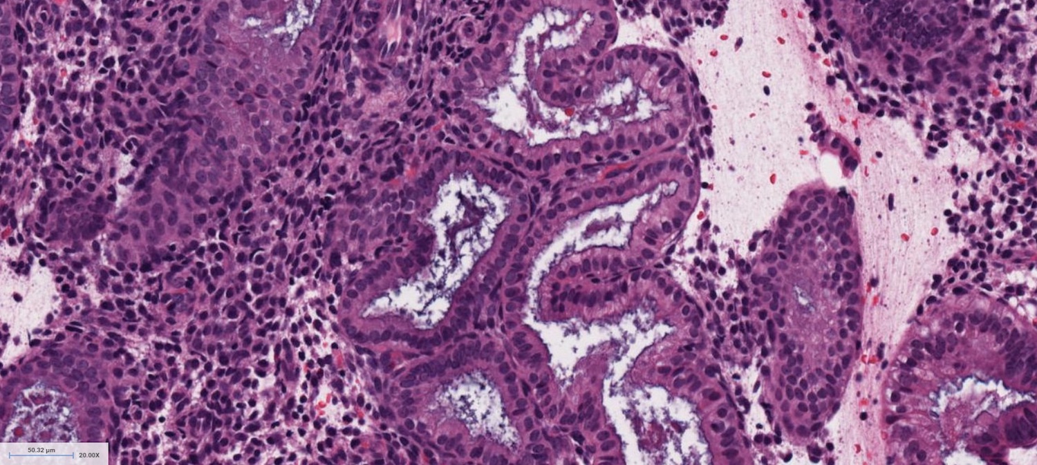
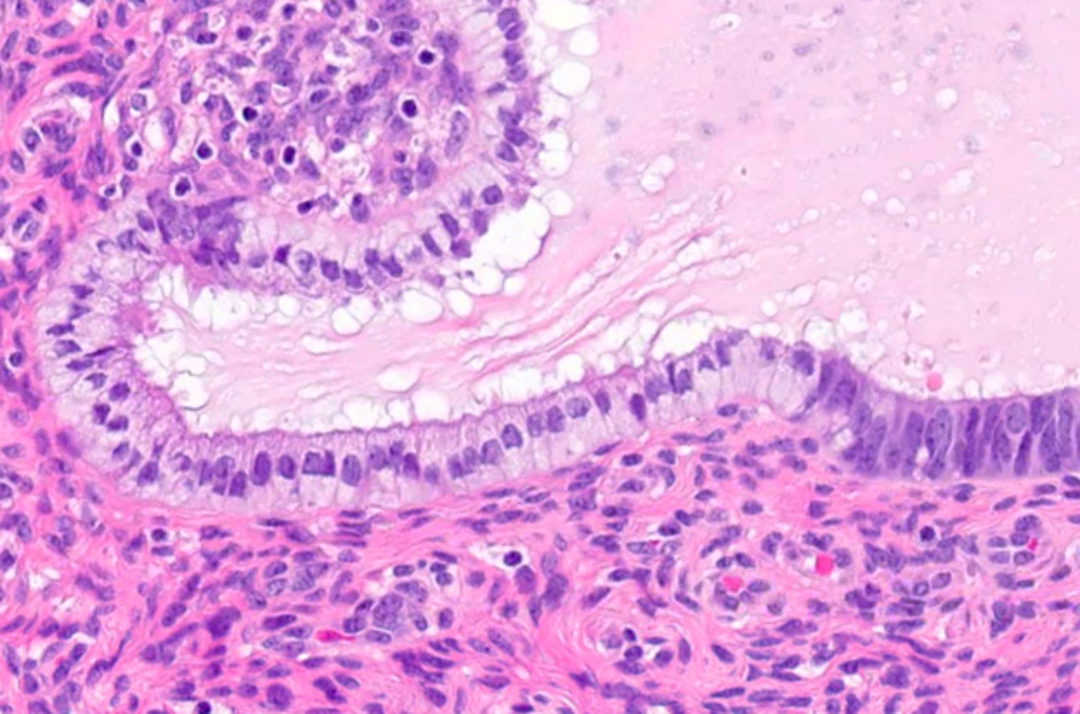
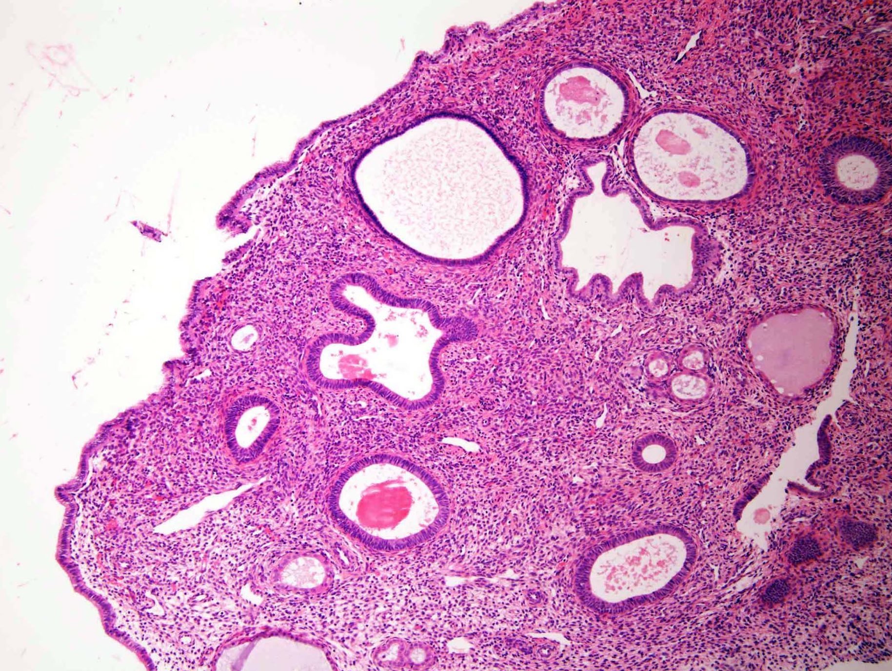
Secretory endometrium
Secretory endometrium
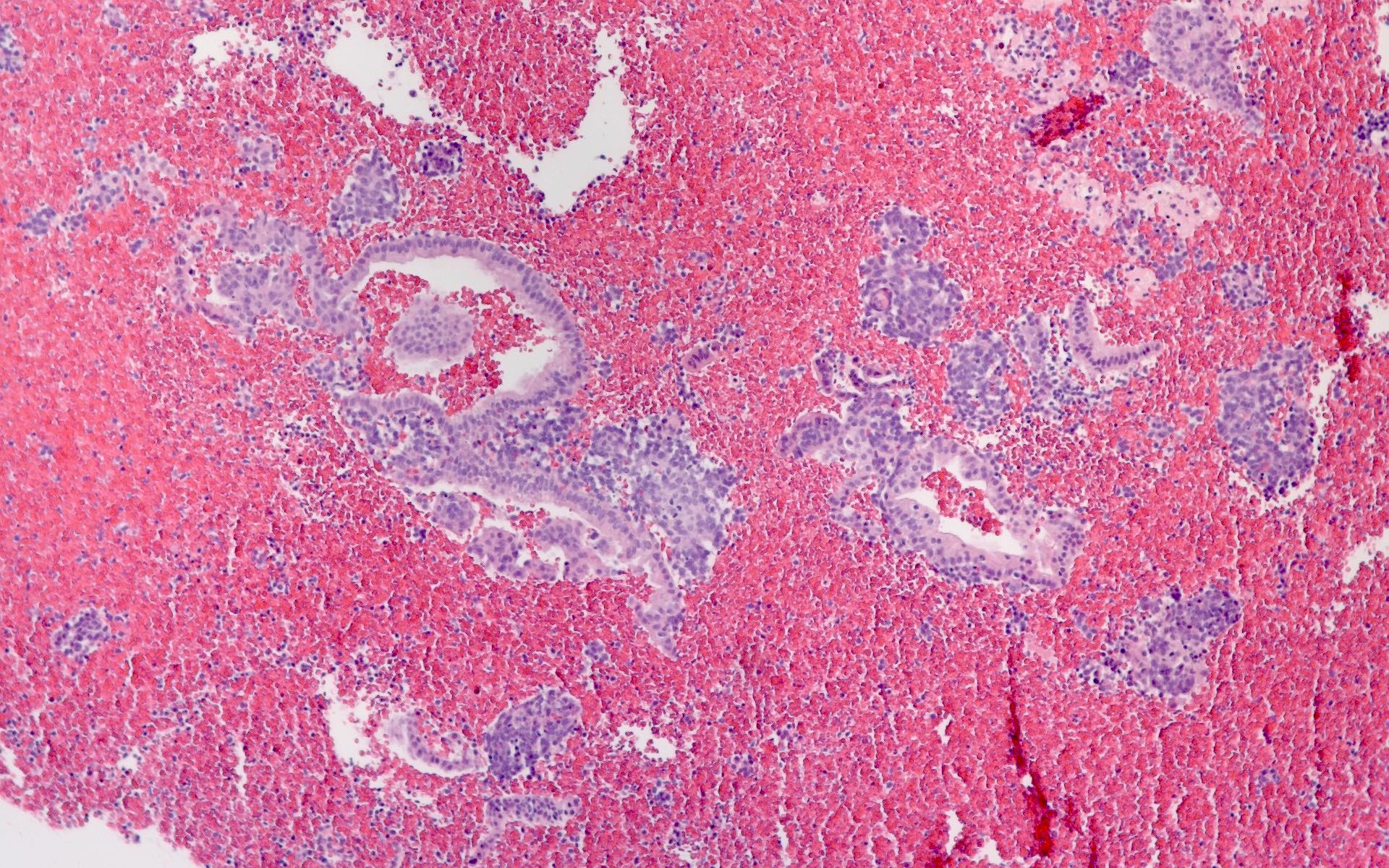
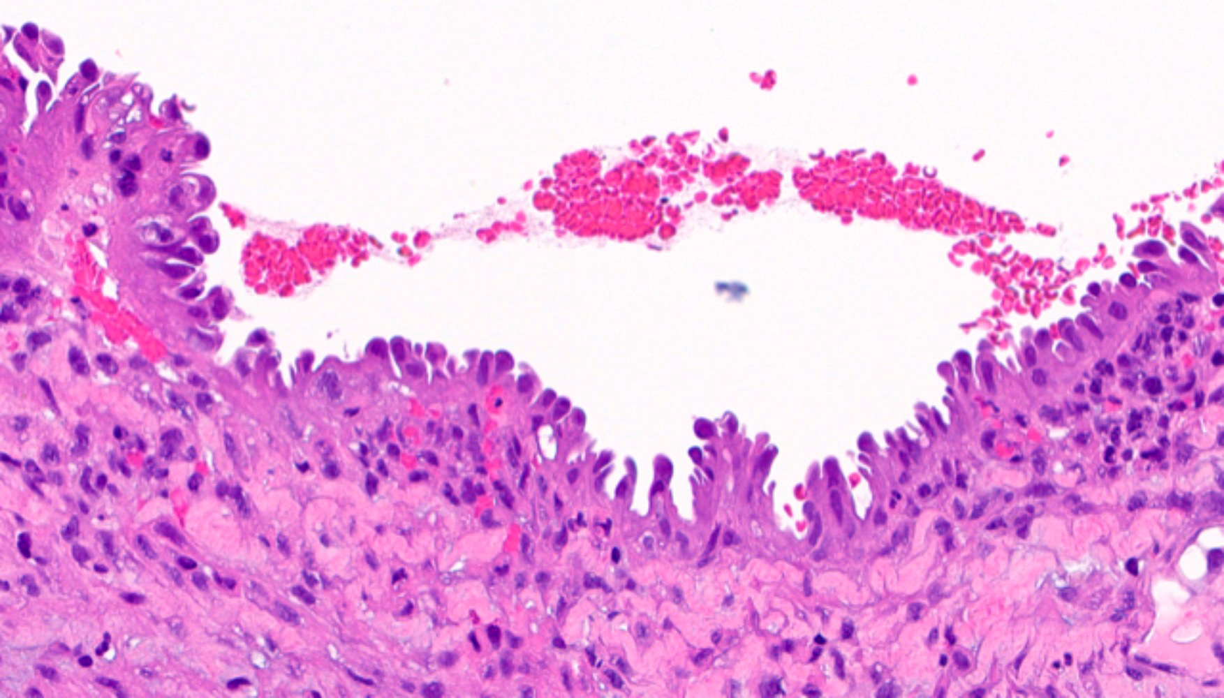
Menstrual endometrium
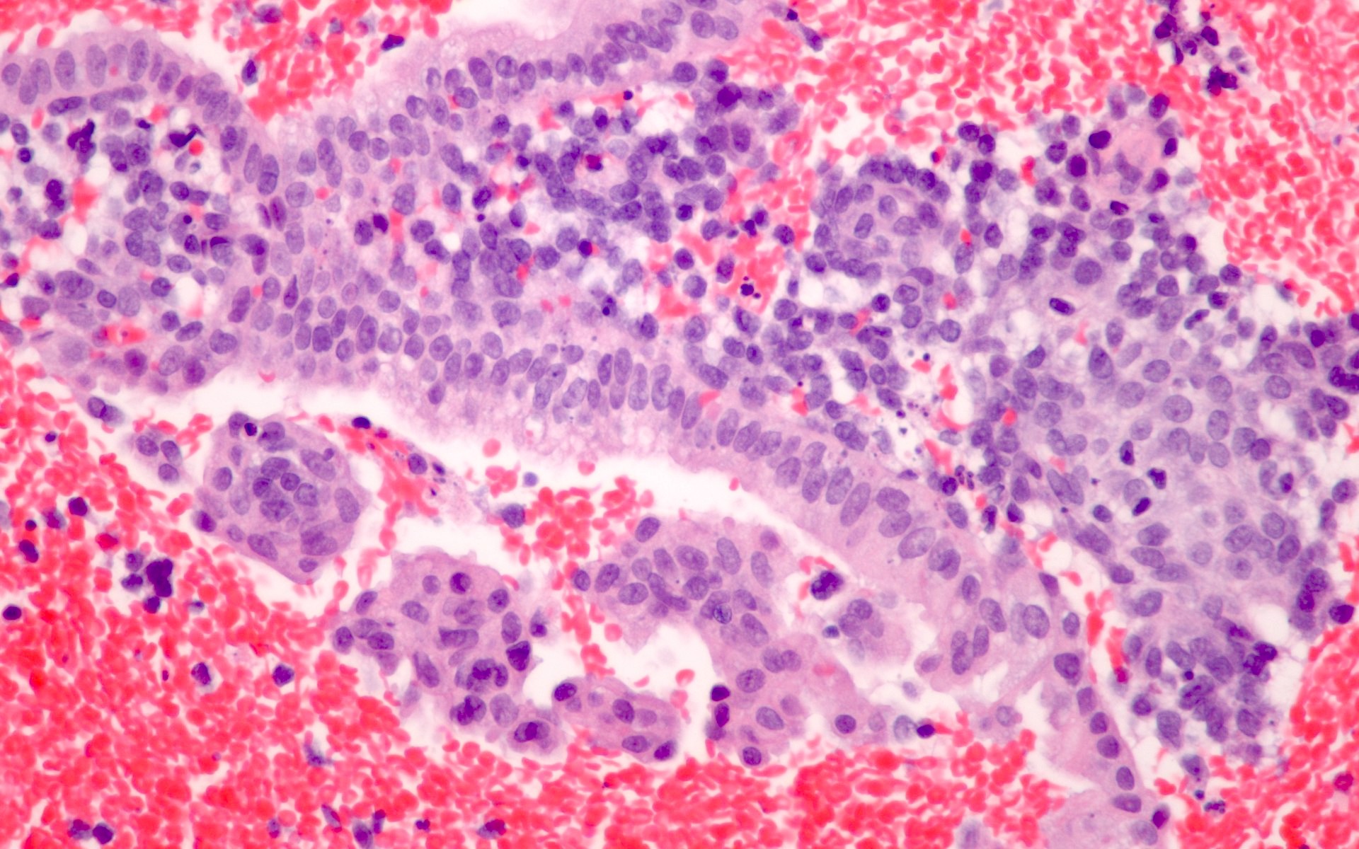
Menstrual endometrium
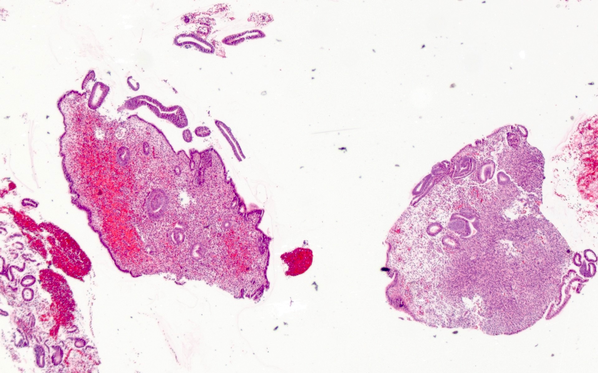
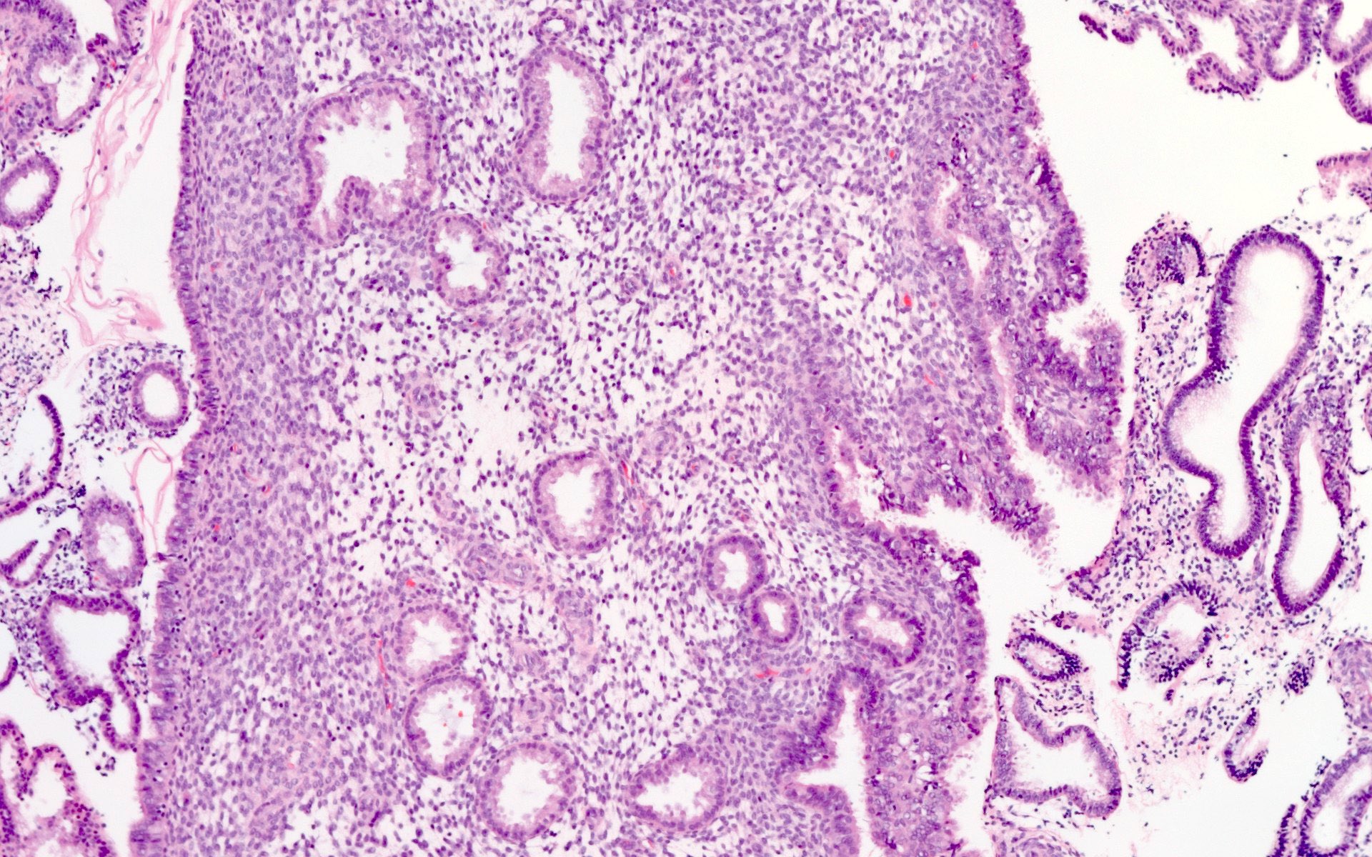
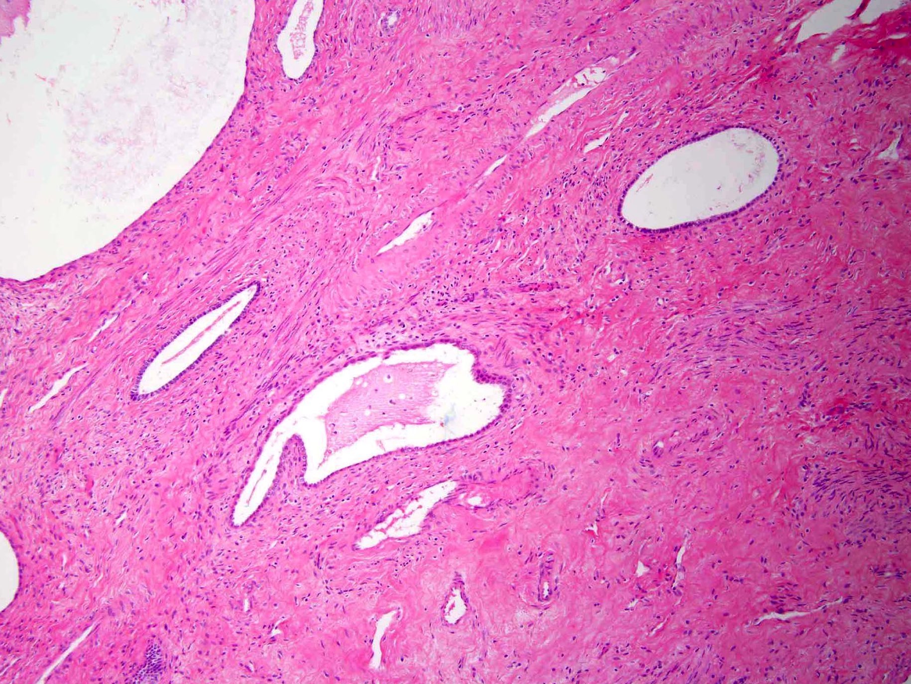
Inactive endometrium
Atrophic endometrium
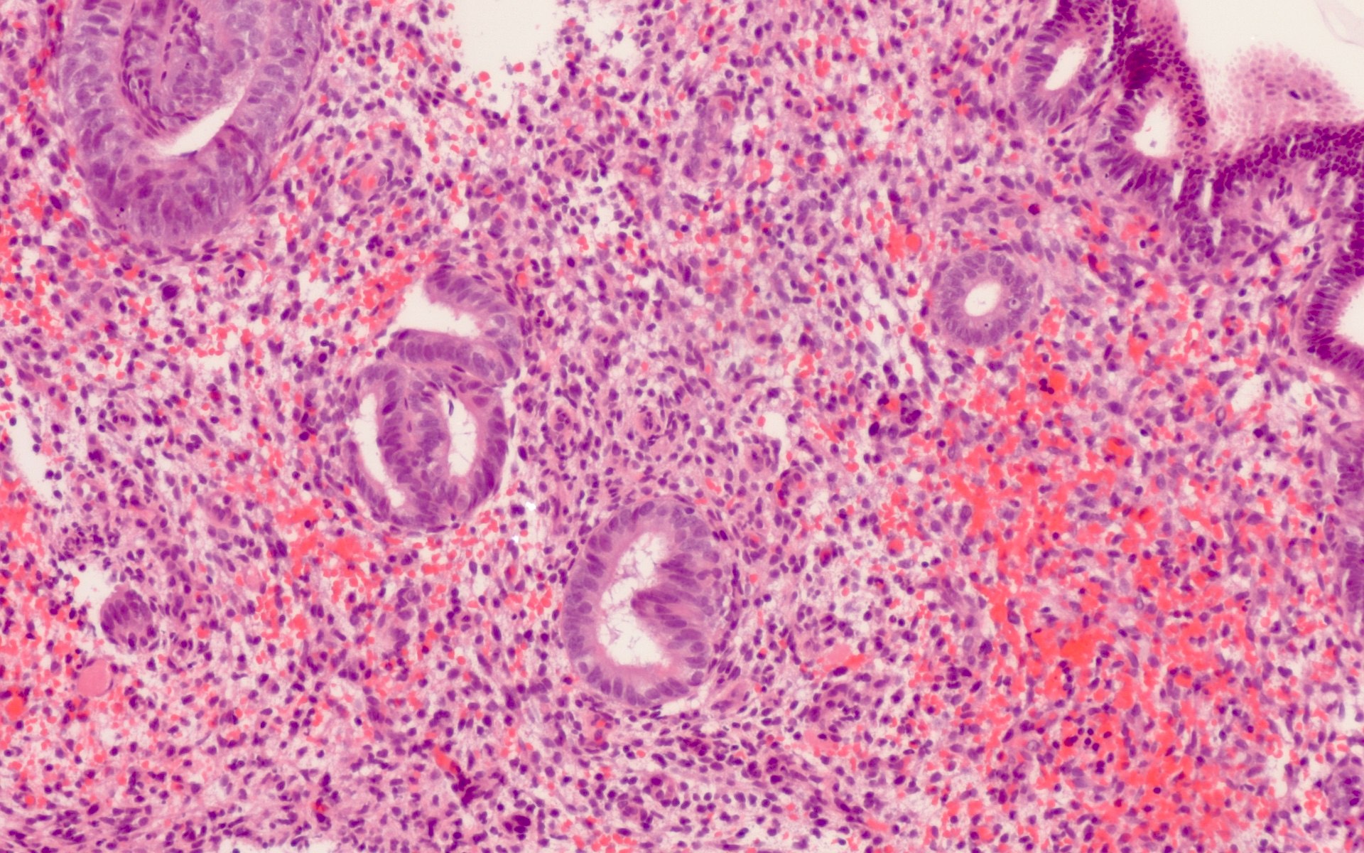
Abnormal uterine bleeding
Contributed by Ayse Ayhan, M.D., Ph.D.

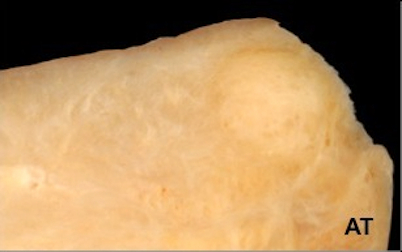
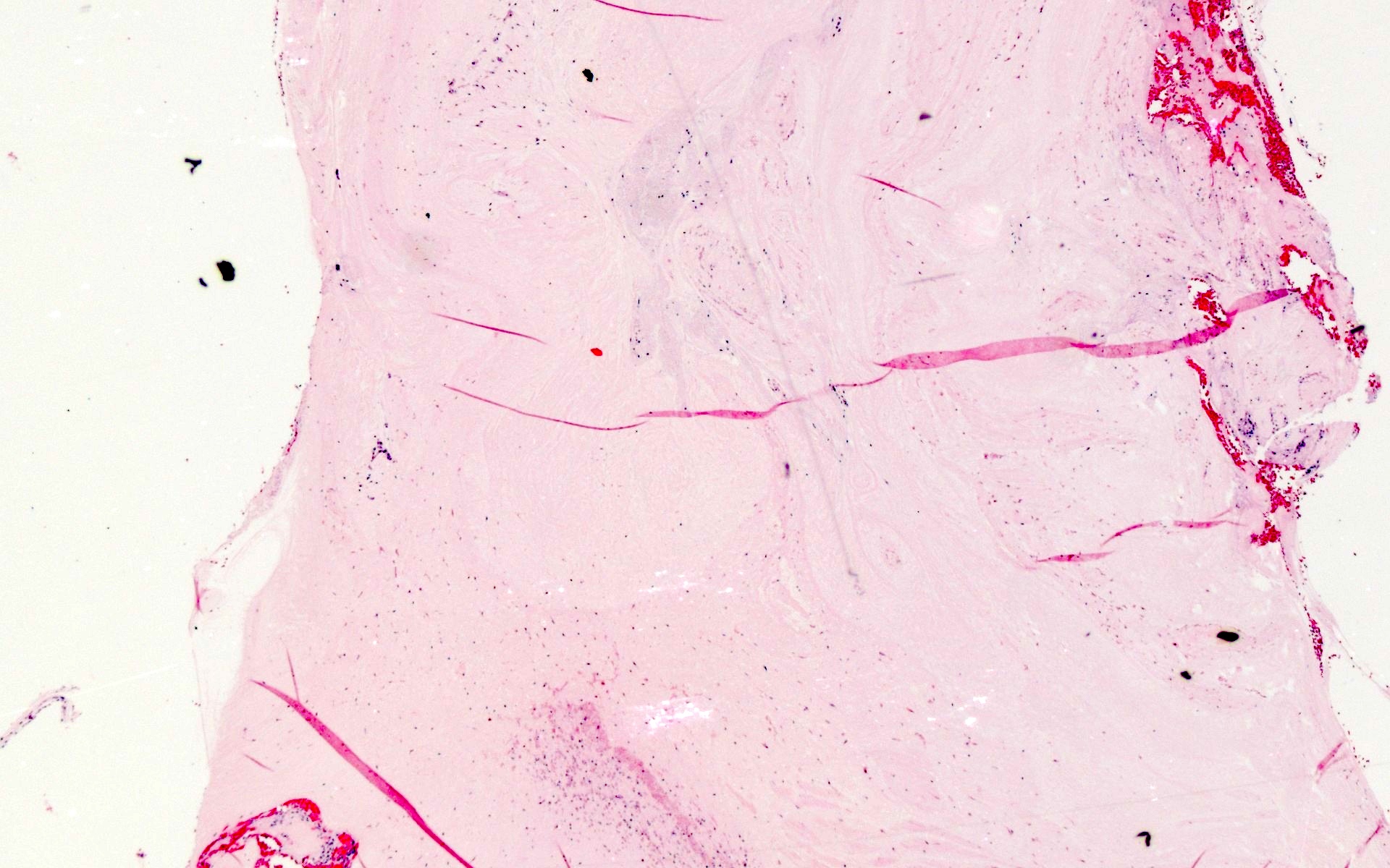
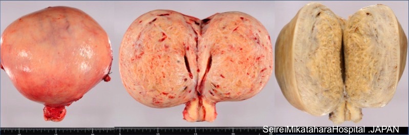
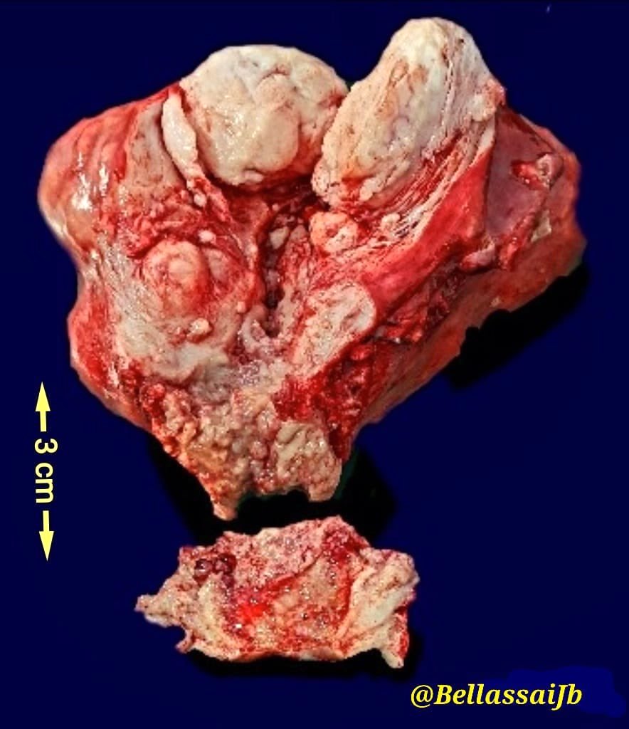
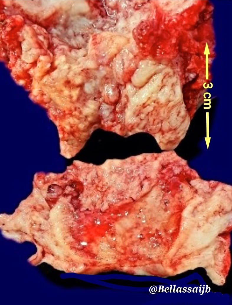
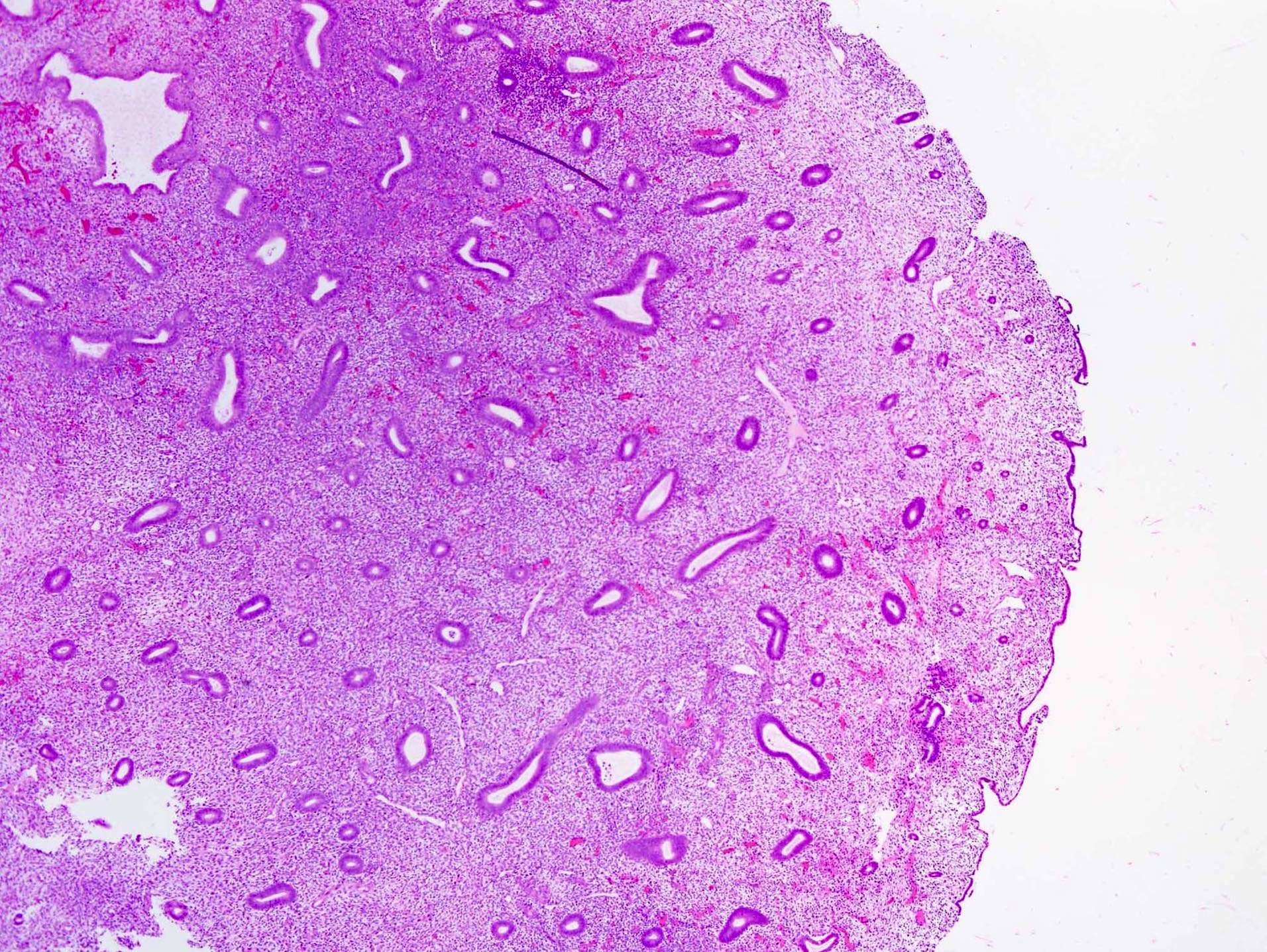
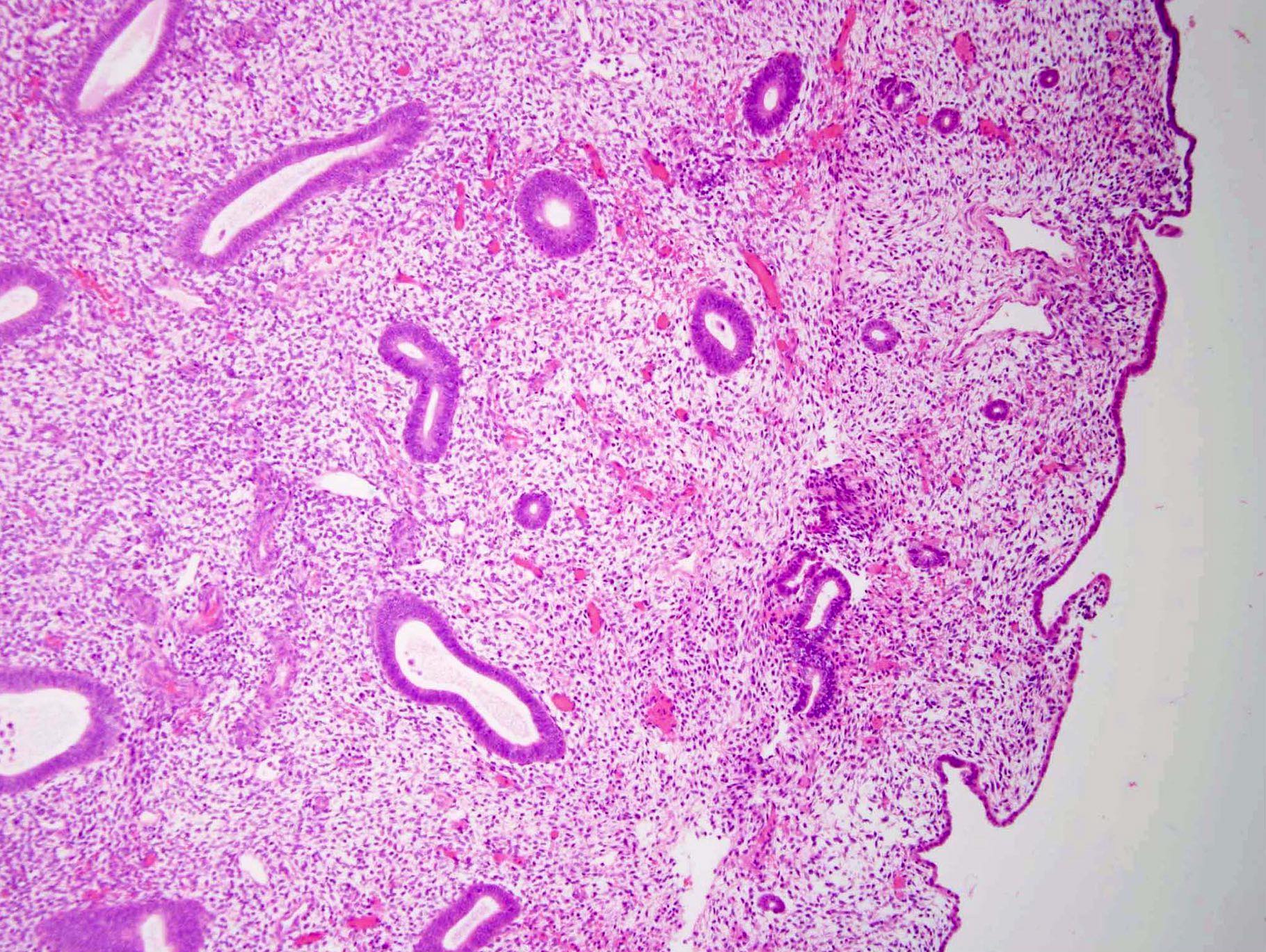
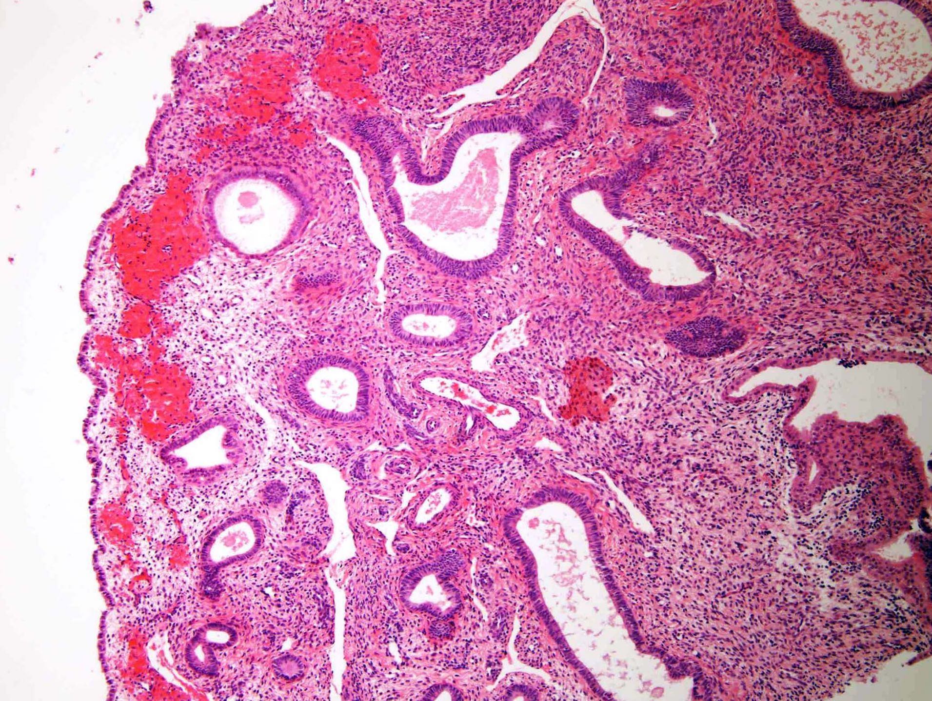
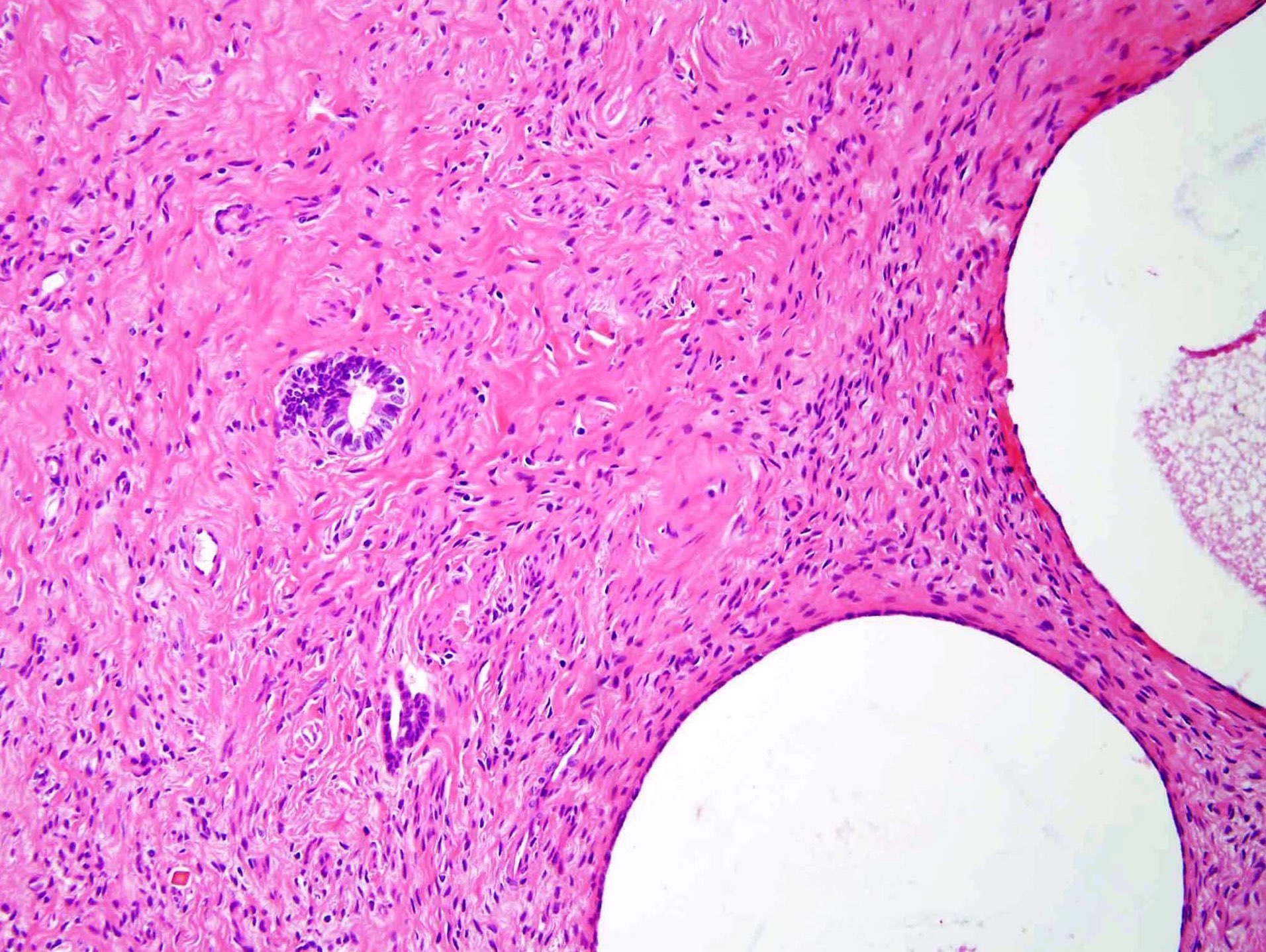
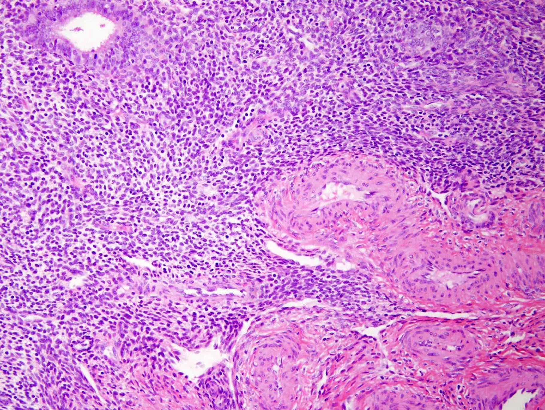
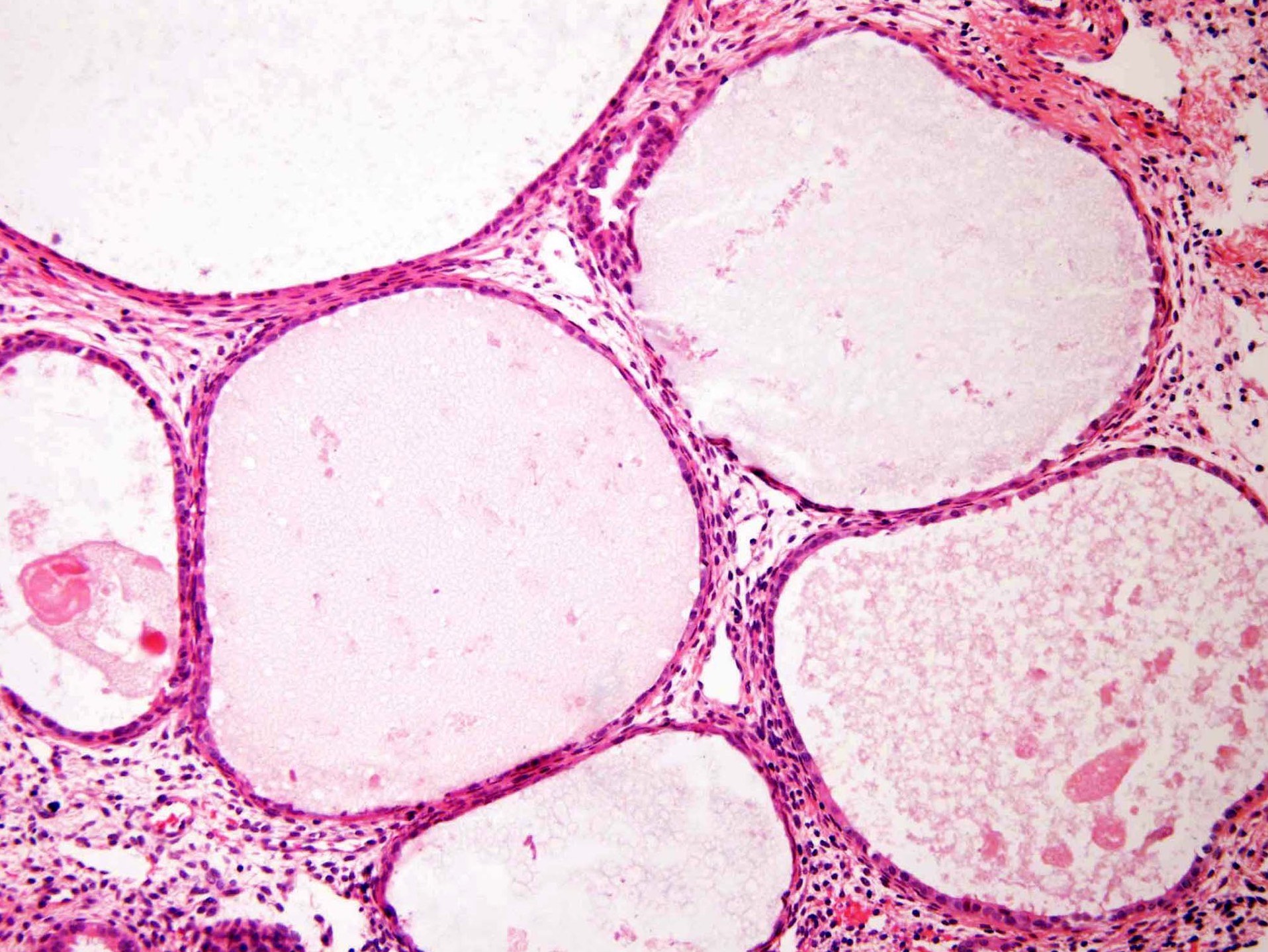
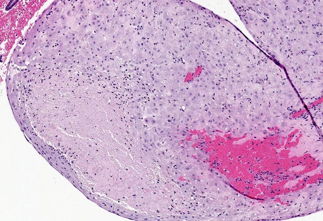
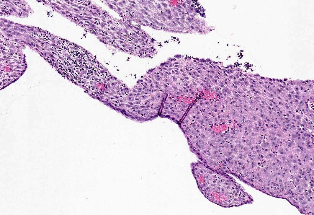
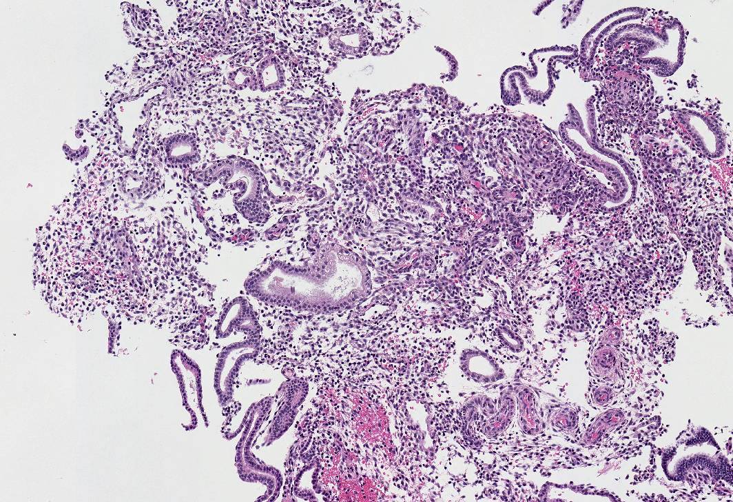
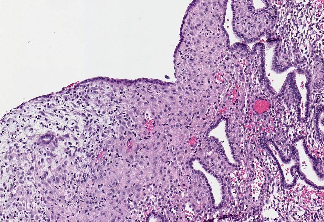
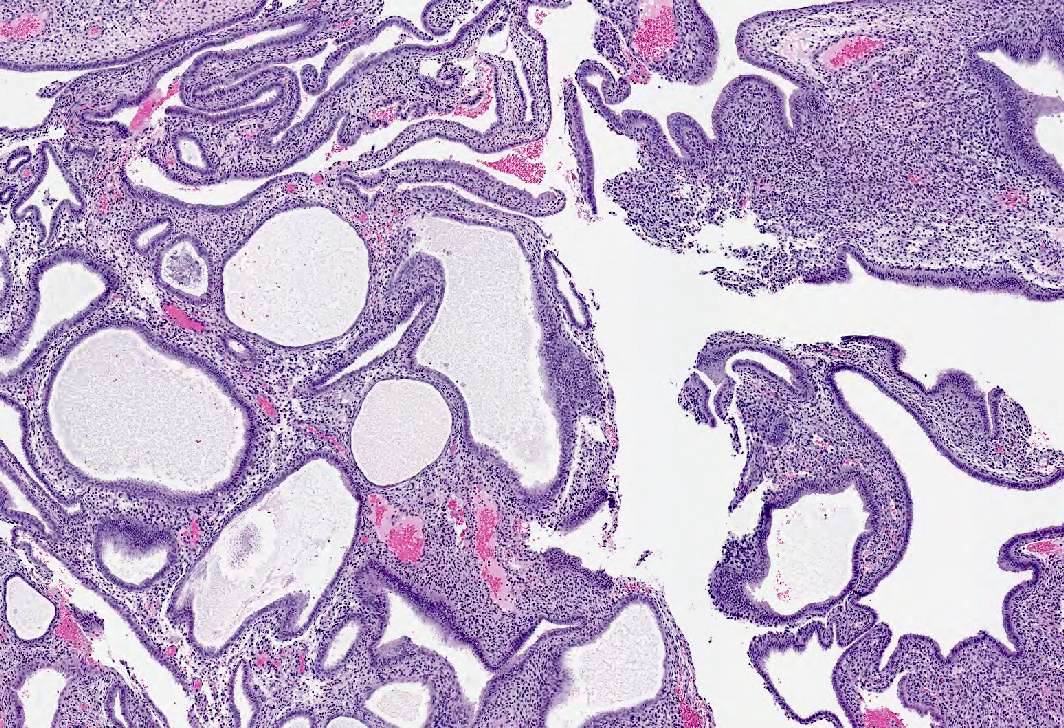
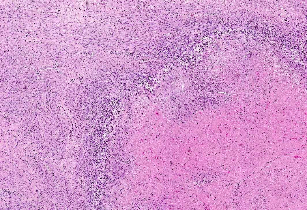
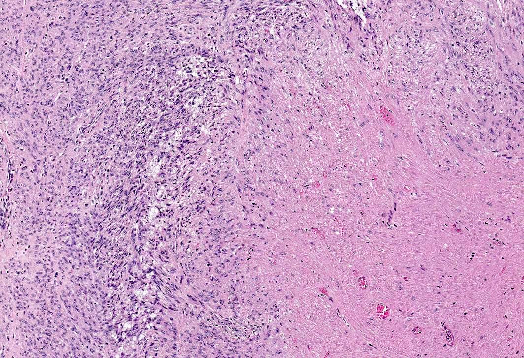
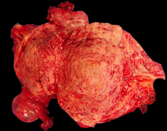
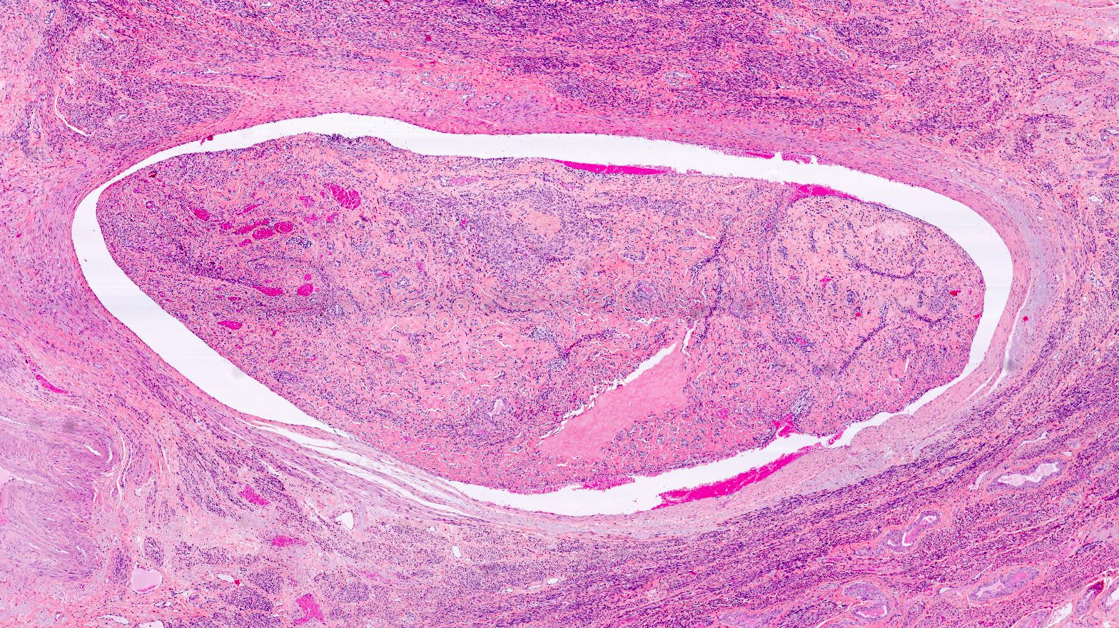
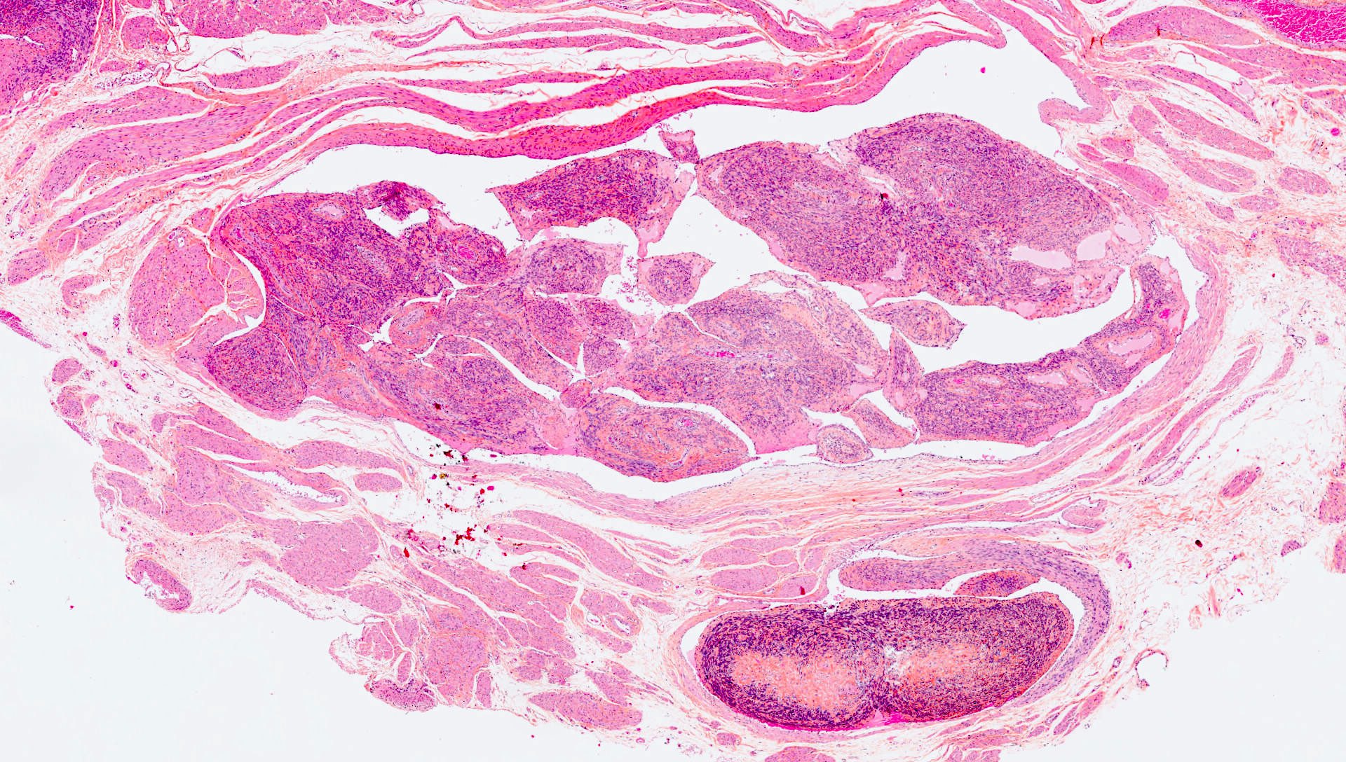
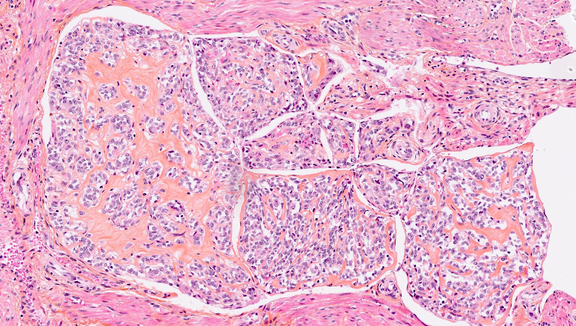
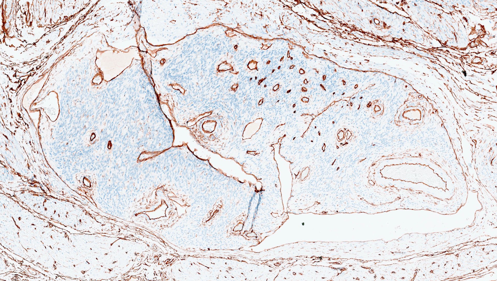
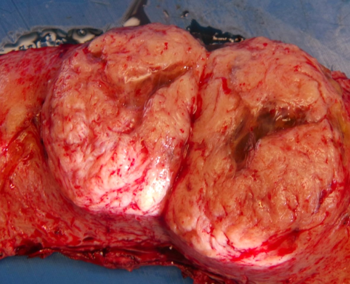
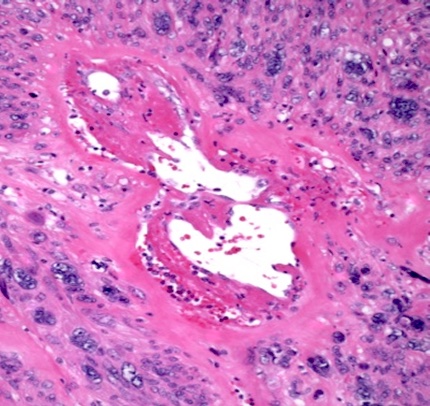
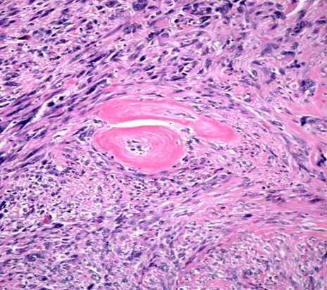
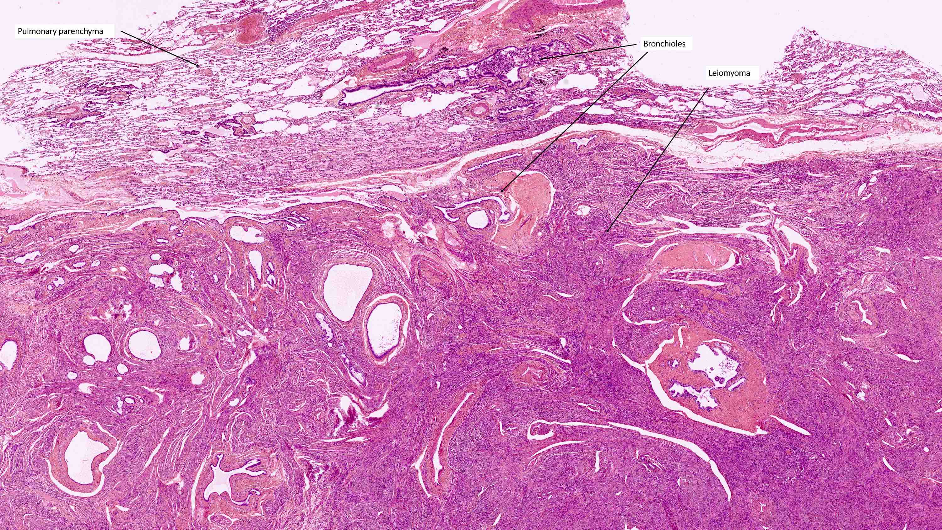
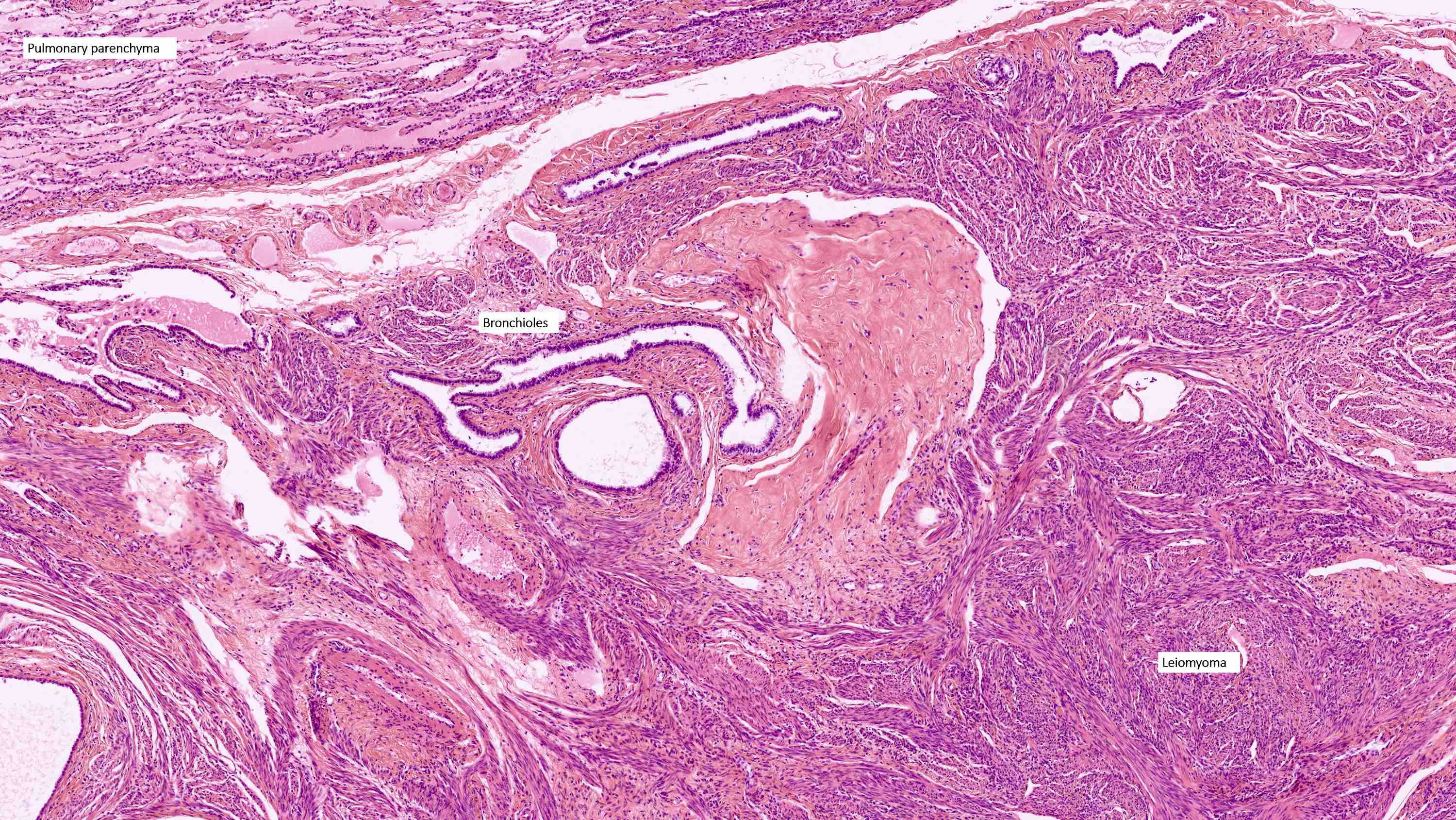
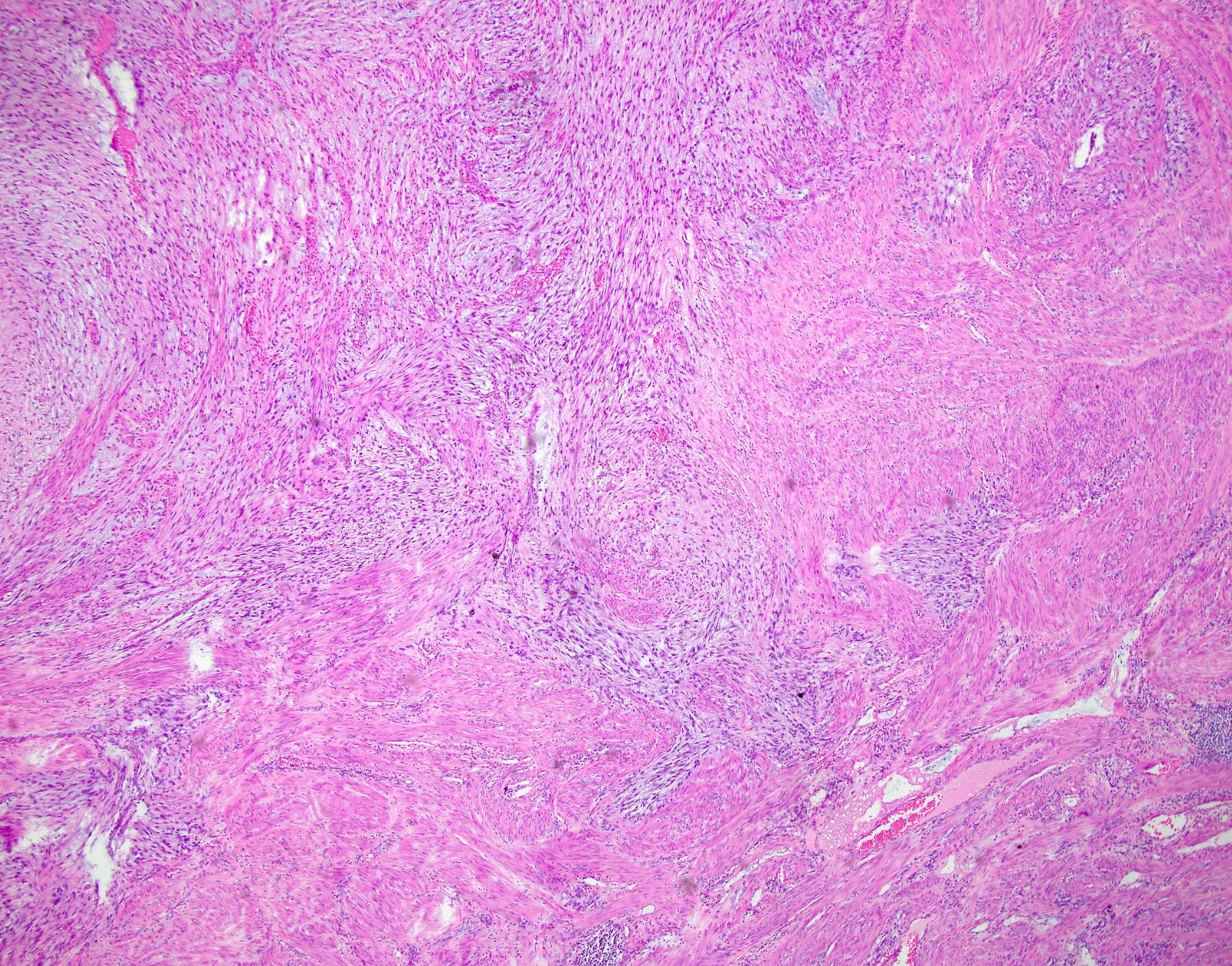
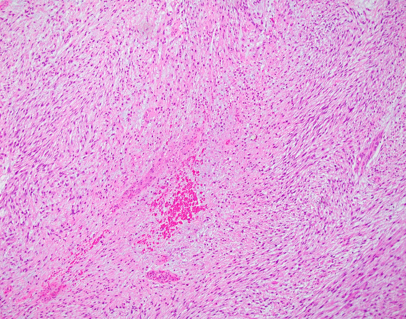
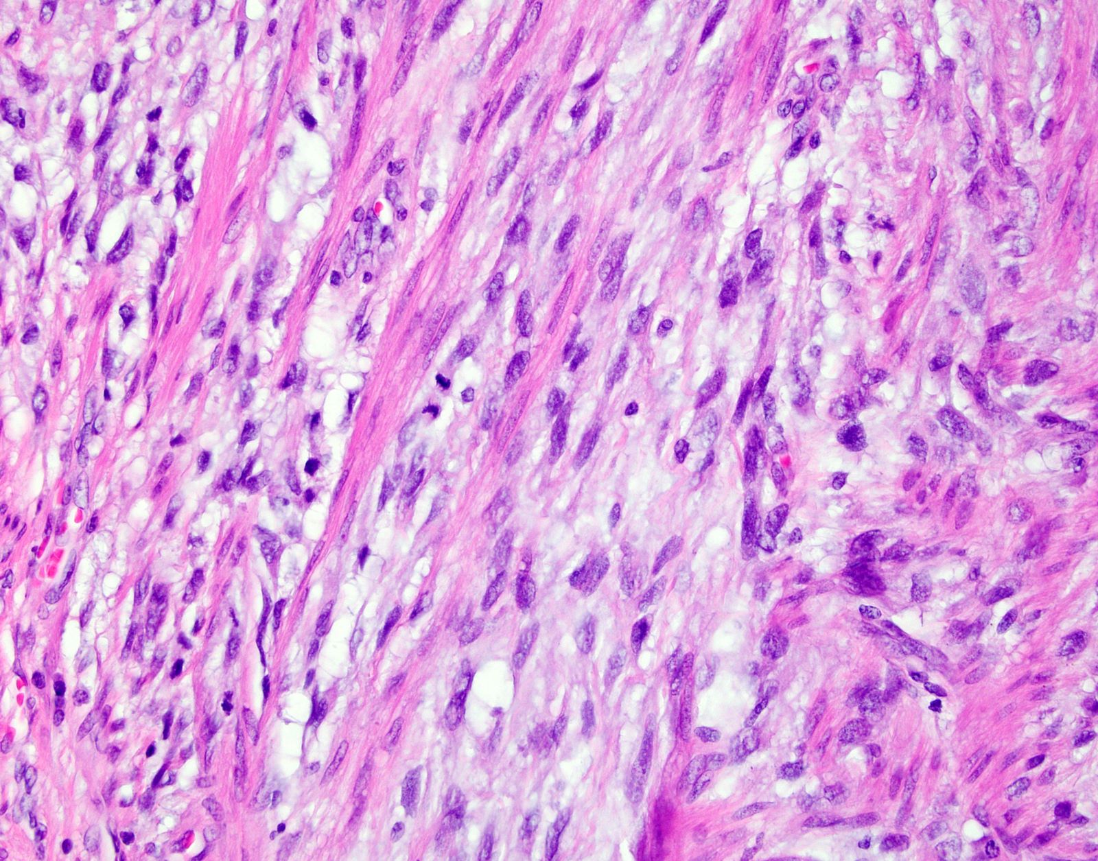
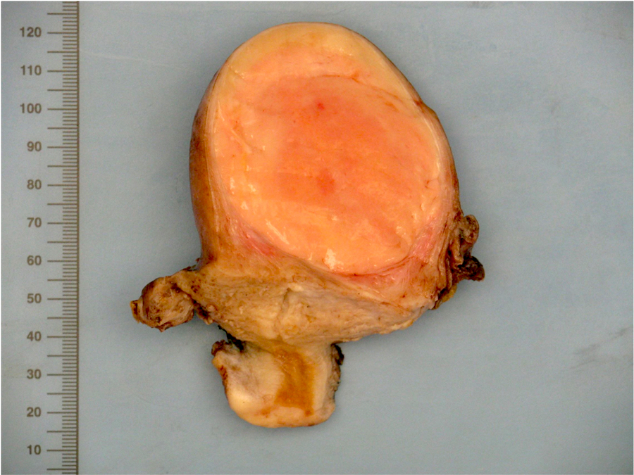
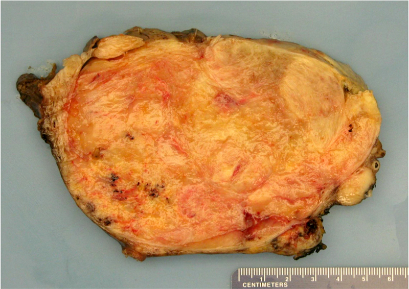
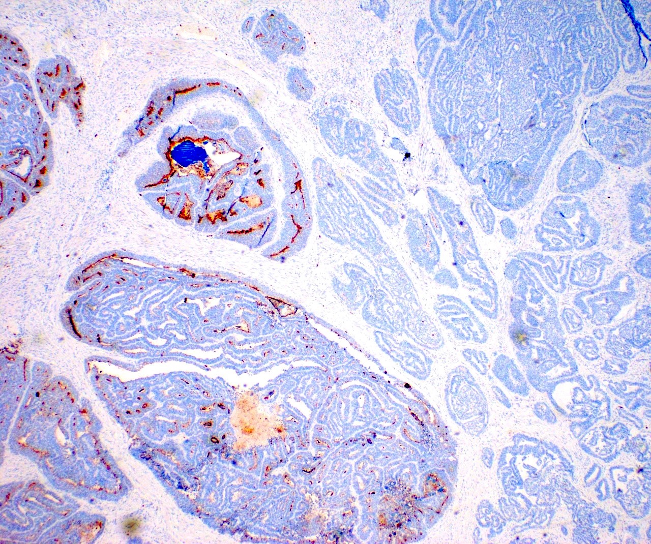
Adenomatoid tumor and leiomyoma
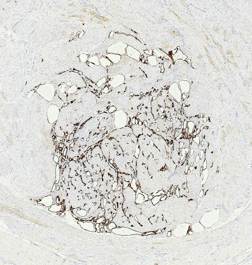
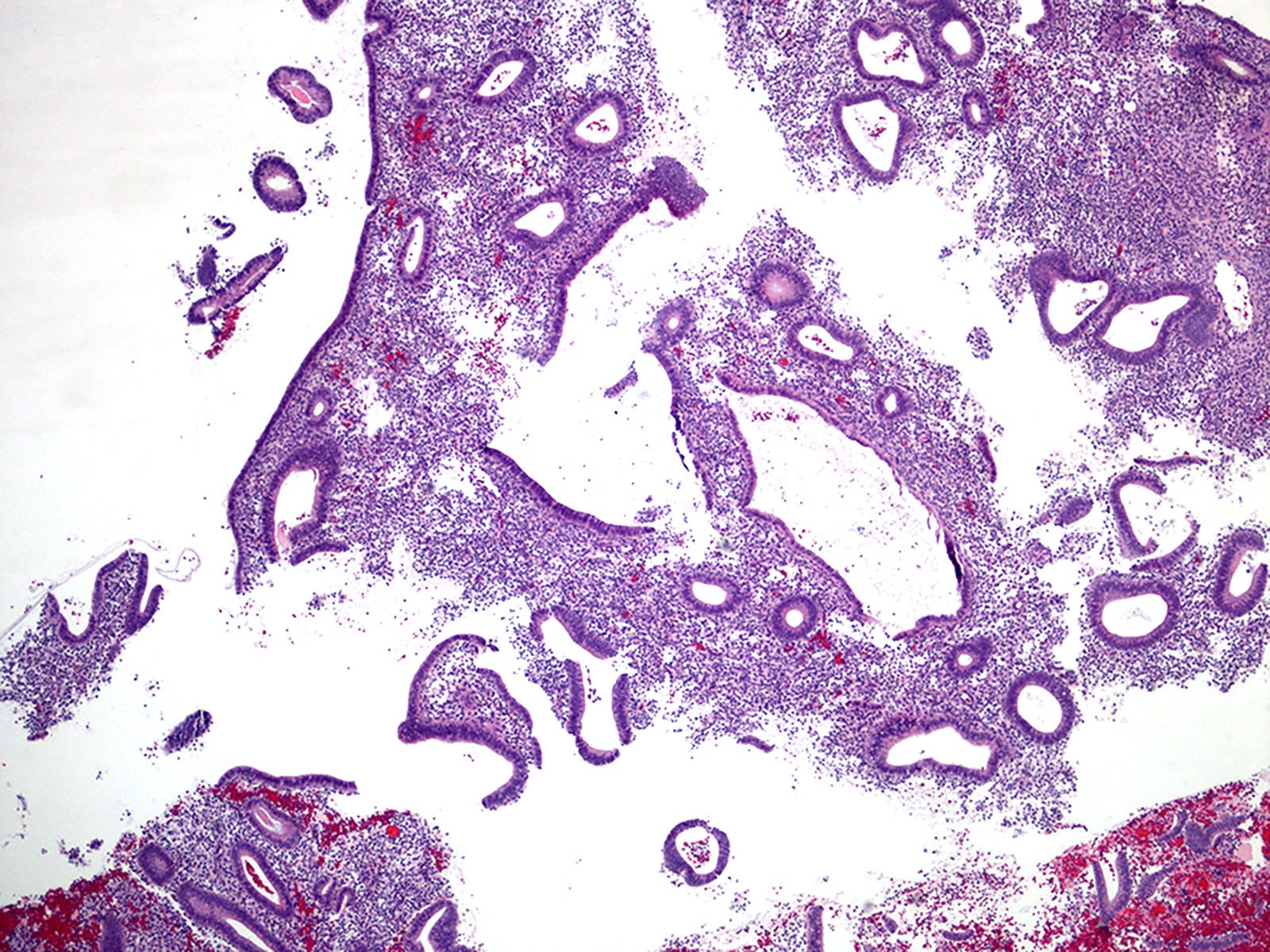
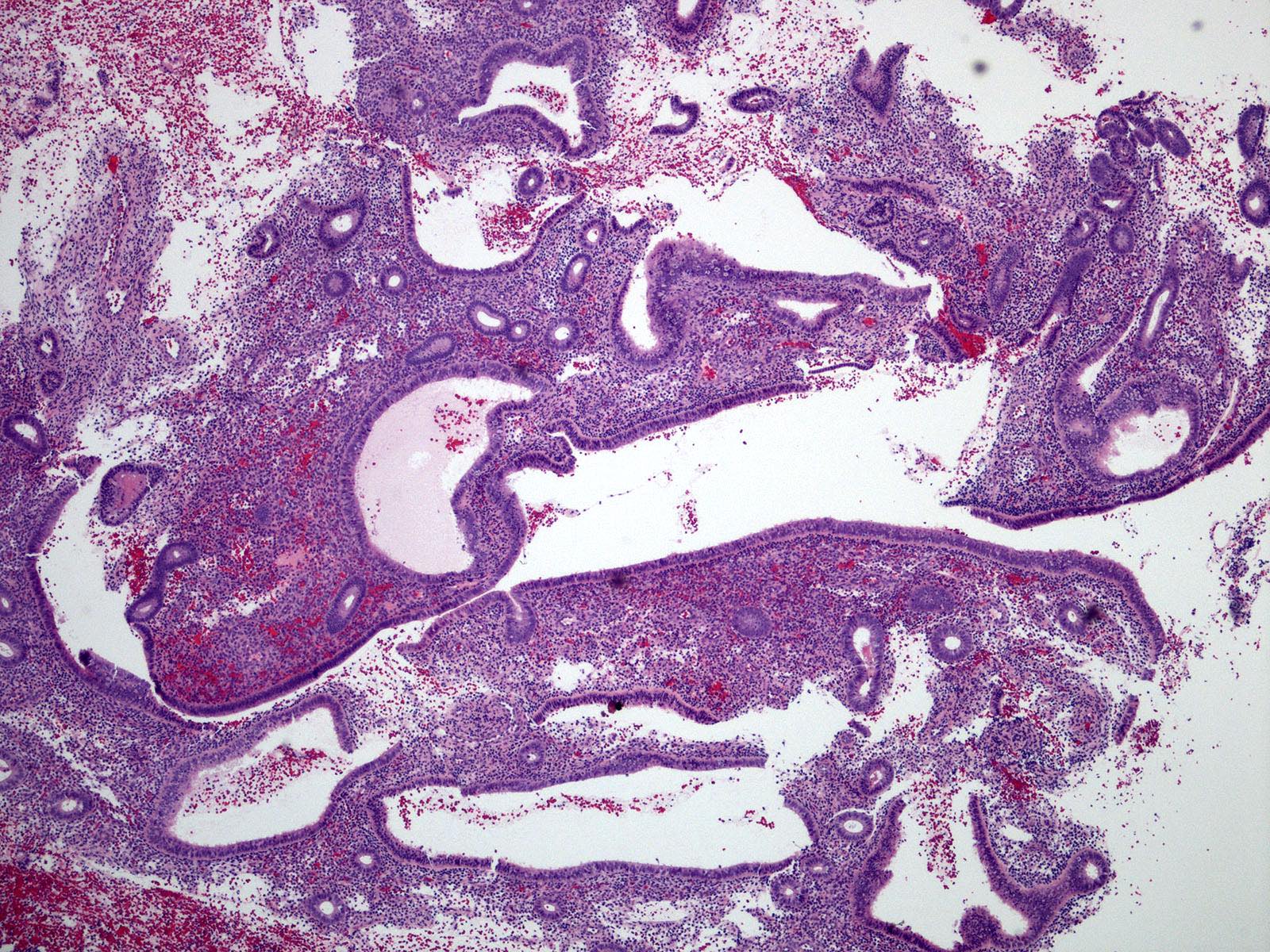
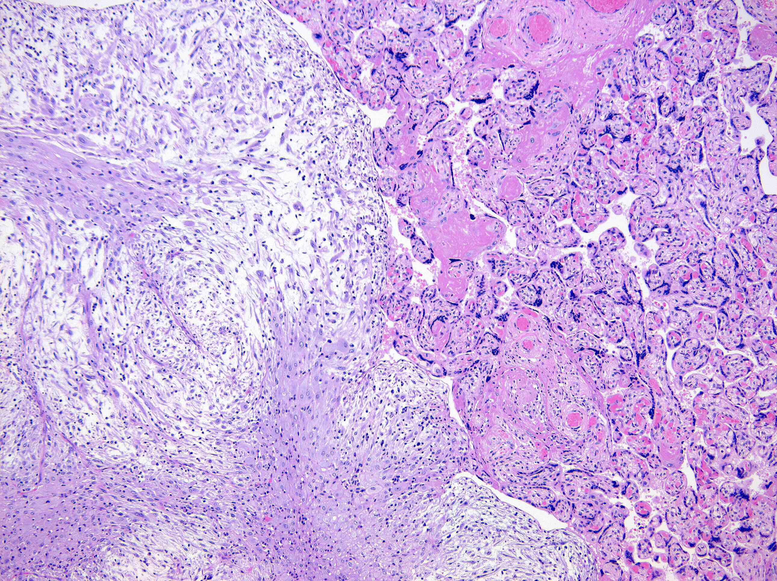
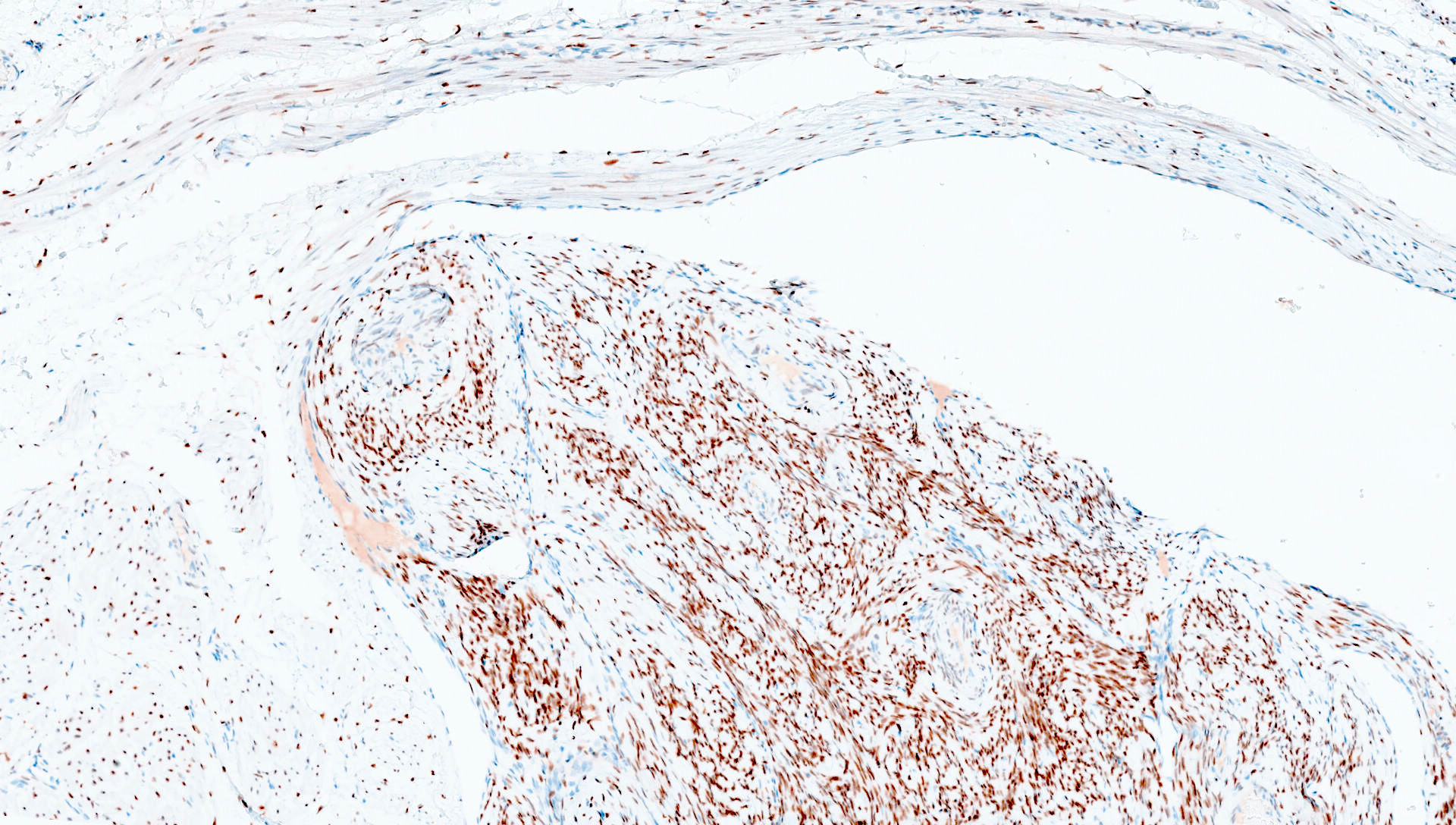
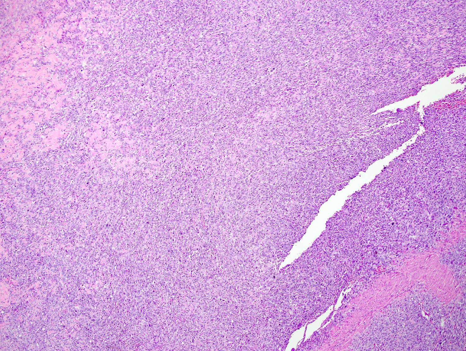
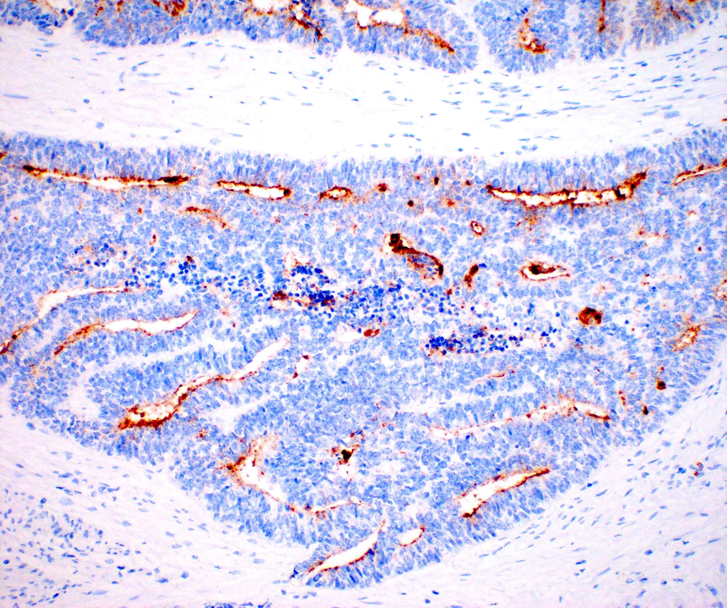
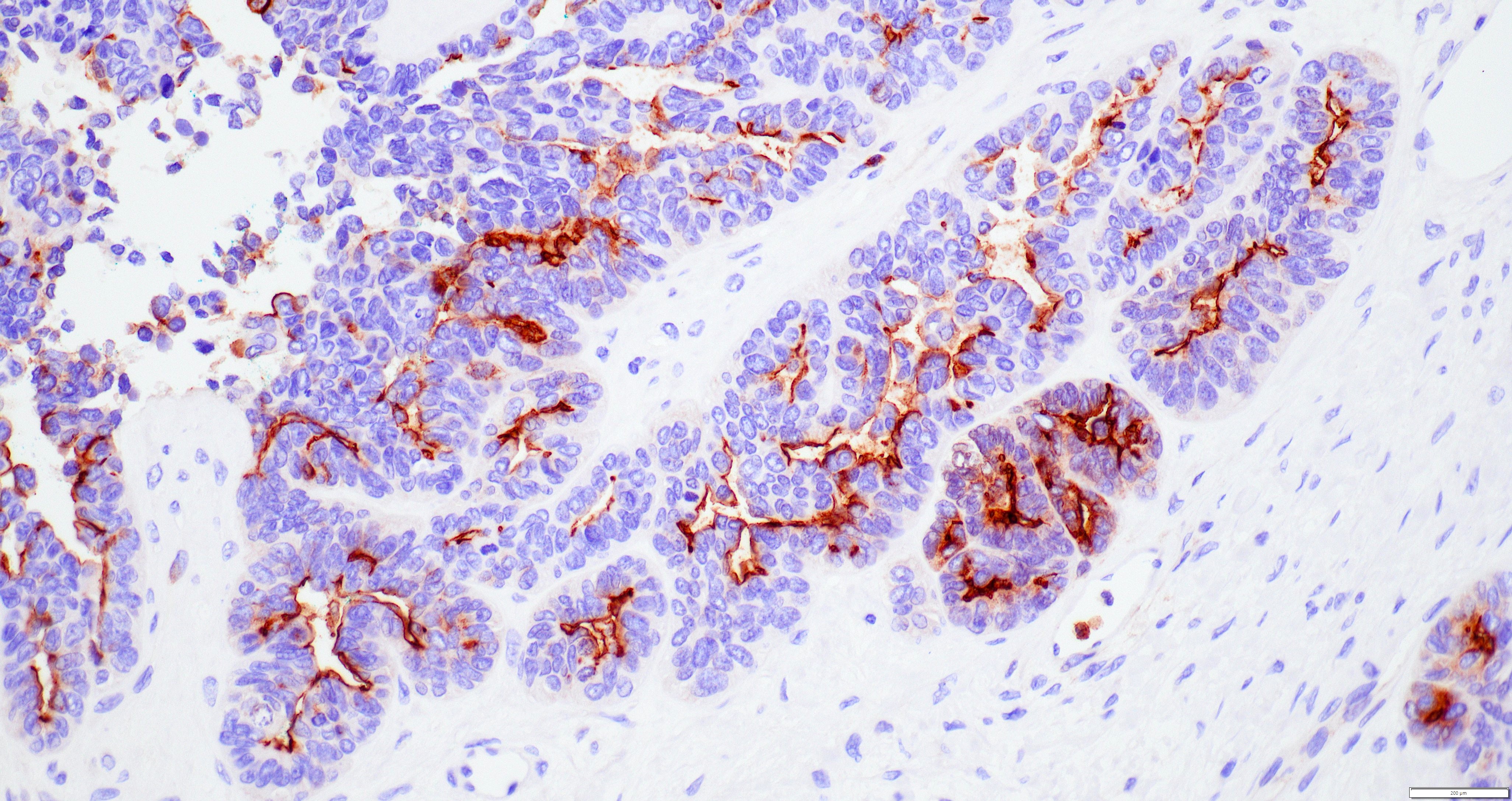
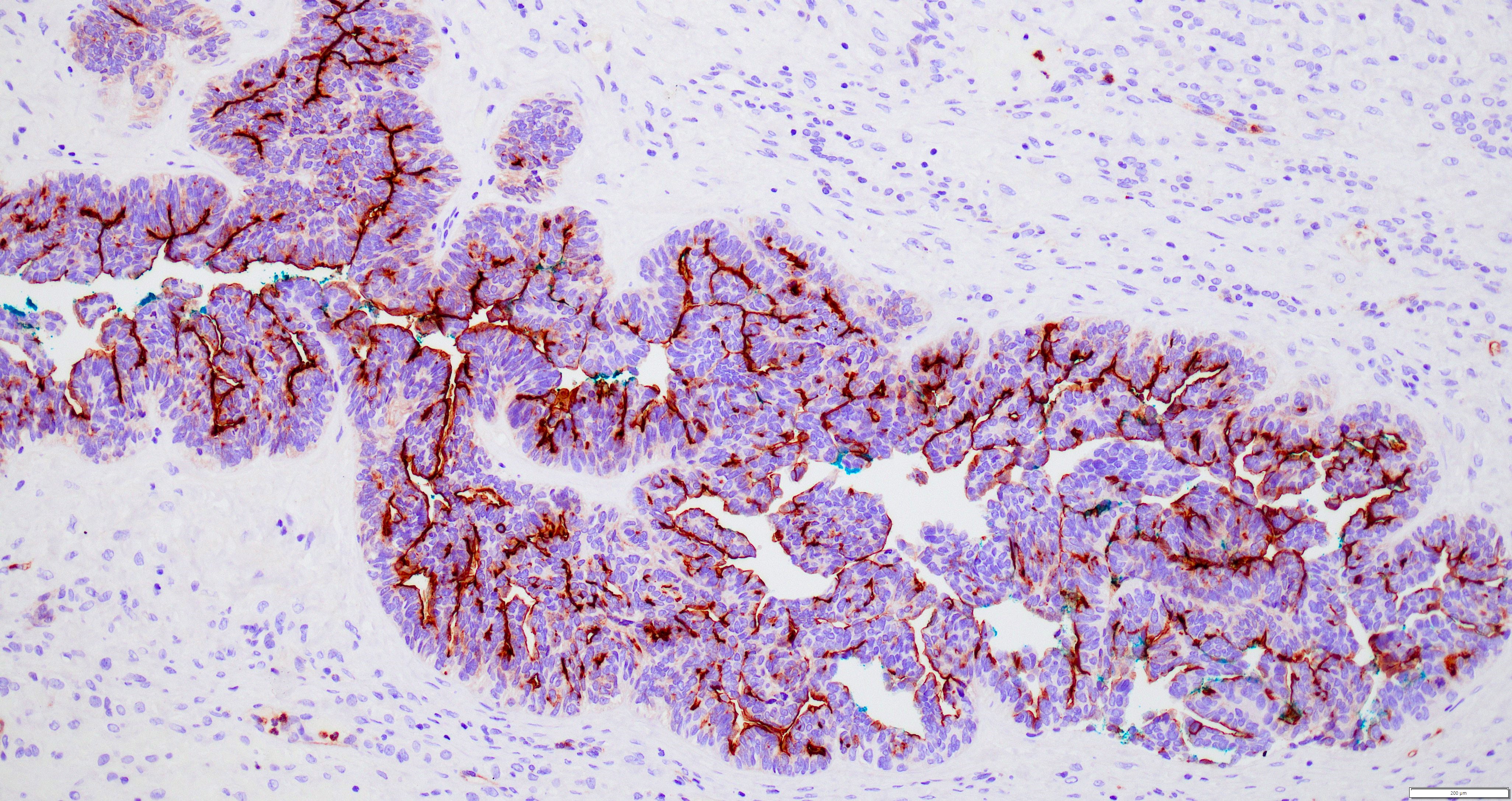
Contributed by Jian-Jun Wei, M.D. and Ayse Ayhan, M.D., Ph.D.


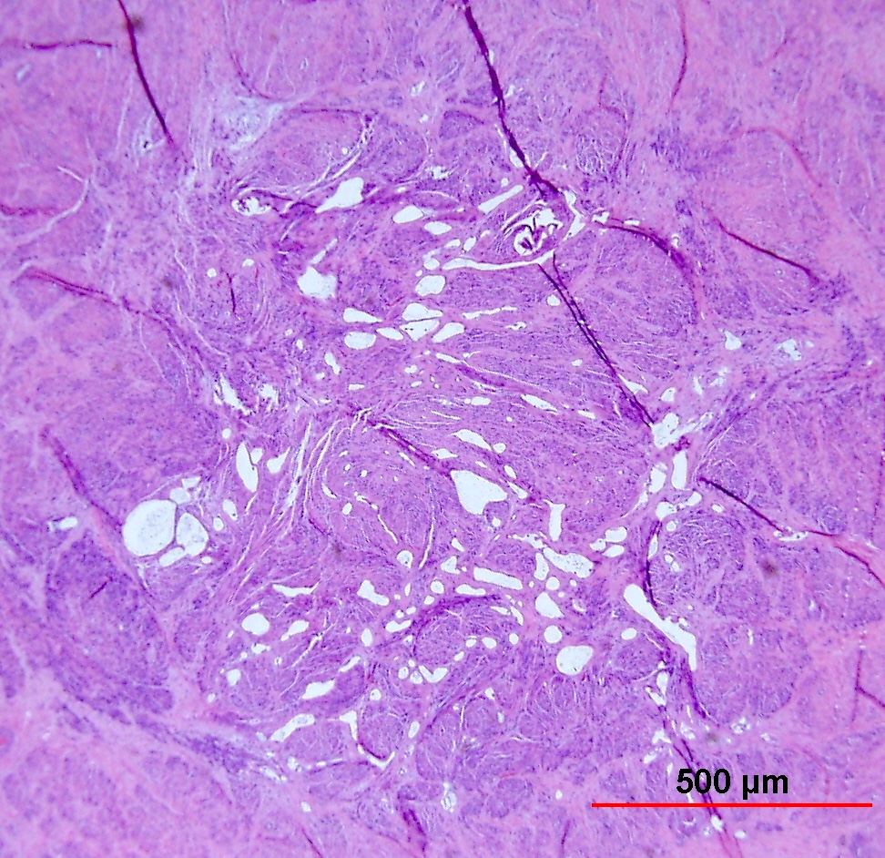

Vascular-like, cystic branching spaces in a background of benign smooth muscle
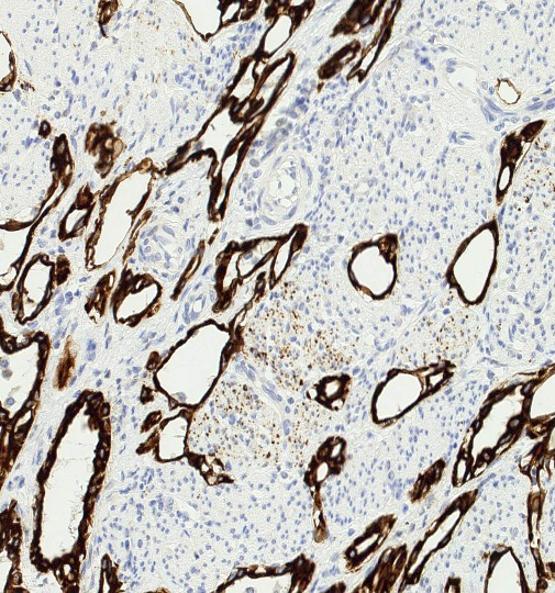
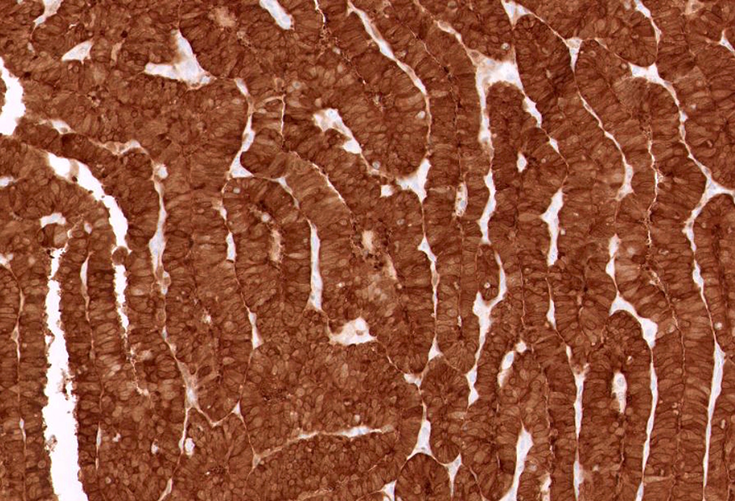
AE1 / AE3 positive in AT cells
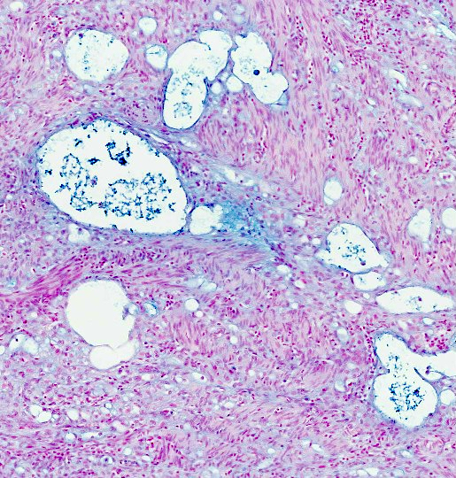
Alcian blue staining secretions
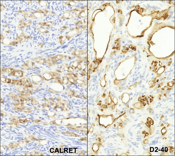
Calretinin and D2-40
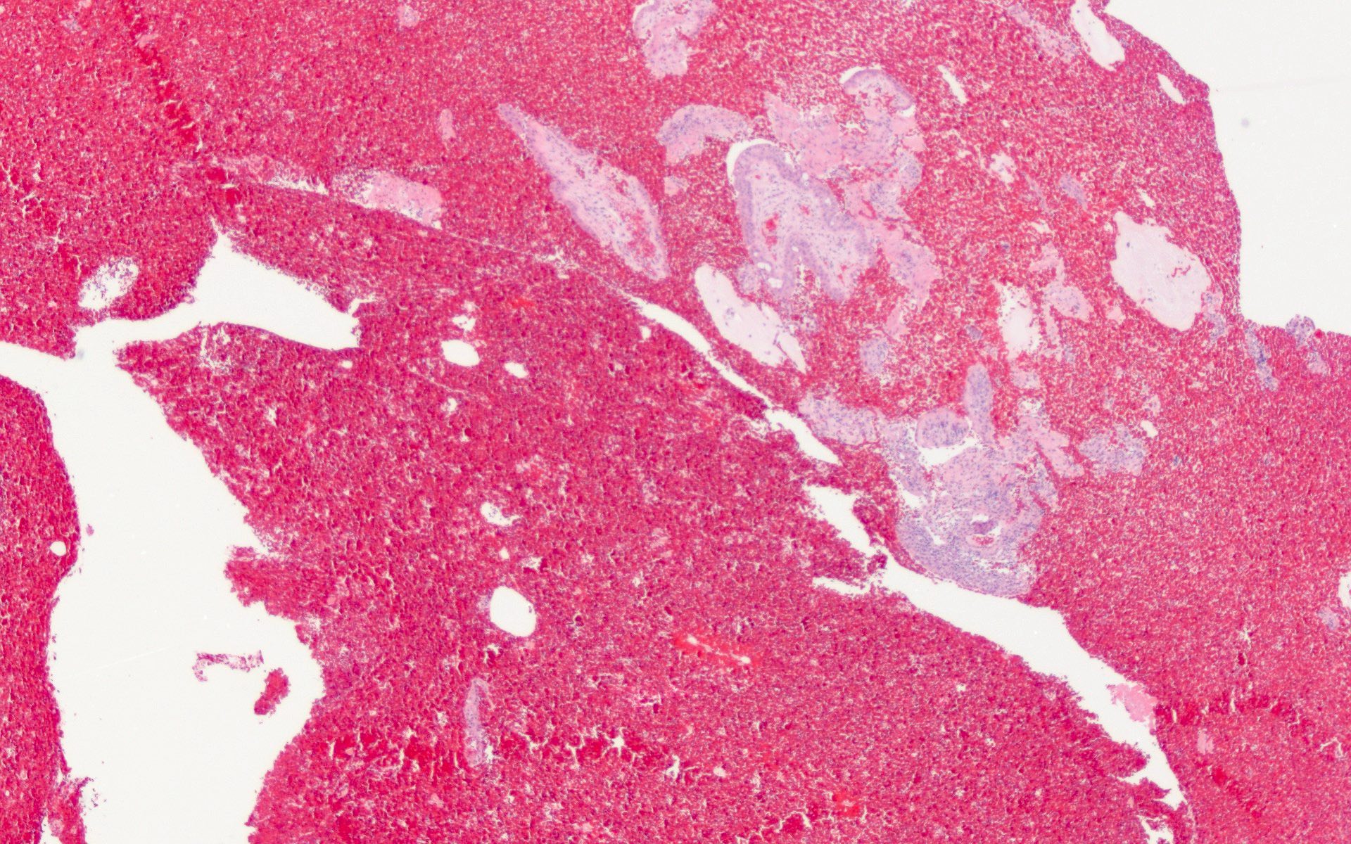
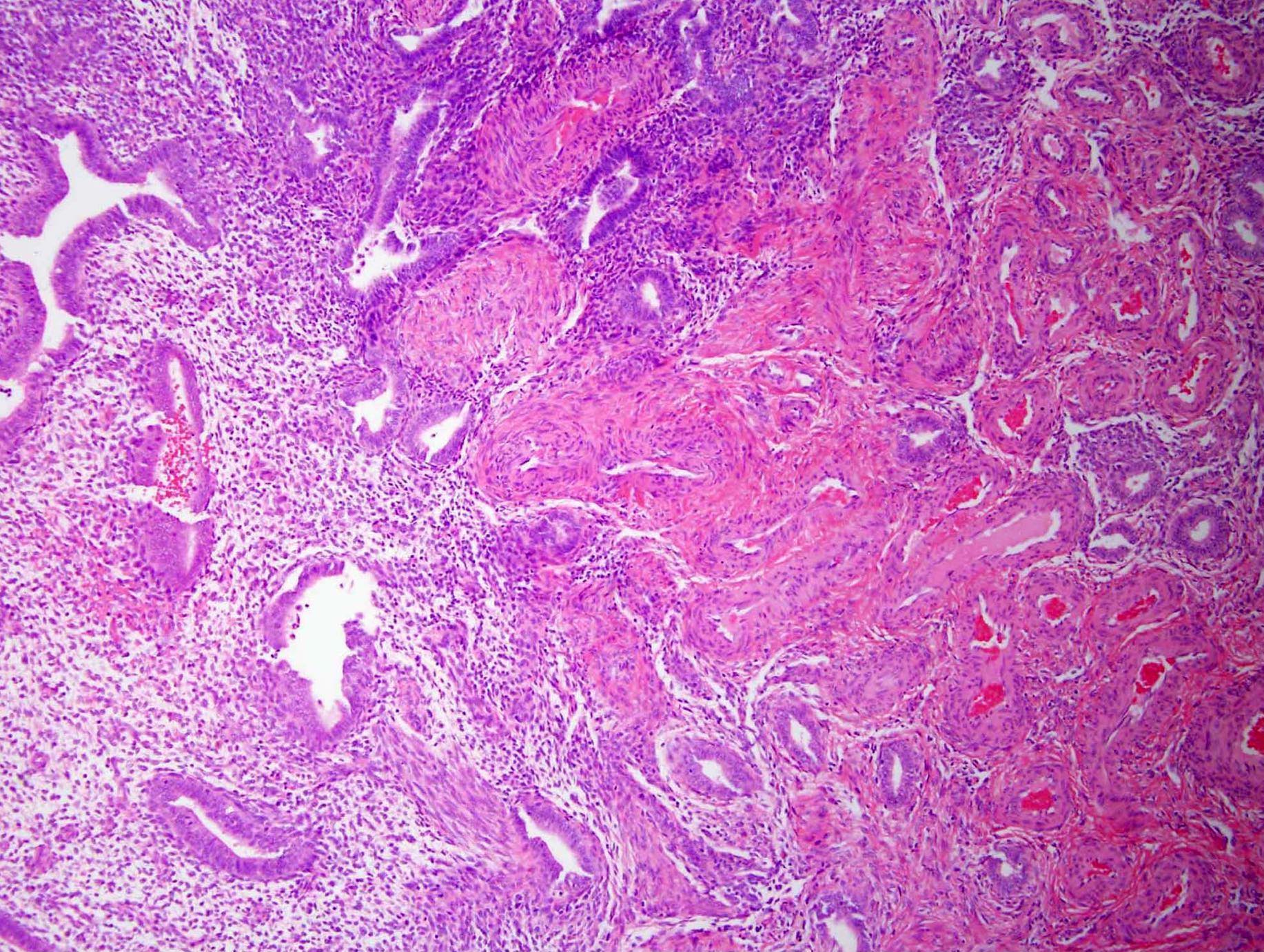
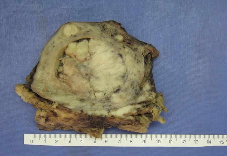
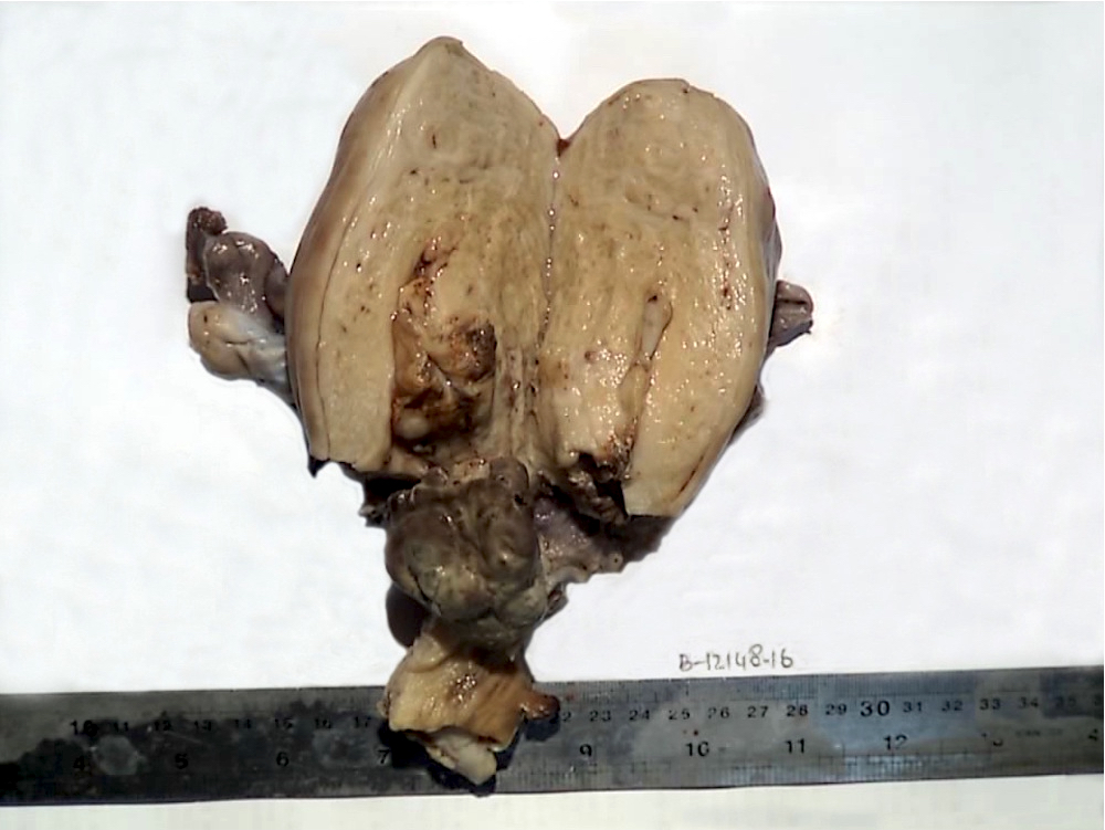
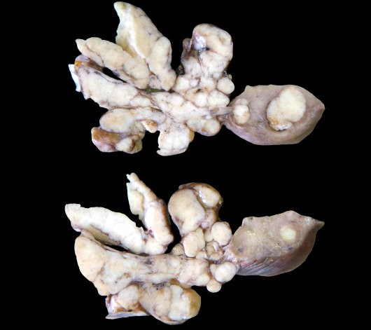
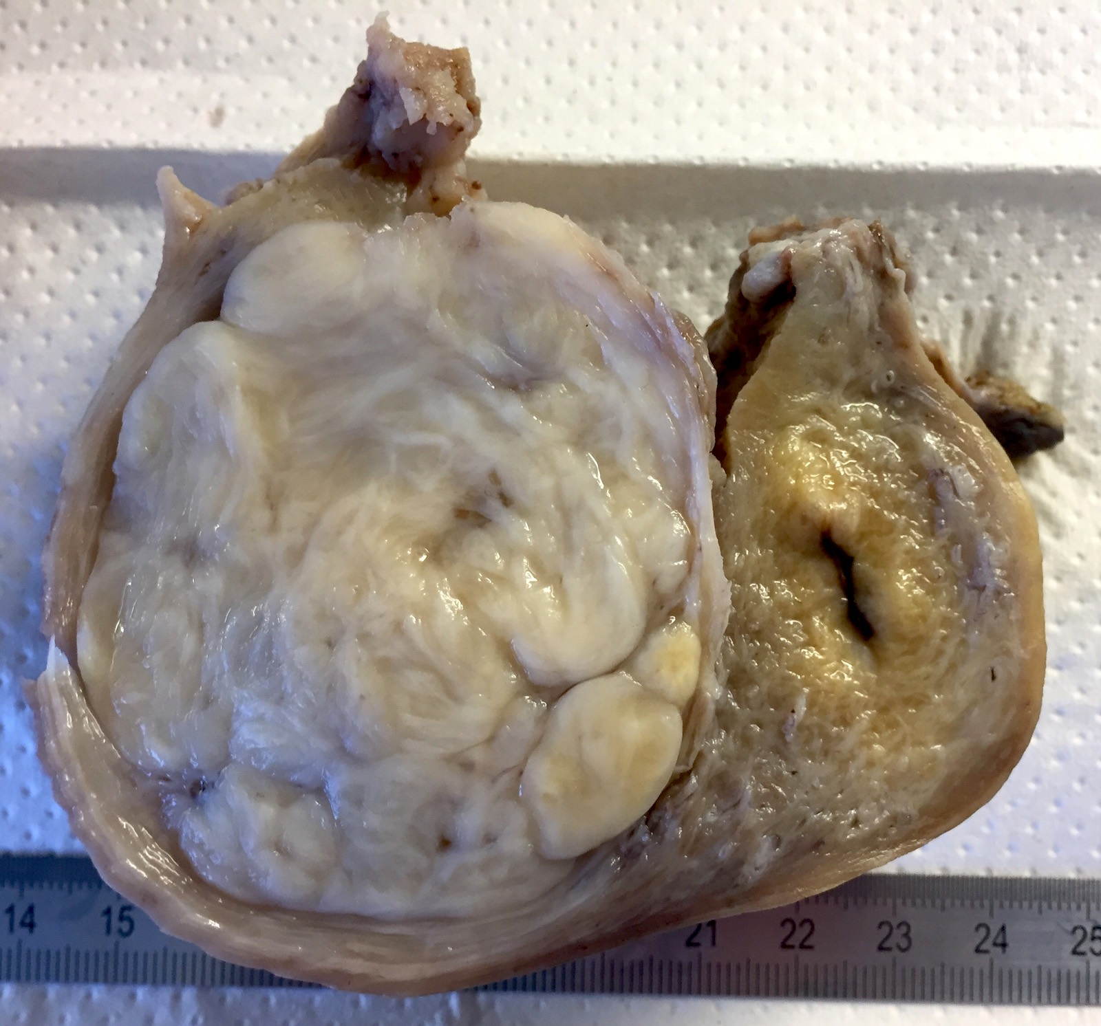
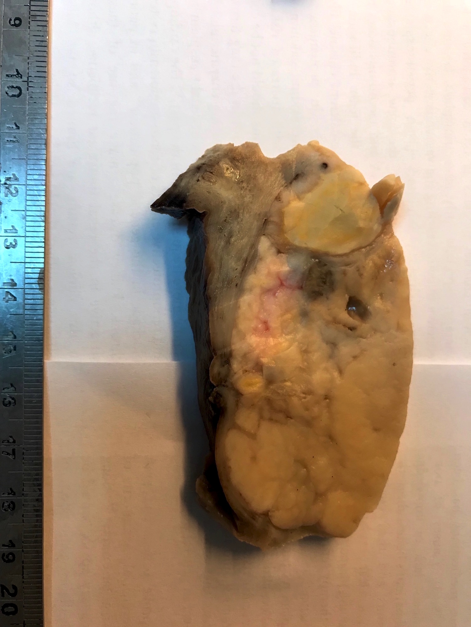
Contributed by Ayse Ayhan, M.D., Ph.D.
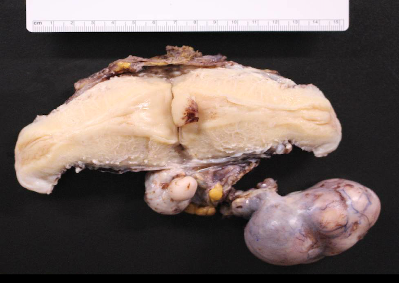
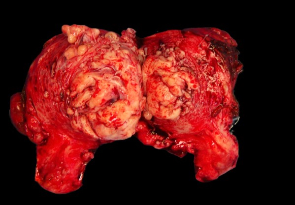
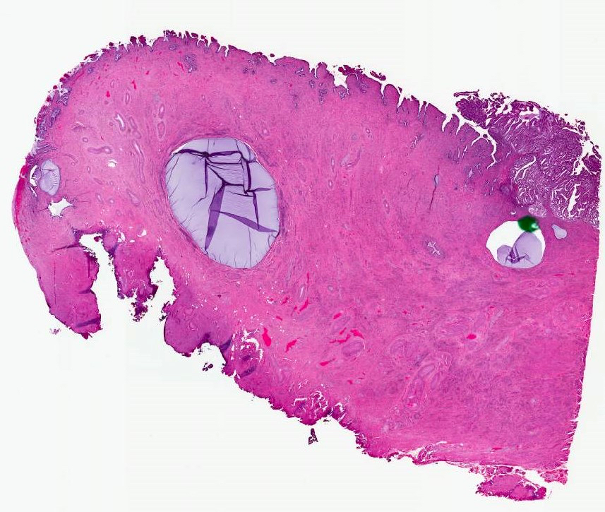
Pre and post-fixed uterus wall
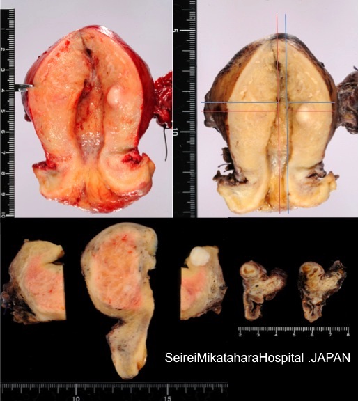
Cut surface of uterus
Images hosted on other servers:

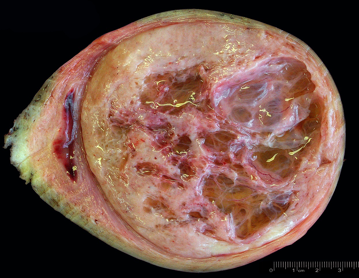
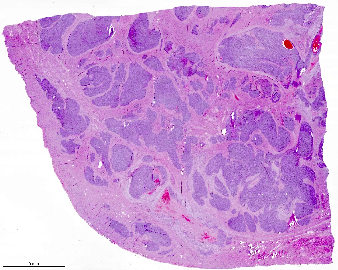
Thickened and spongy myometrium
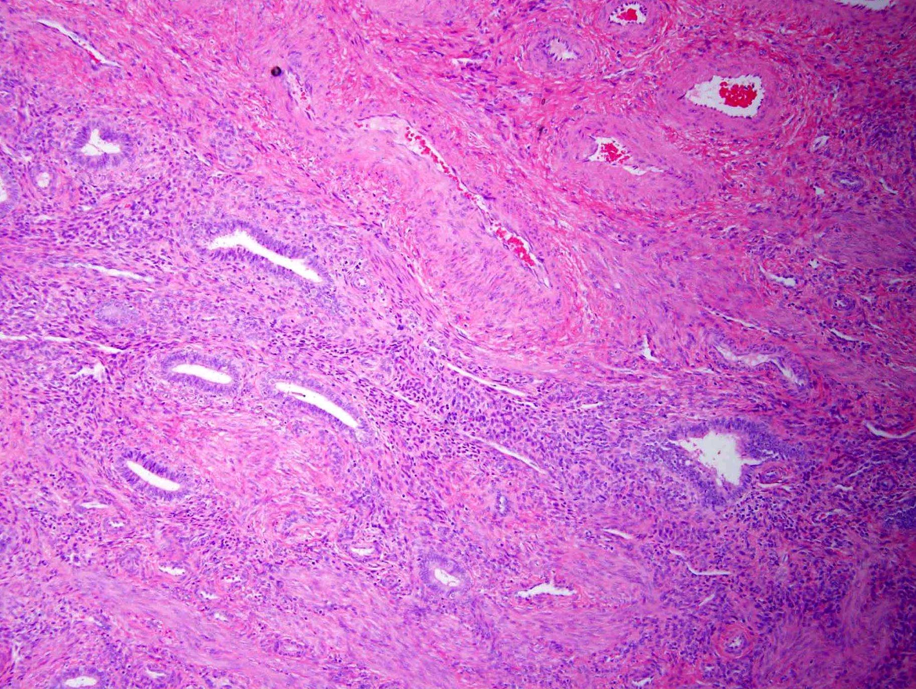
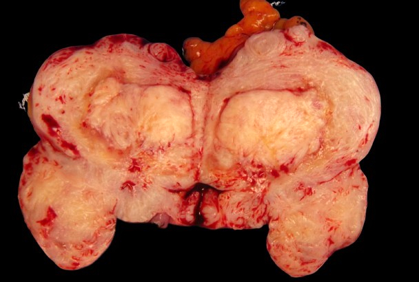
Contributed by Ayse Ayhan, M.D., Ph.D.
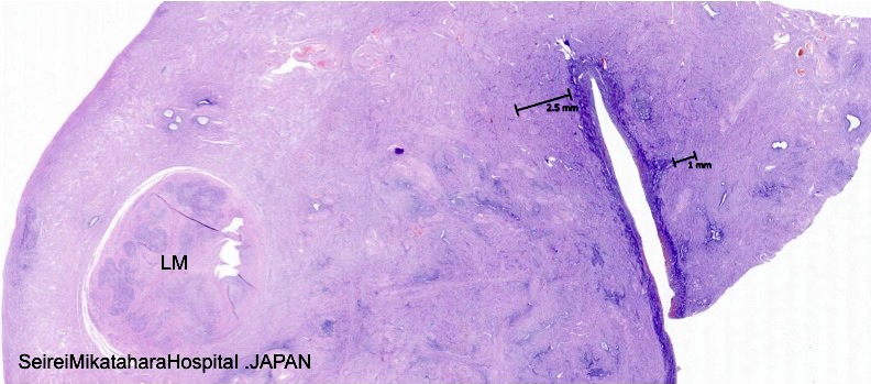
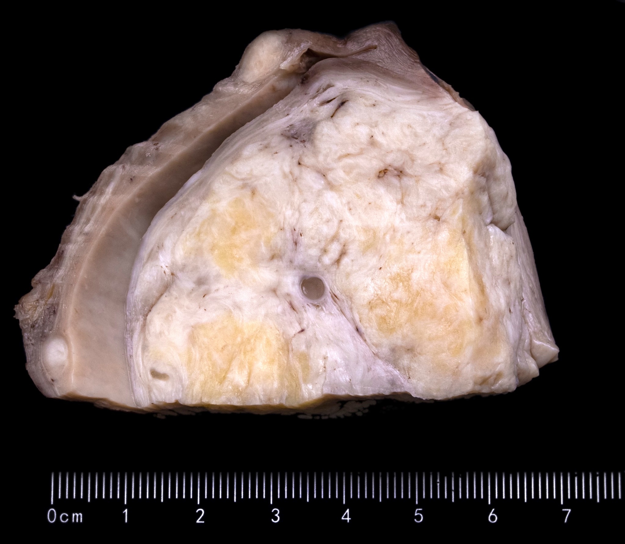
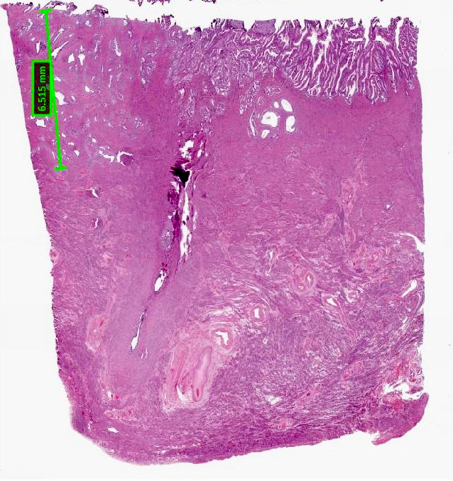
Depth of penetration
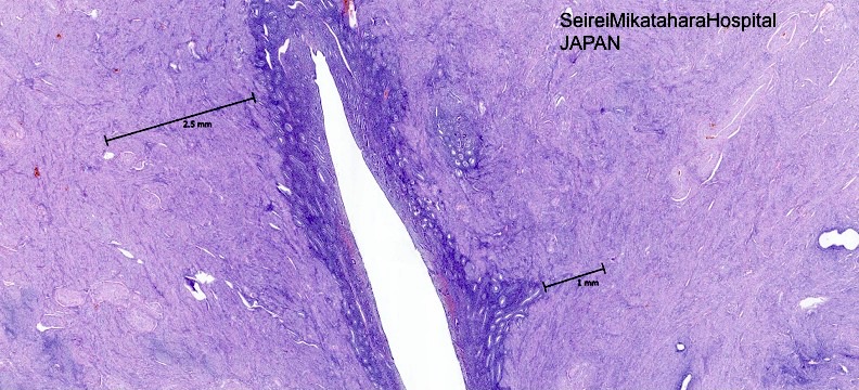
Endomyometrial junction is irregular
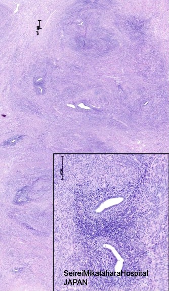
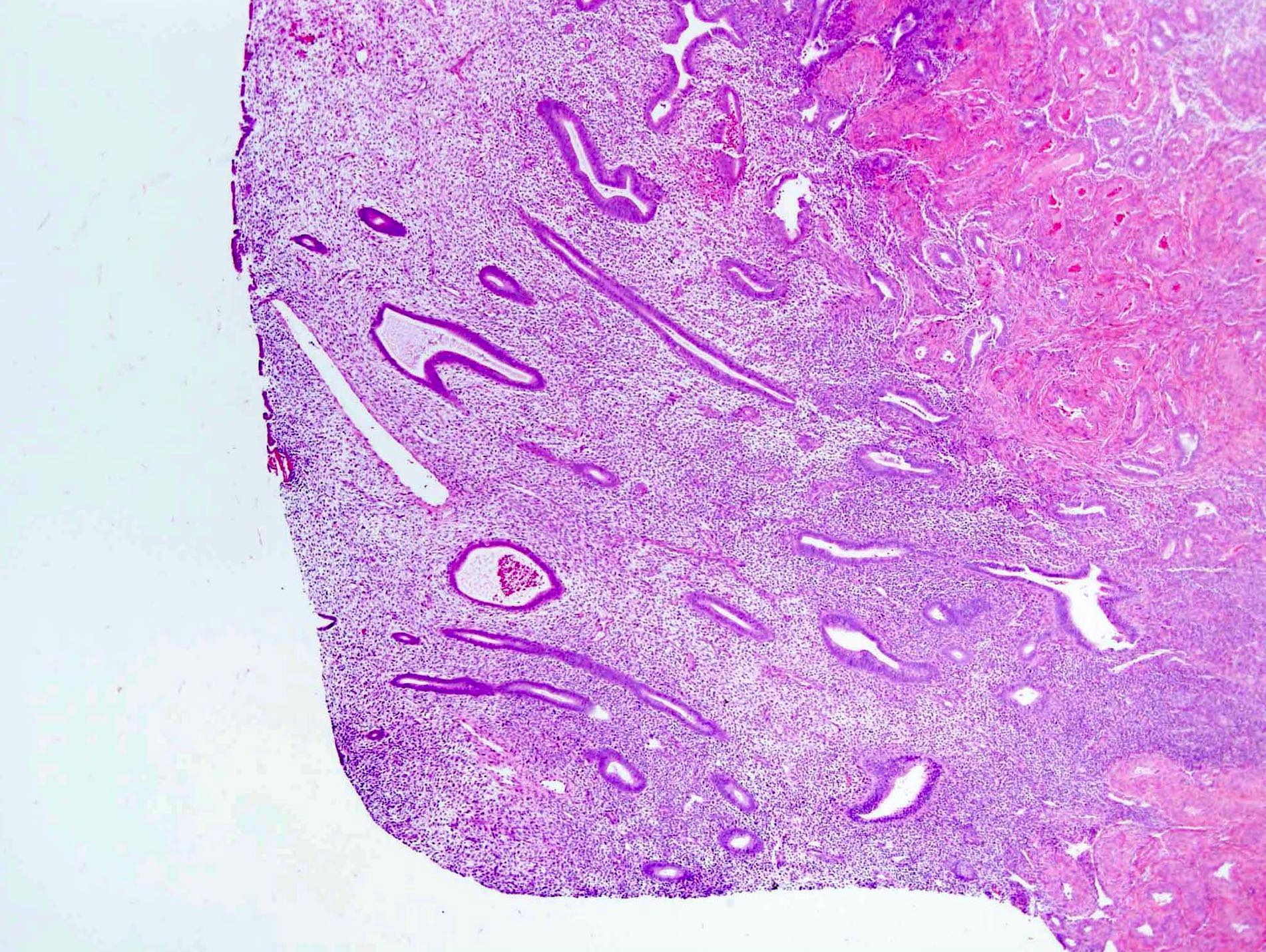
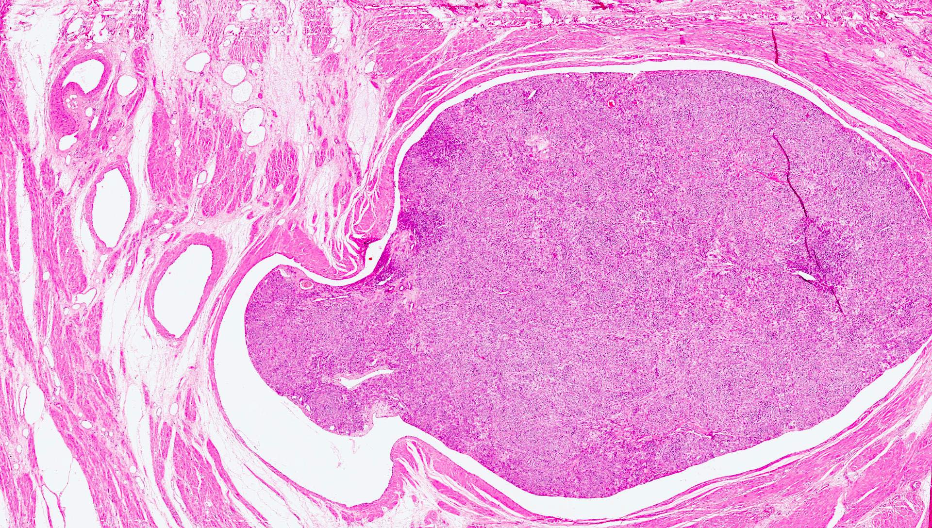
Glandular tissue usually inactive
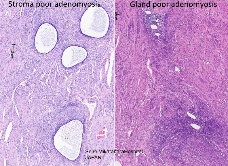
Stroma poor and gland poor
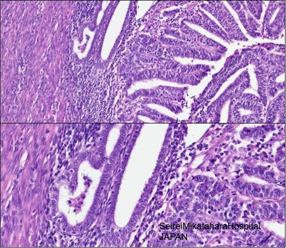
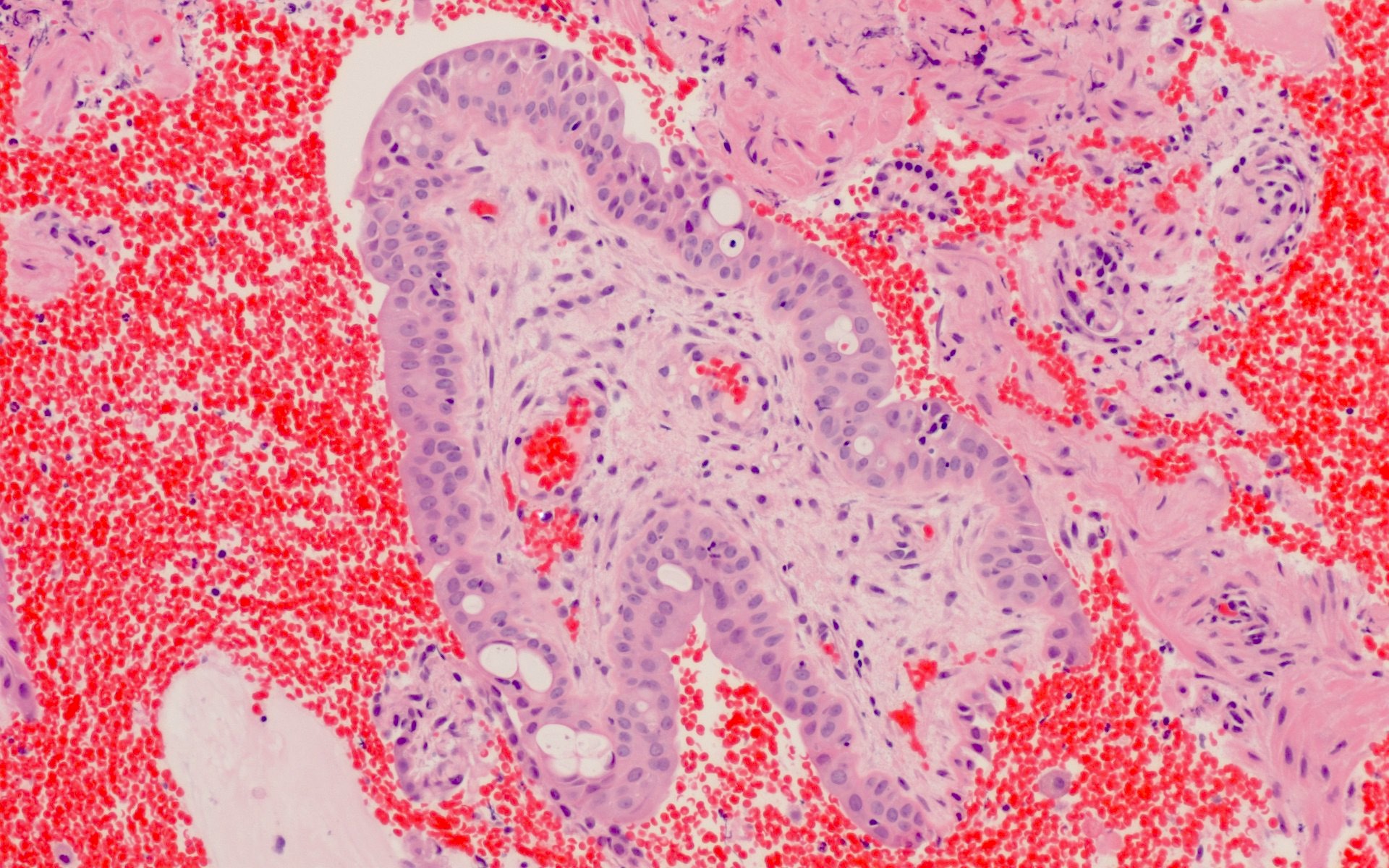
Residual nonneoplastic endometrial glands
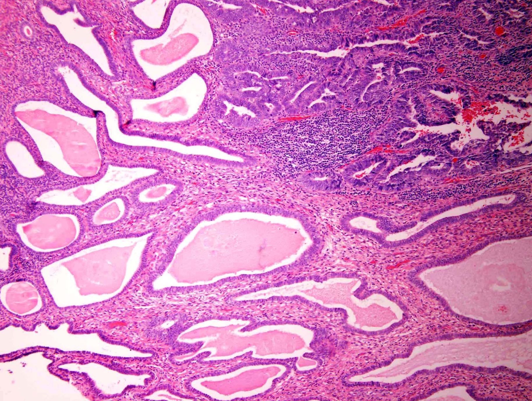
Adenosarcoma arising in endometriosis

Various images
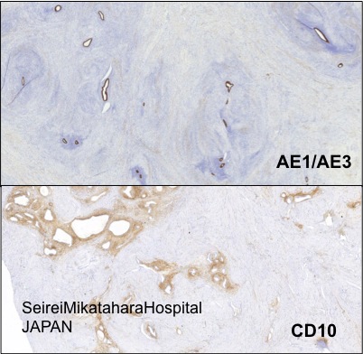
AE1 / AE3 and CD10
Images hosted on other servers:

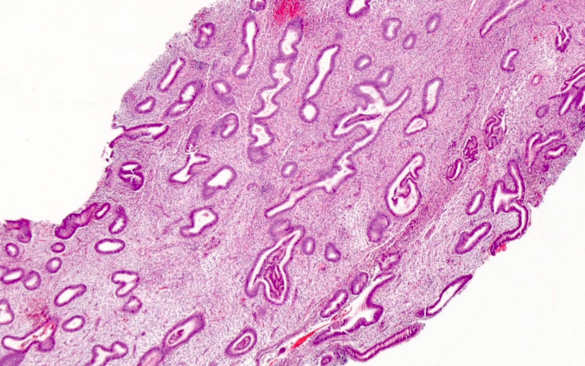
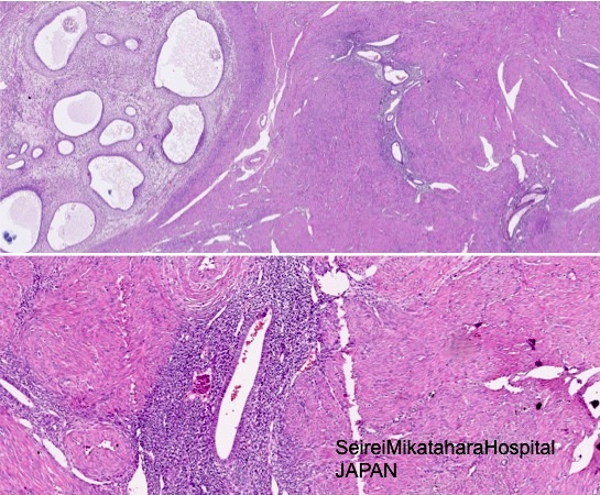
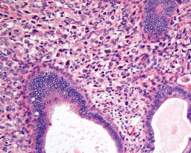
Endometrial glands and stroma
Contributed by Kyle Devins, M.D.
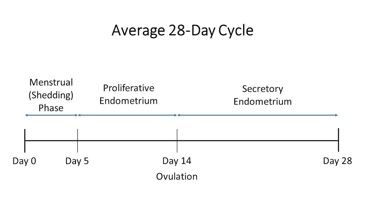
Average 28 day cycle
Contributed by Kyle Devins, M.D.
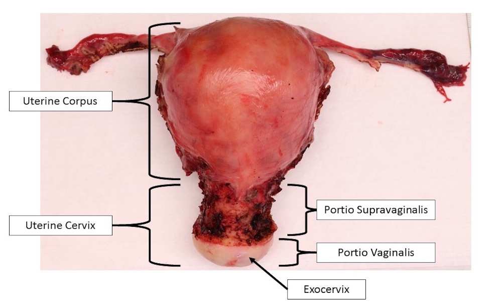
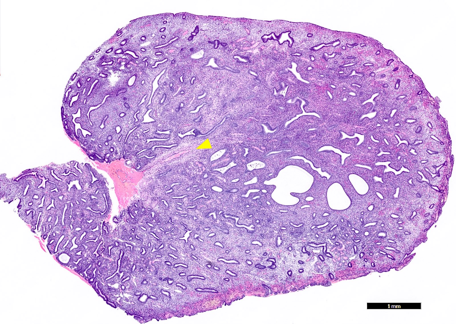
Uterus
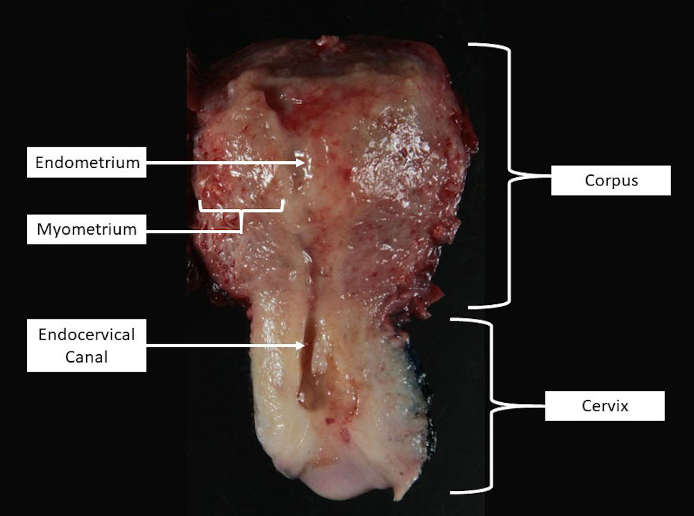
Bivalved uterus
Contributed by Kyle Devins, M.D.
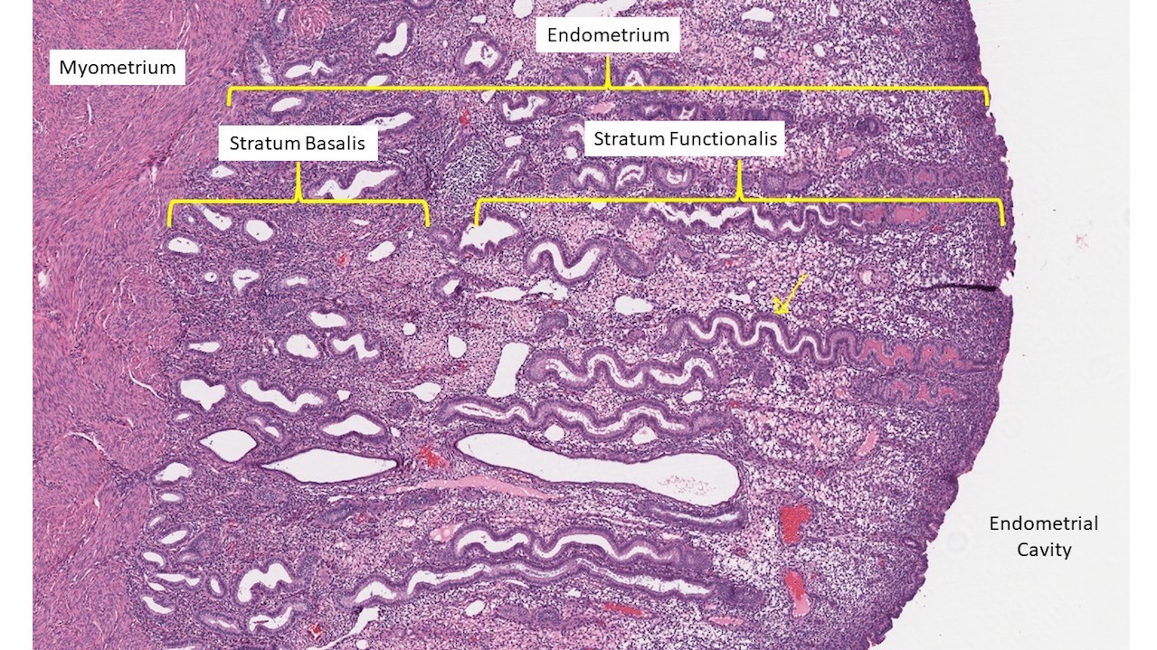
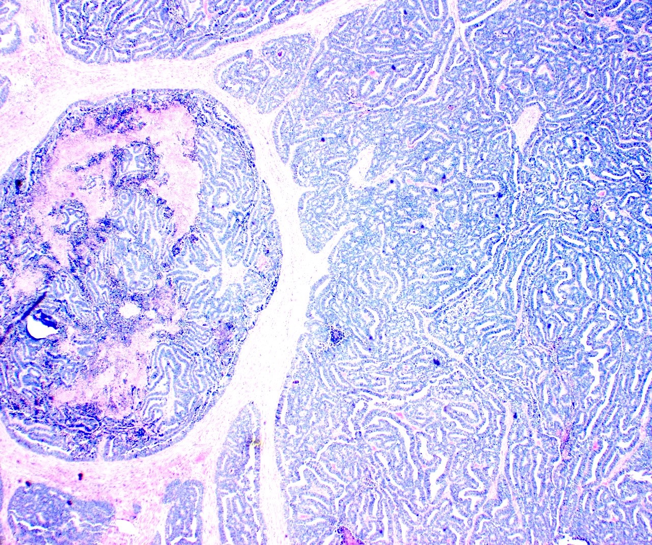
Endometrium Overview
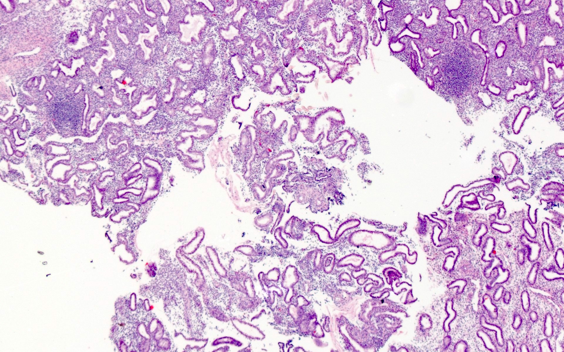
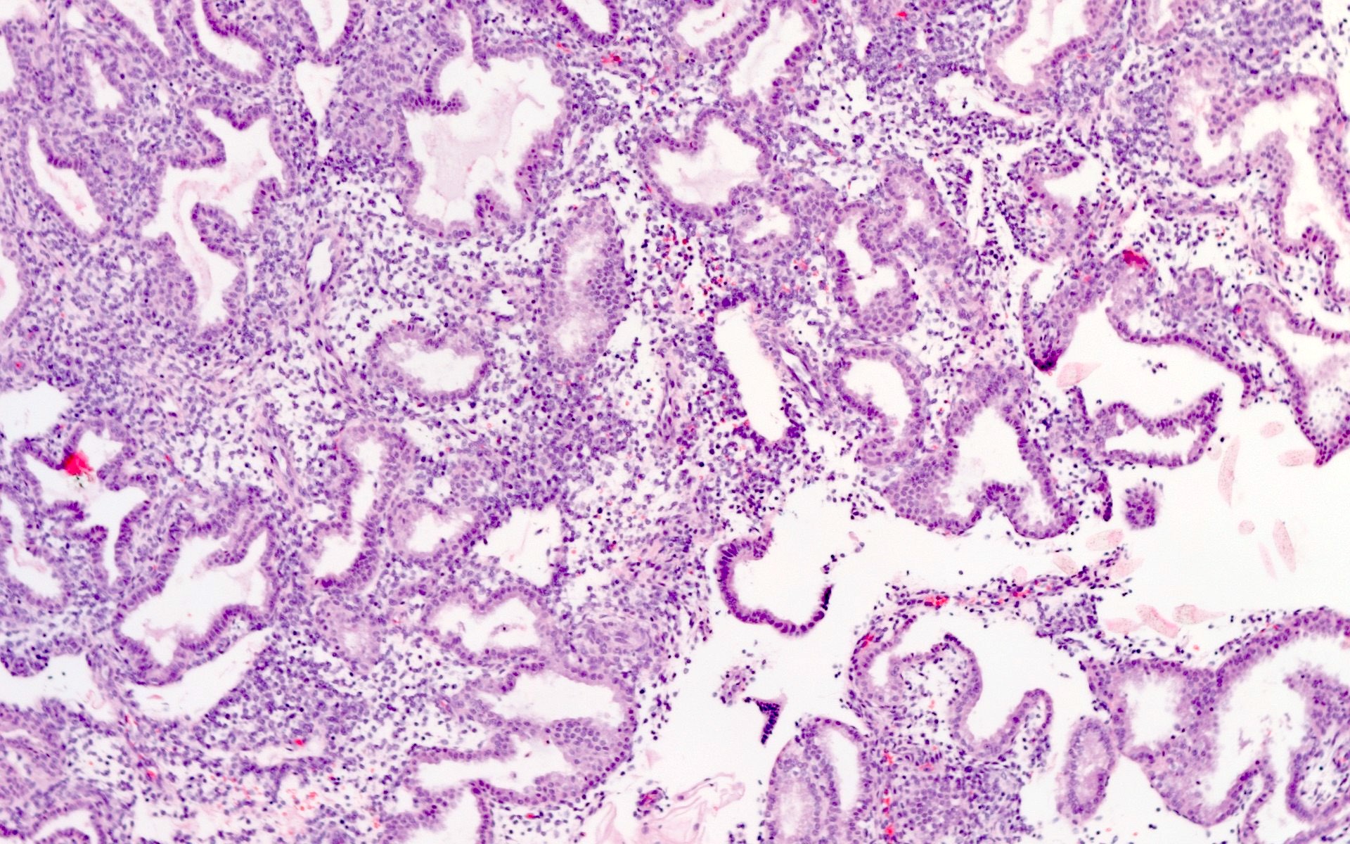
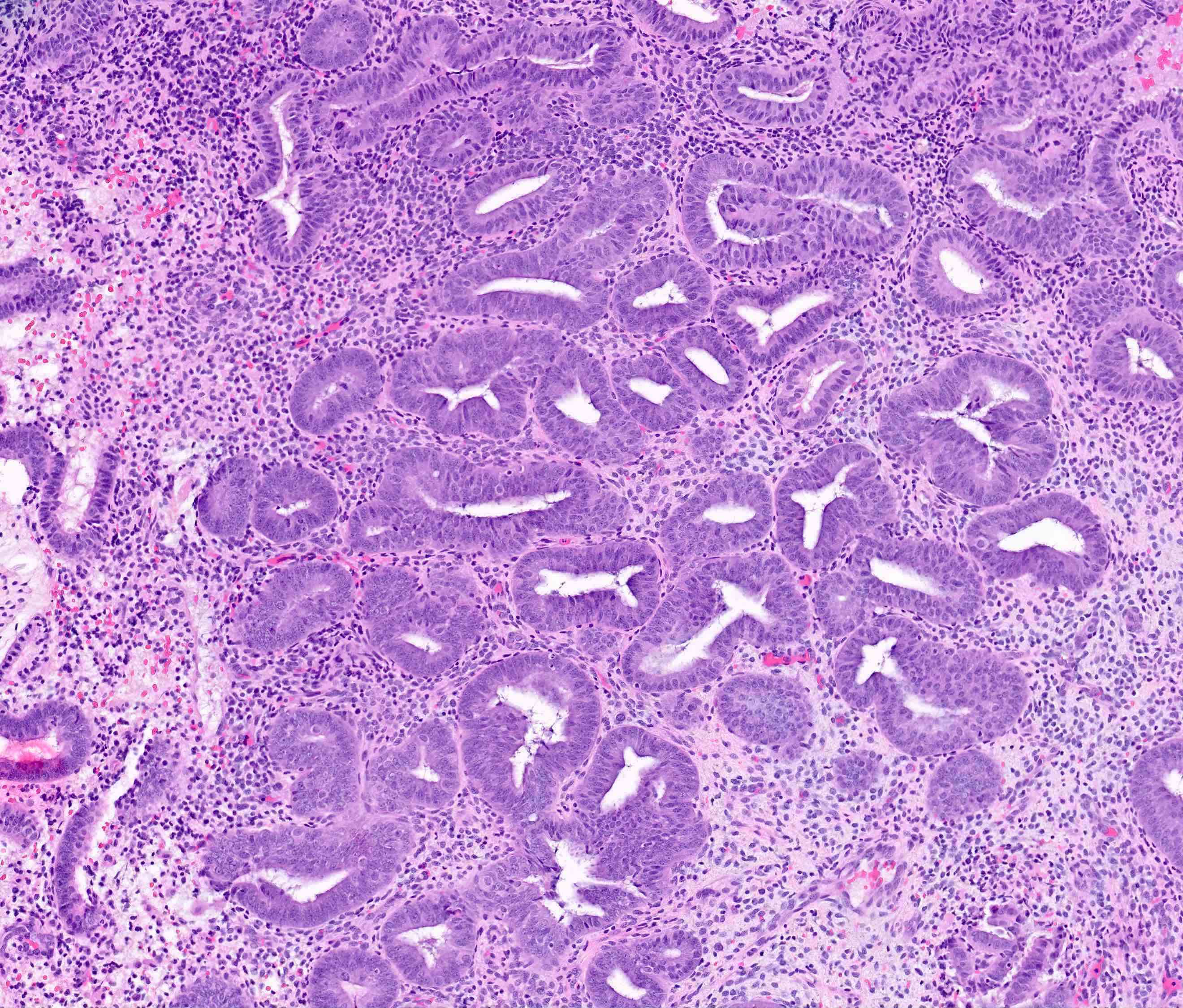
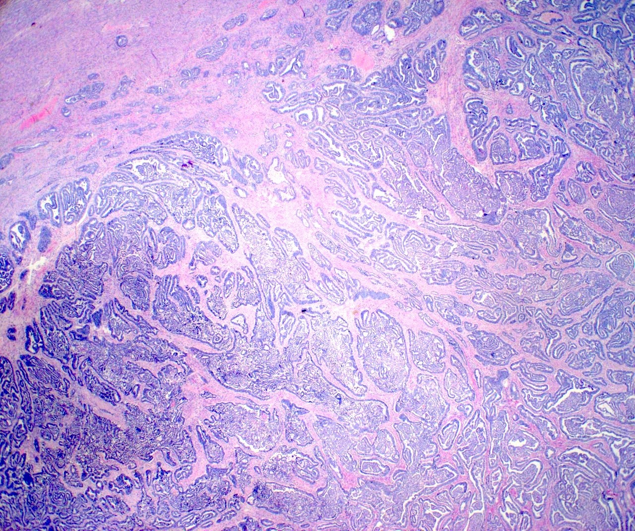
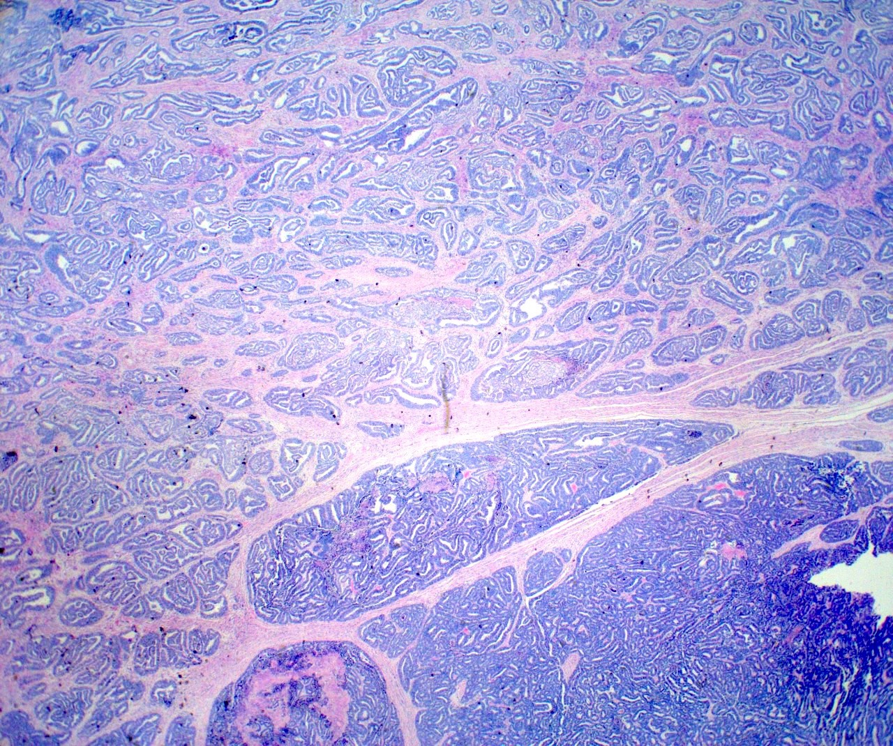
Proliferative endometrium

Proliferative phase, low power
Secretory endometrium
Subnuclear vacuoles
Stromal edema
Intraluminal secretion
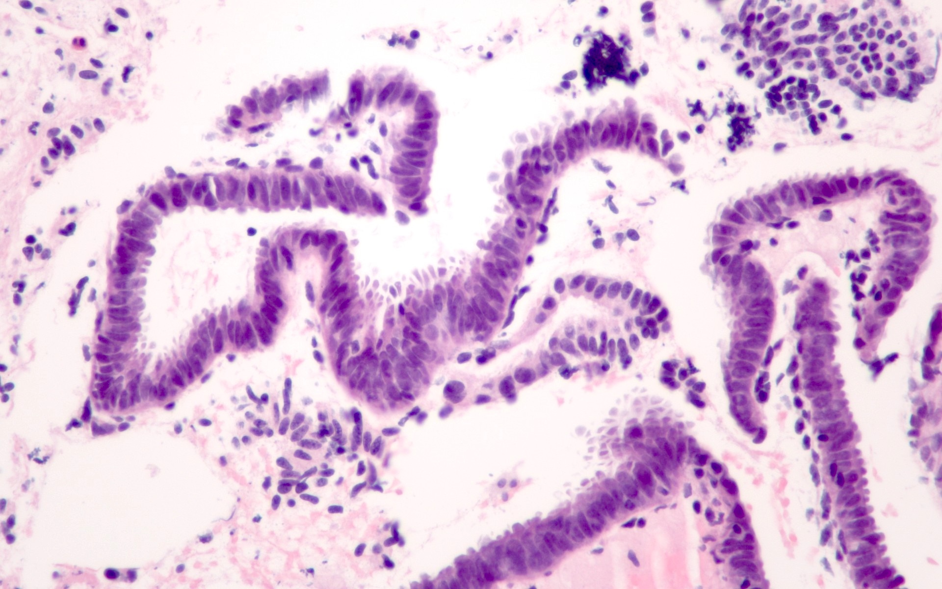
Late secretory
Spiral arteriole
Secretory endometrium, day 26
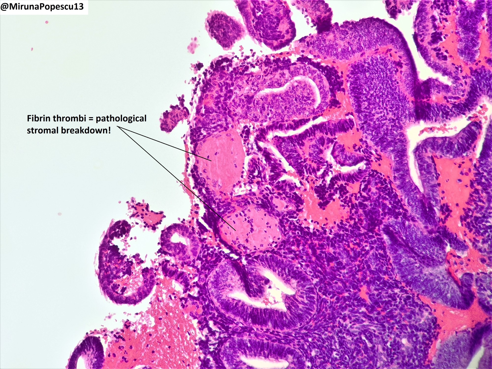
Endometrial stromal breakdown
Stromal aggregates
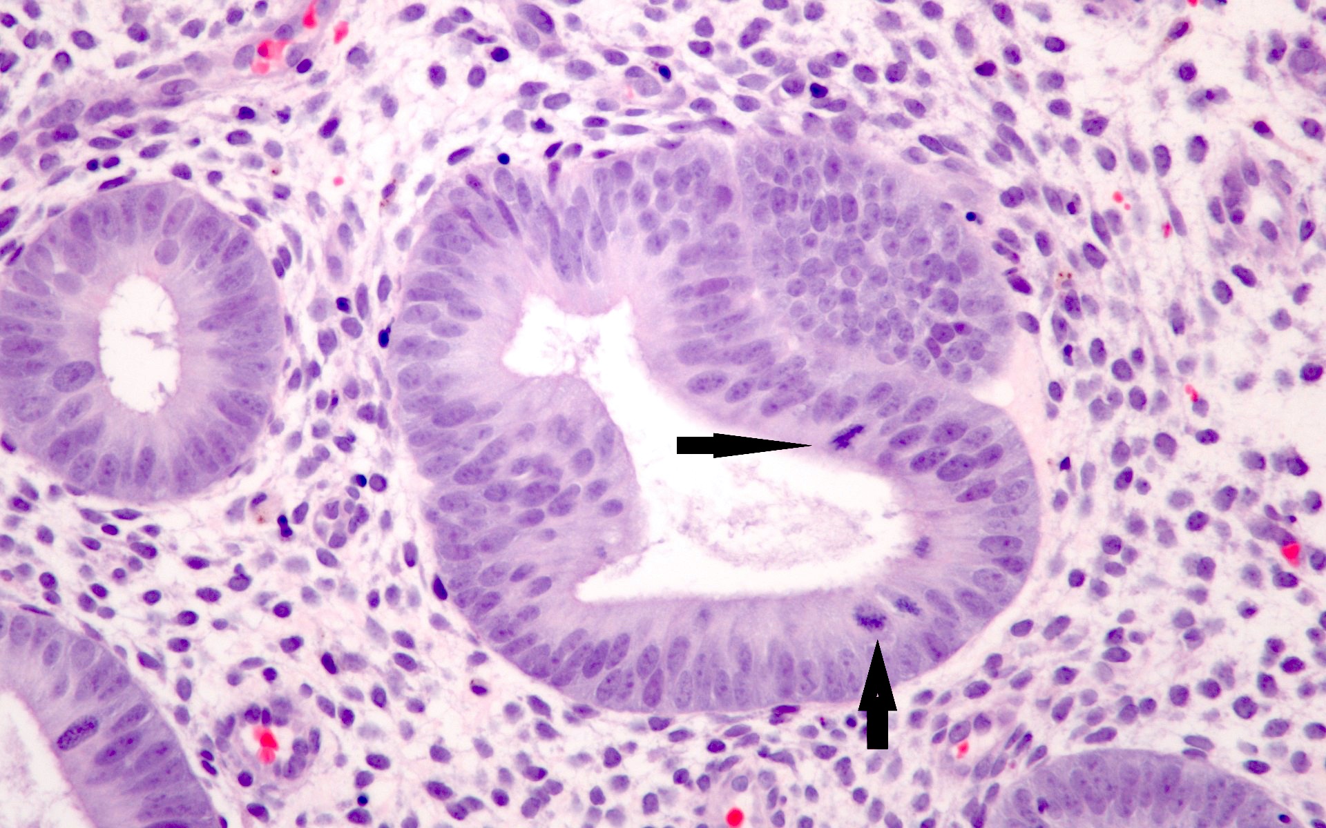
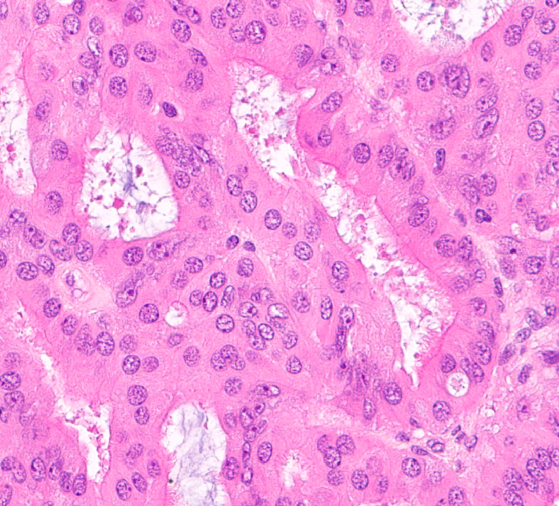
Arias-Stella reaction
Atrophy
Atrophic gland
- Refer to the following: Endometrial cells
Histology of benign cycling endometrium
Contributed by Ayse Ayhan, M.D., Ph.D.


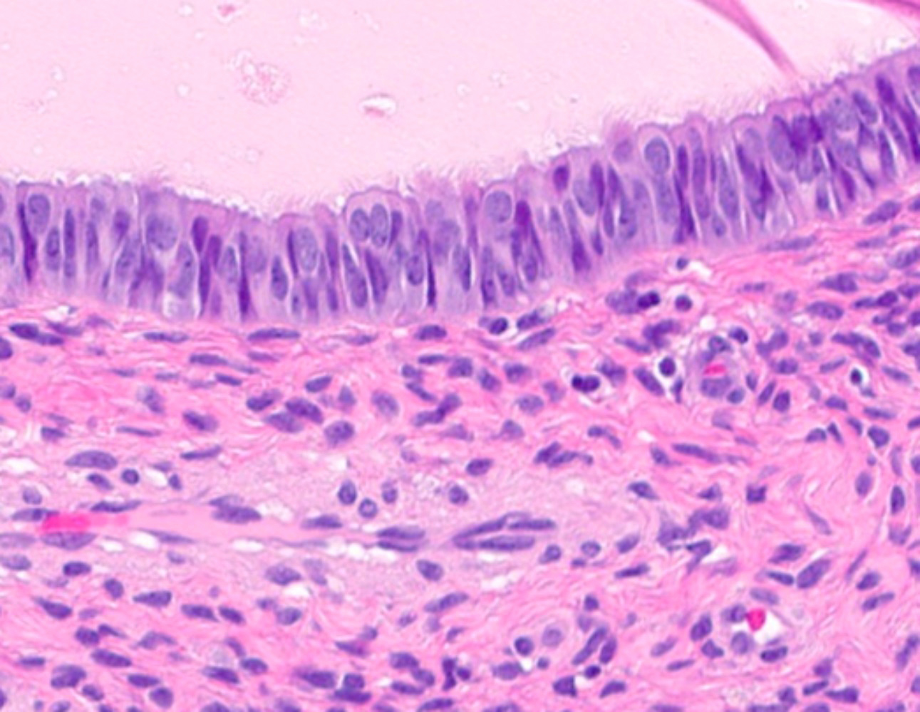
Enlarged nuclei, sub / supranuclear vacuoles
Hobnail growth pattern
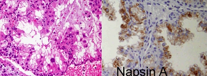
Napsin A positivity
Images hosted on other servers:

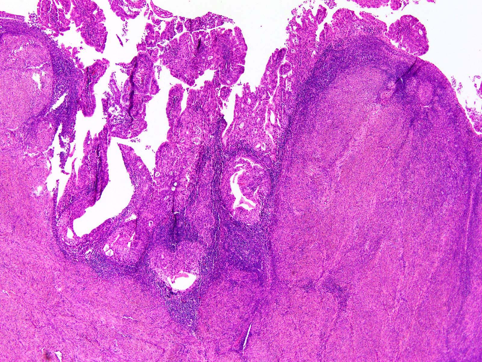
Polypoid mass
Contributed by Stephanie L. Skala, M.D.
Lobulated architecture
Crowded glands and muscular stroma
Smooth muscle underlying surface endometrium
Crowded glands and muscle bundles
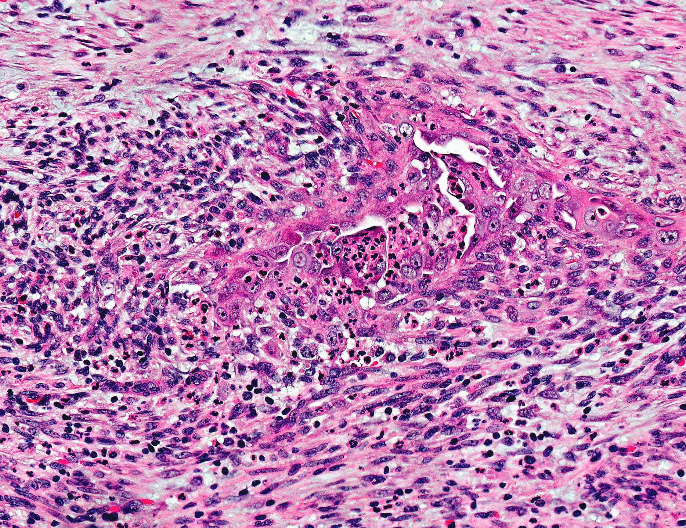
Squamous morule with central necrosis
Haphazard gland arrangement

Myxoid spindled stroma

Stromal SATB2 positivity
Contributed by Joana Ferreira, M.D. and Ana Félix, M.D., Ph.D.

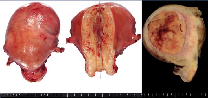
Macroscopic appearance
Polypoid growth
Images hosted on other servers:

Polypoid fleshy mass at fundus of uterus

Infiltrating partially necrotic tumor

Serosal and vaginal invasion

Polypoid tumor with superficial myometrial invasion
Contributed by Joana Ferreira, M.D., Ana Félix, M.D., Ph.D. and @ahmsab_MD on Twitter
Large polypoid tumor with minimal myometrial invasion

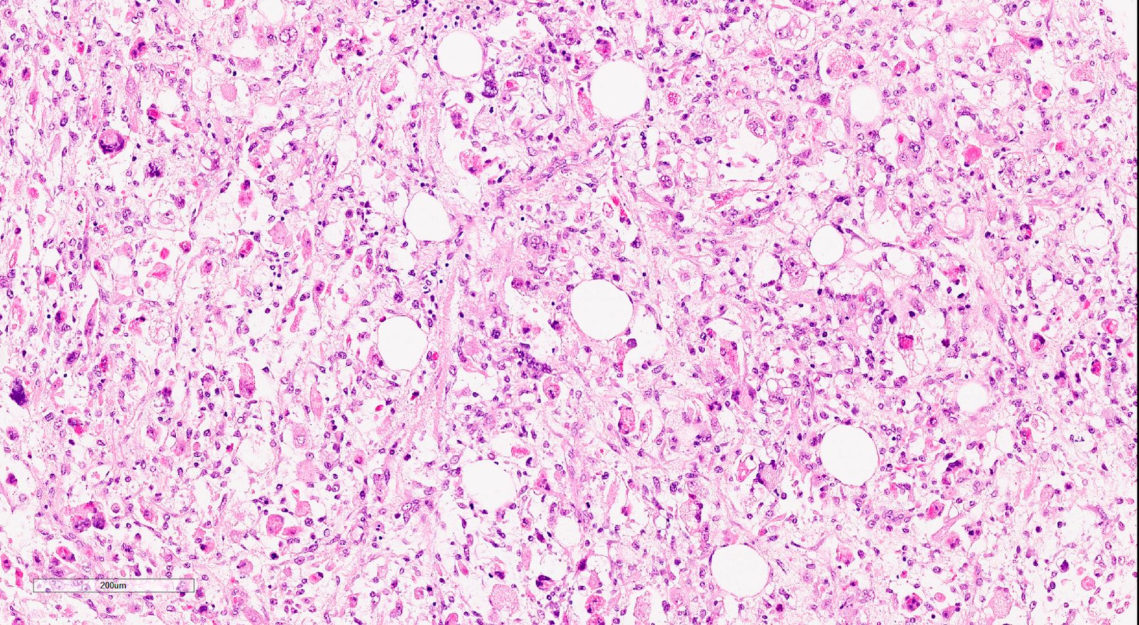
Atypical cells and chondroid matrix

Necrosis, squamous differentiation
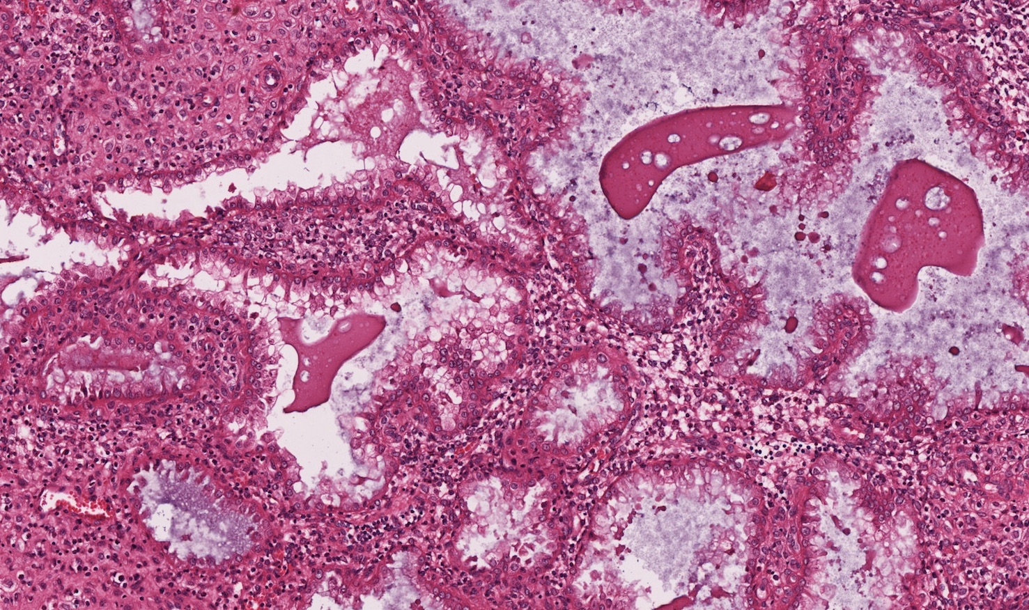
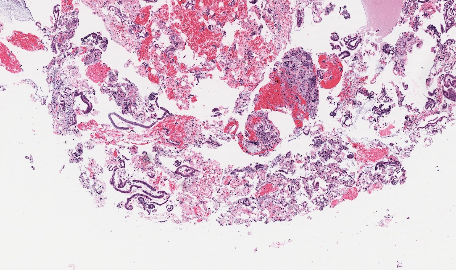
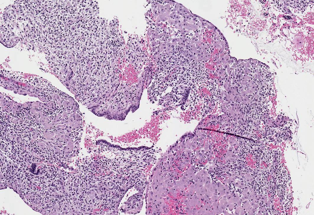
Serous carcinoma

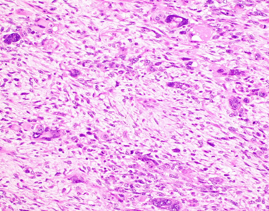
Chondrosarcomatous area
Pleomorphic sarcomatous area
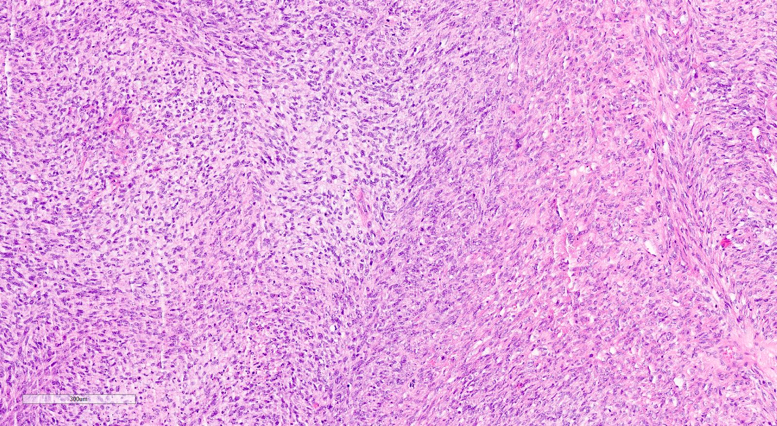
Sarcomatous area
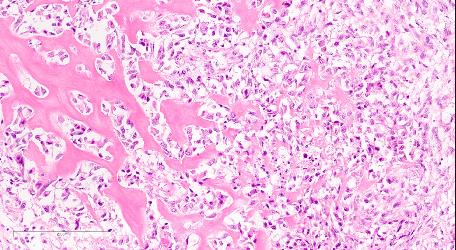
Osteosarcomatous area
Rhabdomyosarcomatous
areas
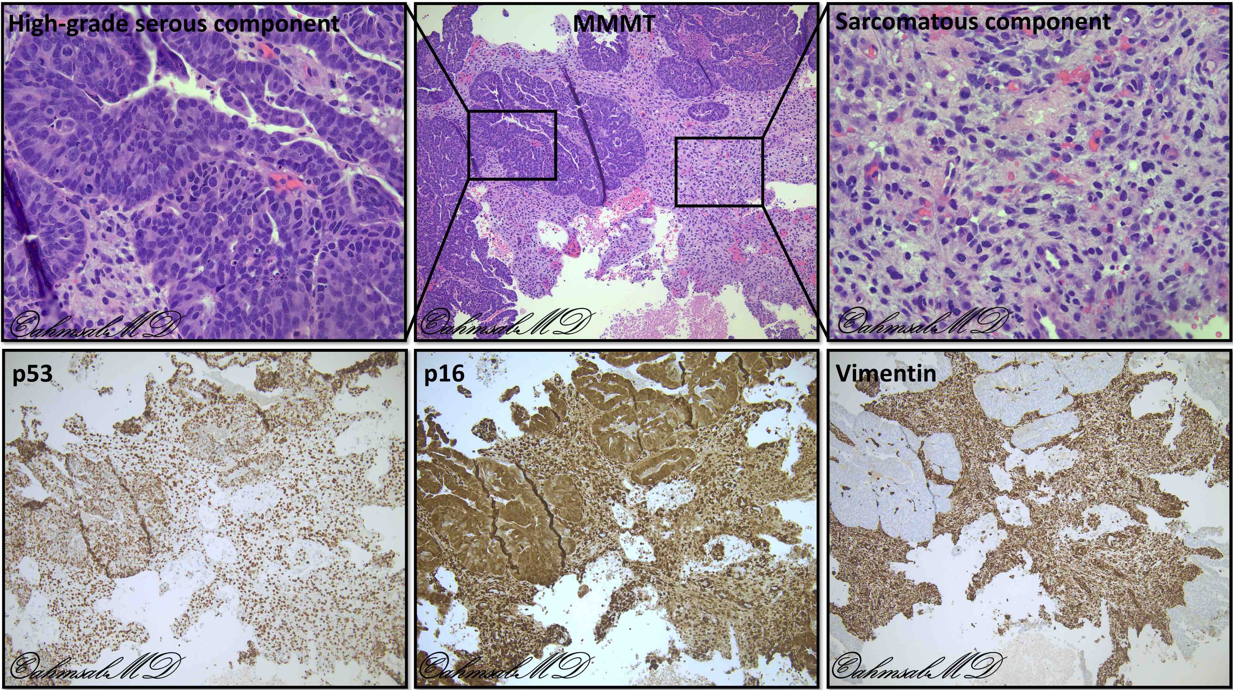
Carcinosarcoma (MMMT)
Images hosted on other servers:
Endometrial mass
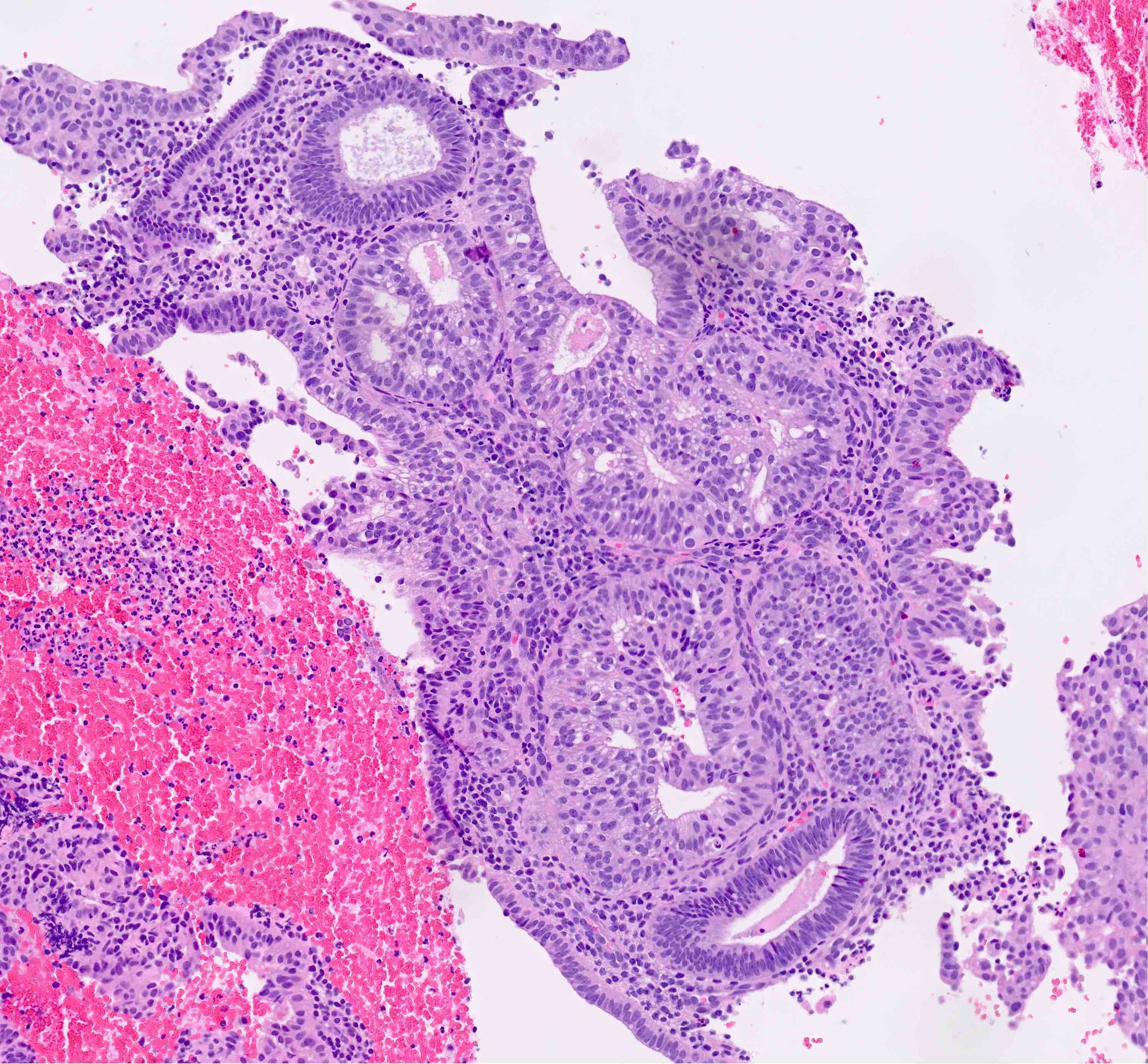
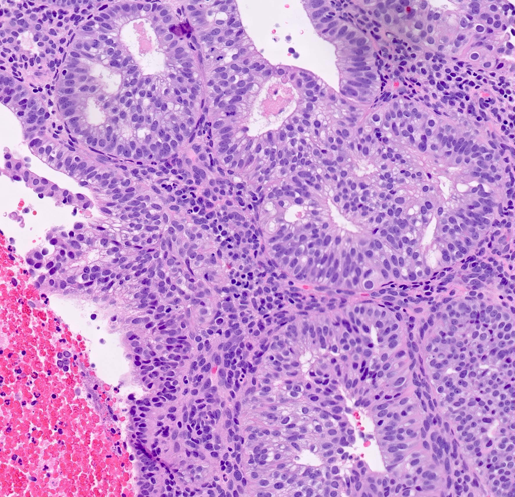
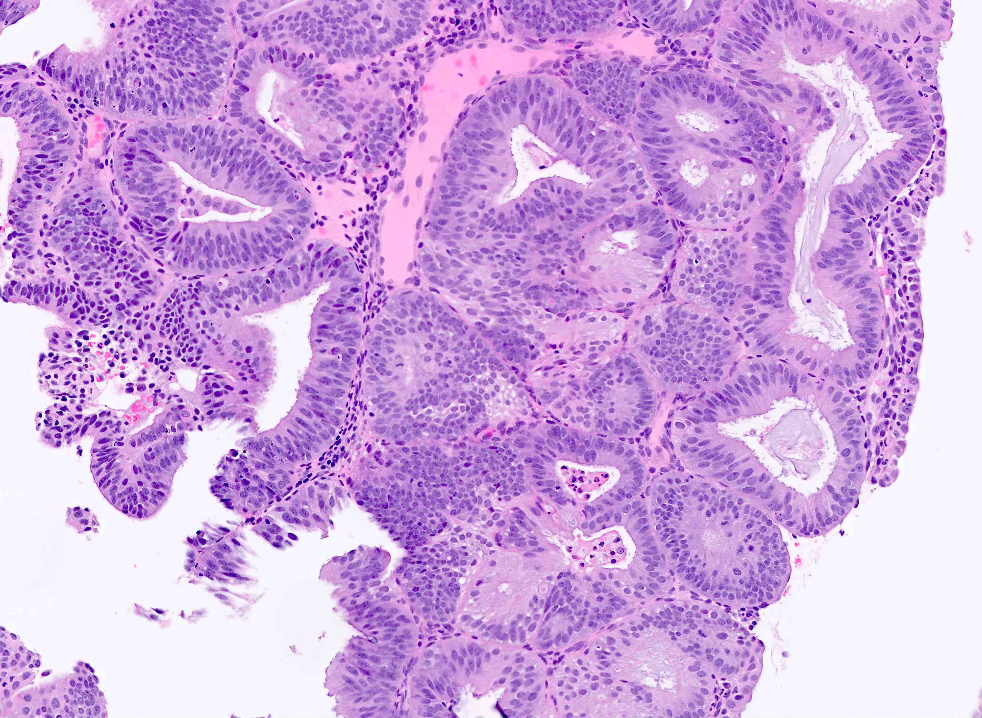
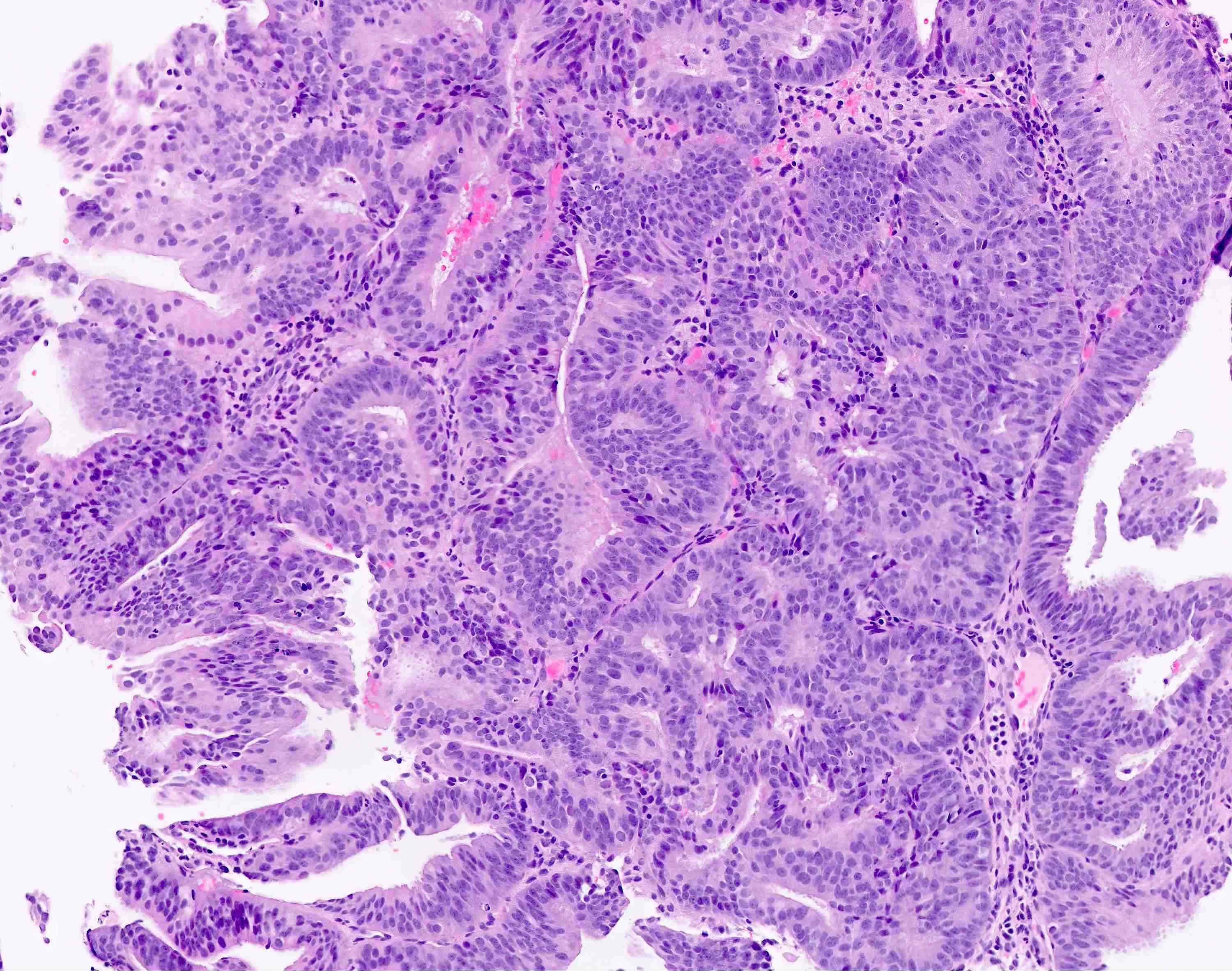
Contributed by Jutta Huvila, M.D.
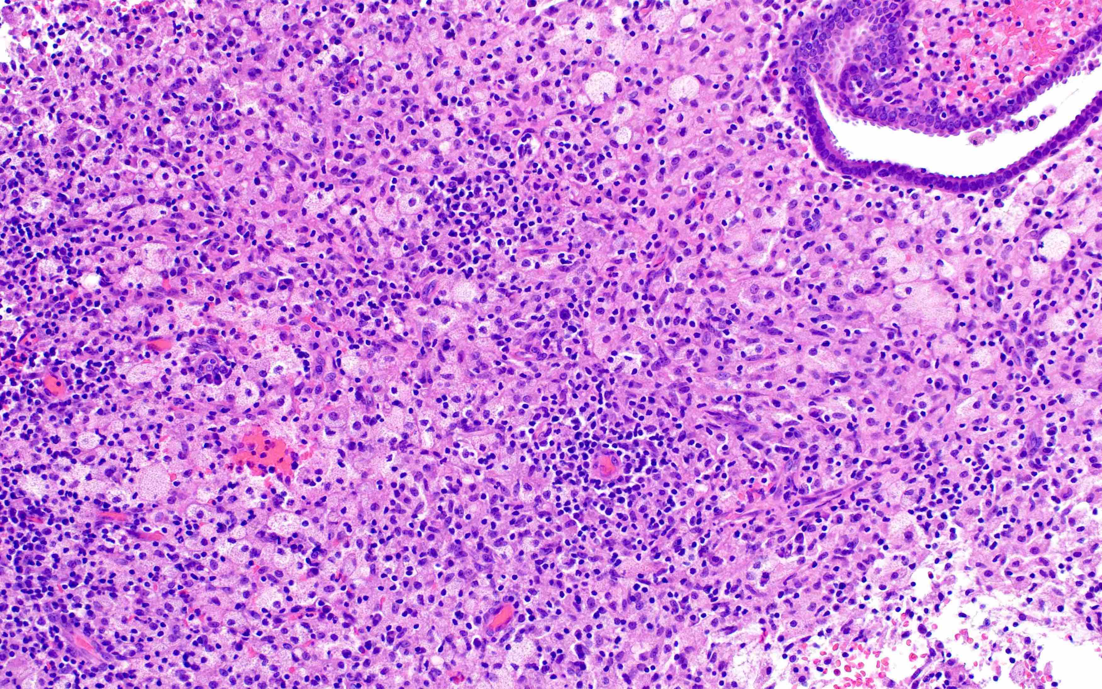
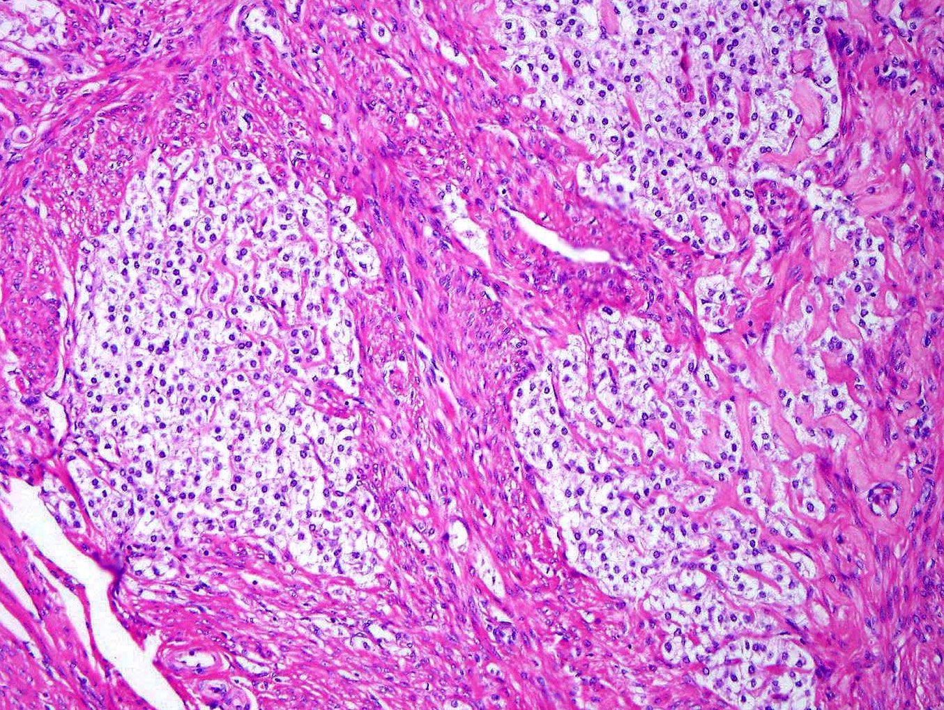
Solid architecture
Glandular architecture
Glandular pattern
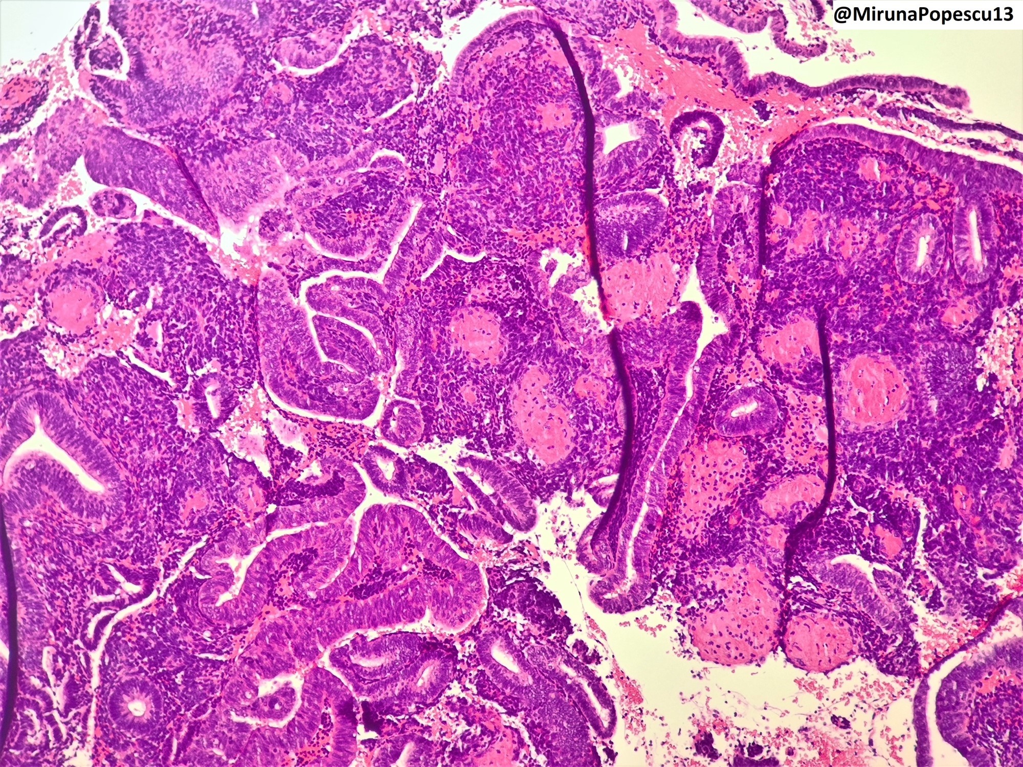
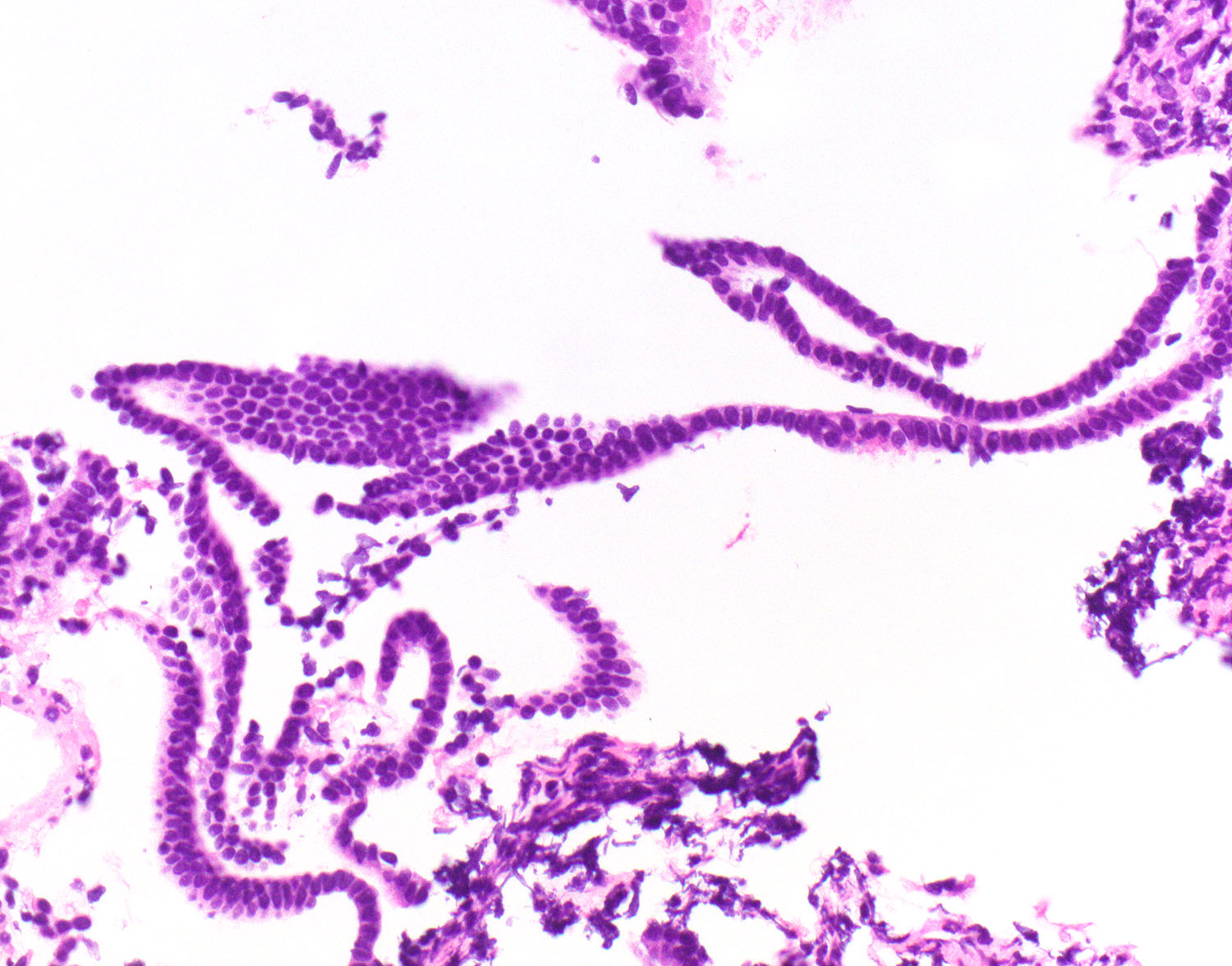
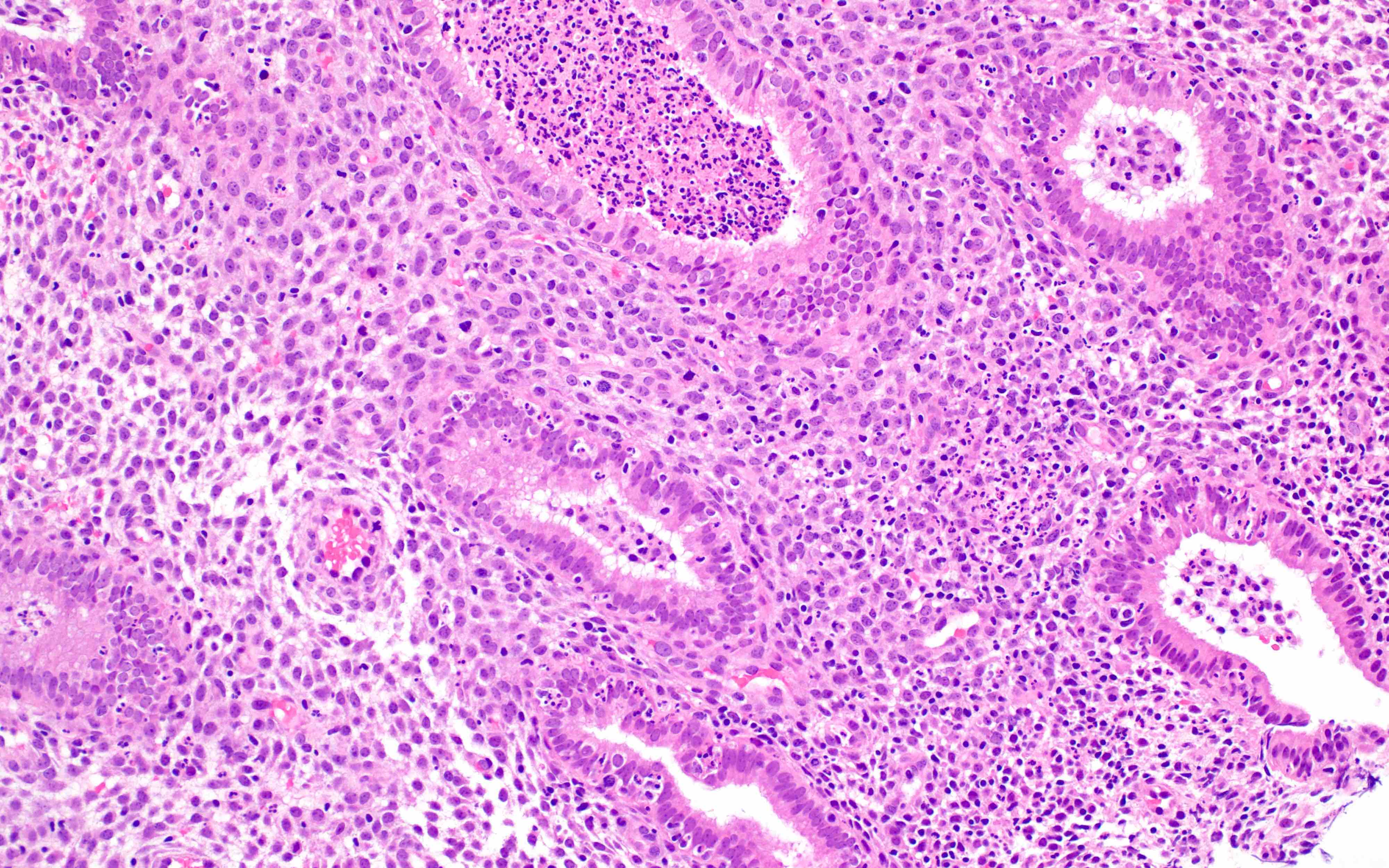
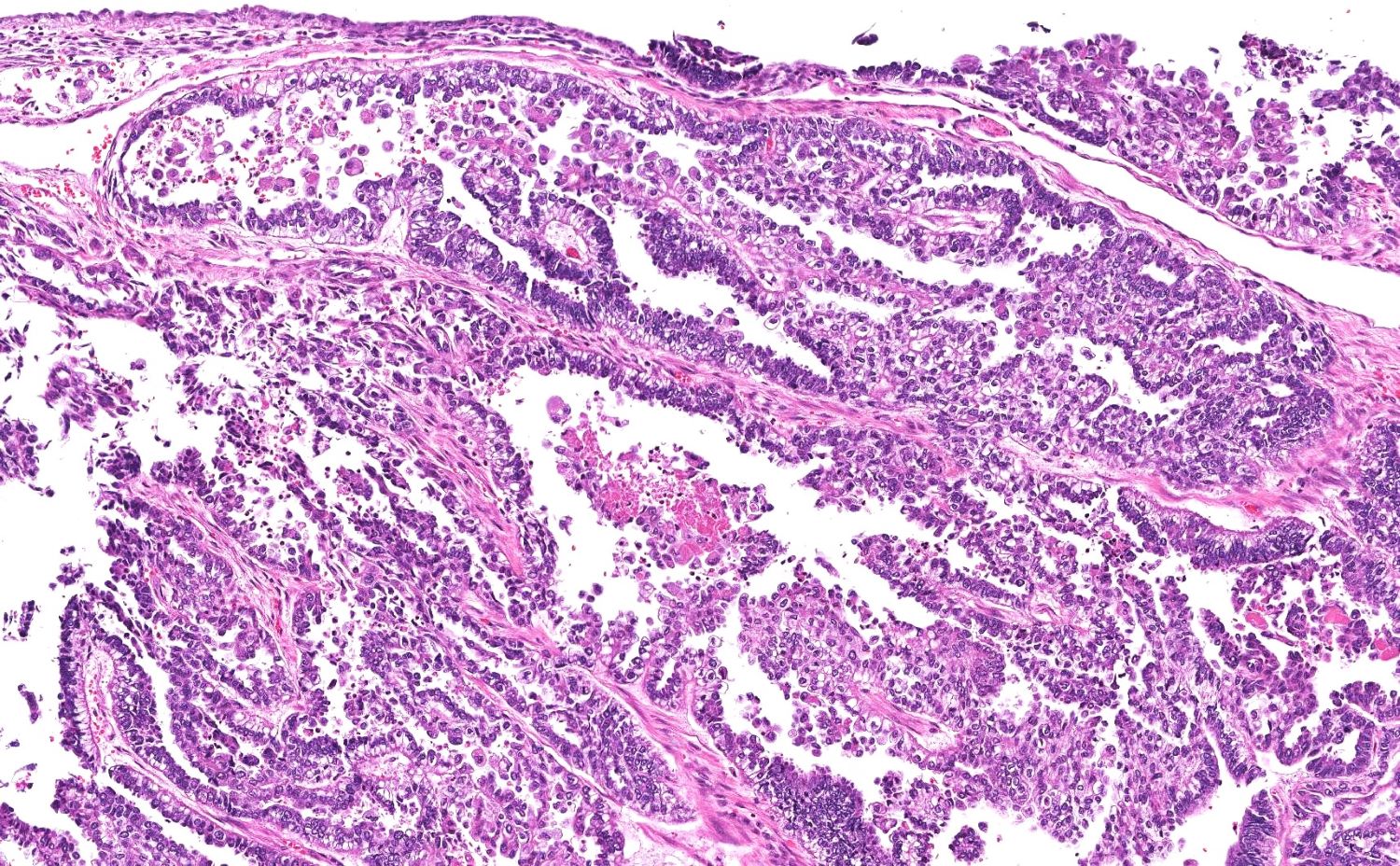
Papillary and glandular architecture
Papillary architecture
Hyalinized stroma
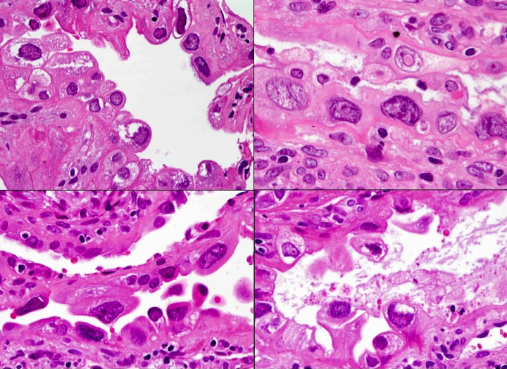
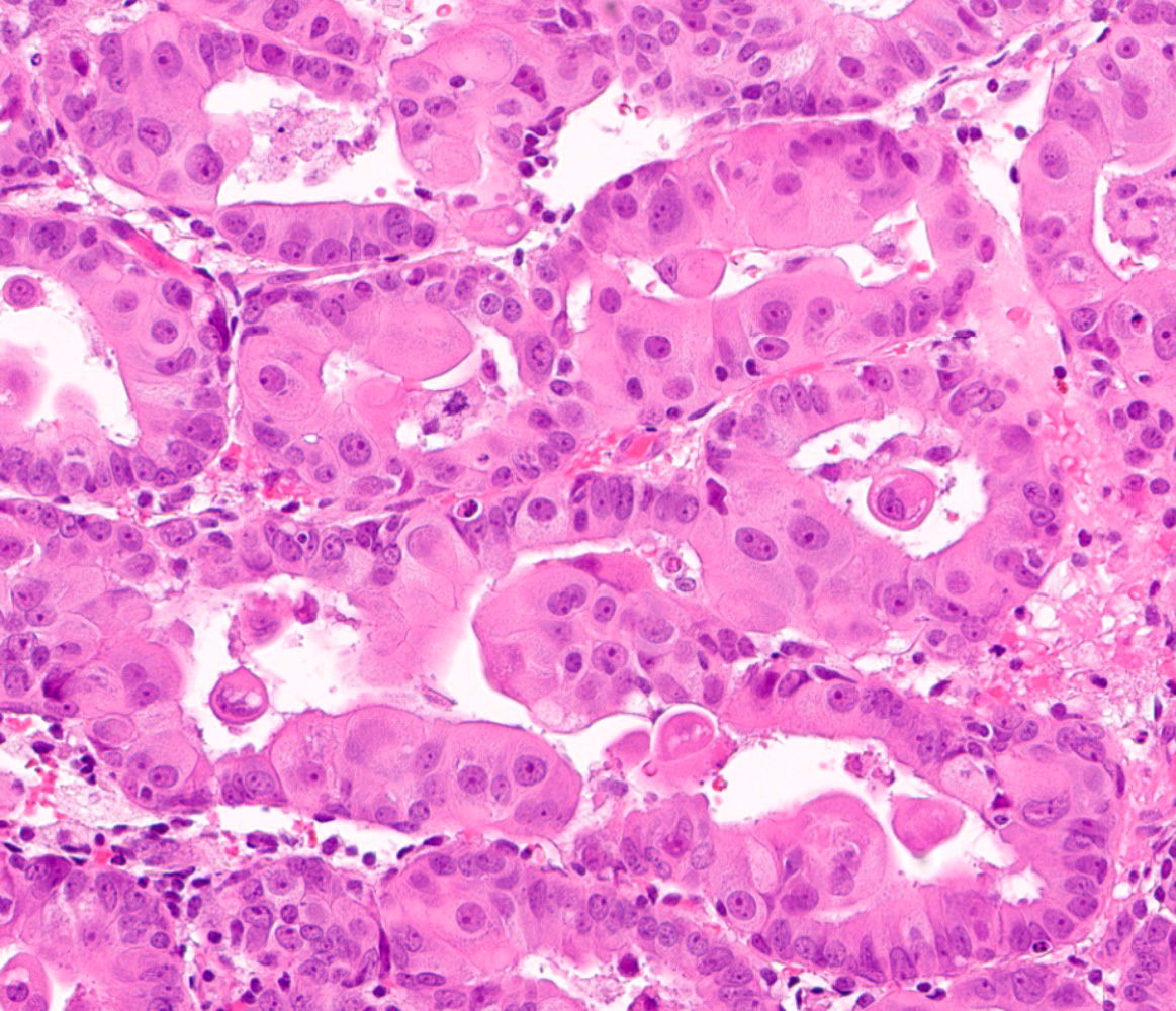
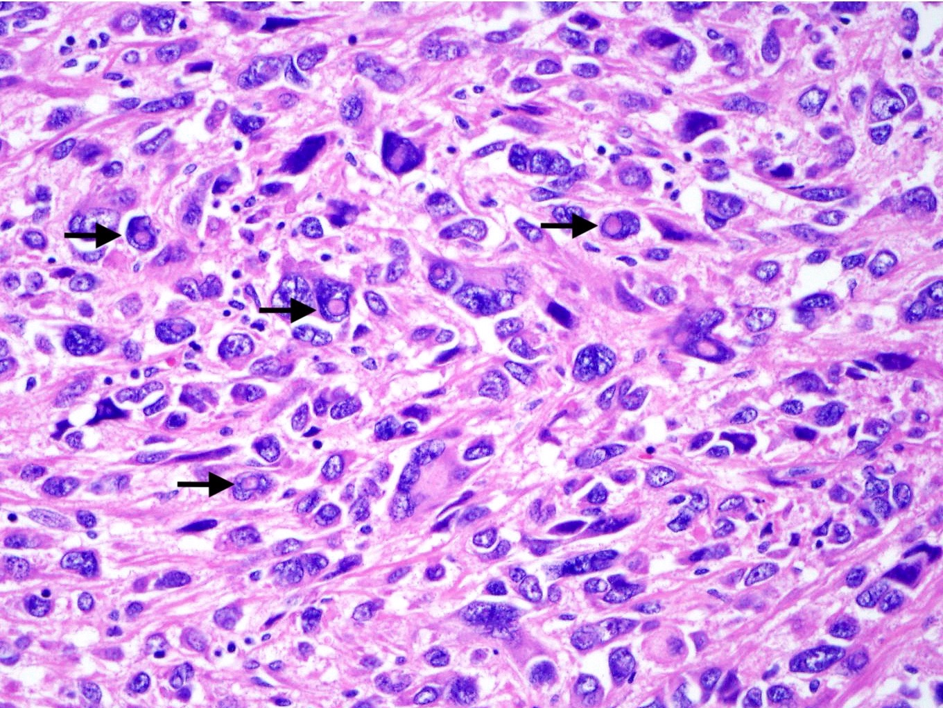
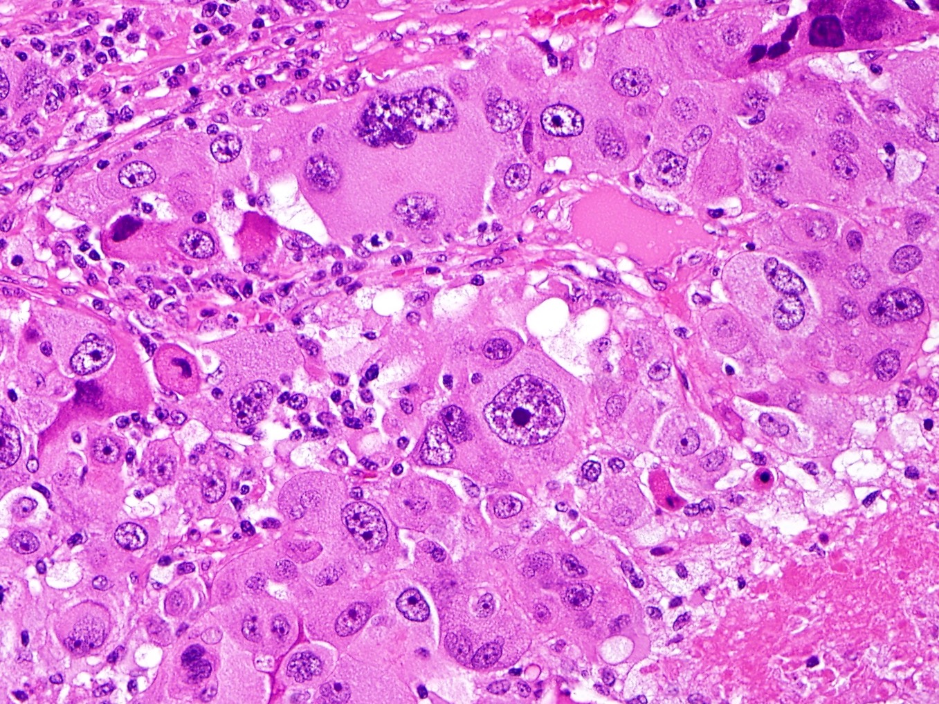
Clear cell cytology
Clear and hobnail cells
Oxyphilic cells
Clear and hobnail cells
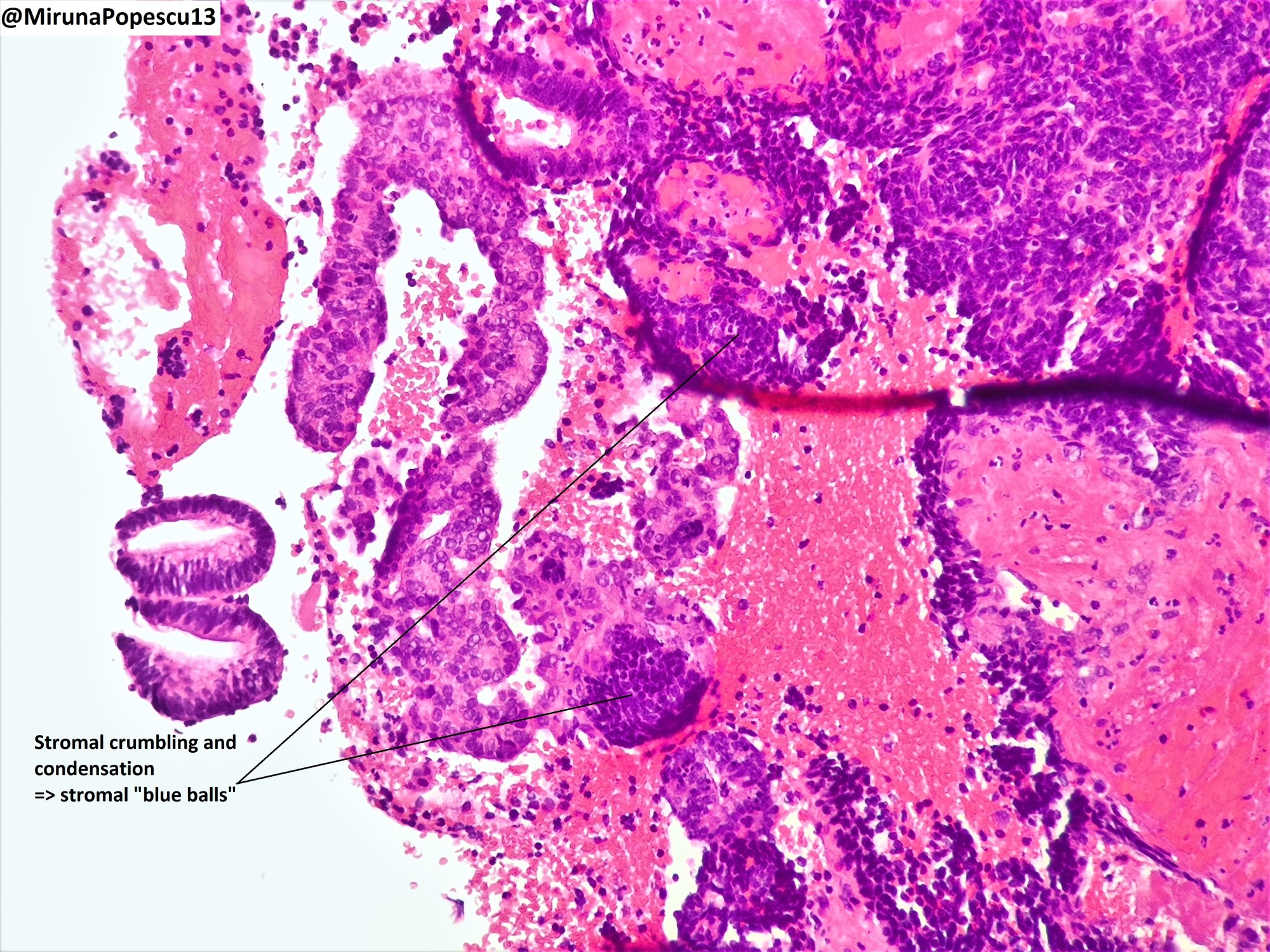
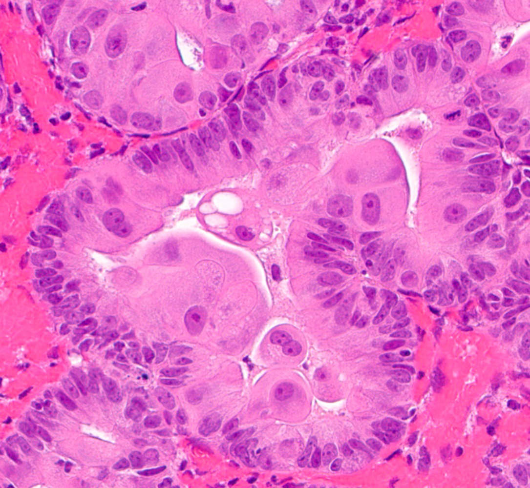
Targetoid bodies
Psammoma bodies
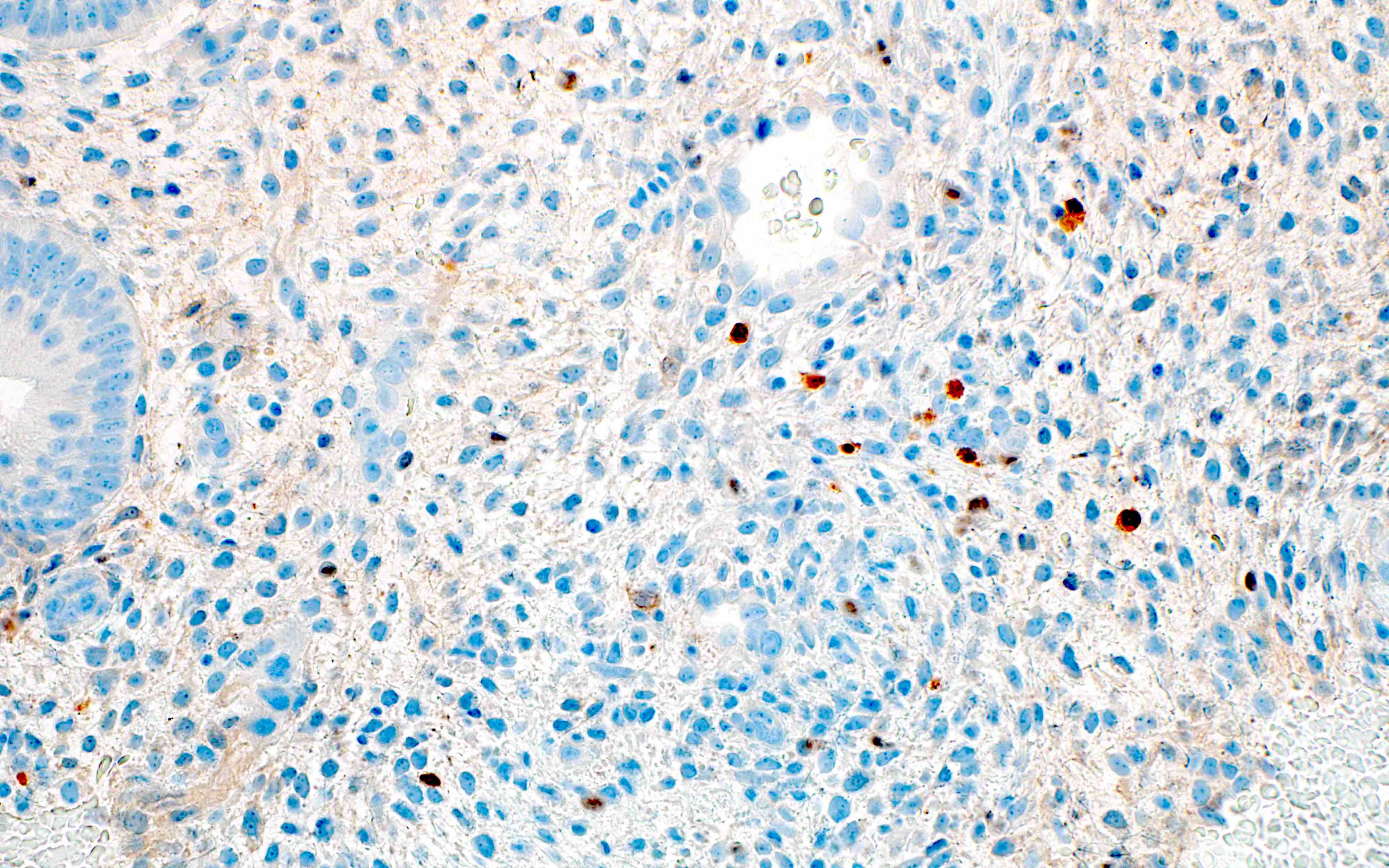
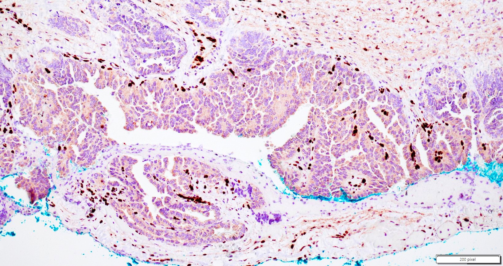
ER
Napsin A
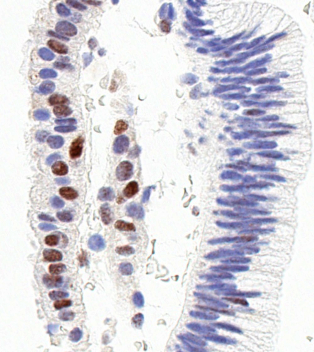
MSH6
Contributed by Hao Chen, M.D., Ph.D.
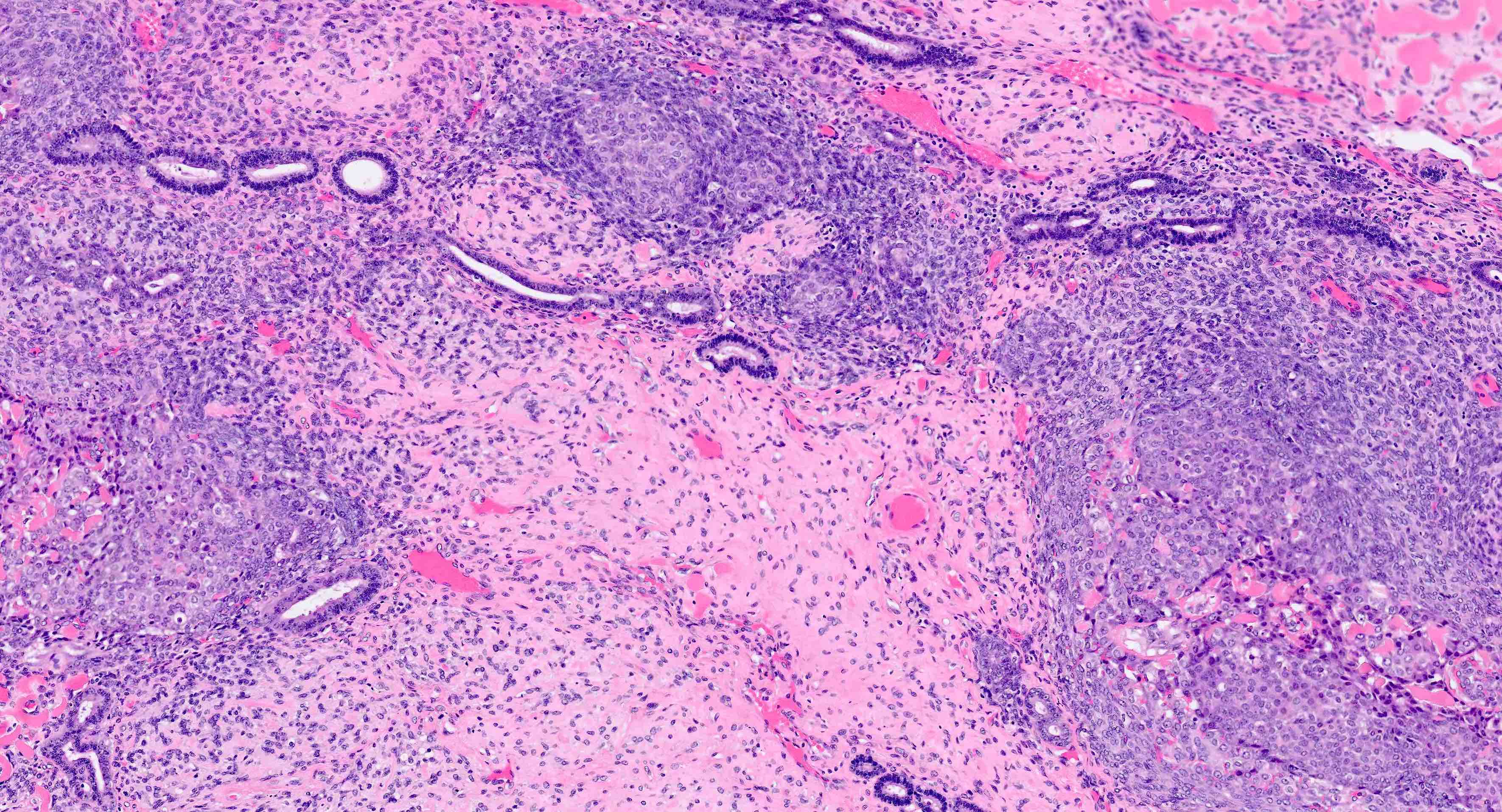
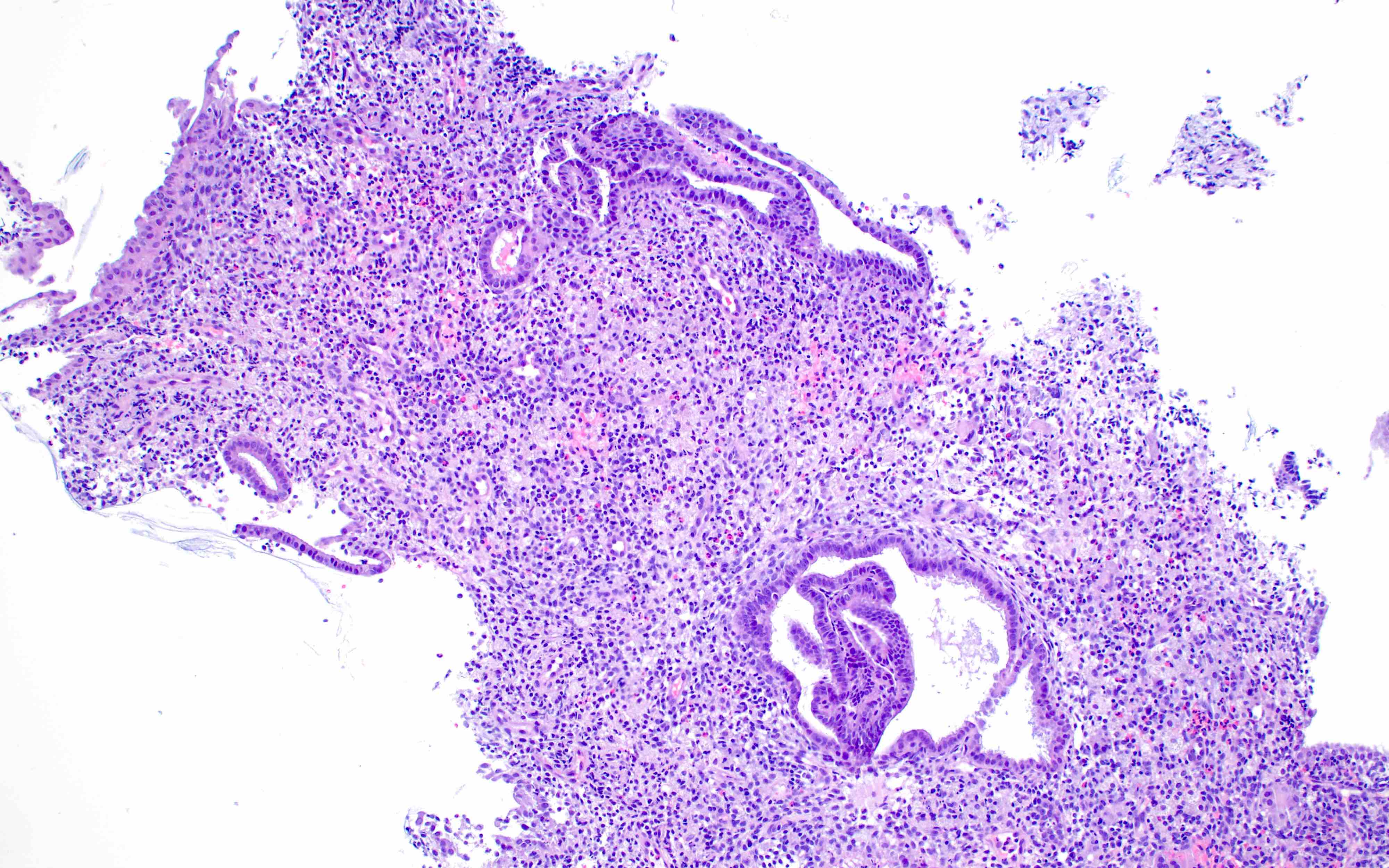
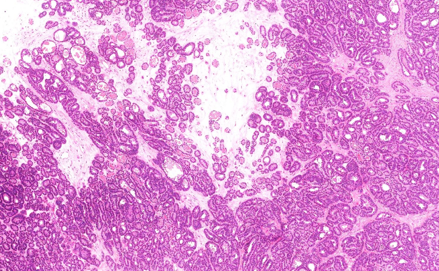
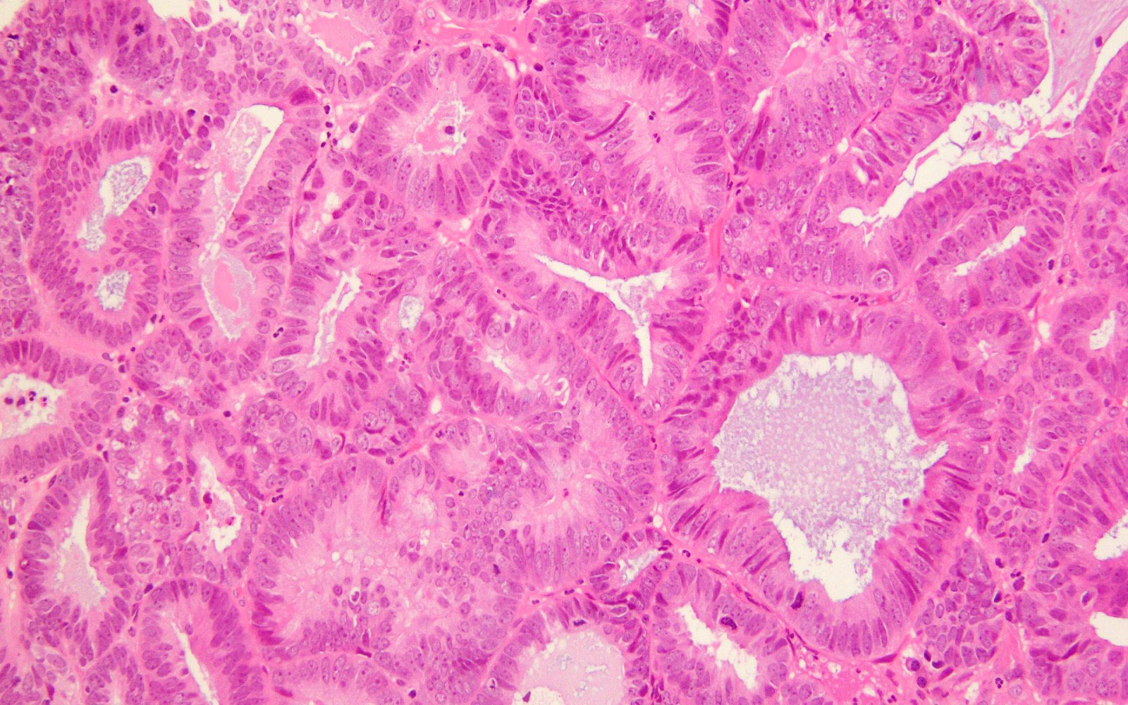
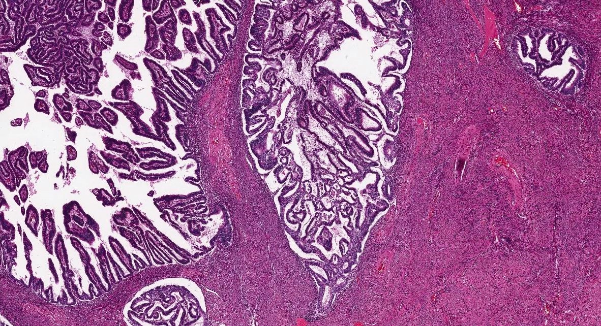
Irregular dilated glands
Irregular dilated glands with tubal metaplasia

- See endometrial carcinoma types
- See endometrial carcinoma types
Images hosted on other servers:

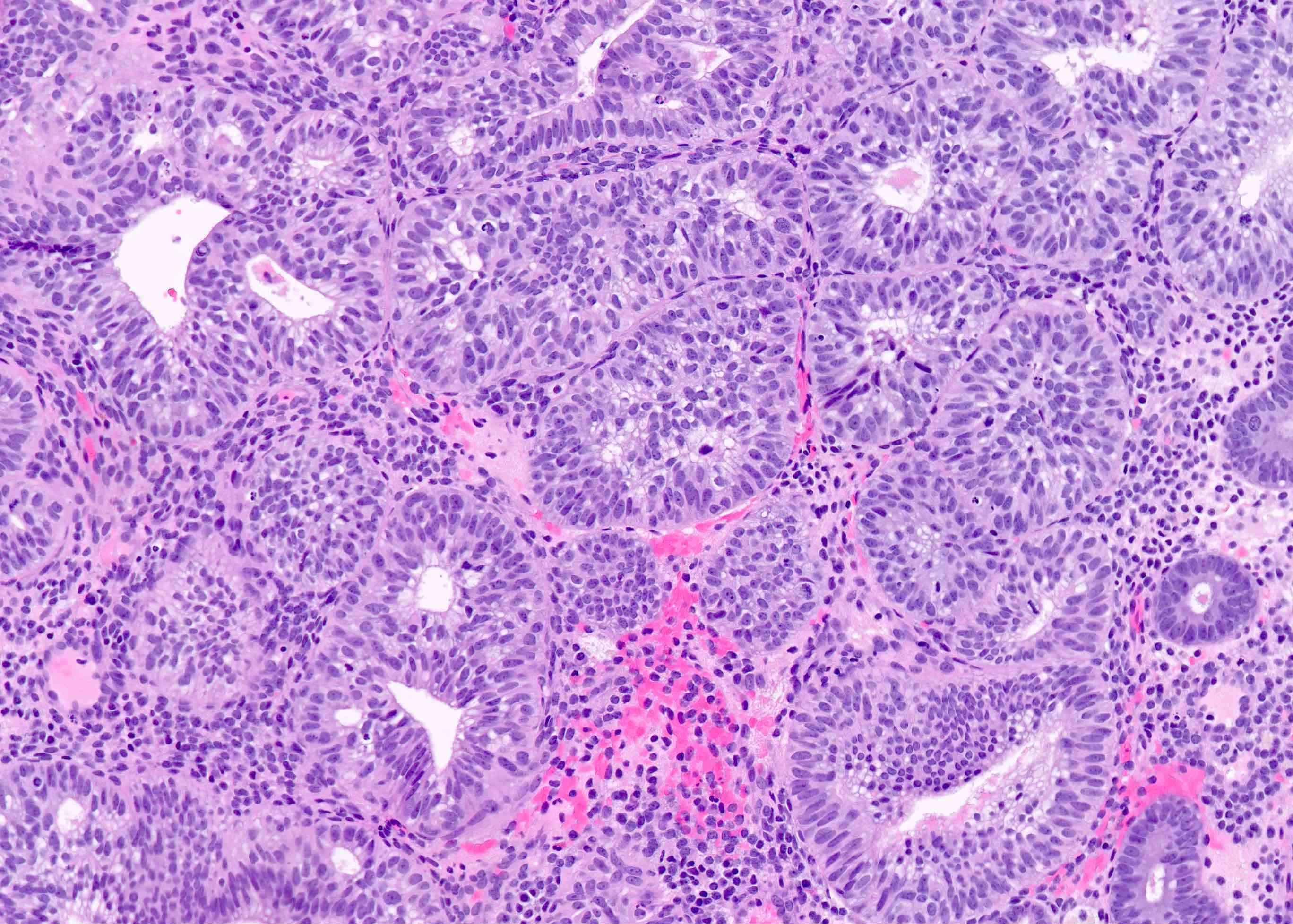
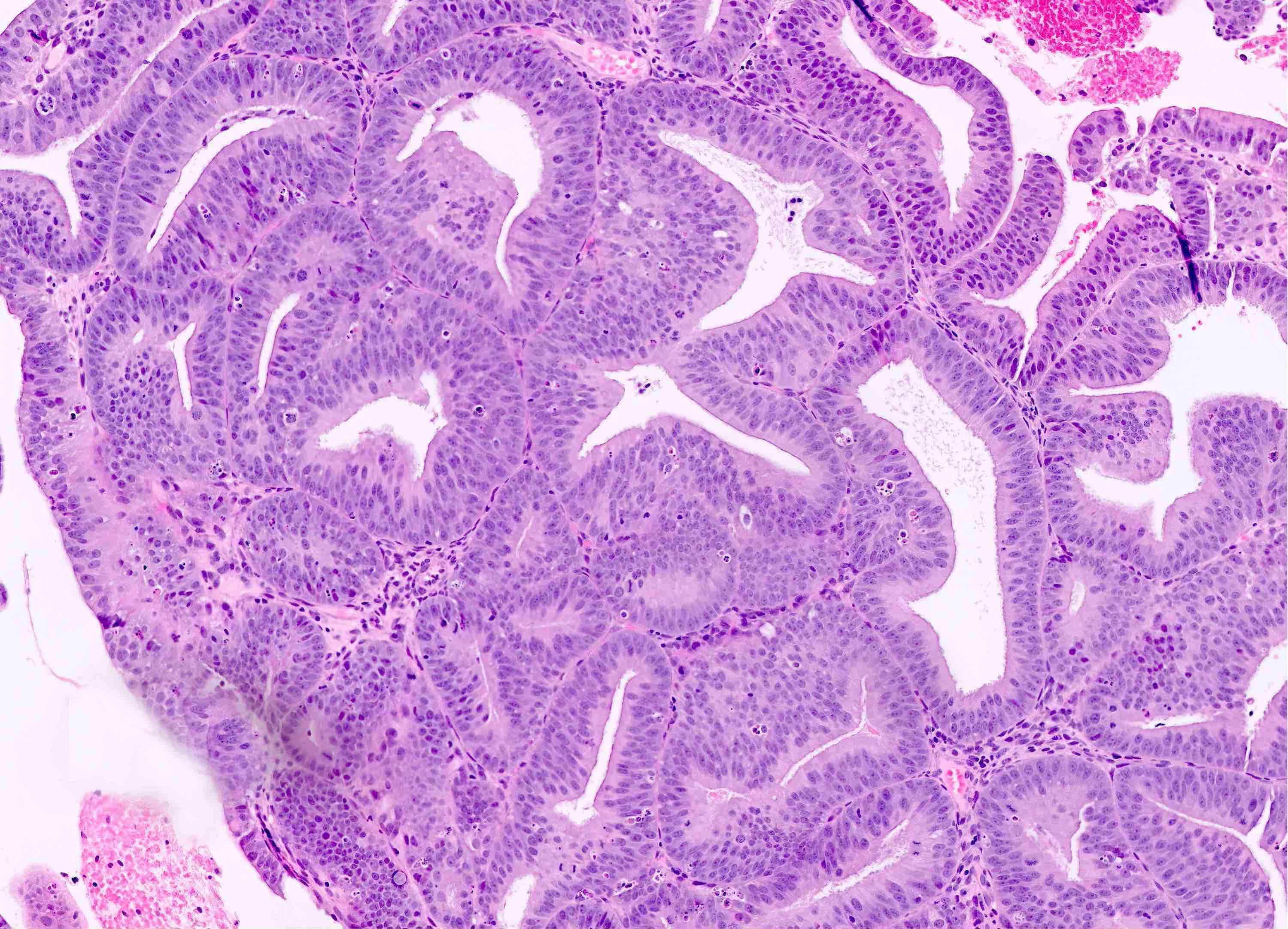
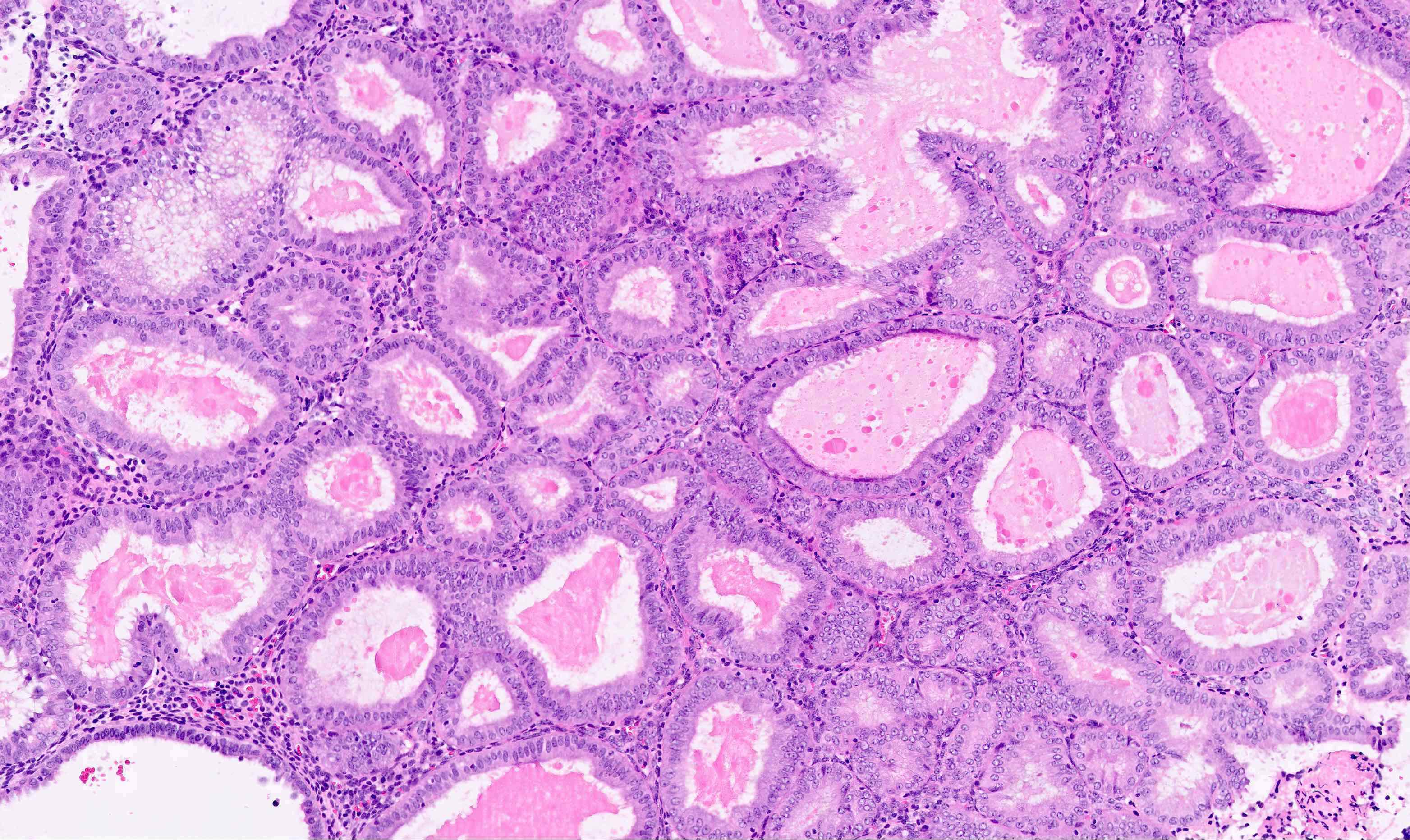
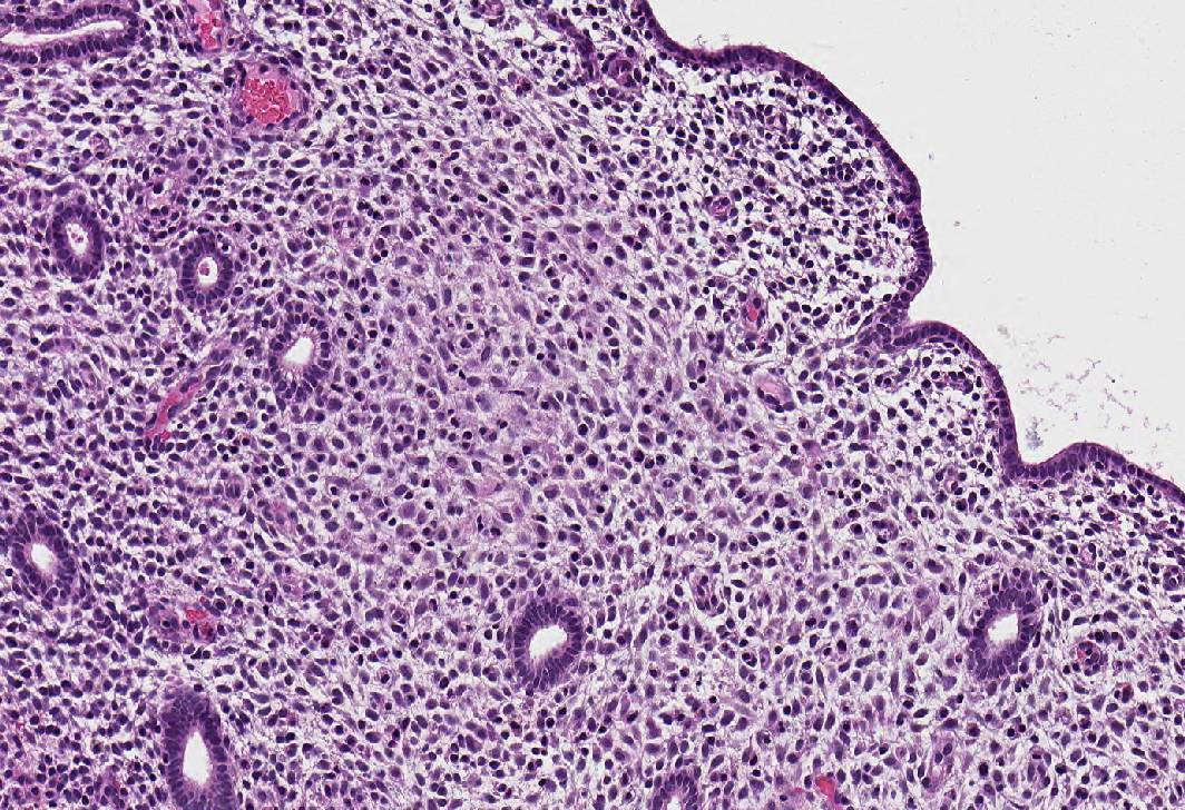
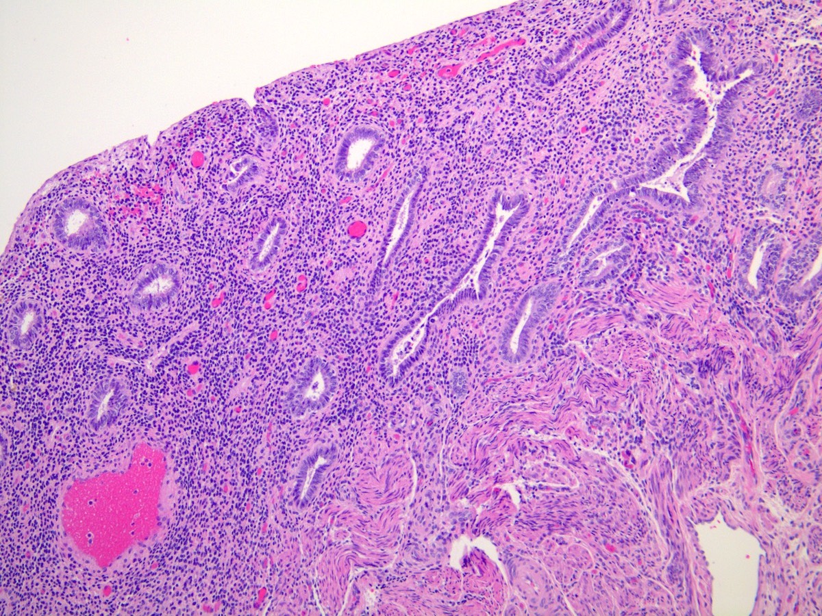
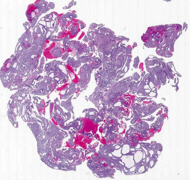
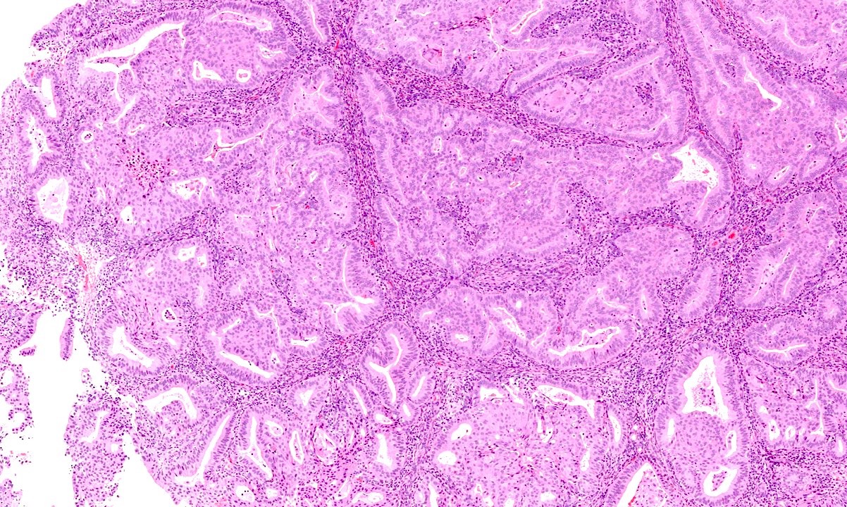
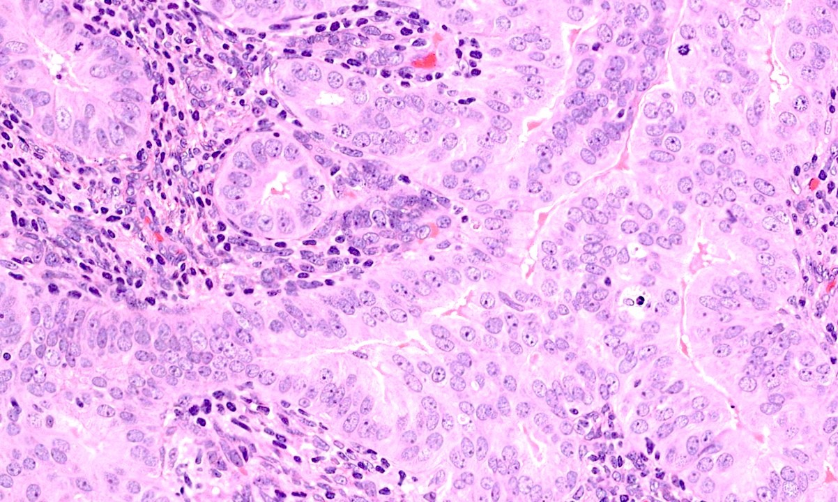
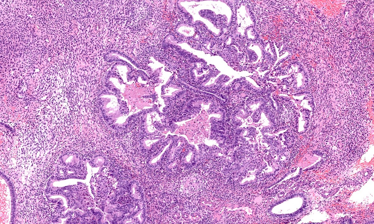
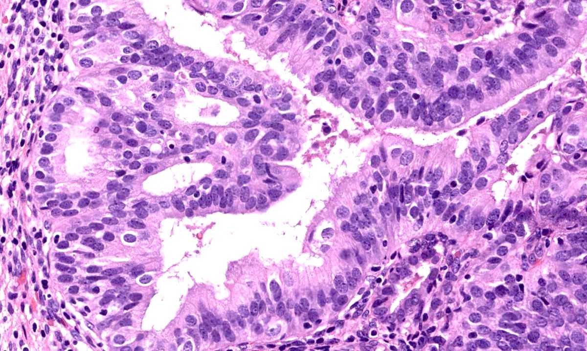
Endometrial hyperplasia
Contributed by Aarti Sharma, M.D.
Hyperplasia without atypia
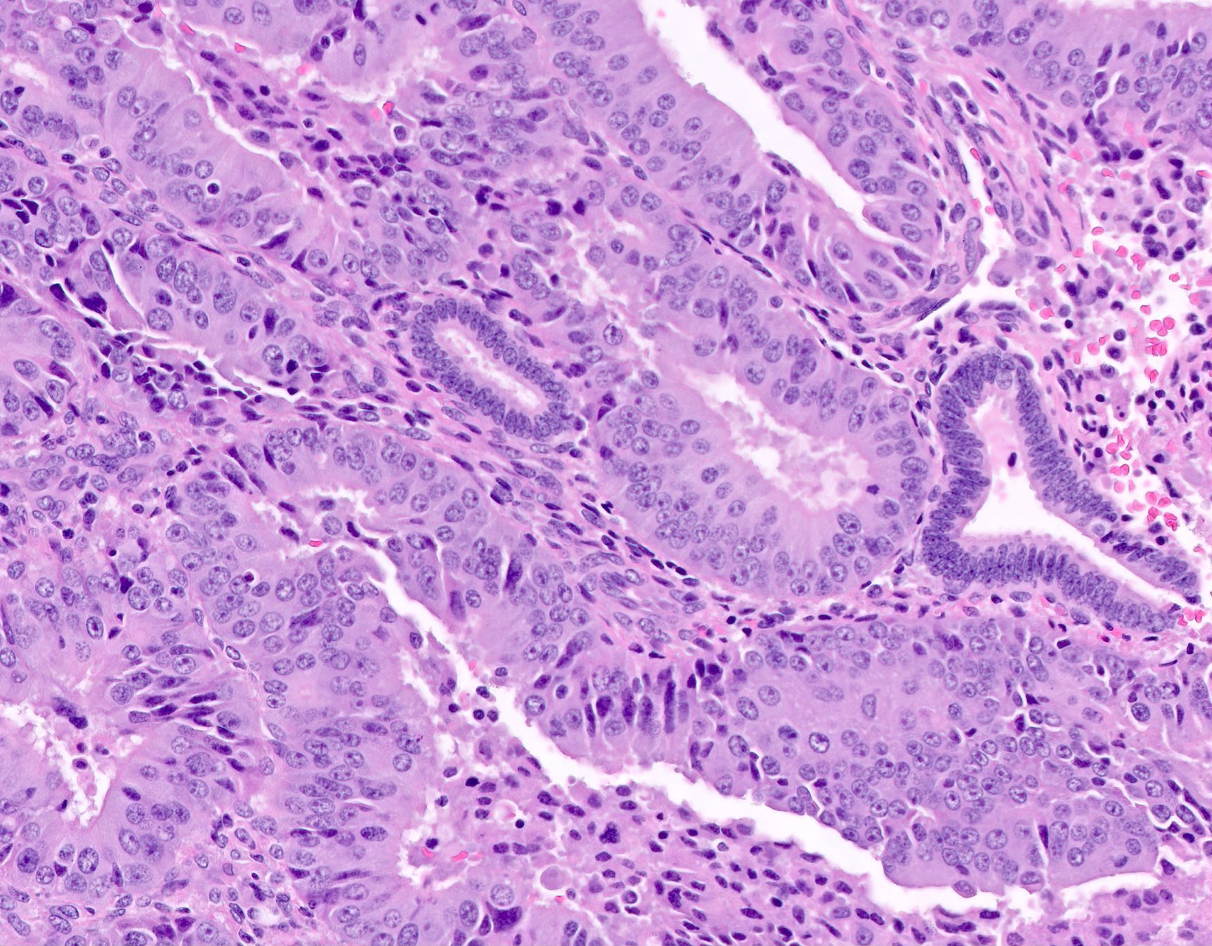
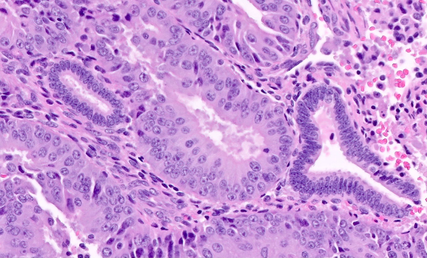
AH / EIN
AH / EIN bordering on FIGO grade I endometrial endometrioid adenocarcinoma
Images hosted on other servers:

Ultrasonographic features of osseous metaplasia
Contributed by Jessica L. Bentz, M.D.
Squamous morular metaplasia
Tubal and mucinous metaplasia
Endometrioid adenocarcinoma with mucinous differentiation
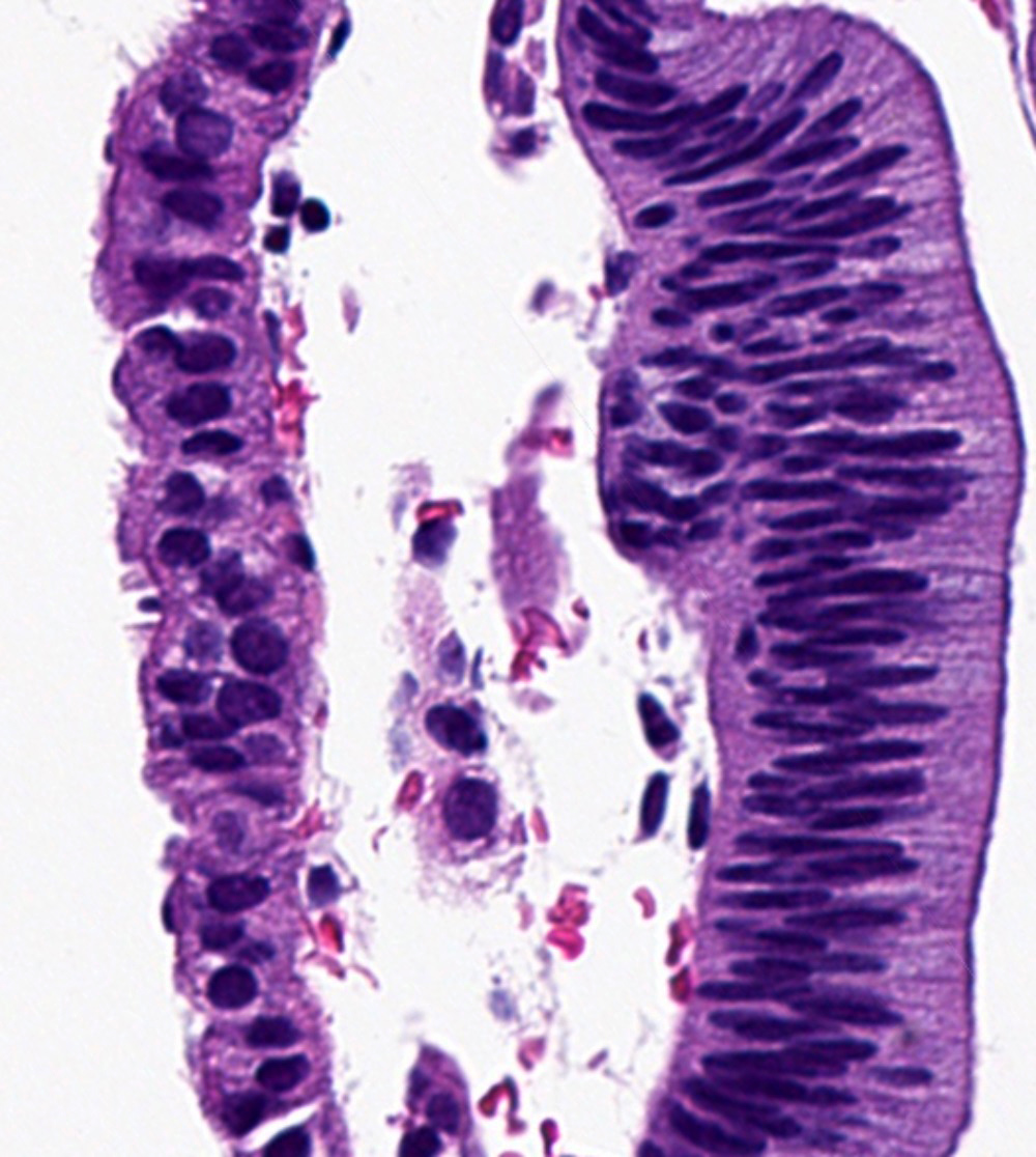
Tubal (ciliated cell) metaplasia
Endometrioid adenocarcinoma with cilia
Eosinophilic metaplasia in complex atypical hyperplasia
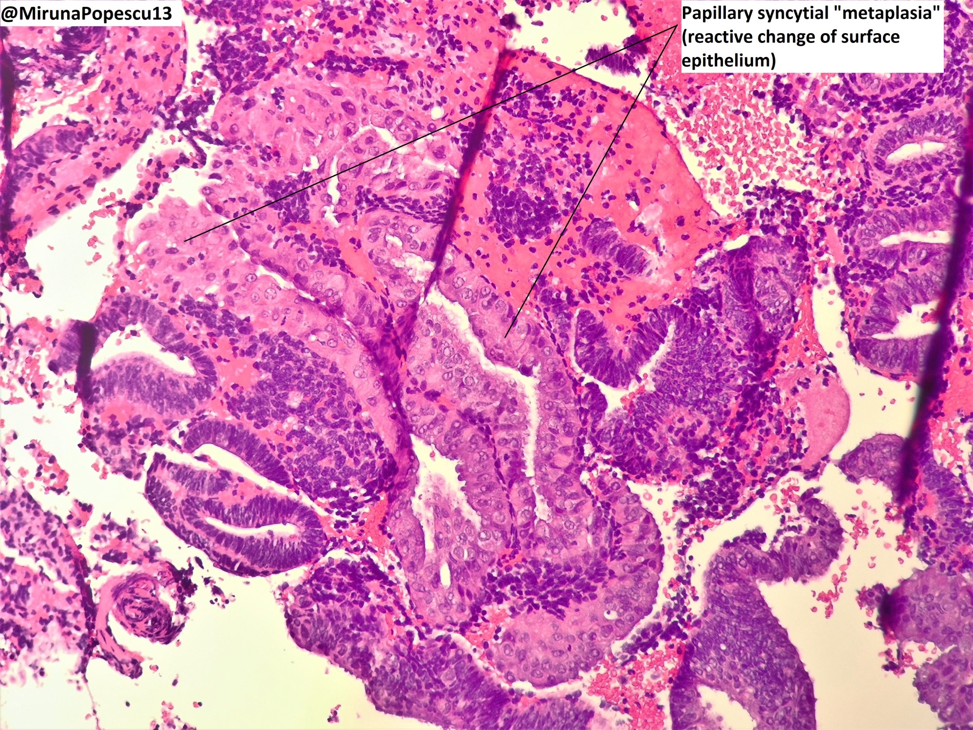
Papillary syncytial metaplasia

Complex papillary metaplasia

Complex papillary metaplasia
Secretory metaplasia
Hobnail metaplasia
Benign and premalignant lesions of the endometrium - Dr. Parra-Herran, from pathCast
Contributed by Monira Haque, M.D.

Excised polyp from uterine fundus
Images hosted on other servers:

Fundic polyp expands uterine cavity

Polyp fills the uterine cavity

Partially cystic polyp

Small fundic polyp

Large endometrial polyp
Contributed by Monira Haque, M.D. and Yuri Tachibana, M.D.
Polyp with pedicle
Atrophic epithelium
Stromal edema and myxoid changes
Variably cellular stroma and stromal hemorrhage

Marked stromal hyalinization
Dense stroma
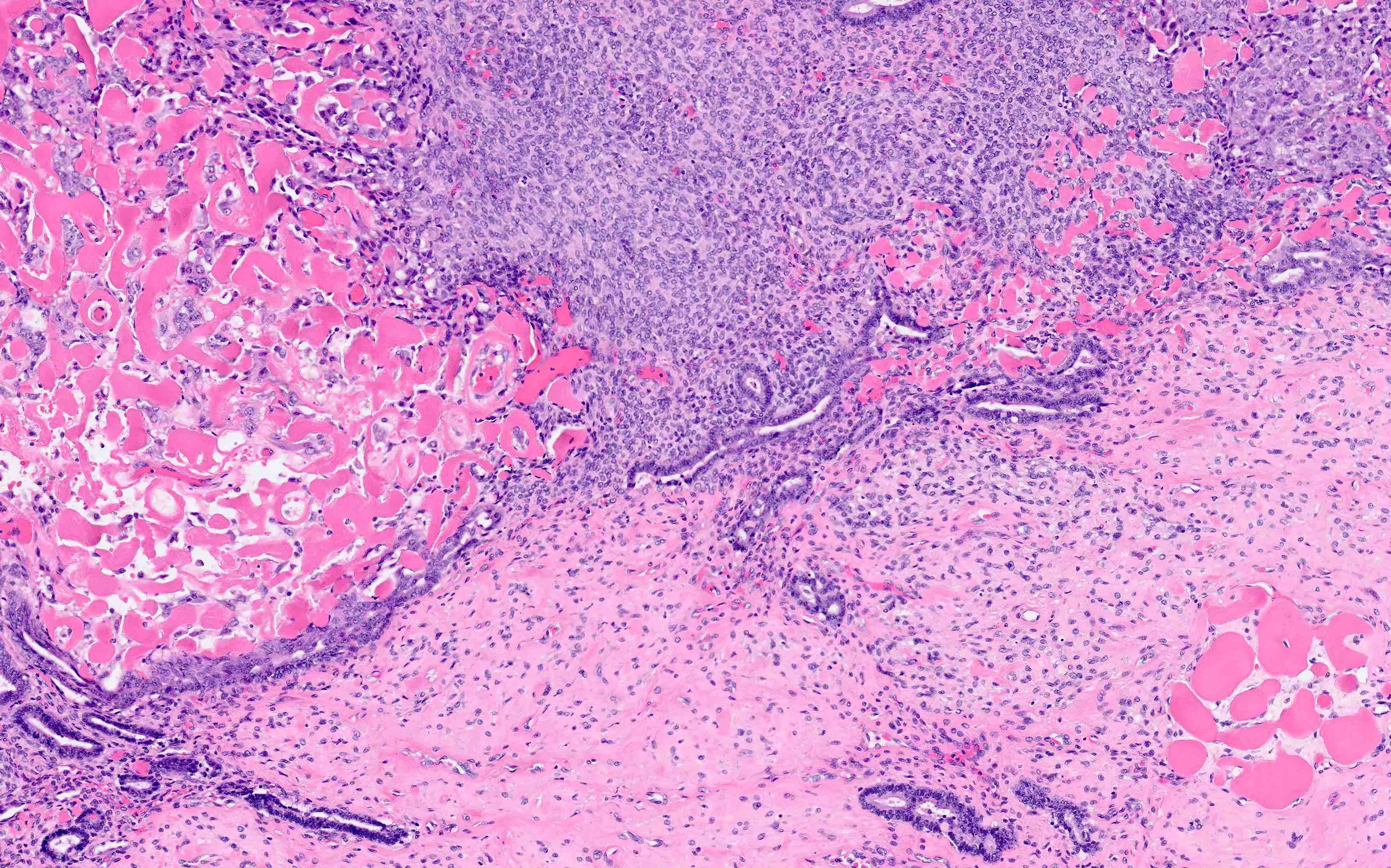
Irregular dilated glands and thickened blood vessels
Highly thickened vessels with fewer glands

Thick walled blood vessels
Variable gland morphology
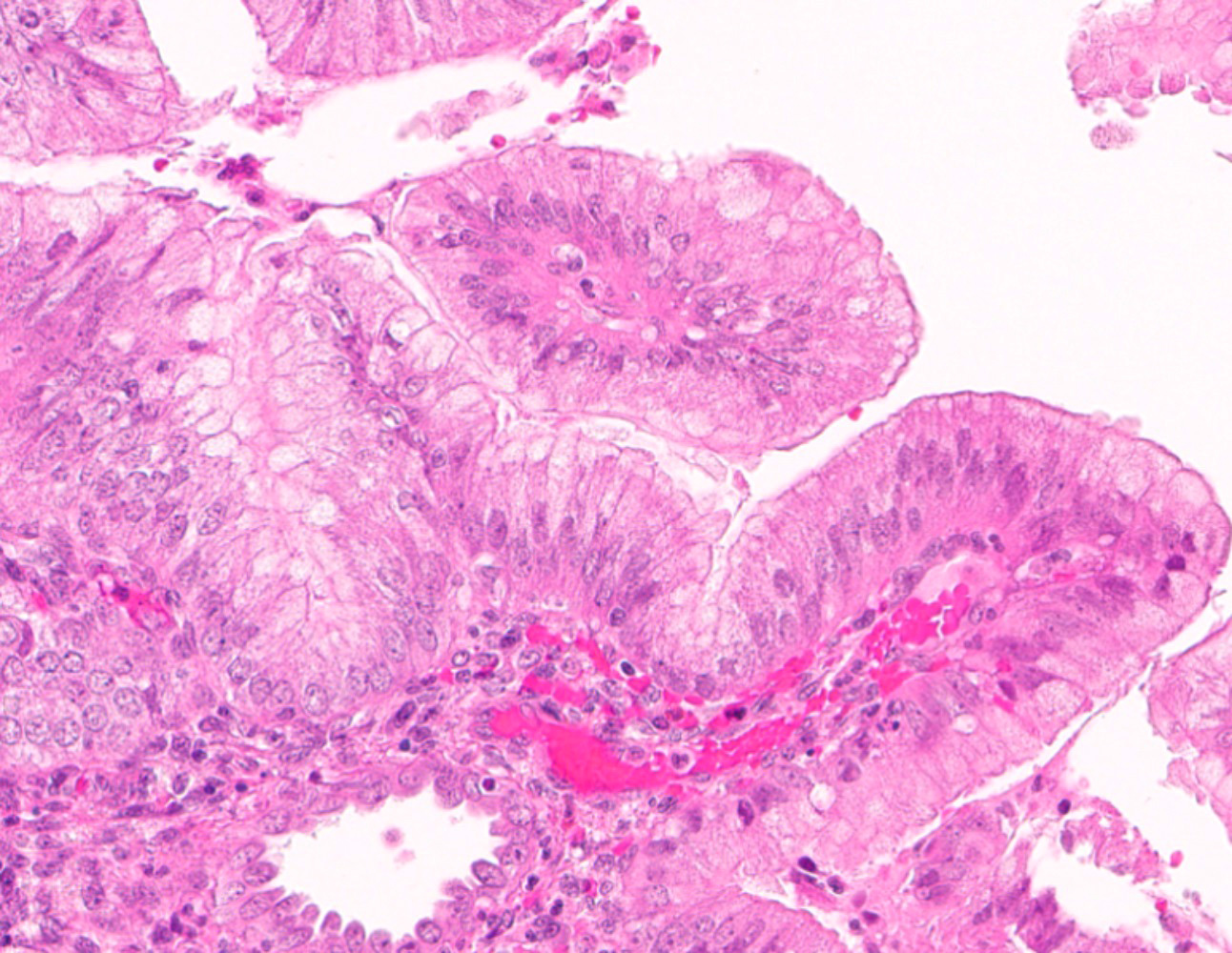
Glands lined by proliferative type endometrium
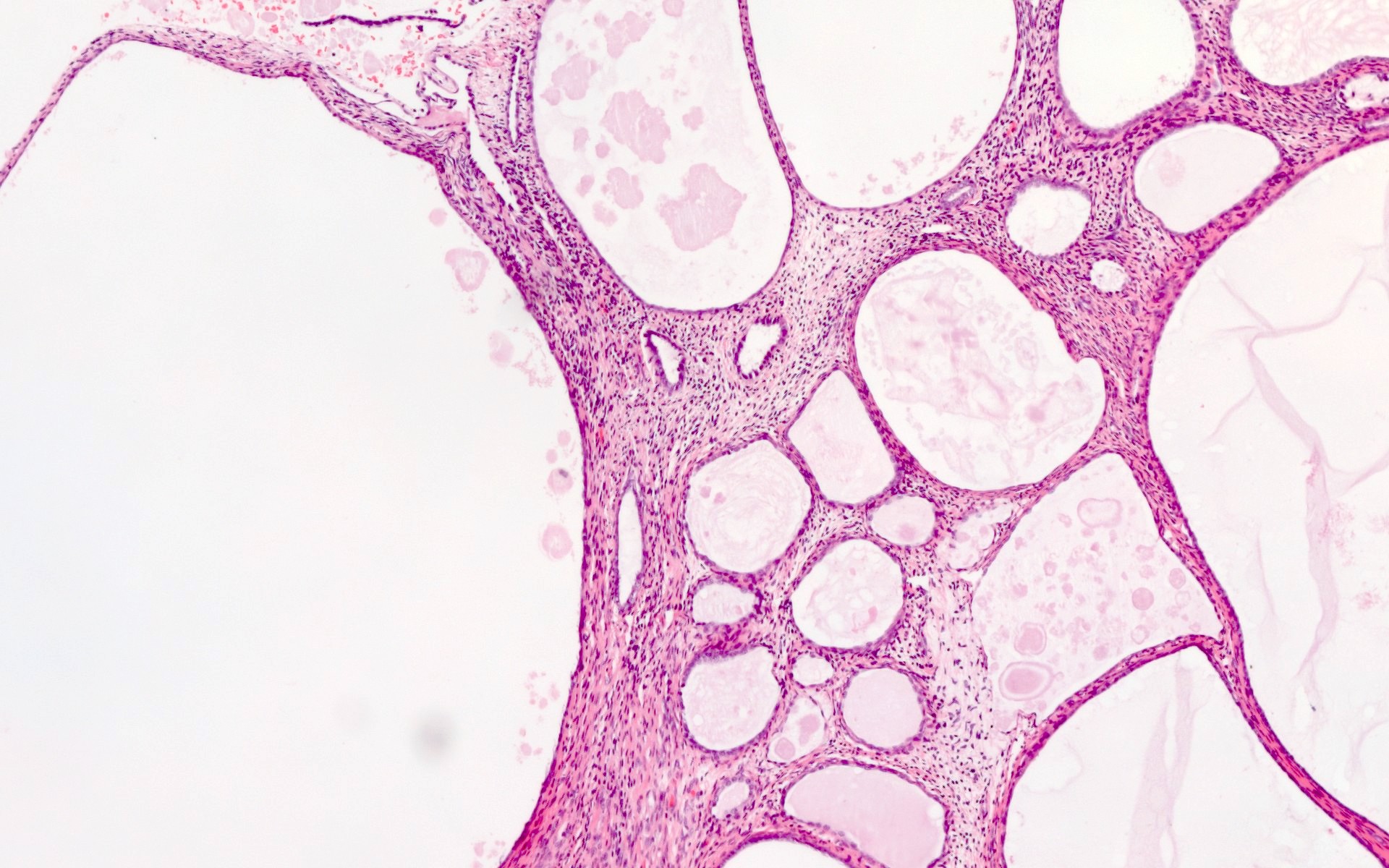
Cystically dilated glands
Markedly dilated glands

Squamous metaplasia
Glandular disarray and endometrioid adenocarcinoma

Endometrioid adenocarcinoma


Serous carcinoma
Contributed by Devi Jeyachandran, M.D.
Polypoid uterine tumor
Images hosted on other servers:

Uterine tumor
Contributed by Devi Jeyachandran, M.D.
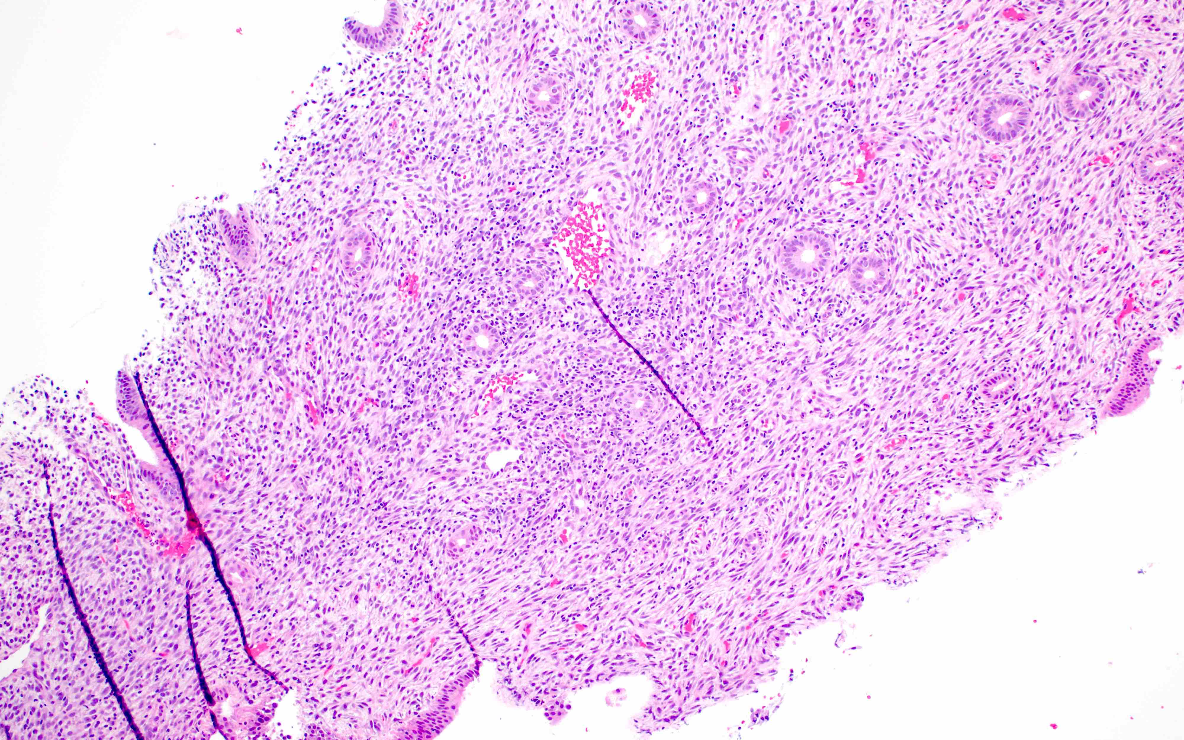
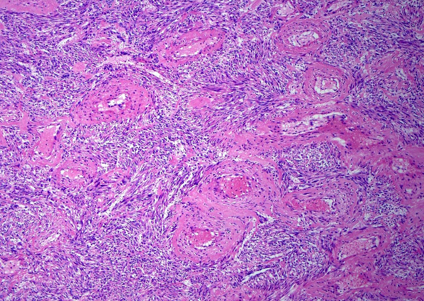
Bland stromal cells
Expansive growth
Proliferative type arterioles
Bland stromal cells
CD10 expression
ER expression

PR expression
WT1 expression
SMA expression
AE1 / AE3 expression

Desmin expression
Contributed by Aarti Sharma, M.D. and Ricardo R. Lastra, M.D.
Fundic mass
Mass occupying entire endometrium
Contributed by Aarti Sharma, M.D. and Ricardo R. Lastra, M.D.
FIGO 1
Solid growth

Squamous metaplasia
Morular metaplasia
Mucinous metaplasia
MELF pattern of invasion
CHEC variant
Contributed by Stephanie L. Skala, M.D. and Yuri Tachibana, M.D.
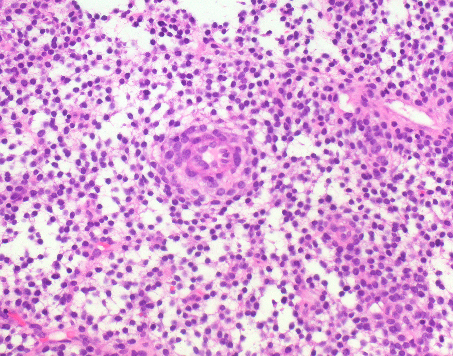
Chronic endometritis
Spindled endometrial stroma
Endometrial stromal plasma cells
Eosinophils and plasma cells
Plasma cells
CD138
MUM1 highlights plasma cells
Foamy histiocytes and eosinophils
Lymphoid follicles and histiocytes
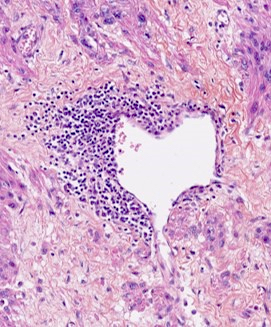
Prominent intraluminal neutrophils
Contributed by Sakinah Ahmad Thiryayi, M.D. and Gulisa Turashvili, M.D., Ph.D.
Decidual pattern
Secretory pattern

Inactive pattern
Variable progestin changes

Contraceptive effect
Testosterone for gender transition
Tamoxifen therapy

Tamoxifen therapy
Lupron treated leiomyoma
Necrosis with Lupron
Images hosted on other servers:

IARC: various images for grossing protocol

Morcellated specimen:
endometrium
identified as a
slit-like space
Images hosted on other servers:

IARC: various images for grossing protocol

Features to report: recommended sectioning
Images hosted on other servers:

MRI findings
Contributed by Ondrej Ondič, M.D., Ph.D. and Ayse Ayhan, M.D., Ph.D.
Pale yellow-white, soft and friable masses
Necrotic polypoid mass
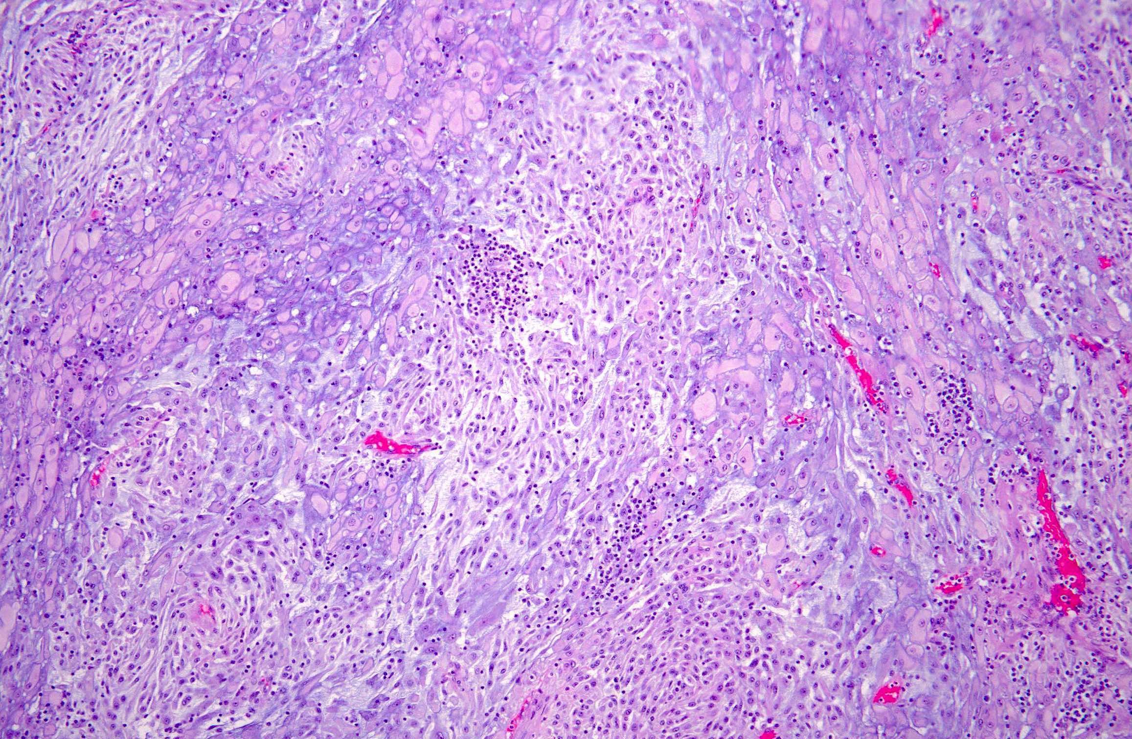
Contributed by Elizabeth Kertowidjojo, M.D., Ph.D., M.P.H., Jennifer A. Bennett, M.D., Ondrej Ondič, M.D., Ph.D. and Ayse Ayhan, M.D., Ph.D.
Mitotically active spindle cells
Myxoid stroma
Collagen plaques
Round cell component
Low grade spindle cell component
High grade transformation
Spindle cell morphology

High grade component
Myxoid stroma and signet ring-like change
Tongue-like infiltration
Small round cells
Undifferentiated small round cell component
Cyclin D1
Contributed by Mahmoud A. Khalifa, M.D., Ph.D.
Plump stromal cells and gland paucity
Transitional cell metaplasia of ectocervix
Follicular cysts with high follicular density
Prostatic metaplasia of vagina
NKX3.1 stain
Images hosted on other servers:

CT shows intrauterine mass

Axial MRI shows local lesion
Images hosted on other servers:

Polypoid lesion within endometrial cavity
Contributed by Jennifer Bennett, M.D.

Hyper and hypocellular areas
Myxoid pattern
Acellular myxoid areas

Myxoid areas resemble nodular fasciitis
Plasma cell aggregates

Compact pattern

Severe nuclear atypia
Ganglion-like cells
Pregnancy associated IMT
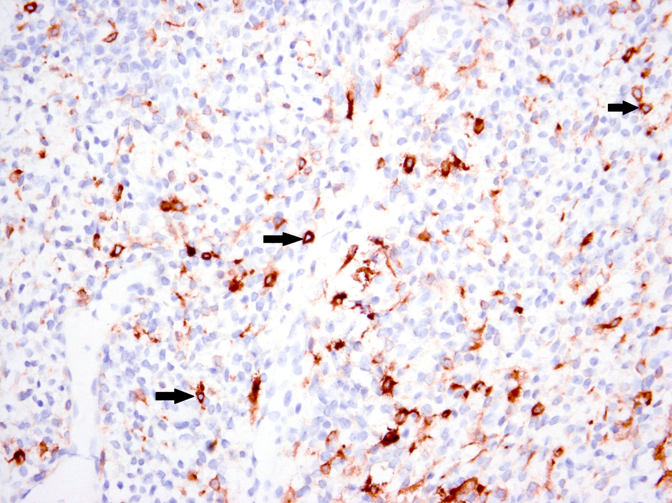
ALK
Images hosted on other servers:

FISH: classic ALK rearrangement
Images hosted on other servers:

Contrast enhancing CT

MRI

MRI
Contributed by Sabrina Croce, M.D., Ph.D.
Tumor plugs
Diffuse myometrial expansion
Numerous coalescing nodules
Symmetrical myometrial enlargement
Images hosted on other servers:


Tumor plugs

Involvement of myometrial vessels
Contributed by Sabrina Croce, M.D., Ph.D.
Intravenous leiomyomatosis
Tumor plugs
Tumor plugs
Plexiform appearance

Coexisting leiomyoma
Estrogen receptor

Desmin
CD34
Diffuse leiomyomatosis
Numerous confluent nodules
Poorly defined nodules

Well defined contours

Progesterone receptor

Caldesmon
Intravenous leiomyomatosis
Contributed by Sabrina Croce, M.D., Ph.D.
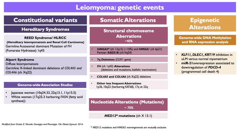
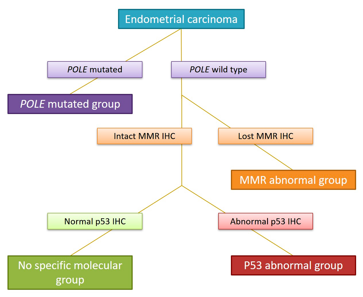
Table 1: molecular alterations
Table 2: differential diagnosis in uterine spindle cell smooth muscle lesions
| | | | |
| - | + | ≥ 10 | Leiomyosarcoma |
| + | + | ≥ 10 | |
| + | + | < 10 | |
| - | - | > 15** | Smooth muscle tumor of uncertain malignant potential (STUMP) |
| - | + | < 10 | |
| + | - | < 10 | |
| - | - | ≥ 6 and ≤ 15** | Mitotically active leiomyoma |
| * Mitoses/mm2 (mitoses/10 HPF of 0.55 mm in diameter) | |||
| ** > 15 or ≥ 15, cutoff value varies according to the references | |||
Table 3: differential diagnosis in uterine myxoid smooth muscle lesions
| | | | |
| Infiltrative borders | - | - | 1 or more of these criteria:
|
| Atypia | - | - | |
| Tumor cell necrosis | - | - | |
| Mitoses* | - | 1 | |
| * Mitoses/mm2 (mitoses/10 HPF of 0.55 mm in diameter) | |||
Table 4: differential diagnosis in uterine epithelioid smooth muscle lesions
| | | | |
| Atypia | - | - | 1 or more of these criteria:
|
| Tumor cell necrosis | - | - | |
| Mitoses* | < 2 | ≥ 2 and < 4 | |
| * Mitoses/mm2 (mitoses/10 HPF of 0.55 mm in diameter) | |||
Contributed by Sabrina Croce, M.D., Ph.D. and @Andrew_Fltv on Twitter

Submucosal leiomyoma
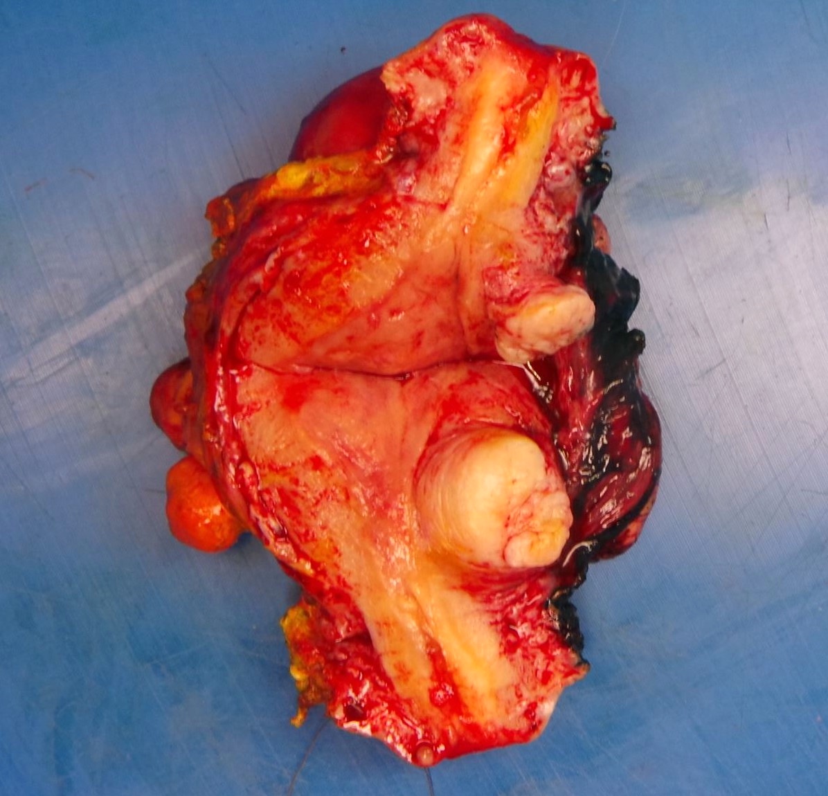
Intramural leiomyoma
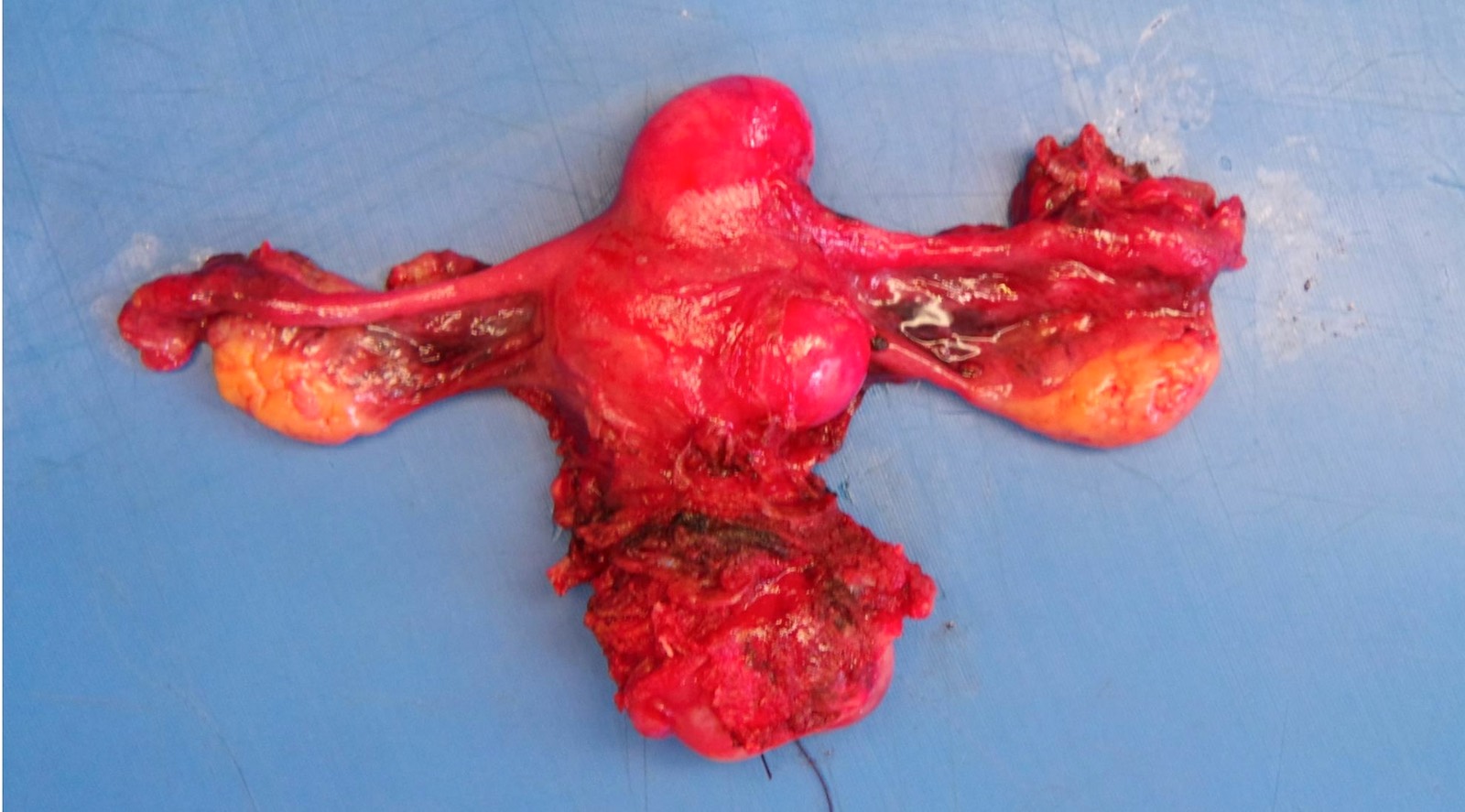
Subserosal leiomyomas
Apoplectic leiomyoma
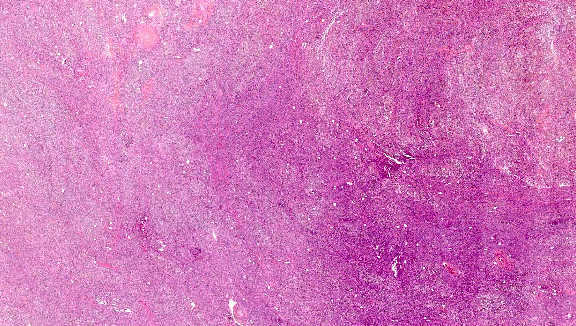
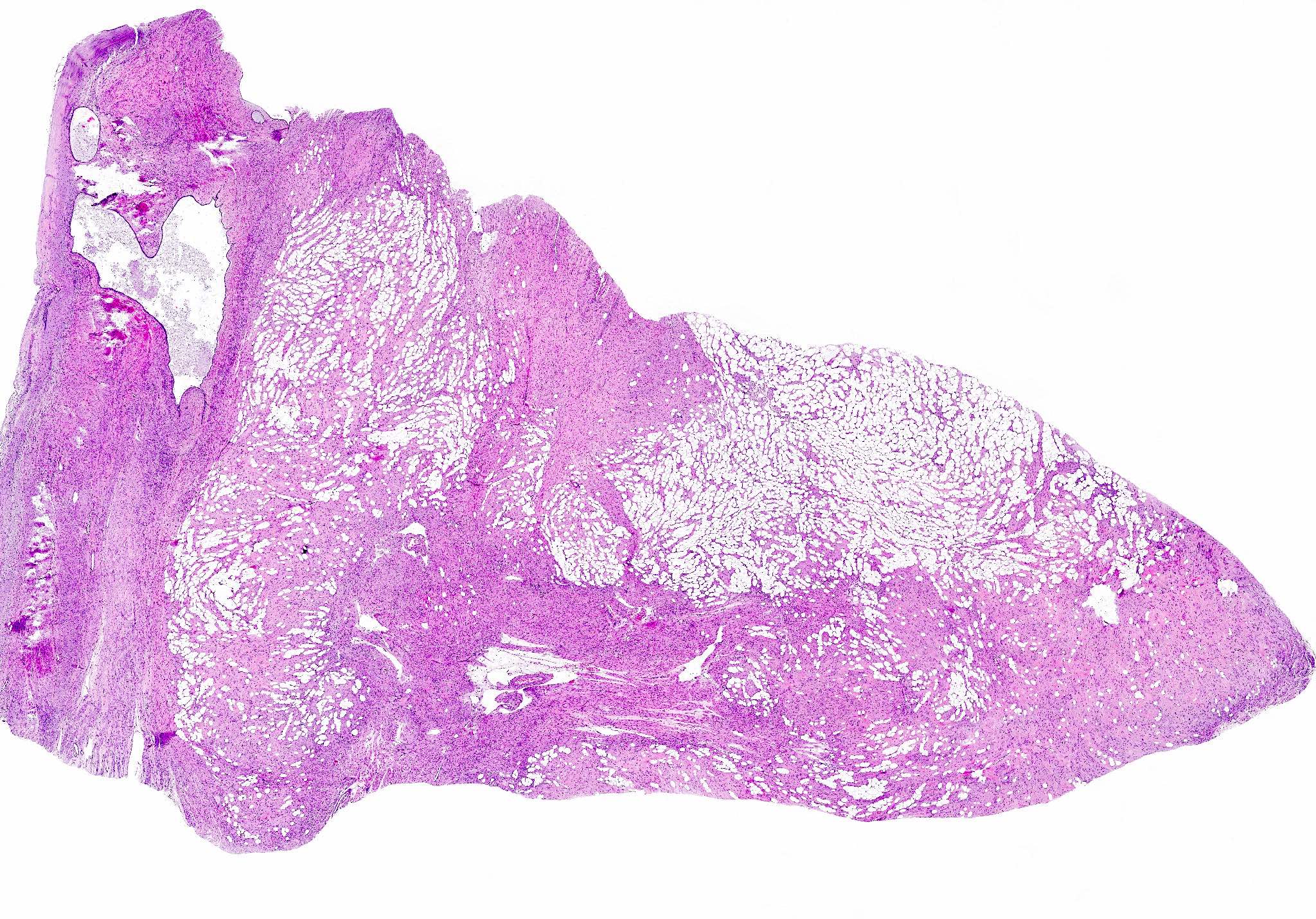
Conventional / usual leiomyoma
Cystic hydropic leiomyoma
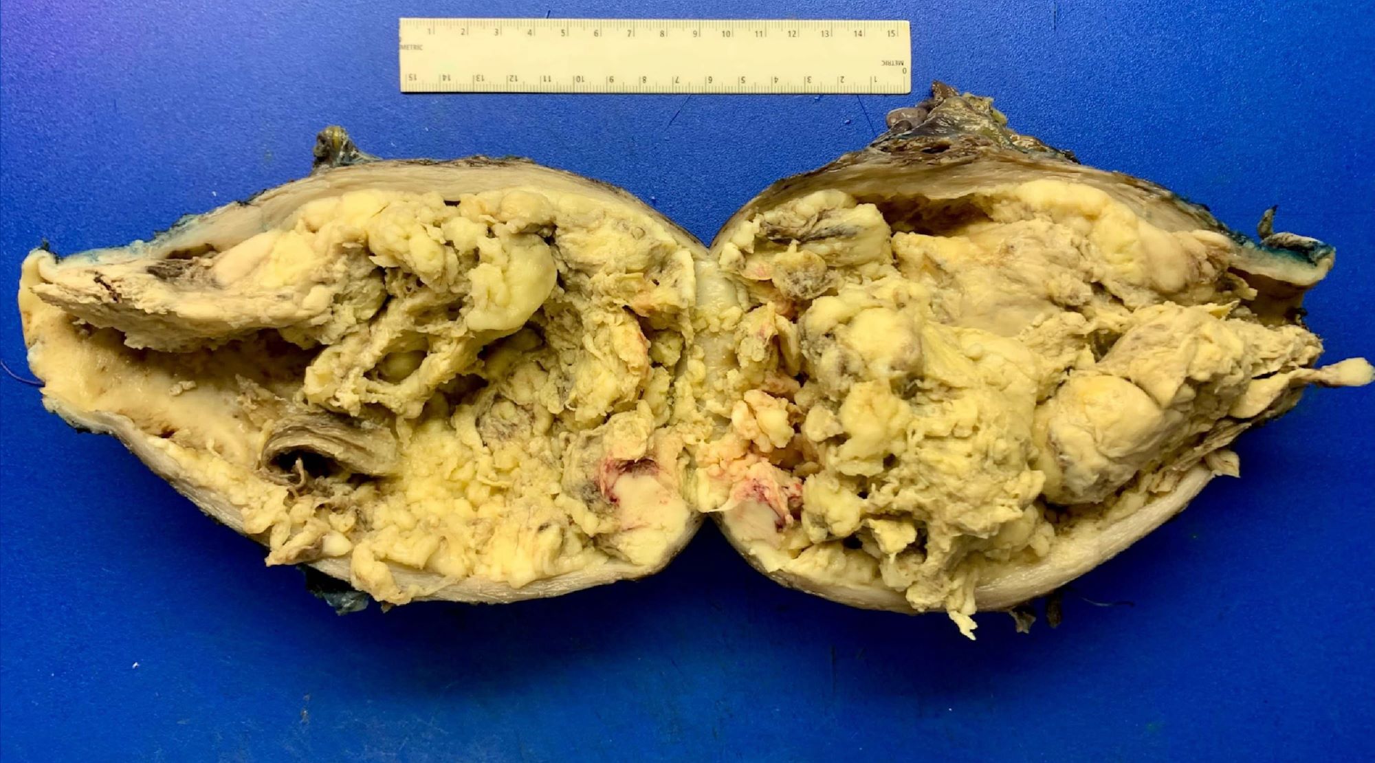
How about a new subtype of 'leiomyolipoma'?😀🤔😅
#grosspath #pathtwitter #gynpath #pathology #pathoutpic"
Contributed by @Andrew_Fltv on Twitter (see original post here)">
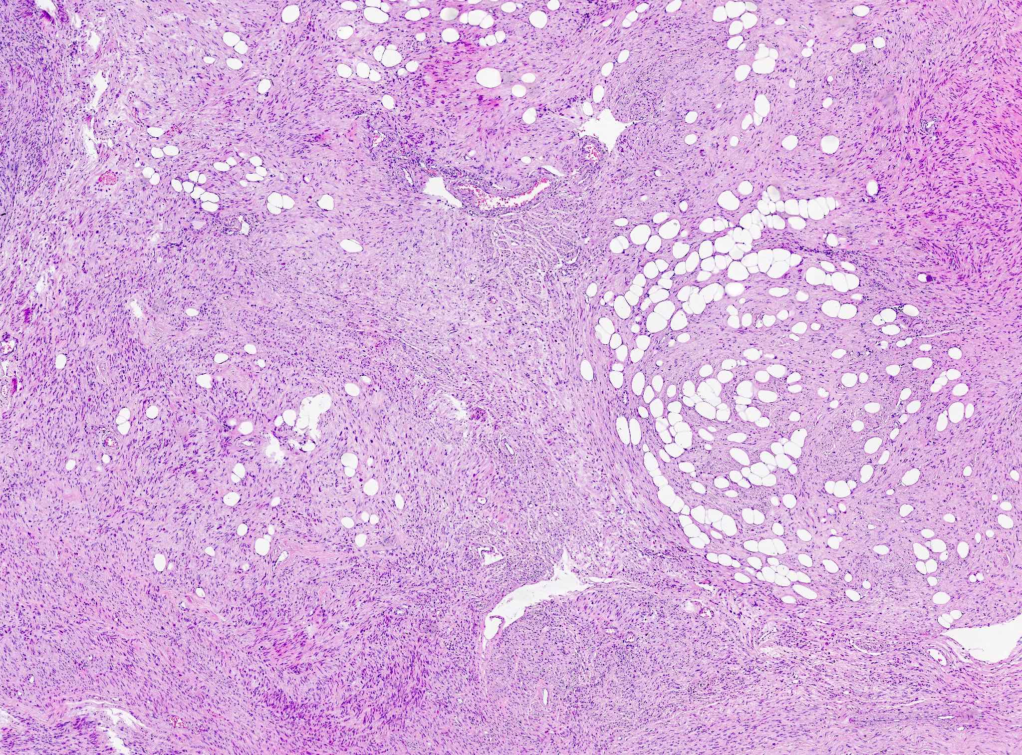
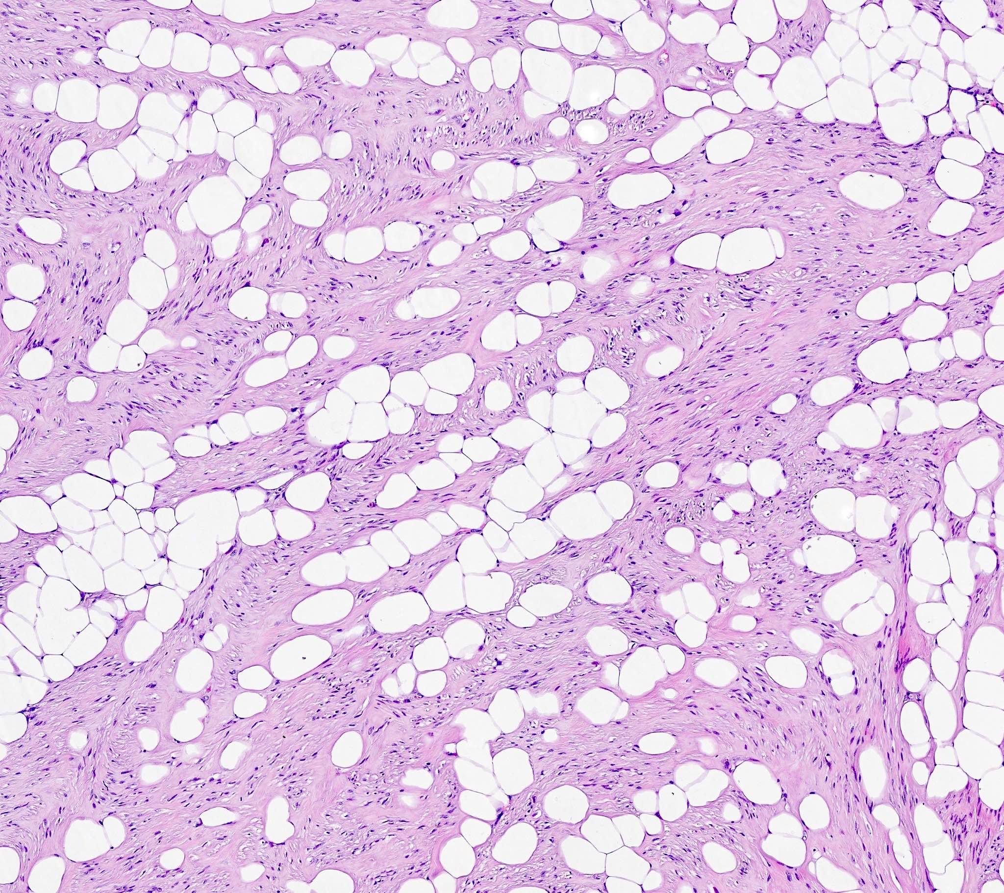
Lipoleiomyoma
Contributed by Sabrina Croce, M.D., Ph.D., Kristina Doytcheva, M.D., Jennifer A. Bennett, M.D. (Case #508) and @Andrew_Fltv on Twitter

Conventional / usual leiomyoma
Cellular leiomyoma


FH deficient leiomyoma

Leiomyoma with bizarre nuclei

Hydropic leiomyoma
Apoplectic leiomyoma
Epithelioid leiomyoma
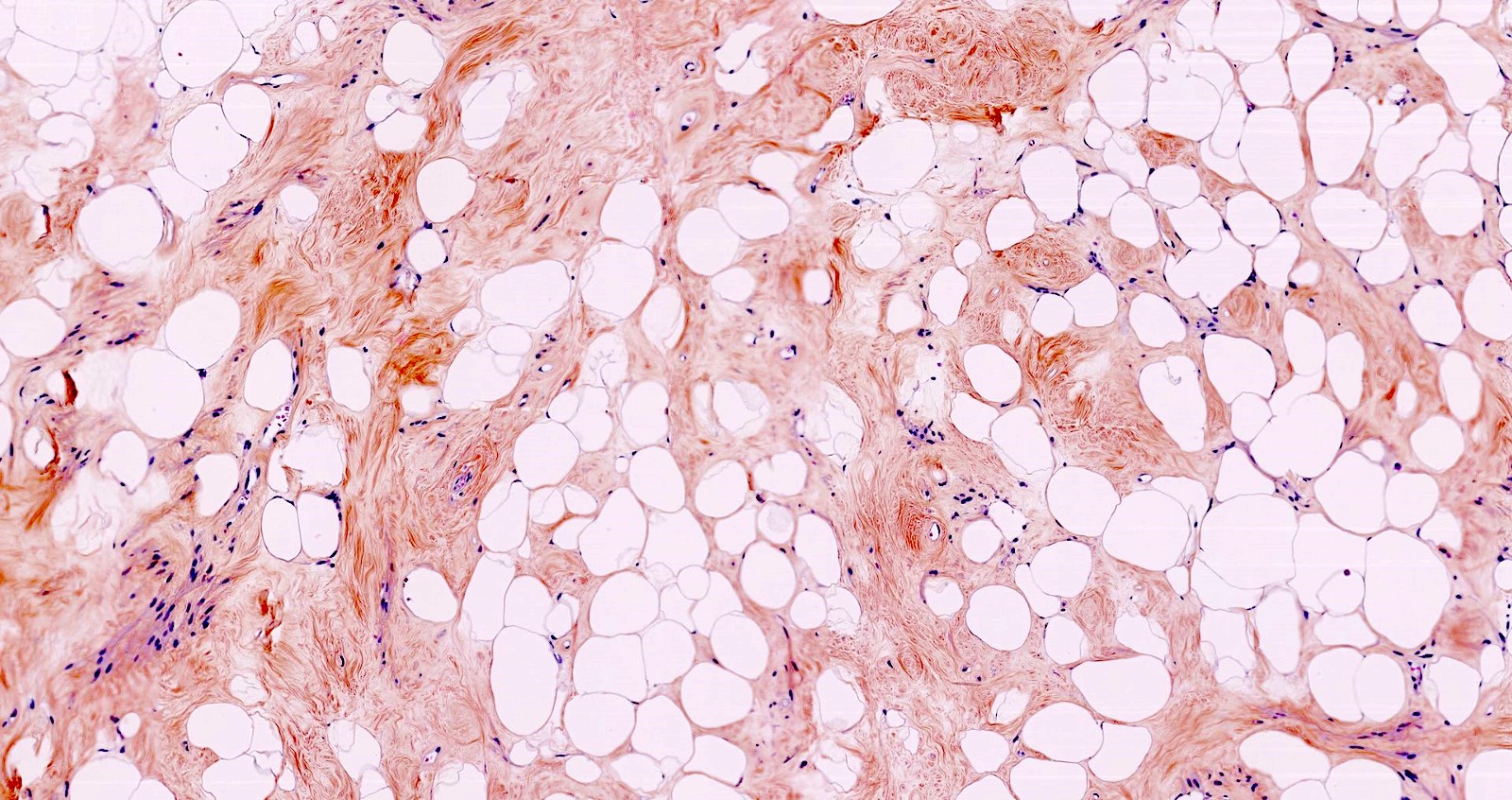
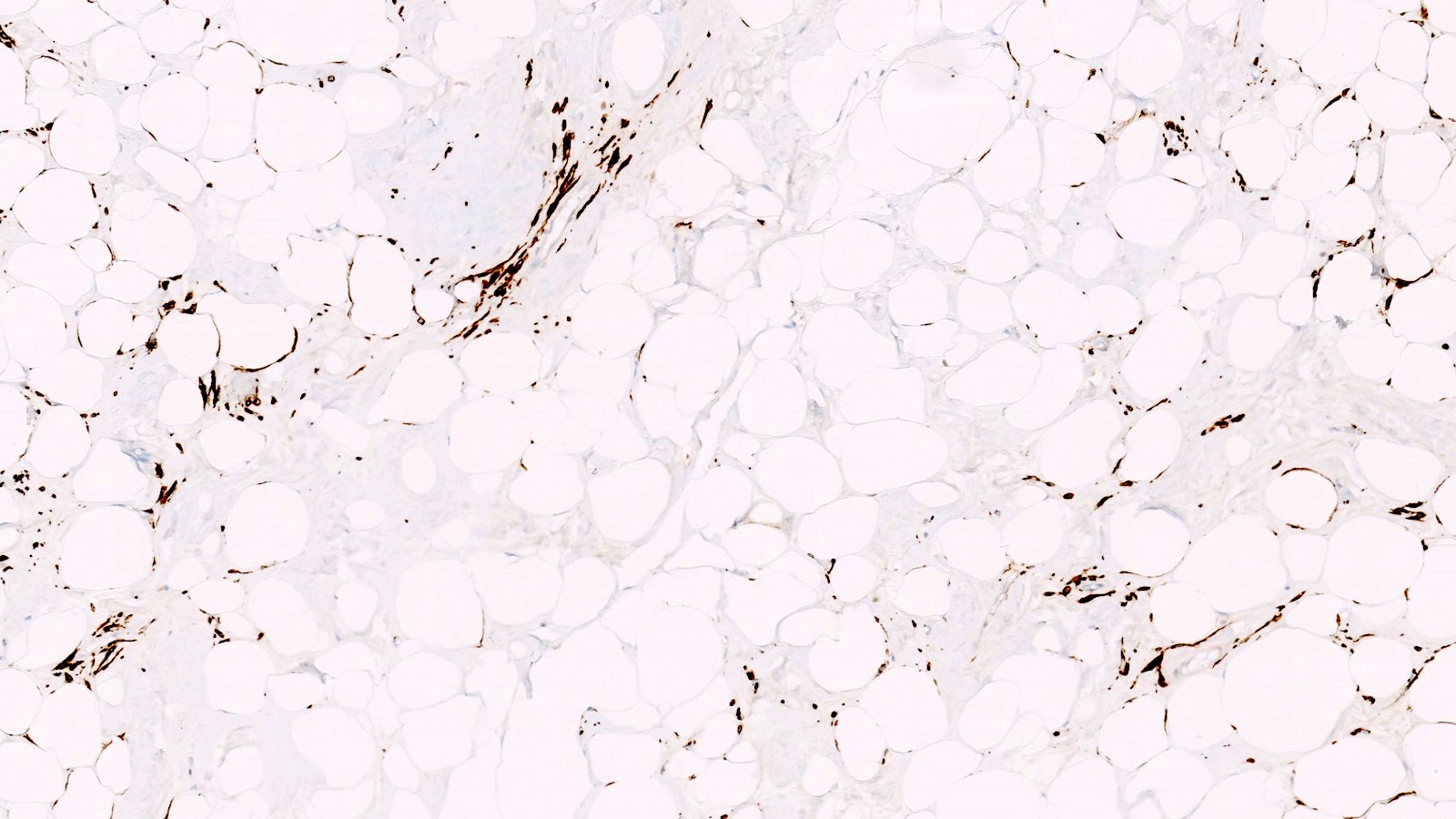
Lipoleiomyoma
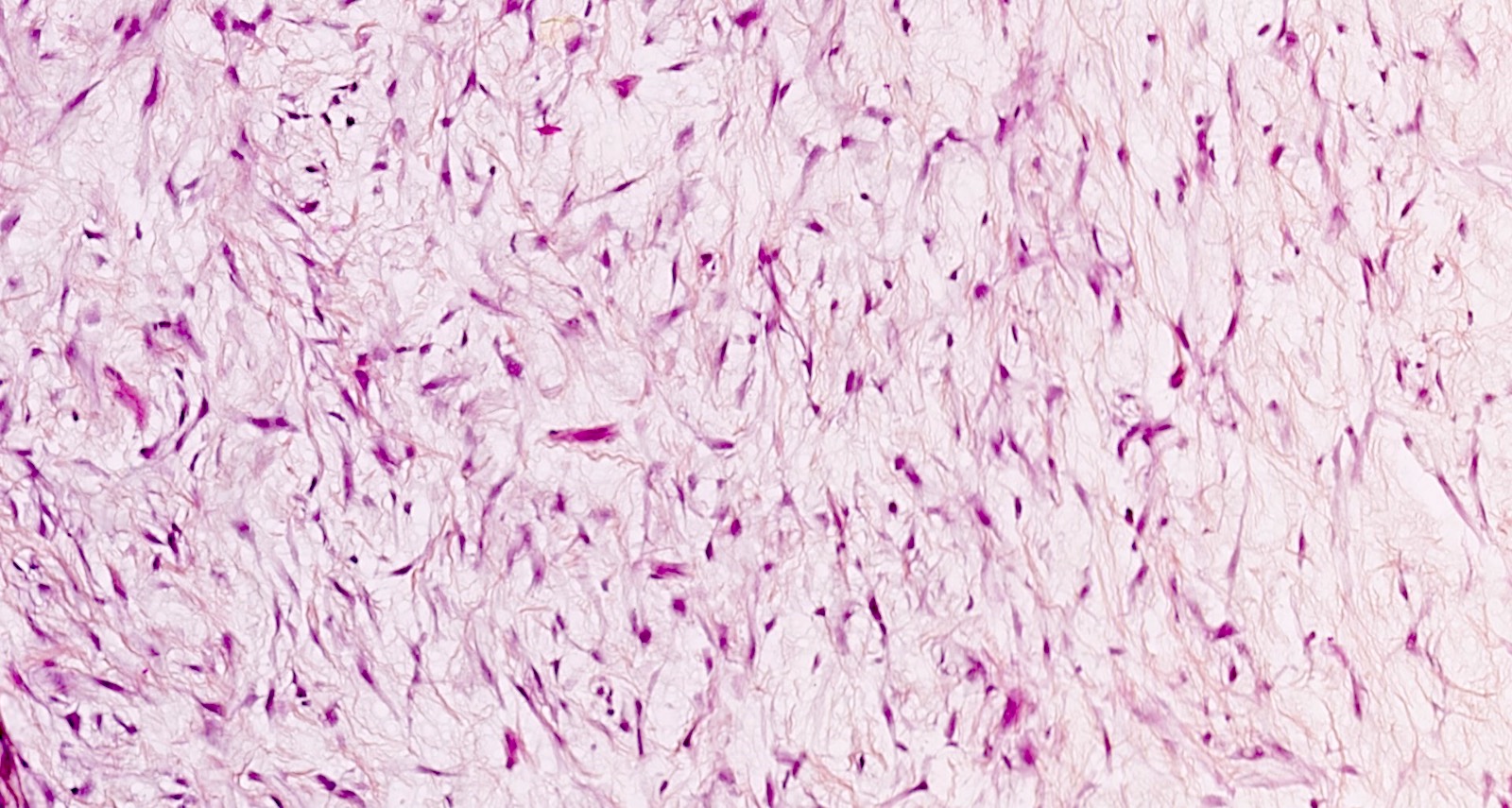
Myxoid leiomyoma

Fumarate hydratase deficient (FH-d) leiomyoma
Benign metastasizing leiomyoma

Cystic hydropic leiomyoma
How about a new subtype of 'leiomyolipoma'?😀🤔😅
#grosspath #pathtwitter #gynpath #pathology #pathoutpic"
Contributed by @Andrew_Fltv on Twitter (see original post here)">
 Compare with another remarkable case @kis_lorand
How about a new subtype of 'leiomyolipoma'?😀🤔😅
#grosspath #pathtwitter #gynpath #pathology #pathoutpic"
Contributed by @Andrew_Fltv on Twitter (see original post here)">
 Compare with another remarkable case @kis_lorand
How about a new subtype of 'leiomyolipoma'?😀🤔😅
#grosspath #pathtwitter #gynpath #pathology #pathoutpic"
Contributed by @Andrew_Fltv on Twitter (see original post here)">
Lipoleiomyoma
Images hosted on other servers:

Large intrauterine mass

Heterogeneous mass with irregular borders
Images hosted on other servers:

Uterus with dilated cervix

Large exophytic intrauterine mass

Large mass
Contributed by Ashley Monsrud, M.D.
Formalin fixed, intracavitary leiomyosarcoma
Images hosted on other servers:

Large mass
Contributed by Ashley Monsrud, M.D. and Paulette Mhawech-Fauceglia, M.D.
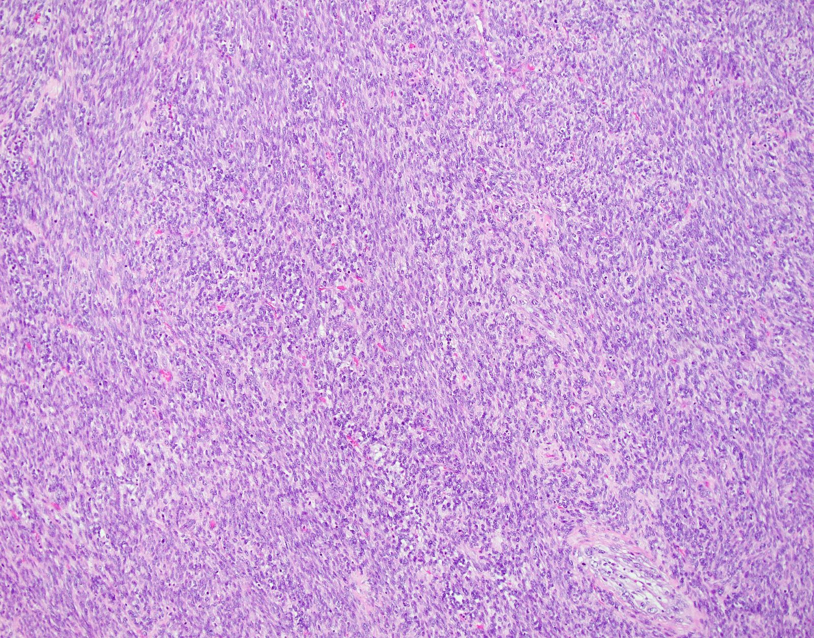

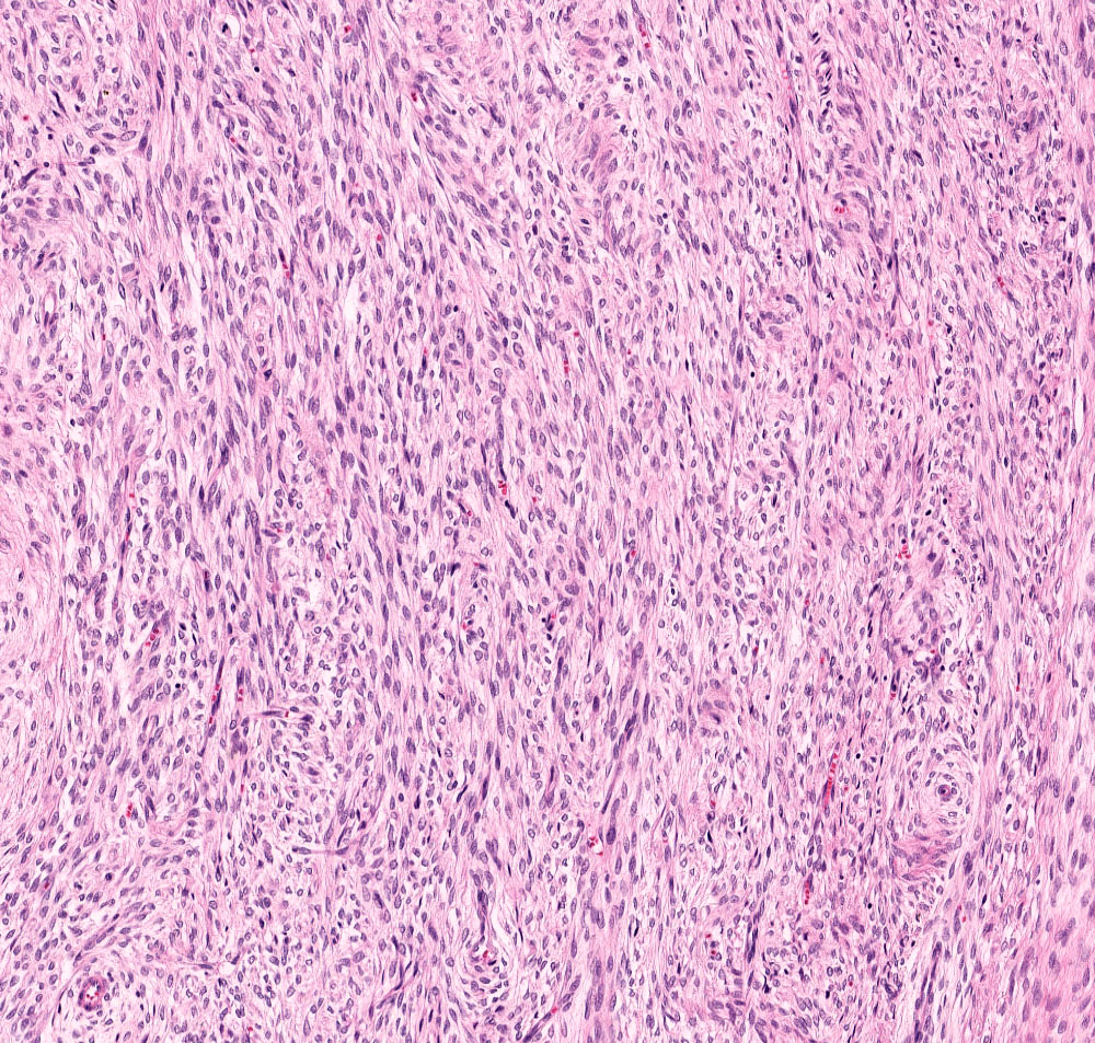
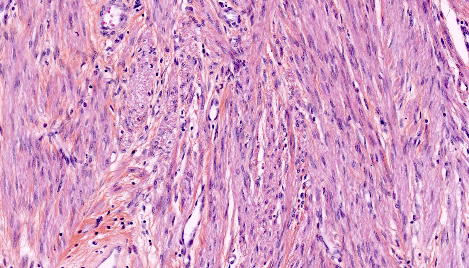
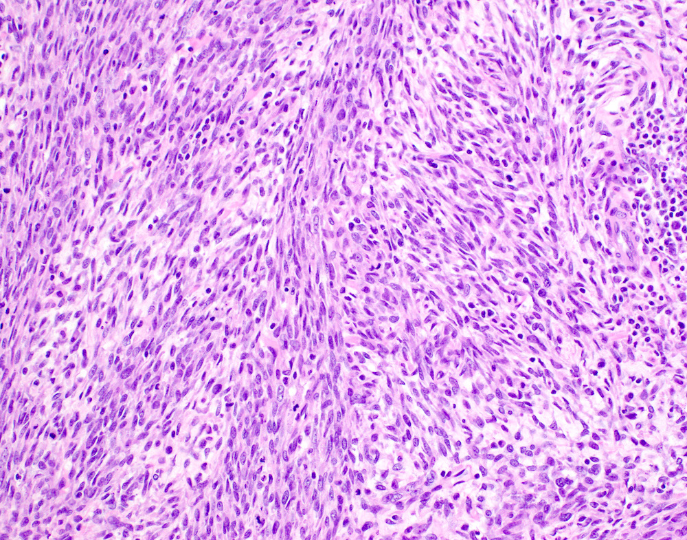
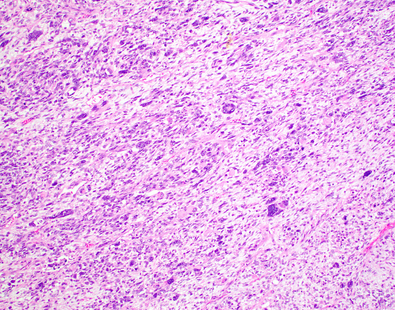
Spindle cell leiomyosarcoma
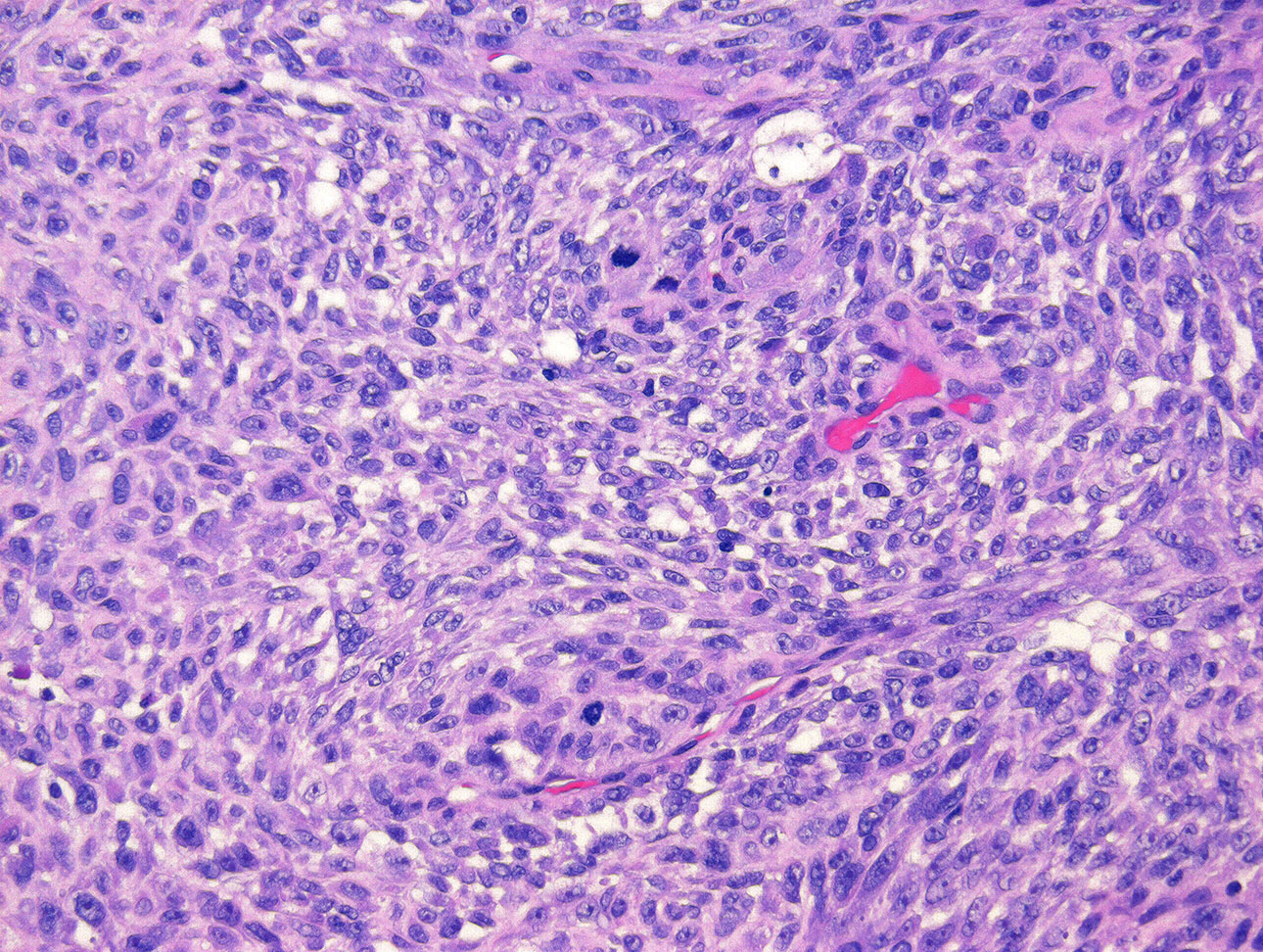
Tumor cell necrosis
Atypical mitotic figures
Hypercellular tumor
Nuclear atypia

Numerous mitoses
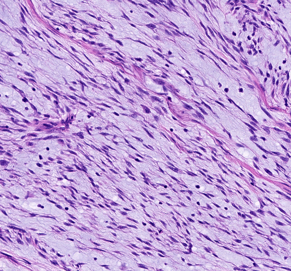
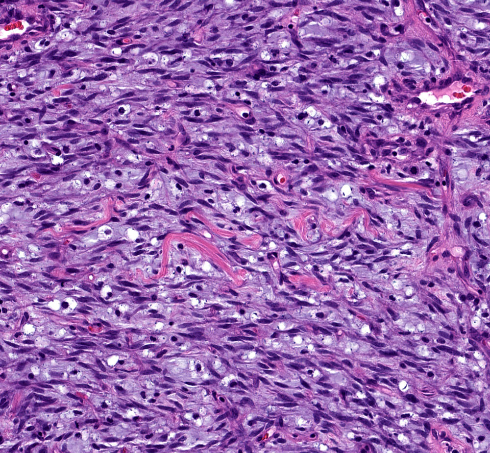
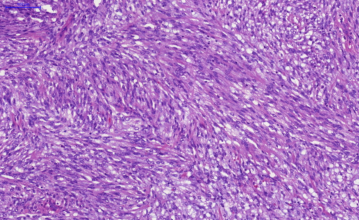
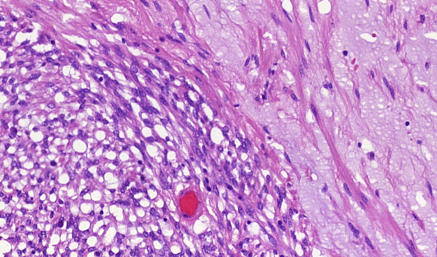
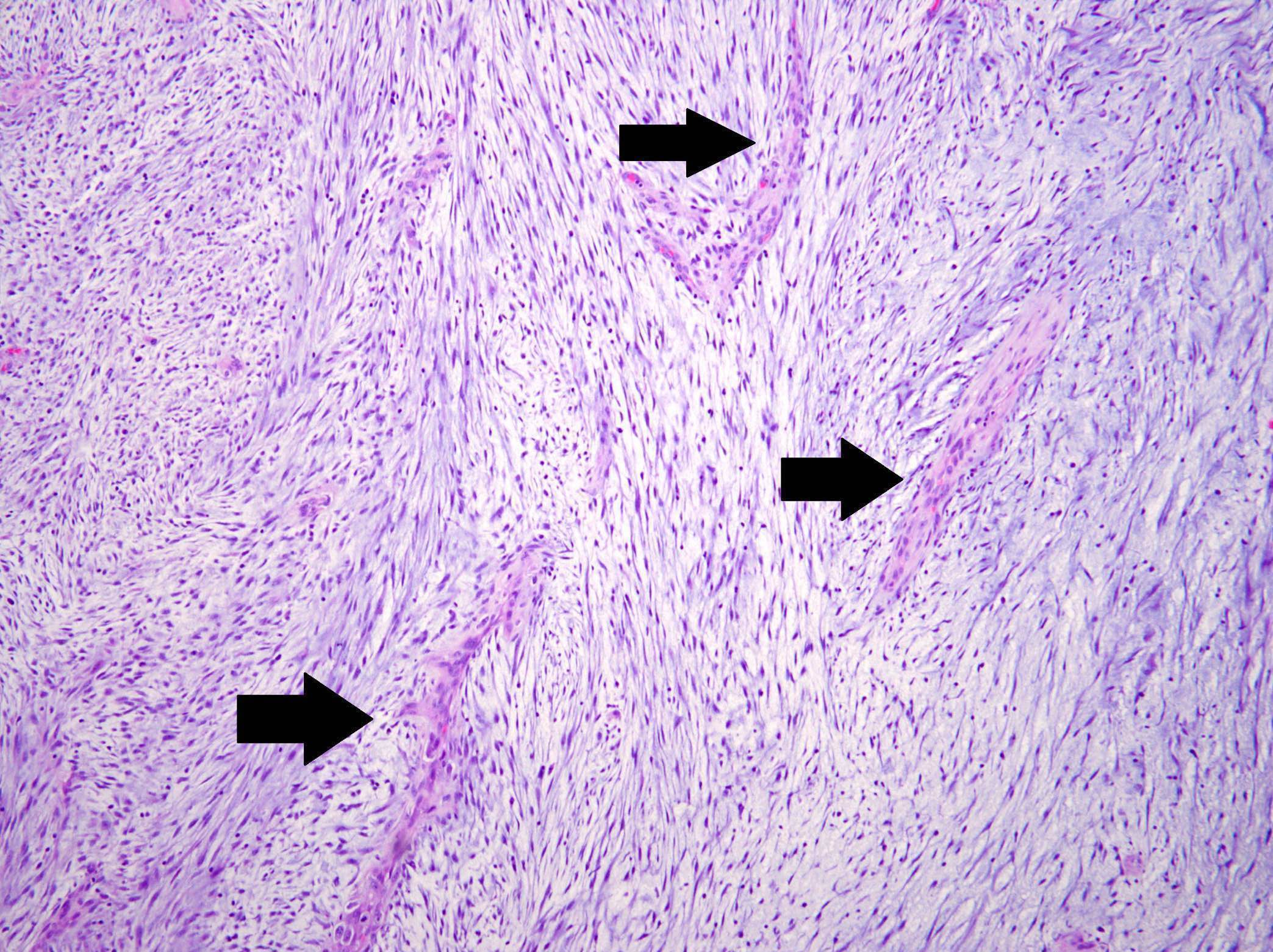
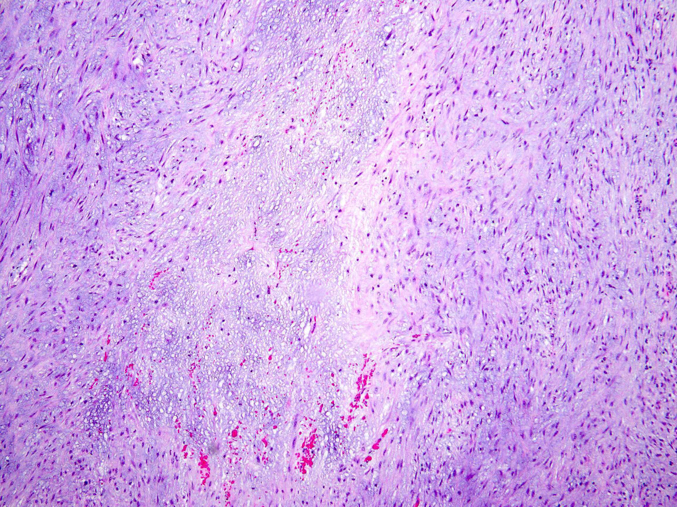
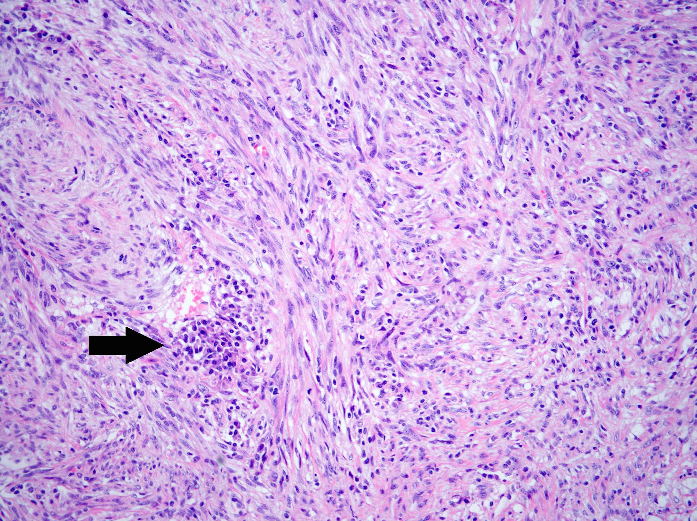
Myxoid leiomyosarcoma, infiltrating border

Myxoid leiomyosarcoma
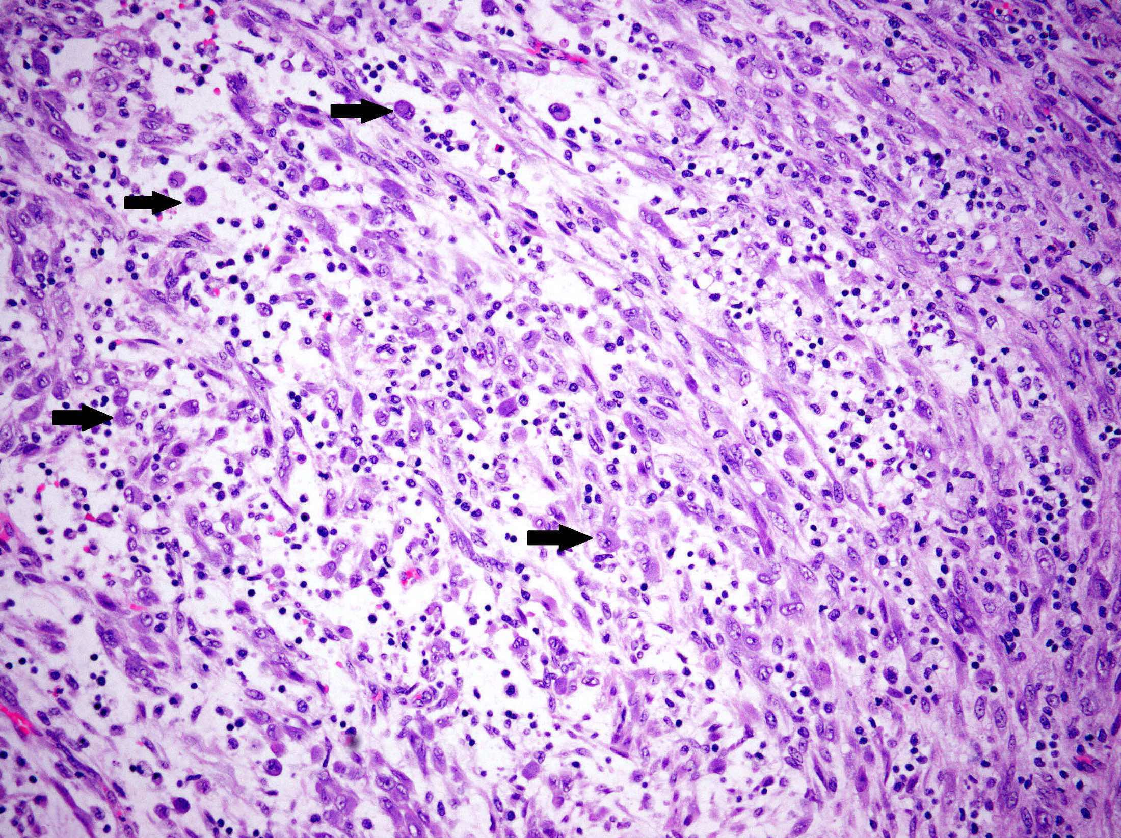
Mitotic figures in myxoid leiomyosarcoma
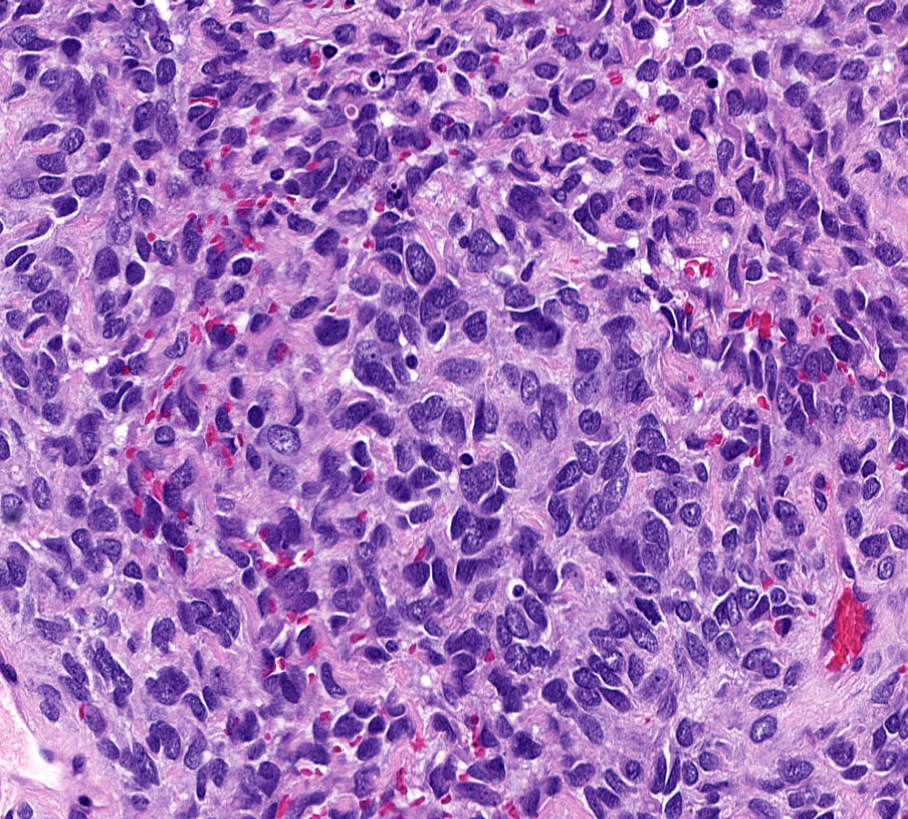
Epithelioid leiomyosarcoma
Epithelioid leiomyosarcoma
Infiltration of adjacent myometrium
Solid growth pattern

Nuclear atypia
Epithelioid subtype
Images hosted on other servers:

Somatic mutational landscape
Review of uterine leiomyosarcoma
Images hosted on other servers:


T2 and T1 weighted images

MRI of low grade endometrial stromal sarcoma
Contributed by Elizabeth Kertowidjojo, M.D., Ph.D., M.P.H. and Ayse Ayhan, M.D., Ph.D.
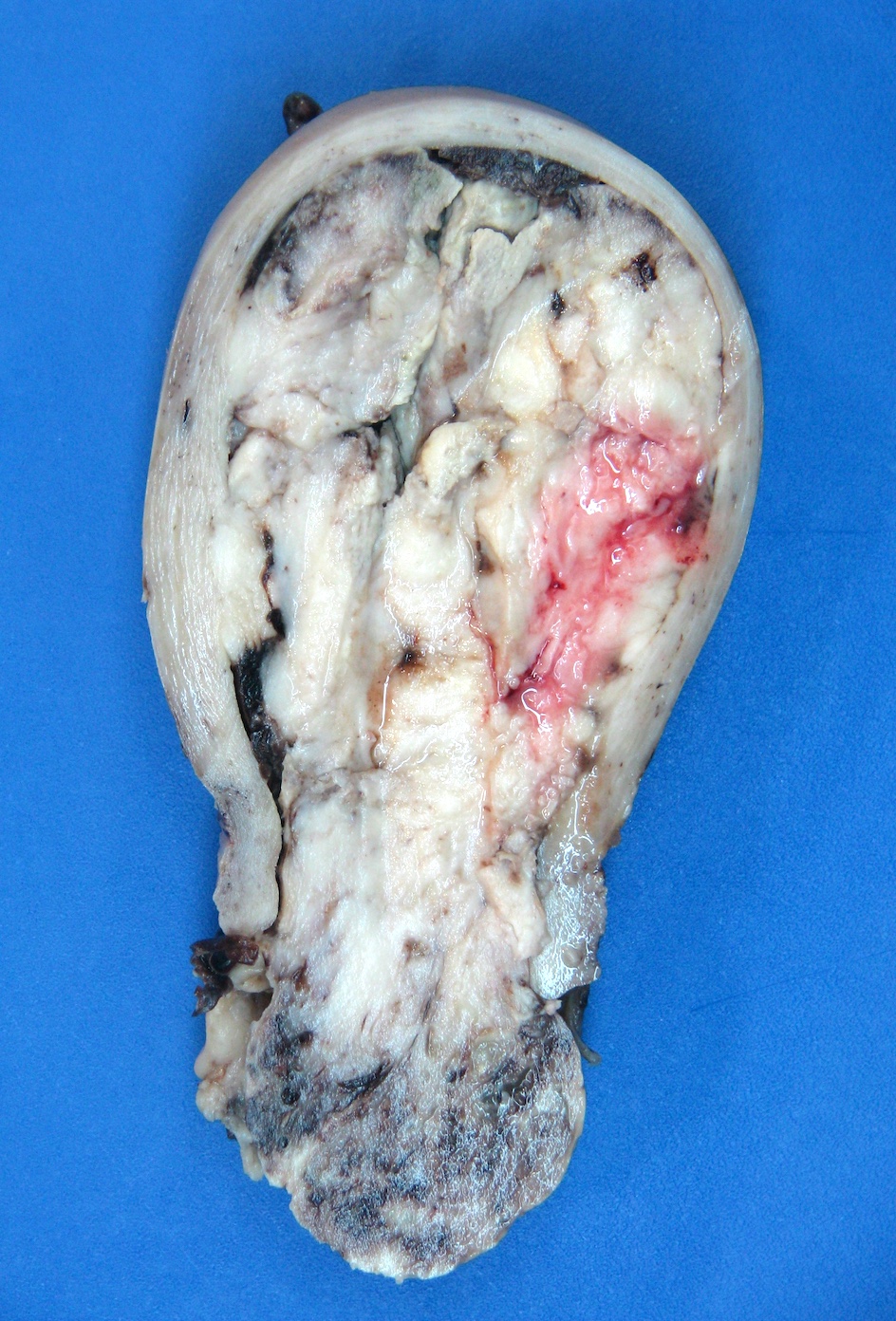
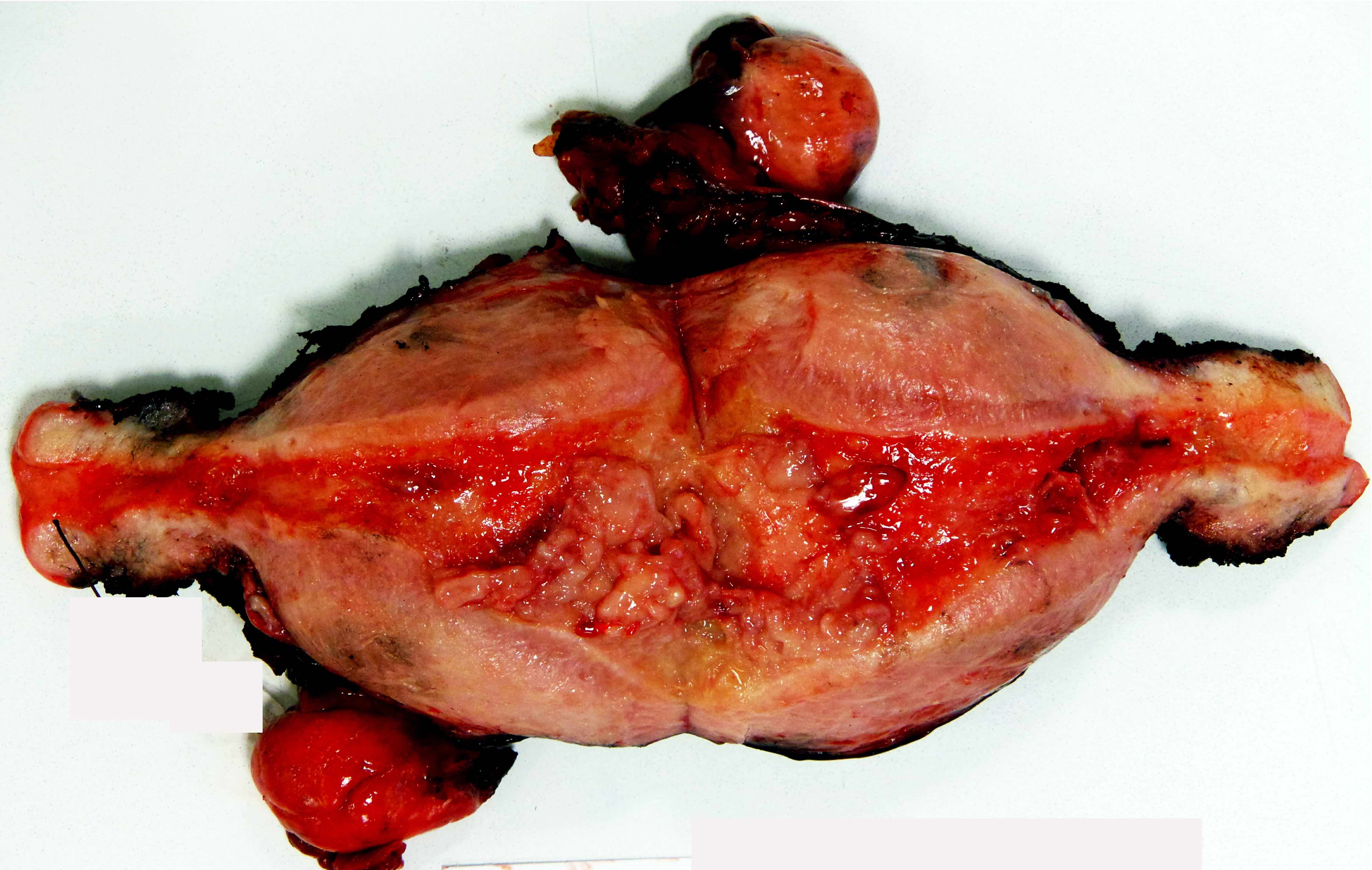
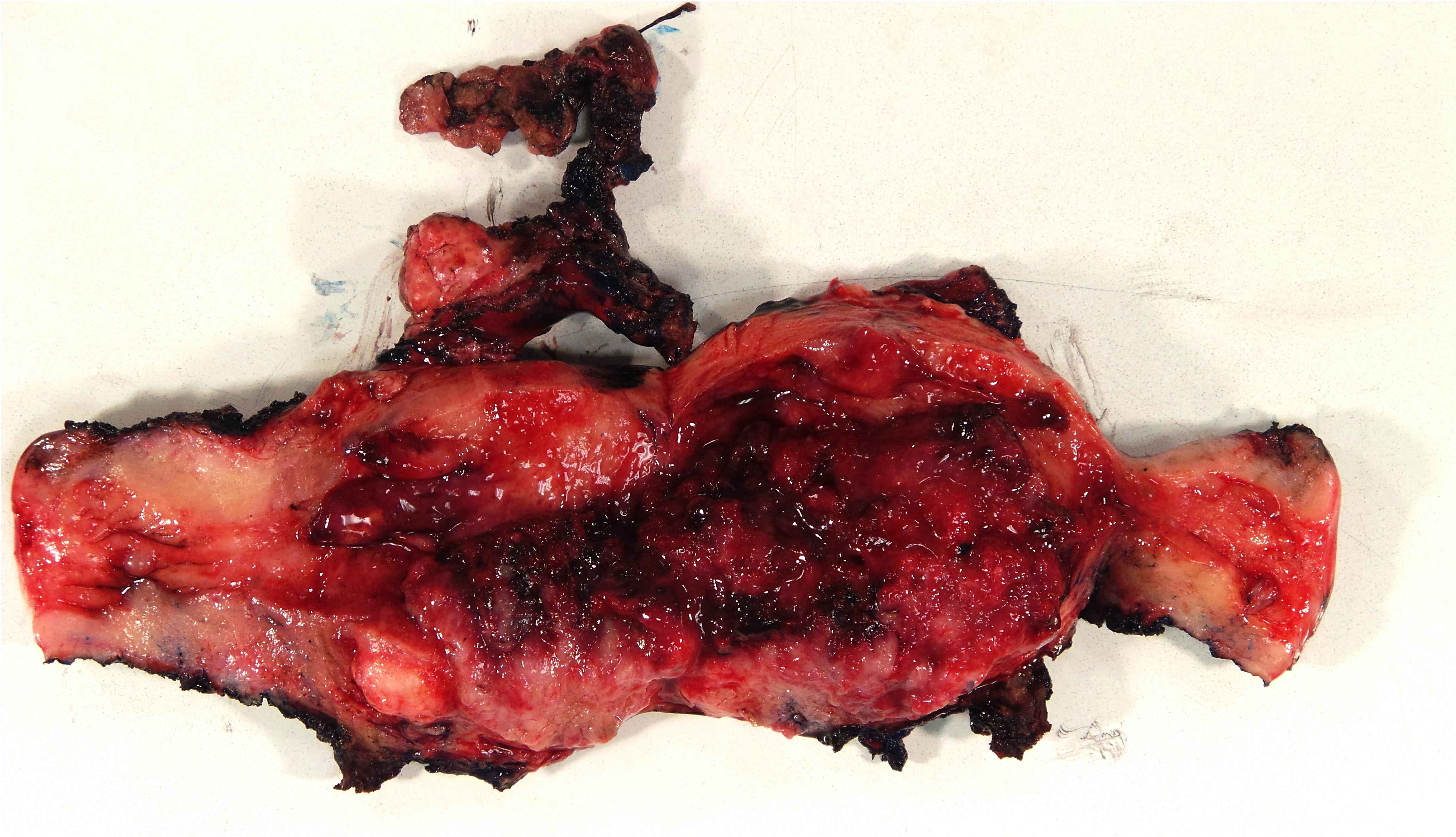
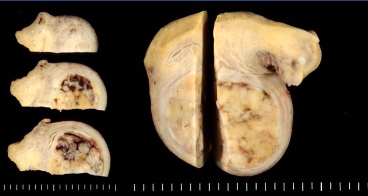
Tan-yellow uterine mass
Fleshy lobulated mass
Multinodular, tan to yellow mass
Images hosted on other servers:

Yellow-brown
tumor mass and
additional infiltrating
tumor nodules

Macroscopically,
sarcoma resembles
uterine leiomyomas

Yellow tumor nodule with necrosis
Contributed by Elizabeth Kertowidjojo, M.D., Ph.D., M.P.H. and Ayse Ayhan, M.D., Ph.D.
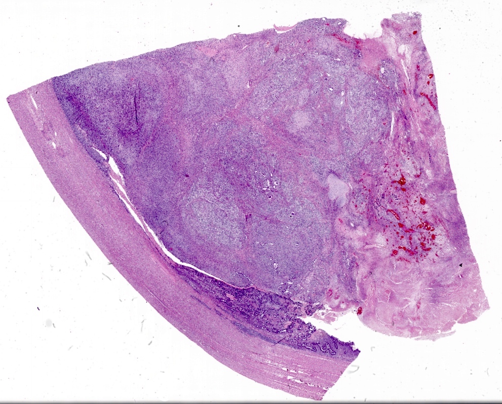
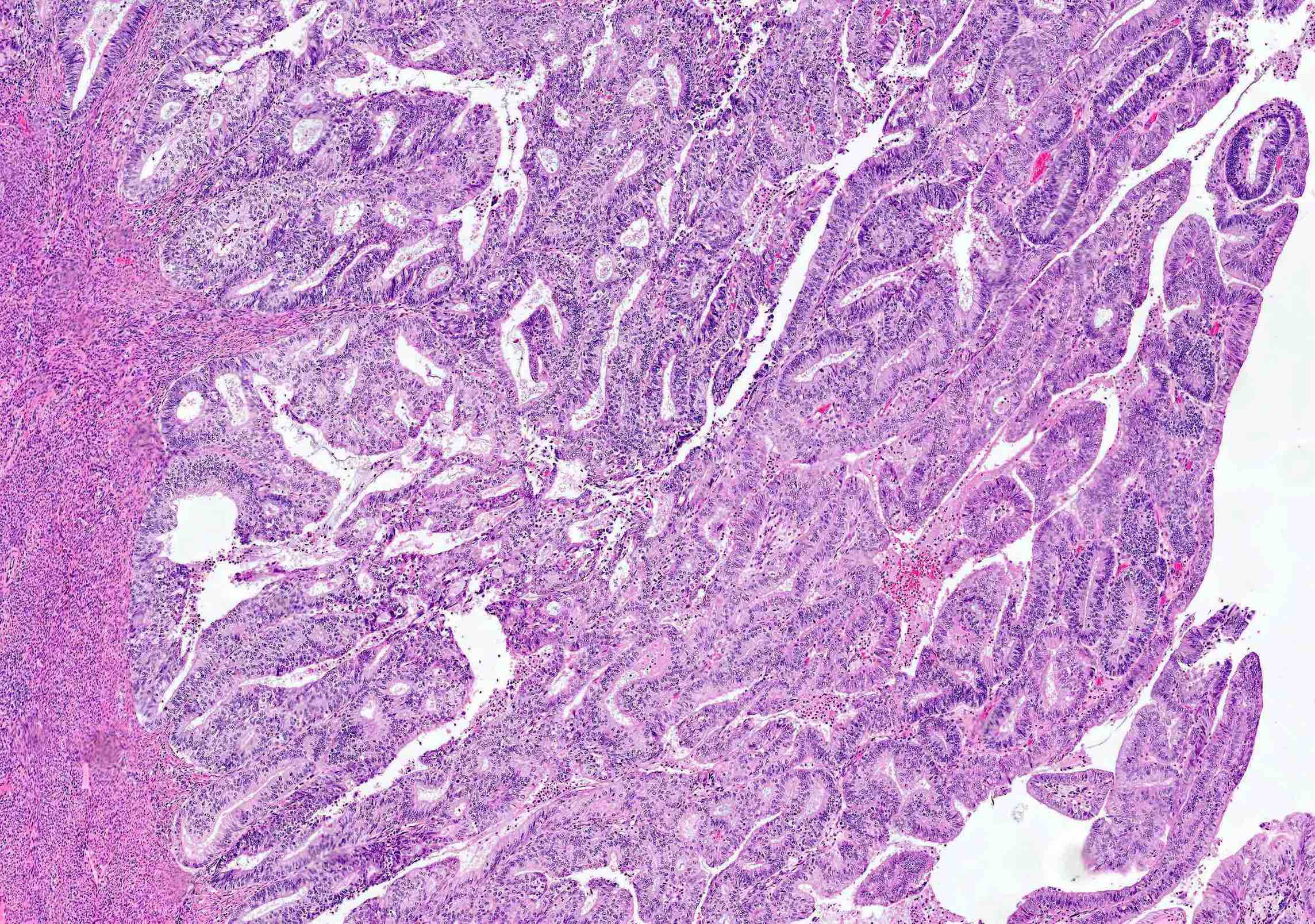
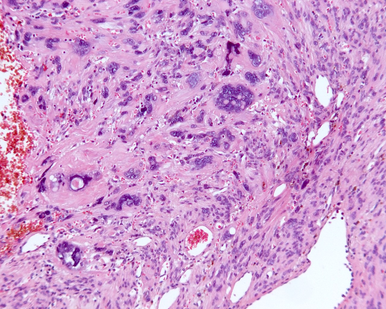
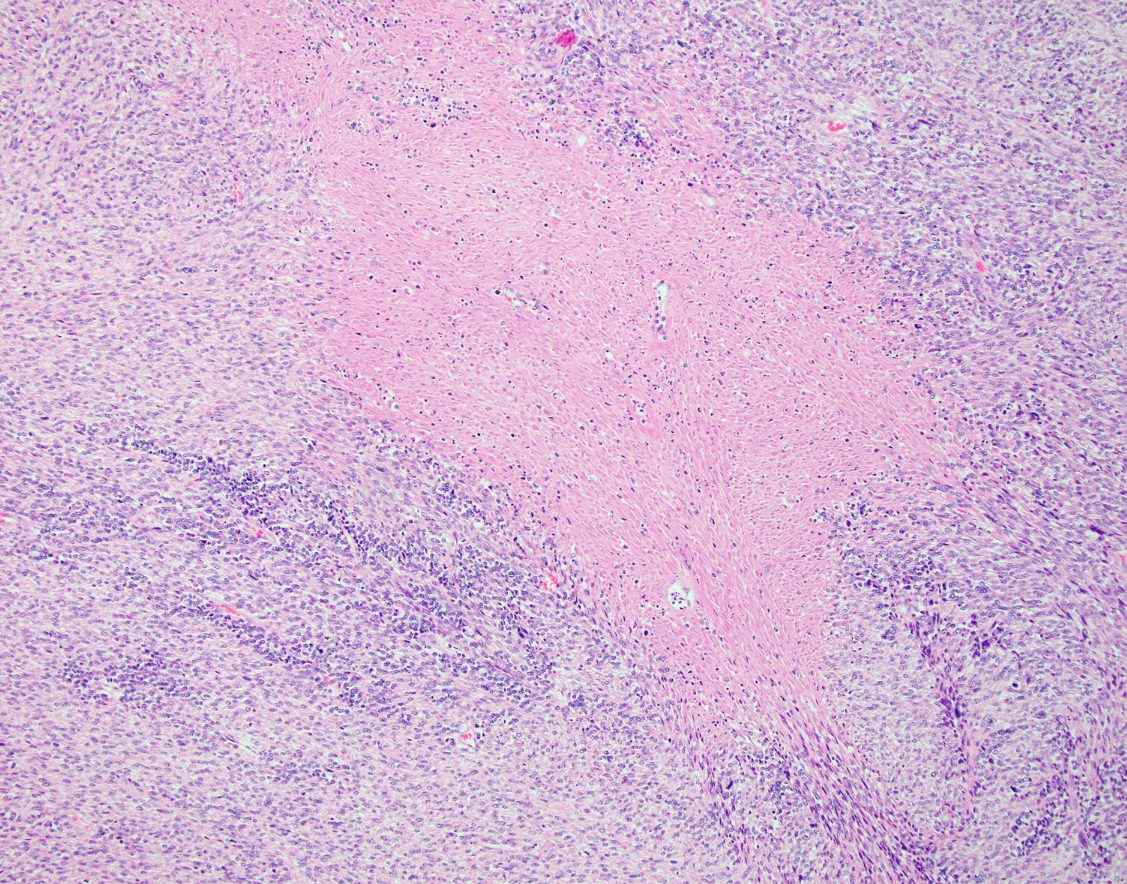
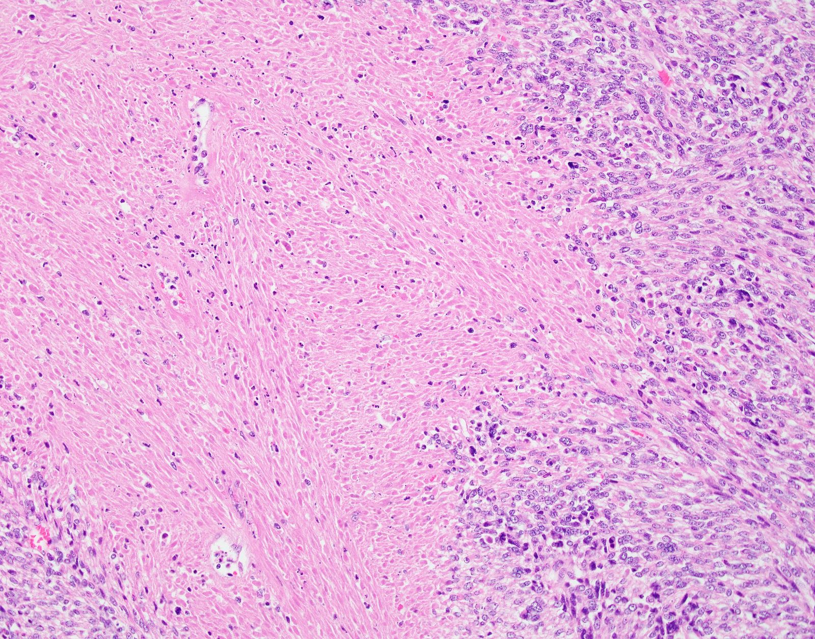
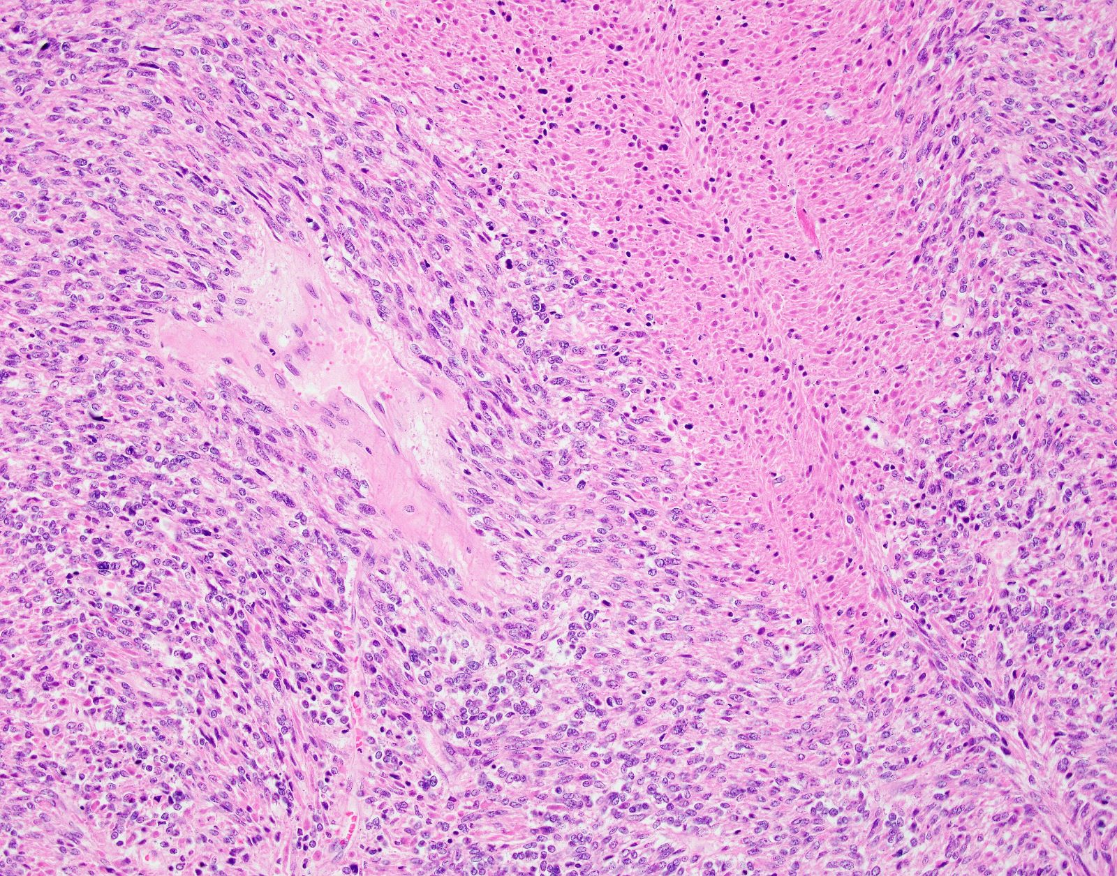
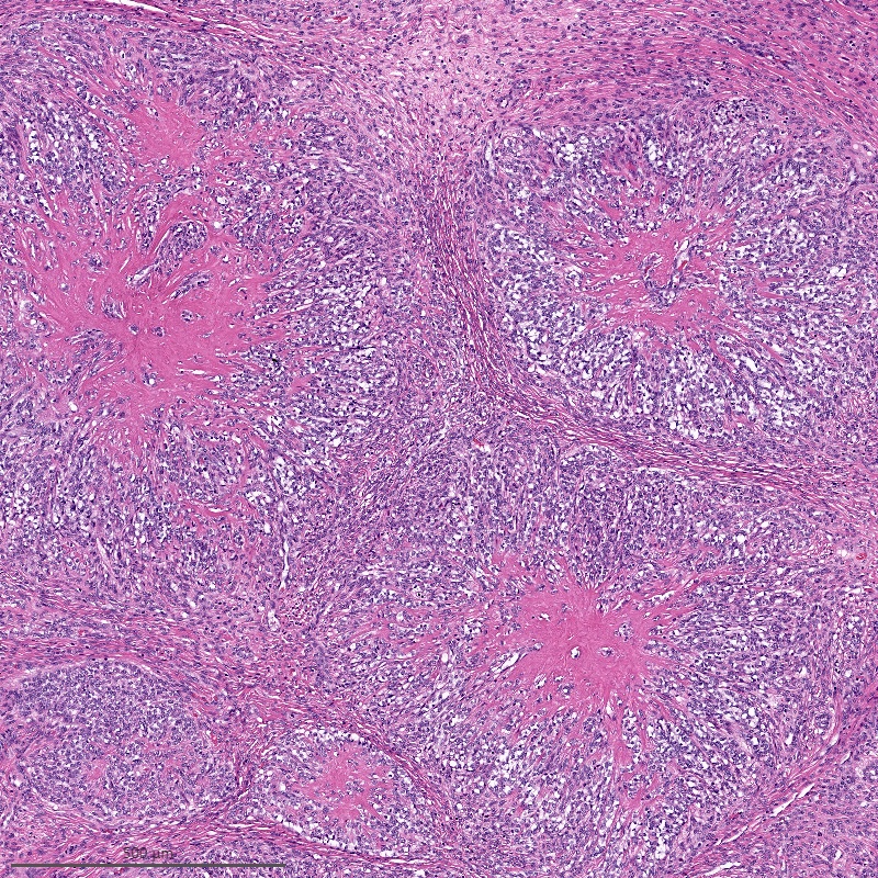
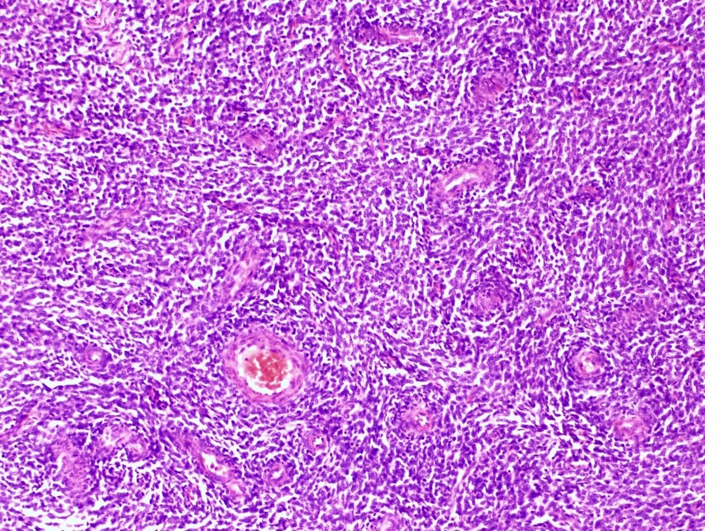
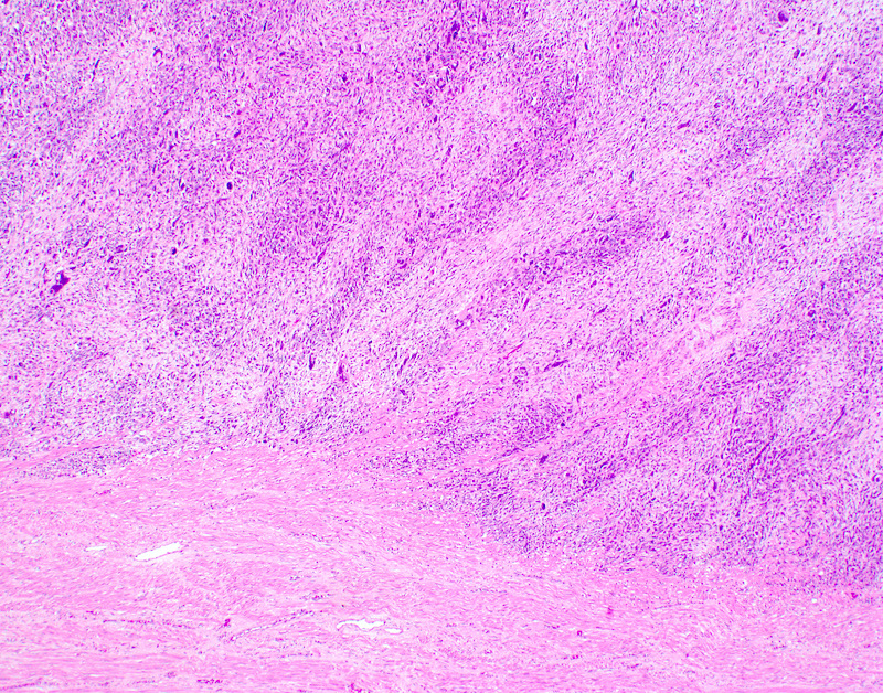
Tongue-like invasion
Monotonous tumor cells
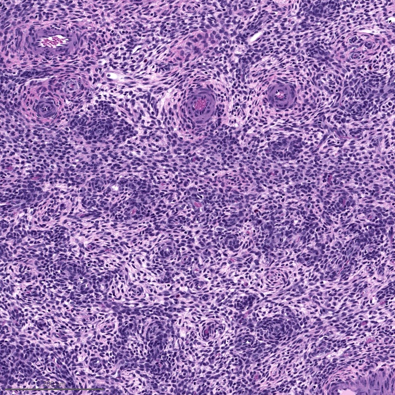
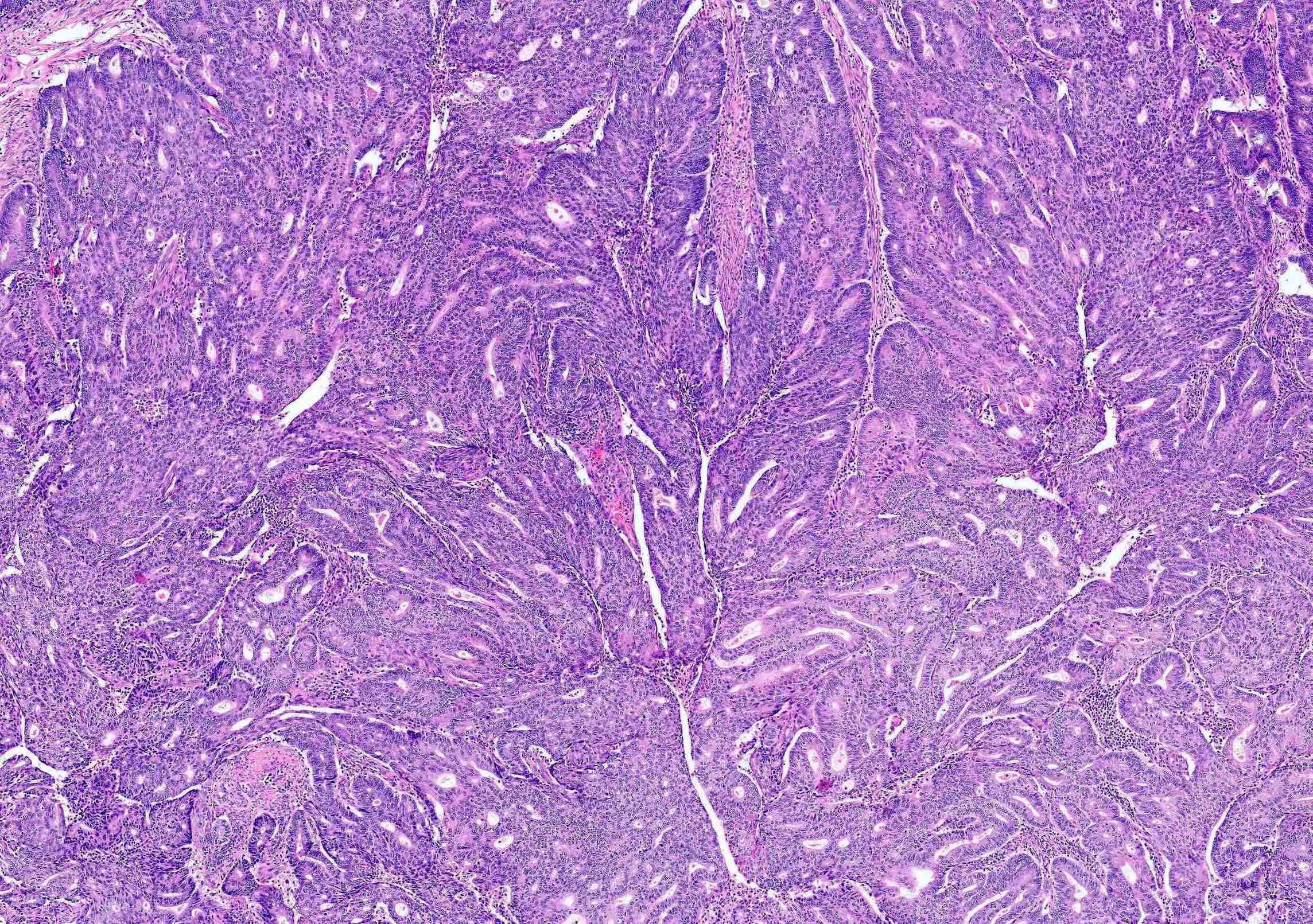
Whorling around vasculature
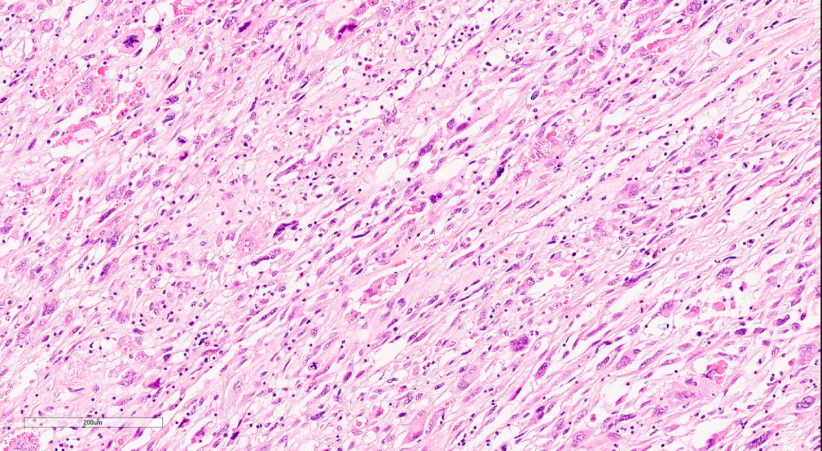
Smooth muscle differentiation
Sex cord-like growth
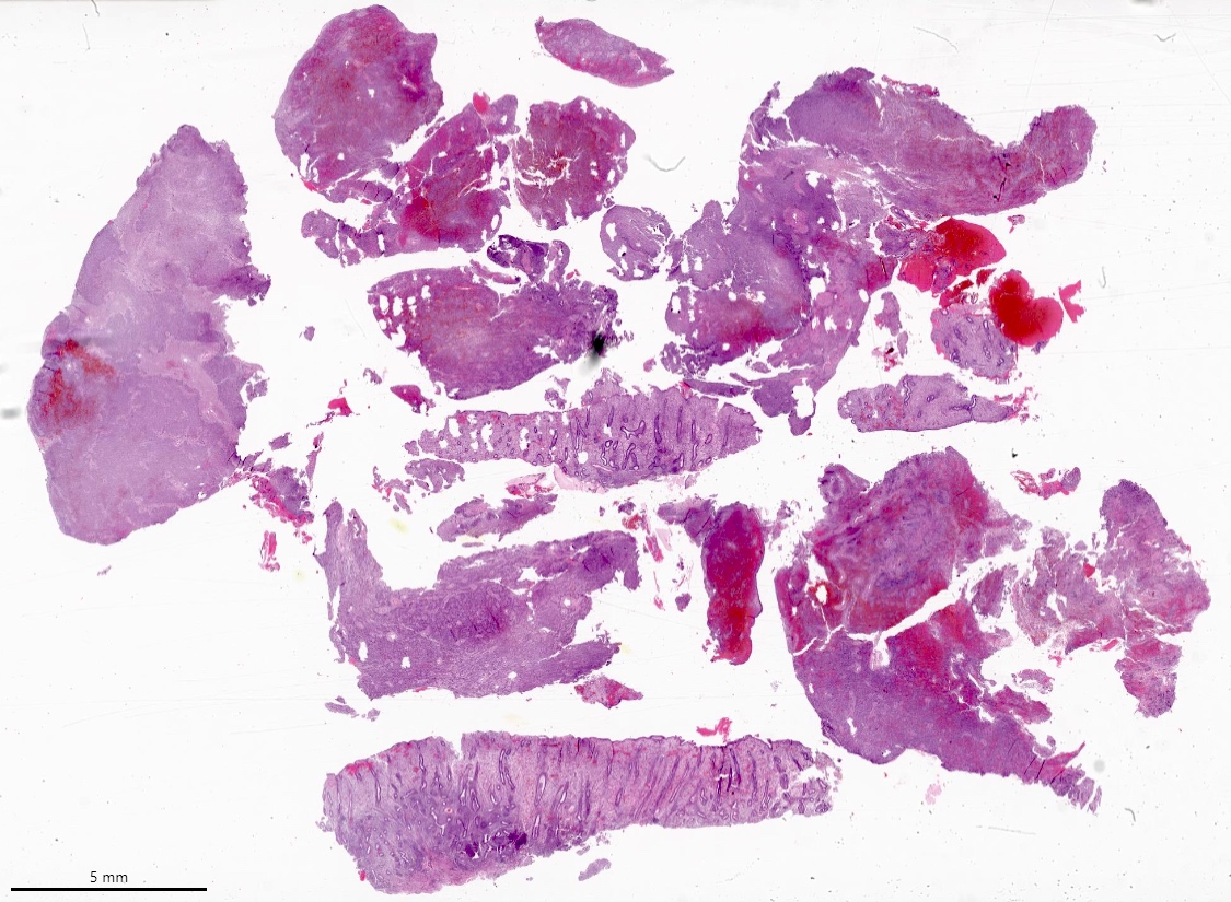

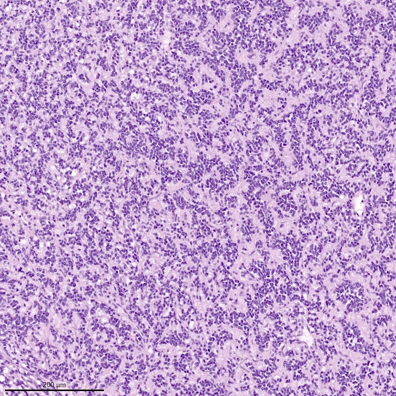
Endometrial curetting
Mild nuclear atypia
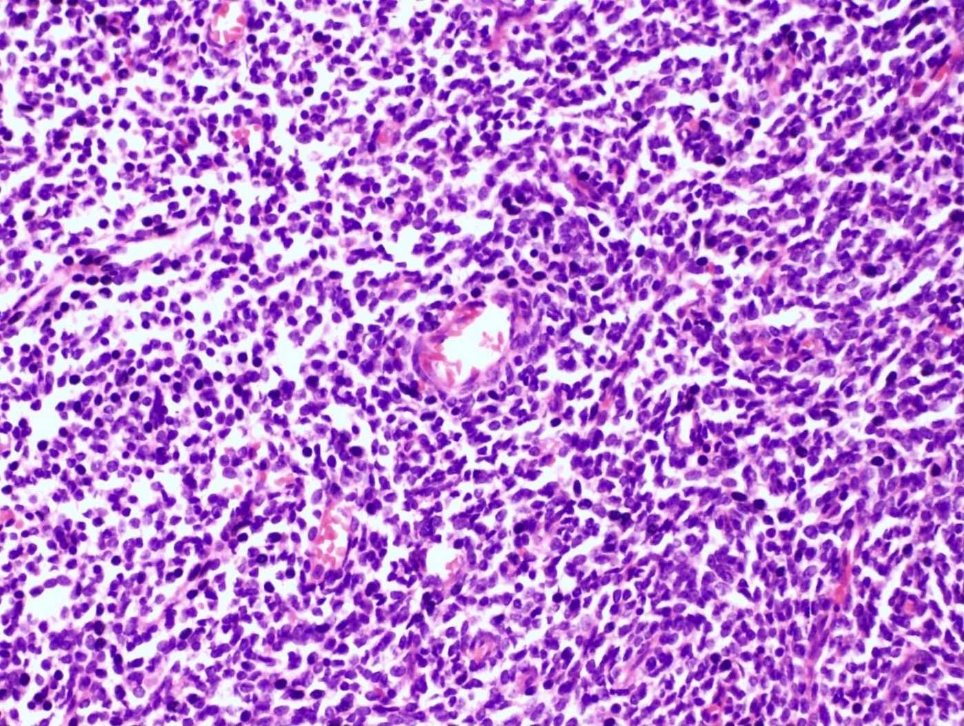
Small blue cells, scant cytoplasm
Unusual features in endometrial stromal sarcoma
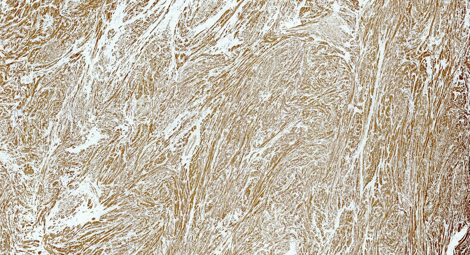
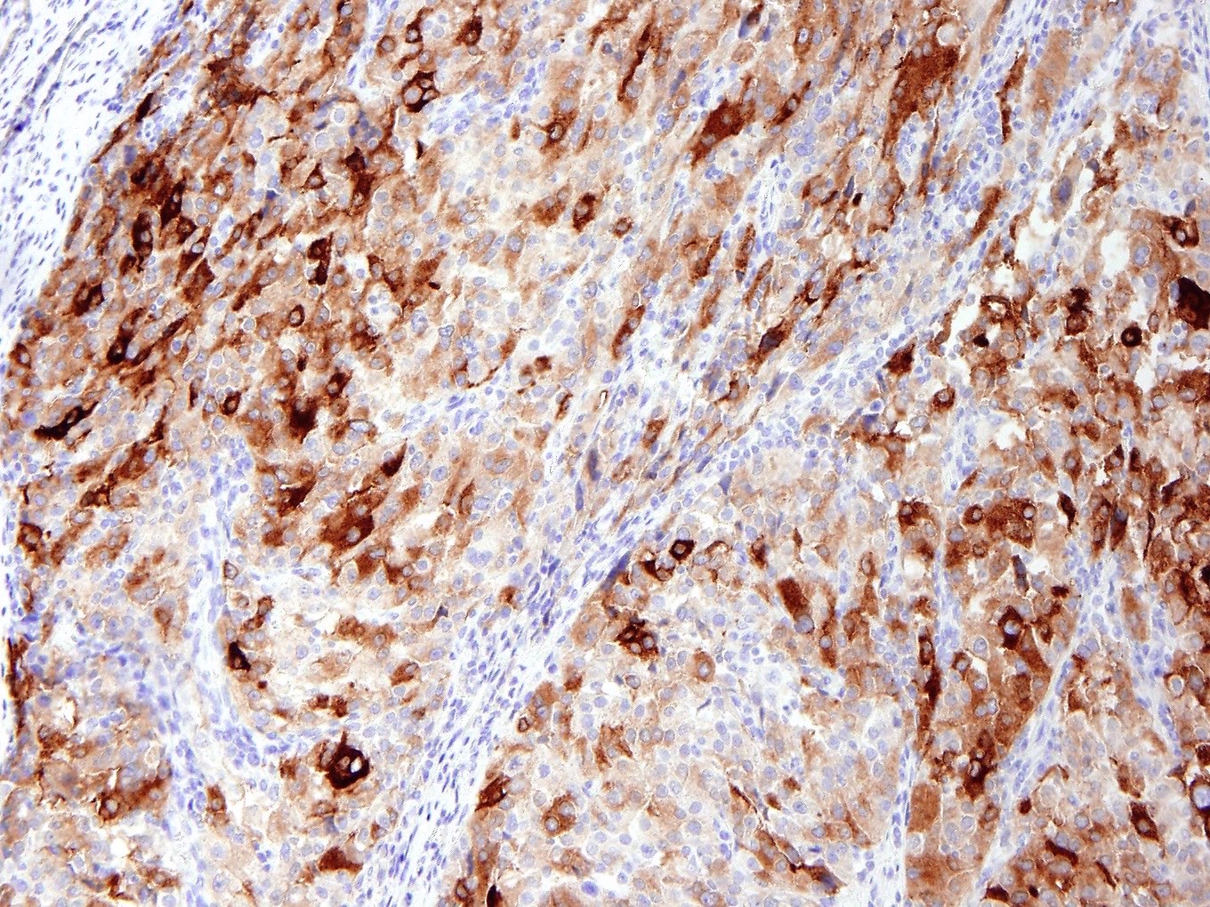
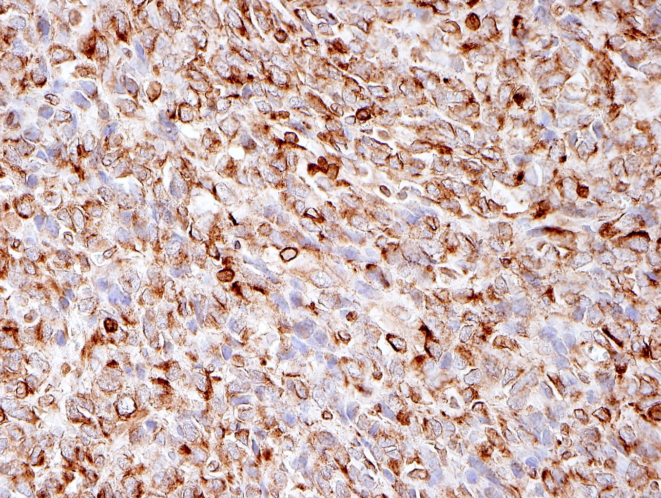
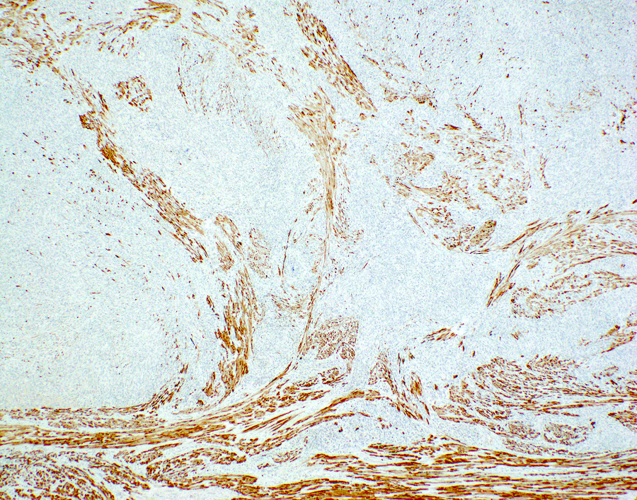
Desmin
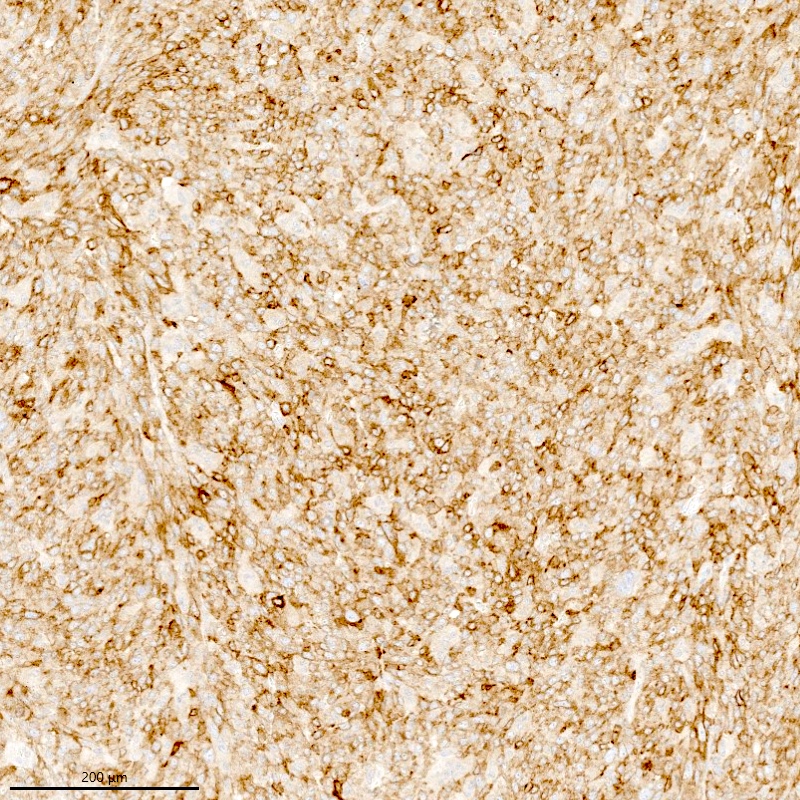
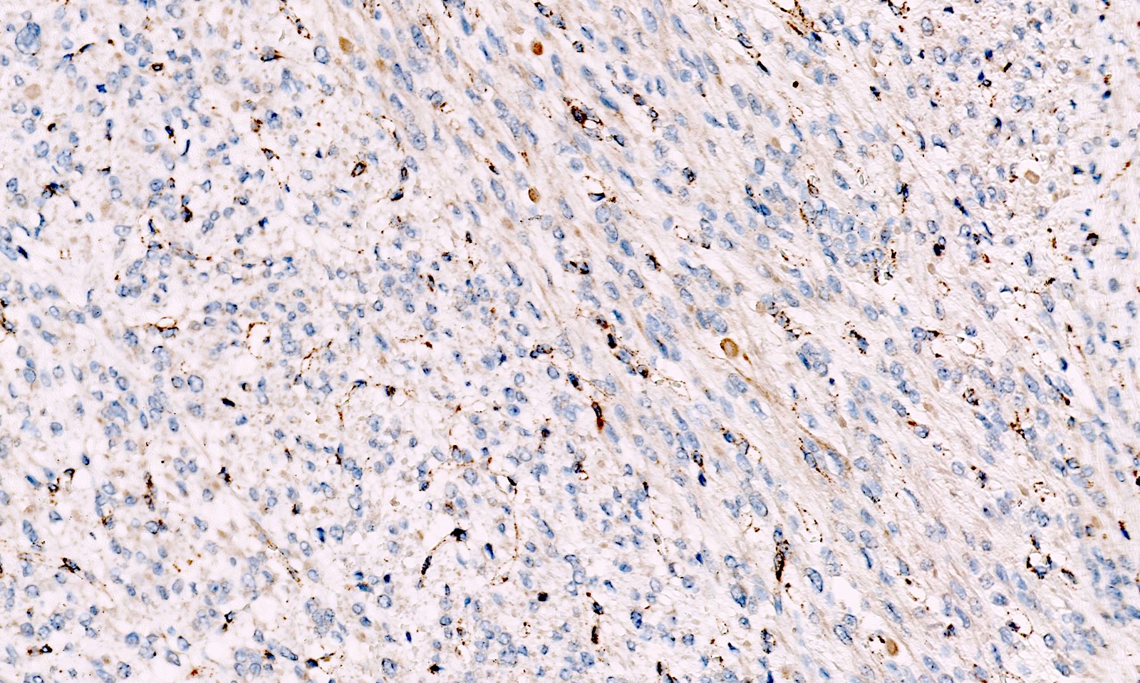
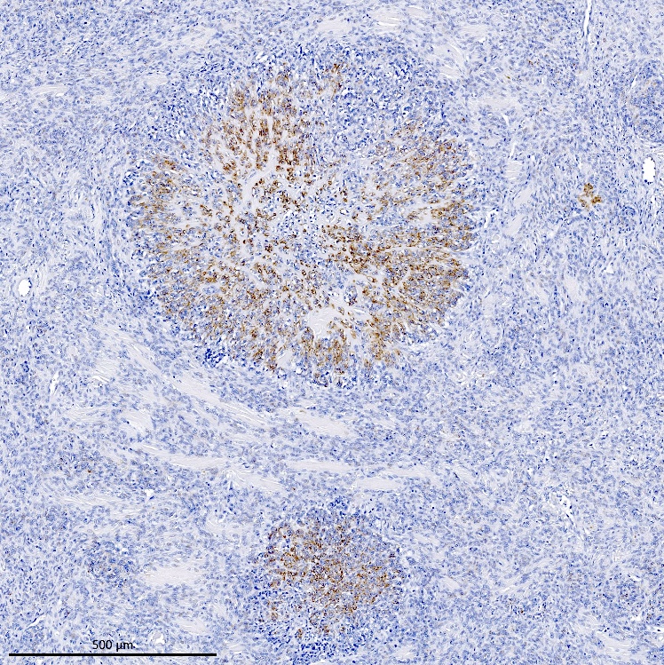
Positive for CD10
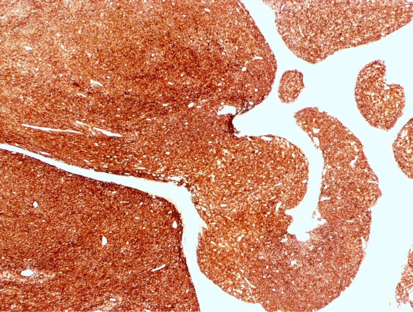
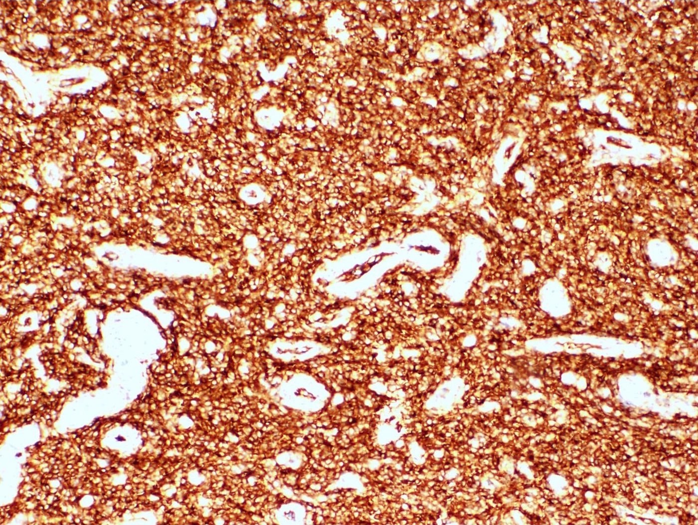
CD10
Estrogen receptor (alpha isoform)
Progesterone receptor
Contributed by Elizabeth Kertowidjojo, M.D., Ph.D., M.P.H.
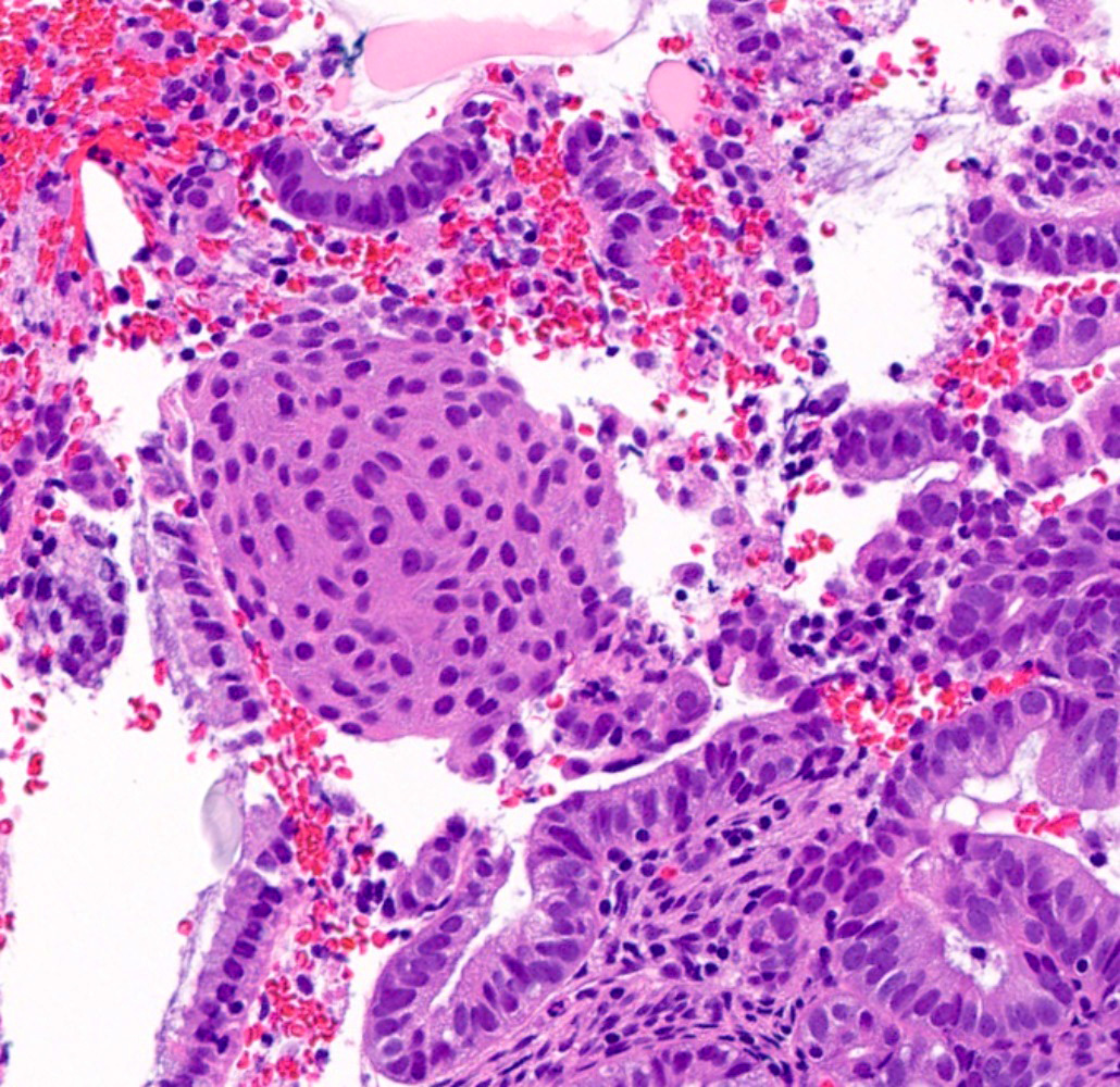
Lung fine needle aspiration
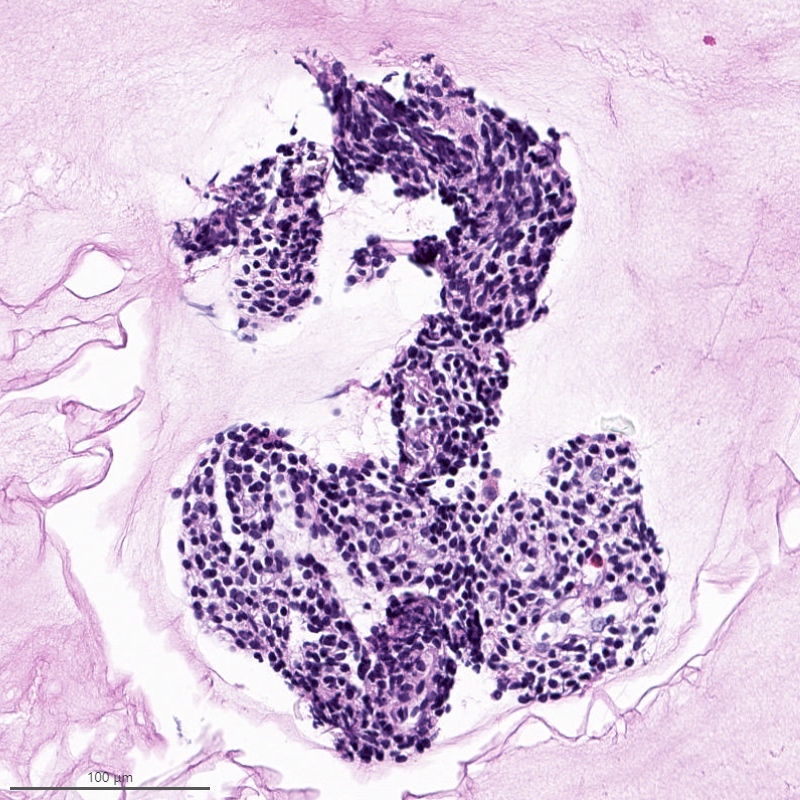
Lung fine needle aspiration cell block
Images hosted on other servers:

JAZF1-SUZ12 dual fusion probe

MBTD1-CXorf67 dual fusion probe
Endometrial stromal sarcoma
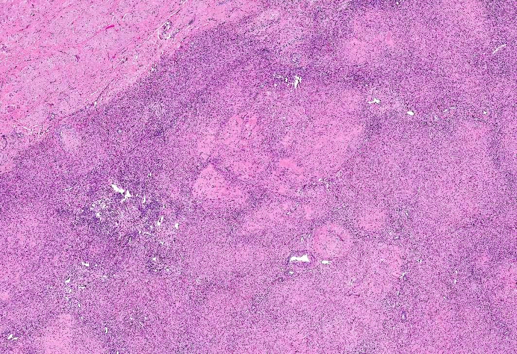
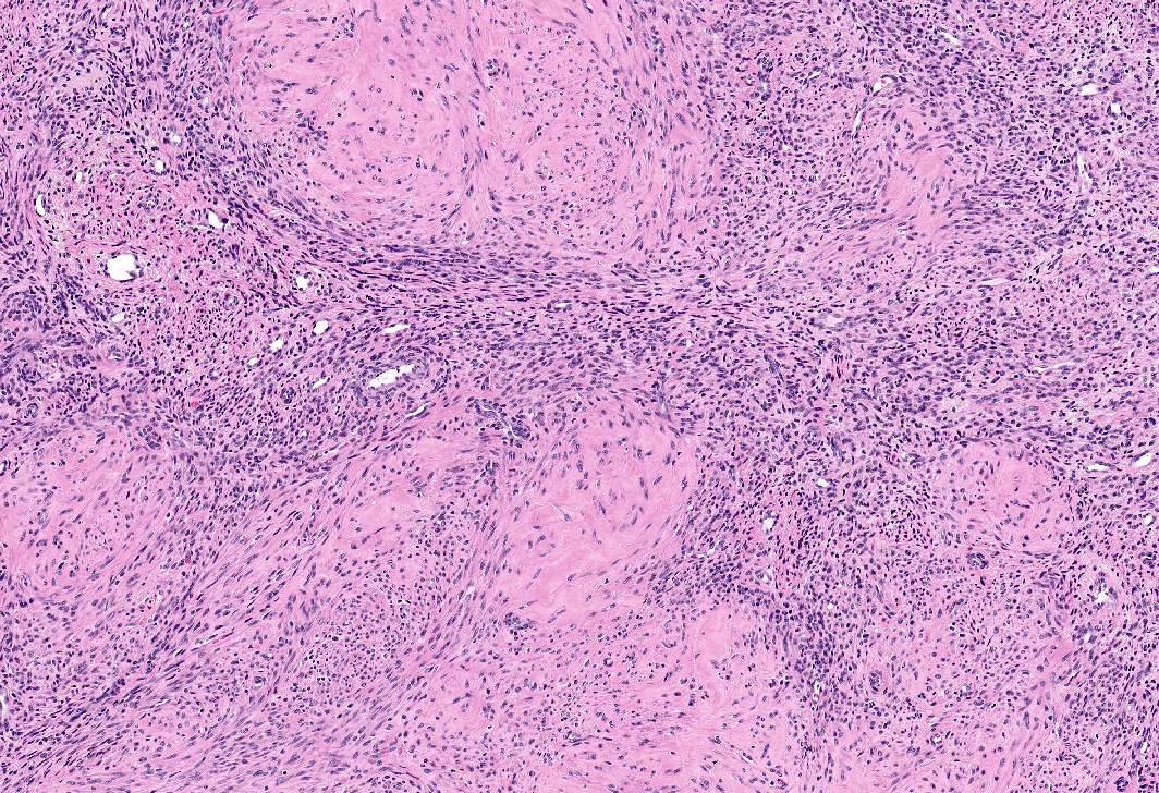
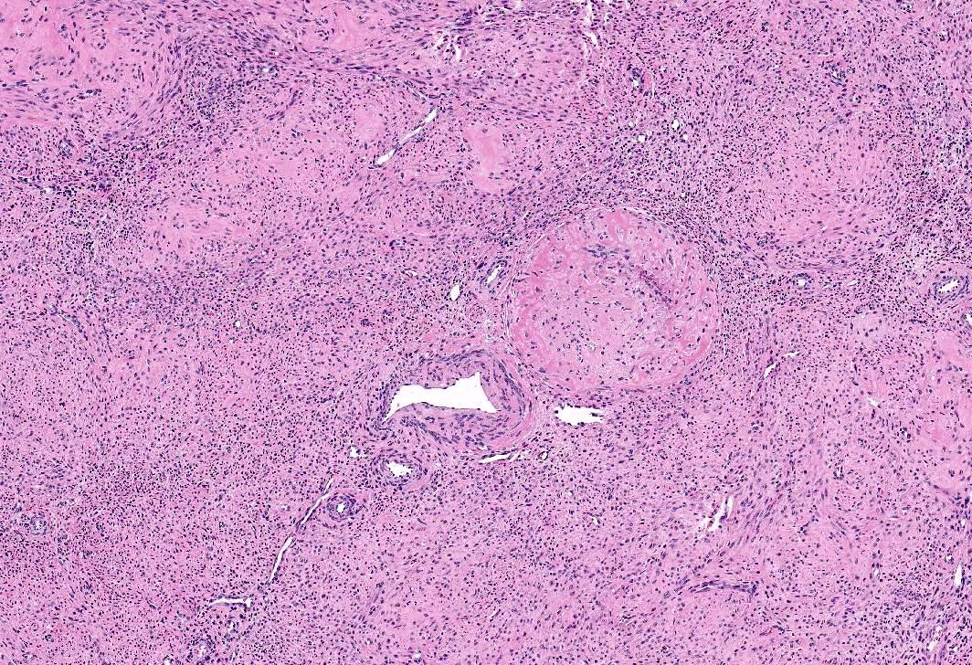
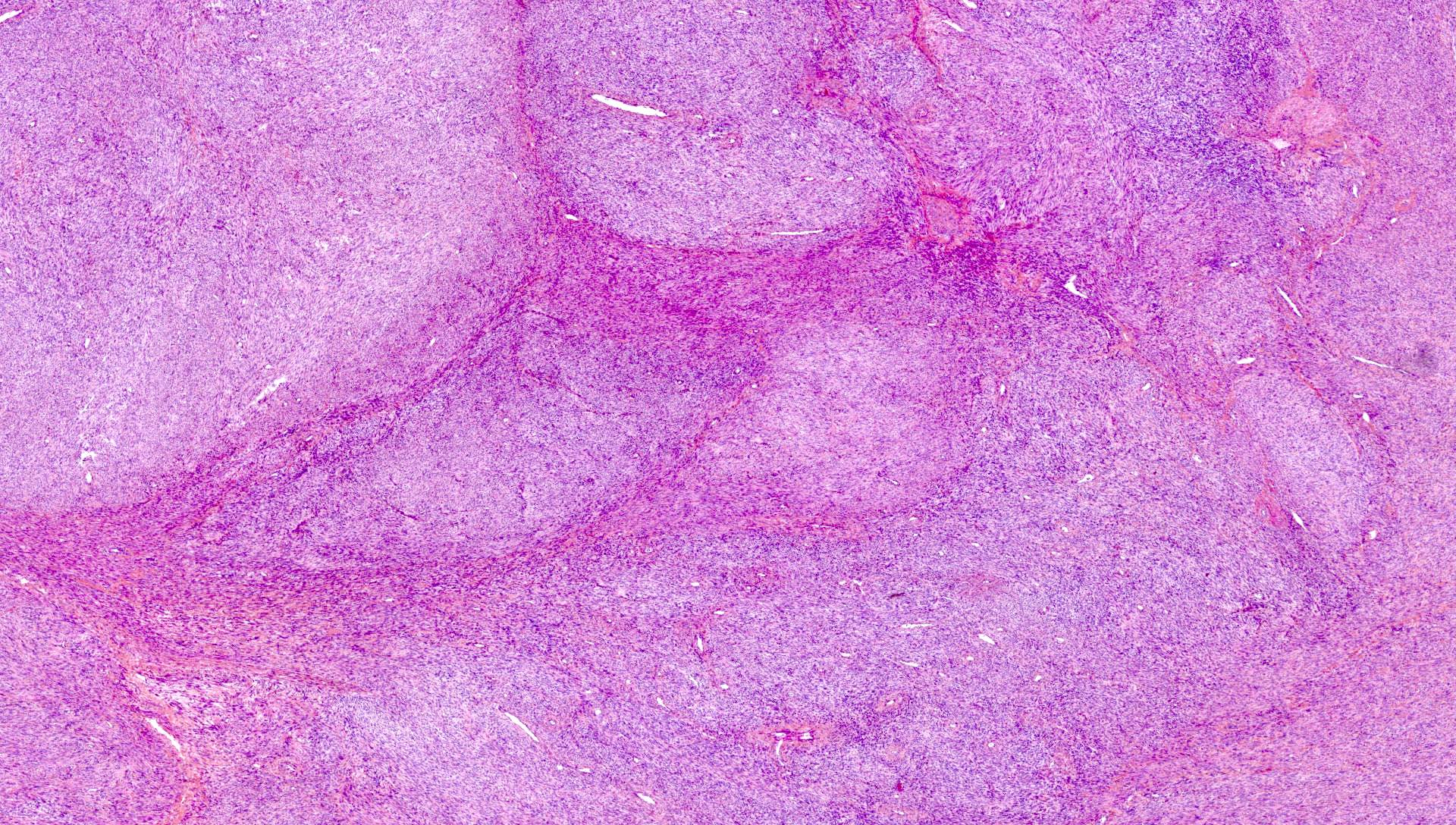
Uterine mesenchymal neoplasms
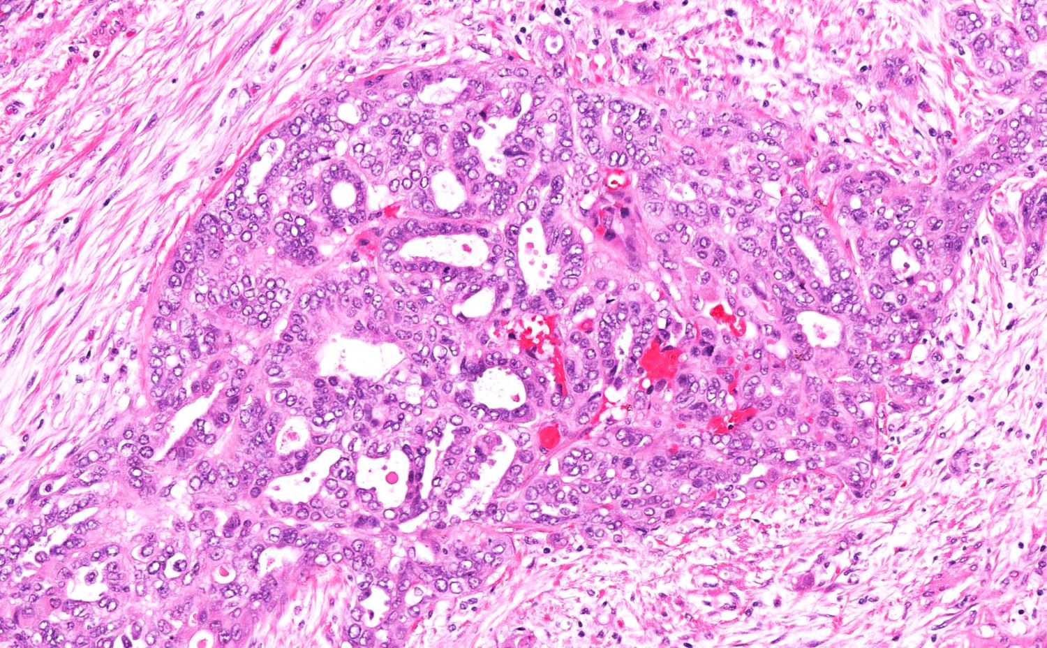
Contributed by Daniel Graham, M.D., Adele Wong, M.B., B.Ch., B.A.O. and Lucy Ma, M.D.
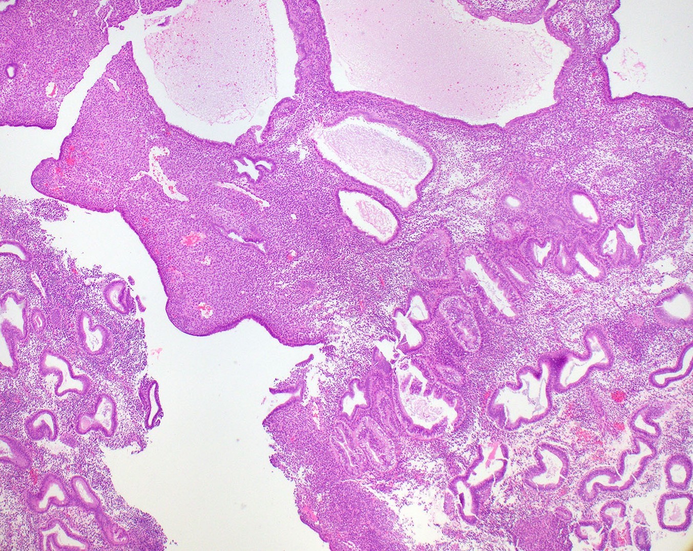
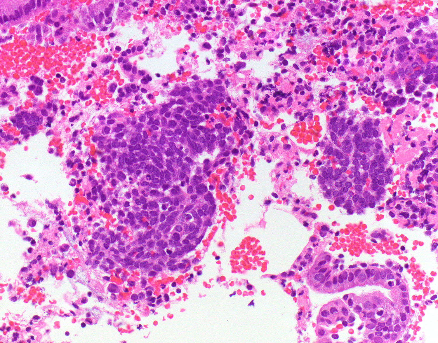
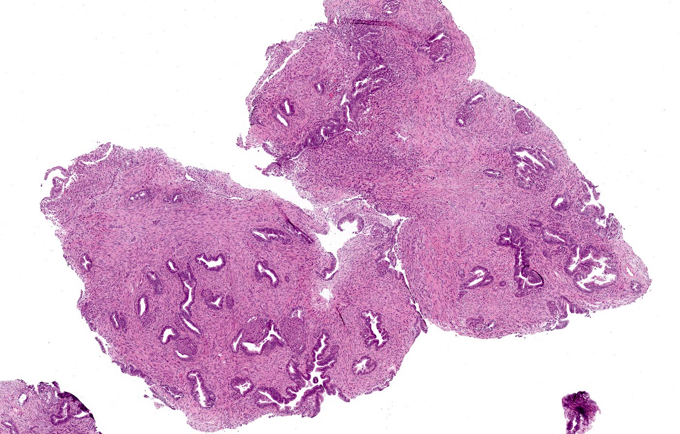
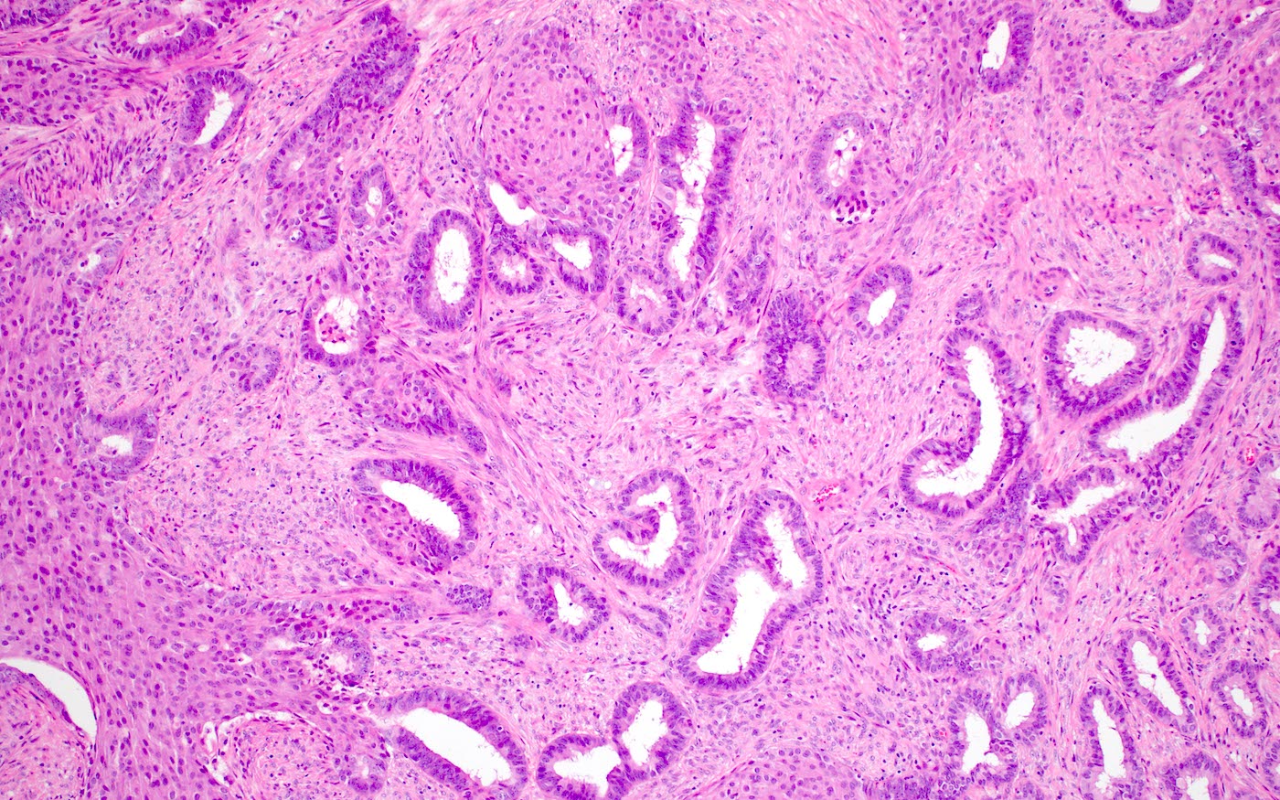
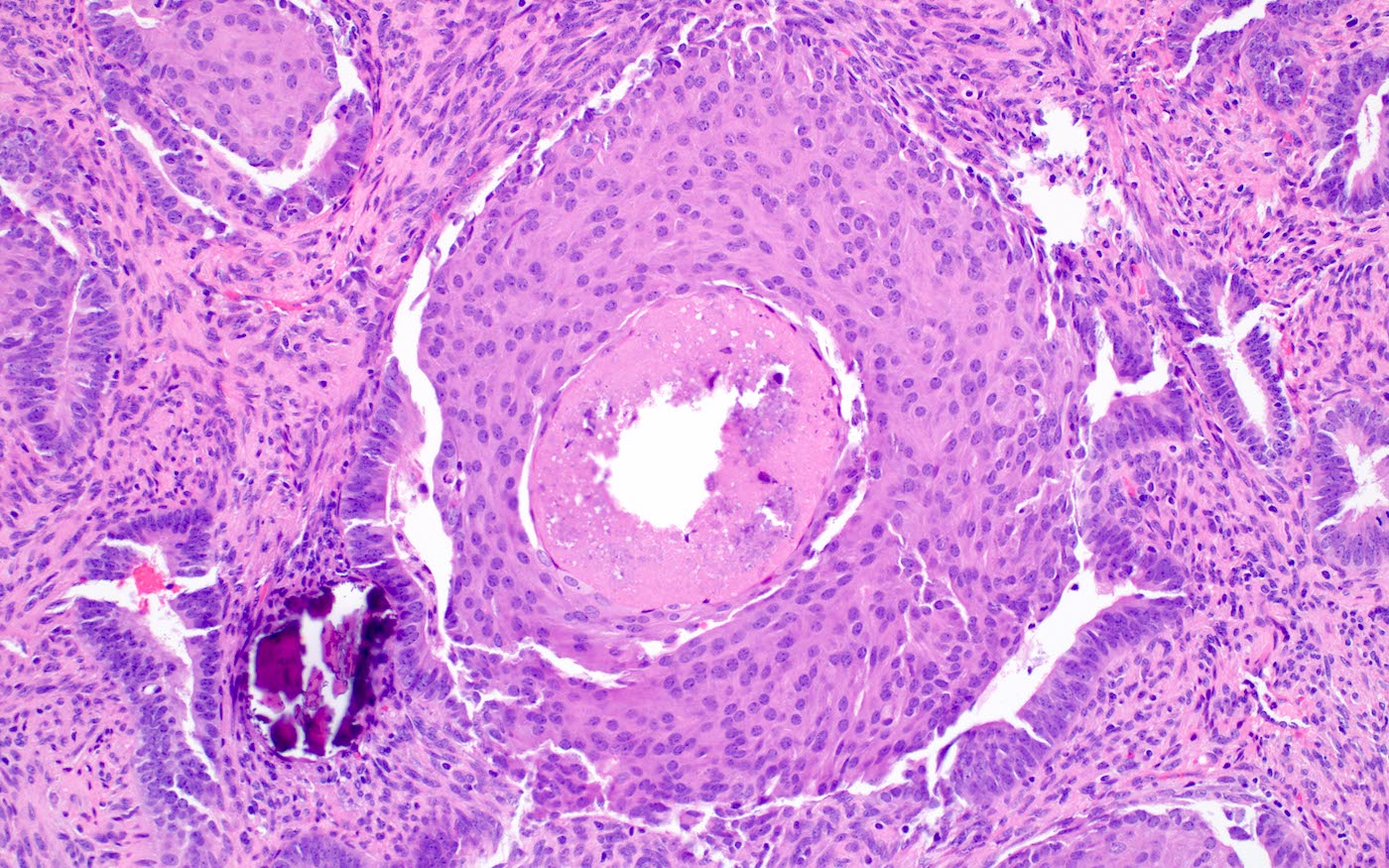
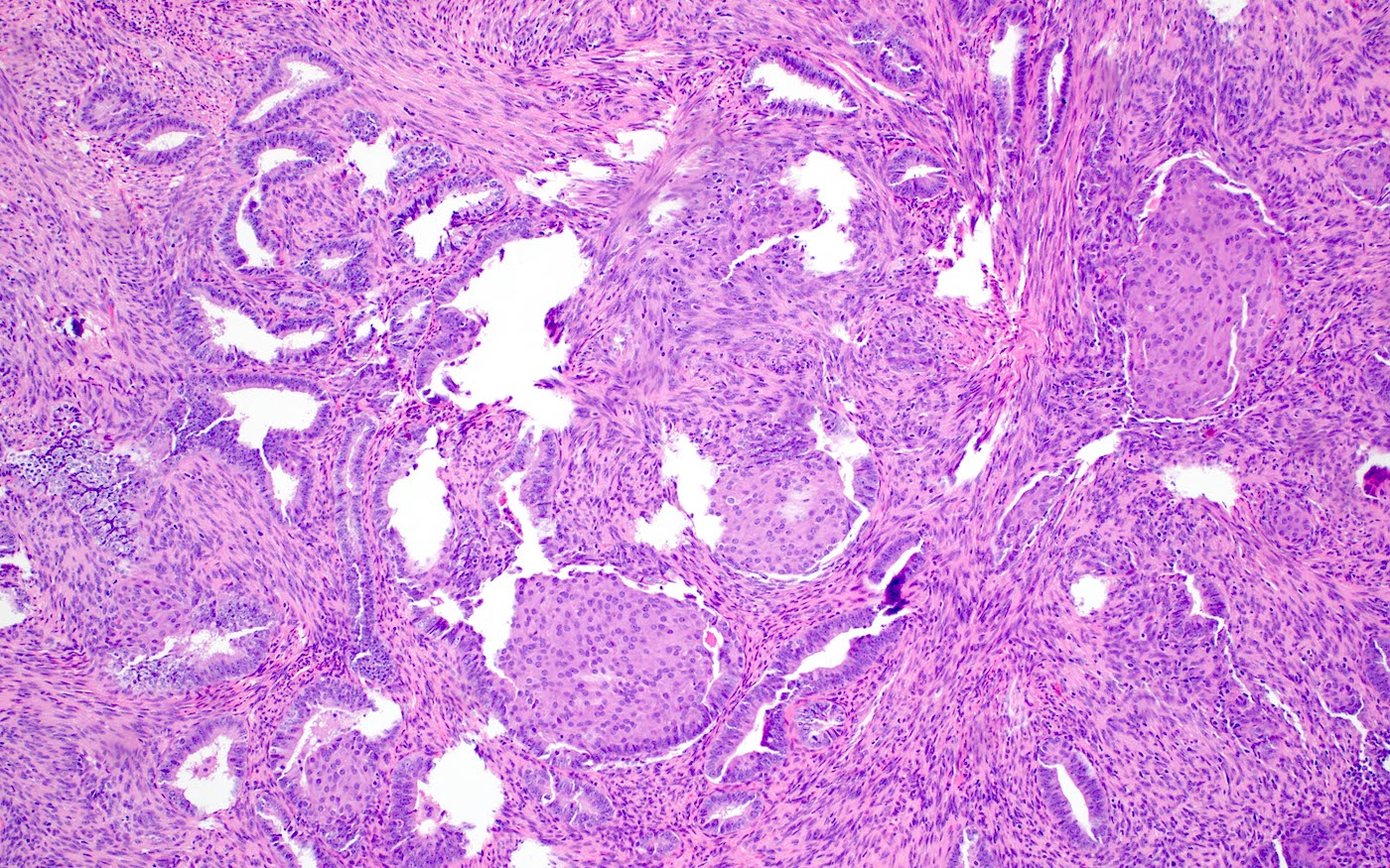
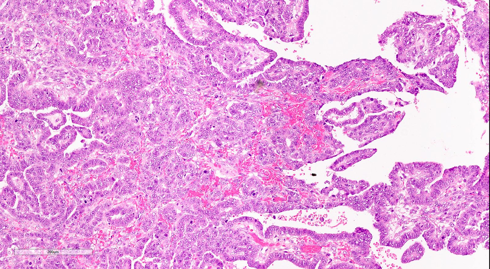
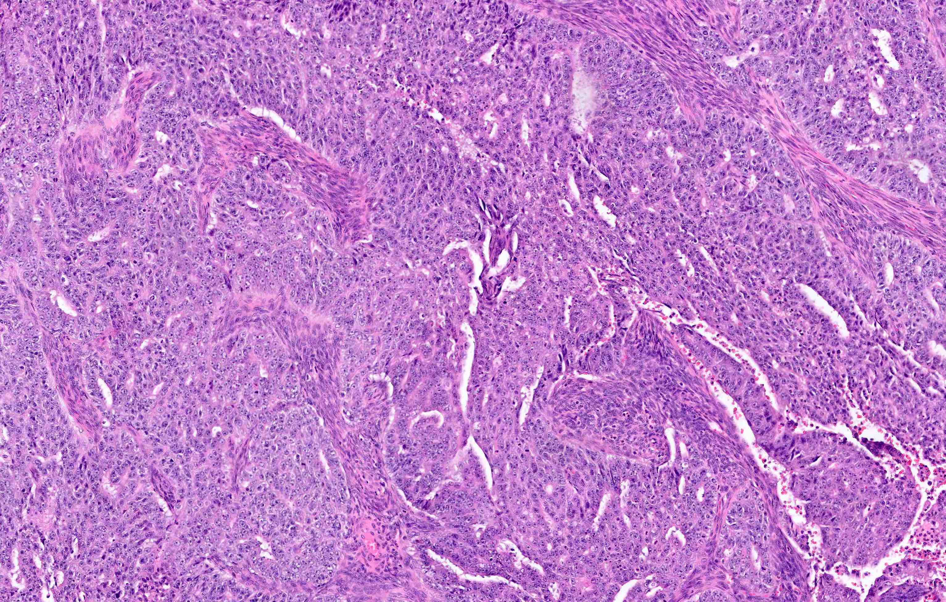
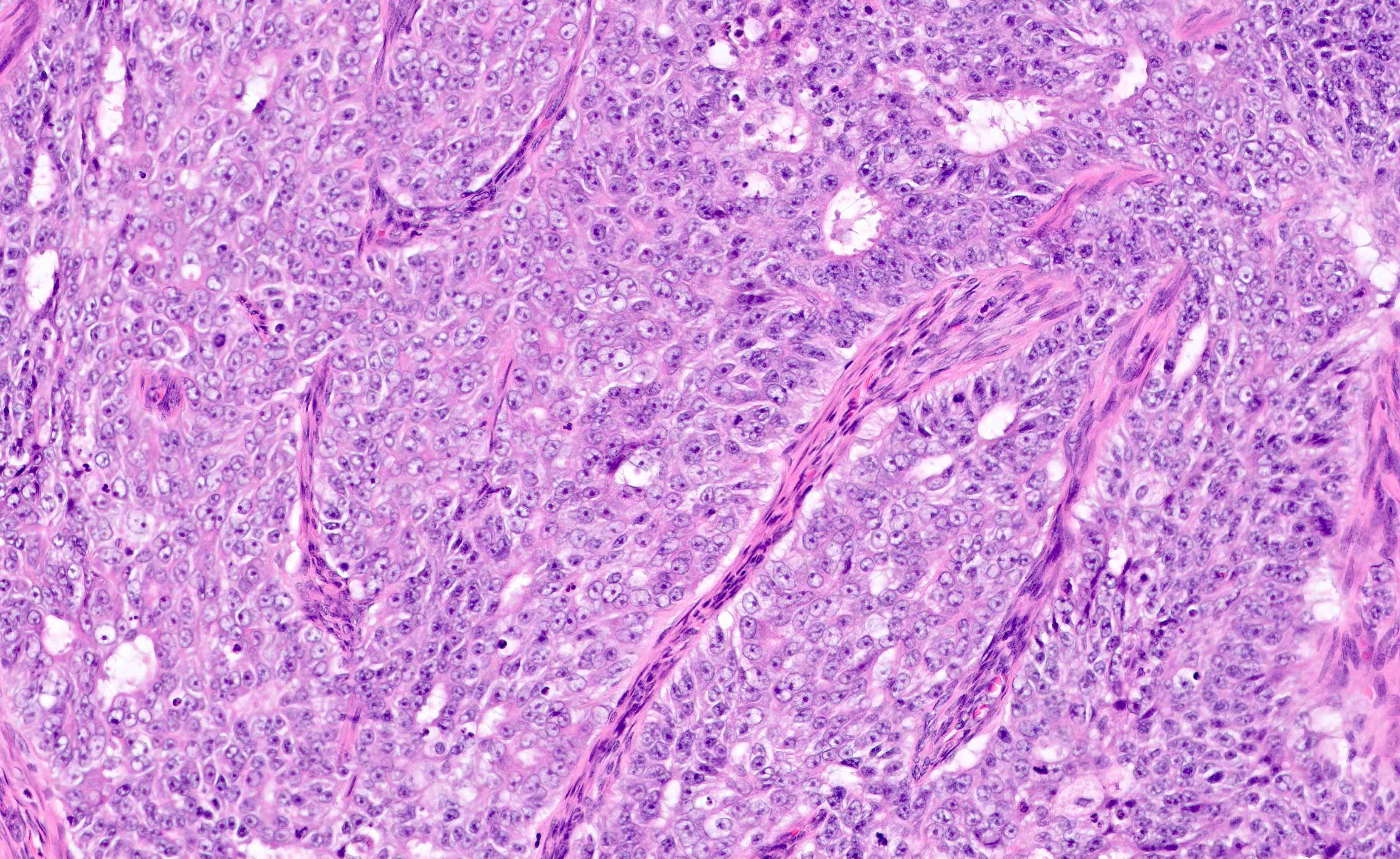
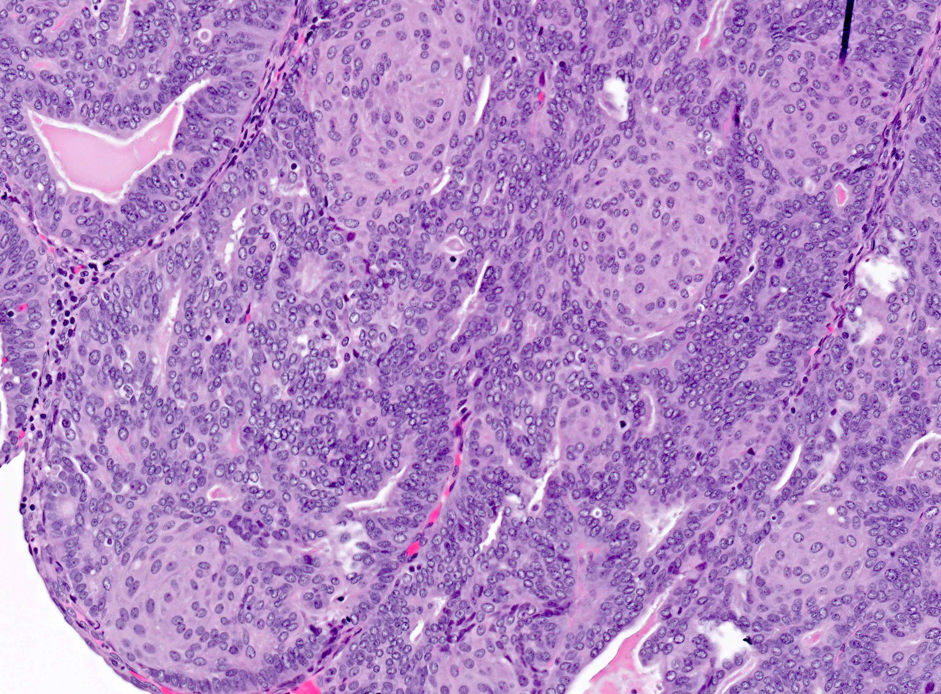
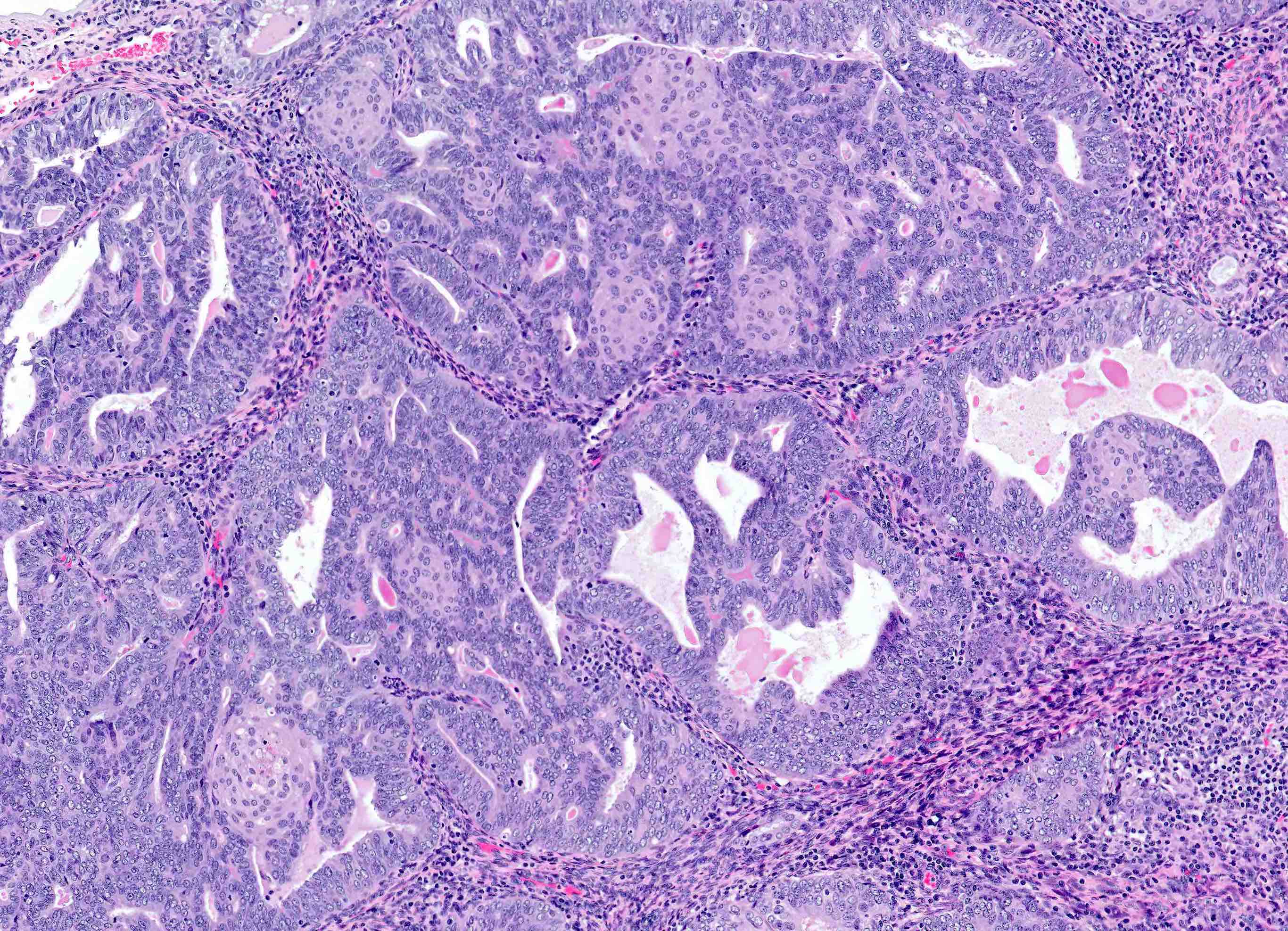
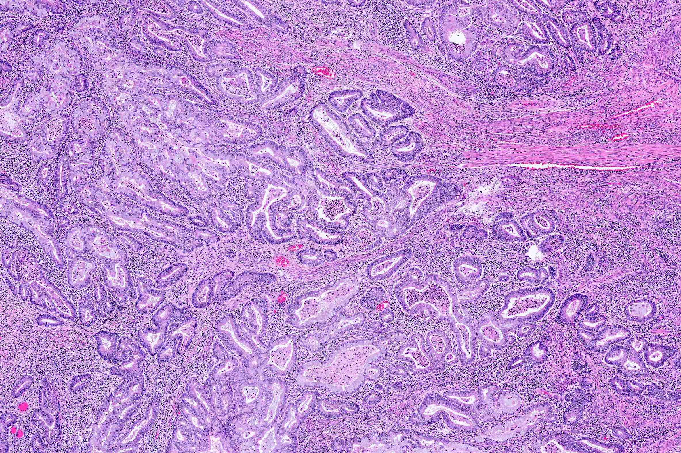
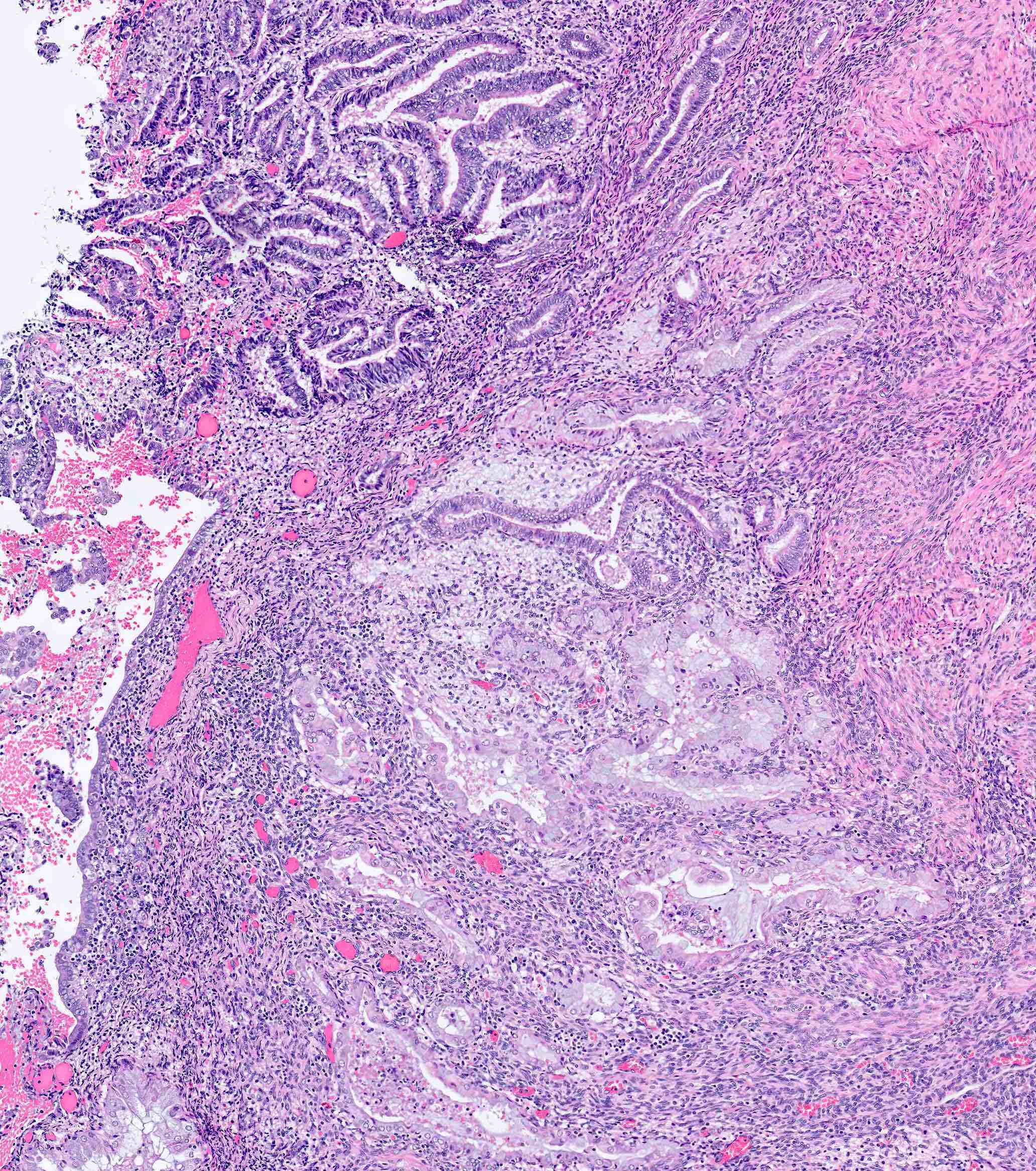
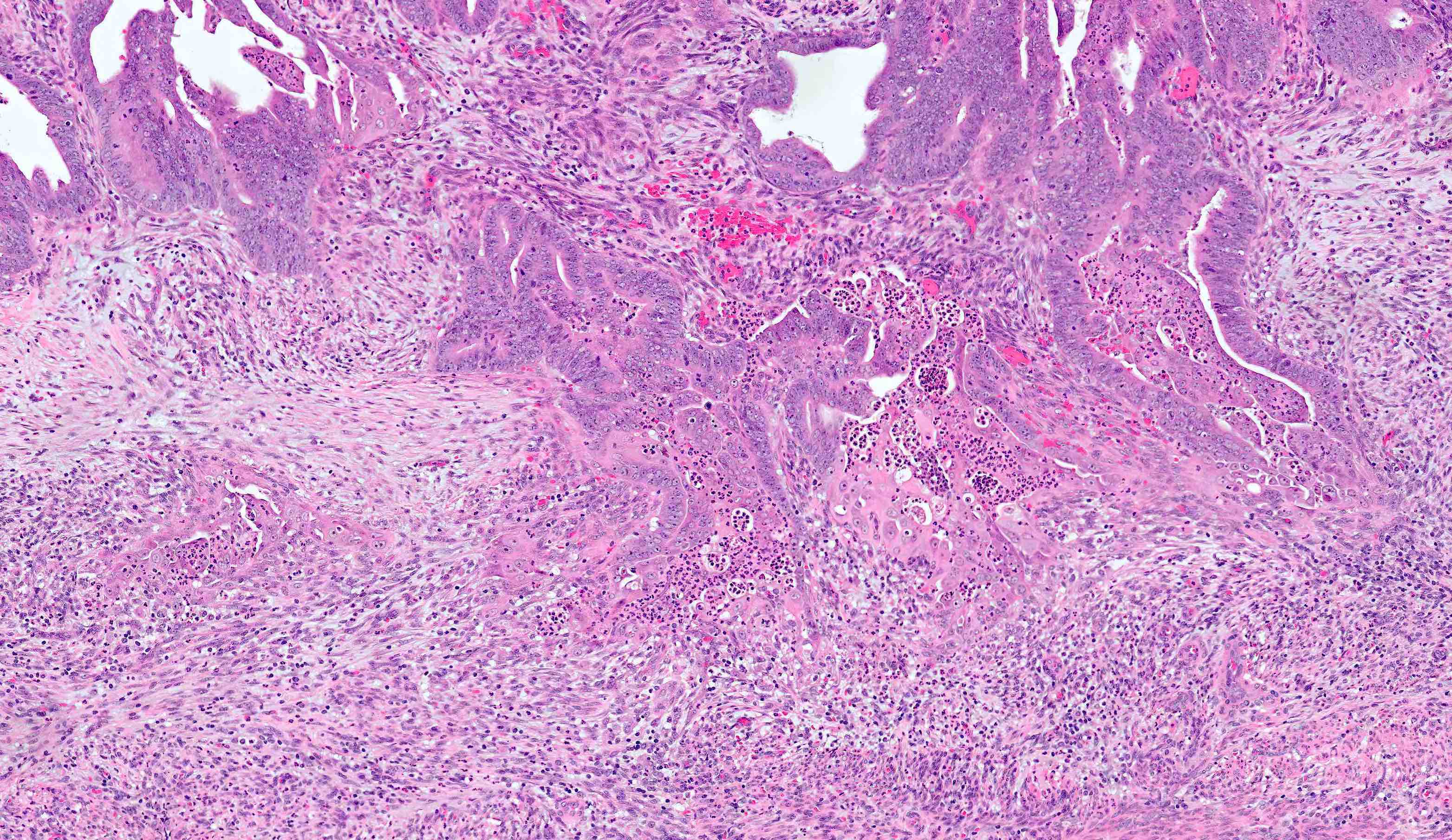
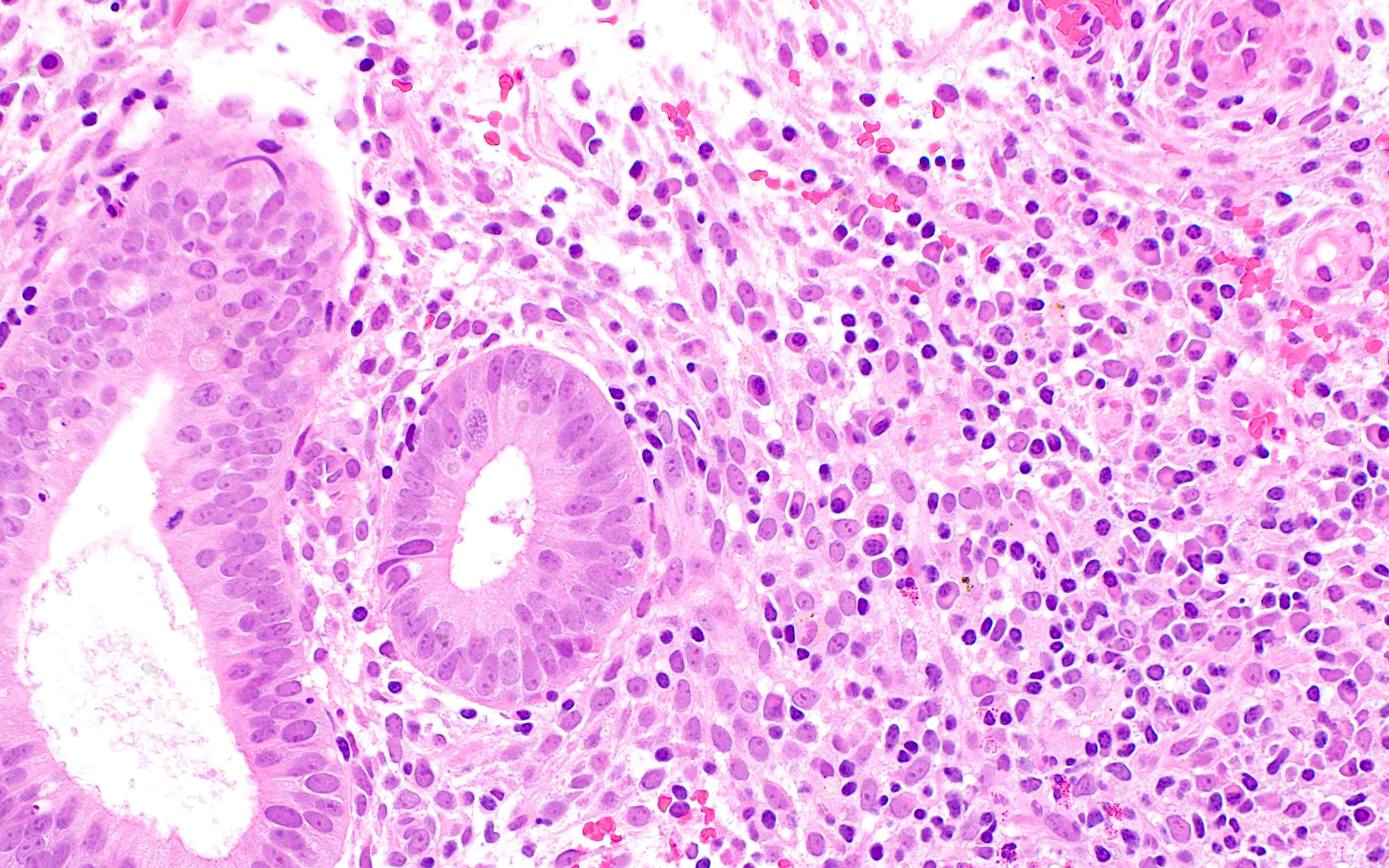
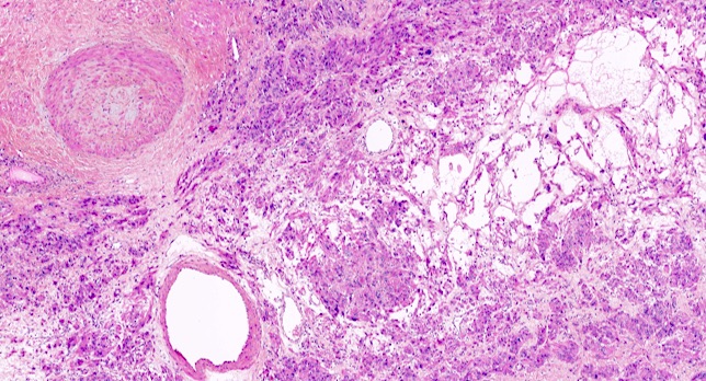
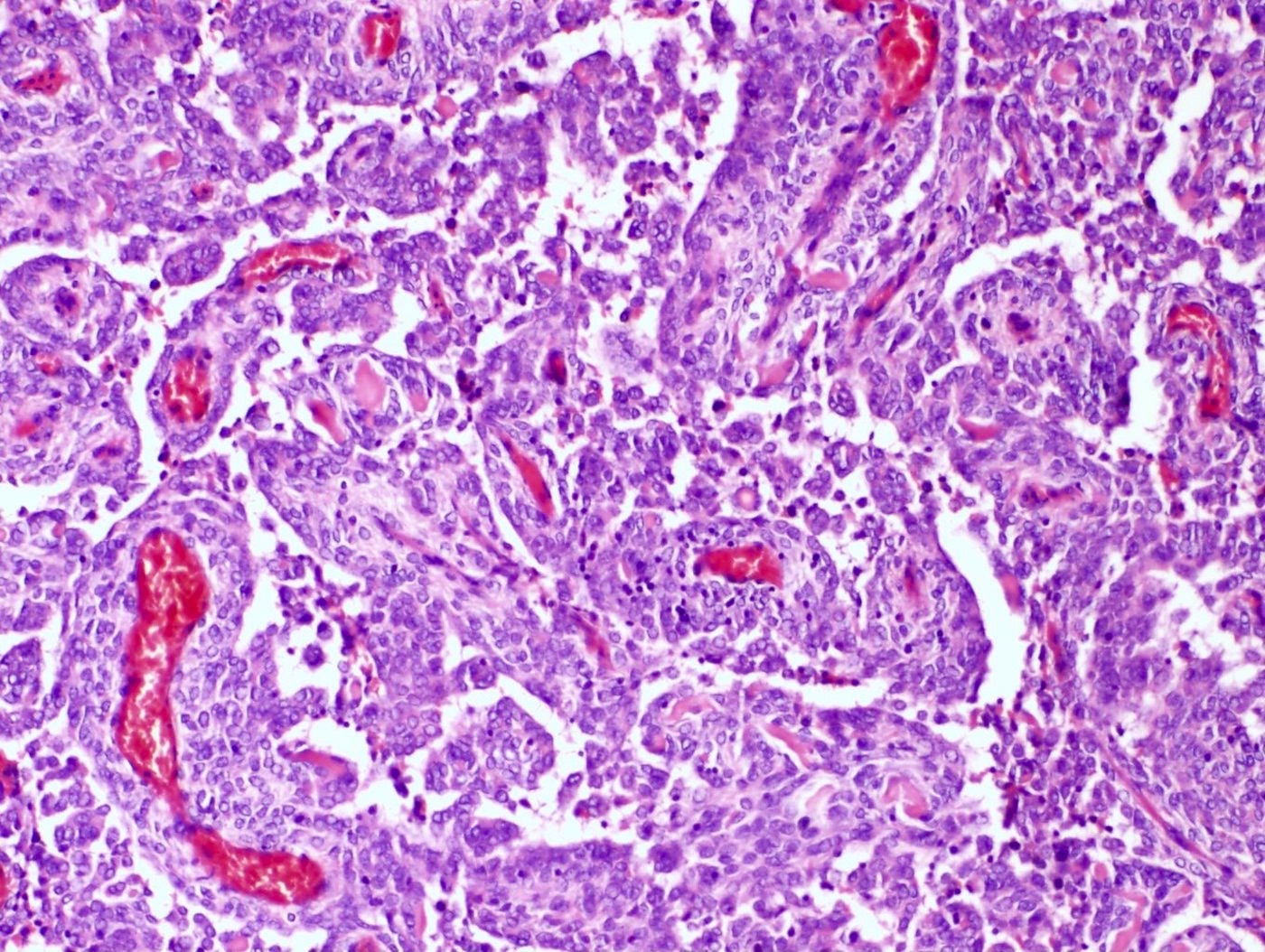
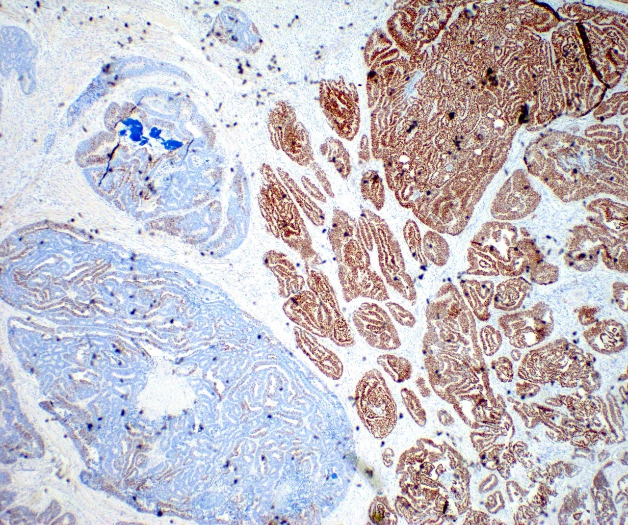
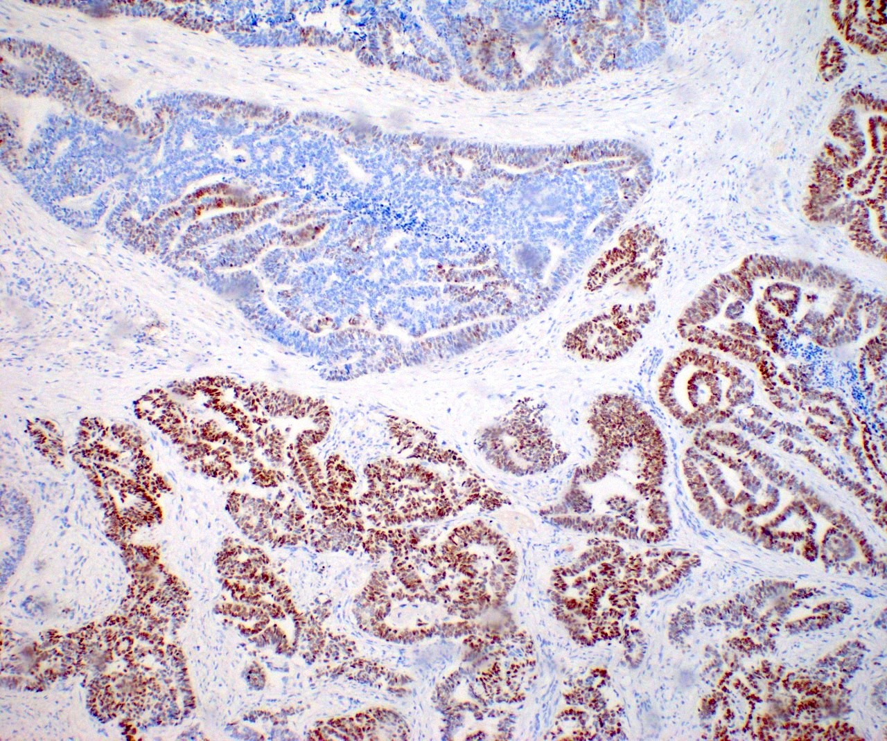
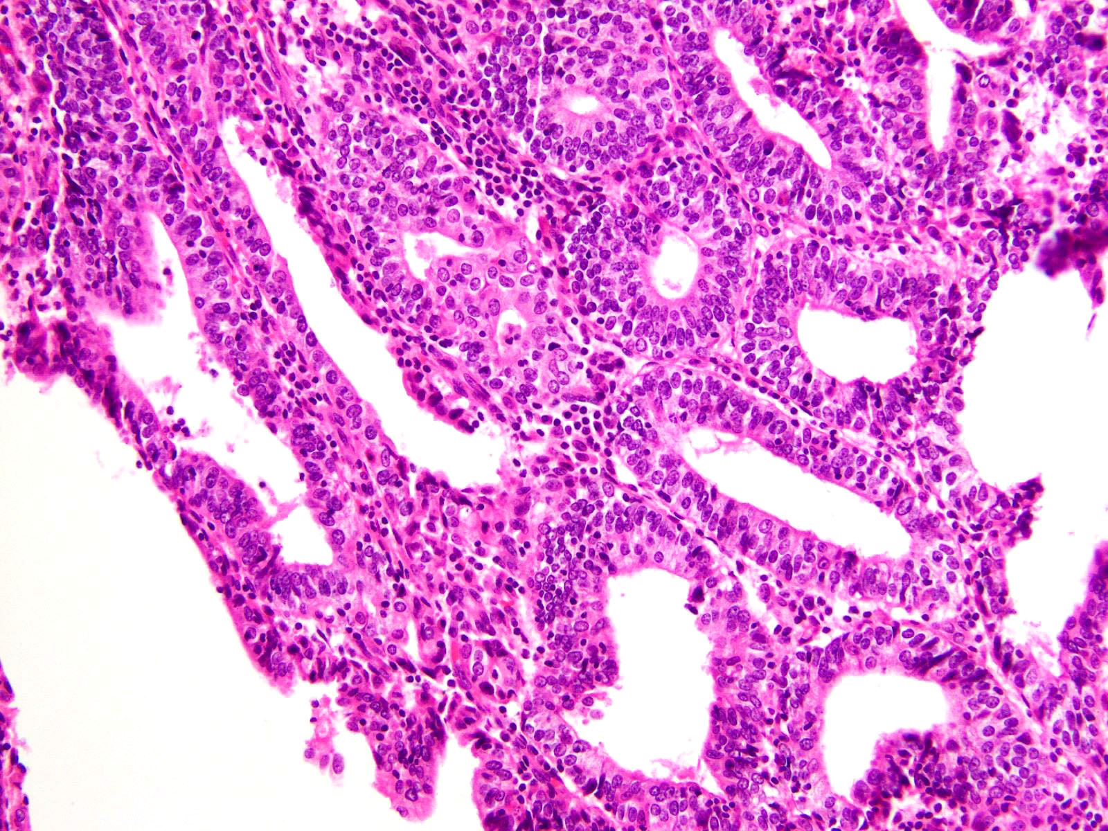
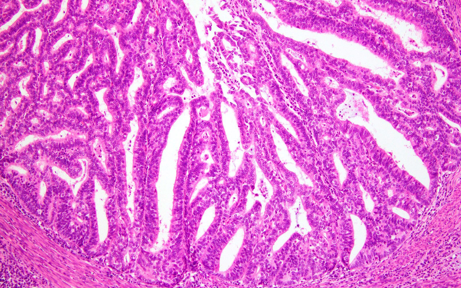
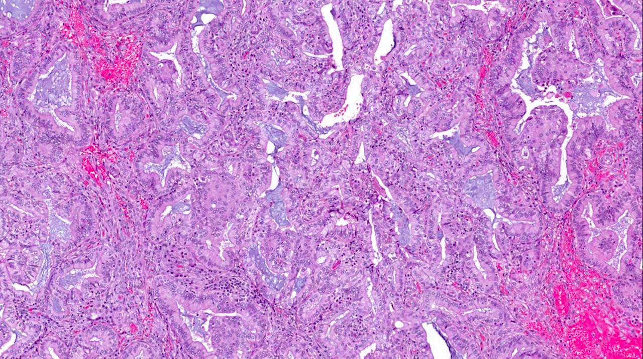
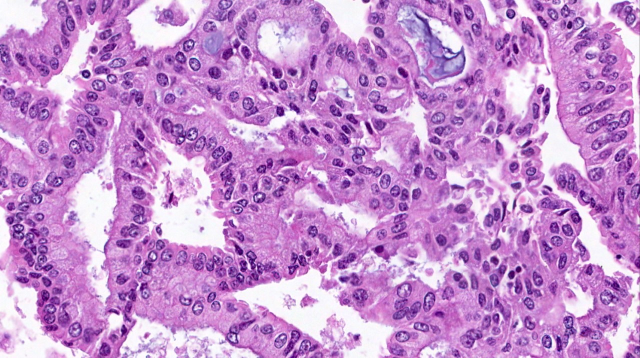
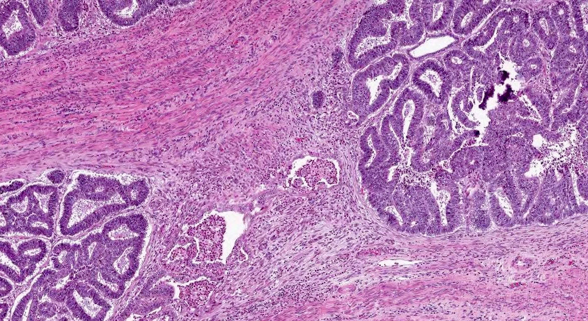
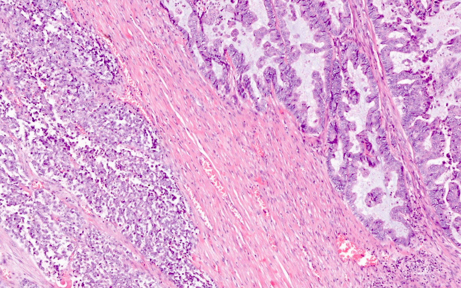
Glandular growth pattern
Variety of histologic patterns
Variable growth patterns

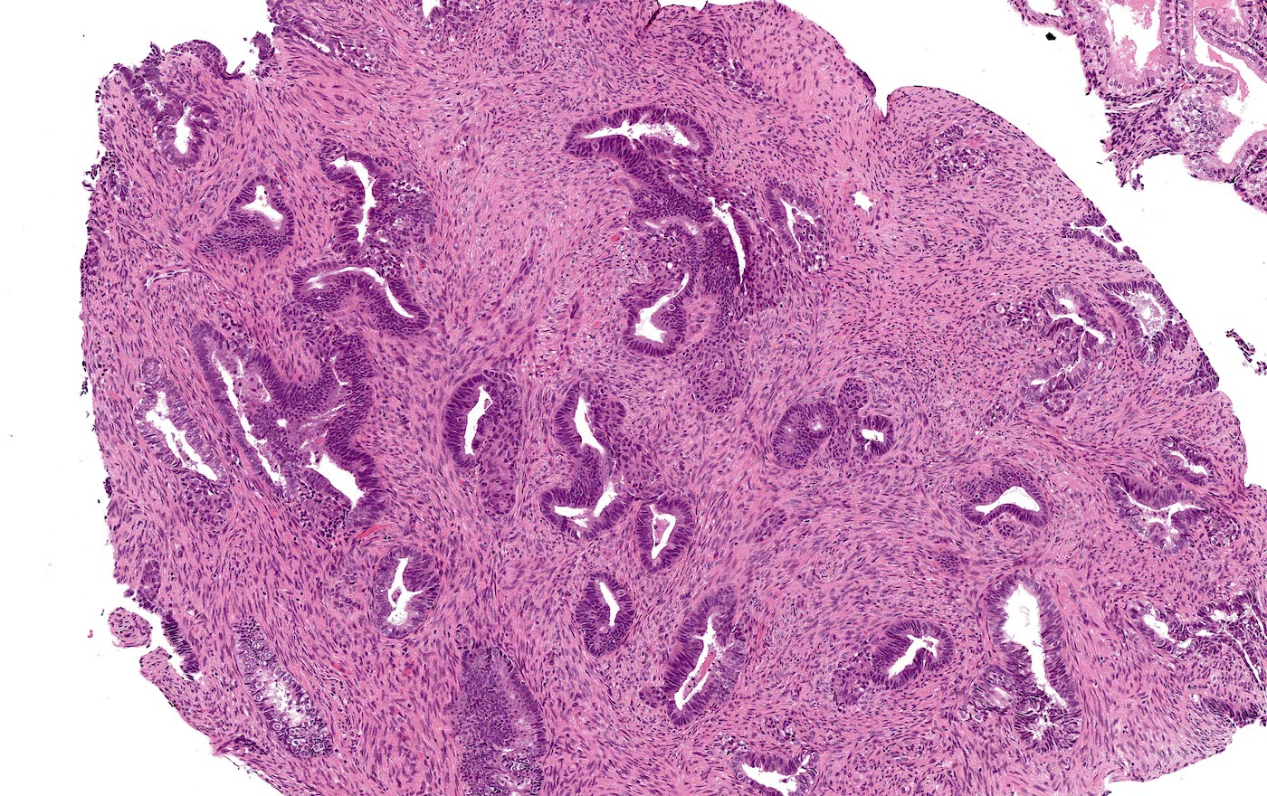
Glandular morphology
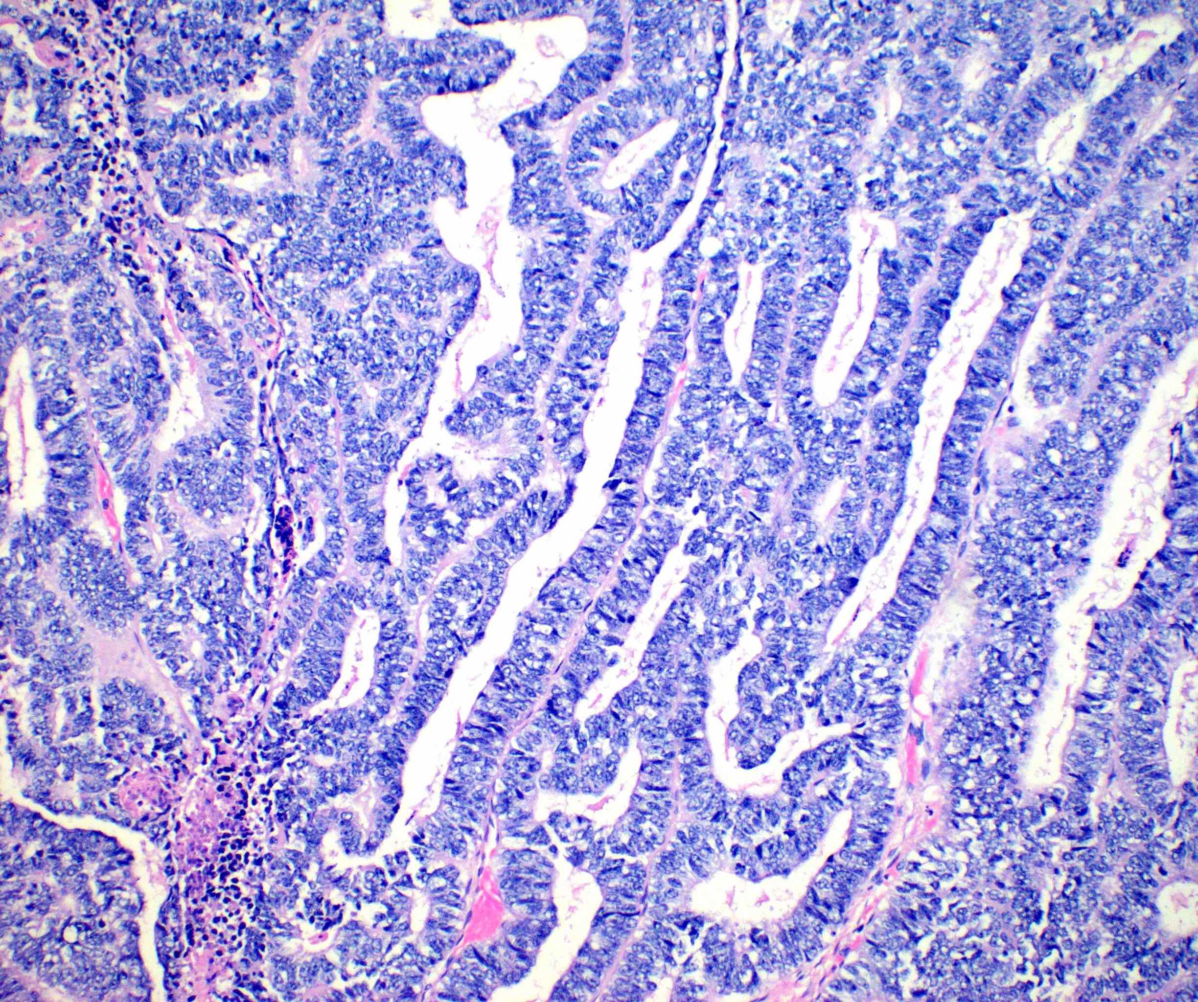
Glands
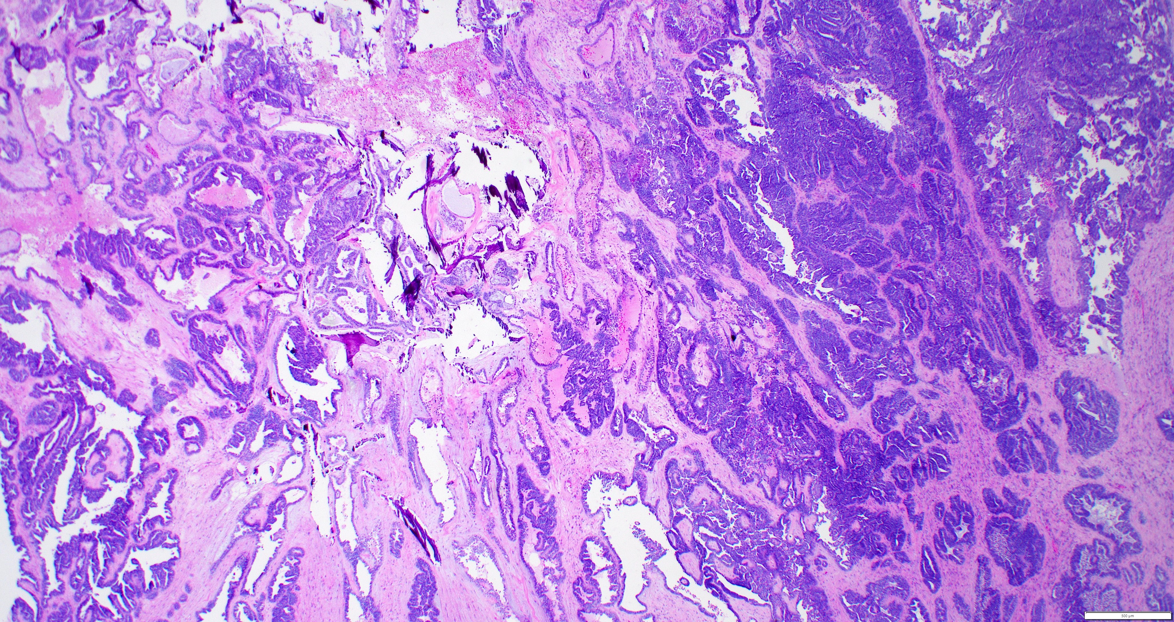
Multiple growth patterns
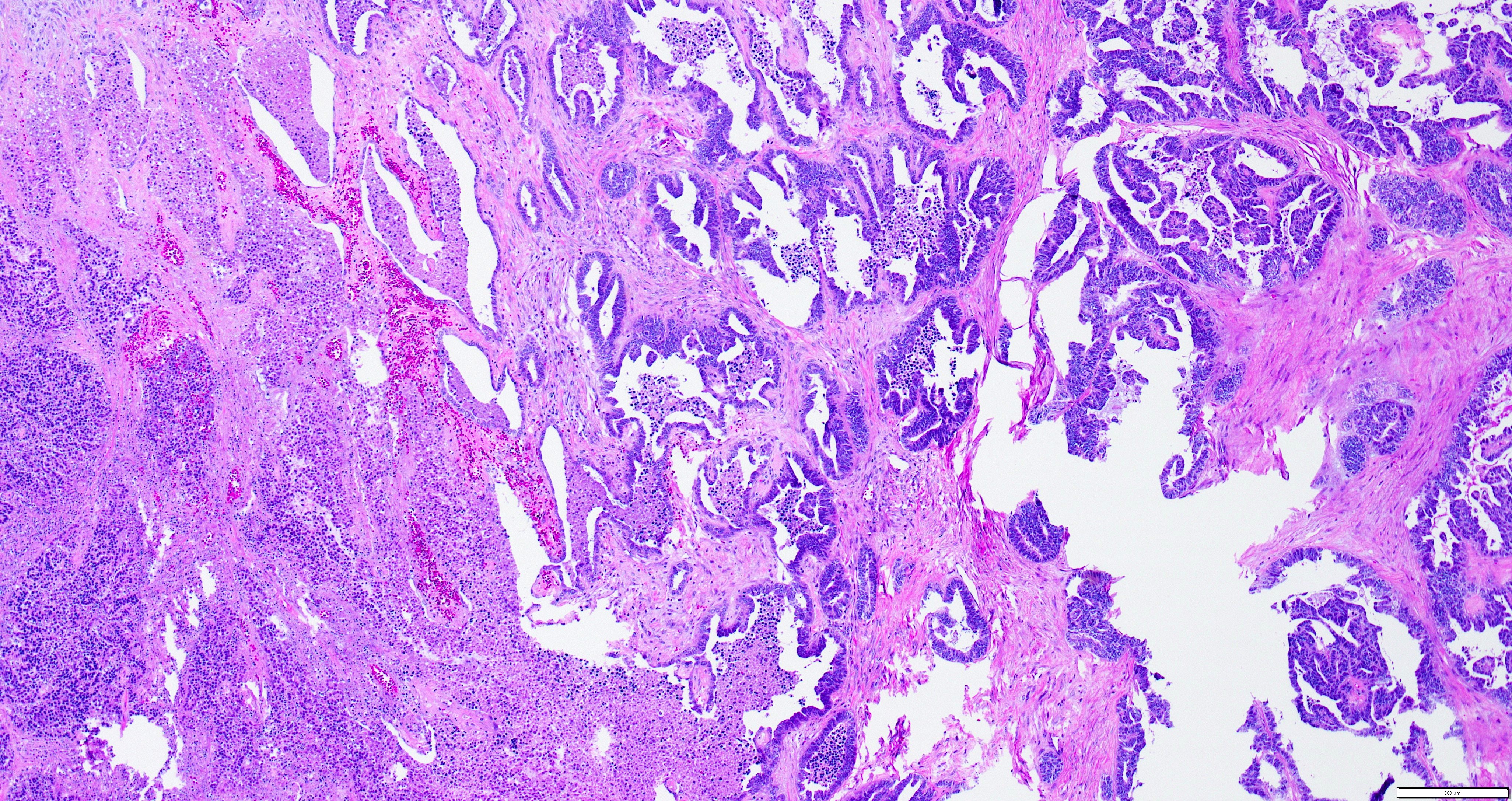
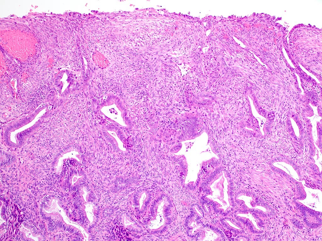
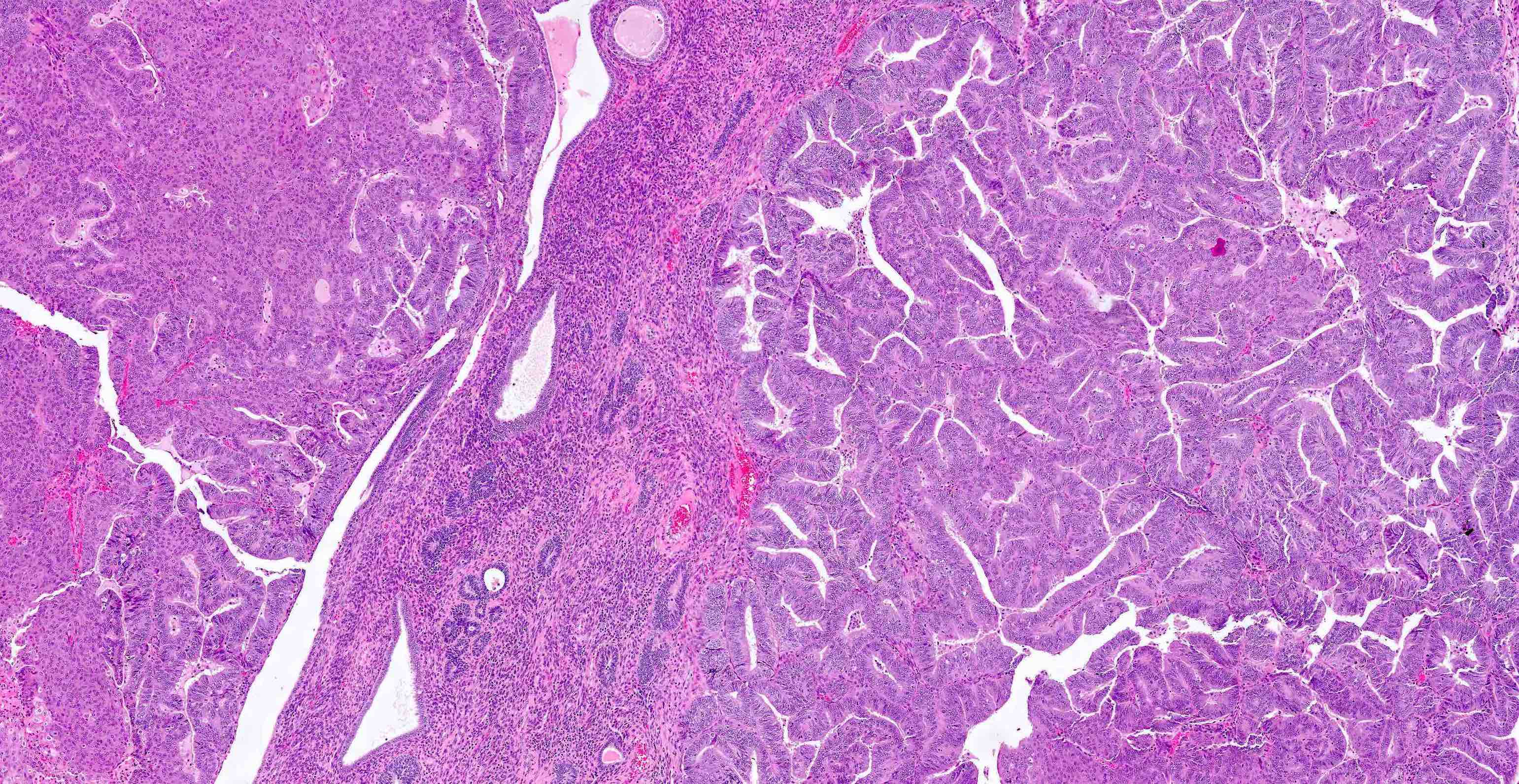
Glandular and cystic areas
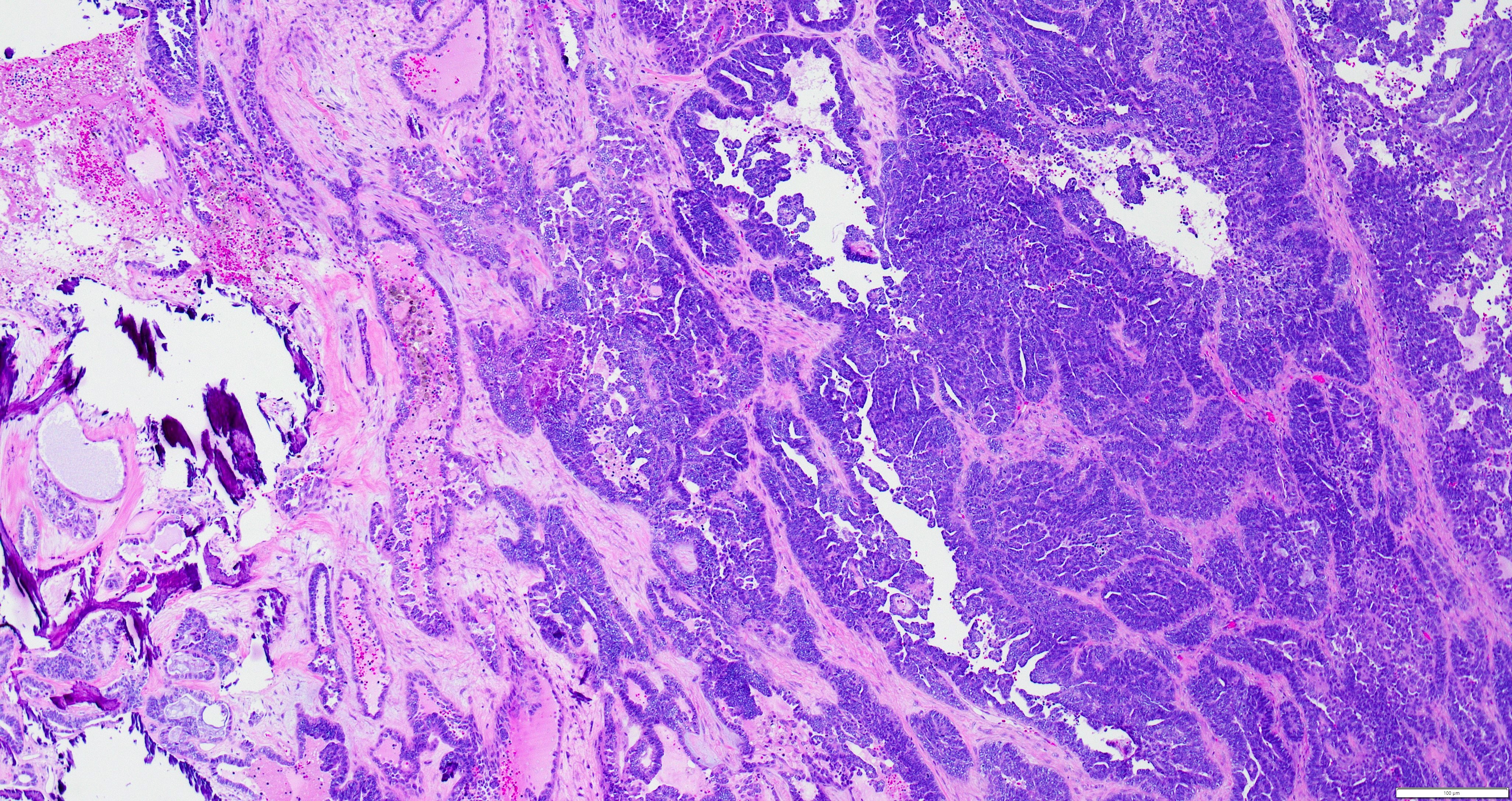
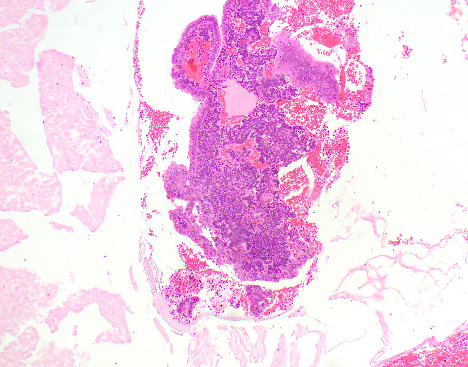
Micropapillary architecture
Mitotic activity
Papillary architecture
Variable morphology
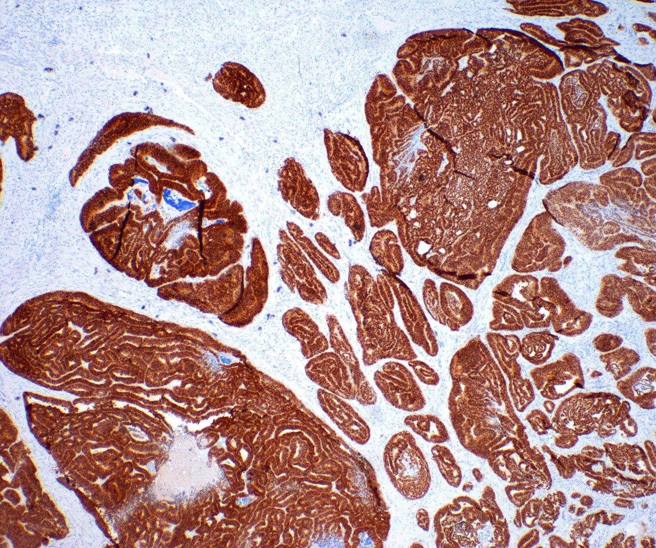
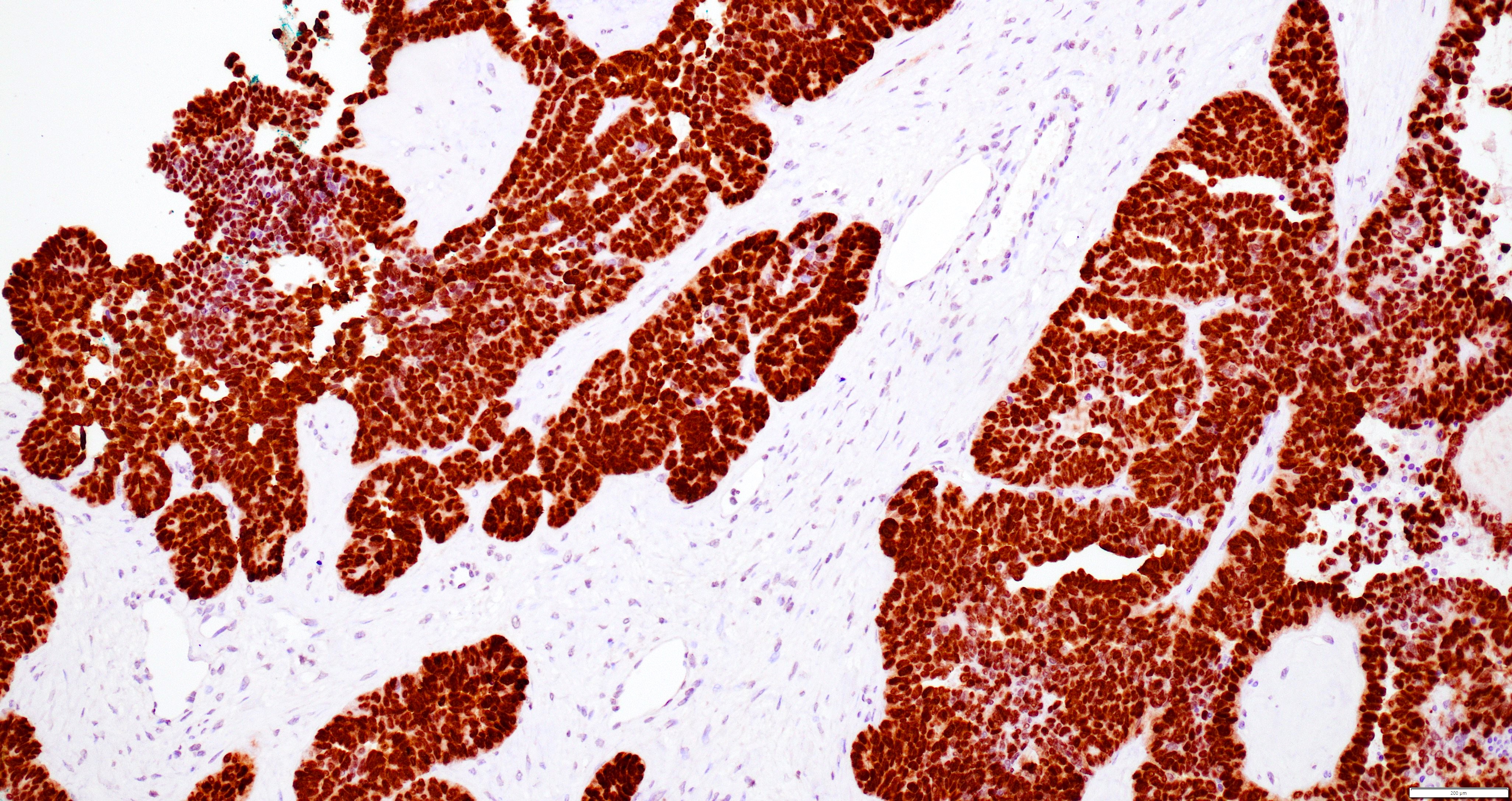
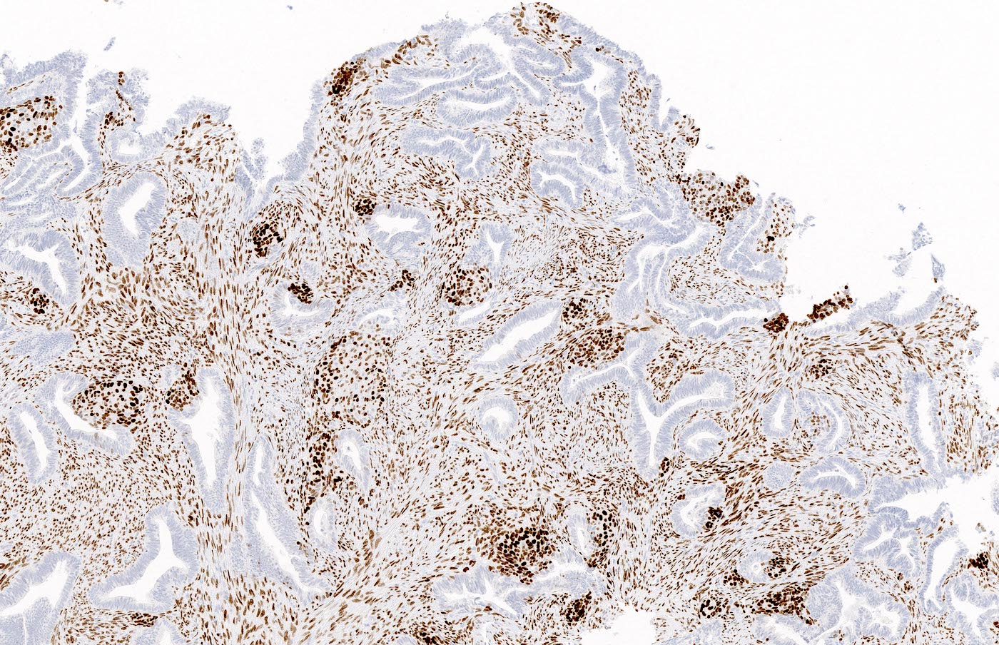
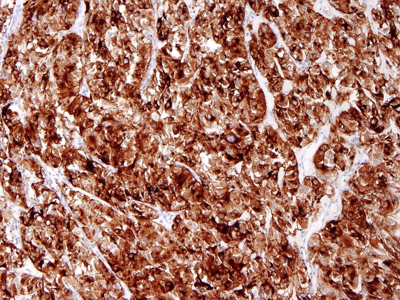
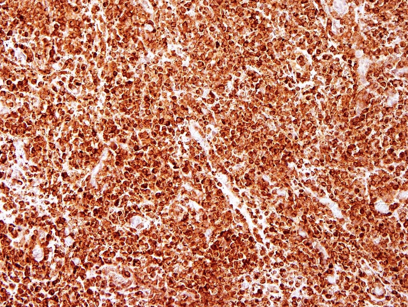
PAX8
GATA3
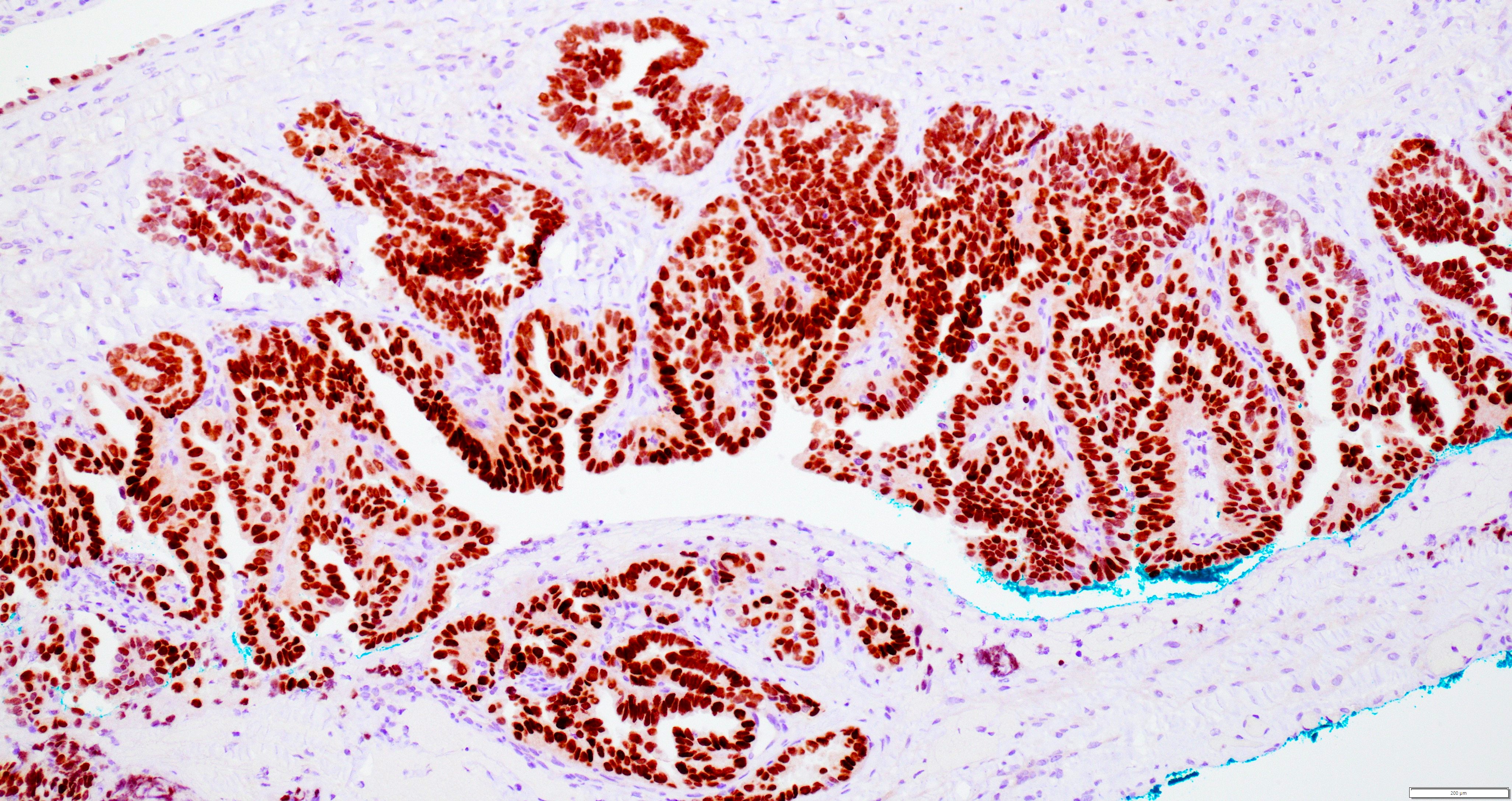
GATA3
TTF1
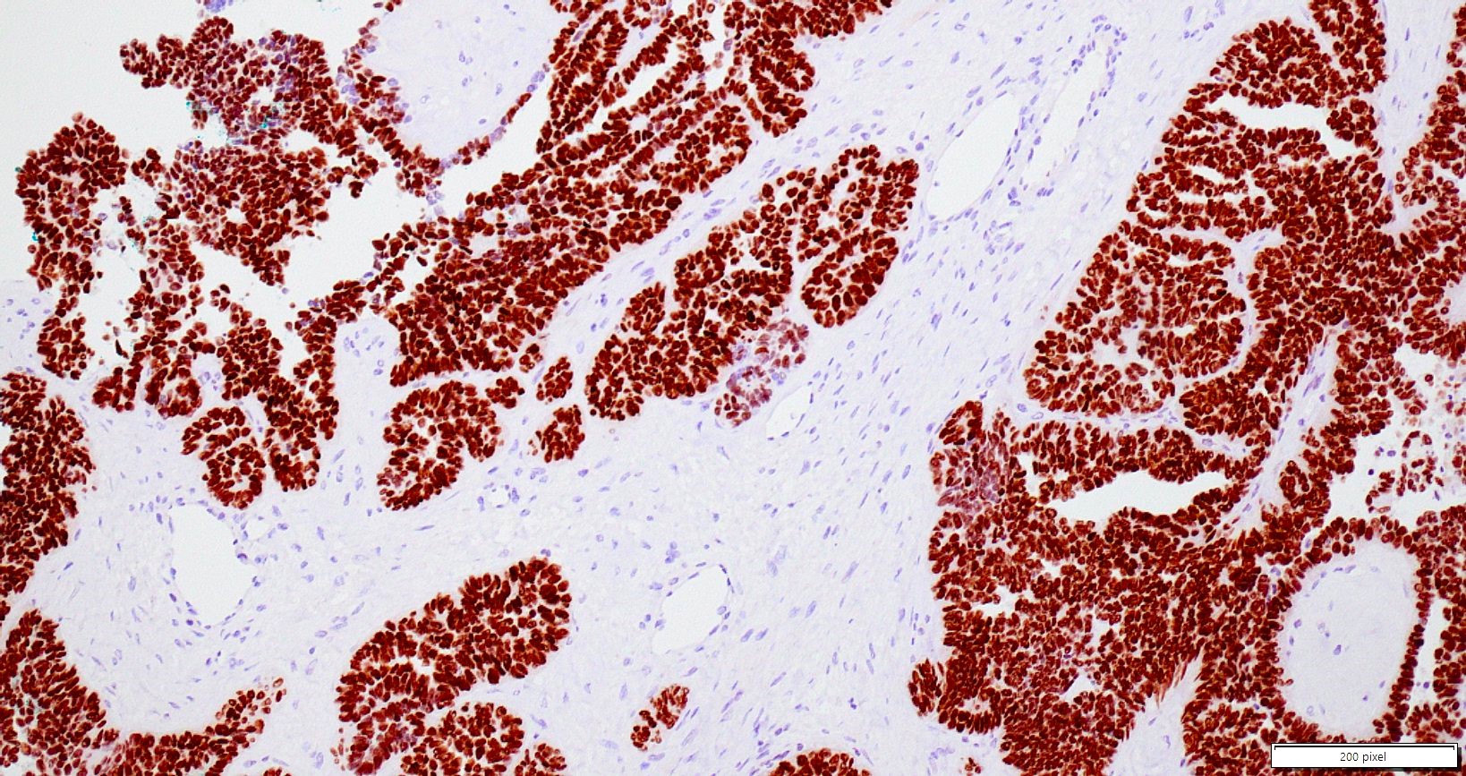
TTF1
CD10
CD10
CD10
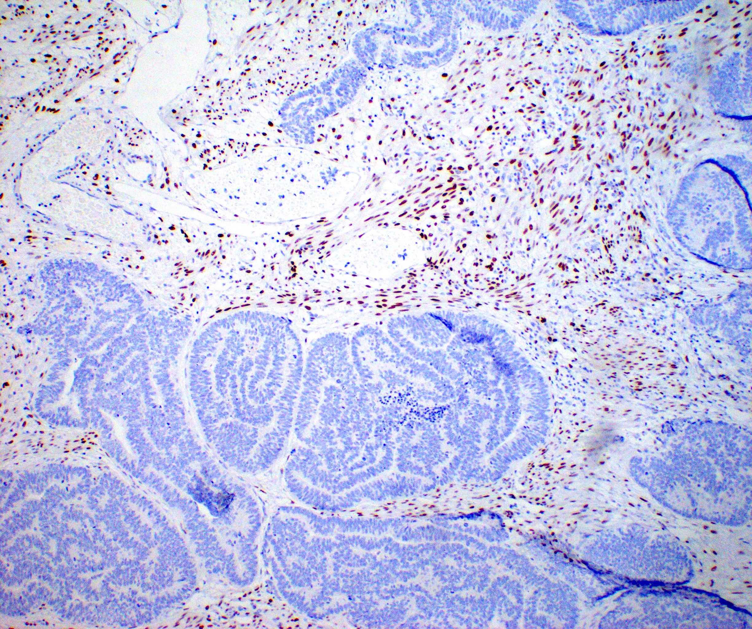

Estrogen receptor
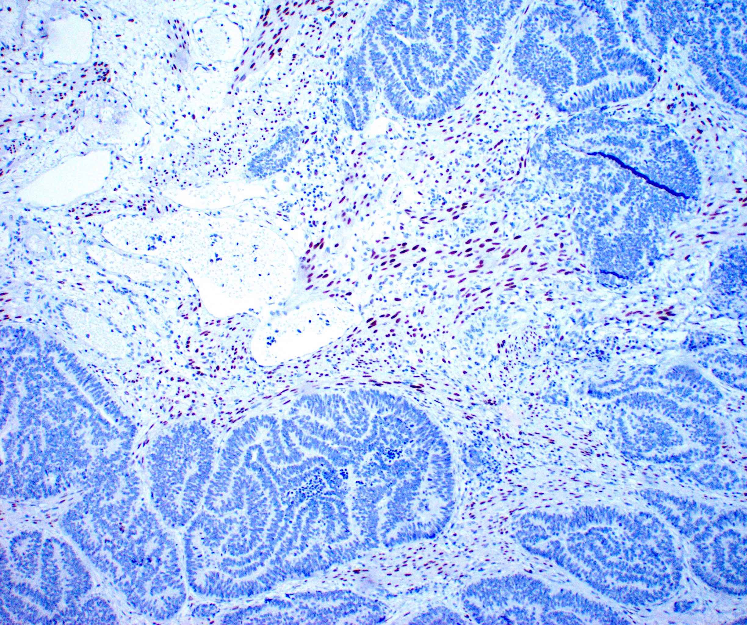
Progesterone receptor
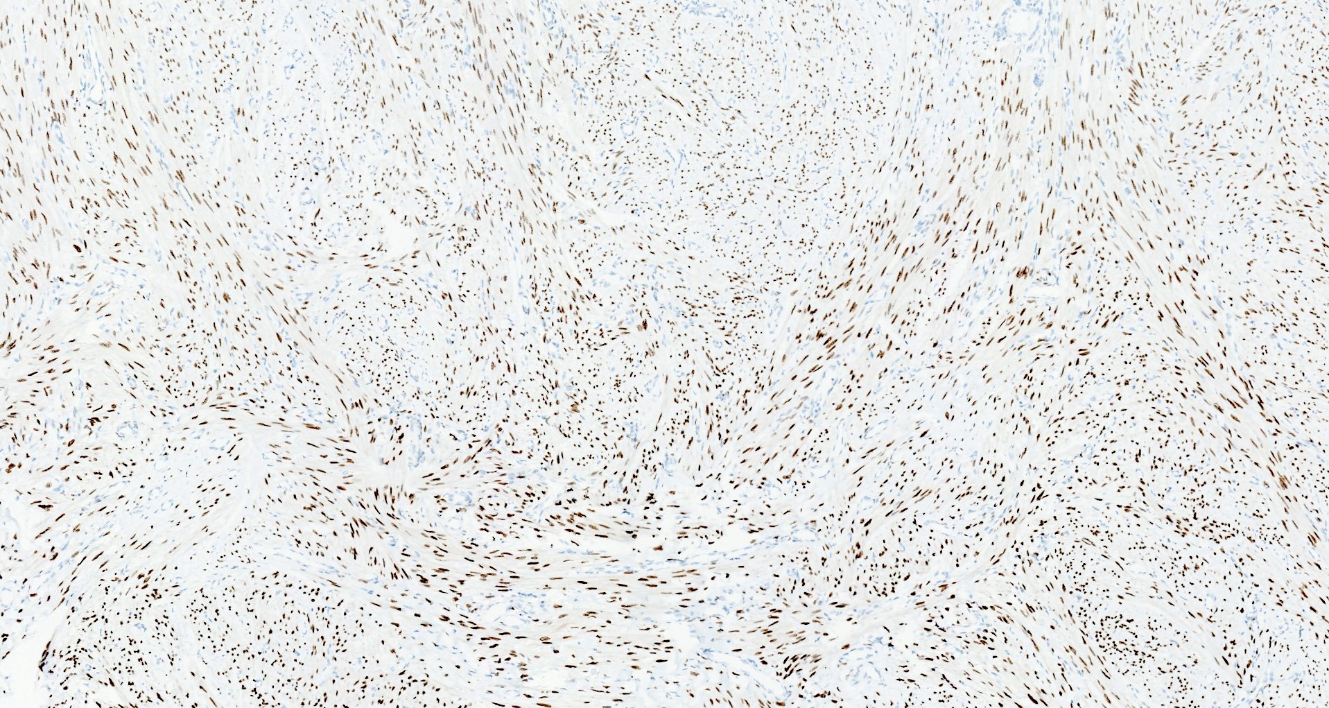
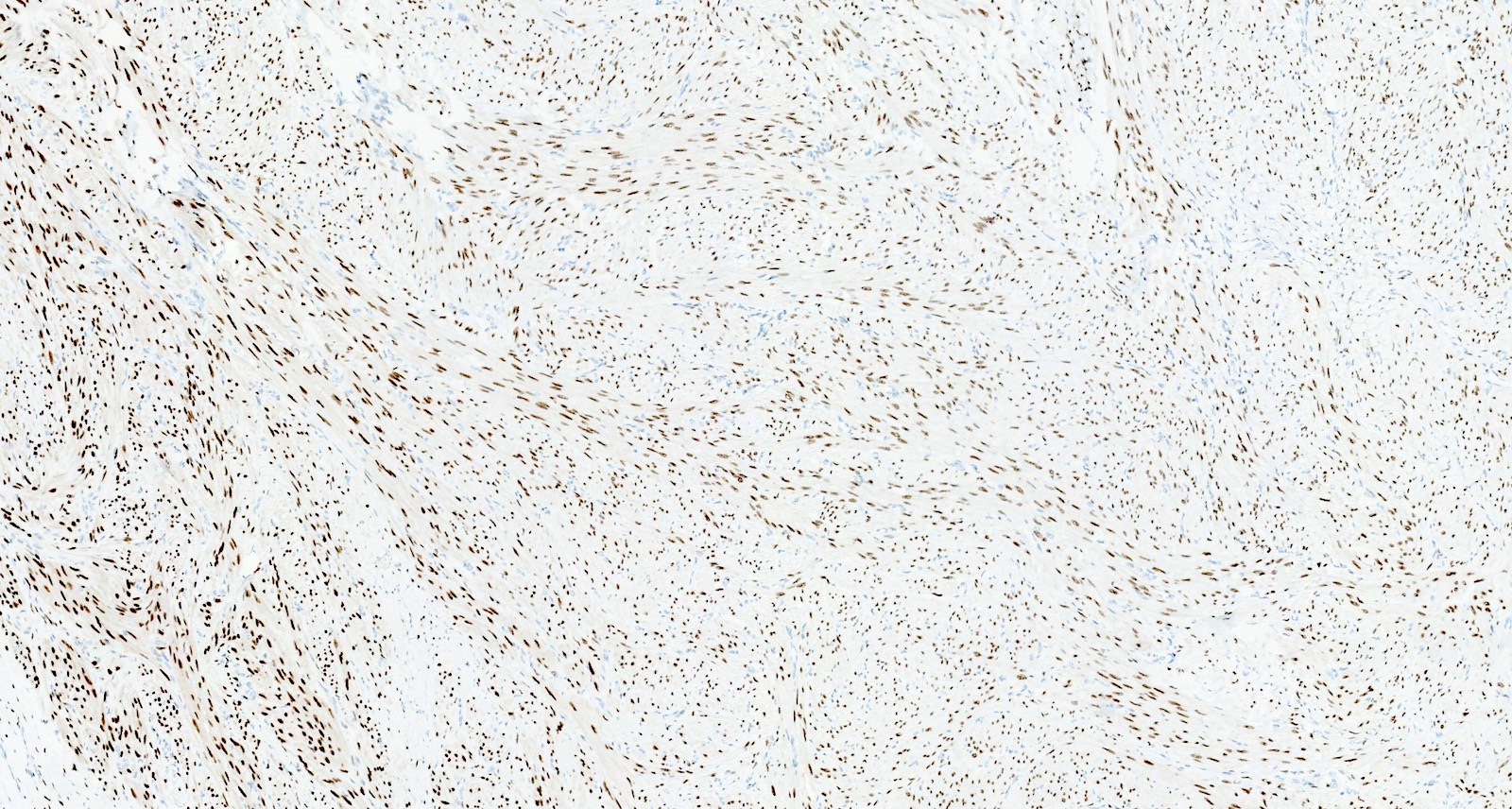
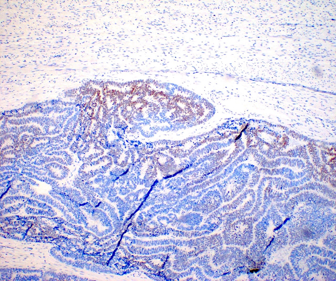
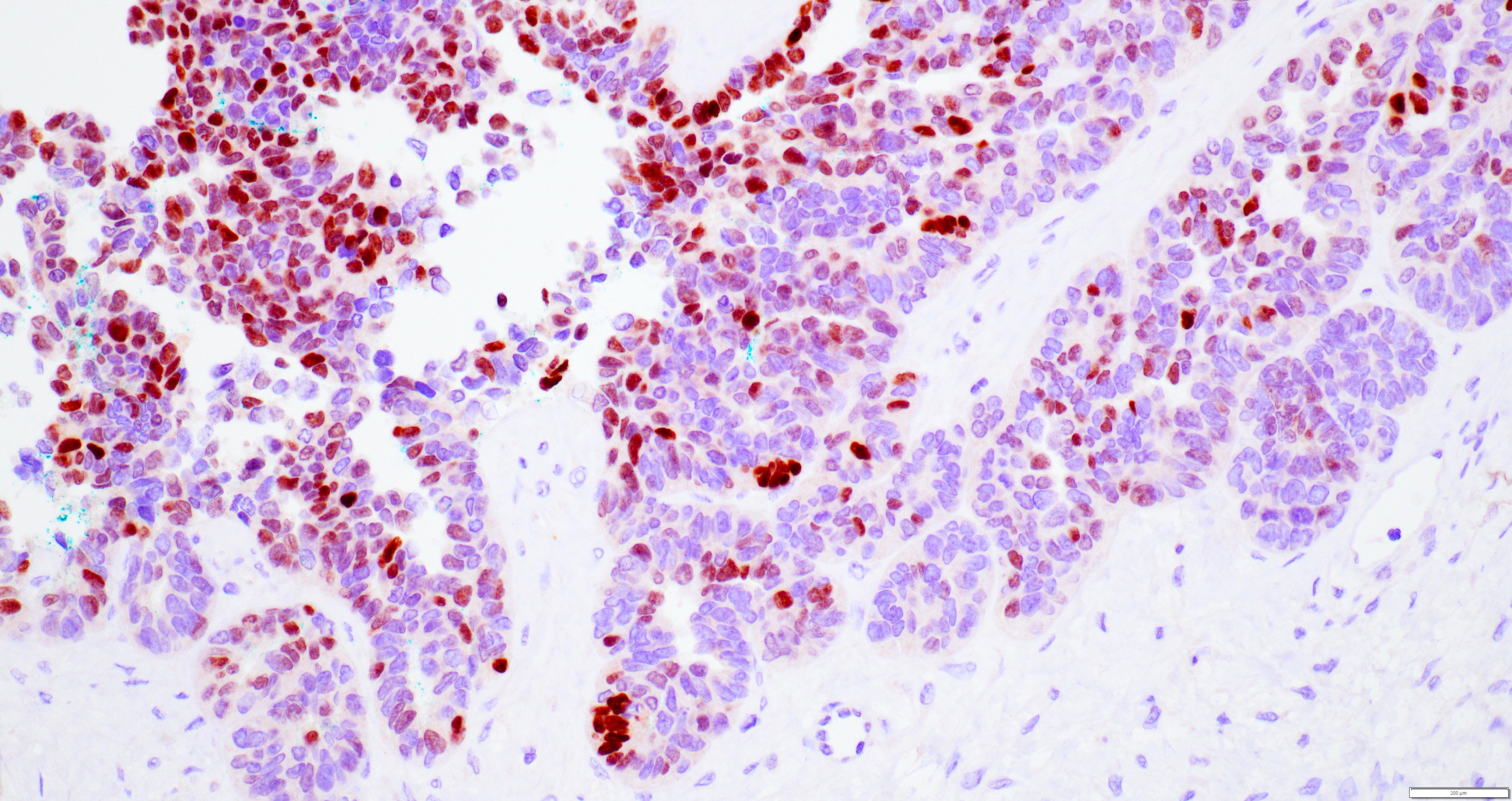
p53
Ki67
WT1
Varied morphology
PTC-like nuclear features
Mesonephric-like adenocarcinoma by Dr. Lewis Hassell

Contributed by Lucy Ma, M.D.
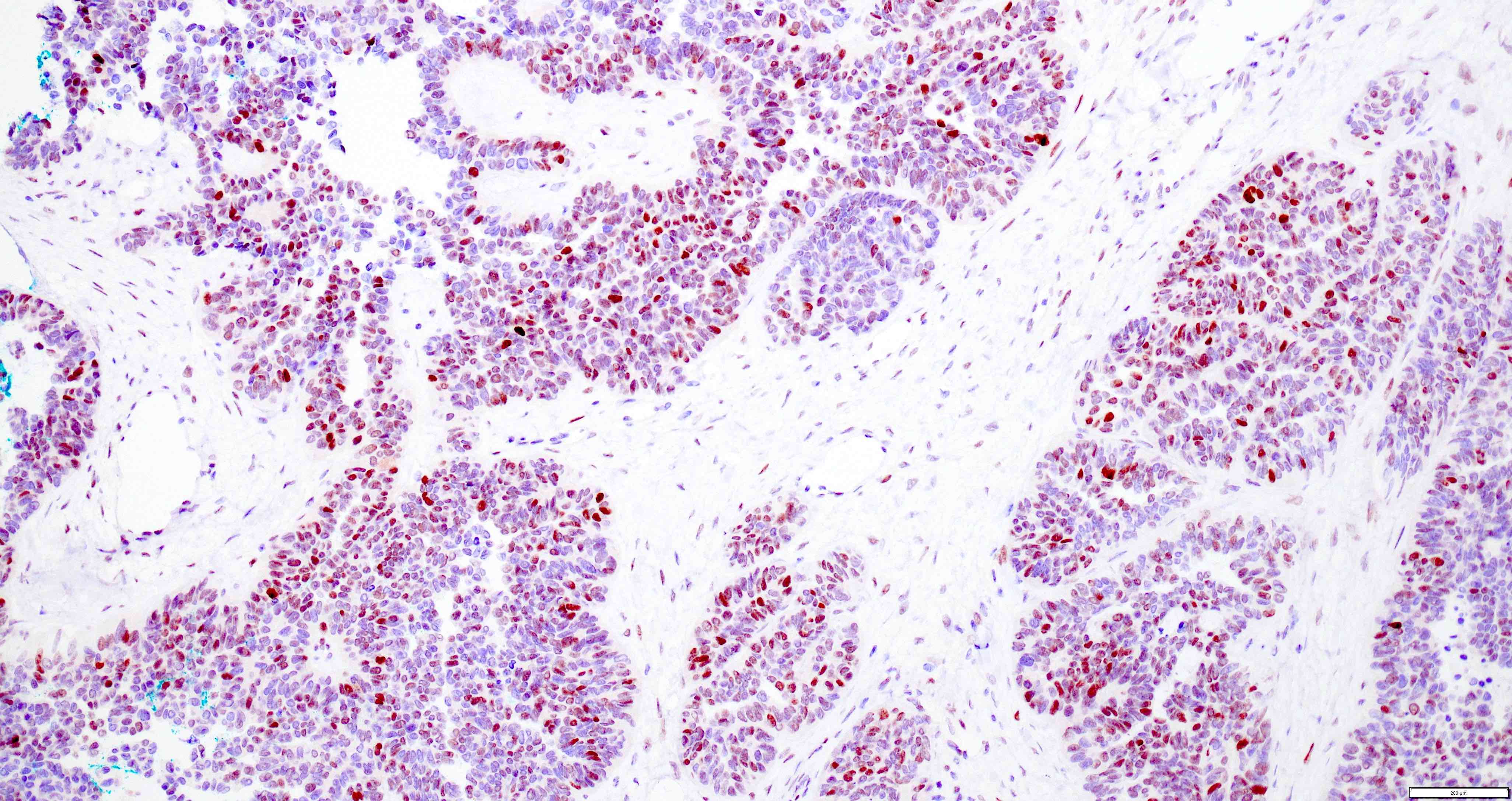
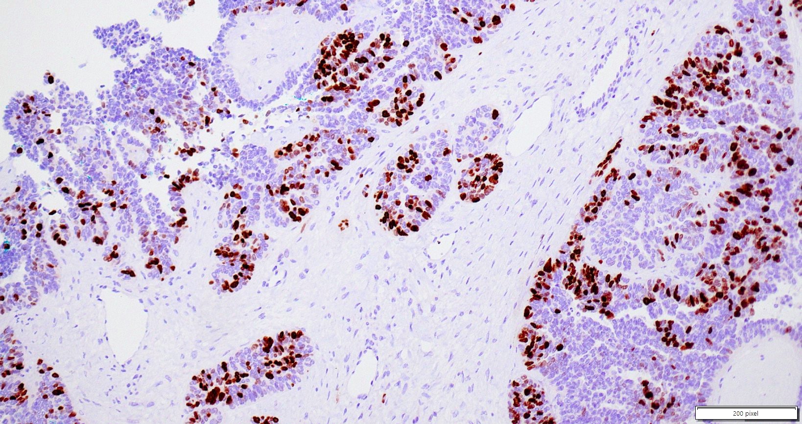
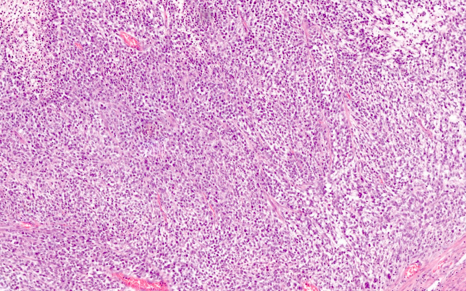
Peritumoral lymphocytes
Tumor infiltrating lymphocytes
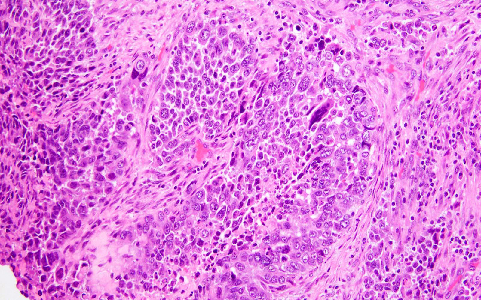
Bizarre atypia
Intratumoral heterogeneity
Endometrioid histotype
Images hosted on other servers:

Sagittal T2 with heterogeneous hyperintense mass

MRI with hyperintense mass
Images hosted on other servers:

Posterior uterine mass

PEComa
Contributed by Jennifer Bennett, M.D.
Diffuse growth of epithelioid cells
Infiltrative growth
Delicate vasculature
Spindled cells
Stromal hyalinization
Multinucleated giant cells
Intranuclear pseudoinclusions
Melanoma-like macronucleoli
TFE3 associated PEComa
LAM-like PEComa
HMB45
MelanA / MART1
Desmin
Cathepsin K
Images hosted on other servers:

EIN / carcinoma remission probability

EIN / carcinoma recurrence probability
Contributed by Carlos Parra-Herran, M.D.
Initial biopsy
3 month follow up
6 month follow up
PathCast lecture on benign endometrial pathology (May 2020)
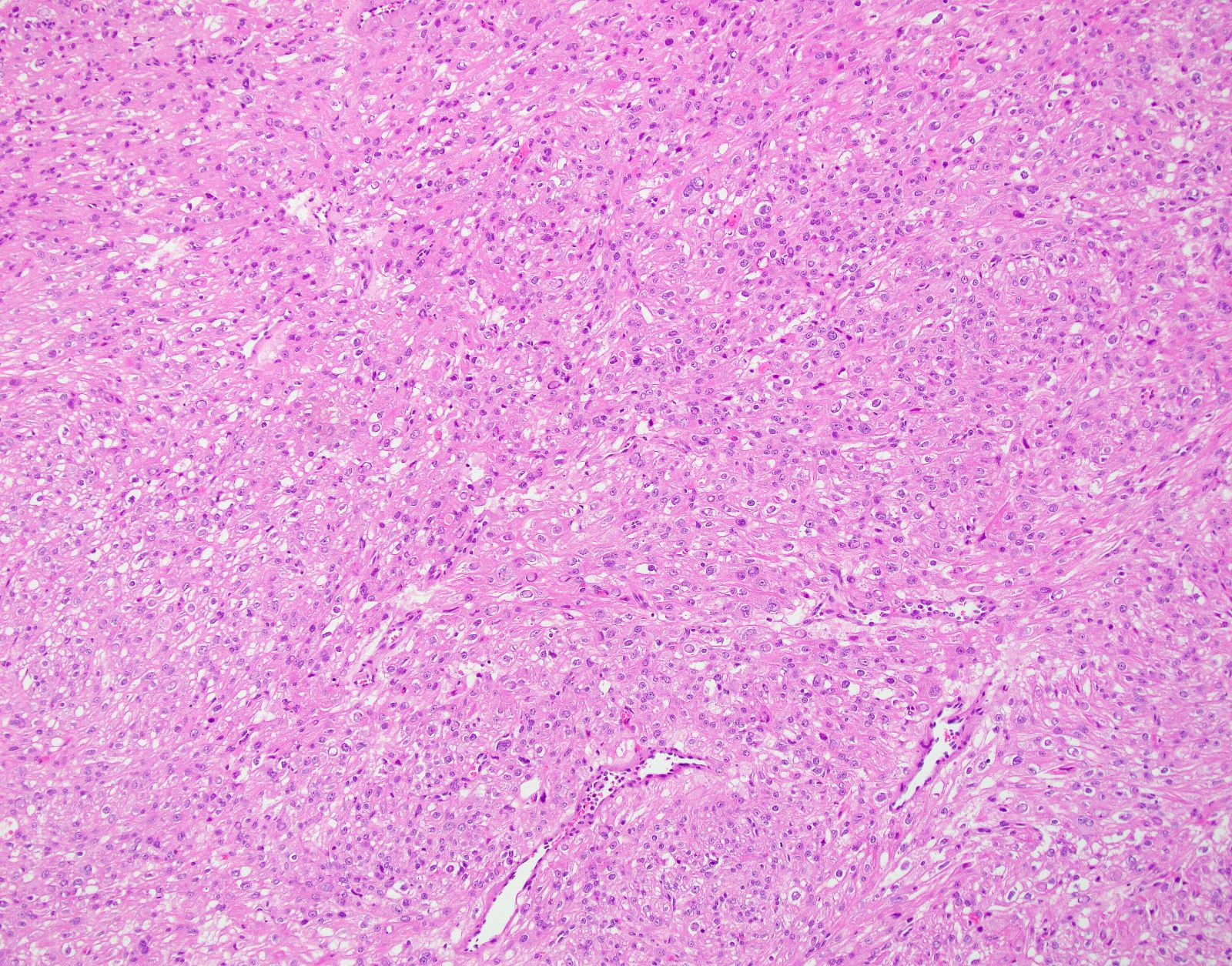
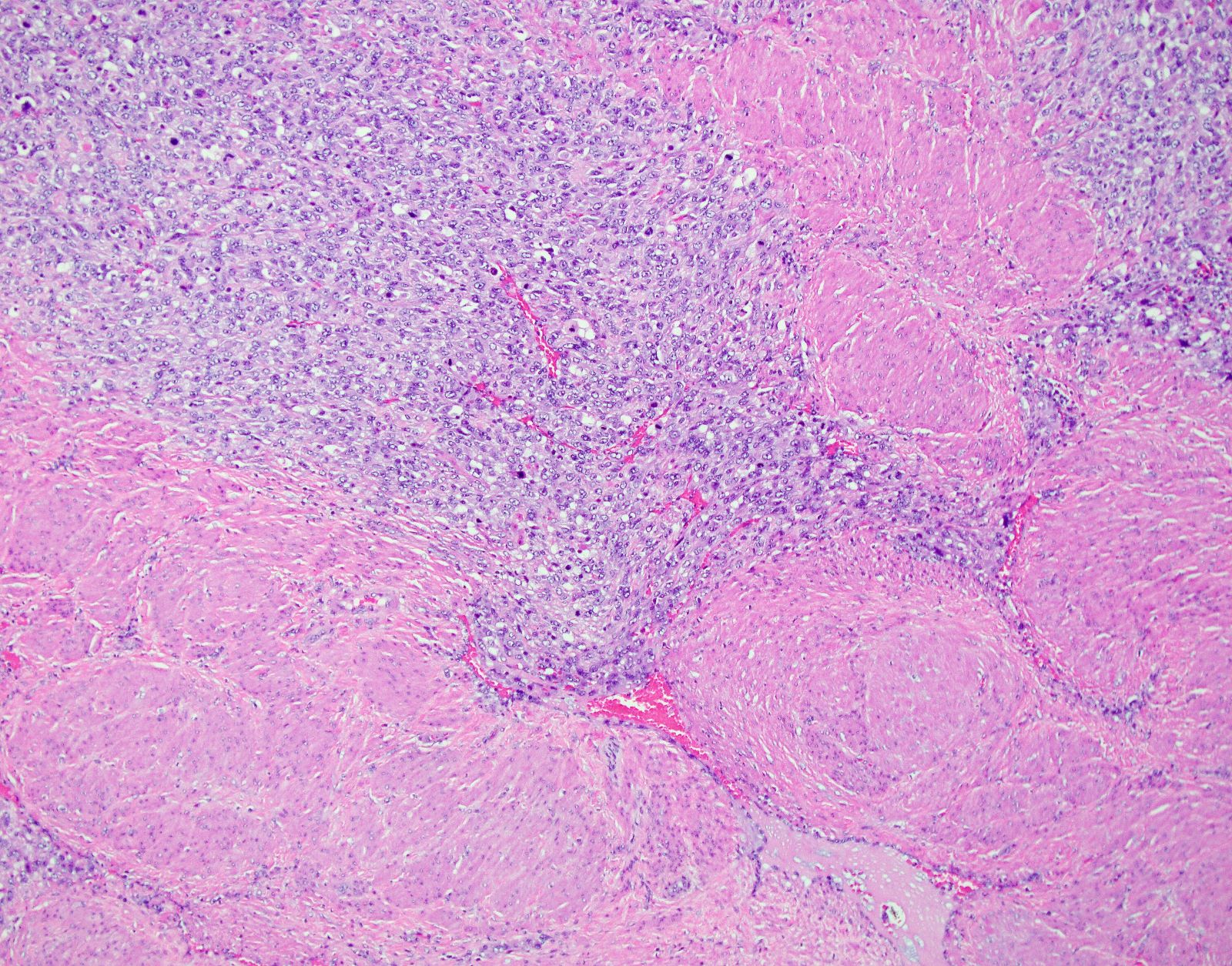
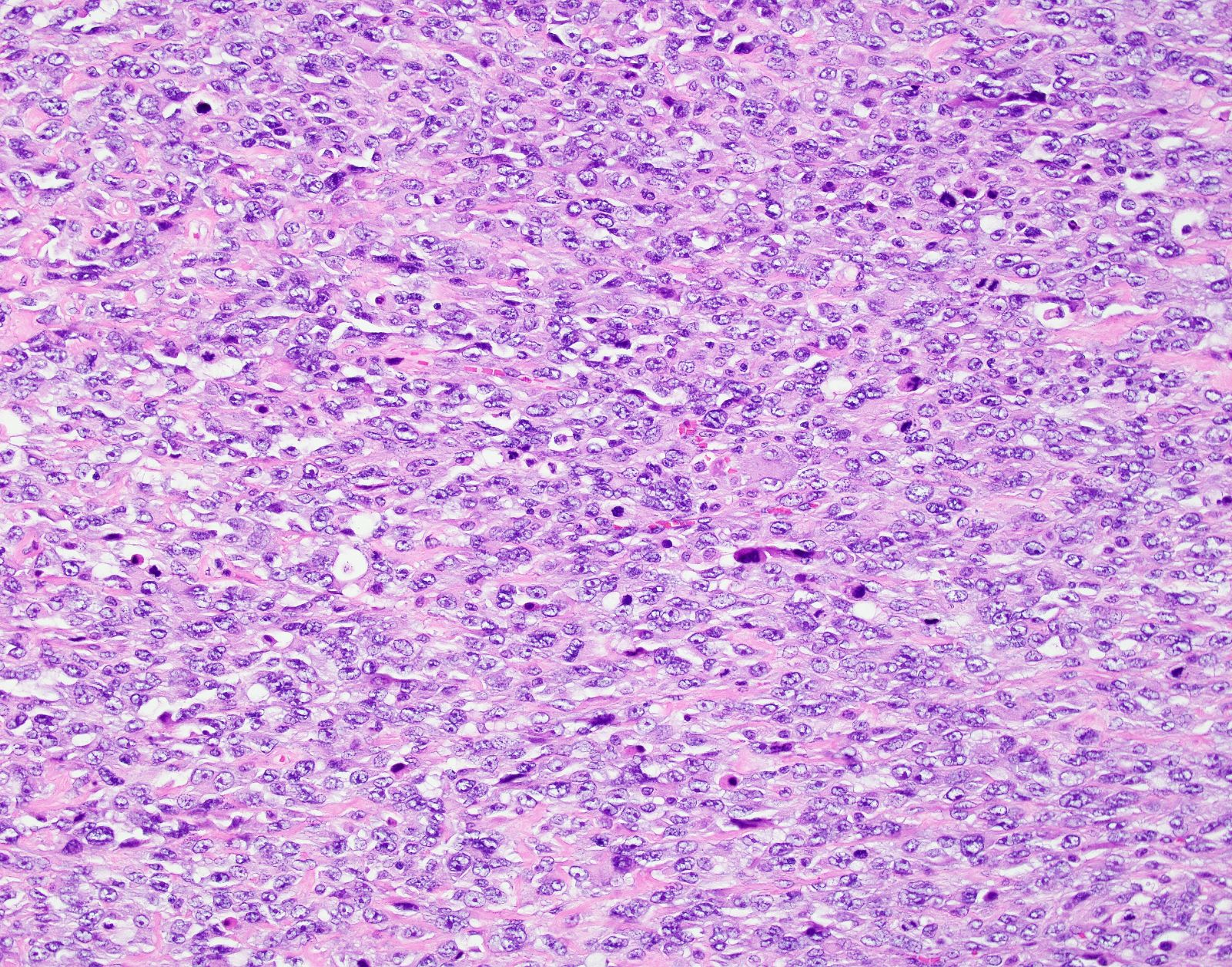
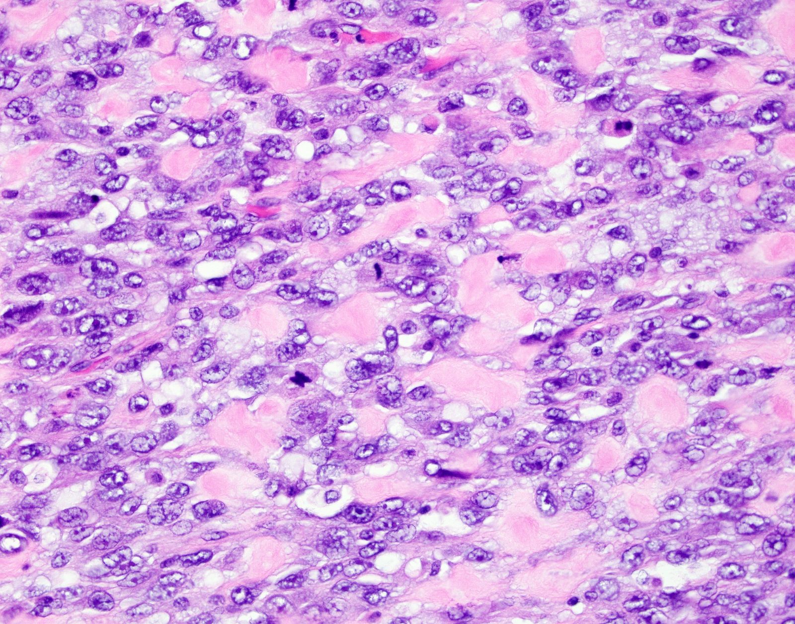
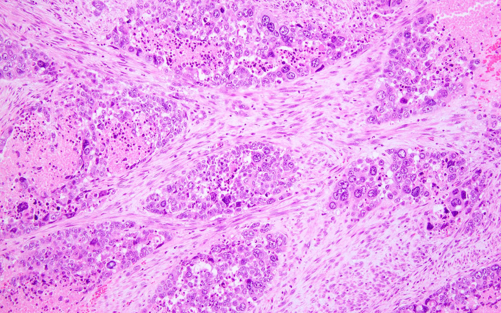
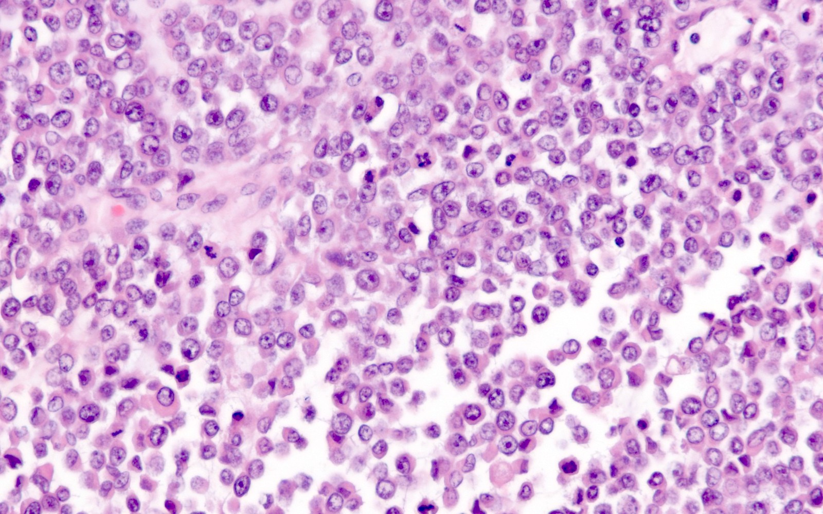
Contributed by Felix K.F. Kommoss, M.D.
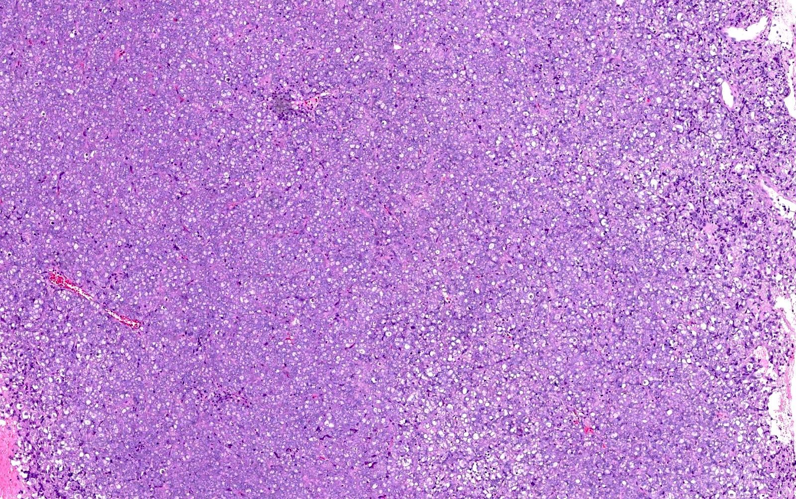
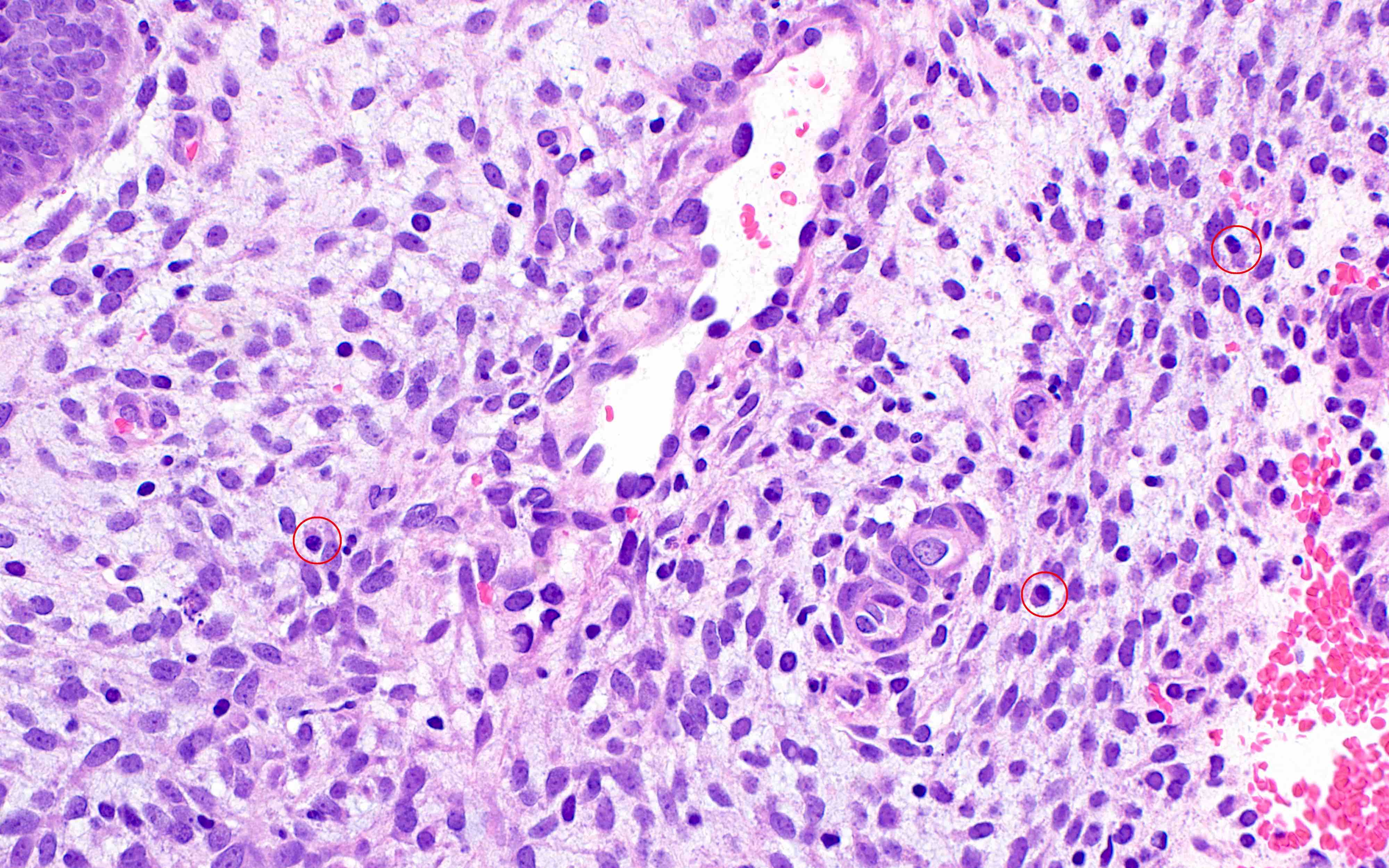
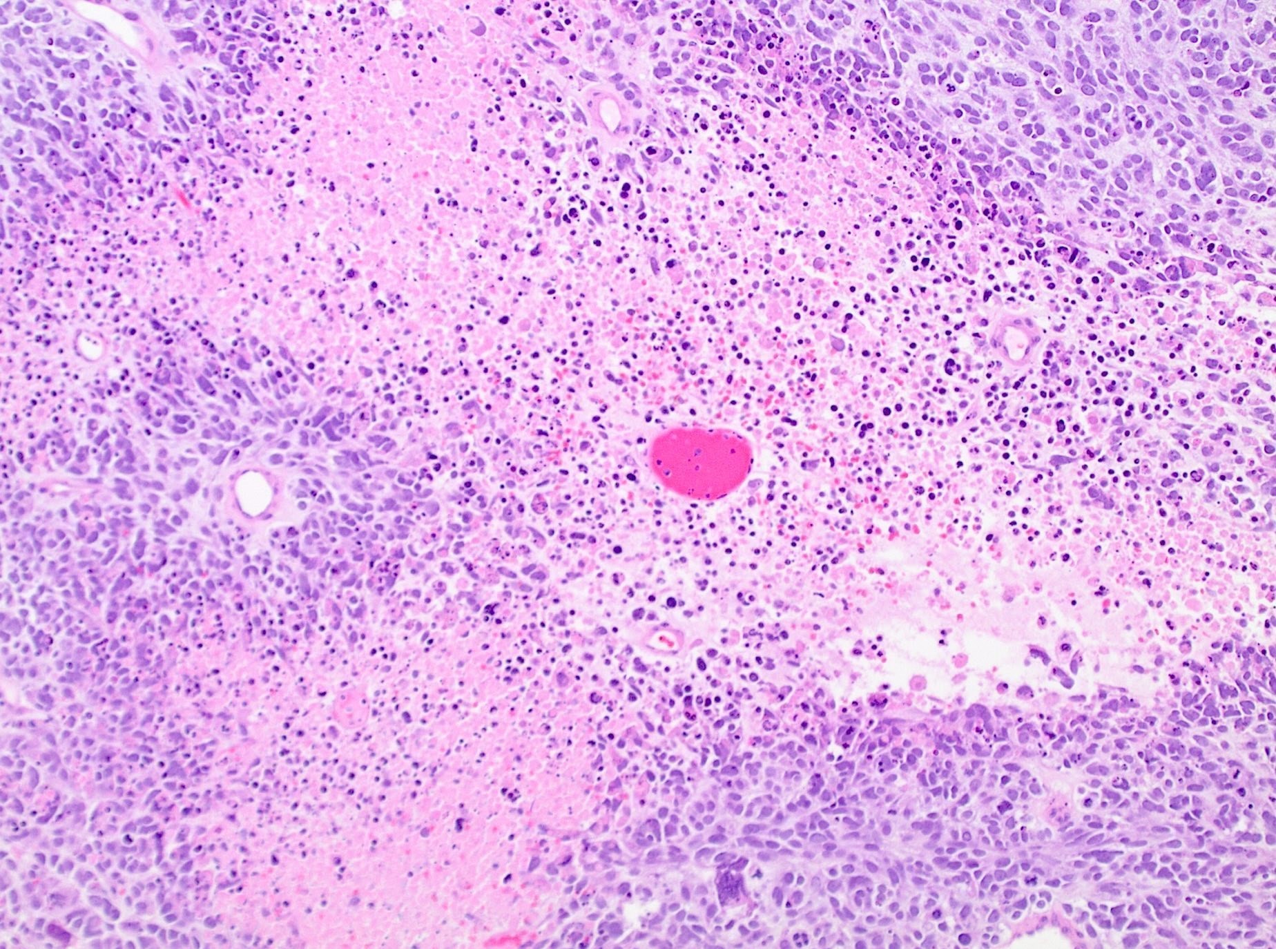
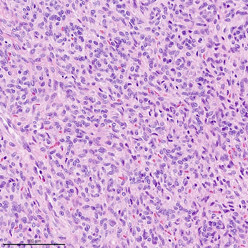
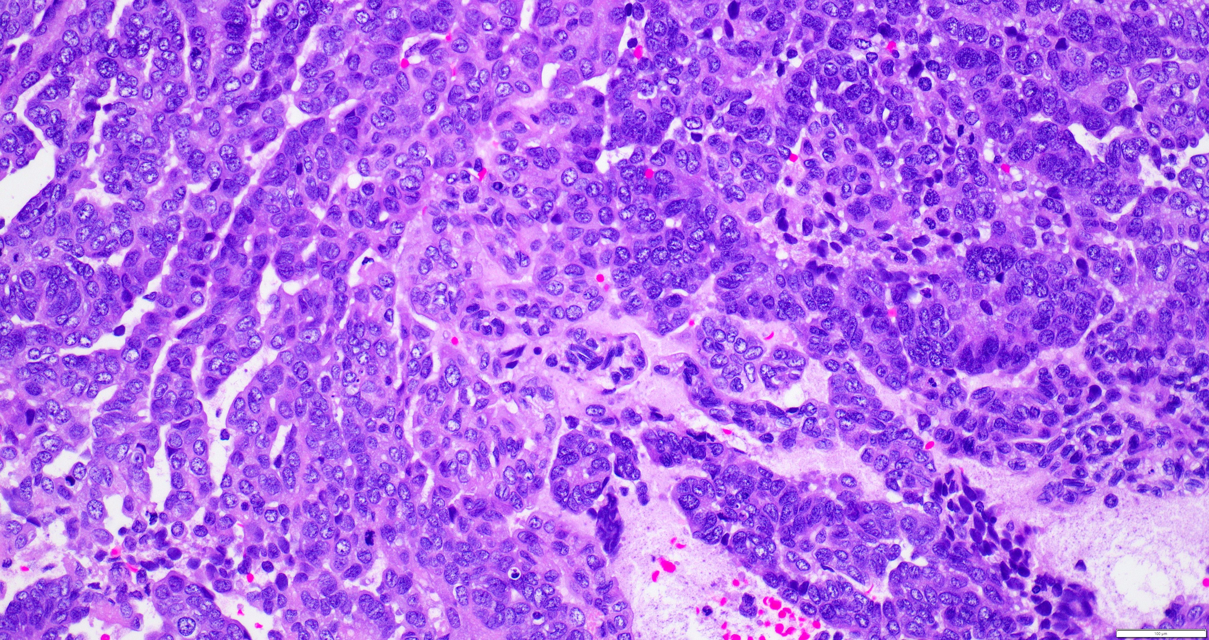
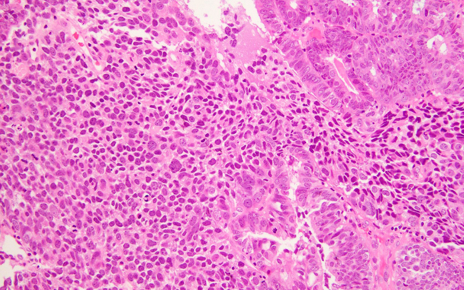
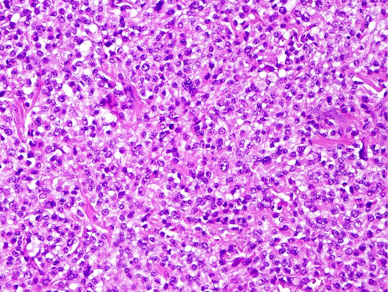
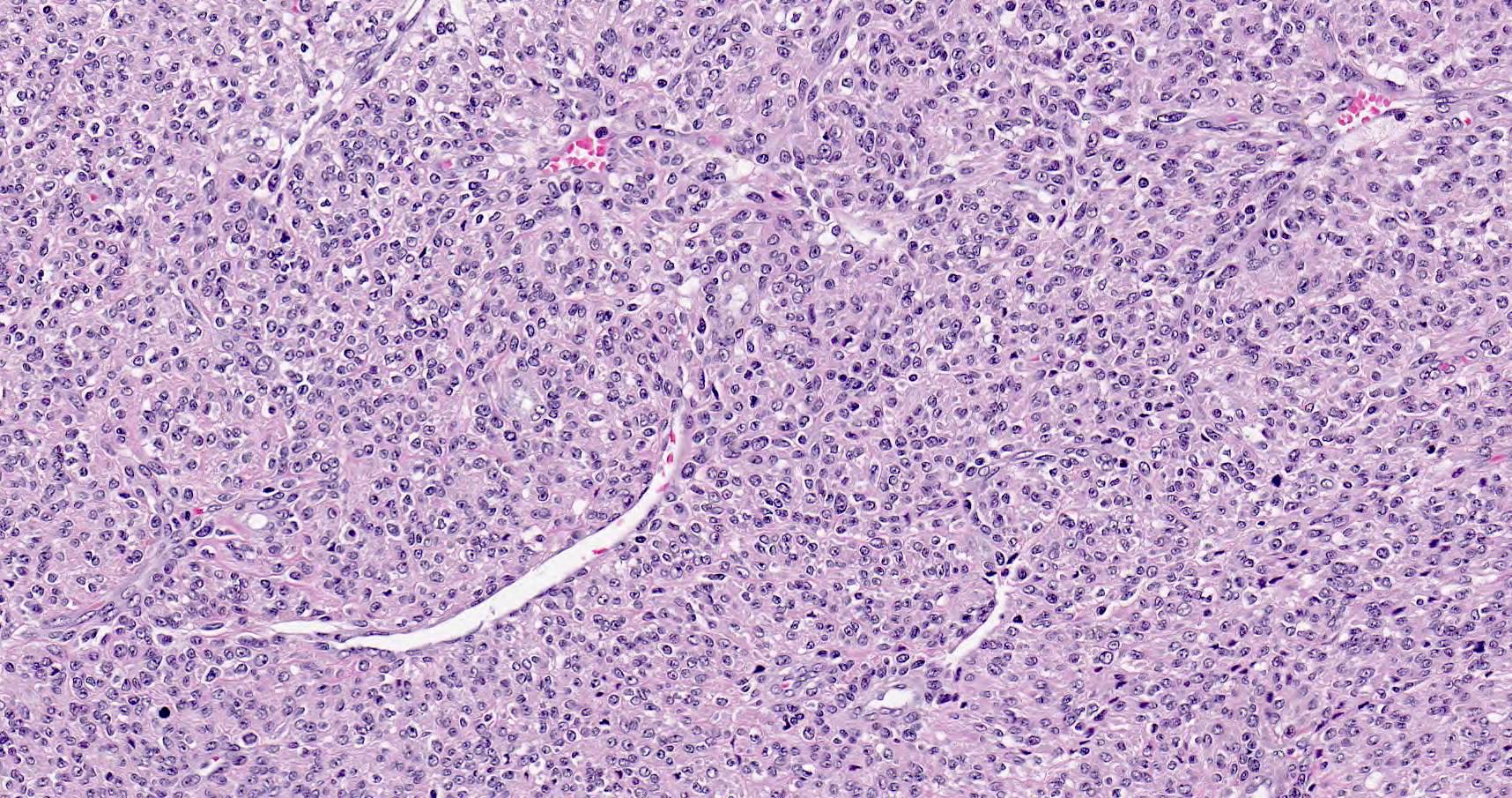
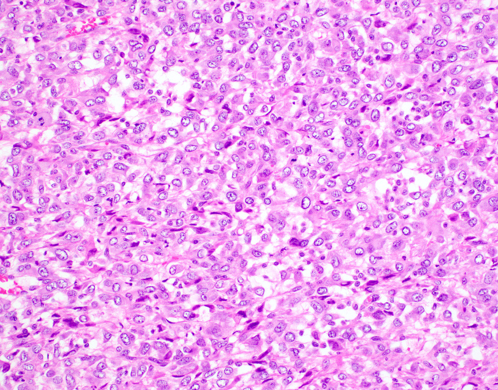
Tumor cells in sheets, no gland or papillary formation
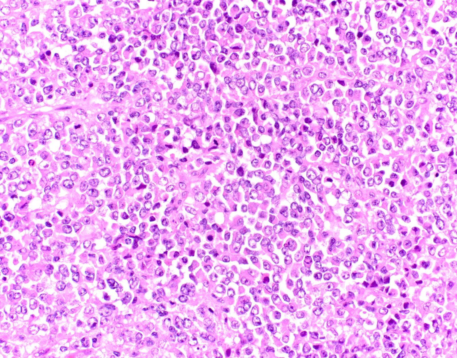
Discohesive and rhabdoid morphology
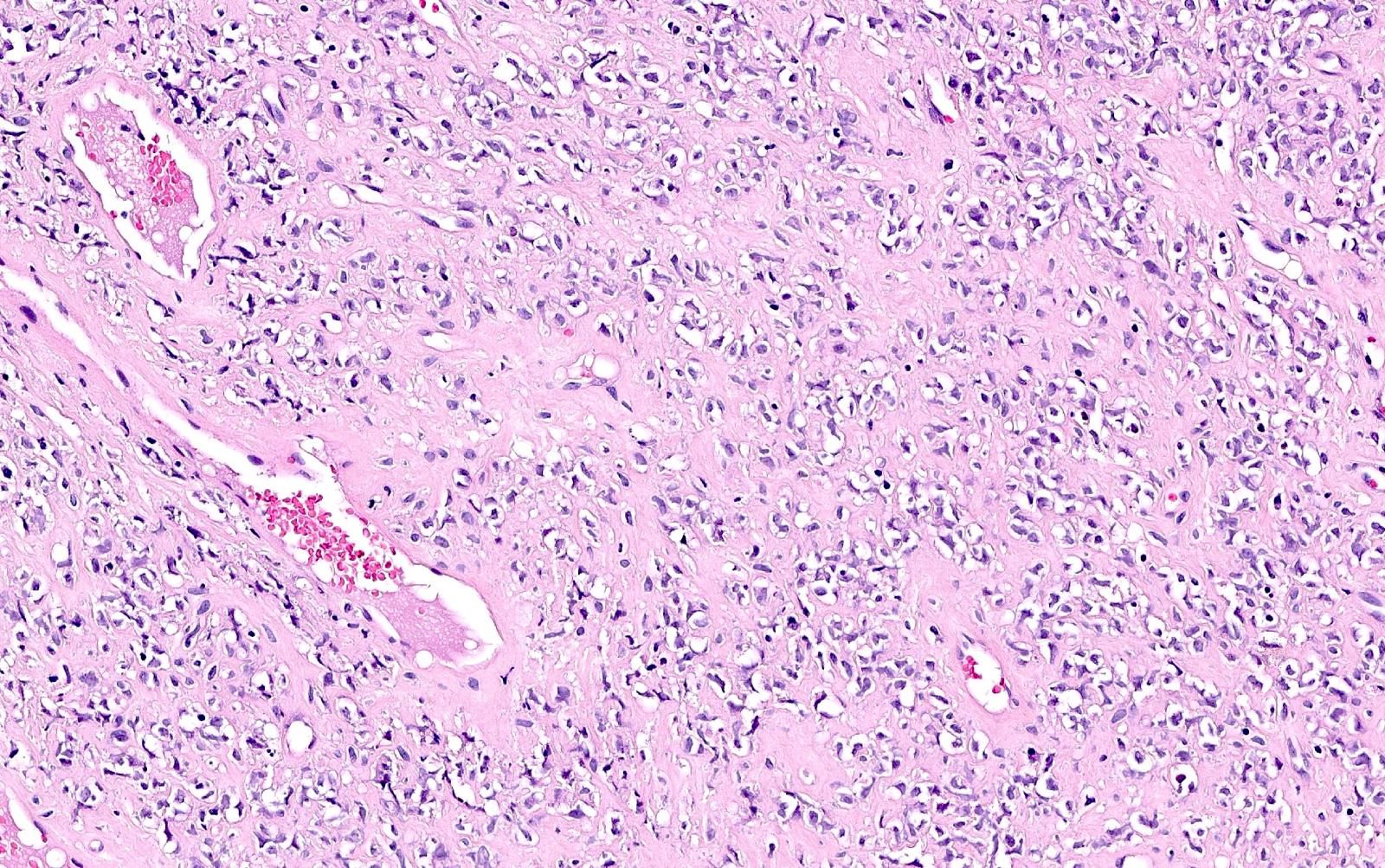
Stromal hyalinization
Mitotic activity
Tumor necrosis
SMARCA4 loss of expression
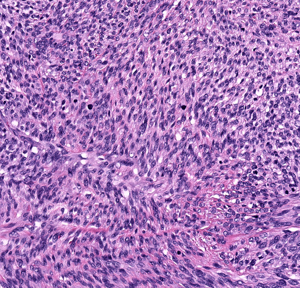
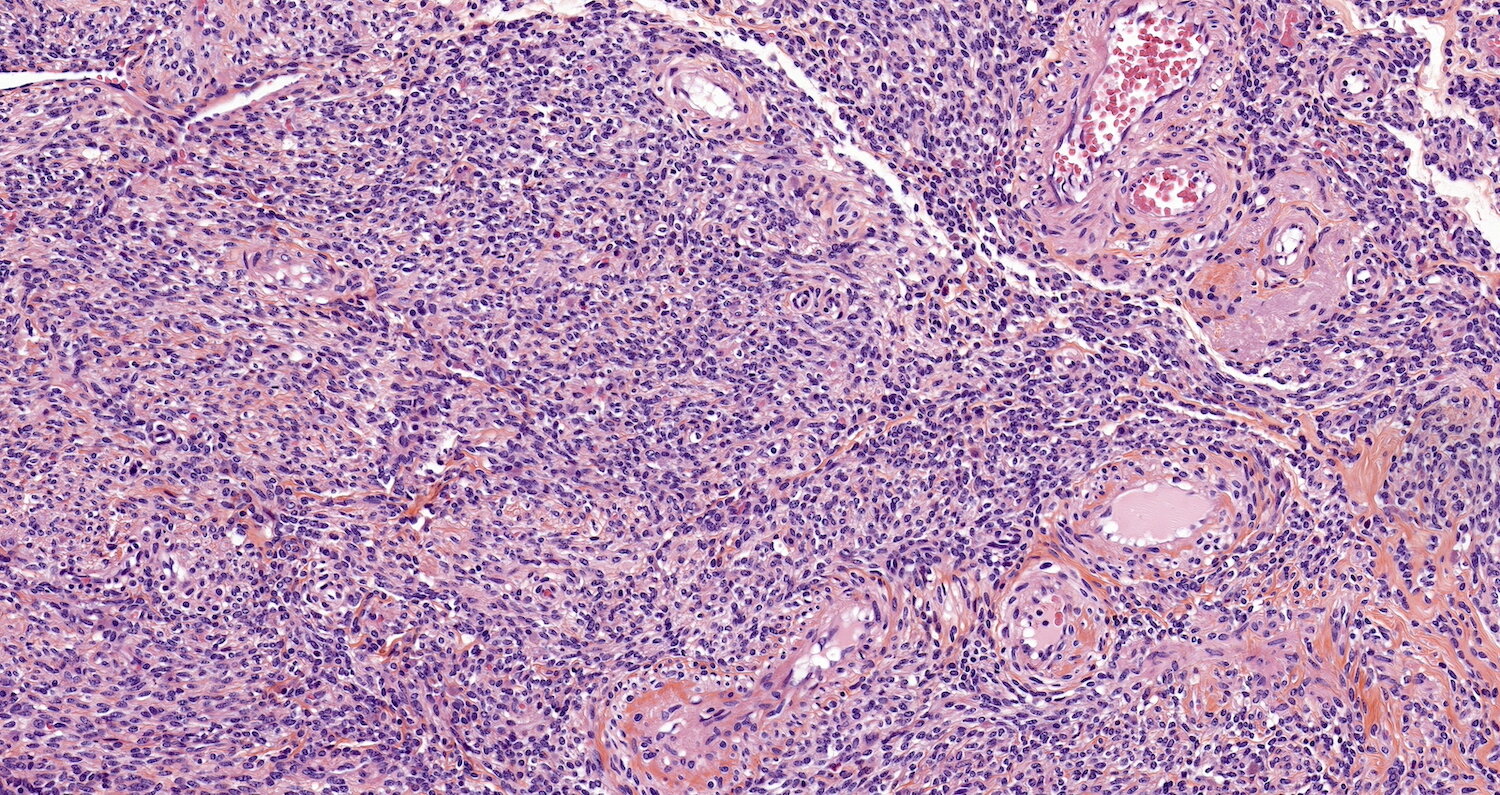
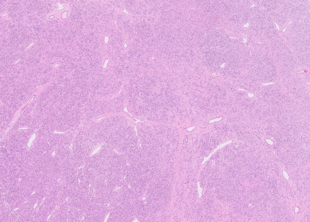
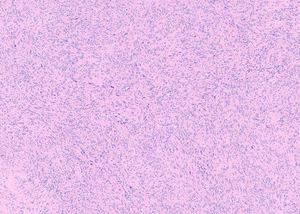
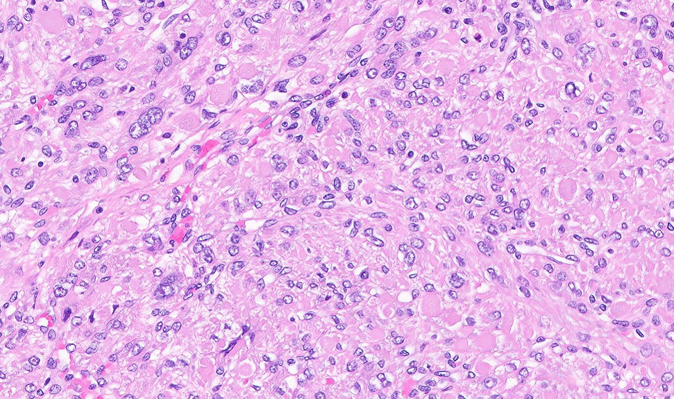
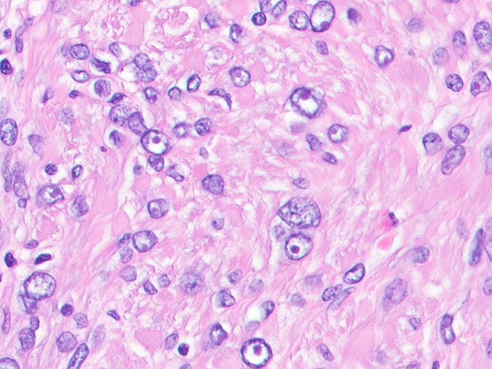
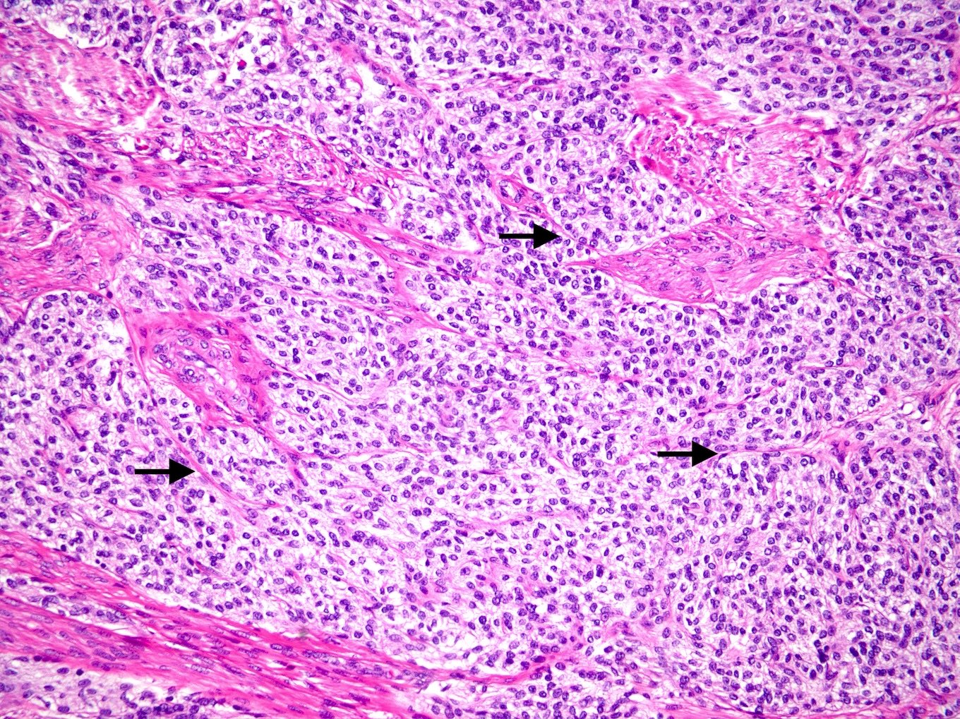
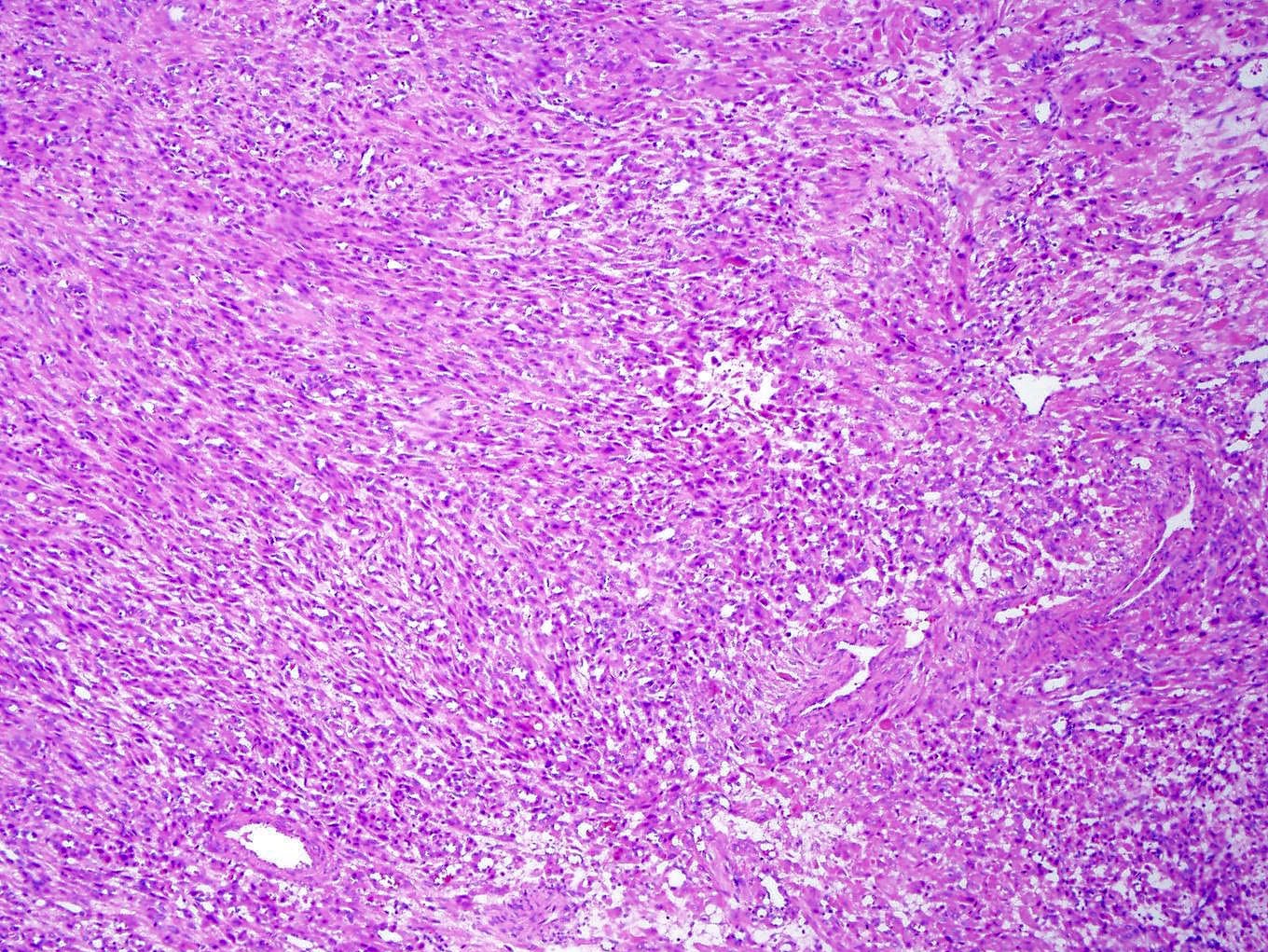
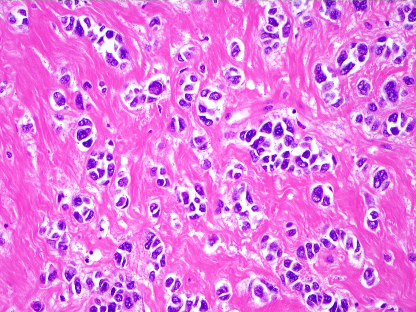
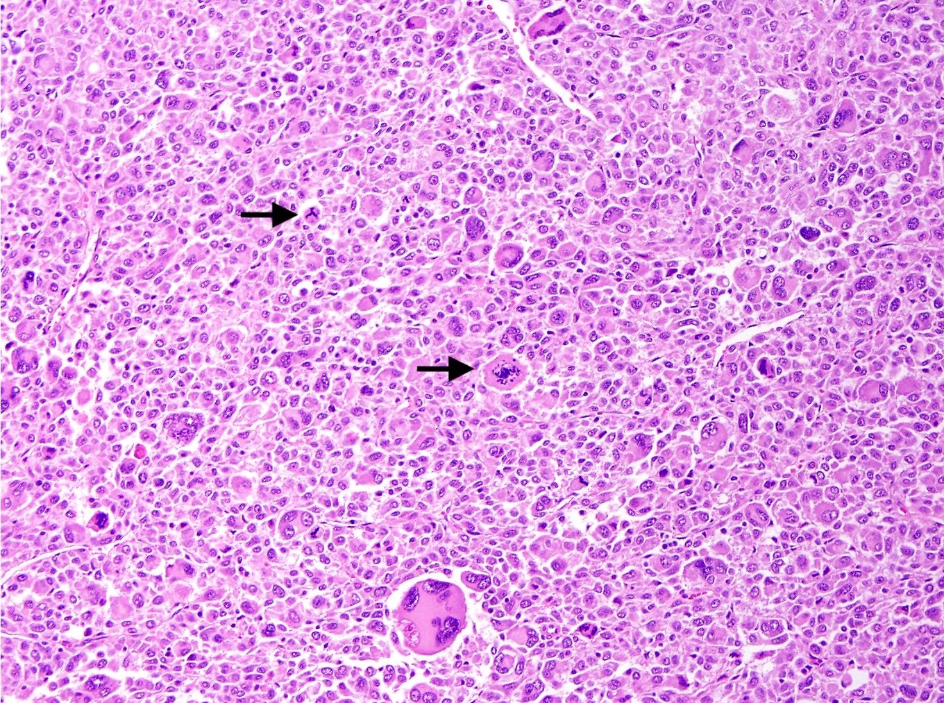
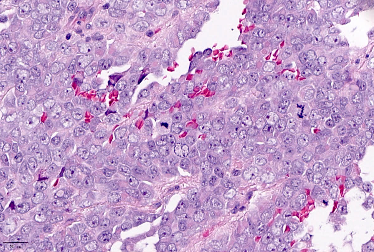
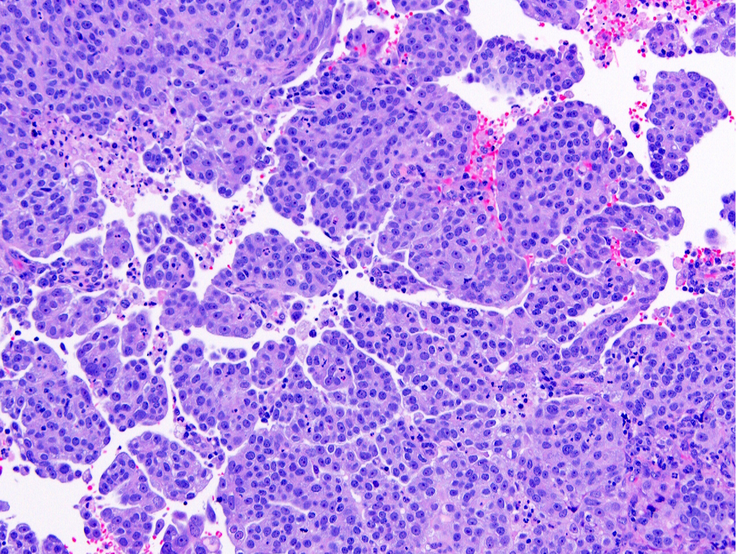
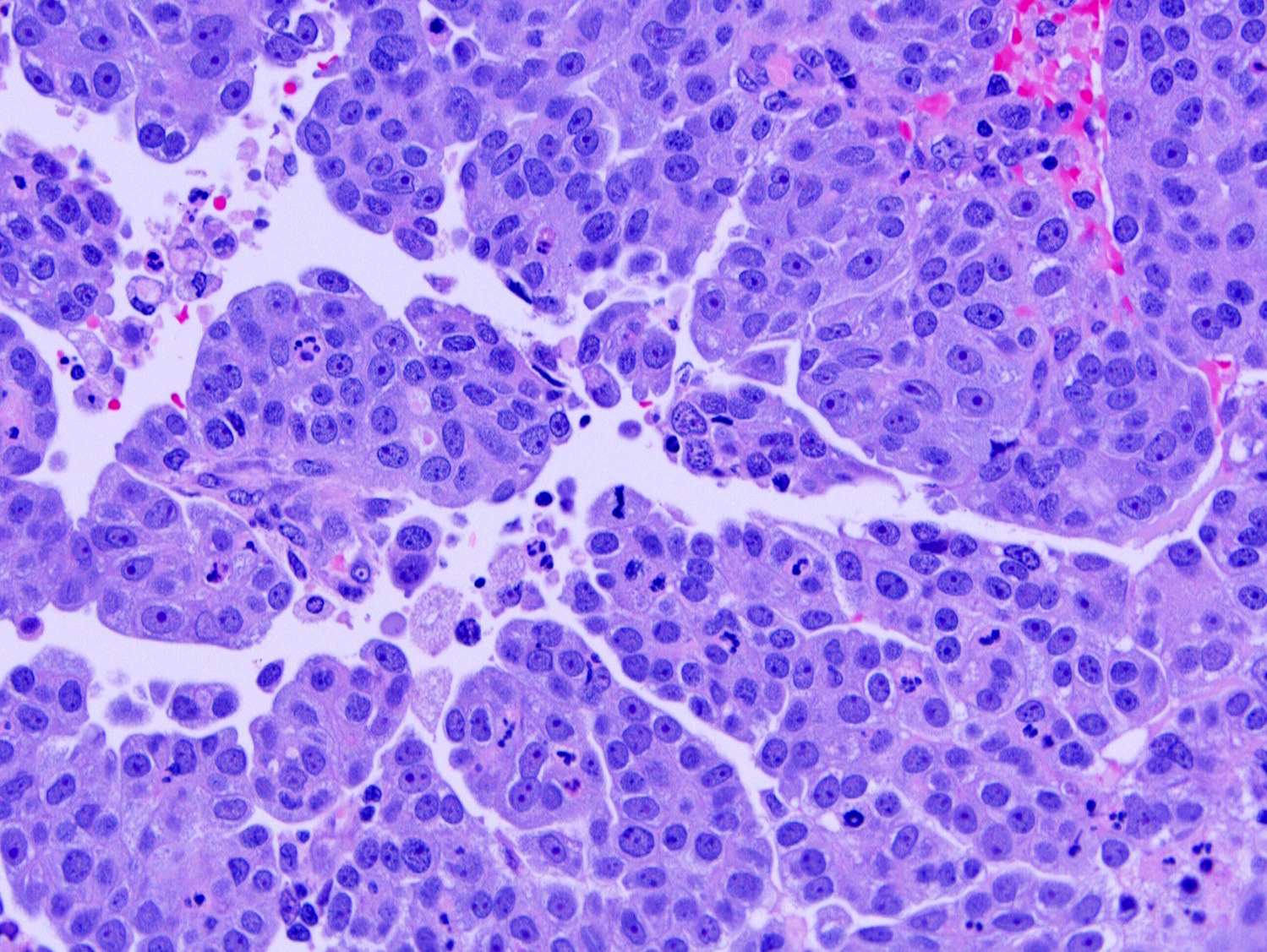
Contributed by Ricardo Lastra, M.D.

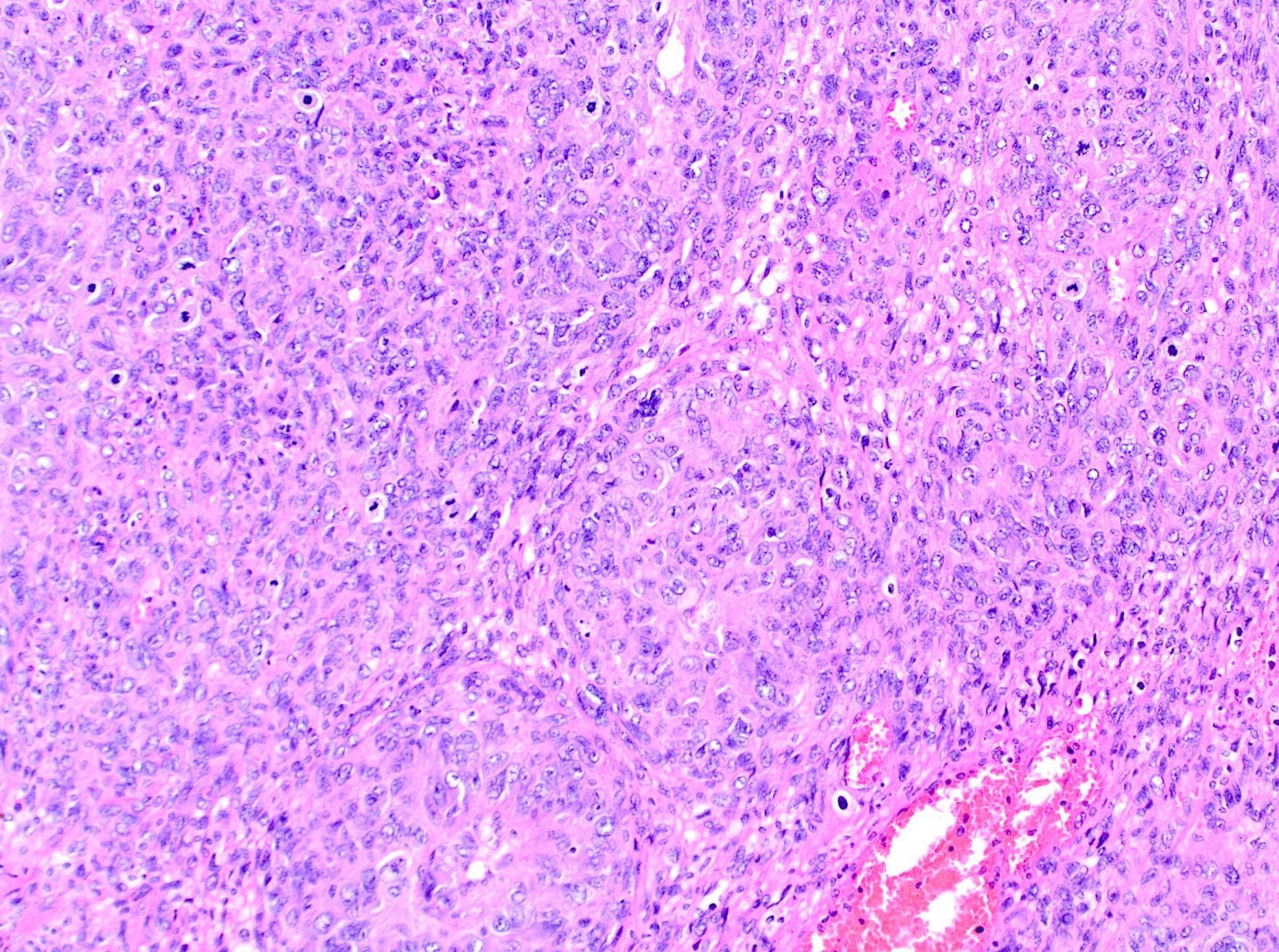
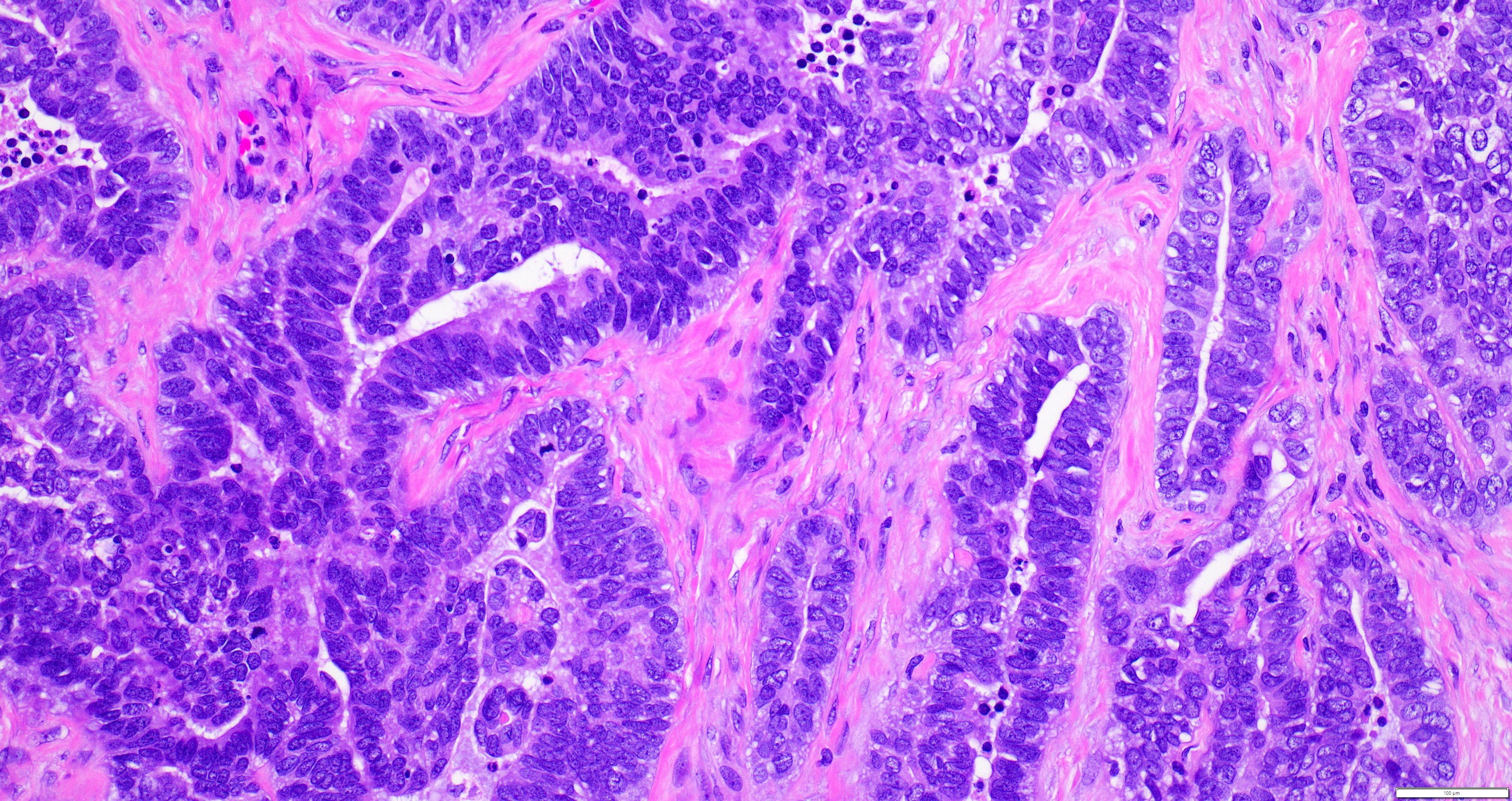
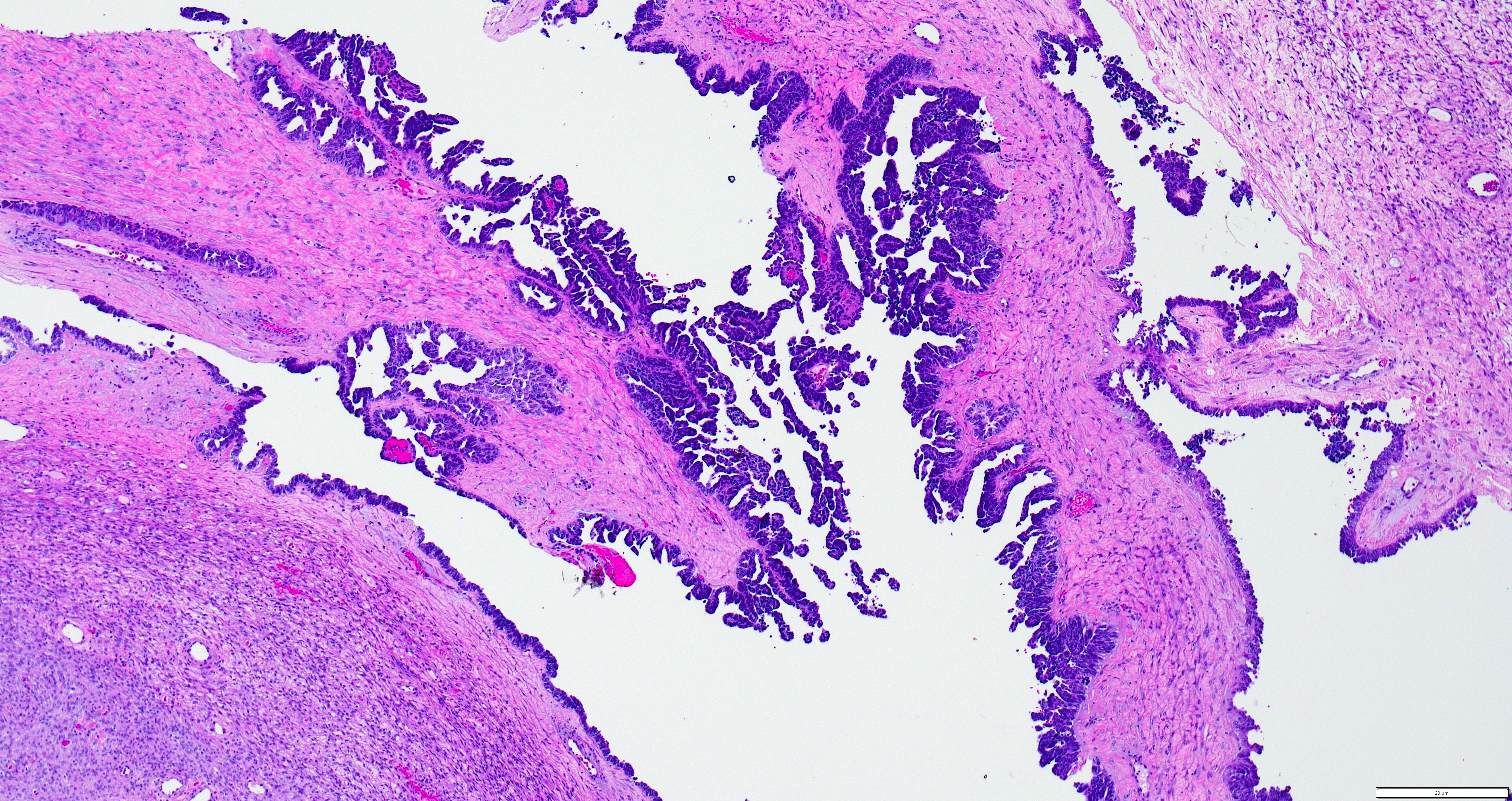
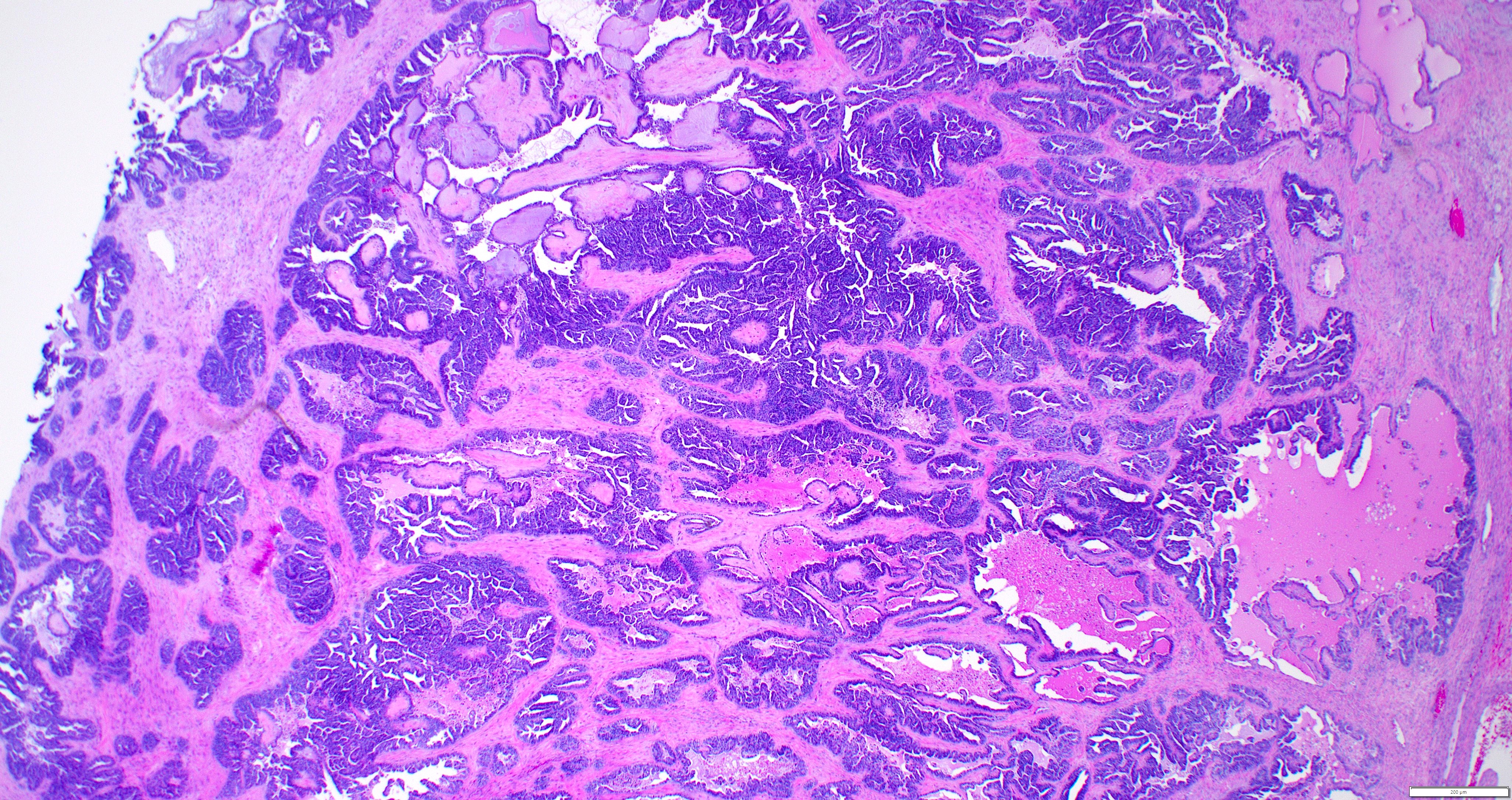
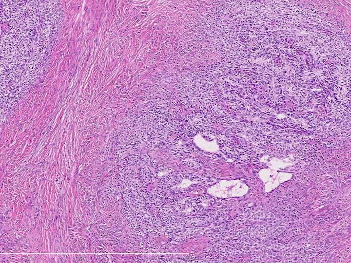
Involving polyp (intraepithelial carcinoma)
Solid and papillary growth
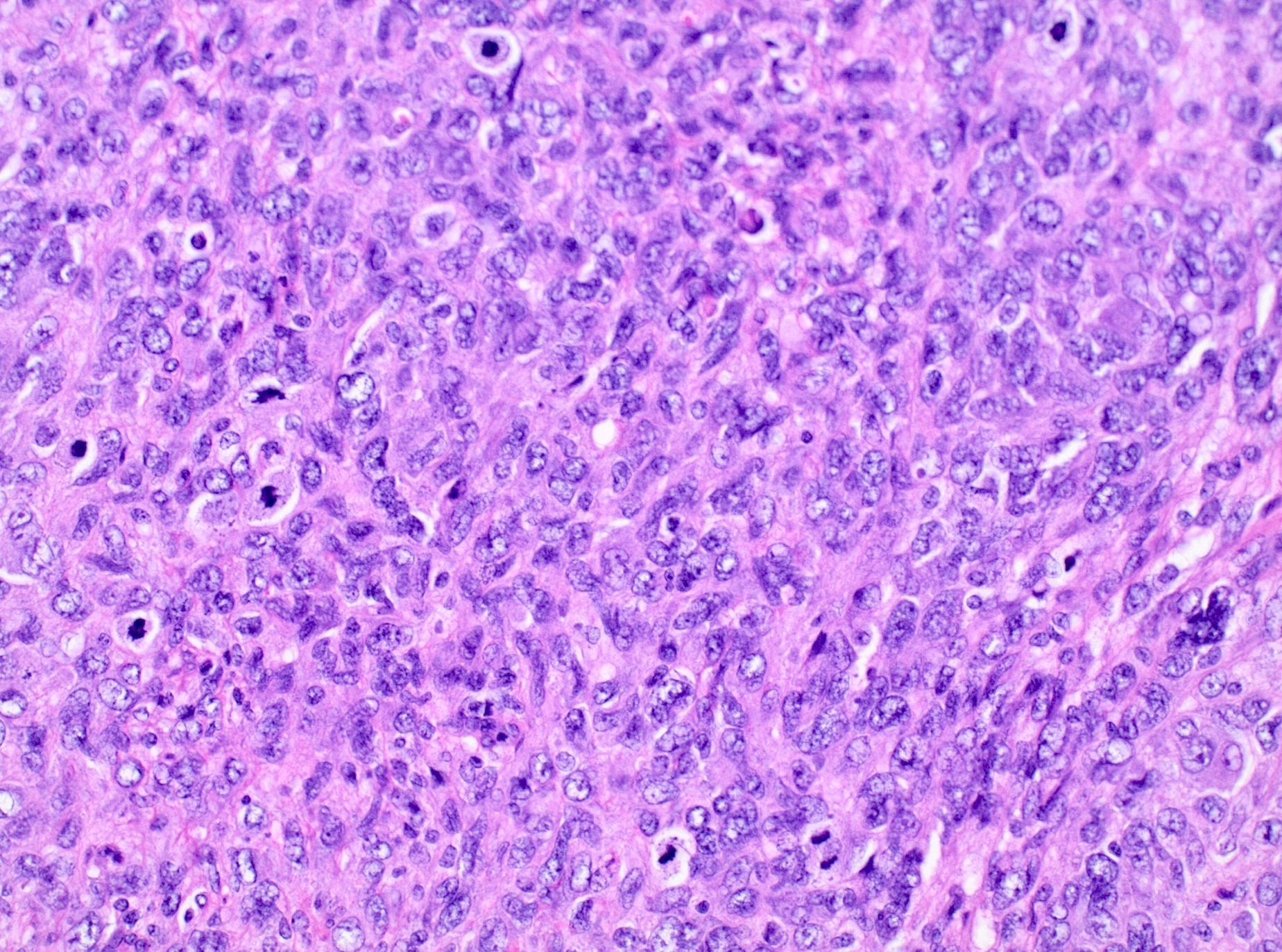
Pleomorphic nuclei

Glandular growth
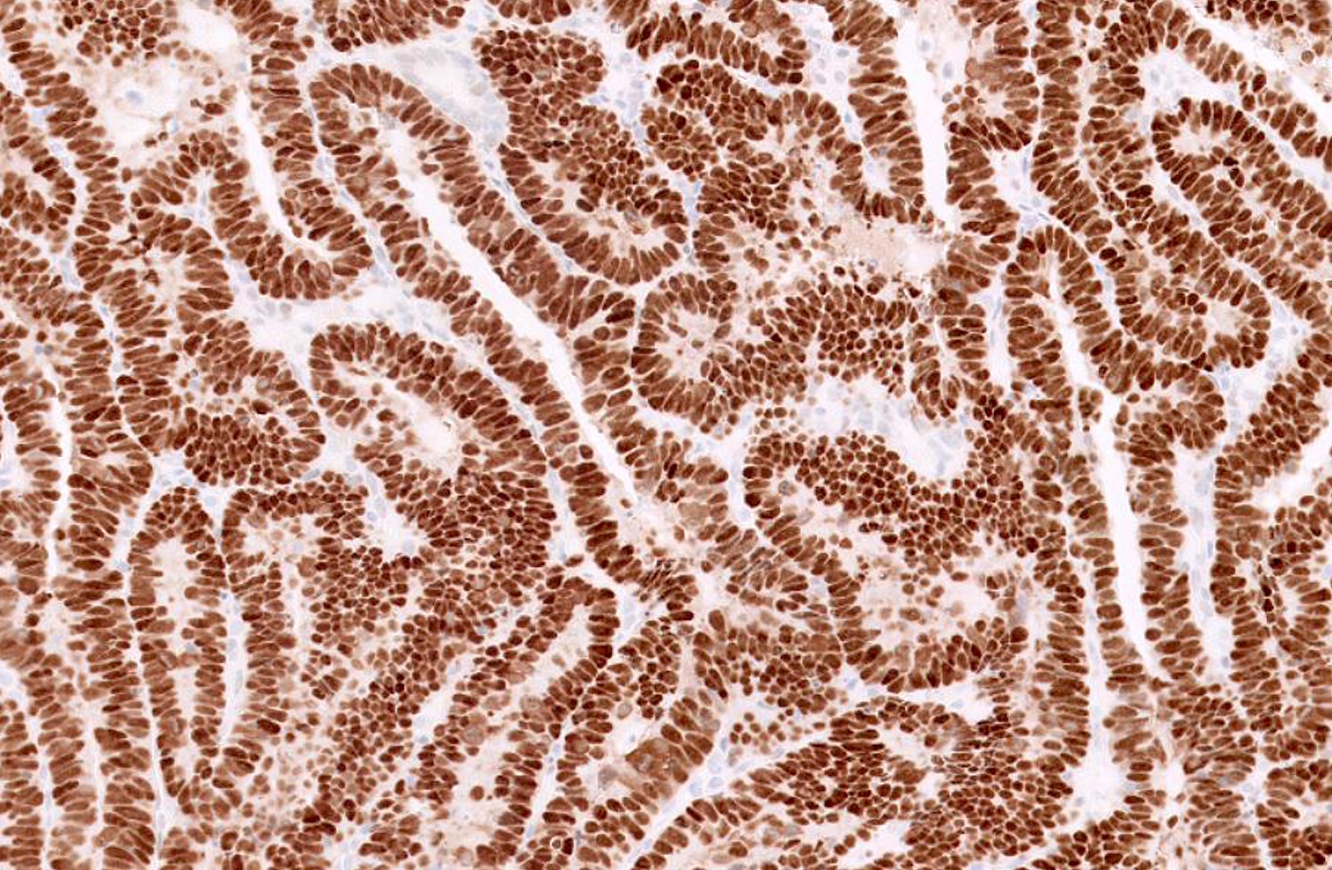
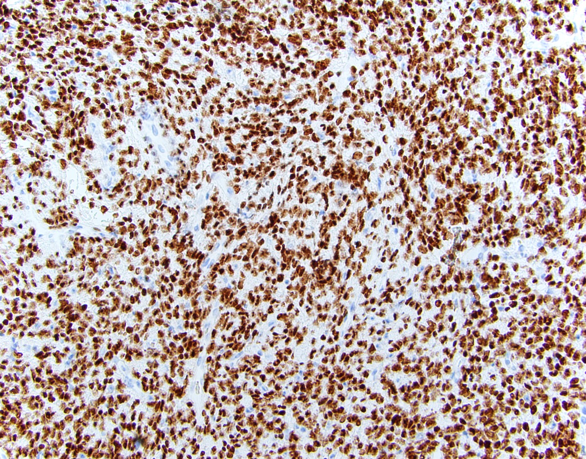
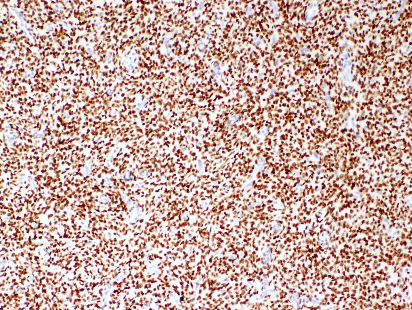
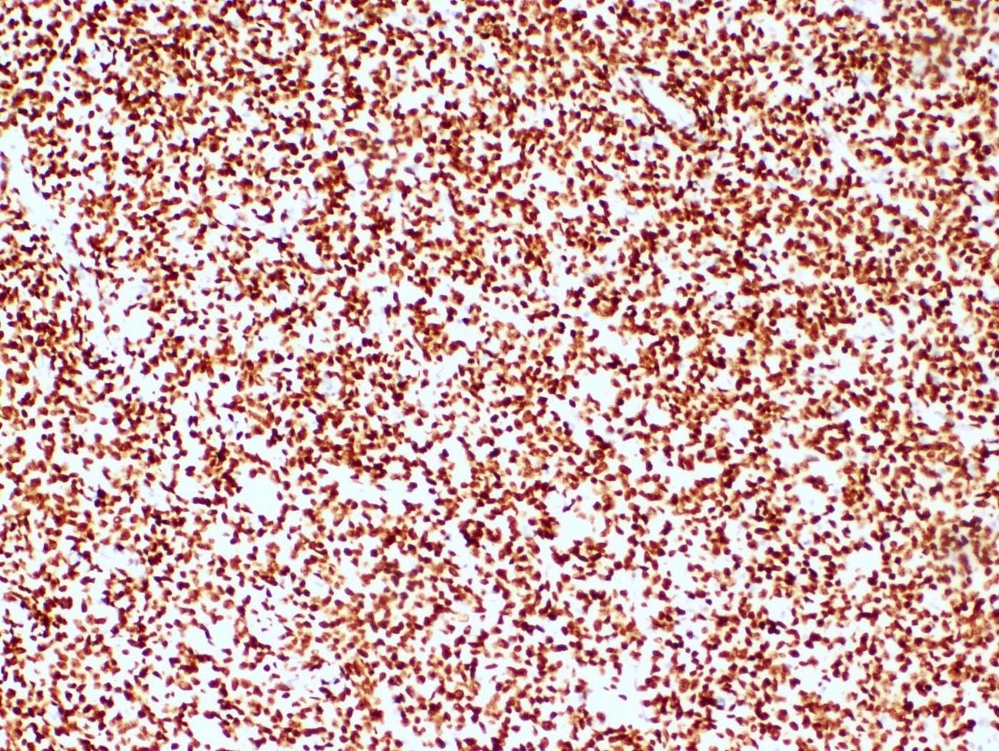
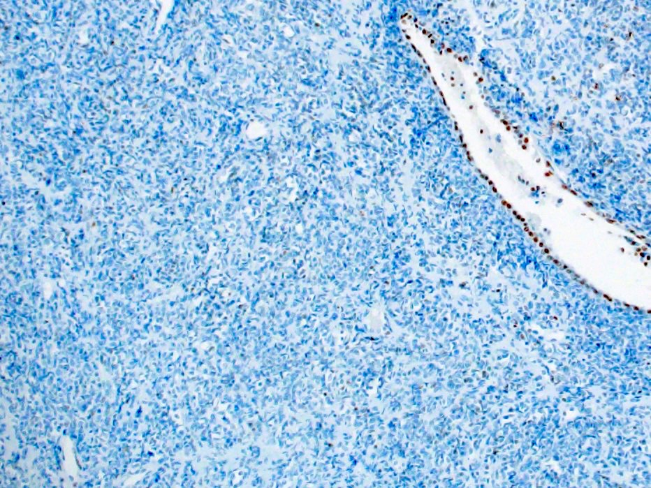
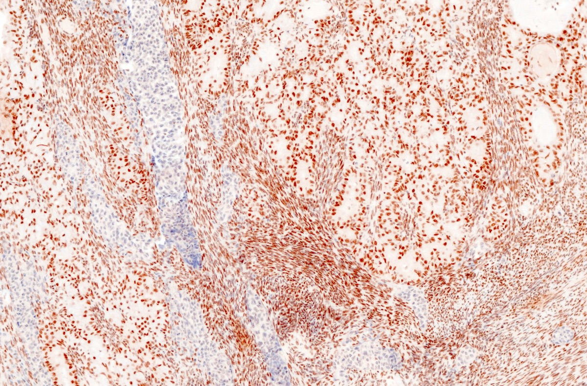
p53 overexpression
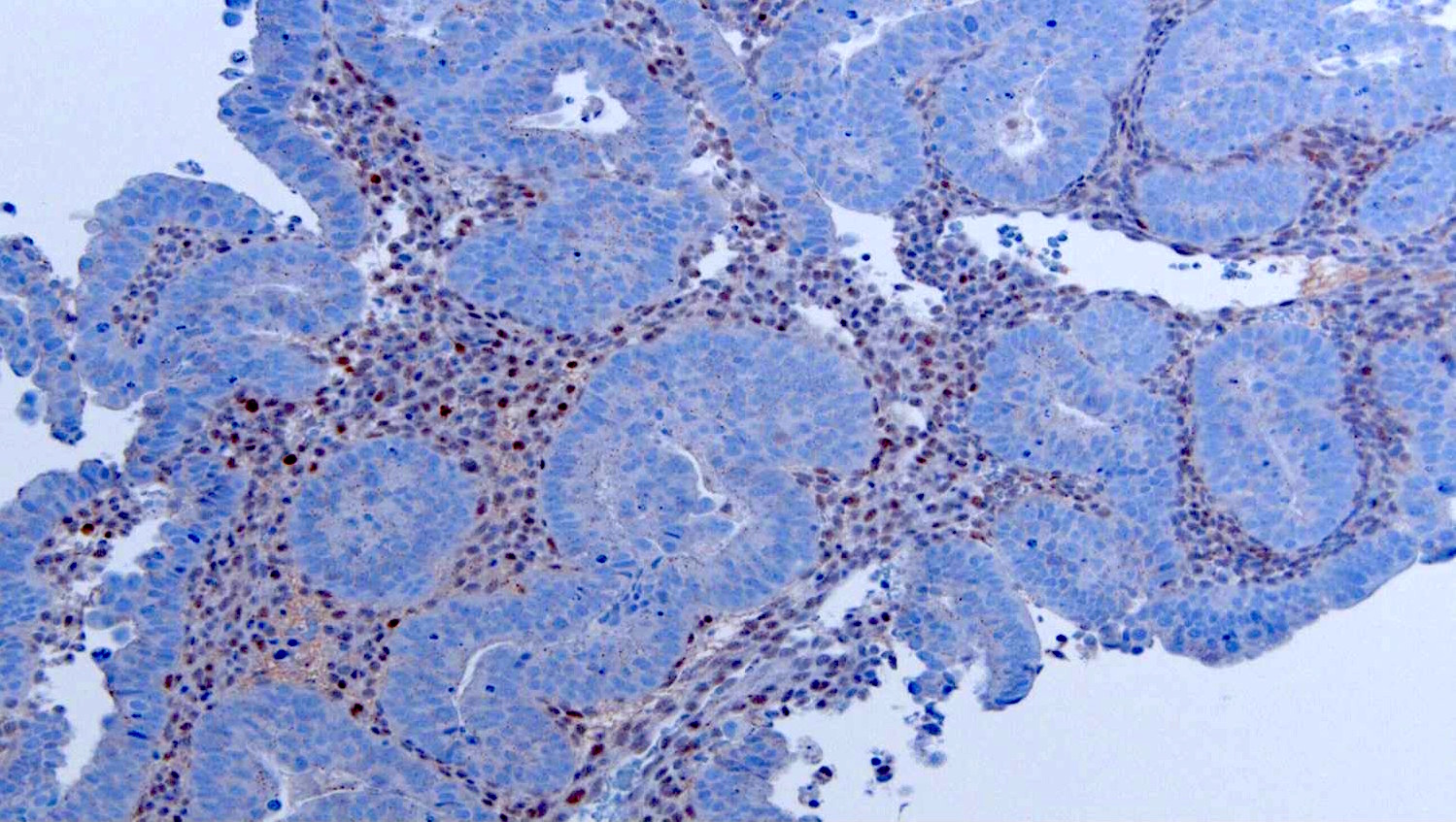
Complete absence of p53 IHC
p16
Contributed by Ricardo Lastra, M.D.
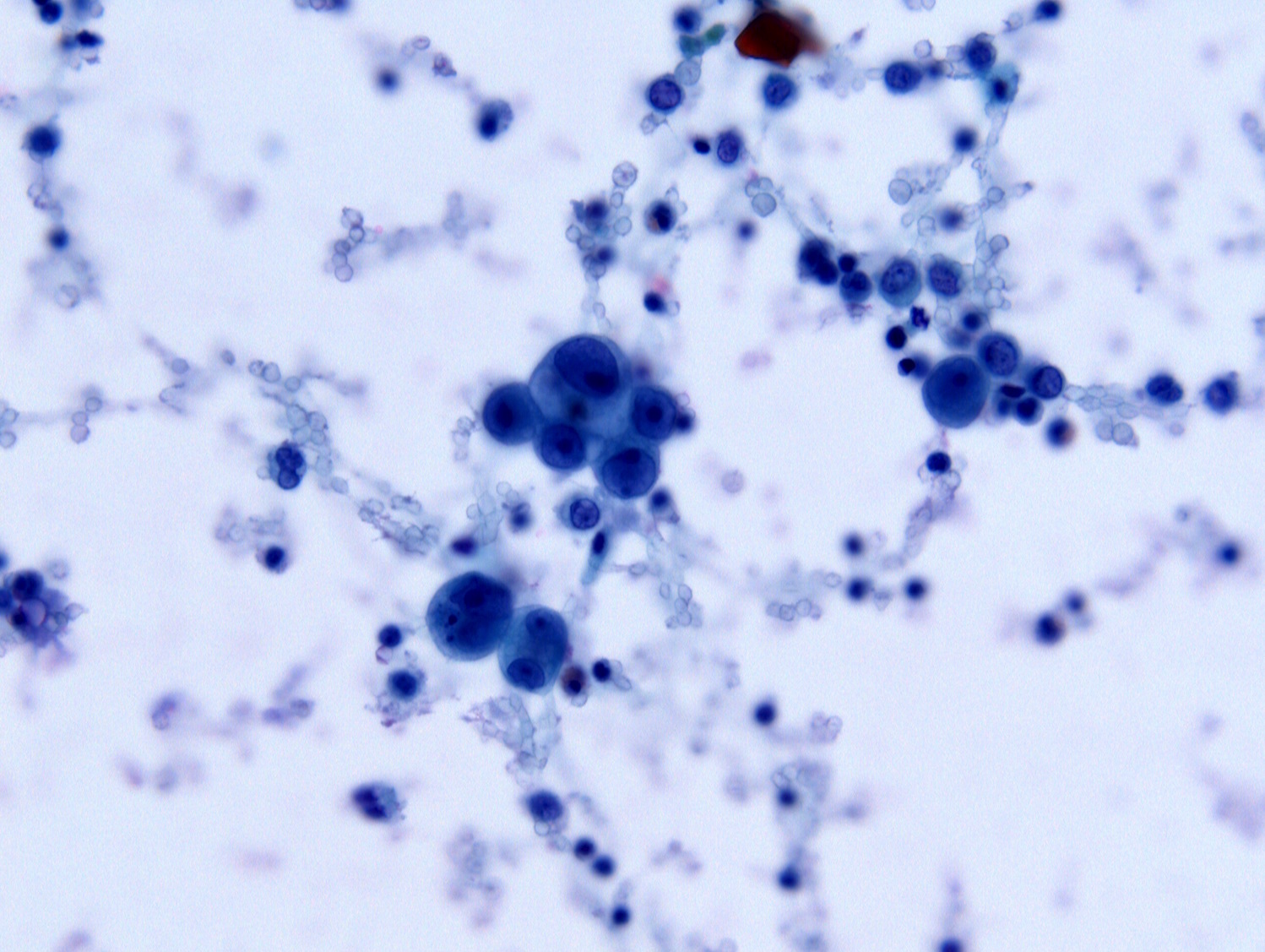
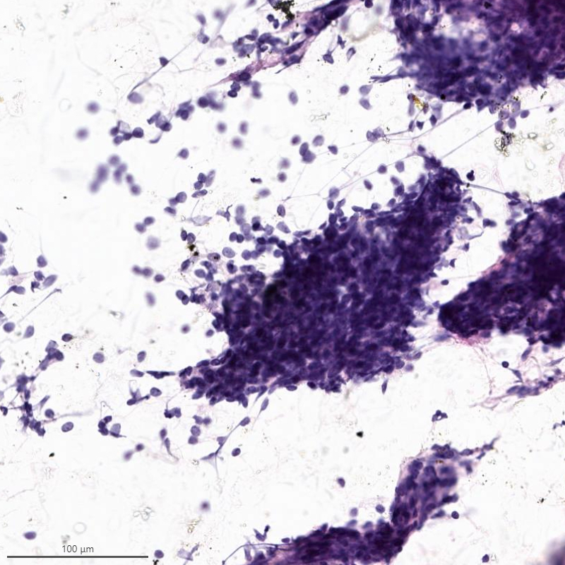
Diff-Quik
Images hosted on other servers:

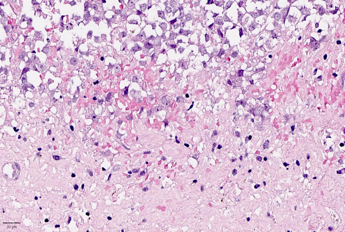
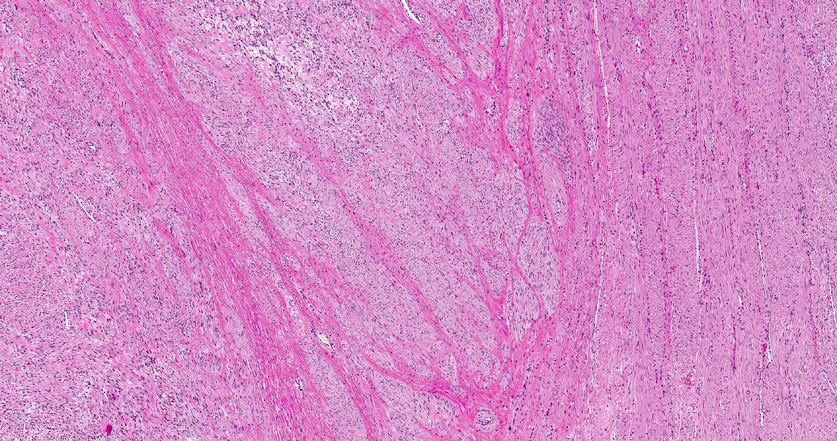
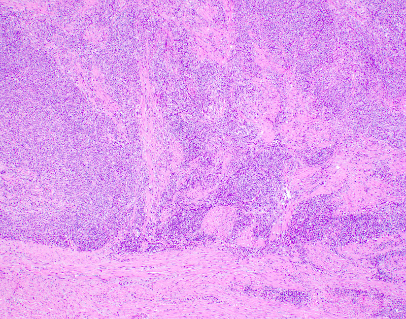
Intramural tumor with hemorrhage
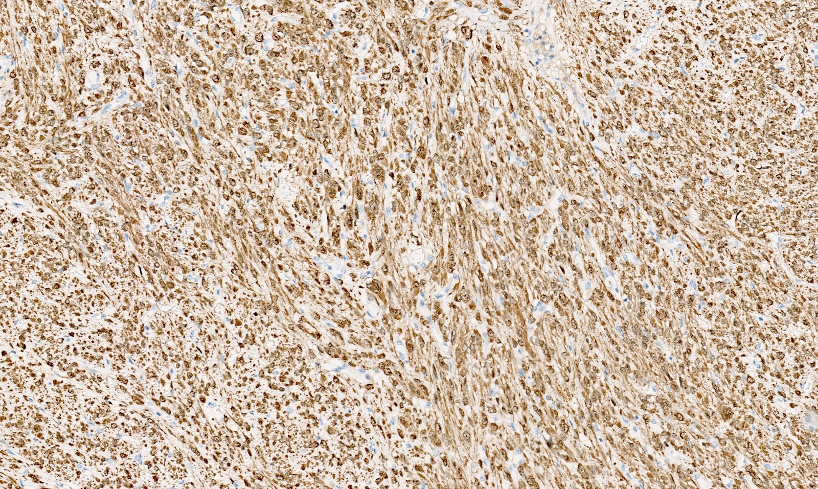
Contributed by Gulisa Turashvili, M.D., Ph.D.
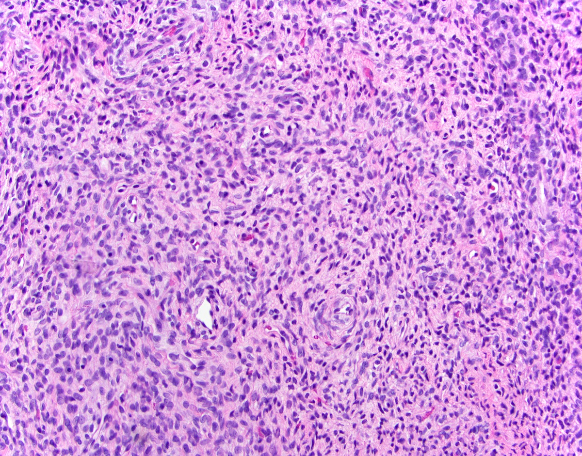
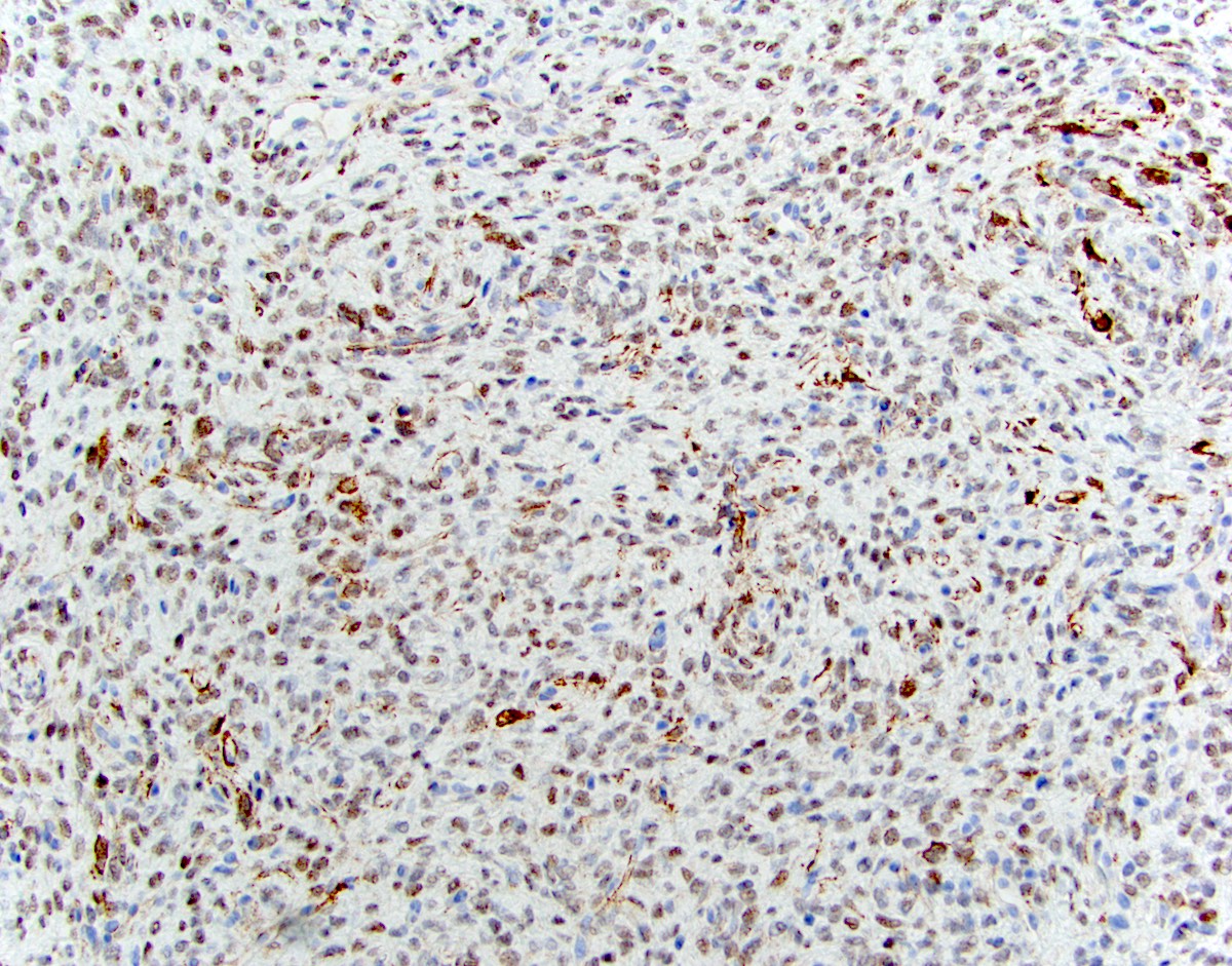
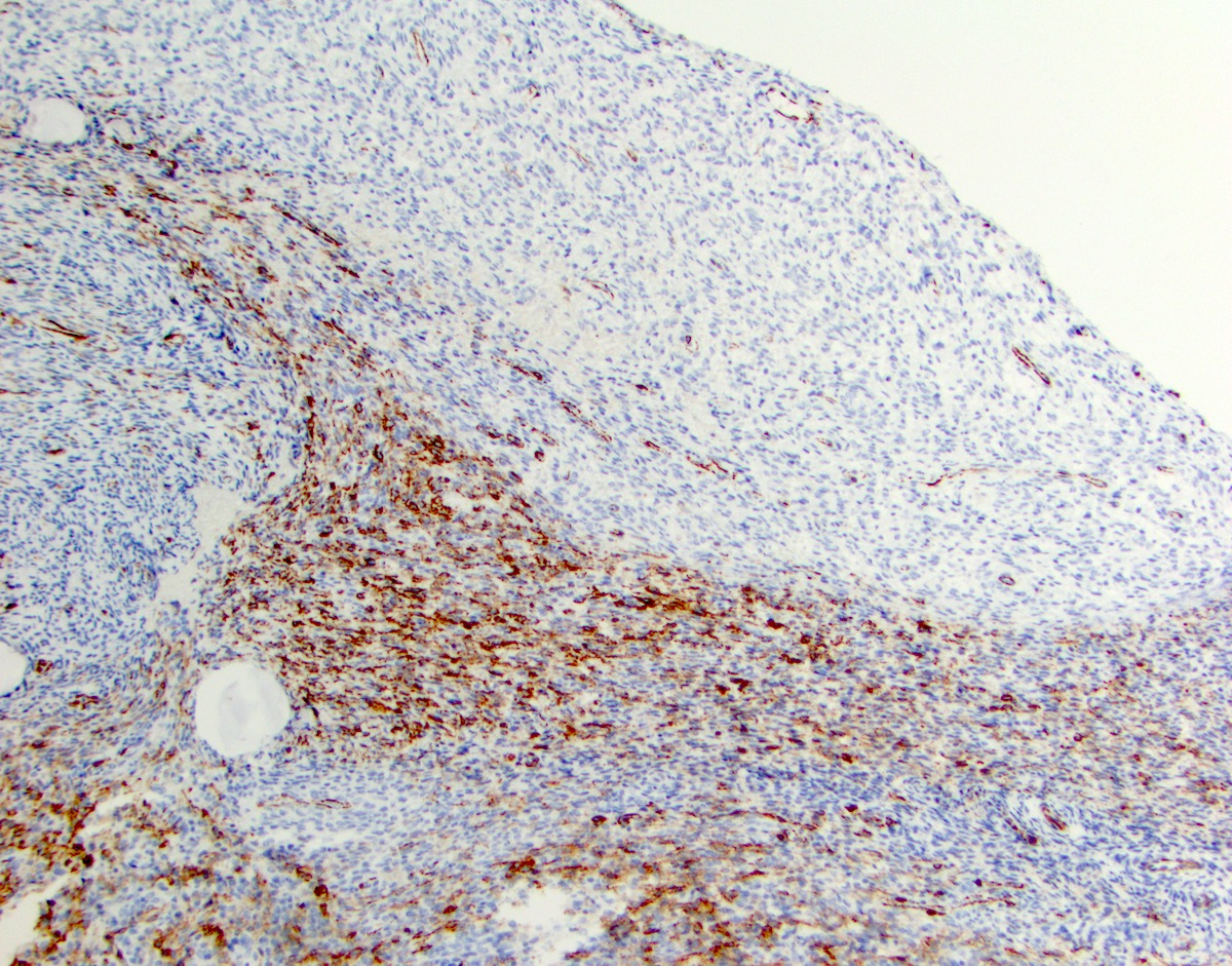
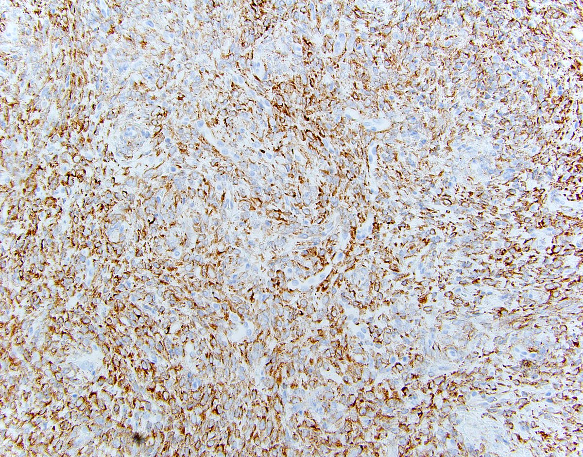
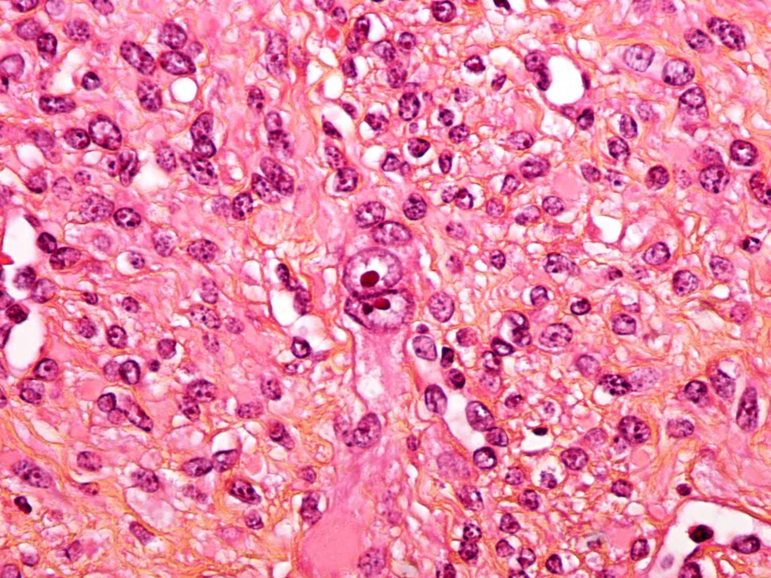
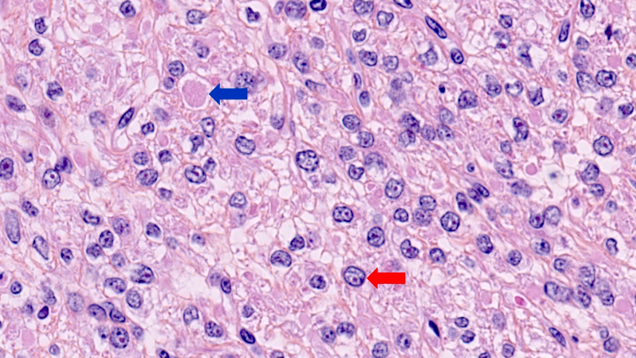
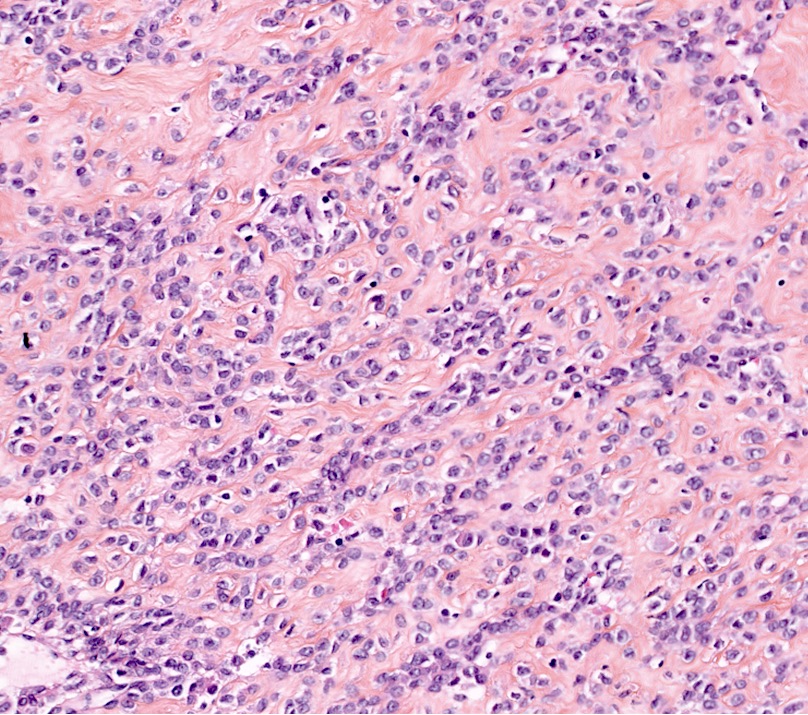
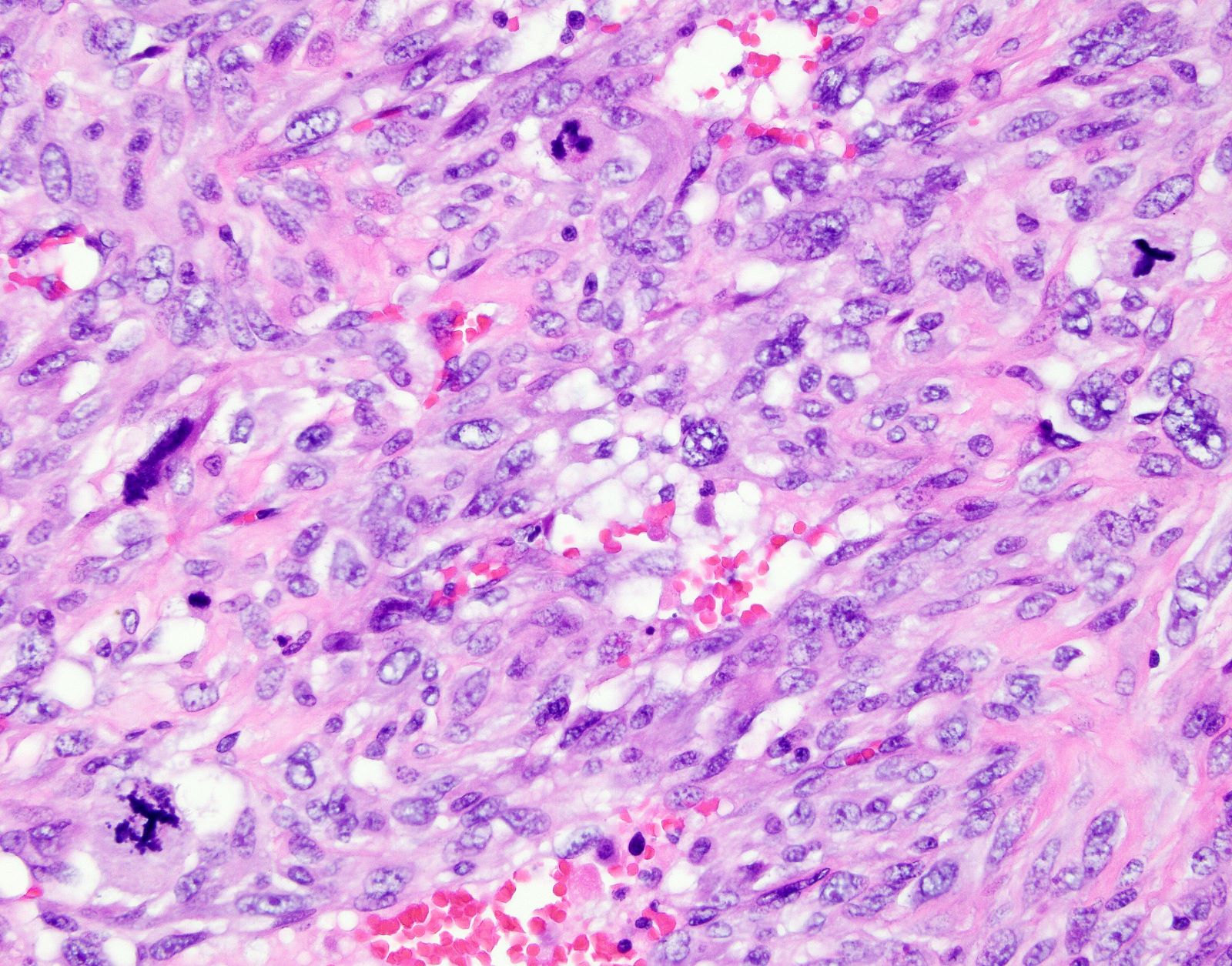
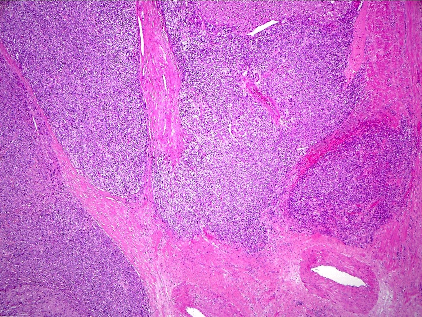
Coagulative necrosis
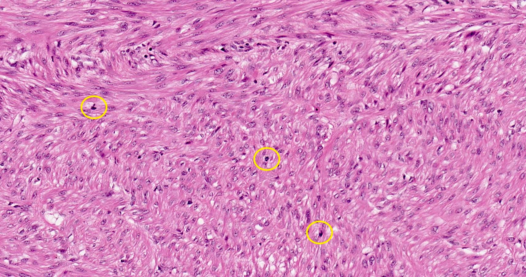
Elevated mitoses
Epithelioid morphology
Irregular borders
Table 1: Staging of endometrial carcinoma as per FIGO (2018 update) and AJCC 8th edition
| FIGO (2018) | AJCC (2018) | Description |
| I | T1 | Tumor confined to the uterus |
| 1A | T1a | Tumor confined to the endometrium or invades < 50% of the myometrial wall |
| 1B | T1b | Tumor invades > 50% of the myometrial wall |
| II | T2 | Tumor infiltrates cervical stroma |
| III | T3, N1 | Tumor extends outside the uterus |
| IIIA | T3a | Tumor involves serosa or adnexa (direct extension or metastases) |
| IIIB | T3b | Tumor involves vagina or parametria (direct extension or metastases) |
| IIIC | N1 | Tumor with pelvic lymph node metastasis |
| IIIC1 | N1 mi | Micrometastases in pelvic lymph nodes (> 0.2 mm or 200 cells but ≤ 2 mm) |
| N1a | Macrometastases in pelvic lymph nodes (> 2 mm in size) | |
| IIIC2 | N2 mi | Micrometastases in para-aortic lymph nodes (> 0.2 mm or 200 cells but ≤ 2 mm) |
| N2a | Macrometastases in para-aortic lymph nodes (> 2 mm in size) | |
| IV | T4, M1 | Tumor invades bladder or bowel mucosa or involves distant organs |
| IVA | T4 | Tumor invades bladder mucosa or bowel mucosa |
| IVB | M1 | Distant metastases (including intra-abdominal metastases and inguinal lymph nodes; excluding metastases to vagina, pelvic serosa or adnexa) |
| N0i+ | Isolated tumor cells in regional lymph nodes* |
Table 2: 2023 FIGO staging of cancer of the endometrium
| Stage | Description |
| I | Confined to the uterine corpus and ovary |
| IA | Disease limited to the endometrium or nonaggressive histological type (i.e., low grade endometroid, with invasion of < 50% of myometrium with no or focal lymphovascular space involvement [LVSI] or good prognosis disease) |
| IA1 | Nonaggressive histological type limited to an endometrial polyp or confined to the endometrium |
| IA2 | Nonaggressive histological types involving < 50% of the myometrium with no or focal LVSI |
| IA3 | Low grade endometrioid carcinomas limited to the uterus and ovary |
| IB | Nonaggressive histological types with invasion of ≥ 50% of the myometrium and with no or focal LVSI |
| IC | Aggressive histological types limited to a polyp or confined to the endometrium |
| II | Invasion of cervical stroma without extrauterine extension or with substantial LVSI or aggressive histological types with myometrial invasion |
| IIA | Invasion of the cervical stroma of nonaggressive histological types |
| IIB | Substantial LVSI of nonaggressive histological types |
| IIC | Aggressive histological types with any myometrial involvement |
| III | Local or regional spread of the tumor of any histological subtype |
| IIIA | Invasion of uterine serosa, adnexa or both by direct extension or metastasis |
| IIIA1 | Spread to ovary or fallopian tube (except when meeting stage IA3 criteria) |
| IIIA2 | Involvement of uterine subserosa or spread through the uterine serosa |
| IIIB | Metastasis or direct spread to the vagina or to the parametria or pelvic peritoneum |
| IIIB1 | Metastasis or direct spread to the vagina or the parametria |
| IIIB2 | Metastasis to the pelvic peritoneum |
| IIIC | Metastasis to the pelvic or para-aortic lymph nodes or both |
| IIIC1 | Metastasis to the pelvic lymph nodes |
| IIIC1i | Micrometastasis |
| IIIC1ii | Macrometastasis |
| IIIC2 | Metastasis to para-aortic lymph nodes up to the renal vessels, with or without metastasis to the pelvic lymph nodes |
| IIIC2i | Micrometastasis |
| IIIC2ii | Macrometastasis |
| IV | Spread to the bladder mucosa or intestinal mucosa or distance metastasis |
| IVA | Invasion of the bladder mucosa or the intestinal / bowel mucosa |
| IVB | Abdominal peritoneal metastasis beyond the pelvis |
| IVC | Distant metastasis, including metastasis to any extra or intra-abdominal lymph nodes above the renal vessels, lungs, liver, brain or bone |
- Disease limited to low grade endometrioid carcinomas involving the endometrium and ovaries (stage IA3) must be distinguished from extensive spread of the endometrial carcinoma to the ovary (stage IIIA1), by the following criteria
- No more than superficial myometrial invasion is present (< 50%)
- Absence of extensive / substantial LVSI
- Absence of additional metastases
- The ovarian tumor is unilateral, limited to the ovary, without capsule invasion / rupture (equivalent to pT1a)
- LVSI as defined in WHO 2021: extensive / substantial, ≥ 5 vessels involved
- Aggressive histological types include serous carcinomas, clear cell carcinomas, mesonephric-like carcinomas, gastrointestinal type mucinous endometrial carcinoma, undifferentiated carcinomas and carcinosarcomas
- For endometrial endometrioid carcinomas, grade is based on the proportion of solid areas: low grade = grade 1 (≤ 5%) and grade 2 (6 - 50%); and high grade = grade 3 (> 50%)
- Nuclear atypia excessive for the grade raises the grade of a grade 1 or 2 tumor by one
- Nonaggressive histological types are composed of low grade (grade 1 and 2) endometrial endometrioid carcinomas
Table 3: 2023 FIGO endometrial cancer stage with molecular classification (for patients with early endometrial cancer [stages I and II after surgical staging])
| Stage | Description |
| IAmPOLEmut | POLEmut endometrial carcinoma, confined to the uterine corpus or with cervical extension, regardless of the degree of LVSI or histological type |
| IICmp53abn | p53abn endometrial carcinoma confined to the uterine corpus with any myometrial invasion, with or without cervical invasion and regardless of the degree of LVSI or histological type |

Images hosted on other servers:

Surgical specimen
Contributed by Basile Tessier-Cloutier, M.D.
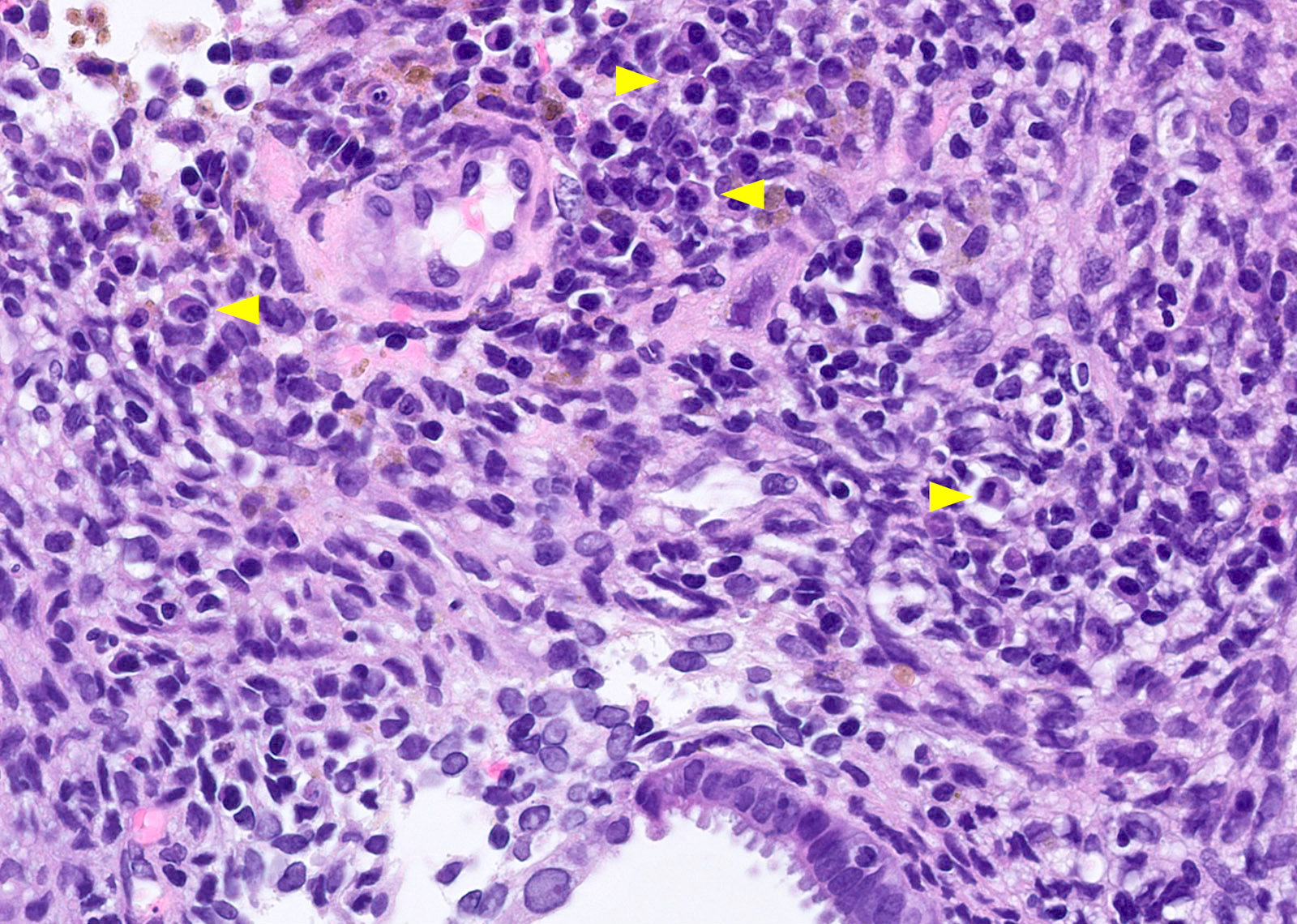
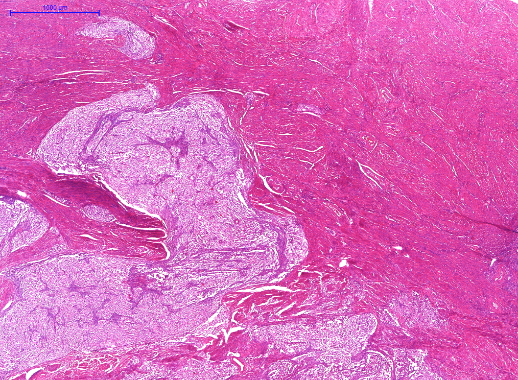
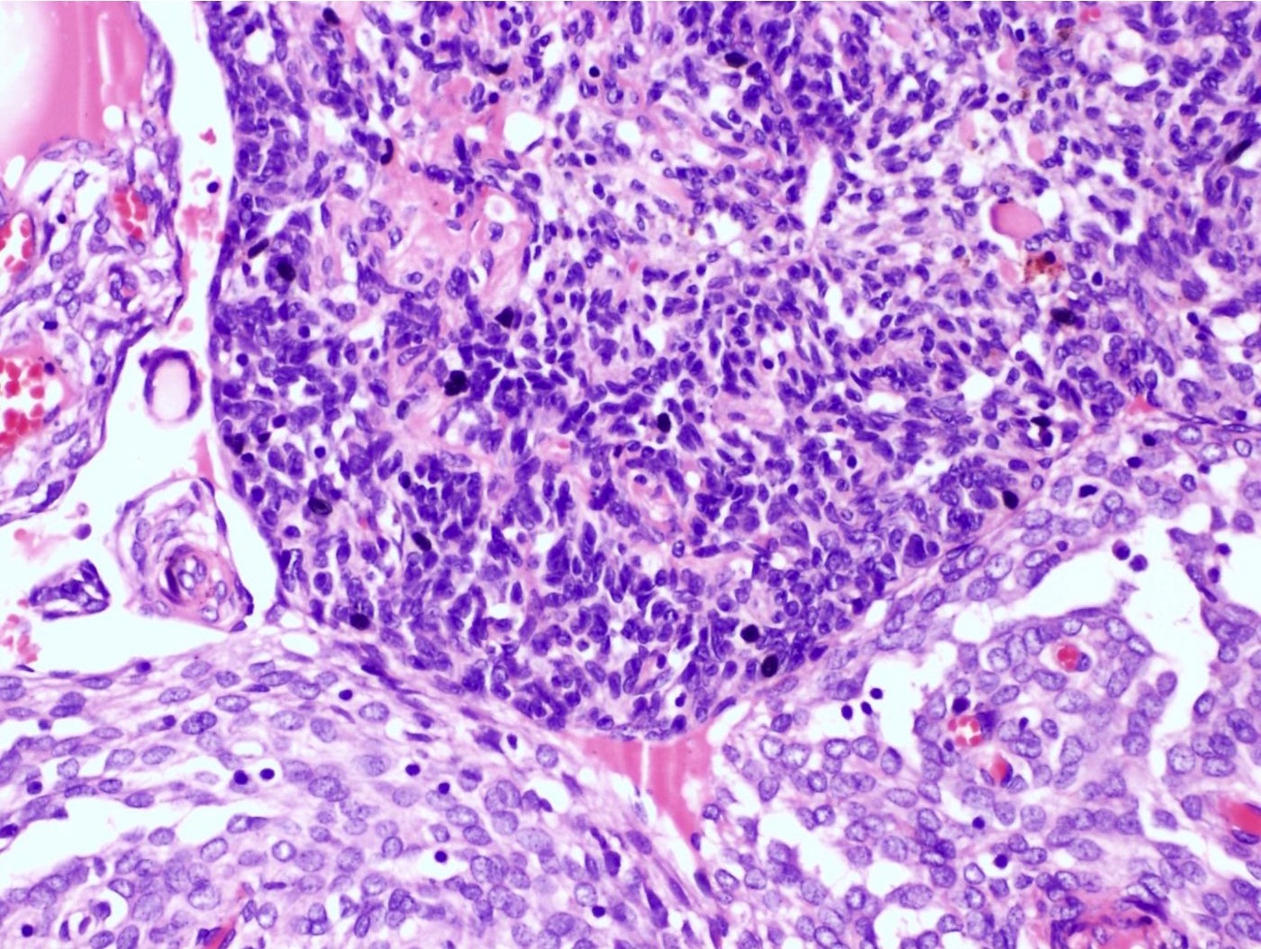
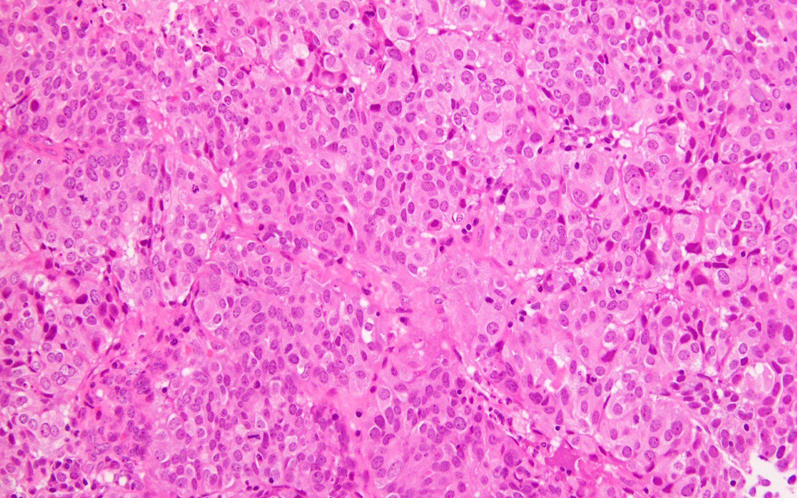
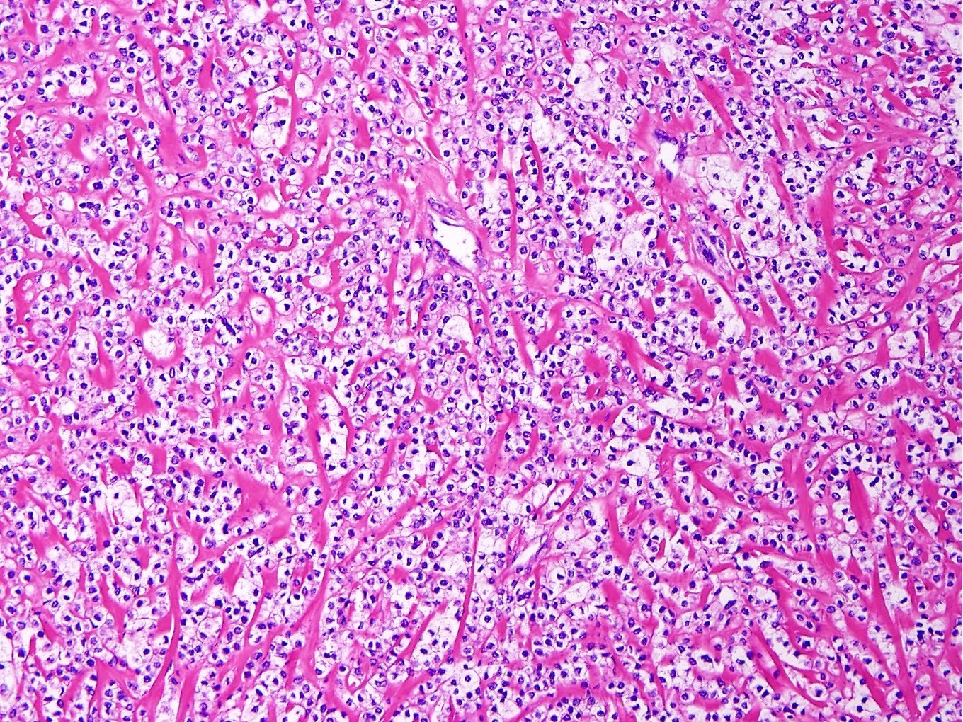
Undifferentiated and differentiated components
Solid architecture
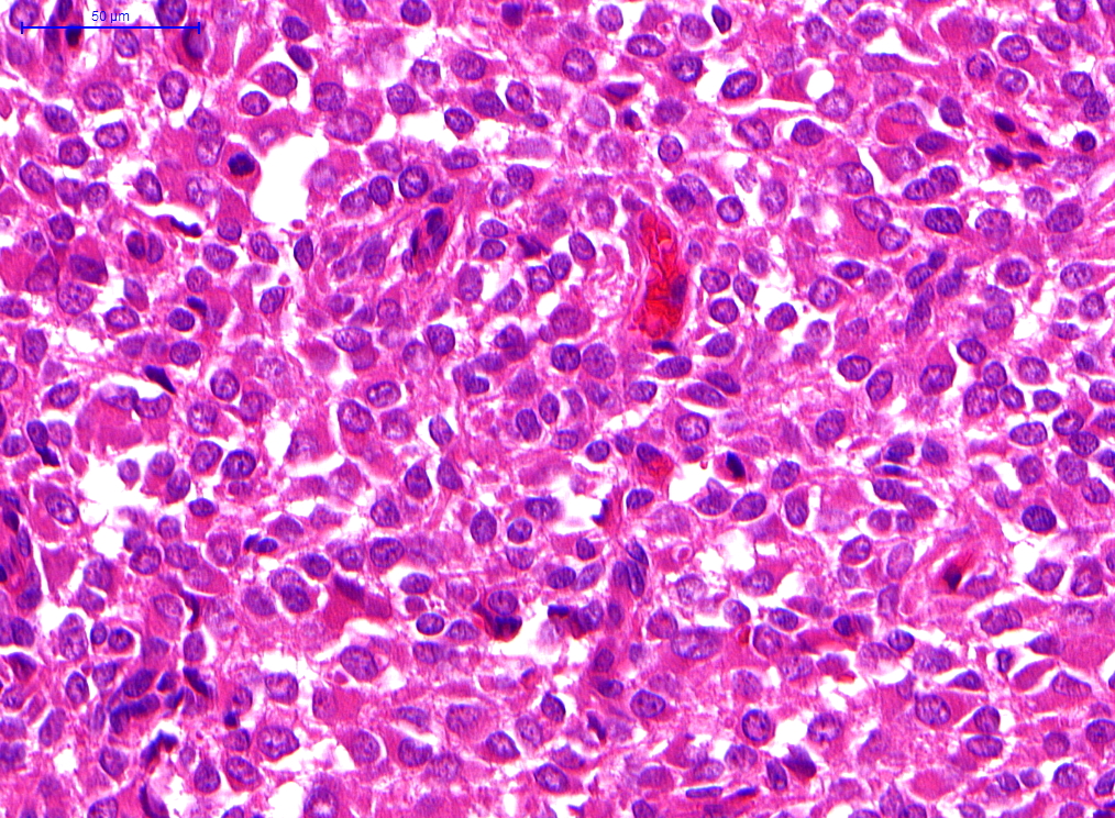
Discohesive rhabdoid cells
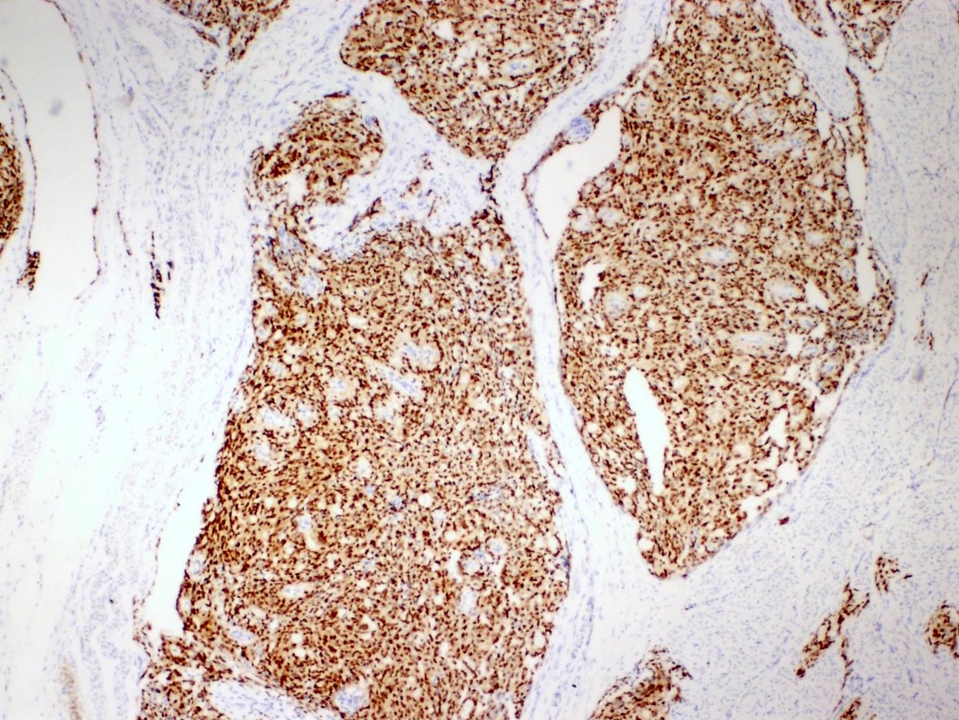
ARID1B
Contributed by Ayse Ayhan, M.D., Ph.D.

Polypoid gray-white uterine mass
Contributed by David B. Chapel, M.D.
Epithelioid morphology

Spindled morphology
Spindled morphology
Pleomorphic morphology
Contributed by Ayse Ayhan, M.D., Ph.D.
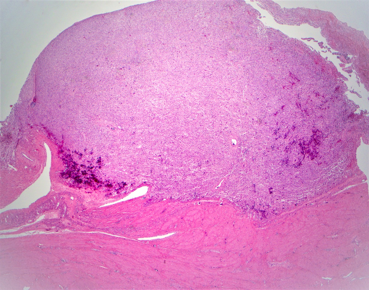
Intramural lesion
Contributed by Ayse Ayhan, M.D., Ph.D.
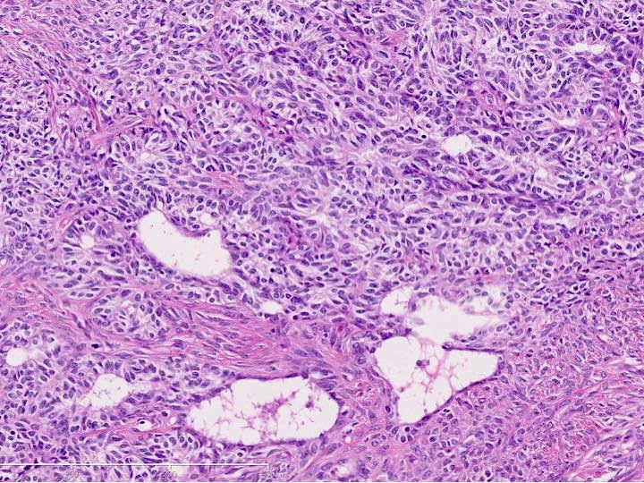
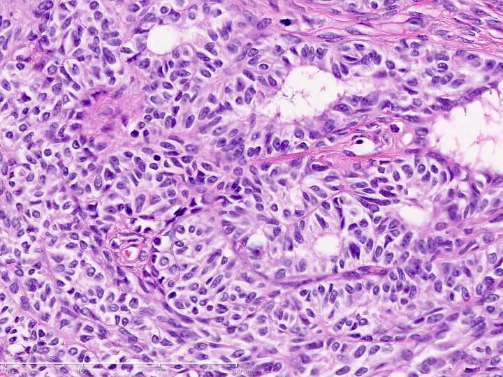
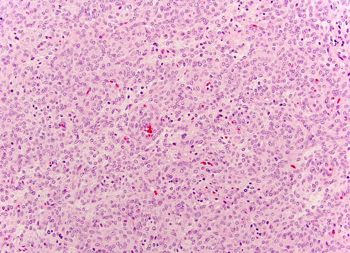
Varied architecture, bland cytology
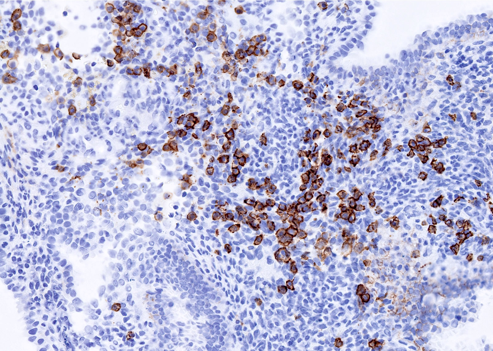
Positive sex cord and epithelial markers
Images hosted on other servers:


Various images

Tumor cells resemble granulosa cells


Various images
Contributed by Ayse Ayhan, M.D., Ph.D.
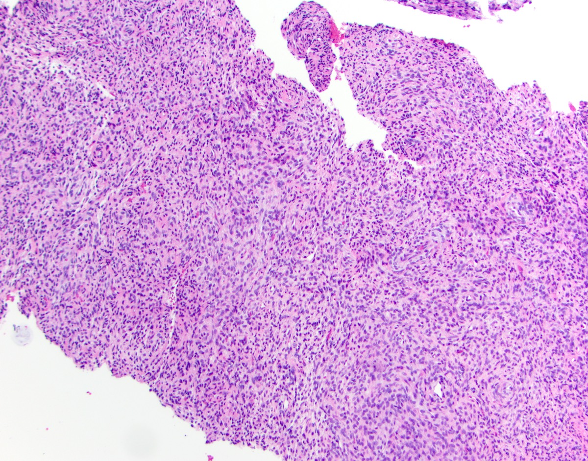
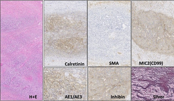
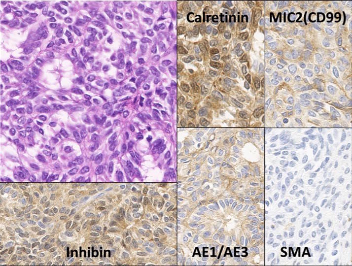
Intramural lesion
Contributed by Ayse Ayhan, M.D., Ph.D. and Jian-Jun Wei, M.D.
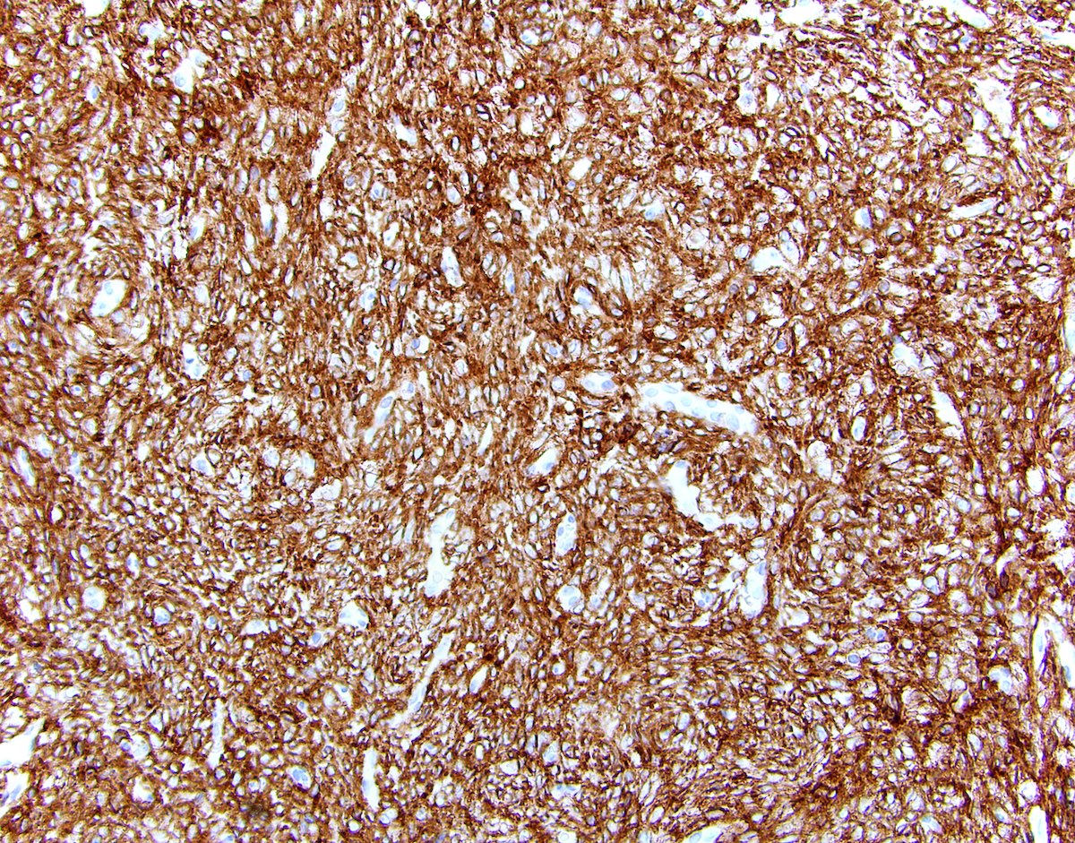
Varied architecture, bland cytology
Positive sex cord and epithelial markers

Vimentin

Clement: 2019
Crum: 2015
Damjanov: 2017
Fadare: 2015
Heller: 2015
IARC: 2014
IARC: 2020
Malpica: 2015
Murdock: 2018

Nucci: 2020
Nucci: 2023
Oliva: 2019
Vang: 2017
Find related Pathology books: breast, gynecologic
