Superpage
Page views in 2023: 85
Page views in 2024 to date: 40

Addison disease
Definition / general
  • Also called primary chronic adrenal insufficiency
  • Affects 3 - 6 individuals per 100,000 population
  • No symptoms until 90% of cortex is compromised
  • More common in white women
  • Causes: autoimmune disorders, infections (Histoplasma, Coccidiodes, tuberculosis), space occupying lesions (metastases, lymphomas), hemorrhage, amyloid, sarcoid or hemochromatosis
  • Symptoms: progressive weakness, easy fatigability, anorexia, weight loss, depression, irritability, menstrual abnormalities, GI disturbances, hyperpigmentation (primary adrenal disease causes elevated ACTH, which stimulates melanocytes at sun exposed areas and pressure points), small heart (due to chronic hypovolemia) and hypotension; infections may precipitate an acute crisis
  • Laboratory: elevated ACTH levels, low cortisol levels that do not respond to exogenous ACTH (since adrenal gland is damaged), hyponatremia, hypoglycemia, hyperkalemia and occasionally hypercalcemia
Gross description
  • Irregularly shrunken glands, may be hard to find

Adenomatoid tumor
Definition / general
  • Rare benign nonfunctioning tumor of the adrenal gland of mesothelial origin
Essential features
  • Proliferation of gland-like or vascular-like spaces lined by attenuated to columnar mesothelial cells
  • Rare nonfunctioning tumor of the adrenal gland typically found incidentally
  • Often confused on imaging and microscopy with tumors such as lymphangioma
  • Accurate diagnosis relies upon microscopic examination, typically with immunohistochemistry
  • Benign, with no reported cases of tumor recurrence following surgical excision
Epidemiology
Sites
  • Adrenal cortex and medulla
Pathophysiology
  • Thought to arise via entrapment of primitive mesenchymal cells from the Müllerian tract within the adrenal gland versus embolized mesothelial cells (Am J Surg Pathol 2003;27:969)
Etiology
  • Unknown
Clinical features
  • Typically asymptomatic and discovered incidentally
  • Rarely, reports of symptoms resulting from elevations in adrenal hormones (homovanillic acid, hyperaldosteronism) secondary to stimulation of normal adrenal parenchyma by tumor (Adv Anat Pathol 2009;16:424)
Diagnosis
  • Diagnosis cannot be made using imaging alone, as adrenal adenomatoid tumors lack specific radiologic features
  • Surgical excision (preferred) or core biopsy with subsequent histopathologic evaluation necessary for diagnosis
  • Easily confused with more common tumors when evaluated via light microscopy so immunohistochemistry plays a key role in accurate diagnosis (Adv Anat Pathol 2009;16:424)
Radiology description
  • Nonspecific; misdiagnosed as nonfunctioning adrenocortical tumor or other more common entities
  • May be well visualized on CT or MRI, with a wide range of presentations reported on imaging
  • Appearance ranges from homogeneous and solid to cystic degeneration or calcifications (Eur Radiol 1999;9:552)
Radiology images

Contributed by Firas G. Petros, M.D.
CT of left adrenal mass CT of left adrenal mass CT of left adrenal mass CT of left adrenal mass

CT of left adrenal mass


MRI of left adrenal mass MRI of left adrenal mass MRI of left adrenal mass MRI of left adrenal mass

MRI of left adrenal mass

Prognostic factors
  • Favorable prognosis, with no reported cases of recurrence following excision
Case reports
Treatment
  • Complete surgical resection
Gross description
Gross images

Contributed by Debra L. Zynger, M.D.
Ill defined spongy lesion

Ill defined spongy lesion


Images hosted on other servers:

White solid and cystic surface

Cut surface of cystic lymphangioma-like adrenal adenomatoid tumor

Cystic surface

Cystic spongy surface

Solid gray surface

Microscopic (histologic) description
  • Well circumscribed to infiltrative into surrounding cortex and medulla
  • Can infiltrate into periadrenal adipose
  • Growth patterns include microcysts, tubules, macrocysts / cavernous and pseudovascular with papillary and anastomosing structures lined by endothelial-like cells (Adv Anat Pathol 2009;16:424)
  • Cells are flattened to hobnailed and bland with no appreciable mitoses
  • Lymphoid aggregates and interspersed lymphocytes are frequently present
  • Dystrophic calcifications are frequent
  • Areas with abundant fine calcifications can have an empty, vacuolated appearance
  • Surrounding fibrosis may be seen
Microscopic (histologic) images

Contributed by Debra L. Zynger, M.D.
Cystic lesion

Cystic lesion

Anastomosing channels

Anastomosing channels

Microcysts

Microcysts

Delicate septae

Delicate septae

Tubules and signet rings

Tubules and signet rings

Entrapped cortex

Entrapped cortex


Lymphoid aggregates

Lymphoid aggregates

Calcification

Calcification

Infiltrative

Infiltrative

CK7

CK7

WT1

WT1

MelanA


MelanA


Electron microscopy description
Sample pathology report
  • Right adrenal gland, robotic adrenalectomy:
    • Adenomatoid tumor, 4.8 cm
    • Surgical margin, negative for tumor
Differential diagnosis
Board review style question #1


A 42 year old man presents for evaluation of right renal stones and is incidentally found to have a 4 cm mass in the left adrenal gland. Laboratory studies are within normal limits and he reports no symptoms other than right flank pain consistent with his known diagnosis. Adrenalectomy is performed and the pathology report subsequently confirms adenomatoid tumor of the adrenal gland (shown above). Positivity for which of the following immunostains (shown above) likely confirmed the diagnosis?

  1. CD31
  2. CD34
  3. CK5/6
  4. Inhibin
  5. MelanA
Board review style answer #1
C. CK5/6. Adenomatoid tumor is of mesothelial origin and as such expresses CK5/6. Adrenal cortical lesions express MelanA and inhibin, while vascular lesions express CD31 and CD34.

Comment Here

Reference: Adenomatoid tumor

Adrenal cortical adenoma
Definition / general
  • Benign neoplasm arising from adrenal cortical cells
  • May or may not be functional
Terminology
  • Adrenal cortical adenoma (ACA)
  • Incidentaloma: small adenoma discovered incidentally during workup of other conditions (Mod Pathol 2011;24:S58)
  • Black (pigmented) adenoma: diffusely pigmented, brown-black ACA presumably due to lipofuscin
  • Hypercortisolism: sometimes used synonymously for Cushing syndrome
  • Pre-clinical / sub-clinical Cushing syndrome: hypercortisolism in the context of an incidentally discovered adrenal mass without overt clinical manifestations of Cushing syndrome Arq Bras Endocrinol Metabol 2007;51:1272)
  • Primary hypercortisolism: due to secretion of cortisol by the adrenal gland versus secondary hypercortisolism: due to increased secretion of ACTH by pituitary or to secretion of cortisol by an ectopic tumor
Epidemiology
  • F > M
  • More common in adults, 5th - 7th decade
  • Equal predilection for right and left adrenal glands
  • True incidence unknown because many are not functional, estimates include 8.7% in autopsy series and 4% in radiology series (Mol Cell Endocrinol 2014;386:67)
  • Incidence has been increasing due to increasing utilization of imaging, estimated 0.2 to 0.4% in general population (Endocrinol Metab (Seoul) 2014;29:5)
  • Incidence increases with age, reported in <1% of patients under 30 years and in up to 7% of patients over 70 years (Indian J Endocrinol Metab 2013;17:S59)
  • Children
    • Generally uncommon in children; only ~25 cases annually in U.S. in those < 20 years (Braz J Med Biol Res 2000;33:1225)
    • Bimodal age distribution; most commonly < age 5, second peak ages 9 - 16 years
    • Incidence higher in females, M:F ratio 1:1.6 (J Clin Oncol 2004;22:838)
    • Higher incidence in southern Brazil, associated with specific p53 mutation (R337H TP53)
    • Associated with several genetic syndromes:
      • Beckwidth-Wiedemann syndrome: hemihypertrophy, splanchnomegaly, macroglossia, intraabdominal neoplasms (i.e. adrenal cortical neoplasms, nephroblastoma, hepatoblastoma), due to alteration of 11p15 region
      • Li-Fraumeni syndrome (SBLA syndrome): sarcoma (rhabdomyosarcoma), breast/brain tumors, leukemia, laryngeal carcinoma, lung cancer, adrenal cortical carcinoma; due to alteration of p53 on chromosome 17p
      • Carney triad: malignant gastrointestinal stromal tumor, pulmonary chondroma, extra-adrenal paraganglioma, adrenal cortical adenoma
      • Adrenogenital syndrome: adrenal cortical neoplasms, congenital adrenal hypertrophy
    • No proven relationship with environmental factors
Sites
Pathophysiology
  • Approximately 90% of ACAs are nonfunctional
  • When functional, may secrete one or more of the 3 major classes of adrenal steroids (from external to internal layers):
    • Zona glomerulosa: mineralocorticoids (aldosterone)
    • Zona fasciculata: glucocorticoids (cortisol)
    • Zona reticularis: androgens (testosterone, dihydrotestosterone [DHT], androstenedione, dihydroepiandosterone [DHEA])
  • Hyperaldosteronism/Conn's syndrome: ↑aldosterone → impacts distal tubules & collecting ducts of nephron → ↑ sodium and water retention, ↓ potassium retention → ↑ blood pressure
  • Hypercortisolism/Cushing's syndrome: ↑cortisol → ↓ corticotropin releasing hormone (CRH), ↓ adrenocorticotropic hormone (ACTH), ↑ blood glucose
  • Virilization: ↑ DHEA, ↑ DHEA-sulfate (DHEA-S), ↑ androstenedione, ↑ testosterone, ↑ DHT → ↑ urinary 17-ketosteroids (metabolic product)
  • Feminization: ↑ androgens → aromatization → ↑ estrogen, ↑ estradiol → ↑ urinary 17-ketosteroids (metabolic product)
  • Children: predisposing genetic factors are present in ~50% of children with adrenal cortical tumors, most commonly Li-Fraumeni syndrome and Beckwith-Wiedemann syndrome (see above); may arise due to defective apoptosis (J Clin Endocrinol Metab 2000;85:2048)
Etiology
  • Neoplastic proliferation of adrenal cortical cells
  • May arise from any of the 3 layers, but zona fasciculata most common (Mod Pathol 2011;24:S58)
Clinical features
  • Minority are functional, may produce a pure or mixed endocrine syndrome (from most to least common):
    • Hyperaldosteronism/Conn's syndrome: hypertension, proximal muscle weakness, headache, polyuria, tachycardia with/without palpitation, hypokalemia, hypocalcemia
    • Hypercortisolism/Cushing's syndrome: central obesity, moon facies, plethora, striae, thin skin, easy bruising, hirsutism, telangiectasias, hyperhidrosis
    • Virilization:
      • Females: increased muscle mass (Herculean habitus), clitoromegaly, facial hair, deep voice, pubic hair
      • Males: penile enlargement, pubic hair
    • Feminization: gynecomastia, impotence
Diagnosis
  • Adrenal lesion discovered with imaging used to work up unrelated clinical symptoms, therefore usually no detectable hormonal abnormalities or clinical symptoms (Pol J Radiol 2013;78:47)
  • Work up may include observation (serial imaging, laboratory tests) or fine needle aspiration / core biopsy (rarely indicated, mainly for ruling out non-adrenal metastases) (J Clin Endocrinol Metab 2010;95:4106)
  • Well circumscribed lesion comprised of cells resembling any of the 3 layers of the normal adrenal cortex
  • Difficult to differentiate ACA from normal adrenal cortex in adrenal core needle biopsies
  • Atypical histologic features are commonly found in children, making adrenal cortical adenomas difficult to distinguish from adrenal cortical carcinoma
Laboratory
  • Battery of endocrine tests usually within normal limits, although a minority may have subclinical hormone production with slight abnormalities
  • Suggested endocrine tests include: dexamethasone suppression test, ACTH levels, plasma free metanephrine/normetanephrine, 24 hour total urinary metanephrines, ratio plasma aldosterone:plasma renin (Indian J Endocrinol Metab 2013;17:S59)
  • Adrenal tumors in patients with previously unrecognized clinical symptoms attributable to the tumor are not considered incidentalomas (Pol J Radiol 2013;78:47)
  • Hyperaldosteronism/Conn's syndrome: ↑aldosterone, hypernatremia, hypokalemia
  • Cushing's syndrome: ↑cortisol, ↓CRH, ↓ACTH, hyperglycemia
  • Virilization: ↑DHEA, ↑DHEA-S, ↑androstenedione, ↑testosterone, ↑DHT, ↑urinary 17-ketosteroids
  • Feminization: ↑estrogen, ↑estradiol, ↑urinary 17-ketosteroids
Radiology description
  • Computed tomography (CT):
    • Rounded, well delineated borders, homogeneous, clear separation from and no extension into surrounding structures, decreased attenuation compared to uninvolved adrenal parenchyma on non-contrast CT (≤10 HU), contrast enhancing (Theranostics 2012;2:516)
  • Magnetic resonance imagining (MRI):
    • Used to visualize microscopic fat (favoring ACA), "chemical shift" phenomenon (increased "in phase" signal intensity, decreased "out of phase" signal) (Theranostics 2012;2:516)
  • 18FDG-PET:
Radiology images

Images hosted on other servers:

Homogeneous left adrenal mass with distinct borders

Left adrenal mass

(A) Plain CT and (B) contrast CT show left suprarenal mass

Prognostic factors
  • Distinguishing ACA from adrenocortical carcinoma (ACC) is difficult and several systems have been proposed
  • In general, most reliable factors include size, necrosis, mitotic activity, atypical mitoses (Mod Pathol 2011;24:S58)
  • Weiss System (Am J Surg Pathol 1984;8:163): most widely used criteria
    • Criteria (≥ 3 criteria indicates malignancy): high mitotic rate, atypical mitoses, high nuclear grade, low percentage of clear cells, necrosis, diffuse tumor architecture, capsular invasion, sinusoidal invasion, venous invasion
  • Modified Weiss System (Am J Surg Pathol 2002;26:1612): > 5 mitoses per 50 high powered fields, < 25% clear cells, atypical mitotic figures, necrosis, and capsular invasion
    • Calculation:
      • 1 point each for the presence of atypical mitotic figures, necrosis, and capsular invasion
      • 2 points each for the presence of > 5 mitoses per 50 high powered fields and < 25% clear cells
      • Total score ranges from 0 to 7, and score of > 3 highly correlates with subsequent malignant behavior
Case reports
  • 24 year old woman with unusual presentation of Carney complex (Endocr J 2012;59:823)
  • 33 year old woman with Cushing's syndrome during pregnancy secondary to adrenal adenoma (Acta Med Iran 2012;50:76)
  • 34 year old woman with eplerenone use in primary aldosteronism during pregnancy (Hypertension 2012;59:e18)
  • 35 year old man with primary adrenal angiosarcoma and functioning adrenocortical adenoma (Eur J Endocrinol 2012;166:131)
  • 36 year old woman with cortisol producing adrenal adenoma associated with latent aldosteronoma (Intern Med 2012;51:395)
  • 46 year old woman with black adrenal adenoma causing preclinical Cushing's syndrome (Tokai J Exp Clin Med 2010;35:57)
  • 50 year old woman with myelolipomata arising within adrenocortical adenoma ipsilateral to synchronous clear cell renal cell carcinoma (Malays J Pathol 2010;32:123)
  • 52 year old woman with adrenalectomy by retroperitoneal laparoendoscopic single site surgery (JSLS 2010;14:571)
  • 56 year old woman with coexistence of Cushing syndrome from functional adrenal adenoma and Addison disease from immune-mediated adrenalitis (J Am Osteopath Assoc 2012;112:374)
  • 56 year old man with generalized glucocorticoid resistance accompanied with an adrenocortical adenoma and caused by a novel point mutation of human glucocorticoid receptor gene (Chin Med J (Engl) 2011;124:551)
  • 66 year old woman with laparoscopic adrenalectomy for bilateral metachronous aldosteronomas (JSLS 2011;15:100)
  • 72 year old woman with ectopic adrenal cortical adenoma in the gastric wall (World J Gastroenterol 2013;19:778)
Treatment
  • Most adrenal lesions > 4 cm should be removed regardless of imaging findings because of increased risk of ACC (BMC Surg 2013;13:57)
  • If large (> 4 cm), functional, worrisome characteristics on imaging: surgical resection
  • If small (< 4 cm), non-functional, benign features on imaging: clinical observation and follow up (exact guidelines not well established)
    • Repeat laboratory studies to assess functional status, yearly for 4 years
    • Repeat CT scan 6 - 12 months after diagnosis, if no increase in size then no further follow up needed
Clinical images

Images hosted on other servers:

Moon facies and central obesity

Gross description
  • Weight usually < 50 grams (in pediatric patients may weight up to 500 grams) (Mod Pathol 2011;24:S58)
  • Size usually < 5 cm
  • Unilateral, solitary, golden yellow
  • May have focal dark areas corresponding with hemorrhage, lipid depletion, increased lipofuscin
  • Functional adenoma may result in atrophy of ipsilateral or contralateral adrenal cortex
Gross images

Contributed by @Andrew_Fltv and @SueEPig on Twitter
Adrenal cortical aldosterone producing adenoma

Adenoma

Adenoma

Adenoma


Images hosted on other servers:

External surface, during surgery

Intact capsule

Well circumscribed

1.3 cm left adrenal adenoma

3 x 3 cm exophytic adrenal mass

Microscopic (histologic) description
  • In comparison to surrounding adrenal gland, adenoma cells are larger with different cytoplasm, increased variation in nuclear size
  • Distinct cell borders, cells have abundant foamy cytoplasm reminiscent of zona fasciculata
  • Balloon cells: clusters of cells with enlarged lipid-rich cytoplasm (seen in Cushing syndrome)
  • Histologic variants: oncocytic, myxoid
Microscopic (histologic) images

Contributed by Xiaoyin "Sara" Jiang, M.D., Debra Zynger, M.D., @Andrew_Fltv on Twitter and @SueEPig on Twitter

Spironolactone bodies with aldosteronoma

Adenoma with spironolactone bodies

Adrenal cortical aldosterone producing adenoma Adrenal cortical aldosterone producing adenoma Adrenal cortical aldosterone producing adenoma

Adrenal cortical aldosterone producing adenoma


Adenoma Adenoma Adenoma

Adenoma


Images hosted on other servers:

Multiple ACAs comprised of clear cells

Resembles normal adrenal fasciculata

Clear cells

Low power

S100- basement cells

Virtual slides

Images hosted on other servers:

ACA with hyperaldosteronism

Cytology description
Positive stains
Negative stains
Electron microscopy description
  • Abundant intracytoplasmic lipid droplets of varying sizes
  • Prominent microvillous projections along cell borders
  • Abundant smooth endoplasmic reticulum
  • Prominent, round to oval mitochondria; cristae may have tubular to vesicular (zona fasciculata) or lamellar (zona reticularis) profile
Electron microscopy images

Contributed by Edward Calomeni, B.S.

Spironolactone bodies

Molecular / cytogenetics description
  • Tumorigenesis not well understood
  • Outside of immunohistochemistry for diagnosis, adjunct molecular studies not currently utilized for clinical purposes (i.e. treatment, prognosis, distinction from ACC)
  • Usually monoclonal and diploid, versus ACC monoclonal and aneuploid/polyploid (Mol Cell Endocrinol 2014;386:67)
  • Gene expression profiling shows decrease in expression of major histocompatibility complex (MCH) class II genes in ACC when compared to ACA in children (Cancer Res 2007;67:600)
  • Usually sporadic, but may be associated with genetic syndrome
    • Cortisol-producing ACA may be associated with McCune-Albright syndrome, primary pigmented nodular adrenocortical disease or Carney complex (Mol Cell Endocrinol 2014;386:67)
    • Comparative genomic hybridization (CGH) studies showed adrenal tumors have complex pattern of chromosomal alterations, with ACCs having more more chromosomal gains/losses than ACAs (Mol Cell Endocrinol 2014;386:67)
    • Single nucleotide polymorphism (SNP) arrays confirm high genetic variability in ACAs (Neoplasia 2012;14:206)
      • Chromosomes with most frequent gains are #5, 3, 6, 11, 2
      • Chromosomes with most frequent losses are #1, 6, 2
      • Candidate genes include NOTCH1, CYP11B2, HRAS, IGF2
Differential diagnosis

Adrenal cortical carcinoma
Definition / general
  • Malignant epithelial tumor of adrenal cortical cells
Essential features
  • Malignant epithelial tumor of adrenal cortex
  • Adrenocortical carcinoma (ACC) is a rare endocrine tumor with high mortality
  • IGF2 overexpression is a major event in adrenal cortical carcinoma tumorigenesis
  • Vast majority are sporadic but could present in different syndromic settings
  • Heterogeneous group of tumors with prognosis dependent on patient age, clinical presentation, stage, histologic variant and molecular / genomic characteristics
  • Positive for SF1, inhibin, calretinin, MelanA, synaptophysin
Terminology
  • Adrenal cortical carcinoma (ACC)
  • Adrenocortical carcinoma, conventional type
  • Adrenal cortical adenocarcinoma
  • Adrenocortical carcinoma
  • Adrenal cortical tumor, malignant
ICD coding
  • ICD-O: 8370/3 - adrenal cortical carcinoma
Epidemiology
Sites
Pathophysiology
Etiology
Diagrams / tables

Images hosted on other servers:
Diagnostic algorithm for ACC stratification

Diagnostic algorithm for ACC stratification

Clinical features
  • ~50% are functional and ~50% are nonfunctional (J Clin Endocrinol Metab 2006;91:2027, N Engl J Med 1990;322:1195)
  • Functional adrenal cortical carcinomas have the following symptoms related to hormone production:
    • 50% cortisol excess (Cushing syndrome, rapid onset)
    • 20% sex hormone secretion (mainly androgens causing hirsutism, virilization and menstrual irregularities)
    • 8% aldosterone (hypertension, hypokalemia)
    • 15 - 25% mixed hormone production
    • Patients are younger, more likely females and present with metastatic disease
  • Nonfunctional adrenal cortical carcinomas could present in 2 ways:
    • 80% create a mass effect (gastrointestinal symptoms, organ compression or back pain) or mimic an infectious process (localized pain, fever due to cytokines from highly necrotic tumors)
    • 20% discovered incidentally during unrelated imaging procedures
  • Tumors often displace and invade adjacent organs (kidneys, liver, pancreas), adrenal vein, vena cava and retroperitoneum
  • Common metastatic sites are liver (60%), lymph nodes (40%), lungs (40%), peritoneal and pleural surfaces, bone, skin (anaplastic tumors) or retroperitoneum (Endocr Rev 2014;35:282)
Diagnosis

Weiss criteria: ≥ 3 for adrenal cortical carcinoma (Am J Surg Pathol 1984;8:163, Am J Surg Pathol 2002;26:1612)
1 Nuclear grade III or IV (Fuhrman)
2 > 5 mitotic figures/50 high power fields*
3 Atypical mitotic figures*
4 Clear or vacuolated cells ≤ 25% tumor
5 Diffuse architecture (> 33% of tumor)
6 Necrosis
7 Venous invasion* (of smooth muscle walled vessels)
8 Sinusoidal invasion
9 Capsular invasion
* = major criteria; 1 is required to diagnose adrenal cortical carcinoma (Virchows Arch 2012;460:9)
Laboratory
  • Hormone quantification in serum or urine (free cortisol, ACTH, DHEA sulfate, 17-OH, 17-hydroxy, testosterone, 17-beta-estradiol, 17-deoxycortison)
  • Dexamethasone suppression test
  • Potassium
  • Steroid metabolome profiling by mass spectrometry (Biomedicines 2021;9:174)
Radiology description
Radiology images

Images hosted on other servers:
Heterogeneous enhancing mass with lymphadenopathy

Heterogeneous enhancing mass with lymphadenopathy

CT imaging of advanced ACC

CT imaging of advanced ACC

Prognostic factors
Case reports
Treatment
Gross description
Gross images

Contributed by Debra Zynger, M.D.
Organ confined, ≤ 5 cm (pT1)

Organ confined, ≤ 5 cm (pT1)

Organ confined, > 5 cm (pT2)

Organ confined, > 5 cm (pT2)

Extra-adrenal invasion (pT3)

Extra-adrenal invasion (pT3)

Liver invasion (pT4)

Liver invasion (pT4)


Images hosted on other servers:
250 g, 10 cm tumor

250 g, 10 cm tumor

Large tumor compressing kidney

Large tumor compressing kidney

Large tumor dwarfing kidney

Large tumor dwarfing kidney

Microscopic (histologic) description
  • Encapsulated tumor composed of variably sized nests, large sheets and trabeculae
  • Invasion of thick fibrous capsule
  • Lymphovascular invasion (venous or sinusoidal)
  • Areas of necrosis, hemorrhage, degeneration are common
  • Large cells with granular clear to eosinophilic cytoplasm, often pleomorphic
  • Frequent intranuclear inclusions, mitoses, atypical mitoses
  • Grade defined by mitotic frequency:
    • Low grade: ≤ 20 mitoses/50 high power fields
    • High grade: > 20 mitoses/50 high power fields
  • Morphologic subtypes:
    • Conventional (vast majority, 70 - 90% of cases)
    • Oncocytic (> 75% oncocytic cells; second most common subtype)
    • Myxoid (abundant extracellular mucin)
    • Sarcomatoid (mesenchymal differentiation)
  • Loss of continuity of the reticular fibers or basement membrane network (> 33% lesion)
  • References: Am J Surg Pathol 1989;13:202, Am J Surg Pathol 2002;26:1612, Am J Surg Pathol 2018;42:201, Histopathology 2018;72:82
Microscopic (histologic) images

Contributed by Maria Tretiakova, M.D., Ph.D.
High nuclear grade

High nuclear grade

Pleomorphic ACC

Pleomorphic ACC

Necrosis

Necrosis

Capsule / fat invasion

Capsule / fat invasion

Positive margin

Positive margin


Reticulin loss

Reticulin loss

Diffuse growth

Diffuse growth

Ki67

Ki67

MelanA

MelanA

Inhibin

Inhibin


Contributed by Debra Zynger, M.D.
High mitotic rate

High mitotic rate

Lymphovascular invasion

Lymphovascular invasion

Extra-adrenal adipose invasion

Extra-adrenal adipose invasion

Liver involvement

Liver involvement

Cytology description
  • Single cells, poorly cohesive cell clusters in necrotic background
  • Cytoplasm is vacuolated to densely eosinophilic
  • Often marked nuclear atypia and mitotic activity
  • Reference: Diagn Cytopathol 2018;46:1064
Cytology images

Images hosted on other servers:
Discohesive, round to polygonal tumor cells showing moderate nuclear pleomorphism

Discohesive, round to polygonal tumor cells

Electron microscopy description
  • Abundant rough and smooth endoplasmic reticulum
  • Intracytoplasmic droplets of lipids
  • Many mitochondria with tubular cristae
  • Occasionally dense core granules (associated with neuroendocrine immunostains)
  • Reference: Int J Surg Pathol 2004;12:231
Molecular / cytogenetics description
  • Most frequent significantly mutated driver genes: IGF2, CTNNB1, TP53, TERT, ZNRF3, PRKAR1A, RPL22, TERF2, CCNE1, NF1 (Cancer Cell 2016;29:723)
  • Often harbors massive DNA loss, followed by whole genome duplication associated with aggressive clinical course / hallmark of disease progression (Cancer Cell 2016;29:723)
  • Increased TERT expression, decreased telomere length and activation of cell cycle programs (Cancer Cell 2016;29:723)
  • DNA methylation signatures identified 3 adrenocortical carcinoma molecular subtypes with distinct clinical outcomes with disease progression rates of 7%, 56% and 96% (Cancer Cell 2016;29:723)
Molecular / cytogenetics images

Images hosted on other servers:
Hallmark molecular features

Hallmark molecular features

Sample pathology report
  • Adrenal gland, left, adrenalectomy:
    • Adrenal cortical carcinoma with the following features:
      • Tumor size: 8.5 cm x 7.5 cm x 7 cm
      • Tumor (gland) weight: 228 g
      • Tumor extent: tumor invades through the adrenal capsule
      • Histologic type: focal myxoid and oncocytic features
      • Histologic grade: low grade
      • Lymphovascular invasion: small vessel (capillary lymphatic)
      • Margins: involved circumferential margin (blue ink)
      • pTNM, AJCC 8th edition: pT3 NX
    • Ancillary studies: Ki67 mitotic rate 20%
    • Reticulin stain: disruption of reticulin framework
Differential diagnosis
Board review style question #1

A 54 year old woman presents to clinic complaining of recent onset of hot flashes, headaches, weight gain and muscle weakness. On exam, she has hypertension, acne, facial plethora and mild abdominal distension. A noncontrast CT of the abdomen reveals a heterogeneous enhancing 9.3 cm mass in the left adrenal gland. An adrenalectomy was done and is shown in the photo. Which of the gross features will be most predictive of a malignant diagnosis?

  1. Areas of necrosis
  2. Fibrous capsule
  3. Size (9.3 cm)
  4. Tan-yellow color
  5. Weight 205 g
Board review style answer #1
A. Areas of necrosis. Macroscopically, adrenal cortical carcinomas (ACC) tend to be large, lobulated, yellow masses with heterogeneous cut surface and fibrous capsule; however, in organ confined disease, only the presence of confluent necrosis would be consistent with either borderline or malignant tumor. Further microscopic examination will be essential to further substantiate ACC diagnosis with overtly malignant features (diffuse architecture with loss of reticulin continuity, high mitotic activity, atypical mitoses or lymphovascular invasion).

Comment Here

Reference: Adrenal cortical carcinoma
Board review style question #2

Which stains should be performed for precise characterization and prognostication of this adrenal of this tumor?

  1. AE1 / AE3, CK7, TTF1
  2. Chromogranin, synaptophysin, CD56
  3. MelanA, inhibin, calretinin
  4. PAX8, CAIX, TFE3
  5. SF1, reticulin, Ki67
Board review style answer #2
E. SF1, reticulin stain, Ki67. This is an adrenal cortical carcinoma. The clinical presentation and image are suggestive of functional (cortisol producing) adrenocortical tumor. Expression of SF1 will confirm adrenocortical origin. Reticulin will be useful to highlight disrupted reticulin framework supporting malignant diagnosis of adrenal cortical carcinoma (ACC). Adrenal cortical carcinomas can be stratified in prognostic groups based on mitotic activity and proliferation index by Ki67. Chromogranin, synaptophysin and CD56 could be useful in distinguishing pheochromocytoma from an adrenocortical tumor. AE1 / AE3, CK7 and TTF1 expression will be consistent with metastatic lung cancer. PAX8, CAIX and TFE3 expression panel will be helpful to confirm and classify renal cell carcinoma. MelanA, inhibin and calretinin are expressed in benign and malignant adrenocortical tumors, thus not ideal for precise diagnosis and prognostication of this neoplasm.

Comment Here

Reference: Adrenal cortical carcinoma

Adrenal hyperplasia
Acquired adrenocortical hyperplasia
Definition / general
  • Defined as nonneoplastic increase in adrenal cortical cells
  • Diffuse (62%) or nodular (20%) or hyperplasia with ectopic ACTH syndrome due to tumors (18%)
  • Diffuse cases are usually bilateral, associated with elevated ACTH produced by pituitary gland, ACTH producing tumor or excess corticotropin releasing factor production and rarely due to ACTH receptor autoantibodies (as with Grave disease)
  • Nodular cases usually are unrelated to ACTH production; may represent a later stage of diffuse hyperplasia, in which the lesion has evolved from ACTH dependent to adrenal gland dependent
  • Associated with MEN1 (not neoplastic in one case); also thyroid neoplasms, leiomyomas, hepatic focal nodular hyperplasia and renal angiomyoliomas (Mod Pathol 1999;12:919)

Case reports

Gross description
  • Nodular variant: at least one nodule 0.5 cm in diameter, often diffusely nodular adrenal cortex which weighs ≥ 6 grams without fat; glands have rounded edges

Gross images

Images hosted on other servers:

Bilateral cortical hyperplasia


Microscopic (histologic) description
  • Increased thickness of zona reticularis and fasciculata
  • Cells in fasciculata may appear lipid depleted
  • Atypical cells with large hyperchromatic nuclei may be present in nodules (endocrine atypia)
  • Small micronodules may be present near central vein, containing lipid laden clear cells similar to zona fasciculata

Molecular / cytogenetics description
  • Cells may be aneuploid or polyploid

Electron microscopy description
  • Abundant smooth endoplasmic reticulum, long microvilli

Differential diagnosis
Adrenal cytomegaly
Definition / general
  • Relatively common finding within fetal cortex in newborns, particularly premature infants (3 - 7%) or infants with Rh incompatibility
  • May be associated with Beckwith-Wiedemann syndrome
  • Cells do not represent carcinoma in situ
  • Pseudoinclusions (Arch Pathol Lab Med 1981;105:358)

Case reports

Gross description
  • Hyperplastic adrenal glands

Microscopic (histologic) description
  • Numerous, markedly enlarged (up to 150 microns) and bizarre polyhedral cells with eosinophilic, granular cytoplasm and large, hyperchromatic nuclei with pseudoinclusions in adrenal cortex
  • No / rare mitotic figures

Differential diagnosis
  • CMV infection:
    • Usually infants, basophilic cytoplasm and large intranuclear inclusion surrounded by a clear halo
Congenital adrenal hyperplasia
Definition / general
  • Various autosomal recessive syndromes due to enzyme deficiencies in biosynthesis of adrenal steroids, diverting production to other pathways and causing elevated ACTH levels and adrenocortical hyperplasia

Clinical features
  • Usually children, rarely adults; no gender preference
  • Symptoms depend on specific defect; include salt wasting, virilization, adrenogenital syndrome and hypertension
  • Salt wasting syndrome: usually evident soon after birth (in utero, maternal kidneys maintain electrolytes and fluids); hypotension due to decreased serum sodium and increased serum potassium (from lack of aldosterone production), acidosis, cardiovascular collapse and death
  • Simple virilizing syndrome: easier to detect in females (clitoral hypertrophy) than males
  • Non-classic virilizing syndrome: more common than simple virilizing syndrome; asymptomatic or only hirsutism
  • Adrenogenital syndrome: adrenal secretes excess androgens, causing changes towards adult masculinity in children or female adults; 50% occur before puberty, 80% are female; diagnose based on elevated dehydroepiandrosterone; rarely associated with male adult feminization due to increased 17-ketosteroids
  • Congenital adrenal hyperplasia tumors: virilization in adult women is usually associated with carcinoma, particularly if Cushings syndrome also present; feminization in adult men is almost always associated with carcinoma; congenital adrenal hyperplasia is associated with testicular tumors that arise from ectopic adrenal cortical rests and rarely with similar ovarian tumors (Arch Pathol Lab Med 2000;124:785, Am J Surg Pathol 2001;25:1443)
  • Congenital lipoid adrenal hyperplasia: very rare, low cortisol and aldosterone secretion, high levels of ACTH, FSH, LH and plasma rennin; present with severe adrenal insufficiency in neonatal period; usually die in infancy

Etiology
  • Specified enzyme deficiencies
    • 21-hydroxylase deficiency: causes 95% of cases; incidence of 1 per 5,000 to 14,500 births (1 in 60 in the normal population are heterozygotes); block in production of aldosterone and cortisol leads to accumulation of 17-hydroxypregnenolone and its catabolite pregnanetriol, also high plasma ACTH; causes virilizing syndrome, cortisol deficiency and variable salt wasting syndrome; also Leydig cell hyperplastic nodules without Reinke crystalloids
    • Non-classic 21-hydroxylase deficiency: very common autosomal recessive disorder (1% incidence in parts of US), with mild cortisol deficiency, excessive adrenal androgens and no salt wasting; usually diagnosed by early adulthood
    • 11-beta hydroxylase deficiency: second most common form (5%), incidence of 1 per 100,000 live births; associated with increased androgens and deoxycorticosterone; causes virilization and hypertension
    • 17-alpha hydroxylase deficiency: causes 1% of cases, all patients have female external genitalia due to increased deoxycorticosterone; also hypertension
    • 3 beta hydroxysteroid dehydrogenase deficiency: impaired synthesis of all steroid hormones, adrenal gland is similar to normal fetus; patients present in early infancy with adrenal insufficiency, variable virilization in females

Treatment
  • Exogenous glucocorticoids and mineralocorticoids to provide cortisol and suppress ACTH levels
  • Surgical correction of external genitalia

Gross description
  • Marked adrenal enlargement (15g each gland) with cerebriform appearance, tan-brown
  • Secondary to elevated ACTH (due to reduced cortisol secretion)

Microscopic (histologic) description
  • Diffuse cortical hyperplasia, particularly of zona reticularis-like compact cells

Differential diagnosis
  • Bilateral hyperplasia due to ectopic ACTH:
    • Not grossly cerebriform, may have metastatic carcinoma and differentiate clinically
Macronodular hyperplasia
Definition / general
  • Large (≥ 0.5 cm), bilateral nodules of histologically unremarkable adrenocortical tissue in adrenal cortex or protruding into adjacent adipose tissue
  • Rare (< 100 cases reported)
  • Mean age 45 - 55 years

Laboratory
  • Elevated plasma cortisol not suppressed by dexamethasone, low serum ACTH

Treatment
  • Bilateral adrenalectomy, does not appear to recur

Gross description
  • Enlarged adrenal glands, up to 200 grams
  • May be massive
  • Nodules from 0.2 to 4.0 cm, yellow-tan and not encapsulated

Microscopic (histologic) description
  • Nodules composed of clear and compact cells with variable lipid
  • Variable myelolipomatous change, osseous metaplasia or atrophic cortex between nodules
  • No / rare mitotic figures or atypia

Differential diagnosis
Macronodular hyperplasia with marked adrenal enlargement
Definition / general

Essential features
  • ACTH independent multinodular adrenocortical proliferation
  • Usually bilateral
  • Occasionally associated with hereditary leiomyomatosis and renal cell cancer (HLRCC) or McCune-Albright syndrome

Clinical features
  • Usually present with either typical or atypical Cushing syndrome

Case reports

Treatment
  • Adrenalectomy

Clinical images

Images hosted on other servers:
Missing Image

Macronodular
adrenocortical
hyperplasia of the
zona reticularis


Gross description
  • Glands up to 180 g, nodules up to 4 cm and nonnodular cortex may be atrophic

Gross images

Images hosted on other servers:
Missing Image

AIMAH patient 1

Missing Image

AIMAH patient 2


Microscopic (histologic) description
  • Predominantly diffuse and vaguely nodular proliferation of clear cortical cells

Microscopic (histologic) images

Images hosted on other servers:
Missing Image

A quantitative RT-PCR array


Positive stains

Differential diagnosis
Micronodular hyperplasia
Definition / general
  • Small (< 0.5 cm) nodules of histologically unremarkable adrenocortical tissue in adrenal cortex or protruding into adjacent adipose tissue
  • Often multiple and bilateral; microscopic or grossly visible
  • Associated with older age, hypertension and diabetes
  • At autopsy, present in 3% of all ages, 20% with hypertension and 29% of women with mean age 81 years
  • May represent localized overgrowth of adrenocortical cells
  • Usually nonfunctional; no clinical significance

Gross description
  • Unencapsulated, may protrude into capsule and may completely detach from capsule

Microscopic (histologic) description
  • Cortical cells may stream into periadrenal fat
  • Adrenal arteries often are hyalinized with intimal proliferation that causes luminal obliteration
  • May have neuromelanin pigment; may undergo myelolipomatous or osseous metaplasia

Differential diagnosis
Pigmented adrenal cortical hyperplasia
Definition / general
  • Also called primary pigmented nodular adrenocortical disease
  • Rare cause of ACTH independent Cushing syndrome (Mod Pathol 1992;5:23)
  • May be familial, autosomal dominant and associated with Carney complex [cutaneous abnormalities in 80% (lentigenes, blue nevi, ephelides, myxomas), cardiac myxomas (72%, may be life threatening), large cell calcifying Sertoli tumors and Leydig cell tumors of testis (56%), primary pigmented nodular adrenocortical disease with Cushing syndrome (32 - 45%), myxoid fibroadenomas of breast in females (42%), growth hormone secreting pituitary adenomas (10%), uterine myxomas (8%), oral cavity myxomas (8%) and psammomatous melanotic schwannomas (5%); also neurofibromatosis, cerebral hemangioma]; associated with 2p16 abnormalities
  • Sporadic and nonfamilial patients are usually infants or age < 30 years
  • Note: Carney complex is also called LAMB syndrome (Lentigenes, Atrial myxomas, Mucocutaneous myxomas, Blue nevi), NAME syndrome (Nevi, Atrial Myxomas, Myxoid neurofibroma, Ephelides) or Swiss syndrome
  • References: Am J Surg Pathol 1989;13:921, Am J Surg Pathol 1984;8:335

Laboratory
  • Moderately elevated plasma cortisol but no diurnal rhythm, resistant to dexamethasone suppression and low / undetectable plasma ACTH

Treatment
  • Bilateral adrenalectomy

Gross description
  • Variably sized adrenal glands with multiple brown-black pigmented cortical nodules, 1 mm to 3 cm and atrophy of adjacent cortical tissue
  • Nodules may extend into corticomedullary junction or periadrenal fat

Microscopic (histologic) description
  • Sharply circumscribed but unencapsulated nodules composed of large eosinophilic lipid poor cells similar to zona reticularis but often with enlarged pleomorphic nuclei, prominent nucleoli and prominent lipofuscin deposits
  • Also lipid rich fasciculata-like cells
  • May have focal necrosis, mitotic activity, trabecular growth pattern, myelolipomatous change and lymphocytic infiltrates

Electron microscopy description
  • Zona reticularis and fasciculata type cells
  • Abundant lipofuscin type bodies

Differential diagnosis
  • Melanoma

Adrenal hypoplasia
Definition / general
  • Congenital aplasia is very rare; found in 10% with unilateral renal agenesis
  • Bilateral hypoplasia is associated with anencephaly due to lack of ACTH cells; causes adrenal insufficiency
  • Unilateral absence occurs in 1 per 10,000 live births
  • May be associated with sudden infant death syndrome (Arch Pathol Lab Med 1977;101:168)
  • Infant symptoms: weight loss, vomiting, dehydration and severe electrolyte disturbances - due to adrenal insufficiency
  • Primary hypoplasia: X linked, associated with mutations or deletions of DAX-1 gene at Xp21; cortex is hypoplastic, but fetal zone is intact and often has cytomegalic features; associated with hypogonadotrophic hypogonadism in young men
  • Miniature adult type of hypoplasia: sporadic or autosomal recessive; small glands but normal architecture
Gross description
  • Small for age adrenal glands, decreased fetal zone in newborns, scattered cytomegalic cells, cells have decreased lipid
Differential diagnosis
  • Chronic exogenous glucocorticoids: cause acquired hypoplasia

Adrenal rests
Definition / general
  • Nests or foci of adrenal tissue, detected in aberrant locations distant from the eutopic adrenal glands
Essential features
  • Ectopic adrenal tissue, also termed heterotopic adrenal tissue or adrenal rests, represents nests or foci of adrenal tissue detected in various locations distant from the eutopic adrenal glands
  • Adrenal rests are frequently detected in retroperitoneal, pelvic or groin areas, mainly composed of adrenocortical component
  • Steroidogenic factor 1 (SF1) is a useful immunohistochemical marker to identify adrenal rests
Terminology
  • Heterotopic adrenal tissue, adrenal rest, renal adrenal fusion
Epidemiology
  • Detected in both pediatric and adult subjects
Sites
  • Identified in retroperitoneal, pelvic or groin areas
  • Prevalence of involvement by location (Endocr Pathol 2021;32:375):
    • Paraovarian / ovarian / parasalpingeal / infundibulopelvic ligament, broad ligament (≈50%)
    • Spermatic cord / paratesticular (≈30%)
    • Inguinal hernia sac / inguinal fat (≈15%)
    • Peritoneal / appendiceal mesentery and others (intrahepatic / intrarenal, etc.) (≈5%)
Pathophysiology
  • Cortex originates from the intermediate mesoderm and medullary part from the neural crest
  • During the sixth to seventh week of gestation, the primordial aggregation of cortical cells is derived from the mesenteric root, medial to the developing gonads and anterior to the mesonephros
  • Compartment of chromaffin cells penetrates into the unencapsulated primordial adrenal cortex
  • Reference: Endocrinol Metab Clin North Am 2015;44:243
Etiology
  • Common sites considered to be due to the abnormality of fusion or persistent remnants of adrenal ridge
  • Rare sites could be explained by abnormal migration of adrenal tissue during gestation or differentiation of adrenal progenitor stem cells (Mol Endocrinol 2009;23:1657)
Clinical features
Diagnosis
  • Because the lesion is hormonally silent, diagnosis can be made only by histopathological examination, usually as an incidental finding
  • In rare cases, functional adrenocortical tumor (adenoma or carcinoma, even oncocytoma) could also arise from the adrenal nests
  • Reference: Endocr Pathol 2021;32:375
Laboratory
  • No characteristic findings
Radiology description
  • Generally undetectable by radiological examination
  • Rarely, the mass can be detected in cases of adrenocortical tumors arising from the ectopic adrenal tissues
  • Reference: Endocr Pathol 2021;32:375, Hernia 2016;20:879
Case reports
Treatment
  • No treatment is required unless a neoplasm has developed in the ectopic tissue
Gross description
  • Golden yellow upon gross examination
  • May be grossly mistaken for an additional nodule of renal cell carcinoma in a nephrectomy or a lymph node in a lymph node dissection
Gross images

Contributed by Debra L. Zynger, M.D.
Partial nephrectomy

Partial nephrectomy

Microscopic (histologic) description
  • Composed of adrenal cortex, which is made of large polygonal cells, distinct cell membranes, vesicular eosinophilic to clear cytoplasm and bland nuclei
  • Medulla component is rarely present; more likely to be identified near the celiac plexus
  • Reference: Endocr Pathol 2021;32:375
Microscopic (histologic) images

Contributed by Yuto Yamazaki, M.D., Ph.D., Hironobu Sasano, M.D., Ph.D., Debra L. Zynger, M.D. and Sean R. Williamson, M.D.
Adrenal rest Nests of cortical cells SF1

Spermatic cord adrenal rest and SF1


Radical orchiectomies / spermatic cords with adrenal rests Radical orchiectomies / spermatic cords with adrenal rests Radical orchiectomies / spermatic cords with adrenal rests

Spermatic cord adrenal rests


Radical orchiectomies / spermatic cords with adrenal rests Radical orchiectomies / spermatic cords with adrenal rests

Paratesticular adrenal cortical rest


Intrarenal adrenals Intrarenal adrenals Intrarenal adrenals Intrarenal adrenals Intrarenal adrenals Intrarenal adrenals

Intrarenal adrenals


Positive stains
Negative stains
Sample pathology report
  • Left kidney, mass, robotic partial nephrectomy:
    • Renal cell carcinoma, clear cell type (see synoptic report)
    • Renal adrenal fusion
Differential diagnosis
Board review style question #1

Ectopic adrenal tissue is shown in the image above, identified in the renal hilum area in a 40 year old man as an incidental finding in a nephrectomy specimen. Which is the most useful diagnostic immunohistochemical marker to confirm the diagnosis?

  1. CD20
  2. CDX2
  3. SF1
  4. Vimentin
Board review style answer #1
C. SF1. Adrenal rests are generally composed of both clear adrenocortical cells and compact ones. However, the zonation is frequently blurred and only composed of either clear or compact cells. Nests of adrenal cortical cells are, in general, immunohistochemically positive for SF1, MelanA and inhibin A at both eutopic and ectopic sites.

Comment Here

Reference: Ectopic adrenal tissue

Adrenoleukodystrophy
Definition / general
  • Also called Addison-Schilders disease
  • Rare, X linked recessive, affects 1 in 120,000 males
  • Progressive demyelination of central and peripheral nervous system (dementia, blindness, quadriplegia) and adrenocortical insufficiency
  • Due to mutations of adrenoleukodystrophy protein (ADLP) in peroxisomal membrane at Xq28, which causes defective oxidation of long-chain fatty acids; causes accumulation of cholesterol esters and gangliosides in membranes of adrenal cortex, brain and other organs
  • Diagnosis: presence of hexacosanoate and other very long-chain saturated fatty acids in cultured skin fibroblasts
  • Carriers: women may have a variant form of disease or no neurologic symptoms (Arch Pathol Lab Med 1987;111:151)
  • Adrenomyeloneuropathy: related disorder with onset in teens to 20s; adrenal insufficiency, but no neurologic disorder at initial presentation; develop weakness, spasticity and distal polyneuropathy, slowly progressive
Treatment
  • Dietary therapy (Lorenzos oil) may delay neurologic progression
Gross description
  • Atrophic adrenal glands, 1-2 g
Microscopic (histologic) description
  • Ballooning and striation of zona fasciculata and reticularis cells, often in nodules
  • Cells may have large cortical vacuoles and clefts (representing lipid dissolved during processing)
  • Medulla unchanged
  • Other: cerebral white matter exhibits demyelination, inflammation, gliosis and macrophages; also abnormalities of schwann cells in peripheral nerves and Leydig cells in testis
Electron microscopy description
  • Proliferation of smooth endoplasmic reticulum and trilaminar lamellar inclusions
Differential diagnosis

Anatomy & histology-adrenal cortex, medulla & paraganglia
Definition / general
  • Adrenal cortex is the outer layer of the adrenal gland responsible for synthesizing and secreting mineralocorticoids, glucocorticoids and sex hormones; this steroid hormone secretion corresponds histologically to its 3 cellular layers: zona glomerulosa, zona fasciculata and zona reticularis
  • Adrenal medulla is a neuroendocrine organ (paraganglia) composed of chromaffin cells, which secrete catecholamines; extra-adrenal paraganglia are aggregates of crest derived neuroendocrine cells that are dispersed throughout the body and are associated with autonomic function
Essential features
  • Adrenal glands are paired endocrine organs that are comprised of 2 embryologically distinct layers (cortex and medulla) and are situated above the kidneys within the retroperitoneum
  • Adrenal medulla is a neuroendocrine organ composed of chromaffin cells, which secrete catecholamines (norepinephrine and epinephrine) and originate from neural crest
  • Adrenal cortex has 3 layers that are histologically and physiologically distinct, listed below from superficial to deep
    • Zona glomerulosa: produces mineralocorticoids, specifically aldosterone, which increases sodium and water absorption and potassium secretion
    • Zona fasciculata: produces glucocorticoids (cortisol) and some sex hormones
    • Zona reticularis: produces estrogens and androgens, some glucocorticoids; this layer lies adjacent to the adrenal medulla
  • Paraganglia are aggregates of neurosecretory chromaffin cells; extraadrenal paraganglia are dispersed throughout the body and are divided into the following 4 subgroups
    • Chemoreceptor, intravagal, sympathetic and visceral - autonomic
Terminology
  • Suprarenal glands
Anatomy
  • Adrenal gland is composed of the inner medulla and outer cortex; each region is histologically, embryologically and physiologically distinct (StatPearls: Anatomy, Abdomen and Pelvis - Adrenal Glands (Suprarenal Glands) [Accessed 7 November 2023])
    • Adrenal cortex
      • Mesodermal origin
      • Produces steroid hormones aldosterone, cortisol and sex hormones
    • Adrenal medulla
      • Neuroectodermal origin
      • Produces catecholamines (epinephrine and norepinephrine)
  • Adrenal gland is surrounded by a complete connective tissue capsule, which may merge with capsule of kidney (bilaterally) and the liver (on the right side)
  • In cross section, adrenal cortex surrounds the medulla and comprises the majority of the tissue (~85%)
    • Coronally, the right gland is pyramidal and the left is crescentic
    • Axially, the right gland is V shaped with a medial limb that is larger than the lateral limb; the left gland is triangular
  • Adrenal glands are retroperitoneal organs and are located atop the kidneys (i.e., suprarenal)
    • Right gland is medial to the right lobe of the liver and posterior to the inferior vena cava
    • Left gland is medial to the spleen, lateral to the aorta and posterior to the pancreatic tail
  • Normal size range is 2 - 4 mm thick and 2 - 4 cm in length
  • Normal weight is 4 - 6 grams each gland after dissection of fat
    • Acute stress reduces lipid content and weight
    • Prolonged stress induces hypertrophy and hyperplasia and increases weight
    • Adrenal enlargement can result from endocrine disorders (e.g., Cushing syndrome, multiple endocrine neoplasia type 1 [MEN1], primary hyperaldosteronism, congenital adrenal hyperplasia) (Int J Endocrinol 2015:2015:192874)
  • Neonatal gland is dark red-brown with no visible medullary tissue
  • Arterial supply to the adrenal glands is by the aorta, inferior phrenic arteries and renal arteries via the superior, middle and inferior adrenal arteries; venous return is via the right adrenal vein to inferior vena cava and left adrenal vein to left renal vein
  • 2 lymphatic plexuses are present, one just deep to the capsule and one within the medulla; these drain to the para-aortic and lateral aortic lymph nodes (Am J Anat 1966;119:359, UpToDate: Surgical Anatomy of the Adrenal Glands [Accessed 5 January 2024])
  • Light and electron microscopy has shown that adrenal cortex in an adult is innervated by the autonomic nervous system; specifically postganglionic unmyelinated fibers to zona fasciculata (J Neural Transm Gen Sect 1991;84:75)
  • Cells of the adrenal medulla are considered specialized sympathetic postganglionic neurons; however, instead of utilizing neurotransmitter, the medulla releases hormones thus functioning as a neuroendocrine transducer (Am J Pharm Educ 2007;71:78)
  • Paraganglia (Treuting: Comparative Anatomy and Histology - A Mouse, Rat, and Human Atlas, 2nd Edition, 2017, Nosé: Diagnostic Pathology - Endocrine, 2nd Edition, 2019)
    • Sympathetic paraganglia are situated along the paravertebral sympathetic chain and near nerves that supply organs of the pelvis and retroperitoneum
    • Parasympathetic paraganglia are arranged along the glossopharyngeal and vagus nerves; these include the carotid and aortic bodies as well as the jugulotympanic, orbital, vagal, laryngeal paraganglia (head and neck paraganglia)
    • Carotid body weight correlates with body weight and can be estimated using the equation, CBw = 0.29 x body weight + 3.0, where CBw is the combined weight of the bilateral carotid bodies
Embryology
  • Adrenal medulla and cortex have different embryonic origins
    • Adrenal medulla originates from neural crest and has large eosinophilic cells mixed with small nodules of primitive neuroblastic cells
    • Adrenal cortex is derived from intermediate mesoderm; its earliest form, called the adrenal blastema or primordium, arises 4.5 weeks postconception
  • At 6 weeks postconception, adrenal glands enlarge and pheochromoblasts migrate through the fetal cortex to form what will become medulla; at this point paraganglionic cells replicate and differentiate into chromaffin cells, which express chromogranin A and tyrosine hydroxylase
  • At 8 weeks postconception, outer (definitive) cortex is distinct from inner (fetal) cortex and the organ becomes encapsulated; each gland weighs ~4 mg
  • Fetal adrenal gland is relatively large and at week 20, is roughly the same size as the kidney
    • However, after birth a decrease in androgen secretion promotes involution of the inner fetal cortex resulting in a weight decrease of ~50%
    • Fetal cortex involution also results in a postnatal gland that is ~33% the size of the kidney
  • Surrounding mesenchyme differentiates to form an outer adrenal cortex, which is later replaced and becomes adult cortex
  • Extra-adrenal paraganglia of the head and neck develop in association with the arteries and cranial nerves of the ontogenetic gill arches
  • References: Endocr Rev 2011;32:317, Clin Anat 2015;28:235, Gnepp: Diagnostic Surgical Pathology of the Head and Neck, 2nd Edition, 2009
Physiology
  • Zona glomerulosa: produces mineralocorticoids (salt regulation)
    • Mineralocorticoids act on the renal distal tubules of the kidney to increase sodium reuptake and promote potassium excretion
    • Overall physiologic effect is increased water reabsorption and increased blood pressure
    • Aldosterone accounts for ~90% of mineralocorticoid activity; others are 11-deoxycorticosterone and corticosterone
    • Regulated by angiotensin II, potassium and adrenocorticotropic hormone (ACTH); also modulated by dopamine, atrial natriuretic peptide and other neuropeptides
  • Zona fasciculata: produces glucocorticoids (sugar regulation)
    • Release of cortisol raises blood glucose level
    • Cortisol also regulates metabolism (increases gluconeogenesis), immune function (immunosuppression) and blood pressure (via regulation of vasoconstrictors and dilators)
    • Cortisol is secreted in response to stress and by activation of the hypothalamic pituitary adrenal (HPA) axis; release is controlled by ACTH secretion from anterior pituitary, which is controlled by corticotropin releasing hormone (CRH) from the hypothalamus
    • Biosynthesis of glucocorticoids is also influenced by catecholamines, neuropeptides and some cytokines (i.e., IL1, IL6, TNF)
  • Zona reticularis: produces androgens (sex hormones)
    • Primary products are dehydroepiandrosterone (DHEA), DHEA sulfate (DHEA-S) and androstenedione (testosterone precursor); some glucocorticoid production
    • Precursors that require peripheral conversion to active sex steroids within the gonads or elsewhere; androgens are the main source of circulating testosterone in females
    • Circulating DHEA-S is the best measure of adrenal androgen excess
  • Regulation of adrenal cortex (Int J Mol Sci 2017;18:2150, Nephron Physiol 2014;128:26, Endocrinol Metab Clin North Am 1991;20:401)
    • Hypothalamic corticotropin releasing hormone (CRH) stimulates release of ACTH from the anterior pituitary gland, which is transported in the blood to adrenal cortex; pituitary ACTH is the primary regulator of glucocorticoid and androgen synthesis; however, androgens are also controlled by other factors (e.g., adrenal blood supply and hormones)
    • Negative feedback on HPA axis: cortisol and ACTH inhibit release of CRH; cortisol also inhibits secretion of ACTH
    • Mineralocorticoid regulation: changes in blood volume affect renin - angiotensin system and stimulates secretion of aldosterone; potassium levels also affect aldosterone activity and subsequent acid base balance
  • Medulla synthesizes and secretes catacholamines (20% epinephrine and 80% norepinephrine) and is the largest sympathetic paraganglion
    • Dopamine is the precursor to catacholamines, which is combined with tyrosine to form norepinephrine
    • Phenylethanolamine N methyltransferase (PNMT) is present in the adrenal medulla and can methylate norephinephrine to form epinephrine (Am J Physiol Regul Integr Comp Physiol 2004;287:R1007)
  • Activity of the medulla is regulated via direct neural input
  • Parasympathetic paraganglia function as chemoreceptors and are sensitive to changes in blood CO2, pH and oxygen; detection of these changes help to regulate respiration and circulation
Diagrams / tables

Images hosted on other servers:
Human adrenal gland

Human adrenal gland

Adrenal gland zones and products

Adrenal gland zones and products

Paraganglia, anatomic location

Paraganglia, anatomic location

Clinical features
  • Primary aldosteronism, excessive aldosterone production from the adrenal gland, is the most common form of secondary arterial hypertension; alterations in the vascular compartment may underlie development of this condition (Front Endocrinol (Lausanne) 2022:13:995228)
  • Adrenal pathologies can result in hypertension, such as in Cushing syndrome (excess cortisol); additionally, excess aldosterone can cause treatment resistant hypertension (Int J Mol Sci 2017;18:2150, Arch Endocrinol Metab 2017;61:305)
  • Neoplasms of the adrenal medulla are referred to as pheochromocytomas whereas neoplasms of the paraganglia are termed paragangliomas
    • Chemodectoma has been used for tumors of the chemoreceptor paraganglia
Laboratory
Radiology description
  • On ultrasound, cortex is hypoechoic to medulla; medulla is echogenic
  • T1 weighted: isodense to liver
  • T2 weighted: brighter than fat and slightly brighter than liver
  • Reference: Radiol Technol 2009;81:57
Radiology images

Images hosted on other servers:
Longitudinal ultrasound, infant

Longitudinal ultrasound, infant

CT, normal, teen

CT, normal, teen

CT normal adrenal glands

CT of normal adrenal glands

MRI normal adrenal glands

MRI of normal adrenal glands

Gross description
  • Normal size of adult adrenal glands is 2 - 4 mm thick and 2 - 4 cm in length
  • Normal weight is 4 - 6 grams each gland after dissection of fat
    • Acute stress reduces lipid content and weight
    • Prolonged stress induces hypertrophy and hyperplasia and increases weight
    • Adrenal enlargement can result from endocrine disorders (e.g., Cushing syndrome, multiple endocrine neoplasia type 1 [MEN1], primary hyperaldosteronism, congenital adrenal hyperplasia) (Int J Endocrinol 2015:2015:192874)
  • Neonatal adrenal gland is dark red-brown with no distinct medullary tissue
  • Adult adrenal gland is yellow due to its lipid content
Gross images

Images hosted on other servers:
Normal adult glands, whole

Normal adult glands, whole

Normal adult gland, sectioned

Normal adult gland, sectioned

Right adrenal gland in situ

Right adrenal gland in situ

Microscopic (histologic) description
  • Adrenal cortex has 3 zones that can be appreciated at the microscopic level
    • Zona glomerulosa
      • Outermost / superficial layer of cortex, just beneath the connective tissue capsule
      • Well defined cells are arranged in small, round clusters
      • Lipid poor with less cytoplasm than other cortical cells
      • ~15% of cortical volume
    • Zona fasciculata
      • Middle / intermediate layer of cortex
      • Broad, lighter staining zone of large cells with distinct membranes; cells are arranged in cords 1 - 2 cells wide that run perpendicular to the surface of the gland
      • Cytoplasm has numerous small lipid vacuoles, which may indent the central nucleus and give the cells a frothy appearance
      • Largest layer of cortex; ~70 - 80% of cortical volume
    • Zona reticularis
      • Innermost / deep layer of cortex; immediately adjacent to adrenal medulla
      • Cells are arranged in a reticular (i.e., net-like) pattern of anastomosing cords; cells are smaller than those of the zona fasciculata
      • Granular and eosinophilic cytoplasm with lipofuscin but very little lipid
      • Thinner layer than zona glomerulosa or fasciculata; darkest staining layer of adrenal cortex
  • Adrenal cortex is richly vascularized tissue that shows abundant capillaries throughout, with capsular blood vessels, nerves and lymphatics penetrating the organ along connective tissue septa
  • Adrenal medulla is the innermost portion of the adrenal gland and is composed of chromaffin cells, sustentacular cells and occasional ganglion cells
    • Chromaffin cells are large polygonal neuroendocrine cells that are arranged in nests and clusters around blood vessels throughout the adrenal medulla; they have abundant membrane bound granules and a basophilic cytoplasm and are the functional cell of the gland
      • Chromaffin cells are named for the cellular granules that darken after exposure to chromium salts
    • Sustentacular cells are spindle shaped support cells that are found in close proximity to chromaffin cells and are hypothesized to have glial functions (Front Endocrinol (Lausanne) 2020;11:79)
    • Vasulature of the medulla includes venous channels, which drain blood from the cortical sinusoids and into the medullary vein
    • Central adrenomedullary vein contains smooth muscle in the tunica media; most commonly one central vein drains each adrenal gland (Clin Anat 2014;27:1253)
  • Paraganglia are composed primarily of chromaffin cells, which are polygonal neuroendocrine cells with basophilic cytoplasm and many membrane bound granules
  • References: Erickson: Atlas of Endocrine Pathology, 1st Edition, 2014, StatPearls: Physiology, Adrenal Gland [Accessed 7 November 2023]
Microscopic (histologic) images

Contributed by Nicole Stringham, Ph.D. (source: University of Michigan virtual slide box) and Debra L. Zynger, M.D.
Full thickness

Adrenal gland full thickness

Zona glomerulosa and fasciculata

Zona glomerulosa and fasciculata

Zona glomerulosa

Zona glomerulosa

Zona fasciculata

Zona fasciculata

Zona reticularis

Zona reticularis

Cortex medulla junction

Cortex medulla junction


Missing Image Missing Image

Zona glomerulosa and fasciculata

Missing Image

Zona fasciculata

Missing Image

Zona fasciculata and reticularis

Missing Image

Zona reticularis

Missing Image

Zona reticularis with lipofuscin


Basophilic cells Nested architecture

Adrenal medulla

Virtual slides

Images hosted on other servers:
Adrenal gland, human

Adrenal gland, human

Cytology description
Cytology images

Contributed by Xiaoyin "Sara" Jiang, M.D.
Normal adrenal gland

Normal adrenal gland

Positive stains
Negative stains
Electron microscopy description
  • Zona glomerulosa: outer undifferentiated small cells and inner well differentiated larger elements with mitochondria and tubulolaminar cristae, abundant smooth endoplasmic reticulum and sparse lipid droplets
  • Zona fasciculata: typical mitochondria with vesicular cristae, well developed smooth endoplasmic reticulum, prominent lipid droplets, occasional lipofuscin pigment granules that increase in number from superficial to deep, some lipid laden cells
  • Zona reticularis: similar features to zona fasciculata but with more abundant lipofuscin pigment granules and sparse lipid droplets
  • Medulla: chromaffin cells have many membrane bound granules; granule size varies from 100 to 350 nm (Acta Endocrinol (Buchar) 2018;14:272)
    • Norepinephrine producing cells have granules that are larger, more electron dense and with a wide clear halo around the dense core
    • Epinephrine producing cells have smaller, less electron dense granules, with a narrow halo surrounding a more homogenous core
Electron microscopy images

Images hosted on other servers:
Zona glomerulosa

Zona glomerulosa

Adrenal chromaffin cells

Adrenal chromaffin cells

Videos

Adrenal gland histology

Board review style question #1

A 39 year old woman presents to clinic with persistent feelings of uncontrollable anxiety and reported weight gain. Upon examination she appears to have a fatty hump that is developing between her shoulders and several purple stretch marks on her abdomen. Her laboratory report shows elevated serum cortisol. Which cell type shown in the micro image above is most likely responsible for the increased serum cortisol seen in her laboratory report?

  1. A
  2. B
  3. C
  4. D
Board review style answer #1
C. Option C in the micro image labels cells of the zona fasciculata, which secrete glucocorticoids (i.e., cortisol) and may account for the serum hypercortisolism and clinical features described in this patient. Further workup could determine primary or secondary causes (e.g., Cushing disease). Answer A is incorrect because it is labeling the connective tissue capsule. Answer B is incorrect because these are cells of the zona glomerulosa, which secrete aldosterone. Answer D is incorrect because these are cells of the zona reticularis, which secrete androgens; these cells do secrete some glucocorticoids, however, the majority is secreted by the zona fasciculata.

Comment Here

Reference: Anatomy & histology-adrenal cortex, medulla & paraganglia
Board review style question #2
A 59 year old patient has been referred to nephrology for suspected primary aldosteronism. They are experiencing peripheral fluid retention, high blood pressure and laboratory studies indicate mild hypokalemia. Radiological examination reveals a small tumor within the adrenal gland that is later determined to be benign. Which layer of adrenal gland was likely the location of tumor?

  1. Adrenal medulla
  2. Subcapsular lymphoid space
  3. Zona fasciculata
  4. Zona glomerulosa
  5. Zona reticularis
Board review style answer #2
D. Zona glomerulosa. Cells of zona glomerulosa are responsible for secretion of mineralocorticoids (i.e., aldosterone). A benign tumor within this layer of adrenal cortex could produce excess aldosterone and cause symptoms such as high blood pressure, fluid retention and hypokalemia. Answer A is incorrect because a tumor within the adrenal medulla (i.e., pheochromocytoma) would affect catecholamine production. Answer B is incorrect because although a tumor might extend into the subcapsular space, it would likely affect the most superficial layer of cortex. Answer C is incorrect because a tumor within the zona fasciculata would affect glucocorticoid production. Answer E is incorrect because a tumor within the zona reticularis would affect androgens, such as DHEA-S.

Comment Here

Reference: Anatomy & histology-adrenal cortex, medulla & paraganglia

Autoimmune adrenalitis
Definition / general
  • Also called idiopathic primary adrenal insufficiency
  • No clinical findings until 90% of adrenal cortex is destroyed
  • Causes 70-90% of cases of adrenal insufficiency; usually women ages 20-50, sporadic or familial
  • Up to 75% have autoantibodies against adrenal cortical zones not present in normal patients; antibodies appear months to years before onset of adrenal insufficiency
  • 50% of autoimmune cases have circulating autoantibodies to 21 hydroxylase and 17 alpha-hydroxylase enzymes
  • 60% are associated with Hashimoto thyroiditis, pernicious anemia, type 1 diabetes or idiopathic hypoparathyroidism (although patients with these common disorders only rarely develop adrenal insufficiency)
  • Associated with HLA-B8, DR3 and DR4
  • 45% with circulating autoantibodies, but without symptoms develop impaired adrenocortical function within 2 1/2 years
Case reports
  • 24 year old woman with death due to pituitary and adrenal insufficiency, with heavy lymphocytic infiltration of adenohypophysis, thyroid, adrenals and diffuse retroperitoneal fibrosis with perivascular lymphocytic infiltrates, 2 years after delivery of normal infant (Arch Pathol Lab Med 1985;109:230)
  • Due to intravascular B cell lymphoma (Hum Pathol 1996;27:209)
Gross description
  • Small adrenal glands with replacement by hyalinized fibrous tissue
Microscopic (histologic) description
  • Fibrotic capsule
  • Lymphocytes, histiocytes and plasma cells in all cortical layers
  • Rare or small islands of remaining cortical cells with eosinophilic cytoplasm and lipid depletion
  • Medulla is unchanged
Differential diagnosis
  • Carney complex: lymphocytes and nodules of enlarged zona reticularis-type cells, no glandular atrophy
  • Chronic glucocorticoid therapy: atrophic adrenal glands but no inflammation, no adrenal insufficiency except in times of crisis
  • Myelipomatous change: fat cells, lymphocytes and bone marrow elements
  • Normal adrenal cortex: focal lymphocytic aggregates, but cortical cells present and no symptoms of adrenal insufficiency

Beckwith-Wiedemann syndrome
Definition / general
  • Beckwith-Wiedemann syndrome (BWS) is an overgrowth syndrome present at birth with certain congenital anomalies and increased risk of pediatric cancer (see OMIM: Beckwith-Wiedemann Syndrome [Accessed 16 October 2017])
  • Originally called exomphalos, macroglossia, gigantism syndrome by Dr. Hans-Rudolf Wiedemann in 1964; the combination of congenital anomalies was renamed Beckwith-Wiedemann syndrome by Prof. John Bruce Beckwith in 1969
Essential features
  • Macrosomia
  • Macroglossia
  • Visceromegaly
  • Embryonal neoplasms; mainly Wilms tumor and hepatoblastoma
  • Omphalocele / exomphalos
  • Adrenocortical cytomegaly
  • Renal abnormalities
  • Neonatal hypoglycemia
Terminology
  • Beckwith syndrome
  • Wiedemann-Beckwith syndrome
  • Exomphalos, macroglossia, gigantism (EMG) syndrome
ICD coding
Epidemiology
  • Incidence of 1:13,700 births
  • Occurs in a variety of ethnic populations
  • M=F
Pathophysiology
  • Epigenetic dysregulation of gene transcription within the BWS critical region (imprint domain) on chromosome 11p15 (sporadic):
    • Imprinting centers (IC1 and IC2) control gene expression across large chromosomal domains
    • Loss of methylation of IC2 (imprinting on the maternal chromosome (50%)
    • Paternal uniparental disomy of 11p15 (20%)
    • Gain of methylation of IC1 on the maternal chromosome (5%)
  • Reference: J Hum Genet 2013;58:402
Etiology
  • Most individuals with BWS have normal chromosome studies or karyotypes
  • Children conceived by assisted reproductive technology (ART) may have an increased risk for imprinting disorders (Fertil Steril 2005;83:349)
  • Imprinted genes, including growth factors and tumor suppressor genes mapping to the 11p15 region, have been implicated
  • Duplication, inversion or translocation involving the p15.5 band of chromosome 11 are found in < 1% of affected individuals (familial)
  • Approximately 85% of reported BWS cases are sporadic, while the remaining 15% are familial (Am J Med Genet Semin Med Genet 2010;154C:343)
Clinical features
  • Macrosomia (height and weight > 97th percentile)
  • Macroglossia
  • Anterior linear ear lobe creases / posterior helical ear pits
  • Anterior abdominal wall defects
  • Visceromegaly involving liver, spleen, kidneys, adrenals and pancreas
  • Renal abnormalities (nephrocalcinosis, medullary sponge kidney, cystic changes, diverticula, nephromegaly)
  • Cytomegaly of fetal adrenal cortex (adrenocortical cytomegaly)
  • Embryonal neoplasms (Wilms tumor, hepatoblastoma, neuroblastoma, adrenocortical carcinoma, rhabdomyosarcoma)
    • Increased risk for neoplasia occurs in first eight years of life
    • This risk is evaluated between 7.5% and 10% (Arch Pediatr 2008;15:1498)
  • Hemihyperplasia (asymmetric overgrowth of one or more regions of the body)
    • May affect segmental regions of body or selected organs and tissues
    • Milder phenotypes may develop tumors associated with BWS
  • Pregnancy related findings include polyhydramnios and prematurity
  • Reference: Eur J Hum Genet 2010;18:8
Diagnosis
  • For all pregnancies at increased risk for BWS, whether or not the genetic mechanism is known:
    • Maternal serum alpha fetoprotein (AFP) concentration may be elevated at 16 weeks gestation in presence of an omphalocele
    • Obtain ultrasound at 19 - 20 and 25 - 32 weeks gestation to assess growth parameters, detect abdominal wall defects, visceromegaly, renal anomalies and macroglossia
  • Postnatal screening for embryonal neoplasms (Am J Med Genet 2016;170:2248, Eur J Med genet 2016;59:52):
    • Abdominal ultrasound examination every three months until age 8 years
    • Measure serum alpha fetoprotein (AFP) concentration every two to three months until age 4 years for early detection of hepatoblastoma (AFP serum concentration declines in postnatal period at slower rate than in healthy children and may be elevated in first year of life in BWS)
  • Low yield: screening for neuroblastoma with periodic chest Xray and urinary VMA and VHA
Radiology images

Images hosted on other servers:

CT scan of abdomen showing a cystic adrenal lesion on the right

Prognostic factors
  • Historical mortality rate of 20% but may now be improved
  • Early death may occur from complications of prematurity, hypoglycemia, cardiomyopathy, macroglossia or neoplasms
Case reports
Treatment
  • Directed toward the specific symptoms that are apparent in each individual
Clinical images

Images hosted on other servers:

Anterior linear crease

Posterior helical ear pits

Gross description
Gross images

Images hosted on other servers:

Large lobulated adrenal glands

Enlarged placenta with multiple vesicles

Microscopic (histologic) description
  • Adrenal cytomegaly
    • Adrenal cortical cells with bizarre, enlarged polyhedral cells with granular eosinophilic cytoplasm and large, hyperchromatic nuclei with pseudoinclusions
  • Placental mesenchymal dysplasia
    • Dilated and thick walled chorionic plate vessels with fibromuscular hyperplasia
  • Hyperplasia of islets of Langerhans in the pancreas
  • Cystic tubules in the medullary region of the kidney
  • Hyperplasia of the interstitial cells in the ovary
  • Nephrocalcinosis
    • Diffuse tubular injury with atrophy, interstitial fibrosis and abundant tubular deposition of calcium phosphate
Microscopic (histologic) images

Images hosted on other servers:

Adrenocortical cytomegaly

Visceral anomalies in the fetus

Placental histological anomalies


Composite pheochromocytoma and paraganglioma
Epidemiology
  • Composite tumors with neuroblastoma, ganglioneuroma or ganglioneuroblastoma may be associated with neurofibromatosis 1 (Mod Pathol 2002;15:183)
Case reports
Gross images

Contributed by Debra L. Zynger, M.D. (Case #319)
Composite pheochromocytoma

Composite
pheochromocytoma

Microscopic (histologic) images

Contributed by Debra L. Zynger, M.D. (Case #319)

Composite pheochromocytoma with ganglioneuroma


Cushing syndrome
Definition / general
  • Excess cortisol for any reason
  • Pituitary gland changes: Crooke hyaline change-basophilic cytoplasm of ACTH producing cells changes from granular to homogenous, due to accumulation of intermediate filaments
  • Adrenal gland changes: exogenous cortisol causes low ACTH, which causes atrophy of fasciculata and reticularis, not glomerulosa, in residual or opposite adrenal gland
Pathophysiology / etiology
  • Causes: (a) exogenous glucocorticoids, (b) small ACTH-producing pituitary adenoma or hyperplasia (most common endogenous cause), (c) adrenal adenoma, adrenal carcinoma or bilateral adrenal hyperplasia, (d) ectopic ACTH production by non-adrenal neoplasm, (e) rarely caused by tumors producing cortisol releasing factor

Exogenous glucocorticoids
  • Most cases

Small ACTH-producing pituitary adenoma or hyperplasia
  • Called Cushing disease (Harvey Cushing observed pituitary adenomas associated with hypercortisolism in 1932)
  • Adrenals usually exhibit nodular or diffuse hyperplasia, have increased weight and rounded contours; outer cortical layers are yellow, inner layers tan-brown; nodules are often multiple, associated with hyperplastic cortex
  • Zona glomerulosa is difficult to identify in adults, fasciculata has lipid-depleted cells and reticularis cells are vacuolated
  • Elevated serum ACTH, cortisol and its precursors
  • Can suppress ACTH with high, but not low dose dexamethasone
  • Treatment: surgical resection or radiation of pituitary tumor; surgical or medical (with mitotane) adrenalectomy

Bilateral adrenal hyperplasia, adrenal adenoma or adrenal carcinoma
  • 25% of endogenous cases; also called ACTH-independent Cushing syndrome
  • 80% are women
  • Children have more carcinomas than adenomas, produce more hypercortisolism
  • Adults have similar frequency of adenomas and carcinomas
  • Virilization: in females-male pattern baldness, clitoromegaly and deepening of voice
  • Feminization: in males-loss of libido, testicular atrophy and gynecomastia
  • Large tumors with Cushing syndrome or Cushing syndrome with obvious virilization and marked 17-ketosteroid excretion are usually carcinomas
  • High cortisol levels, low ACTH levels
  • Opposite adrenal gland is atrophic
  • Cannot suppress or lower ACTH with high or low dose dexamethasone

Ectopic ACTH production by non-adrenal neoplasm
  • Tumors secrete ACTH-like substance
  • In adults, usually due to small cell carcinoma of lung or carcinoid tumor of lung or thymus; also medullary thyroid carcinoma, pancreatic endocrine neoplasm, pheochromocytoma or ovarian tumor
  • In children, tumors are usually pheochromocytoma, neuroblastoma, thymic or pancreatic endocrine neoplasm
  • Associated with hypokalemic alkalosis, high urinary excretion of free cortisol, skin pigmentation, edema and severe diabetes mellitus
  • Usually poor prognosis due to malignant disease
  • Treatment: resect or treat tumor; control hypercortisolism with aminoglutethimide or other drugs; bilateral adrenalectomy
  • Gross description: enlarged adrenal glands (total 20-30g) with tan-brown, diffusely hyperplastic cortex
  • Micro description: diffuse hyperplasia and lipid depletion of fasciculata cells; reticularis cells may exhibit atypia; often accompanied by metastatic tumor

Tumors producing cortisol releasing factor
  • Usually men, age 40-59 years
  • Elevated serum ACTH, cannot suppress with high or low dose dexamethasone

Cysts
Definition / general
Essential features
Epidemiology
Sites
Pathophysiology
Clinical features
Diagnosis
  • Typically incidental finding on radiology imaging, which is then further evaluated via hormonal analysis (Eur J Endocrinol 2022;187:429)
  • Fine needle aspiration (FNA) may be utilized to characterize the lesion
  • Approximately half undergo surgical resection with histopathologic microscopic examination (Eur J Endocrinol 2022;187:429)
  • Definitive diagnosis and cyst subtyping is made by microscopic examination of tissue
Laboratory
  • Most adrenal cysts are hormonally nonfunctional
  • It is important to rule out a functional cystic tumor through adrenal hormone testing
  • Proposed testing regimen involves evaluation of serum potassium and aldosterone, dexamethasone suppression test, dehydroepiandrosterone sulfate (DHEA-S) and 24 hour urine metanephrines (Curr Urol Rep 2010;11:44)
  • Some cases of adrenal cysts with adrenal hormone excess have been described in the literature but may be secondary to hemorrhage into adrenal adenoma and their true prevalence is debatable (Eur J Endocrinol 2022;187:429, Nat Rev Endocrinol 2023;19:398)
Radiology description

Endothelial cysts
Pseudocysts
Epithelial cysts
Parasitic cysts
Radiology images

Images hosted on other servers:
Giant endothelial cyst Giant endothelial cyst

Giant endothelial cyst

Giant pseudocyst

Giant pseudocyst

Hydatid cyst

Hydatid cyst

Prognostic factors
Case reports
Treatment
Clinical images

Images hosted on other servers:
Pseudocyst Pseudocyst

Pseudocyst

Hydatid cyst Hydatid cyst

Hydatid cyst

Gross description
Gross images

Contributed by Debra L. Zynger, M.D.
Endothelial cyst with clot

Endothelial cyst with clot

Endothelial cyst with minimal blood

Endothelial cyst with minimal blood

Pseudocyst reclassified as endothelial cyst

Pseudocyst reclassified as endothelial cyst

Pseudocyst

Pseudocyst

Microscopic (histologic) description
  • Endothelial cysts
    • Cyst lining of flattened, bland endothelium
    • Endothelial lining may be difficult to visualize
    • Cyst wall and contents of hemorrhage, debris and calcification
    • Revascularization and papillary endothelial hyperplasia often present
  • Pseudocysts
    • No cyst lining is present; careful search for intact lining may reveal endothelium, reclassifying a pseudocyst as an endothelial cyst
    • Cyst wall and contents of hemorrhage, debris and calcification
    • Revascularization and papillary endothelial hyperplasia often present
  • Epithelial cysts
    • Lined by a single layer of bland epithelium
    • Epithelium is cuboidal, hobnailed to flattened and can mimic endothelium
  • Hydatid cysts
    • Thick laminated / striated, acellular cyst wall
    • Cyst wall may be calcified
    • Inner germinal epithelial lining
    • Within the cyst, brood capsules containing daughter cysts form
    • Cyst contains pale eosinophilic proteinaceous material, debris and calcifications; may be able to identify calcified spherical scolices in the cyst contents
    • Pale refractile hooklets may be present but are difficult to visualize
Microscopic (histologic) images

Contributed by Debra L. Zynger, M.D. and Lan L. Gellert, M.D., Ph.D.
Endothelial cyst Endothelial cyst Endothelial cyst Endothelial cyst Endothelial cyst Endothelial cyst

Endothelial cyst


Pseudocyst Pseudocyst Pseudocyst Pseudocyst Pseudocyst Pseudocyst

Pseudocyst


Mesothelial cyst Mesothelial cyst Mesothelial cyst Mesothelial cyst Mesothelial cyst Mesothelial cyst

Mesothelial cyst


Hydatid cyst

Hydatid cyst

Cytology description
Cytology images

Images hosted on other servers:

Hydatid cyst

Negative stains
Sample pathology report
  • Left adrenal gland, adrenalectomy:
    • Endothelial cyst, 3.3 cm (see comment)
    • Comment: CD34 and D2-40 are positive. AE1 / AE3 and CK5/6 are negative.
Differential diagnosis
Board review style question #1

An adrenalectomy is performed revealing a cyst with the lining that is seen above. Which is the best diagnosis for this lesion?

  1. Endothelial cyst
  2. Epithelial cyst
  3. Parasitic cyst
  4. Pseudocyst
Board review style answer #1
D. Pseudocyst. No distinct lining is present, consistent with a pseudocyst. Careful evaluation of the lining is required because a flat, bland endothelial lining can be difficult to see. Answer A is incorrect because an endothelial lining will be present in an endothelial cyst. Epithelial and parasitic cysts are exceedingly rare. Answer B is incorrect because an epithelial lining will be present in an epithelial cyst. Answer C is incorrect because parasitic cysts usually have a thickened, striated cyst wall and calcified debris in the cyst contents.

Comment Here

Reference: Adrenal cysts
Board review style question #2

An adrenalectomy is performed, which revealed a parasitic cyst with the contents seen above. Which of the following is the most common cause of adrenal parasitic cysts with the histologic features seen in the image?

  1. Echinococcus
  2. Leishmania
  3. Strongyloides
  4. Taenia solium
  5. Toxoplasma
Board review style answer #2
A. Echinococcus. Parasitic cysts in adrenalectomy specimens are exceedingly rare. The most common cause of an adrenal parasitic cyst is Echinococcus. A striated thick cyst wall with cyst contents containing calcified scolices and debris are the characteristic histologic findings. Answer B, C, D and E are incorrect because these are not the most common causes of adrenal parasitic cysts.

Comment Here

Reference: Adrenal cysts

Features to report-adrenal cortical carcinoma
Weiss criteria
  • Weiss criteria can be evaluated to determine malignant potential:
    • High nuclear grade
    • Mitotic rate > 5 mitoses per 50 high powered (40x) fields
    • Atypical mitotic figures
    • < 25% clear cells
    • Diffuse architecture
    • Necrosis
    • Venous invasion
    • Sinusoidal invasion
    • Capsular invasion
  • Reference: Am J Surg Pathol 1984;8:163
Required features to report
  • If malignancy is established, the following items are required to be reported per the CAP Protocol for the Examination of Specimens From Patients With Carcinoma of the Adrenal Gland:
    • Procedure
    • Laterality
    • Size (greatest dimension)
    • Weight
    • Histologic type (oncocytic, myxoid, sarcomatoid)
    • Grade (low grade if 20 or fewer mitoses per 50 high powered [40x] fields; high grade if > 20 mitoses per 50 high powered fields)
    • Lymphovascular invasion
    • Tumor extent (into capsule, extra-adrenal tissue or adjacent organs)
    • Margins
    • Regional lymph node status
    • Distant metastasis (if applicable)
    • pTNM stage
  • The following items are necessary to establish the 8th edition AJCC pTNM stage:
    • Size (5 cm or less or > 5 cm)
    • Tumor extent (into extra-adrenal tissue or adjacent organs, renal vein or vena cava)
    • Regional lymph node status
    • Distant metastasis (if applicable)
  • References: CAP: Protocol for the Examination of Specimens From Patients With Carcinoma of the Adrenal Gland [Accessed 30 November 2022], Amin: AJCC Cancer Staging Manual, 8th Edition, 2017
Microscopic (histologic) images

Contributed by Debra Zynger, M.D. and Maria Tretiakova, M.D., Ph.D.
Extra-adrenal adipose invasion (pT3)

Extra-adrenal adipose invasion (pT3)

High mitotic rate

High mitotic rate

Lymphovascular invasion

Lymphovascular invasion

Myxoid variant

Myxoid variant

Oncocytic variant

Oncocytic variant

Liver involvement (pT4)

Liver involvement (pT4)


Transition to myxoid area

Transition to myxoid area

Myxoid variant

Myxoid variant

Extra-adrenal adipose invasion (pT3)

Extra-adrenal adipose invasion (pT3)

Fat invasion & positive margin

Positive margin

Regional node involvement

Regional node involvement (pN1)


Features to report-pheochromocytoma / paraganglioma
Definition / general
Required features to report
  • The following items are recommended to be included in the pathology report (there is currently no College of American Pathologists cancer protocol)
    • Size (greatest dimension)
    • Composite features (ganglioneuroma, ganglioneuroblastoma, neuroblastoma or peripheral nerve sheath tumors)
    • Lymphovascular invasion
    • Tumor extent (into capsule, extra-adrenal tissue or adjacent organs)
    • Margins
    • Regional lymph node status
    • Distant metastasis (if applicable)
    • pTNM stage
  • The following items are necessary to establish the 8th edition AJCC pTNM stage (note that parasympathetic paragangliomas are not staged)
    • Size (< 5 cm or ≥ 5 cm)
    • Invasion into surrounding tissue (ex: extra-adrenal adipose, liver, pancreas, spleen, kidney)
    • Regional lymph node status
    • Distant metastasis (if applicable)
  • The following items are 8th edition AJCC registry data collection variables
    • Primary tumor size
    • Primary tumor location
    • Regional lymph node metastases
    • Location of distant metastases
    • Hormone function
    • Chromogranin A
    • Mitotic count
    • Germline mutation status
    • Plasma methoxytyramine
  • Reference: Amin: AJCC Cancer Staging Manual, 8th Edition, 2017
Pheochromocytoma of the adrenal gland scaled score (PASS)
  • The PASS score can be evaluated to determine malignant potential with a score of ≥ 4 concerning for malignancy (it is not required to report each criteria but may be helpful to assess and report if concern for malignancy is identified) (Am J Surg Pathol 2002;26:551)
    • Periadrenal adipose invasion (+2)
    • > 3 mitosis per 10 high powered fields (+2)
    • Atypical mitoses (+2)
    • Necrosis (+2)
    • Cellular spindling (+2)
    • Cellular monotony (+2)
    • Large nests or diffuse growth (+2)
    • High cellularity (+2)
    • Marked nuclear pleomorphism (+1)
    • Capsular invasion (+1)
    • Vascular invasion (+1)
    • Hyperchromasia (+1)
Gross images

Contributed by Debra L. Zynger, M.D.

pT1
pheochromocytoma

pT2
pheochromocytoma

pT3 pheochromocytoma

pT3 pheochromocytoma

pT2 sympathetic
paraganglioma

Microscopic (histologic) images

Contributed by Debra L. Zynger, M.D.
Capsular invasion

Capsular invasion

Vascular, capsular and adipose invasion

Vascular, capsular and adipose invasion

Cellular spindling

Cellular spindling

Nuclear pleomorphism

Nuclear pleomorphism

Mitosis

Mitosis


pT2 sympathetic paraganglioma

pT2 sympathetic
paraganglioma

pT3 pheochromocytoma with extra-adrenal adipose invasion

pT3 pheochromocytoma
with extra-adrenal
adipose invasion

pT3 pheochromocytoma with invasion of the kidney

pT3 pheochromocytoma
with invasion
of the kidney

Board review style question #1

For a pheochromocytoma, which of the following tumor characteristics is needed in order to establish the pT category of the tumor?

  1. Cellularity
  2. Mitotic rate
  3. Presence of necrosis
  4. Size
  5. Weight
Board review style answer #1
D. Size. Tumor parameters that determine the pT category for pheochromocytoma are tumor size (< 5 cm versus ≥ 5 cm) and tumor extent (organ confined versus extra-adrenal invasion). Other parameters can be associated with risk of aggressive behavior but are not used to determine the pT category.

Comment Here

Reference: Features to report-pheochromocytoma / paraganglioma

Focal adrenalitis
Definition / general
  • Aggregates of lymphocytes and plasma cells in adrenal gland
  • Usually incidental finding
  • Common in elderly
Epidemiology
Etiology
  • Uncertain, possibly immune mediated
Clinical features
  • Clinically apparent only after more than 90% of the adrenal cortex destroyed
Microscopic (histologic) description
  • Aggregates of lymphocytes and plasma cells, often perivascular, in adrenal cortex and medulla

Microscopic (histologic) images

Images hosted on other servers:

Various images

Positive staining - disease
  • Most infiltrating cells are CD3+ T cells, which primarily express CD4; CD8+ T cells are fewer
  • A considerable proportion of CD4+ T cells were considered activated based on interleukin 2 receptor expression (Clin Exp Immunol 1989;77:101)
Differential diagnosis
  • Autoimmune adrenalitis: Inflammation more diffuse, sometimes with follicle formation; often associated with marked cortical atrophy
  • Adrenal tuberculosis: granulomatous inflammation in adrenal gland
  • Viral infection in adrenal gland: CMV, EBV infection, usually in patients with immunodeficiency disorders
  • Xanthogranulomatous adrenalitis: diffuse histiocytic inflammation in adrenal gland, with occasional lipofuscin crystals and focal necrosis (Endocr Pathol 2015;26:229)

Ganglioneuroblastoma, intermixed and nodular
Definition / general
  • Neoplasm of neuroectodermal origin comprised of mixture of neuroblasts and ganglion cells in varying proportions
    • Divided into stroma rich (well differentiated, intermixed, nodular) and stroma poor categories depending on amount of Schwannian, spindle cell stroma
  • Intermixed: composite tumor in the stroma rich category
    • Ganglioneuromatous tissue with interspersed, sharply defined, unencapsulated nests of variably differentiated neuroblastic cells
  • Nodular: composite tumor in the stroma rich category
Epidemiology
  • 4th most common tumor in childhood
  • 75 - 85% occur within first 4 years of life
  • M = F
Sites
  • Occur anywhere in anatomic distribution of sympathoadrenal neuroendocrine system
  • ~80% arise within abdomen or adrenal gland
  • ~20% within thoracic cavity
Etiology
  • Clonal proliferation of immature cells of neural crest origin
Clinical features
Diagnosis
Laboratory
  • Increased urine catecholamine metabolites (homovanillic acid, vanillylmandelic acid)
  • Increased urine / serum dopamine as adjunct laboratory test
Radiology description
  • MRI: hypointensity on T1 weighted image with rapid enhancement and hyperintensity on T2 weighted image (Intern Med 1995;34:1168)
Radiology images

Image hosted on other servers:
Spiral CT: hyperechoic mass

Spiral CT: hyperechoic mass

Prognostic factors
  • Multiple classification systems have been developed with the goal of stratifying patients into prognostic groups (see staging - neuroblastic tumors)
  • In Shimada Classification, intermixed type is favorable histology
  • Nodular subtype was initially categorized as unfavorable histology and in 2003, the INPC created 2 prognostically different subsets within this subtype (Cancer 2003;98:2274, Cancer 2000;89:1150)
  • Favorable subset: composed of Schwannian rich, stroma dominant component favorable nodule(s)
    • Poorly differentiated or differentiating neuroblastoma: mitosis karyorrhexis index ([MKI] count of cells undergoing mitosis or karryhorexis, based on 5,000 cell count from random fields) ≤ 200, age < 1.5 years
    • Differentiating neuroblastoma: MKI < 100, age 1.5 - 5 years
  • Unfavorable subset: composed of unfavorable nodule(s)
    • Any neuroblastoma, MKI > 200, any age
    • Any neuroblastoma, MKI 100 - 200, > 1.5 years
    • Undifferentiated neuroblastoma, any age
    • Poorly differentiated neuroblastoma, age > 1.5 years
    • Any neuroblastoma, age > 5 years
Treatment
  • Depends on prognostic stage (Pediatr Blood Cancer 2009;53:563, UpToDate: Treatment and prognosis of neuroblastoma)
    • Low risk
      • Surgical resection alone is mainstay
      • Chemotherapy only if tumor is unresectable or symptoms of spinal cord / respiratory / bowel compromise
      • Expectant observation in some infants with small adrenal masses, localized neuroblastoma or asymptomatic stage 4S disease
    • Intermediate risk
      • Surgical resection
      • Moderate chemotherapy
      • Radiation only if disease progresses despite surgery / chemotherapy
    • High risk
      • Induction: intensive chemotherapy
      • Local control: surgical resection, radiation
      • Consolidation: chemotherapy, myeloablative therapy, autologous stem cell transplant
      • Maintenance: cis-retinoic acid or immunotherapy
Gross description
  • More homogeneous and mature appearance than neuroblastoma
  • Varies by subtype, from circumscribed ovoid mass to large multilobulated tumor
  • Stroma rich, nodular subtype: area(s) of stroma poor, immature tumor are usually hemorrhagic with well defined borders (J Natl Cancer Inst 1984;73:405)
  • Calcification (chalky white, yellow areas) and cystic degeneration may occur
  • If large, adrenal gland may be difficult to identify
Gross images

Contributed by Carmen Perrino, M.D. and Debra L. Zynger, M.D.
Stroma rich, intermixed type

Stroma rich, intermixed type

Focal undifferentiated component

Focal undifferentiated component

Stroma rich, nodular type Stroma rich, nodular type Stroma rich, nodular type

Stroma rich, nodular type

With treatment related changes

With treatment related changes

Microscopic (histologic) description
  • Architecture: lobular, diffuse / solid, organoid
  • Neuroblasts
    • Homer Wright pseudorosettes = circular, ovoid, angular zones of pale staining neuritic cell processes surrounded by tumor cell nuclei; may rarely palisade
    • Minimal cytoplasm, may have cytoplasmic tail
    • Round to ovoid nuclei with stippled salt and pepper chromatin, inconspicuous nucleoli
  • Ganglion cells
    • Abundant granular eosinophilic cytoplasm (Nissl substance = rough endoplasmic reticulum)
    • Distinct cell borders
    • Nuclear enlargement, eccentric nuclei, prominent nucleoli
  • May see neuromelanin pigment (brown, finely granular; rarely present), cystic degeneration, hemorrhage, dystrophic calcification
Microscopic (histologic) images

Contributed by Carmen Perrino, M.D. and Debra L. Zynger, M.D.

Intermixed type
Composite types

Composite types

Intermixed type

Intermixed type

Ganglion cells

Ganglion cells

Fibrillary background

Fibrillary background

Aggregate of ganglion cells

Aggregate of ganglion cells


Lymphocyte aggregate

Lymphocyte aggregate

Area with Schwannian stroma

Area with Schwannian stroma

Maturing ganglion cells

Maturing ganglion cells

Stroma rich, intermixed type

Stroma rich, intermixed type


Nodular type
Stroma rich, nodular type

Stroma rich, nodular type

Neuroblast cell component

Neuroblast cell component

Homer Wright pseudorosettes

Homer Wright pseudorosettes

Vaguely formed Homer Wright pseudorosettes

Vaguely formed Homer Wright pseudorosettes

Hemorrhagic component

Hemorrhagic component

Cytology description
  • Ganglion cells: larger cells, abundant cytoplasm, fine chromatin, prominent nucleoli
  • Neuroblasts: uniform, small, blue cells with scant, eosinophilic, fibrillary cytoplasm; hyperchromatic to vesicular chromatin
    • May form Homer Wright pseudorosettes
Cytology images

Contributed by Carmen Perrino, M.D. and Debra L. Zynger, M.D.
Numerous ganglion cells

Numerous ganglion cells

Electron microscopy description
Molecular / cytogenetics description
  • Considered molecularly heterogeneous but much of genetic basis remains unexplained (Cancer 2003;98:2274)
  • ALK gene mutations have been implicated in some cases of ganglioneuroblastoma (Am J Pathol 2012;180:1223)
  • Ganglioneuroblastoma, stroma rich, nodular subtype, is considered a composite tumor consisting of separate clones (less aggressive stroma rich component; nodular component consisting of a favorable / unfavorable / both clones) (Cancer 2003;98:2274)
Molecular / cytogenetics images

Contributed by Leica Biosystems
<i>MYCN</i> (2p24) / <i>AFF3</i> (2q11)

MYCN (2p24) / AFF3 (2q11)


Ganglioneuroma
Definition / general
  • Mature, benign neoplasm that originates from neural crest cells of sympathetic ganglia or adrenal medulla
  • On a spectrum, from least → most differentiated: neuroblastomaganglioneuroblastoma → ganglioneuroma
Epidemiology
  • At all sites, usually age ≥ 7 years old
  • Patients with adrenal ganglioneuroma usually 4th to 5th decade (BMC Res Notes 2014;7:791)
  • Patients with ganglioneuroma of retroperitoneum or posterior mediastinum usually children (BMC Res Notes 2014;7:791)
  • M=F
  • Familial disposition and associated with Turner syndrome, multiple endocrine neoplasia type 2 (MEN2) (BMC Res Notes 2014;7:791)
Sites
  • Located along distribution of sympathetic nervous system
  • Most commonly in posterior mediastinum, followed by retroperitoneum (especially presacral space)
  • May occur in many locations, including adrenal gland (~20-30% of cases), cervical and parapharyngeal area, urinary bladder, prostate, bone, pancreas, skin, orbit, paratesticular area, appendix, gastrointestinal tract (Lack: Tumors of the Adrenal Glands and Extraadrenal Paraganglia, Vol.8, 2007)
Etiology
  • Clonal proliferation of cells of neural crest origin
Clinical features
  • Usually asymptomatic
  • May have non-specific signs/symptoms such as abdominal pain, palpable abdominal mass
  • Rarely, catecholamine synthesis may cause hypertension
Diagnosis
  • Tumor comprised of admixture of ganglion cells and Schwannian stroma/cells
Laboratory
  • Usually non-functional
  • Occasionally increased vanilmandelic acid and homovanillic acid in urine
Radiology description
  • Ultrasound: well-defined, homogeneous, hypoechogenic mass (BMC Res Notes 2014;7:791)
  • Computed tomography (CT): circumscribed and well-defined, hypodense, homogeneous or slightly heterogeneous lesion, surrounds peripheral blood vessels without compression or occlusion, may have fine/punctate calcifications (~40-60%), poorly enhanced by contrast medium (<40 Hounsfield units) (BMC Res Notes 2014;7:791)
  • Magnetic resonance imaging (MRI): (BMC Res Notes 2014;7:791)
    • T1: homogeneous, signal intensity lower than liver
    • T2: heterogeneous, signal intensity greater than liver; no absolute change in signal intensity on chemical shift imaging
    • Gadolinium administration: delayed and progressive, non-intense enhancement
Radiology images

Images hosted on other servers:
Missing Image Missing Image

Various images

Prognostic factors
  • Benign tumor, good prognosis following adrenalectomy, long term follow-up is recommended (J Med Case Rep 2014;8:131)
  • Rarely malignant transformation of Schwannian stroma to malignant peripheral nerve sheath tumor (MPNST), may occur de novo or following abdominal radiation (Clin Endocrinol (Oxf) 2014;80:342)
Case reports
Treatment
Gross description
  • Well circumscribed
  • May have true capsule or fibrous capsule
  • Size varies, 8 cm to > 15 cm
  • Firm, resilient texture
  • Gray-white to tan-yellow
  • May have trabecular or whorled appearance
Gross images

Contributed by @SueEPig on Twitter
Adrenal ganglioneuroma

Adrenal ganglioneuroma


Images hosted on other servers:
Missing Image

Adrenal ganglioneuroma

Missing Image

Adrenal ganglioneuroma, cut surface

Microscopic (histologic) description
  • Admixture of ganglion cells and Schwann cells
  • Ganglion cells
    • Mature to mildly dysmorphic
      • Mature: compact, eosinophilic cytoplasm with distinct cell borders, single eccentric nucleus, prominent nucleolus
      • Dysmorphic: single or multiple pyknotic nuclei
    • Vary in distribution and number, may be quite sparse
    • May contain finely granular, gold to brown pigment (lipofuscin or neuromelanin)
  • Schwann cells
    • May ensheath neuritic processes
    • May be arranged in small intersecting fascicles which are separated by loose myxoid stroma
  • Two histologic subtypes:
    1. Mature = every ganglion cell is mature
    2. Maturing = minor component of scattered collections of differentiating neuroblasts or maturing ganglion cells
    • Unlike intermixed subtype of ganglioneuroblastoma, these immature foci do not form distinct microscopic nests
  • Background may include lobules of mature adipose tissue (especially at periphery of lesion), mast cells, chronic inflammation, dense collagenized stroma
  • Mild variation in cellularity is permitted
  • No significant atypia, mitoses, or necrosis should be present
  • Masculinizing ganglioneuroma: admixture of ganglioneuroma and Leydig cells with crystalloids of Reinke or strands/clusters of cells resembling adrenal cortical cells
  • Composite tumor: rare; usually ganglioneuroma and pheochromocytoma (ìcomposite pheochromocytomaî)
Microscopic (histologic) images

Contributed by Carmen Perrino, M.D., Debra Zynger, M.D. and @SueEPig on Twitter
Missing Image

Prominent myxoid background

Missing Image

Scattered mature ganglion cells

Missing Image

Cluster of mature ganglion cells

Missing Image

Intervening stroma


Missing Image Missing Image

Ganglioneuroma (top) and residual normal adrenal cortical cells (bottom)

Missing Image

Mature ganglion cells admixed with stroma

Missing Image Missing Image

Mature ganglion cells


Adrenal ganglioneuroma Adrenal ganglioneuroma Adrenal ganglioneuroma Adrenal ganglioneuroma

Adrenal ganglioneuroma

Virtual slides

Images hosted on other servers:
Missing Image Missing Image

Adrenal gland ganglioneuroma

Missing Image

Pheochromocytoma
with focus of
ganglioneuroma

Cytology description
  • Biphasic with large, polyhedral ganglion cells and fibrillary stroma with spindle-shaped cells with cigar-shaped nuclei (J Cytol 2014;31:57)
Cytology images

Images hosted on other servers:
Missing Image

Schwann cells and admixed ganglion cells (insets), neck

Missing Image

Schwann cells (a, c) and ganglion cells (b, d), neck

Electron microscopy description
  • Mixture of neural bundles and normal appearing ganglion cells with eccentric nuclei and large numbers of cytoplasmic organelles
Molecular / cytogenetics description
Differential diagnosis

Hyperaldosteronism
Definition / general
  • Causes urinary loss of potassium and hypokalemia, sodium retention and hypertension
  • Sodium retention causes volume overload, which suppresses the renin-angiotensin system and reduces plasma renin activity; volume overload causes polyuria, polydipsia, nocturia, hypertension, alkalosis and hypernatremia
  • Primary hyperaldosteronism: due to adrenal pathology (most common are adenoma and cortical hyperplasia), idiopathic and rarely carcinoma; also called Conn syndrome
  • Secondary hyperaldosteronism: due to increased levels of plasma renin from non-adrenal pathology, including congestive heart failure, pregnancy (due to estrogen), decreased renal perfusion (renal arterial stenosis, nephrosclerosis), hypoalbuminemia, ovarian tumor and hyperthyroidism
  • Tertiary hyperaldosteronism (Bartter syndrome): hypertrophy and hyperplasia of renal juxtaglomerular cells, causing elevated plasma renin, angiotensin II and aldosterone, hypokalemic alkalosis but no hypertension; some cases are autosomal recessive; glucocorticoid suppressible hyperaldosteronism - infants or adults, rare and familial; due to mutation which causes developmental derangement of cortical zonation, with hybrid cells between glomerulosa and fasciculata that are under the influence of ACTH, but can be suppressed by dexamethasone
  • Symptoms: hypokalemia causes weakness, paresthesias, visual disturbances and tetany
  • Diagnosis: non-suppressible aldosterone excretion with normal cortisol excretion, low plasma renin
Treatment
  • Surgery for adenoma
  • Surgery usually not curative for bilateral adrenal hyperplasia - these patients need spironolactone or other antihypertensive drugs
Gross description
  • Adenomas are small, unilateral, solitary and golden-yellow
Microscopic (histologic) description
  • Thickened glomerulosa layer with tongue-like projections into fasciculata
  • Spironolactone bodies in patients treated with spironolactone
  • Variable micronodules of clear cells
  • Note: adenomas are usually one nodule; nodular hyperplasia is usually bilateral

Hyperplasia-medulla
Definition / general
  • Increase in mass of adrenal medullary cells and expansion into areas of gland where not normally present, such as tail
Etiology
  • Sporadic cases associated with cystic fibrosis, sudden infant death syndrome, nonfamilial Beckwith-Wiedemann syndrome
  • Familial cases associated with MEN 2a, 2b/3, von Hippel-Lindau disease, neurofibromatosis type 1
Clinical features
  • May cause hypertensive symptoms similar to pheochromocytoma, Cushing Syndrome
  • Bilateral; either nodular or diffuse
  • Note: nodular hyperplasia in MEN 2a or 2b patients may act similar to pheochromocytoma
Diagnosis
  • Based on morphometry (medullary volume > 10% of gland)
Laboratory
  • Increased urinary levels of catecholamines
Radiology description
  • On ultrasound, bilateral adrenal medullary hyperplasia is seen as a highly echogenic linear structure
  • On CT, seen as high-density linear structure (Crit Rev Diagn Imaging 1992;33:437)
Case reports
Treatment
Gross description
  • Familial cases usually have multiple, unencapsulated, gray-tan nodules in both glands
Gross images

Images hosted on other servers:

Left adrenal gland

Microscopic (histologic) description
  • Alveolar, trabecular or solid patterns of medullary cells with variable size and shape
  • Often medullary tissue in alar and tail regions of gland
Microscopic (histologic) images

Images hosted on other servers:

Diffuse and nodular adrenal medullary hyperplasia

Positive stains
  • Chromogranin, synaptophysin, NSE
Negative stains
  • Inhibin
Electron microscopy description
  • Resembles normal medullary cells
Molecular / cytogenetics description
  • May be monoclonal
Differential diagnosis
  • Cortical atrophy: makes medulla appear prominent
  • Pheochromocytoma: usually > 1 cm; increased urinary excretion of epinephrine, norepinephrine, VMA and metanephrines

Hyperplasia-paraganglia
Definition / general
  • Usually in carotid bodies (may be due to chronic hypoxemia, Am J Pathol 1978;91:497), associated with high altitude or cardiopulmonary disease
  • Adrenal medulla occasionally affected, if so, usually bilateral, syndromic (e.g. multiple endocrine neoplasia type 2/MEN2, mutations in SDHB/succinate dehydrogenase iron-sulfur subunit), or sporadic
  • Adrenal medullary hyperplasia may be a precursor for pheochromocytoma in MEN2 syndrome (Neoplasia 2014;16:868)
  • May also affect vagal and aorticopulmonary paraganglia
Clinical features
  • From nonsymptomatic to hypertensive
Case reports
Treatment
  • Adrenalectomy if symptomatic and clinically indicated
Gross description
  • Marked increase in weight, could be over 30 g
  • Diameter > 5 mm
Microscopic (histologic) description
  • Increased number of lobules, some confluent, composed of increased numbers of sustentacular cells and chief cells with hyperchromatic and mildly enlarged nuclei
Microscopic (histologic) images

Contributed by Lan L. Gellert, M.D., Ph.D.

Adrenal medullary hyperplasia

Positive stains
Differential diagnosis
  • Paraganglioma: extra-adrenal, usually > 300 mg, may have higher density of chief cells
  • Pheochromocytoma: > 1 cm, but otherwise morphologically similar to adrenal medullary hyperplasia
Board review style question #1
Which of the following differentiate adrenal medullary hyperplasia from pheochromocytoma?

  1. Cytological atypia
  2. Infiltrative border
  3. Mitosis
  4. Size
Board review style answer #1
D. Size. In clinical practice, adrenal medullary hyperplasia is defined as adrenal medullary proliferation with a size < 1 cm, while larger lesions are considered as pheochromocytoma.

Comment Here

Reference: Hyperplasia-paraganglia

Metastases
Definition / general
  • Metastatic tumors more common than primary tumors in adrenal gland
  • Common at autopsy, usually bilateral
  • Most common primary sites are breast, lung, kidney, stomach, pancreas, ovary and colon
  • Usually does not affect adrenal function
    • May cause adrenal insufficiency if extensive (replacing 80%+ of adrenal gland)
Essential features
  • Metastatic tumors more common than primary tumors in adrenal gland
  • Morphologic mimickers of adrenal cortical carcinoma include metastatic renal cell carcinoma, lung large cell neuroendocrine carcinoma, hepatocellular carcinoma and sometimes melanoma
  • Immunohistochemical markers could be helpful to separate metastatic tumors from primary adrenal cortical tumors
Etiology
  • Hematogenous spread is considered the major route of tumor metastasis to the adrenal gland
Clinical features
  • Typically asymptomatic
  • Present with adrenal cortical insufficiency when extensive metastasis
Radiology description
  • CT scan is recommended to be the first imaging approach
  • MRI and nuclear imaging are advisable if CT studies are not diagnostic
Radiology images

Images hosted on other servers:

Unilateral adrenal metastasis from breast cancer

Right renal mass and the left adrenal metastatic mass on CT

Adrenal metastasis from
colon adenocarcinoma
before and after
chemotherapy

Prognostic factors
  • Surgical resection of isolated adrenal metastases improves overall survival (Endocrine 2015;50:187)
  • In recent series, median survival after resection was within 1 year
Case reports
Treatment
  • Surgical resection
  • Chemotherapy
  • Radiotherapy
Gross description
  • Single or multiple firm masses replacing some or all of adrenal gland
  • Hemorrhage and necrosis in larger metastases
  • Tumors are grey white, tan-brown or black
Gross images

Images hosted on other servers:

Adrenal metastasis from ovarian carcinoma

Microscopic (histologic) description
  • Metastatic carcinoma is morphologically similar to the primary tumor
  • Morphologic mimickers of adrenal cortical carcinoma include renal cell carcinoma, large cell lung carcinoma, hepatocellular carcinoma and sometimes melanoma
  • Immunohistochemical markers could be helpful to separate metastatic tumors from primary adrenal cortical tumors
Microscopic (histologic) images

Contributed by Lan L. Gellert, M.D., Ph.D.
Adrenal metastasis from lung adenocarcinoma Adrenal metastasis from lung adenocarcinoma Adrenal metastasis from lung adenocarcinoma

Adrenal metastasis from lung adenocarcinoma

Adrenal metastasis from lung small cell carcinoma Adrenal metastasis from lung small cell carcinoma

Adrenal metastasis from lung small cell carcinoma


Adrenal metastasis from clear cell renal cell carcinoma Adrenal metastasis from clear cell renal cell carcinoma Adrenal metastasis from clear cell renal cell carcinoma

Adrenal metastasis from clear cell renal cell carcinoma

Adrenal metastasis from melanoma Adrenal metastasis from melanoma Adrenal metastasis from melanoma

Adrenal metastasis from melanoma


Images hosted on other servers:

Adrenal metastasis
from moderately
differentiated sigmoid
colon adenocarcinoma

Adrenal metastasis from ovarian clear cell carcinoma

Positive stains
  • Metastatic tumor stains similar to the tumor of origin
Differential diagnosis

Myelolipoma
Definition / general
  • Rare, although it is the second most common benign tumor in adrenal glands after adrenal cortical adenomas (Lancet Diabetes Endocrinol 2020;8:894)
  • Benign and usually unilateral (95%) tumor composed of trilineage hematopoietic cells and mature fat
  • Usually an incidental finding on imaging (CT / MRI) or on autopsy
  • Not associated with hematologic disorders
Essential features
  • Ages range usually between 50 - 70 years, with no gender predilection
  • Benign and usually unilateral (95%) tumor composed of trilineage hematopoietic cells and mature fat
  • Usually an incidental finding on imaging (CT / MRI) or on autopsy
  • Not associated with hematologic disorders
  • Can be associated with congenital adrenal hyperplasia (10%), Cushing syndrome and rarely, adrenal ganglioneuroma
ICD coding
  • ICD-O: 8870/0 - myelolipoma
  • ICD-10: D17.79 - benign lipomatous neoplasm of other sites
Epidemiology
Sites
  • Adrenal gland is most common
  • Extra-adrenal sites are rare but can include retroperitoneum, presacral region, mediastinum
Pathophysiology
  • Unknown but several hypotheses exist (Lancet Diabetes Endocrinol 2021;9:767)
    • Increased concentration of erythropoietin
    • Interactions between 2 stem cell progenitors of fat cells and bone marrow cells
    • Metaplastic changes of blood capillary reticuloendothelial cells due to inflammation, stress, trauma or via embolism of bone marrow cells
    • Chromosomal translocation t(3;21)(q25;p11) or nonrandom X inactivation
    • Hormonal factors (Cushing syndrome, congenital adrenal hyperplasia)
Diagnosis
  • Adrenal myelolipomas can be diagnosed in 90% of cases by ultrasonography, CT and MRI
  • Histological appearance is straightforward on H&E staining
Laboratory
  • No specific laboratory findings
  • Hormonally inactive; excess adrenal hormones can be detected if associated with Cushing syndrome or congenital adrenal hyperplasia
Radiology description
  • Mostly unilateral and solitary; rarely bilateral (5%)
  • Rounded tumor mainly comprised of macroscopic fat with a variable proportion of myeloid components
  • Myeloid components can be seen as a cloudy pattern, solid strands or forming a separate solid nodule within the fat
  • Reference: Lancet Diabetes Endocrinol 2021;9:767
Radiology images

Images hosted on other servers:

Abdominal US

CT abdomen findings

MRI findings

Giant bilateral myelolipomas of abdomen

CT axial section showing mass in right renal gland

CT axial section showing mass in right renal gland

Case reports
Treatment
  • Follow up or excision if symptomatic (if > 8 cm, rarely acute hemorrhage and rupture can occur)
  • No malignant progression or recurrence
Clinical images

Images hosted on other servers:

Large suprarenal mass

Gross description
  • Well demarcated, unencapsulated, yellow (mature fat) to red (hemorrhage) nodule depending on the composition; if large, hemorrhage and infarction are common
  • Mean size is 10 cm (0.5 - 43 cm); weight may be up to 11 kg
Gross images

Contributed by Debra L. Zynger, M.D. and Anil Parwani, M.D., Ph.D.

Fat predominant

Extensive hemorrhage


Images hosted on other servers:

Intact adrenal specimen

Bright yellow and focally red, fatty tumor with extensive hemorrhage

Grey / brown / yellowish with hemorrhage

Well circumscribed<br>tumor with multiple<br>areas of hemorrhage

Well circumscribed
tumor with hemorrhage

Microscopic (histologic) description
  • Mixture of mature adipocytes and extramedullary trilineage hematopoietic cells with full maturation (similar to a hypercellular bone marrow) but often with a markedly increased number of megakaryocytes (Am J Surg Pathol 2006;30:838)
  • Calcification, osseous metaplasia and fibrosis can occur
  • Rarely may have areas of fibromyxoid degeneration resembling low grade fibromyxoid sarcoma
  • Can develop in combination with adrenal cortical tumors, ganglioneuroma, hibernoma, bilateral macronodular adrenocortical disease and congenital adrenal hyperplasia
Microscopic (histologic) images

Contributed by Debra L. Zynger, M.D., Anil Parwani, M.D., Ph.D., O. Hans Iwenofu, M.D., Ph.D. and @ThatGlassTho on Twitter

Hematopoietic cells

Increased megakaryocytes

Hematopoietic cells and mature fat

Hematopoietic cells and adrenal cortex

Myelolipoma in adrenal gland


Hematopoietic cells on core biopsy

Myelolipoma Myelolipoma

Myelolipoma

Cytology description
  • Scattered hematopoietic cells admixed with mature fat cells
Sample pathology report
  • Left adrenal gland, laparoscopic hand assisted adrenalectomy:
    • Myelolipoma, 5.3 cm in greatest dimension
    • Surgical margins are negative for tumor.
Differential diagnosis
  • Adrenal lipoma:
    • Mature fat cells
    • No hematopoietic elements
  • Angiomyolipoma:
    • Classic triphasic appearance, including myoid spindle cells, mature adipocytes and thick walled hyalinized blood vessels
    • No hematopoietic elements
  • Vascular cyst:
    • Mimicker in cases with extensive hemorrhage
    • No adipocytes (but adipocytes may be seen in surrounding adrenal cortex with adipose metaplasia)
    • No hematopoietic elements
  • Liposarcoma:
    • Mature adipocytes with atypical spindle cells embedded in fibrous stroma
    • No hematopoietic elements
  • Myeloid sarcoma:
    • Usually associated with acute leukemia
Board review style question #1

The biopsy shown was taken from a 5 cm incidental mass in the adrenal gland of a 56 year old woman with no clinical symptoms. Which of the following is the patient’s diagnosis?

  1. Adrenal lipoma
  2. Angiomyolipoma
  3. Liposarcoma
  4. Myelolipoma
Board review style answer #1
D. Myelolipoma

Comment Here

Reference: Myelolipoma
Board review style question #2
Which of the following are the microscopic features of adrenal myelolipoma?

  1. Mature adipocytes with atypical spindle cells embedded in fibrous stroma
  2. Mature adipocytes with nuclear vacuolization
  3. Mature fat cells with trilineage hematopoiesis with markedly increased megakaryocytes
  4. Sheets of myeloid blasts with effacement of normal architecture
Board review style answer #2
C. Mature fat cells with trilineage hematopoiesis with markedly increased megakaryocytes. Mature adipocytes with atypical spindle cells embedded in fibrous stroma is associated with liposarcoma. Mature adipocytes with nuclear vacuolization is associated with lipoma. Sheets of myeloid blasts with effacement of normal architecture is associated with myeloid sarcoma.

Comment Here

Reference: Myelolipoma

Myxoid
Definition / general
  • Rare variant of malignant epithelial tumor of adrenal cortical cells with extracellular myxoid component
Essential features
Terminology
  • Adrenal cortical carcinoma (ACC), myxoid variant
  • Adrenocortical carcinoma, myxoid type
  • Myxoid adrenocortical carcinoma
ICD coding
  • ICD-O: 8370/3 - adrenal cortical carcinoma
Epidemiology
Sites
Pathophysiology
Etiology
Diagrams / tables

Images hosted on other servers:

Survival analysis comparing myxoid ACC to other variants

Clinical features
Diagnosis
Radiology description
Radiology images

Images hosted on other servers:

Myxoid ACC on CT, MRI and PET / CT imaging

Prognostic factors
  • Myxoid ACC has slightly worse prognosis than conventional ACC (Horm Cancer 2011;2:333)
  • Overall median survival for myxoid ACC is 29 months (Biomedicines 2021;9:175)
  • Behavior of myxoid ACC is difficult to predict: even tumors with low Weiss score could have fatal outcome (Histopathology 2018;72:82)
  • p53 / Rb1 pathway activation was associated with high tumor stage, high Ki67 index, aggressive disease status and shorter disease free survival
  • Overexpression of miR-483-3p, miR483-5p and miR-210 indicate poor prognosis (similar to conventional ACC) and are associated with male sex, presence of necrosis, high Ki67 proliferation index, mitotic count and increased SF1 expression on IHC (Hum Pathol 2014;45:1555)
Case reports
Treatment
  • Surgical: radical resection, treatment mainstay (Surgery 2019;166:524)
  • Guidelines for adjuvant treatment with mitotane, cytotoxic chemotherapy and radiation therapy are similar to conventional ACC
Clinical images

Images hosted on other servers:

Robot assisted laparoscopic adrenalectomy

Gross description
Gross images

Images hosted on other servers:

Gelatinous and solid whitish appearance

Obvious areas of hemorrhage and necrosis

Microscopic (histologic) description
Microscopic (histologic) images

Contributed by Maria Tretiakova, M.D., Ph.D.

ACC with predominant myxoid component

Delicate arborizing cords

Mucin pools with floating cells

Pseudoglandular architecture

Complex architecture


Adjacent benign adrenal

Focal myxoid pattern

Transition to myxoid area

Microcystic architecture

Positive margin


pT3 myxoid ACC

Mitoses

Normal reticulin network

Loss of reticulin framework

Positive stains
Electron microscopy description
Molecular / cytogenetics description
  • Molecular alterations are similar to conventional ACC, including high mutation burden and massive DNA loss followed by whole genome doubling (Histopathology 2018;72:82)
  • Most frequently affected by somatic mutations, copy number alterations and epigenetic silencing were p53 (21%), ZNRF3 (19%), CDKN2A (15%), CTNNB1 (16%), TERT (14%) and PRKAR1A (11%) (Cancer Cell 2016;30:363)
  • Another study of 7 myxoid ACC showed very frequent gain of TERT (43%) and CDK4 (86%) and loss of RB1 (28%), CDKN2A (28%), ZNRF3 (14%) (Mod Pathol 2018;31:1257)
  • High expression of miR-483-3p, miR483-5p and miR-210 was similar to conventional ACC (Hum Pathol 2014;45:1555)
  • EGFR protein overexpression without mutations or amplification; increased EGFR copy number in one case (Am J Clin Pathol 2011;136:783)
Sample pathology report
  • Adrenal gland, left, adrenalectomy:
    • Adrenal cortical carcinoma with the following features:
      • Tumor size: 12 cm x 10 cm x 9 cm
      • Tumor (gland) weight: 300 g
      • Tumor extent: capsular invasion
      • Histologic type: myxoid variant (30% tumor)
      • Histologic grade: high grade
      • Necrosis: present
      • Lymphovascular invasion: present
      • Margins: positive
      • pTNM, AJCC 8th Edition: pT3 NX MX
    • Ancillary studies: Ki67 mitotic rate 25%
    • Reticulin stain: disruption of reticulin framework
Differential diagnosis
Board review style question #1

Which of the following features is characteristic for myxoid variant of adrenocortical carcinoma (ACC), shown in the picture?

  1. Has unique immunohistochemical, molecular and genomic profiles
  2. More common than other variants of ACC
  3. More indolent than conventional ACC
  4. Must have ≥ 50% extracellular mucinous component
  5. Should be distinguished from metastatic mucin producing carcinoma
Board review style answer #1
E. Should be distinguished from metastatic mucin producing carcinoma. Myxoid variant of adrenocortical carcinoma (ACC) is characterized by the presence of extracellular myxoid component. In some cases, tumors exhibit striking resemblance with myxoid tumors, adenoid cystic and other mucin producing carcinomas, which have to be ruled out in the right clinical context. Answer A is incorrect because there is a substantial overlap in immunohistochemical, molecular and genomic profiles of myxoid and conventional ACC variants. Answer B is incorrect because myxoid ACC is less common than conventional or oncocytic variants of ACC with only ~50 cases reported to date. Answer C is incorrect because compared with conventional ACC, myxoid variant has more aggressive clinical behavior with higher rate of tumor recurrence, metastases and shorter overall survival. Answer D is incorrect because myxoid extracellular matrix is variably abundant from 5 - 90% of tumor volume.

Comment Here

Reference: Adrenal cortical carcinoma - Myxoid variant

Neuroblastoma
Definition / general
  • Primitive neoplasm of neuroectodermal origin
  • Composed of immature neuroblasts
Epidemiology
Sites
  • Occurs anywhere in distribution of sympathoadrenal neuroendocrine system
  • Most in adrenal gland (~40%), followed by connective / subcutaneous / soft tissue (~20%), retroperitoneum (~15%), mediastinum (~10%) (SEER Program: NIH Pub No 99-4649; Bethesda, MD, 1999)
Etiology
  • Clonal proliferation of immature cells of neural crest origin
  • Definitive risk factors not established
Clinical features
  • Clinical features depend on location / extent of tumor
  • Severe ill health, malnourishment, pain all suggest metastatic disease
  • Abdominal mass
  • Watery diarrhea syndrome (6%)
  • Opsoclonus-myoclonus-ataxia syndrome: rapid eye movements, ataxia, irregular muscle movements
  • Heterochromia iridis: cervical, mediastinal neuroblastoma (prenatal / postnatal interruption of sympathetic tracts that mediate pigmentation of iris)
  • Horner's syndrome (damage to sympathetic trunk resulting in miosis, ptosis, enophthalmos, anhidrosis): head, neck, thorax tumors
  • Paralysis: paraspinal tumors
  • Skin bruising associated with metastases to skin
  • Raccoon eyes associated with metastases to orbit cause bruising and proptosis
  • References: J Paediatr Child Health 2012;22:103, Lack: Tumors of the Adrenal Glands and Extraadrenal Paraganglia, AFIP 2007)
Oncocytoid renal cell carcinoma after neuroblastoma
Diagnosis
Laboratory
  • Urine biochemistry for catecholamines or their metabolites (dopamine, vanillylmandelic acid, homovanillic acid)
  • Nonspecific markers: thrombocytosis, increased ferritin, neuron-specific enolase, lactate dehydrogenase (J Paediatr Child Health 2012;22:103)
Radiology description
  • Irregularly shaped, lobulated, +/- calcification / necrosis / hemorrhage, usually heterogeneous on contrast-enhanced CT (Endocr Relat Cancer 2007;14:587)
Radiology images

Images hosted on other servers:
Missing Image

Right adrenal mass

Classification
Histologic classification systems
  • Shimada Classification (J Natl Cancer Inst 1984;73:405)
    • Histologic classification system first proposed in 1984 with prognostic implications
  • International Neuroblastoma Pathology Classification System (INPC) (Cancer 1999;86:349)
    • Original Shimada classification system was modified and renamed in 1999
    • New system also shown to have prognostic implications (Cancer 1999;86:364)
    • 3 subtypes of neuroblastoma: undifferentiated, poorly differentiated, differentiating

    Subtype Description
    Undifferentiated
    • Tumor cells small to medium, indiscernible to small amount of cytoplasm, vague cytoplasmic borders
    • Nuclei round to elongated, salt and pepper chromatin, distinct nucleoli
    • No background neuropil
    • Need ancillary studies to establish diagnosis
    Poorly differentiated
    • Background neuropil present
    • ≤ 5% of tumor cells are differentiating neuroblasts
    Differentiating
    • Abundant background neuropil
    • ≥ 5% of tumor cells are differentiating neuroblasts
    • % of differentiating neuroblasts is more important criteria than amount of neuropil
    • If present, Schwannian stromal development with mature / maturing ganglion cells <50% of tumor with a continuous transition zone to neuroblastomatous areas

  • NOTE: undifferentiated and poorly differentiated neuroblastoma may have focal / diffuse areas with large, spindled, anaplastic, pleomorphic or even rhabdoid undifferentiated cells

Staging
  • 2 main staging systems:
    1. International Neuroblastoma Staging System (INSS) (J Clin Oncol 1993;11:1466)
      • Based on extent of surgical resection
      • Must be applied after surgery for most accurate stage assignment

      Stage Description
      1
      • Localized tumor with complete gross excision, with / without microscopic residual disease
      • Ipsilateral lymph nodes negative for tumor microscopically
      • Lymph nodes attached to and removed with primary tumor may be positive
      2A
      • Localized tumor with incomplete gross excision
      • Ipsilateral lymph nodes negative for tumor microscopically
      2B
      • Localized tumor with / without complete gross excision
      • Ipsilateral, nonadherent lymph nodes positive for tumor
      • Enlarged contralateral lymph nodes must be negative for tumor microscopically
      3
      • Unresectable unilateral tumor infiltrating across the midline (midline is defined as the vertebral column)
      • with / without regional lymph node involvement
        OR
      • Localized unilateral tumor
      • With contralateral regional lymph node involvement
        OR
      • Midline tumor
      • With bilateral extension by infiltration (unresectable) or by lymph node involvement
      4
      • Disseminated tumor to distant lymph nodes, bone, bone marrow, liver, skin and/or other organs (except as defined for stage 4S)
      4S
      • Localized primary tumor (as defined for stage 1, 2A, or 2B) with dissemination limited to skin, liver and/or bone marrow (<10% of nucleated cells)
      • Only in infants <1 year in age

    2. International Neuroblastoma Risk Group (INRG) Staging System (J Clin Oncol 2009;27:289, J Clin Oncol 2009;27:298)
      • More recent
      • Pre-surgical risk assessment tool
      • Based on clinical features and imaging studies

      Stage Description
      L1
      • Localized tumor not involving vital structures as defined by list of image-defined risk factors (IDRF)
      • Confined to one body compartment
      L2
      • Locoregional tumor
      • One / more IDRF
      M
      • Distant metastases (except stage MS)
      MS
      • Children <18 months of age
      • Metastases confined to skin, liver and/or bone marrow
Prognostic factors
  • 2 staging systems (INSS, INRG) are incorporated into different risk stratification systems:
    1. Children's Oncology Group (Pediatr Blood Cancer 2013;60:985)
      • Uses INSS stage, age, MYCN status, DNA ploidy, INPC histology
      • Assigns one of three prognostic groups (low, intermediate, or high risk)
    2. INRG Criteria (J Clin Oncol 2009;27:289)
      • Uses INRG stage, with age, histologic category, grade of differentiation, MYCN status, 11q status, ploidy
      • Assigns one of four risk stratification groups (very low, low, intermediate, high)
  • Many prognostic factors have been proposed, most robust of which include: histologic subtype, grade of tumor differentiation, stage, age at diagnosis, MYCN status (Pediatr Clin North Am 2015;62:225, J Clin Oncol 2009;27:289)
  • Some additional favorable prognostic factors:
    • Abundant lymphoid infiltrates
    • Location in neck, thorax, pelvis
  • Some additional unfavorable prognostic factors:
    • MYC-N amplification (>10 copies/cell)
    • ALK amplification (Nat Rev Cancer 2013;13:397)
    • Chromosome 1p36.3 or 11q23 deletion
    • Near-diploid DNA content (patients <18 months with metastatic disease)
    • Increasing age
Case reports
Treatment
Gross description
  • Variable, circumscribed, ovoid mass to multilobated tumor
  • Hemorrhagic with vague, bulging lobules
Gross images

Images hosted on other servers:
Missing Image

Congenital neuroblastoma

Missing Image

Caudally located neuroblastoma

Microscopic (histologic) description
  • Small round blue cell tumor
  • Neuroblasts (Cancer 1999;86:349)
    • Undifferentiated: small to medium, salt and pepper chromatin, elongated shape, may contain distinct nucleoli, indiscernible / small amount of cytoplasm, vague cytoplasmic borders
    • Differentiating (toward ganglion cells): synchronous differentiation of nucleus (enlarged, eccentric nucleus with vesicular chromatin and single prominent nucleolus) and abundant, eosinophilic / amphophilic cytoplasm
    • May have anaplastic, pleomorphic, spindled, rhabdoid variants
    • May form Homer-Wright pseudorosettes surrounding delicate, eosinophilic neuropil
  • Coagulation necrosis, fibrin, or collagen may be present (Cancer 1999;86:349)
  • In poorly differentiated or differentiating subtypes, Schwann cells and differentiated / differentiating ganglion cells may be found (especially at tumor periphery) (Cancer 1999;86:349)

Neuroblastoma in situ
  • Usually incidental finding at autopsy in 0.4 to 2.5% of infants less than 3 months
  • May not be neoplastic or may mature into ganglioneuroma
  • Clusters of immature neuroblasts, from 0.7 to 9.5 mm, with frequent cystic change

Treatment effect
  • Cannot grade tumors as favorable or unfavorab
  • Extensive fibrosis and calcification may obscure margin involvement
  • Also necrosis and chronic inflammation
Microscopic (histologic) images

Contributed by Carmen Perrino, M.D.
Missing Image

With anaplasia

Missing Image

Anaplasia and a mitotic figure

Missing Image

Homer-Wright pseudorosettes

Missing Image

High power pseudorosettes

Missing Image

Poorly differentiated

Missing Image

Dystrophic calcification


Missing Image Missing Image

Posttreatment

Missing Image Missing Image

Metastatic to bone marrow

Missing Image Missing Image

Significant crush artifact


Images hosted on other servers:
Missing Image

Small round blue cell

Missing Image

Neuroblasts

Missing Image

Mats of neuropil and focal resetting

Missing Image

Differentiating bladder neuroblastoma

Virtual slides

Images hosted on other servers:
Missing Image

Neuroblastoma (cystic)

Missing Image

Neuroblastoma, neck

Missing Image

Benign congenital neuroblastoma

Missing Image

Neuroblastoma, 1 y/o boy

Missing Image

Neuroblastoma with skull metastases

Cytology description
  • Neuroblasts: uniform, small, blue cells with hyperchromatic to vesicular chromatin and scant, eosinophilic, fibrillary cytoplasm, may form Homer-Wright rosettes
Cytology images

Images hosted on other servers:
Missing Image

Occasional rossetting

Electron microscopy description
  • Most characteristic features are arrays of neuritic processes containing microtubules, diffuse intermediate filaments, and sparse dense-core neurosecretory granules (average diameter 100 nm) (Ultrastruct Pathol 1994;18:149)
Electron microscopy images

Images hosted on other servers:
Missing Image

C: neurosecretory dense core granules in the cytoplasm

Missing Image

Poorly differentiated neuroblasts

Missing Image

Widespread neuritic processes

Molecular / cytogenetics description
  • Familial neuroblastoma (Nat Rev Cancer 2013;13:397)
    • Rare (<2%), due to mutations in genes (PHOX2B, ALK) involved in signaling pathways important for development of sympathoadrenal lineage
    • Genome-wide association studies have revealed several single nucleotide polymorphisms (SNPs) which give rise to and/or contribute to progression of neuroblastoma: LINC00340 and LOC729177 (FLJ44180), BARD1, LMO1, DUSP12, HSD17B12, DDX4-IL31RA, HACE1, LIN28B
  • Sporadic neuroblastoma (Nat Rev Cancer 2013;13:397)
    • ALK amplification associated with poor prognosis
      • 6-10% of neuroblastomas have somatic ALK mutations
      • 3-4% of neuroblastomas have high risk ALK amplifications
    • MYCN amplification (≥ 10 copies for diploid genome or >4 fold signal relative to chromosome 2) associated with poor prognosis
      • Occurs in ~22% of tumors
    • ATRX mutations among most common in sporadic neuroblastomas, but not sufficient for tumorigenesis
      • Association with age at diagnosis
        • No ATRX mutations in very young children (<18 months) with stage 4 disease and better prognosis
        • ATRX mutations occur in 17% of children 18 months to 12 years with stage 4 disease, and in 44% of patients >12 years, all with very poor prognosis
    • Frequent mutations in Rac/Rho pathway and ARID1A and ARRID1B genes identified by whole genome sequencing, significance of each yet to be elucidated (Nat Rev Cancer 2013;13:397)
Molecular / cytogenetics images

Contributed by Leica Biosystems

MYCN (2p24) / AFF3 (2q11)

Differential diagnosis
Board review style question #1

The image shown above is a Wright-Giemsa stained bone marrow aspirate smear from a 6 month old child who presented with a 5 cm abdominal mass. Which of the following answer choices, if present, would result in an improved prognosis for the patient?

  1. FISH shows loss of chromosome 1p
  2. FISH shows loss of chromosome 11q
  3. Molecular diagnostic testing shows N-myc amplification
  4. The child is less than 1 year old
  5. The child is male
Board review style answer #1
D. The child is less than 1 year old. The bone marrow aspirate shows clusters of small, round, blue cells with smudged chromatin and nuclear molding; given the history of a child with an abdominal mass, this is consistent with metastatic neuroblastoma in the bone marrow. If the patient is less than 1 year old and has metastatic disease limited to skin, liver and bone marrow, the tumor would be staged as 4S, which has a good prognosis. In contrast, N-myc amplification and loss of chromosomes 1p and 11q are associated with a worse prognosis.

Comment Here

Reference: Neuroblastoma

Oncocytic
Definition / general
  • Malignant epithelial tumor of adrenal cortical cells with predominantly oncocytic morphology
Essential features
  • Rare variant of adrenal cortical carcinoma (ACC); less than 70 cases reported
  • Composed entirely or predominantly (> 75%) of oncocytic cells with abundant cytoplasmic mitochondria (Adv Anat Pathol 2014;21:151)
  • Tend to be lower stage, rarely invade adjacent organs and seem to represent more indolent variant of ACC with delayed recurrence and improved survival, despite a larger average size and aggressive morphologic appearance (Surgery 2019;166:524, Turk Patoloji Derg 2015;31:98, Biomedicines 2021;9:175)
  • Positive for inhibin, calretinin, MelanA, synaptophysin and mitochondrial antigen
Terminology
  • Adrenal cortical carcinoma (ACC), oncocytic variant
  • Adrenocortical carcinoma, oncocytic type
  • Oncocytic adrenocortical carcinoma
ICD coding
  • ICD-O: 8370/3 - adrenal cortical carcinoma
  • ICD-10: C74.0 - malignant neoplasm of cortex of adrenal gland
Epidemiology
Sites
Pathophysiology
Etiology
Clinical features
Diagnosis
LWB diagnostic algorithm for oncocytic ACC
 1  Major criteria:
    High mitotic rate (> 5 mitoses/50 HPF) 
    Atypical mitotic figures 
    Venous invasion 
 2  Minor criteria:
    Large size (> 10 cm) or weight (> 200 g) 
    Necrosis 
    Sinusoidal invasion  
    Capsular invasion 

  • One major criterion indicates malignancy, minor criteria indicates uncertain malignant potential (borderline) and the absence of all major and minor criteria indicates benign
Laboratory
  • Same as conventional ACC
Radiology description
  • Abdominal computed tomography (CT):
  • On magnetic resonance imaging (MRI):
    • Isointense to hypointense signal on T1 imaging, a hyperintense signal on T2 images and a heterogeneous signal drop on chemical shift (Surgery 2019;166:524)
  • Because oncocytic tumors are lipid poor, CT, MRI or PET imaging are unable to distinguish benign from malignant lesions regardless of size, attenuation or handling of the contrast agent (Surgery 2019;166:524, Asia Ocean J Nucl Med Biol 2018;6:179)
Radiology images

Images hosted on other servers:

CT imaging of oncocytic ACC

F18 FDG PET of oncocytic ACC

Prognostic factors
Case reports
Treatment
  • Surgical (Surgery 2019;166:524):
    • Radical resection - treatment mainstay
    • Lymphadenectomy in 75% of cases
  • Guidelines for adjuvant treatment with mitotane, cytotoxic chemotherapy and radiation therapy are similar to conventional ACC but data is limited
Gross description
Gross images

Contributed by Maria Tretiakova, M.D., Ph.D.
Large lobulated yellow mass

Large lobulated yellow mass

Microscopic (histologic) description
Microscopic (histologic) images

Contributed by Maria Tretiakova, M.D., Ph.D.
Oncocytic ACC with confluent necrosis

Oncocytic ACC with confluent necrosis

Diffuse discohesive growth

Diffuse discohesive growth

Trabecular pattern

Trabecular pattern

Capsule invasion

Capsule invasion

Pleomorphic morphology

Pleomorphic morphology


Nuclear pleomorphism

Nuclear pleomorphism

Abundant mitoses

Abundant mitoses

Fat invasion & positive margin

Fat invasion & positive margin

Regional node involvement

Regional node involvement

Disrupted reticulin network

Disrupted reticulin network

Electron microscopy description
  • Abundant mitochondria
Molecular / cytogenetics description
  • Compared to conventional ACC oncocytic variant characterized by:
    • Similar but less common somatic mutations of ACC driver genes
    • Lower mutation burden of β catenin and p53 / Rb pathways (Turk Patoloji Derg 2015;31:98)
    • Reduced expression of miR-483-3p, miR-483-5p and miR-210 compared to other ACC variants (Hum Pathol 2014;45:1555)
  • Common deletion in mitochondrial DNA (mtDNA 4977 bp) (Am J Surg Pathol 2011;35:1882)
Sample pathology report
  • Adrenal gland, left, adrenalectomy:
    • Adrenal cortical carcinoma with the following features:
      • Tumor size: 15 cm x 11 cm x 10 cm
      • Tumor (gland) weight: 450 g
      • Tumor extent: organ confined
      • Histologic type: pure oncocytic variant
      • Histologic grade: low grade
      • Necrosis: present
      • Lymphovascular invasion: absent
      • Margins: free of tumor
      • pTNM, AJCC 8th edition: pT2 NX MX
    • Ancillary studies: Ki67 mitotic rate 7%
    • Reticulin stain: disruption of reticulin framework
Differential diagnosis
Board review style question #1

A 47 year old woman presents to a clinic complaining of abdominal distention and constipation. Imaging studies reveal a low fat heterogeneous enhancing 14 cm mass in the left adrenal gland with central necrosis. An adrenalectomy was done and representative morphologic picture of pure oncocytic neoplasm is shown in the photo. Which one of the listed criteria will be sufficient for the diagnosis of adrenal cortical carcinoma?

  1. Capsular invasion
  2. Presence of necrosis
  3. Sinusoidal invasion
  4. Tumor weight 350 g
  5. Venous invasion
Board review style answer #1
E. Venous invasion. Oncocytic adrenocortical tumors should be diagnosed using modified scoring Lin-Weiss-Bisceglia (LWB) system. Presence of 1 major criteria indicates malignancy, minor criteria indicates uncertain malignant potential (borderline) and the absence of all major and minor criteria indicates benign. From all listed answers, only venous invasion belongs to major criteria and would quality this tumor as adrenocortical carcinoma. (Hum Pathol 2011;42:489, Adv Anat Pathol 2014;21:151)

Comment Here

Reference: Oncocytic variant
Board review style question #2
When comparing to conventional adrenal cortical carcinoma (ACC), which statement is accurate in characterizing oncocytic ACC variant?

  1. Typically smaller at presentation
  2. Affects male patients more often
  3. More aggressive with higher metastatic rate and shorter survival
  4. Difficult to distinguish from benign oncocytic tumors on imaging
  5. Requires different therapy approach
Board review style answer #2
D. Difficult to distinguish from benign oncocytic tumors on imaging. Because all oncocytic tumors are lipid poor, CT, MRI or PET imaging are unable to distinguish benign from malignant lesions regardless of size, attenuation or handling of the contrast agent. Answers A and C are incorrect, because oncocytic ACC, despite larger size at presentation and aggressive morphologic appearance, tends to be lower stage, less frequently metastasizes and seems to represent more indolent variant of ACC with delayed recurrence and improved survival. Answers B and E are false since patient population and therapy approaches are similar for both conventional and oncocytic ACC. (Surgery 2019;166:524, Asia Ocean J Nucl Med Biol 2018;6:179, Turk Patoloji Derg 2015;31:98, Biomedicines 2021;9:175)

Comment Here

Reference: Oncocytic variant

Ovarian thecal metaplasia
Definition / general
  • Rare mesenchymal lesion composed of cells that resemble ovarian-like stroma
Essential features
  • Fibroblastic spindle cell proliferation
  • Often described as wedge shaped lesion
  • Continuous with adrenal gland capsule
  • Can be multifocal or a solitary lesion
Terminology
  • Spindle cell foci of unknown significance has been recommended as a new diagnostic term, as ovarian thecal metaplasia is a misnomer (Endocr Pathol 2011;22:222)
Epidemiology
Sites
  • In the adrenal cortex adjacent to the adrenal gland capsule
Etiology
  • Originally thought to be due to nodular hyperplasia of adrenal cortical blastema (Arch Pathol 1971;92:319)
  • Now considered to be due to metaplasia of adrenal capsule mesenchymal cells into spindle cells, which resemble ovarian stroma in response to unopposed pituitary gonadotropin following menopause (Arch Pathol 1971;92:319)
Clinical features
Diagnosis
Radiology description
Case reports
Treatment
Gross description
Microscopic (histologic) description
Microscopic (histologic) images

Contributed by Debra L. Zynger, M.D.
Subcapsular spindle cells Subcapsular spindle cells

Subcapsular spindle cells

Spindle cells with entrapped adrenal cortical cells Spindle cells with entrapped adrenal cortical cells

Spindle cells with entrapped adrenal cortical cells

Bland spindle cells Bland spindle cells

Bland spindle cells


Thick hyalinized fibers Thick hyalinized fibers

Thick hyalinized fibers

WT1

WT1

Desmin

Desmin

MelanA

MelanA

Negative stains
Sample pathology report
  • Left kidney and adrenal, radical nephrectomy:
    • Kidney with renal cell carcinoma, clear cell type (see synoptic report)
    • Adrenal with spindle cell proliferation (also known as ovarian thecal metaplasia) (0.15 cm) (see comment)
    • Comment: A bland micronodule of spindle cells is seen; it expresses desmin, SMA and WT1 and is negative for S100, CD34, inhibin, SF1, AE1 / AE3 and ER, consistent with the incidental lesion referred to as ovarian thecal metaplasia.
Differential diagnosis
Board review style question #1

The photo above shows a spindle cell proliferation in the adrenal cortex that was incidentally identified upon microscopic examination. This lesion is described in the literature as ovarian thecal metaplasia. Based on IHC expression, this lesion is not related to either the ovary or the adrenal gland. Which combination of the following markers is usually expressed in this lesion?

  1. AE1 / AE3
  2. Inhibin, SF1, MelanA, HMB45
  3. S100, CD34
  4. SMA, desmin, WT1
Board review style answer #1
D. SMA, desmin, WT1. This rare spindle cell proliferation of the adrenal cortex, termed ovarian thecal metaplasia, expresses SMA, desmin and WT1. Answer A is incorrect because this entity does not express keratins such as AE1 / AE3. Answer B is incorrect because this entity does not express adrenal cortical markers. Answer C is incorrect because this entity does not express S100 or CD34.

Comment Here

Reference: Ovarian thecal metaplasia

Paraganglioma
Definition / general
  • Nonepithelial, neural crest derived neuroendocrine neoplasms arising from the adrenal medulla (pheochromocytoma) and extra-adrenal paraganglia
  • Composed of paraneuronal peptide hormone secreting neuroendocrine cells
  • Histologically similar, regardless of location
  • 2 categories:
    • Parasympathetic: arises predominately in the head and neck
    • Sympathetic: arises in the retroperitoneum, thorax and pelvis
Essential features
  • Nonepithelial neuroendocrine tumors producing dopamine, epinephrine, norepinephrine and other peptide hormones
  • Occuring at almost any location, except within the brain and bones
  • All tumors have metastatic potential; they are referred to as metastatic or nonmetastatic instead of benign or malignant
  • Because of frequency of multifocal primary tumors, metastatic deposits should only routinely be considered as such at sites where normal chromaffin tissue is not present, including bone, brain and lymph node; however, metastatic disease can involve any organ
  • Regrowth or recurrence in the surgical bed should not be identified as metastatic pheochromocytoma / paraganglioma
  • At least 20% are multiple (bilateral or multicentric)
  • Tumor with highest degree of heritability among all human neoplasms
Familial pheochromocytoma and paraganglioma syndromes
  • Associated with various syndromes and familial conditions, known as familial pheochromocytoma and paraganglioma syndromes, including:
    • SDHx related syndromes (SDHB, SDHD, SDHA, SDHC and SDHAF2)
    • Von Hippel-Lindau (VHL)
    • Neurofibromatosis type 1 (NF1)
    • Multiple endocrine neoplasia type 2 (MEN2)
    • Hereditary leiomyomatosis and renal cell carcinoma (HLRCC) or FH tumor predisposition syndrome
    • TMEM mutations
    • MAX mutations
    • EPAS1 mutations (Pazak-Zhuang syndrome)
    • EGLN1/2 mutations
    • MDH2 mutations
    • KIF1B mutations
    • Multiple endocrine neoplasia type 1 (MEN1)
    • Carney triad (gastrointestinal stromal tumor, paraganglioma and pulmonary chondroma): nonhereditary genetic disorder potentially associated with currently unknown somatic mosaicism or SDH genetic defects, including DNA hypermethylation of SDHC or rarely SBHx pathogenic variant
  • Genetic testing and counseling should be offered to all patients with paraganglioma regardless of patient and family characteristics (J Intern Med 2019;285:187)
  • Genetic status represents a key element for accurate diagnosis, follow up and prognosis


Table 1: susceptibility genes in hereditary pheochromocytoma and paraganglioma
Gene Secreting phenotype Location Other associated disorders
NF1 Noradrenaline + adrenaline Adrenal medulla Multiple neurofibromas, malignant peripheral nerve sheath tumor, brain stem glioma, optic glioma, pilocytic astrocytoma, duodenal neuroendocrine tumors (somatostatinomas), gastrointestinal stromal tumor, café au lait spots, freckling (axilla and groin), Lisch nodules, bone dysplasia
VHL Noradrenaline Adrenal medulla / abdominal Retinal and CNS hemangioblastomas, clear cell renal cell carcinoma, renal cysts, pancreatic serous cystadenomas, pancreatic and small bowel neuroendocrine tumors, endolymphatic sac tumor, epididymal papillary cystadenoma, cystadenoma of the broad ligament and mesosalpinx
RET Noradrenaline + adrenaline Adrenal medulla Medullary thyroid carcinoma, thyroid C cell hyperplasia, Hirschsprung disease, hyperparathyroidism (multiglandular adenomas), cutaneous lichen amyloidosis, neuromas: lips, tongue, conjunctiva, intestinal ganglioneuromatosis, Marfanoid habitus
EPAS1 Noradrenaline, EPO Adrenal medulla / thoracic / abdominal Congenital polycythemia, pancreatic or duodenal somatostatinoma
EGLN1 / 2 Noradrenaline Adrenal / abdominal / thoracic Congenital polycythemia
SDHA Noradrenaline, dopamine, nonsecreting Head and neck / abdominal Gastrointestinal stromal tumor, renal cell carcinoma, pituitary neuroendocrine tumors
SDHB Noradrenaline, dopamine, nonsecreting Abdominal / head and neck Gastrointestinal stromal tumor, renal cell carcinoma, pituitary neuroendocrine tumors
SDHC Noradrenaline, dopamine, nonsecreting Head and neck Gastrointestinal stromal tumor, renal cell carcinoma, pulmonary chordoma*
SDHD Noradrenaline, dopamine, nonsecreting Adrenal, abdominal, head and neck Gastrointestinal stromal tumor, renal cell carcinoma, pituitary neuroendocrine tumors
SDHAF2 Nonsecreting Head and neck ?
FH Noradrenaline, dopamine, nonsecreting Abdominal Cutaneous and uterine leiomyomas, renal cell carcinoma
TMEM127 Noradrenaline + adrenaline Adrenal medulla and abdominal Renal cell carcinoma
MAX Noradrenaline Adrenal medulla and abdominal Parathyroid, pituitary neuroendocrine tumors, pancreatic neuroendocrine tumors, renal cell carcinoma, squamous cell carcinoma, breast, lung and endometrial cancer
MDH2 Noradrenaline, nonsecreting Head and neck / abdominal
GOT2 Noradrenaline Abdominal
SLC25A11 Noradrenaline, nonsecreting Abdominal
DLST Noradrenaline Thoracic
H3F3A N/A Abdominal / head and neck Giant cell tumor of bone (Clin Cancer Res 2016;22:2301)
DNMT3A Noradrenaline Head and neck Papillary thyroid carcinoma, intellectual disability (Cancers (Basel) 2020;12:3304)
MET N/A Adrenal medulla Medullary thyroid carcinoma
MERTK N/A Abdominal Medullary thyroid carcinoma
KIF1B N/A Adrenal medulla Ganglioneuroma / neuroblastoma, leiomyosarcoma, lung adenocarcinoma
Terminology
  • Parasympathetic paraganglioma
    • Most commonly arises in the head, neck and upper thorax (along glossopharyngeal and vagus nerve)
    • Named according the anatomical sites of origin:
      • Carotid body paraganglioma
      • Jugulotympanic paraganglioma (previously called glomus jugulare or glomus tympanicum)
      • Vagal paraganglioma
      • Laryngeal paraganglioma
      • Cardiac and pulmonary paraganglioma
    • Chemodectoma refers to carotid and aortic body paraganglioma because of their chemoreceptor function but is no longer used
    • Often reported to be both biochemically and clinically silent but if measured, frequently produces dopamine
  • Sympathetic extra-adrenal paraganglia
    • Approximately 85% arise below the diaphragm
      • Distributed along prevertebral and paravertebral sympathetic chain and sympathetic nerve fibers innervating retroperitoneum, thorax and pelvis
      • Largest paraganglia: adrenal medulla (bilateral) and organ of Zuckerkandl
      • Can also arise from cervical sympathetic chain, superior cervical ganglion, ciliary ganglion (orbital paraganglioma)
  • Composite paraganglioma:
    • Rare
    • Combined with ganglioneuroma, ganglioneuroblastoma, neuroblastoma or peripheral nerve sheath tumor
    • Most common locations: adrenal, urinary bladder and retroperitoneum
  • Misnomers:
    • 2 tumors have been called paragangliomas but are not composed of paraganglial chief cells
    • They are instead composed of epithelial neuroendocrine cells that are positive for keratins and lack GATA3 expression, often associated with neurons in a gangliocytoma / ganglioneuroma component
    • These tumors are not associated with the genetic alterations found in paragangliomas:
      • Cauda equina / filum terminale paraganglioma: spinal intradural and extramedullary tumors (3 - 4% of spinal tumors)
      • Gangliocytic paraganglioma changing to composite ganglioneuroma neuroendocrine tumor (CoGNET); arises in the duodenal periampular region and has triphasic differentiation: epithelial neuroendocrine cells, spindle shaped Schwannian / sustentacular cells and neuronal ganglion cells (proportion of each cell type varies)
Epidemiology
  • Annual incidence of is about 5 cases per million, with 500 to 1,600 cases reported per year in the United States (Eur J Intern Med 2018;51:68)
  • Prevalence varies from 0.2% to 0.6% in hypertensive patients to less than 0.05% in the general population (J Hypertens 2020;38:1443)
  • Mean age of presentation is 51, plus or minus 16 years
  • Distribution by gender is almost equal
  • Pediatric paragangliomas are almost always hereditary
  • Familial paragangliomas are often multifocal or bilateral
Sites
  • Parasympathetic: head and neck, along parasympathetic nerves near carotid body, jugulotympanicum, vagus nerve and larynx
    • Carotid body is the most common site
  • Sympathetic: 42% adrenal / periadrenal / perirenal, 28% organ of Zuckerkandl / near abdominal aorta, 10% bladder, 12% thorax, 2% cardiac
  • Unusual locations: pituitary, paranasal sinuses, orbit, thyroid, parathyroid glands, trachea, heart and lung, posterior mediastinum, gallbladder, liver, gut, pancreas, mesentery and rarely in testes and ovary (J Clin Med 2018;7:280, Am J Surg Pathol 2006;30:600)
Pathophysiology
  • Pheochromocytomas and paragangliomas (PPGLs) are neuroendocrine tumors with strong genetic susceptibility, with approximately 40% harboring germline mutation
  • Almost 30% of the remaining sporadic cases carry a somatic mutation in a predisposition gene
  • Predisposition genes are implicated in mitochondrial metabolism, DNA methylation, chromatin remodeling and mitogen activated protein kinase (MAPK) pathway signaling (see Molecular / cytogenetics description) (Front Mol Neurosci 2019;12:6)
  • Co-occurrence of somatic and germline mutations might have a synergistic effect on driver mutations and cosegregation playing a role in tumor progression (Endocr Rev 2017;38:489)
Etiology
  • Neural crest derived neoplasm
  • Novel experimental evidence suggest that peripheral nerves may represent a stem cell niche for neuroendocrine differentiation in the adrenal medulla and paraganglia
    • Peripheral glial stem cells, also called nerve associated Schwann cell precursors (SCPs), have been shown to generate chromaffin cells via an intermediate progenitor (Science 2017;357:eaal3753)
    • Role of Schwann cell precursors seems to be less significant in the sympathetic paraganglia around the dorsal aorta (Front Mol Neurosci 2019;12:6)
  • Differences in origin of chromaffin cell lineage might explain the variability in biological behavior and genetic landscape related to pheochromocytoma, paraganglioma and neuroblastoma
Clinical features
  • Depends on tumor location, size and secretory phenotype (functional versus nonfunctional, sympathetic versus parasympathetic) (see Table 1) (Endocr Rev 2021 Jun 19 [Epub ahead of print])
    • Asymptomatic: incidentally detected on imaging studies
    • Symptomatic: vary with location and tumor type
  • Sympathetic parangliomas: epinephrine, norepinephrine and dopamine
    • Adrenal pheochromocytomas and most infradiaphragmatic lesions
      • Paroxysmal tachycardia, hypertension, pallor, headache and anxiety
      • Micturition induced paroxysms and hematuria caused by urinary bladder paragangliomas
      • Mass effect with local compression
  • Parasympathetic paraganglioma: dopamine or methoxytyramine
    • Carotid body:
      • Present at the angle of mandible, beneath the anterior edge of the sternocleidomastoid muscle, lateral to the tip of the hyoid bone (bifurcation of common carotid artery)
      • Usually slow growing, painless mass; often pulsatile
      • More frequent in patients living at high altitudes and with history of chronic hypoxia
      • May cause cranial nerve palsy, dysphagia, hoarseness or carotid sinus syndrome (bradycardia and syncope)
      • Mainly secrete dopamine
      • May adhere to carotid bifurcation and involve vagus nerve (group II and III Shamblin classification)
      • May invade locally or metastasize to lymph nodes or to the lungs (must rule out multicentric synchronous or metachronous tumor)
    • Jugulotympanic:
      • Usually arise lateral in temporal bone or jugular bulb, erode through floor and present as mass of external auditory canal or middle ear; also mass at base of skull, middle ear polyps
      • 40% extend into cranial cavity
      • Usually adults; female preponderance
      • Often misidentified as hemangiomas
      • Bone involvement can be present
    • Vagal / intravagal:
      • Arise from small dispersed collection of paraganglia following the cervical course of the vagus nerve (jugular and nodal ganglia)
      • Symptoms: painless neck mass, hoarseness, dysphagia, weakness of the tongue, vocal cord paralysis and Horner syndrome
      • More frequent on the right side
      • More common in women
      • Mean age: 50 years
    • Laryngeal:
      • Rare
      • Usually arise from superior paraganglia
      • Hoarseness and dysphagia
      • 25% mortality
      • Often tender subcutaneous metastases
    • Aorticopulmonary:
      • May cause hoarseness, dysphagia or chest pain / discomfort
      • Rarely hemoptysis or superior vena cava syndrome
  • Composite paraganglioma:
    • Extremely rare tumors
    • Age at diagnosis: 15 months to 81 years
    • Slight female predominance
    • Most common paraganglioma with ganglioneuroma
    • Most common location: adrenal, urinary bladder and retroperitoneum
    • Clinically silent, associated with catecholamies related signs or rarely associated with watery diarrhea, hypokalaemia and achlorhydria (WDHA syndrome) due to secretion of vasoactive intestinal polypeptide (VIP)
Diagnosis
  • Distinct signs and symptoms on presentation (as described above) (Pancreas 2021;50:469)
  • Increase levels of fractionated metanephrines and catecholamines in plasma and urine (values 3 - 4 times higher than the upper reference limit are almost always considered diagnostic for PPGLs)
  • Anatomical documentation of the tumor by imaging studies
  • Sometimes asymptomatic and discovered incidentally
Laboratory
  • Plasma and urinary metanephrines or catecholamines levels (liquid chromatography tandem mass spectrometry based) (Pancreas 2021;50:469)
    • PPGLs can be classified according to their biochemical profile (N Engl J Med 2019;381:552)
      1. Truly biochemically silent phenotype (usually head and neck region)
      2. Biochemically pseudosilent phenotype: normal or near normal levels of catecholamines and metanephrines
      3. Noradrenergic phenotype: increased levels of norepinephrine NE / NMN (usually extra-adrenal)
      4. Adrenergic phenotype: either purely elevated epinephrine (E) / metanephrine (MN) or in both E / MN and NE / NMN (usually adrenals)
      5. Dopaminergic phenotype: high levels of DA / 3-methoxytyramine (3-MT) with normal or near normal levels of E / MN and NE / NMN (extra-adrenal and mainly in the head and neck region
    • Plasma chromogranin A levels as a complement to metanephrines assays can be used as tumor marker to follow disease progression (Pancreas 2021;50:469)
Radiology description
  • Anatomical imaging studies: Doppler ultrasonography, CT, MRI, MRA, CT angiography (Cancer Imaging 2012;12:153, Insights Imaging 2019;10:29, Eur J Nucl Med Mol Imaging 2019;46:2112)
    • CT is the anatomic imaging modality of choice due to its excellent spatial resolution
    • MRI is recommended for children, pregnant women or patients with hereditary syndromes or metastatic disease
    • Functional imaging studies: there are 3 types of PET / CT radiopharmaceuticals that exert their actions through different receptors: 18F-FDG, 18F-fluorodopa (18F-FDOPA) and 68Galium(68Ga)-tetraazacyclododecanetetraacetic acid (DOTA) analogs (Front Endocrinol (Lausanne) 2018;9:515)
      • Ga 68-DOTATATE PET / CT is the preferred modality, binds to somatostatin receptor (SSTR2) expressed in paragangliomas with a detection rate of 80 - 100%, demonstrating increased uptake in the tumor anatomical location
    • See Radiology images
Radiology images

Contributed by Luvy Delfin, M.D. and Sylvia L. Asa, M.D., Ph.D.
Missing Image Missing Image Missing Image

Ga-DOTATATE PET / CT


Images hosted on other servers:

Left carotid body tumor

Prognostic factors
  • Aggressive biologic behavior can be due to metastases or locally aggressive / multicentric inoperable disease (Pancreas 2021;50:469)
  • Poor clinical outcome is also related to high Ki67 labelling index
  • In addition, low tumoral and plasma levels of chromogranin B (CHGB) have recently been suggested as a marker for potential aggressive behavior (Am J Surg Pathol 2019;43:409)
  • Molecular predictive and prognostic markers:
    • Several markers with different levels of evidence have been proposed as genetic and expressional indicators of metastatic disease (Cancers (Basel) 2019;11:225)
    • Presence of constitutional SDHB mutations is the strongest genetic risk factor for the development of metastasis
    • Other significant markers include somatic mutations in ATRX or SETD2
    • High somatic mutational burden and global hypermethylation status
    • TERT gene abberancies
    • Gene fusions involving MAML3
    • 2 mRNA clustering subtypes: the pseudohypoxia (cluster 1) and the Wnt altered (cluster 2)
Case reports
Treatment
  • Complete surgical excision is the primary treatment (North American Neuroendocrine Tumor Society guidelines April 2021) (Pancreas 2021;50:469)
  • Intraoperative risk is kept to the minimum with appropriate preoperative medical treatment:
    • Blockage of catecholamine effect for 10 - 14 before surgery with alpha adrenoreceptor blockade followed by beta adrenoreceptor blockade
    • Preoperative volume expansion with saline infusion and increase water intake to limit volume contraction after surgery
  • Laparoscopic surgery is the first option for adrenal and extra-adrenal tumors
  • Partial cortical sparing adrenalectomies are advocated for bilateral adrenal disease
  • Long term periodic follow up is always recommended
    • Plasma free or fractionated urine metanephrines
    • Recommended at 6 and 12 months following resection (every 3 - 6 months for advanced disease), then annually
    • Duration of follow up not defined
    • Chromogranin A (neuroendocrine marker): consider if tumor does not produce plasma metanephrines
  • Advanced disease management:
    • Palliative surgery
    • Radiofrequency ablation or cryoablation of metastatic lesions
    • High dose versus fractionated 131I MIBG is used in patients with positive 123-I MIBG scintigraphy
    • Chemotherapy provide tumor regression and symptom relief in up to 50% of patients with negative 123-I MIBG scintigraphy
    • External beam irradiation represent and appropriate approach for bone metastasis
Gross description
Gross images

Contributed by Luvy Delfin, M.D., Sylvia L. Asa, M.D., Ph.D. and Debra Zynger, M.D.
Missing Image Missing Image

Adrenal pheochromocytoma

Missing Image

Para-aortic paraganglioma

Missing Image

Carotid body paraganglioma

Missing Image

Parapharyngeal paraganglioma

Frozen section description
  • Intraoperative consultation is rarely requested and generally discouraged
  • Correct frozen diagnosis can be extremely challenging if low clinical suspicion and unusual location (Int J Surg Pathol 2018;26:213)
  • Tumors are often misdiagnosed due to their dissimilar architectural patterns and cytologic features worsened by freezing artifact
Microscopic (histologic) description
  • Prevailing histologic pattern: epithelioid chief cells arranged in distinctive clusters / nests (zellballen pattern), separated by prominent fibrovascular stroma (J Clin Med 2018;7:280)
  • Trabecular pattern: ribbons or cords of epithelioid cells divided by fibrous bands
  • Other patterns: pseudorosette, angioma-like, spindled and sclerosing
  • Chief cells: round, oval to polygonal cells with abundant granular basophilic, eosinophilic or amphophilic cytoplasm (Surg Pathol Clin 2019;12:951)
  • Intracytoplasmic hyaline globules may be present in sympathoadrenal paragangliomas
  • Giant multinucleated cells and bizarre cells can be present (Srp Arh Celok Lek 2002;130:7)
  • Rarely, elongated and spindle shaped cells with a sarcomatoid appearance may be found
  • Scattered ganglion cells can be seen
  • May have nuclear atypia
  • May have dysmorphic vessels, melanin-like pigment (neuromelanin) (pigmented paraganglioma), amyloid, abundant stroma and osseous metaplasia (Diagn Pathol 2012;7:77, Hum Pathol 1992;23:33)
  • No or rare mitotic figures except in highly aggressive rapidly proliferating lesions
  • May have focal chronic inflammatory infiltrate
  • Necrosis is unusual except in patients who have undergone preoperative tumor embolization
  • Special histopathologic features usually related to genetic syndromes:
Microscopic (histologic) images

Contributed by Luvy Delfin, M.D. and Sylvia L. Asa, M.D., Ph.D.
Missing Image Missing Image

Adrenal pheochromocytoma

Missing Image

Duodenum

Missing Image

Pheochromocytoma

Missing Image Missing Image

Organ of Zuckerkandl


Missing Image Missing Image Missing Image

Pheochromocytoma in VHL

Missing Image Missing Image Missing Image

Pheochromocytoma with metastatic behavior


Missing Image

Metastatic pheochromocytoma in lymph node

Missing Image Missing Image

Metastatic paraganglioma in bone

Missing Image

Composite pheochromo-
cytoma with ganglioneuroma

Missing Image

Middle ear: jugulotympanic paraganglioma

Missing Image

Cardiac paraganglioma


Missing Image

Para-aortic paraganglioma

Missing Image Missing Image

Carotid body paraganglioma

Missing Image

S100

Missing Image

Chromogranin

Missing Image

GATA3


Missing Image

Tyrosine hydroxylase

Missing Image

S100 protein

Missing Image

SOX10

Missing Image

Ki67

Missing Image

SDHB intact

Missing Image

SDHB deficient


Missing Image

CAIX

Missing Image

Inhibin

Missing Image Missing Image

S100 staining in metastatic pheochromocytoma

Missing Image

Ki67 in metastatic pheochromocytoma

Cytology description
  • Irregular clusters of tumor cells with eosinophilic finely granular cytoplasm and occasional intracytoplasmic pigment (Acta Cytol 2005;49:421)
  • Mild to severe nuclear pleomorphism with sporadic binucleation and intranuclear pseudoinclusions (Acta Cytol 2006;50:372)
  • Fine needle aspiration not recommended for paragangliomas due to the wide variety of morphologic patterns and high probability of catecholamine crisis and hemorrhage; if required, patients should undergo alpha blocade (Acta Cytol 2003;47:1082)
Positive stains


Contributed by Luvy Delfin, M.D. and Sylvia L. Asa, M.D., Ph.D.
Missing Image

Pathway of catecholamine biosynthesis

Negative stains
Electron microscopy description
  • Chief cells are polygonal with well developed rough endoplasmic reticulum, prominent Golgi complex and abundant cytoplasmic neurosecretory granules (Arch Pathol Lab Med 1980;104:46)
  • May have giant mitochondria with paracrystalline inclusions
  • Sustentacular cells wrap around chief cells and lack neurosecretory granules
  • No desmosomes
Electron microscopy images

Contributed by Luvy Delfin, M.D. and Sylvia L. Asa, M.D., Ph.D.
Missing Image Missing Image

Paraganglioma

Molecular / cytogenetics description
  • Transcriptome based classification of paragangliomas (see Table 2) (Cancer Cell 2017;31:181):
    • Cluster 1: pseudohypoxia pathway / Krebs cycle related: SDH / FH deficient, mutations in MDH2 and mutant IDH state (1A) and pseudohypoxia pathway / VHL / EPAS1 related (1B): VHL, HIF2A, PHD1 / EGLN2, PHD2 / EGLN1 (dopaminergic or noradrenergic secretory profiles) (Cancers (Basel) 2021;13:3312, Cancer Cell 2017;31:181)
    • Cluster 2: kinase signaling (RAS / RAF / ERK, P13 kinase / AKT / mTOR, TMEM127, NF1 and MYC / MAX / MXD1) pathways (adrenergic and noradrenergic secretory profiles)
    • Cluster 3: wnt / B catenin pathway activation: CSDE1 or MAML3 somatic mutations (Mol Cancer Res 2021;19:1476, Cancer Cell 2017;31:181)
  • Somatic mutations are present in 30% of sporadic paragangliomas: ATRX, TP53, KMT2D, SETD2 and TERT (Int J Endocrinol 2015;2015:138573)


Table 2: molecular clusters of pheochromocytoma and paraganglioma
Molecular cluster Mutated gene Percentage of all PPGLs % Hereditary
Pseudohypoxia TCA cycle related Succinate dehydrogenase complex iron sulfur subunit (SDHB) 10 - 15% 100%
Succinate dehydrogenase complex flavoprotein subunit (SDHA)
Succinate dehydrogenase complex subunit C (SDHC)
Succinate dehydrogenase complex subunit D (SDHD)
Succinate dehydrogenase complex assembly factor 2 (SDHAF2)
Fumarate hydratase (FH)
Pseudohypoxia, VHL / EPAS1 related Von Hippel-Lindau tumor suppressor (VHL) 15 - 20% 25%
Endothelial PAS domain protein 1 (EPAS1)
Wnt signaling Cold shock domain containing E1 (CSDE1) 5 - 10% 0%
Mastermind-like transcriptional coactivator 3 (MAML3)
Kinase signaling Ret proto-oncogene (RET) 50 - 60% 20%
Neurofibromin 1 (NF1)
MYC associated factor X (MAX)
Transmembrane protein 127 (TMEM127)
HRAS proto-oncogene, GTPase (HRAS)
ICCR guidelines for pheochromocytoma and paraganglioma
AJCC staging
Risk stratification (controversial)
  • Risk stratification scoring systems proposed when these lesions were classified as benign and malignant:
  • Validation studies have confirmed that both PASS and GAPP have variable predictive value (J Clin Endocrinol Metab 2020;105:e4661, Cancers (Basel) 2019;11:225)
  • Both systems may overestimate risk and have high interobserver variability (Langenbecks Arch Surg 2018;403:785)
  • Histological parameters are best interpreted in conjunction with clinical and molecular data (Hum Pathol 2021;110:8)
  • Prognosis depends on complete tumor resection
    • Considered histological parameters: histologic pattern, cellularity, comedo type necrosis and capsular / vascular invasion
    • Immunohistochemical assessment: Ki67 labeling index
    • Biochemical data: catecholamine type
  • Clinical characteristics of potentially aggressive pheochromocytoma and paraganglioma, North American Neuroendocrine Tumor Society guidelines 2021 (Pancreas 2021;50:469)
    • Presurgical:
      • Tumor size > 5 cm if located in adrenal medulla and > 4 cm if extra-adrenal
      • Gross vessel invasion
      • Germline SDHB pathogenic variant
    • Postsurgical:
      • Tumor with adjacent lymph node involvement
      • Persistently elevated metanephrine levels
      • High mitotic index or Ki67 labeling index
Sample pathology report
  • Adrenal (right), adrenalectomy:
    • Pheochromocytoma, 6 cm (see synoptic report)
    • Synoptic report: pheochromocytoma and extra-adrenal paraganglioma
    • Procedure: adrenalectomy - right
    • Biochemical features: biochemically functioning
      • Metanephrine or adrenaline
      • Normetanephrine or noradrenaline
      • Dopamine
    • Tumor location and size (by imaging)
      • Anatomic location: right adrenal
      • Greatest dimension: 4 cm
      • Additional dimensions: 3.8 cm
    • Specimen description
      • Received: in formalin
      • Specimen integrity: intact
      • Specimen size : 6 x 6 x 3.5 cm
      • Specimen weight: 56.2 grams
    • Tumor focality: unifocal
    • Tumor size: 6 x 4 x 3.5 cm
    • Tumor type: pheochromocytoma
    • Histologic features:
      • Growth pattern: nested (alveolar, zellballen) pattern
      • Composite tumor elements: absent
      • Cytologic variants: epithelioid
      • Necrosis: not identified
      • Mitotic rate: < 2 mitoses (per 10 high power fields or mm2)
      • Additional features: none
      • Encapsulation: no capsule
    • Invasive growth
      • Tumor capsule invasion: not applicable
      • Adrenal capsule invasion: not identified
      • Surrounding tissues: not identified
      • Vascular invasion: not identified
      • Lymphatic invasion: not identified
      • Surgical margins: uninvolved
    • Metastases
      • Lymph nodes: not identified
      • Distant: not assessed
    • Immunohistochemistry
      • Chromogranin A: positive
      • GATA3: positive
      • Tyrosine hydroxylase: positive
      • S100 protein: positive in tumor cells and strong in sustentacular cells
      • SOX10: positive in sustentacular cells
      • SDHB: intact
      • CAIX: negative
      • Inhibin: negative
      • Ki67 10%
    • Associated lesions
      • Adrenal medullary hyperplasia: none
      • Current or past tumors in other organs (specify): none
    • Gross description example
      • Received in formalin, labeled with the patient's name and hospital number and right adrenal gland, are multiple fragments of fibroadipose tissue with an adrenal gland containing a mass; the fragments measure 6.0 x 6.0 x 3.5 in aggregate and weigh 56.2 g. The surface is inked black. The adherent fat is removed and the adrenal gland with mass weighs 43.5 g. The mass measures 6.0 x 4.0 x 3.5 cm and weighs 36.6 g. The cut surface of the mass reveals a well delineated soft homogenous surface with central hemorrhage. The nontumorous adrenal measures 3.0 x 1.5 x 1.0 cm and has an unremarkable cut surface. In the periadrenal fibroadipose tissue there are no nodules or lymph nodes. A photograph is taken. Representative sections are submitted in 26 cassettes.
      • 1 - 3 mass with portion of adrenal gland, serially sectioned
      • 4 - 8 complete cross section of mass
      • 9 - 20 capsule entirely submitted
      • 21 - 23 mass with portion of adrenal gland, serially sectioned
      • 24 - 26 nontumorous adrenal entirely submitted
Differential diagnosis
Board review style question #1

The presence of which of the following histologic features in pheochromocytomas can be associated with von Hippel-Lindau syndrome?

  1. Bizarre giant cells
  2. Hyaline droplets / globules
  3. Prominent stromal edema, clear cystoplasm and lipid degeneration
  4. Stromal amyloid
Board review style answer #1
C. Prominent stromal edema, clear cytoplasm and lipid degeneration in the tumor are usually asscociated with von Hippel-Lindau syndrome

Comment Here

Reference: Paraganglioma
Board review style question #2
The strongest genetic risk factor for the development of metastatic paraganglioma is

  1. NF1
  2. RET oncogene
  3. SDHB mutations
  4. THEM127 and VHL
Board review style answer #2
C. Presence of constitutional SDHB mutation is the strongest genetic risk factor for the development of metastasis

Comment Here

Reference: Paraganglioma

Pheochromocytoma
Definition / general
Essential features
  • Paraganglioma of the adrenal medulla composed of chromaffin cells that produce catecholamines
  • Most often sporadic but associated with genetic syndromes in approximately 30 - 40% of cases and pathogenic mutations are identifiable in half of cases; therefore genetic testing in all cases may be warranted (Am J Surg Pathol 2021;45:1155)
  • Malignant in approximately 10% of cases
  • No single biomarker or histologic feature predicts malignancy: pheochromocytoma of the adrenal gland scaled score (PASS) provides prognosis based on histologic features (Am J Surg Pathol 2002;26:551)
  • Positive for chromogranin, synaptophysin and S100
Terminology
  • Pheochromocytoma may also be referred to as adrenal paraganglioma
    • Distinct from paraganglioma / extra-adrenal pheochromocytoma, which arises from chromaffin cells of sympathetic ganglia
    • Distinct from composite pheochromocytoma, which are tumors composed of pheochromocytoma plus ganglioneuroma, ganglioneuroblastoma, neuroblastoma or peripheral nerve sheath tumors
  • Zellballen: used to describe characteristic nested architecture
  • Pheochromocytoma of the adrenal gland scaled score (PASS): prognostic score based on histologic features (Am J Surg Pathol 2002;26:551)
ICD coding
  • ICD-O: 8700/3 - pheochromocytoma
  • ICD-10:
    • C74.1 - adrenal medulla
    • D35.00 - benign neoplasm of unspecified adrenal gland
    • C74.10 - pheochromocytoma, malignant; pheochromoblastoma
Epidemiology
Sites
  • Adrenal gland medulla
Pathophysiology
  • Normal chromaffin cell function relies on:
    • 4 key enzymes: tyrosine hydroxylase (rate limiting), aromatic L amino acid decarboxylase, dopamine ß hydroxylase (makes norepinephrine) and phenylethanolamine N methyltransferase (converts norepinephrine to epinephrine)
    • Storage and vesicular transport of catecholamines via vesicular monoamine transporters
    • Exocytosis of storage vesicles following sympathetic neuronal stimulation and stimulatory paracrine signaling in chromaffin cells in response to stress
      • Tightly regulated by neuronal stimulation (acetylcholine, pituitary adenylate cyclase activating peptide) and multiple peptides thought to participate in negative feedback and autocrine / paracrine regulation
  • Alterations in components of normal catecholamine biosynthesis may drive proliferation of chromaffin cells, oversecretion of catecholamines and dysregulation of chromaffin cell division
    • Chromaffin cells in pheochromocytoma release catecholamines without sympathetic innervation or neuronal stimulation
    • Evidence of differences in levels of certain regulatory molecules/enzymes in normal adrenal medulla versus neoplasms (Table 1)
  • 3 clusters of mutations thought to drive pathophysiology have recently been described: (Cancer Cell 2017;31:181)
    • Cluster 1: induce a hypoxic response from cells in the absence of true hypoxia, resulting in excessive methylation of phenylethanolamine N methyltransferase
      • Primarily germline mutations
      • Phenotype: intra- or extra-adrenal; produce mainly norepinephrine, little epinephrine
      • Examples: dysregulation of the TCA cycle (SDH mutations) or VHL related tumors
    • Cluster 2: increased MAP kinase and P13K / AKT pathway activity, which increases cell proliferation and catecholamine synthesis
      • Approximately 20% germline; primarily somatic mutations
      • Phenotype: intra-adrenal, typically produce epinephrine
      • Examples: RET, NF1, TMEM-127, MAX, HRAS
    • Cluster 3: alter Wnt pathway signaling; little is known but attenuate phenylethanolamine N methyltransferase expression
      • All known mutations are somatic
      • Phenotype: intra-adrenal; produce epinephrine in quantities intermediate between cluster 1 and cluster 2
      • Examples: CSDE1, MAML3


    Table 1. Elements of normal catecholamine biosynthesis potentially involved in the pathophysiology of pheochromocytoma (Cancers (Basel) 2019;11:E1121)
    Element Role in normal catecholamine biosynthesis Alterations in pheochromocytoma versus normal adrenal medulla
    Neuropeptide Y Increases tyrosine hydroxylase expression, intracellular calcium and neoangiogenesis Increased plasma levels, increased adrenal mRNA expression
    Adrenomedullin Increases adrenal blood flow and catecholamine release Increased plasma levels
    Pituitary adenylate cyclase activating peptide Increases transcription, stimulates activity of synthetic enzymes, increased expression of regulatory peptides High mRNA expression
    Chromogranin Protein sorting, vesicle stability and hormone storage Increased plasma levels, increased mRNA expression
    Tyrosine hydroxylase (rate limiting), aromatic L amino acid decarboxylase, dopamine β hydroxylase Enzymes required for catecholamine synthesis Upregulation and increased activity
    Carboxypeptidase E Peptide sorting, activation of prosurvival genes and increased survival of chromaffin cells in hypoxia and nutrient deprivation Increased mRNA expression, correlation with rapid growth and malignancy
    Galanin Stimulates catecholamine and glucocorticoid secretion Increased levels
Etiology
  • Approximately 30% are hereditary and include the following autosomal dominant disorders:
  • Suspect hereditary etiology when:
    • Family history or symptoms of a syndrome
    • Bilateral tumors
    • Presentation of tumor < 45 years of age
    • Paraganglioma concurrent with pheochromocytoma
Clinical features
Diagnosis
  • Clinical suspicion with laboratory testing and imaging for confirmation
  • Rarely may be detected in a needle core needle biopsy
    • Histologic appearance overlaps with normal adrenal medulla
    • Diagnose or raise the consideration to prevent severe surgical complications associated with unsuspected pheochromocytoma for which patient does not receive adrenergic blockade
Laboratory
Radiology description
Radiology images

Images hosted on other servers:

CT and scintigraphy

MRI

PET / CT and scintigraphy

Prognostic factors

Table 2. Clinical and pathologic features of pheochromocytoma associated with malignancy
Clinical features Younger age
Secretion of norepinephrine
Gross features Tumor size
Tumor weight
Histologic features Periadrenal adipose tissue invasion
Mitotic rate
Atypical mitoses
Necrosis
Cellular spindling
Marked nuclear pleomorphism
Cellular monotony
Large nests or diffuse growth
High cellularity
Capsular invasion
Vascular invasion
Hyperchromasia
Ancillary studies Higher Ki67
SDHB, VHL, RET mutations
Case reports
Treatment
  • Surgical resection via total adrenalectomy unless bilateral tumors
    • For bilateral tumors: cortical sparing adrenalectomy to reduce complications related to lifelong glucocorticoid replacement (JAMA Netw Open 2019;2:e198898)
    • Preoperative considerations: administer alpha adrenergic blockers prior to surgery to prevent intraoperative hypertensive crisis, volume expansion to counter catecholamine mediated volume contraction
  • Metastatic tumor also treated with surgical resection if possible
  • Chemotherapy for unresectable tumor: cyclophosphamide, vincristine and dacarbazine
Clinical images

Images hosted on other servers:

Café au lait spots and axillary freckling in NF1

Intraoperative
images of giant
pheochromocytoma

Gross description
Gross images

Contributed by Debra L. Zynger, M.D. (Case #319)
Well circumscribed tan mass Well circumscribed tan mass

Well circumscribed tan mass

Well circumscribed tan mass Well circumscribed tan mass

Well circumscribed tan mass

Renal invasion

Renal invasion

Composite pheochromocytoma

Composite
pheochromocytoma

Frozen section description
  • Vaguely nested architecture
  • Cellular crowding
  • Vesicular, overlapping nuclei
Frozen section images

Contributed by Debra L. Zynger, M.D.

Well circumscribed tumor

Nested with cellular crowding

Permanent section

Microscopic (histologic) description
  • Nested (zellballen), trabecular or solid arrangement
    • Nests outlined with sustentacular cells best visualized with S100 immunostain
  • Cells: large, polygonal, uniform or extensively vacuolated
  • Cytoplasm: abundant fine, granular red-purple cytoplasm
    • Pigmented granules containing hemosiderin, melanin, neuromelanin and lipofuscin may be seen
  • Nuclei: may be uniform or exhibit extensive variation in size, round to oval nuclei, nucleoli prominent
  • Pheochromocytoma of the adrenal gland scaled score (PASS) and grading system for adrenal pheochromocytoma and paraganglioma (GAPP) can be used to assess for malignant potential (see Table 3 and Table 4)
  • Composite pheochromocytoma: pheochromocytoma with ganglioneuroma, ganglioneuroblastoma, neuroblastoma or peripheral nerve sheath tumors

Table 3. Pheochromocytoma of the adrenal gland scaled score (Am J Surg Pathol 2002;26:551)
Histologic feature Score
(total ≥ 4 is concerning for malignancy)
Periadrenal adipose invasion +2
> 3 mitoses/10 high power fields +2
Atypical mitoses +2
Necrosis +2
Cellular spindling +2
Marked nuclear pleomorphism +1
Cellular monotony +2
Large nests or diffuse growth +2
High cellularity +2
Capsular invasion +1
Vascular invasion +1
Hyperchromasia +1


Table 4. Grading system for adrenal pheochromocytoma and paraganglioma (GAPP) (Endocr Relat Cancer 2014;21:405)
Feature Score (well differentiated, 0 - 2; moderately differentiated,  
3 - 6; poorly differentiated, 7 - 10)
Histological pattern Zellballen, 0
Large and irregular cell nests, +1
Pseudorosette, +1
Cellularity (number of tumor cells in 10 mm x  
10 mm square at high power magnification)
Low, < 150, 0
Moderate, 150 - 250, +1
High, > 250, +2
Comedonecrosis +2
Capsular / vascular invasion +1
Ki67 % < 1%, 0
1 - 3%, +1
> 3%, +2
Catecholamine type Nonfunctional, 0
Epinephrine or epinephrine + norepinephrine, 0
Norepinephrine or norepinephrine + dopamine , +1
Microscopic (histologic) images

Contributed by Debra L. Zynger, M.D. (Case #319), Brian Werstein, M.D. and Nicole K. Andeen, M.D. (Case #489)

Nested architecture

Sheets of cells

Abundant cytoplasm

Capsular invasion

Adipose invasion

Vascular invasion


Cellular spindling

Nuclear pleomorphism

Mitotic activity

Necrosis

Large nests

Lymph node metastasis


Synaptophysin

Chromogranin

GATA3

S100

S100


Composite pheochromocytoma with ganglioneuroma

Virtual slides

Images hosted on other servers:

Pheochromocytoma

Cytology images

Contributed by Debra L. Zynger, M.D.

Diff-Quik

Touch preparation

Positive stains
Electron microscopy description
  • Numerous dense cytoplasmic secretory granules 200 - 300 nm containing catecholamines
  • Most have norepinephrine granules
    • Epinephrine granules: centrally placed dense core that lacks a halo
    • Norepinephrine granules: eccentrically placed dense core surrounded by an empty halo
Electron microscopy images

Contributed by Debra L. Zynger, M.D. and Edward Calomeni, B.S.

Numerous epinephrine granules

Norepinephrine granules


Images hosted on other servers:

Neurosecretory granules

Molecular / cytogenetics description
  • Genetic testing is recommended for all patients with pheochromocytoma
  • Germline mutations include:
    • Von Hippel-Lindau syndrome: autosomal dominant truncation, deletion or missense mutation of VHL tumor suppressor gene on 3p25-26
      • Pheochromocytoma in 10 - 20%
      • Also hemangioblastomas of the central nervous system, renal cysts, clear cell renal cell carcinoma, pancreatic cysts and pancreatic neuroendocrine tumors, endolymphatic sac tumors and epididymal cystadenomas (Cancer Genet 2012;205:1)
    • Multiple endocrine neoplasia type 2 (MEN 2): autosomal dominant activating mutations in RET proto oncogene on 10q11.2
      • Pheochromocytoma in 50%
      • Also medullary thyroid carcinoma (Endocr Relat Cancer 2018;25:T15)
      • MEN 2a: may also present with hyperparathyroidism
      • MEN 2b: may also present with Marfanoid habitus and multiple mucosal ganglioneuromas
    • Neurofibromatosis type 1 (NF1): autosomal dominant inactivating mutations in NF1 tumor suppressor gene on 17q11.2 (J Clin Oncol 2005;23:8812)
      • Pheochromocytoma in 5 - 7%
      • Also café au lait spots, iris hamartomas, axillary or inguinal freckling, osseous dysplasia and optic pathway glioma (Cancer Genet 2012;205:1)
    • Familial paraganglioma: inactivating mutations in succinate dehydrogenase tumor suppressor gene
    • Others: mutations in TMEM127 and MYC associated factor X (MAX) (familial), HRAS (somatic) (Cancer Genet 2012;205:1, Genes Chromosomes Cancer 2016;55:452)
Molecular / cytogenetics images

Images hosted on other servers:

NF1 mutation

TMEM127 mutation

Sample pathology report
  • Left adrenal, adrenalectomy
    • Pheochromocytoma, 5.1 cm (see synoptic report)
Differential diagnosis
Board review style question #1

    A 38 year old woman presents to clinic complaining of intermittent episodes of headaches and sweating. Her family history is significant for VHL gene mutation in her father. On exam, she has mild hypertension and elevated plasma metanephrines. A noncontrast CT of the abdomen reveals an attenuating 8 cm tumor in the adrenal medulla. An adrenalectomy was done and is shown in the photo. Which immunostains should be expressed?

  1. AE1 / AE3, CK7, TTF1
  2. AE1 / AE3, CK7, ER
  3. MelanA, inhibin A, calretinin
  4. PAX8, PAX2, CAIX
  5. Synaptophysin, chromogranin, S100
Board review style answer #1
E. Synaptophysin, chromogranin, S100. The clinical presentation and image are diagnostic of a pheochromocytoma. Synaptophysin, chromogranin and S100 are positive in pheochromocytoma. AE1 / AE3, CK7 and TTF1 are consistent with metastatic lung adenocarcinoma. AE1 / AE3, CK7 and ER are consistent with metastatic breast carcinoma. MelanA, inhibin A are calretinin are seen in adrenal cortical adenoma / carcinoma. PAX8, PAX2 and CAIX are associated with renal cell carcinoma, clear cell type.

Comment Here

Reference: Pheochromocytoma
Board review style question #2

A 65 year old man presents with acute worsening of previously well controlled hypertension. Further investigation reveals elevated plasma metanephrines and a right sided adrenal mass. Adrenalectomy reveals a 3 cm, 120 g pheochromocytoma with large, uniform, polygonal cells and 4 mitotic figures per 10 high power fields (HPF). It is positive for chromogranin and synaptophysin. Which of the following features is associated with a more aggressive course?

  1. > 3 mitoses per HPF
  2. Age of diagnosis ≥ 45 years
  3. Positive for synaptophysin
  4. Tumor size < 5 cm
  5. Tumor weight < 130 g
Board review style answer #2
A. > 3 mitoses per HPF. > 3 mitoses per HPF increases risk for malignant pheochromocytoma. It is part of the pheochromocytoma of the adrenal gland scaled score (PASS). A score of ≥ 4 is concerning for malignancy, and scores are based on the presence of the following features: periadrenal adipose invasion (+2), > 3 mitosis per 10 high powered fields (+2), atypical mitoses (+2), necrosis (+2), cellular spindling (+2), marked nuclear pleomorphism (+1), cellular monotony (+2), large nests or diffuse growth (+2), high cellularity (+2), capsular invasion (+1), vascular invasion (+1), hyperchromasia (+1). Age of diagnosis < 45 years is associated with more aggressive disease, rather than older age. Staining with synaptophysin is a characteristic feature of pheochromocytoma and does not predict a more aggressive disease course. Larger and heavier tumors are associated with more aggressive disease, rather than smaller and lighter tumors.

Comment Here

Reference: Pheochromocytoma
Board review style question #3

The adrenal tumor shown above was found to express chromogranin and GATA3 and was negative for AE1 / AE3. Which is the correct diagnosis?

  1. Adrenal cortical adenoma
  2. Adrenal cortical carcinoma
  3. Lung adenocarcinoma
  4. Pheochromocytoma
  5. Urothelial carcinoma
Board review style answer #3
D. Pheochromocytoma

Comment Here

Reference: Pheochromocytoma

Primary and secondary adrenal insufficiency
Primary adrenal insufficiency
Definition / general
  • Often insidious in onset, patients may present in shock due to increased stress
  • Patients usually live normal lives after diagnosis (depending on cause); may be at higher risk for heart failure, hypertension or osteopenia
  • Patients with chronic adrenal insufficiency (primary or secondary) and acute stress require immediate increase in steroids

Etiology
  • Rapid withdrawal of exogenous steroids (i.e. no taper) or failure to increase steroids with acute stress
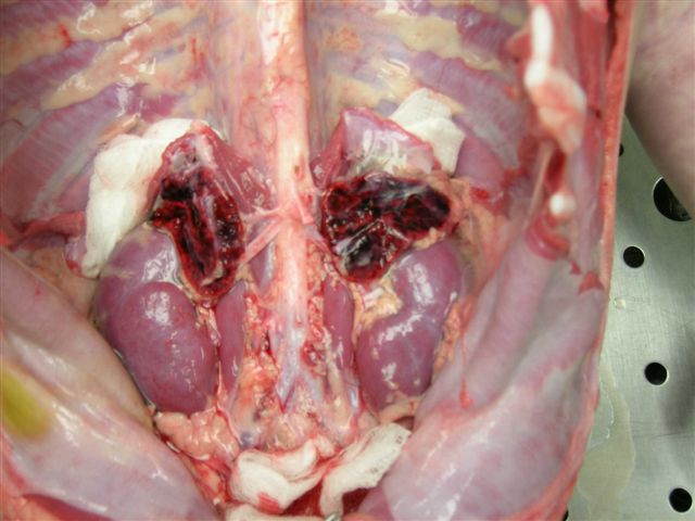
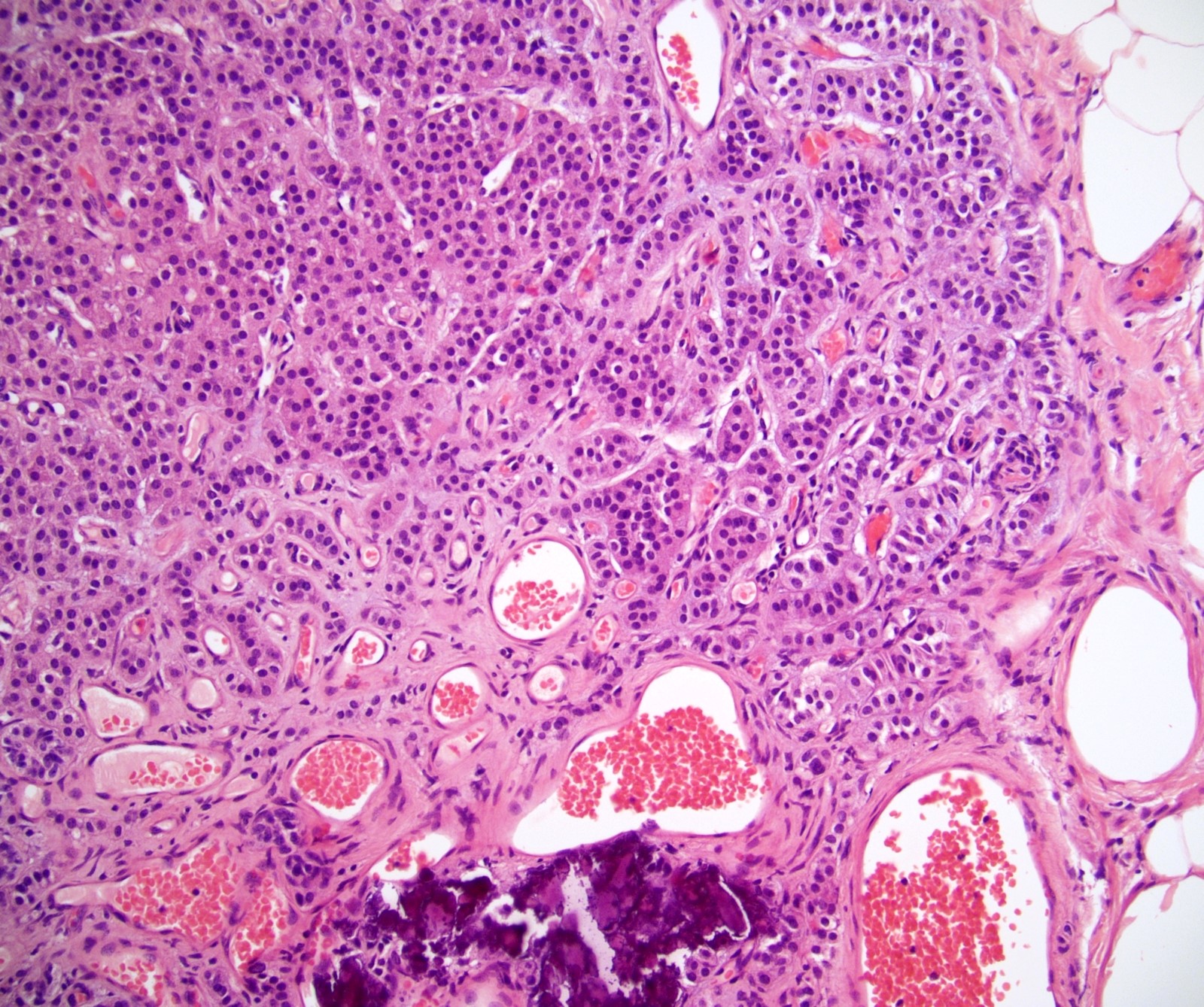
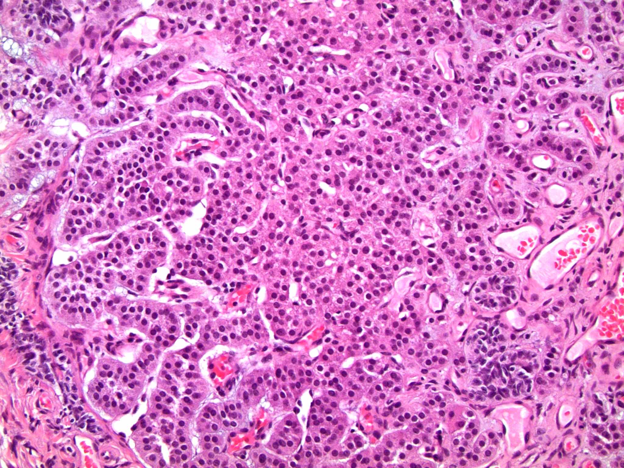
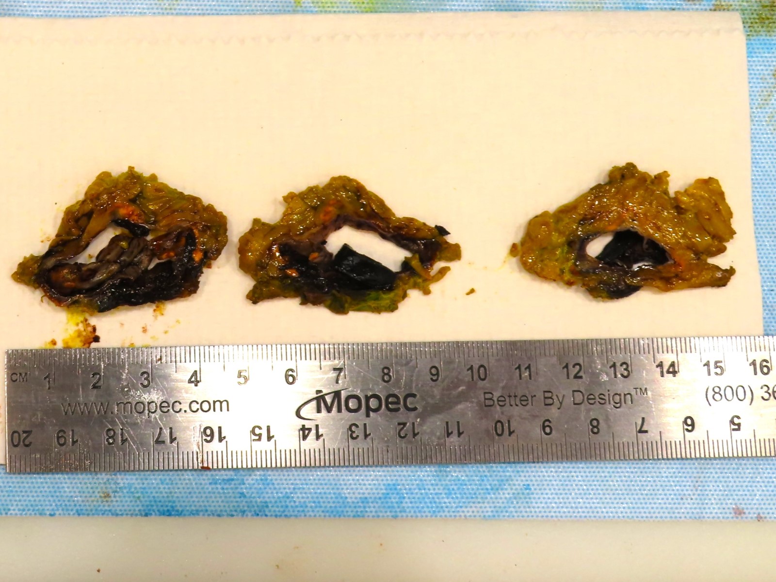
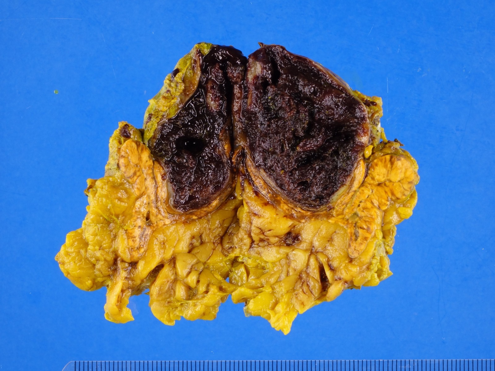
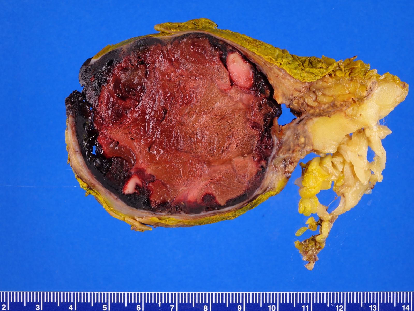
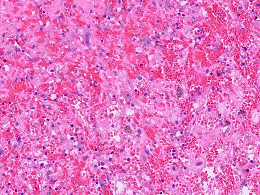
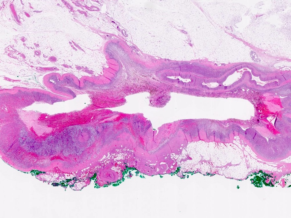
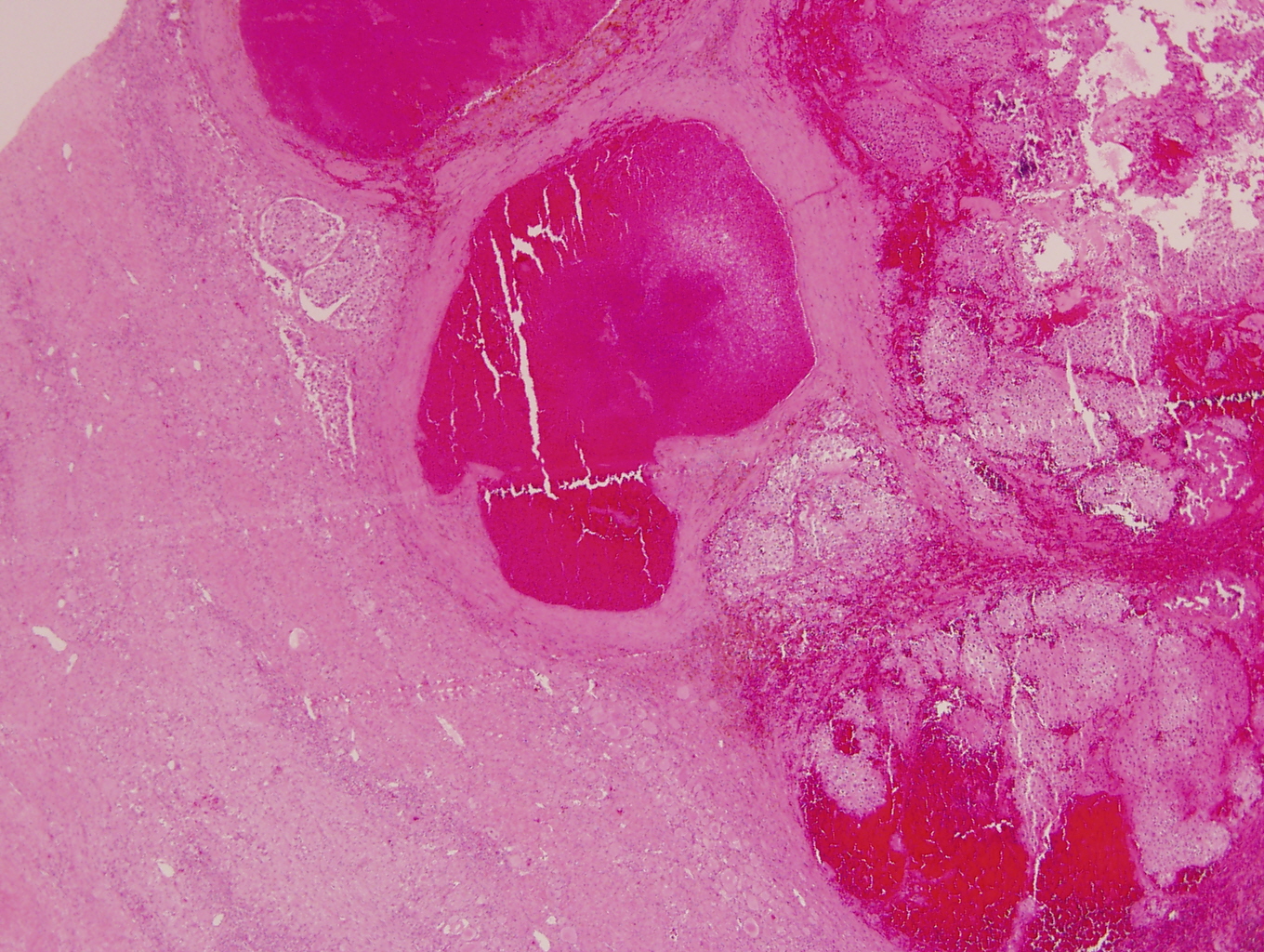
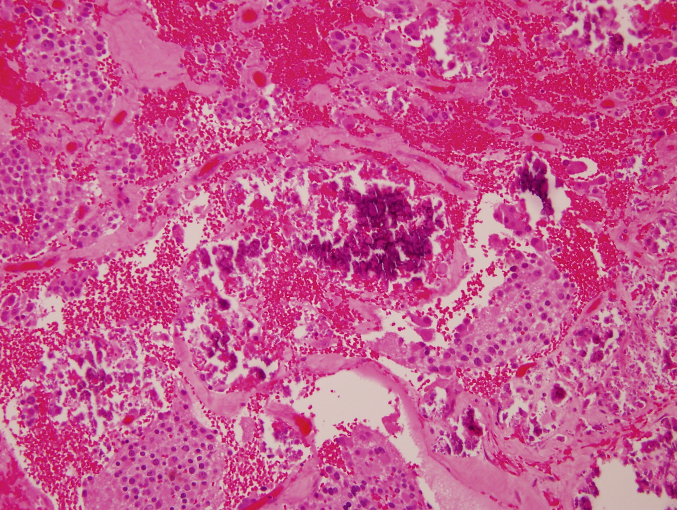
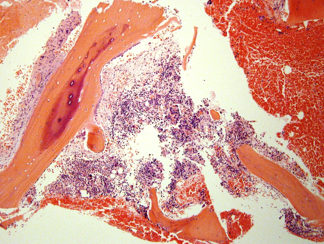
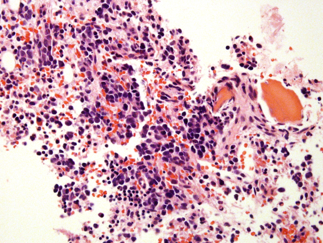
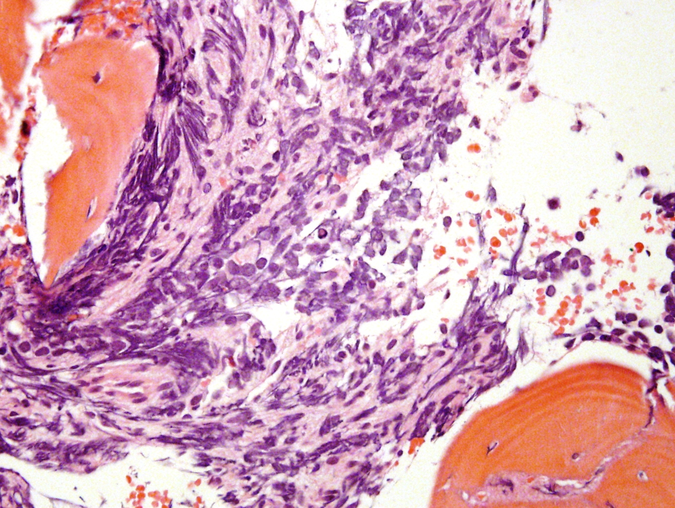
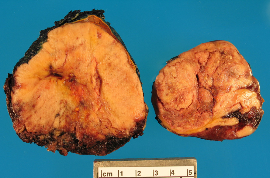
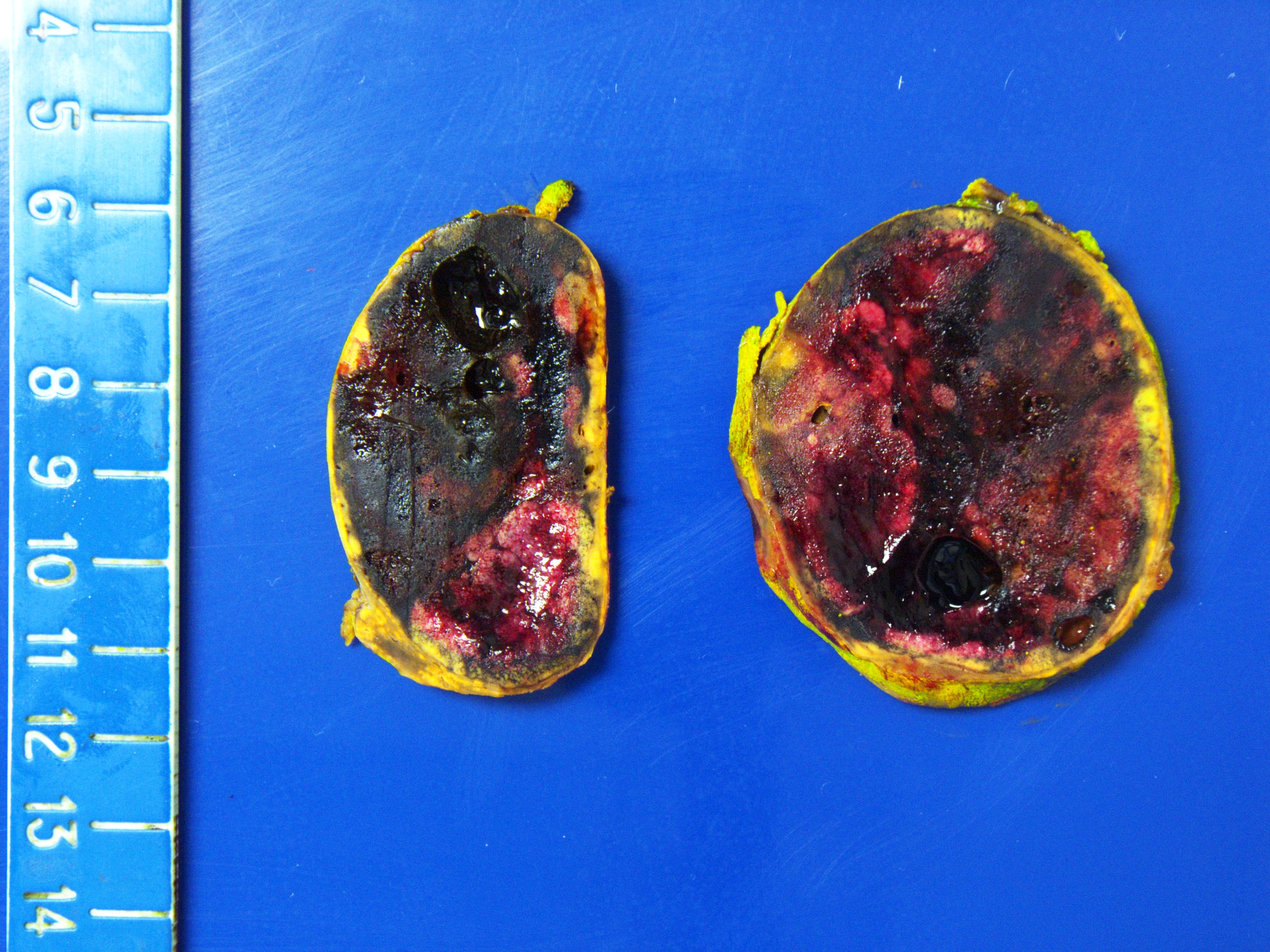
  • Massive adrenal hemorrhage destroying adrenal cortex due to anticoagulation, coagulopathy and newborns with physiologic deficiencies in prothrombin time
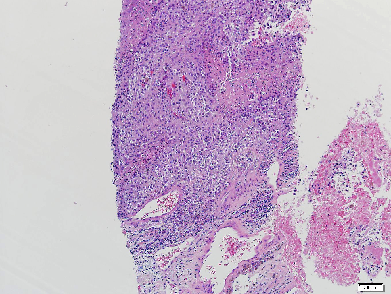
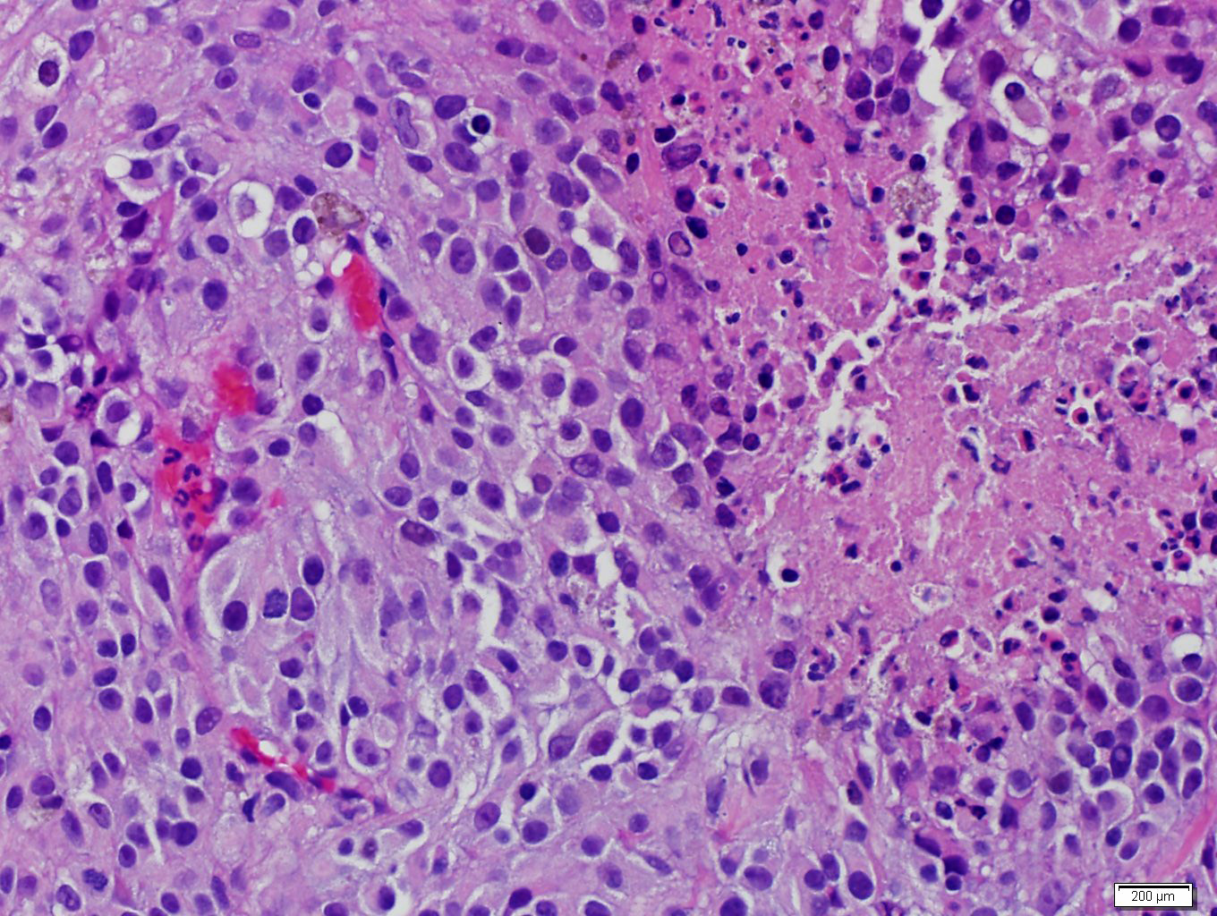
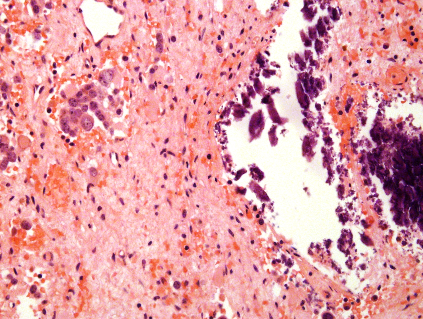
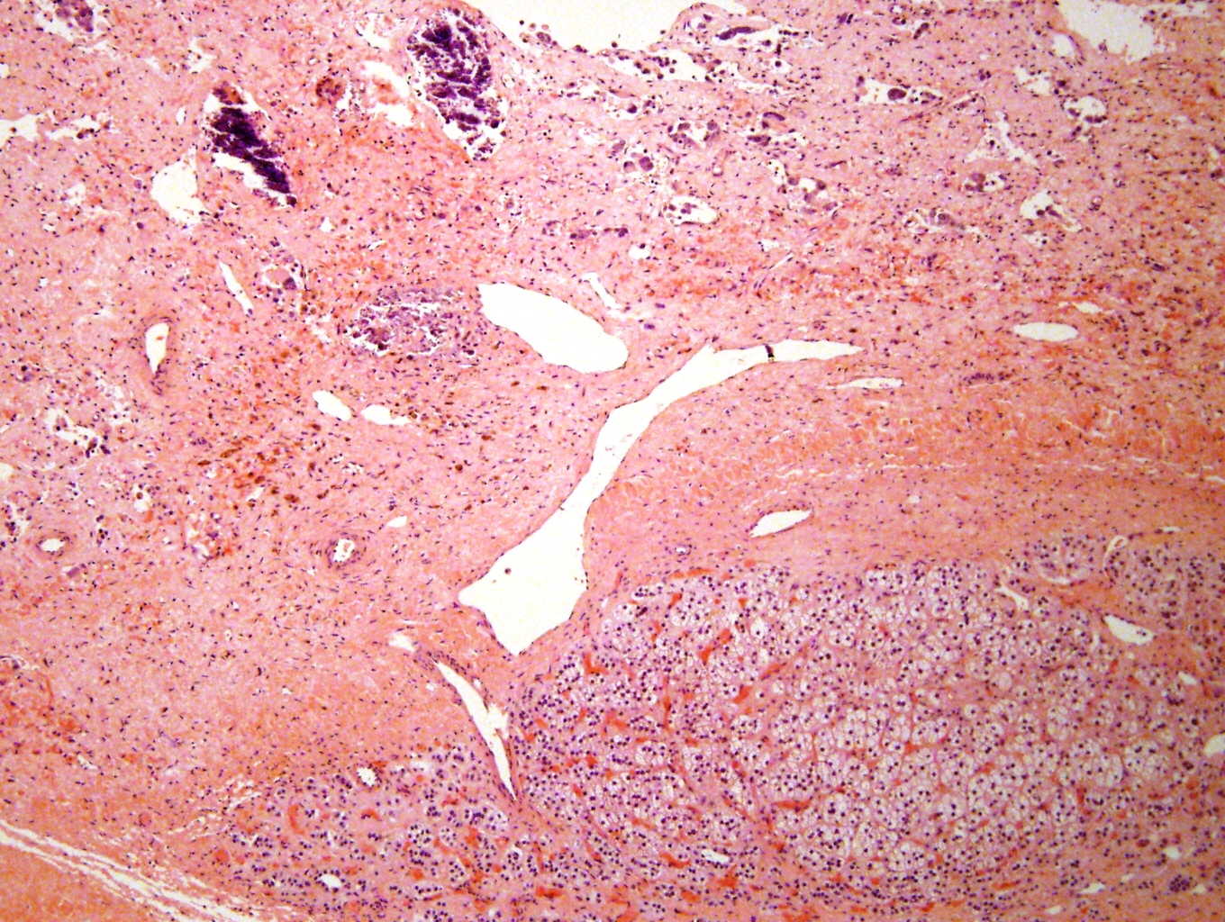
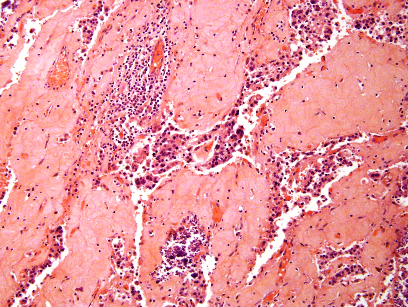
  • Hypotension / shock that causes mild or massive corticomedullary necrosis, including Waterhouse-Friderichsen syndrome
  • Infections that destroy substantial adrenal cortical tissue
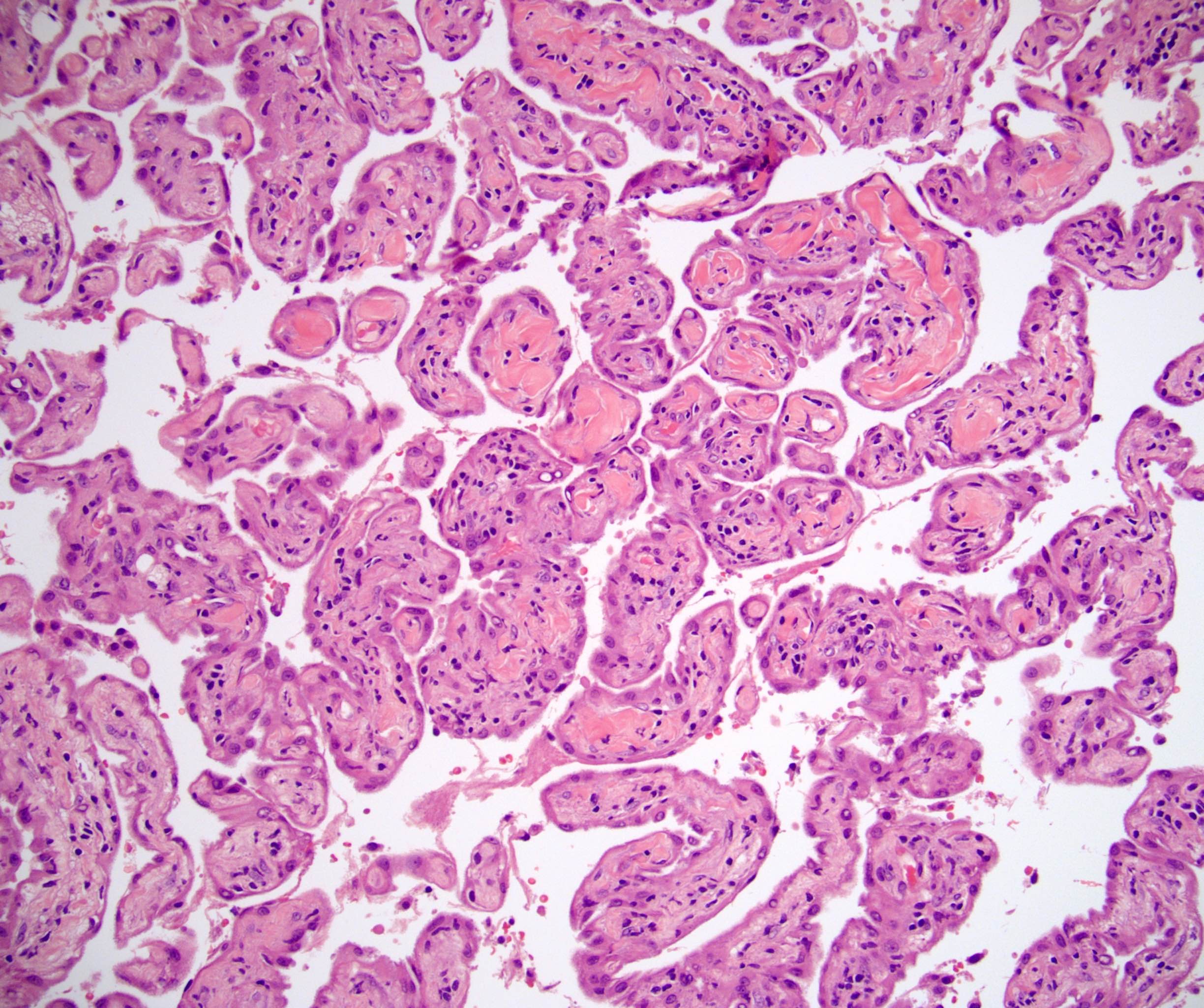
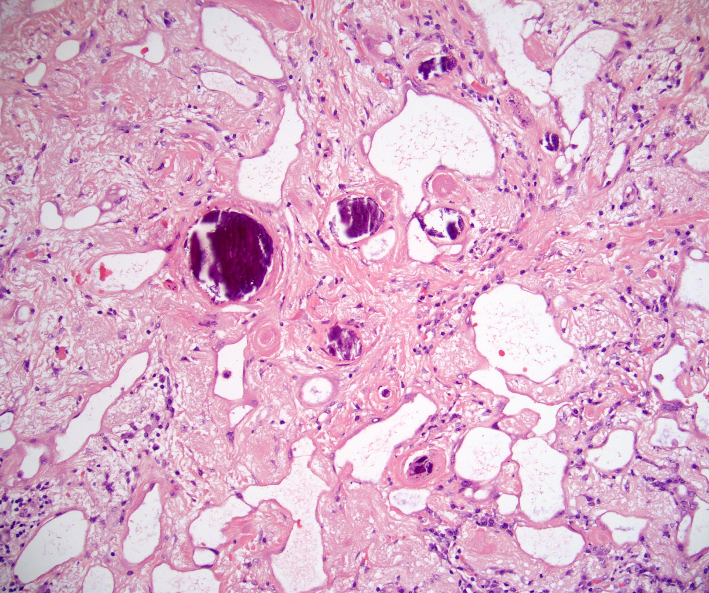
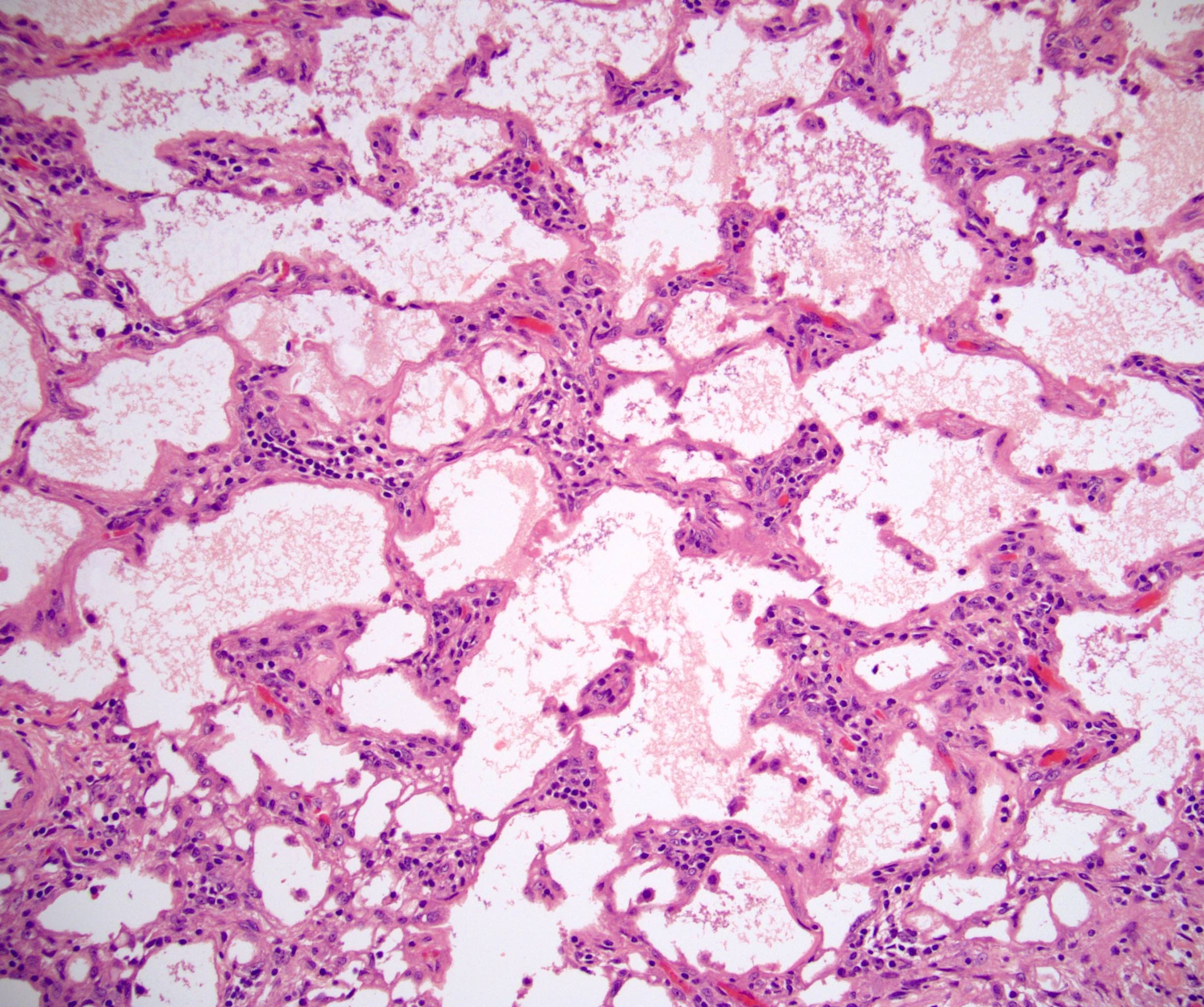
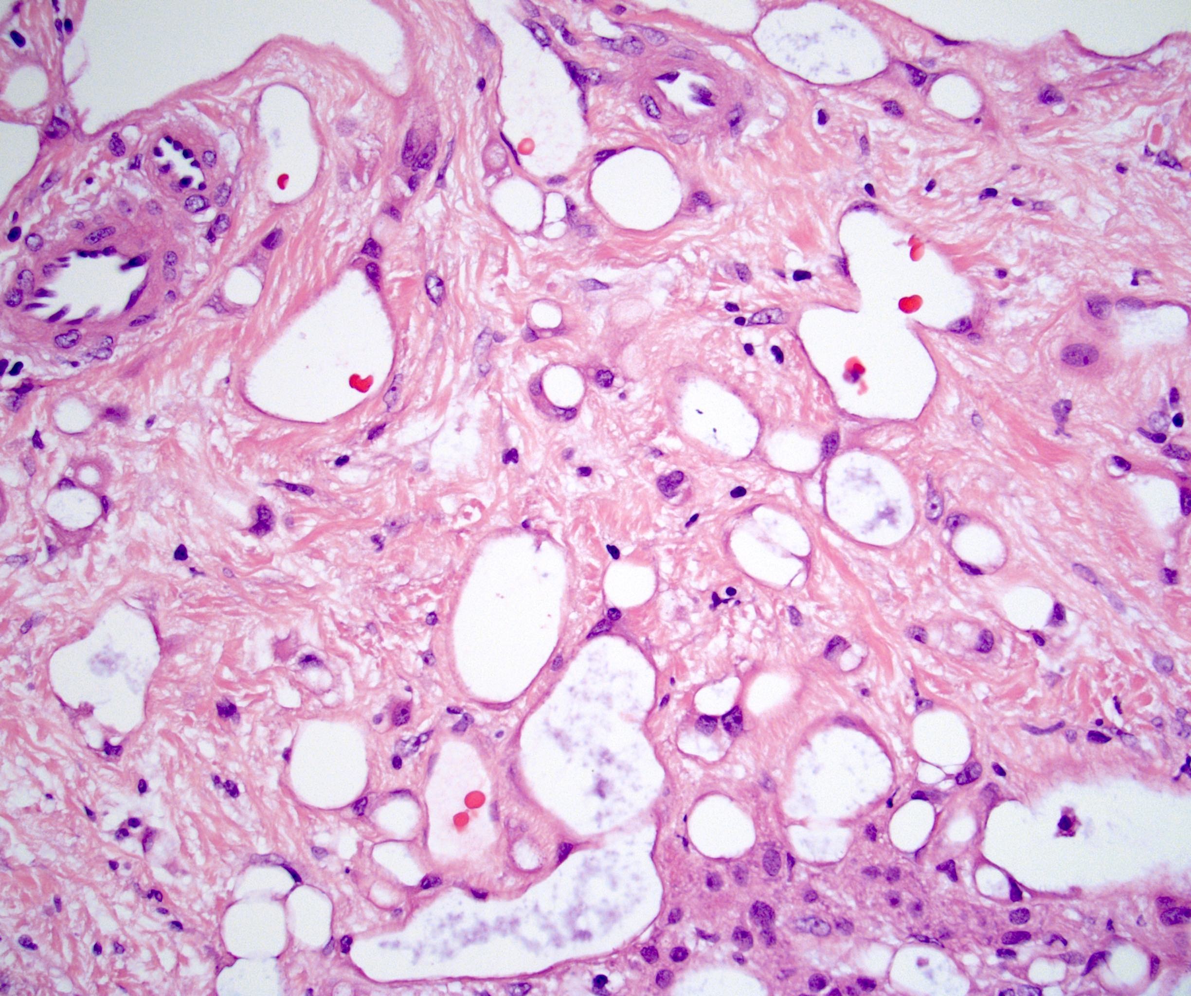
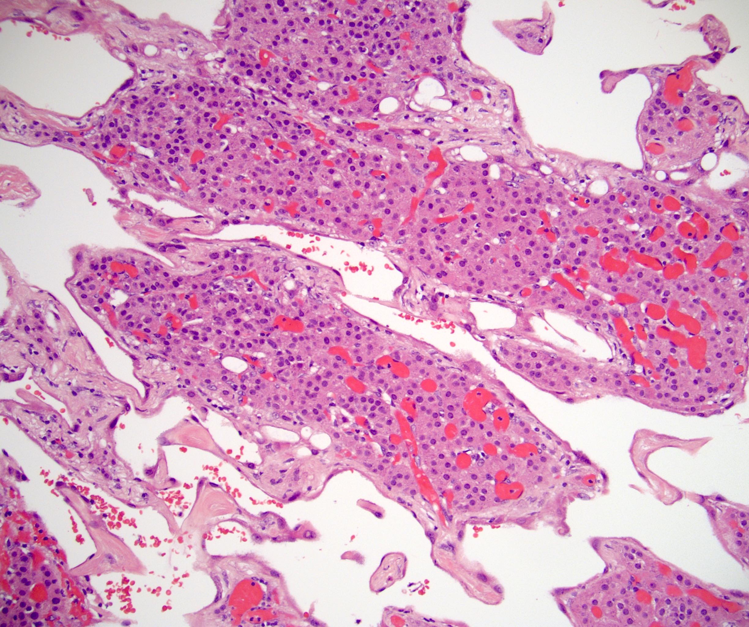
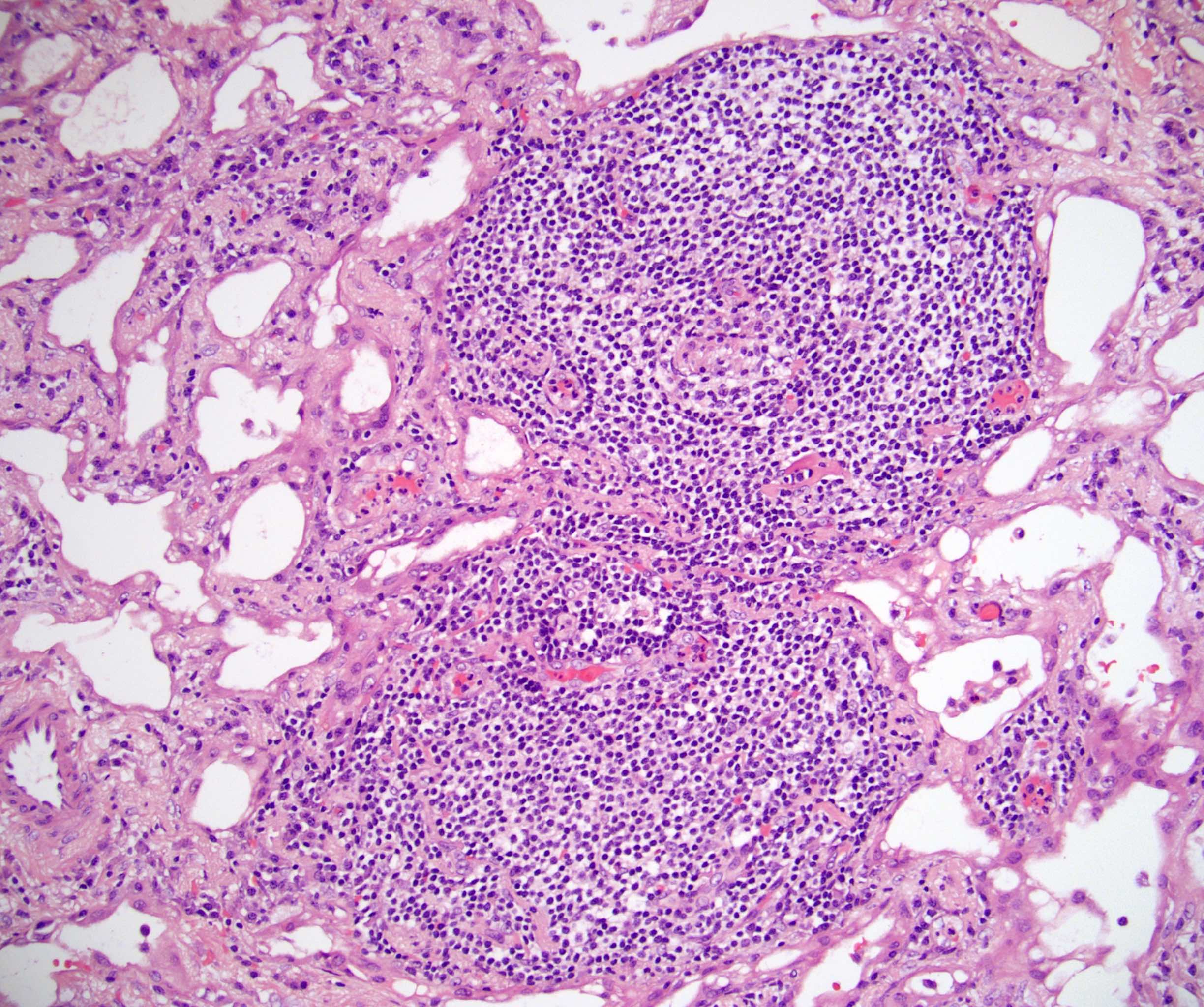
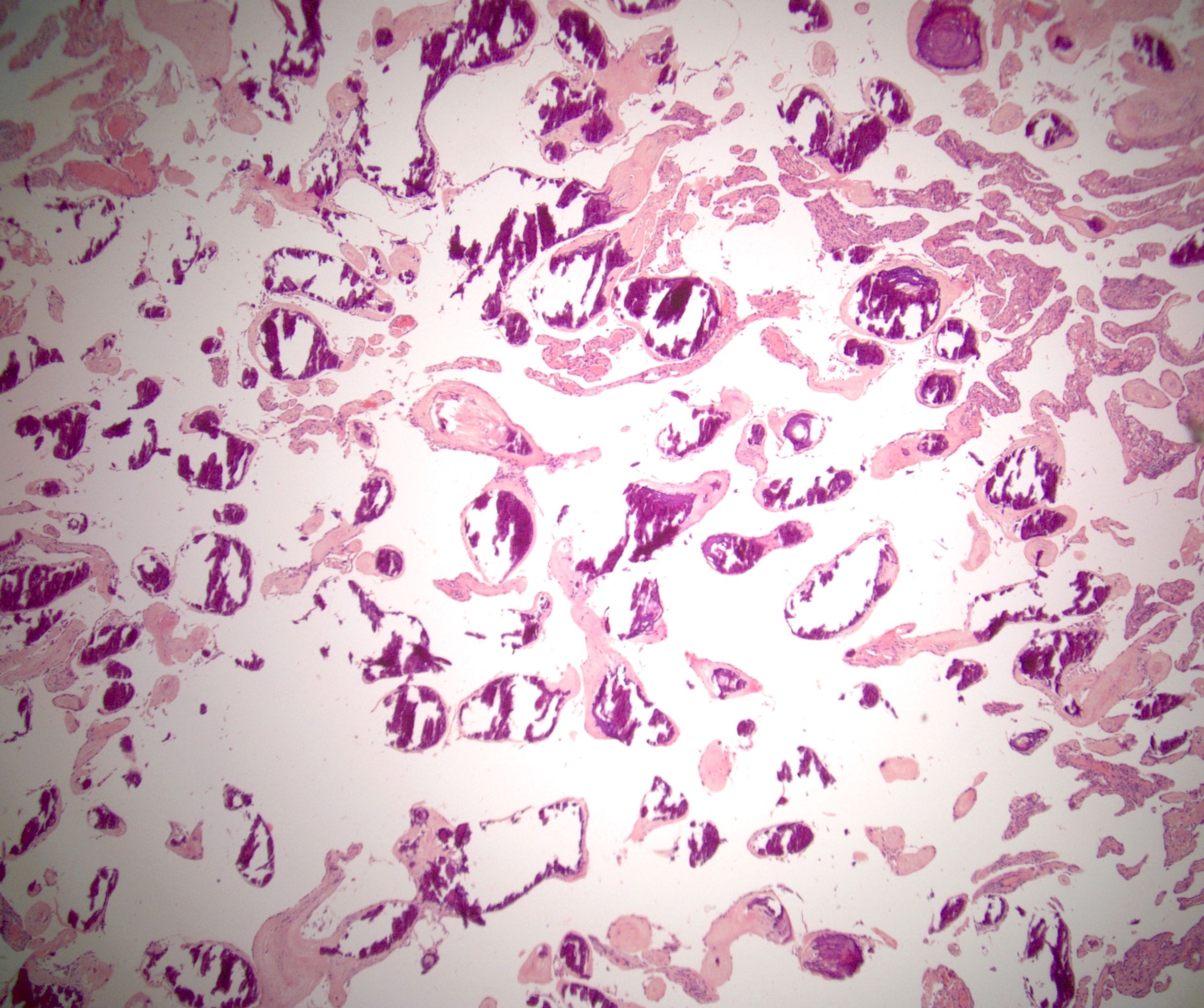
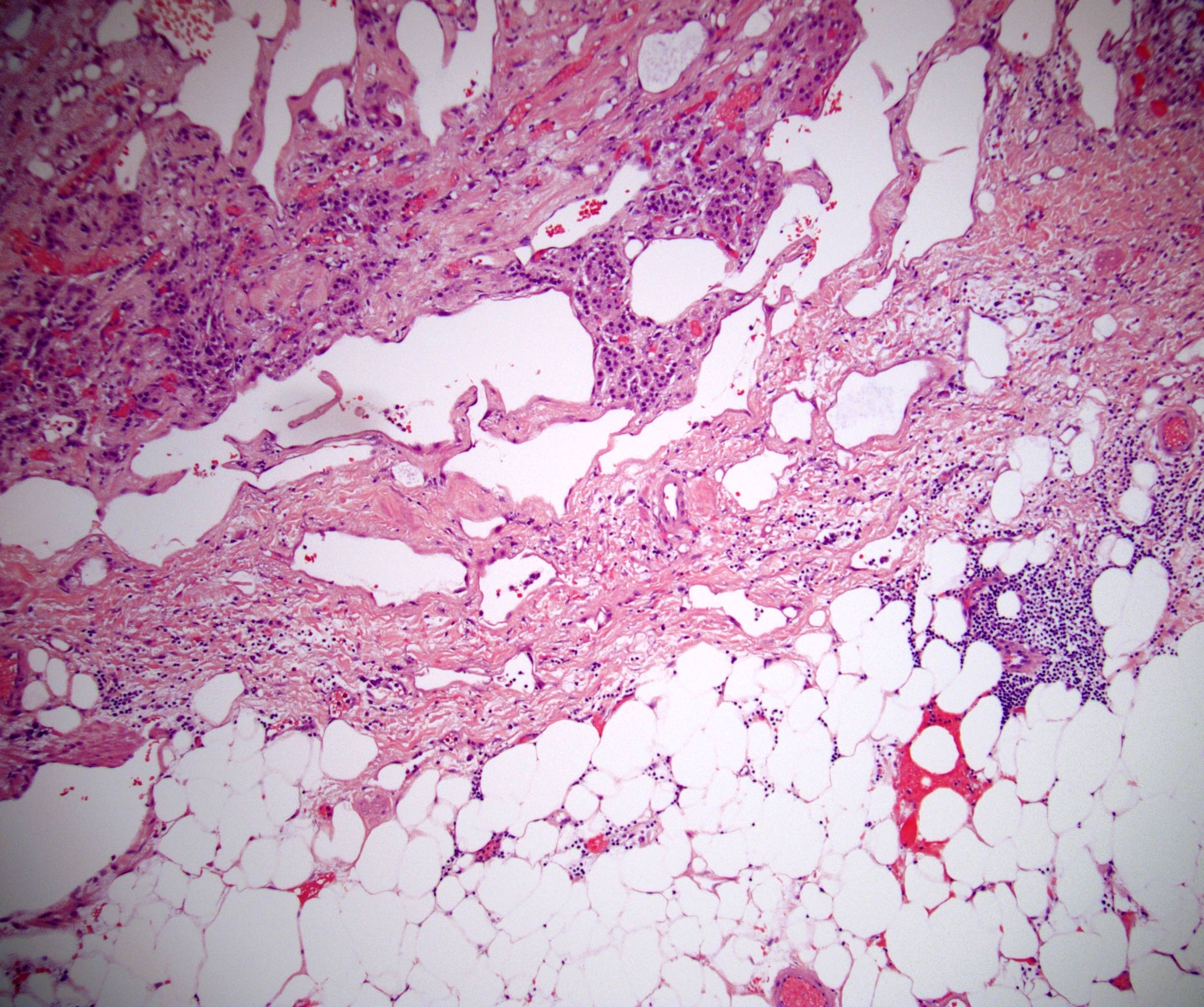
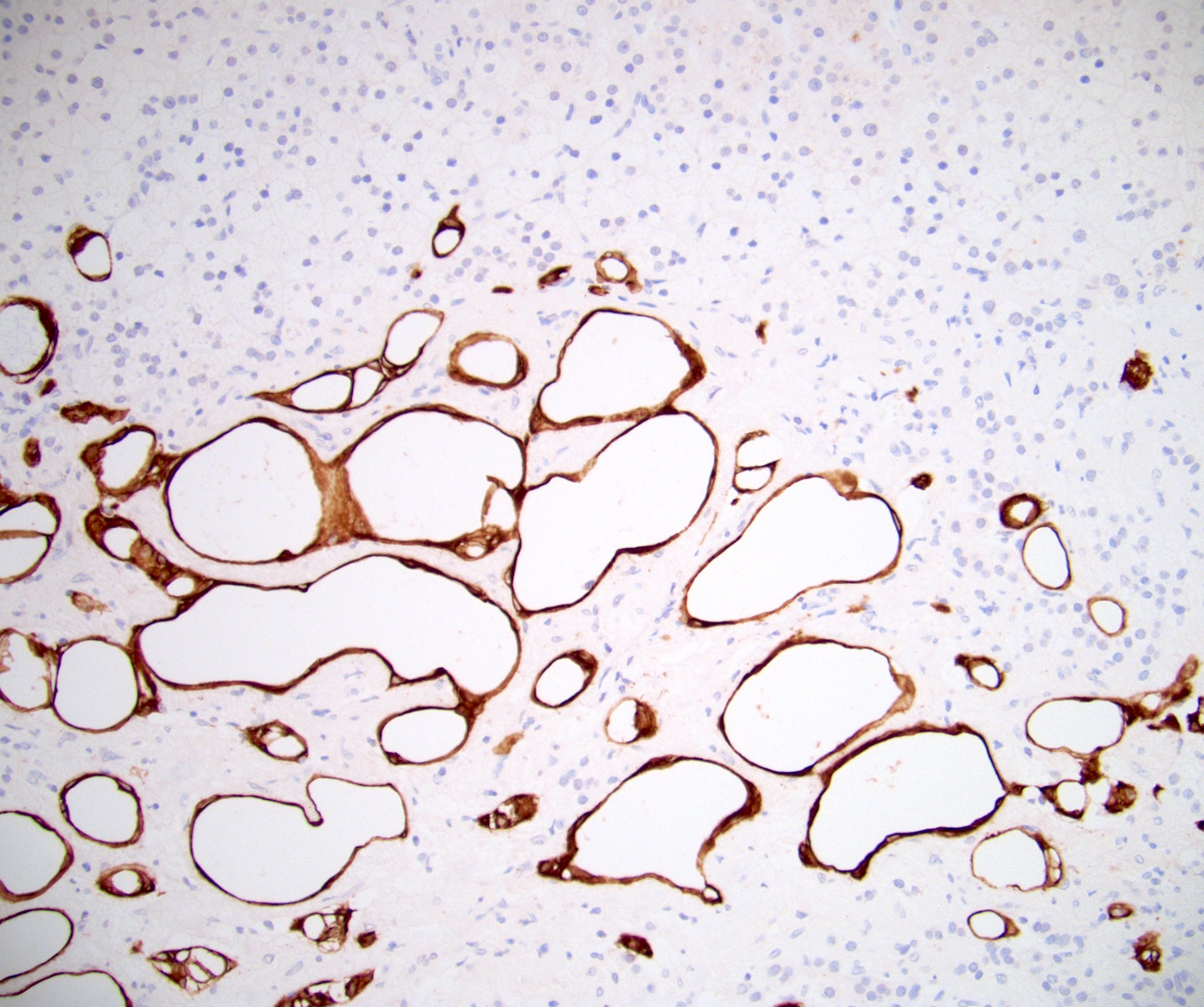
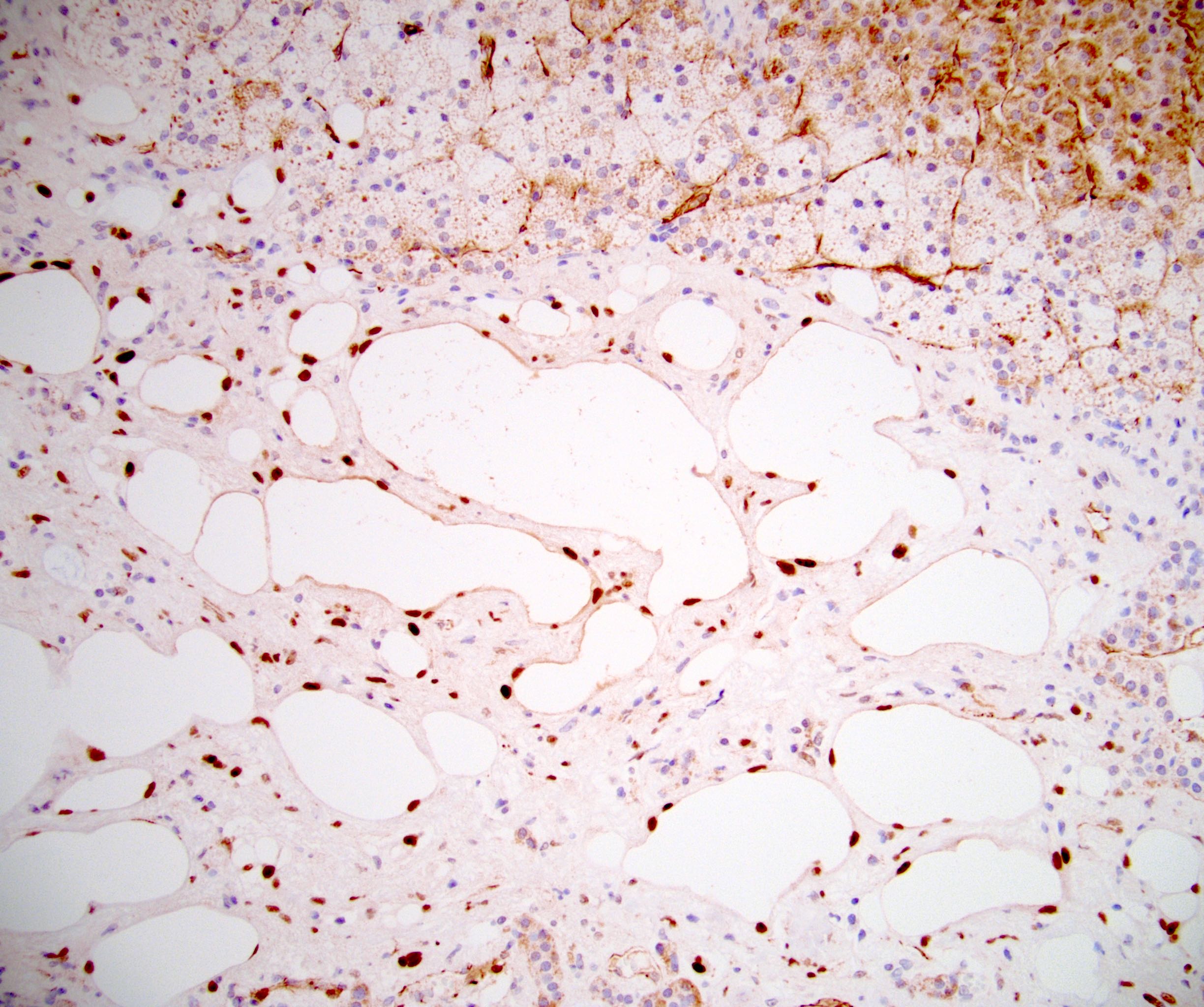
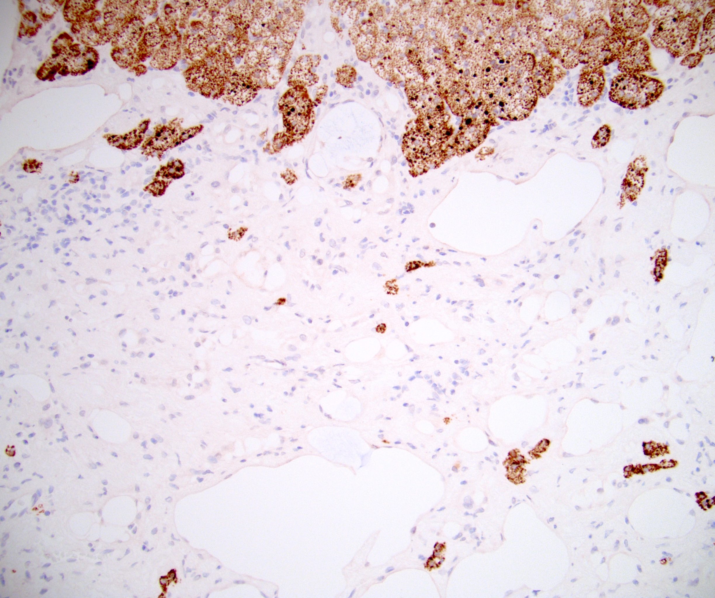
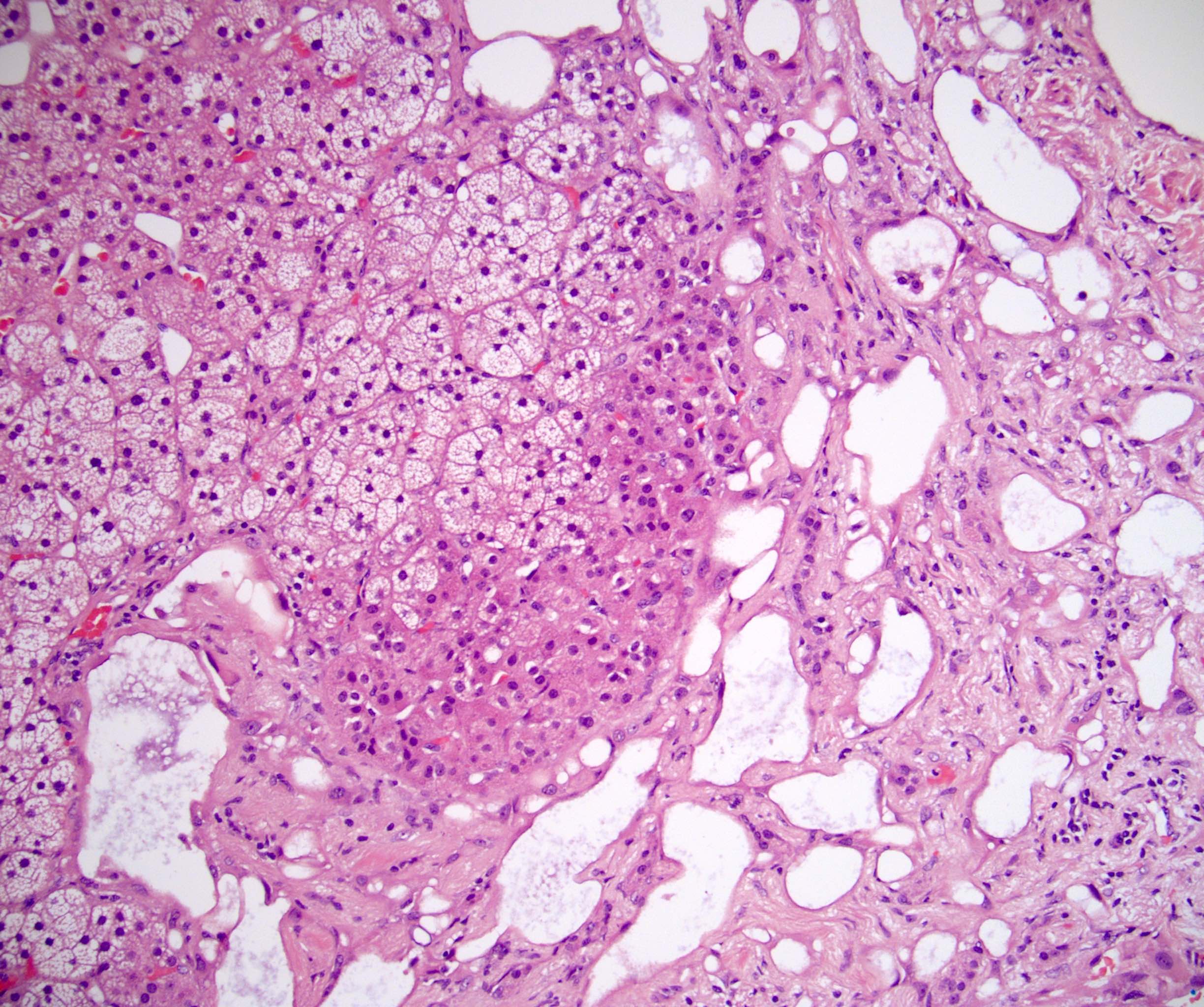
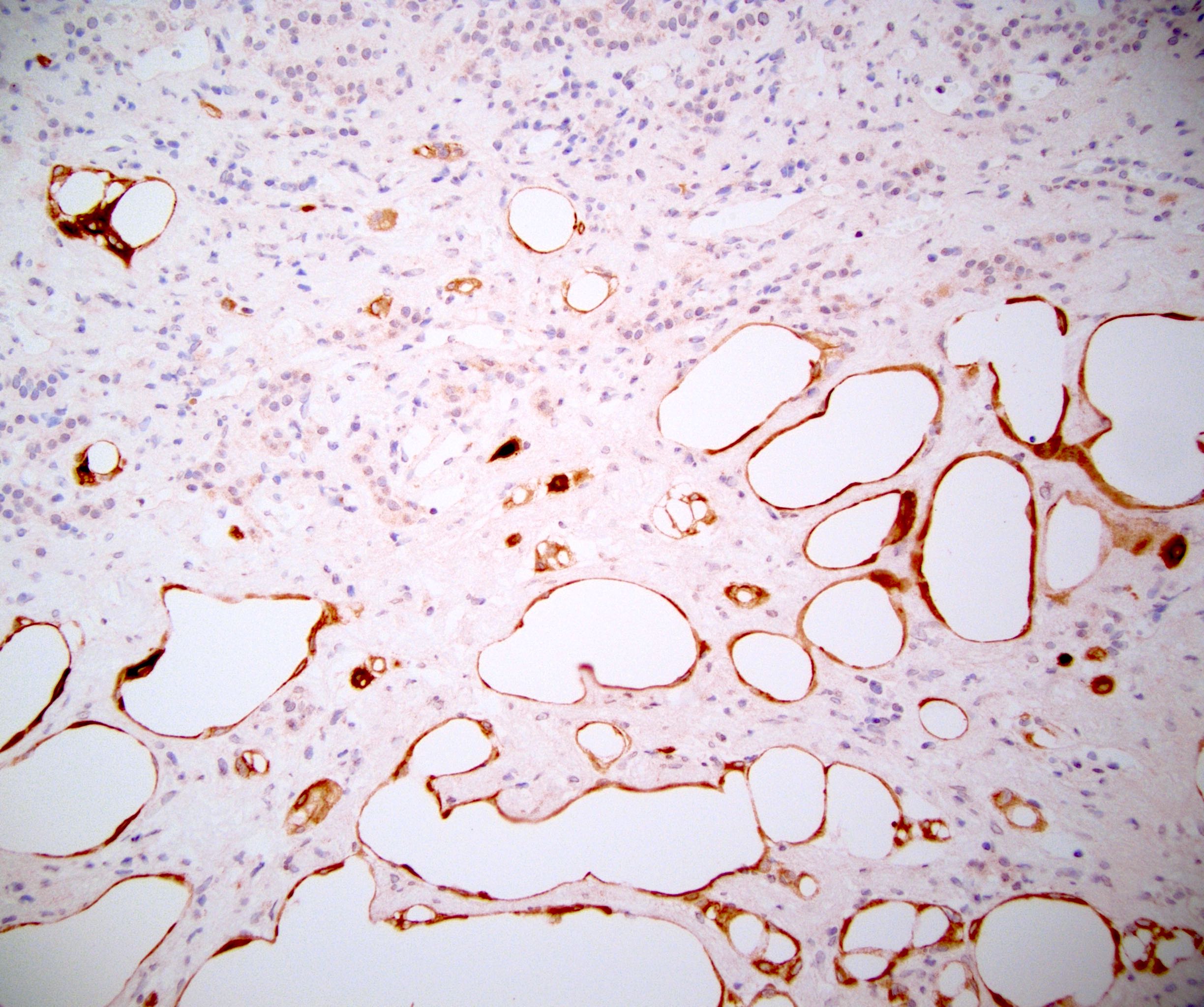
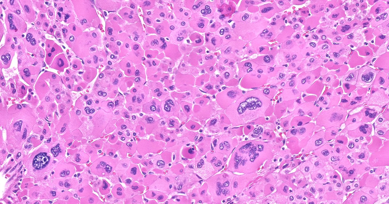
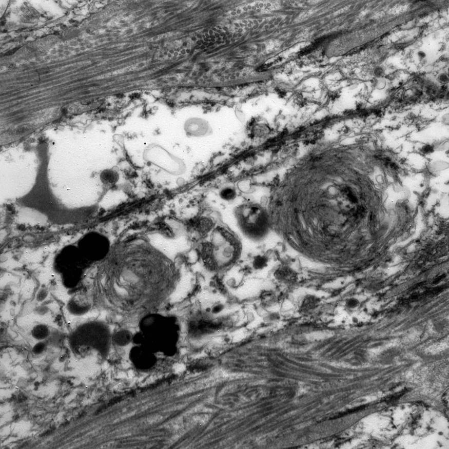
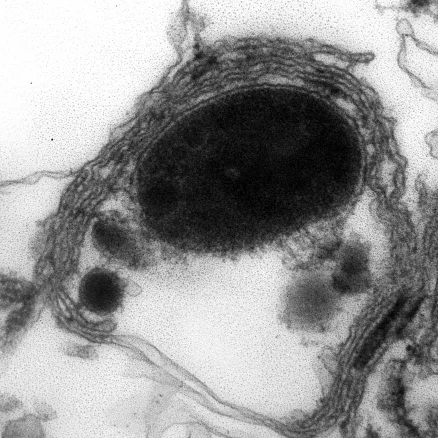
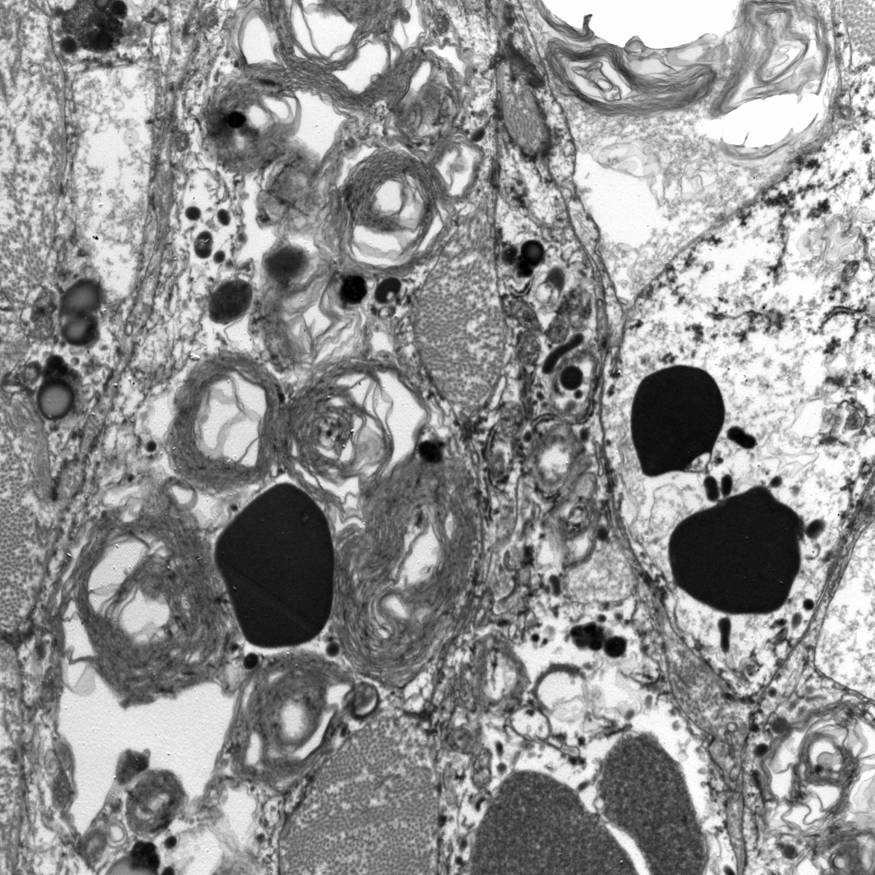
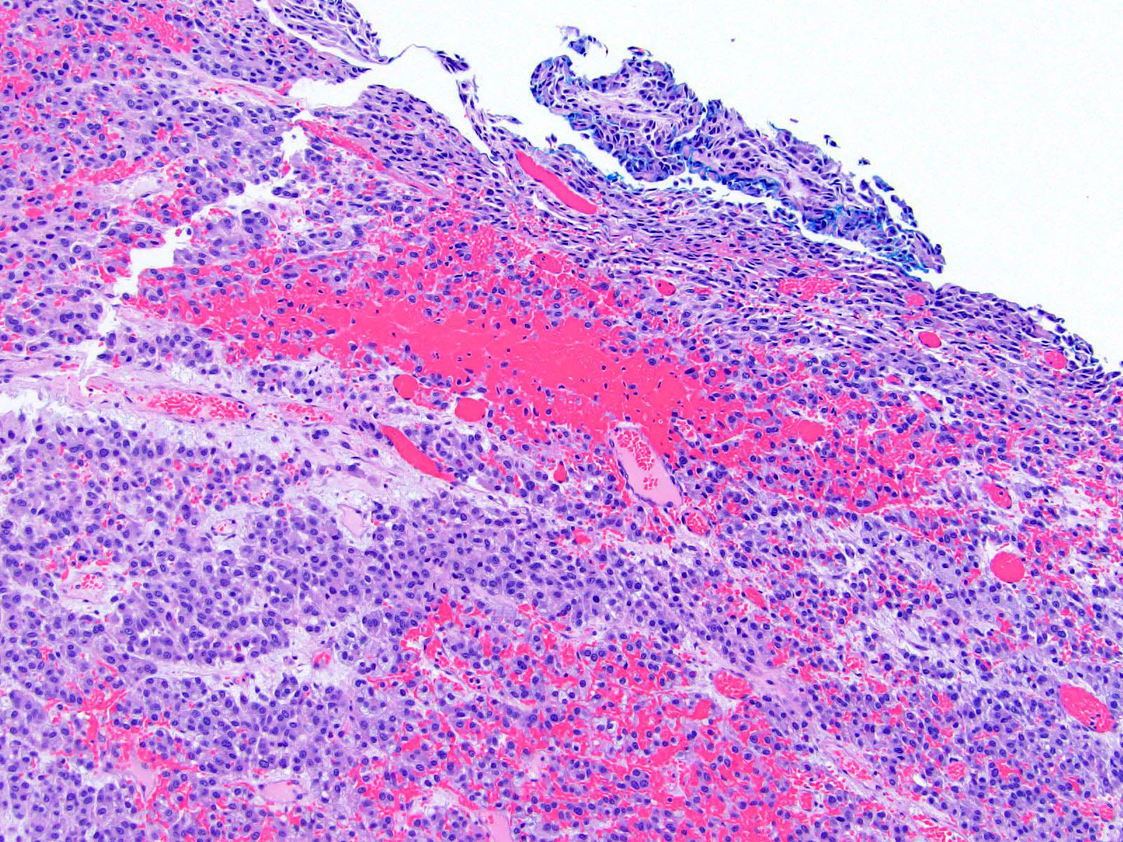
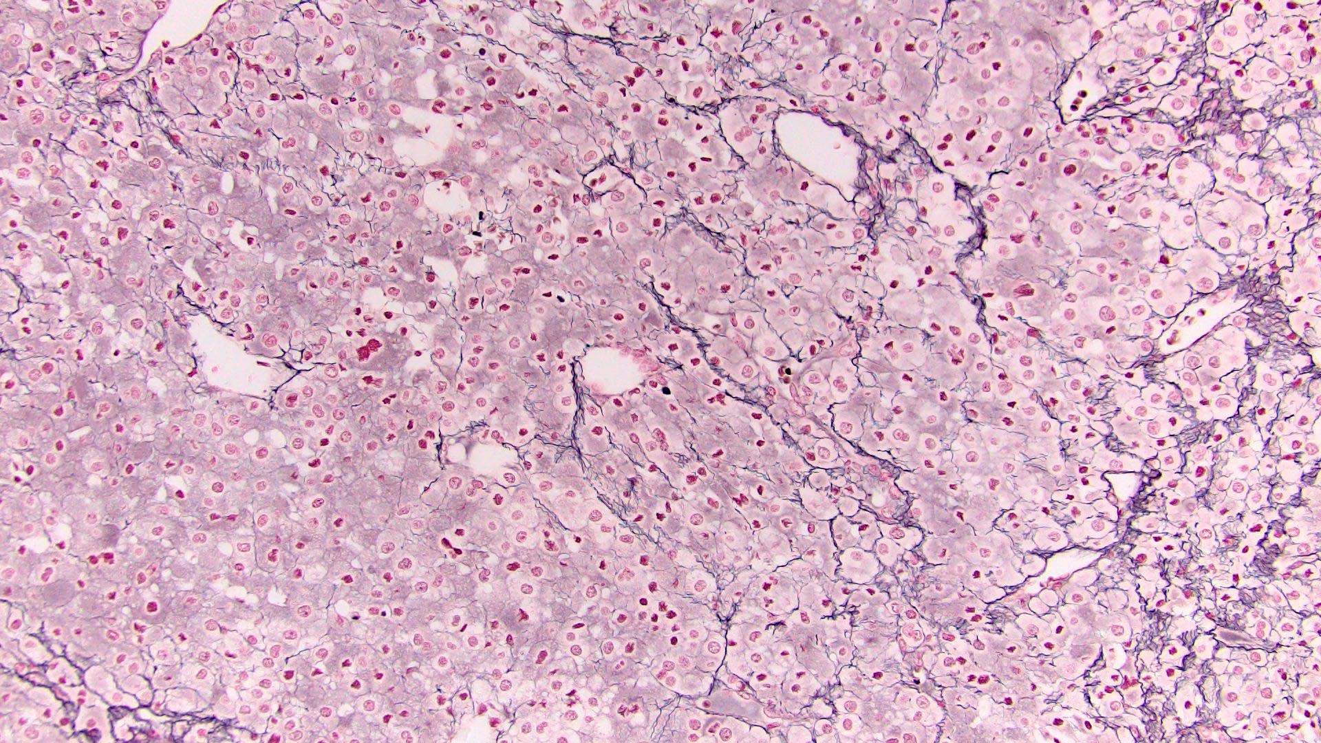
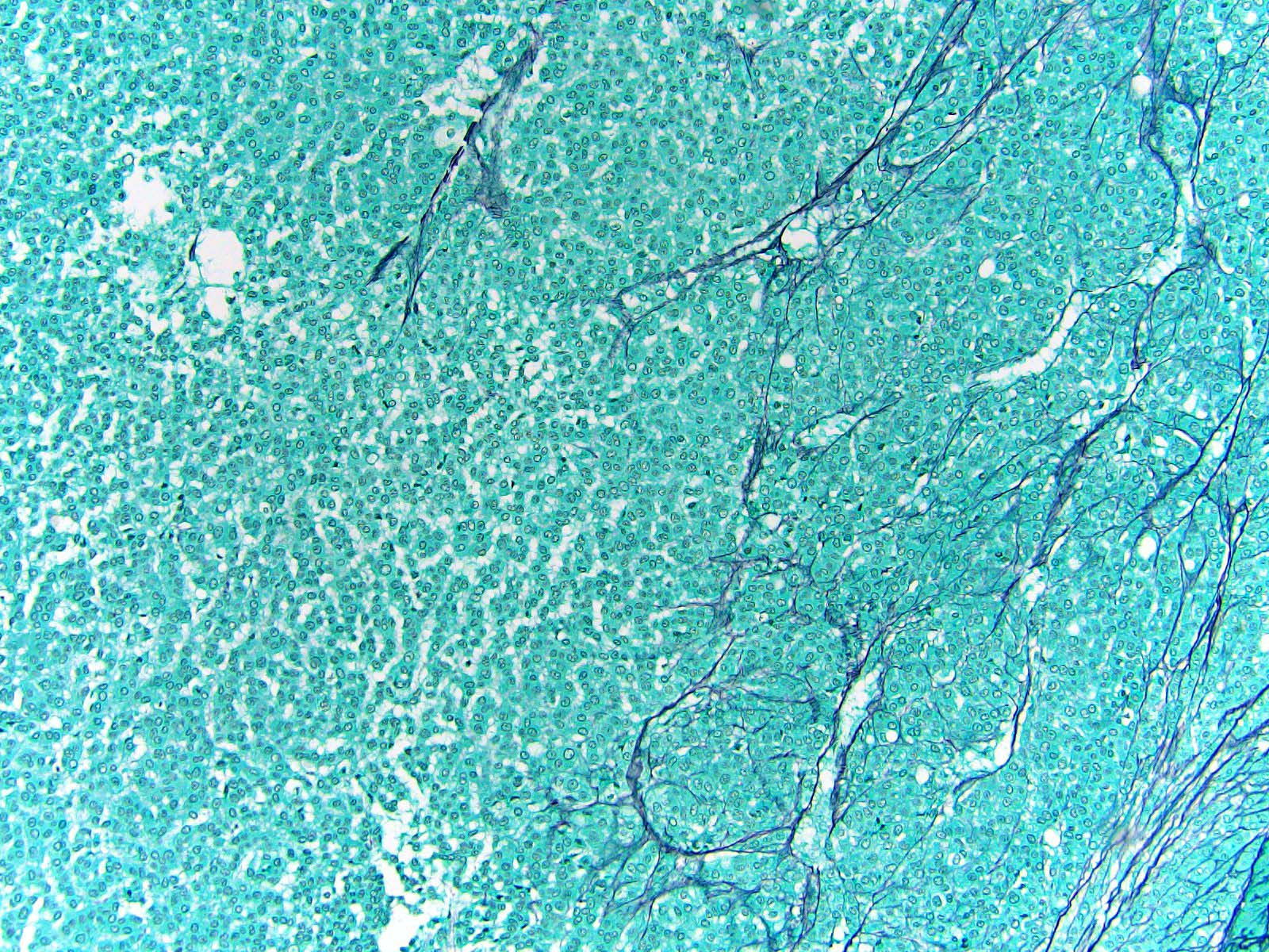
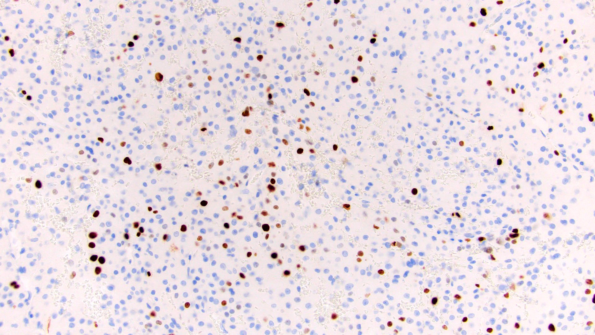
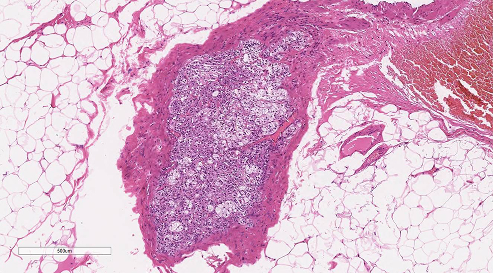
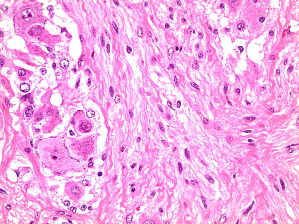
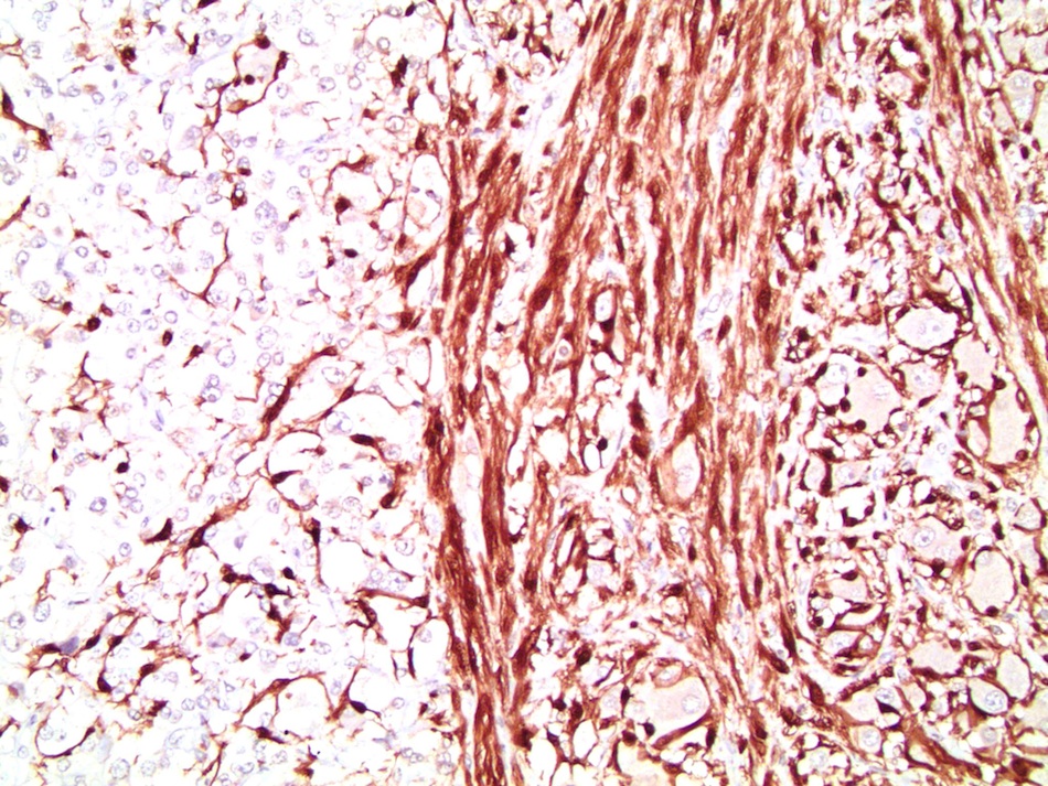
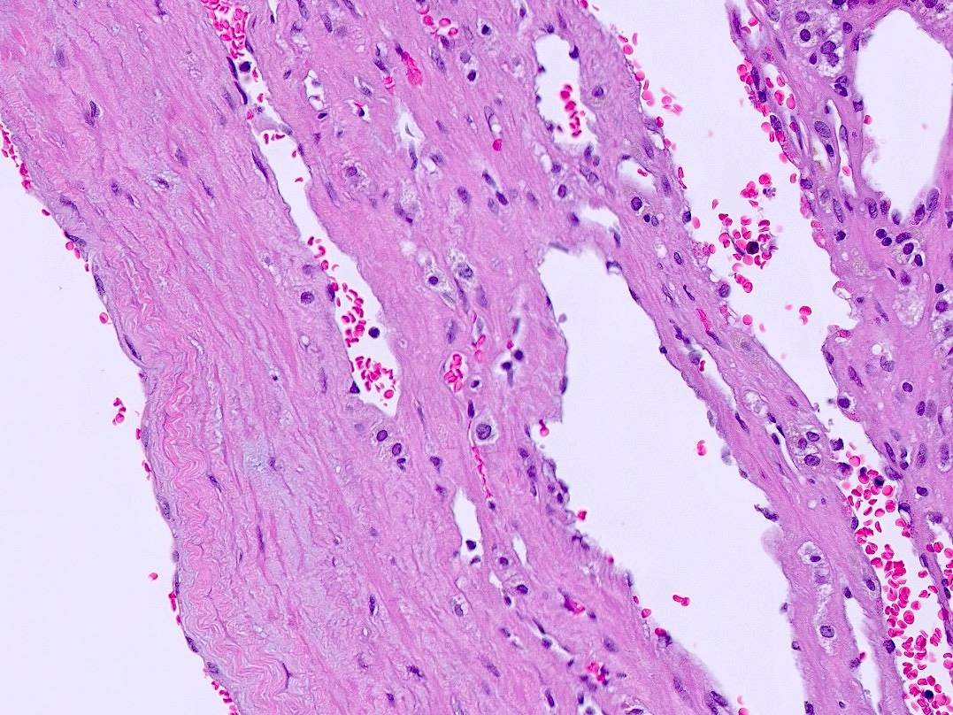
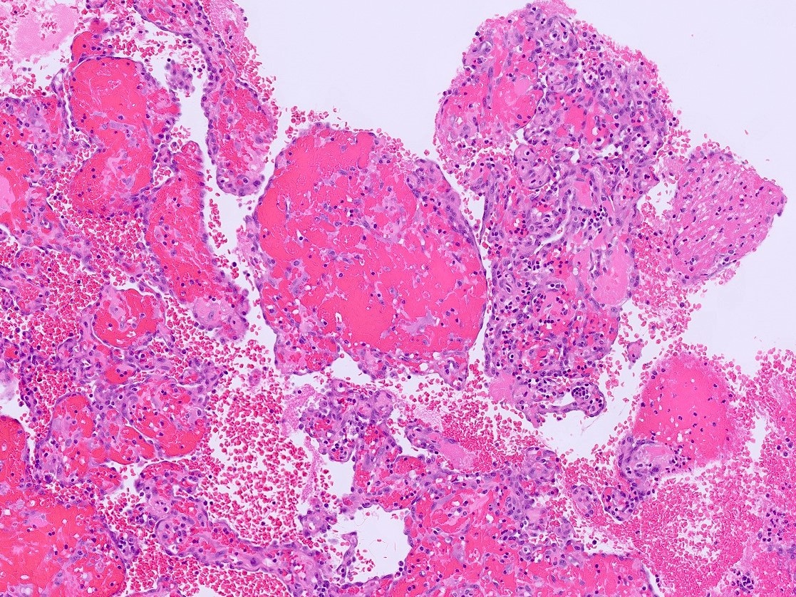
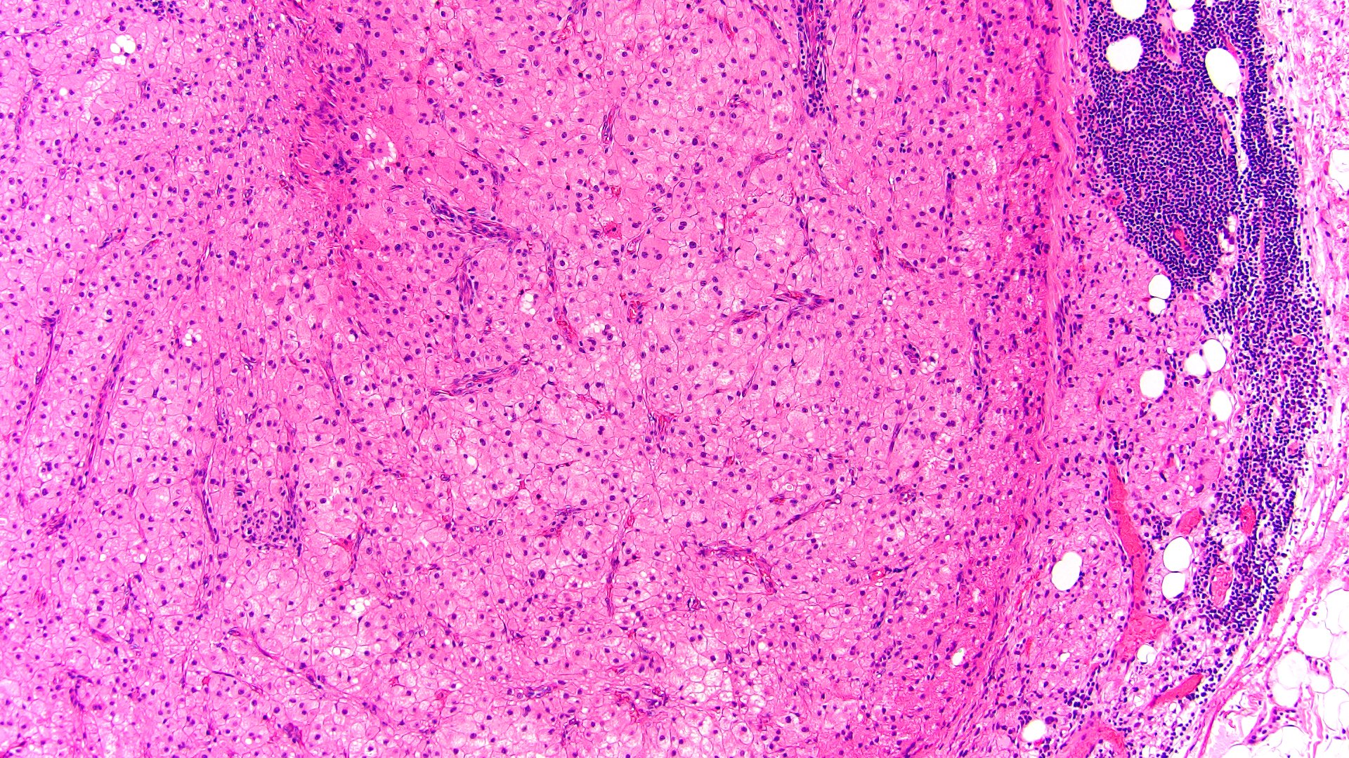
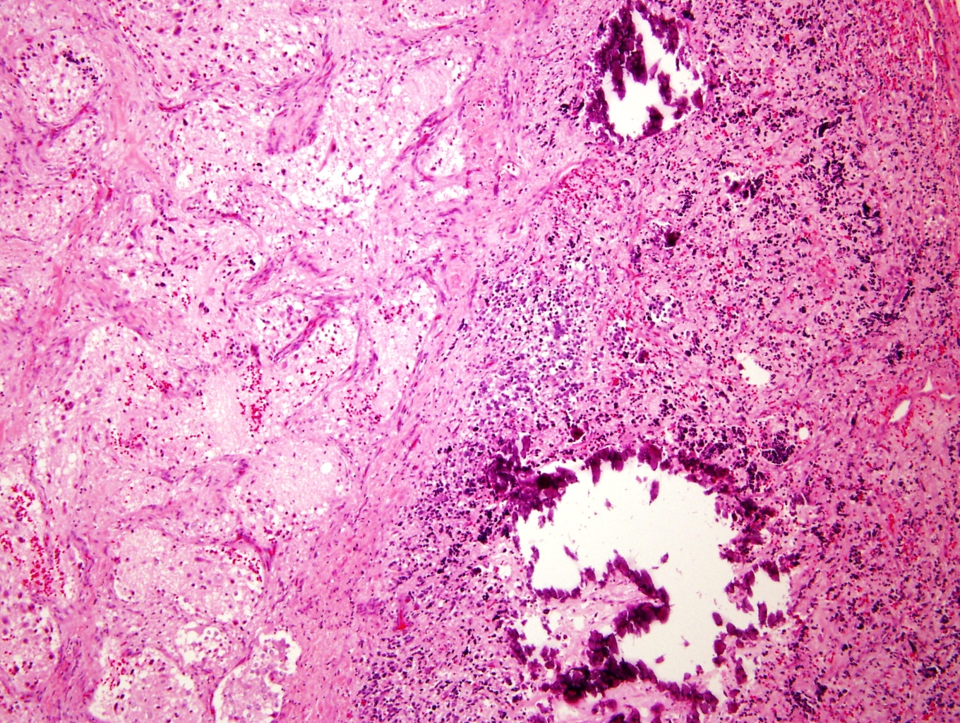
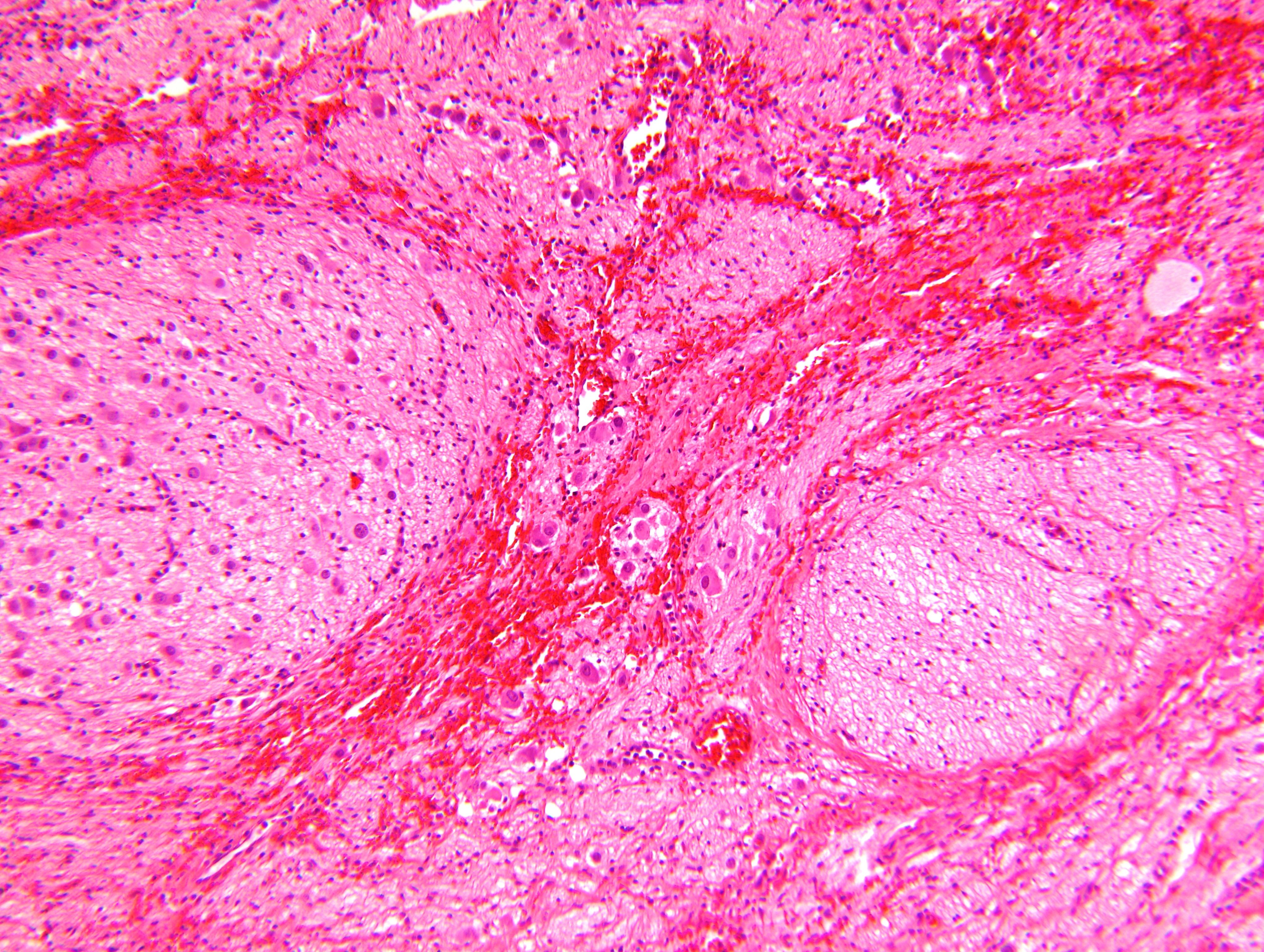
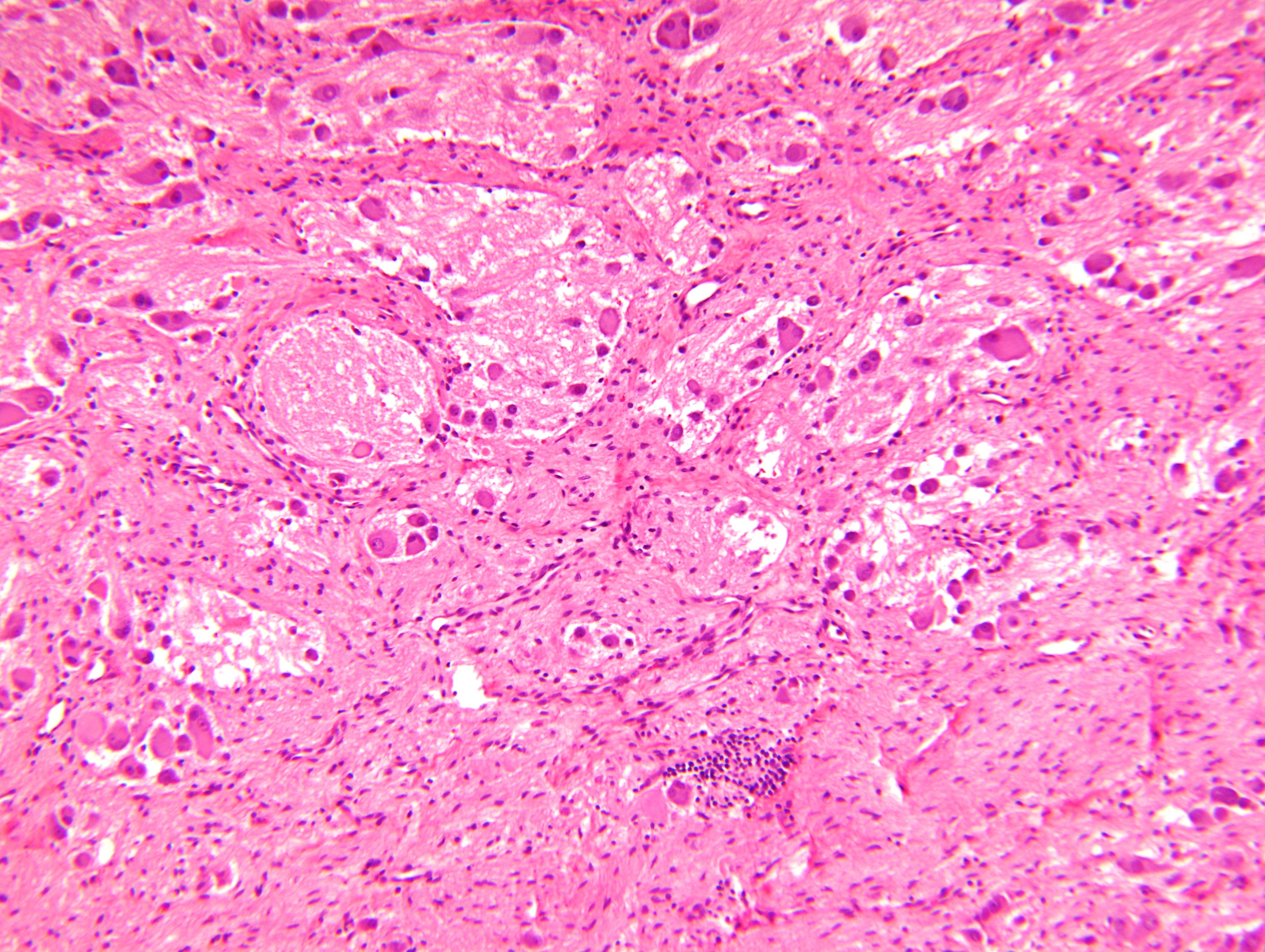
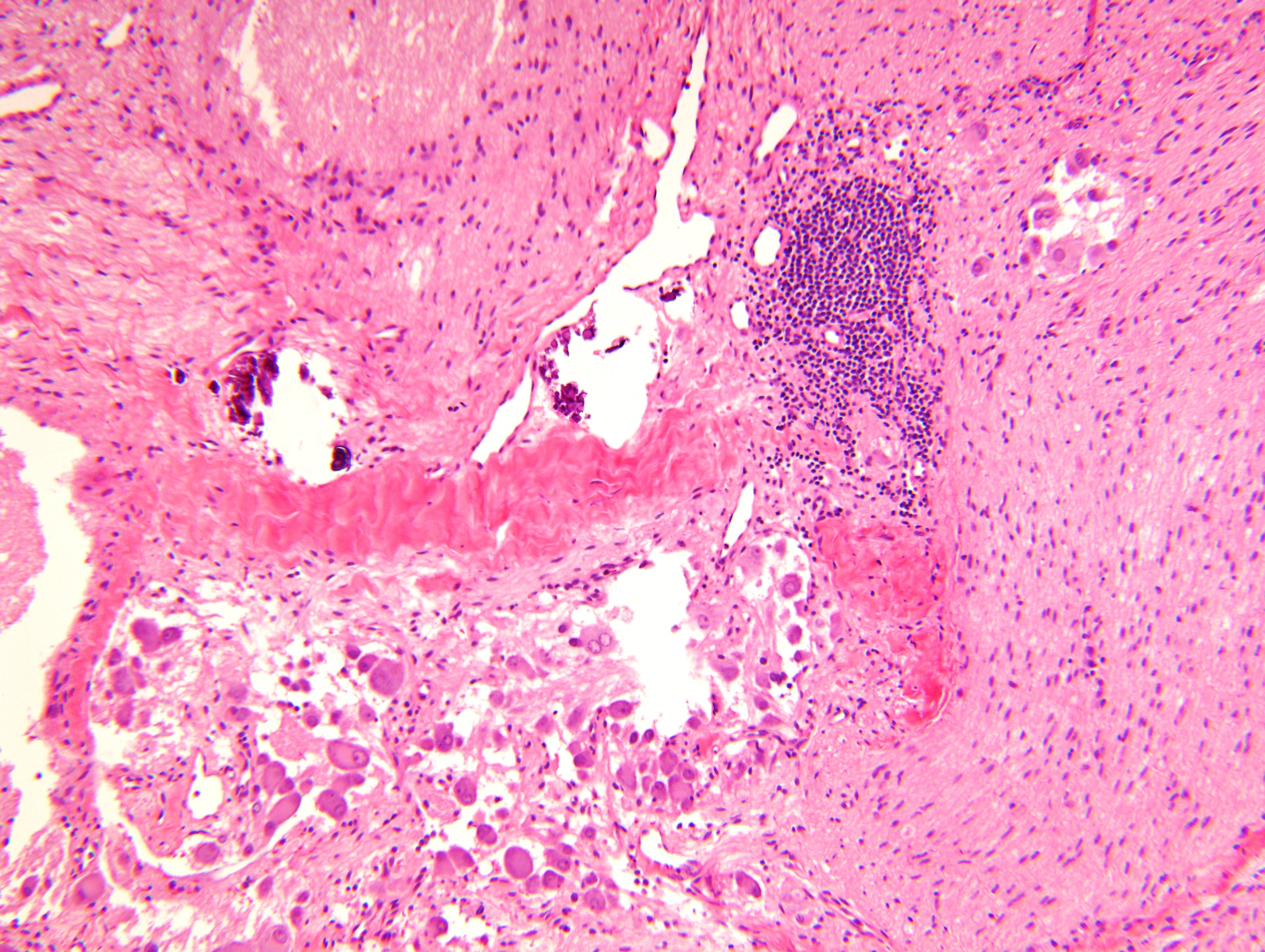
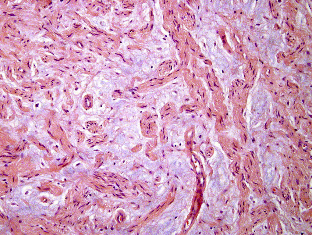
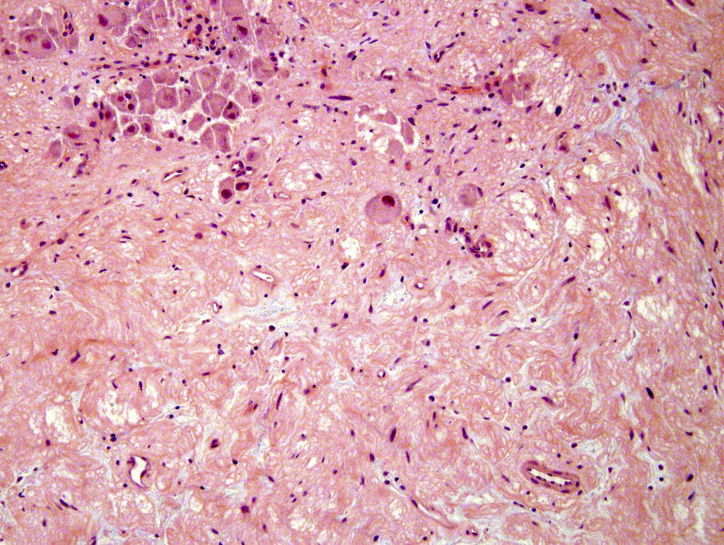
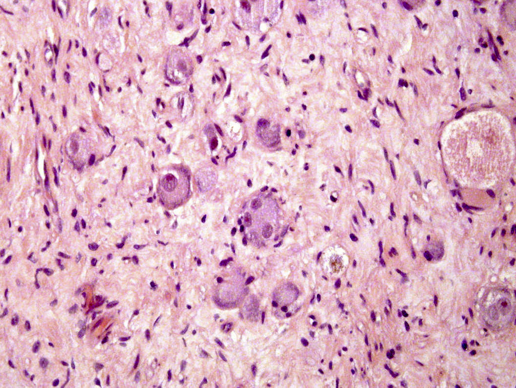
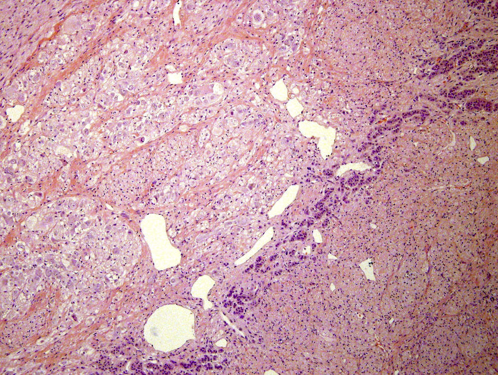
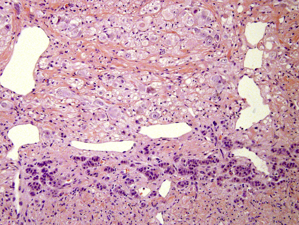
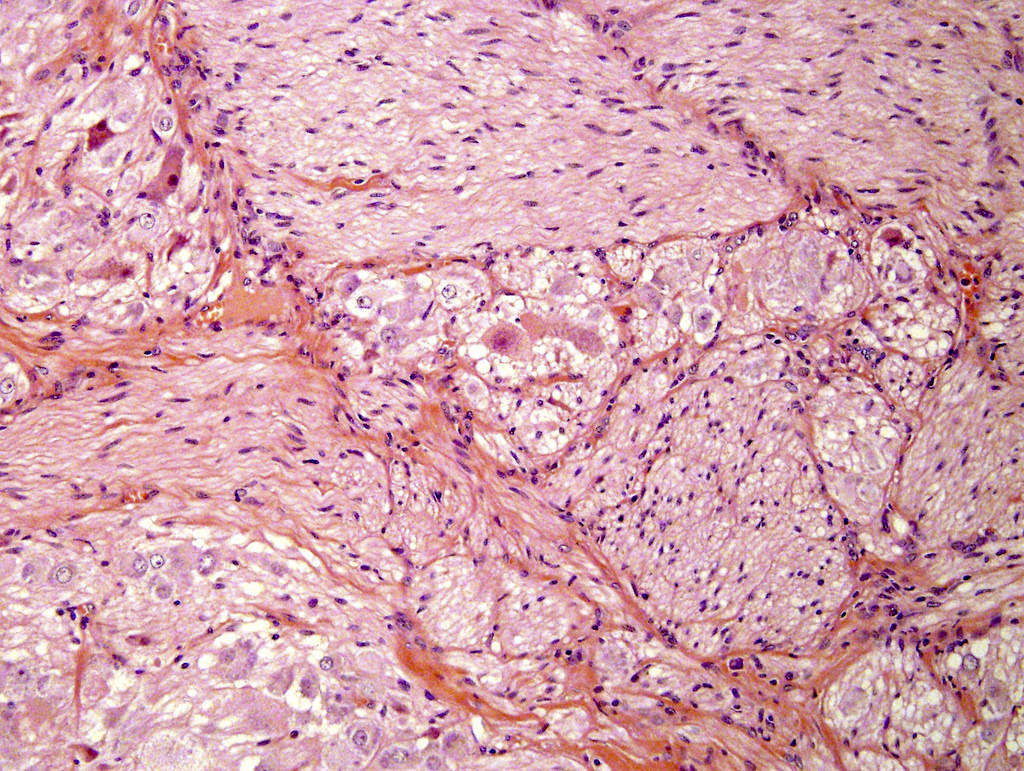
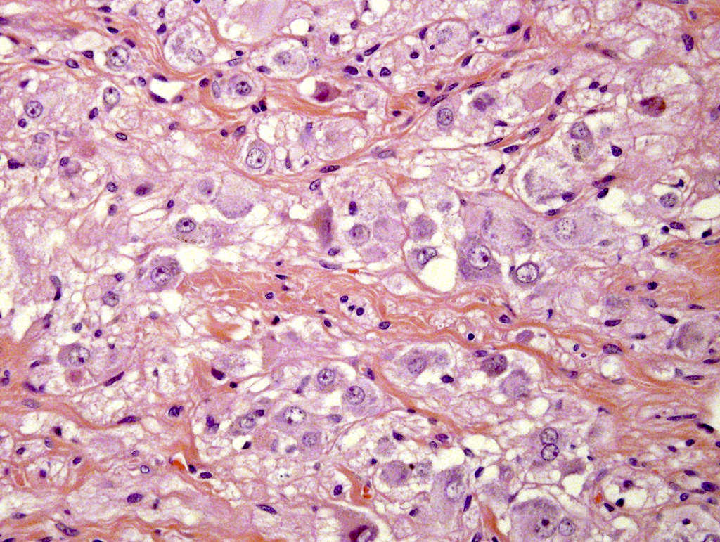
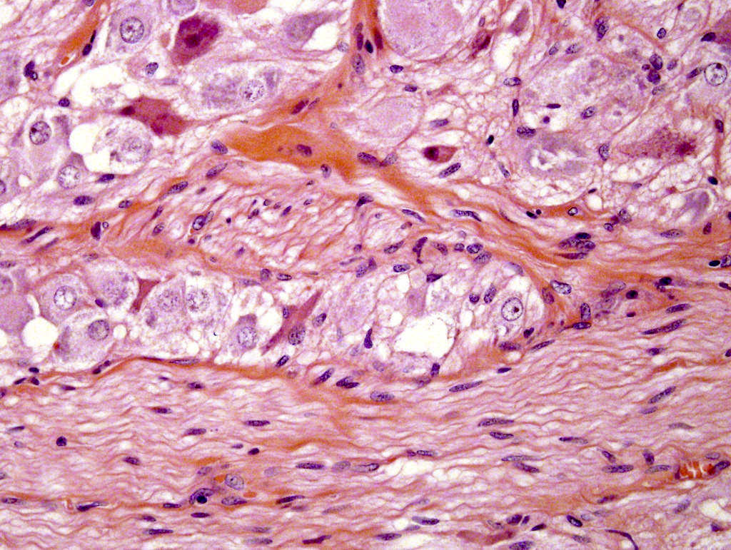
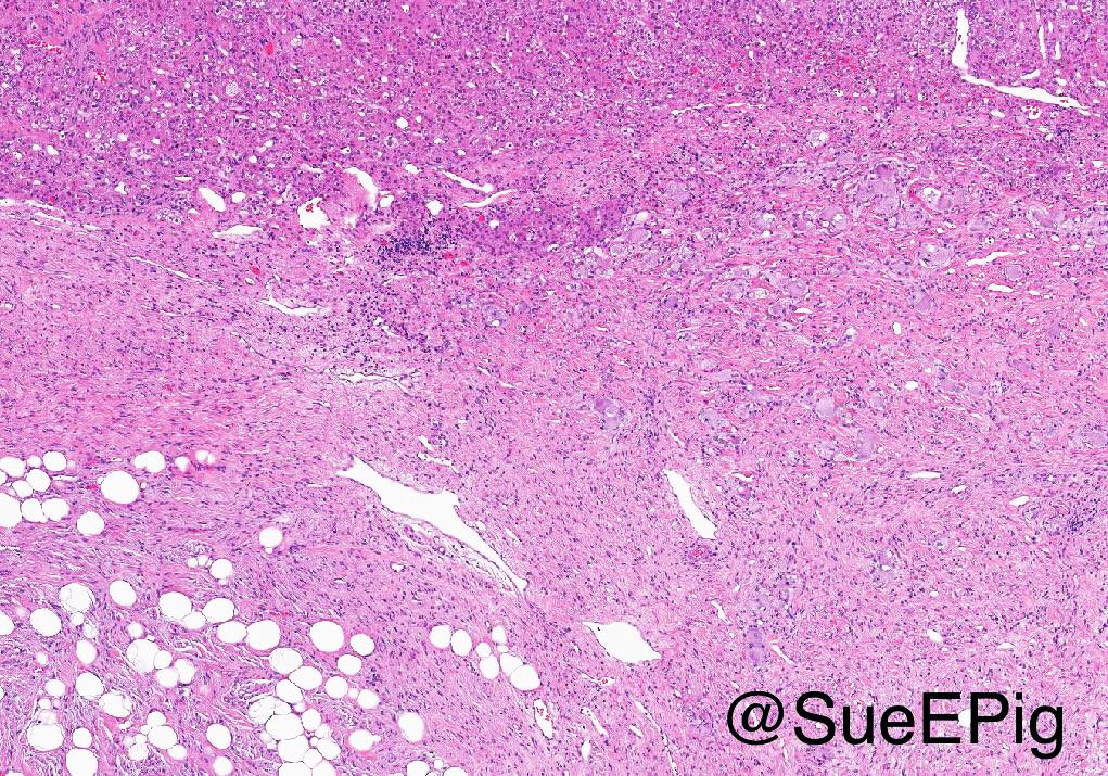
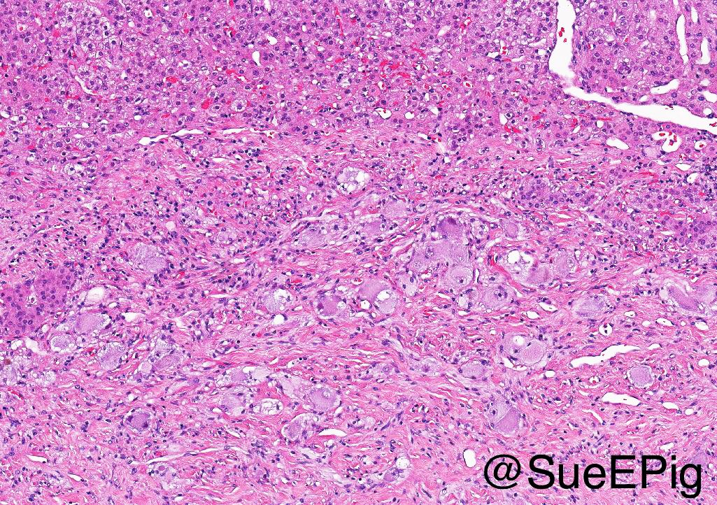
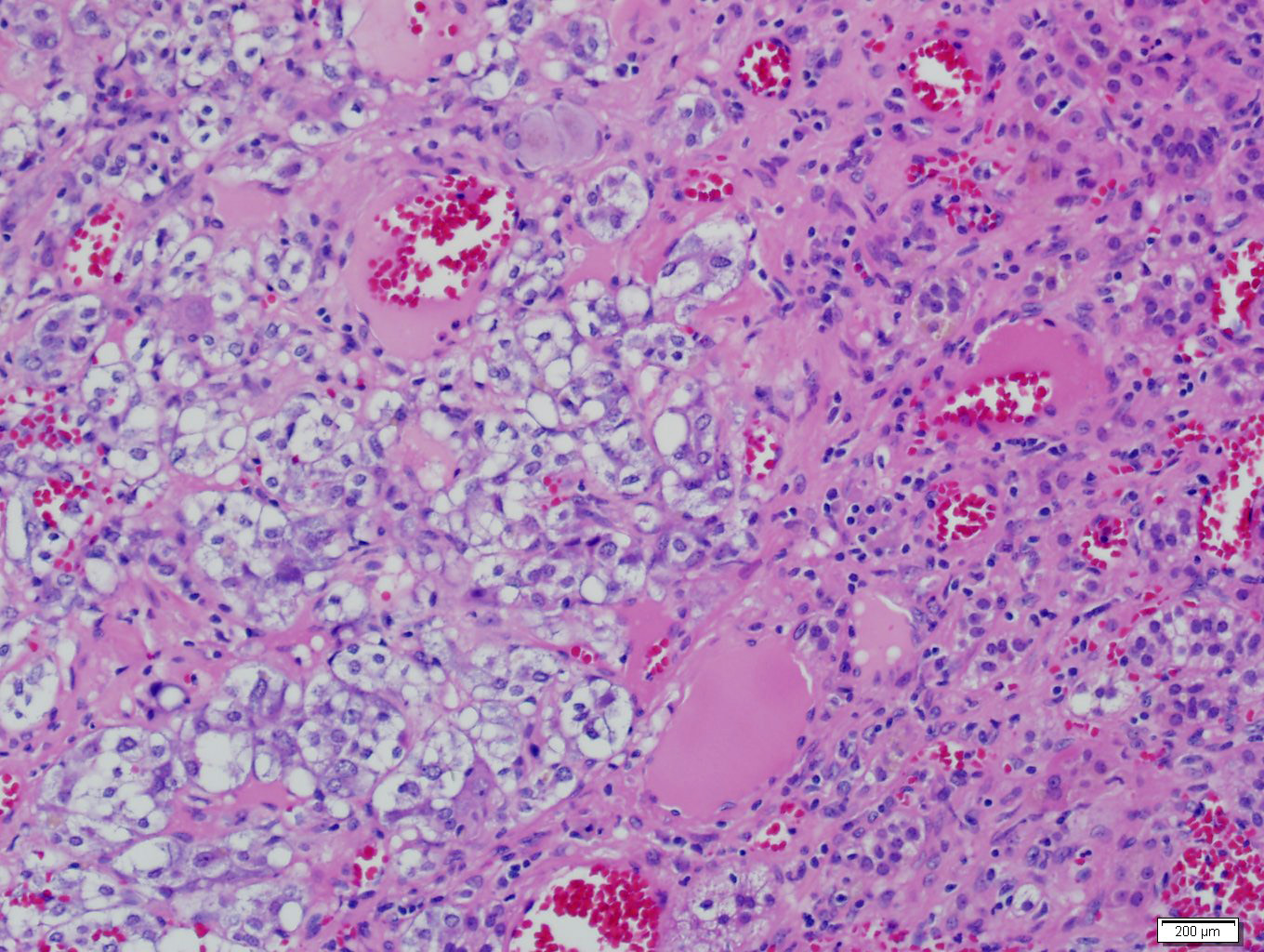
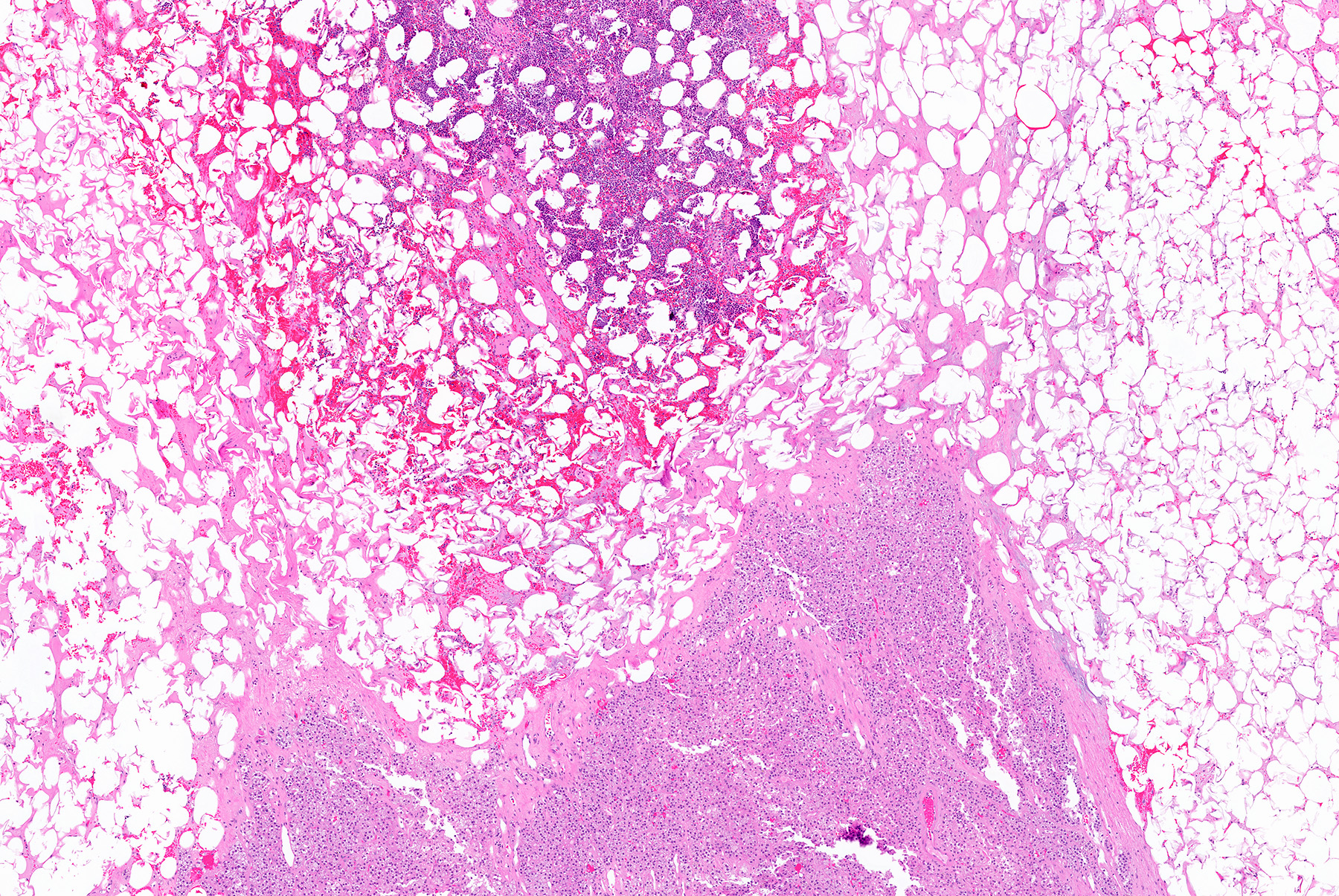
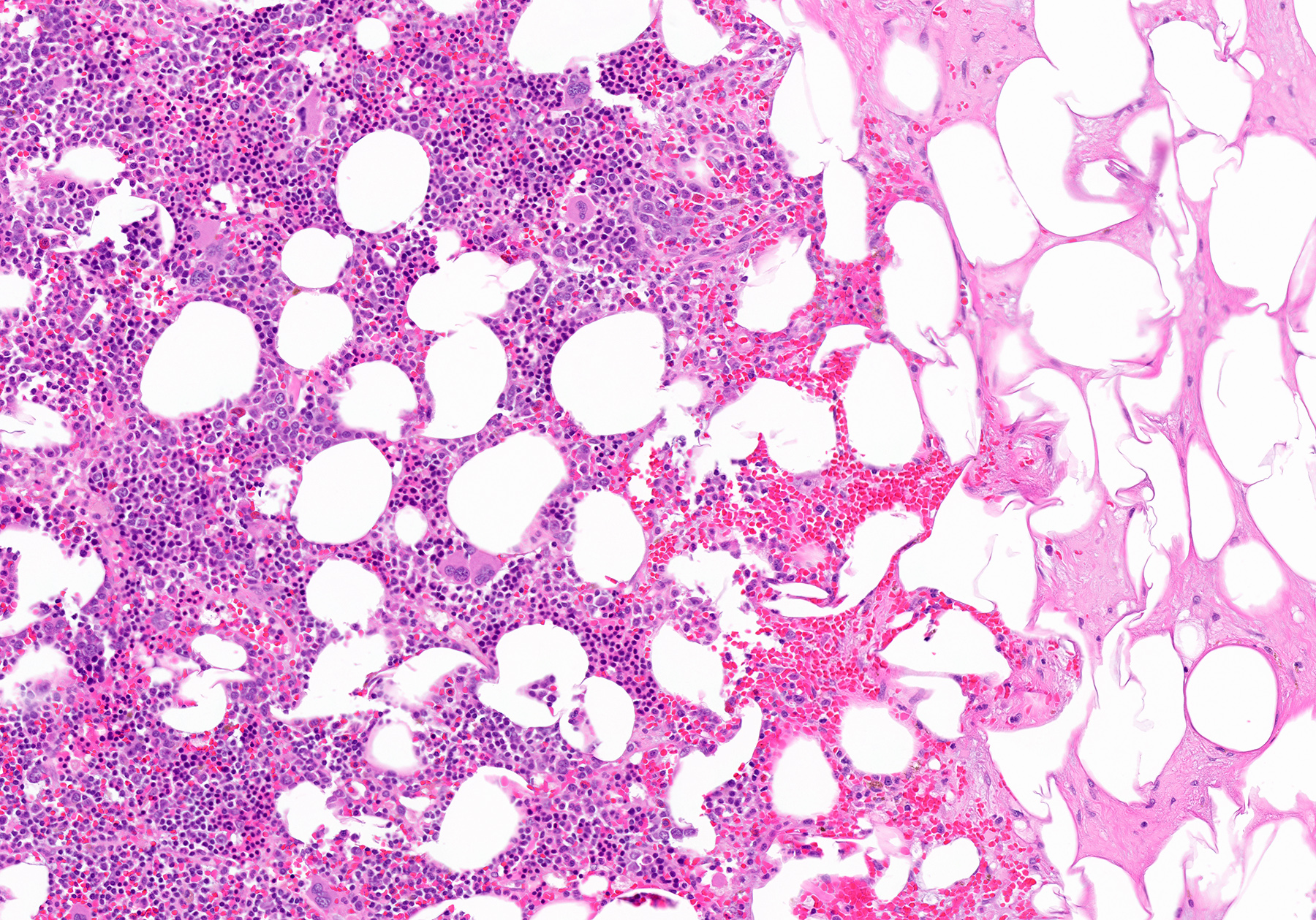
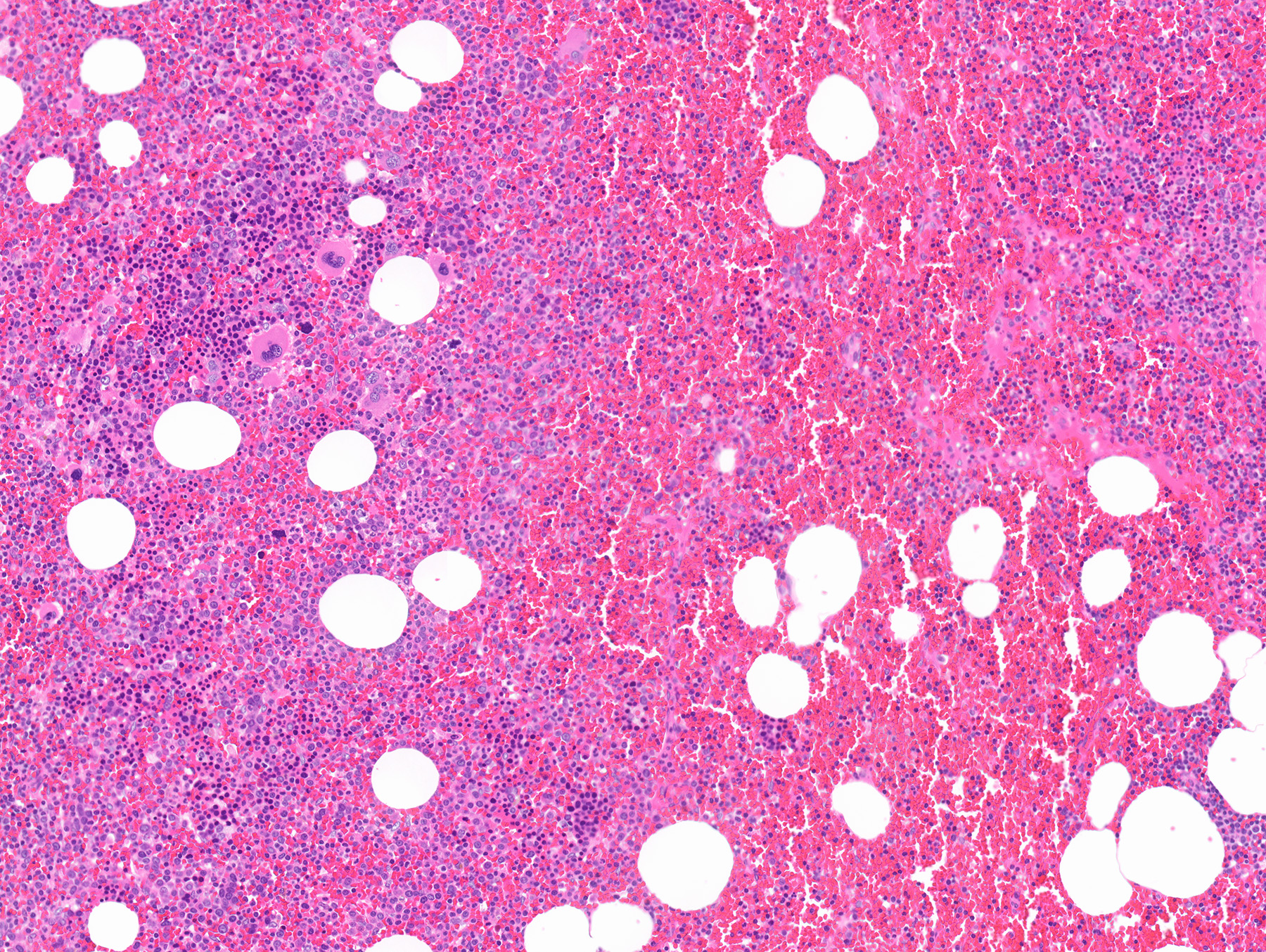
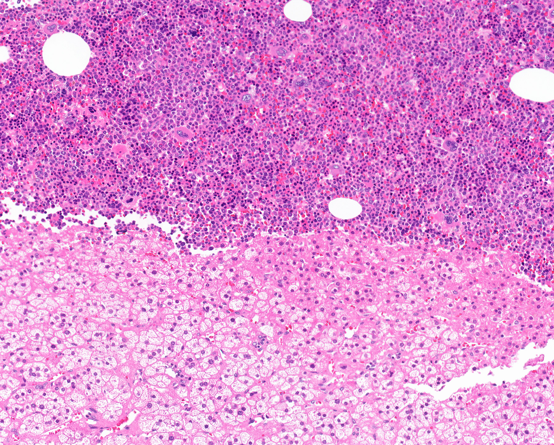
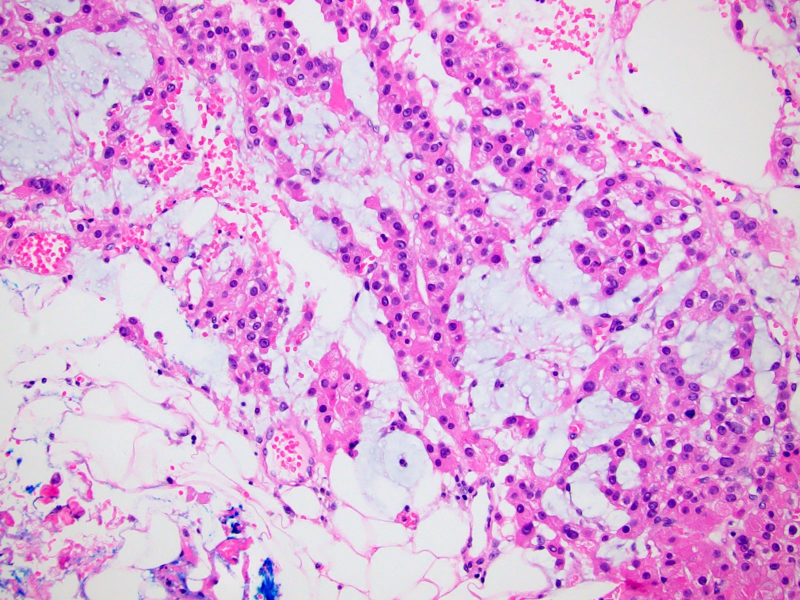
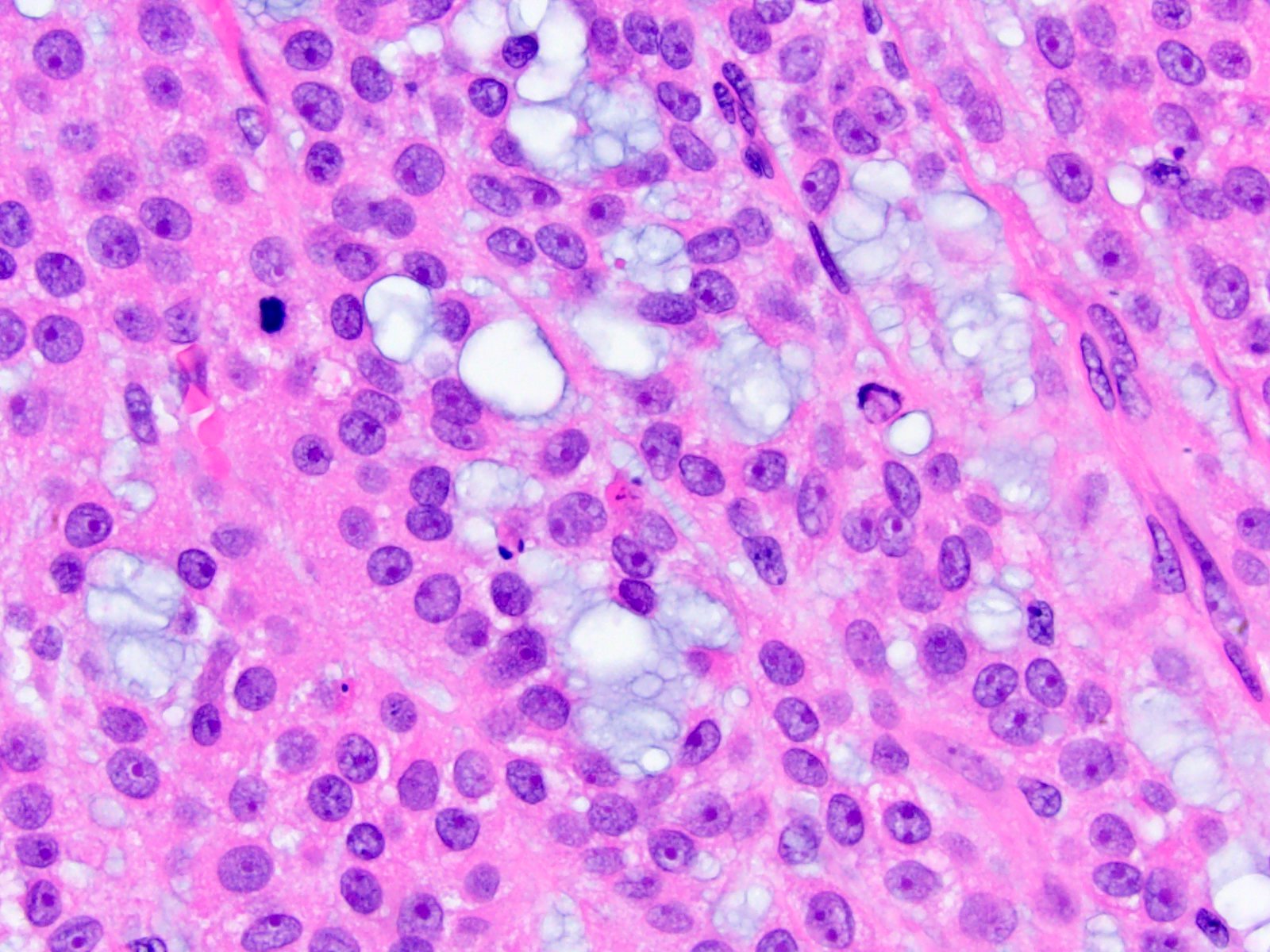
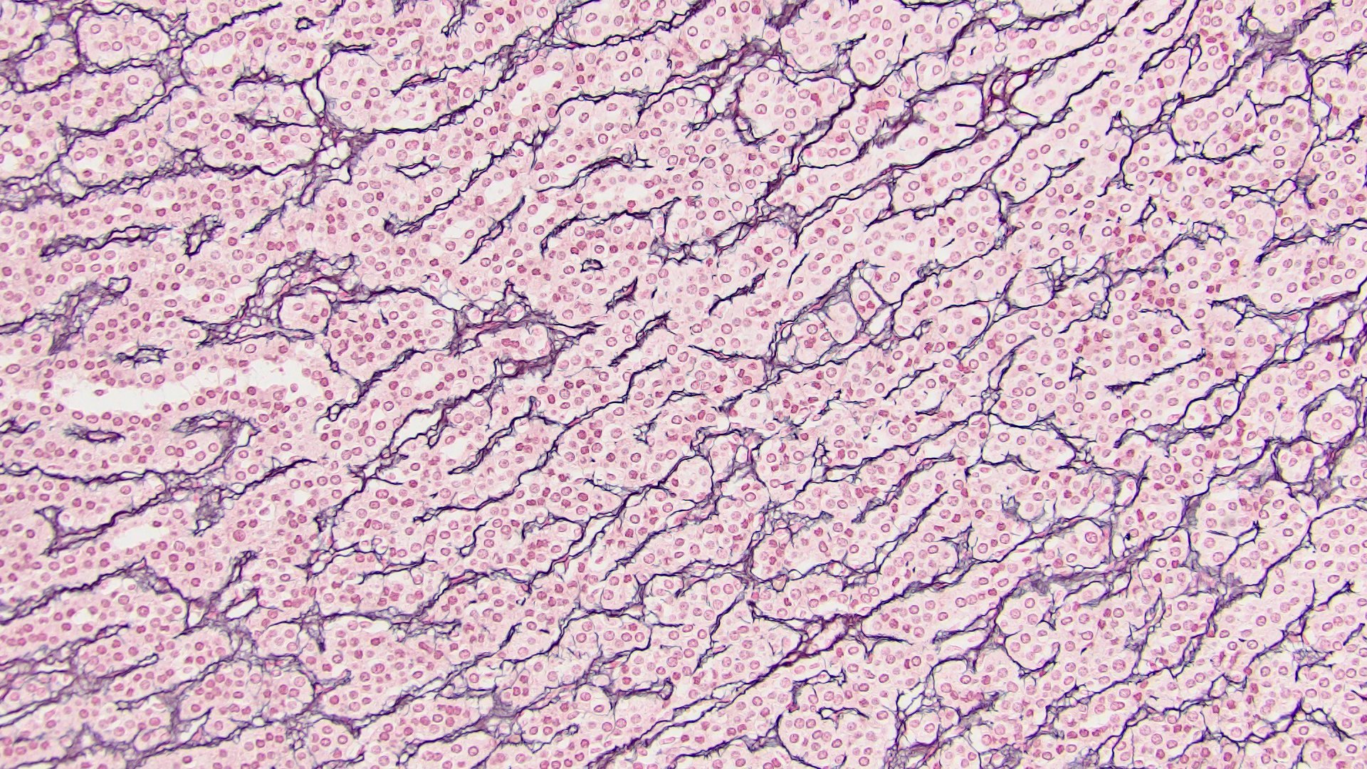
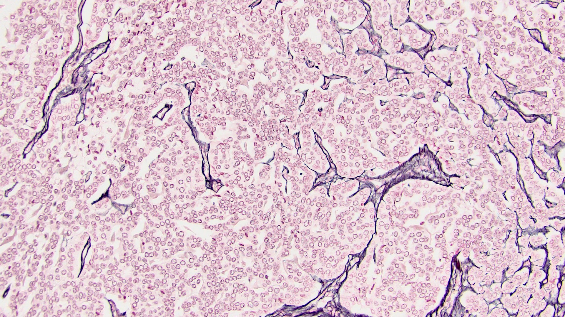
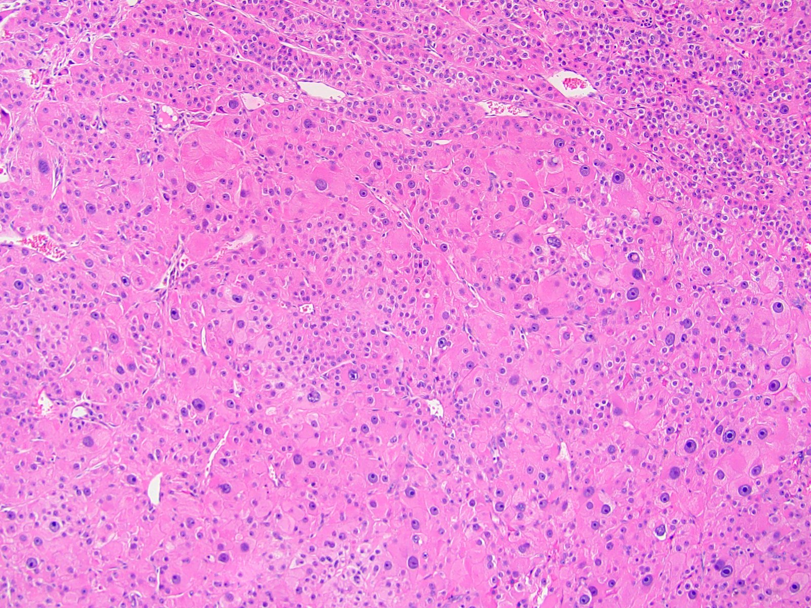
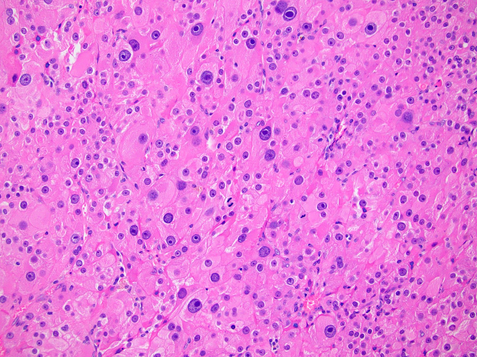
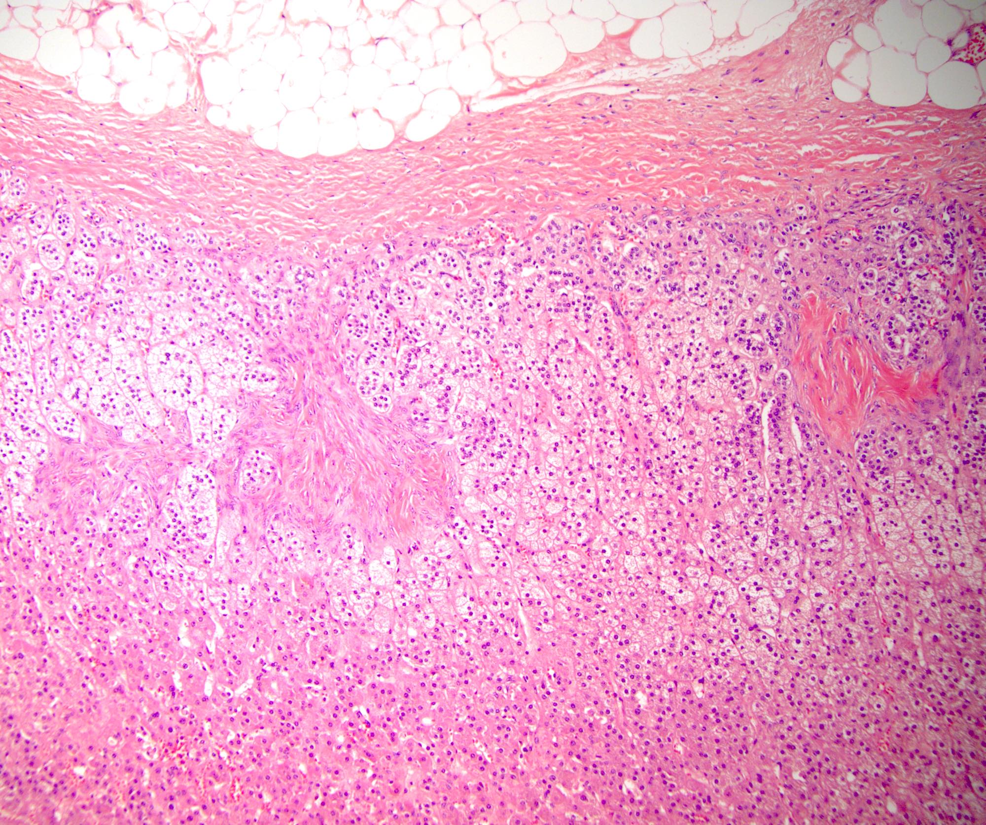
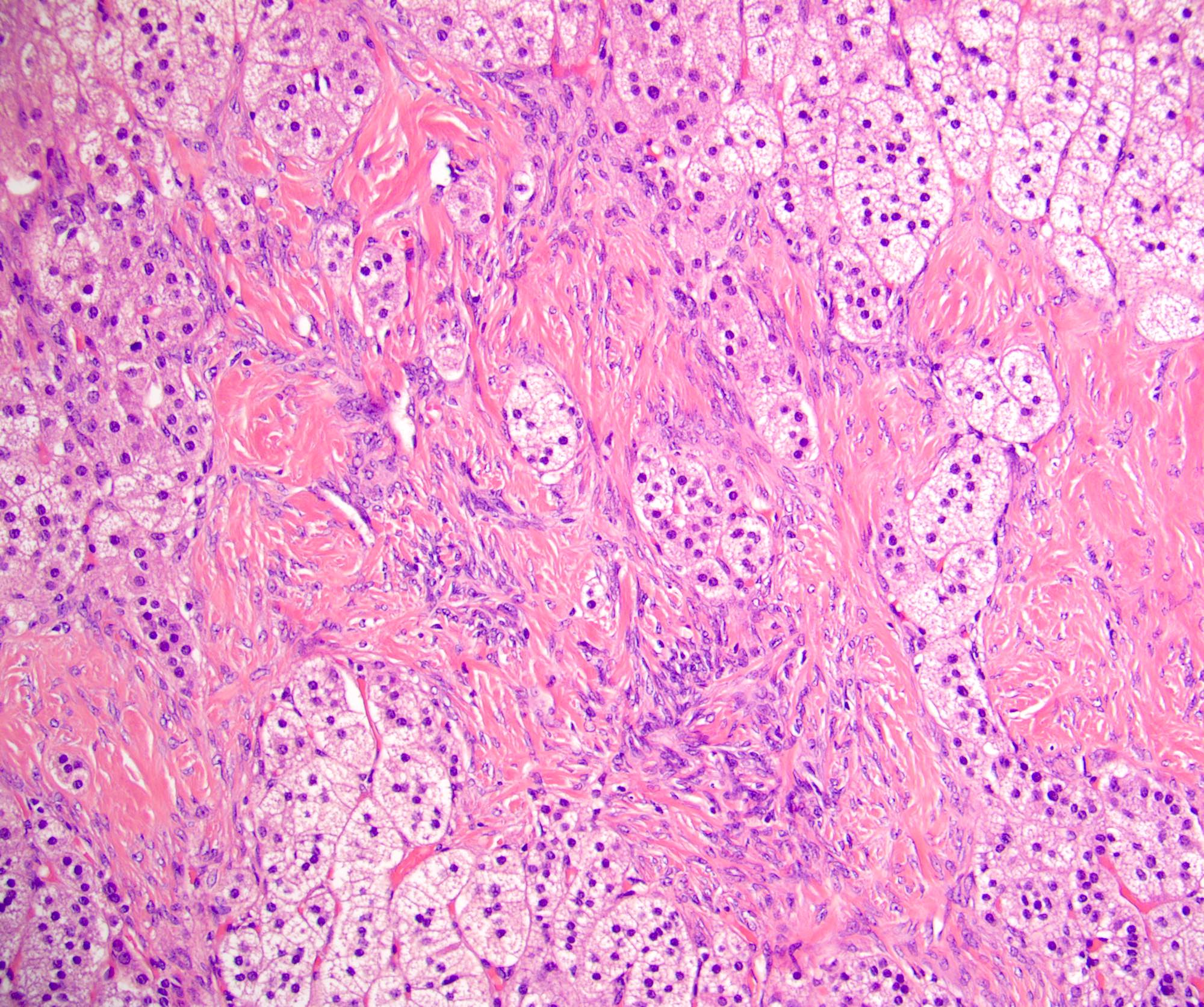
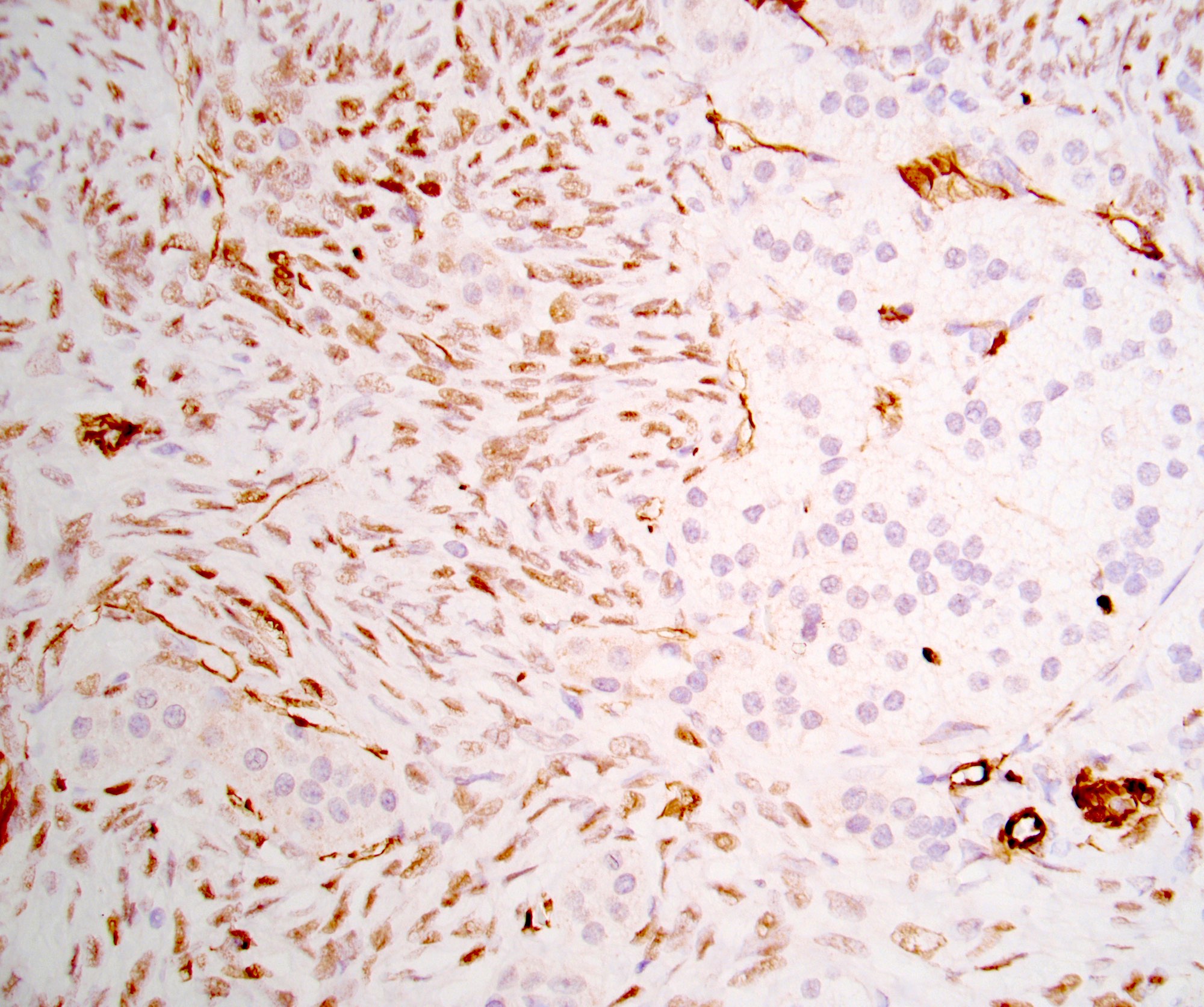
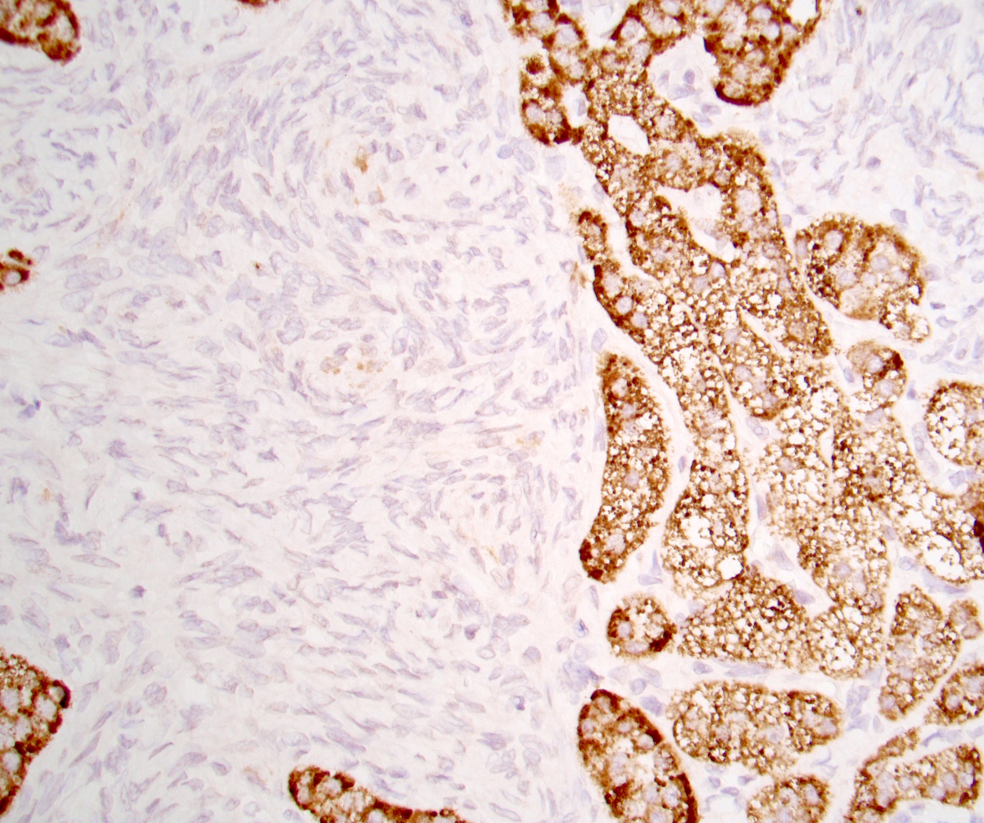
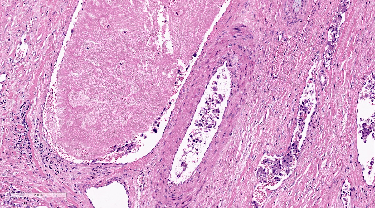
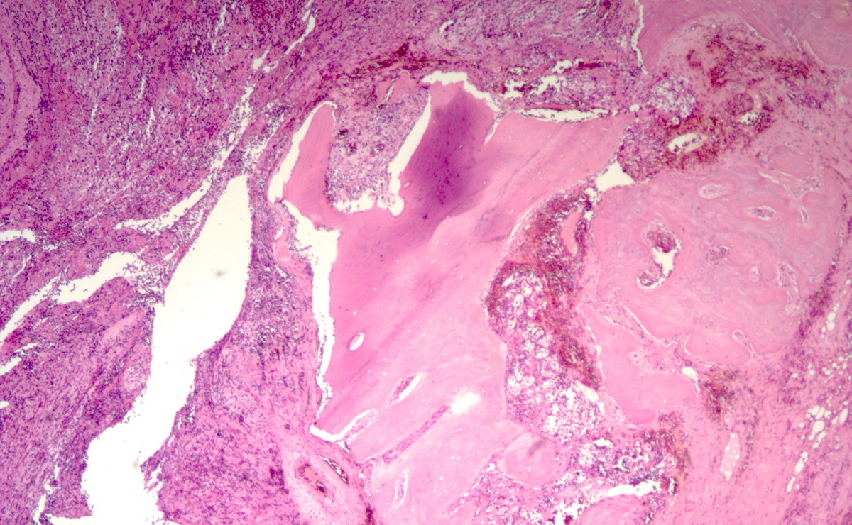
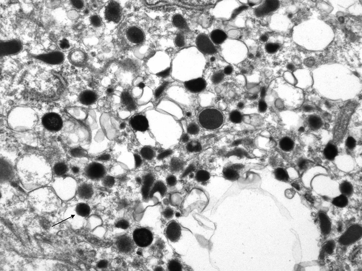
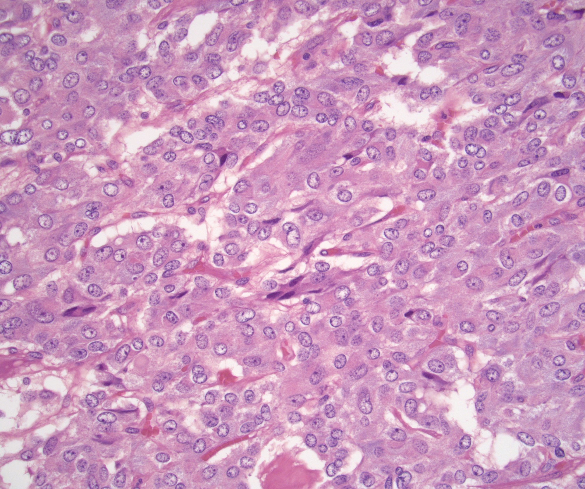
  • Amyloidosis
    • Rarely causes cortical hypofunction, only if extensive bilateral involvement
    • Usually associated with systemic amyloidosis-AA type
    • 68% of consecutive autopsies had adrenal amyloid deposits, often multinodular and probably due to aging
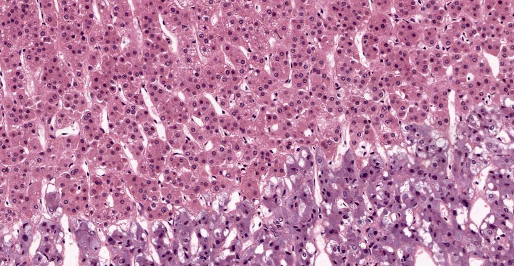
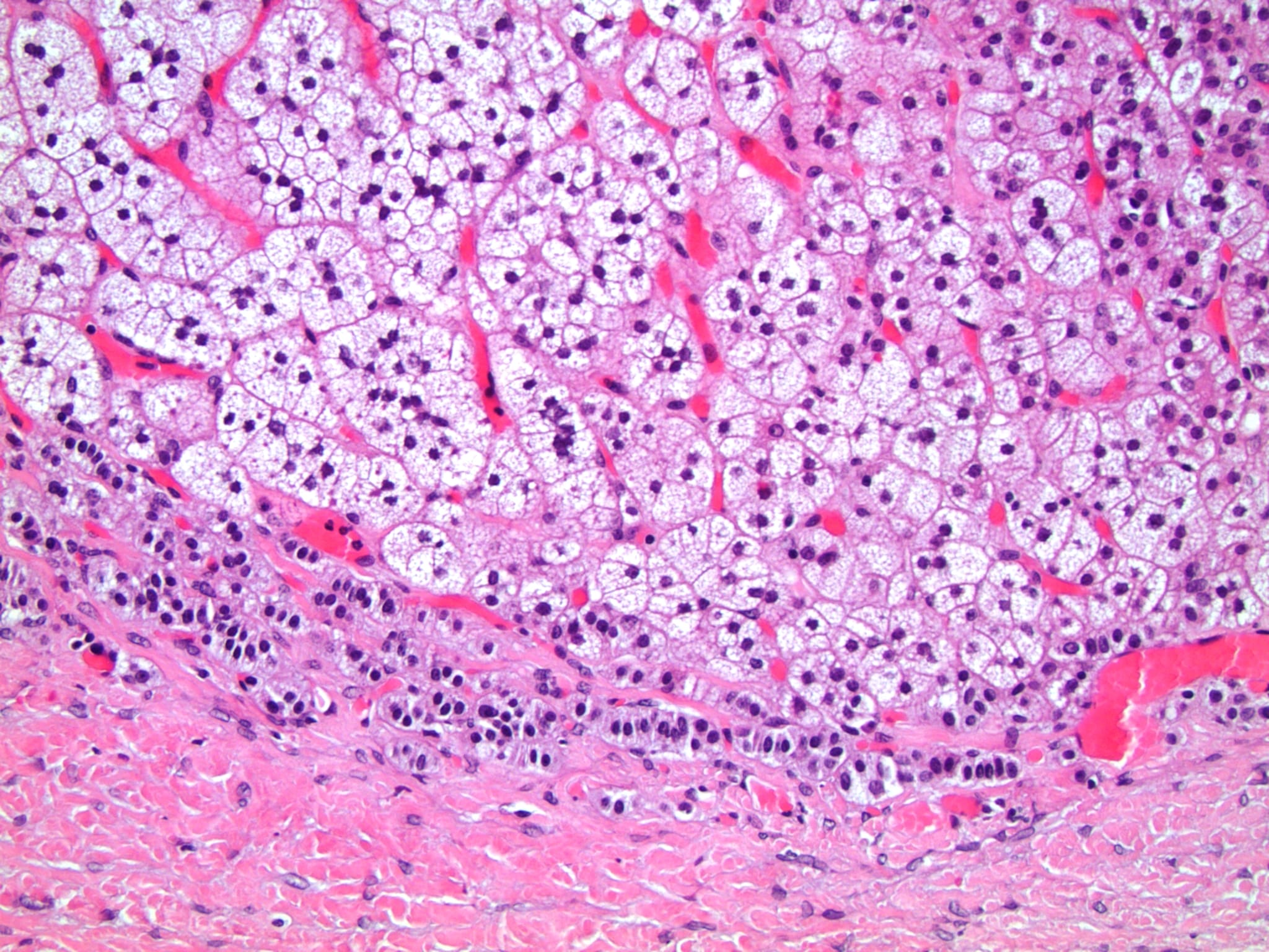
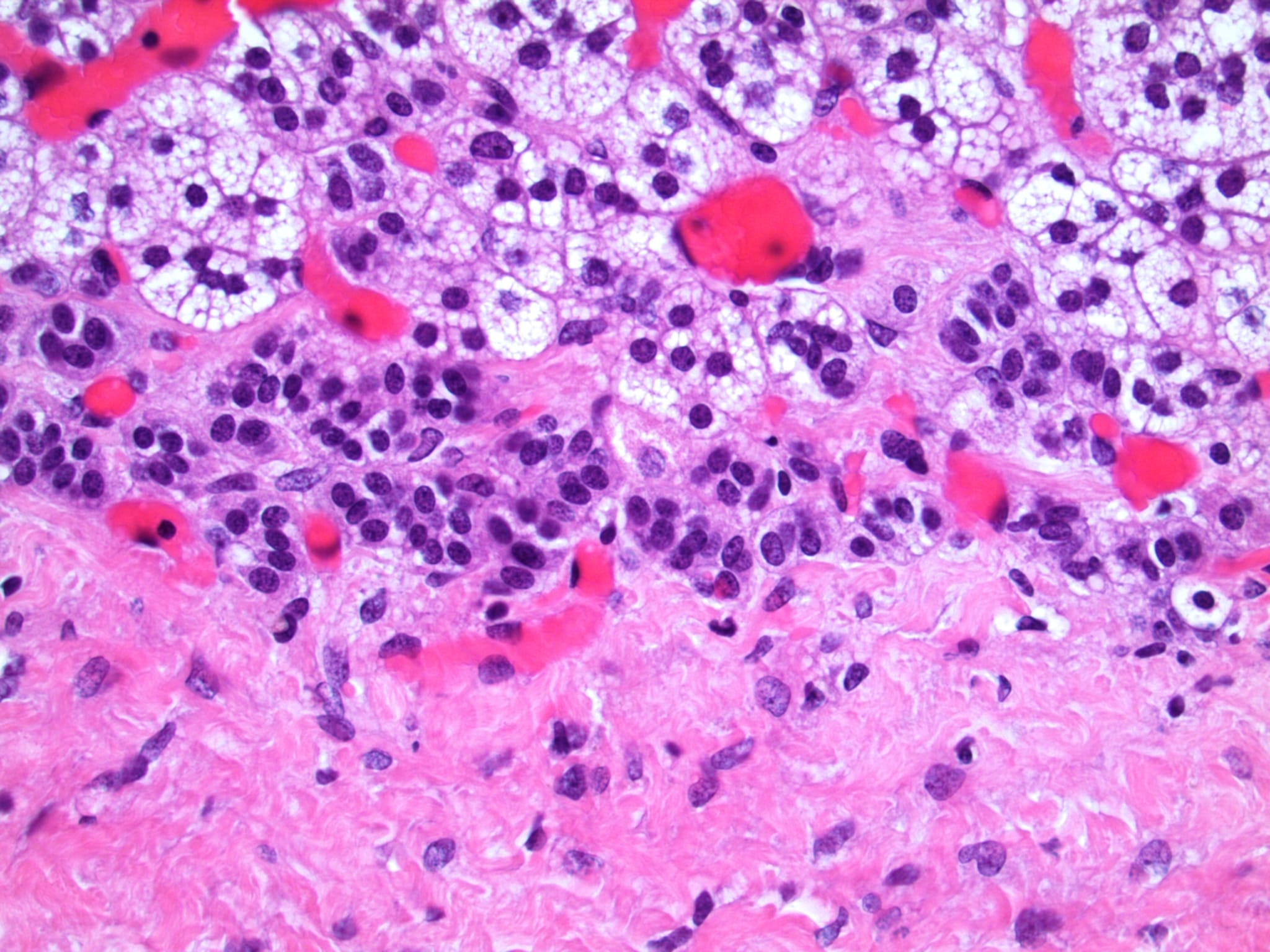
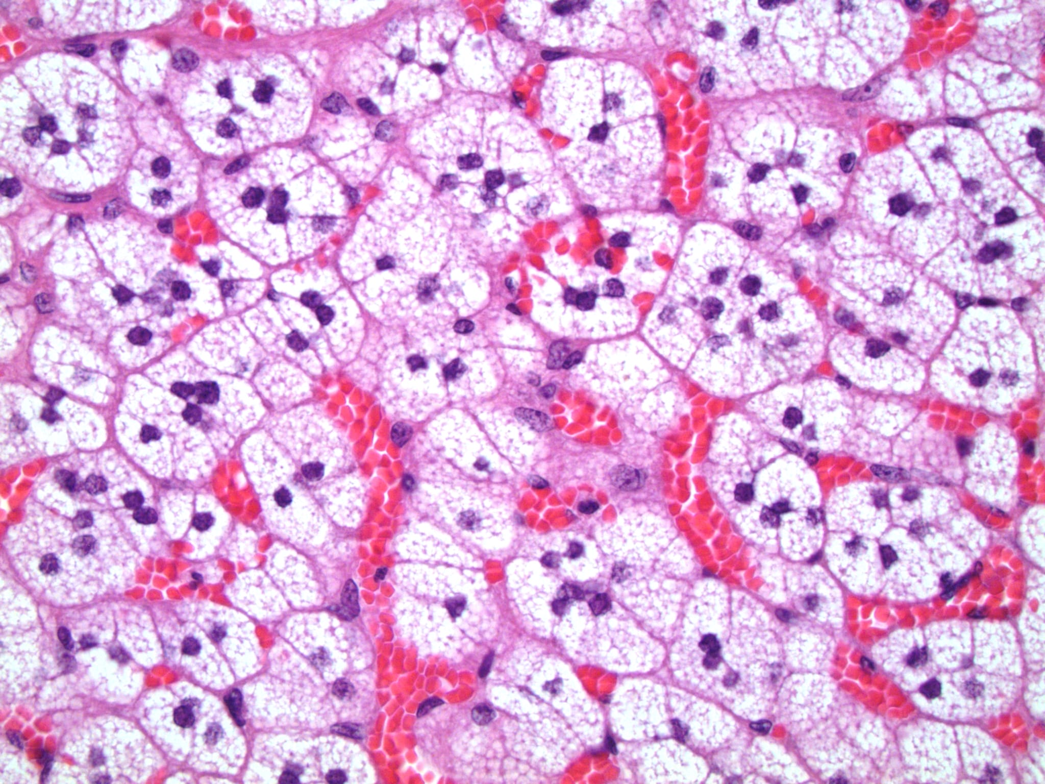
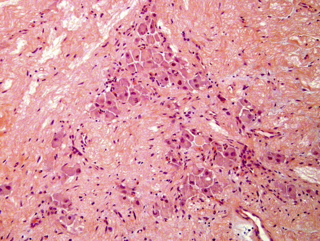
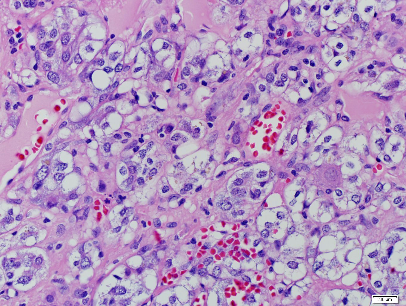
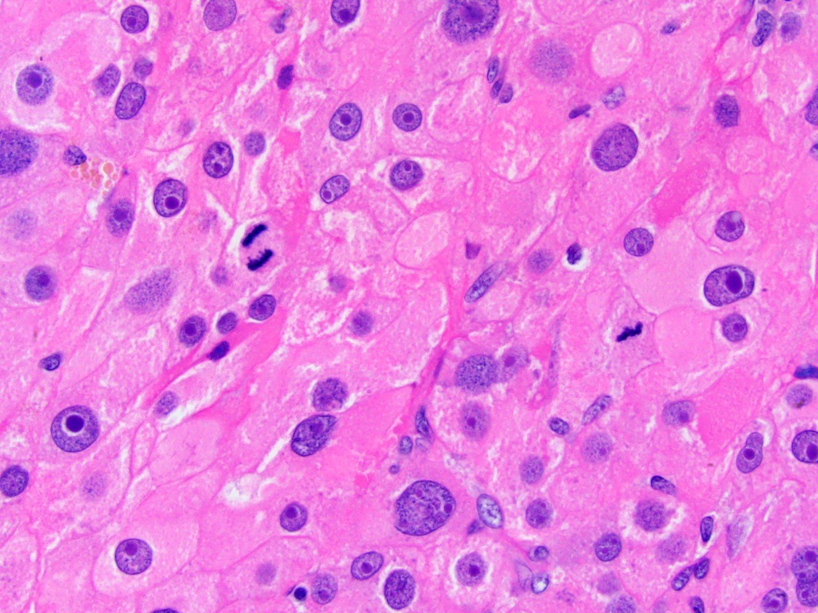
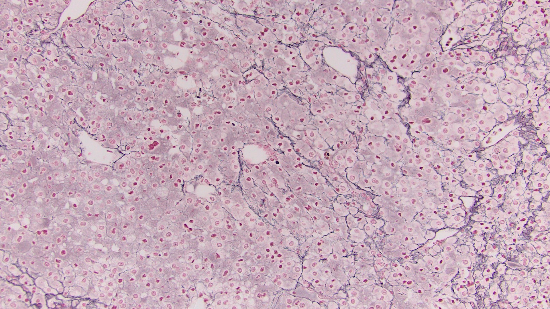
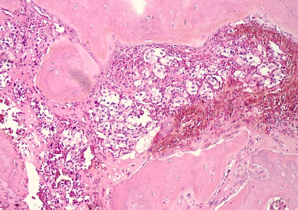
    • Typically affects zona fasciculata and reticularis
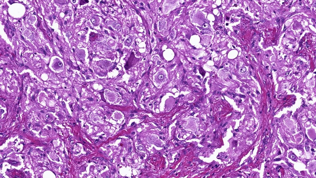
    • Acellular salmon-pink amorphous material is present between cortical cells, which ultimately become atrophic
    • Amyloidosis-AL type is typically deposited intravascularly
  • Drugs
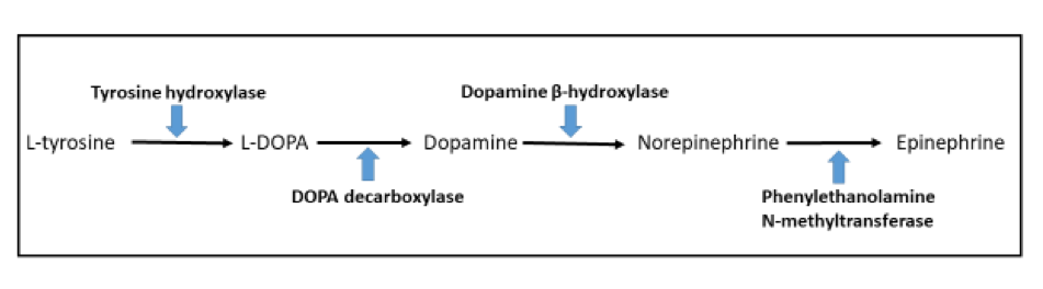
    • Aminoglutethimide: inhibits enzyme converting cholesterol to pregnenolone, causes decrease in cortisol and aldosterone
    • Metapyrone: inhibits 11 beta hydroxylase, which inhibits cortisol and aldosterone synthesis
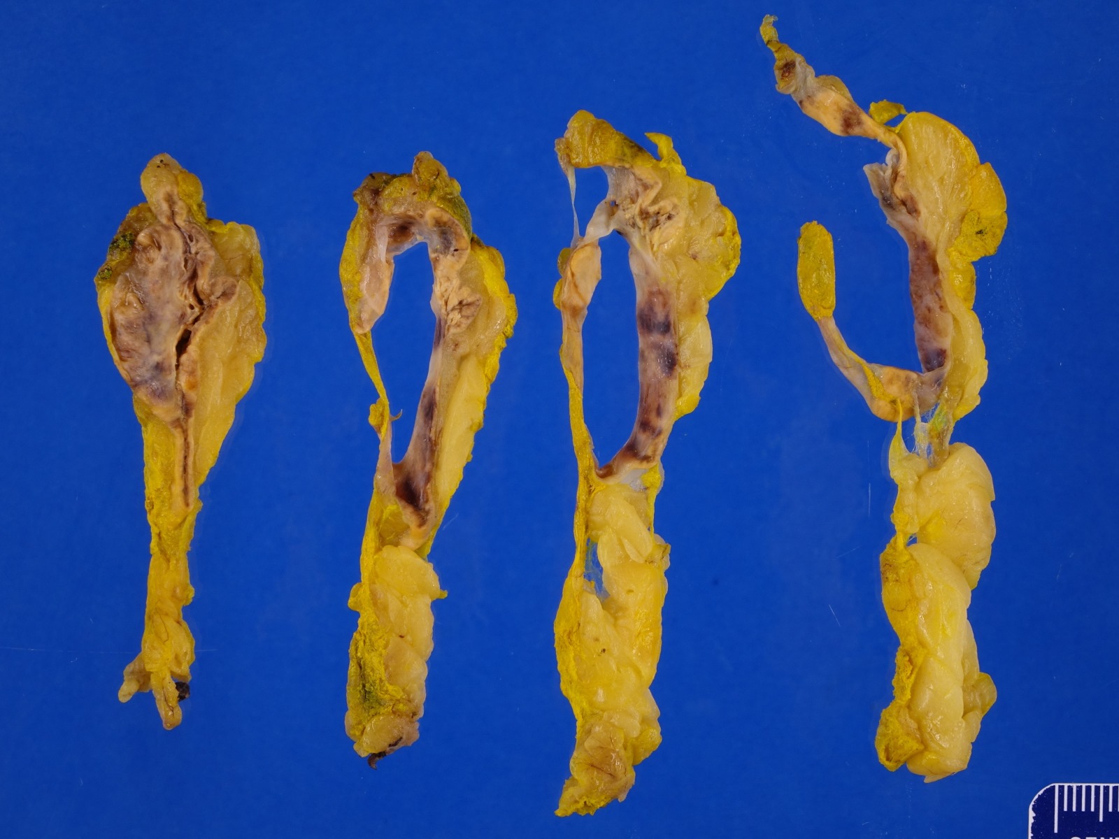
    • Mitotane: cytotoxic to zona fasciculata and reticularis, produces medical adrenalectomy; produces atrophic adrenal glands with fibrosis and residual islands of cortical cells
  • Radiation
    • May cause fibrosis, although cortex is relatively radioresistant
    • High doses (> 5000 roentgens) to abdomen, pelvis or lumbar region may cause hyaline fibrosis of reticularis and reduction of fasciculata, although does not necessarily affect cortical function
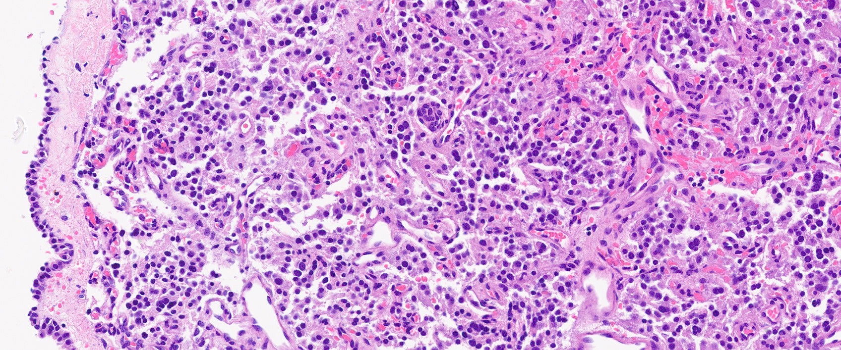
  • Autoimmune disorders (autoimmune adrenalitis or polyglandular autoimmune syndromes)

Treatment
  • Glucocorticoids, mineralocorticoids and IV fluids
  • In chronic patients, must give steroid boost during infections, prior to surgery or during pregnancy
Secondary adrenal insufficiency
Definition / general
  • Caused by any disorder of pituitary gland which decreases ACTH production and causes adrenal cortical atrophy
  • Causes: pituitary macroadenoma, craniopharyngioma, tuberculosis or other infections, sarcoidosis, lymphocytic hypophysitis, head trauma, aneurysms, postpartum pituitary necrosis (Sheehan syndrome), pituitary apoplexy or metastases; also mutations in pro-opiomelanocortin gene
  • Similar atrophic changes are caused by exogenous steroids, which also decrease ACTH production
  • No hyperpigmentation since ACTH levels are low
  • May be associated with hypopituitarism

Laboratory
  • Serum aldosterone, sodium and potassium levels are usually normal, since they are controlled by renin-angiotensin axis, which is not under the control of ACTH
  • Androgens and cortisol levels are low since their production is influenced by ACTH, although androgens are less affected in males since they are also produced by testis
  • Hypoglycemia is more common than with primary adrenal insufficiency

Treatment
  • Exogenous ACTH causes rise in serum cortisol levels
  • May also need to replace other pituitary hormones

Gross description
  • Atrophic adrenal glands with retention of architecture, often fibrotic capsule, bright yellow (due to lipid accumulation) and prominent medulla

Microscopic (histologic) description
  • Normal thickness of zona glomerulosa, thinner fasciculata and reticularis
  • Usually no lymphoplasmacytic infiltration
Tertiary adrenocortical insufficiency
  • Due to disorders of hypothalamus reducing release of corticotropin releasing hormone (CRH); some include this within secondary adrenocortical insufficiency
Isolated mineralocorticoid deficiency
  • Deficiency of aldosterone production by zona glomerulosa
  • Usually due to impaired release of renin from kidney due to diabetes mellitus, autoimmune disease, amyloidosis or sickle cell anemia; also heparin, rarely tumors or idiopathic
  • Primary (idiopathic) hypoaldosteronism may be due to autosomal recessive disorder associated with deficiency in CYP11B2 enzyme (converts 18-hydroxyl group to aldehyde at end of aldosterone biosynthesis)
  • Infants have failure to thrive, recurrent dehydration and salt wasting
Polyglandular automimmune syndromes
  • More common in women
  • Type I polyglandular autoimmune syndrome:
    • Also called autoimmune polyendocrinopathy candidiasis ectodermal dystrophy (APECED)
    • Due to mutations in gene at 21q22.3
    • Rare with < 200 cases reported
    • Autosomal recessive; associated with adrenal insufficiency in 60% of cases, usually by age 13 years
    • Also chronic mucocutaneous candidiasis and hypoparathyroidism in childhood
    • Also autoimmune thyroiditis, diabetes mellitus; variable alopecia; most have autoantibodies to glutamic acid decarboxylase
  • Type II polyglandular autoimmune syndrome:
    • Usually autosomal dominant
    • Adrenal insufficiency in all cases; more common than type I
    • 50% familial with onset between ages 20 - 40 years
    • Also autoimmune thyroiditis (Grave disease), insulin dependent diabetes mellitus
    • Also primary hypogonadism, myasthenia gravis and celiac disease; only rarely hypoparathyroidism
Wolman disease
Definition / general
  • Primary familial xanthomatosis
  • Rare, autosomal recessive lipid storage disorder due to deficiency of lysosomal acid lipase, causing accumulation of triglycerides and cholesterol esters in liver, spleen and adrenal glands
  • Usually causes death by age 6 months

Gross description
  • Markedly enlarged adrenal glands with dystrophic calcifications but normal architecture

Microscopic (histologic) description
  • Necrosis, fibrosis and calcification; zona fasciculata and reticularis cells have vacuolated cytoplasm

Differential diagnosis
  • Niemann-Pick disease, other storage diseases

Sarcomatoid
Definition / general
  • Extremely rare malignant tumor of adrenal cortex with biphasic carcinomatous and sarcomatous morphology
Essential features
  • Least common variant of adrenal cortical carcinoma (ACC), ~30 cases reported
  • Sarcomatoid ACC is a particularly aggressive tumor with a dismal prognosis
  • Composed of malignant epithelial adrenocortical cells and specialized mesenchymal elements resembling various sarcomas, often with heterologous features of rhabdomyoblastic, osteogenic, chondroid or PNET-like differentiation
  • Compared to conventional ACC, sarcomatoid variant presents as:
    • Predominately nonfunctioning tumors
    • Predilection for the right adrenal
    • Larger tumor size
    • Higher rate of distant metastases and shorter median survival
    • Older patient population
Terminology
  • Adrenal cortical carcinoma (ACC), sarcomatoid variant
  • Adrenocortical carcinoma, sarcomatoid type
  • Adrenal carcinosarcoma
ICD coding
  • ICD-O: 8370/3 - adrenal cortical carcinoma
Epidemiology
Sites
Pathophysiology
  • 4 theories of sarcomatoid ACC histogenesis (Virchows Arch 2012;460:9):
    • Conversion tumor theory with neoplastic transformation of epithelial to mesenchymal cells
    • Composition tumor theory (paradoxical reactive proliferation of nonepithelial component induced by the epithelial component via paracrine secretion)
    • Collision or biclonal tumor theory (2 synchronous, histologically independent tumors of different clones)
    • Combination or divergent tumor theory (deriving from a common monoclonal stem cell precursor); best supported by molecular studies (Hum Pathol 2016;58:113)
Etiology
Diagrams / tables

Images hosted on other servers:

Survival analysis:
sarcomatoid ACC
versus other
variants

Clinical features
Diagnosis
  • Sarcomatoid ACC on imaging (CT, MRI) has large size, irregular contours, lipid poor characteristics and significant signal heterogeneity (Endocr J 2019;66:739, World J Surg Oncol 2015;13:117)
  • Radiographically and even during gross evaluation, often difficult to confirm adrenal origin due to the advanced presentation
  • By definition, tumor should be composed of adrenocortical epithelial malignant cells and sarcoma-like mesenchymal component on microscopy
  • No minimum percentage of sarcomatoid cells required for the diagnosis (Biomedicines 2021;9:175)
  • Weiss system does not account for sarcomatoid histology and may not be applicable to these tumors, especially the criteria of diffuse architecture (Virchows Arch 2012;460:9)
Laboratory
Radiology description
Radiology images

Images hosted on other servers:

CT scan: necrotic areas replacing adrenal gland

Coronal abdominal CT with large adrenal mass

Prognostic factors
Case reports
Treatment
Gross description
Gross images

Image hosted on other servers:

Whitish fleshy, variegated hemorrhagic and necrotic surface

Tumor adherent
to pancreas (A)
and compressing
kidney (B)

Microscopic (histologic) description
Microscopic (histologic) images

Contributed by Maria Tretiakova, M.D., Ph.D.
Sarcomatoid ACC biphasic

Sarcomatoid ACC biphasic

Epithelioid and spindle cells

Epithelioid and spindle cells

Heterologous differentiation

Heterologous differentiation

Positive stains
Electron microscopy description
  • Epithelioid component with tight junctions and abundant mitochondria, similar to conventional ACC
Electron microscopy images

Images hosted on other servers:

Epithelioid and spindle cell components

Molecular / cytogenetics description
  • Sarcomatoid ACC had the lowest adrenocortical differentiation scores with very low expression of NR5A1 (SF1 regulator) and steroidogenic enzymes (Cancer Cell 2016;29:723)
  • Wnt beta catenin signaling pathway dysregulation and mutational inactivation of TP53 are common genetic events in sarcomatoid ACC (Histopathology 2018;72:82)
  • Enriched for EMT related markers and stem cell factors that may be associated with the poor prognosis of these tumors and may provide possible therapeutic targets (Histopathology 2018;72:82, Biomedicines 2021;9:175, Hum Pathol 2016;58:113)
  • In differentiated epithelioid component, molecular alterations are similar to conventional ACC including high mutation burden, massive DNA loss followed by whole genome doubling and frequent somatic mutations of P53, CTNNB1, CDKN2A, TERT, ZNRF3, PRKAR1A (Histopathology 2018;72:82, Cancer Cell 2016;30:363)
  • Concordant molecular alterations in phenotypically diverse components (i.e. presence of TP53 and CTNNB1 gene mutations) were detected in 50% of studies by NGS tumors and support a common clonal origin (Hum Pathol 2016;58:113)
Sample pathology report
  • Adrenal gland, right, adrenalectomy:
    • Adrenal cortical carcinoma with the following features:
      • Tumor size: 19 cm x 18 cm x 12 cm
      • Tumor (gland) weight: 600 g
      • Tumor extent: invasion into inferior vena cava and inferior liver surface
      • Histologic type: sarcomatoid variant
      • Histologic grade: high grade
      • Necrosis: present
      • Lymphovascular invasion: present
      • Margins: positive
      • pTNM, AJCC 8th edition: pT4 N0
    • Ancillary studies: Ki67 mitotic rate 40%
Differential diagnosis
Board review style question #1

A 68 year old woman presented with right sided back pain and weight loss. Abdominal ultrasonography and computed tomography (CT) showed the presence of a 12 cm heterogeneous adrenal mass. It was biopsied, showing biphasic morphology with large epithelioid polygonal cells and haphazardly arranged spindle cells. Which statement about this adrenal cortical tumor is accurate?

  1. Pure sarcomatoid morphology is more common than biphasic
  2. Hormonal production is typically abnormal
  3. Clinical presentation and prognosis are similar to conventional adrenocortical carcinoma (ACC)
  4. Adrenocortical immunomarkers are often lost in sarcomatoid areas
  5. Risk of metastatic disease and recurrence is low
Board review style answer #1
D. This is a case of sarcomatoid ACC, an extremely rare malignant tumor with biphasic epithelioid and mesenchymal differentiation. The carcinomatous component is strongly positive for inhibin, MelanA, calretinin and SF1, thus supporting an adrenocortical origin. However, the sarcomatoid component often has a partial or complete loss of adrenocortical markers. Fortunately, > 80% of sarcomatoid ACC has both carcinomatous and sarcomatoid components (A). Functional sarcomatoid ACCs with hormone overproduction are documented in only 11% of cases (B). Compared to conventional ACC, the sarcomatoid variant has a more advanced age at presentation, predominately nonfunctional tumors, predilection of the right side, larger tumor size, higher rate of distant metastases and shorter median survival (C). 75 - 80% of patients with sarcomatoid ACC develop metastases or recurrence within 4 months and have an overall dismal prognosis (E).

Comment Here

Reference: Adrenal cortical carcinoma, sarcomatoid variant

Staging-adrenal cortical carcinoma
Definition / general
  • All carcinomas of the adrenal gland are covered by this staging system
  • These topics are not covered: pheochromocytoma, neuroblastic tumors
Essential features
  • AJCC 7th edition staging was sunset on December 31, 2017; as of January 1, 2018, use of the 8th edition is mandatory
ICD coding
  • ICD-O-3: C74.0 - cortex of the adrenal gland
Primary tumor (pT)
  • pTX: cannot be assessed
  • pT0: no evidence of primary tumor
  • pT1: tumor ≤ 5 cm, no extra-adrenal invasion
  • pT2: tumor > 5 cm, no extra-adrenal invasion
  • pT3: extra-adrenal invasion but no invasion of adjacent organs
  • pT4: invasion of kidney, diaphragm, pancreas, spleen, liver, renal vein or vena cava
Regional lymph nodes (pN)
  • pNX: cannot be assessed
  • pN0: no regional lymph node metastasis
  • pN1: regional lymph node metastasis (e.g., paraaortic, periaortic, retroperitoneal)
Distant metastasis (pM)
  • pM1: distant metastasis
AJCC prognostic stage groups
Stage group I: T1 N0 M0
Stage group II: T2 N0 M0
Stage group III: T1 - 2 N1 M0
T3 - 4 N0 - 1, X M0
Stage group IV: T1 - 4, X   N0 - 1, X   M1  
Registry data collection variables
  • Tumor weight
  • Vascular invasion
  • Mitotic count
  • Ki67 proliferation index
  • Weiss score
Histologic grade (G)
  • Low grade: ≤ 20 mitoses per high powered field
  • High grade: > 20 mitoses per high powered field; TP53 or CTNNB mutation
Histopathologic type
  • Oncocytic
  • Myxoid
  • Sarcomatoid
Gross images

Contributed by Debra Zynger, M.D.

Organ confined, ≤ 5 cm (pT1)

Organ confined, > 5 cm (pT2)

Extra-adrenal invasion (pT3)

Extra-adrenal invasion (pT3)

Liver invasion (pT4)

Microscopic (histologic) images

Contributed by Debra Zynger, M.D. and Maria Tretiakova, M.D., Ph.D.

Extra-adrenal adipose invasion (pT3)

Liver involvement (pT4)

Regional node involvement

Regional node involvement (pN1)

Board review style question #1

    The adrenal cortical carcinoma shown above has invasion of the extra-adrenal adipose tissue but does not invade adjacent organs. What is the correct pT for this tumor?

  1. pT1
  2. pT2
  3. pT3
  4. pT4
Board review style answer #1
C. pT3. Adrenal cortical carcinoma with extra-adrenal adipose invasion is pT3. pT1 tumors are organ confined and ≤ 5 cm. pT2 tumors are organ confined and > 5 cm. pT4 tumors show direct invasion of adjacent structures including the kidney, diaphragm, pancreas, spleen, liver, renal vein or vena cava.

Comment Here

Reference: Staging-adrenal cortical carcinoma

Staging-neuroblastic tumors
International Neuroblastoma Staging System (INSS)
  • 1: localized tumor with complete gross (not necessarily microscopic) excision, negative representative ipsilateral lymph nodes microscopically; note: nodes removed with primary tumor may be positive; includes grossly resectable tumor in midline from pelvic ganglia or organ of Zuckerkandl
  • 2A: localized tumor with incomplete gross excision, negative representative nonadherent ipsilateral lymph nodes microscopically; note: includes midline tumor that extends beyond one side of vertebral column and is unresectable
  • 2B: localized tumor with positive representative nonadherent ipsilateral lymph nodes; enlarged contralateral lymph nodes must be negative microscopically; note: includes midline tumor that extends beyond one side of vertebral column, is unresectable with positive ipsilateral lymph node involvement (on side of extension); also includes a thoracic tumor with malignant unilateral pleural effusion
  • 3: unresectable unilateral tumor infiltrating across midline or localized unilateral tumor with contralateral regional lymph node involvement; note: includes midline tumor with bilateral extension by infiltration (unresectable) or by lymph node involvement; includes a tumor of any size with malignant ascites or peritoneal implants
  • 4: any primary tumor with dissemination to distant lymph nodes, bone, bone marrow, liver, skin or other organs (except 4S)
  • 4S: localized primary tumor (stage 1, 2A or 2B) with dissemination limited to skin, liver or bone marrow (limited to infants < 1 year of age); note: marrow involvement should be minimal (< 10% of total nucleated cell identified as malignant); more extensive involvement should be classified as stage 4
  • Note: for infants < 1 year, stages 1, 2A, 2B, 3 and 4S have similar rates of 4 year overall survival (98.5%) compared to stage 4 (73%)
International Neuroblastoma Pathology Classification (INPC) histology group
Favorable histopathology
  • Any age
    • Ganglioneuroblastoma, intermixed
    • Ganglioneuroma
  • < 18 months
    • Neuroblastoma, poorly differentiated subtype with low or intermediate mitosis-karyorrhexis index
    • Neuroblastoma, differentiating subtype with low or intermediate mitosis-karyorrhexis index
  • 18 - 60 months
    • Neuroblastoma, differentiating subtype with low mitosis-karyorrhexis index

Unfavorable histopathology
  • Any age
    • Neuroblastoma, undifferentiated subtype
    • Neuroblastoma, any subtype with high mitosis-karyorrhexis index
  • 18 - 60 months
    • Neuroblastoma, poorly differentiated subtype
    • Neuroblastoma, differentiating subtype with intermediate mitosis-karyorrhexis index
  • > 60 months
    • Neuroblastoma, any subtype
Diagrams / tables

Images hosted on other servers:

Children's Oncology
Group (COG)
neuroblastoma risk
grouping system

INRG stage definitions

INRG consensus pretreatment classification


Staging-pheochromocytoma & paraganglioma
Definition / general
  • All pheochromocytoma and sympathetic (functional) paraganglioma are covered by this staging system
  • These topics are not covered: parasympathetic (nonfunctional) paraganglioma of the head and neck, adrenal cortical carcinoma, neuroblastic tumors
Essential features
  • AJCC 7th edition staging was sunset on December 31, 2017; as of January 1, 2018, use of the 8th edition is mandatory
ICD coding
  • ICD-O:
    • C74.1 - medulla of adrenal gland
    • C75.5 - aortic body and other paraganglioma
Primary tumor (pT)
  • pTX: primary tumor cannot be assessed
  • pT1: pheochromocytoma < 5 cm
  • pT2: pheochromocytoma 5 cm or larger OR sympathetic paraganglioma
  • pT3: invasion into surrounding tissues (including extra-adrenal adipose)

Note:
  • Parasympathetic (nonfunctional) paraganglioma, usually arising in the head and neck, are typically benign and do not require staging
Regional lymph nodes (pN)
  • pNX: regional lymph nodes cannot be assessed
  • pN0: no regional lymph node metastasis
  • pN1: regional lymph node metastasis

Note:
  • Includes aortic and retroperitoneal nodes for abdominal and pelvic tumors
Distant metastasis (pM)
  • pM1a: metastasis to bone
  • pM1b: metastasis to nonregional lymph node, liver or lung
  • pM1c: metastasis to bone and multiple other sites
AJCC prognostic stage groups
Stage group I:  T1  N0  M0
Stage group II:  T2  N0  M0
Stage group III:  T1 - 2  N1  M0
 T3  N0 - 1  M0
Stage group IV:  T1 - 3  N0 - 1  M1
Registry data collection variables
  • Size (cm)
  • Location
  • Reginal lymph node metastases
  • Location distant metastases
  • 24 hour urinary fractional metanephrines and plasma metanephrines
  • Chromogranin A
  • Mitotic count
  • Germline mutation status
  • Plasma methoxytyramine
Histologic grade (G)
  • There is currently no recommended grading system
Gross images

Contributed by Debra L. Zynger, M.D.

pT1
pheochromocytoma

pT2
pheochromocytoma

pT3 pheochromocytoma

pT3 pheochromocytoma

pT2 sympathetic
paraganglioma

Microscopic (histologic) images

Contributed by Debra L. Zynger, M.D., Brian Werstein, M.D. and Nicole K. Andeen, M.D. (Case #489)

pT2 sympathetic
paraganglioma

pT3
pheochromocytoma
with extra-adrenal
adipose invasion

pT3
pheochromocytoma
with invasion
of the kidney

pN1
pheochromocytoma

Board review style question #1

What is the pT classification for a pheochromocytoma that is 1.8 cm and is confined to the adrenal gland?

  1. pT1
  2. pT2
  3. pT3
  4. pT4
Board review style answer #1
A. pT1. Pheochromocytoma < 5 cm that is confined to the adrenal is pT1.

Comment Here

Reference: Staging-pheochromocytoma and paraganglioma

Waterhouse-Friderichsen syndrome
Definition / general
  • Hemorrhagic necrosis of adrenal glands, usually due to bacteremia, classically Neisseria meningitides; also Pseudomonas aeruginosa, pneumococci, staphylococcus and historically Haemophilus influenzae
  • Less common causes are burns, cardiac failure, hypothermia and birth trauma
Clinical features
  • More common in children, particularly before age 2 years
  • Usually bilateral; newborns may have unilateral hemorrhage, more commonly in right adrenal gland
  • Symptoms: shock, disseminated intravascular coagulation and adrenal insufficiency; the shock may cause the hemorrhage
  • Meningococcemia (Arch Pathol Lab Med 1977;101:6)
Case reports
Treatment
  • Treat underlying infection with antibiotics
  • Also cortisol, electrolytes
  • Must detect and treat quickly
Gross description
  • Glands are enlarged and hemorrhagic with extensive cortical and medullary necrosis
Gross images

Contributed by Eric L. Vey, M.D

Fatal meningococcemia in 10 month old boy

Adrenal glands in situ

Normal (top) and hemorrhagic (bottom) adrenal glands

Microscopic (histologic) description
  • Hemorrhage, necrosis, fibrin deposition, neutrophilic infiltration of medulla and cortex
  • Zona glomerulosa cells may be spared
Differential diagnosis
  • Central adrenal vein thrombosis

WHO classification
Definition / general
  • This classification is applicable to tumors that occur in the adrenal gland
WHO (2017)


  • ICD-O note: the first four digits indicate the specific histological term; the fifth digit after the slash (/) is the behavior code, including /0 for benign tumors, /1 for unspecified, borderline or uncertain behavior, /2 for carcinoma in situ and grade III intraepithelial neoplasia (not used for adrenal tumors), and /3 for malignant tumors
  • Board review style question #1
    Which of these tumors arises from within the adrenal gland?

    1. Carotid body paraganglioma
    2. Jugulotympanic paraganglioma
    3. Laryngeal paraganglioma
    4. Pheochromocytoma
    5. Vagal paraganglioma
    Board review style answer #1
    Back to top
    Recent Adrenal gland & paraganglia Pathology books

    Gill: 2022

    IARC: 2017

    Lack: 2007

    Lloyd: 2002

    Mete: 2016

    Nosé: 2022

    Tickoo: 2015

    VandenBussche: 2022


    Find related Pathology books: cytopathology, GI, GU/adrenal, head & neck/endocrine
    Image 01 Image 02