Superpage
Page views in 2024 to date: 37

Acute appendicitis
Definition / general
  • Acute inflammation of the vermiform appendix not attributable to distinct inflammatory disorders, such as idiopathic inflammatory bowel disease
  • Existence of chronic appendicitis is disputed; may represent recurrent acute appendicitis
Essential features
  • Disease of the young; most typically presents in children and adolescents (10 - 19 years), although no age group is exempt (Ulus Travma Acil Cerrahi Derg 2010;16:38)
  • Male > female (Ulus Travma Acil Cerrahi Derg 2010;16:38)
  • Pathogenesis includes obstruction of appendiceal orifice and subsequent bacterial infection
  • Most common symptom is periumbilical pain radiating to the right lower quadrant
  • Histological findings include variable acute inflammation with predominance of neutrophils involving some or all layers of the appendiceal wall
Terminology
  • Acute suppurative appendicitis
  • Acute gangrenous appendicitis
ICD coding
  • ICD-10:
    • K35 - acute appendicitis
      • K35.2 - acute appendicitis with generalized peritonitis
        • K35.20 - acute appendicitis with generalized peritonitis, without abscess
        • K35.21 - acute appendicitis with generalized peritonitis, with abscess
      • K35.3 - acute appendicitis with localized peritonitis
        • K35.30 - acute appendicitis with localized peritonitis, without perforation or gangrene
        • K35.31 - acute appendicitis with localized peritonitis and gangrene, without perforation
        • K35.32 - acute appendicitis with perforation and localized peritonitis, without abscess
        • K35.33 - acute appendicitis with perforation and localized peritonitis, with abscess
      • K35.8 - other and unspecified acute appendicitis
        • K35.80 - unspecified acute appendicitis
        • K35.89 - other acute appendicitis
          • K35.890 - other acute appendicitis without perforation or gangrene
          • K35.891 - other acute appendicitis without perforation, with gangrene
Epidemiology
  • Incidence is approximately 233/100,000 people
  • M > F; lifetime incidence of 8.6% for men and 6.7% for women
  • Approximately 300,000 hospital visits yearly in the United States for appendicitis related issues (StatPearls: Appendicitis [Accessed 2 September 2021])
Sites
  • Appendix
Pathophysiology
  • Obstruction of appendiceal orifice leads to an increase in intraluminal and intramural pressure, resulting in small vessel occlusion and lymphatic stasis
    • Wall of the appendix becomes ischemic and necrotic
    • Bacterial infection then occurs in the obstructed appendix
      • Aerobic organisms predominant in early appendicitis and mixed aerobes and anaerobes later in the course
  • If left untreated, acute appendicitis can progress to mural necrosis and perforation, local abscess formation and peritonitis
Etiology
Clinical features
  • Main symptom is abdominal pain
    • Initially colicky, periumbilical abdominal pain, classically dull and poorly localized
    • Pain later migrates and localizes to right lower quadrant, typically sharp and well localized
  • Other symptoms can include nausea, vomiting (typically after the pain, not preceding it), anorexia, diarrhea or constipation and fever
  • In severe cases, patients can show features of sepsis, being tachycardic and hypotensive
  • On examination:
    • There may be rebound tenderness and percussion pain over McBurney point (located 3.8 to 5.7 cm over the right anterior iliac spine, in line with the umbilicus) and guarding (especially if the appendix is perforated)
    • Other specific signs that may be found include:
      • Rovsing sign: palpation of the left lower quadrant of a patient’s abdomen increases the pain felt in the right lower quadrant
      • Psoas sign: right iliac fossa pain with extension of the right hip
      • Obturator sign: pain with internal rotation of the right hip
  • Atypical location of the appendix may cause atypical manifestations:
    • Atypical locations include inguinal canal, femoral canal, subhepatic, retrocecal, intraperitoneal abdominal midline and left side in situs inversus or intestinal malrotation patients (Pol J Radiol 2016;81:583)
    • Retrocecal appendix may cause atypical manifestations, mimicking pathology in the right flank and hypochondrium, such as acute cholecystitis, diverticulitis, acute gastroenteritis, ureter colic and acute pyelonephritis (J Comput Assist Tomogr 2006;30:772)
Diagnosis
  • Based on clinical presentation, physical examination, laboratory testing and radiologic findings (World J Emerg Surg 2020;15:27)
    • Emergency department physicians must refrain from giving patients any pain medication until the surgeon has seen the patient; analgesics can mask the peritoneal signs and lead to a delay in diagnosis or even a ruptured appendix
Laboratory
  • Elevated white blood cells (WBC) with or without a left shift or bandemia is classically present but up to 33% of patients with acute appendicitis will present with a normal WBC count
  • Elevated C reactive protein, elevated erythrocyte sedimentation rate (ESR)
  • There are usually ketones found in the urine (Am J Emerg Med 2013;31:1560)
  • HIV positive patients may lack or have minimal granulocytosis (Am J Surg 1991;162:9)
Radiology description
Radiology images

Images hosted on other servers:
Left sided appendicitis

Left sided appendicitis

Acute appendicitis as inflammatory mass

Acute appendicitis as inflammatory mass

Prognostic factors
  • If diagnosed and treated early (within 24 - 48 hours), the prognosis is excellent
  • Cases that present with advanced abscesses, sepsis and peritonitis may have a more prolonged and complicated course
  • Reference: Open Access Emerg Med 2021;13:265
Case reports
  • 37 year old man with no past medical history presented to the emergency department with vague abdominal pain as well as 12 days of cyclical fever (Clin Pract Cases Emerg Med 2021;5:66)
  • 36 year old slightly obese man presented with pain in the lower abdomen for 24 hours, followed by nausea, vomiting and mild fever (World J Clin Cases 2014;2:391)
  • 43 year old man who had undergone an appendectomy 10 years previously with acute onset of abdominal pain (Cases J 2010;3:14)
  • 64 year old woman, seamstress, presented with abdominal pain; plain radiography and CT scan showed metal density, suggesting a foreign body in the lower right abdomen (Int J Surg Case Rep 2020;72:499)
  • 66 year old man who had undergone bilateral blepharoplasty 3 days earlier was admitted with a 24 hour history of increasing right lower quadrant pain accompanied by nausea, vomiting and anorexia (J Surg Case Rep 2020;2020:rjaa173)
Treatment
  • While in the emergency department, the patient must be kept nil per os (NPO) and hydrated intravenously with crystalloid
  • Antibiotics should be administered intravenously as per the surgeon
  • Appendectomy is the gold standard treatment
    • Laparoscopic appendectomy is preferred over the open approach
  • When there is a known abscess from a perforated appendix, may require a percutaneous drainage procedure, usually done by interventional radiologist
    • Laparoscopic appendectomy to be performed at a later date
  • Several studies promote the treatment of uncomplicated appendicitis solely with antibiotics and avoiding surgery (J Pediatr Surg 2019;54:1365, Eur J Pediatr Surg 2019;29:53)
Clinical images

Images hosted on other servers:
Laparoscopic appendectomy

Laparoscopic appendectomy

Gross description
  • Gross and microscopic extent of inflammation may not correlate
  • Inflammation may involve entire appendix or only a segment
  • Appendix may appear grossly normal when inflammation is limited to the mucosa and submucosa
  • Appendix appears swollen and erythematous when inflammation extends into the muscularis propria
  • When the serosa is affected, a purulent exudate appears
  • Cut surface may show hyperemia or intraluminal or intramural abscess
  • Appendiceal wall may be completely necrotic in gangrenous appendicitis (J Pediatr Surg 2019;54:718)
  • Perforation in severe cases
Gross images

Contributed by Elliot Weisenberg, M.D. and Andrey Bychkov, M.D., Ph.D.
Fibrinopurulent exudate

Fibrinopurulent exudate

Mucosal ulceration

Severe disease

Severe disease

Dark mucosa

Dark mucosa

Microscopic (histologic) description
  • Variable acute inflammation with predominance of neutrophils; involves some or all layers of the appendiceal wall
    • Process may be divided into acute focal, acute suppurative, gangrenous and perforative
      • Early lesions display mucosal erosions and scattered crypt abscesses
      • Later, the inflammation extends into the lamina propria and collections of neutrophils are also seen in the lumen
      • Mural necrosis in gangrenous appendicitis
  • Periappendiceal inflammation alone (found in 1 - 5% of appendices resected for clinically acute appendicitis) suggests extraappendicular cause for symptoms
  • Incidental tumors may be found (i.e. well differentiated neuroendocrine tumor)
Microscopic (histologic) images

Contributed by Qingqing Liu, M.D., Ph.D.
Appendiceal diverticulitis Appendiceal diverticulitis

Appendiceal diverticulitis

Appendiceal endometriosis Appendiceal endometriosis

Appendiceal endometriosis


Contributed by Raul S. Gonzalez, M.D. and Elliot Weisenberg, M.D.
Marked neutrophilic infiltration Marked neutrophilic infiltration Marked neutrophilic infiltration

Marked neutrophilic infiltration

Transmural inflammation

Transmural inflammation


Intraluminal neutrophils

Intraluminal neutrophils

Periappendiceal abscess

Periappendiceal abscess

Xanthogranulomatous<br>inflammation

Xantho-granulomatous
inflammation

Sample pathology report
  • Appendix, appendectomy:
    • Acute suppurative appendicitis and periappendicitis
Differential diagnosis
  • Idiopathic inflammatory bowel disease is the most important pathologic differential diagnosis
    • Ulcerative appendicitis (ulcerative colitis involving the appendix):
      • Typically present in patients with pancolitis but also common as a skip lesion or in patients with left sided or rectal disease (Can J Gastroenterol 2001;15:201)
      • Same histological changes as those seen in ulcerative colitis, including mucosal based active chronic inflammation
      • Distinction from acute appendicitis mainly relies on clinical history
      • Expression of prominent S100 protein reactive dendritic cells, MAC387 positive dendritic cells and upregulated HLA class II antigens in ulcerative appendicitis (Hum Pathol 1997;28:297)
  • Appendiceal endometriosis:
    • Typically has a nonspecific presentation; pain may wax and wane with the menstrual cycle
    • Most often affects the serosa or muscularis propria and is accompanied by abundant fibrosis and adhesions
    • Microscopically, consists of endometrial type glands and stroma associated hemosiderin deposition and a fibroblastic response (Afr Health Sci 2008;8:196)
  • Appendiceal deciduosis:
    • Found in the appendix of pregnant women
    • Present with typical signs and symptoms of acute appendicitis
    • Microscopically, lacks glands and consists only of large polyhedral cells arranged in sheets in the serosa or outer muscularis propria
  • Appendiceal diverticulosis:
Board review style question #1


A 40 year old caucasian man presented into the emergency room with right lower quadrant pain associated with vomiting, abdominal tenderness, fever and moderate leukocytosis. Acute appendicitis was suspected and he underwent an appendectomy. His appendix was sent to pathology for histological evaluation. The H&E images are shown above. Which of the following is the most likely diagnosis?

  1. Appendiceal diverticulitis
  2. Appendiceal endometriosis
  3. Interval appendicitis
  4. Low grade appendiceal mucinous neoplasm
Board review style answer #1
A. Appendiceal diverticulitis

Comment Here

Reference: Acute appendicitis
Board review style question #2


A 35 year old woman presented with localized right lower quadrant abdominal pain for 1 day. She also presented with nausea, vomiting, menorrhagia and dizziness. She reported that she was actively menstruating and that these symptoms typically occurred monthly with menstruation but had been particularly severe in that month. CT scan showed thick walled appendix consistent with acute appendicitis. She underwent an appendectomy. The H&E images are shown above. Which of the following is the most likely diagnosis?

  1. Acute appendicitis
  2. Appendiceal diverticulitis
  3. Appendiceal endometriosis
  4. Interval appendicitis
Board review style answer #2
C. Appendiceal endometriosis

Comment Here

Reference: Acute appendicitis

Adenocarcinoma
Definition / general
  • Malignant gland forming neoplasm of the appendix
Essential features
  • Distinction from low grade appendiceal mucinous neoplasm (LAMN) is made by the presence of invasive glands or signet ring cells
  • Rare, histologically and molecularly diverse malignancies
  • Most commonly found incidentally on a surgical specimen after appendectomy for acute appendicitis or after rupture of the primary tumor with spread of mucin and tumor cells throughout the peritoneal cavity
  • Prognosis depends on the tumor subtype, grade and stage
Terminology
  • Not recommended by WHO (2019): mucinous cystadenocarcinoma
ICD coding
  • ICD-O:
    • 8140/3 - adenocarcinoma, NOS
    • 8480/3 - mucinous adenocarcinoma
    • 8490/3 - signet ring cell adenocarcinoma
    • 8020/3 - undifferentiated carcinoma, NOS
  • ICD-11:
    • 2B81.0 - adenocarcinoma of appendix
    • 2B81.00 - mucinous adenocarcinoma of appendix
Epidemiology
  • Rare, with an incidence of 0.12 cases per 1,000,000 people per year in the United States (Cancer 2002;94:3307)
  • Typically affects patients in their fifth to seventh decade of life (Mod Pathol 2015;28:S67)
  • Slight female predominance for mucinous adenocarcinoma (53.9% female) and signet ring cell adenocarcinoma (63.6% female) (Cancer 2016;122:213)
  • Slight male predominance for nonmucinous adenocarcinoma (54.6% male) (Cancer 2016;122:213)
Sites
  • Anywhere in the appendix
Pathophysiology
  • Unknown
Etiology
Clinical features
  • Patients with nonmucinous adenocarcinoma most often present with incidentally identified lesions following appendectomy for appendicitis or other indication (Dis Colon Rectum 2004;47:474)
  • Patients with mucinous adenocarcinoma most often present after rupture of the primary tumor with spread of mucin and tumor cells throughout the peritoneal cavity (Clin Colon Rectal Surg 2015;28:247)
  • Less common presentations include a palpable mass, obstruction, gastrointestinal bleeding and symptoms related to metastasis
Diagnosis
  • Patients are often diagnosed after an appendectomy due to acute appendicitis (Clin Colon Rectal Surg 2015;28:247)
  • Sometimes imaging tests, such as Xrays or CT scans, reveal existing tumors
Laboratory
Radiology description
  • Nonspecific, features may mimic acute appendicitis (J Med Imaging Radiat Oncol 2022;66:92)
  • Mucinous adenocarcinoma: thick walled appendix with hyperenhancement; intraperitoneal fluid
  • Nonmucinous adenocarcinoma: nodular mural thickening or soft tissue attenuation
Radiology images

Contributed by Bella Lingjia Liu, M.D.
CT of abdomen

CT of abdomen

Prognostic factors
  • Prognosis depends on the tumor subtype, grade and stage and tumor location (Ann Surg 1994;219:51, Dis Colon Rectum 2004;47:474)
    • 5 year survival rate
      • Stage I - III
        • Mucinous adenocarcinoma: 51.1 - 82%
        • Nonmucinous adenocarcinoma: 39.8 - 68.9%
      • Stage IV
        • Mucinous adenocarcinoma: 11.3 - 56.7%
        • Nonmucinous adenocarcinoma: 6 - 29.7%
      • Signet ring cell carcinoma: 7% (World J Clin Cases 2015;3:538)
    • Tumor location: tumors involving the base of appendix carry better prognosis than those of distal appendix, as obstruction in the lumen and associated acute appendicitis lead to early recognition
Case reports
Treatment
  • Adenocarcinoma confined to the appendix
    • Right hemicolectomy (Dis Colon Rectum 2019;62:1425)
    • Regional lymph node metastasis rate: 20 - 67%
    • Formal colectomy allows for more complete staging with therapeutic benefit
  • Adenocarcinoma with peritoneal metastasis
Clinical images

Images hosted on other servers:
Adenocarcinoma involving appendiceal tip

Adenocarcinoma involving appendiceal tip

Adenocarcinoma involving appendix

Adenocarcinoma involving appendix

Gross description
  • Appendiceal wall nodular or circumferential thickening
  • May have cystic dilation of appendix due to luminal obstruction
  • Copious gelatinous mucin in mucinous adenocarcinoma (Radiographics 2017;37:1059)
Gross images

Contributed by Bella Lingjia Liu, M.D.
Mucinous adenocarcinoma Mucinous adenocarcinoma

Mucinous adenocarcinoma

Appendiceal adenocarcinoma

Appendiceal adenocarcinoma

Frozen section description
Microscopic (histologic) description
  • Nonmucinous adenocarcinoma (J Pathol Transl Med 2021;55:247)
    • Resembles colorectal adenocarcinoma
      • Neoplastic glands with columnar cells containing hyperchromatic nuclei; often cribriform and with necrotic debris
    • 2 tiered grading system based on gland formation (WHO 5th edition)
      • Low grade (well to moderately differentiated): ≥ 50% gland formation
      • High grade (poorly differentiated): < 50% gland formation
  • Mucinous adenocarcinoma (Mod Pathol 2017;30:1177)
    • Similar to those of the colon and rectum; requires > 50% extracellular mucin
      • Irregular and jagged glands infiltrating the appendiceal wall or floating in the mucin; arranged as single cells, strips, clusters or complex glandular structures
      • Often with high grade cytology
    • 3 tiered grading system (AJCC 8th Cancer Staging Manual) (Mod Pathol 2014;27:1521, J Pathol Transl Med 2021;55:247, Adv Anat Pathol 2018;25:38)
      • G1 (well differentiated): only applies to low grade mucinous neoplasm
      • G2 (moderately differentiated): conventional mucinous adenocarcinoma without signet ring cell component
      • G3 (poorly differentiated): high grade mucinous adenocarcinoma with signet ring cells component (< 50%)
    • May rupture with transcoelomic spread to the peritoneum or ovaries
  • Signet ring adenocarcinoma (J Pathol Transl Med 2021;55:247)
    • Similar to signet ring cell adenocarcinoma elsewhere in the GI tract; requires > 50% signet ring cells
Microscopic (histologic) images

Contributed by Bella Lingjia Liu, M.D.
Adenocarcinoma with adjacent LAMN

Adenocarcinoma with adjacent LAMN

Infiltrative glands with desmoplasia

Infiltrative glands with desmoplasia

Tumor cells in mucin

Tumor cells in mucin

Adenocarcinoma with adjacent TVA

Adenocarcinoma with adjacent TVA

Infiltrative glands in stroma

Infiltrative glands in stroma


Cribriforming with necrotic debris

Cribriforming with necrotic debris

Lymphovascular invasion

Lymphovascular invasion

Luminal obstruction

Luminal obstruction

Signet ring cells Signet ring cells

Signet ring cells

Negative stains
Molecular / cytogenetics description
Sample pathology report
  • Appendix, appendectomy:
    • Invasive, moderately to poorly differentiated adenocarcinoma of the proximal appendix, 1.0 cm in greatest dimension (see comment)
    • Comment: The tumor arises in association with a tubular adenoma and invades transmurally to the visceral peritoneum (pT4a). Lymphovascular invasion is identified. Tumor deposits are identified (pN1c). There is no tumor or dysplasia present in the appendiceal resection margin. No lymph nodes are identified in the entirely submitted mesoappendiceal fibroadipose tissue.
    • AJCC staging (8th edition): pT4a N1c Mx (stage group: IIIB)
Differential diagnosis
Board review style question #1

A 58 year old patient presents with acute right lower quadrant abdominal pain and is found to have an appendiceal mass during surgery. Histological examination of the resected specimen reveals a lesion as depicted in the images above. Which of the following is the correct diagnosis?

  1. High grade appendiceal mucinous neoplasm
  2. Low grade appendiceal mucinous neoplasm
  3. Mucinous appendiceal adenocarcinoma
  4. Signet ring cell adenocarcinoma
Board review style answer #1
C. Mucinous appendiceal adenocarcinoma. The tumor demonstrates infiltrative growth pattern with irregular glands and large mucin pools, adjacent to a low grade mucinous neoplasm. Answers A and B are incorrect because both low grade appendiceal mucinous neoplasm and high grade appendiceal mucinous neoplasm exhibit a pushing type of invasion, unlike the irregular infiltrative pattern seen here. Answer D is incorrect because signet ring cell adenocarcinoma should typically have a minimum of 50% of neoplastic cells displaying signet ring cell morphology.

Comment Here

Reference: Appendix - Adenocarcinoma
Board review style question #2

You are reviewing a case of acute appendicitis and notice the findings in the images provided. Which of the following statements is true regarding this lesion?

  1. It is characterized by frequent microsatellite instability
  2. It is frequently positive for CK7 and negative for CK20
  3. It usually involves patients 30 - 50 years of age
  4. Signet ring cell type is associated with a worse prognosis
Board review style answer #2
D. Signet ring cell type is associated with a worse prognosis. The clinical and histological findings suggest appendiceal signet ring cell adenocarcinoma, which is associated with a worse prognosis compared to other types of appendiceal adenocarcinoma. Answer A is incorrect because appendiceal adenocarcinoma typically displays a low prevalence of microsatellite instability. Answer B is incorrect because appendiceal adenocarcinoma stains positive for CK20 but majority lacks CK7 expression. Answer C is incorrect because appendiceal adenocarcinomas usually occur in patients in their fifth to seventh decade of life.

Comment Here

Reference: Appendix - Adenocarcinoma

Adenovirus & measles
Adenovirus
  • Associated with marked lymphoid hyperplasia leading to intussusception, usually in children
  • Viral nuclear inclusions: basophilic nuclei with indistinct nuclear membranes (Cowdry type B, smudge cells) and, less commonly, eosinophilic inclusions surrounded by halos and distinct nuclear membranes (Cowdry type-A)
  • Immunohistochemistry, PCR, and in situ hybridization helpful in diagnosis (Infect Dis Clin North Am 2010;24:995)
Measles
Microscopic (histologic) images

Contributed by Dr. Oleksandr Grygoruk, Boris Hospital and Medical Center, Kiev (Ukraine)

40 year old woman with prodromal measles confirmed by serology


Images hosted on other servers:

Warthin-Finkeldey cells (lymph node)


Anatomy
Definition / general
  • Anatomy
    • Commonly located in retrocecal or pelvic region
    • Arises from posteriomedial cecum, usually lies posterior to cecum or ascending colon, may overlie pelvic brim and impinge on bladder; also other locations
    • Locate by following the 3 teniae coli of the large bowel, which all terminate at base of appendix
    • Same 4 layers as gut (mucosa, submucosa, muscularis externa / propria, serosa)
    • Orifice is ~2.5 cm below ileocecal valve; may be covered by small flap of mucosa
    • No known function; may have role in mucosal immunity
  • Vasculature / lymphatics / innervation
    • Vascular supply from posterior cecal branch of ileocolic artery, a branch of superior mesenteric artery
    • Drains into ileocolic vein, then superior mesenteric vein and portal circulation
    • Lymphatics drain into ileocolic lymph nodes
    • Innervation from vagus nerve and superior mesenteric plexus
  • Mesoappendix
    • Adipose tissue plus appendiceal vessels and occasionally small lymph nodes
    • Anchors appendix
  • Abnormal positions of appendix
    • Left sided appendix is associated with congenital anomalies including situs inversus and midgut malrotation; appendicitis is in the differential diagnosis of left lower quadrant pain in these patients (World J Gastroenterol 2010;16:5598)
Essential features
  • Appendix arises from posteromedial aspect of cecum, is lined by large bowel type epithelium and is variable in length from 2 - 20 cm
Diagrams / tables

Images hosted on other servers:

Cecum, appendix and arteries

View from cecum

Gross description
  • Blind vermiform structure with attached mesoappendix
Gross images

Contributed by Maryam Kherad Pezhouh, M.D., M.Sc.
Appendix

Appendix

Microscopic (histologic) description
  • Large bowel type epithelium
  • Rich lymphoid tissue in mucosa and submucosa that may disrupt the muscularis mucosa, obliterate the lumen and distort the crypt architecture (lymphoid tissue atrophies with age)
  • Epithelium contains occasional Paneth cells at crypt bases (basal nucleus, conspicuous nucleoli, abundant eosinophilic supranuclear granules)
  • Lamina propria also contains plasma cells, occasional eosinophils
  • Muscularis propria contains complete longitudinal and circular layers and prominent ganglion cells
  • Presence of neutrophils is not typical and suggests acute appendicitis
Microscopic (histologic) images

Contributed by Maryam Kherad Pezhouh, M.D., M.Sc.
Cross section of an appendix

Cross section of an appendix

Board review style question #1

Which of these features can be seen in a normal appendix?

  1. Focal lymphoid follicle
  2. Neutrophils
  3. Tubular adenoma
  4. Ulceration
Board review style answer #1
A. Focal lymphoid follicle. Focal small lymphoid follicles can normally be seen in a normal appendix. Answers B and D are incorrect because they are features of acute appendicitis. Answer C is incorrect as tubular adenoma is not a normal finding anywhere in the GI tract.

Comment Here

Reference: Appendix - Anatomy

CMV appendicitis
Definition / general
  • Tissue invasive cytomegalovirus (CMV) infection of the appendix
  • Rare entity; almost exclusively affects immunocompromised individuals
Essential features
  • Presents as acute appendicitis in immunocompromised individuals
  • Caused by tissue invasive CMV infection
  • Treatment includes surgery and antiviral therapy
Terminology
  • Cytomegalovirus (CMV) appendicitis
ICD coding
  • ICD-10
    • B25.9 - cytomegaloviral disease, unspecified
    • K36 - other appendicitis
Epidemiology
Sites
  • Appendix
Pathophysiology
  • Primary CMV infection in the immunocompetent individual is typically either asymptomatic or manifested as mononucleosis-like syndrome
  • After primary infection, CMV remains latent in the host and can reactivate
  • In immunocompromised individuals, either primary infection or reactivation of CMV can manifest as wide range of pathologies, including gastrointestinal disease and rarely acute appendicitis (J Clin Virol 2016;78:9)
Etiology
  • Tissue invasive CMV infection of the appendix
  • Immunocompromised patients, including those with history of HIV infection, solid organ transplant or hematopoietic stem cell transplant
Clinical features
  • Typical symptoms of acute appendicitis
    • Colicky, periumbilical abdominal pain, eventually localizing to the right lower quadrant
    • Nausea or vomiting
    • Fever
  • Immunocompromised patients may present with more prolonged prehospital course with several weeks of fever, diarrhea and abdominal pain (Arch Surg 1993;128:467)
  • Perforation is common (Infect Dis Clin North Am 2010;24:995)
Diagnosis
  • Clinical history including HIV infection and organ transplantation
  • Radiologic evidence of acute appendicitis
  • Histopathologic diagnosis
  • Laboratory testing, including serum quantitative PCR assays, serology, antigen testing and viral culture (J Clin Virol 2015;66:48)
Laboratory
Radiology description
Radiology images

Images hosted on other servers:
Acute appendicitis on CT

Acute appendicitis on CT

Prognostic factors
Case reports
  • 22 year old woman with a history of orthotopic heart transplantation presented with 3 days of worsening shortness of breath, lightheadedness, abdominal pain, dysuria and fever found to have CT findings consistent with acute appendicitis (J Cardiol Cases 2023;28:113)
  • 36 year old man with history of B cell acute lymphoblastic leukemia status postchemotherapy and stem cell transplantation complicated by gastrointestinal graft versus host disease (GVHD) developed acute perforated appendicitis (Transpl Infect Dis 2017;19:e12747)
  • 38 year old woman, recently diagnosed with HIV, presented with right lower quadrant pain, anorexia, nausea and fever 2 weeks after initiating antiretroviral therapy found to have CT findings consistent with acute appendicitis (BMC Infect Dis 2014;14:313)
  • 63 year old man with history of renal transplant presented with right iliac fossa pain, fluctuating, dull, not colicky and 1 month of progressive fatigue and no fever, found to have acute appendicitis on CT scan (Transpl Infect Dis 2013;15:96)
Treatment
  • Surgery and antiviral therapy (usually ganciclovir)
Gross description
  • Variable appearance depending on severity
  • Changes may be present diffusely or affect only a portion of appendix
  • Gross findings may not correlate with the degree of microscopic inflammation
  • Early changes include dull appearance of serosa and dilatation of serosal vessels
  • Edematous appendiceal wall with dilated lumen and hyperemia
  • Fibrinopurulent serosal exudate
  • Gangrenous appendicitis may appear as purple, green or black discoloration
  • Perforation may be present
  • Cut surface may show reddish discoloration, congestion or intraluminal or intramural abscess (Semin Diagn Pathol 2004;21:86)
Gross images

Contributed by Danielle Hutchings, M.D.
Dilatation and hyperemia

Dilatation and hyperemia

Serosal exudate

Serosal exudate

Perforation

Perforation

Microscopic (histologic) description
  • Variably ulcerated appendiceal mucosa with a transmural mixed inflammatory infiltrate, including abundant neutrophils, histiocytes, plasma cells and lymphocytes
  • Infected cells appear cytomegalic (enlarged 2 - 4 fold)
  • Owl eye inclusions: targetoid, basophilic intranuclear inclusions (Cowdry bodies) surrounded by a clear halo
  • Intracytoplasmic inclusions
  • Reddish granules in cytoplasm
  • Viral cytopathic changes may be found in endothelial, epithelial, histiocytic or stromal cells (Infect Dis Clin North Am 2010;24:995)
Microscopic (histologic) images

Contributed by Danielle Hutchings, M.D.
Transmural mixed inflammatory infiltrate

Transmural mixed inflammatory infiltrate

Cluster of CMV infected cells

Cluster of CMV infected cells

Intranuclear and intracytoplasmic inclusions

Intranuclear and intracytoplasmic inclusions

CMV infected cell

CMV infected cell

Positive CMV immunostain, endothelial cells

Positive CMV immunostain, endothelial cells

CMV immunostain, epithelial cell

CMV immunostain, epithelial cell

Positive stains
Negative stains
Sample pathology report
  • Appendix, appendectomy:
    • Acute appendicitis with numerous CMV infected cells (confirmed by positive CMV immunostain), consistent with CMV appendicitis
Differential diagnosis
  • Adenovirus appendicitis:
    • Viral inclusions are only intranuclear and show smudgy, crescent shaped appearance (smudge cells)
    • Positive adenovirus immunostain
    • Negative CMV immunostain
  • Appendicitis related to other infectious etiology:
    • Identification of other microorganisms; e.g., Entamoeba histolytica (flask shaped mucosal ulcers with foamy appearing amoebae containing ingested erythrocytes)
    • Negative CMV immunostain
  • Nonspecific acute appendicitis:
    • Negative CMV immunostain
Board review style question #1

A 31 year old man presenting with right lower quadrant pain, fever and radiologic evidence of acute appendicitis undergoes appendectomy, which reveals the findings above. What is the most likely clinical risk factor?

  1. History of recent antibiotic use
  2. HIV infection
  3. Inflammatory bowel disease
  4. Trauma
  5. Travel outside the U.S.
Board review style answer #1
B. HIV infection. Immunocompromised states like HIV infection or history of solid organ transplantation are risk factors for CMV infection. Answer D is incorrect because trauma is not a risk factor for CMV infection. Answer A is incorrect because recent antibiotic use is not a risk factor for CMV infection, though may predispose other infections, such as C. difficile. Answer C is incorrect because the patient's clinical presentation is not typical of inflammatory bowel disease (IBD), though patients with IBD may be at increased risk for CMV infection, particularly after steroid therapy. Answer E is incorrect because travel outside the U.S. may predispose to certain infections (e.g., parasitic) but it is not a risk factor for CMV infection.

Comment Here

Reference: CMV appendicitis
Board review style question #2
Which histologic feature is characteristic of CMV appendicitis?

  1. Intranuclear and intracytoplasmic inclusions
  2. Luminal amoebae with foamy cytoplasm and ingested erythrocytes
  3. Multinucleate Warthin-Finkeldey giant cells
  4. Rod shaped bacteria in the appendiceal lumen
  5. Smudgy, crescent shaped intranuclear inclusions (smudge cells)
Board review style answer #2
A. Intranuclear and intracytoplasmic inclusions. The presence of basophilic intranuclear inclusions (owl eye inclusions) and intracytoplasmic inclusions are characteristic of CMV infected cells. Answer D is incorrect because rod shaped bacteria in the appendiceal lumen are nonspecific and may not be pathogenic. Answer E is incorrect because these features are typical of adenovirus infection. Answer B is incorrect because these features are typical of Entamoeba histolytica infection. Answer C is incorrect because these features are typical of measles (rubeola virus) infection.

Comment Here

Reference: CMV appendicitis

Cystic fibrosis
Microscopic (histologic) images

Contributed by Dr. Michael Feely

Thick eosinophillic mucus distends the lumen of the appendix as well as individual crypts


Diverticulosis
Definition / general
  • Outpouchings of the appendiceal wall; either acquired or congenital
Essential features
  • Rare, usually asymptomatic (Int J Surg Pathol 2009;17:231)
  • The majority of appendiceal diverticula are acquired (pseudodiverticula), consisting of mucosa, submucosa and serosa but not muscularis propria
  • Congenital diverticula (true diverticula) consist of all layers of the appendiceal wall (Int J Surg Pathol 2009;17:231)
  • A meta analysis of 11 studies revealed a strong association between appendiceal neoplasms and appendiceal diverticulosis; however, no significant correlation was found between low grade appendiceal mucinous neoplasm (LAMN) and appendiceal diverticulosis in another study (ANZ J Surg 2020;90:1871, Mod Pathol 2020;33:953)
ICD coding
  • ICD-10: K38.2 - diverticulum of appendix
Epidemiology
Sites
  • Appendix
Pathophysiology
  • Acquired appendiceal diverticulosis results from either inflammatory or noninflammatory processes, the former from an appendiceal wall weakened by inflammation and the latter from increased luminal pressure from obstruction (stricture, fecalith, tumors, etc.) and muscular contraction (Int J Surg Pathol 2009;17:231, South Med J 2000;93:76)
  • Congenital appendiceal diverticulosis is believed to be associated with chromosomal anomalies, such as trisomy 13 or trisomy 21; other etiology includes developmental anomalies (Int J Surg Case Rep 2020;77:450, Int J Surg Pathol 2009;17:231)
Etiology
Clinical features
Diagnosis
  • Appendiceal diverticula are diagnosed microscopically on appendectomy specimens
  • Appendiceal diverticula can be identified radiologically; however, detection is usually difficult due to their small size and superimposed inflammation
Laboratory
  • Nonspecific laboratory findings including leukocytosis and increased C reactive protein level if appendiceal diverticulitis developed
Radiology description
  • Computed tomography (CT) can detect diverticula as round cystic outpouchings of the appendiceal wall and may outperform colonoscopy and barium enema for detecting diverticula (World J Gastroenterol 2021;27:4441)
  • Compared with typical acute appendicitis, appendiceal diverticulitis tends to exhibit a larger diameter, periappendiceal fat stranding and extraluminal fluid on CT (Jpn J Radiol 2017;35:225)
Radiology images

Images hosted on other servers:
Oblique coronal reformation of contrast enhanced CT scan

Oblique coronal
reformation of
contrast enhanced
CT scan

Prognostic factors
  • Risk factors associated with acquired appendiceal diverticula include age > 30 years old, cystic fibrosis and Hirschsprung disease (Int J Surg Pathol 2009;17:231)
  • Association between diverticulosis and neoplasms (e.g., low grade mucinous neoplasia, sessile serrated lesion, carcinoid tumor and carcinoma) (ANZ J Surg 2020;90:1871)
Case reports
Treatment
  • Appendectomy is the treatment of choice for symptomatic appendiceal diverticulitis
  • Prophylactic appendectomy may be beneficial for appendiceal diverticulosis due to its higher perforation rate, higher mortality rate and association with neoplasm (Am Surg 2006;72:221, ANZ J Surg 2020;90:1871)
Clinical images

Images hosted on other servers:
Intraoperative image

Intraoperative image

Gross description
  • Acquired appendiceal diverticula are often small (2 - 5 mm) and multiple, most commonly occurring at the distal one - third of the appendix and also along the mesenteric border (Int J Surg Pathol 2009;17:231)
  • Congenital appendiceal diverticulosis is usually present as a single diverticulum at the antimesenteric side (Int J Surg Pathol 2009;17:231)
Gross images

Images hosted on other servers:
Multiple appendiceal diverticula

Multiple appendiceal diverticula

Congenital appendiceal diverticulum

Congenital appendiceal diverticulum

Microscopic (histologic) description
  • Acquired appendiceal diverticulosis (pseudodiverticulum), consisting of mucosa and submucosa, are outpouchings through the weakened muscularis propria
    • When inflamed, mixed inflammatory cells including neutrophils are present in association with diverticulum
    • Ruptured diverticulum may mimic appendiceal mucinous neoplasm with extravasated mucin pool (Am J Surg Pathol 2009;33:1515)
    • Association with mucosal Schwann cell proliferation (Int J Surg Pathol 2013;21:603)
  • Congenital appendiceal diverticulosis (true diverticulum) comprises all layers of the appendiceal wall including the muscularis propria; less likely to perforate (Int J Surg Case Rep 2020;77:450)
Microscopic (histologic) images

Contributed by Qingqing Liu, M.D., Ph.D.
Multiple diverticula

Multiple diverticula

Connection between diverticulum and main lumen

Connection between diverticulum and main lumen

Inflamed diverticulum

Inflamed diverticulum

Inflammatory exudate

Inflammatory exudate

Videos

Acute appendicitis accompanied by appendiceal diverticulitis

Sample pathology report
  • Appendix, appendectomy:
    • Appendiceal diverticulosis with associated active chronic diverticulitis

  • Appendix, appendectomy:
    • Ruptured appendiceal diverticulum with associated mucin extravasation in the periappendix, inflammation and fibrosis (see comment)
    • Comment: The appendix has been entirely sectioned and reviewed. Negative for dysplasia or neoplasm.
Differential diagnosis
  • Appendiceal endometriosis:
      Endometrial glands and stroma Endometrial glands and stroma

    • Endometrial glands and stroma with associated hemorrhage or hemosiderin laden macrophages
    • Affects women of reproductive age
    • PAX8, ER and CD10 immunohistochemistry study can help with diagnosis
  • Appendiceal mucinous neoplasm:
    • Extrusion of mucin seen in ruptured diverticula can mimic low grade appendiceal mucinous neoplasm (LAMN)
    • However, LAMN is lined by neoplastic epithelial cells with diminished or decreased lamina propria, which should not be identified in diverticula (Am J Surg Pathol 2009;33:1515)
Board review style question #1

Which of the following is correct regarding the entity shown in the image above?

  1. All cases exhibit a higher perforation rate
  2. It can be acquired or congenital
  3. It is a frequently encountered lesion and is commonly observed in routine surgical specimens
  4. It is an insignificant pathological finding; therefore, it does not require reporting
Board review style answer #1
B. It can be acquired or congenital. Answer C is incorrect because the entity is appendiceal diverticulosis, which is a rare finding. Answer D is incorrect because there is a strong association between appendiceal diverticulosis and neoplasms; therefore, reporting of diverticulosis is required. Answer A is incorrect because appendix with acquired diverticulosis (acquired but not all diverticulosis) has a greater likelihood of perforation and mortality when inflamed.

Comment Here

Reference: Diverticulosis

Enterobius vermicularis
Definition / general
  • Nematode infection caused by human pinworm Enterobius vermicularis
Essential features
  • Enterobiasis is one of the most common worm diseases in humans, occurring predominantly in children via fecal - oral transmission and also by inhalation of dust containing eggs
  • Pruritis ani is the main symptom; also can present with intestinal or less commonly extraintestinal manifestations
  • Affection of appendix varies from asymptomatic to various inflammatory patterns
  • Diagnosis is by detection of adult worms or eggs clinically or microscopically
Terminology
  • Synonyms include threadworm and seatworm
  • Symptomatic pinworm infection is referred to as enterobiasis (older term: oxyuriasis) (Acta Vet Hung 2013;61:147)
ICD coding
  • ICD-10: B80 - enterobiasis
  • ICD-11: 1F65 - enterobiasis
  • MeSH: D017229 - enterobiasis
Epidemiology
  • More common in children and adolescents; F > M (5 - 14 year age group) but overall M:F = 2:1
  • Highest prevalence in Africa, especially Nigeria
  • More common in countries with lower levels of income and lower Human Development Index (HDI) (PLoS One 2020;15:e0232143)
Sites
  • Worms live and reproduce in the ileum, cecum, colon and appendix
  • Nematode female migrates to the anus to deposit its eggs and die, usually at nighttime
  • Extraintestinal presentation is very rare (J Clin Microbiol 2011;49:4369)
    • Most common extraintestinal site is the female reproductive tract (vagina, uterus, ovaries and fallopian tubes) due to migration of the female worm from the anus
    • Female worm can also enter the urinary tract, kidneys, biliary tract and liver
    • Isolated case reports of infection involving the salivary glands, nasal mucosa, skin and lungs, presumably due to autoinoculation of these sites with eggs or adult worms from the intestinal tract
Pathophysiology
  • Following ingestion, the embryonated eggs hatch in the small intestine and develop into adult worms that reside in the cecum, appendix, colon and rectum
  • Male and female worms mate in the human intestinal tract and the gravid female worm migrates to the anus to lay partially embryonated eggs on the perianal and perineal surfaces
  • Migration of the female worm to the anus causes pruritus, which is the most common symptom of pinworm infection
  • See diagram
  • Reference: J Clin Microbiol 2011;49:4369
Etiology
  • Caused by ingestion and inhalation of the worm oocysts / eggs (fecal - oral route and autoinfection)
  • Strictly human - pathogenic parasite
Diagrams / tables

Images hosted on other servers:
Life cycle

Life cycle

Clinical features
  • Most patients are asymptomatic
  • In symptomatic patients, the most common complaint is perianal pruritus, usually nocturnal or in the early morning
  • Other complaints include abdominal pain, irritability and restlessness
  • Heavy infection in children may cause anorexia, behavioral changes (sleep disturbance, nausea, nail biting, grinding teeth at night)
  • Also has been reported to present with manifestations of urinary tract infection, salpingitis, eosinophilic ileocolitis and pelvic abscess
  • In the case of appendicitis, release and accumulation of eggs from female Enterobius vermicularis may lead to the obstruction and inflammation of the appendix (PLoS One 2020;15:e0232143)
  • People infected with pinworm can transfer the parasite to others for as long as there is a female pinworm depositing eggs on the perianal skin
  • A person can also reinfect themselves or be reinfected by eggs from another person
  • Good personal hygiene: washing hands with soap and warm water after using the toilet and changing diapers and before handling food is the most successful way to prevent pinworm infection
  • Reference: Centers for Disease Control and Prevention: Enterobiasis - Prevention & Control [Accessed 8 March 2021]
Diagnosis
  • While diagnosis may be made by histology, it can also be made clinically:
    1. Look for the worms in the perianal region 2 - 3 hours after the infected person is asleep
    2. Touch the perianal skin with cellophane tape to collect possible pinworm eggs around the anus first thing in the morning
      • This should be conducted on 3 consecutive mornings right after the infected person wakes up and before he / she does any washing
      • If a person is infected, the eggs on the tape will be visible under a microscope
    3. Analyze samples from beneath fingernails of infected person under a microscope; if patient has scratched the anal area, they may have picked up some pinworm eggs under the nails (CDC: Parasites - Enterobiasis - Diagnosis [Accessed 23 February 2021])
  • In case of severe infestation, worms may be visibly expelled with the stool
  • Occasionally, individual adult worms are visualized on proctoscopy or colonoscopy
Laboratory
  • Best diagnosed by microscope detection of adult worms or microscopic detection of eggs on the perineum (Dtsch Arztebl Int 2019;116:213)
  • Microscopic demonstration of characteristic eggs in the perianal scrapings is the method of choice for the diagnosis of enterobiasis
  • Anal or perianal specimens can be collected by National Institutes of Health (NIH) swab, cellophane swab or cellophane tape
  • Since anal itching is a common symptom of pinworm, the third option for diagnosis is analyzing samples from beneath fingernails under microscope
  • Pinworm eggs and worms are often sparse in stool; examining stool samples is not recommended
  • Serologic tests are not available for diagnosing pinworm infections
Case reports
Treatment
  • Either mebendazole, pyrantel pamoate or albendazole
  • Any of them given in 1 dose initially, then another dose of the same drug 2 weeks later
  • Pyrantel pamoate is available without prescription but medication does not reliably kill pinworm eggs, therefore, the second dose is to prevent reinfection by adult worms that hatch from any eggs not killed by the first treatment
  • Health practitioners and parents should weigh the health risks and benefits of these drugs for patients under 2 years of age and in pregnant and lactating women
  • Repeated infections should be treated by the same method as the first infection
  • When multiple members in the same house or institution are infected, simultaneous treatment to all household members or mass treatment to all institution members is recommended and to be repeated after 2 weeks to be effective (CDC: Parasites - Enterobiasis - Treatment [Accessed 25 February 2021])
Clinical images

Images hosted on other servers:
Intra-operative

Intraoperative pinworm

<i>Enterobius vermicularis</i> through appendix

Enterobius vermicularis through appendix

Gross description
  • If inflamed, appendix shows congested wall, opaque serosal covering with serofibrinous exudate, lumen can be filled by stercolith
  • Some authors have reported macroscopically visible parasites (BMC Res Notes 2017;10:494)
    • Female worms are 9 - 12 mm long with a diameter of approximately 0.5 mm, while the males are shorter (3 - 5 mm) but visible to the naked eye
    • Striking whitish beige in color; they are typically round in shape and move with a vigorous crawling motion
Gross images

Images hosted on other servers:
Inflammed appendix with stercolith

Inflamed appendix with stercolith

With retrieved worm

With retrieved worm

<i>Enterobius vermicularis</i> through appendix

Enterobius vermicularis through appendix

Microscopic (histologic) description
  • Appendix can range from normal to various inflammatory patterns, such as mucosal ulceration, suppuration, lymphoid hyperplasia, eosinophilic infiltrate or neutrophilic infiltrate and plasma cells (Diagn Pathol 2007;2:16, Infect Dis Clin North Am 2010;24:995, BMJ Case Rep 2015;2015:bcr2015210464)
  • Worms mostly seen in lumen
  • Occasionally, erratic migration of eggs and larvae can elicit granuloma formation
  • On cross section, adult worms have a thick cuticle, lateral alae (wings) and visible organs, which may include intestines and ovaries / testes
  • Head section is rounded and contains a muscular esophagus and bulb; in females, the tail section is narrow and sharply tapered and the extensive uterine reproductive system of the fertilized female worm is often completely filled with eggs (> 10,000/worm)
  • Eggs are colorless, measuring 50 - 60 μm in length and 20 - 32 μm in breadth
    • Typically plane convex, with 1 flattened side and 1 convex side
    • They are surrounded by a thin, hyaline, transparent shell composed of 2 layers of chitin
    • Egg contains a coiled tadpole-like larva and floats in saturated salt solution (Dtsch Arztebl Int 2019;116:213)
Microscopic (histologic) images

Contributed by Eiman Adel Hasby, M.D. and Case #90
Appendix with luminal <i>Enterobius</i>

Appendix with luminal Enterobius

<i>Enterobius</i> in acute appenicitis

Enterobius in acute appenicitis

Appendicitis with luminal <i>Enterobius</i>

Appendicitis with luminal Enterobius

<i>Enterobius</i> eggs

Enterobius eggs

Appendicitis with enterobiasis Appendicitis with enterobiasis

Appendicitis with enterobiasis


Appendicitis with enterobiasis Appendicitis with enterobiasis

Appendicitis with enterobiasis

Male <i>Enterobius</i> adult worm

Male Enterobius adult worm

Female <i>Enterobius</i> adult worm

Female Enterobius adult worm

Gravid female <i>Enterobius</i> worm

Gravid female Enterobius worm

<i>Enterobius</i> eggs

Enterobius eggs


<i>Enterobius</i> egg with larva

Enterobius egg with larva

Eggs Eggs Eggs

Eggs

Virtual slides

Images hosted on other servers:
Appendix, Enterobius vermicularis Appendix, Enterobius vermicularis

Appendix, Enterobius vermicularis

PASD, no cuticle staining

PASD, no cuticle staining

Cytology images

Contributed by Bobbi Pritt, M.D. and Chamarajan Shrinivasan, M.D.
Wet mount from stool sample Wet mount from stool sample

Wet mount from stool sample

Ova and larva of Enterobius vermicularis


Contributed by Centers for Disease Control and Prevention
Eggs Eggs Eggs Eggs

Eggs

Sample pathology report
  • Appendix, excision:
    • Acute appendicitis with Enterobiasis vermicularis (see comment)
    • Comment: There is an inflamed appendix showing congested blood vessels in its wall, hyperplastic lymphoid follicles of variable sizes with prominent mantle zones and reactive germinal centers and a diffuse inflammatory infiltrate formed of lymphoplasmacytic cells, together with neutrophils and eosinophils. There is also focal surface ulceration. Appendicular lumen shows inspissated mucous with fecal matter within which cross sections of Enterobius vermicularis adult worms show an outer eosinophilic cuticle with lateral alae, worm gut and uterus filled with eggs inside.
Differential diagnosis
  • On microscopic examination, the worm and eggs should be differentiated from:
    • Whipworm (Trichuris trichiura):
      • Worms are longer, embedded in surface mucosa by one end, its eggs have polar plugs
    • Vegetable material:
      • Enterobius vermicularis has cuticle, eggs are distinctly compressed laterally and flattened on one side
Board review style question #1

A 14 year old girl complaining of perianal itching especially at night for 2 weeks came to the emergency department complaining of abdominal pain and underwent appendectomy. On pathologic examination of the resected appendix, structures shown in the micro image were detected in the appendicular lumen. What is the most likely diagnosis?

  1. Appendicitis with Enterobius vermicularis infestation
  2. Appendicitis with whipworm infestation
  3. Crohn's disease
  4. Granulomatous appendicitis
Board review style answer #1
A. Appendicitis with Enterobius vermicularis infestation

Comment Here

Reference: Enterobius vermicularis
Board review style question #2
What is most likely to be helpful for the diagnosis of enterobiasis?

  1. Breath test
  2. Cellophane tape test
  3. Serological tests
  4. Stool examination
Board review style answer #2
B. Cellophane tape test

Comment Here

Reference: Enterobius vermicularis

Features to report & grossing
Table of Contents
Features to report | Grossing
Features to report
  • Tumor size
  • Histologic type
  • Histologic grade
  • Depth of invasion
  • Angiolymphatic invasion
  • Perineural invasion
  • Margin involvement (surgical, radial, mesenteric)
  • Peritumoral satellite nodules (for appendiceal carcinomas)
  • Lymph node involvement (total involved, total examined)
  • Mitotic rate and Ki-67 labeling index (for appendiceal carcinoids)
  • Additional findings such as diverticula

Notes:
  • As of June 2012, there are updated College of American Pathologists (CAP) protocols for both appendiceal adenocarcinomas and well-differentiated neuroendocrine tumors (NET) of the appendix: visit CAP.org and search for "Cancer Protocols and Checklists"
  • Staging for NETS does not include poorly differentiated (high-grade, G3) neuroendocrine carcinomas, small cell carcinomas, and goblet cell carcinoids (use CAP protocol for appendiceal carcinomas)

Grossing
  • Describe length and greatest diameter, external surface (fibrinous exudate, perforation), luminal patency, mucosa, details of any unusual lesions
  • Submit longitudinal or cross section of distal tip (common site of carcinoid tumors), cross sections from middle and margin of resection

Fibrous obliteration
Definition / general
  • Benign spindle cell proliferation replacing the lumen of the appendix
Essential features
  • Incidental, benign finding of the appendix
  • May obliterate mucosa and lumen
  • Wall becomes replaced with spindle cell proliferation and adipose tissue
Terminology
ICD coding
  • ICD-10: K38.8 - other specified diseases of appendix
Epidemiology
Sites
  • May occur just in tip or involve entire appendix
Etiology
Clinical features
Gross description
  • Usually no gross tumor
  • Usually affects distal tip, may affect entire lumen
Microscopic (histologic) description
  • Lumen replaced by spindle cells in loose fibromyxoid background with chronic inflammatory cells (including eosinophils), hypertrophied nerve bundles, neuroendocrine cells, adipose cells and collagen
  • Loss of lymphoid follicles, mucosa and crypts
Microscopic (histologic) images

Contributed by Raul S. Gonzalez, M.D.
Obliterated lumen

Obliterated lumen

Adipose and spindle cells

Adipose and spindle cells

Bland spindle cells

Bland spindle cells

Superimposed acute appendicitis

Superimposed acute appendicitis

Positive stains
Sample pathology report
  • Appendix, appendectomy:
    • Appendix with fibrous obliteration
Differential diagnosis
Board review style question #1
    Which of the following is true about fibrous obliteration of the appendix?

  1. Can show marked cytologic atypia, mimicking sarcoma
  2. Generally only involves the base of the appendix
  3. May contain chronic inflammation and adipocytes
  4. Suggests an underlying diagnosis of neurofibromatosis type 1
Board review style answer #1
C. May contain chronic inflammation and adipocytes

Comment Here

Reference: Fibrous obliteration
Board review style question #2

    A young patient is diagnosed with acute appendicitis and the appendix specimen shows the pictured process occupying the lumen. Immunohistochemistry for what protein would be positive in this process?

  1. MUC4
  2. Myogenin
  3. S100
  4. SMA
Board review style answer #2

Gastrointestinal stromal tumor
Definition / general
  • Gastrointestinal stromal tumor arising in the appendix, analogous to those from other digestive sites
Essential features
  • Extremely rare location for gastrointestinal tumor (GIST)
  • Almost always incidental / small and indolent
  • Harbors KIT mutations, as do most GISTs elsewhere
ICD coding
  • ICD-10: C49.A0 - gastrointestinal stromal tumor, unspecified site
Epidemiology
Clinical features
  • May be incidental or associated with appendicitis-like symptoms
  • Nearly all cases appear to follow indolent course, though aggressive behavior (invasion and perforation of adjacent bowel) has been reported (J Gastrointest Cancer 2010;41:9)
Prognostic factors
  • Presumably size and mitotic rate, as for other GISTs but no large case series has validated this for the appendix
Case reports
Treatment
  • Surgery
  • Responds to imatinib if necessary
Gross description
  • Firm nodule (usually small) expanding and potentially obliterating appendiceal wall
Gross images

Images hosted on other servers:

Small GIST of appendix

Pedunculated cystic GIST

Pedunculated GIST after imatinib

Microscopic (histologic) description
  • Resemble GISTs at other sites
  • Bland eosinophilic spindle cell tumor, often with extracellular collagen globules (skeinoid fibers)
  • Generally no atypia and little / no mitotic activity
  • May be well circumscribed
  • Artifactual perinuclear vacuoles may be seen
Microscopic (histologic) images

Contributed by Raul S. Gonzalez, M.D.

Well circumscribed tumor

Bland spindle cells


Images hosted on other servers:

Small appendiceal GIST

Bland appendix GIST

Positive stains
Negative stains
Molecular / cytogenetics description
Differential diagnosis
Board review style question #1
Which of the following is true about GISTs of the appendix?

  1. Likely more common in men
  2. Negative for DOG1 by immunohistochemistry
  3. Often large with abundant mitotic activity
  4. Typically have PDGFRA mutations
Board review style answer #1
A. Likely more common in men

Comment Here

Reference: Gastrointestinal stromal tumor
Board review style question #2
This mesenchymal tumor was found incidentally within an appendix removed for appendicitis. What immunohistochemical stains would most likely be positive in this tumor?


  1. CD117 and DOG1
  2. Desmin and SMA
  3. S100
  4. Synaptophysin and chromogranin
Board review style answer #2
A. CD117 and DOG1. This lesion is a gastrointestinal stromal tumor (GIST), as evidenced by the bland spindled cells, perinuclear vacuoles and skeinoid fibers. The appendix is a rare location but as with GISTs elsewhere in the gastrointestinal tract, they are positive for CD117 and DOG1 by immunohistochemistry.

Comment Here

Reference: Gastrointestinal stromal tumor

Goblet cell adenocarcinoma
Definition / general
  • Amphicrine neoplasm containing goblet-like mucinous cells with variable numbers of endocrine cells and Paneth-like cells, typically arranged in a tubular or clustered architecture, resembling intestinal crypts
  • To be classified as a goblet cell adenocarcinoma, a tumor must demonstrate at least a component of classic low grade goblet cell adenocarcinoma
  • Despite previous goblet cell carcinoid terminology, goblet cell adenocarcinoma is considered a variant of adenocarcinoma and is distinct from neuroendocrine neoplasm
Essential features
  • Amphicrine tumor containing goblet-like mucinous cells, endocrine cells and Paneth-like cells
  • Prognosis dependent on grade and stage of adenocarcinoma
  • Important to differentiate from neuroendocrine neoplasm to avoid inappropriate chemotherapeutic regimens and incorrect prognostic indicators
  • Mass lesions are typically absent and are often found incidentally or associated with acute appendicitis
  • Appendix must be submitted entirely to determine the extent of high grade component, the depth of invasion and the margin status
Terminology
  • Acceptable by World Health Organization (WHO) Classification of Tumors (2019):
    • Goblet cell carcinoma
  • Not recommended by WHO (2019):
    • Goblet cell carcinoid
    • Adenocarcinoma ex goblet cell carcinoid
    • Adenocarcinoid
    • Crypt cell carcinoma
    • Microglandular carcinoma
    • Amphicrine neoplasm
    • Mucinous carcinoid
    • Mucin producing neuroendocrine tumor or carcinoma
    • Mixed goblet cell carcinoid adenocarcinoma
ICD coding
  • ICD-O: 8243/3 - goblet cell carcinoid
  • ICD-11: 2B81.Y & XH4262 - malignant neoplasms of appendix & goblet cell carcinoid
Epidemiology
Sites
Pathophysiology
Etiology
Clinical features
  • Acute appendicitis
  • Perforation / disseminated disease:
Diagnosis
  • Often found incidentally or associated with acute appendicitis
Laboratory
  • Nonspecific; findings associated with acute appendicitis (i.e. leukocytosis with left shift)
  • CEA, CA19-9, CA125 may be used to monitor tumor recurrence (Endocr Connect 2018;7:268)
Radiology description
  • Nonspecific; findings associated with acute appendicitis (distended edematous appendix) or perforation / disseminated disease
Radiology images

Images hosted on other servers:

CT abdomen: dilated and inflamed appendix

CT / MRI: ovary and appendix

Prognostic factors
  • Grade and stage dependent (Arch Pathol Lab Med 2015;139:782):
    • Low grade:
      • Presents with stage I or II disease
      • 33% of patients presented with metastasis in 1 study (Am J Surg Pathol 2008;32:1429)
      • 84 - 204 months survival time
    • Intermediate grade:
      • 60 - 86 months survival time
    • High grade / disseminated:
      • 29 - 45 months survival time
Case reports
  • 57 year old man with acute appendicitis (Cureus 2021;13:e13511)
  • 67 year old woman with metastatic appendiceal goblet cell adenocarcinoma to ovarian mature teratoma, mimicking malignant transformation of a teratoma (Diagn Pathol 2019;14:88)
  • 77 year old man with history of adenocarcinoma of the transverse colon status postcolectomy, found to have appendiceal lesion on surveillance colonoscopy (J Med Case Rep 2018;12:275)
Treatment
  • Debate on performing appendectomy with clear margins versus right hemicolectomy
    • Appendectomy with negative margin and lifelong surveillance
      • pT1 / pT2 tumors
      • Comorbidities that do not allow further surgical intervention
    • Some advocate right hemicolectomy only for:
      • pT3 / pT4 tumors
      • > 2 cm in size
      • High grade tumors
      • Involvement of the base of the appendix
      • Positive appendectomy margin
      • Perforated appendix
      • Nodal metastasis
  • Unclear whether cytoreductive surgery and hyperthermic intraperitoneal chemotherapy (HIPEC) improve survival
    • Indicated for peritoneal carcinomatosis
    • No improvement of survival for patients with high grade goblet cell adenocarcinoma in 1 series (Ann Surg Oncol 2016;23:4338)
    • Recent study showed median survival of 30 months, 80% 1 year survival rate and 20% 3 year survival rate (Clin Colorectal Cancer 2011;10:108)
  • Unresponsive to anti-EGFR therapies due to absence of KRAS / NRAS mutation
  • Prophylactic oophorectomy, particularly for postmenopausal women
    • Candidates for right hemicolectomy or chemotherapy
Clinical images

Images hosted on other servers:

Laparoscopic view of an inflamed appendix

Gross description
  • Normal to thickened appendiceal wall
  • Often overlooked due to absence of gross mass lesion
  • Appendix must be entirely submitted with careful attention to the proximal margin
  • Reference: Adv Anat Pathol 2019;26:75
Microscopic (histologic) description
  • Concentric / circumferential infiltration of the appendiceal wall by small tubules, nests, clusters or cords of tumor cells with goblet cell morphology (small, compressed nuclei with intracytoplasmic mucin)
  • Reference: Adv Anat Pathol 2019;26:75

    Low grade versus high grade histological features
    Low grade features
    High grade features
    • Goblet-like mucinous cells arranged in a tubular pattern
      with or without lumens or forming small round to oval
      discrete clusters
    • Variable numbers of endocrine and Paneth-like cells
    • Mild nuclear atypia
    • Low mitotic rate

    • Acceptable:
      • Limited tubular fusion or simple trabecular growth
      • Occasional single goblet-like cells
      • Extracellular mucin, which can be abundant
    • Complex anastomosing tubules
    • Cribriform architecture
    • Confluent solid sheets
    • Large irregular clusters
    • Numerous poorly cohesive goblet-like or nonmucinous cells
    • Single file growth pattern
    • Single goblet or signet ring-like cells or adenocarcinomatous
      glands floating in mucin pools
    • High grade cytology
    • Conventional adenocarcinoma
    • Numerous mitoses with atypical mitotic figures
    • Necrosis
    • Desmoplastic stromal reaction


    Yozu 3 tier grading system based on low grade versus high grade patterns
    Grade
    Low grade pattern
    (tubular or clustered growth)
    Any combination of high grade features
    (loss of tubular or clustered growth)
    1
    > 75%
    < 25%
    2
    50 - 75%
    25 - 50%
    3
    < 50%
    > 50%
    Reference: Am J Surg Pathol 2018;42:898
Microscopic (histologic) images

Contributed by Trang Lollie, M.D.

Concentric growth

Low grade growth pattern

High grade growth pattern


High grade growth pattern

Perineural invasion

Lymphovascular invasion

Positive stains
Electron microscopy description
  • Pleomorphic neurosecretory type granules within mucin containing goblet cells (Cancer 1979;44:1700)
  • Lysozyme-, secretory component (SC)- and CEA reactive and contained weakly NSE reactive endocrine cells (Pathol Res Pract 1984;178:555)
    • In contrast to typical carcinoids: CEA negative and strongly NSE reactive
Electron microscopy images

Images hosted on other servers:

Pleomorphic electron dense granules

Electron dense granules within mucin

Sample pathology report
  • Appendix, appendectomy:
    • Goblet cell adenocarcinoma, grade 1
    • Tumor invades through the muscularis propria into mesoappendix but does not extend to serosal surface
    • Proximal appendiceal margin free of tumor
    • Mesoappendiceal margin free of tumor
    • Perineural invasion present
    • No lymphovascular invasion identified
    • See synoptic report for additional details
Differential diagnosis
Board review style question #1


Which of the following criteria below should be used to stage the appendiceal tumor in this patient?

  1. Extent of invasion
  2. Percentage of gland formation only
  3. Mitotic activity / Ki67
  4. Size
Board review style answer #1
A. Extent of invasion. Goblet cell adenocarcinoma behaves (and thus is graded and staged) like adenocarcinoma. Grading should be based on the proportion of low and high grade components. This case would be considered low grade since there is > 75% tubular growth and < 25% high grade component on the representative slide. Purely assessing percentage of glandular formation is used to grade traditional colorectal adenocarcinoma. Mitotic activity and Ki67 proliferative index are used to stage neuroendocrine tumors and should not be used to grade goblet cell adenocarcinomas. Size is used to stage neuroendocrine tumors.

Comment Here

Reference: Goblet cell adenocarcinoma
Board review style question #2


You are reviewing a case of acute appendicitis and astutely notice the findings in the images provided. What should you do next?

  1. Immediately alert the surgeon, as the patient requires a right hemicolectomy
  2. Send the case out for consultation
  3. Sign it out as goblet cell adenocarcinoma, grade 1 (formerly known as goblet cell carcinoid)
  4. Submit the remaining appendix
Board review style answer #2
D. Submit the remaining appendix. You are confident with your diagnosis of low grade goblet cell adenocarcinoma and are ready to sign it out; however, the remaining appendix must be submitted to assess the extent of high grade features and the depth of invasion, thus D is the correct answer. There is a controversy over whether patients need a right hemicolectomy. Some features that suggest sufficient surgical management of an appendectomy with negative margins includes pT1 / pT2 tumors, < 2 cm size, confinement to the distal tip, no nodal metastasis and low grade histology.

Comment Here

Reference: Goblet cell adenocarcinoma

Granulomatous appendicitis
Definition / general
  • Granulomatous inflammation of appendix
Essential features
  • Granulomatous inflammation of appendix
  • Clinically resembles acute appendicitis
  • Does not recur after appendectomy
Terminology
  • Granulomatous appendicitis
ICD coding
  • ICD-10
    • K36 - other appendicitis
    • K37 - unspecified appendicitis
    • K35.8 - other and unspecified acute appendicitis
    • K35.80 - unspecified acute appendicitis
    • K35.89 - other acute appendicitis
    • K35.890 - other acute appendicitis without perforation or gangrene
    • K35.891 - other acute appendicitis without perforation, with gangrene
Epidemiology
Sites
  • Appendix
Pathophysiology
  • Granulomatous inflammation of the appendix, due to variety of etiologies, leads to an acute or subacute increase in intraluminal and intramural pressure and acute or subacute inflammatory processes
Etiology
  • Idiopathic granulomatous appendicitis is rare and is a diagnosis of exclusion
  • Secondary granulomatous appendicitis can be due to infectious etiologies such as Yersinia, tuberculosis, fungal and parasitic infection and noninfectious etiologies such as interval appendicitis, Crohn's disease, sarcoidosis or other granulomatous disease (Int J Surg Pathol 2010;18:14)
Clinical features
  • Main symptom is acute or subacute abdominal pain
  • Other symptoms include fever, anorexia, vomiting, diarrhea and malaise
  • On physical examination, tenderness and rebound tenderness can be seen
  • Similar to acute appendicitis (Surgeon 2003;1:286)
Diagnosis
  • Definite diagnosis is based on histological examination and special stains; serologic test, tissue microbial culture and PCR are sometimes necessary for diagnosis of underlying disease
Radiology description
Case reports
  • 17 year old boy with nausea and right lower quadrant abdominal pain (Clin Case Rep 2021;9:e05074)
  • 20 year old diabetic man with a 3 day history of right lower quadrant abdominal pain associated with fever, vomiting and anorexia (Cureus 2022;14:e23247)
  • 24 year man presented with acute abdominal pain localized in the right iliac fossa with rebound tenderness for 24 hours (BMJ Case Rep 2020;13:e238955)
  • 26 year old woman presented with persistent right lower abdominal pain and mass on computed tomography (CT) scan suggesting an inflammatory tumor around appendix (Surg Today 2007;37:690)
  • 28 year old man with Crohn's disease presenting as granulomatous appendicitis (Case Rep Gastroenterol 2019;13:398)
Treatment
  • Dependent on the etiology of granulomatous appendicitis
Gross description
  • Appendix can be normal appearing or show erythema, perforation, abscess formation or thick wall appearance
Microscopic (histologic) description
  • Presence of necrotizing or nonnecrotizing granulomas with or without focal acute inflammation, mucosal erosion, ulceration, fissures or lymphoid aggregates (Hum Pathol 1993;24:595)
  • Presence of numerous granulomas is a histopathologic feature distinguishing idiopathic granulomatous appendicitis from Crohn's disease (Hum Pathol 1993;24:595)
Microscopic (histologic) images

Contributed by Maryam Kherad Pezhouh, M.D., M.Sc. and Masoumeh Peykan Heyraty, M.D.
Multiple granulomas Multiple granulomas

Multiple granulomas

Focal necrotizing granuloma

Focal necrotizing granuloma

Necrotizing granuloma

Necrotizing granuloma

Well formed granulomas Well formed granulomas

Well formed granulomas

Negative stains
Sample pathology report
  • Appendix, appendectomy:
    • Appendix with numerous granulomas, focally necrotizing (see comment)
    • Comment: Sections show an appendix with granulomatous inflammation (focally necrotizing). There is no evidence of chronicity. Multiple levels were evaluated. Acid fast bacteria (AFB) stain is negative for mycobacteria, Grocott Gomori methenamine silver (GMS) stain is negative for fungal organisms. Possible etiologies include but are not limited to infectious etiologies, including Yersinia pseudotuberculosis, Mycobacterium tuberculosis, fungal or parasitic infection or sarcoidosis. Crohn's disease is less favored (due to lack of other features of inflammatory bowel disease [IBD]) but should be excluded clinically. Idiopathic granulomatous appendicitis may occur but is rare. Clinical correlation is recommended.
Differential diagnosis
Board review style question #1

A 20 year old man presented to the emergency department with right lower quadrant abdominal pain and tenderness, vomiting, fever and anorexia. Appendectomy with clinical diagnosis of acute appendicitis was done and histologic examination is shown in the image above. What is the diagnosis?

  1. Acute appendicitis
  2. Appendix diverticula
  3. Granulomatous appendicitis
  4. Mucocele
Board review style answer #1
C. Granulomatous appendicitis. This section shows the formation of multiple granulomas. Answer A is incorrect because no significant acute inflammation supportive of acute appendicitis is present. Answer B is incorrect because no diverticula are identified. Answer D is incorrect because mucocele is a clinical diagnosis referring to a dilated appendix.

Comment Here

Reference: Granulomatous appendicitis
Board review style question #2
A 17 year old boy presented with mild right lower quadrant abdominal pain, malaise and mild fever for 2 weeks. He was diagnosed with ruptured acute appendicitis and was treated with antibiotics. He presented 2 months after for an appendectomy. Histologic evaluation of the appendix revealed chronic inflammation and multiple granulomas. What is the most likely underlying cause of granulomatous appendicitis in this patient?

  1. Crohn's disease
  2. Interval appendectomy
  3. Sarcoidosis
  4. Tuberculosis
Board review style answer #2
B. Interval appendectomy. Interval appendectomy is an important cause of granulomatous inflammation in the appendix. Clinical history is the main clue in this scenario. Answer A is incorrect because Crohn's disease rarely involves the appendix. Answer D is incorrect because tuberculosis often presents with systemic symptoms and necrotizing granulomatous inflammation. Answer C is incorrect because sarcoidosis often shows well formed nonnecrotizing granulomas. Presence of a clinical history is helpful.

Comment Here

Reference: Granulomatous appendicitis

Hyperplastic polyp
Definition / general
  • Appendiceal hyperplastic polyps (HPs) are serrated proliferations in the appendiceal mucosa similar to colorectal hyperplastic polyps, that are devoid of architectural and cytologic dysplasia
  • Epithelial serrations defined as crypt epithelium with sawtooth luminal infolding (Hum Pathol 2014;45:227)
Essential features
  • Serrated polyp in the appendix similar to colorectal counterpart and by definition, lacks cytologic dysplasia
  • Usually discovered incidentally in appendectomy specimens
  • Frequently harbor KRAS mutations and are thought to be unrelated to the serrated pathway of colorectal neoplasia
  • Entirely benign and cured with appendectomy
Terminology
  • Nondysplastic serrated lesion
ICD coding
  • ICD-11: 2E92.4Y & DB35.0 - other specified benign neoplasm of the large intestine & hyperplastic polyp of the large intestine
Epidemiology
  • Occurs equally in both men and women
  • Wide age range but mostly in older patients in the sixth to eighth decades of life
Sites
Pathophysiology
Etiology
  • Unknown
  • Hyperplastic polyps and mucosal hyperplasia may be seen in the postinflammatory reparative setting (i.e., after appendicitis, in diverticular disease involving the appendix or in interval appendectomy)
Clinical features
  • Incidental finding detected in appendices removed for other reasons
  • Rarely, large hyperplastic polyps may obstruct, leading to appendicitis and possible rupture
Diagnosis
  • Usually incidental finding in appendices removed for other reasons
Prognostic factors
  • Benign, not clinically relevant
Treatment
  • Appendectomy is curative
Gross description
  • May form a discrete lesion / polyp or may circumferentially involve the appendiceal mucosa
  • Uncommonly recognized on gross examination as a discrete lesion
  • Rarely associated with gross dilatation or luminal mucin
  • Usually smaller than 1 cm
  • References: Am J Surg Pathol 2007;31:1742, Hum Pathol 2014;45:227
Microscopic (histologic) description
  • Morphologically similar to colorectal hyperplastic polyps
  • Increased number of goblet cells / mixture of goblet cells with columnar cells with abundant luminal mucin
  • Elongated crypts with serration (sawtooth appearance) limited to the luminal aspect of the crypt
  • If cytological atypia is present, it is attributed to reactive changes in the deep crypts; cytologic dysplasia is absent
Microscopic (histologic) images

Contributed by Aaron R. Huber, D.O.
Associated acute appendicitis

Associated acute appendicitis

Serrated epithelium

Serrated epithelium

Sample pathology report
  • Appendix, appendectomy:
    • Hyperplastic polyp
Differential diagnosis
  • Serrated lesion with or without cytologic dysplasia:
    • Serrated lesions without dysplasia demonstrate serration and dilatation that extends to the base of the crypts with abnormal shapes (i.e., L or inverted T shapes)
    • Serrated lesions with dysplasia demonstrate the same architectural features of a serrated lesion with conventional adenoma-like, serrated or traditional serrated adenoma-like dysplasia or a mixture of these subtypes of dysplasia in the same polyp (Am J Clin Pathol 2022;157:180)
      • Dysplastic components are usually well demarcated or show a sharp transition from the nondysplastic epithelium
      • Hyperplastic polyps, by definition, should not have cytologic dysplasia
    • Given the relative morphologic ambiguity in classifying appendiceal serrated lesions, some authors have recommended simply classifying them as dysplastic or nondysplastic serrated lesion (Hum Pathol 2014;45:227, Am J Surg Pathol 2007;31:1742)
  • Mucosal hyperplasia:
    • May be seen in diverticular disease and postinflammatory settings (i.e., interval appendectomy) (Mod Pathol 2020;33:953)
    • Histologically, there are increased numbers of goblet cells within the epithelium with scattered Paneth cells
    • Historically associated with concomitant colorectal adenocarcinoma; the finding of mucosal hyperplasia of the appendix may justify excluding this possibility (Histopathology 1995;26:33)
    • Mucosal hyperplasia is diffuse and nonpolypoid
Board review style question #1

The depicted lesion was incidentally identified in an appendix removed for acute appendicitis. The lesion is best classified as which of the following?

  1. Hyperplastic polyp
  2. Serrated lesion with dysplasia
  3. Serrated lesion without dysplasia
  4. Tubulovillous adenoma
Board review style answer #1
A. Hyperplastic polyp. This is an incidentally identified hyperplastic polyp in the appendix. There is background acute inflammation indicative of acute appendicitis. Appendiceal hyperplastic polyps are usually found in appendices removed for reasons other than a polypoid lesion. Answers B and D are incorrect because there is no cytologic dysplasia, so this is not a serrated lesion with dysplasia or a tubulovillous adenoma. Answer C is incorrect because a serrated lesion without dysplasia should have architectural features of a serrated lesion, including serrations involving the full thickness of the epithelium and base of the crypts with abnormal inverted T or L shaped crypts.

Comment Here

Reference: Hyperplastic polyp
Board review style question #2
Which of the following is true regarding the current molecular understanding of the possible pathogenesis of hyperplastic polyps in the appendix?

  1. They are clearly related to and caused by the serrated pathway of colorectal neoplasia
  2. They commonly demonstrate a CpG island methylator (CIMP) phenotype
  3. They commonly have microsatellite instability
  4. They more often have KRAS rather than BRAF mutations
Board review style answer #2
D. Appendiceal hyperplastic polyps more often harbor a KRAS gene mutation versus a BRAF mutation. Appendiceal hyperplastic polyps do not appear to develop via the serrated pathway of colorectal neoplasia, which is characterized by BRAF mutations, DNA methylation and microsatellite instability. Appendiceal hyperplastic polyps do not typically have a CIMP phenotype.

Comment Here

Reference: Hyperplastic polyp

Interval appendicitis
Definition / general
Essential features
  • Interval appendectomy is usually performed in patients with complicated appendicitis (i.e., rupture) who are clinically stable
  • These patients are treated with initial antibiotic therapy or drainage followed by a delayed appendectomy (JAAPA 2018;31:35)
  • Wide spectrum of histological findings ranges from normal to acute inflammation, granulomatous and xanthogranulomatous inflammation with giant cells, changes mimicking mucinous neoplasms, transmural chronic inflammation with lymphoid aggregates and mural fibrosis and thickening mimicking Crohn's disease (Histopathology 2022;80:965, Semin Diagn Pathol 2004;21:98, Am J Surg Pathol 2003;27:1147)
  • Microscopic findings can assist diagnosis when there is a lack of clinical information
  • Appendiceal neoplasm can be found in the resection specimen incidentally, more commonly in elderly (Am J Surg 2015;209:442, J Surg Oncol 2019;120:736)
ICD coding
  • ICD-10: K36 - other appendicitis
Epidemiology
  • Annual incidence of acute appendicitis is ~100 per 100,000 person years in developed countries and complicated appendicitis is seen in 27 - 29% patients (Arch Surg 2011;146:156, JAMA Surg 2020;155:330)
  • About half of patients managed with initial nonsurgical methods (antibiotics and abscess drainage) will undergo interval appendectomy (Am J Surg 2015;209:442)
Sites
  • Appendix
Pathophysiology
  • Patients' defense mechanisms may clear, restrict or enclose the inflammation, resulting in resolution of inflammation or transition from acute to chronic inflammation and formation of an inflammatory mass (phlegmon or plastron) of a contained (circumscribed) abscess (World J Gastrointest Surg 2012;4:83)
Etiology
  • Conservative treatment of complicated acute appendicitis
Clinical features
  • Clinical symptoms caused by acute appendicitis (e.g., abdominal pain, fever, poor appetite, etc.) usually improve after initial nonsurgical management
  • Nonspecific laboratory findings
  • Radiological study (ultrasound or CT) before interval appendectomy can reveal and appendix that is normal, inflamed or with residual abscess (J Surg Res 2018;225:90)
  • Interval appendectomy is often performed to exclude an appendiceal neoplasm (J Surg Oncol 2019;120:736, Am J Surg 2015;209:442)
Diagnosis
  • Clinical history of delayed appendectomy in patients with complicated acute appendicitis
  • When clinical history is unclear, diagnosis may be made based on characteristic histological findings
Laboratory
  • Nonspecific laboratory findings include normal or variably elevated white blood cell count and C reactive protein and leukocyte shift to left
Radiology description
Radiology images

Images hosted on other servers:

Abscess resolved after treatment

Prognostic factors
Case reports
  • 19 year old man with complicated appendicitis (phelgmon) underwent interval appendectomy after failed initial nonoperative treatment (Int J Surg Case Rep 2018;50:75)
  • 64 year old woman presented with perforated appendicitis was found to have adenocarcinoma of the appendix (Cureus 2021;13:e13578)
  • 69 year old woman with perforated appendicitis underwent initial medical treatment followed by interval appendectomy 3 months later (Int J Surg Case Rep 2022;96:107319)
Treatment
  • Routine interval appendectomy following successful conservative management of complicated acute appendicitis is usually not required (World J Gastrointest Surg 2012;4:83)
  • Interval appendectomy should be considered in selected patients with risk factors, i.e.
    • Patients with retained fecaliths, indicating a high probability of recurrence
    • Patients with multiple (> 2 or 3) episodes of acute appendicitis after initial conservative treatment
    • Patients > 40 years with anemia
    • Patients with a presumed appendiceal mass to exclude malignancy (World J Gastrointest Surg 2012;4:83, J Surg Oncol 2019;120:736)
Clinical images

Images hosted on other servers:

Interval appendectomy

Gross description
  • Appendix can be grossly normal or with nonspecific findings such as serosal adhesion or wall thickening
Gross images

Contributed by Pu Ni, M.D. and Qingqing Liu, M.D., Ph.D.
Mildly enlarged appendix

Mildly enlarged appendix

Significantly enlarged appendix

Significantly enlarged appendix

Inflammatory polyp mimicking neoplasm

Inflammatory polyp mimicking neoplasm

Microscopic (histologic) description
  • Residual acute inflammation may be present as focal cryptitis and crypt abscess (Am J Surg Pathol 2003;27:1147, Histopathology 2022;80:965)
  • Chronic inflammation of the appendix (Am J Surg Pathol 2003;27:1147, Histopathology 2022;80:965)
    • Scattered granulomas composed of epithelioid histiocytes or loose histiocytic aggregates may be present in all layers of the appendiceal wall; special stains such as GMS, AFB and Gram stain can be used to rule out granuloma associated infections
    • Xanthogranulomatous inflammation characterized by foamy macrophages admixed with lymphocytes, plasma cells and other inflammatory cells
    • Crohn's-like features: mucosal architecture distortion, lymphohistiocytic inflammation and transmural chronic inflammation with lymphoid aggregates (string of pearls) and mural fibrosis
    • Hemosiderin laden macrophages
    • Pulse granulomas may be occasionally seen (Case Rep Gastroenterol 2018;12:765, Arch Pathol Lab Med 2022 Sep 9 [Epub ahead of print])
  • Changes mimicking mucinous neoplasms
    • Intramural / periappendiceal / intraluminal mucin pool, postinflammatory reactive goblet cell hyperplasia and diverticula
    • Appendix is needed to be submitted entirely for histological examination to rule out an appendiceal mucinous neoplasm (Histopathology 2022;80:965, Mod Pathol 2020;33:953)
Microscopic (histologic) images

Contributed by Pu Ni, M.D., Qingqing Liu, M.D., Ph.D. and @RaulSGonzalezMD on Twitter
Granulomas and xanthogranulomatous inflammation

Granulomas and
xanthogranulomatous
inflammation

Xanthogranulomatous inflammation

Xanthogranulomatous
inflammation

Granulomas

Granulomas

Mural fibrosis

Mural fibrosis

Transmural chronic inflammation

Transmural chronic inflammation


tinyurl.com/2ndx69rz / #pathology #PathTwitter #gipath #PathOutPic"
Contributed by @RaulSGonzalezMD on Twitter (see original post here)"> Primary anal melanomatinyurl.com/2ndx69rz / #pathology #PathTwitter #gipath #PathOutPic"
Contributed by @RaulSGonzalezMD on Twitter (see original post here)"> Primary anal melanoma

Interval appendicitis

Sample pathology report
  • Appendix, interval appendectomy:
    • Appendix with active chronic appendicitis featuring transmural chronic inflammation, multiple nonnecrotizing epithelioid cell granulomas, xanthogranulomatous inflammation, pulse granulomas and mural fibrosis, compatible with patient's history of interval appendectomy
    • No microorganisms are seen utilizing special stains for Grocott methenamine silver (GMS) and acid fast bacteria (Ziehl-Neelsen)
Differential diagnosis
Board review style question #1

A 65 year old man presented to the emergency room with decreased appetite and right lower abdominal pain. He was noted to have a fever 1 week ago that had resolved at the time of presentation. The patient has no known significant medical history. CT image showed an abscess around the cecum. Antibiotics was initiated and percutaneous drainage was performed. The patient came back 2 months later for appendectomy. The H&E image of the surgical specimen is shown. What is the most likely diagnosis?

  1. Acute appendicitis
  2. Adenocarcinoma of the appendix
  3. Interval appendicitis
  4. Crohn's disease
  5. Lymphoma
Board review style answer #1
C. Interval appendicitis. According to the clinical history provided, the patient was presented with acute appendicitis about 2 months ago and came back for a delayed appendectomy. There is no evidence of infection, inflammatory bowel disease or other significant medical history. H&E image showed a benign appendix with transmural chronic inflammation. Both clinical history and histological findings support a diagnosis of interval appendicitis.

Comment Here

Reference: Interval appendicitis
Board review style question #2
Which of the following descriptions is true regarding interval appendicitis?

  1. Interval appendicitis is a chronic inflammatory process; thus, acute inflammation should not be identified microscopically
  2. The main purpose of interval appendectomy is to prevent recurrence of appendicitis
  3. Most patients with complicated appendicitis will undergo interval appendectomy after initial antibiotic management
  4. Scattered granulomas and xanthogranulomatous inflammation can be seen in interval appendectomy specimens
  5. Transmural chronic inflammation and granulomas are characteristic features of Crohn's disease, which is not seen in interval appendicitis
Board review style answer #2
D. Interval appendicitis is characterized by histologic features of a chronic inflammatory process, such as the presence of scattered granulomas, xanthogranulomatous inflammation, and macrophages laden with hemosiderin, among others. Interval appendectomy is performed in selective patients with risk factors but not in most patients (see Treatment section). It has a wide spectrum of histological findings including acute inflammation. Crohn’s-like features can often be seen in interval appendectomy specimen.

Comment Here

Reference: Interval appendicitis

Inverted appendix
Definition / general
  • Appendix that inverts / intussuscepts and is pulled into the lumen of the cecum
Essential features
  • Inverted appendix may appear as a polyp or mass in the cecum on endoscopy or imaging
  • Histological clues on endoscopically retrieved specimens include
    • Dome-like tissue configuration with mucosa on the convex surface
    • Deep, smooth muscle component with ganglion cells (muscularis propria)
    • Lymphoid aggregates or prominent submucosal adipose tissue
Terminology
  • Appendiceal intussusception
ICD coding
  • ICD-10
    • K38.8 - other specified diseases of appendix
    • K38.9 - disease of the appendix, unspecified
Epidemiology
Sites
Pathophysiology
Etiology
Clinical features
Diagnosis
Radiology description
Radiology images

Images hosted on other servers:
CT demonstrating an intraluminal tubular structure

CT demonstrating an intraluminal tubular structure

Intraluminal protruding mass with central fat component

Intraluminal protruding mass with central fat component

Cecal thickening without visualization of appendix

Cecal thickening without visualization of appendix

Prognostic factors
  • Benign lesion, unless it arises in associated with an underlying neoplasm
  • Rarely, perforation with associated peritonitis may occur in polypectomy specimens (Am J Gastroenterol 1982;77:556)
Case reports
Treatment
  • Appendectomy with resection of cecal cuff is typically recommended to reduce risk of recurrence (Am J Surg 2009;198:122)
Clinical images

Images hosted on other servers:
Elongated lesion at appendiceal orifice

Elongated lesion at appendiceal orifice

Tubular structure in cecum

Tubular structure in cecum

Endoscopy

Endoscopy

Gross description
  • Elongated to polypoid mass in the proximal cecum at the usual location of the appendiceal orifice
  • May show foreskin and glans-like morphology (Histopathology 2019;74:853)
  • Anatomical classification of inverted appendix; McSwain classification: 5 types of appendiceal intussusceptions (Am J Surg 2009;198:122)
    • Type I: tip of the appendix is invaginated into the proximal appendix
    • Type II: invagination starts at some point along the length of the appendix and the intussusception is the appendiceal body
    • Type III: invagination starts at the junction of the appendix and cecum; the intussusception is the cecum
    • Type IV: proximal appendix is invaginated into the distal appendix; retrograde intussusception
    • Type V: appendix is completely invaginated into the cecum
Gross images

Contributed by Mahzad Azimpouran, M.D. and Danielle Hutchings, M.D.
Elongated tubular lesion

Elongated tubular lesion

Inversion of appendiceal base

Inversion of appendiceal base

Inverted appendix associated with mucinous neoplasm

Associated with mucinous neoplasm

Serosal surface of inverted appendix

Serosal surface

Microscopic (histologic) description
  • Dome shaped configuration of tissue covered by colonic mucosa on the outer surface
  • Presence of thick smooth muscle with ganglion cells (muscularis propria)
  • Lymphoid aggregates
  • Submucosal adipose tissue
  • Reference: Histopathology 2019;74:853
Microscopic (histologic) images

Contributed by Mahzad Azimpouran, M.D. and Danielle Hutchings, M.D.
Dome shaped configuration

Dome shaped configuration

Polypoid configuration

Polypoid configuration

Ganglion cells aggregates

Ganglion cell aggregates

Ganglion cells and nerves

Ganglion cells and nerves

Lymphoid aggregates

Lymphoid aggregates

Appendiceal mucinous neoplasm

Appendiceal mucinous neoplasm

Positive stains
  • S100: highlights ganglion cells and nerves in myenteric plexus of muscularis propria
  • SMA: highlights smooth muscle of muscularis propria
Sample pathology report
  • Appendix, appendectomy:
    • Inverted appendix
    • No dysplasia or malignancy identified

  • Cecal polyp, polypectomy:
    • Polypoid colonic tissue with underlying adipose tissue and muscularis propria consistent with inverted appendix
Differential diagnosis
Board review style question #1

A 44 year old woman underwent screening colonoscopy. The endoscopist noted a polypoid mass arising in the cecum and performed endoscopic polypectomy. The findings are pictured above. Which of the following is a key microscopic feature of this entity?

  1. Absence of ganglion cells
  2. Absence of submucosal adipose tissue
  3. Ganglion cells embedded in thick smooth muscle bundles
  4. Sessile configuration of the lesion
Board review style answer #1
C. Ganglion cells embedded in thick smooth muscle bundles. The presence of thick smooth muscle bundles of muscularis propria containing ganglion cells and nerve fibers is a key microscopic feature. Answer B is incorrect because the presence, rather than absence, of submucosal adipose tissue is a microscopic feature of inverted appendix. Answer A is incorrect because the presence of ganglion cells is a key to the identification of muscularis propria. Answer D is incorrect because the lesion is typically elongated, polypoid or dome shaped rather than sessile.

Comment Here

Reference: Inverted appendix
Board review style question #2

A 56 year old woman with a history of cecal mass on imaging underwent partial cecectomy with appendectomy. The gross findings are pictured above. What is true of this diagnosis?

  1. It is associated with endometriosis
  2. More common in men than women
  3. Resection of the lesion is necessary for the diagnosis
  4. Resection was inappropriate management for this patient
Board review style answer #2
A. It is associated with endometriosis. The lesion shown is an inverted appendix, which can occur without any underlying abnormality or may be associated with anatomical variations or pathologic conditions, such as endometriosis or appendiceal neoplasm. Answer B is incorrect because inverted appendix is more common in women than men. Answer C is incorrect because though it may be diagnostically challenging in some cases, it is possible to diagnose on radiographic imaging or colonoscopy. Answer D is incorrect because appendectomy with resection of cecal cuff is generally recommended for inverted appendix to avoid recurrence.

Comment Here

Reference: Inverted appendix

LAMN and HAMN
Definition / general
  • Evolving nomenclature with considerable controversy, although recent consensus terminology has been established (Am J Surg Pathol 2016;40:14)
  • Variable clinical consequences depending on the location of neoplastic epithelium and associated mucin
  • By definition, must lack infiltrative invasion, which would be termed mucinous adenocarcinoma
Essential features
  • Low grade appendiceal mucinous neoplasm (LAMN) is a low grade noninvasive epithelial proliferation that can cause pseudomyxoma peritonei if the appendix ruptures
  • Similar, rare lesions with high grade nuclear dysplasia are termed high grade appendiceal mucinous neoplasm (HAMN)
Terminology
  • Low grade appendiceal mucinous neoplasm (LAMN): lesion arising in appendix with low grade epithelial features in the absence of infiltrative growth
  • High grade appendiceal mucinous neoplasm (HAMN): lesion arising in appendix with high grade epithelial features in the absence of infiltrative growth
  • Pseudomyxoma peritonei: strictly clinical term for apparent mucinous ascites or peritoneal mucin deposition
  • Mucocele: strictly clinical term for dilated, mucin filled appendix
  • Cystadenoma: outdated diagnostic term that should no longer be used
Epidemiology
Clinical features
  • Typically occurs in patients during their sixth decade of life, although age range is broad; more common in women (Am J Surg Pathol 2009;33:1425)
  • Most patients with disease restricted to the appendix present with acute appendicitis-like symptoms, while those with disseminated disease may present with abdominal or ovarian masses or pseudomyxoma peritonei
Radiology description
  • Appendix with a diameter of more than 15 mm, a soft tissue mass or wall thickening may raise the possibility of a mucinous neoplasm (Cancer Imaging 2013;13:14)
Prognostic factors
  • Mucinous lesions confined to appendix largely considered cured by resection
  • Lesions with extra-appendiceal acellular mucin considered to be low risk for recurrence or progression, occurring in about 4% of cases (Am J Surg Pathol 2009;33:248)
  • If extra-appendiceal mucin contains neoplastic epithelium, patient is at high risk for recurrence or dissemination, which occurs in 33 - 75% of these cases (Am J Surg Pathol 2009;33:248, Am J Surg Pathol 2009;33:1425)
Treatment
  • Simple appendectomy considered sufficient for lesions limited to appendix
  • Close surveillance for patients with localized periappendiceal disease following initial surgery
  • Disseminated peritoneal disease may be treated with hyperthermic intraperitoneal chemotherapy (HIPEC) following cytoreductive surgery
Gross description
  • Typically, appendix appears dilated with luminal mucin, although diameter may appear unremarkable
  • Serosa appears smooth when appendiceal wall is intact
  • Adhesions or extra-appendiceal mucin are concerning for underlying rupture
Gross images

Contributed by @Andrew_Fltv on Twitter
LAMN and HAMN LAMN and HAMN

LAMN and HAMN


Images hosted on other servers:

Mucocele: dilated appendix

Microscopic (histologic) description
  • Villous or occasionally flat proliferation of mucinous epithelial cells originating from appendiceal lumen
  • Lesional cells typically demonstrate abundant apical mucin with elongated nuclei and low grade nuclear atypia (LAMN); however, nuclei may appear compressed or rarely high grade (HAMN)
  • HAMN may show convoluted architecture, including micropapillary or cribriform features (Histopathology 2020;76:461)
  • Often associated with atrophy of underlying lymphoid tissue, crypt loss and effacement of muscularis mucosae
  • Broad dissection of mucin, epithelium or both may occur with potential involvement of extra-appendiceal surface, an important finding affecting prognosis
  • Extra-appendiceal mucin incites a serosal reaction and may contain neovascularization, assisting in differentiation from benign transfer of mucin during gross examination
Microscopic (histologic) images

Contributed by Raul S. Gonzalez, M.D. and Michael Feely, D.O.

Denuded LAMN

LAMN with loss of muscularis mucosae

LAMN with pushing invasion

HAMN


LAMN in cross section

Villous architecture in LAMN

Low grade epithelium in LAMN

LAMN with extra-appendiceal mucin

Extra-appendiceal mucin with serosal reaction

Cellular extra-appendiceal mucin

Negative stains
Molecular / cytogenetics description
Sample pathology report
  • Appendix, appendectomy:
    • Low grade appendiceal neoplasm, confined to appendix (see synoptic report)
Differential diagnosis
Board review style question #1
Which finding associated with an appendiceal mucinous neoplasm has the greatest risk of progression?

  1. Acellular mucin outside the right lower quadrant of abdomen
  2. Acellular mucin restricted to right lower quadrant of abdomen
  3. Extra-appendiceal mucin containing low grade epithelium
  4. High grade epithelium confined to appendix
  5. Low grade epithelium confined to appendix
Board review style answer #1
C. Extra-appendiceal mucin containing low grade epithelium

Comment Here

Reference: LAMN and HAMN (mucinous neoplasms)

Lymphoid hyperplasia
Definition / general
  • Normal appendix is rich in lymphoid tissue
  • Hyperplasia may be associated with acute appendicitis (uncertain if cause or effect)
Gross description
  • May be present at entrance to cecum
Microscopic (histologic) description
  • Marked swelling and thickening of mucosa and submucosa, with virtual luminal obliteration
  • Older patients also have plasma cells and eosinophils

Mucinous neoplasms-general
Definition / general
  • Several mucinous neoplasms can arise in the appendix
  • These are categorized in the 2016 Peritoneal Surface Oncology Group International (PSOGI) classification (Am J Surg Pathol 2016;40:14)
  • WHO 2019 classification offers grading criteria for some of these neoplasms
Essential features
  • PSOGI classification offers definitions and criteria for specific mucinous and nonmucinous diagnoses
  • WHO grading applies to several different mucinous lesions involving the appendix and peritoneum
PSOGI 2016
Diagrams / tables

Grading appendiceal mucinous neoplasms
Tumor grade In the appendiceal primary tumor In the peritoneal metastasis
1 Low grade cytology with a pushing margin
(low grade appendiceal mucinous neoplasm)
Hypocellular mucinous deposits

Neoplastic epithelial elements have low grade cytology

No infiltrative type invasion
2 High grade cytology with a pushing margin
(high grade appendiceal mucinous neoplasm)

Invasive mucinous adenocarcinoma without a signet ring cell component
Hypercellular mucin deposits as judged at 20x magnification

High grade cytological features

Infiltrative type invasion characterized by jagged or angulated glands in a desmoplastic stroma or a small mucin pool pattern with numerous mucin pools containing clusters of tumor cells
3 Signet ring cell adenocarcinoma with numerous signet ring cells in mucin pools or infiltrating tissue Mucinous tumor deposits with signet ring cells
Reference: WHO Classification of Tumours Editorial Board: Digestive System Tumours, 5th Edition, 2019

  • Note: several different neoplasms are covered by this table
    • LAMN should be grade 1 (primary)
    • HAMN should be grade 2 (primary)
    • Mucinous adenocarcinoma should be grade 2 (primary)
    • Signet ring cell carcinoma should be grade 3 (primary)
    • Pseudomyxoma from LAMN or HAMN can be any grade (peritoneal)
    • Pseudomyxoma from adenocarcinoma can be any grade (peritoneal) but is almost always grade 2 or 3
Board review style question #1
Which of the following statements is true about mucinous proliferations of the appendix and peritoneum?

  1. Low grade appendiceal mucinous neoplasm (LAMN) has a variable grade depending on the morphologic features present
  2. Mucinous adenocarcinoma of the appendix is typically grade 1
  3. Pseudomyxoma peritonei from appendiceal adenocarcinoma is typically grade 2 or 3
  4. Pseudomyxoma peritonei is typically nonneoplastic
Board review style answer #1
C. Pseudomyxoma peritonei from appendiceal adenocarcinoma is typically grade 2 or 3. Pseudomyxoma peritonei can be any WHO grade (using peritoneal criteria) but is almost always grade 2 or 3 when arising from appendiceal adenocarcinoma. LAMN is always grade 1, so answer A is incorrect. Mucinous adenocarcinoma of the appendix is typically grade 2 unless signet ring cells are present, so answer B is incorrect. Pseudomyxoma peritonei is a neoplastic process, so answer D is incorrect.

Comment Here

Reference: Mucinous neoplasms-general

Mucocele
Definition / general
  • Appendiceal mucocele is a clinical term referring to a mucus filled, distended appendix
Essential features
  • Asymptomatic distension of the lumen due to the accumulation of mucin
  • Surgical treatment is dependent on dimensions and the histology of the mucocele
Terminology
  • Mucocele, obstructive mucocele, inflammatory mucocele, simple retention cyst
ICD coding
  • ICD-10: K38.8 - other specified diseases of appendix
  • ICD-11: DB11.6 - mucocele of appendix
Epidemiology
  • Mucoceles of the appendix are rare, encountered in 0.2 - 0.3% of appendectomies
  • Usually diagnosed in patients ~50 - 60 years old, with a slight female predominance (Acta Chir Scand 1973;139:392)
Sites
  • Rupture of a mucocele can present as acute peritonitis
Pathophysiology
  • Pathogenesis of appendiceal mucocele is dependent on its etiology
  • Natural history is strongly influenced by anatomic peculiarities of the vermiform appendix that predispose to perforation and subsequent mucinous spillage into the peritoneal cavity
Etiology
Clinical features
Diagnosis
  • Asymptomatic mucoceles are often diagnosed incidentally on imaging or endoscopic procedures
  • Unexpected finding during surgical procedures performed for acute appendicitis or various nonappendiceal pathologies (Am J Case Rep 2014;15:355)
Laboratory
  • Mucoceles have nonspecific laboratory findings
  • Appendiceal mucoceles with a neoplastic etiology may have elevated tumor markers, such as carcinoembryonic antigen (Int J Surg Case Rep 2013;4:886)
Radiology description
Radiology images

Images hosted on other servers:
Hypodense ovoid structure (solid arrow) & curvilinear mural calcification (dashed arrow)

Hypodense ovoid structure

Prognostic factors
  • Prognosis is dependent on the underlying etiology of the lesion
  • Simple mucoceles are benign; they have an excellent prognosis (91 - 100% 5 year survival) and do not recur after standard appendectomy (Dig Dis 1998;16:183)
Case reports
Treatment
  • Appendectomy for nonneoplastic mucoceles is curative
Gross description
  • Mucus filled, distended appendix
  • Nonneoplastic mucoceles exhibit less dramatic distention, often measuring < 2 cm across the short axis (Cancer 1995;75:757)
Gross images

Images hosted on other servers:
Distended appendix with intact wall

Distended appendix with intact wall

Microscopic (histologic) description
Sample pathology report
  • Appendix, appendectomy:
    • Dilated appendix due to endometriosis and focal scarring
Differential diagnosis
Board review style question #1
A 32 year old woman with history of hematuria coinciding with her menstruation underwent an appendectomy for an incidentally discovered dilated appendix. On gross exam, the appendix appeared dilated. Sectioning revealed a dilated mucin filled lumen with no evidence of neoplasms. What histological findings are supportive of an endometriosis involving an appendix?

  1. Complex villous proliferation of appendiceal epithelium
  2. Flat proliferation of mucinous epithelial cells
  3. Monotonous infiltration of small round cells with speckled nuclei
  4. Unremarkable appendiceal epithelium with focal fibrosis and nests of endometrial stroma
Board review style answer #1
D. Unremarkable appendiceal epithelium with focal fibrosis and nests of endometrial stroma. This patient is presenting with signs of endometriosis. Endometriosis and focal scarring may obstruct the appendix, causing dilation of the lumen and mucus build up. Answer B is incorrect because flat proliferation of mucinous epithelial cells suggests a mucinous neoplasm, likely a low grade appendiceal neoplasm (LAMN). Answer C is incorrect because monotonous infiltration of small round cells with speckled nuclei suggests a neuroendocrine tumor. Answer A is incorrect because complex villous proliferation of appendiceal epithelium refers to a high grade mucinous neoplasm.

Comment Here

Reference: Mucocele
Board review style question #2
A 54 year old man with a history of acute appendicitis managed conservatively with antibiotics alone presented for an elective appendectomy after his appendix was incidentally found to be dilated in a recent hospital visit. The resected appendix appeared pink-tan with no gross signs of infection. Sectioning revealed focal scarring with no signs of necrosis of neoplasms. Microscopic exam noted focal fibrosis of the mucosa and focal lymphoid aggregates. No other abnormality was identified. What other treatment, if any, is appropriate for this patient?

  1. Intraperitoneal chemotherapy is appropriate
  2. No further treatment is necessary
  3. Right sided hemicolectomy with lymph node dissection
  4. Tumor debulking
Board review style answer #2
B. No further treatment is necessary. This patient presents with an incidentally dilated appendix likely due to scarring from his previous history of acute appendicitis. His clinical picture does not suggest any evidence of an appendiceal neoplasm. Answers A, C and D are incorrect because intraperitoneal chemotherapy, tumor debulking and right sided hemicolectomy with lymph node dissection are not warranted.

Comment Here

Reference: Mucocele

Myxoglobulosis
Definition / general
  • Descriptive term for rare variant of mucocele causing appendiceal dilation due to nonspecific increased intraluminal pressure
  • Presents either as acute abdomen or as incidental finding at laparotomy or autopsy
Case reports
Clinical images

Images hosted on other servers:

Mucin and globules

Microscopic (histologic) description
  • Intraluminal mucinous and pearl-like globules, which may calcify
  • Globules composed of faintly eosinophilic laminated mucin surrounding an amorphous core
Microscopic (histologic) images

Images hosted on other servers:

Alcian blue staining of globules


Neuroendocrine tumor
Definition / general
  • Well differentiated appendiceal epithelial neoplasms that likely arise from neuroendocrine cells, including enterochromaffin (EC) cell neuroendocrine tumors (NETs), L cell NETs and tubular NETs
  • Molecular evidence at other anatomic sites supports that poorly differentiated neuroendocrine neoplasms (neuroendocrine carcinomas [NECs]) are biologically different from well differentiated NETs and are recognized as a different entity not discussed here (Mod Pathol 2018;31:1770)
  • Appendiceal goblet cell adenocarcinoma (GCA) has a distinct genetic profile and is managed differently; it will not be discussed here (Hum Pathol 2018;77:166, Mod Pathol 2018;31:829)
Essential features
  • Appendiceal NETs are well differentiated appendiceal epithelial neoplasms that likely arise from neuroendocrine cells
  • Most commonly seen at the tip of the appendix
  • Majority (80%) of cases found incidentally, such as after a surgery for acute appendicitis
  • Size of the primary tumor is the most reliable indicator of distant metastases
Terminology
  • Appendiceal carcinoid tumor, appendiceal carcinoid
ICD coding
  • ICD-O: 8240/3 - neuroendocrine tumor, grade 1
  • ICD-11:
    • 2B81.2 - neuroendocrine neoplasms of appendix
    • 2B81.2 & XH9LV8 - neuroendocrine neoplasms of appendix & neuroendocrine tumor, grade 1
    • 2B81.2 & XH7NM1 - neuroendocrine neoplasms of appendix & enterochromaffin cell carcinoid
    • 2B81.2 & XH7LW9 - neuroendocrine neoplasms of appendix & L cell tumor
    • 2B81.2 & XH7152 - neuroendocrine neoplasms of appendix & glucagon-like peptide producing tumour
    • 2B81.2 & XH9ZS8 - neuroendocrine neoplasms of appendix & pancreatic peptide and pancreatic peptide-like peptide within terminal tyrosine amide producing tumor
Epidemiology
  • Appendiceal NETs are the fifth most frequent gastrointestinal NET
  • Incidence of 0.15 - 0.6 cases per 100,000 person years
  • Slight female predominance and highest incidence before the age of 40 years
Sites
  • Most commonly seen at the tip of the appendix (67% of adult patients, 73% of pediatric patients)
Pathophysiology
  • Pathogenesis is largely unknown; possible hypothetical origins include:
    • Neuroendocrine cells within the mucosal crypts
    • Subepithelial neuroendocrine cells, especially for enterochromaffin cell NETs
Etiology
  • Currently unknown
Clinical features
  • Majority (80%) of cases are incidental, such as after a surgery for acute appendicitis
  • Diagnosed in 1.86% of appendectomies between 2001 and 2015 (Neuroendocrinology 2018;106:242)
  • Extremely rare association with carcinoid syndrome
Diagnosis
  • Predominantly diagnosed incidentally with other diseases, such as acute appendicitis
  • Somatostatin receptor imaging reported to be useful in establishing the diagnosis of neuroendocrine neoplasm in general (World J Gastrointest Surg 2021;13:231)
Laboratory
Radiology description
  • Usually not identifiable due to small size
  • Typical presentation includes ultrasound or CT findings consistent with acute appendicitis (e.g. appendix enlargement, inflammatory changes or densification of the periappendicular fat, adjacent liquid sheet and reactive adenomegaly) (J Surg Case Rep 2019;2019:rjz086)
Radiology images

Images hosted on other servers:

Ultrasound and CT scan

Prognostic factors
  • Size of the primary tumor is the most reliable indicator of distant metastases (30% in tumors ≥ 2 cm) (Arch Pathol Lab Med 1980;104:272)
  • Excellent long term outcome
    • 10 year survival rate of 92 - 99%
      • Metastases in 1.4 - 8.8% of cases, typically only involving regional lymph nodes
      • 5 year survival ~85%, even with regional metastases; 34% with distant metastases
  • Mesoappendiceal invasion and invasion > 3 mm are associated with lower survival rates (Am J Surg Pathol 2013;37:606)
Case reports
Treatment
  • Simple appendectomy is considered curative for tumor < 1 cm
  • Right hemicolectomy for tumor > 2 cm or positive surgical margin or extremely pronounced mesoappendiceal spread
Gross description
  • 70% at tip, most appendiceal NETs are < 2 cm (52 - 62% are < 1 cm, 28 - 30% are 1 - 2 cm, 8 - 19% are > 2 cm)
  • Well demarcated, yellow (after formalin fixation), firm, intramural nodules that may narrow or obliterate lumen
  • Proximal tumors may cause obstruction
  • Gross findings are helpful for tumor staging; AJCC and ENETs staging are the 2 most commonly used systems:
    • AJCC T staging (8th edition):
      • pT0: no evidence of primary tumor
      • pT1: tumor 2 cm or less in greatest dimension
      • pT2: tumor more than 2 cm but less than or equal to 4 cm
      • pT3: tumor more than 4 cm or with subserosal invasion or involvement of the mesoappendix
      • pT4: tumor perforates the peritoneum or directly invades other adjacent organs or structures (excluding direct mural extension to adjacent subserosa of adjacent bowel) (e.g. abdominal wall and skeletal muscle)
    • ENETS T staging (Neuroendocrinology 2016;103:144):
      • pTX: primary tumor cannot be assessed
      • pT0: no evidence of primary tumor
      • pT1: tumor ≤ 1 cm with infiltration of submucosa and muscularis propria
      • pT2: tumor ≤ 2 cm with infiltration of the submucosa, muscularis propria or minimal (≤ 3 mm) infiltration of the subserosa or mesoappendix
      • pT3: tumor > 2 cm or extensive (> 3 mm) infiltration of the subserosa or mesoappendix
      • pT4: tumor with infiltration of the peritoneum or other neighboring structures
Gross images

Contributed by Robert Grefka, P.A. (ASCP)CM
Appendiceal NET in mesentery

Appendiceal NET involving mesentery

Microscopic (histologic) description
  • Monotonous cells with round nuclei and finely stippled chromatin
  • Architecture patterns: trabeculae, acini, nests and ribbons
  • Grading systems for neuroendocrine tumors of the midgut (jejunum, ileum, appendix and cecum):
    • Low grade (G1): < 2 mitoses / 2 mm2 and < 3% Ki67 index
    • Intermediate grade (G2): 2 - 20 mitoses / 2 mm2 or 3 - 20% Ki67 index
    • High grade (G3): > 20 mitoses / 2 mm2 or > 20% Ki67 index
    • G3 NETs are less commonly seen in GI tract compared with pancreas and do not indicate a diagnosis of NEC
  • Enterochromaffin cell NETs:
    • Most common
    • Uniform polygonal tumor cells, frequently arranged in large nests or ribbons
    • Common histologic features include:
      • Production of serotonin
      • Peripheral palisading and pseudogland formation
      • Associated with a fibrotic stromal response
      • Mitoses infrequent to absent
      • Necrosis uncommon
      • Often located in the deep muscular wall and subserosa
  • L cell NETs: distinct trabecular or pseudoglandular growth pattern, producing GLP1 and other proglucagon derived peptides
  • Tubular NETs (formally called tubular carcinoid):
    • Predominantly tubular pattern
    • Produce serotonin and substance P
    • Usually no desmoplastic response
    • Morphologically mimic adenocarcinoma and goblet cell adenocarcinoma but no nuclear atypia
    • Stromal retraction artifact must be distinguished from vascular invasion
  • Subtyping of NETs is not required and provides no prognostic significance
  • ~33% of appendiceal NETs infiltrate the mesoappendix, of which 50 - 82% are < 3 mm in thickness
Microscopic (histologic) images

Contributed by Kwun Wah Wen, M.D., Ph.D. and @jgwBMS on Twitter
NET in nests NET in nests

NET in nests

Nests and trabecular

Nests and trabeculae

Monomorphic and inconspicuous mitoses

Monomorphic, low grade

Salt and pepper chromatin

Salt and pepper chromatin


Well differentiated neuroendocrine tumor Well differentiated neuroendocrine tumor Well differentiated neuroendocrine tumor Well differentiated neuroendocrine tumor

Well differentiated neuroendocrine tumor


Negative mucicarmine

Negative mucicarmine

Diffusely strong synaptophysin

Diffusely strong synaptophysin

Diffusely strong chromogranin A

Diffusely strong chromogranin A

Low Ki67 index

Low Ki67 index


Well differentiated neuroendocrine tumor Well differentiated neuroendocrine tumor Well differentiated neuroendocrine tumor

Well differentiated neuroendocrine tumor

Virtual slides

Images hosted on other servers:

H&E

Chromogranin

Synaptophysin

Serotonin

Positive stains
Negative stains
Molecular / cytogenetics description
  • Appear to lack mutational changes in common cancer associated genes
  • May have 18q- (rare, compared to ileal NETs)
  • May show nonrecurrent large copy number changes in several chromosomes
Videos

Neuroendocrine tumor, appendix - histopathology

Sample pathology report
  • Appendix, appendectomy:
    • Well differentiated neuroendocrine tumor, WHO grade 1, 0.6 cm, negative margins (see comment)
    • Acute appendicitis with periappendicitis
    • Comment: The following immunohistochemical stains were performed on block XX:
      • Synaptophysin: Diffusely positive
      • Chromogranin: Diffusely positive
      • Ki67: < 1% proliferation index
    • Neuroendocrine tumor synoptic
      • Location of tumor: Appendix, tip
      • Procedure: Appendectomy
      • Tumor size: 0.6 cm in greatest dimension
      • Histologic type (WHO 2017 classification update): Well differentiated neuroendocrine tumor (NET)
      • Histologic grade (WHO 2017 classification): G1 - low grade
      • Mitotic count: < 1 mitotic figure / 2 mm2
      • Ki67 index: < 1%
      • Tumor focality: Unifocal
      • Microscopic tumor extension: Tumor invades through muscularis propria and infiltrates < 1 mm into the subserosa or mesoappendix but does not extend to the serosal surface
      • Tumor necrosis: None
      • Lymphovascular invasion: Not identified
      • Perineural invasion: Not identified
      • Margins:
        • Proximal margin: Negative (tumor is > 5 cm from margin)
        • Mesenteric margin: Negative (tumor is 0.2 cm from margin)
        • Other margin(s): Not applicable
      • Regional lymph node status: None present
      • Additional pathologic findings: Acute appendicitis
      • AJCC pathologic stage: pT3NX
      • ENETS pathologic stage: pT2NX
Differential diagnosis
  • Neuroendocrine carcinoma (NEC):
    • Typically with a poorly differentiated morphology (i.e. small cell NEC or large cell NEC)
    • Usually with a very high Ki67 labeling index
    • May or may not be positive for synaptophysin or chromogranin A
  • Goblet cell adenocarcinoma (GCA):
    • Typically with circumferential growth pattern of goblet cells with glandular differentiation (reminiscent of normal crypt)
    • Mucicarmine positivity in neoplastic cells and mucin pools
    • May have additional signet ring cell or poorly differentiated components
Board review style question #1
What feature(s) determine(s) the difference between appendiceal neuroendocrine tumor (NET) and neuroendocrine carcinoma?

  1. Invasive versus noninvasive
  2. Ki67 labeling index and counts of mitotic figures
  3. Morphologically well differentiated or poorly differentiated
  4. Presence of TP53 mutation
Board review style answer #1
C. Morphologically well differentiated or poorly differentiated

Comment Here

Reference: Well differentiated neuroendocrine tumor
Board review style question #2

What is the most important feature that determines the potential for metastasis for the appendiceal tumor (NET) shown above?

  1. Depth of invasion
  2. Ki67 labeling index
  3. Loss of synaptophysin expression on immunohistochemistry
  4. Size of the tumor
Board review style answer #2

Periappendicitis
Definition / general
  • Acute inflammation of the serosal surface of the appendix
Essential features
  • Neutrophilic infiltrate in the serosa of the appendix
  • Sparing of the appendiceal mucosa
  • Serosal fibrin deposition
  • No history of surgical manipulation
Terminology
  • Appendiceal serositis, acute serositis
ICD coding
  • Periappendicitis does not have a dedicated ICD-10 code
    • ICD-10: K35.3 - acute appendicitis with localized peritonitis
    • ICD-10: K35.80 - unspecified acute appendicitis
Epidemiology
Sites
  • Serosal surface of vermiform appendix
  • Secondary to intra-abdominal inflammatory conditions
Etiology
Clinical features
Laboratory
Radiology description
  • Diagnosis may be suspected based on imaging findings, including appendiceal enlargement and fat stranding with inflammatory changes on CT scan (Int J Surg 2014;12:1010)
  • However, as with the clinical presentation, imaging findings overlap closely with appendicitis (Postgrad Med J 2006;82:830)
  • Imaging findings may also reflect the underlying causative process
Prognostic factors
  • Alone, it has unclear prognostic significance (Postgrad Med J 2006;82:830)
  • Disease course will be largely dictated by prompt recognition and treatment of the underlying disease
Case reports
Treatment
  • Definitive management should be directed at the underlying condition (Int J Surg 2014;12:1010)
  • For infectious conditions of the peritoneum, refer to the guidelines published by the Infectious Disease Society of America
  • Conditions such as abdominal aortic aneurysm require surgical management
  • Management is generally appendectomy, although a growing body of evidence suggests conservative management with antibiotics may be sufficient
Gross description
Gross images

Contributed by Lukas Streich, M.D.

Periappendicitis

Frozen section description
  • Will show inflammatory infiltrate in the serosa with sparing of the mucosa
Microscopic (histologic) description
Microscopic (histologic) images

Contributed by Maryam Kherad Pezhouh, M.D., M.Sc.

Absent luminal inflammation

Serosal inflammation

Inflammation and edema

Neutrophils at serosa

Sparing of muscularis

Sample pathology report
  • Appendix, appendectomy:
    • Periappendicitis.
Differential diagnosis
  • Acute appendicitis:
    • Acute inflammation of the appendiceal mucosa
    • Since serosal findings are common in acute appendicitis, examination of the entire appendix might be necessary in order to exclude this entity
  • Periappendicitis due to endometriosis
  • Periappendicitis due to other nonneoplastic or neoplastic processes involving the appendix
    • Careful examination of the entire appendix can help excluding other possible etiologies
Board review style question #1
A 17 year old girl presents with a one day history of crampy right lower quadrant abdominal pain and fever. Imaging shows an enlarged appendix. Appendectomy is performed and on histologic examination the specimen shows neutrophilic infiltrate in the serosa, sparing the mucosa. What is the most likely underlying cause of periappendicitis?

  1. Acute appendicitis with perforation
  2. Acute salpingitis
  3. Inflammatory bowel disease
  4. Meckel's diverticulum
Board review style answer #1
B. The patient has periappendicitis secondary to intra-abdominal inflammatory pathology. Salpingitis is the most common cause of acute periappendicitis. While inflammatory bowel disease and Meckel diverticulum are both possible causes, they are less common. Acute appendicitis by definition will involve the mucosa of the appendix.

Comment Here

Reference: Periappendicitis
Board review style question #2

You receive an appendix for microscopic examination. You note that the patient has a history of treated Crohn's colitis and is currently hospitalized after partial colectomy. On examination, the appendiceal serosa shows scattered neutrophils without other pathologic change. What is the most likely cause of this finding?

  1. Acute appendicitis
  2. Previous surgery
  3. Salpingitis
Board review style answer #2
B. Mild serosal neutrophilic infiltrate can occur after surgical manipulation of the appendix. Acute appendicitis is not the correct answer as the mucosa did not show any evidence of active inflammation. Although salpingitis is a possibility, the likelihood of that is less than mild inflammation due to prior surgery.

Comment Here

Reference: Periappendicitis

Pseudomyxoma peritonei / mucinous carcinoma peritonei
Definition / general
  • Clinical term referring to intraperitoneal accumulation of mucin secondary to mucinous neoplasia
  • Fairly controversial topic with regard to biological behavior and nomenclature
Essential features
  • Pseudomyxoma peritonei (PMP) is a clinical term, not a histologic diagnosis
  • Most commonly due to ruptured appendiceal mucinous neoplasia
  • Histologic grade of peritoneal disease is an important prognostic factor
  • Treatment includes cytoreductive surgery with hyperthermic intraperitoneal chemotherapy (HIPEC) for low grade disease and systemic chemotherapy for high grade disease
Terminology
  • Jelly belly
  • Historical terminology (not recommended to be used): disseminated peritoneal adenomucinosis (DPAM), peritoneal mucinous carcinomatosis (PMCA), peritoneal mucinous carcinomatosis with signet ring cells
    • Current terminology:
      • WHO Digestive System Tumors (5th edition):
        • G1: low grade peritoneal mucinous neoplasia
        • G2: high grade peritoneal mucinous neoplasia
        • G3: high grade peritoneal mucinous neoplasia with signet ring cells
      • Peritoneal Surface Oncology Group International (PSOGI) (Am J Surg Pathol 2016;40:14):
        • Low grade mucinous carcinoma peritonei
        • High grade mucinous carcinoma peritonei
        • High grade mucinous carcinoma peritonei with signet ring cells
ICD coding
  • ICD-O: 8480/6 - pseudomyxoma peritonei
  • ICD-10: C78.6 - secondary malignant neoplasm of retroperitoneum and peritoneum
Epidemiology
Sites
  • Abdominal cavity
  • Peritoneum / omentum
  • External surface of abdominal and pelvic organs
  • Ovaries
  • Paracolic gutters
Pathophysiology
  • Rupture and intra-abdominal spread of mucinous neoplasms (Am J Surg Pathol 2016;40:14)
    • Majority from appendiceal mucinous neoplasm
    • Ovarian mature teratomas with mucinous neoplasia far less common (Am J Surg Pathol 2008;32:645)
  • Redistribution phenomenon: spread of mucus and epithelium along the normal flow of peritoneal fluid (Ann Surg 1994;219:109)
  • Mucinous deposits and production of mucin result in increased intra-abdominal pressure, compression of visceral organs and bowel obstruction (World J Gastrointest Surg 2018;10:49)
  • Unlikely to metastasize to lymph nodes or beyond abdominal cavity
Clinical features
Diagnosis
  • Cross sectional imaging to assess disease extent
  • Histologic evaluation for subtype and grade of peritoneal biopsies or primary tumor
  • Histologic evaluation for grade of cytoreductive surgery specimen
    • Search for and sample solid components
    • Submit 1 section per centimeter of mucinous deposits (Cancer 2020;126:2534)
      • Any focus of high grade warrants a classification of high grade disease
Laboratory
  • CEA, CA125 and CA19-9
    • Preoperative serologic levels may predict overall and disease free survival following complete cytoreductive surgery with HIPEC for pseudomyxoma peritonei (Eur J Surg Oncol 2014;40:515)
Radiology description
Radiology images

Images hosted on other servers:
Morphological patterns of PMP

Morphological patterns of PMP

Prognostic factors
  • Considered a malignant condition (Am J Surg Pathol 2016;40:14)
  • Degree of cellularity and histologic grade important prognostic factors
    • Histologic grade: low grade versus high grade - 63% versus 23% overall 5 year survival (J Clin Pathol 2012;65:919)
    • Signet ring cells and destructive invasion associated with poor outcomes (Mod Pathol 2014;27:1521)
      • Degenerated mucinous cells floating in pools of mucin not considered true signet ring cells
  • Complete cytoreduction improves survival (J Clin Pathol 2012;65:919)
  • Incomplete cytoreduction may be predicted by involvement of perihepatic area (hepatic pedicle, lesser omentum, vena cava) (Ann Surg Oncol 2018;25:694)
Case reports
Treatment
  • Cytoreductive surgery with or without HIPEC
  • Systemic chemotherapy may be considered with high grade or signet ring cell histology (Eur J Surg Oncol 2021;47:11)
Clinical images

Images hosted on other servers:

Ascitic fluid

Mucinous ascites

Cytoreductive surgery

Cytoreductive surgery with HIPEC

Gross description
  • Deposits on omentum, peritoneum and visceral surfaces
  • Variably sized, round nodules with gelatinous cut surface
  • May show solid areas
  • Reference: Int Cancer Conf J 2017;6:158
Gross images

Contributed by Alex Placek, M.D.

Omental nodules

Frozen section description
Frozen section images

Contributed by Alex Placek, M.D.

Low cellularity

Low grade cytology

Increased cellularity

High grade cytology

Microscopic (histologic) description
  • WHO Digestive System Tumors (5th edition) grading system of peritoneal metastases of appendiceal mucinous neoplasms
    • Grade 1: hypocellular mucinous deposits, low grade cytology, lacks infiltrative type invasion
    • Grade 2: hypercellular mucinous deposits, high grade cytology, infiltrative type invasion (angulated glands with desmoplasia or numerous small mucin pools containing clusters of tumor cells)
    • Grade 3: mucinous tumor deposits with signet ring cells
  • Although uncommon, discordance of histologic grade between primary appendiceal lesion and peritoneal disease may exist (J Clin Pathol 2012;65:919)
Microscopic (histologic) images

Contributed by Alex Placek, M.D.

Low cellularity

Low grade cytology

Increased cellularity

Invasion with desmoplasia

Signet ring cells

Degenerated mucinous cells

Molecular / cytogenetics description
Sample pathology report
  • Omentum, omentectomy:
    • Acellular mucin (or)
    • Low grade peritoneal mucinous neoplasia / low grade mucinous carcinoma peritonei (or)
    • High grade peritoneal mucinous neoplasia / high grade mucinous carcinoma peritonei (or)
    • High grade peritoneal mucinous neoplasia with signet ring cells / high grade mucinous carcinoma peritonei with signet ring cells
Board review style question #1

A 45 year old woman presents with a 2 week history of abdominal pain and increased abdominal girth. Clinical and radiologic findings are concerning for pseudomyxoma peritonei. A biopsy from an omental nodule is shown. The primary neoplasm is most likely from which anatomic site?

  1. Appendix
  2. Colon
  3. Ovary
  4. Pancreas
Board review style answer #1
A. Appendix. Pseudomyxoma peritonei is a clinical syndrome characterized by extravasation of mucin into the abdomen due to a ruptured mucinous neoplasm. Primary tumors from all of the sites listed have been associated with pseudomyxoma peritonei. However, the large majority arise from the appendix. Furthermore, the low grade cytology displayed in the image strongly argues for a low grade appendiceal mucinous neoplasm primary.

Comment Here

Reference: Pseudomyxoma peritonei
Board review style question #2
Based on the WHO (5th edition) grading system of peritoneal metastases of appendiceal mucinous neoplasms, which of the following is a feature of grade 2?

  1. Destructive invasion
  2. Infiltrating signet ring cells
  3. Low grade cytology
  4. Signet ring appearing cells floating in mucin pools
Board review style answer #2
A. Destructive invasion. The WHO (5th edition) recommends a 3 tier system for grading peritoneal disease of primary appendiceal mucinous neoplasia (grade 1 - 3). The grade of peritoneal disease has prognostic significance and guides therapeutic interventions. Hypocellular deposits with low grade cytology is classified as grade 1. Hypercellular deposits with high grade cytology or destructive invasion is classified as grade 2. Finally, grade 3 requires infiltrative signet ring cells. Importantly, degenerative cells floating in pools of mucin that morphologically resemble signet ring cells are not associated with a poor prognosis and do not warrant categorization as grade 3.

Comment Here

Reference: Pseudomyxoma peritonei

Serrated polyp
Definition / general
  • Benign serrated lesion of the appendix
Essential features
  • Benign appendix polyp that may show cytologic atypia / dysplasia
  • Harbors KRAS mutations
Terminology
  • Serrated polyp is currently preferred terminology over sessile serrated adenoma / polyp, as appendiceal lesions appear to have different mutations than similar lesions in the colon (Am J Surg Pathol 2016;40:14)
  • Presence or absence of dysplasia should be noted
Clinical features
  • Typically an incidental finding at the time of appendectomy
Microscopic (histologic) description
  • Localized serrated epithelial lesion within the luminal appendix, with retention of the muscularis mucosae
  • Often circumferential
  • Typically minimal to no nuclear atypia, though visible cytologic dysplasia (low grade or high grade) may sometimes be present
Microscopic (histologic) images

Contributed by Michael Feely, D.O.
Missing Image

Serrated polyp of appendix


Contributed by @liverwei on Twitter
Serrated polyp Serrated polyp Serrated polyp

Serrated polyp

Molecular / cytogenetics description
Sample pathology report
  • Appendix, appendectomy:
    • Serrated polyp with cytologic low grade dysplasia
    • Negative for malignancy.
    • Margin of resection negative for serrated polyp.
    • Background acute appendicitis.
Differential diagnosis
Additional references
Board review style question #1
Serrated polyp of the appendix differs from sessile serrated polyp / adenoma of the colon in what way?

  1. It favors KRAS mutation over BRAF mutation
  2. It has a higher incidence
  3. It never shows cytologic atypia
  4. It only occurs in patients with Lynch syndrome
  5. It rarely loses MLH1 by immunohistochemistry
Board review style answer #1
A. It favors KRAS mutation over BRAF mutation

Comment Here

Reference: Serrated polyp

Staging-carcinoma
Definition / general
  • All carcinomas of the appendix, including poorly differentiated neuroendocrine carcinomas, are covered by this staging system
  • Low grade appendiceal mucinous neoplasm (LAMN), high grade appendiceal mucinous neoplasm (HAMN) and goblet cell adenocarcinoma are also covered by this staging system
  • Not covered by this staging system are conventional well differentiated neuroendocrine tumors at this location (use the neuroendocrine tumors staging system instead, see CAP Protocol)
Essential features
  • AJCC 7th edition staging was sunset on December 31, 2017; as of January 1, 2018, use of the 8th edition is mandatory
  • AJCC 9th edition was released in 2023, with minimal changes from the 8th edition
ICD coding
  • ICD-10: C18.1 - malignant neoplasm of appendix
Primary tumor (pT)
  • TX: primary tumor cannot be assessed
  • T0: no evidence of primary tumor
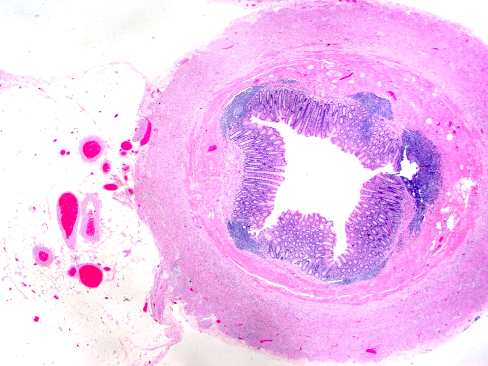
  • Tis: carcinoma in situ (intramucosal carcinoma; invasion of the lamina propria or extension into but not through the muscularis mucosae)
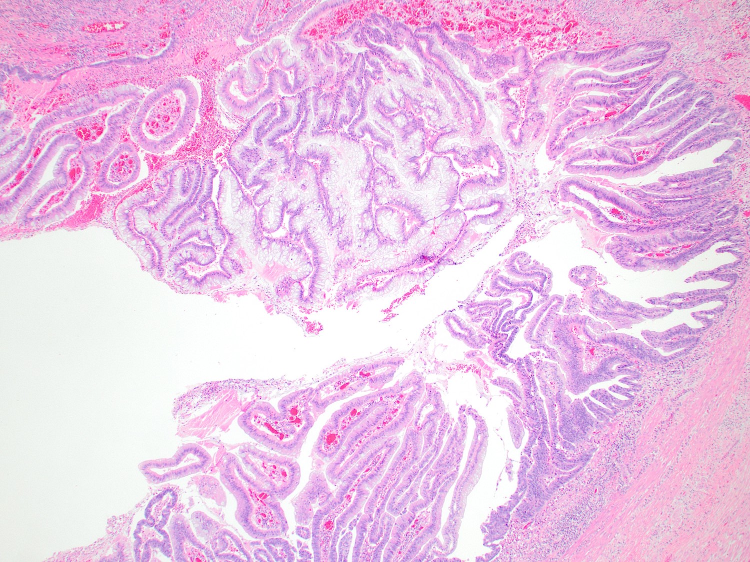
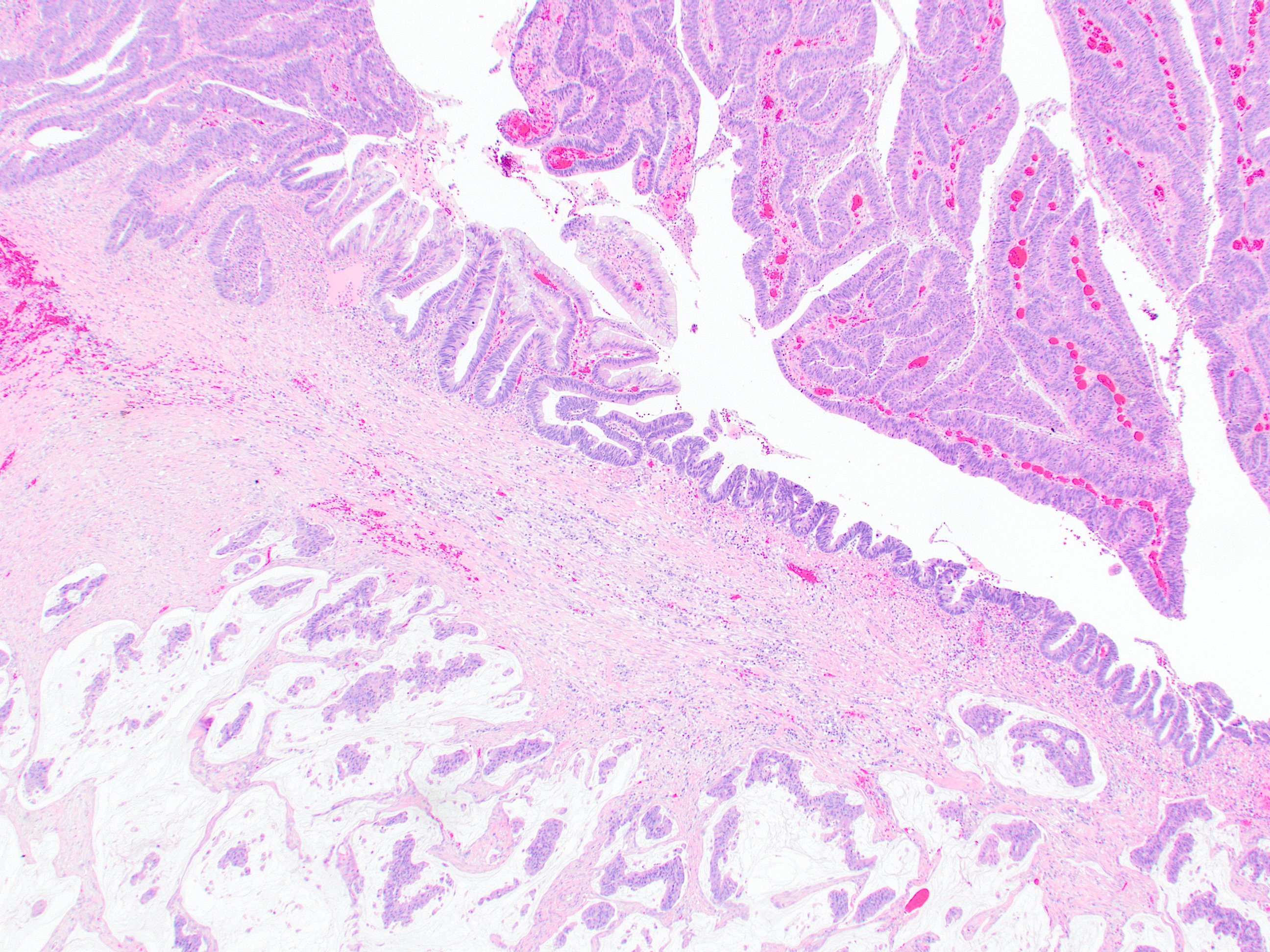
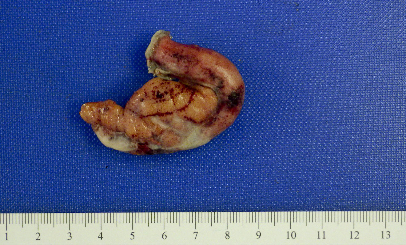
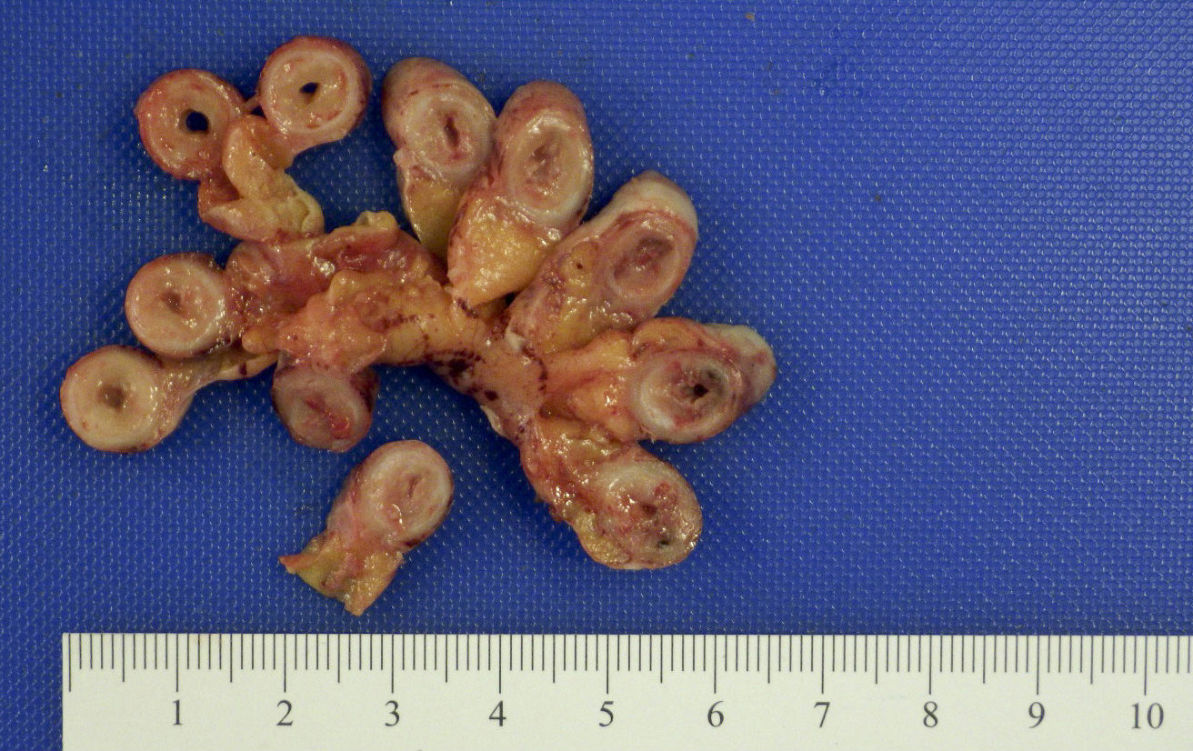
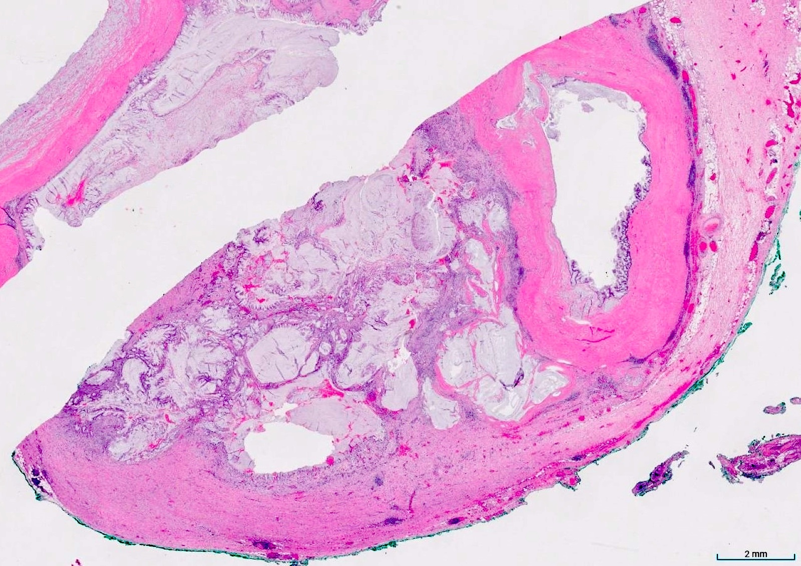
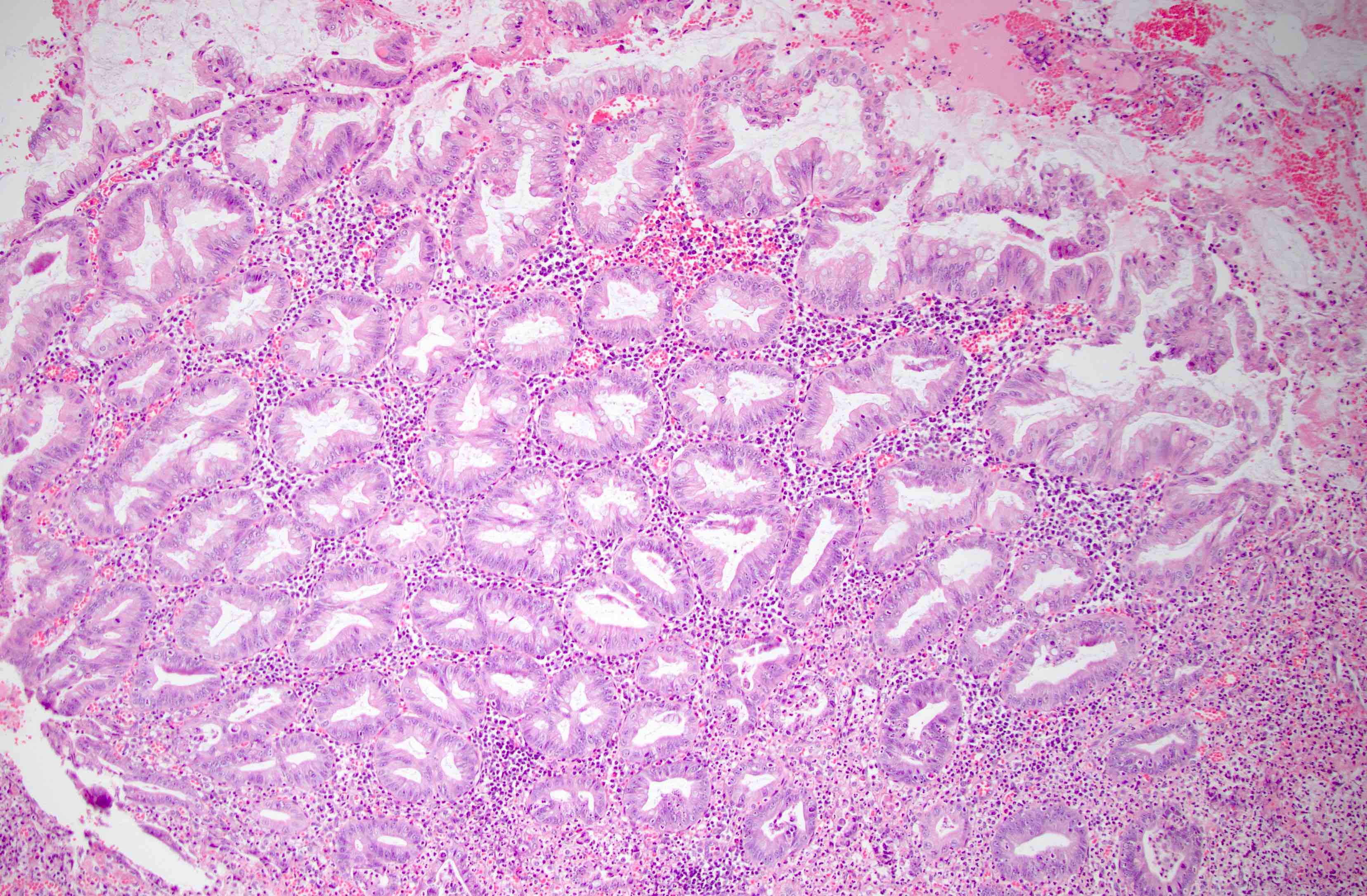
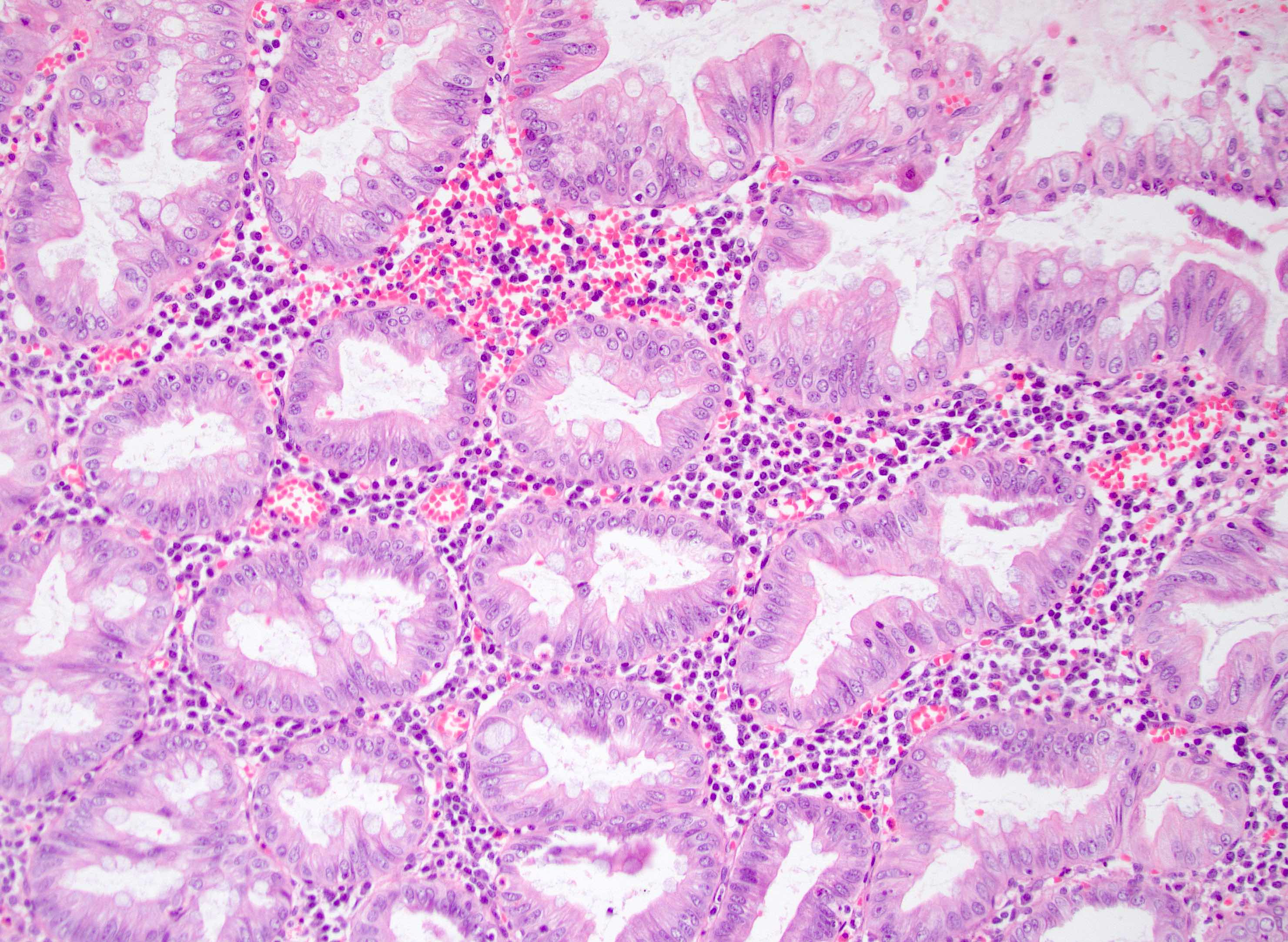
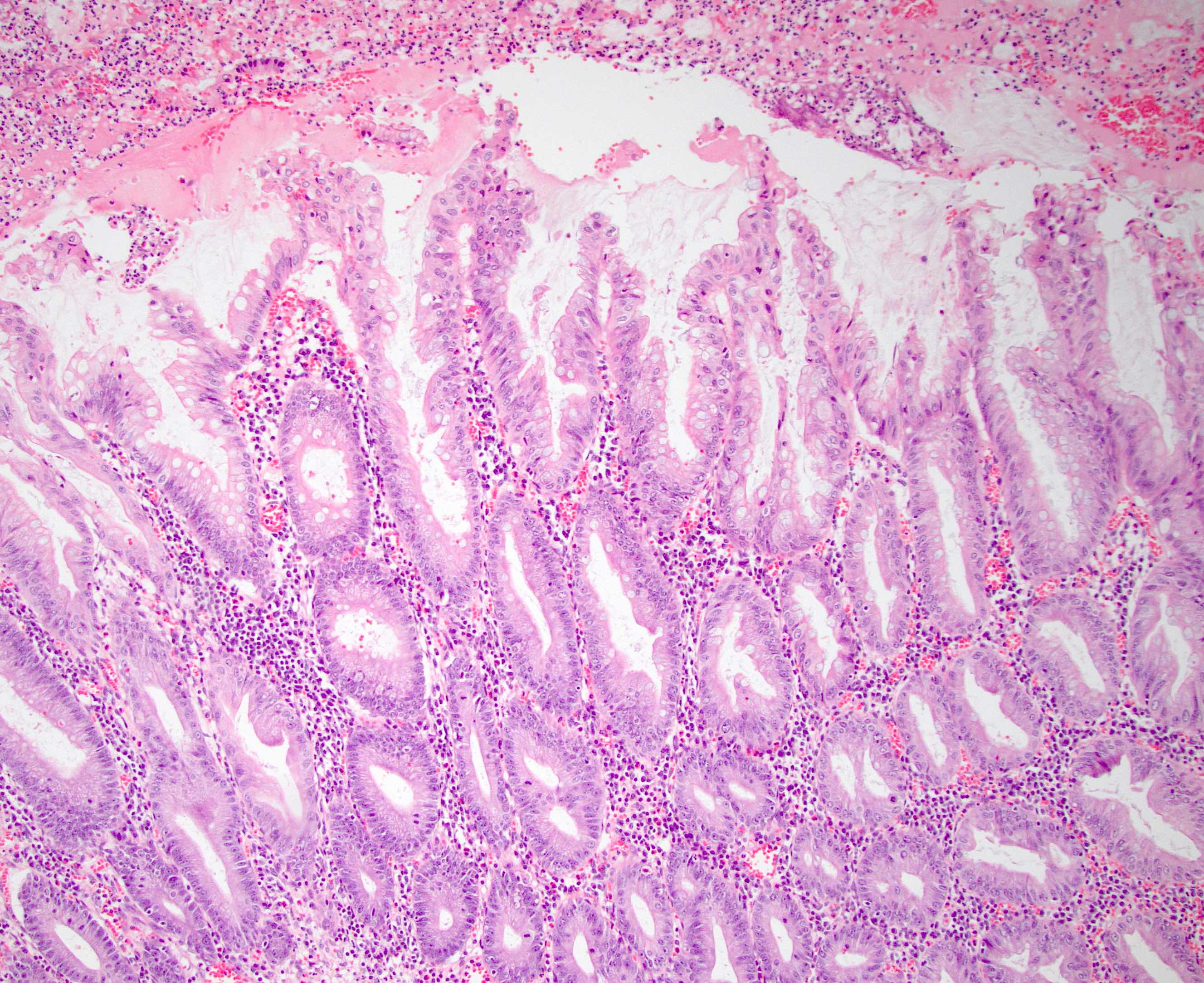
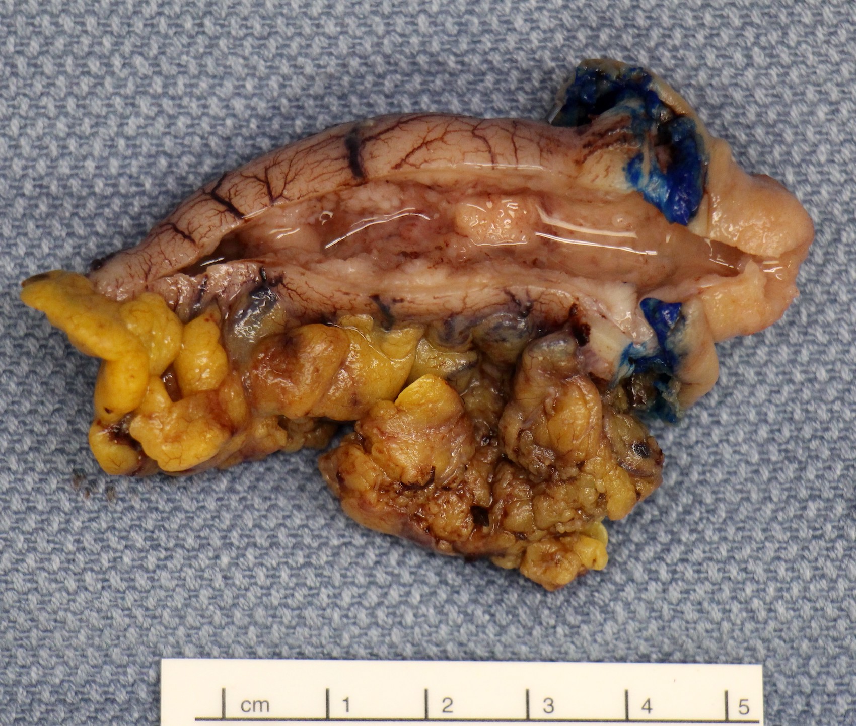
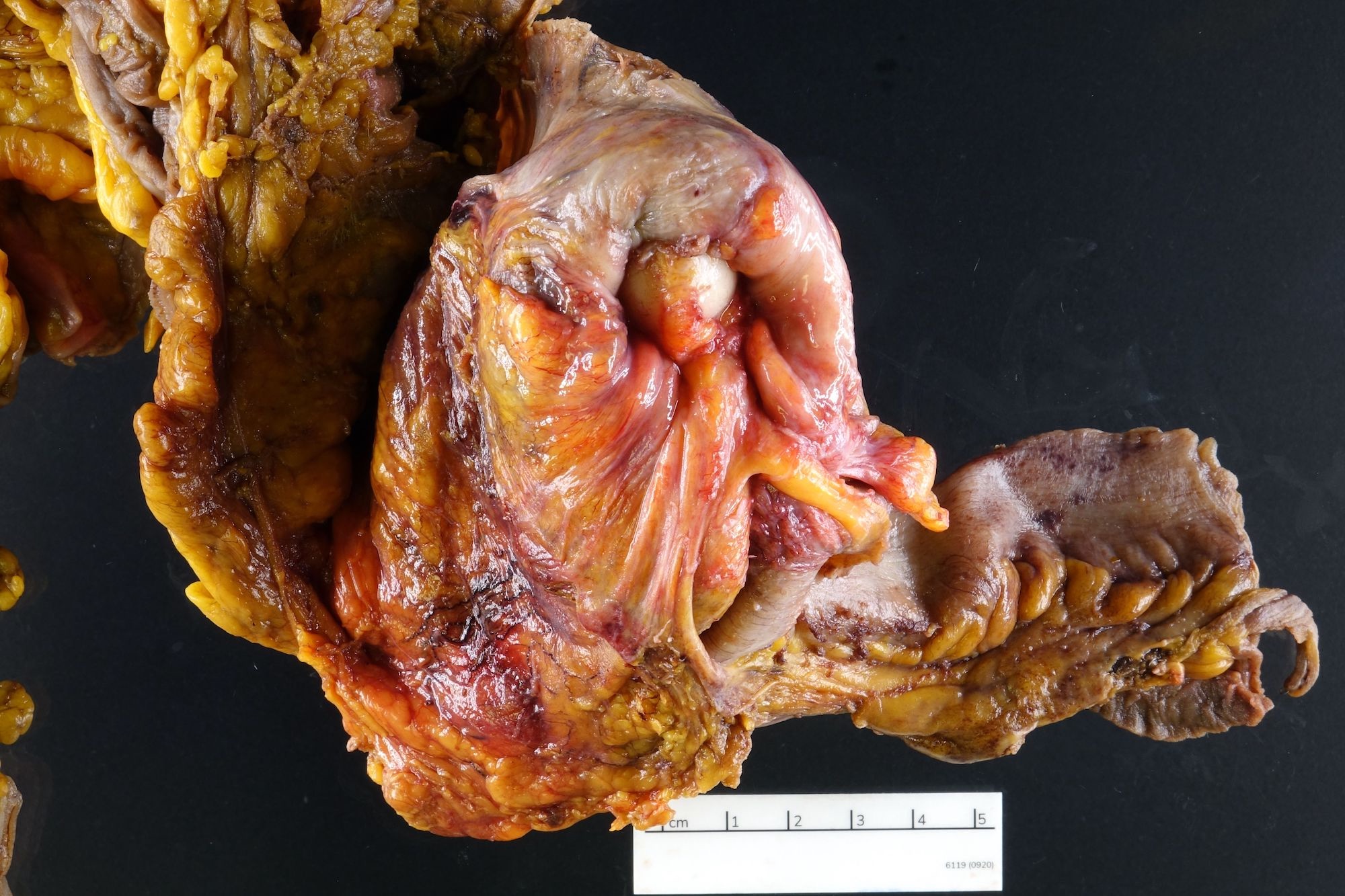
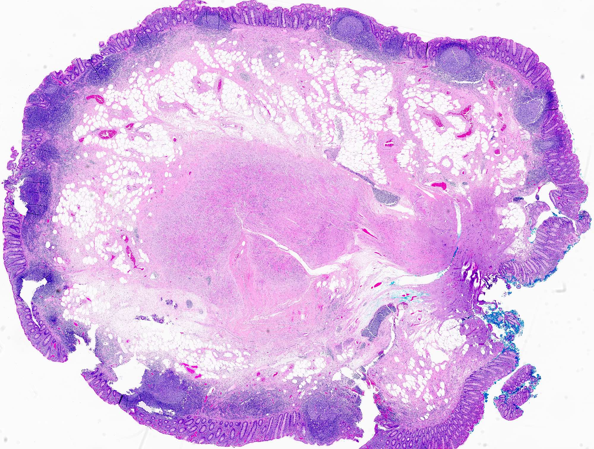
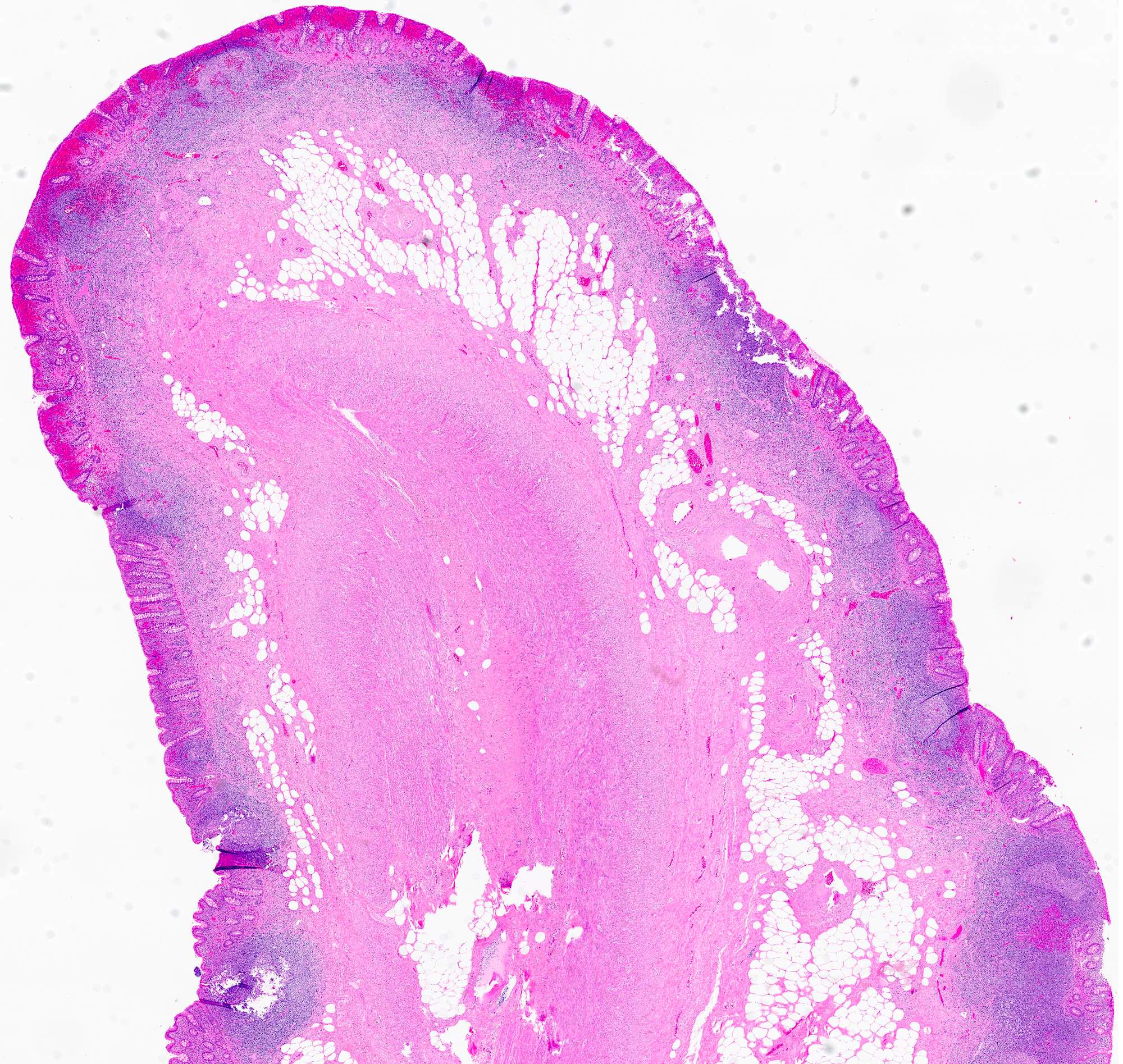
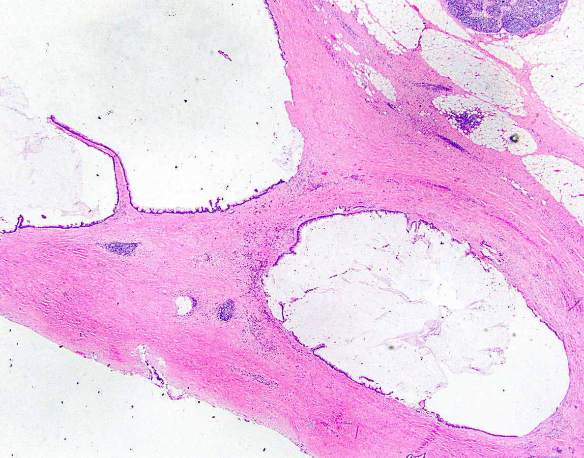
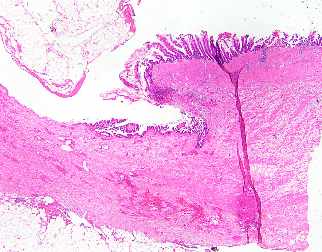
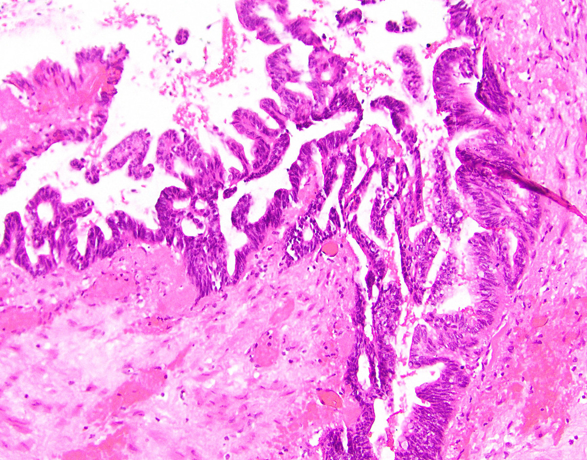
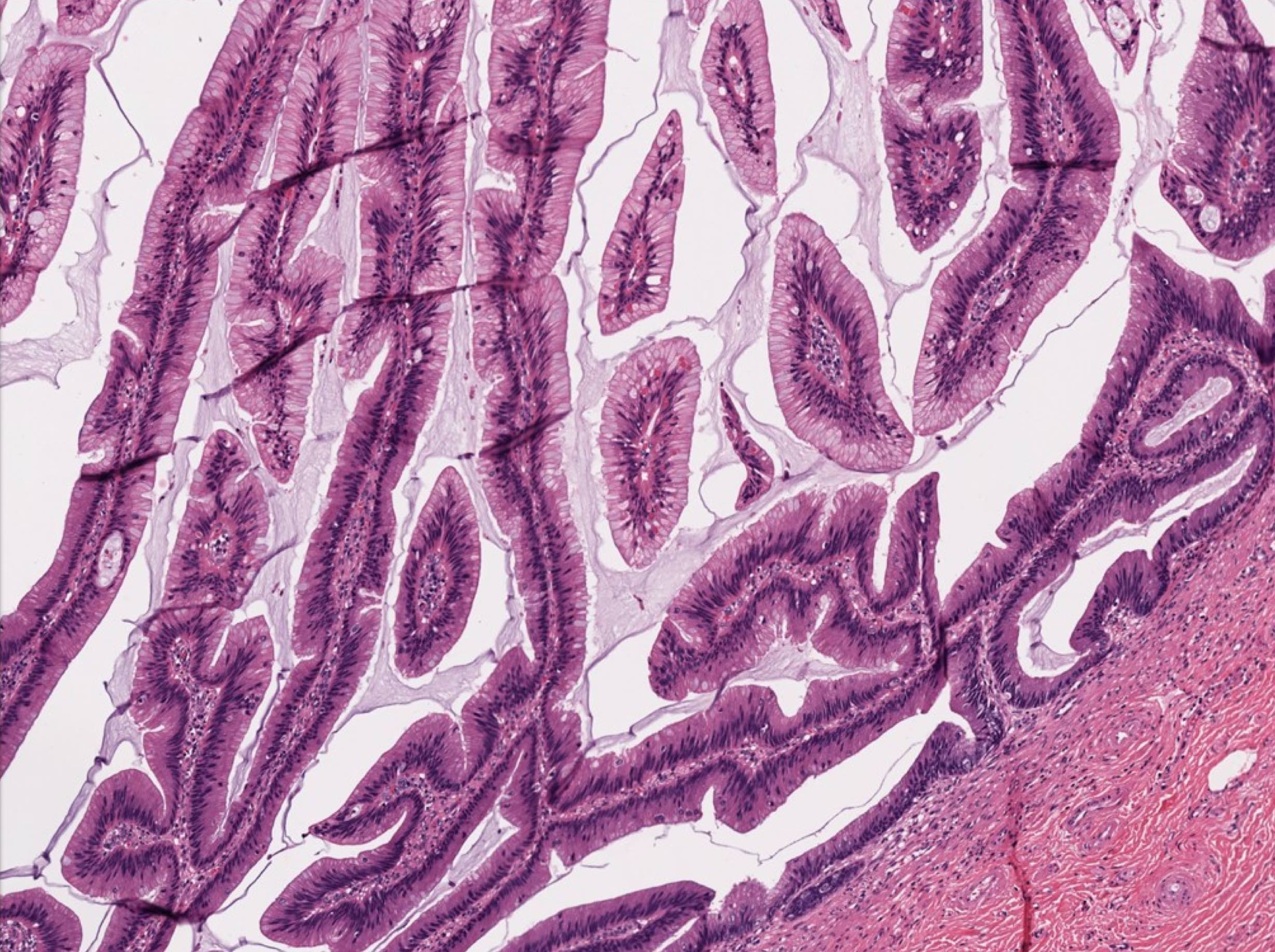
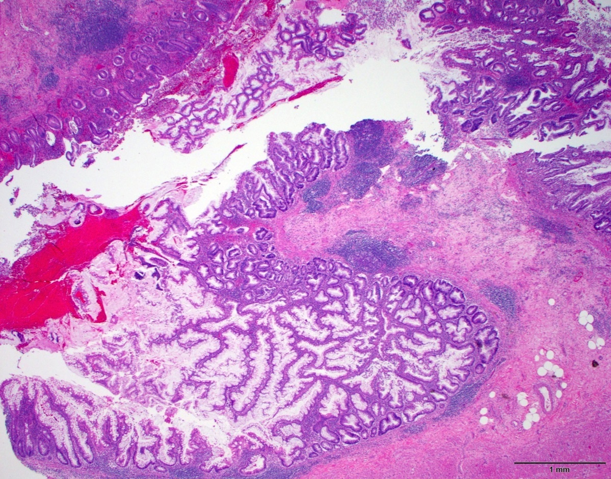
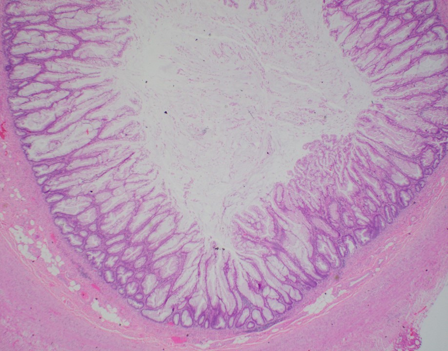
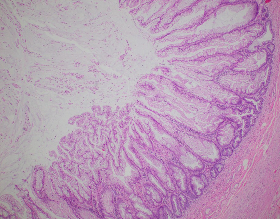
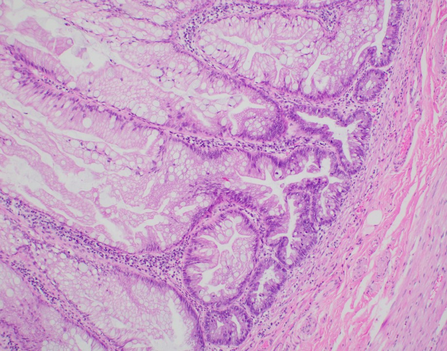
  • Tis(LAMN): low grade appendiceal mucinous neoplasm confined by the muscularis propria; acellular mucin or mucinous epithelium may invade into the muscularis propria
  • T1: tumor invades the submucosa (through the muscularis mucosa but not into the muscularis propria)
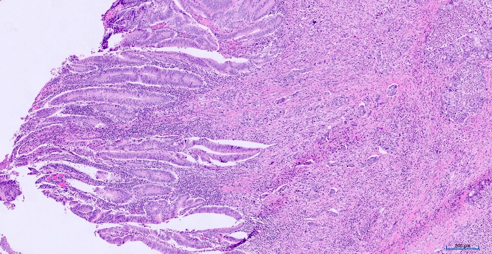
  • T2: tumor invades the muscularis propria
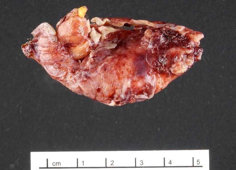
  • T3: tumor invades through the muscularis propria into the subserosa or the mesoappendix
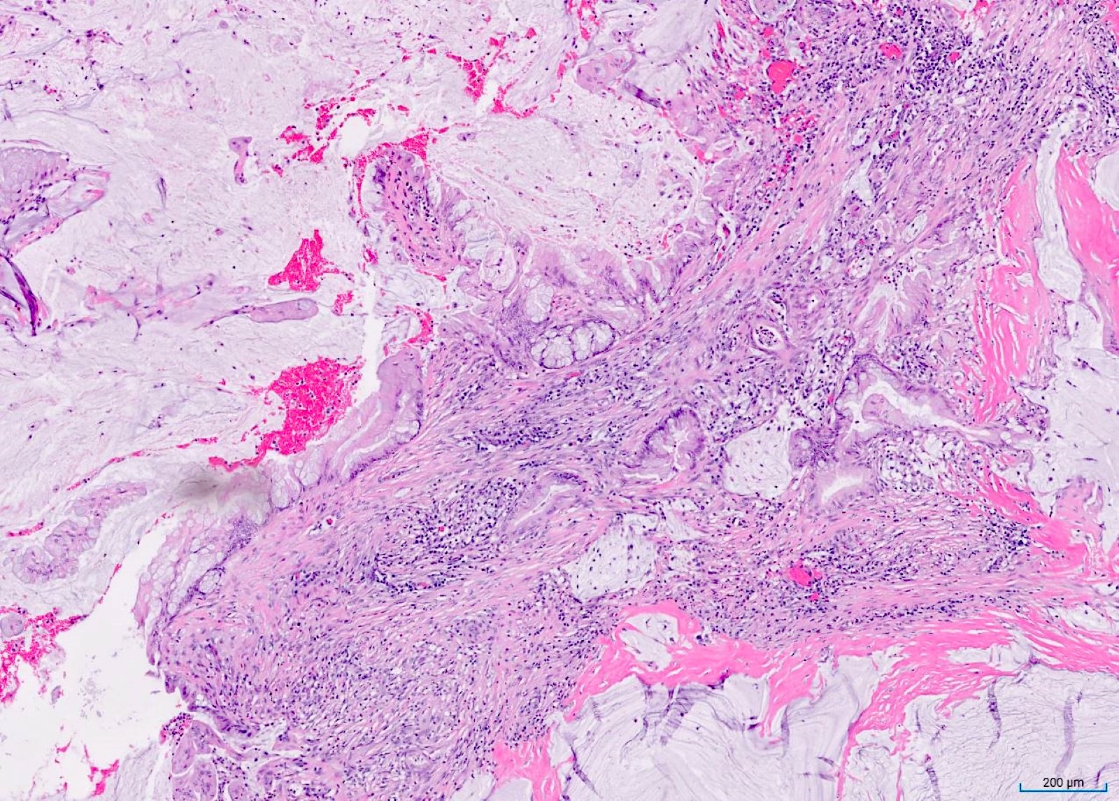
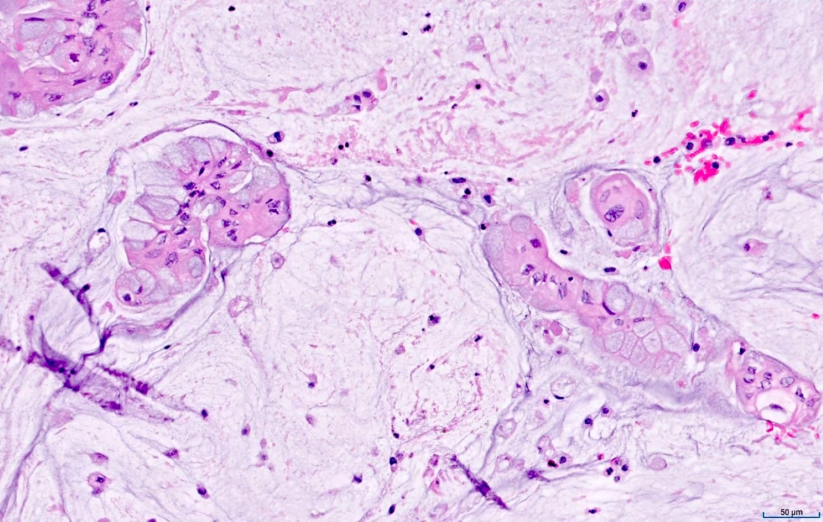
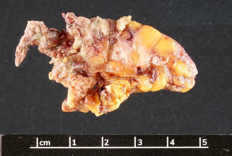
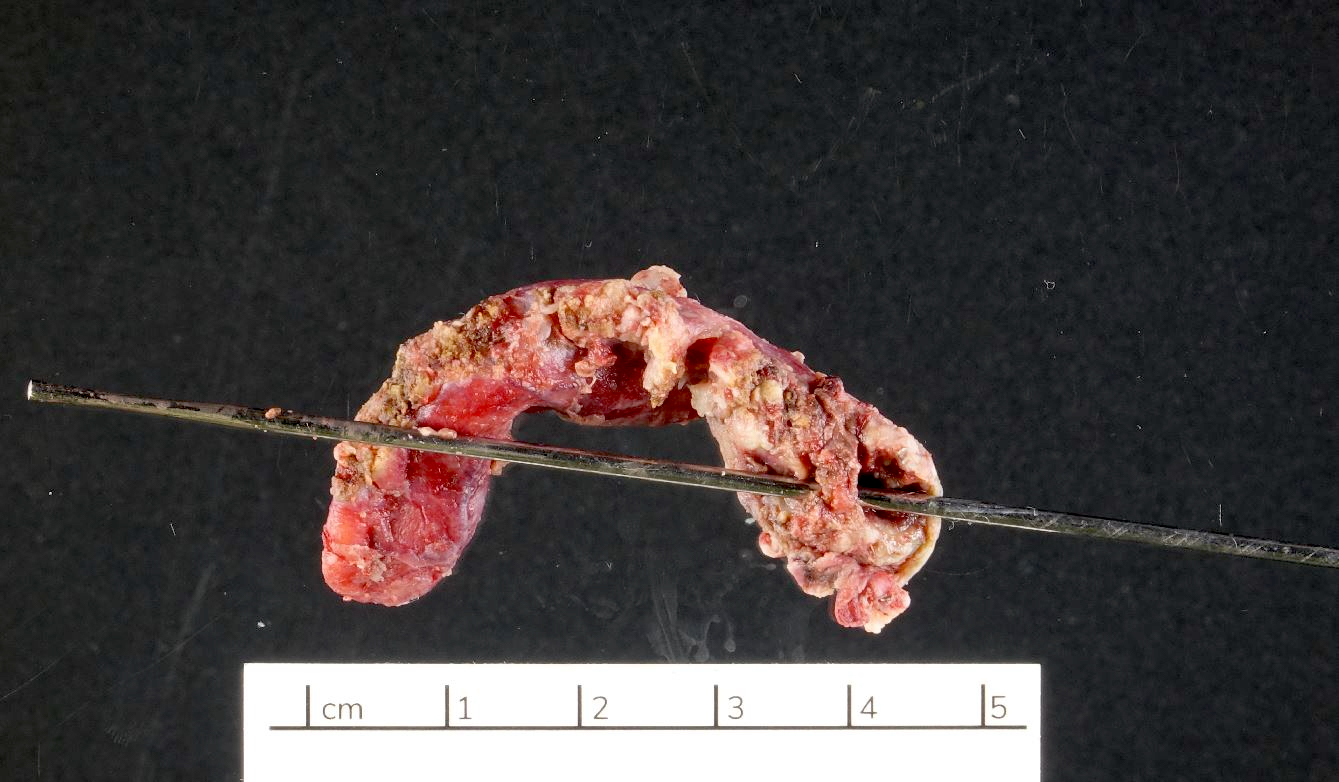
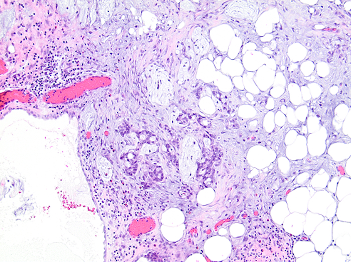
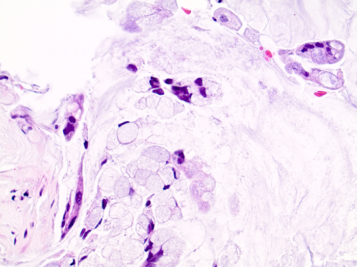
  • T4: tumor invades the visceral peritoneum, including the acellular mucin or mucinous epithelium involving the serosa of the appendix or mesoappendix or directly invades adjacent organs or structures
    • T4a: tumor invades through the visceral peritoneum, including the acellular mucin or mucinous epithelium involving the serosa of the appendix or serosa of the mesoappendix
    • T4b: tumor directly invades or adheres to adjacent organs or structures

Notes:
  • T1 and T2 are not applicable to LAMN; acellular mucin or mucinous epithelium that extends into the subserosa or serosa should be classified as T3 or T4a, respectively (Hum Pathol 2017;69:81)
  • T1 and T2 are applicable to HAMN
Regional lymph nodes (pN)
  • NX: regional lymph nodes cannot be assessed
  • N0: no tumor involvement of regional lymph node(s)
  • N1: tumor involvement of 1 to 3 regional lymph nodes (tumor in lymph node measuring ≥ 0.2 mm) or any number of tumor deposit(s)
    • N1a: tumor involvement of 1 regional lymph node
    • N1b: tumor involvement of 2 or 3 regional lymph nodes
    • N1c: no tumor involvement of regional lymph nodes but there are tumor deposits in the subserosa or mesentery
  • N2: tumor involvement of 4 or more regional lymph nodes

Notes:
  • Regional lymph nodes include ileocolic nodes
Distant metastasis (pM)
  • cM0: no distant metastasis
  • cM1: distant metastasis
    • cM1c: metastasis to sites other than peritoneum
  • pM1: microscopic confirmation of distant metastasis
    • pM1a: intraperitoneal acellular mucin, without identifiable tumor cells in the disseminated peritoneal mucinous deposits
    • pM1b: intraperitoneal metastasis only, including peritoneal mucinous deposits containing tumor cells
    • pM1c: microscopic confirmation of metastasis to sites other than peritoneum

Notes:
  • For specimens containing acellular mucin without identifiable tumor cells, efforts should be made to obtain additional tissue for thorough histologic examination to evaluate for cellularity
Prefixes
  • c: clinical
  • p: pathological
  • yc: posttherapy clinical
  • yp: posttherapy pathological
  • rc: recurrence / retreatment clinical
  • rp: recurrence / retreatment pathological
  • a: autopsy
Primary tumor suffix
  • (m): multiple synchronous primary tumors
Regional lymph nodes suffix
  • (sn): sentinel node procedure
  • (f): fine needle aspiration (FNA) or core needle biopsy
AJCC prognostic stage groups
Stage group 0:   Tis or Tis(LAMN)   N0   M0   any G
Stage group I:   T1 - 2   N0   M0   any G
Stage group IIA:   T3   N0   M0   any G
Stage group IIB:   T4a   N0   M0   any G
Stage group IIC:   T4b   N0   M0   any G
Stage group IIIA:   T1 - 2   N1   M0   any G
Stage group IIIB:   T3 - 4   N1   M0   any G
Stage group IIIC:   any T   N2   M0   any G
Stage group IVA:   any T   any N   M1a   any G
any T   any N   M1b   G1  
Stage group IVB:   any T   any N   M1b   G2, G3, GX
Stage group IVC:   any T   any N   M1c   any G
Prognostic tumor characteristics
  • Histologic grade of primary tumor
  • Histologic grade of metastatic peritoneal disease
  • Mucinous histology
  • LAMN
  • HAMN
  • Number of tumor deposits
  • Lymphovascular invasion
  • Perineural invasion
  • Perforation
  • Microsatellite instability / mismatch repair
  • Preoperative / pretreatment tumor markers: carcinoembryonic antigen (CEA), cancer antigen (CA) 19-9, CA-125
Registry data collection variables
  • Primary tumor grade
  • CEA laboratory value and interpretation
  • Lymphovascular invasion
  • LAMN
  • HAMN
Emerging factors for data collection
  • Distinct WHO histopathologic codes for LAMN and HAMN
  • Histologic grade of metastatic peritoneal disease
Histologic grade (G)
  • GX: grade cannot be assessed
  • G1: well differentiated
  • G2: moderately differentiated
  • G3: poorly differentiated

Notes:
  • In rare cases of discordance in primary and metastatic histological grade, the grade of metastatic disease is utilized for stage group assignment
Histopathologic type
  • Large cell neuroendocrine carcinoma
  • Undifferentiated carcinoma
  • Small cell neuroendocrine carcinoma
  • Squamous cell carcinoma
  • Adenocarcinoma
  • Mixed neuroendocrine nonneuroendocrine neoplasm
  • Goblet cell adenocarcinoma
  • Low grade appendiceal mucinous neoplasm
  • High grade appendiceal mucinous neoplasm
  • Mucinous adenocarcinoma
  • Signet ring cell carcinoma
Residual tumor (operative factor)
  • R0: complete resection, margins histologically negative, no residual tumor left after resection
  • R1: incomplete resection, margins histologically involved, microscopic tumor remains after resection of gross disease (relevant to resection margins that are microscopically involved by tumor)
  • R2: incomplete resection, margins involved or gross disease remains
Board review style question #1
Using AJCC staging criteria for adenocarcinomas of the appendix, at what point is tumor grade utilized (along with pT, pN and pM status) in order to assign overall combined tumor stage?

  1. When a tumor extends to the surgical resection margin
  2. When a tumor has cellular intraperitoneal metastases
  3. When a tumor involves the muscularis propria
  4. When a tumor is a goblet cell adenocarcinoma
Board review style answer #1
B. When a tumor has cellular intraperitoneal metastases. Such cases are stage IV and they may be subclassified as IVA or IVB depending on the tumor grade. Answer C is incorrect because tumor grade does not impact pT category staging of appendiceal adenocarcinomas. Answers A and D are incorrect because they do not influence staging of appendiceal adenocarcinomas.

Comment Here

Reference: Appendix - Staging - carcinoma
Board review style question #2
What is the difference in AJCC staging parameters for low grade apendiceal mucinous neoplasm (LAMN) and high grade apendiceal mucinous neoplasm (HAMN)?

  1. LAMN cannot be pM1a but HAMN can
  2. LAMN cannot be pN1 but HAMN can
  3. LAMN cannot be pT1 / 2 but HAMN can
  4. LAMN cannot be pT4a but HAMN can
Board review style answer #2
C. LAMN cannot be pT1 / 2 but HAMN can. LAMN is reported as pTis(LAMN) if bound by muscularis propria, whereas HAMN would be pT1 or pT2 in the same situation, depending on whether submucosa or muscularis propria is involved. Answer A is incorrect because both LAMN and HAMN can be pM1a. Answer B is incorrect because both LAMN and HAMN can theoretically be pN1 (though this is extraordinarily unlikely). Answer D is incorrect because both LAMN and HAMN can be pT4a.

Comment Here

Reference: Appendix - Staging - carcinoma

Staging-neuroendocrine tumors
Definition / general
  • All well differentiated neuroendocrine tumors of the appendix, regardless of grade, are covered by this staging system
  • Not covered by this staging system are appendiceal adenocarcinomas, goblet cell adenocarcinomas and poorly differentiated neuroendocrine carcinomas (use the appendix carcinoma staging system instead)
Essential features
  • AJCC 7th edition staging was sunset on December 31, 2017; as of January 1, 2018, use of the 8th edition is mandatory
  • AJCC 9th edition was released in 2024, with minimal changes from the 8th edition
Terminology
N/A
ICD coding
  • ICD-10: C7A.020 - malignant carcinoid tumor of the appendix
Diagrams / tables
N/A
Primary tumor (pT)
  • TX: primary tumor cannot be assessed
  • T0: no evidence of primary tumor
  • T1: tumor ≤ 2 cm in greatest dimension
  • T2: tumor > 2 cm but ≤ 4 cm in greatest dimension
  • T3: tumor > 4 cm in greatest dimension or with subserosal invasion or involvement of the mesoappendix
  • T4: tumor perforates the peritoneum or directly invades other adjacent organs or structures (excluding direct mural extension to adjacent subserosa of adjacent bowel) (e.g., abdominal wall and skeletal muscle)
Regional lymph nodes (pN)
  • NX: regional lymph nodes cannot be assessed
  • N0: no tumor involvement of regional lymph node(s)
  • N1: tumor involvement of regional lymph node(s)

  • Notes:
    • Ileocolic nodes are the regional nodes for this staging
Distant metastasis (pM)
  • cM0: no distant metastasis
  • cM1: distant metastasis
    • cM1a: metastasis confined to liver
    • cM1b: metastasis in at least 1 extrahepatic site (e.g., lung, ovary, nonregional lymph node, peritoneum, bone)
    • cM1c: both hepatic and extrahepatic metastasis
  • pM1: microscopic confirmation of distant metastasis
    • pM1a: microscopic confirmation of metastasis confined to liver
    • pM1b: microscopic confirmation of metastasis in at least 1 extrahepatic site (e.g., lung, ovary, nonregional lymph node, peritoneum, bone)
    • pM1c: microscopic confirmation of both hepatic and extrahepatic metastasis
Prefixes
  • c: clinical
  • p: pathological
  • yc: posttherapy clinical
  • yp: posttherapy pathological
Primary tumor suffix
  • (m): multiple synchronous primary tumors
AJCC prognostic stage groups
Stage I: T1 N0, NX M0
Stage II: T2   N0, NX M0
T3 N0 M0
Stage III: T4 N0 M0
any T N1 M0
Stage IV:any T any N  M1
Prognostic tumor characteristics
  • Ki67 index
  • Mitotic count
  • Plasma or urinary 5-HIAA level
  • Echocardiogram (carcinoid heart disease)
Registry data collection guidance
  • Size of tumor
  • Depth of invasion
  • Invasion of mesoappendix
  • Number of nodes involved, mesenteric mass, mesenteric vessel encasement
  • Perineural invasion
  • Lymphovascular invasion
  • Sites of metastasis, if applicable
  • Type of surgery
  • Ki67 index
  • Mitotic count
  • Grade (from Ki67 and mitotic count): G1, G2, G3
  • Presence of somatostatin receptor expression (usually determined by using somatostatin receptor imaging such as DOTATATE PET)
Emerging factors for data collection
  • Somatostatin receptor type 2 (SSTR2) positivity
Histologic grade (G)
  • G1: mitotic rate < 2 per 2 mm2 and Ki67 < 3%
  • G2: mitotic rate 2 - 20 per 2 mm2 or Ki67 3 - 20%
  • G3: mitotic rate > 20 per 2 mm2 or Ki67 > 20%
Histopathologic type
  • Neuroendocrine tumor, NOS
  • Neuroendocrine tumor, grade 1
  • Neuroendocrine tumor, grade 2
  • Neuroendocrine tumor, grade 3
  • L cell neuroendocrine tumor
  • Glucagon-like peptide producing neuroendocrine tumor
  • PP / PPY producing tumor
  • Enterochromaffin cell (EC cell) neuroendocrine tumor
  • Serotonin producing neuroendocrine tumor
Residual tumor (operative factor)
  • R0: complete resection, margins histologically negative, no residual tumor left after resection
  • R1: incomplete resection, margins histologically involved, microscopic tumor remains after resection of gross disease (relevant to resection margins that are microscopically involved by tumor)
  • R2: incomplete resection, margins involved or gross disease remains
Gross images
N/A
Microscopic (histologic) images
N/A
Board review style question #1
Which of the following factors affect the staging of appendiceal neuroendocrine tumors?

  1. Ki67 index and extent of local spread
  2. Ki67 index and mitotic rate
  3. Size and extent of local spread
  4. Size and Ki67 index
Board review style answer #1
C. Size and extent of local spread. These are used to determine pT category staging; pT1 - pT3 include size and pT3 - pT4 include local spread. Answers A, B and D are incorrect because Ki67 index and mitotic rate are used in grading but not staging of appendiceal neuroendocrine tumors.

Comment Here

Reference: Appendix - Staging-neuroendocrine tumors
Board review style question #2
An appendiceal neuroendocrine tumor develops clinical metastatic disease to the liver but no other sites. What is the cM category staging for this tumor?

  1. cM0
  2. cM1a
  3. cM1b
  4. cM1c
Board review style answer #2
B. cM1a. Metastatic disease only to the liver is considered M1a for appendiceal neuroendocrine tumors. Answer A is incorrect because it suggests there is no distant metastatic disease. Answer C is incorrect because M1b requires metastasis to nonhepatic distant sites. Answer D is incorrect because M1c requires distant metastasis to both hepatic and extrahepatic sites.

Comment Here

Reference: Appendix - Staging-neuroendocrine tumors

Staging-neuroendocrine tumors
Definition / general
  • All well differentiated neuroendocrine tumors of the appendix, regardless of grade, are covered by this staging system
  • Not covered by this staging system are appendiceal adenocarcinomas, goblet cell carcinoids and poorly differentiated neuroendocrine carcinomas (use the appendix carcinoma staging system instead)
Essential features
ICD coding
  • ICD-10: C7A.020 - Malignant carcinoid tumor of the appendix
Primary tumor (pT)
  • TX: Primary tumor cannot be assessed
  • T0: No evidence of primary tumor
  • T1: Tumor ≤ 2 cm in greatest dimension
  • T2: Tumor > 2 cm but ≤ 4 cm
  • T3: Tumor > 4 cm or with subserosal invasion or involvement of the mesoappendix
  • T4: Tumor perforates the peritoneum or directly invades other adjacent organs or structures (excluding direct mural extension to adjacent subserosa of adjacent bowel), e.g. abdominal wall and skeletal muscle
Regional lymph nodes (pN)
  • NX: Regional lymph nodes cannot be assessed
  • N0: No regional lymph node metastasis
  • N1: Regional lymph node metastasis
  • Notes: the ileocolic nodes are the regional nodes for this staging
Distant metastasis (pM)
  • M0: No distant metastasis
  • M1: Distant metastasis
    • M1a: Metastasis confined to liver
    • M1b: Metastasis in at least one extrahepatic site (e.g. lung, ovary, nonregional lymph node, peritoneum, bone)
    • M1c: Both hepatic and extrahepatic metastasis
Prefixes
  • r: recurrent tumor
Stage grouping
Stage I: T1 N0 M0
Stage II: T2 - 3  N0 M0
Stage III: T4 N0 M0
any T N1 M0
Stage IV:any T any N  M1
Registry data collection variables
  • Size of tumor
  • Depth of invasion
  • Invasion of mesoappendix
  • Number of nodes involved, mesenteric mass, mesenteric vessel encasement
  • Perineural invasion
  • Lymphovascular invasion
  • Sites of metastasis, if applicable
  • Type of surgery
  • Ki67 proliferative index
  • Mitotic count
  • Histologic grading (from Ki67 and mitotic count): G1, G2, G3
Histologic grade
  • Grading is not formally part of the staging system
  • Most pathologists use the European Neuroendocrine Tumor Society (ENETS) / World Health Organization (WHO) grading criteria:
    • Grade 1: Mitotic rate < 2 per 10 high power fields and Ki67 < 3%
    • Grade 2: Mitotic rate 2 - 20 per 10 high power fields or Ki67 3 - 20%
    • Grade 3: Mitotic rate > 20 per 10 high power fields or Ki67 > 20%
Histopathologic type
  • Well differentiated neuroendocrine tumor, G1 / G2 / G3
Board review style question #1
Which of the following factors affect the staging of appendiceal neuroendocrine tumors?

  1. Ki67 index and extent of local spread
  2. Ki67 index and mitotic rate
  3. Size and extent of local spread
  4. Size and Ki67 index
Board review style answer #1
C. Size and extent of local spread

Comment Here

Reference: Staging-neuroendocrine tumors

Tubular adenoma
Definition / general
  • Polypoid dysplastic lesion in the appendix, analogous to conventional adenomas in the colorectum
Essential features
  • Uncommon (not included in WHO classification of tumors of the appendix)
  • Tubulovillous adenomas are often resected as mass lesions, while small tubular adenomas may be encountered incidentally in colectomy specimens from patients with polyposis syndromes
  • Many adenocarcinomas of the appendix arise from adenomas
ICD coding
  • ICD-10: D12.1 - benign neoplasm of appendix
Epidemiology
Sites
  • Appendix
Pathophysiology
Clinical features
Diagnosis
Radiology description
Radiology images

Images hosted on other servers:

Double contrast barium enema

Abdominal CT scan

Prognostic factors
  • Appendiceal adenocarcinoma can arise from adenomas
  • Risk of progression likely increases with size and high grade dysplasia (as in the colon) but data are limited
Case reports
Treatment
Gross description
  • Similar to colorectal tubular adenomas, which can appear pedunculated, sessile, flat or depressed
  • May be seen arising from the appendix
  • Features of acute appendicitis and perforation can be seen if the adenoma presents accordingly (BMC Gastroenterol 2011;11:35)
Gross images

Images hosted on other servers:

Grossly visible appendiceal adenoma

Microscopic (histologic) description
  • Largely similar to colorectal tubular adenomas
  • Architecture is most often villous but may also be tubular
  • Predominantly composed of low grade dysplasia, with elongated, hyperchromatic nuclei and crowded glands
  • Foci of high grade dysplasia may show rounded cells with nucleoli, low polarity and cribriform architecture
  • Epithelial lining may have a somewhat hypermucinous appearance
  • Reference: Scand J Gastroenterol 1985;20:512
Microscopic (histologic) images

Contributed by Raul S. Gonzalez, M.D.
Tubular glands with elongated, hyperchromatic nuclei and crowded glands Tubular glands with elongated, hyperchromatic nuclei and crowded glands

Tubular glands with elongated, hyperchromatic nuclei and crowded glands

Crowded tubular and villous glands with elongated hyperchromatic nuclei Crowded tubular and villous glands with elongated hyperchromatic nuclei

Crowded tubular and villous glands with elongated hyperchromatic nuclei

Mucinous appearance

Mucinous appearance

Adenocarcinoma arising in adenoma

Adenocarcinoma arising in adenoma

Sample pathology report
  • Appendix, appendectomy:
    • Tubulovillous adenoma (2.1 cm)
    • Negative for high grade dysplasia or malignancy
    • Margin of resection negative
Differential diagnosis
  • Serrated polyp:
    • Can show areas of nuclear dysplasia but should also show additional areas of serrated glands with booting architecture and lacking conventional dysplasia
  • Appendiceal mucinous neoplasm:
    • May have villous areas and cytologic atypia but causes appendiceal dilation without polyp formation grossly
Board review style question #1

Which of the following is true about intestinal type adenomas of the appendix?

  1. They arise from low grade appendiceal mucinous neoplasms
  2. They can cause intussusception
  3. They have no risk of malignant progression
  4. They only occur in patients with familial adenomatous polyposis
Board review style answer #1
B. They can cause intussusception. Intestinal type adenomas of the appendix can cause appendicitis, bowel obstruction or perforation. They have no precursor lesion and may themselves progress to adenocarcinoma. They can occur sporadically or in patients with polyposis syndromes.

Comment Here

Reference: Tubular adenoma

WHO classification
Table of Contents
Definition / general | WHO (2019)
Definition / general
  • WHO classification of tumors of the appendix
  • Currently on 5th edition, published in 2019
  • Based on histologic appearance, not molecular characteristics
WHO (2019)

Xanthogranulomatous inflammation
Definition / general
  • Considered an unusual healing pattern of appendicitis
  • May represent chronic or prolonged inflammatory process
Microscopic (histologic) description
  • Prominent histiocytes with clusters of xanthoma-type cells
Case reports
Differential diagnosis
  • Malakoplakia, Mycobacterium avium-intracellulare infection, Whipple's disease, rarely Crohn’s disease
Back to top
Recent Appendix Pathology books

Greenson: 2019

IARC: 2019

Montgomery: 2017

Montgomery: 2017

Odze: 2022

Srivastava: 2023

Yantiss: 2021


Find related Pathology books: GI, liver
Image 01 Image 02