Superpage
Cervix

Authors: Apeksha N. Agarwal, M.B.B.S., M.D., Maryam Aghighi, M.D., Hiba Al Dallal, M.B.Ch.B., Khaled J. Alkhateeb, M.B.B.S., Léonie Alran, B.S., Jaya Ruth Asirvatham, M.D., Ryan W. Askeland, M.D., Erika M. Baardsen, D.O., Julieta E. Barroeta, M.D., Sarah M. Bean, M.D., Natalia Buza, M.D., David B. Chapel, M.D., Yevgen Chornenkyy, M.D., M.Sc., Bonnie Choy, M.D., Sabrina Croce, M.D., Ph.D., Teresa M. Darragh, M.D., Kyle Devins, M.D., Farnaz Hasteh, M.D., Lynn Hoang, M.D., Anjelica Hodgson, M.D., Ali Ismail, M.B.B.S., Lucy Jager, M.D., Soumya Jaladi, M.B.B.S., Rachel Jug, M.B.B.Ch., B.A.O., Andreas Kontosis, M.D., Adam Lechner, B.M., Zaibo Li, M.D., Ph.D., Leonel Maldonado, M.D., Brandon Douglas Metcalf, M.D., Nada Mohamed, M.D., Carlos Parra-Herran, M.D., Nat Pernick, M.D., Branko Perunovic, D.M., John K.S.S. Philip, M.D., Jennifer Pors, M.D., Shuyue Ren, M.D., Ph.D., Joseph Reznicek, M.D., Marilin Rosa, M.D., Agnieszka Rychlik, M.D., Ziyan T. Salih, M.D., Lauren Schwartz, M.D., Sucheta Srivastava, M.D., Ashwyna Sunassee, M.D., Gulisa Turashvili, M.D., Ph.D., Philip T. Valente, M.D., Jaclyn Watkins, M.D., M.S., Katharina Wiedemeyer, M.D., Mikami Yoshiki, M.D., Ph.D.
Editorial Board Members: David B. Chapel, M.D., Bonnie Choy, M.D., Kyle Devins, M.D., Ricardo R. Lastra, M.D., Lucy Ma, M.D., Carlos Parra-Herran, M.D., Marc Pusztaszeri, M.D., Gulisa Turashvili, M.D., Ph.D.
Deputy Editors-in-Chief: Jennifer A. Bennett, M.D., Gulisa Turashvili, M.D., Ph.D.
Editor-in-Chief: Debra L. Zynger, M.D.

Copyright: 2002-2024, PathologyOutlines.com, Inc.

Gyn related: Jobs, Fellowships, Conferences, Cases, CME, Board Review, What's New

Related chapters: Uterus, Vulva, vagina & female urethra

Editorial Board oversight: David B. Chapel, M.D. (last reviewed December 2023)
Page views in 2024 to date: 49

Actinomycosis
Definition / general
Terminology
  • Also called "dust bunnies" or "Gupta bodies"
Epidemiology
  • Rarely identified in Pap Test (< 1%)
  • Can be found in culture of 25% of asymptomatic patients
Etiology
  • Actinomyces Israeli is most common subtype (normal flora of the mouth and bowel)
  • Associated with IUD (common) or rarely with other objects (pessaries, tampon)
  • Copper containing IUD and long term use are major risk factors
Clinical features
  • Asymptomatic
  • Malodorous brown discharge
  • Pain with invasive disease and PID (pelvic inflammatory disease)
Treatment
  • Removal of IUD for asymptomatic women
  • Removal of IUD and antibodies for symptomatic women
Microscopic (histologic) description
  • Tangled clumps of gram positive, non-acid fast, filamentous organisms, often with acute angle branching, sometimes showing irregular wooly appearance
  • Swollen filaments may be seen with clubs at periphery
  • Often cotton ball-like acute inflammatory response
Cytology description
  • Aggregates of pseudofilamentous material, often with acute angle branching
  • May have wooly appearance; periphery may contain swollen filaments with clubs
Cytology images

Contributed by Marilin Rosa, M.D.

Actinomyces


Images hosted on other servers:
Missing Image

Actinomyces

Missing Image

IUD related changes

Differential diagnosis
  • Other filamentous organisms (Leptothrix, Aspergillus)
  • Cockleburs (degenerate radiate crystals associated with pregnancy, IUDs, birth control pills)

Adenocarcinoma in situ
Definition / general
  • An intraepithelial lesion containing malignant appearing glandular epithelium that carries a significant risk of invasive adenocarcinoma if not treated
Essential features
  • Neoplastic glandular precursor for invasive endocervical adenocarcinoma
  • Variable histologic features based on adenocarcinoma in situ type
  • Most adenocarcinoma in situ types are associated with high risk human papillomavirus (HPV)
  • Negative p16 immunohistochemical staining may indicate a non HPV associated adenocarcinoma in situ type
Terminology
ICD coding
  • ICD-O: 8140/2 - adenocarcinoma in situ, NOS
  • ICD-10: D06.0 - carcinoma in situ of endocervix
  • ICD-11: 2E66.Y (XA7Z73) - other specified carcinoma in situ of cervix uteri (cervical canal)
Epidemiology
  • Uncommon (1% of cervical noninvasive lesions versus 99% high grade squamous intraepithelial lesion (HSIL) in the SEER registry)
  • Mean age 38 years, 10 - 15 years younger than invasive endocervical adenocarcinoma
  • Coexists with high grade squamous intraepithelial lesion in at least 50% of cases (Int J Gynecol Pathol 2002;21:314)
  • Declined incidence rates in young women (21 - 24 years of age) in US since introduction of HPV vaccine (Int J Cancer 2020;146:810)
Sites
  • At or near transformation zone of cervix
Pathophysiology
  • Arises from reserve cells with capacity to undergo columnar differentiation or from columnar epithelium (Int J Gynecol Pathol 2010;29:378)
  • An interval of approximately 13 years between the average age of presentation of AIS (39 years) and the average age of presentation of invasive adenocarcinoma (52 years) has been documented; this interval is shorter than the one seen in squamous cervical lesions (Gynecol Oncol 1999;75:55)
Etiology
Clinical features
Diagnosis
  • Cytologic or histologic examination
Prognostic factors
  • Excellent prognosis in most cases
  • After conization, positive endocervical margins increase risk of residual or recurrent in-situ disease (19.4% with positive margins versus 2.6% with negative margins) and subsequent diagnosis of invasive adenocarcinoma (5.2% with positive margins versus 0.1% with negative margins) (Am J Obstet Gynecol 2009;200:182.e1)
  • Rarely may involve endometrium or adnexa via pagetoid spread
Case reports
Treatment
  • Management after cytologic diagnosis of adenocarcinoma in situ
    • Referral to a qualified health care provider for medical follow up
    • Colposcopy with endocervical sampling in all women
    • Endometrial sampling in women aged ≥ 35 years or at risk for endometrial neoplasia (J Low Genit Tract Dis 2007;11:201)
  • Management after histologic diagnosis of adenocarcinoma in situ
    • Cold knife conization or hysterectomy
    • Most patients can be successfully treated with conization and close follow up by colposcopy, cytology and HPV testing, provided the endocervical margin is negative
  • Hysterectomy may be considered in women with positive endocervical margins or women not desirous of maintaining fertility
Clinical images

Images hosted on other servers:
Missing Image

Colposcopy

Gross description
  • Typically incidental without distinctive gross appearance
  • Rarely erythematous mucosa on colposcopy
  • May be multifocal
Microscopic (histologic) description
  • Replacement of normal epithelium on the endocervical surface and in pre-existing endocervical glands with preservation of the normal endocervical architecture (comparison with the uninvolved cervix is often useful)
  • Abrupt transition from normal to atypical epithelium from gland to gland and within individual glands
  • Skip lesions are often seen
  • Common partial gland involvement or surface epithelial involvement
  • No desmoplastic stromal reaction and minimal inflammatory infiltrate
  • Additional variable histologic features depending on type
    • HPV related
      • Usual (conventional) type
        • Rarely cribriform or papillary intraglandular growth patterns
        • Variable amounts of apical eosinophilic to mucinous cytoplasm
        • Enlarged, fusiform, hyperchromatic, pseudostratified nuclei with irregular, coarse chromatin and occasionally with prominent nucleoli
        • Frequent mitotic figures, often apical or “floating”
        • Frequent apoptotic bodies
        • Superficial forms show less nuclear enlargement and stratification with fewer apoptotic bodies and commonly occur in younger women (mean age 27 years)
      • Intestinal type
        • Commonly admixed with conventional subtype
        • Frequent goblet cells
        • Paneth and enteroendocrine cells may be present
        • Few mitotic figures and apoptotic bodies
        • Less commonly pancreatobiliary type epithelium
      • Tubal type
        • Apical eosinophilic cytoplasm and cilia
        • Variable cytologic atypia and mitotic figures
        • Important to rule out tubal metaplasia
      • Stratified mucin producing intraepithelial lesion (SMILE)
        • Variably pseudostratified epithelium
        • Polyhedral to columnar cells with eosinophilic to mucinous cytoplasm
        • Resembles HSIL on low power but the stratified neoplastic cells contain intracellular mucin in the form of discrete vacuoles or as cytoplasmic clearing throughout all cell layers
        • Can be an isolated finding or more often found in association with HSIL or conventional adenocarcinoma in situ (Am J Surg Pathol 2000;24:1414)
        • May be a form of adenosquamous carcinoma in situ
    • HPV independent
      • Gastric type (Am J Surg Pathol 2017;41:1023)
        • Columnar cells with pale foamy to mucinous cytoplasm and prominent cytoplasmic borders
        • Basally located nuclei
        • Intestinal differentiation often seen
        • Fewer mitotic figures and apoptotic bodies compared to HPV related adenocarcinoma in situ
Microscopic (histologic) images

Contributed by Gulisa Turashvili, M.D., Ph.D., Seema Khutti, M.D., Jijgee Munkhdelger, M.D., Ph.D. and Andrey Bychkov, M.D., Ph.D.
Missing Image Missing Image

AIS admixed with HSIL

Missing Image Missing Image

AIS, gastric type

Missing Image

p16 in SMILE

Missing Image

Mucicarmine in SMILE


Missing Image Missing Image Missing Image

SMILE

Missing Image

Glands with crowding and stratification

Missing Image

Abundant mitosis and apoptosis

Missing Image

AIS with adjacent uninvolved glands


Missing Image

Transition to AIS

AIS p16

High grade cervical
glandular intraepithelial
neoplasia

High power

Nuclear atypia

Virtual slides

Images hosted on other servers:

Stratified mucin production intraepithelial lesion (SMILE)

Cytology description
  • General features
    • Variable cellularity
    • Background shows intact red blood cells
    • Crowded sheets, strips and torn gland forms of crowded, overlapping nuclei with polarization perpendicular to circumferential or luminal axis
    • Peripheral feathering of atypical cells may be seen due to polarization and wisps of cytoplasm
    • Rosette-like structures may be seen
    • Nuclei may bulge out from center of cytoplasm, imparting a snake egg appearance
  • Nuclei
    • Oval to elongated hyperchromatic nuclei with increased nuclear cytoplasmic ratio, mild pleomorphism and evenly dispersed chromatin
    • Mitotic and apoptotic figures are difficult to appreciate
    • Feathering and prominent nucleoli may be absent in SMILE (Acta Cytol 2016;60:225)
  • Cytoplasm
    • Variable cytoplasmic characteristics depending on stain and type of adenocarcinoma in situ
    • Usually eosinophilic or cyanophilic
    • Goblet cells may be present
  • Liquid based cytology
    • Clean background
    • Sheets of atypical glandular cells are often smaller
    • Peripheral feathering may be difficult to appreciate as it appears as peripheral knuckles
    • Rosette-like structures may be difficult to appreciate
    • Single cells and more strips with fish tail or bird tail appearance on SurePath preparations
    • Subtle strips and smaller cells lacking cytoplasmic mucin may mimic endometrial cells
  • Gastric type adenocarcinoma
    • Clean background
    • Single or crowded clusters of tumor cells with pale, foamy or vacuolated cytoplasm and well defined cytoplasmic borders (Int J Gynecol Pathol 2019;38:263)
Cytology images

Images hosted on other servers:

Atypical glandular cells
Missing Image

Groups of atypical rosette-like structures

Missing Image

Feathering

Missing Image Missing Image

Feathering and enlarged hyperchromatic nuclei

Missing Image

Palisading and enlarged nuclei


Missing Image

Palisading and feathering

Missing Image

Forming a rosette-like structure

Missing Image Missing Image

Enlarged nuclei

Missing Image

Feathering and enlarged nuclei


Missing Image Missing Image

Homogenous chromatin pattern

Missing Image

Slightly atypical metaplastic cells

Positive stains
  • p16: strong and diffuse block-like positivity in HPV related adenocarcinoma in situ types, patchy or negative staining in gastric type (rarely overexpressed)
  • ProExTMC (aberrant S-phase induction): strong and diffuse positivity
  • CEA: diffuse cytoplasmic positivity (especially conventional type)
  • Ki67: high (usually > 75% in conventional type, variable in other types)
  • P63: may be positive in basal portion of SMILE
  • CDX2: positive in intestinal type
  • HIK1083: positive in gastric type adenocarcinoma in situ (Virchows Arch 2016;469:351, Methods Mol Biol 2015;1249:213)
  • Mucicarmine: positive (pink) intracytoplasmic staining in SMILE
Negative stains
Molecular / cytogenetics description
  • Human papillomavirus detection (Int J Gynecol Pathol 2010;29:378)
    • Positive by polymerase chain reaction or in situ hybridization in most adenocarcinoma in situ types
    • Gastric and intestinal types are typically negative
Sample pathology report
  • Cervix, cone biopsy:
    • Endocervical adenocarcinoma in situ, HPV associated (usual type)
    • All resection margins free of adenocarcinoma
Differential diagnosis
  • Histology
    • Cervical Arias-Stella reaction:
      • History or pregnancy or hormonal therapy; usually focal, enlarged cells with abundant eosinophilic to vacuolated cytoplasm, preserved nuclear cytoplasmic ratio, enlarged hyperchromatic and irregular nuclei with variable chromatin and intranuclear cytoplasmic inclusions, hobnail cells, no mitotic figures or apoptotic bodies, ER / PR positive, Ki67 low, p16 negative
    • Cervical endometriosis:
      • Evidence of recent or remote hemorrhage, endometrial stroma, endometrioid glands with cuboidal to columnar eosinophilic cytoplasm and bland nuclei, variable mitoses in both glandular and stromal components
    • Benign reactive endocervical glands:
      • Lack abrupt transition to normal endocervical cells, often associated with inflammatory infiltrate, typically dispersed chromatin with prominent nucleoli, p16 negative or focally positive
    • Mesonephric remnants:
      • Usually deep in cervical wall with intraluminal eosinophilic secretions, bland nuclei without mitotic activity, GATA3 positive, p16 negative or focally positive
    • Microglandular hyperplasia:
      • Smaller and more uniform glands with frequent subnuclear and supranuclear vacuoles, bland nuclei, no mitotic activity, associated with acute inflammation, no apoptotic bodies, variable mitoses
    • Tubal metaplasia:
      • History of previous cervical conization or loop excision, abundant ciliated cells, ER, PR, BCL2, PAX2 and vimentin positive
    • Radiation and cautery effects:
      • Enlarged nuclei with smudged chromatin, preserved nuclear cytoplasmic ratio, vacuolated cytoplasm, no pseudostratification, no apoptotic bodies or mitoses
    • Invasive adenocarcinoma:
      • Infiltrating glands with irregular, haphazard or confluent growth with desmoplastic stromal reaction and extension beyond benign endocervical glands
    • High grade squamous intraepithelial lesion (HSIL):
      • Should be differentiated from SMILE, polygonal cells with intercellular bridges lacking intracytoplasmic mucin
  • Cytology
    • Cervical Arias-Stella reaction:
      • History or pregnancy or hormonal therapy; abundant eosinophilic to vacuolated cytoplasm with preserved nuclear cytoplasmic ratio, no mitotic or apoptotic figures, p16 negative to focally positive, ER, PR positive, MIB1 low
    • Endometriosis, directly sampled lower uterine segment or endometrial polyps:
      • Strips, variably shaped glands or spheres that may be accompanied by plump stromal cells
      • Background blood may mimic tumor diathesis
      • p16 negative to focally positive
      • Endometrium is also commonly found in post-trachelectomy samples (Cancer 2008;114:1, Diagn Cytopathol 2009;37:641)
    • Benign reactive endocervical glands:
      • Flat sheets, school of fish appearance, preserved nuclear cytoplasmic ratio, evenly dispersed chromatin with prominent nucleoli, p16 negative to focally positive
    • Tubal metaplasia:
      • Rare groups or strips, powdery or watery chromatin, no other patterns of adenocarcinoma in situ, no mitotic and apoptotic bodies, terminal bars and cilia are key diagnostic features, p16 focally or patchy positive
    • Radiation atypia:
      • Cells with bizarre sizes and shapes with vacuoles but preserved nuclear cytoplasmic ratio, p16 negative to focally positive
    • Invasive adenocarcinoma:
      • Tumor diathesis in background which is variable depending on cytology preparation, more rounded vesicular nuclei with conspicuous nucleoli, nuclear pleomorphism and polarization may be lost
    • High grade squamous intraepithelial lesion (HSIL):
      • Peripheral flattening or rounding of hyperchromatic crowded groups of cells, larger cells compared to adenocarcinoma in situ arranged parallel to circumferential axis, lack of peripheral feathering
Board review style question #1

    Which of the following immunoprofiles would be expected in an HPV related (usual type) adenocarcinoma in situ?

  1. Block-like p16, high Ki67 index, diffuse ER
  2. Block-like p16, high Ki67 index, focal ER
  3. Focal p16, low Ki67, focal ER
  4. Focal p16, low Ki67, diffuse ER
Board review style answer #1
B. Block-like p16, high Ki67 index, focal ER. HPV related usual type adenocarcinoma in situ is characterized by diffuse, block-like staining for p16, high Ki67 index and negative or focal ER expression.

Comment Here

Reference: Adenocarcinoma in situ (AIS)
Board review style question #2

    Which of the following immunoprofiles would be expected in gastric type adenocarcinoma in situ?

  1. Block-like p16, high Ki67 index, diffuse ER
  2. Block-like p16, high Ki67 index, focal ER
  3. Focal p16, variable Ki67, focal ER
  4. Focal p16, variable Ki67, diffuse ER
Board review style answer #2
C. Focal p16, variable Ki67, focal ER. Most cases of gastric type adenocarcinoma in situ are characterized by negative or focal staining for p16, variable Ki67 index and focal ER staining
.

Comment Here

Reference: Adenocarcinoma in situ (AIS)
Board review style question #3
    Which of the following cytologic features would be expected in a posttrachelectomy Pap smear from a 35 year old woman treated with trachelectomy for invasive endocervical adenocarcinoma?

  1. Atypical endocervical cells, favor neoplastic
  2. Atypical endometrial cells
  3. Tubular endometrial glands in a bloody background
  4. Atypical glandular cells, not otherwise specified
Board review style answer #3
C. Tubular endometrial glands in a bloody background. 33% of patients treated with trachelectomy for cervical cancer show endometrium on follow up Pap smears.

Comment Here

Reference: Adenocarcinoma in situ (AIS)

Adenoid basal carcinoma
Definition / general
  • Very rare indolent tumor with favorable clinical course and excellent prognosis
  • Affects older postmenopausal women, often non white
  • Usually diagnosed retrospectively on surgical specimens
  • Rare metastases
Essential features
  • Very rare indolent tumor with favorable clinical course and excellent prognosis
  • Affects postmenopausal women, often non white
  • Important to distinguish from adenoid cystic carcinoma due to different clinical behavior
  • Associated with squamous intraepithelial lesions (SIL)
Terminology
Epidemiology
Pathophysiology
Etiology
Clinical features
  • 80 - 90% are asymptomatic but have an abnormal pap smear - usually high grade squamous intraepithelial lesion
  • May have vaginal bleeding
Prognostic factors
  • Excellent prognosis with low potential for metastasis and recurrence
  • Morphologically pure lesions usually have favorable outcome
Case reports
Treatment
  • Conservative - LEEP, conization
Gross description
  • Cervix with no gross abnormality, rarely ulceration
Microscopic (histologic) description
  • Solid basaloid tumor nests with peripheral palisading or cord like arrangement and some microcyst formation (Diagn Pathol 2006 Aug 16;1:20)
  • May form acini structures without hyaline material
  • Uniform, round to oval cells with scant cytoplasm, small hyperchromatic nuclei with inconspicuous nucleoli and minimal nuclear atypia (J Pathol Transl Med 2015;49:396)
  • No desmoplastic stroma (J Menopausal Med 2013;19:154)
  • Associated with SIL (usually HSIL)
Microscopic (histologic) images

Images hosted on other servers:
Missing Image

Rounded nests of basaloid cells infiltrating the stroma

Missing Image

Small acini

Missing Image

Palisading arrangement around cellular nest

Missing Image

Cervical stroma


Missing Image

Cytologic and histopathologic features

Missing Image

IHC stains

Missing Image

EMA

Cytology description
  • Often no findings or unrecognized on cytology, as many cases do not involve surface
  • Usually associated with HSIL and hence detected on Pap, HPV 16+
  • Three dimensional, somewhat dyscohesive groups of intact small and uniform cells with overlapping nuclei (Acta Cytol 1995;39:563)
  • Occasional peripheral palisading
  • No glandular formation
  • Scant cytoplasm (Diagn Cytopathol 1996;14:172)
  • Dense basophilic, hyperchromatic nuclei with fine granular chromatin (J Pathol Transl Med 2015;49:396) and small / inconspicuous nucleoli
  • "Windswept appearance" when compared to reactive atypia (Acta Cytol 1995;39:563)
Positive stains
Negative stains
Differential diagnosis
  • Adenoid basal hyperplasia: absence of deep invasion into stroma
  • Adenoid cystic carcinoma: Collagen IV, laminin, CD117+, cribriform nests with hyaline material, coarse granular chromatin, more aggressive
  • Invasive squamous cell carcinoma: tumor diathesis, single cells, variation in size of nuclei

Adenoid cystic carcinoma
Definition / general
  • Uncommon (less than 1% of primary cervical adenocarcinomas), occurs in elderly, black women with multiple pregnancies
  • Recently characterized by Xing et al, who divide adenoid cystic carcinomas of the lower female genital tract (cervix and vulva) in two distinct groups (Am J Surg Pathol 2016;40:529):

Pure adenoid cystic carcinoma Mixed carcinoma with adenoid cystic differentiation
Patient age Median 48 years, range 27 - 74 Median 76 years, range 50 - 86
Adenoid cystic component 100% Usually < 25%
High risk HPV Not detected Detected (usually HPV16)
p16 immunohistochemistry Nondiffuse Diffuse and strong
Perineural invasion ~50% Absent

  • Poor prognosis due to frequent local recurrences and distant metastases in approximately 50% of cases (Am J Surg Pathol 1988;12:134)
  • Adenoid cystic carcinoma may be a component of a mixed malignant lesion; commonly admixed with squamous cell carcinoma, adenoid basal carcinoma and even carcinosarcoma (Am J Surg Pathol 2001;25:338)
Case reports
Treatment
  • Surgery is the primary form of treatment, if early stage
    • Complete excision is required to exclude the possibility of a second component (squamous cell or adenoid basal carcinomas)
  • Radiotherapy and chemotherapy in elderly or advanced stage
Gross description
  • Irregular, polypoid, friable cervical mass
Gross images

Contributed by Ihab Hosny, M.D.
Missing Image Missing Image

Various images

Microscopic (histologic) description
  • Adenoid cystic carcinoma of the uterine cervix displays the same morphologic features of its counterparts in the salivary gland, larynx / trachea / lung, breast and vulva
  • The tumor is composed of basaloid cells arranged in cribriform, tubular and solid growth patterns
  • Tumors with cribriform architecture have cystic areas containing mucinous or eosinophilic secretions, alternating with pseudocystic areas containing basement membrane-like material [positive for collage type IV and periodic acid-Schiff (PAS) stain] (Am J Surg Pathol 1999;23:448)
  • Tumors with solid growth also have basement membrane-like material around tumor nests and cords (IInt J Gynecol Pathol 1992;11:2)
Microscopic (histologic) images

AFIP images

Cribriform architecture and basement membrane material


Contributed by Carlos Parra-Herran, M.D.


Contributed by Ihab Hosny, M.D.

Vascular invasion

Actin

CEA


EMA

High molecular weight keratin

S100

Cytology description
Cytology images

Images hosted on other servers:
Missing Image

Three dimensional structures

Positive stains
Electron microscopy description
Molecular / cytogenetics description
Differential diagnosis
  • Adenoid basal carcinoma: no intraluminal hyaline material, smaller and less pleomorphic nuclei, usually no type IV collagen or laminin (Am J Surg Pathol 1999;23:448)
    • Both lesions can coexist
    • Adenoid cystic carcinoma has more nuclear atypia, expansile growth pattern, distinct stromal reaction and necrosis; mitotic figures, angiolymphatic invasion and hyalinized stroma are common
Additional references

Adenomyoma
Definition / general
  • Rare and frequently underdiagnosed biphasic tumor composed of benign endocervical glands and smooth muscle typically located in the endocervix, first described in 1996 (Mod Pathol 1996;9:220)
Essential features
  • Usually an incidental finding in women of reproductive or postmenopausal age (mean age 40 years)
  • Presents as a polyp or mass protruding through the external cervical os, resulting in abnormal bleeding or vaginal discharge, causing concern for malignancy
  • Well circumscribed neoplasm composed of irregularly shaped, benign endocervical-type glands, often in a lobular arrangement, admixed with myomatous smooth muscle
  • Benign with an excellent prognosis if completely removed
ICD coding
  • Suggested ICD-10 Code(s):
    • D26.0: other benign neoplasm of cervix uteri
    • N84.1: polyp of cervix uteri
    • N88.8: other specified noninflammatory disorders of cervix uteri
    • N88.9: non-inflammatory disorder of cervix uteri, unspecified
Epidemiology
  • Women of reproductive or post-menopausal age (mean age 40 years)
Sites
  • Arises from endocervix
  • Endometrioid type adenomyomas (not described here) usually originate from the endometrium, but could also arise from the endocervix
Clinical features
  • Usually an incidental finding
  • May be asymptomatic; usually discovered incidentally during regular gynecologic examination
  • May present as a polyp or 'fibroid' protruding through the external cervical os, or may result in abnormal bleeding or vaginal discharge, causing concern for malignancy
  • Less commonly, there is cervical enlargement by a mural mass without mucosal involvement
Radiology images

Contributed by Leonel Maldonado, M.D.
Missing Image

MRI of the pelvis

Missing Image

Sagittal T2

Prognostic factors
  • Excellent prognosis if completely removed by local excision or simple hysterectomy
Case reports
Treatment
  • Local excision or simple hysterectomy
Clinical images

Images hosted on other servers:
Missing Image

Bulging tumor from anterior cervix (fig 1)

Gross description
  • Usually well circumscribed, unencapsulated and solitary; may be polypoid if submucosal
  • White to gray to tan cut surface
  • The epithelial component may be seen as mucin filled cysts, while the mesenchymal areas have a firm consistency with a whorled cut surface
Gross images

Contributed by Leonel Maldonado, M.D.
Missing Image

Endocervical mural tumor

Missing Image

Polypoid mass from uterine cervical wall

Missing Image

Polypoid mass with numerous cysts

Microscopic (histologic) description
  • Well circumscribed, unencapsulated neoplasm composed of irregularly shaped benign endocervical type glands, often in a lobular arrangement, admixed with myomatous smooth muscle
  • Endocervical cells have basal nucleus with abundant pale cytoplasm and may show tubal or tuboendometrioid metaplasia
  • The smooth muscle component forms variably sized and shaped fascicles embedded in a collagenous background; cells have bland cytologic features with eosinophilic cytoplasm and spindled cigar shaped nuclei
  • Mitotic activity is absent in both epithelial and smooth muscle components
  • No desmoplastic response is evident
Microscopic (histologic) images

Contributed by Leonel Maldonado, M.D.
Missing Image Missing Image

Endocervical type glands surrounded by smooth muscle

Cytology description
  • Endocervical polyps in general have no specific cytological pattern, except for the rare finding of a fragment of a polyp in smears which, when present, display smooth borders lined by columnar cells, pale intermediate zones and dark inner cores of numerous small, dark stromal cells
  • Otherwise, cervical smears present considerable numbers of benign endocervical cells, shedding in large sheets or forming glands
  • Spindled smooth muscle cells present as cohesive fragments with frayed edges revealing spindle cells with bipolar cytoplasmic processes
  • Metaplastic and reactive endocervical cells in an inflammatory background can be present as well
Cytology images

Contributed by Leonel Maldonado, M.D.
Missing Image

Fragment of endocervical polyp

Missing Image

Benign endocervical cells

Positive stains
Negative stains
Differential diagnosis
  • Adenoma malignum (minimal deviation adenocarcinoma):
    • Glands lined by cells with abundant cytoplasm reminiscent of pyloric type epithelium (HIK1083 and cytoplasmic CEA positive), with at least focal malignant cytologic features and rarely identified mitotic figures
    • Lymphovascular and perineural invasion
  • Lobular endocervical glandular hyperplasia:
    • Lobular arrangement of hyperplastic small / medium sized, rounded endocervical glands lined mostly by single layer of columnar, mucin rich epithelium
    • Lacks a prominent component of smooth muscle
  • Mesonephric adenomyoma:
    • Lobular arrangement of dilated gland of nonmucinous type lined by simple cuboidal epithelium with scant cytoplasm and luminal eosinophilic colloid-like material
    • Abundant stromal smooth muscle; glands are ER / PR negative and vimentin / CD10 positive (Histopathology 2015;66:420)
  • Tunnel clusters:
    • Lobular proliferation of often dilated endocervical glands (clefts) with side channels growing out of them
    • Benign nuclear features with minimal atypia and no smooth muscle component

Adenosarcoma
Definition / general
  • Rare, mixed lesion with malignant mesenchymal and benign glandular components
Essential features
  • Leaf-like glands composed of bland epithelium and condensed periglandular stroma with atypia and mitotic activity
  • Most are of low malignant potential with good probability of disease free and overall survival
  • 3 most important prognostic factors are: (1) presence of sarcomatous overgrowth, (2) histologic grade and (3) depth of myometrial invasion
  • Stromal cells may lose CD10 and PR when sarcomatous overgrowth is present; other markers may be gained in cases of heterologous differentiation
  • Recurrence may consist solely of sarcomatous component
Terminology
  • Also called Müllerian adenosarcoma
ICD coding
  • ICD-O: 8933/3 - adenosarcoma
  • ICD-10: C53.9 - malignant neoplasm of cervix uteri, unspecified
Epidemiology
Sites
  • Müllerian adenosarcoma can occur in multiple sites:
    • Uterine corpus > cervix > ovary / pelvis
    • Extrauterine adenosarcoma may show associated endometriosis
    • 10% occur in the cervix (Gynecol Oncol 2016;143:636)
  • Patients with cervical primaries are younger whereas corpus and ovarian primaries typically affect postmenopausal patients (Gynecol Oncol 2016;143:636)
Etiology
  • Multiple small series have implicated hyperestrogenism (e.g., in the setting of tamoxifen therapy or ovarian thecoma) as a risk factor for uterine sarcomas including adenosarcoma (Int J Gynecol Pathol 1996;15:222, Gynecol Oncol 1985;21:135)
    • Due to a small population, these associations may be coincidental
  • Prior pelvic radiation therapy may increase risk
Clinical features
  • Common presenting features include (Adv Anat Pathol 2010;17:122):
    • Abnormal vaginal bleeding (most common)
    • Pelvic pain
    • Abdominal mass
    • Vaginal discharge
  • Lesion is frequently interpreted as an endometrial or endocervical polyp on clinical and radiologic evaluation
  • Recurrence is usually composed of solely sarcomatous component
Prognostic factors
  • Most uterine adenosarcomas are of low malignant potential with favorable prognosis:
    • 83% are FIGO stage I at time of diagnosis with 63 - 84% 5 year overall survival (Gynecol Oncol 2010;119:305)
    • Low grade histology, absence of myometrial invasion or sarcomatous overgrowth all confer good prognosis (Oncol Rep 1998;5:939)
    • Presence of a tumor stalk is an independent protective factor for both disease free and overall survival (Front Oncol 2019;9:237)
  • Cervical primary is associated with improved disease free survival compared to uterine corpus primary (Front Oncol 2019;9:237)
  • Adverse prognostic factors are:
  • Recurrence of uterine adenosarcoma (up to 46%) with mean time to recurrence of 18.3 months (Gynecol Oncol 2014;135:455)
Case reports
Treatment
  • Hysterectomy with bilateral salpingectomy oophorectomy is the standard of treatment and is curative in most cases
  • Radiation therapy is considered in patients with advanced stage (FIGO stage II or more) or after recurrence
  • Fertility sparing surgery (FSS) via cervical conization may be an option for a subset of patients:
    • Recent data show no decrease in disease free or overall survival after FSS for FIGO stage IA tumors (Front Oncol 2019;9:237)
    • Older reports do not support this finding; however, patients from these groups were of higher clinical stage (e.g., FIGO IB)
Gross description
  • Broad based or sessile polypoid mass on gross examination
  • Cut surface displays predominantly solid tumor with numerous cysts
  • Reference: Adv Anat Pathol 2010;17:122
Gross images

Images hosted on other servers:

Endocervical polypoid lesion

Microscopic (histologic) description
  • Biphasic (malignant stromal and benign glandular components)
  • Glandular component is bland and evenly dispersed
  • Epithelial metaplasia can be appreciated but atypia or frank malignant features are absent
  • Most glands have narrow lumens, usually compressed by the underlying mesenchymal growth giving a leaf-like appearance
  • Cystic dilation with rigid contours is common
  • Periglandular cuffing:
    • Stroma around the glands is usually more cellular and atypical; in these cellular areas, mitotic activity is increased, usually ≥ 4 mitoses/10 high power fields
    • Stroma in this region is sometimes referred to as the cambium layer
  • Diagnosis of adenosarcoma relies on the identification of the following features:
    • Intraglandular projections and leaf-like (phyllodes-like) architecture
    • Marked stromal cytologic atypia
    • Periglandular stromal condensation (cuffing)
    • Rigid cystic dilation
    • Mitotic activity ≥ 2 mitoses/10 high power fields
  • Diagnosis of adenosarcoma is favored if ≥ 2 of the above features are diffusely present
  • Uterine polyps that are morphologically worrisome for (but not diagnostic of) Müllerian adenosarcoma have recently been shown to follow a benign clinical course, requiring only conservative management (Mod Pathol 2022;35:106)
    • Tumors with up to 3 of the above changes, when focal, fall under this category
    • The term "atypical uterine polyp" has been proposed for such cases
  • High grade sarcoma is defined as pleomorphic sarcoma cells that are identifiable at low power magnification; nuclei are enlarged, hyperchromatic and contain prominent nucleoli
  • Adenosarcoma with sarcomatous overgrowth:
    • Stromal overgrowth is defined as pure sarcoma representing ≥ 25% of the tumor
    • Sarcoma can be homologous or heterologous and frequently displays high grade cytologic features
    • Aggressive variant (Am J Surg Pathol 1989;13:28)
    • Seen in approximately 10% of cases
Microscopic (histologic) images

Contributed by Carlos Parra-Herran, M.D. and AFIP images
Missing Image

High grade adenosarcoma

Missing Image

Adenosarcoma with sarcomatous overgrowth

Missing Image Missing Image

Low grade adenosarcoma

Missing Image Missing Image

Phyllodes tumor-like pattern


Contributed by Ayse Ayhan, M.D., Ph.D.
Missing Image

Biphasic tumor

Missing Image Missing Image Missing Image Missing Image

Periglandular cuff


Missing Image Missing Image Missing Image

Intraglandular papillae

Missing Image

Stromal mitoses

Missing Image

Squamous metaplasia


Missing Image Missing Image Missing Image

Sarcomatous overgrowth

Missing Image

Heterologous elements

Virtual slides

Images hosted on other servers:

Uterine adenosarcoma

Positive stains
Negative stains
Electron microscopy description
  • Stromal cells resemble endometrial stromal cells
Molecular / cytogenetics description
  • Müllerian adenosarcoma harbors a number of somatic gene alterations that are exclusive to the mesenchymal component; this supports the hypothesis that this lesion is primarily a mesenchymal neoplasm (J Pathol 2016;238:381)
  • Amplification of MDM2 and CDK4 is seen in approximately 25% of cases
  • Adenosarcomas with sarcomatous overgrowth have a higher number of copy number variations, MYBL1 amplification, ATRX mutations, global chromosomal instability and chromothripsis (up to thousands of clustered chromosomal rearrangements occur in a single event in localized and confined genomic regions in one or a few chromosomes) (Mod Pathol 2016;29:1070, J Pathol 2015;235:37, Am J Surg Pathol 2017;41:1513)
Sample pathology report
  • Uterus, total hysterectomy:
    • Müllerian adenosarcoma, high grade, with sarcomatous overgrowth and heterologous rhabdomyoblastic differentiation (3.1 cm); lesion involves cervix and lower uterine segment
    • Myometrial / cervical stromal invasion is present (> 50% of the wall)
    • Lymphovascular invasion is not identified
    • Margins are negative
    • AJCC stage pT1c Nx Mx (FIGO stage Ic)

  • Cervix, polyp, polypectomy:
    • Müllerian adenosarcoma, low grade (2.5 cm) (see comment)
    • Comment: Tumor cells are positive for ER and PR (strong staining in > 90% of cells) as well as CD10.
Differential diagnosis
  • Adenofibroma:
    • Benign glands within fibrotic stroma
    • < 2 mitoses/10 high power fields
    • Less stromal cellularity without periglandular cuffing or atypia
    • This entity is no longer recognized by the WHO; there is growing consensus that this lesion does not exist in the uterus
  • Carcinosarcoma:
  • Endocervical / endometrial polyp:
    • Glands lack leaf-like architecture or rigid cystic dilation
    • Lack of periglandular stromal condensation
    • Lack of stromal atypia
  • Endometrial stromal sarcoma:
    • Absence of epithelial elements
    • Normal epithelial elements can be entrapped by the mesenchymal proliferation, mimicking adenosarcoma; however, this usually happens only at the periphery of the lesion and on the endometrial surface; moreover, leaf-like growth and periglandular condensation are absent
  • Rhabdomyosarcoma:
    • Differential in cases of high grade adenosarcoma with heterologous differentiation
    • Pure rhabdomyosarcoma lacks benign epithelial elements admixed within the tumor
Board review style question #1

A 32 year old woman presents with abnormal vaginal bleeding and is found to have a 4.5 cm polypoid lesion protruding from the cervical os. Histologic evaluation shows that the lesion arises from the cervix and has bland epithelium with leaf-like architecture. There is periglandular cuffing by markedly atypical stromal cells with a mitotic index of 8 per 10 high power fields. The stromal component comprises 75% of the lesion. Which of the following features defines this as a high grade sarcoma?

  1. ≥ 25% of the lesion is the stromal component
  2. Mitotic index > 2 per 10 high power fields
  3. Periglandular cuffing by stromal cells
  4. Pleomorphic tumor cells visible at low power
Board review style answer #1
D. Pleomorphic tumor cells visible at low power categorize this lesion as a high grade adenosarcoma, which is associated with metastasis, recurrence and overall poor prognosis. The presence of sarcomatous overgrowth (≤ 25% stromal component) is frequently associated with high grade cytologic features.

Comment Here

Reference: Cervix - Adenosarcoma

Adenosquamous carcinoma
Definition / general
Epidemiology
  • Glassy cell carcinoma:
    • Younger age group (mean 41 years), associated with pregnancy, HPV 16 and 18 in tumor cells (Cytojournal 2013;10:17)
    • Peak incidence third to fourth decades (Cytojournal 2013;10:17)
    • Some studies have noted an association with pregnancy
Pathophysiology
Etiology
Clinical features
  • Same prognosis as other cervical carcinomas when stratified by grade and stage but most cases are high grade
  • Glassy cell carcinoma:
    • 1 - 2% of cervical carcinomas
    • Historically considered more aggressive with poorer prognosis than ordinary adenosquamous carcinoma or adenocarcinoma, although recent studies show less or no difference (APMIS Suppl 1991;23:119, Am J Obstet Gynecol 2004;190:67)
    • May have peripheral blood eosinophilia
Prognostic factors
  • Glassy cell carcinoma has poor prognostic factors
    • Angiolymphatic invasion, deep stromal invasion, large tumor size
    • HER2 overexpression may correlate with more aggressive behavior and worse clinical outcome (Acta Cytol 2006;50:418)
Case reports
Treatment
  • Radical hysterectomy and chemoradiation
  • Cisplatin based chemoradiation: overall survival now comparable to squamous cell carcinoma of cervix, although historically poorer prognosis
  • Poorer prognosis with radiation alone (Gynecol Oncol 2014;135:208, Gynecol Oncol 2014;135:462)
Gross description
Gross images

AFIP images

Bulky exophytic mass

Microscopic (histologic) description
  • Usually defined as biphasic pattern of well defined malignant glandular and squamous components clearly identifiable without special stains
  • Glandular component usually endocervical and poorly differentiated with cytoplasmic vacuoles or luminal mucin
  • Squamous component also is poorly differentiated
  • If endometrioid call endometrioid carcinoma with squamous differentiation
  • Glassy cell carcinoma:
    • Solid nests of markedly pleomorphic, polygonal tumor cells with prominent cell membrane, glassy and eosinophilic cytoplasm, large eosinophilic nuclei, prominent nucleoli, surrounded by heavy inflammatory infiltrate containing eosinophils
    • Frequent mitotic figures
    • Pure cases have no histologic evidence of glandular or squamous differentiation (i.e. no intracellular bridges, no dyskeratosis, no intracellular glycogen), which is detectable only by electron microscopy
    • Often less invasion than is suspected
Microscopic (histologic) images

AFIP images

Poorly formed glands and squamous components

Sheets of cells with abundant lightly stained cytoplasm

Distinct cell border and prominent nucleoli

Cytology description
  • Often not diagnosed on pap smear (Cancer Cytopathology 2004;102:210)
  • Papillary subtype (thin layer cytology):
    • High cellularity
    • Multiple small, papillary clusters of basaloid to columnar cells
    • Discernible fibrovascular cores
    • Background of loosely dispersed bland looking columnar cells and high grade squamous intraepithelial lesion
    • Scattered adenocarcinoma cells containing intracytoplasmic vacuoles (Acta Cytol 2003;47:649)
  • Glassy cell carcinoma:
    • Tumor cells arranged in sheets or clusters
    • Distinct cell borders with moderate to abundant finely granular (ground glass-like) cytoplasm
    • Large round/oval vesicular nuclei with one or more prominent nucleoli
    • Chromatin varies from finely dispersed to coarse and irregular (Acta Cytol 2004;48:99, Zhonghua Bing Li Xue Za Zhi 2011;40:523)
    • Cytoplasmic vacuolization and bizarre cells with multinucleation may be seen (Acta Cytol 2001;45:407)
    • Mitotic figures frequently seen
    • Background inflammatory infiltrate including frequent eosinophils, neutrophils, plasma cells, lymphocytes and necrotic debris
    • Focal abortive keratin production; squamous or glandular differentiation may be present
    • Focal clear cell differentiation may be present
Cytology images

Images hosted on other servers:
Missing Image

Abundant granular cytoplasm

Positive stains
Negative stains
Electron microscopy description
  • Glandular features include mucous secretory vacuoles, true lumen formation and scattered glycogen
  • Tonofilaments and secretory products
  • Note: most undifferentiated cervical carcinomas have ultrastructural features of squamous or glandular differentiation
  • Glassy cell carcinoma:
      • Glassy features may be due to cytoplasmic polyribosomes, abundant tonofilaments and abundant dilated rough endoplasmic reticulum (Am J Clin Pathol 1991;96:520)
      • Adenosquamous features include well developed desmosomal complexes and microvilli
      • Occasional intracellular lumina (Cancer 1983;51:2255)
Differential diagnosis
Additional references

Alveolar soft part sarcoma
Definition / general
  • Very rare
  • Usually ages 30 to 40 years
  • Associated with abnormal uterine bleeding
  • Patients often do well, but may die of metastatic disease
Case reports
Microscopic (histologic) description
  • Well circumscribed tumor with loss of central cohesion causing a pseudoalveolar pattern
  • Nests are separated by thin - walled, sinusoidal vascular spaces
  • Cells are large with distinct cell borders, resembling gemistocytic astrocytes
  • Contain PAS+ diastase resistant intracytoplasmic crystals
  • Small nuclei with prominent nucleoli
Microscopic (histologic) images

Images hosted on other servers:
Missing Image Missing Image

Nests of tumor cells with PAS+ crystals

Positive stains
Negative stains
Molecular / cytogenetics description
  • t(X;17)(p11;q25) - TFE3 - ASPL fusion transcript
Electron microscopy description
  • Rhomboid, rod - shaped or spicular crystals with a regular lattice pattern and electron dense secretory granules
  • Crystals consist of filaments 6 nm in diameter, arranged in parallel arrays with periodicity of 10 nm
  • Basal lamina surrounds groups of tumor cells with prominent mitochondria, glycogen and lipid
Differential diagnosis
  • Clear cell carcinoma: often papillary or cystic with hobnail cells, cytoplasm is more clear, may have focal PAS+ areas in cytoplasm, but diastase sensitive
  • Metastatic renal cell carcinoma
  • Paraganglia: solid nests of neuroendocrine cells surrounded by S100+ sustentacular cells; negative for muscle markers, no PAS+ diastase resistant crystals
Additional references

Alveolar soft part sarcoma
Definition / general
  • Alveolar soft part sarcoma is a morphologically distinctive neoplasm of unknown histogenesis / cell lineage, which may rarely occur in the female genital tract
  • Alveolar soft part sarcoma is a high grade sarcoma by definition (no formal grading system available)
Essential features
  • Rare malignant mesenchymal neoplasm exhibiting nested or solid growth patterns with pseudoalveolar spaces
  • Composed of large, polygonal cells with abundant eosinophilic cytoplasm and PAS-D+ intracytoplasmic granules or crystals
  • Harbors characteristic ASPSCR1::TFE3 gene fusion
Terminology
  • Alveolar soft part sarcoma is the preferred WHO terminology
ICD coding
  • ICD-O: 9581/3 - alveolar soft part sarcoma
  • ICD-10: C49.9 - malignant neoplasm of connective and soft tissue, unspecified
  • ICD-11: 2B5F.2 & XH8V95 - sarcoma, not elsewhere classified of other specified sites & Alveolar soft part sarcoma
  • Epidemiology
  • Alveolar soft part sarcoma of the female genital tract is rare: 50 - 60 cases published to date (Int J Womens Health 2024;16:17)
  • Affects women over a wide age range (8 to 68 years), with most cases diagnosed in the third and fourth decades (Am J Surg Pathol 2017;4:622)
  • Alveolar soft part sarcoma of non genital sites also shows a ~2:1 female predilection (J Surg Oncol 2016;113:581)
  • Sites
  • ASPS can affect any site in the female genital tract, most commonly cervix (cervix > uterine corpus > vagina) (Int J Gynecol Pathol 1995;14:283, Am J Surg Pathol 2017;4:622, Int J Gynecol Pathol 2014;33:263)
    • Vulvar, perineal and adnexal tumors are very rare
  • Overall, ASPS is much more common in deep soft tissues of the extremities and head and neck than in the female genital tract
  • Pathophysiology
  • Unbalanced translocation: der(17)t(X:17)(p11;p25)
  • Translocation results in fusion of ASPSCR1 (ASPL) gene on chromosome 17 to the TFE3TFE3 gene on X chromosome
  • ASPSCR1 gene is joined in-frame upstream of either the third or fourth exon of TFE3, yielding two fusion variants (type 1 and type 2) (J Clin Pathol 2006;59:1127)
  • Etiology
  • No risk factors have been identified
  • No germline mutations are associated
  • Diagrams / tables
  • Not applicable
  • Clinical features
  • Uterine, cervical and vaginal tumors typically present with vaginal / abnormal uterine bleeding or menstrual cycle shortening (Int J Clin Exp Pathol 2017;10:9812, Int J Gynecol Pathol 1995;14:283)
    • May also be discovered incidentally during pregnancy or at surgery for an unrelated cause
  • Rare vulvar or perineal tumors present as a painless mass (Int J Gynecol Pathol 2014;33:263)
  • Diagnosis
  • Characteristic histomorphology: eosinophilic, polygonal cells with a nested or solid growth pattern, delicate vasculature and intracytoplasmic PAS-D positive material
  • TFE3 nuclear expression by immunohistochemistry
  • Molecular confirmation of TFE3 rearrangement or ASPSCR1::TFE3 fusion
  • Laboratory
  • Not applicable
  • Radiology description
  • Hypervascular tumors with high signal intensity on T2 weighted magnetic resonance imaging
  • Contrast enhanced masses on computed tomography
  • Lobulated contours
  • Well defined tumors of the uterine corpus or cervix (compared to more ill defined alveolar soft part sarcoma of other sites) (Korean J Pathol 2014;48:361)
  • Radiology images

    Images hosted on other servers:
    Hypervascular mass originating in the cervix

    Hypervascular mass originating in the cervix

    Well defined mass extending from cervix into vagina

    Well defined mass extending from cervix into vagina

    Prognostic factors
    • Prognostic information on ASPS of the female genital tract is limited by short median follow up in published studies
    • ASPS of the uterine corpus, cervix or adnexa appear to have a more favorable prognosis than their soft tissue counterparts, likely reflecting early clinical detection, small size or resectability (Korean J Pathol 2014;48:361, Am J Surg Pathol 2017;4:622, Int J Gynecol Pathol 1995;14:283)
    • Vulvar and vaginal tumors appear to have a prognosis more comparable to their soft tissue counterparts, with disease related mortality in approximately one third
    Case reports
    Treatment
    • Complete surgical resection is first line therapy
    • Most vulvovaginal ASPS reported in the literature have been treated with adjuvant radiation and a subset with adjuvant chemotherapy, though objective data are scarce (Int J Gynecol Pathol 2014;33:263)
    • Surgical staging offers little prognostic benefit (Arch Gynecol Obstet 2009;279:263)
    • Given the propensity for late recurrence or metastasis, long term follow up is recommended
    Clinical images
    • Not applicable
    Gross description
    Gross images

    Images hosted on other servers:
    Well circumscribed, lobulated mass

    Well circumscribed, lobulated mass

    Frozen section description
    • Large, round to polygonal cells with abundant eosinophilic granular cytoplasm and round, vesicular nucleus with prominent nucleolus
    • Organoid / nest-like growth pattern or solid growth pattern
    • Central discohesion results in characteristic pseudoalveolar-like structures
    • Rich capillary network
    Frozen section images

    Contributed by Katharina Wiedemeyer, M.D.
    ASPS typical tumor nests

    ASPS typical tumor nests

    ASPS shows delicate vasculature

    ASPS shows delicate vasculature

    ASPS cytopathological features

    ASPS cytopathological features

    ASPS nuclear pleomorphism

    ASPS nuclear pleomorphism

    Microscopic (histologic) description
    • ASPS of the female genital tract is indistinguishable from its soft tissue counterpart (Int J Gynecol Pathol 1995;14:283, Am J Surg Pathol 2017;4:622)
    • Multinodular to lobular architecture with intervening fibrous septae
    • Pushing or infiltrative invasion into surrounding tissues
    • Well delineated tumor cell nests surrounded by thin walled vessels
    • Loss of tumor cell cohesion results in pseudoalveolar pattern
    • Subset show areas of solid sheet-like growth
    • Tumor cells are large and polygonal with ample granular eosinophilic or clear cytoplasm and monomorphic, round to ovoid nuclei with prominent nucleoli
    • Mitoses are rare (typically 0 - 1 per 10 hpf)
    • Features seen in a minority of tumors: necrosis, nuclear hobnailing, nuclear pleomorphism (typically focal), bi- or multinucleated tumor cells
    Microscopic (histologic) images

    Contributed by Katharina Wiedemeyer, M.D. and W. Glenn McCluggage, M.D.
    Nested pattern

    Nested pattern

    Solid pattern

    Solid pattern

    Vasculature

    Vasculature

    ASPS consists of polygonal cells

    ASPS consists of polygonal cells

    ASPS, cells with granular cytoplasm

    ASPS, cells with granular cytoplasm

    ASPS, cells with monomorphic nuclei and moderate atypia

    ASPS, cells with monomorphic nuclei and moderate atypia


    TFE3

    TFE3

    Cathepsin K

    Cathepsin K

    SMA

    SMA

    Virtual slides
    • Not applicable
    Cytology description
    • Not applicable
    Cytology images
    • Not applicable
    Immunofluorescence description
    • Not applicable
    Immunofluorescence images
    • Not applicable
    Positive stains
    Negative stains
    Electron microscopy description
    Electron microscopy images

    Images hosted on other servers:
    Cytoplasmic electron-dense secretory

    Cytoplasmic electron-dense secretory

    Molecular / cytogenetics description
    • FISH confirms TFE3 gene rearrangement
    • Reverse transcription PCR can detect ASPSCR1::TFE3 fusion transcripts
    • ASPSCR1::TFE3 gene fusions are characteristic for ASPS but the same fusion has been reported in a subset of PEComas and a small subset of renal cell carcinomas
    Molecular / cytogenetics images

    Images hosted on other servers:
    FISH test detecting ASPL::TFE3 fusion gene in tumor cells

    FISH test detecting ASPL::TFE3 fusion gene in tumor cells

    Videos
    Not applicable
    Sample pathology report
    • Uterus and Cervix, Hysterectomy:
      • Uterus:
        • Proliferative endometrium, negative for hyperplasia and malignancy
        • Myometrium and serosa without pathologic findings
    • Cervix:
      • Alveolar soft part sarcoma
      • Max tumor size: 4 cm (grossly)
      • Lymphovascular invasion present
      • Negative for perineural invasion
      • All margins negative for tumor, closest margin: proximal (0.7 cm) (see comment).
      • Comment: Alveolar soft part sarcoma rarely affects the cervix or other sites of the female genital tract. It is a neoplasm of unknown differentiation but is defined as a high grade sarcoma. Molecular analysis confirmed the characteristic gene fusion involving ASPSCR1 and TFE3.
    Differential diagnosis
    Board review style question #1
    Board review style answer #1
    STOP*
    Board review style question #2
    Board review style answer #2

    Anatomy
    Definition / general
    • The cervix is the lowest, cylindrical / fusiform part of the uterus that connects the body of uterus (corpus uteri) to the vagina and is composed of 2 regions, the endocervical canal and ectocervix
    Essential features
    • Internal os connects the uterine cavity to the endocervical canal
    • Endocervical canal is lined by columnar mucinous epithelium
    • External os connects the endocervical canal to the vagina
    • Ectocervix is distal (exterior to the external os), projects into the vagina and is lined by stratified squamous epithelium
    • Squamous epithelium meets the glandular epithelium at the squamocolumnar junction (SCJ); at birth the SCJ is usually near the external os and is identifiable as a sharp line
    • SCJ is dynamic and migrates during the lifespan of the woman
    • Epithelium between the 2 sites of the SCJ is called the transformation zone (TZ); cervical cancer mostly originates from this region
    Terminology
    • Cervix uteri (Latin: neck of uterus)
    Diagrams / tables

    Contributed by Nada Mohamed, M.D.
    Schematic of cervical anatomy

    Schematic of cervical anatomy


    Images hosted on other servers:

    Migration of squamocolumnar junction with age

    Missing Image

    Local anatomy

    Missing Image

    Microanatomy


    Missing Image Missing Image

    Sagittal section of local anatomy

    Missing Image

    Uterus, cervix and vagina

    Missing Image

    Vasculature

    Clinical features (clinicoanatomical correlation)
    • Identification of the transformation zone (TZ) is very important during colposcopy
      • Childhood to early reproductive period: original SCJ is located near external os
      • Reproductive period: SCJ moves outward to ectocervix (ectropion or ectopy)
        • Caused by elongation of the endocervix under the effect of estrogen
      • Reproductive period to perimenopause: formation of a new SCJ
        • Caused by squamous metaplasia of the endocervical epithelium
        • Metaplasia begins at original SCJ and moves toward external os
        • New SCJ moves towards the external os
        • TZ is the epithelium between original and new SCJ
      • Peri to postmenopausal: accelerates migration of new SCJ towards external os
        • Lack of estrogen causes the cervix to shrink
    • Vaginal portion (portio vaginalis):
    • Supravaginal portion:
      • Anteriorly abuts and is separated from bladder by connective tissue
      • Posteriorly is covered by peritoneum that forms lining of cul de sac
    • Fornices (anterior, posterior, right, left):
      • Recesses formed by reflection of upper vagina (vault) around vaginal portion of the cervix
    • Blood supply:
    • Lymphatics:
      • Most commonly through supraureteral paracervical pathway to interiliac, external iliac and common iliac lymph nodes (Eur J Surg Oncol 2010;36:298)
    • Supporting ligaments (apical support) (Int Urogynecol J 2012;23:1483)
      • Cardinal / Mackenrodt / lateral cervical ligaments:
        • Fibromuscular bands from lower uterine segment / cervix to lateral pelvic walls
        • Provide main support for cervix
      • Uterosacral ligaments:
        • Connective tissue surrounding cervix and vagina that extends towards vertebrae
    • SCJ frequently disappears up into the endocervical canal in the postmenopausal period and may no longer be visible on colposcopic examination
    Radiology description
    • MRI (T2W) can show 4 distinct anatomical zones of the cervix (listed from inner cervix outward): 1) central hyperintense mucous in cervical canal, 2) high signal intensity endocervical mucosa and glands, 3) hypointense fibrous stroma, 4) outer intermediate signal intensity of loose stroma (Indian J Radiol Imaging 2021;31:454)
    Radiology images

    Images hosted on other servers:

    Zonal anatomy of cervix

    Clinical images

    Images hosted on other servers:

    Normal cervix colposcopy

    Migration of squamocolumnar junction with age

    Gross description
    • Cervix is the lower fibromuscular portion of the uterus
    • Usually described as cylindrical but the anterior and posterior walls may oppose
    • Size and shape of the cervix depends on age, parity and hormonal status (Indian J Radiol Imaging 2021;31:454)
      • In the prepubertal state, the cervix is approximately half to one - third of the uterus
      • In nonpregnant adult women, the cervix is approximately one - third of the uterus
      • In postmenopausal women, the cervix is approximately half of the uterus
      • Average size is 3 - 4 cm in length x 2 cm in diameter
    • Internal os (isthmus):
      • Opening of endocervical canal into uterine cavity
      • Usually a narrowing between the uterine corpus and supravaginal portion
    • External os:
      • Opening of endocervical canal into vagina
      • Bound by anterior and the posterior lips
      • Round (3 - 5 mm) in nullipara
      • Slit-like (≈1 cm) in parous women
    • Endocervical canal / endocervix:
      • Extends from internal os to external os
      • Fusiform in shape
      • 7 - 8 mm in diameter (widest in reproductive age group)
      • Contains longitudinal or oblique mucosal ridges called plicae palmatae / arbor vitae uteri on anterior and posterior walls
    • Ectocervix (exocervix):
      • Part of the vaginal portion of the cervix that lies external to external os
      • Has smooth glistening surface
    • The demarcation between the upper endocervix and the lower uterine segment is generally not grossly evident; this demarcation relies on microscopic examination of the gland differentiation and can be challenging, even in that context
    • The boundary between the ectocervix and the vagina can be difficult to demarcate, both by gross and microscopic examination; clinical correlation is recommended, especially when dealing with questions of staging
    Gross images

    Contributed by Misty Hensch, P.A.
    Anatomy of cervix

    Anatomy of cervix


    Images hosted on other servers:
    Missing Image

    Nulliparous cervix

    Microscopic (histologic) description
    • Cervix is composed mainly of fibromuscular connective tissue (StatPearls: Cervical Squamous Cell Carcinoma [Accessed 21 December 2022])
    • Endocervical canal is lined by a single layer of mucus secreting glandular epithelium
    • Ectocervix is lined by nonkeratinized stratified squamous epithelium
    • Squamous epithelium meets the glandular epithelium at the squamocolumnar junction (SCJ)
    • SCJ is dynamic and influenced by hormonal levels
    • Original SCJ lies in the endocervical canal, after adolescence and childbirth the SCJ moves to lie in the ectocervix
    • Epithelium between the 2 sites of SCJ is called the transformation zone (TZ); cervical cancer mostly originates from this region
    • See Cervix - Histology for details
    Microscopic (histologic) images

    Contributed by Nada Mohamed, M.D.
    Normal cervical squamous epithelium

    Normal cervical squamous epithelium

    Normal cervical glandular epithelium

    Normal cervical glandular epithelium

    Virtual slides

    Images hosted on other servers:

    Normal cervical transformation zone

    Positive stains
    Negative stains
    Videos

    Histology of the cervix

    Board review style question #1
    cervixanatomy-BRQA cervixanatomy-BRQA


    Identification of the transformation zone is very important during colposcopy, as most cervical carcinomas originate there. The original squamocolumnar junction is likely to be located at the external os in which age group?

    1. Postpubertal
    2. Postmenopausal
    3. Prepubertal
    4. Reproductive
    Board review style answer #1
    C. Prepubertal. After puberty the original squamocolumnar junction migrates due to the elongation of the cervix under the influence of estrogen and is located in the ectocervix. The acidic environment of the vagina is thought to induce metaplasia of the everted endocervical epithelium and formation of a new squamocolumnar junction, which migrates towards the external os and possibly into the endocervical canal during the lifespan of the woman. In postmenopausal women the new SCJ may not be visible on colposcopy. As the original SCJ migrates, it results in ectropion which is seen as a bright red patch on colposcopy. The original SCJ is the junction between the bright red area (endocervical epithelium) and the peripheral pale area (ectocervical epithelium). Nabothian cysts may develop in the transformation zone when the opening of the endocervical gland is obstructed by squamous metaplasia leading to retention of mucus.

    Comment Here

    Reference: Cervix - Anatomy

    Arias Stella reaction
    Definition / general
    • First described in 1954 by Dr. Javier Arias-Stella (AMA Arch Pathol 1954;58:112)
    • Refers to nuclear changes in endocervical glands similar to those in endometrium commonly seen during pregnancy (10%) or postpartum
    Essential features
    • The main characteristic is cellular enlargement, mainly of the nucleus, to double or many times the normal size; without this feature, the phenomenon cannot be diagnosed
    • The cells have abundant clear or oxyphilic cytoplasm and large atypical, hyperchromatic and irregular nuclei with variable chromatin distribution
    • Associated with pregnancy (intrauterine or ectopic), oral contraceptive use or trophoblastic disease
    Terminology
    • Also known as Arias-Stella phenomenon, effect or change
    ICD coding
    • N88.8: other specified noninflammatory disorders of cervix uteri
    • N88.9: noninflammatory disorder of cervix uteri, unspecified
    Epidemiology
    • The Arias-Stella reaction occurs in the endocervix of pregnant women at a frequency that ranges between 9% and 37.5% (Adv Anat Pathol 2002;9:12)
    • The Arias-Stella reaction is seen in women between 19 and 44 years of age
    Sites
    • Affects glandular cells of the endometrium, but also of the endocervix and fallopian tube
    • Can also be seen in other areas or lesions, including endometriosis, endocervical polyps, vaginal adenosis, germinal inclusion cysts of the ovary, paraovarian and paratubal cysts and mucinous cystadenoma (Adv Anat Pathol 2002;9:12, Int J Gynecol Pathol 1982;1:145)
    Pathophysiology
    • In response to high levels of human chorionic gonadotropin and exposure to estrogens and progesterone stimulation, proliferative (estrogens) and secretory (progesterone) activity occur together, resulting in large, pleomorphic cells with large, hyperchromatic nuclei and prominent nucleoli (Adv Anat Pathol 2002;9:12)
    Etiology
    • Pregnancy and oral contraceptive use
    Clinical features
    • Almost always associated with pregnancy (either intrauterine or ectopic) or with trophoblastic disease; it rarely occurs secondary to hormone therapy, especially progestins
    • Arias-Stella reaction in the cervix can present as involvement of an endocervical polyp or as an incidental finding in cervical tissue obtained for other reasons
    Case reports
    Treatment
    • Arias-Stella changes disappear after pregnancy
    Gross description
    Microscopic (histologic) description
    • Main characteristic is cellular enlargement, mainly of the nucleus, to double or many times the normal size; without this feature, the phenomenon cannot be diagnosed (Am J Surg Pathol 2004;28:608)
    • The glandular cells are large with abundant clear or oxyphilic cytoplasm and large atypical, hyperchromatic nuclei demonstrating irregularity of the nuclear contour and variability of the chromatin distribution, ranging from even to dense
    • Nuclei typically protrude into the gland lumen, giving the cell a hobnail appearance
    • Intraglandular proliferation can be striking, producing a papillary or cribriform pattern
    • Rare mitotic figures can be seen
    • Tissue specimens will often show other features associated with gestation, such as decidual change
    • It is most commonly seen in the upper endocervical canal but can involve glands anywhere in the endocervix
    • Affects superficial or deep glands; tends to be focal, but may be extensive, producing a confluent appearance
    • Five histologic variants reported (Adv Anat Pathol 2002;9:12):
      • Minimal atypia: nuclear enlargement is minimal and occurs in limited foci; usually at the beginning of gestation
      • Early secretory pattern: resembles normal early secretory endometrium with subnuclear or subnuclear / supranuclear vacuoles
      • Secretory or hypersecretory pattern: the classically recognized pattern; glandular cells with intense and diffuse cytoplasmic vacuolization; enlarged and hyperchromatic nuclei are usually pyknotic
      • Regenerative, proliferative or nonsecretory pattern: no or minimal evidence of secretory activity; enlarged nuclei show a vesicular configuration or a granular chromatin with a well delineated nuclear membrane
      • Monstrous cell pattern: usually focal, with giant and bizarre nuclei, which involve all the cells in the glands
    Microscopic (histologic) images

    Contributed by Leonel Maldonado, M.D.

    Arias-Stella reaction, 10×

    Arias-Stella reaction, 20×

    Cytology description
    • Arias-Stella reaction is uncommonly seen in Pap smears
    • Often overdiagnosed as SIL, but pregnant women with atypical glandular lesions (AGUS) may have SIL on subsequent biopsy (Acta Cytol 2001;45:294)
    • Characterized by large, pleomorphic cells with large, hyperchromatic, eccentrically located nuclei with fine to granular chromatin and prominent nucleoli (can be multiple)
    • The cytoplasm is abundant and pale, fine to coarsely vacuolated and PAS positive
    • No stratification or crowding occurs
    • Arias-Stella cells are polyploid (but not aneuploid)
    • Mitotic figures and intranuclear cytoplasmic invaginations may be seen
    Positive stains
    Negative stains
    • Wild type p53
    Differential diagnosis
    Board review style question #1
    The colposcopic examination of a 32 year old female with heavy menstrual bleeding showed an abnormal transformation zone. A cervical biopsy was performed and the histologic findings are shown below:


      The main histologic characteristic needed for the diagnosis of this condition is

    1. Abundant clear cytoplasm
    2. Cellular enlargement, mainly of the nucleus, many times the normal size
    3. Hyperchromatic, irregular nuclei with variable chromatin distribution
    4. Its involvement of the glands in the endocervical canal
    5. Its papillary architecture
    Board review style answer #1
    B. Cellular enlargement, mainly of the nucleus, many times the normal size

    Comment Here

    Reference: Arias Stella reaction

    ASC-H (cytology)
    Definition / general
    • Atypical squamous cells - cannot exclude high grade squamous intraepithelial lesion (ASC-H) refers to cytologic changes that are suggestive of high grade squamous intraepithelial lesion (HSIL) but insufficient for a definitive interpretation
    Essential features
    • Criteria are based on the 2014 Bethesda System for Reporting Cervical Cytology (Nayar: The Bethesda System for Reporting Cervical Cytology, 3rd Edition, 2015)
    • Usually sparse in cellularity
    • Cells resemble immature (basal or parabasal) squamous cells with high N:C ratios
    • Nuclei are ~1.5 - 2.5x larger than normal intermediate nuclei and show nuclear abnormalities
    • Differential diagnosis includes HSIL as well as changes that are not related to human papillomavirus (HPV) infection and neoplasia (e.g., squamous metaplasia, atrophy and intrauterine device [IUD] effect)
    CPT coding
    • For screening Pap tests (routine and high risk): smear
      • Manual screening only
        • Technical component: P3000
        • Professional component: P3001
      • FocalPoint (instrument only)
        • Technical component: G0147
        • Professional component: G0141
      • FocalPoint (with manual screening)
        • Technical component: G0148
        • Professional component: G0141
    • For screening Pap tests (routine and high risk): liquid based
      • Manual screening only
        • Technical component: G0123
        • Professional component: G0124
      • ThinPrep imager assisted screening
        • Technical component: G0145
        • Professional component: G0141
      • FocalPoint (instrument only)
        • Technical component: G0144
        • Professional component: G0124
      • FocalPoint (with manual screening)
        • Technical component: G0145
        • Professional component: G0141
    • For diagnostic Pap tests: smear
      • Manual screening only
        • Technical component: 88164
        • Professional component: 88141
      • FocalPoint (instrument only)
        • Technical component: 88147
        • Professional component: 88141
      • FocalPoint (with manual screening)
        • Technical component: 88148
        • Professional component: 88141
    • For diagnostic Pap tests: liquid based
      • Manual screening only
        • Technical component: 88142
        • Professional component: 88141
      • ThinPrep imager assisted screening
        • Technical component: 88175
        • Professional component: 88141
      • FocalPoint (instrument only)
        • Technical component: 88174
        • Professional component: 88141
      • FocalPoint (with manual screening)
        • Technical component: 88175
        • Professional component: 88141
    Sites
    • Cervix, vagina, anus
    Diagrams / tables

    Images hosted on other servers:
    Missing Image

    Management algorithm for patients < 25 years old

    Clinical features
    Laboratory
    • HPV testing may be used as part of screening, triage and surveillance (J Am Soc Cytopathol 2020;9:291)
    • Initially endorsed in 2001 as triage test for ASCUS (ASC of undetermined significance) cytologic result
    • Approved for:
      • Cotesting in 2003
      • Postcolposcopic / posttreatment follow up and risk stratification using partial genotype (HPV 16/18) in 2006
      • Primary screening option in 2014
    • 5 FDA approved HPV testing platforms:
      • Qiagen Hybrid Capture
      • Hologic Cervista
      • Hologic Aptima
      • Roche Cobas (FDA approved for primary screening)
      • Becton Dickinson Onclarity (FDA approved for primary screening)
    • Note: HPV result plays no role in the cytologic examination or grading of SIL
    Management
    • 2019 American Society of Colposcopy and Cervical Pathology (ASCCP) risk based management consensus guidelines for abnormal cervical cancer screening tests and cancer precursors (J Low Genit Tract Dis 2020;24:102)
      • Personalized risk based recommendations based on a patient's risk of cervical intraepithelial neoplasia (CIN) 3+, as determined by a combination of current results and past history (including unknown history)
      • Unlike prior versions, the 2019 guidelines do not provide management algorithms for most screening and triage scenarios
    • For patients < 25 years old with ASC-H cytology, colposcopy is recommended (refer to the management algorithm in Diagrams / tables)
    • For patients ≥ 25 years old with ASC-H cytology:
      • Colposcopy is recommended if HPV status is unknown or negative
      • Colposcopy / treatment is recommended if HPV positive (untyped or genotyped)
    • Use the website or mobile app to calculate risk estimate and determine individualized management recommendation for patients
    • 5 year risks for histologic HSIL and cancer for cytology samples interpreted as ASC-H with high risk HPV testing (N Engl J Med 2013;369:2324):
      • ASC-H with negative HPV: 12%
      • ASC-H with positive HPV: 45%
    • Reference: J Low Genit Tract Dis 2020;24:102
    Cytology description
    • Usually sparse cells
    • Common cytologic patterns of ASC-H that are suggestive of HSIL but insufficient for a definitive interpretation (Nayar: The Bethesda System for Reporting Cervical Cytology, 3rd Edition, 2015):
      • Small cells with high N:C ratios (atypical immature metaplastic cells)
        • Seen in rare single cells or in small groups (< 10 cells)
        • Enlarged nuclei (1.5 - 2.5x larger than normal intermediate nuclei)
        • Some nuclear abnormalities; features such as coarse chromatin, focal irregular nuclear contour and hyperchromasia favor an interpretation of HSIL
      • Crowded sheet pattern:
        • Crowded squamous cells with atypical nuclear features, loss of polarity or are difficult to visualize
        • Features such as dense cytoplasm, polygonal shape of the cell and sheets with sharp linear edges favor squamous over glandular differentiation (Hum Pathol 1999;30:816)
    Cytology images

    Contributed by Bonnie Choy, M.D.

    High N:C ratio

    Irregular nuclear membranes and hyperchromasia

    HSIL

    Squamous metaplasia

    Atrophy with inflammation

    Endometrial cells


    Images hosted on other servers:

    WHO digital atlas

    Sample pathology report
    • Statement of adequacy:
      • Satisfactory for evaluation
      • Transformation zone component present
    • Final interpretation:
      • Epithelial cell abnormality, squamous cell
      • Atypical squamous cells - cannot exclude a high grade squamous intraepithelial lesion (ASC-H)
    Differential diagnosis
    • HSIL:
      • Approximately the size of parabasal cells
      • Nuclear atypia, including nuclear enlargement, irregular nuclear contours with frequent prominent indentations / grooves, generally hyperchromatic, lack of nucleoli
      • High N:C ratio
    • Squamous metaplasia:
      • Less nuclear enlargement and lower N:C ratio
      • Minimal to mild nuclear membrane abnormalities
      • If reactive, may have nucleoli
    • Atrophy:
      • Variable N:C ratio
      • Smooth nuclear contours / membranes
      • Smudgy or degenerated nuclear chromatin
      • No mitoses
      • Blood and inflammation may be present but no tumor diathesis
    • Isolated endocervical cells:
      • Presence of small nucleoli
      • Finely granular and evenly distributed chromatin
      • Smooth nuclear contours
      • Granular or finely vacuolated cytoplasm, occasionally with some elongation
      • Exfoliated cells may have rounded up appearance and high N:C ratio
      • Retain columnar cytoplasmic configuration with eccentrically placed nuclei
    • Exfoliated endometrial cells:
      • Small cells with dark nuclei and scant cytoplasm
      • Small nucleoli may be seen
      • Apoptotic bodies may been present within shedding endometrial groups
    • IUD effect:
      • May present as isolated cells with high N:C ratio
      • Degenerative nuclei with wrinkled chromatin
      • Usually more regular nuclear membranes
      • Presence of nucleoli
    • Histiocytes:
      • Small to medium sized, oval kidney bean nuclei
      • Sometimes prominent longitudinal groove
      • Finely textured, normochromatic
      • Abundant foamy vacuolated cytoplasm
    Board review style question #1

    The cervical cytology specimen shown above is from a 35 year old woman. What is the most likely interpretation?

    1. Atypical glandular cells, NOS
    2. Atypical squamous cells - cannot exclude HSIL (ASC-H)
    3. Atypical squamous cells - undetermined significance (ASCUS)
    4. Low grade squamous intraepithelial lesion (LSIL)
    Board review style answer #1
    B. Atypical squamous cells - cannot exclude HSIL (ASC-H)

    Comment Here

    Reference: ASC-H
    Board review style question #2
    A cervical cytology specimen shows very rare cells with nuclear atypia, irregular nuclear contours and high N:C ratios, concerning but not definitive for the diagnosis of high grade squamous intraepithelial lesion. What is the best interpretation?

    1. Atypical squamous cells - cannot exclude HSIL (ASC-H)
    2. Atypical squamous cells - undetermined significance (ASCUS)
    3. High grade squamous intraepithelial lesion (HSIL)
    4. Low grade squamous intraepithelial lesion (LSIL)
    Board review style answer #2
    A. Atypical squamous cells - cannot exclude HSIL (ASC-H)

    Comment Here

    Reference: ASC-H

    ASCUS (cytology)
    Definition / general
    Essential features
    • Criteria based on the 2014 Bethesda System for Reporting Cervical Cytology (Nayar: The Bethesda System for Reporting Cervical Cytology, 3rd Edition, 2015)
      • Nuclei are approximately 2.5 - 3.0x the area of the nucleus of a normal intermediate squamous cell
      • Minimal nuclear hyperchromasia and irregularity in chromatin distribution or nuclear shape
      • Differential diagnosis includes LSIL, as well as changes that are not related to HPV infection and neoplasia
    CTP coding
    • For screening Pap tests (routine and high risk): smear
      • Manual screening only
        • Technical component: P3000
        • Professional component: P3001
      • FocalPoint (instrument only)
        • Technical component: G0147
        • Professional component: G0141
      • FocalPoint (with manual screening)
        • Technical component: G0148
        • Professional component: G0141
    • For screening Pap tests (routine and high risk): liquid based
      • Manual screening only
        • Technical component: G0123
        • Professional component: G0124
      • ThinPrep Imager assisted screening
        • Technical component: G0145
        • Professional component: G0141
      • FocalPoint (instrument only)
        • Technical component: G0144
        • Professional component: G0124
      • FocalPoint (with manual screening)
        • Technical component: G0145
        • Professional component: G0141
    • For diagnostic Pap tests: smear
      • Manual screening only
        • Technical component: 88164
        • Professional component: 88141
      • FocalPoint (instrument only)
        • Technical component: 88147
        • Professional component: 88141
      • FocalPoint (with manual screening)
        • Technical component: 88148
        • Professional component: 88141
    • For diagnostic Pap tests: liquid based
      • Manual screening only
        • Technical component: 88142
        • Professional component: 88141
      • ThinPrep Imager assisted screening
        • Technical component: 88175
        • Professional component: 88141
      • FocalPoint (instrument only)
        • Technical component: 88174
        • Professional component: 88141
      • FocalPoint (with manual screening)
        • Technical component: 88175
        • Professional component: 88141
    Sites
    • Cervix, vagina, anus
    Clinical features
    Laboratory
    • HPV testing may be used as part of screening, triage and surveillance (J Am Soc Cytopathol 2020;9:291)
      • Initially endorsed as triage test for ASCUS cytologic result in 2001
      • Approved for:
        • Cotesting in 2003
        • Postcolposcopic / posttreatment follow up and risk stratification using partial genotype (HPV 16 / 18) in 2006
        • Primary screening option in 2014
    • 5 FDA approved HPV testing platforms:
      • Qiagen Hybrid Capture
      • Hologic Cervista
      • Hologic Aptima
      • Roche Cobas: FDA approved for primary screening
      • Becton Dickinson Onclarity: FDA approved for primary screening
      • Note: HPV result plays no role in the cytologic examination or grading of squamous intraepithelial lesion (SIL)
    Management
    Diagrams / tables

    Images hosted on other servers:

    Management algorithm for patients < 25 years

    Cytology description
    • Diagnostic criteria (Nayar: The Bethesda System for Reporting Cervical Cytology, 3rd Edition, 2015)
      • Size of nuclei approximately 2.5 - 3.0x the area of the nucleus of a normal intermediate squamous cell (approximately 35 square microns) or 2.0x the size of a metaplastic squamous cell nucleus (approximately 50 square microns)
      • Slight increase in nuclear to cytoplasmic ratio
      • Minimal hyperchromasia and irregularity in chromatin distribution or shape
      • Nuclear enlargement, hyperchromasia or irregular contours associated with dense orangeophilic cytoplasm (atypical parakeratosis)
      • Cytoplasmic changes suggestive of HPV cytopathic effect (incomplete koilocytosis), such as ill defined cytoplasmic halos or cytoplasmic vacuoles resembling koilocytes but without or with minimal nuclear changes
      • Cells that may be classified as ASCUS typically have the size and shape of superficial or intermediate squamous cells but can also be round or ovoid that are a third the size of superficial cells (resembling larger metaplastic or small intermediate cells)
    • Common patterns
      • Atypical parakeratosis: cells with dense orangeophilic cytoplasm with some degree of nuclear atypia or arranged in 3 dimensional clusters
      • Atypical repair: reparative changes with some degree of cytology atypia, including cellular overlap, dyscohesion, anisonucleosis
      • Postmenopausal atypia: atrophic cells with some nuclear enlargement and hyperchromasia
    Cytology images

    Contributed by Joseph Reznicek, M.D., Bonnie Choy, M.D. and Lucy Jager, M.D.
    Enlarged nuclei Enlarged nuclei

    Enlarged nuclei

    Slight hyperchromasia

    Slight hyperchromasia

    Atypical parakeratosis

    Atypical parakeratosis

    Radiation changes

    Radiation changes


    LSIL

    LSIL

    Pseudokoilocytes Pseudokoilocytes

    Pseudokoilocytes

    Herpes cytopathic effect

    Herpes cytopathic effect


    Images hosted on other servers:

    WHO digital atlas

    Sample pathology report
    • Statement of adequacy:
      • Satisfactory for evaluation
      • Transformation zone component present
    • Final interpretation:
      • Epithelial cell abnormality, squamous cell
      • Atypical squamous cells of undetermined significance (ASCUS)
    Differential diagnosis
    • Low grade squamous intraepithelial lesion (LSIL):
      • Nuclear atypia, including nuclear enlargement (> 3x the area of normal intermediate nuclei), hyperchromasia, anisonucleosis, coarsely granular, smudgy, densely opaque chromatin, variable nuclear membranes, binucleation and multinucleation
    • Pseudokoilocytosis:
      • Small perinuclear halo without any significant nuclear abnormality
        • Seen in association with reactive, inflammatory conditions like Trichomonas infection
        • Glycogen cytoplasmic vacuolization appears yellow, refractile and cracked
    • Herpes cytopathic effect:
      • Early herpes cytopathic effect shows nuclear enlargement and degenerative chromatin but lacks other changes of the HPV cytopathic effect (koilocytosis)
      • Cells with classic features of herpes (multinucleation, nuclear molding, margination of chromatin and clear, ground glass nuclei) will also be present
    • Radiation changes:
      • Large, bizarre cells with normal N/C ratio
      • Binucleation and multinucleation common
      • Cytoplasmic vacuolization and polychromasia (2 toned) without perinuclear clearing and peripheral condensation
    • Reactive endocervical cells:
      • Enlarged, polygonal shaped cell with prominent nucleolus and granular cytoplasm
      • Usually seen with other more readily recognizable endocervical cells
    Board review style question #1

    What is the most likely interpretation of this cervical cytology specimen from a 25 year old woman?

    1. Atypical glandular cells, NOS
    2. Atypical squamous cells of undetermined significance (ASCUS)
    3. Benign reactive squamous cells
    4. High grade squamous intraepithelial lesion (HSIL)
    Board review style answer #1
    B. Atypical squamous cells of undetermined significance (ASCUS)

    Comment Here

    Reference: ASCUS cytology
    Board review style question #2
    A cervical cytology specimen shows cells with some degree of nuclear atypia, concerning but not definitive for the diagnosis of low grade squamous intraepithelial lesion. What is the best interpretation?

    1. Atypical squamous cells - cannot exclude a high grade squamous intraepithelial lesion (ASC-H)
    2. Atypical squamous cells of undetermined significance (ASCUS)
    3. Benign reactive squamous cells
    4. Low grade squamous intraepithelial lesion (LSIL)
    Board review style answer #2
    B. Atypical squamous cells of undetermined significance (ASCUS)

    Comment Here

    Reference: ASCUS cytology

    Atrophy
    Definition / general
    • May resemble SIL
    • Increased number of basal and parabasal cells, associated with diagnosis of ASCUS
    Clinical features
    Prognostic factors
    Treatment
    • Estrogen will cause atypical atrophic cells to mature, but dysplastic cells will not respond (Cancer 1998;84:218)
    Microscopic (histologic) description
    • No atypia in upper epithelial layers, no mitotic figures
    • Pseudokoilocytosis, immature but bland epithelium
    • May resemble urothelial metaplasia
    • May have focal nuclear enlargement and hyperchromasia
    • Cells have prominent intercellular bridges
    • Nuclei are uniform, evenly spaced, often elongated with grooves
    Cytology description
    • Increased number of parabasal cells and basal cells, which form sheets and syncytial-like aggregates or hyperchromatic crowded groups
    • Naked nuclei (small cells) may be seen
    • Cells have high N/C ratio but uniform chromatin
    • Pseudokeratinized cells (pink to orangophilic cytoplasm) are due to degeneration
    • Severe atrophy can show dirty background with inflammation, debris, old blood, blue blobs and giant cells
    • In liquid based cytology, background of atrophic smear is cleaner
    • May resemble urothelial metaplasia, but cells have prominent intercellular bridges
    • Nuclei are uniform, evenly spaced, often elongated with grooves
    Cytology images

    Contributed by Marilin Rosa, M.D.

    Atrophic sheet


    Images hosted on other servers:
    Missing Image

    Various images

    Videos

    Differential diagnosis

    Atypical glandular cells (cytology)
    Definition / general
    • Atypical glandular cells encompass a broad and challenging differential diagnosis and include entities such as atypical endocervical cells and atypical endometrial cells
    Essential features
    • Criteria based on the 2014 Bethesda System for Reporting Cervical Cytology (Nayar: The Bethesda System for Reporting Cervical Cytology, 3rd Edition, 2015)
    • Atypical glandular cells fall under the Bethesda system general diagnostic heading of epithelial abnormalities, glandular
    • Atypical glandular cells encompass both endocervical cell and endometrial cell abnormalities
    • Atypical endocervical cells may be further classified as follows: atypical endocervical cells, NOS or atypical endocervical cells, favor neoplastic
      • Atypical endocervical cells, favor neoplastic should be rendered when cell morphology is slightly insufficient for an interpretation of endocervical adenocarcinoma in situ or invasive adenocarcinoma
    • Atypical endometrial cells are generally not further classified as favor neoplastic because this distinction has been shown to be poorly reproducible
    CPT coding
    • For screening Pap tests (routine and high risk): smear
      • Manual screening only
        • Technical component: P3000
        • Professional component: P3001
      • FocalPoint (instrument only)
        • Technical component: G0147
        • Professional component: G0141
      • FocalPoint (with manual screening)
        • Technical component: G0148
        • Professional component: G0141
    • For screening Pap tests (routine and high risk): liquid based
      • Manual screening only
        • Technical component: G0123
        • Professional component: G0124
      • ThinPrep Imager assisted screening
        • Technical component: G0145
        • Professional component: G0141
      • FocalPoint (instrument only)
        • Technical component: G0144
        • Professional component: G0124
      • FocalPoint (with manual screening)
        • Technical component: G0145
        • Professional component: G0141
    • For diagnostic Pap tests: smear
      • Manual screening only
        • Technical component: 88164
        • Professional component: 88141
      • FocalPoint (instrument only)
        • Technical component: 88147
        • Professional component: 88141
      • FocalPoint (with manual screening)
        • Technical component: 88148
        • Professional component: 88141
    • For diagnostic Pap tests: liquid based
      • Manual screening only
        • Technical component: 88142
        • Professional component: 88141
      • ThinPrep Imager assisted screening
        • Technical component: 88175
        • Professional component: 88141
      • FocalPoint (instrument only)
        • Technical component: 88174
        • Professional component: 88141
      • FocalPoint (with manual screening)
        • Technical component: 88175
        • Professional component: 88141
    Sites
    • Cervix
    Diagrams / tables

    Images hosted on other servers:
    Algorithm for initial workup of AGC

    Algorithm for initial workup of AGC

    Algorithm for management after diagnostic examination

    Algorithm for management after diagnostic examination

    Clinical features
    • Atypical glandular cell (AGC) reporting rates are published by the College of American Pathologists (CAP) for benchmarking purposes
    • High grade lesions are identified in 10 - 40% of AGC cytology cases on follow up (Nayar: The Bethesda System for Reporting Cervical Cytology, 3rd Edition, 2015)
      • Squamous lesions (high grade squamous intraepithelial lesion [HSIL] / cervical intraepithelial neoplasia [CIN] grade 2 - 3) occur more often than glandular lesions
      • HSIL and adenocarcinoma in situ (AIS) frequently coexist
    Laboratory
    • HPV testing may be used as part of screening, triage and surveillance (J Am Soc Cytopathol 2020;9:291)
    • Initially endorsed as triage test for atypical squamous cells of undetermined significance (ASCUS) cytologic result in 2001
    • Approved for
      • Cotesting in 2003
      • Postcolposcopic / posttreatment follow up and risk stratification using partial genotype (HPV 16 / 18) in 2006
      • Primary screening option in 2014
    • 5 FDA approved HPV testing platforms
      • Qiagen Hybrid Capture
      • Hologic Cervista
      • Hologic Aptima
      • Roche Cobas: FDA approved for primary screening
      • Becton Dickinson Onclarity: FDA approved for primary screening
    • Note: HPV result plays no role in the cytologic examination or grading of SIL
    Management
    • Management guidelines are per the 2019 ASCCP Risk Based Management Consensus Guidelines for Abnormal Cervical Cancer Screening Tests and Cancer Precursors (J Low Genit Tract Dis 2020;24:102)
    • Atypical glandular cells
      • All subcategories (except atypical endometrial cells; see below) regardless of HPV result are recommended to proceed to colposcopy (with endocervical sampling if not pregnant) and endometrial sampling (if at least 35 years of age or < age 35 and at risk for endometrial neoplasia)
    • Atypical glandular cells, NOS or atypical endocervical cells, NOS
      • No CIN2+, AIS or cancer detected
        • Cotest at 1 and 2 years
          • Any abnormality → colposcopy
          • Both tests negative → cotest 3 years later
        • CIN2+ detected but no glandular neoplasia
          • Manage per 2019 ASCCP guidelines
    • Atypical glandular cells, favor neoplastic
      • If no adenocarcinoma in situ (AIS) or invasive carcinoma are detected by colposcopy / biopsy, then proceed to diagnostic excisional procedure
    • Atypical endometrial cells
      • Endometrial and endocervical sampling is recommended
      • If no endometrial pathology, perform colposcopy
    Case reports
    • 29 year old woman with high grade squamous intraepithelial lesion and adenocarcinoma in situ of cervix, initially presented as atypical endocervical cells, favor neoplastic, on cervical cytology (Diagn Cytopathol 2008;36:823)
    • 41 year old woman with endometriosis mimicking atypical glandular cells on cervical cytology (J Cytol 2017;34:61)
    • 48 year old woman with high grade serous ovarian carcinoma with serous tubal intraepithelial carcinoma, initially presented as atypical glandular cells, favor neoplastic, on cervical cytology (Taiwan J Obstet Gynecol 2015;54:183)
    Cytology description
    • Diagnostic criteria (Nayar: The Bethesda System for Reporting Cervical Cytology, 3rd Edition, 2015)
      • Atypical endocervical cells, NOS
        • Cells occur in sheets and strips with some cell crowding, nuclear overlap or pseudostratification
        • Nuclear enlargement and increased N:C ratio
        • Anisonucleosis
        • Mild nuclear hyperchromasia and chromatin irregularities
        • Occasional nucleoli
        • Rare mitotic figures
        • Distinct cell borders
      • Atypical endocervical cells, favor neoplastic
        • Abnormal cells occur in sheets and strips with nuclear crowding, overlap or pseudostratification
        • Rare cell groups with rosettes (gland formations) or feathering
        • Nuclei are enlarged and often elongated with some hyperchromasia and coarse chromatin with heterogeneity
        • Occasional mitoses or apoptotic debris
        • Cell borders may be ill defined
      • Atypical endometrial cells
        • Atypical endometrial cells are generally not further classified as favor neoplastic because this distinction has been shown to be poorly reproducible
        • Cells occur in small groups, usually 5 - 10 cells per group
        • Nuclei are slightly enlarged compared with normal endometrial cells
        • Mild hyperchromasia and chromatin heterogeneity
    Cytology images

    Contributed by Joseph Reznicek, M.D.
    Atypical endocervical cells, favor neoplastic Atypical endocervical cells, favor neoplastic Atypical endocervical cells, favor neoplastic

    Atypical endocervical cells, favor neoplastic

    Atypical glandular cells, NOS

    Atypical glandular cells, NOS


    Atypical glandular cells, NOS Atypical glandular cells, NOS

    Atypical glandular cells, NOS

    Endocervical adenocarcinoma

    Endocervical adenocarcinoma

    Endometrial adenocarcinoma

    Endometrial adenocarcinoma


    Images hosted on other servers:
    Missing Image

    WHO digital atlas

    Sample pathology report
    • Gynecologic pap:
      • Statement of adequacy:
        • Satisfactory for evaluation
        • Transformation zone component present
      • Final interpretation:
        • Epithelial cell abnormality, glandular cell
        • Atypical glandular cells, favor neoplastic (see comment)
        • Comment: Suggest colposcopy (with endocervical sampling) and endometrial sampling as clinically indicated.
    Differential diagnosis
    • Isolated endocervical cells:
      • Presence of small nucleoli
      • Finely granular and evenly distributed chromatin
      • Smooth nuclear contours
      • Granular or finely vacuolated cytoplasm, occasionally with some elongation
      • Exfoliated cells may have rounded up appearance and high N:C ratio
      • Retain columnar cytoplasmic configuration with eccentrically placed nuclei
    • Endocervical adenocarcinoma in situ (AIS):
      • Hyperchromatic nuclei with fine to coarse chromatin
      • Nuclear membrane irregularities and notching
      • High N:C ratio
      • Feathering or rosette formation
    • Directly sampled endometrium:
      • Nuclei slightly larger than those of intermediate squamous cells
      • Lower N:C ratio
      • Smooth nuclear membranes
      • Maintain nuclear polarity when seen in clusters with associated endometrial stromal cells
    • Endocervical adenocarcinoma:
      • Abundant abnormal columnar shaped cells
      • Enlarged nuclei with irregular chromatin distribution and nuclear membrane irregularities
      • Finely vacuolated cytoplasm
      • Macronucleoli
    • Endometrial adenocarcinoma:
      • Small, tight clusters or single round cells
      • Enlarged, hyperchromatic nuclei with small to prominent nucleoli
      • Scant, often vacuolated cytoplasm
      • Mitotic figures and apoptotic bodies present
      • Intracytoplasmic neutrophils within single cells or small groups
      • In liquid based preparations, 3 dimensional groups and clusters or papillary configuration are common
      • Watery or finely granular tumor diathesis is variably present; in liquid based preparations, it is often less prominent and identified as finely granular debris clinging to the periphery of clusters of cells
    • HSIL:
      • Nuclear atypia, including nuclear enlargement, irregular nuclear contours with frequent prominent indentations / grooves, generally hyperchromatic, lack of nucleoli
      • High N:C ratio
      • Lacks specific architectural features of AIS such as feathering, rosettes and pseudostratified strips of columnar cells
    • Tubal metaplasia:
      • Apical terminal bar and cilia
      • Same sized nuclei as squamous metaplastic nuclei
      • Basally placed nucleus with smooth nuclear contours
      • May have higher N:C ratio than normal endocervical cells
    Board review style question #1

    What is the most likely interpretation of this cervical cytology specimen from a 55 year old woman?

    1. Atypical glandular cells, favor neoplastic
    2. Endometrial adenocarcinoma
    3. High grade squamous intraepithelial lesion (HSIL)
    4. Tubal metaplasia
    Board review style answer #1
    A. Atypical glandular cells, favor neoplastic. The image shows endocervical cells with crowding, nuclear overlap, hyperchromasia and focal feathering. Cytologic features of endometrial adenocarcinoma, HSIL and tubal metaplasia are not seen.

    Comment Here

    Reference: Atypical glandular cells (cyto)
    Board review style question #2
    A cervical cytology specimen from a 36 year old woman is signed out as atypical glandular cells, favor neoplastic. According to the 2019 ASCCP guidelines, which of the following is the next most appropriate step in management?

    1. Colposcopy with biopsy
    2. Endometrial sampling only
    3. HPV testing
    4. Return to care in 6 months
    Board review style answer #2
    A. Colposcopy with biopsy. Colposcopy with biopsy is the single best answer choice in this scenario per the 2019 ASCCP guidelines. All atypical glandular cell subcategories regardless of HPV result are recommended to proceed to colposcopy. HPV testing alone, endometrial sampling alone and return to care in 6 months are not appropriate.

    Comment Here

    Reference: Atypical glandular cells (cyto)

    Bacterial vaginosis
    Definition / general
    • Common cause of vaginal discharge in young women, usually due to multiple microbes
    Terminology
    • Also called "shift in flora", Gardnerella vaginalis, "clue cells"
    Epidemiology
    • Most common cause of abnormal discharge in young women
    • Common finding in women with SIL (HPV infection)
    • Uncommon in postmenopausal women, except those with hormonal replacement therapy
    • Risk factors include multiple sexual partners, IUD, prior pregnancy, medication, spermicides and smoking
    Etiology
    • Gardnerella vaginalis in the most common cause (eMedicine)
    • Usually it is multimicrobial (Gardnerella vaginalis, Prevotella, Mobiluncus, peptostreptococci, Mycoplasma hominis and Ureaplasma urealyticum)
    • Increases in vaginal pH (> 4.5) can cause Gardnerella vaginalis to adhere more to squamous cells, causing the morphologic appearance of "clue cells"
    Clinical features
    • Discharge with "fishy" or ammonia-like odor in some patients
    • Most patients are asymptomatic
    • Pap smear is 80% sensitive and 87% specific; presence of "clue cells" is more sensitive and specific (Acta Cytol 2005;49:634)
    Prognostic factors
    • Complications are rare and include PID, infertility, postoperative infection, premature labor or low birth weight babies
    Cytology description
    • Clue cells are squamous cells covered by coccobacilli with extension to the cell edges (velvety coat or shaggy appearance)
    • The entire cell does not need to be covered
    • Lactobacilli and inflammatory cells are absent, unless there is another infectious process
    • The small coccobacilli form a granular blue background (sandy background) on conventional smears
    • In liquid based cytology, the background is cleaner than with conventional smears
    Cytology images

    Images hosted on other servers:
    Missing Image

    Conventional smears


    Bethesda system
    Definition / general
    • The Bethesda system provides a framework for consistent interlaboratory terminology for the reporting of cervicovaginal cytology specimens
    Essential features
    • Report elements include specimen type, specimen adequacy, general categorization, interpretation / result and other optional elements such as ancillary testing, computer assisted interpretation, educational notes and comments
    • Interpretations / results include the general categories of negative for intraepithelial lesion or malignancy (NILM), epithelial cell abnormalities and other malignancies
    • Prognosis and management of cervical cytology screening diagnostic categories is based on the 2019 ASCCP Risk Based Management Consensus Guidelines for Abnormal Cervical Cancer Screening Tests and Cancer Precursors (J Low Genit Tract Dis 2020;24:102)
    Background
    2014 Bethesda system for reporting cervical cytology - report elements
    • Specimen type: conventional smear, liquid based preparation or other
    • Specimen adequacy (mandatory)
      • Satisfactory or unsatisfactory for evaluation
      • Satisfactory
        • Adequate number of well visualized or preserved squamous or squamous metaplastic cells
        • Conventional smear: minimum of 8,000 to 12,000 cells
        • Liquid based preparation: minimum of 5,000 cells
        • Woman's postchemotherapy, radiotherapy, postmenopausal, atrophic changes or posthysterectomy may have < 5,000 cells and be deemed adequate at laboratory's discretion (if > 2,000 cells)
        • Exception: adequate if any abnormal cells are present
      • Unsatisfactory
        • > 75% of cells obscured by inflammation, bacteria or interfering substances (lubricants and blood)
          • Glacial acetic acid treatment may be applied to liquid based collections that are inadequate based on the Bethesda system to facilitate the removal of mucus, erythrocytes, inflammatory cells and debris (J Clin Microbiol 2012;50:2129)
        • If 50 - 75% of cells are obscured, include a disclaimer describing how they are obscured and the percentage of cells obscured
        • Report specific reason for unsatisfactory evaluation whether specimen is rejected or not processed or processed but unsatisfactory for evaluation
      • Quality indicators
    • General categorization (optional)
    • Interpretation / result (mandatory)
      • Negative for intraepithelial lesion or malignancy (NILM)
        • State NILM in General categorization or Interpretation / result sections of report and then specify nonneoplastic findings, including organisms, if present
        • Nonneoplastic cellular findings
          • Squamous metaplasia
          • Keratotic changes
          • Tubal metaplasia
          • Atrophy
          • Pregnancy associated changes
          • Reactive cellular changes with association specified
            • Inflammation (with or without repair)
            • Lymphocytic (follicular) cervicitis
            • Radiation
            • Intrauterine contraceptive device changes
          • Glandular cells posthysterectomy
          • Organisms
            • Trichomonas vaginalis
            • Fungal organisms morphologically consistent with Candida species
            • Shift in flora suggestive of bacterial vaginosis
            • Bacteria morphologically consistent with Actinomyces species
            • Cellular changes associated with herpes simplex virus
            • Cellular changes associated with cytomegalovirus
          • Other
          • Endometrial cells (in a woman ≥ 45 years of age)
      • Epithelial cell abnormalities
        • Squamous cell
          • Atypical squamous cells
            • Of undetermined significance (ASCUS)
            • Cannot exclude HSIL (ASC-H)
          • Low grade squamous intraepithelial lesion (LSIL)
          • High grade squamous intraepithelial lesion (HSIL)
            • With features suspicious for invasion (if invasion suspected)
          • Squamous cell carcinoma
        • Glandular cell
          • Atypical
            • Endocervical cells (NOS or specify in comment)
            • Endometrial cells (NOS or specify in comment)
            • Glandular cells (NOS or specify in comment)
            • Endocervical cells, favor neoplastic
            • Glandular cells, favor neoplastic
          • Endocervical adenocarcinoma in situ
          • Adenocarcinoma
            • Endocervical
            • Endometrial
            • Extrauterine
            • Not otherwise specified (NOS)
        • Other malignant neoplasms (specify) (Nayar: The Bethesda System for Reporting Cervical Cytology, 3rd Edition, 2015)
    • Other reporting considerations (optional)
      • Ancillary testing: describe test method and report result (e.g., immunohistochemical stains performed on cell block material)
      • Computer assisted interpretation of cervical cytology: specify automated instrument used, whether specimen was successfully processed by device, result and whether additional manual screening / review of specimen was performed
      • Educational notes and comments: may be appended to cytology reports (e.g., references to relevant publications such as clinical guidelines published by professional organizations) (Nayar: The Bethesda System for Reporting Cervical Cytology, 3rd Edition, 2015)
    Definitions of interpretation categories
    • Negative for intraepithelial lesion or malignancy (NILM)
      • Adequate squamous cells in the absence of an intraepithelial lesion or malignancy with or without the presence of notable nonneoplastic cellular findings, reactive cellular changes, organisms and glandular cells (posthysterectomy or endometrial in origin ≥ 45 years old)
    • Epithelial cell abnormalities
      • Squamous cells
        • Atypical squamous cells
          • Cytologic changes in squamous cells suggestive of a squamous intraepithelial lesion (increased N:C ratio and minimal nuclear changes) but qualitatively or quantitatively insufficient for a definitive interpretation
          • Atypical squamous cell: squamous intraepithelial lesion (ASC:SIL) ratio is used as a quality assurance measure and while the median ratio is 1.5:1, laboratories serving high risk populations should aim for an ASC:SIL ratio at or below 3:1 (Diagn Cytopathol 2010;38:180, Cibas: Cytology - Diagnostic Principles and Clinical Correlates, 4th Edition, 2014)
          • Atypical squamous cells of undetermined significance (ASCUS)
            • Changes suggestive of LSIL, including nuclei 2.5 - 3 times the size of a normal intermediate cell nucleus with or without minimal nuclear hyperchromasia and mildly irregular nuclear contours or poorly formed cytoplasmic halos or vacuoles, resembling koilocytes
            • Includes atypical parakeratosis, atypical repair and atypia in postmenopausal women and in atrophy
          • Atypical squamous cells cannot exclude HSIL (ASC-H)
            • Scant atypical cells with changes suggestive of HSIL
            • Includes atypical immature metaplastic cells, crowded sheets of cells, markedly atypical repair, severe atrophy and postradiation changes suspicious for recurrent or residual carcinoma
        • Low grade squamous intraepithelial lesion (LSIL)
          • Synonyms: mild dysplasia / cervical intraepithelial neoplasia (CIN) I; these terms are not recommended
          • Large squamous cells with intermediate or superficial (mature) squamous cell type cytoplasm
          • Nuclear enlargement (3 times the size of normal intermediate cell nuclei)
          • Nuclear hyperchromasia, anisonucleosis, uniform chromatin, variable nuclear membranes, bi or multinucleation, inconspicuous nucleoli and koilocytosis or perinuclear cavitation
        • High grade squamous intraepithelial lesion (HSIL)
          • Synonyms: moderate dysplasia, severe dysplasia, CIN2, CIN3, carcinoma in situ (CIS); these terms are not recommended
          • Smaller squamous cells than LSIL occurring singly, in sheets or in syncytial-like aggregates (hyperchromatic crowded groups) with a high N:C ratio, nuclear hyperchromasia, nuclear contour irregularities with indentations and grooves and generally absent nucleoli
          • Specify presence of features suspicious for invasion (if invasion suspected), including highly pleomorphic HSIL cells without background features of invasion (necrosis or tumor diathesis) or background features of tumor diathesis without overtly malignant cells
          • Includes HSIL involving endocervical glands
        • Squamous cell carcinoma
          • Single or (less commonly) cellular aggregates of variable cell sizes and shapes with nuclear membrane irregularities, dense opaque nuclei, coarsely granular chromatin, tumor diathesis (necrotic debris and broken down blood elements) and keratotic changes if keratinizing squamous cell carcinoma
          • Encompasses invasive squamous cell tumors of varying degrees of differentiation, including nonkeratinizing and keratinizing carcinomas
          • Nonkeratinizing squamous cell carcinoma may be differentiated from adenocarcinoma using immunohistochemistry on cell blocks made from residual liquid based material
      • Glandular cells
        • Atypical NOS
          • Atypical glandular cells should be categorized by site of origin whenever possible to guide management but can be labeled as not otherwise specified (NOS) when further classification is not possible
          • Endocervical cells (NOS or specify in comment)
            • Endocervical cell nuclear atypia (mild nuclear enlargement 3 - 5 times normal endocervical nuclei, overlap, pseudostratification, variation in size or shape, hyperchromasia, chromatin irregularities, rare nucleoli and mitoses) beyond reactive or reparative changes and without definite features of in situ or invasive endocervical adenocarcinoma
          • Endometrial cells (NOS or specify in comment)
            • Small groups of endometrial cells with enlarged nuclei, mild hyperchromasia, chromatin heterogeneity, occasional small nucleoli, scant with or without vacuolated cytoplasm and ill defined cell borders
          • Glandular cells (NOS or specify in comment)
          • Interpretive category used when unable to distinguish between mildly atypical endocervical and endometrial cells
        • Atypical, favor neoplastic
          • Endocervical cells, favor neoplastic
            • Atypical endocervical cell morphology (nuclear atypia [above] plus nuclear elongation with rare cells forming rosettes or glands) quantitatively or qualitatively insufficient for a diagnosis of in situ or invasive endocervical adenocarcinoma
          • Glandular cells, favor neoplastic
            • Interpretive category used when unable to distinguish between atypical endocervical and endometrial cells quantitatively or qualitatively insufficient for a diagnosis of in situ or invasive adenocarcinoma
        • Endocervical adenocarcinoma in situ (AIS)
          • Groups of cells in clusters, pseudostratified strips and rosettes with nuclear crowding, enlargement, hyperchromasia, coarsely granular chromatin, inconspicuous nucleoli, pseudostratification, apoptotic bodies and mitotic activity in a clean background
        • Adenocarcinoma
          • Endocervical
            • Cytologic findings seen in endocervical AIS with the addition of more pleomorphism, macronucleoli, nuclear clearing with uneven distribution of chromatin and features indicative of invasion, including background tumor diathesis
          • Endometrial
            • Single or small clusters of cells with variation in nuclear size (dependent on grade), loss of nuclear polarity, moderate hyperchromasia, chromatin clearing (in higher grade tumors), increasing prominence of nucleoli with grade, scant vacuolated cytoplasm, intracytoplasmic neutrophils and finely granular, watery tumor diathesis
          • Extrauterine
            • Adenocarcinoma cells without tumor diathesis raise suspicion for tumors originating outside the uterus and cervix
            • Some cytologic features may provide insight into the site of origin (such as psammoma bodies and papillary clusters suggestive of Müllerian origin), otherwise extra material can be made into a cell block on which immunohistochemical stains may be performed to help define a site of origin
          • Not otherwise specified (NOS)
            • Cells consistent with adenocarcinoma without features differentiating between site of origin (endocervical, endometrial or extrauterine)
    • Other malignant neoplasms (apart from squamous cell carcinoma or adenocarcinoma; specify) (e.g., gynecologic primaries involving cervix or vagina by direct extension or secondary or metastatic tumors to the uterine cervix) (Nayar: The Bethesda System for Reporting Cervical Cytology, 3rd Edition, 2015)
    Sites
    • Cervix, vagina, anus
    Prognosis and management
    • According to the 2019 ASCCP Risk Based Management Consensus Guidelines for Abnormal Cervical Cancer Screening Tests and Cancer Precursors, the prognosis of cervical cytology screening results is expressed as their threshold of risk for having or developing CIN3+ and is based on current and previous screening results as well as history of previous precancer treatment
    • Thresholds of risk are used to guide management recommendations
    • Management options include return to routine screening, 1 year or 3 year surveillance, colposcopy or treatment corresponding to a risk stratum (J Low Genit Tract Dis 2020;24:102)
    Case reports
    • 34 year old woman, who was operated on for a left borderline ovarian tumor 2 years ago, was found to have glandular dysplasia in a Pap smear screening which supported a diagnosis of serous borderline tumor (Acta Cytol 2013;57:96)
    • 42 year old woman with history of cutaneous melanoma 3 years prior was diagnosed by a routine cervical Pap smear with metastatic melanoma (Diagn Cytopathol 2018;46:1045)
    • 52 year old woman underwent routine gynecologic and Pap smear examination that found a large number of hyphae and many fruiting bodies of Aspergillus species (Acta Cytol 2009;53:229)
    Cytology description
    Cytology images

    Contributed by Rachel Jug, M.B.B.Ch., B.A.O. and Sarah M. Bean, M.D.
    Resembling koilocytes

    Resembling koilocytes

    Nuclear enlargement

    Nuclear enlargement

    Koilocyte

    Koilocyte

    Binucleation

    Binucleation

    High nuclear to cytoplasmic ratio

    High N:C ratio


    Hyperchromasia

    Hyperchromasia

    Nuclear contour irregularities

    Nuclear contour irregularities

    Binucleated cell with hyperchromasia, LSIL

    Binucleated cell with hyperchromasia, LSIL

    Hyperchromatic nucleus and small halo, ASCUS

    Hyperchromatic nucleus and small halo, ASCUS

    Nuclear overlap and elongation, atypical glandular cells

    Nuclear overlap and elongation, atypical glandular cells


    Hyperchromatic and irregular nucleus, atypical glandular cells

    Hyperchromatic and irregular nucleus, atypical glandular cells

    Molecular / cytogenetics description
    • HPV testing methods: HPV tests using a hybrid capture assay, invader chemistry technology, real time PCR and transcription mediated amplification (Cytojournal 2014;11:5)
    • HPV testing indications
      • Patients with atypical squamous cells (ASC)
      • Normal cytology samples in women over the age of 30 years
      • Samples with suspected glandular lesions (Cytojournal 2014;11:5)
    Sample pathology report
    • Cervix, Pap smear:
      • Cytologic diagnosis:
        • Specimen adequacy:
          • Satisfactory for evaluation - endocervical component absent
      • Interpretation:
        • Epithelial cell abnormality
        • Low grade squamous intraepithelial lesion (LSIL)
        • Fungal organisms morphologically consistent with Candida species
      • Comment: This specimen has been analyzed by the ThinPrep Imaging System (Hologic Corp), along with an additional manual rescreening by a cytotechnologist or pathologist.

    • Human papillomavirus DNA, high risk:
      • HPV 16: negative
      • HPV 18: negative
      • HPV OTH: positive
      • Human papillomavirus 16, 18 or high risk pool (31, 33, 35, 39, 45, 51, 52, 56, 58, 59, 66, 68) is detected by an FDA approved DNA amplification test. Positive for the high risk pool indicates that one or more of the genotypes was detected. A negative for any of the genotypes indicates that the genotype(s) was undetectable or below the threshold of the test.
    Board review style question #1
      What is the minimum number of well visualized or preserved squamous or squamous metaplastic cells in a liquid based preparation that must be present in order to be considered an adequate cervical cytology sample?

    1. 2,000
    2. 5,000
    3. 8,000
    4. 10,000
    5. 12,000
    Board review style answer #1
    B. 5,000. A minimum of 5,000 well visualized / well preserved squamous or squamous metaplastic cells is considered adequate for a liquid based preparation from a woman with a cervix. Certain circumstances based on the patient’s clinical history may allow for a lower adequacy threshold of 2,000 cells in the instance of postmenopausal atrophic changes, prior chemotherapy / radiation therapy or women posthysterectomy. The cellularity threshold for a conventional preparation is a minimum of 8,000 to 12,000 well preserved and well visualized squamous epithelial cells.

    Comment Here

    Reference: Bethesda system
    Board review style question #2

      Under the interpretation category of the Bethesda system, which epithelial cell abnormality is described by squamous cells with nuclear enlargement 3 times the size of the normal intermediate cell nucleus, hyperchromasia, anisonucleosis, uniform chromatin, bi or multinucleation and koilocytosis or perinuclear cavitation?

    1. Atypical glandular cells, not otherwise specified
    2. Atypical squamous cells of undetermined clinical significance (ASCUS)
    3. High grade squamous intraepithelial lesion (HSIL)
    4. Low grade squamous intraepithelial lesion (LSIL)
    5. Negative for intraepithelial lesion or malignancy (NILM)
    Board review style answer #2
    D. Low grade squamous intraepithelial lesion (LSIL). In contrast to normal squamous epithelial cells of the cervix (consistent with a diagnosis of NILM), squamous type epithelial cell abnormalities feature changes in nuclear size and features along a spectrum. ASCUS nuclei are enlarged by 2 to 3 times, have smooth to slightly irregular nuclear membranes, finely granular chromatin, inconspicuous nucleoli and questionable cytoplasmic cavitations. LSIL nuclei are enlarged by 3 to 4 times, have smooth to slightly irregular nuclear membranes, slightly more granular chromatin, absent nucleoli and diagnostic HPV cytoplasmic cavitations. HSIL nuclei are more variable than LSIL but the cytoplasmic area is decreased, resulting in a higher N:C ratio. HSIL nuclei have more irregular membranes, nuclear hyperchromasia with fine or coarse chromatin, generally absent nucleoli and can occur singly or in sheets or syncytial-like aggregates. Atypical glandular cells have features such as increased N:C ratio, mitotic activity, feathering and may show pseudostratification.

    Comment Here

    Reference: Bethesda system
    Board review style question #3
      Which interpretation category best describes single or small clusters of cells with variably sized nuclei, loss of nuclear polarity, nuclear hyperchromasia, prominent nucleoli, scant vacuolated cytoplasm, intracytoplasmic neutrophils and watery tumor diathesis?

    1. Adenocarcinoma, endocervical
    2. Adenocarcinoma, endometrial
    3. Adenocarcinoma, extrauterine
    4. Adenocarcinoma, not otherwise specified
    5. Endocervical adenocarcinoma in situ
    Board review style answer #3
    B. Adenocarcinoma, endometrial. Endometrial adenocarcinoma cells may be seen in Pap smears as single or small clusters of cells with variably sized nuclei, loss of nuclear polarity, nuclear hyperchromasia, prominent nucleoli, scant vacuolated cytoplasm, intracytoplasmic neutrophils and watery tumor diathesis. In contrast, endocervical adenocarcinoma cells may present as pseudostratified strips of cells with crowding, nuclear enlargement and peripheral feathering. Block positive p16 staining can help differentiate endocervical adenocarcinoma in situ (or invasive) from endometrial adenocarcinoma. Extrauterine adenocarcinoma will often present in a clean background as opposed to endometrial adenocarcinoma with tumor diathesis.

    Comment Here

    Reference: Bethesda system

    Blue nevus
    Definition / general
    • Present in up to 2% of cervices; may be more common in Japanese women, particularly if step sections are obtained (Acta Pathol Jpn 1991;41:751)
    • 20% are multiple
    • Usually an incidental finding
    Case reports
    Gross description
    • Blue / black, flat up to 3 cm
    • Usually ill defined in lower endocervix
    Microscopic (histologic) description
    • Elongated, wavy dendritic cells in clusters or individually, below endocervical epithelium
    • Cytoplasm has brown melanin
    • Stromal macrophages
    Microscopic (histologic) images

    AFIP images

    Pigment containing nevus cells in cervical stroma

    Positive stains
    Negative stains
    Electron microscopy description
    Differential diagnosis
    • Hemosiderin: coarse granules are refractile and iron+, Fontana-Masson negative; pigment is in macrophages, not spindle cells
    • Melanoma: junctional change, stromal infiltration by malignant cells
    • Melanosis: basal epithelium only, not in stroma
    Additional references

    Candida / fungi
    Definition / general
    Terminology
    • Also called vulvovaginal candidiasis, yeast infection
    Epidemiology
    • Common in reproductive ages but it can occur in any age
    • More common in late luteal phase of cycle
    • 75% of women get Candida infection at some time during their lives
    • Associated with immunosupression (steroid use, HIV and diabetes), antibiotics, chemotherapy, soaps
    Etiology
    • Due to changes in vaginal PH, glycogen or flora
    Clinical features
    • Itching, erythema
    • Thick white discharge
    Treatment
    • Antifungals
    Cytology description
    Cytology images

    Contributed by @AnaPath10 on Twitter
    Candida / fungi

    Candida / fungi

    Candida / fungi

    Candida / fungi

    Candida / fungi

    Candida / fungi


    Images hosted on other servers:
    Candida albicans

    Candida albicans

    Differential diagnosis
    • Mucus strands
    Additional references

    Carcinosarcoma
    Definition / general
    • Also called malignant mixed mesodermal tumor or carcinosarcoma (if homologous)
    • Rare, < 100 reported cases, less common than leiomyosarcoma
    • Most tumors of cervix are extensions from endometrium; may be secondary to radiation therapy for cervical squamous cell carcinoma
    • Mean age 50 to 65 years, range 12 to 93 years
    • Often confined to uterus at presentation with better prognosis
    Case reports
    Treatment
    Gross description
    • Polypoid mass with variable necrosis
    Microscopic (histologic) description
    • May resemble uterine tumor
    • Neoplastic epithelial and mesenchymal components
    • Usually accompanied by high grade squamous intraepithelial lesion
    • Invasive epithelial component may be adenoid basal, adenoid cystic, basaloid squamous cell or keratinizing squamous cell but is usually NOT adenocarcinoma
    • Sarcomatous component usually homologous resembling fibrosarcoma or endometrial stromal sarcoma, often with prominent myxoid change (Int J Gynecol Pathol 1998;17:211)
    • Heterologous component is usually rhabdomyosarcoma, present in 50%; also chondrosarcoma, liposarcoma
    Cytology description
    Positive stains
    Molecular / cytogenetics description
    Differential diagnosis

    Cervical diverticulum (pending)
    [Pending]

    Chlamydia trachomatis
    Definition / general
    • Chlamydia trachomatis is the most common sexually transmitted bacterial infection in the U.S.
    • 4 million new cases annually in US
    Epidemiology
    • Transmitted by sexual intercourse and to newborns during delivery
    • Risk factors include sexually active young women, OCP use, pregnancy
    Sites
    • In gynecologic tract, affects cervix, uterus, adnexae; not vulva / vagina
    Etiology
    • Chlamydia trachomatis is an obligate intracellular parasite with elementary bodies (infectious but incapable of cell division) and reticulate bodies (multiply within cytoplasm, but not infectious until they transfer back into elementary bodies)
    Clinical features
    • Infection is usually not associated with symptoms (Sex Health 2004;1:115)
    • Symptoms, present in 1/3, include mucopurulent cervicitis with yellow exudate
    • PID and infertility are late complications
    • Also leading cause of pneumonia and pinkeye in neonates
    • Not associated with dysplasia or cervical cancer (Diagn Cytopathol 2007;35:198)
    • Diagnose based on culture, PCR of urine or enzyme immunoassay on cervical / urethral swab (Arch Pathol Lab Med 2000;124:840)
    • Nucleic acid amplification of urine has similar sensitivity as samples from cervix or urethra (Ann Intern Med 2005;142:914)
    • Immunotypes A - C cause trachoma (chronic conjunctivitis endemic in Africa and Asia)
    • Immunotypes D - K cause genital tract infections (eMedicine: Chlamydial Genitourinary Infections)
    • Immunotypes L1 - L3 cause lymphogranuloma venereum (associated with genital ulcer disease in tropical countries)
    Treatment
    • Doxycycline
    • Patient and all sex partners should be treated
    Microscopic (histologic) description
    • Lymphoid germinal centers (follicular cervicitis - sensitive but not specific for chlamydia), plasma cells, reactive epithelial atypia
    Cytology description
    • Involvement of endocervical cells or metaplastic cells but not mature squamous cells
    • Granular cytoplasm with multiple intracytoplasmic inclusions with central small coccoid bodies
    • Targetoid inclusion within large intracytoplasmic vacuole
    • "Nebular bodies" are more specific, but are difficult to differentiate from intracytoplasmic mucin vacuoles (Diagn Cytopathol 1991;7:252)
    • Nuclear enlargement, hyperchromasia or multinucleation
    • Marked acute inflammation is common
    Cytology images

    Images hosted on other servers:
    Missing Image

    Various images

    Molecular / cytogenetics description
    • Residual liquid based pap (LBP) test can be submitted for both microbiological testing (chlamydia and neisseria), as well as DNA testing for HPV (Diagn Cytopathol 2005;33:177)

    Chronic cervicitis
    Definition / general
    • Dense infiltrate of plasma cells and small lymphocytes with follicle formation in superficial cervical stroma, predominantly due to irritation (chemical or procedural) or infection (HSV, Chlamydia)
    • Papillary endocervicitis is an endocervical inflammatory process with papillary growth pattern
    Essential features
    • Predominantly lymphocytic inflammation of the transformation zone of cervix
    • Ulceration and necrosis suggest infective etiology
    • Viral inclusions or lymphoid aggregates may point towards a chlamydia infection
    Terminology
    • Chronic nonspecific cervicitis, follicular cervicitis, plasma cell cervicitis, infective cervicitis
    ICD coding
    • ICD-10: N72 - inflammatory disease of cervix uteri
    Sites
    • Transformation zone of the cervix
    Etiology
    • Infection (chlamydia, herpes simplex virus, syphilis, Candida)
    • Inflamed or traumatized Nabothian cysts
    • Intrauterine device use (Eur J Contracept Reprod Health Care 2014;19:187)
    • Foreign bodies (tampons, diaphragms, pessaries)
    • Idiopathic
    Clinical features
    Diagnosis
    • Redness or induration on physical examination; chronic inflammation of the cervix on pap smear or histologic evaluation
    Case reports
    Treatment
    • Infectious cervicitis requires antimicrobial treatment
    • Noninfectious cervicitis generally does not require treatment
    Microscopic (histologic) description
    Microscopic (histologic) images

    Contributed by Ali Ismail, M.B.B.S.

    Chronic inflammation

    Cytology description
    Cytology images

    Images hosted on other servers:
    Missing Image Missing Image

    Lymphofollicular cervicitis in a
    53 year old woman

    Sample pathology report
    • Cervix, biopsy:
      • Squamous mucosa with nonspecific chronic inflammation
    • Cervix, biopsy:
      • Squamocolumnar junction mucosa with dense inflammation, ulceration and epithelial viral cytopathic changes consistent with herpes simplex infection
    Differential diagnosis
    • Lymphoma:
      • Monomorphic lymphoid population, large lymphoid cells seen in cases with diffuse large B cell lymphoma
      • Negative for viral immunohistochemical stains
      • Most are B cell lymphomas and express pan-B cell markers
      • Clonal IGH rearrangements
    • Florid reactive lymphoid hyperplasia:
      • Superficial aggregates of large lymphoid cells with admixed small lymphocytes, macrophages and germinal center formation
      • Negative for viral immunohistochemical stains
      • Mixture of B cells, T cells and polytypic plasma cells
      • May show clonal IGH rearrangements (Am J Surg Pathol 2010;34:161)
    Board review style question #1
    Which of the following causes of chronic cervicitis is associated with multinucleated cells with ground glass chromatin with eosinophilic nuclear inclusions?

    1. Adenocarcinoma in situ
    2. Arias-Stella reaction
    3. Atrophy
    4. High grade squamous intraepithelial lesion
    5. Herpes simplex virus (HSV) cervicitis
    Board review style answer #1
    E. Herpes simplex virus (HSV) cervicitis

    Comment Here

    Reference: Chronic cervicitis
    Board review style question #2

    A 33 year old woman presented with pain and erythema of the cervix. A biopsy was performed and the histologic features are shown above. Which of the following is a possible diagnosis?

    1. Adenocarcinoma in situ
    2. Arias-Stella reaction
    3. Atrophy
    4. Chronic cervicitis
    5. High grade squamous intraepithelial lesion
    Board review style answer #2
    D. Chronic cervicitis

    Comment Here

    Reference: Chronic cervicitis

    Clear cell carcinoma
    Definition / general
    • Malignant glandular neoplasm composed of clear or eosinophilic cells with varying architectural patterns, including solid, tubulocystic or papillary
    • Accounts for ~3 - 4% of adenocarcinoma of the cervix
    • HPV independent
    Essential features
    • Multiple architectural patterns with hyalinized stroma and hobnail cells
    • Cytoplasm may be clear, eosinophilic or granular and may contain intracytoplasmic hyaline globules
    • High nuclear grade, at least focally, contrasting with relatively low mitotic count and mildly increased Ki67
    • HNF1β and napsin A positive; ER, PR and p16 negative; normal p53 expression in most cases (Int J Gynecol Pathol 2018;37:388, Am J Surg Pathol 2011;35:633)
    • Women with history of in utero exposure to diethylstilbestrol (DES) are at higher risk for developing clear cell adenocarcinoma of the vagina and cervix (Cancer Causes Control 2022;33:1121)
    Terminology
    • Not preferred: mesonephroid carcinoma
    ICD coding
    • ICD-O: 8310/3 - clear cell adenocarcinoma, NOS
    • ICD-10: C53.0 - malignant neoplasm of endocervix
    • ICD-11: 2C77.1 & XH6L02 - adenocarcinoma of cervix uteri & clear cell adenocarcinoma, NOS
    Epidemiology
    • Accounts for 3 - 4% of adenocarcinoma of cervix
    • Bimodal age distribution (J Midlife Health 2015;6:85)
      • First peak occurs in women aged 17 - 37 years
      • Second peak occurs in women aged 44 - 88 years
    • Median age of diethylstilbestrol (DES) related clear cell carcinoma is 19 years, while median age of sporadic (non-DES associated) clear cell carcinoma is 51 years (Gynecol Oncol 2000;76:147)
    • Majority of cervical clear cell carcinoma (~60%) is associated with in utero exposure to diethylstilbestrol (DES) (JAAPA 2017;30:49)
    Pathophysiology
    Clinical features
    • Abnormal uterine bleeding
    • Abdominal pain
    • Postcoital bleeding
    • Abnormal vaginal discharge
    • Physical examination shows polypoid / exophytic lesion, barrel shaped cervix or normal appearing cervix
    • Rarely pelvic mass
    • Abnormal pap smear
    • Reference: Onco Targets Ther 2014;7:111
    Diagnosis
    • Can be made on a biopsy with classic morphologic patterns, which can be supported by napsin A immunohistochemical stain positivity
    Prognostic factors
    • Traditional prognostic factors include the following (Gynecol Oncol 2008;109:335)
      • FIGO stage, in particular related to
        • Lymph node status
        • Parametrial involvement
        • > One - third cervical stromal involvement
      • Positive surgical margins
      • Tumor diameter > 4 cm
      • Lymph vascular space involvement
    • DES independent cervical clear cell carcinoma does not seem to have a worse prognosis than squamous cell carcinoma of the cervix, when matched for stage (Gynecol Oncol 2000;76:331, Onco Targets Ther 2014;7:111)
      • However, a large study of ~25,000 patients indicated that adenocarcinoma histology negatively impacts survival for both early and advanced stage carcinomas (Gynecol Oncol 2012;125:287)
      • Though not statistically significant, patients with DES independent clear cell carcinoma had a worse 5 year survival rate (67%) compared to squamous cell carcinoma (80%) and non-clear cell carcinoma (77%) (Gynecol Oncol 2000;76:331)
    • Cervical clear cell carcinoma has similar overall survival and recurrence free survival to gastric type endocervical adenocarcinoma; it also has poorer outcomes than HPV associated endocervical adenocarcinoma (Am J Surg Pathol 2022;46:1317)
    • Median time to recurrence is 12 months overall (Gynecol Oncol 2008;109:335, Gynecol Oncol 2000;76:147)
    • Common sites of relapse include pelvis, para-aortic lymph nodes and distant sites (Int J Gynecol Cancer 2014;24:S90)
    • 3 year overall survival: 91% stage I and II versus 22% advanced stage (Gynecol Oncol 2008;109:335)
    • 5 year progression free survival: 85% stage I - IIA and 42% stage IIB - IV
    • 5 year overall survival: 90% stage I - IIA and 63% stage IIB - IV (Gynecol Oncol 2008;109:335)
    • Lymph node status is a strong predictor of overall survival (80%) and progression free survival (31%) (Gynecol Oncol 2008;109:335, Gynecol Oncol 2000;76:331)
    Case reports
    • 12 year old girl with vaginal bleeding and no prior exposure of diethylstilbestrol with clear cell carcinoma of cervix (Sichuan Da Xue Xue Bao Yi Xue Ban 2021;52:534)
    • 26 year old woman with cesarean radical hysterectomy in a triplet pregnancy complicated by clear cell carcinoma of the cervix (Int J Gynecol Cancer 2012;22:1198)
    • 47 year old woman with synchronous invasive squamous cell carcinoma and clear cell carcinoma of the uterine cervix (Gynecol Oncol 2005;97:976)
    • 52 year old woman with obstructed hemivagina and ipsilateral renal agenesis (OHVIRA) syndrome with uterine didelphys and clear cell carcinoma of the cervix (World J Oncol 2021;12:34)
    • 52 year old woman with clear cell carcinoma of the cervix exhibiting choriocarcinomatous differentiation and mismatch repair protein abnormality (Int J Gynecol Pathol 2017;36:323)
    • 56 year old woman with clear cell carcinoma of the uterine cervix and cervical endometriosis (Int J Gynecol Pathol 2018;37:88)
    Treatment
    • Due to rarity of the tumor, treatment guidelines are based on common cervical cancers (e.g., squamous cell carcinoma)
    • Early stage (IA, IB1 and selected IIA1): surgery or radiation therapy or concurrent chemoradiation (NCCN: Clinical Practice Guidelines in Oncology - Cervical Cancer, Version 3.2019 [Accessed 17 February 2023])
      • Surgery
        • Fertility sparing: cone biopsy or radical trachelectomy with or without pelvic lymph node dissection
        • Nonfertility sparing: simple or radical hysterectomy with sentinel lymph node mapping / pelvic lymph node dissection + para-aortic lymph node dissection
      • Radiation therapy (RT): pelvic beam external beam RT (EBRT) with brachytherapy
    • Advanced disease (IB2, II, III and IVA): platinum containing chemotherapy and EBRT
    Clinical images

    Images hosted on other servers:
    Vascularized, indurated lesion

    Vascularized, indurated lesion

    Gross description
    • In non-DES exposure cases, the tumor arises in ectocervix or endocervix
    • In DES exposure cases, the tumor most commonly arises in the ectocervix
    • Tumor median size is 3.4 cm (Obstet Gynecol Int 2019;2019:9465375)
    • Variable presentation: exophytic mass, barrel shaped cervix or normal appearing cervix (Onco Targets Ther 2014;7:111)
    Gross images

    Contributed by Philip T. Valente, M.D.
    Tan-white lesion

    Tan-white lesion


    Images hosted on other servers:
    Exophytic mass on the posterior lip of cervix

    Exophytic mass on the posterior lip of cervix

    Didelphys uterus with a cervical mass

    Didelphys uterus with a cervical mass

    Frozen section description
    • Variable mix of patterns: tubulocystic, papillary and solid forms seen with clear to eosinophilic cytoplasm
    Frozen section images

    Contributed by Apeksha N. Agarwal, M.B.B.S., M.D.
    Tubulocystic pattern

    Tubulocystic pattern

    Eosinophilic cytoplasm, high N:C ratio

    Eosinophilic cytoplasm, high N:C ratio

    Microscopic (histologic) description
    • 3 major patterns
      • Tubulocystic pattern (most common): tubules lined by a single layer of bland cells or prominent hyperchromatic nuclei project into the apical cytoplasm forming hobnail appearance
      • Papillary pattern (least common): papillae with central hyaline fibrous tissue cores lined by hobnail cells with hyperchromatic nuclei
      • Solid pattern: nests of cells with clear to pale eosinophilic cytoplasm, notable nuclear atypia, focal gland formation and variable sized cytoplasmic vacuoles, simulating signet ring cell differentiation; appears to be more common in clear cell carcinoma of the cervix (Int J Gynecol Pathol 2018;37:388)
    • Intracytoplasmic hyaline globules, especially in solid pattern
    • Extensive stromal hyalinization may be present
    • May be associated with abundant plasma cells and psammoma bodies
    • Morphologic spectrum is comparable to that of endometrial and ovarian counterparts with few differences (Int J Gynecol Pathol 2018;37:388)
      • Low mitotic index (0 - 5/10 high power fields) often encountered in cervical (85%) compared to endometrial and ovarian cases (72% and 50%, respectively)
      • Necrosis or psammoma bodies are usually absent in endocervical cases (present in 38% and 6% in ovarian clear cell carcinoma and 59% and 5% endometrial clear cell carcinoma, respectively, without statistical significance)
    Microscopic (histologic) images

    Contributed by Nadia Hameed, M.D.
    Tubulocystic pattern Tubulocystic pattern

    Tubulocystic pattern

    Tubulocystic pattern

    Tubulocystic pattern

    Papillary pattern Papillary pattern

    Papillary pattern


    Papillary pattern

    Papillary pattern

    Solid pattern Solid pattern

    Solid pattern

    Solid pattern

    Solid pattern

    Rare intracytoplasmic eosinophilic globules

    Rare intracytoplasmic eosinophilic globules

    Cytology description
    • Pap test
      • Cells arranged in sheets, clusters or papillae
      • Cells have delicate, vacuolated, glycogen rich cytoplasm or finely granulated cytoplasm, naked nuclei and a tigroid background
      • Nuclei are large, pale and round with prominent nucleoli
    • Reference: Diagn Cytopathol 2020;48:804
    Cytology images

    Contributed by Nadia Hameed, M.D.
    SurePath Pap test SurePath Pap test

    SurePath Pap test

    Negative stains
    Electron microscopy description
    • Continuous lamina densa, numerous mitochondria and rough endoplasmic reticulum, abundant glycogen and blunt microvilli
    • Vesicular aggregates in nucleoplasm, perinuclear cytoplasm or between membranes of nuclear envelope (Acta Cytol 1976;20:262)
    Electron microscopy images

    Images hosted on other servers:
    Mitochondria and rough endoplasmic reticulum

    Mitochondria and rough endoplasmic reticulum

    Molecular / cytogenetics description
    • Rare case has been associated with Lynch syndrome (Int J Mol Sci 2018;19:979)
    • Rare cases of clear cell carcinomas of endocervical origin may be associated with POLE mutation (Gynecol Oncol Rep 2019;28:15)
    • Pathogenic genetic alterations in the Hippo signaling pathway (including recurrent somatic mutations in WWTR1 S89W) have been identified in patients with cervical clear cell carcinoma (J Pathol 2022;257:635)
    Videos

    Clear cell neoplasms of the gynecologic tract

    Sample pathology report
    • Cervix, 12:00, biopsy:
      • Clear cell adenocarcinoma (see comment)
      • Comment: Depth of invasion is at least 2 mm. Immunohistochemical stains for mismatch repair proteins show retained nuclear expression of MLH1, PMS2, MSH2 and MSH6.
    Differential diagnosis
    • Benign entities:
      • Arias-Stella reaction:
        • History of intrauterine or ectopic pregnancy, progestin treatment or gestational trophoblastic disease
        • Hypersecretory glands; may be striking and produce a papillary or cribriform pattern
        • Decidual change in stromal cells
        • Focally enlarged, hyperchromatic nuclei that typically protrude into the gland lumen, giving the cell a hobnail appearance; nuclei may be smudged and have pseudoinclusions
        • No / rare mitosis (low Ki67 index), no infiltration
      • Microglandular hyperplasia:
        • Small, markedly crowded glands lined by a single layer of bland cuboidal cells and usually associated with squamous metaplasia
        • Has 2 distinct cell layers (luminal and basal / reserve cell); p63 highlights the basal / reserve layer or squamous metaplasia
        • May present with sheets of clear cells
        • Small bland nuclei and low mitotic activity should distinguish it from clear cell carcinoma
    • Malignant entities:
    Board review style question #1
    Which of the following statements regarding clear cell adenocarcinoma of the cervix is true?

    1. All cases are linked to perinatal exposure of diethylstilbestrol (DES)
    2. Histologically, there are cords of cells with abundant eosinophilic cytoplasm
    3. Immunohistochemical profile of this neoplasm includes positivity for CK7, AE1 / AE3, CAM 5.2, HNF1β and napsin A
    4. Median age of diagnosis is 5 years
    Board review style answer #1
    C. Immunohistochemical profile of this neoplasm includes positivity for CK7, AE1 / AE3, CAM 5.2, HNF1β and napsin A. Answer A is incorrect because not all cases are linked to DES exposure. Answer B is incorrect becauase cytoplasm in clear cell carcinoma of the cervix may be clear, eosinophilic or granular. Answer D is incorrect because the median age of diethylstilbestrol (DES) related clear cell carcinoma is 19 years and the median age of sporadic (non-DES associated) clear cell carcinoma is 51 years.

    Comment Here

    Reference: Cervix - Clear cell carcinoma
    Board review style question #2

    H&E of a cervical biopsy is shown above. What immunohistochemical markers should be included in the panel?

    1. CDX2, CA 19-9 and TTF1
    2. MelanA, GFAP and S100
    3. p63, napsin A and PAX8
    4. Synaptophysin, chromogranin and Ki67
    Board review style answer #2
    C. p63, napsin A and PAX8. Clear cell carcinoma of the cervix is negative for p63 and PAX8 while positive for napsin A. Answer B is incorrect because MelanA, GFAP and S100 are markers for melanoma and glial origin markers. Answer A is incorrect because it includes markers for gastrointestinal tract, pancreatic and lung origin markers. Answer D is incorrect because synaptophysin, chromogranin and Ki67 are neuroendocrine markers.

    Comment Here

    Reference: Cervix - Clear cell carcinoma

    CMV
    Definition / general
    Epidemiology
    Case reports
    Treatment
    • No treatment in healthy people
    Microscopic (histologic) description
    • Large, basophilic intranuclear inclusions or intracytoplasmic eosinophilic inclusions in occasional endocervical glandular epithelial cells
    • Inclusions also in endothelial and stromal cells but not squamous cells
    • Associated with fibrin thrombi, dense acute inflammatory infiltrate, lymphoid follicles, vacuoles in glandular cells, reactive changes in glandular epithelial cells
    Microscopic (histologic) images

    Images hosted on other servers:
    Missing Image

    Intracytoplasmic inclusions #1 (endocervical cells)

    Missing Image

    #2 (endothelial cells)

    Missing Image

    CMV+ glands and stroma

    Missing Image

    Associated acute inflammatory infiltrate

    Missing Image

    Intracytoplasmic vacuoles within endocervical glandular cells

    Missing Image

    Fibrin thrombi within small vessels


    Not cervix:
    Missing Image

    Lung #1 - Giemsa stain

    Missing Image

    Kidney

    Missing Image

    Pancreas

    Cytology description
    • Involvement of glandular and squamous cells
    • Cellular enlargement, nuclear enlargement and large nuclear inclusion surrounded by a halo "owl's eye"
    • Occasionally smaller nuclear or cytoplasmic inclusions

    Decidual reaction
    Definition / general
    • Refers to a progesterone mediated transitory change of the stromal cells on the cervix during pregnancy
    • Regresses a few weeks after delivery
    Essential features
    • Decidual reaction of the cervix is the presence of ectopic decidual tissue outside the uterine cavity
    • Occurs during pregnancy and regresses a few weeks after delivery
    • May cause pregnancy complications, such as bleeding
    Terminology
    ICD coding
    • ICD-10: N88.8 - noninflammatory disorders of cervix uteri
    Epidemiology
    • Seen during pregnancy
    • Can be seen in 33% of cervical biopsies from pregnant women
    • Can occur in women taking exogenous progesterone therapy
    • Reference: BMJ Case Rep 2022;15:e245569
    Sites
    Pathophysiology
    • Driver of decidualization is the hormone progesterone
    • Progesterone is produced by the corpus luteum during the last half of the menstrual cycle
    • If pregnancy occurs, the corpus luteum maintains the production of progesterone until the placenta is developed
    • Biochemical factors, such as lipid mediators, prostaglandins, interleukins and glucose, among others, also promote and support decidualization during pregnancy
    • References: Int J Mol Sci 2020;21:4092, BMJ Case Rep 2022;15:e245569
    Clinical features
    Diagnosis
    Case reports
    Treatment
    • Treatment varies according to presentation
    • If asymptomatic, no treatment is required
    Microscopic (histologic) description
    • Decidual change is characterized by large, rounded cells with abundant pale eosinophilic cytoplasm, large bland nuclei with prominent nucleoli
    Microscopic (histologic) images

    Contributed by Aung Phyo, M.D. (Case #480)
    Decidualized cervical nodule

    Decidualized cervical nodule

    Multiple prominent nucleoli

    Multiple prominent nucleoli

    Low N:C ratio

    Low N:C ratio

    Intermixed lymphocytes

    Intermixed lymphocytes

    Cytology description
    • Cells occur in aggregates or singly, have abundant cytoplasm, marked nuclear enlargement with finely granular chromatin and prominent nucleoli
    • Cell size is similar to mature squamous cells
    Positive stains
    Negative stains
    Sample pathology report
    • Cervix, biopsy:
      • Benign squamous mucosa with stromal decidual changes
    Differential diagnosis
    • Squamous cell carcinoma:
      • Decidual cells are large and eosinophilic; however, no cytologic atypia or necrosis is present
      • Clinical presentation and history of pregnancy are key
    • Metastatic disease:
      • If decidualization is present in other organs, it may mimic metastatic carcinoma
      • Absence of malignant or atypical features as well as the clinical history are key to the diagnosis
    Board review style question #1

    Which of the following is true about cervical deciduosis?

    1. Cervical deciduosis is a dysplastic change in the cervix
    2. Cervical deciduosis is a diagnosis of exclusion
    3. Deciduosis has only been reported in organs of the gynecological tract
    4. Gross examination allows a straightforward assessment of cervical deciduosis versus other entities in the cervix
    5. True incidence of cervical deciduosis is unknown but it is thought to be common during pregnancy
    Board review style answer #1
    E. True incidence of cervical deciduosis is unknown but it is thought to be common during pregnancy. Since cervical deciduosis is often detected incidentally and is usually asymptomatic, the true incidence is unknown; however, studies have shown the incidence may be as high as 34% of pregnancies.

    Comment Here

    Reference: Decidual reaction
    Board review style question #2
    Which statement is true regarding decidual changes during pregnancy?

    1. Before treatment, tissue biopsy is necessary to confirm the diagnosis
    2. Cervical deciduosis usually resolves spontaneously within 4 to 6 weeks postpartum
    3. Decidual changes are confined to the epithelial cells of the cervix and the endometrium
    4. Decidual changes can involve the cervix and the breast lactiferous ducts
    5. Treatment includes cryotherapy or excision
    Board review style answer #2
    B. Cervical deciduosis occurs during pregnancy and self resolves usually within 4 to 6 weeks postpartum. Treatment is not required. Decidual changes have not been described in breast tissue.

    Comment Here

    Reference: Decidual reaction

    Diffuse laminar endocervical hyperplasia
    Definition / general
    • Also called nonspecific hyperplasia
    • Usually an incidental finding
    • First described in 1991 (Am J Surg Pathol 1991;15:1123)
    • Mean age 37 years, range 22 to 48 years
    • Nonneoplastic, incidental finding, no recurrences after surgery
    Case reports
    Microscopic (histologic) description
    • Diffuse proliferation of medium sized, evenly spaced, closely packed, well differentiated mucinous glands within inner third of cervical wall
    • Area sharply demarcated from underlying storm
    • Cells have basal nuclei
    • Associated with chronic inflammation and stromal edema
    • No significant cytologic atypic
    • No mitotic activity, no / rare apoptotic activity (Int J Gynecol Pathol 2002;21:125), not deeply invasive
    Negative stains
    Differential diagnosis

    Embryology
    Definition / general
    Essential features
    • Cervix develops from mesoderm derived Müllerian (paramesonephric) ducts
    • Deviations from normal development (e.g., abnormal formation, fusion or resorption of Müllerian ducts) result in anomalies
    • Müllerian anomalies are classified into 7 classes; they can be asymptomatic or manifest as amenorrhea, infertility, dyspareunia and repeated miscarriage
    Terminology
    • Müllerian ducts: Paramesonephric ducts
    • Wolffian ducts: Mesonephric ducts
    • AMH: Anti-Müllerian hormone
    • DES: Diethylstilbestrol
    • UVC: Uterovaginal canal
    Embryology
    • At 6 weeks, 2 sets of paired genital ducts are present: the mesonephric (Wolffian) and the paramesonephric (Müllerian) ducts
    • From ~8 - 12 weeks
      • In the male fetus: anti-Müllerian hormone (AMH) and SRY gene (via testosterone production) cause regression of Müllerian ducts and differentiation of the Wolffian ducts into epididymides, vasa deferentia, seminal vesicles and ejaculatory ducts
      • In the female fetus: in the absence of AMH and testosterone production, the Wolffian ducts degenerate and the Müllerian ducts continue to develop
        • Müllerian ducts elongate and canalize forming 3 regions: cranial, horizontal and caudal
        • Funnel shaped cranial regions open directly into the primitive peritoneal cavity forming the fimbria of the fallopian tubes
        • Horizontal regions extend caudomedially and form the remainder of the fallopian tubes
        • Caudal regions fuse together in the midline and form a single Y shaped tubular structure, the uterovaginal primordium / uterovaginal canal (UVC)
        • UVC meets the posterior wall of the urogenital sinus caudally at the Müllerian tubercle (location of vaginal orifice at hymenal ring)
        • 2 solid evaginations (the sinovaginal bulbs) grow from the urogenital sinus and encircle the caudal end of the UVC
        • After fusion a septum remains, separating the 2 Müllerian ducts; the septum later is resorbed through apoptosis creating a patent uterine cavity, cervix and upper vagina
        • UVC gives rise to the fibromuscular wall of vagina, epithelium and glands of corpus uteri and cervix; the endometrial stroma and myometrium develop from adjacent mesenchyme
        • Epithelium of the vagina is largely derived from urogenital sinus by upgrowth of sinovaginal bulbs that fuse to form the vaginal plate; the plate subsequently breaks down centrally and leaves the periphery as vaginal epithelium
    • Reference: StatPearls: Embryology, Mullerian Ducts (Paramesonephric Ducts) [Accessed 22 December 2022]
    Pathophysiology
    • The traditional hypothesis states that the Müllerian ducts are fused in a caudal cranial direction
    • An alternative hypothesis indicates that fusion and resorption start at the isthmus and proceed bidirectionally at the same time or in a segmental pattern (J Hum Reprod Sci 2020;13:352)
    • Müllerian anomalies result from failure of Müllerian duct formation, arrested development, failure of fusion of the Müllerian ducts or failure of resorption of the medial septum
    • Müllerian anomalies can be associated with abnormalities in gastrointestinal and urinary system
    • Gartner duct is the remnant of the distal Wolffian duct and can result in cystic structure in the lower vaginal wall called Gartner duct cyst
    Etiology
    • Diethylstilbestrol (DES) exposure in utero can lead to congenital Müllerian anomalies and adenosis (resulting in increased risk of clear cell adenocarcinoma of vagina and cervix)
    • Congenital cervical anomalies associated with in utero DES exposure include hypoplasia, collar, hood, polyps and ectropion (presence of cervical glandular mucosa on the ectocervix)
    • These are grossly evident in about 20% of patients with DES exposure in utero
    • DES daughters have a 40 fold increased risk for cervical and vaginal clear cell adenocarcinoma
    Diagrams / tables

    Images hosted on other servers:

    Female genital tract development

    Müllerian duct embryonic formation

    Missing Image

    Fusion of Müllerian ducts

    Missing Image

    Classic theory versus Acien theory

    Missing Image Missing Image

    Congenital uterine malformations

    Clinical features
    • Müllerian anomalies can be asymptomatic or manifest as amenorrhea, infertility, dyspareunia, repeated miscarriage, complicated pregnancy or difficult labor
    • These anomalies are classified into 7 categories (StatPearls: Embryology, Mullerian Ducts (Paramesonephric Ducts) [Accessed 22 December 2022]):
      • Class I: hypoplasia / uterine hypoplasia (Mayer-Rokitansky-Küster-Hauser syndrome) (5 - 10%)
      • Class II: unicornuate uterus (20%)
      • Class III: uterus didelphys (5%)
      • Class IV: bicornuate uterus (10%)
      • Class V: septate uterus (55%)
      • Class VI: arcuate uterus (limited data)
      • Class VII: diethylstilbestrol in fetal life (T shaped uterus, cervical anomalies: hypoplasia, collar, hood, ectropion)
    Diagnosis
    • Müllerian anomalies present in ~17% of women with recurrent miscarriage
    • Gynecological exam can reveal vaginal and some cervical anomalies
    • Diagnosis depends on radiology, including 2 dimensional or 3 dimensional ultrasound, MRI, hysterosalpingo contrast sonography, Xray hysterosalpingography, video hysteroscopy and video laparoscopy
    • Reference: Taiwan J Obstet Gynecol 2020;59:183
    Radiology description
    • 2 dimensional or 3 dimensional ultrasound imaging is the initial diagnostic modality in cases with high index of suspicion
    • MRI is considered the gold standard imaging study for identifying Müllerian anomalies
    • Hysterosalpingo contrast sonography provides good information about the cervix and uterine cavity (Taiwan J Obstet Gynecol 2020;59:183)
    Radiology images

    Images hosted on other servers:

    MRI showing uterovaginal absence

    Hysterosalpingogram
    showing double
    cervix

    Case reports
    Treatment
    • Some Müllerian malformations can be treated by corrective surgery
    Clinical images

    Images hosted on other servers:

    Speculum examination showing double cervix

    Gross description
    • Cervix develops from the middle two - fourths of the uterovaginal canal
    • 8 - 10 weeks: the boundaries between the cervix and the uterine corpus or the vagina are unclear
    • 18 - 20 weeks: the vaginal fornices are recognizable with the ectocervix projecting into the vagina
    • Reference: Differentiation 2017;97:9
    Gross images

    Images hosted on other servers:

    Fetal female reproductive tract

    Microscopic (histologic) description
    • Initially, a simple columnar Müllerian epithelium lines the uterovaginal canal throughout
    • During development, the epithelium lining of the caudal portion of the uterovaginal canal becomes stratified
    • Glands are well developed within the cervix at 20 weeks of gestation (Differentiation 2017;97:9)
    Microscopic (histologic) images

    Images hosted on other servers:

    Fusion of Müllerian ducts to UVC

    Histology of UVC

    Cervical development

    Positive stains
    Negative stains
    • FOXA1 positive in urogenital sinus and its derivatives but not in the Müllerian duct derivatives
    Molecular / cytogenetics description
    • Many transcription factors and signaling molecules are necessary for the development of the Müllerian ducts
    • PAX2, PAX8, EMX2, Lim1, PBX1 and HNF-1B are expressed in Müllerian epithelial progenitors
    • Wnt4 and DMRT1 are expressed in the mesenchymal progenitor cells
    • Disruption of any of these can result in anomalies throughout development and their presence at birth (Dis Model Mech 2021;14:dmm047977, Front Cell Dev Biol 2021;9:605301)
    Videos

    Female genital tract embryology

    Board review style question #1
    Which of the following is the most common congenital Müllerian anomaly?

    1. Bicornuate uterus
    2. Septate uterus
    3. Unicorunate uterus
    4. Uterus didelphys
    Board review style answer #1
    B. Septate uterus comprises 55% of uterine anomalies. Bicornuate uterus (answer A) comprises 10% of uterine anomalies. Unicorunate uterus (answer C) comprises 20% of uterine anomalies. Uterus didelphys (answer D) comprises 5% of uterine anomalies (StatPearls: Embryology, Mullerian Ducts (Paramesonephric Ducts) [Accessed 22 December 2022]).

    Comment Here

    Reference: Cervix - Embryology
    Board review style question #2
    Diethylstilbestrol (DES) exposure in utero is known to be associated with congenital anomalies, subfertility and increased risk of precursor lesions and cancers of the reproductive tract, especially clear cell adenocarcinoma (CCA) of the cervicovaginal region. DES exposure is also associated with gross cervical anomalies. Compared to the normal population, what is the increase in risk for CCA after in utero DES exposure and frequency of gross cervical anomalies?

    1. 2x, 10%
    2. 10x, < 1%
    3. 20x, 20%
    4. 40x, 20%
    Board review style answer #2
    D. 40x, 20%. DES daughters have an increased risk for clear cell adenocarcinoma that is 40 fold. Congenital cervical anomalies associated with in utero DES exposure include hypoplasia, collar, hood, polyps and ectropion. These are grossly evident in ~20% of patients with DES exposure in utero (NIH: Diethylstilbestrol (DES) Exposure and Cancer [Accessed 22 December 2022], Am J Obstet Gynecol 1986;154:1312).

    Comment Here

    Reference: Cervix - Embryology

    Endocervical polyp
    Definition / general
    • Benign exophytic proliferation composed of a variable admixture of endocervical glandular and metaplastic squamous epithelium with a fibrovascular core
    Essential features
    • Endocervical polyps are common, benign proliferations composed of a fibrovascular core and endocervical glandular or metaplastic squamous epithelium
    • Chronic inflammation, surface erosion and reactive epithelial changes are common
    • Rare cases may harbor in situ or invasive squamous or glandular lesions
    • Lack of leaf-like architecture and periglandular stromal condensation, which are key features of Müllerian adenosarcoma (Am J Surg Pathol 2009;33:278)
    ICD coding
    • ICD-10: N84.1 - polyp of cervix uteri
    Epidemiology
    • Endocervical polyps are very common and may present at any age, although they are more common in patients over age 40 (Menopause 2009;16:524)
    Sites
    • Cervix / endocervix
    Pathophysiology
    • Most likely nonneoplastic in nature
    Etiology
    • No definite, known etiologic factors, although the role of chronic inflammation has been hypothesized
    • Endocervical polyps with microglandular hyperplasia / polypoid cervical microglandular hyperplasia may be associated with pregnancy or exogenous progestin effect (Int J Gynecol Pathol 1995;14:50)
    Clinical features
    • Most are found incidentally during a routine gynecological exam
    • May also present with abnormal vaginal spotting or bleeding (postcoital or contact) or abnormal vaginal discharge (Obstet Gynecol 1963;21:659)
    Diagnosis
    Case reports
    Treatment
    Clinical images

    Images hosted on other servers:
    Colposcopy

    Colposcopy

    Polyp

    Gross description
    • Usually single, most often measures < 1 cm
    Gross images

    Contributed by Natalia Buza, M.D.
    Small polyp

    Small polyp

    Frozen section description
    • Microscopic features of endocervical polyp on frozen sections are similar to those seen on permanent sections
    • Most important differential diagnosis on frozen section is adenosarcoma
    • Unlike adenosarcoma, endocervical polyps lack periglandular stromal condensation and papillary intraglandular projections; no significant stromal or epithelial cell atypia is identified (Am J Surg Pathol 2015;39:116)
    Frozen section images

    Contributed by Natalia Buza, M.D.
    Glands within uniform stroma Glands within uniform stroma

    Glands within uniform stroma

    Microscopic (histologic) description
    Microscopic (histologic) images

    Contributed by Natalia Buza, M.D. and Andrey Bychkov, M.D., Ph.D.
    Thick walled arteries

    Thick walled arteries

    Microglandular hyperplasia

    Microglandular hyperplasia

    Squamous metaplasia

    Squamous metaplasia

    Chronic inflammation

    Chronic inflammation

    Mitoses

    Mitoses

    Cystically dilated glands

    Cystically dilated glands

    Positive stains
    • Immunohistochemical stains are typically not necessary to make the diagnosis
    • Epithelial elements stain with various epithelial markers (cytokeratins, EMA) and the stromal component stains with vimentin and smooth muscle markers
    Negative stains
    Sample pathology report
    • Cervix, polypectomy:
      • Benign endocervical polyp with chronic inflammation and reactive changes
    Differential diagnosis
    • Adenosarcoma:
      • Leaf-like glandular architecture, resembling phyllodes tumor of the breast, with intraglandular papillary projections and prominent periglandular stromal condensation
      • Stromal cell atypia and stromal mitotic figures are also present
    • Endometrial polyp:
      • Proliferation of benign endometrial stromal and glandular elements, protruding into the endometrial cavity
      • Occasionally a larger polyp, arising in the lower uterine segment, may protrude into the endocervical canal or may be visible through the cervical os, mimicking an endocervical polyp
    • Polypoid adenomyoma:
      • Polypoid mass composed of smooth muscle and irregular benign endocervical type glands, often showing a lobular architecture (Case Rep Pathol 2014;2014:275421)
    • Condyloma:
      • Exophytic, warty lesion of the ectocervix with squamous epithelium showing papillomatosis and koilocytosis, commonly associated with low risk human papillomavirus infection
    Board review style question #1

    Which of the following statements is true about the endocervical lesion shown above?

    1. Presence of any mitotic activity indicates malignancy
    2. Presence of heterologous mesenchymal elements indicates malignancy
    3. Rarely, such lesions may harbor in situ or invasive malignancy
    4. Such lesions are most common in children and adolescents
    Board review style answer #1
    C. Rarely, such lesions may harbor in situ or invasive malignancy

    Comment Here

    Reference: Endocervical polyp
    Board review style question #2
    Which of the following morphological features are characteristic of benign endocervical polyps?

    1. Bland endocervical glandular proliferation with squamous metaplasia
    2. Marked nuclear atypia of surface epithelium with strong diffuse p16 immunoreactivity
    3. Periglandular stromal condensation
    4. Prominent leaf-like glandular architecture with intraglandular projections
    Board review style answer #2
    A. Bland endocervical glandular proliferation with squamous metaplasia

    Comment Here

    Reference: Endocervical polyp

    Endometrial adenocarcinoma (cytology)
    Definition / general
    • 10th most common type of cancer, representing 3.6% of all new cancer cases in the United States (SEER - Cancer Statistics Review 1975 - 2014)
    • Most common invasive gynecologic malignancy; approximately 25.7 new cases per 100,000 women per year
    • Predominantly a tumor of postmenopausal women
      • Peak incidence in women in their late 50s and early 60s
      • Rare in women younger than 40
    • 90% present with abnormal vaginal bleeding, including menorrhagia, metrorrhagia and postmenopausal bleeding
    • Two types: type 1 (classic or usual) and type 2 (variant types)
      • Type 1 is more common, approximately 80% of endometrial adenocarcinomas
      • Type 2 includes FIGO grade 3 endometrioid endometrial adenocarcinomas and those variants with nonendometrioid histology (serous, clear cell, undifferentiated, carcinosarcoma)
    • Most common extracolonic cancer in women with Lynch syndrome (hereditary nonpolyposis colorectal cancer syndrome)
    Essential features
    • Cervical Pap testing is not recommended for screening endometrial pathology; however, exfoliated malignant cells may be present in cervicovaginal cytology preparations
    • Cytologic findings are dependent on the grade of the tumor; histologic grade 1 tumors shed few abnormal cells with minimal atypia, whereas higher grade tumors shed cells exhibiting greater pleomorphism
    • Testing for high risk HPV is negative
    Pathophysiology
    • Type 1 (classic or usual):
    • Type 2 (variant types):
      • Most occur "de novo" with no identifiable precursor lesion
      • Serous intraepithelial carcinoma is the proposed preinvasive precursor lesion of endometrial serous carcinoma
    Etiology
    • Type 1 (classic or usual):
      • Unopposed hyperestrogenism, usually in the context of chronic anovulation, obesity or estrogen hormone replacement therapy
    • Type 2 (variant types):
      • Uncertain; often no history of hyperestrogenism
    Clinical features
    Diagnosis
    Prognostic factors
    • Type 1 (classic or usual):
      • Tend to have a favorable prognosis after hysterectomy
    • Type 2 (variant types):
      • Generally has a poor prognosis
    • Histologic type, FIGO stage, histologic grade, angiolymphatic invasion (particularly for stage 1 tumors), ER, p53, HER2 and ploidy
    Case reports
    Treatment
    • Grade 1: hysterectomy is treatment of choice for patients whom fertility is not a consideration
      • Young women who desire fertility preservation may be treated nonsurgically
      • Postoperative radiation if myometrial invasion > 50% thickness
    • Grade 2: initial treatment is hysterectomy with postoperative radiation therapy to patients with myometrial invasion
    • Grade 3: most cases have invaded the myometrium at the time of hysterectomy; adjuvant chemotherapy is often warranted
    Cytology description
    • Small, tight clusters or single round cells
    • Enlarged (become larger with increasing grade of tumor), hyperchromatic nuclei
    • Small to prominent nucleoli (become larger with increasing grade of tumor)
    • Scant, cyanophilic, often vacuolated cytoplasm
    • Mitotic figures and apoptotic bodies present
    • Intracytoplasmic neutrophils within single cells or small groups ("bags of polys")
    • In liquid based preparations, three dimensional groups and clusters or papillary configuration are common
    • "Watery" or finely granular tumor diathesis is variably present; in liquid based preparations, it is often less prominent and identified as finely granular debris clinging to the periphery of clusters of cells
    Cytology images

    Contributed by Marilin Rosa, M.D. and Carmen Luz, M.D.
    Missing Image

    Endometrial adenocarcinoma

    Missing Image

    Endometrial group with atypia


    Images hosted on other servers:
    Missing Image

    Various images

    Positive stains
    Negative stains
    Molecular / cytogenetics description
    • In 2013, the Cancer Genome Atlas (TCGA) Research Network published an integrated genomic characterization of endometrial carcinoma based on genomic data from array and sequencing based technologies; it proposed a classification that separates endometrial carcinomas into four groups (Nature 2013;497:67):
      1. Ultramutated cancers with DNA polymerase epsilon (POLE) mutations:
        • Microsatellite stable tumors that account for 6% of low grade and 17% of high grade endometrioid endometrial carcinomas
      2. Hypermutated cancers with defective mismatch repair (MMR) and microsatellite instability (MSI):
        • Exhibit frequent MLH1 promoter methylation and reduced MLH1 gene expression
        • Account for 29% of low grade and 54% of high grade endometrioid endometrial carcinomas
      3. Low mutation rate cancers with low frequency DNA copy number abnormalities:
        • Almost all (92%) of tumors have somatically altered PI3K pathway
        • Accounts for 60% of low grade and 9% of high grade endometrioid carcinomas; 2% of serous carcinomas and 25% of mixed histology carcinomas
      4. Low mutation rate cancers with high frequency DNA copy number abnormalities:
        • Accounts for > 95% of serous carcinomas and 75% of mixed histology carcinomas
    Differential diagnosis
    • Endocervical adenocarcinoma in situ:
      • Distinction from invasive endometrial adenocarcinoma is problematic and often not possible in cytology specimens
      • If tumor diathesis is present or cells are round with prominent nucleoli, the tumor is more likely invasive adenocarcinoma
    • Endocervical polyp:
      • Large vacuolated cells associated with neutrophils
      • Histology reveals polyp with reactive endocervical cells
      • No feathering, nuclear palisading or chromatin clearing present
    • Intrauterine device (IUD) effect:
      • Associated with IUD use
      • Cells are indistinguishable morphologically from those of endometrial adenocarcinoma but their cellularity is scant
    • Microglandular hyperplasia:
      • Associated with contraceptive drugs and pregnancy
      • Cytologic features vary and may include clear cells or aggregates of cells in strips, sheets, papillae, rosettes or corolla-like arrangements
      • Should have no or rare foci of atypia, mitotic figures, apoptotic bodies or watery diathesis present (Pathologica 1993;85:607)
    • Adenocarcinoma of other sites:
      • Endocervical:
        • Cells are more columnar and more commonly shed as sheets of cells in comparison to endometrial adenocarcinoma cells, which are more round and tend to exfoliate as single cells and small clusters
        • Histiocytes are not seen in endocervical adenocarcinoma
        • Usually the cytopathologist can only suggest or favor one site over another and final classification is made on histology
      • Extrauterine / metastatic:
        • Rare cells with features dependent on primary location
        • Diathesis is usually absent
      • Ovarian and tubal:
        • More commonly associated with psammoma bodies
      • Vaginal:
        • Rare and often associated with a maternal history of DES exposure during pregnancy
    Board review style question #1
    A 22 year old woman undergoes routine Pap screening. Her results reveal atypical endometrial cells. What is the next best step in the management of this patient?

    1. Diagnostic excisional procedure with interpretable margins
    2. Endocervical and endometrial sampling
    3. Immediate loop electrosurgical excision
    4. Repeat cotesting at 12 months
    5. Repeat cytology at 12 months
    Board review style answer #1
    B. Endocervical and endometrial sampling: per the American Society for Colposcopy and Cervical Pathology (ASCCP) Updated Consensus Guidelines for Managing Abnormal Cervical Cancer Screening Tests and Cancer Precursors (2014), the initial work up for women of all ages with atypical endometrial cells on Pap screening is endocervical and endometrial sampling.

    Comment Here

    Reference: Endometrial adenocarcinoma (cytology)

    Endometriosis
    Definition / general
    • May cause abnormal uterine bleeding, postcoital bleeding
    • Mean age 37 years, range 20 to 51 years
    • Superficial endometriosis may be due to mechanical disruption of endometrium after D & C or cone biopsy
    Case reports
    Gross description
    • Red / blue nodules
    Microscopic (histologic) description
    • Similar to endometriosis elsewhere
    • Two of three present - endometrial glands with basal nuclei, spindled stroma, hemorrhage
    • Usually involves superficial third of cervical wall, not deep wall
    • Glands are evenly spaced and without atypia, are surrounded by stroma at least focally
    • Inflammation and hemorrhage may obscure endometrial storm
    • May have prominent mitotic activity
    • No thick collagen bundles
    Microscopic (histologic) images

    AFIP images

    Endometriosis

    Cytology description
    • Endometrial type cells in solid and cohesive clusters (may be tridimensional) with crowded, overlapping glandular groups, loss of cellular polarity and a frequent ragged "feathered" edge appearance with protruding nuclei (Diagn Cytopathol 2004;30:88)
    • Also hemosiderin laden macrophages and sometimes spindle stromal cells
    • No pseudostratified cell strips (Diagn Cytopathol 1999;21:188)
    Positive stains
    Additional references


    Stromal endometriosis
    Definition / general
    • Endometriotic stroma only with no/rare glands
    • Mean age 43 years, range 29 to 64 years
    Microscopic (histologic) description
    • Well circumscribed foci within cervical superficial stroma containing endometrial stromal cells, small blood vessels, extravasated RBCs
    • Usually no endometrial type glands
    Additional references

    Features to report (pending)
    [Pending]

    Gastric type adenocarcinoma
    Definition / general
    • A subtype of human papillomavirus (HPV) negative mucinous adenocarcinoma with gastric differentiation
    Essential features
    • Second most common subtype of endocervical adenocarcinoma
    • Most common subtype of non HPV associated endocervical adenocarcinoma (Am J Surg Pathol 2018;42:214)
    • Minimal deviation adenocarcinoma is part of the spectrum of gastric differentiation
    • It can be difficult to diagnose in small specimens and cytology preparations
    • Worse prognosis compared with HPV associated usual type endocervical adenocarcinoma
    Terminology
    • Minimal deviation adenocarcinoma (adenoma malignum) refers to a well differentiated gastric type adenocarcinoma
    ICD coding
    • ICD-O: 8482/3 - Mucinous adenocarcinoma, endocervical type
    • ICD-10: C53.0 - Malignant neoplasm of endocervix
    • ICD-11: 2C77.Z - Malignant neoplasms of cervix uteri, unspecified
    Epidemiology
    Sites
    • Endocervix
    Pathophysiology
    Etiology
    • Unclear
    Clinical features
    • Abnormal cervical cytology in asymptomatic patients
    • Mucoid or profuse watery vaginal discharge (Mol Clin Oncol 2013;1:833)
    • Vaginal bleeding
    • Abdominal discomfort
    • Barrel shaped cervix
    • Cervical mass
    • Adnexal metastases
    Diagnosis
    • Histologic examination of tissue
    Radiology description
    • General:
      • Hypoechoic, heterogeneous mass on ultrasound examination
      • Mass lesion with a high signal relative to the low signal of the cervical stroma on magnetic resonance imaging
    • Minimal deviation adenocarcinoma:
      • Difficult to diagnose due to benign appearance
      • Multilocular cystic masses on ultrasound examination, sometimes with increased intralesional vascularity on color Doppler
      • Multiple irregular cystic lesions or cysts arranged in floret-like manner with aggregates of small cysts resulting in a cosmos pattern on magnetic resonance imaging (Int J Gynecol Cancer 2011;21:1287)
    Radiology images

    Images hosted on other servers:

    Barrel shaped cervix on MRI in MDA

    Ill defined cervical mass on MRI in MDA

    Prognostic factors
    Case reports
    Treatment
    • Treatment varies depending on stage at presentation, similar to other forms of adenocarcinoma (Gynecol Oncol 2019;153:13)
    • Surgical excision (trachelectomy, radical hysterectomy) and regional lymphadenectomy for stage I tumors at presentation
    • Neoadjuvant chemotherapy and radiotherapy for advanced stage tumors (Int J Gynecol Cancer 2018;28:99)
    Gross description
    • In minimal deviation adenocarcinoma, the cervix may be grossly unremarkable or hypertrophic with multiple cystic lesions
    • Exophytic, papillary, polypoid or ulcerated mass
    • Induration of cervical wall
    • Diffuse or nodular enlargement of cervical wall
    • Tan to yellow, hemorrhagic, friable or mucoid cut surfaces
    Gross images

    Contributed by Gulisa Turashvili, M.D., Ph.D.

    Radical hysterectomy for gastric type adenocarcinoma


    Images hosted on other servers:
    Missing Image

    Bulging cervix in MDA

    Missing Image

    Cervical swelling without a mass in MDA

    Microscopic (histologic) description
    • Histologic criteria of gastric differentiation defined by Kojima et al (note that all features are cytologic and not architectural) (Am J Surg Pathol 2007;31:664):
      • Tumor cells with clear or pale eosinophilic and voluminous cytoplasm
      • Distinct cell borders
    • Histologic and cytologic features further refined (Int J Gynecol Pathol 2019;38:263):
      • Tumor cells usually contain tall apical mucin
      • Cytoplasm can be foamy
      • Nuclei are typically basally located and range from small round or ovoid to markedly enlarged and irregular with vesicular chromatin and prominent nucleoli
      • Variable mitotic activity
      • Intestinal differentiation may be present
        • (goblet cells and Paneth-like neuroendocrine cells)
      • Rarely, mucin extravasation, adenocarcinoma in situ or gastric metaplasia
    • Architectural features range from well differentiated forms comprised of well defined glands with minimal to no desmoplastic stromal reaction (such as minimal deviation adenocarcinoma) to poorly differentiated forms comprised of infiltrating poorly formed glands, tumor nests or single cells, including goblet cells, eliciting desmoplastic stromal reaction
    • Minimal deviation adenocarcinoma is characterized by (Mol Clin Oncol 2013;1:833):
      • Neoplastic glands of variable shape and size with irregular or claw-like outlines
      • Deep cervical stromal invasion with haphazardly distributed glands and minimal to no desmoplastic reaction
      • Low grade morphology with minimal to absent cytologic atypia and abundant apical mucin
      • Pure minimal deviation adenocarcinoma can be underrecognized or missed due to deceptively bland morphologic appearance, although adequate sampling should allow for correct diagnosis as complex growth can be identified and there is usually at least focal cytologic atypia (J Clin Pathol 2010;63:935)
    • Grading:
      • Grading of gastric type adenocarcinomas is not recommended as even well differentiated tumors may behave aggressively (Am J Surg Pathol 2007;31:664)
      • Gastric type adenocarcinomas are best regarded as inherently high grade (Surg Pathol Clin 2019;12:281)
      • Well differentiated and poorly differentiated areas may be admixed
      • In poorly differentiated gastric type adenocarcinomas, the tumor cells are markedly atypical with loss of the abundant cytoplasm and can grow as single cells and clusters
      • These tumors are called gastric type due to the pyloric gland type mucin but they are morphologically and immunophenotypically similar to pancreatobiliary adenocarcinomas (Surg Pathol Clin 2019;12:281)
    • There can be morphologic overlap between human papillomavirus associated usual type endocervical adenocarcinoma and gastric type adenocarcinoma
    Microscopic (histologic) images

    Contributed by Gulisa Turashvili, M.D., Ph.D.

    Well differentiated gastric type adenocarcinoma

    Poorly differentiated gastric type adenocarcinoma

    Well differentiated gastric type adenocarcinoma


    Gastric type adenocarcinoma in hysterectomy

    Poorly differentiated gastric type adenocarcinoma

    Gastric type adenocarcinoma involving fallopian tube

    Cytology description
    • Single and crowded clusters of tumor cells (Int J Gynecol Pathol 2019;38:263)
    • Pale, foamy or vacuolated cytoplasm and well defined cytoplasmic borders (Int J Gynecol Pathol 2019;38:263)
    • Moderately pleomorphic round to oval nuclei with one or more nucleoli (Int J Gynecol Pathol 2019;38:263)
    • Necrotic debris an neutrophils may be seen (Diagn Cytopathol 2006;34:119)
    • Yellowish orange staining of cytoplasmic mucins in Pap cytology preparations is an important diagnostic clue to identify intracytoplasmic mucin on cytology (Cancer 1999;87:245)
    • Finding human papillomavirus negative atypical glandular cells in Pap cytology preparations should raise the possibility of gastric type adenocarcinoma
    • Minimal deviation adenocarcinoma:
      • May be missed on cytology as differentiation from reactive endocervical cells is often not possible (Am J Clin Pathol 1996;105:327)
      • Honeycombed sheets of glandular cells with abundant nonvacuolated cytoplasm, cytoplasmic extensions or tails, uniform round to oval nuclei with fine chromatin and small nucleoli, lacking significant pleomorphism and rare or no mitoses, sometimes prominent and displaced hyperchromatic nuclei with chromatin clumping (Am J Clin Pathol 1996;105:327, Taiwan J Obstet Gynecol 2015;54:447)
    Cytology images

    Contributed by Gulisa Turashvili, M.D., Ph.D.

    Gastric type adenocarcinoma on Pap cytology

    Positive stains
    Negative stains
    Electron microscopy description
    • In minimal deviation adenocarcinoma, double immunogold staining revealed localization of HIK1083 reactive mucin to the matrix and lysozyme to the core of the mucin granules suggestive of a gastric phenotype ultrastructurally (Ultrastruct Pathol 1999;23:375)
    Molecular / cytogenetics description
    Sample pathology report
    • Uterus with cervix, fallopian tubes and ovaries, radical hysterectomy and bilateral salpingo-oophorectomy:
      • Cervix: invasive endocervical adenocarcinoma, gastric type (see synoptic report)
      • Endometrium: proliferative
      • Myometrium: leiomyomata
      • Right fallopian tube: involved by adenocarcinoma
      • Right parametrium: involved by adenocarcinoma
      • Left fallopian tube: benign
      • Left parametrium: benign
      • Bilateral ovaries: benign
    Differential diagnosis
    Board review style question #1

      Which of the following immunoprofiles would be expected in this tumor?

    1. Negative HIK1083, PAX8, p16 and CEA
    2. Positive HIK1083, PAX8, negative p16 and CEA
    3. Positive HIK1083, PAX8, p16 and CEA
    4. Positive HIK1083, PAX8, p16, negative CEA
    Board review style answer #1
    B. Gastric type adenocarcinoma (shown here) is positive for PAX8 and HIK1083 and negative or focally positive for p16 and CEA.

    Comment Here

    Reference: Gastric type adenocarcinoma
    Board review style question #2
      Which of the following statements is true regarding the clinical outcome of minimal deviation adenocarcinoma and gastric type adenocarcinoma compared to stage matched human papillomavirus associated usual type endocervical adenocarcinoma?

    1. Gastric type adenocarcinomas have less favorable clinical outcomes regardless of histologic grade
    2. Minimal deviation adenocarcinomas are well differentiated and usually have favorable prognosis
    3. Stage matched minimal deviation adenocarcinomas clinical outcomes are not significantly different
    Board review style answer #2
    A. Gastric type adenocarcinomas usually have a worse prognosis compared to human papillomavirus associated usual type endocervical adenocarcinomas; even well differentiated tumors may behave aggressively and thus grading of gastric type adenocarcinomas is not recommended.

    Comment Here

    Reference: Gastric type adenocarcinoma

    Glial polyp
    Definition / general
    • Very rare; "<100" cases reported
    • Benign but may recur up to 5 years layer
    • May be due to implantation of fetal brain tissue at curettage / abortion (Obstet Gynecol 1983;61:261), overgrowth of teratoma, ectopic glial tissue or neoplastia of Müllerian origin
    Microscopic (histologic) description
    • Discrete polypoid lesion of endocervix
    • Moderately cellular glial containing bland astrocytes surround endocervical glands and invade storm
    • Astrocytes are evenly spaced, have long radiating processes, no atypica, no biotic figures
    Microscopic (histologic) images

    AFIP images

    Polypoid mass of glia below endocervical surface


    Case #135

    H&E and GFAP

    Positive stains

    Granuloma inguinale
    Definition / general
    • Sexually transmitted infection caused by gram negative rod Klebsiella granulomatis, formerly called Calymmatobacterium granulomatis, which has characteristic bipolar staining
    • Sexually transmitted disease which affects genital skin and mucosa and causes inguinal lymphadenopathy; rarely becomes disseminated
    • May occur in children of infected mothers via birth canal (Am J Clin Pathol 1997;108:510)
    • May mimic carcinoma (Genitourin Med 1990;66:380)
    Terminology
    • Also called granuloma venereum, donovanosis
    Epidemiology
    • Endemic in tropical and developing areas, including India, Guyana, New Guinea, central Australia, southern Africa
    • Rare in USA and Europe
    Sites
    • Anogenital skin, rarely oral mucosa or pharynx
    • In females, vulva and perianal area are frequent sites; only rarely affects uterus, fallopian tubes, ovaries
    Etiology
    • Due to Klebsiella granulomatis, an intracellular gram negative coccobacillus, previously termed Calymmatobacterium granulomatis and Donovania granulomatis
    Clinical features
    Diagnosis
    • Microscopic examination of smears from ulcer base or histologic sections of ulcer is preferred
    • Culture is difficult to perform and not routinely available
      • Does not grow on artificial solid media but has been cultured in chicken embryonic yolk sacs, on human monocytes and on human epithelial (Hep - 2) cells
    • A serologic test, based on indirect immunofluorescence, is more useful in confirming the diagnosis in long - standing lesions, less useful in early disease
    • A diagnostic PCR test has been developed
    • Electron microscopic examination may be helpful
    Case reports
    Treatment
    • Doxycycline 100 mg orally twice a day for at least 3 weeks and until all lesions have completely healed
    Microscopic (histologic) description
    • Inflammatory cells, mainly neutrophils and some macrophages
    • Plump histiocytes with thin walled vacuoles containing multiple bacteria
    • Bacteria appear as straight or curved dumbbell shaped rods with prominent bipolar granules (Donovan bodies), resembling a "safety pin"
    • This classic "safety pin" appearance is more evident in Giemsa stain and not so apparent in alcohol fixed smears
    • Epithelioid histiocytes may be seen, but giant cells are not seen (Pantanowitz: Cytopathology of Infectious Diseases, page 106)
    • Relative paucity of epithelial cells
    • Intact capillaries may be seen in scrapings and conventional pap smears (Diagn Cytopathol 1986;2:138)
    Positive stains
    Negative stains
    Electron microscopy description
    Differential diagnosis
    • Follicular cervicitis: accompanying inflammatory infiltrate is lymphoid predominant and not neutrophilic
    • Malakoplakia: Michaelis-Gutmann bodies are more dense and round with concentric lamination, compared with donovan bodies (Cytopathology 1991;2:271)
    Additional references

    Grossing
    Definition / general
    • Information provided herein is based on the recommendations from the International Society of Gynecological Pathologists (ISGyP) as part of the ISGyP Endocervical Adenocarcinoma Project, authored by C Parra-Herran, A Malpica, E Oliva, GF Zannoni, PT Ramirez and JT Rabban (Int J Gynecol Pathol 2021;40:S24)
    • Guidelines were based on current evidence, as well as existing institutional grossing protocols and guidelines from the International Collaboration on Cancer Reporting (ICCR) and the College of American Pathologists (CAP)
    • While the guidelines were conceived in the framework of the Endocervical Adenocarcinoma Project, its recommendations are applicable to other types of carcinoma of the cervix
    Essential features
    • Surgical specimens originating from or including the uterine cervix must ideally be examined and processed in their fresh state
      • Examination should include measurement of specimen size, as well as size of any tumor or lesion
    • Overnight fixation, if feasible, can facilitate tissue handing and sectioning
    • Trachelectomy specimens usually require intraoperative consultation for frozen section examination of the endocervical margin
    Terminology
    • LEEP: loop electrosurgical excision procedure
    • LETZ: large loop excision of the transformation zone
    • Cold knife cone
    • Trachelectomy, simple or radical
    • Hysterectomy, simple or radical
    • Pelvic exenteration, anterior, posterior or total
    LEEP / LETZ and cone specimens
    • Intact LEEP and cold knife cones are cylindrical or conical in shape
    • Sometimes, removal of the transformation zone requires 2 or more passes or the specimen from the initial pass is too thin and breaks; these result in a fragmented LEEP / LETZ specimen

    Specimen orientation
    • Specimen has a mucosal surface on 1 side and cauterized connective tissue on the opposite side (the deep margin)
    • Intact specimens may be oriented by the surgeon, usually with a suture marking an o'clock position; the ectocervical and endocervical ends are usually easy to recognize
    • Ectocervical mucosa is shiny, smooth and white, whereas the endocervical mucosa is finely granular with adherent mucus
    • In fragmented LEEP / LETZ, orientation in terms of anatomic position (o'clock position or quadrants) is neither possible nor required
      • In LEEP / LETZ specimens, the resection margins are identified by the thermal artifact on the tissue edges; therefore, inking is not required
    • Resection margins or a cold knife cone should always be inked
      • It is recommended to use different colors to distinguish the endocervical end versus the ectocervical end of the specimen

    Specimen and tumor measurements
    • Whenever possible, measurements should be obtained in the fresh specimen (before fixation and handling)
    • For fragmented LEEP / LETZ, document the number of tissue fragments and size range (minimum to maximum)
    • For intact LEEP or cones, document the length (dimension parallel to the endocervical canal), width (perpendicular to the length, going from right to left) and wall thickness of the specimen
    • Any grossly visible lesion also should be recorded, in terms of length, width and thickness
      • If orientation was provided by the surgeon, document the location of the lesion, e.g. o'clock position or quadrant(s)
    • Document the distance between lesion and margins

    Specimen processing and tissue sampling
    • If an intact LEEP / cone is received fresh and the lab's workflow allows for it, open the specimen along the cervical canal and pin it
      • This allows the mucosa to be properly exposed to the fixative and facilitates obtaining sections that are of even thickness
      • Sectioning will occur after fixation, at 2 - 3 mm intervals, in a manner that consecutive sections, parallel to the canal, are obtained (each going from endocervical to ectocervical, with mucosa on 1 edge and the deep margin on the opposite end)
    • If the intact LEEP / cone is received in fixative, it can still be opened along the canal and serially sectioned, with the caveat that the resulting sections may be of uneven thickness and may require trimming
    • Fragmented LEEP / LETZ do not require orientation or pinning and can be immediately placed in formalin
      • Each fragment of tissue should be sliced at 2 - 3 mm intervals, cutting parallel to the longitudinal aspect of the cervical mucosa (from ectocervical to endocervical when possible)
    • Tissue should be submitted entirely for histologic examination (including trimmed tissue from serially sectioned slices)
    • Place at most 1 - 2 tissue fragments in each block; placing more fragments may interfere with proper embedding and tissue orientation potentially resulting in incomplete representation of the mucosa in all sections within the block
    • In principle, 1 initial H&E section per block is sufficient for accurate diagnosis; deeper sections can be useful in certain instances, such as missing mucosa in initial H&E, absence of squamous intraepithelial lesion, foci suspicious for stromal invasion, discordance with clinical, colposcopic or cytologic findings (J Clin Pathol 2001;54:650)
    Trachelectomy specimens
    • Trachelectomy is a fertility sparing approach for patients with selected stage I cervical cancers (stage IA1 with lymphovascular space invasion, stage IA2 or stage IB1 centered in the distal cervix)
    • Trachelectomy specimens are usually received intact and fresh
      • They typically require intraoperative evaluation of the endocervical margin
      • Therefore, specimen orientation, inking and measurement usually occur prior to fixation

    Specimen orientation and inking
    Specimen and tumor measurements
    • Specimen and tumor dimensions should be obtained in the fresh specimen, before stretching, pinning, fixation and serial sectioning
    • Similar to cone specimens, trachelectomies should be measured in terms of length (parallel to the canal, from proximal to distal), width (dimension perpendicular to the length, going from right to left) and thickness of the wall
      • Vaginal cuff should be measured in terms of average and range of width (from the junction with the cervix to the resection edge)
      • Parametrial tissues should be measured in terms of length (from superior to inferior) and lateral dimension (from uterine wall to most lateral edge)
    • Any grossly visible lesion should be documented in terms of appearance, size and location
      • Location should be established using o'clock positions
      • Size should be documented in terms of length, width and thickness
      • In addition, the macroscopic depth of invasion can be included; this dimension can be different from the tumor thickness, which includes the exophytic portion of the tumor
      • Depth, on the other hand, only includes the portion of the cervical wall involved by tumor (thus, it excludes any exophytic component)
      • Distance of the tumor to all margins should also be documented
    • Documentation of macroscopic tumor dimensions is essential, as these data are eventually needed to determine the final tumor dimensions after review of the microscopic findings

    Specimen processing and tissue sampling
    • Trachelectomy specimen should be opened along the endocervical canal
      • If possible, opening should be performed in an area free of gross tumor
      • Opening will reveal the cervical mucosa, facilitating the macroscopic evaluation of the tumor (including dimensions)
      • The then opened cylinder can now be pinned for fixation
      • It is prudent to also pin the vaginal cuff (while attached to the cervix) to prevent retraction
      • Alternatively, the cervix can be sectioned fresh
    • Trachelectomy specimen should be serially sliced at 2 - 3 mm intervals parallel to the endocervical canal
      • Each slice should have mucosa along 1 edge (from the vaginal cuff margin to the endocervical margin) and the radial paracervical connective margin on the opposite edge
      • If the slices are too large to fit in a single block, they can be divided and submitted as a composite section in multiple blocks
    • Any grossly visible tumor should always be sampled
      • If the lesion is 2 cm or less in size, it is recommended that it is submitted entirely
      • If the lesion is larger than 2 cm, partial sampling is sufficient
      • Sampling should include tumor in relation to all the margins and tumor at the point of deepest invasion into the cervical wall
    • In the absence of any grossly visible lesion, the entire specimens should be submitted for histologic examination
    • It is recommended that the vaginal cuff and its respective margin are left attached to the cervix and serially sampled along with it
      • Alternatively, if the cuff is too wide, the margin can be shaved and submitted en face
    • Parametrial tissues should be sliced and entirely submitted
      • To this effect, the parametrial tissues can be separated from the cervix; however, if the tumor appears to invade the parametria or is close to it, it is advisable to leave the parametrial tissue attached to the cervix and submit it with a portion of the outer cervix attached to it
    Hysterectomy specimens
    • Ovaries or fallopian tubes may be included with hysterectomies

    Specimen orientation, margins and inking
    • Hysterectomy specimens can usually be oriented following anatomic landmarks:
      • Peritoneal reflections, with the anterior reflection being shorter than the posterior reflection (which goes more distally)
      • On a sagittal plane, the curvature of the uterus is, in most cases, concave on the anterior surface and convex on the posterior surface
      • Fallopian tubes are typically anterior to the ovaries
    • Margins to ink are the ectocervical or vaginal margin, the right and left parametrial margins and the nonperitonealized surfaces at the anterior and posterior aspects of the cervix (see Trachelectomy section)
      • Multiple ink colors can be used to separate sides (right versus left, anterior versus posterior)

    Specimen and tumor measurements
    • Uterus should be weighed
    • For measurements of cervix, parametria and vaginal cuff, follow recommendations for Trachelectomy specimens
    • Likewise, any visible tumor tumor should be measured as indicated in the Trachelectomy section
    • Uterus and each ovary should have 3 dimensions
    • Fallopian tubes should be measured in terms of length and average / range diameter

    Specimen processing and tissue sampling
    • Hysterectomy specimens should be opened and prepared for fixation as soon as possible
      • Delayed fixation usually results in poor preservation of the tissue, which may preclude optimal microscopic evaluation
    • Opening the uterus can be achieved in several ways:
      • One is to first amputate the cervix from the corpus; then the cervix is opened, pinned and sectioned as described for Cone or Trachelectomy
      • Alternatively, the cervix is left attached with the corpus; the uterus is then opened along the lateral walls resulting in anterior and posterior halves (bivalve approach)
        • Opening can be done using scissors or by inserting forceps into the uterine cavity, then inserting the knife in between the forceps arms to guide the cutting
    • Independent from the method used to open the uterus, the cervix needs to be cut from the corpus and serially sectioned; opening and pinning to optimized fixation is again recommended
    • If the tumor is seen grossly involving the uterine corpus or lower uterine segment, then it is advisable to sample the tumor in relationship with the corpus and amputating the cervix may not be necessary
    • Uterine corpus should be serially thinly sliced (3 - 5 mm) parallel to the axial plane, from the endometrial surface to the serosa
    • Parametrial tissues should be removed from the uterus after margin inking and before opening, unless there is suspicion that the tumor is extending into the parametria, in which case the parametrial tissues should be left attached and sectioned along with the outer cervical wall (in order to properly document a possible extension of the tumor into the parametria)
    • Any grossly visible tumor should be described in terms of appearance, size, location and distance to margins following recommendations stated in the Cone and Trachelectomy sections
    • Sections of the cervix, should also follow recommendations included in the Trachelectomy section
      • In short, cervix with no lesions or with tumor 2 cm in size or less should be serially sectioned at 2 - 3 mm intervals and submitted entirely
      • If the tumor is larger than 2 cm, representative sections are allowed (to include relationship with margins, vaginal cuff, parametria and lower uterine segment as appropriate)
      • Composite sections should be considered if the canal is too long to fit in a single block
    • Representative sections of the uterine corpus and lower uterine segment should be full thickness (from endometrium to serosa) and representative of both the anterior and posterior halves
    • If fallopian tubes are present, it is recommended that at least the fimbria are submitted entirely, serially sectioned, along with representative cross sections of the ampullary portion
    • If ovaries are present, at least 1 representative section of each should be submitted
    Pelvic exenteration specimens
    • Pelvic exenteration is indicated in patients with recurrent cervical or vaginal cancer in which adjuvant radiation therapy fails to control disease and in those with advanced stage cancer that are suitable for extensive surgical resection (Int J Gynecol Cancer 2013;23:755, Gynecol Oncol 2006;103:1023)
    • Specimen is complex but in most cases can be oriented by identifying organs and tissue landmarks
      • Small organs, such as ureters and urethra, may need to be labeled by the surgical team
    • All organs removed (as per requisition and operative note) should be identified and measured; taking gross photographs is strongly recommended
    • Measurements of all organs should be taken in the fresh state
      • For the uterus and bladder, 3 dimensions should be obtained
      • For the rectum, vagina and ureters, report the length and the range of their diameter
    • Lateral (right and left) soft tissue margins should be inked
    • Any tumor or abnormality should be described in terms of appearance, size and location with respect to all organs present, including whether an organ is involved or the gross distance between the lesion and the organ
    • Recommendations for tumor description and sampling are the same as those made for trachelectomy and hysterectomy specimens
    • Urethral and ureteral margins, as well as proximal and distal rectal margins, should be obtained en face
      • Soft tissue margins, including pararectal, parametrial and paracervical soft tissues, should be sampled, preferably perpendicularly in relationship to the tumor
    Procedure
    • LEEP / LETZ:
      • Loop electrosurgical excision procedure / large loop excision of the transformation zone
      • In this procedure, an electrical loop shaped cutting device is used, which cauterizes tissue as it cuts through it
      • Thus, cautery effect can be used to identify the tissue margins
    • Cone:
      • Conservative excision of the cervix, including the transformation zone and surrounding tissue
      • Excision usually results in an intact cone or cylinder shaped specimen
      • It is removed using a cold knife, which does not produce cautery artifact on the tissue edges
      • Sometimes, a second portion of the endocervix is removed (so called top hat)
    • Trachelectomy:
      • Consists of the entire cervix (ectocervix, transformation zone and endocervical canal), with or without upper vagina (cuff of 1 - 2 cm)
      • Radical trachelectomy includes parametrial tissues, whereas simple trachelectomy does not
    • Hysterectomy:
      • Consists of the entire cervix and uterine corpus, with or without upper vagina (cuff of 1 - 2 cm)
      • Radical hysterectomy includes parametrial tissues, whereas simple hysterectomy does not
    • Pelvic exenteration:
      • En bloc resection of pelvic organs, along with the uterus and vagina
      • Anterior exenteration includes bladder, urethra or ureters
      • Posterior exenteration includes rectum
      • Total exenteration includes both anterior and posterior organs (Obstet Gynecol 1989;73:1027)
    Gross images

    Images hosted on other servers:

    Grossing diagrams

    Sample gross description report
    • Fragmented LEEP:
      • Labeled with patient's name and medical record number, specimen labeled __ consists of (number, multiple) fragments of tissue ranging in size from __ cm to __ cm (__ cm in aggregate). Specimen is submitted in toto in blocks __ (1 - 2 tissue sections per block).
    • Intact LEEP / cone:
      • Labeled with patient's name and medical record number, specimen labeled __ consists of a donut / C shaped fragment of tissue, received (fresh / in fixative). The specimen is (not oriented / oriented with a suture in the __ o'clock position as per requisition). It measures __ cm in length and __ cm in diameter; wall thickness is __ cm on average. The ectocervical margin is inked blue, endocervical margin is inked black. The specimen is opened and radially sectioned in a clockwise fashion. It is submitted entirely (in __ blocks / as follows):
        • Block __-__: 12 to 3 o'clock
        • Block __-__: 3 to 6 o'clock
        • Block __-__: 6 to 9 o'clock
        • Block __-__: 9 to 12 o'clock
    • Trachelectomy:
      • Labeled with patient's name and medical record number, specimen labeled __ consists of a (simple / radical) trachelectomy. The specimen is (not oriented / oriented with a suture in the __ o'clock position as per requisition). Endocervical margin is inked black, paracervical / parametrial margin is inked green, distal vaginal margin is inked blue.
        • Cervix: length __ cm, diameter __ cm, wall thickness __ cm on average
        • Tumor: present / absent
          • Measurement: length __ cm, diameter __ cm, thickness __ cm, depth __ cm
          • Location: __ o'clock position
          • Appearance: fungating / ulcerated / flat; soft / friable / indurated
          • Margins: distal (vaginal): __ cm; endocervical: __ cm; radial __ cm
        • Vaginal cuff: ranging from __ to __ cm; it is (unremarkable versus __)
        • Right parametrium: length __ cm, width __ cm (unremarkable versus __)
        • Left parametrium: length __ cm, width __ cm (unremarkable versus __)
      • Specimen is sampled (entirely / representatively) as follows:
        • Frozen sections, resubmitted
      • If no tumor identified or less than 2 cm in size, specimen is submitted entirely:
        • Cervix and vaginal cuff, 12 - 3 o'clock
        • Cervix and vaginal cuff, 3 - 6 o'clock
        • Cervix and vaginal cuff, 6 - 9 o'clock
        • Cervix and vaginal cuff, 9 - 12 o'clock
        • Right parametrium, in toto
        • Left parametrium, in toto
      • If tumor is 2 cm or more in size, specimen is representatively sampled:
        • Cervical tumor (specify which section has the deepest point of invasion and which has the closest vaginal, endocervical and deep margins)
        • Uninvolved cervix, at __ o'clock
        • Right parametrium, in toto
        • Left parametrium, in toto
        • Anterior lower uterine segment
        • Posterior lower uterine segment
        • Anterior endomyometrium, full thickness
        • Posterior endomyometrium, full thickness
        • Right ovary
        • Right fallopian tube (modified SEE-FIM)
        • Left ovary
        • Left fallopian tube (modified SEE-FIM)
    • Hysterectomy:
      • Labeled with patient's name and medical record number, specimen labeled __ consists of a (simple / radical) hysterectomy. The paracervical / parametrial margin is inked green, distal vaginal margin is inked blue.
        • Uterus: __ grams, __ x __ x __ cm
        • Cervix: length __ cm, diameter __ cm, wall thickness __ cm on average
        • Tumor: present / absent
          • Measurement: length __ cm, diameter __ cm, thickness __ cm, depth __ cm
          • Location: __ o'clock position
          • Appearance: fungating / ulcerated / flat; soft / friable / indurated
          • Margins: distal (vaginal): __ cm; endocervical: __ cm; radial __ cm
        • Vaginal cuff: ranging from __ to __ cm; it is (unremarkable versus __)
        • Right parametrium: length __ cm, width __ cm (unremarkable versus __)
        • Left parametrium: length __ cm, width __ cm (unremarkable versus __)
        • Endometrial cavity: __ x __ cm (length x cornu to cornu)
        • Endometrium: thickness __ cm (unremarkable versus __)
        • Myometrium: thickness __ cm (unremarkable versus __)
        • Ovaries: right __ x __ cm (unremarkable versus __); left __ x __ cm (unremarkable versus __)
        • Fallopian tubes: right __ x __ cm (unremarkable versus __); left __ x __ cm (unremarkable versus __)
      • Specimen is sampled (entirely / representatively) as follows:
        • Frozen sections, resubmitted
      • If no tumor identified or less than 2 cm in size, specimen is submitted entirely:
        • Cervix and vaginal cuff, 12 - 3 o'clock
        • Cervix and vaginal cuff, 3 - 6 o'clock
        • Cervix and vaginal cuff, 6 - 9 o'clock
        • Cervix and vaginal cuff, 9 - 12 o'clock
        • Right parametrium, in toto
        • Left parametrium, in toto
        • Anterior lower uterine segment
        • Posterior lower uterine segment
        • Anterior endomyometrium, full thickness
        • Posterior endomyometrium, full thickness
        • Right ovary (representative section / in toto)
        • Right fallopian tube, fimbriated in __ (modified SEE-FIM)
        • Left ovary (representative section / in toto)
        • Left fallopian tube, fimbriated in __ (modified SEE-FIM)
      • If tumor is 2 cm or more in size, specimen is representatively sampled:
        • Cervical tumor (specify which section has the deepest point of invasion and which has the closest vaginal, endocervical and deep margins)
        • Uninvolved cervix, at __ o'clock
        • Right parametrium, in toto
        • Left parametrium, in toto
        • Anterior lower uterine segment
        • Posterior lower uterine segment
        • Anterior endomyometrium, full thickness
        • Posterior endomyometrium, full thickness
        • Right ovary (representative section / in toto)
        • Right fallopian tube, fimbriated in __ (modified SEE-FIM)
        • Left ovary (representative section / in toto)
        • Left fallopian tube, fimbriated in __ (modified SEE-FIM)
    Diagrams / tables

    Images hosted on other servers:

    Hysterectomy specimen

    Board review style question #1
    Regarding the assessment and sampling of cervix resection specimens in patients with cervical cancer, which of the following is correct?

    1. Tumor should be measured after specimen was been opened, pinned and fixed in formalin
    2. Cervix should be cut along the axial plane from superior to inferior aspects
    3. Representative sampling of the cervix is indicated if no grossly visible lesion is identified
    4. Tumor measurements to include in the report are the length (parallel to the endocervical canal), width (axial plane perpendicular to length, from right to left), thickness and depth
    Board review style answer #1
    D. The dimensions of any grossly visible cervical mass are the length, width, thickness and depth (if different from thickness). These dimensions should be estimated before fixation and pinning, as processing distorts the tumor size and may result in overestimation. The cervix is sectioned parallel to the endocervical canal plane, not along the axial plane. If no obvious mass is identified, the cervix should be submitted entirely for histologic examination.

    Comment Here

    Reference: Cervix - Grossing
    Board review style question #2
    Regarding the different specimens encountered in patients with cervical cancer, which of the following is correct?

    1. LEEP specimens have a cautery artifact in the tissue edges, introduced by the electrosurgical effect of the loop
    2. Simple trachelectomy includes right and left parametrial tissues
    3. Anterior pelvic exenteration includes the rectum
    4. Radical hysterectomy includes the lower third of the vagina
    Board review style answer #2
    A. LEEP specimens are obtained by an electrically charged loop device that cauterizes tissue as it cuts through it. Simple trachelectomy / hysterectomy does not have parametria. Anterior pelvic exenteration includes bladder, urethra or ureters along with the uterus, cervix or vagina. Radical hysterectomy usually includes the upper aspect of the vagina, not the lower.

    Comment Here

    Reference: Cervix - Grossing

    Histology
    Definition / general
    • Lower portion of the uterus which connects the uterine corpus to the vaginal canal
    Essential features
    • Exocervix is lined by stratified squamous epithelium
    • Endocervix is lined by columnar mucinous epithelium
    • Transformation zone is the hormonally responsive zone of metaplasia between the exocervix and endocervix
    • Squamocolumnar junction is the precise histologic transition between squamous and glandular epithelium
    • Transformation zone is usual site of persistent infection with high risk subtypes of human papillomavirus (HPV), the most common cause of cervical carcinoma
    Physiology
    • 2 anatomic portions:
      • Portio vaginalis: anatomic portion of cervix inferior to vaginal reflection and within vaginal canal
      • Portio supravaginalis: anatomic portion of cervix superior to vaginal reflection
    • Ectocervix (exocervix): external mucosal surface of portio vaginalis
      • Normally lined by nonkeratinizing stratified squamous epithelium
    • Endocervix (endocervical canal): mucosa lined cylindrical canal leading from ectocervix to uterine cavity
      • Normally lined by single layer of mucinous columnar epithelium
    • External os: anatomic opening of the endocervical canal onto the ectocervix
      • Represents the inferior limit of the endocervix
    • Internal os: anatomic widening of the endocervical canal as it opens and gradually transitions into the uterine cavity
      • Represents the superior limit of the endocervix and distal aspect of the lower uterine segment
    • Squamocolumnar junction: histologic junction between squamous epithelium of ectocervix and columnar glandular epithelium of endocervix; location changes throughout life in response to hormonal status and age
      • Squamocolumnar junction is located on outer surface of portio vaginalis at birth due to effect of maternal hormones; rapidly recedes into endocervical canal until menarche (J Reprod Med 1976;16:221)
      • In early adolescence, squamocolumnar junction migrates distally from the external os onto the exocervix in response to pubertal hormones, forming the ectropion
      • Ectropion: physiologic ectopic columnar epithelium present on exocervix beginning at adolescence
      • Ectropion is gradually replaced by squamous metaplasia throughout reproductive years and the functional squamocolumnar junction slowly recedes towards the external os
      • Transformation zone: the physiologic area of metaplastic squamous epithelium between the exocervix and the endocervix; it begins distally at the original squamocolumnar junction present at adolescence and extends proximally to the current functional squamocolumnar junction
      • Squamocolumnar junction often recedes within external os following menopause
    Diagrams / tables

    Contributed by Kyle Devins, M.D.

    Schematic of exocervix and transformation zone


    Images hosted on other servers:
    Missing Image

    Location of glandular and squamous epithelium

    Clinical features
    • Transformation zone is the usual site of persistent infection with high risk subtypes of human papillomavirus (HPV), the most common cause of cervical carcinoma (J Clin Pathol 2002;55:244)
    Gross description
    • Cylindrical structure forming lower ~ third of uterus, connecting uterine corpus to vaginal canal
    • Anatomic position within the pelvic cavity between the urinary bladder (anterior) and rectum (posterior)
    • Anterior portion can be identified by a higher peritoneal reflection, due to location of the bladder in vivo (J Clin Pathol 1993;46:388)
    Gross images

    Contributed by Kyle Devins, M.D.

    Uterus and cervix

    Bivalved uterus and cervix

    Microscopic (histologic) description
    • Ectocervix
      • Stratified nonkeratinizing squamous epithelium
        • Basal cells: deepest layer; dense nuclear chromatin, uniform oval nuclei oriented perpendicular to basement membrane, scant cytoplasm
        • Parabasal cells: located just above the basal cell layer; slightly more cytoplasm than basal cells; may be multiple cell layers thick
        • Intermediate cells: abundant cytoplasm which may be pink or clear due to glycogen accumulation
        • Superficial cells: small, round nuclei; abundant pink or clear cytoplasm; cells flatten and are oriented parallel to basement membrane
      • Hormone responsive
        • Superficial cells predominate in early cycle due to estrogen
        • Intermediate cells predominate in late cycle due to progestins
        • Loss of intermediate and superficial cells (atrophy) occurs postmenopause
      • Rare melanocytes, Langerhans cells and endocrine cells have been identified (Br J Obstet Gynaecol 1983;90:400, Virchows Arch A Pathol Anat Histopathol 1987;411:293)
    • Endocervix
      • Single layer of mucinous columnar cells with dense, uniform, oval, basally oriented nuclei and apical mucin
      • Mucin has a pale bluish appearance in H&E preparations; with PAS-Alcian blue stain, apical mucin stains intense blue / purple (due to the presence of acid type mucin)
      • Ciliated cells can be found (usually in the context of tuboendometrioid metaplasia)
      • Inconspicuous underlying reserve cell layer
      • Forms infoldings, clefts and glands of variable shape
    • Transformation zone
      • Metaplastic cells: formed by endocervical reserve cells differentiating toward squamous lineage
        • Located at transition between glandular and squamous epithelia
        • Similar appearance to parabasal cells with relatively scant cytoplasm and dense nuclei
      • Endocervical epithelium may overlie metaplastic cells
      • Variable nonspecific inflammatory infiltrate consisting of lymphocytes, plasma cells and even neutrophils is common and is not necessarily associated with infection
    • Cervical stroma
      • Mostly fibrous tissue with some haphazard smooth muscle fibers
      • Blood vessels often numerous and prominent
    • Mesonephric rests / remnants
      • Remnants of involuted embryologic mesonephric (Wolffian) ducts
      • Present in lateral cervical wall in ~33% of women
      • Microscopic clusters of tubules lined by single layer of cuboidal cells with eosinophilic luminal secretions (Am J Surg Pathol 1990;14:1100)
    Microscopic (histologic) images

    Contributed by Kyle Devins, M.D.

    Squamocolumnar junction

    Cervical squamous epithelium

    Benign endocervical glands


    Dilated endocervical glands

    Normal cervical squamous p16 IHC

    Normal endocervical p16 IHC

    Virtual slides

    Images hosted on other servers:

    Cervix, normal transformation zone

    Positive stains
    Negative stains
    Additional references
    Board review style question #1

    Which of the following is true about the squamocolumnar junction of the cervix, shown in the image?

    1. Relatively static and does not undergo significant histologic changes after birth
    2. Inflammatory infiltrate at the squamocolumnar junction is abnormal and indicates infection
    3. Migrates distally from the external os during menopause
    4. Migrates distally from the external os in response to estrogens during adolescence
    Board review style answer #1
    D. The squamocolumnar junction and transformation zone are hormonally responsive. The squamocolumnar junction actively migrates distally onto the exocervix in response to estrogens at puberty.

    Comment Here

    Reference: Cervix - histology
    Board review style question #2
    Which of the following best describes the appearance of normal endocervical epithelium?

    1. Columnar cells with basally located nuclei, apical mucin and occasional cilia
    2. Tall pseudostratified nuclei with conspicuous apoptotic bodies and apical mitotic figures
    3. Stratified cuboidal cells with scattered mucocytes
    4. Stratified nonkeratinizing squamous epithelium
    Board review style answer #2
    A. Normal endocervical epithelium consists of columnar cells with round, basally oriented nuclei and pink apical cytoplasm. Ciliated cells can be found.

    Comment Here

    Reference: Cervix - histology

    HPV associated adenocarcinoma (usual type and variants)
    Definition / general
    • Malignant neoplasm of the uterine cervix with a glandular phenotype
    • 5 - 25% of invasive cervical carcinomas
    Essential features
    Terminology
    ICD coding
    • ICD-O:
      • 8140/3 - adenocarcinoma, usual type
      • 8480/3 - mucinous carcinoma
      • 8482/3 - mucinous adenocarcinoma, endocervical type
      • 8144/3 - adenocarcinoma, intestinal type
      • 8490/3 - mucinous carcinoma, signet ring cell type
      • 8263/3 - villoglandular carcinoma
      • 8574/3 - adenocarcinoma with neuroendocrine differentiation
    • ICD-11: 2C77.1 - adenocarcinoma of cervix uteri
    Epidemiology
    Etiology
    • Infection by high risk HPV (most commonly HPV types 18 and 16)
    Clinical features
    • Often vaginal bleeding or pelvic pain
    • Abnormal cytology screening seen in ~88% of cases, either as glandular or squamous cell abnormalities (Cytojournal 2016;13:28)
    • Often spreads to pelvic structures and regional lymph nodes
    • Metastases most often to ovaries and fallopian tubes, less frequently to distant organs
    • Stage is the most important prognostic factor; 5 year overall survival rates vary depending on stage: FIGO stage I - 79%, II - 37%, III / IV - less than 9% (see Staging)
    Diagnosis
    • Routine screening cervicovaginal cytology identifies many but not all cervical adenocarcinomas (J Low Gen Tract Dis 2017;21:91)
    • Patients with abnormal cytology or symptoms (e.g. bleeding) are referred to examination by colposcopy (J Low Genit Tract Dis 2020;24:102)
    • Adjunct imaging can be useful (pelvic ultrasound, MRI)
    • Definitive diagnosis requires biopsy
    Radiology description
    • Mass occupying the canal or replacing the wall
    • Nondiscrete thickening or distortion of the wall
    Prognostic factors
    Case reports
    Treatment
    Gross description
    Microscopic (histologic) description
    • Diagnosis of invasion by endocervical adenocarcinoma is based on the following features:
      • Stromal infiltration in the form of:
        • Marked glandular confluence with cribriform or microacinar architecture
        • Irregularly shaped, angulated or fragmented glands with an adjacent desmoplastic stromal reaction
        • Tumor cell clusters or individual cells
        • Lymphovascular space invasion
      • Increased number of glands with loss of a lobular arrangement and glandular density exceeding that of the normal cervix
      • Glands are often close to thick walled vessels (Int J Gynecol Pathol 2005;24:125)
    • Superficially invasive carcinoma (FIGO stage IA1) is defined as a microscopic tumor with depth of 3 mm or less and negative resection margins (in partial samples)

    Histologic types of HPV related endocervical adenocarcinoma
    • Usual adenocarcinoma represents 70 - 90% of all endocervical adenocarcinomas and is characterized by:
      • Mucin depleted epithelium, meaning mucinous cells comprise < 50% of the tumor volume; in turn, most of the population has columnar, nonmucinous indistinct cytoplasm
      • Cells have columnar shape; nuclei are elongated, enlarged and hyperchromatic with coarse chromatin
      • Loss of polarity and nuclear overlapping
      • Brisk mitotic activity; mitotic figures are usually apical
    • Mucinous adenocarcinoma is characterized by:
      • Mucinous epithelium representing 50% or more of the tumor volume (usually represents the majority of the lesion)
      • Mucinous epithelium can be of endocervical type, intestinal type or (rarely) with signet ring cells
      • Intestinal adenocarcinomas show intestinal differentiation, goblet cells and (occasionally) Paneth cells (Arch Pathol Lab Med 1990;114:731)
      • A novel variant, described as invasive stratified mucin producing carcinoma, is included in this subset; it is commonly associated with stratified mucin producing intraepithelial lesion (SMILE) and thought to represent an invasive manifestation of this type of growth (Am J Surg Pathol 2016;40:262, Am J Surg Pathol 2020;44:1374, Am J Surg Pathol 2020;44:873)

    Pattern based classification (Silva system)
    • Silva system classifies HPV associated adenocarcinomas based on growth pattern, rather than the size or grade of the invasive component (Int J Gynecol Pathol 2013;32:592, Am J Surg Pathol 2015;39:667)
      • Tumors with a nondestructive pattern of invasion (pattern A) are associated with a 0% rate of lymph node metastases, whereas focally (B) and diffusely (C) destructive patterns have 4% and 23% rates of nodal involvement, respectively
      • Similarly, pattern A tumors had 0% recurrence and 0% fatality rates, compared with pattern B tumors (1.2% and 0%, respectively) and pattern C tumors (22.1% and 8.8%, respectively)
      • Multiple independent retrospective studies have validated the association between pattern of invasion and lymph node metastases, recurrence rates as well as survival
      • However, there are reports of early, well differentiated, adenocarcinoma in situ (AIS)-like adenocarcinomas with ovarian metastases (Am J Surg Pathol 2008;32:1835)

    Tumor classification based on pattern of stromal invasion (pattern based classification, Silva system)
    • Classifies tumors into 3 categories as follows:
    Pattern A
    • Well demarcated glands with rounded contours, frequently forming groups
    • No destructive stromal invasion
    • No single cells or cell detachment
    • No lymphovascular invasion
    • Complex intraglandular growth acceptable (i.e. cribriform, papillae)
    • Lack of solid growth (i.e. architecturally well to moderately differentiated)
    • Depth of tumor or relationship to large cervical vessels not relevant
    Pattern B
    • Localized (limited, early) destructive stromal invasion arising from pattern A glands (well demarcated glands)
    • Individual or small groups of tumor cells, separated from the rounded gland, often in a focally desmoplastic or inflamed stroma
    • Foci may be single, multiple or linear at base of tumor
    • With or without lymphovascular invasion
    • Lack of solid growth (i.e. architecturally well to moderately differentiated)
    Pattern C
    • Diffuse destructive invasion, characterized by diffusely infiltrative glands with associated extensive desmoplastic response
    • Glands often angulated or with canalicular pattern, with interspersed open glands
    • Confluent growth filling a 4x field (5 mm): glands, papillae (stroma only within papillae) or mucin lakes
    • Solid (architecturally poorly differentiated); nuclear grade is disregarded
    • With or without lymphovascular invasion

    Tumor grade of adenocarcinoma
    • For usual type adenocarcinoma, not variants; not universally accepted, not proven to be prognostically significant (Int J Gynecol Pathol 2021;40:S66)
      • Grade 1:
        • Well differentiated (10% or less solid growth)
        • Tumor contains well formed regular glands with papillae
        • Cells are elongated and columnar with uniform oval nuclei
        • Minimal stratification (fewer than 3 cell layers in thickness)
        • Infrequent mitotic figures
      • Grade 2:
        • Moderately differentiated (11 - 50% solid growth)
        • Tumor contains complex glands with frequent bridging and cribriform formation
        • Solid areas up to 50% of tumor
        • Nuclei more rounded and irregular
        • Small nucleoli present
        • Mitoses more frequent
      • Grade 3:
        • Poorly differentiated (over 50% solid growth)
        • Sheets of malignant cells
        • Few glands are discernible
        • Cells are large and irregular with pleomorphic nuclei
        • Occasional signet cells are present
        • Mitoses are abundant with abnormal forms
        • Marked desmoplasia
        • Necrosis is common
    Microscopic (histologic) images

    Contributed by Carlos Parra-Herran, M.D.
    Adenocarcinoma in situ

    Adenocarcinoma in situ

    Invasive adenocarcinoma, pattern A

    Invasive adenocarcinoma, pattern A

    Missing Image Missing Image

    Invasive adenocarcinoma, pattern B


    Invasive adenocarcinoma, pattern C Invasive adenocarcinoma, pattern C Invasive adenocarcinoma, pattern C

    Invasive adenocarcinoma, pattern C


    Contributed by Tanner Storozuk, M.D. and Jennifer Bennett, M.D. (Case #495)

    Invasive stratified mucin producing carcinoma (iSMILE)

    Cytology description
    • Multilayering
    • May form glandular structures with central lumina or acinar formations with peripheral nuclei
    • Cells are pleomorphic, large or small with fluffy cytoplasm, cytoplasmic vacuolization, loss of nuclear polarity, true nuclear crowding, nuclei with clumped chromatin, marked variation of nucleoli, occasional mitotic figures
    • Invasion is often characterized by heavy blood with abundant glandular epithelium, even without tumor diathesis or fully malignant nuclear criteria (Cancer 2002;96:5)
    • May see morules (also seen with mesothelial cells, benign and malignant lesions)
    • Endocervical adenocarcinoma: usually columnar with granular cytoplasm, rosettes, sheets with holes versus balls, round plump cells, molded groups
    • Conventional smears that are false negative often have fewer and smaller abnormal cells, small nuclei, less atypia and less hyperchromasia (Arch Pathol Lab Med 2006;130:23)
    Cytology images

    Images hosted on other servers:
    Missing Image

    Inflammatory exocervical smear

    Positive stains
    Negative stains
    Molecular / cytogenetics description
    Sample pathology report
    • Endocervical adenocarcinoma, HPV associated type (usual / mucinous)
    • Macroscopic tumor size: __ cm
    • Depth of invasion: __ mm (out of __ mm of cervical wall thickness)
    • Pattern of invasion: A / B / C
    • Lymphovascular space invasion: present / absent
    • Resection margins (specify which): positive / negative; if negative, distance __ mm
    • Stage: FIGO stage __, TNM stage __
    Differential diagnosis
    Board review style question #1

    Under the current WHO classification, which is considered a variant of HPV associated endocervical adenocarcinoma?

    1. Gastric type
    2. Endometrioid
    3. Usual
    4. Clear cell
    Board review style answer #1
    C. Usual adenocarcinoma, the most common form adenocarcinoma of the cervix, is associated with HPV. Gastric type and clear cell carcinomas are known to be HPV independent. Endometrioid carcinoma is no longer a recognized subtype of HPV associated adenocarcinoma, as it leads to confusion with the usual type. True endometrioid carcinoma of the cervix is exceedingly rare and likely arises from cervical endometriosis. The term should be reserved to cases with definitive endometrioid morphology, negative p16 / HPV testing and absence of an endometrial primary.

    Comment Here

    Reference: HPV related adenocarcinoma (usual type and variants)
    Board review style question #2
    Which of the following is a known adverse prognostic factor in patients with HPV associated endocervical adenocarcinoma?

    1. Pattern A invasion
    2. Lymphovascular space invasion
    3. Early stage
    4. Negative margin status
    Board review style answer #2
    B. Lymphovascular space invasion

    Comment Here

    Reference: HPV related adenocarcinoma (usual type and variants)

    HPV associated cervical squamous cell carcinoma
    Definition / general
    • Invasive epithelial tumor composed of neoplastic cells with varying degrees of squamous differentiation
    Essential features
    • Most common type of cervical carcinoma
    • Most cervical squamous cell carcinomas are associated with high risk human papillomavirus (HPV) and arise from a precursor lesion, high grade squamous intraepithelial lesion (HSIL)
    • Predominantly associated with HPV 16 and HPV 18 (HPV 16 > HPV 18)
    • More common in low resource countries and in women without adequate cytologic screening
    • Variable morphology with several histologic variants described
    ICD coding
    • ICD-O: 8070/3 - squamous cell carcinoma, NOS
    • ICD-10: C53.1 - malignant neoplasm of exocervix
    • ICD-11: 2C77.Z - malignant neoplasms of cervix uteri, unspecified
    Epidemiology
    • Fourth most common type of cancer (15.1 per 100,000) and cause of cancer mortality (8.2 per 100,000) among women worldwide in 2018 (CA Cancer J Clin 2018;68:394)
    • Most common type of cervical carcinoma (> 90% of cases)
    • Most patients are 40 - 54 years old (Cancer Manag Res 2018;10:3177)
    • Significant disparities in incidence and mortality between low resource countries versus high resource countries (CA Cancer J Clin 2018;68:394)
      • Incidence varies from 100 per 100,000 in unscreened women to 1 - 5 per 100,000 in highly screened women
      • ~75% decrease over the past 50 years in countries with cervical cancer screening programs (Cancer 2004;100:1035)
      • ~76% of recent cases occur in countries without screening programs
      • In high resource countries, more common in women who failed to receive screening or follow up (Cancer Epidemiol Biomarkers Prev 2012;21:1402)
    Sites
    • Most cases arise at the squamous columnar junction of the cervix
    Pathophysiology
    • High prevalence of HPV infection among adolescents and young women
    • Most infections spontaneously regress (N Engl J Med 1998;338:423)
    • Persistent infection with high risk HPV subtypes is necessary but not sufficient for developing HSIL and squamous cell carcinoma
    • Most tumors are due to progression of a precursor lesion, HSIL
    • Progression of HSIL is variable among women and may take decades
    • Risk factors (see Etiology below) significantly increase risk of HPV persistence
    • High risk HPV acts via E6 and E7 oncogenes (Biotech Histochem 2015;90:573, Ecancermedicalscience 2015;9:526)
      • E6 binds to tumor suppressor p53, causing its proteolytic degradation and inactivation, p53 mediated DNA damage and apoptosis
      • E7 binds to retinoblastoma gene (Rb), displacing transcription factors normally bound by Rb and the inactivating Rb mediated cell cycle regulation pathway
      • Rb inactivation leads to overexpression of p16, a tumor suppressor gene involved in cell cycle regulation by inhibition of cyclin dependent kinases
      • p16 immunohistochemistry is used as a surrogate marker for high risk HPV infection
    • Usually spreads through cervical lymphatics to regional lymph nodes or via direct extension to vagina, uterus, parametrium, lower urinary tract, uterosacral ligaments; distant metastases may involve aortic and mediastinal lymph nodes, lungs, bones and adnexa
    • HPV vaccination of women 16 - 23 years of age offers durable protection for at least 12 years; the Centers for Disease Control and Prevention (CDC) recommends HPV vaccination in 2 or 3 doses depending on age (EClinicalMedicine 2020;23:100401, CDC: HPV Vaccine Schedule and Dosing [Accessed 20 October 2023])
      • 2 doses for children and adolescents of any gender ages 9 - 14 years
      • 3 doses for adolescents and adults of any gender ages 15 - 26 years
    Etiology
    Clinical features
    • Abnormal cervical cytology in asymptomatic patients (Gynecol Oncol 1991;42:48)
    • Cervical mass
    • Vaginal bleeding or discharge
    • Pain, urinary symptoms (ureteral obstruction leading to anuria or uremia, hematuria, frequency, vesicovaginal fistula), gastrointestinal symptoms (tenesmus, rectovaginal fistula), lymphedema in the lower extremities in advanced tumors
    Diagnosis
    • Histologic examination of biopsy or excisional material
    Radiology description
    • Only tumors that are at least stage IB can be identified radiologically (Radiology 2013;269:149)
    • Magnetic resonance imaging (MRI) is the imaging modality of choice to assess the extent of primary tumor (Eur Radiol 2011;21:1102)
      • Mass lesion with a high signal relative to the low signal of the cervical stroma
    • Ultrasound examination may also be used
      • Hypoechoic, heterogeneous mass, sometimes with increased vascularity on color Doppler
    • Adenopathy and metastatic disease are best assessed with computed tomography (CT)
    • Positron emission tomography (PET) may also be used to rule out metastases (J Natl Compr Canc Netw 2006;4:463)
    Radiology images

    Images hosted on other servers:
    Missing Image

    MRI

    Prognostic factors
    Case reports
    Treatment
    Gross description
    • Variable appearance and size (Gynecol Oncol 2023;176:147)
    • Red, friable, indurated or ulcerated lesion or elevated granular area in early stage tumors
    • Exophytic, papillary, polypoid, nodular or ulcerated mass
    • Deeply invasive mass with infiltration into surrounding structures
    Gross images

    Images hosted on other servers:
    Missing Image

    Stage I

    Missing Image

    Stage III

    Missing Image

    Stage IV

    Microscopic (histologic) description
    • Tumor cells infiltrating as irregular anastomosing nests or single cells within desmoplastic or inflammatory stroma
    • Stromal loosening, desmoplasia or increased epithelial cell cytoplasmic eosinophilia in tumors with superficial stromal invasion
    • Lymphovascular invasion may be present
    • Grading is based on nuclear pleomorphism or differentiation and does not correlate with prognosis (J Pathol Clin Res 2018;4:81)
    • Morphologic variants
      • Keratinizing
        • Keratin pearls, abundant keratohyaline granules and intercellular bridges
        • Large, hyperchromatic nuclei with coarse chromatin and inconspicuous nucleoli
      • Nonkeratinizing
        • Polygonal cells forming sheets or nests
        • Intercellular bridges but not keratin pearls
        • Large nuclei with unevenly distributed, coarsely granular chromatin and one or multiple nucleoli
        • Numerous mitoses
      • Papillary (Int J Gynecol Pathol 2000;19:231)
        • Thin or broad papillae with fibrovascular cores lined by multilayered epithelium with squamous differentiation resembling HSIL
        • Stromal invasion may not be seen in superficial biopsies
      • Basaloid (Adv Anat Pathol 2002;9:290, Am J Surg Pathol 1993;17:133)
        • Well defined nests of immature basaloid cells (resembling the cells of HSIL) with peripheral palisading of pleomorphic, hyperchromatic nuclei, brisk mitoses and scant cytoplasm
        • Geographical or comedo-like necrosis
        • Focal keratinization but no keratin pearls
        • Resembles basaloid squamous cell carcinomas at other sites usually exhibiting an aggressive behavior
      • Warty (Am J Surg Pathol 1993;17:133)
        • Warty surface and low power architecture resembling a condyloma or bowenoid lesion of the vulva
        • Keratinization and koilocytic atypia may be seen
      • Verrucous
        • Very rare and poorly understood form of squamous cell carcinoma in the cervix
        • Highly differentiated
        • Exophytic growth with undulating, warty surface and hyper or parakeratotic and frond-like acanthotic squamous epithelium
        • Broad based pushing invasion with bulbous epithelial pegs
        • Abundant cytoplasm, minimal cytologic atypia and rare mitoses
        • Absence of koilocytes
      • Squamotransitional (Am J Surg Pathol 1997;21:915)
        • Resembles squamotransitional carcinoma of the urinary bladder
        • Papillae with fibrovascular cores lined by multilayered epithelium with transitional differentiation resembling HSIL
        • May occur in a pure form or in association with squamous elements
        • Not related to transitional cell metaplasia
      • Lymphoepithelial-like carcinoma (Arch Pathol Lab Med 2002;126:1501, Arch Gynecol Obstet 2009;280:725)
        • Resembles nasopharyngeal lymphoepithelial-like carcinoma
        • Poorly defined nests of undifferentiated, discohesive squamous cells with uniform, vesicular nuclei, conspicuous nucleoli and moderate amounts of cytoplasm in a background of abundant lymphocytes
        • Indistinct cell borders impart a syncytial-like appearance
        • No evidence of keratinization and lack of intercellular bridges
        • Associated with HPV, not Epstein-Barr virus (EBV)
      • Spindled / sarcomatoid (J Cancer Res Ther 2008;4:39)
        • Spindled cells with hyperchromatic nuclei, conspicuous nucleoli and brisk mitoses
        • May be admixed with more conventional epithelioid areas
        • Occasional osteoclast-like giant cells
        • Necrosis may be present
    • Rare findings are focal mucinous differentiation, pseudoglandular pattern due to acantholysis, amyloid, signet ring cells, melanin granules, HSIL-like growth pattern (Arch Pathol Lab Med 1993;117:199)
    • There are no reliable morphologic criteria to distinguish between HPV associated and HPV independent subtypes (Mod Pathol 2019;32:1189)
    • Depth of stromal invasion
      • Measured from the focus of HSIL, from which it originated, to the deepest point of invasion
      • If the invasive focus is not in continuity with the focus of dysplastic epithelium, measure the depth of invasion from the base of the nearest dysplastic crypt or surface epithelium to the deepest focus of invasion
      • If there is no obvious epithelial origin, measure the depth of invasion from the base of the nearest surface epithelium (regardless of whether it is dysplastic or not) to the deepest focus of invasion
      • If the tumor is exophytic and lacks conventional stromal invasion, measure the tumor thickness (from the surface to the deepest point of tumor) instead and clearly state it in the pathology report
      • Horizontal extent is no longer used for the AJCC 9th version or FIGO 2018 staging
    Microscopic (histologic) images

    Contributed by Gulisa Turashvili, M.D., Ph.D., Jijgee Munkhdelger, M.D., Ph.D. and Andrey Bychkov, M.D., Ph.D.

    Tumor involving transformation zone

    Nests of epithelioid cells

    Deeply infiltrative tumor

    Atypical epithelioid cells

    Atypical cells


    Atypical cells within inflamed stroma

    Marked pleomorphism

    Bizarre cells

    p16 immunostain

    p16 immunostain

    HPV in situ hybridization

    HPV in situ hybridization

    Cytology description
    • Adequacy criteria: adequate if abnormal cells are seen, irrespective of cellularity
    • Cellular specimens, usually with background tumor diathesis (fresh or hemolyzed blood and necrotic cellular debris)
    • Tumor diathesis may not be seen in tumors with < 5 mm depth of invasion or exophytic tumors (Acta Cytol 1997;41:781)
    • Necrotic material at the periphery of cell groups (clinging diathesis) rather than in the background in liquid based preparations (Diagn Cytopathol 2002;26:1)
    • Nonkeratinizing variants
      • Crowded groups and syncytial fragments
      • Large to medium sized nonkeratinized cells with high N:C ratio
      • Round nuclei with irregular contours, coarse, irregularly distributed chromatin and macronucleoli
      • Naked nuclei
      • Scant, dense basophilic cytoplasm without keratinization
      • Rare keratinized single cells may be seen
    • Keratinizing squamous cell carcinoma
      • Dispersed cells and less prominent background diathesis
      • Markedly hyperchromatic nuclei with granular irregular chromatin and rare nucleoli
      • Irregularly shaped keratinized cells with orangeophilic cytoplasm, often with squamous pearls
      • Tadpole shaped cells with Herxheimer spirals and keratohyaline granules in cytoplasm
    • Compared to adenocarcinoma, cells and nuclei are more irregular with denser cytoplasm, greater chromatin granularity and nuclear hyperchromasia
    Cytology images

    Images hosted on other servers:
    Missing Image Missing Image Missing Image

    Pleomorphic malignant cells

    Missing Image

    Pleomorphic and keratinized malignant cells

    Missing Image Missing Image

    Pleomorphic malignant cells, isolated or in clusters

    Positive stains
    Negative stains
    Electron microscopy description
    • Well developed intracytoplasmic tonofilaments, desmosome tonofilament complexes and intercellular microvilli in well differentiated tumors, lost with decreasing differentiation
    Electron microscopy images

    Images hosted on other servers:
    Missing Image

    Tumor cell in intratumoral vessel

    Molecular / cytogenetics description
    Sample pathology report
    • Uterus with cervix, fallopian tubes and ovaries, radical hysterectomy and bilateral salpingo-oophorectomy:
      • Squamous cell carcinoma, human papillomavirus associated (see synoptic report)
      • Cervix
        • Tumor size: 21 mm
        • Depth of stromal invasion: 4 mm
        • Lymphovascular space invasion: positive
        • Margins: negative for carcinoma
      • Endometrium: inactive
      • Myometrium: leiomyomata
      • Uterine serosa: negative for carcinoma
      • Ovaries: negative for carcinoma
      • Fallopian tubes: fimbriated, negative for carcinoma
    Differential diagnosis
    • HPV independent cervical squamous cell carcinoma:
      • Morphologic features are similar to HPV associated squamous cell carcinoma
      • Negative for p16 and RNA in situ hybridization for high risk human papillomavirus
    • Clear cell carcinoma (versus squamous cell carcinoma with cytoplasmic clearing):
      • Papillary, tubulocystic and solid growth patterns, hobnail cells, no squamous differentiation
      • Positive for HNF-1B and Napsin A, negative for p63
    • Adenoid basal carcinoma:
      • Deeply infiltrative nests of basaloid squamous cells with nuclear palisading and focal lumina, no desmoplastic stromal reaction
    • Adenoid cystic carcinoma:
      • Cribriform, solid and tubular growth patterns, luminal epithelial cells and basal cells, basement membrane-like material
      • p63 positive in myoepithelial cells, positive for MYB::NFIB fusion
    • Small cell neuroendocrine carcinoma (versus basaloid squamous cell carcinoma):
      • Nests, cords, trabeculae and rosettes of small cells with scant cytoplasm, hyperchromatic nuclei with molding and crush artifact, brisk mitoses, apoptotic debris, geographic necrosis
      • Positive for neuroendocrine markers
    • Carcinosarcoma (versus sarcomatoid squamous cell carcinoma):
      • Malignant mesenchymal component with or without heterologous elements
      • Negative for p63 and p40
    • Epithelioid trophoblastic tumor:
      • Well circumscribed tumor with neoplastic cells arranged around vessels and areas of necrosis, no keratinization
      • Positive for inhibin, HPL and CK18
    • Immature squamous metaplasia:
      • Uniform cells lacking significant nuclear atypia
      • Negative for p16
    • Placental site nodule:
      • Well circumscribed nodules of bland intermediate trophoblast cells, no keratinization, no or rare mitotic activity
      • Positive for inhibin and HPL
    • HSIL involving endocervical glands:
      • Well defined nests with smooth borders, preserved polarity of basal epithelial cells, no abrupt maturation at interface, no desmoplastic stromal reaction
    Board review style question #1

    Which of the following immunoprofiles would be expected in the cervical tumor shown above?

    1. Negative p63, p40, CK5/6, p16; positive CK18, napsin A, inhibin, HPL
    2. Positive CK5/6, p16, inhibin; negative p63, p40, CK18, napsin A, HPL
    3. Positive CK18, p16, p63, inhibin, HPL; negative napsin A, p40, CK5/6
    4. Positive p63, p40, CK5/6, p16; negative CK18, napsin A, inhibin, HPL
    Board review style answer #1
    D. Positive p63, p40, CK5/6, p16; negative CK18, napsin A, inhibin, HPL. This is an HPV associated cervical squamous cell carcinoma. The tumor is positive for p63, p40, CK5/6 and p16 and negative for CK18, napsin A, inhibin and HPL. Answer A is incorrect because all negative markers in this option should be positive and all positive ones should be negative in the immunoprofile of the tumor shown. Answer B is incorrect because p63 and p40 should be positive. Answer C is incorrect because CK18 and HPL should be positive and p40 and CK5/6 should be negative.

    Comment Here

    Reference: Squamous cell carcinoma and variants
    Board review style question #2
    What are the most significant risk factors of cervical squamous cell carcinoma?

    1. HPV 16, history of HSIL, multiple sexual partners, multiparity, smoking, immunodeficiency
    2. HPV 16, history of HSIL, multiple sexual partners, nulliparity, smoking, immunodeficiency
    3. HPV 16, history of LSIL, multiple sexual partners, nulliparity, smoking, immunodeficiency
    4. HPV 18, history of LSIL, single sexual partner, multiparity, smoking, immunodeficiency
    Board review style answer #2
    A. HPV 16, history of HSIL, multiple sexual partners, multiparity, smoking, immunodeficiency. Risk factors of cervical squamous cell carcinoma include persistent high risk HPV infection, particularly HPV 16, history of HSIL, multiple sexual partners, multiparity, smoking and immunodeficiency. Answer B is incorrect because nulliparity is not a significant risk factor. Answer C is incorrect for the same reason in addition to LSIL not being a significant risk factor. Answer D is incorrect because cervical squamous cell carcinoma is associated with multiple sexual partners and more often HPV 16 rather than HPV 18.

    Comment Here

    Reference: Squamous cell carcinoma and variants
    Board review style question #3
    Which of the following statements is true regarding squamous cell carcinoma of the cervix?

    1. Most cervical squamous cell carcinomas are associated with human papilloma virus (HPV) 18
    2. Nearly all cases of cervical squamous cell carcinoma are associated with high risk HPV and arise from a precursor lesion, high grade squamous intraepithelial lesion (HSIL)
    3. Poorly differentiated squamous cell carcinomas are associated with high risk HPV subtypes, while low risk HPV subtypes are more likely to cause well differentiated tumors
    4. Squamous cell carcinoma is the second most common type of cervical cancer following endocervical adenocarcinoma
    Board review style answer #3
    B. Nearly all cases of cervical squamous cell carcinoma are associated with high risk HPV and arise from a precursor lesion, HSIL. Answer A is incorrect because HPV 16 is more prevalent than HPV 18. Answer C is incorrect because HPV subtypes are not associated with tumor grade. Answer D is incorrect because cervical squamous cell carcinoma is the most common cervical cancer.

    Comment Here

    Reference: Squamous cell carcinoma and variants

    HPV independent cervical squamous cell carcinoma (pending)
    [Pending]

    HPV overview
    Definition / general
    Essential features
    • HPV 16 and 18 are the main cause of HPV associated precancers and cancers
    • Currently, there are 5 FDA approved HPV nucleic acid tests
    ICD coding
    Epidemiology
    Sites
    • Cervix
    • Other anogenital sites (anus, vagina, vulva, perianus, penis) and oropharynx are also possible
    Pathophysiology
    • Classified as a member of the Papovaviridae family, nonenveloped virus, 55 nm in diameter
    • Low risk HPV types are 6, 11, 40, 42, 43, 44, 54, 61, 70, 72, 81, CP6108: associated with genital condylomas and LSIL
    • High risk HPV types are 16, 18, 31, 33, 35, 39, 45, 51, 52, 56, 58, 59, 66 and 68 (NIH: HPV and Cancer [Accessed 6 May 2022])
    • HPV 16 and 18 are the main cause of HPV related cancers
    • HPV results in cell destruction and tumor growth by interfering in cell cycle regulation and preventing cell apoptosis
    • E1 and E2 proteins have impact on HPV viral genome amplification
    • E6 and E7 proteins cause cancer by inactivating the tumor suppressor proteins p53 and pRb (which prevent cell apoptosis) and by disrupting cell cycle regulation (J Cancer Metastasis Treat 2016;2:201)
    • L1 and L2 code for the capsid proteins and are the target of HPV prophylactic vaccines
    Etiology
    • Higher prevalence of HPV in:
      • Immunosuppressed people, including those living with HIV and posttransplant patients
      • Smokers
      • Patients with high parity or multiple sexual partners
    Clinical features
    • May present as:
      • Asymptomatic (precancers and early stage cancers)
      • Abnormal cervical cancer screening test such as Pap test or HPV test
      • Vaginal bleeding, discharge and pain in later stage cancer
    • Colposcopy of high grade lesions typically shows acetowhite epithelium with regular borders and vascular changes with punctation or a mosaic pattern; cervical cancer may show a mass (exophytic or ulcerated) or irregular surface with atypical vessels
    Development of cervical disease after HPV infection
    • Most HPV infections are transient
      • On average, 50% of infections are cleared within 8 months and 90% are cleared within 2 years
      • Duration of infection is related to HPV type with high risk HPV infections lasting longer than that of low risk HPV (13 months versus 8 months)
    • LSIL
      • Frequently multicellular in origin
      • Develops within field of latently infected cervical epithelium and frequently associated with multiple HPV types
    • HSIL
      • Unicellular in origin
    • HPV 18
    Diagnosis
    • Cytology screening: Papanicolaou (Pap) test (American Society of Cytopathology: ASC Position Statement - Cervical Cancer Screening and Prevention [Accessed 6 May 2022]):
    • High risk human papillomavirus (HR HPV) testing
    • FDA approved HPV molecular testing: 5 HR HPV tests, 4 DNA based and 1 mRNA based (J Am Soc Cytopathol 2019;8:284)
      • Hybrid Capture II by Qiagen, DNA based using full genome probe by signal amplification for genotypes 16, 18, 31, 33, 35, 39, 45, 51, 52, 56, 58, 59 and 68
        • Sensitivity: 63.6 - 100%
        • Specificity: 60.2 - 98.4%
      • Cervista by Hologic, DNA based using L1, E6 and E7 genes by signal amplification for genotypes 16, 18, 31, 33, 35, 39, 45, 51, 52, 56, 58, 59, 66 and 68
        • Sensitivity: 92.8 - 100%
        • Specificity: 43 - 89%
      • Cobas by Roche, DNA based using L1 gene target by polymerase chain reaction for genotypes 16, 18, 31, 33, 35, 39, 45, 51, 52, 56, 58, 59, 66 and 68, providing genotyping of 16 and 18
        • Sensitivity: 71.1 - 99%
        • Specificity: 24 - 86.2%
      • Aptima by Gen Probe (Hologic), mRNA based using E6 / E7 gene target by polymerase chain reaction for genotypes 16, 18, 31, 33, 35, 39, 45, 51, 52, 56, 58, 59, 66 and 68
        • Sensitivity: 55.3 - 100%
        • Specificity: 28.8 - 99.2%
      • BD Onclarity by Becton Dickinson, DNA based using E6 / E7 gene target by polymerase chain reaction for genotypes 16, 18, 31, 33, 35, 39, 45, 51, 52, 56, 58, 59, 66 and 68, providing discrete detection of 16, 18 and 45
        • Sensitivity: 85.7 - 100%
        • Specificity: 17 - 98.8%
    • Cotesting: screening with cervical cytology and HPV test
      • Results lead to algorithmic referral to colposcopy, with short term follow up or routine long interval screening being based on the risk of precancer or cancer
    • Clinical applications of HR HPV testing (Gynecol Oncol 2015;136:178):
      • Primary HPV screening
      • Cotesting with cervical cytology
      • Reflex testing for ASCUS and in postmenopausal women, for low grade SIL
      • Genotyping for HPV 16, 18 or 45
      • Follow up for abnormal cervical cytology and HPV screening with negative colposcopy
      • Follow up after treatment for cervical precancer
    • Cervical cancer screening guideline (J Low Genit Tract Dis 2020;24:102):
      • U.S. Preventive Services Task Force (USPSTF) currently recommends:
        • 21 - 29 year old women: cervical cytology alone every 3 years
        • 30 - 65 year old women:
          • Cervical cytology alone every 3 years or
          • HPV testing alone every 5 years or
          • Cotesting every 5 years
      • American Cancer Society (ACS) recommends:
        • 25 - 65 year old women:
        • < 21 year old women: no screening is recommended
      • > 65 year old women: no screening is recommended for women with adequate prior screening for both ACS and USPSTF
        • If prior tests were normal or
        • After hysterectomy with cervix removal for benign reasons
      • FDA approved primary HPV testing for > 25 year old women:
        • Cobas HPV Test on specimens prepared with ThinPrep or SurePath
        • BD Onclarity HPV Assay for SurePath
    Prognostic factors
    • Overall, 5 year disease free survival in patients with cervical cancer:
      • Stage IA: 95%
      • Stage IB1 and IIA: 70 - 85%
      • Stage IB2 and IIB: 50 - 70%
      • Stage III: 30 - 50%
      • Stage IV: 5 - 15% (Oncotarget 2017;8:66352)
    • Worse prognosis observed in HIV positive patients with lower CD4 counts
    Case reports
    Treatment
    • Prevention: vaccination against high risk HPV types
    • Low grade squamous intraepithelial lesions (LSIL)
    • High grade squamous intraepithelial lesions (HSIL):
      • Treatment of HSIL helps to reduce risk of cervical cancer
        • Ablative therapy:
          • Cryotherapy
          • Laser vaporization
          • Thermal coagulation
        • Excisional therapy:
          • Loop electrosurgical excision procedure (LEEP)
          • Cold knife conization
    • Cervical cancer:
      • Superficially invasive cancer:
        • Loop electrosurgical excision procedure
        • Conization
        • Simple hysterectomy
      • Early stage cancer:
        • Radical trachelectomy for early stage disease, to preserve fertility
        • Radical hysterectomy with pelvic lymph node dissection
      • High stage cancer:
        • Radiotherapy with or without cisplatin based chemotherapy
    Microscopic (histologic) description
    • Diagnosis of squamous intraepithelial lesions is based on:
      • Nuclear atypia: variation in nuclear size and shape (raisinoid), hyperchromasia and coarse chromatin granules
      • N:C ratio
    • Low grade dysplasia / koilocytosis / koilocytic changes:
      • Histologically, the changes involve only the lower third of the epithelium or there are koilocytic changes in the upper epithelium (maturation seen)
      • Koilocytes are superficial or intermediate squamous cells with large and irregular, well defined perinuclear halos with a cookie cutter border and cytoplasmic thickening
      • Bi or multinucleation is often identified
      • Nuclei are enlarged (2 - 3 times normal size)
      • Nuclear changes are required for diagnosis of koilocytosis (Arch Pathol Lab Med 1990;114:1038)
    • High grade dysplasia (CIN 2 and CIN 3):
      • Striking nuclear atypia involving all layers of the epithelium
      • Lack of or minimal maturation
      • Nuclear changes include enlargement, membrane irregularities, variable shapes and abnormal chromatin
      • N:C ratio is high
    Positive stains
    • Ki67 and p16 staining are highly correlated with HPV infection
    • HPV immunostains:
      • Normal cervix has some HPV background staining
      • HPV+: cervical condyloma, LSIL / CIN 1, HSIL / CIN 2, HSIL / CIN 3
      • Ki67: higher in HPV+ epithelium than inflamed or metaplastic squamous epithelium; very high with high risk HPV types, carcinoma
    • Diffuse and strong p16 is associated with high risk HPV (Am J Surg Pathol 2007;31:33, Eur J Gynaecol Oncol 2013;34:227)
    Board review style question #1
    What is the best management for a 32 year old woman with a Pap test of high grade squamous intraepithelial lesion?

    1. Colposcopy
    2. High risk human papillomavirus (HPV) test
    3. Radiotherapy
    4. Repeat Pap test in 4 - 6 months
    Board review style answer #1
    A. Colposcopy

    Comment Here

    Reference: HPV overview
    Board review style question #2
    A 32 year old woman presents to her primary care physician with complaints of new onset labial ulcers. Her PCP swabs one of the ulcers and sends it for DNA testing by PCR. At that time, her physician also performs a Pap test and pelvic examination. The cytopathologist identifies several large squamous cells with orangeophilic cytoplasm and large, crinkled nuclei. Scattered, single cells of smaller size with hyperchromatic nuclei, nuclear enlargement, abnormal chromatin clumping and irregular nuclear contours are also seen. HPV testing is performed. What is the most likely HPV type, if any, associated with this patient's Pap test findings?

    1. HPV 6
    2. HPV 11
    3. HPV 16
    4. HPV 18
    5. HSV, as HPV is not a causative agent in this case
    Board review style answer #2
    C. HPV 16. Although the cytologic features describe predominantly koilocytic change, there are cells representative of HSIL present. HSIL is often identified in a background of LSIL. HPV 16 is the most commonly associated high oncogenic risk HPV type in HSIL. HPV 6 (answer A) and HPV 11 (answer B) are considered low oncogenic risk types and are associated with genital condyloma and LSIL. HPV 18 (answer D) is also a high oncogenic risk type but is less commonly associated with HSIL in comparison to HPV 16. HSV may be the cause of the patient's labial ulcers; however, infection with HSV would not explain the cytologic changes described.

    Comment Here

    Reference: HPV overview

    HSIL (cytology)
    Definition / general
    Essential features
    • Virtually all women with high grade squamous intraepithelial lesion (HSIL) cytology are positive for high risk HPV (J Natl Cancer Inst 2001;93:293)
    • Criteria for HSIL based on the 2014 Bethesda System for Reporting Cervical Cytology (Nayar: The Bethesda System for Reporting Cervical Cytology, 3rd Edition, 2015)
      • Approximately the size of parabasal cells
      • Nuclear atypia, including nuclear enlargement, irregular nuclear contours with frequent prominent indentations / grooves, generally hyperchromatic, lack of nucleoli
      • Decreased cytoplasmic area leading to high N/C ratio
    • Differential diagnosis ranges from benign to malignant entities and includes squamous / transitional cell metaplasia, atrophy, endocervical / endometrial cells, intrauterine device (IUD) effect, endocervical polyp atypia, adenocarcinoma in situ (AIS), squamous cell carcinoma, atypical squamous cells cannot exclude HSIL (ASC-H), atypical squamous cells of undetermined significance (ASC-US) associated with atrophy and inflammatory cells
    CPT coding
    • For screening Pap tests (routine and high risk): smear
      • Manual screening only
        • Technical component: P3000
        • Professional component: P3001
      • FocalPoint (instrument only)
        • Technical component: G0147
        • Professional component: G0141
      • FocalPoint (with manual screening)
        • Technical component: G0148
        • Professional component: G0141
    • For screening Pap tests (routine and high risk): liquid based
      • Manual screening only
        • Technical component: G0123
        • Professional component: G0124
      • ThinPrep imager assisted screening
        • Technical component: G0145
        • Professional component: G0141
      • FocalPoint (instrument only)
        • Technical component: G0144
        • Professional component: G0124
      • FocalPoint (with manual screening)
        • Technical component: G0145
        • Professional component: G0141
    • For diagnostic Pap tests: smear
      • Manual screening only
        • Technical component: 88164
        • Professional component: 88141
      • FocalPoint (instrument only)
        • Technical component: 88147
        • Professional component: 88141
      • FocalPoint (with manual screening)
        • Technical component: 88148
        • Professional component: 88141
    • For diagnostic Pap tests: liquid based
      • Manual screening only
        • Technical component: 88142
        • Professional component: 88141
      • ThinPrep imager assisted screening
        • Technical component: 88175
        • Professional component: 88141
      • FocalPoint (instrument only)
        • Technical component: 88174
        • Professional component: 88141
      • FocalPoint (with manual screening)
        • Technical component: 88175
        • Professional component: 88141
    Sites
    • Cervix, vagina, anus
    Etiology
    Clinical features
    Laboratory
    • HPV testing may be used as part of screening, triage and surveillance (J Am Soc Cytopathol 2020;9:291)
      • Initially endorsed as triage test for ASCUS cytologic result in 2001
      • Approved for:
        • Cotesting in 2003
        • Postcolposcopic / posttreatment follow up and risk stratification using partial genotype (HPV 16 / 18) in 2006
        • Primary screening option in 2014
    • 5 FDA approved HPV testing platforms:
      • Qiagen Hybrid Capture
      • Hologic Cervista
      • Hologic Aptima
      • Roche Cobas - FDA approved for primary screening
      • Becton Dickinson Onclarity - FDA approved for primary screening
    • Note: HPV result plays no role in the cytologic examination or grading of SIL
    Management
    • 2019 American Society of Colposcopy and Cervical Pathology (ASCCP) risk based management consensus guidelines for abnormal cervical cancer screening tests and cancer precursors (J Low Genit Tract Dis 2020;24:102)
      • Personalized risk based recommendations based on a patient's risk of CIN 3+, as determined by a combination of current results and past history (including unknown history)
    • Those with immediate risk of CIN 3+ 60% or greater based on history and current results, expedited treatment is preferred
      • Clinical situation exceeding this risk threshold: HSIL cytology and concurrent positive testing for HPV 16
      • Exceptions to this recommendation include: patients who are pregnant, younger than 25 years or have concerns about potential effects of treatment on future pregnancy outcomes that outweigh concerns about cancer
      • Expedited treatment = excisional procedure without preceding colposcopic biopsy demonstrating CIN 2+ ("see and treat" approach)
    • Those with risks 25% or greater and less than 60% based on history and current results, expedited treatment or colposcopy is acceptable
    Case reports
    • 33 year old woman with a double cervix and a single uterine corpus, diagnosed with bilateral HSIL (Acta Cytol 2004;48:273)
    • 39 year old woman with persistent HSIL cytology and HPV 16 positivity, diagnosed with vaginal intraepithelial neoplasia (VAIN) 3 without uterine cervix involvement on total hysterectomy (J Med Cases 2020;11:246)
    Cytology description
    • Diagnostic criteria (Nayar: The Bethesda System for Reporting Cervical Cytology, 3rd Edition, 2015)
      • Approximately the size of parabasal cells
        • Smaller than low grade squamous intraepithelial lesion (LSIL) cells
        • Cells seen singly, in sheets or in syncytium-like clusters (appearing as hyperchromatic crowded groups)
      • Nuclear atypia
        • Nuclear enlargement: more variable than in LSIL; some in same range, while others may be smaller
        • Irregular nuclear contour and frequently demonstrates prominent indentations / grooves
        • Generally hyperchromatic but can be normochromatic or hypochromatic
        • Chromatin may be fine or coarsely granular and evenly distributed
        • Lack of nucleoli
          • Nucleoli can be seen when HSIL extends into endocervical gland spaces or in the background of reactive or reparative change
      • Cytoplasm
        • Decreased cytoplasmic area leading to high N/C ratio
        • May be immature / lacy / delicate or densely metaplastic or mature / densely keratinized
    • Problematic HSIL patterns (Nayar: The Bethesda System for Reporting Cervical Cytology, 3rd Edition, 2015, Am J Clin Pathol 2021;156:300)
      • Syncytium-like aggregates / hyperchromatic crowded groups
        • Tight clusters should be examined with care for cytomorphologic features of HSIL
        • Mitoses may be seen within these clusters
        • Flattening at the edge of the groups, whorling in the center and lack of glandular architectural features favor HSIL over glandular abnormality
      • HSIL with endocervical gland involvement
        • Nucleoli may be seen in HSIL within glands
        • May have peripheral palisading of cells and nuclear pseudostratification (features usually seen in glandular lesions)
        • Central cells with spindling or whorling and flattening of the nuclei at the edge of the clusters favor HSIL over glandular origin
      • HSIL resembling endometrial cells and repair
        • Small cells with degenerated nuclei showing pyknosis and scant cytoplasm, resembling shedding endometrial cells
        • Cells with more abundant cytoplasm and may have elongated taffy pull cytoplasmic appendages, enlarged nuclei and prominent nucleoli, resembling repair
        • Careful examination for cells with more classic features of HSIL if suspicion is high
      • Single and rare small HSIL cells
        • Few small single cells with HSIL cytomorphologic features may be missed
        • Close inspection, especially in the empty spaces between cells, is important
      • Abnormal stripped nuclei
        • Large, cytologically abnormal stripped nuclei should prompt a closer examination for more classic HSIL cells
        • Should be differentiated from cytolysis and small blue cells seen in atrophy / tamoxifen therapy
      • Keratinizing high grade lesions
        • HSIL cells with more cytoplasm that is abnormally keratinized
        • Often with anisokaryosis and marked variation of cellular shape, including elongate, spindle, caudate and tadpole cells
        • Seen singly or in clusters
      • HSIL in atrophy
        • HSIL cells are generally small in background of atrophy, making it difficult to differentiate from benign atrophic cells
    Cytology images

    Contributed by Yevgen Chornenkyy, M.D., M.Sc. and Bonnie Choy, M.D.
    High N/C ratio

    High N/C ratio

    Metaplastic or dense cytoplasm

    Metaplastic or dense cytoplasm

    Irregular nuclear contour, prominent indentations / grooves

    Irregular nuclear contour, indentations / grooves

    Lack of nucleoli

    Lack of nucleoli

    Variable cytoplasm

    Variable cytoplasm


    Images hosted on other servers:

    WHO digital atlas

    Sample pathology report
    • Statement of adequacy:
      • Satisfactory for evaluation
      • Transformation zone component present
    • Final interpretation:
      • Epithelial cell abnormality, squamous cell
      • High grade squamous intraepithelial lesion (HSIL)
    Differential diagnosis
    • Squamous metaplasia:
      • Less nuclear enlargement and lower N/C ratio
      • Minimal to mild nuclear membrane abnormalities
      • If reactive, may have nucleoli
    • Transitional cell metaplasia:
      • Characteristic nuclear morphology showing longitudinal grooves (frequent coffee bean shaped nuclei suggest metaplastic changes)
      • Minimal hyperchromasia
    • Tubal metaplasia:
      • Apical terminal bar and cilia
      • Same sized nuclei as squamous metaplastic nuclei
      • Basally placed nucleus with smooth nuclear contours
      • May have higher N/C ratio than normal endocervical cells
    • Endometrial cells:
      • Exfoliated:
        • Degenerated, small nuclei with high N/C ratio
        • Small nucleoli may been seen
        • Apoptotic bodies may been present within shedding endometrial groups
      • Directly sampled:
        • Nuclei slightly larger than those of intermediate cells
        • Lower N/C ratio
        • Smooth nuclear membranes
        • Maintain nuclear polarity when seen in clusters with associated endometrial stromal cells
    • Endocervical cells:
      • Presence of small nucleoli
      • Finely granular and evenly distributed chromatin
      • Smooth nuclear contours
      • Granular or finely vacuolated cytoplasm, occasionally with some elongation
      • Exfoliated cells may have rounded up appearance and high N/C ratio
      • Retain columnar cytoplasmic configuration with eccentrically placed nuclei
    • Endocervical polyp atypia:
      • Single cells with highly atypical and hyperchromatic nuclei
      • Correlation with clinical history and histology is important
    • IUD effect:
      • Variable amount of cytoplasm and N/C ratio
      • Degenerative nuclei with wrinkled or smudgy dark chromatin
      • Vacuolated cytoplasm, may have signet ring appearance
    • Atrophy:
      • Variable N/C ratio
      • Chromatin is uniformly distributed and finely textured
      • Nuclear contour / membrane is smooth
      • Smudgy or degenerated nuclear chromatin
      • No mitoses
    • Decidualized stromal cells:
      • More granular, less dense cytoplasm
      • Prominent basophilic nucleolus
      • Lack of any evidence of HPV cytopathic effect
      • Larger nucleus and higher N/C ratio can mimic HSIL
      • Clinical history of pregnancy
    • Atypical squamous cells, cannot exclude HSIL (ASC-H):
      • Recommended in cases with cells showing features of squamous metaplasia and nuclear atypia for which it is difficult / impossible to exclude HSIL
    • Atypical squamous cells of undetermined significance (ASC-US) associated with atrophy:
      • Recommended in cases with marked squamous atypia associated with atrophy that is difficult / impossible to distinguish from HSIL
    • Adenocarcinoma in situ (AIS):
      • Hyperchromatic nuclei with fine to coarse chromatin
      • Nuclear membrane irregularities and notching
      • High N/C ratio
      • Feathering or rosette formation
    • Squamous cell carcinoma:
      • Prominent nucleoli
      • Irregular chromatin distribution
      • Necrotic debris
    • Inflammatory cells:
      • Histiocytes:
        • Small to medium sized, oval kidney bean nuclei
        • Sometimes prominent longitudinal groove
        • Finely textured, normochromatic
        • Abundant foamy vacuolated cytoplasm
      • Lymphocytes:
        • Small round nuclei with coarse chromatin to larger nuclei with open chromatin
        • Minimal cytoplasm
        • May be seen with tingible body macrophages, plasma cells and dendritic cells
    Board review style question #1

    A 37 year old woman with a previous history of recent pregnancy, recurrent sexually transmitted infections, intrauterine device use, persistent high risk HPV and LSIL, undergoes cervical cytology testing as part of her routine preventative health care visit. Cytomorphology is shown in the image above. Taking into account the clinical and cytomorphological findings, what is the most correct diagnosis?

    1. Atrophic changes
    2. High grade squamous intraepithelial lesion (HSIL)
    3. Intrauterine device related changes
    4. Tubal metaplasia
    Board review style answer #1
    B. High grade squamous intraepithelial lesion (HSIL). High grade squamous intraepithelial lesion (HSIL) changes consist of immature, delicate or metaplastic cytoplasm. The nuclear features generally contain high N/C ratios, nuclear envelope irregularities and generally hyperchromatic nuclei, although hypochromatic forms can be present. The chromatin is usually evenly dispersed. Nucleoli are generally absent but can be seen when HSIL extends into endocervical gland spaces or in the background of reactive or reparative change. Atrophic changes (A) generally have a clinical history of advanced age or menopause. The morphologic features include variable N/C ratio, degenerated nuclear chromatin, smooth nuclear membranes and no mitotic activity. Intrauterine device related changes (C): while this patient has history of IUD use, she also has history significant for LSIL and positive hrHPV increasing pretest probability for HSIL. Generally IUD changes consist of degenerative nuclei, smudgy dark chromatin and vacuolated cytoplasm. Tubal metaplasia (D) is an HSIL mimic and generally contains apical terminal bar and cilia, nuclear size roughly similar to squamous metaplastic cells, basally located nucleus, smooth nuclear contours and can have higher N/C ratios than normal endocervical cells.

    Comment Here

    Reference: HSIL cytology
    Board review style question #2

    A 47 year old woman taking medication for HIV / AIDS undergoes cervical cytology testing. The image is shown above. What is the most likely diagnosis?

    1. Atrophy
    2. High grade squamous intraepithelial lesion (HSIL)
    3. Low grade squamous intraepithelial lesion (LSIL)
    4. Tubal metaplasia
    Board review style answer #2
    B. High grade squamous intraepithelial lesion (HSIL). An immunocompromised state increases a patient's risk of persistent infection and development of a squamous intraepithelial lesion (SIL). Specifically, smoking, immunosuppression (e.g. transplant or human immunodeficiency virus [HIV / AIDS]) or radiation therapy are risk factors for low grade squamous intraepithelial lesions (LSIL) progression to HSIL. In this example, HSIL cells demonstrate delicate cytoplasm, variation of nuclear size and shape, nuclear grooves, nuclear envelope irregularities and abnormal chromatin. Low grade squamous intraepithelial lesion (LSIL) (C) would be expected to have lower nuclear to chromatin ratio (for nuclear enlargement more than ≥ 3, the area of normal intermediate nuclei results in a slightly increase nuclear to cytoplasmic ratio), mature cytoplasm, koilocytic change (perinuclear cavitation consisting of a broad, sharply delineated clear perinuclear zone and a peripheral rim of densely stained cytoplasm). Atrophy (A) distinguishing features are absent. These include clinical history of menopause or advanced age, uniformly distributed chromatin, smooth nuclear contours, nuclear to chromatin ratio can be increased, degenerated nuclear chromatin, no mitoses. Tubal metaplasia (D) is an HSIL mimic and generally contains apical terminal bar and cilia, nuclear size roughly similar to squamous metaplastic cells, basally located nucleus, smooth nuclear contours and can have higher N/C ratios than normal endocervical cells.

    Comment Here

    Reference: HSIL cytology

    HSIL / CIN II / CIN III
    Definition / general
    • Precancerous squamous proliferative lesion with full thickness nuclear atypia and varying degrees of cytoplasmic maturation
    • Driven by high risk (HR) HPV subtypes
    Essential features
    • High risk (HR) HPV driven precancerous lesion (HPV 16 most common)
    • CIN II: superficial cytoplasmic maturation; high rate of regression
    • CIN III: marked full thickness atypia and loss of maturation; carries highest risk of progression to invasive squamous cell carcinoma
    • p16: diffuse and strong nuclear and cytoplasmic reactivity (block type staining); improper use / interpretation may lead to overdiagnosis of HSIL
    • Surgical excision is the treatment of choice, except during pregnancy or CIN II in females < 25 years old
    Terminology
    • Two tier grading is preferred: low grade squamous intraepithelial lesion (LSIL) / high grade squamous intraepithelial lesion (HSIL)
    • HSIL may be subdivided into cervical intraepithelial neoplasia II (CIN II) and cervical intraepithelial neoplasia III (CIN III), particularly in young women (significantly higher regression rate in the former)
      • CIN II: cytoplasmic maturation in the upper third of mucosa
      • CIN III: diffuse basal / parabasal type, no maturation difference across all layers
    ICD coding
    • ICD-10:
      • N87.9 - dysplasia of cervix, unspecified
      • N87.1 - moderate cervical dysplasia / CIN II
      • D06.9 - carcinoma in situ of cervix / CIN III
    Epidemiology
    • Reproductive age women
    • Estimated prevalence: 0.5 - 1% (in high income countries)
    • HSIL typically occurs at an older age compared with LSIL
    • Risk factors: HIV infection, immunosuppression, cigarette smoking
    Sites
    • Predominantly at transformation zone
    • Occurs in squamocolumnar junctional cells or even in columnar epithelium (Int J Cancer 2015;137:2520)
    Pathophysiology
    • High risk HPV driven clonal proliferation of epithelial cells
    • Viral E6 protein binds to p53 tumor suppressor protein, inducing its degradation
    • Viral E7 protein inactivates retinoblastoma protein (Rb), leading to cell cycle progression
    • Viral E7 protein function ultimately triggers upregulation of CDKN2A tumor suppressor gene, causing marked accumulation and overexpression of p16 (Am J Pathol 1998;153:1741)
    • Extracellular E7 affects endothelial cells by increasing production of IL6 and IL8, promoting progression to invasive carcinoma (J Natl Cancer Inst 2001;93:1843)
    Etiology
    Clinical features
    • Asymptomatic disease of women of reproductive age
    • Colposcopy - leukoplakia, acetowhite epithelium, mosaics, vascular changes
    Prognostic factors
    • CIN II shows high spontaneous regression rate (42% and 50% at 12 and 24 months, respectively), particularly in young women (< 30 years) (BMJ 2018;360:k499)
    • CIN II progression risk to CIN III or worse increases with time (from 5% at 3 months to 24% at 36 months) (BMJ 2018;360:k499)
    • CIN III confers highest risk for progression to invasive squamous cell carcinoma (up to 31% if untreated) and lowest rate of spontaneous regression (Int J Gynecol Pathol 1993;12:186, J Clin Pathol 2011;64:303)
    • Risk of HSIL after 2 consecutive high risk HPV+ tests = 17% (BMJ 2009;339:b2569)
      • Increases to 41% with 2 previous HPV 16+ tests
    Case reports
    Treatment
    Clinical images

    Images hosted on other servers:

    Low grade disease with unsatisfactory colposcopy

    Acetowhite epithelium

    High grade disease and unsatisfactory colposcopy

    Before acetic acid

    Dense acetowhite epithelium after acetic acid

    Before acetic acid


    Acetowhite epithelium

    Gross description
    • Predominantly flat lesions
    • Hard to identify without acetic acid application
    Microscopic (histologic) description
    • Conventional / classic pattern: full thickness nuclear abnormalities (hyperchromasia, coarse chromatin, irregular nuclear contours and inconspicuous nucleoli), high N/C ratio in at least lower two - thirds of epithelium
      • CIN II: cytoplasmic maturation in the upper third of mucosa
      • CIN III: full thickness basal / parabasal type, no maturation difference across layers
    • Increased mitotic activity with atypical mitoses
    • Other patterns:
    • May present as small metaplastic type cells mimicking immature metaplastic epithelium (Int J Gynecol Pathol 2007;26:180, Am J Surg Pathol 2014;38:470)
    Microscopic (histologic) images

    Contributed by Khaled J. Alkhateeb, M.B.B.S.
    Squamocolumnar junction HSIL

    Squamocolumnar junction HSIL

    Cytoplasmic maturation

    Cytoplasmic maturation

    CIN III involving endocervical glands

    CIN III involving endocervical glands

    Full thickness atypia

    Full thickness atypia

    HSIL involving endocervical glands

    HSIL involving endocervical glands


    HSIL with superficial keratinization

    HSIL with superficial keratinization

    HSIL / CIN III with adjacent squamous cell carcinoma

    HSIL / CIN III with adjacent squamous cell carcinoma

    Invasive squamous cell carcinoma

    Invasive squamous cell carcinoma

    HSIL with significant nuclear pleomorphism

    HSIL with significant nuclear pleomorphism

    p16 IHC

    p16 IHC

    Virtual slides

    Images hosted on other servers:

    Transition from LSIL to HSIL

    p16

    HSIL with gland extension, incidental neuroma

    p16 (both HSIL and neuroma)

    Screening Pap test, HSIL

    Cytology description
    Cytology images

    Contributed by Ziyan T. Salih, M.D.
    Marked nuclear atypia

    Marked nuclear atypia

    Sharp contrast from surrounding benign cells

    Sharp contrast from surrounding benign cells

    Positive stains
    • p16: strong and diffuse block staining, continuous nuclear or nuclear and cytoplasmic staining in the basal layer of dysplastic epithelium with upward extension involving at least one - third of the epithelium; extension into the upper half of epithelium is not required
      • Lower anogenital squamous terminology (LAST) project recommends p16 IHC in the following contexts (Arch Pathol Lab Med 2012;136:1266):
        • Distinguish HSIL from mimickers (e.g. atrophy, immature metaplasia)
        • Distinguish morphologically equivocal LSIL / CIN I versus CIN II
        • Professional disagreement on diagnosis when HSIL is in consideration
        • Biopsies showing ≤ LSIL in patients at high risk for missed HSIL based on prior Pap / HPV testing results
      • Improper use of p16 IHC may lead to overdiagnosis of HSIL (Hum Pathol 2016;55:51)
    • Ki67: increased proliferation index compared with LSIL (Pathol Res Pract 2017;213:723)
    • BAG3: promising marker with expression shown to directly correlate with the degree of dysplasia (Acta Obstet Gynecol Scand 2020;99:99)
    Molecular / cytogenetics description
    • HPV RNA in situ hybridization (ISH): assays detect E6 / E7 oncoproteins of majority of HR and LR-HPV subtypes
    • High sensitivity and specificity for the detection of HPV in formalin fixed, paraffin embedded tissue (PLoS One 2014;9:e91142, Am J Surg Pathol 2017;41:607)
    • Typical staining patterns (PLoS One 2014;9:e91142, Am J Surg Pathol 2018;42:192):
      • Productive / proliferative pattern: abundant dense superficial epithelial nuclear staining as well as basal epithelial multiple dot-like cytoplasmic and nuclear staining; seen more commonly in LSIL / CIN I lesions
      • Transformative / nonproliferative pattern: strong and diffuse multiple dot-like cytoplasmic / nuclear staining; absent / rare dense superficial nuclear staining; seen more commonly in CIN III lesions
      • CIN II lesions commonly show a combination of productive / transformative staining, patterns
    • More utility in distinguishing LSIL / CIN I from its mimics (Am J Surg Pathol 2018;42:192)
    Sample pathology report
    • Cervix, cone biopsy:
      • High grade squamous intraepithelial lesion / cervical intraepithelial neoplasia 3 (HSIL / CIN III), extending into endocervical glands
      • Surgical resection margins are negative for squamous intraepithelial lesion.
      • No invasive carcinoma identified.
    Differential diagnosis
    • Atrophy:
      • High N/C ratio, fine chromatin, no significant nuclear irregularities
      • Absent / rare mitotic figures
      • Ki67: absent / minimal staining
    • Atypia of repair:
      • Atypical cells may extend up to middle layer
      • Superficial maturation and retention of cellular polarity in the more basal layers compared to HSIL
      • Prominent nucleoli, no coarse chromatin
    • Radiation changes:
      • Cytomegaly with maintenance of low N/C ratio
    • Immature squamous metaplasia:
      • No loss of organization / polarity
      • Uniform nuclei with fine chromatin
      • Columnar cells may be present on surface
      • No abnormal mitotic figures
    • Transitional metaplasia:
      • Postmenopausal women
      • Upper layer shows horizontal orientation
      • Oval nuclei with irregular contours, nuclear grooves and fine chromatin
    • Invasive squamous cell carcinoma:
      • May be difficult to distinguish from HSIL with complete replacement of endocervical glands or when dysplastic epithelium is displaced into stroma during prior surgical procedure
      • HSIL involving endocervical glands shows smooth contours without desmoplastic stromal reaction or paradoxical maturation
    Board review style question #1

    A 32 year old woman presented after 2 consecutive Pap tests revealing atypical squamous cells of undetermined significance (ASCUS). Colposcopic exam was unsatisfactory. Loop electrosurgical excision procedure (LEEP) was performed and showed the histologic findings in the image above. What immunohistochemical study and staining pattern combination would be expected in this lesion?

    1. CK20: diffuse cytoplasmic immunoreactivity
    2. p16: diffuse block type nuclear and cytoplasmic immunoreactivity
    3. p16: scattered nuclear immunoreactivity
    4. p53: diffuse nuclear immunoreactivity
    Board review style answer #1
    B. p16: diffuse block type nuclear and cytoplasmic immunoreactivity

    Comment Here

    Reference: HSIL / CIN II / CIN III
    Board review style question #2
    What is the most common HPV subtype in cervical HSIL?

    1. HPV 6
    2. HPV 16
    3. HPV 18
    4. HPV 58
    Board review style answer #2

    Invasive stratified mucin producing carcinoma
    Definition / general
    • Variant of the mucinous type of human papillomavirus (HPV) associated adenocarcinoma of the cervix
    Essential features
    • Second most common type of HPV associated cervical adenocarcinoma
    • Considered the invasive counterpart of stratified mucin producing intraepithelial lesion (SMILE) and is associated with high risk HPV infection (most commonly HPV 18 followed by HPV 16)
    • Typical histologic features include solid invasive nests of stratified mucinous cells often exhibiting peripheral nuclear palisading, apoptotic bodies, mitoses and an associated neutrophilic infiltrate
    • May demonstrate morphologic variability including areas of usual type endocervical adenocarcinoma; it can be mixed with other types of carcinomas
    • Both pure and mixed tumors show increased rates of local recurrence and lymph node metastasis and a worse prognosis
    Terminology
    • Stratified mucin producing carcinoma
    • i-SMILE and iSMILE
    • Invasive stratified mucinous carcinoma (iSMC)
    • Invasive stratified mucin producing carcinoma (ISMC)
    • HPV associated endocervical adenocarcinoma, invasive stratified mucin producing type
    ICD coding
    • ICD-O
      • 8482/3 - mucinous adenocarcinoma, endocervical type
      • 8483/3 - HPV associated adenocarcinoma
      • 8481/3 - mucin producing adenocarcinoma
    • ICD-11: 2C77.1 & XH1S75 - adenocarcinoma of cervix uteri & mucinous adenocarcinoma
    Epidemiology
    Sites
    • Arises at the transformation zone of the cervix
    Pathophysiology
    • Considered the invasive counterpart of stratified mucin producing intraepithelial lesion of the cervix (Am J Surg Pathol 2016;40:262)
      • Believed to arise from cervical reserve cells after persistent infection by high risk HPV serotypes (Am J Surg Pathol 2020;44:873)
        • Basally located stem cells under columnar epithelium near the squamocolumnar junction (transformation zone); can be differentiated into both squamous and glandular cells as well as express squamous and glandular lineage specific markers, respectively (Cancer Med 2020;9:6330)
        • Supporting evidence
          • Expression of p63 and CK5/6 in the peripheral palisading cells in ISMC
          • Distinct stemness and epithelial - mesenchymal transition prone features of ISMC (Mod Pathol 2021;34:1738)
          • Ability to present variable architectural and cytologic patterns (Am J Surg Pathol 2020;44:873)
      • HPV targets cells capable of both squamous and columnar differentiation (Am J Surg Pathol 2000;24:1414)
    Etiology
    • Associated with high risk HPV infection (HPV 18 is the most common subtype, followed by HPV 16 and rarer subtypes [e.g., HPV 52, 45 and 59]) (Cancer Epidemiol 2023:86:102442)
    Diagrams / tables
    Not relevant to this topic
    Clinical features
    Diagnosis
    • See HPV associated adenocarcinoma
    • Abnormal pap smear or high risk HPV testing results may lead, depending on risk assessment, to colposcopy and biopsy (J Low Genit Tract Dis 2020;24:102)
    • Biopsy is the gold standard for the definite diagnosis
    • Diagnostic criteria (WHO 5th edition)
      • Essential
        • Stromal invasion, either destructive or nondestructive
        • Solid invasive nests of stratified mucinous cells
        • Absence of endometrioid confirmatory features such as squamous metaplasia and endometriosis
      • Desirable
        • p16 overexpression
        • HPV detection
        • Negative ER, PR and usually vimentin
        • Wild type p53
    Laboratory
    Not relevant to this topic
    Radiology description
    Unknown at this time
    Radiology images
    Not relevant to this topic
    Prognostic factors
    • More aggressive clinical course as compared to usual type endocervical adenocarcinoma (Histopathology 2024;84:315)
      • More likely to be higher grade, have larger size, advanced FIGO stage and lymph node metastasis
      • Worse overall survival and shorter tumor recurrence
    • 5 year overall survival (OS) (Am J Surg Pathol 2020;44:1374)
      • 88.9% for FIGO stage I
      • 30% for FIGO stages II - IV
    • 5 year recurrence free survival (RFS)
      • 73.9% for FIGO stage I
      • 38.1% for FIGO stages II - IV
    • Prognostic factors (affecting overall survival and recurrence free survival)
      • FIGO stage
      • Tumor size
      • Lymph node metastasis
      • Local recurrence
      • Type of surgical treatment (if lymph node dissection is included or not) appears to affect RFS but not OS (Am J Surg Pathol 2020;44:1374)
    • Pure iSMCs, when compared with mixed iSMCs, are more likely to have
    Case reports
    Treatment
    • No consensus for specific treatment of ISMC; treat like other cervical adenocarcinomas (Arch Gynecol Obstet 2022;306:1703)
    • It is suggested that ISMC should be treated with radical surgery and lymph node dissection, regardless of the size of tumor and proportion of iSMC component in mixed tumors, since extent of surgery (inclusion or not of lymph node dissection) can affect prognosis (indicated by RFS data) (Am J Surg Pathol 2020;44:1374)
      • Potential role of PD-1 / PDL1 immunotherapy, since ISMC (70 - 100%) shows overexpression of PDL1, with combined positive score (CPS): 30 - 100 and 1 - 92 in 2 studies (Cancer Genomics Proteomics 2021;18:685)
    • Amplification of ERBB2 or c-erB2 overexpression observed in few cases
    Clinical images
    Not relevant to this topic
    Gross description
    Gross images
    Not relevant to this topic
    Frozen section description
    Unknown at this time
    Frozen section images
    Not relevant to this topic
    Microscopic (histologic) description
    • Classic ISMC features
      • Solid infiltrative nests of stratified mucinous cells
        • Mucin can be variable, ranging from mucin poor to mucin rich tumors
      • Distinct nuclear palisading at the periphery of the nests
      • Bland (usually mild to moderate pleomorphic), oval or round nuclei with indistinct nucleoli
      • Easily identified mitotic figures and apoptotic bodies
      • Intratumoral and peritumoral neutrophilic infiltrates
      • May have adjacent foci of stratified mucin producing intraepithelial lesion (Am J Surg Pathol 2016;40:262)
    • Can present as pure ISMC (ISMC ≥ 90% of tumor) or mixed with other carcinomas (mixed ISMC) (ISMC ≥ 10% and < 90%) (Histopathology 2024;84:315)
      • Usual type adenocarcinoma (most common) followed by adenosquamous, mucinous adenocarcinoma not otherwise specified and rarely neuroendocrine carcinoma (NEC) (Am J Surg Pathol 2020;44:1374)
    • Other rare morphologic features have been described
      • Architecture patterns: insular, trabecular, glandular, solid, papillary, micropapillary and single cells
      • Cytoplasm can have intracellular mucin and also be glassy-like, clear, delicately eosinophilic, dense eosinophilic (giving a squamoid appearance), histiocytoid or with signet ring features
      • Bizarre nuclear atypia, extravasated pools of mucin and hyaline-like globules have been reported (Am J Surg Pathol 2020;44:873)
    • Invasion may be expansile or destructive
    • Overlying intraepithelial lesions can be identified if not overgrown by the tumor
      • Most commonly stratified mucin producing intraepithelial lesion but also HSIL and adenocarcinoma in situ (AIS) or a combination (Am J Surg Pathol 2020;44:873)
    Microscopic (histologic) images

    Contributed by Julieta E. Barroeta, M.D., Andreas Kontosis, M.D. and Ricardo R. Lastra, M.D.
    Infiltrative nests Infiltrative nests

    Infiltrative nests

    Intratumoral and peritumoral inflammation

    Intratumoral and peritumoral inflammation

    Metastatic ISMC

    Metastatic ISMC

    SMILE and AIS

    SMILE and AIS


    Mucicarmine

    Mucicarmine

    p16 IHC

    p16 IHC

    PAX8 IHC

    PAX8 IHC

    p40 IHC

    p40 IHC

    ER IHC

    ER IHC

    Virtual slides
    Unknown at this time
    Cytology description
    Unknown at this time
    Cytology images
    Not relevant to this topic
    Immunofluorescence description
    Not relevant to this topic
    Immunofluorescence images
    Not relevant to this topic
    Positive stains
    Negative stains
    Electron microscopy description
    • Stratified structure
    • Cells with elongated and irregularly shaped nuclei
    • Abundant mitochondria and rough endoplasmic reticulum in the cytoplasm
    • Some cells with intracytoplasmic mucous vacuoles
    • Primitive cell junctions present but without tonofilaments (Hum Pathol 2016:55:174)
    Electron microscopy images
    Not available in free article
    Molecular / cytogenetics description
    • HPV ISH with nuclear positivity; HPV 18 E7 PCR is the most common product (Hum Pathol 2016:55:174)
    • Low mutational burden (average mutation rate is 5.9 mutations per lesion) and microsatellite stable status (J Transl Med 2022;20:187, Mod Pathol 2021;34:1738)
    • Different results from a limited number of studies
      • Whole exome analysis in 8 cases showed (J Transl Med 2022;20:187)
        • MUC4 mutations in pure ISMCs
        • DMD (encodes dystrophin protein) and DMKN mutations in mixed ISMCs
        • Gene alterations in EMT related, Notch and Wnt signaling pathways consistent with EMT capabilities of ISMCs
      • Targeted sequence analysis of 10 cases showed (Mod Pathol 2021;34:1738)
        • TWIST1, AKT2, GNAQ, PTEN and SF3B1 mutations only in pure ISMCs
        • STK11, MET, ERB2 and KMT2D EMT related mutations, in both pure and mixed ISMCs
        • STK11, MET, FANCA and PALB2 mutations preferentially expressed in ISMCs compared to endocervical adenocarcinomas and squamous cell carcinoma (SCC)
      • Targeted sequence of 8 cases showed (Cancer Genomics Proteomics 2021;18:685)
        • ERBB3, KRAS, ERBB2, PIK3CA and GNAS mutations
        • ERBB2 amplification in 1 case
    Molecular / cytogenetics images
    Not relevant to this topic
    Videos
    Not relevant to this topic
    Sample pathology report
    • Uterus, cervix, bilateral fallopian tubes and ovaries; radical hysterectomy with bilateral salpingo-ophorectomy and (pelvic) lymph node dissection:
      • HPV associated endocervical adenocarcinoma, pure / mixed (invasive) stratified mucin producing carcinoma (percentage%) and usual type adenocarcinoma (percentage%) (see comment)
      • Size: __ cm in greatest dimension
      • SILVA A / B / C pattern of invasion
      • Depth of invasion: __ mm (superficial one - third, middle one - third or deep one - third of cervical wall)
      • Lymphovascular invasion present / absent
      • Associated in situ lesions (AIS, SMILE or HSIL) present
      • Surgical resection margins negative for in situ and invasive carcinoma (see synoptic report)
      • TNM and FIGO staging
      • Comment: Invasive stratified mucin producing carcinoma is a variant of mucinous type of HPV associated adenocarcinomas of the cervix and it has been associated with a higher risk of lymph node metastases, higher risk of recurrence and a worse prognosis.
    Differential diagnosis
    • Cervical adenosquamous carcinoma:
      • Unequivocal areas of glandular and squamous differentiation closely admixed
        • Should be recognized without the need of stains
      • Squamous component: p63 and p40, glandular: PAX8
    • Cervical mucoepidermoid carcinoma:
      • Very rare
      • 3 cell types: epidermoid, intermediate and mucous
      • MAML2 rearrangements
    • Cervical adenocarcinoma, usual type:
      • Mucinous cells make up 0 - 50% of tumor
      • May show papillary and micropapillary growth
    • Cervical squamous cell carcinoma:
      • May show focal mucin
      • Polygonal eosinophilic cells with intercellular bridges with or without keratin production
      • p40, p63 diffusely positive
    • SMILE (noninvasive lesion):
      • No evidence of stromal invasion
    • Microglandular hyperplasia:
      • Usually uniform nuclei with absent or infrequent mitoses
      • Associated with pregnancy and oral contraceptive use
      • p16 negative, Ki67 < 10%
    Board review style question #1

    Which of the following statements is true regarding the cervical carcinoma shown above?

    1. HPV 16 is the most common identifiable HPV subtype
    2. It is considered the invasive counterpart of stratified mucin producing intraepithelial lesion (SMILE) of the cervix
    3. It is the most common type of HPV associated cervical adenocarcinoma
    4. It rarely shows lymph node metastasis
    Board review style answer #1
    B. It is considered the invasive counterpart of stratified mucin producing intraepithelial lesion (SMILE) of the cervix. As the name implies, invasive stratified mucin producing carcinoma (ISMC) shows morphologic similarities with the stratified mucin producing intraepithelial lesions of the cervix, which can be identified in the nearby mucosa and are considered precursor lesions. Answer A is incorrect because ISMC is most associated with HPV subtype 18. Answer C is incorrect because usual type endocervical adenocarcinoma (UEA) is the most common HPV associated cervical adenocarcinoma. Answer D is incorrect because it frequently metastasizes to the lymph node and ~33% of patients can present with lymph node metastasis.

    Comment Here

    Reference: Invasive stratified mucin producing carcinoma
    Board review style question #2
    Which immunohistochemical profile would most likely be seen in an invasive stratified mucin producing carcinoma (ISMC) of the cervix?

    1. PAX8- / p16- / p40- / CK7-
    2. PAX8+ / p16- / p40- / CK7+
    3. PAX8- / p16+ / p40+ / CK7+
    4. PAX8- / p16+ / p40- / CK7+
    Board review style answer #2
    D. PAX8- / p16+ / p40- / CK7+. Invasive stratified mucin producing carcinoma of the cervix (ISMC) shows positivity for CK7 and p16 (block-like) and is negative for PAX8 (can be focally positive). p40 IHC can sometimes highlight the palisading nuclei at the periphery of the neoplastic nests but is negative for the other neoplastic cells. Answer A is incorrect because ISMC is an HPV associated adenocarcinoma and thus stains positive for p16. CK7 is also positive in ISMC. Answer B is incorrect because this immunohistochemical profile can be seen in endometrial endometrioid adenocarcinoma, among other diagnoses. Answer C is incorrect because this immunohistochemical profile is consistent with HPV associated cervical squamous cell carcinoma.

    Comment Here

    Reference: Invasive stratified mucin producing carcinoma

    Invasive stratified mucin producing carcinoma (pending)
    [Pending]

    Lactobacillus
    Definition / general
    • Lactobacillus spp. is a major component of normal vaginal flora
    • Lactobacilli are gram positive rods
    • They are beneficial because they produce lactic acid, which reduces the vaginal pH and possibly protects from infection by Candida and other pathogens
    Essential features
    • Lactobacillus spp. is normal vaginal flora that is commonly seen in cervical Pap smears
    • Lactobacilli are blue thick rods usually found on the top of intermediate squamous cells
    Terminology
    • Also known as Döderlein bacilli or Bacillus vaginalis
    ICD coding
    • ICD-10: B96.89 - other specified bacterial agents as the cause of diseases classified elsewhere
    Epidemiology
    • Lactobacillus colonizes the mouth, intestine and vagina
    • Lactobacillus is predominant during the second half of the menstrual period (luteal phase)
      • They cannot thrive in alkaline media; menstrual flow increases vaginal PH and diminishes their growth (Sex Transm Dis 1990;17:51)
    • Conditions commonly associated with Lactobacillus (Sex Transm Dis 1990;17:51)
      • Pregnancy
      • Using progestational drugs including oral contraceptives
      • Diabetes
    • Premenarchal girls have diminished growth of Lactobacillus due to alkaline vaginal PH (Pediatrics 1978;62:57)
    Sites
    • Lactobacillus is ubiquitous in the environment and colonizes plants and animals
    • In humans, they colonize the mouth, intestine and vagina
    Pathophysiology
    • Lactobacilli metabolize the glycogen contained within exfoliated intermediate squamous cells which result in cytolysis
      • This cellular pattern is commonly seen during the second (luteal) phase of the menstrual cycle
    • They play a critical role in preventing illness, including bacterial vaginitis, yeast infection, sexually transmitted disease and even cancer (Microbes Infect 2002;4:319)
    • Mechanism by which Lactobacillus prevents pathogens includes
      • Blocking adhesion receptors
      • Competing for nutrients
      • Producing lactic acid by creating acidic vaginal media
    Clinical features
    • Excessive lactobacterial cytolysis can be associated with vaginosis-like symptoms (cytolytic vaginosis) and may cause (Diagn Cytopathol 2003;29:156)
      • Vulvovaginal itching
      • Burning
      • Discharge
    Laboratory
    • Biochemical test
    • Microbiology culture
    • Microscopy: gram positive bacilli, usually seen on cervical Paps; however, are not reported because they are normal flora
    Treatment
    • No treatment is needed
    Cytology description
    • Lactobacilli are blue thick rods usually found on the top of the intermediate squamous cells
    • They can lyse glycogen rich intermediate cells which may cause cytolysis
    • Cytolysis is characterized by bare normal size intermediate cell nuclei, fragments of squamous cytoplasm and abundant bacterial rods
    • Abundant cytolysis (> 50%) may be mentioned as quality indicator in Bethesda system but the specimen should not be regarded as unsatisfactory
    Cytology images

    Contributed by Hiba Al Dallal, M.D.
    Cytolysis Cytolysis

    Cytolysis

    Cytolysis

    Cytolysis


    Lactobacillus on the top of squamous cells Lactobacillus on the top of squamous cells Lactobacillus on the top of squamous cells Lactobacillus on the top of squamous cells

    Lactobacillus on the top of squamous cells

    Videos

    Lactobacillus, normal vaginal bacterial rods

    Differential diagnosis
    Board review style question #1

    Lactobacilli are normal vaginal flora that may cause which of the following?

    1. Cytolysis of intermediate squamous cells
    2. Decrease vaginal PH
    3. Dysplastic changes
    4. Overgrowth of other pathogens
    Board review style answer #1
    A. Cytolysis of intermediate squamous cells. Lactobacilli are able to use glycogen rich intermediate cells and are a normal finding that is commonly observed in the second half of the menstrual cycle as well as in pregnancy and diabetic women. Answer B is incorrect because because Lactobacilli increase vaginal PH. Answer C is incorrect because no dysplastic changes are seen. Answer D is incorrect because no other pathogens / bacteria are seen.

    Comment Here

    Reference: Lactobacillus
    Board review style question #2
    Cytolysis is observed in > 50% of cells on the cervical Pap slide. How should a pathologist report it according to the Bethesda system?

    1. Lactobacilli should be reported as pathogenic
    2. The specimen is satisfactory but should be mentioned as quality indicator
    3. The specimen should be rejected
    4. The specimen should be reported as unsatisfactory
    Board review style answer #2
    B. The specimen is satisfactory but should be mentioned as quality indicator. Answer B is correct because the specimen is satisfactory but should be mentioned as a quality indicator. Abundant cytolysis (more than 50%) may be mentioned as a quality indicator in Bethesda system but the specimen should not be regarded as unsatisfactory. The specimen is satisfactory but should be mentioned as quality indicator. Answer A is incorrect because according to the Bethesda system, there is no need to report Lactobacilli as they are normal vaginal flora. Answer C is incorrect because the specimen should not be rejected according to the Bethesda system. Answer D is incorrect because the specimen should not be reported as unsatisfactory according to the Bethesda system.

    Comment Here

    Reference: Lactobacillus

    Large cell neuroendocrine carcinoma
    Definition / general
    • Rare ( < 1% of cervical carcinomas)
    • Mean age 34 years, range 21 to 62 years
    • Presents with abnormal Pap smear or vaginal bleeding
    • Aggressive behavior, similar to lung counterpart, with early metastases to regional lymph nodes and liver, lung, bone and brain (Int J Gynecol Pathol 2003;22:226)
    • Median survival < 2 years
    Case reports
    Microscopic (histologic) description
    • Defined as moderate to severe nuclear atypia, neuroendocrine differentiation with cells larger than typical small cell carcinoma
    • Insular, trabecular, glandular and solid growth patterns
    • Usually eosinophilic cytoplasmic granules, > 10 MF/10 HPF and extensive necrosis
    • Angiolymphatic invasion
    • Often with adjacent adenocarcinoma in situ
    Microscopic (histologic) images

    Case #327
    Missing Image Missing Image Missing Image

    H&E


    Missing Image

    CK7

    Missing Image

    p16

    Missing Image

    Chromogranin

    Missing Image

    Synaptophysin

    Positive stains
    Negative stains
    Molecular / cytogenetics description
    Electron microscopy images

    Images hosted on other servers:
    Missing Image

    Pseudorosette

    Differential diagnosis
    Additional references

    Leiomyoma

    Leptothrix
    Definition / general
    • Leptothrix are nonpathogenic, filamentous, gram positive, nonspore forming anaerobic organisms that may be seen in association with Trichomonas
    Essential features
    • Nonpathogenic, filamentous, gram positive organism
    • Frequently identified along with Trichomonas
    • Mostly identified in cytology smears
    Sites
    • Cervix, oral and vaginal cavities
    Case reports
    Cytology description
    Cytology images

    Contributed by Ziyan T. Salih, M.D.
    Cytologic smear with <i>Leptothrix</i> Cytologic smear with <i>Leptothrix</i>

    Cytologic smear with Leptothrix


    Images hosted on other servers:
    Filamentous Leptothrix in a cytologic preparation

    Filamentous Leptothrix species

    Leptothrix

    Leptothrix

    LBC: <i>Leptothrix</i> with <i>Trichnomonas</i>

    LBC: Leptothrix with Trichomonas

    Long, filamentous structure

    Long, filamentous structure

    Differential diagnosis
    • Lactobacillus:
      • Shorter than Leptothrix and does not form loops
      • Leptothrix is seen in conjunction with Trichomonas and may form colonies
    Board review style question #1

    Which of the following organisms is frequently identified in cytologic smears in patients with Trichomonas infections?

    1. Candida
    2. Herpes
    3. Leptothrix
    4. Sporothrix
    Board review style answer #1
    C. Leptothrix

    Comment Here

    Reference: Leptothrix
    Board review style question #2
    Which of the following characterizes the Leptothrix organisms in cervical smears?

    1. Filamentous, gram negative, nonpathogenic
    2. Filamentous, gram positive, nonpathogenic
    3. Spore forming, filamentous, nonpathogenic
    4. Spore forming, gram positive, pathogenic
    Board review style answer #2
    B. Filamentous, gram positive, nonpathogenic

    Comment Here

    Reference: Leptothrix

    Lobular endocervical glandular hyperplasia
    Definition / general
    • Rare lesion of the cervix characterized by a proliferation of discrete lobules of small, round glands showing gastric type differentiation
    Essential features
    • Benign (though frequent association with underlying malignancy poses a challenge for management)
    • Involves the upper endocervical canal of predominantly perimenopausal women
    • Not related to high risk HPV infection
    • Well differentiated (minimal deviation type) gastric type adenocarcinoma is the primary differential diagnosis of this lesion
    • Immunohistochemical profile: HIK1083+, MUC6+, PAX2+ (retained), CEA-, ER-, PR-
    • Atypical cytologic and architectural features are associated with progression to malignancy (known as atypical LEGH; now classified as a form of adenocarcinoma in situ, gastric type)
    Terminology
    • Lobular endocervical glandular hyperplasia (LEGH) is part of the benign spectrum of gastric type epithelium in the uterine cervix
    • Atypical LEGH is now considered to be a form of gastric type adenocarcinoma in situ (gAIS)
    • Minimal deviation adenocarcinoma (MDA), also known as adenoma malignum:
      • Malignant lesion, now referred to as HPV independent endocervical adenocarcinoma, gastric type; it is part of the differential diagnosis of LEGH
    ICD coding
    • ICD-10: N88.9 - noninflammatory disorder of cervix uteri, unspecified
    Epidemiology
    Sites
    • Located in the upper part of the endocervical canal (rather than at the transformation zone) and usually confined to the inner half of the cervix (Minerva Chir 1989;44:1107)
    Pathophysiology
    Etiology
    • Association with Peutz-Jeghers syndrome (due to germline STK11 / LKB1 mutations) has been reported (Pathol Int 2011;61:369, Ann Oncol 2012;23:2990)
    • May be associated with the recently described synchronous mucinous metaplasia and neoplasia of the female genital tract (SMMN FGT) (Histopathology 2009;54:184)
    • LEGH and other gastric type cervical glandular lesions are not associated with persistent infection by high risk HPV
    Diagrams / tables

    Images hosted on other servers:
    Gastric type cervical lesion spectrum

    Gastric type cervical lesion spectrum

    Proposed management algorithm

    Proposed management algorithm

    Clinical features
    • Often incidental in hysterectomy or cervical loop electrosurgical excision specimens (LEEP)
    • Up to half of cases present with symptoms suggestive of MDA (Am J Surg Pathol 1999;23:886):
      • Watery / mucoid vaginal discharge
      • Cervical mass
    Diagnosis
    • Constellation of features on MRI and Pap smear can yield reasonably high suspicion; however, histologic evaluation remains necessary for definitive diagnosis and exclusion of occult malignant disease
    Radiology description
    • Multiple large cysts encircling a smaller, central solid component in the upper cervix; termed the cosmos pattern (referring to the flower, Cosmos bipinnatus)
    • Gastric type adenocarcinomas, including MDA, tend to present instead as predominantly solid lesions; however, some may display overlapping features (Abdom Imaging 2015;40:459)
    • Observing a hypointense cosmos pattern lesion relative to the surrounding cervical stroma on T1 weighted imaging gave a specificity of 95% for gastric type, mucin positive lesions in one series (J Obstet Gynaecol Res 2021;47:745)
    Radiology images

    Images hosted on other servers:
    LEGH with cosmos pattern LEGH with cosmos pattern LEGH with cosmos pattern

    LEGH with cosmos pattern

    Prognostic factors
    Case reports
    Treatment
    • Pure LEGH without atypical features, diagnosed on hysterectomy, is considered benign and requires no additional treatment / follow up
    • If diagnosed on biopsy, cervical LEEP or conization, conduct may vary:
      • Hysterectomy may be preferred to confirm absence of coexisting MDA or gAIS; however, deferral for fertility preservation is an alternative option (Adv Anat Pathol 2013;20:227)
      • Symptomatic presentation (discharge / cervical mass) warrants cautious management, which should include at least close follow up with diagnostic imaging and further sampling
      • Diagnosis on biopsy should prompt consideration for cervical loop electrosurgical excision or conization
    • Diagnostic algorithm with retrospective validation has been proposed for managing patients with multicystic, gastric mucin positive lesions identified on MRI and cervical Pap smear (Int J Gynecol Cancer 2011;21:1287, J Obstet Gynaecol Res 2016;42:1588)
    Gross description
    • Cut surface displays variably sized cystic spaces within a thickened, fibrotic cervical wall
    • Typically confined to the inner half of the cervix, in the superior aspect near the internal os
    • Reference: Adv Anat Pathol 2013;20:227
    Microscopic (histologic) description
    • Well demarcated lesion with lobular / acinar architecture composed of a central crypt, sometimes with cystic dilation, surrounded by smaller, round shaped glands and cysts arranged in a floret-like pattern
      • Most cases are confined to the inner third of the cervical wall; however, larger cystic lesions may be seen deeper within the cervical wall
    • Central and peripheral glands are lined by columnar cells with pale eosinophilic cytoplasm and basally oriented, small nuclei with bland morphology
      • Focal intestinal metaplasia may be observed, especially within the central glands
    • May have, at most, mild nuclear atypia
    • No desmoplasia, no irregularly shaped glands, no mitotic figures, no squamous differentiation
    • Atypical LEGH, now regarded as a form of gastric type AIS, is defined as architecturally consistent with LEGH but with ≥ 4 of the following features (Mod Pathol 2004;17:962):
      • Nuclear enlargement
      • Irregular nuclear contours
      • Distinct nucleoli and coarse chromatin
      • Loss of polarity
      • Mitotic figures (occasional)
      • Apoptotic bodies or luminal nuclear debris
      • Intraluminal papillary projections
    Microscopic (histologic) images

    Contributed by Carlos Parra-Herran, M.D.
    Densely packed glands

    Densely packed glands

    Round mucinous glands

    Round mucinous glands

    Lobulated / floret-like appearance

    Lobulated / floret-like appearance

    Glands lined by benign columnar cells

    Glands lined by benign columnar cells

    PAS - Alcian blue stain PAS - Alcian blue stain

    PAS - Alcian blue stain


    Contributed by @MirunaPopescu13 on Twitter
    Lobular endocervical glandular hyperplasia

    Lobular endocervical glandular hyperplasia

    Cytology description
    • High columnar mucinous cells with smooth nuclear contours, no atypia and no / rare mitotic figures
    • Monolayered sheets, no glandular complexity, no loss of polarity
    • Extracellular or intracellular golden yellow mucin on Papanicolaou stain, reflecting gastric type differentiation (Diagn Cytopathol 2002;27:80)
    • Intranuclear cytoplasmic inclusions usually are present; less likely in MDA (Diagn Cytopathol 2008;36:535)
    • PAS shows neutral mucin filling entire cytoplasm
    Cytology images

    Images hosted on other servers:
    LEGH and MDA

    LEGH and MDA

    Positive stains
    Negative stains
    Molecular / cytogenetics description
    • Up to 58% harbor mutually exclusive mutations in GNAS, STK11 or KRAS (8, 2, and 1 out of 19, respectively) (Am J Surg Pathol 2014;38:370)
    • Whole exome sequencing in 3 cases of pure LEGH demonstrated a lack of known carcinogenic mutations, supporting a hypothesis of metaplasia (Oncol Lett 2019;18:2592)
      • Driver mutations may therefore only occur in late lesions (i.e., atypical or MDA associated LEGH)
    • Atypical LEGH shares molecular alterations with MDA, including 3q gain and 1p loss (Mod Pathol 2004;17:962)
    • HPV negative (Int J Gynecol Pathol 2005;24:296)
    Sample pathology report
    • Uterus, hysterectomy:
      • Cervix with lobular endocervical glandular hyperplasia

    • Cervix, loop electrosurgical excision:
      • Lobular endocervical glandular hyperplasia (see comment)
      • Comment: Lesion appears completely excised; margins are negative.

    • Cervix, biopsy:
      • Gastric type glandular proliferation, favor lobular endocervical glandular hyperplasia (see comment)
      • Comment: The epithelial proliferation has a lobular configuration. The epithelial cells have a gastric phenotype, which includes tall pale to foamy mucinous cytoplasm, as well as neutral type mucin on PAS - Alcian blue stain, positive HIK1083 and negative ER / PR. PAX2 is normal (retained). By itself, LEGH is considered benign but it is associated with other gastric type proliferations in the cervix, including gastric type adenocarcinoma. Consideration for excisional sampling or at least close monitoring and imaging to exclude the presence of a cervical mass is recommended.
    Differential diagnosis
    Board review style question #1

    A 40 year old woman presents with watery vaginal discharge and is found to have a multicystic, cervical lesion on MRI. The lesion (shown above) displays well defined lobules of endocervical glands lined by columnar cells with eosinophilic cytoplasm and small, bland nuclei. Immunostain for HIK1083 and MUC6 is positive. PAS staining shows pale red cytoplasm in lesional cells. Desmoplastic response is not seen. Which of the following distinguishes this lesion from gastric type endocervical adenocarcinoma?

    1. Absence of haphazard growth
    2. HIK1038 / MUC6 positivity
    3. Lack of association with high risk HPV infection
    4. Pale red cytoplasmic staining on PAS
    Board review style answer #1
    A. Absence of haphazard growth. Like lobular endocervical glandular hyperplasia (LEGH), very well differentiated forms of gastric type endocervical adenocarcinoma (formerly known as minimal deviation endocervical adenocarcinoma) are characterized by well differentiated endocervical glands that express gastric type mucin, which stains pale red (neutral) on PAS and is positive for HIK1083 and MUC6. Diagnosis of malignancy is reached when glands with the previously described features infiltrate the cervical stroma haphazardly, unlike LEGH, which has a distinct floret-like lobulated configuration. Haphazard growth in adenocarcinoma is often associated with at least focal desmoplastic response and cytologic atypia. None of the glandular lesions with gastric type differentiation of the cervix are associated with HPV infection.

    Comment Here

    Reference: Lobular endocervical glandular hyperplasia

    LSIL (cytology)
    Definition / general
    • Changes in squamous cells associated with human papillomavirus (HPV) infection, encompassing mild dysplasia and cervical intraepithelial neoplasia (CIN) 1
    Essential features
    • Lesion of intermediate or superficial cells caused by low risk and high risk HPV
    • Most are transient infections with little risk for oncogenesis
    • Criteria based on the 2014 Bethesda System for Reporting Cervical Cytology (see Bethesda system):
      • Nuclear atypia, including nuclear enlargement (> 3x the area of normal intermediate nuclei), hyperchromasia, anisonucleosis, coarsely granular / smudgy / densely opaque chromatin, variable nuclear membranes, binucleation / multinucleation
      • Koilocytosis or dense orangeophilia must be accompanied by nuclear abnormalities
    CPT coding
    • For screening Pap tests (routine and high risk): smear
      • Manual screening only
        • Technical component: P3000
        • Professional component: P3001
      • FocalPoint (instrument only)
        • Technical component: G0147
        • Professional component: G0141
      • FocalPoint (with manual screening)
        • Technical component: G0148
        • Professional component: G0141
    • For screening Pap tests (routine and high risk): liquid based
      • Manual screening only
        • Technical component: G0123
        • Professional component: G0124
      • ThinPrep Imager assisted screening
        • Technical component: G0145
        • Professional component: G0141
      • FocalPoint (instrument only)
        • Technical component: G0144
        • Professional component: G0124
      • FocalPoint (with manual screening)
        • Technical component: G0145
        • Professional component: G0141
    • For diagnostic Pap tests: smear
      • Manual screening only
        • Technical component: 88164
        • Professional component: 88141
      • FocalPoint (instrument only)
        • Technical component: 88147
        • Professional component: 88141
      • FocalPoint (with manual screening)
        • Technical component: 88148
        • Professional component: 88141
    • For diagnostic Pap tests: liquid based
      • Manual screening only
        • Technical component: 88142
        • Professional component: 88141
      • ThinPrep Imager assisted screening
        • Technical component: 88175
        • Professional component: 88141
      • FocalPoint (instrument only)
        • Technical component: 88174
        • Professional component: 88141
      • FocalPoint (with manual screening)
        • Technical component: 88175
        • Professional component: 88141
    Sites
    • Cervix, vagina, anus
    Etiology
    Clinical features
    Laboratory
    • HPV testing may be used as part of screening, triage and surveillance (J Am Soc Cytopathol 2020;9:291)
      • Initially endorsed as triage test for atypical squamous cells of undetermined significance (ASCUS) cytologic result in 2001
      • Approved for:
        • Cotesting in 2003
        • Postcolposcopic / posttreatment follow up and risk stratification using partial genotype (HPV 16 / 18) in 2006
        • Primary screening option in 2014
    • 5 U.S. Food and Drug Administration (FDA) approved HPV testing platforms
      • QIAGEN Hybrid Capture
      • Hologic Cervista
      • Hologic Aptima
      • Roche Cobas: FDA approved for primary screening
      • Becton Dickinson Onclarity: FDA approved for primary screening
    • Note: HPV result plays no role in the cytologic examination or grading of SIL
    Management
    • 2019 American Society of Colposcopy and Cervical Pathology (ASCCP) risk based management consensus guidelines for abnormal cervical cancer screening tests and cancer precursors (J Low Genit Tract Dis 2020;24:102)
      • Personalized risk based recommendations based on a patient's risk of CIN 3+, as determined by a combination of current results and past history (including unknown history)
    Cytology description
    • Diagnostic criteria (Nayar: The Bethesda System for Reporting Cervical Cytology, 3rd Edition, 2015)
      • Large, mature cells (equal in size to a normal superficial or intermediate squamous cell) with abundant cytoplasm
        • Cells seen singly, in clusters, as well as in sheets
      • Nuclear atypia
        • Nuclear enlargement > 3x the area of normal intermediate nuclei
        • Low but slightly increased N/C ratio
        • Generally hyperchromatic but may be normochromatic
        • Anisonucleosis
        • Coarsely granular, smudgy or densely opaque chromatin
        • Variable nuclear contours ranging from smooth to very irregular with notches
        • Binucleation and multinucleation common
        • Absent or inconspicuous nucleoli
      • Cytoplasmic cavities (koilocytes): characteristic viral cytopathic feature
        • Broad, sharply delineated clear perinuclear zone and a peripheral rim of densely stained cytoplasm
      • Dense, eosinophilic cytoplasm of increased keratinization with little or no evidence of koilocytosis
      • Koilocytosis or dense orangeophilia must be accompanied by nuclear abnormalities
    Cytology images

    Contributed by Lucy Jager, M.D. and Bonnie Choy, M.D.

    Nuclear enlargement and hyperchromasia

    Multinucleation

    Binucleation and perinuclear cavitation

    Koilocytosis with nuclear abnormalities


    Koilocytosis with nuclear abnormalities

    Pseudokoilocytes

    Herpes cytopathic effect


    Images hosted on other servers:
    Missing Image

    WHO digital atlas

    Sample pathology report
    • Statement of adequacy:
      • Satisfactory for evaluation
      • Transformation zone component present
    • Final interpretation:
      • Epithelial cell abnormality, squamous cell
      • Low grade squamous intraepithelial lesion (LSIL)
    Differential diagnosis
    • Pseudokoilocytosis:
      • Small perinuclear halo without any significant nuclear abnormality
        • Seen in association with reactive / inflammatory conditions like Trichomonas infection
      • Glycogen cytoplasmic vacuolization appears yellow, refractile and cracked
    • Herpes cytopathic effect:
      • Early herpes cytopathic effect shows nuclear enlargement and degenerative chromatin but lack other changes of HPV cytopathic effect (koilocytosis)
      • Cells with classic features of herpes (multinucleation, nuclear molding, margination of chromatin and clear, ground glass nuclei) will also be present
    • Radiation changes:
      • Large, bizarre cells with normal N/C ratio
      • Binucleation and multinucleation common
      • Cytoplasmic vacuolization and polychromasia (2 toned) without perinuclear clearing and peripheral condensation
    • Atypical squamous cells of undetermined clinical significance (ASCUS):
      • Nuclei approximately 2.5 - 3x the area of the nucleus of a normal intermediate squamous cell or 2x the size of a squamous metaplastic cell nucleus
      • Slightly increased N/C ratio
      • Minimal nuclear hyperchromasia and irregular chromatin distribution or nuclear shape
      • Nuclear abnormalities associated with dense orangeophilic cytoplasm (atypical parakeratosis)
      • Cytoplasmic changes suggestive of HPV cytopathic effect (incomplete koilocytosis)
    • Reactive endocervical cells:
      • Enlarged, polygonal shaped cell with prominent nucleolus and granular cytoplasm
    Board review style question #1

    Which of the following is the correct interpretation of the cervical cytology shown above from a 32 year old woman?

    1. Atypical glandular cells, NOS
    2. Atypical squamous cells of undetermined significance (ASCUS)
    3. Benign reactive squamous cells
    4. High grade squamous intraepithelial lesion (HSIL)
    5. Low grade squamous intraepithelial lesion (LSIL)
    Board review style answer #1
    E. Low grade squamous intraepithelial lesion (LSIL)

    Comment Here

    Reference: LSIL (cyto)
    Board review style question #2

    This routine cervical cytology specimen was obtained from a 33 year old woman. What is the correct interpretation?

    1. Atypical squamous cells cannot exclude HSIL (ASCH)
    2. Atypical squamous cells of undetermined significance (ASCUS)
    3. High grade squamous intraepithelial lesion (HSIL)
    4. Low grade squamous intraepithelial lesion (LSIL)
    5. Negative for intraepithelial lesion or malignancy (NILM)
    Board review style answer #2
    E. Negative for intraepithelial lesion or malignancy (NILM)

    Comment Here

    Reference: LSIL (cyto)

    LSIL / CIN I
    Definition / general
    • Usually transient / self limited infection by human papillomavirus (low risk or high risk HPV)
    • This category includes:
      • Flat low grade squamous intraepithelial lesion (LSIL) / cervical intraepithelial neoplasia (CIN) I
      • Exophytic / papillary LSIL (can be immature or mature)
    Essential features
    • Low grade squamous lesion caused by low or high risk HPV
    • Koilocytes in the upper layers are characteristic
    • Majority of LSIL regress spontaneously
    Terminology
    • Bethesda terminology: low grade squamous intraepithelial lesion (LSIL), use for cytology or cervical biopsies; recommended by LAST project and adopted by the World Health Classification of Female Genital Tumours (IARC 2020) (Arch Pathol Lab Med 2012;136:1266)
    • Alternative terminology (no longer recommended): mild squamous dysplasia, cervical intraepithelial neoplasia I (CIN I)
    • Exophytic forms: immature condyloma, mature condyloma / giant condyloma) (Am J Surg Pathol 2013;37:300, Hum Pathol 1998;29:641)
    ICD coding
    • ICD-10: N87.0 - mild cervical dysplasia
    Epidemiology
    Sites
    • HPV associated LSIL can involve:
      • Cervix
      • Vagina
      • Vulva
      • Anus
      • Penis
      • Scrotum
    Pathophysiology
    • LSIL is a heterogenous group caused by
      • Low risk HPV: 6, 11, 42 and 44
      • High risk HPV: 16, 18, 31, 33, 35, 45, 52 and 58
    • HPV is a member of the papovavirus family and consists of a virion containing double stranded, circular DNA surrounded by a protein capsid
      • HPV has 6 E (early) genes (E1, E2, E4, E5, E6, E7) and 2 L (late) genes
      • Called E and L genes depending on when they are expressed during the cycle
      • HPV needs a human host cell to replicate
      • HPV infects squamous cells (a population of reserve / metaplastic cells in the transformation zone of the cervix) (Proc Natl Acad Sci U S A 2012;109:10516)
      • HPV infection begins in the basal layers of the epithelium with expression of the E genes
      • E4 protein of HPV interact with filaggrin (a cytokeratin binding protein), which leads to loss of specific cytokeratin from the cell and collapse of matrix
      • As the cells mature and move toward the surface, L1 and L2 genes are expressed, which are necessary for the viral capsid proteins' transcription
      • In LSIL, the HPV DNA does not integrate into the host DNA and remains in free episomal form; this allows for replication of the virus
      • Koilocytes are filled with complete virions, ready to be discharged
    Etiology
    Clinical features
    • Asymptomatic
    • Found incidentally on Pap smear screening, cervical biopsy or hysterectomy
    Diagnosis
    • Pap smear (cervical cytology screening)
    • Cervical biopsy
    Prognostic factors
    Treatment
    • Women 30 years of age and older
      • Pap+ and no HPV test or HPV+ → colposcopy
      • Pap+ and HPV- → repeat Pap or cotesting in 1 year → if Pap+ or HPV+ → colposcopy; if both negative, repeat cotesting in 3 years
    • Women aged 21 - 24 years
      • Colposcopy is not recommended; follow up in 1 year
    • Pregnant women
      • Deferring colposcopy until 6 weeks postpartum is acceptable
    • Postmenopausal women
      • HPV testing, repeat cytologic testing at 6 months and 12 months or colposcopy
    • Types of available vaccines
      • HPV bivalent vaccine (Cervarix) will only protect against HPV 16 and 18
      • HPV quadrivalent vaccine (Gardasil) will protect against HPV types 6, 11, 16 and 18
      • HPV 9 valent vaccine, recombinant (Gardasil 9) can protect against HPV types 6, 11, 16, 18, 31, 33, 45, 52 and 58
    • Reference: Obstet Gynecol 2013;121:829, National Comprehensive Cancer Network: NCCN Guidelines [Accessed 2 June 2021]
    Clinical images

    Images hosted on other servers:
    Colposcopy - acetowhite lesion

    Colposcopy - acetowhite lesion

    Gross description
    • Flat lesions usually do not produce a grossly identifiable lesion
    • Appears white after application of 3% acetic acid (acetowhite) on colposcopy examination
    • Exophytic lesions can occasionally be seen grossly as small and friable frond-like lesions
    Microscopic (histologic) description
    • Flat LSIL
      • Preserved maturation
      • Basal layer preserves polarity while intermediate to superficial cells lose polarity
      • Nuclei in the superficial layer are large, hyperchromatic and irregular with perinuclear halos (koilocytosis / koilocytotic atypia)
      • May show mitotic activity
    • Exophytic LSIL
      • Mature: papillomatosis with preserved maturation and koilocytic atypia
      • Immature: slender papillae with metaplastic immature squamous epithelium and mild koilocytic atypia
      References: Nucci: Gynecologic Pathology, 2nd Edition, 2020, Crum: Diagnostic Gynecologic and Obstetric Pathology, 3rd Edition, 2017
    Microscopic (histologic) images

    Contributed by Ziyan T. Salih, M.D.
    LSIL / koilocytic atypia

    LSIL / koilocytic atypia

    Koilocytic atypia

    Koilocytic atypia

    Cervical biopsy (LSIL) Cervical biopsy (LSIL)

    Cervical biopsy (LSIL)

    Virtual slides

    Images hosted on other servers:
    Cervical condyloma

    Cervical condyloma

    LSIL on H&E

    LSIL

    Spectrum of normal to LSIL to HSIL

    Spectrum of normal to LSIL to HSIL

    Cytology description
    • Presence of koilocytes: intermediate squamous cell with enlarged hyperchromatic nuclei (1 or multiple nuclei) surrounded by perinuclear halo
    • Nuclear features:
      • Nuclear enlargement comparted to intermediate squamous cell (increased N/C ratio)
      • Dense hyperchromatic chromatin
      • Irregular nuclear membranes
      • Perinuclear clear halo with a well defined, thick cytoplasmic rim
    • See LSIL / CIN I cytology
    Cytology images

    Contributed by Ziyan T. Salih, M.D.
    Koilocytosis (LSIL) Koilocytosis (LSIL)

    Koilocytosis (LSIL)

    Negative stains
    • p16 is usually negative or weak and patchy, although lesions caused by high risk HPV will show overexpression (~33% of cases), limiting its use as a biomarker (Int J Surg Pathol 2012;20:146)
    Molecular / cytogenetics description
    • Molecular assays available based on RNA ISH include:
      • HPV E6 / E7 mRNA in situ hybridization (ISH) assay covering 18 common high risk types (HR-RISH, aka HR-HPV RNA18 ISH)
      • HR-RISH was also positive in 78% of anogenital low grade squamous intraepithelial lesion, including 81% of CIN1 (Am J Surg Pathol 2017;41:607)
    • Molecular assays based on the detection of HPV DNA include:
      • Nonamplified hybridization assays, such as Southern transfer hybridization (STH), dot blot hybridization (DB) and in situ hybridization (ISH)
      • Signal amplified hybridization assays, such as hybrid capture assays (HC2)
      • Target amplification assays, such as polymerase chain reaction (PCR) and in situ PCR (Int J Biol Markers 2009;24:215)
    Videos

    Squamous lesions of the cervix and their differential diagnosis
    by Carlos Parra-Herran, M.D.

    Sample pathology report
    • Cervix, biopsy:
      • Low grade squamous intraepithelial lesion (LSIL) / CIN I (see comment)
    Differential diagnosis
    • Inflammatory changes / repair:
      • Nuclei are round, uniform and lack hyperchromasia
    • Squamous metaplasia:
      • Cells with mucin vacuoles, nuclei lack hyperchromasia
    • Radiation and chemotherapy effects:
      • Previous history, low N/C ratio
    • High grade squamous intraepithelial lesion (HSIL):
      • Shows hyperchromatic crowded squamous cells with high N/C ratio, present throughout the epithelium
      • Mitotic figures are seen above the basal layer
      • p16 shows block type staining in most HSILs; however, about 33% of LSILs also show p16 overexpression; therefore, p16 helps only if it is negative or patchy (not overexpressed), as it excludes HSIL and would be supportive of LSIL in the right diagnostic context
    Board review style question #1

    A cervical cone biopsy from a 37 year old woman is shown above. The majority of these lesions will

    1. Persist
    2. Progress to higher grade
    3. Progress to invasive cancer
    4. Regress
    Board review style answer #1

    Lymphoepithelioma-like carcinoma
    Definition / general
    • Resembles nasopharyngeal counterpart
    • Usually younger patients than squamous cell carcinoma of cervix
    • In the nasopharynx, lymphoepithelioma-like carcinoma is associated with Epstein Barr virus (EBV) infection
    • EBV is not directly involved in squamous cell malignant transformation but may have an effect by infecting the inflammatory component
    • An association with HPV infection has also been postulated
    • Tends to be low stage at diagnosis; better outcome compared to conventional squamous cell carcinoma of the cervix (Int J Gynecol Pathol 2009;28:279, Cancer 1997;80:91)
    Case reports
    Gross description
    • Usually exophytic
    Microscopic (histologic) description
    • Tumor is usually moderately to poorly differentiated and nonkeratinizing
    • Cells are usually loose or arranged in small clusters, have variable amounts of eosinophilic cytoplasm and marked nuclear atypia
    • Syncytium of large tumor cells with eosinophilic cytoplasm, vesicular nuclei, prominent nucleoli
    • Prominent lymphoplasmacytic infiltration predominantly composed of T lymphocytes; the inflammatory component can be very dense and obscure the squamous cell component, in which case cytokeratin stains may be needed
    • Pushing margins
    Microscopic (histologic) images

    Images hosted on other servers:
    Missing Image

    Vagina: well circumscribed tumor

    Cytology description
    • Uniform large tumor cells, indistinct cell borders, finely granular scant cytoplasm, round / oval nuclei and prominent nucleoli, marginated chromatin
    • No koilocytes, no gland formation, no keratinization (Acta Cytol 1999;43:285)
    Positive stains
    Negative stains
    • Lymphoid markers (stain infiltrating lymphocytes only), bcl2, ER, PR)
    Molecular / cytogenetics description
    Differential diagnosis

    Lymphoma
    Definition / general
    • Primaries are rare in cervix (< 100 cases reported)
    Clinical features
    • Mean age approximately 40 years; range 20's to 80's
    • Usually presents with abnormal uterine or vaginal bleeding
    • Cervical smear may be first indication of tumor but smear is often negative or reported as SIL
    • Most cases present with stage IE disease (Am J Obstet Gynecol 2005;193:866)
    • Usually diffuse large B cell lymphoma or follicular lymphoma (Mod Pathol 2000;13:19)
    • 5 year survival: 83% in low stage tumors, 29% in high stage tumors
    • Should confirm with immunostains to rule out other unusual tumors and to classify
    Case reports
    Gross description
    • Diffuse enlargement of cervix (barrel shaped), or polypoid mass with fish flesh appearance
    • Soft, gray white
    Microscopic (histologic) description
    • Tumor cells infiltrate stroma without destroying glandular or squamous epithelium
    Cytology description
    Cytology images

    Images hosted on other servers:

    Various Images

    Differential diagnosis

    Müllerian papilloma
    Definition / general
    • Rare, benign, polypoid lesion of cervix or vagina of young girls to adult women
    • Treat with local excision; may recur, but good prognosis
    Terminology
    Case reports
    Microscopic (histologic) description
    • Superficially located, composed of papillary stalks covered by mucinous epithelium with focal squamous metaplasia
    • Stroma is highly cellular fibrous tissue
    • No atypia, minimal mitotic activity
    Microscopic (histologic) images

    Images hosted on other servers:

    Various images and immunostains

    Papillary tumor with various epithelial types

    Focal atypia due
    to stratification,
    pleomorphism and
    atypical mitotic figure

    Positive stains
    Negative stains
    Differential diagnosis
    Additional references

    Melanosis
    Case reports
    Gross description
    • Flat, dark lesion up to 3 cm
    Microscopic (histologic) description
    • Benign pigmented melanocytes in basal layer of epithelium
    • No thickening of epithelium
    • Melanocytes are densely pigmented and dendritic but do not involve the stroma
    Differential diagnosis

    Mesonephric adenocarcinoma
    Definition / general
    • Rare, nonhuman papillomavirus (HPV) associated cervical neoplasm that is thought to derive from mesonephric / Wolffian remnants
    Essential features
    • Shows a variety of morphologic patterns, presenting a risk of misdiagnosis
    • PAX8+, GATA3+, p16-, ER- immunoprofile; TTF1+ in a subset
    • Frequently harbors KRAS mutations
    Terminology
    • Mesonephric carcinoma of the cervix
    • Mesonephric carcinosarcoma of the cervix
    • Mesonephric adenocarcinoma of the cervix
    ICD coding
    • ICD-10: C53.9 - malignant neoplasm of cervix uteri, unspecified
    Epidemiology
    Sites
    • Cervix, more common in lateral walls
    Pathophysiology
    • Unknown at this time
    Etiology
    Clinical features
    Diagnosis
    • Cervical biopsy, endometrial curettage or hysterectomy specimen
    Radiology description
    • MRI demonstrates cervical mass with extension to the uterus, parametrium and upper vagina
    Radiology images

    Images hosted on other servers:

    MRI cervical tumor

    Prognostic factors
    Case reports
    Treatment
    • Mainstay of treatment is hysterectomy with or without bilateral salpingo-oopherectomy and pelvic lymph node dissection
    • Roles of neoadjuvant or adjuvant chemotherapy and radiation therapy remain unclear
    Microscopic (histologic) description
    Microscopic (histologic) images

    Contributed by Lynn Hoang, M.D.

    Mix of morphologic patterns

    Tubular pattern

    Tubular pattern with adjacent mesonephric remnants

    Tubular pattern with desmoplasia

    Ductal
    (pseudoendometrioid)
    pattern

    Ductal pattern


    Papillary pattern

    Sieve-like pattern

    Solid pattern

    Mesonephric carcinosarcoma


    GATA3+

    ER-

    TTF1+

    CD10+

    Wild type p53

    Cytology description
    Negative stains
    Electron microscopy description
    Molecular / cytogenetics description
    Sample pathology report
    • Uterus (with cervix), fallopian tubes and ovaries, total hysterectomy and bilateral salpingo-oophorectomy:
      • Mesonephric adenocarcinoma (see comment and synoptic report)
      • Comment: This tumor demonstrates several distinct histologic patterns (tubular, glandular and papillary) with associated peripheral mesonephric remnants. By immunohistochemistry, this tumor shows diffuse and strong expression of GATA3 and PAX8 and no expression of ER. Taken together, these features are diagnostic of mesonephric adenocarcinoma of the cervix.
    Differential diagnosis
    Board review style question #1

    Which immunohistochemical staining profile would you expect in the cervical neoplasm depicted in this image?

    1. PAX8-, ER-, GATA3+, AMACR+
    2. PAX8+, ER-, GATA3+, napsin A-
    3. PAX8+, ER+, GATA3-, napsin A-
    4. PAX8+, ER-, napsin A+, AMACR+
    Board review style answer #1
    B. PAX8+, ER-, GATA3+, napsin A-. Mesonephric carcinomas of the cervix are typically PAX8+, ER-, GATA3+ and napsin A-. Answers A and D are incorrect because they typically show negative to focal staining for napsin A and AMACR.

    Comment Here

    Reference: Mesonephric adenocarcinoma
    Board review style question #2
    Which mutation is frequently harbored by mesonephric adenocarcinomas of the cervix?

    1. BRAF
    2. BRCA
    3. KRAS
    4. RET
    Board review style answer #2
    C. KRAS. Mesonephric adenocarcinomas of the cervix frequently harbor KRAS mutations. Answers A, B and D are incorrect because BRAF, BRCA and RET mutations have not been identified in mesonephric adenocarcinoma of the cervix (Gynecol Oncol Rep 2020;34:100652, Mod Pathol 2021;34:1570, Mod Pathol 2015;28:1504).

    Comment Here

    Reference: Mesonephric adenocarcinoma

    Mesonephric rests and mesonephric hyperplasia
    Definition / general
    Case reports
    Microscopic (histologic) description
    • Mesonephric duct remnants appear as groups of round glands and tubules, lined by simple flat to low cuboidal epithelium
    • Glandular lumen is usually filled with a dense eosinophilic PAS positive, diastase resistant material; mucinous or ciliated cells are not identified
    • Hyperplasia of mesonephric ducts is characterized by a glandular population similar to mesonephric remnants but larger, more irregular and haphazardly distributed with increase in lobule size and extensive involvement of the cervix
    • Either lobular, diffuse (bland glands, no stromal reaction) or ductal patterns (large, dilated or irregular ducts in wall of cervix with micropapillary budding of pseudostratified epithelial cells without atypia); lobular is the most frequent
    • Small round mesonephric tubules are often deep within cervical wall and extend to cervical surface
    • May appear infiltrative
    • No back to back glandular crowding, no nuclear atypia, no angiolymphatic invasion, no perineural invasion
    Microscopic (histologic) images

    Contributed by Michela Campora, M.D., Carlos Parra-Herran, M.D. and AFIP
    Missing Image Missing Image Missing Image Missing Image

    Mesonephric hyperplasia

    Marked tubular proliferation


    More nuclear variation

    Bland glands deep in cervical stroma

    Clusters of mesonephric tubules

    Mesonephric remnants

    Cuboidal cells

    Focal squamous metaplasia

    Cytology description
    Positive stains
    Negative stains
    Differential diagnosis
    Additional references

    Metastases
    Definition / general
    • Extragenital tumors more commonly metastasize to ovary and vagina than cervix
    • Usually from ovary, breast, colon (Arch Pathol Lab Med 2003;127:1586), stomach, kidney; evidence of widespread disease is usually present
    • Direct extension from endometrial primary tumor is also common (particularly poorly differentiated adenocarcinoma)
    • Often involves cervical stroma and NOT surface epithelium or endocervical glands
    • Rarely due to metastatic mucinous carcinoma of appendix
    Case reports
    Microscopic (histologic) description
    • Usually no in situ component
    • Extensive angiolymphatic invasion is present, even in small and superficial lesions
    Microscopic (histologic) images

    AFIP images

    Breast carcinoma metastatic to cervix


    Case #125 (metastatic lobular carcinoma to endometrial polyp)

    Various images


    ER

    PR

    GCDFP-15

    Cytology description
    • Yield for positive Pap smear diagnoses in extrauterine malignancies is best in patients with an established diagnosis (Acta Cytol 1999;43:806)
    Cytology images

    Contributed by Carmen Luz, M.D.
    Missing Image

    Ovarian clear cell carcinoma

    Immunohistochemistry & special stains
    • Immunohistochemistry panels will vary according to site of origin
    • Clinical history important when selecting panels of immunostains

    Microglandular hyperplasia
    Definition / general
    • Benign, nonneoplastic endocervical glandular proliferation
    Essential features
    • Benign lesion not requiring treatment
    • Usually incidental microscopic finding in reproductive women
    • Associated with hormonal exposure (pregnancy, postpartum, oral contraceptive pills, hormone replacement therapy)
    • May mimic cervical and endometrial adenocarcinomas
    Terminology
    • Microglandular adenosis
    • Microglandular change
    Epidemiology
    • Common in reproductive age women, rarely postmenopausal
    • Typically incidental
    Sites
    • Cervix
    Etiology
    Clinical features
    • Usually asymptomatic
    • May present with contact bleeding
    • May form solitary or multiple polypoid masses (Arch Pathol Lab Med 2010;134:393)
    • May involve an endocervical polyp
    Diagnosis
    • Microscopic examination
    Prognostic factors
    • Excellent prognosis
    Case reports
    Treatment
    • Not required
    Gross description
    Microscopic (histologic) description
    • Solitary or multiple polypoid lesions
    • Often superficial location
    • Complex proliferation of back to back tubular or cystically dilated glands with scant intervening stroma
    • Intraluminal mucin with acute inflammation
    • Mixed stromal inflammatory infiltrate
    • May be associated with immature or mature squamous metaplasia
    • Glands are lined by bland cuboidal, columnar or flattened cells with subnuclear and supranuclear vacuoles (may not be well developed in some cases), indistinct nucleoli, absent or rare (≤ 3 per 10 high power fields) mitoses (Int J Gynecol Pathol 2003;22:261)
    • Variable reserve cells or immature squamous cells beneath endocervical cells
    • Rare unusual features (atypical microglandular hyperplasia):
    Microscopic (histologic) images

    Contributed by Gulisa Turashvili, M.D., Ph.D.
    Benign glandular proliferation

    Benign glandular proliferation

    Back to back glands lacking atypia

    Back to back glands lacking atypia

    Bland epithelial lining

    Bland epithelial lining

    Rare mitosis

    Rare mitosis

    Cytology description
    • Nonspecific changes
    • Spectrum of benign appearing to bi or tridimensional clusters of cuboidal or columnar glandular cells with vacuolated cytoplasm, microlumina or fenestration (Acta Cytol 1999;43:110)
    • Admixture of cells with immature squamous metaplasia and reserve cells with scant cytoplasm and small, round nuclei (Diagn Cytopathol 2004;30:57, Acta Cytol 1999;43:110)
    • Inflammatory cells
    • Glandular cells may show cytologic atypia with enlarged, hyperchromatic, crowded nuclei, smooth nuclear contours, fine chromatin, sometimes multiple nucleoli, mitoses, apoptotic bodies and watery diathesis, mimicking high grade squamous intraepithelial lesion (Diagn Cytopathol 1994;10:326, Acta Cytol 2000;44:661)
    Cytology images

    Images hosted on other servers:

    Isolated rounded endocervical cells

    Negative stains
    Sample pathology report
    • Endocervix, curettings:
      • Fragments of benign endocervical tissue
      • (Microglandular hyperplasia need not be reported; may be included in microscopic description)
    Differential diagnosis
    Board review style question #1

    Which of the following entities is the closest mimic of microglandular hyperplasia?

    1. Endometrial serous carcinoma
    2. Nabothian cysts
    3. Clear cell carcinoma
    4. Minimal deviation adenocarcinoma
    5. Endometrial endometrioid carcinoma, microglandular pattern (microglandular hyperplasia-like endometrioid adenocarcinoma)
    Board review style answer #1
    E. Endometrial endometrioid carcinoma, microglandular pattern (microglandular hyperplasia-like endometrioid adenocarcinoma)

    Comment Here

    Reference: Microglandular hyperplasia
    Board review style question #2
    Which of the following statements is true for microglandular hyperplasia?

    1. Microglandular hyperplasia is a nonobligate precursor of usual type endocervical adenocarcinoma
    2. Microglandular hyperplasia is often seen in patients with Cowden syndrome
    3. Microglandular hyperplasia is associated with increased risk of clear cell carcinoma
    4. Microglandular hyperplasia is often associated with endometrial endometrioid adenocarcinoma, microglandular pattern
    5. Microglandular hyperplasia is usually an incidental finding in women of reproductive age
    Board review style answer #2
    E. Microglandular hyperplasia is usually an incidental finding in women of reproductive age

    Comment Here

    Reference: Microglandular hyperplasia

    Nabothian cysts
    Definition / general
    • Variably shaped, cystically dilated benign endocervical glands
    Essential features
    • Most common type of cervical cyst
    • Benign finding, typically incidental, not requiring treatment
    • May be associated with tunnel clusters
    • Deep or large cysts may mimic malignancy on imaging
    Epidemiology
    • Common, representing the most common type of cervical cyst
    • Typically incidental
    Sites
    • Transformation zone of cervix
    Etiology
    • Cyst formation may be related to obstruction of endocervical gland opening due to inflammation or squamous metaplasia (Mayo Clin Proc 2011;86:147)
    • Symptomatic nabothian cysts may occur as a late complication of subtotal hysterectomy due to internalization of the transformation zone and partial obliteration of the cervical canal (J Reprod Med 1999;44:567)
    Clinical features
    Diagnosis
    • Microscopic examination
    Radiology description
    • Deep or large cysts may mimic malignancy on imaging (J Med Ultrasound 2018;26:153)
    • Deep cysts need to be distinguished from well differentiated gastric type adenocarcinoma (minimal deviation adenocarcinoma)
    • Magnetic resonance imaging:
      • Nonenhancing well circumscribed single or multiple round or oval cysts with a smooth wall, in contrast to well differentiated gastric type adenocarcinoma (minimal deviation adenocarcinoma), which shows enhancing irregular branching cysts with rough and fine granules (Magn Reson Imaging 2004;22:1333)
      • Small cysts with well defined margins showing iso to hypointense or rarely hyperintense signal relative to muscle on T1 weighted images, hyperintense on T2 weighted images (Radiographics 2003;23:425)
    • Ultrasonography (AJR Am J Roentgenol 1982;138:927):
      • Anechoic well defined cystic lesions near the endocervical canal
      • Large cysts may be associated with enlargement of the cervical region
      • No associated color flow on color Doppler
    • Computed tomography:
      • Usually seen as a focal low attenuation region
      • Small cysts may not be detected
    • Nabothian cysts have been described to cause false positive iodine uptake (Clin Nucl Med 2014;39:680)
    Radiology images

    Images hosted on other servers:
    Missing Image

    Nabothian cysts on computed tomography

    Prognostic factors
    • Excellent prognosis
    Case reports
    Treatment
    • Not required
    • Sometimes excision may be performed in large complex cystic lesions raising the differential diagnosis of gastric type endocervical adenocarcinoma
    Clinical images

    Images hosted on other servers:
    Missing Image

    Multiple nabothian cysts

    Missing Image

    Nabothian cyst

    Gross description
    • Single or multiple yellow-white mucin filled cysts usually measuring up to 1.5 cm, rarely reaching 4 cm
    • Occasionally, cervical enlargement (Mayo Clin Proc 2011;86:147)
    Gross images

    Images hosted on other servers:
    Missing Image

    Nabothian cysts

    Microscopic (histologic) description
    • Single or multiple dilated mucin filled cysts lined by a single layer of columnar, cuboidal to flat cells with variable amounts of mucinous cytoplasm and small, basal, round to oval nuclei with fine chromatin, lacking nucleoli and mitotic activity
    • May rupture with extravasation of mucin into stroma and reactive changes
    • May penetrate deeply into cervical wall
    • May be associated with tunnel clusters (Am J Surg Pathol 1990;14:895)
    Microscopic (histologic) images

    Contributed by Gulisa Turashvili, M.D., Ph.D. and Andrey Bychkov, M.D., Ph.D.

    Cystically dilated glands

    Bland epithelial lining

    Endocervical polyp with nabothian cysts

    Endocervical polyp with nabothian cysts

    Virtual slides

    Images hosted on other servers:
    Missing Image

    Nabothian cysts and microglandular hyperplasia

    Missing Image Missing Image

    Deep nabothian cysts
    and tunnel clusters
    (right: PAS-Alcian blue stain)

    Cytology description
    • Nabothian cyst content (granular material) may be mistaken for tumor diathesis in liquid based preparations (Diagn Cytopathol 2019;47:127)
    Positive stains
    Negative stains
    Sample pathology report
    • Nabothian cysts are generally not mentioned in the pathology report, unless there was clinical / radiological concern of a malignant process.
    Differential diagnosis
    Board review style question #1

    Which malignant entity has the greatest morphologic overlap with multiple deep nabothian cysts?

    1. Clear cell carcinoma
    2. Endometrioid adenocarcinoma with mucinous differentiation
    3. Mesonephric adenocarcinoma
    4. Well differentiated gastric type adenocarcinoma (minimal deviation adenocarcinoma)
    5. Usual type endocervical adenocarcinoma
    Board review style answer #1
    D. Well differentiated gastric type adenocarcinoma (minimal deviation adenocarcinoma)

    Comment Here

    Reference: Nabothian cysts
    Board review style question #2
    Which of the following statements is true for nabothian cyst?

    1. Nabothian cyst is a nonobligate precursor of gastric type adenocarcinoma
    2. Nabothian cyst is a benign finding not requiring treatment
    3. Multiple nabothian cysts are associated with increased risk of well differentiated gastric type adenocarcinoma (minimal deviation adenocarcinoma)
    4. Multiple nabothian cysts are more common in endometrial endometrioid adenocarcinoma with mucinous differentiation
    5. Multiple nabothian cysts are often associated with Peutz-Jeghers syndrome
    Board review style answer #2
    B. Nabothian cyst is a benign finding not requiring treatment

    Comment Here

    Reference: Nabothian cysts

    Neuroendocrine tumor (carcinoid and atypical carcinoid)
    Definition / general
    • Typical carcinoid tumor:
      • Grade 1 neuroendocrine tumor
      • Recommended terminology for neuroendocrine tumors arising in the cervix is similar to that used for gastroenteropancreatic neuroendocrine tumors
      • Same architectural and cytological features as neuroendocrine tumors at other sites
    • Atypical carcinoid tumor:
      • Neuroendocrine tumor with cytologic atypia and increased mitotic activity compared to typical carcinoid
      • Classified as low grade neuroendocrine tumor (previously grade 2 and grade 3 neuroendocrine tumor) (Modern Pathology 2018;31:1770)
    Essential features
    • Typical carcinoid tumor:
      • Low grade (grade 1) neuroendocrine tumor
      • Characteristic neuroendocrine and organoid differentiation without nuclear atypia, mitotic figures or necrosis
      • No specific recommendation for Ki67 labelling index or mitotic count
      • Generally follow an indolent course; however, they retain the potential for metastatic spread
    • Atypical carcinoid tumor:
      • Aggressive tumor that frequently shows subclinical hematogenous and lymphatic metastases, even in early disease
      • Metastasize to liver and may present with carcinoid syndrome
      • Urine 5-HIAA levels may be elevated
      • Frequently coexist with other more common types of cervical carcinoma such as adenocarcinoma or squamous cell carcinoma
      • Development of immunohistochemistry panels and plasma assays for peptides and amines, as well as the widespread use of Octreoscan and PET / CT, has significantly enhanced the identification and diagnosis
    Terminology
    • For typical carcinoid tumor: low grade neuroendocrine tumor, grade 1
    • For atypical carcinoid tumor: low grade neuroendocrine tumor, grade 2 or grade 3 (Modern Pathology 2018;31:1770)
    • Updated 2014 World Health Organization (WHO) classification of neuroendocrine tumors of female reproductive organs provides following terminology for uterine cervix which is more in line with the terminology used for GI tract and aligns with the recently proposed unified nomenclature by the International Agency for Research on Cancer (IARC) and World Health Organization (WHO) (Curr Oncol Rep 2017;19:59, Modern Pathology 2018;31:1770)
      • Low grade neuroendocrine tumor (typical carcinoid, atypical carcinoid tumor), corresponding to the term neuroendocrine tumor in the 2018 terminology
      • High grade neuroendocrine carcinoma (small cell neuroendocrine carcinoma, large cell neuroendocrine carcinoma) corresponding to the term neuroendocrine carcinoma in the 2018 terminology
      • Adenocarcinoma admixed with neuroendocrine carcinoma
    Epidemiology
    Pathophysiology
    • High risk HPV sequences (types 16 and 18) were not present in typical carcinoid tested in some of the studies however their numbers are very small for any reliable generalization (Gynecol Oncol 1999;72:3)
    • High risk HPV sequences were present in large cell NEC and small cell neuroendocrine carcinomas tested in these studies (Am J Surg Pathol 2004;28:901)
    Clinical features
    Laboratory
    • Plasma assays for peptides such as calcitonin, gastrin, serotonin, substance P, vasoactive intestinal peptide, pancreatic polypeptide, somatostatin, adrenocorticotrophic hormone, β-melanocyte-stimulating hormone and histamine may be elevated, although it is not related to clinical symptoms
    • Urinary levels of 5-HIAA may be elevated and 24 hour urinary 5-HIAA may be useful
    • Chromogranin A is present in the neurosecretory vesicles of neuroendocrine tumor cells and may be detectable in the plasma of such patients
    • Serum chromogranin A is a more sensitive and broadly applicable tumor marker for neuroendocrine tumors than is urinary 5-HIAA but it is less specific
    • Chromogranin A levels are higher in patients with diffuse metastases than localized disease or isolated hepatic involvement; higher levels may be associated with a poorer prognosis
    • Data on the correlation of plasma chromogranin A levels with treatment response and on their prognostic value are not available for cervical carcinoids (Case Rep Oncol 2017;10:737)
    Radiology description
    • Cross sectional imaging including either a triphasic CT or an MRI should be performed to evaluate the extent of the disease
    • Somatostatin receptor scintigraphy (SRS) can localize the tumor, with an overall sensitivity as high as 90%
    • Uptake of radiolabeled octreotide is predictive of a clinical response to therapy with somatostatin analogues
    • FDG positron emission tomography (PET) may be useful for initial staging
    • Octreoscan appears more sensitive than FDG PET
    • PET / CT does not provide any meaningful diagnostic information except with aggressive tumors (J Obstet Gynaecol Res 2011;37:636)
    Prognostic factors
    • Grade 1 neuroendocrine tumors generally follow an indolent course; however, they retain the potential for metastatic spread and their prognosis is difficult to evaluate due to their rarity
    • Considered aggressive with metastases to liver and extension outside the uterus
    • Paucity of cases with reported follow up yields uncertain prognosis; however, considered to have potential for aggressive behavior since metastases to liver and extension outside the uterus have been reported
    • Outcome is considered to be intermediate between carcinoid tumor and high grade neuroendocrine carcinoma
    • Most commonly present as stage 1 disease in contrast to squamous cell carcinoma, which usually presents as stage 2 disease
    • Prognosis is mainly dependent on tumor stage
    • Relationship between size and metastatic propensity has not been established
    • Uncertain impact of mitotic rate or Ki67 labeling index
    • Most patients with recurrent disease develop metastases (Gynecol Oncol 2017;144:637, Case Rep Oncol 2017;10:737, Gynecol Oncol Case Rep 2013;7:4)
    Case reports
    Treatment
    Gross description
    • Macroscopic appearance is not distinctive
    • Average reported size range is 0.5 - 11 cm in diameter
    • Lesions are commonly large, sometimes with a barrel shaped appearance
    Gross images

    Images hosted on other servers:
    Missing Image

    Hysterectomy specimen

    Microscopic (histologic) description
    • Well to moderately differentiated and contain neurosecretory granules
    • Growth patterns can be organoid, trabecular, nodular, nests / islands or cord-like
    • Rosette-like structures are common
    • Cells are round to oval with abundant cytoplasm, elongated nuclei, characteristic finely granular chromatin and visible to prominent nucleoli
    • Cells are commonly epithelioid but can be spindled
    • Typical carcinoid tumors show a lack of nuclear atypia, mitotic figures and necrosis while atypical carcinoid tumors show insular and trabecular growth, mild to moderate nuclear pleomorphism and focal necrosis
    • Mitotic activity has been traditionally assessed in number of mitoses per high power field; however, the 2018 consensus terminology recommends that mitotic count should be expressed as mitoses per mm2 area, ideally counted in up to 10 mm2 to assure accuracy (Modern Pathology 2018;31:1770)
      • In typical carcinoid tumors, mitotic activity is absent
      • In atypical carcinoid tumors, there is increased mitotic activity (5 - 10 mitotic figures / 10 high powered fields) (Semin Diagn Pathol 2013;30:224)
      • The impact of the mitotic rate per 10 HPF and Ki67 labeling index on the prognosis of neuroendocrine tumors has been evaluated in multiple studies, which included NET of gastroenteropancreatic or bronchopulmonary origin mostly
      • The same might be true for cervical carcinoids but the overall small sample size prohibits any reliable generalization of patient outcome (Semin Diagn Pathol 2013;30:224)
    • No specific evidence for the formulaic use of Ki67 labelling index or mitotic count for grading; immunohistochemical evidence of neuroendocrine differentiation is required for diagnosis (Case Rep Oncol 2017;10:737, Gynecol Oncol 2017;144:637, Semin Diagn Pathol 2013;30:224)
    Negative stains
    Molecular / cytogenetics description
    • High risk HPV sequences (types 16 and 18) were not present in typical carcinoid tested in some of the studies however, their numbers are very small for any reliable generalization (Gynecol Oncol 1999;72:3)
    • Oncogenic HPV sequences and allelic losses have been identified; most commonly detected aberrancies are allelic losses, one or more 3p regions and 9p21 (Gynecol Oncol 1999;72:3)
      • They usually do not harbor the mutations seen in large cell NEC and small cell carcinoma such as allelic losses and p53 gene abnormalities
      • Allelic losses at 5q21-q22 (APC::MCC region), 11q13 (MEN1 gene locus) and RB gene (13q) are relatively infrequent
    Differential diagnosis
    • Adenocarcinoma and squamous cell carcinoma of cervix (SCC):
      • SCC and adenocarcinomas are more common but may coexist with neuroendocrine tumors
      • SCC and adenocarcinomas are confined to pelvis in early stages of the disease
      • Neuroendocrine markers (synaptophysin and chromogranin) are negative
      • p63 is positive in SCC
      • CK7, p16 and HPV are positive in adenocarcinoma of cervix
    • Other mass forming lesions of the cervix
    • Metastatic carcinoid from other locations especially GI tract
    Board review style question #1
    Which of the following is true?

    1. Typical carcinoids are commonly positive for high risk HPV
    2. Typical carcinoids commonly present with carcinoid syndrome
    3. Typical carcinoids frequently metastasize
    4. Typical carcinoids lack nuclear atypia, mitotic figures and necrosis
    Board review style answer #1
    D. Typical carcinoids lack nuclear atypia, mitotic figures and necrosis

    Comment Here

    Reference: Neuroendocrine tumor
    Board review style question #2
    Atypical cervical carcinoid is positive for which of the following?

    1. CD56
    2. p63
    3. S100
    4. Desmin
    Board review style answer #2

    Normal and nonneoplastic findings
    Definition / general
    • Normal and nonneoplastic findings in cervical components of Pap test for routine screening for cervical cancer
    • Preparations: conventional and liquid based (ThinPrep and SurePath)
    Essential features
    • Normal cellular elements:
      • Squamous cells:
        • Superficial cells
        • Intermediate cells
        • Parabasal and basal cells
      • Endocervical cells, endometrial cells and lower uterine segment cells
    • Nonneoplastic findings:
      • Variations, reactive changes and inflammatory cells
    Terminology
    • Pap smear / test
    ICD coding
    • ICD-10: Z01.419 - encounter for gynecological examination (general) (routine) without abnormal findings
    Cytology - normal
    • Squamous cells: ectocervical stratified epithelium
      • Superficial cells:
        • Outermost layer
        • Small highly condensed / pyknotic nucleus
        • Abundant, usually eosinophilic cytoplasm
      • Intermediate cells:
        • Middle layer
        • Larger nucleus with finely granular chromatin and often longitudinal groove
        • Abundant cytoplasm
      • Parabasal and basal cells:
        • Also called immature squamous metaplastic cells
        • Least mature cells
        • Deep layer
        • Nucleus larger than intermediate cells
        • Scant cytoplasm, more granular and dense
        • High nuclear to cytoplasmic ratio
        • Hallmark of atrophy:
          • Low estrogen state: premenarche, postpartum, postmenopause, Turner syndrome and postoophorectomy
    • Endocervical cells:
      • Picket fence or honeycomb configuration
      • Mucin producing glandular cells with polarity, nuclei at one end and mucus at the opposite end
      • Nucleus slightly larger than intermediate cell nucleus
      • Nucleus with finely granular and even chromatin and small nucleoli
      • Vacuolated or granular cytoplasm
    • Endometrial cells and lower uterine segment cells:
      • Glandular cells, tight clusters or isolated
      • Smaller than endocervical cells
      • Nucleus equal or slightly smaller than intermediate cell nucleus, dense heterogeneous chromatin
      • Scant cytoplasm, dense or vacuolated
      • Exodus: exfoliated dense aggregates of endometrial stroma cells with a surrounding layer of glandular epithelium
    Cytology - nonneoplastic findings
    • Variations:
      • Bland nuclear enlargement
      • Squamous metaplasia: replacement of endocervical cells
        • Stimulated by trauma, infection or inflammation
        • Spectrum of morphologic changes
        • Immature parabasal-like cells
        • Intermediate / superficial cells-like squamous cells
      • Hyperkeratosis:
        • Anucleate mature polygonal squamous cells
        • Empty spaces or ghost nuclei
      • Parakeratosis:
        • Miniature superficial squamous cells with dense eosinophilic cytoplasm
        • Small and dense nuclei
      • Tubal metaplasia (Diagn Cytopathol 1993;9:98):
        • Replacement of endocervical epithelium by fallopian tube-like epithelium with cilia and terminal bar
      • Pregnancy related changes:
        • Navicular cells:
          • Variant of intermediate cells with boat-like appearance
          • Incomplete maturation of the squamous epithelium associated with pregnancy, postpartum, contraceptive use, androgenic atrophy and estrogen in men (Diagn Cytopathol 2000;23:161, Cancer 2002;96:74)
          • Ellipsoid (boat shaped) squamous epithelial cells with cyanophilic / eosinophilic cytoplasm due to intracytoplasmic glycogen (golden, refractile and granular)
          • Eccentric nuclei and thickened folded cell borders
          • Can form dense clusters
        • Decidual cells (Diagn Cytopathol 2013;41:886):
          • Present in pregnancy, during the postpartum period, oral contraceptives and progestin releasing intrauterine devices (IUDs)
          • Derived from hormonally stimulated endocervical or endometrial stroma containing abundant glycogen and glycoprotein
          • Singly and rarely small clusters, the size of mature squamous cells
          • Ill defined cytoplasm is abundant, granular or finely vacuolated
          • Nuclear with prominent basophilic nucleoli, fine granular evenly distributed chromatin and smooth membrane
        • Cytotrophoblast and syncytiotrophoblast:
          • Cytotrophoblastic cells:
            • Derived from the placenta in late pregnancy and postpartum
            • Typically single cells, occasionally in small clusters
            • May resembling small squamous metaplastic or endometrial cells, as well as high grade squamous intraepithelial lesion cells
            • Small cells with enlarged nuclei, high nuclear to cytoplasmic ratios, hyperchromasia with evenly distributed chromatin
            • Scant dense cytoplasm with prominent vacuoles
          • Syncytiotrophoblastic cells:
            • Derived from fusion of cytotrophoblastic cells in late pregnancy and postpartum period
            • Large multinucleated cells with up to 50 or more nuclei in the center
            • Normal chromatic nuclei, even chromatin distribution and irregular nuclear membranes
            • Tapering / tail of granular cytoplasm at one end of the cell
        • Arias-Stella reaction (Diagn Cytopathol 1996;14:349):
          • In association with pregnancy or occasionally in nonpregnant hormonally stimulated individuals
          • Reactive changes involving endocervical or endometrial glandular cells singly or in clusters in a clean background
          • Large pleomorphic nuclei with irregular contour, granular to smudgy chromatin, multiple prominent nucleoli, no or very rare mitotic figures
          • Variable cytoplasm, abundant, secretory
    • Reactive changes (Nayar: The Bethesda System for Reporting Cervical Cytology, 3rd Edition, 2015, DeMay: The Art & Science of Cytopathology, 2nd Edition, 2011, DeMay: The Pap Test, 1st Edition, 2005):
      • See also actinomycosis, bacterial vaginosis, Candida / fungi, Chlamydia trachomatis
      • Atrophy:
        • Associated with lack of hormone stimulation, thin epithelium consisting of immature basal / parabasal cells
        • Flat monolayer cells with preserved nuclear polarity and little nuclear overlap
        • Slightly increased nuclear to cytoplasmic ratio, naked nuclei, mild hyperchromasia and elongated nuclei, evenly distributed chromatin and smooth nuclear contour
        • Blue blobs
        • Pseudoparakeratosis: degenerated orangeophilic eosinophilic parabasal cells with nuclear pyknosis
      • Repair / regeneration with inflammation:
        • Enlarged cohesive sheets of cells with "school of fish" architecture
        • Variable nuclear enlargement, not overlapping, may be binucleation or multinucleation
        • Smooth nuclear contour, vesicular and hyperchromatic nuclei, evenly distributed chromatin, prominent nucleoli or bare nuclei (loss of cytoplasm)
        • Cytoplasm showing polychromasia, vacuolization, perinuclear halos and well defined cytoplasmic boundaries
        • "Dirty" background with inflammatory cells, granular debris and fibrin (Arch Pathol Lab Med 2001;125:134)
      • Radiation effect:
        • Markedly enlarged cells, may be bizarre shaped, without increased nuclear to cytoplasmic ratio
        • Variable nuclear size, common binucleation or multinucleation, degenerative nuclear changes with nuclear pallor, wrinkling or smudging chromatin, and nuclear vascularization, nucleoli
        • Cytoplasmic vascularization (earliest effect), polychromatic and intracytoplasmic polymorphonuclear leukocytes
      • Reactive changes with intrauterine contraceptive device:
        • Reactive endometrial or endocervical columnar cells exfoliated singly or in clusters present in a clean background
        • Cytoplasm with large vacuoles, displacing the nucleus
        • Degenerated nuclei with wrinkled chromatin and cracking, prominent nucleoli
        • Calcifications may resembling psammoma bodies
        • Actinomyces-like organisms present in up to 25% of cases
      • Perimenopausal (PM) cells:
        • Squamous cells from perimenopausal women
        • Significant cause of atypical squamous cell (ASC) overdiagnosis in women ages 40 - 55 years, may be attributable in part to air drying artifact and subtle atrophic changes (Cancer 2001;93:100, Am J Clin Pathol 2005;124:58)
        • Squamous cells with enlarged, smooth, bland nuclei, no hypochromasia
        • In early menopause, there is an intermediate cell maturation pattern (DeMay: The Pap Test, 1st Edition, 2005)
      • Small blue cells (Nayar: The Bethesda System for Reporting Cervical Cytology, 3rd Edition, 2015):
        • Mimic of exfoliated endometrial cells, increase with age
        • Clusters of naked nuclei, likely of parabasal squamous or reserve cell origin
        • Some loose nuclear contour, evenly distributed granular chromatin, sometimes molding
    • Inflammatory cells:
      • Present in different conditions
      • Neutrophils, lymphocytes, plasma cells and histiocytes
    • Artifact:
      • Barr body: darkly stained body attached to nuclear membrane
      • Blue blobs: dark blue, rounded, amorphous masses
        • Condensed mucus, degenerated bare nuclei or precipitating hematoxylin
        • In postmenopausal women, represent parabasal / intermediate squamous cells with various degrees of degeneration (Acta Cytol 2000;44:547)
        • May have string of pearls appearance on ThinPrep in postmenopausal atrophy (Diagn Cytopathol 2010;38:233)
      • Cornflakes:
        • Brown artifact of air bubbles trapped on superficial squamous cells resulting in obscuring of nuclei
        • More common on conventional than liquid based preparations
        • It can be reversed by returning the slides through xylene and alcohol to water then restaining and recoverslipping
      • Degeneration / air drying artifact:
        • Degenerative type changes due to delay in transfer of cells to the slide, inflammation or atrophy
        • Cytoplasm is lost and moth eaten with vacuolization
        • Chromatin is clumped, hazy, smudged or indistinct
        • Chromatin rim has variable thickness and irregular contours but no sharp angles of malignancy
    Cytology images

    Contributed by Shuyue Ren, M.D., Ph.D.

    Squamous cells, superficial

    Squamous cells, intermediate

    Squamous cells, parabasal and basal cells

    Endocervical glandular cells

    Endometrial cells

    Endometrial cells: exodus


    Squamous cells: bland enlargement

    Hyperkeratosis

    Parakeratosis

    Tubal metaplasia

    Squamous metaplasia

    Neutrophils


    Abundant blood

    Glandular cells status posthysterectomy

    Reactive changes
    associated with
    inflammation

    Positive stains
    Negative stains
    • Benign squamous cells: p16
    Sample pathology report
    • Specimen adequacy:
      • Satisfactory for evaluation; endocervical cells / transformation zone component present
    • General categorization:
      • Negative for intraepithelial lesion or malignancy
    Board review style question #1
    A 35 year old woman presents for routine gynecological examination and Pap test is performed. What kind of squamous cells are predominantly present?

    1. Basal cells
    2. Intermediate cells
    3. Parabasal cells
    4. Superficial layer cells
    Board review style answer #1
    D. Superficial layer cells

    Comment Here

    Reference: Normal and nonneoplastic findings
    Board review style question #2

    A 40 year old woman presents for routine gynecological examination and Pap test is performed. A tight cluster of cells is noted. By 2014 Bethesda System, how do you report the cluster of cells?

    1. Endocervical cells, not reported
    2. Endocervical cells, reported
    3. Exfoliated endometrial cells, not reported
    4. Exfoliated endometrial cells, reported
    Board review style answer #2
    C. Exfoliated endometrial cells, not reported. By 2014 Bethesda System, exfoliated endometrial cells should be reported in a woman 45 years of age or older.

    Comment Here

    Reference: Normal and nonneoplastic findings

    Postoperative spindle cell nodule
    Definition / general
    Microscopic (histologic) description
    • Resembles nodular fasciitis and granulation tissue
    • Bundles or fascicles of proliferative spindle cells with infiltrative margins
    • Nuclei are oval to spindled with mild hyperchromasia and pleomorphism
    • Frequent mitotic figures
    • Often edematous stroma, delicate capillary network, neutrophils and red blood cells

    Preparations
    Diff-Quik stain
    Definition / general
    Terminology
    • Also spelled Diff - Quick
    Liquid based cytology
    Technique
    • Head of spatula, where cells are lodged, is broken off into small glass vial containing preservative fluid or rinsed directly into preservative fluid
    • Sample is sent to lab, then spun and treated to remove mucus, pus or other obscuring material
    • Random sample of remaining cells is taken and deposited onto a slide

    Clinical features
    Clinical images

    Images hosted on other servers:

    Smearing and slide preparation

    Papanicolaou (Pap) stain
    Definition / general
    • Multichromatic staining histological technique developed by George Papanikolaou
    • Classic form involves five dyes in three solutions (IHC World: Papanicolaou Stain (Pap Stain) Protocol [Accessed 9 April 2019], Wikipedia: Papanicolaou Stain [Accessed 9 April 2019])
    • An alcohol dried preparation that produces better nuclear detail than air fixation
    • Stains ribosomes blue green, particularly in parabasal cells, mesothelial cells and metaplastic squamous cells
    • Stains metabolically inactive cells pink, such as superficial cells
    • Stains keratinized cells or thick specimens orange (benign or malignant)
    • Must fix smear quickly and stain carefully; air dried smears are inadequate

    Clinical images

    Images hosted on other servers:

    Smearing and slide preparation

    Procedure for staining


    Cytology images

    Images hosted on other servers:

    Conventional
    smears: examples
    of good cellularity


    Primary endometrioid adenocarcinoma (HPV independent)
    Definition / general
    • Rare, human papillomavirus (HPV) independent subtype of cervical adenocarcinoma, histologically indistinguishable from endometrioid adenocarcinoma of the endometrium or ovary
    Essential features
    • Usually low grade (predominantly gland forming) or rarely high grade (predominantly solid) endometrioid morphology according to the FIGO grading system
    • Cervical involvement by endometrial endometrioid carcinoma is more common than primary cervical endometrioid carcinoma and must be excluded by clinical, radiographic and pathological correlation
    • HPV independent: lacks conspicuous apical mitoses and basal apoptotic bodies on scanning magnification; negative to patchy p16 and negative high risk HPV assays
    • Often but not always associated with endometriosis
    Terminology
    • Endometrioid adenocarcinoma of the uterine cervix
      • Rare cases may be deemed minimal deviation endometrioid adenocarcinoma of the cervix, though precise diagnostic criteria for this designation are lacking (Am J Surg Pathol 1993;17:660)
    • The International Endocervical Adenocarcinoma Criteria and Classification (IECC, published in 2018) more strictly codified cervical endometrioid carcinoma as an HPV independent tumor; previously, much of what had been called cervical endometrioid carcinoma was mucin depleted, HPV associated usual type endocervical adenocarcinoma with endometrioid carcinoma-like morphologic features (Am J Surg Pathol 2018;42:214)
    ICD coding
    • ICD-O: 8140/3 - adenocarcinoma, NOS
    • ICD-11
      • 2C77.1 - adenocarcinoma of cervix uteri
      • 2C77.Y - other specified malignant neoplasms of cervix uteri
    Epidemiology
    • In IECC cohort, endometrioid carcinoma accounts for 1.1% of primary endocervical adenocarcinomas (Gynecol Oncol Rep 2020;32:100579, Am J Surg Pathol 2018;42:214)
    • Data published prior to 2018 IECC are unreliable, as they often include a high proportion of mucin depleted, HPV associated usual type endocervical adenocarcinomas with endometrioid carcinoma-like morphology
    Sites
    • Cervix
    Pathophysiology
    Diagnosis
    • May present with vaginal bleeding (Virchows Arch 2019;475:537)
    • May be detected on cervical cancer screening in asymptomatic patients
    • Diagnosis is primarily by characteristic endometrioid histomorphology
      • Characteristic endometrioid immunoprofile is supportive
      • Clinical, radiologic and pathological correlation are essential to exclude cervical spread of a primary endometrial neoplasm; if feasible, it may be prudent to submit the entire endometrium for histologic examination prior to rendering a diagnosis of primary cervical endometrioid adenocarcinoma (Pathology 2021;53:568)
    • p16 immunohistochemistry or high risk HPV in situ hybridization is recommended to confirm absence of HPV (Histopathology 2020;76:112)
    Radiology description
    Prognostic factors
    • Site specific prognostic factors for primary cervical endometrioid adenocarcinoma are unclear
    • In one series, 4/4 patients with minimal deviation endometrioid adenocarcinoma of the cervix were disease free after 1 - 7 (median: 2) years of follow up (Am J Surg Pathol 1993;17:660)
    Case reports
    Treatment
    • National Comprehensive Cancer Network (NCCN) clinical practice guidelines for cervical cancer do not make recommendations specific to primary cervical endometrioid adenocarcinoma; recommendations are the same for squamous cell carcinoma, all types of adenocarcinoma and adenosquamous carcinoma (NCCN: Guidelines Detail - Cervical Cancer [Accessed 14 February 2024])
      • Depending on stage, treatment options include
        • Cone biopsy (with or without pelvic lymphadenectomy)
        • Radical trachelectomy with pelvic lymphadenectomy (with or without para-aortic lymphadenectomy)
        • Extrafascial or modified radical hysterectomy with pelvic lymphadenectomy (with or without para-aortic lymphadenectomy)
        • Brachytherapy, pelvic external beam radiation therapy or platinum containing chemotherapy
    Gross description
    • May manifest as a polypoid, ulcerated or cystic cervical mass or be grossly occult
    • To exclude cervical involvement by subtle endometrial endometrioid neoplasia, complete histologic examination of the endometrium is prudent, if feasible (Pathology 2021;53:568)
    • Reference: J Obstet Gynaecol Res 2020;46:536
    Microscopic (histologic) description
    • Morphology of cervical endometrioid adenocarcinoma is indistinguishable from its endometrial counterpart
      • Variably fused, cribriform or labyrinthine tubular glands, lined by columnar cells with pseudostratified nuclei and mild to moderate nuclear atypia (Arch Pathol Lab Med 2014;138:453, Am J Surg Pathol 2018;42:214)
      • May show morular, squamous, mucinous, ciliated, eosinophilic or secretory differentiation
      • Extension from a primary endometrial endometrioid adenocarcinoma must be excluded histologically
    • Often associated with cervical endometriosis
    • Lacks morphologic features of HPV associated adenocarcinoma (floating luminal mitoses, basal apoptotic bodies) at scanning magnification (Am J Surg Pathol 2018;42:214)
    • There is no currently validated grading system for primary endometrioid endocervical adenocarcinoma due to limited data but FIGO grading system can be applied (Int J Gynecol Pathol 2021;40:S66, Surg Pathol Clin 2019;12:281)
    • Minimal deviation endometrioid adenocarcinoma of the cervix is comprised of more widely spaced, well differentiated glands and cysts with (at most) mild atypia, scarce mitoses and haphazard infiltration of cervical stroma
    Microscopic (histologic) images

    Contributed by David B. Chapel, M.D.
    Cervical endometriosis Cervical endometriosis

    Cervical endometriosis

    Cervical endometrioid adenocarcinoma, FIGO grade 1 Cervical endometrioid adenocarcinoma, FIGO grade 1 Cervical endometrioid adenocarcinoma, FIGO grade 1

    Cervical endometrioid adenocarcinoma, FIGO grade 1

    Minimal deviation endometrioid adenocarcinoma

    Minimal deviation endometrioid adenocarcinoma

    Cytology description
    Positive stains
    Negative stains
    Molecular / cytogenetics description
    Videos

    HPV independent glandular lesions

    Benign endocervical lesions

    Cytology: glandular cervical lesions

    Sample pathology report
    • Uterus and cervix, hysterectomy:
      • Endometrioid adenocarcinoma, FIGO grade 1 (of 3), most consistent with cervical primary site (see comment)
      • Comment: Gross examination of the cervix reveals a 1.2 cm solid - cystic lesion. The endometrium is grossly unremarkable. Microscopic examination of the cervical lesion reveals a FIGO grade 1 endometrioid adenocarcinoma, arising in a background of cervical endometriosis. The endometrium was entirely submitted for histologic examination, which revealed no evidence of endometrial hyperplasia or neoplasia. The composite findings are most consistent with primary cervical endometrioid adenocarcinoma. Correlation with clinical and radiographic findings is advised.
    Differential diagnosis
    • Cervical involvement by endometrial endometrioid adenocarcinoma:
    • HPV associated usual type endocervical adenocarcinoma:
      • Conspicuous floating apical mitoses and basal apoptotic bodies
      • Typically greater nuclear atypia
      • Lacks squamous morular metaplasia
      • Block positive, strong, diffuse p16 or positive high risk HPV RNA in situ hybridization
      • Negative for ER (95%), PR (80%) and vimentin (88%) (Virchows Arch 2019;475:537)
    • Mesonephric carcinoma of the cervix:
      • Often associated with mesonephric remnants / hyperplasia
      • GATA3 or TTF1 positive; ER and PR negative
      • Mixed architectural patterns, including tubular, ductal (resembling endometrioid glands), papillary, solid, sieve-like, corded, glomeruloid and single cell, among others
      • Dense, colloid-like eosinophilic luminal secretions
      • Lacks squamous morular or mucinous differentiation
      • In principle, a mesonephric-like adenocarcinoma could arise in cervical endometriosis; immunostains should resolve this differential
    • Tuboendometrioid metaplasia:
      • Criteria favoring a diagnosis of minimal deviation endometrioid adenocarcinoma of the cervix over tuboendometrioid metaplasia include (Arch Pathol Lab Med 2014;138:453)
        • Deep cervical wall involvement (outer third)
        • Haphazard arrangement of glands
        • Irregularly sized, branched or cystic glands
        • At least focal desmoplastic stromal response
    Board review style question #1

    A 68 year old obese woman presents with 6 months of postmenopausal bleeding. She has no history of diethylstilbestrol (DES) exposure. Colposcopy reveals an ulcerated 2.0 cm nodular lesion at the cervical os. A Pap test shows atypical glandular cells and human papillomavirus (HPV) testing is negative. Pelvic magnetic resonance imaging (MRI) reveals a cervical based tumor. A hysterectomy is performed. The endometrium and lower uterine segment are entirely submitted for histologic examination and are negative for hyperplasia or neoplasia. A representative image of the cervical mass is shown above. By immunohistochemistry, the tumor is positive for PAX8, ER, PR and vimentin. p16 is patchy. An RNA in situ hybridization assay for high risk HPV is negative. What is the most likely diagnosis?

    1. Endometrial endometrioid adenocarcinoma with cervical stromal involvement
    2. Endometrioid adenocarcinoma of the uterine cervix, HPV independent
    3. Mesonephric carcinoma of the uterine cervix, HPV independent
    4. Metastatic adenocarcinoma of the bladder
    5. Usual type endocervical adenocarcinoma, HPV associated
    Board review style answer #1
    B. Endometrioid adenocarcinoma of the uterine cervix, HPV independent. This is a cervical based tumor, with no evidence of a concurrent endometrial carcinoma. The tumor morphology and immunophenotype are characteristic of endometrioid adenocarcinoma and an HPV assay is negative. These features are diagnostic of primary cervical endometrioid adenocarcinoma.

    Answer A is incorrect because a complete histologic examination of the endometrial cavity revealed no evidence of an endometrial tumor. Answer C is incorrect because while mesonephric carcinoma can show ductular architecture that mimics endometrioid neoplasia, it characteristically shows other admixed architectural patterns (tubular, solid, papillary, corded, etc.). Mesonephric carcinoma is also characteristically ER and PR negative. Answer D is incorrect because bladder carcinoma is not expected to be positive for PAX8, ER and PR. Answer E is incorrect because usual type HPV associated endocervical adenocarcinoma shows brisk apical mitoses and basal apoptotic bodies, which are not present in this case. Usual type HPV associated endocervical adenocarcinoma also shows strong diffuse p16 and a high risk HPV assay would be positive.

    Comment Here

    Reference: Primary endometrioid adenocarcinoma (HPV independent)

    Pseudolymphoma
    Definition / general
    • Also called lymphoma-like lesion; a form of chronic cervicitis
    • Rare; benign reactive lesions that resemble lymphoma
    • Usually reproductive age women
    Case reports
    Gross description
    • Soft, superficial, focal erosion
    Microscopic (histologic) description
    • Clusters or sheets of large lymphoid cells, mixed with plasma cells, neutrophils, macrophages and germinal cells
    • Infiltrate is usually above endocervical glands
    • Prominent mitotic activity, often starry sky pattern
    • No deep invasion, no cellular monomorphism, no prominent sclerosis
    Microscopic (histologic) images

    AFIP images

    Dense lymphoid
    infiltrate with
    germinal centers

    Positive stains
    • Polyclonal
    Additional references

    Radiation atypia
    Definition / general
    • Radiation can lead to cytologic features affecting any organ following radiotherapy (commonly those of the female reproductive, GI and GU tracts), which may be mistaken for neoplastic or preneoplastic conditions
    • Cervical changes due to radiation are discussed below
    Essential features
    • Characteristic changes affect both squamous and glandular epithelial cells and include marked cellular and nuclear enlargement with preservation of nuclear to cytoplasmic (N:C) ratio, cytoplasmic vacuolization and cytoplasmic polychromasia
    • Cytopathic effects may diminish but can persist many years
    Terminology
    • Reactive cellular changes associated with radiation
    • Radiation changes
    • Radiation atypia
    Etiology
    • Radiation therapy induces a wide spectrum of DNA changes, including nuclear base damage, single and double strand breaks and DNA crosslinks
    • Affected cells undergo DNA repair, which may result in restoring cell function or transmitting these abnormalities to the descendent cells
    Clinical features
    • Clinical history is important
    • Patients range in age from 34 to 68 years of age and received at least 36 Gy total dosage directed to the pelvis (Int J Gynecol Pathol 1996;15:242)
    • May show ulceration shortly after radiation therapy
    • Stenosis, especially of the endocervix, may develop weeks to as long as 17.5 years after completion of radiotherapy (Int J Gynecol Pathol 1996;15:242)
    Gross description
    Microscopic (histologic) description
    Microscopic (histologic) images

    Contributed by Carlos Parra-Herran, M.D.

    Radiation atypia - squamous epithelium

    Radiation atypia - vascular endothelium

    Cytology description
    Cytology images

    Contributed by Erika Baardsen, D.O.

    Pap smear


    Images hosted on other servers:

    Pap smear

    Positive stains
    Negative stains
    • p16
    • Ki67 low mitotic index; should be positive only in parabasal cells and rare cells above parabasal layer
    Molecular / cytogenetics description
    • Negative for HPV (in situ hybridization)
    Sample pathology report
    • Cytology specimen:
      • Specimen type: Liquid based preparation.
      • Specimen adequacy: Satisfactory for evaluation. Endocervical / transformation zone component present.
      • Interpretation / result: Negative for intraepithelial lesion or malignancy. Reactive cellular changes associated with radiation.
    • Surgical specimen:
      • Uterine cervix, biopsy:
        • Squamous mucosa with moderate acute inflammation and reactive changes consistent with prior radiation therapy
        • Endocervical epithelium with no significant histopathologic findings
        • Negative for dysplasia and negative for malignancy
    Differential diagnosis
    • Viral infection, such as herpes simplex virus:
      • Ground glass nuclear appearance and Cowdry type A inclusions
    • Recurrent squamous cell carcinoma or adenocarcinoma:
      • Cells are typically more numerous, more nuclear atypia with coarsely textured chromatin and mitotic activity, invasion is present
    • Low grade squamous intraepithelial lesion (LSIL):
      • Characteristic koilocytes with cytoplasmic halos / cavities
    • Atypical glandular cells and adenocarcinoma in situ:
      • Cells should display nuclear atypia that exceeds that of reactive or reparative changes
      • Generally specimens will be more cellular with many hyperchromatic crowded clusters and three dimensional sheets / strips with feathering and rosette formation
      • Nuclear crowding / overlapping, hyperchromasia and increased N:C ratios can be seen (features not typically associated with radiation atypia)
      • p16 will be diffusely positive (negative in radiation atypia)
    Additional references
    Board review style question #1
    A 69 year old woman with a remote history of cervical adenocarcinoma presents with chief complaint of pelvic pain. On pelvic exam, the clinician notes nodularity of the vaginal cuff. A vaginal Pap smear is performed and shows rare atypical cells with large pleomorphic nuclei, prominent nucleoli and abundant vacuolated polychromatic cytoplasm. The remainder of the specimen is comprised of scattered squamous cells of varying maturation and inflammation. Which is the most likely diagnosis?

    1. Adenocarcinoma
    2. Atypical glandular cells of undetermined significance (AGUS)
    3. Glandular cells status posthysterectomy
    4. Reactive cellular changes associated with radiation
    5. Squamous cell carcinoma
    Board review style answer #1
    D. Reactive cellular changes associated with radiation

    Invasive cervical adenocarcinoma is typically treated with simple or radical hysterectomy with radiation therapy or chemotherapy. It is assumed this patient received standard treatment, including radiation therapy. The cytologic features described are most representative of cellular changes seen after radiation therapy, which may persist for many years. Additionally, the background reflects a nonneoplastic process, further supporting the diagnosis.

    Comment Here

    Reference: Radiation atypia
    Board review style question #2
    Which of the following is the appropriate management of this lesion seen on cervical Pap smear from a 32 year old woman?


    1. Colposcopy with endocervical sampling
    2. Immediate loop electrosurgical excision (LEEP)
    3. No HPV testing should be performed; routine screening with Pap smear only at 1 year
    4. Observation with colposcopy and cytology at 6 month intervals for 12 months
    5. Perform HPV cotesting; if negative, repeat cotesting at 5 years
    Board review style answer #2
    E. Perform HPV cotesting; if negative, repeat cotesting in 5 years

    In the absence of an intraepithelial lesion or malignancy (NILM), reactive cellular changes associated with radiation (radiation atypia) should be managed according to the American Society for Colposcopy and Cervical Pathology (ASCCP) Consensus Guidelines. Per the Cytology NILM algorithm, women 30 years and older with negative cytology should undergo HPV cotesting (preferred):
    • If negative, continue routine screening (cotesting at 5 years or cytology alone at 3 years)
    • If positive, repeat cotesting at 1 year or genotyping is indicated
    • If HPV status is unknown / cannot be performed, repeat cytology at 3 years

    Comment Here

    Reference: Radiation atypia

    Rhabdomyosarcoma
    Definition / general
    • Rare sarcoma showing evidence of skeletal muscle differentiation
    • Multiple subtypes: embryonal, alveolar and pleomorphic
    Essential features
    • Most common variant in the gynecologic tract is embryonal, which typically presents as a polypoid vaginal or cervical mass in children and adolescents
    • Rhabdomyosarcoma originating in the uterine corpus, fallopian tube or ovary is less common and occurs over a wider age range
    • Presence of rhabdomyoblasts, typically expressing MyoD1 or myogenin, is essential for diagnosis
    Terminology
    • Sarcoma botryoides: refers to the macroscopic appearance of embryonal rhabdomyosarcoma (i.e. polypoid mass resembling a cluster of grapes)
    ICD coding
    • ICD-10:
      • C52 - malignant neoplasm of vagina
      • C53.9 - malignant neoplasm of cervix uteri, unspecified
      • C54.2 - malignant neoplasm of myometrium
      • C56.9 - malignant neoplasm of unspecified ovary
      • C57.0 - malignant neoplasm of fallopian tube
    Epidemiology
    • Dependent upon subtype and location:
      • Embryonal (most common)
        • Vagina: most common vaginal cancer in children < 5 years of age
        • Uterine cervix: peak incidence 10 - 19 years but may occur in older women
        • Uterine corpus: middle aged and older women
      • Alveolar, pleomorphic (rare)
        • Wide age range, usually older women
    • References: Int J Gynecol Cancer 2008;18:190, Gynecol Oncol 1988;29:290, Hum Pathol 2018;74:122
    Sites
    • Most common: cervix and vagina
    • Rarely occurs in the uterine corpus, fallopian tube and ovary
    Pathophysiology
    Etiology
    • Subset of embryonal rhabdomyosarcomas, particularly of the uterine cervix, occur as a component of DICER1 syndrome with germline DICER1 mutations
    Clinical features
    Diagnosis
    Radiology description
    • Embryonal rhabdomyosarcoma appears as a heterogenous mass, with cystic and solid components on ultrasound; CT demonstrates a similar appearance to that seen on ultrasound (Radiographics 1997;17:919)
    Radiology images

    Images hosted on other servers:

    Vaginal embryonal rhabdomyosarcoma

    Prognostic factors
    Case reports
    Treatment
    • Embryonal rhabdomyosarcoma may be treated with a combination of surgery, chemotherapy and radiation therapy (Int J Gynecol Cancer 2008;18:190)
    • Limited data in treating adults, nonembryonal subtypes and noncervicovaginal sites in the female genital tract
      • Protocols are typically extrapolated from nongynecologic sites and involve a combination of resection (total hysterectomy and bilateral salpingo-oophorectomy) and chemotherapy with or without radiation (Int J Radiat Oncol Biol Phys 2013;86:58)
    Clinical images

    Images hosted on other servers:

    Cervical embryonal
    rhabdomyosarcoma

    Gross description
    • Botryoid embryonal rhabdomyosarcoma: grape-like polypoid cluster of tan to gray, semitranslucent tissue protruding from the vagina or cervical os
    • Uterine rhabdomyosarcoma: myometrially centered tumors with tan-white, fleshy cut surface with hemorrhage and necrosis
    • References: Am J Obstet Gynecol 1970;107:484, Gynecol Oncol 1988;29:290
    Gross images

    Images hosted on other servers:
    Missing Image

    Gray surface and areas of hemorrhage

    Microscopic (histologic) description
    • Embryonal rhabdomyosarcoma (Gynecol Oncol 1988;29:290, Mod Pathol 2012;25:602, Mod Pathol 2021 May 20 [Epub ahead of print])
      • Alternating hypercellular areas and hypocellular areas with myxoid / edematous (more common) or collagenous (less common) stroma
      • Densely cellular subepithelial zone (cambium layer) composed of primitive, small cells with hyperchromatic nuclei and mitotic activity
      • Rhabdomyoblasts typically seen in hypocellular foci
        • Eccentric, dense eosinophilic cytoplasm
        • Elongated strap cells (bipolar) or tadpole cells (unipolar) with eosinophilic cytoplasm and cross striations
      • Hyaline or fetal type cartilage in ~50% of cases
    • Alveolar rhabdomyosarcoma (Hum Pathol 2018;74:122)
      • Nests and sheets of primitive small round cells with abundant eosinophilic cytoplasm
      • Noncohesive cells floating in empty spaces create characteristic alveolar pattern
    • Pleomorphic rhabdomyosarcoma (Hum Pathol 2018;74:122, Int J Gynecol Pathol 2010;29:122)
      • Sheet-like growth of pleomorphic round to spindled cells
      • Scattered large rhabdomyoblasts
    Microscopic (histologic) images

    Contributed by Kyle Devins, M.D.
    Edematous stroma

    Edematous stroma

    Cambium layer

    Cambium layer

    Rhabdomyoblasts

    Rhabdomyoblasts

    Hyaline cartilage

    Hyaline cartilage

    Nests, sheets and alveolar spaces

    Nests, sheets and alveolar spaces


    Alveolar spaces

    Alveolar spaces

    Desmin

    Desmin

    Myogenin

    Myogenin

    Nuclear pleomorphism

    Nuclear pleomorphism

    Virtual slides

    Images hosted on other servers:
    Embryonal rhabdomyosarcoma Embryonal rhabdomyosarcoma Embryonal rhabdomyosarcoma Embryonal rhabdomyosarcoma

    Embryonal rhabdomyosarcoma

    Cytology description
    • Clusters of atypical hyperchromatic round to ovoid cells (Diagn Pathol 2016;11:3)
    • Occasional characteristic unipolar tadpole cells
    Cytology images

    Images hosted on other servers:

    Brushing of
    uterine embryonal
    rhabdomyosarcoma

    Positive stains
    Negative stains
    Molecular / cytogenetics description
    Sample pathology report
    • Cervix, polypectomy:
      • Embryonal rhabdomyosarcoma (see comment)
      • Comment: The tumor demonstrates polypoid fragments with myxoid / edematous stroma and scattered aggregates of primitive cells. Clusters of differentiating rhabdomyoblasts with focal cross striations are seen, which are highlighted by an immunohistochemical stain for desmin. There is also focal positivity for myogenin and myoD1, supporting the above diagnosis.
    Differential diagnosis
    Board review style question #1

    Which of the following is true about the pictured tumor?

    1. Benign entity with minimal chance of recurrence
    2. Most common presentation is a polypoid mass in the cervix or vagina of children and adolescents
    3. Most often occurs in the uterus of adult women
    4. Immunohistochemistry is unhelpful in establishing the correct diagnosis
    Board review style answer #1
    B. Most common presentation is a polypoid mass in the cervix or vagina of children and adolescents

    Comment Here

    Reference: Rhabdomyosarcoma
    Board review style question #2
    Which of the following features are associated with poor prognosis in gynecologic rhabdomyosarcoma?

    1. Age < 10 years at diagnosis
    2. Embryonal histology
    3. FOXO1 gene fusion
    4. Somatic DICER1 mutation
    Board review style answer #2
    C. FOXO1 gene fusion

    Comment Here

    Reference: Rhabdomyosarcoma

    Small cell neuroendocrine carcinoma
    Definition / general
    • Rare (1 - 5% of invasive cervical carcinomas)
    • Clinically aggressive with rapid metastases and poor prognosis
    Terminology
    • Amphicrine carcinoma: small cell carcinoma combined with squamous cell carcinoma or adenocarcinoma
    Epidemiology
    Pathophysiology
    • Coexisting SIL is rare; endocrine cell hyperplasia may be a precursor lesion
    • HPV 18 > HPV 16
    Clinical features
    • Vaginal bleeding, post-coital spotting, lower abdominal pain (J Clin Diagn Res 2014;8:147)
    • Cervical mass / bulkiness
    • Frequently presents with parametrial invasion and pelvic lymph node metastases
    • Paraneoplastic syndromes include Cushing syndrome, carcinoid syndrome, SIADH, hypoglycemia (Cytojournal 2013;10:17)
    • Mostly pure form, but may coexist with cervical squamous cell carcinoma or adenocarcinoma (Cytojournal 2013;10:17)
    • May develop after a negative Pap test to an advanced stage between screenings (Case Rep Pathol 2014;2014:971464)
    • 5 year survival is 30 - 40%; relapse in 2/3 at median 8 months (Gynecol Oncol 2004;93:27), poor prognostic factors are smoking and high stage (Cancer 2003;97:568), focal glandular differentiation does not affect prognosis
    Prognostic factors
    Case reports
    Treatment
    • Radical hysterectomy with bilateral lymphadenectomy, radiation therapy and chemotherapy
    Gross description
    • Erythematous cervix, often barrel shaped, with small exophytic mass
    • May be ulcerative and infiltrative
    Microscopic (histologic) description
    • Loose aggregates of uniform small cells with indistinct cell borders, scant cytoplasm, hyperchromatic nuclei with fine granular chromatin, nuclear molding, indistinct nucleoli, extensive mitotic activity, single cell necrosis
    • May form sheets with small acini resembling rosettes
    • Necrosis common
    • Vascular invasion in 9%
    • Resembles counterpart in lung
    • Patterns include insular (solid nests / islands of cells with peripheral palisading and retraction of stroma), perivascular and thick trabeculae with serpiginous (wavy) growth
    • Variable amyloid deposition
    • May have minor (< 10%) component of glandular or squamous differentiation
    • Often no associated inflammation
    Microscopic (histologic) images

    AFIP and Case #327

    Sheets of small cells with scant cytoplasm and hyperchromatic nuclei

    H&E


    CK7

    p16

    Chromogranin

    Synaptophysin

    Cytology description
    • Pap slides are usually moderately to highly cellular
    • Cells appear in loosely cohesive multidimensional aggregates and sheets as well as single and dispersed
    • Cells are monotonous in size (approximately 2x intermediate squamous cell nuclei) (Case Rep Pathol 2014;2014:971464)
    • Very high nuclear/cytoplasmic ratios with delicate rims of amphophilic cytoplasm
    • Nuclei have finely granular/stippled chromatin, with nuclear molding and smear artifact
    • Mitotic figures common
    • Background is mostly clear but may have granular proteinaceous diathesis material (clinging diathesis) and apoptotic degenerated single tumor cells (Case Rep Pathol 2014;2014:971464, Acta Cytol 1998;42:978, Acta Cytol 2003;47:56, Diagn Cytopathol 2001;24:46)
    Cytology images

    Images hosted on other servers:
    Missing Image

    Lesional cells, Pap

    Missing Image

    Single necrotic cells, Pap

    Missing Image

    Amphophilic cytoplasm

    Missing Image

    Large aggregates of malignant cells


    Missing Image

    Malignant cells loosely cohesive

    Missing Image

    Scant cytoplasm

    Missing Image

    PanCK

    Missing Image

    Synaptophysin

    Missing Image

    p16

    Positive stains
    Negative stains
    Electron microscopy description
    • Cells are tightly packed with close apposition of cell membranes
    • Dense core secretory granules
    Molecular / cytogenetics description
    • Frequent loss of heterozygosity at 3p and 11p
    Differential diagnosis
    • Three specific cytomorphological criteria are the most reliable features for separating small cell from non-small cell carcinoma:
      • Nuclear molding
      • Finely granular "salt and pepper" chromatin
      • Scant delicate cytoplasm
    • Follicular cervicitis: reactive polymorphous population including lymphocytes in every stage of maturation as well as germinal center macrophages containing phagocytosed cellular debris
    • Lymphoma: cells individually scattered and loosely arranged in a dirty background with inflammatory cells; nuclear molding infrequent but high grade lymphoma may have pseudomolding which resembles real molding
    • Rhabdomyosarcoma or other small blue cell sarcomas
    • Squamous cell carcinoma: tumor cells arranged singly or in syncytial aggregates with smooth cell borders, high N/C ratio, more cytoplasm than small cell carcinoma, coarsely granular hyperchromatic nuclei with irregularly distributed chromatin, nuclear molding not seen
    • Carcinoid tumor
    • Metastatic carcinoma: lung or other sites
    Additional references

    SMILE (stratified mucin producing intraepithelial lesions)
    Definition / general
    • Rare HPV associated premalignant cervical intraepithelial lesion arising in reserve cells of transformation zone, originally described in 2000 (Am J Surg Pathol 2000;24:1414)
      • These cells can transdifferentiate throughout the carcinogenic process
    • SMILE shows morphological overlap with both squamous intraepithelial lesions (SIL) and adenocarcinoma in situ (AIS)
    Essential features
    • SMILE often coexists with other preinvasive lesions, including SIL (up to 93% of cases) and AIS (up to 42% of cases) as well as invasive carcinoma (up to 10% of cases)
    • May be a morphologic indicator of phenotype instability / ambiguity
    • Recently proposed to be a precursor to a unique invasive cervical carcinoma termed "invasive stratified mucin producing carcinoma" (Am J Surg Pathol 2016;40:262)
      • Mucoepidermoid carcinoma is the principal differential diagnosis of this invasive counterpart
    Epidemiology
    Case reports
    Treatment
    Microscopic (histologic) description
    • Multilayered and stratified cells (resembling SIL) with intracytoplasmic mucin or cytoplasmic vacuoles throughout the thickness of the lesional epithelium
    • Associated nuclear pleomorphism, hyperchromasia, mitotic activity and apoptotic bodies
      • Most consistent feature is the spacing of nuclei by intracytoplasmic mucin
      • Rounded or lobular contour seen at epithelial stromal interface (in keeping with an in situ lesion)
      • Overt gland formation not seen (as opposed to AIS)
    Microscopic (histologic) images

    Images hosted on other servers:
    Missing Image

    Resembles HSIL but with abundant mucin

    Missing Image Missing Image

    Adenocarcinoma in situ stratified type

    Cytology description
    • Jagged group borders
    • Moderate crowding within cell groups
    • Distinct and sharp cytoplasmic borders
    • Spherical nuclear shape
    • Moderate to marked nuclear membrane irregularity
    • Fine granular chromatin structure
      • Cytologic features of SMILE and AIS overlap, however prominent nucleoli and feathering are not typically described in SMILE (Diagn Cytopathol 2016;44:20)
    Positive stains
    • Ki67 / MIB1: high index (no threshold stablished)
    • p16: block type staining (full thickness strong nuclear and cytoplasmic staining spanning a continuous segment of abnormal epithelium)
    • Mucicarmine: positive (pink) intracytoplasmic staining
    Negative stains
    • Keratins 5 / 14 (may be positive in basal portion of the lesion)
    • p63 (may be positive in basal portion of the lesion, absent in areas of columnar differentiation)
    • IMP3
    Differential diagnosis
    • Adenocarcinoma in situ (AIS):
      • Has gland formation; subtle stratification may exist but epithelium retains a columnar shape and lacks the "squamoid" pattern of stratification of SMILE
    • Atypical immature squamous metaplasia:
      • Preserved cell polarity, rare mitoses confined to basal layer, lack of mucin
    • Squamous intraepithelial lesion (SIL):
      • Stratified epithelium with a squamous appearance (polygonal cells with intercellular bridges, lack of intracytoplasmic mucin)
      • As a pitfall, mucin may be present superficially if the SIL colonizes endocervical epithelium (which is usually pushed towards the luminal aspect)
      • In this setting, mucin containing epithelium is benign (no nuclear enlargement, hyperchromasia or mitotic activity) and p16 will be negative in the mucinous cells (staining of the basal aspect colonized by dysplastic squamous epithelium)
    Board review style question #1
    Which is of the following staining profiles would be expected for SMILE?

    1. Ki67 high, block p16, mucicarmine+, IMP3-, CK14-
    2. Ki67 high, block p16, mucicarmine-, IMP3-, CK14+
    3. Ki67 low, block p16, mucicarmine+, IMP-, CK14-
    4. Ki67 low, block p16, mucicarmine-, IMP+, CK14+
    Board review style answer #1
    A. SMILE has a high MIB1 index, block staining for p16 and is positive for mucicarmine.
    • SMILE is negative for IMP3 and CK14
    • Both HSIL and AIS stain strongly and diffusely with p16 and tend to have an elevated MIB1 index
    • Mucicarmine is positive in AIS and should be not positive in HSIL
    • IMP3 has been shown to be positive in AIS

    Comment Here

    Reference: SMILE (stratified mucin producing intraepithelial lesions)

    Squamous metaplasia
    Definition / general
    • Stratified squamous epithelium replacing preexisting mucin producing columnar epithelium
    Essential features
    • Stratified squamous epithelium replacing overlying preexisting endocervical glands in the stroma
    • Absence of nuclear abnormalities and brisk mitotic activity
    • Physiological change due to estrogenic milieu and vulnerable to HPV infection
    • Mimic of high grade squamous intraepithelial lesion (HSIL) but immunohistochemically negative for p16
    Terminology
    • Transformation zone defined as area of newly formed squamous epithelium
    • Junction between squamous and columnar cervical epithelium is called squamocolumnar junction
    • Transformation zone is defined as the area of newly formed squamous epithelium (between the original squamocolumnar junction and the functional / current squamocolumnar junction)
    • Immature squamous metaplasia for those with incomplete squamous maturation
    ICD coding
    • ICD-11: GA15.Y - other specified acquired abnormalities of cervix uteri
    Epidemiology
    • Common among women of reproductive age
    Sites
    • External os of the cervical canal
    • Adjacent to squamocolumnar junction (histologic external os)
    Pathophysiology
    Etiology
    • Induced by estrogenic stimulation and vaginal acidic environment
    Clinical features
    • No clinical manifestation
    • Found incidentally on biopsy, conization or hysterectomy specimen
    • Recognized as transformation zone bordered by original and secondary squamocolumnar junction
    Diagnosis
    • Diagnosed by microscopic examination
    • Can be identified on cytology (cervicovaginal smear)
    Prognostic factors
    • Nonneoplastic condition per se but vulnerable to human papillomavirus (HPV) infection
    • Risk for high grade squamous intraepithelial lesion defined by maturation status of squamous metaplasia (Am J Surg Pathol 2013;37:1311)
    Case reports
    Treatment
    • No need for treatment
    Clinical images

    Contributed by Masatsugu Ueda, M.D.
    Transformation zone Transformation zone

    Transformation zone

    Gross description
    • Whitish and rather translucent mucosa with opening of endocervical glands
    Gross images

    Contributed by Mikami Yoshiki, M.D., Ph.D.
    Transformation zone Transformation zone

    Transformation zone

    Microscopic (histologic) description
    • Stratified epithelium with varying degrees of superficial squamous differentiation, divided into 2 forms, i.e., mature and immature forms (Mills: Histology for Pathologists, 5th Edition, 2020)
      • Mature squamous metaplasia, showing superficial and intermediate cells with relatively abundant eosinophilic cytoplasm
      • Immature squamous metaplasia, composed of polygonal cells with scant cytoplasm
    • Preexisting endocervical glands are usually colonized by metaplastic epithelium
      • Metaplastic squamous epithelium usually grows beneath the pre-existing endocervical epithelium
      • Single layer of mucin producing columnar or cuboidal cells may remain on the surface of the epithelium
    • Occasional normal mitotic figures or nuclear enlargement in association with inflammation
    • Significant heterogeneity in nuclear size and shape, hyperchromatism, nuclear overlapping and abnormal mitotic figures absent
    Microscopic (histologic) images

    Contributed by Mikami Yoshiki, M.D., Ph.D.
    Transformation zone

    Transformation zone

    Primary squamocolumnar junction

    Primary squamocolumnar junction

    Secondary squamocolumnar junction

    Secondary squamocolumnar junction

    Squamous metaplasia overlying preexisting glands.

    Squamous metaplasia
    overlying
    preexisting glands

    Reserve cell hyperplasia Reserve cell hyperplasia

    Reserve cell hyperplasia


    Immature squamous metaplasia Immature squamous metaplasia

    Immature squamous metaplasia

    Squamous metaplasia involving endocervical glands Squamous metaplasia involving endocervical glands

    Squamous metaplasia involving endocervical glands

    Cytology description
    Cytology images

    Contributed by Mikami Yoshiki, M.D., Ph.D.
    Squamous metaplasia

    Squamous metaplasia

    Positive stains
    Negative stains
    Sample pathology report
    • Uterine cervix, biopsy:
      • Squamous metaplasia, negative for intraepithelial lesion or malignancy (see comment)
      • Comment: There is immature squamous epithelium composed of polygonal cells with homogeneity in nuclear size and shape adjacent to mucin producing columnar epithelium. Abnormal mitotic figures and significant nuclear hyperchromasia are absent. These features support the diagnosis of squamous metaplasia, although further examination may be considered if the previous cytology interpretation was HSIL or atypical squamous cells, cannot exclude HSIL (ASC-H).
    • Uterine cervix, biopsy:
      • Immature squamous metaplasia (see comment)
      • Comment: Negative staining for p16 and low Ki67 labeling index exclude the diagnosis of HSIL.
    Differential diagnosis
    • High grade squamous intraepithelial lesion (HSIL):
      • Composed of cells with heterogeneity in nuclear size and shape, hyperchromasia and nuclear overlapping
      • Brisk mitotic activity with occasional atypical mitosis
      • Shows high Ki67 labeling index and block positive immunostaining for p16 (overexpression)
    • Low grade squamous intraepithelial lesion (LSIL) with metaplastic features:
      • Also called atypical immature metaplasia (this term is not recommended)
      • Shows mild to moderate degree of nuclear enlargement and heterogeneity in nuclear size and shape
      • Can be positive for p16 in a subset of cases but shows lower Ki67 labeling index
    • Stratified mucin producing intraepithelial lesion (SMILE):
      • Mild to moderate nuclear abnormalities
      • Cells with intracytoplasmic mucin throughout all epithelial layers
      • Diffuse and strong staining for low molecular cytokeratin (i.e. CAM5.2)
      • Negative or weak and focal immunoreactivity for p63 or p40
      • Positive for p16 overexpression
    • Atrophy:
      • Thin epithelium composed of cells with scant cytoplasm
      • Shows homogeneity in nuclear size and shape
    • Transitional cell metaplasia:
      • Resembles urothelium of the urinary tract
      • Characterized by elongated cells with nuclear groove, arranged in perpendicular direction to basement membrane
      • Regarded as a form of atrophy (postmenopausal squamous atypia)
    • Squamous cell carcinoma:
      • Confused with squamous metaplasia extending into preexisting endocervical glands
      • Shows significant nuclear abnormalities
      • Epithelial nests showing irregular border, distinguishable from outline of preexisting endocervical glands and desmoplastic stromal reaction
      • May be associated with HSIL
      • Positive for p16 overexpression
    • Squamous metaplasia of endometrium (icthyosis):
      • May be identified on endocervical biopsy or curettage
      • Commonly associated with pyometra
    • Endometrioid carcinoma with squamous differentiation:
      • May be identified on endocervical biopsy or curettage, particularly in case of cervical stroma involvement
      • Coexisting malignant glandular component contributory to exact diagnosis
    Board review style question #1

    Which is true for squamous metaplasia of the cervix?

    1. Indicates a risk for squamous cell carcinoma of the cervix
    2. Invisible on colposcopy
    3. Positive for p16
    4. Transitional cell metaplasia is a related condition
    5. Vulnerable for HPV infection
    Board review style answer #1
    E. Vulnerable for HPV infection. Squamous metaplasia is a preferential site for high risk HPV infection and HPV related carcinogenesis and thus is a hot spot for developing squamous intraepithelial lesion (SIL) and squamous cell carcinoma. However, it is a physiologic condition and is not a precursor per se. Colposcopy can identify squamous metaplasia as transformation zone, which is a site for targeted biopsy in case of abnormal Pap smear. Transitional cell metaplasia is a mimic of squamous metaplasia and high grade SIL (HSIL) and is a form of atrophy. It should be kept in mind that squamous metaplasia, in particular its immature form, can be erroneously misinterpreted as HSIL. The p16 immunohistochemistry, a diagnostic tool for HPV related neoplastic condition including HSIL, can solve the problem since squamous metaplasia is negative for this particular maker.

    Comment Here

    Reference: Squamous metaplasia
    Board review style question #2

    A 28 year old woman was referred to a gynecology clinic for colposcopy because of abnormal Pap smear, which was interpreted as atypical squamous cells, cannot exclude HSIL (ASC-H). The photomicrograph is obtained from one of the cervical biopsy specimens and shows

    1. Atrophy
    2. High grade squamous intraepithelial lesion (HSIL)
    3. Low grade squamous intraepithelial lesion (LSIL)
    4. Squamous metaplasia
    5. Transitional cell metaplasia
    Board review style answer #2
    D. Squamous metaplasia. The image illustrates squamous metaplasia composed of polygonal cells with scant cytoplasm. Nuclear morphology is homogenous in size and shape and nuclear hyperchromasia, coarse chromatin texture, nuclear overlapping and brisk mitotic activity are absent and thus HSIL is excluded. Absence of koilocytosis makes LSIL unlikely. Atrophy is also unlikely, considering patient age. Transitional cell metaplasia is a variant of atrophy, characterized by nuclear groove and elongated nuclei perpendicular to basement membrane, features inconsistent with the image under discussion.

    Comment Here

    Reference: Squamous metaplasia

    Squamous papilloma
    Definition / general
    • Benign polypoid lesion composed of a single papillary frond with a central fibrovascular core and mature squamous epithelium
    Essential features
    • Composed of a single papillary frond with a central fibrovascular core and mature squamous epithelium without complex arborizing architecture, acanthosis or cellular atypia (koilocytes)
    Epidemiology
    • Usually women of reproductive age
    Sites
    • Lower genital tract (vagina, vulva, less commonly in cervix)
    Etiology
    • Unrelated to HPV infection
    • Predisposing factors unknown
    Clinical features
    • Usually asymptomatic but may be associated with burning or dyspareunia
    Diagnosis
    • Composed of a single papillary frond with a central fibrovascular core and mature squamous epithelium
    • Lacks the complex arborizing architecture, acanthosis and cellular atypia of koilocytes
    Treatment
    • Excision is curative
    Gross description
    • May be single or multiple polypoid lesions, usually 5 mm or less
    Microscopic (histologic) description
    • Composed of a single papillary frond with a central fibrovascular core and mature hyperplastic squamous epithelium
    • Lacks koilocytic features: complex arborizing architecture, acanthosis, cellular atypia, mitosis
    Cytology description
    • Usually negative for abnormal / atypical cells
    Differential diagnosis
    Board review style question #1
      Which of the following is true about squamous papilloma:

    1. It typically shows a complex arborizing architecture and acanthosis
    2. It is a benign lesion with proliferation of squamous epithelium
    3. It is a high risk HPV related lesion
    4. Koilocytes are present
    Board review style question #1
    B. It is a benign lesion with proliferation of squamous epithelium

    Staging
    Definition / general
    Essential features
    • According to the FIGO 2019 update (Int J Gynaecol Obstet 2019;145:129, Int J Gynaecol Obstet 2019;147:279):
      • Imaging and pathology can be used, when available, to supplement clinical findings of tumor size and extent
      • Pathological findings supercede imaging and clinical findings
        • In other words, pathology findings can now modify the clinical stage (former versions of FIGO staging dictated otherwise)
    Terminology
    • Tumor size:
      • Maximum tumor dimension; can be provided in centimeters (if tumor is grossly visible) or in millimeters (for microscopic tumors)
      • Macroscopic tumor size should be measured in terms of length (dimension parallel to the endocervical canal length), width (dimension perpendicular to the length) and thickness (dimension from mucosal surface to deepest aspect of the tumor, including any exophytic portion)
    • Depth of invasion:
      • Extent of invasive disease measured from the nearest epithelial-stromal interface to the deepest point of invasion into the stroma
      • If continuity between in situ and invasive population, measure depth from base of epithelium from which carcinoma arises (surface or crypt)
      • If invasive focus is not in continuity with in situ disease, locate the nearest dysplastic crypt or surface and measure from there
      • If no obvious epithelial origin, measure from base of nearest surface epithelium
    • Horizontal extent:
      • Dimension of the tumor in the plane parallel to the mucosal surface
      • In a grossly visible tumor, this measurement corresponds to the tumor length obtained grossly
      • In a microscopic tumor, this measurement is obtained by measuring the tumor extent from the most proximal (towards the uterine cavity) to the most distal (towards the ectocervix / vagina) aspect of the tumor (Gynecol Oncol 2020;158:266)
    • Multifocality:
    FIGO 2019 staging system for cervical cancer
    • Stage I: carcinoma is strictly confined to the cervix (extension to the corpus is allowed)
      • IA: invasive carcinoma that can be diagnosed only by microscopy with maximum depth of invasion ≤ 5 mm a
        • IA1: measured stromal invasion ≤ 3 mm in depth
        • IA2: measured stromal invasion > 3 mm and ≤ 5 mm in depth
      • IB: invasive carcinoma with measured deepest invasion > 5 mm; lesion limited to the cervix uteri with size measured by maximum tumor diameter b
        • IB1: invasive carcinoma > 5 mm depth of stromal invasion and ≤ 2 cm in greatest dimension c
        • IB2: invasive carcinoma > 2 cm and ≤ 4 cm in greatest dimension
        • IB3: invasive carcinoma > 4 cm in greatest dimension
    • Stage II: cervical carcinoma invades beyond the uterus but has not extended onto the lower third of the vagina or to the pelvic wall
      • IIA: involvement limited to the upper two - thirds of the vagina without parametrial invasion
        • IIA1: invasive carcinoma ≤ 4 cm in greatest dimension
        • IIA2: invasive carcinoma > 4 cm in greatest dimension
      • IIB: with parametrial invasion but not up to the pelvic wall
    • Stage III: carcinoma involves the lower third of the vagina or extends to the pelvic wall or causes hydronephrosis or nonfunctioning kidney or involves pelvic or paraaortic lymph nodes
      • IIIA: carcinoma involves lower third of the vagina, with no extension to the pelvic wall
      • IIIB: extension to the pelvic wall or hydronephrosis or nonfunctioning kidney (unless known to be due to another cause)
      • IIIC: involvement of pelvic or paraaortic lymph nodes (including micrometastases) d, irrespective of tumor size and extent (with r and p notations) e
        • IIIC1: pelvic lymph node metastasis only
        • IIIC2: paraaortic lymph node metastasis
    • Stage IV: carcinoma has extended beyond the true pelvis or has involved (biopsy proven) the mucosa of the bladder or rectum; a bullous edema, as such, does not permit a case to be allotted to stage IV
      • IVA: spread of the growth to adjacent organs
      • IVB: spread to distant organs
    • Reference: Int J Gynaecol Obstet 2019;147:279

    Notes:
    a  Imaging and pathology can be used, when available, to supplement clinical findings with respect to tumor size and extent, in all stages
    • Pathological findings supercede imaging and clinical findings
    b  Involvement of vascular / lymphatic spaces should not change the staging
    • Lateral extent of the lesion is no longer considered
    c  Stage IB1 is defined as grossly visible tumor < 2 cm or tumor with depth > 5 mm, < 50% cervical wall invasion, no suspicious nodes clinically
    • Therefore, it is recommended to report the wall thickness at the deepest point of invasion or at least the percentage of cervical wall involved by tumor
    d  Isolated tumor cells do not change the stage but their presence should be recorded
    e  Adding notation of r (imaging) and p (pathology), to indicate the findings that are used to allocate the case to stage IIIC
    • For example, if imaging indicates pelvic lymph node metastasis, the stage allocation would be stage IIIC1r; if confirmed by pathological findings, it would be stage IIIC1p
    • Type of imaging modality or pathology technique used should always be documented
    • When in doubt, the lower staging should be assigned

    AJCC staging system (8th edition, 2017)
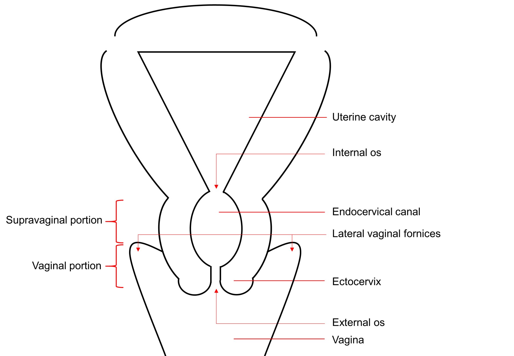
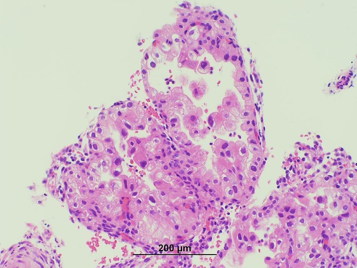
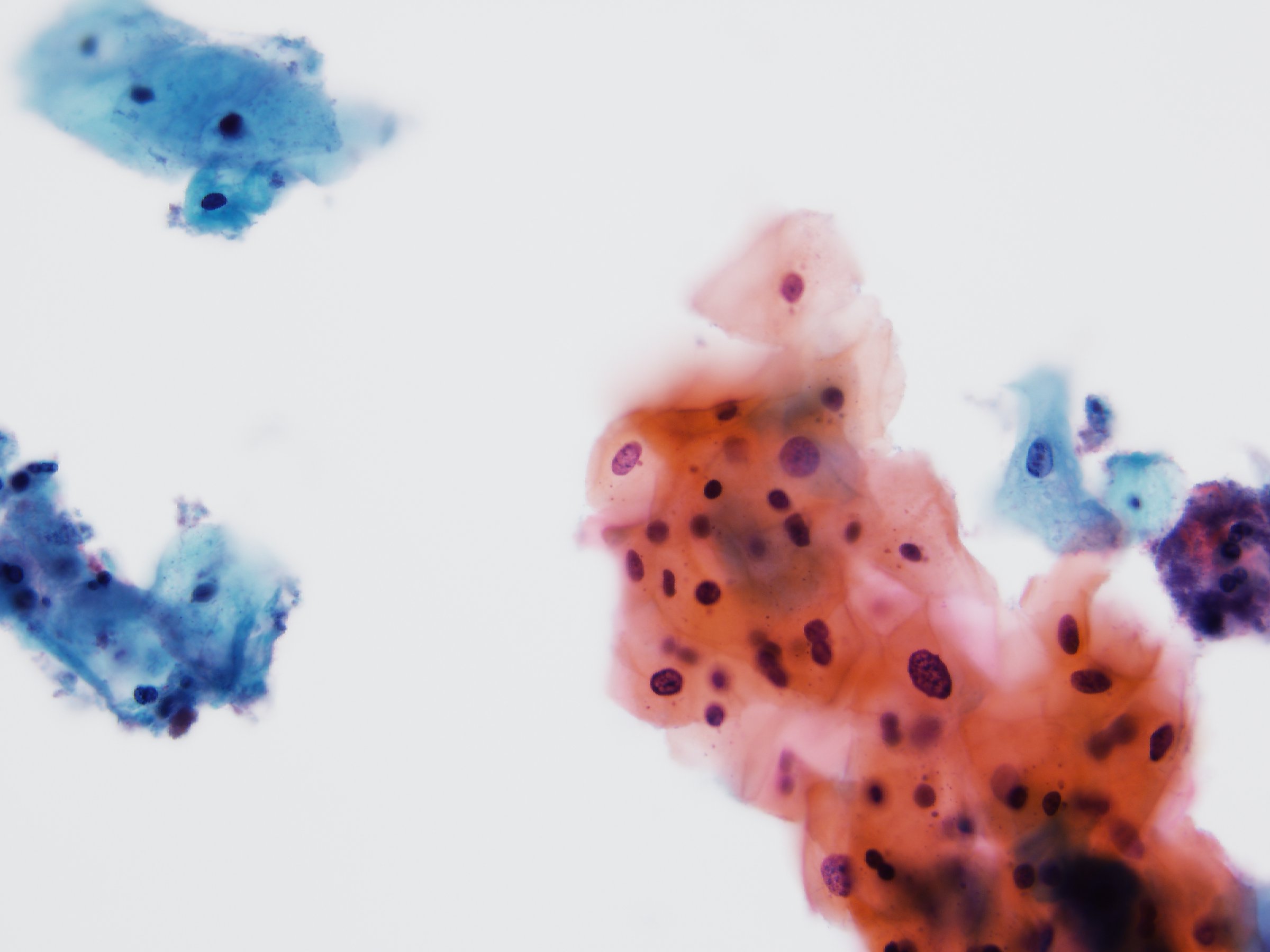
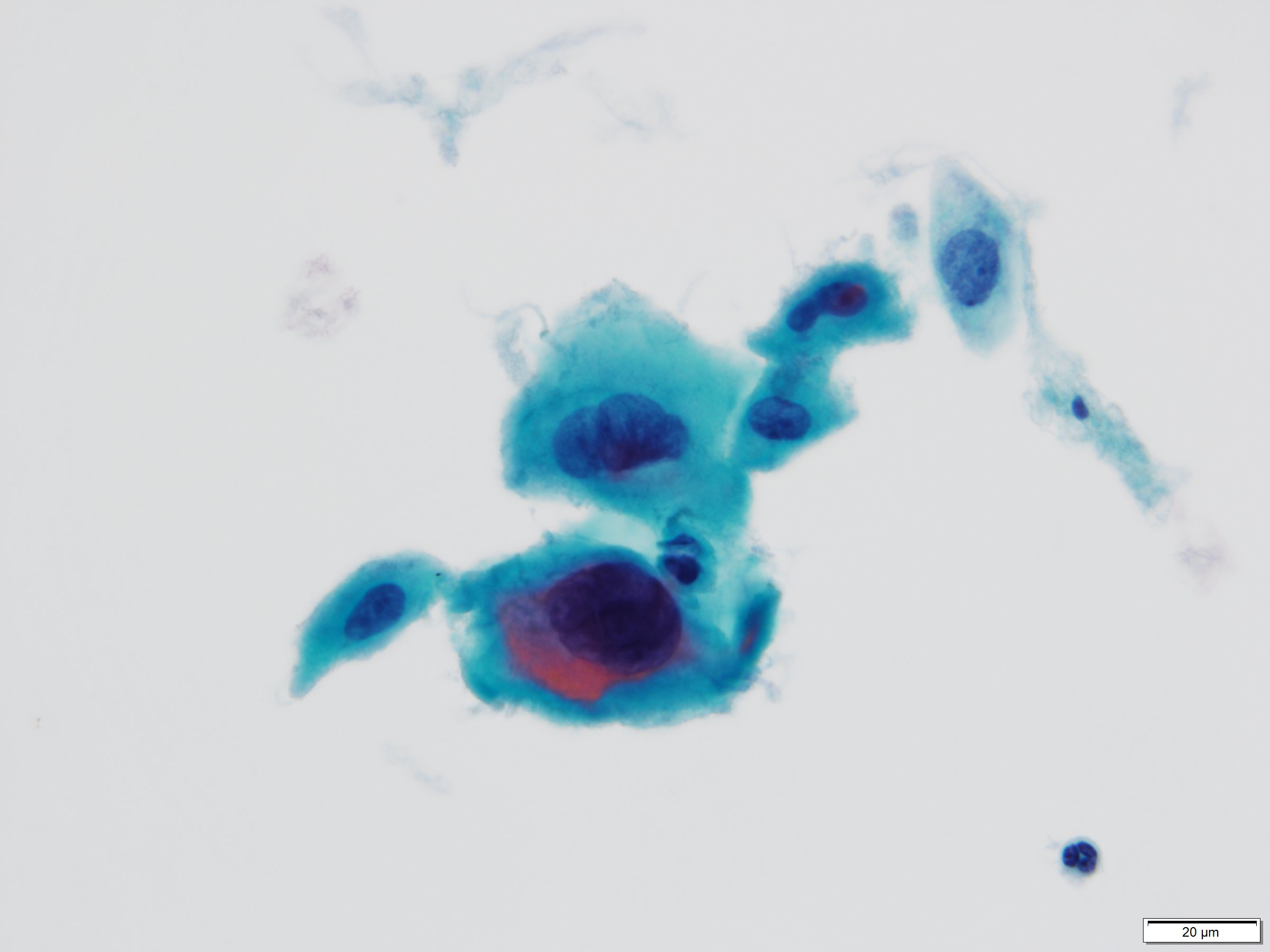
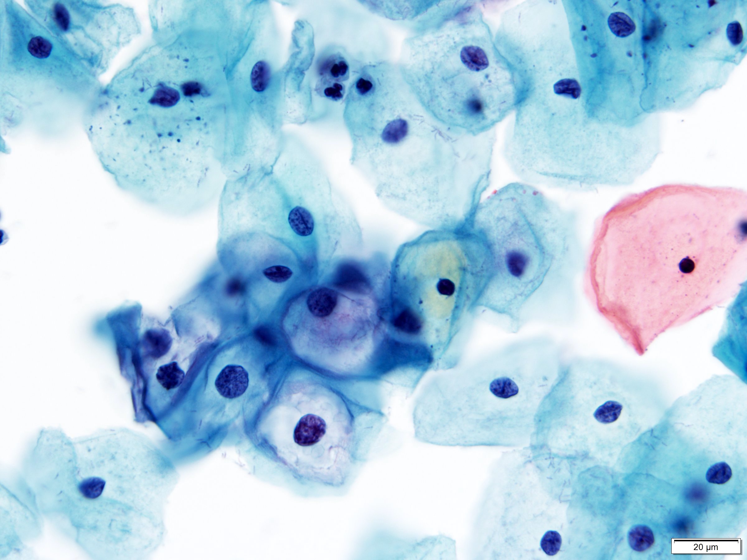
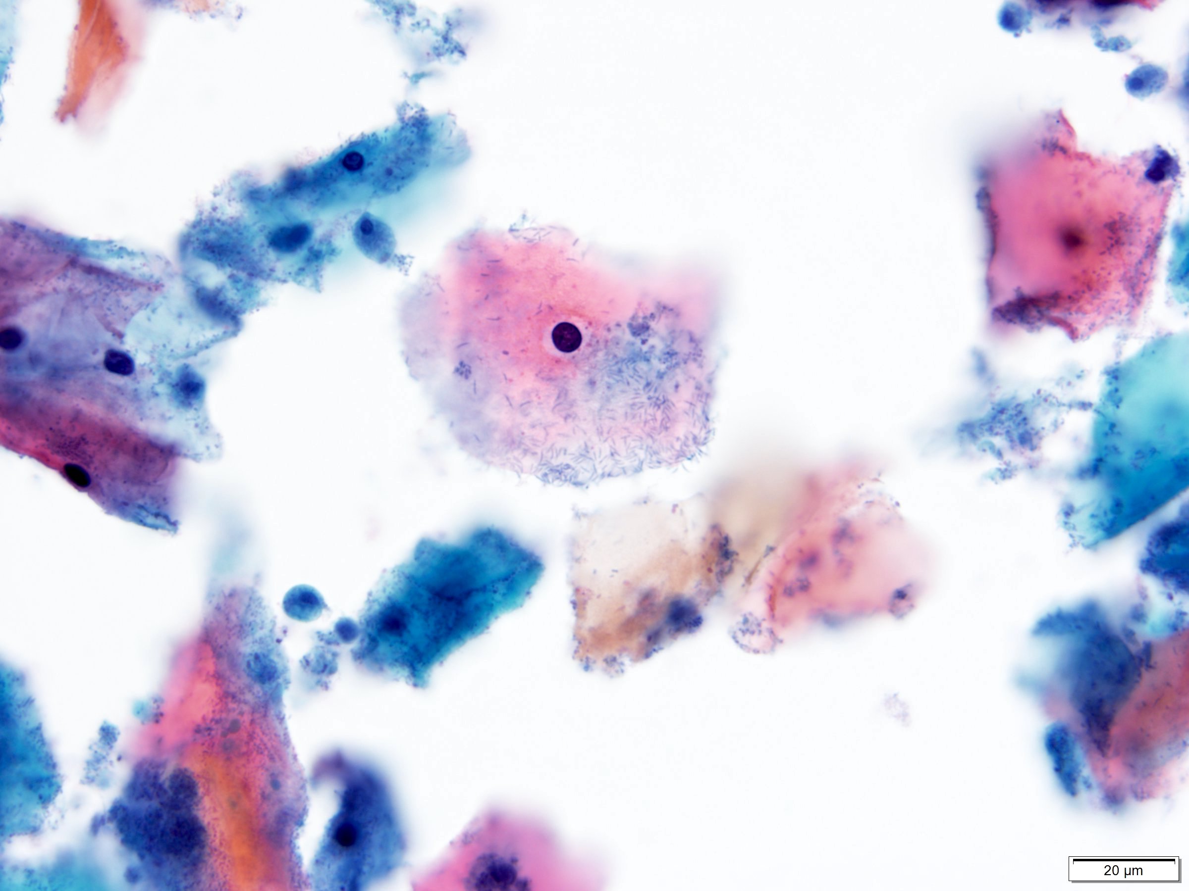
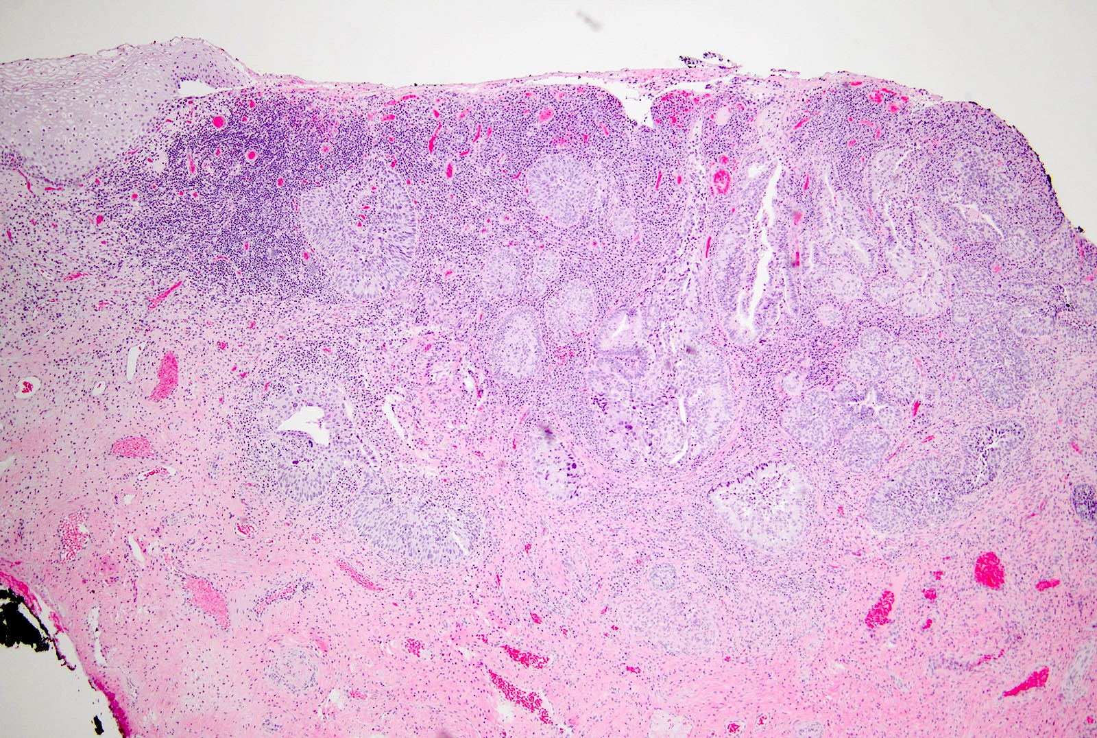
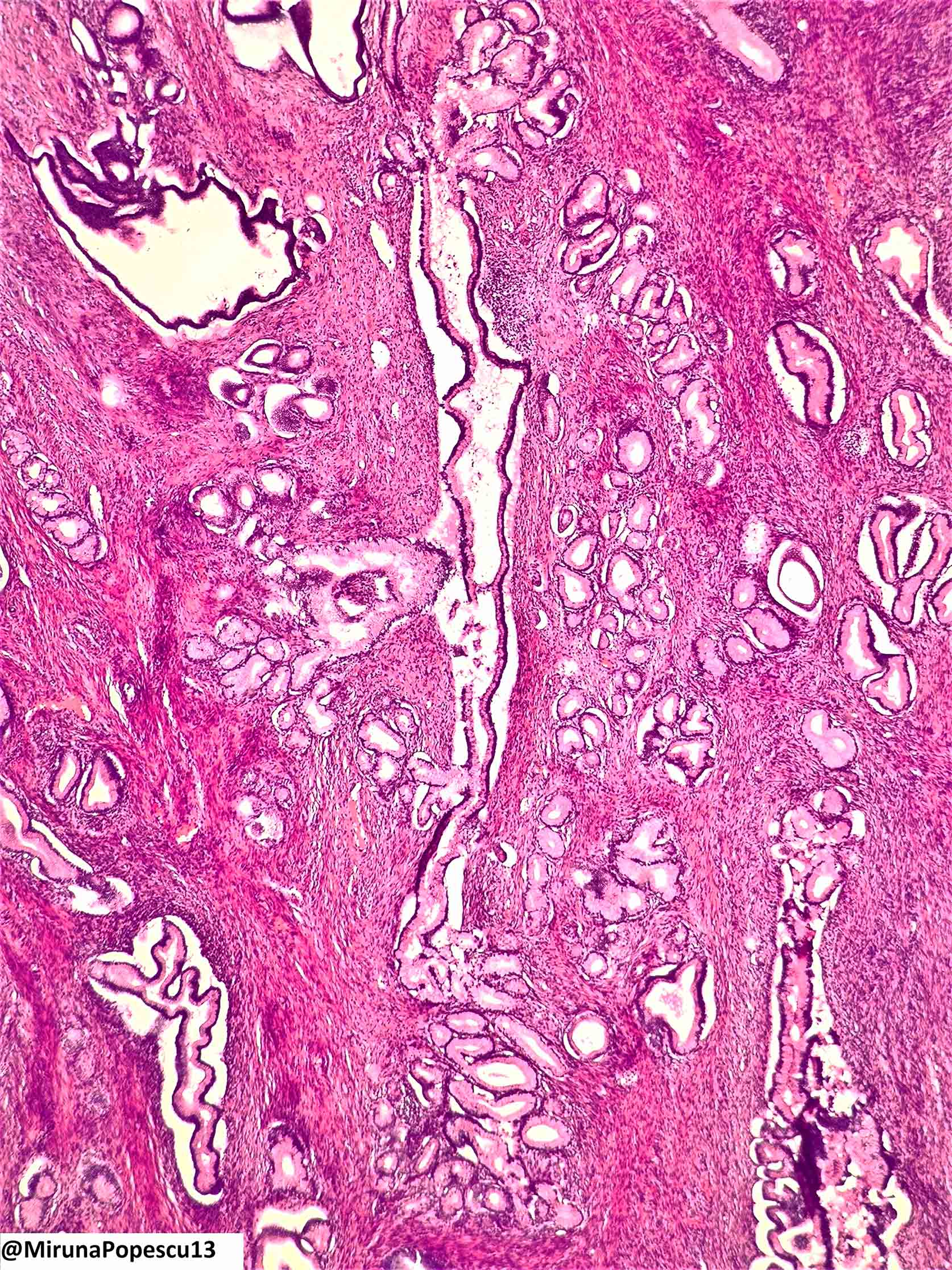
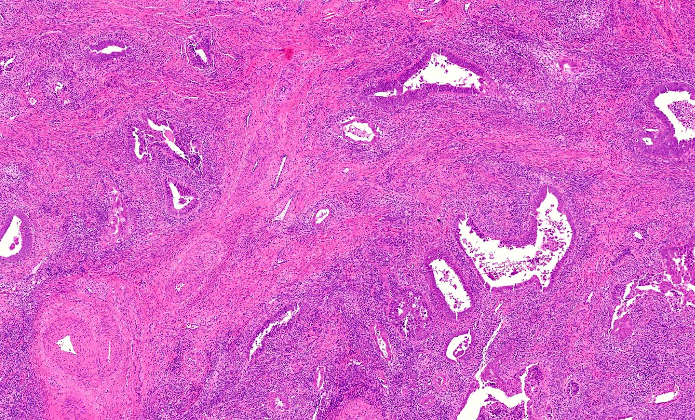
    Primary tumor (pT) and FIGO stage
    • pTX: primary tumor cannot be assessed
    • pT0: no evidence of primary tumor
    • pT1: cervical carcinoma confined to uterus (extension to corpus should be disregarded)
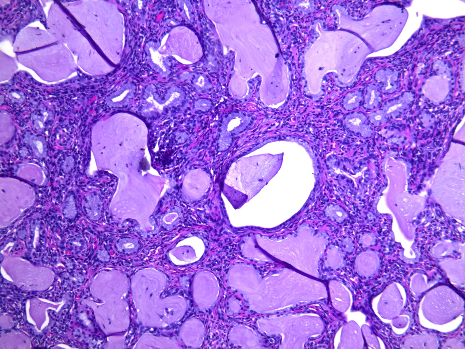
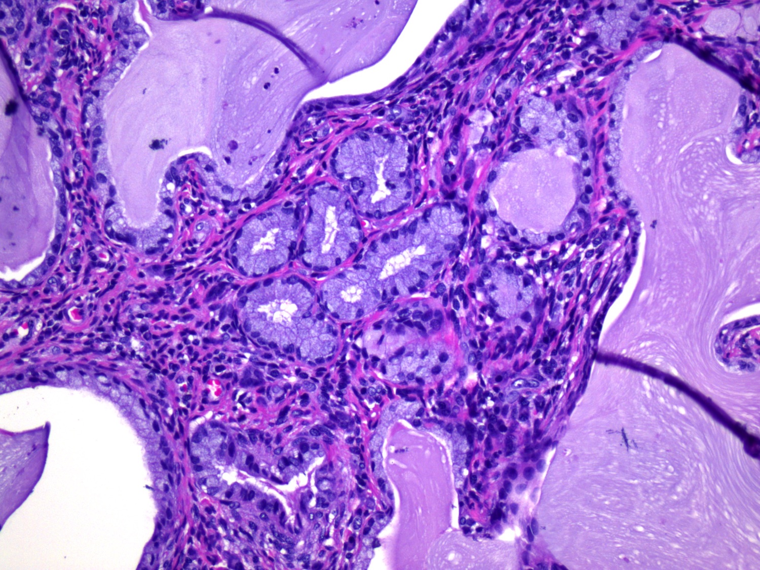
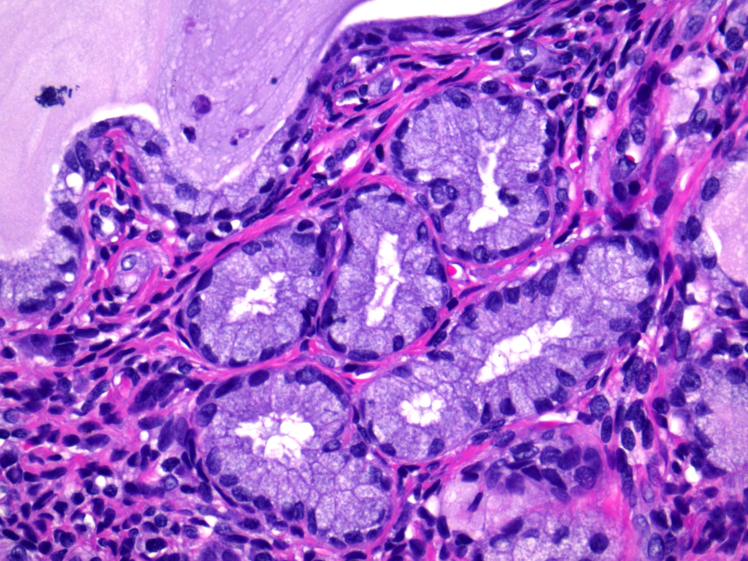
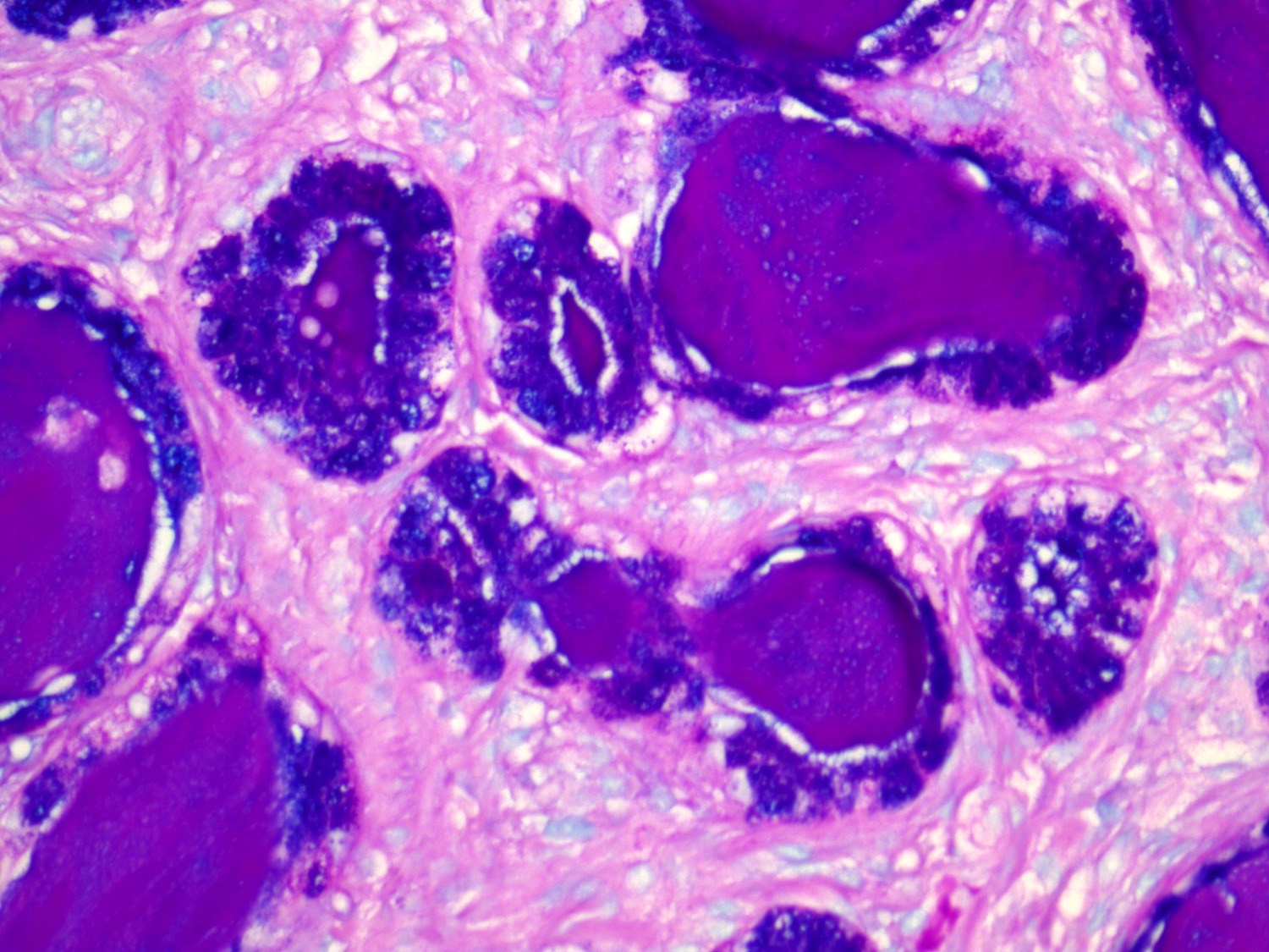
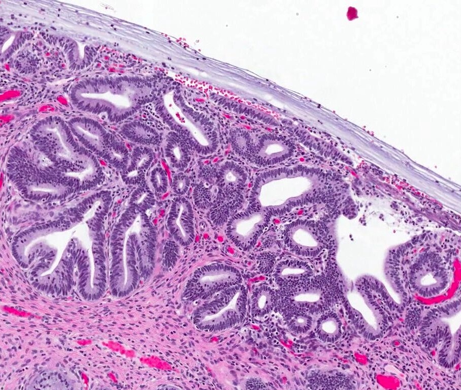
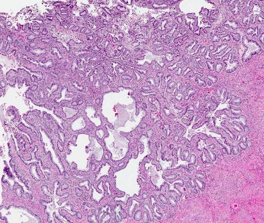
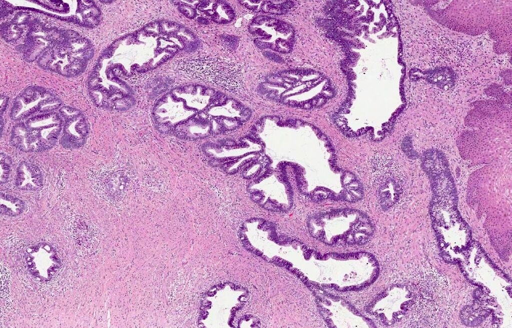
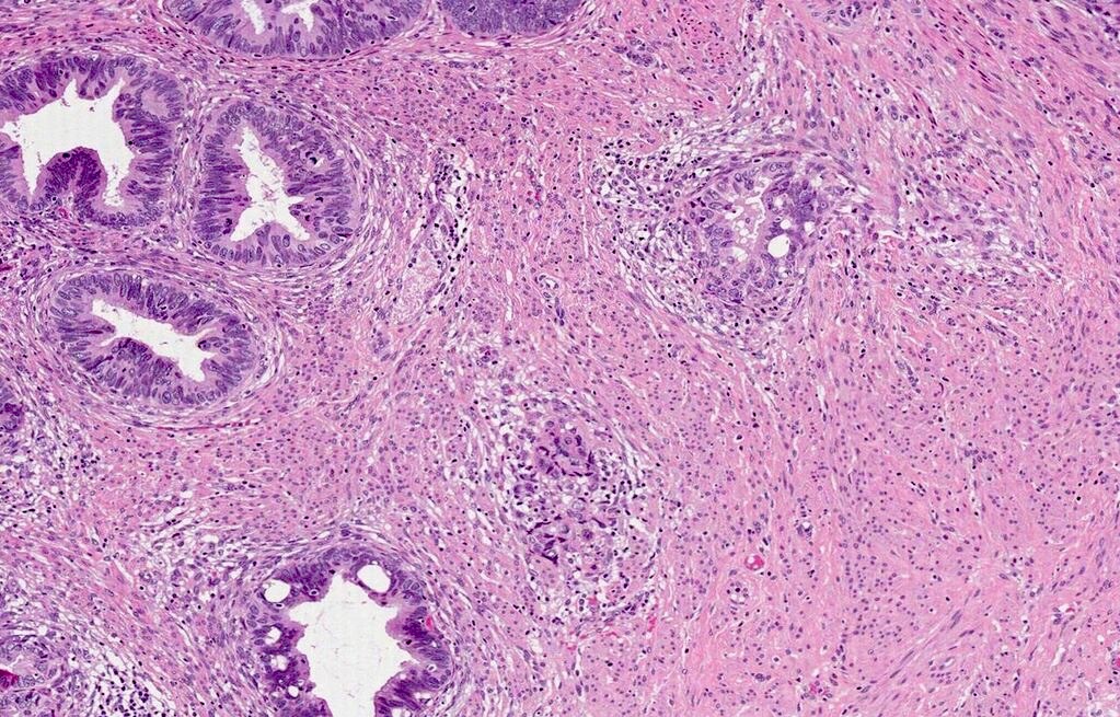
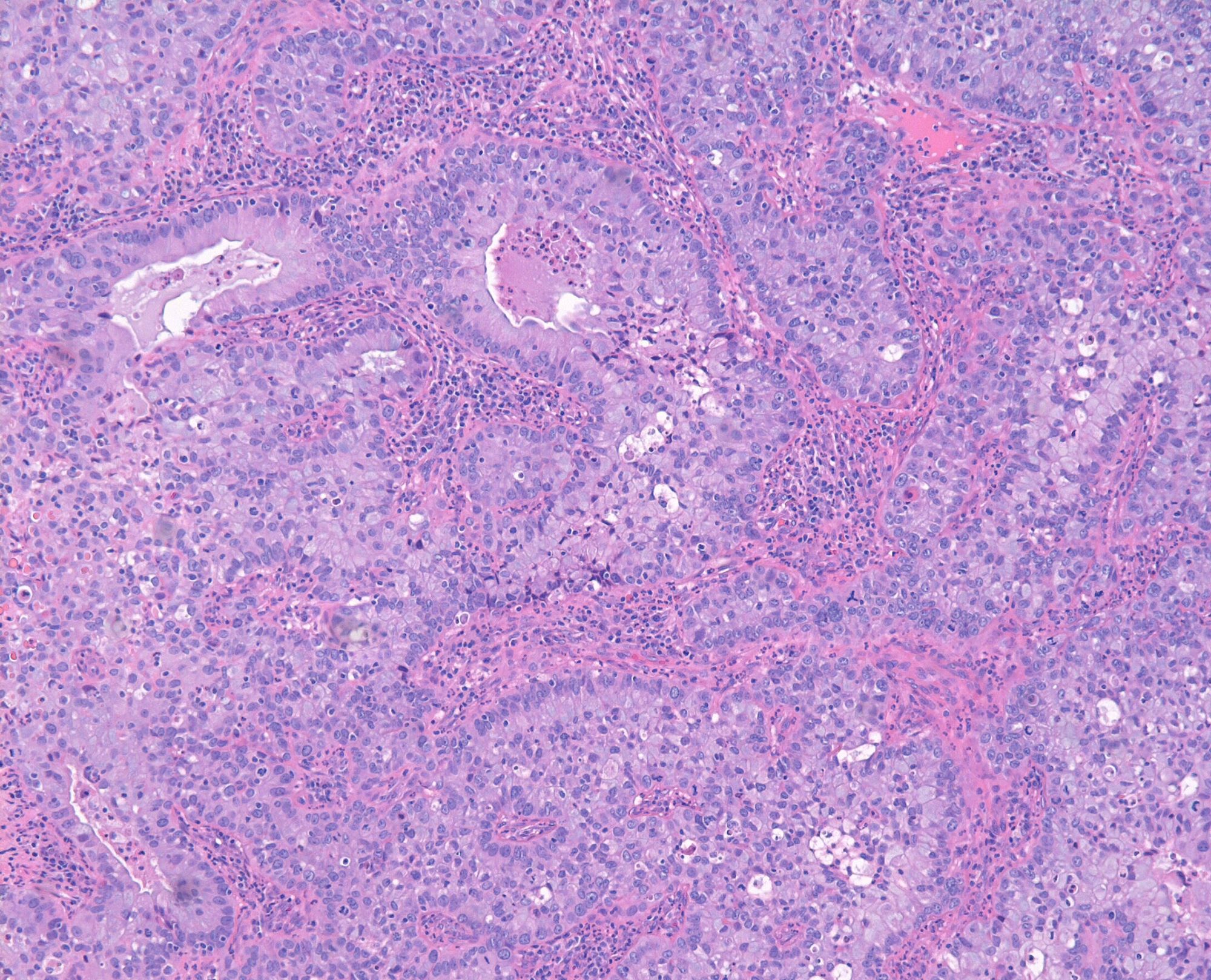
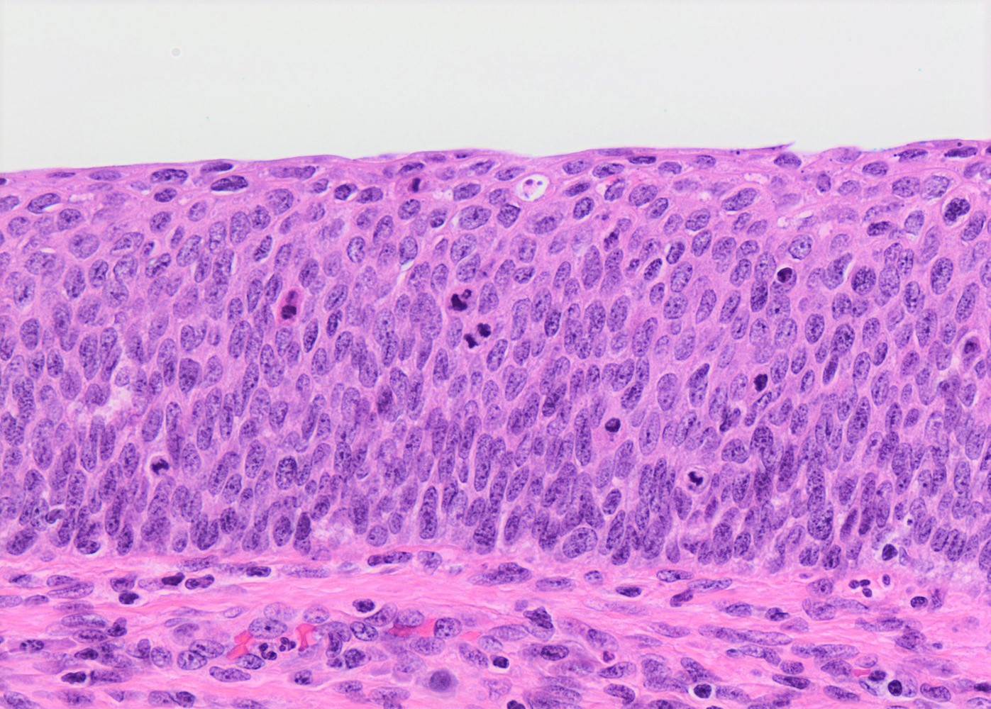
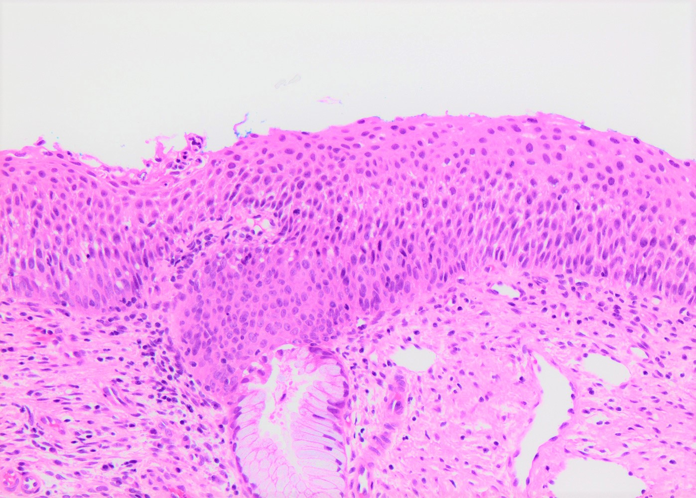
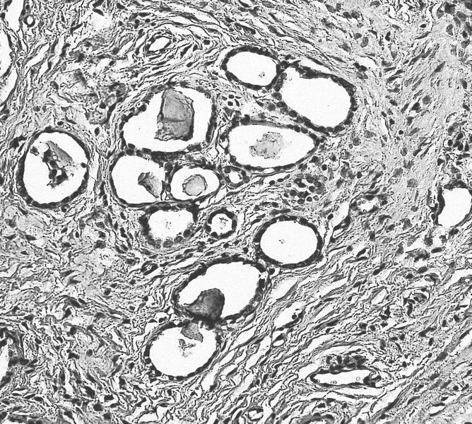
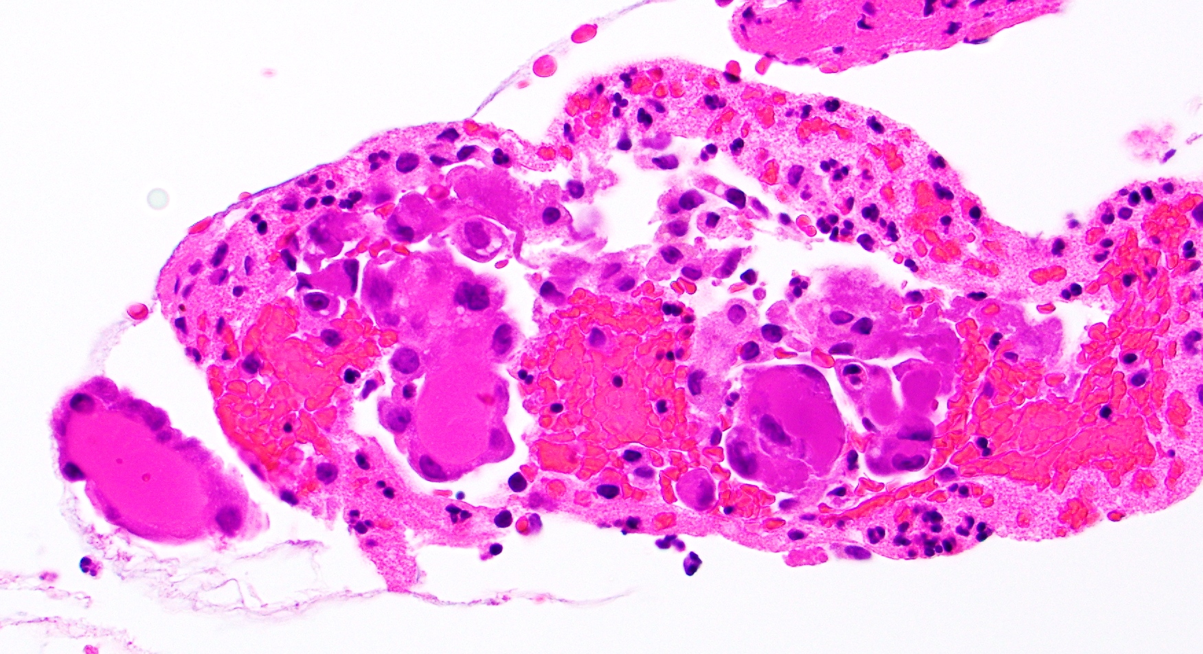
      • pT1a: invasive carcinoma diagnosed by microscopy only; stromal invasion with a maximum depth of 5.0 mm measured from the base of the epithelium and a horizontal spread of 7.0 mm or less; vascular space involvement, venous or lymphatic, does not affect classification
        • pT1a1: measured stromal invasion of 3.0 mm or less in depth and 7.0 mm or less in horizontal spread
        • pT1a2: measured stromal invasion of more than 3.0 mm and not more than 5.0 mm, with a horizontal spread of 7.0 mm or less
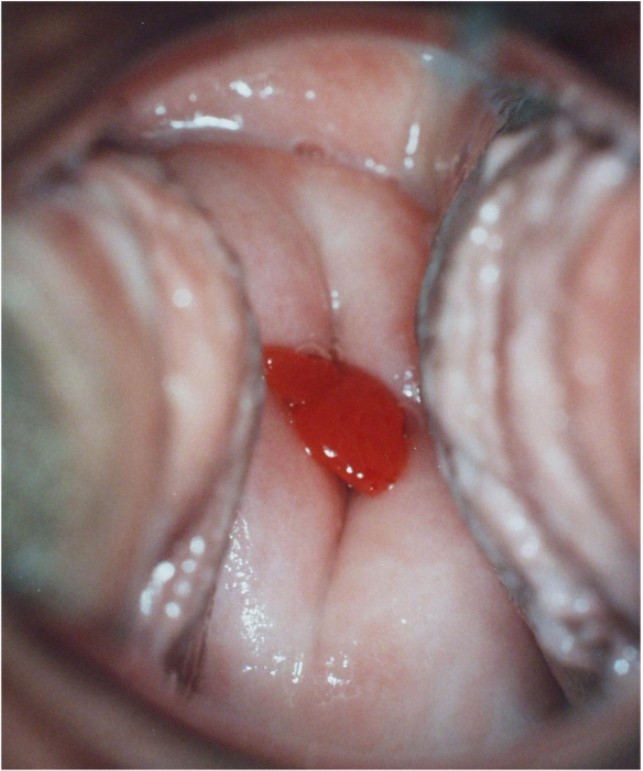
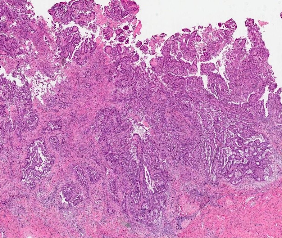
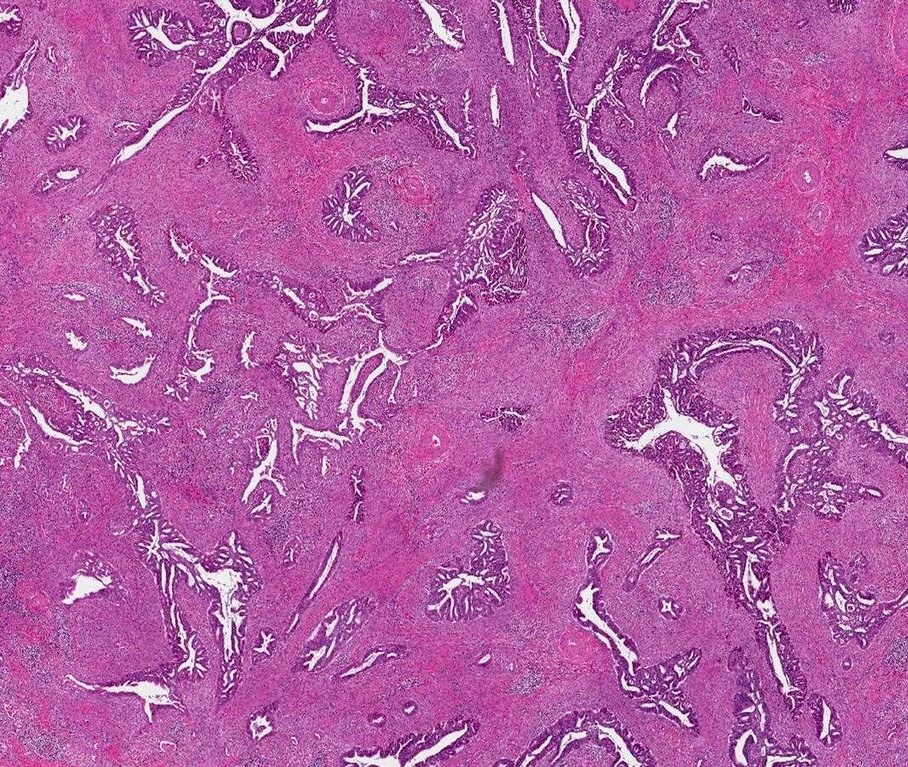
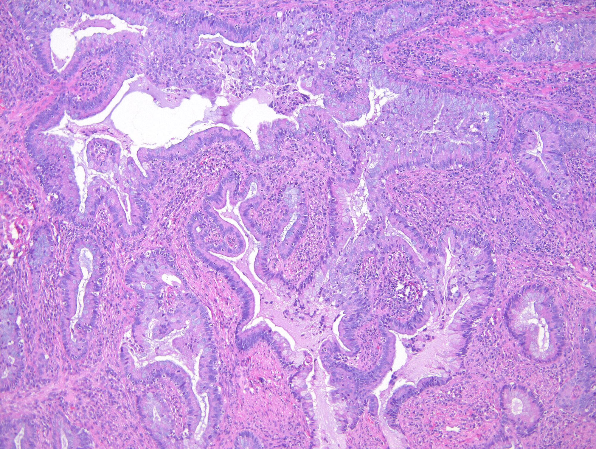
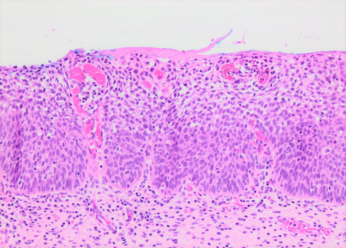
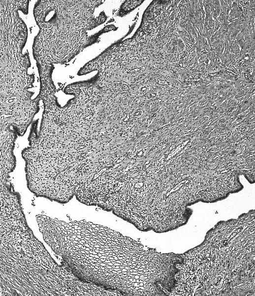
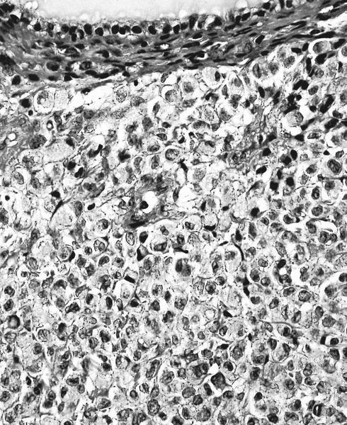
      • pT1b: clinically visible lesion confined to the cervix or microscopic lesion greater than pT1a2 / IA2; includes all macroscopically visible lesions, even those with superficial invasion
        • pT1b1: clinically visible lesion 4.0 cm or less in greatest dimension
        • pT1b2: clinically visible lesion more than 4.0 cm in greatest dimension
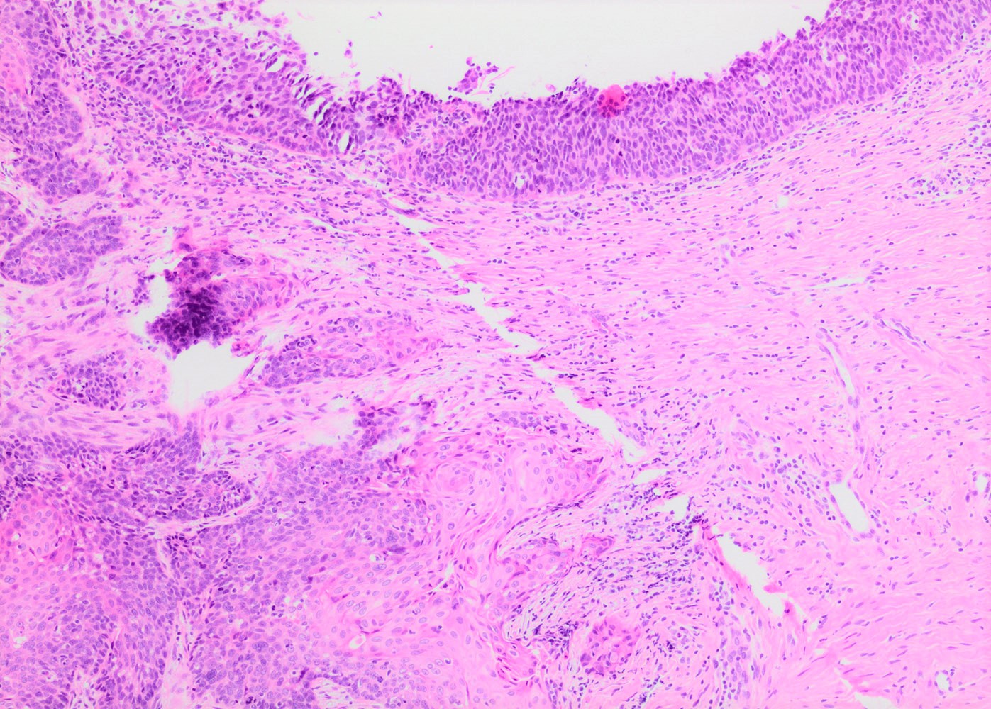
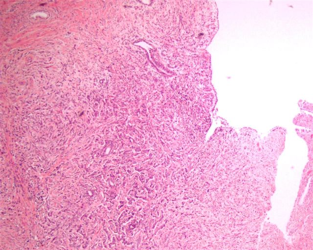
    • pT2: cervical carcinoma invading beyond the uterus but not to the pelvic wall or to the lower third of the vagina
      • pT2a: tumor without parametrial invasion
        • pT2a1: clinically visible lesion 4.0 cm or less in greatest dimension
        • pT2a2: clinically visible lesion more than 4.0 cm in greatest dimension
      • pT2b: tumor with parametrial invasion
    • pT3: tumor extending to the pelvic sidewall or involving the lower third of the vagina or causing hydronephrosis or nonfunctioning kidney
      • pT3a: tumor involving the lower third of the vagina but not extending to the pelvic wall
      • pT3b: tumor extending to the pelvic wall or causing hydronephrosis or nonfunctioning kidney
    • pT4: tumor invading the mucosa of the bladder or rectum or extending beyond the true pelvis (bullous edema is not sufficient to classify a tumor as pT4)
    • Reference: Amin: AJCC Cancer Staging Manual, 8th Edition, 2017

    Notes:
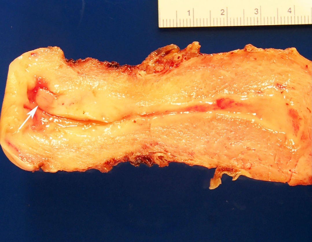
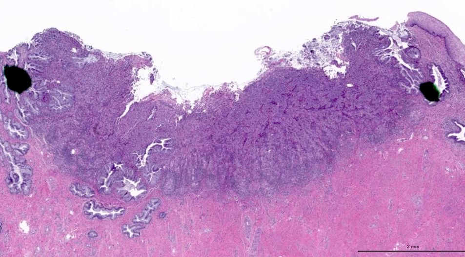
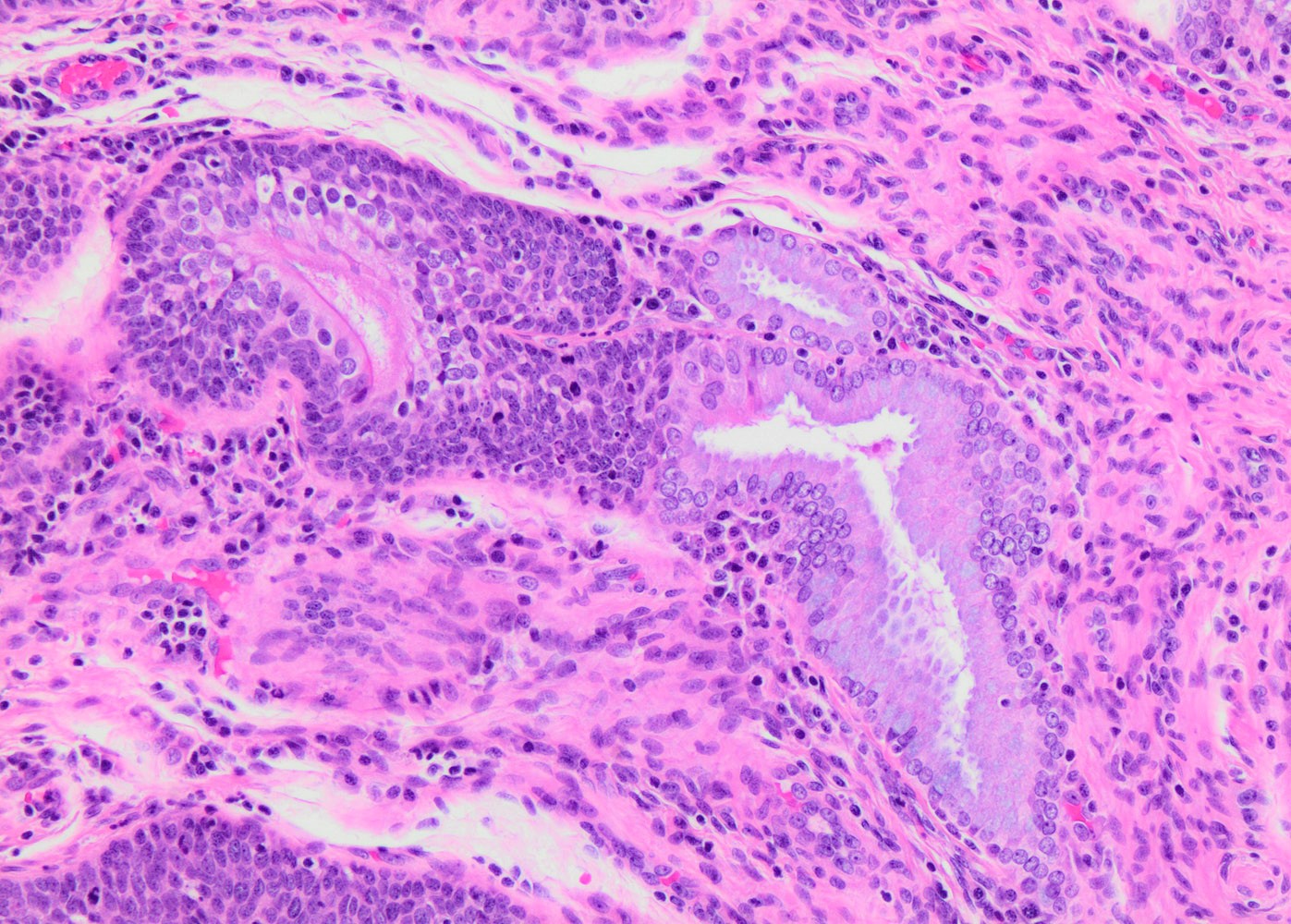
    • Stromal invasion needs to be documented in millimeters for tumors that cannot be measured grossly
      • This step is not necessary in larger tumors that can be measured grossly
    • All macroscopically visible lesions - even with only superficial invasion - are at least FIGO IB
    • Pelvic sidewall is defined as the muscle, fascia, neurovascular structures and skeletal portions of the bony pelvis; on rectal examination, there is no cancer free space between the tumor and pelvic sidewall
    Regional lymph nodes (pN)
    • pNX: regional lymph nodes cannot be assessed
    • pN0: no regional lymph node metastasis
    • pN0(i+): isolated tumor cells in regional lymph node(s) no greater than 0.2 mm
    • pN1: any metastasis greater than 0.2 mm (per the 8th edition)

    Notes:
    • Modifier for regional lymph nodes:
      • + (sn)
      • + (sn)(i-)
      • + (sn)(i+)
    • Size criteria for nodal metastatic diseases is adopted from breast carcinoma staging
      • Isolated tumor cells (ITCs): single cells or small clusters of cells not more than 0.2 mm in greatest dimension
    Distant metastasis (pM)
    • pM0: no distant metastasis
    • pM1: distant metastasis (including peritoneal spread, involvement of supraclavicular, mediastinal or paraaortic lymph nodes, lung, liver or bone)
    Prefixes
    • m: multiple primary tumors
    • r: recurrent
    • y: posttreatment
    Board review style question #1
    According to the latest FIGO staging system, which of the following prognostic variables affects the staging of patients with cervical cancer?

    1. Horizontal tumor extent
    2. Lymphovascular space invasion
    3. Multifocal disease
    4. Depth of invasion
    5. Margin status
    Board review style answer #1
    D. Depth of invasion. All other variables are important for reporting purposes but are not part of the FIGO staging criteria.

    Comment Here

    Reference: Cervix - Staging

    Traumatic neuroma
    Definition / general
    • Reparative lesion at site of traumatic injury of peripheral nerves
    • Interruption in continuity of nerve causes wallerian degeneration (loss of axons in proximal stump and retraction of axons in distal segment), then exuberant regeneration of nerve and formation of mass of Schwann cells, axons and fibrous cells
    • Rare complication of cone biopsy (Arch Pathol Lab Med 1989;113:945)
    • Microneuromas present in 55% of hysterectomy patients, associated with childbirth (Histopathology 1996;28:153)
    Gross description
    • Irregular gray area up to 2 cm near cone biopsy margin or scar
    Microscopic (histologic) description
    • Haphazard nerves within mature collagenous scar with entrapped smooth muscle
    Positive stains

    Trichomonas vaginalis
    Definition / general
    • Trichomonas vaginalis is a primitive eukaryotic organism, a parasitic protozoan that causes trichomoniasis, which is a sexually transmitted disease
      • Sometimes accompanied by Leptothrix (a nonpathogenic, long, filamentous bacterium)
    Essential features
    • Pear shaped primitive eukaryotic organism, a parasitic protozoan
    • Primarily a disease of woman, though it also occurs in men
    • Clusters of the organisms are colloquially called trich parties
    Terminology
    • Trichomonas vaginalis
    Epidemiology
    • Trichomoniasis is the most prevalent nonviral sexually transmitted infection in the United States, affecting an estimated 3.7 million persons (CDC: Trichomoniasis [Accessed 29 July 2022])
    • Having multiple sexual partners is the primary risk factor
    • Mainly affects women from ages 16 - 35 years but can occur in postmenopausal women
    • Trichomonas vaginalis infection is strongly associated with an increased risk of HIV acquisition and transmission, particularly among women (Sex Transm Dis 2016;43:617)
    Sites
    • Female: lower genital tract (vulva, vagina, cervix or urethra)
    • Male: urethra, epididymis and prostate
    Pathophysiology
    • Unclear why some people with the infection get symptoms while others do not
    • Likely depends on factors such as a person's age and overall health
    Etiology
    • Sexually active people can get trichomoniasis by having sex without a condom with a partner who has trichomoniasis infection
    Diagrams / tables

    Images hosted on other servers:

    Trophozoite

    Diagnosis
    • Nucleic acid amplification test (NAAT) is the gold standard for diagnosis of trichomoniasis (Can J Infect Dis Med Microbiol 2005;16:35)
    • OSOM Trichomonas rapid test: immunochromatographic test that detects pathogen antigens from vaginal swab
    • DNA hybridization probe test
    • Direct microscopic examination of secretions - wet mount
    • Culture was considered as a gold standard before the availability of molecular tests
    Prognostic factors
    • Person's age and overall health
    • People with trichomoniasis can pass the infection to others, even if they do not have symptoms
    Case reports
    Treatment
    Prevention
    • Condoms can reduce the risk of getting trichomoniasis if used the right way every single time
    Clinical images

    Images hosted on other servers:
    Missing Image

    Strawberry cervix

    Cytology description
    • Organism is a 15 - 30 μm, pear shaped protozoan
    • Nucleus is small, very pale, eccentrically placed
    • Cytoplasm often contains tiny red granules
    • Clusters of the organisms are colloquially called trich parties
    • Sometimes accompanied by Leptothrix, a nonpathogenic, long, filamentous bacterium (Bibbo: Comprehensive Cytopathology, 4th Edition, 2014)
    • Trichomonas and Leptothrix together have been referred to as spaghetti and meatballs
    • Leptotrichia and Trichomonas were observed together in 95% of cases in a study of 1,000 cases (Clin Microbiol Rev 1998;11:341)
    • 3 dimensional clusters of neutrophils (poly balls) may be seen in the background
    • Numerous lymphocytes and many mast cells may be seen
    Cytology images

    Contributed by Soumya Jaladi, M.B.B.S., Marilin Rosa, M.D. and @zaalruwai83 on Twitter

    Trichomonas in a cytologic preparation

    Trichomonas


    Trichomonas vaginalis Trichomonas vaginalis

    Trichomonas vaginalis

    Trichomonas vaginalis Trichomonas vaginalis

    Trichomonas vaginalis


    Images hosted on other servers:

    Filamentous Leptothrix species

    Trichomonas in wet mount

    Trichomonas in conventional
    Pap smear

    Trichomonas vaginalis with Leptothrix

    Trichomonas


    Trophozite

    Inflammatory ectocervix with Trichomonas infection

    Electron microscopy images

    Images hosted on other servers:
    Missing Image

    T. vaginalis parasite

    Sample pathology report
    • Cervix, smear:
      • Squamous cells with background inflammatory cells and pear shaped organisms with eccentric nuclei, consistent with Trichomonas
    Differential diagnosis
    • Cell fragments, cytoplasmic debris, bare epithelial nuclei, small mucus aggregates and leukocytes:
      • Identification of a definite elliptical nucleus helps avoid misinterpretation
      • Presence of eosinophilic cytoplasmic granules will be helpful
      • In most cases, Trichomonas organisms are plentiful (trich party)
    Board review style question #1

    What are the organisms frequently identified in cytologic smears in patients with Trichomonas infections?

    1. Candida
    2. Herpes
    3. Leptothrix
    4. Sporothrix
    Board review style answer #1
    C. Leptothrix

    Comment Here

    Reference: Trichomonas vaginalis
    Board review style question #2
    What is the gold standard test for diagnosis of trichomoniasis?

    1. DNA hybridization probe test
    2. Immunochromatographic test
    3. Nucleic acid amplification test (NAAT)
    4. Wet mount microscopic examination
    Board review style answer #2
    C. Nucleic acid amplification test (NAAT)

    Comment Here

    Reference: Trichomonas vaginalis

    Tuboendometrioid metaplasia
    Definition / general
    • Common (1/3 of women)
    • In upper portion of endocervical canal, often in deep glands (Int J Gynecol Pathol 1992;11:89)
    • Often seen after cervical cone biopsy
    • May represent response to injury
    • Tubal epithelium is invariably seen at the cervix - isthmus junction and is not considered a metaplastic process at that site (Int J Gynecol Pathol 2013;32:122)
    Microscopic (histologic) description
    • Endocervix contains:
      • Ciliated cells (clear cytoplasm, abundant apical cilia and large, oval, variably hyperchromatic nuclei)
      • Secretory cells (nonciliated with dark eosinophilic or basophilic cytoplasm, apical cytoplasmic protrusions but no mucin vacuoles, basal nuclei)
      • Intercalated cells (also called peg cells, scant cytoplasm, thin and long nuclei), as found in normal fallopian tube
    • Glands are regular
    • Minimal mitotic activity, rare crowding or atypia
    • Associated with endometrial type cells
    • Usually near squamocolumnar junction, usually no inflammation
    • May have cystic glands and periglandular stromal alterations suggestive of premalignant conditions or deep glands with periglandular edema suggestive of well differentiated adenocarcinoma but cells are ciliated with bland cytology, no mitotic figures, no definite desmoplastic stroma (Am J Clin Pathol 1995;103:618)
    Microscopic (histologic) images

    Contributed by Carlos Parra-Herran, M.D.

    TEM and relevant stains

    Positive stains
    Negative staining - disease
    • p16 is considered to be negative or only focally positive or show a mosaic pattern in tubal metaplasia (Adv Anat Pathol 2006;13:8, Cell Oncol 2007;29:37), useful to differentiate from high grade CIN and invasive cervical carcinomas that are diffusely positive
    Cytology description
    Differential diagnosis
    Additional references

    Tunnel clusters
    Definition / general
    • Lobular aggregates of benign endocervical glands in the cervical wall
    Essential features
    • Incidental finding
    • Benign proliferation of endocervical glands with lobular configuration
    • 2 types (A and B) have been described:
      • Type A tunnel clusters are composed of small noncystic glands and may show gastric metaplasia in up to 15% of cases
      • Type B tunnel clusters are composed of cystically dilated glands
    Terminology
    • No synonyms
    ICD coding
    • ICD-O: Not applicable
    • ICD-10: Not applicable
    • ICD-11: Not applicable
    Epidemiology
    Sites
    • Endocervix
    Pathophysiology
    Etiology
    • Unclear
    Clinical features
    • Typically asymptomatic
    • May be associated with mucoid discharge
    • Rarely visible on colposcopic examination
    Diagnosis
    • Histologic examination
    Prognostic factors
    • Excellent prognosis
    • No risk of recurrence or malignant transformation
    Treatment
    • None
    Gross description
    • Tunnel A clusters are often grossly unremarkable
    • Tunnel B clusters show a grossly visible, lobular mass lesion in 40% of cases and multiple lesions in 80% of cases
    • Rarely cervical wall expansion
    Microscopic (histologic) description
    • Well demarcated, rounded, lobular proliferation of closely packed tubules of varying size lined by endocervical glandular epithelium
    • No desmoplastic or inflammatory stromal response
    • May be associated with Nabothian cysts
    • Usually found close to endocervical surface epithelium
    • Type A:
      • Small elongated noncystic glands lined by columnar to low cuboidal cells with basally located nuclei and apical mucinous cytoplasm
      • Mild cytologic atypia may be present with pseudostratification, nuclear enlargement, hyperchromasia, vesicular chromatin or prominent nucleoli (Am J Surg Pathol 1996;20:1312)
      • Minimal or no mitotic activity
      • Gastric metaplasia in up to 15% (Histopathology 2007;50:843)
    • Type B (B for big):
      • Cystically dilated glands containing inspissated mucin and lined by cuboidal or flattened epithelium
      • The lining cells are cytologically bland with ovoid nuclei lacking mitotic activity
    • Type A and type B tunnel clusters may be admixed
    Microscopic (histologic) images

    Contributed by Gulisa Turashvili, M.D., Ph.D.

    Type A tunnel clusters

    Alcian blue / PAS in type A tunnel clusters


    Type B tunnel clusters

    Alcian blue / PAS in type B tunnel clusters

    Virtual slides

    Images hosted on other servers:

    Tunnel clusters and deep Nabothian cysts

    Alcian blue / PAS in tunnel clusters and Nabothian cysts

    Tunnel clusters

    Cytology description
    • No specific cytologic features
    • Benign endocervical glands
    Positive stains
    Negative stains
    Sample pathology report
    • Tunnel clusters have no clinical relevance and do not need to be mentioned in the pathology report
    Differential diagnosis
    • Minimal deviation adenocarcinoma
      • Lacks lobular configuration
      • Composed of irregular branching glands often with deep extension in cervical wall
      • At least focal stromal desmoplasia and cytologic atypia
      • Negative for PAX2
    • Usual type endocervical adenocarcinoma
      • Shows infiltrative growth with cytologic atypia, apical mitotic figures and apoptotic bodies, often with stromal desmoplastic reaction
      • Diffusely positive for p16
    • Adenocarcinoma in situ
      • Demonstrates cytologic atypia with pseudostratified hyperchromatic nuclei, apical mitotic figures and apoptotic bodies
      • Diffusely positive for p16
    • Nabothian cysts
      • Shows large mucin filled cysts without lobular configuration
    • Mesonephric remnants / hyperplasia
      • May extend deeply in cervical wall
      • Glands often contain densely eosinophilic intraluminal secretions and are lined by cuboidal cells lacking intracytoplasmic mucin
      • Positive for GATA3 or TTF1
    Board review style question #1


    Which of the following immunoprofiles would be expected in type A tunnel clusters (pictured above)?

    1. Positive for PAX2, p16 and CEA, Ki67 > 10%
    2. Positive for PAX2, p16 and CEA, Ki67 > 10%, variable HIK1083
    3. Positive for PAX2, negative p16 and CEA, Ki67 < 1%, variable HIK1083
    4. Negative for PAX2, p16 and CEA, Ki67 < 1%
    Board review style answer #1
    C. Positive for PAX2, negative p16 and CEA, Ki67 < 1%, variable HIK1083. Type A tunnel clusters are positive for PAX2 and negative or focally positive for p16 and CEA. The Ki67 labeling index is low. HIK1083 may also be positive if gastric metaplasia is present.

    Comment Here

    Reference: Tunnel clusters
    Board review style question #2


    Which of the following immunoprofiles would be expected in type B tunnel clusters (pictured above)?

    1. Positive for PAX2, p16 and CEA, negative for HIK1083, Ki67 > 10%
    2. Positive for PAX2 and HIK1083, negative for p16 and CEA, Ki67 > 10%
    3. Positive for HIK1083, negative for PAX2, p16 and CEA, Ki67 < 1%
    4. Positive for PAX2, negative for p16, CEA and HIK1083, Ki67 < 1%
    Board review style answer #2
    D. Positive for PAX2, negative for p16, CEA and HIK1083, Ki67 < 1%. Type B tunnel clusters are positive for PAX2 and negative or focally positive for p16 and CEA. The Ki67 labeling index is low. HIK1083 is negative.

    Comment Here

    Reference: Tunnel clusters

    Vasculitis
    Definition / general
    • Vasculitis of any type affecting the female genital tract is usually an isolated finding (only 10% have systemic disease, Int J Gynecol Pathol 2000;19:258)
    • Isolated polyarteritis nodosa of female genital tract is rare, either giant cell type in postmenopausal women in any part of female genital tract or PAN type in younger women affecting cervix (Mod Pathol 1994;7:610)
    Case reports
    • 44 year old woman with previous cervical biopsies showing CIN II - III (Case #91)
    Microscopic (histologic) images

    Case #91
    Missing Image

    Inflammatory infiltrate in the vessel wall

    Missing Image

    Lymphocytes, neutrophils and occasional eosinophils

    Missing Image

    Fibrinoid necrosis within the media

    Additional references

    WHO classification
    Definition / general
    • The classification below is the updated classification of tumors of the cervix as per the World Health Organization classification of tumors of female reproductive organs, 5th edition (2020)
    Major updates
    • Squamous lesions:
      • Invasive squamous lesions are now classified in 2 main categories: HPV associated and HPV independent
      • HPV independent squamous cell carcinomas are rare but their existence is now acknowledged as they may behave more aggressively than the more common HPV associated lesions
    • Glandular lesions:
      • Both preinvasive and invasive glandular lesions are now classified in 2 main categories: HPV associated and HPV independent
      • HPV associated adenocarcinomas can be classified using terminology from previous classification; however, they need to be distinguished, as a group, from HPV independent tumors
      • HPV independent adenocarcinomas are most frequently of the gastric type (with both in situ and invasive forms)
      • Other distinct types include clear cell and mesonephric carcinomas
    WHO (2020)
    Squamous epithelial tumors
    Glandular tumors and precursors
    Board review style question #1
    According to the latest classification of female genital tumors from the World Health Organization, which is currently a major category in the classification of squamous cell lesions?

    1. Squamous cell carcinoma, HPV associated
    2. Basaloid carcinoma
    3. Ectopic prostatic tissue
    4. Keratinizing squamous cell carcinoma
    5. Adenosquamous carcinoma
    Board review style answer #1
    A. Squamous cell carcinoma, HPV associated. Squamous cell carcinoma of the uterine cervix is now classified as HPV associated and HPV independent. Basaloid and keratinizing are morphologic variants of squamous cell carcinoma but do not represent diagnostic categories. Ectopic prostatic tissue belongs to the category of benign glandular lesions and adenosquamous carcinoma belongs to the category of mixed epithelial tumors.

    Comment Here

    Reference: Cervix - WHO classification
    Back to top
    Recent Cervix Pathology books

    Ali: 2019

    Clement: 2019

    Crum: 2017

    Howitt: 2019

    Hui: 2015

    IARC: 2020

    Malpica: 2015

    Mody: 2022

    Nayar: 2015

    Nucci: 2020

    Nucci: 2023

    Nucci: 2023

    Soslow: 2020

    Vang: 2017

    Zheng: 2019

    Zheng: 2019


    Find related Pathology books: cytopathology, gynecologic, frozen section
    Image 01 Image 02