Superpage
Ovary

Authors: Eman Abdulfatah, M.D., Laura Adhikari, M.D., Hafza Asma Adnan, M.B.B.S., Rouba Ali-Fehmi, M.D., Fatemeh Ghazanfari Amlashi, M.D., Doaa Atwi, M.D., Ayse Ayhan, M.D., Ph.D., Sudeshna Bandyopadhyay, M.D., Jennifer A. Bennett, M.D., Jessica L. Bentz, M.D., Swati Bhardwaj, M.B.B.S., M.D., Aurelia Busca, M.D., Ph.D., Natalia Buza, M.D., Indranil Chakrabarti, M.D., Victoria Collins, M.D., Shahrzad Ehdaivand, M.D., Erna Forgó, M.D., Rochelle Garcia, M.D., Mohiedean Ghofrani, M.D., C. Blake Gilks, M.D., Arun Gopinath, M.D., Nalini Gupta, M.D., Lisa Han, M.D., Krisztina Hanley, M.D., Saroona Haroon, M.B.B.S., Lewis A. Hassell, M.D., Jutta Huvila, M.D., Ph.D., Shahid Islam, M.D., Ph.D., Tamara Kalir, M.D., Ph.D., Mahmoud A. Khalifa, M.D., Ph.D., Felix K.F. Kommoss, M.D., Elisabetta Kuhn, M.D., Ricardo R. Lastra, M.D., Adam Lechner, B.M., Cheng-Han Lee, M.D., Ph.D., Krisztina Lengyel, M.D., Teri A. Longacre, M.D., Lucy Ma, M.D., Rajit Malliah, M.D., Kruti P. Maniar, M.D., Azin Mashayekhi, M.D., M.B.A., Ekaterina Menshikova, M.D., Aysha Mubeen, M.D., Neshat Nilforoushan, M.D., Carlos Parra-Herran, M.D., Nat Pernick, M.D., Jian-Hua Qiao, M.D., Jack Reid, M.D., M. Carolina Reyes, M.D., Nicole D. Riddle, M.D., Catherine J. Roe, M.D., Ozlen Saglam, M.D., Carolina Sforza, M.D., Aarti Sharma, M.D., Dong Ping Shi, M.D., Jamie Shutter, M.D., Rayan Sibira, M.D., Kamaljeet Singh, M.D., Stephanie L. Skala, M.D., Sharon Song, M.D., M.S., Basile Tessier-Cloutier, M.D., Gulisa Turashvili, M.D., Ph.D., Brandon Umphress, M.D., Russell Vang, M.D., Leena Varughese, M.D., Laura J. Vidis, D.O., Shannon Mingo Welter, M.D., Y. Albert Yeh, M.D., Ph.D., Shabnam Zarei, M.D.
Editorial Board Members: Jennifer A. Bennett, M.D., David B. Chapel, M.D., Kyle Devins, M.D., C. Blake Gilks, M.D., Ricardo R. Lastra, M.D., Lucy Ma, M.D., Carlos Parra-Herran, M.D., Stephanie L. Skala, M.D., Gulisa Turashvili, M.D., Ph.D.
Deputy Editors-in-Chief: Jennifer A. Bennett, M.D., Gulisa Turashvili, M.D., Ph.D.
Editor-in-Chief: Debra L. Zynger, M.D.

Copyright: 2002-2024, PathologyOutlines.com, Inc.

Gyn related: Jobs, Fellowships, Conferences, Cases, CME, Board Review, What's New

Related chapters: Fallopian tubes, Pleura & peritoneum

Editorial Board oversight: Gulisa Turashvili, M.D., Ph.D. (last reviewed December 2023)
Page views in 2024 to date: 22

46XX DSD
Female pseudohermaphroditism
Definition / general
  • Disorder of sex development (DSD) with female genotype (46,XX), female internal phenotype (2 ovaries) but variable degrees of virilization

Terminology
  • In 2006 the International Consensus Conference on Intersex recommended using the designation "46,XX DSD" instead of the potentially pejorative and confusing term "female pseudohermaphroditism"

Etiology
  • Most virilized 46,XX infants have congenital adrenal hyperplasia (CAH)
  • Non-CAH etiologies include gestational hyperandrogenism

Clinical features
  • In CAH, depending on the site of the steroid biosynthesis defect, there may be under or overproduction of mineralcorticoid, resulting in imbalances in serum electrolytes and blood pressure

Laboratory
  • Serum steroid measurements can confirm or rule out congenital adrenal hyperplasia in most 46,XX DSD cases

Additional references
Female pseudohermaphroditism associated with congenital adrenal hyperplasia
Definition / general
  • Most common form of female pseudohermaphroditism in which a female genotype (46,XX) and female internal phenotype (two ovaries) is associated with variable degrees of virilization due to a defect in the steroid biosynthetic pathway

Terminology
  • In 2006, the International Consensus Conference on Intersex recommended using the designation "46,XX DSD" to replace the potentially pejorative and confusing term female "pseudohermaphroditism" (Pediatrics 2006;118:e488)

Epidemiology
  • CAH is the most common cause of 46,XX DSD

Etiology

Diagrams / tables

Images hosted on other servers:

Steroid pathways


Clinical features
  • Depending on the site of the steroid biosynthesis defect, there may be under or overproduction of mineralcorticoid, resulting in imbalances in serum electrolytes and blood pressure
  • 21 alpha hydroxylase deficiency often leads to hyponatremia, hyperkalemia and hypotension; these patients are at risk for life threatening adrenal crises

Laboratory
  • Serum steroid measurements can confirm or rule out CAH in the vast majority of 46,XX DSD cases
  • 11 beta hydroxylase deficiency and 3 beta hydroxysteroid dehydrogenase deficiency have characteristic serum steroid patterns
Female pseudohermaphroditism-nonadrenal
Definition / general
  • Rarer form of female pseudohermaphroditism in which a female genotype (46,XX) and female internal phenotype (two ovaries) is associated with variable degrees of virilization due to etiologies other than congenital adrenal hyperplasia, including gestational hyperandrogenism (maternal exposure to progestins or androgens)
  • 46,XX Barr bodies are chromatin positive
  • Ovary with female ducts and variable virilized external genitalia

Terminology
  • In 2006, the International Consensus Conference on Intersex recommended using the designation "46,XX DSD" to replace the potentially pejorative and confusing term female "pseudohermaphroditism" (Pediatrics 2006;118:e488)

Epidemiology
  • Gestational hyperandrogenism is a rarer cause of 46,XX DSD

Etiology
  • May be due to exposure to maternal androgen (maternal luteoma, theca lutein cysts, placental aromatase enzyme deficiency) or synthetic progestational agents

Clinical features
  • Diagnosis may be suggested by history of exposure to exogenous progestin or androgen or maternal virilization

Laboratory
  • 17 ketosteroids and estrogen levels are normal

Case reports

Additional references

Anatomy & histology
Definition / general
  • Paired female reproductive glands located on each side of the uterus, adjacent to the lateral pelvic wall, posterior to the broad ligament and anterior to the rectum
  • Originate from the genital ridge formed by the thickening of the coelomic epithelium with oogonia later migrating from the yolk sac endoderm (Am J Surg Pathol 1987;11:277)
Essential features
  • Paired ovoid organs located on each side of the uterus
  • Responsible for the development of the dominant follicle and the production of hormones
  • Multiple histologic components can be seen in the normal ovaries and can mimic pathologic findings
Embryology
  • Ovary is composed of 4 main components, each with different embryologic origins, all of which eventually come together in the developed ovary: surface epithelium, stroma, germ cells and sex cords
  • Embryonic coelomic cavity is initially lined by primitive mesothelium (modified coelomic epithelium), which arises from the underlying mesoderm
  • At ~5 weeks gestation, the gonadal ridge develops as a coelomic thickening and mesenchymal growth high in the abdominal cavity near where the kidneys develop
    • Coelomic epithelium forms the ovarian surface epithelium
    • Subcoelomic mesoderm forms the ovarian stroma
    • Primordial germ cells migrate from the yolk sac endoderm to the developing ovary
    • Invaginations of coelomic epithelium in the superficial ovarian cortex form the sex cords (pregranulosa cells)
  • During weeks 12 - 20, the vascular network develops from the hilus towards the cortex and pregranulosa cells encircle germ cells to form primordial follicles, which in turn are encircled by stroma derived theca cells
  • Germ cells differentiate into oogonia, which differentiate into primary oocytes and arrest in this stage until puberty
  • Each ovary descends into the pelvis along the gubernaculum (attached inferiorly to the inguinal region); the gubernaculum becomes part of the uterine wall at entry of the fallopian tube and persists in adults as ovarian and round ligaments
  • Invaginations of the coelomic epithelium at the site of the gonadal ridge also give rise to two types of genital ducts: the mesonephric (Wolffian) ducts and the paramesonephric (Müllerian) ducts
  • Müllerian ducts eventually give rise to the fallopian tubes and fuse to form the uterus
  • Wolffian (mesonephric) duct remnants may be seen in the vicinity of the Müllerian system later in life
  • Gonadal development is influenced by both male and female promoting signals (Mol Endocrinol 2008;22:1)
  • References: Wikipedia: Gonadal Ridge [Accessed 26 December 2023], Embryology: Human Embryology [Accessed 26 December 2023]
Physiology
  • Function first described by Reinier de Graaf (Arch Pathol Lab Med 2000;124:1115)
  • Ovaries play an essential role in the fertility and cycling of reproductive activity in women, mainly by controlling the development of the dominant follicle and producing hormones (estrogen and progesterone) (Endotext: Morphology and Physiology of the Ovary [Accessed 9 September 2021])
    • Folliculogenesis starts by recruiting primordial follicles into growing follicles that eventually proceed either to ovulation or death
    • Folliculogenesis is divided into preantral (growth and differentiation of follicles) and antral phase (increase in the follicle size)
    • Folliculogenesis will result in the production of a single dominant follicle (Graafian follicle), which will eventually ovulate
    • ~400,000 primordial follicles containing primary oocytes are present at birth in ovarian stroma (100,000 at gestational age of 15 weeks; 680,000 at 8 months) (Fertil Steril 2007;88:675)
    • Follicular decay appears to advance with increasing age; prominent cystic follicles are present at birth and at puberty (Hum Reprod 2008;23:699)
    • Germ cells travel from yolk sac endoderm to ovary where they develop into oogonia and oocytes, arresting at prophase of mitosis
  • Ovulation: induces cyclic rupture and regenerative repair of the ovarian surface epithelium
  • Following ovulation, the dominant follicle becomes the corpus luteum responsible for the production of progesterone during the luteal phase of each menstrual cycle or becomes the corpus luteum of pregnancy to sustain gestation
  • Hormones produced by the dominant follicle are essential for the preparation of the uterus for implantation of the embryo (Endotext: Morphology and Physiology of the Ovary [Accessed 9 September 2021])
  • Hilus cells produce steroids (predominantly androstenedione); resemble Leydig cells of testis; may produce masculinizing tumors (hilus cell tumors)
Diagrams / tables

AFIP images

Ovary - lymphatics


Images hosted on other servers:

Anatomic relationships of ovary

Ovary - vascular relations

Section of the ovary

Ovarian stroma

Folliculogenesis

Clinical features
  • Characteristic streak gonads in patients with Turner syndrome, also known as congenital ovarian hypoplasia, due to monosomy X (45,XO); these are not functional and will subsequently manifest with symptoms of ovarian failure (J Minim Invasive Gynecol 2015;22:S15)
  • Primary ovarian insufficiency: a condition diagnosed in women less than 40 years of age and characterized by depletion or dysfunction of the ovarian follicles with subsequent impaired ovarian function (N Engl J Med 2009;360:606)
Laboratory
  • Tests that evaluate the ovarian function usually done in the context of primary or secondary amenorrhea or infertility problems (Am Fam Physician 2013;87:781)
  • These tests include but are not limited to
    • Measurement of follicle stimulating hormone (FSH) and luteinizing hormone (LH) levels
    • Estrogen production: serum 17 beta estradiol level
    • Serum progesterone level
Gross description
  • Attachment:
    • Along its anterior (hilar) margin to posterior aspect of broad ligament by mesovarium (double fold of peritoneum)
    • At its medial pole to ipsilateral uterine cornu by utero-ovarian ligament
    • At superior aspect of lateral pole to lateral pelvic side wall by infundibulopelvic (suspensory) ligament
  • Laterality in a hysterectomy and bilateral salpingo-oophorectomy specimen established based on the
    • Utero-ovarian ligament, which connects each ovary to the ipsilateral uterine cornu and is situated posterolateral and inferior to the attachment of the fallopian tubes
    • Posterior peritoneal reflection, which extends over a longer segment of the uterus compared to the anterior surface, which has a longer roughened area
  • Appearance of the ovary depends widely on the age / menopausal state:
    • Prepubertal ovaries:
      • Newborn ovaries are elongated and approximately 1.3 cm in greatest dimension
      • Ovaries enlarge during infancy and childhood and reach adult size and shape by the time of puberty
      • Neonatal ovaries often have cysts, which resolve spontaneously (J Pediatr Endocrinol Metab 2007;20:397)
    • Premenopausal ovaries:
      • 3 - 5 cm long and weigh 5 - 8 g; size and weight depend on the amount of follicular derivatives (cysts and corpora albicantia / lutea)
      • Pink-white exterior is initially smooth but gradually becomes more convoluted
      • Cystic follicles and corpora lutea may be visible from outside
      • Cut section may exhibit 3 zones: cortex, medulla and hilus, with follicular derivatives usually in the cortex and medulla
    • Postmenopausal ovaries:
      • Firm consistency with solid, pale cut surface
      • Occasional cysts measuring several millimeters in diameter (inclusion cysts) may be discernible within the cortex
      • Small white scars (corpora albicantia) are typically present within the medulla
      • Thick walled blood vessels may be appreciable within the medulla and the hilus
  • Blood supply and drainage:
    • Arterial supply: approximately 10 arterial branches from anastomotic arcade of ovarian artery (branch of aorta) and ovarian branch of uterine artery penetrate hilus into medulla and cortex
    • Venous drainage: left ovarian vein drains to left renal vein, right ovarian vein drains to inferior vena cava
    • Lymphatic drainage: originates predominantly from theca layer of follicles, exiting through the hilus, to the mesovarium, along the infundibulopelvic ligament, into upper paraaortic lymph nodes; may bypass to internal iliac, external iliac, common iliac, sacral, obturator, pelvic, retroperitoneal or inguinal nodes
Gross images

Contributed by Doaa Atwi, M.D.
Missing Image

Ovarian attachment


Images hosted on other servers:

Corpus luteum

Frozen section description
  • Enlarged corpus luteum cyst can trigger an intraoperative consultation; the cyst lining is yellow and has a convoluted appearance
  • Luteoma of pregnancy can be discovered incidentally during cesarean section or preoperatively on imaging study mimicking an ovarian mass (J Magn Reson Imaging 2009;29:713)
Frozen section images

Contributed by Doaa Atwi, M.D.
Missing Image

Corpus luteum cyst

Missing Image Missing Image

Corpus luteum lining

Microscopic (histologic) description
  • Ovarian parenchyma can largely be divided into 3 compartments:
    • Albuginea: a protective hypocellular compartment, composed of a fibrotic layer measuring approximately 0.3 mm and occupying the most superficial part of the ovary (Obstet Gynecol 1971;37:832, Ann Diagn Pathol 2020;46:151475)
    • Cortex: a 0.3 mm hypercellular layer composed of spindle cells arranged parallel to the surface and housing the majority of the follicles in a premenopausal woman (Ann Diagn Pathol 2020;46:151475)
    • Medulla: a hypocellular area beneath the cellular cortex and housing abundant blood vessels; nodular or diffuse proliferation of spindle cells can commonly be seen in the medulla (Ann Diagn Pathol 2020;46:151475)
  • Stroma:
    • Comprises bulk of ovarian tissue
    • Resembles fibroblasts in whorls / storiform pattern surrounded by dense reticulin network
    • Contains luteinized stromal cells, decidual cells, smooth muscle, fat, neuroendocrine cells and endometrial stroma-like cells
  • Ovarian parenchyma can alternatively be defined as the ovarian follicles, given that the follicles are the constituents that perform the function of the ovary with all the other constituents being labeled as ovarian stroma (Reproduction 2020;160:R25)
    • Number of primordial follicles and oocytes in premenopausal women can be markedly different between the 2 ovaries (Ann Diagn Pathol 2020;46:151475)
    • Within the same ovary, the primordial follicles and oocytes exhibit an uneven distribution in the cortex with clustering in few areas (Ann Diagn Pathol 2020;46:151475)
    • In addition to their more common location in the cortex, the primordial follicles can be seen in the medulla within areas of nodular proliferations, especially in women with abundant follicular cysts (Ann Diagn Pathol 2020;46:151475)
    • Follicle is designated cystic when it measures 2 mm or more but less than 9 mm; these are very common and can be lateralized to 1 ovary in some individuals (Endocrinol Metab Clin North Am 1998;27:877)
  • Stages of follicular development:
    • Primordial follicle:
    • Maturing follicle:
      • Oocyte with surrounding granulosa cell layer
      • Lacks reticulum
      • Contains Call-Exner bodies (rosette-like formations with central filamentous / eosinophilic material consisting of excess basal lamina) and theca cells (within follicle are luteinized and produce sex hormones, external to follicle are very cellular)
    • Corpus luteum:
      • 2 cm, round to serpiginous, yellow, lobulated structure with cystic center
      • Has luteinized granulosa and theca cells
      • In pregnancy, is larger, bright yellow with prominent central cavity, hyaline droplets and calcification
    • Corpus albicans: remnant of corpus luteum, hyalinized, paucicellular fibrotic scar with serpiginous contours
  • Other normal components:
    • Surface epithelium:
      • Ovarian surface epithelium (OSE) is a modified mesothelium, also called coelomic or germinal epithelium (Reprod Biol Endocrinol 2006;4:42)
      • Single layer of flat to cuboidal mesothelial type cells, which appear to actively participate in the ovulatory rupture and repair process (J Histochem Cytochem 2020;68:113)
      • Closely related to Müllerian duct lining epithelium
    • Mesothelial inclusion cyst:
      • Superficial cysts that are lined by mesothelial cells and represent invagination of the surface mesothelial epithelium
      • Usually seen in ovaries with clefts (Ann Diagn Pathol 2020;46:151475)
    • Endosalpingiosis:
      • Group of glands and cysts with a tubal epithelial lining
      • Usually seen in ovaries without clefts and commonly associated with calcification
      • Rim of fibrotic tissue can surround them (Ann Diagn Pathol 2020;46:151475)
      • In contrast to inclusion cysts, their presence can be associated with the recurrence of low grade serous neoplasms (Int J Gynecol Pathol 1998;17:1)
    • Hilus (hilar) cells:
      • Located in ovarian medulla, rarely within ovarian stroma; located away from the hilum, round to polygonal, epithelial appearing, presumed vestigial remnant of gonad from its ambisexual phase
      • Closely associated with large hilar veins and lymphatics and may protrude within their lumina; also associated with nerves
      • May contain Reinke crystalloids, lipid, lipochrome pigment
      • Resemble steroid cells by electron microscopy with microtubular smooth endoplasmic reticulum, mitochondria with tubular cristae
      • Hilar cells are seen in the fetal ovary but not in infancy and childhood; they reappear at puberty
      • Hilar cell hyperplasia: associated with hCG administration, pregnancy and choriocarcinoma
    • Rete ovarii:
      • Counterpart of rete testis
      • Seen as clefts, tubules, cysts, papillae lined by cuboidal or columnar epithelium
      • Located usually in the hilum of the ovary, surrounded by spindle cell stroma
    • Walthard cell nests:
      • Usually microscopic cystic / solid structures with urothelial type epithelium and variable mucin seen in mesovarium, mesosalpinx and ovarian hilus
    • Ovarian stem cells:
      • Ovary can house stem cells for variable cell types with both somatic and germline stem cells being mentioned
      • Somatic stem cells include granulosa cells, surface epithelial cells, thecal cells and stromal cells (Reproduction 2019;157:545)
    • Cortical granuloma:
      • Common incidental finding in the ovaries of late reproductive and postmenopausal women, composed of an aggregate of epithelioid histiocytes surrounded by a rim of lymphocytes
      • They are of uncertain origin but could represent previous areas of endometriosis, luteinized stromal cells or ectopic decidua (Int J Gynecol Pathol 2016;35:544)
  • Can show significant difference in the composition between the right and left side (Int J Gynecol Pathol 1998;17:1)
Microscopic (histologic) images

Contributed by Doaa Atwi, M.D.
Missing Image Missing Image

Mature follicle

Missing Image Missing Image

Corpus luteum

Missing Image

Cortical granuloma


Missing Image Missing Image

Cortical granuloma

Missing Image

Ovarian surface epithelium

Missing Image

Luteinized stromal cells

Missing Image

CD68


AFIP images

Undifferentiated gonad

Ovulation age of 48 days

Ovulation age of 5 months

Virtual slides

Images hosted on other servers:

Ovary in a newborn female

Ovary in premenopausal woman

Positive stains
Negative stains
  • CD10
  • Ovarian surface epithelium is negative for CA125, unlike other coelomic derivatives
Videos

Corpus luteum

Corpus albicans

Board review style question #1

The image above represents a section of a well circumscribed, yellow, cystic nodule seen on gross examination of the ovary. The cells are positive for inhibin and calretinin. Which statement is correct?

  1. Its presence is permanent and does not depend on the phase of the menstrual cycle or the age of the patient
  2. Surgical removal of the ovary is indicated to prevent metastasis
  3. The image represents a physiologic constituent of the ovary
  4. The image represents a sex cord stromal tumor
Board review style answer #1
C. The image represents a mature corpus luteum, which is a physiologic structure present in the luteal phase of menstruation. The constituent cells are polygonal with abundant, pale, eosinophilic cytoplasm representing the luteinized granulosa cells. The round nucleus contains 1 or 2 large nucleoli. These cells usually stain similar to steroid hormone producing cells and are typically positive for inhibin and calretinin.

Comment Here

Reference: Ovary - Anatomy & histology
Board review style question #2
Which of the following microscopic findings in an ovary are indicators of a pathologic condition?

  1. Benign tubular structures located within the ovarian cortex; constituent cells are positive for mesothelial markers
  2. Benign polygonal cells with clear cytoplasm, round nucleus and positive staining for inhibin and calretinin
  3. Epithelial cell proliferation composed of papillary structures lined by cuboidal cells and exhibiting hierarchical branching and tufting of the lining cells
  4. Urothelial-like nests of cells known as Walthard nests
Board review style answer #2
C. The description is that of a serous neoplasm. Complex architecture including hierarchical branching and tufting of lining epithelial cells should not be seen in normal ovary or in the context of endosalpingiosis.

Comment Here

Reference: Ovary - Anatomy & histology

Appendiceal neoplasms
Definition / general
  • Mucinous neoplastic proliferations of the cecal appendix commonly manifest with signs of extra-appendiceal spread, particularly in the form of pseudomyxoma peritonei
  • Involvement of other organs such as the ovary may be the first manifestation of the disease
  • Current evidence supports that, in the context of pseudomyxoma peritonei, an ovarian mucinous tumor should be regarded as appendiceal in origin, with the exception of a mucinous tumor arising in a mature teratoma (Am J Surg Pathol 1991;15:415, Int J Gynecol Pathol 1997;16:1)
Terminology
  • Pseudomyxoma peritonei is a clinicopathologic syndrome characterized by mucinous ascites
  • Intra-abdominal mucin is abundant and can be admixed with neoplastic mucinous epithelium (Am Soc Clin Oncol Educ Book 2013;221)
  • Although commonly used to refer to all metastatic carcinomas involving the ovary, the term Krukenberg tumor strictly refers to adenocarcinomas with signet-ring cell differentiation, of which most (76%) arise from the stomach (J Clin Pathol 2012;65:585)
Epidemiology
  • Primary appendiceal tumors are found in < 2% of surgically removed appendices (Arch Pathol Lab Med 2011;135:1261)
  • 28 - 37% of appendiceal tumors not associated with pseudomyxoma peritoneii present with ovarian metastases (Gynecol Oncol 2014;133:155)
  • Among patients with ovarian mucinous tumors showing borderline features, only about 2% have a documented primary appendiceal malignancy
  • Despite this low incidence, there is significant overlap of clinical, radiologic and pathologic features between primary and metastatic ovarian adenocarcinoma; in the work-up of a mucinous or endometrioid-like ovarian neoplasm, a secondary malignancy should always be considered pathologically or clinically
  • Pseudomyxoma peritonei is 2 - 3 times more common in women than in men
Pathophysiology
  • Spread to the ovaries can be hematogenous, lymphatic, transperitoneal or by direct extension (Rev Gastroenterol Mex 1994;59:290)
  • The pathogenesis of pseudomyxoma peritonei is still unknown; current theories postulate that the mucin is produced by very low quantities (sometimes undetectable) of neoplastic intestinal-type epithelium, which colonizes the peritoneal cavity presumably after appendiceal tumor rupture
Clinical features
  • Patients with metastatic appendiceal adenocarcinoma involving the ovary have a poor prognosis
  • Patients with a low grade appendiceal mucinous neoplasm (LAMN) and pseudomyxoma peritonei has a protracted clinical course with multiple recurrences, progressive fibrous adhesions and complications such as fatal obstructive disease
Laboratory
  • Elevated CA-125 levels (> 100 U/mL) are common, which has been suggested as a useful feature to distinguish metastatic from primary ovarian mucinous carcinomas (Gynecol Obstet Invest 2011;72:196)
  • Elevated CEA levels (> 5 ng/mL) and CA19-9 levels (> 37 U/mL) are seen in a minority of cases
Radiology description
  • Bilaterality is seen in up to 83.3% of cases
  • On imaging, pseudomyxoma peritonei is suspected if there is irregularly localized or loculated fluid with scalloped appearance within the peritoneal cavity
  • The ovarian mass can be cystic, solid or a mixture
Gross description
  • Ovarian mass is frequently complex (solid and cystic) or purely solid
  • Purely cystic unilocular lesions are rare
  • Solid tumors have a multinodular appearance and extend to the ovarian / tumor surface
  • Cut surface reveals mucinous contents in cystic areas and a soft, glistening appearance of solid areas, sometimes with easily expressed mucinous material from it
Microscopic (histologic) description
  • Low grade appendiceal mucinous neoplasms (LAMN) have bland cytomorphology and mimic benign and borderline ovarian mucinous tumors
  • LAMN involving the ovary display certain distinctive microscopic features (Int J Gynecol Pathol 2014;33:1):
    • Bilaterality
    • Tall mucinous cells with indistinct apical borders and lack of cellular stratification
    • Elongated glands with shallow epithelial invaginations (scalloping)
    • Subepithelial clefts
  • These characteristics, however, can also be seen in up to 38.8% of ovarian mucinous borderline tumors
  • Appendiceal adenocarcinomas can have a mucinous or a conventional (mucin-depleted) glandular appearance
    • Mucinous carcinomas have intestinal (goblet cell) differentiation
    • Mucin extravasation and signet-ring cell morphology can be seen
    • The term Krukenberg tumor should be reserved to adenocarcinomas involving the ovary with a signet ring cell component > 10% of the tumor volume, regardless of its site of origin (Adv Anat Pathol 2006;13:205)
    • Conventional tumor cytomorphology with mucin depletion mimics the architecture and cytoplasmic appearance of primary endometrioid tumors; however, nuclear pleomorphism and hyperchromasia tend to be prominent, and exceed that expected for a primary ovarian tumor
    • Central glandular necrosis is also more typical of intestinal tumors, although is not entirely specific
  • Several clinical and pathologic features have been described as indicative of secondary (metastatic) origin (Am J Surg Pathol 2003;27:281, J Clin Pathol 2012;65:591), including:
    • Bilaterality
    • Size less than 10 cm
    • Surface involvement
    • Infiltrative pattern of invasion
    • Presence of signet ring cells
    • Extensive lymphovascular space invasion
    • Mucin extravasation
  • If any of the above features is present, the possibility of a metastasis should be considered and prompt ancillary testing and clinical investigation
Microscopic (histologic) images

Contributed by Carlos Parra-Herran, M.D.

Appendiceal adenocarcinoma

Low grade appendiceal mucinous neoplasm


Low grade appendiceal mucinous neoplasm

Positive stains
  • Immunohistochemistry is a reliable tool in the distinction between primary ovarian tumors and metastases from appendiceal origin
  • SATB2 is a sensitive and specific marker of colorectal epithelium
    • Also positive in appendiceal epithelium
    • This marker has shown 93.8% sensitivity and 97.5% specificity in determining appendiceal origin (Histopathol 2016;68:977)
  • CK20, CDX2 and MUC2 are also optimal markers of appendiceal origin; although they are frequently expressed in primary ovarian tumors, such expression is usually patchy / focal
  • Diffuse and strong expression for CK20, CDX2 and MUC2 has 90%, 87.5% and 95% specificity for appendiceal origin
Negative stains
  • PAX8: 0% (vs 30 - 65% of primary ovarian mucinous tumors and 80% of primary ovarian endometrioid tumors)
  • ER: 0% (vs 30% of primary ovarian tumors)
Differential diagnosis
  • Low grade mucinous neoplasm or mucinous carcinoma with pseudomyxoma peritonei arising in a mature cystic teratoma (Am J Surg Pathol 2007;31:854)
  • Metastases from cervix: p16 positive (strong, diffuse)
  • Metastases from colon and rectum: mass located in the large intestine, normal appendix (colorectal and appendiceal tumors have a similar IHC profile)
  • Metastases from upper GI tract: CK7 positive, CK20 / CDX2 / SATB2 variable (mostly negative)
  • Primary ovarian neoplasm (benign, borderline or malignant): absence of suspicious features described above (bilaterality, surface involvement, signet-ring cell morphology, etc), SATB2 negative, CK20 / CDX2 / MUC2 negative or patchy, PAX8 positive

Autoimmune oophoritis (pending)
[Pending]

Benign, borderline and malignant Brenner tumors
Definition / general
  • Tumor composed of transitional / urothelial-like epithelium, typically embedded in fibromatous stroma
  • Benign, borderline and malignant variants are recognized, based on the growth pattern and cytological features of the epithelial cells
Essential features
  • Benign Brenner tumor:
    • Adenofibromatous architecture with nests of bland transitional epithelium present within fibromatous stroma
  • Borderline Brenner tumor:
    • Papillary architecture with papillae covered by multilayered transitional epithelium
    • There is variable but usually low grade cytological atypia
  • Malignant Brenner tumor:
    • Stromal invasion by carcinoma with transitional cell features, associated with a benign or borderline Brenner tumor
ICD coding
Epidemiology
  • Brenner tumors are most common in the fifth and sixth decades but can occur across a wide age range
Sites
  • Ovary
  • Rare extraovarian Brenner tumors are reported
Pathophysiology
  • Cell of origin of Brenner tumors is controversial; they may arise from Walthard rests
Etiology
  • Unknown
Clinical features
  • Benign Brenner tumors are usually asymptomatic
  • Borderline and malignant Brenner tumors are larger and usually present with findings secondary to an adnexal mass
Diagnosis
  • Most benign Brenner tumors are an incidental finding in an ovary removed for other reasons
  • Borderline and malignant Brenner tumors are usually diagnosed at the time of removal of an adnexal mass
Radiology description
  • Nonspecific findings of a solid or solid and cystic ovarian mass
Prognostic factors
Case reports
Treatment
  • Oophorectomy
  • No adjuvant treatment for benign or borderline Brenner tumors
  • Adjuvant chemotherapy for advanced stage malignant Brenner tumors
Gross description
  • Benign Brenner tumor:
    • Small (usually < 2 cm), circumscribed, fibrous tumor with a uniform cut surface
    • Calcifications may be present
  • Borderline and malignant Brenner tumor:
    • Smooth surface, larger (usually > 10 cm) with fleshy, polypoid masses projecting into cystic cavity(s)
Gross images

Contributed by Jutta Huvila, M.D., Ph.D. and C. Blake Gilks, M.D. and AFIP images
Solid, uniform cut surface

Solid, uniform cut surface

Fleshy appearance

Fleshy appearance

Sharply demarcated

Fibroma-like appearance

Solid fibroma-like component and 2 cysts


Images hosted on other servers:
Infarcted tumor

Infarcted tumor

Frozen section description
  • Benign:
    • Adenofibromatous architecture, smooth contoured nests of bland epithelial cells within benign fibromatous stroma
  • Borderline or malignant:
Frozen section images

Contributed by Jutta Huvila, M.D., Ph.D. and C. Blake Gilks, M.D.
Adenofibroma

Benign Brenner tumor

Microscopic (histologic) description
  • Benign:
    • Smooth contoured nests of bland transitional epithelium within fibromatous stroma
    • Transitional cells have uniform oval nuclei and may have a longitudinal nuclear groove
    • There may be mucinous epithelium at the center of the nests, with microcyst formation
    • Ciliated or nondescript glandular epithelium may be present; a coexistent mucinous cystadenoma is present in 10% of cases
    • Calcification is common
  • Borderline:
    • Papillary architecture with papillae covered by multilayered transitional epithelium
    • There is variable cytological atypia; usually low grade but on occasion moderate or marked cytological atypia may be present
    • Benign Brenner tumor component is often present
  • Malignant:
    • Stromal invasion by carcinoma with transitional cell features, with irregular nests of cells and single cells in an infiltrative pattern
    • Squamous or mucinous differentiation may be present
    • Benign or borderline Brenner tumor component is present
  • Reference: Int J Gynecol Pathol 2012;31:499
Microscopic (histologic) images

Contributed by Jutta Huvila, M.D., Ph.D. and C. Blake Gilks, M.D.
Benign transitional nests

Benign transitional nests

Nuclear grooves

Nuclear grooves

Papillary architecture

Papillary architecture

Nuclear atypia

Nuclear atypia

Stromal invasion

Stromal invasion

Malignant nuclear features

Malignant nuclear features


AFIP images
Fibromatous stromal component

Fibromatous stromal component

Resembling coffee beans

Resembling coffee beans

Several lumens

Several lumens

Mucinous epithelium

Mucinous epithelium

Ciliated epithelium<

Ciliated epithelium

Negative stains
  • ER, PR, WT1 (weak / focal positivity may be seen)
  • Wild type p53 staining in benign and borderline Brenner tumors; malignant Brenner tumors may show mutant pattern staining
Sample pathology report
  • Right ovary, oophorectomy:
    • Borderline Brenner tumor (see comment)
    • Comment: This borderline Brenner tumor is associated with a component of benign Brenner tumor. Negative for invasive carcinoma.
Differential diagnosis
Board review style question #1

Which of the following is true about benign Brenner tumors of the ovary?

  1. Composed of both epithelium and fibromatous stroma
  2. Papillary architecture
  3. Typically > 10 cm
  4. Usually present with abdominal pain
Board review style answer #1
A. Composed of both epithelium and fibromatous stroma

Comment Here

Reference: Brenner tumor
Board review style question #2
Brenner tumors of the ovary typically have which of the following immunophenotypes?

  1. GATA3 and ER positive
  2. GATA3 and p63 positive
  3. GATA3 and WT1 positive
  4. GATA3 positive and mutant pattern p53 expression
Board review style answer #2
B. GATA3 and p63 positive

Comment Here

Reference: Brenner tumor

Breast carcinoma
Definition / general
  • Usually incidental finding associated with (a) staging, (b) oophorectomy for patient with BRCA mutation or strong family history of breast cancer, or (c) therapeutic oophorectomy (induced hormone suppression for hormone receptor+ breast carcinoma)
  • Ovarian involvement is seen in ~30% of therapeutic oophorectomies for advanced breast cancer, and at autopsy in ~10% of breast cancer cases (Am J Surg Pathol 2006;30:277)
  • Involvement by lobular carcinoma (36%, including signet-ring cell type) is more frequent than ductal carcinoma (2.6%) (Br J Cancer 1984;50:23, Adv Anat Pathol 2007;14:149)
  • Metastatic disease frequently arises up to 5 years post diagnosis, median 11.5 months, and is related to breast cancer stage (Br J Cancer 1984;50:23)
Pathophysiology
  • High rates of hormone receptor+ breast cancer and premenopausal status suggest that hormone regulation is important in metastases to ovary
Clinical features
Radiology description
  • USG: mostly bilateral ovarian involvement by solid tumors 10 cm in size or less
Case reports
Treatment
  • Diagnosis of metastatic carcinoma to the ovary is usually done on salpingo-oophorectomy specimens:
  • Subsequent treatment is commonly not necessary
  • More extensive surgery (contralateral salpingo-oophorectomy, hysterectomy, lymph node dissection) may be considered if obvious or suspected residual tumor and no other sites of metastatic involvement on imaging
  • Minimal (e.g., palliative debulking) can also be considered
  • Surgery is defined as "optimal" when largest residual tumor mass is <2 cm (J Korean Med Sci 2009;24:114)
Gross description
  • 80% are bilateral
  • Multiple solid tumor nodules with soft consistency
  • Involvement of ovarian surface and superficial cortex is common
Microscopic (histologic) description
  • Morphology mirrors the architecture and cytomorphology of the breast primary (including histologic grade)
  • Lobular carcinoma: small cords and clusters of tumor cells in ovarian cortex, single cells in between normal cortical stroma; histiocytoid or signet ring cell morphology can be seen
  • Ductal carcinoma: tubules / glands or solid nests of tumor cells in a variable fibrous stroma
Microscopic (histologic) images

Images hosted on other servers:
Missing Image

Diffuse infilitration
by lobular type
epithelial cells

Missing Image

Ductal

Cytology description
  • Cell clusters have high nuclear-cytoplasmic ratio, moderate cytoplasm, vesicular nuclei, conspicuous nucleoli
Cytology images

Images hosted on other servers:
Missing Image

Malignant cells
in cluster, Giemsa

Positive stains
Negative stains
  • PAX8: usually positive in most ovarian carcinomas subtypes (Am J Surg Pathol 2008;32:1566)
  • WT1: positive in only 2% of breast metastases versus 63% of ovarian tumors, but also negative in ovarian clear cell, mucinous and endometroid subtypes
  • CA125: weak / negative in breast carcinomas, 90% of ovarian carcinomas are CA125+ (Am J Surg Pathol 2005;29:1482)
Differential diagnosis
  • Primary ovarian adenocarcinoma: PAX8+, CA125+
  • Metastatic carcinoma from other sites (GI tract): history of known primary and IHC for primary site (CK20, CDX2, TTF1, Napsin A)

Calcification
Definition / general
  • Ovarian calcifications are commonly found in the context of a neoplasm (mature teratoma, mucinous cystadenoma, serous neoplasia, etc.) or as incidental findings in grossly normal ovaries
Essential features
  • Ovarian calcifications can be idiopathic or associated with benign or malignant conditions (endometriosis, teratoma, mucinous lesions, serous neoplasms, etc.)
  • Calcifications are divided into psammomatous (psammoma bodies) and nonpsammomatous
  • Calcifications associated with a neoplasm do not need to be reported but identifying psammomatous calcifications in an otherwise normal ovarian specimen should prompt additional sampling to rule out the possibility of an underlying serous neoplasm
Terminology
  • Calcifications, microcalcifications, osseous metaplasia
Sites
  • Ovary
Pathophysiology
  • Pathologic calcifications are metastatic (associated with hypercalcemia) or dystrophic (associated with normal calcemia); ovarian calcifications are considered dystrophic, due to calcium deposition in areas of cellular degeneration or necrosis
  • Presence of stromal calcifications suggests a paracrine effect in the stromal cells in response to hormonal stimuli (Mod Pathol 2003;16:219)
  • Gallstones spilled into the peritoneal cavity during laparoscopic cholecystectomy can lead to secondary cholelithiasis of the ovary (J Reprod Med 2007;52:968)
Etiology
  • No known causes
Clinical features
  • Calcifications are usually asymptomatic
Diagnosis
  • Identified by ultrasound or at the time of histologic examination
Laboratory
  • No specific laboratory findings
Radiology description
Radiology images

Images hosted on other servers:

Calcifications associated with a mature teratoma

Prognostic factors
  • In a study of 28 patients, the presence of large ovarian calcifications (> 5 mm) identified by imaging in otherwise normal ovaries remained stable and was not associated with ovarian neoplasms (Ultrasound Obstet Gynecol 2007;29:438)
Case reports
Treatment
  • Resection if symptomatic, rupture or suspicion for neoplastic process
Gross description
  • Microcalcifications usually not apparent grossly
  • Areas of bone formation are nodular and calcified
Microscopic (histologic) description
  • Most ovarian calcifications consist of calcium phosphate, which stains dark purple on H&E sections
  • Psammomatous calcifications: round with concentric laminations (onion-like appearance)
  • Nonpsammomatous: irregular shape, dense, nonlaminated
  • Bone formation shows well developed bone trabeculae
  • Background ovary can be otherwise normal or show other associated pathology, such as endometriosis, teratoma, mucinous lesions, serous lesions; calcifications can be stromal or associated with the epithelial component (Mod Pathol 2003;16:219, J Nippon Med Sch 2005;72:29)
  • Psammomatous calcifications in the absence of a serous neoplasm should prompt additional sampling to rule out a neoplastic process; their presence should be mentioned in the report if a serous tumor cannot be identified
Microscopic (histologic) images

Contributed by Aurelia Busca, M.D., Ph.D.
Calcifications Calcifications

Calcifications

Calcifications Calcifications

Calcifications


Calcifications associated with low grade serous carcinoma of the ovary Calcifications associated with low grade serous carcinoma of the ovary Calcifications associated with low grade serous carcinoma of the ovary

Calcifications associated with low grade serous carcinoma of the ovary

Osseous metaplasia of the ovary Osseous metaplasia of the ovary

Osseous metaplasia of the ovary

Sample pathology report
  • Ovary, right, oophorectomy:
    • Benign ovary with surface adhesions and focal stromal psamommatous calcifications (see comment)
    • Comment: The ovary has been extensively sampled, without evidence of a neoplastic process.
Differential diagnosis
  • Teratoma:
    • Osseous metaplasia versus teratoma: teratoma will have other components apart from bone
Board review style question #1

Which of the following is true about the finding shown in the ovary in the above image?

  1. Has an increased risk of malignancy
  2. Is a benign incidental finding
  3. Is a precursor lesion of serous neoplasia
  4. Requires correlation with imaging findings
Board review style answer #1
B. Is a benign incidental finding

Comment Here

Reference: Ovary calcification
Board review style question #2
Which of the following is true about calcifications of the ovary?

  1. Are often symptomatic
  2. Can be associated with a neoplastic process
  3. Often arise in the context of hypercalcemia
  4. Their presence on imaging studies prompts a resection
Board review style answer #2
B. Can be associated with a neoplastic process

Comment Here

Reference: Ovary calcification

Carcinoid tumor
Definition / general
  • Primary ovarian carcinoid tumors are well differentiated neuroendocrine tumors resembling those arising in the gastrointestinal tract
  • In some cases, teratomatous components are identified (component of mature cystic teratoma)
  • 4 types: insular, trabecular, strumal and mucinous
Essential features
  • Unilateral
  • May be associated with carcinoid syndrome
  • Rare
Terminology
  • Classified as monodermal teratomas
ICD coding
  • ICD-O: 9091/1 - strumal carcinoid
  • ICD-10
    • C56 - malignant neoplasm of ovary
    • C56.9 - malignant neoplasm of unspecified ovary
    • C7A.09 - malignant carcinoid tumors of other sites
    • D3A.09 - benign carcinoid tumors of other sites
  • ICD-11: 2F76 & XH2XW3 - neoplasms of uncertain behavior of female genital organs & strumal carcinoid
Epidemiology
  • Primary ovarian carcinoid is rare and accounts for
  • Patient age at time of presentation ranges from 17 to 83 years with a median of 55 (Gynecol Oncol 1996;61:259)
    • Mucinous: observed in a younger group of patients compared to other ovarian carcinoids
  • Insular type is the most common (~50%); mucinous is the least common
Sites
  • Ovary (Curr Opin Obstet Gynecol 1997;9:44)
    • Primary ovarian carcinoids are unilateral
    • Metastatic nearly always bilateral
  • Most arise in mature cystic teratoma or in association with mucinous tumor
  • Pure form is less frequent; may be associated with struma ovarii
Pathophysiology
  • Pathogenesis remains unclear
Etiology
  • Arises from neuroendocrine cells within gastrointestinal type epithelium of mature cystic teratoma or rarely other tumors
Clinical features
  • Asymptomatic, incidental finding
  • Abdominal mass
  • Ascites
  • Carcinoid syndrome
    • ~33% of patients are > 50 years old
    • Presence of carcinoid syndrome correlates with size of the tumor; typically seen in tumors > 7 cm, rarely seen in tumors < 4 cm
    • Most common in insular type
    • Does not require the presence of liver metastasis since venous drainage bypasses portal venous circulation (Tex Heart Inst J 2019;46:21)
    • Carcinoid heart disease may be the initial manifestation
      • Occurs in < 10% of cases of primary ovarian carcinoid
      • Generally involves right chambers and valves
  • Endocrine effects thought to occur due to stromal luteinization (Diagnostics (Basel) 2022;12:2706)
    • Virilization
    • Hirsutism
    • Endometrial hyperplasia
    • Cushing syndrome
  • Constipation due to inhibitory effect on gastrointestinal motility of peptide YY secreted in strumal carcinoid (Obstet Gynecol Sci 2017;60:602)
  • Hyperthyroidism in strumal carcinoid
Diagnosis
  • Preoperative diagnosis of primary ovarian carcinoid is challenging; typically, the mass is properly classified postoperatively
  • May be detected as a mass on physical examination
  • Imaging: ovarian mass lacks typical imaging characteristics
  • Somatostatin receptor scintigraphy: useful in detection of neuroendocrine tumors that show intensive octreotide intake (Am J Case Rep 2022;23:e937403)
  • Cytologic features: polygonal uniform cells, round monotonous nuclei, salt and pepper chromatin, ample to eosinophilic cytoplasm, often containing red to brown argentaffin granules; low mitotic activity
  • Mucinous carcinoids cytologically show cells exhibiting neuroendocrine and mucinous differentiation; signet ring cells may be seen in the stroma
  • WHO essential and desirable diagnostic criteria
    • Essential
      • Insular architecture (if insular carcinoid)
      • Trabecular or corded architecture (if trabecular carcinoid)
      • Thyroid follicles intimately admixed or juxtaposed with carcinoid (if strumal carcinoid)
      • Acini or glands with goblet cells free floating in mucin (if mucinous carcinoid)
      • Salt and pepper chromatin pattern of the nuclei, with or without cytoplasmic granules
    • Desirable: positivity for neuroendocrine markers
Laboratory
  • Tumor markers may be elevated (Acta Obstet Gynecol Scand 2023;102:935)
    • Elevation of CA125 and CA19-9 may mimic ovarian carcinoma
    • Neuron specific enolase (NSE)
    • Carcinoembryonic antigen (CEA)
  • Tumor markers used in carcinoid syndrome to monitor disease activity, response to therapy and early detection of metastasis (Endocrinol Metab Clin North Am 2017;46:669)
    • 5-hydroxyindole acetic acid (5-HIAA)
    • Chromogranin A
Radiology description
  • Ultrasound
  • Computed tomography (CT)
    • Unilateral, lobulated adnexal mass with scattered necrotic areas indistinguishable from other solid ovarian neoplasms
    • Solid enhancing nodule in the wall of a mature cystic teratoma or a mucinous neoplasm
  • Magnetic resonance imaging (MRI)
    • On T2 weighted MR images, hypointense solid adnexal mass or low intensity focus within a multilocular cystic mass
Radiology images

Contributed by Chenxi Wu, M.D., Ph.D.
T1 precontast MRI

T1 precontast MRI

T2 MRI

T2 MRI

Noncontrast CT, coronal plane

Noncontrast CT, coronal plane

Prognostic factors
  • 5 year survival rate of ovarian carcinoid is 84% and 94% in patients with and without mature cystic teratoma, respectively
  • Insular carcinoid
    • Favorable prognosis
    • Slow growing
    • Only occasionally has metastasis
  • Trabecular carcinoid
    • Favorable prognosis
    • Not associated with metastasis
  • Strumal carcinoid
  • Mucinous
    • Behaves more aggressively than other types of primary ovarian carcinoid
    • Tends to spread mainly via lymphatics and can metastasize
    • No metastases, better prognosis
  • Low Ki67 proliferative index; mean index of 2.5% with a maximum of 5% (Acta Obstet Gynecol Scand 2023;102:935)
  • Carcinoid heart syndrome has been reported to be associated with overall poor outcome
Case reports
Treatment
  • Treatment strategies vary (Acta Obstet Gynecol Scand 2023;102:935)
  • Surgical
    • Conservative
      • Cystectomy
      • Unilateral salpingo-oophorectomy, especially in reproductive age women
    • Radical surgery
      • Bilateral salpingo-oophorectomy
      • Hysterectomy and bilateral salpingo-oophorectomy
      • In mucinous carcinoid, regional lymph node dissection may be required
    • Cytoreductive surgery
  • Adjuvant treatment restricted to patients with residual or metastatic diseases
  • Somatostatin analogs: may be used in metastatic or recurrent primary ovarian carcinoids; treatment of symptoms of excess hormone secretion and for tumor growth control
Clinical images

Images hosted on other servers:

Ovarian mass intraoperatively

Gross description
  • Usually present as firm, solid and homogeneous gray to yellow masses in association with mature teratoma or mucinous tumor (Curr Opin Obstet Gynecol 1997;9:44)
  • If pure (Curr Opin Obstet Gynecol 1997;9:44)
    • Strumal carcinoid: solid yellow-brown nodule with fleshy areas
    • Mucinous carcinoid: gray-yellow, firm, usually solid but may contain cystic areas even when seen in a pure form; most tumors > 8 cm
Gross images

AFIP images
Missing Image

Insular tumor

Microscopic (histologic) description
  • 4 types (J Int Med Res 2021;49:3000605211034666)
    • Insular carcinoid
      • Most common type of ovarian carcinoid
      • Growth pattern usually shows large islands, variable sized nests, small acini or glands within the fibrous stroma
        • Simple tubular glands, cribriform nests may also be seen
        • Retraction artifact around tumor cells is common
      • Composed of uniform polygonal epithelial cells with abundant cytoplasm and round, centrally located nuclei; cytoplasm contains red-brown granules, located basally
      • Secretions in lumina, psammoma bodies
      • Stroma is dense and hyalinized
      • Low mitotic activity
    • Trabecular carcinoid
      • Associated with mature cystic teratoma, rare in pure form
      • 1 or 2 cell layers arranged in long, wavy, branching cords, parallel ribbons or trabeculae
      • Occasionally, acinar pattern may be seen admixed with trabecular pattern
      • Tumor cells are uniform, round to oval, with amphophilic, slightly granular cytoplasm at the base and prominent, centrally located, ovoid or elongated nuclei with finely stippled chromatin; red-brown granules may be seen at the cell base
      • Stroma is represented by dense fibrous connective tissue, which may be hyalinized or luteinized
      • Low mitotic activity, only occasional mitoses
    • Strumal carcinoid
      • Associated with mature cystic teratoma > pure (40%) >> associated with mucinous tumor
      • Composed of thyroid tissue intimately admixed or juxtaposed with a neuroendocrine neoplasm (struma ovarii and carcinoid)
      • Either of the components may be predominant
        • Trabecular pattern >> both trabecular and insular patterns > insular only
        • Thyroid component resembles normal thyroid tissue; rarely, papillary or follicular carcinoma may be present
      • Stroma may show luteinization
    • Mucinous carcinoid
      • These morphologically resemble low grade goblet cell adenocarcinoma of the appendix (previously termed goblet cell carcinoid)
      • Pure form >> in association with mature cystic teratoma
      • Composed of numerous small glands or acini with very small lumina floating in pools of mucin (pseudomyoxoma ovarii); some of the glands or acini may be cystically dilated containing mucin
      • Lined by uniform cuboid or columnar and goblet cells and containing small round or oval nuclei and cytoplasmic neuroendocrine granules
      • In some areas, the tumor cells tend to invade the surrounding connective tissue, often assuming signet ring appearance
      • Tumor may form large solid aggregates, show a less uniform appearance and have more atypical features with large hyperchromatic nuclei and brisk mitotic activity
  • Can also be mixed and present as a combination of 2 or more types of carcinoids
Microscopic (histologic) images

Contributed by Krisztina Hanley, M.D., Rulong Shen, M.D. and AFIP
Trabecular pattern

Trabecular pattern

Nuclear features

Nuclear features

Mixed patterns

Mixed patterns

Strumal carcinoid Strumal carcinoid

Strumal carcinoid

Thyroid component

Thyroid component


Cytologic features of carcinoid

Cytologic features of carcinoid

Chromogranin in strumal ovarian carcinoid

Chromogranin in strumal ovarian carcinoid

Synaptophysin in strumal ovarian carcinoid

Synaptophysin in strumal ovarian carcinoid

Insular carcinoid Missing Image

Insular carcinoid

Missing Image

Insular carcinoid


Missing Image Missing Image

Insular carcinoid

Missing Image

TTF1

Missing Image

AE1 / AE3

Missing Image

CDX2

Missing Image

Synaptophysin


Missing Image

Chromogranin

Missing Image

Inhibin

Missing Image Missing Image

Mucinous carcinoid

Trabecular tumor

Virtual slides

Images hosted on other servers:
Carcinoid and mature cystic teratoma

Carcinoid and mature cystic teratoma

Positive stains
Negative stains
Electron microscopy description
  • Numerous neurosecretory granules
Videos

Germ cell neoplasms of the ovary - ovarian carcinoid tumor
by Dr. Wafaey Badawy

Sample pathology report
  • Right ovary, oophorectomy:
    • Ovarian carcinoid tumor, arising from struma ovarii (see comment)
    • Comment: Sections show extensive involvement of the ovary by a well differentiated neuroendocrine tumor, showing acinar, trabecular, tubular, corded and some rare areas of solid growth patterns, confined to the ovary without surface involvement. Background shows benign thyroid tissue. Immunohistochemical stains including chromogranin and synaptophysin support neuroendocrine differentiation. Given the presence of benign thyroid tissue, this neoplasm is classified as a strumal carcinoid. The use of proliferative markers such as Ki67 has not been established in the grading of these tumors and in this case, it is < 1%

  • Left ovarian cyst, resection:
    • Ovarian carcinoid tumor (4.5 mm), trabecular type, arising in a mature cystic teratoma (dermoid cyst) (see comment)
    • Comment: Extensive sampling of this mature teratoma shows a 4.5 mm trabecular carcinoid tumor. Immunohistochemical stains confirm neuroendocrine differentiation as the tumor cells are positive for synaptophysin, chromogranin, CD56, MCK (AE1 / AE3) and Ki67 is ~2%. This small focus of carcinoid tumor shows CDX2 positivity indicating midgut origin, similar to the gastrointestinal counterpart.
Differential diagnosis
  • Carcinoid metastasis:
    • Bilateral, no associated teratoma or struma ovarii
    • Often multinodular growth
    • Microscopically indistinguishable from primary
    • Most commonly from midgut primary
  • Brenner tumor:
    • May be confused with insular carcinoid
    • Transitional epithelial cells, grooved nuclei, lack of cytoplasmic granules
  • Krukenberg tumor:
    • Bilateral, marked cellular and nuclear pleomorphism
    • Mitotically active, abundant mucin
    • Signet ring cells are common
    • Lymphovascular invasion is common
  • Sertoli cell tumor (SCT) with trabecular architecture:
Board review style question #1

Which of the following is true for primary ovarian carcinoid?

  1. Most cases of primary ovarian carcinoid are associated with mature cystic teratoma
  2. Mucinous ovarian carcinoid behaves less aggressively than other types
  3. Primary ovarian carcinoids are poorly differentiated neuroendocrine tumors
  4. To cause carcinoid syndrome, the presence of liver metastasis is required
Board review style answer #1
A. Most cases of primary ovarian carcinoid are associated with mature cystic teratoma. Despite mucinous ovarian carcinoids more frequently occurring in pure form, 3 of 4 types, including the most common insular type, mostly arise in mature cystic teratoma. Answer B is incorrect because mucinous ovarian carcinoid is more aggressive than other types of primary ovarian carcinoid. Answer C is incorrect because per definition, primary ovarian carcinoid tumors are well differentiated neuroendocrine tumors. Answer D is incorrect because ovarian veins drain directly into systemic circulation: the right ovarian vein drains directly into the inferior vena cava, while the left ovarian vein joins the left renal vein. Therefore, hormones produced by a carcinoid tumor do not undergo hepatic metabolism.

Comment Here

Reference: Carcinoid tumor
Board review style question #2
Which of the following indicates poor prognosis of patients with primary ovarian carcinoid?

  1. Carcinoid heart syndrome
  2. Insular type
  3. Low Ki67 proliferative index
  4. Trabecular type
  5. Tumor confined to the ovary
Board review style answer #2
A. Carcinoid heart syndrome can be a presenting symptom even in the absence of liver metastases. The 3 year survival has been reported to be 31%. Answer B is incorrect because insular type is slow growing and only occasionally metastasizes. The prognosis is favorable. Answer C is incorrect because a high Ki67 proliferative index, not low, is associated with worse outcomes. Answer D is incorrect because trabecular type is not associated with metastasis and has favorable prognosis. Answer E is incorrect because the absence of tumor spread is a good prognostic factor.

Comment Here

Reference: Carcinoid tumor

Carcinoid tumor metastatic to ovary
Definition / general
  • Carcinoid tumors involving the ovary can be primary (see Ovary tumor > germ cell tumors > Carcinoid tumors) or metastatic
  • Primary ovarian carcinoid tumors are frequently seen in the context of a teratoma
    • Robboy et al. reported 35/48 (73%) insular carcinoids were associated with a mature teratoma component
    • Also, 2/48 tumors (4%) had a mucinous cystadenoma component (Cancer 1975;36:404)
  • Metastatic carcinoid tumors originate from the GI tract
    • Most common primary site is distal ileum: 20/30 (67%) as reported by Robboy et al. (Cancer 1974;33:798), and 15/17 (88%) as reported by Strosberg et al. (Gynecol Oncol 2007;106:65)
    • Less frequent locations are cecum, appendix, jejunum and pancreas
    • Synchronous metastases are frequent at time of diagnosis (88 - 100% of cases) (Cancer 1974;33:798, Gynecol Oncol 2007;106:65)
      • Involved sites include pelvis (uterine and tubal serosa), abdominal cavity (peritoneum, intestinal serosa, liver) and retroperitoneum (periaortic lymph nodes)
Epidemiology
Clinical features
  • Symptoms of carcinoid syndrome are seen in 30 - 53% of cases, and include intermittent diarrhea, flushing, ankle edema, involuntary weight loss and cardiac murmurs
  • Other symptoms are due to mass effect, including abdominal/pelvic pain and bowel obstruction
  • Rarely, tumors are diagnosed incidentally during routine gynecologic examination
Laboratory
  • Elevated urinary levels of 5-hydroxyindole acetic acid and serum serotonin are consistently found, as observed by Strosberg et al. (average peak of 24h 5-HIAA urine levels was 60 mg, reference range 0 - 6 mg) (Gynecol Oncol 2007;106:65)
Radiology description
  • Pelvic ultrasound confirms the presence of an ovarian mass (and usually detects bilaterality)
  • Most patients have abnormal radiotracer uptake on indium-111-pentetreotid scintigraphy
Prognostic factors
  • In 1974, the first and largest case series of carcinoid tumors metastatic to the ovary, Robboy et al. reported an overall poor prognosis, with a survival rate of 66% after the first year and 33% at 4 years post-diagnosis (Cancer 1974;33:798)
  • A 2007 case series by Strosberg et al. reported an excellent response to long-term Ocreotide treatment and cytoreductive surgery, with only 2 deaths out of 17 patients and a projected 5 year survival rate of 94% (Gynecol Oncol 2007;106:65)
Case reports
  • Goblet cell carcinoid of vermiform appendix metastatic to ovary, mimicking primary ovarian mucinous cystadenocarcinoma (Acta Pathol Jpn 1991;41:455)
Gross description
Gross images

Contributed by Hayam Aiad, M.D.
Missing Image

Cut section

Microscopic (histologic) description
  • Insular pattern is the most frequently observed; mucinous and trabecular patterns have also been reported (Cancer 1975;36:157, Int J Gynecol Pathol 2008;28:41)
  • Insular architecture is characterized by sharply demarcated nests of tumor cells of varying sizes and shapes in a variable fibromatous stroma
  • Acinar configurations and calcifications can also be observed
  • Neoplastic cells are typically small and uniform in size, with a central round nucleus containing coarsely and evenly clumped chromatin ("salt and pepper" pattern)
  • Cytoplasm is moderate to abundant and eosinophilic, occasionally granular
  • Mitotic activity is absent to low (up to 2 mitoses / 10 HPFs)
  • Tumor cell necrosis is absent
Microscopic (histologic) images

Contributed by Hayam Aiad, M.D.
Missing Image

Higher power

Missing Image

Low power

Missing Image

Carcinoid in meckels

Missing Image

High power showing uniform monotonus nuclei

Positive stains
Differential diagnosis
  • Primary ovarian carcinoid tumor: distinction relies on conventional clinicopathologic features, as outlined by Rabban et al. (Int J Gynecol Pathol 2008;28:41):
    • Laterality: Frequent in metastatic carcinoid; but all confirmed primary ovarian carcinoids reported have been unilateral
    • Multinodular growth: Frequent in metastases, but not seen in primary ovarian carcinoids
    • Size: Primary ovarian carcinoids average 3.4 cm in size (excluding any teratomatous element) versus 10.2 cm for metastatic carcinoids (range: 4-32 cm); 75% of primary ovarian carcinoids were 3 cm or less, compared to 0% of metastatic carcinoids to ovary
    • Presence of teratomatous elements: supports primary ovarian origin
    • Metastates in other sites: as mentioned above, very common in metastatic carcinoid to the ovary
    • Immunohistochemistry has controversial value in this differential
      • Rabban et al. reported 37.5% expression of CDX2 in primary ovarian carcinoids; importantly, most (4/6) insular primary carcinoids expressed this marker and recommended against the use of immunohistochemistry (Int J Gynecol Pathol 2008;28:41)
      • In contrast, Desouki et al. found that all 30 primary ovarian carcinoids tested were negative for CDX2, and therefore suggested this marker as sensitive and specific for small intestinal or appendiceal origin (Hum Pathol 2013;44:2536)
  • Brenner tumor: nests composed of epithelioid, urothelial type cells with oval, pale, grooved nuclei
  • Granulosa cell tumor: Call-Exner bodies, inhibin and calretinin expression

Carcinosarcoma
Definition / general
  • Aggressive tumor with malignant epithelial and sarcomatous components
  • Most common in postmenopausal, low parity women
  • Very poor prognosis; stage is best predictor and most patients present at advanced stage
Terminology
  • Previously called malignant mixed mesodermal tumor, MMMT
Epidemiology
  • Mostly postmenopausal females, peaks in sixth decade
Sites
  • Uterus, cervix, fallopian tube (rare)
Pathophysiology
Clinical features
  • Abdominal mass, pain, vaginal bleeding
  • Risk factors include advanced age, excess estrogen exposure, nulliparity, prior pelvic irradiation, tamoxifen use (Lancet 2000;356:881)
Laboratory
Radiology description
  • Pelvic ultrasound and CT: heterogeneous pelvic mass containing solid parts with/without ascites
Case reports
Treatment
Gross description
  • Soft and fleshy mass, often with bleeding and necrosis
Gross images

Contributed by Mona Kandil, M.D., Ph.D. and AFIP

Ovary

Uterus

Omental deposit

Distended lumen

Fallopian tube

Microscopic (histologic) description
  • Malignant epithelial and sarcomatous elements
  • Sarcomatous element can be homologous (nonspecific malignant stroma) or heterologous (malignant elements of a different tissue type, particularly cartilage)
  • Often contains cytoplasmic hyaline droplets containing alpha-1-antitrypsin (Hum Pathol 1982;13:930)
  • Rarely trophoblastic tissue (Hum Pathol 1988;19:1235)
Microscopic (histologic) images

Contributed by Mona Kandil, M.D., Ph.D.

Malignant stroma

High grade malignant glands


Necrotic foci

High grade epithelial tumor cells and necrosis

High grade tumor cells with mitotic figures


Images hosted on other servers:

Rhabdomyosarcoma component

Positive stains
Negative stains

Cervical carcinoma metastatic to ovary
Definition / general
  • Risk also increases with tumor stage, ranging from 0% in stage IA squamous cell carcinomas to 9.8% in stage IIB adenocarcinomas (Gynecol Oncol 1987;28:255, Gynecol Oncol 2006;101:234)
  • Epidemiology
    • Mean age of presentation is 57.4 years for squamous cell carcinoma and 50.2 years for adenocarcinoma (Gynecol Oncol 1991;41:46)
    Pathophysiology
    Clinical features
    Radiology description
    • USG: enlarged ovaries
    Prognostic factors
    Treatment
    • In patients with cervical cancer, ovarian preservation and transposition is recommended in:
      • Women 44 years old or younger with squamous cell carcinoma stage IB-II
      • Clinically normal ovaries and no history of breast cancer or family history of ovarian cancer (Gynecol Oncol 2001;82:312)
    • Oophorectomy is recommended in patients that do not fulfill these criteria, in particular those with adenocarcinoma or locally advanced tumors (Int J Gynaecol Obstet 2007;99:64)
    Gross description
    Gross images

    Images hosted on other servers:
    Missing Image

    Uterus, growth
    in cervix
    extending up to
    endocervical canal

    Missing Image

    Bisected uterus, cut surface ovary

    Microscopic (histologic) description
    • Squamous cell carcinoma:
    • Adenocarcinoma:
      • Malignant glandular proliferation with confluent glandular, cribriform and papillary architecture
      • Stromal invasion may be expansile or infiltrative (J Clin Pathol 2012;65:591)
      • Usual (endocervical) type:
        • Glandular epithelium with mucinous or endometrioid differentiation
        • Neoplastic cells have pleomorphic, hyperchromatic, round to elongated nuclei with stratification and conspicuous apical mitoses
      • Gastric type:
        • Neoplastic cells have columnar shape and granular eosinophilic apical cytoplasm
        • Nuclei are round in shape and display significant atypia and pleomorphism
      • Importantly, malignant areas usually coexist with regions showing a benign and borderline appearance (architectural and cytologic)
    Microscopic (histologic) images

    Contributed by Carlos Parra-Herran, M.D.

    Endocervical carcinoma primary


    Images hosted on other servers:
    Missing Image

    Foci of metastatic squamous cells, H&E

    Missing Image

    CIN

    Missing Image

    Metastases

    Missing Image Missing Image Missing Image

    p63, CK5/6, CK7, CK20

    Cytology images

    Image hosted on other servers:
    Missing Image

    High grade SIL

    Negative stains
    Molecular / cytogenetics description
    • Detection of HPV ribonucleic acid by PCR or in situ hybridization is a reliable method to differentiate metastatic HPV related cervical carcinoma (squamous cell and usual type adenocarcinoma) from primary ovarian tumors and other secondary lesions (Int J Gynecol Pathol 2004;23:7)
    Differential diagnosis

    Choriocarcinoma
    Definition / general
    • Rare primary ovarian tumor; < 1% of primitive ovarian germ cell tumors
    • May develop from ovarian pregnancy (gestational choriocarcinoma), as a germ cell tumor (pure or mixed, non-gestational choriocarcinoma) or as a surface epithelial tumor with choriocarcinomatous differentiation
    Epidemiology
    • Primary ovarian choriocarcinomas are more common in children and adolescents
    • After puberty, origin from an ovarian ectopic pregnancy cannot be excluded
    Clinical features
    • Presentation similar to ectopic or tubal pregnancy
    • Abdominal enlargement
    • Pain, sometimes hemoperitoneum
    • Isosexual precocity, menstrual abnormalities, androgenic changes
    Laboratory
    • Markedly elevated serum β hCG
    • Monitoring serum levels of β hCG is helpful in predicting recurrence and response to therapy
    Radiology description
    • On imaging, appears as vascular solid tumor with cystic, hemorrhagic, necrotic areas
    Prognostic factors
    • In contrast to gestational choriocarcinomas, non-gestational choriocarcinomas are difficult to treat, generally unresponsive to chemotherapy
    • Metastases to lungs, liver, bone and viscera are common at diagnosis
    Case reports
    Treatment
    • Surgery, chemotherapy
    Gross description
    • Hemorrhagic, soft, tan with necrosis
    Gross images

    Images hosted on other servers:

    Hemorrhagic mass

    Ovarian mass

    TAHBSO specimen

    Microscopic (histologic) description
    • Composed of cytotrophoblast, intermediate trophopblast and multinucleated syncytiotrophoblast around periphery of blood channels
    • Cytotrophoblasts have clearly defined cytoplasmic borders, are mononucleated with vesicular nuclei and distinct nucleolus
    • Syncytiotrophoblasts contain basophilic to amphophilic cytoplasm with mutiple nuclei
    Microscopic (histologic) images

    AFIP images

    Plexiform pattern


    Images hosted on other servers:

    H&E stain

    Atypical syncytio
    cytotrophoblast

    β hcG+

    Positive stains
    • Cytokeratin, CD10, human placental lactogen, EMA
    • Syncytiotrophoblasts are hCG+
    Negative stains
    Molecular / cytogenetics description
    Differential diagnosis

    Clear cell borderline tumor
    Definition / general
    • Adenofibromatous tumor with clear cells demonstrating low grade cytologic atypia and some degree of glandular crowding but without stromal invasion
    Essential features
    • Abundant adenofibromatous stroma with small glands / cysts with low grade nuclear atypia but without stromal invasion
    • No solid, papillary or cribriform growth
    • Pure clear cell borderline tumors are rare; more frequently associated with clear cell carcinoma
      • If pure clear cell adenofibroma or borderline tumor is present, additional sampling is recommended to exclude clear cell carcinoma
    Terminology
    • No longer recommended historical terms:
      • Atypical proliferative clear cell tumor
      • Clear cell tumor of low malignant potential
    ICD coding
    • ICD-10: D39.10 - neoplasm of uncertain behavior of unspecified ovary
    Epidemiology
    • Pure clear cell borderline tumors are very rare
      • < 1% of all ovarian borderline tumors
    • ~21% of ovarian clear cell carcinomas contain a clear cell adenofibromatous component (Am J Surg Pathol 2007;31:999)
    • Most patients are postmenopausal (age range of 36 - 82 years old) (Ann Surg Oncol 2022;29:1165)
    Sites
    • Ovary, almost all unilateral
    Pathophysiology
    Etiology
    Diagrams / tables

    Images hosted on other servers:
    Clear cell carcinoma development may occur via 2 pathways

    Clear cell carcinoma
    development may
    occur via 2 pathways

    Clinical features
    • Incidental imaging finding
    • Abdominal / pelvic pain
    Diagnosis
    • Surgical excision
    • Definite diagnosis requires extensive sampling
    Prognostic factors
    Case reports
    Treatment
    Gross description
    • Predominantly solid, white-tan mass
    • May have small or large cysts, imparting a honeycomb cut surface
    Frozen section description
    • Small glands or cysts in background of fibromatous stroma
      • Lining epithelium comprising 1 - 2 layers of cuboidal, flat or hobnail cells
    • Absence of the admixture of growth patterns characteristic of clear cell carcinoma (tubulocystic, papillary and solid)
    Microscopic (histologic) description
    • Abundant fibromatous stroma
    • Small glands or cysts lined by 1 - 2 layers of cuboidal, flat or hobnail cells with clear cytoplasm and low grade nuclear atypia (Cancer 1985;56:2922)
      • Atypia: nuclear enlargement, hyperchromasia, small nucleoli
      • Presence of atypia may be subjective
    • No stromal invasion; lacks architectural complexity
      • No confluent, solid, papillary and cribriform architecture
    • May show intraluminal eosinophilic secretions
    • May show simple cystic outpouchings
    • Typically a component of clear cell carcinoma
      • Initial sections that demonstrate exclusive clear cell borderline tumor or pure adenofibromatous component warrant additional sampling to exclude clear cell carcinoma
    Microscopic (histologic) images

    Contributed by Lucy Ma, M.D.
    Small glands / cysts

    Small glands / cysts

    Low grade cytologic atypia Low grade cytologic atypia

    Low grade cytologic atypia

    Adenofibromatous component Adenofibromatous component

    Adenofibromatous component

    Negative stains
    Molecular / cytogenetics description
    • Loss of ARID1A is seen in both clear cell carcinoma and its adjacent adenofibromatous component (Mod Pathol 2012;25:615)
    • Loss of heterozygosity (LOH) of 5q, 10q and 22q is seen in benign, borderline and malignant clear cell tumors (J Pathol 2008;216:103)
      • However, LOH of 1p and 13q is only seen in clear cell carcinoma, suggesting association with malignant transformation
    Sample pathology report
    • Right ovary and fallopian tube, salpingo-oophorectomy:
      • Clear cell borderline tumor, 6 cm
    Differential diagnosis
    Board review style question #1

    A 52 year old patient undergoes a left salpingo-oophorectomy for a 6 cm ovarian mass. The mass is well sampled and a representative section is shown. Immunohistochemical stains demonstrate that the tumor is positive for PAX8, CK7 and Napsin A, while it is negative for ER, PR, TTF1 and AFP. What is the best diagnosis?

    1. Clear cell borderline tumor
    2. Endometrioid adenofibroma
    3. Struma ovarii
    4. Yolk sac tumor
    Board review style answer #1
    A. Clear cell borderline tumor. The image shows an adenofibromatous neoplasm with an immunoprofile consistent with clear cell borderline tumor. Endometrioid adenofibromas are typically ER and PR positive. Yolk sac tumors are typically AFP positive and mostly negative for PAX8 and CK7. While struma ovarii is positive for PAX8 and CK7, it is also positive for TTF1 and negative for Napsin A.

    Comment Here

    Reference: Clear cell borderline tumor

    Clear cell carcinoma
    Definition / general
    • Malignant epithelial tumor composed of clear, eosinophilic or hobnail cells with tubulocystic, papillary and solid growth patterns
    Essential features
    • Admixture of tubulocystic, papillary and solid growth patterns comprised of cells with uniform nuclear atypia without brisk mitotic figures
    • Usually positive for CK7, PAX8, napsin A and HNF1β and negative for ER, PR and WT1, with aberrant p53 expression in up to 24% of cases
    • May be associated with endometriosis or Lynch syndrome
    • Stage is the most important prognostic factor
    ICD coding
    • ICD-O: 8310/3 - clear cell adenocarcinoma, NOS
    • ICD-10: C56 - malignant neoplasm of ovary
    • ICD-11: 2C73.00 - clear cell adenocarcinoma of ovary
    Epidemiology
    Sites
    • Ovary
    • Any extraovarian site with endometriosis
    Pathophysiology
    Etiology
    Clinical features
    Diagnosis
    • Microscopic examination
    Laboratory
    Radiology description
    • Large unilocular, mainly cystic, smooth marginated mass with 1 or more solid nodular protrusions into the cavity on all imaging modalities
    • High attenuated cystic portion on computed tomography (J Comput Assist Tomogr 2006;30:875)
    • Variable T1 signal, depending on hemorrhagic components on magnetic resonance imaging
    • Blood flow in solid nodules on Doppler ultrasound examination
    • In postmenopausal women, ground glass appearance in a cyst ultrasonographically should be evaluated carefully to rule out a component of clear cell carcinoma (Ultrasonography 2015;34:173)
    Radiology images

    Images hosted on other servers:

    Ovarian mass on ultrasound

    Prognostic factors
    Case reports
    Treatment
    Gross description
    • Usually unilateral
    • Mean size 13 cm (range 0.8 - 35 cm) (Histopathology 2015;66:808)
    • Variable appearance on cut surface:
      • Mainly cystic, with fleshy nodules protruding into the cyst lumen
      • Solid and cystic (spongy appearance due to small cysts)
      • Solid (may be arising from clear cell adenofibroma or borderline clear cell tumor)
      • Cyst may contain chocolate brown fluid
      • Pale yellow to brown appearance
      • Necrosis or hemorrhage
    • Ovarian surface involvement in 11% of cases (Histopathology 2015;66:808)
    Gross images

    Contributed by Ayse Ayhan, M.D. and AFIP images
    External surface

    External surface

    Cut surface Cut surface

    Cut surface

    External surface


    Cut surface Cut surface

    Cut surface

    Polypoid areas in a cyst

    Frozen section description
    • Admixure of characteristic tubulocystic, papillary and solid growth patterns
    • Uniformly atypical cells with clear to eosinophilic cytoplasm, no brisk mitoses
    Frozen section images

    Contributed by Gulisa Turashvili, M.D., Ph.D.
    Epithelial neoplasm

    Epithelial neoplasm

    Uniform cytologic atypia

    Uniform cytologic atypia

    Microscopic (histologic) description
    • 3 classic growth patterns, frequently admixed but 1 pattern (usually papillary or tubulocystic) may predominate (Am J Surg Pathol 2011;35:36, Adv Anat Pathol 2012;19:296, Am J Surg Pathol 2016;40:656, Histopathology 2015;66:808):
      • Papillary:
        • Most frequent (70%)
        • Usually small, round, simple, nonbranching papillae with fibrous / hyalinized, edematous / myxoid or empty (open tumor rings) stromal cores
        • Lined by 1 - 2 layers of cuboidal, flattened or hobnail cells
        • Micropapillary tufts may be seen
        • Rarely irregular, broad papillae with or without hierarchical branching and micropapillae (Am J Surg Pathol 2008;32:269)
      • Tubulocystic:
        • Frequent (65%)
        • Variably sized tubules and cysts, with or without intraluminal dense eosinophilic secretions and outpouchings
        • Lined by a single layer of hobnail, cuboidal or flattened cells
        • Often associated with an adenofibromatous background
      • Solid:
        • Least common (62%)
        • Diffuse sheets or nested clusters of polyhedral cells separated by delicate septa exhibiting clear to eosinophilic cytoplasm
    • General features:
      • Low cuboidal, polyhedral, hobnail or flattened cells with uniform nuclear atypia, prominent nucleoli and variable mitotic activity (usually low at < 6 per 10 high power fields)
      • Hobnail cells have hyperchromatic nuclei protruding into lumina of tubulocystic structures or lining papillae
      • Focal nuclear pleomorphism may be present in 40%
      • Eosinophilic hyaline globules (Am J Surg Pathol 2011;35:36, Adv Anat Pathol 2012;19:296)
      • Nuclear pseudoinclusions
      • Peritumoural or diffuse lymphoplasmacytic infiltrate may be present
      • May be associated with clear cell adenofibroma, clear cell borderline tumor, endometriosis or endometrioid adenocarcinoma
      • Not graded (high grade by definition)
      • Oxyphilic variant of clear cell carcinoma is composed of tumor cells with variable amounts of clear to granular eosinophilic cytoplasm (oxyphil cells), especially in nested, solid or trabecular growths (Am J Surg Pathol 1987;11:661)
    • Rare features:
      • Multinucleated cells
      • Psammoma bodies
      • Diastase resistant intracytoplasmic material imparting a signet ring appearance (so called targetoid cells)
      • Intraglandular cellular sloughing mimicking high grade serous carcinoma
      • Uncommon growth patterns include glandular, nested, trabecular and reticular-like (myxoid stroma)
    Microscopic (histologic) images

    Contributed by Gulisa Turashvili, M.D., Ph.D., Sonali Lanjewar, M.D., M.B.B.S., Semir Vranić, M.D., Ph.D. and AFIP images
    Variable architecture Variable architecture

    Variable architecture

    Papillary architecture Papillary architecture Papillary architecture

    Papillary architecture


    Tubulocystic architecture

    Tubulocystic architecture

    Tubulocystic and papillary architecture

    Tubulocystic and papillary architecture

    Solid pattern

    Solid architecture

    Solid architecture Targetoid cells

    Targetoid cells


    Left: cystic pattern; right: hobnail cells

    Adenofibroma component

    Hyaline bodies

    HNF1β

    HNF1β

    Napsin A

    Napsin A

    Cytology description
    • Round to oval nuclei with fine chromatin
    • Abundant, pale, finely granular to vacuolated cytoplasm and indistinct cytoplasmic membranes
    • Hyaline extracellular material forming so called raspberry bodies or globule-like structures (Cytopathology 2016;27:427)
    • Psammoma bodies in ascitic fluid (Acta Cytol 2000;44:1005)
    Positive stains
    Negative stains
    Molecular / cytogenetics description
    Sample pathology report
    • Uterus with cervix, fallopian tubes and ovaries, total hysterectomy and bilateral salpingo-oophorectomy:
      • Right ovary: clear cell carcinoma, positive for ovarian surface involvement (see synoptic report)
      • Left ovary and fallopian tubes: benign
      • Endometrium: inactive
      • Myometrium: leiomyomata
      • Uterine serosa and cervix: benign
    Differential diagnosis
    Board review style question #1

      Which of the following germline mutations is more common in Lynch syndrome associated clear cell carcinoma?

    1. ARID1A mutation
    2. BRAF mutation
    3. MSH2 mutation
    4. PMS2 mutation
    5. PTEN mutation
    Board review style answer #1
    C. MSH2 mutation

    Comment Here

    Reference: Clear cell carcinoma
    Board review style question #2
      Which of the following is the most useful immunohistochemical panel for distinguishing between clear cell carcinoma and high grade carcinoma with clear cell features?

    1. ER, PR, WT1 and p53
    2. ER, PR, WT1 and SALL4
    3. PAX8, p16, napsin A and HNF1β
    4. WT1, ER, napsin A and HNF1β
    5. WT1, napsin A, p16 and SALL4
    Board review style answer #2
    D. WT1, ER, napsin A and HNF1β

    Comment Here

    Reference: Clear cell carcinoma

    Clear cell cystadenoma and adenofibroma
    Gross images

    AFIP images

    Borderline malignancy

    Microscopic (histologic) images

    AFIP images

    Borderline malignancy


    Colorectal adenocarcinoma
    Definition / general
    • Secondary ovarian involvement by colorectal cancer (CRC) is, by definition, evidence of advanced tumor stage (pM1)
    • There is significant overlap of clinical, radiologic and pathologic features between primary and metastatic ovarian adenocarcinoma
    • In the workup of mucinous or endometrioid-like ovarian neoplasms, a secondary malignancy should always be excluded, either pathologically or clinically
    Essential features
    • Mimic of primary ovarian endometrioid or mucinous adenocarcinoma
    • Most common metastatic tumor to ovary; usually arising from rectosigmoid colon (Int J Colorectal Dis 2020;35:1035)
    • Usual gross morphologic features of ovarian metastasis (bilateral, < 10 cm, surface involvement) may not be seen
    • Variably sized glands, nuclear atypia that is more pronounced than in ovarian primary, dirty necrosis, multinodular / infiltrative growth
    • SATB2+, CK20+, CDX2+, CK7-, PAX8-
    Terminology
    • Metastases to the ovary (OM) are also known as secondary tumors of ovary (STO)
    • Although commonly used to refer to all metastatic carcinoma involving the ovary, Krukenberg tumor (KT) strictly refers to adenocarcinoma with signet ring cell differentiation, most of which (76%) arise from the stomach (Am J Surg Pathol 2006;30:277)
    ICD coding
    • ICD-O: 8144/6 - metastatic adenocarcinoma, intestinal type
    • ICD-10:
      • C79.6 - secondary malignant neoplasm of ovary
      • C79.60 - secondary malignant neoplasm of unspecified ovary
    Epidemiology
    Pathophysiology
    Diagrams / tables

    Table 1. Most common immunohistochemical expression
    patterns seen in primary ovarian mucinous neoplasms and
    secondary epithelial tumors from gastrointestinal origin
    Lower GI Upper GI Ovarian
    CK7 Negative Positive Positive
    SATB2 Positive Negative Negative
    PAX8 Negative Negative Positive*
    CK20 Positive Pos / neg Pos / neg
    CDX2 Positive Pos / neg Pos / neg
    MUC2 Positive Pos / neg Pos / neg
    ER Negative Negative Pos / neg
    Beta catenin Pos / neg Negative Negative
    MUC1 Pos / neg Positive Positive

    • GI = gastrointestinal; pos = positive; neg = negative
    • In this table, positive markers are those with expression in > 75% and negative markers those with expression in < 25% of tumors in the indicated category
    • Expression ranging from 25 to 75% is coded as pos / neg
    • *PAX8 expression was 75% in all primary ovarian neoplasms but lower in the subgroup of primary ovarian carcinomas (65%)
    • Table contents adapted from Int J Gynecol Pathol 2016;35:191
    Clinical features
    • Most patients manifest changes in bowel habits, rectal bleeding and bloating; constitutional symptoms are less frequent
    • Symptoms related to a pelvic mass include abdominal pain, discomfort or bowel obstruction
      • Abnormal vaginal bleeding or even virilization may result from metastasis induced luteinization of the ovarian stroma (Am J Surg Pathol 2006;30:277)
    • Mean size of the metastatic lesion usually exceeds the size of the primary tumor
      • Right sided CRC with OM is more likely to present with peritoneal carcinomatosis than those with left sided primary (Curr Oncol 2021;28:2914)
    • Most cases are stage pT3 (full thickness wall involvement) or pT4 (perforation of visceral peritoneum or direct invasion of adjacent structures); nodal involvement occurs in 87% (Int J Clin Oncol 2020;25:1822)
    • Left sided CRC is more common among OM (Curr Oncol 2021;28:2914)
    Diagnosis
    • Imaging is not reliable in distinguishing STO from primary malignancy; definitive diagnosis requires pathologic evaluation
    Laboratory
    • Elevated CA125 levels (> 100 U/mL) are more characteristic of primary ovarian malignancy but may be seen in OM (J Gastrointest Oncol 2021;12:226)
      • In the setting of known CRC, elevated CA125 may suggest OM
    • CA125:CEA ratio < 25 is common and accurately excludes ovarian primary; CA19-9 may be elevated but is more specific for pancreaticobiliary origin (Indian J Surg Oncol 2021;12:152)
    Radiology description
    Prognostic factors
    Case reports
    Treatment
    • Given the high frequency of ovarian involvement by CRC and the high proportion of cases that are unsuspected before oophorectomy, preoperative investigation in a patient with an ovarian mass should include consideration for colonoscopy (Int J Gynecol Pathol 2008;27:182)
    • Prophylactic oophorectomy is not universally recommended in the setting of known CRC but has been considered in certain contexts (e.g., postmenopausal, synchronous cystic ovarian lesions, ovarian tethering to site of primary tumor, pelvic ascites accumulation) (Updates Surg 2021;73:391)
    • Cytoreductive surgery with adjuvant chemotherapy (particularly hyperthermic intraperitoneal chemotherapy [HIPEC]) shows improved survival over primary resection or palliative treatment alone (Int J Colorectal Dis 2020;35:1035, J Gastrointest Oncol 2021;12:226)
    Gross description
    Gross images

    Images hosted on other servers:

    Cystic and solid architecture

    Frozen section description
    • OM can frequently be mistaken for primary ovarian malignancy on frozen section, particularly in the setting of an occult primary (Int J Gynecol Pathol 2005;24:356)
    • Useful features to suggest OM include:
      • Bilateral
      • Small (< 10 cm)
      • Surface involvement
      • Multinodular growth pattern with desmoplasia
      • Intervening benign ovarian stroma
    • Features suggestive of CRC origin (Am J Surg Pathol 2006;30:177):
      • Variably sized glands often with cystic dilation
      • Garland pattern
      • Intraluminal dirty necrosis
    • Diagnostic accuracy of frozen section for CRC with OM specifically, is not well known
    Frozen section images

    Contributed by Adam Lechner

    Glandular features

    Stromal desmoplasia

    Microscopic (histologic) description
    • Metastatic colorectal adenocarcinoma to the ovary usually has a mucinous or a conventional glandular appearance
      • Mucinous carcinomas have intestinal (goblet cell) differentiation
      • Mucin extravasation and signet ring cell morphology can be seen
      • Krukenberg tumor is a term that should be reserved for adenocarcinoma involving the ovary with a signet ring cell component > 10% of the tumor volume, regardless of its site of origin (Adv Anat Pathol 2006;13:205)
      • Conventional tumor cytomorphology with mucin depletion mimics the architecture and cytoplasmic appearance of a primary ovarian endometrioid tumor
      • Nuclear pleomorphism and hyperchromasia tend to be prominent and exceed what is expected for a primary ovarian tumor
      • Central glandular necrosis (dirty necrosis) is also more typical of colorectal tumors, although it is not entirely specific
      • Desmoplastic stromal response
      • Mucinous metastases frequently mimic the appearance of an ovarian mucinous neoplasm and may have areas mimicking a borderline or even benign primary mucinous tumor
    • Several clinical and pathologic features have been described as indicative of secondary (metastatic) origin, including (Am J Surg Pathol 2003;27:281, Diagn Pathol 2021;16:20):
      • Bilaterality
      • Size < 10 cm
      • Surface involvement
      • Infiltrative pattern of growth with intervening benign ovarian stroma
      • Presence of signet ring cells
      • Extensive lymphovascular space invasion
      • Mucin extravasation
    • If any of the above features is present, the possibility of a metastasis should be considered and prompt ancillary testing and clinical investigation should be performed
    Microscopic (histologic) images

    Contributed by Carlos Parra-Herran, M.D. and Adam Lechner

    Neoplastic columnar epithelium

    Dirty necrosis

    Dilated glands

    Sharply defined, angulated glands


    CK7

    SATB2

    CDX2

    CK20


    Positive stains
    Negative stains
    • PAX8: 0% (versus 30 - 65% of primary ovarian mucinous tumors and 80% of primary ovarian endometrioid tumors)
    • ER: 0% (versus 30% of primary ovarian tumors)
    • See Diagrams / tables
    Molecular / cytogenetics description
    Sample pathology report
    • Ovary, oopherectomy:
      • Metastatic adenocarcinoma, moderately differentiated (see comment)
      • Comment: Immunostaining demonstrates SATB2+, CDX2+, CK20+, CK7- and PAX8- in lesional cells. The cytomorphology and immunohistochemical profile is most consistent with metastatic adenocarcinoma of colorectal primary.
    Differential diagnosis
    • Metastases from upper GI tract:
    • Metastases from cervix:
      • p16 positive (strong, diffuse)
    • Primary ovarian neoplasm (benign, borderline or malignant):
      • Most often mucinous or endometrioid types; less likely high grade serous and clear cell types
      • No suspicious features described above (bilaterality, surface involvement, signet ring cell morphology, etc.)
      • PAX8+, CK7+, CK20-, CDX2-, SATB2-
      • For endometrioid tumors: squamous differentiation, adenofibromatous background, involvement by endometriosis, ER+
      • For mucinous tumors: association with teratoma or Brenner tumor, CK7+, PAX8+ (50 - 65% of cases)
      • For serous tumors: complex papillary architecture, severe pleomorphism, ER+, WT1+
    Board review style question #1

    A 51 year old woman presents with a 6 month history of menorrhagia and abdominal discomfort. Pelvic imaging identifies bilateral complex cystic adnexal masses of 7 and 9 centimeters. Whole body PET / CT identifies an additional hypermetabolic lesion in the cecum and in the liver. A representative section taken from a bilateral salpingo-oopherectomy specimen is shown above. Which of the following immunohistochemical results provides the strongest support for the diagnosis?

    1. CDX2+
    2. CK7+
    3. MUC2+
    4. SATB2+
    Board review style answer #1
    D. SATB2+. Bilateral, small (< 10 cm) ovarian masses are concerning for a metastatic process. The presence of a lesion in the cecum with an additional metastatic focus in the liver raises the possibility of colorectal origin. Histology shows punched out, angulated glands with intraluminal dirty necrosis consistent with metastatic colorectal adenocarcinoma. The immunoprofile of this tumor is CK7- / CK20+, CDX2+, SATB2+, MUC2+, PAX8-. Although MUC2, CDX2 and SATB2 are both positive in CRC, primary ovarian malignancies (particularly mucinous neoplasms) are often positive for CDX2 and MUC2 as well (usually with patchy distribution).

    Comment Here

    Reference: Colorectal adenocarcinoma
    Board review style question #2
    A 60 year old woman is diagnosed with colorectal adenocarcinoma with isolated metastases to the left ovary. She undergoes cytoreductive surgery, including low anterior resection and oophorectomy. Which of the following statements is correct?

    1. Adjuvant chemotherapy is contraindicated
    2. The presence of signet ring cell differentiation would indicate better overall survival
    3. This patient's colorectal cancer likely originated in the cecum
    4. This patient has a relatively better prognosis compared to those with additional metastases beyond the ovary
    Board review style answer #2
    D. This patient has a relatively better prognosis compared to those with additional metastases beyond the ovary. Isolated ovarian metastasis in colorectal adenocarcinoma shows improved survival over additional metastases to extraovarian sites. Unilateral ovarian metastasis in CRC most commonly originates from a left sided primary. HIPEC adjuvant therapy at the time of cytoreductive surgery improves survival. Signet ring cell differentiation is a uniformly negative prognostic factor.

    Comment Here

    Reference: Colorectal adenocarcinoma

    Corpus luteum cyst
    Definition / general
    • Ovarian cyst > 3 cm in diameter, lined by luteinized granulosa and theca cells
    Essential features
    • Cyst over 3 cm in size
    • Cyst lining composed of inner layer of luteinized granulosa cells and outer layer of theca cells
    ICD coding
    • ICD-11: GA18.1 - Corpus luteum cyst
    Epidemiology
    • Functional cysts in women of reproductive age, including pregnancy
    • Rare in postmenopausal women
    Sites
    • Ovary
    Pathophysiology
    Clinical features
    • Patients can be asymptomatic or present with menstrual irregularities, amenorrhea, abdominal pain, palpable abdominal mass if large size
    • If cyst ruptures, patient may present with acute abdomen and hemoperitoneum
    • Reference: Turk J Obstet Gynecol 2020;17:300
    Diagnosis
    • On pelvic ultrasound, appears as simple ovarian cyst, often hemorrhagic; incidental finding or diagnosed during symptomatic workup
    Laboratory
    • No specific laboratory findings
    Radiology description
    • On ultrasound: unilocular cyst with prominent peripheral blood flow and thick crenulated vascular walls
    • On CT: unilocular structure with crenulated walls and brisk enhancement (Abdom Radiol (NY) 2016;41:2270)
    Radiology images

    Images hosted on other servers:
    CT: ruptured corpus luteum cyst and hemoperitoneum

    CT: ruptured corpus luteum cyst and hemoperitoneum

    Prognostic factors
    • Most cysts resolve spontaneously
    • Hemorrhagic cysts over 5 cm or simple cysts between 5 and 7 cm in women of reproductive age require follow up to ensure resolution (Ultrasound Q 2010;26:121)
    • In postmenopausal women, consider surgical evaluation of hemorrhagic cysts, as the etiology is more likely neoplastic than functional (Radiology 2010;256:943)
    • Vast majority of pregnancy associated simple cysts < 5 cm resolve by weeks 16 - 20 and require no intervention (Clin Obstet Gynecol 2006;49:492)
    Case reports
    Treatment
    Clinical images

    Images hosted on other servers:
    Intraoperative: hemorrhage of corpus luteum cyst

    Intraoperative: hemorrhage of corpus luteum cyst

    Gross description
    Gross images

    Images hosted on other servers:
    Missing Image

    Corpus luteum cyst

    Frozen section description
    • Convoluted cyst wall lined by cells with abundant eosinophilic cytoplasm and bland nuclear features
    • Cytoplasm may appear vacuolated on frozen tissue
    Microscopic (histologic) description
    • Cyst lining is convoluted, composed of an inner layer of luteinized granulosa cells and outer layer of theca cells
    • Granulosa cells are polygonal in shape, with abundant eosinophilic cytoplasm and central round nuclei
    • Mitotic figures may be seen in the granulosa cells
    • Outer theca cells are smaller in size
    • Prominent inner layer of fibrous tissue
    • Reference: Kurman: Blaustein's Pathology of the Female Genital Tract, 7th Edition, 2019
    Microscopic (histologic) images

    Contributed by Aurelia Busca, M.D., Ph.D.
    Convoluted cyst lining

    Convoluted cyst lining

    Bilayered cyst lining

    Bilayered cyst lining

    Granulosa and theca cells Granulosa and theca cells

    Granulosa and theca cells

    Reticulin stain

    Reticulin stain

    Cytology description
    • Rarely performed
    • Luteinized granulosa cells and hemosiderin laden macrophages in the background of blood and fibrin (Diagn Cytopathol 1990;6:77)
    Positive stains
    Negative stains
    Sample pathology report
    • Ovary, right, cystectomy:
      • Corpus luteum cyst
      • Background ovary with cystic follicles and epithelial inclusion cysts
      • Negative for malignancy
    Differential diagnosis
    • Cystic granulosa cell tumor:
      • Usually larger
      • The 2 cell types in the cyst wall have a more disorderly pattern
      • Neoplastic cells can infiltrate the cyst wall
      • With or without Call-Exner bodies
    • Endometriotic cyst:
      • Endometrial glands and stroma, hemosiderin laden macrophages
    • Epithelial inclusion cyst:
      • Lined by either ciliated (tubal type) or flat (ovarian surface / peritoneal type) epithelium
    • Follicular cyst:
      • > 3 cm: lined by an inner layer of granulosa cells and an outer layer of theca cells
      • Luteinization is either absent or only focal
      • Lacks the convoluted appearance on low power magnification
    • Cystic corpus luteum:
      • Size < 3 cm
    Board review style question #1

    Which of the following is true about the 4 cm ovarian cyst shown in the image above?

    1. Affects predominantly postmenopausal women
    2. It is a benign finding, expected to undergo regression
    3. It is a precursor lesion for granulosa cell tumor
    4. It is lined by a single layer of granulosa cells
    Board review style answer #1
    B. It is a benign finding, expected to undergo regression

    Comment Here

    Reference: Corpus luteum cyst
    Board review style question #2
    Which of the following is characteristic of a corpus luteum cyst of the ovary?

    1. Bilayered lining of granulosa and theca cells
    2. Presence of endometrial type stroma
    3. Single layer of ciliated cells with eosinophilic cytoplasm
    4. Single layer of granulosa cells
    Board review style answer #2
    A. Bilayered lining of granulosa and theca cells

    Comment Here

    Reference: Corpus luteum cyst

    Cortical inclusion cyst
    Definition / general
    • Cystically dilated glands within the ovarian cortex that result from the invagination of the surface lining or implantation of tubal epithelium
    Essential features
    • 2 types based on morphology and pathophysiology:
      • Peritoneal inclusion cyst: lined by flat epithelium that is invaginated from ovarian surface epithelium; these express a peritoneal phenotype (calretinin, WT1 and D2-40 positive, PAX8 and BerEP4 negative)
      • Müllerian inclusion cyst: lined by ciliated tubal epithelium as a result of the implantation of tubal epithelium in the ovarian parenchyma, presumably at the time of ovulation when the ovarian surface epithelium is disrupted; these cysts express a tubal Müllerian phenotype (PAX8, BerEP4 and WT1 positive, calretinin and D2-40 negative)
    • Size is less than 1 cm; if more than 1 cm, by convention the lesion is designated as cystadenoma or cystadenofibroma
    Terminology
    • Cortical inclusion cyst
    • Some terminology overlap with endosalpingiosis
    Epidemiology
    • Can occur at any age but more common postmenopausal
    Sites
    • Ovary
    Pathophysiology
    Clinical features
    • Incidental findings, usually asymptomatic
    Diagnosis
    • Histologic examination, usually incidental findings in oophorectomy specimens
    Prognostic factors
    • Benign entities
    • Presence of inclusion cyst in postmenopausal women does not increase the risk of ovarian or other hormone driven cancers (breast and endometrial cancers) (BJOG 2012;119:207)
    • Traditionally thought to represent a precursor lesion of ovarian carcinoma; however most ovarian serous carcinomas are now considered to be tubal in origin with serous tubal intraepithelial carcinoma (STIC) as the precursor lesion
    • For a subset of ovarian serous carcinomas (in which STIC or tubal involvement is not identified), a potential origin in epithelial inclusion cyst of Müllerian (tubal) phenotype has been postulated (Gynecol Oncol 2013;130:246, Mod Pathol 2011;24:1488, Int J Gynecol Pathol 2015;34:3)
    • Use of oral contraceptives for more than 5 years was shown to reduce the number of Müllerian type inclusion cysts (PAX8 positive); this suggests a possible mechanism for reducing the risk of epithelial neoplasms and supports the hypothesis that these cysts constitute a precursor lesion of ovarian serous carcinoma (Diagn Pathol 2016;11:30)
    Treatment
    • Not required, as these are often incidental findings
    Gross description
    • Most are not apparent grossly unless very superficial in the cortex
    • When visible, they appear as small cysts bulging at the ovarian surface
    Microscopic (histologic) description
    • Small cysts (< 1 cm), singly or in clusters within the ovarian cortex
    • Peritoneal inclusion cyst: lined by simple flat epithelium devoid of cilia or mucinous cytoplasm
    • Müllerian inclusion cyst: lined by simple cuboidal to columnar epithelium with ciliated cells, sometimes admixed with nonciliated (secretory, peg) cells
    • Can have psammomatous calcifications in adjacent stroma
    • Reference: Int J Gynecol Pathol 2015;34:3
    Microscopic (histologic) images

    Contributed by Aurelia Busca, M.D., Ph.D.
    Cystically dilated glands Cystically dilated glands Cystically dilated glands

    Cystically dilated glands

    Sample pathology report
    • Left and right ovary, bilateral oophorectomy:
      • Benign ovaries with cortical inclusion cysts

    • Note: many pathologists do not report the finding of inclusion cysts since they are common and benign
    Differential diagnosis
    Board review style question #1

    Which of the following is true about the finding illustrated in this photo of the ovary?

    1. Direct precursor of high grade serous carcinoma
    2. PAX8 is always positive
    3. Tubal differentiation distinguishes it from endometriosis
    4. Usually is an incidental finding
    Board review style answer #1
    D. Usually is an incidental finding. Epithelial inclusion cysts are incidental findings and can be PAX8 positive (Müllerian type) or negative (peritoneal type). Presence of endometrial stroma distinguishes them from endometriosis, as both can have focal tubal differentiation. Although some relationship with risk of serous neoplasia, they are considered benign findings (not a serous tubal intraepithelial carcinoma [STIC] lesion).

    Comment Here

    Reference: Epithelial inclusion cyst
    Board review style question #2
    What is the typical immunoprofile of Müllerian type epithelial inclusion cyst of the ovary?

    1. BerEP4 and D2-40 positive
    2. BerEP4 and PAX8 positive
    3. Calretinin and PAX8 positive
    4. Calretinin and WT1 positive
    Board review style answer #2
    B. BerEP4 and PAX8 positive

    Comment Here

    Reference: Epithelial inclusion cyst

    Disorders of sex development-general
    Definition / general
    • Disorder of sex development (DSD) in which one or both gonad(s) is / are undeveloped (streak gonad)
    • Disorders of sex development are either female pseudohermaphroditism (46XX with 2 ovaries), male pseudohermaphroditism (46XY with two testes), true hermaphroditism (ovotestis present) or gonadal dysgenesis (either pure with normal 46XX or 46XY chromosomes and bilateral streak gonads or mixed with streak gonad)
    Terminology
    • Pure gonadal dysgenesis (PGD): both gonads are streak gonads (i.e. dysfunctional gonads without germ cells)
    • Mixed gonadal dysgenesis (MGD): patient has a testis on one side and a streak gonad on the other
    Etiology
    • Chromosomal abnormality: may be 46XX, 46XY or 45XO
    Clinical features
    • Patients with pure gonadal dysgenesis have underdeveloped Müllerian organs (sexual infantilism), sometimes with clitoromegaly, while patients with mixed gonadal dysgenesis have ambiguous or female genitalia
    • 46XX and 46XY patients present with primary amenorrhea and delayed secondary sexual development; 45XO present with typical Turner syndrome features
    Prognostic factors
    • Patients with pure gonadal dysgenesis are raised as female since there is no sexual ambiguity at birth; however, gender assignment in patients with mixed gonadal dysgenesis is variable and depends on the degree of virilization
    • Approximately 20% of patients with gonadal dysgenesis develop a gonadal neoplasm within the first two decades of life, usually gonadoblastoma; almost all cases of gonadoblastoma have been reported in patients with pure or mixed gonadal dysgenesis or male pseudohermaphroditism
    • Other neoplasms include seminomatous germ cell tumors such as seminoma and dysgerminoma, nonseminomatous germ cell tumors such as embryonal carcinoma and choriocarcinoma, as well as nongerm cell tumors such as Wilms tumor
    Treatment
    • Given the higher risk of malignancy in dysgenetic gonads, early surgical removal is indicated
    Molecular / cytogenetics description
    • Pure gonadal dysgenesis: 46XX, 46XY or 45XO
    • Mixed gonadal dysgenesis: 45XY / 45XO (mosaic)
    Pure (complete) gonadal dysgenesis
    Definition / general
    • Presence of undeveloped (streak) gonads in a phenotypic female who may have an either 46,XX female or 46,XY male genotype

    Terminology
    • Term gonadal dysgenesis originally referred to Turner syndrome but it is now applied to other conditions as well
    • Under the new nomenclature, pure (complete) gonadal dysgenesis is considered a type of either 46,XY DSD (disorder of sex development) or 46,XX DSD

    Etiology
    • For a variety of reasons, known and unknown, primordial germ cells do not form or interact with the gonadal ridge or undergo accelerated atresia, leading to extremely hypoplastic (underdeveloped) and dysfunctioning gonads that are mainly composed of fibrous tissue, hence the name streak gonads
    • Gonadal dysfunction leads to deficiency of both Müllerian inhibiting factor and testosterone

    Clinical features
    • During embryonal development, the human reproductive system has an inherent tendency to give rise to female reproductive organs; therefore, in the absence of hormonal influences from the undeveloped and dysfunctional gonads, patients with complete gonadal dysgenesis are phenotypically female, regardless of genotype
    • First clinical manifestation is usually absence of expected female secondary sex characteristic development at puberty

    Microscopic (histologic) description
    • Streak gonads are mainly composed of fibrous tissue
    • Absence of testosterone results in regression of Wolffian ducts; i.e. normal male internal reproductive tracts do not develop
    • Absence of Müllerian inhibiting factor allows Müllerian ducts to differentiate into oviducts and the uterus
    Pure (complete) gonadal dysgenesis 46XX
    Definition / general
    • Form of gonadal dysgenesis (underdeveloped and dysfunctioning ovaries) associated with female 46,XX genotype and female internal and external phenotype
    • Phenotypic female 46,XX but no functional ovaries are present to induce puberty (may have streak ovaries)

    Terminology
    • Term gonadal dysgenesis originally referred to Turner syndrome but it is now applied to other conditions as well
    • Under the new nomenclature, this form of gonadal dysgenesis is considered a type of 46,XX DSD (disorder of sex development)
    • Perrault syndrome: 46,XX gonadal dysgenesis with sensorineural hearing loss

    Etiology
    • May be familial (Am J Med Sci 1980;280:157)
    • Occasionally due to mutation in FSH receptor (Cell 1995;82:959)
    • For reasons that are not clear, primordial germ cells do not form or interact with the gonadal ridge or undergo accelerated atresia

    Clinical features
    • First clinical manifestation is usually absence of expected female secondary sex characteristic development at puberty

    Laboratory
    • Low serum estrogen and progesterone (since no functional ovaries), high serum FSH and LH

    Treatment
    • Estrogen and progesterone therapy

    Microscopic (histologic) description
    • Progressive loss of primordial germ cells in the developing gonads of the embryo leads to extremely hypoplastic ovaries mainly composed of fibrous tissue, hence the name streak gonads

    Additional references
    Pure (complete) gonadal dysgenesis 46XY
    Definition / general
    • Form of gonadal dysgenesis (underdeveloped and dysfunctioning testes) associated with male 46,XY genotype and female internal and external phenotype
    • Phenotypic female, hypoplastic (streak) gonads without germ cells

    Terminology
    • Term gonadal dysgenesis originally referred to Turner syndrome but it is now applied to other conditions as well
    • Under the new nomenclature, this form of gonadal dysgenesis is considered a type of 46,XY DSD (disorder of sex development)
    • Also called Swyer syndrome

    Etiology
    • Mutation in SRY, the sex determining region of the Y chromosome, is reported in 10 - 15%
    • Defects in the genetic pathways that lead to development of testes from indifferent gonads result in hypoplastic (streak) testes and lack of testosterone or anti-Müllerian hormone (AMH) production

    Clinical features
    • In the absence of testosterone, external genitalia fail to virilize and the Wolffian ducts fail to develop, leading to normal female genitalia and absent internal male organs
    • Presents with primary amenorrhea (delayed puberty) since no functional gonads are present to induce puberty
    • May develop pubic hair through androgens produced from adrenal gland

    Laboratory
    • Low serum estrogen and progesterone, high serum FSH and LH

    Prognostic factors

    Case reports

    Treatment

    Microscopic (histologic) description
    • Hypoplastic (streak) gonads without primordial germ cells
    • In the absence of AMH, Müllerian ducts develop into normal internal female organs (uterus, fallopian tubes, cervix, vagina)

    Additional references
    Testicular feminization
    Definition / general
    • Male genotype (46,XY) but impaired or absent masculinization of male genitalia or development of male secondary sexual characteristics at puberty due to partial or complete inability of cells to respond to androgens (androgen insensitivity)
    • Most common type of male pseudohermaphroditism
    • Patients present with amenorrhea or sterility
    • Vagina but no uterus
    • Also, bilateral cryptorchid testes with nodular masses of immature tubules resembling Sertoli-Leydig tumor

    Terminology
    • Also called androgen insensitivity syndrome (AIS)
    • Divided into three categories:
      • Complete androgen insensitivity syndrome (CAIS): normal female external genitalia
      • Mild androgen insensitivity syndrome (MAIS): normal male external genitalia
      • Partial androgen insensitivity syndrome (PAIS): partially but not fully masculinized external genitalia

    Etiology
    • Most commonly, a mutation in the androgen receptor (AR) gene
    • Other etiologies include: chromosomal abnormalities, dysfunctional androgen biosynthesis, developmental syndromes, teratogens

    Clinical features
    • Depending on the severity of androgen insensitivity, patients may present with male, female or ambiguous phenotype
    • Given the presence of a Y chromosome (more specifically, SRY gene), gonads are testes regardless of phenotype
    • Physical exam may reveal a vagina but no ovaries or uterus

    Prognostic factors
    • 9% develop tumors in cryptorchid testis, which should be removed after puberty

    Case reports

    Treatment
    • Currently limited to symptomatic management, including sex assignment, genitoplasty, gonadectomy, hormone replacement, counseling

    Gross images

    AFIP images

    Multiple hamartomas


    Microscopic (histologic) description
    • Bilateral cryptorchid testes with nodular masses of immature tubules resembling Sertoli-Leydig tumor

    Microscopic (histologic) images

    AFIP images

    Ovarian type
    stroma with a
    storiform pattern

    Tubules containing
    immature
    Sertoli cells

    Testis is composed
    almost entirely of
    stroma resembling
    ovarian stroma

    Hamartomas are
    composed of Sertoli
    and Leydig cells


    Additional references

    Dysgerminoma
    Definition / general
    • Malignant primitive germ cell tumor with no specific type of differentiation
    Essential features
    • Most common malignant ovarian germ cell tumor; female counterpart to testicular seminoma
    • Most common in children and young women
    • Excellent prognosis with chemotherapy
    • Composed of sheets or nests of uniform cells with clear or eosinophilic cytoplasm and distinct cell membranes; nuclei are rounded, often with angulated edges; tumor often separated by fibrous septae containing cytotoxic T cells and epithelioid histiocytes that can extend into tumor
    • Positive for SALL4, OCT3/4, D2-40, CD117, PLAP
    ICD coding
    • ICD-10: C56 - malignant neoplasm of ovary
    • ICD-11: 2C73.1 - dysgerminoma of ovary
    Epidemiology
    • Constitutes 1 - 2% of all malignant ovarian tumors (Am J Surg Pathol 2021;45:1009)
    • Most common malignant germ cell tumor of ovary (50%)
    • Most common in children and young women
    Sites
    Pathophysiology
    • Unknown
    Etiology
    • Arises from primordial germ cells of the ovary
    Clinical features
    • Most common in children and young women
    • May present in pure form or as a component of a malignant mixed germ cell tumor
    • Present with abdominal pain or distention
    • Bilateral ovarian involvement in 10 - 15% of patients; involvement of contralateral ovary may be microscopic (Cancer Treat Rev 2008;34:427)
    • Extraovarian spread in 33% of cases
    • One of the most common neoplasms in pregnancy (Cancer Treat Rev 2008;34:427)
    • Rare association with gonadal dysgenesis and chromosome Y abnormalities; may arise from gonadoblastoma
    • Rare estrogenic manifestations
    Diagnosis
    • Diagnosis informed by patient's age, radiology and laboratory values (e.g. elevated lactate dehydrogenase [LDH] levels) but ultimately made based on histologic examination of surgical specimen
    Laboratory
    • Serum LDH often elevated (95%)
    • 3 - 5% show low level hCG production related to the presence of multinucleated syncytiotrophoblastic giant cells that are not associated with cytotrophoblasts (Cancer Treat Rev 2008;34:427)
    • Serum placental alkaline phosphatase (PLAP) may be elevated (Cancer Treat Rev 2008;34:427)
    • Hypercalcemia may develop as a paraneoplastic syndrome, due to tumor production of 1,25-dihydroxyvitamin D3 (active form of vitamin D) (Cureus 2019;11:e6097)
    Radiology description
    • On ultrasound, tends to appear as a highly vascularized, large, solid, lobulated adnexal mass with irregular internal echogenicity (Ultrasound Obstet Gynecol 2011;37:596)
    • Enhancing septa and areas of cystic change that may represent necrosis or hemorrhage may be seen; calcifications may manifest as a speckled pattern (Radiographics 2014;34:777)
    Prognostic factors
    • Most tumors are stage I at diagnosis
    • Recurrence rate is low
    • KIT mutations associated with advanced stage at presentation (Cancer 2011;117:2096)
    • With treatment, excellent prognosis and overall survival > 90%
    • Dysgerminoma patients have the most favorable outcomes compared with those with other malignant germ cell tumors; young age, low grade and surgery are significant predictors for improved survival (Oncol Res Treat 2021;44:145)
    Case reports
    Treatment
    • Radiosensitive but responds very well to cisplatin based chemotherapy, with overall survival > 90% (Cancer Treat Rev 2008;34:427)
    • Fertility sparing surgery in patients with pure ovarian dysgerminoma with additional staging surgery, including retroperitoneal lymph node dissection to determine stage IA patients exempt from adjuvant chemotherapy (J Adolesc Young Adult Oncol 2021;10:303)
    Gross description
    • Usually > 10 cm
    • Solid and lobulated with fleshy tan-white cut surface (Am J Surg Pathol 2021;45:1009)
    • Hemorrhage and necrosis with cystic degeneration may be present
    • Presence of calcifications may suggest an associated component of gonadoblastoma but not specific
    Gross images

    AFIP images

    Solid, fleshy, lobulated

    Cystic degeneration


    Images hosted on other servers:

    Pale brown parenchyma and central scarring

    Frozen section description
    • May favor diagnosis of high grade malignant germ cell tumor or malignant epithelioid neoplasm on frozen section; however, final diagnosis should be deferred to permanent sections
    • Clear cytoplasm or distinct cell membranes may not be appreciated on frozen section due to artifact
    • May mimic large cell lymphoma, both of which share similar gross appearance, large neoplastic nuclei and infiltrating background lymphocytes (Int J Gynecol Pathol 2020;39:79, Int J Surg Pathol 2019;27:387)
      • Features favoring dysgerminoma include presence of fibrous septae and squared off nuclei
      • Thickening of the fallopian tube is more suggestive of lymphoma (Int J Gynecol Pathol 2008;27:353)
    • May mimic small cell carcinoma, hypercalcemic type, both of which typically occur in young women and show overlapping features, including paraneoplastic hypercalcemia (Semin Diagn Pathol 2019;36:246)
      • Features favoring dysgerminoma include elevated LDH levels, moderately abundant cytoplasm
      • In small cell carcinoma, hypercalcemic type, tumor cells usually have scant cytoplasm unless admixed component of large cell variant present
    Frozen section images

    Contributed by Sharon Song, M.D., M.S.
    Necrosis and calcifications

    Necrosis and calcifications

    Nests of tumor cells

    Nests of tumor cells

    Sheets and follicle-like spaces

    Sheets and follicle-like spaces

    Nests of tumor cells (high power)

    Nests of tumor cells

    Sheets of tumor cells (high power)

    Sheets of tumor cells

    Microscopic (histologic) description
    • Histomorphology identical to that of testicular seminoma
    • In a well preserved specimen, characteristic appearance of nests of large, uniform polygonal cells with clear or eosinophilic cytoplasm and distinct cell membranes (alveolar pattern)
    • May also show sheets, cords, macronodules, insular growth, microcysts, tubules, pseudoglandular spaces or trabeculae (Arch Pathol Lab Med 2014;138:351, Am J Surg Pathol 2021;45:1009)
    • Rarely may show pseudopapillary or macrofollicular growth pattern (Iran J Pathol 2018;13:94)
    • Tumor separated by fibrous septae containing cytotoxic T cells and epithelioid histiocytes, often with spillover into tumor; plasma cells, eosinophils, Langhans type giant cells, syncytiotrophoblasts, noncaseating granulomas or lymphoid follicles with germinal centers may be present (Am J Surg Pathol 2021;45:1009)
    • Stroma usually loose and delicate but in some cases may be hyalinized, myxoid or luteinized
    • Numerous mitotic figures
    • Rare cases show syncytiotrophoblastic cells, singly or in clusters, without cytotrophoblastic cells; sample tumor thoroughly to exclude choriocarcinoma
    • Extensive hemorrhage and necrosis may be present with dystrophic calcification; if large areas of dystrophic calcification seen in the absence of necrosis, may be a gonadoblastoma
      • As gonadoblastomas give rise to dysgerminomas, thorough sampling indicated to rule out its presence
    Microscopic (histologic) images

    Contributed by Sharon Song, M.D.

    Medium sized cells

    Eosinophilic cytoplasm

    Eosinophilic cytoplasm

    Cells growing as cords

    Varied growth pattern

    Nested growth

    Inflammation in fibrous septae


    Poor preservation

    Epithelioid cytomorphology

    OCT3/4

    SALL4

    CD117


    AFIP images
    Lymphocytes

    Lymphocytes

    Cords

    Cords

    Solid tubular pattern

    Solid tubular pattern

    Syncytiotrophoblasts

    Syncytiotrophoblasts

    Cytology description
    • Uniform population of medium to large sized cells (Diagn Cytopathol 2020;48:356)
    • Centrally located, round to oval nuclei, often with angulated, squared off borders
    • Vesicular, finely granular or coarse chromatin
    • Prominent single or multiple nucleoli
    • With poor fixation, cells may show loss of cohesion, nuclei can appear hyperchromatic and cytoplasm may appear scant and eosinophilic, mimicking cells of embryonal carcinoma
    Positive stains
    • OCT3/4 (nuclear), CD117 (KIT; membranous), D2-40 (membranous and cytoplasmic) (Histopathology 2013;62:71)
    • NANOG (nuclear) and SALL4 (nuclear) stem cell markers
    • PLAP (membranous and cytoplasmic)
    • May focally express keratins with cytoplasmic dot or rim-like staining: CAM 5.2 (positive in up to 10% of tumor cells in < 50% of dysgerminomas); AE1 / AE3 and CK7 (positive in up to 10% of tumor cells in < 25% of dysgerminomas)
    • hCG, inhibin, glypican 3 and CK7 can highlight syncytiotrophoblastic giant cells; can search for these cells in patients with moderate serum hCG elevation (Histopathology 2013;62:71)
    Negative stains
    Molecular / cytogenetics description
    • Isochromosome 12p in 81% (Pathol Res Pract 2012;208:628, Mod Pathol 2006;19:611)
    • KIT mutations in exon 17 codon 816 or KIT amplification detected in approximately 33% of cases and associated with advanced stage at presentation (Cancer 2011;117:2096)
      • As this KIT mutation involves exon 17, not exon 11 as in gastrointestinal stromal tumors, these cases are not responsive to KIT receptor inhibitors
    Sample pathology report
    • Left ovary, left oophorectomy:
      • Dysgerminoma (11 cm) (see synoptic report)
      • No ovarian surface involvement identified
    Differential diagnosis
    • Important to rule out other types of mixed germ cell tumor
      • Embryonal carcinoma:
        • Mimicker, especially in poorly fixed specimens
        • Nuclei are more pleomorphic, with amphophilic cytoplasm and frequent apoptotic cells
        • Lack fibrous septae containing lymphocytes
        • CD30+, SOX2+, NANOG+, OCT3/4+, CD117-, D2-40-
    • Lymphoma:
      • Lacks fibrous septae
      • May be associated with thickened fallopian tube
      • Positive lymphoid markers and IgH clonality
    Board review style question #1

      What is the most common malignant ovarian germ cell tumor?

    1. Choriocarcinoma
    2. Dysgerminoma
    3. Embryonal carcinoma
    4. Mature cystic teratoma
    5. Mixed germ cell tumor
    Board review style answer #1
    B. Dysgerminoma is the most common malignant ovarian germ cell tumor.

    Comment Here

    Reference: Dysgerminoma
    Board review style question #2
      Which of the following immunostains are typically positive in dysgerminoma and can confirm the diagnosis?

    1. AFP, SALL4, OCT3/4, hCG
    2. D2-40, CD117, OCT3/4, SALL4
    3. OCT3/4, EMA, hCG, SALL4
    4. PLAP, AFP, hCG, AE1 / AE3
    Board review style answer #2
    B. D2-40, CD117, OCT3/4, SALL4. SALL4 is positive in germ cell tumors. OCT3/4 positivity narrows the differential of germ cell tumors to dysgerminomas and embryonal carcinomas. Dysgerminomas are positive for CD117 and D2-40.

    Comment Here

    Reference: Dysgerminoma

    Ectopic decidual reaction
    Definition / general
    • Presence of ectopic decidualized uterine stromal cells in the ovary
    Essential features
    • Presence of ectopic decidualized uterine stromal cells in the ovary
    • Usually an incidental finding associated with pregnancy
    • Typically transient, will regress 4 - 6 weeks postpartum
    Terminology
    • Ectopic decidua or ovarian deciduosis
    Epidemiology
    • Associated with pregnancy
    • In a study of 307 consecutive caesarean sections, macroscopic deciduosis was found in 31 (10.1%) cases (Eur J Obstet Gynecol Reprod Biol 2016;197:54)
    • Can also develop as a result of exogenous progesterone effect
    Sites
    • Ectopic decidua have been described in the cervix, ovary and fallopian tube; also peritoneal surface, appendix, bladder, small and large intestine, mesentery, lymph nodes
    Pathophysiology
    Diagnosis
    • Often an incidental finding in surgical specimens or a discrete nodule / mass discovered during caesarean section
    Radiology description
    Case reports
    Treatment
    • Benign entity, no further treatment necessary; usually regresses 4 - 6 weeks postpartum
    • If mass forming and showing worrisome features for malignancy on imaging, the lesion is excised to establish the correct diagnosis
    • Reference: Case Rep Obstet Gynecol 2015;2015:217367
    Clinical images

    Images hosted on other servers:
    Macroscopic appearance

    Macroscopic appearance

    Microscopic (histologic) description
    • Large polygonal cells with abundant eosinophilic cytoplasm, bland nuclei and visible nucleoli
    • No glands present (to differentiate it from decidualized endometriosis)
    • Reference: Case Rep Obstet Gynecol 2015;2015:217367
    Microscopic (histologic) images

    Contributed by Aurelia Busca, M.D., Ph.D.
    Deciduosis of ovarian stroma

    Deciduosis of ovarian stroma

    Areas of deciduosis

    Areas of deciduosis

    Ovarian surface adhesions with deciduosis Ovarian surface adhesions with deciduosis

    Ovarian surface adhesions with deciduosis


    Ovarian surface adhesions with deciduosis Ovarian surface adhesions with deciduosis

    Ovarian surface adhesions with deciduosis

    Ovarian deciduosis in a patient on progestin therapy Ovarian deciduosis in a patient on progestin therapy

    Ovarian deciduosis in a patient on progestin therapy

    Negative stains
    Sample pathology report
    • Ovary, oophorectomy:
      • Benign ovary with focal deciduosis
    Differential diagnosis
    • Endometriosis:
    • Epithelial ovarian neoplasm when mass forming:
      • Histologic features are bland and mitotic activity is low in deciduosis
      • Absence of glands in deciduosis
      • Epithelial proliferation with variable atypia and architectural complexity in epithelial neoplasms
    • Peritoneal mesothelioma or peritoneal carcinomatosis when there is additional involvement of peritoneal surface:
      • Histologic features are bland in deciduosis; also, context of pregnancy is helpful
      • Increased atypia, proliferation and architectural complexity in carcinomatosis and mesothelioma
    • Metastatic squamous cell carcinoma with keratinization:
      • More atypical
      • Presence of necrosis
      • Mitotically active
      • Cytokeratin+
    Board review style question #1

    Which of the following is true about the ovarian change illustrated in the figure shown above?

    1. It is a malignant process
    2. It is only seen in the ovary
    3. It requires lymph node sampling and full staging for management
    4. More sampling is required to demonstrate the presence of glands
    5. Typically associated with pregnancy
    Board review style answer #1
    E. Typically associated with pregnancy. The image shows ovarian deciduosis, which is associated with pregnancy, is typically transient and resolves postpartum. Answer A is incorrect because ovarian deciduosis is a benign process typically associated with pregnancy. Answer D is incorrect because it consists of stromal tissue, without glands. Answer C is incorrect because it typically regresses after pregnancy and does not require treatment. Answer B is incorrect because deciduosis can be seen at other sites (cervix, peritoneal cavity, etc.).

    Comment Here

    Reference: Ectopic decidual reaction
    Board review style question #2
    Which of the following is true about ovarian deciduosis?

    1. It can be mistaken for endometriosis
    2. It is CD10 negative by immunohistochemistry
    3. Presence of nuclear atypia indicates malignant potential
    4. Responds to chemotherapy
    5. Treatment includes resection with or without chemotherapy
    Board review style answer #2
    A. It can be mistaken for endometriosis with stromal decidualization but unlike endometriosis, it does not contain glands. Answer B is incorrect because both have CD10 positive stroma. Answers C, D and E are incorrect because ovarian deciduosis is a benign incidental process and does not require treatment.

    Comment Here

    Reference: Ectopic decidual reaction

    Embryonal carcinoma
    Definition / general
    • Primitive appearing ovarian tumor that is histologically similar to embryonal carcinoma of the testis
    • Often a component in a malignant mixed germ cell tumor of the ovary; the pure form is exceedingly rare
    Essential features
    • Median age is 14 - 15 years; patients often present with precocious puberty, vaginal bleeding, infertility, amenorrhea and mild hirsutism (Semin Diagn Pathol 2023;40:22)
    • Both serum alpha fetoprotein (AFP) and beta human chorionic gonadotropin (βhCG) can be elevated (which can result in a positive pregnancy test) (Semin Diagn Pathol 2023;40:22)
    • Positive IHC expression with OCT 3/4, CD30, SOX2, SALL4 and βhCG (if syncytiotrophoblast is present); negative for glypican and CD117
    • Worst prognosis among germ cell tumors, even at low stage (Cancer 1976;38:2420)
    • Large, primitive pleomorphic cells with abundant cytoplasm arranged in sheets and nests
    ICD coding
    • ICD-O: 9070/3 - embryonal carcinoma, NOS
    • ICD-11: 2C73.Y & XH8MB9 - other specified malignant neoplasms of the ovary & embryonal carcinoma, NOS
    Epidemiology
    Sites
    • Ovary
    • Metastasis is possible to liver, lungs, retroperitoneal lymph nodes, peritoneal surfaces in up to half of all cases (Cancer 1976;38:2420)
    Pathophysiology
    • Chromosome 12 abnormalities in 5 of 6 cases analyzed by FISH (iscochromosome 12p or 12p amplification) (Hum Pathol 2010;41:716)
    Etiology
    • Unknown
    Clinical features
    • Hormonal manifestation, including precocious puberty, vaginal bleeding, infertility, amenorrhea and mild hirsutism, in approximately half of patients (Cancer 1976;38:2420, Semin Diagn Pathol 2023;40:22)
    • May show serum elevation of AFP or βhCG with positive pregnancy test
    • Palpable unilateral abdominal or pelvic mass in 80% of patients (Cancer 1976;38:2420)
    • May present with abdominal pain, fever, menstrual irregularities or weight loss (Cancer 1976;38:2420)
    • Clinical evidence of an endocrinopathy is seen in approximately half of patients (Cancer 1976;38:2420)
    Diagnosis
    Laboratory
    Radiology description
    Radiology images

    Images hosted on other servers:
    Ultrasound

    Ultrasound

    CT abdominal mass & ascites

    CT abdominal mass & ascites

    Prognostic factors
    Case reports
    Treatment
    • Surgery with adjuvant chemotherapy
    Gross description
    Gross images

    Images hosted on other servers:
    External and cut surface of ovarian tumor

    External and cut surface of ovarian tumor

    Frozen section description
    • High grade malignant neoplasm with necrosis
    Frozen section images

    Contributed by Jessica L. Bentz, M.D.
    Pleomorphism, cytologic atypia

    Pleomorphism, cytologic atypia

    Pleomorphic cells, cytologic atypia, necrosis

    Pleomorphic cells, cytologic atypia, necrosis

    Pleomorphism, cytologic atypia, apoptosis

    Pleomorphism, cytologic atypia, apoptosis

    Microscopic (histologic) description
    Microscopic (histologic) images

    Contributed by Jessica L. Bentz, M.D. and AFIP
    Primitive atypical cells

    Primitive atypical cells

    Mitoses and apoptotic debris

    Mitoses and apoptotic debris

    Papillary pattern

    Papillary pattern

    CD30

    CD30


    CD30

    CD30

    OCT 3/4 OCT 3/4

    OCT 3/4

    CK AE1 / AE3

    CK AE1 / AE3

    Virtual slides

    Images hosted on other servers:
    OCT4 positive in embryonal carcinoma component OCT4 positive in embryonal carcinoma component

    OCT4 positive in embryonal carcinoma component

    CD30 positive in embryonal carcinoma component CD30 positive in embryonal carcinoma component

    CD30 positive in embryonal carcinoma component

    Immunofluorescence description
    • Chromosome 12p overrepresentation
    • Isochromosome 12p
    Positive stains
    Molecular / cytogenetics description
    • Chromosome 12p FISH can be helpful in establishing a diagnosis in conjunction with IHC (Hum Pathol 2010;41:716)
    Sample pathology report
    • Left ovary and fallopian tube, left salpingo-oophorectomy:
      • Malignant mixed germ cell tumor (see comment)
      • Comment: The ovarian tumor is compatible with a malignant mixed germ cell tumor composed of embryonal carcinoma (__%) (e.g., yolk sac tumor __%; immature teratoma __%; with or without choriocarcinoma __%). Sheets of pleomorphic, primitive cells with brisk mitotic activity, tumor cell necrosis and positive expression of OCT 3/4 and CD30 are consistent with a component of embryonal carcinoma. Syncytiotrophoblastic cells are present (hCG+), which can raise concern for a component of choriocarcinoma; however, the presence of isolated syncytiotrophoblastic cells has been reported in embryonal carcinoma and there is no evidence of choriocarcinoma in examined sections of this extensively sampled ovarian tumor.
    Differential diagnosis
    Board review style question #1

    What is the most specific immunohistochemical stain for an embryonal carcinoma of the ovary?

    1. AFP
    2. βhCG
    3. CD30
    4. CD117
    5. OCT 3/4
    Board review style answer #1
    C. CD30. CD30 is the most specific marker for embryonal carcinoma of the ovary. Answer A is incorrect because AFP is a marker for yolk sac tumor. Answer D is incorrect because CD117 highlights dysgerminoma. Answer B is incorrect because βhCG is a marker for syncytiotrophoblast and choriocarcinoma. Answer E is incorrect because while OCT 3/4 can stain embryonal carcinoma of the ovary, it also stains dysgerminoma, whereas dysgerminoma should be negative for CD30.

    Comment Here

    Reference: Embryonal carcinoma
    Board review style question #2
    The glandular pattern of embryonal carcinoma, particularly if present with hyaline globules, can mimic which of the following tumors?

    1. Choriocarcinoma
    2. Dysgerminoma
    3. Leydig cell tumor
    4. Sertoli-Leydig cell tumor
    5. Yolk sac tumor
    Board review style answer #2
    E. Yolk sac tumor. The glandular pattern of embryonal carcinoma can mimic a yolk sac tumor, particularly in the presence of hyaline globules. Glandular architecture can also mimic a poorly differentiated carcinoma. Answer A is incorrect because choriocarcinoma will have a mixture of syncytiotrophoblast and cytotrophoblast without a prominent glandular architecture. Answer B is incorrect because dysgerminoma will not show a glandular architecture and will have fibrous septa with lymphocytic or granulomatous infiltrate. Answer D is incorrect because Sertoli-Leydig cell tumors will not show a prominent glandular architecture and stain with inhibin / calretinin. Answer C is incorrect because Leydig cell tumors are rare and arise in the hilum and have Reinke crystals.

    Comment Here

    Reference: Embryonal carcinoma

    Endometrial stromal sarcoma
    Definition / general
    • Composed of cells resembling endometrial stromal cells in the proliferative stage
    • Rare in extra-uterine sites
    Sites
    • Most common site is uterus
    • Extrauterine sites rare - includes ovary, fallopian tube, pelvic cavity, abdominal cavity, and retroperitoneum (Int J Gynecol Pathol 1993;12:282)
    Clinical features
    Radiology description
    • Ultrasound / USG: solid cystic mass
    • CT scan with IV contrast: peripheral enhancement
    Prognostic factors
    Case reports
    Treatment
    • Salphingo-opherectomy
    Gross description
    Microscopic (histologic) description
    • High or low grade
    • Low grade: infiltrative and diffuse proliferation of uniform round or oval cells, abundant small vessels
    • Undifferentiated: pleomorphic round / oval to spindled cells, high mitotic rate, necrosis
    Microscopic (histologic) images

    AFIP images
    Endometrioid stromal sarcoma Endometrioid stromal sarcoma

    Endometrioid stromal sarcoma

    With sex cord-like differentiation

    Reticulin stain

    Metastatic to omentum


    Images hosted on other servers:

    High grade tumor

    Monotonous cells

    Endometrium-like glands

    CD10+

    Positive stains
    Negative stains
    Differential diagnosis

    Endometrioid borderline tumor
    Definition / general
    • Rare (< 200 cases reported)
    • Mean age 55 years, range 28-86 years
    • 97% present with stage I disease
    • 36% associated with endometriosis
    • Typically no recurrent disease or metastasis on follow-up, even if intraepithelial carcinoma or microinvasion (Am J Surg Pathol 2003;27:1253, Am J Surg Pathol 2000;24:1465)
    Microscopic (histologic) description
    • Composed of aggregates, glands or cysts of endometrioid-type epithelium that is atypical or cytologically malignant
    • May have architectural atypia including non-branching villous papillae, cribriform glands but lacks destructive stromal invasion, glandular confluence or stromal disappearance
    • Often has adenofibromatous pattern (47%) or squamous differentiation (47%)
    • May have foci of intraepithelial carcinoma (7%, high grade / grade 3 nuclei are large, pleomorphic with coarse chromatin or hypochromasia, large irregular nucleoli; also many mitotic figures; associated with villoglandular architecture or microinvasion)
    • Microinvasion if areas of invasion are 10 mm2 or less (7%)
    Microscopic (histologic) images

    AFIP images

    With intraepithelial carcinoma, grade 1

    Atypical
    endometrioid
    glands separated
    by stroma

    Differential diagnosis
    • Atypical endometriosis: will have endometrial stroma

    Endometrioid carcinoma
    Definition / general
    • Ovarian carcinoma resembling endometrioid adenocarcinoma of the endometrium
    Essential features
    • Most common ovarian endometrioid tumor
    • Usually low grade and diagnosed at early stages
    • May be associated with endometriosis and adenofibroma
    Terminology
    • Seromucinous carcinoma is included as a subtype of endometrioid ovarian carcinoma in the 2020 WHO blue book for female genital tumors
    ICD coding
    • ICD-11: 2C73.01 - endometrioid adenocarcinoma of ovary
    Epidemiology
    Sites
    • Ovaries
    Pathophysiology
    • Most common molecular alterations: WNT / beta catenin signaling pathway (CTNNB1 - 53%), PI3K pathway (PIK3CA - 40% and PTEN - 17%) (Mod Pathol 2014;27:128)
    Etiology
    • Unknown
    Clinical features
    • Most common symptoms are abdominal distention and pain
    • Background endometriosis is not associated with survival (J Gynecol Oncol 2018;29:e18)
    Laboratory
    Radiology description
    Prognostic factors
    Case reports
    Treatment
    • Surgery for early stage cancers
    • Adjuvant chemotherapy is associated with survival benefit for patients with inadequately staged and grade 2 stage I cancers (Gynecol Oncol 2020;156:315)
    • Patients with advanced stage disease (FIGO III and IV) might benefit from platinum based chemotherapy (Future Oncol 2018;14:123)
    Gross description
    • Usually unilateral; only 5% bilateral
    • Cystic with solid component and areas of hemorrhage
    • With or without polypoid nodule in endometriotic cyst
    • Mean tumor size: 11 cm (range: 3 - 22 cm)
    • Reference: Arch Gynecol Obstet 2008;278:209
    Gross images

    AFIP images
    Surface of ovary and opened uterus

    Ovary and uterus with tumor

    Arising in endometriotic cyst

    Tumor arising in endometriotic cyst


    Contributed by Ayse Ayhan, M.D.
    Bilateral endometrioid cancer, left: 730g, 14.5x12.6cm

    Smooth ovarian capsule

    Bilateral endometrioid cancer, left: 730g, 14.5x12.6cm

    Multiple papillary excrescences

    Bilateral endometrioid cancer, left: 730g, 14.5x12.6cm

    Papillary / nodular solid component

    Partially opened cyst wall

    Partially opened cyst wall

    Cyst wall outer surface

    Cyst wall outer surface

    Solid / papillary areas

    Solid / papillary areas


    Serial sections

    Serial sections

    Cross section

    Cross section

    Outer aspect

    Outer aspect

    Partially opened cyst wall

    Partially opened cyst wall

    Fixation

    Fixation

    Ovarian mass lesion

    Ovarian mass lesion

    Frozen section description
    • Depending on histologic grade, a combination of glandular and solid areas may be seen
    • Squamous differentiation may be present
    • Differential diagnosis depends on histologic grade but includes metastatic carcinoma, well differentiated Sertoli-Leydig cell tumor and serous carcinoma (Arch Pathol Lab Med 2019;143:47)
    Frozen section images

    Contributed by Ozlen Saglam, M.D.
    Endometrioid carcinoma with papillary architecture

    Endometrioid carcinoma with papillary architecture

    Low grade nuclear features

    Low grade nuclear features

    Microscopic (histologic) description
    • Most common morphologic pattern is confluent (back to back) glands
    • Stromal invasion is usually by expansion; rarely, destructive stromal invasion can be observed
    • Squamous metaplasia (morules or keratin pearls), cytoplasmic mucin, intracytoplasmic vacuoles, oncocytic changes, clear cell changes and cilia and sex cord-like elements (sertoliform) can be observed; none of these morphologic features affect the histologic grade (Am J Surg Pathol 2007;31:1203)
    • Histologic grading: same as for endometrial endometrioid adenocarcinoma
      • FIGO grade 1: less than 5% solid component
      • FIGO grade 2: 6 - 50% solid component
      • FIGO grade 3: more than 50% solid component
    • Endometriosis or adenofibroma may be present in the background
    • Vascular invasion is rare
    • Might be associated with serous, undifferentiated carcinoma and yolk sac tumor (mixed carcinoma) (Am J Surg Pathol 1994;18:687, Am J Surg Pathol 1996;20:1056)
    • Morphologic features in favor of synchronous endometrial and ovarian tumors:
      • Both tumors are low grade
      • No myometrial invasion or less than 50% myometrial inivasion
      • There is no involvement in any other anatomical sites
      • No lymphovascular involvement in either tumor
      • Background atypical endometrial hyperplasia in the endometrium
      • Unilateral ovarian involvement and unifocal parenchymal distribution of tumor
      • Lack of ovarian capsular, multifocal or hilar involvement
      • Presence of endometriosis or adenofibroma in the ovary (Int J Gynecol Pathol 2019;38:S75)
    Microscopic (histologic) images

    Contributed by Ozlen Saglam, M.D.
    Endometrioid adenocarcinoma, FIGO grade 1

    Endometrioid adenocarcinoma, FIGO grade 1

    Endometrioid adenocarcinoma, FIGO grade 2

    Endometrioid adenocarcinoma, FIGO grade 2

    Endometrioid adenocarcinoma, FIGO grade 3

    Endometrioid adenocarcinoma, FIGO grade 3

    Areas with squamous differentiation

    Squamous differentiation

    Peritoneal keratin granuloma

    Peritoneal keratin granuloma

    Ciliated cells

    Ciliated cells


    Secretory cells

    Secretory change

    Oncocytic cells

    Oncocytic cells

    Spindle cells

    Spindle variant

    Trabecular pattern resembling sex cord stromal tumor

    Trabecular pattern
    resembling sex
    cord stromal tumor

    Endometrioid cancer and background adenofibroma

    Endometrioid carcinoma and background adenofibroma

    Atypical / papillary endometriosis

    Endometriosis


    Contributed by Sakinah A Thiryayi, M.D. and Gulisa Turashvili, M.D., Ph.D. (Case #500)
    Glandular architecture

    Glandular architecture

    Back to back glands

    Back to back glands

    Low grade cytology

    Low grade cytology

    Cellular intervening stroma

    Cellular intervening stroma


    Plump stromal cells

    Plump stromal cells

    Inhibin

    Inhibin

    p53

    p53

    Virtual slides

    Images hosted on other servers:
    Background endometriosis

    FIGO grade 1, background endometriosis

    Cytology description
    • Large cohesive cell clusters
    • Mild nuclear membrane irregularities
    • May see squamous differentiation
    • Detection in pelvic washing: sensitivity 58%, specificity 89% Cancer 2004;102:150
    Positive stains
    Negative stains
    Molecular / cytogenetics description
    • Ovarian endometrioid cancers and associated endometriosis share mutations in majority of cases (85 - 90%) (J Pathol 2018;245:265)
    • CTNNB1 (53%), PIK3CA (40%), KRAS (33%), ARID1A (30%) and PTEN (17%) are common mutations (Cancer Cell 2007;11:321, Mod Pathol 2014;27:128)
    • The Cancer Genome Atlas (TCGA) molecular classifiers for endometrial carcinoma has categorized ovarian endometrioid carcinoma into prognostically significant groups (Mod Pathol 2017;30:1748)
    Sample pathology report
    • Left ovary and fallopian tube, salpingo-oophorectomy:
      • Endometrioid adenocarcinoma, FIGO grade 1 (see synoptic report)
      • Tumor size: 8.5 cm
      • Ovarian surface: not involved by carcinoma
      • Fallopian tube: not involved by carcinoma
      • Additional findings: background endometriotic cyst
    Differential diagnosis
    Board review style question #1

    A 48 year old woman with endometrial carcinoma underwent laparoscopic staging procedure and a right ovarian adenocarcinoma was identified. Endometrial and ovarian lesions have identical morphology (see image). The ovarian lesion is confined to the right ovary and endometrial cancer has superficial myometrial invasion. Which immunostain can be useful to rule out metastatic endometrial adenocarcinoma?

    1. Immunostains are not contributory in differential diagnosis
    2. Napsin A
    3. PAX8
    4. Vimentin
    5. WT1
    Board review style answer #1
    A. Immunostains are not contributory in a differential diagnosis

    Comment Here

    Reference: Endometrioid carcinoma
    Board review style question #2
    Which of the following is associated with favorable disease outcome in patients with ovarian endometrioid carcinoma?

    1. Strong PAX8 expression
    2. WT1 expression
    3. Vimentin expression
    4. Cytoplasmic napsin A expression
    5. Low grade tumor and nuclear beta catenin expression
    Board review style answer #2
    E. Low grade tumor and nuclear beta catenin expression

    Comment Here

    Reference: Endometrioid carcinoma
    Board review style question #3
    What clinical manifestations have most commonly been associated with functioning stroma in ovarian tumors?

    1. Adrenergic manifestations
    2. Androgenic manifestations
    3. Estrogenic manifestations
    4. Progestogenic manifestations
    Board review style answer #3
    C. Estrogenic manifestations

    Comment Here

    Reference: Endometrioid carcinoma

    Endometrioid cystadenoma and adenofibroma
    Definition / general
    • Benign tumors with endometrioid type glands, sometimes associated with endometriosis
    Essential features
    • Benign biphasic tumor with endometrioid type glands and fibromatous stroma
    • Sometimes associated with endometriosis
    • Squamous or mucinous metaplasia or dystrophic stromal calcifications can be seen
    ICD coding
    • ICD-O:
      • 8380/0 - endometrioid cystadenoma, NOS
      • 8381/0 - endometrioid adenofibroma, NOS
    • ICD-11:
      • 2F32.Y & XH1CX5 - other specified benign neoplasm of ovary & endometrioid adenofibroma, NOS
    Epidemiology
    Sites
    • Ovary
    Pathophysiology
    • In some cases, endometrioid cystadenomas may represent endometriotic cysts without obvious endometrial type stroma
    Etiology
    • Unknown
    Clinical features
    Diagnosis
    • Detected on pelvic imaging (ultrasound, CT, MRI)
    • Cystectomy / oophorectomy
    Radiology description
    Prognostic factors
    • These tumors are benign
    Case reports
    Treatment
    • Not applicable; these tumors are benign
    Gross description
    • Cystadenoma: usually unilocular cyst
    • Adenofibroma: firm, white nodule with honeycomb appearance to cut surface, sometimes in the wall of an endometriotic cyst (J Obstet Gynaecol 2011;31:352)
    Gross images

    AFIP images
    With endometriosis

    With endometriosis

    Microscopic (histologic) description
    • Cystadenoma:
      • Cyst lined by benign endometrioid epithelium without endometrial stroma
      • Endometriosis may be present
    • Adenofibroma:
      • Widely spaced benign endometrioid glands associated with fibromatous stroma
      • Endometriosis may be present
    • Areas of mucinous or serous differentiation can be seen (Histopathology 2021;78:445)
    Microscopic (histologic) images

    Contributed by Stephanie L. Skala, M.D.
    Variably dilated endometrioid glands

    Variably dilated endometrioid glands

    Endometrioid glands, some ciliated

    Endometrioid glands, some ciliated

    Predominantly cystic neoplasm

    Predominantly cystic neoplasm

    Attenuated cyst lining

    Attenuated cyst lining

    Virtual slides

    Images hosted on other servers:

    Cystic ovarian tumor

    Cytology description
    • Bland endometrioid cells (epithelial cells with moderate amounts of cytoplasm and bland, eccentrically placed nuclei) and fragments of ovarian type stroma (Diagn Cytopathol 2004 Jul;31:38)
    Positive stains
    Negative stains
    Sample pathology report
    • Ovary, right, oophorectomy:
      • Right ovary with endometrioid adenofibroma arising in a background of endometriosis
    Differential diagnosis
    • Endometriosis:
      • Has associated endometrial type stroma with or without hemosiderin laden macrophages
    • Serous cystadenoma / adenofibroma:
      • Glands show only tubal type differentiation
      • WT1, ER diffusely positive
      • Membranous beta catenin staining
    • Seromucinous cystadenoma:
      • Glands show admixture of Müllerian epithelia with bland cytological features
        • Ciliated, endocervical type, mucinous, endometrioid (which may have squamous or mucinous differentiation)
      • Also associated with endometriosis
      • Some authors believe that a subset of seromucinous cystadenomas represent benign endometrioid neoplasms with foci of mucinous or serous differentiation (Histopathology 2021;78:445)
    Board review style question #1

    The image above shows a representative section of a 6 cm, solid and cystic ovarian mass. What is the diagnosis?

    1. Endometrioid adenofibroma
    2. Endometrioid borderline tumor
    3. Endometrioid carcinoma
    4. Endometriosis
    5. Fibroma
    Board review style answer #1
    A. Endometrioid adenofibroma

    Comment Here

    Reference: Endometrioid cystadenoma / adenofibroma
    Board review style question #2
    What feature distinguishes endometrioid cystadenoma / adenofibroma from endometriosis?

    1. Absence of endometrial type stroma
    2. Epithelial lining
    3. Location
    4. Mucinous differentiation
    5. Size
    Board review style answer #2
    A. Absence of endometrial type stroma

    Comment Here

    Reference: Endometrioid cystadenoma / adenofibroma

    Endometriosis
    Definition / general
    • Presence of endometrial tissue outside of endometrium and myometrium, consisting of both endometrial glands and stroma
    Essential features
    • Ectopically located endometrial tissue consisting of at least 2 of the following: endometrial type glands, endometrial type stroma or evidence of chronic hemorrhage
    • Endometriosis is associated with ovarian clear cell carcinoma and endometrioid carcinoma and shares similar molecular alterations
    • Endometriosis in patients without cancer harbors oncogenic mutations in ARID1A, PIK3CA, KRAS and PPP2R1A, suggesting a neoplastic nature in some cases (N Engl J Med 2017;376:1835)
    • CD10 immunohistochemistry can be used to confirm the presence of endometrial stroma
    Terminology
    • Endometriotic cyst / endometrioma: cystic form of endometriosis
    • Atypical endometriosis: endometriosis with cytologic atypia or crowded glands lined by atypical epithelium resembling endometrial atypical hyperplasia (Adv Anat Pathol 2007;14:241)
    ICD coding
    • ICD-10: N80 - endometriosis of uterus
      • N80.0 - endometriosis of uterus
      • N80.1 - endometriosis of ovary
      • N80.2 - endometriosis of fallopian tube
      • N80.3 - endometriosis of pelvic peritoneum
      • N80.4 - endometriosis of rectovaginal septum and vagina
      • N80.5 - endometriosis of intestine
      • N80.6 - endometriosis in cutaneous scar
      • N80.8 - other endometriosis
      • N80.9 - endometriosis, unspecified
    Epidemiology
    • Affects 5 - 15% women of reproductive age
    • Peak incidence: 30 - 45 years of age
    • Estrogen dependent; can rarely affect individuals assigned male at birth taking large doses of estrogen (Fertil Steril 2012;98:511)
    Sites
    Pathophysiology
    • Retrograde menstruation hypothesis: endometrial lining cells travel backwards through fallopian tubes during menses to reach peritoneal cavity, proliferate and cause chronic inflammation with formation of adhesions (Nat Rev Endocrinol 2014;10:261)
    • Coelomic metaplasia hypothesis: metaplastic transformation of coelomic cells lining the pelvic peritoneum (J Lab Physicians 2010;2:1)
    • Induction hypothesis: a combination of the first 2 theories (N Engl J Med 1993;328:1759)
    • Development of malignant neoplasm occurs in < 1% of cases; 75% of malignant neoplasms arise in ovarian endometriosis (J Lab Physicians 2010;2:1)
    Etiology
    Clinical features
    • Pelvic pain (Arch Gynecol Obstet 2015;292:1295)
    • Dyspareunia
    • Dysmenorrhea
    • Infertility
    • Rarely, infection or rupture of an endometriotic cyst with ascites or hemoperitoneum
    Diagnosis
    • Laparoscopy required for definitive diagnosis, although ~50% laparoscopic biopsy specimens contain microscopic endometriosis (Am J Obstet Gynecol 2001;184:1407)
    • Endometriosis in pelvis categorized as superficial peritoneal, ovarian and deeply infiltrating
    Radiology description
    • Ultrasound is mostly used for ovarian endometriotic cysts
    • Typically multilocular cysts with septations and hyperechoic mural nodules (Radiology 1999;210:739)
    Prognostic factors
    • Risk of development of malignant neoplasm is estimated at 1% for premenopausal women and up to 2.5% for postmenopausal women
    • ~75% neoplasms complicating endometriosis arise within the ovary; most common extraovarian site is rectovaginal septum
      • Increased risk of endometrioid carcinoma followed by clear cell carcinoma (Clin Chim Acta 2019;493:63)
      • Other associated neoplasms include seromucinous neoplasms (mainly borderline), endometrioid adenofibromas and borderline neoplasms, adenosarcomas and endometrial stromal sarcomas (Histopathology 2020;76:76)
    • Women with carcinoma arising in endometriosis tend to be premenopausal, obese and with history of unopposed estrogens (Gynecol Oncol 2000;79:18)
    • Endometriosis associated carcinomas (other than clear cell) tend to be lower grade and stage than similar ovarian carcinoma without associated endometriosis (Gynecol Oncol 2001;83:100)
    Case reports
    Treatment
    Gross description
    Gross images

    Contributed by University of Washington Medical Center and AFIP
    Uterus with shaggy hemorrhagic adhesions

    Uterus with shaggy hemorrhagic adhesions

    External surfaces of the ovarian wedges show red, blue and brown areas, some associated with fibrotic puckering External surfaces of the ovarian wedges show red, blue and brown areas, some associated with fibrotic puckering

    External surfaces of the ovarian wedges

    Cyst contains chocolate colored fluid

    Cyst contains chocolate colored fluid

    Cyst has dark brown discoloration

    Cyst has dark brown discoloration


    Images hosted on other servers:
    Small foci resembling powder burns Small foci resembling powder burns

    Small foci resembling powder burns

    "Chocolate" cyst

    Chocolate cyst

    Frozen section description
    • Presence of endometrial glands or endometrial stroma (Taiwan J Obstet Gynecol 2019;58:328)
      • Sometimes only macrophages and hemosiderin are present (diagnose as consistent with clinical impression of endometriosis, as other causes are possible)
    • Can be associated with fibrous adhesions
    • Negative for neoplastic features such as glandular complexity
    Frozen section images

    Contributed by University of Washington Medical Center
    Endometriosis involving omental nodule

    Endometriosis involving omental nodule

    Endometriosis in peritoneal nodule

    Endometriosis in peritoneal nodule

    Microscopic (histologic) description
    • At least 2 of the following 3 features
      • Endometrial type glands
        • Müllerian type epithelium (can be atrophic to cycling endometrium)
        • Can show degenerative atypia (enlarged smudgy nuclei) or metaplasia
      • Endometrial type stroma
        • Often contains fine capillary network
        • May undergo smooth muscle metaplasia, fibrosis (longstanding), decidual change
        • May be myxoid (particularly in pregnancy)
        • Stroma may be the only identifiable component (stromal endometriosis)
      • Evidence of chronic hemorrhage (hemosiderin laden or foamy macrophages)
    • Other rare findings
      • Necrotic pseudoxanthomatous nodules: central necrosis surrounded by histiocytes and outer fibrous zone
      • Liesegang rings: eosinophilic acellular rings within necrotic tissue (Histopathology 2020;76:76)
      • Burnt out endometriosis: this term has been proposed for changes suggestive of endometriosis, such as central necrosis with surrounding fibrosis and pseudoxanthoma cells but lacking confirmatory features as listed above
      • Atypical endometriosis: this has been reported in 1.7 - 4.4% of endometriotic lesions and is considered the precursor lesion for endometriosis associated carcinomas (clear cell or endometrioid); may be in continuity with these tumors
    Microscopic (histologic) images

    Contributed by University of Washington Medical Center and Aurelia Busca, M.D., Ph.D.
    Uterine serosa with endometriosis

    Uterine serosa with endometriosis

    Endometriosis, partially denuded

    Endometriosis, partially denuded

    Endometriosis with hemosiderin

    Endometriosis with hemosiderin

    Endometriosis involving appendix

    Endometriosis involving appendix

    Denuded hemorrhagic cyst

    Denuded hemorrhagic cyst


    Endometriotic cyst Endometriotic cyst

    Endometriotic cyst

    CD10 CD10

    CD10

    ER

    ER

    Virtual slides

    Images hosted on other servers:
    Ovarian endometriotic cyst

    Ovarian endometriotic cyst

    Cytology description
    • Reported in peritoneal fluid and fine needle aspiration of scar tissue following gynecologic procedure (e.g., Caesarean section) (J Cytol 2017;34:61)
    • Variably sized, 3 dimensional spherules with periphery of polygonal endometrial cells with larger, hyperchromatic nuclei and moderate amount of cytoplasm, often with a center of stromal cells with hyperchromatic nuclei, scant cytoplasm and indistinct cytoplasmic borders (Cancer Cytopathol 2013;121:582)
    • May have admixed hemosiderin laden macrophages
    Cytology images

    Contributed by Carmen Luz, M.D.
    FNA from abdominal wall

    FNA from abdominal wall

    Positive stains
    Molecular / cytogenetics description
    • Endometriosis and synchronous carcinoma share similar genetic alterations including ARID1A, PTEN and PIK3CA
    • Mutations in ARID1A, a tumor suppressor gene, identified in up to 57% of ovarian endometrioid carcinoma and up to 30% of clear cell carcinoma
      • Multiple studies suggest ARID1A mutation occurs at early stage of canceration of endometriosis (Oncol Rep 2016;35:607)
      • Endometriosis occurring distant from ARID1A deficient carcinomas are more likely to retain ARID1A expression
    • Other associated genetic alterations include loss of BAF250a, ER and PR and upregulation of hepatocyte nuclear factor - beta and SKP2
    • In one study, loss of DNA mismatch repair protein expression was found in 10% of patients with endometriosis associated ovarian carcinoma (Int J Gynecol Pathol 2012;31:524)
    Videos

    Causes, symptoms, diagnosis, treatment, pathology

    Histopathology - ovary

    Sample pathology report
    • Uterus, hysterectomy:
      • Proliferative endometrium negative for hyperplasia or malignancy
      • Unremarkable cervix
      • Myometrium with leiomyomata
      • Uterine serosa with multifocal endometriosis

    • Right ureterosacral peritoneum, excision:
      • Fibrous tissue with focal endometrial type stroma and hemorrhage, suggestive of endometriosis
    Differential diagnosis
    • Endocervicosis:
      • Glandular component is endocervical mucinous type, no endometrial stroma, no hemorrhage
    • Endosalpingiosis:
      • Glandular component is tubal (ciliated with peg / intercalated) cells, no endometrial stroma, no hemorrhage
    • Adenomyosis:
      • Endometrial glands and stroma in myometrium
    • Endometrioid adenocarcinoma:
      • Complex glandular growth and cytologic atypia
    • Metastatic carcinoma:
      • Morphology varies by site of origin; however, no endometrial stroma
      • Other features of neoplasm present, including crowded irregular glands, nuclear atypia or elevated mitotic activity
    Board review style question #1

    A 39 year old woman presents with pelvic pain and menorrhagia. Hysterectomy was performed and the histologic findings shown in this photomicrograph were present on the serosal surface and right ovary. What malignant neoplasm is most associated with the lesion?

    1. Clear cell sarcoma
    2. Endometrial stromal sarcoma
    3. High grade serous carcinoma
    4. Immature teratoma
    5. Ovarian endometrioid carcinoma
    Board review style answer #1
    E. Ovarian endometrioid carcinoma. Endometriosis can be associated with endometrioid and clear cell carcinoma, with the greatest association identified in the former neoplasm. Rare endometrial stromal sarcomas and adenosarcomas have also been reported to arise in association with endometriosis. Answer D is incorrect because there is no reported association between germ cell tumors and endometriosis. Answer C is incorrect because there is no reported association between high grade serous carcinoma and endometriosis. Answer A is incorrect because there is no reported association between clear cell sarcoma and endometriosis. Answer B is incorrect because even though some cases of adenosarcomas have been reported to arise in association with endometriosis, the association is less frequent than endometrioid carcinoma.

    Comment Here

    Reference: Endometriosis
    Board review style question #2
    Which of the following genes is most commonly altered in endometriosis associated carcinomas?

    1. ARID1A
    2. BRAF
    3. CDKN2A (p16)
    4. PTEN
    5. TP53
    Board review style answer #2
    A. ARID1A. Of the 5 genes listed, mutations in tumor suppressor gene ARID1A are most commonly identified in endometriosis and endometriosis associated carcinomas. Many studies suggest that loss of ARID1A expression is an early driver mutation. Other common molecular alterations include mutations in PTEN, PIK3CA, KRAS and to a lesser extent, microsatellite instability with MMR protein loss. Answer D is incorrect because PTEN mutations are less frequent. Answers E and C are incorrect because TP53 and CDKN2A mutations are rare. Answer B is incorrect because BRAF mutations are not associated with endometriosis.

    Comment Here

    Reference: Endometriosis

    Endosalpingiosis
    Definition / general
    • Presence of ectopic glands lined by fallopian tube type ciliated epithelium
    Essential features
    • Defined as ectopic glands lined by fallopian tube type ciliated epithelium outside the fallopian tube
    • Incidental finding in resection specimens
    • Diagnosed by microscopic examination
    • Dilated glands measure less than 1 cm in size (if more than 1 cm, serous cystadenoma)
    Epidemiology
    • Typically seen in women of reproductive age
    • In 33% of cases, endosalpingiosis is associated with endometriosis (Gynecol Oncol 2016;142:255)
    Sites
    • Ovaries, omentum, fallopian tube serosa, uterine serosa, bladder, lymph nodes
    Pathophysiology
    • Multiple hypotheses, including distal fallopian tube implants in the ovarian cortex during ovulation or metaplastic changes of the ovarian surface epithelium or peritoneal cells (Reprod Sci 2013;20:1030)
    • Papillary tubal hyperplasia (PTH) is a term that has been proposed to describe small rounded clusters of tubal epithelial cells and small papillae, with or without associated psammoma bodies, that are present within the tubal lumen; PTH has been linked to endosalpingiosis and serous tumor (Am J Surg Pathol 2011;35:1605)
    Etiology
    • No specific etiology identified
    Clinical features
    Diagnosis
    • By histopathological examination, with confirmation of morphology and exclusion of endometriosis
    Radiology description
    Prognostic factors
    • Considered a benign finding
    • Relationship with development of epithelial malignancies is not clear but patients with endosalpingiosis have been found to be more likely to have concurrent endometriosis as well as uterine and ovarian cancer (Gynecol Oncol 2016;142:255)
    Case reports
    Treatment
    • Surgery for the mass forming lesions
    Gross description
    Gross images

    Images hosted on other servers:

    Multiple lesions

    Microscopic (histologic) description
    • Glands lined by ciliated tubal type epithelium
    • 3 types of cells:
      • Ciliated columnar cells
      • Nonciliated columnar secretory mucous cells
      • So called intercalated or peg cells
    • May have psammoma bodies
    • Absence of endometrial stroma
    • Glands can be cystically dilated; they are usually found in the superficial ovarian cortex, either scattered or in loose clusters
    • Reference: Clement: Atlas of Gynecologic Surgical Pathology, 4th edition, 2019
    Microscopic (histologic) images

    Contributed by Aurelia Busca, M.D., Ph.D.
    Cystically dilated glands Cystically dilated glands

    Cystically dilated glands

    Tubal differentiation Tubal differentiation Tubal differentiation

    Cystically dilated glands with tubal differentiation

    Stromal microcalcifications

    Cystically dilated glands with stromal microcalcifications

    Positive stains
    Negative stains
    Sample pathology report
    • Right and left ovaries, bilateral oophorectomies:
      • Benign ovaries with focal endosalpingiosis
      • Note: many pathologists do not report the finding of endosalpingiosis since it is common and benign
    Differential diagnosis
    Additional references
    Board review style question #1

    Which of the following is true about the finding illustrated in the photo of the ovary shown above?

    1. It represents an indication for resection
    2. Tubal differentiation distinguishes it from endometriosis
    3. Typically PAX8 negative
    4. Usually an incidental finding
    Board review style answer #1
    D. Usually an incidental finding. Endosalpingiosis is typically benign incidental finding in resection specimens, Müllerian derived (PAX8 positive). Absence of endometrial stroma distinguishes it from endometriosis, as tubal differentiation can be seen in both entities.

    Comment Here

    Reference: Endosalpingiosis
    Board review style question #2
    What feature below is most important to distinguish endosalpingiosis from endometriosis?

    1. Location
    2. Presence of endometrial stroma
    3. Presence of tubal epithelium
    4. Reactivity for PAX8
    Board review style answer #2
    B. Presence of endometrial stroma. Endometrial stroma is seen in endometriosis but not endosalpingiosis. Endometriosis can have tubal differentiation. It tends to have a similar anatomic distribution as endosalpingiosis and to express markers of Müllerian derivation, such as PAX8.

    Comment Here

    Reference: Endosalpingiosis

    Epidermoid cyst
    Definition / general
    • Ovarian cyst lined exclusively by mature squamous epithelium
    Terminology
    • Also called epidermal inclusion cyst
    • Do not confuse with dermoid cyst, which is a mature cystic teratoma (see differential diagnosis)
    Epidemiology
    • Very rare
    Etiology
    • Either monodermal teratoma or originating from surface epithelium (given the presence of Walthard nests in the wall of some) or rete ovarii
    Clinical features
    Prognostic factors
    Case reports
    Gross description
    • Cyst with creamy contents
    Microscopic (histologic) description
    Microscopic (histologic) images

    AFIP images

    Keratinized material

    Differential diagnosis

    Epithelial tumors-overview / molecular
    Definition / general
    • The ovary is a highly specialized organ physiologically dedicated to ovum production
    • It is composed mainly of stromal cells that support maturing germ cells, covered by a monolayer modified mesothelium (so called ovarian surface epithelium)
    • Most ovarian cancers are epithelial, but most testicular tumors derive from germ cells or sex cord stroma
    Essential features
    • Ovarian carcinoma (previously called ovarian surface epithelial malignant tumors) is not a single entity, but a group of neoplasms with different histotypes, immunohistochemical characteristics, origins and biology
    • Although the WHO classification has not substantially changed from the first edition, recent molecular knowledge combined with epidemiological data has caused a considerable reevaluation of the pathogenesis, cell of origin and precursor lesions of ovarian carcinoma
    Terminology
    • Serous / Endometrioid / Clear cell / Seromucinous / Mucinous / Brenner / Undifferentiated
    Epidemiology

    Tumor type Acronym Prevalence
    High grade serous carcinoma HGSC 70%
    Low grade serous carcinoma LGSC 5 %
    Endometrioid carcinoma EMC 10%
    Clear cell carcinoma CCC 10%
    Seromucinous carcinoma SMT rare
    Mucinous carcinoma MC 3%
    Malignant Brenner tumor Malignant BT rare
    Undifferentiated carcinoma UC rare
    Pathophysiology
    Traditional pathogenetic view:
    • Cells of origin: ovarian surface epithelium (OSE)
    • Primary site of origin: ovary
    • Mechanism: OSE is a modified mesothelium which undergoes metaplasia, acquiring a Müllerian or non-Müllerian epithelial phenotype and successively undergoes neoplastic transformation
    • In 1925, Sampson hypothesized that ovarian carcinoma may arise from malignant transformation of endometriosis, thought to derive from OSE metaplasia or embryonic residuals
    • Fathalla’s theory of “incessant ovulation” identifies recurrent ovulation as a transforming event for OSE through direct damage, inflammation and exposure to sex hormone-rich follicular fluid (Lancet 1971;2:163)
      • Therefore, incessant ovulation constitutes an index for individual ovarian cancer risk
      • This theory assumes that all ovarian surface epithelial tumors have the same origin
    • For many years, pathologists diagnosed and classified ovarian epithelial tumors based on histological similarity, size, clinical information and apparent cell of origin; fallopian tubes were inadequately sampled and ignored

    Controversies with the traditional view
    • No ovarian surface epithelial carcinomas (serous, endometrioid, clear cell, mucinous, seromucinous, transitional) resemble OSE
    • Metaplasia of OSE is difficult to demonstrate and almost never observed
    • There are no intermediate / hybrid phenotypes of OSE metaplasia tumors
    • Real mesotheliomas are rare
    • Most ovarian tumors do not express OSE molecular markers
    • In the testis, analogous surface (tunica albuginea) tumors are rare

    Realities to consider
    • Recent advances in molecular genetics have found mutations in BRCA1 and BRCA2 tumor suppressor genes responsible for most hereditary ovarian cancer
    • When patients with mutations underwent prophylactic salpingo-oophorectomy, dysplastic precursor lesions were found in fallopian tubes (later named Serous Tubal Intraepithelial Carcinoma, STIC), but not in ovaries
    • Tubal ligation reduces the incidence of ovarian endometrioid, clear cell carcinoma and high grade serous carcinoma (Cancer Epidemiol Biomarkers Prev 1996;5:933, Int J Cancer 2016;138:1076)
    • Thorough examination of fallopian tubes using the SEE-FIM protocol (Sectioning and Extensively Examining the FIMbriated end), revealed early fallopian tube cancer in sporadic high grade serous carcinomas
      • There are overlapping molecular alterations with STIC and high grade serous carcinoma regarding p53, p16, FAS, RSF1, CCNE1 and centrosome amplification, suggesting a clonal relationship (Mod Pathol 2016;29:1254)
    • Endometrioid (40%) and clear cell carcinoma (50-90%) are commonly associated with endometriosis, and patients with endometriosis have a 3-10x increased risk of developing ovarian endometrioid and clear cell carcinomas
      • Molecular genetic alterations reveal a clonal relationship between endometriosis and endometrioid and clear cell carcinomas (ARID1A, KRAS, MET) (Int J Gynecol Cancer 2012;22:1310)
    • Primary mucinous ovarian carcinomas are uncommon: the real incidence of mucinous carcinomas is less than previously thought

    Emerging pathogenetic views
    • Ovarian carcinoma (so called ovarian surface epithelial tumors) is not a single entity
    • Cells of origin: extra-ovarian Müllerian epithelia (endometrium or fallopian tube epithelium)
    • Primary site of origin: extra-ovarian Müllerian epithelia (endometrium or fallopian tube epithelium) or ovary (endometriosis or fallopian tube epithelium seeded on the ovary as inclusion cysts)
    • Mechanism: genetically normal or initially transformed epithelial cells from fallopian tube or endometrium seed the ovary, whose microenvironment promotes their neoplastic transformation
    • Mucinous carcinomas are composed of tumors with different genesis, including mature cystic teratoma and extra-ovarian Müllerian epithelium (Brenner tumors or endometriosis)
    Etiology
    • The most significant risk factor is family history (Bethesda (MD): National Cancer Institute (US); 2002)
    • Contributing factors: excessive gonadotropins, androgen stimulation, pelvic inflammation, ovarian exposure to contaminants and carcinogens (talc and asbestos), nulliparity, refractory infertility, obesity
    • Protective factors: multiparity, tubal ligation, hysterectomy, oral contraceptives (use for 5+ years reduces risk 50% even for those with a family history, Cancer Epidemiol Biomarkers Prev. 2009 Feb;18:601)
    Clinical features
    Genetic syndromes and germline alterations
    • Hereditary Breast and Ovary Cancer syndrome from germline mutations in BRCA1 and BRCA2
    • Lynch syndrome from germline mutations in DNA mismatch repair genes (MLH1, MSH2, MSH6, PMS2)
    • Peutz-Jeghers syndrome from germline mutations in STK11
    • Cowden syndrome from germline mutations in PTEN
    • Coffin-Siris Syndrome from germline mutations in ARID1A
    • Li-Fraumeni syndrome from germline mutations in TP53 / p53
    • Other germline mutations that are correlated to ovarian carcinoma affect BRIP1, CHEK2, RAD51
      Gross images

      Contributed by Ayse Ayhan, M.D.

      Clear cell carcinoma, case 1:

      Schema

      Outer appearance

      Fixed pictures of surgical material


      Cut surface


      Clear cell carcinoma, case 2:

      Outer appearance


      Cut surface

      Fixed cut surface


      Bilateral endometrioid cancer:

      Left, 730g, 14.5x12.6cm


      Left, 730g, 14.5x12.6cm


      Ovarian tumor:

      Right, 70g, 7.5x3.5cm

      Microscopic (histologic) description
      Putative precursor lesions
      • Serous tubal intraepithelial carcinoma / low grade serous carcinoma: for high grade serous carcinoma
      • Serous borderline tumor: for low grade serous carcinoma and a few high grade serous carcinomas
      • Endometriosis / adenofibroma / borderline tumor: for endometrioid carcinoma, clear cell carcinoma, seromucinous carcinoma, mixed endometrioid-clear cell carcinoma
      • Endometriosis / mucinous borderline tumor: for mucinous carcinoma
      • Teratoma / Brenner tumor / mucinous borderline tumor: for mucinous carcinoma
      • Benign Brenner tumor: for borderline Brenner tumor
      Positive stains
      • p53 (high grade serous carcinoma, high grade endometrioid carcinoma, mucinous carcinoma)
      • nuclear β catenin (endometrioid carcinoma)
      • HER2 (mucinous carcinoma)
      • Refer to histopathology for lineage specific markers
      Negative stains
      Molecular / cytogenetics description
      Common mutations
      Tumor type Acronym Prevalence
      High grade serous carcinoma BRCA1 12%
      BRCA2 11%
      CSMD3 6%
      FAT3 6%
      TP53 95%
      Low grade serous carcinoma BRAF 38%
      KRAS 19%
      Endometrioid carcinoma ARID1A 55%
      CTNNB1 60%
      KRAS 7%
      MLH1/MSH2/MSH6/PMS2 20%
      PIK3CA 35%
      PTEN 16%
      PPP2R1A 10-15%
      TP53 7%
      Clear cell carcinoma ARID1A 75%
      KRAS 10%
      PIK3CA 35%
      PPP2R1A 10%
      TERT 16%
      Seromucinous tumor ARID1A 33%
      KRAS 69%
      Mucinous carcinoma BRAF 12%
      HER2 50%
      KRAS 54%
      RNF43 20%
      TP53 50%
      Borderline Brenner tumors KRAS 14%
      PIK3CA 28%

      NOTE: High grade serous carcinoma and low grade serous carcinomas have a mutually exclusive mutational signature

      • Germline/somatic mutations in BRCA1, BRCA2 and other genes predict:
        • Platinum sensitivity and longer survival in women with high grade serous carcinoma
        • Benefit from PARP inhibitors (Journal of Cancer 2016;7:1441)
      Additional references

      Features to report
      Definition / general
      • Specimen type:
        • Ovary (unilateral / bilateral), presence of fallopian tubes, uterus, cervix, omentum, peritoneum
      • Procedure done
      • Involvement of fallopian tube(s), opposite ovary and other tissues / organs (as mentioned in specimen type)
      • Specimen integrity
        • Capsule intact, ruptured, fragmented, etc.
      • Ovarian surface involvement (present / absent)
      • Tumor site (within the ovary)
      • Tumor size
        • Greatest dimension ____cm
        • Additional dimensions ___X ____cm
      • Tumor appearance
        • Smooth / papillary, solid / cystic / both, necrosis, hemorrhage, calcification
      • Histologic type (Refer to WHO classification of Ovarian neoplasms)
      • Histologic grade
      • Intratumoral T cells
      • Lymphovascular space involvement
      • Lymph nodes positive / number of nodes examined
      • Extranodal extension (present / absent)
      • Implants:
        • Applicable to serous / seromucinous borderline tumors
        • Not sampled / present / absent
        • If present: noninvasive epithelial, noninvasive desmoplastic or invasive (mention sites)
      • Presence of endometriosis (ovarian / extraovarian), endosalpingosis or other findings
      • Treatment effect
        • Applicable to carcinomas treated with neoadjuvant therapy
        • Poor response / no response / marked response
      • Special studies
        • Histochemistry, immunohistochemistry, hormone receptor studies, flow cytometry for DNA ploidy, etc.

      Additional references

      Fibroma
      Definition / general
      • Benign stromal tumor composed of fibroblastic cells within a variably collagenous stroma
      Essential features
      • Benign stromal tumor composed of spindled, ovoid to round cells within a variably collagenous stroma
      • Variants include fibroma with minor sex cord elements, cellular fibroma and mitotically active cellular fibroma
      • Important differential diagnoses for cellular fibroma include diffuse adult type granulosa cell tumor and fibrosarcoma
      • Gorlin syndrome should be suspected in young patients with bilateral ovarian fibroma
      • Long term follow up is recommended for cellular fibromas associated with rupture and adhesions due to increased risk of recurrence
      ICD coding
      • ICD-O: 8810/0 - fibroma, NOS
      • ICD-10: D27 - benign neoplasm of ovary
      • ICD-11: 2F32.1 - ovarian fibroma
      Epidemiology
      • Most common ovarian stromal tumor
        • ~4% of all ovarian tumors (Cancer 1981;47:2663)
        • Cellular fibromas represent ~10% of all ovarian fibromas
      • Mean age 48 years, uncommon before 30
      • Occurs in ~25% of patients with nevoid basal cell carcinoma syndrome (Gorlin syndrome) (J Med Genet 1993;30:460)
        • Autosomal dominant disease due to mutations of the human homologue of the Drosophila gene PTCH (patched)
      Sites
      • Ovary
      Pathophysiology
      Clinical features
      Diagnosis
      • Histologic examination for definitive diagnosis
      • Tentative diagnosis can be made grossly and at frozen section evaluation (Semin Diagn Pathol 2002;19:237)
      Laboratory
      Radiology description
      • Ultrasonography:
        • Usually solid, homogeneous and hypoechoic mass with posterior acoustic shadowing, similar to a pedunculated subserosal uterine leiomyoma (AJR Am J Roentgenol 1985;144:1239)
        • Heterogeneous echogenicity in tumors with necrosis, hemorrhage or cystic degeneration
      • Computed tomography:
        • Slightly hypoattenuating solid mass with poor, very slow contrast enhancement (Radiographics 2002;22:1305)
        • Rarely calcifications
      • Magnetic resonance imaging (MRI):
        • T1: homogeneous low signal intensity
        • T2:
          • Well circumscribed mass with low signal intensity (J Magn Reson Imaging 1997;7:465)
          • Hyperintense areas due to edema or cystic degeneration
          • Characteristic feature is a band of T2 hypointensity separating the tumor from the uterus on all imaging planes
        • Gadolinium: heterogeneous enhancement
        • May mimic malignancy (Radiographics 2002;22:1305)
      Radiology images

      Images hosted on other servers:
      Bilateral ovarian fibroma on MRI

      Bilateral ovarian fibroma on MRI

      Unilateral ovarian fibroma on MRI

      Unilateral ovarian fibroma on MRI

      Prognostic factors
      • Excellent in most cases
      • Ovarian surface involvement and extraovarian adhesions occur in 6% of cellular fibromas and 10% of mitotically active cellular fibromas (Am J Surg Pathol 2006;30:929)
      • Extraovarian spread at surgery in 11% of cellular fibromas and 13% of mitotically active cellular fibromas (Am J Surg Pathol 2006;30:929)
      • Cellular fibromas may recur, often after a long interval, warranting long term follow up (Cancer 1981;47:2663)
      Case reports
      Treatment
      • Surgical excision (salpingo-oophorectomy, oophorectomy or ovarian sparing procedure with or without hysterectomy depending on patient’s age)
      • Cellular fibromas require long term follow up, particularly in the setting of ovarian surface involvement, intraoperative rupture or extraovarian spread (Cancer 1981;47:2663)
      Gross description
      • Well circumscribed mass with smooth, lobulated surface
      • Firm, chalky, solid, white to yellow-white to tan-yellow cut surface that may be whorled
      • Frequent edema resulting in softer consistency
      • Mean size 6 cm (range 1 - 21.5)
      • Usually unilateral (< 10% bilateral)
      • Cystic degeneration in ~25% of cases
      • Calcifications in ~10% of cases
      • ~20% present as pedunculated or polypoid growths on ovarian surface
      • With or without hemorrhage or necrosis
      • Cellular fibroma:
        • Mean size 8 - 9 cm (range 1 - 19)
        • Tan-yellow, soft and fleshy cut surface (Cancer 1981;47:2663)
        • With or without surface adhesions
      • Fibroma associated with Gorlin syndrome:
      Gross images

      Contributed by Rex Bentley, M.D. and AFIP images

      Chalky white surface

      Edematous and focally hemorrhagic

      Dense white mass


      Images hosted on other servers:
      Fibroma

      Hard white tumor

      Fibrothecoma

      Fibrothecoma

      Frozen section description
      • Variably cellular spindle cell neoplasm with fascicular or storiform growth with or without hyaline plaques or calcifications
      Frozen section images

      Contributed by Gulisa Turashvili, M.D., Ph.D.

      Spindle cell neoplasm

      Bland spindle cells

      Microscopic (histologic) description
      • Conventional fibroma:
        • Recapitulates ovarian cortex
        • Usually well circumscribed but nonencapsulated
        • Variably cellular fascicular or less frequently, storiform growth of tumor cells within a variably collagenous stroma, sometimes with hyaline plaques (Cancer 1981;47:2663)
        • Bland spindled to ovoid nuclei with pointy ends and scant eosinophilic cytoplasm blending with surrounding stroma
        • Occasional mitoses (usually up to 3 mitoses per 10 high power fields)
        • With or without Verocay-like areas (slightly wavy, parallel arrays of spindled nuclei), calcification, edema, hemorrhage, infarct type necrosis, rare groups of luteinized cells
        • Rarely intracytoplasmic lipid (may be diagnosed as thecoma), eosinophilic hyaline globules, melanin pigment or bizarre nuclei (Int J Gynecol Pathol 2009;28:356, Int J Gynecol Pathol 2019;38:92, Int J Surg Pathol 2020 [Epub ahead of print])
      • Cellular fibroma (10% of cases):
        • Resembles diffuse type of adult granulosa cell tumor
        • Densely cellular with little intercellular collagen
        • Bland spindled nuclei with up to 3 mitoses per 10 high power fields (Cancer 1981;47:2663)
        • "Mitotically active cellular fibroma" has been proposed for cellular fibromas with ≥ 4 mitoses per 10 high power fields (Am J Surg Pathol 2006;30:929)
      • Fibroma with minor sex cord elements:
        • Sex cord component accounting for < 10% of overall tumor volume
        • No prognostic significance
      Microscopic (histologic) images

      Contributed by Gulisa Turashvili, M.D., Ph.D., Kyle C. Strickland, M.D., Ph.D. and Rex Bentley, M.D.
      Cellular spindle cell neoplasm

      Cellular spindle cell neoplasm

      Mild cytologic atypia

      Mild cytologic atypia

      Mitotic activity

      Mitotic activity

      Small tubules

      Small tubules

      Variable cellularity


      Bland spindle cells

      Spindled to ovoid nuclei

      Dense collagen

      Cellular spindle cell neoplasm


      Reticulin

      Reticulin

      Inhibin Inhibin

      Inhibin

      Calretinin

      Calretinin

      Virtual slides

      Images hosted on other servers:

      Fibroma (conventional)

      Reticulin stain in fibroma

      Cellular fibroma

      Reticulin stain in cellular fibroma


      Fibroma (conventional)

      Calretinin stain

      Inhibin stain

      Positive stains
      Electron microscopy description
      • Numerous thin, elongated cells with intercommunicating cytoplasmic processes admixed with abundant collagen fibrils (Cancer 1971;27:438)
        • Fibrils appear grouped in bundles with a crossbanding pattern composed of a light band bound by two thin dark bands (recurring at intervals of 600 Å)
        • Cells have indistinct cell borders and are compressed by collagenous stroma
        • Elongated and thin nuclei with marked peripheral heterochromatin condensation
        • Scant cytoplasm containing few mitochondria and scant endoplasmic reticulum
      Molecular / cytogenetics description
      Sample pathology report
      • Right ovary and fallopian tube, salpingo-oophorectomy:
        • Ovary: fibroma
        • Fallopian tube: unremarkable
      Differential diagnosis
      Board review style question #1

      Microscopic examination of an 8 cm unilateral ovarian mass demonstrates a densely cellular neoplasm composed of intersecting fascicles of spindle cells with mild cytologic atypia and approximately 9 mitotic figures per 10 high power fields. Reticulin staining shows pericellular staining pattern and molecular testing is negative for FOXL2 mutation.

      What is the most likely diagnosis?

      1. Fibroma
      2. Fibrosarcoma
      3. Leiomyoma
      4. Leiomyosarcoma
      5. Mitotically active cellular fibroma
      Board review style answer #1
      E. Mitotically active cellular fibroma

      Comment Here

      Reference: Fibroma
      Board review style question #2
      You receive an intraoperative consultation for bilateral multinodular ovarian masses in a 16 year old patient. Microscopic examination demonstrates bland, monomorphic spindled cells within a collagenous stroma.

      Mutation of which of the following genes is most likely associated with this entity?

      1. FOXL2
      2. HMGA2
      3. JAZF1
      4. PTCH
      5. Vimentin
      Board review style answer #2
      D. PTCH. Bilateral ovarian fibroma in a young patient is most likely associated with Gorlin syndrome.

      Comment Here

      Reference: Fibroma

      Fibromatosis and massive edema
      Definition / general
      • Tumor-like enlargement of ovary due to edema fluid
      • First described in 1969 (Obstet Gynecol 1969;34:564)
      • Patients present with pain, abdominal mass, menstrual irregularities, virilization, precocious puberty, Meigs syndrome (with ascites and pleural effusion, eMedicine: Meigs Syndrome)
      • May be due to partial torsion of mesovarium leading to interference of venous / lymphatic drainage
      • Usually young women
      • Right sided involvement is more common
      Gross description
      • Marked ovarian enlargement, watery cut surface, no necrosis
      Microscopic (histologic) description
      • Marked edema of stroma surrounding follicles and clusters of luteinized cells
      • Stroma around vessels and in superficial cortical zone is normal
      • Variable stromal luteinization
      Microscopic (histologic) images

      Contributed by Dr. Josehp Christopher Castillo


      AFIP images

      Clusters of lutein cells lie in the edematous stroma


      Images hosted on other servers:

      Loose stroma with occasional inflammatory cells

      Differential diagnosis
      • Fibroma: circumscribed, tumor cells replace ovarian architecture
      • Krukenberg tumor: usually bilateral, signet ring cells present

      Fibrosarcoma
      Definition / general
      • Very rare (< 100 reported cases) aggressive tumor which arises directly from stromal cells or from preexisting cellular fibroma
      • More common in bone
      Epidemiology
      • Median age 49 years, range 20 - 73 years
      Clinical features
      Laboratory
      • CA125 and CEA in normal range
      Radiology description
      Prognostic factors
      Case reports
      Treatment
      • Surgical resection with or without adjuvant chemotherapy or radiation
      Gross description
      Gross images

      Images hosted on other servers:

      Capsular disruption and hemorrhage

      Microscopic (histologic) description
      • Spindle cells arranged in sheets and intersecting fascicles creating a diffuse herringbone appearance
      • Mild to moderate nuclear atypia
      • Reticulin stain separates individual neoplastic cells
      • Mitotic activity > 4/10 HPF is most important criteria for diagnosing fibrosarcoma
      Microscopic (histologic) images

      AFIP images

      Moderate cytologic atypia

      Cytology description
      • Pleomorphic oval to spindle shaped cells with indistinct cell borders and atypical mitotic figures
      Cytology images

      Images hosted on other servers:

      Clusters of pleomorphic spindle cells

      Molecular / cytogenetics description
      Differential diagnosis

      Follicle cyst
      Definition / general
      • Benign cyst measuring at least 3 cm and lined by an inner layer of granulosa cells with an outer layer of theca cells
      Essential features
      • Benign cyst lined by an inner layer of granulosa cells with an outer layer of theca cells
      • Measures ≥ 3 cm, as opposed to cystic follicle, which measures < 3 cm
      • Should be differentiated from cystic granulosa cell tumor
      • May occur at any age, with a variable clinical presentation depending on age and etiology
      Terminology
      • Follicular cyst
      ICD coding
      • ICD-10: N83.0 - cyst, follicular (atretic) (hemorrhagic) (ovarian)
      • ICD-11: GA18.0 - follicular cyst of ovary
      Epidemiology
      Sites
      • Ovary
      Pathophysiology
      • Physiologically:
        • Ovarian follicle matures during proliferative phase of menstrual cycle, mature oocyte gets released due to luteinizing hormone (LH) surge at midcycle and follicle transforms to corpus luteum
        • If no fertilization, corpus luteum atrophies and forms corpus albicans
      • Follicle may become cystic via 2 mechanisms:
        • Gonadotropin independent:
          • McCune-Albright syndrome (N Engl J Med 1985;312:65)
          • Primary hypothyroidism
          • Isosexual pseudoprecocity
          • Idiopathic central precocious puberty
        • Gonadotropin dependent due to hypothalamic pituitary gonadal axis dysfunction:
      • Cyst usually disappears within 2 - 3 menstrual cycles but may persist
      Clinical features
      • Usually asymptomatic and incidental
      • May form adnexal or pelvic mass
      • Pelvic / abdominal pain, rarely hemoperitoneum, due to rupture or torsion (Am J Obstet Gynecol 1984;149:5)
      • May be multiple or bilateral and present with symptoms related to hyperestrogenism (isosexual precocity, pseudoprecocity, menstrual disturbances including amenorrhea and postmenopausal bleeding, endometrial hyperplasia) when associated with McCune-Albright syndrome
      • Rarely, central precocious puberty or isosexual precocity not related to McCune-Albright syndrome (Arch Dis Child 1999;81:53)
      • Most cysts regress during the first 4 months of life but may undergo torsion, hemorrhage and rupture during the neonatal period or in utero
      • May be associated with symptoms of primary hypothyroidism, including Van Wyk-Grumbach syndrome (juvenile hypothyroidism, precocious puberty with delayed bone age and ovarian cysts)
      • May be associated with FSH secreting pituitary adenoma and sometimes precedes clinical presentation of adenoma (Int J Gynecol Pathol 2019;38:562)
      • May be associated with ovarian remnant syndrome in 7% of cases (Acta Obstet Gynecol Scand 2012;91:965)
      • May be associated with autoimmune oophoritis (Obstet Gynecol 1989;74:492)
      Diagnosis
      • Histologic examination of tissue
      Radiology description
      • Ultrasound examination (J Ultrasound Med 1988;7:597):
        • Thin walled unilocular cyst measuring at least 3 cm
        • Posterior acoustic enhancement
        • Absence of internal echoes
        • No color flow, nodules or any solid components
        • Fluid debris level or internal echoes, if torsion
      Radiology images

      Images hosted on other servers:
      Follicle cyst on ultrasound

      Follicle cyst on ultrasound

      Prognostic factors
      Case reports
      Treatment
      • Observation
      • High dose, combined estrogen progestogen preparations
      • LH releasing hormone agonists
      • Excision if symptomatic or persistent (Arch Pediatr 1994;1:903)
      • Cyst puncture if associated with isosexual pseudoprecocity
      Gross description
      • Usually solitary
      • Ranging from at least 3 cm to up to 18.5 cm (Int J Gynecol Pathol 2021;40:359)
      • Larger size during pregnancy and puerperium
      • Thin walled cyst with smooth inner surface
      • Usually unilocular
      • No solid component
      • Clear to straw colored fluid contents
      • Serosanguinous or hemorrhagic fluid contents or clotted blood if torsion
      • Multiple or bilateral if associated with McCune-Albright syndrome
      Gross images

      AFIP images
      Unilocular cyst with smooth lining

      Unilocular cyst with smooth lining


      Images hosted on other servers:

      Large follicle cysts

      Frozen section description
      • Benign cystic structure lined by an inner layer of granulosa cells with an outer layer of theca cells
      • Either cell type may be luteinized
      Microscopic (histologic) description
      • Inner layer (1 to several) of granulosa cells with or without luteinization
      • Outer layer of theca cells with or without luteinization
        • Dense reticulum on reticulin stain
      • Luteinized cells have eosinophilic to clear cytoplasm and round nuclei with central nucleoli
      • Nonluteinized cysts are more common in patients with precocious puberty (Int J Gynecol Pathol 2021;40:359)
      • Dystrophic calcifications in neonatal cysts (Int J Gynecol Pathol 2021;40:359)
      • Multiple follicle cysts may be associated with eosinophil rich infiltrate (so called eosinophilic perifolliculitis) in autoimmune oophoritis (J Reprod Med 2006;51:141, Int J Gynecol Pathol 2021;40:359)
      Microscopic (histologic) images

      Contributed by Gulisa Turashvili, M.D., Ph.D.
      Cyst wall

      Cyst wall

      Cyst lining Cyst lining

      Cyst lining

      Luteinized theca cells

      Luteinized theca cells

      Reticulin stain

      Reticulin stain

      Positive stains
      Negative stains
      Sample pathology report
      • Right ovary, cystectomy:
        • Follicle cyst
      Differential diagnosis
      • Cystic adult type granulosa cell tumor:
        • May be virilizing
        • Usually larger than follicular cysts
        • Multiple layers of granulosa cells
        • Typical architectural patterns within cyst wall (Call-Exner bodies, trabecular, corded)
        • With or without invagination of granulosa cells into cyst wall (Int J Gynecol Pathol 2021;40:359)
        • Nuclear grooves (may be inconspicuous in luteinized forms)
        • Extensive sampling may be required
      • Cystic juvenile granulosa cell tumor:
        • Pale to vacuolated cytoplasm
        • Typical noncystic foci usually present
      • Cystadenoma:
      • Endometriotic cyst:
        • At least focally lined by endometrial-type epithelium
        • Surrounded by endometrial stroma with or without hemorrhage or hemosiderin laden macrophages within wall
      • Cystic follicle:
        • Considered physiologic
        • Morphology identical to follicle cyst but measuring < 3 cm
      • Simple cyst:
        • Denuded cyst without obvious lining or flattened lining
        • No theca cells
      • Large solitary luteinized follicle cyst (Am J Surg Pathol 1980;4:431):
        • Occurs during pregnancy or puerperium
        • Larger than follicular cyst (median size 25 cm)
        • 1 to several layers of markedly luteinized granulosa cells and theca cells that are usually indistinguishable
        • Variable nuclear atypia ranging from small round nuclei with single nucleolus to enlarged nuclei with focal marked pleomorphism, hyperchromasia and smudgy chromatin (degenerative)
        • Absent or rare mitotic figures
      • Hyperreactio luteinalis:
        • Secondary to elevated human chorionic gonadotropin (hCG) levels due to gestational trophoblastic disease, fetal hydrops, multiple gestations, ovarian hyperstimulation syndrome
        • Hyperandrogenism in 15%
        • Bilateral, multiple, thin walled follicle cysts with distinct granulosa and theca cells
        • More extensive luteinization in theca cells compared with granulosa cells
        • Markedly edematous ovarian stroma and groups of luteinized stromal cells between cysts
        • Corpora lutea in ovarian hyperstimulation syndrome
      • Corpus luteum cyst:
        • Undulating border and inner fibrous lining with a zone of small blood vessels with involution (Int J Gynecol Pathol 2021;40:359)
        • Markedly luteinized polygonal shaped granulosa cells with abundant eosinophilic cytoplasm
      • Cortical inclusion cyst:
        • < 1 cm
        • Lined by serous type ciliated epithelium or nonciliated nonmucinous flat epithelium
      Additional references
      Board review style question #1

      What entities are included in the differential diagnosis of this cystic lesion of the ovary?

      1. Clear cell carcinoma, cystic follicle and cystic corpus luteum
      2. Corpus luteum cyst, endometriotic cyst and endometrioid borderline tumor
      3. Cystic adult type granulosa cell tumor, cystic follicle and corpus luteum cyst
      4. Endometriotic cyst, corpus luteum cyst and clear cell carcinoma
      5. Serous borderline tumor, cystic adult type granulosa cell tumor and cystic follicle
      Board review style answer #1
      C. Cystic adult type granulosa cell tumor, cystic follicle and corpus luteum cyst

      Comment Here

      Reference: Follicular cyst
      Board review style question #2
      What features would favor a cystic adult type granulosa cell tumor over a follicle cyst?

      1. Large size (> 10 cm), 1 - 2 layers of granulosa cells with readily identifiable mitotic figures and without invagination into cyst wall or nuclear grooves
      2. Large size (> 10 cm), markedly luteinized granulosa cell layer with readily identifiable mitotic figures
      3. Large size (> 10 cm), thick granulosa cell layer with multiple architectural patterns, invagination into cyst wall and nuclear grooves
      4. Small size (< 10 cm), 1 - 2 layers of granulosa cells without invagination into cyst wall or nuclear grooves
      5. Small size (< 10 cm), 1 - 2 layers of granulosa cells without invagination into cyst wall or nuclear grooves and with external layer of luteinized theca cells
      Board review style answer #2
      C. Large size (> 10 cm), thick granulosa cell layer with multiple architectural patterns, invagination into cyst wall and nuclear grooves

      Comment Here

      Reference: Follicular cyst

      Gonadoblastoma
      Definition / general
      • First described by Scully in 1953 (Cancer 1953;6:455)
      • Also called dysgenetic gonadoma
      • Mixture of germ cell tumor and sex-cord stromal tumor
      • Usually occurs in individuals with abnormal sexual development and indeterminate gonads; usually gonadal dysgenesis with Y chromosome (i.e. XY gonadal dysgenesis, XO-XY mosaicism, not XX gonadal dysgenesis); 25% risk of neoplasia in these gonads
      • Also present in phenotypically normal women, even during pregnancy, although ovary is never normal
      • Y chromosome material appears to participate in gonadoblastoma tumorigenesis (Am J Clin Pathol 1997;108:197, Birth Defects Res C Embryo Today 2009;87:114)
      • Associated with ataxia-telangiectasia
      • 80% are phenotypic women, 20% are phenotypic men with undescended testicles and female internal secondary organs
      • 50% have coexisting dysgerminoma
      • Excellent prognosis if completely excised; almost never malignant
      • Chromosomal analysis useful to diagnose androgen insensitivity / male pseudohermaphroditism (46,XY) and Turner syndrome (45,XO)

      • Imaging:
        • Imaging studies useful to diagnose intersexuality and identify patients at risk of developing gonadoblastoma
        • Flat abdominal radiograph may reveal gonadal calcification, a classic pathologic finding in gonadoblastoma
      Pathophysiology
      • Gonadal differentiation starts after 5 weeks of gestation and depends on sex chromosome of fetus
      • Errors in this complex multistep process of sexual differentiation may cause dysgenetic gonads
      Case reports
      Treatment
      • Pure Gonadoblastoma have excellent prognosis
      • Local excision is adequate treatment if no frankly malignant are present
      Gross description
      • 36% bilateral, tumors usually small and may be microscopic
      Gross images

      AFIP images

      Bilateral tumor, with germinoma

      With germinoma


      Images hosted on other servers:

      Gonadal dysgenesis

      Microscopic (histologic) description
      • Primitive germ cells and sex cord stromal cells surrounded by ovarian type stroma
      • Nests of dysgerminoma-like germ cells and sex cord derivatives resemble immature Sertoli and granulosa cells
      • Arranged in nests surrounded by ovarian stroma containing Leydig or lutein type cells
      • Hyalinization and calcification are common
      • May be dysgerminoma if overgrowth of this component (Am J Clin Pathol 1997;108:197)
      Microscopic (histologic) images

      AFIP images

      Nests of tumor

      With germinoma

      Small sex cord type cells


      Positive stains
      Differential diagnosis
      • Incidental finding in ovaries of normal infants / children: associated with follicular cysts, microscopic foci resembling gonadoblastoma or sex cord tumor with annular tubules

      Granulomatous inflammation
      Definition / general
      • Necrotizing or nonnecrotizing granulomatous inflammation of the ovary
      Epidemiology
      • Cortical granulomas are common incidental findings with uncertain clinical significance
      • Primary ovarian tuberculosis and other primary ovarian granulomas are uncommon
      • Frequency of secondary ovarian granulomas depends on primary cause
      Etiology
      Clinical features
      • Often an incidental finding or part of a systemic granulomatous disorder
      Laboratory
      Case reports
      Treatment
      • In secondary cases, treat primary cause
      Gross description
      • May present as a mass and simulate a tumor
      Microscopic (histologic) description
      • Tuberculosis is typically confined to the cortex
      Microscopic (histologic) images

      Contributed by Eman Abdelzaher, M.D., Ph.D. and AFIP

      Actinomycosis: tubo-ovarian

      Actinomycosis


      Images hosted on other servers:

      Central suppuration
      and surrounding
      giant cells in
      Crohn's disease

      Tuberculosis

      Positive stains
      • AFB is typically positive in tuberculous oophoritis
      Additional references

      Granulosa cell tumor-adult
      Definition / general
      • Low grade indolent malignant neoplasm originating from granulosa cells of the ovarian follicles, accounting for approximately 10% of all sex cord stromal tumors of the ovary
      Essential features
      • Low grade indolent sex cord stromal tumor of the ovary with 20 - 30% chance of local recurrence, 5 - 20 years after diagnosis
      • Should be considered in the differential diagnosis of solid / cystic and hemorrhagic ovarian mass in postmenopausal patients
      • FOXL2 mutation by immunohistochemistry or sequencing identified in > 95% (N Engl J Med 2009;360:2719)
      • Immunohistochemical panel could include inhibin, calretinin, FOXL2, SF1, EMA and reticulin special stain
      ICD coding
      • ICD-O: 8620/1 - granulosa cell tumor, adult type (C56.9)
      • ICD-10:
        • D39.10 - neoplasm of uncertain behavior of unspecified ovary
        • D39.11 - neoplasm of uncertain behavior of right ovary
        • D39.12 - neoplasm of uncertain behavior of left ovary
      Epidemiology
      • Accounts for 2% of all ovarian tumors and is the second most common ovarian sex cord stromal tumors after fibroma / thecomas
      • Most granulosa cell tumors are adult type (95%) and 5% are juvenile type
      • Wide age range; most common in postmenopausal women with peak age 50 - 55
      Sites
      • Ovary, often unilateral
      Pathophysiology
      • Presumed to originate from the granulosa cells of the ovarian follicle; the histogenesis is unknown
      • Dimerization of mutant FOXL2 protein (402C → G, C134W) causes negative dominant effect, dysregulates gene expression and plays a major role in tumor pathogenesis (Endocrinology 2011;152:3917)
      Clinical features
      • Mostly present with symptoms due to adnexal mass or endocrine manifestations
      • 10% present with ovarian torsion, tumor rupture and hemoperitoneum
      • Most associated with hyperestrogenism, causing metrorrhagia, postmenopausal bleeding, endometrial hyperplasia / carcinoma (5%, usually FIGO grade 1 and superficial) in adults and if in children, associated with precocious puberty
      • Rarely associated with hyperandrogenism presenting with virilization or hirsutism
      • 10 year survival > 90%; tends to recur locally in the abdomen / pelvis in 20 - 30% of cases, usually 5 years after diagnosis and up to 20 years later
      • Most are confined to the ovary (stage I); rarely distant metastasis to lung and liver
        • Lymph node metastasis uncommon
      Diagnosis
      • Surgical specimen: gross, frozen and permanent microscopic examination
      • Peritoneal cytology
      Laboratory
      • Hyperestrogenism
      • Hyperandrogenism (rare)
      Radiology description
      • Large unilateral solid and cystic ovarian mass, with or without torsion / rupture and associated hemoperitoneum
      Radiology images

      Images hosted on other servers:
      Large pelvic mass Large pelvic mass

      Large pelvic mass

      Prognostic factors
      • Stage (most important) and tumor rupture
      • Large size (> 15 cm) and bilaterality are considered unfavorable prognostic factors; size does not show significant difference if corrected for stage
      • No definitive correlation between mitotic activity or nuclear atypia and clinical outcome
      • Homozygous FOXL2 mutation and chromosomal imbalance are associated with early relapse and worse outcome (Oncotarget 2020;11:419)
      • Higher FOXL2 mRNA expression associated with worse outcome (Mod Pathol 2011;24:1360)
      • Higher levels of SMAD3 expression are associated with recurrence in early stage disease (Int J Gynecol Pathol 2017;36:240)
      • TERT promoter mutation (C228T) identified in 22% of primary and 41% of recurrent adult GCT (Mod Pathol 2018;31:1107)
      Case reports
      Treatment
      • Surgical excision is the main treatment
      • Hormone modulator therapy, chemotherapy, radiation therapy, novel targeted treatments can be used in multiply recurrent or metastatic patients
      Clinical images

      Images hosted on other servers:
      Hirsutism

      Hirsutism

      Intraoperative mass

      Intraoperative mass

      Gross description
      • > 95% unilateral and confined to the ovary
      • Variable size, average 10 - 12 cm
      • Encapsulated with smooth lobulated surface, tan or yellow (depending on the degree of luteinization and lipid content), soft to firm (depending on the amount of fibromatous component), usually solid and cystic with straw colored or mucoid fluid, can have areas of necrosis and hemorrhage
      • The more luteinized tumors are more yellow / orange
      • May resemble serous cystadenoma
      • Rare androgenic tumors tend to be large with thin walled cysts (Arch Pathol Lab Med 1984;108:786)
      Gross images

      Contributed by Shabnam Zarei, M.D. and AFIP
      Solid, cystic and hemorrhagic mass

      Solid, cystic and hemorrhagic mass

      Solid and cystic mass

      Solid and cystic mass

      Solid yellow mass

      Solid yellow mass

      Predominantly cystic mass

      Predominantly cystic mass

      Solid and cystic tumor


      Images hosted on other servers:
      Solid cystic mass

      Solid cystic mass

      Frozen section description
      • May be mistaken for endometrioid adenocarcinoma on frozen section
      • Characteristic nuclear features can be helpful in diagnosis
      Frozen section images

      Contributed by Shabnam Zarei, M.D.
      Frozen section

      Frozen section, angulated grooved nuclei

      Microscopic (histologic) description
      • Small, bland, cuboidal to polygonal cells with scant cytoplasm and pale, uniform angulated and usually grooved nuclei (coffee bean)
      • Various patterns, including diffuse (the most common), trabecular and corded, insular, microfollicular (resembling Call-Exner bodies of the Graafian follicles: small follicle-like structures filled with eosinophilic material) and macrofollicular (the least common)
      • Usually a mixed growth pattern is seen
      • Rarely can be seen with juvenile type; classification should be based on the predominant histology
      • Luteinized adult type (such as during pregnancy): rare (1%) if extensive (> 50%), plump cells with moderate to abundant eosinophilic cytoplasm, conspicuous nucleoli, no nuclear grooves, myxoid or edematous stroma; may resemble steroid cell tumor
      • Mitotic activity is usually not brisk (< 3/10 high power fields)
      • Stroma is usually hypervascular with variable amounts of fibroblasts and theca cells
      • Theca cell proliferation is considered a stromal response rather than a second population of tumor cells (granulosa - theca cell tumor)
      • Can have a prominent fibrothecomatous stroma; need 10% granulosa cells to be classified as adult granulosa cell tumor, otherwise best classified as thecoma or fibroma with minor sex cord elements
      • Rarely show focal hepatic cell differentiation (large cells with abundant eosinophilic, slightly granular cytoplasm); HepPar1+, inhibin- (Am J Surg Pathol 1999;23:1089)
      • Rarely (2%) show focal or multifocal areas of atypical cells with enlarged hyperchromatic bizarre, some multinucleated, suggestive of degenerative change and not associated with adverse outcome (Int J Gynecol Pathol 1983;1:325)
      • Predominantly cystic granulosa cell tumor or macrofollicular pattern may mimic ovarian follicle
      • Small aggregates of nonneoplastic granulosa cells can be seen within vascular spaces adjacent to the follicles: probably a surgery related artifact
      Microscopic (histologic) images

      Contributed by Shabnam Zarei, M.D. and Sharon Bihlmeyer, M.D.
      Classic diffuse pattern

      Classic diffuse pattern

      Nuclear grooves

      Nuclear grooves

      Luteinized

      Luteinized

      Luteinized with round nuclei

      Luteinized with round nuclei

      Luteinized

      Luteinized

      Calretinin

      Calretinin


      Reticulin

      Reticulin

      Diffuse pattern

      Diffuse pattern

      Inhibin

      Inhibin

      Calretinin

      Calretinin

      CK7

      CK7


      AFIP images
      Trabecular pattern

      Trabecular pattern

      Insular pattern

      Insular pattern

      Microfollicular pattern

      Microfollicular pattern

      Microfollicular pattern

      Macrofollicular pattern

      Watered silk (moire silk) pattern

      Watered silk (moiré silk) pattern

      Gyriform pattern

      Gyriform pattern


      Cystic tumor

      Cystic tumor

      Enlarged, hyperchromatic, bizarre nuclei

      Theca cells

      Theca cells

      Virtual slides

      Images hosted on other servers:
      Diffuse pattern

      Diffuse pattern

      Cytology description
      • Scant cytoplasm, naked nuclei, relatively uniform mildly convoluted nuclei with grooves, vacuolated cytoplasm (Diagn Cytopathol 2008;36:297)
      Positive stains
      Negative stains
      • EMA (in contrast to juvenile type)
      • CK7
      Molecular / cytogenetics description
      Videos

      Sex cord stromal tumors of the ovary and mimics

      Histopathology of adult granulosa cell tumor

      Granulosa cell tumor

      Sample pathology report
      • Left ovary and fallopian tube, left salpingo-oophorectomy:
        • Granulosa cell tumor, adult type (see synoptic report)
      Differential diagnosis
      Board review style question #1

      A 59 year old woman underwent abdominal hysterectomy and salpingo-oophorectomy for ovarian torsion and endometrial atypical hyperplasia. You receive a 10 cm hemorrhagic cystic ovarian mass for intraoperative consultation. Frozen section is shown above. What is the most common mutation identified in this ovarian tumor?

      1. CDH1
      2. FOXL2
      3. P53
      4. STK11
      Board review style answer #1
      B. The FOXL2 somatic mutation has been identified in 94 - 97% of adult granulosa cell tumors and is thought to play an essential role in the pathogenesis of the disease.

      Comment Here

      Reference: Granulosa cell tumor - adult
      Board review style question #2
      Which of the following is associated with worse outcome in adult granulosa cell tumor?

      1. Cytologic atypia
      2. FOXL2 heterozygous mutation
      3. Tumor necrosis
      4. Tumor rupture
      Board review style answer #2
      D. The tumor rupture and disease stage are associated with adverse outcome in adult type granulosa cell tumor.

      Comment Here

      Reference: Granulosa cell tumor - adult

      Granulosa cell tumor-juvenile
      Definition / general
      • Sex cord stromal tumor composed of primitive appearing granulosa cells with follicular and solid growth patterns
      Essential features
      • Sex cord stromal tumor with primitive granulosa cell differentiation
      • Solid and follicular growth
      • Almost always occurs in patients younger than 30 years
      • Lacks the FOXL2 somatic mutation seen in adult granulosa cell tumor
      • Usually stage IA and associated with a favorable prognosis
      ICD coding
      • ICD-O: 8622/1 - granulosa cell tumor, juvenile
      • ICD-10:
        • C56.9 - malignant neoplasm of unspecified ovary
        • D39.1 - neoplasm of uncertain behavior of ovary
      • ICD-11: 2F76 & XH2KH2 - granulosa cell tumor, juvenile
      Epidemiology
      Sites
      Pathophysiology
      • Unknown
      Etiology
      • Unknown
      Clinical features
      Diagnosis
      • Diagnosis is made at the time of removal of an adnexal mass
      • Possibility of juvenile granulosa cell tumor may be suspected if there are estrogenic manifestations in a prepubertal patient but histopathological examination is required for diagnosis
      Radiology description
      • Typically appears on imaging as a large, unilateral, multicystic mass with occasional septae; most contain both solid and cystic components
      • Can have a sponge-like appearance on imaging (Radiographics 2014;34:2039)
      Prognostic factors
      • Of low malignant potential, with a very favorable prognosis for patients with stage IA tumors (which account for a large majority of cases) but a guarded prognosis if there is spread beyond the ovary
      Case reports
      Treatment
      Gross description
      • Usually unilateral with a smooth surface
      • Mean size 12.5 cm (Am J Surg Pathol 1984;8:575)
      • Multiloculated, cystic and solid tumor with yellow-white solid areas
      • May have hemorrhage and necrosis
      Gross images

      AFIP Images
      Solid and slightly lobulated surface

      Solid and slightly lobulated

      Large locules with smooth linings

      Large locules with smooth linings

      Solid and cystic

      Solid and cystic

      Frozen section description
      • Smooth surfaced ovarian mass in a young patient
      • Presence of follicular differentiation is helpful but definitive diagnosis may have to be deferred to permanent sections
      Microscopic (histologic) description
      • Diffuse or nodular appearance at low power
      • Macrofollicle and microfollicle formation containing eosinophilic secretions
      • Tumor cells are often luteinized
      • Round / oval hyperchromatic nuclei with small nucleoli, irregular nuclear contours
      • No / rare nuclear grooves; high mitotic rate (mean 11/10 high power fields) (Arch Pathol Lab Med 1989;113:40)
      • Bizarre / atypical nuclei are rarely present
      • May have pseudopapillary architecture (Am J Surg Pathol 2008;32:581)
      Microscopic (histologic) images

      Contributed by Jutta Huvila, M.D., Ph.D. and AFIP
      Lobulated growth pattern

      Lobulated growth pattern

      Follicular differentiation

      Follicular differentiation

      Atypical cells lining follicle

      Atypical cells lining follicle

      Solid growth

      Solid growth

      Solid cellular neoplasm with focal follicle formation Solid cellular neoplasm with focal follicle formation

      Solid cellular neoplasm with focal follicle formation


      Solid nodules

      Solid nodules

      Mitotic figures

      Mitotic figures

      Hobnail nuclei

      Hobnail nuclei

      Bizarre nuclei

      Bizarre nuclei

      Virtual slides

      Images hosted on other servers:
      Lobulated architecture with follicle-like spaces

      Lobulated architecture with follicle-like spaces

      Molecular / cytogenetics description
      Sample pathology report
      • Right ovary, oophorectomy:
        • Juvenile granulosa cell tumor, negative for ovarian surface involvement or extraovarian spread
      Differential diagnosis
      Board review style question #1

      A 2 year old girl presents with a unilateral ovarian tumor. Histology shows follicle-like spaces lined by cells with moderate amounts of cytoplasm and showing nuclear atypia and prominent nucleoli. What is the diagnosis?

      1. Juvenile granulosa cell tumor
      2. Sertoli-Leydig cell tumor
      3. Teratoma
      4. Yolk sac tumor
      Board review style answer #1
      A. Juvenile granulosa cell tumor

      Comment Here

      Reference: Granulosa cell tumor - juvenile

      Gynandroblastoma
      Definition / general
      • Sex cord stromal tumor with equal numbers of granulosa-theca cells and Sertoli-Leydig cells
      • Very rare; variable hormones
      Case reports
      Microscopic (histologic) images

      AFIP images

      Well differentiated
      ovarian and testicular
      type cells


      High grade serous carcinoma
      Definition / general
      • Epithelial carcinoma of serous cell lineage with papillary, glandular and solid growth patterns and high grade cytologic atypia
      Essential features
      • Often bilateral, solid and cystic ovarian masses
      • Solid, pseudoendometrioid, transitional cell carcinoma-like (SET) appearance more common in BRCA1 mutations; need to refer to genetic testing
      • 3 patterns of aberrant p53 expression correlate with TP53 mutation
      • Most important prognostic factor is stage, ~40% overall 5 year survival
      Terminology
      • High grade serous adenocarcinoma
      ICD coding
      • ICD-O: 8461/3 - malignant epithelial serous tumors, high grade serous carcinoma
      Epidemiology
      • Accounts for majority of ovarian carcinoma diagnoses and related deaths
      • Affects women disproportionately in western nations
      • Increased risk in Caucasian as compared with African American women
      • Decreased risk with several menstrual and reproductive factors:
        • Parity
        • Later age at menarche
        • Earlier age of menopause
        • Oral contraceptive use
      • Increased risk with estrogen only hormonal therapy, combined estrogen and progestin regimens
      Sites
      • Tubo-ovarian, peritoneum
      Pathophysiology
      • Hereditary predisposition in 15 - 20% of cases involving BRCA genes (Am J Surg Pathol 2016;40:404):
        • BRCA1 germline mutations cause 50% lifetime risk of ovarian cancer at average age of ~50 years
        • BRCA2 germline mutations causes lower lifetime risk (10 - 35%) at a later age (average ~55 years)
      • Associated with homologous recombination defects
      • Hereditary predisposition in 6% of women with Fanconi anemia
      • A significant proportion arise in the fallopian tube and spread to the ovaries and peritoneum
      Clinical features
      • Mean age: 63 years
      • Abdominal distension / bloating / pain
      • Nausea
      • Anorexia / early satiety
      • Back pain
      • Urinary incontinence
      • Advanced disease at presentation in ~80% of cases
      Diagnosis
      Laboratory
      • Elevated serum CA-125 level
      Radiology description
      • Unilocular or multilocular, often bilateral, cystic and solid ovarian masses
      • Papillary projections identifiable
      • Peritoneal implantations and lymphadenopathy visible
      Radiology images

      Images hosted on other servers:

      Cystic mass with papillary projections

      Prognostic factors
      • Most important: stage, ~40% overall 5 year survival
      • In advanced stage disease, the amount of residual tumor present after staging and debulking is the most important factor
      • Low Ki67 proliferation index associated with platinum resistance and decreased survival
      • CD3+ or CD8+ tumor infiltrating lymphocytes confer favorable prognosis
      Case reports
      Treatment
      • Bilateral salpingo-oophorectomy with hysterectomy, debulking and staging biopsies
      • Chemotherapy (neoadjuvant and adjuvant)
      • Poly (ADP-ribose) polymerase (PARP) inhibitors with best response in patients with BRCA1 / BRCA2 mutations or other homologous recombination deficiency and platinum sensitivity (Cancer Chemother Pharmacol 2018;81:647, Invest New Drugs 2018;36:828)
      Gross description
      • Often bilateral
      • Variable in size; often large, ~30% of cases with grossly normal ovary or surface nodules < 1 cm
      • Exophytic with solid or papillary growth
      • Solid areas tan to white with necrosis and hemorrhage
      • Serous / bloody fluid filled cysts
      • Fallopian tube can be grossly involved at fimbriated end
      Gross images

      Contributed by Erna Forgó, M.D. and Teri A. Longacre, M.D.

      Cystic mass with papillary tumor growth

      Frozen section description
      • Nuclear pleomorphism (> 3x variation in size)
      • Moderate to severe nuclear atypia
      • Can have large bizarre forms
      • Increased mitotic figures, can be atypical
      Frozen section images

      Contributed by Erna Forgó, M.D. and Teri A. Longacre, M.D.

      SET pattern

      Necrosis, nuclear pleomorphism

      Nuclear atypia and pleomorphism

      Microscopic (histologic) description
      • Solid masses of columnar to cuboidal cells with eosinophilic cytoplasm and slit-like spaces (fusion of papillae)
      • Solid, pseudoendometrioid, transitional cell carcinoma-like (SET) appearance (more common in BRCA1 mutations) (Mod Pathol 2012 Apr;25(4):625)
      • Hierarchical papillary branching, glandular and cribriform patterns common
      • Significant nuclear atypia
      • Significant nuclear pleomorphism (> 3x variation in size) with large, bizarre and multinucleated forms (Hum Pathol 2005;36:1049)
      • Prominent nucleolus, often large and eosinophilic
      • High mitotic index: ≥ 12 mitotic figures per 10 high power fields, often atypical
      • Necrosis is frequent
      • Can have increased CD3+ or CD8+ tumor infiltrating lymphocytes (more common in BRCA1 mutation)
      • Psammoma bodies are variable
      • Serous tubal intraepithelial carcinoma (STIC) (90% fimbria, 10% ampulla / isthmus) exhibits similar histologic features; may be bilateral (10 - 20%) and multifocal
      • Findings after neoadjuvant chemotherapy: single cells in a background of histiocytic reaction and fibrosis, clear cell change, decrease in mitotic figures, increased psammoma bodies
      Microscopic (histologic) images

      Contributed by Erna Forgó, M.D. and Teri A. Longacre, M.D.

      Significant nuclear atypia and pleomorphism

      Micropapillary growth pattern


      SET pattern


      SET pattern

      PAX8 positive

      ER positive


      Variable PR (negative in this case)

      WT1 positive

      CK7 positive

      CK20 negative


      p53 overexpression

      p53 null phenotype

      Serous tubal intraepithelial carcinoma (STIC)

      p53 overexpression in STIC


      Contributed by Jijgee Munkhdelger, M.D., Ph.D. and Andrey Bychkov, M.D., Ph.D.

      Ovarian serous carcinoma immunoprofile

      Virtual slides

      Contributed by Andrey Bychkov, M.D., Ph.D.

      High grade serous carcinoma

      Cytology description
      • 3 dimensional clusters or single cells with scant cytoplasm
      • Significant nuclear pleomorphism (> 3x variation in size)
      • Prominent nucleoli
      Cytology images

      Contributed by Erna Forgó, M.D. and Teri A. Longacre, M.D.

      Malignant glandular cells in 3D clusters and singly

      Positive stains
      Negative stains
      Molecular / cytogenetics description
      • TP53 alterations in nearly all cases
      • Germline, somatic or promoter hypermethylation (inactivation) of BRCA1 and BRCA2 in ~50% of cases (Am J Surg Pathol 2016;40:404)
      • Prominent DNA copy number changes, including CCN1, MYC, NOTCH3, PIK3CA, AKT oncogenes
      Sample pathology report
      • Ovary and fallopian tube, right, salpingo-oophorectomy:
        • Ovary
          • High grade serous carcinoma (see comment and synoptic report)
        • Fallopian tube
          • Intravascular involvement by high grade serous carcinoma
      • Ovary and fallopian tube, left, salpingo-oophorectomy:
        • Ovary
          • High grade serous carcinoma
        • Fallopian tube
          • Intravascular involvement by high grade serous carcinoma
          • Paratubal cysts
      • Comment: We reviewed the frozen sections and confirm the diagnosis rendered. Histologic sections of the right and left adnexa show a high grade neoplasm growing in a solid trabecular pattern that involves both ovaries without capsular disruption. Abundant tumor associated lymphocytes are present. The tumor cells are positive for CK7 and PAX8 with abnormal (overexpression) of p53 but negative for CK20. Estrogen receptor and progesterone receptor expression is negative. These features are best characterized as high grade serous carcinoma which displays a SET (solid, pseudoendometrioid or transitional) pattern. Ovarian carcinomas which show a SET pattern of high grade serous carcinoma may be associated with BRCA gene mutations (somatic or germline).
      Differential diagnosis
      Board review style question #1

      An ovarian tumor is shown. This particular pattern, called SET (solid, pseudoendometrioid, transitional cell carcinoma-like), is most commonly associated with which genetic mutation?

      1. BRCA1
      2. KRAS
      3. P53
      4. PIK3CA
      Board review style answer #1
      A. BRCA1. SET (solid, pseudoendometrioid or transitional) pattern has a strong correlation between the presence of BRCA1 mutation and ovarian high grade serous carcinoma phenotype. These tumors also tend to have a higher mitotic index, increased tumor infiltrating lymphocytes and necrosis that can be comedonecrosis or geographic necrosis, when compared with unassociated cases. Recognition of this architectural growth pattern allows for the initiation of appropriate genetic testing and referral into necessary screening programs.

      Comment Here

      Reference: High grade serous carcinoma
      Board review style question #2
      Which is the most important prognostic factor for overall survival of ovarian high grade serous carcinoma?

      1. Grade
      2. Histologic subtype
      3. Mitotic index
      4. Stage
      Board review style answer #2
      D. Stage. Stage is the most important prognostic factor in ovarian high grade serous carcinoma, with a ~40% overall survival at 5 years. In advanced stage disease, the amount of residual tumor present after staging and debulking is the most important factor for survival. Ovarian, tubal and peritoneal high grade serous carcinomas are staged the same. It has been proposed that ovarian disease in absence of serous intraepithelial carcinoma (STIC) be classified as ovarian in origin, ovarian disease in the presence of STIC be classified as tubal in origin and peritoneal disease in absence of ovarian or tubal involvement be classified as peritoneal in origin.

      Comment Here

      Reference: High grade serous carcinoma

      Hyperreactio luteinalis
      Definition / general
      • Bilateral, ovarian enlargement due to multiple, luteinized follicle cysts
      Epidemiology
      • Rare
      • Associated with hydatidiform mole, choriocarcinoma, fetal hydrops due to Rh sensitization and multiple gestations
      • Rarely seen in uncomplicated, single pregnancy
      Etiology
      • Due to hCG stimulation or hypersensitivity to hCG
      Clinical features
      • Usually no symptoms, may have pain from hemorrhage; rarely ascites
      • Virilization in 15% of mothers but not female infants in cases without gestational trophoblastic disease
      Prognostic factors
      Case reports
      Treatment
      Gross description
      • Multiple, bilateral thin walled cysts, filled with clear or hemorrhagic fluid
      Gross images

      AFIP images

      Both ovaries are
      enlarged by
      multiple thin
      walled cysts

      Microscopic (histologic) description
      • Follicular cysts with luteinization of theca interna or granulosa cells, edema of theca layer and stroma
      Microscopic (histologic) images

      AFIP images

      Luteinized granulosa cells

      Prominent edema
      in the luteinized
      theca cell layer

      Ovarian stroma is markedly edematous


      Large solitary luteinized follicular cyst of pregnancy and puerperium
      Definition / general
      • Rare solitary follicular cyst that occurs during pregnancy and puerperium, may be related to hCG
      • Palpable adnexal mass or seen at C section
      • No clinical endocrine disturbance
      Treatment
      Gross description
      • Large, solitary, unilocular cyst, resembles follicular cyst but markedly larger (median size - 25 cm)
      Gross images

      AFIP images

      25 cm unilocular cyst

      Microscopic (histologic) description
      • Cyst lined by 1 or more layers of luteinized cells with clear to pink cytoplasm, no theca / granulosa distinction
      • Focal nuclear atypia (enlarged, pleomorphic and hyperchromatic nuclei) is probably degenerative
      • Also nests of luteinized cells within fibrous tissue of cyst wall (Pathol Res Pract 2006;202:471)
      • May have a few mitotic figures (Diagn Pathol 2011;6:3)
      Microscopic (histologic) images

      Contributed by Raul S. Gonzalez, M.D.

      Solitary luteinized follicle cyst


      Images hosted on other servers:

      Microscopic features of ovarian cyst

      Histologic characteristics of ovarian cyst

      Differential diagnosis
      • Cystic ovarian neoplasm (low N/C ratio, sporadic atypia and clinical setting favor a solitary follicular cyst)

      Leiomyoma
      Definition / general
      • Accounts for 0.5 - 1% of all benign ovarian tumors
      • Associated with synchronous leiomyomas of uterus
      • Do not recur locally or metastasize, even if mitotically active
      Epidemiology
      Pathophysiology
      • Arises from smooth muscle cells in ovarian hilar blood vessels
      • Other possible origins include cells in ovarian ligament, smooth muscle cells or multipotential cells in ovarian stroma, undifferentiated germ cells, or cortical smooth muscle metaplasia (Am J Surg Pathol 2004;28:1436)
      Clinical features
      • Abdominal pain, mass, vaginal discharge
      Laboratory
      • CA125, CA19-9 and CEA are within normal range
      Radiology description
      • Transvaginal ultrasonography: solid mass
      • MRI T1 and T2 weighted images: well circumscribed low signal intensity mass (Eur Radiol 1998;8:1444)
      Case reports
      Treatment
      • Surgical resection
      Gross description
      • Solid lobulated mass, median 3 cm (range 0.3 - 20 cm), often in hilar region
      • Cut surface is gray-white, whorled
      Gross images

      AFIP images

      Lobulated sectioned surface


      Images hosted on other servers:

      Gray-white with whorled pattern

      Ovary replaced with creamy mass

      Microscopic (histologic) description
      • Irregular bundles and whorling of spindle shaped cells with no atypia or pleomorphism
      • Degenerative changes like hyaline degeneration and myxomatous changes can be seen
      • May be cellular, have prominent mitotic activity or occasionally have bizarre nuclei or myxoid stroma
      Microscopic (histologic) images

      AFIP images

      Spindle cells arranged in intersecting fascicles


      Images hosted on other servers:

      Long fascicle of spindle shaped cells

      H&E, desmin, α smooth muscle actin

      Positive for SMA

      Positive stains
      Negative stains

      Leiomyosarcoma
      Definition / general
      • Rare tumor of teratomatous or nonteratomatous origin
      • "True" leiomyosarcoma if of nonteratomatous origin
      Sites
      • Omentum, reteroperitoneum, mesentery
      Pathophysiology
      • Arises from: malignant degeneration of ovarian leiomyoma; smooth muscle in blood vessel walls in cortical stroma and corpus luteum, muscular attachments of ovarian ligament, wolfian duct remnants, or totipotential ovarian mesenchyme; or from a teratoma
      Clinical features
      Prognostic factors
      Case reports
      Treatment
      • Multimodality treatment: surgical debulking, postoperative radiotherapy or chemotherapy
      • FIGO staging and treatment of ovarian sarcoma is same as for epithelial ovarian carcinoma (Obstet Gynecol Surv 2007;62:480)
      Gross description
      • Solitary, lobular, soft fleshy solid mass with hemorrhage and cystic degeneration
      Microscopic (histologic) description
      • Usually 2 of 3: moderate / severe cytologic atypia, 10+ MF/10 HPF, tumor cell necrosis
      • Varies from well differentiated to highly pleomorphic sarcoma
      Microscopic (histologic) images

      AFIP images

      Epithelioid tumor


      Images hosted on other servers:

      Pleomorphic tumor cells

      H-Caldesmon, desmin and smooth muscle actin

      Negative stains
      Differential diagnosis

      Leydig cell hyperplasia (pending)
      [Pending]

      Leydig cell tumor
      Definition / general
      • Rare, derived from hilar cells
      • Almost always benign
      • Call a nonhilar Leydig cell tumor if it occurs in ovarian cortical stroma
      • Symptoms: virilization, elevated 17-ketosteroid excretion and unresponsive to cortisol suppression
      Treatment
      • Excision
      Gross description
      • Unilateral
      Gross images

      AFIP images

      Red-brown mass

      Brown-black nodule

      Microscopic (histologic) description
      • Large lipid laden cells with distinct borders; Reinke crystals common
      Microscopic (histologic) images

      AFIP images

      Crystals of Reinke

      Separated cellular areas

      Prominent fibrous stroma


      Fibrinoid replacement

      Abundant, pale, vacuolated cytoplasm

      Enlarged, hyperchromatic, bizarre nuclei


      Low grade serous carcinoma
      Definition / general
      • Epithelial carcinoma of serous cell lineage, usually with distinctive patterns of invasion and low grade malignant cytologic atypia
      Essential features
      • Often bilateral ovarian masses that are solid or solid and cystic with calcifications
      • Extraovarian spread at presentation associated with poor outcome
      • Precursor lesion is often a borderline serous tumor
      • KRAS and BRAF mutations in 50 - 60% of cases
      Terminology
      • Low grade serous adenocarcinoma
      • Well differentiated serous carcinoma
      • Invasive micropapillary carcinoma
      ICD coding
      • ICD-O: 8460/3 - malignant epithelial serous tumors, low grade serous carcinoma
      Epidemiology
      Sites
      • Ovary
      • Usually bilateral
      Pathophysiology
      Clinical features
      • Abdominal pain / swelling
      • Incidental finding
      • Most patients present with advanced stage disease
      • Can manifest as recurrence following diagnosis of serous borderline tumor
      Diagnosis
      • CT with contrast is the preferred imaging modality
      • Diagnosis is by surgical resection
      Laboratory
      Radiology description
      • Ovarian masses are usually solid or solid and cystic
      • Can contain thick septae or nodular components with increased vascularity
      • Calcifications are often readily identifiable
      • Reference: Eur J Radiol Open 2015;2:39
      Radiology images

      Images hosted on other servers:

      Solid pelvic mass with calcifications

      Cystic pelvic mass with calcifications

      Prognostic factors
      • Excellent with surgical excision alone if confined to the ovary
      • Extraovarian spread at presentation associated with poor outcome (Int J Gynecol Pathol 2013;32:529)
      • 5 and 10 year survival rate in advanced stage is 80% and 50%, respectively
      • Better prognosis than high grade serous carcinoma when matched for stage
      Case reports
      Treatment
      • Bilateral salpingo-oophorectomy and hysterectomy with debulking and lymph node biopsies
      • Adjuvant chemotherapy or hormonal therapy
        • Typically poor response to platinum / Taxol chemotherapy
      Gross description
      • Often bilateral
      • Fine papillary, nodular growth
      • Little to no necrosis
      • Calcification in the ovary and extraovarian lesions can be extensive
      Gross images

      Contributed by Erna Forgó, M.D. and Teri A. Longacre, M.D.

      Irregular papillary mass

      Heterogeneous cystic yellow friable mass

      Frozen section description
      • Uniform population of small cells with scant cytoplasm
      • Mild to moderate nuclear atypia at most (grade 1 or 2)
      • No nuclear pleomorphism (< 3x variation in size) (Hum Pathol 2005;36:1049)
      • May have a conspicuous nucleolus
      • Minimal necrosis
      Microscopic (histologic) description
      • Uniform / homogeneous population of small cells with scant cytoplasm
      • Mild to moderate nuclear atypia at most (grade 1 or 2)
      • No nuclear pleomorphism (< 3x variation in size) (Hum Pathol 2005;36:1049)
      • May have a conspicuous nucleolus
      • Low mitotic index: < 12 mitotic figures per 10 high power fields
      • Little to no necrosis
      • Psammoma bodies are frequent
      • 2 patterns, noninvasive and invasive:
        • Noninvasive: nonhierarchical architecture with micropapillary or cribriform patterns with significant expansile growth
        • Invasive (> 5 mm): micropapillary or complex papillae, compact cell nests, inverted macropapillae (with broad fibrovascular cores), cribriform, glandular or cystic, solid sheets with slit-like spaces and single cells
          • Multiple different invasive patterns can exist within one tumor
      Microscopic (histologic) images

      Contributed by Erna Forgó, M.D. and Teri A. Longacre, M.D.

      Mild to moderate (low grade) cytologic atypia

      Admixed glandular, micropapillary and cribriform invasive patterns

      Cytology description
      • Malignant glandular cells in clusters and singly
      • Moderate amounts of finely vacuolated cytoplasm
      • Enlarged hyperchromatic nuclei
      • No significant nuclear pleomorphism (< 3x variation in size)
      • Prominent nucleoli
      Cytology images

      Contributed by Erna Forgó, M.D. and Teri A. Longacre, M.D.

      Finely vacuolated cytoplasm

      Hyperchromatic nuclei

      Clusters of uniform cells

      Prominent nucleoli

      Micropapillary clusters

      Positive stains
      Negative stains
      • p16 negative or patchy positive
      • p53 wild type expression pattern
      Molecular / cytogenetics description
      Sample pathology report
      • Fallopian tube and ovary, right, salpingo-oophorectomy:
        • Ovary
          • Low grade serous carcinoma (see comment and synoptic report)
        • Fallopian tube
          • Involved by low grade serous carcinoma
        • Comment: Histologic sections show diffuse involvement by carcinoma with low to intermediate grade nuclei, prominent nucleoli, vesicular chromatin and moderate amounts of delicate cytoplasm. Mitotic count is 5 per 10 high power fields. Cells are arranged as solid nests and tightly packed papillary clusters. Immunohistochemical stains show that the tumor cells are positive for WT1, ER (2 - 3+, 90%) and PR (1+, 5%). Tumor cells demonstrate slightly elevated but patchy p53 wild type staining. p16 also demonstrates patchy staining. The findings are consistent with involvement by serous carcinoma of Müllerian origin and the overall morphologic features, low mitotic activity and wild type p53 and p16 staining support the diagnosis of low grade serous carcinoma.
      Differential diagnosis
      Board review style question #1

      What is the best diagnosis for this ovarian mass in a 44 year old woman?

      1. Low grade serous carcinoma
      2. Mesothelioma
      3. Psammocarcinoma
      4. Serous borderline tumor
      Board review style answer #1
      A. Low grade serous carcinoma. Low grade serous carcinomas of the ovary exhibit minimal necrosis or apoptotic bodies. They demonstrate a low proliferation index and a low mitotic index (< 12 mitotic figures per 10 high power fields). This is in contrast to high grade ovarian serous carcinomas, which often display comedo or geographic necrosis, high mitotic index (≥ 12 mitotic figures per 10 high power fields) and increased Ki67 proliferation rate.

      Comment Here

      Reference: Low grade serous carcinoma
      Board review style question #2
      Which of the following is true about low grade serous carcinoma of the ovary?

      1. KRAS and BRAF mutations are present in approximately half of the cases
      2. Most tumors show aberrant p53 expression
      3. Nearly all the tumors progress to high grade serous carcinomas
      4. TP53 mutations are present in nearly all of the tumors
      Board review style answer #2
      A. KRAS and BRAF mutations are present in about half of the cases. Low grade serous carcinomas of the ovary have few point mutations. KRAS and BRAF mutations are present in 50 - 60% of the low grade serous carcinoma of the ovary. BRAF V600E mutation is associated with early stage disease and improved prognosis and it is rarely seen in advanced stage disease.

      Comment Here

      Reference: Low grade serous carcinoma

      Luteinized thecoma associated with sclerosing peritonitis
      Definition / general
      • Uncommon syndrome characterized by presence of ovarian (mostly bilateral) stromal tumor(s) resembling theca cells and peritoneal fibrotic lesions
      Essential features
      • Benign tumor
      • Usually bilateral (unilateral cases have been reported)
      • Ovarian theca cell proliferation involving the cortex with or without mass formation
      • Associated abnormal proliferation of fibroblasts and myofibroblasts in the peritoneum that results in an irreversible fibrosis
      • Leads to incarceration and strangulation of the abdominal organs, which is eventually fatal
      Terminology
      • Luteinized thecomatosis associated with sclerosing (encapsulating) peritonitis
      ICD coding
      • ICD-O: 8601/0 - thecoma, luteinized
      • ICD-10
        • D27 - benign neoplasm of ovary
        • K65.8 - other peritonitis
        • K65.9 - peritonitis, nonspecific
      • ICD-11
        • 2F32.Y & XH0Z30 - other specified benign neoplasm of ovary & thecoma, luteinized
        • DC50.Z - peritonitis, unspecified
      Epidemiology
      Sites
      Pathophysiology
      • Still unclear; debatable whether this is a neoplasm or nonneoplastic proliferation
      • Suggested that neoplastic theca cells are able to produce substances that, when released into the peritoneal cavity, stimulate fibroblastic proliferation
      • Has also been suggested that luteinized theca cells secrete steroid hormones (estrogen and progesterone) that would target submesothelial fibroblasts characterized by hormonal sensitivity
      • Among the substances produced are cytokines, in particular transforming growth factor beta (TGFβ), which induce a fibrotic reaction within the peritoneum and, rarely, even in distant anatomical sites
      • References: Expert Rev Anticancer Ther 2021;21:23
      Etiology
      • Unknown
      Clinical features
      Diagnosis
      • Histologic examination
      Laboratory
      Radiology description
      • Magnetic resonance imaging features
        • Cystic, solid adnexal region mass
        • Cystic component gives a homogeneous iso or hyperintense signal
        • Solid component shows moderate enhancement
      • Computed tomography (CT) features
        • Thickened contrast material enhanced abnormal peritoneal membrane, dilation of bowel loops with clump appearance
        • Thickening or calcification of bowel walls, with or without loculated ascites
      • References: J Ovarian Res 2013;6:58, Radiographics 2019;39:62
      Radiology images

      Contributed by Rajesh Thampy, M.D.
      Sclerosing encapsulating peritonitis, abdominopelvic CT Sclerosing encapsulating peritonitis, abdominopelvic CT

      Sclerosing encapsulating peritonitis, abdominopelvic CT


      Images hosted on other servers:
      Luteinized thecoma

      Luteinized thecoma

      Prognostic factors
      • Ovarian luteinized thecoma itself is a benign tumor without risk of recurrence or metastasis
      • However, associated peritoneal fibrosis may result in bowel incarceration and strangulation, causing potentially life threatening bowel obstruction
      • References: Expert Rev Anticancer Ther 2021;21:23, Pathology 2018;50:5
      Case reports
      Treatment
      • Surgical management
        • Bilateral salpingo-oophorectomy if localized to ovaries
        • Oophorectomy or unilateral salpingo-oophorectomy if unilateral and preserved fertility is desired
        • Total hysterectomy with bilateral salpingo-oophorectomy is indicated in postmenopausal patients
        • Omentectomy in extensive disease
        • Peritoneal biopsy in case of peritoneal nodules only
        • Appendectomy or small bowel resection in case of involvement
      • Medical management (single or combined therapy)
        • Cyclophosphamide, thalidomide, leuprorelin, tamoxifen, goserelin and corticosteroids
      • References: Expert Rev Anticancer Ther 2021;21:23, Gynecol Oncol Rep 2019;28:44
      Gross description
      • Usually bilateral
      • Dimensions range from 2 - 31 cm
      • Outer surface is brown, red, purple, pink or gray
      • Cut surface is solid, yellow, pink or gray, very often characterized by cystic hemorrhagic or sometimes soft lobulated, cerebriform or polypoid cut surface
      • Peritoneum with brown to white fibrous adhesions
      • Omentum with lobulation, nodularities, indurations or thickening
      • Thick, white adherent membrane encasing the small bowel (partially or complete)
      • Reference: Am J Surg Pathol 2008;32:1273
      Frozen section description
      • Sheets of bland spindle cell proliferation circumferentially involve the entire cortex, with sparing of the medulla in smaller lesions (Am J Surg Pathol 2008;32:1273)
      • Luteinized cells are arranged in small nests or singly with small to moderate amounts of clear or eosinophilic cytoplasm (Am J Surg Pathol 2008;32:1273)
      • Mitotic activity is typically prominent (Am J Surg Pathol 2008;32:1273)
      • Edema with extravasated erythrocytes, prominent small vessels and microcystic areas
      • Peritoneal / omental lesions have proliferation of bland spindled cells with variably fibrillar cytoplasm
      • While it is unlikely peritoneal samples are submitted for frozen section examination along with the ovarian tumor, it is very helpful to inquire from the surgeon about the presence of peritoneal lesions or seek this information from the patient's chart as this helps guide diagnosis
      Frozen section images

      Contributed by Rayan Sibira, M.D. and Mahmoud A. Khalifa, M.D., Ph.D.
      Spindle cells with entrapped luteinized cells

      Spindle cells with entrapped luteinized cells

      Luteinized cells and mitoses Luteinized cells and mitoses

      Luteinized cells and mitoses

      Peritoneal lesions

      Peritoneal lesions

      Microscopic (histologic) description
      • Highly cellular
      • Sheets of spindle cell proliferation circumferentially involves the entire cortex with sparing of the medulla
      • Proliferation arranged haphazardly in small fascicles or short cords and frequently includes pre-existing ovarian structures
      • Less cellular areas show edema with characteristic microcystic pattern
      • Larger lesions show prominent cellular lobules or vaguely nodular cellular aggregates, often highlighted by hemorrhage, separated by less cellular regions with varying amounts of edema which could give a microcystic appearance
      • Edematous areas contain extravasated erythrocytes and prominent small vessels
      • High power, the cells are rounded, fusiform or stellate and cytoplasm volume ranged from scant to moderate with pale or eosinophilic color
      • Ill defined aggregates of luteinized cells with rounded nuclei, eosinophilic to pale cytoplasm, in the background of bland spindle cells
      • Brisk mitotic activity may be seen
      • Sex cord-like differentiation in rare cases
      • Entrapped normal ovarian structures, usually follicles, rarely corpora albicans (Am J Surg Pathol 2008;32:1273)
      • Variable amount of hemorrhage, ischemic type necrosis and in rare cases fibrin thrombi in large vessels (Am J Surg Pathol 2008;32:1273)
      • Peritoneal lesions composed of a proliferation of fibroblastic spindled cells
      • Prominent omental lobulation in which the fibrosis expands between the septa of lobules with or without mild chronic inflammatory lymphocytic infiltrate
      Microscopic (histologic) images

      Contributed by Rayan Sibira, M.D. and Mahmoud A. Khalifa, M.D., Ph.D.
      Spindled and luteinized cells Spindled and luteinized cells

      Spindled and luteinized cells

      Luteinized cells Luteinized cells

      Luteinized cells

      Luteinized cells with scarred mitosis

      Luteinized cells with scarred mitosis

      Sex cord-like differentiation

      Sex cord-like differentiation


      Peritoneal lesions Peritoneal lesions

      Peritoneal lesions

      Reticulin stain

      Reticulin stain

      Calretinin IHC stain

      Calretinin IHC stain

      Beta catenin IHC stain

      Beta catenin IHC stain

      Virtual slides

      Images hosted on other servers:
      LTSP, ovary, frozen section

      LTSP, ovary, frozen section

      LTSP, ovary, frozen section

      LTSP, peritoneal lesions, frozen section

      LTSP, ovary, frozen section

      LTSP, ovary, permanent section

      LTSP, ovary, frozen section

      LTSP, peritoneal lesions, permanent section

      Positive stains
      Molecular / cytogenetics description
      • MGAT5B::NCOA3 fusion gene recently reported in luteinized thecoma associated with sclerosing peritonitis (LTSP), identified in the luteinized cells (Medicine (Baltimore) 2023;102:e33911)
      • Negative for FOXL2 mutation
      Molecular / cytogenetics images

      Images hosted on other servers:
      MGAT5B::NCOA3 fusion gene

      MGAT5B::NCOA3 fusion gene

      Sample pathology report
      • Right ovary and fallopian tube, right salpingo-oophorectomy:
        • Ovarian luteinized thecoma, encased with fibrosis
        • Fibrosis involving the surface of the ovary
        • Unremarkable right fallopian tube
        • Negative for malignancy

      • Anterior pelvic peritoneum, excision:
        • Peritoneum with fibrosis and mesothelial cell hyperplasia (see comment)
        • Negative for malignancy
        • Comment: The given picture of peritoneal sclerosis (fibrosis) with hyperplasia of the submesothelial mesenchymal cells can result from a variety of stimuli. Given the finding of luteinized thecoma of the ovary, this favors the diagnosis of luteinized thecoma associated with sclerosing peritonitis (LTSP). LTSP is an uncommon syndrome characterized by the presence of a benign ovarian stromal tumor resembling theca cells and peritoneal fibrotic lesions. In some cases, peritoneal lesions lead to irreversible fibrosis with the incarceration and strangulation of abdominal organs, eventually proving fatal.
        • Other causes of peritoneal sclerosis include chronic ambulatory peritoneal dialysis, the use of peritoneovenous shunt, bacterial or mycobacterial infection, sarcoidosis, carcinoid syndrome, familial Mediterranean fever and exposure to fibrogenic foreign materials. Idiopathic sclerosing peritonitis is diagnosed when all known causes are excluded, with supportive intraoperative and imaging findings.
      Differential diagnosis
      Board review style question #1

      A 37 year old woman presents with severe abdominal pain and distention. Laboratory investigations revealed a negative beta hCG and slightly elevated serum CA125. Radiologic images showed thick soft tissue of peritoneal sclerosis encasing the small bowel loops. Exploratory laparotomy reveals a 12.5 cm right ovarian mass and multiple peritoneal nodules, which are resected. Which of the following statements is true regarding the ovarian mass?

      1. Associated with an unfavorable outcome in the small bowel
      2. Associated with hormonal manifestations
      3. Bilateral manifestation indicates metastasis of contralateral ovarian tumor
      4. Peritoneal nodules are metastasized from the ovarian mass
      Board review style answer #1
      A. Associated with an unfavorable outcome in the small bowel. Axial abdominopelvic computed tomography (CT) shows peritoneal sclerosis encasing the small bowel loops with a characteristic clumped appearance of small bowel loops within a cocoon. The histopathology picture shows nodules of spindled and luteinized cells. Luteinized thecoma associated with sclerosing peritonitis (LTSP) may result in bowel incarceration and strangulation, causing potentially life threatening bowel obstruction. Answers C and D are incorrect because the ovarian tumor is benign and there is no recurrence or metastasis of the ovarian tumor after excision. Answer B is incorrect because this ovarian tumor usually is not hormonally active.

      Comment Here

      Reference: Luteinized thecoma associated with sclerosing peritonitis

      Müllerian adenosarcoma
      Definition / general
      • Ovarian MMMT is either low grade (Müllerian adenosarcoma) or high grade (carcinosarcoma, mixed mesodermal tumor)
      • Müllerian adenonosarcoma is usually unilateral, more commonly seen in endometrium and cervix
      • 67% have tumor rupture at or before excision
      • Poorer prognosis than uterine adenosarcoma, perhaps due to greater ease of peritoneal spread
      • Tends to recur as pure sarcoma or adenosarcoma (Am J Surg Pathol 2002;26:1243)
      Epidemiology
      Pathophysiology
      Clinical features
      • Abdominal or pelvic pain, abdominal swelling, may present as adnexal mass
      Laboratory
      Radiology description
      • MRI: solid pelvic mass, especially in patients with endometriosis
      • USG: very low resistive index (Ultrasound Q 2007;23:189)
      Prognostic factors
      • Poor prognostic factors: extraovarian spread (high stage), tumor rupture, high grade, high grade sarcomatous overgrowth, age < 53 years
      • Survival: 5 year - 64%, 10 year - 46%
      Case reports
      Treatment
      • Salphingopherectomy or panhysterectomy
      Gross description
      • 97% unilateral, mean 14 cm (range 5 - 50 cm)
      • Usually solid with occasional small cysts
      Gross images

      Images hosted on other servers:

      Bilateral ovarian masses

      Microscopic (histologic) description
      • Benign or atypical epithelial component and a low grade malignant stromal component
      • Conspicuous noninvasive Müllerian type glands within a predominant malignant stroma, either homologous or heterologous
      • Periglandular cuffs of cellular stroma (80%), intraglandular protrusions of cellular stroma (60%) or both
      • At least mild stromal atypia (33%)
      • Variable stromal mitotic count; usually marked stromal cellularity (resembling endometrial stromal sarcoma or cellular ovarian fibroma) but may also have hypocellular stromal areas
      • Other features: glands widely spaced throughout stroma (90%), occasional sarcomatous overgrowth (30%), sex cord-like elements (15%), heterologous elements (12%)
      Microscopic (histologic) images

      Images hosted on other servers:

      Irregularly dilated endometrial glands

      CD10, MIC2, calretinin, progesterone receptor, MIB1

      Positive stains
      Differential diagnosis
      • Benign tumors (adenofibroma or endometriosis): lack periglandular cellular cuffs of cellular stroma, no stromal atypia
      • Endometrial stromal sarcoma: sample thoroughly to rule out glandular component
      • Immature teratoma: younger patient, usually embryonal neuroectodermal elements, no periglandular cellular cuffs of cellular stroma
      • Malignant mixed Müllerian tumor: glandular and stromal epithelium is obviously malignant versus at most atypical glands in stromal sarcoma

      Mesonephric-like adenocarcinoma (uterus / ovary)
      Definition / general
      • Mesonephric-like adenocarcinoma (MLA) is a rare, recently recognized but frequently misdiagnosed subtype of gynecologic malignancy arising in the ovary and uterine corpus
      • MLA shares morphologic, immunophenotypic and molecular characteristics with mesonephric adenocarcinoma (MA) except that mesonephric remnants are not identified in the former
      • Entity is included in the 5th edition World Health Organization (WHO) classification of female genital tumors
      Essential features
      • Rare subtype of gynecologic malignancy with high risk of recurrence and increased tendency to metastasize
      • Morphologic hallmark is the combination of architectural patterns in a tumor without squamous or mucinous differentiation; intraluminal dense eosinophilic secretions can be seen
      • Low grade morphology with negative ER and PR should prompt the pathologist to perform additional stains
      • PAX8, GATA3, TTF1, ER and PR are the main immunohistochemical stains that can be used to support the diagnosis
      • Usually mismatch repair (MMR) proficient and p53 wild type
      • KRAS mutation is commonly present
      ICD coding
      • ICD-O: 9111/3 - mesonephric-like adenocarcinoma
      • ICD-11:
        • 2C76.Y - other specified malignant neoplasms of corpus uteri
        • 2C73.Y - other specified malignant neoplasms of the ovary
      Epidemiology
      Sites
      • Uterine corpus and ovary
      Pathophysiology
      • Controversial whether MLA originates from mesonephric structures or represents Müllerian tumors that have acquired mesonephric characteristics
      • Evidence supporting Müllerian origin with mesonephric transdifferentiation
        • Endometrial MLA arises from the endometrium rather than myometrium
        • MLA is not associated with mesonephric remnants
        • Ovarian MLA has been found to coexist with other Müllerian neoplasms such as low grade serous carcinoma (Int J Gynecol Pathol 2020;39:84)
        • MA and MLA share KRAS mutations but concurrent PIK3CA mutations, which are described in endometrioid adenocarcinoma, are also documented in MLA
        • NRAS mutations may occur (Int J Gynecol Pathol 2018;37:448)
      Etiology
      • Unknown
      Clinical features
      • Patients with endometrial MLA may present with abnormal vaginal bleeding
      • Patients with ovarian MLA may present with pelvic or abdominal pain and may be associated with endometriosis and other benign, borderline and malignant lesions of Müllerian origin (Am J Surg Pathol 2021;45:498)
      Diagnosis
      • Definitive diagnosis of MLA requires a surgical specimen
      • Limited literature on the cytologic features of these tumors; may be identified on Pap test as atypical glandular cells (AGC) (Am J Surg Pathol 2021;45:498)
      • MLA of the uterine corpus was correctly diagnosed on initial biopsy in only 32% of the cases in one study (Am J Surg Pathol 2021;45:498)
      Laboratory
      Prognostic factors
      • Factors associated with development of metastasis in mesonephric adenocarcinoma of the uterine corpus (Am J Surg Pathol 2019;43:12)
        • Large tumor size (> 4 cm)
        • Ill defined tumor border
        • Advanced FIGO stages (III - IV)
        • Presence of coagulative tumor cell necrosis
        • High mitotic activity (> 10/10 high power fields)
        • Lymphovascular invasion (Am J Surg Pathol 2019;43:12)
      • Associated with aggressive clinical course (J Clin Med 2021;10:698)
        • Tends to present with advanced stage (FIGO II or more)
        • Increased risk of recurrent disease
        • Increased tendency to metastasize to the lungs even for tumors with stage I disease
      • Compared with other endometrial adenocarcinomas (Am J Surg Pathol 2021;45:498)
        • Better overall survival than carcinosarcoma and serous carcinoma
        • Equal overall survival to endometrioid grade 3
        • Worse overall survival than endometrioid grade 1 - 2 carcinomas
      Case reports
      • 32 year old woman with coexistent endometrial mesonephric-like adenocarcinoma and endometrioid carcinoma (Diagn Pathol 2019;14:54)
      • 58 year old woman with endometrial mesonephric-like adenocarcinoma presenting as an ocular lesion (Int J Gynecol Pathol 2022;41:161)
      • 69 year old woman with synchronous ovarian and uterine mesonephric-like carcinoma that potentially arose from endometrioid adenofibroma (J Obstet Gynaecol Res 2023;49:1052)
      • 70 year old woman with mesonephric-like adenocarcinoma arising from the uterine body and mimicking follicular thyroid carcinoma (Histopathology 2019;74:651)
      • 71 year old woman with corded and hyalinized mesonephric-like adenocarcinoma of the uterine corpus (Hum Pathol 2019;86:243)
      Treatment
      • Surgical approach with total hysterectomy and bilateral salpingo-oophorectomy, with or without pelvic and para-aortic lymph node dissection, is the primary therapy
      • Adjuvant chemotherapy and radiation therapy have been used
      • Hormone therapy has been reported in 2 cases (Am J Surg Pathol 2020;44:429)
      • No tumor specific treatment options have been elucidated for MLA
      Gross description
      • No distinctive macroscopic appearance compared to other endometrial or ovarian tumors
      • Areas of necrosis and hemorrhage can be present
      • Ovarian MLA is usually unilateral, ranging in size from 4 - 32 cm, with solid or mixed solid cystic, grey-white or yellow-tan appearance (Histopathology 2016;68:1013)
      Microscopic (histologic) description
      • Variety of histologic patterns that may be present within the same tumor
        • Most frequently small tubules with ductal / glandular growth
        • Papillary, solid growth, trabecular, retiform, sex cord-like, sieve-like, glomeruloid and spindle cell areas have all been described
      • Luminal eosinophilic secretions are characteristic but not always identified
      • Tumor cells can be flattened, cuboidal or columnar with mild to moderate cytological atypia
        • Clear cell features can be seen but are less common
        • High grade cytological atypia is usually not a predominant feature
      • Nuclei show vesicular chromatin and nuclear grooves
      • Sarcomatoid transformation has been seen in rare instances
      • Squamous, ciliated or mucinous differentiation (metaplasia) are not present and there are no associated mesonephric remnants (J Clin Med 2021;10:698)
      Microscopic (histologic) images

      Contributed by Daniel Graham, M.D., Adele Wong, M.B., B.Ch., B.A.O. and Lucy Ma, M.D.
      Low power morphologic appearance

      Glandular growth pattern

      Variety of histologic patterns

      Variety of histologic patterns

      Low power appearance

      Variable growth patterns

      Medium power appearance Medium power appearance

      Glandular morphology

      High power

      Glands


      Multiple growth patterns

      Multiple growth patterns

      Glandular and cystic areas

      Glandular and cystic areas

      Micropapillary architecture

      Micropapillary architecture

      Mitotic activity Mitotic activity

      Mitotic activity

      Papillary architecture

      Papillary architecture


      Variable morphology

      Variable morphology

      PAX8 PAX8

      PAX8

      GATA3 GATA3

      GATA3

      GATA3

      GATA3


      TTF1 TTF1

      TTF1

      TTF1

      TTF1

      CD10

      CD10

      CD10

      CD10


      CD10

      CD10

      Estrogen receptor ER

      Estrogen receptor

      Progesterone receptor

      Progesterone receptor

      p53

      p53

      Ki67

      Ki67


      WT1

      WT1

      Varied morphology Varied morphology Varied morphology

      Varied morphology

      PTC-like nuclear features

      PTC-like nuclear features

      Virtual slides

      Images hosted on other servers:
      MLA H&E and IHC MLA H&E and IHC

      MLA H&E and IHC

      Positive stains
      • PAX8: usually diffusely positive
      • GATA3 and TTF1: focal or diffuse with inverse staining pattern described in several studies in the most recent WHO classification; cells positive for GATA3 are negative for TTF1 and vice versa (Am J Surg Pathol 2018;42:1596)
      • CD10: focal and apical / luminal
      • p53 wild type
      • MMR proficient
      Negative stains
      Molecular / cytogenetics description
      • Majority of MLA shows KRAS mutation, most commonly G12V and G12D (Am J Surg Pathol 2020;44:429)
      • Concurrent ARID1A and PIK3CA mutations are quite common
      • PTEN mutation has been reported
      • CTNNB1 hotspot mutations have been reported
      • Copy number analysis: copy number gain of 1q and 10 are the most common, some have 1p loss (Mod Pathol 2021;34:1570)
      • Usually does not have the common molecular abnormalities described in endometrioid (p53, MMR and POLE) (Mod Pathol 2021;34:1570)
      • NRAS mutations may occur (Int J Gynecol Pathol 2018;37:448)
      Videos

      Mesonephric-like adenocarcinoma by Dr. Lewis Hassell

      Sample pathology report
      • Uterus and cervix, bilateral fallopian tubes and ovaries, total hysterectomy and bilateral salpingo-oophorectomy:
        • Endometrial adenocarcinoma, most consistent with mesonephric-like adenocarcinoma (see comment)
        • Comment: The tumor exhibits various growth patterns including small tubular, glandular and papillary areas. Tumor cells show diffuse immunoreactivity for PAX8 and focal immunoreactivity for GATA3 and TTF1. Despite the predominant glandular architecture and low grade appearance, the tumor is negative for ER and PR. The cervix is benign and mesonephric remnants are not identified. All these findings are most consistent with endometrial adenocarcinoma, mesonephric-like subtype.
      Differential diagnosis
      Board review style question #1

      A 55 year old woman was recently diagnosed with endometrioid intraepithelial neoplasia on endometrial curettage. The entire endometrium is submitted for histologic evaluation. Histologic details from one section are shown in the image above. The tumor cells are diffusely positive for GATA3 and PAX8 and focally positive for TTF1. ER and PR are negative. Which of the following statements is true regarding this entity?

      1. KRAS mutations, particularly G12V and G12D, are commonly identified
      2. MMR deficiency is present in half of the cases
      3. The tumor frequently shows p53 overexpression
      4. The tumor has better prognosis compared to low grade endometrioid adenocarcinoma
      Board review style answer #1
      A. KRAS mutations, particularly G12V and G12D, are commonly identified. Despite the low grade morphology commonly encountered in mesonephric-like adenocarcinoma, the tumor has worse prognosis compared to low grade endometrioid adenocarcinoma. They do not show abnormal p53 expression and are usually MMR proficient. KRAS mutations are commonly seen, with G12V and G12D being the most common alterations identified.

      Comment Here

      Reference: Mesonephric-like adenocarcinoma
      Board review style question #2
      Which of the following statements is true regarding mesonephric-like adenocarcinoma occurring in the uterus?

      1. The tumor commonly expresses GATA3 and TTF1 immunohistochemically
      2. The tumor expresses hormone receptors similar to those seen in conventional endometrioid carcinoma
      3. The tumor is most often seen in the myometrium without a surface connection to the endometrium
      4. The tumor often shows DNA polymerase epsilon (POLE) mutation
      Board review style answer #2
      A. The tumor commonly expresses GATA3 and TTF1 immunohistochemically. The finding of a hormone receptor negative, PAX8, GATA3 and TTF1 positive tumor is highly supportive of the diagnosis of mesonephric-like adenocarcinoma. While mesonephric remnants and mesonephric derived tumors are often found in paracervical or adnexal locations, this is not true of the mesonephric-like tumors in the endometrium, which have a surface component in virtually all cases. Most such tumors are of no specific molecular type.

      Comment Here

      Reference: Mesonephric-like adenocarcinoma

      Metastases to ovary
      Definition / general
      • Secondary involvement of ovary from malignant neoplasms of extraovarian primary sites
      Essential features
      • Ovary is the most common site of metastasis within the gynecologic tract
      • Colorectal and breast carcinoma are the most common primary tumors to metastasize to the ovary
      • Usually bilateral, small (< 10 cm), multinodular tumors with extraovarian spread
      Terminology
      ICD coding
      • ICD-10:
        • C79.60 - secondary malignant neoplasm of unspecified ovary
        • C79.61 - secondary malignant neoplasm of right ovary
        • C79.62 - secondary malignant neoplasm of left ovary
      • ICD-11:
        • 2E05.0 - malignant neoplasm metastasis in ovary
      Epidemiology
      Sites
      • Ovary and primary organ site, with or without other organs
      Pathophysiology
      • Tumors can spread to the ovary by several pathways, depending on the primary
        • Direct extension
        • Lymphovascular / hematogeneous spread (Obstet Gynecol Int 2011;2011:612817)
        • Transcoelomic / transperitoneal dissemination (e.g., Krukenberg tumors of gastric primary)
        • Tumor cell exfoliation / transtubal spread (e.g., concurrent low grade endometrioid endometrial and ovarian carcinomas [FIGO stage IIIA endometrial carcinoma] simulating independent primary tumors) (Histopathology 2020;76:37)
      Etiology
      • Malignant tumor of extraovarian primary site
      Clinical features
      • Clinical features often relate to the primary tumor
      • Pelvic (ovarian) mass may be the first manifestation of the disease from a clinically occult primary (Virchows Arch 2017;470:69)
      Diagnosis
      • Comprehensive medical history, physical evaluation, intraoperative evaluation, imaging, endoscopy
      • Final diagnosis is rendered by histopathology and immunohistochemistry (with or without molecular studies)
      Laboratory
      • CA-125 is not a useful biomarker in the primary diagnostics of metastases to the ovary (Clin Exp Metastasis 2017;34:295)
      • CA-125/CEA ratio may be of clinical use in distinguishing primary ovarian tumors from colorectal carcinoma metastases (Tumour Biol 1992;13:18)
      Radiology description
      • No specific imaging features differentiate primary ovarian malignancy from metastatic ovarian tumors (Magn Reson Imaging Clin N Am 2023;31:93)
      • MRI appearances that suggest a mucinous lesion should trigger a search on the scan for an alternative primary mass in the gastrointestinal tract
      Radiology images

      Images hosted on other servers:
      Missing Image Missing Image

      Solid and cystic bilateral ovarian masses

      Prognostic factors
      • Ovarian metastases from colorectal carcinoma are often unresponsive to chemotherapy and are associated with poor survival (Cancer 2017;123:1134)
      Case reports
      Treatment
      • Debulking surgery with or without chemotherapy
      Gross description
      • Bilateral ovarian involvement (overall occurs in 40 - 80% of cases)
        • Primary mucinous ovarian tumors, uncommonly bilateral
      • Size: usually < 10 cm
        • Generally applied to metastatic mucinous tumors, though may be less reliable than previously reported (Gynecol Oncol 2006;101:152)
      • Multinodular, solid and cystic, or friable
      • Surface involvement
      • Hilar involvement (common in hematogenous spread)
      Gross images

      Images hosted on other servers:
      Bilateral ovarian masses with multinodular cut surfaces

      Bilateral ovarian masses with multinodular cut surfaces

      Microscopic (histologic) description
      • Morphology varies with appearance of primary tumor
      • General features
      • Colorectal adenocarcinoma
        • Dilated glands with cribriform epithelium draped along the periphery of luminal dirty necrosis (garland pattern)
      • Low grade appendiceal mucinous neoplasm
        • Hypermucinous columnar epithelium with low grade cytologic atypia
        • Retraction of epithelium from underlying stroma is commonly seen
        • Abundant pseudomyxoma ovarii
      • Gastric carcinoma
        • Frequently presents as Krukenberg tumor (composed predominantly of signet ring cells)
        • Stroma may alternate from hypercellular to hypocellular (edematous)
      • Breast carcinoma
        • Lobular or ductal differentiation
        • Signet ring cells may be present
      • Endocervical adenocarcinoma
        • HPV associated
          • Variety of glandular patterns
          • Epithelium with hyperchromatic nuclei, apical mitoses, basal apoptoses
        • HPV independent, gastric type
          • Mucinous epithelium with clear / pale cytoplasm with distinct borders
          • Cytologic atypia may range from minimal to severe
      • Endometrial adenocarcinoma
      • Melanoma
        • Follicle-like spaces are frequently seen
        • Diffuse or nodular growth is common
        • Intracytoplasmic melanin pigment
      Microscopic (histologic) images

      Contributed by Lucy Ma, M.D.
      Krukenberg tumor Krukenberg tumor

      Krukenberg tumor

      Metastatic cervical adenosquamous carcinoma Metastatic cervical adenosquamous carcinoma

      Metastatic cervical adenosquamous carcinoma


      Metastatic low grade appendiceal mucinous neoplasm

      Metastatic low
      grade appendiceal
      mucinous
      neoplasm

      Metastatic lobular breast carcinoma Metastatic lobular breast carcinoma

      Metastatic lobular breast carcinoma

      Virtual slides

      Images hosted on other servers:
      Metastatic breast carcinoma to ovary

      Metastatic breast carcinoma to the ovary

      Metastatic malignant melanoma to the ovary

      Metastatic malignant melanoma to the ovary

      Metastatic pancreatic adenocarcinoma to the ovary

      Metastatic pancreatic adenocarcinoma to the ovary

      Metastatic hepatocellular carcinoma to the ovary

      Metastatic hepatocellular carcinoma to the ovary

      Positive stains
      Negative stains
      Molecular / cytogenetics description
      • KRAS, SMAD4 and NTKR1 mutations are more frequent in cases of ovarian metastasis from colorectal carcinoma than in cases of colorectal carcinoma without ovarian metastasis (Cancer 2017;123:1134)
      Sample pathology report
      • Fallopian tube and ovary, right and left, bilateral salpingo-oophorectomy:
        • Metastatic carcinoma, consistent with patient's known history of breast primary, involving bilateral ovaries (see comment)
        • Bilateral fallopian tubes, negative for tumor
        • Comment: On immunohistochemistry, the tumor cells are positive for GATA3, mammaglobin and GCDFP-15, while negative for PAX8. These findings support the diagnosis above.
      Differential diagnosis
      Board review style question #1

      A 65 year old woman with an ovarian mass undergoes an oophorectomy. A representative section of the mass is shown above. Which of the following immunoprofiles best supports a diagnosis of metastatic colorectal adenocarcinoma?

      1. CK7+, CK20+, PAX8-, GATA3+, CDX2-, SATB2-
      2. CK7+, CK20-, PAX8+, GATA3-, CDX2+, SATB2-
      3. CK7-, CK20+, PAX8-, GATA3-, CDX2+, SATB2+
      4. CK7-, CK20-, PAX8-, GATA3-, CDX2-, SATB2-
      Board review style answer #1
      C. CK7-, CK20+, PAX8-, GATA3-, CDX2+, SATB2+. Compared with the other answer choices, this immunoprofile best supports a colorectal adenocarcinoma primary, which is typically CK7-, CK20+, PAX8-, GATA3-, CDX2+ and SATB2+. Answer A is incorrect as it fits the immunoprofile for a metastatic urothelial carcinoma. Answer B is incorrect as it is more consistent with a primary ovarian endometrioid carcinoma. Answer D is incorrect as it is not immunoreactive to any typical adenocarcinoma markers.

      Comment Here

      Reference: Metastases to ovary
      Board review style question #2
      Which of the following organs is the most common site of metastasis within the gynecologic tract?

      1. Cervix
      2. Endometrium
      3. Fallopian tube
      4. Ovary
      5. Vulva
      Board review style answer #2
      D. Ovary. The ovary is the most common site of metastasis within the gynecologic tract. Answers A, B, C and E are incorrect because the vagina is the second most frequent site of metastasis within the gynecologic tract following the ovary (Cancer 1984;53:1978).

      Comment Here

      Reference: Metastases to ovary

      Microcystic stromal tumor
      Definition / general
      Essential features
      • Features to establish a diagnosis of microcystic stromal tumor in well sampled specimens include:
        • A microcystic pattern and regions with lobulated cellular masses with intervening hyalinized, fibrous stroma
        • No morphologic features enabling any other specific diagnosis in the sex cord stromal category
        • No epithelial elements
        • No teratomatous or other germ cell elements (Am J Surg Pathol 2009;33:367)
      Epidemiology
      • Mean age of 45 years (range 26 - 63 years)
      Pathophysiology / etiology
      Clinical features
      • Patients present with symptoms attributable to a pelvic mass
      • Not associated with manifestations related to hormonal excess
      • Usually unilateral
      • Often < 5 - 10 cm (range 2 - 27 cm)
      Diagnosis
      • Histologic recognition, confirmed by immunophenotype
      Laboratory
      • No specific laboratory findings
      Prognostic factors
      • Benign behavior, no reports of recurrence or malignant transformation
      Case reports
      Treatment
      • Excision results in complete cure
      Gross description
      • Well demarcated, solid-cystic tumor, however either solid or cystic areas can predominate
      • Solid component has tan-white cut surface
      • Foci of hemorrhage or necrosis can be present
      Microscopic (histologic) description
      • Distinctive triad of histologic features:
        • Microcysts: small round / oval cystic spaces that may coalesce to larger irregular channels
        • Solid cellular regions
        • Hyalinized fibrous stroma
        • Cells have moderate, finely granular eosinophilic cytoplasm, bland round/oval nuclei with fine chromatin and indistinct nucleoli
        • Foci of bizarre nuclei may be seen, low mitotic rate (up to 2/10HPF)
      Microscopic (histologic) images

      Images hosted on other servers:
      Missing Image

      Well demarcated mass

      Missing Image

      CD10, CD56, β-catenin, WT1, PR

      Negative stains
      Electron microscopy description
      • Tumor cells with predominantly spindle and stellate morphology with fewer epithelioid cells, arranged in a loose "microcystic" pattern
      • The cytoplasm shows intermediate filaments and variable amount of other organelles (Ultrastructural Pathol 2014;38:261)
      • The centrally placed nuclei are round with thin nuclear membranes and dispersed euchromatin
      • Some nuclei have prominent nucleoli
      Molecular / cytogenetics description
      Differential diagnosis
      • Adult granulosa cell tumor: oval cells with nuclear grooves, Call-Exner bodies, positive for inhibin and calretinin and FOXL2 gene mutation
      • Sertoli-Leydig tumor: positive for inhibin and calretinin
      • Steroid cell tumor: androgenic manifestations, positive for inhibin and calretinin
      • Thecoma: older women, estrogenic manifestations, positive for inhibin, calretinin and CD56
      • Yolk sac tumor: larger size, elevated serum AFP, Schiller-Duval bodies; positive for AFP immunostain

      Mixed carcinoma (pending)
      [Pending]

      Mixed germ cell - sex cord stromal tumor, unclassified (pending)

      Mixed germ cell tumor
      Gross images

      AFIP images

      Lobulated, focally hemorrhagic

      Microscopic (histologic) images

      AFIP images

      Mixed malignant germ cell tumor

      Diffuse embryoma

      Molecular / cytogenetics description

      Monodermal cystic teratoma
      Definition / general
      • Benign cystic tumor comprising mature tissues derived from a single germ layer (ectoderm or endoderm), except for struma ovarii, carcinoid and neuroectodermal type tumors (malignant tumors of central neuroectodermal derivation)
      Essential features
      • Cystic tumor lined by benign keratinizing squamous epithelium or neuroectodermal tissues
      • May contain pituitary type adenomas
      • Benign with excellent prognosis
      ICD coding
      • ICD-O: 9080/0 - cystic teratoma NOS
      • ICD-11: 2F32.0 - cystic teratoma
      Epidemiology
      • Monodermal teratomas are rare (Pediatr Dev Pathol 2015;18:341)
      • Neuroectodermal cysts have been reported in children and young women
      • Epidermoid cysts occur across a wide age range
      Sites
      • Ovary
      Pathophysiology
      • Unknown
      Etiology
      • Unknown
      Clinical features
      Diagnosis
      • Histologic examination
      Laboratory
      Radiology description
      • Wide spectrum of radiologic presentation ranging from a purely cystic to a complex cystic mass with a considerable solid component
      Radiology images

      Contributed by Azin Mashayekhi, M.D., M.B.A. and Gulisa Turashvili, M.D., Ph.D.
      MRI of pelvis MRI of pelvis MRI of pelvis

      Magnetic resonance imaging (MRI) of pelvis

      Prognostic factors
      • Benign with excellent prognosis
      Case reports
      Treatment
      Gross description
      Gross images

      Contributed by Azin Mashayekhi, M.D., M.B.A. and Gulisa Turashvili, M.D., Ph.D.
      Cystic lesion

      Cystic lesion

      Frozen section description
      • Rarely performed but usually shows a cystic lesion lined by benign squamous epithelium or neuroectodermal tissues
      Microscopic (histologic) description
      • Neuroectodermal cyst
        • Lined by ependymal cells
        • May show choroid plexus-like epithelium along the cyst lining
        • Astrocytes, oligodendrocytes, microglia and ganglion cells may be present adjacent to the cyst lining (Histopathology 1994;24:477)
      • Epidermoid cyst
        • Lined by mature, often keratinizing, stratified squamous epithelium
        • Surrounded by a rim of collagenous stroma containing fibroblasts
        • Keratinaceous debris fills the cyst lumen, with no evidence of hair or a Rokitansky tubercle (Int J Gynecol Pathol 1996;15:69)
      • Rarely, cysts are lined by respiratory type epithelium (Cancer 1979;43:383)
      • Lactotroph adenoma
      • Corticotroph adenoma
      Microscopic (histologic) images

      Contributed by Azin Mashayekhi, M.D., M.B.A. and Gulisa Turashvili, M.D., Ph.D.
      Cystic lesion Cystic lesion

      Cystic lesion

      Cystic lesion

      Cystic lesion

      Intracystic proliferation Intracystic proliferation

      Intracystic proliferation

      GFAP

      GFAP

      Positive stains
      Sample pathology report
      • Ovary, right, cystectomy:
        • Epidermoid cyst (monodermal cystic teratoma)
      Differential diagnosis
      • Mature teratoma:
        • Mature tissue representing at least 2 germ layers (BMC Cancer 2019;19:217)
        • Malignant transformation occurs rarely (most commonly squamous cell carcinoma)
      • Immature teratoma:
        • Malignant germ cell tumor (Obstet Gynecol 2006;107:1075)
        • Contains variable amounts of immature tissue (typically immature neuroepithelium comprising primitive appearing cells with scant cytoplasm, hyperchromatic nuclei and frequent mitoses)
      • Steroid cell tumor:
        • Pituitary type adenomas may be mistaken for steroid cell tumors with eosinophilic (lipid poor) cells
        • Association with ependymal lined neuroectodermal cyst as well as prolactin expression in lactotroph adenoma and ACTH expression in corticotroph adenoma along with negative sex cord stromal markers (inhibin, calretinin, SF1) are helpful clues
      Board review style question #1

      The image above is a representative section of an ovarian cyst from a 40 year old woman. What is the most likely diagnosis?

      1. Epidermoid cyst
      2. Immature cystic teratoma
      3. Monodermal cystic teratoma with respiratory epithelium
      4. Struma ovarii
      Board review style answer #1
      A. Epidermoid cyst. This ovarian cyst is lined by benign keratinizing squamous epithelium, consistent with epidermoid cyst. Answer D is incorrect because struma ovarii is exclusively composed of benign thyroid tissue. Answer C is incorrect because some monodermal cystic teratomas are lined by respiratory epithelium. Answer B is incorrect because immature cystic teratoma exhibits immature neuroepithelium in addition to endodermal, mesodermal and ectodermal elements.

      Comment Here

      Reference: Monodermal cystic teratoma

      Mucinous borderline tumor
      Definition / general
      • Noninvasive mucinous neoplasm with complex architecture and gastrointestinal type differentiation
      Essential features
      • Ovarian tumors with mucinous epithelium of gastrointestinal type and epithelial proliferation (tufting, stratification and villus formation) involving > 10% of tumor
      • Mild cytologic atypia, resembling low grade dysplasia of intestinal epithelium
      • May be associated with intraepithelial carcinoma (foci with high grade cytologic atypia) and microinvasion (stromal invasion by single cells or small nests, measuring < 5 mm)
      • > 90% of tumors are unilateral
      • KRAS mutations are the most frequent molecular alterations
      Terminology
      • Mucinous borderline tumor
      • Previously classified as atypical proliferative mucinous tumor or mucinous tumor of low malignant potential
      ICD coding
      • ICD-O: 8472/1 - mucinous cystic tumor of borderline malignancy
      Epidemiology
      Sites
      • Ovary; less frequently in retroperitoneum
      Pathophysiology
      • May arise from mucinous cystadenoma
      • Also associated with Brenner tumor and mature cystic teratoma (Mod Pathol 2020;33:722)
      Clinical features
      • Symptoms most often are related to pelvic mass
      Diagnosis
      • May be suggested by pelvic ultrasound
      • Definitive diagnosis deferred to oophorectomy
      Laboratory
      • Patients may have increased CA 125, CEA or CA19-9 levels
      Prognostic factors
      Case reports
      • 42 year old woman with ovarian borderline mucinous tumor accompanied by low grade endometrial stromal sarcoma with myxoid change (Eur J Med Res 2017;22:52)
      • 53 year old woman with mucinous borderline tumor with pulmonary and pleural metastasis (Front Med (Lausanne) 2020;7:571348)
      • 59 year old postmenopausal woman with primary signet ring cell carcinoma with neuroendocrine differentiation arising in mucinous borderline tumor (Gynecol Oncol Rep 2019;31:100522)
      Treatment
      • Surgery with staging
      Gross description
      • > 90% of the tumors are unilateral (Gynecol Oncol 2005;97:80)
      • Mean size: 22 cm; some tumors can measure as large as 50 cm (Am J Surg Pathol 2008;32:128)
      • Cysts are multiloculated with mucinous contents and smooth external surface
      • Solid areas and necrosis may be present
      Gross images

      Contributed by Russell Vang, M.D.
      Multiloculated cyst

      Multiloculated cyst

      Frozen section description
      • Complex architecture with tufting and villus formation
        • Glands with luminal mucin
        • Mucinous epithelium of gastrointestinal type with goblet cells
        • Mild cytologic atypia with nuclear stratification, hyperchromasia and mitotic activity
      Frozen section images

      Contributed by Neshat Nilforoushan, M.D. and Russell Vang, M.D.
      Complex architecture with tufting and luminal mucin

      Complex architecture with tufting and luminal mucin

      Intestinal type mutinous epithelium

      Intestinal type mutinous epithelium

      Microscopic (histologic) description
      • Complex architecture with tufting and villus formation
        • Epithelium resembles low grade dysplasia of the intestine with goblet cells, neuroendocrine cells and occasional Paneth cells
        • Neoplastic cells have hyperchromasia, crowding, stratification and mitotic activity
        • Glands with luminal mucin
      • With intraepithelial carcinoma:
        • Resembles high grade dysplasia of intestines
        • No stromal invasion
      • With microinvasion:
        • Foci of stromal invasion, measuring < 5 mm in the greatest dimension
        • Degree of cytologic atypia is mild and similar to borderline tumor
        • Areas of mucin extravasation with inflammatory response are not diagnostic of invasion
      • With mural nodule:
        • Classified as sarcoma-like, anaplastic carcinoma and sarcoma (Cancer 1979;44:1327)
          • Sarcoma-like nodule (reactive): spindled and round cells, multinucleated giant cells and marked inflammation
          • Anaplastic carcinoma: rhabdoid, pleomorphic and spindle cells
          • Sarcoma: usually without specific differentiation but there are some reports of rhabdomyosarcomatous and leiomyosarcomatous differentiation (Int J Gynecol Pathol 1994;13:62)
      Microscopic (histologic) images

      Contributed by Neshat Nilforoushan, M.D. and Russell Vang, M.D.
      Complex architecture, tufting and villus formation Complex architecture, tufting and villus formation

      Complex architecture, tufting and villus formation

      Mucinous intestinal type epithelium Mucinous intestinal type epithelium

      Mucinous intestinal type epithelium


      Intraepithelial carcinoma Intraepithelial carcinoma

      Intraepithelial carcinoma

      Microinvasion

      Microinvasion

      Cytology description
      • Clusters of epithelial cells with nuclear stratification, mild to moderate cytologic atypia and mucinous cytoplasm
      Positive stains
      Molecular / cytogenetics description
      • KRAS mutations are identified in 30 - 75% of the tumors (Genome Med 2015;7:87)
      • TP53 mutations are present in lower frequency
      Sample pathology report
      • Ovary, left (oophorectomy):
        • Mucinous borderline tumor
      • Ovary, right (oophorectomy):
        • Mucinous borderline tumor with intraepithelial carcinoma
      • Ovary, left (oophorectomy):
        • Mucinous borderline tumor with microinvasion
      Differential diagnosis
      Board review style question #1
      What is the most common mutation associated with ovarian mucinous borderline tumors?

      1. BRAF
      2. CDKN2A
      3. KRAS
      4. TP53
      Board review style answer #1
      C. KRAS. KRAS mutations are present in 30 - 75% of mucinous borderline tumors.

      Comment Here

      Reference: Mucinous borderline tumor
      Board review style question #2

      Which of the following is true regarding microinvasion in mucinous borderline tumor?

      1. Associated with adverse clinical behavior
      2. Associated with mucin extravasation with inflammatory response and histiocytes
      3. Cytologic atypia is severe and similar to intraepithelial carcinoma
      4. Foci of stromal invasion measuring < 5 mm in the greatest dimension
      Board review style answer #2
      D. Foci of stromal invasion measuring < 5 mm in the greatest dimension. Cytologic atypia in microinvasion is mild to moderate, similar to adjacent borderline tumor. Presence of severe atypia warrants the diagnosis of microinvasive carcinoma. Mucin extravasation with inflammatory response and histiocytes is associated with gland rupture and not diagnostic of microinvasion. Overall, mucinous borderline tumors have excellent prognosis. Microinvasion is not associated with adverse behavior.

      Comment Here

      Reference: Mucinous borderline tumor

      Mucinous carcinoma
      Definition / general
      • 77% of ovarian mucinous carcinomas are metastases, 23% are ovarian primaries (Am J Surg Pathol 2003;27:985)
      • Of the ovarian primaries, most arise in a benign or borderline tumor; only 5-10% are pure
      • Features favoring primary ovarian carcinoma vs. metastasis are: unilateral, "expansile" pattern of invasion, complex papillary pattern, size > 10 cm, smooth external surface, microscopic cystic glands, necrotic luminal debris, mural nodules and accompanying teratoma, adenofibroma, endometriosis or Brenner tumor (Am J Surg Pathol 2003;27:281)
      • Stromal invasion > 10 mm2 distinguishes these tumors from borderline tumors
      • Two types of invasion - expansile or infiltrative:
        • Expansile tumors are usually stage I and behave "benign"
        • Infiltrative tumors may demonstrate malignant behavior and cause death even if stage I (Am J Surg Pathol 2002;26:139)
      Clinical features
      • Distant metastases are rare
      • Survival: 95% for stage I vs. 32% for stages II or greater
      Prognostic factors
      • Poor prognostic factors for stage I tumors: infiltrative invasion (destructive stromal invasion, Mod Pathol 2005;18:903), high nuclear grade, tumor rupture
      • Anaplastic components in intact tumors do not affect prognosis (Am J Surg Pathol 2002;26:139)
      Gross description
      • Primary tumors are usually unilateral, > 10 cm, smooth capsule, cystic and solid areas of tumor evenly distributed throughout ovary without discrete nodularity
      Gross images

      AFIP images

      Mucinous
      cystadenocarcinoma

      Gelatinous with extensive hemorrhage and necrosis

      Mucinous cystic
      tumor with
      various
      components


      Images hosted on other servers:

      Mucinous
      cystadenocarcinoma

      Microscopic (histologic) description
      • Stromal invasion; also more solid growth, atypia, stratification, papillae, loss of glandular architecture, necrosis (resembles colon carcinoma), greater complexity of glands than borderline tumors
      • Stromal invasion may be infiltrative with disorderly penetration of stroma by neoplastic glands, single cells or cell clusters, may have desmoplastic response or expansile (confluent) with complex arrangement of glands, cysts or papillae lined by malignant epithelium with minimal or no intervening stroma with a broad, sharply defined border
      • Glands are almost always intestinal type
      • Endocervical type usually has other epithelial components (serous, endometrioid, squamous)
      • Carcinoma often merges with borderline or benign mucinous tumors
      • Rarely has signet ring cells, but differs from Krukenberg tumor (Am J Surg Pathol 2008;32:1373)

      • Grading:
        • Not standardized and does not predict prognosis independent of stage (Am J Surg Pathol 2000;24:1447)
        • Grade 1-no solid areas
        • Grade 2-up to 50% solid foci
        • Grade 3-more than 50% solid foci
        • Severe nuclear atypia can increase raise grade I or II carcinomas by one grade
      Microscopic (histologic) images

      AFIP images

      Cribriform pattern

      Resembling endometrioid adenocarcinoma

      Mostly apical
      mucin, but several
      goblet cells
      with red mucin

      With microinvasion of stroma

      Disorderly
      arrangement of
      cysts and glands
      of irregular shapes


      Invasive small glands and small collections of signet ring cells

      Cyst lined by cells with abundant mucin and highly stratified; nuclei highly atypical and presence of necrotic debris in lumen

      Resembling
      mucinous (colloid)
      carcinoma
      of intestine

      Positive stains
      • CEA, CK7, CK20, CA125 (weak)
      Molecular / cytogenetics description
      • K-ras mutations are common
      Differential diagnosis
      Additional references

      Mucinous cystadenoma and adenofibroma
      Definition / general
      • Benign mucinous neoplasm composed of cysts and glands lined by gastrointestinal or Müllerian type mucinous epithelium lacking architectural complexity or cytologic atypia
      Essential features
      • Includes cystadenoma and adenofibroma
      • Usually unilateral and composed of variable amounts of cysts and glands lined by bland gastrointestinal or Müllerian type mucinous epithelium and variably cellular stroma
      • May be associated with mature teratoma or Brenner tumor
      • Excellent prognosis, with rare recurrences associated with cystectomy or rupture
      ICD coding
      • ICD-O:
        • 8470/0 - mucinous cystadenoma, NOS
        • 9015/0 - mucinous adenofibroma, NOS
      • ICD-11:
        • 2F32.Y & XH6H73 - other specified benign neoplasm of ovary and mucinous cystadenoma, NOS
        • 2F32.Y & XH59X8 - other specified benign neoplasm of ovary and mucinous adenofibroma, NOS
      Epidemiology
      Sites
      Pathophysiology
      Etiology
      • Unknown
      Clinical features
      • Abdominal distention with or without palpable mass
      • Abdominal or pelvic pain
      • Other symptoms related to abdominal / pelvic mass
      • Rarely, estrogenic or androgenic manifestations secondary to stromal luteinization (Int J Gynecol Pathol 2005;24:4)
      Diagnosis
      • Microscopic examination
      Laboratory
      Radiology description
      • Ultrasonography:
        • Usually large multilocular cystic adnexal mass with numerous thin septations
        • Various degrees of echogenicity in different locules
        • Low level internal echogenicity due to increased mucin content
      • Magnetic resonance imaging:
        • Usually large multilocular cyst containing fluid of various viscosity resulting in variable signal intensities on both T1 and T2 sequences (stained glass appearance)
      • Reference: Radiographics 2000;20:1445, Radiographics 2019;39:982
      Radiology images

      Images hosted on other servers:
      Palpable lower abdominal mass

      Palpable lower abdominal mass

      Right pelvic pain and dysmenorrhea

      Right pelvic pain

      Prognostic factors
      Case reports
      Treatment
      • Oophorectomy or cystectomy
      Gross description
      • Usually unilateral (95%)
      • Smooth or bosselated surface
      • Cystadenoma:
        • Uni or multilocular cyst with variably sized smooth walled locules
        • Filled with dense, viscous, sticky, gelatinous material
        • No solid areas or papillary excrescences
        • Mean size 10 cm, rarely > 30 cm
      • Adenofibroma:
      Gross images

      AFIP images

      Mucinous cystadenoma

      Associated with dermoid cyst

      Frozen section description
      • Benign cystic neoplasm lined by a single layer of bland mucinous epithelium (cystadenoma) or solid and cystic neoplasm composed of small glands or cysts lined by a single layer of bland mucinous epithelium set in a fibromatous stroma (adenofibroma)
      Frozen section images

      Contributed by Gulisa Turashvili, M.D., Ph.D.
      Unilocular cyst

      Unilocular cyst

      Bland mucinous epithelium Bland mucinous epithelium

      Bland mucinous epithelium

      Microscopic (histologic) description
      Microscopic (histologic) images

      Contributed by Gulisa Turashvili, M.D., Ph.D. and AFIP images
      Unilocular cyst

      Unilocular cyst

      Bland mucinous epithelium Bland mucinous epithelium Bland mucinous epithelium

      Bland mucinous epithelium

      Mucinous adenofibroma Mucinous adenofibroma

      Mucinous adenofibroma


      Mucinous cystic tumor Mucinous cystic tumor

      Mucinous cystic tumor

      Virtual slides

      Images hosted on other servers:
      Mucinous cystic neoplasm

      Mucinous cystic neoplasm

      Mucinous cystadenoma associated with Brenner tumor

      Mucinous cystadenoma associated with Brenner tumor

      Mucinous cystadenoma with functioning stroma

      Mucinous cystadenoma with functioning stroma

      Mucinous cystadenoma, intestinal type

      Mucinous cystadenoma, intestinal type


      PAX8

      PAX8

      CK7

      CK7

      CK20

      CK20

      Cytology description
      • Usually negative cytology or reactive mesothelial cells
      Positive stains
      Molecular / cytogenetics description
      Sample pathology report
      • Right fallopian tube and ovary, salpingo-oophorectomy:
        • Ovary: mucinous cystadenoma
        • Fallopian tube: benign
      Differential diagnosis
      Board review style question #1

      Which immunohistochemical markers are useful for differentiating this primary ovarian tumor from a low grade appendiceal mucinous neoplasm involving the ovary?

      1. CK7, CK20, CDX2, SATB2 and PAX8
      2. CK7, CK20, CDX2, WT1 and SATB2
      3. CK7, CK20, PAX8, ER and SATB2
      4. CK7, CK20, SATB2, ER and PR
      5. CK7, CK20, SATB2, WT1 and PAX8
      Board review style answer #1
      A. CK7, CK20, CDX2, SATB2 and PAX8. Low grade appendiceal mucinous neoplasm is usually positive for CK20, CDX2 and SATB2 and negative for CK7 and PAX8.

      Comment Here

      Reference: Mucinous cystadenoma / adenofibroma
      Board review style question #2
      Which of the following is true for ovarian mucinous cystadenoma?

      1. Always positive for PAX8
      2. May be associated with mature teratoma or Brenner tumor
      3. Most common in postmenopausal women
      4. Usually bilateral ovarian involvement
      5. Usually positive for ER and PR
      Board review style answer #2
      B. May be associated with mature teratoma or Brenner tumor

      Comment Here

      Reference: Mucinous cystadenoma / adenofibroma

      Mural nodules in mucinous cystic neoplasms
      Definition / general
      • Grossly circumscribed nodules, most associated with primary ovarian mucinous neoplasms (less frequently other ovarian tumor types)
      • May be microscopically invasive but still separate / distinct from the associated mucinous tumor (Int J Gynecol Pathol 1994;13:62)
      Essential features
      • May be reactive (sarcoma-like), benign neoplastic (leiomyomatous) or malignant (anaplastic carcinoma, sarcoma, carcinosarcoma)
      • Associated mucinous tumor may be benign, borderline or malignant
      • Can present a diagnostic challenge as the discrimination between truly malignant versus reactive mural nodules can be difficult
      Terminology
      • General term: ovarian mucinous neoplasm with mural nodule
      • Various classification schema used (Int J Gynecol Pathol 1994;13:62, Cancer 1979;44:1332, Am J Surg Pathol 2008;32:383):
        • Reactive (sarcoma-like mural nodules)
          • Pleomorphic and epulis-like
          • Pleomorphic and spindle celled
          • Giant cell histiocytic
        • Benign neoplastic
        • Malignant neoplastic
          • Anaplastic carcinoma: rhabdoid, spindled or pleomorphic
          • Sarcoma
          • Carcinosarcoma
      ICD coding
      Epidemiology
      Sites
      • Ovary (almost always unilateral)
      Pathophysiology / etiology
      • Unclear histogenesis; however, may evolve through divergent differentiation of mucinous neoplasm or through a collision phenomenon (Int J Gynecol Pathol 2015;34:19)
      • Sarcoma-like mural nodules: likely common histogenesis for different subtypes, favored to be reactive and of histiocytic origin (Cancer 1979;44:1332, Am J Surg Pathol 2002;26:1467)
      • Malignant mural nodules clonally related to accompanying mucinous tumor and may represent dedifferentiation (Am J Surg Pathol 2017;41:1261)
      • Targeted next generation sequencing has shown a clonal relationship between carcinomatous mural nodules and the ovarian mucinous tumor, indicating a dedifferentiation process (Am J Surg Pathol 2017;41:1261)
      Clinical features
      Radiology images

      Images hosted on other servers:

      Mural anaplastic carcinoma

      Mural leiomyoma

      Prognostic factors
      Case reports
      Treatment
      • Appropriate treatment depends on nature of mural nodule (benign / reactive versus malignant) as well as type of associated mucinous tumor and stage at presentation
      • Tumors with sarcoma-like nodules can be followed after oophorectomy
      • Staging surgery can be considered in malignant mural nodules, as well as systemic treatment (particularly in patients with extra-ovarian tumor spread)
      • Reported cases have been variably treated (Cancer 1979;44:1332, Am J Surg Pathol 2002;26:1467, Am J Surg Pathol 2008;32:383)
        • Surgical treatment: unilateral salpingo-oophorectomy, bilateral salpingo-oophorectomy or bilateral salpingo-oophorectomy with hysterectomy
        • Subset of patients also underwent omentectomy, appendectomy or lymphadenectomy
        • Subset of patients treated with adjuvant chemotherapy, especially in cases of malignant nodules
      Gross description
      Gross images

      Images hosted on other servers:

      Mural leiomyoma

      Sarcoma-like mural nodule

      Anaplastic carcinoma mural nodule

      Clear cell mural nodule

      Microscopic (histologic) description
      Microscopic (histologic) images

      Contributed by Kruti P. Maniar, M.D. and Jian-Jun Wei, M.D.

      Mural carcinosarcoma

      Stromal invasion

      Atypical spindled and epithelial cells

      CK7

      Vimentin


      Mural neuroendocrine carcinoma

      Stromal invasion

      Marked atypia

      Synaptophysin

      Mural anaplastic carcinoma

      Marked atypia


      Vascular / stromal invasion

      Metastasis

      Mural carcinosarcoma

      Stromal invasion

      Malignant cells


      Images hosted on other servers:

      Rhabdomyoblastic

      Sarcomatoid anaplastic

      Virtual slides

      Images hosted on other servers:

      Anaplastic carcinoma

      Positive stains
      Negative stains
      Molecular / cytogenetics description
      • Recent next generation sequencing study found identical KRAS mutations in paired mucinous tumors and carcinomatous mural nodules (Am J Surg Pathol 2017;41:1261, Int J Gynecol Pathol 2015;34:19)
        • Identical CDH1 and p53 mutations may also be seen in both components
        • Unpaired p53 and PTEN mutations detected in 2 mural nodules
      • One case of different KRAS mutations in sarcomatous mural nodule and associated mucinous tumor - suggests different clones of same tumor (Int J Gynecol Pathol 2014;33:186)
      • Overall findings support neoplastic nature of carcinomatous mural nodules and clonal relationship with better differentiated mucinous tumor; acquisition of additional mutations within the mucinous neoplasm may result in transformation to anaplastic morphology (Am J Surg Pathol 2017;41:1261)
      • Further larger molecular studies still needed to clearly elucidate the mechanisms and molecular alterations underlying the different types of mural nodules
      Differential diagnosis
      Board review style question #1
      Which of the following features favors diagnosis as a true sarcomatous mural nodule?

      1. Monotonous spindle cells with infiltration into stroma
      2. Positivity for CD68
      3. Positivity for pankeratin AE1 / AE3
      4. Presence of extravasated red blood cells and blood lakes
      5. Scattered osteoclast-like multinucleated giant cells
      Board review style answer #1
      A. Monotonous spindle cells with infiltration into stroma

      Comment Here

      Reference: Mural nodules in mucinous cystic neoplasms
      Board review style question #2
      60 year old patient had a 30 cm mucinous ovarian tumor removed. Gross exam demonstrated a 5 cm mural nodule. Based on the histologic image of the mural nodule (right) and the overlying mucinous tumor (left), which of the following is most likely true regarding tumor behavior?


      1. Absence of vascular invasion indicates that the tumor is benign
      2. Presence of cytologic atypia within the mural nodule indicates that it is malignant
      3. Presence of a fibrous layer between the nodule and the mucinous tumor indicates that the mural nodule is benign
      4. Presence of infiltrative tumor nests between the nodule and the mucinous tumor indicates that the mural nodule is malignant
      5. Tumor behavior will be driven by the mucinous tumor, which appears benign
      Board review style answer #2
      D. Presence of infiltrative tumor nests between the nodule and the mucinous tumor indicates that the mural nodule is malignant

      All mural nodules are characterized by the presence of a fibrous stromal layer between the nodule and the mucinous tumor. However, tumor invasion into this stroma is indicative of malignancy. Vascular invasion may also be seen in malignant mural nodules but is not required for diagnosis as such. Cytologic atypia may be seen in both malignant mural nodules and benign sarcoma-like mural nodules. A malignant mural nodule can be associated with poor prognosis regardless of the benignity of the associated mucinous tumor.

      Comment Here

      Reference: Mural nodules in mucinous cystic neoplasms

      Neuroectodermal type tumors (pending)
      [Pending]

      Olaparib
      Definition / general
      • Apoly (ADP-ribose) polymerase (PARP) inhibitor blocks PARP1, PARP2, PARP3, which are involved in DNA transcription and DNA repair
      • Developed by KuDOS Pharmaceuticals and acquired and developed later by AstraZeneca Pharmaceuticals LP and Merck & Co
      • Synonym: AZD2281, MK-7339, Lynparza, PARP inhibitor AZD2281
      Trade name
      • Lynparza®
      Indications
      • Advanced ovarian cancer with BRCA1 and BRCA2 germ line mutations, has been treated with other types of chemotherapy (U.S. FDA approved, 12/19/2014) (Drugs.com: Olaparib [Accessed 5 May 2020])
      • Previously treated or metastatic breast cancer that is HER2 negative and has germ line mutations in BRCA1 and BRCA2 genes (U.S. FDA approved, 1/12/2018)
      • Advanced or recurrent ovarian epithelial, fallopian tube or primary peritoneal cancer with germ line / somatic mutations in BRCA1 and BRCA2 genes and partial or complete response to platinum chemotherapy (U.S. FDA approved, 12/9/2018)
      • Metastatic pancreatic cancer with germ line mutations in BRCA1 and BRCA2 genes and has been treated with platinum chemotherapy (U.S. FDA approved, 12/27/2019)
      • Reference: National Cancer Institute: Cancer Drugs [Accessed 5 May 2020]
      Pathophysiology
      • In normal cells, base excision repair and homologous recombination mediated double strand breaks repair responsible for damaged DNA repair (Figure 1, panel A)
      • In cells with BRCA1 or BRCA2 mutation, base excision repair and other DNA repair systems compensate for the nonfunctional homologous recombination (Figure 1, panel B)
      • In cells with nonfunctioning base excision repair because of PARP1 inhibition and at least one copy of BRCA1 and BRCA2, homologous recombination can repair the DNA (Figure 1, panel C)
      • In cancer cells with BRCA1 or BRCA2 mutation, PARP1 inhibitor (olaparib) blocks the base excision repair leading to cell death (Figure 1, panel D)
      • Mechanism of drug resistance (Ann Oncol 2019;30:1437)
        • Secondary mutations resulting in somatic reversion or restoration of open reading frame of homologous recombination genes including BRCA1/2
        • Expression of functional hypomorphic variants of BRCA1/2
        • Stabilization of BRCA1/2 variants by HSP90
        • Reversion of BRCA1 epigenetic silencing by demethylation process
        • Removal of barriers to DNA end resection via loss of 53BP1 or proteins from shieldin complex
        • Oncogenic signaling that promotes expression of homologous recombination repair genes
        • Protection of the replication fork and decrease in cell cycle progression
      • Reference: N Engl J Med 2009;361:189, see diagram below
      Diagrams / tables

      Images hosted on other servers:

      Mechanism of PARP1 inhibitor in cancer cells

      Clinical information
      • BRCA1/2 aberrations occur in ovarian and breast cancers and 5% of other cancers including prostate, skin (nonmelanoma), endometrial, pancreatic and biliary duct cancers (Ann Oncol 2019;30:1437)
      • Efficacy of olaparib in BRCA mutated advanced ovarian cancer with prior treatment: objective response rate 34%, complete response 2%, partial response 32% (FDA: LYNPARZA (Olaparib) Tablets [Accessed 6 May 2020])
      • Efficacy in BRCA1/2 mutated HER2 negative metastatic breast cancer: objective response rate 52% (olaparib) versus 23% (nonolaparib chemotherapy)
      • Olaparib in patients with metastatic castration resistant prostate cancer with DNA repair gene aberrations: TOPARP-B, phase II clinical trial (Lancet Oncol 2020;21:162)
        • 161 of 711 patients with DNA damage repair gene aberrations
        • Decrease in PSA of 50%: 37% of patients received 400 mg; 30.2% with 300 mg
        • Circulating tumor cell count conversion: 53.6% of patients received 400 mg; 48.1% with 300 mg
      • Olaparib in patients with metastatic castration resistant prostate cancer with BRCA1/2 or ATM mutations: PROfound, phase III trial (Cancer Discov 2019;9:1638)
        • 245 patients with BRCA1/2 or ATM mutations
        • Objective response rate of 33.3% compared with 2.3% of patients who received enzalutamide or abiraterone plus prednisone
      • Restored DNA repair capacities found in resistant clones of carcinoma cells (Clin Cancer Res 2013;19:5485)
      • Myelodysplastic syndrome / acute myeloid leukemia occurred in < 1.5% with fatal outcome; discontinue if it is confirmed (Gynecol Oncol 2018;151:190)
      • Pneumonitis occurred in < 1% with some fatal outcome; discontinue if it occurs (FDA: LYNPARZA (Olaparib) Tablets [Accessed 6 May 2020])
      • Embryo / fetal toxicity: potential hazard to a fetus
      • Clinical pharmacokinetics (FDA: LYNPARZA (Olaparib) Tablets [Accessed 6 May 2020])
        • Absorption: rapid, peak median plasma concentrations peak at 1.5 hours after dosing, co-administration of a high fat meal slowed the rate
        • Distribution:
          • Mean volume of distribution 158 ± 136 L after a single dose of 300 mg
          • In vitro protein binding approximately 82%
        • Metabolism: mainly by CYP3A4/5 in vitro
        • Excretion: 44% via urine and 42% via feces
          • Mean terminal plasma half life of 14.9 ± 8.2 hours
          • Plasma clearance of 7.4 ± 3.9 L/h, after a single dose of 300 mg
      • Drug interactions: avoid concurrent use of strong or moderate CYP3A inhibitors such as itraconazole, clarithromycin, ketoconazole, ritonavir, indinavir, ciprofloxacin, diltiazem, erythromycin, fluconazole (FDA: LYNPARZA (Olaparib) Tablets [Accessed 6 May 2020])
      • Olaparib costs about $3,000 per month
      Uses by pathologists
      • Anti-BRCA1 mouse monoclonal clone MS110, most effective antibody to detect mutated BRCA1
      • Normal BRCA1: retained BRCA1 staining
      • Abnormal BRCA1: equivocal and loss of BRCA1 staining (Am J Surg Pathol 2013;37:138)
      Side effects
      • Anemia
      • Nausea
      • Vomiting
      • Diarrhea
      • Stomatitis
      • Infections and infestations
      • Nasopharyngitis / sinusitis / rhinitis / influenza
      • Fatigue including asthenia
      • Metabolism and nutrition disorders
      • Decreased appetite
      • Arthralgia / myalgia
      • Dysgeusia
      • Headache
      • Neutropenia
      • Reference: N Engl J Med 2009;361:123
      Drug administration
      • 300 mg taken orally, twice daily for 2 years of treatment
      • Continued treatment until disease progression or intolerable drug toxicity
      Board review style question #1
      Which of the following is true about olaparib?

      1. Olaparib is used to treat breast and ovarian cancers with BRAF mutation
      2. Olaparib is used to treat breast and ovarian cancers with BRCA1/2 mutation
      3. Olaparib is used to treat breast and ovarian cancers with HER2 mutation
      4. Olaparib is used to treat breast and ovarian cancers with RAS mutation
      Board review style answer #1
      B. Olaparib is used to treat breast and ovarian cancers with BRCA1/2 mutation

      Comment Here

      Reference: Olaparib

      Ovarian myxoma (pending)
      [Pending]

      Ovotesticular DSD
      Definition / general
      • Ovarian and testicular tissue in the same individual (either bilateral or unilateral ovatestes or separate testis and ovary) with or without abnormal genitalia
      Terminology
      Epidemiology
      • Very rare, with only about 500 cases identified worldwide
      Clinical features
      Laboratory
      • Hormone levels are typically normal
      Prognostic factors
      • Important to assign gender as soon as possible (in infants, before age 2 - 4), since: (a) usually no other developmental anomalies and (b) patients will have a better chance for normal psychological, sexual and reproductive lives if gonad inappropriate for gender assignment is removed
      • Dysplastic gonads are at risk for developing germ cell malignancy; risk of gonadal malignancy increases if Y chromosome is present
      Case reports
      • 16 year old phenotypic boy with gynecomastia and pubertal arrest (Horm Res 2007;68:261)
      • Due to tetragametic chimerism (fertilization of two ova by two sperm, followed by the fusion of the zygotes and the development of an organism with intermingled cell lines, Urology 2009;73:293)
      Treatment
      • Plastic surgery is the mainstay after gender is assigned, based on genetic sex, gonadal sex, social sex, psychologic sex and patient request
      • Conservative gonadal surgery with long term followup (J Urol 2007;177:726)
      • Case series on surgical treatment of 25 cases of hermaphroditism, 4 of whom were true hermaphrodites who had female gender assignment (Ann Plast Surg 2009;63:543)
      Microscopic (histologic) description
      • Ovary usually normal (prepubertal ovary has primordial follicles with primary oocytes with a few primary or antral follicles)
      • Prepubertal testis has immature seminiferous tubules lined by immature Sertoli cells and primitive germ cells, usually without spermatogonia
      • Ovotestis often has normal ovarian tissue with atrophic testicular tissue (Obstet Gynecol 2009;113:534)
      Microscopic (histologic) images

      Contributed by @drraajul on Twitter
      Ovotesticular DSD

      Ovotesticular DSD

      Positive stains
      Molecular / cytogenetics description
      • 70% are 46,XX; 10% are 46,XY; 20% show various forms of mosaicism; less than 1% show 46,XX / 46, XY chimerism or two or more cell lines of different genetic origins
      • 80% have chromatin positive Barr bodies
      • 60% are 46,XX only in blood cells with Y chromosome present in most of rest of cells
      Differential diagnosis
      • Mixed gonadal dysgenesis:
        • Ovarian tissue, if present, lacks primordial follicles; internal / external genitalia are not relevant for distinguishing these 2 conditions

      Polycystic ovary disease
      Definition / general
      • Polycystic ovarian syndrome (PCOS) is a clinicopathologic syndrome comprising polycystic ovaries and characteristic clinical features
      • Polycystic ovaries resembling PCOS may be seen in prepubertal period or puberty without clinical manifestations
      Essential features
      • Most common cause of anovulatory infertility
      • Presents with a variety of clinical and radiologic findings
      • Grossly enlarged, multicystic ovaries, although may not be enlarged in adolescent patients
      • Microscopically: ovaries with multiple cysts, hyperthecosis and atretic follicles
      • Associated with endometrial hyperplasia and endometrial neoplasia
      Terminology
      • Formerly called Stein-Leventhal syndrome
      ICD coding
      • ICD-10: E28.2 - polycystic ovarian syndrome
      • ICD-11: 5A80.1 - polycystic ovary syndrome
      Epidemiology
      Pathophysiology
      • Development of insulin resistance and hyperandrogenism in polycystic ovary disease is not fully understood
      • Familial clustering of cases has been observed, strongly suggesting a genetic basis for polycystic ovary disease; however, no specific genetic abnormality has been shown to be the sole culprit in the development of polycystic ovary disease
      Clinical features
      • Presents with menstrual disorders (from amenorrhea to menorrhagia) and infertility
      • Can cause acne, obesity, hirsutism, insulin resistance and diabetes
      • 3 different diagnostic criteria:
        • National Institutes of Health (NIH) (1990): androgen excess, oligoovulation and exclusion of other entities that cause polycystic ovaries
        • European Society of Human Reproduction and Embryology / American Society of Reproductive Medicine (ESHRE / ASRM) in Rotterdam (2003) (2 of 3 present):
          • Oligoovulation or anovulation
          • Excess androgen activity (clinical or biochemical signs)
          • Polycystic ovaries present (by ultrasound) but no other endocrine disorders
        • Androgen Excess and Polycystic Ovary Syndrome (AE-PCOS) Society criteria (2006):
          • Hyperandrogenism and any 1 or 2 of:
            • Ovulatory dysfunction
            • Polycystic ovarian morphology (PCOM)
      • 90% of endometrial cancers arise in a background of chronic estrogen stimulation
        • Obesity (associated with PCOS), which encourages the conversion of androstenedione to estrone in adipose tissue via aromatase activity, is strongly associated with this subset of endometrial tumors
      • Reference: Crum: Diagnostic Gynecologic and Obstetric Pathology, 3rd Edition, 2017
      Diagnosis
      Radiology description
      • Transvaginal ultrasound:
        • Increased ovarian volume with size > 10 mL
        • Presence of at least 12 follicles that are between 2 and 9 mm in a single ovary (Hum Reprod 2003;18:598)
        • Peripheral distribution of multiple follicles, giving a string of pearls appearance
      Case reports
      Treatment
      Gross description
      • Enlarged ovaries, with numerous subcortical cysts (cysts may be immature follicles)
      Gross images

      AFIP images

      Enlarged ovary is
      pearly white

      Microscopic (histologic) description
      • Increased ovarian size, including thickness of cortical and subcortical stroma
      • Thickened collagenized tunica
      • Normal numbers of primordial follicles
      • Twice the expected number of ripening and atretic follicles
      • Multiple cystic follicles (1 - 2 mm) with luteinized theca layer (theca lutein hyperplasia)
      • Reference: Crum: Diagnostic Gynecologic and Obstetric Pathology, 3rd Edition, 2017
      Microscopic (histologic) images

      Contributed by Krisztina Lengyel, M.D. and AFIP
      Multiple variably sized cysts and cystic follicles

      Multiple variably sized cysts and cystic follicles

      Atretic follicle with bands of luteinized theca cells

      Atretic follicle with bands of luteinized theca cells

      Bands of luteinized theca cells with hemorrhage

      Bands of luteinized theca cells with hemorrhage

      Outer cortex is
      collagenized with
      several follicle
      cysts

      Sample pathology report
      • Ovary, left, oophorectomy:
        • Benign ovary with collagenized tunica, theca lutein hyperplasia of cystic follicles and atretic follicles with residual theca lutein hyperplasia, consistent with polycystic ovary
      Differential diagnosis
      • Endocrine disorders:
        • Androgen secreting tumors:
          • Symptoms of hirsutism, acne, deepening of voice
          • Laboratory results: high blood testosterone, DHEA sulfate levels and urine 17 ketosteroids
        • Exogenous androgen:
          • Laboratory results: high blood testosterone
        • Cushing syndrome:
          • Adrenal adenoma (ACTH independent Cushing syndrome)
          • Symptoms include male pattern baldness, clitoromegaly and deepening of voice
          • High cortisol levels, low ACTH level
        • Nonclassical congenital adrenal hyperplasia:
          • Due to either 21 hydroxylase deficiency (most common), 11β hydroxylase deficiency or 3β hydroxysteroid dehydrogenase deficiency
          • Most common biochemical abnormality is adrenal androgen excess
          • Measure 17 hydroxyprogesterone, serum cortisol, serum androstenedione
          • Analysis of CYP21A2 gene, which encodes 21 hydroxylase, may be used to confirm diagnosis of nonclassic congenital adrenal hyperplasia (Hum Reprod Update 2017;23:580)
        • Primary ovarian failure:
          • Characterized by high levels of gonadotropins, particularly FSH
            • Exceptions are prepubertal age or menopausal transition
      Board review style question #1
      A 34 year old woman presents with years of irregular menstrual cycles and infertility. On physical examination, she's noted to have hirsutism, central obesity (BMI of 35) and acne. Her bloodwork is noted to have elevated total testosterone and an abnormal glucose tolerance test (GTT). What is she at increased risk of?

      1. Cardiovascular disease
      2. Endometrial hyperplasia / adenocarcinoma
      3. Hypothyroidism
      4. Primary ovarian failure
      Board review style answer #1
      B. Endometrial hyperplasia / adenocarcinoma. The vignette describes a patient presenting with polycystic ovary disease (reproductive age woman with hirsutism and infertility). She had biochemical changes indicating polycystic ovary disease, such as elevated testosterone and an abnormal glucose tolerance test suggesting insulin resistance. She is at increased risk for endometrial hyperplasia / adenocarcinoma due to frequent anovulation.

      Comment Here

      Reference: Polycystic ovary disease
      Board review style question #2

      Which of the common histologic features of polycystic ovary syndrome is shown in the image above?

      1. Cortical stromal hyperplasia
      2. Cystic follicles
      3. Fibrotic ovarian cortex
      4. Hilar cell hyperplasia
      5. Stromal hyperthecosis
      Board review style answer #2
      C. Fibrotic ovarian cortex. Polycystic ovary syndrome has multiple histologic hallmarks, one of which is the fibrotic ovarian cortex / collagenized tunica. The other features include twice the expected ripening and atretic follicles, multiple cystic follicles with luteinized theca layer (theca lutein hyperplasia).

      Comment Here

      Reference: Polycystic ovary disease

      Pregnancy luteoma
      Definition / general
      • Pregnancy luteoma falls under the tumor-like lesions category of the WHO 2020 classification of tumors
      • Self limited, hyperplastic (nonneoplastic) proliferation of large, luteinized ovarian cells during pregnancy, resulting in a tumor-like mass of the ovary that regresses spontaneously during puerperium
      Essential features
      • Pregnancy luteomas are nonneoplastic (hyperplastic) proliferations of luteinized cells, occurring, by definition, during pregnancy
      • Regress spontaneously after delivery
      • Microscopic features include diffuse sheets of round cells with abundant eosinophilic cytoplasm and round nuclei
      • Some luteomas may show brisk mitoses
        • This may be a diagnostic pitfall, especially during intraoperative evaluation
        • Presence of uniform bland cells, despite the high mitotic rate and a supportive clinical history, is the key to making a diagnosis in these cases
      Terminology
      • Luteoma of pregnancy
      ICD coding
      • ICD-O: 8610/0 - luteoma, NOS
      • ICD-11: 2F32.Y & XH40J2 - other specified benign neoplasm of ovary & luteoma, NOS
      Epidemiology
      Sites
      • Ovary
      Pathophysiology
      • Occurs due to nodular hyperplasia of theca interna cells
      • Previously, elevated hCG was implicated as a direct causative agent of luteomas, due to the finding of elevated hCG levels in luteoma patients and spontaneous regression after pregnancy
      • hCG shares a common alpha subunit with luteinizing hormone (LH) and is thus able to bind to the LH-hCG receptor on the ovarian stromal cells; this causes proliferation and luteinization of ovarian stromal cells
      • Factors against hCG being the sole causative agent in luteoma pathogenesis:
        • There are other conditions (such as trophoblastic tumors) that are more common, that have elevated hCG levels; luteomas have not been found to be associated with trophoblastic tumors
        • Luteomas occur in mid to late pregnancy, while hCG levels are higher in the first trimester
        • Therefore, factors other than hCG are also involved in the pathogenesis; exact pathogenesis is unknown (Am J Obstet Gynecol 1969;104:871)
      • Pre-existent endocrinopathy, such as stromal hyperthecosis or polycystic ovarian syndrome (PCOS), may predispose to the development of luteomas in some patients
      Etiology
      • Elevated hCG levels have been implicated in causing hyperplasia of luteinized cells
      Clinical features
      • Usually asymptomatic, with the mass being incidentally detected at cesarean section or tubal ligation at the end of pregnancy
      • Pelvic mass: rarely seen, can rarely cause obstruction of birth canal
      • Virilization (Obstet Gynecol 1982;59:105S):
        • Seen in 25% of patients, appearing or increasing in the third trimester of pregnancy
        • Androgen levels are increased up to 70 times normal
        • High androgen levels may be seen in women who do not exhibit virilizing symptoms; this is due to the androgen blunting effect of sex hormone binding globulin (SHBG)
        • Virilization symptoms seen with luteomas include:
          • Hirsutism
          • Deepening of voice
          • Acne
          • Frontal baldness
          • Clitoral hypertrophy, etc.
        • Virilization features can be seen in up to 70% of female infants born to women that exhibit virilization features under the influence of androgens, leading to clitoromegaly and labial fusion (Obstet Gynecol 1982;59:105S)
        • Androgen levels are high in female infants but lower than maternal levels
        • Other uncommon symptoms include:
          • Abdominal pain
          • Abdominal mass
          • Ascites
      • Regression of luteomas:
        • Regression occurs spontaneously, several weeks after delivery (starts within days after delivery)
        • Androgen levels fall to baseline 2 weeks after delivery
        • Symptoms of virilization disappear during first 2 weeks after delivery
      Diagnosis
      • Excisional biopsy, frozen section examination
      Laboratory
      • Elevated hCG levels
      • Elevated androgen levels
      Radiology description
      • Ultrasound and MRI show a predominantly solid, heterogenous mass
      Radiology images

      Images hosted on other servers:

      Peripherally located multinodular ovarian masses

      Prognostic factors
      • All pregnancy luteomas invariably regress spontaneously after pregnancy
      Case reports
      Treatment
      • Treatment is usually not necessary as these are spontaneously resolving lesions
      Gross description
      • Mostly unilateral; bilateral in 33% of cases (Am J Surg Pathol 2014;38:239)
      • Multiple lesions are seen in 50% of cases
      • Usually measure 5 - 10 cm but can be smaller or rarely larger, up to 20 cm
      • Cut surface:
        • Well circumscribed, multinodular or multiple lesions, brown with focal hemorrhage, without areas of necrosis
        • When necrosis is seen (rarely), it is central and due to infarction
      Gross images

      Images hosted on other servers:

      Bilaterally enlarged ovaries

      Multicentric and bilateral tumor

      Frozen section description
      • Similar to permanent sections, frozen sections show round to oval cells with abundant eosinophilic cytoplasm, arranged in diffuse sheets
      • Caution is advised in interpreting mitoses
        • Historically, some luteomas may show brisk mitoses misleading a pathologist into making a diagnosis of malignancy on intraoperative evaluation
        • Even with brisk mitoses, cells are uniform and stroma is rarely prominent (Am J Surg Pathol 2014;38:239)
      Frozen section images

      Contributed by Swati Bhardwaj, M.B.B.S., M.D. and Tamara Kalir, M.D., Ph.D.

      Eosinophilic luteoma cells in sheets

      Brisk mitoses

      Microscopic (histologic) description
      • Arrangement:
        • Lesion is well circumscribed; even when there are multiple nodules, they are sharply circumscribed from the stroma
        • Cells are arranged in sheets
        • Occasionally, stromal edema may result in separation of cell clusters giving rise to a vaguely nested appearance (Am J Surg Pathol 2014;38:239)
        • There may be follicle-like spaces that give rise to a thyroid-like or (when smaller), acinus-like appearance
        • Sometimes, a vague reticular or nested pattern may be seen (Am J Surg Pathol 2014;38:239)
      • Cells are round to oval with abundant eosinophilic cytoplasm, round nuclei with prominent nucleoli
        • Sometimes, cells may have pale, frothy cytoplasm instead of eosinophilic cytoplasm; nuclei are uniform (no nuclear pleomorphism)
      • Mitoses: mitoses are infrequent; in some cases, brisk mitoses can be seen, as high as 2 - 3/10 high power fields (Am J Surg Pathol 2014;38:239)
      • Necrosis: not usually seen; can be seen in very large tumors, in a central location, due to infarction of the tumor as it outgrows its blood supply
      • Stroma: usually scant; some cases may show stromal edema or fibrosis giving rise to a vaguely nested arrangement of tumor cells
      • Additional features: some authors have described the presence of granulosa cell proliferations of pregnancy in the ovary uninvolved by pregnancy luteoma (Am J Surg Pathol 2014;38:239)
        • Granulosa cell proliferations of pregnancy may be observed in the ovary involved by luteoma
      Microscopic (histologic) images

      Contributed by Swati Bhardwaj, M.B.B.S., M.D., Tamara Kalir, M.D., Ph.D. and AFIP images

      Multinodular,
      well circumscribed
      pregnancy
      luteoma

      Uniform, round, eosinophilic luteoma cells

      Luteoma with brisk mitoses

      Stromal edema

      Stromal edema with nested appearance of luteoma cells


      Reticular pattern

      Granulosa cell proliferations adjacent to luteoma

      Groups of cells surrounded by reticulin fibers

      Mitoses

      Cytology description
      • Moderate to large cells with abundant eosinophilic cytoplasm and uniform, round nuclei
      Positive stains
      • Inhibin: strong positivity in tumor cells
      • Reticulin: positive staining fibers enclosing groups of cells, rather than individual cells
      Negative stains
      Electron microscopy description
      • Electron microscopy shows features of steroid hormone producing cells (Hum Pathol 1976;7:205, Pathol Res Pract 1999;195:859):
        • Lipid droplets
        • Mitochondria with tubular cristae
        • Abundant smooth endoplasmic reticulum
        • Dispersed golgi apparatus
      • No crystalloid lattice structures are seen
      Sample pathology report
      • Ovary, oophorectomy specimen:
        • Pregnancy luteoma (see comment)
        • Comment: Reticulin staining revealed reticulin fibers encircling groups of luteinized cells, consistent with the diagnosis.
        • Microscopic description: There are sheets of uniform, round to oval cells with abundant eosinophilic cytoplasm with round nuclei. Background stromal luteosis is seen.

      • Ovary, oophorectomy specimen:
        • Pregnancy luteoma (see comment)
        • Comment: Mitotic activity is brisk but attributed to the hormonal milieu of pregnancy.
        • Microscopic description: There are sheets of uniform, round to oval cells with abundant eosinophilic cytoplasm with round nuclei. 5 - 6 mitoses are seen per 10 high power fields. Brisk mitotic activity has been described in pregnancy luteomas. The presence of bland and uniform cells is compatible with a luteoma, despite the brisk mitoses.
      Differential diagnosis
      • Steroid cell tumor:
        • Rarely bilateral
        • Grossly, yellow in color
        • Cells are smaller than luteoma cells (cells of pregnancy luteoma are 1.5 - 2 times larger than the cells of steroid cell tumor)
        • Eosinophilic cells may be accompanied by pale staining lipid rich cells
        • Follicle-like spaces are rarely seen
        • Distinction between steroid cell tumor and luteomas is important; a subset of steroid cell tumors are clinically malignant
        • Dense reticulum pattern and the presence of lipochrome pigment are other features favoring a diagnosis of steroid cell tumor
        • In Leydig cell tumors, a hilar location and the presence of Reinke crystals are helpful
      • Luteinized thecoma:
        • Shows at least focal classical thecoma cells with oval to round cells with spindled nuclei
      • Luteinized granulosa cell tumor:
        • Shows at least focal areas of granulosa cell tumor with classical morphology
        • Can retain evidence of epithelial differentiation
        • Even when they are luteinized, adult granulosa cell tumor do not show luteinization to the extent seen in pregnancy luteomas
        • FOXL2 402C → G (C134W) somatic mutation is highly sensitive and specific for diagnosis of adult granulosa cell tumor (N Engl J Med 2009;360:2719, PLoS One 2009;4:e7988, Am J Surg Pathol 2011;35:484)
      • Malignant melanoma:
      Board review style question #1

      Which of the following features is characteristic of pregnancy luteoma?

      1. AFP staining
      2. Brisk mitotic activity
      3. Necrosis even in small tumors
      4. Reticulin staining fibers enclosing groups of cells, rather than individual cells
      Board review style answer #1
      D. Reticulin staining fibers enclosing groups of cells, rather than individual cells

      Comment Here

      Reference: Pregnancy luteoma
      Board review style question #2

      Which of the following features favors a diagnosis of steroid cell tumor over a pregnancy luteoma?

      1. Bilaterality and multinodularity
      2. Brisk mitoses
      3. Follicle-like spaces
      4. Lipid rich cells and dense reticulum pattern
      Board review style answer #2
      D. Lipid rich cells and dense reticulum pattern. Lipid rich cells and dense reticulum patterns favor a diagnosis of steroid cell tumors over pregnancy luteoma. Other features are commonly seen in pregnancy luteomas.

      Comment Here

      Reference: Pregnancy luteoma

      Rete cystadenoma, adenoma and adenocarcinoma
      Definition / general
      • Also called adenoma of rete ovarii
      • Rare; mean age 59 years
      Case reports
      Gross description
      • Mean 9 cm, usually hilar
      Microscopic (histologic) description
      • Tubulopapillary proliferations of columnar cells with clear cytoplasm; stroma has extensive polygonal, Leydig-like cells
      • Rete and hilar mesonephric remnants found in vicinity of the lesion
      Microscopic (histologic) images

      AFIP images

      Small cyst in hilus

      Numerous small crevices


      Sclerosing stromal tumor
      Definition / general
      • Sclerosing stromal tumor is a benign stromal tumor that has a pseudolobular appearance resulting from alternating cellular and hypocellular areas
      Essential features
      • Alternating cellular and hypocellular areas impart a pseudolobular appearance
      • Pseudolobules contain a haphazard arrangement of epithelioid (lutein) and spindled cells
      • Hemangiopericytoma-like vessels are conspicuous in both components
      • Positive for sex cord markers but negative for EMA and cytokeratin
      ICD coding
      • ICD-10: D27.9 - benign neoplasm of unspecified ovary
      • ICD-11: 2F32 - benign neoplasm of ovary
      Epidemiology
      Sites
      • Ovary
      Pathophysiology
      Etiology
      • Unknown
      Clinical features
      Diagnosis
      • Mass is often observed on imaging
      • Diagnosis is made by histological examination
      Laboratory
      Radiology description
      • CT:
        • Plain: nonhomogeneous density
        • Contrast: peripheral ring enhancement; may see enhancement of central patch or internal septa
        • Venous phase: enhancement decreases but increases with centripetal progression; no enhancement of cystic component (Oncol Lett 2016;11:3817)
      • MRI:
        • T1 weighted: slight hyperintense periphery, irregular hypointense center
        • T2 weighted: hyperintense cystic components or heterogeneous solid mass with intermediate to high intensity
        • T1 with contrast: early peripheral enhancement with centripetal progression (AJR Am J Roentgenol 2005;185:207)
      Radiology images

      Images hosted on other servers:
      Missing Image

      CT ovarian mass

      Missing Image

      MRI ovarian mass

      Prognostic factors
      Case reports
      Treatment
      • Complete surgical excision (oophorectomy)
      Clinical images

      Images hosted on other servers:
      Missing Image

      Laparoscopy

      Missing Image

      Large mass

      Gross description
      • Typically unilateral and well circumscribed, ranging from 1.0 - 23 cm (mean: 8.4 cm) (Histopathology 2022;80:360)
      • White-yellow variegated solid mass, often edema and cysts
      • Hemorrhage, calcifications and rarely necrosis may be seen
      Gross images

      Contributed by Jennifer Bennett, M.D.
      Missing Image

      Yellow-white mass

      Frozen section description
      • Alternating cellular and hypocellular areas that impart a pseudolobular appearance may be seen in Krukenberg tumors; carefully examine for incipient metastases
      Frozen section images

      Contributed by Jennifer Bennett, M.D.
      Missing Image

      Alternating cellularity

      Missing Image

      Hypercellular pseudolobule

      Microscopic (histologic) description
      • Alternating cellular and hypocellular areas impart a pseudolobular appearance
      • Hypocellular foci may be ill defined in pregnancy due to expansion of the pseudolobules by lutein cells (Int J Gynecol Pathol 2015;34:357)
      • Thin, dilated and branching hemangiopericytoma-like vasculature is often conspicuous in both components
      • Pseudolobules comprised of a jumbled admixture of epithelioid (lutein) and spindled cells with minimal atypia
        • Epithelioid cells: round nuclei with prominent nucleoli, vesicular chromatin and clear to vacuolated cytoplasm
        • Spindled cells: elongated nuclei with indistinct nucleoli, bland chromatin and scant eosinophilic cytoplasm
          • Typically round to ovoid but may show angulation if edema is striking
      • Hypocellular areas can be edematous, collagenous (variably keloid-like) or myxoid
      • Mitoses are often inconspicuous but rarely can be up to 12/10 high power fields, no atypical forms (Int J Gynecol Pathol 2016;35:549)
      • Infarct type necrosis and calcification infrequent
      • References: Cancer 1973;31:664, Histopathology 2022;80:360
      Microscopic (histologic) images

      Contributed by Jennifer Bennett, M.D.
      Missing Image

      Alternating cellularity

      Missing Image Missing Image

      Pseudolobule

      Missing Image

      Collagenous background

      Missing Image

      Abundant eosinophilic cytoplasm

      Missing Image

      Inhibin

      Virtual slides

      Images hosted on other servers:

      Sclerosing stromal tumor

      Positive stains
      Negative stains
      Electron microscopy description
      • 3 cell types (Int J Gynecol Pathol 1988;7:280):
        • Epithelioid cells: membrane bound cytoplasmic lipid, well developed Golgi
        • Spindled cells: fibroblast-like, prominent rough endoplasmic reticulum
        • Undifferentiated primitive mesenchymal cells (in hypocellular areas): few organelles, rare cilia and basal lamina
      • Smooth muscle differentiation supported by aggregates of cytoplasmic filaments with interspersed dense bodies, pinocytotic vesicles and basal lamina (Ultrastruct Pathol 1992;16:363)
      Molecular / cytogenetics description
      Sample pathology report
      • Right ovary, oophorectomy:
        • Sclerosing stromal tumor (5.0 cm)
      Differential diagnosis
      Board review style question #1
      A 30 year old woman presented with pelvic pain; ultrasound detected a 10 cm mass in the left ovary. She underwent an oophorectomy. You receive the intact specimen with a 10 cm well circumscribed yellow-white edematous lesion with multiple cysts that replaces the entire ovary. Histologic examination shows cellular areas that alternate with hypocellular edematous foci. Staghorn vessels are prominent. What is the likely diagnosis?

      1. Pregnancy luteoma
      2. Sclerosing stromal tumor
      3. Solitary fibrous tumor
      4. Steroid cell tumor
      5. Thecoma
      Board review style answer #1
      B. Sclerosing stromal tumor

      Comment Here

      Reference: Sclerosing stromal tumor
      Board review style question #2

      Which of the following is true regarding the ovarian lesion pictured above?

      1. It is characterized by a recurring mutation
      2. It is only seen in pregnancy
      3. It is typically white, firm and uniform on cut surface
      4. Marked cytologic atypia, tumor cell necrosis and atypical mitoses are common
      5. The cellular foci are composed of lutein and spindled cells
      Board review style answer #2
      E. The cellular foci are composed of lutein and spindled cells

      Comment Here

      Reference: Sclerosing stromal tumor

      Seromucinous borderline tumor
      Definition / general
      • Ovarian seromucinous borderline tumor is a papillary neoplasm of the ovary that is associated with endometriosis and has an excellent prognosis
      Essential features
      • Papilla with hierarchical branching lined by a mix of Müllerian cell types
      • No evidence of stromal invasion
      Terminology
      • Historic terms that are no longer used: endocervical type mucinous borderline tumor, Müllerian mucinous borderline tumor, Müllerian mucinous papillary borderline tumor, mixed epithelial papillary borderline tumor of Müllerian type
      ICD coding
      • ICD-O: 8474/1 - seromucinous borderline tumor
      • ICD-11: 2C73.Y & XH0RB9 - other specified malignant neoplasms of the ovary & seromucinous borderline tumor
      Epidemiology
      Sites
      • Ovary
      Pathophysiology
      Etiology
      • Associated with endometriosis
      • Many arise from endometriotic cysts
      Clinical features
      • Incidental finding on imaging or may present with signs and symptoms of pelvic mass
      • Considered to be endometriosis related ovarian neoplasm, along with endometrioid and clear cell tumors
      Diagnosis
      • Tumor can be identified via ultrasound or other imaging studies
      • Removal of the ovary is then needed for histologic evaluation
      Laboratory
      • Generally not useful
      Radiology description
      • Cystic or solid mass
      Prognostic factors
      • Generally excellent prognosis
      Case reports
      Treatment
      • Surgical management
        • Treatment is surgical with the extent of surgery dependent on the stage
        • Depending on patient's desire to preserve fertility, a conservative approach may be taken in younger patients (Ann Oncol 2016;27:i20)
      Gross description
      Frozen section description
      • Frozen sections of ovarian tumors are common
      • Frozen section will usually show architecture consistent with a borderline tumor with epithelium showing mixed cell types
      Microscopic (histologic) description
      • Papillary tumor with hierarchical branching
      • Epithelial lining composed of cytologically bland cells of mixed Müllerian types: endometrioid, squamous, endocervical type, ciliated, hobnail, clear and indifferent (nondescript) eosinophilic cells
      • Stromal cores with edema or fibrosis with conspicuous inflammatory cells (neutrophils or eosinophils)
      • Background endometriosis or endometriotic cyst
      • Microinvasion has been described
      • References: Cancer 1988;61:546, Cancer 1988;61:340, Am J Surg Pathol 2002;26:1529, Am J Surg Pathol 2004;28:1311
      Microscopic (histologic) images

      Contributed by Shannon Mingo Welter, M.D.
      Endometriotic cyst

      Endometriotic cyst

      Squamous and endocervical Squamous and endocervical

      Squamous and endocervical

      Stromal cores with edema or fibrosis

      Stromal cores with edema or fibrosis

      Papillary tumor

      Papillary tumor


      Fibrous cores with inflammation

      Fibrous cores with inflammation

      Variable cell types

      Variable cell types

      Hobnail and endocervical

      Hobnail and endocervical

      Endocervical cell lining

      Endocervical cell lining

      Nondescript eosinophilic cells

      Nondescript eosinophilic cells

      Negative stains
      Videos

      Ovary tumors associated with endometriosis

      Sample pathology report
      • Right ovary, oophorectomy:
        • Seromucinous borderline tumor, size 9 cm (see synoptic report)
      Differential diagnosis
      Board review style question #1
      A 37 year old woman is found to have a 13.5 cm left ovarian cystic mass with smooth outer surface and internal papillary excrescences. On frozen section, the tumor has papillary, hierarchical branching. The epithelial lining is composed of cytologically bland cells of mixed Müllerian types. Endometriosis background is present but there is no stromal invasion. What is the most likely immunohistochemical profile of this tumor?

      1. CK7+, CK20-, PAX8-, WT1-
      2. CK7+, CK20+, PAX8-, WT1-
      3. CK7+, CK20-, PAX8+, WT1-
      4. CK7-, CK20+, PAX8-, WT1+
      5. CK7-, CK20-, PAX8+, WT1-
      Board review style answer #1
      C. CK7+, CK20-, PAX8+, WT1-. Answers A, B, D and E are incorrect because seromucinous borderline tumors are positive for CK7 and PAX8. They are negative for CK20 and usually negative for WT1. Serous tumors of the ovary are usually positive for WT1.

      Comment Here

      Reference: Seromucinous borderline tumor
      Board review style question #2

      A 35 year old woman is found to have an 8.2 cm right ovarian mass. Histologic sections show a cyst lined by bland columnar epithelium with underlying spindle stroma. What tumors are known to be associated with this type of benign cyst?

      1. Clear cell carcinoma, mucinous carcinoma, low grade serous carcinoma
      2. Endometrioid carcinoma, seromucinous borderline tumor, clear cell carcinoma
      3. Seromucinous borderline tumor, clear cell tumor, mucinous borderline tumor
      4. Serous borderline tumor, mucinous borderline tumor, endometrioid carcinoma
      Board review style answer #2
      B. Endometrioid carcinoma, seromucinous borderline tumor, clear cell carcinoma. The picture shows an endometriotic cyst. Endometriosis associated malignancies include clear cell carcinoma, endometrioid carcinoma and seromucinous borderline tumors. Answers A, C and D are incorrect because serous and mucinous tumors are not associated with endometriosis.

      Comment Here

      Reference: Seromucinous borderline tumor

      Seromucinous cystadenoma and adenofibroma
      Definition / general
      • Rare, benign ovarian epithelial tumors exhibiting admixture of 2 or more Müllerian type epithelia (mucinous, serous and endometrioid), each accounting for at least 10% of the epithelium
      Essential features
      • Benign ovarian neoplasms with admixture of 2 or more Müllerian type epithelia, each accounting for 10% or more
      • Mucinous, serous and endometrioid cell types
      • Mean age 62 years (Histopathology 2021;78:445)
      • Association with endometriosis
      Terminology
      • Müllerian mucinous cystadenoma (preferred by some authors to reflect association with endometriosis, endometrioid and clear cell tumors rather than serous tumors of the ovary)
      ICD coding
      • ICD-10: D27 - benign neoplasm of ovary
      Epidemiology
      • Rare tumors
      Clinical features
      • Detected incidentally during work up of other gynecological diseases
      • May present with an abdominal mass, abdominal pain or swelling
      • Unilateral / bilateral (bilateral in 40% of cases) (Int J Gynecol Pathol 2022;41:68)
      • Mean size is 9 cm, generally smaller than intestinal type neoplasms (Int J Gynecol Pathol 2022;41:68)
      Diagnosis
      • Microscopic examination
      Radiology description
      Treatment
      • Surgery (cystectomy or oophorectomy)
      Gross description
      • Cystic masses (unilocular or multilocular)
      • Seromucinous cystadenofibroma are solid with a fibrous cut surface
      Microscopic (histologic) description
      • Single layered epithelial lining with occasional stratification
      • Cell types: mucinous (endocervical in appearance), serous (ciliated), endometrioid (nonserous, nonciliated), hybrid morphology (serous and mucinous) (Histopathology 2021;78:445)
      • Variable degree of papillae (epithelial papillary tufting or broader papillae with fibrovascular core) (Int J Gynecol Pathol 2022;41:68)
      • Neutrophilic infiltrate may be present
      • Endometrioid epithelium may have squamous metaplasia
      • Some tumors have a prominent fibrous stromal component (classified as seromucinous adenofibroma)
      • Associated with endometriosis in 27 - 36% of cases (Histopathology 2021;78:445, Int J Gynecol Pathol 2022;41:68)
      Microscopic (histologic) images

      Contributed by Gulisa Turashvili, M.D., Ph.D.
      Cyst wall lined by simple epithelium

      Cyst wall lined by simple epithelium

      High power view of the cyst wall lined by simple epithelium consisting of serous type ciliated cells admixed with endocervical type mucinous cells High power view of the cyst wall lined by simple epithelium consisting of serous type ciliated cells admixed with endocervical type mucinous cells High power view of the cyst wall lined by simple epithelium consisting of serous type ciliated cells admixed with endocervical type mucinous cells High power view of the cyst wall lined by simple epithelium consisting of serous type ciliated cells admixed with endocervical type mucinous cells High power view of the cyst wall lined by simple epithelium consisting of serous type ciliated cells admixed with endocervical type mucinous cells

      Cyst wall lined by simple epithelium consisting of serous type ciliated cells admixed with endocervical type mucinous cells

      Sample pathology report
      • Right ovary, cystectomy / oophorectomy:
        • Seromucinous cystadenoma
      Differential diagnosis
      Board review style question #1
      Seromucinous cystadenoma of ovary is associated with which of the following conditions?

      1. Endometriosis
      2. Hypercalcemia
      3. Isochromosome 12p
      4. Peutz-Jeghers syndrome
      Board review style answer #1
      A. Endometriosis

      Comment Here

      Reference: Seromucinous cystadenoma

      Serous borderline tumor
      Definition / general
      • Ovarian serous borderline tumor (SBT) is a low grade epithelial neoplasm of generally younger women with a favorable prognosis when diagnosed at an early stage
      • Defined, nonobligate precursor to low grade serous carcinoma (LGSC)
      • As a borderline tumor, can give rise to extra-ovarian abdominoperitoneal or lymph node implants
      Essential features
      • 2 morphologic subtypes:
        • Conventional SBT shows hierarchically branching papillae lined by stratified, heterogenous epithelium with up to moderate atypia
        • Micropapillary / cribriform SBT shows multiple nonbranching filiform structures without fibrovascular cores that are 5 times longer than they are wide, originating directly from bulbous central stalks
      • When making distinction between SBT and LGSC in a properly sampled resection, numerous stipulations are in place regarding invasion (size and cytomorphology) and features of extra-ovarian implants (noninvasive versus invasive)
      Terminology
      • Serous borderline tumor is currently the sole recommended term for primary ovarian tumor (WHO female genital tumors, 2014 and 2020)
      • Implant references extra-ovarian nonnodal disease rather than metastasis
      • Involvement of lymph node by SBT is preferred over lymph node metastasis
      • Micropapillary / cribriform SBT no longer considered definitionally synonymous with noninvasive LGSC per 2020 WHO
      • SBT with microinvasion (as defined) is no longer, by definition, synonymous with LGSC per 2020 WHO
      • Obsolete terminology no longer recommended:
        • Atypical proliferative serous tumor
        • Serous tumor of low malignant potential
        • Semimalignant serous tumor
        • Noninvasive LGSC / micropapillary SBT
      ICD coding
      • ICD-O: 8442/1 - serous cystadenoma, borderline malignancy (ICD-10: C56.9), serous tumor, NOS, of low malignant potential (C56.9), atypical proliferating serous tumor (C56.9)
      • ICD-O: 8462/1 - serous papillary cystic tumor of borderline malignancy (C56.9), atypical proliferative papillary serous tumor (C56.9), papillary serous cystadenoma, borderline malignancy (C56.9), papillary serous tumor of low malignant potential (C56.9)
      • ICD-O: 8463/1 - serous surface papillary tumor of borderline malignancy (C56.9)
      • ICD-O: 9014/1 - serous adenofibroma of borderline malignancy, serous cystadenofibroma of borderline malignancy
      • ICD-10: D39.1 - neoplasm of uncertain behavior of ovary
      Epidemiology
      • Up to 15% of ovarian serous neoplasms are of the borderline type (Hum Pathol 2000;31:539)
      • Median age is fifth decade, with a range from the second to ninth decades; i.e. younger than high grade serous carcinoma (HGSC) demographic
      • Nulliparity and unopposed estrogen therapy confer risk, which decreases with increasing parity (Gynecol Oncol 2017;144:571)
      • Unlike most other ovarian borderline histotypes (endometrioid, clear cell, seromucinous), SBT is not associated with endometriosis
      Sites
      • Primary tumor:
        • Ovaries, fallopian tube (lumen and paratubal serosa - extremely rare) and peritoneal surfaces
        • When ovarian, frequently bilateral (up to 33%) (Gynecol Oncol 2017;144:174)
        • Rarely encountered in males; in the paratestis, is thought to derive from either multipotent mesodermal epithelium or Müllerian ductular remnants (Am J Surg Pathol 2001;25:373)
      • Implants (all subtypes):
        • Ipsilateral ovarian surface (autoimplant), contralateral ovarian surface, omentum, diaphragm, abdominal wall, serosal surface of other abdominopelvic organs
      Pathophysiology
      • There is data to suggest progression from serous cystadenoma / cystadenofibroma with BRAF / KRAS mutations → SBT → LGSC; however, this is still controversial (Ann Oncol 2016;27:i16)
      • Alternative theory suggests that SBT directly arises from papillary hyperplasia of the fallopian tube, which exfoliates and travels through the lumen to implant on ovarian and peritoneal surfaces (Am J Surg Pathol 2011;35:1605)
      Etiology
      • High incidence of microinvasion in SBTs of pregnant patients
      Clinical features
      • About a third are asymptomatic; otherwise present with nonspecific pelvic / abdominal pain (Oncologist 2012;17:1515)
      • Mass / compression effect on adjacent tissues (constipation, dysuria)
      • Uncommonly abdominal bloating, ascites / distention, associated early satiety
      Diagnosis
      • Definitive diagnosis is made only after both adequate sampling of resection specimen and classification of any synchronous abdominoperitoneal implants (Pathology 2018;50:205)
      • Frozen section diagnosis of SBT often dictates subsequent intraoperative staging protocols (ie, more extensive resection, omental and peritoneal biopsies / washings, extent of lymph node dissections)
      Laboratory
      • Preoperative serum levels of CA 125 are usually elevated (less frequently, CEA and CA19-9)
      Radiology description
      • Radiologically challenging (with any modality) to distinguish between SBT and LGSC
      • Pelvic ultrasound (Abdom Radiol (NY) 2020 Aug 29 [Epub ahead of print])
        • Primary modality of imaging evaluation - both transvaginal / abdominal components useful to characterize primary mass and appraise extrapelvic disease
        • Spectrum of features from serous borderline to LGSC (former has fewer / smaller papillary projections, lower proportion of solid components, thinner septations / wall thicknesses and fewer microcalcifications)
      • MRI
        • Used to further characterize indeterminate masses or those too large to visualize on ultrasound (Jpn J Radiol 2020;38:782, J Magn Reson Imaging 2014;40:151)
        • Variably multicystic to solid, bilateral irregularly enhancing masses with internal septations, fibrous branching and closely packed to loose papillations
      • CT or PET / CT
        • Used to further characterize enhancing solid components or qualities of extra-ovarian disease (Abdom Radiol (NY) 2020 Aug 29 [Epub ahead of print])
        • Presence of nodularity and microcalcifications within peritoneal deposits more likely to indicate invasive (versus noninvasive) implants
      Radiology images

      Contributed by Aarti Sharma M.D. and Ricardo R. Lastra, M.D.
      SBT 26 cm

      26 cm mass

      Prognostic factors
      • Prognosis largely stage dependent (FIGO / TNM):
        • Early stage disease (ovary confined, constituting a majority of patients) has survival comparable to that of general population (Gynecol Oncol 2014;134:267)
        • Advanced stage at presentation (i.e. extra-ovarian disease) associated with decreased survival; however, prognosis of advanced stage SBT hinges on presence of established poor prognosticators (invasive versus noninvasive implants, micropapillary / cribriform histotype) not reflected in conventional TNM staging
      Case reports
      Treatment
      • Primary treatment is surgical and subsequently stage dependent:
        • Hysterectomy and bilateral salpingo-oophorectomy with complete staging; if fertility desired, then unilateral salpingo-oophorectomy with complete staging (both only if early stage preoperatively)
        • With bulky disease or advanced preoperative stage: neoadjuvant chemotherapy followed by cytoreductive surgery
      • Advanced postoperative stage, incomplete staging, quality of implants (invasive or noninvasive) and desire for fertility dictates postoperative management
      Gross description
      • Received from surgery (often for frozen section) as distended, lobulated bulbous ovarian, adnexal or pelvic mass
      • Upon receipt and prior to inking, crucial to document:
        • Presence of surface autoimplants (plaque-like deposits, suspicious adhesions)
        • Integrity of surface / capsule - intact or focally / diffusely ruptured
      • Cut surface:
        • Multiloculated cystic to solid mass with variably thick dissecting septations
        • Firm to friable tan-yellow edematous papillary outgrowths from inner wall - resembling clusters of small berries (Am J Surg Pathol 2002;26:1111)
        • Can be either subtle, inconspicuous foci in an overall smooth walled cyst or solid proliferations overtaking entire cavity
        • Serosanguinous fluid contents
        • Gross coagulative necrosis rare
      • Proper sampling for permanent sections to exclude areas of destructive invasion, atypia or solid growth that would merit promotion to LGSC
        • At least 2 representative sections per centimeter of greatest gross tumor dimension (i.e. solid or papillary areas, not the overall simple cyst)
        • Grossly normal omentum: 5 - 10 sections total
      Gross images

      Contributed by Aarti Sharma M.D. and Ricardo R. Lastra, M.D.
      Papillary clusters

      Papillary clusters

      Excrescences inked cyst

      Excrescences

      Frozen section description
      • Request for frozen section anticipated preoperatively based on imaging and results of peritoneal cytopathology
      • Gross intraoperative evaluation:
        • Representative sections of any papillary excrescences or solid areas should be evaluated histologically
        • Unless focally thickened, uniform smooth walled areas are not grossly concerning and do not merit freezing
      • Histologic intraoperative evaluation:
        • After concerning histology is judiciously followed up with additional representative sections, report concerning features that would impact subsequent intraoperative course:
          • Ambiguous volume of epithelial proliferation
          • Micropapillary or cribriform / foci
          • True invasion (microinvasion, as defined below, is not an adverse prognosticator and does not influence management)
      • Correlation between frozen and permanent sections in SBT (up to 93% consistent) better than other borderline histotypes, e.g. endometrioid or mucinous (Gynecol Oncol 2011;123:517, Gynecol Oncol 2007;107:248)
        • Risk of underdiagnosis of SBT (frozen) and subsequently LGSC (permanents) increases with larger tumor size (≥ 8 cm)
      Frozen section images

      Contributed by Aarti Sharma M.D. and Ricardo R. Lastra, M.D.
      Conventional SBT

      Conventional SBT

      Micropapillary SBT

      Micropapillary SBT

      Microscopic (histologic) description
      • Conventional SBT
        • Architecture:
          • Numerous slender to bulbous, irregularly contoured papillae with fibrous, hyaline or myxoid cores
          • Hierarchical branching pattern - larger papillae arborize to sequentially smaller papillae terminating in epithelial clusters or single cells
          • Pseudostratified, crowded lining with hobnailing or epithelial tufting, which exfoliate from papillae
          • Psammomatous (concentrically lamellated) or dystrophic calcifications
        • Cytologic features:
          • Heterogenous cellular population - cuboidal to columnar ciliated, secretory or eosinophilic cells (latter can have baseline atypia, are more numerous during pregnancy)
          • Up to moderate atypia - mild cellular enlargement, chromatin coarsening, small nucleoli
          • Minimal, nonatypical mitotic activity
      • Micropapillary / cribriform SBT
        • Often admixed with areas of conventional SBT
          • Micropapillary / cribriform subtype defined as SBT harboring a focus of ≥ 5 mm (confluent linear extent) of pure micropapillary / cribriform growth
          • Some experts also cite 10 mm2 (2 dimensional area) towards this definition but this is not recognized in 2020 WHO (Am J Surg Pathol 1999;23:397, Am J Surg Pathol 2002;26:1111)
        • Architecture - often combination of both patterns:
          • Micropapillary:
            • Thread-like extensions lacking true fibrovascular cores, radiating directly from cyst walls or central bulbous and smooth contoured prominences with fibromatous / myxoid stroma
            • Micropapillae appear 5 times longer than they are wide, resembling mythical snakes of Medusa’s head
            • Low power appearance of labyrinthine spaces created by complex micropapillary interweaving
          • Cribriform: confluent sieve-like microacini (occasionally appearing solid), formed by fusion of micropapillae
        • Cytologic features distinct from those of conventional SBT:
          • Epithelial population is uniform rather than heterogenous - low cuboidal monotonous cells with high nuclear to cytoplasmic (N/C) ratio and small, prominent nucleoli
          • Rare to absent ciliation or cytoplasmic eosinophilia
          • Minimal, nonatypical mitotic activity and up to moderate atypia
      • SBT with microinvasion
        • Distinction from LGSC in this context is made on size and cytoarchitectural features of the area of concern:
          • Foci of microinvasion with cytoarchitecture of SBT actually represent senescent cells and confer no prognostic detriment (Am J Surg Pathol 2014;38:743)
            • Individual cells to rounded clusters invading papillary or intracystic stroma with retraction clefting
            • Minimally atypical cells with cytoplasmic hypereosinophilia, scant nonatypical mitoses (< 3 - 4/10 high power fields) and lower Ki67 index than surrounding tumor
            • Surrounding stroma is unremarkable to minimally reactive (fibroblastic reaction, edema)
            • Often multiple foci within same tumor (cutoff for progression to LGSC not defined) but any individual one must be < 5 mm in greatest dimension (otherwise defined as invasive LGSC arising in background of SBT)
          • Foci of microinvasion with cytoarchitecture of LGSC are defined as microinvasive LGSC or SBT with microinvasive LGSC only after proper sampling to exclude frankly invasive LGSC
            • Micropapillae, inverted micropapillae or confluent cribriform structures haphazardly infiltrating papillary or intracystic stroma in a fashion similar to conventional invasive LGSC (Am J Surg Pathol 2006;30:1209)
            • Monotonous hyperchromatic cells with scant cytoplasm and prominent nucleoli
            • Must measure < 5 mm (confluent linear growth) in greatest dimension in any single focus, otherwise defined as frankly invasive LGSC
        • Implants
          • 2 types of implants: noninvasive and invasive, latter now classified as extra-ovarian LGSC
          • Noninvasive implant:
            • Grossly, both epithelial and desmoplastic noninvasive implants appear plastered onto peritoneal surface
            • 2 histologic subtypes of no clinical bearing but important to recognize each in the differential of invasive implants
            • Epithelial:
              • Hierarchical papillary proliferations or cellular tufts without associated underlying stroma
              • Planted on mesothelial surface or within cystic mesothelial invaginations, with overall smooth epithelial stromal interface
              • Cytologic features similar to conventional SBT
            • Desmoplastic:
              • True papillae with clusters of or single hypereosinophilic cells blending into (compressed by) inflamed fibroblastic, granulation tissue-like stroma
              • Despite reactive stroma, neoplastic groups remain serosa based, retain smooth and sharply delineated contours at stromal interface and invade without underlying stromal destruction
              • In omentum, desmoplastic implants can create infiltrative inflammatory fibrous septation but do not engulf or extend into adipose tissue
              • Autoimplant: deposit of tumor on ovarian surface, often of desmoplastic noninvasive histomorphology
          • Peritoneal extra-ovarian LGSC (formerly known as invasive implant) - epithelial predominant lesions:
            • Invasive implant defines said focus as extra-ovarian LGSC (even if primary tumor remains as borderline - uncommon situation in which sufficient sampling must be ensured, to exclude histologic features meriting upgrade of the primary)
            • Most critical histologic feature of invasion in the context of an SBT implant is presence of destructive stromal growth evident at low magnification (should not be present in desmoplastic noninvasive implants)
            • Haphazard infiltration of solid epithelial nests with peripheral retraction clefting / halos resembling lymphatic channels
            • In omentum, invasive implants have expansile to infiltrative borders with interruption of normal lobulated fibroadipose architecture
        • Lymph node involvement
          • Does not portend worse prognosis but retroperitoneal nodes with involvement of SBT still staged per usual (as N1), not as an extra-ovarian implant
          • Composed of single cells, small clusters, micropapillae or macropapillae and cribriform glands located in nodal subcapsular sinuses or parenchyma
          • Cytologically similar to conventional SBT
          • Adjacent endosalpingiosis is common
      Microscopic (histologic) images

      Contributed by Aarti Sharma M.D., Ricardo R. Lastra, M.D. and Carlos Parra-Herran, M.D.
      Conventional SBT Conventional SBT Conventional SBT Conventional SBT

      Conventional SBT


      Micropapillary SBT Micropapillary SBT Micropapillary SBT

      Micropapillary SBT

      Lymph node involvement

      Lymph node involvement


      Noninvasive implants Noninvasive implants Noninvasive implants Noninvasive implants

      Noninvasive implants


      Microinvasion Microinvasion

      Microinvasion

      Cytology description
      • Peritoneal / pelvic washings for ovarian or pelvic masses are taken immediately post entry into the abdominal cavity (during primary resection operation) to minimize contamination from potential intraoperative tumor rupture
      • Rarely can be taken along with preoperative peritoneal or omental biopsies for advanced stage disease
      • Positive washings upgrade FIGO / TNM stage of both SBT and LGSC:
        • Implies presence of any of the following: ovarian surface involvement, occult extra-ovarian disease (i.e. microscopic invasive / noninvasive implants) or pre/intraoperative tumor rupture
        • No reliable cytologic features to distinguish between the above scenarios or between involvement by SBT versus LGSC
      • Cytologic features of SBT in washings (Diagn Cytopathol 2004;30:313, Cancer Cytopathol 2012;120:238):
        • 3 dimensional clusters with smooth or irregular contours, papillations (more prominent on cell block) or other architecturally complex structures
        • 2 cell population of minimally atypical cuboidal nonciliated cells with high N/C ratio and frequent cytoplasmic vacuolization
        • Psammomatous calcifications can be seen in any of the following: endosalpingiosis, SBT and serous carcinoma (low or high grade)
      Cytology images

      Contributed by Aarti Sharma M.D. and Ricardo R. Lastra, M.D.
      Clusters of cells with irregular contours

      Clusters of cells with irregular contours

      High N/C ratio in clusters

      High N/C ratio in clusters

      Positive stains
      Negative stains
      • p53 wild type (patchy and weak expression in scattered nuclei)
        • To be considered aberrant (aka mutation type), p53 must be either diffusely positive in > 80% of nuclei, completely negative (null type mutant pattern) or any amount of unequivocal cytoplasmic staining (blush does not qualify)
      • p16 mosaic (patchy and weak nuclear / cytoplasmic expression)
        • To be considered positive, p16 must have strong, diffuse, block-like nuclear and cytoplasmic positivity throughout the area of interest
      • CK20, HNF-1B, Napsin A negative
      Molecular / cytogenetics description
      • KRAS mutations implicated in the progression from SBT to LGSC and are associated with higher risk of recurrence as LGSC (Cancer Res 2004;64:6915, J Pathol 2013;231:449)
      • BRAF mutations implicated in the progression from serous cystadenoma to SBT but these are rarely associated with advanced stage SBT or progression to LGSC
      • Molecular studies (X inactivation, loss of heterozygosity, KRAS / BRAF sequencing) conflict in regards to clonal versus multifocal versus metaplastic origin of SBT implants (Ann Oncol 2016;27:i16)
      Sample pathology report
      • Sample report 1:
        • Right adnexal mass, excision:
          • Ovarian serous borderline tumor (7.5 cm) with surface involvement (see synoptic report)
        • Cecal nodule, excision:
          • Low grade serous carcinoma (invasive implant)
      • Sample report 2:
        • Left fallopian tube and ovary, left salpingo-oophorectomy:
          • Serous ovarian borderline tumor, without ovarian surface involvement (see comment and synoptic report)
          • Fallopian tube without diagnostic abnormality
          • Comment: Immunohistochemical stains performed with adequate controls on sections of the right ovarian tumor show the tumor cells to be diffusely positive for PAX8, WT1, ER and PR, with focal / weak expression of p53 (wild type expression) and patchy positivity for p16. The combined morphologic and immunohistochemical findings support the above diagnosis.
      Differential diagnosis
      • With ovarian SBT
        • Serous cystadenoma / cystadenofibroma:
          • Should not harbor architectural complexity in the form of papillae, micropapillae, cribriforming or nesting - otherwise:
            • If proliferation constitutes < 10% of overall tumor: serous cystadenoma / cystadenofibroma with focal epithelial proliferation
            • If > 10% of overall tumor: serous borderline tumor
          • Similar heterogenous population of secretory / ciliated cells but without atypia or mitoses
        • Low grade serous carcinoma:
          • Any single focus of invasion with nuclear features of borderline tumor measuring ≥ 5 mm in greatest linear extent (invasive LGSC)
          • Any single focus of invasion with nuclear features of low grade serous carcinoma:
            • < 5 mm in greatest linear extent - microinvasive LGSC
            • > 5 mm - invasive LGSC
          • Noninvasive LGSC and SBT often coexist and can be challenging to distinguish in the absence of invasion
            • SBT should not have architecturally confluent cribriform or micropapillary growth - i.e. solid, florid proliferation of low grade cells (amount required to define carcinoma over borderline not defined)
        • Endometrioid borderline tumor:
          • Arises from / associated with endometriosis (unlike SBT)
          • Can have papillary structures but not in hierarchically branching configuration
          • Luminal surface of endometrioid glands is nonciliated and crisp / linear, not exfoliative, hobnailing or tufting like SBT
          • Squamous, squamous morular or mucinous metaplasia (extremely rare in both low and high grade serous neoplasms)
        • Seromucinous borderline tumor (formerly endocervical type mucinous borderline tumor):
          • Arises from / associated with endometriosis (unlike SBT)
          • Has hierarchically branching edematous papillae but in contrast to SBT, these have a prominent stromal acute inflammatory infiltrate
          • Lined by endocervical type mucinous but occasionally ciliated to indifferent cells (unclassifiable cells unique to seromucinous tumors - abundant eosinophilic cytoplasm and prominent nucleoli)
      • With SBT autoimplant
        • Invasive LGSC arising in SBT (Am J Surg Pathol 2006;30:457):
          • As autoimplants are usually identical in morphology to peritoneal noninvasive desmoplastic implants, can mimic invasive LGSC arising in the borderline tumor if they are situated between papillae of exophytic or intracystic tumor
          • Autoimplants are more stromal dominant with a more prominent inflammatory infiltrate
          • Epithelial component of autoimplants appears compressed by desmoplastic reaction, rigid structural retention like the nests of microinvasive LGSC
      • With noninvasive peritoneal implant of SBT
        • Mesothelial hyperplasia:
          • Diffuse sheets to papillary proliferations of monotonous mesothelial cells restricted to serosa and demarcated from underlying stroma
          • Variable cellular atypia with rare mitoses
          • Both can be PAX8, WT1 positive but SBT is negative for calretinin, D2-40
        • Well differentiated papillary mesothelioma:
          • Tubulopapillary architecture (uniformly short / blunted rather than hierarchically branching papillae) with bland cuboidal monolayered mesothelial lining
          • Psammoma bodies associated with both
          • Immunoprofile similar to mesothelial hyperplasia (above)
        • Endosalpingiosis:
          • Endosalpingiosis is limited to simple glandular structures lined by a monolayer of cells similar to fallopian tube epithelium with neither atypia nor cellular crowding / tufting
        • Primary peritoneal SBT:
          • Can be multifocal and is histomorphologically identical to either epithelial or desmoplastic noninvasive implants of ovarian SBT
            • Defined as primary peritoneal SBT only when a primary ovarian SBT is excluded
      Board review style question #1

      The ovarian neoplasm in the image above is associated with which of the following?

      1. Endometriosis
      2. Increased risk of extra-ovarian peritoneal implants
      3. Lynch syndrome
      4. Progression to high grade serous carcinoma
      5. Serous tubal intraepithelial neoplasia
      Board review style answer #1
      B. Increased risk of extra-ovarian peritoneal implants

      Comment Here

      Reference: Serous borderline tumor
      Board review style question #2
      Which of the following features, if present, increases the TNM stage of a serous borderline tumor?

      1. Lymphovascular invasion
      2. Positive pelvic washings
      3. Presence of frank destructive invasion
      4. Presence of microinvasion
      5. Tumor size ≥ 10 cm
      Board review style answer #2
      B. Positive pelvic washings

      Comment Here

      Reference: Serous borderline tumor

      Serous cystadenoma, adenofibroma and surface papilloma

      Serous cystadenoma, adenofibroma and surface papilloma
      Definition / general
      • Benign partially or completely cystic lesion measuring > 1 cm in size and composed of cells resembling fallopian tube epithelium or cuboidal nonciliated epithelium resembling ovarian surface epithelium
      Essential features
      • Benign; > 1 cm in size (< 1 cm signifies a cortical inclusion cyst); composed of cells resembling fallopian tube epithelium
      • Presents over a broad age range and are generally asymptomatic
      • Usually small, uni to multilocular cysts lined by a single layer of tall, columnar, ciliated cells
      • Adenofibromas and cystadenofibromas are composed predominantly of fibrous stroma, with glands and cysts forming a minor component
      Terminology
      • Includes cystadenoma, cystadenofibroma, adenofibroma, papillary cystadenoma, papillary cystadenofibroma, papillary adenofibroma
      • Term used depends on the relative amount of fibrous stroma but distinctions are often arbitrary
      ICD coding
      • ICD-11: 2F32.3 - serous ovarian cystadenoma
      Epidemiology
      • Patients present over a broad age range
      • Most often found in adult women of reproductive age
      Sites
      • Ovary, less commonly fallopian tube
      Pathophysiology
      Etiology
      • Unknown
      Clinical features
      • Generally asymptomatic
      • Symptoms related to an ovarian mass
      • One of the more common ovarian tumors to undergo torsion
      Diagnosis
      • Cystectomy or oophorectomy
      Laboratory
      Radiology description
      • Typically anechoic with thin, smooth walls and posterior acoustic enhancement; unilocular cysts, thin walls, minimal septations and absence of papillary projections (Radiographics 2000;20:1445)
      • Imaging modality: pelvic ultrasound or CT scan
      Prognostic factors
      • May recur after incomplete excision
      Case reports
      Treatment
      • Surgery (cystectomy or oophorectomy)
      Gross description
      Gross images

      Images hosted on other servers:

      Serous cystadenoma

      Cystadenofibroma

      Frozen section description
      • Uni or multiloculated cysts with single layer of cuboidal or columnar epithelium and simple papillary projections, if present
      • Bland appearing fibrous stroma in varying amounts
      • No invasion, architectural complexity or atypia
      Microscopic (histologic) description
      • Usually small, uni to multilocular cysts lined by a single layer of tall, columnar, ciliated cells resembling normal tubal epithelium or cuboidal nonciliated epithelium resembling ovarian surface epithelium
      • Stroma contains spindle fibroblasts
      • If papillae are present, they are simple
      • Adenofibromas and cystadenofibromas are composed predominantly of fibrous stroma, with glands and cysts forming a minor component
      • If < 10% of the total tumor volume shows epithelial proliferation within the cysts that would otherwise qualify as serous borderline tumor, the tumor is designated as serous cystadenoma with focal epithelial proliferation
      • Reference: Kurman: Blaustein's Pathology of the Female Genital Tract (Springer Reference), 7th Edition, 2019
      Microscopic (histologic) images

      Contributed by Catherine J. Roe, M.D. and Krisztina Hanley, M.D.
      Serous cystadenofibroma medium power Serous cystadenofibroma medium power Serous cystadenofibroma

      Serous cystadenofibroma

      Serous cystadenoma, medium power Serous cystadenoma

      Serous cystadenoma


      Serous cystadenoma with focal epithelial proliferation Serous cystadenoma with focal epithelial proliferation

      Serous cystadenoma with focal epithelial proliferation

      Papillary tufting

      Papillary tufting

      Cytologic features Cytologic features

      Cytologic features

      Focal epithelial proliferation

      Focal epithelial proliferation

      Cytology description
      • Groups, strips or clusters of epithelial cells with small, bland, round to oval nuclei and variable cytoplasm with or without cilia
      • Background cyst contents, including histiocytes and proteinaceous debris
      • Cannot definitively diagnose on a cytology specimen; histologic examination is required for classification
      Cytology images

      Images hosted on other servers:

      Small epithelial cell cluster

      CD68

      Positive stains
      Sample pathology report
      • Right ovary, oophorectomy:
        • Serous cystadenofibroma (3.3 cm)
      • Left ovary, oophorectomy:
        • Serous cystadenoma with focal epithelial proliferation (see comment)
        • Comment: The 8.3 cm cystic ovarian mass was extensively sampled. Focal epithelial proliferation (small, noncomplex papillae) is noted, which represents less than 10% of sampled cyst wall. These finding represent a benign serous cystadenoma with focal epithelial proliferation.
      Differential diagnosis
      • Rete cyst / cystadenoma:
        • Located in ovarian hilus, undulating epithelium, smooth muscle wall, cyst lining is a single layer of flat cuboidal cells
        • Nests of Leydig cells may be present in the wall
      • Paratubal Müllerian cyst (hydatid cyst of Morgagni):
        • Paramesonephric cyst attached to fimbria, thin fallopian tube type epithelium with small epithelial plicae projecting into the lumen, may have smooth muscle in the wall
      • Peritoneal cyst:
        • Lined by mesothelial cells, often associated with ovarian surface adhesions
      • Mesonephric cyst:
        • Lined by cuboidal cells and usually surrounded by smooth muscle
      • Mucinous cystadenoma:
        • Lined by a single layer of mucin containing tall columnar epithelium
      • Hydrosalpinx:
        • Dilated fallopian tube lumen, lined by ciliated epithelium, attenuated or rare plicae, well developed smooth muscle in the wall
      • Cortical / epithelial inclusion cyst:
        • < 1 cm
        • Lined by simple cuboidal to columnar epithelium with ciliated cells, sometimes admixed with nonciliated cells
      • Serous borderline tumor:
        • Epithelial proliferation with architectural complexity, including branching of irregularly shaped papillae
      • Seromucinous cystadenoma:
      Board review style question #1

      This image is a representative section of a 4.3 cm solid and cystic ovarian mass. What is the diagnosis?

      1. Fibroma
      2. Hydrosalpinx
      3. Mucinous cystadenoma
      4. Paratubal cyst
      5. Serous cystadenoma
      Board review style answer #1
      E. Serous cystadenoma

      Comment Here

      Reference: Serous cystadenoma / adenofibroma
      Board review style question #2
      What feature distinguishes cystadenoma from cortical inclusion cyst?

      1. Epithelial lining
      2. Location
      3. Presence of psammoma bodies
      4. Relationship to ovarian serosa
      5. Size
      Board review style answer #2

      Sertoli cell tumor
      Definition / general
      • Pure sex cord neoplasm of the ovary composed of Sertoli cells most commonly arranged in a tubular pattern
      • Rare to no Leydig cells present
      • Many have hormone manifestations (most commonly estrogenic)
      Essential features
      • Solid or hollow tubules with or without other histologic patterns containing cuboidal cells that are positive for sex cord markers
      ICD coding
      • ICD-O: 8640/1 - Sertoli cell tumor, NOS
      • ICD-11: 2F32.Z & XH4H24 - benign neoplasm of ovary, unspecified & Sertoli cell tumor, NOS
      Epidemiology
      Sites
      • Ovary
      Pathophysiology
      Etiology
      • No known causative agents
      Diagrams / tables

      Contributed by Mahmoud A. Khalifa, M.D., Ph.D.
      Sex cord classification

      Sex cord classification

      Clinical features
      • Incidental finding or may present with signs and symptoms of pelvic mass
      • Hormonal manifestations
        • Most commonly due to estrogen
        • Less commonly due to production of androgens, renin, progesterone or aldosterone
      • Associations
      • Reference: Cancer 1980;46:2281
      Diagnosis
      • Tumor can be identified via ultrasound or other imaging studies
      • Removal of the ovary is then needed for histologic evaluation
      Laboratory
      • Generally not useful although hormonal manifestations are seen and suggest a possible steroid producing tumor
      Radiology description
      • Predominantly solid mass in the ovarian parenchyma
      Prognostic factors
      • Excellent in the vast majority of cases
      • Features of malignancy
        • Severe cytologic atypia
        • Necrosis
        • Mitotic rate > 5/10 high power fields
      • Tumor size > 5 cm
      • Reference: Am J Surg Pathol 2005;29:143
      Case reports
      • 29 year old woman, previously diagnosed with polycystic ovarian syndrome, presented with complaints of amenorrhea for the past 3 years (Medicina (Kaunas) 2022;58:1638)
      • 32 year old woman with rapidly growing pelvic tumors and widespread bulky peritoneal disseminations (Gynecol Oncol Rep 2018;24:54)
      • 46 year old regularly menstruating woman presented with complaints of dull aching, recurrent pain in the suprapubic region (Clin Case Rep 2022;10:e05892)
      Treatment
      • Oophorectomy
      • Chemotherapy with or without radiation if malignant
      Gross description
      • Unilateral, solid, tan to yellow
      • Mean size is 8 cm
      Gross images

      AFIP images
      Yellow and lobulated

      Yellow and lobulated

      Frozen section description
      • Frozen sections of ovarian tumors are common
      • Frozen section will usually show a bland tumor composed of tubules
      • History of hormone activity, unilaterality and young age are helpful features to recognize the tumor as likely benign
      • Reference: Arch Pathol Lab Med 2019;143:47
      Microscopic (histologic) description
      • Tubular pattern (most common and usually present at least focally) with solid or hollow tubules
      • Cuboidal or columnar cells
      • Bland oval to round, monotonous nuclei
      • Pale cytoplasm
      • Lipid rich or oxyphilic variants may be associated with Peutz-Jeghers syndrome
      • Other patterns: trabecular, diffuse, alveolar, pseudopapillary, reniform, pseudoendometrioid, spindled
      • Absent to very rare Leydig cells
      • Pathologic features predictive of malignant behavior include 5 mitoses per 10 high power fields, severe cytologic atypia, necrosis and size > 5 cm
      • Reference: Am J Surg Pathol 2005;29:143
      Microscopic (histologic) images

      Contributed by Shannon Mingo Welter, M.D.
      Hollow and solid tubules Hollow and solid tubules

      Hollow and solid tubules

      Tubules with hyalinized stroma

      Tubules with hyalinized stroma

      Tubular and trabecular

      Tubular and trabecular


      Trabecular

      Trabecular

      Diffuse with tubules

      Diffuse with tubules

      Mixed patterns

      Mixed patterns

      Positive stains
      Negative stains
      Molecular / cytogenetics description
      • DICER1 mutations can be seen in some of these tumors
      Sample pathology report
      • Ovary, right, oophorectomy:
        • Sertoli cell tumor
      Differential diagnosis
      Board review style question #1

      A 40 year old woman is seen in the clinic with complaints of irregular uterine bleeding and pelvic fullness. Transvaginal ultrasound shows a 9 cm right ovarian mass. The mass is surgically removed and a pathologic examination shows the features in the picture above. The mass is positive for inhibin and SF1. The tumor is negative for EMA, PAX8, MelanA and chromogranin. In which of the following general categories does this tumor belong?

      1. Epithelial
      2. Neuroendocrine tumor
      3. Pure sex cord tumor
      4. Sex cord stromal tumor
      Board review style answer #1
      C. Pure sex cord tumor. Sertoli cell tumor is a pure sex cord neoplasm of the ovary composed of Sertoli cells most commonly arranged in a tubular pattern. Sertoli cells are positive for inhibin and SF1. Answer D is incorrect because no Leydig cells, which are positive for MelanA and are stromal in nature, are present. Answer B is incorrect because the tumor is negative for neuroendocrine markers. Answer A is incorrect because the tumor is negative for EMA and PAX8.

      Comment Here

      Reference: Sertoli cell tumor

      Sertoli-Leydig cell tumor
      Definition / general
      • Rare ovarian tumor composed of sex cord (Sertoli cells) and stromal (Leydig cells) elements, accounting for < 0.5% of all ovarian neoplasms
      Essential features
      • Rare ovarian tumor composed of sex cord (Sertoli cells) and stromal (Leydig cells) elements
      • May occur sporadically or in patients with DICER1 syndrome
      • 3 molecular subtypes (Am J Surg Pathol 2019;43:628):
        • DICER1 mutant: younger age, moderately / poorly differentiated, retiform or heterologous elements
        • FOXL2 c.402C>G (p.Cys134Trp) mutant: postmenopausal patients, moderately / poorly differentiated, no retiform or heterologous elements
        • DICER1 / FOXL2 wildtype: intermediate age, no retiform or heterologous elements, including all well differentiated tumors
      • Express general sex cord markers (inhibin, calretinin, SF1, FOXL2 and CD56); CK7 and EMA immunostains are negative
      • Behavior correlates with tumor grade and histologic subtype
      Terminology
      • Sertoli-Leydig cell tumor
        • Well differentiated
        • Moderately differentiated / intermediate grade
        • Poorly differentiated
      • Sertoli-Leydig cell tumor with heterologous elements
      • Sertoli-Leydig cell tumor, retiform variant
      • Histologic grade and subtype correlates with clinical behavior (see Prognostic factors)
      ICD coding
      • ICD-O:
        • 8631/1 - Sertoli-Leydig cell tumor, NOS
      • ICD-11:
        • XH0UP7 - Sertoli-Leydig cell tumor of intermediate differentiation
        • XH6FQ9 - Sertoli-Leydig cell tumor, NOS
        • XH8U56 - Sertoli-Leydig cell tumor, intermediate differentiation, with heterologous elements
        • XH6XB6 - Sertoli-Leydig cell tumor, retiform
        • XH3PN1 - Sertoli-Leydig cell tumor, retiform, with heterologous elements
      Epidemiology
      • Rare, accounting for < 0.5% of all ovarian neoplasms and 1 - 2% of pediatric ovarian cancers
      • Most common in young patients with a mean age of 25 years (Am J Surg Pathol 1985;9:543)
      • May also present after menopause (Eur J Gynaecol Oncol 2017;38:214)
      • Tumors with a retiform pattern or germline DICER1 mutation occur at a younger age (Gynecol Oncol 2017;147:521)
      • Nearly all pediatric Sertoli-Leydig cell tumors are moderately or poorly differentiated, are DICER1 syndrome associated and often show a retiform pattern or heterologous elements
      Sites
      • Ovary
      Pathophysiology
      Etiology
      Clinical features
      • Clinical presentation may include pelvic pain or a pelvic mass, rarely with ascites and tumor rupture
      • Androgenic hormonal symptoms (virilization) are common and present in approximately 50% of patients; they include hirsutism, clitoromegaly, breast atrophy and menstrual irregularity or amenorrhea (Am J Surg Pathol 1985;9:543)
      • Estrogenic hormonal manifestations are rare
      Diagnosis
      • May be suspected clinically in a young patient presenting with a combination of virilization, elevated testosterone levels and ovarian / pelvic mass on imaging studies
      • Clinical presentation often overlaps with other, more common entities, as nearly half of patients with Sertoli-Leydig cell tumors lack the characteristic androgenic symptoms and may be older (peri or postmenopausal, expanding the clinical differential diagnosis) (Eur J Gynaecol Oncol 2017;38:214)
      • Intraoperative frozen section evaluation is helpful to confirm the clinical suspicion and guide the surgical management
      Laboratory
      Radiology description
      • Ultrasound, pelvic CT or MRI can show a solid or solid and cystic adnexal mass
      Radiology images

      Images hosted on other servers:

      Large tumor

      Prognostic factors
      Case reports
      Treatment
      Gross description
      • Almost always unilateral (Am J Surg Pathol 1985;9:543)
      • Tumor size ranges widely from < 1 cm to 35 cm (mean 12 - 14 cm) (Am J Surg Pathol 1985;9:543)
      • Cut surface is typically solid, tan-yellow
      • Cystic component may also be seen, especially in tumors with heterologous elements or with a retiform morphologic pattern
      • Poorly differentiated tumor may show grossly identifiable foci of necrosis
      Gross images

      Contributed by Natalia Buza, M.D.

      Intermediate grade tumor

      Frozen section description
      • Most critical issue is differentiation from epithelial ovarian malignancies
        • Unlike epithelial malignancies, Sertoli-Leydig cell tumor is almost always unilateral and typically occurs in younger patients
        • Clinical history of hormonal (androgenic) symptoms is helpful and should be actively sought from the surgical team
        • Key microscopic features on frozen section include admixture of Sertoli cell tubules or compressed cords with variable amount of Leydig cell clusters
        • Intracytoplasmic Reinke crystals may be better preserved compared to formalin fixed paraffin embedded permanent sections
        • Sertoli-Leydig cell tumor: fertility sparing surgery is typically performed in younger patients
        • Epithelial ovarian malignancies: generally requires complete surgical staging and the role of fertility preserving surgery is more controversial (Hui: Atlas of Intraoperative Frozen Section Diagnosis in Gynecologic Pathology, 1st Edition, 2015, Mod Pathol 1999;12:933, J Obstet Gynaecol Res 2013;39:305, Gynecol Oncol 2012;125:673)
      Frozen section images

      Contributed by Natalia Buza, M.D.

      Well differentiated tumor

      Moderately differentiated tumor

      Poorly differentiated tumor

      Heterologous elements

      Microscopic (histologic) description
      • Difficulty of histologic diagnosis increases with tumor grade
      • While Sertoli and Leydig components can be readily identified in well differentiated tumors, less differentiated tumors lack well formed Sertoli cell tubules and have fewer Leydig cells
      • Well differentiated
        • Open or compressed Sertoli cell tubules, admixed with clusters of Leydig cells in the intervening stroma
        • Sertoli cells are low columnar to cuboidal with oval to round nuclei, which often show nuclear grooves and small nucleoli
        • Leydig cells can be recognized by their round nuclei and abundant eosinophilic cytoplasm with characteristic Reinke crystals and lipofuscin pigment
        • No significant atypia or mitotic activity
      • Moderately differentiated
        • Typically has a diffuse or lobulated architectural pattern and may show alternating hypo and hypercellularity on low magnification
        • Sertoli cells form compressed tubules, cords or diffuse sheets and have hyperchromatic, oval or spindled nuclei with mild to moderate atypia and occasional mitotic figures (average 5 per 10 high power fields)
        • Rare small clusters of Leydig cells are admixed with the Sertoli cell component
        • Follicular differentiation may be seen in moderately and poorly differentiated tumors and may resemble juvenile granulosa cell tumor (Am J Surg Pathol 2021;45:59)
      • Poorly differentiated
        • Diffuse sheets of immature, sarcomatoid Sertoli cells with moderate to marked nuclear atypia and only rare foci of vague cord formation
        • Increased mitotic activity, up to 20 mitoses per 10 high power fields
        • Leydig cells are difficult to find; a few small clusters are typically located at the periphery of tumor nodules
      • Sertoli-Leydig cell tumor with heterologous elements
        • Heterologous (epithelial or mesenchymal) elements may be seen in approximately 20% of moderately or poorly differentiated tumors and in retiform Sertoli-Leydig cell tumor (Am J Surg Pathol 1985;9:543)
        • Most common heterologous element is mucinous (intestinal or gastric type) epithelium, which may be benign, borderline or malignant (Cancer 1982;50:2448)
        • Carcinoid tumor (trabecular or goblet cell) has also been described arising from heterologous mucinous epithelial elements (Cancer 1982;50:2448)
        • Heterologous mesenchymal (cartilage or skeletal muscle) elements are less common (Cancer 1982;50:2465)
        • Rare focal hepatocyte differentiation has also been reported and may be associated with increased serum AFP (Hum Pathol 1999;30:611)
      • Sertoli-Leydig cell tumor, retiform variant
      Microscopic (histologic) images

      Contributed by Natalia Buza, M.D.

      Well differentiated tumor

      Moderately differentiated tumor


      Poorly differentiated tumor

      Retiform pattern

      Heterologous elements

      Positive stains
      Negative stains
      Molecular / cytogenetics description
      • 3 molecular subtypes (Am J Surg Pathol 2019;43:628):
        • DICER1 mutant: younger age, moderately / poorly differentiated, retiform or heterologous elements
        • FOXL2 c.402C>G (p.Cys134Trp) mutant: postmenopausal patients, moderately / poorly differentiated, no retiform or heterologous elements
        • DICER1 / FOXL2 wildtype: intermediate age, no retiform or heterologous elements, including all well differentiated tumors
      • Somatic hotspot DICER1 mutations are present in approximately half of all cases (range: 15 - 97%), 61 - 69% of which also have germline DICER1 mutations (Gynecol Oncol 2017;147:521, Am J Surg Pathol 2017;41:1178, Histopathology 2016;68:279, Mod Pathol 2015;28:1603)
      • More recent study identified DICER1 mutations in all of their moderately and poorly differentiated tumors, whereas none of the well differentiated tumors had DICER1 mutations, raising the possibility that well differentiated Sertoli-Leydig cell tumors may represent a different pathogenetic entity (Am J Surg Pathol 2017;41:1178)
      • FOXL2 mutation has been reported in up to 22% of ovarian Sertoli-Leydig cell tumors and is mutually exclusive with DICER1 mutations (Histopathology 2016;68:279, J Pathol 2010;221:147, Int J Gynecol Pathol 2018;37:305)
      Sample pathology report
      • Ovary and fallopian tube, right, salpingo-oophorectomy:
        • Sertoli-Leydig cell tumor of the ovary, poorly differentiated, with heterologous elements (embryonal rhabdomyosarcoma) (see comment)
        • Tumor size: 28 cm
        • Tumor ruptured intraoperatively (see surgeon's operative report)
        • Ovarian surface involvement: not definitely identified
        • Fallopian tube: negative for tumor
        • TNM stage (AJCC 8th edition): pT1c1 Nx, at least stage IC1
        • Comment: Pelvic wash cytology is negative. Referral for genetic counseling is recommended.
      Differential diagnosis
      Board review style question #1

      The ovarian tumor in this image most commonly harbors mutations in which gene?

      1. ARID1A
      2. DICER1
      3. FOXL2
      4. SMARCA4
      5. STK11
      Board review style answer #1
      B. DICER1. Somatic hotspot missense mutation affecting the RNase IIIb domain of DICER1 gene are common in Sertoli-Leydig cell tumors. In a recent large case series, all moderately and poorly differentiated Sertoli-Leydig cell tumors harbored DICER1 mutations.

      Comment Here

      Reference: Sertoli-Leydig cell tumor
      Board review style question #2
      Sertoli-Leydig cell tumors of the ovary typically show which of the following immunoprofiles?

      1. CK7+ / EMA+ / inhibin- / calretinin+
      2. CK7- / EMA+ / inhibin+ / calretinin-
      3. CK7- / EMA- / inhibin+ / calretinin+
      4. CK7- / EMA- / inhibin+ / calretinin-
      Board review style answer #2
      C. CK7- / EMA- / inhibin+ / calretinin+. Sertoli-Leydig cell tumors express general sex cord immunohistochemical markers, such as inhibin and calretinin. CK7 and EMA immunostains are negative and can help differentiate Sertoli-Leydig cell tumors from sertoliform endometrioid adenocarcinoma of the ovary, which is typically positive for both CK7 and EMA.

      Comment Here

      Reference: Sertoli-Leydig cell tumor

      Sex chromosome DSD
      Mixed gonadal dysgenesis
      Definition / general
      • Mixed gonadal dysgenesis refers to an individual who usually has a differentiated gonad on one side and a streak gonad or streak testis on the other side
      • Either (a) testis plus contralateral streak gonad, (b) testis and contralateral gonadal agenesis, (c) hypoplastic gonads with tubules in one gonad or (d) streak gonad with contralateral tumor
      • A type of asymmetrical gonadal dysgenesis due to chromosomal abnormality
      • Often mosaicism: 45X / 46XY, 45X / 46XX or 45X / 47XXY; may also have 46XX but missing part of one X chromosome

      Terminology
      • In 2005, the Chicago Consensus of Intersex Disorders proposed the term "mixed gonadal dysgenesis" to substitute for disorders of sex development, defined by congenital conditions in which the development of chromosomal, gonadal or anatomical sex is altered
      • There is much confusion about the nomenclature for patients with gonadal dysgenesis
      • Asymmetrical gonadal dysgenesis is the term used if one gonad displays more complete development and can be identified as a testis or ovary (usually is a testis) and the other gonad is a streak
      • Streak testis: streak tissue identified at periphery of differentiated testis
      • Streak gonad: ovarian type stroma without differentiated gonadal structures

      Etiology
      • Reproductive system developmental disorder characterized by progressive loss of primordial cells on the developing glands of an embryo
      • This loss leads to extremely hypoplastic and dysfuctioning gonads, which are mainly composed of fibrous tissue, hence the name streak glands
      • During embryogenesis, the reproductive system is intrinsically conditioned to give rise to a female, unless altered by hormone production
      • As a result, if a gonad cannot express hormones, as occurrs with gonadal dysgenesis, the affected person will still develop both internal and external genitalia
      • In gonadal dysgenesis, the accompanying hormonal failure prevents the development of secondary sex characteristics in either sex, resulting in the appearance of a sexually infantile female and infertility

      Clinical features
      • Phenotype may be ambiguous, male, female, Turner-like syndrome or intersex
      • Müllerian structures present since no / minimal anti-Müllerian hormone is produced
      • Usually bilateral fallopian tubes are present, occasionally vas deferens
      • Females may exhibit clitoromegaly; no breast development except with tumors
      • Phenotypic males may have short stature, 90 degree penile torsion, undescended testis, chordae without hypospadias, hypospadias or no obvious abnormalities
      • Patients with 45X mosaicism are often considered to have a variant of Turner syndrome
      • Phenotypic females may develop virilization at puberty, often complete
      • High risk for gonadoblastoma (30%) if Y chromosome material is present, which may obliterate testicular elements and cause incorrect diagnosis
      • Higher risk for other medical problems, including deficient immunoglobulin levels, aberrant bony development of inner ear structures, cardiovascular and renal anomalies (horseshoe kidney)

      Laboratory
      • Elevated FSH
      • Chomatin negative Barr bodies
      • Low immunoglobulins levels

      Prognostic factors
      • High risk for gonadoblastoma (30%) if Y chromosome material is present, which may obliterate testicular elements and cause incorrect diagnosis

      Case reports

      Treatment
      • Excise gonads early to prevent tumors
      • Hormone replacement therapy

      Microscopic (histologic) description
      • Prepubertal patients show normal immature testis
      • At / after puberty, tubules exhibit mild hypospermatogenesis to total sclerosis
      • Streak gonad has ovarian stroma without primordial ovarian follicles
      • Streak ovary is streak gonad with primordial follicles and primitive sex cord-like structures with or without germ cell components within the ovarian type stroma, mimicking gonadoblastoma, granulosa cell tumor or Sertoli cell tumors

      Microscopic (histologic) images

      AFIP images

      Mixed gonadal dysgenesis


      Molecular / cytogenetics description

      Differential diagnosis

      Additional references
      Turner syndrome
      Definition / general
      • Describes females with a variety of features (most commonly short stature and gonadal insufficiency) associated with loss of an entire X chromosome (45,X0) in about 50%, mosaicism in 15 - 25% and the rest with loss of only a portion of the X chromosome containing the tip of its short arm

      Terminology
      • Also called Ullrich-Turner syndrome

      Epidemiology
      • Occurs in about 1:4000 live births
      • Increased frequency in abortuses or women with short stature

      Etiology
      • Haploinsufficiency of genes that are normally expressed by both X chromosomes including:
        • "Short stature homeobox containing gene on the X chromosome" (SHOX) in chondrocytes
        • Lymphatic gene, leading to absence or hypoplasia of lymphatics, which causes lymphedema, cystic hygroma, webbed neck, low posterior hairline, nail dysplasia and lymphedematous hands and feet at birth
        • Multiple genes involved in ovarian function, leading to gonadal dysgenesis and accelerated loss of oocytes at 15 weeks gestation

      Clinical features
      • Growth failure: short stature with cubitus valgus, genu valgum and short fourth metacarpals (Wikipedia: Cubitus Valgus [Accessed 15 January 2024], Wikipedia: Genu Valgum [Accessed 15 January 2024])
      • High palate, prominent ears, chronic otitis media, obstructive sleep apnea, increased sensitivity to noise and problems learning how to suck, blow, eat and articulate (Wikipedia: Turner Syndrome [Accessed 15 January 2024])
      • Cardiovascular disease: including bicuspid aortic valve, coarctation of the aorta, partial anomalous pulmonary venous return (PAPVR) and atrial and ventricular septal defects
      • Gonadal failure: have female ducts and external genitalia but usually no pubertal development
      • Learning disabilities: deficits in visuomotor skills, visual spatial skills, visual and working memory, visual attention, executive functions and social skills
      • Associated with nongonadal neoplasms: including atypical polypoid adenomyoma of uterus, endometrial adenocarcinoma, leukemia and soft tissue tumors

      Laboratory
      • Diagnosis requires a standard 30 cell karyotype
      • Markedly elevated serum FSH, reduced serum estrogen

      Case reports

      Treatment
      • Growth hormone (to increase stature) as soon as growth failure occurs
      • Cardiac imaging (MRI) at diagnosis and repeated in 5 - 10 year intervals to assess for congenital heart abnormalities and the emergence of aortic dilatation (precursor to aortic dissection)
      • Aggressive treatment of hypertension
      • Hormone replacement therapy (transdermal estradiol) beginning at normal pubertal age continued to age 50
      • Routine neuropsychological testing and family support, including special education
      • Also search for hidden Y chromosome mosaicism (J Pediatr Endocrinol Metab 2006;19:1113)
      • Follow up medical care for various conditions in adulthood (Endocr Rev 2002;23:120, J Clin Endocrinol Metab 2010;95:1487)

      Clinical images

      Images hosted on other servers:

      Various images


      Microscopic (histologic) description
      • Streak gonad with fibrous tissue resembling ovarian stroma

      Molecular / cytogenetics images

      Images hosted on other servers:

      45,X0 karyotype


      Sex cord stromal tumor NOS (pending)
      [Pending]

      Sex cord tumor with annular tubules
      Definition / general
      • Sex cord tumor with annular tubules (SCTAT) is a distinctive variant of sex cord stromal tumor that histologically exhibits a unique morphology, characterized by sharply circumscribed nests composed of ring-like tubules that encircle basement membrane-like material
      Essential features
      • Presence of characteristic tubular pattern, with antipodal distribution of nuclei and basement membrane-like material
      • Rare tumor, < 1% of all sex cord stromal tumors
      • 2 types: sporadic and syndromic, the latter is associated with Peutz-Jeghers syndrome
      • Peak incidence between third and fourth decade
      • Uncertain clinical behavior: syndromic types usually have a benign clinical course, up to 20% of sporadic type exhibit extraovarian spread
      ICD coding
      • ICD-O: 8623/1 - sex cord tumor with annular tubules
      • ICD-10: D39.1 - neoplasm of uncertain behavior of ovary
      • ICD-11: 2F76 & XH5BV8 - neoplasms of uncertain behavior of female genital organs & sex cord tumor with annular tubules
      Epidemiology
      Sites
      • Peutz-Jeghers syndrome associated cases occur as bilateral ovarian lesions, sometimes multifocal
      • Nonsyndromic / sporadic cases are unilateral ovarian lesions
      Pathophysiology
      • Peutz-Jeghers syndrome associated lesions harbor germline STK11 mutations
      Etiology
      • Unknown
      Clinical features
      Diagnosis
      • Histologic evaluation
      Radiology description
      • Ultrasound imaging (Pediatr Radiol 2007;37:1270)
        • Multiple small, lobulated, discrete high echogenicity masses or tumorlets
        • Occasionally may have calcifications
      Prognostic factors
      Case reports
      Treatment
      • Surgical management (salpingo-oophorectomy) is a feasible treatment option for nonsyndromic cases
      • Complete resection in recurrent cases is advised
      • Prognosis is overall favorable
      • Reference: BMC Cancer 2015;15:270
      Gross description
      • Nonsyndromic tumors
        • Unilateral masses ranging in size from several millimeters to 3 cm
        • Solid, tan to yellow
        • Cysts may be seen and occasionally may predominate (Pathology 2018;50:5)
      • Syndromic tumors
        • Bilateral and multifocal lesions
        • Often microscopic; may not be grossly visible
        • Gritty texture may be noted if there is a mass
      Gross images

      AFIP images
      Predominantly solid tumor

      Predominantly solid tumor

      Microscopic (histologic) description
      • Syndromic and nonsyndromic tumors have similar morphology
        • Variably size round nests of sharply delineated simple and complex tubules containing basement membrane-like material, which may also be present around the tubules
        • Sertoliform tubules are classified as annular by their arrangement as small concentric secondary ring shaped acini peripherally arranged around the circumference of a larger tubule
        • Tall cells with ample amount of pale cytoplasm and basally located round nuclei
        • Often with antipodal nuclear distribution within the tubules
      • Tumors associated with Peutz-Jeghers syndrome
      • Nonsyndromic / sporadic tumors
        • May focally transition to granulosa or Sertoli cell morphology
        • Cytologic atypia and mitotic activity may rarely be seen
        • May be hyalinized
      • Reference: Crum: Diagnostic Gynecologic and Obstetric Pathology, 3rd Edition, 2017
      Microscopic (histologic) images

      Contributed by Krisztina Lengyel, M.D. and AFIP
      Sharply delineated bland nests

      Sharply delineated bland nests

      Variably sized nests

      Variably sized nests

      Variably sized discrete nests

      Variably sized discrete nests

      Numerous and variable nests

      Numerous and variable nests

      Multiple cysts and nests in young patient

      Multiple cysts and nests in young patient

      Multiple nests of cells

      Multiple nests of cells

      Negative stains
      Molecular / cytogenetics description
      Sample pathology report
      • Right ovary, oophorectomy:
        • Sex cord tumor with annular tubules (SCTAT), single 9 mm focus (see comment)
        • Comment: This neoplasm is an incidental finding. There is no mitotic activity, cytologic atypia, lymphovascular invasion or ovarian surface involvement noted.

      • Ovaries and fallopian tubes, bilateral salpingo-oophorectomy:
        • Ovaries: bilateral multifocal sex cord tumor with annular tubules (SCTAT), ranging in size from 1.2 mm to 28 mm (see comment)
        • Fallopian tubes: benign fimbriated, histologically unremarkable
        • Comment: Given the presence of bilateral ovarian involvement and multifocal disease, a possibility of Peutz-Jeghers syndrome associated SCTAT should be considered and excluded. Germline testing for STK11 mutation is recommended.
      Differential diagnosis
      • Gonadoblastoma:
        • Multiple variably sized round nests are distributed in a fibrous to cellular gonadal stroma
        • Nests contain 3 components
          • Germ cells
          • Small sex cord cells
          • Globular basement membrane deposits
        • Sex cord cells may palisade at the periphery of nests and may resemble the appearance of SCTAT
      • Adult granulosa cell tumor:
        • Tumor cells of round to oval nuclei with irregular nuclear membrane and nuclear grooves (coffee bean nuclei) and scant cytoplasm
        • May form small spaces containing eosinophilic material (Call-Exner bodies) in a microfollicular pattern
        • Associated with FOXL2 point mutation (N Engl J Med 2009;360:2719)
      • Sertoli cell tumor:
        • Cuboidal or columnar cells with pale, at times lipid rich to eosinophilic cytoplasm
        • Bland oval to round nuclei, with small nucleoli
        • Lipid rich and oxyphilic tumors may be associated with Peutz-Jeghers syndrome
        • Areas strongly resembling SCTAT may be seen, especially in patients with Peutz-Jeghers syndrome
        • Subset may harbor DICER1 mutations (Mod Pathol 2015;28:1603)
      Board review style question #1

      A 47 year old woman with mucocutaneous oral pigmentations and a history of prior gastrointestinal (GI) polyp resections (multiple hamartomatous polyps) presents for a total abdominal hysterectomy with bilateral salpingo-oophorectomy for menorrhagia. Patient's hysterectomy specimen has no significant histopathologic abnormalities, however, lesion is found in bilateral ovaries. The ovaries were grossly unremarkable. The patient harbors a germline mutation in which of the following genes?

      1. DICER1
      2. MMR deficiency
      3. PTEN
      4. STK11
      Board review style answer #1
      D. STK11. The patient has characteristic findings and history of Peutz-Jeghers syndrome. This includes mucocutaneous pigmentation, previous hamartomatous gastrointestinal (GI) polyps. The incidental ovarian findings resemble sex cord tumor with annular tubules (SCTAT). This entity is frequently associated with Peutz-Jeghers syndrome and is often an incidental finding. They are grossly unremarkable and usually involve bilateral ovaries.

      Answer A is not correct as DICER1 is associated with Sertoli-Leydig cell tumor, sarcoma and gynandroblastoma, as well as other nongynecologic tumors. Answer B is incorrect because MMR loss is associated with Lynch syndrome. Lynch syndrome predisposes to endometrioid endometrial adenocarcinoma, gastric and intestinal cancers to name a few. Answer C is incorrect because PTEN gene mutations are part of Cowden syndrome, which is associated with high prevalance of thyroid, breast and endometrial neoplasias.

      Comment Here

      Reference: Sex cord tumor with annular tubules

      Signet ring stromal tumor
      Definition / general
      • Rare, benign ovarian stromal tumor with signet ring cells
      Essential features
      • Primary ovarian stromal neoplasm comprised of various proportions of signet ring and spindle cells
      • Cytoplasmic vacuoles in signet ring cells are negative for lipid, mucin and glycogen
      • Usually positive, at least focally, for 1 or more sex cord markers (SF1, inhibin, calretinin) but typically negative for epithelial membrane antigen (EMA)
      Terminology
      • Signet ring stromal cell tumor (SRSCT)
      • Signet ring stromal tumor (SRST)
      ICD coding
      • ICD-O: 8590/1, C56 - sex cord gonadal stromal tumor, NOS
      • ICD-10: D27.9 - benign neoplasm of unspecified ovary
      Epidemiology
      Sites
      • Ovary
      Pathophysiology
      Etiology
      • Unknown
      Clinical features
      • Nonspecific including abdominopelvic pain, abnormal uterine bleeding or incidental finding on imaging
      Diagnosis
      • Oophorectomy
      Laboratory
      Radiology description
      • No defining features to distinguish from other ovarian neoplasms on imaging
      • Solid or cystic, may appear complex
      Prognostic factors
      • No reports of metastasis or recurrence but overall limited follow up data
      Case reports
      Treatment
      • Surgical excision (oophorectomy)
      Gross description
      • Most are unilateral
      • Typically well circumscribed and unencapsulated
      • Yellow to tan and soft to firm cut surface
        • May show white fibromatous areas
        • Variable hemorrhage, necrosis and cystic spaces
      • Range: 3.0 - 20 cm (mean: 12.8 cm) (Am J Surg Pathol 2022;46:1599)
      Microscopic (histologic) description
      Microscopic (histologic) images

      Contributed by Glenn McCluggage, M.D.
      Spindled and signet ring cells

      Spindled and signet ring cells

      Signet ring cells

      Signet ring cells

      Positive stains
      Negative stains
      Electron microscopy description
      Molecular / cytogenetics description
      Sample pathology report
      • Right ovary, oophorectomy:
        • Signet ring stromal tumor (5.0 cm)
      Differential diagnosis
      Board review style question #1

      Which of the following is true regarding the ovarian lesion pictured above?

      1. It has a poor prognosis
      2. It is negative for epithelial membrane antigen (EMA)
      3. It is positive for mucicarmine
      4. It is typically bilateral
      5. Treatment involves local excision with adjuvant chemotherapy
      Board review style answer #1
      B. It is negative for EMA. This is a signet ring stromal tumor. Answers A and E are incorrect as this is a benign entity for which excision is curative. Answer D is incorrect because the tumor is usually unilateral but may rarely be bilateral. Answer C is incorrect as it is negative for mucicarmine. Given the signet ring cell morphology and cytokeratin expression, there may be concern for a metastatic mucinous carcinoma with signet ring cells (i.e., Krukenberg tumor). However, EMA helps to distinguish between the 2 entities as it is negative in signet ring stromal tumor but positive in Krukenberg tumor.

      Comment Here

      Reference: Signet ring stromal tumor
      Board review style question #2
      A 32 year old woman without significant medical history presents with abdominal pain. Workup reveals a 2 cm right adnexal mass on ultrasound and she undergoes salpingo-oophorectomy. Histologic examination of the ovarian tumor reveals sheets of cells with crescent shaped nuclei and intracytoplasmic vacuoles. The vacuoles are negative for mucicarmine and periodic acid Schiff (PAS). The cells are positive for inhibin and steroidogenic factor 1 (SF1) but negative for cyclin D1 and epithelial membrane antigen (EMA). Beta catenin shows cytoplasmic expression. What is the likely diagnosis?

      1. Adult granulosa cell tumor
      2. Brenner tumor
      3. Krukenberg tumor
      4. Microcystic stromal tumor
      5. Signet ring stromal tumor
      Board review style answer #2
      E. Signet ring stromal tumor. All of the answers above are reasonable considerations for which clinicopathologic and immunohistochemical features should aid towards the diagnosis; however, in contrast to the other options, signet ring stromal tumors should present as a unilateral mass with expression of sex cord markers and negativity for cyclin D1 and beta catenin (membranous positivity only). Answer A is incorrect because adult granulosa cell tumor usually occurs in postmenopausal females who present with estrogenic manifestations. Histologically, multiple growth patterns are common and cells are usually small with coffee bean nuclei, longitudinal nuclear grooves and scant cytoplasm. Answer C is incorrect because Krukenberg tumor also presents in older females, usually as bilateral ovarian masses, with positivity for cytokeratin and negativity for sex cord markers. Answer D is incorrect because microcystic stromal tumor can show signet ring cells along with variably thick collagenous septations and strong nuclear expression of beta catenin and cyclin D1. Answer B is incorrect because Brenner tumor usually shows a nested architecture of transitional type cells embedded in a fibromatous stroma and should be positive for CK7 and EMA.

      Comment Here

      Reference: Signet ring stromal tumor

      Small cell carcinoma of ovary, hypercalcemic type
      Definition / general
      • Small cell carcinoma of the ovary, hypercalcemic type (SCCOHT) is a rare and highly aggressive ovarian malignancy defined by an inactivating mutation of the SMARCA4 gene, that occurs in young women and is commonly associated with hypercalcemia
      Essential features
      • Small cell neoplasm with undifferentiated morphology (monomorphic, discohesive and mitotically active) with or without follicle-like spaces, rhabdoid morphology or large cell component
      • Loss of nuclear SMARCA4 / BRG1 immunohistochemical expression (> 93% of cases)
      Terminology
      • SMARCA4 deficient carcinoma of ovary
      • Malignant rhabdoid tumor of the ovary
      • This lesion is unrelated to small cell neuroendocrine (pulmonary) carcinoma, which refers to a neuroendocrine neoplasm of the ovary
      ICD coding
      • ICD-O: 8044/3 - small cell carcinoma, intermediate cell
      • ICD-11: 2C73.0Y & XH8ZR8 - other specified carcinomas of ovary & small cell carcinoma, hypercalcemic type
      Epidemiology
      Sites
      • Ovary
      Pathophysiology
      • Inactivating mutation (germline or somatic) of the SMARCA4 gene, a core subunit of the SWI / SNF complex, drives oncogenesis (Gynecol Oncol 2016;141:454)
      • SWI / SNF complex is an important regulator of the chromatin remodeling process
      Etiology
      • Predisposition factor: rhabdoid tumor predisposition syndrome 2 (RTPS2)
      • Exact cellular origin remains unknown
      Clinical features
      Diagnosis
      • There are no established tests to screen for SCCOHT
      • When clinical suspicion arises, abdominal ultrasound and computed tomography scans are useful adjuncts
      • Definitive diagnosis requires biopsy
      • All patients diagnosed with SCCOHT should be referred to clinical genetics services and offered testing for germline SMARCA4 pathogenic variants
      • At risk family member surveillance
        • In patient's family members with germline SMARCA4 mutation, surveillance may include clinical examination and ultrasound, as well as whole body and CNS MRI
        • Furthermore risk reducing bilateral salpingo-oophorectomy (RRBSO) may be considered; however, the benefits of early detection using imaging and RRBSO remain unproven
        • Guidelines are provided by the International SCCOHT Consortium (ISC) (Clin Cancer Res 2020;26:3908)
      Laboratory
      Prognostic factors
      • Stage is the most important parameter (Gynecol Oncol 2016;141:454)
        • FIGO stage I: 55% 5 year overall survival
        • FIGO stage II: 40% 5 year overall survival
        • FIGO stage III: 29% 5 year overall survival
        • FIGO stage IV: 0% 5 year overall survival
      • Favorable prognostic factors include
      Case reports
      Treatment
      • No established standard treatment
      • Patients are usually treated by a combination of surgery, chemotherapy and radiation
      • Treatment guidelines are provided by the International SCCOHT Consortium (ISC) (Clin Cancer Res 2020;26:3908)
      • Following complete response to initial chemotherapy, high dose chemotherapy with autologous stem cell rescue (HDC-aSCR) may be considered (Gynecol Oncol 2016;141:454)
      • There is active research looking at epigenetic modulators, kinase inhibitors and immunotherapy as potential treatment alternatives
      Gross description
      • Unilateral (vast majority of cases), solid or cystic mass with lobulated or nodular surface
      • Mean size of 15 cm (range: 6 - 26 cm)
      • Fleshy and tan to white to gray cut surface
      • Hemorrhage and necrosis are common
      • Familial cases are more often bilateral (Arch Pathol Lab Med 1995;119:523, J Med Genet 1996;33:333)
      Gross images

      AFIP images

      Fleshy cream colored tissue

      Microscopic (histologic) description
      • Small round cells with scant cytoplasm, small nucleoli and brisk mitotic activity
      • Diffuse growth of tightly packed cells, sometimes in a nested or corded architecture
      • Follicle-like spaces (80%), usually containing eosinophilic or basophilic fluid
      • Necrosis seen in most tumors
      • Focal myxoid stroma is common
      • Most show a large cell component, often with rhabdoid features, of varying extent (if > 50% of the tumor, it is referred to as a small cell carcinoma of the ovary hypercalcemic, large cell variant)
      • Intermixed mucinous glands are reported in some cases (Arch Pathol Lab Med 1995;119:523)
      Microscopic (histologic) images

      Contributed by Basile Tessier-Cloutier, M.D. and AFIP
      Follicle-like spaces and myxoid stroma

      Follicle-like spaces and myxoid stroma

      Solid and corded architecture

      Solid and corded architecture

      Rhabdoid morphology

      Rhabdoid morphology

      SMARCA4 / BRG1 loss of expression

      SMARCA4 / BRG1 loss of expression

      Reduced SMARCA4 / BRG1 expression

      Reduced SMARCA4 / BRG1 expression


      Diffuse growth of small cells with scanty cytoplasm

      Hyperchromatic nuclei

      Growing in nests

      Large cell variant


      Follicle-like structures with eosinophilic material

      Several mucinous glands

      Signet ring cells in basophilic mucin

      Several glands lined by tumor cells, some with mucin

      Molecular / cytogenetics description
      Sample pathology report
      • Ovary, oophorectomy:
        • Small cell carcinoma of the ovary hypercalcemic type (see comment and synoptic report)
        • Comment: Immunohistochemical tests for SMARCA4 / BRG1 and SMARCA2 / BRM show loss of expression. Small cell carcinoma of the ovary, hypercalcemic type (SCCOHT) can be associated with the rhabdoid tumor predisposition syndrome 2 (RTPS2); referral to medical genetics is recommended.
      Differential diagnosis
      Board review style question #1

      A 27 year old woman presented with a 5 cm unilateral ovarian mass. SMARCA4 / BRG1 IHC image is provided above. What is the most likely diagnosis?

      1. Dysgerminoma
      2. Granulosa cell tumor, adult type
      3. Granulosa cell tumor, juvenile type
      4. Metastatic melanoma
      5. Small cell carcinoma of the ovary, hypercalcemic type
      Board review style answer #1
      E. Small cell carcinoma of the ovary, hypercalcemic type. The H&E image shows a solid and corded undifferentiated tumor with follicle-like spaces and focal myxoid stroma. The immunohistochemical test for SMARCA4 / BRG1 shows aberrant expression (loss of nuclear staining). The morphology and immunophenotype are consistent with the diagnosis of SCCOHT. All 4 other options may have some overlapping morphologic features with SCCOHT but retain SMARCA4 / BRG1 expression.

      Comment Here

      Reference: Small cell carcinoma of ovary, hypercalcemic type
      Board review style question #2
      Which of the following immunohistochemical tests shows aberrant expression in small cell carcinoma of the ovary, hypercalcemic type (SCCOHT)?

      1. BAP1
      2. MDM2
      3. MLH1
      4. p53
      5. SMARCA4 / BRG1
      Board review style answer #2
      E. SMARCA4 / BRG1. SCCOHT is molecularly defined by inactivating SMARCA4 mutations, which are associated with SMARCA4 / BRG1 nuclear loss of expression. The other options are in other entities such as mesothelioma (BAP1 loss of expression), malignant Brenner tumor (MDM2 overexpression), ovarian undifferentiated and dedifferentiated carcinoma (MLH1 loss of expression) and high grade serous carcinoma (aberrant p53 expression).

      Comment Here

      Reference: Small cell carcinoma of ovary, hypercalcemic type

      Solid pseudopapillary tumor
      Definition / general
      Essential features
      • Rare tumor of reproductive age group; 10 reported cases
      • Possible origin from genital ridge related cells, presumed epithelial / neuroendocrine nature
      • Solid nests with pseudopapillary architecture, cysts with colloid-like material, papillae with myxoid stroma and tumor cell nuclei oriented away from the papillae
      • Abnormal nuclear β catenin immunohistochemical staining and E-cadherin (negative NCH-38 clone)
      • Limited cases with followup, presumed to be of low malignant potential, like pancreatic solid pseudopapillary neoplasm
      Terminology
      ICD coding
      • ICD-10: C56.9 - Malignant neoplasm of unspecified ovary
      Epidemiology
      Sites
      • Ovary
      Clinical features
      • Abdominal mass, fullness, swelling or pain (6 cases)
      • Pelvic pain (2 cases)
      • Incidental radiologic finding (2 cases)
      • Postmenopausal bleeding (1 case)
      Diagnosis
      • Exclude metastasis of solid pseudopapillary neoplasm from extraovarian site
      Radiology description
      Radiology images

      Images hosted on other servers:

      Solid pseudopapillary neoplasm

      Prognostic factors
      • Unknown due to limited number of cases
      • Only patient who died of disease was stage IV
      • 6 patients had no evidence of disease at followup, including a stage IIIC case at 18 months followup
      Case reports
      Treatment
      • Surgery is mainstay of treatment
      Gross description
      • Circumscribed ovarian / adnexal mass with solid and cystic hemorrhagic cut surface
      Microscopic (histologic) description
      • Well circumscribed tumor
      • Tumor cell sheets, nests and bands surrounded by fibrous septa
      • Thin delicate capillary sized vessels forming myxohyaline pseudopapillary cores; nuclei located away from the papillary core
      • Moderate amount of pale eosinophilic cytoplasm with central / eccentric round to oval nuclei with uniform chromatin
      • Inconspicuous mitoses, usually no necrosis, no significant nuclear atypia
      • Abundant foamy cytoplasm, paranuclear vacuoles, nuclear grooves and intra / extracellular PAS+ diastase resistant eosinophilic globules can be present
      • Microcysts with colloid-like material
      • Infarction can be present
      Microscopic (histologic) images

      Contributed by Kamaljeet Singh, M.D.

      Circumscribed mass

      Nests with fibrous septa

      Pseudopapillae

      Intracytoplasmic vacuoles


      β catenin

      c-kit

      CAM5.2

      CD10

      CD99


      Synaptophysin

      Vimentin

      WT1

      E-cadherin

      Ki67

      Positive stains
      Negative stains
      Electron microscopy description
      • Tumor cells with numerous pleomorphic mitochondria interspersed among short strands of rough endoplasmic reticulum (Ann Diagn Pathol 2012;16:498)
      Molecular / cytogenetics description
      Differential diagnosis
      Board review style question #1
      The most characteristic immunohistochemical staining pattern of solid pseudopapillary neoplasm of the ovary is

      1. c-kit-
      2. Consistent keratin expression
      3. Consistent positive expression of both chromogranin and synaptophysin
      4. Nuclear β catenin+ and E-cadherin-
      5. S100+ and HMB45+
      Board review style answer #1
      D. Nuclear β catenin+ and E-cadherin-

      Comment Here

      Reference: Solid pseudopapillary neoplasm
      Board review style question #2
      The image below is taken from an H&E stained section of a 3.0 cm circumscribed hemorrhagic left ovarian lesion of a 48 year old woman. Plasma inhibin levels were normal. Tumor cells expressed CAM5.2, vimentin and synaptophysin and were negative for S100, inhibin, FOXL2, E-cadherin and chromogranin. Genetic analysis showed c.98C > G point mutation in CTNNB1 exon 3. The diagnosis is


      1. Ependymoma
      2. Granulosa cell tumor
      3. Microcystic stromal tumor
      4. Solid pseudopapillary neoplasm
      5. Strumal carcinoid
      Board review style answer #2
      D. Solid pseudopapillary neoplasm

      Comment Here

      Reference: Solid pseudopapillary neoplasm

      Somatic neoplasms arising from teratomas (pending)
      [Pending]

      Staging
      Definition / general
      • All primary tumors (malignant or potentially malignant [borderline] of the ovary and fallopian tube) and primary peritoneal carcinoma are covered by this staging system
      • The following topics are not covered: metastatic tumors to the ovary and fallopian tube, hematopoietic malignancies, peritoneal primaries other than primary peritoneal carcinoma (i.e., mesothelioma, gastrointestinal stromal tumor)
      Essential features
      • AJCC 7th edition staging was sunset on December 31, 2017; as of January 1, 2018, use of the 8th edition is mandatory
      ICD coding
      • ICD-10:
        • D39.10 - neoplasm of uncertain behavior of unspecified ovary
        • D39.11 - neoplasm of uncertain behavior of right ovary
        • D39.12 - neoplasm of uncertain behavior of left ovary

        • C56.1 - malignant neoplasm of right ovary
        • C56.2 - malignant neoplasm of left ovary
        • C56.9 - malignant neoplasm of unspecified ovary

        • C57.00 - malignant neoplasm of unspecified fallopian tube
        • C57.01 - malignant neoplasm of right fallopian tube
        • C57.02 - malignant neoplasm of left fallopian tube

        • C48.1 - malignant neoplasm of specified parts of peritoneum
        • C48.2 - malignant neoplasm of peritoneum, unspecified
        • C48.8 - malignant neoplasm of overlapping sites of retroperitoneum and peritoneum
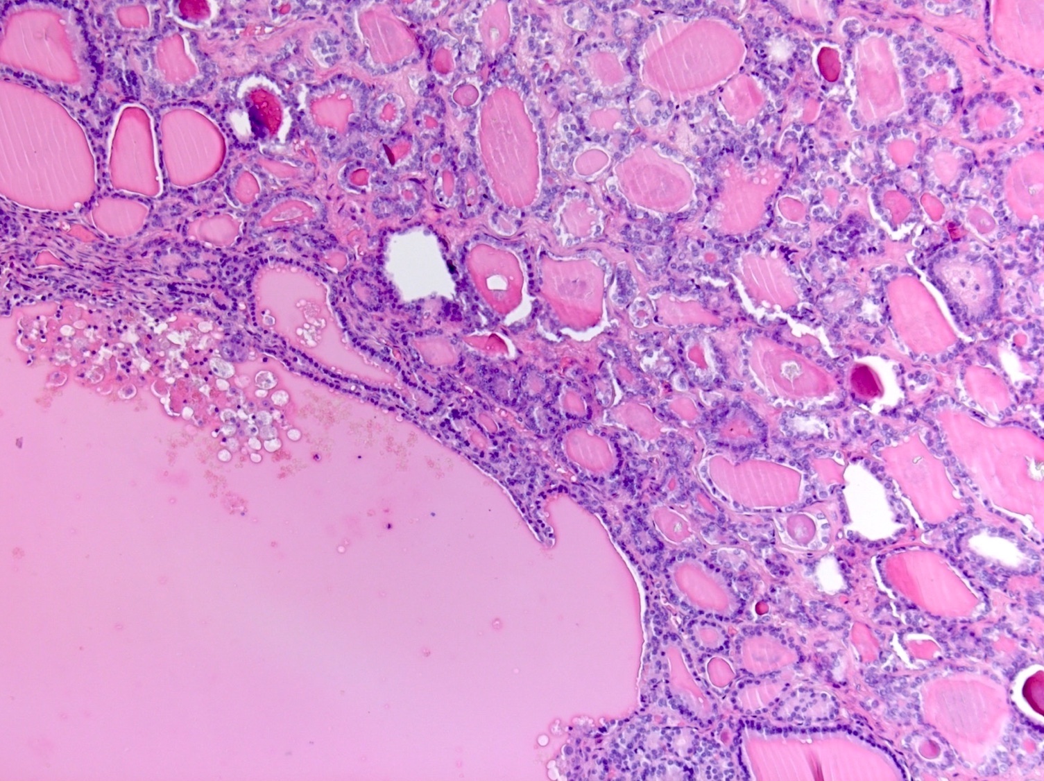
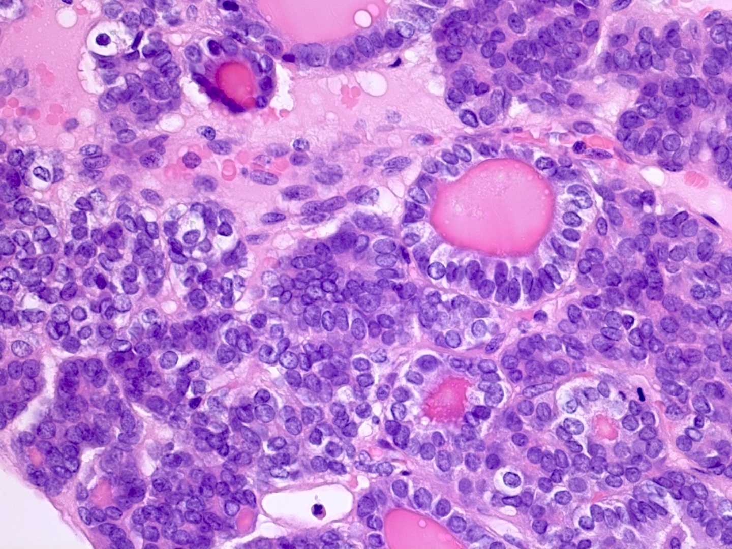
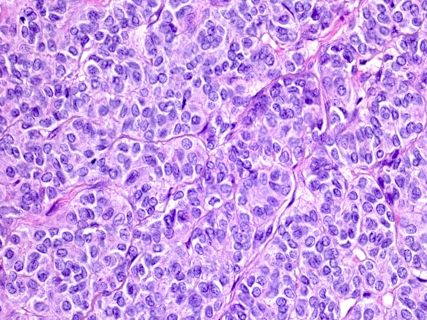
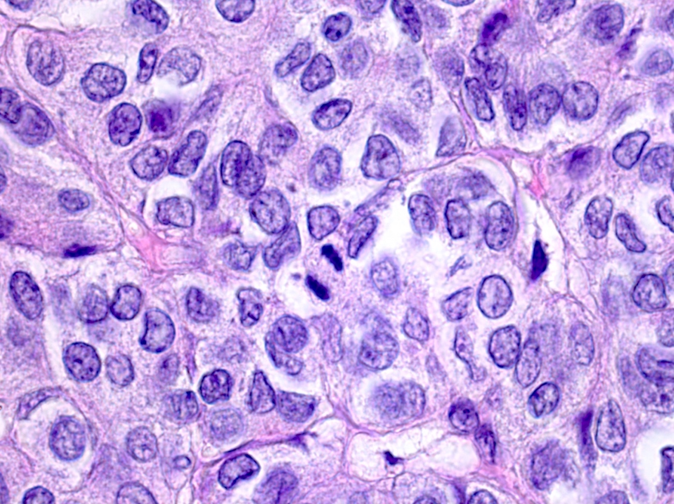
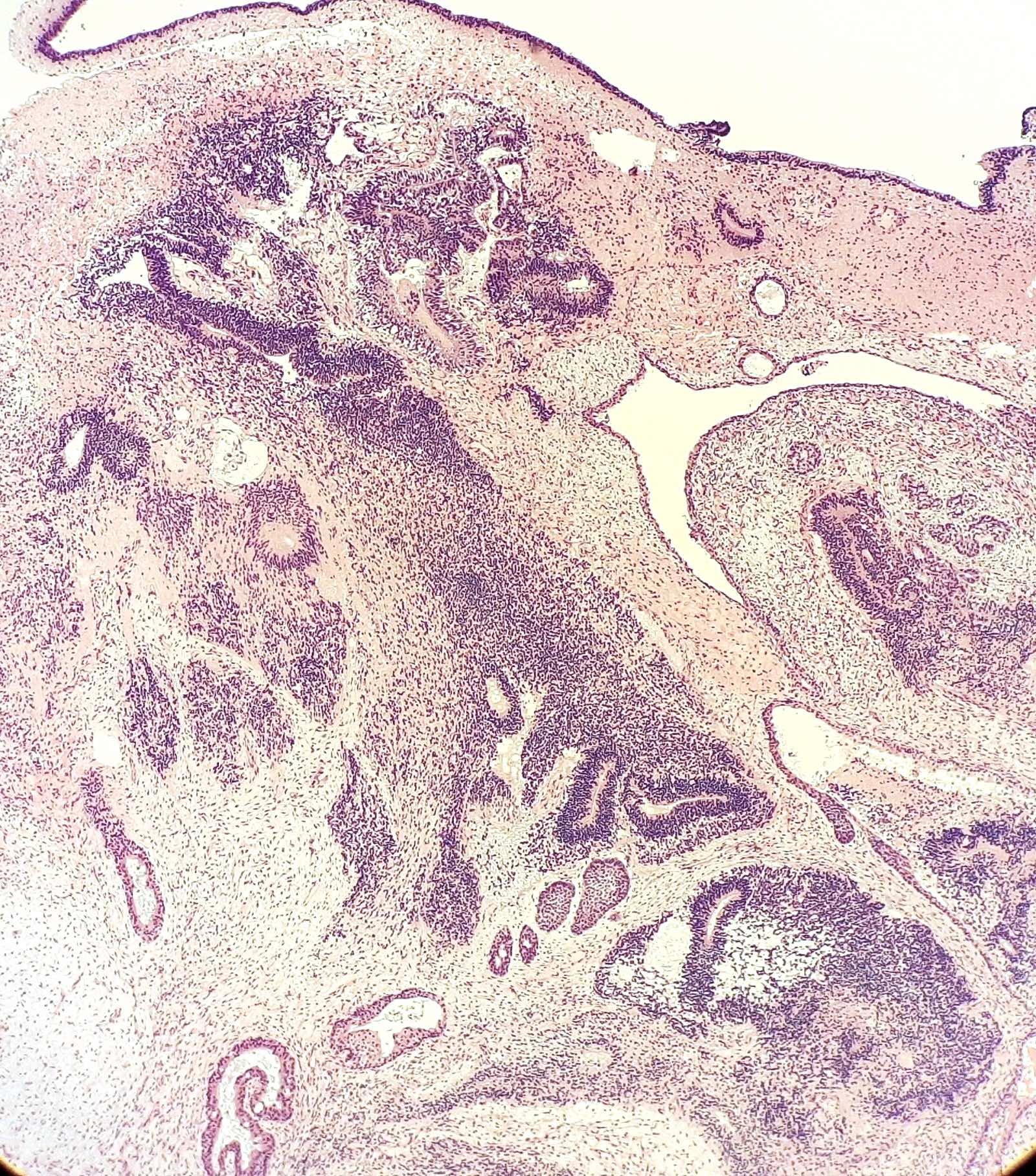
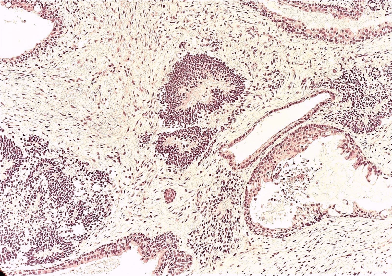
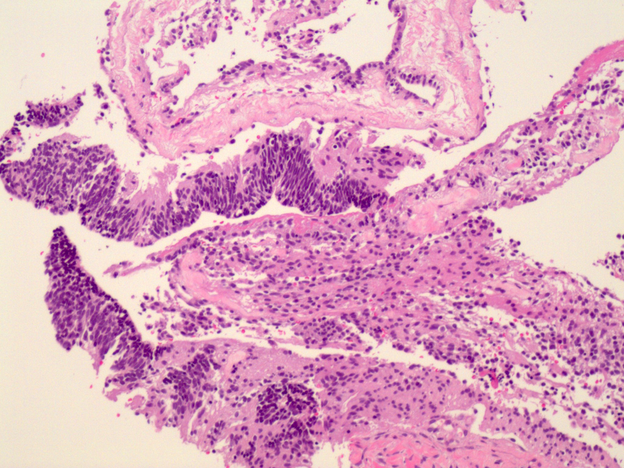
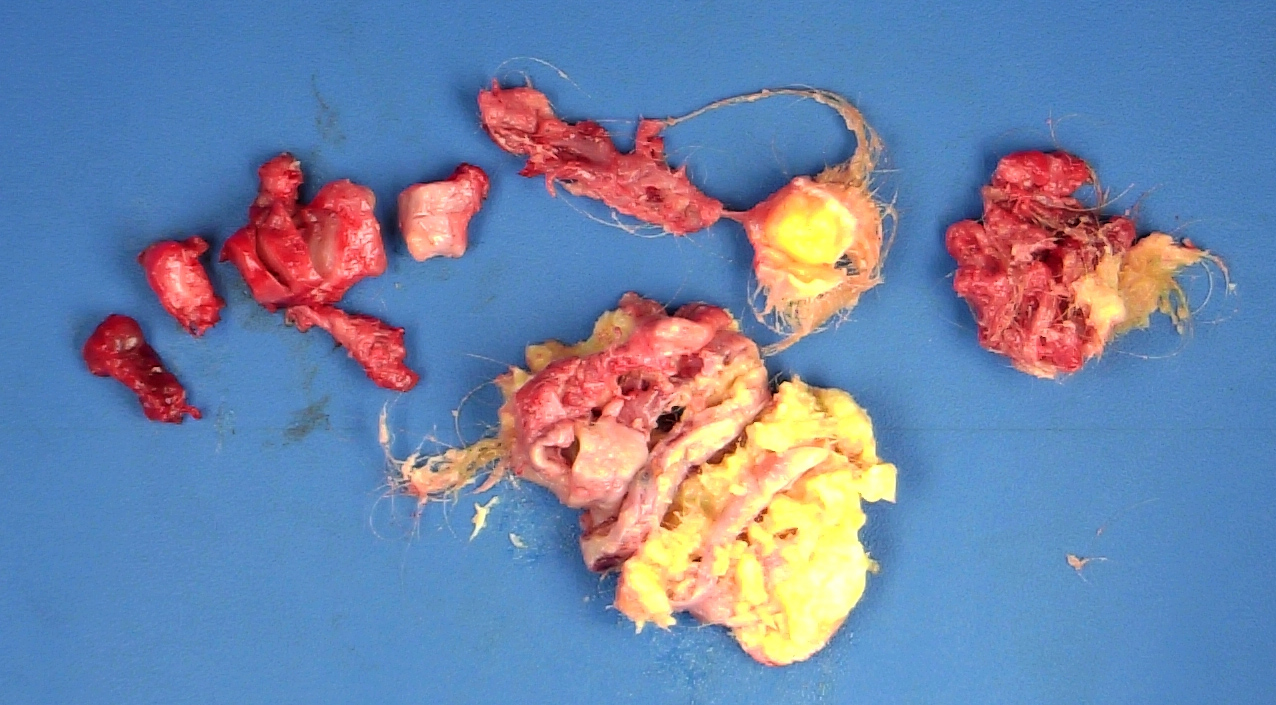
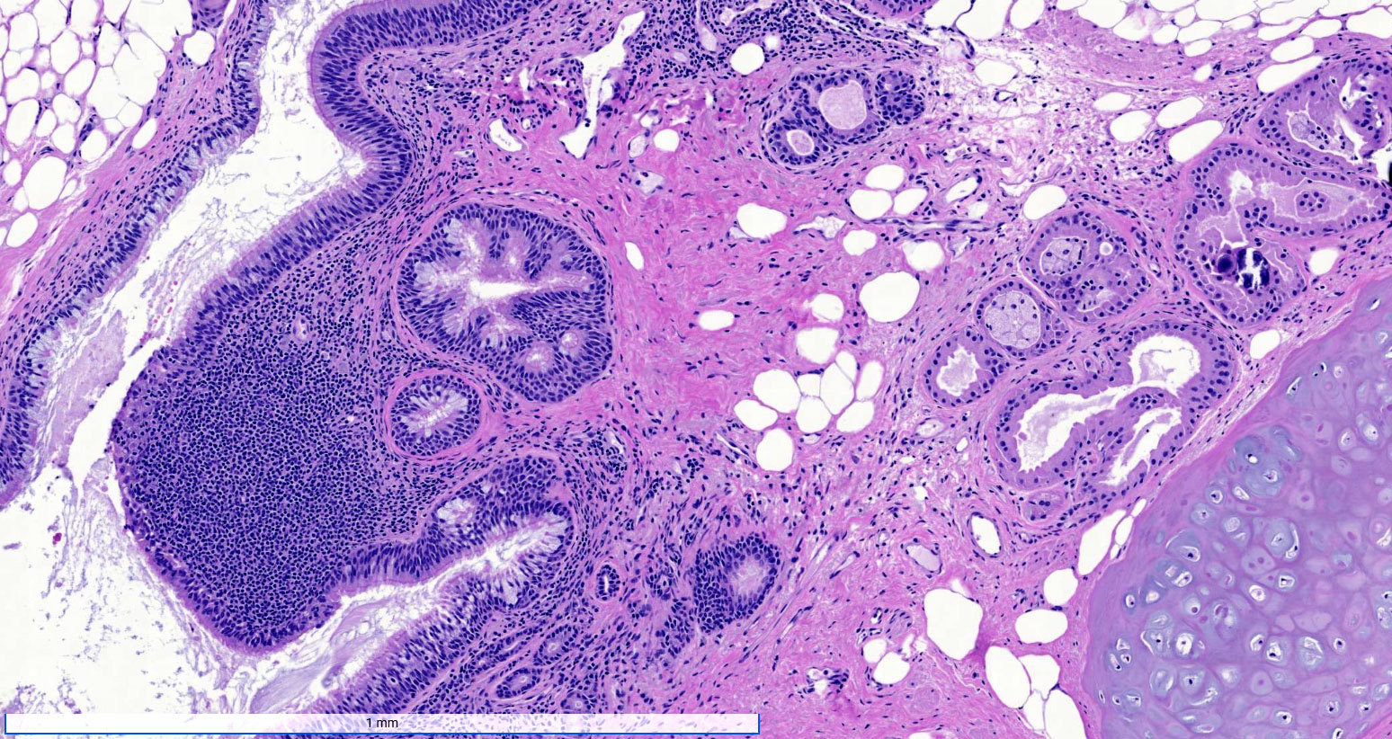
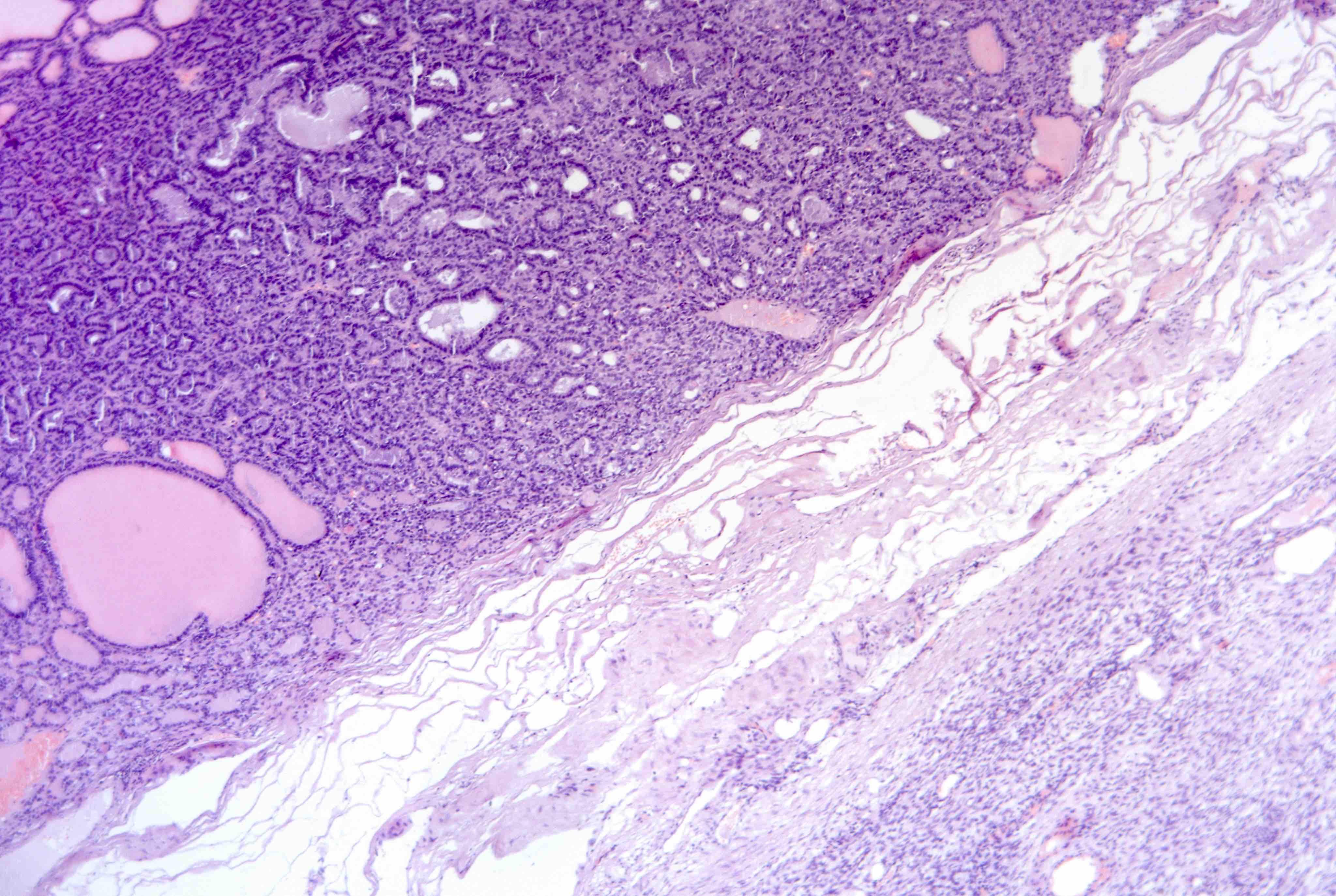
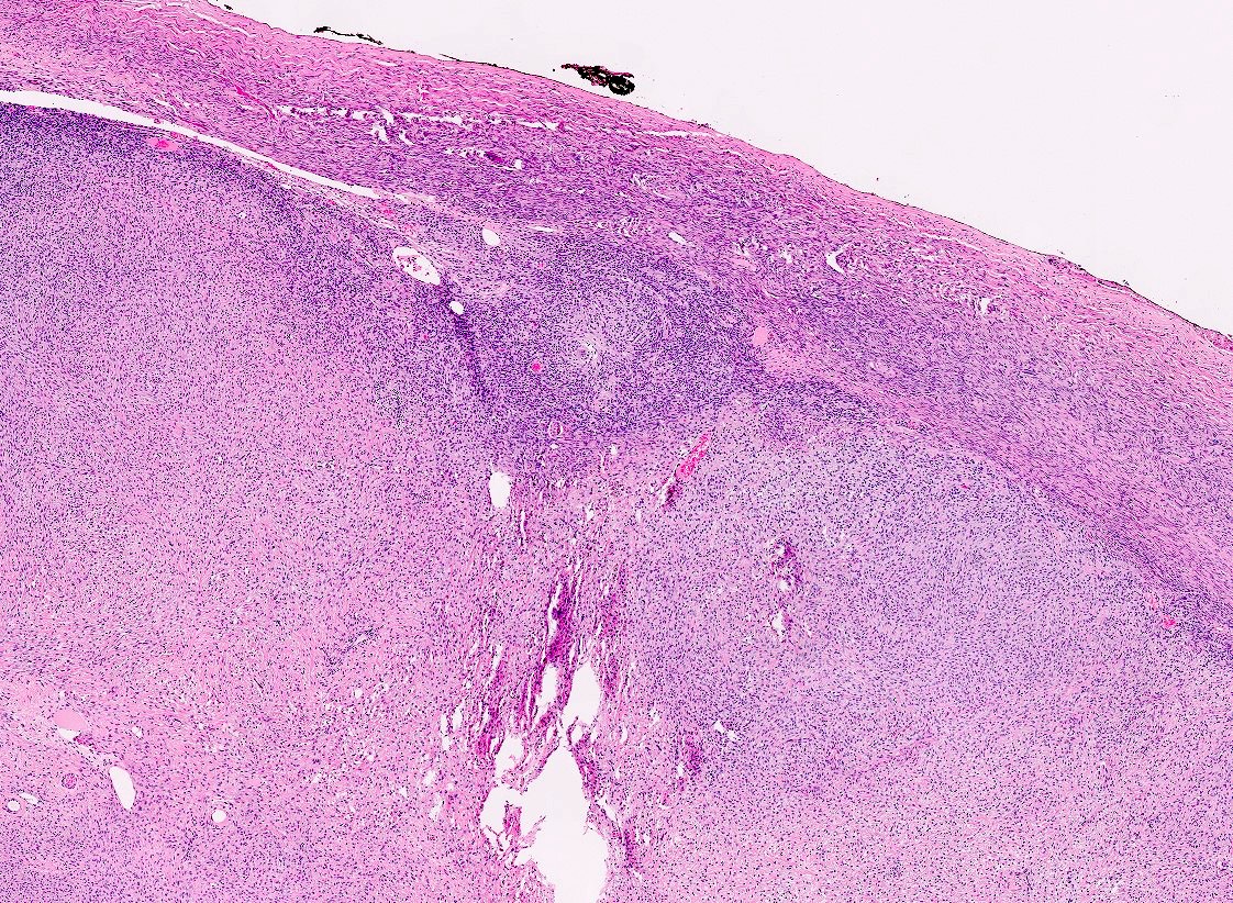
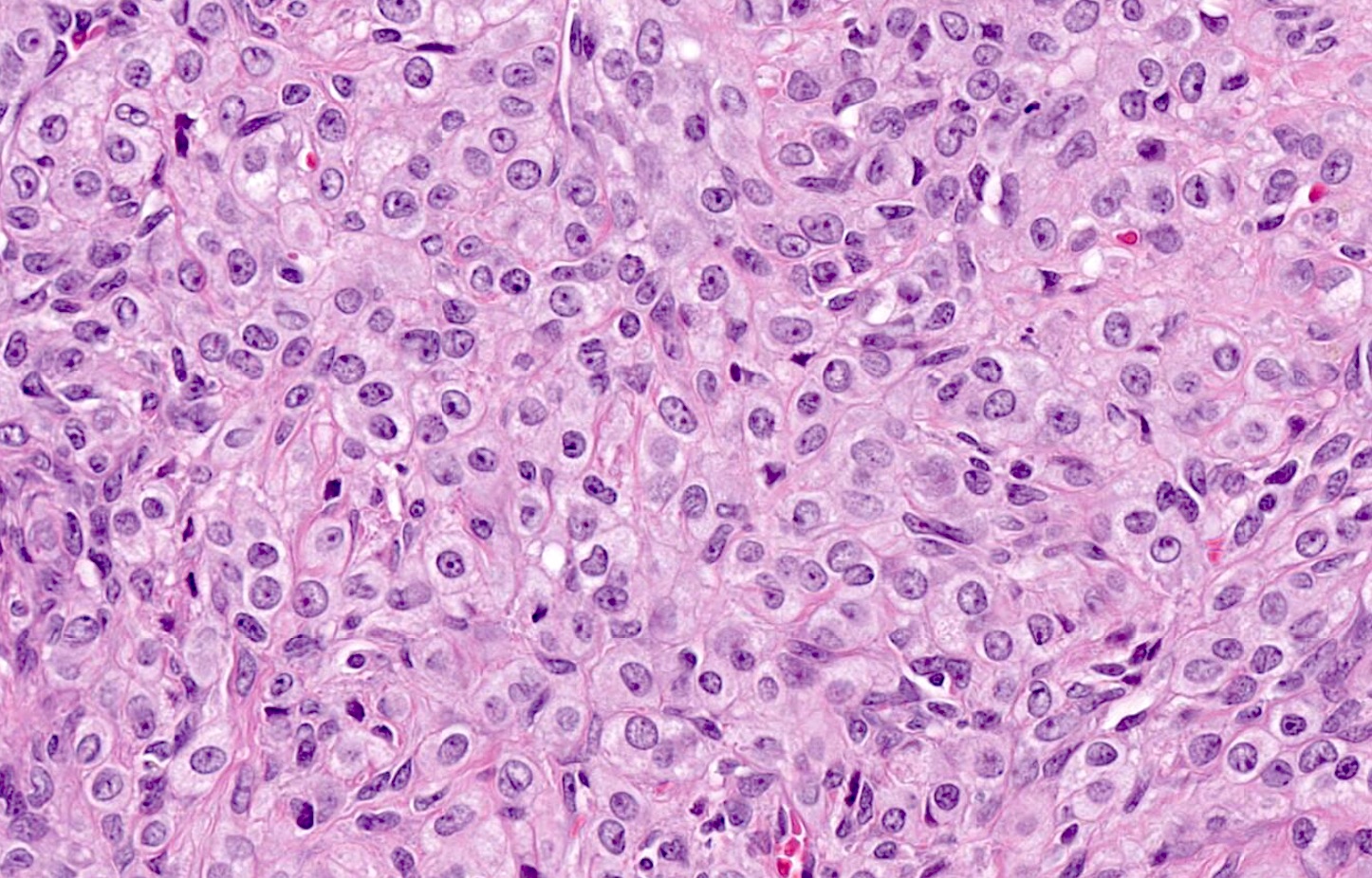
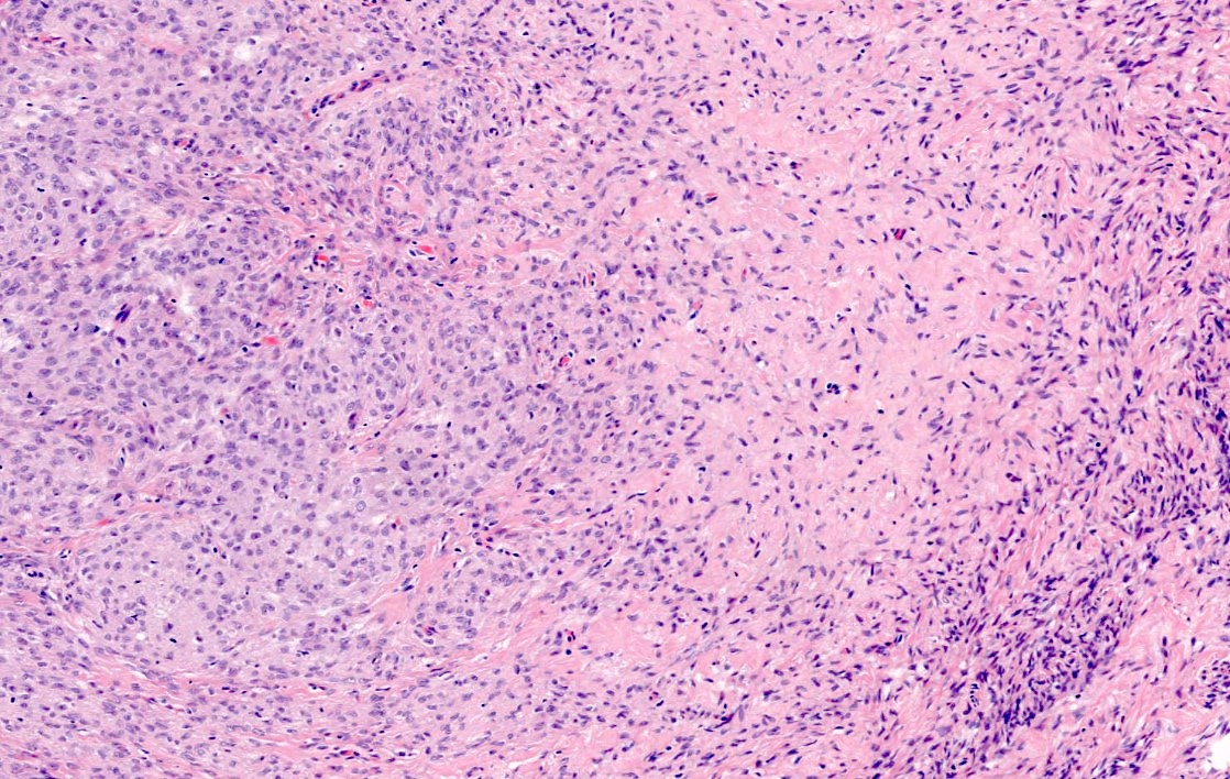
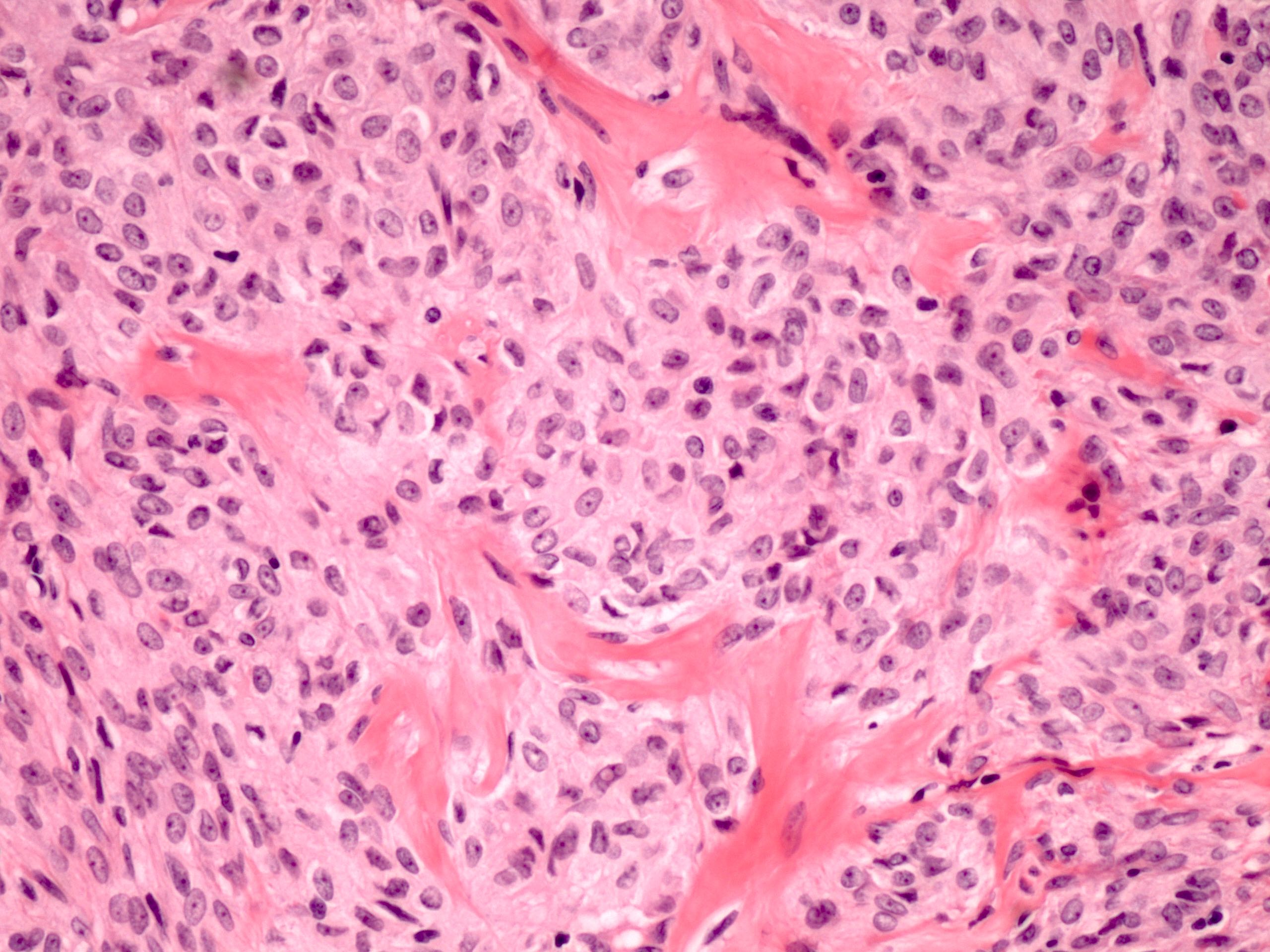
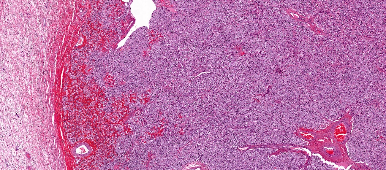
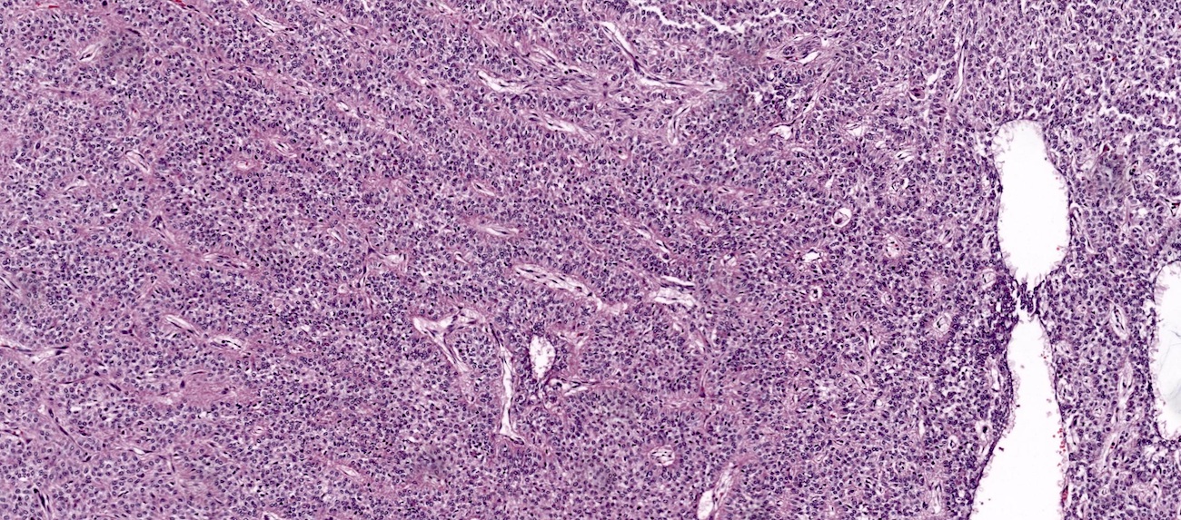
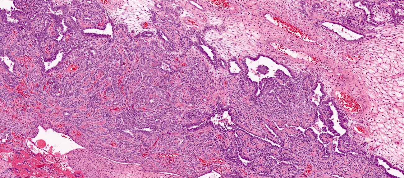
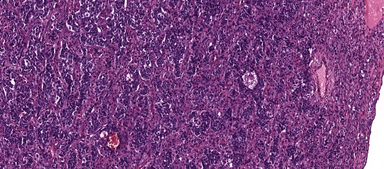
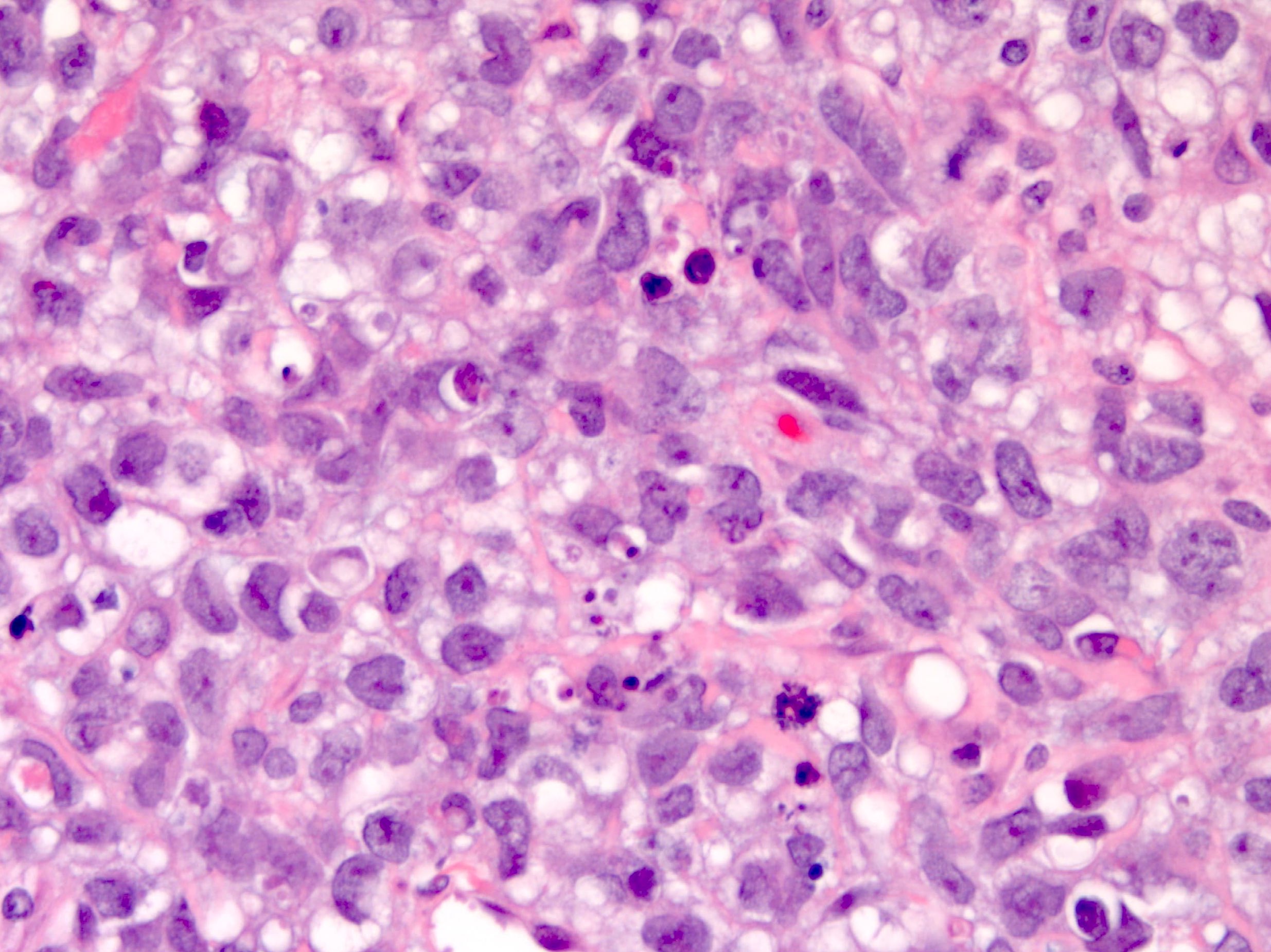
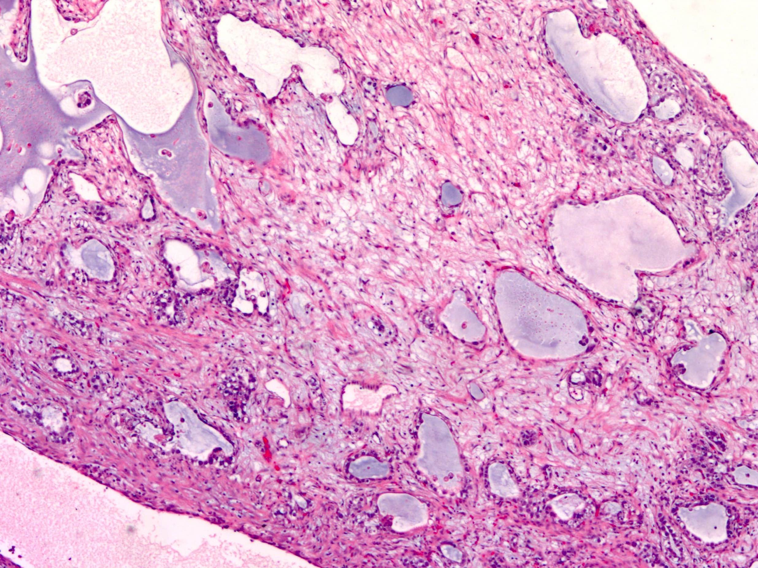
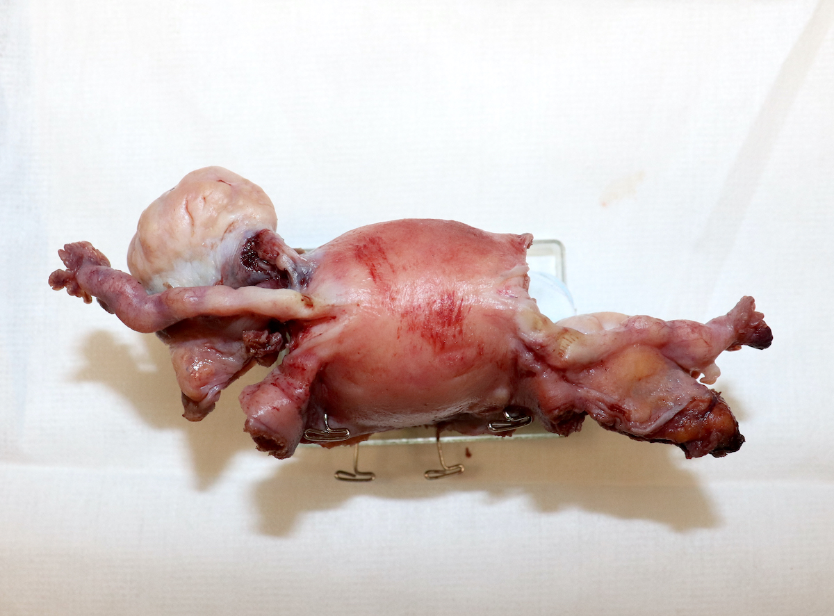
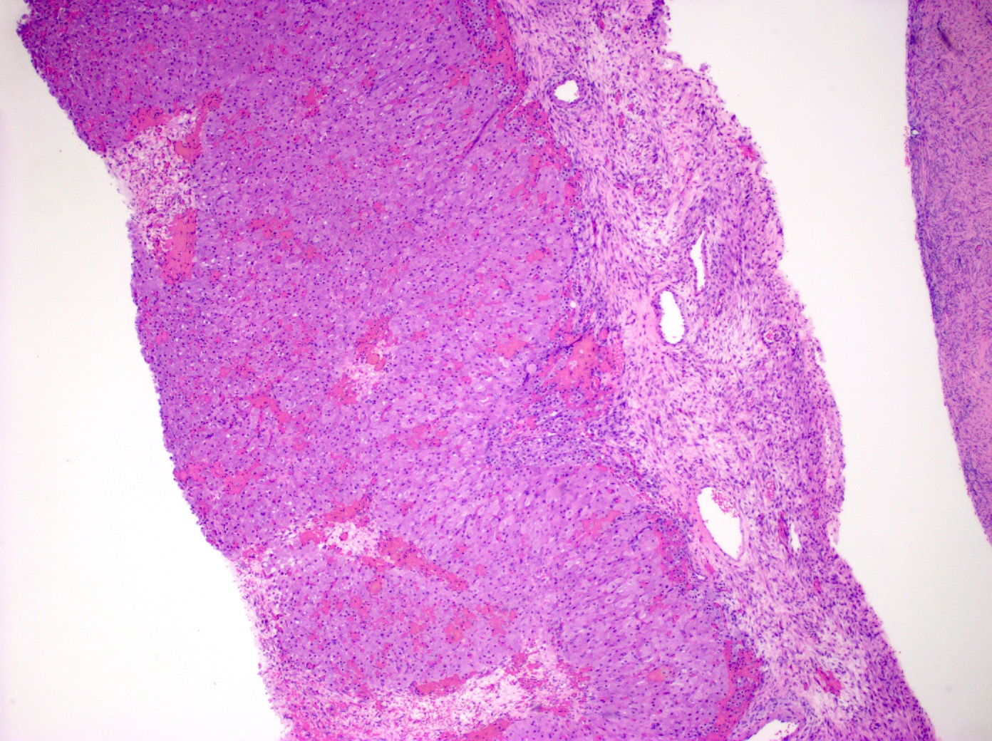
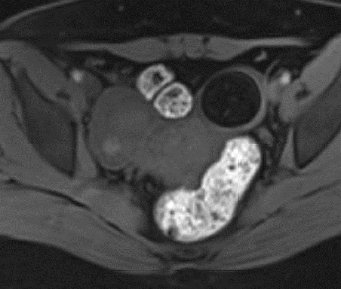
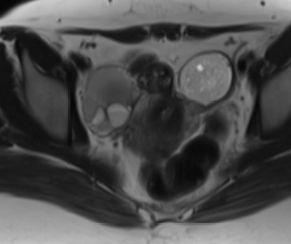
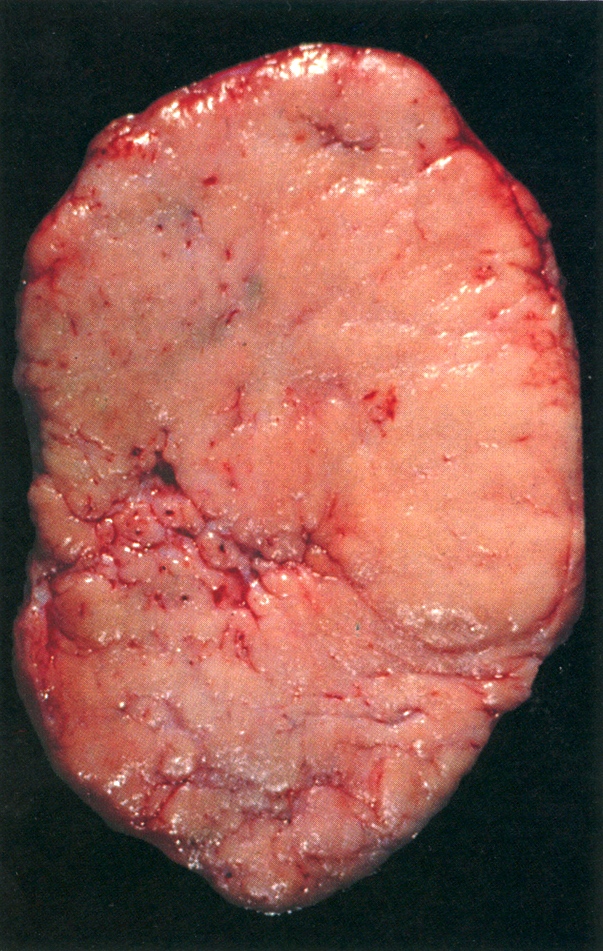
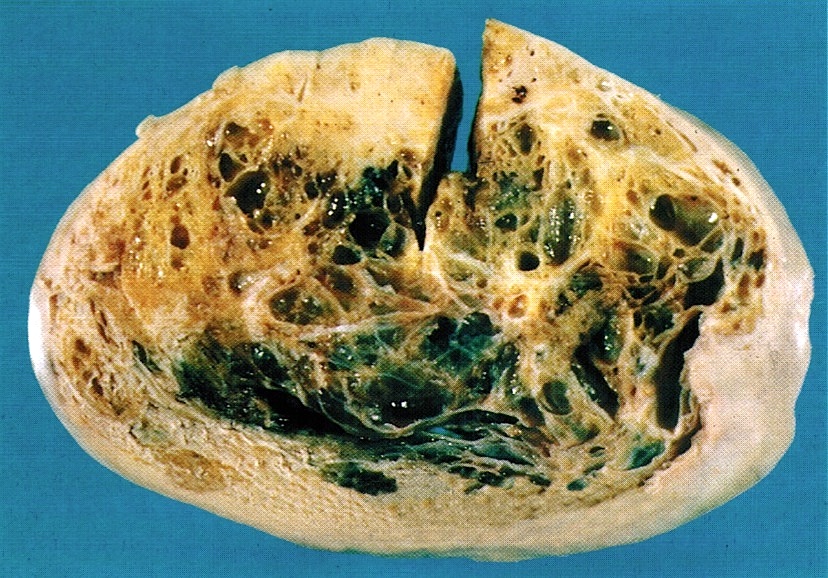
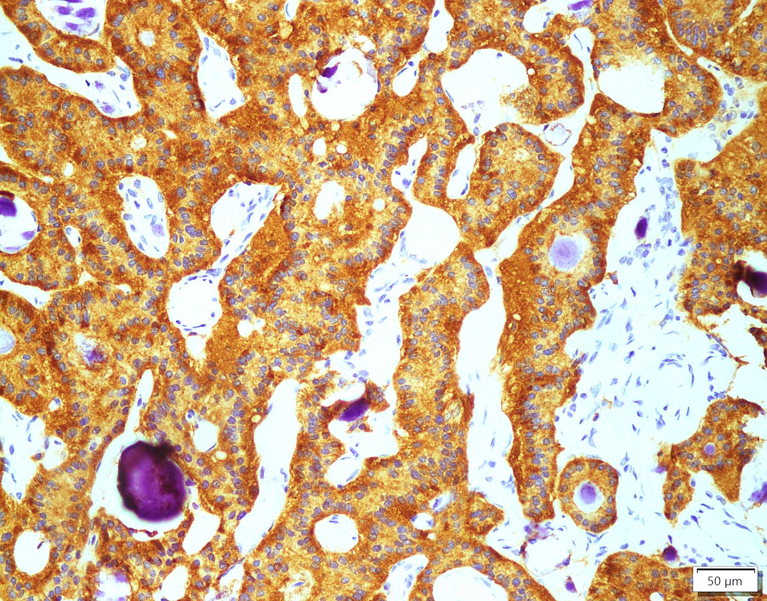
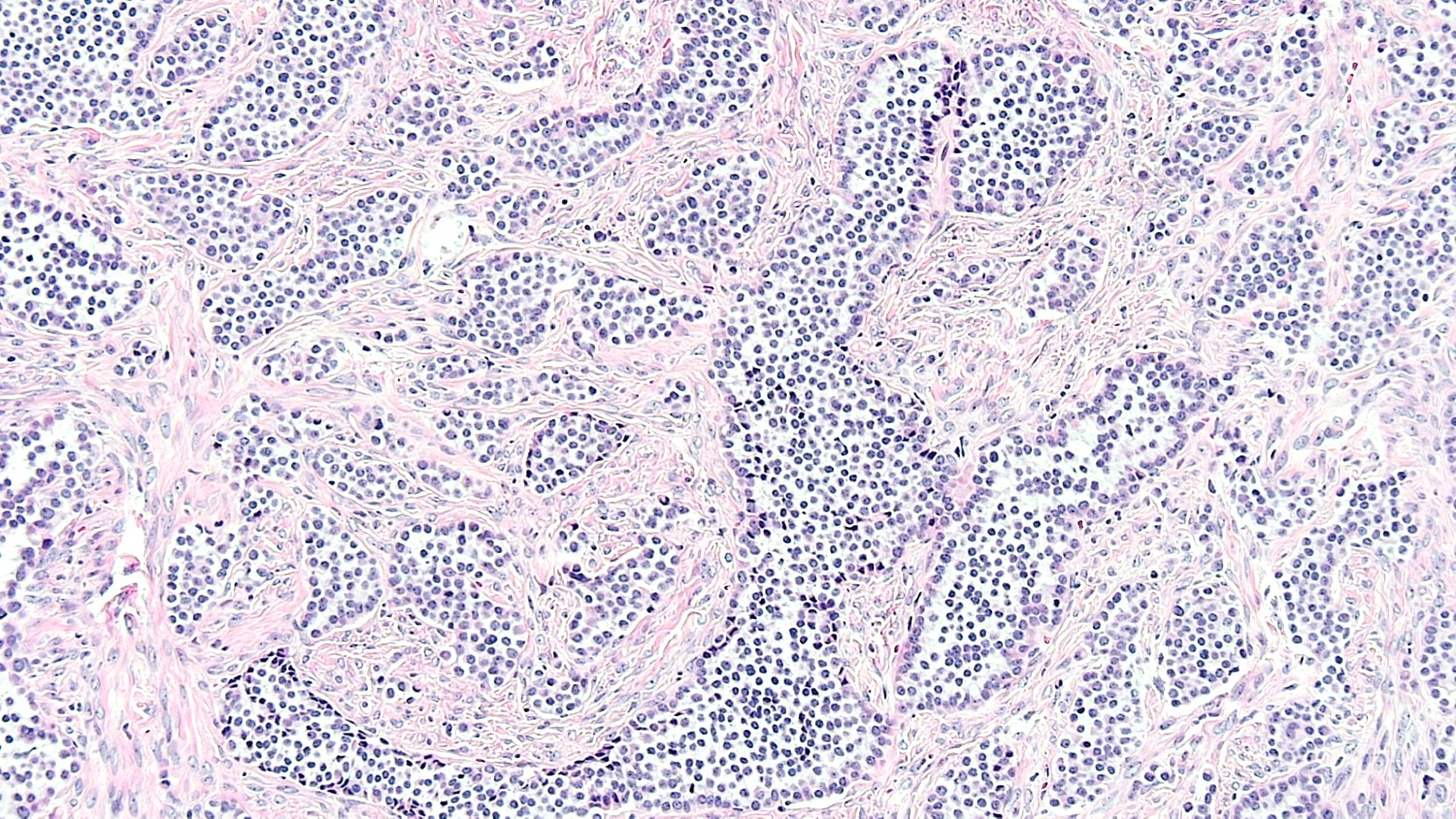
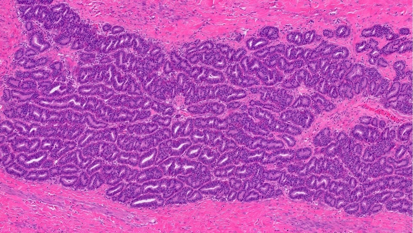
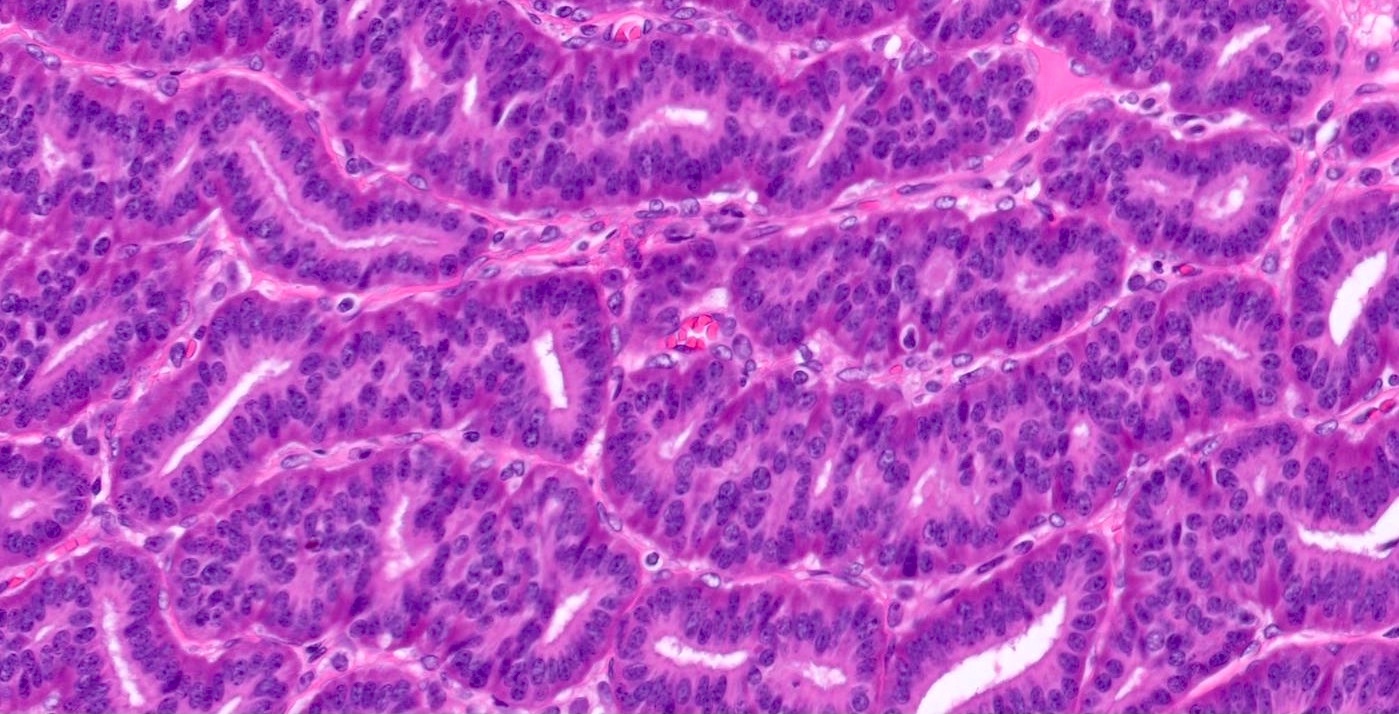
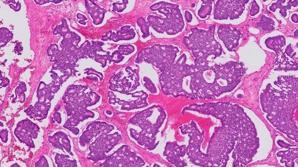
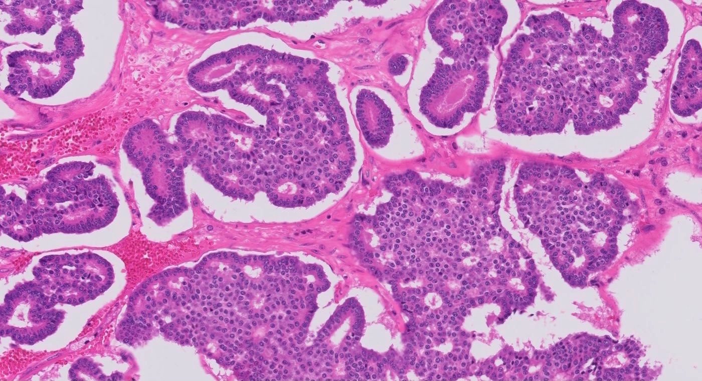
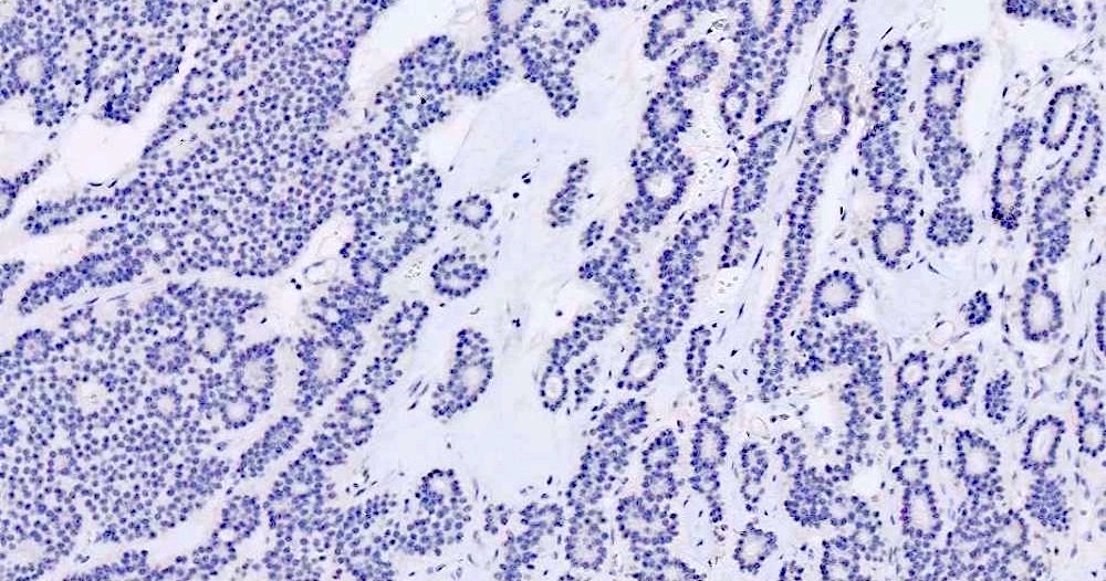
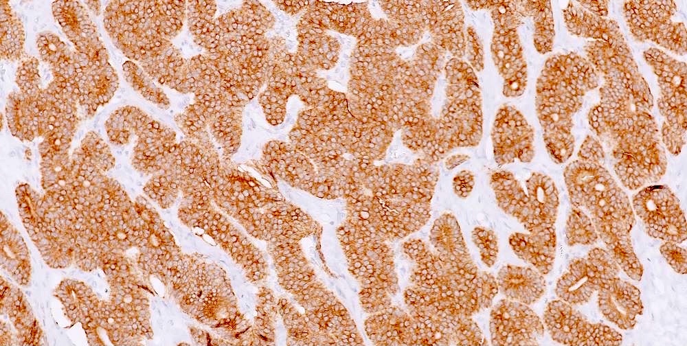
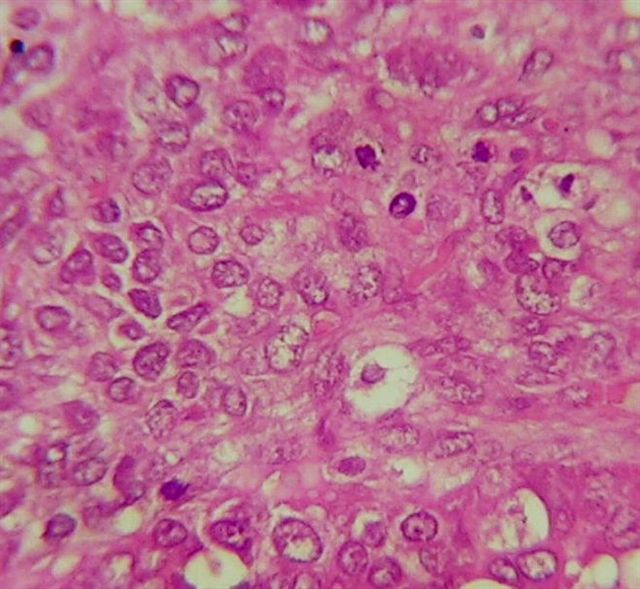
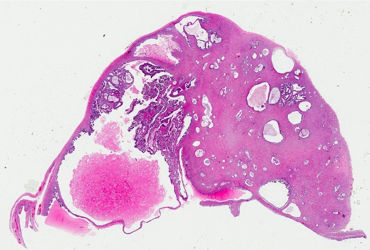
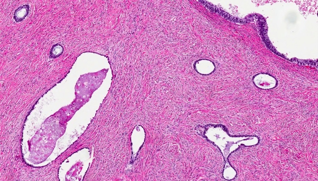
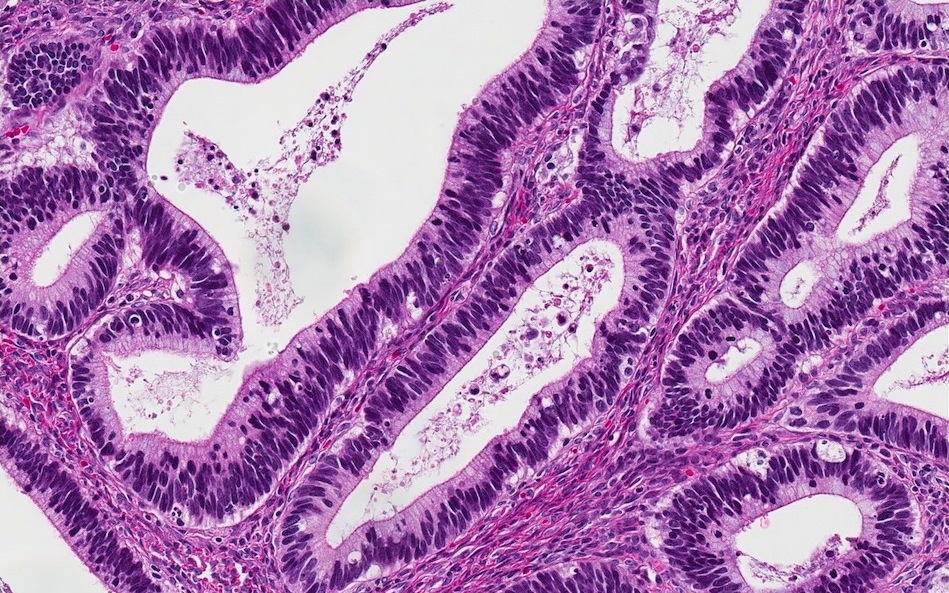
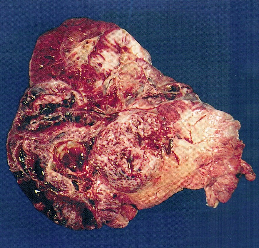
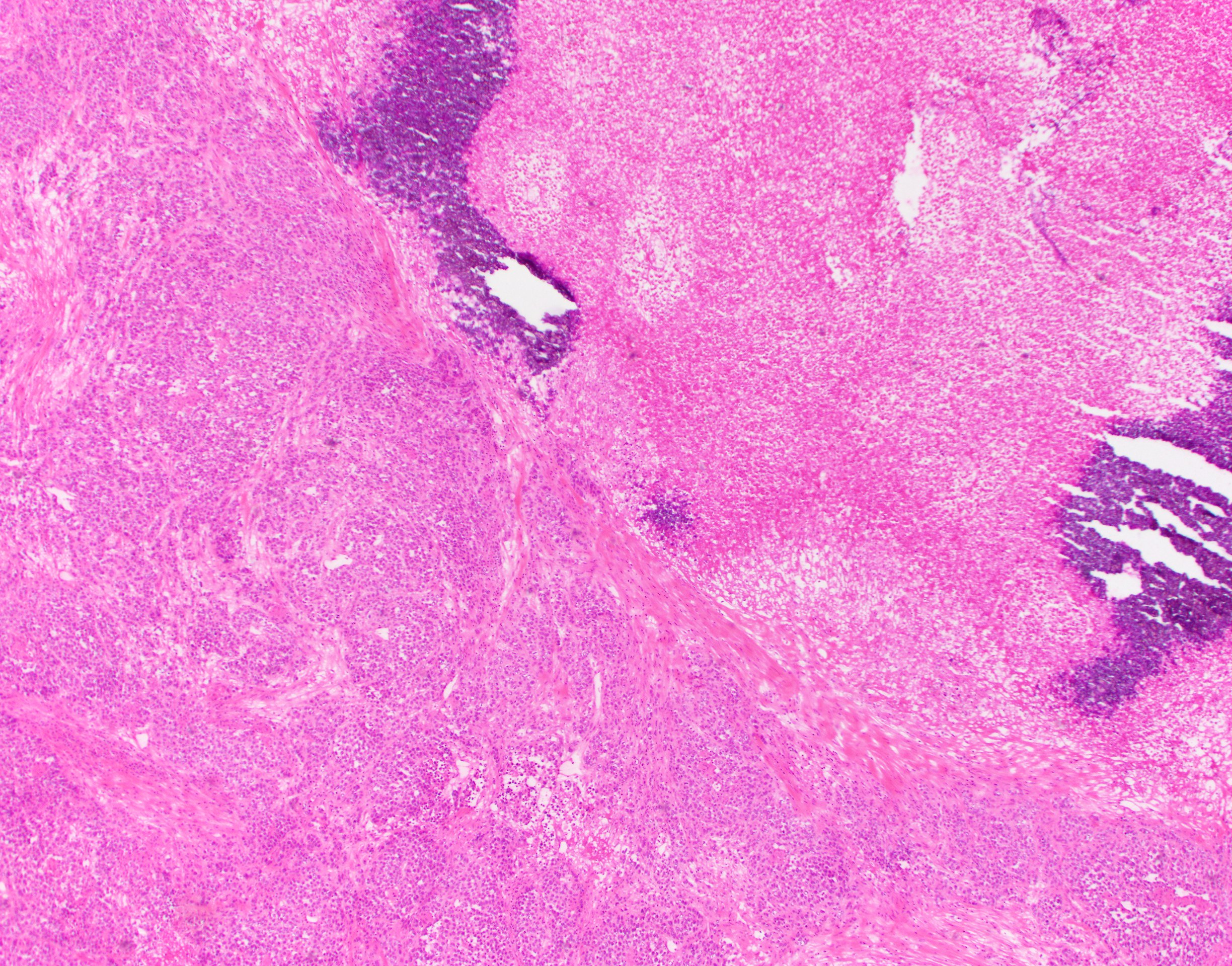
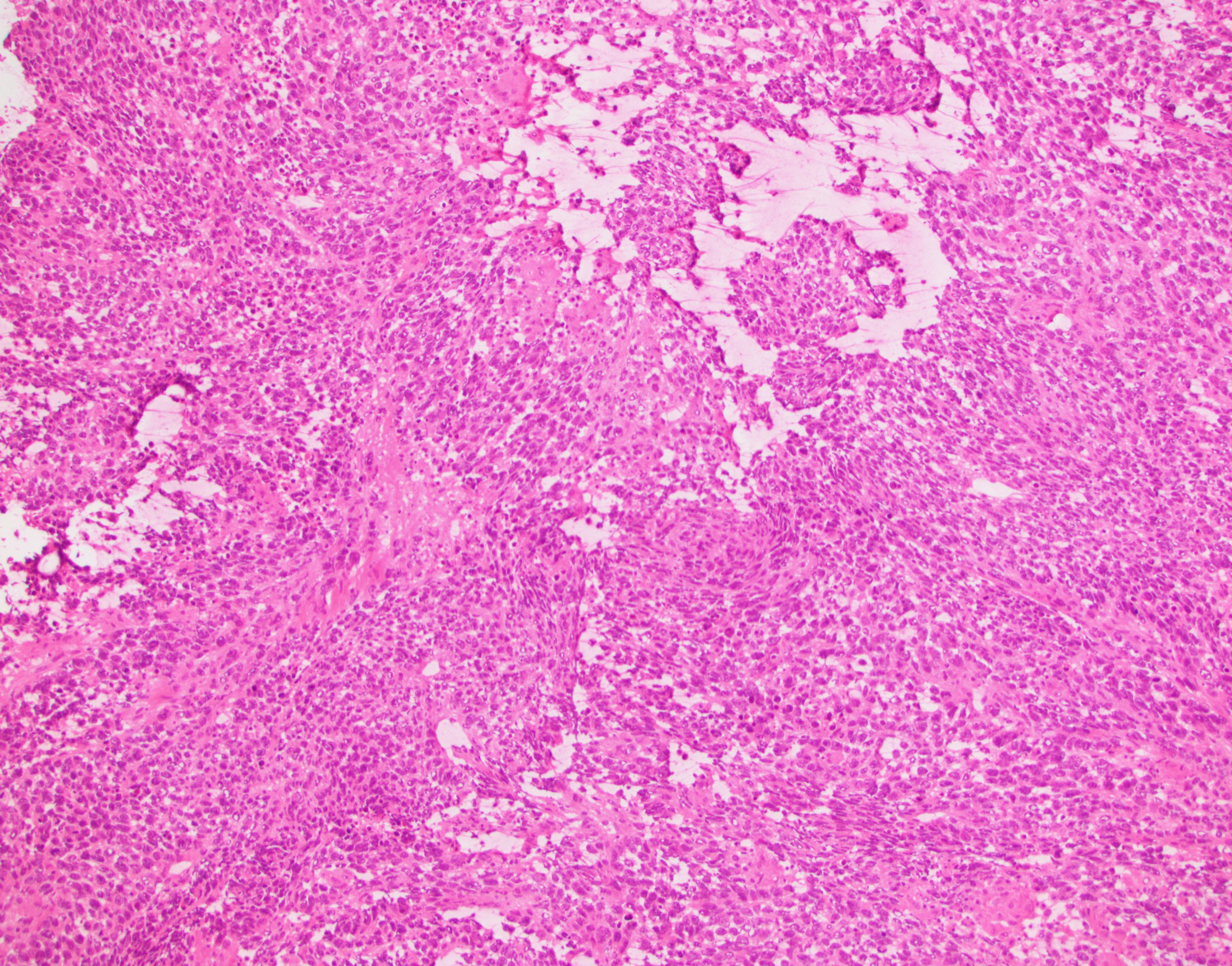
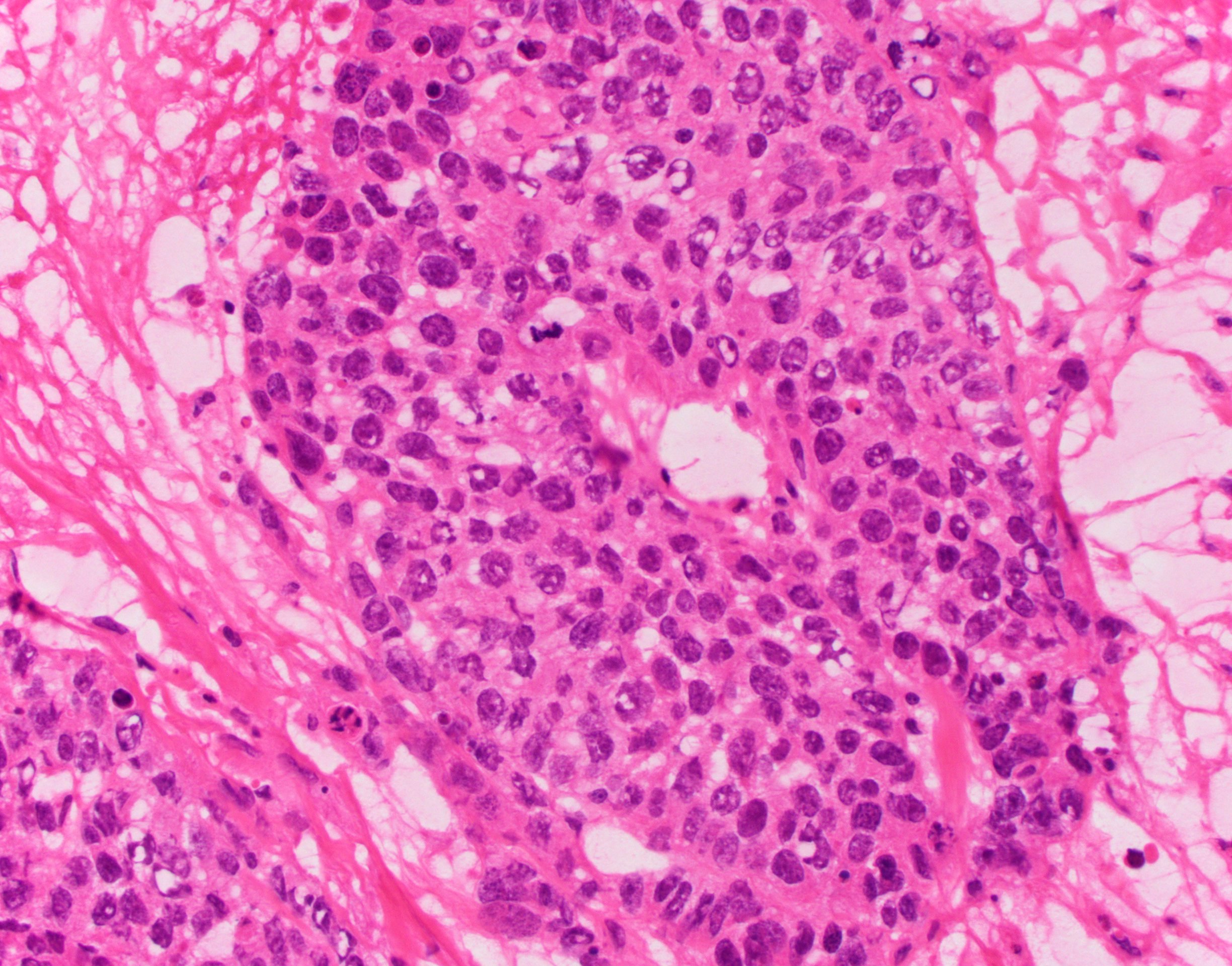
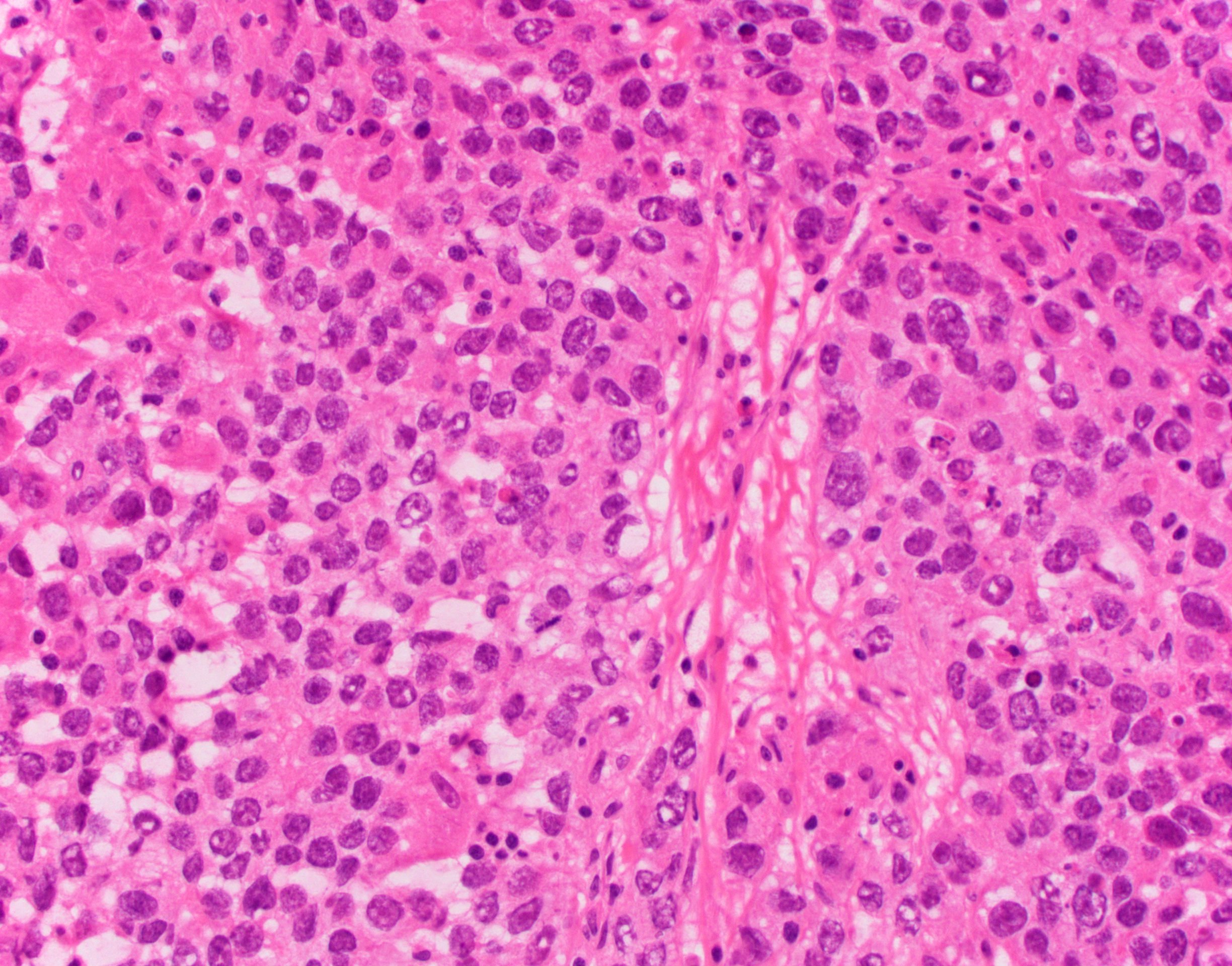
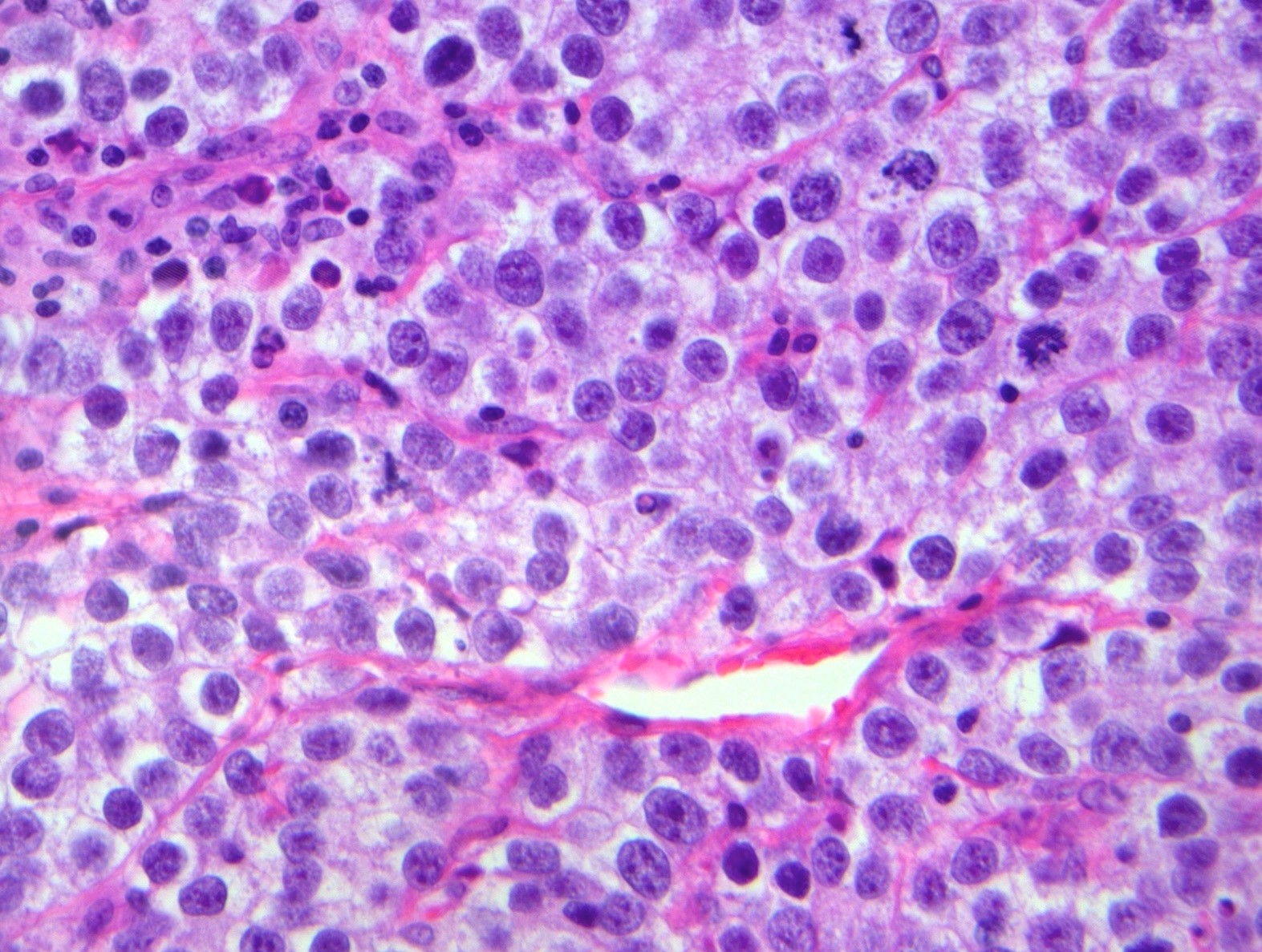
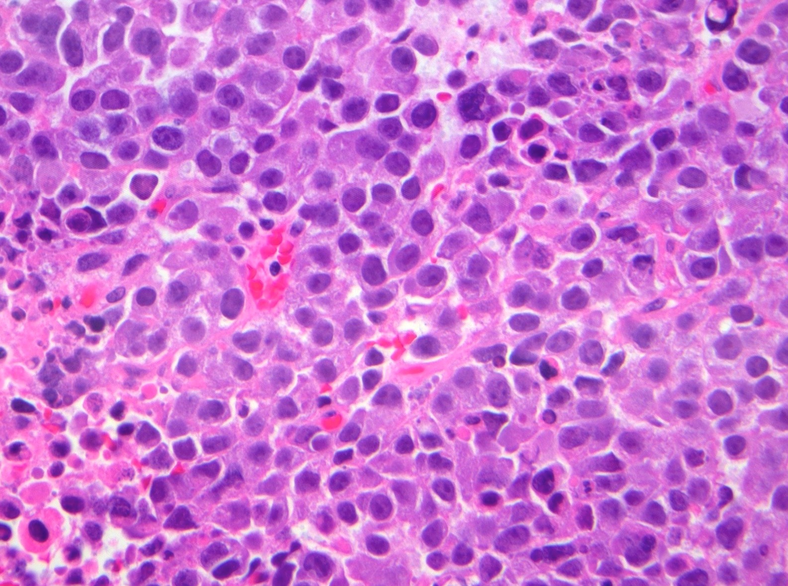
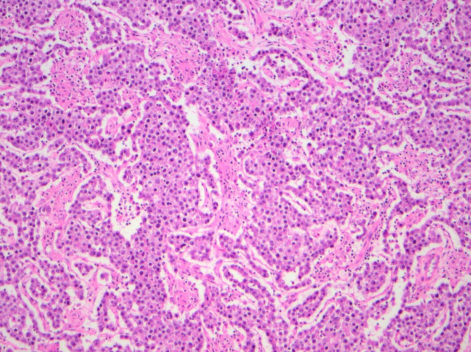
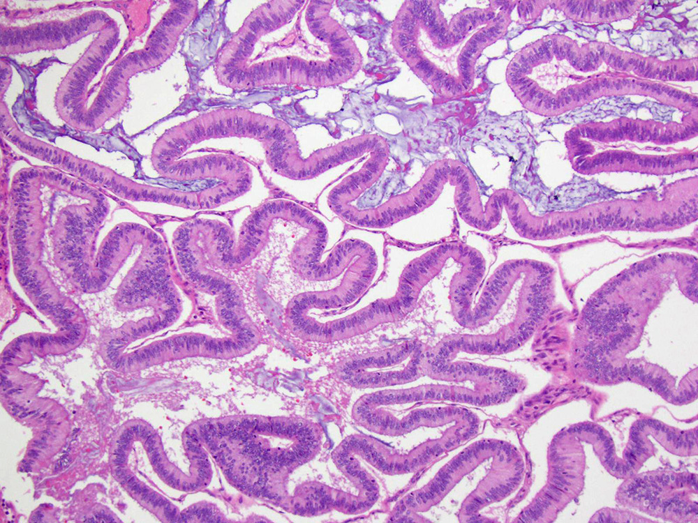
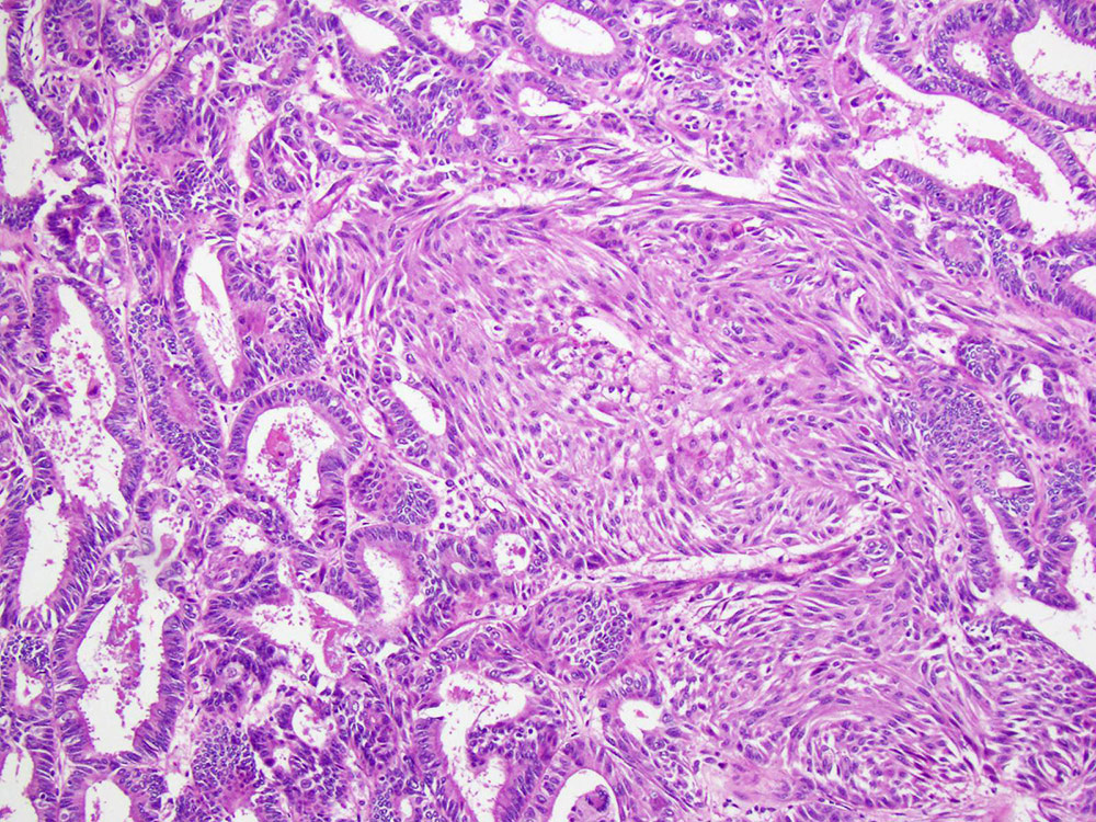
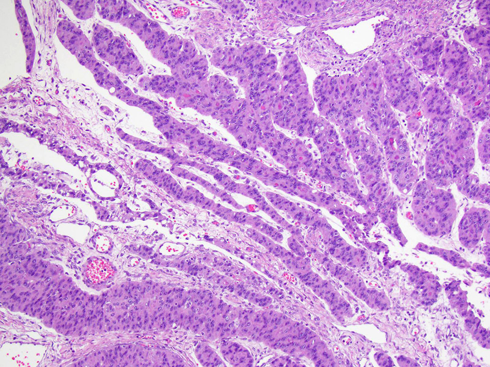
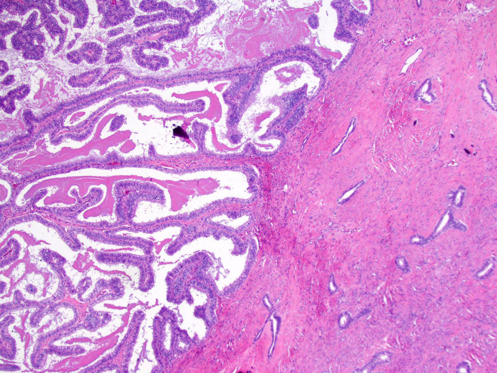
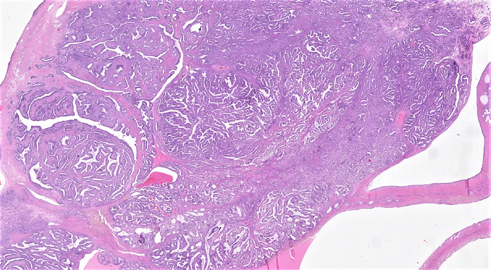
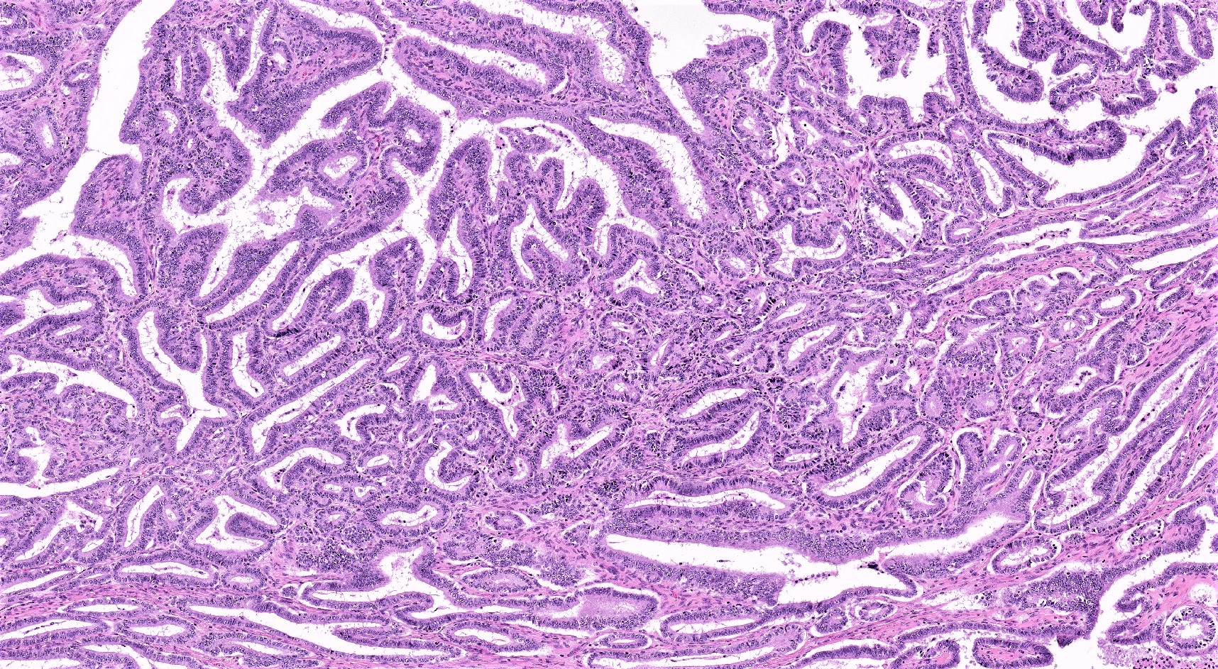
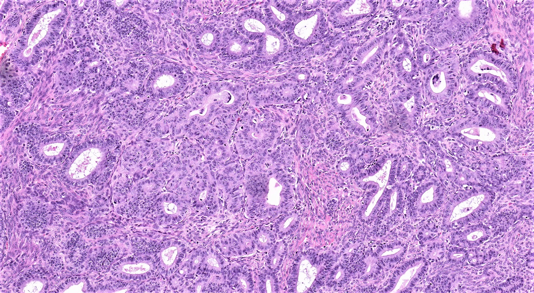
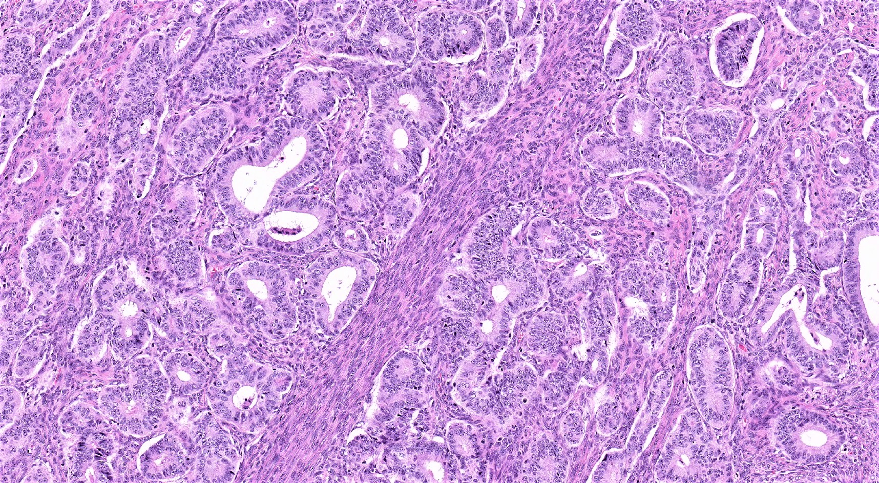
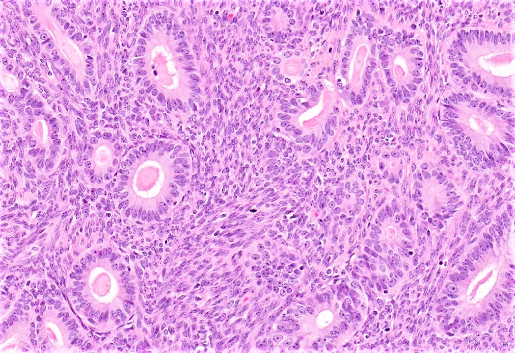
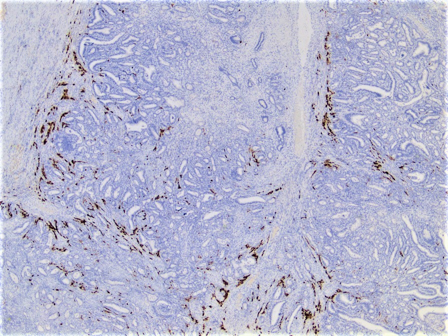
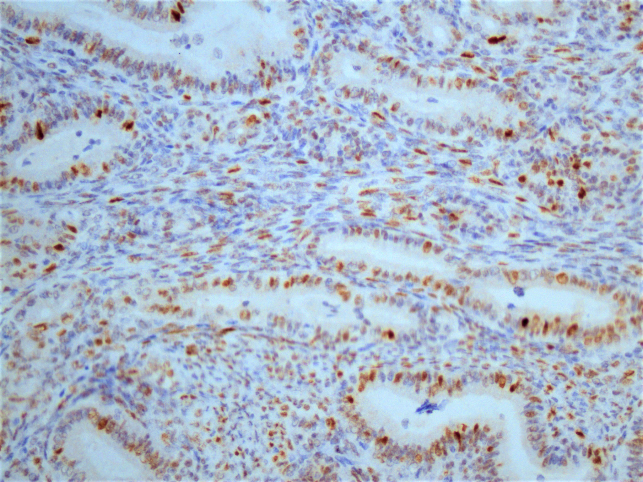
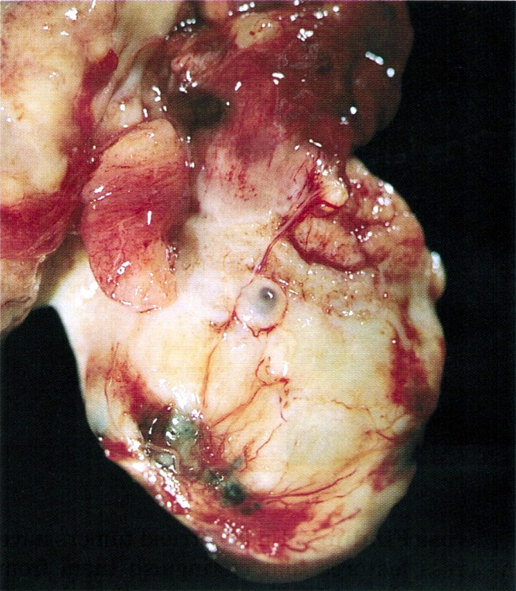
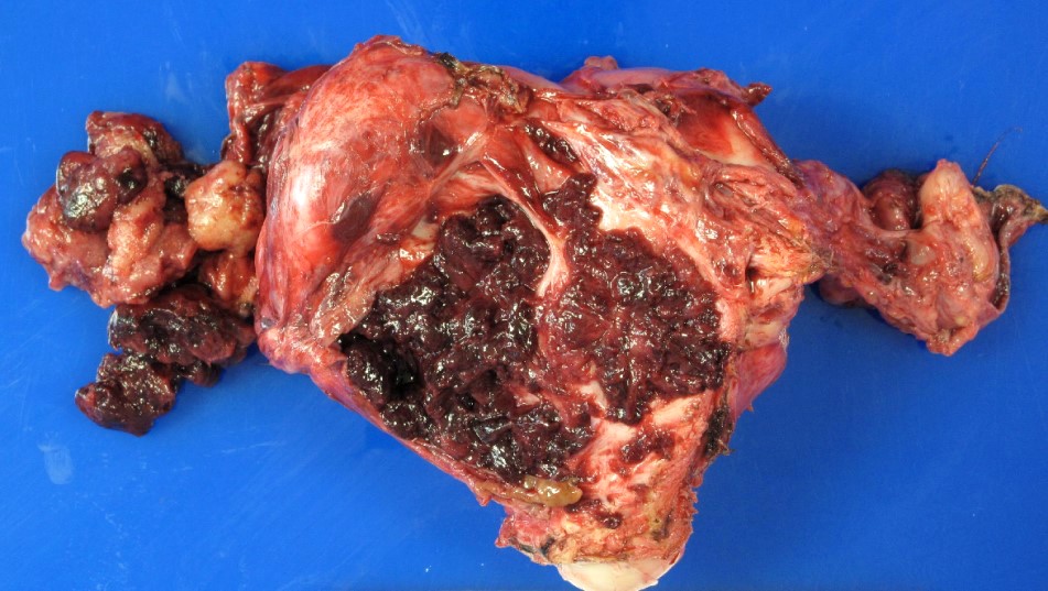
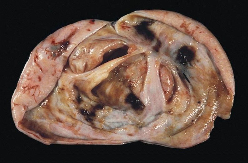
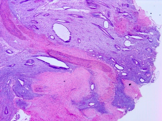
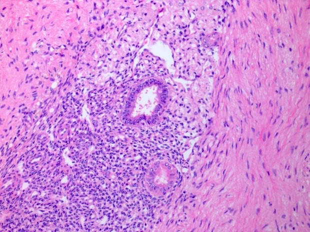
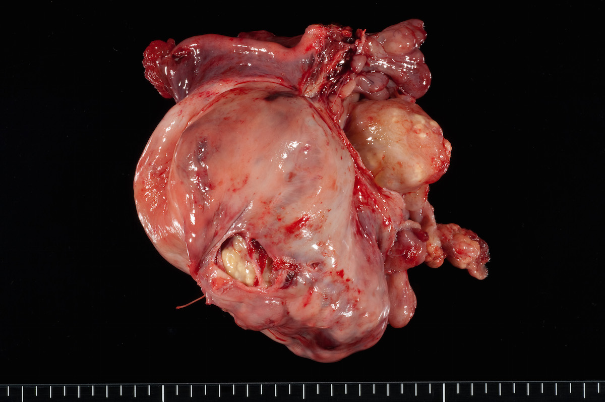
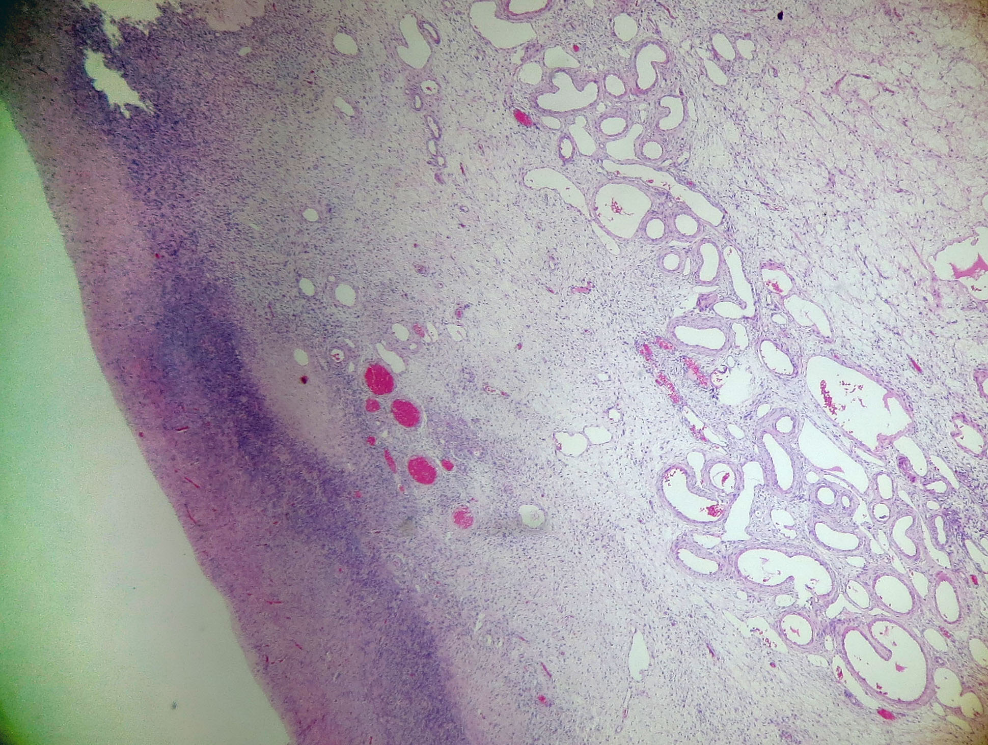
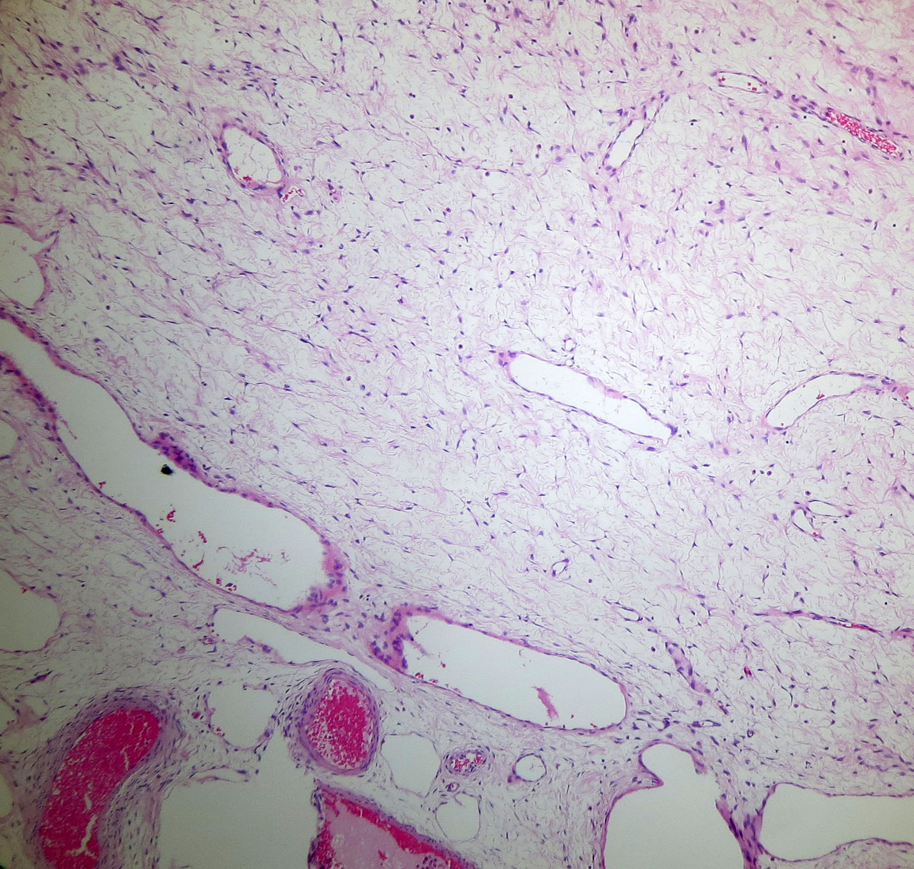
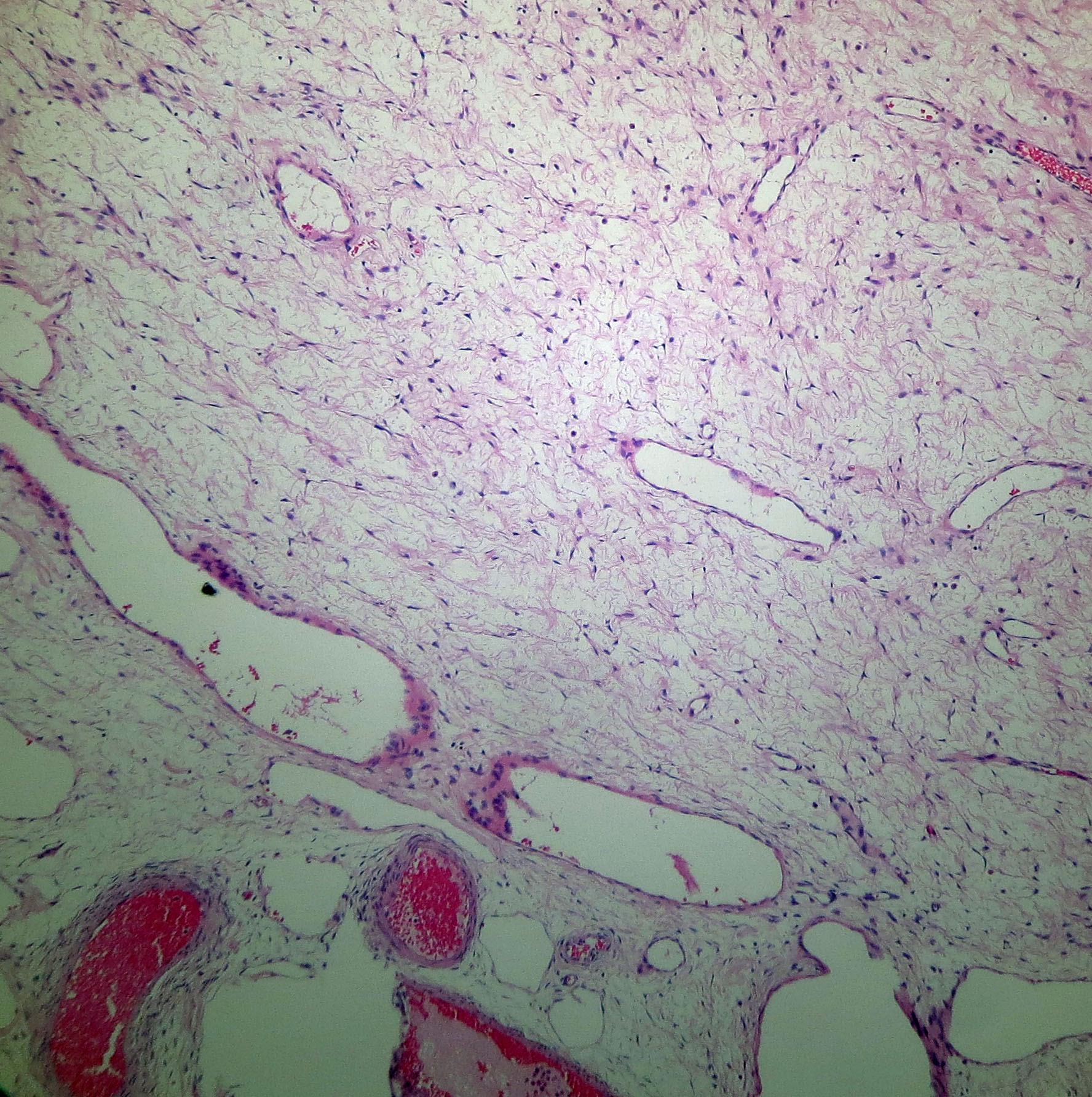
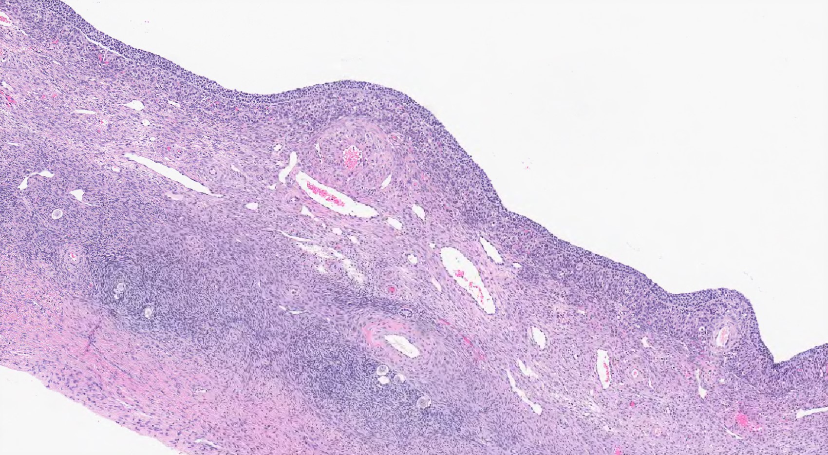
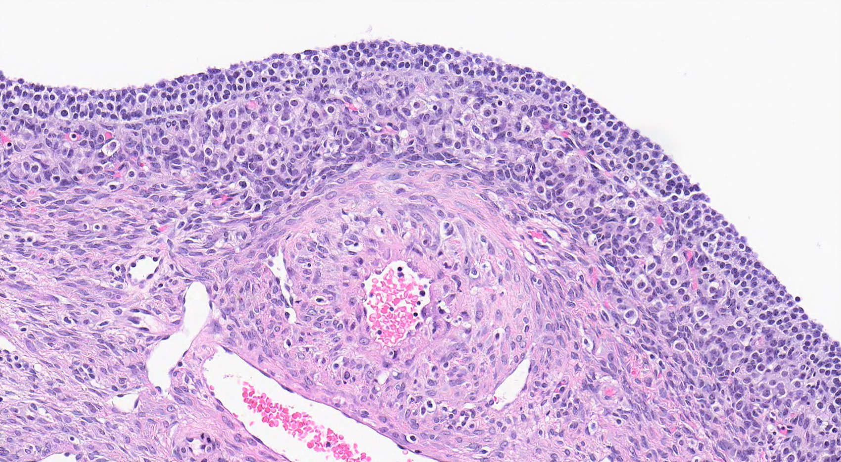
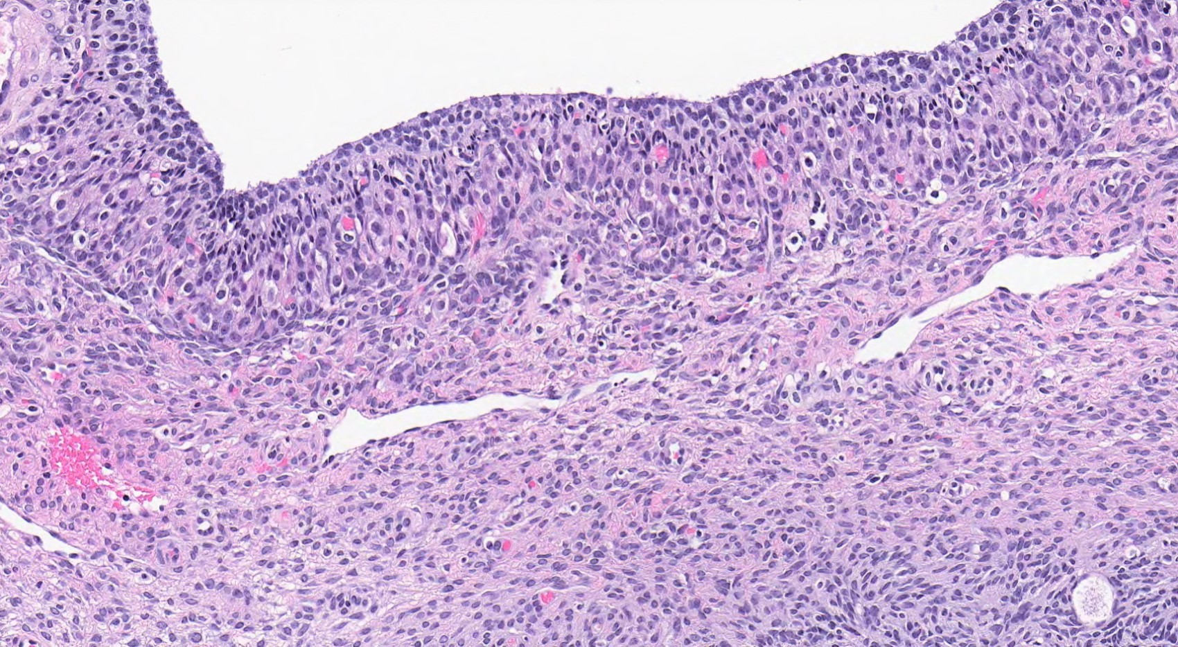
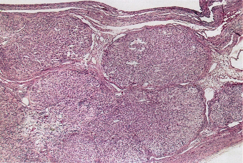
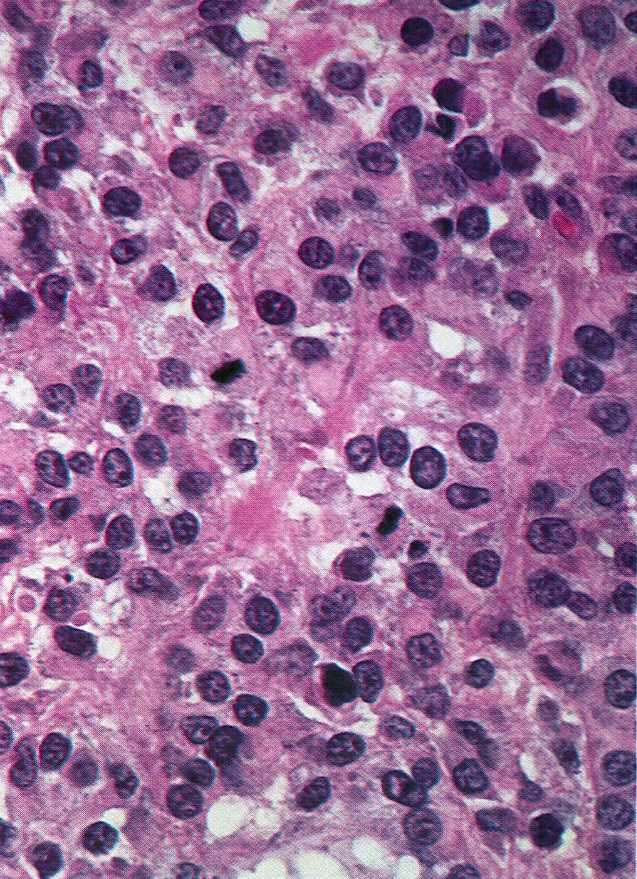
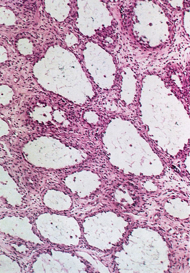
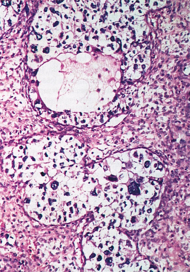
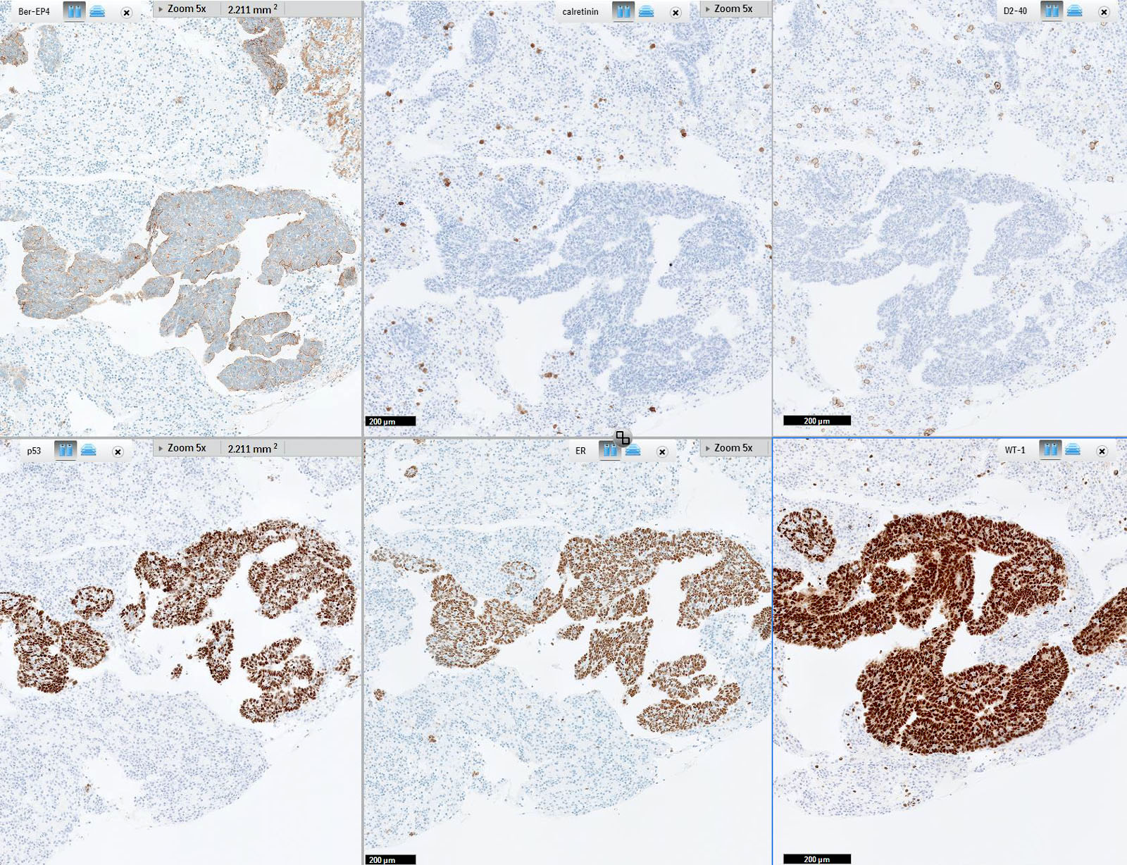
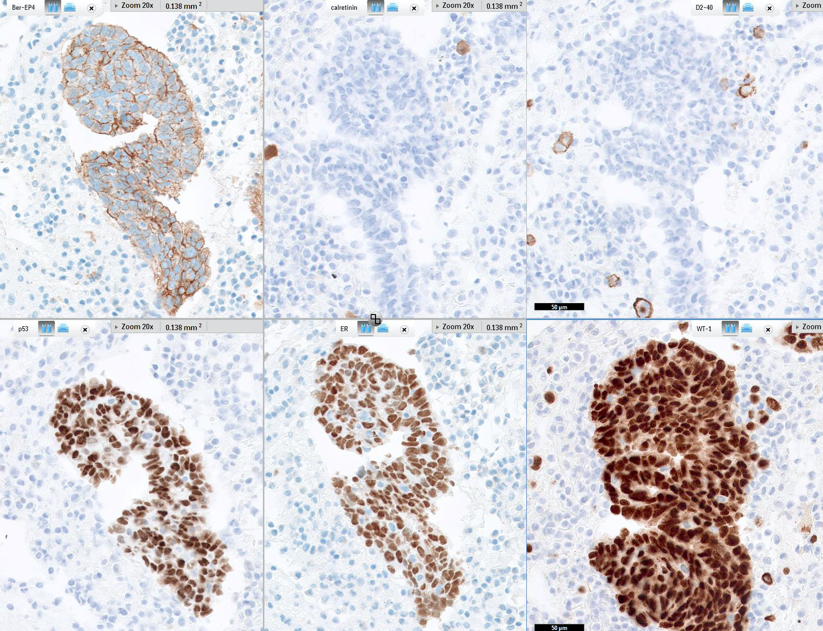
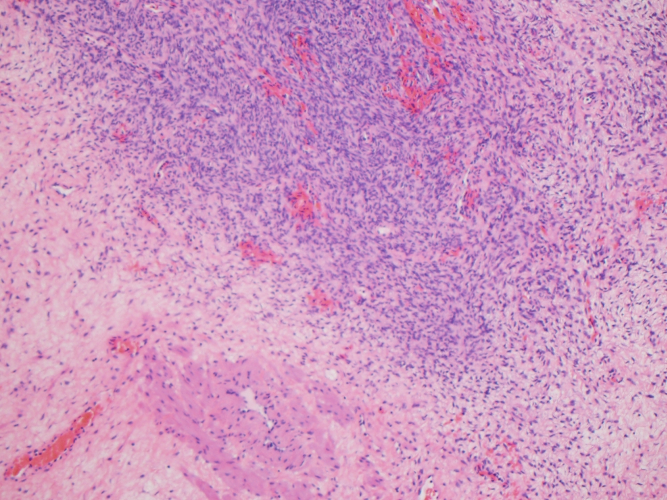
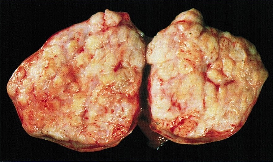
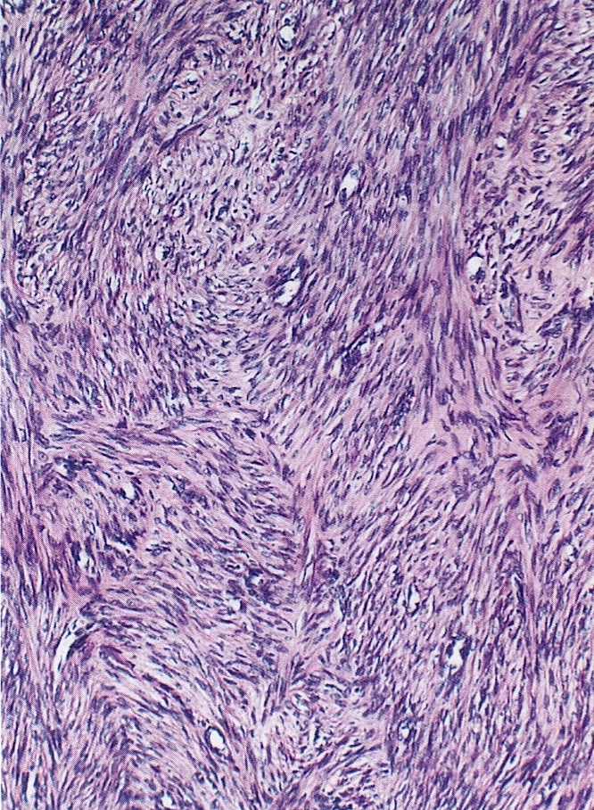
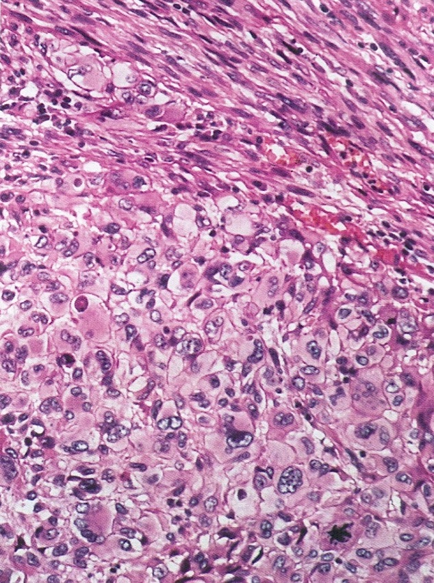
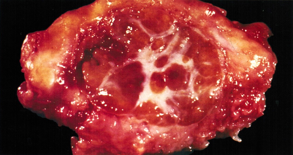
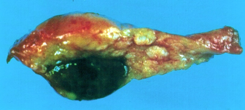
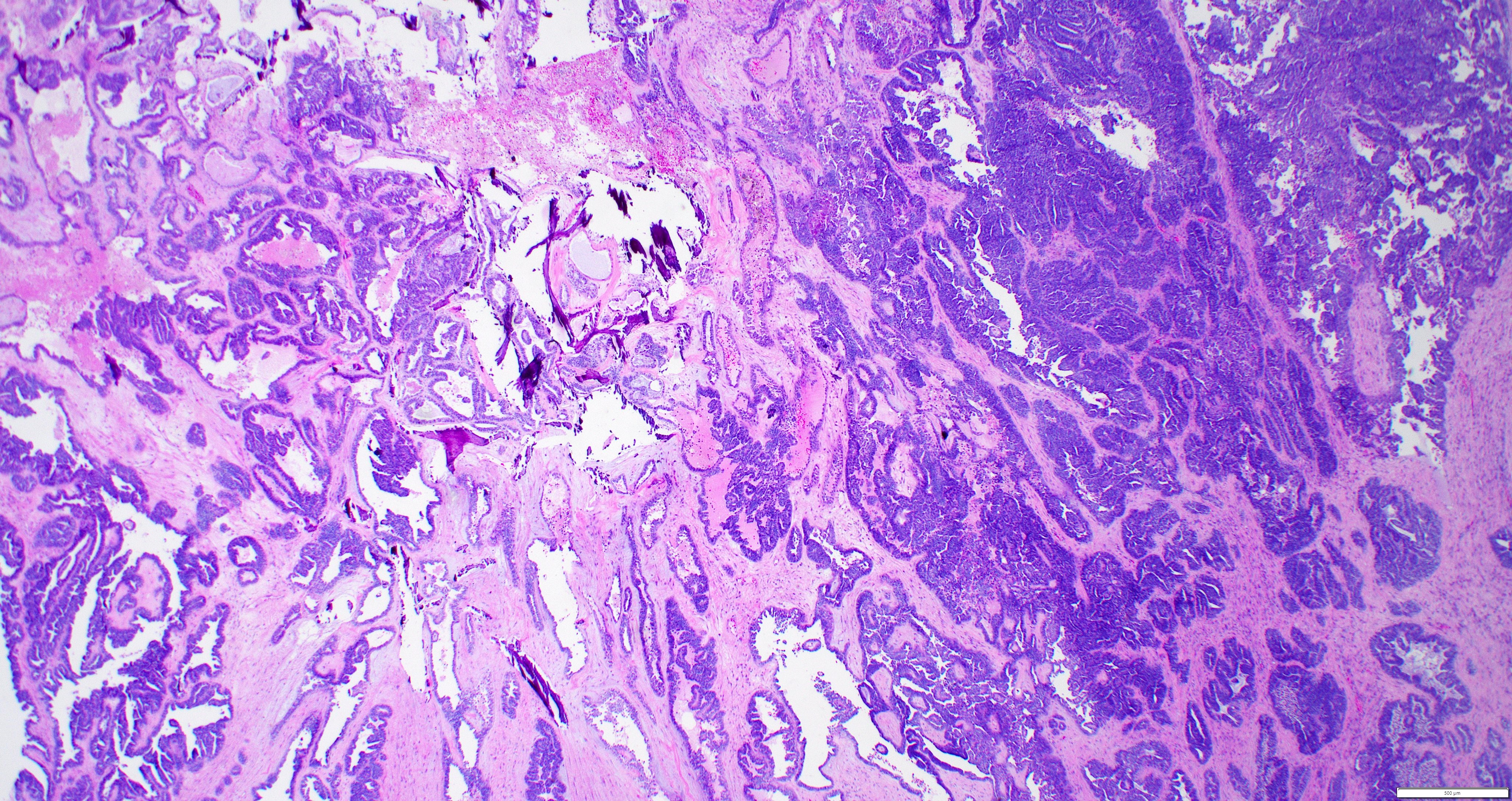
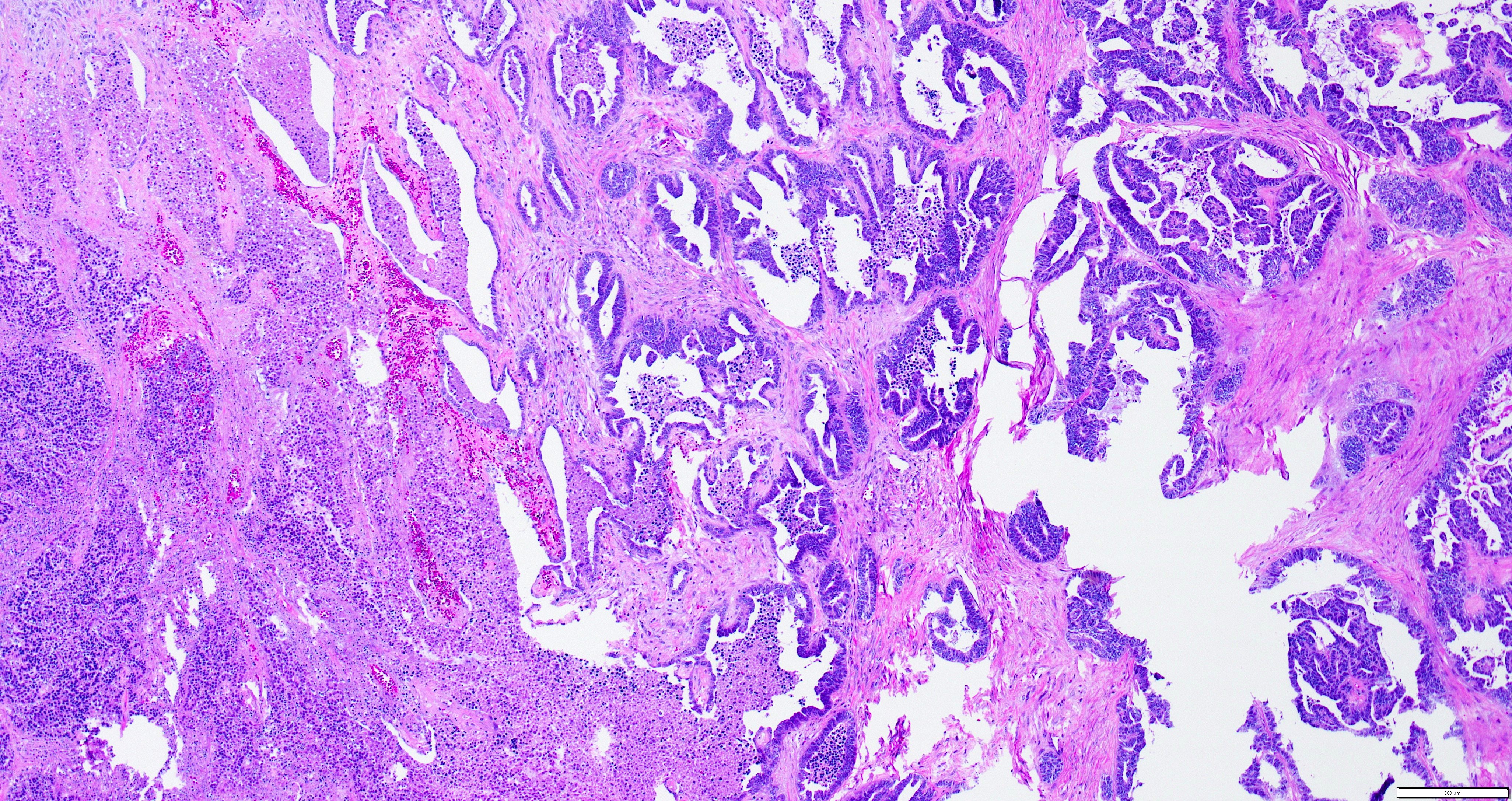
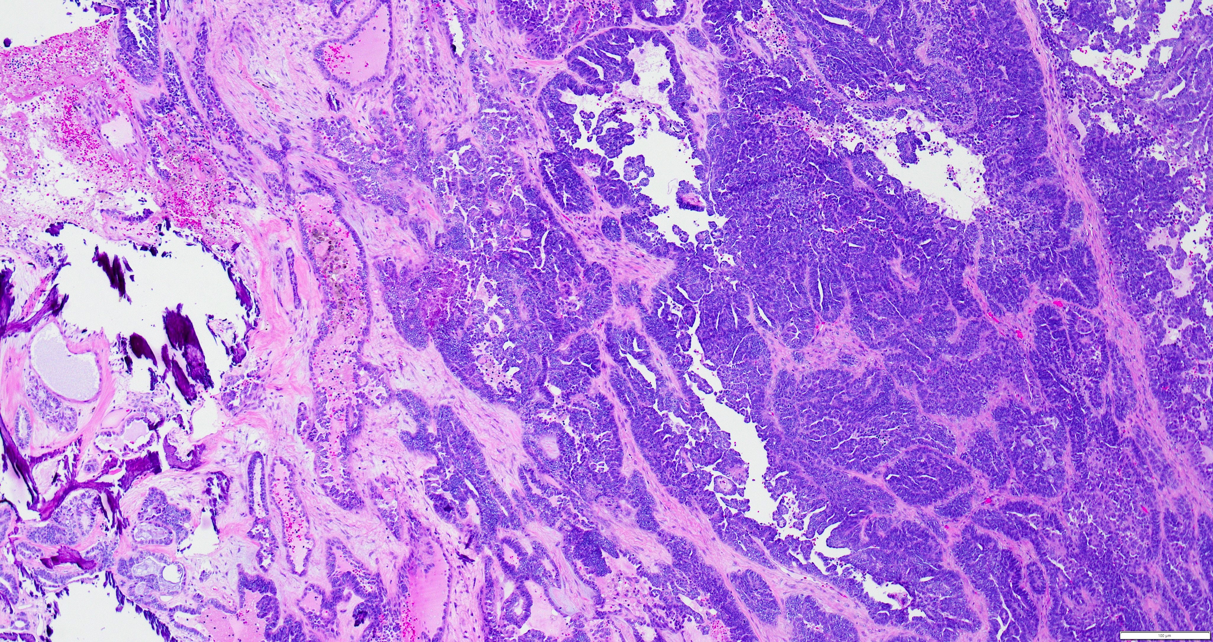
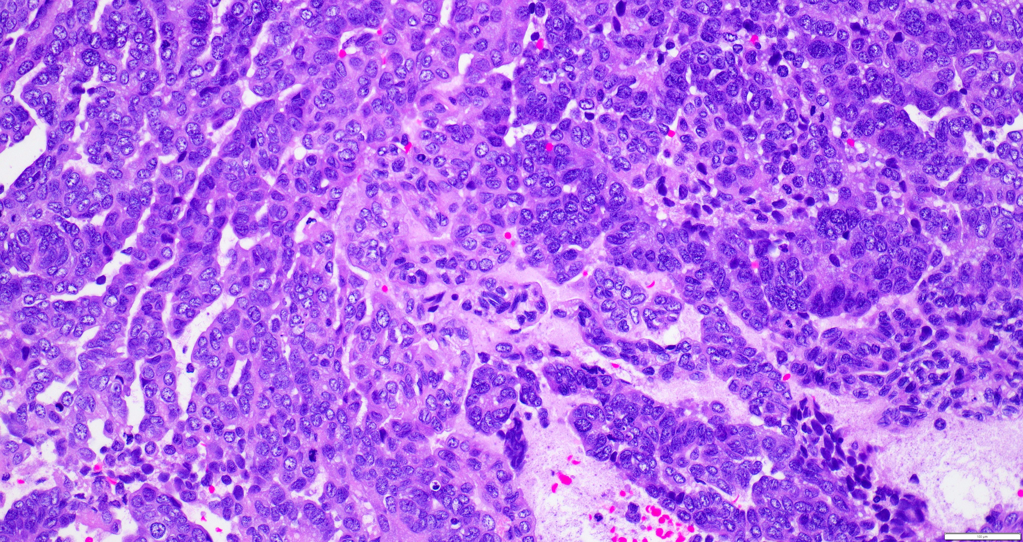
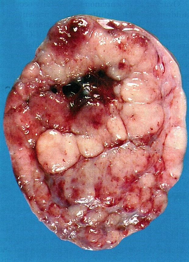
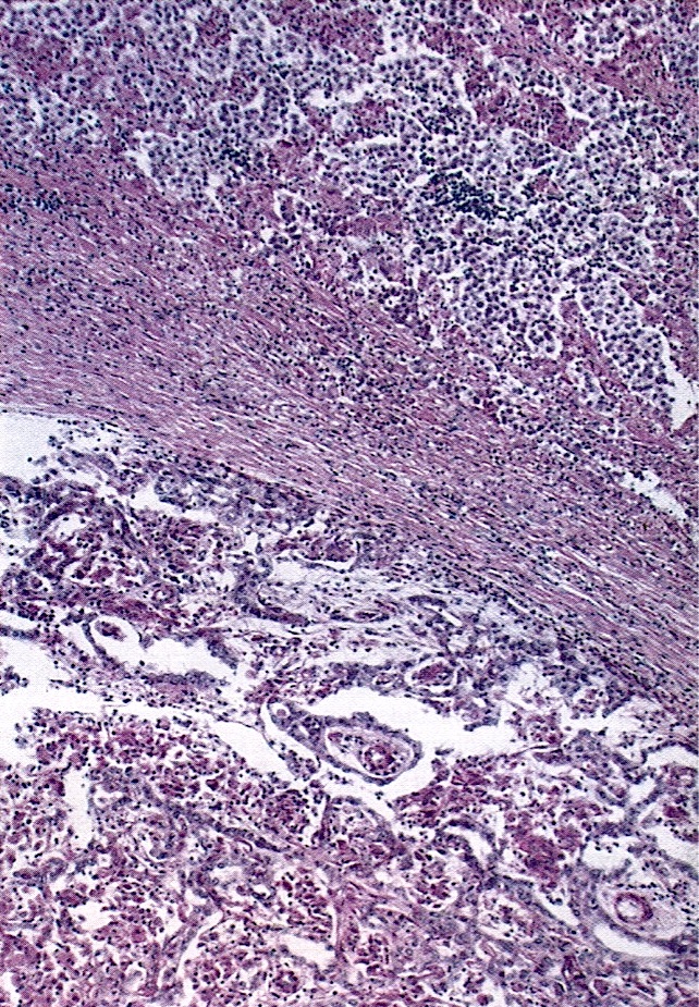
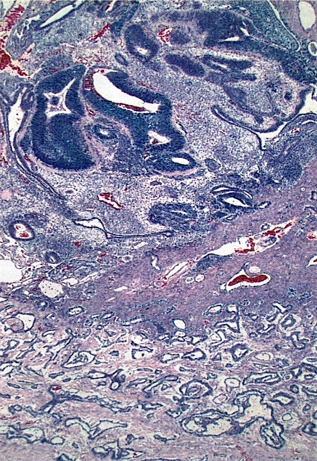
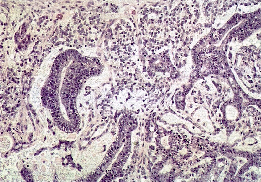
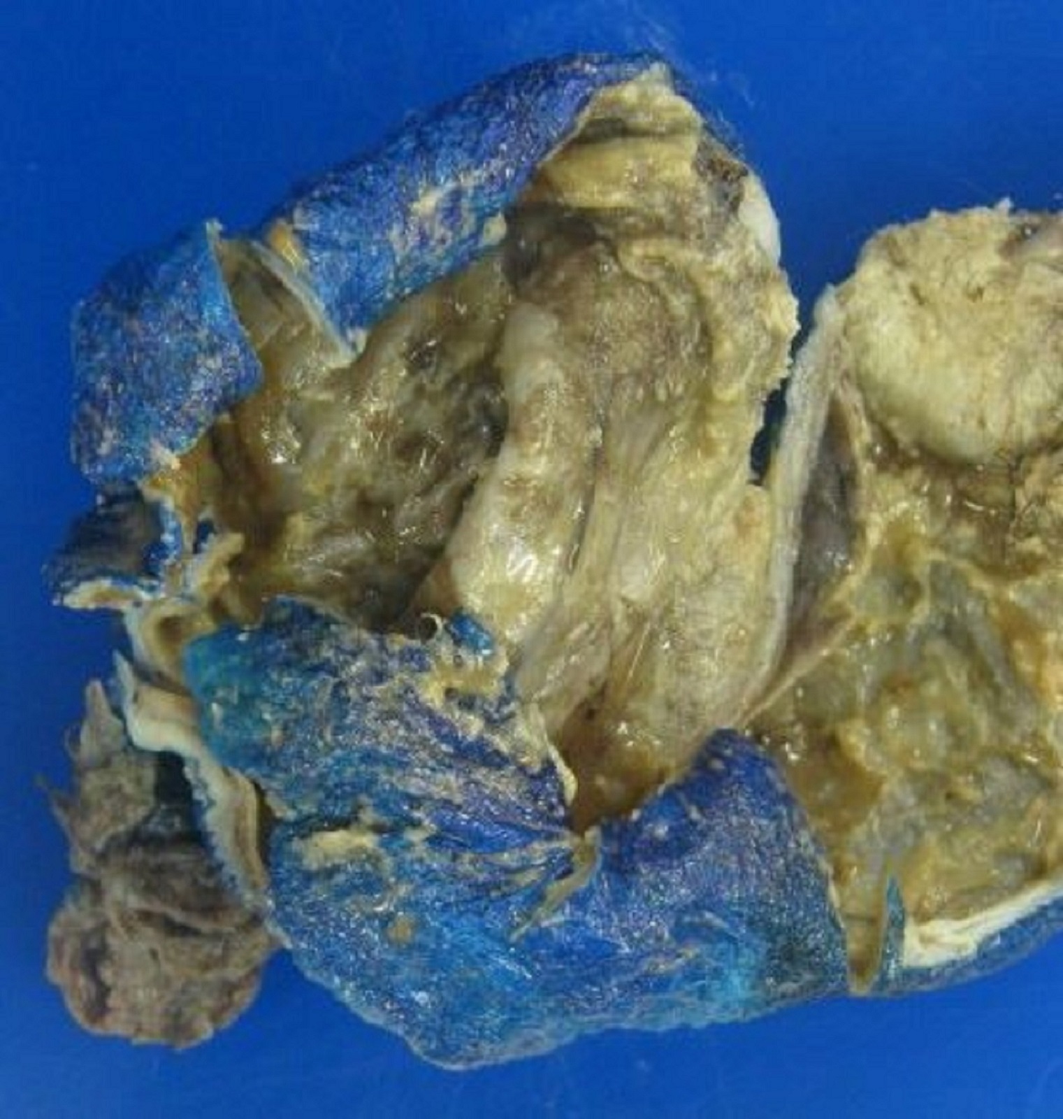
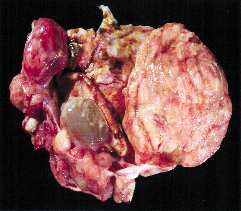
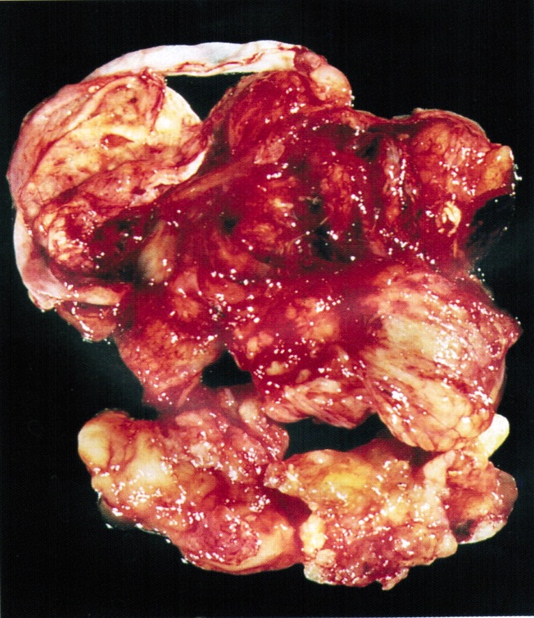
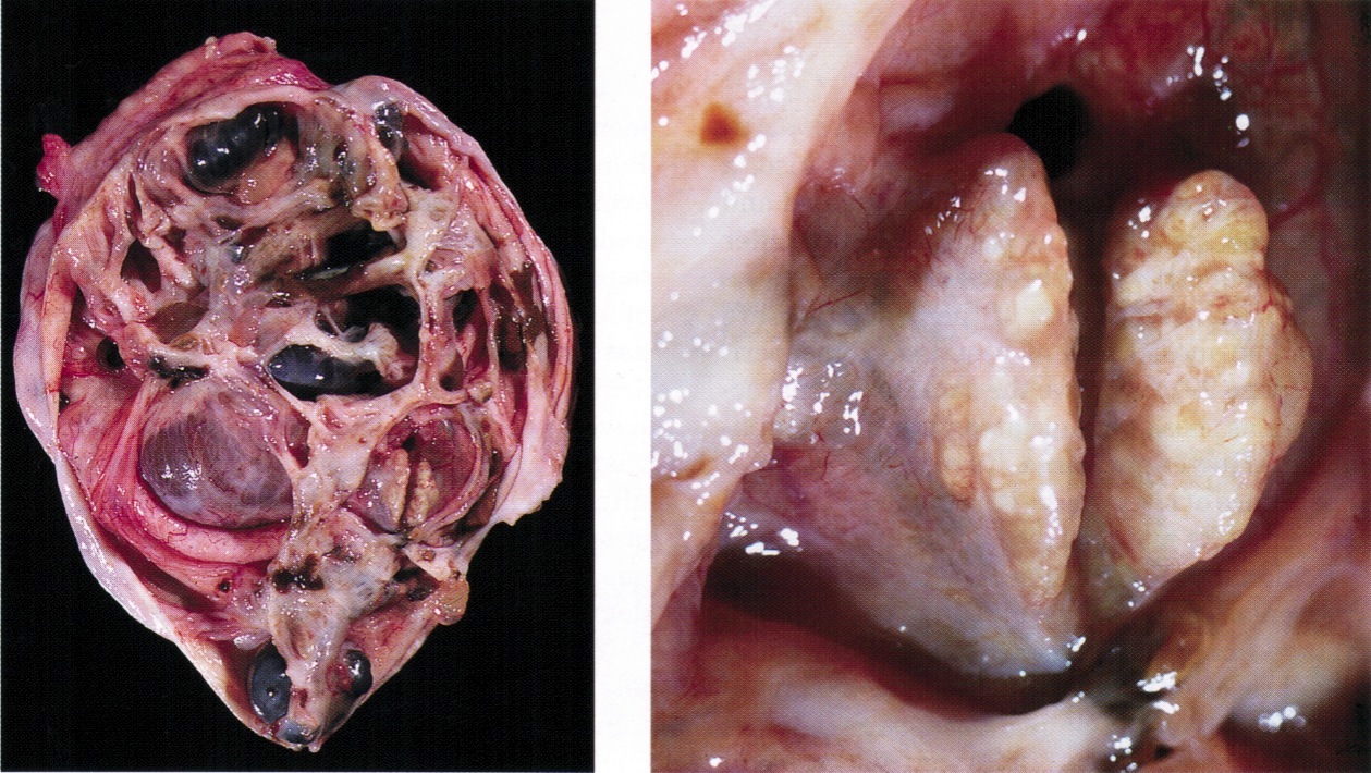
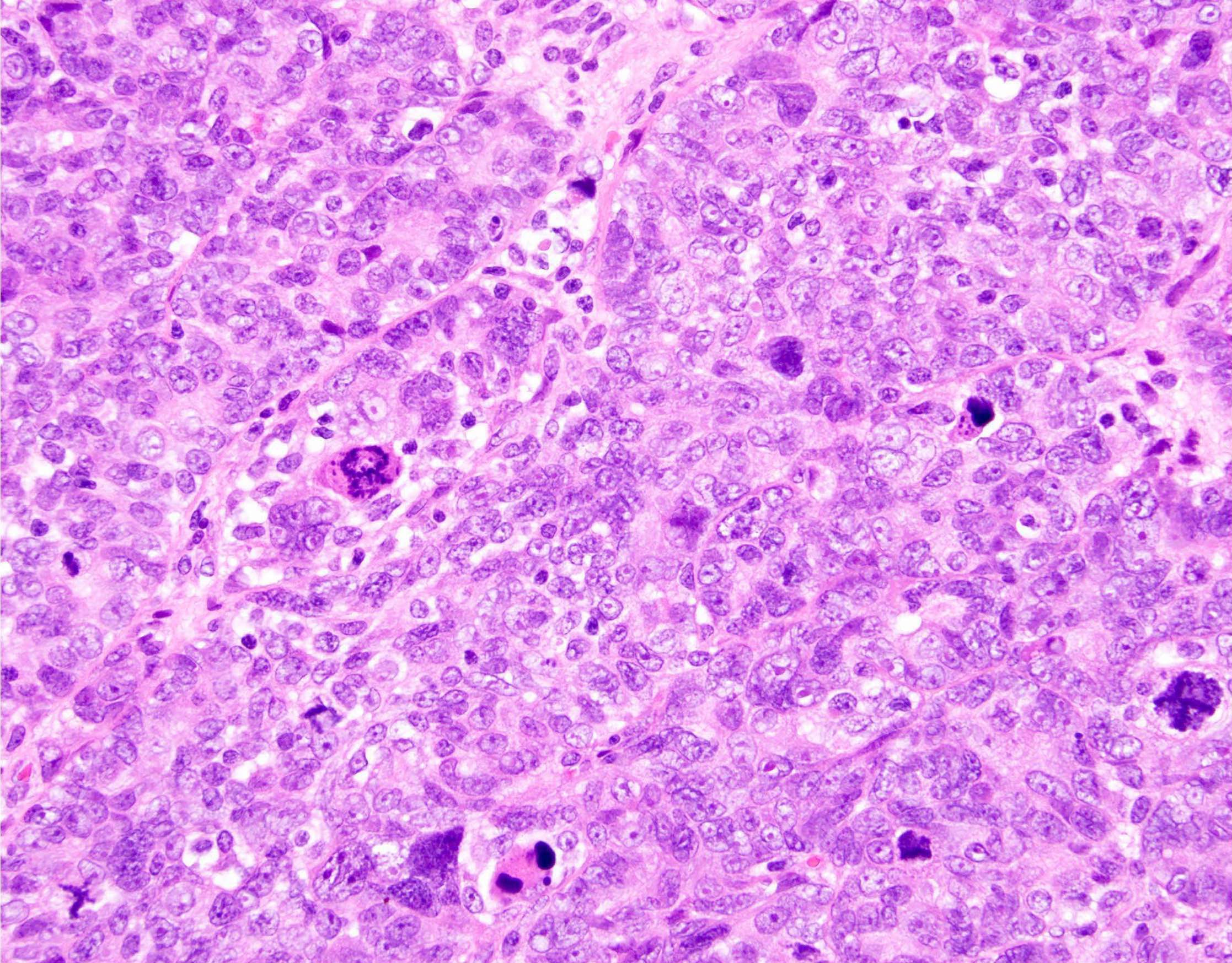
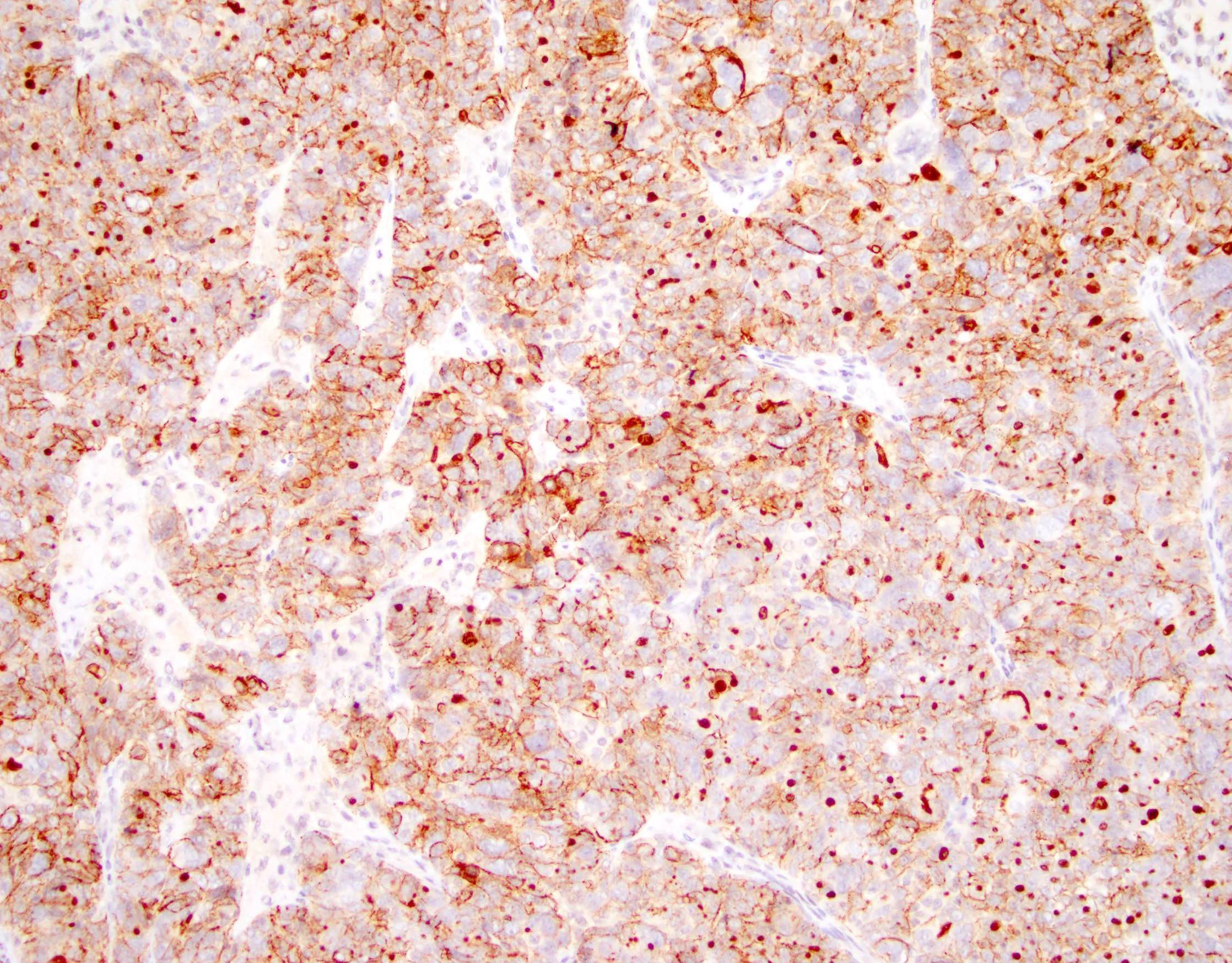
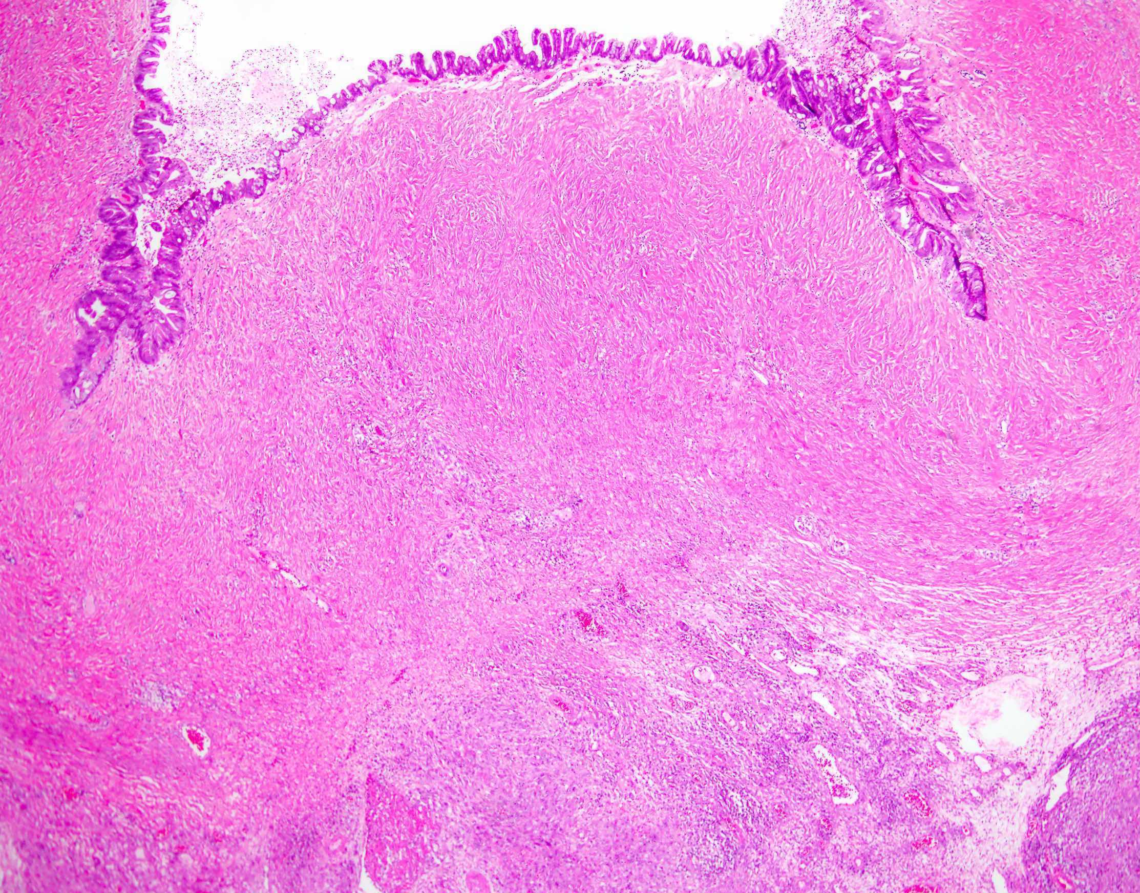
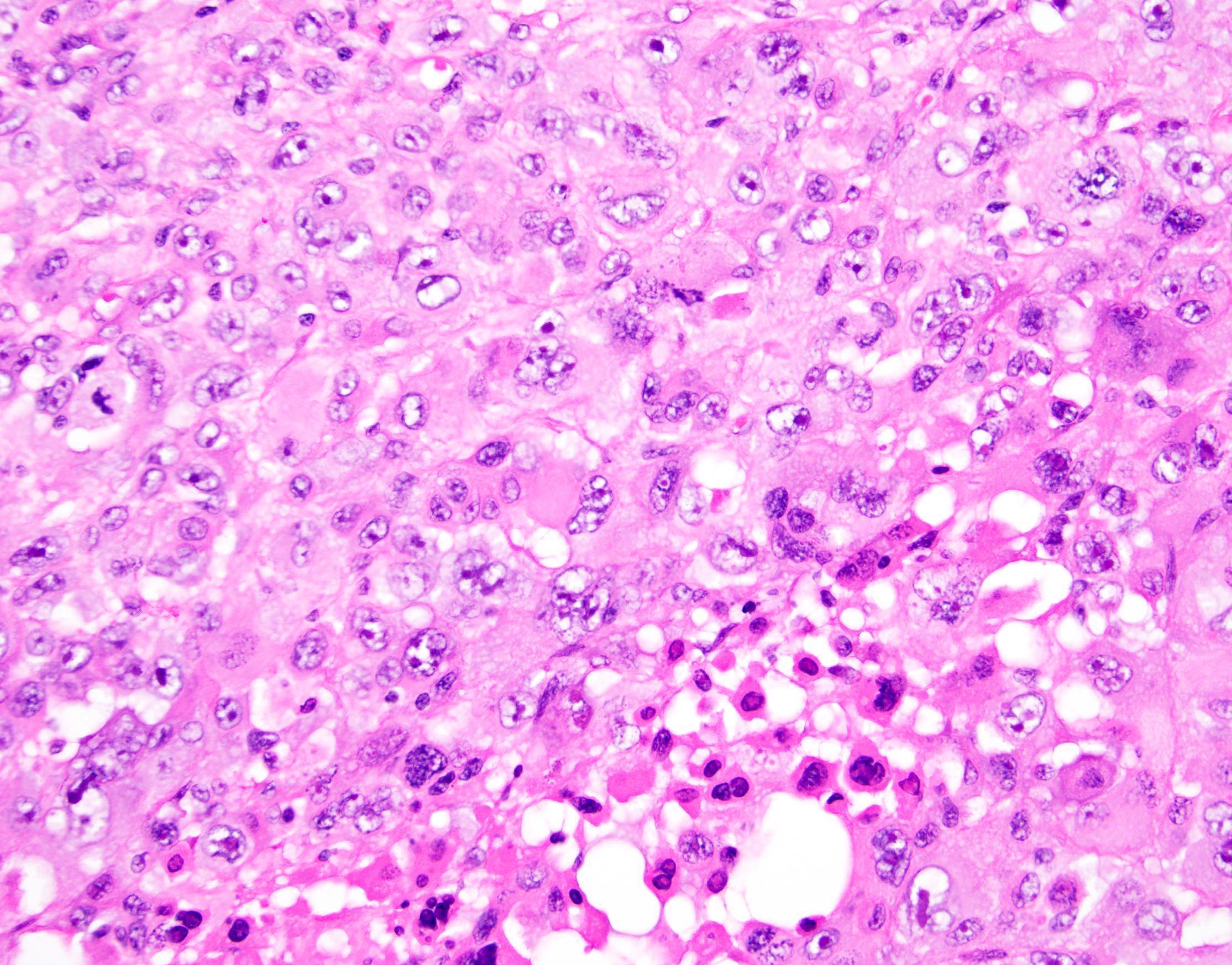
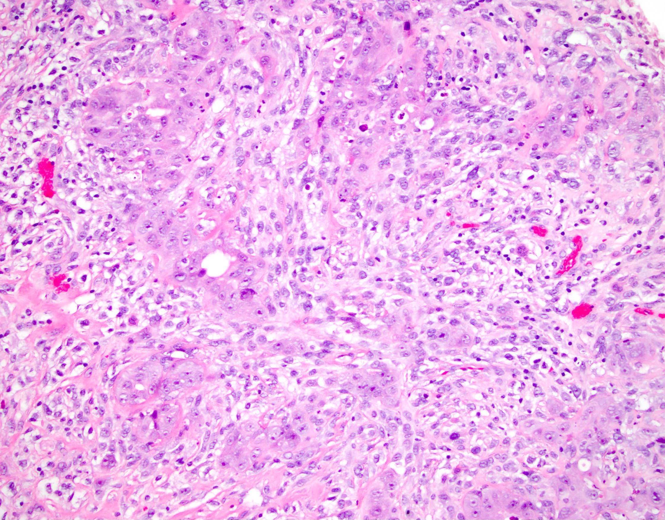
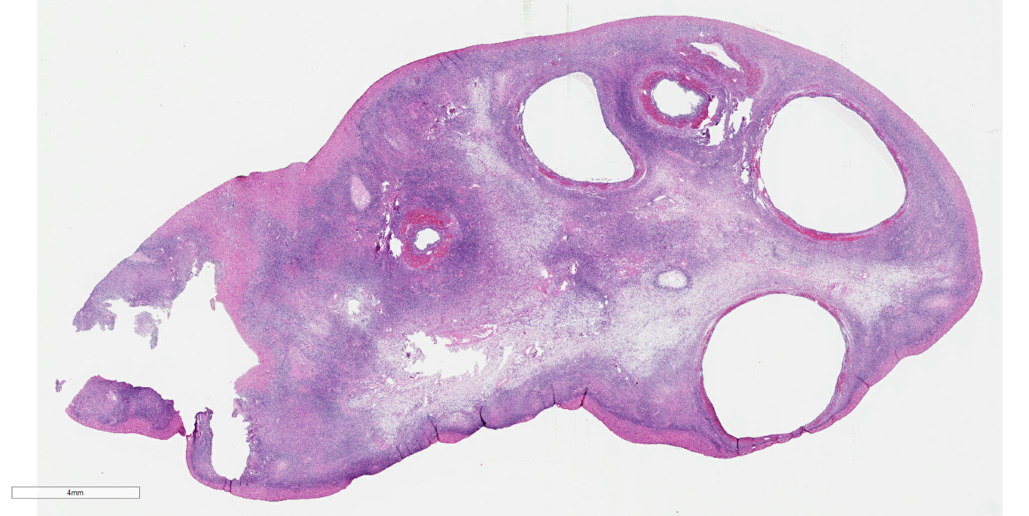
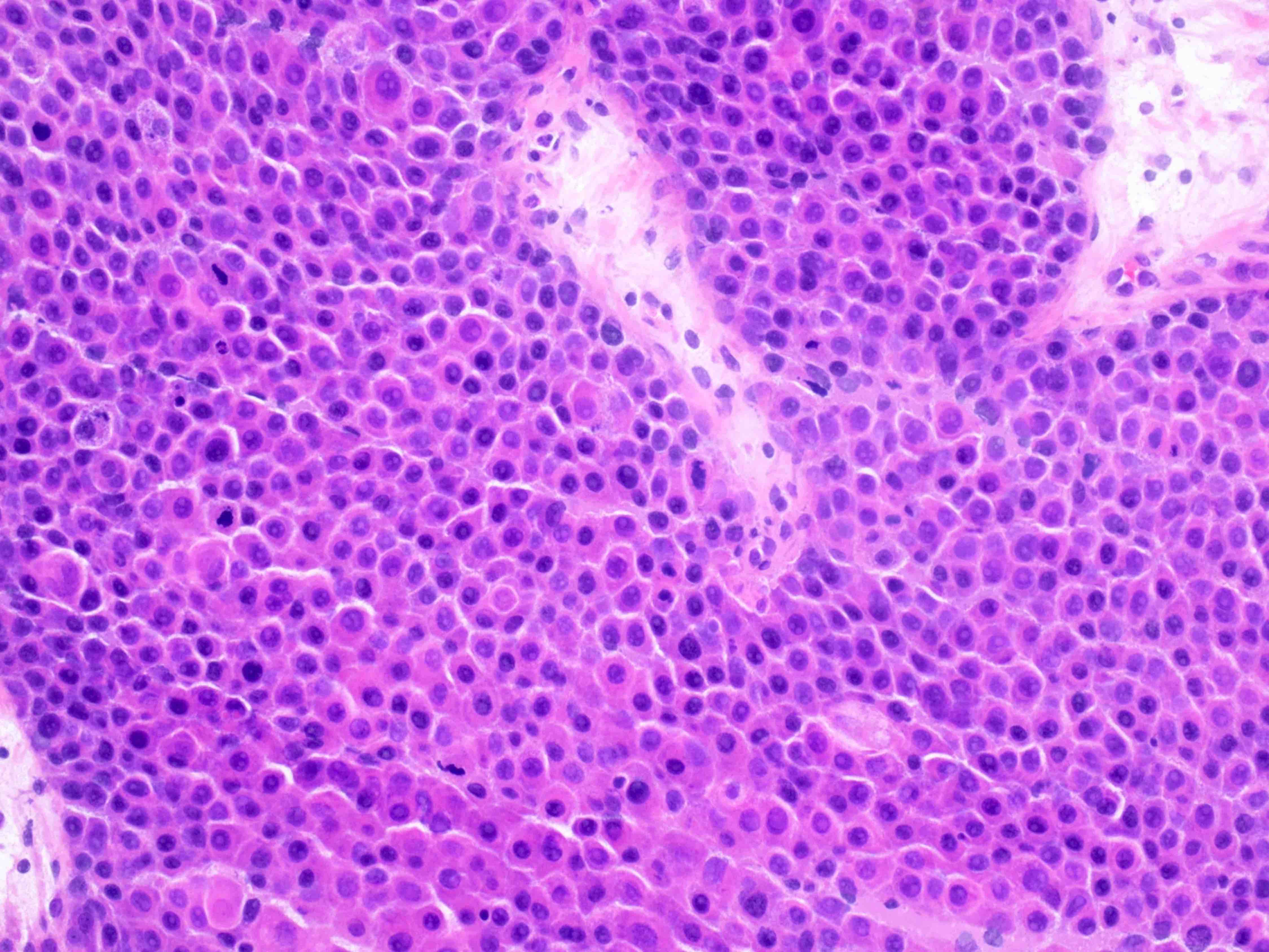
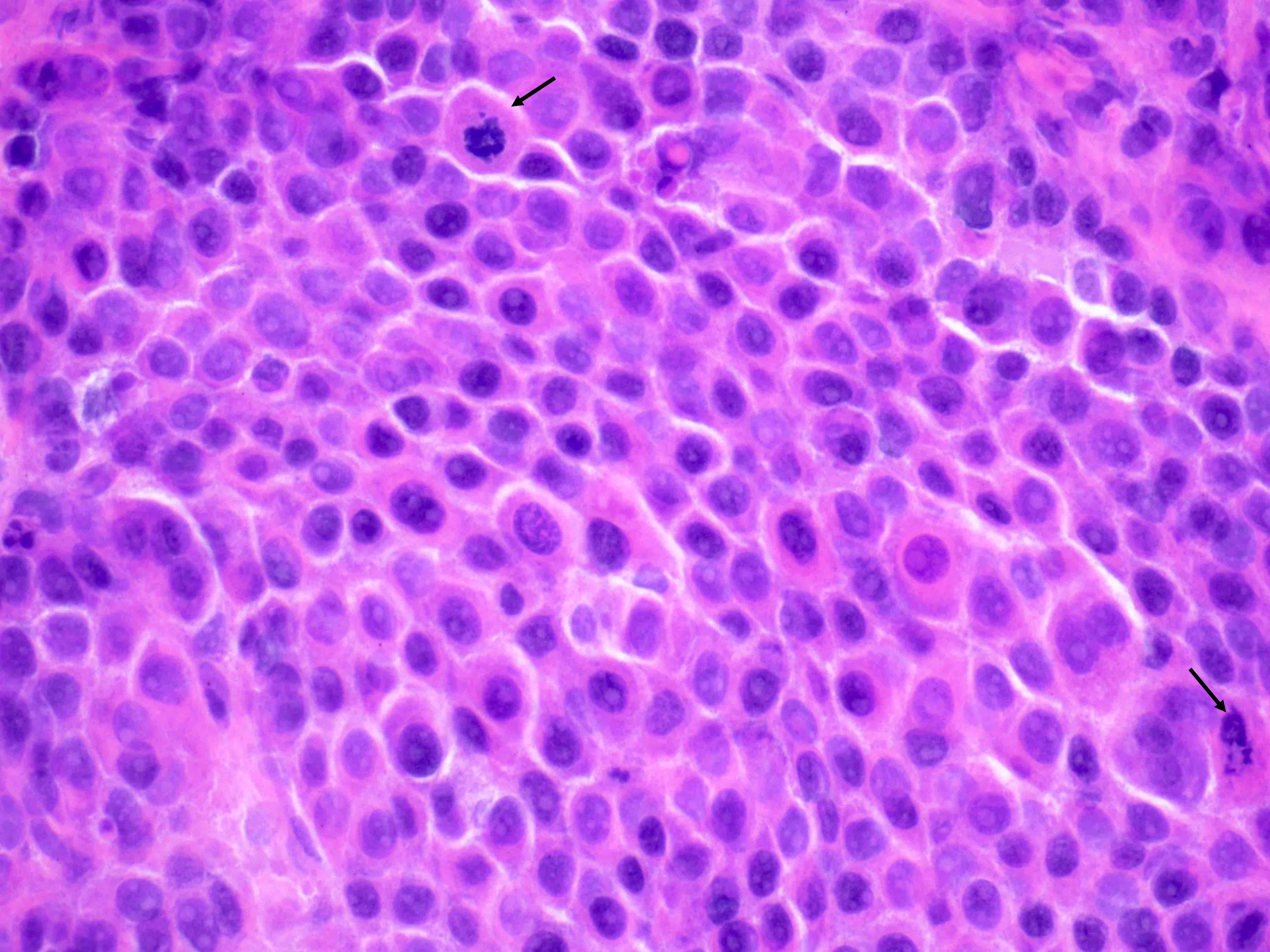
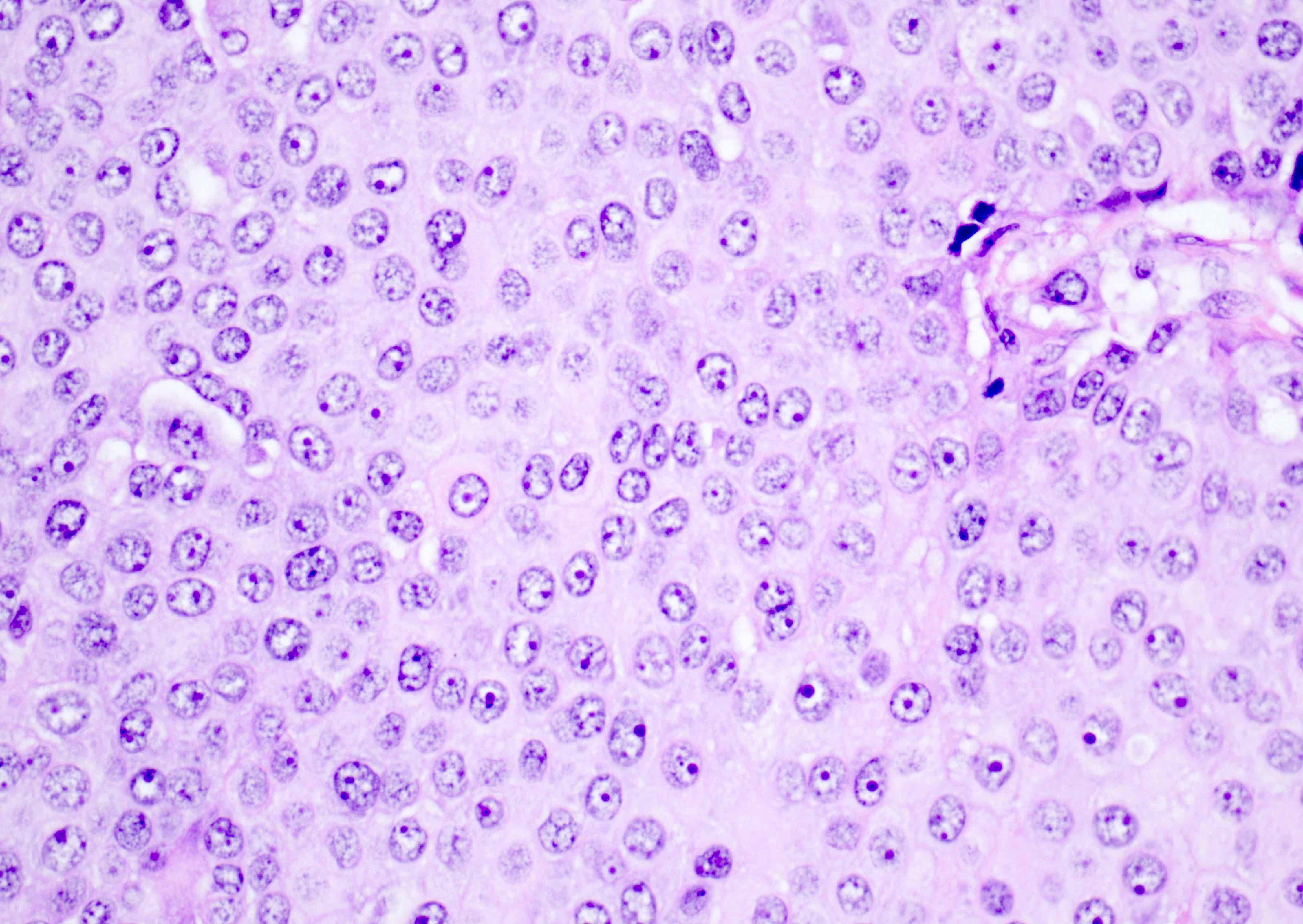
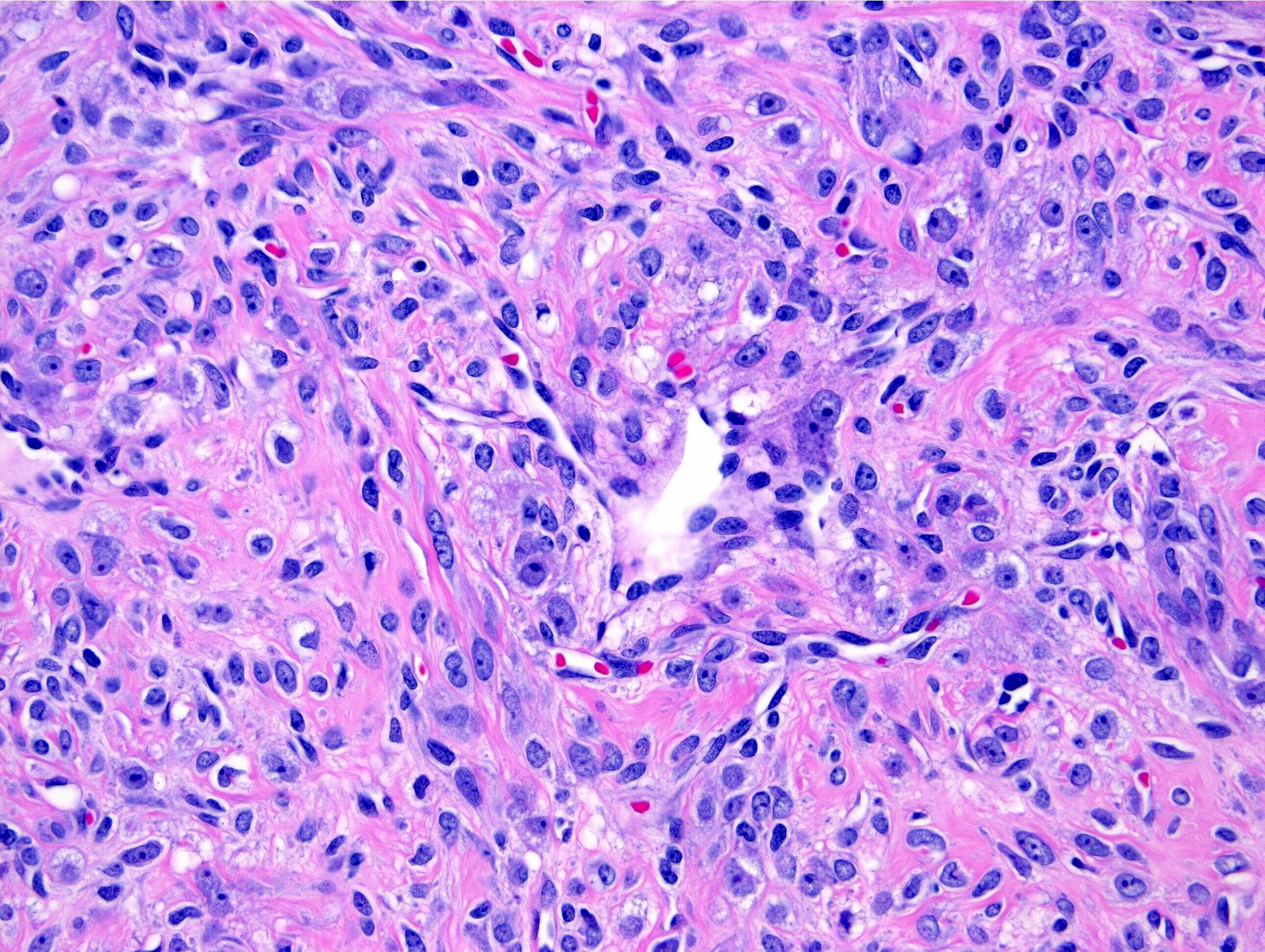
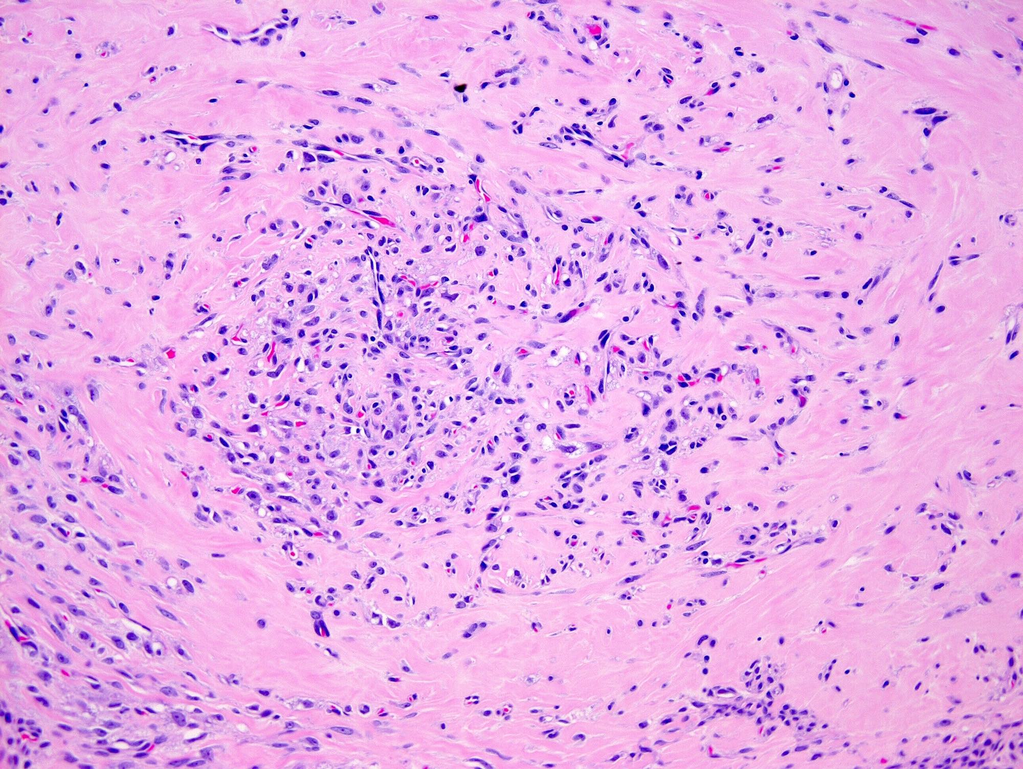
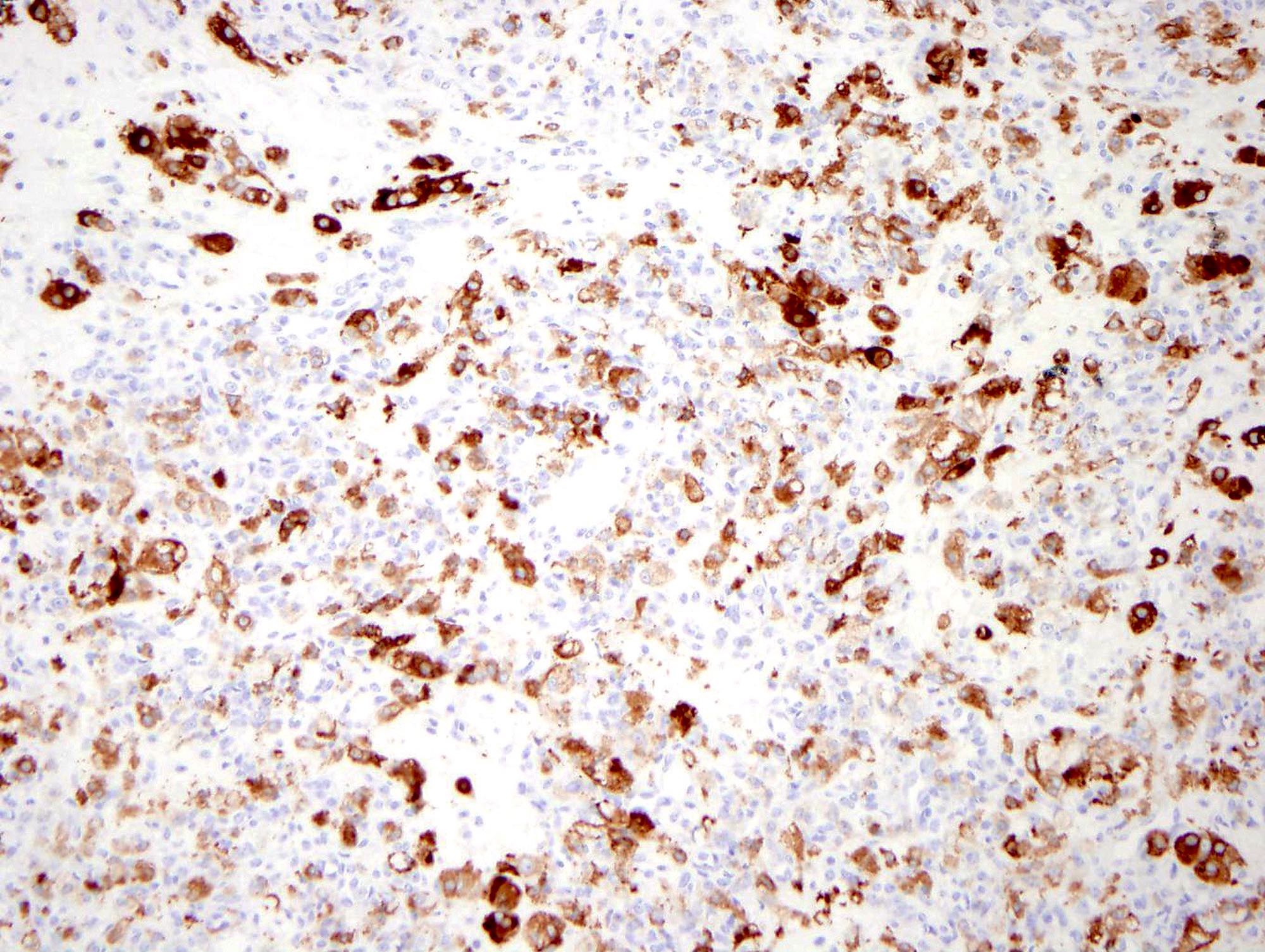
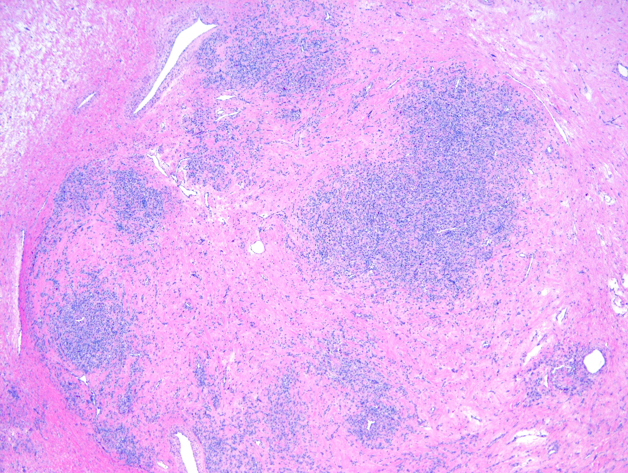
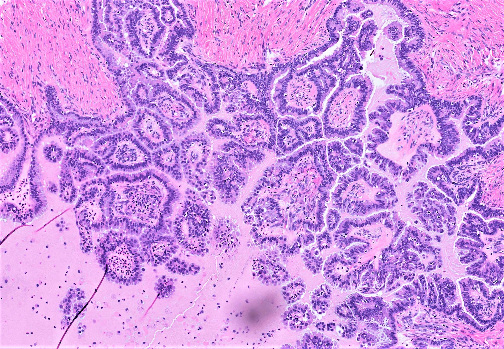
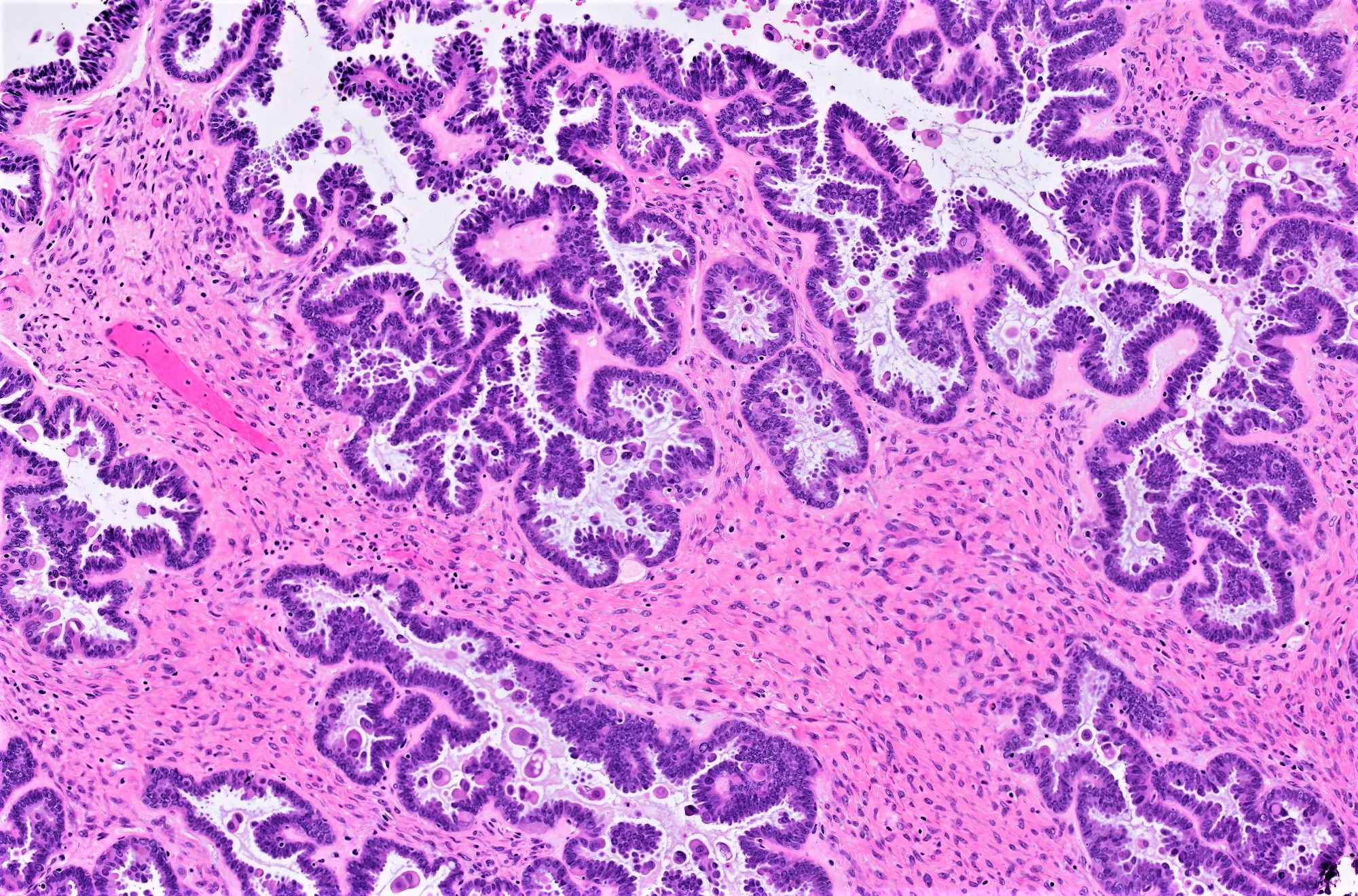
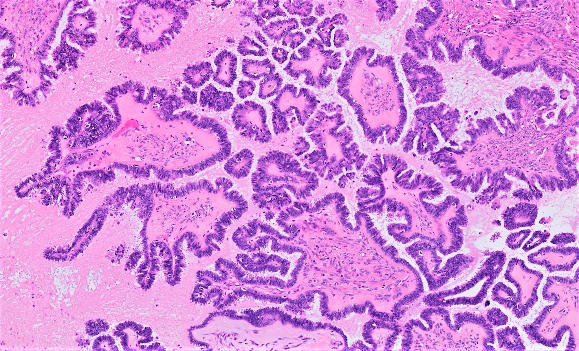
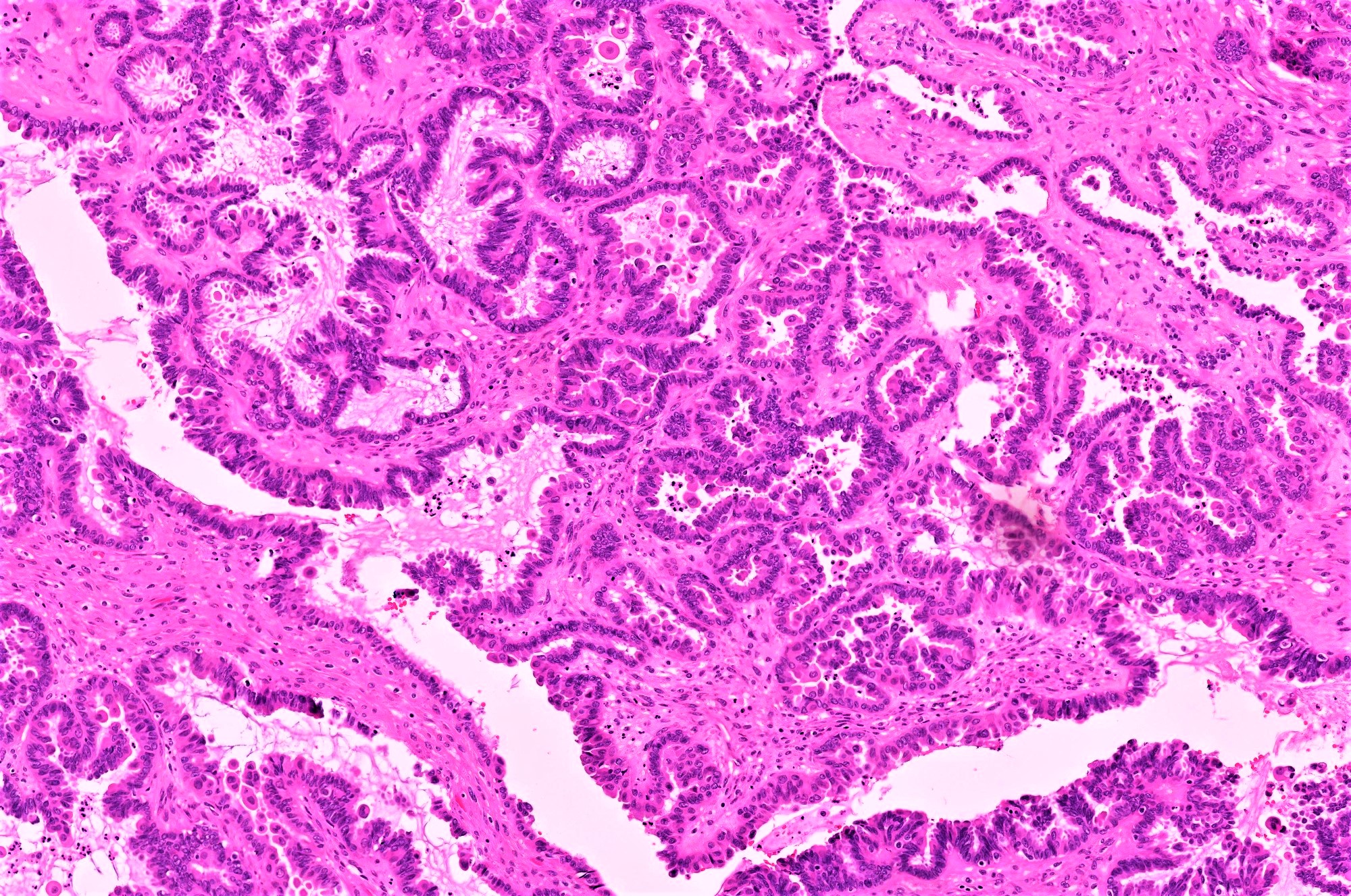
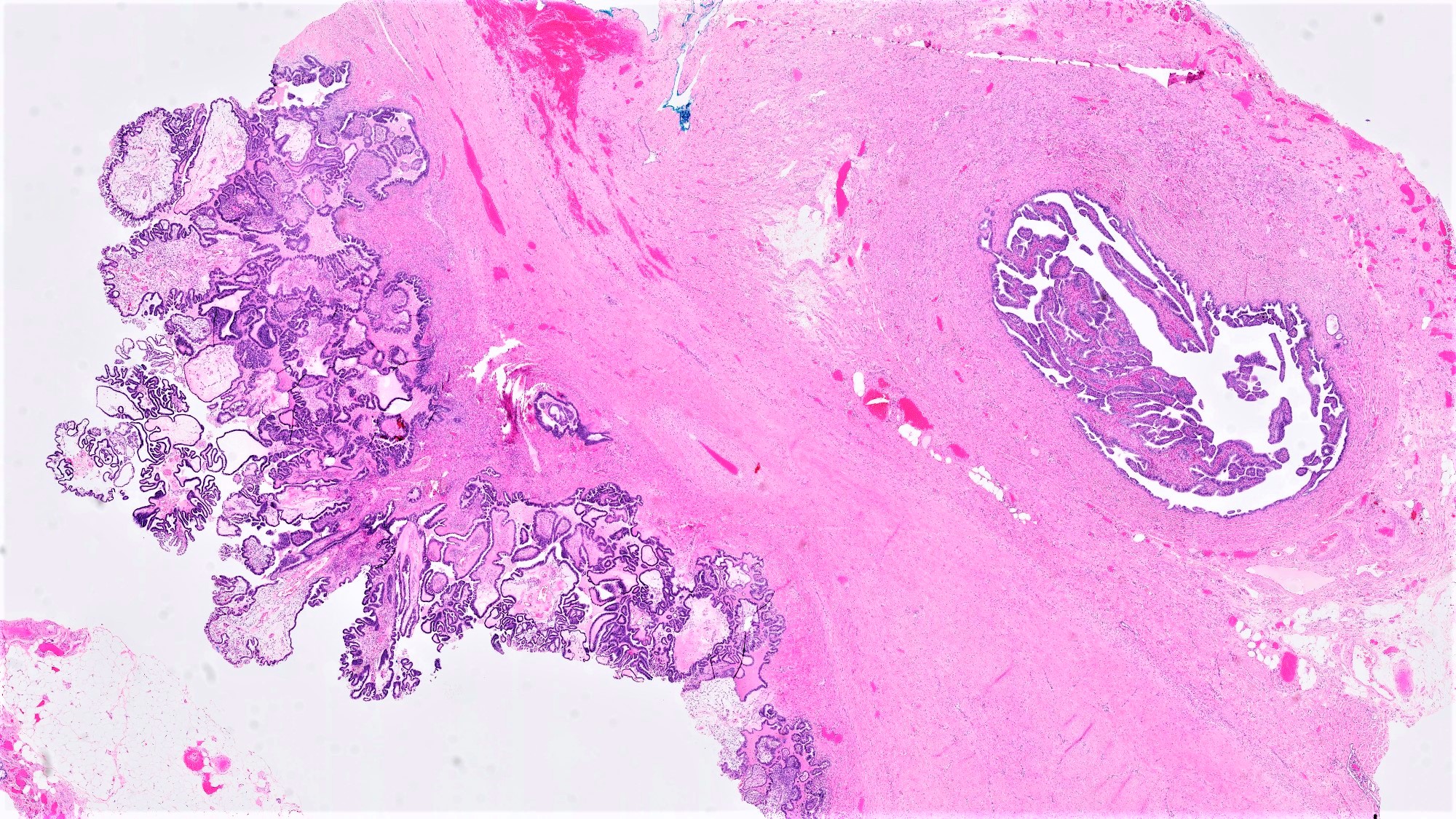
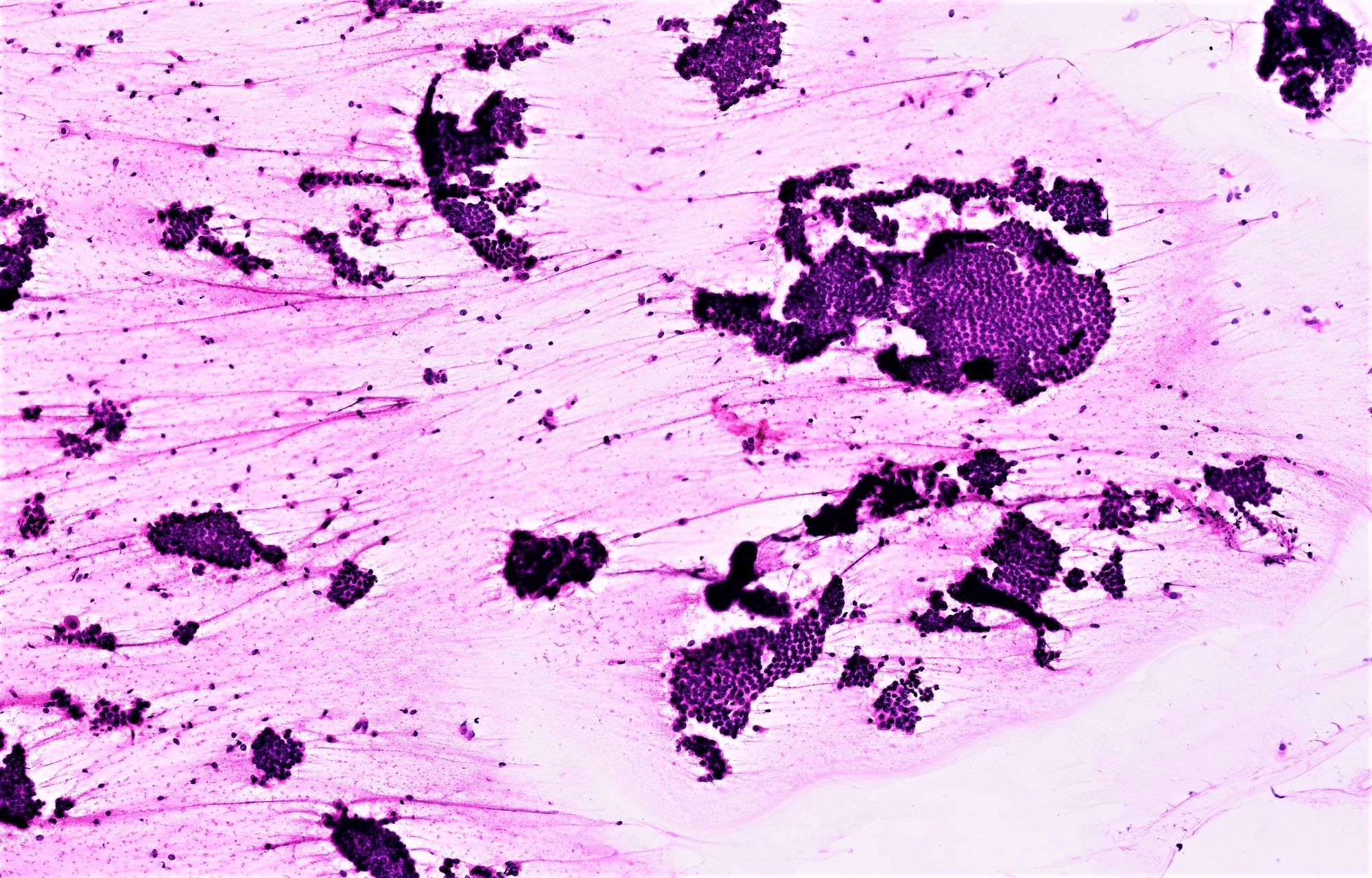
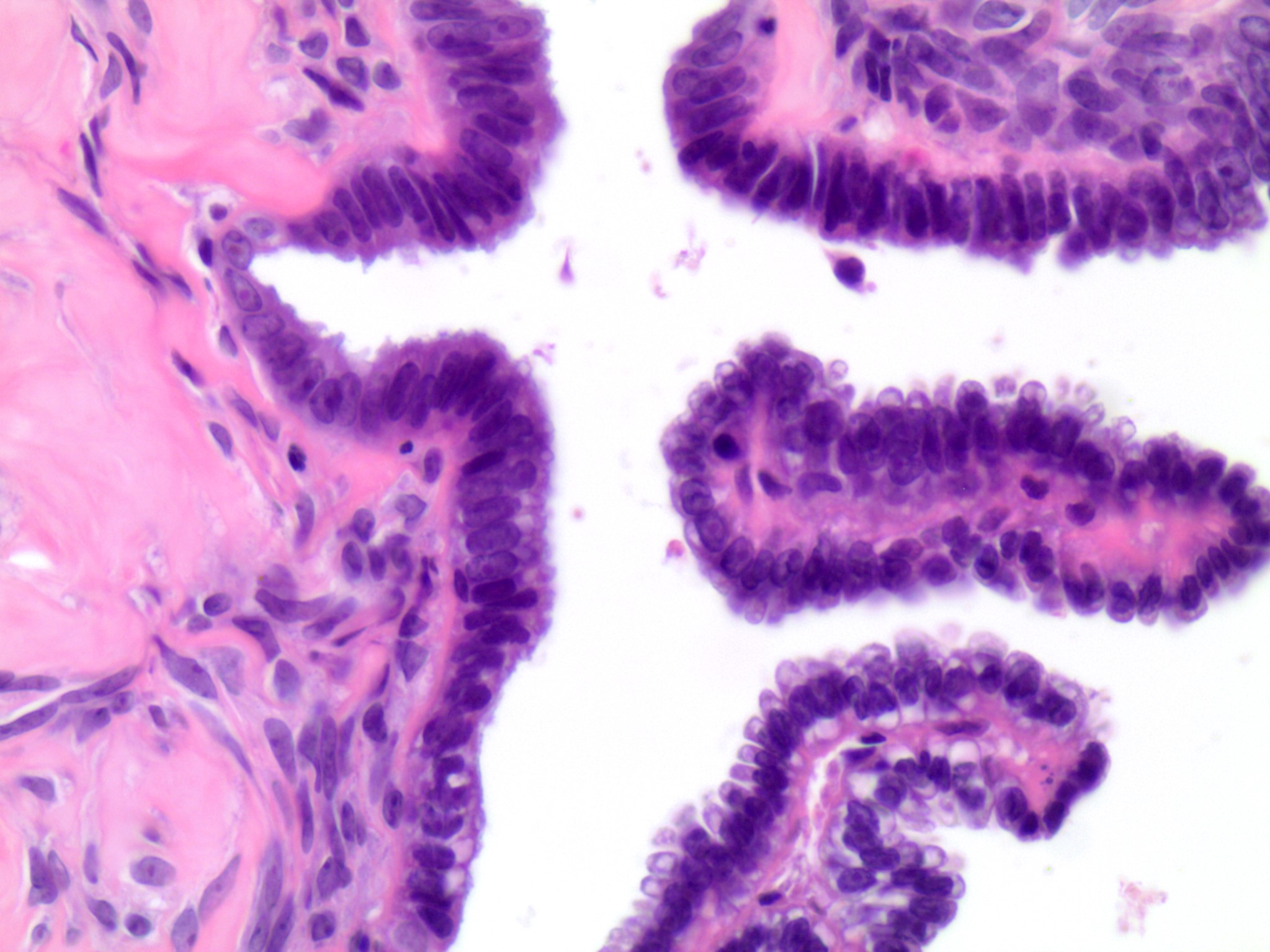
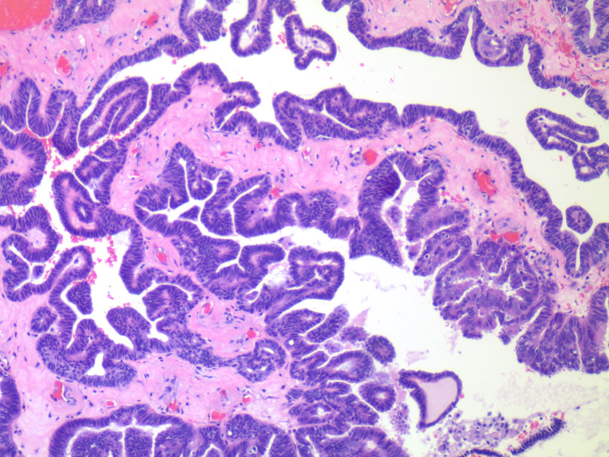
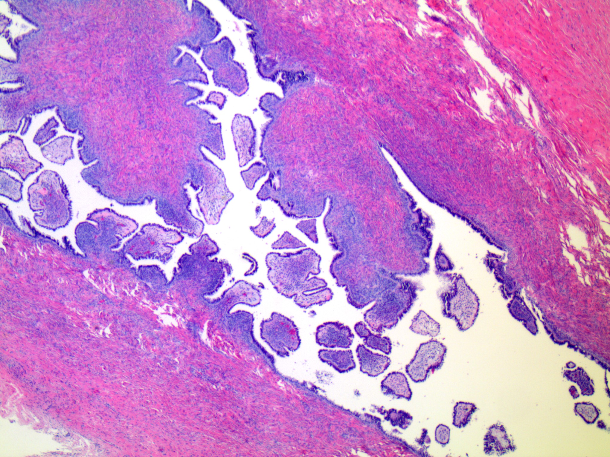
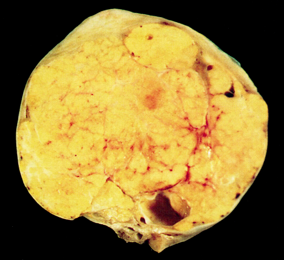
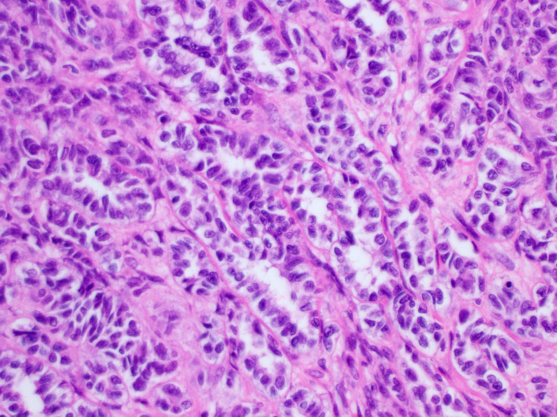
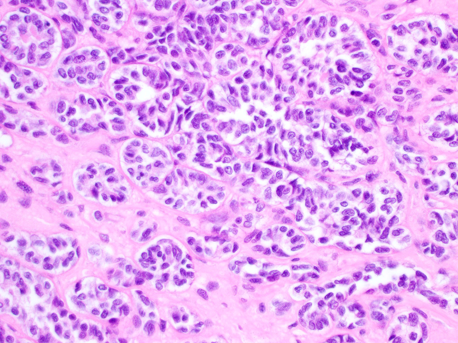
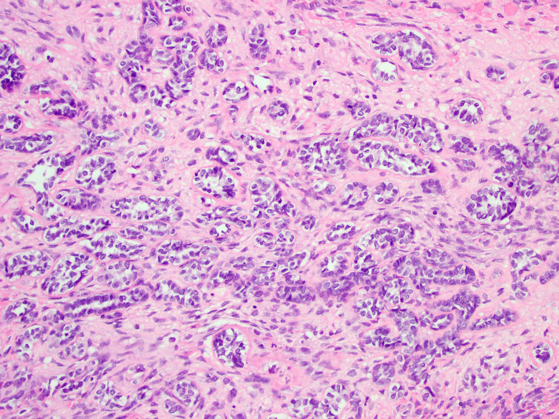
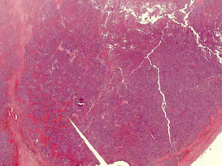
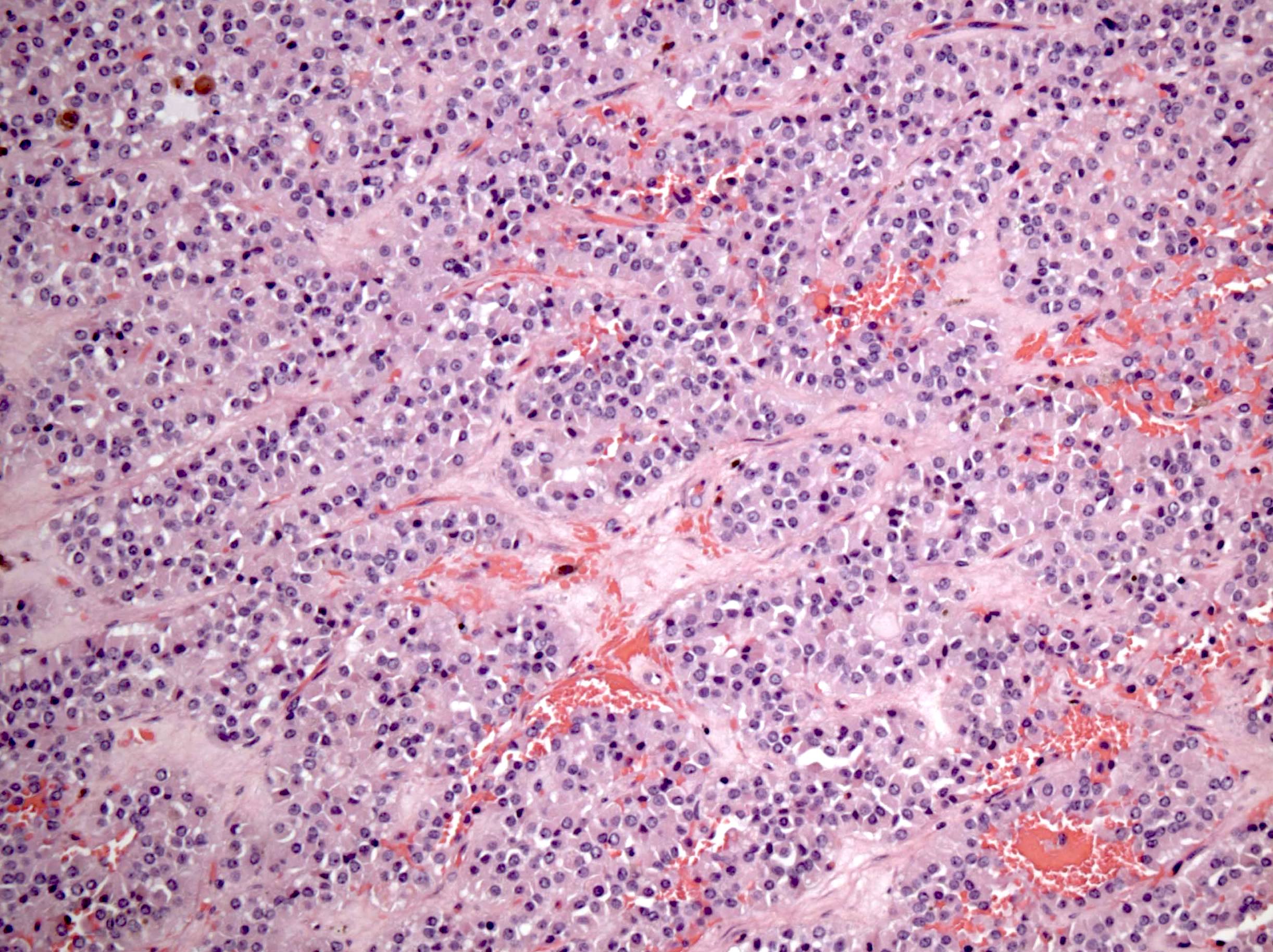
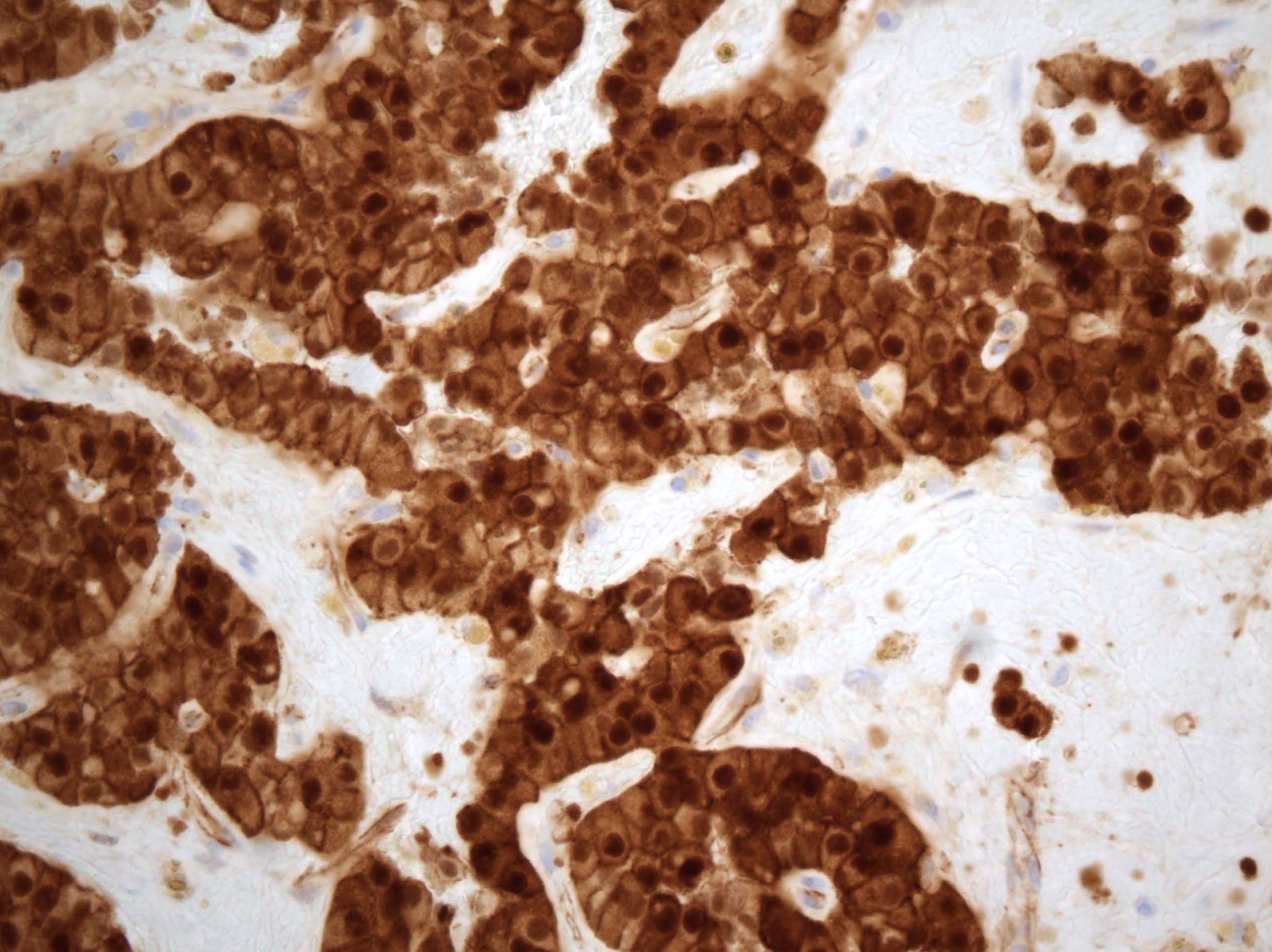
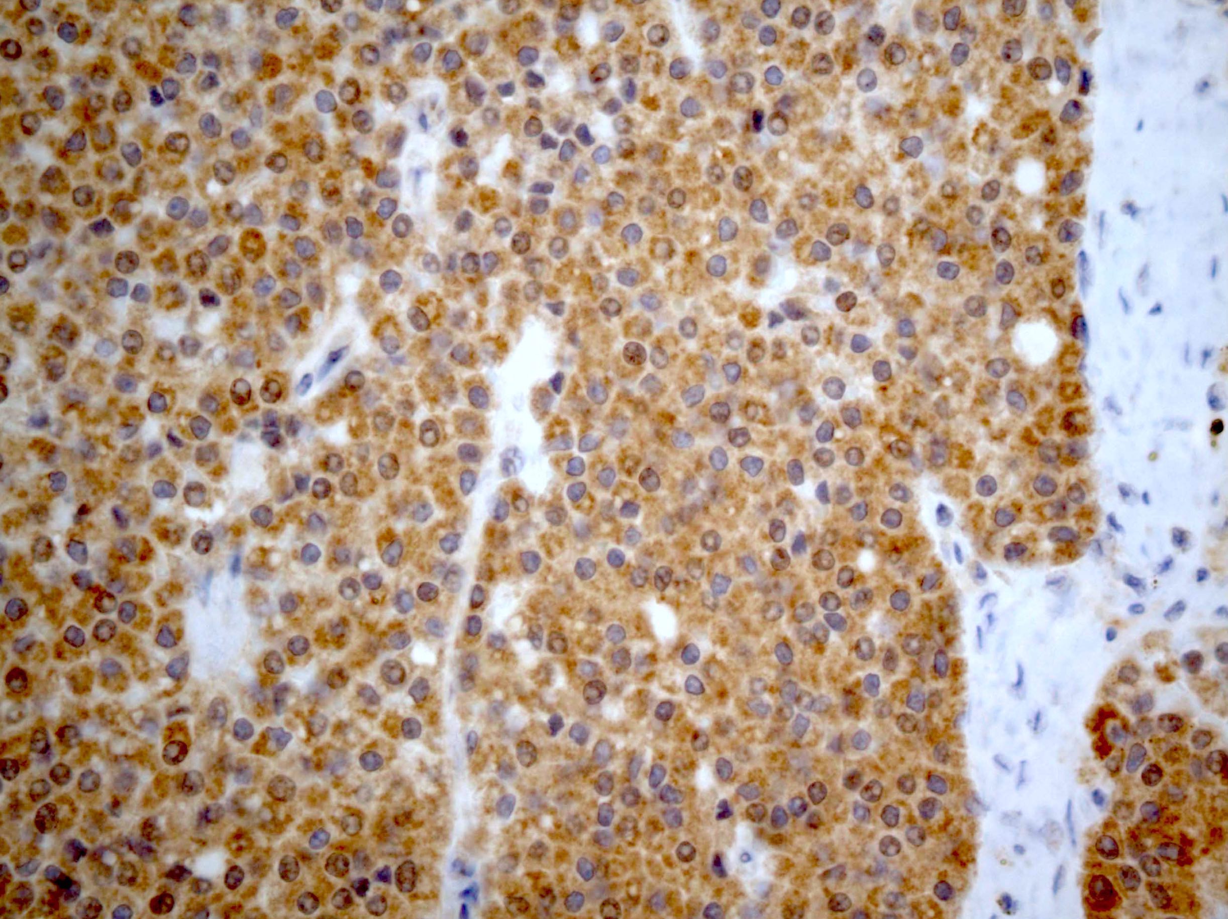
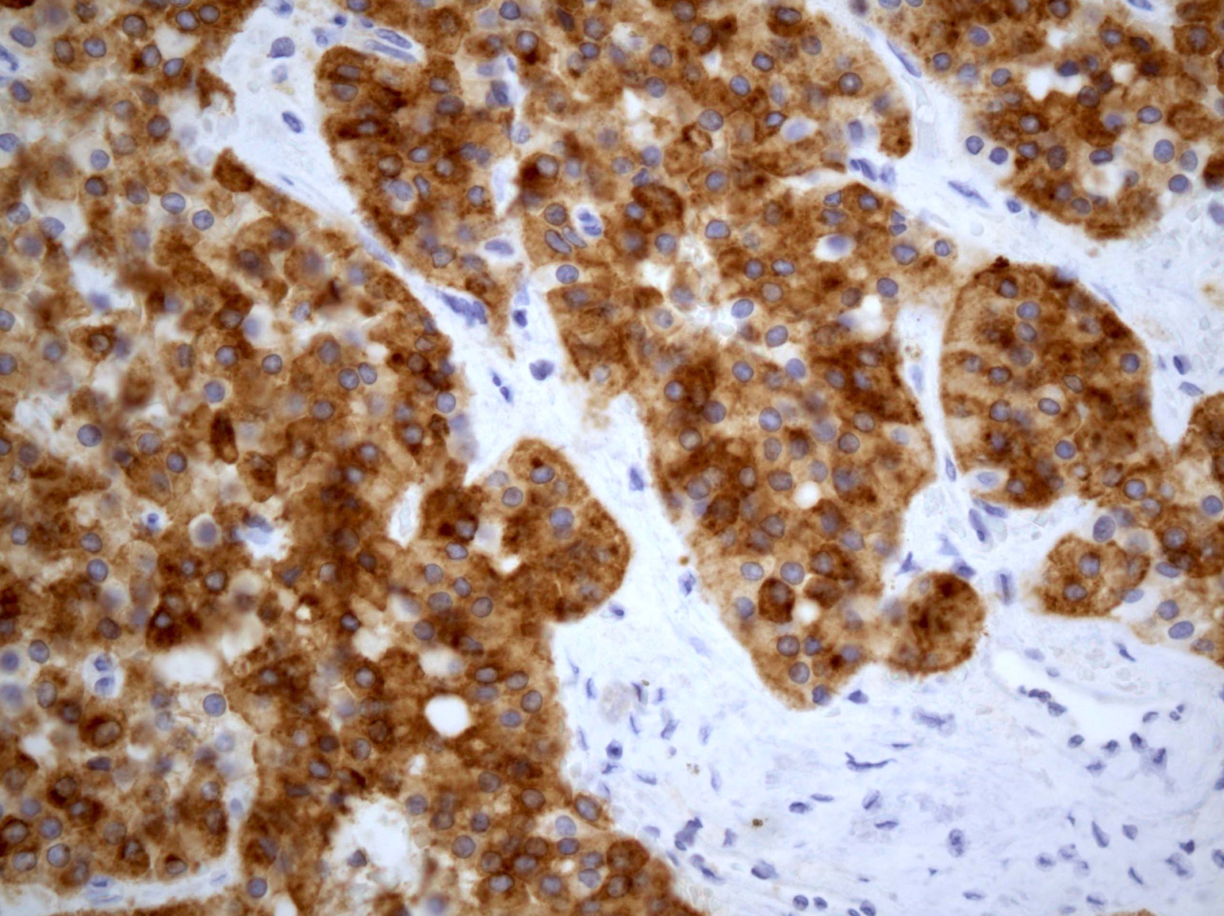
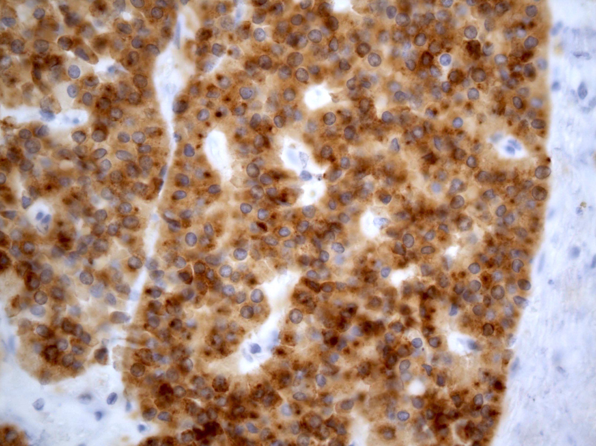
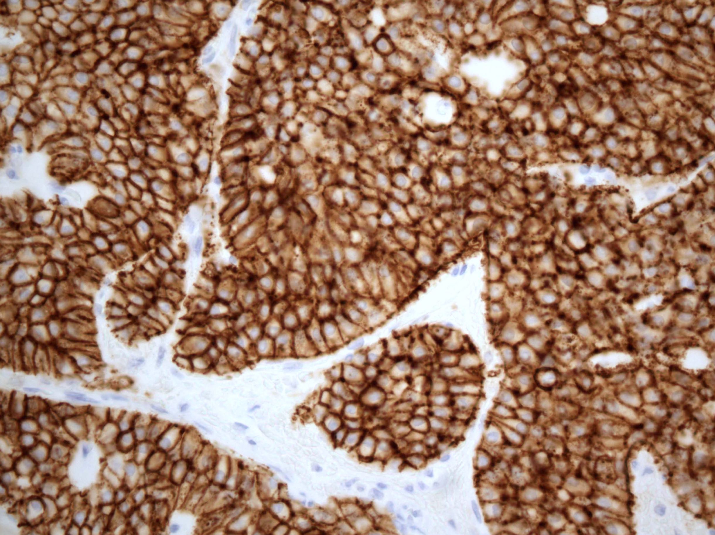
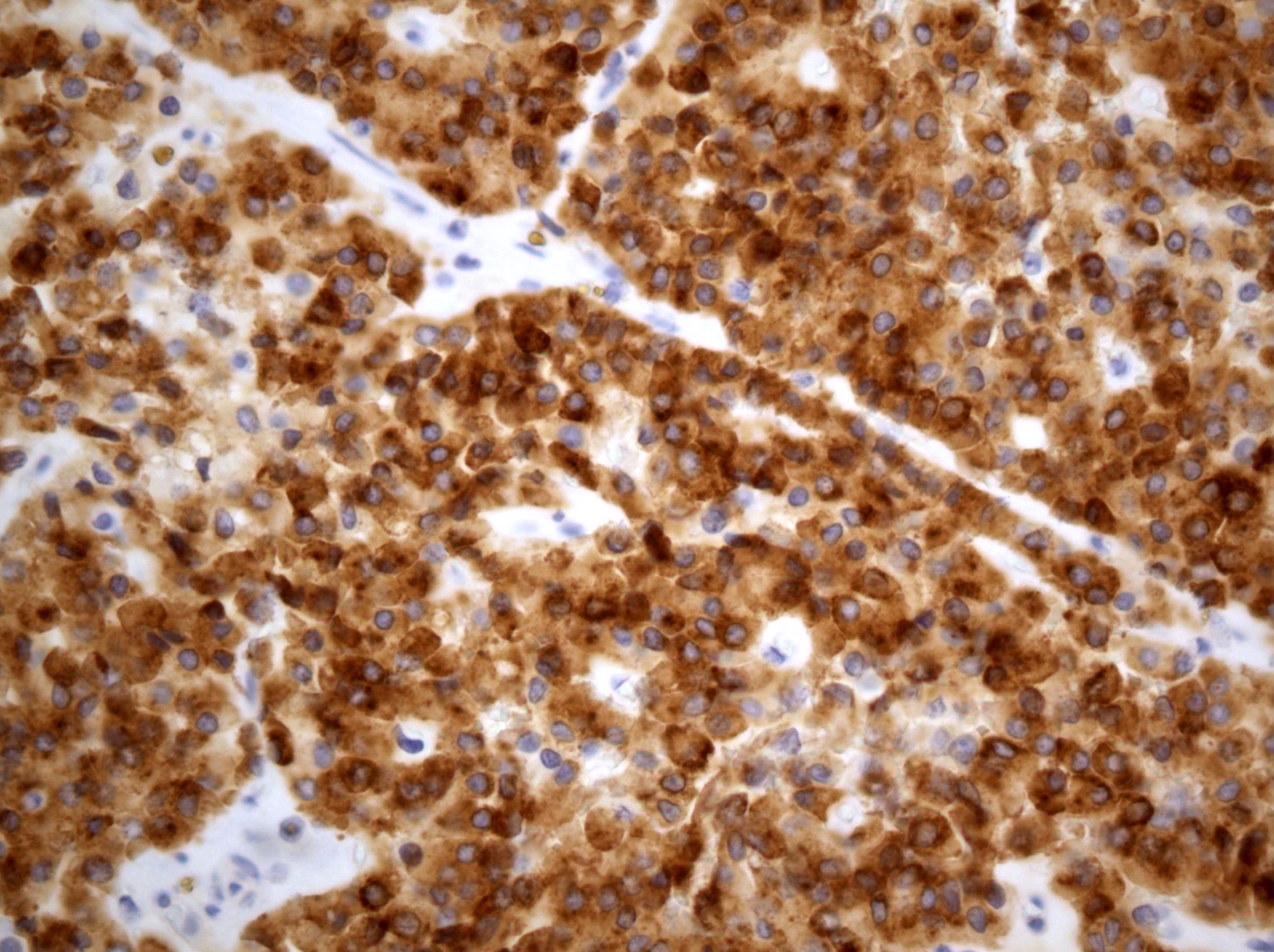
      Primary tumor (pT) and FIGO stages in ( )
      • pTX: primary tumor cannot be assessed
      • pT0: no evidence of primary tumor
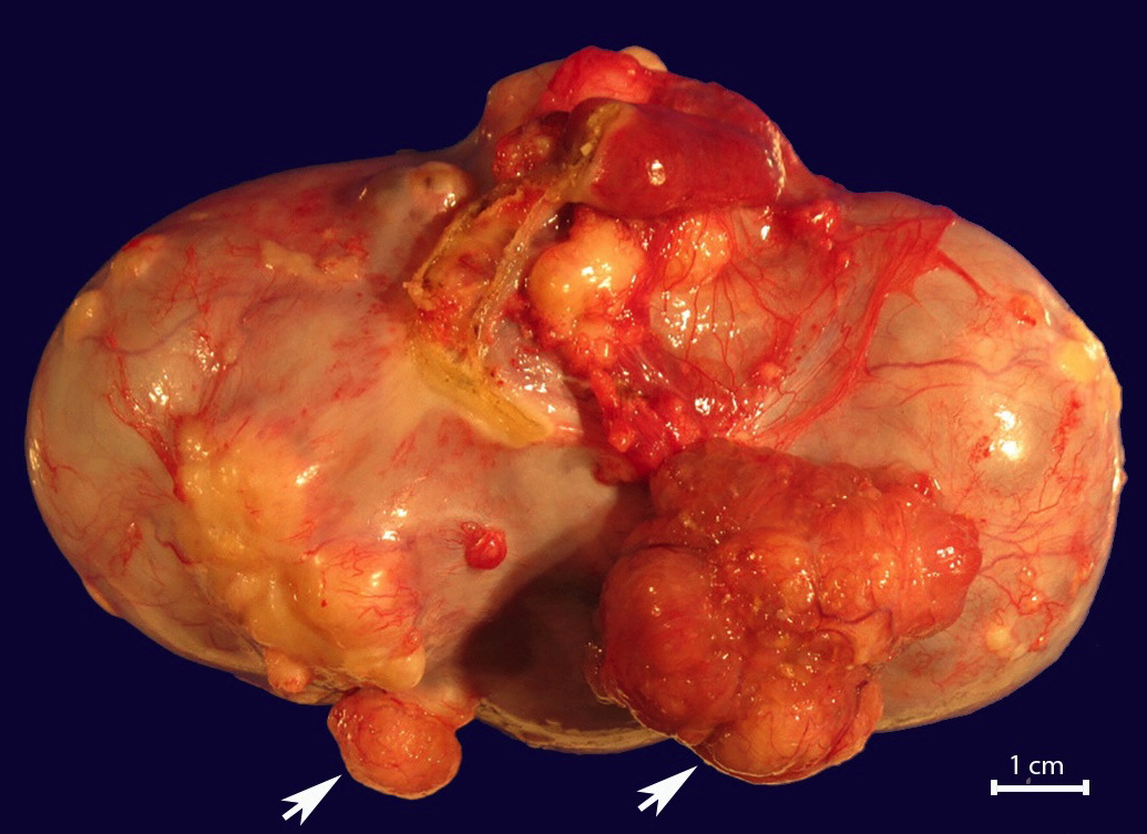
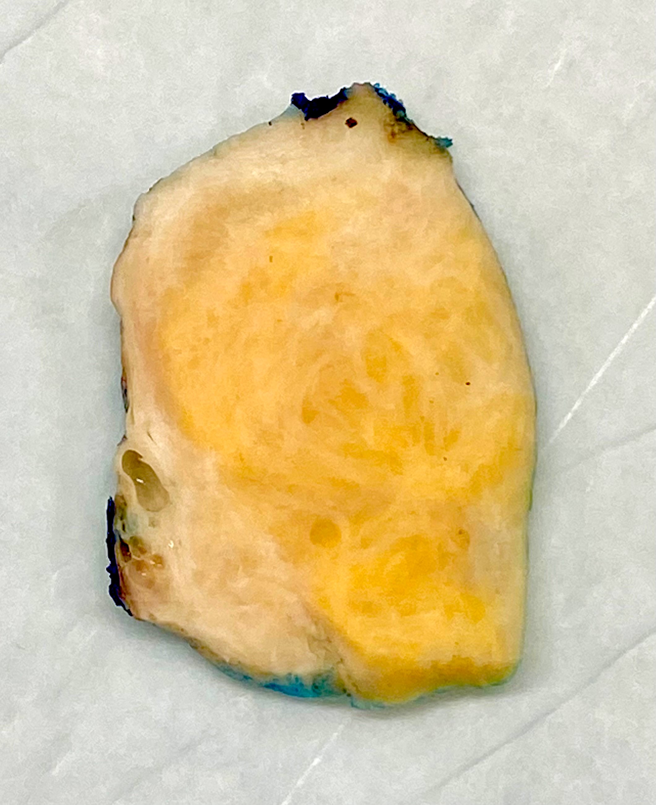
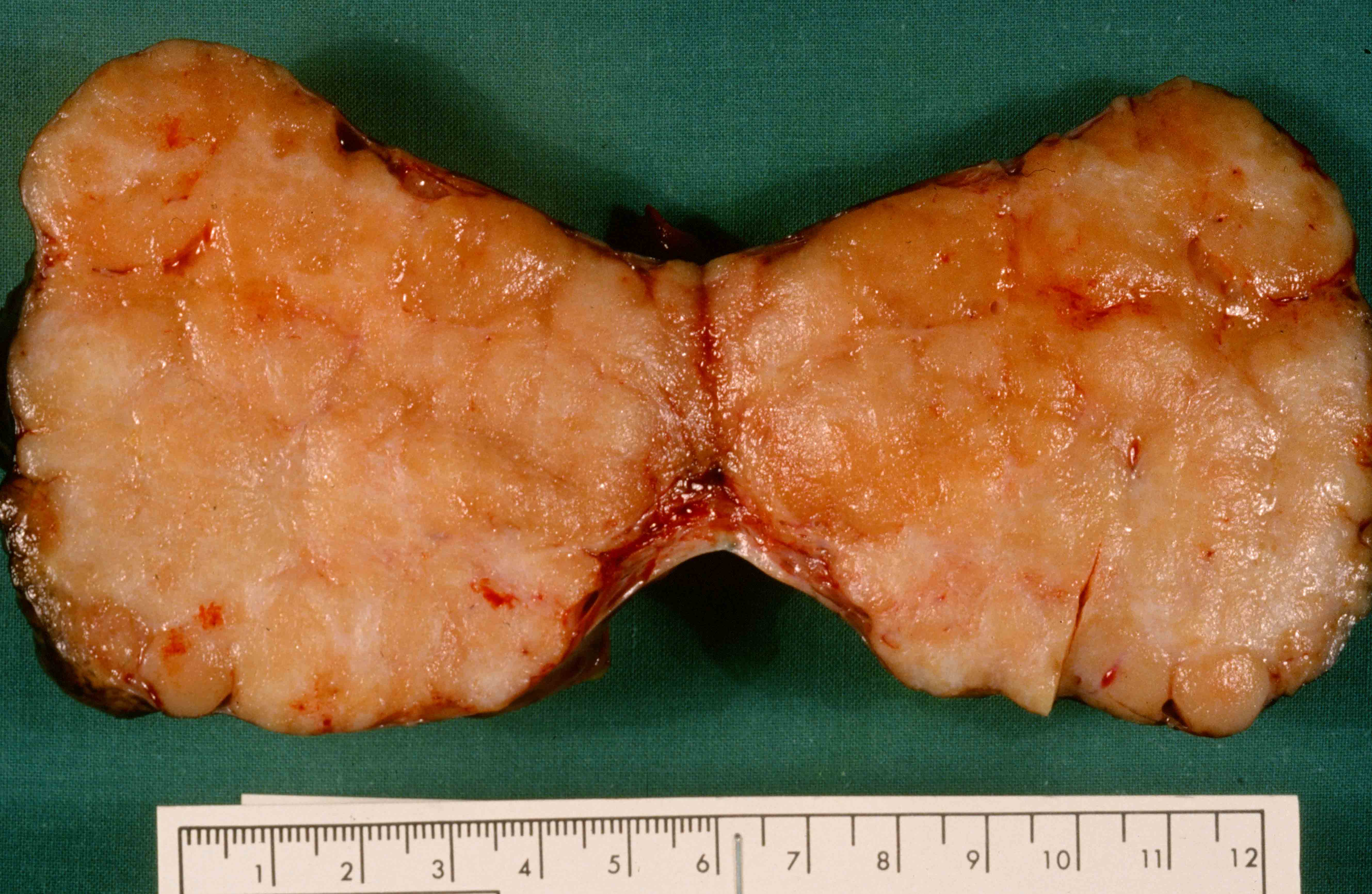
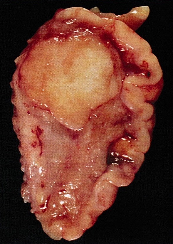
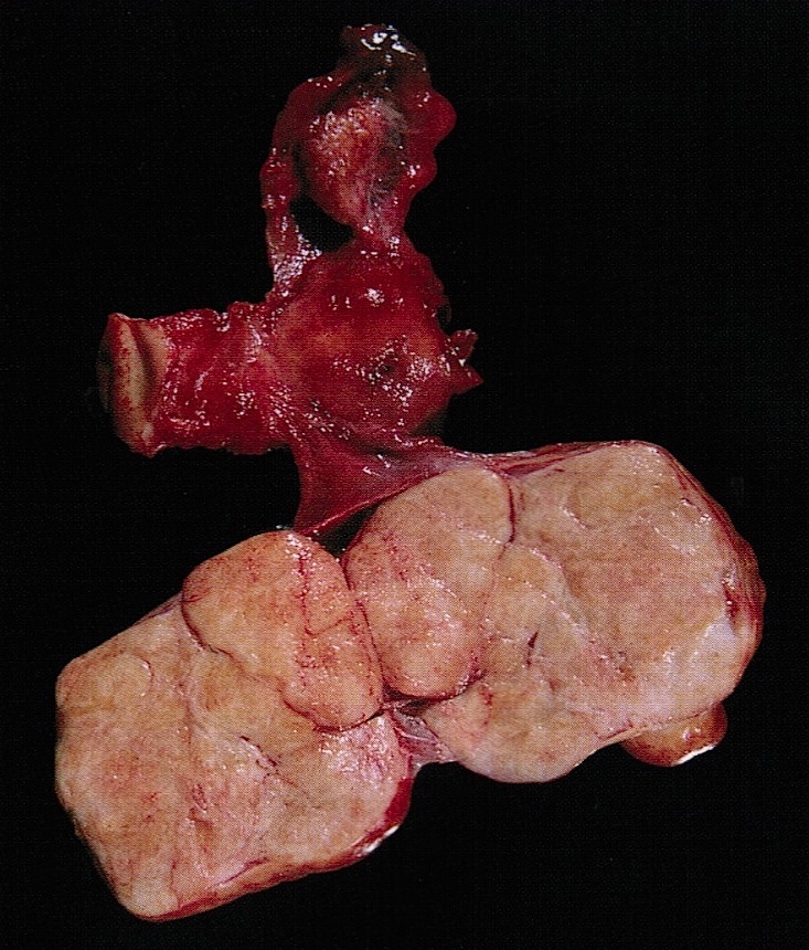
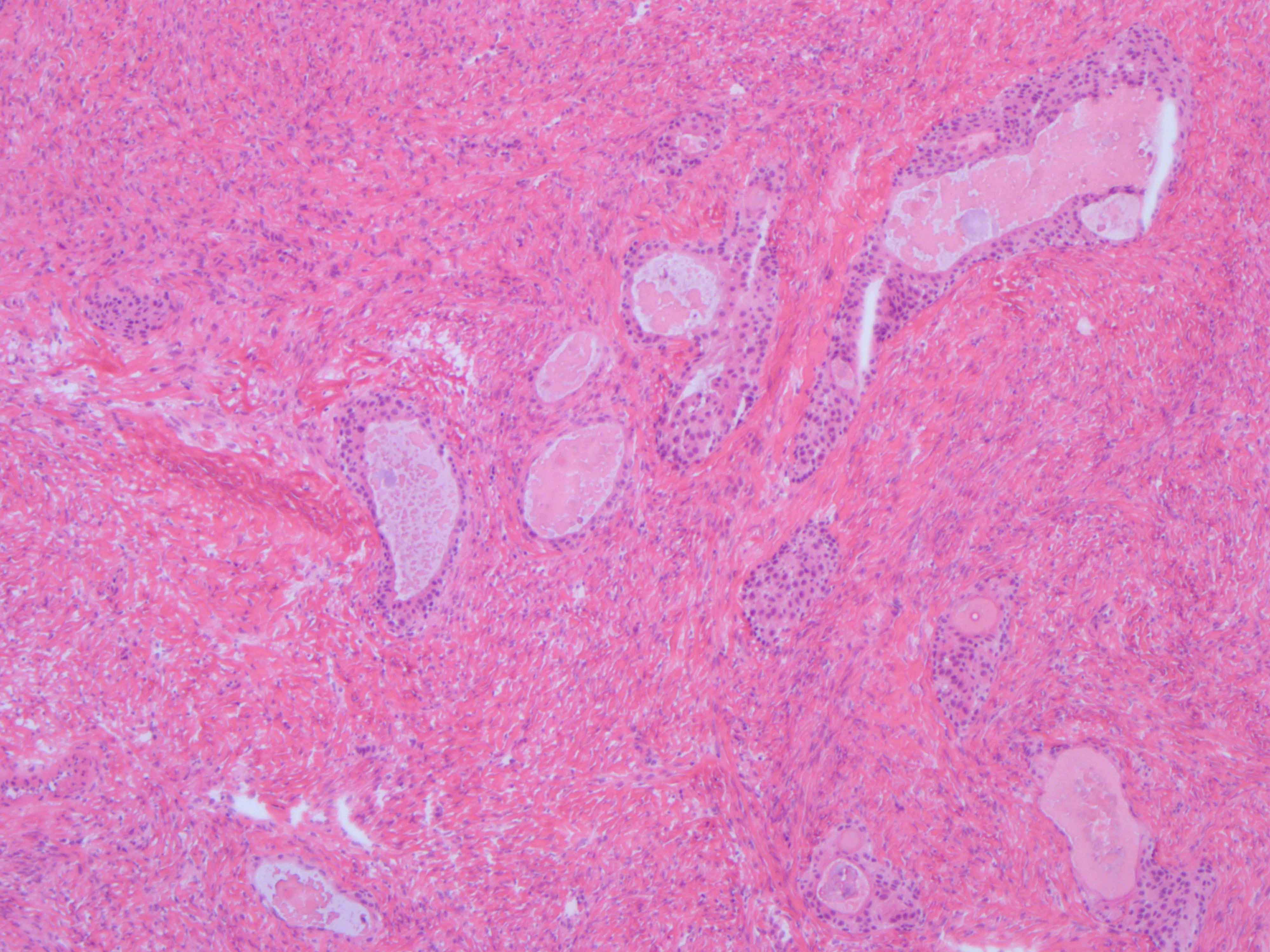
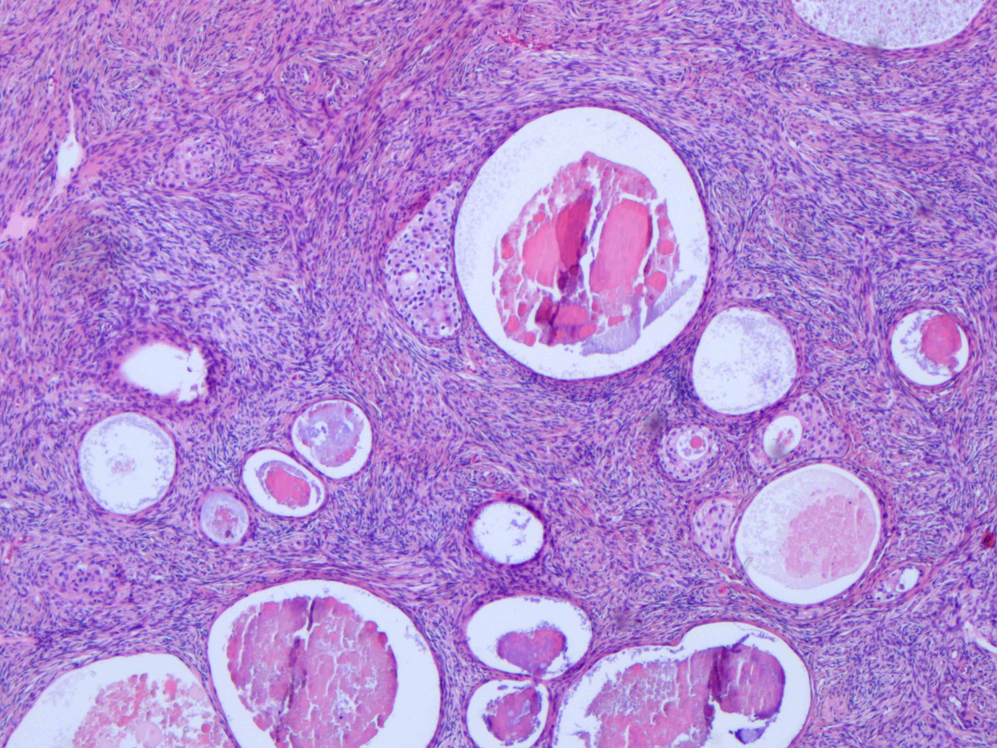
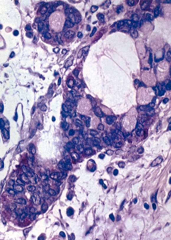
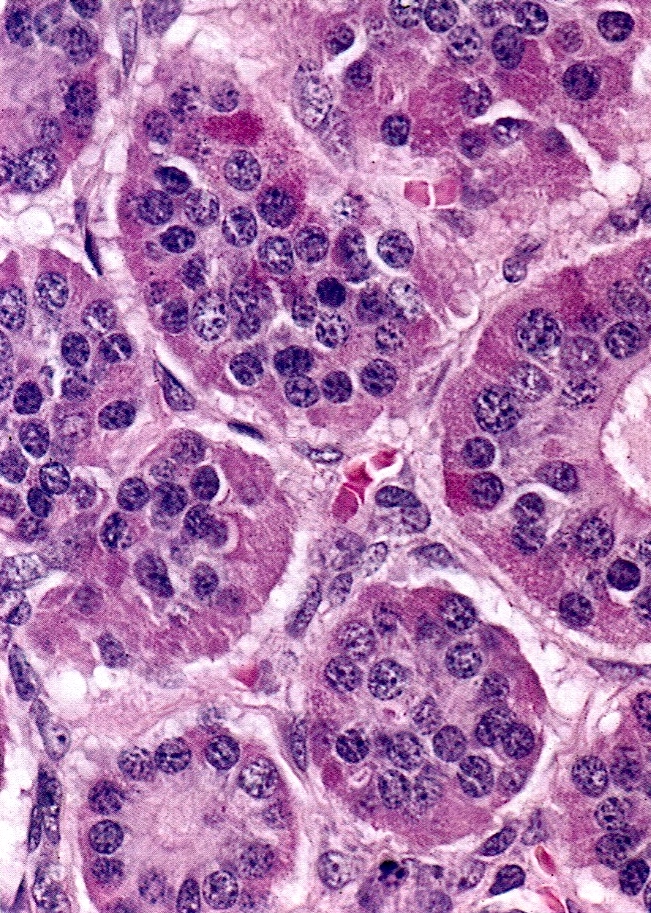
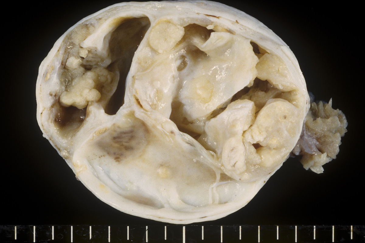
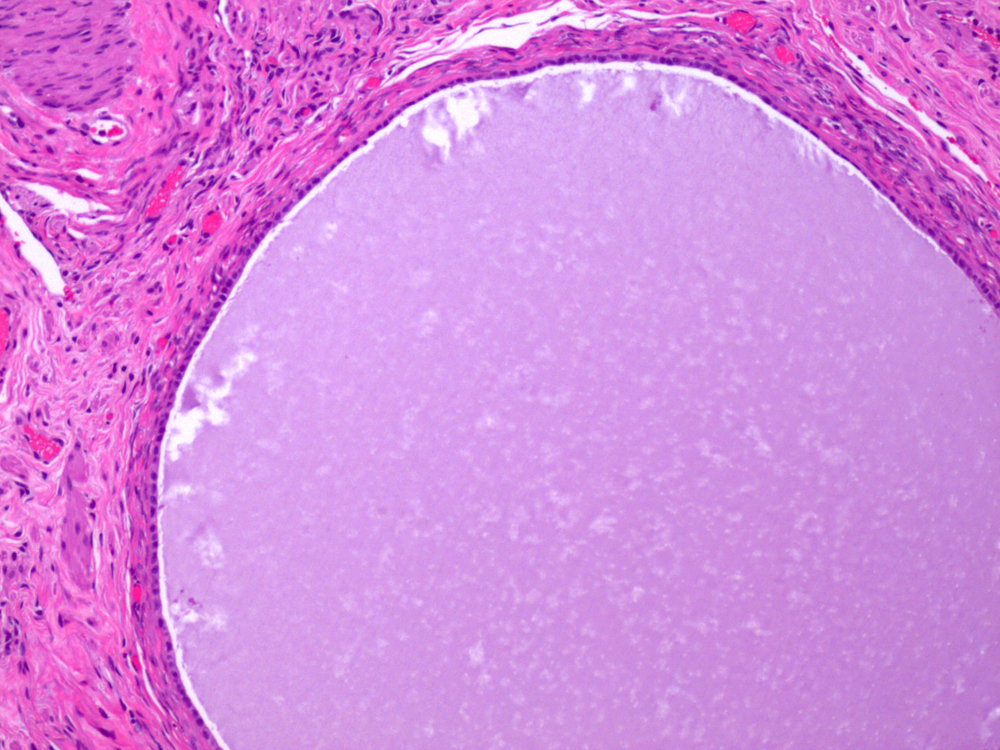
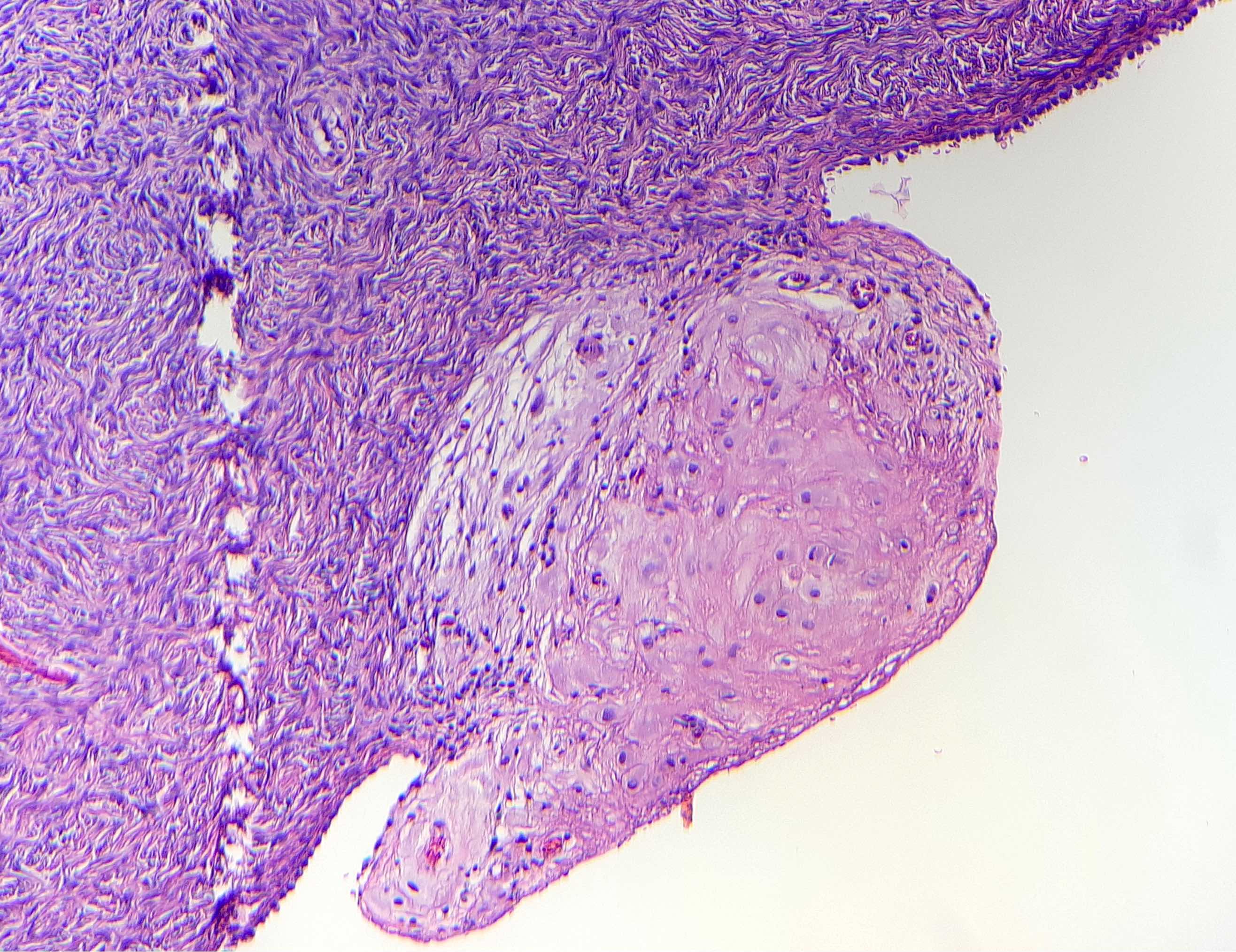
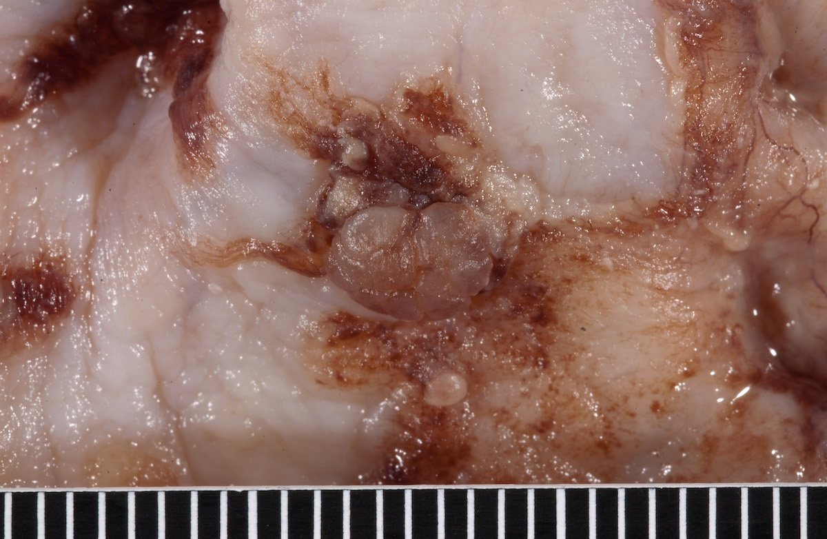
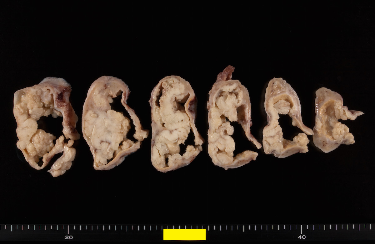
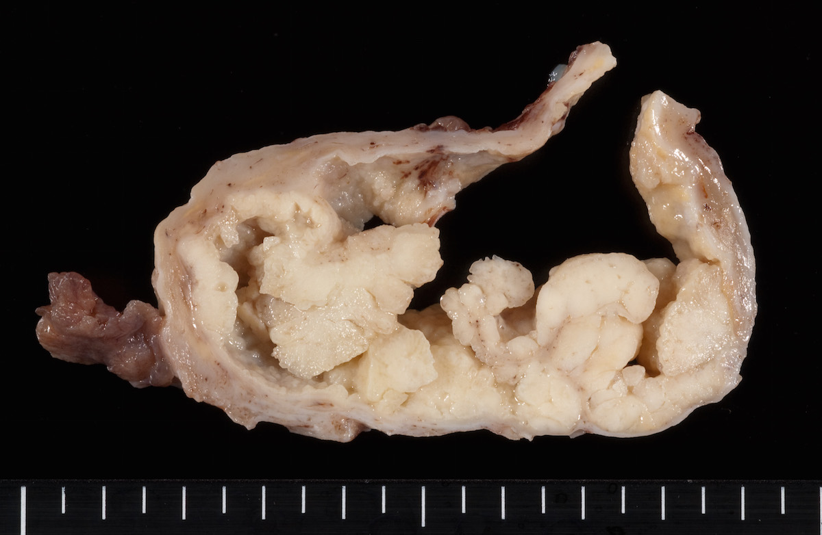
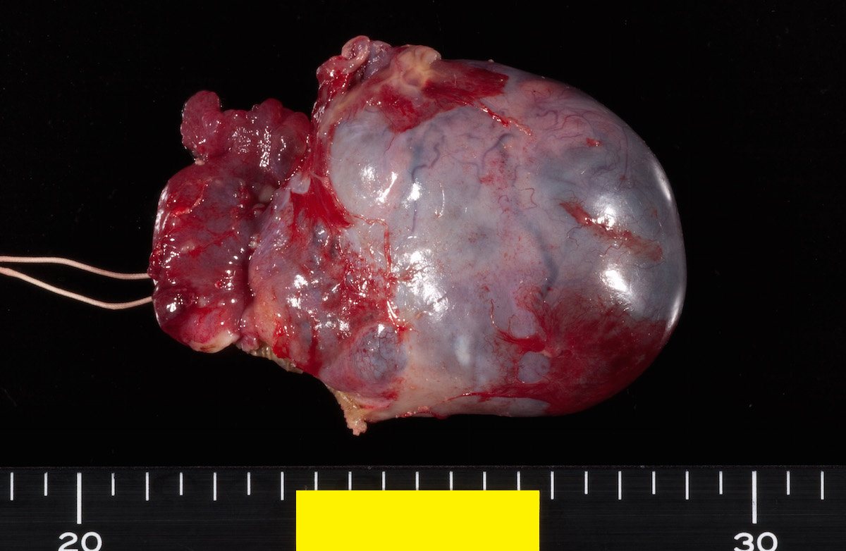
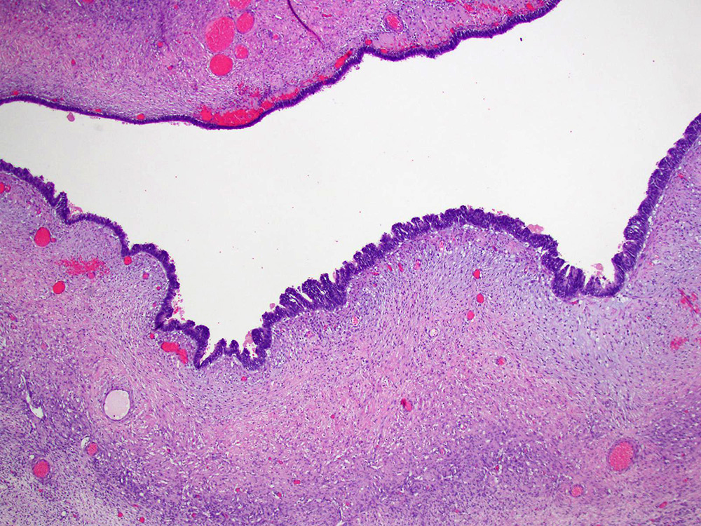
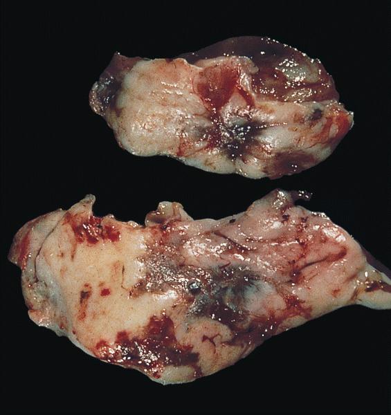
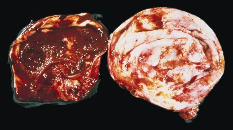
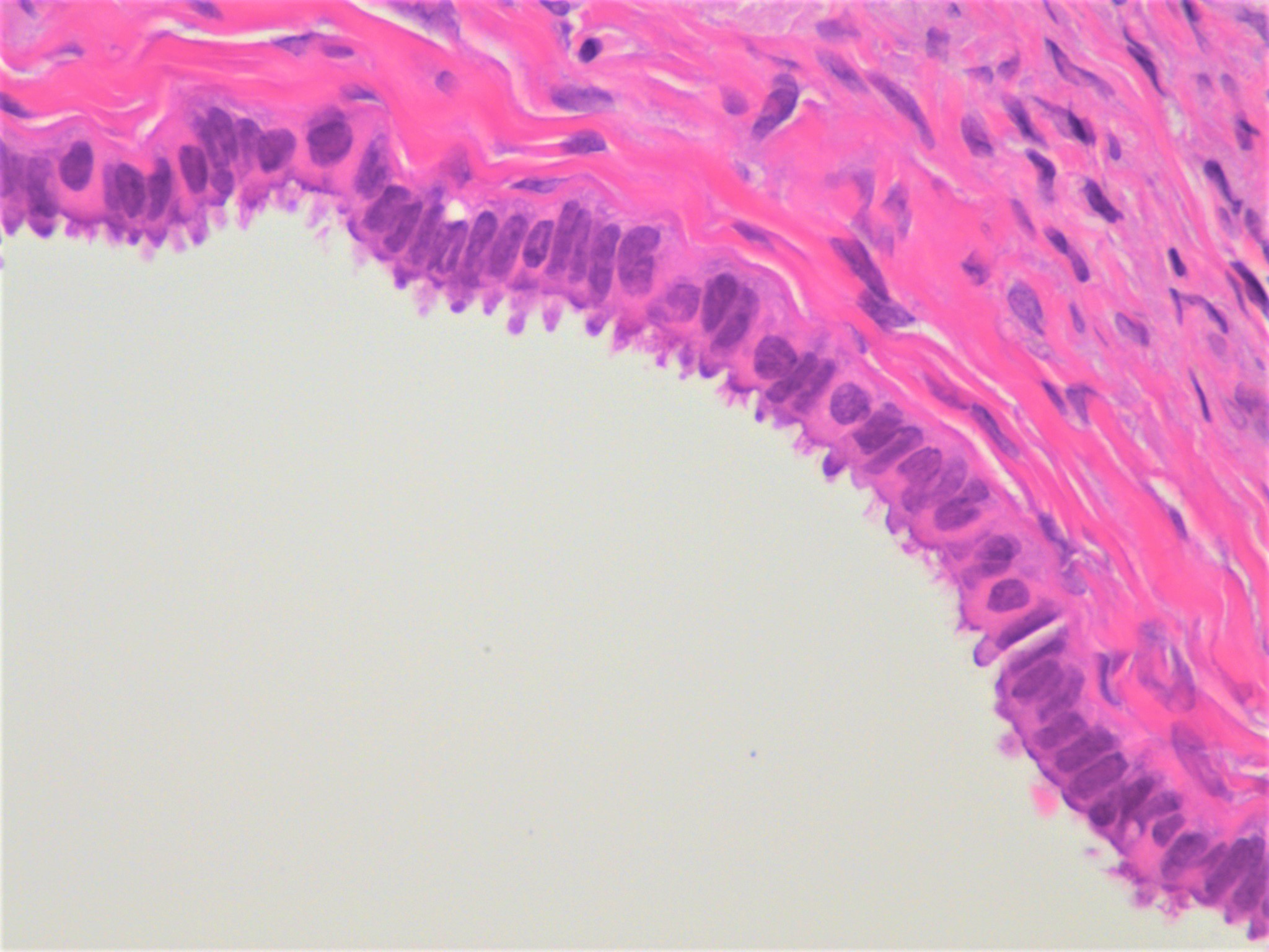
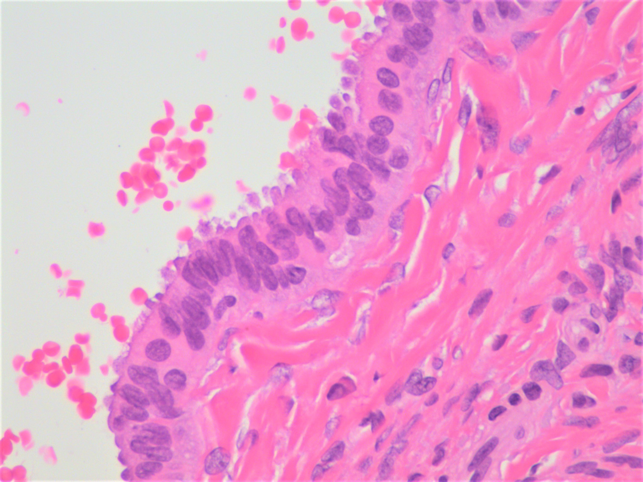
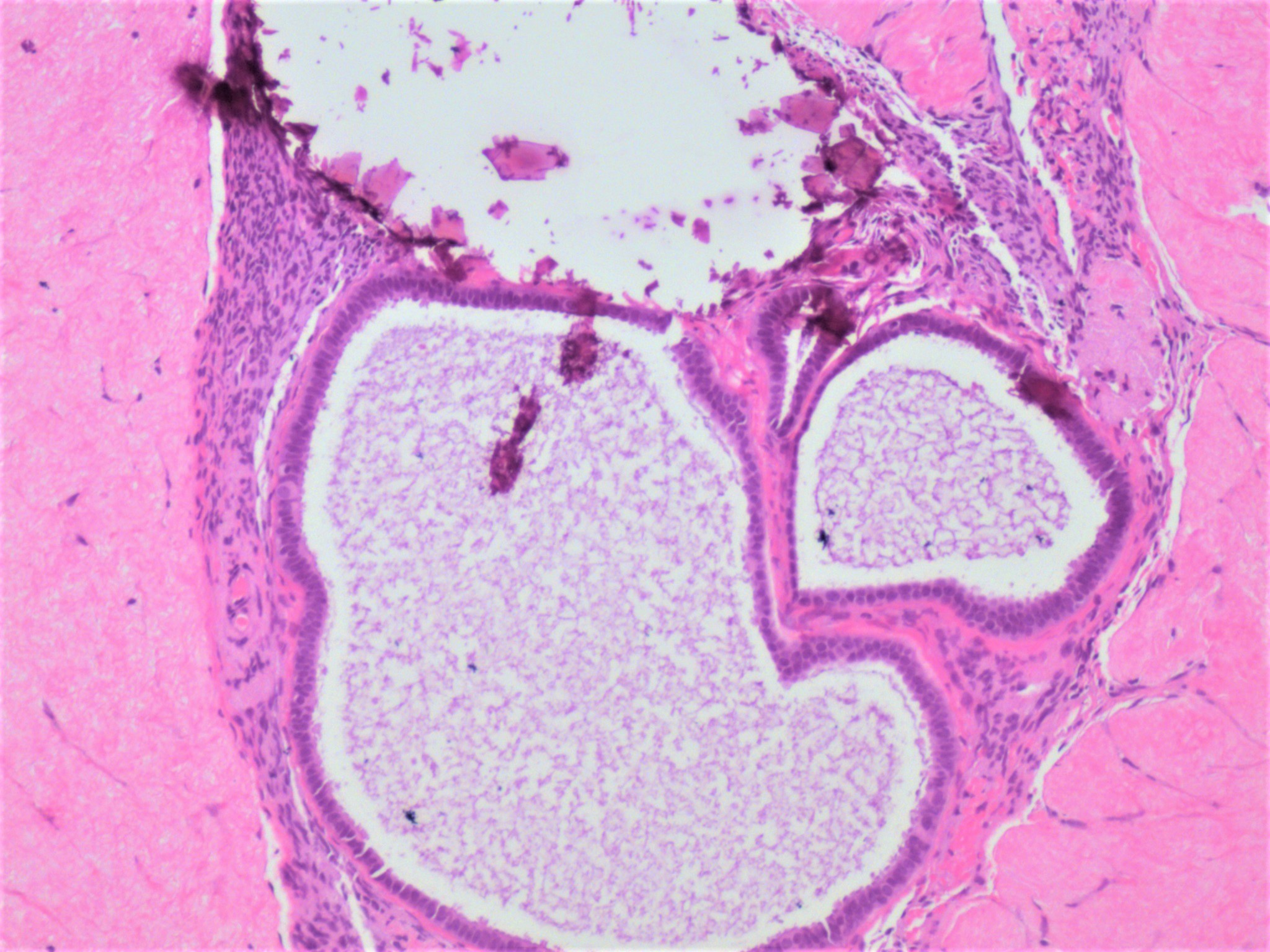
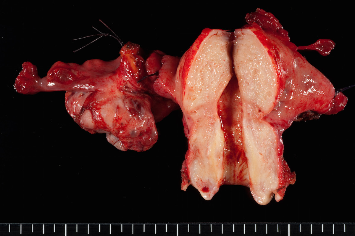
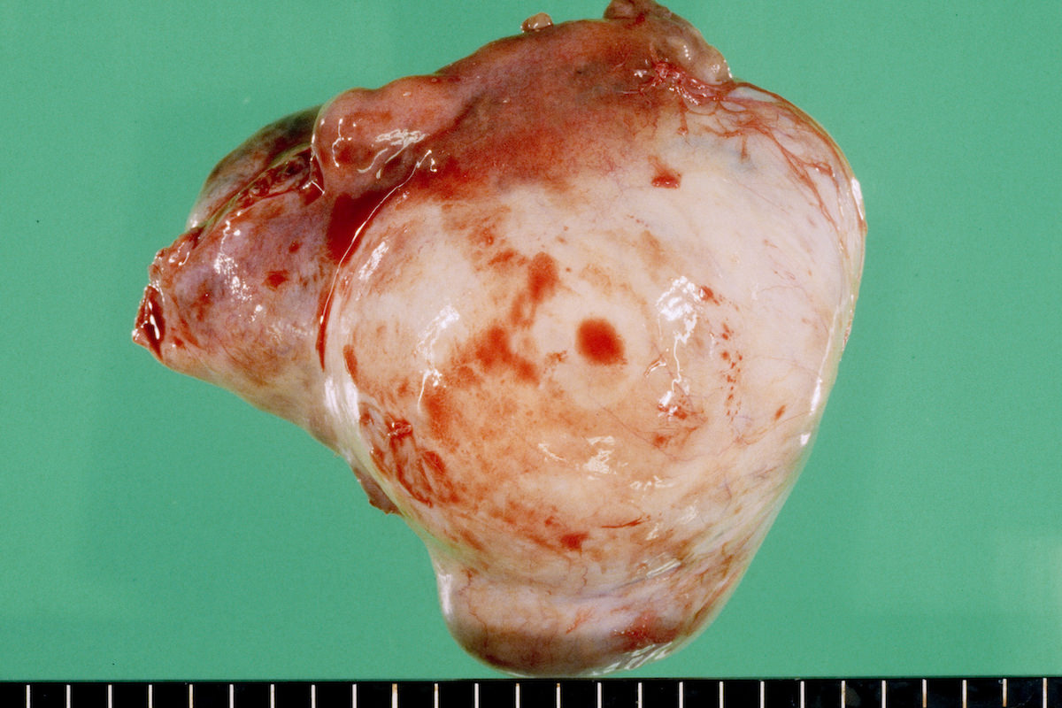
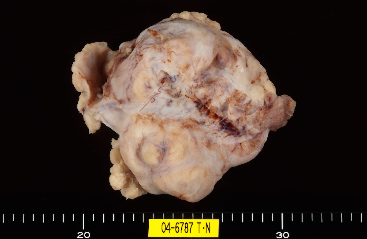
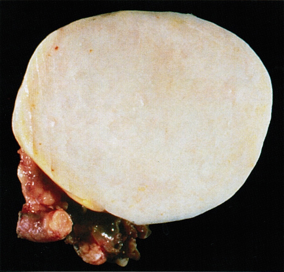
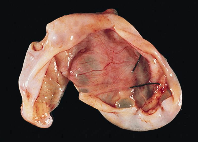
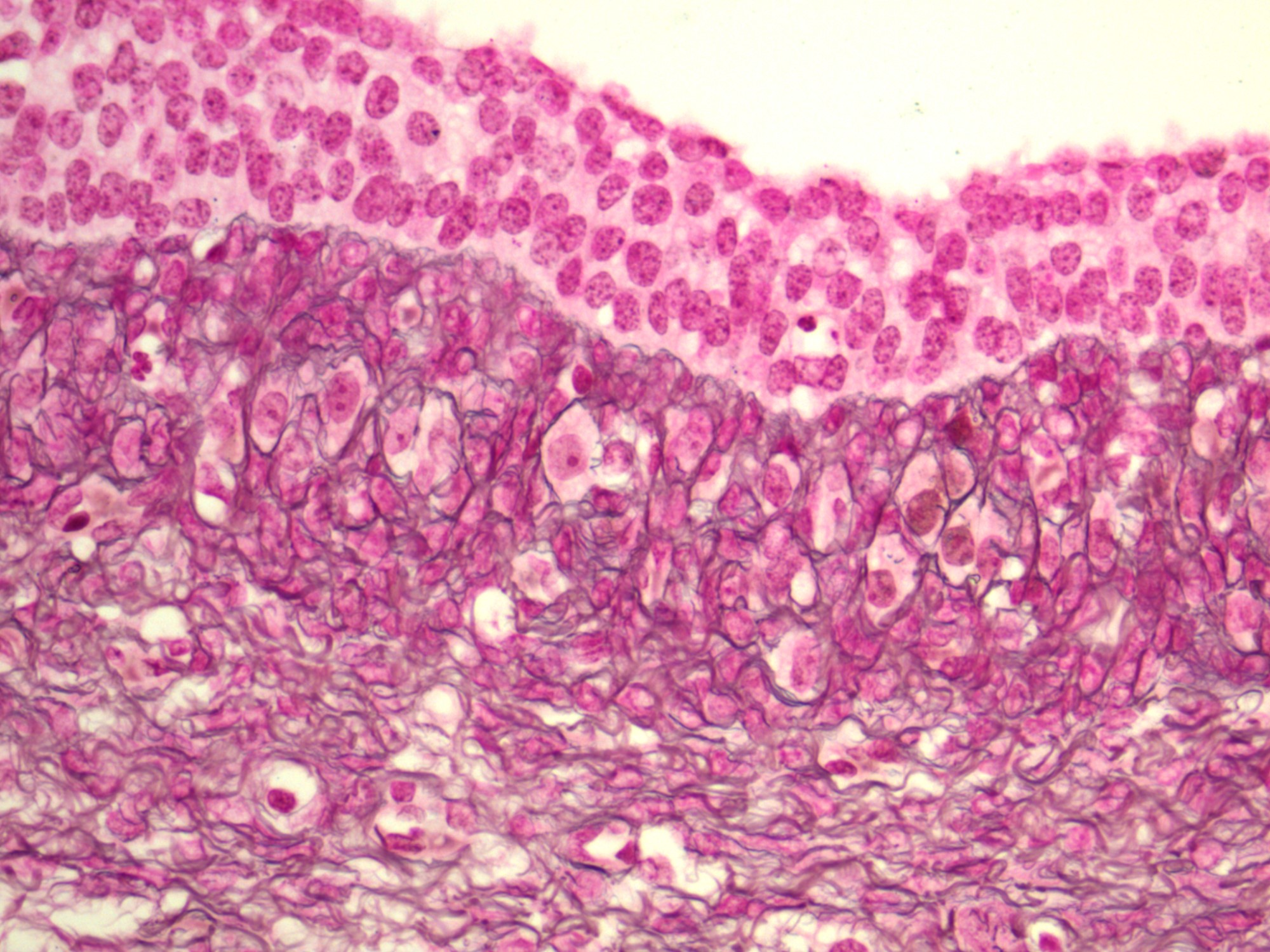
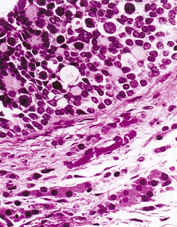
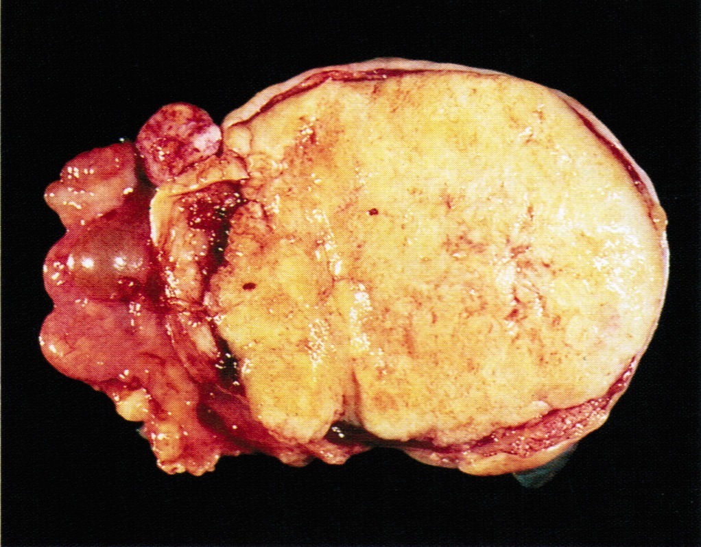
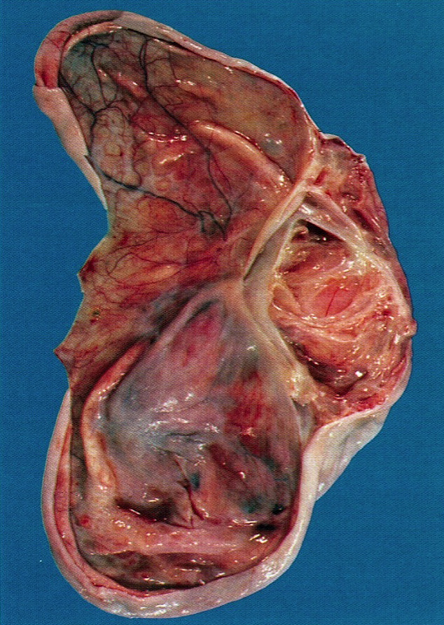
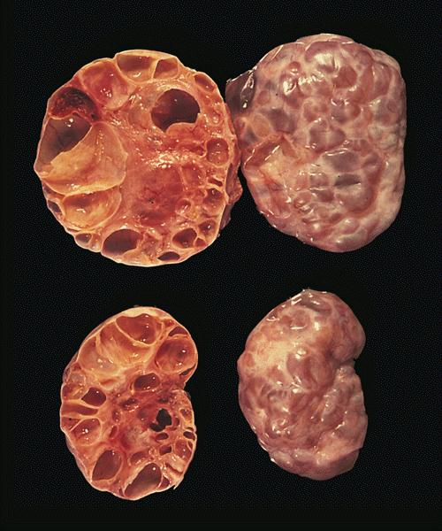
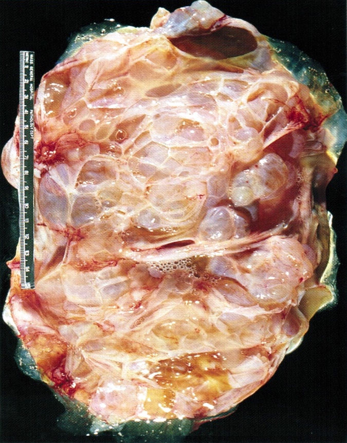
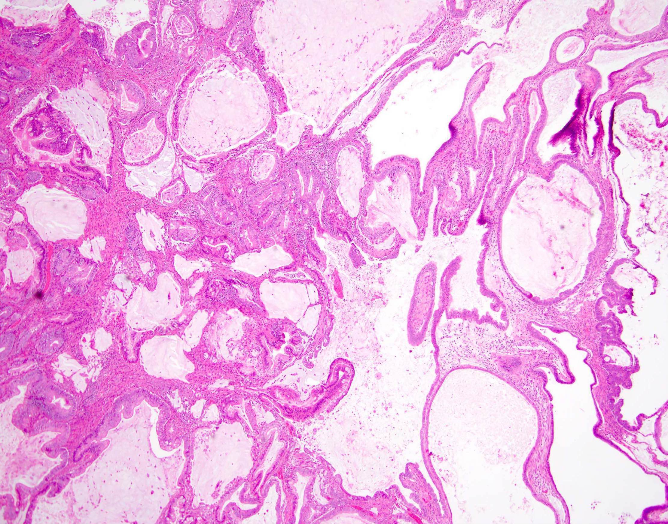
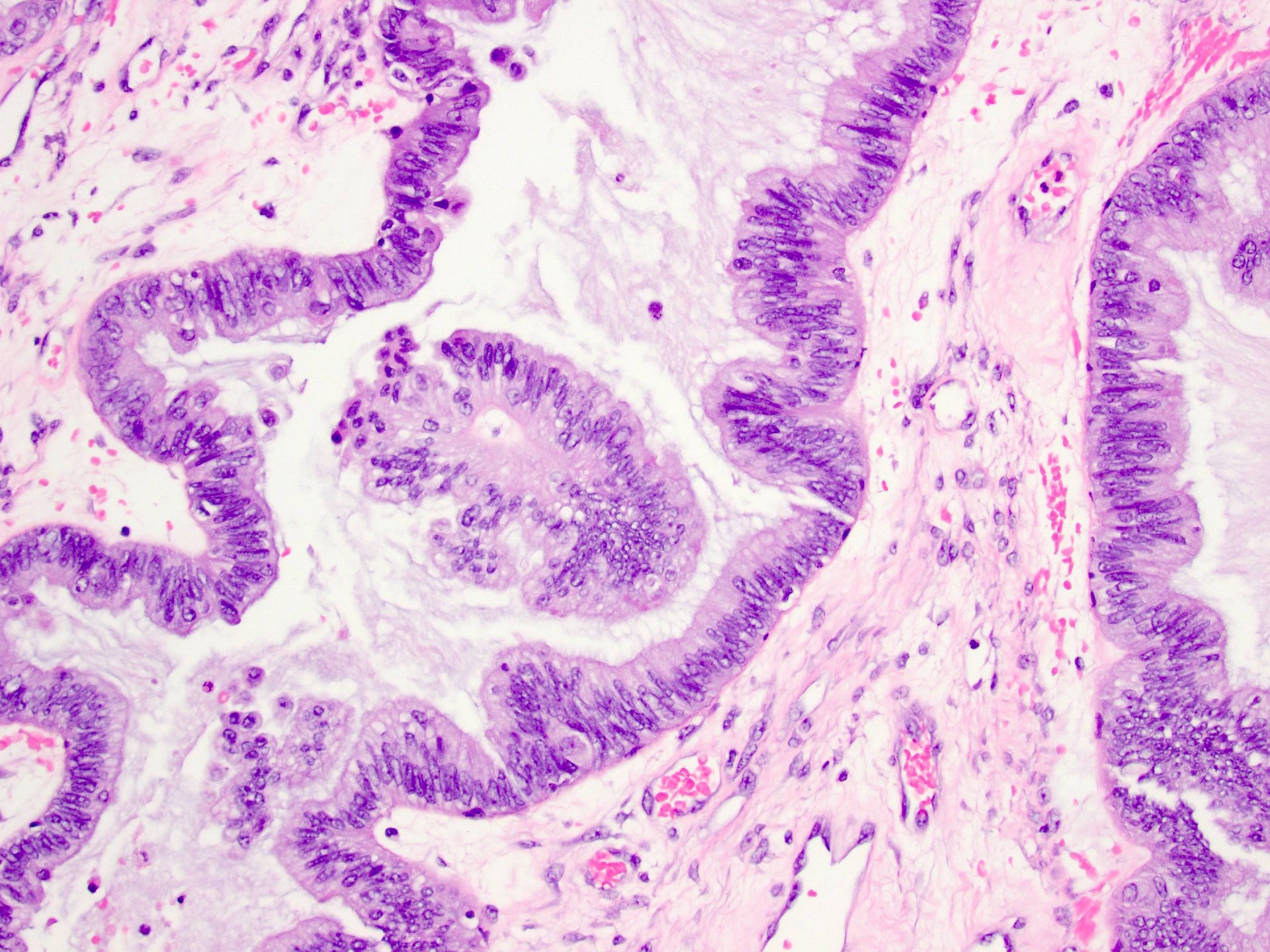
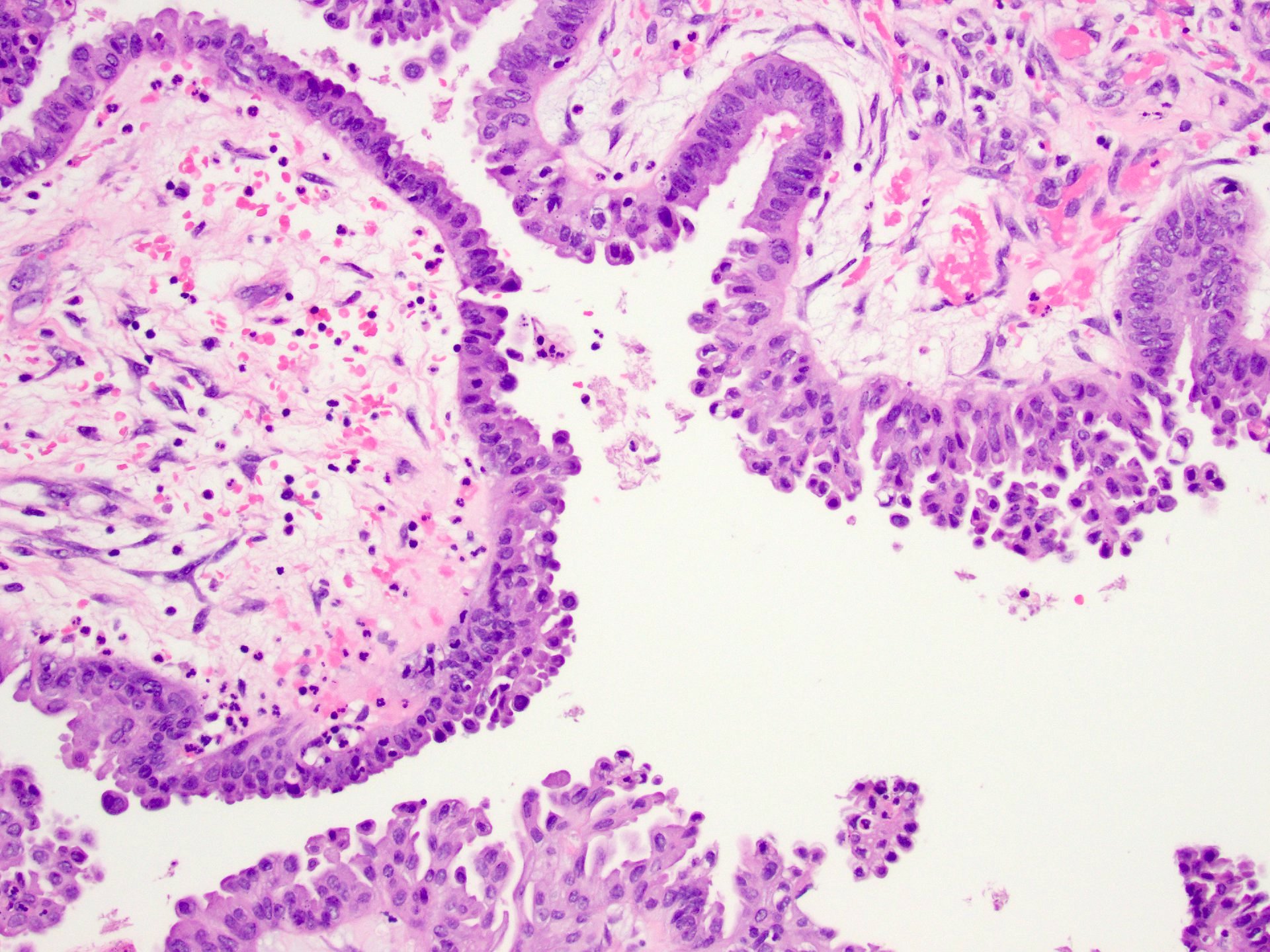
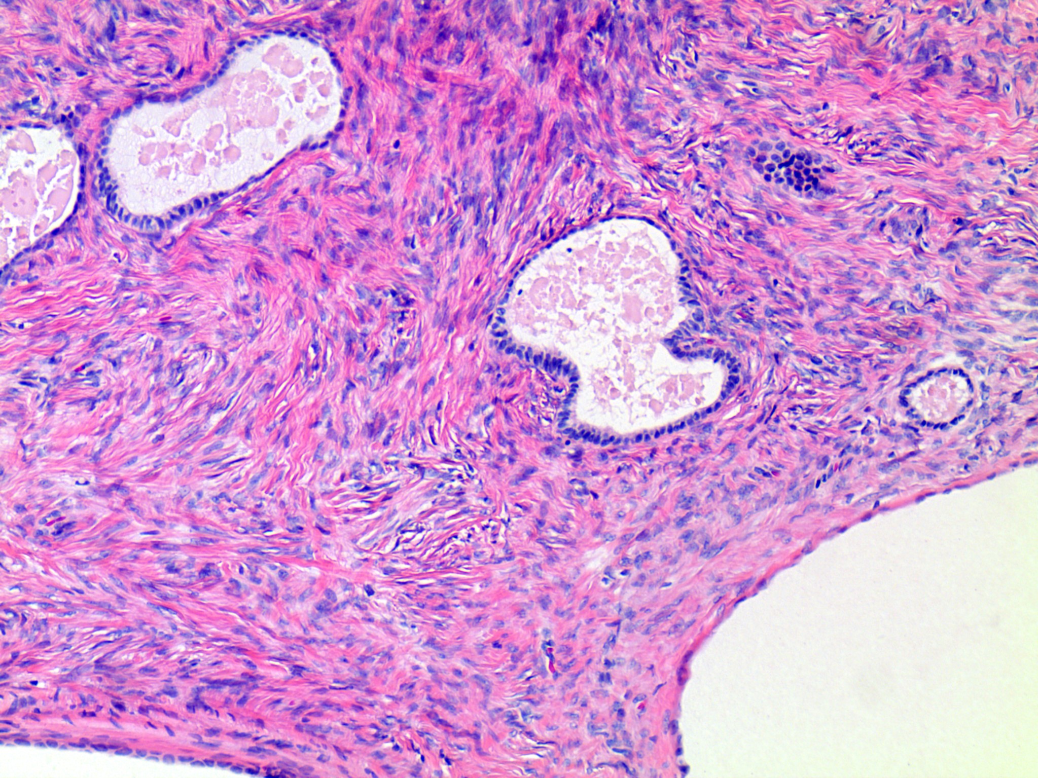
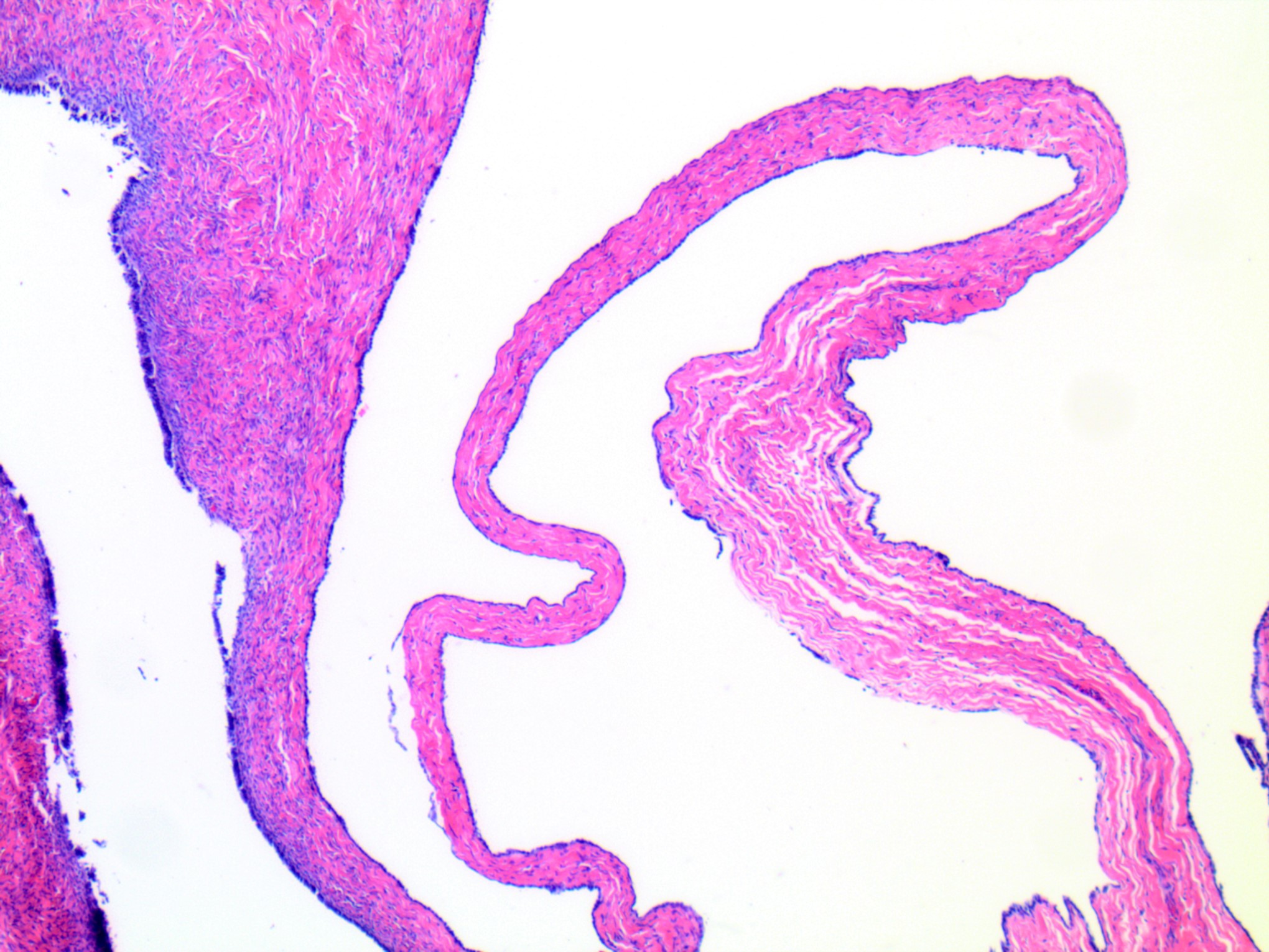
      • pT1a (IA): tumor limited to 1 ovary (capsule intact) or fallopian tube; no tumor on ovarian or fallopian tube surface; no malignant cells in ascites or peritoneal washings
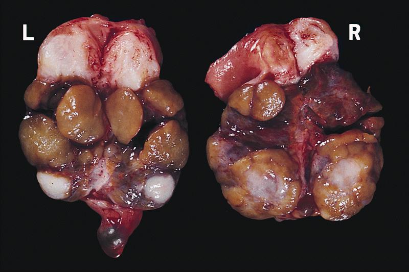
      • pT1b (IB): tumor limited to both ovaries (capsules intact) or fallopian tubes; no tumor on ovarian or fallopian tube surface; no malignant cells in ascites or peritoneal washings
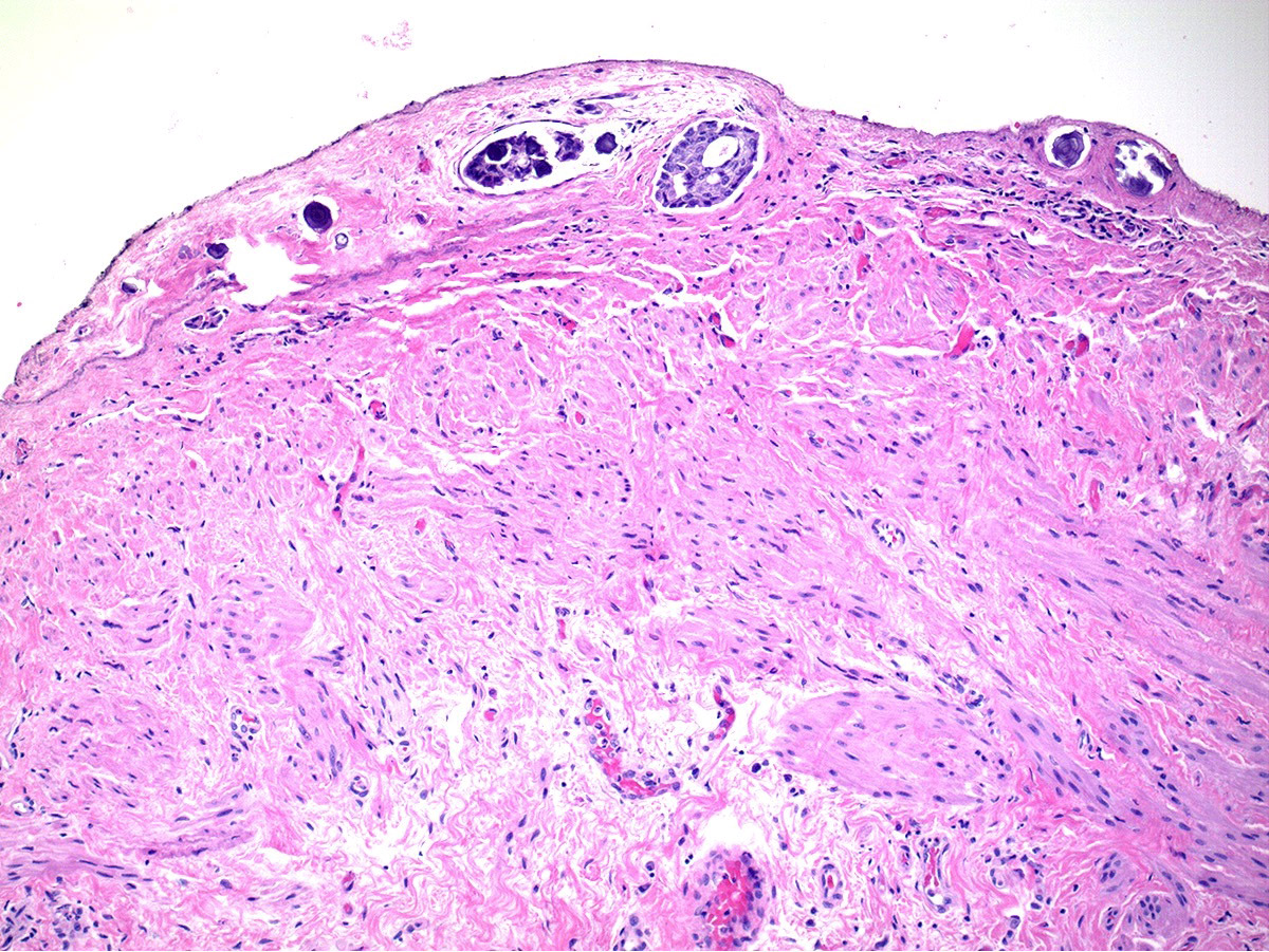
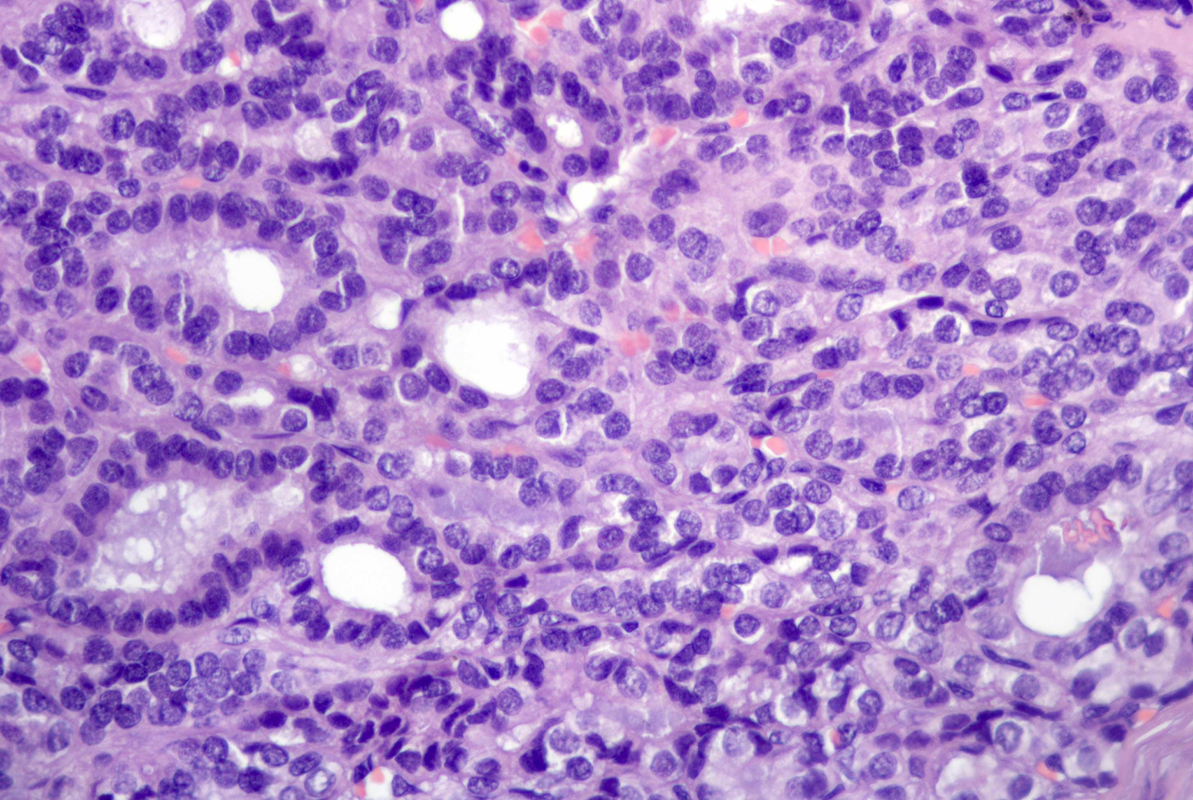
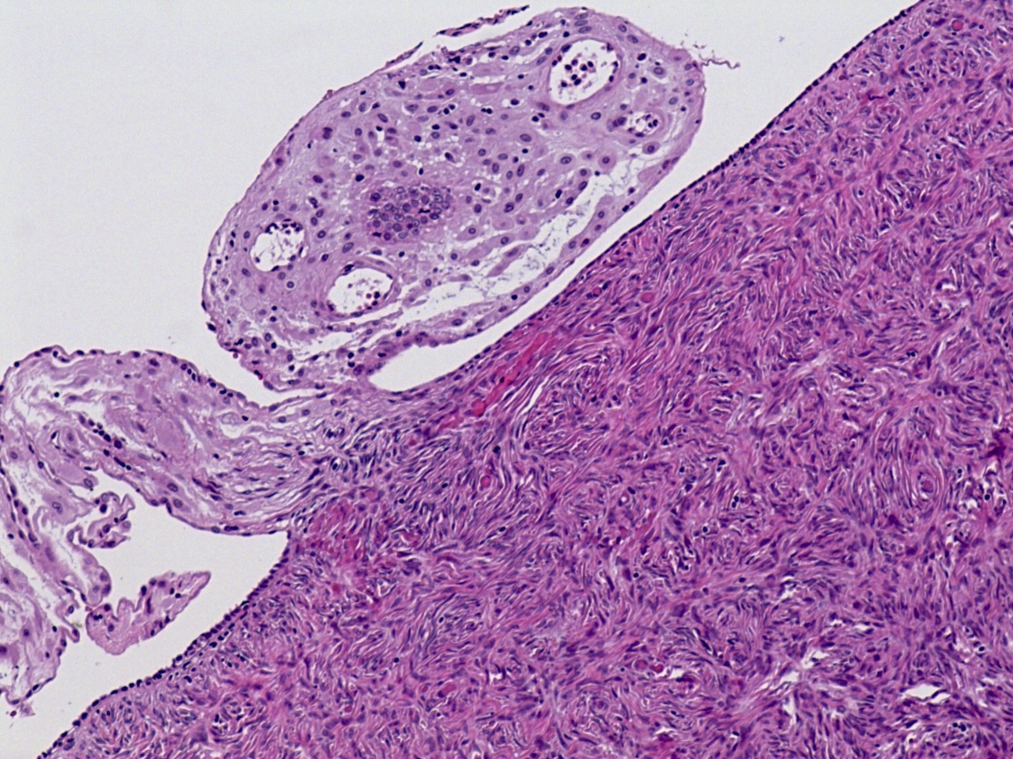
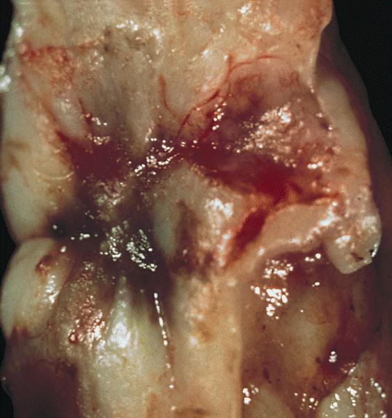
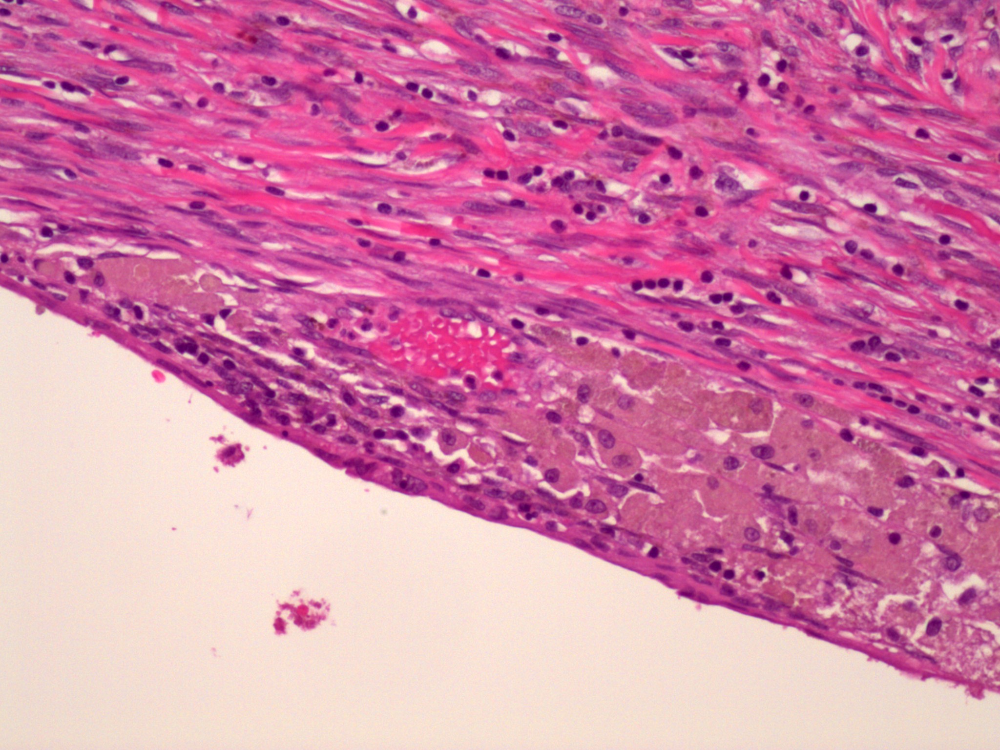
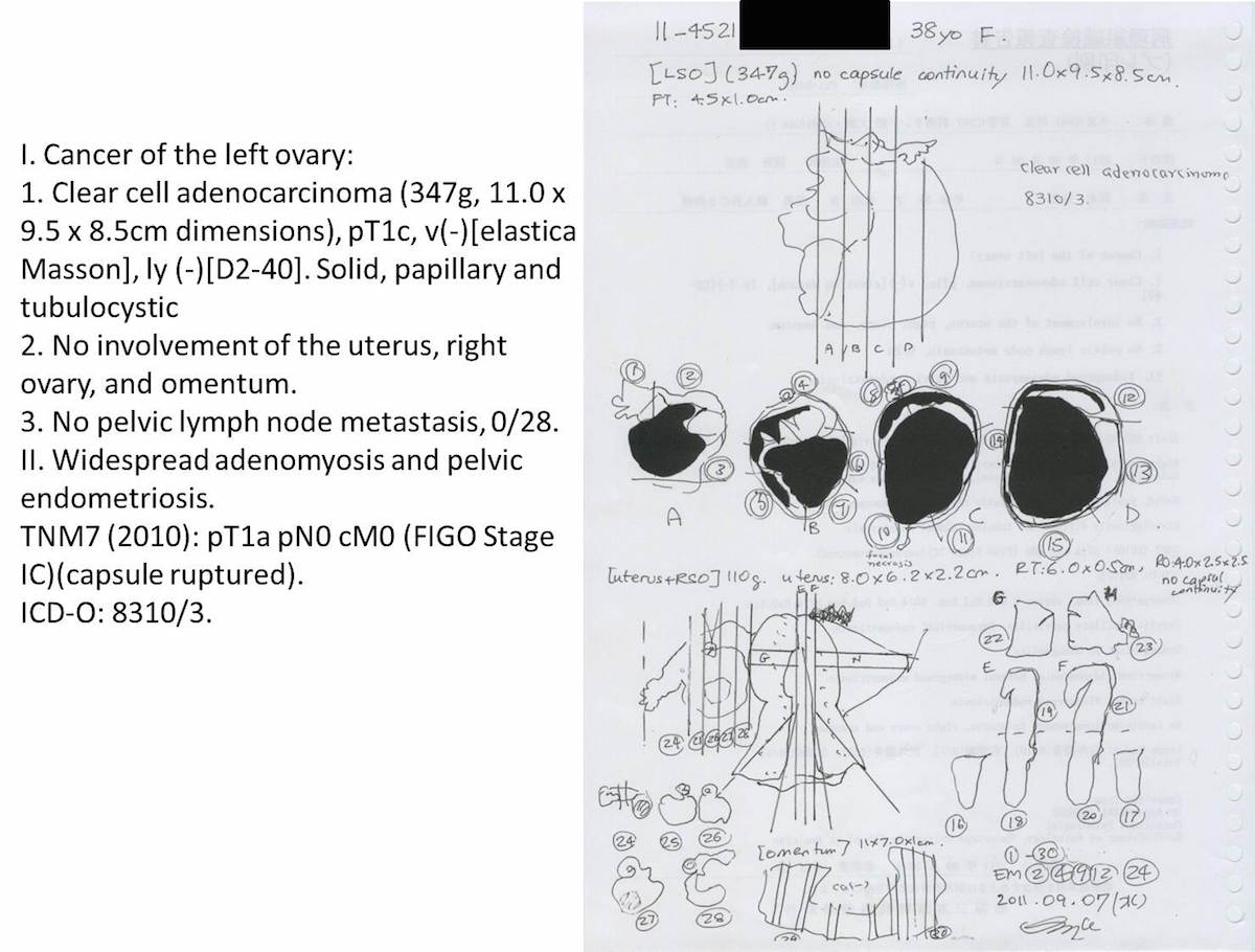
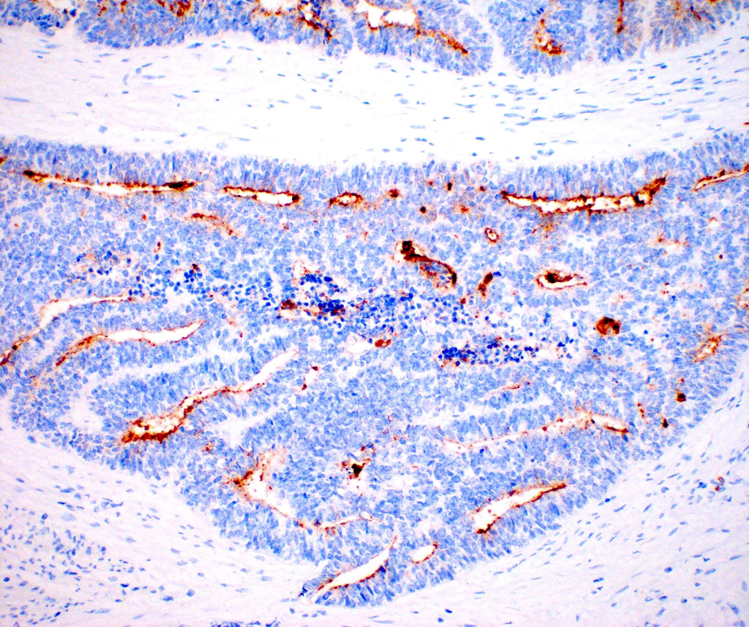
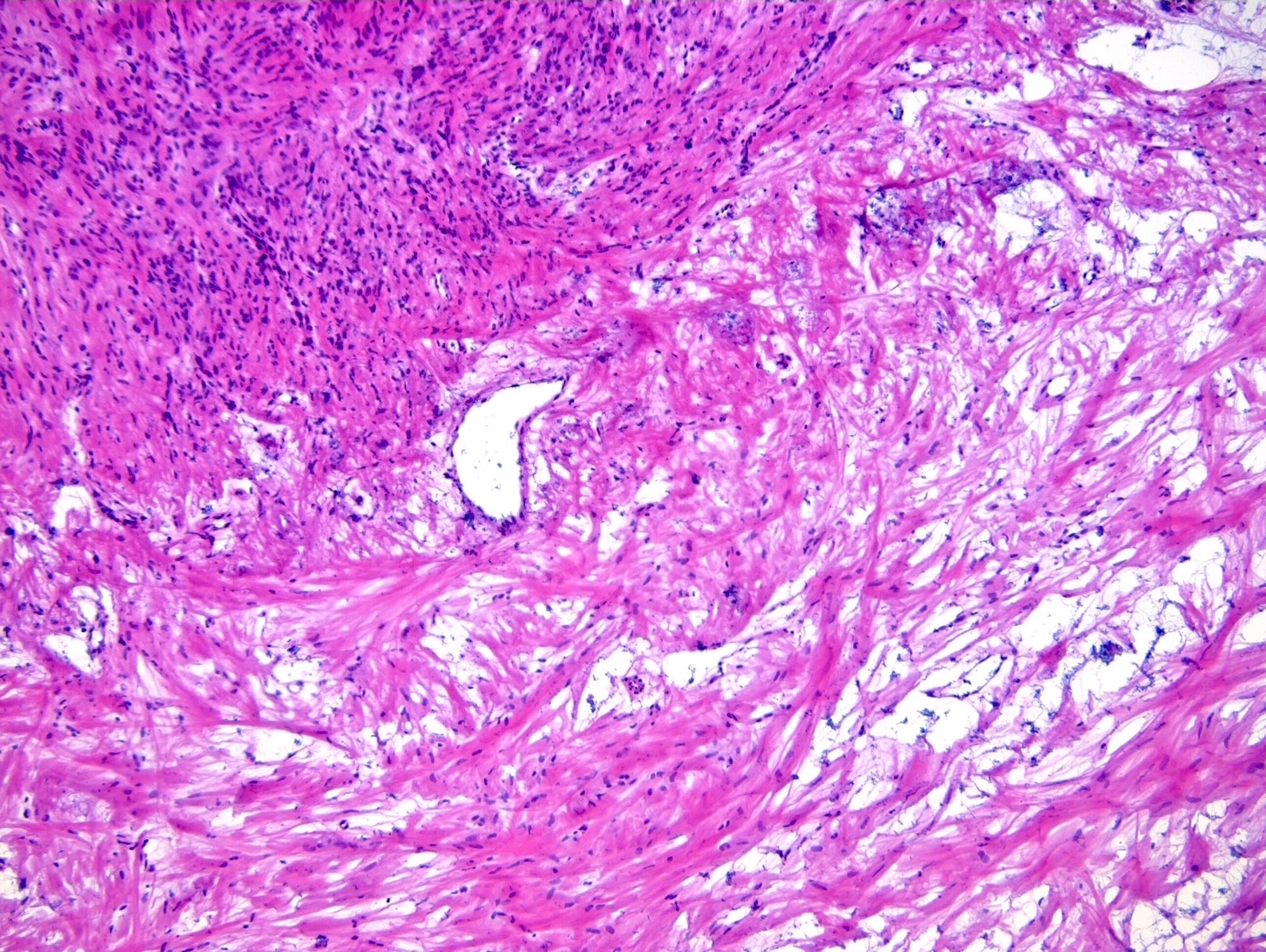
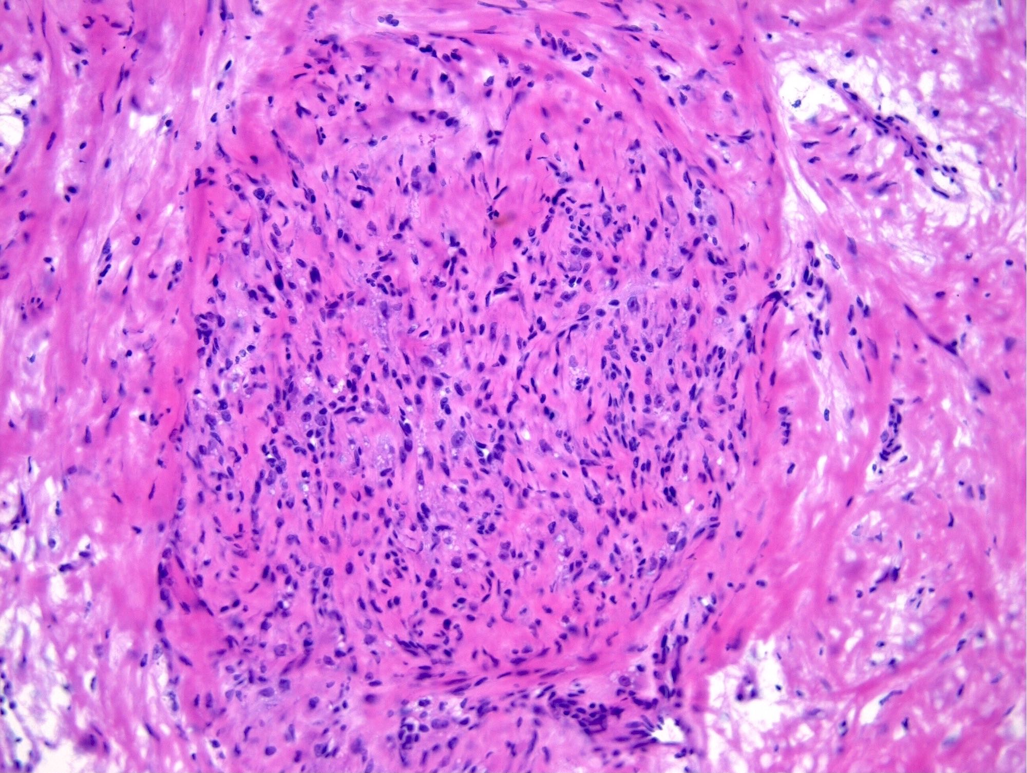
      • pT1c (IC): tumor limited to 1 or both ovaries or fallopian tubes, with any of following:
        • pT1c1 (IC1): surgical spill
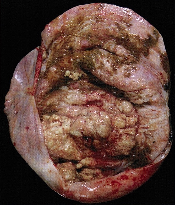
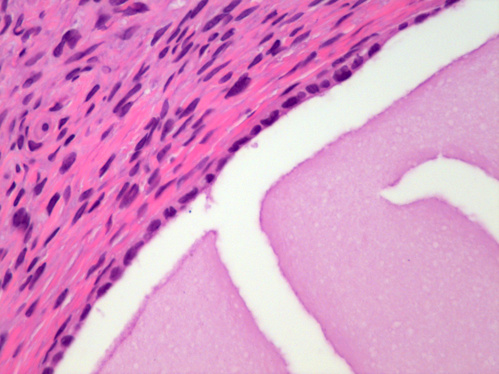
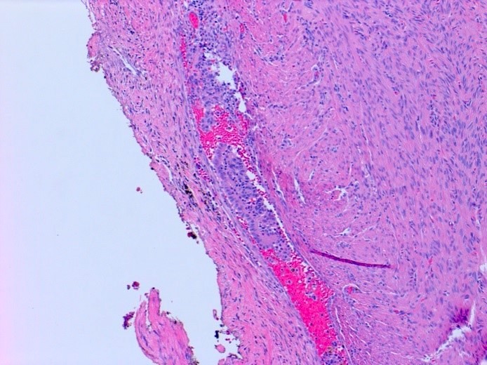
        • pT1c2 (IC2): capsule ruptured before surgery or tumor on ovarian or fallopian tube surface
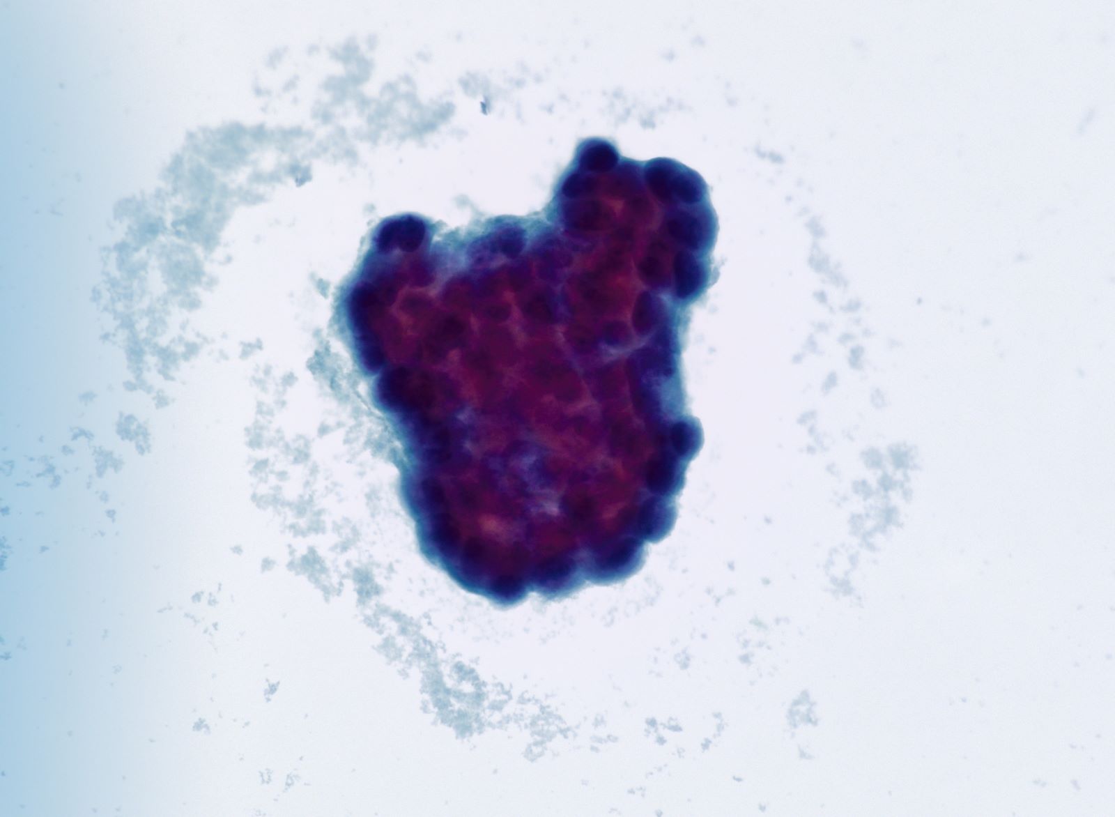
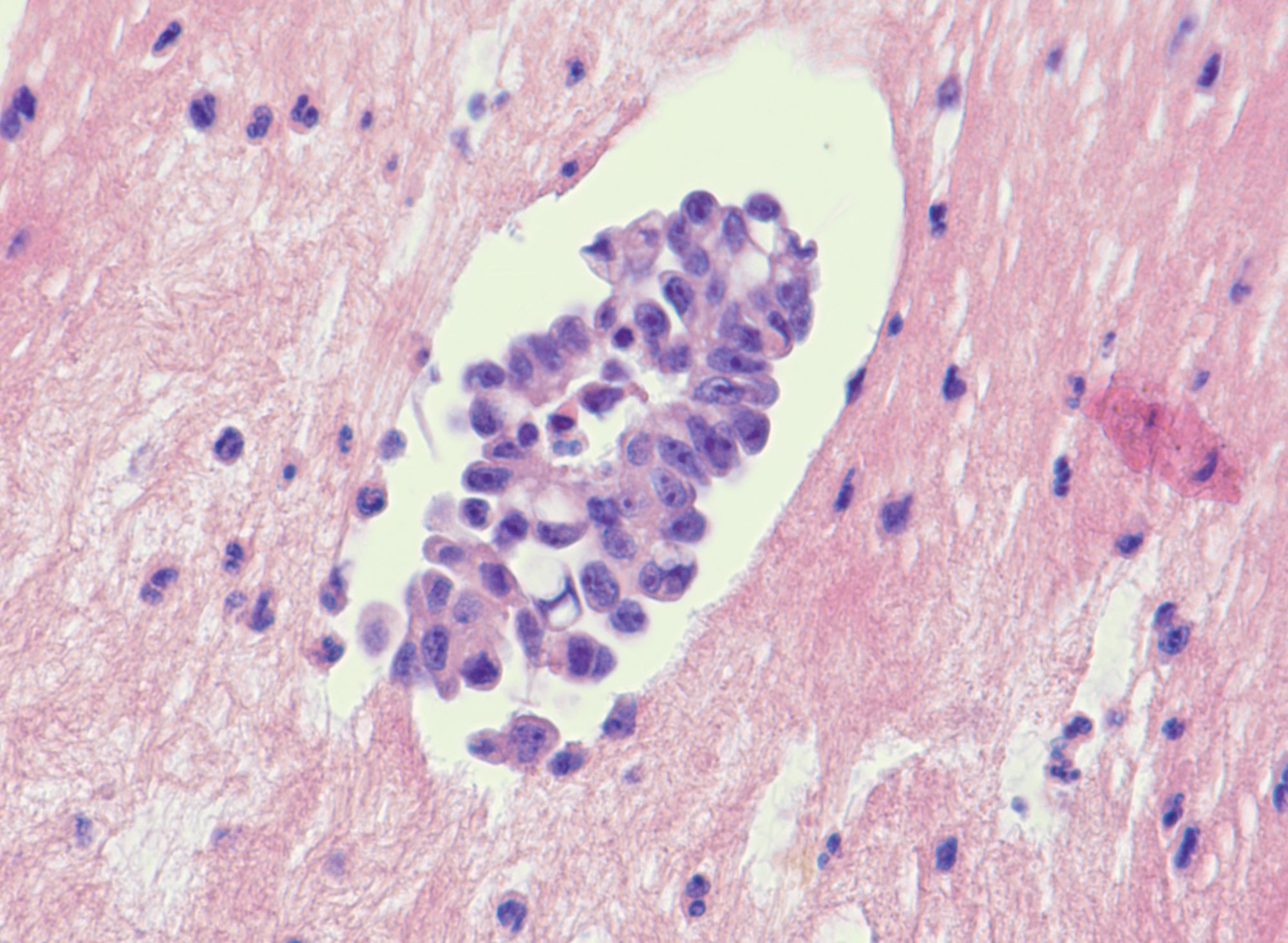
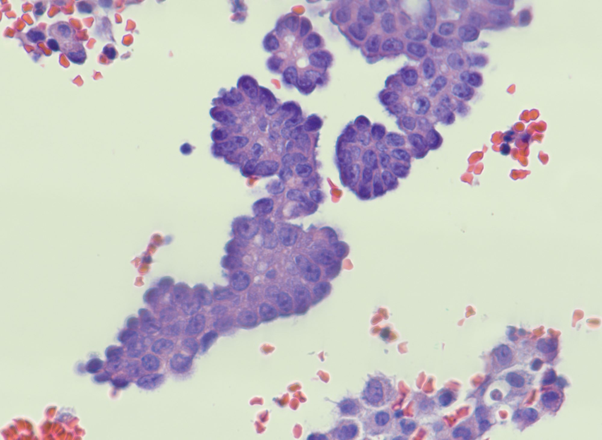
        • pT1c3 (IC3): malignant cells in ascites or peritoneal washings
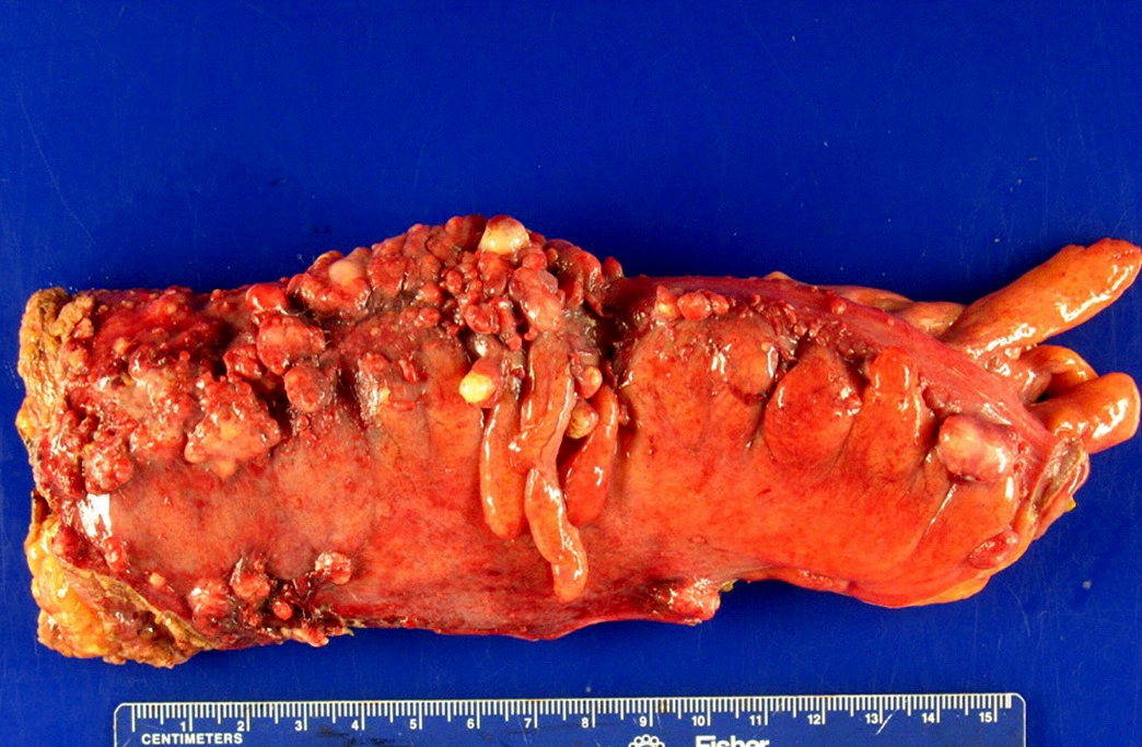
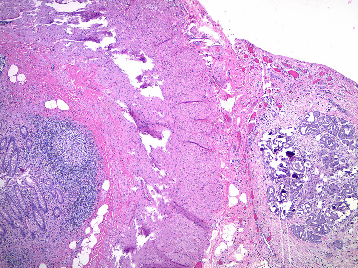
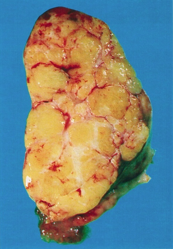
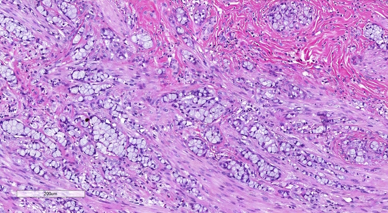
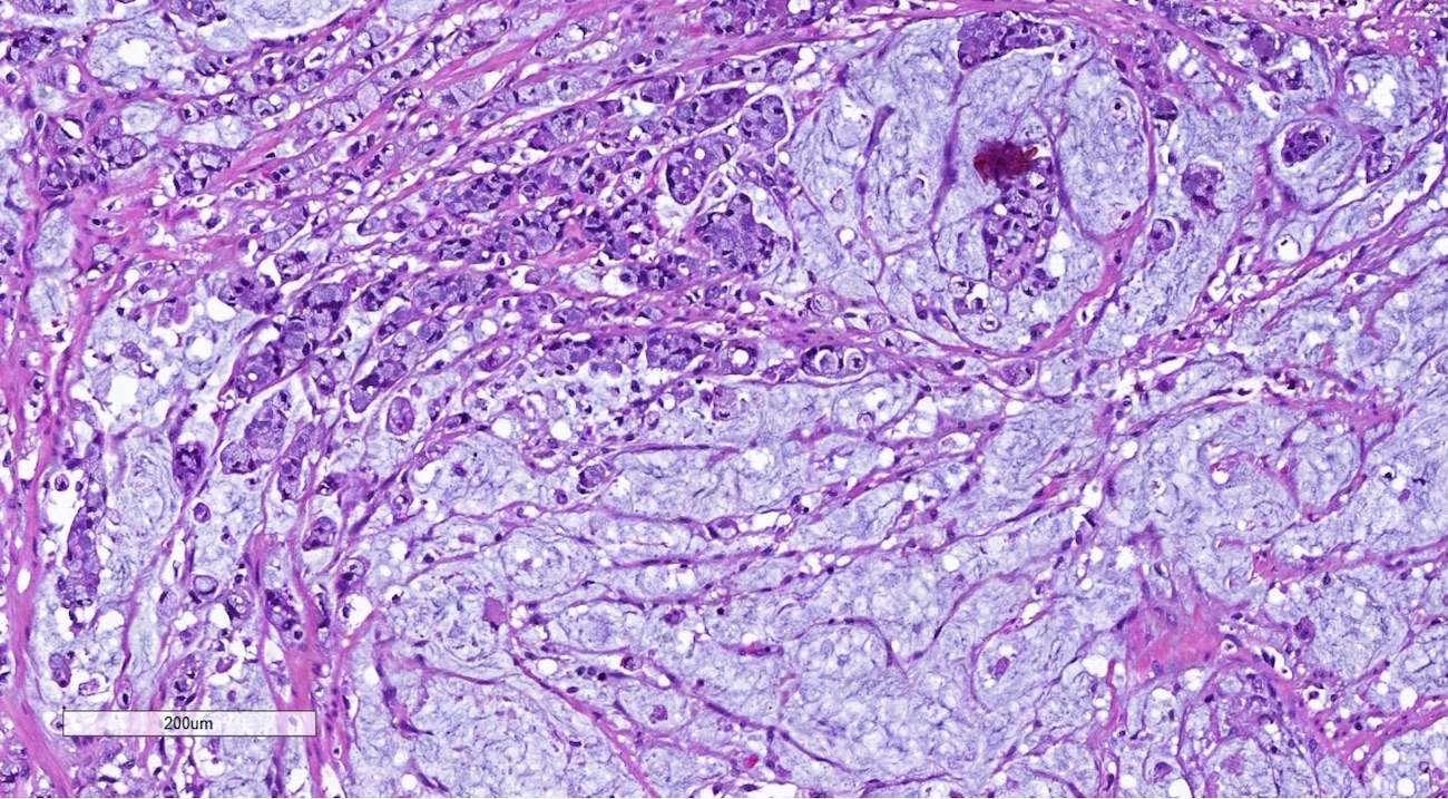
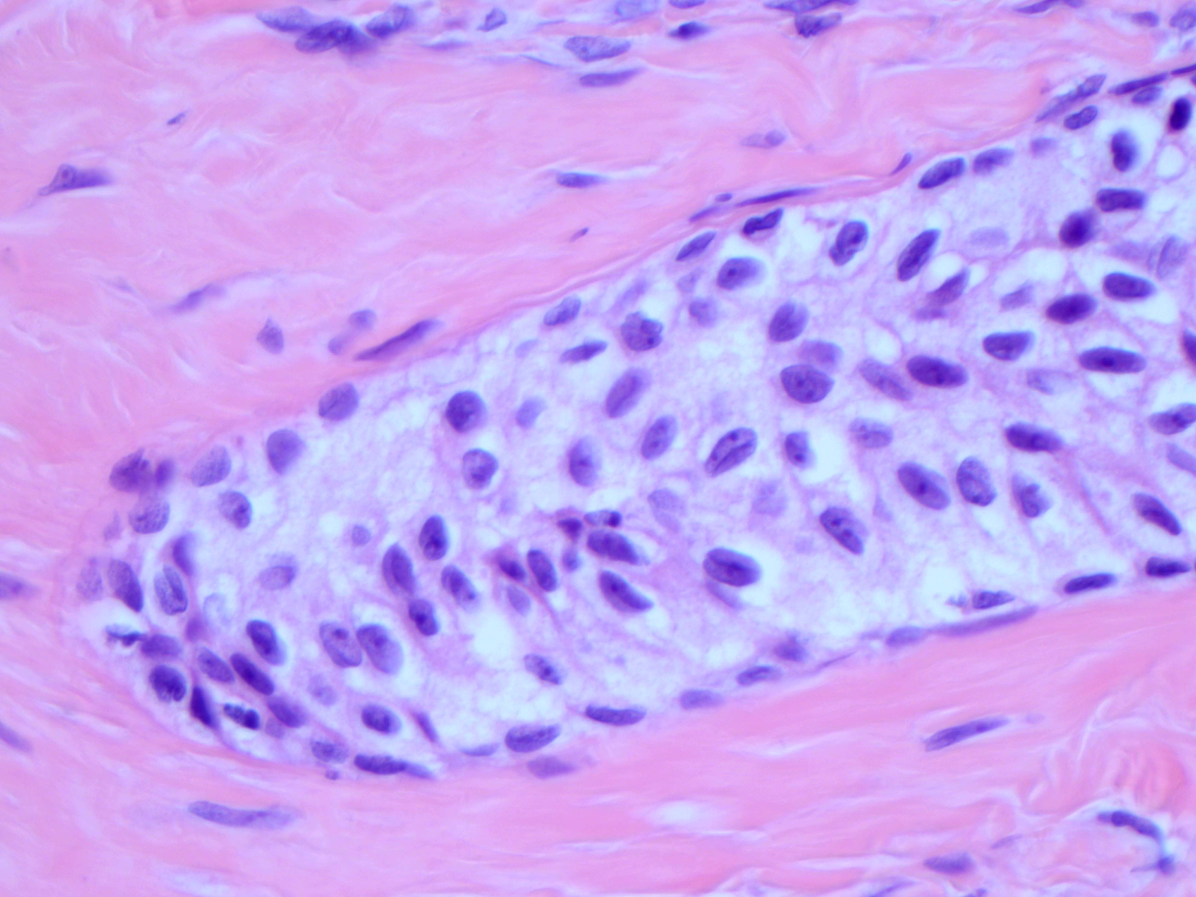
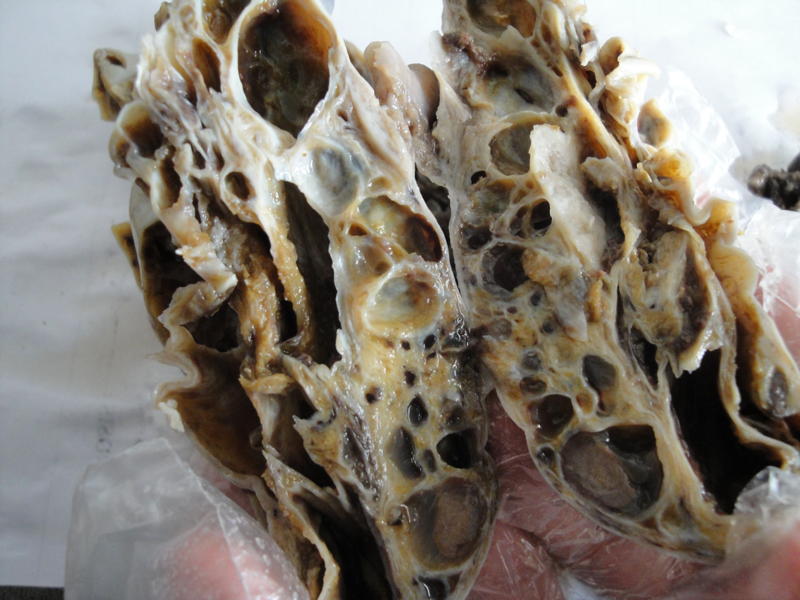
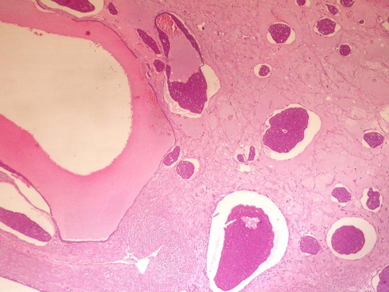
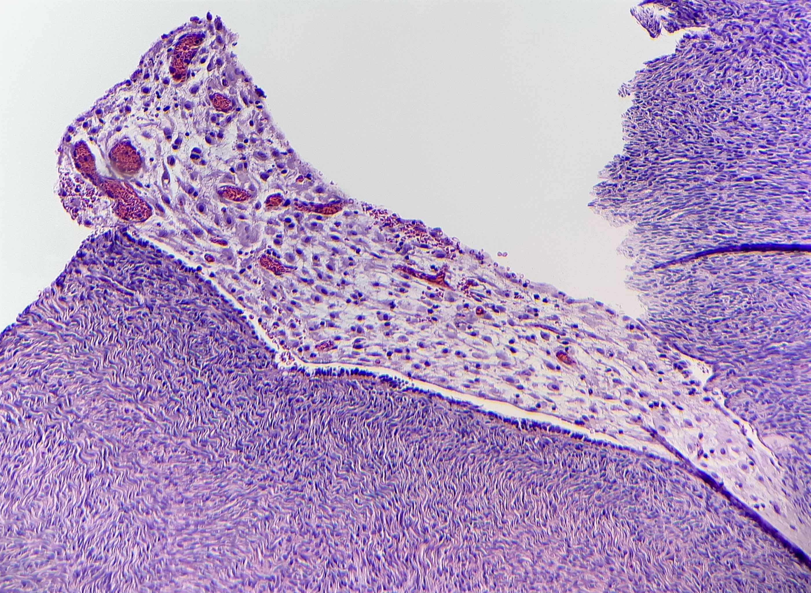
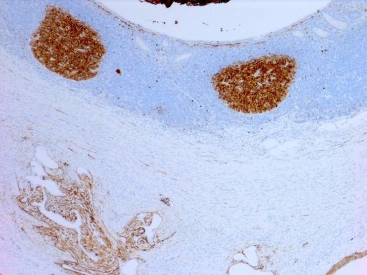
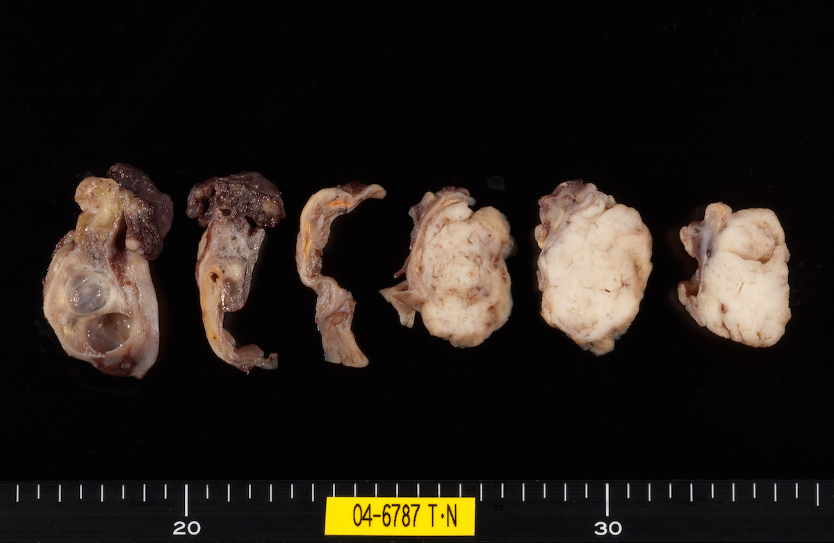
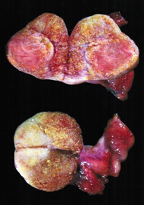
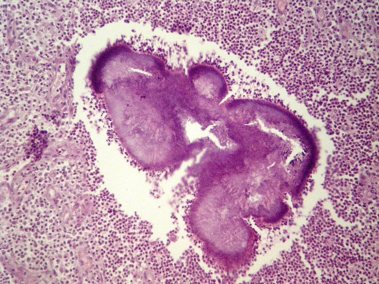
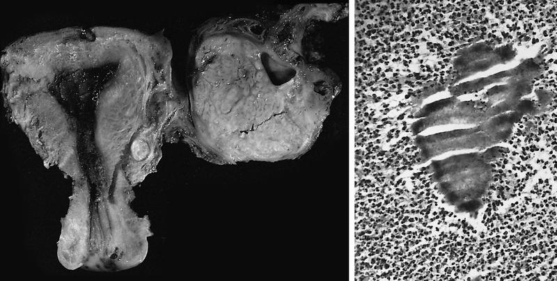
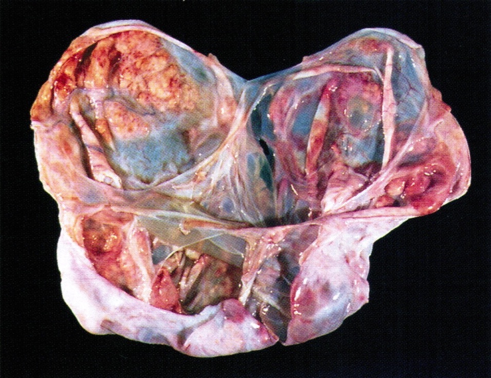
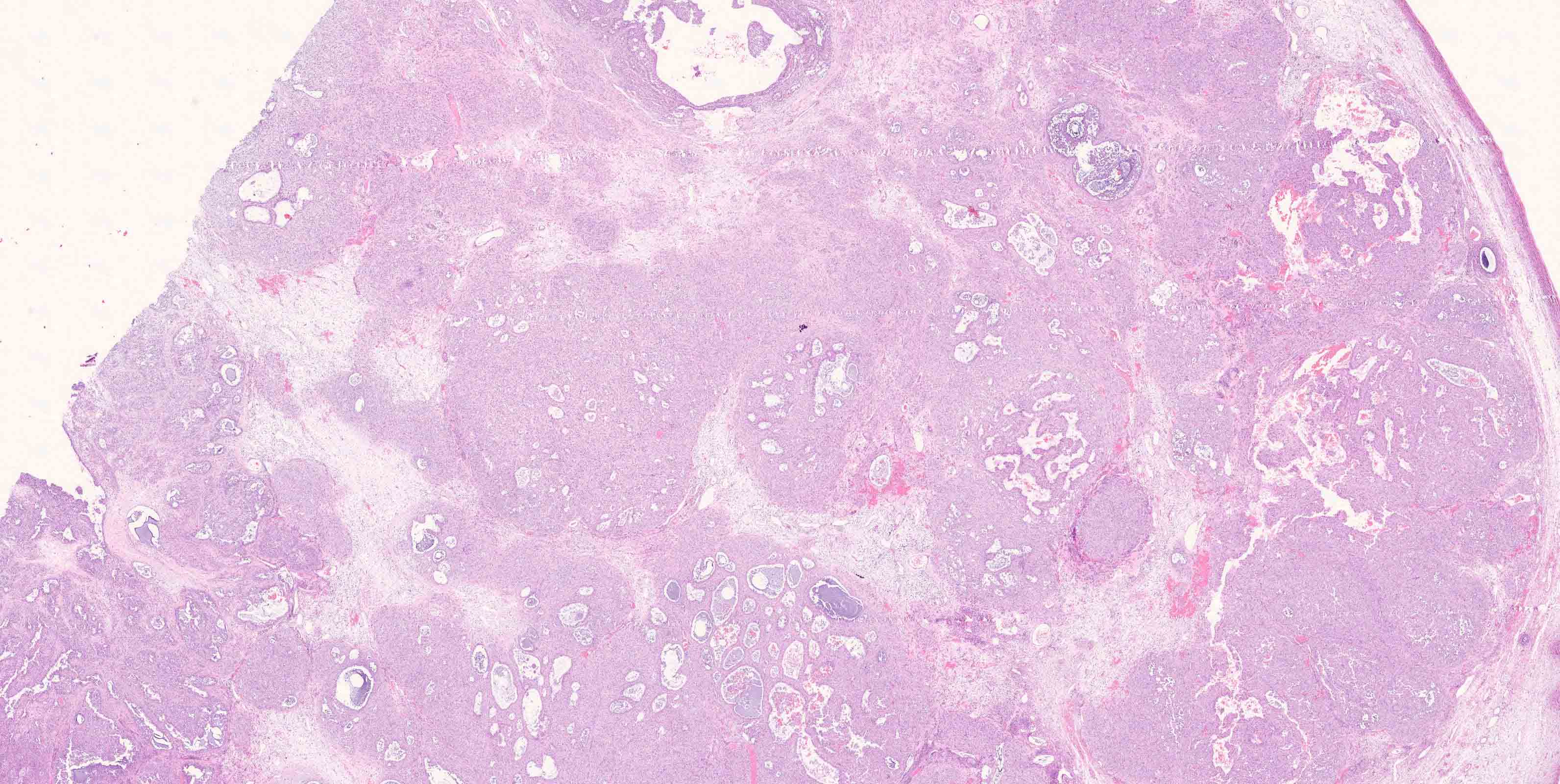
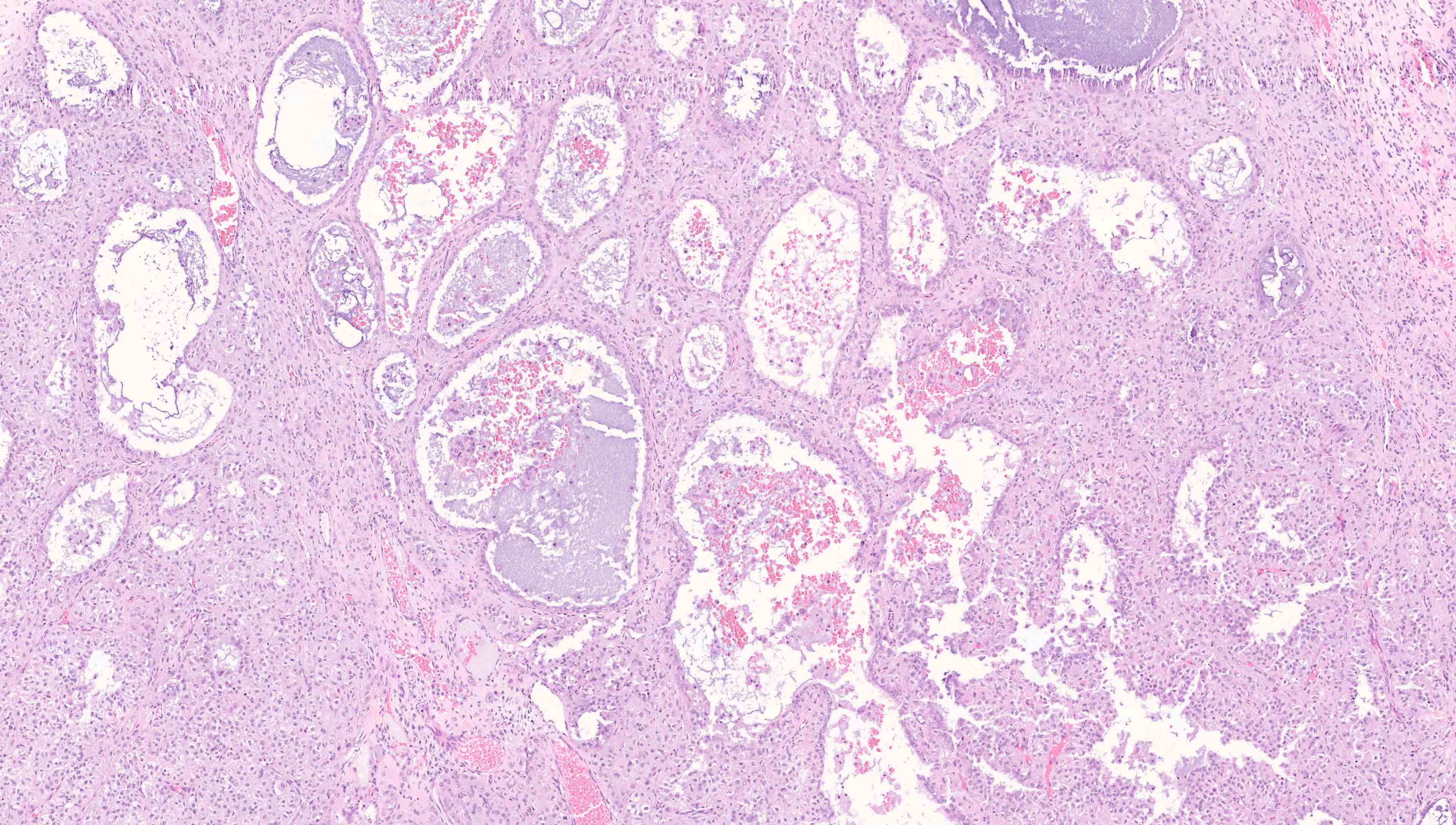
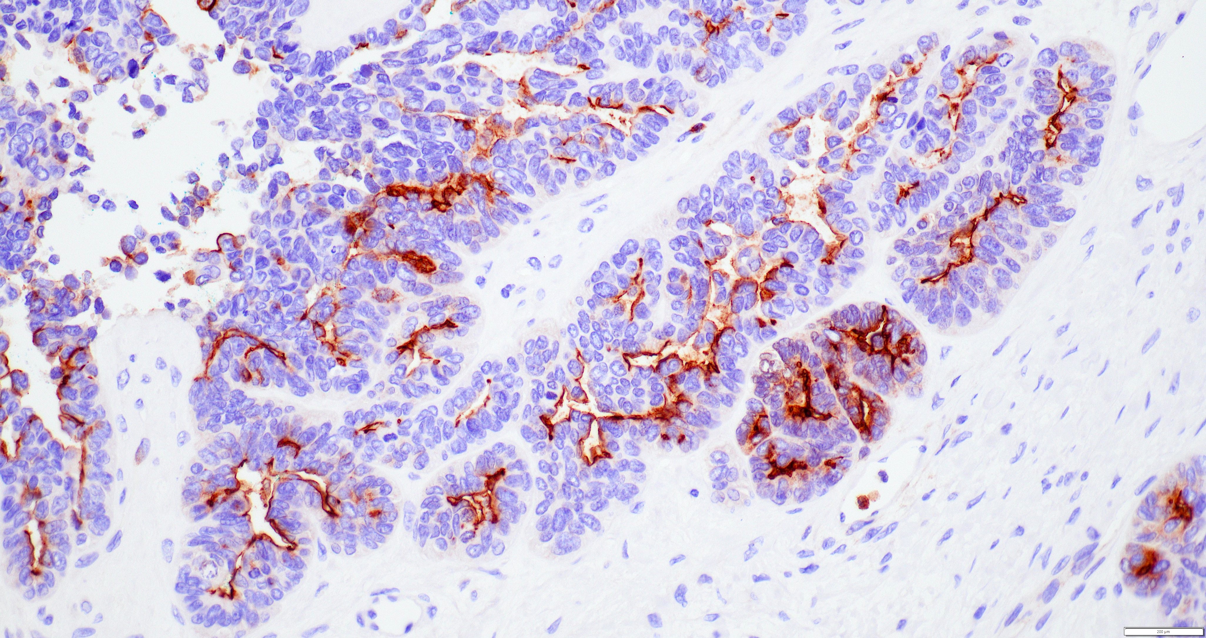
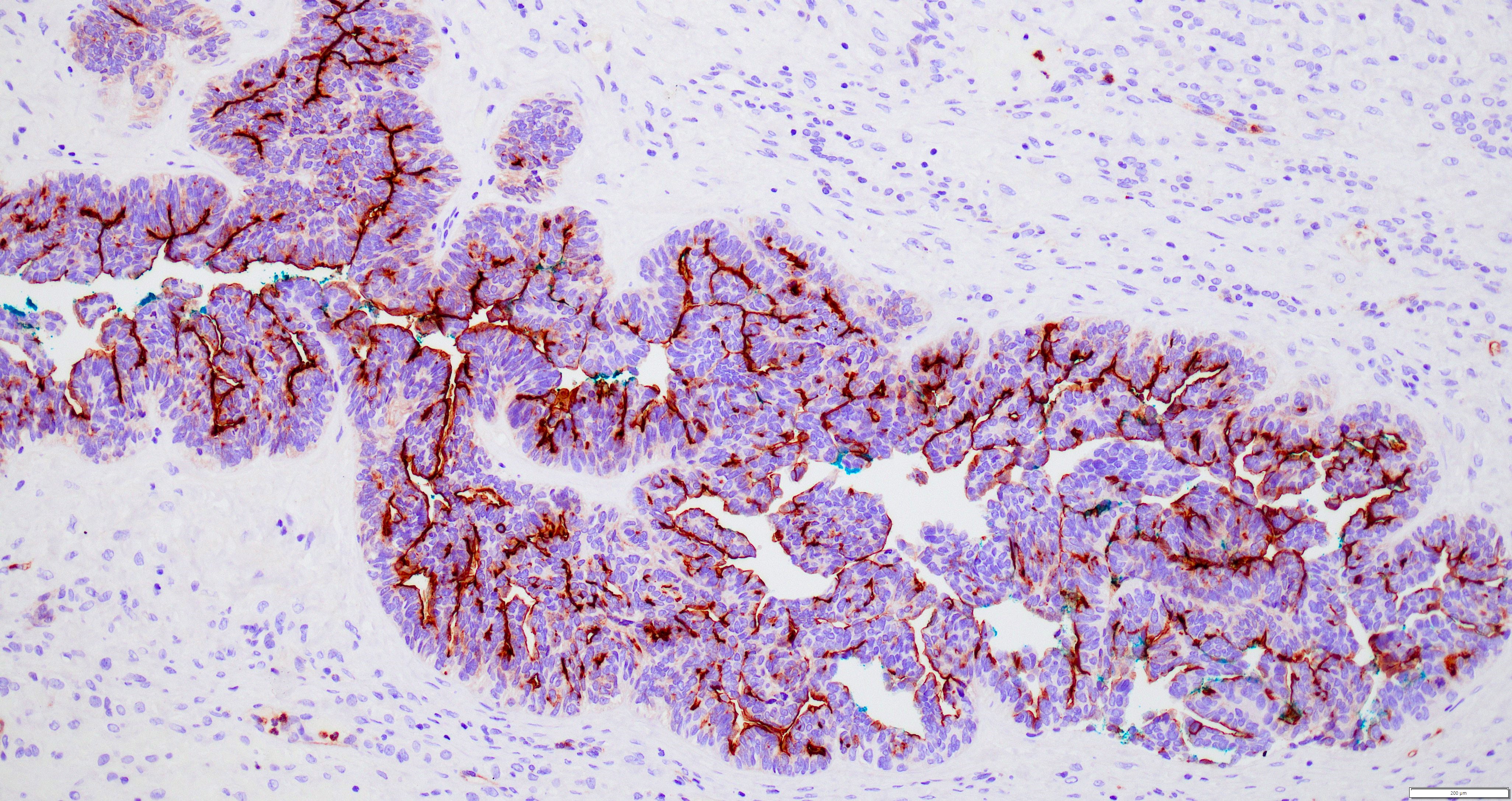
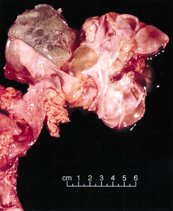
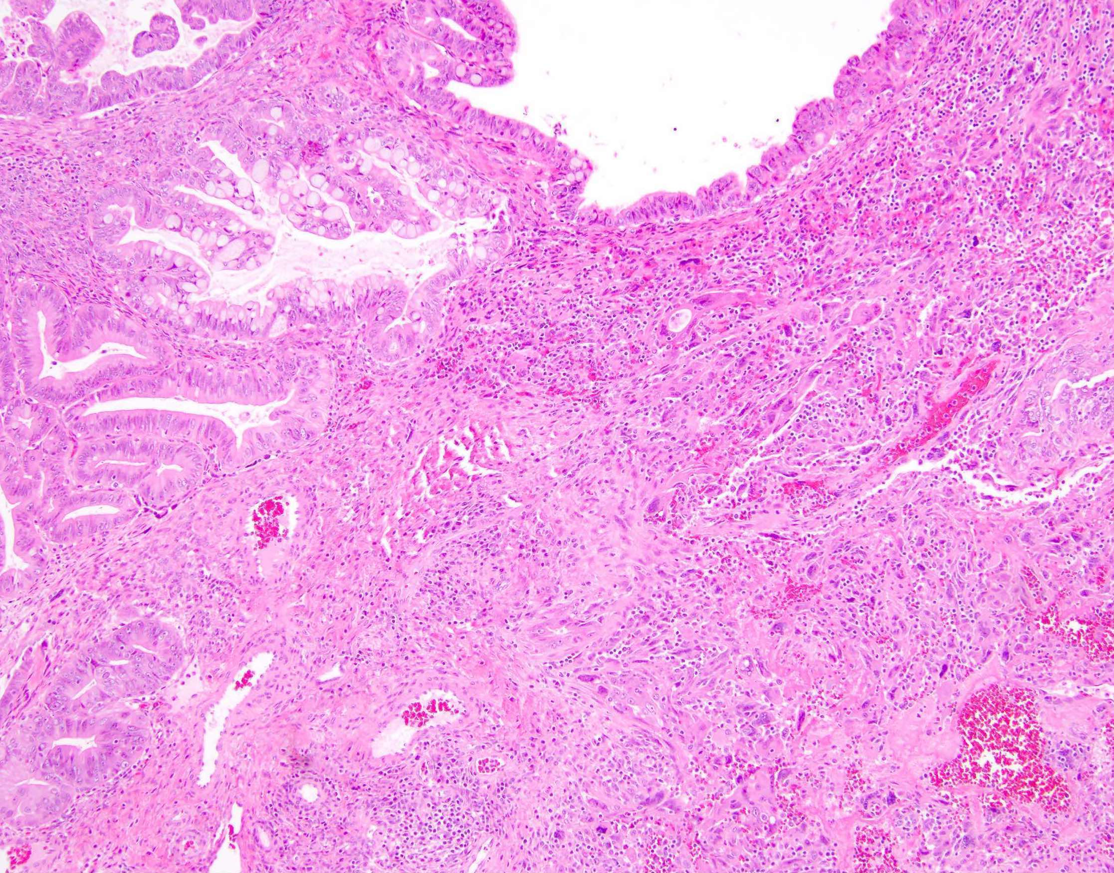
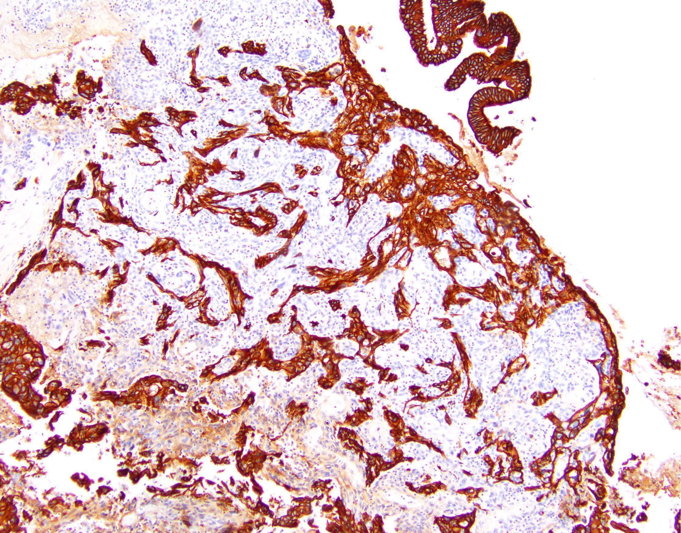
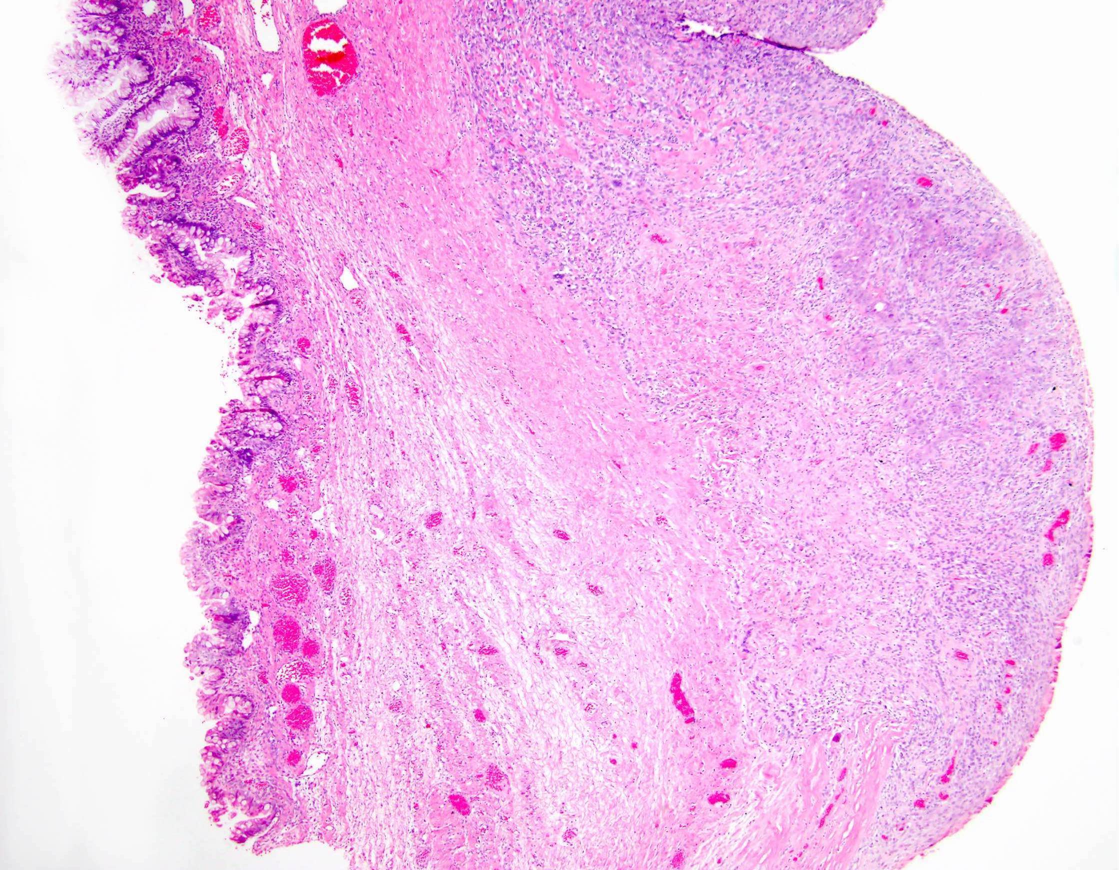
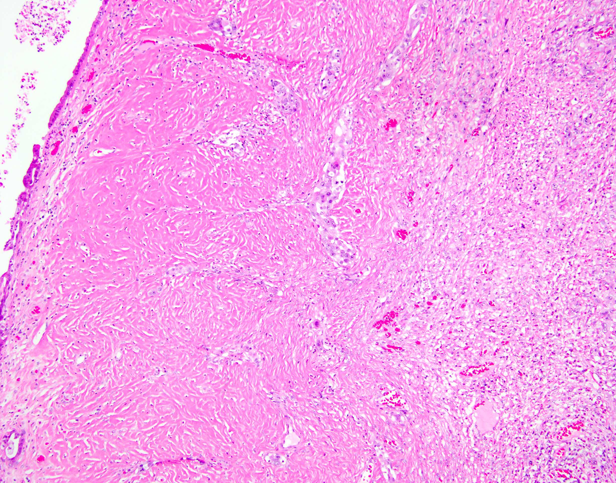
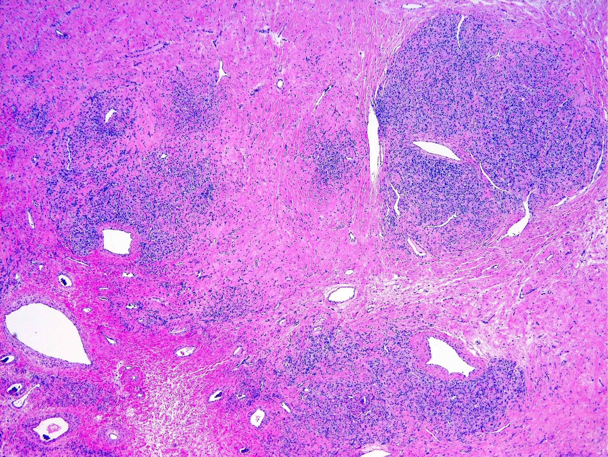
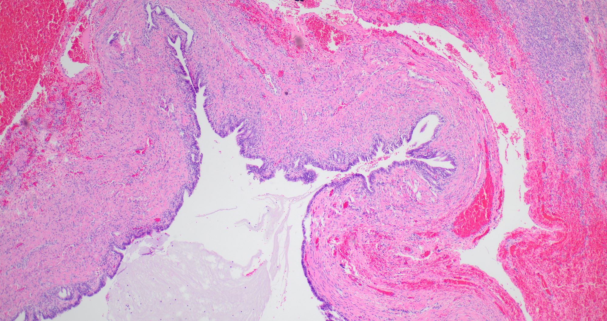
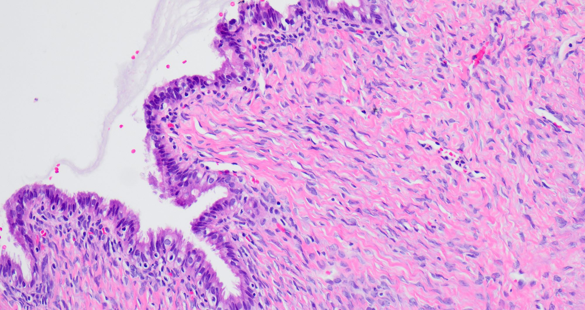
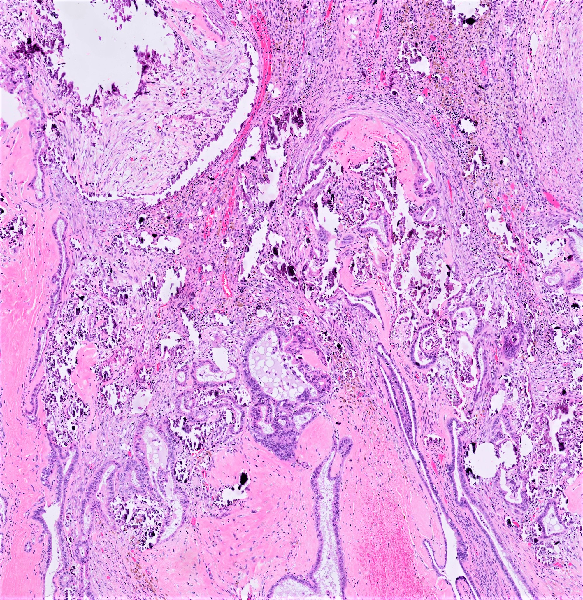
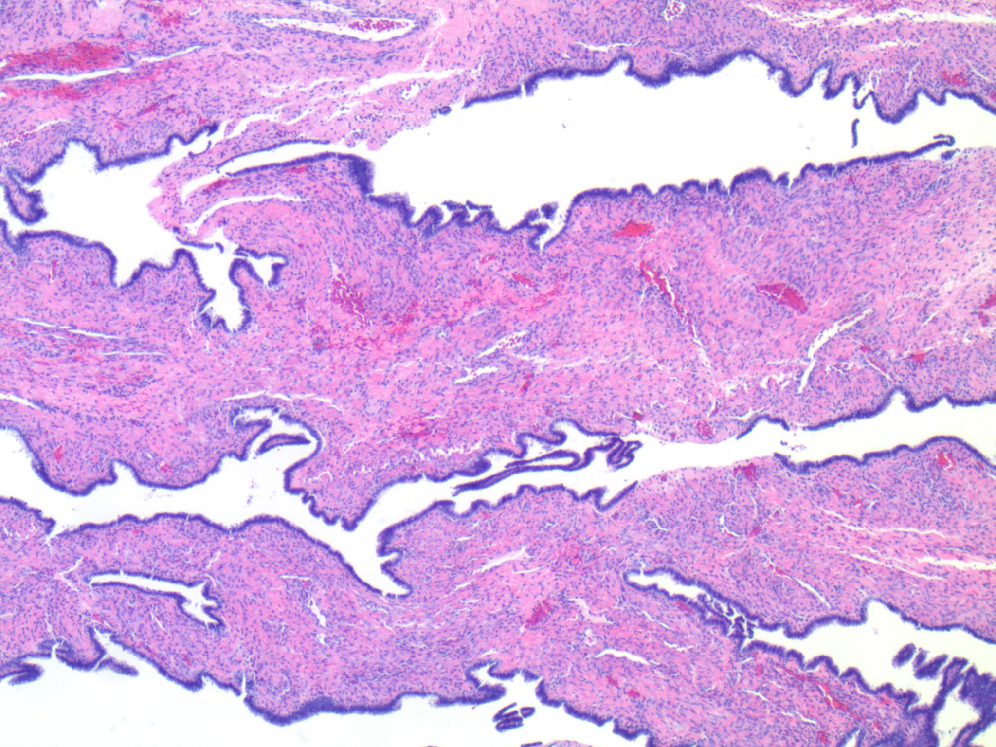
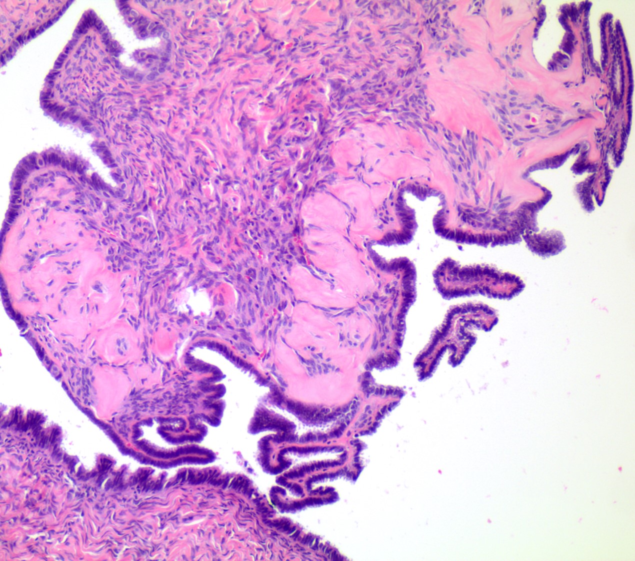
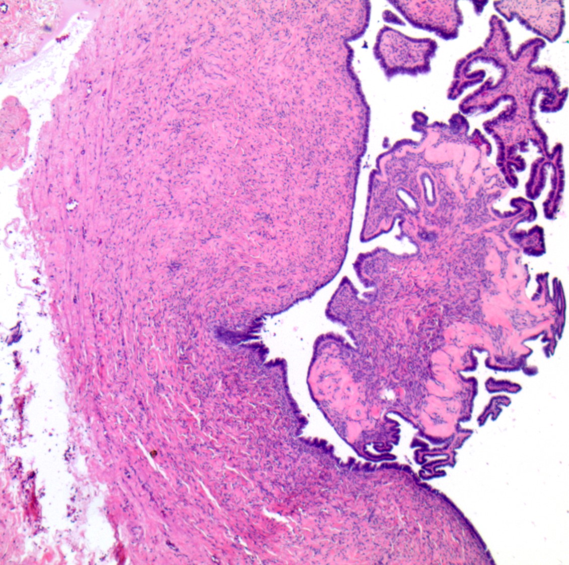
      • pT2a (IIA): extension or implants on the uterus or fallopian tube(s) or ovaries
      • pT2b (IIB): extension to or implants on other pelvic tissues
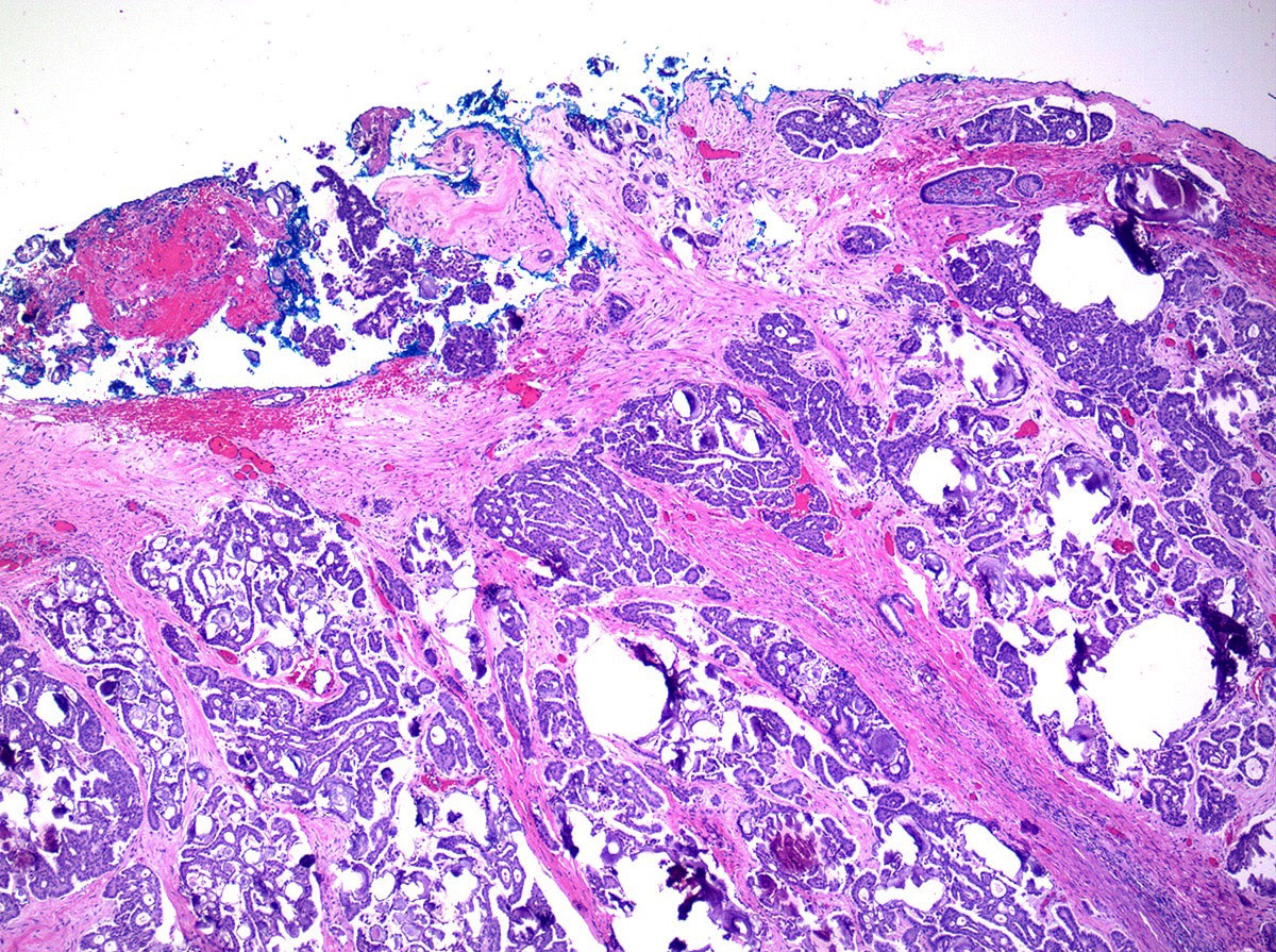
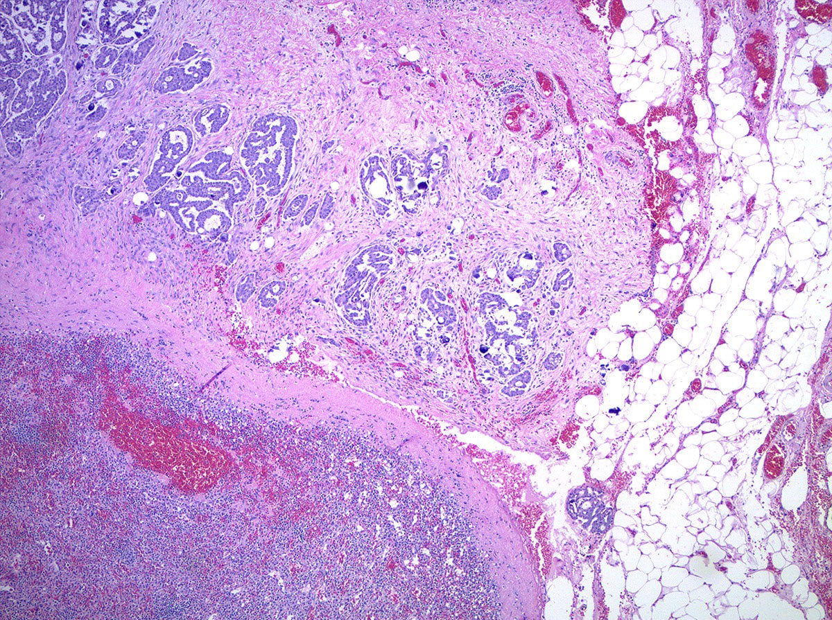
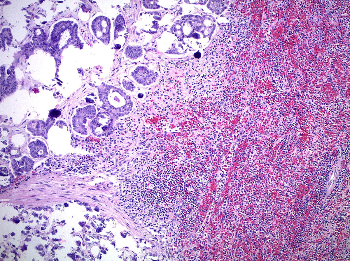
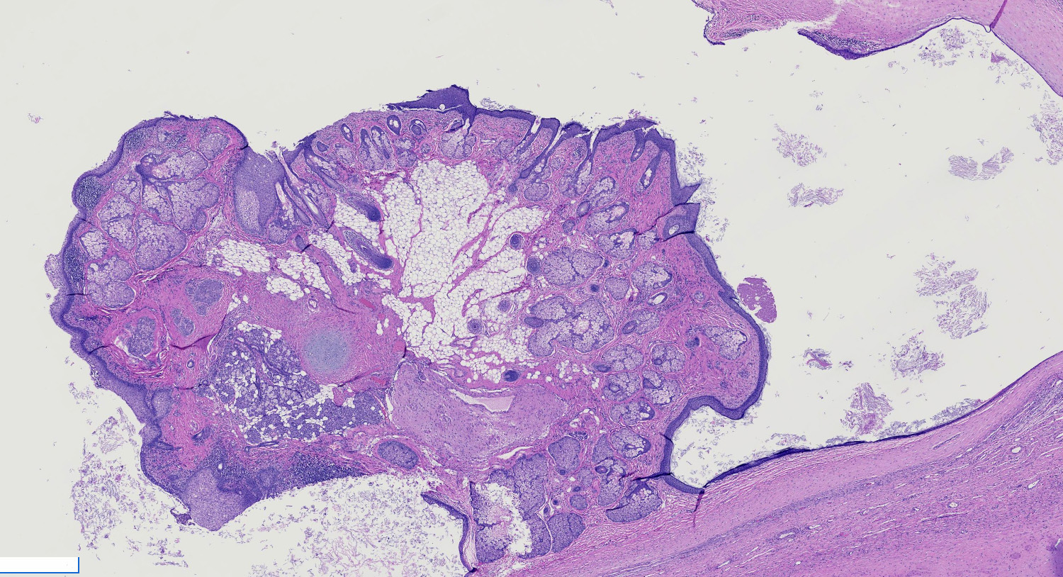
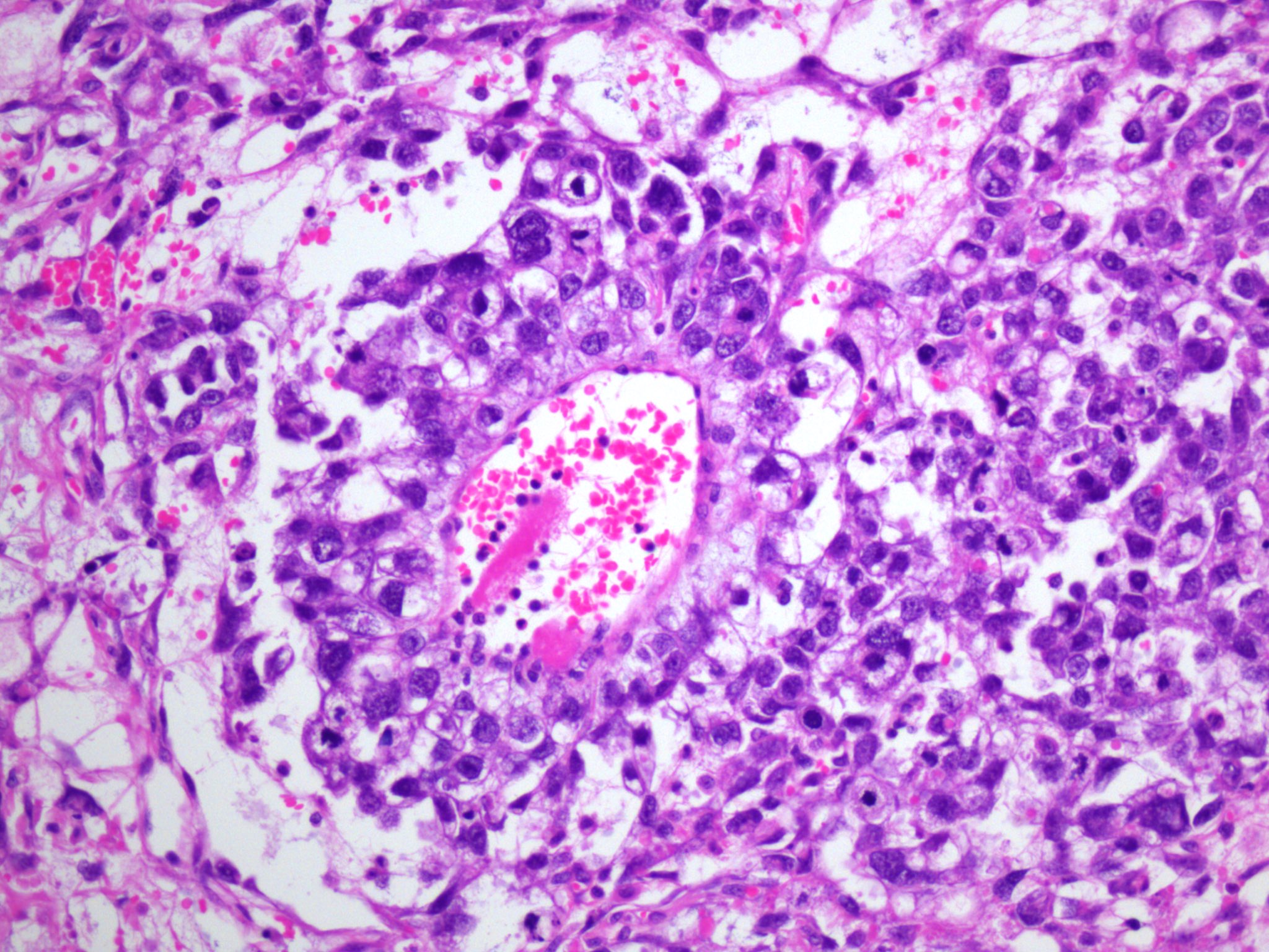
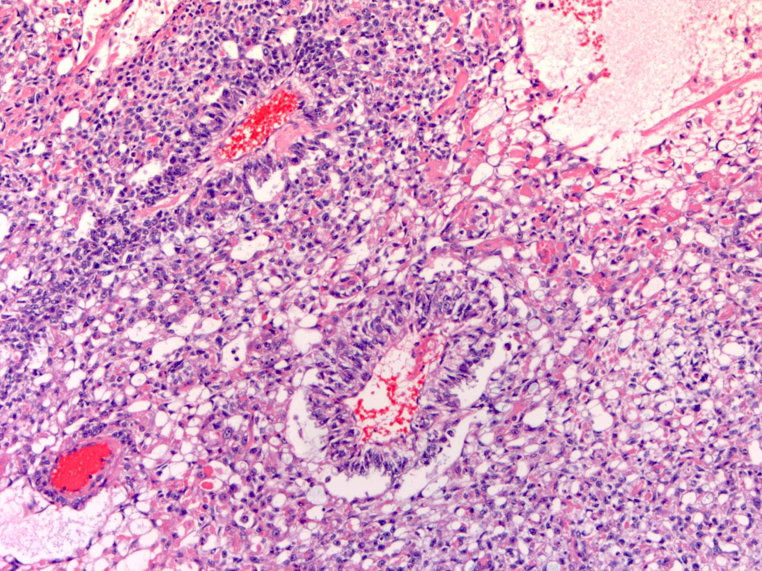
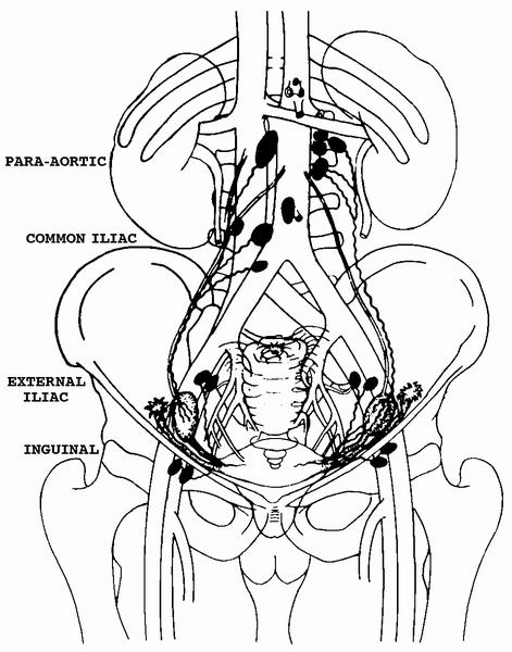
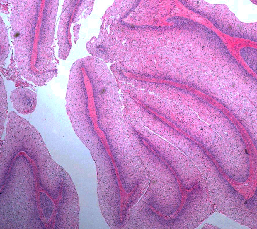
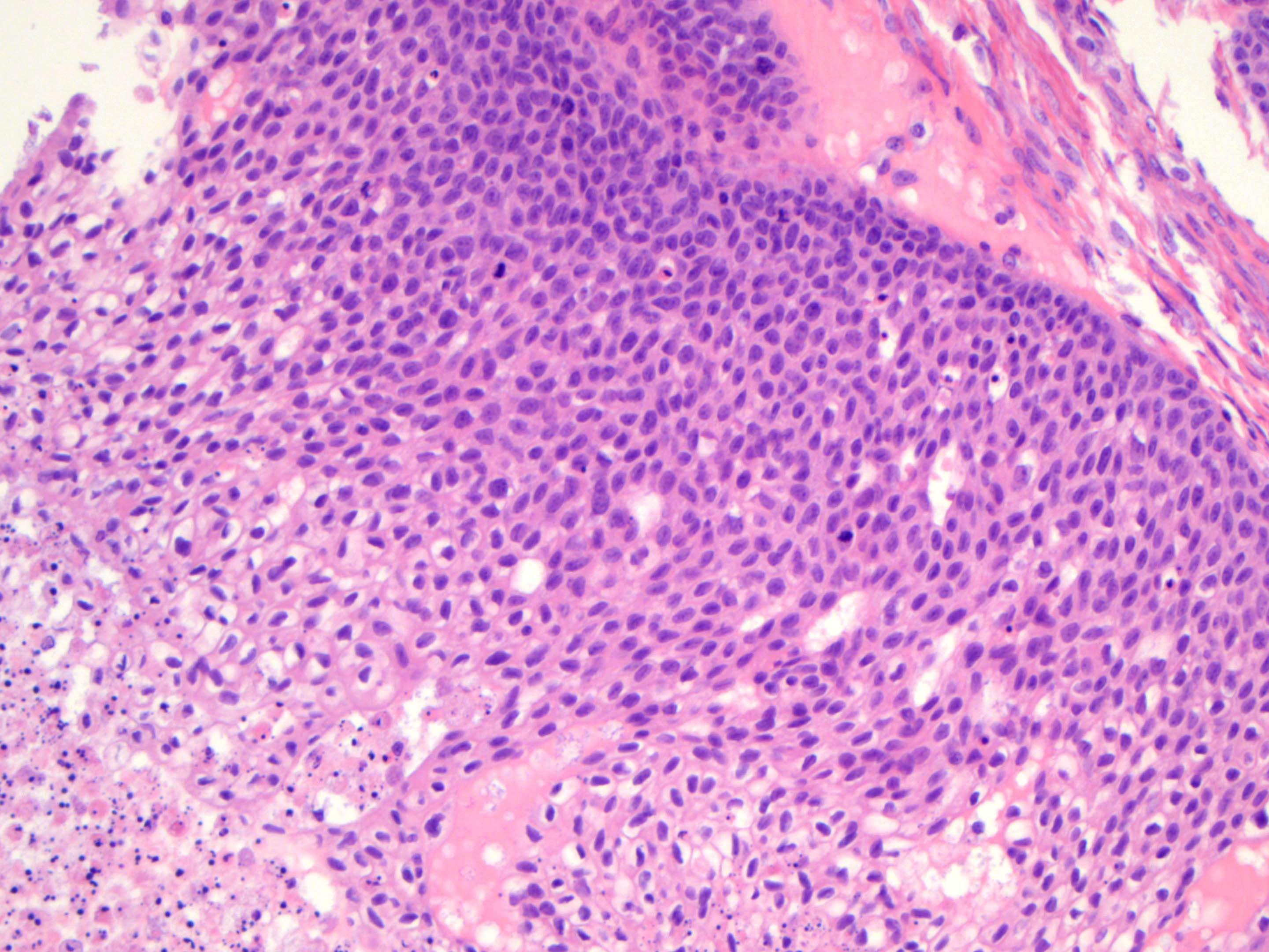
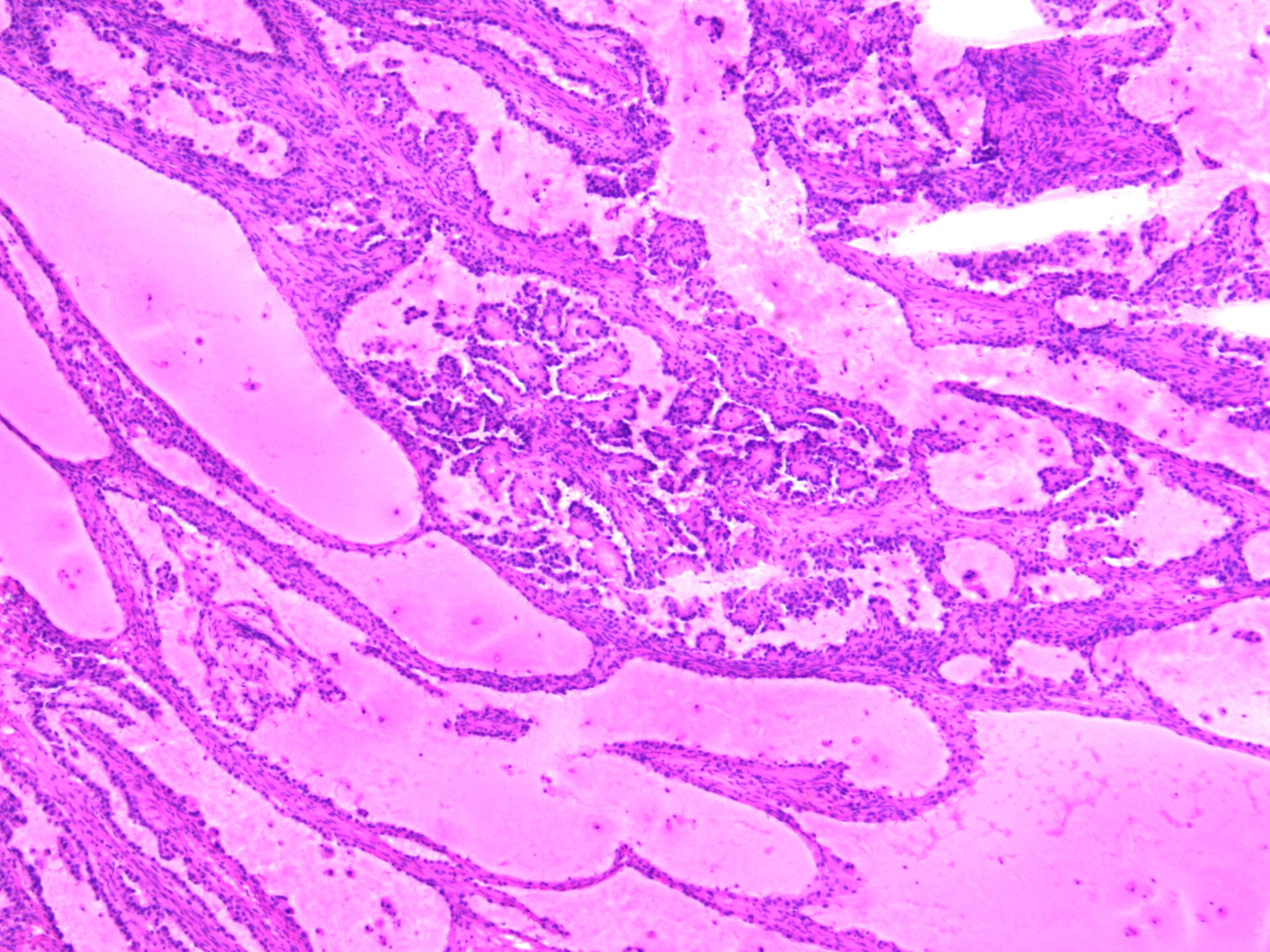
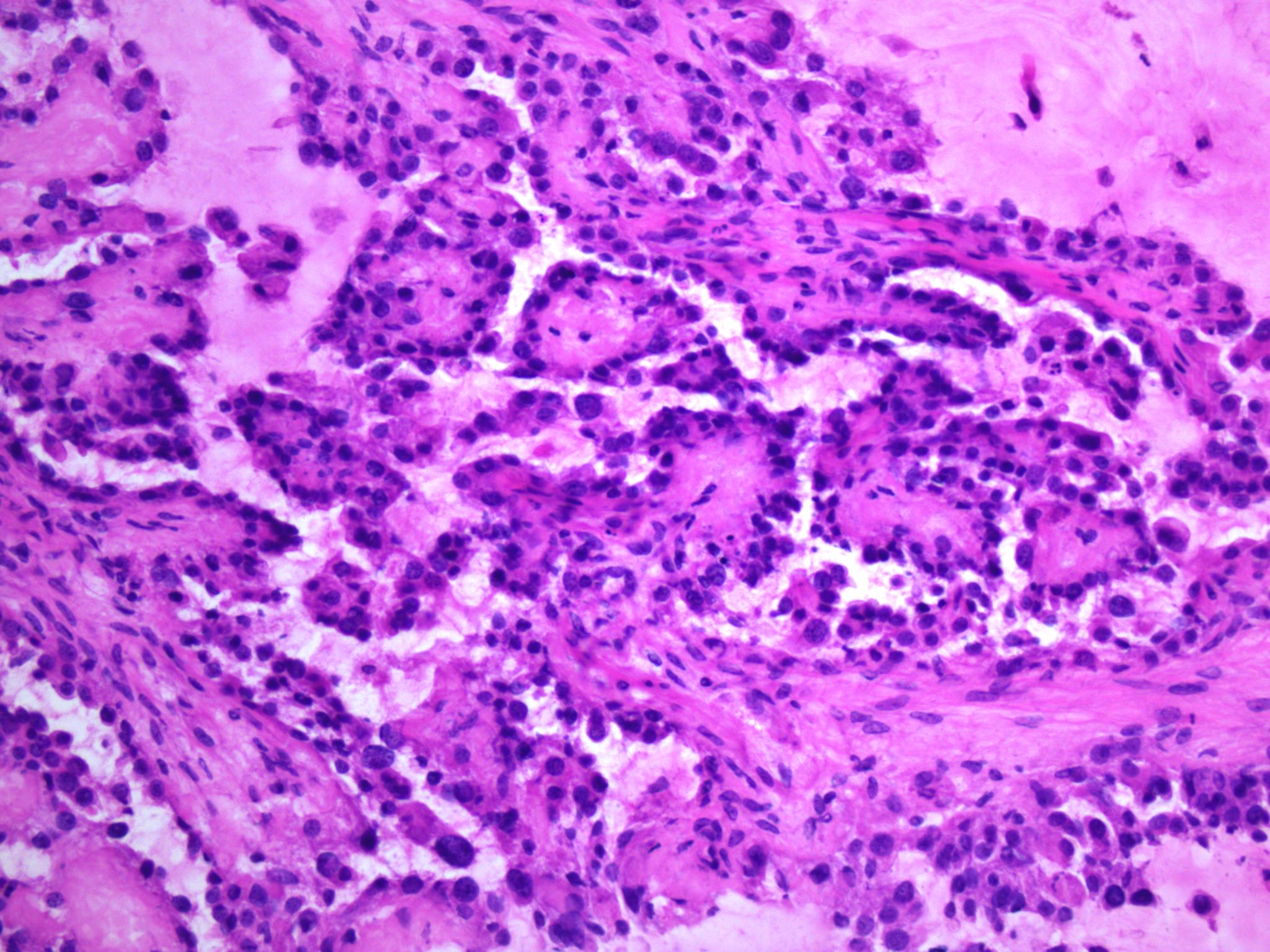
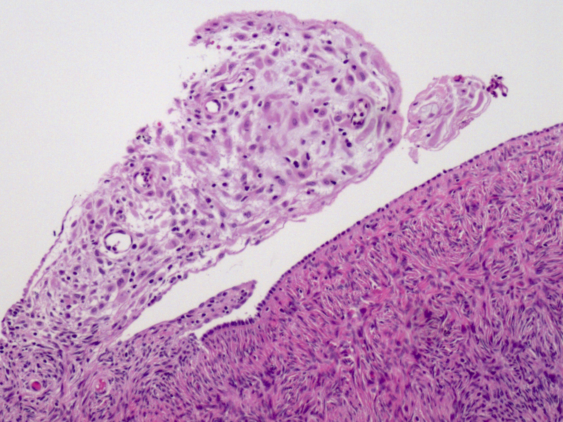
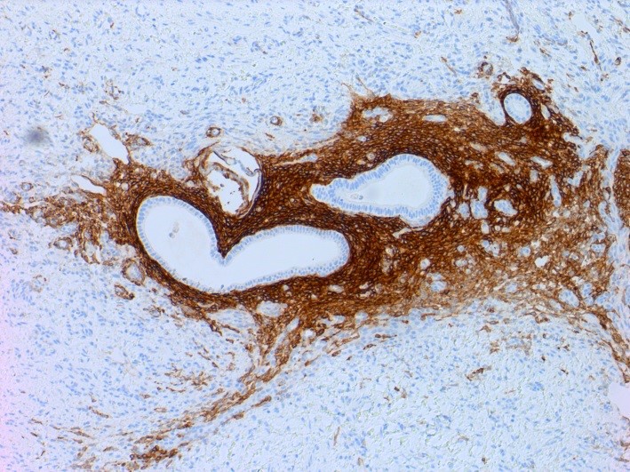
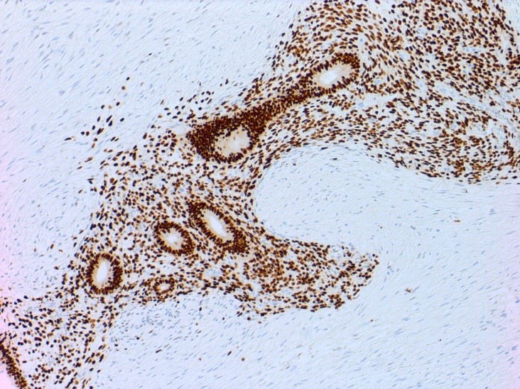
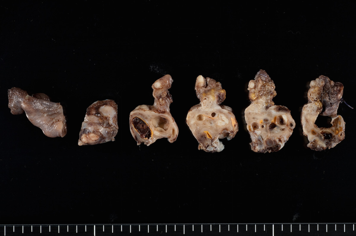
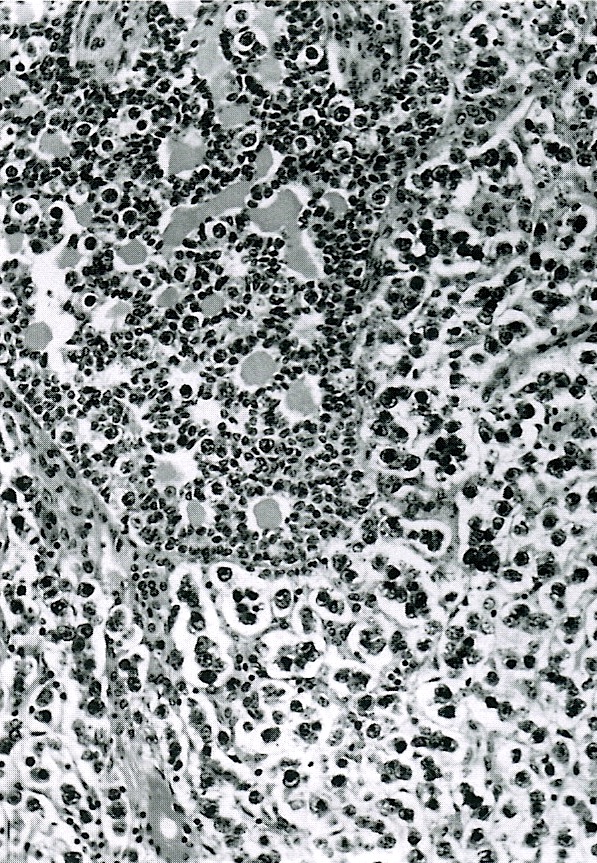
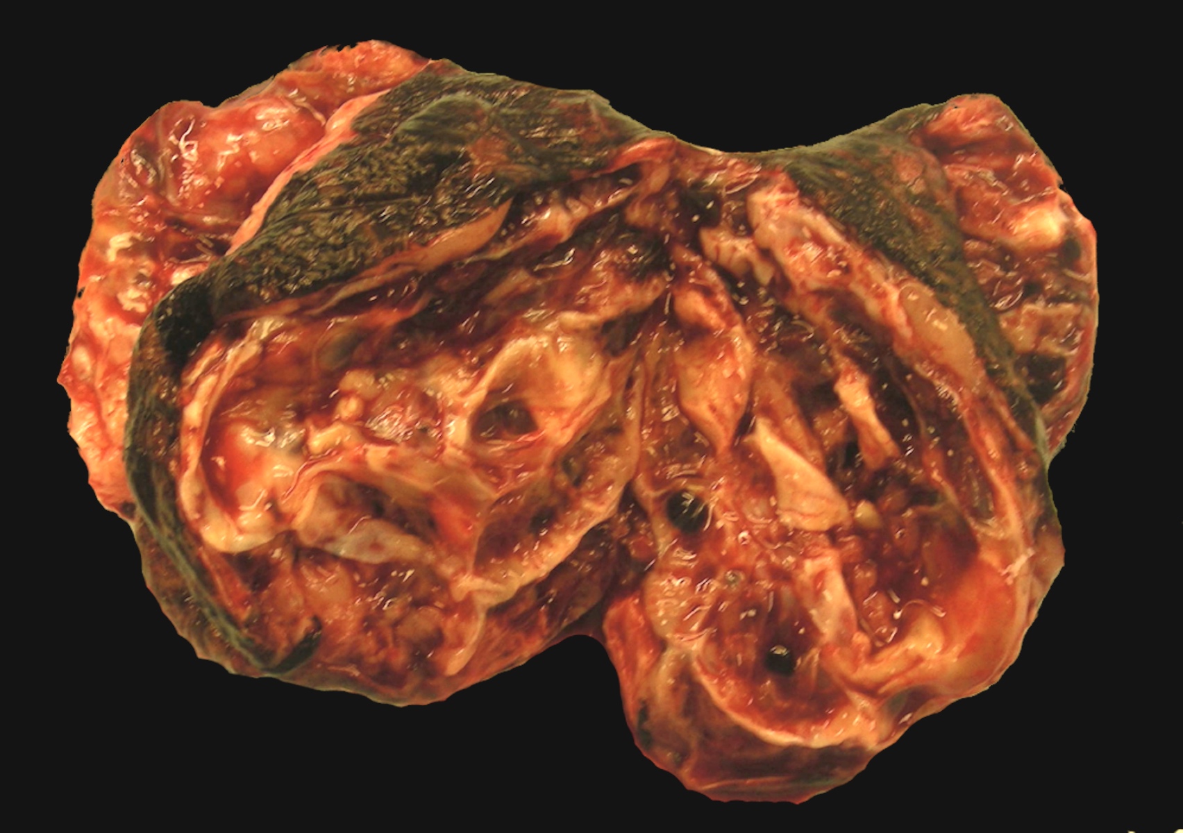
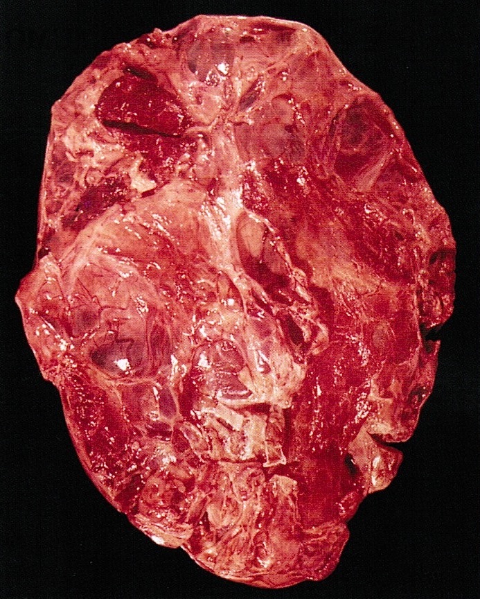
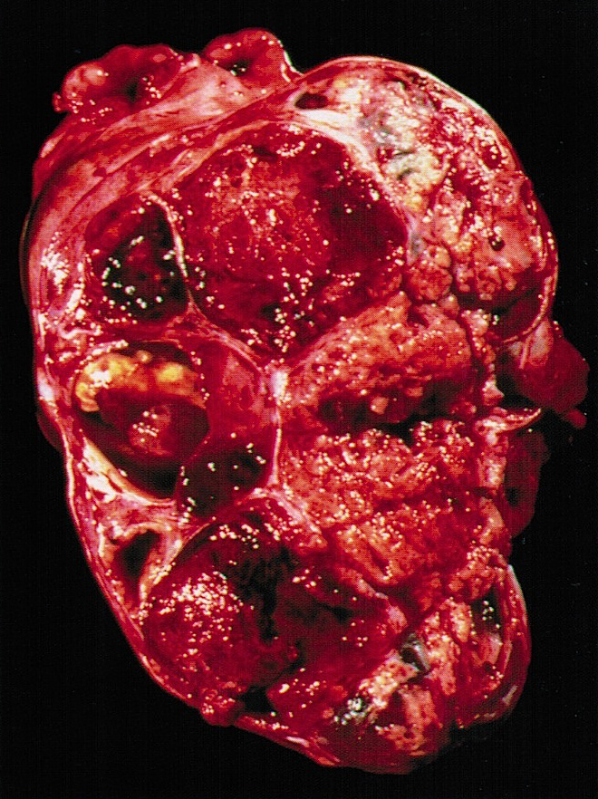
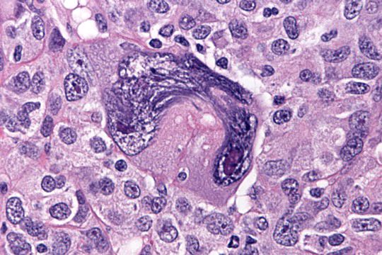
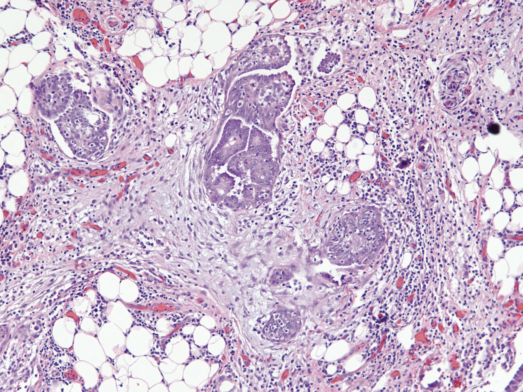
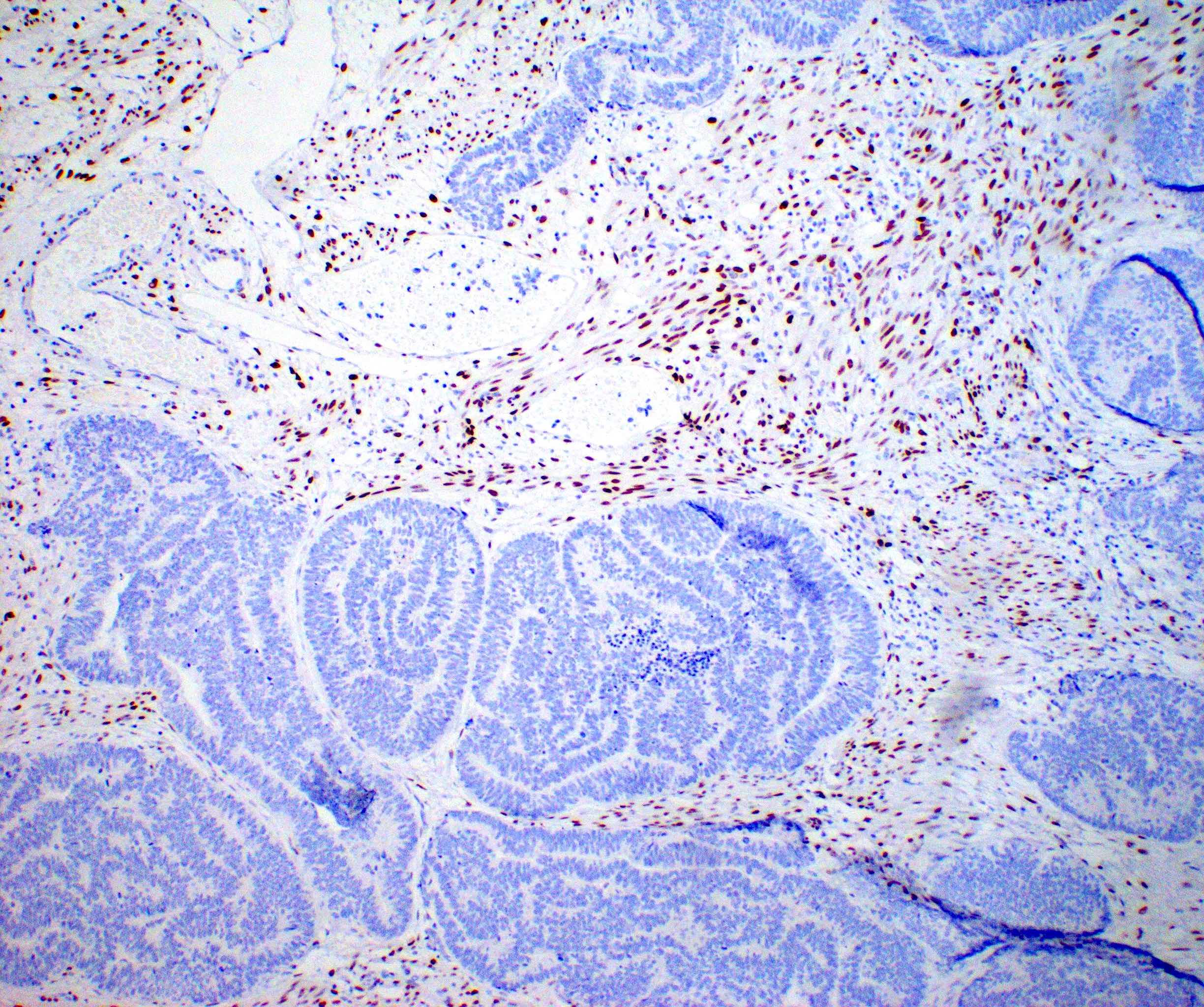
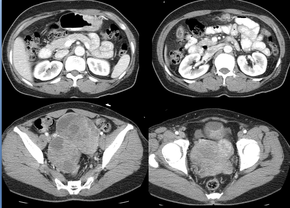
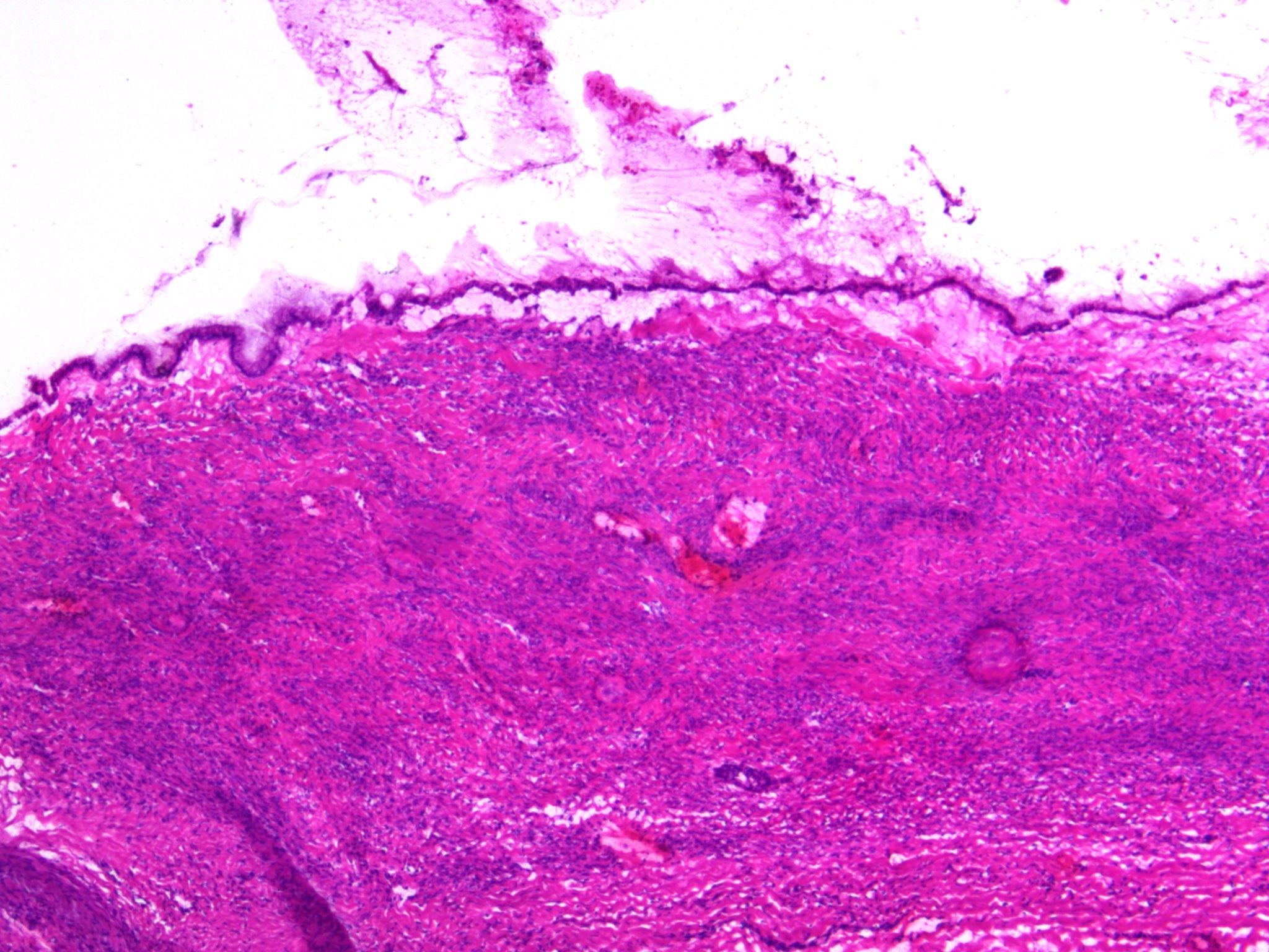
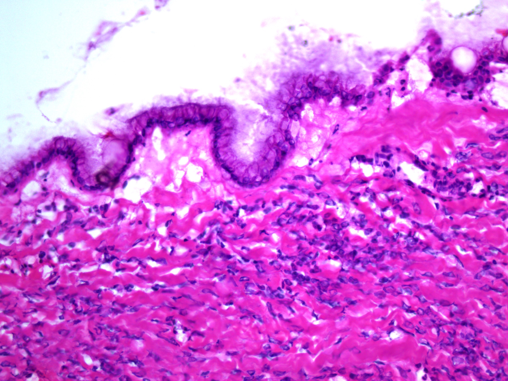
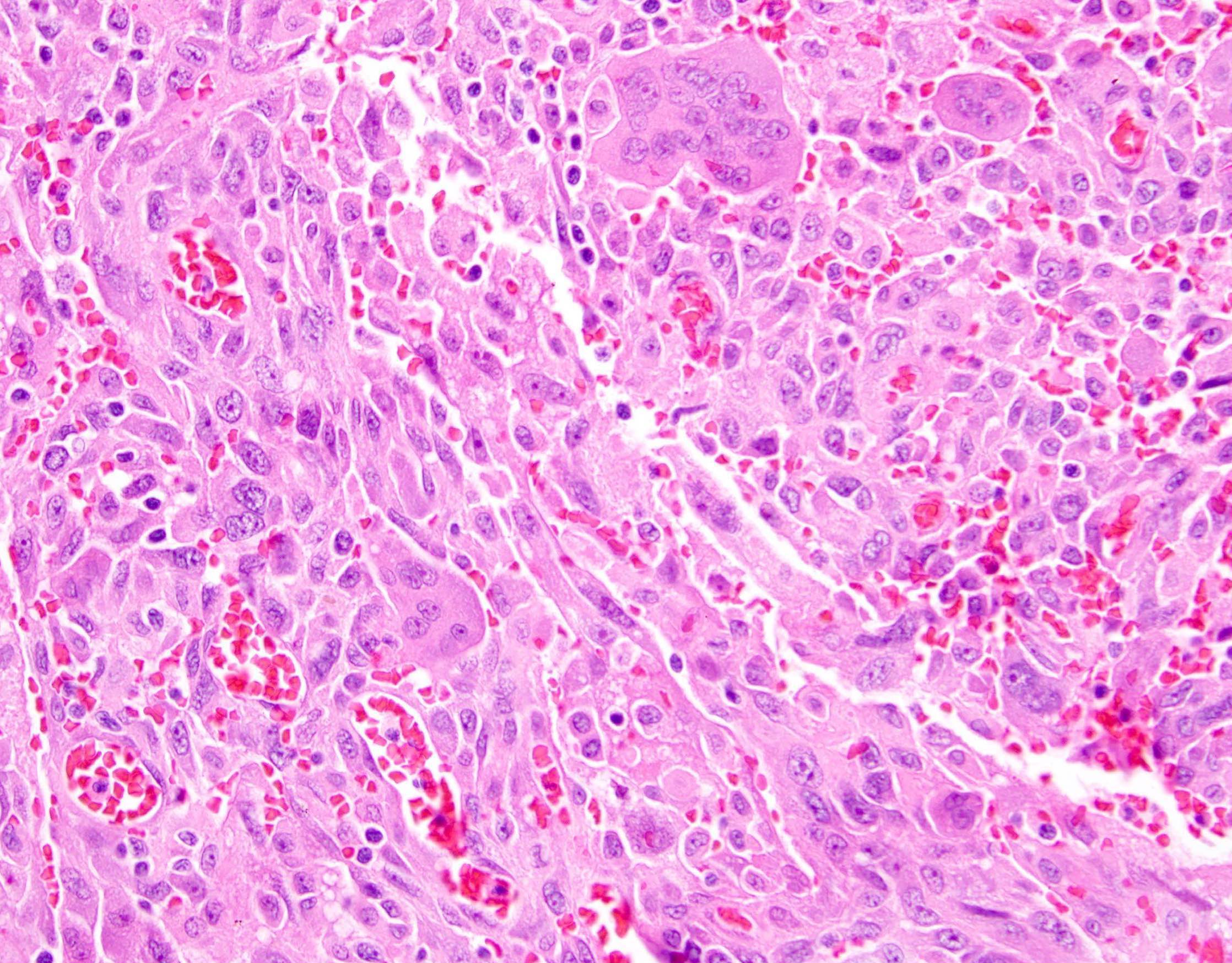
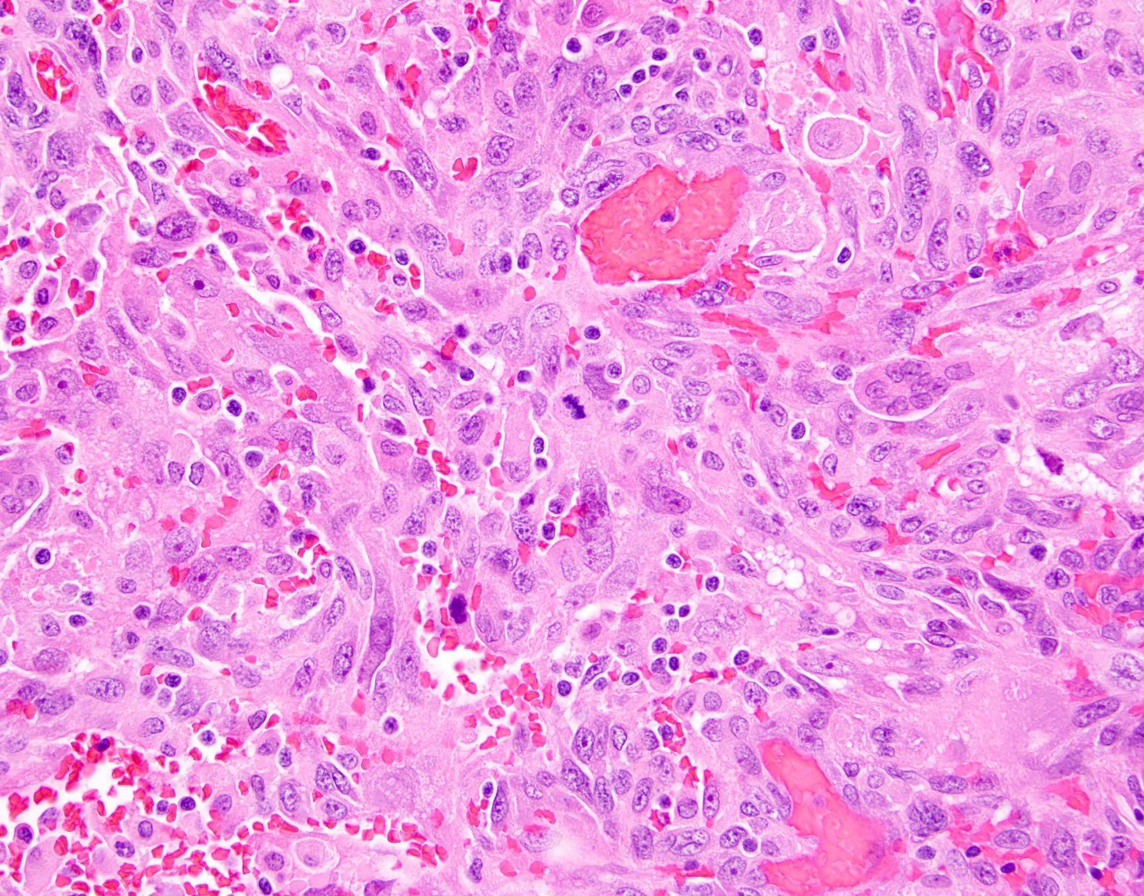
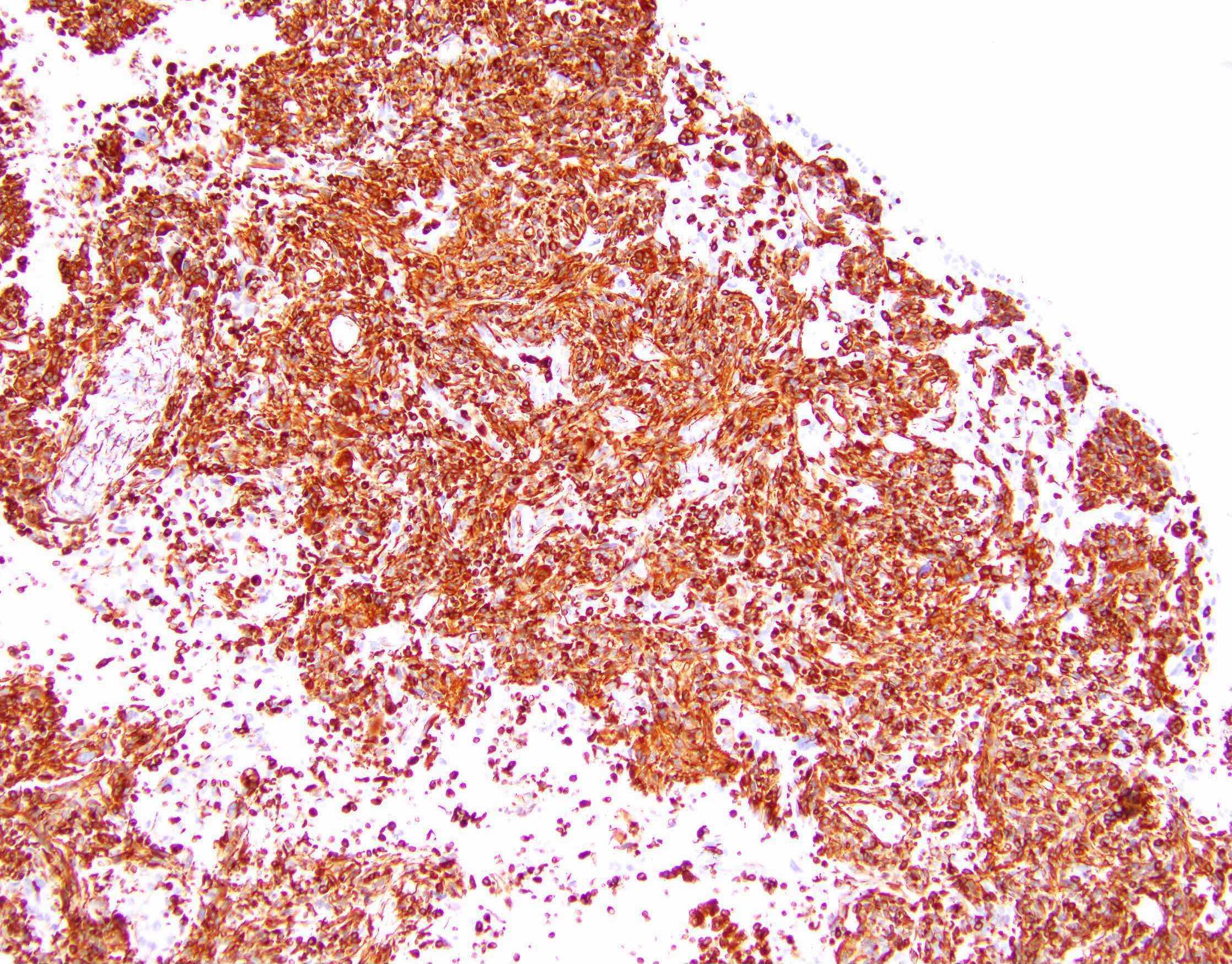
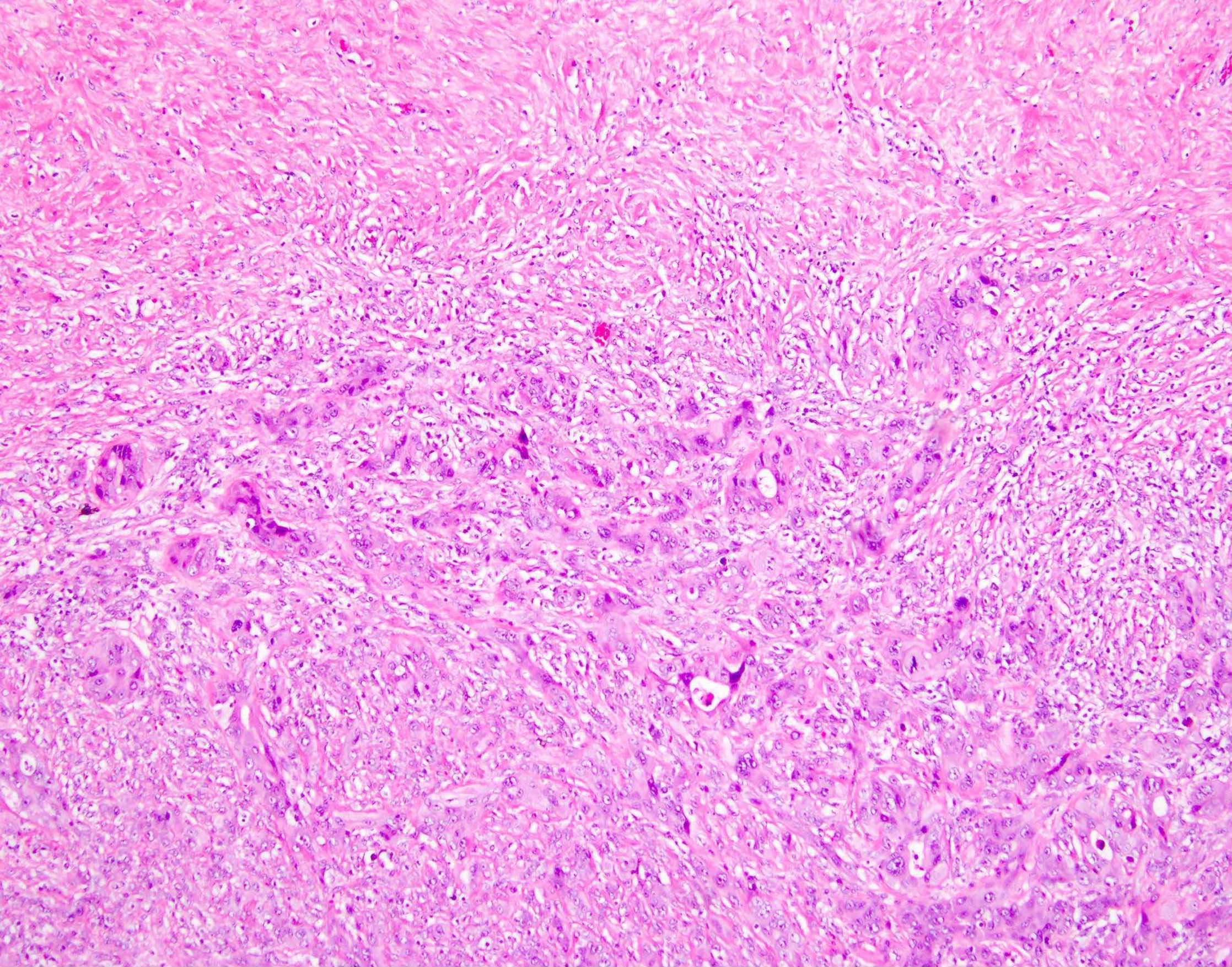
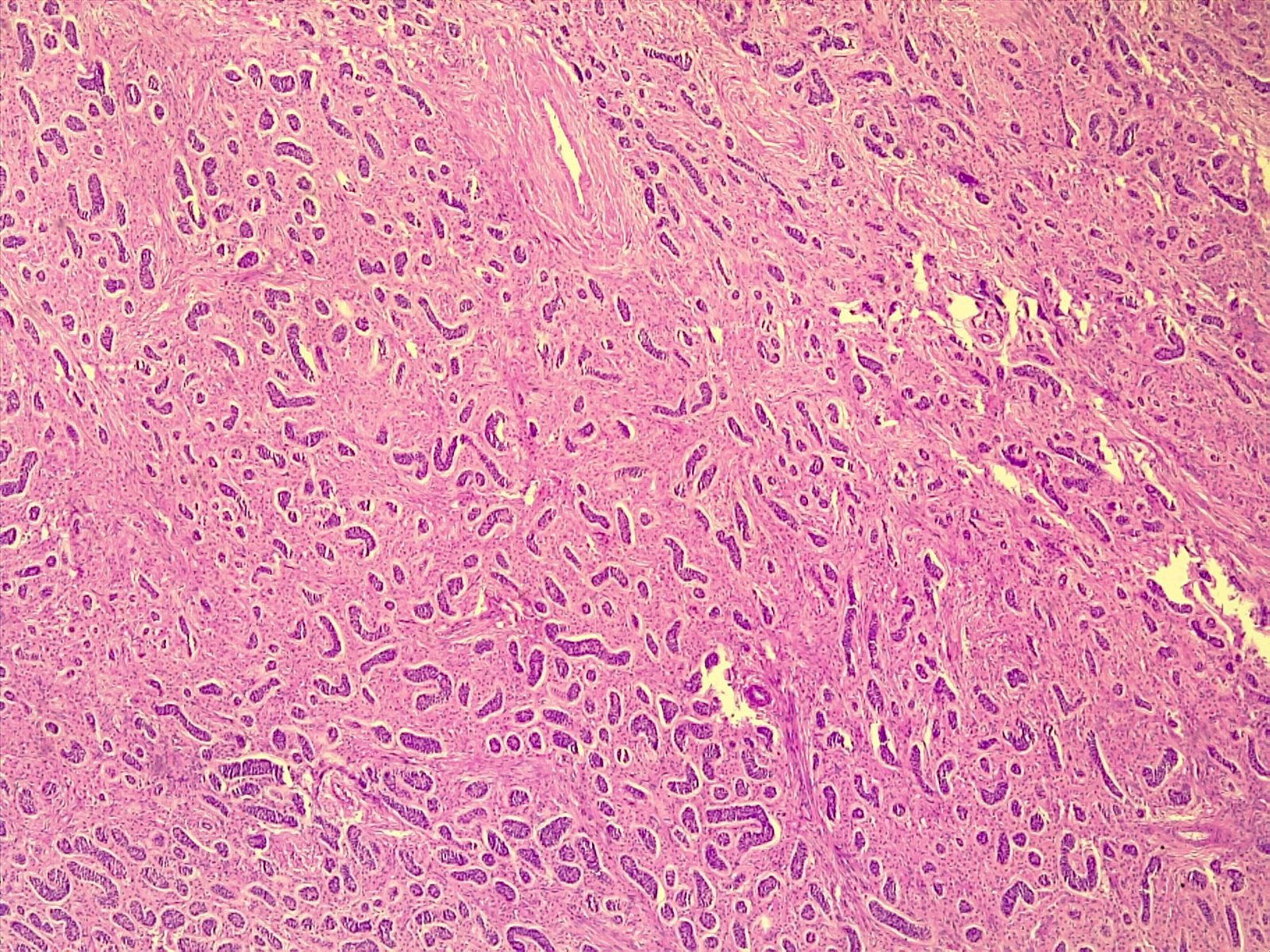
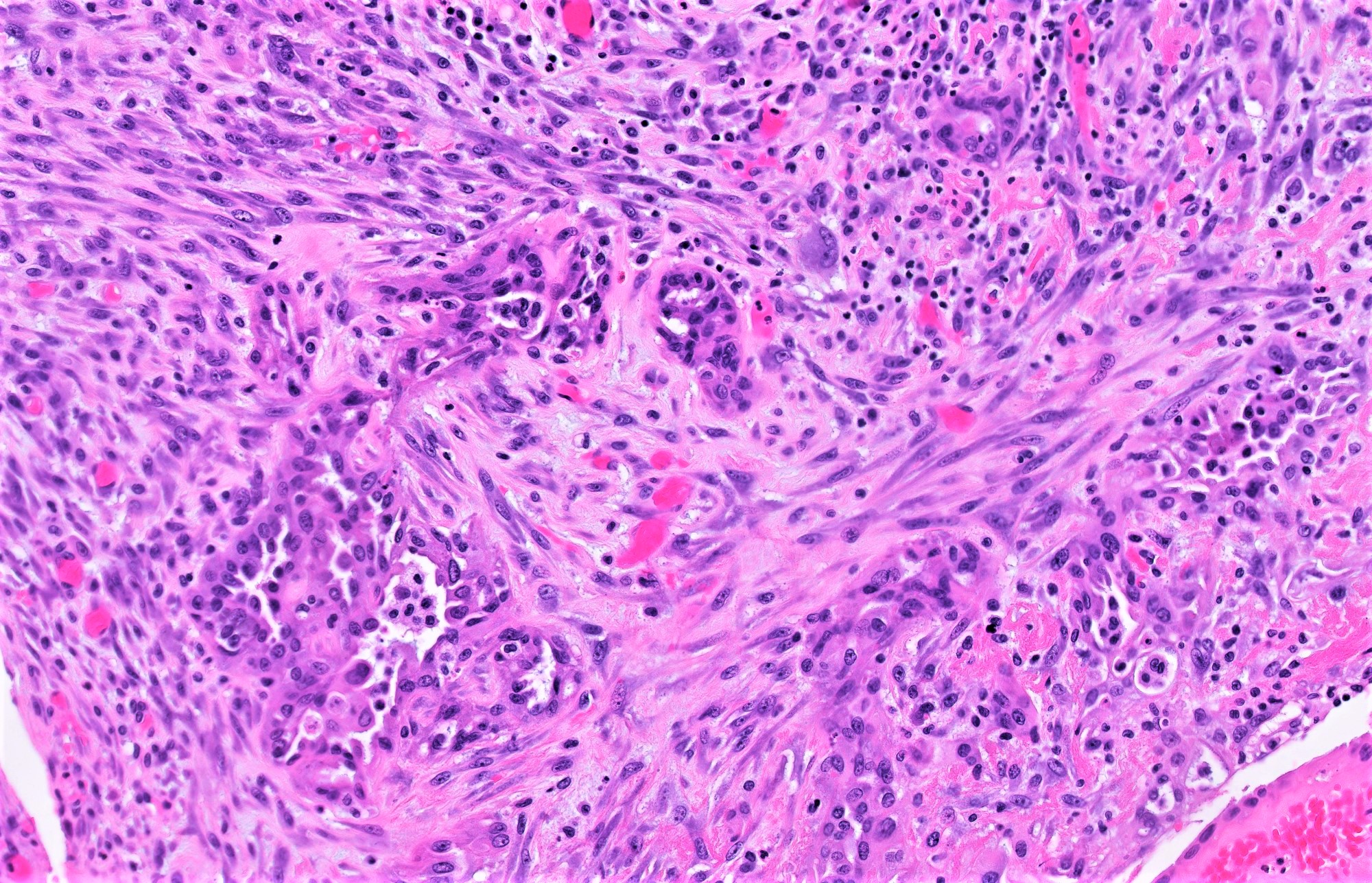
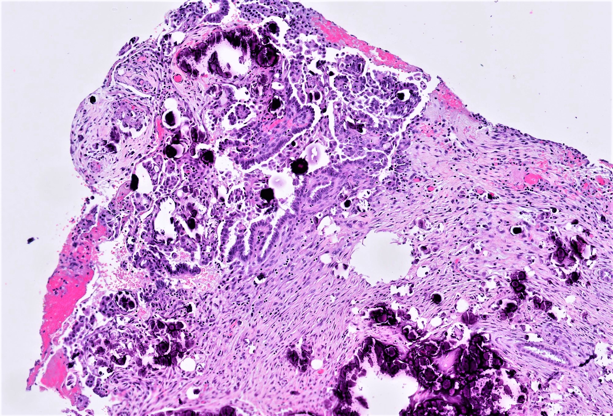
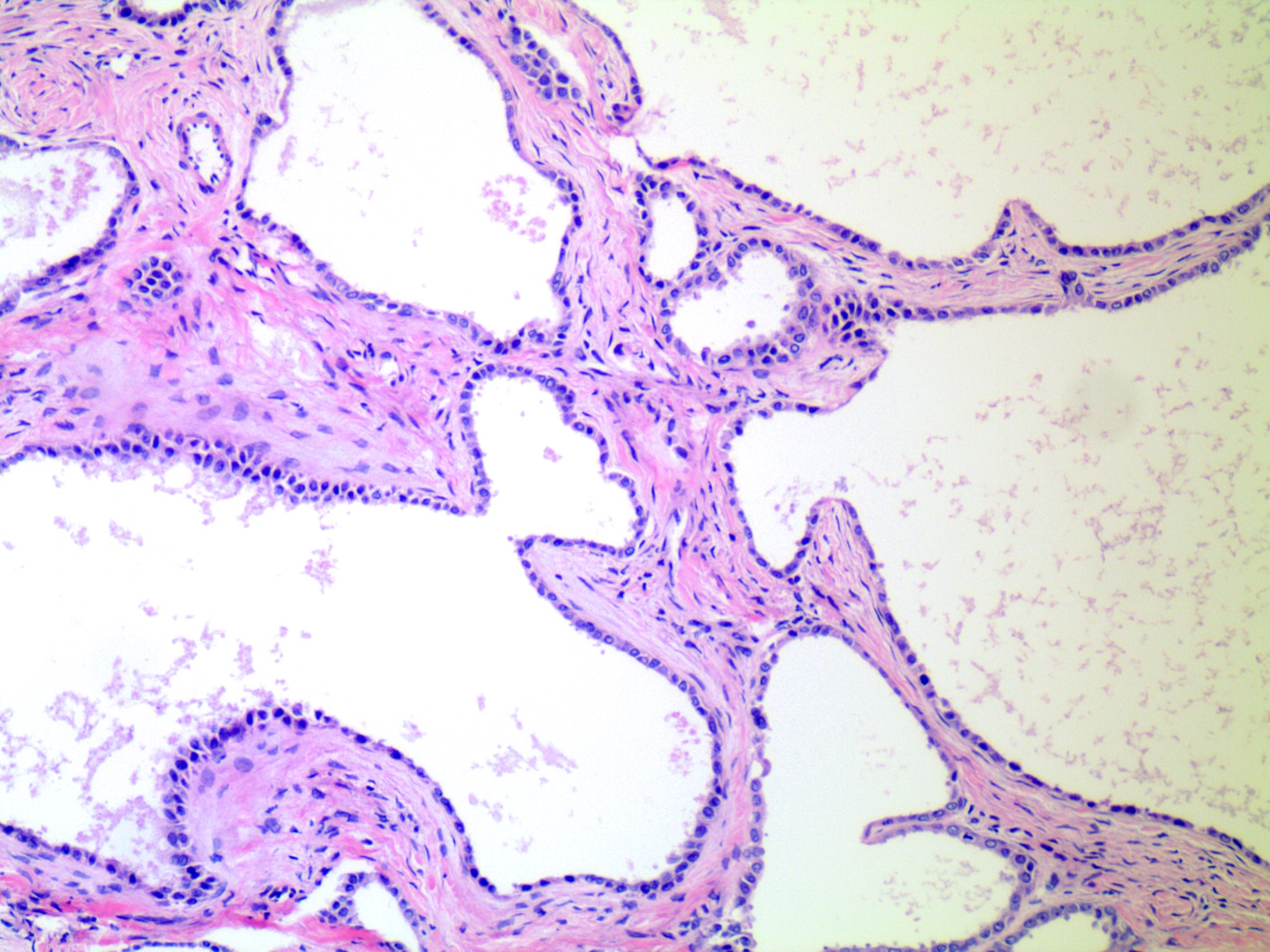
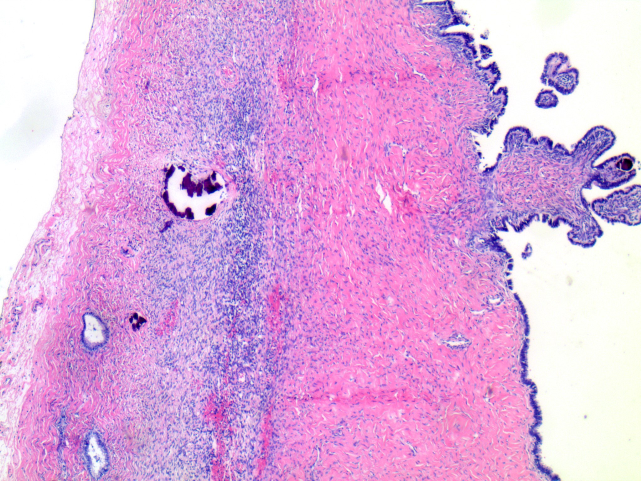
      • pT3a (IIIA2): microscopic extrapelvic (above the pelvic brim) peritoneal involvement with or without positive retroperitoneal lymph nodes
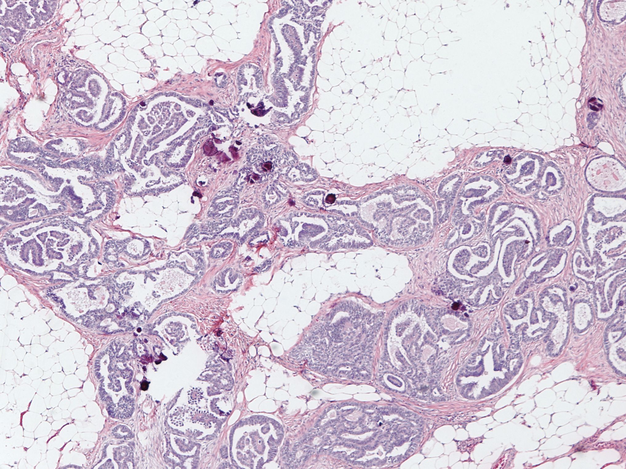
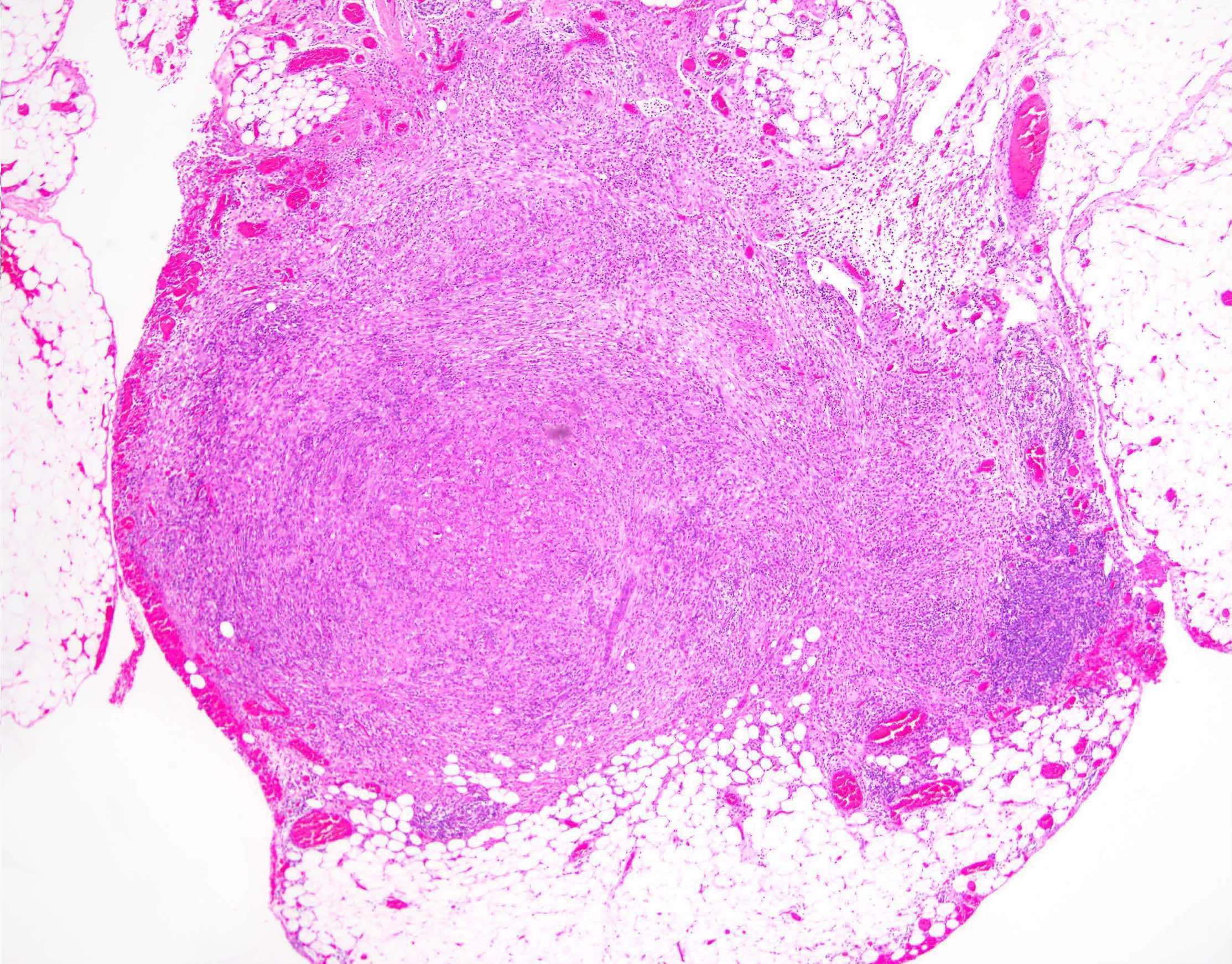
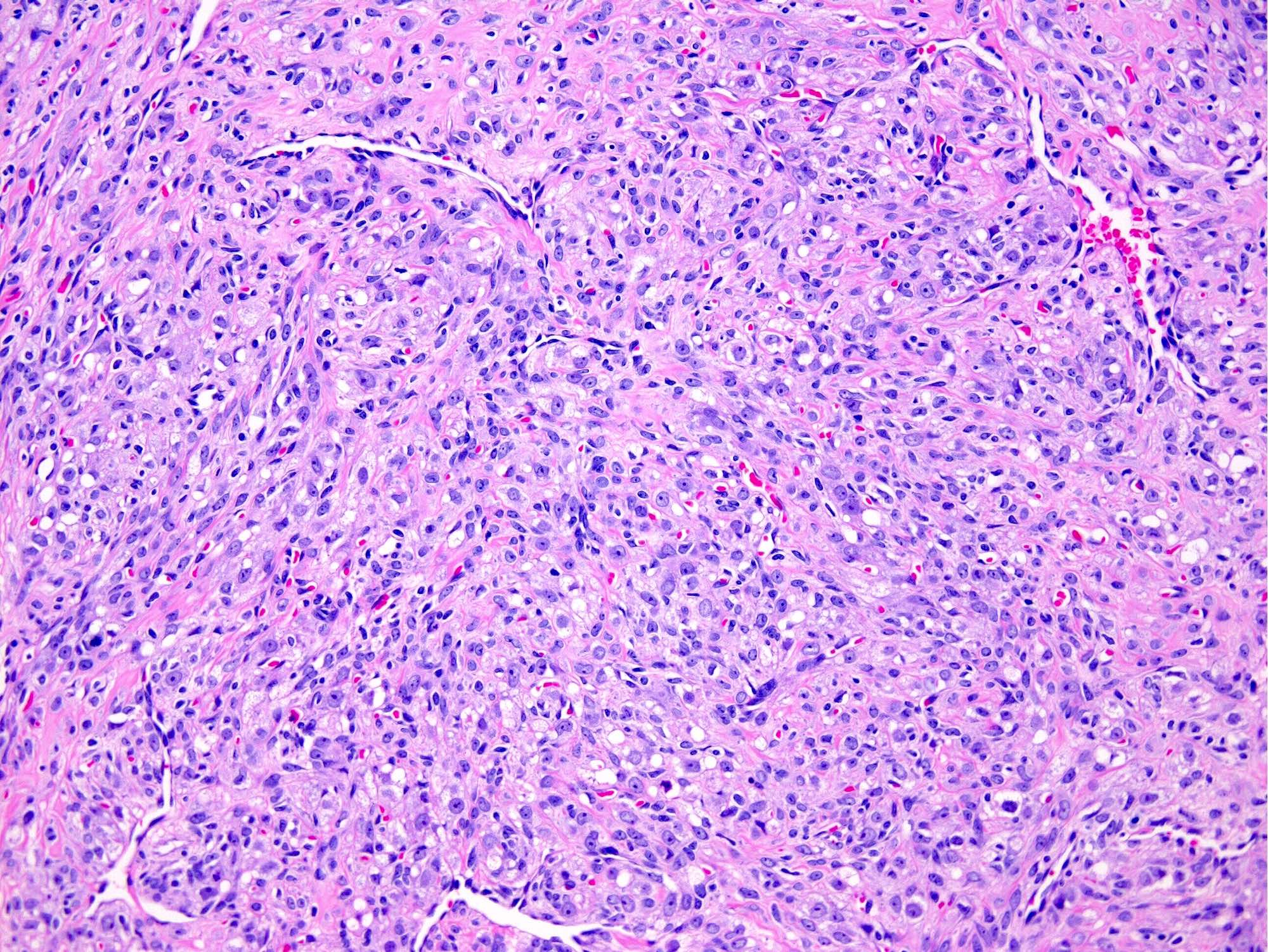
      • pT3b (IIIB): macroscopic peritoneal metastasis beyond pelvis ≤ 2 cm with or without retroperitoneal lymph node metastasis
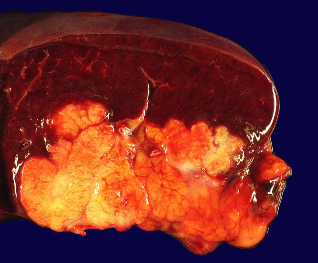
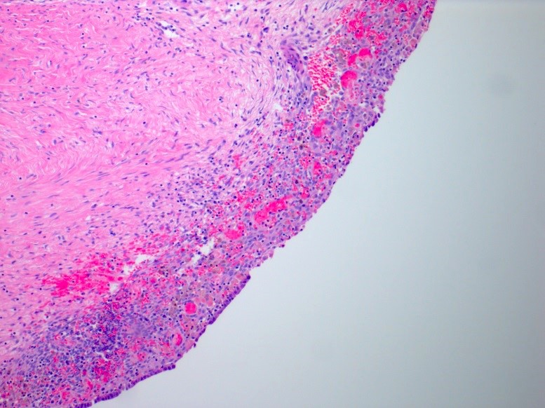
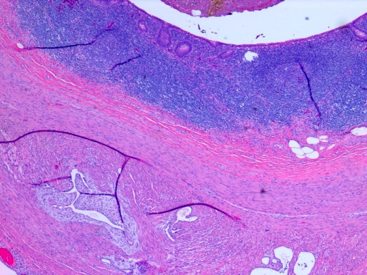
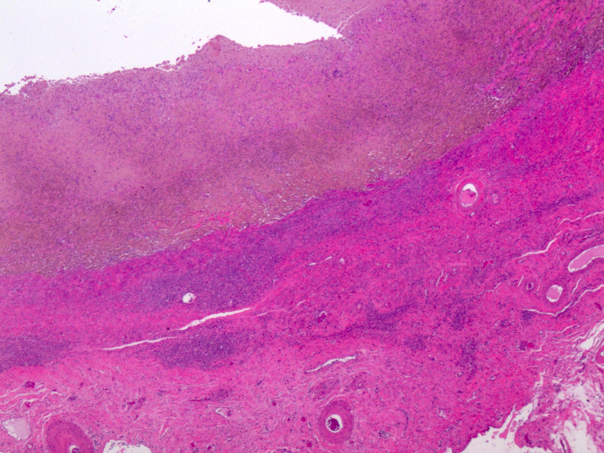
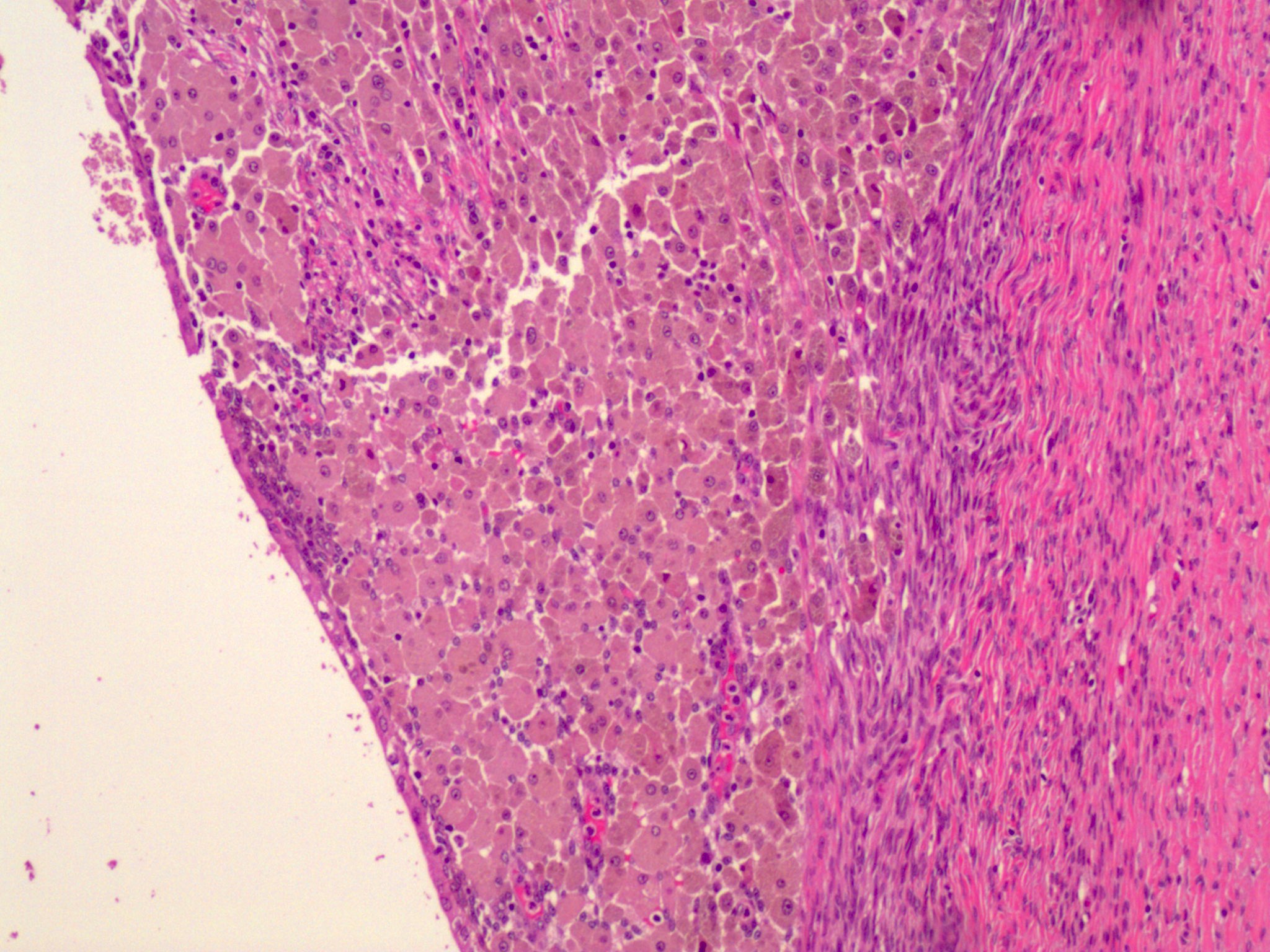
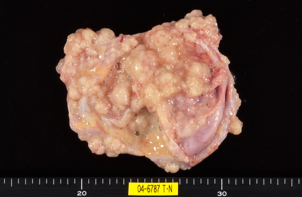
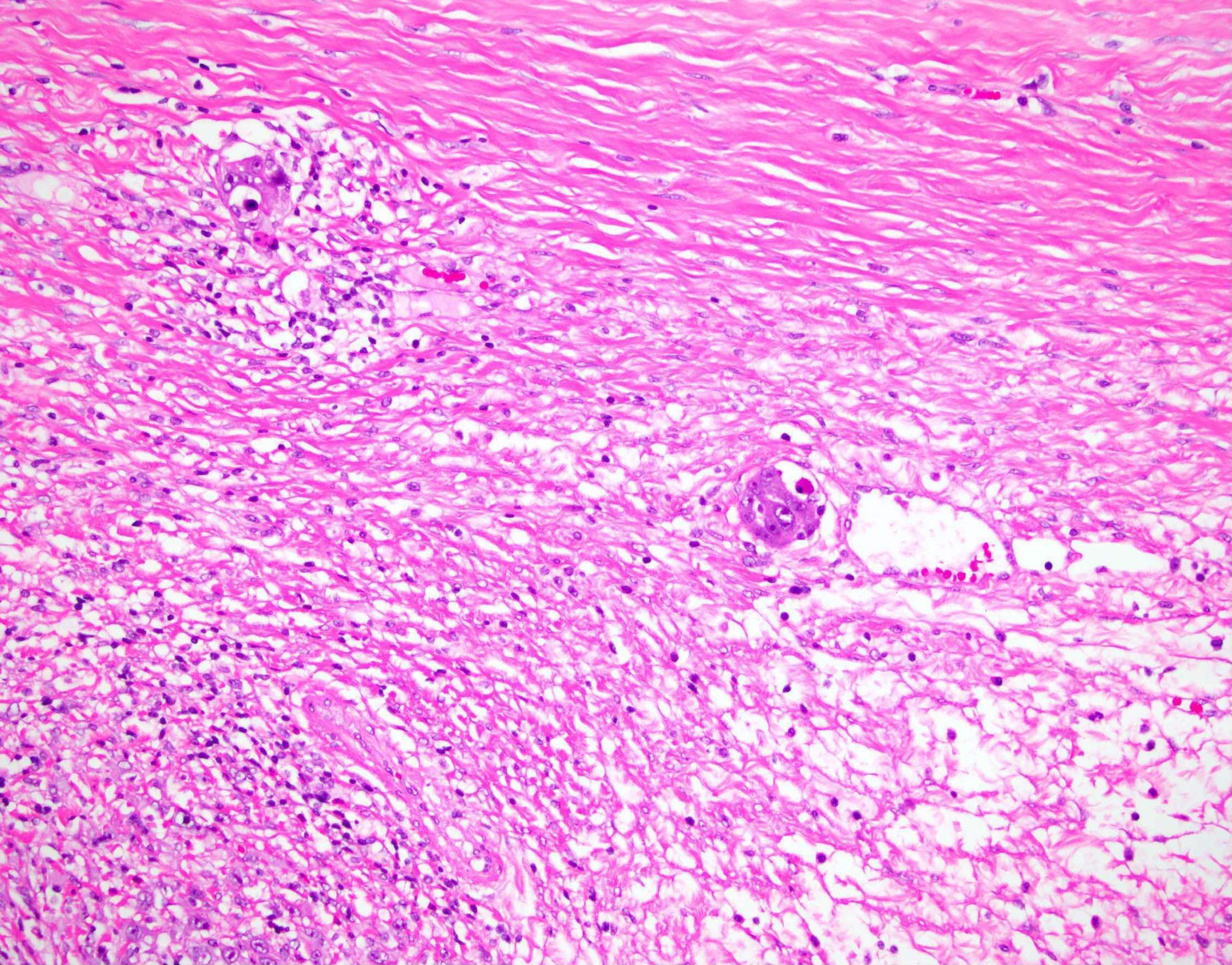
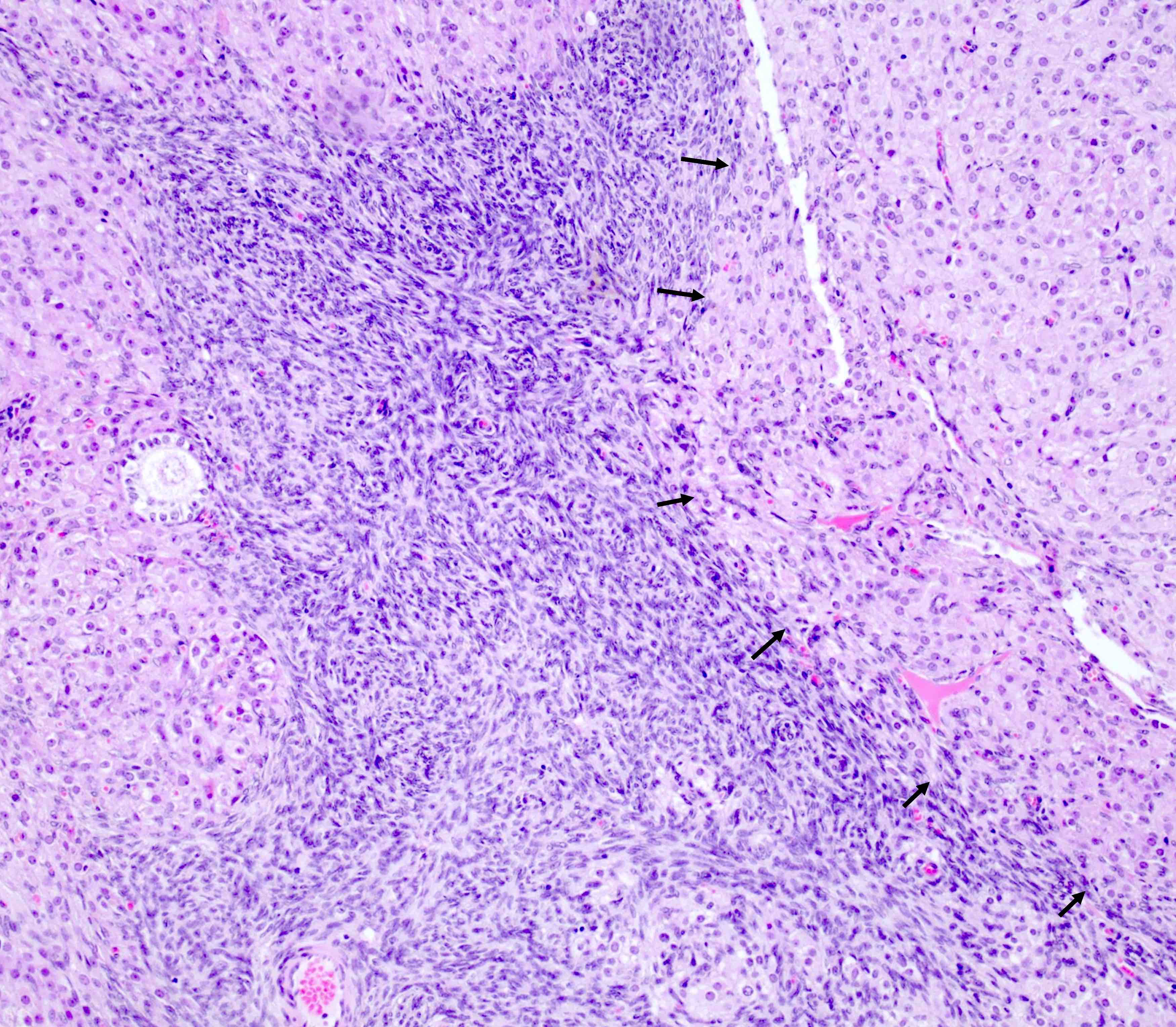
      • pT3c (IIIC): macroscopic peritoneal metastasis beyond pelvis > 2 cm with or without retroperitoneal lymph node metastasis (includes extension of tumor to capsule of liver and spleen without parenchymal involvement of either organ)
      Regional lymph nodes (pN) and FIGO stages in ( )
      • pNX: regional lymph nodes cannot be assessed
      • pN0: no regional lymph node metastasis
      • pN0(i+): isolated tumor cells in regional lymph node(s) ≤ 0.2 mm
      • pN1a (IIIA1i): lymph node metastatic deposit > 0.2 mm to ≤ 10 mm
      • pN1b (IIIA1ii): lymph node metastatic deposit > 10 mm

      Note:
      • Regional lymph nodes include the following: external iliac, internal iliac (hypogastric), obturator, common iliac, para-aortic, pelvic NOS and retroperitoneal NOS lymph nodes
      Distant metastasis (pM) and FIGO stages in ( )
      • pM0: no distant metastasis
      • pM1a (IVA): pleural effusion with positive cytology
      • pM1b (IVB): liver or splenic parenchymal metastases; metastases to extra-abdominal organs (including inguinal lymph nodes and lymph nodes outside the abdominal cavity); transmural involvement of intestine
      Prefixes
      • cTNM: clinical classification
      • pTNM: pathological classification
      • ypTNM: post therapy pathological classification
      • rTNM: recurrence or retreatment classification
      • aTNM: autopsy classification
      AJCC prognostic stage groups
      Stage I:  pT1  N0  M0 
      Stage IA:  pT1a  N0  M0 
      Stage IB:  pT1b  N0  M0 
      Stage IC:  pT1c  N0  M0 
      Stage II:  pT2  N0  M0 
      Stage IIA:  pT2a  N0  M0 
      Stage IIB:  pT2b  N0  M0 
      Stage IIIA1:  pT1/2   N1  M0 
      Stage IIIA2:  pT3a  any N  M0 
      Stage IIIB:  pT3b  any N  M0 
      Stage IIIC:  pT3c  any N  M0 
      Stage IV:  any T  any N  M1 
      Stage IVA:  any T  any N  M1a 
      Stage IVB:  any T  any N  M1b 
      Registry data collection variables
      • FIGO stage
      • Preoperative CA125 level
      • Gross residual tumor after primary cytoreductive surgery
      • Residual tumor volume after primary cytoreductive surgery
      • Residual tumor location following primary cytoreductive surgery
      Histologic grade (G)
      • GX: grade cannot be assessed
      • GB: borderline tumor
      • G1: well differentiated
      • G2: moderately differentiated
      • G3: poorly differentiated
      Histopathologic type
      • Epithelial tumors (borderline or malignant)
        • Serous tumors
          • Serous borderline tumor
          • Low grade serous carcinoma
          • High grade serous carcinoma
        • Mucinous tumors
          • Mucinous borderline tumor
          • Mucinous carcinoma
        • Endometrioid tumors
          • Endometrioid borderline tumor
          • Endometrioid carcinoma
        • Clear cell tumors
          • Clear cell borderline tumor
          • Clear cell carcinoma
        • Seromucinous tumors
          • Seromucinous borderline tumor
          • (Seromucinous carcinoma - now considered a subtype of endometrioid carcinoma, according to WHO 5th edition)
        • Brenner tumors
          • Borderline Brenner tumor
          • Malignant Brenner tumor
        • Other carcinomas
          • Mesonephric-like adenocarcinoma
          • Undifferentiated and dedifferentiated carcinoma
          • Carcinosarcoma
          • Mixed carcinoma
      • Sex cord - stromal tumors (malignant or potentially malignant)
        • Adult granulosa cell tumor
        • Juvenile granulosa cell tumor
        • Sertoli-Leydig cell tumor
        • Gynandroblastoma
        • Steroid cell tumor, NOS
        • Fibrosarcoma
      • Germ cell tumors (malignant)
        • Dysgerminoma
        • Yolk sac tumor
        • Immature teratoma
        • Choriocarcinoma (nongestational)
        • Embryonal carcinoma
        • Mixed germ cell tumor
        • Somatic neoplasms arising from teratomas
      • Miscellaneous tumors
        • Small cell carcinoma, hypercalcemic type
        • Adenocarcinoma of rete ovarii
      Gross images

      Contributed by Natalia Buza, M.D.
      Ovarian surface involvement (pT1c2)

      Ovarian surface involvement (pT1c2)

      Rectosigmoid colon (pT2b)

      Rectosigmoid colon (pT2b)

      Splenic hilum and parenchyma (pM1b)

      Splenic hilum and parenchyma (pM1b)

      Microscopic (histologic) images

      Contributed by Natalia Buza, M.D.
      Ovarian surface involvement (pT1c2)

      Ovarian surface involvement (pT1c2)

      Uterine serosal (pT2a)

      Uterine serosal (pT2a)

      Appendiceal serosal (pT3b)

      Appendiceal serosal (pT3b)

      Splenic capsule (pT3c)

      Splenic capsule (pT3c)

      Splenic parenchyma (pM1b)

      Splenic parenchyma (pM1b)

      Additional references
      Board review style question #1

      Which of the following is the pTNM stage (per the AJCC 8th edition) of a malignant ovarian tumor involving the right ovarian parenchyma and surface, omentum and splenic capsule (for which the largest tumor focus in the omentum and splenic capsule exceeds 2 cm) and no lymph node or parenchymal spleen metastases?

      1. pT1c N0 M0, stage IC
      2. pT1c N0 M1b, stage IVB
      3. pT3b N0 M0, stage IIIB
      4. pT3c N0 M1b, stage IVB
      5. pT3c N0 M0, stage IIIC
      Board review style answer #1
      E. pT3c N0 M0, stage IIIC. Macroscopic peritoneal metastasis beyond the pelvis (including capsule of liver and spleen without parenchymal involvement) measuring more than 2 cm in greatest dimension is pT3c. Parenchymal metastasis to the liver or spleen is pM1b, which would yield stage IVB.

      Comment Here

      Reference: Staging - ovary

      Steroid cell tumor
      Definition / general
      • Ovarian stromal tumor composed of steroid cells with malignant potential
      Essential features
      • Mostly unilateral
      • Half of cases have androgenic manifestations
      • Microscopic features include diffuse sheets of polygonal to rounded tumor cells with moderate to abundant cytoplasm (Am J Surg Pathol 1987;11:835)
      Terminology
      • Steroid cell tumor, not otherwise specified (NOS)
      ICD coding
      • ICD-O
        • 8670/0 - steroid cell tumor, NOS
        • 8670/3 - steroid cell tumor, malignant
      • ICD-11: 2C73.Y & XH4L39 - other specified malignant neoplasms of the ovary & steroid cell tumor, malignant
      Epidemiology
      Sites
      Pathophysiology
      • Tumors are presumed to be of ovarian stromal cell origin
      Etiology
      Clinical features
      • Androgenic (~50%), estrogenic changes including rare examples of isosexual pseudoprecocity (~10%), occasional progestogenic changes, Cushing syndrome or elevated cortisol levels (uncommon)
      • Rarely with hypercalcemia, erythrocytosis, ascites or acute heart failure
      • Reported in von Hippel-Lindau (both types I and II), with the most common presentation including abnormal uterine bleeding, amenorrhea and infertility (Int J Clin Exp Pathol 2022;15:332)
      Diagnosis
      • Histologic examination
      Laboratory
      • In patients with androgenic changes, Cushing syndrome or both: elevated urinary levels of 17-ketosteroids and 17-hydroxycorticosteroids, serum testosterone and androstenedione
      • In Cushing syndrome: elevated free cortisol in the blood or urine
      Radiology description
      Radiology images

      Images hosted on other servers:
      Enhancing adnexal mass Enhancing adnexal mass

      Enhancing adnexal mass

      Fat containing pelvic mass

      Fat containing pelvic mass

      Homogeneous isoechoic mass Homogeneous isoechoic mass

      Homogeneous isoechoic mass

      Solid adnexal mass

      Solid adnexal mass

      Prognostic factors
      • Clinically malignant in 25 - 43% of cases (Am J Surg Pathol 1987;11:835)
      • Patients with clinically malignant tumor were on average 16 years older than patients with benign tumors in one series (Am J Surg Pathol 1987;11:835)
      • No malignant steroid cell tumors have been reported in patients in their first 2 decades
      • Rare tumors have recurred up to 19 years postoperatively (Am J Surg Pathol 1987;11:835)
      • Extraovarian spread of tumor is present at the time of operation in a small minority of cases; 3 cases with Cushing disease with extensive intra-abdominal spread of tumor (Int J Gynecol Pathol 1987;6:40)
      • Features associated with malignancy (Am J Surg Pathol 1987;11:835)
        • 2+ mitoses per 10 high power fields (92% malignant)
        • Necrosis (86% malignant)
        • 7 cm or larger (78% malignant)
        • Intratumoral hemorrhage (77% malignant)
        • Grade 2 or 3 nuclear atypia (64% malignant)
      • Aggressive behavior with 4 or more malignant features (Am J Surg Pathol 2023;47:1398)
      Case reports
      Treatment
      Clinical images

      Images hosted on other servers:
      Hirsutism

      Hirsutism

      Male pattern baldness

      Male pattern baldness

      Gross description
      • Wide size range (mean size: 8.4 cm) (Am J Surg Pathol 1987;11:835)
      • Unilateral (95%)
      • Solid, well circumscribed, occasionally lobulated
      • Sections from yellow-orange (lipid rich) to red-brown (lipid poor) to dark brown-black (abundant lipochrome pigment)
      • Occasionally with necrosis, hemorrhage and cystic degeneration
      Gross images

      Images hosted on other servers:
      Yellow cut surface

      Yellow cut surface

      Metastatic steroid cell tumor

      Metastatic steroid cell tumor

      Lobulated cut surface

      Lobulated cut surface

      Nodular solid tumor

      Nodular solid tumor

      Tan-brown, well circumscribed tumor with hemorrhage

      Tan-brown, well circumscribed tumor with hemorrhage

      Frozen section description
      • Diffuse or nested arrangement of tumor cells
      • Cells with abundant eosinophilic or clear, foamy cytoplasm
      Frozen section images

      Contributed by Fatemeh Ghazanfari Amlashi, M.D. and Tamara Kalir, M.D., Ph.D.
      Monotonous sheets of cells

      Monotonous sheets of cells

      Abundant cytoplasm

      Abundant cytoplasm

      Microscopic (histologic) description
      • Architectural pattern: diffuse (most common) but occasionally in nests, cords, pseudoglandular and follicle-like arrangements (Am J Surg Pathol 1987;11:835)
      • Stroma: usually sparse (85%) but may be fibrotic or hyalinized, rarely edematous or myxoid, exceptionally with calcification and psammoma body formation
      • Polygonal to rounded tumor cells with distinct cell borders, central nuclei and moderate to abundant cytoplasm
      • Tumor cell cytoplasm: eosinophilic and granular (lipid poor) to vacuolated and spongy (lipid rich); tumors with a mixture of 2 cell types are also present
      • More lipid rich cytoplasm in this tumor compared to other subtypes of steroid cell tumor
      • Lipochrome pigment present (40%)
      • Rarely, signet ring appearance
      • Adipocytic metaplasia and hyaline globule (unusual) (Am J Surg Pathol 2023;47:1398)
      • Usually absent / slight nuclear atypia along with low mitotic activity (< 2 mitotic figures/10 high power fields); those with grades 1 - 3 nuclear atypia associated with increased mitotic activity (up to 15 mitotic figures/10 high power fields)
      • Necrosis and hemorrhage are occasionally present, particularly in tumors with significant cytologic atypia
      • Metastatic tumor is similar to the primary tumor in some cases but more poorly differentiated in others
      • Stromal hyperthecosis may be seen in the adjacent ovarian stroma and contralateral ovary, particularly in small tumors
      Microscopic (histologic) images

      Contributed by Fatemeh Ghazanfari Amlashi, M.D., Tamara Kalir, M.D., Ph.D. and AFIP
      Well defined tumor

      Well defined tumor

      Sparse stroma

      Sparse stroma

      Diffuse sheets

      Diffuse sheets

      Moderate to abundant cytoplasm

      Moderate to abundant cytoplasm

      Round to polygonal cells

      Round to polygonal cells

      Distinct cell borders

      Distinct cell borders


      Spongy and eosinophilic cytoplasm

      Spongy and eosinophilic cytoplasm

      2 atypical mitotic figures

      2 atypical mitotic figures

      Inhibin

      Inhibin

      Calretinin

      Calretinin

      MelanA

      MelanA

      Abundant lipid (oil red O stain)

      Abundant lipid (oil red O stain)

      Virtual slides

      Images hosted on other servers:
      Lipid rich tumor cells

      Lipid rich tumor cells

      Cytology description
      • Large polygonal to round cells
      • Arranged in sheets and attached with vascular stromal tissue fragments (J Cytol 2015;32:284)
      • Cells with small central round nuclei with conspicuous nucleoli and abundant granular to pale foamy cytoplasm
      • Ascites in malignant steroid cell tumor: isolated tumor cells or clusters with slight overlapping and cannibalism formation (Acta Cytol 2017;61:165)
      Cytology images

      Images hosted on other servers:
      Large polygonal cells

      Large polygonal cells

      Molecular / cytogenetics description
      Videos

      Steroid cell tumor

      Sample pathology report
      • Right ovary and fallopian tube, salpingo-oophorectomy:
        • Ovarian steroid cell tumor, not otherwise specified (6 cm size) (see comment)
        • Fallopian tube with no significant pathologic change
        • Comment: Sections show a well circumscribed neoplasm composed of abundant clear cytoplasm. There is mild nuclear atypia. No mitosis or necrosis identified. Immunostains show the tumor cells are positive for inhibin, calretinin and negative for CD10, supporting the diagnosis.

      • Left ovary and fallopian tube, salpingo-oophorectomy:
        • Ovarian steroid cell tumor, not otherwise specified (12 cm size) (see comment)
        • Fallopian tube with no significant pathologic change
        • Comment: Sections show a well circumscribed neoplasm composed of sheets of round cells with abundant eosinophilic granular cytoplasm. Intratumoral hemorrhage and focal necrosis is present. Moderate nuclear atypia and mitoses (4/10 high power fields) are seen. The ovarian capsule is intact and uninvolved by the tumor cells. Immunostains show the tumor cells are positive for calretinin and inhibin, supporting the diagnosis.
      Differential diagnosis
      Board review style question #1

      Which of the following statements is correct regarding steroid cell tumor, not otherwise specified (NOS) of the ovary?

      1. Inhibin+, calretinin+, MelanA+, PAX8-
      2. Less than 1% risk of malignancy
      3. Mostly bilateral
      4. Various architectural patterns
      Board review style answer #1
      A. Inhibin+, calretinin+, MelanA+, PAX8-. Steroid cell tumor, NOS shows positive inhibin, calretinin and MelanA and negative PAX8. Note that MelanA (A103 clone) can be expressed in steroid cell tumor and the presence of it should not necessarily prompt the diagnosis of melanoma. Answer B is incorrect because steroid cell tumor has a 30% risk of malignancy. Answer C is incorrect because steroid cell tumors are mostly unilateral. Answer D is incorrect because having various architectural patterns is a feature of ovarian clear cell carcinoma.

      Comment Here

      Reference: Steroid cell tumor
      Board review style question #2
      A 40 year old woman presented with 5 cm, solid, well defined adnexal mass. Which of the following features is in favor of steroid cell tumor, NOS?

      1. Acellular eosinophilic material and nuclear clustering
      2. Location in hilum
      3. Reinke crystal
      4. Sheets of tumor cells with distinct cell borders and clear to foamy cytoplasm
      Board review style answer #2
      D. Sheets of tumor cells with distinct cell borders and clear to foamy cytoplasm. Steroid cell tumor, NOS is mostly seen as sheets of tumor cells with distinct cell borders and can have clear to foamy cytoplasm in lipid rich tumor. Answers A, B and C are incorrect because these are features of Leydig cell tumor.

      Comment Here

      Reference: Steroid cell tumor

      Stromal hyperplasia and hyperthecosis
      Definition / general
      • Ovarian stromal hyperplasia is a nonneoplastic functional disorder resulting in nodular or diffuse proliferation of ovarian stroma
      • Hyperthecosis is characterized by proliferation of ovarian stroma along with luteinization of stromal cells (J Turk Ger Gynecol Assoc 2013;14:182)
      Essential features
      • Ovarian stromal hyperplasia and hyperthecosis are nonneoplastic conditions resulting in diffuse or nodular proliferation of ovarian stroma as single cells, clusters or nodules of luteinized cells
      • Patients can present with bleeding due to endometrial pathology or symptoms of hyperandrogenism, principally androgenetic alopecia or hirsutism, along with elevated testosterone levels
      • Both show an excess of androgen production leading to increased testosterone
      • Ovaries with stromal hyperplasia and hyperthecosis secrete more androstenedione than normal ovaries that can be converted to estradiol by peripheral aromatization (BJOG 2003;110:690)
      ICD coding
      • ICD-11: 5A80.Y - other specified ovarian dysfunction
      Epidemiology
      Sites
      • Ovarian stroma
      Pathophysiology
      • Studies have shown that ovaries with stromal hyperplasia and hyperthecosis secrete more androstenedione and estradiol than normal ovaries
        • Most patients with stromal hyperplasia and hyperthecosis have normal gonadotrophin levels, unlike patients with polycystic ovary syndrome (Histopathology 1989;14:369)
      • Some in vitro incubation studies show that stromal proliferation in premenopausal and postmenopausal women is increased by luteinizing hormone
      • Most patients with stromal hyperplasia and hyperthecosis show hyperinsulinemia or insulin resistance that likely plays a role in pathogenesis (Am J Obstet Gynecol 1986;154:384)
      Etiology
      Clinical features
      • Symptoms and signs of hyperandrogenism, such as deepening voice, acne, hirsutism, frontal alopecia, clitoromegaly
      • Other features include obesity, hyperinsulinemia, acanthosis nigricans and metabolic syndrome
      • There is an increased risk of vaginal bleeding, endometrial hyperplasia and carcinoma, cardiovascular pathology and diabetes mellitus (J Menopausal Med 2020;26:39)
      • May be associated with HAIR AN syndrome in a few patients
      Diagnosis
      • Initial investigation is usually done to rule out adrenal tumors and androgen secreting ovarian tumors
      • Clinical assessment for hirsutism, alopecia, deepening of voice, etc.
      • Serological assessment: elevated testosterone, androgen and estrogen levels, normal or suppressed luteinizing hormone, normal dehydroepiandrosterone (DHEA), DHEA sulfate (DHEAS), 17 hydroxyprogesterone (17 OHP), normal progesterone and prolactin levels
      • Imaging: enlarged ovaries without hypervascularization on imaging (J Menopausal Med 2020;26:39)
      • Histology: the gold standard diagnostic test used for confirmation (and subsequent treatment) consists of histological investigation performed after bilateral salpingo-oophorectomy (J Menopausal Med 2020;26:39)
      Laboratory
      • Increased androgen, testosterone and estrogen levels
      • Normal or suppressed LH and FSH
      • Normal serum androstenedione or dehydroepiandrosterone sulfate (DHEAS) concentrations
      • Unfavorable lipid profiles (i.e., increase in low density lipoprotein [LDL] and triglycerides, along with decrease in high density lipoprotein [HDL] associated with the hyperandrogenemia)
      Radiology description
      Radiology images

      Images hosted on other servers:
      Ovarian stromal hyperplasia

      Ovarian stromal hyperplasia

      Prognostic factors
      Case reports
      Treatment
      Gross description
      • Mostly involves bilateral ovaries with increase in size
      • May be nodular or diffuse with solid, firm, homogeneous white to yellow cut surface
      Gross images

      AFIP images
      Enlarged ovaries

      Enlarged ovaries

      Microscopic (histologic) description
      • Diffuse or nodular proliferation of ovarian stroma in cortex, medulla or both
      • Stromal proliferation of spindle cells with scant intervening collagen
      • May be diffuse or present as a nodule with coalescent nodules (Endocrinol Diabetes Metab Case Rep 2021;2021:20)
      • Entrapped follicular derivatives may be present
      • In hyperthecosis, single cells, nests or nodules of polygonal luteinized theca cells with eosinophilic or clear cytoplasm < 1 cm are present
      Microscopic (histologic) images

      Contributed by Hafza Asma Adnan, M.B.B.S. and AFIP
      Ovary with stromal hyperplasia Diffuse stromal hyperplasia Diffuse stromal hyperplasia

      Diffuse stromal hyperplasia

      Stromal hyperplasia of ovary Ovarian stromal hyperplasia Ovarian stromal hyperplasia

      Ovarian stromal hyperplasia


      Nests of vacuolated, luteinized cells

      Nests of vacuolated, luteinized cells

      Large nodule of luteinized cells

      Large nodule of luteinized cells

      Lutein cells contain abundant lipid (oil red O stain)

      Lutein cells contain abundant lipid (oil red O stain)

      Positive stains
      Negative stains
      Sample pathology report
      • Right ovary, oophorectomy:
        • Ovarian stroma hyperplasia / hyperthecosis (see comment)
        • Comment: This ovarian tissue shows expansion of ovarian stroma as diffuse proliferation of stromal cells. In between these stromal cells, few clusters of eosinophilic luteinized cells are also present. No evidence of epithelial elements, increased mitosis, pleomorphism or necrosis is identified.
        • Clinicopathological correlation: Correlation with serological markers and endometrial thickness is recommended as this condition is frequently associated with hyperandrogenism and endometrial hyperplasia.
      Differential diagnosis
      • Fibroma:
        • Mostly unilateral mass
        • Usually well circumscribed and composed of variable cellular spindle to oval cells with scant cytoplasm, arranged as intersecting fascicles with intervening collagen; sometimes hyaline plaques are also present
        • Cellular fibroma is densely cellular with little intervening collagen that may be mitotically active
      • Steroid cell tumor, NOS:
        • This tumor is comprised of expansile diffuse proliferation of large polygonal cells with eosinophilic, pale or vacuolated cytoplasm
        • Steroid cells nodules > 1 cm arising in a background of stromal hyperthecosis are now best diagnosed as steroid cell tumor, NOS, as the previous diagnostic category of stromal luteoma is no longer recognized as a distinct entity
      • Luteinized thecoma associated with sclerosing peritonitis:
        • Extremely rare lesion, patient presents with abdominal swelling, ascites and bowel obstruction
        • Almost always bilateral, ovaries have tan to red sectioned surface
        • Microscopically, ovaries are hypercellular and show microcystic appearance due to edema
        • Cells are predominantly spindled with minority component of small luteinized cells
        • SF1 and FOXL2 are positive in spindle cells while calretinin and inhibin are positive in luteinized cells
      • Normal ovary:
        • Normal appearance of cortex and medulla
        • Cortex is the hypercellular outer zone having majority of follicles in premenopausal women; medulla is the hypocellular central area containing blood vessels and diffuse or nodular population of spindle cells
        • Young women have thick cortex and contain follicles in various stages of development, while this is atrophied in old age and mainly contain corpus albicans
        • Diagnosis of stromal hyperplasia and hyperthecosis is a subjective estimation of cortex and medullary cellularity with respect to age
      • Sex cord stromal tumor, NOS:
        • Both sex cord and stromal components are present
        • Stromal component highlighted by reticulin staining
      • Pregnancy luteoma:
        • Associated with pregnancy
        • Comprised of oval to round uniform cells with abundant eosinophilic cytoplasm
      • Massive ovarian edema and fibromatosis:
        • Prominent edema of stroma sparing cortex
        • Clusters of luteinized cells may be present
      • Krukenberg tumor:
        • Isolated atypical cells with signet ring cell morphology in ovarian stroma
        • AE1 / AE3 positive
      • Low grade endometrial stromal sarcoma:
        • Oval to spindle cells with minimal atypia and associated arterioles
        • CD10 positive
      Board review style question #1
      A specimen for bilateral oophorectomy is received with enlarged ovaries. One ovary measures 44 x 30 x 25 mm while the second measures 35 x 30 x 25 mm. Sectioning shows homogeneous solid areas with a single 3 mm cyst. Microscopy shows a diffuse population of spindle cells with no pleomorphism, mitosis or necrosis. What is the likely diagnosis?

      1. Adult granulosa cell tumor
      2. Fibroma
      3. Metastatic tumor
      4. Sex cord stromal tumor, NOS
      5. Stromal hyperplasia
      Board review style answer #1
      E. Stromal hyperplasia. There is a homogeneous population of spindle cells with no atypia. Answer A is incorrect because adult granulosa cell tumor will show atypia with variable patterns and nuclear grooving. Answer B is incorrect because no hyalinized areas with intersecting fascicles are present. Answer C is incorrect because no epithelial elements are present in this tumor. Answer D is incorrect because no sex cord-like areas or biphasic cell populations are identified.

      Comment Here

      Reference: Stromal hyperplasia and hyperthecosis
      Board review style question #2

      A 57 year old woman presents with postmenopausal bleeding. Endometrial biopsy showed FIGO grade 1 endometrioid type endometrial cancer and hysterectomy revealed stage IA endometrioid type endometrial carcinoma with enlarged bilateral ovaries. Microscopic findings are shown in the image above. What is the diagnosis?

      1. Fibroma
      2. Leydig cell tumor
      3. Metastatic endometrioid type endometrial cancer
      4. Sex cord stromal tumor
      5. Stromal hyperplasia and hyperthecosis
      Board review style answer #2
      E. Stromal hyperplasia and hyperthecosis. Clusters of luteinized cells are present in stromal hyperplasia. Answer A is incorrect because no hyalinized areas with intersecting fascicles are present. Answer B is incorrect because no Leydig cell population is identified and the cells do not contain Reinke crystals. Answer C is incorrect because no epithelial elements are identified. Answer D is incorrect because no tumor-like mass or epithelial elements are identified.

      Comment Here

      Reference: Stromal hyperplasia and hyperthecosis

      Struma ovarii
      Definition / general
      • Monodermal ovarian teratoma primarily (> 50%) or exclusively composed of benign thyroid tissue
      • Any mature teratoma with malignant thyroid tissue
      Essential features
      • Ovarian teratoma either composed predominantly or exclusively of benign thyroid tissue or with any amount of malignant thyroid tissue
      • Most common thyroid malignancy to occur in struma ovarii is papillary thyroid carcinoma, followed by follicular carcinoma
      ICD coding
      • ICD-10: D27.9 - benign neoplasm of unspecified ovary
      • ICD-10: C56 - malignant neoplasm of ovary
      Epidemiology
      • Most common type of monodermal teratoma
      • Accounts for 3% of ovarian teratomas (Pathol 2007;39:139)
      • Usually presents in the fifth decade
      Sites
      • Ovary
      Pathophysiology / etiology
      • Unknown at this time
      Clinical features
      Diagnosis
      • Diagnosis is made by microscopic examination of resected tissue
      Laboratory
      Radiology description
      Radiology images

      Images hosted on other servers:

      Left adnexal mass

      Prognostic factors
      • Most cases have a good prognosis, even when malignancy is present
      Case reports
      • 10 year old girl with tachycardia, normal thyroid and thyrotoxicosis with papillary thyroid carcinoma arising from a struma ovarii (Int J Surg Case Rep 2018;51:218)
      • 48 year old woman with malignant struma ovarii presenting 14 years after oophorectomy and chemotherapy (Medicine (Baltimore) 2018;97:e13867)
      • 52 year old woman with clinical features of advanced ovarian carcinoma found to have a struma ovarii (Oncol Lett 2015;9:1739)
      • 61 year old woman with insular carcinoma (90%) and adjacent papillary thyroid carcinoma arising in a struma ovarii (Gynecol Oncol Rep 2016;18:1)
      • 62 year old woman with 2 different types of RAS mutations in a papillary thyroid carcinoma from a struma ovarii and papillary thyroid microcarcinoma of the thyroid (J Endocr Soc 2018;2:944)
      • 74 year old woman with a mixed ovarian tumor composed of elements of Brenner tumor, mucinous cystadenoma and struma ovarii (Int J Gynecol Pathol 2019;38:576)
      Treatment
      • Oophorectomy
      • Surgery for malignant struma ovarii may include total abdominal hysterectomy and bilateral salpingo-oophorectomy with complete staging (Gynecol Oncol 2004;94:835)
      Gross description
      • Typically unilateral and solid with a gelatinous, red-brown to green cut surface; may show goiter-like multinodular or cystic change (Am J Surg Pathol 1994;18:785)
      Gross images

      Contributed by Kseniya Korchagina, M.D. and AFIP

      Cystic teratoma

      Solid and cystic

      Reddish brown solid mass

      Cystic tumor

      Frozen section description
      • Frozen section sample usually shows a mature teratoma or benign thyroid tissue; rarely, a possible malignant component may be detected
      Microscopic (histologic) description
      • Variably sized macro and microfollicles often containing colloid
      • Other architectural patterns include solid areas composed of cells with clear to oxyphilic cytoplasm, trabeculae, cords and pseudotubular structures
      • Rarely, stroma in between follicles may be fibrotic or edematous; peripheral stromal leutinization may also be seen
      • Adenomatous hyperplasia or proliferative changes may be seen, such as areas of densely packed follicles or papillary formations lacking nuclear features of papillary thyroid carcinoma
      • Birefringent calcium oxalate crystals may be seen in colloid
      • Most commonly associated malignancy is papillary thyroid carcinoma, cytologically characterized by crowded and overlapping elongated nuclei with irregular contours and chromatin clearing, usually with papillary or follicular architecture
      • Follicular carcinoma is the second most common malignancy; since ovarian lesions typically lack capsule, demonstration of tumor invasion into surrounding ovarian tissue, vascular invasion or metastases is needed as evidence of malignancy
      • Uncommonly associated with the more recently described highly differentiated follicular carcinoma of ovarian origin, characterized by extraovarian spread of thyroid elements histologically resembling nonneoplastic thyroid tissue (Int J Gynecol Pathol 2008;27:213)
      • Undifferentiated (anaplastic) carcinoma and medullary carcinoma have also been described in association with struma ovarii
      Microscopic (histologic) images

      Contributed by Sharon Song, M.S., M.D., Kseniya Korchagina, M.D. and AFIP

      Follicular variant PTC

      Capsular invasion

      TTF1 stain

      Papillary thyroid carcinoma in struma ovarii


      Papillary thyroid carcinoma in struma ovarii

      Macrofollicular, microfollicular and solid areas

      Solid tubular pattern

      Oxyphilic cells arranged in nests


      With peripheral formation of lutein cells

      Resembling clear cell carcinoma

      Cystic tumor

      Thyroglobulin

      With papillary carcinoma

      Cytology description
      Negative stains
      Molecular / cytogenetics description
      Sample pathology report
      • Ovary and fallopian tube, left, salpingo-oophorectomy:
        • Struma ovarii, 4.5 cm
      • Ovary and fallopian tube, left, salpingo-oophorectomy:
        • Papillary thyroid carcinoma, follicular and papillary growth patterns, 1.1 cm, arising in a background of struma ovarii
      Differential diagnosis
      Board review style question #1
      What type of crystals may be seen in struma ovarii?

      1. Calcium carbonate
      2. Calcium oxalate
      3. Calcium phosphate
      4. Triple phosphate
      5. Tyrosine
      Board review style answer #1
      B. Calcium oxalate crystals may be identified in colloid

      Comment Here

      Reference: Struma ovarii
      Board review style question #2
      Which laboratory marker may be elevated with struma ovarii?


      1. Alpha-fetoprotein (AFP)
      2. CA125
      3. CEA
      4. hCG
      5. PLAP
      Board review style answer #2
      B. CA125 is a widely accepted biomarker for ovarian cancers that can also be elevated in many other conditions, such as menstruation, endometriosis, pregnancy, pelvic inflammatory disease, fibroids, liver disease and tumors of the endometrium, breasts and lungs. It can be increased in up to 30% of struma ovarii.

      Comment Here

      Reference: Struma ovarii

      Strumal carcinoid
      Definition / general
      • Has features of carcinoid tumor and struma ovarii
      • Associated with MEN IIA / III
      Case reports
      Gross images

      AFIP images

      4 cm, rounded, brown nodule (bottom)

      Microscopic (histologic) description
      • Often other teratomatous elements
      Microscopic (histologic) images

      AFIP images

      Admixed: struma (right) merges with trabecular carcinoid (left)

      Admixed: trabecular
      carcinoid with
      microfollicles
      containing colloid

      Chromogranin (top), thyroglobulin (bottom)

      With formation of
      layer of steroid cells
      in adjacent ovarian
      stroma

      Positive stains
      Negative stains
      Electron microscopy description

      Teratoma-immature
      Definition / general
      • A malignant germ cell tumor consisting of a teratoma containing variable amounts of immature tissue (typically primitive neuroectodermal)
      Essential features
      • Malignant germ cell tumor of the ovary composed of cells from the 3 germ layers and containing variable amounts of mature and immature tissue
      • Affects mostly young females < 20 years old
      • Grossly solid ovarian tumor mass with necrosis and hemorrhage on cut sections
      • Grading is based on the amount of immature neuroepithelium
      • Can have associated nodal or peritoneal gliomatosis (presence of mature neural tissue), which increases risk of recurrence
      • Grading and stage are the most important factors for prognosis and prognosis is greatly improved after chemotherapy
      Terminology
      • Immature teratoma
      ICD coding
      • ICD-O: 9080/3 - teratoma, malignant, NOS
      Epidemiology
      Sites
      • Ovary
      Pathophysiology
      • Abnormal differentiation of fetal germ cells
      • 2 pathogenetic models for ovarian teratomas, similar to their testicular counterparts
        • Pure mature and immature teratomas are not derived from transformed ovarian germ cells
        • Teratomatous components of mixed germ cell tumors derived from their associated nonteratomatous components
      • Pure mature and immature teratomas do not have i(12p) or 12p amplification, abnormalities that are identified in both teratomatous and nonteratomatous components of mixed germ cell tumors; this supports a different pathogenesis for pure versus mixed tumors (Mod Pathol 2006;19:766)
      • 25% of cases with immature teratoma have peritoneal implants composed of mature glial tissue (gliomatosis peritonei); this phenomenon is thought to arise from metaplasia of submesothelial cells in response to growth factors secreted by the ovarian tumor, rather than a true metastatic process (Hum Pathol 1976;7:625, Hum Pathol 2004;35:685, Int J Gynecol Cancer 2015;25:244, Mod Pathol 2015;28:1613)
      • Rarely, mature glial tissue is also present in lymph nodes (nodal gliomatosis), usually with associated gliomatosis peritonei
      • Prognosis is good and the presence of nodal gliomatosis does not warrant chemotherapy (J Ovarian Res 2016;9:45, Mod Pathol 2015;28:1613)
      Etiology
      • No known causative agents
      Clinical features
      • Associated with symptoms of rapidly growing abdominal mass, pain, distention and torsion
      • Growing teratoma syndrome: immature teratoma status postneoadjuvant chemotherapy consists of persistent or enlarging ovarian tumor with normalization of serum markers
      Diagnosis
      • A combination of clinical, radiological and laboratory findings: rapidly enlarging adnexal mass in a young patient with mildly elevated alpha fetoprotein (AFP) and typical radiological findings raises the possibility of immature teratoma
      • Requires histological examination for confirmation of diagnosis and grading
      • Reference: Int J Gynecol Pathol 1994;13:283
      Laboratory
      • Low level increase of AFP
      • High levels of AFP suggest the presence of a yolk sac tumor component (mixed germ cell tumor)
      Radiology description
      Radiology images

      Images hosted on other servers:
      Complex ovarian mass

      Complex ovarian mass

      Prognostic factors
      • Older age at diagnosis, higher stage and grade confer worse prognosis but overall improved after chemotherapy, with 72% survival for stage IV disease (Gynecol Oncol 2016;142:261, Gynecol Oncol 2016;142:446)
      • Grade is the most important risk factor for relapse across all age groups
      • In a study of 98 pediatric patients and 81 adult patients, there were no relapses in grade 1 patients, irrespective of stage or age; only 1 adult patient with a grade 2 tumor relapsed (Cancer 2016;122:230)
      • In patients with grade 3 tumors, recurrence rate was 21% in the pediatric cohort and 20% in the adult cohort and was significantly associated with stage (Cancer 2016;122:230)
      • Higher risk of recurrence with presence of gliomatosis peritonei or lymph node gliomatosis but similar overall survival (Virchows Arch 2012;461:299, J Ovarian Res 2016;9:45)
      Case reports
      Treatment
      Gross description
      • Usually unilateral ovarian masses with a mean size of 16 cm (range: 5 - 42 cm) (Int J Gynecol Pathol 1994;13:283)
      • Solid with cystic areas or mostly solid; hemorrhage and necrosis on cut sections
      • Contralateral ovary with mature teratoma in 9% of cases
      Gross images

      Images hosted on other servers:
      Missing Image Missing Image

      Solid ovarian mass with variegated appearance

      Frozen section description
      • Extensive sampling may be needed to include areas of immature teratoma at the time of frozen section in cases where a preoperative diagnosis of immature teratoma is suspected; sample nodular, soft appearing and mass forming areas
      Microscopic (histologic) description
      • Variable amounts of mature elements from all 3 germ layers, admixed with immature elements, mostly neuroectodermal
      • Immature neuroepithelium
        • Immature neuroepithelium can be spindled (sarcomatoid) or with rosette, pseudorosette and primitive tubule formation; the latter is easier to recognize
        • Cells appear primitive with scant cytoplasm, hyperchromatic nuclei and frequent mitoses, which helps differentiate the immature elements from mature brain tissue
        • Only immature neuroepithelium is used for grading, so it is important to recognize as such on histological slides
        • Grading is based on the number of low power microscopic fields containing aggregated amounts of immature neuroepithelium in any one slide
        • Original grading system (Cancer 1976;37:2359)
          • Grade 1: ≤ 1 low power field (using a 4x power objective, which when using a 10x eyepiece, leads to a 40x magnification) in any slide
          • Grade 2: > 1 but ≤ 3 low power fields in any slide
          • Grade 3: > 3 low power fields in any slide
        • Grade 2 and 3 tumors are considered high grade, therefore the updated grading system is 2 tiered, with improved reproducibility (Int J Gynecol Pathol 1994;13:283)
          • Low grade: ≤ 1 low power field (using a 4x power objective, which when using a 10x eyepiece, leads to a 40x magnification) in any slide
          • High grade: > 1 low power field in any slide
      • Neural tissue can have a prominent associated benign vascular proliferation with Wagner-Meissner-like corpuscles, which should not be interpreted as an immature component (Int J Gynecol Pathol 2002;21:16, Int J Surg Pathol 2008;16:320)
      • Fetal cartilage can be present and is not considered an immature element
      • Small foci of yolk sac tumor or embryonal carcinoma (< 3 mm) can be seen in immature teratomas without changing prognosis; however, presence of any yolk sac or embryonal carcinoma component changes the classification to a mixed germ cell tumor (Int J Gynecol Pathol 1994;13:283, Cancer 1976;37:2359)
      • Peritoneal and nodal gliomatosis nodules consist of mature glial tissue, considered grade 0 for grading purposes; thorough sampling is required to identify immature tissue (metastatic immature teratoma component), which has a worse prognosis
      Microscopic (histologic) images

      Contributed by Aurelia Busca, M.D., Ph.D.
      Missing Image Missing Image

      Mature component

      Missing Image Missing Image

      Immature component

      Missing Image Missing Image

      Immature component

      Virtual slides

      Images hosted on other servers:
      Missing Image

      Large ovarian mass, ruptured on removal

      Cytology description
      Positive stains
      Negative stains
      Molecular / cytogenetics description
      • i(12p) and 12p amplification is present in immature teratomatous components of mixed germ cell tumors but not in pure immature teratomas (Mod Pathol 2006;19:766)
      Sample pathology report
      • Left ovary, oophorectomy:
        • Immature teratoma (see synoptic report)
          • Tumor grade: 1 (low grade)
          • Tumor size: 16 cm
          • Ovarian surface involvement: present
      Differential diagnosis
      Board review style question #1
      Which of the following is true about gliomatosis peritonei found in ovarian immature teratoma?

      1. Considered a metaplastic rather than a true metastatic process
      2. Consists of immature glial tissue
      3. Has no impact on prognosis of immature teratoma
      4. Is used for grading of the immature teratoma
      Board review style answer #1
      A. Considered a metaplastic rather than a true metastatic process. The current understanding of gliomatosis is as a metaplastic rather than a true metastatic process. Answer B is incorrect because gliomatosis consists of mature neural tissue. Answer C is incorrect because presence of gliomatosis has been linked with increased risk of recurrence. Answer D is incorrect because only immature neuroepithelium component is used for grading.

      Comment Here

      Reference: Teratoma - immature
      Board review style question #2

      Which of the following is true about grading of immature teratoma of the ovary?

      1. Both grade 1 and grade 2 tumors are considered low grade in the 2 tier classification system
      2. Grade does not impact prognosis
      3. Presence of immature elements in more than 1 low power field is in keeping with high grade
      4. Takes into account all histologic types of immature tissue
      Board review style answer #2
      C. Presence of immature elements in more than 1 low power field is in keeping with high grade in the 2 tier classification system. Answer B is incorrect because higher grade correlates with worse prognosis. Answer A is incorrect because grade 2 tumors are considered high grade in the 2 tier classification system. Answer D is incorrect because only immature neuroepithelium component is used for grading.

      Comment Here

      Reference: Teratoma - immature

      Teratoma-mature
      Definition / general
      • Benign tumor of the ovary composed of mature tissue representing at least 2 embryonic layers (ectoderm, mesoderm or endoderm)
      Essential features
      • Mature tissue representing at least 2 embryonic layers
      • If malignant transformation, prognosis is related to the type of malignancy
      Terminology
      • Mature cystic teratoma
      • Not recommended: dermoid cyst, mature solid teratoma
      ICD coding
      • ICD-O: 9080/0 - teratoma, benign
      • ICD-11: 2F32.0 - cystic teratoma
      Epidemiology
      • Most common ovarian tumor (20% of all ovarian tumors, 95% of all ovarian germ cell tumors)
      • Reproductive age
      • 10% bilateral (Cancer 1971;27:343)
      Sites
      • Ovary
      Pathophysiology
      Etiology
      • No known causative agents
      Diagrams / tables

      Images hosted on other servers:
      Signs of ovarian teratomas in different imaging modalities

      Signs of ovarian teratomas in different imaging modalities

      Clinical features
      Diagnosis
      • Mainly by ultrasound
      • Histologic examination
      Radiology description
      • Wide variety of presentations, depending on the tissues present (see Diagrams / tables)
      Radiology images

      Images hosted on other servers:
      Ultrasound characteristics Ultrasound characteristics

      Ultrasound characteristics

      CT characteristics CT characteristics

      CT characteristics

      MRI characteristics

      MRI characteristics

      Prognostic factors
      • Benign tumor
      • Malignant transformation occurs rarely
        • Usually in older patients
        • Most common is squamous cell carcinoma
        • Prognosis dependent on stage
      • Microscopic foci of immature neuroepithelium does not warrant diagnosis of immature teratoma and will not affect prognosis (Int J Gynecol Pathol 1987;6:203)
      • Gliomatosis peritonei (mature glial tissue implanted on peritoneal surface) does not adversely affect prognosis (J Ovarian Res 2016;9:45)
      • Anti–n-methyl-D-aspartate receptor (NMDAR) encephalitis will have best outcome with early treatment (immunotherapy and tumor removal) (Lancet Neurol 2013;12:157)
      Case reports
      Treatment
      • Cystectomy in young women to preserve fertility
      • Salpingo-oophorectomy in older women
      Gross description
      Gross images

      Contributed by Shannon Mingo Welter, M.D., AFIP and @Andrew_Fltv on Twitter
      Fragmented ovarian mature teratoma

      Fragmented ovarian mature teratoma

      Ovarian mature teratoma

      Ovarian mature teratoma

      Mature teratoma Mature teratoma

      Mature teratoma

      Mature teratoma

      Mature teratoma

      Frozen section description
      • Rarely performed but will show mixture of mature tissues
      • Solid areas should be sampled to exclude immature teratoma
      Microscopic (histologic) description
      • Mixture of mature, benign tissues
        • Ectodermal (most common): squamous epithelium, sebaceous glands, hair follicles, brain tissue
        • Mesodermal (second most common): bone, cartilage, smooth muscle, fibroadipose tissue
        • Endodermal: intestinal or respiratory epithelium, thyroid, salivary gland
      • Microscopic foci of immature neuroepithelium (less than or equal to 4 foci or 21 mm2) does not warrant diagnosis of immature teratoma and will not affect prognosis (Int J Gynecol Pathol 1987;6:203)
      • Fat necrosis and foreign body reaction may be seen
      • Cases associated with NMDAR encephalitis usually show neuroglial tissue associated with lymphoid aggregates with germinal centers, low number of mature neurons and a hypercellular astrocyte population (Am J Surg Pathol 2019;43:949)
      • Reference: StatPearls: Cystic Teratoma [Accessed 29 July 2021]
      Microscopic (histologic) images

      Contributed by Shannon Mingo Welter, M.D. and @Andrew_Fltv on Twitter
      Skin with sebaceous glands

      Skin with sebaceous glands

      Skin with keratin and adnexa

      Skin with keratin and adnexa

      Rokitansky nodule

      Rokitansky nodule

      Mucin producing epithelium

      Mucin producing epithelium

      Multiple tissue types

      Multiple tissue types

      Orderly arrangement of tissues

      Orderly arrangement of tissues


      Cartilage and other mature tissues

      Cartilage and other mature tissues

      Glial tissue

      Glial tissue

      Gliomatosis peritonei

      Gliomatosis peritonei

      Mature teratoma Mature teratoma

      Mature teratoma

      Virtual slides

      Images hosted on other servers:
      Mature teratoma with well differentiated neuroendocrine tumor

      Mature teratoma with
      well differentiated
      neuroendocrine tumor

      Molecular / cytogenetics description
      • Diploid with normal (46XX) karyotype
      Sample pathology report
      • Ovary, right, cystectomy:
        • Mature teratoma
      Differential diagnosis
      • Immature teratoma:
      • Monodermal teratoma:
        • Contain a single germ layer
          • Neuroectodermal cyst - lined by ependymal cells
          • Epidermoid cyst - lined by squamous epithelium (no sebaceous glands present)
        • Struma ovarii - thyroid tissue is the dominant or sole component
        • Strumal carcinoid - well differentiated neuroendocrine tumor admixed / juxtaposed with thyroid tissue
      Board review style question #1

      A 25 year old pregnant woman is seen in the obstetrics clinic for a routine ultrasound. During the exam, a cystic mass is noted near her left ovary. The mass is eventually surgically removed and a pathologic examination shows the features in the picture shown above. What is an essential feature of this tumor?

      1. Gliomatosis peritonei
      2. Immature tissue
      3. Mature tissue representing at least 2 embryonic layers
      4. Size greater than 10 cm
      Board review style answer #1
      C. Mature tissue representing at least 2 embryonic layers. Mature teratoma can contain microscopic foci of immature neuroectodermal tissue in the cyst wall. This finding does not change the prognosis and should not lead to the tumor being classified as immature teratoma. Gliomatosis peritonei is a rare finding associated with teratoma and size of tumor is variable with most less than 10 cm. Mature teratomas must contain at least 2 embryonic layers.

      Comment Here

      Reference: Teratoma - mature
      Board review style question #2

      A 19 year old woman presents with a symptom of left adnexal fullness. On physical examination, a 9 cm mass is palpated near the left ovary. The mass is surgically removed and a gross picture is shown above. What is the most likely diagnosis?

      1. Yolk sac tumor
      2. Immature teratoma
      3. Lipoma
      4. Mature teratoma
      Board review style answer #2
      D. Mature teratoma. The photo shows a mass with yellow sebaceous material and hair. In a 19 year old woman, this most likely represents a mature teratoma. Mature teratomas are the most common ovarian tumor and are especially common in women of reproductive age.

      Comment Here

      Reference: Teratoma - mature

      Thecoma
      Definition / general
      • Ovarian stromal neoplasm, almost always benign, composed of cells resembling theca cells
      Essential features
      • Almost always benign
      • Usually occurs in postmenopausal women who present with uterine bleeding
      • Histology shows a predominant population of cells with ovoid to round nuclei and pale gray cytoplasm
      • Reticulin stain and molecular testing for FOXL2 mutation help distinguish thecoma from adult granulosa cell tumor
      Terminology
      • Theca cell tumor
      ICD coding
      • ICD-O: 8600/0 - thecoma, NOS
      • ICD-10: D27 - benign neoplasm of ovary
      • ICD-11: 2F32.Y & XH34A0 - other specified benign neoplasm of ovary & thecoma, NOS
      Epidemiology
      Sites
      • Ovary
      Pathophysiology
      Etiology
      • Unknown
      Clinical features
      • May be discovered incidentally
      • Most common symptom is postmenopausal bleeding (Am J Surg Pathol 2014;38:1023)
      • Symptoms can be related to mass: pelvic pain / pressure
      • May present with estrogenic or androgenic manifestations
      • Associated with endometrial hyperplasia and malignancy
      Diagnosis
      • Histologic examination
      Laboratory
      Radiology description
      Radiology images

      Images hosted on other servers:

      Bilateral thecomas on MRI

      Prognostic factors
      • Thecomas are almost always benign but may be associated with endometrial malignancy
      Case reports
      Treatment
      Clinical images

      Images hosted on other servers:

      Bitemporal hair loss

      Frontal balding and hirsutism

      Gross description
      • Usually unilateral
      • Most are < 5 cm
      • Solid, yellow and lobulated or white with focal yellow areas
      • Occasionally cystic change and hemorrhage are present
      • Necrosis is rare (Am J Surg Pathol 2014;38:1023)
      Gross images

      Contributed by Victoria Collins, M.D., Tamara Kalir, M.D., Ph.D. and AFIP

      Well circumscribed, yellow-tan mass

      Lobulated and yellow


      Images hosted on other servers:

      Yellow, lobulated mass

      Frozen section description
      • Sheets of oval to round cells with moderate to abundant pale gray cytoplasm
      • Bilaterality should raise suspicion for metastasis
      Frozen section images

      Contributed by Victoria Collins, M.D. and Tamara Kalir, M.D., Ph.D.

      Spindled to ovoid cells

      Moderate cytoplasm

      Microscopic (histologic) description
      • Predominant population of cells showing ovoid to round nuclei and pale gray cytoplasm, which can be abundant
      • Minor component of the tumor may have spindled nuclei, reflecting overlap between fibroma and thecoma
      • Indistinct cell membranes impart a syncytial appearance
      • Diffuse or nodular growth pattern
      • Absent or minimal nuclear atypia
      • Mitotic rate usually < 5/10 high power fields
      • Hyaline plaques
      • Cytoplasmic lipid vacuoles may be present but are not essential
      • May show aggregates of cells with brightly eosinophilic cytoplasm (lutein cells)
      • Calcification is more common in young patients (Int J Gynecol Pathol 1988;7:343)
      • Uncommon features include keloid-like sclerosis, nuclear grooves, bizarre nuclear atypia (Am J Surg Pathol 2014;38:1023)
      • Rarely contains a minor component of sex cord elements (Int J Gynecol Pathol 1983;2:227)
      • Malignant thecoma: very rare, diagnosis requires diffuse moderate to severe nuclear atypia and high mitotic rate (> 4/10 high power fields) (Am J Surg Pathol 2011;35:e15)
      Microscopic (histologic) images

      Contributed by Victoria Collins, M.D., Tamara Kalir, M.D., Ph.D. and AFIP

      Well circumscribed

      Ovoid to round cells

      Mild nuclear atypia

      Thecoma and fibroma areas

      Cystic change

      Hyaline plaques


      Reticulin

      Inhibin

      Vacuolated tumor cells

      Stromal calcification

      Oil red O stain

      Virtual slides

      Images hosted on other servers:

      Thecoma, with cystic change

      Thecoma, reticulin stain

      Electron microscopy description
      • Cytoplasmic lipid (Ultrastruct Pathol 1992;16:363)
      • Type I cells: dispersed chromatin, basal lamina investment of each cell, coiled / branching rough endoplasmic reticulum, sparse smooth endoplasmic reticulum, irregular mitochondria
      • Type II cells: degenerative changes, large round mitochondria with incomplete cristae and centers displaced by microfilaments (Hum Pathol 1984;15:153)
      Molecular / cytogenetics description
      Videos

      Thecoma of ovary

      Ovarian pathology

      Sample pathology report
      • Right ovary and fallopian tube, salpingo-oophorectomy:
        • Ovarian thecoma
        • Fallopian tube with no significant pathologic changes
      Differential diagnosis
      Board review style question #1

      A 59 year old woman presents with bitemporal hair loss and hirsutism. Exploratory laparotomy reveals a 6 cm right ovarian mass, which is resected. Which of the following is true of this tumor?

      1. 30 - 40% are malignant
      2. CD10 positive
      3. Harbors a FOXL2 mutation
      4. Reticulin stain shows a pericellular pattern
      Board review style answer #1
      D. Reticulin stain shows a pericellular pattern

      Comment Here

      Reference: Thecoma
      Board review style question #2

      A 62 year old woman presents with postmenopausal bleeding and on workup is found to have a 5 cm left ovarian mass. Which of the following immunostains is typically positive in this tumor?

      1. AE1 / AE3
      2. CD99
      3. Cyclin D1
      4. DOG1
      5. Inhibin
      Board review style answer #2
      E. Inhibin

      Comment Here

      Reference: Thecoma

      Torsion
      Definition / general
      Ovary:
      • Rare; partial or complete rotation of ovarian vascular pedicle, causing obstruction to venous outflow and arterial inflow
      • In children, ovary is often normal (Arch Pediatr Adolesc Med 2005;159:532)

      Fallopian tube:
      • "Twisting" of fallopian tube that blocks blood flow, causing ischemia and associated edema and pain
      • Often accompanies torsion of adjacent ovary (often with a cyst) but may be isolated to tube
      Epidemiology
      Fallopian tube:
      • Occurs in women of all ages
      • 2/3 involve right tube
      Etiology
      Fallopian tube:
      • Usually associated with a mass / cyst causing rotation / obstruction
      • Also PID, hydrosalpinx, tubal ligation or adhesions
      • Occasionally no identifiable cause
      Clinical features
        Ovary:
        • Presents with abdominal pain, patients need emergency ultrasound and laparoscopy (J Pediatr Adolesc Gynecol 2008;21:201)
        • Predisposing factors: large cyst, neoplasms (benign or malignant), pelvic inflammatory disease; also associated with in vitro fertilization

        Fallopian tube:
      • sharp lower abdominal pain, fever, tachycardia, leukocytosis (Acta Obstet Gynecol Scand 2010;89:1354)
      Radiology description
      • Fallopian tube: ultrasound usually shows adnexal mass with heterogeneous echogenicity and cystic component with fluid levels
      Case reports
      Treatment
      Ovary:
      Fallopian tube:
      • May resolve or tube may become necrotic and calcified with autoamputation of tube and ovary
      • Often requires surgical excision
      Clinical images

      Images hosted on other servers:

      Fallopian tube

      Gross description
      • Fallopian tube: swollen, dusky fallopian tube, with or without associated mass / cyst
      Gross images

      AFIP images

      Hemorrhagic infarction of tube

      Microscopic (histologic) description
      • Fallopian tube: ischemic necrosis of mucosa and wall

      Tubo-ovarian abscess
      Abscess
      Definition / general
      • Nonspecific abscess localized to the ovary
      • Primary ovarian abscess is defined as inflammation arising in the ovarian tissue
      • Secondary ovarian abscess originates in extraovarian sites

      Epidemiology
      • Primary ovarian abscesses are quite rare; most cases of ovarian abscess are secondary

      Etiology
      • Primary abscesses may arise due to disruption of ovarian capsule, as in ovulation or surgical intervention (vaginal hysterectomy, ovarian cystectomy, caesarean section, pregnancy, use of intrauterine device, transvaginal or percutaneous needle aspiration of endometrioma or follicle aspiration as part of in vitro fertilization), which gives bacteria access to the ovarian stroma (Tunis Med 2010;88:285)
      • Also occurs due to hematogenous and lymphatic spread
      • Secondary ovarian abscess may be associated with tubo-ovarian abscess, salpingitis, ascending infection of lower genital tract (pelvic inflammatory disease) or complication of GI infections (diverticulitis, appendicitis)

      Clinical features
      • Nonspecific symptoms, making diagnosis difficult
      • Time between capsule disruption and clinical presentation depends on bacterial inoculum dose, type and virulence and if infection occurred secondary to direct contamination at surgery or via devitalized tissue

      Laboratory
      • Most common infectious organisms are Streptococcus type B; other bacteria

      Case reports

      Treatment
      • Initial treatment is intravenous antibiotics
      • If no response within 72 hours, if abscess ruptures or if surrounding organs are affected, immediate laparoscopy or laparotomy with removal of the ovary is the main treatment

      Gross description
      • Large, loculated cyst with pus or secretion

      Gross images

      AFIP images

      Tubo-ovarian abscess
      complicating colonic
      diverticulitis

      Ovarian abscess
      associated with
      Crohn's disease


      Microscopic (histologic) description
      • Cyst wall often contains ovarian stroma

      Microscopic (histologic) images

      Contributed by Eman Abdelzaher, M.D., Ph.D. and AFIP

      Tubo-ovarian actinomycosis

      Ovarian stroma
      is replaced by
      collagenous tissue

      Pelvic inflammatory disease (PID)
      Definition / general
      • Polymicrobial infection in women characterized by inflammation of the upper genital tract, including endometritis, salpingitis, pelvic peritonitis and occasionally leading to tubo-ovarian abscess

      Epidemiology
      • Primarily affects young, sexually active and reproductive aged women
      • Inversely proportional to age with the highest rates in the 15 to 19 year old group
      • Incidence steadily increased in 1970s due to rising rates of STDs and peaked in early 1980s
      • Although current incidence is lower, estimates of prevalence are not exact and likely underrepresent the true prevalence of disease due to subclinical PID, increasing rates of outpatient diagnosis and inaccuracies in diagnosis

      Sites
      • Usually begins in endometrium and is associated with tubal involvement and tubo-ovarian abscess or cyst

      Etiology
      • Most commonly due to gonorrhea, chlamydia, enteric bacteria or puerperal infections (from end of third stage of labor until uterus completely involutes at 3 - 6 weeks)
      • Acute, symptomatic forms, previously primarily due to Neisseria gonorrhoeae, have been replaced by mild to moderate cases due to Chlamydia trachomatis or mycoplasmas
      • Usually polymicrobial

      Clinical features
      • No single, subjective complaint, physical examination finding or laboratory finding is highly sensitive or specific for the diagnosis
      • Pelvic pain, adnexal tenderness, fever and vaginal discharge are common
      • Classified as acute, subacute or subclinical

      Laboratory
      • Peripheral white blood cell count is nonspecific and elevated in only 44% of cases
      • Elevations in C reactive protein or erythrocyte sedimentation rate show good sensitivity and specificity
      • Vaginal wet smear with 3+ white blood cells per high power field has a high sensitivity for upper genital tract infection and the absence of WBCs has a high negative predictive value
      • Transvaginal ultrasound may demonstrate a swollen, tortuous fallopian tube
      • Laparoscopic evaluation of the pelvic organs is considered the gold standard for diagnosing PID

      Prognostic factors
      • Short and long term sequelae include tubal factor infertility, ectopic pregnancy, chronic pelvic pain, peritonitis, bacteremia or intestinal obstruction due to adhesions

      Treatment

      Gross images

      Images hosted on other servers:

      Abscess


      Additional references

      Undifferentiated / dedifferentiated carcinoma (endometrium / ovary)
      Definition / general
      Essential features
      • At least focal sheet-like growth pattern with lack of glandular or papillary differentiation
      • High mitotic and proliferation indices
      • Absence or significantly diminished expression of markers of differentiation (e.g. CK7, PAX8, ER and claudin4)
      • Presence of a differentiated component (usually low grade endometrioid adenocarcinoma) is necessary for the diagnosis of dedifferentiated carcinoma
      • Loss of a core SWI / SNF protein is helpful in confirming the diagnosis but not essential to make the diagnosis
      ICD coding
      • ICD-O:
        • 8020/3 - carcinoma, undifferentiated, NOS
        • 8020/3 - dedifferentiated carcinoma
      • ICD-11:
        • 2C73.Y & XH1YY4 - other specified malignant neoplasms of the ovary & carcinoma, undifferentiated, NOS
        • 2C73.Y & XH5R16 - other specified malignant neoplasms of the ovary & dedifferentiated carcinoma
        • 2C76.Y & XH1YY4 - other specified malignant neoplasms of the corpus uteri & carcinoma, undifferentiated, NOS
        • 2C76.Y & XH5R16 - other specified malignant neoplasms of the corpus uteri & dedifferentiated carcinoma
      Epidemiology
      Sites
      • Endometrium
      • Ovary
      Pathophysiology
      • Likely evolves from a differentiated endometrial or ovarian carcinoma secondary to disrupted epigenetic modulation
      • Inactivation of the SWI / SNF chromatin remodeling complex is common
      • Often associated with mismatch repair deficiency
      Etiology
      • No associated environmental risk factors
      Clinical features
      • Usually presents at advanced stage
      • Abdominal pain and swelling
      Diagnosis
      • There are no established tests to screen for endometrial and ovarian malignancies where the vast majority of undifferentiated and dedifferentiated carcinomas occur
      • When clinical suspicion arises, abdominal ultrasound and computed tomography scans are useful adjunct
      • Definitive diagnosis requires biopsy tissue
      Radiology description
      • Large, solid adnexal mass with variable hemorrhage and necrosis
      Prognostic factors
      Case reports
      Treatment
      • Primary ovarian tumor treated primarily with chemotherapy followed by surgery
      • Total abdominal hysterectomy and bilateral salpingo-oophorectomy (TAH BSO) with or without lymphadenectomy
      • Emerging evidence supports that SWI / SNF deficient undifferentiated and dedifferentiated carcinoma does not respond to the conventional platinum based regimens (J Pathol Clin Res 2021;7:144)
      Gross description
      • Large, solid mass with extensive tumor necrosis
      Gross images

      Images hosted on other servers:
      Surgical specimen

      Surgical specimen

      Microscopic (histologic) description
      • Undifferentiated component:
        • Diffuse, sheet-like growth pattern with lack of glandular or papillary architecture (occasionally glandular or papillary structures from the differentiated component may be entrapped)
        • Comprised of a proliferation of monomorphic, medium sized cells
        • Abrupt keratinization may be seen
        • Cells frequently show discohesion (at least focally)
        • Variable amount of cytoplasm, sometimes with rhabdoid features
        • Brisk mitoses
        • Necrosis frequently present
        • Some cases may show focal spindling and myxoid changes
      • Differentiated component:
        • Typically low grade endometrioid adenocarcinoma
        • Rarely high grade endometrioid adenocarcinoma, high grade serous carcinoma or clear cell carcinoma
      Microscopic (histologic) images

      Contributed by Basile Tessier-Cloutier, M.D.
      Dedifferentiated ovarian carcinoma

      Undifferentiated and differentiated components

      Undifferentiated ovarian carcinoma

      Solid architecture

      Undifferentiated ovarian carcinoma

      Discohesive rhabdoid cells

      ARID1B

      ARID1B

      Positive stains
      Negative stains
      • In the differentiated component (if present):
        • p53 wild type pattern (patchy negative to intermediate nuclear staining)
          • Uncommonly the differentiated component may be high grade, which can be associated with a mutant p53 IHC pattern
        • p16 (patchy negative to weak nuclear and cytoplasmic staining)
          • Uncommonly the differentiated component may be high grade, which can be associated with a diffuse p16 IHC pattern
        • Mismatch repair deficiency (MLH1, PMS2, MSH2, MSH6) is common (~50%)
      • In the undifferentiated component:
        • CK7, PAX8, ER, WT1, claudin4 (may have weak or focal expression)
        • SWI / SNF complex loss of protein expression (BRG1, INI1 or co-loss of ARID1A and ARID1B)
        • Usually p53 wild type (negative to patchy intermediate nuclear staining)
        • p16 (negative to patchy weak nuclear and cytoplasmic staining)
        • Mismatch repair deficiency (MLH1, PMS2, MSH2, MSH6) is common (~50%)
      Molecular / cytogenetics description
      • Commonly shows an inactivating mutation in one of the core SWI / SNF complex genes, resulting in the loss of expression of BRG1 (SMARCA4 gene), INI1 (SMARCB1 gene) or co-loss of ARID1A and ARID1B
      • Most cases are associated with mismatch repair deficiency and of those with MLH1 deficiency, almost all show MLH1 promoter hypermethylation
      • Mutations in other genes associated with endometrioid carcinoma, such as PTEN, PIK3CA, KRAS and CTNNB1, are often reported
      Sample pathology report
      • Uterus, fallopian tubes and ovaries, hysterectomy and bilateral salpingo-oophorectomy:
        • Endometrial dedifferentiated carcinoma (see synoptic report)
        • Associated with an endometrioid adenocarcinoma FIGO grade 1
        • Mismatch repair status: abnormal (MLH1 lost)
      • Uterus, fallopian tubes and ovaries, hysterectomy and bilateral salpingo-oophorectomy:
        • Undifferentiated carcinoma of the ovary (see synoptic report)
        • Mismatch repair status: abnormal (MLH1 lost)
      Differential diagnosis
      Additional references
      Board review style question #1

      This is a representative image from a solitary ovarian tumor in a 75 year old woman. The tumor shows absent INI1 immunohistochemistry expression. What is the most likely diagnosis?

      1. Carcinosarcoma
      2. High grade serous carcinoma of the ovary
      3. Primary diffuse large B cell lymphoma of the ovary
      4. Small cell carcinoma of the ovary hypercalcemic type
      5. Undifferentiated carcinoma of the ovary
      Board review style answer #1
      E. Undifferentiated carcinoma of the ovary

      Comment Here

      Reference: Undifferentiated / dedifferentiated carcinoma (endometrium / ovary)
      Board review style question #2

      This is a representative image from an endometrial tumor in a 75 year old woman. What is the most likely diagnosis?

      1. Carcinosarcoma of the ovary
      2. Collision tumor
      3. Endometrial dedifferentiated carcinoma
      4. High grade endometrioid adenocarcinoma of the ovary
      5. High grade serous carcinoma of the ovary
      Board review style answer #2
      C. Endometrial dedifferentiated carcinoma

      Comment Here

      Reference: Undifferentiated / dedifferentiated carcinoma (endometrium / ovary)

      Upper gastrointestinal tract
      Definition / general
      • Secondary ovarian involvement by an adenocarcinoma of gastric, pancreatic or biliary tract origin is, by definition, evidence of advanced tumor stage (pM1), and carries a poor prognosis
      • Significant overlap of clinical, radiologic and pathologic features between primary and metastatic ovarian adenocarcinoma
      • In the work up of a mucinous ovarian neoplasm, a secondary malignancy should always be excluded pathologically or clinically
      Terminology
      • Although commonly used to refer to all metastatic carcinoma involving the ovary, the term Krukenberg tumor strictly refers to adenocarcinoma with signet-ring cell differentiation; most (76%) arise from the stomach (J Clin Pathol 2012;65:585)
      Epidemiology
      • The rate of secondary (metastatic) ovarian malignancies varies from 6 - 22% (Pathol Int 2005;55:231)
      • The rate of metastatic tumors from non-gynecologic organs is 9 - 14% (World J Gastrointest Surg 2010;2:109)

      • Gastric carcinoma
        • Average age of diagnosis is 45 years
        • An ovarian mass is usually the presenting sign, and 70 - 75% of patients have no primary identified at time of diagnosis
        • Rates of ovarian involvement by gastric carcinoma depends on:
          • Prevalence - more frequent in regions with high prevalence such as Japan and Colombia
          • Type of cancer – diffuse (signet ring cell) type is more likely to metastasize
          • Young patients (< 36 years) have higher rates of diffuse-type cancer, with reported ovarian involvement in 55% of cases (Adv Anat Pathol 2006;13:205)

      • Pancreatic and biliary tree carcinoma
      Pathophysiology
      • Poorly differentiated tumors (most with signet-ring cell morphology) are more likely to metastasize to the ovary
      Clinical features
      • Symptoms related to a pelvic mass include abdominal pain or discomfort, bowel obstruction, abnormal vaginal bleeding
      Laboratory
      • Elevated CA-125 levels (> 100 U/mL) are common; suggested as a useful feature to distinguish from primary ovarian mucinous carcinoma (Gynecol Obstet Invest 2011;72:196), which have elevated CEA levels (> 5 ng/mL) and CA19-9 levels (> 37 U/mL) in a minority of cases
      Radiology description
      • Bilaterality is a strong indicator of secondary origin, and occurs more frequently in gastric cancer (~ 80%) than in colon and pancreatobiliary tumors (Ultrasound Obstet Gynecol 2012;39:581)
      • Krukenberg tumor: bilateral complex masses with hypodense solid components (due to prominent stromal reaction) and internal hyperdensity (mucin) on T1 and T2 weighted MRI images
      • Other types: cystic and solid masses with multinodular and necrotic solid components
      Treatment
      Gross description
      • Mass is frequently complex (solid and cystic) or purely solid
      • Purely cystic unilocular lesions are rare
      • Solid tumors have a multinodular appearance and extend to the ovarian / tumor surface
      • Some authors have reported a predominant small size (< 10 cm) and bilateral ovarian involvement; algorithms based solely on laterality and size have been proposed to separate primary from secondary neoplasms with high accuracy (Am J Surg Pathol 2003;27:985, Am J Surg Pathol 2008;32:128)
      • There is, however, reported evidence of significant overlap of such features: in a recent series, all metastatic carcinoma of upper GI origin were unilateral (2 / 6) or > 10 cm (4 / 6) (Int J Gynecol Pathol 2016;35:191)
      Microscopic (histologic) description
      • Metastases frequently mimic the appearance of an ovarian mucinous neoplasm
      • May have areas mimicking a borderline or even benign primary mucinous tumor

      • Several clinical and pathologic features have been described as indicative of secondary (metastatic) origin, including:
        • Bilaterality
        • Size less than 10 cm
        • Surface involvement
        • Infiltrative pattern of invasion
        • Presence of signet ring cells
        • Extensive lymphovascular space invasion
        • Mucin extravasation
      • If any of the above is present, the possibility of a metastasis should be considered and prompt ancillary testing and clinical investigation should be conducted

      • The term Krukenberg tumor should be reserved for adenocarcinoma involving the ovary with a signet ring cell component > 10% of tumor volume
      • Krukenberg tumors are usually solid and show a prominent fibromatous background
        • Stroma is variably cellular and edematous
        • Tumor cell infiltration is subtle, but diffuse
        • Carcinoma cells have a dense eosinophilic cytoplasm and hyperchromatic irregular nuclei
        • Signet-ring cells are usually dispersed between stromal fibers, but can also aggregate in clusters

      • Upper GI adenocarcinoma without a significant (> 10%) signet-ring cell component tends to be moderately to poorly differentiated
        • The amount of intracellular mucin varies according to the degree of mucinous differentiation

      • Adenocarcinoma with mucin depletion mimics the architecture and cytoplasmic appearance of primary endometrioid tumors; however, nuclear pleomorphism and hyperchromasia tends to be prominent, and exceed what is expected for a primary ovarian tumor
      Microscopic (histologic) images

      Contributed by Carlos Parra-Herran, M.D.

      Gastric

      Pancreatic

      Positive stains
      • Immunohistochemistry is useful to distinguish primary ovarian tumors from lower GI tract metastases (colon, rectum, appendix), but its role in identifying upper GI primaries (gastric, pancreatic, biliary tract) is limited given the significant overlap with primary ovarian tumors
        • CK7: 88% (vs 95% of primary ovarian tumors)
        • CK20: 43% (vs 45% of primary ovarian tumors)
        • CDX2: 65% (vs 50% of primary ovarian tumors)
        • SATB2: 11.5% (vs 5% of primary ovarian tumors)

      • Mucins MUC1, MUC4 and MUC5AC may be useful markers of GI adenocarcinoma:
      • MUC1 and MUC5AC may discriminate among different origins (Int J Gynecol Pathol 2014;33:166, Int J Clin Exp Pathol 2013;6:613)
        • Pancreatic: MUC1+, MUC5AC+
        • Biliary: MUC1+, MUC5AC-
        • Ovary: MUC1-, MUC5AC+
        • Gastric and colorectal: MUC1-, MUC5AC-

      • Keratin 17 (CK17) is expressed in 90% of pancreatic and 50% of pancreatobiliary adenocarcinoma, whereas esophageal and gastric cancers tend to be negative (Am J Surg Pathol 2005 Mar;29:359)
      Negative stains
      • Rates of expression in upper GI tract tumors are as follows:
        • PAX8: 2.8% (vs 30 - 65% of primary ovarian mucinous tumors and 80% of primary ovarian endometrioid tumors)
        • Among upper GI tumors, PAX8 expression has only been documented in pancreatic and esophageal adenocarcinoma (Am J Surg Pathol 2011;35:816)
        • ER: 0% (vs 30% of primary ovarian tumors)

      • Inactivation of DPC4 / SMAD4 tumor suppressor gene is seen in approximately 55% of pancreatic ductal adenocarcinoma (Am J Clin Pathol 2001;116:831)

      Contributed by Carlos Parra-Herran, M.D.

      An algorithm to determine origin using currently available markers

      Differential diagnosis
      • Primary ovarian neoplasm (benign, borderline or malignant): no suspicious features described above (bilaterality, surface involvement, signet-ring cell morphology, etc), PAX8+
      • Metastases from lower GI tract: positive for SATB2 / CK20 / CDX2 / MUC2, CK7-
      • Metastases from cervix: p16+ (strong, diffuse)
      • Sertoli-Leydig cell tumors: main differential with Krukenberg tumors given the solid appearance, the fibromatous background and the presence of cells with dense eosinophilic cytoplasm seen in both entities
        • The former lacks signet-ring cells and cytokeratin expression (pan-cytokeratin, CK7, CK20), and will conversely express inhibin and calretinin

      WHO classification
      Definition / general
      • This topic covers tumors of ovarian origin (primary) only, with most tumors falling under the categories of epithelial, mesenchymal, sex cord stromal and germ cell
      • The remaining (exceptionally rare) tumors are in the miscellaneous category
      Major updates
      • WHO first classified low and high grade serous carcinoma as different subtypes of the same tumor; now it is evident that these are 2 different types of carcinoma with different molecular characteristics, immunohistochemical profile, morphology, pathogenesis and tumor progression
      • Most high grade serous carcinomas of the ovary arise from the distal fimbria of the fallopian tube from a precursor lesion known as serous tubal intraepithelial carcinoma (STIC) (Pathology 2015;47:423, Mod Pathol 2015;28:1101)
      • Low grade serous carcinomas of the ovary arise from benign or borderline serous neoplasms
      • SET (solid, endometrial-like, transitional) patterns harbor p53 mutations and are now under the category of high grade serous carcinoma
      • Many mutations have been found in sex cord stromal tumors and can differentiate subtypes, such as adult granulosa cell tumor (FOXL2) from poorly differentiated Sertoli-Leydig cell tumors (DICER1) and microcystic stromal tumor (CTNNB1 or APC mutation) (N Engl J Med 2009;360:2719, Am J Surg Pathol 2017;41:1178, Genes Chromosomes Cancer 2015;54:353)
      • Small cell carcinoma of hypercalcemic type is associated with SMARCA4 mutations (Histopathology 2016;69:727)
      • Seromucinous carcinoma has been removed and is now a subtype of endometrioid carcinoma (Am J Surg Pathol 2015;39:983)
      • Gynandroblastoma has been reintroduced after being removed from the previous WHO edition
      WHO (2020)
      Epithelial tumors
      Mesenchymal tumors
      Mixed epithelial and mesenchymal tumors
      Sex cord stromal tumors
      Germ cell tumors
      Miscellaneous tumors
      Tumor-like lesions
      Microscopic (histologic) images

      Contributed by Lewis A. Hassell, M.D.
      Serous borderline tumor

      Serous borderline tumor

      Serous borderline tumor, micropapillary type

      Serous
      borderline tumor,
      micropapillary
      type

      High grade serous carcinoma

      High grade serous carcinoma

      Clear cell carcinoma, ovary

      Clear cell carcinoma, ovary

      Clear cell adenofibroma, borderline

      Clear cell adenofibroma, borderline


      Seromucinous borderline tumor

      Seromucinous borderline tumor

      Dedifferentiated carcinoma in ovary

      Dedifferentiated carcinoma in ovary

      Sclerosing stromal tumor

      Sclerosing stromal tumor

      Steroid cell tumor, ovary

      Steroid cell tumor, ovary

      Granulosa cell tumor, adult type

      Granulosa cell tumor, adult type


      Sex cord tumor with annular tubules

      Sex cord tumor with annular tubules

      Reteform Sertoli-Leydig tumor

      Retiform Sertoli-Leydig tumor

      Sertoli-Leydig tumor, intermediate

      Sertoli-Leydig tumor, intermediate

      Mixed germ cell tumor

      Mixed germ cell tumor

      Immature teratoma

      Immature teratoma

      Board review style question #1
      The WHO 2020 classification of ovarian tumors defines benign, borderline and malignant versions of which of the following tumor types?

      1. Brenner tumors
      2. Endometrial stromal tumors
      3. Granulosa cell tumors
      4. Mesonephric tumors
      5. Seromucinous tumors
      Board review style answer #1
      A. Brenner tumors are classified into 3 groups: benign, borderline and malignant. Malignant seromucinous tumors were removed from WHO 2019. The other tumors do not include these separations.

      Comment Here

      Reference: Ovary - WHO classification
      Board review style question #2

      This histologic pattern would be most commonly expected in which of the following ovarian tumors listed in WHO 2020?

      1. Clear cell carcinoma
      2. Granulosa cell tumor
      3. Sertoli-Leydig tumor
      4. Sex cord tumor with annular tubules
      5. Steroid cell tumor
      Board review style answer #2
      D. Sex cord tumor with annular tubules is characterized by cells with clear cytoplasm, basal nuclei and eosinophilic tubular material resembling basement membrane. The cells sometimes resemble Sertoli cells and the eosinophilic tubular material can mimic Call-Exner bodies of granulosa cell tumors.

      Comment Here

      Reference: Ovary - WHO classification

      Wilms tumor (nephroblastoma)
      Definition / general
      • See also topics in Kidney tumor chapter: Childhood Wilms, Adult Wilms
      • Extrarenal Wilms tumor are very rare, mostly seen in children
      • Occur mostly in reteroperitoneum and very rarely in ovary
      • Hypothesis for origin of these tumors is still debated
      Epidemiology
      Sites
      Pathophysiology
      Clinical features
      • Palpable mass
      Diagnosis
      • The diagnostic criteria necessary to establish the diagnosis include absence of primary kidney tumor and supernumerary kidney (radiologically and surgically)
      Radiology description
      Prognostic factors
      Treatment
      Gross description
      • Solid large mass with cystic cavities
      Gross images

      Images hosted on other servers:

      Wilms tumor involving both kidneys

      Microscopic (histologic) description
      • Sheets of undifferentiated blastemal component with or without epithelial, mesenchymal differentiation and anaplastic features
      Microscopic (histologic) images

      Images hosted on other servers:

      Adult tumor (kidney)

      Various images (kidney)


      Xanthogranulomatous oophoritis
      Definition / general
      • Xanthogranulomatous oophoritis is an uncommon, nonneoplastic, chronic process (Iran J Pathol 2018;13:372)
      • Destruction by massive cellular infiltration of foamy histiocytes admixed with multinucleated giant cells, plasma cells, fibroblasts, neutrophils and foci of necrosis (Iran J Pathol 2018;13:372)
      Essential features
      • Xanthogranulomatous oophoritis is a rare form of chronic oophoritis (J Indian Med Assoc 2012;110:653)
      • Affects fallopian tubes or ovaries focally or entirely, forming a mass-like lesion in the pelvic cavity that invades the surrounding tissues (J Cancer 2012;3:100)
      Terminology
      • Xanthogranulomatous oophoritis
      • Xanthogranulomatous inflammation of the ovary
      • Xanthogranulomatous inflammation of the female genital tract
      • Ovarian fibroxanthoma
      ICD coding
      • ICD-10: N70.92 - oophoritis, unspecified
      Epidemiology
      • Most frequent in females of reproductive age (range: 23 - 72 years)
      • Average age of patients with affected ovaries is 31 years (AJR Am J Roentgenol 2002;178:749)
      • Only 13 related cases of xanthogranulomatous inflammation involving fallopian tube or ovary have been described through 2012 (J Cancer 2012;3:100)
      Sites
      • Xanthogranulomatous inflammation of the female genital tract is not common and usually involves the endometrium; however, xanthogranulomatous inflammation of the ovaries is a rare entity (J Ayub Med Coll Abbottabad 2017;29:162)
      Pathophysiology
      • Exact pathophysiology is unknown
      • Associated with infection, ineffective antibiotic therapy
      • Ineffective clearance of bacteria by phagocytes, endometriosis, intrauterine contraceptive device, pelvic inflammatory disease and drugs (antibiotics) is suggested (Iran J Pathol 2018;13:372)
      Etiology
      • Development can be of multiple predisposing factors, for example, pelvic inflammatory diseases, intrauterine device use, uterine leiomyoma, endometriosis and inappropriate antibiotic intake (BMJ Case Rep 2015 Jun 25;2015)
      Clinical features
      • Longstanding history of pelvic inflammatory disease
      • Symptoms such as anorexia, fever, suprapubic pain, menorrhagia or vaginal bleeding, adnexal tenderness and a pelvic mass are the usual chief complaints (Iran J Pathol 2018;13:372)
      Diagnosis
      Radiology description
      • Ultrasound of the abdomen reveals a thick walled, cystic hyperechoic ovarian mass (Iran J Pathol 2018;13:372)
      • Pelvic CT scan shows a peripherally enhancing ovarian cystic lesion, which represents either an inflammatory process or ovarian neoplasm (Iran J Pathol 2018;13:372)
      • Presence of nonenhancing intramural nodules in the thickened wall of an ovarian cystic mass may be a unique MRI indicator of xanthogranulomatous oophoritis (AJR Am J Roentgenol 2002;178:749)
      Radiology images

      Images hosted on other servers:

      Pelvic sonography
      shows a mixed
      echogenicity mass

      T2 MRI shows multiseptated cystic mass

      Axial T2 weighted turbo spin echo MRI (TR / TE, 3900 / 99)

      Axial T1 weighted spin echo MRI (800 / 12)

      Prognostic factors
      • Since xanthogranulomatous oophoritis is usually associated with pelvic inflammatory disease, endometriosis, intrauterine death, etc., these patients should be followed up closely (Indian J Pathol Microbiol 2010;53:197)
      Case reports
      Treatment
      Gross description
      • Ovarian lesion usually has a solid mass with rugged outer surface
      • Serial sectioning mostly reveals tan, grayish white and necrotic cut surfaces (J Cancer 2012;3:100)
      Gross images

      Contributed by Jian-Hua Qiao, M.D.

      Yellow nodules

      Microscopic (histologic) description
      • Usually shows characteristic dense fibrosis with infiltration of the ovarian stroma by sheets and nodules of foamy macrophages, many histiocytes, lymphocytes and neutrophils (Iran J Pathol 2018;13:372)
      Microscopic (histologic) images

      Contributed by Jian-Hua Qiao, M.D.

      Foamy histiocytes in cystic wall

      Foamy histiocytes mixed with inflammatory cells


      Foamy histiocytes

      Positive stains
      Negative stains
      Sample pathology report
      • Left ovary and fallopian tube, left salpingo-oophorectomy:
        • Cystic ovary with endometriosis
        • Extensive xanthogranulomatous oophoritis
        • Fallopian tube with no significant pathologic change
      Differential diagnosis
      • Ovarian carcinoma:
        • May be difficult to differentiate with imaging
        • Gross and microscopic pathologic findings will easily differentiate malignant tumors from inflammatory lesions
      • Endometrioma:
        • Usually filled with chocolate colored fluid
        • Histologic sections of cystic wall show hemosiderin laden macrophages without abundant foamy histiocytes
      • Tubo-ovarian mass:
        • Benign tumors can occur in the tubo-ovarian area, i.e. leiomyoma
        • Gross and histologic examinations reveal smooth muscle neoplasm rather than inflammatory process / lesion
      Board review style question #1
      This gross photo of an ovary is most consistent with:


      1. Endometriotic cyst / endometrioma
      2. Leiomyoma
      3. Ovarian malignancy / carcinoma
      4. Ovarian simple cyst
      5. Xanthogranulomatous oophoritis
      Board review style answer #1
      E. Xanthogranulomatous oophoritis (the gross photo shows variably sized yellow nodules, which are consistent with changes of xanthogranulomatous nodules of ovary).

      Comment Here

      Reference: Xanthogranulomatous oophoritis
      Board review style question #2
      This microscopic photo of an ovary includes changes of:


      1. Endometriosis and xanthogranulomatous oophoritis
      2. Hemorrhagic corpus luteum cyst
      3. Ovarian malignancy / carcinoma
      4. Ovarian simple cyst
      Board review style answer #2
      A. Endometriosis and xanthogranulomatous oophoritis. The microscopic photo shows endometriosis with increased foamy histiocytes in stroma and adjacent soft tissue, consistent with xanthomatous change.

      Comment Here

      Reference: Xanthogranulomatous oophoritis

      Yolk sac tumor
      Definition / general
      • Primitive germ cell tumor with a variety of morphologic patterns, ranging from endodermal extraembryonic structures (secondary yolk sac, allantois) to, less commonly, endodermal somatic tissues (intestine, liver, mesenchyme)
      Essential features
      • Most common before the age of 30
      • Usually occurs as a pure form or rarely as a component of a mixed germ cell tumor
      • Numerous morphologic patterns, with the hallmark Schiller-Duval bodies in some cases and immunohistochemical expression of SALL4, glypican 3 and AFP
      • Often associated with elevated serum alpha fetoprotein (AFP)
      • Usually favorable clinical outcomes due to chemosensitivity
      Terminology
      • Primitive endodermal tumor (not recommended)
      • Endodermal sinus tumor (not recommended)
      ICD coding
      • ICD-O: 9071/3 - yolk sac tumor
      • ICD-10: C56 - malignant neoplasm of ovary
      • ICD-11:
        • 2C73.5 - endodermal sinus tumor, unspecified site, female
        • 2C73.Y & XH09W7 - other specified malignant neoplasms of the ovary, yolk sac tumor
      Epidemiology
      Sites
      Pathophysiology
      Clinical features
      Diagnosis
      • Microscopic examination
      Laboratory
      • Elevated serum levels of AFP (may be used diagnostically and in monitoring therapy)
      Radiology description
      • Ultrasonography:
      • Computed tomography:
        • Usually appears as a unilateral large complex mass with solid and cystic components, heterogeneous enhancement and enlarged intratumoral vessels with hemorrhage and capsular tear (Acta Radiol 2016;57:197)
        • Helpful features for differentiating yolk sac tumor from other ovarian tumors include a mixed solid / cystic nature, intratumoral hemorrhage, marked enhancement and dilated intratumoral vessels (Sci Rep 2015;5:11000)
        • Intratumoral calcification and fatty tissue if associated with teratoma
      • Magnetic resonance imaging:
      Radiology images

      Images hosted on other servers:

      Large mass with ascites

      Bilateral ovarian mass

      Prognostic factors
      • Usually favorable prognosis due to chemosensitivity, with complete cure in > 80% of cases
      • Stage dependent, with 5 year survival rates of > 95% for stage I - II, 70% for stage III and 50% for stage IV (Gynecol Oncol 2017;147:296, Int J Gynecol Cancer 2018;28:77)
      • Prominent polyvesicular vitelline pattern associated with more indolent behavior (Am J Surg Pathol 2013;37:393)
      • Pure hepatoid and glandular intestinal type yolk sac tumors associated with poorer prognosis
      Case reports
      Treatment
      Clinical images

      Images hosted on other servers:

      Large ovarian tumor

      Gross description
      • Usually unilateral ovarian mass with predilection for right ovary
      • Encapsulated with smooth and glistening surface, round, oval or globular, may be firm or somewhat lobulated
      • Rupture in 25% of cases (Int J Surg Pathol 2014;22:677)
      • Mean size 15 cm (range 3 - 30)
      • Fleshy, gray-yellow to gray-tan, solid and cystic friable cut surface, often with gelatinous changes and areas of hemorrhage and necrosis (Cancer 1976;38:2404)
      • May form adhesions to surrounding structures
      • When part of a mixed germ cell tumor, other components, such as a mature cystic teratoma or dysgerminoma, may be grossly recognizable
      • Honeycomb appearance (multiple small cysts) on cut surface if polyvesicular vitelline component is present (Am J Surg Pathol 2013;37:393)
      Gross images

      Contributed by Debra L. Zynger, M.D. and AFIP images
      Smooth, nodular exterior

      Smooth, nodular exterior

      Gelatinous cut surface

      Gelatinous cut surface

      Gelatinous cystic surface

      Honeycomb surface (polyvesicular vitelline pattern)

      Frozen section description
      • Admixure of characteristic growth patterns and tumor cells with clear to eosinophilic cytoplasm, variable cytologic atypia and mitotic activity, with or without Schiller-Duval bodies
      Frozen section images

      Contributed by Gulisa Turashvili, M.D., Ph.D.

      High grade neoplasm

      Cytologic atypia

      Microscopic (histologic) description
      • Multiple histologic patterns with predominance of 1 or 2 patterns
      • Reticular / microcystic pattern:
        • Most common pattern
        • Loose meshwork of anastomosing channels and variably sized cysts (macro or microcysts) lined by primitive tumor cells with varying amounts of clear to eosinophilic cytoplasm (Cancer 1976;38:2404, Histopathology 2012;60:1023, Int J Surg Pathol 2014;22:677)
        • Lining cells may be flattened and deceptively bland
        • Tumor cells occasionally contain lipid and have a signet ring-like morphology
        • Cysts may contain eosinophilic hyaline globules and amorphous, eosinophilic acellular basement membrane-like material
        • Loose, hypocellular or myxoid stroma
      • Endodermal sinus pattern:
        • Anastomosing network of labyrinthine-like spaces lined by tumor cells
        • Formation of vaguely glomeruloid perivascular structures (Schiller-Duval bodies)
          • Hallmark of yolk sac tumor but their absence does not rule out the diagnosis
          • Variably present, ranging from 20 - 75% of cases (Surg Pathol Clin 2019;12:621)
          • Rounded to elongated papillary structures containing a central fibrovascular core with a single central vessel, surrounded by tumor cells projecting into a cystic / sinusoidal space (resembling immature glomeruli) (Histopathology 2012;60:1023)
      • Papillary pattern:
        • Papillae containing fibrovascular cores lined by pleomorphic tumor cells with brisk mitoses
        • Fibrovascular cores may be hyalinized
        • May contain tumor giant cells (mono or multinucleated)
      • Solid pattern:
        • Sheets of polygonal tumor cells with large vesicular or pyknotic nuclei with prominent nucleoli, brisk mitoses, clear to eosinophilic cytoplasm and well defined borders, sometimes with prominent hyaline globules
        • Cells may be smaller and more blastema-like with scant cytoplasm
        • May contain tumor giant cells (mono or multinucleated)
      • Festoon pattern:
        • Complex ribbons and undulating cords
        • Occasionally with a drape-like arrangement
      • Glandular pattern (forming endodermal somatic derivatives):
        • Endometrioid type areas with glandular or villoglandular structures lined by single or multiple layers of tall columnar cells containing subnuclear or supranuclear vacuoles resembling secretory endometrium
        • Intestinal type areas with glandular structures lined by mucinous columnar or low columnar glands, ranging from primitive cribriform structures to well differentiated glands with goblet cells and rarely Paneth cells
        • Both types may occur in pure form and may contain tumor giant cells (mono or multinucleated)
      • Polyvesicular vitelline pattern (Am J Surg Pathol 2013;37:393):
        • May occur in pure form
        • Variably sized cysts or vesicles lined by flat to cuboidal to columnar cells, sometimes with basal or paraluminal vacuolation
        • Variably cellular stroma, occasionally with eccentric constriction (resembling subdivision of primary yolk sac vesicle)
      • Parietal pattern:
        • Tumor cells embedded in extracellular linear bands of PAS positive basement membrane-like material
      • Hepatoid pattern:
        • Tends to occur in pure form
        • Aggregates, clusters or cords of large polygonal cells with abundant uniform or granular eosinophilic cytoplasm, round nuclei and prominent nucleoli, separated by thin fibrous bands (Cancer 1982;50:2355)
      • Mesenchyme-like pattern:
        • Cords, tubules and gland-like structures of tumor cells scattered in edematous to myxoid stroma
        • May be markedly myxoid (magma reticulare)
      • General features:
        • Variable cytologic atypia and mitotic activity
        • Pale eosinophilic to clear cytoplasm
        • Prominent nucleoli
        • Intracellular hyaline globules
        • Tumor cells lining cystic structures can be deceptively bland
        • Rarely stromal luteinization
        • May show areas of extramedullary hematopoiesis
        • May be admixed with other malignant germ cell tumor, usually with dysgerminoma or gonadoblastoma in patients with gonadal dysgenesis
        • May be associated with synchronous or metachronous ipsilateral or contralateral mature cystic teratoma
      Microscopic (histologic) images

      Contributed by Gulisa Turashvili, M.D., Ph.D., Sharon Song, M.D. and AFIP images

      Reticular / microcystic pattern

      Glandular pattern

      Endometrioid-like glandular variant

      Solid pattern


      Papillary pattern

      Polyvesicular vitelline pattern

      Schiller-Duval body

      Hepatoid variant


      AFP

      SALL4

      GATA3

      Pancytokeratin

      CK7

      Virtual slides

      Images hosted on other servers:

      Yolk sac tumor

      Yolk sac tumor

      Yolk sac tumor

      AFP

      Glypican 3

      Positive stains
      Negative stains
      Electron microscopy description
      • Glandular intestinal type yolk sac tumor shows large nuclei with prominent nucleolonema, numerous intracytoplasmic ribosomes, rough endoplasmic reticulum, mitochondria and dense amorphous intracellular material (Pathol Res Pract 1987;182:609)
      Molecular / cytogenetics description
      Sample pathology report
      • Right fallopian tube and ovary, salpingo-oophorectomy:
        • Ovary: yolk sac tumor (see synoptic report)
        • Fallopian tube: benign
      Differential diagnosis
      Board review style question #1

      Which immunohistochemical markers are useful for differentiating this ovarian tumor from a dysgerminoma?

      1. CD10, FOXL2, glypican 3 and SALL4
      2. FOXL2, CD30, SALL4 and OCT 3/4
      3. OCT 3/4, glypican 3, inhibin and SF1
      4. OCT 3/4, glypican 3 and AFP
      5. SALL4, CD10, glypican 3 and CD30
      Board review style answer #1
      D. OCT 3/4, glypican 3 and AFP

      Comment Here

      Reference: Yolk sac tumor
      Board review style question #2
      Which of the following is true about yolk sac tumors?

      1. Call-Exner bodies
      2. Expression of SALL4, AFP and glypican 3 by immunohistochemistry
      3. Homer Wright rosettes
      4. Low serum AFP levels
      5. Most common in postmenopausal women
      Board review style answer #2
      B. Expression of SALL4, AFP and glypican 3 by immunohistochemistry

      Comment Here

      Reference: Yolk sac tumor
      Back to top
      Recent Ovary Pathology books

      Carlson: 2023

      Clarke: 2016

      Clement: 2019

      Crum: 2015

      Crum: 2017

      Fadare: 2015

      Heller: 2015

      IARC: 2020

      Kigawa: 2015

      Kurman: 2019

      Nucci: 2020

      Nucci: 2023

      Vang: 2017


      Find related Pathology books: gynecologic
      Image 01 Image 02