Superpage
Page views in 2024 to date: 4

Adenosquamous carcinoma
Definition / general
  • Mixed tumor composed of neoplastic squamous nests intermingled with areas of glandular differentiation
  • ICD-0: 8560 / 3
Epidemiology
Sites
  • Most common is glans but extension to coronal sulcus and inner foreskin is also common
Etiology
  • May originate in misplaced glandular cells in perimeatal region, in metaplastic goblet cells of foreskin mucosa or as aberrant differentiation of squamous epithelium
Clinical features
  • Local recurrence in up to 25% and inguinal nodal metastases in 43 - 50% of cases
  • Low mortality rate (0 - 14%)
Case reports
Gross description
  • Firm, gray-white and granular tumor
Gross images

AFIP images

Various images

Microscopic (histologic) description
  • Squamous cell and glandular patterns, with squamous cell pattern usually predominating
  • Both components are usually discrete but mixtures can be found
  • Glands produce intraluminal and intracellular mucin
  • Frequent presence of penile intraepithelial neoplasia in adjacent mucosa
Microscopic (histologic) images

Contributed by Alcides Chaux, M.D. and Antonio Cubilla, M.D.
Nests and differentiation

Nests and differentiation


AFIP images

Infiltrating tumor
has squamous
and glandular
features

Glandular portion is CEA+

Cervix: poorly formed glands and squamous components (arrows)


Images hosted on other servers:

Cervix: malignant glandular (arrow) and squamous components (star)

Positive stains
  • CEA in glandular portion
  • p63 in squamous component
  • 34bE12 in both components
Differential diagnosis
  • Adenosquamous (mucoepidermoid) carcinoma of urethra: ventral in penis, restricted to periurethral tissue and corpus cavernosa
  • Littré gland adenocarcinoma: ventral in penis, restricted to periurethral tissue and corpus cavernosa
  • Metastatic disease: usually involves shaft, tumor emboli present (Int J Surg Pathol 2011;19:597)
  • Mucoepidermoid carcinoma: mixed tumor with mucin but no glandular or ductal structures
  • Pseudoglandular (acantholytic, adenoid) carcinoma: prominent acantholysis simulates glandular spaces but lining is composed of squamous epithelium; spaces contain necrotic debris and keratin, not mucin

Anatomy & histology-male urethra
Definition / general
Essential features
  • Subepithelium composed of loose fibroelastic tissue, glands and abundant vessels
  • Muscle layers include smooth muscle and exterior skeletal muscle
  • Size, structure and function differ in men and women
Diagrams / tables

Images hosted on other servers:
Male urethra Male urethra

Male urethra

Male urethra

Male urethra

Prostatic urethra

Prostatic urethra

Male urethra development

Male urethra development

Clinical features
Gross description
Gross images

Contributed by Debra L. Zynger, M.D.
Prostatic urethra

Prostatic urethra

Penile urethra Penile urethra

Penile urethra

Microscopic (histologic) description
  • Urothelium
    • Basal layers are composed of either low columnar or cuboidal cells, followed by several layers of polyhedral cells
    • Most superficially composed of round, dome shaped umbrella cells that are occasionally multinucleated and flattened according to amount of distention
  • Stratified / pseudostratified columnar epithelium
    • Multiple cell layers of polyhedral cells; the most superficial are columnar
    • Stratified / pseudostratified; in the pseudostratified layer, all cells are in contact with the basal layer
  • Nonkeratinizing squamous epithelium
    • Composed of multiple layers
    • Divided into zones according to cell shapes: cuboidal (deepest), polymorphous (middle), squamous / flattened (superficial)
  • Urethral / periurethral glands
    • Bulbourethral (Cowper) glands: mucous - acinous glands in the bulbous and membranous urethra with uniform pale cytoplasm and basal, compressed nuclei
    • Littré glands: tubuloacinar mucinous glands with uniform, pale eosinophilic to clear cytoplasm and basally flattened nuclei in penile urethra (Mills: Histology for Pathologists, 4th Edition, 2012)
    • Intraepithelial (juxtaepithelial) glands: nests of cells with eosinophilic to flocculent cytoplasm with basally placed nucleus located at the junction of the epithelium and subepithelium
Microscopic (histologic) images

Contributed by Debra L. Zynger, M.D.
Prostate, cross section with prostatic urethra

Prostate, cross section with prostatic urethra

Prostatic urethra

Prostatic urethra

Prostatic verumontanum and prostatic urethra

Prostatic
verumontanum
and prostatic
urethra

Bulbourethral (Cowper) glands Bulbourethral (Cowper) glands

Bulbourethral (Cowper) glands


Penile urethra

Penile urethra

Penile intraepithelial (juxtaepithelial) glands

Penile intraepithelial (juxtaepithelial) glands

Penile glands of Littre

Penile glands of Littré

Fossa navicularis

Fossa navicularis

Differential diagnosis
  • Prostatic adenocarcinoma, foamy type:
    • Mimics bulbourethral (Cowper) glands
    • Usually admixed with classic prostatic adenocarcinoma
    • Not exclusively located near skeletal muscle / apex of the prostate
    • Completely lacks basal cells (negative for CK5/6, p63, etc.)
    • Has expression of AMACR
Board review style question #1

The image above shows a prostate needle core biopsy that also contained skeletal muscle (not shown). What is your diagnosis?

  1. Bulbourethral (Cowper) glands
  2. Fossa navicularis
  3. Prostatic adenocarcinoma, atrophic type
  4. Prostatic adenocarcinoma, foamy type
Board review style answer #1
A. Bulbourethral (Cowper) glands. Bulbourethral (Cowper) glands are shown, which are near the prostatic apex and usually in close proximity to skeletal muscle. These are a mixture of mucinous glands with bland flattened nuclei and occasional nonmucinous glands / ducts in which the multilayered nature of the epithelium is easier to appreciate. Bulbourethral (Cowper) glands mimic prostatic adenocarcinoma. In particular, the mucinous glands overlap in morphology with foamy prostatic adenocarcinoma. In difficult cases, immunostains can be performed in which adenocarcinoma will lack basal cells and have expression of AMACR, while bulbourethral (Cowper) glands will have patchy basal cells and have weak to no AMACR staining.

Answer B is incorrect because fossa navicularis is a part of the penile urethra, lined by squamous epithelium. Answer C is incorrect because prostatic adenocarcinoma, atrophic type is a type of carcinoma that mimics atrophy. Answer D is incorrect because prostatic adenocarcinoma, foamy type is a type of carcinoma that mimics bulbourethral glands. Prostatic adenocarcinoma, foamy type will not have the bland duct structures mixed with mucinous glands and will not be exclusively found near skeletal muscle.

Comment Here

Reference: Anatomy & histology-male urethra

Anatomy & histology-penis
Definition / general
  • Cylindric organ suspended from front and sides of pubic arch
  • Mainly composed of erectile corpora
  • Contains majority of urethra
  • Orientation: the upper surface is termed dorsal, the undersurface is termed ventral
Essential features
  • Glans and foreskin are the most important anatomic sections of the penis for clinical practice
  • 3 erectile tissues: 2 corpora cavernosa (dorsal), 1 corpus spongiosum (ventral)
  • Foreskin lacks skin adnexa
Terminology
  • There are 3 main parts
    • Proximal root
    • Middle body (corpus or shaft)
    • Distal glans (head)
  • Root
    • Erectile tissues: bulb, crura
    • Muscles: ischiocavernosus and bulbospongiosus
  • Penile shaft / middle body
    • 3 cylindrical masses of erectile tissue
    • Specialized venous sinuses of variable diameter and widely interconnected
    • Bound together by fibrous tunica albuginea
    • Penile (Buck) fascia:
      • Loose connective tissue between dartos layer and tunica albuginea
      • From penile root to coronal sulcus
    • Dartos layer:
      • Smooth discontinuous muscle layer from homologous scrotal layer
      • Throughout entire shaft between dermis and penile fascia
      • Reflects itself over the coronal sulcus before continuing to foreskin (~50%)
      • Continues directly to foreskin (~50%)
    • Tunica albuginea:
      • Dense fibrous membrane
      • Encasing and separating corpora cavernosa from corpus spongiosum
      • From penile root to tips of corpora cavernosa
      • Slits containing small vessels, nerves and adipose tissue
    • Corpora cavernosa:
      • 2 lateral masses of erectile tissue that form the bulk of penis; posterior portions are called crura and connect to pubic arch
    • Corpus spongiosum:
      • Median ventral mass of erectile tissue
      • Contains most of urethra
    • Male urethra: see Anatomy & histology-male urethra
  • Distal penis
    • Glans:
      • Conical cup covering distal end of penile shaft
      • Portion distal to coronal sulcus
    • Glans corona:
      • At base of glans
      • Slightly elevated circumferential rim
      • May contain small papillae over its free border (mistaken for Tyson glands, which are absent in humans), especially in sexually active males
    • Meatus urethralis:
      • Urethral opening
      • Usually at central ventral glans penis
      • Vertical cleft, related to frenulum (BJU Int 2007;100:161)
    • Fossa navicularis:
      • Terminal dilated portion of penile urethra
      • Stratified, nonkeratinized squamous epithelium with clear cytoplasm
    • Frenulum:
      • Fibrous band of tissue attaching foreskin to ventral glans
    • Coronal sulcus:
      • Narrow and circumferential cul de sac (in noncircumcised) behind glans corona
      • Area of insertion of dartos and Buck fascia
    • Foreskin:
      • Skin folded on itself covering the glans (clitoris in females)
      • 3 types of foreskin (Am J Surg Pathol 2003;27:994)
        • Long (orifice covers the glans)
        • Medium (orifice is between meatus and glans corona)
        • Short (orifice is between corona and sulcus)
      • Layers are inner squamous epithelium, lamina propria, dartos layer and preputial skin
      • Incidence of completely retractile foreskin increased from 0% at birth to 42% in adolescence
      • Phimosis rate decreased with age from 99.7% to 7% (World J Pediatr 2009;5:312)
Physiology
  • 2 main functions
    • Urination
    • Sexual intercourse
      • Erection via parasympathetic innervation
      • Muscles compression in the penis root prevent veins from draining erectile corpora
      • Enlargement is obtained by erectile corpora filled and pressing against tunica albuginea
      • Ejaculation via sympathetic innervation
Diagrams / tables

AFIP images

Transverse section

Meatus

Glans

Cut section

Shaft cross section


Images hosted on other servers:

Compartments

Local anatomy

Arteries


Veins

Urethra

Corpora cavernosa

Transverse sections

Clinical features
  • Regional lymph nodes
    • Superficial inguinal nodes (site of 1 - 3 sentinel nodes)
    • Deep inguinal
    • External iliac
    • Internal iliac (pelvic nodes)
  • Periurethral glands
    • Cowper (bulbourethral) glands: mucinous acinar structures deep at level of membranous urethra
    • Intraepithelial glands (Morgagni lacunae): 1 layer cylindrical intraepithelial glands
    • Littre glands: tubuloacinar mucinous glands present along entire length of corpus spongiosum
  • Miscellaneous
    • Penile glycogenated epithelial cells indicate recent vaginal intercourse (Am J Clin Pathol 1985;84:524)
    • Penile swabs after recent vaginal intercourse almost always contain female cells identifiable by FISH (Arch Pathol Lab Med 2000;124:1080)
    • Skin at root of penis is continuous with skin over scrotum and perineum
    • Erectile tissues of corpus spongiosum are composed of straight thin muscle wall whereas those of corpus cavernosum are interanastomosed and composed of thicker muscle walls
Gross description
  • Transversal cut of penile shaft
    • Surgical margin
    • Must include skin, soft tissues and erectile tissues
  • Shaft, longitudinal cut, from surface to deep structures
    • Skin
    • Penile fascia and dartos
    • Albuginea
    • Corpora cavernosa
    • Corpus spongiosum
    • Urethra
  • Glans, longitudinal cut, from surface to deep structures
    • Squamous mucosa and lamina propria
    • Corpus spongiosum
    • Urethra and meatus
    • Tip of corpora cavernosa with albuginea
  • Foreskin
Gross images

AFIP images
Mucosal and wrinkled portion of foreskin

Mucosal and wrinkled portion of foreskin

Cross section

Cross section

Microscopic (histologic) description
  • Skin (Am J Surg Pathol 2017;41:1542, Mills: Histology for Pathologists, 5th Edition, 2019)
    • Thin skin covering
    • Stratified squamous keratinized epithelium
    • Loosely connected to deeper parts of the organ
  • Mucosa
    • Stratified squamous epithelium
    • Up to 10 cell thickness
    • Nonkeratinizing at glans penis, keratinized after circumcision
  • Lamina propria
    • Loose connective tissue
    • Small vessels
    • Sparse lymphocytic infiltrate
  • Penile fascia
    • Loose connective tissue
    • Small arteries, dorsal veins and nerve bundles
    • Adipose tissue
  • Corpora cavernosa
    • Interanastomosed vascular spaces
    • Thicker muscle wall
  • Corpus spongiosum
    • Vascular spaces
    • Straight thin muscle wall
    • Abundant elastic fibers
  • Urethra
    • Prostatic urethra: urothelium
    • Membranous urethra: stratified or ciliated pseudostratified columnar epithelium
    • Penile urethra: stratified or ciliated pseudostratified columnar epithelium
  • Foreskin
    • Inner foreskin: continues glans squamous mucosa
    • Outer foreskin: squamous keratinized epithelium with no adnexa
    • Loose connective tissue with discontinuous smooth muscle bundles (dartos)
Microscopic (histologic) images

Contributed by Diego F. Sanchez, M.D. and Antonio L. Cubilla, M.D.
Penile shaft surgical margin

Penile shaft surgical margin

Surgical margin connective tissue

Surgical margin connective tissue

Erectile corpora

Erectile corpora

Albuginea and corpus cavernosum

Albuginea and corpus cavernosum

Corpora cavernosa

Corpora cavernosa

Corpus spongiosum

Corpus spongiosum


Glans

Glans

Foreskin in anatomical position

Foreskin in anatomical position

Foreskin epithelium

Foreskin epithelium

Foreskin epithelium

Foreskin dartos

Penile urethra

Penile urethra

Littre glands

Littre glands

Virtual slides

Images hosted on other servers:

Penile shaft

Positive stains
Negative stains
Board review style question #1

    What does the eosinophilic material (arrow) in this section of the foreskin correspond to?

  1. Amyloid
  2. Collagen fibers
  3. Elastic fibers
  4. Smooth muscle
  5. Vascular spaces
Board review style answer #1
D. Smooth muscle (dartos) represented by discontinuous fascicles intermixed with connective tissue

Comment Here

Reference: Anatomy / histology of penis
Board review style question #2
    What is the epithelium of the penile urethra classified as?

  1. Pseudostratified columnar
  2. Simple columnar
  3. Simple cuboidal
  4. Simple squamous
  5. Urothelium
Board review style answer #2
A. Pseudostratified columnar

Comment Here

Reference: Anatomy / histology of penis

Anatomy & histology-scrotum
Definition / general
  • Cutaneous fibromuscular sac containing testes, epididymis and distal spermatic cord
Embryology
  • Derives from genital swellings or labioscrotal folds which enlarge and fuse in midline to form scrotal sac
  • Formation is mediated by 5 alpha dihydrotestosterone
Diagrams / tables

AFIP images

Cross section of the scrotal wall


Images hosted on other servers:

Embryologic development

Anatomic layers
  • Skin: thin, corrugated and pigmented; includes keratinized squamous epithelium with skin adnexae, dermis and scattered adipocytes but no subcutaneous tissue; divided in half by a midline cutaneous raphe, which continues to inferior penile surface and along perineum to anus
  • Dartos muscular layer: two coherent plexuses of smooth muscle cells; contracts in cold or during sexual stimulation
  • External spermatic fascia (intercrural layer of Colles fascia): continuation of external oblique aponeurosis
  • Cremasteric muscle (cremasteric layer of Colles fascia): bundles of skeletal muscle, continuation of internal oblique muscle
  • Internal spermatic fascia (infundibuliform layer of Colles fascia): partitioned in the midline, continuation of transversalis fascia, attached to tunica vaginalis
  • Parietal layer of tunica vaginalis
Drawings

Images hosted on other servers:

Various images

Inguinal ring

Clinical images

Images hosted on other servers:

Constricted human
scrotum (without
hair) with the raphe
clearly exposed

Microscopic (histologic) images

AFIP images

Muscle bundles of the
dartos are beneath
the keratinized
squamous epithelium

Thick bundles of
conspicuous smooth
muscle in the deep
reticular dermis


Balanoposthitis
Definition / general
Epidemiology
  • Common in uncircumcised newborns or uncircumcised men with poor hygiene and accumulation of smegma
  • May be due to poor hygiene, sexual transmission or associated with dermatologic disorders
Etiology
  • Due to propensity of pathogenic bacteria to adhere to inner mucosal surface of foreskin
  • Often sexually transmitted in adults (Hinyokika Kiyo 2005;51:737)
  • Also sexually transmitted for adults with Candida albicans, with pathogenicity concentrated to a few genotypes (Clin Infect Dis 2008;47:1119)
Clinical features
Case reports
Treatment
Clinical images

Images hosted on other servers:

Marked ulceration with tissue destruction

Microscopic (histologic) description
  • Nonspecific inflammatory infiltrate with lymphocytes, plasma cells and macrophages
  • Causative agent usually not discernable by routine examination
  • Epithelial changes such as squamous hyperplasia and ulceration can be associated with inflammation
  • Fungal hyphae may be present
Microscopic (histologic) images

Images hosted on other servers:

Dermis with
lymphoplasmacytic
infiltrate and dilated
blood vessels

Positive stains

Basal cell carcinoma
Definition / general
Epidemiology
  • Very rare
Sites
  • Most cases arise in skin of shaft, may be multicentric
Etiology
  • No evidence of HPV infection
  • Typically develops on sun exposed skin and is closely associated with ultraviolet radiation
Clinical features
  • Usually ages 40+ years (J Urol 1994;152:1557)
  • Slowly growing hyperpigmented tumor
  • Risk factors include ultraviolet and ionizing radiation, arsenic ingestion, immunosuppression and inherited syndromes, such as nevoid basal cell carcinoma syndrome and xeroderma pigmentosum
  • Extremely low metastatic potential (J Am Acad Dermatol 2001;45:68)
Prognostic factors
  • Multicentric and large tumors may present more aggressive behavior
Case reports
Treatment
  • Excision
Clinical images

Images hosted on other servers:

Ulcer almost circumscribing penis and causing penis / scrotal edema

Microscopic (histologic) description
  • Regular neoplastic nests with evident peripheral palisading
  • No areas of central comedonecrosis
  • Frequent myxoid stromal changes
Microscopic (histologic) images

AFIP images

Skin: not necessarily penis


Images hosted on other servers:

Skin - not necessarily penis

Differential diagnosis
  • Basaloid carcinoma: involves glans not shaft, more pleomorphic neoplastic cells, frequent central comedonecrosis and absence of peripheral palisading

Bowenoid papulosis
Definition / general
  • Human papillomavirus (HPV) related proliferation of atypical basaloid and koilocytic cells; characteristically involves the anogenital skin and mucosa
Essential features
  • Characterized by the presence of atypical basaloid and koilocytic cells in squamous epithelium above the basement membrane
  • Typically presents as solitary or multiple small pink, brown or violaceous papules or plaques on the penis or other sites
  • Diagnosis requires both characteristic clinical and microscopic findings
  • Usually caused by HPV 16 infection or (less commonly) other HPV strains
Terminology
  • Penile intraepithelial neoplasia (PeIN) is the standardized terminology for pathologic reporting, which may be integrated with clinical presentation for diagnosis of bowenoid papulosis (Eur Urol 2016;70:93)
  • Bowenoid papulosis is no longer a term used by pathologists in the World Health Organization (WHO) Classification of Tumors, 5th edition
  • First reported as multicentric pigmented Bowen disease (historical, not recommended) (Arch Dermatol 1970;101:48)
ICD coding
  • ICD-10:
    • D07.4 - carcinoma in situ of penis
    • D01.3 - carcinoma in situ of anus and anal canal
    • D04.9 - carcinoma in situ of skin, unspecified
    • B97.7 - papillomavirus as the cause of diseases classified elsewhere
  • ICD-11: 2E67.4 - carcinoma in situ of penis
Epidemiology
  • Young, sexually active males (on average, late 20s to early 30s)
Sites
Pathophysiology
  • Persistent infection with high risk HPV leads to expression of oncoproteins E6 and E7, which inactivate p53 and Rb tumor suppressors, leading to dysregulation of cell division and apoptosis (World J Urol 2009;27:141)
Etiology
Clinical features
Diagnosis
  • Diagnosis requires characteristic clinical findings in conjunction with pathologic features from biopsy
Prognostic factors
Case reports
Treatment
  • Conservative treatment modalities include carbon dioxide laser vaporization, cryotherapy, electrocoagulation, 5-aminolevulinic acid mediated photodynamic therapy, excisional surgery, 5-fluorouracil and topical imiquimod cream 5% (Australas J Dermatol 2017;58:86)
  • Female partners are at increased risk for cervical dysplasia and should be monitored (Ther Adv Urol 2011;3:151)
Clinical images

AFIP images

2 small papules with irregular margins


Images hosted on other servers:

Penile lesions

Typical appearance in female


Gross description
  • Skin / mucosal punch or shave biopsy with superficial surface demonstrating papule(s), plaque or papillomatous lesion(s) that may be skin toned, violaceous or red-brown
Microscopic (histologic) description
  • Dysplastic changes with intact basement membrane, consistent with squamous cell carcinoma in situ (Eur Urol 2016;70:93)
  • Proliferation of atypical basaloid and koilocytic cells in squamous epithelium may range from scattered cells to full thickness involvement
  • Often accompanied by acanthosis, parakeratosis and mitotic figures above the basal layer
  • May demonstrate hyperkeratosis, dyskeratosis, lymphocytic infiltrate, loss of polarity and dilated, tortuous capillaries in dermal papillae (Arch Dermatol 1970;101:48, J Am Acad Dermatol 1986;14:433)
  • Cannot definitively be distinguished from other forms of carcinoma in situ, Bowen disease and erythroplasia of Queyrat, based on histology alone (Cancer 1986;57:823, Australas J Dermatol 2019;60:e201)
Microscopic (histologic) images

Contributed by Liwei Jia, M.D., Ph.D.

Intact basement membrane

Mitotic figures

Proliferation


AFIP images

Acanthosis and spotty distribution of atypical cells

Positive stains
Electron microscopy description
Molecular / cytogenetics description
Videos

Microscopic findings of bowenoid papulosis / HSIL

Sample pathology report
  • Penis, dorsal glans, punch biopsy:
    • Penile intraepithelial neoplasia (PeIN), grade 2 (see comment)
    • Comment: Stratified squamous epithelium demonstrates acanthosis with scattered mitoses and atypical koilocytic cells. This moderate dysplasia does not extend below the basement membrane, consistent with PeIN, grade 2. Patient's age and appearance of lesion are noted. Clinical variants of PeIN include bowenoid papulosis, Bowen disease and erythroplasia of Queyrat. Given the presentation of multifocal skin toned, flat topped papules resembling early condyloma acuminata on the glans of a 32 year old man, this may be consistent with bowenoid papulosis. Bowenoid papulosis may spontaneously regress but also may very rarely progress to invasive squamous cell carcinoma.
Differential diagnosis
Board review style question #1


A 25 year old man presents with multiple, violaceous papules (up to 0.5 cm) on the lateral penile shaft. Images of representative microscopic findings are shown above. Immunohistochemical stain for p16 was positive, with a block-like staining pattern. What is the diagnosis?

  1. Bowen disease
  2. Bowenoid papulosis
  3. Invasive squamous cell carcinoma
  4. Verrucous carcinoma
Board review style answer #1
B. Bowenoid papulosis. Given the patient's young age and multifocality of lesions, this penile intraepithelial lesion (PeIN) is most consistent with bowenoid papulosis. Bowen disease is usually in older patients.

Comment Here

Reference: Bowenoid papulosis
Board review style question #2
What is the current standard of care for bowenoid papulosis lesions?

  1. Chemotherapy and radiation, as lesions nearly always metastasize
  2. Conservative management with local resection, ablation or medications
  3. Observation, as lesions typically spontaneously regress
  4. Penectomy, as lesions reflect underlying invasive squamous cell carcinoma
Board review style answer #2
B. Conservative management with local resection, ablation or medications. Although bowenoid papulosis may spontaneously regress, it is suspected to be a premalignant lesion that rarely progresses to Bowen disease or even invasive squamous cell carcinoma. Therefore, conservative local resection or ablation is the favored treatment to reduce risk of progression to malignancy.

Comment Here

Reference: Bowenoid papulosis

Cellulitis
Definition / general
  • Infection of skin of penis
Epidemiology
  • More common in newborns and immunosuppressed patients
Etiology
  • Usually caused by group A Streptococcus; also Staphylococcus, group B Streptococcus and gram negative organisms
  • Deep infections caused by Neisseria gonorrhoeae
  • Mixed infections occur as complication of gonorrhea or in debilitated individuals
Clinical features
  • Usually involves scrotum
  • May have dysuria due to pain and swelling
  • Associated with genital lymphangioma (Int J Dermatol 2006;45:800)
Case reports
Treatment
  • Amoxicillin or other oral antibiotics
  • Rarely IV antibiotics or surgery (circumcision)
Clinical images

Images hosted on other servers:

Acute erythema and edema

Microscopic (histologic) description
  • Severe nonspecific chronic or mixed inflammatory infiltrate

Chancroid
Definition / general
  • Sexually transmitted disease caused by Haemophilus ducreyi which produces a painful genital ulcer and inguinal adenopathy
Terminology
  • Dwarf chancroid: soft, painful, small ulcer
  • Giant chancroid: may extend rapidly and be associated with ruptured inguinal abscess
  • Phagedenic chancroid: may destroy external genitalia if superimposed Fusobacterium infection is present
  • Do not confuse with chancre, a lesion typical of infection with syphilis
Epidemiology
  • Mainly in developing countries, particularly Africa, Asia and Latin America
  • Associated with commercial sex workers
Etiology
  • Caused by Haemophilus ducreyi, a small gram negative rod
Clinical features
  • Painful genital ulcer associated with tender suppurative inguinal adenopathy is suggestive
  • Cofactor for HIV transmission (CDC: Sexually Transmitted Diseases Treatment Guidelines, 2006 [Accessed 28 March 2018])
  • Often culture negative because Haemophilus ducreyi is very fragile in transport
  • Molecular techniques are useful for diagnosis
  • Must rule out Treponema pallidum (serology or darkfield examination) and HSV, which may coexist
Treatment
  • Single oral dose of azithromycin or a single IM dose of ceftriaxone or oral erythromycin for seven days
Clinical images

AFIP images

Slightly raised flat disc with central ulceration


Images hosted on other servers:

Ulcers

Regional adenopathy

Microscopic (histologic) description
  • Zonation phenomenon at ulcer base
  • Upper layer is ulcer base with fibrin, neutrophils and necrosis
  • Middle layer has granulation tissue, palisading blood vessels and thrombosis
  • Deep layer has marked lymphoplasmacytic infiltrate
Microscopic (histologic) images

Images hosted on other servers:

Gram stain

Gentian violet stain


Condyloma acuminatum
Definition / general
  • Human papillomavirus (HPV) associated, nonneoplastic tumor-like growths
  • Typically considered benign
Essential features
  • HPV (6 and 11) associated lesions
  • Frequently occurs in young, sexually active men
  • Soft, flesh colored, cauliflower-like, raised or flat lesions
  • Hallmark of the lesion is koilocytic atypia
  • Benign course with high recurrence rate
Terminology
ICD coding
  • ICD-10: A63.0 - anogenital (venereal) warts
  • ICD-11: 1A95.1 - genital warts
Epidemiology
Sites
Pathophysiology
  • Microabrasions cause HPV virus inoculation into the epithelial structures
  • Virus replicates in the epithelial basal layer
  • Warty plaques or papules form due to viral replication
  • Viral genome of HPV has 6 early open reading frames (E1, E2, E4, E5, E6, E7) as well as 2 late open reading frames (L1, L2)
    • Early open reading frames play a role in regulating and coding of proteins involved in viral replication and cell transformation
    • High risk HPV strains directly integrate their genetic material into the host cell, resulting in uncontrolled activation of E6 and E7 genes, transcription of oncoproteins and inactivation of p53 and retinoblastoma tumor suppressor genes
  • HPV integration into host cells causes formation of atypical and altered morphological cells called koilocytes
  • Viral gene amplification occurs with migration of infected basal layer cells to adjacent layers
  • Site of assembly of virions is in the superficial layer of the epithelium, from which they are released and infect their own or foreign adjacent tissues
  • Viral effects on epithelium causes condyloma's exophytic phenotype
  • Reference: Rom J Morphol Embryol 2021;62:369
Etiology
Clinical features
Diagnosis
  • Detection is through thorough clinical history, physical examination and biopsy
  • Subclinical disease and flat lesions, which may not be obvious on inspection, may be detected by application of 5% acetic acid solution to the penis, followed by inspection with a magnifying glass
    • Lesions will turn white
    • These acetowhite lesions should be confirmed by biopsy, since not all these lesions are caused by HPV
    • Virus can be identified by immunohistochemistry, in situ hybridization and PCR
  • Reference: Partin: Campbell-Walsh Urology 12th Edition Review, 3rd Edition, 2020, Drugs Context 2018;7:212563
Prognostic factors
Case reports
Treatment
Clinical images

AFIP images
Multiple exophytic lesions

Multiple exophytic lesions


Images hosted on other servers:
Multiple exophytic and warty lesions on the shaft and glans of the penis Multiple exophytic and warty lesions on the shaft and glans of the penis

Multiple exophytic and warty lesions on the shaft and glans of the penis

Gross description
Gross images

Contributed by Debra L. Zynger, M.D.
Giant condyloma acuminatum Giant condyloma acuminatum

Giant condyloma acuminatum

Microscopic (histologic) description
  • Lesions show papillomatosis, acanthosis with a well demarcated bulbous base
  • Prominent central fibrovascular cores with branching patterns
    • Surface hyperkeratosis and parakeratosis are frequently seen in the papillae
  • Koilocytic atypia is the hallmark of the lesions
    • Koilocytes have enlarged, wrinkled nuclei surrounded by a perinuclear halo
    • Dyskeratotic cells with binucleation and multinucleated forms may be seen
    • Koilocytic changes are more prominent on the upper levels of the epithelium
    • Despite being the most classic and best recognized feature, koilocytes are not always prominent
  • Sessile (flat) and inverted patterns are rare
  • Prominent degenerative changes such as vacuolization, nuclear enlargement, numerous necrotic keratinocytes in the lower half of the epidermis and an increased number of mitotic figures are seen in condyloma acuminata that has been previously treated with topicals (e.g., podophyllum resin, also known as podophyllin)
  • References: StatPearls: Condyloma Acuminata [Accessed 29 February 2024], Rom J Morphol Embryol 2021;62:369
Microscopic (histologic) images

Contributed by Asra Feroze, M.B.B.S. and Ritu Bhalla, M.D.
Exophytic growth

Exophytic growth

Cauliflower-like lesion

Cauliflower-like lesion

Fibrovascular cores

Fibrovascular cores

Hyperkeratosis and parakeratosis

Hyperkeratosis and parakeratosis


Koilocytes Koilocytes on medium power Koilocytes on high power

Characteristic koilocytes

p16

p16

Positive stains
Molecular / cytogenetics description
  • HPV, which are usually low risk types, can be detected by in situ hybridization and PCR
Videos

Condyloma histopathology

Genital warts under microscope

Sample pathology report
  • Penis, lesion, biopsy:
    • Condyloma acuminata (see comment)
    • Comment: Koilocytic cells are seen within papillomatous lesion. There is no high grade dysplasia or malignancy identified.
    Differential diagnosis
    Board review style question #1
    Condyloma acuminatum Condyloma acuminatum


    A 25 year old sexually active man presents with multiple flesh colored papules on the glans penis. Biopsy was performed and is shown in the images above. What is the most likely diagnosis?

    1. Condyloma acuminatum
    2. Condyloma latum
    3. Molluscum contagiosum
    4. Verrucous carcinoma
    Board review style answer #1
    A. Condyloma acuminatum. The clinical presentation along with the microscopic findings of koilocytosis, acanthosis and hyperkeratosis are classic for condyloma acuminatum caused by HPV infection. Answer B is incorrect because condyloma latum is a cutaneous lesion of secondary syphilis caused by treponema pallidum, which would show prominent epithelial hyperplasia with plasma cell infiltrate and capillary endothelial proliferation. Answer C is incorrect because molluscum contagiosum would have molluscum bodies. Answer D is incorrect because verrucous carcinoma would show endophytic growth with a broad based pushing pattern of invasion and absent koilocytic atypia.

    Comment Here

    Reference: Condyloma acuminatum
    Board review style question #2
    Condyloma acuminatum Condyloma acuminatum


    A penile lesion is shown in the images. What is one of the risk factors for this lesion?

    1. Circumcision
    2. Monogamous relationship
    3. Smoking
    4. Use of condoms
    Board review style answer #2
    C. Smoking. Smoking is one of the risk factors associated with higher rates of infection with HPV. Answers A, B and D are incorrect because lack of multiple sexual partners, safe sex practices (use of condoms) and circumcision decrease risk of acquiring HPV infection.

    Comment Here

    Reference: Condyloma acuminatum
    Board review style question #3

    Which of the following is true regarding the pathogenesis of condyloma acuminatum?

    1. E6 and E7 viral gene activation causes inactivation of p53 and retinoblastoma tumor suppressor genes
    2. Viral effects on the epithelium causes condyloma's endophytic phenotype
    3. Virus does not integrate into host cells
    4. Virus remains confined to the basal cell layer
    Board review style answer #3
    A. E6 and E7 viral gene activation causes inactivation of p53 and retinoblastoma tumor suppressor genes. Answer B is incorrect because viral effects on the epithelium cause exophytic growth. Answer C is incorrect because viral integration into host cells does occur, causing koilocyte formation. Answer D is incorrect because although the viral replication happens in the basal layer, the infected basal layer cells migrate to adjacent layers causing amplification and the assembly of the virions takes place in the superficial layer of the epithelium.

    Comment Here

    Reference: Condyloma acuminatum

    Congenital anomalies
    Aphallia
    • Agenesis of penis caused by failure in embryologic development of genital tubercle
    • Very rare, incidence of 1 per 10 million male births; < 100 cases reported
    • Associated with other GU abnormalities and with musculoskeletal and cardiopulmonary defects
    • Can classify based on site of urethral meatus (J Urol 1989;141:589)
    • Case reports: associated with urethrorectal fistula (Saudi J Kidney Dis Transpl 2008;19:435), various anomalies (J Pediatr Surg 2010;45:E13)
    Chordae (chordee)
    • Fibrous band associated with hypospadias or epispadias that causes bending of penis
    Concealed penis
    • Also called hidden or buried penis
    • Penis is normally developed but hidden under fat in suprapubic region, scrotum, perineum and thigh
    • May be complication of circumcision
    • In adults, surgical repair may be complicated (J Sex Med 2009;6:876)
    Diphallia
    • Duplication of penis
    • Occurs in 1 per 5 million male births
    • Associated with hypospadias, bifid scrotum, bladder duplication (Cir Pediatr 2008;21:235) and renal agenesis
    Epispadias
    • Urethra opens onto dorsal surface of penis
    • Very rare, incidence of 1/300,000 male births
    • Part of exstrophy-epispadias complex (Orphanet J Rare Dis 2009;4:23)
    • Not related to hypospadias (has a different embryologic defect)
    • Penopubic epispadias (opening in penopubic junction) is most common, associated with urinary incontinence
    • Treatment is surgical
    Hypospadias
    • Most common congenital abnormality of male external genitalia other than cryptorchidism
    • Urethra opens onto ventral surface of penis or scrotum
    • 3 - 5/1000 live male births
    • Due to failure of fusion of urethral folds; may be due to mutations in MAMLD1 (CXorf6) gene (Horm Res 2009;71:245)
    • Urethral opening is usually near glans
    • Hypospadias and epispadias are associated with abnormal descent of testes, urinary tract malformations, obstruction, urinary tract infections and possibly infertility if orifices are near base of penis
    • Treatment is usually surgical unless hypospadias is minor (eMedicine: Hypospadias [Accessed 28 March 2018])

    Classified by location of opening of meatus (see first clinical image below):
    • A: anterior (inferior surface of glans)
    • B: coronal (in balanopenile furrow)
    • C: distal third of shaft
    • D: penoscrotal (at base of shaft in front of scrotum)
    • E: scrotal (on scrotum or between the genital swellings)
    • F: perineal (behind scrotum or genital swellings)
    Clinical images

    Images hosted on other servers:

    Classification

    Epispadias: male baby

    Proximal shaft

    Penoscrotal

    43 year old man with median raphe cyst

    Lateral curvature
    • Due to hypo / hyperplasia of one corpora cavernosa
    • Surgical treatment is often effective (J Urol 2008;179:1495)
    Median raphe cysts
    • Relatively common
    • Due to anomalies in development of urethral groove, trapped epithelial cells or migration of epithelial cells after closure of genital folds
    • Usually in foreskin or glans; may also be present in frenulum
    • Lined by squamous, columnar, mucus producing, apocrine-like or distal urethra type epithelium
    • Case report: 43 year old man with an asymptomatic nodule on glans (Dermatol Online J 2005;11:37)
    Microscopic (histologic) images

    Images hosted on other servers:

    Median raphe cysts:

    Pseudostratified columnar epithelium

    Single larger mucinous cell

    Cyst lining cells are CK7+

    Micropenis
    Torsion
    • Fibrous tissue surrounding corpus spongiosum or short urethra causes rotational defect of penile shaft
    • Isolated neonatal torsion occurs in 27%, usually to left (J Pediatr Urol 2007;3:495)
    • Can be surgically corrected in adults, although patients often tolerate it without complaint (J Sex Med 2008;5:735)
    Webbed penis
    • Scrotal skin extends to ventral portion of penis and hides it

    Extramammary Paget disease
    Definition / general
    • Paget disease of the penis and scrotum is a rare, intraepidermal adenocarcinoma; it arises as a primary tumor or from secondary involvement of a nonpenoscrotal neoplasm
    Essential features
    • Single or clusters of atypical intraepithelial cells involving the penis or scrotum
    • Primary tumor expresses CK7 and is negative for CK20 and melanocytic markers
    • Correlation with clinical history to evaluate for an secondary malignancy etiology
    Terminology
    • Paget's disease, extramammary Paget disease, extramammary Paget's disease
    ICD coding
    • ICD-O: 8542/3 - Paget disease, extramammary (except Paget disease of bone)
    Epidemiology
    • Limited data on population incidence
    • Surveillance, Epidemiology and End Results (SEER) registry reported 61% prevalence in White populations and 36% in Asian or Native American populations (Dis Colon Rectum 2019;62:1283)
    Sites
    • Penis or scrotum epidermis or dermis
    Pathophysiology
    Etiology
    Clinical features
    Diagnosis
    • Clinical history and evaluation with a punch biopsy (Dermatol Online J 2019;25:13030)
    • Microscopic examination of tissue
    • Clinical and radiologic evaluation in order to evaluate for secondary involvement from a tumor of nonpenoscrotal origin
    • Sentinel lymph node biopsy may be considered to assist in disease staging (Dermatol Online J 2019;25:13030)
    Laboratory
    • Evaluate tumor serum markers such as carcinoembryonic antigen (CEA) and prostate specific antigen (PSA)
    Radiology description
    • Ultrasound may show skin lesion with irregular contours, heterogenous echogenicity, increased vascularity and dermal invasion (Ultrasound Q 2020;36:84)
    • Imaging may be used to assess for nonscrotal primary tumors and to rule out distant metastases
    Radiology images

    Images hosted on other servers:

    Scrotal MR and ultrasound

    Scrotal ultrasound

    Prognostic factors
    Case reports
    Treatment
    Clinical images

    AFIP images

    Scrotal lesion


    Images hosted on other servers:

    Erythematous penile patches

    Gross description
    Gross images

    Contributed by Debra L. Zynger, M.D.
    White lesion White lesion

    White lesion

    Frozen section description
    Frozen section images

    Contributed by Debra L. Zynger, M.D.
    Extensive infiltrate

    Extensive infiltrate

    Numerous large cells

    Numerous large cells

    Rare large cells

    Rare large cells

    Deceptive tumor

    Deceptive tumor

    Microscopic (histologic) description
    Microscopic (histologic) images

    Contributed by Debra L. Zynger, M.D.
    Intraepithelial growth

    Intraepithelial growth

    Nests and single cells

    Nests and single cells

    Basal location

    Basal location

    Nuclear features

    Nuclear features

    Microinvasion Microinvasion

    Microinvasion


    Deep invasion Deep invasion

    Deep invasion

    Comedonecrosis Comedonecrosis

    Comedonecrosis

    Muscle invasion

    Muscle invasion


    H&E for comparison with IHC

    H&E for comparison with IHC

    CK7

    CK7

    GATA3

    GATA3

    CK5/6

    CK5/6

    p63

    p63

    S100

    S100

    Virtual slides

    Images hosted on other servers:

    Invasion

    Electron microscopy description
    Molecular / cytogenetics description
    Sample pathology report
    • Penis and scrotum, wide local excision:
      • Extramammary Paget disease with focal microinvasion (see comment)
      • Tumor involves margins in blocks A1 (12 - 3:00 penile margin shave), A3 (3 - 6:00 penile margin shave) and A4 (9 - 12:00 penile margin shave); all other resection margins are negative for tumor
      • Comment: Tumor cells are positive for CK7 and GATA3 and are negative for CK5/6, p63, CK20, CDX2 and S100.
    Differential diagnosis
    • Melanoma in situ:
      • No keratinocyte layer in between tumor cells and dermis
      • Both can have intracellular melanin pigment
      • Positive: S100, HMB45, MelanA
      • Negative: AE1 / AE3, CK7
    • Squamous cell carcinoma in situ / penile intraepithelial neoplasia (PeIN):
      • Will not have exclusively pagetoid growth; will see full layers of atypical cells
      • Cells have eosinophilic rather than pale cytoplasm
      • May see intraepithelial bridges
      • Positive: p63
      • Negative: CEA
    • Mucinous metaplasia:
      • Bland cells intraepidermal cells containing mucin
    • Pagetoid dyskeratosis:
      • Most common in head and neck
      • Usually incidental finding
      • Has been reported in the prepuce
      • Also has enlarged cells with pale cytoplasm
      • Found in the upper layers of the squamous epithelium rather than basal predominant
      • Has nuclear halo / clearing
      • Pyknotic, bland nuclei
    • Clear cell papulosis:
      • Usually occurs in Asian children
      • Numerous small lesions
      • Cells are large with pale cytoplasm but lack aggressive nuclear features like nuclear pleomorphism or prominent nucleoli
      • Similar staining profile
    • Secondary versus primary extramammary Paget disease:
      • Clinical history and presentation critical
      • See positive and negative stain sections above
    Board review style question #1

    A 68 year old man presented with a whitish, itchy plaque-like lesion that was located at the base of his penis and had grown larger over the past 2 years. The lesion was resected and a microscopic photo is shown above. Immunostains reveal that the lesional cells are positive for AE1 / AE3, CK7 and CEA and are negative for S100, CK20, CK5/6, p63 and NKX3.1. What is the diagnosis?

    1. Malignant melanoma
    2. Primary extramammary Paget disease
    3. Prostatic adenocarcinoma
    4. Squamous cell carcinoma
    5. Urothelial carcinoma
    Board review style answer #1
    B. Primary extramammary Paget disease. Penoscrotal extramammary Paget disease presents in elderly men as a whitish plaque. The lesion is often misdiagnosed for several years as a nonneoplastic lesion. Primary penoscrotal extramammary Paget disease tumoral cells express AE1 / AE3, CK7 and CEA and are negative for S100, CK20, CK5/6 and NKX3.1. Answer A is incorrect because melanoma is positive for S100 and negative for AE1 / AE3. Answer C is incorrect because prostatic adenocarcinoma is negative for CK7 and positive for NKX3.1. Answer D is incorrect because squamous cell carcinoma is negative for CEA and positive for p63 and CK5/6. Answer E is incorrect because urothelial carcinoma is positive for p63. Of note, primary penoscrotal extramammary Paget disease expresses GATA3 and AR, overlapping with urothelial carcinoma and prostatic adenocarcinoma.

    Comment Here

    Reference: Extramammary Paget disease
    Board review style question #2

    Which of the following markers is typically expressed in penoscrotal primary extramammary Paget disease (shown in the image)?

    1. CK5/6
    2. CK7
    3. CK20
    4. p63
    5. S100
    Board review style answer #2
    B. CK7 (shown in the image). Most cases of primary extramammary Paget disease express AE1 / AE3, EMA, CK7, CEA, GATA3 and AR. There is variable expression of GCDFP and HER2. Answers A, C, D and E are incorrect because most cases are negative for CK5/6, CK20, p63 and S100 and are also negative for CDX2, SOX10, HMB45, NKX3.1, PSA and prostein. Lack of expression for these makers helps exclude malignant melanoma, prostatic adenocarcinoma, squamous cell carcinoma and urothelial carcinoma.

    Comment Here

    Reference: Extramammary Paget disease

    Fournier gangrene
    Definition / general
    Epidemiology
    • Elderly adults (male and female), immunocompromised (particularly diabetes) or those with depressed mental status
    Sites
    • Dartos and penile fascia are preferred sites
    Etiology
    • 50 - 60% of infections have GI or GU source of infection
    • Usually a polymicrobial infection
    • Streptococcus and Staphylococcus most common in children
    • Gram negative bacilli and anaerobic bacteria most common in adults
    • Source of infection may be colorectal, urologic or cutaneous
    • Infection spreads from skin down fascial plane, causing inflammation, ischemia and necrosis; low oxygen tension and necrosis promote anaerobes and cause rapid dissemination
    • Obliterative endarteritis plays a key role in pathogenesis
    Diagrams / tables

    Images hosted on other servers:

    Fournier gangrene severity index

    Clinical features
    • Patients present with genital induration, pain, erythema and crepitus
    • Xrays may show air in perineal tissue
    • To diagnose, MUST examine genitals, particularly in elderly or patients with diminished mental status
    • Finding nidus of infection is important - may be periurethral or perirectal
    • History of perineal trauma is important
    • Affects Buck fascia and foreskin, sparing glans
    • Mortality rate of 7 - 22%, even with timely and aggressive therapy (J Urol 2009;181:2120)
    Prognostic factors
    • Fournier gangrene severity index (FGSI) scores > 9 predicts severity and mortality (J Urol 1995;154:89, J Postgrad Med 2008;54:102)
    • FGSI: nine variables are assigned scores of 0 - 4, which are added together
    • Variables are body temperature, heart rate, respiratory rate, serum sodium, serum potassium, serum creatinine, hematocrit and white blood count and serum bicarbonate (see table above)
    • Cirrhosis, not a FGSI factor, is also a poor prognostic factor (J Microbiol Immunol Infect 2007;40:500)
    Case reports
    Treatment
    • Aggressive medical treatment (fluids, broad spectrum antibiotics) plus aggressive surgical debridement with aggressive wound care
    • Skin grafts are usually not required due to elasticity of genital skin (West J Emerg Med 2009;10:281)
    • Hyperbaric oxygen does not appear to be useful
    Clinical images

    Images hosted on other servers:

    Necrosis spreading along fascial planes

    Paraphimosis

    Lesions on penis and scrotum

    Gross images

    AFIP images
    Extensive sloughing

    Extensive sloughing

    Corbus disease: necrosis of glans

    Corbus disease: necrosis of glans

    Corbus disease: cut surface

    Corbus disease: cut surface

    Microscopic (histologic) description
    • Penile fascia with severe inflammation (neutrophils), bacteria and necrotic tissue
    • Thrombosis of small vessels (obliterative endarteritis)
    • Deep erectile tissue usually remains unaffected
    Microscopic (histologic) images

    Images hosted on other servers:

    Bacteria, neutrophils and necrotic tissue

    Differential diagnosis
    • Corbus disease (gangrenous balanitis):
      • Similar to Fournier gangrene, affects exclusively glans penis and foreskin is usually spared

    Granuloma inguinale
    Definition / general
    • Initially described in India by McLeod (1882) and Donovan (1905)
    • Sexually transmitted disease caused by Klebsiella granulomatis, formerly Calymmatobacterium granulomatis, a gram negative rod
    • Initially a small painful nodule at infection site that ulcerates; may have satellite lesions
    Terminology
    • Also called donovanosis
    Epidemiology
    • Rare in U.S. (100 cases/year)
    • More common in African Americans, in individuals with a lower socioeconomic status and among those untrained in hygiene
    • Endemic in tropical and subtropical climates such as Papua New Guinea, parts of South Africa, parts of India, Indonesia and Australian aborigines (Braz J Infect Dis 2008;12:521)
    Sites
    • Can affect foreskin, glans, penile shaft or scrotum
    Etiology
    Case reports
    Treatment
    • Three weeks of treatment with erythromycin, streptomycin or tetracycline or 12 weeks of treatment with ampicillin
    • Usually clinical improvement within 1 week
    Clinical images

    AFIP images
    Figure 10-76 Figure 10-76

    Figure 10-76

    Scrotum and inner thigh skin

    Scrotum and inner thigh skin


    Images hosted on other servers:

    Ulcerated lesion with hypertrophic borders

    Various images


    Beefy red penile ulcer

    Multiple ulcers on the penile shaft, pubis and scrotum

    Raw granulation tissue

    Diffuse ulceration

    Microscopic (histologic) description
    • Massive plasma cell infiltrate without lymphocytes in granulation tissue
    • Diffuse infiltration by neutrophils forming microabscesses
    • Large mononuclear cells (also called Pund cells) with Donovan bodies (large intracytoplasmic encapsulated bipolar bodies, highlighted with Warthin-Starry or Wright-Giemsa stain)
    Microscopic (histologic) images

    Images hosted on other servers:

    Donovan bodies

    Squamous cells and keratin pearls

    Positive stains
    Electron microscopy description
    • Bacteria residing inside phagosomes of macrophages

    Grossing & features to report
    Definition / general
    • This topic describes how to gross specimens obtained from penectomy and partial penectomy procedures
    • Essential clinical history: clinical diagnosis, procedure performed, prior biopsies
    Grossing - penectomy specimens
    • In the fresh state, cut the proximal resection margin en face
    • 3 important areas of the resection margin need to sampled:
      1. Proximal urethra and surrounding periurethral cylinder composed of epithelium, subepithelial connective tissue (lamina propria), corpus spongiosum and penile fascia
        • Urethra may be retracted but it is important to locate it and submit its circumference entirely
      2. Corpora cavernosa separated and surrounded by tunica albuginea and Buck fascia
      3. Skin of shaft with underlying corporal dartos
    • Fix the remaining specimen in 10% buffered formalin overnight
    • After fixation, section the glans and shaft longitudinally (sagittally) in 2 halves, using the meatus and anterior urethra as a guide
    • Do not probe the urethra as doing so can result in distortion of the urethral mucosa
    • If foreskin is present:
      • Measure its length and identify the presence / absence of phimosis
      • If not affected by tumor, separate the foreskin, leaving a 3 mm margin from the coronal sulcus and include it as a circumcision specimen
    • Do not remove foreskin if it is affected by tumor
    • Document the tumor size, location, color, growth pattern and distance from resection margin
    • Take a photograph of the specimen showing the maximum tumor depth of invasion
    • Map the photograph according to sections submitted
    • Section each half longitudinally along the specimen's longest axis, at 3 - 5 mm intervals
    • Submit entirely the section which depicts the deepest anatomical level infiltrated by tumor
    • If tumor affects multiple anatomical compartments, submit at least 3 sections of each compartment affected
    • Sections should always attempt to include adjacent nontumoral mucosa
    Grossing - circumcision
    • Benign circumcision specimens:
      • Sample with routine sections (1 - 2), including any grossly identified lesions / abnormalities
    • Circumcision specimens containing tumor / suspicious for tumor:
      • Lightly stretch and pin the specimen to cardboard / sheet of cork
      • Fix in 10% buffered formalin overnight
      • Measure and describe the specimen:
        • Identify color and consistency
        • Identify areas of flattening / thickening / induration
        • Describe focal lesions, hemorrhage, exudates and edema
        • Describe the tumor and its relation / distance to surgical resection margins
      • Ink the mucosal and cutaneous margins of resection with different colors
      • Take a photograph of the specimen
      • Section the specimen transversally
      • Map the photograph according to sections submitted, labeling each section in a clockwise fashion
      • Submit the entire tumor and sample each surgical resection margin
    • References: Eur Urol 2004;46:434, Am J Surg Pathol 2001;25:1091, Am J Surg Pathol 2003;27:994
    Features to report
    • Features to report are according to the College of American Pathologists Cancer Protocols (CAP: Cancer Protocol Templates [Accessed 24 January 2023])
    • Foreskin: presence and type (Am J Surg Pathol 2003;27:994)
      • Uncircumcised
        • Phimosis
      • Circumcised
    • Number of lymph nodes examined and involved
    • Specimen size
    • Tumor site
      • Glans
      • Foreskin (mucosal surface or skin surface)
      • Coronal sulcus
      • Skin of shaft
      • Penile urethra
    • Tumor size (greatest dimension + additional dimensions)
    • Tumor focality
      • Unicentric
      • Multicentric
    • Macroscopic features
      • Flat
      • Ulcerated
      • Polypoid
      • Verruciform
      • Necrosis
      • Hemorrhage
    • Tumor deep borders
      • Pushing
      • Infiltrative
    • Anatomic level of involvement: macroscopic and microscopic
      • Glans
        • Involving subepithelial connective tissue (lamina propria)
        • Involving corpus spongiosum
        • Involving tunica albuginea
        • Involving corpus cavernosum
        • Involving distal (penile) urethra
      • Foreskin
        • Involving subepithelial connective tissue (lamina propria)
        • Involving tunica albuginea
        • Involving corpus cavernosum
        • Involving distal (penile) urethra
      • Shaft
        • Involves skin
        • Involves dartos
        • Involves Buck fascia
        • Involves corpus spongiosum
        • Involves corpus cavernosum
        • Involves proximal urethra
    • Gross assessment of surgical resection margins
    • Tumor type (invasive, noninvasive, in situ)
    • Histological type
      • Benign and precursor squamous lesions
        • Condyloma acuminatum
      • Squamous cell carcinoma precursors, HPV associated
        • Penile intraepithelial neoplasia (PeIN), HPV associated
      • Squamous cell tumors and precursors, HPV independent
        • Differentiated penile intraepithelial neoplasia (PeIN), HPV independent
      • Invasive epithelial tumors of the penis and scrotum
        • Invasive squamous epithelial tumors
          • HPV associated squamous cell carcinoma
            • Basaloid squamous cell carcinoma
            • Warty carcinoma
            • Clear cell squamous cell carcinoma
            • Lymphoepithelioma-like carcinoma
          • Non-HPV associated squamous cell carcinoma
            • Squamous cell carcinoma, usual type
            • Verrucous (including carcinoma cuniculatum)
            • Papillary squamous cell carcinoma
            • Sarcomatoid squamous cell carcinoma
          • Squamous cell carcinoma, NOS
        • Other epithelial tumors
          • Adenosquamous carcinoma
          • Mucoepidermoid carcinoma
          • Paget disease, extramammary
        • Other scrotal tumors
        • Basal cell carcinoma of the scrotum
    • Histological grade
      • Well differentiated (G1)
      • Moderately differentiated (G2)
      • Poorly differentiated (G3); % present (J Urol 2001;165:1138)
    • Tumor thickness
    • Lymphovascular invasion
    • Perineural invasion
    • Presence of associated lesions
      • Squamous hyperplasia
      • PeIN (differentiated, basaloid, warty, warty basaloid)
      • Lichen sclerosus
    • Depth of invasion (Mod Pathol 2001;14:963)
      • From deepest malignant cell to highest overlying dermal papilla
      • Note: if tumor replaces most of penis, measure tumor thickness from nonkeratinized tumor surface to the deepest point of invasion
    • Prognostic index optional (Am J Surg Pathol 2009;33:1049)
    Gross images

    Contributed by Shaheed W. Hakim, M.D. and AFIP
    Mapped partial penectomy specimen

    Mapped partial penectomy specimen

    Foreskin

    Foreskin

    SCC involves corpora cavernosa

    SCC involves corpora cavernosa

    Partial penectomy specimen

    Partial penectomy specimen

    Microscopic (histologic) images

    Contributed by Shaheed W. Hakim, M.D. and AFIP

    Grade 1

    Grade 2, more disorganized growth

    Grade 3

    Missing Image

    Lamina propria invasion

    Missing Image

    Urethral mucosal involvement

    Sample gross description report
    • Specimen labeled as the requisition and with the matching Co-Path tag as penis consists of a portion of penis and measures 5.5 cm in length by up to 4.3 x 3.5 cm in diameter.
    • On the skin surface, a white ulcerated / granular mass in distal portion measuring up to 3.8 cm in length and involves up to 70% of luminal circumference, which is 1.5 cm from the proximal resection margin.
    • The margin is inked with silver nitrate, skin margin inked green. On longitudinal sectioning, the tumor partially involves the distal 2 cm of the corpus cavernosum and spongiosum.
    • The proximal shaft / urethral is free of tumor.
    • Macroscopic digital photos taken and representative sections are submitted as follows:
      • B1 - B4: the entire longitudinal section of specimen including proximal resection margin, into 4 quadrants, in toto
      • B5 - B6: the remainder of proximal resection margin bisected in toto, en face
    Diagrams / tables

    AFIP images
    Periurethral corpus spongiosum involvement

    Periurethral corpus spongiosum involvement

    Possible sites of involvement

    Possible sites of involvement

    Tumor involvement in yellow

    Tumor involvement in yellow


    Images hosted on other servers:
    Penis anatomy

    Penis anatomy

    Additional references
    Board review style question #1
    You are grossing a partial penectomy and note that the tumor affects multiple anatomical compartments. How would you approach gross evaluation of the specimen?

    1. Submit 2 - 3 representative sections per centimeter of specimen
    2. Submit at least 3 sections of each affected compartment
    3. Submit the section of the highest stage compartment
    4. Submit the specimen in toto
    Board review style answer #1
    B. Submit at least 3 sections of each affected compartment

    Comment Here

    Reference: Penis & scrotum - Grossing & features to report

    Herpes simplex virus
    Definition / general
    Essential features
    • Sexually transmitted infection causing intraepidermal vesicle formation, ulceration, acantholysis and characteristic nuclear features, including viral nuclear inclusions, chromatin margination, nuclear molding and multinucleation
    Terminology
    • Herpes comes from the Greek word for creeping, ἕρπης (Online Etymology Dictionary: Herpes [Accessed 7 December 2023])
    • Herpesviridae family has at least 9 viruses known to have humans as their primary host: herpes simplex virus 1 (HSV1), herpes simplex virus 2 (HSV2), Epstein-Barr virus (EBV), human cytomegalovirus (HCMV), varicella zoster virus (VZV), human herpesvirus 6A, 6B and 7 (HHV6A, HHV6B, HHV7) and Kaposi sarcoma associated herpesvirus (also known as human herpesvirus 8 [HHV8])
      • Herpesviridae is currently organized into 3 subfamilies, including Alphaherpesvirinae (includes HSV1, HSV2 and VZV), Betaherpesvirinae, Gammaherpesvirinae and a single unassigned genus
    ICD coding
    • ICD-10
      • A60.01 - herpesviral infection of penis
      • A60.02 - herpesviral infection of other male genital organs
    Epidemiology
    • Sexually transmitted through direct contact with lesions or by asymptomatic shedding
    Sites
    • Genital region, mouth, other areas
    Pathophysiology
    • In primary infection, herpes virus infects epithelial cells, nerve endings and sacral ganglia via axonal transport
    • During reactivation, the virus travels down the axon to the skin or mucosa, where ulceration or asymptomatic viral shedding occurs
    • Viral replication occurs within the nucleus resulting in the cytopathic effect
    • Reference: Whitley: Pathogenesis and Disease, 2007
    Etiology
    • Most cases of genital herpes are caused by HSV2
      • However, HSV1 is a growing cause of first episode genital herpes particularly in high income countries, though it causes less frequent genital recurrences than HSV2 (JAMA 2022;328:1730)
    Diagrams / tables

    Images hosted on other servers:
    Herpesvirus structure

    Herpesvirus structure

    Clinical features
    • HSV infection is the most common cause of genital ulceration (BJU Int 2002;90:498)
    • Classic primary presentation of painful papular lesions progressing to vesicles and ulcers, usually with local adenitis
      • Typically arises 4 - 7 days after exposure and may persist 4 - 15 days until crusting and re-epithelization
    • Median of 5 recurrences in the first year after primary infection with genital HSV2 and a median of 1 recurrence after infection with genital HSV1 (Ann Intern Med 1994;121:847)
      • Recurrences are typically milder than initial outbreak and duration of recurrent episodes are typically shorter
    • Atypical presentations include fissures, furuncles, linear excoriations and ulcerations
    • In immunocompromised patients, especially those with human immunodeficiency virus / acquired immunodeficiency syndrome (HIV / AIDS), ulcerations can be deep and persistent
    • Atypical manifestations can include tumor-like nodules and condylomatous / hypertrophic lesions (herpes vegetans or hypertrophic herpes simplex genitalis) in immunocompromised (e.g., HIV positive) patients, raising the risk of misdiagnosis (HCA Healthc J Med 2022;3:247, J Hematol 2017;6:68, Int J Infect Dis 2015:33:165, Ann Clin Lab Sci 2014;44:208)
    • Typical involvement of the glands, prepuce or shaft; however, the scrotum may be involved and blisters can occur around the anus in men who have sex with men
    Diagnosis
    Laboratory
    • Confirmation by cell culture or PCR for viral DNA collected from the base of the genital ulcers
    • Testing for syphilis, chancroid (in locations where it is prevalent) and HIV could also be considered
    • Serologic testing may be useful in some cases
    Case reports
    Treatment
    • Systemic (oral) antiviral therapy results in partial control of signs and symptoms
      • Antiviral agents include acyclovir, valacyclovir and famciclovir
    Clinical images

    Images hosted on other servers:
    Multiple vesicles and ulcerations on surface

    Multiple vesicles and ulcerations on surface

    Herpetic vesicles of penis

    Herpetic vesicles of penis

    Herpes vegetans

    Herpes vegetans

    Microscopic (histologic) description
    • Classic findings are an intraepidermal vesicle or ulcer with dirty necrosis, acantholysis and characteristic nuclear findings
    • Look for viral changes in ulcerated and nonulcerated areas, hair follicles and interface between nonulcerated and ulcerated areas
    • Nuclear changes
      • Pale intranuclear viral inclusion bodies surrounded by peripheral condensation of the native chromatin (chromatin margination)
      • Balloon degeneration
      • Nuclear molding
      • Fusion of infected cells (multinucleation also known as Tzanck cells)
      • Cowdry bodies (dark nuclear inclusions composed of nucleic acid) may also be seen
    • Reticular degeneration, where epidermal cells undergo progressive hydropic swelling and cytoplasmic clearing with peripheral cytoplasmic strands remaining, can occur
    • Cytoplasmic vacuolization may be present
    • Older lesions may appear more necrotic with ghosts of epithelial cells remaining
    • Superficial and deep perivascular and periadnexal lymphocytic predominant infiltrate, lichenoid inflammatory response, intraepithelial lymphocytes, vasculitis and perineural inflammation may be seen
    • Neutrophils may be seen including within vesicles
    • Other less common patterns
    Microscopic (histologic) images

    Contributed by Daniel Anderson, M.D., M.B.A. and Garrison Pease, M.D.
    HSV infected cells HSV infected cells

    HSV infected cells

    HSV infected cells

    HSV infected cells

    HSV1 / HSV2 IHC

    HSV1 / HSV2 IHC

    Ulcer interface, virally infected cells

    Ulcer interface, virally infected cells

    Ulcer interface, viral changes

    Ulcer interface, viral changes


    HSV1 / HSV2 IHC HSV1 /HSV2 IHC

    HSV1 / HSV2 IHC

    HSV1 / HSV2 IHC

    HSV1 / HSV2 IHC

    HSV ulcer, mixed inflammation

    HSV ulcer, mixed inflammation

    Necrotic virally infected cells

    Necrotic virally infected cells

    Viral multinucleation, margination, molding

    Viral multinucleation, margination, molding


    Hypertrophic epidermis, dense inflammation

    Hypertrophic epidermis, dense inflammation

    Dense inflammation and ulcer

    Dense inflammation and ulcer

    Ulcer interface, dense inflammation

    Ulcer interface, dense inflammation

    Mixed inflammation and ulceration

    Mixed inflammation and ulceration

    Cell in ulcer, viral change

    Cell in ulcer, viral change

    Ulcer interface, viral changes

    Ulcer interface, viral changes


    Details viral changes

    Details viral changes

    Multinucleation, margination, molding

    Multinucleation, margination, molding

    Virally infected cells at interface

    Virally infected cells at interface

    Multinucleation, margination, molding

    Multinucleation, margination, molding

    Vulva ulcer

    Ulcer and viral changes

    Vulva ulcer

    Viral changes at interface

    Virtual slides

    Images hosted on other servers:
    HSV positive biopsy, penile skin

    HSV positive biopsy, penile skin

    HSV positive biopsy, foreskin

    HSV positive biopsy, foreskin

    HSV positive biopsy, buttock skin

    HSV positive biopsy, buttock skin

    Cytology description
    • Characteristic nuclear changes in epithelial cells, including multinucleation, molding of nuclei, chromatin margination leading to thick appearing nuclear membranes, ground glass nuclei and eosinophilic intranuclear inclusions
    Cytology images

    Images hosted on other servers:
    HSV infected squamous cells HSV infected squamous cells

    HSV infected squamous cells

    Positive stains
    Videos

    Herpes under the microscope
    by Dr. Jared Gardner

    Sample pathology report
    • Penis, shave biopsy:
      • Ulcer and cellular changes consistent with herpes infection
    Differential diagnosis
    Board review style question #1

    A 36 year old man presents with a painful penile lesion, which is biopsied. An ulcer with the above cellular changes is identified. What is the cause of the lesion?

    1. Autoantibodies for epidermal cell junction proteins
    2. Circulatory compromise from tight underwear / skinny jeans
    3. Double stranded DNA virus
    4. Single stranded RNA virus
    5. Syncytiotrophoblastic change in metastatic choriocarcinoma
    Board review style answer #1
    C. Double stranded DNA virus. The histologic findings are of viral cytopathic effect from herpes, a double stranded DNA virus. Answer A, B, D and E are incorrect because the cytopathic effects are not those of a bullous disease (answer A), an ischemic injury (answer B) or neoplasm (answer E). HSV is a double stranded DNA virus, not a single stranded RNA virus (answer D).

    Comment Here

    Reference: Herpes simplex virus
    Board review style question #2


    A 54 year old HIV positive man has an exophytic mass on the penis. The lesion is clinically worrisome for squamous cell carcinoma and was excised by the urologist. Histologic sections are shown above. What is the correct diagnosis?

    1. Corrosive chemical injury
    2. HSV viral infection causing ulcer and reactive / hyperplastic epidermal changes
    3. Treponemal infection
    4. Ulcerative squamous cell carcinoma
    Board review style answer #2
    B. HSV viral infection causing ulcer and reactive / hyperplastic epidermal changes. The findings are of an atypical HSV infection showing tumor-like condylomatous / hypertrophic epidermal changes as can sometimes be seen in immunocompromised patients. Though biopsies of HSV of the penis are rarely seen, unusual presentations should sometimes be considered in one's differential diagnosis. Answer A is incorrect because while corrosive chemical injury is a mechanism of cell injury, it will not lead to viral changes. The immunostain shown is HSV1 / HSV2 with positive cells. The viral cytopathic changes and positive HSV immunohistochemical stain makes answer C (syphilis infection) incorrect. Answer D is incorrect as the hypertrophic changes are due to HSV infection, not neoplasm.

    Comment Here

    Reference: Herpes simplex virus

    HPV associated squamous cell carcinoma
    Basaloid carcinoma
    Definition / general
    • Squamous cell carcinoma composed of uniform, small to intermediate cells in solid sheets or nests and often with central comedo-like necrosis

    ICD coding
    • ICD-O: 8083/3 - basaloid squamous cell carcinoma

    Epidemiology

    Sites
    • Glans is the preferred site but extension to coronal sulcus and inner foreskin is common

    Etiology

    Diagrams / tables

    AFIP images

    Yellow with focal necrosis


    Clinical features
    • Aggressive, high grade and deeply invasive penile tumor
    • Inguinal nodal metastases in 50 - 100% and local recurrence in 36% (J Urol 2006;176:1431)
    • High mortality rate (21 - 67%)

    Prognostic factors
    • Regional metastasis and mortality associated with tumor thickness > 10 mm and infiltration of corpus cavernosum (Am J Surg Pathol 1998;22:755)

    Treatment

    Gross description
    • Flat, ulcerated, irregular mass with solid tan tissue replacing corpus spongiosum and invading tunica albuginea and corpus cavernosa
    • Mean tumor size of 3 - 4.5 cm

    Gross images

    AFIP images

    Yellow-white tumor


    Microscopic (histologic) description
    • Closely attached nests of tumor cells, often with central comedo-like necrosis
    • Vertical growth pattern is typical
    • Composed of small to intermediate basophilic cells with scant cytoplasm, indistinctive cell borders and high mitotic / apoptotic rate
    • Occasionally peripheral palisading and focal central abrupt keratinization
    • May have peripheral clefts due to retraction artifact
    • Frequently associated with basaloid or warty penile intraepithelial neoplasia (Int J Surg Pathol 2004;12:351)
    • Prominent perineural and vascular invasion

    Microscopic (histologic) images

    Contributed by Alcides Chaux, M.D., Antonio Cubilla, M.D. and AFIP

    Tumor nests

    Pleomorphic cells

    Basaloid differentiation

    Closely packed nests


    Starry sky appearance

    Hyperchromatic nuclei

    Focal necrosis


    Positive stains

    Differential diagnosis
    • Basal cell carcinoma:
      • Neoplastic cells have less atypia, tumor occurs usually in skin of shaft, nests have prominent peripheral palisading and characteristic myxoid stromal changes
    • Neuroendocrine carcinoma:
      • Similar morphology
      • Immunohistochemistry for neuroendocrine differentiation may be useful
    • Poorly differentiated usual squamous cell carcinoma:
      • More irregular nests
      • Neoplastic cells with eosinophilic cytoplasm and distinct cellular boundaries
      • Gradual (not abrupt) keratinization and clear tendency towards squamous differentiation
    • Urothelial carcinoma:
      • More pleomorphism, features of urothelial differentiation usually evident in the invasive or in situ component
      • Positive for uroplakin III and thrombomodulin
    Clear cell carcinoma
    Definition / general
    • Recently described aggressive variant of penile carcinoma

    Epidemiology
    • Middle aged men

    Sites
    • Foreskin inner mucosa

    Etiology
    • Most likely of sweat gland origin
    • HPV 16 DNA found in all reported cases

    Clinical features
    • All reported patients presented with inguinal nodal metastases with clear cells and sclerotic basement membrane material

    Case reports

    Treatment
    • Excision with wide surgical margin

    Gross description
    • Large, exophytic and partially ulcerated
    • Size 2.5 - 5.5 cm

    Microscopic (histologic) description
    • Solid proliferation of large neoplastic clear cells
    • Marked nuclear atypia
    • Extensive geographical necrosis
    • Angiolymphatic invasion is common
    • Warty PeIN may be found at surface

    Positive stains

    Differential diagnosis
    Warty carcinoma
    Definition / general

    Terminology
    • Also called condylomatous carcinoma

    Epidemiology

    Sites
    • Affected sites include glans, foreskin and coronal sulcus
    • Usually affects multiple anatomical compartments
    • Tends to multicentricity

    Clinical features
    • Slow growing
    • Lymph node metastasis in 17 - 18% of cases; associated with deep invasion
    • Intermediate behavior between low grade verrucous or papillary carcinomas and usual squamous cell carcinomas of penis
    • May recur due to inadequate excision or multicentric disease not identified at time of surgery
    • Low mortality rate (0 - 9%) (Am J Surg Pathol 2001;25:673, Am J Surg Pathol 2000;24:505)

    Prognostic factors
    • Poor: invasion of corpora cavernosa; high grade areas; presence of vascular / perineural invasion

    Treatment
    • Partial or total penectomy; circumcision
    • Groin dissection according to risk group stratification

    Gross description
    • Typical lesion is exophytic mass arising from glans; also coronal sulcus or foreskin
    • Verruciform, white-tan, cauliflower-like and up to 5 cm
    • May have cobblestone surface
    • Endophytic cut surface
    • May penetrate deep into corpus spongiosum or corpora cavernosa with broad or irregular contours

    Gross images

    AFIP images

    Exophytic, cauliflower-like white to tan tissue

    Exophytic neoplasm


    Microscopic (histologic) description
    • Low grade verruciform tumor with acanthosis, hyperkeratosis and parakeratosis
    • Identical to warty carcinomas of vulva, uterine cervix or anus
    • Arborescent papillary pattern with long, rounded or spiky papillae with prominent fibrovascular cores
    • Conspicuous koilocytosis (increased nuclear size with hyperchromasia, wrinkling and bi or multinucleation, perinuclear halos and individual cell necrosis) throughout entire tumor (not just surface)
    • May have intraepithelial abscesses
    • Early: sharply delineated interface between tumor and stroma with no invasion (noninvasive warty carcinoma)
    • Later: jagged boundary between tumor and stroma (invasive warty carcinoma)

    Microscopic (histologic) images

    Contributed by Alcides Chaux, M.D., Antonio Cubilla, M.D. and AFIP

    Conspicuous koilocytosis

    Undulating, complex and hyperkeratotic papillae

    High power view of papillae


    Positive stains

    Molecular / cytogenetics description

    Differential diagnosis
    • Carcinoma cuniculatum:
      • Deep tumoral invaginations forming irregular, narrow and elongated neoplastic sinus tracts connecting surface to deep anatomic structures
    • Giant condyloma:
      • Benign, HPV changes only in superficial layers, no pleomorphism and no invasion
    • Papillary carcinoma:
      • No HPV changes, irregular fibrovascular cores with complex papillae and invasive jagged border
      • More likely to have inguinal metastases
    • Verrucous carcinoma:
      • No HPV changes
      • Inconspicuous fibrovascular cores
      • Broad based invasive front
      • No regional or distant metastases
    • Warty basaloid carcinoma:
      • Warty carcinoma mixed with basaloid squamous cell carcinoma
      • Basaloid cells present in bottom layers of papillae or in deeply infiltrative nests
      • More aggressive than pure warty carcinoma

    HPV independent squamous cell carcinoma (usual, verrucous, papillary, sarcomatoid)
    Usual
    [Pending]
    Usual - pseudohyperplastic carcinoma
    Definition / general
    • Nonverruciform, highly differentiated invasive tumor resembling pseudoepitheliomatous hyperplasia

    Epidemiology
    • Affects elderly patients (mean age 69 years)
    • ~12 cases have been reported

    Sites
    • Preferentially involves inner mucosa of foreskin
    • Tends to be multicentric

    Clinical features
    • Good prognosis
    • Usually no recurrence after excision
    • Negative inguinal nodes

    Treatment
    • Circumcision or partial penectomy
    • Groin dissection is not needed in absence of clinically evident metastatic disease

    Gross description
    • Often multicentric
    • Typically flat or slightly elevated, white and granular, 2 cm

    Microscopic (histologic) description
    • Nonverruciform, highly differentiated and downward proliferation of keratinizing nests of squamous cells with minimal atypia
    • Squamous pearls in almost all cases
    • Most nests are orderly and surrounded by reactive fibrous stroma with variable inflammatory cells
    • Tumor cells have eosinophilic cytoplasm with distinct intercellular bridges
    • Definite invasion with irregular stromal interface is present but may need additional sections to identify
    • Frequent extension beyond lamina propria
    • Adjacent squamous epithelium exhibits squamous hyperplasia and differentiated penile intraepithelial lesion
    • Well developed lichen sclerosus is almost always present (Am J Surg Pathol 2004;28:895)
    • Peripheral palisading is not evident

    Microscopic (histologic) images

    Contributed by Alcides Chaux, M.D. and Antonio Cubilla, M.D.

    Well differentiated
    squamous cells
    with minimal atypia


    Molecular / cytogenetics description
    • HPV- in analyzed cases

    Differential diagnosis
    • Pseudoepitheliomatous hyperplasia:
      • Regular nests, conspicuous peripheral palisading, scant stromal reaction, no extension beyond lamina propria and no cytological atypia
    • Squamous cell carcinoma, usual type:
      • Glans penis is preferential location, has irregular nests with moderately differentiated neoplastic cells
      • The presence of broad areas of highly differentiated tumor is uncommon
    • Verrucous carcinoma:
      • Exophytic pattern of growth, broad pushing tumor base
    Verrucous
    Definition / general
    • Verruciform, slow growing, extremely well differentiated variant of squamous cell carcinoma with low malignant potential

    Terminology
    • Also called Buschke-Löwenstein tumor

    ICD coding
    • ICD-O: 8051/3 - verrucous carcinoma, NOS

    Epidemiology

    Sites
    • Glans is the preferred site but there is occasionally extension to other compartments
    • Tends to be multicentric in foreskin

    Etiology

    Clinical features
    • Many cases classified as verrucous carcinoma could be reclassified as other verruciform neoplasms
    • Slow growing but may recur locally
    • No inguinal nodal metastases and no death due to disease in pure verrucous carcinoma

    Treatment

    Gross description
    • Broad based white to gray exophytic neoplasm with a verruciform pattern of growth
    • Invasion is usually limited to lamina propria or superficial corpus spongiosum

    Gross images

    AFIP images

    Large, destructive, cauliflower-like mass

    Papillary lesion involving the glans


    Microscopic (histologic) description
    • Very well differentiated with prominent intercellular bridges, minimal atypia and rare mitotic figures
    • Penetrates through lamina propria with broad base and pushing borders
    • Hyperkeratotic and acanthotic papillae with keratin cysts
    • Orthokeratosis more prominent than parakeratosis
    • Tumor cells are polygonal squamous cells with glassy cytoplasm, central vesicular nuclei and intercellular edema; may have superficial vacuolated clear cells but no koilocytosis
    • Dense inflammatory infiltrate may obscure tumor stroma boundary
    • Intraepithelial abscess and crust formation is common
    • Frequently associated with squamous hyperplasia and differentiated penile intraepithelial neoplasia
    • Central fibrovascular cores are uncommon

    Microscopic (histologic) images

    Contributed by Alcides Chaux, M.D., Antonio Cubilla, M.D. and AFIP

    Mixed (hybrid) usual verrucous carcinoma

    Exophytic growth

    Acanthosis

    Pronounced exophytic tumor

    Sharp interface

    Tumor cells have bland cytologic features


    Negative stains

    Differential diagnosis
    • Giant condyloma:
      • Conspicuous koilocytosis, prominent fibrovascular cores
    • Hybrid verrucous carcinoma:
      • Foci of usual squamous cell carcinoma intermingled with a typical verrucous carcinoma
    • Papillary carcinoma:
      • Invasive and jagged border, more atypia, irregular but usually evident fibrovascular cores
    • Squamous hyperplasia:
      • No atypia, no stromal reaction and no extension beyond lamina propria (in some cases distinction is not possible)
    • Warty carcinoma:
      • Koilocytotic change present, jagged tumor front, neoplastic cells with more pleomorphism, prominent fibrovascular cores and usually deeper invasion
    Verrucous - carcinoma cuniculatum
    Definition / general
    • Rare, low grade variant of verruciform penile carcinoma with deeply penetrating and burrowing pattern of growth
    • More common in plantar surface of foot, first described in 1954 (Br J Surg 1954;42:245)

    Terminology
    • Cuniculatum: from cuniculus, rabbit's burrow (hole)

    Epidemiology
    • Elderly men
    • Mean age 77 years (range 73 - 83 years)

    Sites
    • Glans and coronal sulcus frequently involved
    • Multiple anatomical compartments usually affected

    Etiology
    • No evidence of HPV infection

    Clinical features
    • Slow growing
    • Tumors may begin as small warts and slowly progress to larger tumors
    • No reported cases with regional nodal metastasis or cancer related death

    Case reports

    Gross description
    • Verruciform pattern of growth
    • Average size 6.3 cm (range 5 - 9 cm)
    • Tumor invade deep erectile tissues with a burrowing pattern of growth on cut section
    • Stroma tumor interface sharply delimited
    • Presence of cyst-like and sinus structures
    • Fistulae to preputial or shaft skin are common

    Gross images

    Images hosted on other servers:

    Ulcerated verrucous carcinoma


    Microscopic (histologic) description
    • Low grade keratinizing neoplastic nests resembling verrucous carcinoma
    • Hyperkeratosis, papillomatosis and marked acanthosis
    • Bulbous front of invasion
    • Cyst-like and sinus lumina filled with hyperkeratotic material
    • No / rare vascular and perineural invasion; no fibrovascular cores; no koilocytes

    Microscopic (histologic) images

    Contributed by Alcides Chaux, M.D. and Antonio Cubilla, M.D.

    Cyst-like sinuses and tracts

    Progressive
    squamous maturation
    and extensive
    acantholysis


    Differential diagnosis
    • Mixed (hybrid) usual verrucous carcinoma:
      • Verrucous carcinoma with foci of usual squamous cell carcinoma typically located at tumor front, absence of a burrowing pattern of growth
    • Verrucous carcinoma:
      • Similar in the exophytic component, usually limited to lamina propria or superficial corpus spongiosum and absence of a burrowing pattern of growth
    • Warty carcinoma:
      • Condylomatous papillae with prominent fibrovascular cores, conspicuous koilocytosis and jagged tumor front
    Papillary
    Definition / general
    • Slow growing, low grade verruciform variant of squamous cell carcinoma representing 5 - 15% of all penile carcinomas and 27 - 53% of all verruciform tumors (Anal Quant Cytol Histol 2007;29:185)
    • Diagnosis is made after exclusion of other verruciform tumors

    ICD coding
    • ICD-O: 8050/3 - papillary carcinoma, NOS

    Epidemiology
    • Mean age 57 years (range 26 - 84 years)

    Sites
    • Usually involves glans, possibly with foreskin and coronal sulcus

    Etiology

    Clinical features

    Gross description
    • Usually in glans but extension to coronal sulcus and inner foreskin is common
    • Large gray-white exophytic destructive lesion
    • Mean tumor size 5.5 cm (range 1 - 9 cm)
    • Cut surface shows pearly white papillomatous tissue, poor demarcation between tumor and stroma

    Gross images

    AFIP images

    Tumor of foreskin

    Longitudinal section after removal of foreskin


    Microscopic (histologic) description
    • Well differentiated papillary squamous neoplasm
    • Prominent hyperkeratosis and acanthosis
    • Complex papillae with irregular fibrovascular cores
    • Irregular / infiltrative tumor base
    • Frequent association with squamous hyperplasia (74%), differentiated penile intraepithelial neoplasia (46%) and lichen sclerosus (34%)
    • May have keratin cysts and intraepithelial abscesses
    • High grade foci are unusual
    • No koilocytotic changes

    Microscopic (histologic) images

    Contributed by Alcides Chaux, M.D., Antonio Cubilla, M.D. and AFIP

    Complex papillae

    Papilla

    Surface papillarity and focal invasion

    Well differentiated papillae but no koilocytosis

    Invasion at the base


    Negative stains

    Differential diagnosis
    Sarcomatoid
    Definition / general

    Terminology
    • Also called spindle cell carcinoma
    • Rare tumors with distinct sarcoma and carcinoma components are called carcinosarcoma

    ICD coding
    • ICD-O: 8074/3 - squamous cell carcinoma, sarcomatoid

    Epidemiology
    • Median age 59 years (range 28 - 81 years)

    Sites
    • Preferred site is glans but extension to coronal sulcus and foreskin is not unusual

    Etiology
    • May occur after radiation therapy; low HPV detection rate

    Clinical features
    • Represents 1 - 3% of all penile carcinomas (J Urol 2004;172:932)
    • High mortality rate (40 - 75%)
    • Inguinal nodal metastases in 75 - 89% and local recurrence in 67% of all cases

    Case reports

    Gross description
    • Large gray-white or red polypoid or fungating mass with frequent ulceration and hemorrhage
    • Mean tumor size 3 - 5 cm (up to 7 cm)
    • Cut surface shows deep invasion of corpus spongiosum or corpora cavernosa
    • Superficial or deep tumor satellite nodules

    Gross images

    AFIP images
    Missing Image

    Polypoid solid mass


    Microscopic (histologic) description
    • Predominantly anaplastic spindle cells resembling fibrosarcoma or leiomyosarcoma
    • Occasional giant or multinucleated malignant fibrohistocytoma-like cells
    • Foci of usual squamous cell carcinoma present in most cases
    • Prominent necrosis and mitotic activity
    • Areas of myxoid, chondroid, osteosarcomatous or angiosarcomatous-like changes may be observed
    • Penile intraepithelial neoplasia in adjacent mucosa is not uncommon

    Microscopic (histologic) images

    Contributed by Alcides Chaux, M.D., Antonio Cubilla, M.D., Ajeeth Kumar, M.D., Eliz Thomas, M.D., Mythreye Karthikeyan, Ph.D. and AFIP
    Missing Image

    Marked nuclear atypia and pleomorphic giant cells

    Missing Image

    Resembles angiosarcoma

    Missing Image

    Aggregate of rounded carcinoma cells


    Missing Image Missing Image Missing Image

    55 year old man with ulcerated lesion on glans penis

    Missing Image

    CK10


    Positive stains

    Negative stains

    Differential diagnosis
    • Melanoma:
      • Melanin, intraepidermal melanocytic proliferation, nevoid component
      • Positive for S100, MelanA and HMB45, negative for keratin
    • Primary penile sarcoma:
      • Usually in penile shaft, no squamous differentiation, no penile intraepithelial neoplasia
      • Negative for p63 and keratin 34bE12

    Lichen sclerosus (balanitis xerotica obliterans)
    Definition / general
    Essential features
    • Chronic inflammatory and sclerotic benign condition
    • Clinical diagnosis is usually straightforward
    • Middle aged men
    • Unknown etiology
    • Associated with penile intraepithelial neoplasia (PeIN) and penile carcinomas
    Terminology
    • Also called lichen sclerosus et atrophicus
    • Balanitis: inflammation of glans, from Greek ("acorn")
    • Xerotica: unable to determine origin of term but used by Stuhmer in 1928 (see also Arch Dermatol Syph 1941;44:547)
    ICD coding
    • ICD-10: N48.0 - leukoplakia of penis
    • ICD-10: L90.0 - lichen sclerosus et atrophicus, for other nongenital sites
    • ICD-11: EB60.1 - lichen sclerosus of penis
    Epidemiology
    Sites
    • Inner foreskin, coronal sulcus and glans mucosae
    • Urethra may be affected
    • Rarely extends to the shaft
    Pathophysiology
    • Unknown at this time
    Etiology
    Clinical features
    Diagnosis
    • Peniscopy: grayish white plaques in foreskin, coronal sulcus or glans
    • Biopsy all suspected cases (J Urol 2007;178:2268)
    Prognostic factors
    Case reports
    Treatment
    Clinical images

    Images hosted on other servers:

    Lichen sclerosus and melanosis

    Lichen sclerosus and phimosis

    Depigmentation, glans atrophy and ecchymoses

    Severe sclerosis, scarring and ulceration

    Gross description
    • Grayish, bluish to white irregular geographic foci of atrophy
    • Erosion, ulceration and raised pearly white areas
    • In advanced cases, inner preputial folds may disappear due to replacement of elastic fibers by fibrous tissue (Am J Clin Dermatol 2013;14:27)
    Microscopic (histologic) description
    • Topographical evaluation from surface epithelia to deep penile layers (dartos in foreskin and corpus spongiosum in glans) is recommended as follows (Int J Surg Pathol 2020;28:468)
    • Squamous epithelium:
      • Normal, atrophic or hyperplastic, the latter most common, with more than 10 cell epithelial layers
      • Hyperkeratosis associated with hyperplasia
      • Vacuolar degeneration of basal layer, landmark lesions in all types of lichen
      • Tissue separation at the basal layer, which indices late epithelial ulceration
    • Lamina propria:
      • Thickening and loss of structures
      • Edema, hypervascularity and typically sclerosis
      • Distinctive sclerotic patterns
        • At the surface: perivascular, globular or linear sclerosis
        • Diffuse sclerosis in deeper lamina propria tissues
    • Presence of a sclerotic globule is sufficient for diagnosis
    • Sclerotic changes spares corpus spongiosum of glans and foreskin dartos
    • Lymphocytic infiltration:
      • Landmark lesion with variable presentation
      • At the interface of epithelium and lamina propria
      • Distant from basal layer deep in lamina propria
    • Lymphocytes are the basis for subtyping
      • Lichenoid: lymphocytes at the basal layer
      • Band-like or classic: lymphocyte in deep lamina propria
      • Lymphocytic depletion: only few lymphocytes present
    • Depletion of lymphocytes is typical of cancer associated lichen sclerosis
    • During progression sclerosis increases; fully developed lesions are characterized by:
      • Epithelial thinning and ulceration
      • Wide hyalinized band in the upper dermis
      • Lymphocytic infiltrate below the hyalinized band
      • Penile intraepidermal neoplasia and/or carcinoma can be associated
    Microscopic (histologic) images

    Contributed by Diego F. Sanchez, M.D. and Antonio L. Cubilla, M.D.

    Atrophy and sclerosis

    Hyperplasia and sclerosis

    Linear sclerosis

    Vacuolization and perivascular sclerosis

    Edema and hyalinization


    Classic lichen sclerosus

    Lichenoid lichen sclerosus

    Lymphocytic depleted lichen sclerosus

    PeIN and lichen sclerosus

    Pseudohyperplastic carcinoma, lichen sclerosus

    Positive stains
    Molecular / cytogenetics description
    • Some reports regarding RNA expression are (Int J Biol Sci 2019;15:1429):
      • Upregulation of miR-155, TNF, IL6, galectin 7, collagen (type I, III, V), p53
      • Downregulation of FOXO3, CDKN1B, IL10 and endothelial ECM1
      • CDKN2A and p53 epigenomic modifications
    Videos

    Lichen sclerosus et atrophicus

    Sample pathology report
    • Foreskin, circumcision:
      • Lichen sclerosus (see comment)
      • Comment: Epithelial hyperplasia and hyperkeratosis was noted. Scarce lymphocytic infiltrate was seen in the lamina propria and between sclerotic tissue suggesting lymphocytic depleted variant. This subtype has been reported associated with preneoplastic lesions and carcinoma which were not observed in this specimen.
    Differential diagnosis
    • Lichen planus:
      • Dense lymphoid infiltrate is the hallmark
      • Absence of hyalinization / sclerosis
      • This later can be difficult to differentiate with early lichen sclerosus
    • Zoon balanitis:
      • Plasma cell balanitis
      • Usually absence of granular and cornified layers
      • Very rare
    Board review style question #1

      A 30 year old man went to the urologist complaining about difficulty with coitus. He has a history of untreated phimosis from childhood. At the clinical examination, a buried penis is found. Circumcision is performed and the following image shows the findings. What is your diagnosis?

    1. Acute postitis
    2. Amyloidosis
    3. Lichen planus
    4. Lichen sclerosus
    5. Zoon balanitis
    Board review style answer #1
    D. Lichen sclerosus. Atrophic epithelium, dense hyalinized fibrotic tissue replacing lamina propria and subjacent lymphocytic inflammatory infiltrate.

    Comment Here

    Reference: Lichen sclerosus (balanitis xerotica obliterans)
    Board review style question #2
      Regarding lichen sclerosus, which of the following is correct?

    1. Circumcision is always curative
    2. HPV is the most common etiology
    3. Immunohistochemistry is needed for accurate diagnosis
    4. It is associated with neoplastic lesions
    5. Only seen in anogenital area
    Board review style answer #2
    D. It is associated with neoplastic lesions. Although it is a benign chronic inflammatory condition, this lesion is associated with non-HPV related PeIN and carcinomas.

    Comment Here

    Reference: Lichen sclerosus (balanitis xerotica obliterans)

    Lymphogranuloma venereum
    Epidemiology
    Etiology
    • Sexually transmitted disease caused by Chlamydia trachomatis, an intracellular bacteria
    • Different genovars produce specific clinical manifestations; i.e. types A, B, Ba and C cause trachoma, types LGV I, II and III (serovars L1, L2 and L3) cause LGV and types D to K cause oculogenital diseases
    Clinical features
    • Causes either inguinal, rectal or rarely pharyngeal syndrome
    • Inguinal syndrome: painless papule or ulcer at inoculation site appears and rapidly disappears; followed 1 - 2 weeks later by enlarged inguinal lymph nodes with suppurative inflammation; followed by lymphocytic hyperplasia and massive plasma cell infiltration; then get stellate abscess; then suppurative granuloma with sinuses and tracts
    • Rectal syndrome: most patients have proctitis, many with severe symptoms (Clin Infect Dis 2007;44:26)
    • LGV infection may facilitate transmission of HIV
    • Diagnosis: based primarily on clinical findings, can be supported by culture (30 - 50% sensitive), complement fixation (80% sensitive but does not distinguish different serovars) or PCR (limited availability)
    Treatment
    • Tetracycline (except during pregnancy) or erythromycin
    Clinical images

    Images hosted on other servers:

    Sinus tract

    Microscopic (histologic) description
    • Nonspecific features of ulceration and granulation tissue in dermis
    • Suppurative inflammation of inguinal lymph nodes (neutrophils in necrotic foci); followed by lymphocytic hyperplasia and massive plasma cell infiltration; then stellate abscess from merging of microfoci of suppuration; then suppurative nonnecrotizing granuloma with epithelioid and multinucleated giant cells
    • Sinuses and tracts can develop and fibrosis may ultimately replace most of lymph node architecture
    • In lymph nodes, appear as intravacuolar organisms, associated with necrosis and suppuration
    • Etiological agent is not apparent with routine stains
    • Organisms are 0.2 - 2.0 micrometers in diameter, stain Gram negative with the Brown-Hopps tissue Gram stain, faintly blue with hematoxylin and eosin stain and black with Warthin-Starry stain (Mod Pathol 1995;8:924)
    Microscopic (histologic) images

    Images hosted on other servers:

    Lymph nodes

    Electron microscopy description
    • Elementary and reticulate bodies and intermediate forms characteristic of the genus Chlamydia

    Melanosis and lentiginosis
    Definition / general
    Clinical features
    • Benign, although associated with melanoma

    Penile melanosis:
    • Large, often single, flat and pigmented macule with irregular borders
    • Pigmentation may be associated with Laugier-Hunziker syndrome (Int J Dermatol 2004;43:571)

    Penile lentiginosis:
    • Penile lentigines are 0.2 - 2 cm, oval to irregular lesions with uniform or variegated pigmentation
    • Areas of depigmentation are characteristic
    • Lesions are scattered on shaft or glans
    • Clinically may resemble an atypical melanocytic lesion
    • May be associated with Cowden disease (J Cutan Med Surg 2001;5:228), Bannayan-Riley-Ruvalcaba syndrome (J Am Acad Dermatol 2005;53:639)
    Clinical images

    AFIP images
    Melanosis

    Melanosis

    Microscopic (histologic) description
    Penile melanosis:
    • Melanocytic hyperplasia, hyperpigmentation of basal epithelium and otherwise normal epithelium

    Penile lentiginosis:
    • Elongation of rete ridges with basal layer hyperpigmentation, slight melanocytic hyperplasia, epithelial hyperplasia and stromal melanophages, no atypia (J Am Acad Dermatol 1990;22:453)
    • In hyperpigmented areas, there are increased number of melanocytes along the basal layer
    • Lymphocytes, which are found in close apposition, destroy melanocytes and surrounding keratinocytes lack pigmentation (Pigment Cell Res 1992;5:404)
    Microscopic (histologic) images

    AFIP images
    Melanosis

    Melanosis

    Differential diagnosis

    Molluscum contagiosum
    Definition / general
    Sites
    • In children ages 1 - 5 years, occurs commonly on almost all body sites, including trunk, arms and legs
    • In adults, is considered a sexually transmitted disease, often due to MCV2 virus
    Etiology
    • Caused by DNA poxvirus, which only infects humans
    • Spreads by skin to skin contact, autoinnoculation (spread to neighboring areas by touch), sexual transmission or by handling objects with the virus on them
    Case reports
    • 65 year old HIV+ man with genital lesions appearing after institution of HAART therapy, as part of the immune reconstitution syndrome (Dermatol Online J 2007;13:6)
    Treatment
    • Usually resolves within months in people with a normal immune system
    • Virus lives only in lesions - once they are gone, patient is cured, unless reinfected
    • Treatment is similar to that for warts - cryotherapy, acid, electrocautery, curetting or laser therapy; also topical trichloroacetic acid, cantharidin, retinoic acid or imiquimod
    Clinical images

    Contributed by Harry Ioachim, M.D.

    Multiple lesions on shaft of 30 year old drug abuser


    Images hosted on other servers:

    Lesions on shaft of penis

    Skin lesions (site unspecified)


    Gross description
    • 3 - 6 mm dome shaped pearly painless papule with central umbilication
    Microscopic (histologic) description
    • Prominent Henderson-Patterson (molluscum) bodies (intracytoplasmic eosinophilic inclusions containing virus particles) in keratinocytes of stratum spinosum and granulosum
    • Epidermal lobular acanthosis with inverted epidermal hyperplasia
    Microscopic (histologic) images

    AFIP images

    Henderson-Patterson bodies


    Images hosted on other servers:

    Henderson-Patterson bodies

    Molluscum bodies

    Cytology images

    Images hosted on other servers:

    Pap stain

    Electron microscopy images

    Images hosted on other servers:

    Viral particles


    Mucoid cyst
    Definition / general
    • Inflammatory lesion due to accessory urethral canals (entrapped urothelium during development) on foreskin or glans, with intraepithelial mucous cells or glands
    Terminology
    • Also includes some median raphe cysts (uncommon; found from distal penis and scrotum toward the perineum in a midline position; considered a congenital alteration in embryologic development, Urology 2008;71:830)
    Clinical features
    • Slow growing painless cystic mass
    • Often has an incorrect clinical and pathologic diagnosis (J Urol 1976;115:397)
    Case reports
    Treatment
    • Excision is curative
    Clinical images

    Images hosted on other servers:

    Median raphe cysts

    Microscopic (histologic) description
    • Epithelium is stratified columnar with intraepithelial mucous cells or associated mucous glands
    Microscopic (histologic) images

    Images hosted on other servers:

    Pseudostratified columnar epithelium

    Single larger mucinous cell within epithelium

    CK7

    CK13

    CEA

    Positive stains
    Negative stains

    Myointimoma
    Definition / general
    • Benign myointimal proliferation with predilection to the corpus spongiosum of the glans penis
    Essential features
    • Benign mesenchymal neoplasm in the corpus spongiosum of the glans penis
    • Intravascular proliferation of the vascular intimal cells
    • Lesional cells stain with α smooth muscle actin but not desmin
    Terminology
    • Penile myointimoma
    • Myointimoma of the penis
    ICD coding
    Epidemiology
    Sites
    • Corpus spongiosum of the glans penis
    Pathophysiology
    • Intravascular proliferation from the intimal cells of the inner layer of the corpus spongiosum vasculature in the glans penis (Am J Surg Pathol 2000;24:1524)
    Etiology
    Diagrams / tables

    Images hosted on other servers:

    Anatomy of corpus spongiosum in glans penis

    Histology of intima

    Clinical features
    Diagnosis
    Prognostic factors
    Case reports
    Treatment
    • Simple excision
    Clinical images

    Images hosted on other servers:

    Nodule on the left side of glans penis

    Gross images

    Images hosted on other servers:

    Excised nodule

    Frozen section description
    Microscopic (histologic) description
    • Multinodular or plexiform pattern; composed of occlusive intravascular myointimal proliferation (Am J Surg Pathol 2000;24:1524)
    • Nodules contain spindle or stellate shaped cells embedded in abundant fibromyxoid matrix or sometimes chondroid matrix (Am J Surg Pathol 2007;31:1622)
    • Cells have long eosinophilic cytoplasmic processes, blunt ended nuclei, fine chromatin and juxtanuclear vacuoles
    • Foci of degenerative changes appear as ghost cells (Am J Surg Pathol 2000;24:1524)
    • No cytologic atypia, nuclear pleomorphism, prominent nucleoli or mitoses
    • Residual smooth muscle bundles surrounding the tumor (Actas Dermosifiliogr 2009;100:511)
    • Overlying skin may show slight hyperkeratosis (J Cutan Pathol 2009;36:817)
    • No necrosis or significant inflammation
    Microscopic (histologic) images

    Contributed by Maria Tretiakova, M.D., Ph.D.

    Plexiform architecture

    Intravascular proliferation

    Intravascular intimal proliferation

    Fibromyxoid stroma

    Bland myofibroblastic cells

    SMA staining

    Negative stains
    Sample pathology report
    • Glans penis, excisional biopsy:
      • Penile myointimoma (see comment)
      • Comment: Sections show a plexiform vascular proliferation composed of spindle cells in a fibromyxoid background with occluded lumens. The neoplastic cells are α smooth muscle actin positive and desmin negative.
    Differential diagnosis
    • Plexiform fibrohistiocytic tumor:
      • Not intravascular
      • Common in extremities, trunk, head and neck
      • Dimorphic population of cells:
        • Fascicles of spindle cells with no desmin reactive collarettes of residual native smooth muscles, which are seen in myointimoma
        • Nodules of histiocytes, including osteoclast-like giant cells (CD68+ and CD163+)
    • Epithelioid hemangioendothelioma:
      • Neoplastic cells are more epithelioid with intraluminal vacuoles instead of intracytoplasmic juxtanuclear vacuoles
      • CD34+, CD31+ and SMA-
    • Myofibroma:
      • Biphasic pattern of myoid nodules similar to myointimoma; however, there are hemangiopericytoma-like areas
      • Can involve vessels but is not limited to intravascular lumina
    • Intravascular fasciitis:
      • May overlap
      • Intralesional inflammatory cells, mucoid pools
    • Nerve sheath tumor:
      • Diffuse nuclear S100
    • Leiomyoma:
      • Not intravascular
      • Fascicular architecture not plexiform or multinodular pattern
      • Fibromyxoid stroma is not a typical feature
      • SMA+ and desmin+
    Board review style question #1

    The tumor in the image shown above is found in the penis corpus spongiosum. What is your diagnosis?

    1. Epithelioid hemangioendothelioma
    2. Leiomyoma
    3. Myointimoma
    4. Myofibroma
    Board review style answer #1
    C. Myointimoma

    Comment Here

    Reference: Myointimoma

    Ossification
    Definition / general
    • Heterotopic bone in penis (Eur Urol 1984;10:420), most commonly in elderly and also children
    • Note: male mammals except chimpanzees and humans have an intrapenile bone (bacula); humans have an equivalent strong distal ligament composed of type I collagen in the glans (J Androl 2005;26:624)
    Terminology
    • Also called baculum, penis / penile bone
    Etiology
    • Penile ossification may occur after trauma (Urology 1990;35:349)
    • Also associated with diabetes, gout, venereal disease, Peyronie disease and neoplasia
    • Rarely congenital (J Urol 1964;91:663)
    Clinical images

    Images hosted on other servers:

    Raccoon

    Dog

    Walrus

    Figure F: obvious distal ligament within the glans penis

    Microscopic (histologic) images

    Images hosted on other servers:

    Figure C: distal ligament

    Differential diagnosis

    Paraphimosis
    Definition / general
    • Condition in which the foreskin cannot be easily advanced over glans and becomes trapped between coronal sulcus and glans corona
    • May be secondary to forceful retraction of phimotic foreskin over glans, causing marked swelling which blocks replacement of foreskin

    Causes:
    • Phimosis: leads to a circular scar, which can form a tourniquet when foreskin is retracted, preventing proper venous and lymphatic drainage
    • Iatrogenic: medical staff fails to reduce foreskin after urethral catheterization or genital exam
    • Poor urogenital hygiene, chronic balanoposthitis or genital piercing (West J Emerg Med 2009;10:281)
    Clinical features
    • Painful, associated with urinary retention and constriction
    • Edema due to obstruction rarely causes penile ischemia and infarction
    • One of the few male GU emergencies (others are priapism, testicular torsion, Fournier gangrene)
    Treatment
    Clinical images

    Images hosted on other servers:

    Nonretractable foreskin

    Fournier gangrene


    Pearly penile papules
    Definition / general
    • Benign condition of 20 - 30% of normal young and middle aged males, asymptomatic
    Terminology
    • Also called hirsutoid papillomas, papillomatosis of glans corona
    Epidemiology
    Etiology
    Clinical features
    • Multiple pearly gray-white fibroepithelial papillomas, 1 - 2 mm and in dorsal glans corona
    • Usually arranged in 2 - 3 rows
    • Rarely covers most of glans
    • Reduced prevalence in circumcised men
    • Has been confused with Tyson glands, which don't exist in humans (Wikipedia: Preputial Gland [Accessed 2 April 2018])
    Treatment
    • None required; disappears with age; CO2 laser if patients request removal (Dermatol Surg 2002;28:617)
    • Also cryotherapy, electrodesiccation, podophyllin or curettage
    Clinical images

    AFIP images

    Regular rows of
    small papules
    form a ring
    around the corona


    Images hosted on other servers:

    1 - 2 cm papules

    Microscopic (histologic) description
    • Hyperkeratosis associated with a fibrovascular stroma, simulating an angiofibroma
    • No koilocytosis, no significant inflammation
    Microscopic (histologic) images

    AFIP images

    Benign epithelial hyperplasia with vascular fibrous stroma

    Differential diagnosis
    • Condyloma acuminatum: nonuniform, not arranged in rows and has HPV related changes
    • Syphilis: ulcer with indurated and punched out base, marked plasmacytic inflammation and serologic evidence of syphilis

    Penile intraepithelial neoplasia (PeIN)
    Definition / general
    • Intraepithelial neoplastic proliferation with variable degree of dysplasia, keratinization and nuclear atypia
    • Penile intraepithelial neoplasia (PeIN) is classified as HPV related / dependent or HPV unrelated / independent, similar to invasive carcinomas (Am J Pathol 2001;159:1211, Semin Diagn Pathol 2015;32:198)
    • Considered a precursor of penile invasive carcinoma
    Essential features
    • Classified as HPV related / dependent or HPV unrelated / independent
    • May occur in any of the penile mucosal epithelial areas, most commonly in the glans and foreskin
    • Differentiated PeIN is related to lichen sclerosus
    • HPV16 is the most frequent genotype associated with PeIN
    Terminology
    ICD coding
    • ICD-O: 8010/2 - carcinoma in situ, NOS
    • ICD-10: D07.4 - carcinoma in situ of penis
    • ICD-11: 2E67.4 - carcinoma in situ of penis
    Epidemiology
    • There is geographic variation in the presentation of PeIN (Hum Pathol 2012;43:190)
    • Differentiated PeIN is more commonly diagnosed in countries with a high frequency of penile cancer
    • HPV related PeIN is more common in countries with a low frequency of penile cancer
    • In countries with a high frequency of invasive penile carcinomas (2 - 5 cases/100,000), PeIN is rarely diagnosed as a solitary lesion (Hum Pathol 2012;43:190)
    • In countries in which invasive penile carcinomas are rare (≤ 1 case/100,000), PeIN is the most common penile neoplasia at clinical diagnosis
    • PeIN affects patients younger than those with invasive cancers (Hum Pathol 2012;43:1020)
    • Mean age is ~58 years old
    • HPV related PeIN preferentially affects younger patients
    • Predisposing factors: HIV / immunosuppression, lack of or delayed circumcision, inflammatory / irritative conditions (balanitis, buried penis, phimosis) (Mod Pathol 2022;35:1101)
    Sites
    • PeIN is most commonly found in the glans and foreskin (Histopathology 2011;58:925, Int J Urol 2019;26:353)
    • Differentiated PeIN, non-HPV related, preferentially involves the foreskin inner mucosal epithelium
    • ~33% of cases with PeIN are multicentric, especially the HPV related types
    • PeIN may occur in the skin of the shaft of the penis
    Etiology
    • Undifferentiated: high risk HPV
    • Differentiated: lichen sclerosus
    Clinical features
    • Lesions can be subclassified according to morphological features and HPV genotypes present (Am J Surg Pathol 2017;41:820)
    • Non-HPV related PeIN / differentiated
      • Represented by differentiated PeIN
      • Squamous or simplex PeIN are synonyms
      • Putative precursors of non-HPV related keratinizing squamous cell invasive carcinomas, the majority of penile cancers
      • Most cases in association with invasive carcinoma and rarely as a solitary lesion
      • Difficult to diagnose and underrecognized by pathologists due to only subtle histologic changes
      • Foreskin is a preferential site but the glans is also involved
      • Frequently associated with lichen sclerosus
    • HPV related PeIN / undifferentiated
    • Mixed PeIN
      • Coexisting non-HPV and HPV related PeIN in the same specimen
      • Rarely found as solitary lesions
      • Multicentric lesions may be in collision, next to each other or in separate foci
      • Pathological features and HPV composition are similar in PeIN and invasive carcinomas (Int J Surg Pathol 2020;28:265)
      • HPV is usually negative in differentiated PeIN and the corresponding invasive non-HPV related squamous cell carcinoma
      • HPV is usually positive in PeIN and the corresponding invasive basaloid or warty invasive carcinoma
      • Morphological similarity and HPV genotype composition indicate a causal relation of PeIN and corresponding invasive carcinoma
    Diagnosis
    • Dermatoscopy
    • Biopsy
    • Excision
    Prognostic factors
    Case reports
    Treatment
    Clinical images

    AFIP images

    Erythroplasia of Queyrat: moist red lesion involving glans

    Bowen disease


    Images hosted on other servers:

    Erythematous lesion in the glans

    Erythroplasia of Queyrat

    PeIN with lichen sclerosus

    PeIN in multiple compartments

    Gross description
    Microscopic (histologic) description
    • Non-HPV related PeIN / differentiated
      • Common features are hyperkeratosis, parakeratosis, hypergranulosis, acanthosis, elongation of rete ridges, abnormal squamous maturation and squamous cell atypia
      • Prominent intercellular edematous bridges and intraepithelial keratinization
      • Some histological heterogeneity: hyperplasia-like, classic and pleomorphic features
      • Most common or classic feature is a keratinized maturing lesion with obvious atypical cells involving 2 or 3 basal epithelial layers
      • Less common are the hyperplasia-like features, with acanthotic thickening of the epithelium and subtle basal cell atypia
      • In the pleomorphic variant, there are anaplastic cells involving most of the epithelial thickness but with evident maturation or cellular keratinization
      • Grading system if used can classify as follows: hyperplasia-like as grade 1, classic as grade 2, pleomorphic as grade 3
    • Basaloid PeIN
      • Most frequent subtype
      • Uni or multicentric
      • Usually, flat lesions with a broad or undulating base
      • Occasionally, papillary lesions simulating urothelial tumors
      • Monotonous uniform small anaplastic basaloid cell population
      • Replace the full epithelial thickness
      • Superficial hyper and parakeratosis often with some koilocytes is typical
      • Rarely, cells are larger, spindly or pleomorphic
      • High nuclear cytoplasmic ratio
      • Numerous mitoses may be noted
      • Starry sky pattern is not uncommon
    • Warty PeIN
      • Presentation as a solitary lesion is unusual (Am J Surg Pathol 2017;41:820)
      • Most commonly, it is part of a multicentric lesion
      • Associated with invasive warty or basaloid carcinomas
      • Squamous maturing lesion
      • Striking micropapillary spiking features
      • Surface shows hyper and parakeratosis
      • Hallmark is atypical superficial or deep pleomorphic koilocytosis
      • Multinucleation, nuclei with irregular contours, perinuclear halo and dyskeratosis are common
    • Warty basaloid PeIN
      • Warty cells and basaloid cells in about equal proportions
      • Unifocal or multicentric lesions
      • May be associated with invasive basaloid or warty carcinomas
      • Hyper and parakeratosis, papillary or spiking features at the upper half
      • Upper half is composed of clear warty-like cells
      • Lower half is composed of small, anaplastic basaloid type cells
    Microscopic (histologic) images

    Contributed by Alcides Chaux, M.D. and Antonio Cubilla, M.D.

    Squamous cell atypia

    Small basaloid cells

    Basaloid nonkeratinized cells

    Small blue cells and koilocytes

    Basaloid type

    Warty type


    Atypical lichen sclerosus

    Differentiated type

    Ki67 positivity

    Strong en bloc cytoplasmic and nuclear p16 immunostaining

    Virtual slides

    Images hosted on other servers:

    Basaloid PeIN

    Positive stains
    • Non-HPV related PeIN / differentiated
      • Ki67: positive above the basal cell layer
      • p53: may be positive in an irregular patchy pattern
    • HPV related PeIN / undifferentiated
    Negative stains
    Molecular / cytogenetics description
    • HPV detection by PCR or ISH is negative in differentiated PeIN
    • High risk HPV is detected in most HPV related PeIN
    • HPV16 found in 67% of basaloid PeIN (Am J Surg Pathol 2017;41:820)
    • Low risk HPV is present in 16% of warty PeIN (Am J Surg Pathol 2017;41:820)
    • More variable HPV genotypic composition in warty PeIN
    Molecular / cytogenetics images

    Images hosted on other servers:

    p16 and HPV ISH

    Sample pathology report
    • Penis, glans, biopsy:
      • HPV related basaloid penile intraepithelial neoplasia (PeIN) (see comment)
      • Comment: Lateral margins are involved by PeIN. High risk HPV ISH positive. p16 immunostaining is positive en bloc.
    Differential diagnosis
    Board review style question #1


    A 55 year old man undergoes excision of a 1 cm, slow growing, erythematous plaque on the glans penis. At microscopy, the epithelium showed full thickness dysplasia, pleomorphic cells and mitoses. A representative photomicrograph is shown above. p16 immunostaining was strongly positive and Ki67 was positive in all cell layers. Which of the following statements is true regarding the depicted tumor?

    1. Differentiated PeIN hyperplasia-like is the diagnosis
    2. Distinguishing this lesion from squamous hyperplasia is difficult
    3. HPV16 is the most frequent genotype identified in the lesion
    4. It is the precursor of the most frequent invasive penile carcinoma subtype
    5. This neoplasia is usually related to lichen sclerosus
    Board review style answer #1
    C. HPV16 is the most frequent genotype identified in the lesion. This is HPV related PeIN. 67% of basaloid PeIN are positive for HPV16 genotype.

    Comment Here

    Reference: Penile intraepithelial neoplasia
    Board review style question #2
    A 78 year old man undergoes excision of a 1 cm whitish plaque on the foreskin. At microscopy, the epithelium showed full thickness dysplasia, pleomorphic cells with ample cytoplasm and mitoses. Which of the following steps is needed for a final diagnosis?

    1. CD4 / CD8 for evaluating lichen sclerosus
    2. HMB45 immunostaining
    3. Ki67 for differentiating warty from basaloid PeIN
    4. In situ hybridization for high risk HPV
    5. p53 for ruling out squamous hyperplasia
    Board review style answer #2
    D. In situ hybridization for high risk HPV. In situ hybridization for high risk HPV is needed to confirm HPV related basaloid PeIN versus non-HPV related differentiated PeIN with pleomorphic features.

    Comment Here

    Reference: Penile intraepithelial neoplasia

    Peyronie disease
    Definition / general
    • Fibrous thickening of dermis and Buck’s fascia between corpora cavernosa and tunica albuginea, causing curvature towards side of lesion and restricting movement of these structures during erection
    Essential features
    • Proliferation of fibroblasts and myofibroblasts on or around the tunica albuginea of the penis, most often the dorsal aspect, that may lead to plaques with curvature of the penis towards the plaque
    Terminology
    • Induratio penis plastica, penile fibromatosis
    ICD coding
    • ICD-10: N48.6 - Induration penis plastica
    • ICD-11: GB06.2 - Penile fibromatosis
    Epidemiology
    Sites
    • Tunica albuginea, most commonly the dorsal aspect
    Etiology
    • May be related to microtrauma (coital, urethral instrumentation), which releases fibroblast related cytokines
    • May arise secondary to urethritis as a sclerosing inflammatory process
    • Etiology may be related to Parc protein (BJU Int 2010;106:1706) or Wnt2 (J Sex Med 2012;9:1430)
    Clinical features
    • May be associated with carcinoid syndrome, Dupuytren contracture
    • May be associated with beta blockers, thiazides (J Sex Med 2010;7:1529), hypertension, diabetes and immune reactions
    • Fibrosis produces an abnormal curvature of the penis, which may cause pain during erection and intercourse
    • In some cases, nodules adherent to tunica albuginea are noted over the mid dorsal line
    • May be clinically mimicked by epithelioid sarcoma (Int J Impot Res 2003;15:378)
    Diagnosis
    • Usually clinical, however, occasionally may be a histologic confirmation
    Radiology description
    • Ultrasound
      • Echogenic plaques, frequently calcifications
    • MRI
    Radiology images

    Images hosted on other servers:

    Penile deformation

    Dorsal plaque

    Case reports
    Treatment
    Clinical images

    AFIP images

    Penile curvature


    Images hosted on other servers:

    Penile curvature

    Gross description
    • Irregular areas of fibrosis in tunica albuginea with or without nodules in penile shaft
    • Occasionally nodular areas of fibrosis may predominate
    Gross images

    AFIP images

    Tunica albuginea

    Microscopic (histologic) description
    • Dense fibrous nodules, similar to those in Dupuytren contracture, fibromatosis and other desmoplastic conditions involving myofibroblasts
    • More dense and less cellular than other types of superficial fibromatosis
    • Disorganization of collagen of tunica albuginea with formation of nodules, often hyalinizing fibrosis
    • Perivascular lymphoid infiltrate in early stages of disease in 1/3
    • Fibrotic tunica albuginea with extension of fibrosis to corpus cavernosum
    • Abnormal vessels with venous leakages
    • Calcification or ossification may occur, linear band of calcification in 1/4 (J Urol 1997;157:282)
    Microscopic (histologic) images

    AFIP images

    Thickening of tunica albuginea

    Fibrosis

    Positive stains
    Negative stains
    Electron microscopy description
    • Penile plaques are composed of collagen fibrils, amorphous particulate material and fibroblasts (Int J Urol 1997;4:274)
    Molecular / cytogenetics description
    Videos

    Penile fibromatosis

    Sample pathology report
    • Penis, plaque, excision:
      • Features consistent with Peyronie disease
    Differential diagnosis

    Phimosis
    Definition / general
    • Condition in which the foreskin cannot be retracted due to a small orifice
    Etiology
    Clinical features
    • Smegma (desquamated epithelial cells, debris) may accumulate, causing secondary infections and possibly carcinoma
    • Must liberally sample foreskin to rule out other pathology
    • Most young boys who present with tight foreskins have physiologic phimosis, which will generally resolve by adolescence with proper foreskin hygiene (Can Fam Physician 2007;53:445)
    Case reports
    Treatment
    Clinical images

    Images hosted on other servers:

    Leprosy presenting as phimosis

    Tight preputial orifice

    Erection with phimosis

    Phimotic foreskin

    Treatment

    Microscopic (histologic) description
    • Fibrosis of lamina propria associated with nonspecific lymphocytic and lymphoplasmacytic infiltrate
    • Occasionally mucinous metaplasia in glans and foreskin
    • May be associated with underlying lichen sclerosus
    • May be associated with epithelial hyperplasia, dysplasia, penile intraepithelial neoplasia or occult early invasive carcinoma

    Plasma cell balanitis
    Definition / general
    Terminology
    • Also known as balanitis circumscripta plasmacellularis, Zoon balanitis
    • Balanitis: inflammation of glans
    Epidemiology
    • Middle aged and elderly uncircumcised men
    Etiology
    • Etiology unknown but probably reactive
    Clinical features
    • Disease tends to be chronic and may persist for months to years
    • Solitary or rarely multiple brown-red plaques located in glans or inner foreskin
    • Surface is shiny, slightly moist and stippled with minute red specks
    • May simulate erythroplasia of Queyrat / Bowen disease
    Case reports
    Treatment
    Clinical images

    AFIP images

    Glans mucosa

    Microscopic (histologic) description
    • Epidermal atrophy, band-like infiltrate of plasma cells in dermis, hemosiderin pigment laden macrophages (siderophages), edema and numerous capillaries
    • Epithelium may show mild reactive changes which can simulate penile intraepithelial neoplasia
    • Rarely plasma cells are scant / absent

    Sclerosing lipogranuloma
    Definition / general
    Epidemiology
    • Rare; most patients are young adults
    Sites
    • Usually affects penis, scrotum, spermatic cord and perineum
    Etiology
    • Usually due to injection or topical application of oil based substances (paraffin, silicone, oil or wax) for cosmetic or therapeutic use (Arch Pathol Lab Med 1977;101:321)
    • Foreign body reaction is response to degenerated or damaged fatty tissue or lipids (Med Mol Morphol 2007;40:108)
    • May also be due to trauma and cold weather
    • Idiopathic cases with peripheral eosinophilia
    Clinical features
    • Localized painless or slightly tender, indurated plaque / mass
    • Up to several centimeters
    Clinical images

    AFIP images

    Marked deformity of penis

    Gross description
    • Firm, yellow to grayish white areas
    • Solid or solid and cystic
    • Often fragmented
    Gross images

    Contributed by Debra L. Zynger, M.D. and AFIP images

    Scrotoplasty

    Replacement of entire scrotal wall by solid yellow-white tissue with interspersed cysts

    Microscopic (histologic) description
    • Fat necrosis, histiocytes, giant cells with extensive fibrosis and hyalinization
    • Lipid vacuoles with marked variation in size
    • Cysts, if present, lack epithelial lining but may contain giant cells
    • Also T lymphocyte infiltrate (Pathol Int 2003;53:121)
    Microscopic (histologic) images

    AFIP images

    Characteristic vacuoles,
    sclerotic stroma and
    occasional foreign body
    giant cells

    Numerous, variably sized vacuolated spaces

    Oil Red O stain

    Positive stains
    Differential diagnosis
    • Adenomatoid tumor: epitheliod and spindle cells, cystic spaces lined by flat, cuboidal or low columnar cells, no fat necrosis and no giant cells
    • Lymphangioma: cystic spaces lined by endothelium; no fat necrosis, no giant cells
    • Sclerosing liposarcoma: irregular adipocytes of variable sizes, presence of lipoblasts and usually no giant cells

    Scrotal calcinosis
    Definition / general
    Essential features
    • Clinically, multiple subcutaneous painless nodules in scrotum
    • Multiple varying sized calcified deposits in the dermis and subcutis
    • Rarely shows extensive involvement of the scrotum
    Terminology
    • Previously described as idiopathic scrotal calcinosis
    • First described by Lewinski in 1883 and was established as a distinct entity by Shapiro et al. in 1970 (Ann Dermatol 2018;30:236)
    ICD coding
    • ICD-10:
      • L94.2 - calcinosis cutis
      • E83.59 - other disorders of calcium metabolism
    Epidemiology
    • Lesions begin in childhood or early adulthood
    • Mean age of 31.5 years, 70% in 30s to 40s, range of 15 - 77 years (Dermatol Online J 2010;16:5)
    Sites
    Etiology
    • Pathogenesis of scrotal calcinosis remains unknown and has been the subject of longterm controversy (Urol Case Rep 2020;32:101225)
    • Originally termed idiopathic scrotal calcinosis (ISC); however, many authors suggest that at least some cases are not genuinely idiopathic
    • Dubey et al. evaluated 100 cases, out of which 53% had associated cysts containing calcific material (Dermatol Online J 2010;16:5)
    • Shah et al. found cyst walls in 14 out of 20 cases and suggested that scrotal calcinosis results from calcification of hair follicle or epidermal cysts but as reported in most of the cases, this epithelium disappears and may not be seen (Am J Dermatopathol 2007;29:172)
    • The following are various hypotheses for the origin of scrotal calcinosis:
    Clinical features
    Diagnosis
    Radiology description
    • Ultrasound: typically appear as round or oval echogenic foci with or without acoustic shadowing in cases of large and small calculi, respectively (J Ultrasound Med 2007;26:1775)
    Radiology images

    Contributed by Nasir Ud Din, M.B.B.S.

    Incidentally found scrotal calcinosis

    Prognostic factors
    • Nonneoplastic lesions with little chance of recurrence following localized excision
    Case reports
    Treatment
    Clinical images

    Images hosted on other servers:

    Scrotal calcinosis with vitiligo

    Pre and postoperative scrotum

    Scrotal skin showing calcifications

    Multiple white scrotal nodules

    Gross description
    Gross images

    Contributed by Debra Zynger, M.D.
    White nodule

    White nodule

    Microscopic (histologic) description
    Microscopic (histologic) images

    Contributed by Nasir Ud Din, M.B.B.S., Debra Zynger, M.D. and @katcollmd on Twitter

    Skin tissue showing calcium deposits

    Multiple calcium deposits

    Calcium deposits with granulomatous reaction

    Dermal calcium deposits with epidermis


    Scrotal calcinosis Scrotal calcinosis

    Scrotal calcinosis

    Scrotal calcinosis Scrotal calcinosis

    Scrotal calcinosis

    Cytology description
    • Cytological evaluation of scrotal calcinosis is usually not done due to its typical clinical appearance
    • Aspirate is difficult to obtain as it is thick and shows sticky, chalky white material
    • Smears reveal calcified, basophilic, amorphous, granular material along with refractile crystals
    • These features are nonspecific and raise a differential of calcified epidermal inclusion cysts, dystrophic calcification following any injury or inflammation and scrotal calcinosis (Diagn Cytopathol 2017;45:922)
    Cytology images

    Contributed by Sleiman Khal, M.D. (Case #468)

    FNA smear of scrotal calcinosis


    Images hosted on other servers:

    FNA smear of scrotal calcinosis

    Positive stains
    • Immunohistochemistry is not done in most cases since it is a straightforward histologic diagnosis
    • CK AE1 / AE3 may be utilized to highlight the presence of any component of epidermal inclusion cyst, seen in some cases (Dermatol Online J 2010;16:5)
    Videos

    Scrotal calcinosis: pathology mini tutorial

    Sample pathology report
    • Scrotal mass, excision biopsy:
      • Scrotal calcinosis (see comment)
      • Comment: Scrotal calcinosis is a nonneoplastic condition. Localized excision is considered adequate treatment.
    Differential diagnosis
    • Multiple epidermal inclusion cysts or pilar cysts in scrotum:
      • Some cases of scrotal calcinosis may have remnants of an epithelial cyst
      • True cysts are lined by stratified squamous epithelium
      • Lumen showing keratinous material
    • Idiopathic cutaneous calcinosis:
      • Usually in the extremities
      • Isolated scrotal involvement is rare
    • Dystrophic calcification due to Onchocerca volvulus:
      • Presence of parasite in tissue sections
    Board review style question #1
    Which of the following is true regarding scrotal calcinosis?

    1. Idiopathic in most cases
    2. More common in elderly
    3. Only affects Caucasians
    4. Related to hypercalcemia
    5. Usually a solitary lesion
    Board review style answer #1
    A. Idiopathic in most cases

    Comment Here

    Reference: Scrotal calcinosis
    Board review style question #2

    What is the most characteristic histological feature of scrotal calcinosis depicted in the photomicrograph shown above?

    1. Cyst formation
    2. Epidermal atrophy
    3. Granulomatous reaction
    4. Inflammation
    5. Multifocal deposits of calcium
    Board review style answer #2
    E. Multifocal deposits of calcium

    Comment Here

    Reference: Scrotal calcinosis

    Squamous cell carcinoma-general
    Definition / general
    Terminology
    • Squamous cell carcinoma, NOS is also called usual, typical, conventional, classical or epidermoid squamous cell carcinoma
    • Pseudoglandular growth: also called adenoid or acantholytic carcinoma
    Epidemiology
    • Most penile neoplasms are squamous cell carcinoma
    • Rare in U.S.; < 1% of carcinomas in men versus 10 - 20% in Asia (excluding Japan), Africa and South America (Cancer 2008;113:2883)
    • Usually age 40 - 70 years, median age 58 years
    • Incidence is 0.29 per 100,000 in U.S. whites versus 4.2 per 100,000 in Paraguay versus 4.4 per 100,000 in Uganda
    • Rare if circumcision at birth, more common if late circumcision (after age 10)
    • More prevalent in populations with lower education and higher poverty (Cancer 2008;113:2910)
    • More common in Hispanic and black men
    • Familial cases have occasionally been reported
    • Patients from high risk areas tend to be younger and present with a higher stage disease
    • Rarely associated with genital piercing (J Sex Med 2010;7:2280)
    • In squamous cell carcinoma, NOS, mean age is 58 years
    • In pseudoglandular growth, average age is 54 years
    Risk factors
    HPV related squamous cell carcinoma
    • HPV present in 40 - 50% of penile carcinomas (lower rate in Japanese) but frequency varies by histologic type (Pathol Int 2008;58:477, Am J Surg Pathol 2010;34:104)
    • Usually HPV 16 or 18 (J Clin Pathol 2009;62:870)
    • HPV related tumors include basaloid, warty and warty basaloid carcinomas
    • HPV is rarely found in papillary and sarcomatoid carcinomas
    • Verrucous, pseudohyperplastic and cuniculatum carcinomas are consistently HPV-
    Sites
    • Most tumors arise from glans or inner foreskin near coronal sulcus as a slow growing, irregular mass
    • In high incidence areas, tumors involve multiple anatomical compartments in up to 50% of cases
    • In squamous cell carcinoma, NOS, glans is the preferred site but extension to coronal sulcus and inner foreskin is common
    • In pseudoglandular growth, glans, coronal sulcus and inner foreskin are usually involved
    Etiology
    Diagrams / tables

    AFIP images

    Schematic representation of effect of anatomic depth of invasion on risk of lymph node metastasis in carcinoma

    Possible sites of
    resection margin
    involvement at time
    of frozen section

    Verruciform lesions

    Frozen section evaluation of surgical margins

    Clinical features and outcome
    • Patients occasionally present with inguinal nodal metastases with occult penile cancer due to severe phimosis or very small primary tumor
    • Local recurrence in 33% is due to insufficient surgery or positive margins, which also increases risk of regional inguinal and pelvic nodal metastases
    • 10 year survival rate of 82% (J Urol 2009;182:528)
    • Histologic subtypes have similar frequency in Paraguay and U.S. (Int J Surg Pathol 2010;18:268)
    • Metastases:
      • 5% have metastases at diagnosis
      • Common sites are inguinal and pelvic lymph nodes, liver, lung, heart or bone (Int J Surg Pathol 2011;19:164)
      • Nodes are often enlarged at clinical presentation due to infection, not metastases
      • 5 year survival is related to nodal involvement: 66% (not involved) versus 27% (involved)
    • Low grade:
    • High grade:
      • Deep invasion (8 - 10 mm) into corpus spongiosum, dartos or corpora cavernosa is associated with 80% rate of metastases
      • Intermediate / high grade with invasion of 5 - 10 mm have 15% risk of metastases
    • Squamous cell carcinoma, NOS:
      • Inguinal nodal metastases in 28 - 39% and recurrences in 28% of all cases
      • Intermediate mortality rate (20 - 38%)
    • Pseudoglandular growth has a higher metastatic rate and cancer related deaths when compared with usual SCC
    Prognostic factors
    • Poor prognostic factors:
    • Prognostic index score:
      • Combines histologic grade, anatomical level of tumor infiltration and perineural invasion to predict the likelihood of inguinal nodal involvement (Am J Surg Pathol 2009;33:1049)
      • Useful for risk group stratification and clinical management
      • Appropriate for surgical specimens, not for biopsies
      • Score is sum of points for histologic grade (grade 1: 1, grade 2: 2, grade 3: 3), anatomical level of maximum tumor infiltration (lamina propria: 1, corpus spongiosum / dartos: 2, corpus cavernosum / preputial skin: 3) and perineural invasion (absent: 0, present: 1)
      • Low risk: score of 2 - 3, intermediate risk: score of 4, high risk: score of 5 - 7
    Case reports
    Treatment
    Clinical images

    Contributed by Stewart F. Cramer, M.D. and AFIP images

    Large bosselated mass

    Tumor at junction of scrotum and penis


    Images hosted on other servers:

    Arising on genital lichen sclerosus

    Papillary SCC

    Ulcerated SCC on glans

    Verrucous carcinoma

    Gross description
    • Grossly noted growth patterns may have prognostic implications (Am J Surg Pathol 1993;17:753, World J Urol 2009;27:169)
    • Superficial spreading: tumors are limited to lamina propria or superficial corpus spongiosum and usually extend horizontally through multiple anatomical compartments
    • Vertical growth: tumors invade deep anatomical levels, surface is nonverruciform and frequently ulcerated
    • Verruciform: tumors are exophytic and papillomatous with a cauliflower-like aspect, may be limited to surface (verrucous) or invade deep anatomical levels (cuniculatum)
    • Mixed patterns: observed in 10 - 15% of all cases
    • In some cases, multicentric tumors (2 or more independent foci of carcinomas) are identified
    • Squamous cell carcinoma, NOS:
      • Predominant growth patterns are vertical and superficial spreading
      • Gross aspect is nondistinctive and variable
      • Mean tumor size varies from 2 cm in low incidence areas to 4 - 5 cm in high incidence areas
      • Cut surface shows tan-white solid irregular tumor with superficial or deep penetration
    • Pseudoglandular growth:
      • Large, irregular white-gray ulcerative or exophytic masses
      • Frequent invasion of deep erectile tissues
      • Average size is 4.6 cm
    Gross images

    AFIP images

    Glans:

    Exophytic cauliflower-like mass

    Glans extensively
    involved by a
    multinodular mass
    with focal ulceration


    Foreskin:

    Circumcision
    specimen shows a
    flat, granular and
    beige neoplasm

    Massive involvement
    has caused multiple
    foci of ulceration


    Coronal sulcus:

    Nodular white
    tumor extensively
    involves the sulcus

    Cut section
    shows 2
    discrete nodules
    of tumor


    Verruciform lesions:

    Verrucous
    carcinoma


    Cobblestone:

    Cobblestone appearance representing condyloma


    Multiple compartments:

    Massive involvement
    of glans, coronal
    sulcus and foreskin

    Massive involvement
    has resulted in
    autoamputation

    4 separate
    foci of
    carcinoma
    are present


    Superficial spreading (SCC):

    Tumor involves
    the glans with
    extension to
    coronal sulcus

    Tumor is white,
    involves the mucosa
    of the foreskin
    and coronal sulcus


    Assessment of depth of invasion:

    Penile carcinoma has
    been transversely
    sectioned

    Assessment of
    depth of invasion
    of tumor in
    resected specimen


    Vertical growth (SCC):

    Solid yellow-tan
    neoplasm in
    the dorsal half
    of the glans


    Mixed low and high grade (SCC):

    Neoplasm shows
    superficial, white,
    serrated papillary


    Margin involvement:

    Frozen section evaluation of surgical margins


    Images hosted on other servers:

    Red-tan ulcerated tumor

    Fungating mass

    Microscopic (histologic) description
    • Most histologic subtypes resemble those in vulva, anus or buccal mucosa
    • 48 - 65% are usual squamous cell carcinoma
    • Verruciform tumors are verrucous, warty, papillary or cuniculatum carcinomas
    • Basaloid and sarcomatoid carcinomas usually have a vertical growth pattern
    • Often undifferentiated (bowenoid) penile intraepithelial neoplasia and lichen sclerosis (J Am Acad Dermatol 2010;62:284)
    • Features to report: depth of invasion measured from deepest malignant cells to highest overlying dermal papilla; resection margins
    • Grading:
      • Grade 1: well differentiated cells, almost undistinguishable from normal squamous cells except for the presence of minimal basal / parabasal cell atypia
      • Grade 2: all tumors not fitting into criteria for grade 1 or 3 (Am J Surg Pathol 2009;33:1042)
      • Grade 3: any anaplastic cells
    • Squamous cell carcinoma, NOS:
      • Usually keratinized with moderate differentiation
      • Up to 50% of cases are heterogeneous (> 1 histological grade)
      • Most cases have differentiated penile intraepithelial neoplasia and squamous hyperplasia
      • Tumors composed exclusively of extremely well differentiated or poorly differentiated areas are uncommon
      • In some cases, clear glycogenated cells may predominate (but must differentiate from koilocytes)
      • Stroma has variable lymphoplasmacytic infiltrate
      • Foreign body type giant cells are often seen in highly keratinized tumors
    • Pseudoglandular growth:
      • Hallmark is the pseudogland: open space lined by atypical, cubical or cylindrical uni or multistratified cells
      • Pseudoglands frequently filled with amorphous material containing debris, keratin and desquamated cells
      • Intraluminal neutrophilic microabscesses are not uncommon
      • Frequent presence of intracytoplasmic clear vacuoles, sometimes with a collaret arrangement
      • Variegated tumor aspect showing solid areas intermingled with pseudoglandular spaces
      • Frequent vascular and perineural invasion
    Microscopic (histologic) images

    Contributed by Antonio L. Cubilla, M.D., Alcides Chaux, M.D. and AFIP images

    Scrotum: well, moderately and poorly differentiated tumors

    Mucosa is involved by carcinoma

    Invasion of the lamina propria

    Mixed (hybrid) usual: verrucous carcinoma


    Usual type: well differentiated (left, grade 1); moderately differentiated (middle, grade 2); poorly differentiated (right, grade 3)

    Pseudoglandular growth


    Low grade keratinizing (A, B); moderate to high grade nonkeratinizing (C); high grade nonkeratinizing tumor (D); and carcinoma with prominent glycogenated clear cells (E)


    Images hosted on other servers:

    Pseudoglandular growth:

    Conjunctiva: acantholysis of neoplastic squamous cells

    Head and neck: pseudolumina are present but no true glands

    Positive stains
    Negative stains
    Molecular / cytogenetics description
    Differential diagnosis
    • Squamous cell carcinoma, NOS:
      • Basaloid carcinoma:
        • Basophilic cytoplasm, indistinctive cellular borders and mostly HPV+
      • Clear cell carcinoma:
        • Exclusively composed of high grade large polygonal cells with clear PAS+ cytoplasm, extensive areas of geographical necrosis and HPV+
      • Pseudoepitheliomatous hyperplasia:
        • Elongated rete ridges, no nuclear atypia, regular epithelial nests with evident peripheral palisading and no stromal reaction
      • Urothelial carcinoma:
        • Ventral surface of penis, absence of squamous metaplasia, microglandular hyperplasia, lichen sclerosus or penile intraepithelial neoplasia, presence of urothelial carcinoma in situ or history of urothelial CIS or bladder tumor
    • Pseudoglandular growth:

    Squamous hyperplasia
    Definition / general
    • Benign thickening of squamous epithelium (more than 15 cell layers) without atypia
    Sites
    • May affect any penile anatomical compartment
    Clinical features
    • Most common epithelial change associated with keratinizing penile carcinoma
    • Usually found adjacent to neoplastic changes (in situ or invasive carcinoma)
    • Uncertain if reactive or precancerous (Anal Quant Cytol Histol 2007;29:185)
    • Benign but associated with squamous cell carcinoma, particularly verrucous and low grade papillary subtypes (Int J Surg Pathol 2004;12:351)
    Gross description
    • Flat, smooth and slightly raised pearly white areas
    Microscopic (histologic) description
    • Hyperkeratosis, acanthosis and hypergranulosis but normal maturation of squamous epithelium
    • Minimal to no parakeratosis
    • No cytological atypia, no koilocytosis
    • May be adjacent to carcinoma or merge with adjacent low grade carcinoma

    Morphological patterns:
    • Flat: most common type, linear interface between epithelium and lamina propria
    • Papillary: serrated appearance at low power view, jagged interface with stroma
    • Pseudoepitheliomatous: downward florid but superficial proliferation of regular squamous cell nests with peripheral palisading, often appearing detached but with no keratinization, no stromal reaction, no desmoplasia and no extension beyond lamina propria
    • Verrucous: marked acanthosis with hyperkeratosis, slight papillomatosis and linear interface with stroma
    Microscopic (histologic) images

    Contributed by Alcides Chaux, M.D. and Antonio Cubilla, M.D.

    Flat: hyperkeratosis and acanthosis but also normal maturation without atypia

    Papillary: hyperkeratosis, papillomatosis and acanthosis

    Pseudoepitheliomatous


    AFIP images

    Acanthosis, absence
    of nuclear atypias
    and hyperkeratosis

    Differential diagnosis

    Staging-penis
    Definition / general
    • All carcinomas of the penis are covered by this staging system
    • These topics are not covered: penile urethral carcinoma, sarcoma and melanoma
    Essential features
    • AJCC 7th edition staging was sunset on December 31, 2017; as of January 1, 2018, use of the 8th edition is mandatory
    Primary tumor (pT)
    • pTX: cannot be assessed
    • pT0: no evidence of primary tumor
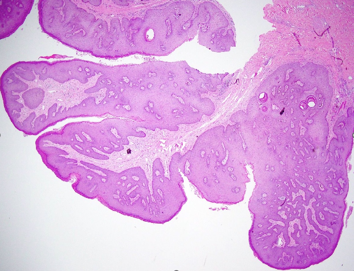
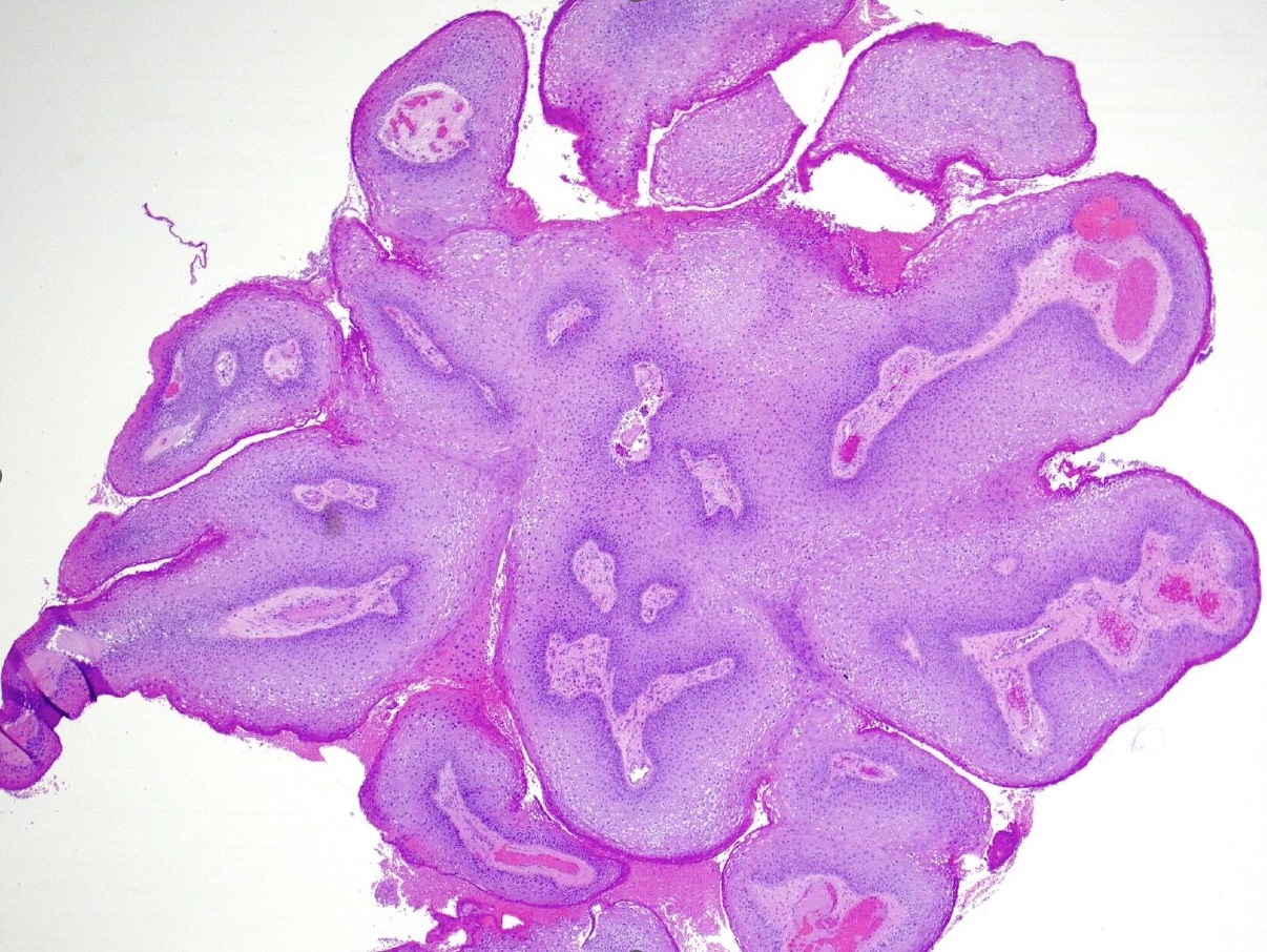
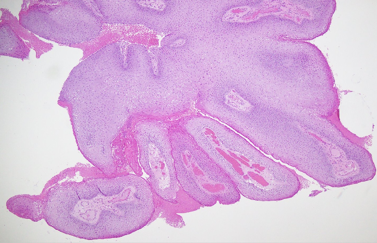
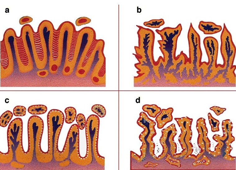
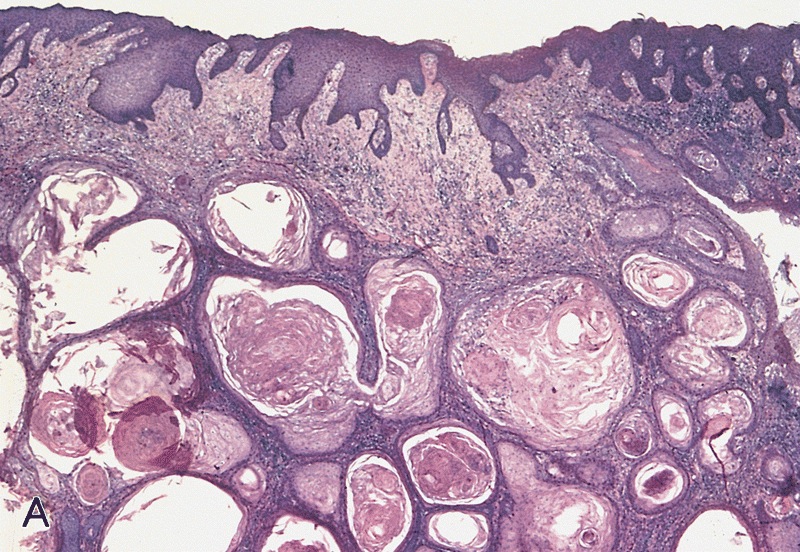
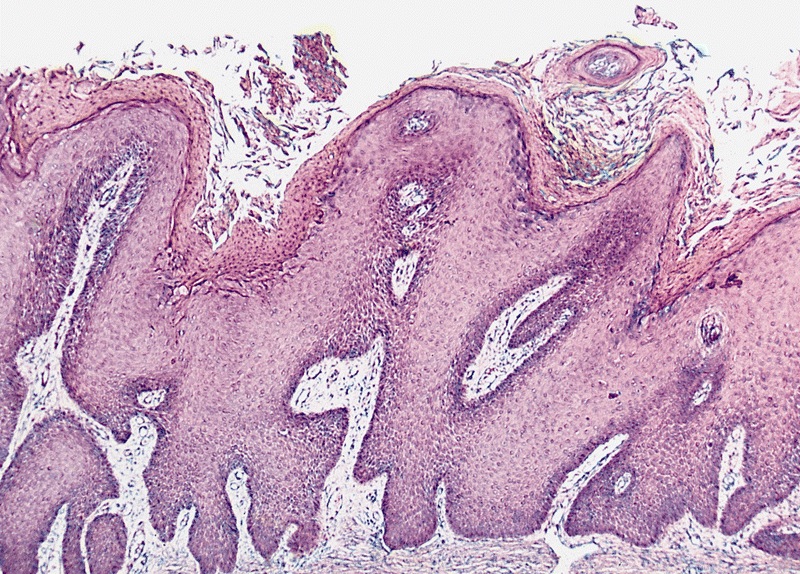
    • pTa: noninvasive carcinoma (broad pushing penetration is permitted)
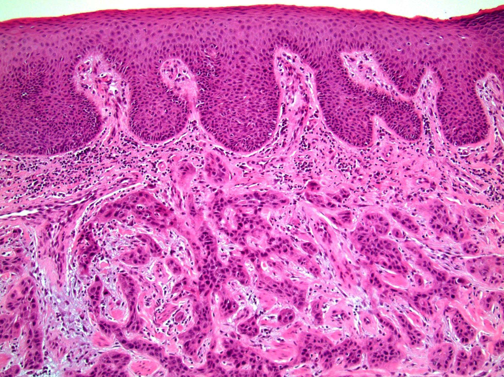
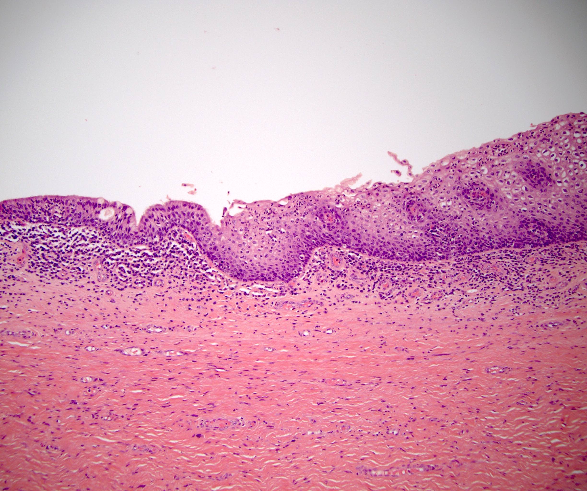
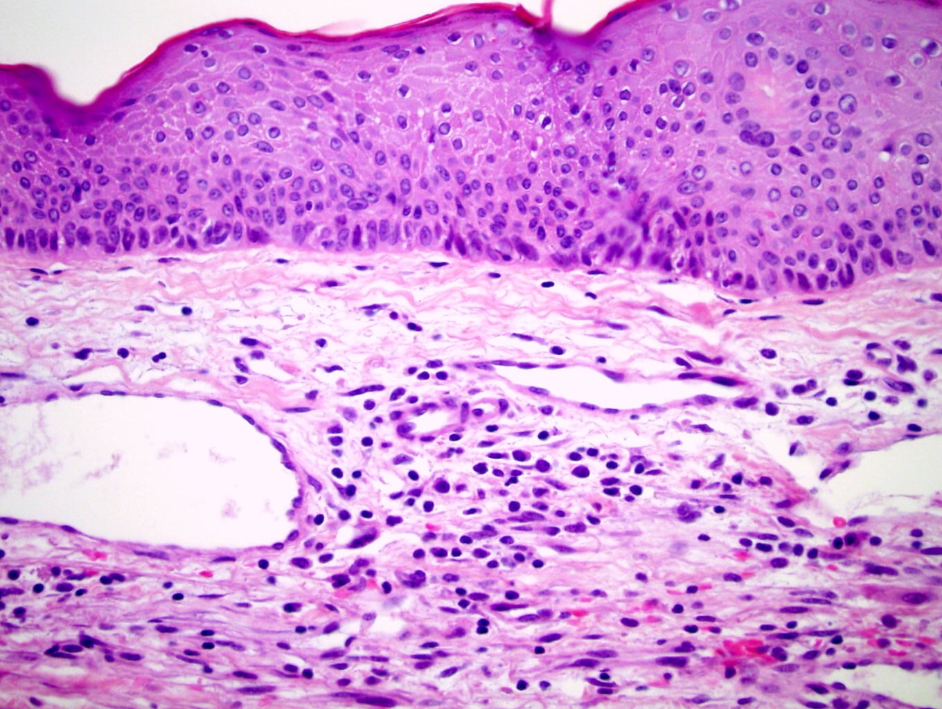
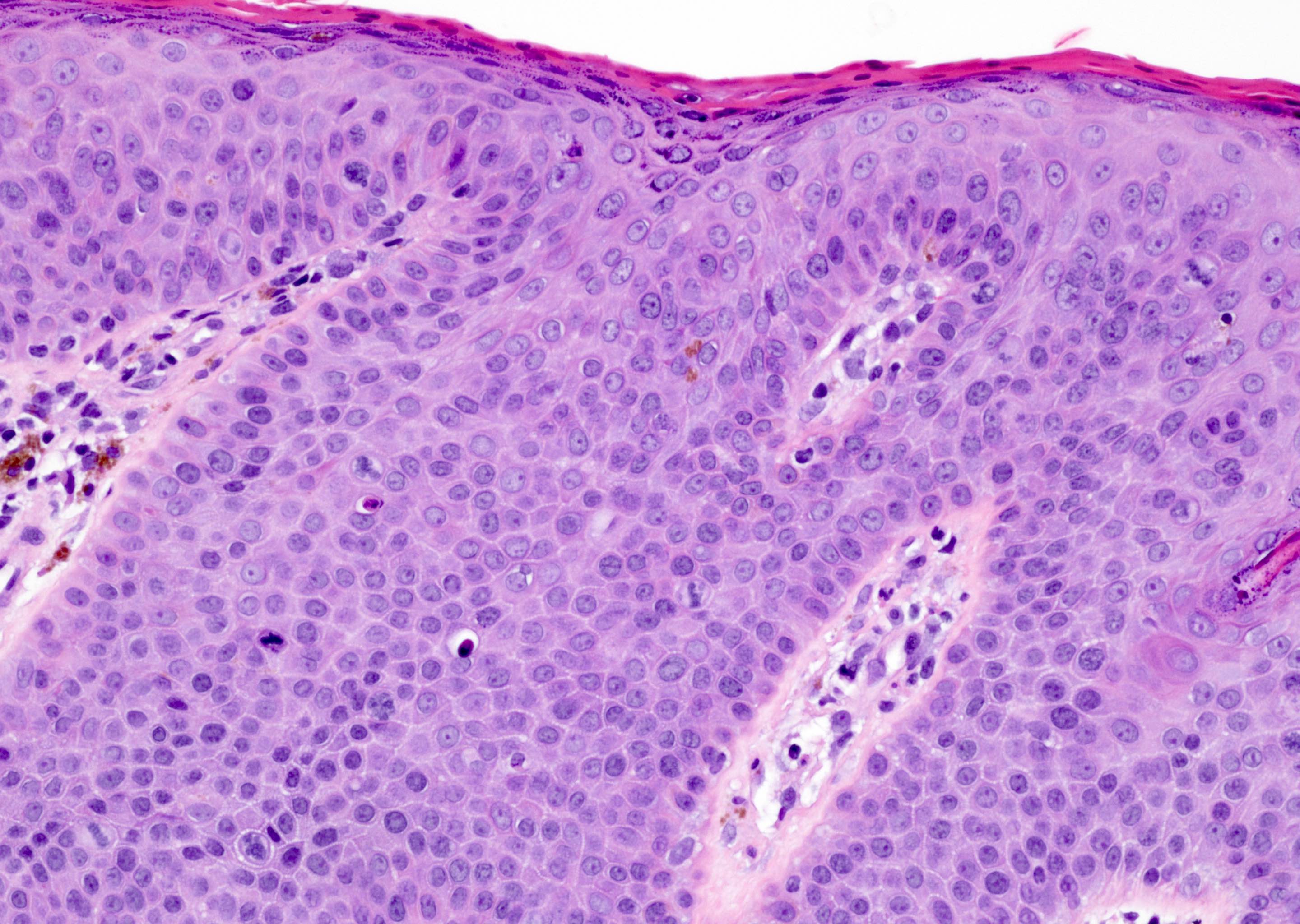
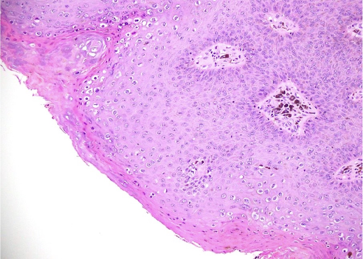
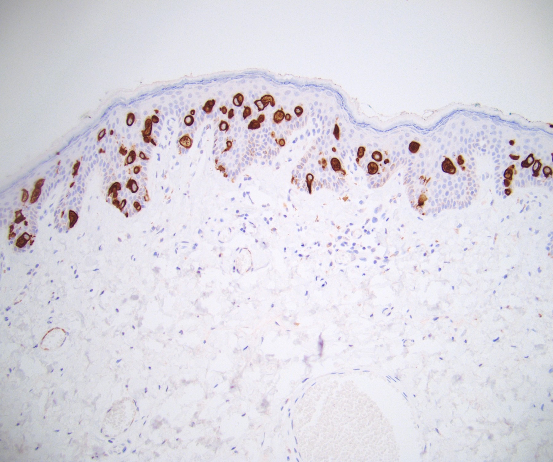
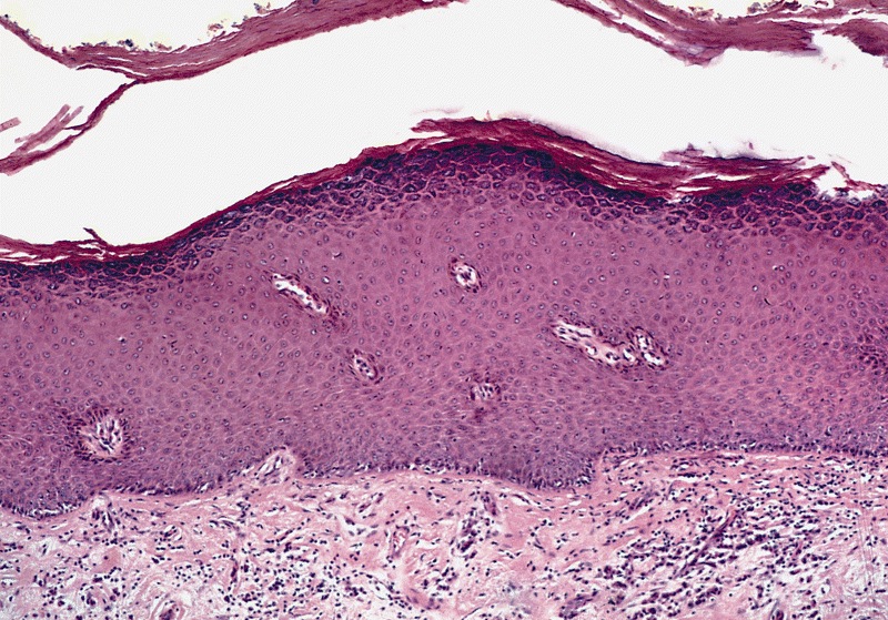
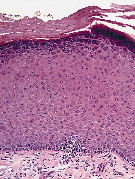
    • pTis: carcinoma in situ
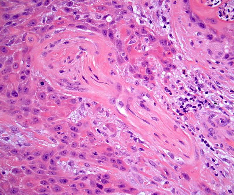
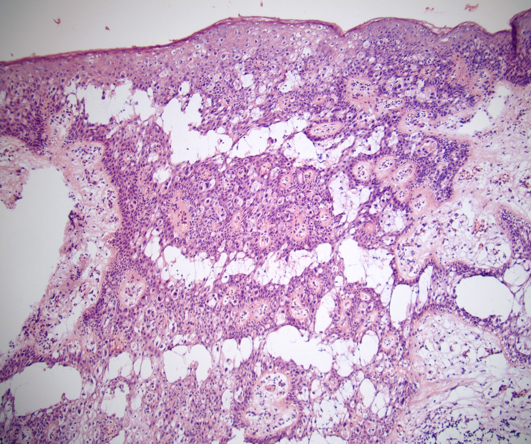
    • pT1a: subepithelial invasion without lymphovascular invasion, perineural invasion or grade 3
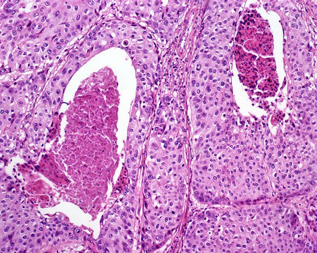
    • pT1b: subepithelial invasion with lymphovascular invasion, perineural invasion or grade 3
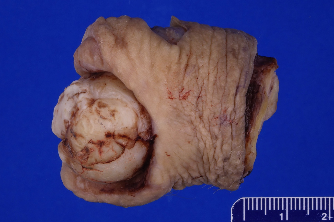
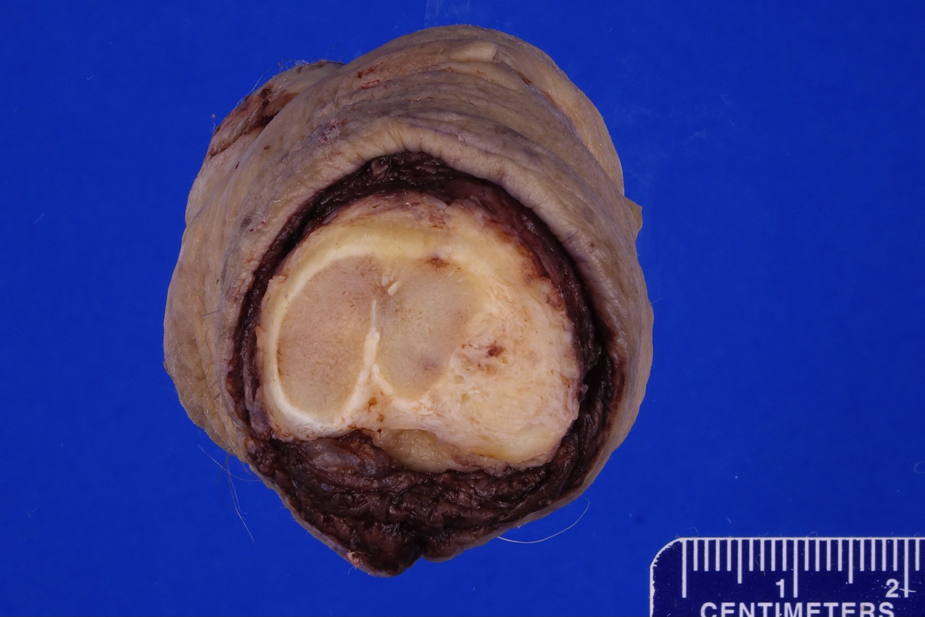
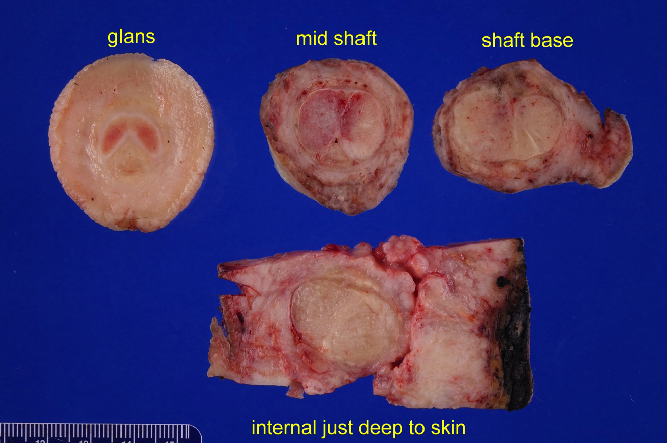
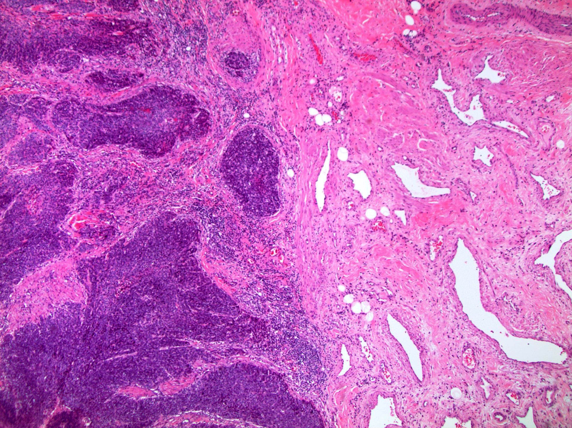
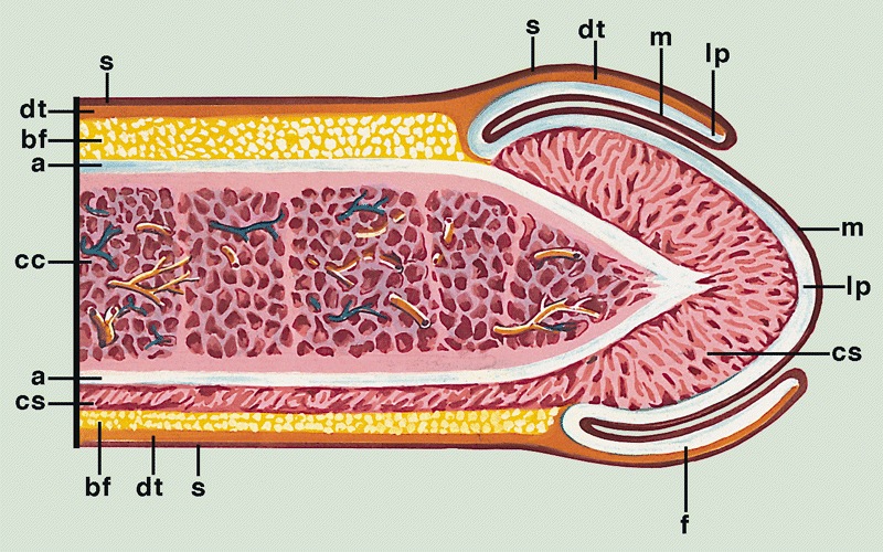
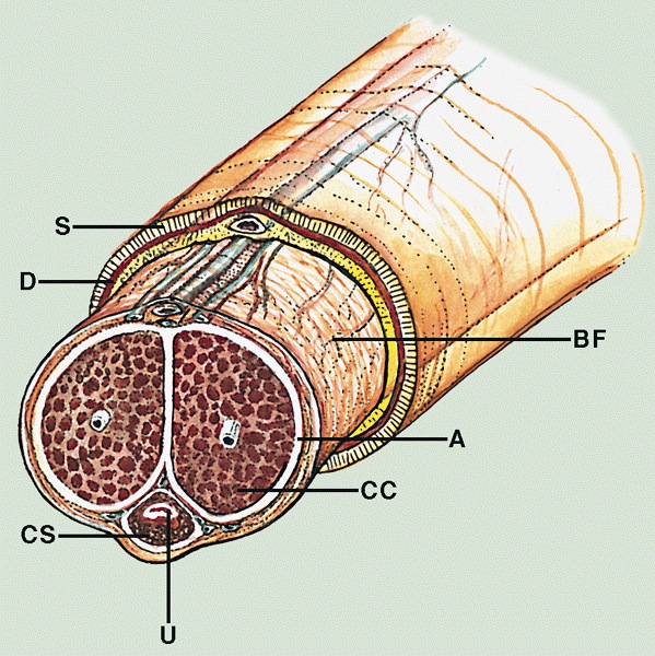
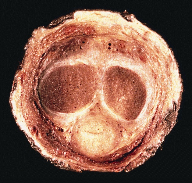
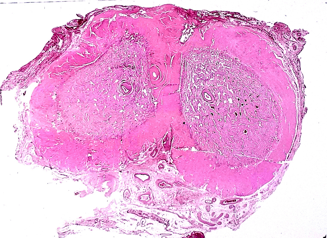
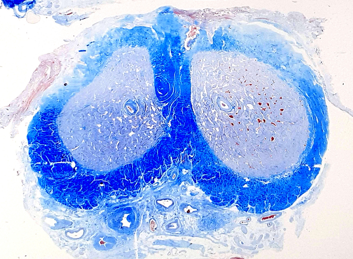
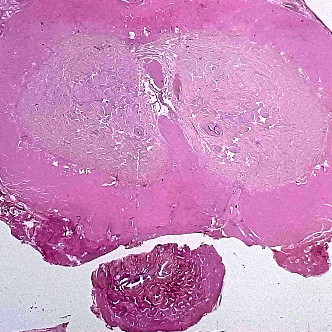
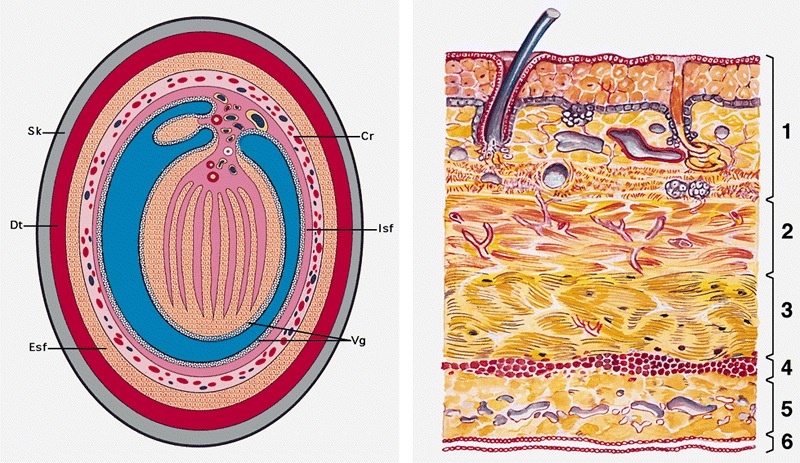
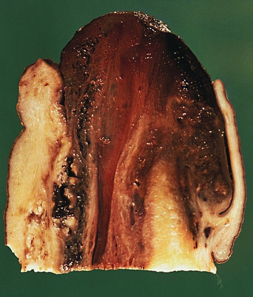
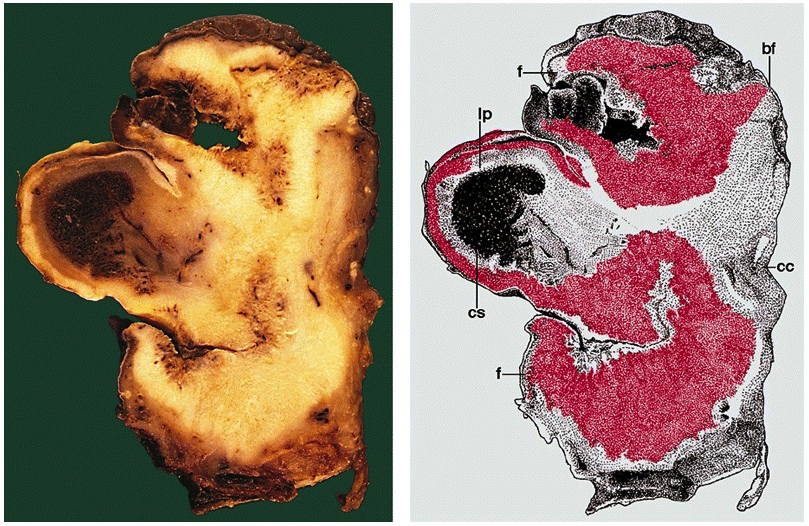
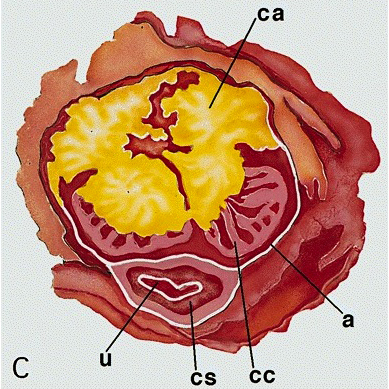
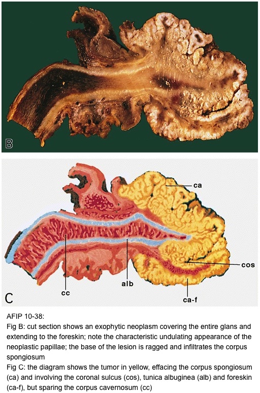
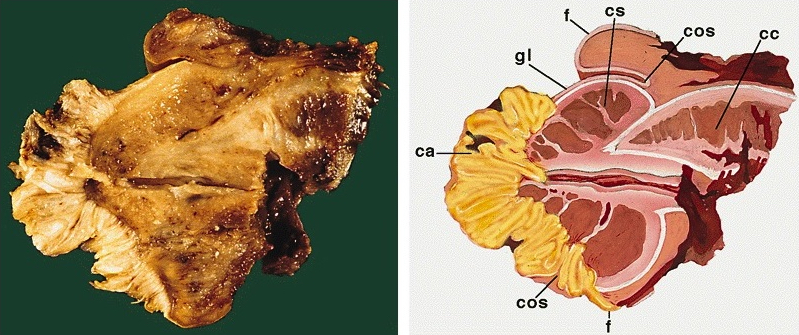
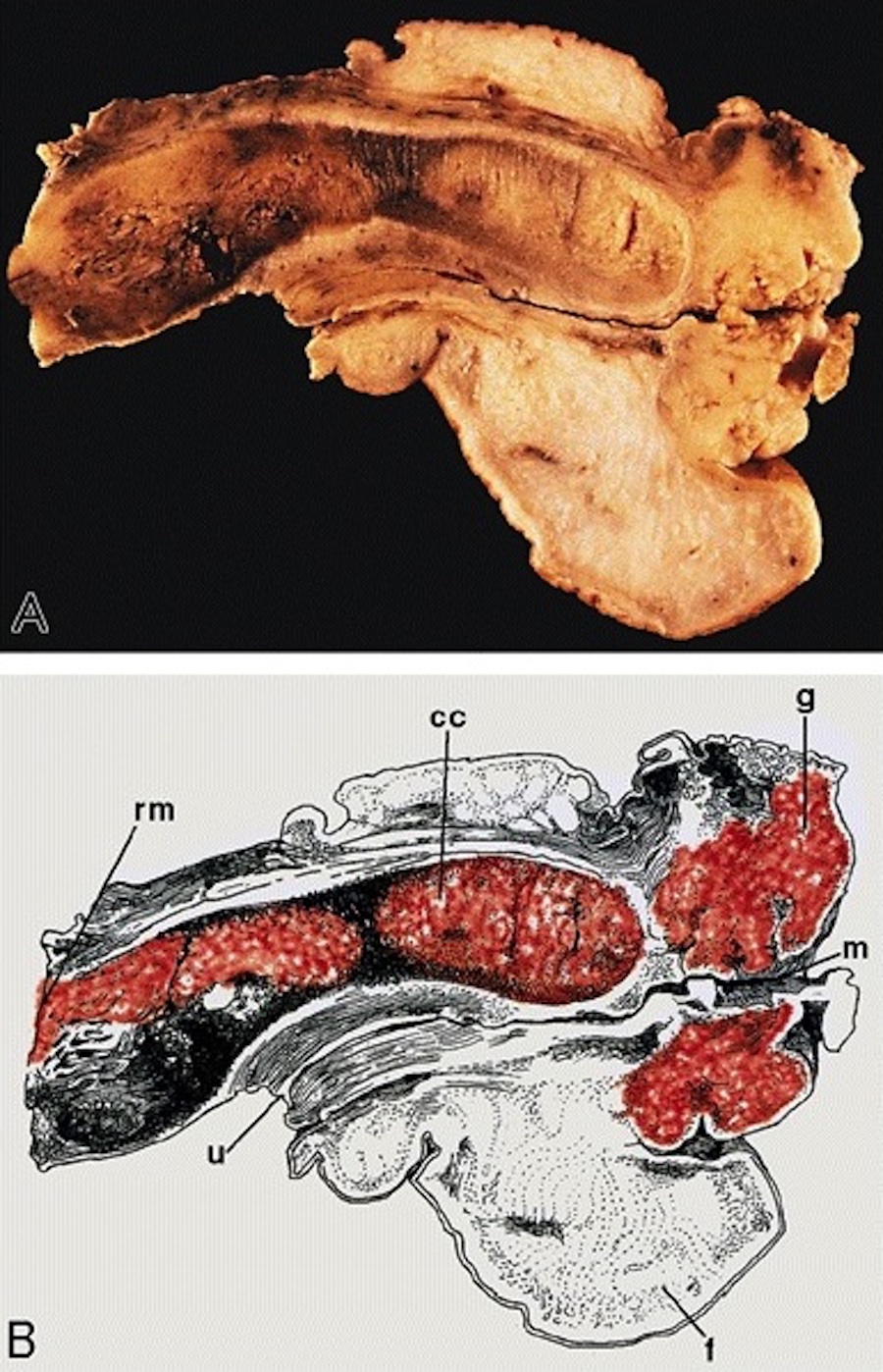
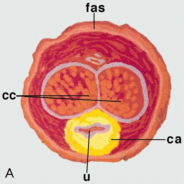
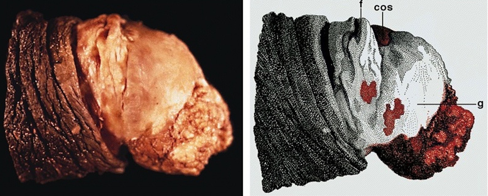
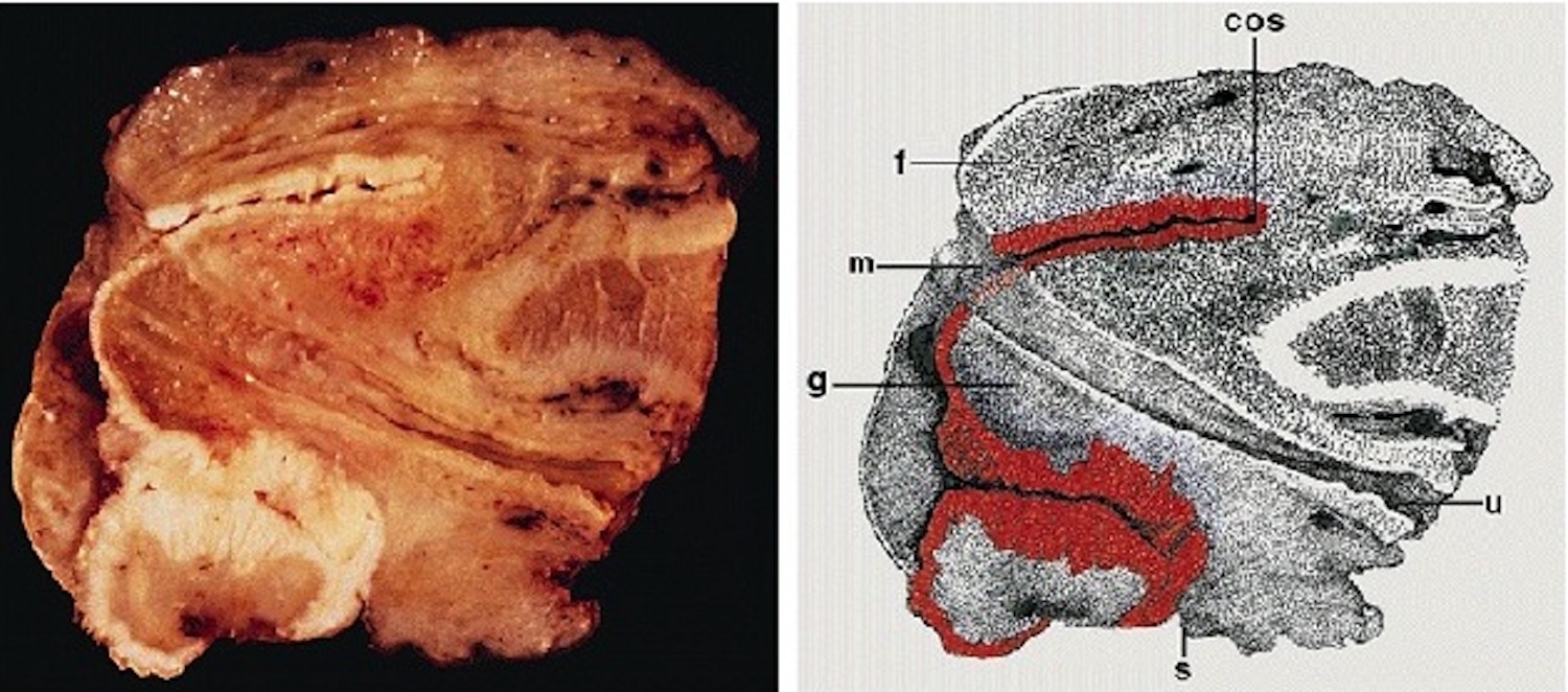
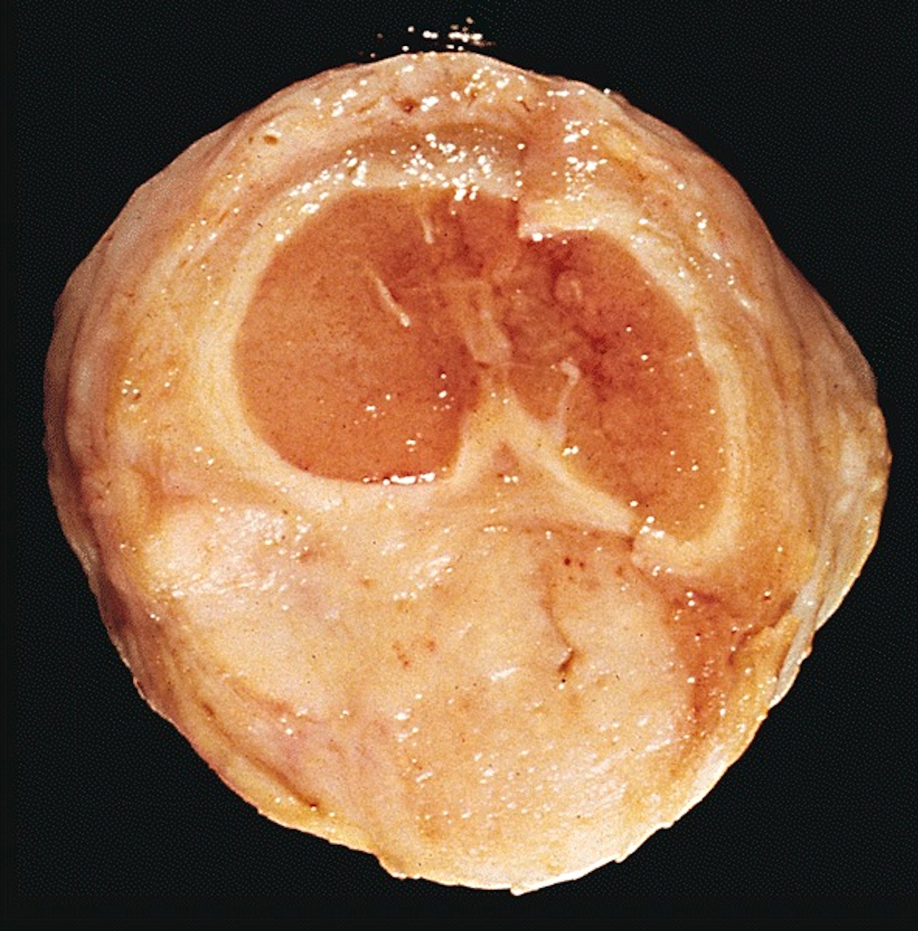
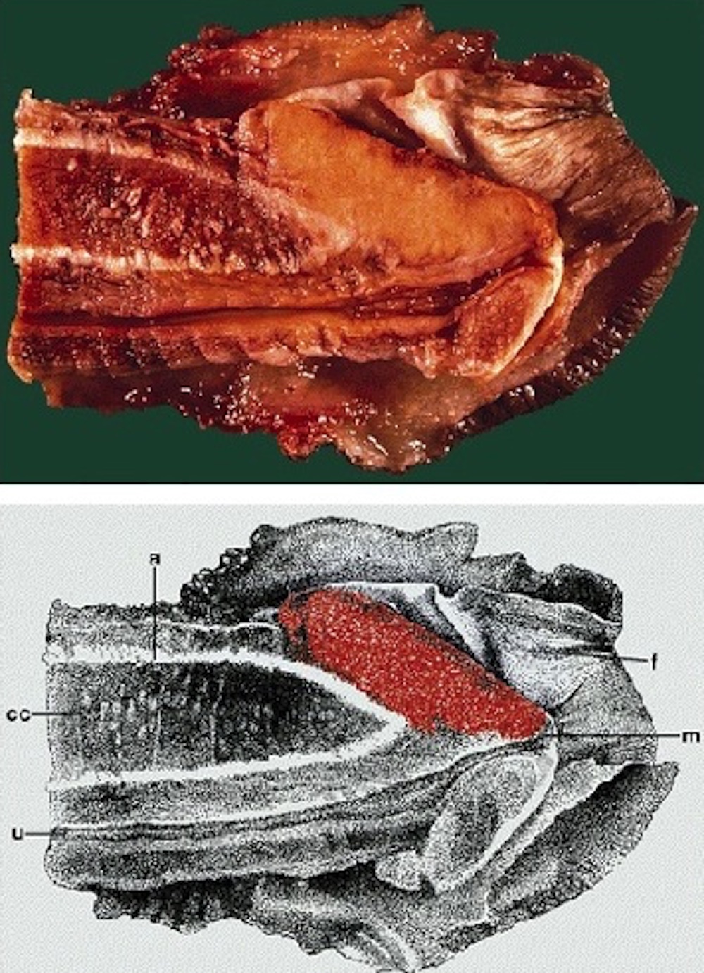
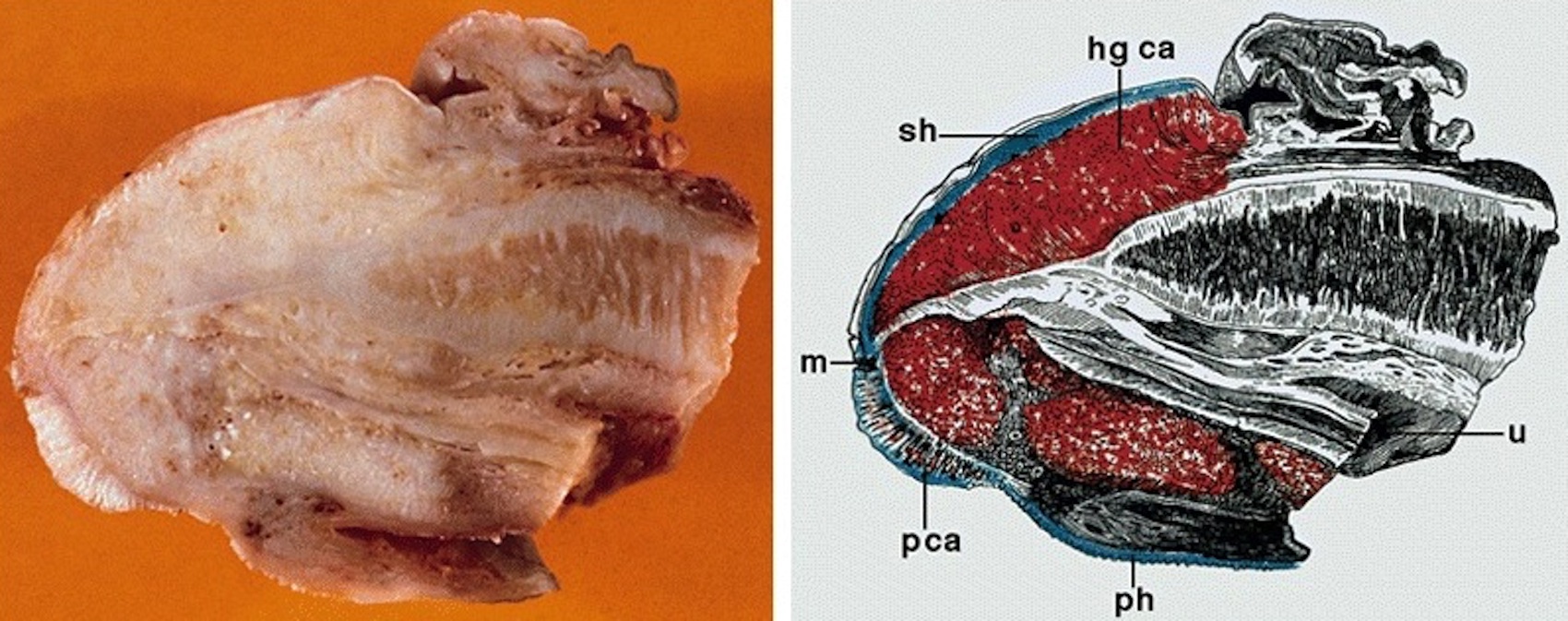
    • pT2: invasion of corpus spongiosum
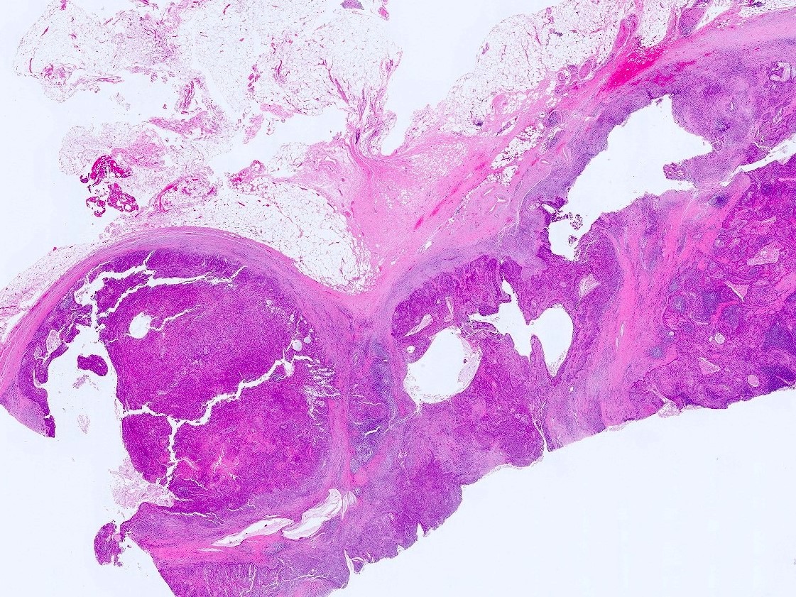
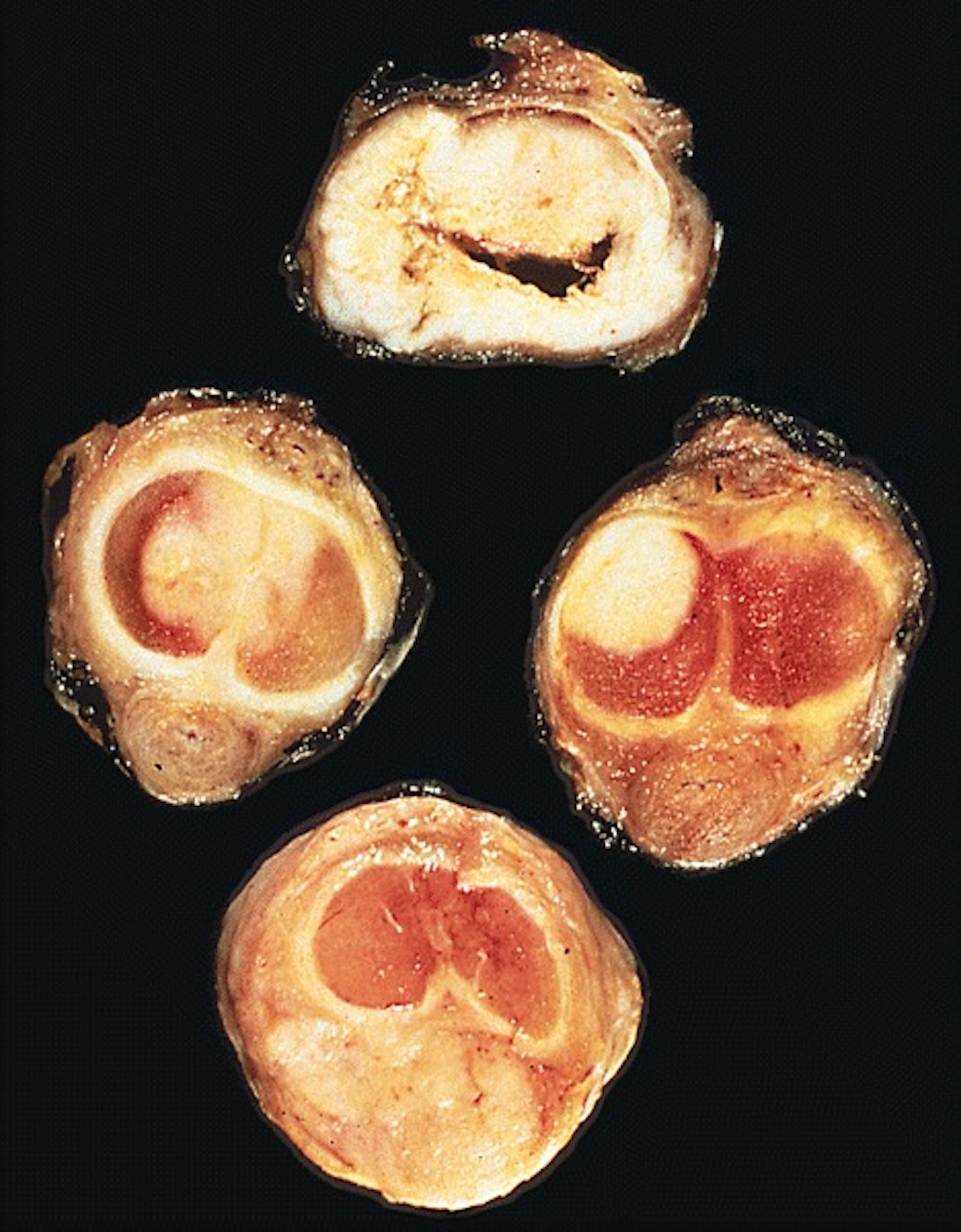
    • pT3: invasion of corpus cavernosum
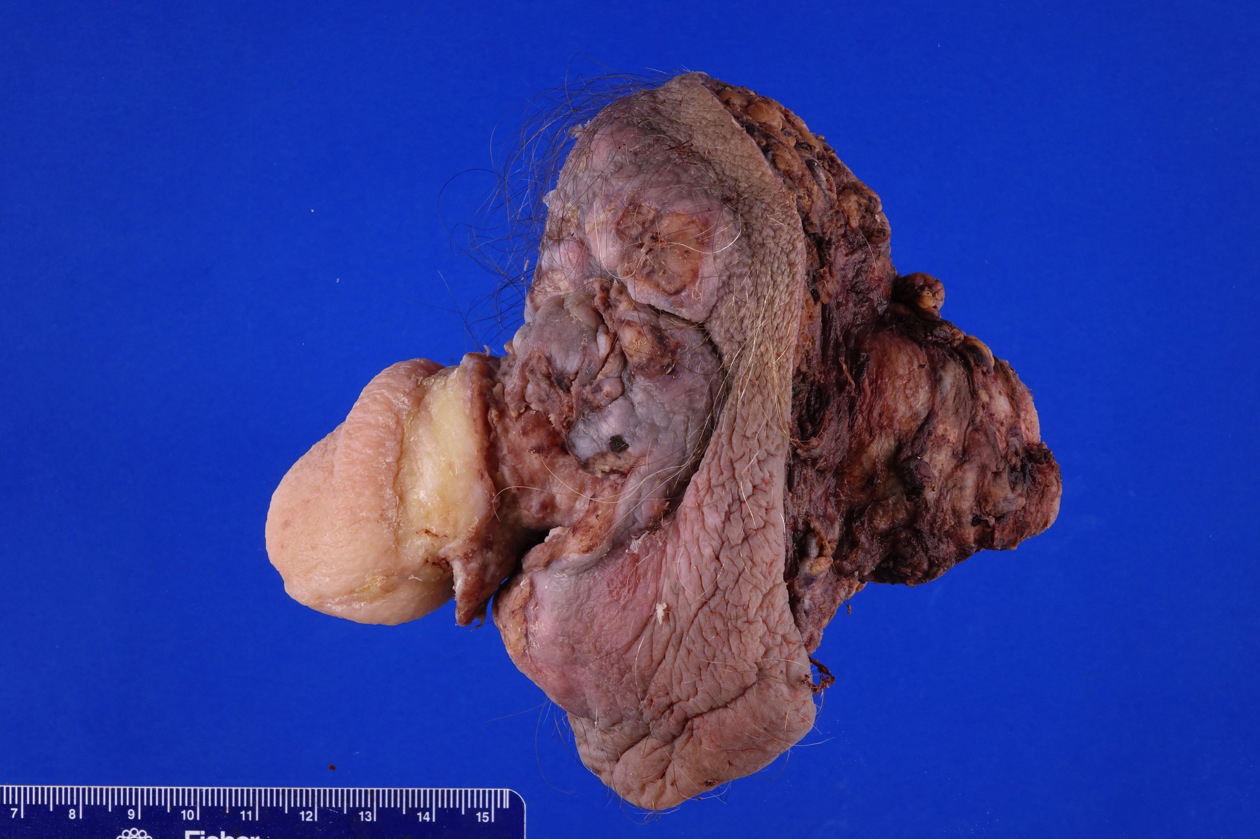
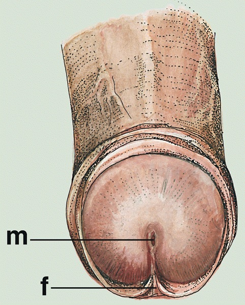
    • pT4: invasion of adjacent structures including scrotum, prostate and pubic bone
    Regional lymph nodes (pN)
    • pNX: cannot be assessed
    • pN0: no regional lymph node metastasis
    • pN1: metastasis in 1 or 2 unilateral inguinal node(s)
    • pN2: metastasis in 3 or more unilateral inguinal nodes or bilateral inguinal nodes
    • pN3: extranodal extension or metastasis to pelvic lymph node

    Notes:
    • Regional lymph nodes include superficial / deep inguinal and pelvic (external iliac, internal iliac / hypogastric, obturator, pelvic NOS)
    Distant metastasis (pM)
    • pM1: distant metastasis

    Notes:
    • Distant metastasis includes common iliac and aortic / caval lymph nodes
    AJCC prognostic stage groups
    Stage group 0is:  Tis  N0  M0
    Stage group 0a:  Ta  N0  M0
    Stage group I:  T1a  N0  M0
    Stage group IIA:  T1b - 2  N0  M0
    Stage group IIB:  T3  N0  M0
    Stage group IIIA:  T1 - 3  N1  M0
    Stage group IIIB:  T1 - 3  N2  M0
    Stage group IV:  T4  N1 - 3, X  M0
     T1 - 4, X  N3  M0
     T1 - 4, X  N1 - 3, X  M1
    Registry data collection variables
    • Histologic subtype
    • Size of largest node metastasis
    • Total number of nodes removed
    • Presence of high risk HPV
    • p16 immunohistochemical expression
    • Urethral mucosal invasion
    Histologic grade (G)
    • GX: cannot be assessed
    • G1: well differentiated
    • G2: moderately differentiated
    • G3: poorly differentiated
    Histopathologic type
    • Non-HPV related squamous cell carcinoma
      • Usual type
      • Pseudoglandular
      • Verrucous
      • Cuniculatum
      • Papillary, NOS
      • Adenosquamous
      • Sarcomatoid
      • Mixed
    • HPV related squamous cell carcinoma
      • Basaloid
      • Papillary basaloid
      • Warty
      • Warty basaloid
      • Clear cell
    • Rare other types of squamous cell carcinoma
    Gross images

    Contributed by Debra Zynger, M.D.
    pTis

    pTis

    pT1a

    pT1a

    pT2

    pT2

    pT3

    pT3

    pT3 partial penectomy pT3 partial penectomy

    pT3 partial penectomy


    pT3 penectomy pT3 penectomy

    pT3 penectomy

    pT4 penectomy, orchiectomy and scrotal excision pT4 penectomy, orchiectomy and scrotal excision pT4 penectomy, orchiectomy and scrotal excision

    pT4 penectomy, orchiectomy and scrotal excision

    Microscopic (histologic) images

    Contributed by Debra Zynger, M.D.
    CIS / PeIN (pTis)

    CIS / PeIN (pTis)

    Subepithelial invasion (pT1a)

    Subepithelial invasion (pT1a)

    Perineural invasion (pT1b)

    Perineural invasion (pT1b)

    Invasion of corpora spongiosum (pT2)

    Invasion of corpora spongiosum (pT2)

    Invasion of corpora spongiosum (pT2) Inguinal lymph node metastasis (pN1)

    Inguinal lymph node metastasis (pN1)

    Board review style question #1

      A 72 year old man has a penectomy revealing squamous cell carcinoma with corpus cavernosum invasion, shown here. Which is the correct pT category for this tumor?

    1. pT1a
    2. pT1b
    3. pT2
    4. pT3
    5. pT4
    Board review style answer #1
    D. pT3. Corpus cavernosum invasion is classified as pT3.

    Comment Here

    Reference: Pathologic TNM staging of penis carcinoma (AJCC 8th edition)

    Syphilis
    Definition / general
    Terminology
    • Historically called lues
    Epidemiology
    • U.S. incidence dropped through 2000 but has risen since with more cases in men having sex with men
    Etiology
    • Caused by Treponema pallidum, subspecies pallidum, a microaerophilic spirochete that is pathogenic only to humans
    Clinical features
    • Called "the great imitator" because it may mimic other disorders if presentation is unusual
    • 30% transmission rate from sexual intercourse

    Primary syphilis:
    • Painless hard chancre (ulcer with indurated and punched out base) at site of inoculation, often in glans
    • Most common affected sites are inner foreskin, coronal sulcus, penile shaft and penile base
    • Chancres are usually solitary

    Secondary syphilis:
    • Bacteremic stage with greatest number of organisms in the body
    • Classically has widespread rash (small red macular lesions), including on palms and soles and mucous membranes
    • Condyloma lata, formed by soft, flat topped, moist, red / rose / gray to pale maculopapules, nodules or plaques, is the characteristic anogenital lesion and may become confluent; are common in scrotum
    • May have varied clinical presentation

    Tertiary syphilis:
    • Gummatous form is characteristic: granulomas with epithelioid and giant cells, obliterative endarteritis and necrosis
    • Also cardiovascular form and neurosyphilis
    • Accelerated time course occurs with HIV infection (1 year to neurosyphilis)
    Laboratory
    Case reports
    Treatment
    • Penicillin G
    Clinical images

    AFIP images
    Sharply delineated lesion

    Sharply delineated lesion

    Two well circumscribed, minimally elevated lesions


    Images hosted on other servers:

    Primary chancre

    Atrophic scar with condylomata lata


    Secondary rash / lesions:

    Palm, limbs and hands

    Back


    Face, teeth

    "Kissing" lesions

    Various images

    Microscopic (histologic) description
    • Presence of obliterative endarteritis surrounded by a predominantly plasmocytic infiltrate is characteristic of all stages
    • Spirochetes can be identified in primary and secondary lesions but are difficult to demonstrate in gummas
    • Primary syphilis: ulceration, granulation tissue and obliterative endarteritis at ulcer base; plasma cells and lymphocytes underlying ulcer, endothelial cell proliferation and capillaritis
    • Secondary syphilis: psoriasiform epidermal hyperplasia or spongiform pustular lesions with superficial or deep obliterative endarteritis and lymphoplasmacytic infiltrate at the dermal epidermal junction; perivascular infiltrate and possible granulomas; also nodal involvement with florid follicular hyperplasia, unusually shaped follicles, endothelial swelling and perivascular cuffs of plasma cells and lymphocytes
    • Tertiary syphilis: gummas formed by granulomas with epithelioid and multinucleated giant cells, obliterative endarteritis and necrotic foci
    Microscopic (histologic) images

    AFIP images

    Lesion characterized by acanthosis


    Images hosted on other servers:

    Perivascular dermal infiltrate

    Chorioretinitis of congenital syphilis

    Darkfield microscopy


    Patchy infiltrate

    Epithelioid cells surrounded by lymphomono-nuclear cells

    Various images

    Positive stains
    Electron microscopy images

    Images hosted on other servers:

    Spirochete in culture

    Differential diagnosis
    • Lymphoma: monoclonal lymphoplasmacytic infiltrate; no clinical or laboratory evidence of syphilis
    • Plasma cell myeloma: monoclonal plasma cells, often binucleated; monoclonal gammopathy; no clinical or laboratory evidence of syphilis

    Tancho nodules / paraffinomas / lipogranulomas
    Definition / general
      Tancho nodules:
      • Custom among some Asian populations to implant, inject or insert paraffin or other foreign material (such as glass spheres) under the skin of the penis to improve sexual pleasure
      • Tancho is a brand of hair pomade widely used in the Far East and Southeast Asia

      Lipogranuloma:
      • Custom among some Asian populations to enlarge penis by injecting mineral oil, paraffin (paraffinoma), silicone or vaseline (Urology 2008;71:1132)
      • Inflammatory / foreign body reaction may occur years after the injection
    Clinical features

    Tancho nodules:
    • Palpable subcutaneous firm nodules

    Lipogranuloma:
    • Distortion of organ with evident gross abnormalities
    • May extend to adjacent structures such as scrotum
    Case reports
    Treatment
    • May require local surgical resection
    Clinical images

    Images hosted on other servers:

    Intraurethral paraffin

    Microscopic (histologic) description
    Tancho nodules:
    • Inflammatory foreign body reaction with variable fibrosis surrounding inserted material

    Lipogranuloma:
    • Numerous vacuoles ranging from tiny to large and cystically dilated (pseudocysts)
    • Stroma may be sclerotic (sclerosing lipogranuloma) and contain inflammatory cells and foreign body giant cells

    Verruciform xanthoma
    Definition / general
    • Rare, nonneoplastic lesion of verrucous epidermal acanthosis, foamy histiocytes aggregates in papillary dermis and neutrophilic inflammation
    Essential features
    • Nonneoplastic verrucous lesion characterized by aggregates of lipid laden macrophages in papillary dermis
    • Closely resembles other verrucous lesions clinically and microscopically
    ICD coding
    • ICD-10: E75.5 - other lipid storage disorders
    Epidemiology
    Sites
    Etiology
    Clinical features
    Diagnosis
    • Punch biopsy with subsequent histological examination
    Case reports
    Treatment
    Clinical images

    AFIP images

    Granular, elevated
    tan white mass
    of distal shaft and
    coronal sulcus

    Gross description
    Microscopic (histologic) description
    Microscopic (histologic) images

    Contributed by Liwei Jia, M.D., Ph.D. and @JMGardnerMD on Twitter

    Verrucous epithelial hyperplasia

    Band-like plasma cells

    Neutrophilic infiltrates

    Foamy histiocytes

    Verruciform xanthoma

    Verruciform xanthoma

    Negative stains
    Electron microscopy description
    Sample pathology report
    • Penis, dorsal, biopsy:
      • Verruciform xanthoma
    Differential diagnosis
    • Verruca vulgaris:
      • Koilocytes, coarse hypergranulosis, prominent papillomatosis, rete ridges curve inward
    • Condyloma acuminatum:
      • Prominent koilocytotic atypia in upper epidermis
      • No prominent foamy macrophages
    • Xanthoma:
      • Foamy cells are located in the mid dermis
      • Clinical hyperlipidemia
    • Squamous cell carcinoma:
      • Marked atypia, no prominent foamy histiocytes
    • Verrucous carcinoma:
      • Ulcerating or fungating lobules of mature squamous epithelium
      • Minimal atypia but no prominent foamy histiocytes
    • Bowenoid papulosis:
      • Resembling basaloid PeIN
      • Spotty cytological atypia
    Board review style question #1

    A 58 year old Caucasian man presents with a 1.0 cm pink, warty lesion on the glans penis. The biopsy shows verruciform hyperplasia of the epidermis with hyperkeratosis, parakeratosis and acanthosis (figure 1). No keratinocyte atypia or dyskeratotic cells are seen. There is a dense infiltrate of lipid laden macrophages (foamy cells) in the papillary dermis (figure 2) which are positive for CD68 and negative for S100. Which statement is true of cutaneous verruciform xanthoma?

    1. It is frequently caused by HPV genotypes 6 and 11
    2. Approximately 2 - 5% will either harbor or progress to a squamous cell carcinoma
    3. It is usually associated with underlying immunosuppression
    4. It is a nonneoplastic lesion that typically does not recur after excision
    Board review style answer #1
    D. It is a nonneoplastic lesion that typically does not recur after excision. The diagnosis of the lesion is cutaneous verruciform xanthoma.

    Comment Here

    Reference: Cutaneous verruciform xanthoma
    Board review style question #2
    Which best summarizes the immunohistochemical profile of the characteristic cells infiltrating the dermal papillae in cutaneous verruciform xanthoma?

    1. CD68 positive; PAS positive diastase resistant; positive granular cytoplasmic staining of adipophilin; S100 negative
    2. CD68 positive; S100 positive; CD1a positive; Langerin positive
    3. CK5/6 positive; p16 positive; p63 positive
    4. CD10 positive; CK5/6 negative; S100 negative
    Board review style answer #2
    A. CD68 positive; PAS positive diastase resistant; positive granular cytoplasmic staining of adipophilin; S100 negative

    Comment Here

    Reference: Cutaneous verruciform xanthoma

    WHO classification
    Definition / general
    • WHO classification of tumors of the urinary and male genital tumors
    • Currently on 5th edition, published in 2022
    Major updates
    • Terminology for squamous cell carcinoma groupings has been changed from non-HPV related to HPV independent and from HPV related to HPV associated
    • Papillary basaloid carcinoma, warty basaloid carcinoma, pseudohyperplastic carcinoma, pseudoglandular carcinoma, carcinoma cuniculatum and mixed squamous cell carcinoma have been removed as distinct subtypes
    • Pseudohyperplastic and pseudoglandular patterns are now a part of squamous cell carcinoma, usual type
    • Carcinoma cuniculatum pattern is now a part of verrucous carcinoma
    Board review style question #1
    Which marker is utilized in the classification scheme of penile squamous cell carcinoma?

    1. ALK
    2. HPV
    3. PTEN
    4. TFEB
    5. TFE3
    Board review style answer #1
    Back to top
    Recent Penis & scrotum Pathology books

    Amin: 2022

    Cheng: 2019

    Colecchia : 2016

    Epstein: 2020

    IARC: 2022

    Wobker: 2021

    Yang: 2020

    Zhou: 2022


    Find related Pathology books: GU/adrenal
    Image 01 Image 02