Superpage
Page views in 2024 to date: 11

Adenomatoid tumor
Definition / general
  • Benign lesion, often incidental finding on oophorectomy specimen
  • More frequently these lesion are found in males (epididymis, spermatic cord and testicular membrane); however, in females lesions are seen more commonly in fallopian tubes, broad ligament and uterus
  • Thought to arise from mesothelial serosal cells
  • First described by Golden and Ash in 1945 (Am J Pathol 1945;21:63)
Essential features
  • Rarely found within ovary
  • Typically small with 0.5 - 3 cm incidental lesions near hilum
Terminology
  • Previously known as benign mesothelioma of the genital tract
Epidemiology
Sites
  • Ovarian and juxtaovarian sites are rare
  • Occur predominantly at the ovarian hilum and may extend into and replace the ovarian parenchyma
  • Most frequently unilateral, found within fallopian tube, broad ligament or on uterine serosal surface
Pathophysiology / etiology
Clinical features
  • Asymptomatic, discovered as an incidental finding
  • Usually 0.5 - 3.0 cm, rarely larger and symptomatic
Diagnosis
  • Histologic recognition, confirmed by immunophenotype
  • Often incidental
Laboratory
Radiology description
  • Not routinely performed for primary diagnosis
  • Case reports describe incidental lesions on transvaginal ultrasound displaying multilocular cystic mass often with vascularized central / solid portion
  • Radiographic differential diagnosis, if provided, may include epithelial tumors, inclusion peritoneal cysts, and multiple large follicles
  • CT imaging seldom describes lesion (J Clin Ultrasound 2005;33:233)
Prognostic factors
  • Benign behavior, no reports of recurrence or malignant transformation
Case reports
Treatment
  • Excision results in complete cure
  • Recurrence after excision is rare
Gross description
  • Small, round to oval, well circumscribed tumor
  • Cut surface may have small cystic spaces
Microscopic (histologic) description
  • Composed of clefts and spaces lined by cuboidal, low columnar or flattened epithelial-like cells
  • Surrounded by connective tissue that varies from loose and edematous to dense and hyalinized
  • The epithelial-like cells may exhibit marked vacoulation, which in some cases may contain weakly basophilic material
  • A spotty lymphoid aggregate may be a low power clue to the diagnosis
  • Distinctive thread-like bridging strands crossing the tubular spaces are useful diagnostic features
  • Morphologic patterns:
    • Adenoid
    • Angiomatoid
    • Cystic
    • Glandular
    • Solid
    • Tubular
    • Plexiform
    • Canalicular
  • Similar appearance to appearance found within other locations
  • Relatively well demarcated, nonencapsulated solid aggregates of cells forming cleft-like spaces lined by low columnar to cuboidal flattened epithelial-like cells
  • Cells often surrounded by stroma that ranges from dense / fibrotic to loose / edematous
  • Epithelial-like cells may display marked vacuolization, signet ring cell-like appearance or oxyphilic cytoplasm
Microscopic (histologic) images

Contributed by Eman Abdulfatah, M.D., AFIP and @AnaPath10 on Twitter
Adenoid pattern Adenoid pattern

Adenoid pattern

Adenoid pattern Adenoid pattern

Adenoid pattern


Angiomatoid pattern Angiomatoid pattern

Angiomatoid pattern

Trabecular pattern Trabecular pattern

Trabecular pattern,


Scattered cysts

Scattered cysts

Adenomatoid tumor

Cytology description
  • Smears are moderately cellular with sheets of monotonous round to oval cells showing indistinct cell borders and moderate to abundant pale cytoplasm with vacuolations
  • Nuclei are eccentric in location, but regular with inconspicuous nucleoli
Positive stains
Negative stains
Electron microscopy description
  • No microvilli, no bundles of cytoplasmic filaments, no tight junctional complexes, no intercellular spaces
Molecular / cytogenetics description
  • No specific genetic abnormality has been identified
Differential diagnosis

Anatomy, history, grossing & features to report
Anatomy
  • Lungs are surrounded by visceral pleural, a delicate serous membrane arranged as a closed invaginated sac
  • Inner chest cavity is lined by parietal pleural membrane
  • Visceral and parietal pleura define the pleural space / cavity, which normally has minimal volume, unless lungs collapse or air / fluid collects between the 2 layers
  • Only minimal contact between right and left pleural sacs
  • Regional lymph nodes are internal mammary, intrathoracic, scalene and supraclavicular
Drawings

Images hosted on other servers:

Pleura and chest wall

Histology
  • Lined by mesothelial cells overlying vascularized connective tissue
  • Mesothelium provides smooth, low friction surface to facilitate the gliding motion of lungs in pleural cavity, heart in pericardial cavity, viscera in abdominal cavity
  • Gliding facilitated by numerous surface microvilli, thick glycocalyx, secretion of hyaluronic acid and other glycosaminoglycans
Mesothelial cells
  • Microscopic (histologic) description
    • Monolayer of flat or low cuboidal cells with bland and uniform nuclei, fine delicate chromatin, inconspicuous nucleoli
    • In fine needle aspirates, have well defined cytoplasm and distinct cell borders
  • Positive stains: keratin
  • Electron microscopy description: apical tight junctions, desmosomes, surface microvilli, cytoplasmic tonofilaments in bundles
Connective tissue cells
  • Microscopic (histologic) description: resemble fibroblasts
  • Positive stains
  • Electron microscopy description: resemble fibroblasts
Black spots
  • Carbonaceous / anthracotic pigments in parietal pleura
  • Present in > 90% of urban dwellers in Belgium at autopsy
  • Not related to hyaline pleural plaques (Am J Surg Pathol 2002;26:1198)
  • Associated with lymphatic drainage
  • Microscopic (histologic) description: deposits of opaque particles (intra or extracellular) of various sizes under an intact mesothelial layer, associated with chronic inflammatory cells
Types of specimen
  • Biopsy
  • Pleurectomy
Surgical procedures definition
  • Pleurectomy / decortication with mediastinal lymph node sampling
    • Complete removal of pleura and all gross tumor
  • Extrapleural pneumonectomy
    • En bloc resection of pleura, lung, ipsilateral diaphragm; may include pericardium
Grossing biopsy
  • If received for frozen section, ensure enough lesional tissue is present
  • Ask for additional tissue if tissue submitted needs to be entirely frozen
    • Important because immunohistochemistry may be unreliable on previously frozen tissue
    • May need to send for special studies including electron microscopy and cytogenetics
Grossing pleurectomy
  • Describe dimension and number of fragments, any lesions present
  • Note if pleural plaques are seen; describe
  • Tumor involvement of adjacent structures - lung, diaphragm, pericardium, skeletal muscle
  • Ink margins in sections closest to tumor
  • Tumor
    • One section per cm of tumor
    • Extensive sampling if desmoplastic mesothelioma is suspected
  • Additional sections of lung, if present (for asbestos fiber analysis)
    • Recommended (up to 5)
  • Sections for ancillary tests
    • Electron microscopy, cytogenetics, etc., if necessary
  • Lymph nodes
  • References: NCCN: NCCN Guidelines [Accessed 23 March 2018], Lester: Manual of Surgical Pathology, 3rd Edition, 2010
Features to report
  • Tumor size and location
  • Histologic type
  • Extent of invasion
  • Surgical resection margins
  • Involvement of pleura, pulmonary vessels, bronchus, mediastinal structures, diaphragm, chest wall, other
  • Lymph nodes: total examined, number involved by tumor, extracapsular extension
  • Presence of pleural plaques, ferruginous bodies, pulmonary interstitial fibrosis, other significant findings


Asbestos related disorders
Table of Contents
Definition / general
  • Asbestos causes localized fibrous plaques, pleural effusions, parenchymal interstitial fibrosis (asbestosis), bronchogenic carcinoma, mesothelioma, laryngeal carcinoma, possibly colon carcinoma
  • Exists in serpentine / chrysotile (curly, flexible) and amphibole (straight, stiff, brittle) forms; most asbestos in industry is serpentine but amphiboles are more pathogenic; link with mesothelioma is almost always with amphibole form
  • Chrysotiles usually are caught in upper respiratory passages, removed by mucociliary elevator; they are soluble and leached from tissue if they reach alveoli
  • Amphiboles go deeper into lungs; fibers > 8 mm and thinner than 0.5 mm are more injurious
  • Both types are fibrogenic; act as tumor initiator and promoter; toxic chemicals such as tobacco smoke may be adsorbed to asbestos fibers; asbestos fibers may also generate reactive free radicals
  • However, asbestos bodies are common in normals; present in 40% at autopsy in U.S. in lung smears
  • Asbestos may act by countering antioxidant effect of vitamin C (ascorbic acid) (Hum Pathol 2003;34:737)
  • In pleura, asbestos causes pleural plaques and mesothelioma
  • Relative risk (RR) compared to normal population: for bronchogenic carcinoma, RR is 5x, increasing to 55x for asbestos exposure plus tobacco use; for mesothelioma (pleural, pericardial, peritoneal), RR is 1000x, with no change for asbestos plus tobacco use

Atypical mesothelial hyperplasia
Definition / general
  • Worrisome proliferations of mesothelial cells that are not unequivocally malignant are termed atypical mesothelial proliferation or atypical mesothelial hyperplasia
Terminology
  • Pseudoneoplastic lesion of the pleural surface
  • Are actually reactive mesothelial proliferations, associated with both benign and malignant conditions
Sites
  • Any mesothelial surface, including pleura, peritoneum, tunica, etc.
Etiology
  • Associated with anemia, bronchogenic carcinoma, cirrhosis, connective tissue diseases, pneumothorax (recurrent), viral infections
Clinical features
  • History of pleural effusion or ascites, fluid may be hemorrhagic
Radiology description
  • Description of pleura on imaging or pleuroscopy helps differentiate benign and malignant mesothelial proliferations
  • Circumferential pleural thickening and nodular pleural thickening are highly suggestive of malignancy (Arch Pathol Lab Med 2012;136:1217)
Case reports
Microscopic (histologic) description
  • Identification of neoplastic invasion is definitive criteria for diagnosis of malignant mesothelioma
  • Finding of mesothelial cells in fat makes the proliferation malignant
  • Challenges and controversies in diagnosis of mesothelioma discussed at J Clin Pathol 2013;66:847
Microscopic (histologic) images

Images hosted on other servers:

Pleural patches - no invasion

Various immunostains - no invasion

Cytology description
  • Criteria are defined for malignant mesothelioma (high specificity - 99% when all criteria are fulfilled); refer to malignant mesothelioma
  • Cytology of atypical mesothelial cells:
    • Mesothelial cells in large groups
    • Cell groups with scalloped borders
    • Nuclear hyperchromasia
    • High N:C ratio
    • Coarse chromatin
    • Prominent nucleoli
    • Diagnostic problems in serous effusions discussed at Diagn Cytopathol 1998;19:131
Positive stains
  • All active mesothelial proliferations, benign or malignant, are pankeratin+
  • EMA, GLUT1 and IMP3 can be positive in both benign and malignant mesothelial proliferation so cannot be used to differentiate reliably
  • Homozygous deletion of p16 / CDKN2A demonstrated by FISH may be specific for malignant proliferations
  • p16 FISH staining usually negative in benign proliferations, 59% sensitive for malignant mesothelioma (Am J Clin Pathol 2011;135:619)
Negative stains
Differential diagnosis

Benign mesothelial proliferations
Terminology
  • Also called simple mesothelial hyperplasia
Pathophysiology
  • Normal mesothelial surface has single layer of flat cuboidal epithelium
  • Irritation of pleural surface causes simple hyperplasia of mesothelium
Etiology
  • Due to asbestos, benign pleural effusion, benign pleural plaque, collagen infections, drug reactions, pneumothorax, pulmonary infarct, trauma, vascular disease
Uses by pathologists
  • If malignancy cannot be excluded, use diagnosis of "atypical mesothelial hyperplasia" and recommend rebiopsy if clinically suspicious (Arch Pathol Lab Med 2012;136:1217)
Case reports
Microscopic (histologic) description
  • Mesothelial cells form conspicuous layer of regularly spaced, bland cuboidal cells along pleural surface; normally, mesotheial cells present only along surface and not in underlying tissue
  • Distinct nucleoli may be present
  • Likely benign if papillary excrescences with tufts of cells with bland tubule-like nonbranching structures, no fibrovascular cores
  • Capillaries are parallel to each other and perpendicular to pleural surface (in malignancy, the capillaries are haphazard)
  • Necrosis may be seen but usually accompanied with inflammatory cells and debris (Arch Pathol Lab Med 2005;129:1421)
Microscopic (histologic) images

Images hosted on other servers:

Exfoliated reactive mesothelium

Reactive mesothelial hyperplasia

Organizing pleuritis

Chronic fibrous pleuritis

Cytology description
  • Usually mesothelial cells will be numerous, dispersed or present in small clusters
  • Clusters of > 12 cells is unusual in simple hyperplasia
  • Binucleation, multinucleation, mitosis, prominent nucleolus can be seen in benign proliferations
  • Two or more mesothelial cells are often separated by "window" or a narrow space
  • Benign mesothelial cells usually have characteristic "skirt" or "halo" at pale outer rim of cell
Cytology images

Images hosted on other servers:

Normal mesothelium in pelvic washings

Reactive mesothelium

Positive stains
  • Immunostains do not differentiate benign and malignant mesothelial cells as both are positive for keratin
  • However, immunostains can demonstrate invasion into underlying tissues
Additional references

Diffuse mesothelioma
Definition / general
  • Malignant neoplasm of mesothelial differentiation that arises from mesothelial lining cells of the pleura
  • Can be of epithelioid or sarcomatoid cytology or a combination thereof
Essential features
  • Aggressive neoplasm of mesothelial differentiation
  • Epithelioid, biphasic or sarcomatoid subtype
  • Overall survival, 4 - 27 months, depending on subtype
  • Most commonly associated with remote (up to 40 years prior) asbestos exposure
  • Loss of expression of BRCA1 associated protein (BAP1) or methylthioadenosine phosphorylase (MTAP) or homozygous deletion of cyclin dependent kinase inhibitor 2A (CDKN2A) (p16) by FISH helps to distinguish reactive mesothelial proliferation from malignant pleural mesothelioma
ICD coding
  • ICD-10: C45.0 - mesothelioma of pleura
  • ICD-11: 2C26.0 - mesothelioma of pleura
Epidemiology
Sites
  • Parietal, visceral, mediastinal or diaphragmatic pleura
Pathophysiology
  • Hypotheses for how asbestos causes malignant pleural mesothelioma include (Respirology 2005;10:2):
    • Toxic oxygen radical generation
    • Chronic pleural irritation
    • Persistent kinase mediated signaling
  • Asbestos fibers exert cytotoxic and genotoxic effects
  • Long and thin fibers associated with higher mutagenesis (Transl Lung Cancer Res 2020;9:S39)
  • Erionite and zeolite fibers are specifically linked to malignant pleural mesothelioma and likely more potent carcinogens than other asbestos fibers (Semin Oncol 2002;29:2)
  • Common inactivated tumor suppressors in malignant pleural mesothelioma: BAP1, neurofibromin 2 (NF2), CDKN2A
  • Malignant mesothelioma in situ has high risk of developing invasive mesothelioma (Mod Pathol 2020;33:297)
Etiology
  • Asbestos exposure
  • Erionite exposure in genetically predisposed people thought to be associated with unusual high prevalence of malignant mesothelioma in region of Cappadocia, Turkey (Cancer Res 2006;66:5063)
  • Ionizing radiation for treatment of malignancy (Hodgkin lymphoma, non-Hodgkin lymphoma, testicular cancer) might be a risk factor (Cancer 2006;107:108, Cancer 2007;109:1432, J Natl Cancer Inst 2005;97:1354)
  • Close family members of patients with malignant pleural mesothelioma might have increased risk (Eur Respir J 2016;48:873)
  • Molecular aberrations / familial - germline pathogenic variants identified in 12% of patients including mutations in BAP1 (1 - 4%), MutS homolog 3 (MSH3), breast cancer gene 1 associated ring domain 1 (BARD1), RecQ-like helicase 4 (RECQL4), breast cancer gene 2 (BRCA2), MRE11 homolog, double strand break repair nuclease (MRE11A), SHQ1, H/ACA ribonucleoprotein assembly factor (SHQ1) (1% each) (J Thorac Oncol 2020;15:655)
  • Association with SV-40 is controversial (Respirology 2005;10:2)
Diagrams / tables

Images hosted on other servers:

Simplified chart


Table 1. Commonly used immunohistochemical stains for the diagnosis of malignant pleural mesothelioma

Immunohistochemical 
stain
Frequency of expression 
in malignant
mesothelioma, %
Relevant other neoplasms that
might express that marker at least in 
a subset of cases
Keratin AE1 / AE3a
84 - 100
Carcinomas
CAM 5.2
98 - 99
Carcinomas
WT1 (nuclear)a
64 - 82
Carcinomas of the gynecologic tract
(e.g., serous carcinomas), Wilms
tumor
Calretinin (nuclear
and cytoplasmic)a
55 - 90
Granulosa cell tumor, squamous
cell carcinoma
CK5, CK5/6a
28 - 93
Squamous cell carcinoma, NUT
carcinoma, breast carcinoma,
urothelial carcinoma
Podoplanin (D2-40)a
43 - 80
Squamous cell carcinoma, follicular
dendritic cell tumor, angiosarcoma,
seminoma, serous carcinoma
GATA3 (nuclear)b
32
Breast carcinoma, urothelial
carcinoma
pCEA
1 - 5
Adenocarcinoma, squamous cell
carcinoma, neuroendocrine tumors
MOC31
0 - 14
Adenocarcinoma, squamous cell
carcinoma
BerEP4
0 - 14
Adenocarcinoma, squamous cell
carcinoma
B72.3
4 - 13
Adenocarcinoma, squamous cell
carcinoma
Claudin 4
0
Adenocarcinoma, squamous cell
carcinoma, small cell carcinoma,
sarcomatoid carcinoma
MUC4
0
Adenocarcinoma, squamous cell
carcinoma
TTF1 (8G7G3/1)
0
Lung adenocarcinoma, thyroid
carcinoma
TTF1 (SP141)
9c
Lung adenocarcinoma, thyroid
carcinoma
Napsin A
0
Lung adenocarcinoma, renal cell
carcinoma
p40
2
Squamous cell carcinoma, thymic
carcinoma, NUT carcinoma,
urothelial carcinoma
CDX2
0
Pancreatobiliary adenocarcinoma,
intestinal type adenocarcinoma
colorectal adenocarcinoma
PAX8
3 - 13
Renal cell carcinoma, thyroid
carcinoma, thymic carcinoma
(polyclonal antibody), carcinoma
of gynecologic tract
Estrogen receptor
0
Breast carcinoma, carcinomas of
gynecologic tract
  • aLower percentages are described in sarcomatoid subtype; higher percentages in epithelioid subtype
  • bGATA3, strong, diffuse expression in sarcomatoid neoplasm argues for sarcomatoid malignant mesothelioma
  • cExpression only in sarcomatoid mesotheliomas described
  • J Clin Pathol 2016;69:136, Arch Pathol Lab Med 2020;144:446, Mod Pathol 2004;17:476, Pathol Int 2015;65:286
  • Table 2. Markers that aid in the distinction between reactive and malignant mesothelial proliferation

    Markers  Sensitivity  Specificity 
    Loss of expression of nuclear BAP1
      All mesothelioma
      Epithelioid mesothelioma
      Sarcomatoid mesothelioma
    27 - 67
    56 - 81
    0 - 63
    100
    Loss of expression of cytoplasmic MTAP
      Epithelioid mesothelioma
      Sarcomatoid mesothelioma
    37
    80
    100
    Homozygous deletion of CDKN2A (p16)
    by FISH

      All mesothelioma
      Epithelioid mesothelioma
      Sarcomatoid mesothelioma

    58 - 62
    48 - 78
    67

    100
    Loss of BAP expression or homozygous
    deletion of CDKN2A

      All mesothelioma
      Epithelioid mesothelioma
      Sarcomatoid mesothelioma

    58 - 92
    92
    67 - 100

    100
    Loss of expression of BAP1 or MTAP
      All mesothelioma
      Sarcomatoid mesothelioma
    76 - 90
    90
    100
  • Mod Pathol 2015;28:1043, Am J Surg Pathol 2015;39:977, Am J Surg Pathol 2016;40:714, Arch Pathol Lab Med 2018;142:1549, Hum Pathol 2017;60:86, Lung Cancer 2017;104:98, J Thorac Oncol 2015;10:565, Lung Cancer 2018;125:198, Mod Pathol 2020;33:245, Ann Diagn Pathol 2017;26:31
  • Clinical features
    • Shortness of breath
    • Chest wall pain, pleurisy
    • Cough
    • Weight loss
    • Recurrent unilateral pleural effusion; might be hemorrhagic
    • Occasionally asymptomatic when discovered at early stage
    Diagnosis
    • Pleural thickening or recurrent pleural effusion on chest Xray followed up with contrast enhanced chest CT scan
    • Thoracocentesis acquiring pleural fluid for cytology
      • In the past, was often considered not sufficient for definite diagnosis
      • With BAP1 and MTAP immunostaining and FISH for homozygous deletion of CDKN2A, diagnosis of malignant pleural mesothelioma possible on at least a subset of fluids) (Cancer Cytopathol 2018;126:54)
    • Pleural biopsy (e.g., video assisted thoracoscopic surgery [preferred], CT guided core biopsy, open biopsy
    • Mediastinoscopy with lymph node sampling
    Laboratory
    Radiology description
    • Chest Xray:
      • Unilateral effusion
    • CT scan ideally with contrast enhancement (Surg Pathol Clin 2020;13:73):
      • Unilateral pleural effusion
      • Loculated, nodular or diffuse pleural thickening
      • Multifocal nodules studding pleural surfaces including visceral, parietal and diaphragmatic pleura and possibly extending into fissures
      • Thick rind of pleura
    • Localized pleural or subpleural mass, rare, might measure up to 15 cm (Surg Pathol Clin 2020;13:73)
    • Anterior mediastinal mass, rare, might invade into abdomen including liver
    • Benign pleural plaques of parietal pleura, usually bilateral, might be seen in addition to malignant pleural mesothelioma; indicate exposure to asbestos; they are not a sign of malignant pleural mesothelioma
    • MRI and PET / CT: complementary to contrast enhanced CT
      • MRI might help to better delineate relationship of malignant pleural mesothelioma to adjacent structures and organs
      • PET / CT might help to identify metastatic disease
    Radiology images

    Contributed by Anja C. Roden, M.D.
    Missing Image Missing Image

    Malignant pleural mesothelioma

    Prognostic factors
    Case reports
    Treatment
    • Management by multidisciplinary team
    • Most patients (40%): no specific modality of treatment (Ann Thorac Surg 2018;105:432)
    • Chemotherapy alone: most common treatment (Ann Thorac Surg 2018;105:432)
    • Trimodality treatment (chemotherapy, surgical resection, radiation therapy): best survival > combination chemotherapy and resection (Ann Thorac Surg 2018;105:432)
    • Surgery within multimodal therapy suggested only to suitable patients (i.e., young patients with early stage epithelioid histology; good performance status, epithelioid or possibly biphasic morphology) (Thorac Cancer 2019;10:1193)
    • NCCN guidelines (Version 1.2020):
      • Clinical stage I - IIIA and epithelioid or biphasic morphology (surgery should be considered for biphasic if early stage disease)
      • Induction chemotherapy followed by surgical exploration (pleurectomy / decortication or extrapleural pneumonectomy; mediastinal lymph node sampling)
        • If pleurectomy / decortication: followed by observation or radiation
        • If extrapleural pneumonectomy: followed by hemithoracic radiation; if found unresectable, chemotherapy
      • Surgical exploration (pleurectomy / decortication or extrapleural pneumonectomy; mediastinal lymph node sampling)
        • If pleurectomy / decortication: followed by chemotherapy followed by observation or radiation
        • If extrapleural pneumonectomy: followed by sequential chemotherapy and hemithoracic radiation; if found unresectable chemotherapy
    • Surgery associated with morbidity and mortality; extrapleural pneumonectomy and extended pleurectomy / decortication: perioperative mortality of 6.8% and 2.9%; morbidity of 62% and 27.9%, respectively (Lung Cancer 2014;83:240)
    • Sarcomatoid and desmoplastic mesothelioma or patients with poor performance status: chemotherapy or supportive care only
    • Supportive care: pleurodesis or pleural catheter if recurrent pleural effusion
    • Anti-PD1 or anti-PDL1 treatment on occasion, not standardized
    Gross description
    • Thick rind of pleura possibly extending into fissures
    • Studding of pleura
    • Multiple pleural nodules
    • Single pleural based mass rare (localized malignant pleural mesothelioma) (Mod Pathol 2020;33:281)
    • Optimal orientation of specimen important for assessment of invasion: pleural sections submitted perpendicular to surface; should contain entire pleural thickness together with adjacent structures such as lung, adipose tissue or skeletal muscle
    Gross images

    Contributed by Anja C. Roden, M.D.
    Missing Image

    Malignant mesothelioma of left pleura

    Frozen section description
    • Assess adequacy of sampled tissue that includes sufficient tissue to assess growth pattern and invasion
    • Distinction between metastatic carcinoma, sarcoma, melanoma and mesothelioma is in general not possible; neither is it important at time of frozen section evaluation
    Frozen section images

    Contributed by Anja C. Roden, M.D.
    Missing Image Missing Image Missing Image

    Pleural biopsy with malignant mesothelioma, epithelioid type

    Microscopic (histologic) description
    • Thickened pleura due to neoplastic cells and desmoplastic stromal reaction with or without necrosis
    • In contrast, benign pleura: mesothelial cells are horizontally oriented, venules are perpendicular oriented, zonation of mesothelial cells [highest cellularity near serosal surface, trailing off toward chest wall], no invasion, no tumefactive growth (J Clin Pathol 2006;59:564)
    • Invasion of neoplastic cells into adipose tissue, skeletal muscle or lung
    • Tumefactive growth of malignant cells
    • Malignant mesothelial cells are either of epithelioid (epithelioid subtype) or spindled (sarcomatoid / desmoplastic subtype) cytology or a combination thereof (biphasic)
    • Epithelioid subtype most common, 36 - 53%; sarcomatoid subtype, 12 - 27%, biphasic 11 - 19% (Thorac Cancer 2019;10:1193, Ann Thorac Surg 2018;105:432)
    • If biphasic: provide percentage of sarcomatoid component
    • Epithelioid:
      • Cytology usually bland
      • Patterns: solid, acinar (glandular), tubulopapillary, trabecular, micropapillary, microcystic (adenomatoid), clear cell, deciduoid, small cell
      • Psammoma bodies might be present in any pattern
      • Be aware of reactive spindle cells that might mimic sarcomatoid component
      • 2 - 5% show cytoplasmic mucin (Ultrastruct Pathol 2006;30:3)
      • Proposed nuclear grading (Mod Pathol 2012;25:260):
        • Grade I - III
        • Scoring:
          • Mitotic count: score 1 (0 - 1 mitoses/10 HPF), score 2 (2 - 4/10 HPF), score 3 (≥ 5/10 HPF)
          • Nuclear atypia: score 1, mild atypia, score 2, moderate atypia, score 3, severe atypia
          • Composite score: 2 - 3 (grade I), 4 - 5 (grade II), 6 (grade III)
    • Sarcomatoid:
      • Haphazard growth of malignant cells
      • Desmoplastic variant: paucicellular, usually bland appearing spindle cells growing in a storiform pattern in a collagenized background with stromal invasion and possible bland necrosis (Arch Pathol Lab Med 2018;142:89)
    • Transitional pattern appears to group closer together with sarcomatoid than epithelioid subtype (J Thorac Oncol 2020;15:1037)
    • Malignant mesothelioma in situ (Histopathology 2018;72:1033):
      • Defined by
        • Single layer of surface mesothelial cells that lost BAP1 expression
        • Usually presenting as unilateral pleural effusion
        • No evidence of tumor by imaging or by direct examination of pleura
        • No invasive mesothelioma developing for at least 1 year
      • CDKN2A homozygous deletion is rare in these tumors
    • Diffuse intrapulmonary malignant mesothelioma; rare, predominantly intrapulmonary growth and minimal pleural involvement clinically simulating interstitial lung disease (Am J Surg Pathol 2019;43:147)
    • Recommendations by European Network for Rare Adult Solid Cancers (EURACAN) and International Association for the Study of Lung Cancer (IASLC) to update histologic classification of malignant pleural mesothelioma using a multidisciplinary approach include (J Thorac Oncol 2020;15:29):
      • To update classification to include architectural patterns and stromal and cytologic features that refine prognostication
      • Malignant mesothelioma in situ could be an additional category
      • Routinely grade epithelioid malignant pleural mesothelioma
      • Routinely stage resection specimens; amongst others
      • Many of these recommendations will likely be included in the upcoming WHO classification
    Microscopic (histologic) images

    Contributed by Anja C. Roden, M.D.
    Missing Image Missing Image Missing Image Missing Image

    Malignant pleural mesothelioma, epithelioid type


    Missing Image Missing Image Missing Image

    Malignant pleural mesothelioma, epithelioid type

    Missing Image

    Malignant pleural mesothelioma, biphasic type


    Missing Image Missing Image Missing Image Missing Image Missing Image Missing Image

    Malignant pleural mesothelioma, epithelioid type, acinar (glandular) pattern


    Missing Image Missing Image Missing Image Missing Image

    Malignant pleural mesothelioma, sarcomatoid type


    Missing Image Missing Image Missing Image

    Malignant pleural mesothelioma, sarcomatoid type

    Missing Image Missing Image

    Malignant pleural mesothelioma, epithelioid type in vicinity to pleurodesis

    Virtual slides

    Images hosted on other servers:
    Missing Image

    Lung, malignant mesothelioma

    Cytology description
    • In general, only epithelioid malignant pleural mesothelioma shed into pleural effusion
    • Epithelioid cells in sheets, clusters, morules, papillae
    • Usually bland cytology, can be pleomorphic
    • Psammoma bodies possible
    • Loss of BAP1 or MTAP expression or homozygous deletion of CDKN2A helpful in establishing the diagnosis of malignant pleural mesothelioma (see below) (Cancer 2003;99:51)
    • On cytology specimens (Klin Khir 1995;2:53):
      • Lack of MTAP and BAP1 expression by immunohistochemistry: 100% specific for malignant pleural mesothelioma (versus reactive mesothelial proliferation); 42 and 60% sensitive, respectively
      • Loss of MTAP or BAP1 expression: 78% sensitive for malignant pleural mesothelioma
      • Homozygous deletion of CDKN2A by FISH: 62% sensitive for malignant pleural mesothelioma, 100% specific
      • Combination of loss of BAP1 expression or homozygous deletion of CDKN2A by FISH 84% sensitive for malignant pleural mesothelioma
      • Loss of MTAP expression: sensitivity and specificity for homozygous deletion of CDKN2A by FISH; 68 and 100%, respectively
    • Exclude metastatic carcinoma using immunostains (see below)
    Cytology images

    Contributed by Anja C. Roden, M.D. and Andrey Bychkov, M.D., Ph.D.
    Missing Image Missing Image Missing Image Missing Image

    Pleural fluid with malignant mesothelioma, epithelioid type

    Brush border

    Brush border

    Glycogen granules

    Glycogen granules


    Pleural effusion Pleural effusion

    Pleural effusion


    Images hosted on other servers:

    Parakeratotic-like cell

    Positive stains
    • See Table 1 and Table 2
    • Keratin (e.g., AE1 / AE3):
      • Expressed in virtually all epithelioid malignant pleural mesothelioma and almost all sarcomatoid malignant pleural mesothelioma (if multiple keratins are negative consider other diagnosis)
      • Useful to:
        • Distinguish from sarcoma, malignant melanoma and other differential diagnoses
        • Identify or confirm invasion
      • Markers for mesothelial differentiation:
      • No single marker sufficiently sensitive or specific for mesothelial differentiation → panel of at least two carcinoma markers (e.g., pCEA, TTF1, among others) and two mesothelial markers (i.e., WT1, calretinin, CK5, CK5/6, D2-40) recommended (Table 1)
    • MOC31 and BerEP4, although considered carcinoma markers, often at least focally expressed in malignant pleural mesothelioma
    Negative stains
    Electron microscopy description
    • In general not used anymore in the distinction from adenocarcinoma (Arch Pathol Lab Med 2018;142:89)
    • Numerous long, thin, sinuous microvilli that are not covered by a glycocalyx (in contrast to adenocarcinoma in which microvilli are usually also shorter) (Ultrastruct Pathol 2006;30:3)
    • Large desmosomes and prominent junctional complexes
    • Tonofilaments frequently in a perinuclear distribution
    Molecular / cytogenetics description
    Sample pathology report
    • Pleura, right, pleurectomy:
      • Malignant mesothelioma, epithelioid type (see synoptic report)
      • 1 of 2 lymph nodes involved by malignant mesothelioma
    Differential diagnosis
    Board review style question #1

      Which clinical history would be most likely for this patient?

    1. 30 year old man who underwent orchiectomy for immature teratoma as a child
    2. 55 year old man who is on dialysis for diabetic nephropathy
    3. 60 year old man who worked in a shipyard 35 years ago
    4. 65 year old man who worked in a coal mine for 30 years
    5. 65 year old woman with history of breast carcinoma
    Board review style answer #1
    C. This patient was likely exposed to asbestos 35 years ago. Unilateral pleural thickening is highly suspicious for malignant pleural mesothelioma.

    Answer A is false; metastases from germ cell tumors would be usually bilateral. B is false; this patient would likely present with bilateral pleural effusion and possible bilateral pleural thickening. D is false; exposure to coal dust is not a risk factor for malignant pleural mesothelioma. E is false; metastases from breast carcinoma would be usually bilateral.

    Comment Here

    Reference: Diffuse malignant mesothelioma
    Board review style question #2

      This patient has a history of ovarian carcinoma. Which would be the most useful panel of immunostains to rule out malignant mesothelioma in this patient?

    1. Calretinin, CK5, PAX8, pCEA
    2. CAM 5.2, WT1, MOC31, D2-40
    3. D2-40, WT1, pCEA, MOC31
    4. Keratin, AE1 / AE3, WT1, estrogen receptor, MOC31
    5. WT1, pCEA, MOC31, BerEP4
    Board review style answer #2
    A. Expression of calretinin and CK5 would indicate malignant pleural mesothelioma while PAX8 and pCEA indicate metastatic ovarian carcinoma. Be aware that PAX8 can be positive in a small subset of malignant pleural mesothelioma.

    Answer B is false; CAM5.2 and WT1 will be positive in both, malignant pleural mesothelioma and metastatic ovarian carcinoma; MOC31 will be expressed in metastatic carcinoma but can be at least focally expressed in malignant pleural mesothelioma. D2-40 can be expressed in serous carcinomas. C is false; although two malignant pleural mesothelioma markers (D2-40, WT1) and two carcinoma markers (pCEA and MOC31), WT1 can be positive in ovarian carcinomas and MOC31 is sometimes at least focally expressed in malignant pleural mesothelioma. D is false; keratin AE1/AE3 and WT1 will be positive in both, malignant pleural mesothelioma and metastatic ovarian carcinoma; MOC31 will be expressed in metastatic carcinoma but can be at least focally expressed in malignant pleural mesothelioma. E is false; these are three carcinoma markers (pCEA, MOC31, BerEP4) and only one malignant pleural mesothelioma marker which also is frequently expressed in ovarian carcinomas. For workup of malignant pleural mesothelioma, at least two carcinoma markers and two mesothelial markers should be used.

    Comment Here

    Reference: Diffuse malignant mesothelioma

    Endosalpingiosis
    Definition / general
    • Glands lined by epithelium with morphology and immunophenotype resembling fallopian tube epithelium, identified outside of the fallopian tube proper (Obstet Gynecol 1980;55:57S)
      • Ciliated and unciliated columnar cells, intercalated cells and peg cells are identified
    • Typically affects pelvic and abdominal peritoneum, usually as an incidental microscopic finding
      • Also known to involve ovary, uterus, cervix, bowel, omentum and skin
      • Rarely occurs in inguinal, mediastinal or axillary lymph nodes
    • Rarely forms a cystic mass, entitled florid cystic endosalpingiosis (Hum Pathol 2002;33:944, Am J Surg Pathol 1999;23:166)
    Essential features
    • Multiple theories of origin including direct implantation of normal fallopian tube epithelium, involution of ovarian surface epithelium and metaplastic changes in multipotential peritoneal cells
    • Typically an incidental finding, except in cystic lesions which may be grossly seen
    • Seen in association with ovarian serous tumors (usually of borderline type), in conjunction with implants or alone
      • Theorized to be the precursor lesion in borderline and low grade serous neoplasms found on the peritoneum or in lymph nodes (Am J Surg Pathol 2010;34:1442)
    • May be seen in isolation but more commonly seen in conjunction with other nonneoplastic lesions of the Müllerian system, such as endometriosis and endocervicosis
    Case reports
    Gross description
    • Usually not grossly discernible
      • Cystic appearance when seen
    Gross images

    Images hosted on other servers:
    Missing Image

    Cystic endosalpingiosis

    Microscopic (histologic) description
    • Glands and tubules lined by low columnar or cuboidal cells, typically with cilia
    • Psammoma bodies (J Reprod Med 2000;45:526) and papillae may be present
    Microscopic (histologic) images

    Images hosted on other servers:
    Missing Image

    Glandular epithelial structures

    Missing Image

    Gland with ciliated epithelium and psammoma body

    Missing Image Missing Image

    Comparison with metastatic breast carcinoma in axillary lymph node

    Missing Image

    PAX8+

    Missing Image

    WT1+

    Cytology description
    Positive stains
    Negative stains
    Differential diagnosis
    Board review style question #1
    What is the expected immunohistochemical staining profile for endosalpingiosis?

    1. WT1+, PAX8+, calretinin+, BCL2-
    2. WT1+, PAX8-, calretinin+, BCL2+
    3. WT1+, PAX8+, calretinin-, BCL2+
    4. WT1-, PAX8+, calretinin-, BCL2+
    5. WT1-, PAX8-, calretinin-, BCL2-
    Board review style answer #1
    C. WT1+, PAX8+, calretinin-, BCL2+. As endosalpingiosis is composed of tubal type epithelium and thought to be derived from the Müllerian system, it is not surprising that this lesion is positive for PAX8 and WT1. Calretinin would be expected to stain lesions of mesothelial origin and ovarian theca origin. Studies have shown that BCL2, in addition to new contemporary markers such as FOXJ1 and phospho-SMAD2, are contemporary and promising markers for tubal epithelium and endosalpingiosis (Gynecol Oncol 2014;132:316).

    Comment Here

    Reference: Endosalpingiosis

    Localized mesothelioma
    Definition / general
    • Localized mesothelioma is a rare malignant mesothelial tumor that is microscopically identical to diffuse mesothelioma
    • Unlike diffuse mesothelioma, localized mesothelioma is solitary and circumscribed, with no radiologic, intraoperative, gross or microscopic evidence of diffuse serosal involvement
    Essential features
    • Solitary and circumscribed, with no radiologic, intraoperative, gross or microscopic evidence of diffuse serosal involvement
    • Microscopically identical to diffuse pleural mesothelioma
    • Can be subclassified into epithelioid, biphasic and sarcomatoid histotypes
    • Mesothelial immunophenotype
    Terminology
    • Not recommended: localized malignant mesothelioma
    • Not recommended: solitary malignant mesothelioma
    ICD coding
    • ICD-O: 9050/3 - mesothelioma, malignant
    • ICD-10: C45.0 - mesothelioma of pleura
    • ICD-11: 2C26.0 - mesothelioma of pleura
    Epidemiology
    Sites
    Pathophysiology
    • Unknown at this time
    Etiology
    Clinical features
    Diagnosis
    • Diagnosis of localized mesothelioma cannot be rendered based on the histologic findings alone and requires correlation with clinical and radiologic findings, in order to ascertain that the tumor is solitary and circumscribed, with no evidence of diffuse serosal involvement
    Radiology description
    Radiology images

    Images hosted on other servers:
    CT chest imaging

    CT chest imaging

    Prognostic factors
    Case reports
    Treatment
    Clinical images

    Images hosted on other servers:
    Intraoperative view

    Intraoperative view

    Gross description
    • Circumscribed; can be encapsulated or nonencapsulated
    • Tumor size ranging from < 2 cm to ~ 20 cm (Mod Pathol 2020;33:281)
    Gross images

    Images hosted on other servers:
    Circumscribed mass Circumscribed mass

    Circumscribed mass

    Microscopic (histologic) description
    • Histologically identical to diffuse mesothelioma
    • Comprises epithelioid, biphasic and sarcomatoid histologic types
    Microscopic (histologic) images

    Contributed by Yin P. (Rex) Hung, M.D., Ph.D.
    Epithelioid cells Epithelioid cells

    Epithelioid cells

    CK7

    CK7

    WT1

    WT1

    Calretinin

    Calretinin

    D2-40

    D2-40

    Positive stains
    Negative stains
    Molecular / cytogenetics description
    • Genomically heterogeneous with multiple subgroups (Mod Pathol 2020;33:271)
      • Alterations involving BAP1, CDKN2A or NF2
      • Mutations involving TRAF7
      • Genomic near haploidization, with extensive loss of heterozygosity involving most chromosomes except chromosomes 5 and 7
    Molecular / cytogenetics images

    Images hosted on other servers:
    Copy number alterations

    Copy number alterations

    Karyotype

    Sample pathology report
    • Pleura, pleurectomy:
      • Localized pleural biphasic mesothelioma (see comment)
      • Comment: Radiologic findings and intraoperative findings were reviewed. Radiologically, intraoperatively and grossly, this tumor is solitary and circumscribed. Microscopically, it is biphasic, with ~70% epithelioid component and ~30% sarcomatoid component. By immunohistochemistry, the tumor cells in both components are positive for AE1 / AE3 keratins, WT1, calretinin and D2-40 (patchy) and are negative for TTF1, MOC31 and claudin4, with complete loss of BAP1 nuclear staining, supporting the above diagnosis. Resection margin is negative for tumor. Tumor is 0.2 cm from the resection margin.
    Differential diagnosis
    Board review style question #1

    Which of the following statements is true about localized mesothelioma?

    1. It involves only the pleura
    2. Its histopathologic features are identical to those of diffuse mesothelioma
    3. Loss of BAP1 nuclear staining is present in all cases
    4. Radiologic and intraoperative correlation is not needed to render this diagnosis
    Board review style answer #1
    B. Its histopathologic features are identical to those of diffuse mesothelioma. As such, its diagnosis requires correlation with radiologic, intraoperative and gross findings to ascertain that the tumor is indeed solitary with no diffuse serosal involvement. Answer D is incorrect because radiologic and intraoperative correlation is needed to render the diagnosis of localized mesothelioma. Answer A is incorrect because localized mesothelioma can involve the pleura and other serosal membranes including the peritoneum. Answer C is incorrect because the loss of BAP1 nuclear staining is seen in only a subset of localized mesotheliomas.

    Comment Here

    Reference: Localized mesothelioma

    Mesothelial hyperplasia
    Definition / general
    • Reactive proliferation of mesothelial cells with no or minimal cytologic atypia and without invasion
    Essential features
    • Common response to inflammation occurring in any process that leads to irritation of peritoneal surface
    • Reactive proliferation of mesothelial cells with minor degrees of nuclear atypia
    • Without invasion, positive for mesothelial markers (CK5/6, calretinin and WT1)
    • BAP1 and MTAP retained
    Terminology
    • Simple mesothelial hyperplasia
    • Florid mesothelial peritoneal hyperplasia
    Epidemiology
    • Exact incidence is unknown but mesothelial cell hyperplasia is not an uncommon phenomenon
    • May occur at any age, whatever the sex (J Clin Pathol 2011;64:313)
    Sites
    • May occur at any site of the peritoneum
    Pathophysiology
    • Common response to inflammation occurring in any process that leads to irritation of peritoneal surface
    Etiology
    • Chronic effusions (ascites)
    • Inflammatory processes
    • Endometriosis
    • Hernias
    • Neoplasms (e.g. ovarian tumor, colorectal tumor)
    • Reference: Int J Gynecol Pathol 2014;33:393
    Diagnosis
    • Exploratory laparoscopy with tissue sampling
    • Often incidentally identified in peritoneal tissue obtained for other purposes
    Radiology description
    • No gross evidence of disease on imaging
    Case reports
    Gross description
    • Not commonly visible upon gross examination (incidental findings on microscopic examination)
    • Occasionally observed as small nodules or flat plaques from the peritoneum (Int J Gynecol Pathol 2014;33:393)
    Microscopic (histologic) description
    • Variety of architectural patterns (solid sheets, nests, discrete small papillary or tubulopapillary growths, gland-like structures, cord-like linear arrays or single cells) (Int J Gynecol Pathol 2014;33:393)
    • Minor degrees of nuclear atypia (small, regular, round or oval and exhibit central nucleoli) without invasion; more common to be uniform in appearance
    • Reactive changes may be present that are worrisome for malignancy (enlarged vesicular nuclei, multinucleation, conspicuous nucleoli and rare mitotic figures)
    • Usually accompanied with inflammatory cells
    • Sometimes florid mesothelial peritoneal hyperplasia or psammoma bodies may be seen (Int J Gynecol Pathol 1993;12:51)
    • Rare findings:
    Microscopic (histologic) images

    Contributed by Nazim Benzerdjeb, M.D., Ph.D.

    Monomorphous mesothelial cells

    Small papillary growth

    Slightly atypical mesothelial cells

    Virtual slides

    Images hosted on other servers:

    Nodular mesothelial hyperplasia

    Reactive mesothelial
    hyperplasia with
    squamous
    metaplasia

    Cytology description
    • Usually mesothelial cells may be numerous or not, dispersed or present in small clusters
    • Binucleation, multinucleation, mitosis, prominent nucleolus can be present in benign proliferations (Cytojournal 2013;10:7)
    • 2 or more mesothelial cells are often separated by window or a narrow space (Cytopathology 2004;15:131)
    • Benign mesothelial cells usually have recognizable halo at outer rim of cell
    • Complex papillary or branching clusters should not be present (Cytojournal 2013;10:7)
    Positive stains
    Negative stains
    Molecular / cytogenetics description
    • FISH for CDKN2A: intact
    Videos

    Mesothelial proliferations

    Sample pathology report
    • Peritoneal biopsy:
      • Mesothelial hyperplasia of the peritoneum (see comment)
      • Comment: There are small papillary structures containing myxoid stroma, which are lined by a single layer of uniform cuboidal mesothelial cells without any infiltrative cells or necrosis. Immunohistochemically, the mesothelial cells are positive for calretinin and WT1. BAP1 and MTAP are retained.
    Differential diagnosis
    Board review style question #1
    Which of the following statements regarding mesothelial hyperplasia of the peritoneum is true?

    1. Approximately 10% of patients progress to malignant mesothelioma over 10 years
    2. BAP1 immunostain can be lost
    3. Common response to inflammation occurring in any process that leads to irritation of peritoneal surface
    4. Lesions harbor consistently mutation of TRAF7
    5. Psammoma bodies are never seen
    Board review style answer #1
    C. Common response to inflammation occurring in any process that leads to irritation of peritoneal surface

    Comment Here

    Reference: Mesothelial hyperplasia
    Board review style question #2

    A small focal nodule depicted in the above photomicrograph was found incidentally during a resection of an ovarian serous cystadenoma in a 26 year old woman. Lesional cells were positive for calretinin. Which of the following is true about the depicted entity?

    1. CDKN2A homozygous deletion is never seen
    2. Deciduoid morphology can be occasionally seen
    3. Immunohistochemical loss of BAP1 is seen in 10% of cases
    4. Lesions harbor consistently mutation of TRAF7
    5. Most cases are causally linked to asbestos exposure
    Board review style answer #2
    A. CDKN2A homozygous deletion is never seen. This is a peritoneal mesothelial hyperplasia.

    Comment Here

    Reference: Mesothelial hyperplasia

    Mesothelioma (peritoneum)-epithelioid
    Definition / general
    • Mesothelioma is a neoplasm arising from mesothelial cells that line serous cavities, such as the pleura and peritoneum
    • Pleural mesothelioma is much more common than peritoneal mesothelioma
    • Epithelioid mesothelioma is the most frequent histologic type of malignant mesothelioma; sarcomatoid and biphasic subtypes are less common
    • 20 - 33% of malignant mesothelioma arises in the peritoneum (Semin Oncol 2002;29:51)
    Essential features
    • Hallmark of epithelioid mesothelioma is the epithelioid cells which are polygonal cells with moderate to abundant eosinophilic cytoplasm, vesicular round nuclei and prominent nucleolus; often mimic nonneoplastic, reactive mesothelial cells (Arch Pathol Lab Med 2018;142:89)
    • The most common histologic patterns of epithelioid mesothelioma are tubulopapillary, adenomatoid, solid well differentiated, solid poorly differentiated and acinar (Arch Pathol Lab Med 2012;136:241)
    • Myxoid variant of peritoneal epithelioid mesothelioma is extremely rare with only 5 reported cases
    Epidemiology
    • Often due to asbestos exposure, whether in the pleura, peritoneum and pericardium; cumulative asbestos exposure is directly proportional to risk of cancer (J Med Case Rep 2008;2:121)
    Pathophysiology
    • Asbestos fibers lead to chronic inflammation, which causes the release of free radicals
    • Latent period between asbestos exposure and disease averages 20 - 30 years (Cancer Treat Rev 2012;38:605)
    Clinical features
    • No distinctive symptoms, causing difficulties in diagnosis and treatment
    • When symptomatic, usually present with abdominal pain, ascites and abdominal distention
    Radiology images

    Images hosted on other servers:

    CT with dilated loops of bowel

    Prognostic factors
    Case reports
    • 34 year old woman with epithelioid mesothelioma after radiation for cervical cancer (Mol Clin Oncol 2018;8:302)
    • 44 year old woman with extensive myxoid change in well differentiated papillary mesothelioma (Ann Diagn Pathol 2002;6:164)
    • 59 year old man with abdominal bloating and vague abdominal pain (Case #441)
    • 60 year old woman with myxoid variant of epithelioid malignant mesothelioma (Cesk Patol 2014;50:149)
    • 76 year old woman with small bowel obstruction secondary to carcinomatosis caused by primary peritoneal mesothelioma (Am J Case Rep 2015;16:496)
    Treatment
    • Systemic chemotherapy
    • Cytoreductive and palliative surgery
    Clinical images

    Images hosted on other servers:

    Fig D: bilateral
    intratubal masses

    Gross description
    • Diffuse thickening or multiple nodules on the peritoneum
    • Myxoid variant is gelatinous
    Microscopic (histologic) description
    • Invasive epithelioid cells are arranged in different patterns (which form the basis of the different subtypes of epithelioid mesothelioma)
    • Form tubules and papillae with / without psammoma bodies (tubulopapillary variant), gland-like structures (acinar variant) and are in solid sheets, nests or cords (solid variant)
    • Myxoid variant:
      • Dyscohesive medium to large epithelioid cells with a moderate to abundant amount of eosinophilic cytoplasm dispersed in a myxoid background
      • Some cells can have intracytoplasmic clear vacuoles
      • Nuclei with coarse chromatin and prominent nucleoli
      • Mitotic figures are usually inconspicuous
      • Difficulty to differentiate from other myxoid lesions of the peritoneum (e.g. adenocarcinoma) thus panel of immunohistochemical markers is generally required
    Microscopic (histologic) images

    Contributed by Aysha Mubeen, M.D.
    Epithelioid mesothelioma (pleural) Epithelioid mesothelioma (pleural) Epithelioid mesothelioma (pleural) Epithelioid mesothelioma (pleural)

    Epithelioid mesothelioma (pleural)

    MOC31

    MOC31


    Myxoid variant of peritoneal mesothelioma Myxoid variant of peritoneal mesothelioma Myxoid variant of peritoneal mesothelioma Myxoid variant of peritoneal mesothelioma

    Myxoid variant of peritoneal mesothelioma

    Calretinin

    Calretinin

    Virtual slides

    Images hosted on other servers:

    Epithelioid mesothelioma: 70 year old man with pleural effusion

    Cytology description
    • Clusters of epithelioid cells (morulae) with knobby contour
    • Abundant cytoplasm, round nuclei and prominent nucleoli
    • Mild atypia
    Cytology images

    Contributed by Aysha Mubeen, M.D.
    Cell block, myxoid variant Cell block, myxoid variant Cell block, myxoid variant

    Cell block, myxoid variant

    ThinPrep, myxoid variant ThinPrep, myxoid variant

    ThinPrep, myxoid variant

    DiffQuik, myxoid variant

    DiffQuik, myxoid variant

    Positive stains
    Negative stains
    Electron microscopy description
    • Very long, thin apical microvilli and the absence of glycocalyx (compared to adenocarcinoma, which has shorter villi)
    Sample pathology report
    • Peritoneum, resection:
      • Multifocal epithelioid mesothelioma (largest focus 4.5 cm) (see comment)
      • Margins of resection unremarkable.
      • Comment: There is not currently an AJCC TNM cancer staging system for peritoneal mesothelioma. Immunohistochemical stains for calretinin and D2-40 are positive in the tumor.
    Additional references
    Board review style question #1
    Which of the following stains is positive in the myxoid variant of epithelioid mesothelioma and helps to differentiate it from mucinous adenocarcinoma?

    1. B72.3
    2. Claudin4
    3. D2-40
    4. MOC31
    Board review style answer #1

    Mesothelioma (peritoneum)-overview
    Definition / general
    • Tumor that originates from the serosal lining of the peritoneal cavity
    Essential features
    • Peritoneal mesothelioma shows weaker association with asbestos exposure and more likely involves women / young patients than pleural mesothelioma
    • Histologic variants (epithelioid, biphasic and sarcomatoid) impart prognostic information with treatment implications
    • Loss of BAP1 expression is seen in 40 - 60% of peritoneal mesothelioma, not sensitive but fairly specific for the diagnosis of mesothelioma in the appropriate histologic context
    Terminology
    • Peritoneal mesothelioma
    • Malignant peritoneal mesothelioma
    • Diffuse malignant peritoneal mesothelioma
    • Abdominal mesothelioma
    ICD coding
    • ICD-O:
      • 9050/3 - mesothelioma
      • 9051/3 - sarcomatoid / desmoplastic mesothelioma
      • 9052/3 - epithelioid mesothelioma
      • 9053/3 - biphasic mesothelioma
    • ICD-10: C45.1 - mesothelioma of peritoneum
    Epidemiology
    Sites
    • Involves the serosal lining in the peritoneal cavity, often of multiple intra-abdominal organs
    • Generally diffuse, rarely solitary / localized (Am J Surg Pathol 2005;29:866)
    Etiology
    Clinical features
    • More likely to affect women and young patients as compared to pleural mesothelioma (Cancer Causes Control 2009;20:935)
    • Symptoms can be nonspecific and depend on the extent of involvement
    • Presentation includes most commonly abdominal pain, distension or ascites; rarely incidental or with new hernia, bowel obstruction or perforation (Tumori 2003;89:269, Ann Gastroenterol 2018;31:659)
    • Morbidity / mortality primarily due to locoregional spread with extra-abdominal metastasis rare
    • Median overall survival of 3 - 7 years with a 5 year survival rate of 40 - 60% with treatment (Ann Gastroenterol 2018;31:659)
    Diagnosis
    • Radiologic assessment of disease extent by computed tomography (CT) or magnetic resonance imaging (MRI)
    • Cytologic analysis of peritoneal fluid, though this is not entirely sensitive
    • Definitive diagnosis is most commonly based on histologic analysis of surgical specimen from laparoscopic / open or core needle biopsy
    • Peritoneal mesothelioma, particularly the sarcomatoid variant, is difficult to diagnose and requires multiple immunohistochemical markers to exclude mimics
    • Since no single immunohistochemical marker is entirely sensitive and specific for the diagnosis, a panel of at least 2 positive markers and 2 negative markers is recommended (Hum Pathol 2017;67:160)
    Radiology description
    • Multiple nodular lesions involving omentum and the mesentery, peritoneal thickening and accumulation of ascites (Anticancer Res 2016;36:1067)
    Radiology images

    Images hosted on other servers:
    Missing Image

    Omental caking

    Prognostic factors
    Case reports
    Treatment
    • Cytoreductive surgery, often combined with hyperthermic intraoperative chemotherapy, generally recommended for treating epithelioid variant (and some biphasic) but not sarcomatoid variant (Ann Surg Oncol 2015;22:1686, Ann Surg Oncol 2018;25:667)
    • Chemotherapy types: hyperthermic intraoperative chemotherapy (HIPEC), early postoperative intraperitoneal chemotherapy (EPIC), long term intraperitoneal (IP) chemotherapy and systemic chemotherapy (Eur J Cancer 2016;65:69, Eur J Surg Oncol 2017;43:1228)
    • Radiation
    • Immunotherapy: clinical trial on using tremelimumab (anti-CTLA4 monoclonal antibody) and durvalumab (PD-L1 blockade) ongoing (Lancet Respir Med 2018;6:451)
    Gross description
    • Multiple omental / serosal nodules and thickened peritoneum
    Gross images

    Images hosted on other servers:
    Missing Image

    Adhesion of abdominal organs

    Microscopic (histologic) description
    • Unequivocal indicator of malignancy: invasion into adipose tissue or stromal invasion (Am J Surg Pathol 2000;24:1183, Arch Pathol Lab Med 2018;142:89)
    • Histologically classified into epithelioid, biphasic and sarcomatoid variants with implications on prognosis and treatment planning
    • Epithelioid mesothelioma is characterized by epithelioid to round tumor cells, which are often more monotonous than what are seen in most carcinomas
    • Sarcomatoid mesothelioma is characterized by spindled tumor cells
    • Biphasic mesothelioma is characterized by the presence of both epithelioid and sarcomatoid components, each comprising at least 10% of the tumor
    • In epithelioid mesothelioma, architectural / cytologic features that can be seen are diverse; histologic patterns most commonly seen are tubular, papillary and solid and more rarely micropapillary, trabecular, acinar, adenomatoid-like, clear cell, deciduoid, adenoid cystic-like, signet ring cell, small cell and rhabdoid (Arch Pathol Lab Med 2013;137:647)
    • In sarcomatoid mesothelioma, histologic patterns include conventional, desmoplastic, lymphohistiocytoid and those with heterologous differentiation (Arch Pathol Lab Med 2013;137:647, Am J Surg Pathol 2015;39:1568)
    Microscopic (histologic) images

    Contributed by Yin P. Hung, M.D., Ph.D.
    Missing Image

    Epithelioid variant, solid growth

    Missing Image

    Calretinin

    Missing Image

    WT1

    Missing Image

    Epithelioid variant, infiltrative growth

    Missing Image

    Typically uniform tumor cells


    Missing Image

    AE1 / AE3

    Missing Image

    Calretinin

    Missing Image

    WT1

    Missing Image

    Biphasic variant

    Missing Image

    AE1 / AE3

    Cytology description
    • Large clusters to sheets of fairly monotonous mesothelial tumor cells
    • Limitation of cytologic diagnosis: rarely definitive, since tissue invasion is difficult to assess
    Positive stains
    Negative stains
    Electron microscopy description
    • Microvilli and desmosomes
    Molecular / cytogenetics description
    Sample pathology report
    • Peritoneum and omentum, resection:
      • Mesothelioma, epithelioid variant, 5.0 cm in greatest dimension
      • Surgical margins, negative for tumor
      • Comment: The tumor cells are positive for WT1, calretinin and D2-40, and are negative for claudin4, PAX8, MOC31 and CEA.
    Differential diagnosis
    • Metastatic adenocarcinoma
      • Variable histologic overlap
      • Typically expresses other epithelial markers: claudin4, BerEP4, polyclonal CEA, MOC31 and B72.3
      • Rarely express keratin 5 / 6 and mesothelial markers (calretinin and D2-40), though WT1 is commonly expressed in carcinomas of Müllerian primary and CAIX is expressed in renal cell carcinoma, clear cell type
      • Expression of PAX8 favors renal or gynecologic origin and TTF1 favors lung origin
    • Sclerosing peritonitis
      • Reactive proliferation of stromal myofibroblasts, often associated with adjacent linear arrangement of mesothelial cells, chronic inflammation, surface fibrin deposition and occasionally entrapped fat
      • Associated with various conditions including luteinized thecomas (Am J Surg Pathol 1994;18:1)
      • Distinction can be challenging, particularly in small biopsies, cases with tangential sectioning or fat entrapment (Am J Surg Pathol 2000;24:1183)
    • Well differentiated papillary mesothelial tumor
    • Adenomatoid tumor of genital type
      • Benign mesothelial tumor that arises mostly commonly near the genital tract
      • Histology: a microcystic low power appearance with poor margination and tubules / cords of epithelioid cells with characteristic cytoplasmic vacuolation
      • Associated with immunosuppressive state in some cases (Histopathology 2018;73:1013)
      • Recurrent mutations in TRAF7 (Mod Pathol 2018;31:660)
    • Peritoneal inclusion cyst
      • Also known as multilocular inclusion cyst
      • Histology: multiple cysts of various sizes, each with thin fibrous walls lined by flattened mesothelial cells (Cancer 1989;64:1336)
      • TNS3-MAP3K3 or ZFPM2-ELF5 gene fusion in rare cases (Cancer Lett 2015;357:502)
    • Epithelioid hemangioendothelioma
    Board review style question #1
      Which of the following is true regarding peritoneal mesothelioma?

    1. All cases are associated with asbestos exposure
    2. Compared to pleural mesothelioma, peritoneal mesothelioma more likely involves women and children
    3. Copy number loss of CDKN2A and NF2 is detected in all cases
    4. Histologic subtyping has no prognostic value
    5. The presence of intact BAP1 protein expression rules out malignancy entirely
    Board review style answer #1
    B. Compared to pleural mesothelioma, peritoneal mesothelioma more likely involves women and children

    Comment Here

    Reference: Peritoneal mesothelioma
    Board review style question #2
    Missing Image Missing Image


      Which of the following is true regarding the two images (the tumor and the corresponding BAP1 immunostain) illustrated?

    1. This indicates germline BAP1 mutation(s) in all cases
    2. This is a metastatic adenocarcinoma
    3. This is a reactive mesothelial proliferation
    4. This is a sarcomatoid mesothelioma
    5. This is an epithelioid mesothelioma
    Board review style answer #2
    E. This is an epithelioid mesothelioma

    Comment Here

    Reference: Peritoneal mesothelioma

    Mesothelioma (peritoneum)-sarcomatoid and biphasic (pending)
    [Pending]

    Mesothelioma (pleura)-epithelioid
    Definition / general
    • Most frequent histologic type of malignant mesothelioma
    Case reports
    • 73 year old man with bilateral pleural effusions, lung nodules and pleural thickening (Case of the Week #476)
    Microscopic (histologic) description
    • Composed of oval, polygonal or cuboidal cells, with multiple secondary patterns:
      • Tubulopapillary:
        • Characteristic papillary growth pattern
        • Tumor cells with round nuclei, moderate amounts of eosinophilic cytoplasm and conspicuous nucleoli
        • Elongated tubular structures may also be seen
        • Must distinguish from micropapillary pattern, which has a poorer prognosis
    • Other patterns:
      • Acinar: elongated or branching gland-like lumina lined by relatively bland cuboidal cells
      • Adenomatoid
      • Adenoid cystic: cribriform and tubular patterns separated by fibrous stroma
      • Clear cell: mesothelial cells with clear cytoplasm
      • Deciduoid: sheets of large polygonal cells with abundant glassy cytoplasm, round vesicular nuclei and prominent nucleoli
      • Micropapillary: has higher incidence of lymphatic invasion
      • Pleomorphic: marked nuclear pleomorphism; if this component is > 10%, has an adverse prognosis
      • Rhabdoid: dyscohesive cells with abundant eosinophilic cytoplasm, eccentric nuclei, prominent nucleoli; variable eosinophilic cytoplasmic inclusion
      • Signet ring cell: clusters or sheets of cells with cytoplasmic vacuoles
      • Small cell: very rare; uniform small round cells with high N:C ratio
      • Solid
      • Trabecular
    Microscopic (histologic) images

    Contributed by Jijgee Munkhdelger, M.D., Ph.D. and Andrey Bychkov, M.D., Ph.D.
    Missing Image

    Low power

    Missing Image

    Infiltration through the membrane

    Missing Image

    Multinucleation

    Missing Image

    High power

    Missing Image Missing Image

    Epithelioid mesothelioma EMA


    Missing Image Missing Image Missing Image Missing Image

    Epithelioid mesothelioma immunoprofile


    Case #476


    CK5/6

    MOC31

    WT1

    BerEP4

    Cytology description
    • Papillary epithelial type:
      • Papillary fragments and cohesive cell clusters
      • Moderate amount of cytoplasm
      • Round to ovoid nuclei, prominent nuclei
      • Psammoma bodies may be seen (Cancer Cytopathol 2013;121:703)
    • Cohesive epithelioid type:
      • Cohesive groups of cells
      • Nuclei round or oval with some pleomorphism
      • Eccentrically located nuclei occasionally with coarse chromatin
      • Multinucleation is common
      • Variable mitotic figures
      • Asbestos bodies maybe seen
    Positive stains
    • Calretinin: both nuclear and cytoplasmic staining; useful to distinguish mesothelioma and lung adenocarcinoma
    • Keratin 5 / 6: expressed in epithelioid mesothelioma but negative in sarcomatoid mesothelioma
    • Podoplanin (D2-40): membranous and apical staining; may be expressed in squamous cell carcinoma of lung and serous carcinoma, synovial sarcoma and angiosarcoma
    • WT1: useful to distinguish mesothelioma from renal cell carcinoma and squamous cell carcinoma (Hum Pathol 2013;44:1)
    Negative stains
    Electron microscopy description
    • Very long, thin apical microvilli that do not have a glycocalyx
    • Adenocarcinomas have shorter microvilli, have a glycocalyx and perinuclear tonofilament bundles
    Differential diagnosis
    Board review style question #1
    Which set of antibodies is useful for distinguishing pleural malignant mesothelioma from lung adenocarcinoma?

    A. WT1, CA125, BerEP4 (EpCAM)
    B. CA125, BerEP4, Calretinin
    C. BerEP4, Calretinin, AE1/AE3
    D. WT1, BerEP4, Calretinin
    E. WT1, BerEP4, AE1/AE3
    Board review style answer #1
    D. WT1 and calretinin are specific for mesothelial cells and BerEP4 is specific for lung adenocarcinoma. CA125 and AE1/AE3 are immunoreactive in both mesothelioma and adenocarcinoma.

    Comment Here

    Reference: Mesothelioma (pleura) - epithelioid

    Mesothelioma versus adenocarcinoma
    Definition / general
    • Mesothelial proliferations are a spectrum of benign to malignant lesions of the lining of the serosal cavities
    • Adenocarcinoma is a malignant epithelial neoplasm with glandular differentiation
    ICD coding
    • ICD-10:
      • C45.9 - mesothelioma, unspecified
      • C80.1 - malignant (primary) neoplasm, unspecified site
    Epidemiology
    • Age and gender are unreliable distinguishing factors, as both tumors have a slight male predominance and peak incidence in patients over 65
    • History of asbestos exposure should not be taken into consideration by the pathologist when confirming or excluding mesothelioma; diagnosis should be made using morphology and immunohistochemical studies (Arch Pathol Lab Med 2009;133:1317)
    • Unlike adenocarcinoma, tobacco is not implicated in the causation of mesothelioma; however, tobacco (especially cigarette) smoke and asbestos can synergistically interact in the causation of lung cancer (Int J Environ Res Public Health 2019;17:258)
    • Etiologies and epidemiology for metastases are dependent on site of origin
    Etiology
    Clinical features
    • Wide variety of symptoms can lead to a diagnosis of lung cancer and are not specific to a pathologic entity
    • Presenting symptoms include chest wall pain (unilateral or bilateral), pleurisy, cough and progressive dyspnea secondary to pleural effusion (Surg Pathol Clin 2020;13:73)
    • Both commonly present with insidious onset of chest pain or dyspnea
    • Other site specific symptoms may or may not be present in patients with metastatic carcinoma
    Diagnosis
    • In the initial evaluation of patients with suspected malignant pleural mesothelioma, radiological imaging is essential
      • Computed tomography (CT) of the chest is the primary imaging modality but magnetic resonance imaging (MRI) and positron emission tomography (PET) can provide additional information for surgical planning (Radiographics 2004;24:105)
      • Further investigation of suspected malignant pleural mesothelioma includes thoracentesis of any existing pleural effusion with cytological examination and closed pleural biopsy
    • If there is not enough tissue to reliably distinguish mesothelioma and adenocarcinoma on closed pleural biopsy, surgical intervention via video assisted thoracoscopic, biopsy or open thoracotomy can be pursued (Eur Respir Rev 2016;25:472)
    • Concurrent bronchoscopy at the time of surgery can also be considered, as endobronchial lesions are typically not seen in mesothelioma and their presence argues against this diagnosis (Tuberc Respir Dis (Seoul) 2015;78:297)
    Laboratory findings / pleural fluid measurements
    • High levels of hyaluronic acid (concentrations exceeding 100,000 ng/ml) are suggestive of mesothelioma (Respir Investig 2013;51:92)
    • Carcinoembryonic antigen elevation (CEA) in both serum and malignant pleural effusions has been associated with pulmonary adenocarcinoma (J Thorac Dis 2018;10:E340)
    • When malignant pleural effusions occur in either mesothelioma or adenocarcinoma, they are typically exudative effusions (Dtsch Arztebl Int 2019;116:377)
    Radiology description
    • Mesothelioma
      • Most often presents with unilateral disease
      • Diffuse thick rind-like or nodular pleural thickening which may extend into the lung (Indian J Radiol Imaging 2013;23:313)
      • May be associated with unilateral pleural effusion and ipsilateral lung volume loss
    • Adenocarcinoma of lung / metastatic carcinoma
      • Lung primary: main finding is a lung mass / nodule which may extend to the pleura
      • Metastatic adenocarcinoma: often multiple nodules in the lung or pleura
    • Pitfalls
      • Both may have bilateral disease or mediastinal lymph node involvement
      • Mesothelial diaphragmatic invasion with liver involvement can mimic a primary liver tumor
    Radiology images

    Images hosted on other servers:

    Mesothelioma: CT
    shows homogenously
    enhancing
    thickening

    Pleural metastases in ovarian adenocarcinoma

    Prognostic factors
    • 75% of mesotheliomas are detected at stage III - IV at diagnosis
    • CT screening detected adenocarcinoma is usually at an earlier stage and has a better prognosis (J Natl Compr Canc Netw 2018;16:412)
    • Metastatic adenocarcinoma, is, by definition, high stage and has poor prognosis
    Case reports
    Treatment
    • Surgical management depends on stage and resectability of the tumor
    Gross description
    • Diffuse pleurotropic growth pattern is most common in mesothelioma (J Carcinog 2008;7:3)
    • Peripheral adenocarcinoma encasing the lung, so called pseudomesotheliomatous growth, will usually have a component involving the lung parenchyma
    Gross images

    Images hosted on other servers:

    Pseudomesotheliomatous
    growth

    Diffuse malignant pleural mesothelioma

    Frozen section description
    • There are no distinctive features that distinguish mesothelioma and adenocarcinoma on frozen section
    Microscopic (histologic) description
    • Epithelioid variant is the most common histological subtype of mesothelioma and its principal differential consideration is adenocarcinoma; they are difficult to differentiate histologically if adenocarcinoma does not produce obvious mucin
    • Morphologic features of mesothelioma (J Clin Pathol 2006;59:564):
      • Round or cuboidal tumor cells, paracentric nuclei, small nucleoli, moderate to abundant amount of cytoplasm with low nuclear to cytoplasmic ratio
      • Often deceptively bland cytologic features
    • Morphologic features of adenocarcinoma (J Clin Pathol 2006;59:564):
      • Tend to have columnar morphology with more pleomorphism, eccentric nuclei, prominent nucleoli, nuclear molding, cellular crowding and variable amount of cytoplasm
      • More likely to have high nuclear to cytopasmic ratio
    • Background myxoid stromal change favors a diagnosis of mesothelioma but is not a definitive feature
    • Pitfalls:
      • Papillary formations or psammoma bodies can be seen in reactive and malignant mesothelial proliferations as well as adenocarcinoma
      • Intracytoplasmic vacuoles can be seen in both entities
    Microscopic (histologic) images

    Contributed by Aliya N. Husain, M.D.
    Malignant mesothelioma invading lung

    Mesothelioma invading lung

    Epithelioid mesothelioma

    Nuclear WT1 immunostaining

    Metastatic mesothelioma in lymph node

    Metastatic mesothelioma in lymph node

    Metastatic mesothelioma

    Calretinin+

    Epithelioid mesothelioma

    Focal lymphocytic inflammation

    Epithelioid mesothelioma

    Invasive mesothelioma


    Epithelioid mesothelioma

    CK 5/6

    Epithelioid mesothelioma with papillary features

    Epithelioid mesothelioma with papillary features

    Epithelioid mesothelioma

    D2-40

    Epithelioid mesothelioma

    GATA3+

    Adenocarcinoma

    Adenocarcinoma invading pleura

    Adenocarcinoma

    Acinar and solid growth patterns


    Adenocarcinoma

    Positive for TTF1 and napsin A

    Adenocarcinoma

    Claudin 4+

    Adenocarcinoma

    ER+

    Adenocarcinoma

    CDX2 reactivity

    Adenocarcinoma

    PAX8+

    Adenocarcinoma

    GATA3+

    Cytology description
    • Adenocarcinoma appears as a distinct population from background mesothelial cells, while mesothelioma appears as a uniform population
    • Adenocarcinoma is the likeliest lung cancer cell type to generate a malignant pleural effusion and it is also associated with the highest cytological yield (Ann Transl Med 2019;7:352)
    • In adenocarcinoma, the cells may line up to form a picket fence arrangement
    • Cytomorphology of mesothelial cells consists of sheets or individual cells with windows between the cells
    • In 1 study, the 3 features that were statistically significant to distinguish mesothelioma from adenocarcinoma were: a giant atypical mesothelial cell being indicative of mesothelioma, in contrast to increased nuclear pleomorphism and acinar structures being indicative of adenocarcinoma (Diagn Cytopathol 2009;37:4)
    • Cytoplasmic vacuoles in reactive mesothelial cells tend to be paranuclear without indentation of the nuclear membrane, unlike in adenocarcinoma
    • Cytoplasmic vacuoles of mesothelial cells contain hyaluronic acid, while those of adenocarcinoma contain epithelial mucin
    • Pitfalls: both entities can occur as cell balls with smooth or knobby contours
    Cytology images

    Contributed by Takashi Hori, C.T., Akira Yoshikawa, M.D., Anja C. Roden, M.D. and Andrey Bychkov, M.D., Ph.D.

    Adenocarcinoma cluster of tumor cells

    Adenocarcinoma gold mucin

    Adenocarcinoma papillary cluster


    Missing Image Missing Image Missing Image Missing Image

    Pleural fluid with mesothelioma, epithelioid type


    Brush border

    Mesothelioma brush border

    Glycogen granules

    Mesothelioma glycogen granules

    Pleural effusion Pleural effusion

    Mesothelioma pleural effusion


    Images hosted on other servers:
    Giant atypical mesothelial cells

    Giant atypical mesothelial cells

    Mesothelioma pleural fluid

    Mesothelioma pleural fluid

    Adenocarcinoma pleural fluid

    Adenocarcinoma pleural fluid

    Missing Image

    Adenocarcinoma large malignant cells

    Mesothelioma parakeratotic-like cell

    Immunohistochemical stains (positive and negative stains)
    • International Mesothelioma Interest Group recommends the initial diagnostic immunohistochemical panel should include at least 2 mesothelial markers and 2 general epithelial markers
    • For other tumors in the differential diagnosis, on the basis of morphology, specific markers can be added (e.g. TTF1, PAX8, etc.)
    • Use more markers if results inconclusive
    • Best positive mesothelioma markers: calretinin, CK5 or CK5/6, WT1, D2-40
    • Best positive carcinoma markers: claudin 4, MOC31, BerEP4, CEA and BG8 (Lewis antigen blood group)
    • Best positive lung adenocarcinoma markers: TTF1 and napsin (Arch Pathol Lab Med 2013;137:647)
    • Metastatic adenocarcinoma (Arch Pathol Lab Med 2009;133:1317)
      • PAX8 is very useful for renal cell carcinoma; about 85 - 100% of renal cell carcinomas are positive; mesotheliomas are mostly negative
      • Claudin 4 is positive in almost all lung adenocarcinomas, 90% of renal cell carcinomas, 98% of papillary serous carcinomas, 100% of gastric, pancreatic, colonic and biliary adenocarcinomas; mesotheliomas are always negative
      • CDX2 is positive in 90 - 100% of colon, 80% of small intestine and 70% of gastric carcinomas and negative in mesothelioma
      • PSA is often positive in metastatic prostatic adenocarcinoma and is always negative in mesothelioma
      • Estrogen receptor is positive in 60 - 93% in serous and breast carcinomas and negative or has a very low positive rate (0 - 8%) in mesothelioma
      • GATA3 is frequently positive in breast and urothelial carcinomas; however, 33 - 50% of epithelioid mesotheliomas also express GATA3
    • Strong diffuse staining for GATA3 favors a diagnosis of sarcomatoid / desmoplastic mesothelioma over metastatic sarcomatoid carcinoma of the lung (Am J Surg Pathol 2017;41:1221)

    IHC stain Mesothelioma Adenocarcinoma Staining pattern in positive cells
    Calretinin Positive Negative Nuclear and cytoplasmic
    D2-40 (podoplanin) Positive Negative Membranous
    WT1 Positive Negative Nuclear
    Cytokeratin 5 / 6 Positive Negative Cytoplasmic
    Claudin 4 Negative Positive Membranous
    MOC31 Negative Positive Membranous
    TTF1 Negative Positive Nuclear
    Napsin A Negative Positive Granular cytoplasmic
    B72.3 Negative Positive Membranous, cytoplasmic or both
    BG8 Negative Positive Cytoplasmic
    CEA (monoclonal) Negative Positive Cytoplasmic with membrane enhancement
    BerEP4 Negative Positive Membranous
    BAP1 Loss in 60% of epithelioid MM Retained Nuclear
    Special stains
    • With the advent of specific histochemical markers, these are now mostly historical and not useful in everyday practice
    • PAS after pretreatment with diastase or Alcian blue after hyaluronidase, positivity indicates neutral mucin which favors adenocarcinoma (Alcian blue without hyalurinadase is not helpful) (J Surg Oncol 1987;35:30)
    • Mucicarmine should not be used as it sometimes cross reacts with hyaluronic acid
    • Histochemical methods are most useful in epithelioid mesotheliomas and not useful in nonepithelioid mesotheliomas
    Electron microscopy description
    EM features Mesothelioma Adenocarcinoma
    Apical microvilli Long and thin, no glycocalyx Shorter and have microvilli
    Perinuclear tonofilament bundles Present Absent
    Basal lamina Present Absent
    Long desmosomes Present Absent

    • Helpful in distinguishing mesothelioma from adenocarcinoma in well differentiated tumors
    • Not helpful in distinguishing benign / reactive from malignant mesothelial proliferations
    • Reference: Ultrastruct Pathol 2006;30:3
    Molecular / cytogenetics description
    • Most mesotheliomas (> 80%) harbor somatic alterations of the CDKN2A locus, 60% harbor somatic mutations and exonic deletions of the BAP1 gene and 30 - 50% show inactivation of NF2 (Transl Lung Cancer Res 2017;6:270)
    • Several driver gene alterations are known in lung adenocarcinomas including EGFR, KRAS and ALK
    Board review style question #1

    Given the morphology of the above neoplasm involving the pleura, what would be the most useful initial panel of stains for diagnosis?

    1. TTF1, BAP1, MOC31, CEA
    2. TTF1, MOC31, p16, GATA3
    3. WT1, calretinin, claudin 4, MOC31
    4. WT1, D240, ER, CDX2
    Board review style answer #1
    C. WT1, calretinin, claudin 4, MOC31 is the only answer choice that has 2 mesothelioma markers (WT1, calretinin) and 2 adenocarcinoma markers (claudin 4, MOC31). BAP1 is lost in the majority of epithelioid mesotheliomas and is indicative of malignancy but is not a sensitive initial diagnostic marker. GATA3 and p16 are not specific to mesothelioma. ER and CDX2 are specific to certain metastatic adenocarcinomas but not other adenocarcinomas.

    Comment Here

    Reference: Mesothelioma versus adenocarcinoma
    Board review style question #2
    Molecular alterations of which of the following genes is least likely to be occur in pleural mesothelioma?

    1. ALK
    2. BAP1
    3. CDKN2A
    4. NF2
    Board review style answer #2
    A. ALK. Genetic alterations in NF2, CDKN2A and BAP1 are commonly associated with epithelioid mesothelioma of the pleura. ALK-EML4 translocations are seen in 3 - 7% of lung adenocarcinomas. ALK alterations rarely occur in a subset of peritoneal mesotheliomas. Rare case reports of pleural mesothelioma with ALK translocations have now recently been described and although very uncommon are important to recognize because they may impact treatment options. (JCO Precis Oncol 2019;3:PO.19.00048, Transl Lung Cancer Res 2018;7:537)

    Comment Here

    Reference: Mesothelioma versus adenocarcinoma

    Mesothelioma-biphasic
    Definition / general
    • Biphasic / mixed: both epithelioid and sarcomatoid components, each at least 10%
    • May represent epithelial mesenchymal transition process (Mod Pathol 2012;25:86)
    Epidemiology
    • Biphasic pattern occurs in 30%, less common than epithelioid variant but more common than the pure sarcomatoid variant
    Radiology images

    Images hosted on other servers:

    Low density mass in liver

    Case reports
    Gross images

    Images hosted on other servers:

    Firm, white lesion

    Microscopic (histologic) description
    • Epithelioid and sarcomatoid areas, juxtaposed to each other or intermingled
    Microscopic (histologic) images

    Images hosted on other servers:

    Biphasic tumor

    H&E and immunostains

    Various stains

    Differential diagnosis

    Mesothelioma-desmoplastic
    Definition / general
    • > 50% of tumor shows desmoplastic pattern
    • 5 - 10% of malignant mesotheliomas
    • Note: DMM may refer to desmoplastic or diffuse malignant mesothelioma
    Case reports
    Microscopic (histologic) description
    • Characteristic dense collagenized tissue separated by atypical cells arranged in a storiform or haphazard pattern (J Clin Pathol 1992;45:295), comprising > 50% of tumor
    • Presence of sarcomatoid areas, bland collagen, necrosis, stromal / fat invasion help differentiate from organizing pleuritis (Am J Surg Pathol 2011;35:1823)
    • Paucicellularity of lesion may lead to misdiagnosis
    Microscopic (histologic) images

    Images hosted on other servers:

    Storiform pattern

    Positive stains
    Negative stains
    • CD31 (positive in angiosarcoma), CD34 (positive in solitary fibrous tumor, Hum Pathol 1995;26:428)
    • S100 (positive in neurogenic tumors, mesotheliomas may show focal staining)
    • CD45
    • Some muscle markers are focally positive
    Electron microscopy description
    • Sarcomatous mesotheliomas including desmoplastic type have no specific ultrastructural features
    Differential diagnosis

    Mesothelioma-sarcomatoid
    Definition / general
    • Defined as > 90% spindle shaped tumor cells
    • Termed desmoplastic if > 50% of the tumor shows desmoplastic pattern
    Epidemiology
    • 10 - 20% of all mesotheliomas
    Radiology images

    Images hosted on other servers:

    Small left pleural effusion

    Clumped calcification and pleural effusion

    Coarse, clumped calcification

    Case reports
    Treatment
    Microscopic (histologic) description
    • Conventional, spindle cell type: spindle cells arranged in fascicles or having a haphazard distribution
    • Desmoplastic variant
    • Heterologous differentiation: osteosarcoma, chondrosarcoma or other sarcoma-like areas
    • Lymphohistiocytoid: diffuse large histiocyte-like cells with dense infiltrate of lymphocytes and plasma cells; behaves similar to epithelioid type, some prefer classifying as such (Am J Surg Pathol 2007;31:711)
    Microscopic (histologic) images

    Images hosted on other servers:

    Infiltrative spindle cells

    Various images

    AE1 / AE3, CAM 5.2, calretinin

    WT1, D2-40

    Cytology description
    • Tumor cells not shed in pleural fluid, which typically contains only reactive mesothelial cells
    Positive stains
    Negative stains
    • CD31 (positive in angiosarcoma), CD34 (solitary fibrous tumor), S100 (neurogenic tumors but mesotheliomas may show focal staining)
    • CD45
    • May have focal positive muscle markers
    Electron microscopy description
    • No specific ultrastructural features
    Differential diagnosis
    Additional references

    Metastases
    Definition / general
    • Metastases to pleura are more common than primary tumors of pleura
    Sites
    • Most common primary pleural neoplasms originate from chest wall, mediastinum, lungs
    • Lung cancer: most common type in pleural fluid is adenocarcinoma, followed by small cell neuroendocrine carcinoma; squamous cell carcinoma is rare
    • Extrathoracic malignancy: most common primary carcinomas are breast, gastrointestinal tract and ovary
    • Metastases: lung is most common primary site in men, breast in women (see table below)
    • For lung, breast and ovarian metastases, 92% of pleural effusions are ipsilateral to primary lesion
    Pathophysiology
    • Adenocarcinoma of lung spreads to parietal pleura from visceral pleura along adhesions
    • Pleural metastases from extrathoracic sites occur via hematogenous or lymphatic spread
    • Malignant pleural effusions from breast occur via chest wall lymphatics or hepatic metastases, resulting in contralateral or bilateral effusions
    Diagrams / tables

    Images hosted on other servers:

    Management
    algorithm for
    malignant
    pleural effusion

    Primary tumor
    site in patients
    with malignant
    pleural effusion

    Diagnostically
    useful pleural fluid
    characteristics

    Clinical features
    • Most malignant effusions are symptomatic; dyspnea is most common symptom and may be associated with chest pain and cough
    Diagnosis
    Laboratory
    • Recommended tests for all sampled pleural effusions:
      1. Biochemistry: LDH and protein (send pleural fluid and blood simultaneously so that Light's criteria can be applied, Thorax 2010;65:ii4)
      2. Microscopy and culture
      3. Cytological examination
      4. Differential count
    Radiology images

    Images hosted on other servers:

    PET: pancreatic metastases

    EUS view

    Case reports
    Microscopic (histologic) images

    Images hosted on other servers:

    Metastatic breast carcinoma to pleura

    Cytology description
    • Cytology helpful since associated pleural effusion contains tumor cells
    Cytology images

    Images hosted on other servers:

    Diff-Quik

    Positive stains

    Nodular histiocytic hyperplasia
    Definition / general
    • Small nodular aggregates of histiocytes with mesothelial cells scattered in the aggregates or in clusters
    Terminology
    • Nodular mesothelial hyperplasia, mesothelial / monoytc incidental cardiac excrescences (MICE), nodular histiocytic / mesothelial hyperplasia
    Sites
    • Lesions have been described in lung, pleura, hernia sac, pericardium, peritoneum
    Pathophysiology
    • Hypothesis: trauma, tumor or inflammation lead to histiocyte / mesothelial expression of CD34 and adhesion molecules, which lead to aggregates of histiocytes and mesothelial cells through cell-cell interactions
    Clinical features
    • Almost always an incidental finding
    Case reports
    Treatment
    • None required; an incidental finding
    Clinical images

    Images hosted on other servers:

    Pericardial nodule over arch of aorta

    Microscopic (histologic) description
    • Compact nodular collections of polygonal to oval cells with indistinct cell borders and moderate cytoplasm
    • Nuclei are oval to angulated with grooves (usually) and inconspicuous nucleoli
    • Mitoses may be present but no atypical mitoses
    • Hemosiderin laden macrophages may be present
    • Also present are scattered indistinguishable bland cuboidal to polygonal cells with moderate cytoplasm, which show cytokeratin and calretinin staining, indicating their mesothelial origin
    Microscopic (histologic) images

    Images hosted on other servers:

    Hernial sac: H&E, CD68, CD1a

    Pericardium

    Pericardium: vimentin+, CD68+, S100+

    Cytology description
    • Similar nodular aggregates have also been described in cell block material from serous effusions - this is the most important pitfall in cytodiagnosis of serous effusions (Diagn Cytopathol 2002;26:68)
    Positive stains
    Negative stains
    Differential diagnosis

    Peritoneal inclusion cyst
    Definition / general
    • Peritoneal inclusion cyst(s) are comprised of one or more cystic spaces lined by bland mesothelial cells
    • This is a broad diagnostic category of uncertain pathogenesis, ranging from small (0.1 cm) incidental unilocular cysts to florid (> 30 cm) symptomatic multilocular tumors
    Essential features
    • F > M
    • Uniloculated or multiloculated cysts lined by bland mesothelial cells
    • Complete resection is mainstay of treatment
    • Recurrence rates range from 3% (in series enriched for smaller, unilocular lesions) to 50% (in series emphasizing florid, multilocular lesions) but death from disease is exceptionally rare in either scenario
    • Pathogenesis (reactive versus neoplastic) is uncertain
    • Knowledge is limited by shifting definitions and variable inclusion criteria in studies across time
    Terminology
    • WHO advocates for (multiloculated) peritoneal inclusion cyst(s) and discourages (benign) multicystic mesothelioma and (benign) cystic mesothelioma
    ICD coding
    • ICD-10: D19.1 - benign neoplasm of mesothelial tissue of peritoneum
    Epidemiology
    Sites
    Pathophysiology
    Clinical features
    Diagnosis
    Radiology description
    Radiology images

    Images hosted on other servers:

    Pelvic ultrasound / MRI

    Pelvic ultrasound / CT

    Prognostic factors
    Case reports
    Treatment
    Clinical images

    Images hosted on other servers:

    Intraoperative
    pelvic tumor

    Gross description
    Gross images

    Images hosted on other servers:

    Multiple cysts

    Cystic cut section

    Microscopic (histologic) description
    • 1 or multiple small cysts lined by a single layer of bland, flat to cuboidal cells
    • Delicate mesothelial papillae or buds may project into cyst lumina (Am J Surg Pathol 1988;12:737, Cancer 1989;64:1336)
    • Cysts separated by scant loose to collagenous stromal septa
    • No infiltrative invasion of underlying tissues (e.g., fat, bowel)
    • Chronic inflammation and hemorrhage are common
    • Uncommon features (Am J Surg Pathol 1988;12:737)
      • Adenomatoid tumor-like foci
      • Squamous metaplasia of the mesothelial lining
      • Hobnail change of mesothelial lining
      • Brisk acute inflammation with mesothelial denudation and granulation type change
      • Small mesothelial nests, cords and single cells in lesional stroma; often associated with brisk inflammation
    • Mesothelial nests, cords and single cells may be seen in stromal septa; often associated with brisk inflammation (termed mural mesothelial proliferation) (Cancer 1989;64:1336, Am J Surg Pathol 1986;10:844, Hum Pathol 2003;34:369)
      • Considered to be a superimposed reactive change
      • No diffuse sheet-like growth or infiltration of underlying tissues
    • Recurrences histologically similar to primary lesions (Cancer 1982;50:1615, Cancer 1989;64:1336)
    Microscopic (histologic) images

    Contributed by David B. Chapel, M.D.
    Noninvasive growth

    Noninvasive growth

    Bland mesothelial lining Bland mesothelial lining

    Bland mesothelial lining

    Chronic inflammation Chronic inflammation

    Chronic inflammation

    Microscopic adenomatoid proliferation

    Microscopic adenomatoid proliferation

    Cytology description
    Positive stains
    Negative stains
    Electron microscopy description
    • Well developed desmosomal attachments
    • Numerous luminal microvilli
    • Abundant cytoplasmic keratin filaments
    • Ovoid nuclei with even chromatin
    • Thin, intact basal lamina (Cancer 1982;50:1615, Cancer 1989;64:1336)
    Sample pathology report
    • Pelvic mass, excision:
      • Multiloculated peritoneal inclusion cysts (6.5 cm) (see comment)
      • Comment: On microscopic examination of this multiloculated cystic lesion, individual cysts are lined by bland, flattened mesothelial cells. Mitoses are not identified. The cysts are separated by fibrous septa with patchy chronic inflammation. There is no infiltration of underlying tissues. The gross and microscopic findings are most consistent with a diagnosis of multiloculated peritoneal inclusion cyst (previously termed multicystic mesothelioma). Although peritoneal inclusion cysts are generally regarded as benign, local recurrence has been documented in up to 50% of cases. Recurrence is more common after incomplete excision. Progression to malignant mesothelioma has been reported in exceptionally rare cases. Clinical and radiographic correlation is advised with appropriate clinical follow up.
    Differential diagnosis
    Additional references
    Board review style question #1

    A 36 year old woman presents with pelvic discomfort, worsening over 6 months. Transvaginal ultrasound shows a multicystic lesion involving the cul de sac, left uterine adnexa and rectal serosa. The lesion is resected by laparoscopy. A representative H&E stained slide is shown. The cyst lining cells are positive for cytokeratin AE1 / AE3, calretinin and WT1 and negative for MOC31. The lesion is extensively sampled and no infiltrative growth is identified. Which of following is true?

    1. ~70% of such lesions harbor BAP1 mutations
    2. Complex copy number alterations are characteristic of such lesions
    3. Excision is curative; no clinical follow up is required
    4. Local recurrence affects ~50% of patients
    5. Men are affected ~4 times as often as women
    Board review style answer #1
    D. Local recurrence affects ~50% of patients. This is a multiloculated peritoneal inclusion cyst. The published literature and WHO blue book indicate a local recurrence rate as high as 50% (answer D). Answer A is incorrect as peritoneal inclusion cysts lack BAP1 mutations; rather, ~70% of peritoneal mesotheliomas harbor BAP1 mutations. Answer B is incorrect because peritoneal inclusion cysts lack recurrent copy number alterations. The molecular underpinnings of these lesions remain uncertain. Answer C is incorrect as the potential for local recurrence (particularly in larger multiloculated lesions) warrants clinical follow up. Answer E is incorrect as peritoneal inclusion cysts predominantly affect women, not men.

    Comment Here

    Reference: Peritoneal inclusion cyst
    Board review style question #2

    Which of the following statements regarding peritoneal inclusion cysts is true?

    1. ~20% of patients progress to malignant mesothelioma over 10 years
    2. 40 - 50% of lesions harbor homozygous deletion of CDKN2A
    3. > 90% of lesions express estrogen receptor and progesterone receptor
    4. Destructive infiltration of underlying structures (e.g., omental fat) is seen in ~33% of cases and does not affect prognosis
    5. Surgical excision is considered the mainstay of treatment
    Board review style answer #2
    E. Surgical excision is considered the mainstay of treatment. Answer A is incorrect. Although the literature contains rare reports of apparent progression from peritoneal inclusion cyst to malignant mesothelioma, the rate of progression is much lower than 20%. Answer B is incorrect as peritoneal inclusion cysts lack CDKN2A deletions. Instead, ~40 - 50% of pleural mesotheliomas harbor CDKN2A deletion. Answer C is incorrect as ER and PR expression are rarely seen in peritoneal inclusion cysts. Answer D is incorrect as peritoneal inclusion cysts lack destructive invasion of underlying tissues. The presence of destructive invasion is consistent with a diagnosis of mesothelioma.

    Comment Here

    Reference: Peritoneal inclusion cyst

    Pleural effusion
    Definition / general
    • Pleural effusion is an excessive buildup of fluid between the layers of the pleura outside the lungs
    Essential features
    • The most common causes of pleural effusion are congestive heart failure, cancer, pneumonia and pulmonary embolism
    • Pleural fluid is classified as a transudate or exudate based on modified Light’s criteria
    • The International System for Serous Fluid Cytopathology standardized the way to report cytopathology diagnoses of serous fluids, which includes 5 different categories: nondiagnostic (ND), negative for malignancy (NFM), atypia of undetermined significance (AUS), suspicious for malignancy (SFM) and malignant (MAL)
    • Treatment is based on the underlying condition and whether the effusion is causing severe respiratory symptoms
    Terminology
    Not relevant
    ICD coding
    • ICD-10
      • J90 - pleural effusion, not elsewhere classified
      • J91 - pleural effusion in conditions classified elsewhere
    • ICD-11: CB27 - pleural effusion
    Epidemiology
    • An estimated 1.5 million patients in the U.S. experience pleural effusion each year (JAMA Netw Open 2021;4:e2120306)
    • Age of pleural effusion varies depending on the underlying cause
    Sites
    • Pleura
    Pathophysiology
    Etiology
    • There are > 50 causes of pleural effusion; the most common are congestive heart failure, cancer, pneumonia and pulmonary embolism (Arch Bronconeumol 2014;50:161)
    Diagrams / tables

    Images hosted on other servers:
    Diagnostic / therapeutic algorithm

    Diagnostic / therapeutic algorithm

    Management of unilateral pleural effusion

    Management of unilateral pleural effusion

    Clinical features
    • Patients can be asymptomatic or can present with cough, dyspnea and pleuritic chest pain (Am Fam Physician 2014;90:99)
    • Depending on the cause, the fluid may be either transudative or exudative
    • Pleural fluid is classified as a transudate or exudate based on modified Light’s criteria; pleural fluid is considered an exudative effusion if at least 1 of the criteria is met
      • Pleural fluid protein / serum protein ratio of > 0.5
      • Pleural fluid lactate dehydrogenase (LDH) / serum LDH ratio of > 0.6
      • Pleural fluid LDH is > 66% of the upper limits of normal laboratory value for serum LDH
    Diagnosis
    • A detailed medical history should be obtained from all patients presenting with a pleural effusion, as this may help to establish the etiology
    • Physical examination includes inspection, palpation, percussion and auscultation (Cleve Clin J Med 2008;75:297)
    • Thoracentesis is a procedure for aspirating fluid from the pleural space by percutaneous insertion of a needle through the chest wall with or without the insertion of a catheter; it can be ultrasound guided (J Hosp Med 2018;13:126)
    Laboratory
    • For distinguishing transudates from exudates, serum and pleural fluid levels of protein and LDH are taken
    • Pleural fluid drainage is recommended if the pH is < 7.20 (2,3); the equivalent glucose level considered low is < 3.30 mmol/L (60 mg/dL) (J Thorac Dis 2023;15:2885)
    • Amylase rich pleural effusion (ARPE) is defined as either a pleural amylase greater than the upper limit of serum amylase or a pleural fluid to serum amylase ratio of > 1 (J R Coll Physicians Edinb 2010;40:119)
    • B type natriuretic peptide (BNP), N terminal proBNP (NTproBNP) and Mid regional pro atrial NP (MRproANP), either in blood or pleural effusion, are effective tools for diagnosis of heart failure (PLoS One 2015;10:e0134376)
    Radiology description
    • Chest radiographs: a posteroanterior view reveals effusions of volume 200 mL or larger, a lateral view shows effusions of volume 50 mL or larger
    • Radiographic signs
      • Meniscus sign on a frontal radiograph (> 200 mL)
      • Blunting of costophrenic sulcus on lateral view (> 50 mL)
      • Subpulmonic effusion: lateral displacement of the apex of the pseudodiaphragm
    • Ultrasound allows the detection of small amounts of pleural locular fluid, with positive identification of amounts as small as 3 - 5 mL
    • According to the characteristics of the pleural effusion on ultrasound, it can appear as anechoic (black), complex nonseptated (black with white strands), complex septated (black with white septa) or homogeneously echogenic (white) (J Bras Pneumol 2014;40:1)
    • On chest computed tomography (CT), free flowing fluid will collect initially in deep lateral and posterior pleural recesses (Respir Med 2017:124:88)
    Radiology images

    Images hosted on other servers:
    Chest Xray and ultrasound

    Chest Xray and ultrasound

    Prognostic factors
    Not relevant
    Case reports
    • 7 year old Turkish girl presented to a local hospital with fever, chest pain and vomiting (J Med Case Rep 2019;13:344)
    • 30 year old man presented with complaints of low grade, intermittent fever for 2 years, right sided chest pain for 2 months and weight loss (Trop Doct 2021;51:111)
    • 33 year old man with a history of sudden onset, pleuritic, right sided chest pain of 2 days duration (Breathe (Sheff) 2017;13:e46)
    Treatment
    • Treatment is based on the underlying condition and whether the effusion is causing severe respiratory symptoms
    Clinical images
    Not relevant
    Gross description
    Not relevant
    Gross images
    Not relevant
    Frozen section description
    Not relevant
    Frozen section images
    Not relevant
    Microscopic (histologic) description
    Not relevant
    Microscopic (histologic) images
    Not relevant
    Virtual slides

    Images hosted on other servers:
    Pleomorphic cells forming 3 dimensional clusters

    Pleomorphic cells forming 3 dimensional clusters

    Cytology description
    • For cytology, different cytological preparation methods can be used, including conventional direct smear, cytospin, liquid based preparations and cell blocks, each with its own strengths and limitations (Am J Cancer Res 2021;11:43)
    • Differential count of pleural fluid white blood cells (WBCs) aids differentiation of the causal diseases
      • It is determined by counting 100 cells under a light microscope (400x)
      • Lymphocytes, neutrophils, histiocytes, eosinophils, mesothelial cells, malignant cells and atypical cells are differentiated (World J Surg Oncol 2021;19:180)
    • The International System for Reporting Serous Fluid Cytopathology standardized the way to report cytopathology diagnoses of serous fluids, which includes 5 different categories: nondiagnostic (ND), negative for malignancy (NFM), atypia of undetermined significance (AUS), suspicious for malignancy (SFM) and malignant (MAL) (see International system for reporting serous fluid cytopathology)
    Cytology images

    Contributed by Gheorghe-Emilian Olteanu, M.D., Ph.D.
    Hemorrhagic effusion with 2 cells

    Hemorrhagic effusion with 2 cells

    Infected pleural effusion

    Infected pleural effusion

    Eosinophilic pleural effusion

    Eosinophilic pleural effusion

    Neutrophilic pleural effusion

    Neutrophilic pleural effusion

    Lymphocytic pleural effusion

    Lymphocytic pleural effusion


    Poorly cellular effusion

    Poorly cellular effusion

    Hemorrhagic pleural effusion

    Hemorrhagic pleural effusion

    Biopsy for suspicious for malignancy on cytology

    Biopsy for suspicious for malignancy on cytology

    Unilateral hemorrhagic effusion

    Unilateral hemorrhagic effusion

    Immunofluorescence description
    Not relevant
    Immunofluorescence images
    Not relevant
    Positive stains
    Negative stains
    Not relevant
    Electron microscopy description
    Not relevant
    Electron microscopy images
    Not relevant
    Molecular / cytogenetics description
    Not relevant
    Molecular / cytogenetics images
    Not relevant
    Videos

    Pleural effusion causes, signs and symptoms, diagnosis and treatment

    Pleural effusion: explanation of Xray findings

    The international system of serous effusion cytopathology

    Sample pathology report
    Differential diagnosis
    Not relevant
    Additional references
    Not relevant
    Board review style question #1
    The distinction between a transudate and an exudate is based on Light's criteria and is determinative for the evaluation and treatment of a pleural effusion. What parameter(s) among the listed is / are the Light's criteria for drawing this distinction?

    1. Amylase in the effusion fluid
    2. NTproBNP in the effusion fluid
    3. pH level in the effusion fluid
    4. Protein and LDH in the effusion fluid and in the serum
    Board review style answer #1
    D. Protein and LDH in the effusion fluid and in the serum. The difference between transudate and exudate are based on modified Light's criteria, which includes: pleural fluid protein / serum protein ratio of > 0.5; pleural fluid lactate dehydrogenase (LDH) / serum LDH ratio of > 0.6; pleural fluid LDH is > 66% of the upper limits of normal laboratory value for serum LDH. Answers A, B and C are incorrect because analyzing amylase, NTproBNP and pH level in effusion fluid can help in detecting the cause of pleural effusion but these do not help in differentiating between transudate and exudate.

    Comment Here

    Reference: Pleural effusion
    Board review style question #2
    Regarding pleural effusion, which of the following is true?

    1. Pleural fluid is classified as a transudate or exudate based on Bethesda criteria
    2. The International System for Reporting Serous Fluid Cytopathology standardized the way to report cytopathology diagnosis of serous fluids, which includes 5 different categories
    3. Treatment is the same, regardless of the underlying condition and whether the effusion is causing respiratory symptoms
    4. Uncommon causes of pleural effusion are congestive heart failure and cancer
    Board review style answer #2
    B. The International System for Reporting Serous Fluid Cytopathology standardized the way to report cytopathology diagnosis of serous fluids, which includes 5 different categories: nondiagnostic (ND), negative for malignancy (NFM), atypia of undetermined significance (AUS), suspicious for malignancy (SFM) and malignant (MAL). Answer D is incorrect because the most common causes of pleural effusion are congestive heart failure, cancer, pneumonia and pulmonary embolism. Answer A is incorrect because pleural fluid is classified as a transudate or exudate based on modified Light’s criteria. Answer C is incorrect because treatment is based on the underlying condition and whether the effusion is causing severe respiratory symptoms.

    Comment Here

    Reference: Pleural effusion

    Pleural effusion
    Table of Contents
    Definition / general
    Definition / general
    • 15 cc or more of fluid within pleural cavities
    • Most common causes are congestive heart failure and metastatic disease
    • Due to increased hydrostatic pressure (congestive heart failure), increased vascular permeability (pneumonia), decreased oncotic pressure (nephrotic syndrome), increased intrapleural negative pressure (atalectasis), decreased lymphatic drainage (carcinomatosis)
    • May or may not be inflammatory
    • Malignant effusions are usually greater than 500 ml; are often first evidence of malignancy
    • For lung, breast and ovarian metastases, 92% of pleural effusions are ipsilateral to primary lesion

    Inflammatory pleural effusion:
    • Either serous, serofibrinous or fibrinous
    • Due to inflammation in lung (tuberculosis, pneumonia, infarct, abscess, bronchiectasis), collagen vascular disease (rheumatoid arthritis, systemic lupus erythematosis, uremia) or radiation therapy
    • Empyema: pus in pleural cavity; due to bacteria or fungal seeding of pleural space; often from lung infection; usually organizes into dense adhesions but does not disappear

    Noninflammatory pleural effusion:
    • Hydrothorax: clear / straw colored fluid; usually due to congestive heart failure
    • Isolated right sided hydrothorax seen in Meigs syndrome (hydrothorax, ascites, ovarian fibroma)
    • Usually is NOT loculated, collects basally

    • Hemothorax: usually due to rupture of aneurysm or vascular trauma; associated with large clots
    • Chylothorax: accumulation of milky white fluid, usually lymph, contains fat (DD: turbid serous fluid); usually left sided, caused by thoracic duct trauma / obstruction due to malignancy

    Pleural plaques
    Definition / general
    • Due to chronic injury of pleural surface
    • Do not contain asbestos bodies but rare if no asbestos history (may be from other pneumoconiosis)
    • May induce pleural effusions but usually no symptoms
    Gross description
    • On parietal pleura near vertebral column, lower lung, dome of diaphragm
    Gross images

    Contributed by @Andrew_Fltv on Twitter
    Pleural plaques

    Pleural plaques


    Images hosted on other servers:

    Fibrotic plaque on diaphragm

    Microscopic (histologic) description
    • Well circumscribed plaques of dense, hyalinized, acellular collagen, may have with basketweave morphology, often calcified
    Microscopic (histologic) images

    Contributed by @Andrew_Fltv on Twitter
    Pleural plaques Pleural plaques Pleural plaques

    Pleural plaques


    Images hosted on other servers:

    Dense layers of collagen


    Pleuritis
    Definition / general
    • May be secondary to lung disease, hematoma
    • Fibrosis may cause pleural symphysis (fusion of visceral and parietal layers)
    • Thickened pleura may prevent lung expansion; treat with decortication
    • Hemorrhagic pleuritis: rare, associated with hemorrhage, Rickettsiae infection, tumor
    Microscopic (histologic) description
    • Thickened pleura is composed of fibrous tissue without elastic fibers
    Negative stains

    Pneumothorax
    Table of Contents
    Definition / general
    Definition / general
    • All types may cause compression, collapse, atelectasis of lung and associated respiratory distress
    • Either spontaneous, traumatic or therapeutic
      • Spontaneous: due to abscess, tuberculosis, emphysema, asthma
      • Traumatic: due to puncture of chest wall or lung
      • Therapeutic: formerly used to treat tuberculosis
    • Spontaneous idiopathic: young people, rupture of small, peripheral, apical subpleural blebs, usually subsides spontaneously but may recur and be disabling
    • Tension pneumothorax: acts as one way valve to trap air during inhalation; may compress contralateral lung, mediastinal structures

    Staging
    Definition / general
    • All diffuse malignant pleural mesothelioma are covered by this staging system
    • This staging system does not apply to localized malignant pleural mesothelioma, solitary fibrous tumor, peritoneal mesothelioma, lymphoma or sarcoma
    • Clinical staging relies primarily on imaging, most frequently CT, sometimes FDG PET / CT and MRI
    • Pathological staging is based on surgical resection
    • T category staging is descriptive without a size criterion
    • Other staging systems also exist (Brigham staging, Butchart staging) (Semin Thorac Cardiovasc Surg 1997;9:356)
    Essential features
    • AJCC 7th edition staging was sunset on December 31, 2017; as of January 1, 2018, use of the 8th edition is mandatory
    • T, N and M categories predict outcome of patients with malignant pleural mesothelioma
    • Tumor histology (epithelioid versus sarcomatoid) is an important prognostic factor beyond TNM
    ICD coding
    • ICD-O: C38.4 - pleura, NOS
    • ICD-10: C38.4 - malignant neoplasm of pleura, NOS
    Diagrams / tables

    Images hosted on other servers:

    Simplified chart

    Clinical features
    • The most frequent sites of metastatic disease are the contralateral pleura and lung, peritoneum, extrathoracic lymph nodes, bones and liver
    • Tumor histology is an important prognostic factor: epithelioid tumors have a better prognosis than biphasic or sarcomatoid tumors, while desmoplastic malignant mesothelioma has the worst prognosis (J Thorac Oncol 2012;7:1631)
    • Major variables from the largest international database (J Thorac Oncol 2012;7:1631):
      • Median age: 63 years; 79% men, 62% epithelioid tumor
      • Stage I (11%), stage II, (21%), stage III (48%) and stage IV (20%)
      • Median survival by TNM stage: 21 months (stage I), 19 months (stage II), 16 months (stage III) and 12 months (stage IV)
      • Median survival by histology: epithelioid 19 months, biphasic 13 months and sarcomatoid 8 months
    Primary tumor (pT)
    • pTX: cannot be assessed
    • pT0: no evidence of primary tumor
    • pT1: tumor limited to the ipsilateral parietal surface with or without involvement of visceral pleura, mediastinal pleura or diaphragmatic pleura
    • pT2: tumor involves each of the ipsilateral pleural surfaces (parietal, mediastinal, diaphragmatic and visceral pleura) and has at least one of the following
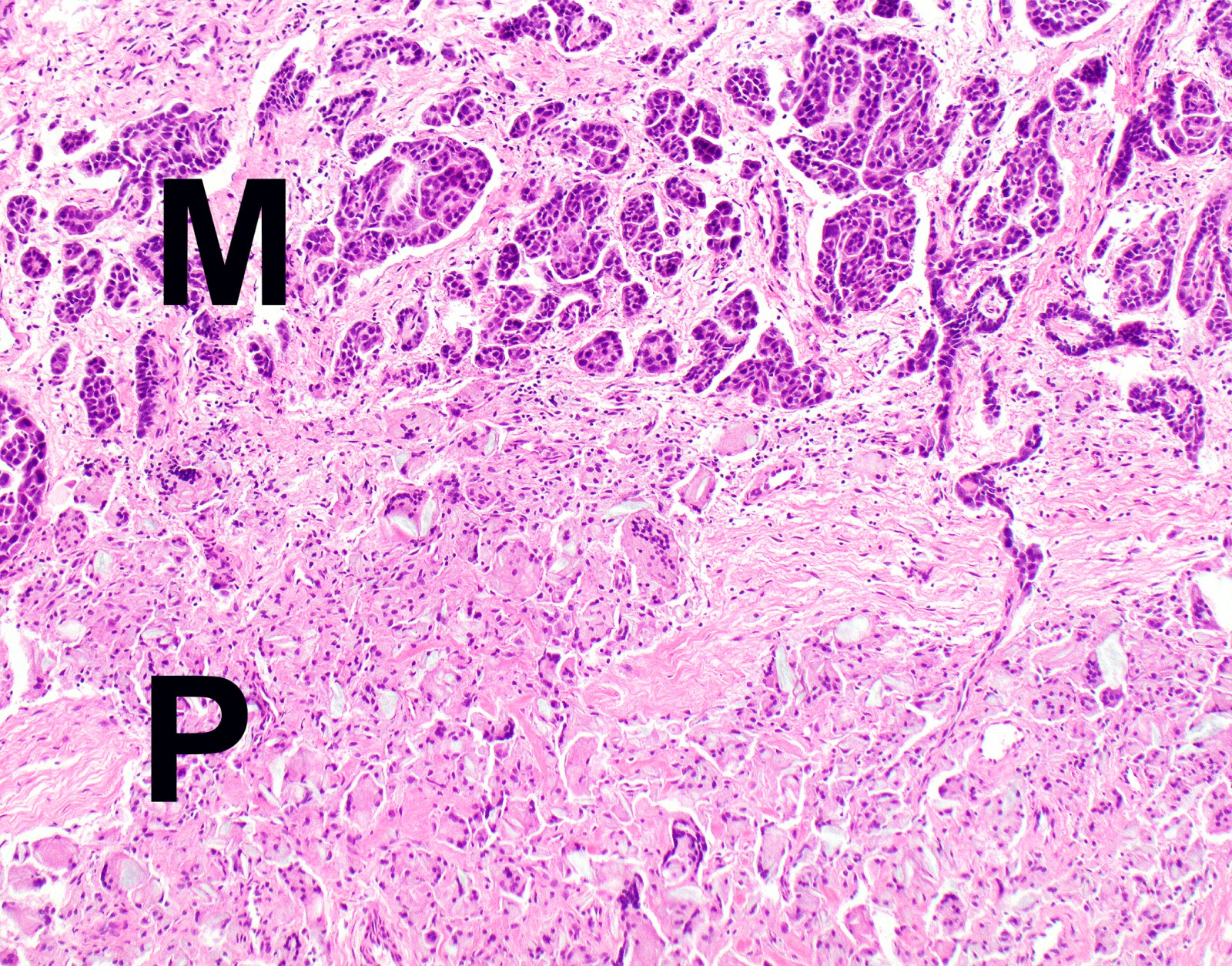
      • Diaphragmatic muscle involvement
      • Extension from visceral pleura into the underlying pulmonary parenchyma
    • pT3: tumor is locally advanced but potentially resectable; tumor involves all the ipsilateral pleural surfaces (parietal, mediastinal, diaphragmatic and visceral pleura) and has at least one of the following
      • Endothoracic fascia involvement
      • Mediastinal fat involvement
      • Solitary completely resectable focus of tumor extending into soft tissue of chest wall
      • Nontransmural involvement of the pericardium
    • pT4: tumor is locally advanced and technically unresectable; tumor involves all the ipsilateral pleural surfaces (parietal, mediastinal, diaphragmatic and visceral pleura) and has at least one of the following
      • Diffuse extension or multifocal masses in chest wall
      • Direct transdiaphragmatic extension to peritoneum
      • Direct extension to the contralateral pleura
      • Direct extension to mediastinal organs
      • Direct extension into spine
      • Extension to internal surface of the pericardium
      • Myocardium involvement
    Regional lymph nodes (pN)
    • pNX: cannot be assessed
    • pN0: no regional lymph node metastasis
    • pN1: metastases in the ipsilateral bronchopulmonary hilar or mediastinal (including the internal mammary, peridiaphragmatic, pericardial fat pad or intercostal) lymph nodes
    • pN2: metastases in the contralateral mediastinal, ipsilateral or contralateral supraclavicular lymph nodes
    Distant metastasis (pM)
    • pM0: no distant metastasis
    • pM1: distant metastasis
    Prefixes
    • y: preoperative radiotherapy or chemotherapy
    • r: recurrent tumor stage
    AJCC prognostic stage groups
    Stage group IA:  T1   N0   M0
    Stage group IB:  T2 - 3   N0   M0
    Stage group II:  T1 - 2   N1   M0
    Stage group IIIA:  T3   N1   M0
    Stage group IIIB:  T1 - 3   N2   M0
     T4   any N   M0
    Stage group IV:  any T   any N   M1
    Registry data collection variables
    • Histologic type
    • Age, sex
    • Performance status
    • Laboratory parameters (white blood cells, platelet count and hemoglobin)
    • Surgical resection with curative intent (pleurectomy / decortications, extended pleurectomy / decortications or extrapleural pneumonectomy)
    • For patients undergoing multimodality therapy, use of chemotherapy or radiotherapy
    • Comment: these are supplementary factors recommended for clinical care owing to their prognostic significance (J Thorac Oncol 2014;9:856)
    Histologic grade (G)
    • GX: cannot be assessed
    • G1: well differentiated
    • G2: moderately differentiated
    • G3: poorly differentiated
    • G4: undifferentiated
    Histopathologic type
    • Epithelioid
    • Biphasic (at least 10% of epithelioid and sarcomatoid)
    • Sarcomatoid
    • Desmoplastic
    Board review style question #1
    A 59 year old man presented with a 2 cm malignant pleural tumor with focal extension to the underlying lung parenchyma. Biopsy revealed well differentiated epithelioid malignant pleural mesothelioma confirmed by BAP1 immunostaining. What is the pT category using the AJCC / TNM 8th edition?

    1. pTX
    2. pT1
    3. pT2
    4. pT3
    5. pT4
    Board review style answer #1
    C. pT2. Extension of malignant mesothelioma from visceral pleura into the underlying pulmonary parenchyma qualifies as the pT2 category.

    Comment Here

    Reference: Pleura - Staging
    Board review style question #2
    Which of the following is a major prognostic factor of malignant mesothelioma (other than TNM variables)?

    1. Duration of asbestos exposure
    2. Family history
    3. Genetic / mutation signature
    4. PDL1 expression by microenvironment
    5. Tumor histology
    Board review style answer #2
    E. Tumor histology is the major prognostic factor beyond TNM (epithelioid tumors have better prognosis than sarcomatoid).

    Comment Here

    Reference: Pleura - Staging

    Well differentiated papillary mesothelial tumor (peritoneum)
    Definition / general
    • Well differentiated papillary mesothelioma (WDPM) is a neoplastic mesothelial proliferation of low malignant potential, which predominantly involves the peritoneum of young women
    Essential features
    • Most cases diagnosed incidentally during surgery for an unrelated indication
    • Composed of branching papillae lined by a single layer of bland cuboidal mesothelial cells
    • Invasion of underlying pre-existing tissues (e.g. fat, muscle, ovarian stroma) precludes the diagnosis (→ diagnosis of malignant mesothelioma)
    • BAP1 loss and CDKN2A (p16) homozygous deletion preclude the diagnosis (→ diagnosis of malignant mesothelioma)
    Terminology
    ICD coding
    • ICD-10: D19.9 - Benign neoplasm of mesothelial tissue, unspecified
    Epidemiology
    • F:M = ~5:1
    • Median age at diagnosis: ~40 years (range 7 - 75)
    Sites
    • Peritoneum is most commonly affected site
    • Rare cases involve the pleura or tunica vaginalis (Hum Pathol 2019;92:48Am J Surg Pathol 2004;28:534)
      • Cases in the pleura or tunica vaginalis more evenly distributed between men and women
      • Pleural cases diagnosed at older age (median ~60 years)
      • Pleural cases have higher rate of asbestos exposure (~50%)
    Pathophysiology
    Etiology
    • Asbestos exposure reported in only ~1% (causality not established)
    Clinical features
    • Most cases (> 90%) incidentally identified during abdominal surgery for an unrelated cause
    • Rare florid cases (< 10%) present with abdominal pain, weight loss or ascites
    Diagnosis
    • Tissue sampling followed by microscopic evaluation
    Radiology description
    • In patients with florid or symptomatic disease, computed tomography (CT) may show ascites or thickening of the bowel wall or mesentery
    Radiology images

    Images hosted on other servers:
    Computed tomography

    Computed tomography

    Prognostic factors
    • Most cases follow a benign clinical course
    • Recurrence rate in morphologically classical cases is low (< 5%)
    • Recurrence rate reportedly higher in cases with invasive foci in papillary cores and with higher disease burden (Am J Surg Pathol 2014;38:990, Eur J Surg Oncol 2019;45:371)
    • Fatal outcomes rare
    • Progression to malignant peritoneal mesothelioma is exceptionally rare (< 5%) and causality is unclear (may occur years to decades later)
    Case reports
    Treatment
    • Observation
    • Surgical cytoreduction and hyperthermic intraperitoneal chemotherapy performed in some cases with diffuse, symptomatic or recurrent disease (Ann Surg Oncol 2019;26:852)
    • Randomized clinical trials not available
    Gross description
    • Discrete nodular implants
    • Papillary fronds macroscopically evident in some (but not all) cases
    • Individual implants usually 0.2 - 2.0 cm but may be up to several centimeters (Am J Surg Pathol 2012;36:117)
    • Approximately 50% of cases are unifocal and 50% are multifocal (Ann Diagn Pathol 2019;38:43)
    • Ovarian or fallopian tube surface involved in a minority of cases
    Gross images

    Images hosted on other servers:
    Large peritoneal well differentiated papillary mesothelioma

    Large peritoneal
    well differentiated
    papillary mesothelioma

    Frozen section description
    • Papillary architecture (Am J Surg Pathol 2012;36:117)
    • Single nonstratified layer of low cuboidal cells with bland cytomorphology
    • No solid growth
    • No invasion of underlying tissues
    • Correlate with surgical findings, re: extent of peritoneal involvement
    Frozen section images
    Microscopic (histologic) description
    • Branching plump papillae lined by a single layer of bland cuboidal cells (Am J Surg Pathol 2012;36:117, Ann Diagn Pathol 2019;38:43, Histopathology 2013;62:805)
    • Minimal cytologic atypia; single small nucleoli may be present
    • Mitoses usually absent (or at most 1 mitosis per 10 high power fields)
    • Papillary cores may show myxoid change, edema, psammomatous calcifications, fibrosis, foamy macrophages or chronic inflammation
    • Invasion of underlying pre-existing tissues is absent
    • Unusual patterns
      • Some cases associated with a discrete adenomatoid tumor or multicystic mesothelioma (no apparent effect on prognosis)
      • Invasive foci composed of tubules, single cells, small solid nests or cords within papillary cores (see Prognostic factors)
    Microscopic (histologic) images

    Contributed by David B. Chapel, M.D. and Debra L. Zynger, M.D.
    Papillary architecture

    Papillary architecture

    Bland cytomorphology

    Bland cytomorphology

    Fibrosis of papillary cores

    Fibrosis of papillary cores

    Calretinin

    Calretinin

    WT1

    WT1

    D2-40

    D2-40


    BAP1

    BAP1

    Delicate papillae

    Hypocellular fibrovascular cores

    Bland mesothelium

    Virtual slides

    Images hosted on other servers:
    Frozen section

    Frozen section

    Well differentiated<br>papillary mesothelioma

    Well differentiated
    papillary mesothelioma

    Well differentiated<br>papillary mesothelioma<br>with invasive foci

    Well differentiated
    papillary mesothelioma
    with invasive foci

    Positive stains
    Electron microscopy description
    • Mesothelial cells lining papillary cores show (Am J Surg Pathol 2001;25:1304):
      • Long slender microvilli (length to diameter ratio > 10)
      • Abundant mitochondria and rough endoplasmic reticulum
      • Well formed desmosomes
    Molecular / cytogenetics description
    • All cases appear to harbor mutation in TRAF7 or CDC42 (Mod Pathol 2019;32:88)
      • TRAF7 mutations also found in adenomatoid tumors, suggesting biologic connection
    • BAP1 mutation or deletion exceptionally rare
    • Lack homozygous CDKN2A (p16) deletion
    • Abnormal karyotypes seen in a subset (Am J Surg Pathol 2014;38:990)
    Sample pathology report
    • Peritoneal nodule, biopsy:
      • Well differentiated papillary mesothelioma (0.6 cm) (see comment)
      • Comment: Microscopic examination shows branching papillae lined by bland cuboidal mesothelial cells. No mitoses are seen. These morphologic features are consistent with well differentiated papillary mesothelioma. Small unifocal lesions are generally regarded as benign, although recurrences are reported in rare cases. Clinical correlation is advised with appropriate follow up to exclude regrowth or recurrence.
    • Peritoneal lesion, excision:
      • Well differentiated papillary mesothelioma (2.0 cm) with invasive foci (see comment)
      • Comment: Microscopic examination reveals a bland mesothelial lesion with features of well differentiated papillary mesothelioma, including branching papillae lined by a single layer of bland cuboidal mesothelial cells. However, the papillary cores are infiltrated in few foci by nests and cords of bland mesothelial cells. There is no invasion of the underlying peritoneal fibroadipose tissue. Such lesions have been termed "well differentiated papillary mesothelioma with invasive foci" and there is evidence suggesting that such lesions have a higher rate of recurrence compared with banal well differentiated papillary mesothelioma. Clinical and radiographic correlation is advised with appropriate follow up to exclude recurrence.
    Differential diagnosis
    • Diffuse malignant peritoneal mesothelioma:
      • Diffuse involvement of the peritoneum
      • Infiltration of underlying pre-existing tissue is typical
      • At least moderate cytologic atypia in most cases
      • Increased mitotic activity (> 1 mitosis per 10 high power fields is typical)
      • Solid architecture may be present
      • BAP1 loss frequent (~70%)
      • May contain WDPM-like foci so thorough sampling is necessary
    • Localized malignant peritoneal mesothelioma:
      • Exceptionally rare diagnosis
      • Localized disease (resembling WDPM)
      • Complex papillary or solid architecture, identical to that seen in diffuse malignant peritoneal mesothelioma
      • At least moderate cytologic atypia in most cases
      • Increased mitotic activity (> 1 mitosis per 10 high power fields is typical)
      • May contain WDPM-like foci so thorough sampling is necessary
    • Reactive mesothelial hyperplasia:
      • History of surgery, inflammatory disease or cardiovascular disease
      • Reactive or inflammatory changes also seen in adjacent flat serosa
    • Serous borderline tumor:
      • Columnar cells with mixed morphology, including ciliated cells
      • Negative for calretinin
      • Positive for claudin4, MOC31, BerEP4 and other epithelial lineage markers
      • Positive for ER and PR
    Board review style question #1


    The unifocal lesion depicted in the photomicrograph was discovered incidentally during an exploratory laparoscopy in a 38 year old woman with endometriosis. It measured 0.6 cm on gross examination. On immunohistochemical stains, lesional cells were positive for calretinin (shown above) but negative for MOC31. Which of the following is true about the depicted entity?

    1. Immunohistochemical loss of BAP1 is seen in ~70% of cases
    2. Men are more frequently affected than women
    3. Most cases are causally linked to asbestos exposure
    4. Most cases of this entity result in patient death within 10 years
    5. WT1 expression is seen in virtually 100% of cases
    Board review style answer #1
    E. WT1 expression is seen in virtually 100% of cases. This is a well differentiated papillary mesothelioma.

    Comment Here

    Reference: Well differentiated papillary mesothelioma
    Board review style question #2
    Which of the following molecular alterations is most frequently seen in well differentiated papillary mesothelioma of the peritoneum?

    1. BAP1 missense mutation
    2. CDKN2A homozygous deletion
    3. NF2 homozygous deletion
    4. TP53 missense mutation
    5. TRAF7 mutation
    Board review style answer #2

    Well differentiated papillary mesothelioma tumor-pleura (pending)
    Table of Contents
    Definition / general
    Definition / general
    [Pending]
    Back to top
    Recent Pleura & peritoneum Pathology books

    Allen: 2015

    Cagle: 2015

    IARC: 2015

    IARC: 2020

    IARC: 2021

    Mukhopadhyay: 2023

    Nicholson: 2022

    Nucci: 2023

    Suster: 2022


    Find related Pathology books: cardiovascular, gynecologic, lung, mediastinum/serosa
    Image 01 Image 02