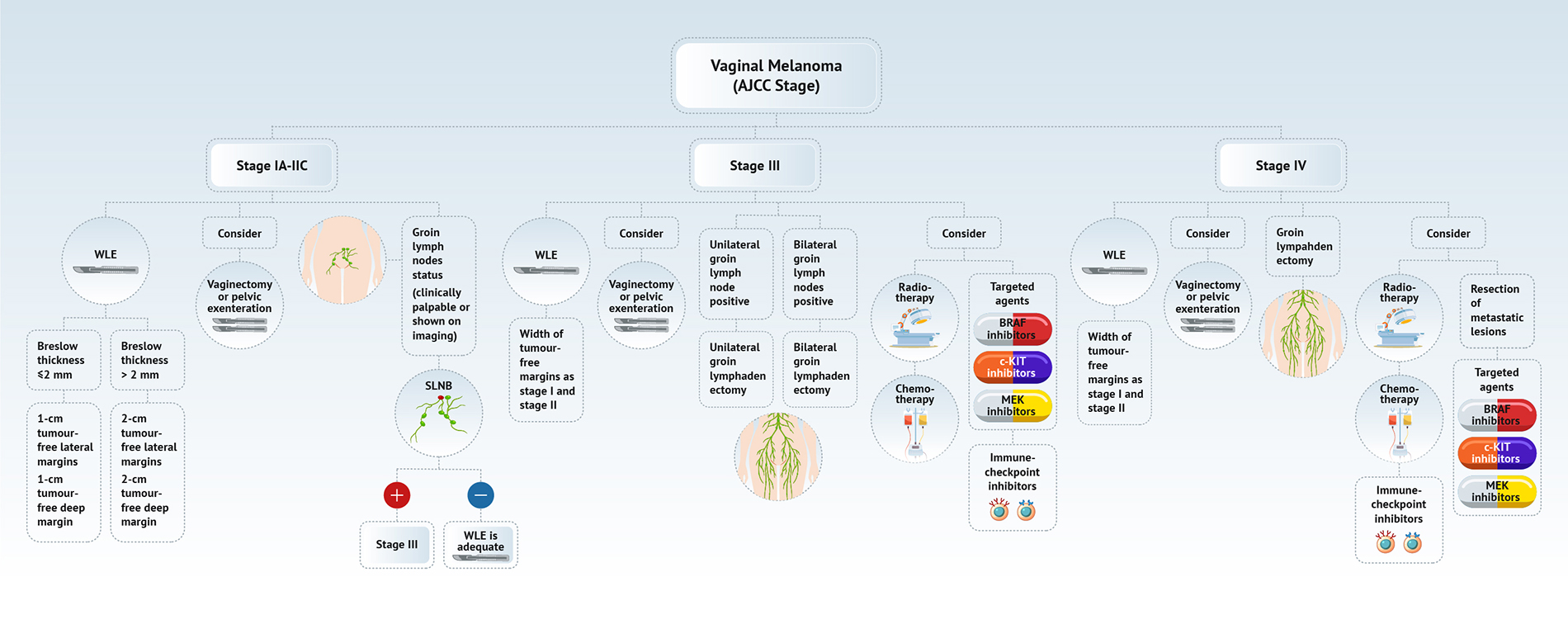
Superpage
Page views in 2023: 95
Page views in 2024 to date: 25

Adenosis
Definition / general
  • First described in 1877 by von Preuschen, vaginal adenosis refers to the persistence of Müllerian glandular epithelium in the vagina after birth
Essential features
ICD coding
  • ICD-10:
    • N89.8 - other specified noninflammatory disorders of vagina
    • N89.9 - noninflammatory disorder of vagina, unspecified
Epidemiology
Sites
  • Involves the upper third of the vagina (34% of diethylstilbestrol exposed cases), with the anterior wall more frequently involved than the posterior wall
  • Middle third (9% of cases) and lower third (2% of cases) of the vagina may be involved
Pathophysiology
  • Diethylstilbestrol causes irregularities in p63 gene expression, which determines whether Müllerian duct epithelium becomes uterine or vaginal cells and leads to vaginal adenosis
  • Mouse model of diethylstilbestrol exposure demonstrates vaginal adenosis and structural changes similar to actual changes observed in women (Hum Pathol 1982;13:190)
  • Pathophysiology of non-diethylstilbestrol related adenosis has not been determined conclusively; however, it may result from p63 gene expression changes in squamous epithelium or Müllerian remnants upon epithelial damage or from implantation of Müllerian derived columnar cells into vaginal lesions during menstruation
Etiology
  • Diethylstilbestrol is a synthetic nonsteroidal estrogen that profoundly affects the development of the vagina, uterus and fallopian tubes
  • Today, in the post-diethylstilbestrol era, vaginal adenosis usually occurs in women exposed to other hormones, drugs, trauma, inflammation, vulvovaginal involvement by Stevens-Johnson syndrome or toxic epidermal necrolysis (SJS / TEN), laser, radiation or chemotherapy (5-fluorouracil) (Rev Obstet Gynecol 2011;4:81)
Clinical features
Diagnosis
  • Lesions identified on physical examination
  • Lesions can be identified on colposcopic examination as mosaicism and punctation (Obstet Gynecol 1978;52:457)
  • Diagnosis confirmed on biopsy specimens with or without cytologic brushing of clinically suspicious lesions on colposcopy
Case reports
Treatment
  • In symptomatic patients, treatment with CO2 laser, simple excision or unipolar cautery may be considered (Lasers Surg Med 1983;3:23)
  • In patients with vulvovaginal involvement by Stevens-Johnson syndrome and toxic epidermal necrolysis, adenosis prevention includes intravaginal steroids and menstrual suppression ((luteinizing hormone releasing hormone [LHRH] agonists, combined oral contraceptives or progesterone only contraceptives) (Rev Obstet Gynecol 2011;4:81)
Clinical images

Images hosted on other servers:

Vaginal adenosis, posterior wall

Vaginal red granular patch

Gross description
  • Vaginal mucosa displays cysts or red granular spots or patches
  • In the setting of DES exposure, you may also find a flattened, shallow upper vagina with absent vaginal fornices around a hypoplastic cervix
Microscopic (histologic) description
  • 2 adult (differentiated) forms of adenosis have been described:
    • Mucinous: most common type of adenosis (62% of biopsy specimens); characterized by mucinous columnar cells that resemble those of the normal endocervical mucosa
    • Tuboendometrial: found in 21% of specimens; glands are lined by light and dark cells, often ciliated and resemble fallopian tube and endometrial gland cells
  • Embryonic: glands composed of low columnar or cuboid cells
  • Glands may be simple, complex, cystic or papillary and usually found in the lamina propria but also on the mucosal surface
  • Cysts lined by a single layer of columnar mucinous cells may resemble cervical nabothian cysts
  • Squamous metaplasia can be present and represents the process by which adenosis transforms and heals
  • Rarely, intestinal metaplasia may be seen
  • References: Mutter: Pathology of the Female Reproductive Tract, 3rd Edition, 2014, Kurman: Blaustein's Pathology of the Female Genital Tract, 6th Edition, 2011
Microscopic (histologic) images

Contributed by Leonel Maldonado, M.D.
Missing Image Missing Image Missing Image

Vaginal wall lesion biopsy

Missing Image Missing Image

Vaginal wall lesion biopsy


Missing Image Missing Image

Glands in vaginal mucosa

Missing Image

Mucinous glands below epithelium

Cytology description
  • Vaginal specimens are characterized by endocervical-like glandular cells or metaplastic squamous cells
  • Endometrioid glandular cells can also occur
Cytology images

Contributed by Leonel Maldonado, M.D.
Missing Image Missing Image

Benign appearing glandular cells

Videos

Vaginal adenosis diffuse, rare case

Sample pathology report
  • Vagina, posterior wall, biopsy:
    • Squamous mucosa with bland mucinous glands consistent with vaginal adenosis; negative for malignancy
Differential diagnosis
  • Clear cell adenocarcinoma (vagina):
    • May be confused with the glands of adenosis that undergo microglandular hyperplasia (small, uniform, crowded glands without mucin, nuclear pleomorphism and prominent nucleoli)
  • Endometriosis:
    • Presence of endometrial stroma with glands that much more closely resemble those of normal endometrium than the glands seen in adenosis
    • CD10 is useful to confirm the presence of endometrial stroma
  • Mesonephric (Wolffian) remnants:
    • Mesonephric tubules are lined by nonciliated, nonmucinous cuboid cells with dense, eosinophilic luminal secretions; these remnants are surrounded by a loose fibrovascular stroma that may contain smooth muscle fibers
  • Recurrent endometrial adenocarcinoma:
    • Glandular cells in vaginal cytology Papanicolaou tests in patients with hysterectomy for endometrial adenocarcinoma (Diagn Cytopathol 2012;40:138)
Board review style question #1

A 28 year old woman presents with persistent vaginal discharge and is found to have submucosal mucinous glands on a vaginal biopsy. What is the most likely scenario?

  1. Maternal grandmother's use of diethylstilbestrol (DES) caused in utero exposure for the patient’s mother
  2. Maternal use of DES caused in utero exposure for the patient
  3. The patient had prior treatment for bacterial vaginosis
  4. The patient had prior treatment for vaginal condyloma acuminata
  5. Use of DES by the patient
Board review style answer #1
D. The patient had prior treatment for vaginal condyloma acuminata. Vaginal application of 5-fluoruracil and vaginal CO2 laser therapy (common treatments for condyloma acuminata) are 2 known non-DES causes for vaginal adenosis. Answer C is incorrect because these are not common treatments for bacterial vaginosis. Answers A, B and E are incorrect because in utero exposure to diethylstilbestrol (DES) was the most common cause of vaginal adenosis for women born in the middle of the 20th century but DES exposure is not a concern in this patient who was born long after its discontinued use.

Comment Here

Reference: Vaginal adenosis
Board review style question #2
Which statement concerning vaginal adenosis is true?

  1. Inapparent on gross examination
  2. May be associated with microglandular hyperplasia
  3. Progresses to adenocarcinoma in a majority of the cases
  4. Usually manifests in prepubertal age
  5. Usually related to bacterial vaginosis
Board review style answer #2
B. May be associated with microglandular hyperplasia. Vaginal adenosis commonly appears as red nodules / red granular spots or patches and fails to stain with an iodine solution on colposcopy. Mosaicism and punctation are common findings during colposcopy. It commonly manifests as excessive mucous discharge in postpubertal age. Vaginal adenosis seldom progresses to cancer. Microglandular hyperplasia develops in foci of vaginal adenosis, especially after oral contraceptive use. Microglandular hyperplasia may be confused with clear cell adenocarcinoma (which is also diethylstilbestrol associated).

Comment Here

Reference: Vaginal adenosis

Aggressive angiomyxoma-vulva
Definition / general
  • Aggressive angiomyxoma is an infiltrative spindle cell neoplasm arising in the soft tissues of the lower genital tract, perineum and pelvis; approximately 30% recur locally but distant metastasis is exceptionally rare
Essential features
  • Unique to the soft tissues of the lower genital tract, pelvis and perineum
  • Infiltrative hypocellular myxoid lesion with bland spindle cells and prominent variably sized vessels
  • HMGA2 overexpressed by immunohistochemistry in > 90%
  • Local recurrence in ~ 30% but distant metastasis and death from disease are exceptionally rare
ICD coding
  • ICD-10: D48.1 - neoplasm of uncertain behavior of connective and other soft tissue
Sites
Pathophysiology
  • Histogenesis uncertain
  • May arise from site specific stromal cells with capacity for fibroblastic, myofibroblastic and smooth muscle differentiation (Cancer 1996;78:79)
Clinical features
Diagnosis
Radiology description
Radiology images

Images hosted on other servers:
Ultrasound and MRI Ultrasound and MRI Ultrasound and MRI Ultrasound and MRI Ultrasound and MRI Ultrasound and MRI

Ultrasound and MRI


MRI

MRI

Perineal, MRI

Perineal, MRI

Prognostic factors
Case reports
Treatment
Clinical images

Images hosted on other servers:
Paravaginal, intraoperative

Paravaginal, intraoperative

Perineal, intraoperative

Perineal, intraoperative

Vulvar, preoperative

Vulvar, preoperative

Vulvar, pre and postoperative

Vulvar, pre and postoperative

Gross description
Gross images

Contributed by @Andrew_Fltv on Twitter
Aggressive angiomyxoma

Aggressive angiomyxoma


Images hosted on other servers:
Cut surface

Cut surface

Microscopic (histologic) description
  • Unencapsulated and locally infiltrative (Am J Surg Pathol 1983;7:463, Hum Pathol 1985;16:621, Am J Dermatopathol 1993;15:446, Histopathology 1997;30:3)
  • Hypocellular (though focally increased cellularity is common) (Histopathology 1997;30:3)
  • Tumor cells:
    • Spindle to stellate with delicate cytoplasmic processes
    • Bland chromatin with small nucleoli
    • Mitoses rare or absent
  • Tumor stroma:
    • Myxoid stroma with scattered delicate collagen fibers
    • Stromal mucin positive for Alcian blue
    • Stroma peripherally entraps fat, nerves and muscle
    • Extravasated red blood cells common (Histopathology 1997;30:3)
    • Necrosis absent
  • Vasculature:
    • Conspicuous haphazard dilated capillaries
    • Scattered large, thick walled (medial hypertrophy) or hyalinized vessels
    • Vessels are nonanastomosing but may cluster together
    • Stromal smooth muscle bundles cluster around tumor vessels (Cancer 1996;78:79, Histopathology 1997;30:3)
  • Recurrent lesions may show increased cellularity, increased vasculature and dense stromal collagen
  • Rare cases may show minor foci with morphologic features of angiomyofibroblastoma (Histopathology 1997;30:3)
Microscopic (histologic) images

Contributed by David B. Chapel, M.D. and @Andrew_Fltv on Twitter
Infiltrative growth

Infiltrative growth

Entrapped adipose tissue

Entrapped adipose tissue

Extravasated red blood cells

Extravasated red blood cells

Thick walled vessels

Thick walled vessels

Myoid bundles

Myoid bundles

Entrapped nerve

Entrapped nerve


Bland cytology

Bland cytology

Aggressive angiomyxoma Aggressive angiomyxoma Aggressive angiomyxoma

Aggressive angiomyxoma

HMGA2

HMGA2

Virtual slides

Images hosted on other servers:
Aggressive angiomyxoma

Aggressive angiomyxoma

32 year old woman with a 6 cm vulvar mass

32 year old woman with a 6 cm vulvar mass

Aggressive angiomyxoma

Aggressive angiomyxoma

Positive stains
Electron microscopy description
Molecular / cytogenetics description
Molecular / cytogenetics images

Images hosted on other servers:
FISH, karyotype

FISH, karyotype

Videos

Histopathology

Sample pathology report
  • Vulva, mass, wide local excision:
    • Aggressive angiomyxoma with involvement of the deep margin (see comment)
    • Comment: Microscopic examination reveals a poorly circumscribed hypocellular lesion with myxoid stroma and abundant vasculature, infiltrating fibroadipose tissue. By immunohistochemistry, lesional stromal cells are positive for desmin, ER, PR and HMGA2. The morphologic and immunophenotypic findings are most consistent with aggressive angiomyxoma. Although distant metastasis is exceptionally rare, approximately 30% of such lesions recur locally. Clinical follow up is advised.
Differential diagnosis
Additional references
Board review style question #1

A 35 year old woman presented to her gynecologist with a complaint of pelvic discomfort and a slowly growing, ill defined vulvar mass. Radiology showed a vascular lesion extending from the vulva into deep pelvic soft tissues. An excision was performed. A representative photomicrograph of the lesion is shown. By immunohistochemistry, the lesional stromal cells were positive for desmin, ER and PR. Which of the following statements about this lesion is true?

  1. Approximately 50% of patients develop distant metastases
  2. FISH reveals a characteristic DDIT3 rearrangement
  3. HMGA2 is positive (overexpressed) by immunohistochemistry
  4. Immunohistochemistry for S100 is characteristically positive
  5. Less than 10% of patients experience local recurrence
Board review style answer #1
C. HMGA2 is positive (overexpressed) by immunohistochemistry. This is an aggressive angiomyxoma.

Comment Here

Reference: Aggressive angiomyxoma-vulva
Board review style question #2
Which of the following most accurately describes the expected immunophenotype of aggressive angiomyxoma?

  1. Desmin, ER, PR, HMGA2 positive; S100 negative
  2. S100, EMA, NF positive; ER, PR negative
  3. SMA, CD34 positive; ER, PR negative
  4. SMA, desmin, caldesmon positive; S100 negative
  5. SMA, ER, PR positive; Rb negative (lost)
Board review style answer #2
A. Desmin, ER, PR, HMGA2 positive; S100 negative

Comment Here

Reference: Aggressive angiomyxoma-vulva

Anatomy & histology - vulva, vagina & female urethra
Definition / general
  • Vulva constitutes the portion of female genitalia that is external to the hymen
  • Vagina is a fibromuscular tube that extends from the vestibule of vulva to uterine cervix
  • Female urethra extends from the bladder to midurethra and exits the body between clitoris and vagina
Essential features
  • Vulva is composed of mons pubis, clitoris, labia minora, labia majora, vulvar vestibule, vestibulovaginal bulbs, urethral meatus, hymen, Bartholin and Skene glands and ducts, vaginal introitus
  • Vagina extends from vulva to uterine cervix and is derived from paired Müllerian ducts
Terminology
  • Vulva
    • Lies external to hymen and is limited by mons pubis anteriorly, anus posteriorly and inguinal gluteal folds laterally
  • Vagina
    • Fibromuscular canal that extends from the vestibule of vulva, between labia minora, to the uterine cervix
    • Wolffian (mesonephric) duct, also known as Gartner duct, runs deeply along lateral vaginal walls
    • Lymphatic drainage: external iliac nodes (upper third of the vagina), the common and internal iliac nodes (middle third) and the superficial inguinal and perirectal nodes (lower third)
  • Female urethra
    • Fibromuscular tube that takes urine from the urinary bladder to the exterior through the external urethral meatus
Physiology / embryology
  • Germ cells from yolk sac migrate to the urogenital ridge, forming the epithelium and stroma of the gonads; genital tubercle becomes the clitoris and the parallel ridges become the labia minora
  • Urorectal septum divides the cloaca into the urogenital sinus and anal canal; degeneration of the central portion of the urogenital membrane forms the hymen opening
  • Lateral Müllerian ducts (paramesonephric ducts) give rise to upper vagina while the lower vagina is formed by the urogenital sinus
    • Vaginal vestibule develops by the joining of the distal vagina and urogenital sinus
    • Originates from endoderm, except near the urethra (ectoderm)
    • Vestibular line of Hart marks the boundary between these tissues
  • Epithelium of female urethra is derived from endoderm of the urogenital sinus while the surrounding connective tissue and smooth muscle tissue is derived from splanchnic mesenchyme (Sadler: Langman's Medical Embryology, 15th Edition, 2023, Am J Obstet Gynecol 1976;126:769)
Diagrams / tables

Images hosted on other servers:

Hart line

Female urethra: embryology and anatomy

Clinical images

Images hosted on other servers:

Labia majora

Vestibule

Gross description
  • Vulva
    • Mons pubis
      • Anteriormost region of vulva and is anatomically located over the prominence of pubic symphysis
    • Hymen
      • Corresponds to the distalmost extent of vagina and posterior aspect of vulvar vestibule
    • Clitoris
      • Erectile tissue similar to corpora cavernosa of penis and is located anterior to frenulum at the junction of labia minora
    • Labia majora
      • Form the lateral boundaries of the vulva
      • Fuse anteriorly into mons pubis
      • Posteriorly terminate 3 - 4 cm anterior to the anus where they are united by posterior commissure or fourchette
    • Labia minora
      • Are medial to labia majora and lateral to vulvar vestibule
      • Anteriorly, labia minora divide into 2 parts; one part passes over clitoris to form prepuce and the other joins beneath clitoris and forms frenulum
      • Posteriorly, they blend with medial surfaces of labia majora
    • Hart line
      • Lies at the inferior junction between vulvar vestibule and perineal skin
    • Vestibule
      • Area between the hymen (anteriorly), Hart line (posterolaterally) and labia minora (anterolaterally)
      • Includes vaginal opening and urethral orifice
      • Structures found in vestibule include major vestibular (Bartholin) glands, minor vestibular glands, periurethral (Skene) glands, urethra
    • Bartholin glands
      • Correspond to bulbourethral glands in male
      • These are mucin producing glands and are located posterolaterally in the vulva
    • Minor vestibular glands
      • Correspond to penile glands of Littre
      • Concentrically located within the vestibule
  • Vagina
    • Posterior to urinary bladder (from which it is separated by fibroadipose tissue)
    • Anterior to rectum (from which is separated by rectouterine space in the upper 25%, rectovaginal septum in the middle portion and sphincter musculature in the distal portion of anal canal)
  • Female urethra
    • Extends from the bladder to the vestibule of the vagina to its opening posterior to the clitoris
    • Measures 4 cm in length
    • Striated muscle of the urogenital diaphragm forms the external voluntary sphincter as the urethra penetrates it
  • Reference: StatPearls: Anatomy, Abdomen and Pelvis - Female External Genitalia [Accessed 21 August 2023]
Microscopic (histologic) description
  • Hymen
    • Nonkeratinized stratified squamous epithelium
  • Labia majora
    • Composed of keratinized stratified squamous epithelium with hair follicles and eccrine, apocrine and sebaceous glands
  • Labia minora
    • Composed of keratinized stratified squamous epithelium, usually no adnexa
  • Stroma
    • Composed of stromal cells that can be spindled, stellate, fusiform and may have large multilobated nuclei
  • Vestibule
    • Lined by nonkeratinized squamous epithelium, may be glycogenated
  • Minor vestibular gland
    • Superficial glands lined by mucin secreting columnar cells that merge with squamous epithelium of the vestibule
    • Open directly onto the surface
  • Glands
    • Apocrine glands (scent glands)
      • Identical to those of axillae, breast and perianal regions
      • Height of secretory cells varies
      • Lumina of glands are large compared to lumina of eccrine glands
    • Eccrine glands (sweat glands)
      • Primarily involved in heat regulation
      • Lined by layer of epithelial cells that contain eosinophilic cytoplasm
    • Sebaceous glands
      • Alveolar, holocrine glands that do not contain lumina
      • Each gland is composed of several lobules
      • Cells in each lobule form a delicate network filled with fat
    • Skene glands
      • Periurethral glands analogous to prostate
      • Mucus secreting columnar epithelium merges with duct urothelium, then stratified squamous epithelium of vestibule
  • Vagina
    • Lined by nonkeratinized stratified squamous epithelium and is composed of basal, parabasal, intermediate and superficial cell layers
      • Basal cell layer is composed of a single layer of columnar cells with high N:C ratio
      • Parabasal layer lies above the basal layer and has cells with higher N:C ratio than the more superficial layers
      • Intermediate cell layer has more abundant cytoplasm, which can be glycogenated
      • Superficial cell layer appears flattened with cells showing pyknotic nuclei (J Mol Med (Berl) 2021;99:531)
    • Lamina propria (subepithelial stroma) is composed of loose connective tissue with elastic fibers, rich venous and lymphatic networks, spindle to stellate and some multinucleated stromal cells
    • Muscle
      • Outer longitudinal and thin inner circular layer of smooth muscle
    • Adventitia is composed of inner dense connective tissue layer and outer loose connective tissue layer containing peripheral nerves, blood vessels and lymphatics
    • Wolffian (mesonephric) duct, also known as Gartner duct, runs deeply along lateral vaginal walls; single small duct surrounded by a cluster of small glands lined by cuboidal epithelium with eosinophilic secretion in lumen
    • Maturation index
      • Ratio of parabasal to intermediate to superficial cells of vaginal epithelium (sampled at middle third of lateral vaginal wall)
      • Sample is often obtained simultaneous with Pap smear to detect hormonal effects in menopausal and postmenopausal women
      • Increased maturation in vaginal epithelium may be due to estrogenic effect of tamoxifen (Clin Exp Obstet Gynecol 1998;25:121)
  • Female urethra
    • Lined by urothelium (proximal two - thirds), stratified and pseudostratified columnar and squamous epithelium (distal third)
      • Basal layers composed of either low columnar or cuboidal cells, followed by several layers of polyhedral cells
      • Most superficial layer is composed of round, dome shaped umbrella cells that are occasionally multinucleated and flattened according to amount of distention
      • Nonkeratinized squamous epithelium: cuboidal (deepest), polymorphous (middle), squamous / flattened (superficial) (J Urol 1987;138:775)
    • Periurethral (Skene) glands (homologous to the prostate gland) open into the distal portion
      • Lined by columnar or cuboidal epithelium with surrounding connective tissue and smooth muscle
    • Minor vestibular glands (homologous to glands of Littre in males) open along the entire length
      • Tubuloacinar mucinous glands with uniform, pale eosinophilic to clear cytoplasm and basally flattened nuclei
Microscopic (histologic) images

Contributed by Pooja Srivastava, M.D.
Labia majora overview

Labia majora overview

Labia minora overview

Labia minora overview

Vagina overview

Vagina overview

Vagina epithelium, high power

Vagina epithelium


Vagina lamina propria Vagina lamina propria, high power

Vagina lamina propria

Vagina muscular layer

Vagina muscular layer

Virtual slides

Images hosted on other servers
Vulva normal histology

Vulva normal histology

Videos

Normal histology of vagina

Anatomy of female urethra

Histology of female urethra

Board review style question #1

What is the site of biopsy shown in this image?

  1. Cervix
  2. Labia majora
  3. Labia minora
  4. Uterus
  5. Vagina
Board review style answer #1
B. Labia majora. Labia majora is composed of keratinized stratified squamous epithelium with hair follicles and eccrine, apocrine and sebaceous glands. Answer A is incorrect because the ectocervix is lined by stratified squamous epithelium overlying fibrous stroma and endocervix is lined by simple, columnar mucinous epithelial cells. Answer C is incorrect because labia minora is lined by keratinized stratified squamous epithelium, without any adnexa. Answer D is incorrect because uterus is lined by endometrial glands, stroma, myometrium and serosa. Answer E is incorrect because vagina has nonkeratinized stratified squamous epithelium and is composed of basal, parabasal, intermediate and superficial cell layers.

Comment Here

Reference: Anatomy & histology - vulva, vagina & female urethra
Board review style question #2
What is the epithelium of the labia minora classified as?

  1. Keratinized stratified squamous epithelium with adnexa
  2. Keratinized stratified squamous epithelium without adnexa
  3. Nonkeratinized stratified squamous epithelium with adnexa
  4. Simple, columnar mucinous epithelial cells
Board review style answer #2
B. Keratinized stratified squamous epithelium without adnexa. This describes the lining of labia minora. Answer A is incorrect because it describes the lining of the labia majora. Answer C is incorrect because the epithelium of labia minora is keratinized and without adnexa. Answer D is incorrect because this describes the lining of endocervix.

Comment Here

Reference: Anatomy & histology - vulva, vagina & female urethra

Angiomyofibroblastoma
Definition / general
  • Angiomyofibroblastoma is a benign, site specific, soft tissue tumor of the lower genital tract with alternating hypo and hypercellular zones, bland spindle to epithelioid cells and abundant vessels
Essential features
  • Benign lower genital tract tumor, seen predominantly in women
  • Alternating hypo and hypercellular zones, comprised of bland plump spindle to epithelioid cells clustered around abundant small, thin walled vessels
  • No specific immunophenotypic or molecular features reported
  • Local excision curative; recurrences exceptionally rare
ICD coding
  • ICD-10:
    • D21.9 - benign neoplasm of soft tissue, unspecified
    • D23.9 - benign neoplasm of skin, unspecified
    • D28.0 - benign neoplasm of vulva
    • D28.1 - benign neoplasm of vagina
Epidemiology
Sites
Pathophysiology
Clinical features
Diagnosis
  • Diagnosis typically follows complete local excision of a clinically benign appearing mass
Radiology description
Radiology images

Images hosted on other servers:
Vaginal angiomyofibroblastoma: ultrasound Vaginal angiomyofibroblastoma: ultrasound

Vaginal ultrasound

Vaginal angiomyofibroblastoma: MRI Vaginal angiomyofibroblastoma: MRI

Vaginal MRI

Vulvar angiomyofibroblastoma: MRI

Vulvar MRI

Prognostic factors
Case reports
Treatment
Clinical images

Images hosted on other servers:
Intraoperative images Intraoperative images Intraoperative images Intraoperative images Intraoperative images

Intraoperative images

Preoperative image

Preoperative image

Gross description
Gross images

Images hosted on other servers:
Gross specimen Gross specimen

Gross specimen

Microscopic (histologic) description
  • Well circumscribed, noninfiltrative
  • Fibrous pseudocapsule in a subset (Am J Surg Pathol 1992;16:373)
  • Characteristic alternating hypo and hypercellular zones (Am J Surg Pathol 1992;16:373)
  • Tumor cells:
    • Spindle to epithelioid to plasmacytoid myofibroblastic cells
    • Typically bland chromatin and inconspicuous nucleoli; scattered mildly atypical cells in a minority (Am J Surg Pathol 1992;16:373)
    • Bland multinucleated cells common (Am J Surg Pathol 1992;16:373)
    • Mitoses rare or absent; no atypical mitoses
  • Tumor stroma and vasculature:
    • Lacks prominent stromal mucin
    • Hypocellular foci show edematous stroma with scattered fine to thick bands of stromal collagen
    • In hypocellular foci, tumor cells appear randomly distributed
    • In hypercellular foci, tumor cells congregate around small, irregularly distributed, thin walled vessels
    • Occasional larger vessels interspersed
    • Mast cells typically conspicuous
    • Adipocytic differentiation in ~ 25 - 50% (Mod Pathol 1996;9:284, Hum Pathol 1997;28:1046)
  • Rare cases with predominant adipocytic differentiation = lipomatous variant of angiomyofibroblastoma (Hum Pathol 1997;28:1046, Hum Pathol 2014;45:1647, Int J Gynecol Pathol 2015;34:204)
  • Rare tumors show mixed features of angiomyofibroblastoma and aggressive angiomyxoma; should be managed like aggressive angiomyxoma (Histopathology 1997;30:3)
  • Sole case with sarcomatous transformation showed hypercellular foci with cytologic atypia, increased mitoses and lymphovascular invasion (Am J Surg Pathol 1997;21:1104)
Microscopic (histologic) images

Contributed by David B. Chapel, M.D.
Alternating cellularity

Alternating cellularity

Spindle cells

Spindle cells

Epithelioid cells

Epithelioid cells

Multinucleated cells

Multinucleated cells

Virtual slides

Images hosted on other servers:
Vulvar: focal adipocytic differentiation

Vulvar, focal adipocytic differentiation

Vulvar: lipomatous variant

Vulvar, lipomatous variant

Vaginal: focal adipocytic differentiation

Vaginal, focal adipocytic differentiation

Electron microscopy description
  • Features consistent with myofibroblastic differentiation (Am J Surg Pathol 1992;16:373)
    • Stromal cells surrounded by incomplete basal lamina
    • Well developed, rough endoplasmic reticulum and Golgi apparatus
    • Abundant intermediate filaments and pinocytotic vessels
    • Nucleus with delicate heterochromatin
Molecular / cytogenetics description
Sample pathology report
  • Vulva, mass, local excision:
    • Angiomyofibroblastoma (3.5 cm) (see comment)
    • Comment: Margins are negative.
Differential diagnosis
  • Aggressive angiomyxoma:
    • Typically large (most > 10 cm)
    • Infiltrative
    • More uniformly hypocellular
    • No perivascular congregation of cells
    • Tumor cells more uniformly spindled (versus epithelioid)
    • Perivascular myoid bundles characteristic
    • Red blood cell extravasation common
    • HMGA2 positive
    • FISH shows HMGA2 rearrangement in a subset (Genes Chromosomes Cancer 2007;46:981)
  • Leiomyoma:
    • More uniformly cellular
    • Tumor cells show cigar shaped nuclei and eosinophilic cytoplasm
    • Larger, thick walled vessels
    • Often has coagulative necrosis
    • SMA, desmin and caldesmon positive
  • Solitary fibrous tumor:
    • More common in extragenital sites
    • Prominent staghorn (hemangiopericytoma-like) vessels
    • Thick, keloid-like stromal collagen bands
    • CD34 and STAT6 positive
    • Intrachromosomal NAB2-STAT6 translocation on chr 12
  • Superficial angiomyxoma:
    • More uniformly hypocellular
    • No perivascular congregation of cells
    • Prominent stromal mucin, often with acellular mucin clefts / pools
    • Stromal neutrophils conspicuous
Board review style question #1

On a routine exam, a 46 year old woman was found to have a 3 cm, painless mass on her labia majora. Her gynecologist suspected a Bartholin cyst and performed a conservative local excision. A representative photomicrograph from the lesion is shown. The lesion was grossly and microscopically well circumscribed but the surgical margin was focally positive. By immunohistochemistry, the tumor cells were positive for desmin, ER and PR. Which of the following statements about this lesion is true?

  1. Approximately 10% experience distant metastasis
  2. Conservative local excision is considered curative
  3. HMGA2 IHC is positive in ~ 90%
  4. If surgical margins are positive, local recurrence risk exceeds 50%
  5. Stromal neutrophils are found in virtually all such lesions
Board review style answer #1
B. Conservative local excision is considered curative. This is an angiomyofibroblastoma

Comment Here

Reference: Angiomyofibroblastoma
Board review style question #2

Which of the following statements about angiomyofibroblastoma is true?

  1. Abundant, thick walled hyalinized vessels are characteristic
  2. At low magnification, alternating hypo and hypercellular zones are characteristic
  3. Men and women are affected in approximately equal numbers
  4. Rb IHC is negative, reflecting underlying RB1 / FOXO1 (chr 13q14) deletion
  5. The vagina is affected approximately 3 times as often as the vulva
Board review style answer #2
B. At low magnification, alternating hypo and hypercellular zones are characteristic

Comment Here

Reference: Angiomyofibroblastoma

Atypical melanocytic nevi of the genital type-vulva
Definition / general
  • Rare benign melanocytic lesion, most commonly involving the vulva of young women with concerning histologic features that may overlap with melanoma but are associated with benign behavior (J Cutan Pathol 2008;35:24)
  • Belongs to the general group nevi of special sites
  • Commonly also part of pigmented lesions of the vulva (J Cutan Pathol 2008;35:24)
Essential features
  • Nonmelanoma histologic diagnosis
Terminology
  • Atypical melanocytic nevi of genital type (AMNGT)
  • Atypical genital nevi (AGN)
  • Nevi with site related atypia
  • Nevi of special sites
ICD coding
  • ICD-O
    • 8720/0 - atypical melanocytic nevus of genital type
  • ICD-10
    • D28 - benign neoplasm of other and unspecified female genital organs
    • D39 - neoplasm of uncertain behavior of female genital organs
  • ICD-11
    • EG9Y - skin disorders involving other specific body regions
    • EG9Z - skin disorders involving certain specific body regions, unspecified
    • Specific anatomy (use additional code, if desired)
      • 2F33 - benign neoplasm of other or unspecified female genital organ
      • XA78U5 - vulva
        • XA11L9 - labia of vulva
        • XA1A52 - vulval vestibule
        • XA0565 - posterior fourchette of vulva
          • Frenulum of labia minora
      • XA4851 - clitoris
      • XA10Z0 - mons pubis
Epidemiology
Sites
Etiology
  • Largely unknown but associated with anatomic milk line, including axillae, breasts, periumbilical region and groin (Am J Surg Pathol 2008;32:51)
Clinical features

Table 1: Clinical and histologic differences between atypical genital nevus and vulvar melanoma (adapted from Hoang: Melanocytic Lesions - A Case Based Approach, 1st Edition, 2014)
Proposed diagnosis Atypical genital nevus of special anatomic site Vulvar melanoma
Age Premenopausal, young adult Postmenopausal
Size < 1 cm > 1 cm
Delineation Well circumscribed Infiltrative
Symmetry Present Absent
Lateral extension of junctional component Focal Present
Lentiginous junctional component Focal Present
Junctional nests Discohesive Confluent
Retraction artifact Present Absent
Ulceration Absent or due to trauma Often present
Pagetoid upward spread Focal, central, inconspicuous Prominent
Cytologic atypia Superficial, mild to moderate Deep, moderate to severe
Dermal mitosis Rare and superficial Conspicuous, atypical, deep
Dermal maturation Present Absent
Melanin pigmentation Coarse, uniform Fine, irregular
Dermal fibrosis Broad zone of superficial coarse dermal fibrosis Regression type
Diagnosis

Table 2: Distinguishing vulvar nevi, melanosis and melanoma (adapted from J Am Acad Dermatol 2014;71:1241)
Reassuring features suggestive of a benign process Concerning features for possible malignancy
Clinical
  • Age at diagnosis < 50 years
  • Symmetric, uniformly pigmented, macular or papular lesion with regular borders
  • Measures < 1 mm in diameter
  • Associated with genodermatoses
  • Age at diagnosis > 50 years
  • Asymmetric, nonuniformly pigmented, elevated lesion with irregular borders
  • Measures > 0.7 mm in diameter
  • Associated bleeding, pruritus or discharge
Dermoscopy
  • Variable appearance, including cobblestone, globular, ring-like, reticular-like, homogeneous, parallel or mixed
  • Variable color (gray, white, or blue) with structureless zones, irregular globules or dots and atypical vessels
Reflectance confocal microscopy
  • Draped or ringed polycyclic papillae
  • Hyper refractive cells around the papillae
  • Sparse dendritic cells
  • Increased cellularity with atypical cells and disordered architecture
Prognostic factors
Case reports
Treatment
  • Simple excision is usually sufficient
Clinical images

Contributed by José Alberto Fonseca Moutinho, M.D.
Nevus

Nevus


Images hosted on other servers:

Irregular dots on the periphery of the lesion

Pigmented lesion

Microscopic (histologic) description
  • Symmetric lesion with sharp demarcation and dermal maturation (J Cutan Pathol 2008;35:24)
  • 3 histologic patterns described by Clark (Hum Pathol 1998;29:S1)
    • Nested: oval, typically large nests, perpendicular or parallel to dermoepidermal junction
    • Discohesive nests: nearly contiguous, forming a band that separates the epidermis from the mature dermal melanocytes
    • Crowded: closely apposed, ill defined nests and single cells obscuring the dermal epidermal junction
  • Usually mild to moderate uniform cytologic atypia (J Cutan Pathol 2008;35:24)
  • Single cell growth with focal pagetoid spread may be present but is usually located in the center of the lesion (J Cutan Pathol 2008;35:24)
  • Adnexal spread may be present (Hum Pathol 1998;29:S1)
  • Dense eosinophilic fibrosis in the superficial dermis (Hum Pathol 1998;29:S1, J Cutan Pathol 2008;35:24)
  • Intradermal component with maturation is often present (J Cutan Pathol 2008;35:24)
  • Nevi within lichen sclerosus can appear more atypical
  • See table 1
Microscopic (histologic) images

Contributed by Anna Sarah Erem, M.D. and Gulisa Turashvili, M.D., Ph.D.
Atypical melanocytic proliferation Atypical melanocytic proliferation

Atypical melanocytic proliferation

Atypical melanocytic proliferation Atypical melanocytic proliferation

Atypical melanocytic proliferation

Atypical melanocytic proliferation Atypical melanocytic proliferation

Atypical genital nevi


Positive stains
Negative stains
Molecular / cytogenetics description
  • BRAF mutation is the most common alteration in melanocytic nevi of genital skin but not in vulvar melanomas (N Engl J Med 2015;373:1926, J Invest Dermatol 2016;136:1858)
  • Fluorescence in situ hybridization (FISH) or comparative genomic hybridization (CGH) may be considered in very challenging cases to rule out melanoma
Videos

Atypical nevi and nevi of special sites by Dr. Phillip McKee

Atypical nevi and melanoma by Dr. Steven Wang

Sample pathology report
  • Vulva, labia minora, excisional biopsy:
    • Atypical melanocytic nevus of the genital type, compound pattern, excised entirely
Differential diagnosis
  • Melanoma:
    • May be most challenging to differentiate from superficial spreading subtype of melanoma (SSM)
    • Atypical melanocytes in the epidermis as single cells or forming nests with pagetoid spread
    • Dermal component shows no maturation with cells forming sheets, nests, cords, single cells and rarely fascicles
    • S100, SOX10 and nerve growth factor receptor (NGFR) are the most sensitive markers in visualization of invasive growth
    • PRAME shows diffuse staining
    • Melanomas of vulva usually lack BRAF mutation (Br J Dermatol 2010;162:677)
  • Pigmented epithelioid melanocytoma (PEM) of the vulva:
    • PEM family consists of multiple, usually slow growing, distinct histologic melanocytic entities with potential to metastasize but with a better prognosis than melanoma
    • Infiltrative deep dermal tumor that may involve subcutis
    • Hypercellular tumor with cells ranging from medium sized epithelioid cells to large epithelioid cells and spindled cells
    • Low mitotic activity
    • PRKAR1A loss in 67% of PEMs (Am J Surg Pathol 2017;41:1333, Am J Surg Pathol 2019;43:480)
  • Dysplastic nevus of vulva:
    • Differentiation requires clinical pathologic correlates
    • Presence of junctional shoulders; extension of junctional component at least 3 rete ridges beyond the dermal component
    • Superficial nests are usually very similar and may show focal bridging or coalescence of the nests
    • Elongation and bridging of the rete ridges with nests
    • Melanocytes may scatter suprabasally (confined to the lower epidermal layer and centrally)
    • 2 tier grading of cytologic atypia is recommended by World Health Organization (WHO) classification and is largely based on nuclear features (Hum Pathol 1999;30:500)
Board review style question #1
A 28 year old woman presented to the clinic with a 0.6 cm, flat, dark brown lesion with an irregular border on the labia majora. The histologic findings of the shave biopsy demonstrated a relatively symmetric, broad melanocytic proliferation with large oval nests with retraction perpendicular to the dermal epidermal junction, focal fusion and mild cytologic atypia. Dermal maturation is evident. What is the most likely molecular alteration in this lesion?

  1. ALK fusion
  2. BRAF mutation
  3. Homozygous deletion of CDKN2A
  4. PTEN mutation
Board review style answer #1
B. BRAF mutation. The majority of atypical melanocytic nevi of genital type (AMNGT) have a driver mutation in BRAF. Answer A incorrect because ALK fusion is more characteristic of Spitz nevi. Answers C and D are incorrect because those alterations are specific to superficial spreading melanoma.

Comment Here

Reference: Atypical melanocytic nevi of genital type-vulva
Board review style question #2

A 16 year old girl presented with her parents to the OBGYN clinic with an irregular, dome shaped, dark pigmented papule on the mons pubis. A biopsy demonstrated large, ill defined nests with focal retraction. What is the most likely interpretation of the immunohistochemical stains for this patient's lesion?

  1. Diffuse expression of PRAME
  2. Gradient pattern of HMB45
  3. High Ki67 proliferation index in the dermal component
  4. SOX10 highlighting pagetoid and extensive lentiginous growth
Board review style answer #2
B. Gradient pattern of HMB45. The histologic features are consistent with atypical melanocytic nevi of genital type (AMNGT). Although HMB45 expression varies in nevi, the stain can be useful to highlight the maturation (zonation) pattern (diminished or loss of expression from epidermal to dermal component). Answers A, C and D are incorrect because PRAME is usually diffusely positive in melanoma, SOX10 helps to highlight the increase of melanocytes and pagetoid growth in melanoma and Ki67 is expected to be low in the dermal component of genital nevi.

Comment Here

Reference: Atypical melanocytic nevi of genital type-vulva

Bartholin gland carcinoma-vulva
Definition / general
  • Rare carcinoma arising from the Bartholin gland
  • These can be squamous cell carcinoma, adenocarcinoma, adenoid cystic carcinoma or other rare variants
Essential features
  • Carcinoma arising from Bartholin gland
  • Wide range of histologic types can be encountered but most are squamous cell carcinoma, adenocarcinoma or adenoid cystic carcinoma
  • Overlying epithelium should be uninvolved, i.e. exclude downgrowth of an overlying vulvar squamous cell carcinoma or Paget disease
  • Exclude metastasis to Bartholin gland from a genital or extragenital site
ICD coding
  • ICD-11: 2C70.Z & XH74S1 - malignant neoplasms of vulva, unspecified and adenocarcinoma, NOS
Epidemiology
  • Bartholin gland carcinomas arise in middle aged and elderly women and account for approximately 5% of vulvar malignancies (Surg Oncol 2013;22:117)
Sites
  • Diagnosis of Bartholin gland carcinoma can only be made if the anatomical location of the tumor is compatible with origin from Bartholin gland, i.e. posterolateral on the vulva (posterior to the labium major), without involvement of overlying squamous epithelium
Etiology
Clinical features
  • Painless mass in the region of Bartholin gland in an elderly woman
  • May be mistaken for a Bartholin gland cyst or abscess
Diagnosis
  • Biopsy of a mass in the region of Bartholin gland
Radiology description
Prognostic factors
  • Tumor size and stage are the most important, with prognosis similar to that of vulvar squamous cell carcinoma (Surg Oncol 2013;22:117, J Clin Oncol 2008;26:884)
  • Overall prognosis is favorable, especially for tumors localized to the vulva, without nodal metastasis
Case reports
Treatment
  • Surgical excision, (vulvectomy with unilateral or bilateral lymphadenectomy) often with adjuvant radiotherapy administered (similar to treatment of vulvar squamous cell carcinoma)
Gross description
  • Firm, irregularly shaped, unencapsulated mass deep to the vulvar skin
Frozen section description
  • Frozen section findings are not specific and are dependent on the tumor histologic type (e.g. squamous cell carcinoma, adenocarcinoma, adenoid cystic carcinoma)
  • Frozen section is typically not required as diagnosis will have been made based on preoperative biopsy
Microscopic (histologic) description
  • Approximately 85 - 90% of Bartholin gland carcinomas are squamous cell carcinomas, approximately 10% adenocarcinomas and < 5% adenoid cystic carcinomas (Gynecol Oncol 2001;82:247, Int J Gynecol Cancer 2016;26:785, Int J Gynecol Pathol 2019;38:189)
  • Additionally, several rare to exceedingly rare histological subtypes have been described, such as transitional cell carcinoma, neuroendocrine carcinoma, epithelial myoepithelial carcinoma, Merkel cell carcinoma and lymphoepithelioma-like carcinoma (Crit Rev Oncol Hematol 2017;117:1)
  • Histopathologic diagnostic criteria include the following (Obstet Gynecol 1972;39:489):
    • Tumor involves the anatomic region of the Bartholin gland and is histologically compatible with origin from the Bartholin gland
    • Areas of apparent transition from normal Bartholin gland elements to neoplastic ones are present
    • There is no evidence of primary tumor elsewhere
  • Histological features
    • Squamous cell carcinoma:
    • Adenocarcinomas:
      • Nonspecific features (adenocarcinoma NOS) are most common
      • May show mucinous differentiation or other growth patterns
    • Adenoid cystic carcinoma:
Microscopic (histologic) images

Contributed by Mehrane Nazeran, M.D. and Hugo Horling, M.D.
Papillary architecture Papillary architecture

Papillary architecture

p16 p16 p16

p16


Infiltrating carcinoma

Infiltrating carcinoma

Solid and glandular pattern

Solid and glandular pattern

Cribriform growth

Cribriform growth

Glandular pattern

Glandular pattern

Cytology description
  • Cytological examination is typically not used in diagnosis
  • It may be used to confirm lymph node metastasis
  • Cytological features reflect the histologic type of the carcinoma
Negative stains
Molecular / cytogenetics description
  • Adenoid cystic carcinomas show chromosomal rearrangements involving NFIB (also seen in adenoid cystic carcinomas arising at other anatomic sites) in most cases (Int J Gynecol Pathol 2017;36:289)
Sample pathology report
  • Vulva, biopsy:
    • Invasive squamous cell carcinoma, consistent with Bartholin gland carcinoma (see comment)
    • Comment: This squamous cell carcinoma shows strong diffuse p16 immunoreactivity, consistent with it being associated with high risk human papillomavirus. The clinical setting, i.e. location of the tumor mass and uninvolved overlying vulvar skin, is noted. While the histologic type of this tumor is compatible with it being a primary Bartholin gland carcinoma and there are adjacent benign Bartholin gland acini in this biopsy, clinical correlation is required to exclude a metastatic squamous cell carcinoma from another site.
Differential diagnosis
Board review style question #1

    Which of the following is true about squamous cell carcinoma of the Bartholin gland?

  1. Has a significantly worse prognosis than other vulvar squamous cell carcinomas
  2. Is often associated with human papillomavirus infection
  3. Is the second most common histological subtype of Bartholin gland carcinoma after adenocarcinoma
  4. Often presents with a painful mass similar to Bartholin gland abscess
Board review style answer #1
B. Is often associated with human papillomavirus infection

Comment Here

Reference: Bartholin gland carcinoma-vulva

Basal cell carcinoma-vulva
Definition / general
  • Basal cell carcinoma (BCC) arises from epithelial cells of either epidermis or hair follicle stem cells
  • Vulvar BCC is rare
  • Histologic features are identical to that of BCC occurring elsewhere on the skin
Essential features
  • Characterized by the presence of nests of basaloid / hyperchromatic nuclei with minimal cytoplasm
  • Predominantly seen in older (mean: seventh decade) White women
  • Occurs most commonly in the labium majus but can involve any other area of vulva
  • Excellent prognosis with up to 21% rate of local recurrence and very rare metastasis
Terminology
  • Not recommended: basalioma of the vulva, basal cell epithelioma of the vulva
ICD coding
  • ICD-O: 8090/3 - basal cell carcinoma, NOS
  • ICD-10
    • C51 - malignant neoplasm of vulva
    • C51.0 - malignant neoplasm of labium majus
    • C51.1 - malignant neoplasm of labium minus
    • C51.2 - malignant neoplasm of clitoris
    • C51.8 - malignant neoplasm of overlapping sites of vulva
    • C51.9 - malignant neoplasm of vulva, unspecified
  • ICD-11: 2C32.Z - basal cell carcinoma of skin, unspecified
Epidemiology
Sites
  • Cutaneous epithelium of the vulva but may involve mucosal epithelium as well
Pathophysiology
  • Arises from skin epithelial cells or hair follicle stem cells (Cell Stem Cell 2015;16:400)
  • Inactivating mutations in PTCH1 may contribute to BCC genesis in the absence of ultraviolet irradiation (Cancer Cell 2018;33:229)
  • Mutations in TP53
  • Activating mutations of SMO
  • HPV independent
Etiology
Clinical features
Diagnosis
  • Definite diagnosis requires biopsy / excision
Prognostic factors
Case reports
  • 51 year old woman with a 1.5 cm firm vulvar lesion with shallow red ulcerations at the lower mons pubis (Cureus 2021;13:e20791)
  • 51 year old woman with a history of incompletely resected vulvar basal cell carcinoma, now with bilateral lung nodules and inguinal lymphadenopathy (Gynecol Oncol Rep 2016;18:32)
  • 70 year old woman with vulvar basal cell carcinoma and bilateral inguinal lymph node metastases (Case Rep Oncol 2019;12:573)
  • 80 year old woman with a history of repeated exposure to perineal heat lamps presents with a 1.8 cm pink pearly eroded plaque on the left vulva (JAAD Case Rep 2020;6:103)
  • 83 year old woman with a history of lichen sclerosus presents with a tender, 5 mm eroded papule on the right labium majus (Int J Dermatol 2019;58:892)
Treatment
Clinical images

Images hosted on other servers:
Irregularly shaped and ulcerated tumor

Irregularly shaped and ulcerated tumor

Well limited plaque with a pigmented border

Well limited plaque with a pigmented border

Multiple indurated nodules

Multiple indurated nodules

Gross description
Microscopic (histologic) description
  • Identical diagnostic criteria to BCC occurring elsewhere on the skin
  • Basaloid tumor cells with uniform hyperchromatic / basophilic nuclei and scant cytoplasm
  • Peripheral palisading of tumor cells with variety of architectural patterns
  • Retraction artifact of tumor nests from surrounding stroma (also known as clefting)
  • Stromal changes
    • Fibromyxoid change, calcification, amyloid deposition
  • May be colonized by nonneoplastic melanocytes and may contain melanin pigment
  • With or without squamous differentiation
  • Most frequent histologic subtype is nodular, followed by superficial and infiltrative
  • References: Calonje: McKee’s Pathology of the Skin, 5th Edition, 2019, Kurman: Blaustein's Pathology of the Female Genital Tract, 7th Edition, 2019
Microscopic (histologic) images

Contributed by Lucy Ma, M.D. and Priya Nagarajan, M.D., Ph.D.
Nodular growth and cribriform tumor nests

Nodular growth and cribriform tumor nests

Basaloid cells with mucin production

Basaloid cells with mucin production

Retraction artifact

Retraction artifact

Basaloid lobules with palisading

Cleft formation between basaloid tumor lobules & stroma Cleft formation between basaloid tumor lobules & stroma

Cleft formation between basaloid tumor lobules & stroma


Basaloid lobules - conspicuous peripheral nuclear palisade Basaloid lobules - conspicuous peripheral nuclear palisade

Basaloid lobules - conspicuous peripheral nuclear palisade

Cleft formation between basaloid tumor lobules & stroma

Cleft formation between basaloid tumor lobules & stroma

p40 - CK5/6 dual stain

p40 - CK5/6 dual stain

BerEP4

BerEP4

BCL2

BCL2


p16

Positive stains
Negative stains
Molecular / cytogenetics description
Videos

BCC 101 by Dr. Jerad Gardner

Reporting BCC by Dr. Catriona McKenzie, pathCast

Sample pathology report
  • Vulva, left (partial vulvectomy):
    • Basal cell carcinoma, 1.3 cm
    • Depth of invasion: 2 mm
    • Margins are negative
Differential diagnosis
  • Basaloid squamous cell carcinoma:
    • Typically HPV associated, p16 block positive
    • Generally greater degree of cytologic atypia
    • Desmoplastic stromal reaction and lack of retraction artifact
    • EMA+, p16+
    • BerEP4-, CD10-
  • Trichoepithelioma / trichoblastoma:
    • Basaloid follicular neoplasm
    • Presence of horn / keratin cysts and lack of retraction artifact
    • CD10 expression limited to stroma
    • PHLDA1 (TDAG51)+
  • Merkel cell carcinoma:
    • Typically no connection to overlying dermis
    • Small, round, blue cell tumor with high N:C ratio, round nuclei, salt and pepper chromatin
    • Conspicuous mitoses and apoptotic bodies
    • CK20+, perinuclear dot-like staining
  • Adenoid cystic carcinoma:
    • More deeply situated; no connection to overlying epidermis
    • Biphasic neoplasm with ductal and myoepithelial differentiation
    • Characteristic cribriform, tubular and solid architectural patterns
    • Cribriform spaces filled with basement-like material
    • MYB:NFIB fusion
  • Differentiated vulvar intraepithelial neoplasia:
    • Differential diagnosis of superficial subtype of BCC
    • Atypical cells confined to basal layer
      • Large nuclei with vesicular chromatin, prominent nuclei
    • Retained but abnormal maturation of epithelium
    • Aberrant p53 expression
Board review style question #1

A 78 year old woman presents with a 2 cm pearly pink nodule in the right labium majus. Dermoscopy of the lesion reveals ovoid nests and arborizing fine blood vessels. A biopsy is performed and shows the image above. Which of the following is true?

  1. Genes usually mutated in this cancer include TP53 and NOTCH1
  2. Human papillomavirus (HPV) plays a major role in disease pathogenesis
  3. The tumor is also positive for CK20 perinuclear dot-like staining
  4. This neoplasm has a high rate of metastatic disease
Board review style answer #1
A. Genes usually mutated in this cancer include TP53 and NOTCH1. TP53 and NOTCH1 are mutated in about half of basal cell carcinomas (BCCs). Answer D is incorrect because metastatic disease from BCC is rare. Answer B is incorrect because BCCs are HPV independent. Answer C is incorrect because the described staining pattern is seen in Merkel cell carcinomas.

Comment Here

Reference: Basal cell carcinoma-vulva
Board review style question #2
An 83 year old woman presents with a slightly erythematous and ulcerative lesion in the left vulva. A biopsy is performed and is positive for basal cell carcinoma. Which of the following immunohistochemical stains is positive in this tumor?

  1. BCL2
  2. CEA
  3. CK20
  4. EMA
Board review style answer #2
A. BCL2. Basal cell carcinomas (BCCs) stain diffusely positive for BCL2. Answer D is incorrect because BCCs usually stain negative for EMA. Answer C is incorrect because BCCs usually stain negative for CK20. Answer B is incorrect because BCCs usually stain negative for CEA.

Comment Here

Reference: Basal cell carcinoma-vulva

Cellular angiofibroma
Definition / general
  • Cellular angiofibroma is a benign, site specific soft tissue tumor of the lower genital tract
Essential features
  • Benign lower genital tract tumor, occurring equally in women and men
  • Bland spindle cell fascicles, abundant medium sized hyalinized vessels and wispy stromal collagen
  • Characterized by deletion of RB1 / FOXO1 locus on chr 13q
  • Local excision curative; recurrences exceptionally rare
ICD coding
  • ICD-10:
    • D21.9 - benign neoplasm of soft tissue
    • D23.9 - benign neoplasm of skin
    • D28.0 - benign neoplasm of vulva
Epidemiology
Sites
Pathophysiology
  • Histogenesis unknown
Clinical features
Diagnosis
  • Diagnosis typically follows complete local excision of a clinically benign appearing mass
Prognostic factors
Case reports
Treatment
Gross description
Gross images

Images hosted on other servers:
Inguinal cellular angiofibroma excision

Inguinal cellular angiofibroma

Scrotal cellular angiofibroma excision

Scrotal cellular angiofibroma

Microscopic (histologic) description
Microscopic (histologic) images

Contributed by David B. Chapel, M.D.
Well circumscribed tumor

Well circumscribed tumor

Hyalinized vessels Hyalinized vessels

Hyalinized vessels

Virtual slides

Images hosted on other servers:
Cellular angiofibroma of the perineum

Cellular angiofibroma of the perineum

Cellular angiofibroma of the vulva

Cellular angiofibroma of the vulva

Positive stains
Negative stains
Molecular / cytogenetics description
Molecular / cytogenetics images

Images hosted on other servers:
Karyotype and <i>RB1</i> FISH

Karyotype and RB1 FISH

<i>RB1</i> FISH

RB1 FISH

Sample pathology report
  • Vulva, mass, excision:
    • Cellular angiofibroma (3.2 cm) (see comment)
    • Comment: Margins are negative for tumor.
Differential diagnosis
  • Angiomyofibroblastoma:
    • Alternating hypo and hypercellular foci
    • Spindled to epithelioid to plasmacytoid cells, clustering around vessels
    • Vascular hyalinization not prominent
  • Spindle cell lipoma:
    • Typically affects head and neck region in men
    • Thick ropy collagen bands
    • Hyalinized vessels not prominent
  • Mammary type myofibroblastoma:
    • More fascicular spindle cells
    • Less conspicuous hyalinized vessels
    • Thick ropy collagen bands
    • Desmin and CD34 consistently co-expressed
  • Solitary fibrous tumor:
    • Characteristic staghorn / hemangiopericytoma-like vessels
    • More marked variation in cellularity
    • Thick bands of hyalinized collagen
    • STAT6 positive
  • Leiomyoma:
    • Large cigar shaped nuclei
    • Prominent eosinophilic cytoplasm
    • Fewer and larger vessels, with less prominent hyalinization
    • SMA, desmin and caldesmon positive
  • Perineurioma:
    • Lacks vascular hyalinization
    • EMA positive
Board review style question #1

A 48 year old woman presented with a painless 3.5 cm vulvar mass, which had been slowly growing for 18 months. She underwent complete local excision of a rubbery, well circumscribed mass. A representative photomicrograph is shown. Which of the following is true about this lesion?

  1. Complete local excision is considered curative
  2. Immunohistochemistry for estrogen receptor and progesterone receptor is typically negative
  3. Nuclear pleomorphism is associated with an increased risk of distant metastasis
  4. Rb protein is typically overexpressed
  5. This lesion occurs exclusively in women
Board review style answer #1
A. Complete local excision is considered curative. This is a cellular angiofibroma.

Comment Here

Reference: Cellular angiofibroma
Board review style question #2

Which of the following gene loci is characteristically deleted in cellular angiofibroma?

  1. CDKN2A / p16
  2. HMGA2
  3. NF2
  4. RB1
  5. SMARCB1 / INI1
Board review style answer #2

Clear cell carcinoma-vagina
Definition / general
  • Most common subtype of vaginal adenocarcinoma associated with DES exposure in young females; can also occur in postmenopausal women without exposure to DES
Terminology
Epidemiology
Sites
Pathophysiology
  • DES causes persistence of Müllerian epithelium while inducing contact between epithelium and the vaginal mesenchyme
  • Unopposed estrogen and obesity causes increase in the peripheral conversion of steroid hormones to estrone by the enzyme aromatase leading to a hyperestrogenic environment (Gynecol Oncol 2006;103:1130)
Etiology
Clinical features
Prognostic factors
Case reports
Treatment
Clinical images

Images hosted on other servers:

Fig 1: not involving urethra or clitoris

Fig 2: not originating from cervix

Gross description
  • Superficially located polypoid, exophytic mass that typically originates from the anterior wall of the upper two thirds of the vagina (Gynecol Oncol 2007;105:273)
Microscopic (histologic) description
  • Tumor has cystic, papillary, tubular / glandular and solid architectural patterns with focal necrosis
  • Cells have distinct cell membranes, are large with moderate to abundant clear cytoplasm, occasionally may be oxyphilic
  • Cells are usually cuboidal and sometimes hobnail type with nuclei protruding into the lumen
  • Nuclei are round to irregular, hyperchromatic with conspicuous nucleoli (Int J Gynecol Pathol 2001;20:252)
Microscopic (histologic) images

Case #363


CK7

EMA


Images hosted on other servers:

Fig 3

Fig 4: papillae and acini in tubulocystic pattern

Cytology description
  • Provides diagnostic information in 41% of cases (Gynecol Oncol 2007;105:273)
  • Cells are arranged in sheets, clusters or papillae; cells have delicate vacuolated glycogen rich cytoplasm
  • May have naked nuclei and a tigroid background, similar to other glycogen containing tumor cells such as seminoma and Ewing sarcoma
  • Nuclei are large, pale and round with prominent nucleoli (Cytojournal 2013;10:17)
Cytology images

Images hosted on other servers:

Pap stain

Positive stains
Electron microscopy description
  • Similar ultrastructural features as CCA of ovary, cervix or endometrium
  • Glands have short, thick microvilli in the lumina; cells are attached by desmosomes and interdigitating cytoplasmic processes
  • Cells contain abundant glycogen granules, also many small, uniform mitochondria and "stacked" parallel rows of granular endoplasmic reticulum (Cancer 1972;29:1680, Cancer 1977;40:3019)
Molecular / cytogenetics description
  • p53 nuclear staining patterns is heterogeneous in both proportion and intensity of tumor cells stained (Gynecol Oncol 1996;60:339)
  • Overexpression of p53 protein is not due to mutational inactivation of the p53 gene but probably due to persistent DNA damage or genetic instability
  • Intrauterine exposure to DES, therefore, is unlikely to directly alter the p53 gene as has been suggested for other mutagens
  • Persistence of wild type p53 in these rare tumors may correlate with their typically favorable prognosis and radiosensitivity (Gynecol Oncol 1996;60:339)
Videos

Histopathology vagina - clear cell carcinoma

Differential diagnosis

Condyloma
Definition / general
  • Condyloma acuminatum is an HPV associated wart-like lesion of squamous epithelium
Essential features
  • Benign HPV related lesion of the anogenital region with warty appearance and histologically bland papillomatous hyperplasia of squamous epithelium
  • Giant condyloma (Buschke-Löwenstein tumor) develops over confluence of longstanding condyloma acuminata with the propensity to transform into invasive carcinoma
Terminology
  • Acceptable: condylomatous low grade squamous intraepithelial lesion (LSIL), genital wart
ICD coding
  • ICD-11: 1A95.1 - genital warts
Epidemiology
  • Sexually active adults (1%), peak incidence between 20 - 29, HIV predisposing
  • Buschke-Löwenstein tumor is observed mostly in the fifth decade after median condyloma diagnosis for 5 years
Sites
  • Labia majora, vestibule, perineum, vagina, perianal, rare extragenital
Etiology
Clinical features
  • Range from only colposcopically visible lesions to small papules to large masses with cauliflower-like appearance, often multiple (Best Pract Res Clin Obstet Gynaecol 2014;28:1063)
  • Buschke-Löwenstein tumor shows clinically expansive and destructive growth; there is no sharp criterion for size to differentiate it from large condyloma
Diagnosis
  • Clinical appearance, punch biopsy
Prognostic factors
  • Benign process, frequent recurrence, spontaneous regression possible (Sex Transm Dis 2011;38:949)
  • Buschke-Löwenstein tumor shows focal development of invasive carcinoma in up to 50%, rarely metastasizes; mortality 20%; relapse in up to 50% after therapy (Dis Colon Rectum 1994;37:950)
Case reports
Treatment
  • Local excision or laser ablation, imiquimod
Microscopic (histologic) description
  • Overall verrucous or warty architecture (Adv Anat Pathol 2005;12:20)
  • Acanthosis, papillomatosis, hyperkeratosis and hypergranulosis of squamous epithelium
  • Broad papillae with rounded ends which are fused at the base
  • Viral cytopathic effects: variable koilocytosis (may be absent or inconspicuous), binuclear cells
  • Usually no stromal inflammation
  • Giant condyloma Buschke-Löwenstein shows the same histological changes with more endophytic growth but without invasion
Microscopic (histologic) images

Contributed by Matthias Choschzick, M.D.
Condyloma acuminatum

Condyloma acuminatum

Condyloma and HPV

Condyloma and HPV

Koilocytosis

Koilocytosis

Dysplasia

Dysplasia

Positive stains
  • HPV detection by RNA in situ hybridization or immunohistochemical demonstration of L1 capsid protein
Negative stains
  • p16 stains in patchy / nonblock type pattern or is negative
Sample pathology report
  • Labium minora, biopsy:
    • Condyloma acuminatum
Differential diagnosis
  • Seborrheic keratosis:
    • Horn pearls, squamous eddies, minimal hypergranulosis
    • HPV negative
  • Vestibular papillomatosis:
    • Branching delicate papillae without keratinization, no cytopathic viral effects
    • HPV ISH negative
  • Squamous papilloma:
    • Minimal branching, fibrovascular core, no cytopathic viral effects
    • HPV ISH negative
  • Condyloma with HSIL:
    • p16 positive / block type
    • Significant nuclear atypia up to upper epithelial layers, loss of maturation towards apical, no prominent koilocytosis
    • High risk HPV positive
  • Verrucous carcinoma:
    • Unequivocal pushing border invasion, stromal inflammation, no koilocytosis / no HPV association (as seen in giant condyloma Buschke Löwnstein)
Board review style question #1

Which virus type is most often associated with condyloma acuminatum?

  1. HHV8
  2. HIV
  3. HPV16
  4. HPV6
  5. HSV
Board review style answer #1
D. HPV6

HPV6 is a low risk HPV and is most often found in condyloma as shown in several studies. The frequency of HPV16 is lower than that of HPV6. HIV is only predisposing, not found in the epithelium. There is no association with HHV8 and HSV. (Am J Surg Pathol 2006;30:1513)

Comment Here

Reference: Condyloma
Board review style question #2
Which diagnostic test is useful to differentiate between verrucous carcinoma and a large condyloma?

  1. HPV IHC
  2. HPV ISH
  3. Ki67
  4. p16
  5. p53 IHC
Board review style answer #2
B. HPV ISH

HPV ISH has high sensitivity and specificity while HPV IHC has low sensitivity. Both entities are p53 wildtype, p16 patchy / negative and have elevated Ki67 indices.

Comment Here

Reference: Condyloma

Dysplastic melanocytic nevus
Definition / general
  • Acquired nevus in the genital area with architectural and cytologic atypia
Essential features
  • Clinical and epidemiologic features are similar to atypical genital nevi
  • Most commonly occurs in women of reproductive age
  • Morphologically characterized by a significant lentiginous component, well developed shoulder, randomly distributed melanocytic atypia and marked inflammatory infiltrate
Terminology
  • Nevus with architectural and cytologic atypia (acceptable by the 5th edition of World Health Organization [WHO] Classification of Female Genital Tumors)
ICD coding
  • ICD-O
    • 8727/0 - dysplastic nevus
      • C51.0 - labia majus / labia majora, NOS; Bartholin gland; skin of the labia
      • C51.1 - labia minus / labia minora
      • C51.2 - clitoris
      • C51.8 - overlapping lesions of the vulva
      • C51.9 - vulva, NOS
  • ICD-10
    • D28 - benign neoplasm of other and unspecified female genital organs
    • D39 - neoplasm of uncertain behavior of female genital organs
  • ICD-11
    • 2F20.1 - atypical melanocytic nevus
    • XH9035 - dysplastic nevus
    • 2F72.2 - melanocytic nevus with severe melanocytic dysplasia
    • Specific anatomy (use additional code, if desired)
Epidemiology
Sites
Pathophysiology
  • Dysplastic melanocytic nevi are usually associated with activating mutations in genes such as BRAF (typically BRAF V600E) and NRAS (N Engl J Med 2015;373:1926)
Etiology
Clinical features
  • During obstetrics - gynecologic visit
  • Dysplastic melanocytic nevus
    • Commonly presents as macules or papules with irregular borders, pink in the center and tan to brown at the periphery (Dermatol Ther 2010;23:449)
    • Usually measures > 0.6 cm in size contrasting with common acquired nevi, which are typically < 0.6 cm
    • Difficult to distinguish clinically from atypical melanocytic nevus of genital type (Am J Surg Pathol 2008;32:51, Hum Pathol 1998;29:S1)

Table 1: Clinical - pathologic features of vulvar melanocytic lesions (adapted from Diagn Histopathol 2024;30:P15)
Vulvar melanocytic nevus Vulvar melanocytic nevus in association with lichen sclerosus Atypical genital nevus of special anatomic site Dysplastic melanocytic nevus Vulvar melanoma
Age Premenopausal Premenopausal Premenopausal, young adult Premenopausal Postmenopausal
Size < 1 cm < 1 cm < 1 cm 0.5 - 1 cm* > 1 cm
Delineation Well circumscribed Well circumscribed Well circumscribed Well circumscribed Infiltrative
Symmetry Present Present Present Present Absent
Lateral extension of junctional component Absent Absent Focal Present Present
Pigmented junctional nests Absent Present Absent Absent Absent
Junctional nests Discrete Coalescent Mostly discrete Discrete Coalescent
Retraction artefact Absent Absent Present Usually absent Absent
Ulceration Absent Absent Absent or traumatic Absent or traumatic Often present
Pagetoid spread Absent Focal
Central
Focal
Central
Focal
Central
Prominent
Cytologic atypia None to mild Moderate to severe Mild to moderate
Superficial
Mild to severe** Severe
Uniform
Deep
Dermal mitosis Rare in pregnancy Rare Rare
Superficial
Rare Conspicuous
Atypical
Deep
Dermal maturation Present Present Present Present Absent
Dermal fibrosis Absent Dermal sclerosis Broad zone of superficial coarse dermal fibrosis Concentric or fibrolamellar fibrosis Regression type
*5 mm suggested as the minimum size for diagnosing dysplastic melanocytic nevus
**See Table 3 for dysplasia grading per WHO 5th edition
Diagnosis

Table 2: Differential diagnosis of vulvar melanocytic nevi, melanosis and melanoma (adapted from J Am Acad Dermatol 2014;71:1241)
Reassuring features suggestive of a benign process Concerning features for possible malignancy
Clinical
  • Age at diagnosis < 50 years
  • Symmetric, uniformly pigmented, macular or papular lesion with regular borders
  • Measures < 1 mm in diameter
  • Associated with genodermatoses
  • Age at diagnosis > 50 years
  • Asymmetric, nonuniformly pigmented, elevated lesion with irregular borders
  • Measures > 0.7 mm in diameter
  • Associated bleeding, pruritus or discharge
Dermoscopy
  • Variable appearance, including cobblestone, globular, ring-like, reticular-like, homogeneous, parallel or mixed
  • Variable color (gray, white or blue) with structureless zones, irregular globules or dots and atypical vessels
Reflectance confocal microscopy
  • Draped or ringed polycyclic papillae
  • Hyperrefractive cells around the papillae
  • Sparse dendritic cells
  • Increased cellularity with atypical cells and disordered architecture
Prognostic factors
Case reports
Treatment
  • Simple excision is usually sufficient
Clinical images

Contributed by José Alberto Fonseca Moutinho, M.D.
Melanocytic lesion

Melanocytic lesion


Images hosted on other servers:
Examples of dysplastic nevi defined based on clinical criteria

Dysplastic nevi defined based on clinical criteria

Microscopic (histologic) description
  • Dysplastic melanocytic nevus is characterized by architectural and cytologic atypia
    • Architectural atypia
      • Bridging / fusion of junctional nests between adjacent rete ridges
      • Variation in the size, shape or position of junctional nests
      • Scattered mild to moderate cytologic atypia in the melanocytes
      • Pagetoid spread of melanocytes may be seen, which is typically central and confined to lower epidermal layer
      • Bland melanocytes may extend into the papillary dermis
      • Concentric, lamellar eosinophilic fibroplasia in the papillary dermis near the dermoepidermal junction
      • Presence of shouldering (junctional shoulders): a lentiginous junctional component (basilar proliferation of atypical melanocytes) at the periphery of the lesion, extending beyond the dermal component (> 3 rete ridges)
      • Inflammatory infiltrate is usually confined to the superficial dermis
      • Usually papillomatous appearance of the epidermis
    • Cytologic atypia
      • See Table 3 for grading
      • Nuclear features are evaluated based on nuclear size, shape and presence of prominent nucleoli
      • To estimate the nuclear size, the adjacent keratinocytes are typically referenced
      • Chromatin evaluation includes hyperchromasia, coarse versus clumping of chromatin and its condensation within the nucleus
      • Typically, absent or low mitotic activity (< 1/mm2)
  • Stereotypical features of junctional nevi are usually seen, including a well circumscribed, symmetrical lesion with intradermal component demonstrating maturation (J Cutan Pathol 2008:35:24)
  • Nevi in the background of lichen sclerosus may appear more atypical (Arch Dermatol 2002;138:77)
  • See Table 1 for clinicopathologic correlations

Table 3: Grading of cytologic atypia (adapted from WHO Classification of Skin Tumors, 5th edition)
Grade Previously WHO grade, 4th edition Nucleus Presence of nucleoli Chromatin
Size Shape variation
0 Mild Not dysplastic nevus 1x Minimal Absent or small Can be hyperchromatic
1 Moderate Low grade dysplasia 1 - 1.5x Random atypia* Hyperchromatic or dispersed Hyperchromatic or dispersed
2 Severe High grade dysplasia 1.5x or more More than random atypia but still in minority of cells** Prominent, often lavender Hyperchromatic
Coarse granular
Peripheral condensation
Note: architectural atypia is essential for the diagnosis; high grade (severe dysplasia) in the presence of low grade cytology should be considered if the following architectural features are present: pagetoid spread (typically central and not above the middle third of the epidermis), presence of rare mitoses (< 1/2 mm2) and focal continuous basal proliferation
*Random atypia is referred to prominent shape variation in the minority of cells
**Shape variation would be more than random atypia or in a larger minority of cells, hence, not diagnostic for melanoma
Microscopic (histologic) images

Contributed by Anna Sarah Erem, M.D. and Gulisa Turashvili, M.D., Ph.D.
Mild cytologic atypia

Mild cytologic atypia

Shouldering

Shouldering

Nest fusion

Nest fusion

Grade 2 cytologic atypia Grade 2 cytologic atypia

Grade 2 cytologic atypia

Melanoma in dysplastic nevi

Melanoma in dysplastic nevi

Positive stains
Negative stains
Molecular / cytogenetics description
  • Dysplastic melanocytic nevi typically have a higher mutational burden than benign lesions with broader spectrum of initiating oncogenes, including BRAF V600K or BRAF K601E and NRAS (N Engl J Med 2015;373:1926)
Videos

Dysplastic nevus: 5 minute pathology pearls by Dr. Jerad Gardner

Dysplastic nevi by Prof. Naseem Ahmed

Melanoma arising in a dysplastic nevus by Drs. Philip H. Mckee and Antonina Kalmykova


Histopathology and dermoscopy of severely dysplastic nevus / in situ melanoma by Dr. Sasi Kiran Attili

Dysplastic nevus: fact or fiction by Prof. Cliff Rosendahl

Junctional dysplastic lentiginous nevus by Dr. Ian McColl


Dysplastic nevus by Dr. Sonam Kumar Pruthi

Dysplastic nevus by Audiopedia

Sample pathology report
  • Vulva, labium majus, biopsy:
    • Dysplastic melanocytic nevus with moderate to focally severe cytologic atypia and moderate architectural atypia; the lesion extends to 1 peripheral section edge (see comment)
    • Comment: Multiple levels have been examined. Given the presence of the focally severe atypia present in this melanocytic proliferation and extension to a peripheral section edge, re-excision to ensure complete removal and minimize the risk of recurrence is recommended.
Differential diagnosis
Board review style question #1
A 42 year old woman presented to the clinic with a 0.8 cm flat, dark brown lesion with an irregular border on the mons pubis. The histologic findings of the shave biopsy demonstrated a broad junctional atypical melanocytic proliferation with central dermal component, focal bridging of adjacent junctional nests, focal pagetoid spread, moderate cytologic atypia, rare dermal mitoses (2 mitoses/2 mm2) and dense lymphocytic infiltrate. The lesion is transected at the deep dermis. What is the most important histological finding that questions the diagnosis of a dysplastic nevus?

  1. Cytologic atypia, grade 1
  2. Dermal mitotic activity (2 mitoses/2 mm2)
  3. Focal fused junctional nests
  4. Focal pagetoid spread
Board review style answer #1
B. Dermal mitotic activity (2 mitoses/2 mm2). When considering a dysplastic melanocytic nevus, any dermal mitotic figures, especially atypical mitosis, should warrant additional work up. Answer C is incorrect because unless there is confluent architectural atypia, the focal fusion of junctional nests is not a concerning feature by itself and part of the diagnostic criteria for dysplastic nevus. Answer D is incorrect because it is permissible for dysplastic nevus to demonstrate focal pagetoid spread that does not exceed the lower part of epidermis. The concerning features include extensive wide pagetoid spread of melanocytes, especially if it extends past the epidermis and laterally beyond the underlying junctional component. Answer A is incorrect because cytologic atypia, grade 1 (see Table 3) is the least concerning feature in this lesion and without the presence of architectural atypia would not be enough for diagnosis of dysplastic nevus.

Comment Here

Reference: Dysplastic nevi
Board review style question #2

A 38 year old woman presented to the OBGYN clinic with a 0.9 cm, irregular, dome shaped, dark pigmented papule on the right labium majus. A biopsy demonstrated symmetric melanocytic proliferation, with maturation, adjacent bridges of rete ridges and focal pagetoid spread mostly confined to the lower level of the epidermis. What is the most likely molecular alteration in this lesion?

  1. ALK fusion
  2. BRAF V600E mutation
  3. Homozygous deletion of CDKN2A
  4. NRAS mutation
Board review style answer #2
B. BRAF V600E mutation. Most dysplastic melanocytic nevi have a driver mutation in BRAF V600E. Answer A is incorrect as ALK fusion is more characteristic of Spitz nevus. Answers C and D are incorrect as those alterations are specific to superficial spreading melanoma.

Comment Here

Reference: Dysplastic nevi

Emerging / novel neoplastic entities
Table of Contents
Definition / general | Entities
Definition / general
  • This topic lists new entities described but not part of the WHO classification for this site
  • Email possible additions or changes to Authors@PathologyOutlines.com
Entities

Endometrioid carcinoma-vagina
Definition / general
  • Second most common subtype of primary vaginal adenocarcinoma with majority of cases associated and likely arising from endometriosis
  • Metastatic endometrioid adenocarcinoma to vagina and local spread from a neoplasm arising in an adjacent organ needs to be excluded (Am J Surg Pathol 2007;31:1490)
Epidemiology
Sites
Pathophysiology
  • Prior hysterectomy and trauma due to surgery might predispose to development of endometriosis at vaginal apex and further development of carcinoma at the same site (Am J Surg Pathol 2007;31:1490)
Etiology
Clinical features
  • Most common symptoms are vaginal bleeding or vaginal discharge (Am J Surg Pathol 2007;31:1490)
  • Also pelvic pain and constipation
  • Can be discovered incidentally as pelvic mass on routine vaginal examination
  • May have history of prior hysterectomy due to endometriosis or other benign disease (Pathol Int 2010;60:636)
Diagnosis
  • Based on histologic examination of biopsy or resection specimen which shows pure or predominant component of typical endometrioid adenocarcinoma and excluding local spread or metastatic carcinoma to vagina
Prognostic factors
  • May recur and can metastasize to distant sites including lungs, bowel
  • Stage I and II do well without distant metastasis and have better 5 year survival (Am J Surg Pathol 2007;31:1490)
Case reports
Treatment
  • Radical resection of tumor; if tumor is small, conservative local resection can be attempted
  • Post surgical radiotherapy, chemotherapy, hormonal therapy or a combination (Am J Surg Pathol 2007;31:1490)
Gross description
  • Polypoid, papillary, rough, granular, fungating, exophytic or flat
  • Can also be partially cystic
  • Size ranges from 1.4 cm to 7.0 cm (Am J Surg Pathol 2007;31:1490)
Microscopic (histologic) description
  • Atypical glandular proliferation composed of tubular glands lined by columnar cells with moderate amount of eosinophilic cytoplasm and occasional intracytoplasmic mucin
  • Nuclei are oval to elongated, large and stratified or pseudostratified
  • Glands can show microcysts and numerous neutrophils within and around cysts, microglandular pattern (Pathol Int 2010;60:636, Am J Surg Pathol 2007;31:1490)
  • Nuclear features are bland in microglandular pattern so careful histological examination for classic endometrioid adenocarcinoma and architectural complexity is required (Pathol Int 2010;60:636)
  • Squamous metaplasia can also be seen with cytoplasmic clearing due to glycogen accumulation
  • Rare cases have nonvillous papillary budding pattern (Am J Surg Pathol 2007;31:1490)
  • Grades: vary from well differentiated to moderately to poorly differentiated (Am J Surg Pathol 2007;31:1490, Am Fam Physician 2000;62:734)
Microscopic (histologic) images

Images hosted on other servers:

Endometroid adenocarcinoma

Cytology description
  • Atypical glandular cells with hyperchromatic nuclei and high N:C ratio with prominent nucleolus
  • Microglandular pattern has clusters of epithelial cells in papillary arrangement and microglandular structures; neutrophils are seen within and around cystic glands
  • Cells have lacy and pale cytoplasm, round to oval small nuclei with fine chromatin and small but distinct nucleoli (Pathol Int 2010;60:636)
Positive stains
Differential diagnosis
  • Metastasis from adjacent organs including: uterus, cervix, ovary, vulva, urinary tract and from distant sites such as lower GI tract
  • Other subtypes of primary vaginal adenocarcinoma: serous adenocarcinoma, adenosarcoma, polypoid endometriosis (Am J Surg Pathol 2007;31:1490)
Additional references

Epithelioid sarcoma-vulva
Definition / general
  • Epithelioid sarcoma of the vulva and deep pelvic soft tissue is a rare malignant neoplasm characterized by SMARCB1 / INI1 deletion
  • Most vulvar and pelvic epithelioid sarcomas are of the proximal type and show more aggressive clinical behavior than distal type (classical) epithelioid sarcoma
Essential features
  • In vulva and pelvic soft tissues, proximal type epithelioid sarcoma > distal type
  • Proximal type epithelioid sarcoma: sheets or nests of epithelioid to rhabdoid cells with nuclear atypia
  • Positive immunostains: CK, EMA, CD34 (~ 50%)
  • SMARCB1 / INI1 deletion in > 90%, resulting in SMARCB1 / INI1 loss by IHC
  • Frequent local recurrence, lymph node metastasis, distant metastasis (especially to lung) and death
ICD coding
  • ICD-10:
    • C49 - malignant neoplasm of other connective and soft tissue
      • C49.5 - malignant neoplasm of connective and soft tissue of pelvis
      • C49.9 - malignant neoplasm of connective and soft tissue, unspecified
    • C51 - malignant neoplasm of vulva
      • C51.0 - malignant neoplasm of labium majus
      • C51.1 - malignant neoplasm of labium minus
      • C51.2 - malignant neoplasm of clitoris
      • C51.8 - malignant neoplasm of overlapping sites of vulva
      • C51.9 - malignant neoplasm of vulva, unspecified
Epidemiology
Sites
Pathophysiology
  • Histogenesis uncertain
Clinical features
Diagnosis
Radiology description
Radiology images

Images hosted on other servers:
MRI, proximal type MRI, proximal type

MRI, proximal type

Ultrasound, proximal type with lung metastases

Ultrasound, proximal type with lung metastases

CT, proximal type with lung metastases CT, proximal type with lung metastases CT, proximal type with lung metastases

CT, proximal type with lung metastases


CT, proximal type with lung metastases

CT, proximal type with lung metastases

Prognostic factors
Case reports
Treatment
Clinical images

Images hosted on other servers:
Proximal type, preoperative

Proximal type, preoperative

Proximal type, intraoperative

Proximal type, intraoperative

Gross description
Gross images

Images hosted on other servers:
Proximal type, excision specimen

Proximal type,
excision specimen

Microscopic (histologic) description
  • In vulva: proximal type > distal type
  • Proximal type epithelioid sarcoma (Cancer 1983;52:1462, Am J Surg Pathol 1989;13:848, Am J Surg Pathol 1997;21:130, Mod Pathol 2001;14:655, Am J Surg Pathol 2015;39:836):
    • Extensively infiltrates surrounding tissues
    • Multinodular or diffuse growth pattern in collagenous or myxoid stroma
    • Atypical polygonal epithelioid cells
    • Minor spindle cell population may be present peripherally or admixed
    • Coarse vesicular chromatin and 1 - 2 prominent nucleoli
    • Abundant eosinophilic cytoplasm with distinct borders
    • Cytoplasm may contain an inclusion displacing nucleus peripherally (rhabdoid morphology)
    • Subset show exclusively rhabdoid morphology
    • Mitoses highly variable (2 - 57 mitoses per 10 high power fields) (Cancer 1983;52:1462)
    • Atypical mitoses, necrosis, hemorrhage and lymphovascular invasion common
  • Distal type epithelioid sarcoma of the vulva (Am J Surg Pathol 1989;13:848, Am J Surg Pathol 1997;21:130):
    • Pseudo granulomatous growth pattern: scattered small tumor nodules with central necrosis in a collagenous stroma with prominent lymphocytic inflammation
    • Monomorphic eosinophilic epithelioid or histiocytoid cells
    • Bland nuclei with inconspicuous nucleoli
    • Rhabdoid morphology not common
    • 8 mitoses per 10 high power fields in 1 vulvar case (Am J Surg Pathol 1989;13:848)
Microscopic (histologic) images

Contributed by David B. Chapel, M.D. and Priya Nagarajan, M.D., Ph.D.
Infiltrative growth

Infiltrative growth

Sheet-like growth

Sheet-like growth

Atypia

Atypia

Myxoid stroma

Myxoid stroma

High grade atypia

High grade atypia

Diffuse growth

Diffuse growth


Rhabdoid morphology

Rhabdoid morphology

Core biopsy

Core biopsy

Epithelioid cells

Epithelioid cells

Tumor necrosis

Cytokeratin

Virtual slides

Images hosted on other servers:
Proximal type, groin

Proximal type, groin

Proximal type, soft tissue

Proximal type, soft tissue

Proximal type, deep pelvic

Proximal type, deep pelvic

Distal type (classical)

Distal type (classical)

Cytology description
Electron microscopy description
Molecular / cytogenetics description
Molecular / cytogenetics images

Images hosted on other servers:
FISH and aCGH based detection of <i>SMARCB1</i> deletion FISH and aCGH based detection of <i>SMARCB1</i> deletion

FISH and aCGH based detection of SMARCB1 deletion

Videos

Proximal type epithelioid sarcoma of the groin

Sample pathology report
  • Vulva, mass, wide local excision:
    • Epithelioid sarcoma, proximal type (6.0 cm) (see comment)
    • Margins are negative for tumor
    • Comment: Microscopic examination reveals an infiltrative tumor composed of atypical epithelioid cells, some of which show prominent rhabdoid cytoplasmic inclusions. By immunohistochemistry, tumor cells are positive for cytokeratin AE1 / AE3, EMA and CD34 and show loss of SMARCB1 / INI1 expression. S100, CD31, ER and PR are negative. The findings are most consistent with proximal type epithelioid sarcoma. These are aggressive tumors with a high rate of local recurrence, lymph node metastasis and distant metastasis. Clinical and radiographic correlation are advised.
Differential diagnosis
Additional references
Board review style question #1

A 44 year old woman presented with a 3 cm painless vulvar mass. Her gynecologist diagnosed a Bartholin cyst and performed a conservative local excision. A representative photomicrograph of the lesion is shown. On immunohistochemical studies, tumor cells were positive for CK AE1 / AE3 and showed loss of INI1 staining. Which of the following is true about this tumor?

  1. CD34 is expressed in ~ 50%
  2. Exposure to ultraviolet radiation is the most common risk factor
  3. Fewer than 10% of patients experience local recurrence
  4. Hormone receptors (ER, PR) are usually positive
  5. Lymph node metastases are exceptionally rare
Board review style answer #1
A. CD34 is expressed in ~ 50%. This is a vulvar epithelioid sarcoma.

Comment Here

Reference: Epithelioid sarcoma-vulva
Board review style question #2

Which of the following molecular alterations is most commonly seen in proximal type epithelioid sarcoma?

  1. CDKN2A deletion
  2. DICER1 mutation
  3. SMARCA4 deletion
  4. SMARCB1 deletion
  5. TP53 loss of function mutation
Board review style answer #2
D. SMARCB1 deletion

Comment Here

Reference: Epithelioid sarcoma-vulva

Features to report
Features to report-vagina
  • Anatomic site and location
  • Size
  • Depth of invasion
  • Histologic type
  • Histologic grade
  • Pagetoid spread
  • Type of invasion (infiltrating, pushing, mixed)
  • Angiolymphatic invasion
  • Adjacent organs
  • Margins
  • Presence of VAIN, condyloma acuminatum, adenosis
  • Descriptive features (ulcers, etc.)
  • Lymph nodes: total, number positive, location, tumor size
  • If adjacent tissue is involved
  • References: Arch Pathol Lab Med 1999;123:62
Features to report-vulva
  • Specimen type
  • Procedure
  • Tumor site / location
  • Tumor focality
  • Tumor size
  • Histologic type and grade
  • Thickness of tumor: measured in millimeters from the surface of the tumor or, if keratinization, from the bottom of the granular layer to the deepest point of invasion
  • Depth of invasion: measured in millimeters from the epithelial-stromal junction of the adjacent, most superficial dermal papilla to the deepest point of invasion
  • Tumor growth pattern (infiltrating, pushing, mixed)
  • Margins
  • Lymphovascular invasion
  • Lymph nodes (total, location, number positive, tumor size, extranodal extension)
  • Distant metastasis
  • Pathologic staging
  • Presence of vulvar intraepithelial neoplasia

Fibroepithelial (stromal) polyp
Definition / general
  • Benign mesenchymal mass characterized by a polypoid proliferation of the stroma with overlying squamous epithelium
  • Originally described by Norris and Taylor in 1966 (Cancer 1966;19:227)
Essential features
  • Common benign polypoid proliferation of the stroma with overlying squamous epithelium
  • Hormonally related and affects women, mostly of reproductive age
  • The key to diagnosis is the characteristic stellate and multinucleated stromal cells
  • May raise concern of a sarcoma due to the stromal cellularity
  • Complete surgical resection is curative or otherwise it can recur
Terminology
  • Formerly known as pseudosarcoma botryoides (not recommended)
ICD coding
  • ICD-10: N84.3 - polyp of vulva
Epidemiology
  • Common
  • Reproductive age women, often during pregnancy or in postmenopausal women taking hormone replacement therapy
Sites
Pathophysiology
  • Reactive hyperplastic process arising from the distinctive specialized subepithelial stromal cells of the lower female genital tract (Histopathology 2000;36:97)
  • Some of these stromal cells exhibit hormonal responsiveness and immunoreactivity to estrogen and progesterone receptors (Am J Perinatol 1991;8:236)
  • Due to hormone induced hyperplasia of loose subepithelial connective tissue or end stage of granulation tissue (J Clin Pathol 1992;45:235)
Clinical features
  • Usually asymptomatic
  • Occasionally can cause bleeding, discharge and general discomfort
Diagnosis
  • Morphology is the key to diagnosis
Radiology description
  • Imaging is rarely done but the typical findings are (Jpn J Radiol 2010;28:609):
    • Polypoid stromal proliferation
    • Hyper and hypointense areas
    • Covering surface epithelium
    • Central fibrovascular core
    • No focal or diffusion restriction
    • No pelvic lymph node enlargement
Radiology images

Images hosted on other servers:
Labium major mass Labium major mass Labium major mass

Labium majora mass

Labium major polyp

Labium majora polyp

Prognostic factors
  • Benign, may recur during pregnancy, often regresses following delivery
Case reports
Treatment
Clinical images

Contributed by Hope Haefner, M.D.
Vulvar polyp

Vulvar polyp

Gross description
  • Polypoid, multilobulated or pedunculated mass usually < 5 cm
  • Giant polyps (15 - 20 cm) are rare; likely arise from proliferation of mesenchymal cells within the hormonally sensitive subepithelial stromal layer and have been reported in association with chronic lymphedema (Gynecol Oncol 2005;98:168)
Gross images

Images hosted on other servers:
Labia majora mass Labia majora mass

Labia majora mass

Microscopic (histologic) description
Microscopic (histologic) images

Contributed by Albert Alhatem, M.D. and Debra S. Heller, M.D.
Stroma reaching the epithelium

Stroma reaching the epithelium

Hypocellular stromal form

Hypocellular stromal form

Hypercellular stromal form

Hypercellular stromal form

Stellate cells Stellate cells

Stellate cells

Multinucleated cells

Multinucleated cells

Virtual slides

Images hosted on other servers:
Fibroepithelial stromal polyp of vulva

Fibroepithelial stromal polyp of vulva

Polyp with overlying squamous hyperplasia

Polyp with overlying squamous hyperplasia

Polyp with negative p16 reactive squamous hyperplasia

Polyp with negative
p16 reactive
squamous
hyperplasia

Positive stains
Sample pathology report
  • Vulva, lesion, excision:
    • Fibroepithelial stromal polyp
Differential diagnosis
  • Aggressive angiomyxoma:
    • Deep location
    • Large size (> 10 cm)
    • Infiltrative borders
    • Lack of hypercellular areas
    • Presence of uniformly distributed blood vessels throughout the tumor
    • Rearrangements of the high mobility group AT-hook 2 (HMGA2) gene (chromosome 12q15)
    • Immunohistochemistry: strong nuclear reactivity for HMGA2
  • Angiomyofibroblastoma:
    • Subcutaneous rather than a polypoid mass
    • Stromal cells are:
      • Epithelioid with abundant eosinophilic cytoplasm
      • Cluster around blood vessels
    • Abundant thin walled blood vessels
    • Mean age: fifth decade of life
    • Mast cells within the stroma
    • Epithelioid and spindle cells are myofibroblastic in origin
    • Immunohistochemistry: positive for desmin with coexpression of BCL2 and CD99
  • Cellular angiofibroma:
    • Well circumscribed
    • Moderately cellular stroma
    • Intersecting short fascicles of bland spindle cells
    • Numerous medium sized blood vessels often with thick hyalinized walls
    • Immunohistochemistry: weak to absent staining with desmin and SMA and variably positive for CD34
    • Loss of 13q14 (FOX1A1) (similar to spindle cell lipoma and extramammary myofibroblastoma)
  • Superficial angiomyxoma:
    • Superficial location
    • Multinodular growth pattern in a myxoid stroma
    • Thin blood vessels
    • Neutrophils in the stroma
    • Lack of stellate stromal cells
    • Mostly found in the head and neck and rarely in the vulva
    • Multiple extragenital superficial angiomyxomas are strongly associated with Carney complex
  • Prepubertal vulvar fibroma:
    • Stroma is edematous and hypocellular without atypia
    • Thick walled blood vessels
    • Entrapped adipose tissue and nerve bundles
    • Immunohistochemistry:
  • Botryoid rhabdomyosarcoma:
    • Usually under 5 years of age
    • Cambium layer
    • Invasion
    • Rapid growth
    • Cross striations
Board review style question #1

    A 25 year old woman presented with a vulvar mass, measuring 3.0 cm in diameter. The lesion was excised and the H&E image is shown. Which of the following is the most likely diagnosis?

  1. Superficial angiomyxoma
  2. Cellular angiofibroma
  3. Fibroepithelial stromal polyp
  4. Angiomyofibroblastoma
  5. Acrochordon of vulva
Board review style answer #1
C. Fibroepithelial stromal polyp

Comment Here

Reference: Fibroepithelial (stromal) polyp
Board review style question #2
    Which of the following morphologic features is characteristic of fibroepithelial stromal polyps?

  1. Uniformly distributed blood vessels
  2. Thin blood vessels
  3. Thick walled blood vessels
  4. Stellate cells around the blood vessels
  5. Abundant thin walled blood vessels
Board review style answer #2
D. Stellate cells around the blood vessels

Comment Here

Reference: Fibroepithelial (stromal) polyp

HPV associated SIL
Definition / general
  • Squamous intraepithelial lesion (SIL) is the morphological manifestation of HPV infection, both low and high risk types
Essential features
  • Precursor lesion of HPV associated vulvar carcinoma
  • p16 positivity in block type pattern (strong diffuse nuclear or nuclear / cytoplasmic staining)
  • Harbors clinically undetected carcinomas in up to 20% of cases (J Gynecol Oncol 2017;28:e27)
Terminology
  • Low grade squamous intraepithelial lesion (LSIL): vulvar intraepithelial neoplasia 1, low grade vulvar intraepithelial neoplasia, condyloma accuminatum, mild dysplasia, condyloma/VaIN 1 (Pathology 2016;48:291)
  • High grade squamous intraepithelial lesion (HSIL): vulvar intraepithelial neoplasia of usual type (u-VIN), high grade vulvar intraepithelial neoplasia, vulvar intraepithelial neoplasia 2, vulvar intraepithelial neoplasia 3, moderate, severe dysplasia, VaIN 2, VaIN 3
ICD coding
  • ICD-10: N90.0 - mild vulvar dysplasia
  • ICD-10: N90.1 - moderate vulvar dysplasia
  • ICD-10: N90.2 - severe vulvar dysplasia, not elsewhere classified
  • ICD-O: 8077/0 - low grade squamous intraepithelial lesion
  • ICD-O: 8077/2 - high grade squamous intraepithelial lesion
Epidemiology
  • Reproductive age women
  • High grade squamous intraepithelial lesion is the most frequent (> 90%) vulvar intraepithelial neoplasia type
  • More common in patients with HIV
  • Risk factors: smoking, oral contraception, herpes infection, HPV infection elsewhere
  • Often component of multitopic lower genital tract neoplasia
  • References: Ecancermedicalscience 2015;9:531, Gynecol Oncol 2017;145:298
Sites
  • Frequently multifocal (70%)
  • Introitus, labia minora, labia majora, posterior fourchette, periclitoral
  • Fornix and upper portions of the vagina, multifocal
Pathophysiology
  • HPV integrates in the host genome or resides episomally
  • Virus proteins E6 and E7 promote cell transformation and proliferation due to suppression of p53, upregulation of telomerase and stabilization of retinoblastoma protein (Nat Rev Cancer 2002;2:342)
Etiology
  • High grade squamous intraepithelial lesion: HPV high risk types, most common HPV type 16, others 18, 31, 33, 45
  • Low grade squamous intraepithelial lesion: HPV low risk 6 / 11 and high risk types (~40%) (Int J Gynecol Pathol 2017;36:486)
Clinical features
Diagnosis
  • Punch biopsy
Prognostic factors
  • Most low grade squamous intraepithelial lesions regress spontaneously, low risk for progression to high grade squamous intraepithelial lesion or invasive carcinoma
  • Up to 9% of high grade squamous intraepithelial lesion progress to squamous cell carcinoma if untreated, 3 - 4% if treated; spontaneous regression possible; recurrence rate ~15% after treatment (Gynecol Oncol 2005;97:645)
Case reports
Treatment
  • Wide local excision if cancer is suspected; otherwise local excision, laser ablation, local topical agent imiquimod (Obstet Gynecol 2016;128:937)
  • No treatment for low grade squamous intraepithelial lesion if asymptomatic
Clinical images

Images hosted on other servers:
Missing Image

White plaque

Missing Image

Ulcerated nodule

Microscopic (histologic) description
  • High grade squamous intraepithelial lesion
    • Acanthosis, papillomatosis, enlarged atypical nuclei in all cell layers including middle and upper third of the epithelium (full thickness atypia), suprabasal mitoses, atypical mitosis, extension in hair follicles and skin appendages (Pathology 2013;45:214)
    • Warty type vulvar intraepithelial neoplasia: extensive hyper and parakeratosis at the surface, some koilocytosis possible, condylomatous appearance with wide rete pegs which extend into the subepithelial stroma
    • Basaloid type vulvar intraepithelial neoplasia: atypical immature parabasal type cells in the entire epithelium, numerous mitotic figures
    • Rarely pagetoid pattern of vulvar intraepithelial neoplasia is observed simulating extramammary Paget disease
    • Various types often admixed, classification of the predominant type, distinction of types has no known clinical relevance
    • Can be a component of a condyloma
    • Foci of unsuspected invasion in up to 20% cases (J Gynecol Oncol 2017;28:e27)
  • Low grade squamous intraepithelial lesion
    • Acanthosis / papillomatosis, atypical koilocytosis in upper layers, usually mild atypia of immature cells and mitoses limited to the lower third, binucleated epithelial cells
Microscopic (histologic) images

Contributed by Matthias Choschzick, M.D.

HSIL, basaloid type

HSIL, p16 block staining

HSIL, increased Ki67

HSIL, warty type

LSIL

LSIL, HPV stain

Positive stains
  • Squamous cell markers: p63, CK5/6
  • High grade squamous intraepithelial lesion
    • Nuclear or nuclear and cytoplasmic p16 positivity involving at least the lower one third of epithelium with frequent extension upward (J Low Genit Tract Dis 2012;16:205)
    • Numerous Ki67 positive nuclei in middle and higher portions
    • Heterogeneous p53 staining with varying intensities especially in lower epithelial layers (wild type pattern)
    • CK17 shows only patchy, weak to moderate staining in superficial cell layers (Virchows Arch 2018;473:739)
  • Low grade squamous intraepithelial lesion
    • p16 usually negative but may be rarely positive in block type pattern in the lower third of the epithelium
    • Ki67 is elevated, mostly in the lower third of the epithelium, preserved compartments
    • Immunohistochemically positive HPV detection in koilocytic cells in a subset of cases depending on the antibody used
Negative stains
Molecular / cytogenetics description
  • HPV DNA/mRNA positive
Sample pathology report
  • Labium minor, left side, biopsy:
    • High grade squamous intraepithelial lesion (vulvar intraepithelial neoplasia 2-3) (see comment)
    • Comment: p16 is positive (block type).
Differential diagnosis
Board review style question #1
    The picture above shows an example of high grade squamous intraepithelial lesion of the vulva. Which immunohistochemical stain is most relevant for differential diagnosis against differentiated vulvar intraepithelial neoplasia in daily routine practice?

  1. CK17
  2. Ki67
  3. p16
  4. p53
  5. proExC
Board review style answer #1
Board review style question #2
    Which immunohistochemical stain shows a block type pattern in high grade squamous intraepithelial lesion?

  1. CK17
  2. Ki67
  3. p16
  4. p53
  5. SOX2
Board review style answer #2

HPV associated squamous cell carcinoma-vulva
Definition / general
  • Stromal invasive squamous cell carcinoma with immunohistochemically or molecularly verified association with human papillomavirus (HPV)
Essential features
  • Stromal invasive squamous cell carcinoma
  • p16 block type positivity or positive molecular testing (PCR or ISH) for HPV
  • Coincident HPV associated vulvar intraepithelial neoplasia (VIN) (desirable)
ICD coding
  • ICD-O: 8085/3 - squamous cell carcinoma, HPV positive
  • ICD-10: C51 - malignant neoplasm of vulva
  • ICD-11: 2C70.2 & XH0EJ7 - squamous cell carcinoma of vulva & squamous cell carcinoma, HPV positive
Epidemiology
Sites
  • All parts of the vulva including labia majora and minora, clitoris (midline carcinomas), vestibule, introitus
Etiology
  • Human papillomaviruses: mostly high risk HPV 16 followed by HPV 33; very rare low risk HPV 6 and 11 (Lancet Oncol 2023;24:403)
Diagrams / tables

Contributed by Matthias Choschzick, M.D.
HPV, p16, p53

HPV, p16, p53

Clinical features
  • Risk factors: smoking, immunodeficiency
Diagnosis
  • Punch biopsy, vulvectomy specimen
Prognostic factors
Case reports
Treatment
Clinical images

Images hosted on other servers:
Ulcerative lesion, upper labia minor

Ulcerative lesion, upper labia minor

Gross description
  • Exophytic mass or ulcerated lesion, frequently multifocal
  • Tumor diameter should be measured in millimeters parallel to the skin surface
Gross images

Contributed by Matthias Choschzick, M.D.
Vulvar carcinoma

Vulvar carcinoma

Microscopic (histologic) description
  • Morphologic variants: keratinizing (~33%, most common), nonkeratinizing, basaloid, warty / condylomatous type (Int J Cancer 2017;141:2517)
  • Basaloid and warty type are more frequent in HPV associated squamous cell carcinomas than in HPV independent tumors (blue appearance)
  • Rare verrucous and spindle cell differentiation
  • Frequent necrosis, koilocytic changes and nuclear pleomorphism
  • There is no established grading system
  • Measurement of tumor invasion depth is as follows
    • Union for International Cancer Control (UICC): from the tip of the nearest most superficial dermal papilla to the deepest tumor deposit
    • International Federation of Gynecology and Obstetrics (FIGO): from the deepest adjacent dysplastic rete ridge to the deepest point of tumor invasion
    • FIGO staging may downstage some tumors from IB to IA
  • Surgical margin is measured from the peripheral edge of tumor straight to the nearest epithelial or stromal margin through tissue (Int J Gynecol Pathol 2020;39:420)
Microscopic (histologic) images

Contributed by Matthias Choschzick, M.D.
Nonkeratinizing SCC

Nonkeratinizing SCC

Margin

Margin

Invasion depth

Invasion depth

p16

p16

p53

p53

HPV RNA ISH

HPV RNA ISH

Positive stains
  • p16 block type positivity
  • HPV RNA ISH (high risk) positivity
Negative stains
  • p53 wild type pattern
Sample pathology report
  • Vulva, wide local excision:
    • Poorly differentiated basaloid squamous cell carcinoma (HPV associated) (see synoptic report)
    • Comment: p16 IHC block-like positive
Differential diagnosis
  • HPV independent squamous cell carcinoma:
    • Frequently, keratinizing squamous cell carcinomas but significant overlap with morphologically identical HPV associated carcinomas
    • HPV independent carcinomas are usually p16 negative with mutation type p53 expression in most cases

  • VIN with tangential section or extension in adnexal structures:
    • Regular tumor cell infiltrates with smooth outline, evidence of cross sectioning, adnexal structures with VIN, no single cells, no desmoplastic stroma, no paradoxical maturation
    • Loose inflammatory infiltrates are common
Board review style question #1

The image above shows an example of human papillomavirus (HPV) associated squamous cell carcinoma of the vulva. Which immunostain is most relevant for diagnosis?

  1. BerEp4
  2. CK17
  3. EMA
  4. p16
  5. p53
Board review style answer #1
D. p16. IHC for p16 shows highest specificity and sensitivity for HPV association. Answers A, B, C and E are incorrect because they are not relevant for verification of HPV association.

Comment Here

Reference: HPV associated squamous cell carcinoma-vulva
Board review style question #2
International Federation of Gynecology and Obstetrics (FIGO) and Union for International Cancer Control (UICC) have decided to use different instructions for measurement of infiltration depth in vulvar carcinomas. Which histomorphological structure is important for the measurement of tumor infiltration depth according UICC?

  1. Basal cell layer
  2. Dermal papilla
  3. Rete ridge
  4. Surface epidermis
  5. Tumor surface
Board review style answer #2
B. Dermal papilla. According to UICC, the infiltration depth of vulvar carcinomas should be measured from the top of the nearest dermal papilla to the deepest tumor cell infiltrate. Answer A is incorrect because VIN plays no role in this relation. Answer D is incorrect because the epidermial surface is not relevant for infiltration depth. Answer C is incorrect because rete ridges are important for FIGO staging. Answer E is incorrect because tumor surface is relevant for the measurement of tumor thickness.

Comment Here

Reference: HPV associated squamous cell carcinoma-vulva

HPV independent squamous cell carcinoma-vulva
Definition / general
  • Not a common tumor
  • Verrucous carcinoma was first described in 1948 in the oral cavity (Surgery 1948;23:670)
  • Though verrucous carcinoma has traditionally been considered to be in the spectrum of giant condylomas histologically, recent studies have suggested that verrucous carcinomas are distinct entities with a non human papillomavirus (HPV) etiology (Am J Surg Pathol 2004;28:638, Int J Gynecol Cancer 2003;13:317)
Epidemiology
  • Most common in elderly, postmenopausal women
  • Almost all lesions in younger women with similar features are giant condyloma or warty carcinoma / basaloid carcinoma
Sites
  • Most commonly affected sites are labium majora > labia minora > posterior commissure
Etiology
Clinical features
  • Slow growing firm mass, usually accompanied by pruritus and occasionally pain and discharge (especially if large, ulcerated or infected)
Diagnosis
  • Histologic examination alone can be misleading due to superficial sampling or lack of an obvious invasive component
  • Therefore, clinical history indicating exophytic tumor in an elderly woman is often helpful
Radiology description
Radiology images

Images hosted on other servers:

Abdominal pelvic CT

Prognostic factors
  • Most important prognostic factors are depth of invasion and surgical margin status
  • Local recurrence is common (30 - 50%), especially after resection with inadequate margins (Br J Dermatol 2000;142:1195)
Case reports
Treatment
  • Usually excision by partial, simple or radical vulvectomy, with or without inguinal or femoral lymphadenectomy
  • Local radiotherapy may be used as an adjuvant but has been associated with development of higher grade squamous cell carcinoma
  • Systemic acitretin treatment (dose: 25 mg/day, for 4 - 6 weeks) is effective, with tumor regression in most patients (Br J Dermatol 2000;142:1195)
  • Chemotherapy (cisplatin, bleomycin, methotrexate and leucovorin) (Eur J Gynaecol Oncol 2011;32:680)
Gross description
Gross images

Images hosted on other servers:

Vulvectomy

Microscopic (histologic) description
  • Exo-endophytic, well circumscribed tumor composed of closely packed papillary structures lined by well differentiated stratified squamous epithelium, with minimal cellular atypia
  • By definition, no invasion by clusters or single tumors cells should be present
  • Epithelium demonstrates prominent acanthosis with (bulbous) expansion of rete ridges that push into the dermis / submucosa with rounded borders or a broad front
  • In tangentially cut sections, the tumor is composed of large, back to back nests of well differentiated squamous epithelium
  • Massive hyperkeratosis and parakeratosis is present
  • Cells have abundant pale, eosinophilic cytoplasm with low nuclear to cytoplasmic ratio, no / mild nuclear pleomorphism, basally located mitotic figures
  • Mild to moderate chronic inflammation in stroma is common
  • If infection / ulceration, neutrophils may be prominent
  • Lymphovascular and perineural invasion are extremely rare
  • Coexisting non human papillomavirus (HPV) associated carcinomas of higher grade or precursors are not uncommon and should therefore be ruled out (Am J Surg Pathol 2004;28:638)
  • Background lichen simplex chronicus with diffuse verrucous features or lichen sclerosus may be seen; VIN is not usually present
Microscopic (histologic) images

Contributed by Priya Nagarajan, M.D., Ph.D.

Verrucous carcinoma of vulva


Verrucous carcinoma of vulva


Images hosted on other servers:

Globoid projections

Exophytic growth

Mild atypia

Cytology description
Positive stains
  • Ki67 proliferation index is variably increased but in 70 - 80% of cases, Ki67 expression is localized to basal layer of squamous epithelium
Negative stains
Molecular / cytogenetics description
Differential diagnosis
  • Giant condyloma acuminatum:
    • Predominantly exophytic
    • Human papillomavirus (HPV) associated cytopathic effects of koilocytes (wrinkled or raisinoid nuclei, perinuclear halo, multinucleation, suprabasal mitoses) are easily identified
    • HPV nucleic acids demonstrated by in situ hybridization (Arch Gynecol Obstet 2013;287:1047)
  • Other keratinizing squamous cell carcinomas:
    • Demonstrate at least focal stromal invasion by single cells or small clusters of tumors cells
    • Cellular atypia may be prominent
    • Variable differentiation
    • Rare metastases to regional lymph nodes

HPV independent VIN-vulva
Definition / general
  • Human papillomavirus (HPV) independent vulvar intraepithelial neoplasia is the putative precursor lesion of HPV independent vulvar squamous cell carcinoma
  • Subtypes include p53 mutant differentiated vulvar intraepithelial neoplasia (dVIN) and p53 wild type lesions such as differentiated exophytic vulvar intraepithelial lesion (DEVIL) and vulvar acanthosis with altered differentiation (VAAD), recently also designated as vulvar aberrant maturation (VAM) or HPV independent, p53 wild type verruciform acanthotic vulvar intraepithelial neoplasia (Mod Pathol 2022;35:1317)
Essential features
  • Highly differentiated with overlapping histological features to benign vulvar lesions
  • Currently, there are no reliable immunohistochemical or molecular markers to establish the diagnosis
  • HPV independent VIN can be p53 abnormal (dVIN) or p53 wild type (DEVIL, VAAD, VAM, vaVIN)
  • Differentiated vulvar intraepithelial neoplasia is an aggressive lesion with higher potential to become invasive than HPV associated vulvar intraepithelial neoplasia (usual type)
Terminology
  • Old: simplex vulvar intraepithelial neoplasia
ICD coding
  • ICD-O: 8071/2 - differentiated type vulvar intraepithelial neoplasia
  • ICD-10: D07.1 - carcinoma in situ of vulva
Epidemiology
  • Postmenopausal women, sixth to eighth decade; 2 - 29% of all vulvar intraepithelial neoplasia (Pathology 2016;48:291)
Sites
  • Labia minora; commonly found adjacent to invasive squamous cell carcinoma (70%)
Etiology
Clinical features
  • Unifocal, gray-white papules or plaques
Diagnosis
  • Difficult to diagnose in punch biopsies alone; overlapping histological features with other common vulvar lesions, especially lichen simplex
Prognostic factors
Case reports
Treatment
Microscopic (histologic) description
Microscopic (histologic) images

Contributed by Matthias Choschzick, M.D.
Basal atypia

Basal atypia

Basaloid differentiation

Basaloid differentiation

Cell pallor

Cell pallor

No atypia

No atypia

p53 expression p53 expression

p53 expression


p53 overexpression p53 overexpression

p53 overexpression

CK17

CK17

CK17

CK17

CK17

CK17

Positive stains
Negative stains
Molecular / cytogenetics description
Sample pathology report
  • Labium majus, biopsy:
    • Differentiated vulvar intraepithelial neoplasia (see comment)
    • Comment: p16 is negative and p53 is overexpressed (aberrant pattern).
Differential diagnosis
Board review style question #1


The image above shows an example of HPV independent squamous cell carcinoma of the vulva in association with differentiated vulvar intraepithelial neoplasia. Which gene is most relevant for the pathogenesis of this subgroup of vulvar cancer?

  1. BRCA1
  2. EGFR
  3. HRAS
  4. PIK3CA
  5. TP53
Board review style answer #1
E. TP53 is most frequently mutated and probably most relevant for pathogenesis. Answer A is incorrect because it is not mutated. Answer B is incorrect because it is infrequently amplified. Answer C is incorrect because it is frequently mutated but probably not the most relevant. Answer D is incorrect because it is frequently mutated but mostly in HPV associated lesions.

Comment Here

Reference: HPV independent VIN-vulva
Board review style question #2
Which immunohistochemical stain shows an aberrant pattern in differentiated vulvar intraepithelial neoplasia?

  1. CK18
  2. Ki67
  3. p16
  4. p53
  5. SOX10
Board review style answer #2
D. p53 frequently shows a mutation type aberrant staining pattern. Answer A is incorrect because CK18 is not overexpressed in differentiated vulvar intraepithelial neoplasia (dVIN), differentiated exophytic vulvar intraepithelial lesion (DEVIL) or vulvar acanthosis with altered differentiation (VAAD). Answer B is incorrect because Ki67 shows only a variable growth fraction as in many other lesions. Answer C is incorrect because p16 is overexpressed in HPV associated VIN. Answer E is incorrect because it is not aberrantly expressed in dVIN.

Comment Here

Reference: HPV independent VIN-vulva

Infection
Candida
Definition / general
  • Causes vulvovaginitis; most frequent mycoses of women; 10% of women are carriers
  • Risk factors: diabetes, oral contraceptives, pregnancy
  • Transmission: male genitalia are not a relevant reservoir for recurrent vulvovaginal candidosis, thus decreasing the possibility of sexual heterosexual transmission (J Eur Acad Dermatol Venereol 2011;25:145)
  • Recurrent vulvovaginal candidosis: mostly caused by identical Candida strains suggesting C. albicans persistence in female anogenital area, particularly in external vulva (Mycoses 2011;54:e807)


Etiology
  • Acute vulvovaginitis: most commonly C. albicans, followed by C. glabrata
  • Chronic recurrent vulvovaginitis: C. albicans and C. glabrata are often equally distributed (Med Monatsschr Pharm 2010;33:324)


Clinical features
  • Small white surface patches with leukorrhea and itching


Diagnosis
  • Fungal hyphae on wet mount


Treatment
  • Local and systemic antimycotic agents based on the severity of disease and etiological agent
Gardnerella vaginalis (bacterial vaginosis)
Definition / general
  • Small gram negative rods, implicated in vaginitis when other causes can't be found
  • Presence of 3 of 4 criteria indicate BV (bacterial vaginosis, Medscape Womens Health 1997;2:2):
    • Homogenous noninflammatory discharge (not many WBCs)
    • pH > 4.5
    • Clue cells (bacteria attached to border of epithelial cells, > 20% of epithelial cells)
    • Positive "whiff" test
Granuloma inguinale
Definition / general
  • Caused by Calymmatobacterium granulomatis, an encapsulated, nonmotile gram negative rod
  • Soft granulomatous area enlarges by peripheral extension and ulcerates


Clinical features
  • Genital ulceration, genital tract bleeding


Microscopic (histologic) description
  • Donovan bodies (small round encapsulated bodies within histiocytes), seen best with silver or Giemsa stains
  • Also pseudoepitheliomatous hyperplasia, plasma cells, histiocytes, small abscesses


Differential diagnosis
Herpes simplex virus
Definition / general
  • Sexually transmitted disease characterized by labial ulcers with punched out centers
  • Usually HSV2 in young women
  • 1/3 are symptomatic (lesions 3 - 7 days after sex); lesions heal in 1 - 3 weeks but virus is latent in regional nerve ganglia
  • 2/3 suffer recurrences (less painful)
  • High risk of transmission to neonate during vaginal birth, especially if active primary infection
  • Rarely, in HIV positive patients, presents as chronic hyperproliferative plaque or mass in vulva or perianal region that clinically resembles malignancy (J Drugs Dermatol 2003;2:198)


Clinical features
  • Extremely painful ("heartbreak of herpes"), papules in vulva, progress to vesicles, later coalescent ulcers
  • Also affects vagina and cervix


Case reports

Clinical images


Case #53

Large, fungated vulvar mass


Images hosted on other servers:

Ulcerative form


Microscopic (histologic) description
  • Multinucleated giant cells with molding, ground glass nuclei
  • Hypertrophic masses show epithelial hyperplasia, brisk infiltrate of lymphocytes and plasma cells (Dis Colon Rectum 2005;48:2289)


Microscopic (histologic) images

Case #53

Vulvar mass

HSV immunostain


Images hosted on other servers:

Edge of ulceration

IHC

Human papillomavirus (HPV)
Definition / general
  • HPV subtypes 6, 8,11,13 associated with papillary lesions
  • HPV subtypes 16, 18, 31, 33 associated with flat lesions
  • HPV 16 produces E6 protein that binds to p53 and E7 protein that binds to Rb protein
  • Koilocytotic atypia is a viral cytopathic effect, often NOT present in vulvar condylomas
  • Verrucopapillary lesions, even in children / young adults, are likely to be HPV associated (Am J Surg Pathol 1994;18:728)

  • HPV and vulvar neoplasia:
    • HPV leads to pathogenetic pathways for vulvar squamous cell carcinoma and vulvar intraepithelial neoplasia (VSCC and VIN)
  • HPV associated vulvar squamous cell carcinoma: basaloid or warty type; arise from VIN of usual type
  • HPV independent vulvar squamous cell carcinoma: keratinizing, associated with differentiated VIN and lichen sclerosus; frequently have p53 mutation
  • Most studies show no survival differences between HPV associated and HPV independent tumors (Histopathology 2013;62:161)


Microscopic (histologic) images

Images hosted on other servers:

HPV+ squamous cell carcinoma

HPV-

Lymphogranuloma venereum
Definition / general
  • Sexually transmitted disease caused by Chlamydia trachomatis, L1 - L3 serotypes, treated with tetracycline
  • Initially small ulcer at contact site, then inguinal adenopathy with stellate abscesses surrounded by epithelioid histiocytes, then scarring, fistulas and strictures of urethra, vagina, rectum (Prim Care 1990;17:153)
  • Squamous cell carcinoma or adenocarcinoma may be engrafted on lymphogranulomatous structures


Diagnosis
  • Frei test (intradermal skin test), complement fixation, immunofluorescence


Case reports

Clinical images

Images hosted on other servers:

Vulvoperineal lesions


Microscopic (histologic) images

Images hosted on other servers:

Immuno-peroxidase staining

Immuno-peroxidase and May-Grünwald-Giemsa staining

Direct immuno-fluorescence

Molluscum contagiosum
Definition / general
  • Sexually transmitted disease that affects vulva only


Microscopic (histologic) description
  • Molluscum bodies
Mycoplasma
Definition / general
  • Causes spontaneous abortions and chorioamnionitis
Neisseria gonorrhea
Definition / general
  • Affects entire gynecologic tract in adults except vagina; only children get vaginitis
  • Causes infertility
  • Begins in Bartholin glands or other vestibular or periurethral glands, then spreads to cervix, tubes, ovaries


Microscopic (histologic) description
  • Acute suppurative reaction, inflammation within mucosa and submucosa only
Pelvic inflammatory disease
Definition / general
  • Clinical syndrome due to various bacteria
  • Compared to N. gonorrhea, exudates are less with staphylococcal or streptococcal infections or coliforms but infection extends throughout wall to serosa and may cause bacteremia
  • Complications: bacteremia, infertility, intestinal obstruction due to adhesions, peritonitis


Additional references
Syphilis
Primary syphilis
  • First symptom is small, round, firm ulcer known as chancre, which is infectious
    • Occurs where bacteria first enters body - usually vulva in women ~3 weeks after infection
    • Usually disappears in 3 - 6 weeks even without treatment
  • Composed of plasma cells, lymphocytes, histiocytes, covered by zone of ulceration with neutrophils and necrosis; also endarteritis
  • Adjacent lymph nodes may be enlarged with plasma cells, endarteritis within or outside capsule, fibrosis (capsular, pericapsular), follicular hyperplasia


Secondary syphilis
  • Usually rash on palms and soles that doesn't itch, which appears 2 - 10 weeks after chancre
  • Other symptoms include headache, sore throat, swollen lymph glands, tiredness
  • Lesions contain bacteria and are infectious
  • This stage may disappear without treatment but will recur and progress if not treated appropriately


Latent syphilis
  • Begins when symptoms of secondary syphilis are over
  • Early latent syphilis is infectious
  • Late latent syphilis has low to no risk of infecting the partner
  • If not treated appropriately, latent syphilis may progress to tertiary syphilis


Tertiary syphilis
  • Affects very small number of syphilis patients even if never treated
  • Can affect heart, eyes, brain, joints, nervous system, bones


Additional references
Trichomonas vaginalis
Definition / general
  • Large, flagellated, ovoid protozoan, causes up to 25% of vaginitis cases
  • Diagnose with wet mount
  • 15% of women in sexually transmitted disease clinic are infected
  • Purulent discharge, local discomfort, "strawberry" cervix (fiery red with thin epidermis)
  • Infection limited to epithelium and lamina propria


Diagnosis
  • Foul frothy discharge, pH > 4.5 (in 70% of cases), punctuate cervical microhemorrhages (25% of cases) and motile trichomonads on wet mount (25 - 75% of cases, Medscape Womens Health 1997;2:2)


Treatment
  • Oral metronidazole; treatment failure is usually due to nontreatment of male partner

Lichen planus
Definition / general
  • Chronic, mucocutaneous, inflammatory disease, likely T cell mediated, characterized by band-like lymphocytic inflammation (Am J Surg Pathol 1998;22:473)
Essential features
  • Papule or plaque located on hair bearing or hairless vulvar or perianal area
  • Band-like lymphocytic infiltrate at the dermoepidermal junction
  • Features of destruction of basal keratinocytes
Terminology
ICD coding
  • ICD-O:
  • ICD-10:
    • L43 - lichen planus
    • L43.0 - hypertrophic lichen planus
    • L43.1 - bullous lichen planus
    • L43.3 - subacute (active) lichen planus
    • L43.8 - other lichen planus
    • L43.9 - lichen planus, unspecified
  • ICD-11:
    • EA91 - lichen planus
    • EA91.0 - acute eruptive lichen planus
    • EA91.1 - hypertrophic lichen planus
    • EA91.2 - follicular lichen planus
    • EA91.3 - lichen planus of genital skin and mucous membranes
    • EA91.4 - lichen planus and lichenoid reactions of oral mucosa
    • EA91.5 - lichen planus of the nails
    • EA91.6 - subacute lichen planus
    • EA91.Y - other specified lichen planus
    • EA91.Z - lichen planus of unspecified type
Epidemiology
Sites
  • Skin: flexors of wrists, ankles, hands, scalp and genital skin
  • Mucosa: genital and oral
Pathophysiology
  • Although exact pathophysiology remains unknown, lichen planus is believed to be a T cell mediated autoimmune disease (Clin Exp Dermatol 2005;30:551)
  • Epitopic alteration of basal keratinocytes in the epidermis may lead to lymphocytic attack, followed by their destruction and repair (Adv Anat Pathol 2017;24:278)
  • Variety of cytokines may play a role in upregulation of cytotoxic T cells (Adv Anat Pathol 2017;24:278)
    • Interferon γ, tumor necrosis factor α, interleukins and CXCL10, a chemokine selectively expressed by basal keratinocytes in the presence of cytotoxic T cells in lichenoid dermatoses
    • ICAM1, an intercellular adhesion molecule, is crucial in the migration of the T cells to a particular skin site and is overexpressed in lichen planus
Etiology
Diagrams / tables

Table 1: Diagnostic criteria of vulvar erosive lichen planus by the International Society of the Study of Vulvovaginal Disease (ISSVD) (adapted from Br J Dermatol 2013;169:337)
3 out of 9 criteria needed for diagnosis
Well demarcated erosions or glazed erythema of introitus
Wickham striae on surrounding skin
Pain or burning
Scarring or loss of architecture
Vaginal inflammation
Involvement of other mucosal surfaces
Well defined inflammatory band in superficial connective tissue involving dermoepidermal junction
Inflammatory band consists of predominantly lymphocytes
Evidence of basal cell layer degeneration (e.g., Civatte bodies, abnormal keratinocytes or basal apoptosis)


Table 2: Summary of minimum clinicopathologic criteria to diagnose vulvar erosive lichen planus, more specific than those proposed by ISSVD (adapted from J Low Genit Tract Dis 2020;24:317)
All 5 criteria must be present
Clinical presentation (signs) Well demarcated glazed red macule or patch located on vestibule, labia minora or vagina*
Histopathological correlate Nonkeratinized squamous epithelium, mucocutaneous junction or adjacent hairless skin
Band-like lymphocytic infiltrate abuts the epithelial layer
Basal layer with one of the following patterns
  • Degenerative pattern: apoptotic bodies, vacuolar change or squamatization
    • Specific features: Civatte bodies (dyskeratosis) within epidermis and colloid bodies in papillary dermis secondary to basal keratinocyte damage
  • Regenerative patterns: high N:C ratio, mitoses, diminished epithelial maturation
Sclerosis is absent

  • *Supportive features
    • Architectural changes include midline fusion or labia minora, adhesions between affected opposed structures
    • Common comorbidities: lichen sclerosis (LS), other types of locations of lichen planus, systemic autoimmune diseases


Table 3: Clinicopathologic features of classic hypertrophic lichen planus (adapted from J Low Genit Tract Dis 2020;24:317)
Classic lichen planus Hypertrophic lichen planus
Clinical features Red, gray-white or purple-brown plaque or papules on hairless or hair bearing skin Circumferential red plaque over hairless and hair bearing skin of vulva or perianal area
Histopathological correlate
Stratum corneum* Hyperkeratosis Marked hyperkeratosis
Granular cell layer* Hypergranulosis Diffuse and marked hypergranulosis
Rete ridge morphology Mild to moderate acanthosis with irregular rete ridges, often sawtoothed or spiky Marked acanthosis with deep and irregular rete ridges
Basal layer Apoptotic bodies, squamatization, vacuolar change
Lymphocytosis
Apoptotic bodies, squamatization, vacuolar change may be confined to rete tips / basal layer above papillary processes
Lymphocytosis
Dermis Band-like applied lymphocytic infiltrate
Papillary dermal fibrosis

  • *May be altered by excoriation, especially in hypertrophic lichen planus
Clinical features
  • Cutaneous
    • Pleuritis, planar, polygonal, purple papules
    • Wickham striae represent a fine reticulate network of white lines and are considered pathognomonic
  • Mucosal
    • Erosive lichen planus (ELP) is most common on vulvovaginal surface
    • Reticular pattern and white lacy stria
    • Less commonly, atrophic, hypertrophic, papular and bullous lesions
  • Reference: J Low Genit Tract Dis 2020;24:317
Diagnosis
  • Most reliable diagnosis is based on clinicopathologic correlations
  • Vulvar ELP
    • Clinical diagnosis is sufficient per the international electronic Delphi consensus exercise conducted by the International Society of the Study of Vulvovaginal Disease (ISSVD) (see Table 1) (Br J Dermatol 2013;169:337)
    • To achieve the most reliable diagnosis of vulvar ELP, the recommended clinicopathologic criteria include clinical appearance, site and 3 histopathologic features: lymphocytic infiltrate, basal layer damage (degenerative / regenerative) and absence of sclerosis (see Table 2) (J Low Genit Tract Dis 2020;24:317)
    • Biopsy should be taken at the edge of vulvar erosion (Br J Dermatol 2013;169:337)
  • Classic and hypertrophic lichen planus diagnosis require their characteristic clinical appearance, accompanied by specific changes at the 5 epidermal layers (see Table 3) (J Low Genit Tract Dis 2020;24:317)
  • Concurrent histopathologic evidence should be considered in the diagnosis of complex cases (J Low Genit Tract Dis 2020;24:317)
Prognostic factors
  • Overall favorable for lichen planus with cutaneous involvement (self limiting within a year)
  • Mucosal involvement is persistent and commonly refractory to the treatment
  • Association of lichen planus with human papillomavirus (HPV) independent squamous cell carcinoma is unclear (J Low Genit Tract Dis 2018;22:159)
Case reports
Treatment
  • Topical
    • First line: ultrapotent corticosteroids
    • Second line: calcineurin inhibitors (can be used in combination with steroids, if there is no initial response to first line)
    • Promising initial results in a limited study with aloe vera gel used as an adjunct anti-inflammatory treatment (82% of 17 patients showed significant clinical improvement compared with 1 in the placebo group)
    • Topical photodynamic therapy (PDT): 1 session showed a similar reduction in clinical symptom scores compared with months long steroid therapy
    • Intralesional injections: triamcinolone acetonide (Kenalog) 3 - 10 mg/mL used where topical steroids failed (Obstet Gynecol Surv 2020;75:624)
  • Systemic
    • Methotrexate as an additional treatment regimen in recalcitrant vulvovaginal disease
    • Prednisone to treat flares of widespread desquamative disease
Clinical images

Contributed by M. Luann Racher, M.D.
Bilateral red patch

Bilateral red patch

Circumferential red patch

Circumferential red patch

Excoriated erythematous plaque

Excoriated erythematous plaque

Excoriated erythematous plaque

Excoriated erythematous plaque

Scarring and excoriated plaques

Scarring and excoriated plaques


Images hosted on other servers:
Glassy, reticulated, white papules and plaques

Glassy, reticulated, white papules and plaques

Extensive erosion and ulceration

Extensive erosion and ulceration

Microscopic (histologic) description
  • The International Society of the Study of Vulvovaginal Disease (ISSVD) tasked the Difficult Pathologic Diagnoses Committee with development of a consensus document for the clinicopathologic diagnosis of vulvar ELP, distributed in 2013 based on the Delphi consensus exercise
    • Well defined inflammatory band in superficial connective tissue involving dermoepidermal junction
    • Inflammatory band consists of predominantly lymphocytes
    • Evidence of basal cell layer degeneration (e.g., Civatte bodies, abnormal keratinocytes or basal apoptosis)
  • Summary of microscopic findings only to diagnose vulvar ELP, more specific than proposed by ISSVD (see Diagrams / tables for the entire clinicopathologic criteria) (J Low Genit Tract Dis 2020;24:317)
    • Nonkeratinized squamous epithelium, mucocutaneous junction or adjacent hairless skin
    • Band-like lymphocytic infiltrate abuts the epithelial layer
    • Basal layer with one of the following patterns
      • Degenerative pattern: apoptotic bodies, vacuolar change or squamatization
        • Specific features: Civatte bodies (dyskeratosis) within epidermis and colloid bodies in papillary dermis secondary to basal keratinocyte damage
      • Regenerative patterns: high N:C ratio, mitoses, diminished epithelial maturation
    • Sclerosis is absent
  • Classic lichen planus (see Diagrams / tables for the entire clinicopathologic criteria) (J Low Genit Tract Dis 2020;24:317)
    • Hyperkeratosis and hypergranulosis
    • Mild to moderate acanthosis with irregular rete ridges, often sawtoothed or spiky
    • Apoptotic bodies squamatization, vacuolar change and lymphocytosis
    • Dermis: band-like applied lymphocytic infiltrate
  • Hypertrophic lichen planus (see Diagrams / tables for the entire clinicopathologic criteria) (J Low Genit Tract Dis 2020;24:317)
    • Marked hyperkeratosis
    • Diffuse and marked hypergranulosis
    • Rete ridges morphology: marked acanthosis with deep and irregular rete ridges
    • Basal layer: apoptotic bodies squamatization, vacuolar change may be confined to rete tips / basal layer above papillary processes and lymphocytosis
    • Dermis: band-like applied lymphocytic infiltrate
    • Papillary dermal fibrosis
Microscopic (histologic) images

Contributed by Anna Sarah Erem, M.D. and Gulisa Turashvili, M.D., Ph.D.
Lichenoid interface dermatitis

Lichenoid interface dermatitis

Civatte bodies or colloid bodies

Civatte bodies or colloid bodies

Hypertrophic lichen planus

Hypertrophic lichen planus

Civatte bodies and clefting at dermal epidermal junction

Civatte bodies
and clefting at
dermal epidermal
junction

Hyperkeratosis and hypergranulosis

Hyperkeratosis and hypergranulosis

Chronic lichenoid interface dermatitis

Chronic lichenoid interface dermatitis

Virtual slides

Images hosted on other servers:
58 year old woman with left buccal mucosal papule biopsy

58 year old woman with left buccal mucosal papule biopsy

Immunofluorescence description
  • Although not specific, immunofluorescence can support diagnosis
    • IgM > IgA, IgG, C3 deposits
    • Fibrin stains colloid bodies
Positive stains
  • p53: aberrant expression of basal cells in degenerative and regenerative areas
Negative stains
  • p16: negative in regenerative areas, focally positive in degenerative areas of vulvar ELP
Videos

Lichen planus & hypertrophic lichen planus

Hypertrophic lichen planus: 5 minute pathology pearls

Lichen planus

Sample pathology report
  • Vulva, labium majus, left, punch biopsy:
    • Findings consistent with lichen planus (see comment)
    • Comment: Sections show epidermal acanthosis, hypergranulosis and a lichenoid chronic inflammatory infiltrate with Civatte bodies. No basal atypia is seen. A special stain for PASF is negative for fungal organisms. A p53 immunohistochemical stain shows a wild type staining pattern. The morphologic features are consistent with lichen planus. Clinicopathologic correlation is recommended.
Differential diagnosis
  • Lichenoid drug eruption:
    • Parakeratosis and dyskeratosis
    • Perivascular and periadnexal inflammatory infiltrate composed of plasma cells and eosinophils
  • Early lichen sclerosus:
    • Psoriasiform epidermal hyperplasia
    • Superficial band-like chronic inflammation with lymphocytes extravasation into lower epidermis
    • Papillary dermal fibrosis
    • Vacuolization of basal epidermal layer
  • Syphilis:
    • Minimal dyskeratosis
    • Early infection: neutrophilic infiltrate in early infection
    • Granulomatous inflammation later in the disease
    • Deep perivascular plasmacytic infiltrate
    • Endothelial cell swelling (endarteritis)
    • Positive T. pallidum immunohistochemistry or Warthin-Starry silver stain
  • Plasmacytosis mucosae (Zoon vulvitis):
    • 1 - 3 cm isolated brown-red patch or plaque
    • ≥ 50% polyclonal plasmacytic infiltrate
    • Loss of stratum granulosum with mild spongiotic change
    • Red cell extravasation with hemosiderin deposition
  • High grade squamous intraepithelial lesion (HSIL):
    • Full thickness epithelial atypia with acanthosis, papillomatosis, suprabasal mitoses, extension in hair follicles and skin appendages
    • Diffuse, block-like p16 expression
  • Differentiated vulvar intraepithelial neoplasia (dVIN):
    • Basal atypia with enlarged, hyperchromatic nuclei, prominent nucleoli, prominent intercellular bridges, eosinophilic cytoplasm
    • Acanthosis, parakeratosis, elongation and anastomosis of rete ridges
    • Aberrant p53 expression
  • Desquamative inflammatory vaginitis:
    • Purulent vaginal discharge with abnormal vaginal flora
    • Mixed predominantly polymorphonuclear leukocytes in parabasal epithelial cells
    • Thinning of adjacent epithelium
Board review style question #1
A 63 year old woman previously diagnosed with lichen planus returns to the clinic with a persistent, well demarcated plaque and suboptimal treatment response. A biopsy shows lichenoid inflammation with minimal basal atypia. What result or clinical information would favor reactive process over neoplastic process?

  1. Complete loss of p53
  2. Diffuse, block-like expression of p16
  3. Prior history of HPV independent squamous cell carcinoma
  4. Wild type p53 expression
Board review style answer #1
D. Wild type p53 expression. Wild type p53 expression pattern supports the diagnosis of lichen planus, although aberrant expression can be seen in the basal cells of regenerative erosive lichen planus. Answer A is incorrect because the complete loss of p53 constitutes aberrant expression and would be concerning for differentiated vulvar intraepithelial neoplasia. Answer B is incorrect because diffuse block-like expression of p16 is characteristic in HPV associated with a high grade squamous intraepithelial lesion. Answer C is incorrect because prior history of HPV independent squamous cell carcinoma increases a pretest probability in a 63 year old woman with a persistent vulvar plaque and suboptimal treatment response.

Comment Here

Reference: Lichen planus
Board review style question #2


A 45 year old woman with a past medical history of hypothyroidism presents to the clinic with pain and burning discomfort in the vulvar area. On physical examination, a well delineated red plaque in the vaginal introitus is observed extending onto the labia majora. There is no scarring or obvious vaginal inflammation. A punch biopsy was obtained. PASF shows no fungal elements. Based on the microphotograph above and clinical information provided, what is the most likely diagnosis?

  1. Fixed drug reaction
  2. High grade squamous intraepithelial lesion (HSIL)
  3. Syphilis
  4. Vulvar hypertrophic lichen planus
Board review style answer #2
D. Vulvar hypertrophic lichen planus. The patient presented with a circumferential red plaque over the hairless and hair bearing skin of the vulva. The microphotograph shows marked acanthosis with deep and irregular rete ridges, hypergranulosis and dense, band-like lymphocytic infiltrate in the superficial dermis, with apoptotic bodies squamatization and superficial perivascular inflammation. Taken together with the clinical presentation, the findings are consistent with vulvar hypertrophic lichen planus. Answer A is incorrect because although it is important to evaluate medication history, there are no conspicuous eosinophils in the epidermis and superficial dermis, which makes a fixed drug reaction less likely. Answer B is incorrect because high grade squamous intraepithelial lesion can be ruled out based on the absence of full thickness epithelial atypia and increased suprabasal mitotic activity. Answer C is incorrect because syphilis is typically associated with minimal dyskeratosis and neutrophilic infiltrate in the early stages and granulomatous inflammation later in the disease, usually with deep perivascular inflammation.

Comment Here

Reference: Lichen planus

Lichen sclerosus
Definition / general
  • Immune mediated chronic fibroinflammatory condition of vulvar skin
  • Most commonly postmenopausal at onset; rarely can occur in children
Essential features
  • Lichenoid interface inflammatory reaction
  • Hyalinization and homogenization of the superficial dermal collagen with displacement of inflammatory cells downward, below the abnormal collagen layer
  • Epidermis is usually thin (atrophic)
Terminology
  • Lichen sclerosus et atrophicus
  • Alternative spelling: lichen sclerosis
Epidemiology
Sites
  • Vulvar skin
  • Can involve perianal skin
  • Typically does not involve vaginal mucosa but focal extension from vulvar skin onto the adjacent mucosa may be seen
Pathophysiology
  • Cell mediated immune response with associated degenerative changes of the basal keratinocytes
  • Secondary fibrosis of the superficial dermis, leading to a subepithelial hypocellular band of homogenous appearing collagen
Clinical features
  • Intensely pruritic
  • May become excoriated
  • Associated with increased risk of developing human papillomavirus (HPV) independent vulvar intraepithelial neoplasia (VIN) and squamous cell carcinoma (Int J Cancer 2017;140:1998)
  • Vulvar skin becomes thinned (cigarette paper appearance), with destruction of normal anatomic landmarks as the disease progresses
Diagnosis
  • Characteristic clinical findings are highly suggestive but a biopsy can provide a definitive diagnosis if there is diagnostic uncertainty and also rule out neoplasia (e.g., VIN or Paget disease)
Prognostic factors
  • Early diagnosis and treatment may prevent disease progression
Case reports
Treatment
Clinical images

Images hosted on other servers:

Cigarette paper appearance

Clitoris becomes buried under clitoral hood

Gross description
  • Diagnosis is made based on small biopsy specimens
  • May be an incidental finding in a resection specimen (e.g., for vulvar squamous cell carcinoma)
Microscopic (histologic) description
  • Vacuolar interface reaction pattern in conjunction with dermal sclerosis (homogenized and hyalinized eosinophilic collagen bundles) of any thickness intervening between inflammatory infiltrate and epithelium or vessel walls (Mod Pathol 1998;11:844)
  • Early lesions show only the inflammation and no or minimal fibrosis (inflammatory phase); the histopathological findings at this stage of disease development are not diagnostic
  • Severe hyperkeratosis; thin epidermis, loss of rete pegs, basal cell degeneration, homogenized band of dense fibrosis at papillary dermis, upper dermal edema, band-like chronic inflammation
  • In early stages, findings are subtle and often more prominent in adnexal structures than in interfollicular skin; adnexal structures show acanthosis, luminal hyperkeratosis and hypergranulosis
  • Early dermal changes are homogenized collagen and wide ectatic capillaries in dermal papillae immediately beneath basement membrane
  • Superficial dermal collagen may be wire-like with lymphocyte entrapment (J Cutan Pathol 2015;42:510)
  • Lymphocytic infiltrate can be sparse or dense, lichenoid or interstitial with epidermal lymphocyte exocytosis
  • Erosions or ulceration can occur (J Low Genit Tract Dis 2021;25:255)
Microscopic (histologic) images

Contributed by Jutta Huvila, M.D., Ph.D.

Typical lichen sclerosis

Sclerotic superficial dermis

Edema

Early lichen sclerosus

Interface change

Virtual slides

Images hosted on other servers:

Vulvar lichen sclerosus

Negative stains
  • Wild type pattern staining for p53
Videos

Introduction to lichen sclerosus

Sample pathology report
  • Right labium majus, biopsy:
    • Lichen sclerosus (see comment)
    • Comment: This vulvar biopsy shows established lichen sclerosus. Negative for dysplasia or malignancy.
Differential diagnosis
  • HPV independent vulvar intraepithelial neoplasia (VIN):
    • Other terminology: differentiated VIN (dVIN), differentiated exophytic vulvar intraepithelial lesion (DEVIL), vulvar acanthosis with altered differentiation (VAAD), vulvar altered maturation (VAM)
    • Shows epithelial hyperplasia and loss of normal maturation; often shows significant basal atypia and may show mutant pattern p53 immunostaining (Mod Pathol 2011;24:297)

  • Lichen planus:
    • Clinically, the presence of erosions, oral involvement, a burning sensation or a hyperkeratotic lesional margin favor a diagnosis of lichen planus over lichen sclerosus (Australas J Dermatol 2020;61:324)
    • Typically involves mucosa or nonhair bearing skin
    • Subepithelial band-like inflammatory infiltrate is directly under the squamous epithelium, without a separating area of fibrosis / sclerosis
    • Pointed rete ridges are more common in lichen planus, while the presence of epidermal atrophy or basal lamina thickening favor lichen sclerosus (Am J Surg Pathol 1998;22:473)
    • Early lesions of lichen sclerosus, before the fibrosis becomes established, can be difficult or impossible to distinguish from lichen planus; such cases can be signed out descriptively, indicating that follow up, with or without rebiopsy, should allow for definitive diagnosis
    • Lichen planus can coexist with lichen sclerosus (J Low Genit Tract Dis 2017;21:204)

  • Lichen simplex chronicus:
    • Common; spares the vaginal mucosa (Dermatol Clin 2010;28:669)
    • Epidermal hyperplasia is present, rather than atrophic changes, with no degenerative changes of the basal epithelial layer and no superficial subepidermal sclerosis
    • Excoriation is common and may lead to subepidermal scarring but with variably sized collagen bundles and not the homogenized sclerotic band of lichen sclerosis
    • Spongiosis / spongiotic dermatitis may be present but is not necessary for diagnosis (Int J Womens Dermatol 2017;3:58)
    • Hypergranulosis is common
Board review style question #1

This vulvar biopsy shows

  1. Lichen planus
  2. Lichen simplex chronicus
  3. Lichen sclerosus
  4. Vulvar intraepithelial neoplasia (VIN)
Board review style answer #1
C. Lichen sclerosus

Comment Here

Reference: Lichen sclerosus
Board review style question #2
There is an association between lichen sclerosus of the vulva and

  1. High grade squamous intraepithelial lesion (HSIL / VIN3)
  2. Human papillomavirus (HPV) independent vulvar intraepithelial neoplasia (VIN)
  3. Lichen planus
  4. Psoriasis
Board review style answer #2
B. Human papillomavirus (HPV) independent vulvar intraepithelial neoplasia (VIN)

Comment Here

Reference: Lichen sclerosus

Mammary/mammary-like tissue and associated lesions
Definition / general
  • Occurs along primitive milk line, from axilla to groin; may be source of papillary hidradenoma
  • Mammary-like anogenital glands are now considered a normal constituent of the perineum rather than ectopic tissue (Adv Anat Pathol 2011;18:1)
Proposed criteria for primary breast carcinoma in vulva
  • Histology consistent with breast carcinoma
  • Nonneoplastic breast tissue or DCIS present in vulva
  • Supportive immunohistochemistry (ER+, PR+, GCDFP-15+, HER2+)
  • No carcinoma in ordinary breast tissue or other organs
  • Inability to locate other primary carcinoma of vulvar glands or skin appendages
Case reports
Clinical images

Case #273

Case #273

Microscopic (histologic) images

Case #273

Case #273


Images hosted on other servers:

Vulvar lactating adenoma


Melanoma
Definition / general
  • Primary malignant melanocytic tumor
Essential features
  • Malignant melanocytic tumor arising from melanocytes in mucosal or cutaneous areas of the lower genital tract
  • Tumor thickness is the most important prognostic factor
Terminology
  • Malignant melanoma (MM)
  • Primary malignant melanoma of vulva
  • Primary malignant melanoma of vagina
  • Primary malignant melanoma of lower genital tract
ICD coding
  • ICD-O
    • 8720/3 - malignant melanoma, NOS
      • C51.0 - labia majus / labia majora, NOS; Bartholin gland; skin of the labia
      • C51.1 - labia / minus / labia minora
      • C51.2 - clitoris
      • C51.8 - overlapping lesions of the vulva
      • C51.9 - vulva, NOS
  • ICD-10
    • C51 - malignant neoplasm of the vulva
      • C51.0 - malignant neoplasm of labium majus
      • C51.1 - malignant neoplasm of labium minus
      • C51.2 - malignant neoplasm of clitoris
      • C51.8 - malignant neoplasm of overlapping sites of vulva
      • C51.9 - malignant neoplasm of vulva, unspecified
    • C52 - malignant neoplasm of vagina
    • C57 - malignant neoplasm of other and unspecified female genital organs
      • C57.7 - malignant neoplasm of other specified female genital organs
      • C57.8 - malignant neoplasm of overlapping sites of female genital organs
      • C57.9 - malignant neoplasm of overlapping sites of female genital organ, unspecified
  • ICD-11
    • 2C70 - malignant neoplasm of the vulva
      • 2C70.1 - melanoma of vulva
      • 2C70.Z - malignant neoplasm of vulva, unspecified
    • Specific anatomy (use additional code, if desired)
    • 2C71 - malignant neoplasms of vagina
      • 2C71.1 - melanoma of vagina
      • 2C71.Z - malignant neoplasm of vagina, unspecified
    • Specific anatomy (use additional code, if desired)
    • 2C7Y - other specified malignant neoplasms of female genital organs
    • 2C7Z - malignant neoplasms of female genital organs, unspecified
Epidemiology
Sites
  • Vulvar melanoma
    • Labia majora and labia minora > periclitoral / clitoris > midline structures (e.g., periurethral, introitus and posterior fourchette) (Ann Surg Oncol 2015;22:1959)
    • Most common: glabrous (hairless, mucosal) site in about 50% of cases, followed by hairy - glabrous skin junction (38%) and hairy skin (13%) (Acta Oncol 2004;43:421)
  • Vaginal melanoma
Pathophysiology
Etiology
Diagrams / tables

Contributed by Anna Sarah Erem, M.D.
Summary of vulvar melanoma management

Summary of vulvar melanoma management

Treatment strategies for vaginal melanoma

Treatment strategies for vaginal melanoma

Clinical features

Table 1: Clinical and histologic differences between atypical genital nevus and vulvar melanoma (adapted from Hoang: Melanocytic Lesions - A Case Based Approach, 2014)
Proposed diagnosis Atypical genital nevus of special anatomic site Vulvar melanoma
Age Premenopausal, young adult Postmenopausal
Size < 1 cm > 1 cm
Delineation Well circumscribed Infiltrative
Symmetry Present Absent
Lateral extension of junctional component Focal Present
Lentiginous junctional component Focal Present
Junctional nests Discohesive Confluent
Retraction artefact Present Absent
Ulceration Absent or due to trauma Often present
Pagetoid upward spread Focal, central, inconspicuous Prominent
Cytologic atypia Superficial, mild to moderate Deep, moderate to severe
Dermal mitoses Rare and superficial Conspicuous, atypical, deep
Dermal maturation Present Absent
Melanin pigmentation Coarse, uniform Fine, irregular
Dermal fibrosis Broad zone of superficial coarse dermal fibrosis Regression type


Table 2: Clinical differences in mucosal melanoma of the vulva and vagina (adapted from Ann Surg Oncol 2015;22:1959 and Am J Cancer Res 2020;10:4017)
Proposed diagnosis Vulvar melanoma Vaginal melanoma
Frequency 3 - 10% of vulvar tumors 4 - 5% of vaginal tumors
Age Postmenopausal (50 - 70 years) Postmenopausal (50 - 70 years)
Location Labia majora and labia minora > periclitoral / clitoris > midline structures (e.g., periurethral, introitus and posterior fourchette)* Lower vagina
Presentation Bleeding, abnormal discharge, pruritis or visible exophytic mass to the patient Similar to vulvar melanoma; polypoid mass may be only seen on closer gynecologic examination
Prognosis Poor; 5 year survival of 25 - 60% Very poor; 5 year survival of < 20%
Histologic growth patterns Lentiginous (most common)
Superficial spreading (less common)
Nodular and pagetoid (rare)
Nodular (most common)
Differential diagnosis Poorly differentiated carcinoma
Metastatic melanoma
Melanocytic nevus
Lymphoma
Soft tissue neoplasm
Poorly differentiated carcinoma
Metastatic melanoma
Lymphoma
Soft tissue neoplasm
*Labia majora (34%) > labia minora (29%) > periclitoral / clitoris (24%) > midline structures (13%) (Ann Surg Oncol 2015;22:1959)
Diagnosis

Table 3: Differential diagnosis of vulvar melanocytic nevi, melanosis and melanoma (adapted from J Am Acad Dermatol 2014;71:1241)
Reassuring features suggestive of a benign process Concerning features for possible malignancy
Clinical
  • Age at diagnosis < 50 years
  • Symmetric, uniformly pigmented, macular or papular lesion with regular borders
  • Measures < 1 mm in diameter
  • Associated with genodermatoses
  • Age at diagnosis > 50 years
  • Asymmetric, nonuniformly pigmented, elevated lesion with irregular borders
  • Measures > 0.7 mm in diameter
  • Associated bleeding, pruritus or discharge
Dermoscopy
  • Variable appearance, including cobblestone, globular, ring-like, reticular-like, homogeneous, parallel or mixed
  • Variable color (gray, white, or blue) with structureless zones, irregular globules or dots and atypical vessels
Reflectance confocal microscopy
  • Draped or ringed polycyclic papillae
  • Hyper refractive cells around the papillae
  • Sparse dendritic cells
  • Increased cellularity with atypical cells and disordered architecture
Laboratory
Prognostic factors

Table 4: Revised 2017 AJCC TNM staging for melanoma
Primary tumor (T)
TX Primary tumor thickness cannot be assessed (i.e., diagnosis by curettage)
T0 No evidence of primary tumor (i.e., axillary metastases without known primary tumor)
Tis Intraepithelial (i.e., melanoma in situ)
T1 Tumor ≤ 1.0 mm thick, without or with ulceration
T1a ≤ 0.8 mm thick and Clark level II or III, without ulceration
T1b < 0.8 mm thick and Clark level IV or V or with ulceration
0.8 - 1.0 mm thick and Clark level IV or V, with or without ulceration
T2 Tumor 1.01 - 2.0 mm thick, without (T2a) or with ulceration (T2b)
T3 Tumor 2.01 - 4.0 mm thick, without (T3a) or with ulceration (T3b)
T4 Tumor > 4.0 mm thick without (T4a) or with (T4b) ulceration
Regional lymph node (N)
Nx Regional lymph nodes cannot be assessed
N0 No regional lymph node metastasis
N1 Metastasis to 1 lymph node or in transit, satellite or microsatellite metastases with no tumor involved nodes
N1a Clinically occult (i.e., detected by SLNB)
N1b Clinically apparent (i.e., macroscopic)
N1c In transit, satellite or microsatellite metastases with no tumor involved nodes
N2 Metastasis to 2 or 3 regional lymph nodes or in transit, satellite or microsatellite metastases with no tumor involved nodes
N2a Clinically occult (i.e., detected by SLNB)
N2b Clinically apparent (i.e., macroscopic)
N2c In transit, satellite or microsatellite metastases combine with 1 clinically occult or apparent
N3 Metastasis to 4 or more regional lymph nodes, matted lymph nodes or combination of in transit metastasis or satellite(s) and metastatic regional lymph node(s)
N3a Clinically occult (i.e., detected by SLNB)
N3b Clinically apparent (i.e., macroscopic) or presence of matted nodes
N3c In transit, satellite or microsatellite metastases combine with 2 or more clinically occult or clinically detected or presence of matted nodes
Distant metastasis (M)
Mx Distant metastasis cannot be assessed
M0 No distant metastasis
M1 Distant metastasis, LDH status (designated as 0 for not elevated and I for elevated level; no suffix is used if LDH is not recorded or is unspecified)
M1a Distant skin, subcutaneous or lymph node
M1b Lung
M1c All other non-central nervous system (CNS) visceral sites
M1d CNS


Table 5: 2017 eighth AJCC prognostic stage group for cutaneous melanoma
Stage T N M
0 Tis N0 M0
IA T1a N0 M0
IB T1b N0 M0
T2a
IIA T2b N0 M0
T3a
IIB T3b N0 M0
T4a
IIC T4b N0 M0
IIIA T1a, T1b N1, N2a M0
T2a
IIIB T0 N1b, N1c M0
T1a / b - T2a N1b / c, N2b
T2b / T3a N1a - N2b
IIIC T0 N2b / c, N3b / c M0
T1a - T3a N2c, N3
T3b / T4a Any N ≥ N1
T4b N1a - N2c
IIID T4b N3 M0
IV Any T, Tis Any N M1
Case reports
Treatment

Table 6: Clinicopathologic correlates of melanoma in the lower genital tract (adapted from Am J Cancer Res 2020;10:4017)
Subtype Sun protected cutaneous vulvar melanoma Mucosal melanoma (vulvar and vaginal)
Molecular alterations BRAF, NRAS, TERT, CDKN2A, PTEN KIT, NRAS, BRAF, NF1, CDKN2A, TERT, PTEN, SF3B1
Metastatic pattern Initially involving regional lymph nodes, distant metastases (lung, brain) at later stage More likely to develop distant metastases (lung, liver, brain)
Surgical modality Wide local excision with adequate margins Complete excision
Lymph node assessment Sentinel lymph node biopsy, regional lymphadenectomy if needed Sentinel lymph node biopsy or routine regional lymphadenectomy not recommended
Systemic therapy
Chemotherapy Interferon, dacarbazine, paclitaxel Cisplatin, vinblastine, dacarbazine, interferon
Radiotherapy Neoadjuvant or adjuvant Palliative treatment of local or metastatic disease
Targeted therapy BRAF and MEK inhibitors BRAF and c-KIT inhibitors
Immunotherapy Programmed cell death protein 1 (PD-1) and cytotoxic T lymphocyte antigen 4 (CTLA4) checkpoint inhibitors
Palliative therapy Palliative surgery or radiotherapy
Prognosis factors AJCC staging, Breslow thickness, lymph node status, distant metastases Breslow thickness, depth of invasion, lymph node status, distant metastases
Clinical images

Contributed by Anna Sarah Erem, M.D., Gulisa Turashvili, M.D., Ph.D. and José Alberto Fonseca Moutinho, M.D.
Vulvar melanoma

Vulvar melanoma

Vulvovaginal melanoma

Vulvovaginal melanoma


Images hosted on other servers:

Nodule and ulcer

Large friable, gray-white growth

Large friable, gray-white growth

Vulvar melanoma

Missing Image

Nonpigmented primary lesion found in vagina

Gross description
Gross images

Contributed by Anna Sarah Erem, M.D. and Gulisa Turashvili, M.D., Ph.D.
Vulvectomy for melanoma Vulvectomy for melanoma

Vulvectomy for melanoma

Microscopic (histologic) description

Table 7: AJCC versus CAP features recommended for histopathologic assessment of primary vulvar melanomas (adapted from CAP: Protocol for the Examination of Specimens From Patients With Melanoma of the Skin [Accessed 30 November 2023])
Histopathologic feature AJCC recommended CAP recommended
Histologic type Yes
Clarka / anatomicb level
Breslowa / tumorb thickness Yes Yes
Radial (nontumorigenic) growth phase
Vertical (tumorigenic) growth phase
Mitotic rate (number/mm2) Yes Yes
Ulceration Yes Yes
Regression
Lymphovascular invasion Yes
Perineural invasion
Microscopic satellitosis Yes Yes
Tumor infiltrating lymphocytes
Associated precursor melanocytic nevus
Predominant cytology
Margins Yes
a Used in evaluation of primary melanomas of cutaneous origin
b Used in evaluation of primary melanomas of mucosal origin

Table 8: Clark and Chung classifications comparative aspects (adapted from Biomedicines 2021;9:758, Ann Surg 1970;172:902, Cancer Res 1969;29:705, Obstet Gynecol 1975;45:638)
Level of invasion Clark classification
Level of invasion of cutaneous melanoma
Chung classification
Level of invasion of mucosal melanoma
I Lesions involving only the epidermis (in situ melanoma) Tumor confined to the epithelium (in situ melanoma)
II Invasion of the papillary dermis, does not reach the papillary - reticular dermal interface Invasion into the basement membrane to a depth of ≤ 1 mm
III Invasion fills and expands the papillary dermis, does not extend to reticular dermis Invasion to a depth of 1 - 2 mm
IV Invasion into the reticular dermis, does not extend to the subcutaneous tissue Invasion to a depth of > 2 mm, does not extend to the subcutaneous fat
V Invasion through the reticular dermis into the subcutaneous tissue Tumor penetrates the subcutaneous fat
Microscopic (histologic) images

Contributed by Anna Sarah Erem, M.D., Gulisa Turashvili, M.D., Ph.D. and Priya Nagarajan, M.D., Ph.D.
superficial growth pattern superficial growth pattern

Superficial growth pattern

Melanoma in situ

Melanoma in situ

Melanoma in situ, lentiginous pattern

Melanoma in situ, lentiginous pattern

Vaginal melanoma

Vaginal melanoma

HMB45

HMB45


Vulvovaginal melanoma

Vulvovaginal melanoma

Vulvar melanoma, recurrent

Vulvar melanoma, recurrent

S100

S100

Mucosal vulvar melanoma

Mucosal vulvar melanoma

MelanA / MART1

MelanA / MART1

Vaginal amelanotic melanoma

Vaginal amelanotic melanoma

Positive stains
Negative stains
  • p16: complete loss of expression is usually seen in melanomas
    • Its utility in differentiation between benign and malignant melanocytic lesions has been questioned
Molecular / cytogenetics description
  • Current studies have not consistently separated melanomas arising from glabrous / mucosal from hair bearing vulva; hence, their molecular distinction is challenging (J Low Genit Tract Dis 2023;27:40)
  • KIT mutations are the most common alterations in mucosal melanomas (J Clin Oncol 2006;24:4340, Melanoma Res 2014;24:360, J Low Genit Tract Dis 2023;27:40)
    • Vulvar melanoma > vaginal melanomas >> cutaneous melanomas
  • NRAS, NF1, TP53, TERT, CDKN2A, PTEN have also been identified
  • BRAF mutation in vulvovaginal melanomas (J Low Genit Tract Dis 2023;27:40)
    • Rare and identified in < 10% of cases by next generation sequencing
  • Fluorescence in situ hybridization (FISH) or comparative genomic hybridization to confirm or rule out melanoma
  • See table 6 for clinicopathologic correlates
Videos

Vulvovaginal melanoma
by Dr. Lewis Hassell

Vaginal melanoma radiology
by Dr. Ayushi Gupta

Vaginal melanoma

Sample pathology report
  • Vulva, right labium majus, excision:
    • Melanoma, superficial spreading growth pattern, with features of regression (Breslow thickness = 1.7 mm, Clark level = IV; pT2a)
    • Compound nevus
    • Dermal scar
    • All margins are free of melanoma (see comment and synoptic report)
    • Comment: Immunohistochemical stains for MelanA performed on blocks A3 - A7 highlight the invasive melanoma. A BRAF V600E stain performed on block A6 is positive in the tumor cells.

  • Vagina, radical hysterectomy and bilateral salpingo-oophorectomy:
    • Vagina:
      • Multifocal melanoma, nodular type (largest focus: 4.7 x 1.7 x 1.0 cm)
      • Vaginal cuff margin involved by melanoma
    • Cervix:
      • Involved by melanoma (direct extension)
    • Uterus:
      • Endometrium: secretory
      • Myometrium: adenomyosis
      • Uterine serosa and cervix: benign
      • Fallopian tubes: benign, fimbriated
      • Ovaries: benign
Differential diagnosis
  • Atypical melanocytic nevus of genital type (AMNGT):
  • Pigmented epithelioid melanocytoma (PEM) of the vulva:
    • PEM family consists of multiple, usually slow growing, distinct histologic melanocytic entities with potential to metastasize but with a better prognosis than melanoma (WHO 5th edition)
    • Infiltrative deep dermal tumor that may involve subcutis
    • Hypercellular tumor with cells ranging from medium sized epithelioid cells to large epithelioid cells and spindled cells
    • Low mitotic activity
    • PRKAR1A loss in 67% of PEMs (Am J Surg Pathol 2017;41:1333, Am J Surg Pathol 2019;43:480)
  • Dysplastic nevus of vulva:
    • Differentiation requires clinical - pathologic correlates
    • Relative symmetry
    • Presence of junctional shoulders: extension of junctional component at least 3 rete ridges beyond the dermal component
    • Superficial nests are usually very similar and may show focal bridging or coalescence of the nests
    • Elongation and bridging of the rete ridges with nests
    • Melanocytes may scatter suprabasally (confined to the lower epidermal layer and centrally)
    • 2 tier grading of cytologic atypia is recommended by World Health Organization (WHO) classification and is largely based on nuclear features (WHO 5th edition) (Hum Pathol 1999;30:500)
Board review style question #1
A 67 year old woman was recently diagnosed with vulvar melanoma of the right labium majus. What is the most likely molecular alteration in this lesion?

  1. ALK fusion
  2. BRAF mutation
  3. KIT mutation
  4. NTRK 1/3
  5. PRKAR1A loss
Board review style answer #1
C. KIT mutation. KIT mutations are more common in vulvar melanomas compared with cutaneous melanomas. Answers A and D are incorrect as ALK fusions and NTRK rearrangements are common in Spitz nevi. Answer E is incorrect as PRKAR1A loss is characteristic of pigmented epithelioid melanocytoma. Answer B is incorrect as BRAF mutations are more common in cutaneous melanomas and are only rarely encountered (< 10%) in vulvovaginal melanomas; however, nevi in genital areas, including atypical melanocytic nevi of genital type, frequently have a driver mutation in BRAF.

Comment Here

Reference: Melanoma
Board review style question #2

A 69 year old woman presented to the OBGYN clinic with severe pruritis. Clinical exam showed a 12 x 15 mm asymmetric, dark brown patch on the right labia minora extending into the clitoral hood. Biopsy demonstrated a broad, asymmetric, junctional melanocytic proliferation with dense lichenoid infiltrate. What is most likely to be demonstrated by the immunohistochemical stains for this patient’s lesion?

  1. HMB45 showing diffuse nuclear expression
  2. Loss of PRAME expression
  3. MelanA highlighting invasive melanoma cells
  4. p16 demonstrating strong nuclear expression
  5. SOX10 highlighting increased number of melanocytes with focal pagetoid spread
Board review style answer #2
E. SOX10 highlighting increased number of melanocytes with focal pagetoid spread. The histologic features are consistent with melanoma in situ, lentiginous growth pattern. SOX10 and MelanA will highlight increased melanocytes and pagetoid growth in melanoma. Answer C is incorrect as there is no evidence of invasive melanoma on H&E section. Answers A, B and D are incorrect as HMB45 is a cytoplasmic stain, PRAME is usually diffusely positive in melanoma and p16 typically demonstrates loss of nuclear expression in melanoma.

Comment Here

Reference: Melanoma

Mixed tumor of vagina
Definition / general
Essential features
  • Rare, benign tumor commonly of the lower posterior vaginal wall (near hymenal ring)
  • Well circumscribed, unencapsulated, detached from overlying epithelium
  • Predominantly bland spindle cells (cytokeratin+, SMA+, S100-) with sparsely admixed glands / squamous islands
  • Recurrence following complete excision is rare
Terminology
  • Mixed tumor of vagina is the term currently endorsed by the World Health Organization classification of female genital tract tumors
  • The term spindle cell epithelioma (SCE) is not recommended
ICD coding
  • ICD-O: 8011/0 - epithelioma, benign
  • ICD-10: D28.1 - benign neoplasm of vagina
Epidemiology
Sites
Pathophysiology
Clinical features
Diagnosis
  • Diagnosis is made by histopathologic examination, either via biopsy or excision of the lesion
Radiology description
  • Ovoid, well circumscribed
  • T1 isointense, T2 hyperintense relative to adjacent vaginal wall; homogenous enhancement and restricted diffusion (Clin Imaging 2018;50:181)
Prognostic factors
Case reports
Treatment
  • Local excision
Clinical images

Images hosted on other servers:

Posterior vaginal wall tumor

Gross description
Microscopic (histologic) description
  • Well circumscribed but unencapsulated with a pushing border
  • Located just deep (< 3 mm) to the vaginal mucosa but without connection to overlying epithelium (seen as a Grenz-like zone of loose connective tissue in between)
  • Biphasic population of predominantly spindle cells with a minor squamous or glandular component and numerous small vessels (Am J Surg Pathol 1993;17:509)
  • Spindle cell (major) component:
    • Variably cellular spindle cell component
    • Hypocellular areas of fibroblastic type cells
    • Bland cytology, minimal mitotic activity
    • Cytoplasm is usually indistinct but can appear eosinophilic
    • Condensed extracellular matrix forms eosinophilic hyaline globules
  • Glandular / squamoid (minor) component:
    • More often seen at the periphery of the lesion
    • Simple glands lined by cuboidal to columnar epithelium
    • Glandular epithelium may resemble endocervical epithelium with intracytoplasmic mucin
    • Squamous metaplasia is common, resulting in nests / morules and cords of plump squamous cells with glycogenated cytoplasm (Am J Surg Pathol 1993;17:509, Pathol Res Pract 2012;208:424)
Microscopic (histologic) images

Contributed by Brooke Howitt, M.D.
Separation from squamous epithelium

Separation from squamous epithelium

Cytologically bland spindle cells

Cytologically bland spindle cells

Discrete nests

Discrete nests

Pancytokeratin

Pancytokeratin

Positive stains
Negative stains
Electron microscopy description
  • Spindle cells show prominent tonofilaments with associated desmosomes (Am J Surg Pathol 1993;17:509)
  • Myoepithelial features (dense myofibrils, pinocytotic vesicles, basal laminar investitures) are not seen
Sample pathology report
  • Vagina, local excision:
    • Benign mixed tumor of vagina, 1.5 cm, margins are uninvolved by tumor (see comment)
    • Comment: Immunostaining shows AE1 / AE3, CD10, SMA positivity in lesional cells. S100 and SOX10 are negative. The cytomorphology and immunohistochemical profile are consistent with the above diagnosis.
Differential diagnosis
  • Carcinosarcoma of vagina:
    • Infiltrative border
    • Overtly sarcomatous mesenchymal component
    • Malignant glandular component (usually high grade morphologically)
  • Müllerian adenosarcoma:
    • Cambium layer
    • Mitotic activity (≥ 2 per 10 high power fields)
    • Leaf-like (phyllodes-like) architecture
    • Periglandular stromal condensation
    • Atypical / frankly malignant spindle cell component
  • Tubulosquamous polyp:
    • Anatomically more superior
    • Thought to arise from mesonephric rests
    • Epithelial elements with squamoid and glandular appearance are discrete but are not associated with a surrounding spindle cell population
    • GATA3+ and NKX3.1+ epithelium, CD10- and cytokeratin- stroma (Am J Surg Pathol 2007;31:1013)
  • Metastatic endometrial stromal sarcoma:
    • Infiltrative border
    • Usually epithelial elements are absent
    • Previous history of the disease
  • Metastatic / primary spindle cell carcinoma:
    • Infiltrative border and marked atypia
    • Overlying mucosal dysplasia or better differentiated squamous cell carcinoma components (if primary)
  • Synovial sarcoma:
Board review style question #1

A 31 year old woman presents with a 2 cm solid mass protruding from the vagina. A local resection is performed and shows a submucosal, well circumscribed but unencapsulated lesion composed of predominantly bland spindle cells with sparse glands and squamous metaplasia (see image shown above). Electron microscopy of the spindle cells shows abundant tonofilaments and desmosomes but no pinocytotic vesicles. Which of the following is true regarding these spindle cells?

  1. They are derived from mesonephric rests
  2. They are derived from normal myoepithelial cells found in the vagina
  3. They are likely keratin+, CD10+ and SMA+
  4. They are the malignant component of this biphasic tumor
Board review style answer #1
C. They are likely keratin+, CD10+ and SMA+. This lesion represents a benign spindle cell epithelioma (mixed tumor of vagina). Electron microscopy of the spindle cells of this lesion shows epithelial cell features and a lack of contractile elements. A differential diagnosis of this lesion is a tubulosquamous polyp, which is thought to be derived from mesonephric rests. Myoepithelium does not normally occur in the vagina; a multipotent progenitor cell is thought to give rise to this lesion.

Comment Here

Reference: Mixed tumor of vagina

Paget disease
Definition / general
  • Intraepithelial adenocarcinoma primarily involving the epidermis of vulvar skin, with or without an underlying invasive adenocarcinoma
  • May be either primary or secondary
Essential features
  • Large, round epithelial cells with abundant pale cytoplasm and large nuclei present within the epidermis
  • In primary Paget disease, neoplastic cells are consistently strongly positive for CK7
  • Recurrence of disease is common despite seemingly adequate excision
Terminology
  • Extramammary Paget disease (EMPD)
  • Primary cutaneous Paget disease
  • Secondary Paget disease
ICD coding
  • ICD-O: 8542/3 - Paget disease, extramammary
  • ICD-10
    • C51.9 - malignant neoplasm of the vulva, unspecified
    • C79.82 - secondary malignant neoplasm of genital organs
  • ICD-11
    • 2E67.11 - vulvar Paget disease
    • 2C70.Z - malignant neoplasm of the vulva, unspecified
Epidemiology
  • Accounts for 1 - 2% of vulvar neoplasms (Cancers (Basel) 2023;15:1803)
  • Typically affects postmenopausal White women > 60 years of age; mean age is 65 years (Australas J Dermatol 2013;54:9)
  • Most cases are primary Paget disease
  • ~15 - 20% of vulvar extramammary Paget disease cases are associated with invasive disease
Sites
  • Labia majora is the most common site
  • Labia minora, clitoris; can extend to the vaginal or (rarely) cervical mucosa (Diagn Cytopathol 2016;44:931)
  • Can extend onto extravulvar skin
Pathophysiology
  • Primary Paget disease (cutaneous)
    • Presumably originates in the appendages
      • Pluripotent stem cells of the adnexa, cells from sweat ducts or cells from anogenital mammary-like glands including the clear cells of Toker (Am J Dermatopathol 2005;27:185)
      • Paget cells can thus display apocrine, eccrine and keratinocyte differentiation
  • Secondary Paget disease (extracutaneous)
    • Associated primary anal, rectal, bladder, cervical or other noncutaneous adenocarcinoma secondarily involving vulvar skin
Clinical features
  • Symptoms include pruritus, burning and vulvar pain; few cases are asymptomatic
  • Generally appears as erythematous and eczematoid plaques; may also demonstrate crusting, focal erosion or discharge
  • Frequently multifocal
  • As most cases present similarly to inflammatory processes, delay in diagnosis is common and often occurs months to years after symptom onset (Gynecol Oncol 2011;122:42)
Diagnosis
  • Vulvar biopsy
  • Once diagnosis is confirmed, may be prudent to exclude secondary involvement by other underlying malignancy (e.g., rectal, bladder or cervical origin)
    • May require physical, endoscopic or radiologic examination
Laboratory
  • No validated tumor markers for extramammary Paget disease
  • Tumor cells express carcinoembryonic antigen (CEA)
    • Serum CEA may be elevated in patients with metastatic or invasive extramammary Paget disease (Br J Dermatol 2008;158:313)
    • Elevated CEA levels have been associated with poor prognosis
Prognostic factors
  • Generally good prognosis; 5 year overall survival rate is 75 - 95% (Dermatol Surg 2020;46:151)
  • Recurrence is common, occurring in up to 44% of patients who undergo wide local excision
  • Negative prognostic factors include the presence of a nodule within the primary lesion, palpable lymphadenopathy, deep invasion into the dermis and lymph node metastases (Dermatol Surg 2012;38:1938)
Case reports
Treatment
  • Primary treatment is wide local excision or vulvectomy (Gynecol Oncol 2020;157:146)
  • Other treatment options include Mohs micrographic surgery, radiation and topical agents such as 5-fluorouracil, imiquimod, rapamycin and bleomycin
  • Targeted therapy may be used to treat metastatic extramammary Paget disease when HER2 amplification is present
  • In secondary Paget disease, treatment of the underlying malignancy is necessary
    • May require managements tailored to the primary malignancy
Clinical images

Images hosted on other servers:
Lesion on perineal area Lesion on perineal area

Lesion on perineal area

Preoperative

Preoperative

Erythematosquamous lesion

Erythematosquamous
lesion

Before and after treatment

Before and after treatment

Asymmetrical brown lesion

Asymmetrical brown lesion

Gross description
  • Often ill defined, raised, red scaly plaque(s)
Gross images

Contributed by Lucy Ma, M.D.
Plaque-like lesion

Plaque-like lesion

Frozen section description
  • Identifying Paget cells on frozen section is difficult
Microscopic (histologic) description
  • Paget cells are enlarged polygonal epithelial cells with abundant pale cytoplasm, large nuclei and small to prominent nucleoli
    • Intracytoplasmic mucin is frequently present
    • Intracytoplasmic melanin pigment may be present (rare)
    • Rarely, gland formation may be seen
  • Predominantly located in the basal and parabasal portions of the squamous epithelium
    • Extends upward as single cells or in small clusters
  • Commonly involves the epithelium of skin adnexa
  • Basal layer keratinocytes are preserved but may appear compressed by the tumor cells (Histopathology 2006;48:723)
  • Prominent host inflammatory response in the superficial dermis is frequently seen
  • Dermal invasion
    • Single cells or small groups of cells infiltrating the dermis with an associated desmoplastic reaction
    • Groups of cells may form glandular structures
    • May be associated with dense dermal inflammatory response
Microscopic (histologic) images

Contributed by Priya Nagarajan, M.D., Ph.D. and Lucy Ma, M.D.
Abundant intracytoplasmic mucin

Abundant intracytoplasmic mucin

Preservation of basal keratinocytes Preservation of basal keratinocytes

Preservation of basal keratinocytes

Large polygonal cells with pale cytoplasm

Large polygonal cells with pale cytoplasm

Paget cells extending upward as single cells

Paget cells extending upward as single cells


Adnexal structures

Adnexal structures

Infiltrating Paget cells

Infiltrating Paget cells

Large invasive nests

Large invasive nests

Subtle case Subtle case

Subtle case


Subtle case

Subtle case

Secondary extramammary Paget disease Secondary extramammary Paget disease Secondary extramammary Paget disease

Secondary extramammary Paget disease

Mucicarmine positive

Mucicarmine positive

Virtual slides

Images hosted on other servers:
Extending to peripheral margins

Extending to peripheral margins

Stromal invasion

Stromal invasion

CK7 positive

CK7 positive

Cytology description
Positive stains
Negative stains
Molecular / cytogenetics description
Videos

Extramammary Paget disease: 5 minute pathology pearls

Paget disease versus melanoma versus squamous cell carcinoma in situ

Sample pathology report
  • Vulva, left, wide local excision:
    • Invasive extramammary Paget disease
    • Invasive tumor size: 13 mm
    • Depth of invasion: 6 mm
    • Lymphovascular invasion: present
    • Margins are negative for invasive tumor; extramammary (in situ) Paget disease extensively involves the medial peripheral margin

  • Vulva, right, biopsy:
    • Extramammary Paget disease (see comment)
    • Comment: On immunostains, the neoplastic cells are strongly positive for CK7 and CAM 5.2 and negative for SOX10, supporting the diagnosis.
Differential diagnosis
Board review style question #1
A vulvar biopsy reveals large polyhedral cells with large nuclei and pale cytoplasm trickling through the epidermis. Immunohistochemistry reveals that the tumor cells are positive for CK7, CK20, uroplakin III and p63. The tumor cells are negative for GCDFP-15, ER and PR. What is the best diagnosis?

  1. Primary extramammary Paget disease
  2. Secondary Paget disease, likely associated with breast carcinoma
  3. Secondary Paget disease, likely associated with rectal carcinoma
  4. Secondary Paget disease, likely associated with urothelial carcinoma
Board review style answer #1
D. Secondary Paget disease, likely associated with urothelial carcinoma. Vulvar Paget disease secondary to urothelial carcinoma is positive for CK7, CK20, uroplakin III and p63. Answer A is incorrect because primary Paget disease of the vulva would be negative for uroplakin III and CK20. It is, however, positive for CK7. Answer C is incorrect because vulvar Paget disease secondary to anorectal carcinoma is generally negative for CK7 and uroplakin III. It is, however, positive for CK20. Answer B is incorrect because vulvar Paget disease secondary to breast carcinoma would be exceedingly rare. If such an entity were to occur, it would not be positive for uroplakin III and it would be unlikely for it to be positive for CK20; it could be CK7 positive.

Comment Here

Reference: Vulva & vagina - Paget disease
Board review style question #2

A 66 year old woman presents with a pruritic plaque on the right labia majora of 2 years duration. She has no significant oncologic history. As her symptoms have not improved, a punch biopsy is performed (image shown above). A mucicarmine stain is positive within the tumor cells. Which of the following ancillary stains would also likely be positive in this tumor?

  1. CK5/6
  2. CK7
  3. High risk HPV in situ hybridization
  4. HMB45
Board review style answer #2
B. CK7. Given the history of a pruritic lesion of years in duration, along with the microscopic image of mucicarmine positive cells percolating through the epidermis, the diagnosis is most likely extramammary Paget disease, which is strongly CK7 positive. Answer D is incorrect because while melanoma is certainly in the differential diagnosis given the microscopic image, it would not be consistent with mucicarmine positivity; therefore, HMB45, a melanoma marker, is incorrect. Answer C is incorrect because while in theory high risk HPV ISH could potentially be positive in Paget disease of the vulva secondary to a HPV dependent cervical adenocarcinoma, primary vulvar Paget disease is far more common. Answer A is incorrect because CK5/6 is a high molecular weight cytokeratin, which stains squamous cells, not glandular cells.

Comment Here

Reference: Vulva & vagina - Paget disease

Pigmented lesions-vulva
Definition / general
  • Vulvar lesions that appear pigmented either grossly or microscopically
Essential features
  • Pigmented vulvar lesions are a poorly characterized group of entities with variable gross and histologic appearances
  • Lesions containing melanin and those that appear pigmented but lack melanin microscopically
  • Postinflammatory reactive and reparative changes, purpura, vascular lesions, debris filled comedones and normal genital skin variations may be included in this category (Dermatol Clin 2010;28:795)
ICD coding
  • ICD-O
    • 8720/0 - nevus, NOS
    • 8761/0 - congenital melanocytic nevus, NOS
    • 8720/0 - atypical melanocytic nevus of genital type
    • 8727/0 - dysplastic nevus
    • 8720/3 - malignant melanoma, NOS
    • 8077/0 - low grade squamous intraepithelial lesion
    • 8077/2 - high grade squamous intraepithelial lesion
    • 8071/2 - differentiated vulvar intraepithelial neoplasia (VIN)
    • 8085/3 - squamous cell carcinoma, HPV associated
    • 8086/3 - squamous cell carcinoma, HPV independent
    • 8070/3 - squamous cell carcinoma, NOS
    • 8090/3 - basal cell carcinoma, NOS
    • 8542/3 - Paget disease, extramammary
    • C51.0 - labia majus / labia majora, NOS; Bartholin gland; skin of the labia majora
    • C51.1 - labia minus / labia minora
    • C51.2 - clitoris
    • C51.8 - overlapping lesions of the vulva
    • C51.9 - vulva, NOS
  • ICD-10
    • D22.9 - melanocytic nevi, unspecified
    • N90.89 - vulvar lesion(s), nontraumatic
    • N76 - inflammation of the vulva
    • D28.0 - benign neoplasm of the vulva
    • D39.0 - neoplasm of uncertain behavior of female genital organs
    • N90.3 - intraepithelial neoplasia (VIN)
    • D07 - carcinoma in situ of other and unspecified genital organs
    • C51 - malignant neoplasm of the vulva
  • ICD-11
    • 2C70 - malignant neoplasm of the vulva
    • 2C70.0 - basal cell carcinoma of the vulva
    • 2C70.2 - squamous cell carcinoma of the vulva
    • 2E67.1 - carcinoma in situ of the vulva
    • 2E67.11 - vulvar Paget disease
    • 2E67.12 - vulvar intraepithelial neoplasia, HPV independent
    • 2E67.13 - high grade squamous intraepithelial lesions of the vulva
    • EA83.00 - lichen simplex of the vulva
    • EB60.0 - lichen sclerosus of the vulva
    • ED61.11 - vulvar melanotic macule
    • GA13.1 - low grade squamous intraepithelial lesions of the vulva
    • XH5FT2 - vulvar intraepithelial neoplasia, grade III
Epidemiology
Sites
  • Vulvar pigmented lesions generally show predilection for labia majora, labia minora and clitoris, less commonly mons pubis and perineum (Arch Pathol Lab Med 2011;135:317)
  • Vulvar melanosis
  • Vulvar nevus
    • Most commonly located on labia majora, labia minora or clitoral hood
  • Atypical melanocytic nevi of the genital type
  • Angiokeratoma of Fordyce
    • Labia majora
Etiology
  • Physiologic hyperpigmentation
    • Dark skin > light skin complexion
    • Hormonal variation
  • Postinflammatory hyperpigmentation (Dermatol Clin 2010;28:795)
    • Most commonly occurs in areas of previous injury or dermatosis
    • Associated with known inflammatory conditions
      • Lichen sclerosus
      • Lichen planus
      • Erythema multiforme
    • Fixed drug eruptions
    • Trauma and dermatologic treatments
  • Acanthosis nigricans
    • Strong association with insulin resistance (diabetes mellitus, obesity, hyperandrogenism) (Am J Clin Dermatol 2004;5:199)
    • Malignancy (adenocarcinoma most common) related acanthosis nigricans
      • Production of hormones and peptides by malignancy have been implicated
      • Secretion of transforming growth factor alpha (TGFα) in large quantities is associated with increased keratinocyte proliferation (J Gastroenterol 1997;32:71)
    • Medication: nicotinic acid, glucocorticoids, oral contraceptives and diethylstilbestrol, protease inhibitors in human immunodeficiency virus infected patients (Clin Infect Dis 2002;34:716)
  • Seborrheic keratosis
  • Genital / vulvar melanosis / lentiginosis
    • Largely unknown
    • Variation in hormonal levels may play a role (onset of melanosis after oral contraceptive use and the immediate postpartum period) (J Am Acad Dermatol 2014;71:1241)
  • Vulvar nevus
  • Atypical melanocytic nevi of the genital type
  • Angiokeratoma of Fordyce
  • Pigmented condylomata acuminata
    • HPV infection (90% accounted for HPV 6 and 11)
  • Pigmented basal cell carcinoma
    • Sun exposure, ultraviolet (UV) specific p53 mutations in sun exposed areas
    • Previous radiation
    • Limited studies in Chinese population proposed p53 overexpression independent to UV specific pathway (J Cutan Pathol 2018 Jun 19 [Epub ahead of print])
Diagrams / tables

Table 1: Features distinguishing vulvar nevi, melanosis and melanoma (adapted from J Am Acad Dermatol 2014;71:1241)
Reassuring features suggestive of a benign process Concerning features for possible malignancy
Clinical
  • Age at diagnosis < 50 years
  • Symmetric, uniformly pigmented, macular or papular lesion with regular borders
  • Measures < 1 mm in diameter
  • Associated with genodermatoses
  • Age at diagnosis > 50 years
  • Asymmetric, nonuniformly pigmented, elevated lesion with irregular borders
  • Measures > 0.7 mm in diameter
  • Associated bleeding, pruritus or discharge
Dermoscopy
  • Variable appearance, including cobblestone, globular, ring-like, reticular-like, homogeneous, parallel or mixed
  • Variable color (gray, white, or blue) with structureless zones, irregular globules or dots and atypical vessels
Reflectance confocal microscopy
  • Draped or ringed polycyclic papillae
  • Hyper refractive cells around the papillae
  • Sparse dendritic cells
  • Increased cellularity with atypical cells and disordered architecture


Table 2: Clinical morphology for pigmented vulvar lesions (adapted from Dermatol Ther 2010;23:449)
Papules and macules Patches and plaques
Pigmented nevi (nevocellular nevi) Physiologic hyperpigmentation
Dysplastic nevi Postinflammatory hyperpigmentation
Malignant melanoma Vulvar melanosis (vulvar lentiginosis)
Anogenital warts Acanthosis nigricans
Vulvar intraepithelial neoplasia
Seborrheic keratosis
Basal cell carcinoma
Angiokeratoma


Table 3: Genodermatosis syndromes with vulvar melanosis (adapted from J Am Acad Dermatol 2014;71:1241)
Genodermatosis Areas affected Key features Genetic mutation
Peutz-Jeghers syndrome Oral, perianal and genital area, lips, nostrils, hands, feet Hamartomatous polyps in the gastrointestinal tract (GIT), breast, ovarian, pancreatic and GIT cancers STK11
Carney complex Oral and genital area, lips, eyelids, conjunctiva Blue nevus, psammomatous melanotic schwannoma, myxomas (usually cardiac), endocrine neoplasms PRKAR1A
Multiple lentigines syndrome (LEOPARD syndrome) Face, neck, trunk, genital area Delayed growth, sensorineural deafness, ECG abnormalities, ocular hypertelorism, pulmonic stenosis, abnormal genitalia PTPN11, RAF1, BRAF
Bannayan-Riley-Ruvalcaba syndrome Face, genital area Macrocephaly, intellectual and motor deficiencies, joint hyperextensibility, pectus excavatum, scoliosis, intestinal hamartomatous polyposis, lipomas, hemangiomas PTEN
Dowling-Degos syndrome Axillae, neck, flexural folds, inguinal and genital area Follicular papules, comedone-like lesions, perioral scar, reticulated hyperpigmentation, hypopigmented or erythematous macules KRT5


Table 4: Clinical and histologic differences between atypical genital nevus and vulvar melanoma
(adapted from Hoang: Melanocytic Lesions - A Case Based Approach, 1st Edition, 2014)

Proposed diagnosis Atypical genital nevus of special anatomic site Vulvar melanoma
Age Premenopausal, young adult Postmenopausal
Size < 1 cm > 1 cm
Delineation Well circumscribed Infiltrative
Symmetry Present Absent
Lateral extension of junctional component Focal Present
Lentiginous junctional component Focal Present
Junctional nests Dyscohesive Confluent
Retraction artefact Present Absent
Ulceration Absent or due to trauma Often present
Pagetoid upward spread Focal, central, inconspicuous Prominent
Cytologic atypia Superficial, mild - moderate Deep, moderate to severe
Dermal mitosis Rare and superficial Conspicuous, atypical, deep
Dermal maturation Present Absent
Melanin pigmentation Coarse, uniform Fine, irregular
Dermal fibrosis Broad zone of superficial coarse dermal fibrosis Regression type
Clinical features
  • During obstetrics / gynecologic visit
  • Physiologic hyperpigmentation (Dermatol Clin 2010;28:795)
    • Most common in patients with dark complexion
    • May vary with different hormonal stages (e.g., puberty, menopause, pregnancy, contraception use)
    • Distribution
      • Macular, symmetric, nonscaly or change in texture; usually darker at the posterior introitus, labia minora and perianal skin
      • Proximal medial thighs may show uniform hyperpigmentation with fading into the color of nonmodified mucous membranes
  • Postinflammatory hyperpigmentation
    • May occur in all skin types
    • Most notable in patients with dark skin complexion
    • Most commonly in areas of previous injury or dermatosis
    • Distribution
      • Usually less symmetric than physiologic hyperpigmentation
      • Macules and patches with different shades of brown
      • Macular, symmetric, nonscaly or change in texture; usually darker at the posterior introitus, labia minora and perianal skin
      • Proximal medial thighs may show uniform hyperpigmentation with fading into the color of nonmodified mucous membrane
  • Acanthosis nigricans
    • Velvety, leathery, wrinkly texture with a darker appearance versus unaffected sites of the body
    • Skin tags within skin folds are common
    • Spares mucous and modified mucous membranes (MMB)
  • Seborrheic keratosis
    • Typically single, keratotic, flat topped, sharply demarcated, brown lesions with a stuck on appearance, showing keratotic and follicular plugging; uniform color and shape
    • Because of irritation, moisture and hair in genital area, the lesions may appear less keratotic due to loss of their coloration and uniformity (Dermatol Clin 2010;28:795)
    • May have slightly verrucous surface
      • Absence of seborrheic keratosis on other body parts coupled with a younger age and Ki67 immunohistochemical stain extending into the superficial part of the lesion would favor condyloma
      • Detection of low risk HPV test would confirm diagnosis
    • Unlikely to occur in the vulva if other skin sites appear unaffected (Dermatol Clin 2010;28:795)
    • Clinically seborrheic keratosis may mimic nevi, pigmented condylomata acuminata and vulvar intraepithelial neoplasia and less often dysplastic nevi or melanoma (Dermatol Clin 2010;28:795)
    • Only seborrheic keratosis that appear atypical, clinically and on dermoscopy, should be biopsied
  • Genital / vulvar melanosis / lentiginosis
    • Incidental finding on inspection
    • Single or multiple asymptomatic, asymmetric, macules or patches with variation of size and color ranging from tan to black within the lesion, irregular and poorly demarcated borders (Dermatol Clin 2010;28:795, J Low Genit Tract Dis 2013;17:320)
    • Concerning features: papular component, erosions, reported pruritis or pain
    • Can be associated with a postinflammatory hyperpigmentation, such as in patient with lichen sclerosus and lichen planus (Dermatol Clin 2010;28:795)
    • Biopsy is recommended to definitely exclude the presence of melanoma or pigmented vulvar intraepithelial neoplasia (Dermatol Clin 2010;28:795)
  • Vulvar nevus
    • Nevi can be flat (junctional) or domed (compound or intradermal)
    • Common nevi range from tan to dark brown in color; bluish color is indicative of pigment in the dermis (Dermatol Clin 2010;28:795)
    • Generally symmetrical, with sharp and regular border and even color throughout the lesion
    • Common acquired nevi are usually < 0.6 cm, dysplastic are usually > 0.6 cm
    • Atypical lesions should be biopsied to rule out dysplastic nevi versus melanoma
  • Atypical melanocytic nevi of the genital type (Hum Pathol 1998;29:S1, Am J Surg Pathol 2008;32:51)
    • Compared to nevi of special site, they may have alarming clinical features, darker pigmentation, irregular border and large size
  • Angiokeratoma of Fordyce
    • Typically multiple, small, shiny red to purple; some can appear very dark blue to black
    • Early lesions are soft and blanchable; later lesions may become darker and keratotic
  • Pigmented condylomata acuminata
    • Usually multiple with variety of morphology (papillomatous, verrucous, fleshy papules and flat topped) (Dermatol Ther 2010;23:449)
    • In patients with dark complexion, pigmented warts can be flat topped, lobular or papular; only the filiform variant is less likely to be hyperpigmented (Dermatol Ther 2010;23:449)
    • Flat topped, brown, anogenital warts in individuals of light skin should be biopsied with concern of vulvar intraepithelial neoplasia (Dermatol Clin 2010;28:795)
    • Clinically, anogenital warts may be indistinguishable with seborrheic keratosis
  • Pigmented vulvar intraepithelial neoplasia
    • HPV associated high grade squamous intraepithelial lesion
      • Typically multifocal, hyperpigmented, flat topped papules or plaques with distinct margins
      • Biopsies should be avoided if lesions were recently treated (e.g., podophyllin); treatment effect may be interpreted as vulvar intraepithelial neoplasia histologically
    • HPV independent differentiated vulvar intraepithelial neoplasia
      • Unifocal, often erythematous, well demarcated patches and plaques, with occasional hyperpigmentation
      • Clinical differentials include nevi, seborrheic keratosis and anogenital warts
      • Keratotic scale may be lacking on mucous and modified mucous membranes
      • Clinical differentials include psoriasis, lichen simplex chronicus and tinea
      • May be associated with lichen sclerosus or lichen planus
  • Pigmented basal cell carcinoma
    • Usually pink and flat or nodular with flesh colored, pearly appearing, translucent sheen but may be irregularly pigmented (brown-black)
    • Can appear as nodules, polyps, ulcers or flat areas of hyperpigmentation and hypopigmentation
    • Basal cell nevus syndrome may affect perineum
    • Regular anogenital examinations are recommended
  • See Table 2
Diagnosis
Laboratory
  • Additional laboratory workup may be considered if syndromic association is suspected
Prognostic factors
  • Vulvar nevus
    • Atypical nevi formed along the milk line have no increased risk for malignant transformation (Dermatol Ther 2010;23:449)
    • Risk for vulvar melanoma arising from vulvar nevi remains unclear
    • Out of 219 Swedish patients, 98% of de novo melanomas occur on mucosal surfaces or the hairy - mucosal junction (Acta Oncol 2004;43:421, Cancer 1999;86:1273)
      • 35% of these cases involve pre-existing nevi at the melanoma sites on the hair bearing skin
    • Dysplastic nevi indicate increased risk for melanoma (Dermatol Ther 2010;23:449)
  • Atypical melanocytic nevi of the genital type
  • Pigmented vulvar intraepithelial neoplasia
    • Risk of progression to invasive vulvar carcinoma can be as high as 87.5% in women with untreated disease
    • HPV independent differentiated vulvar intraepithelial neoplasia may coexist with lichen sclerosus
    • Multifocal warty / basaloid HPV associated vulvar intraepithelial neoplasia may regress spontaneously in young women (Eur J Obstet Gynecol Reprod Biol 2008;137:97)
  • Pigmented basal cell carcinoma
    • Metastasis is very rare (< 1% of all basal cell carcinomas) (Arch Dermatol 2003;139:643)
    • 10% recurrence rate for vulvar basal cell carcinomas (Arch Dermatol 2003;139:643)
    • Basal cell carcinomas can be locally disfiguring and destructive, unless treated with excision and radiation
Case reports
Treatment
  • Vulvar nevus
    • Atypical lesions should be biopsied to rule out dysplastic nevi and melanoma
    • Simple excision is usually sufficient
  • Angiokeratoma of Fordyce (Dermatol Ther 2010;23:449)
    • Therapy is usually not needed
    • If irritated or for cosmetic purposes, the individual lesions can be lightly hyfrecated or removed by shave
    • Multiple lesions can be treated with cryotherapy, electrocautery, radiofrequency and ablative laser
    • Depending on the exact location, simple excision may be preferred for some lesions
  • Pigmented condylomata acuminata (Dermatol Ther 2010;23:449)
    • Cryotherapy, laser, shave / snip excision
    • Chemodestruction with podofilox, trichloroacetic / bichloroacetic acid
      • Alternatively, immunotherapy with imiquimod can be used
  • Pigmented vulvar intraepithelial neoplasia:
    • Surgical excision (wide local excision), cryotherapy, laser, vaporization, shave / snip excision (Obstet Gynecol 2016;128:e178)
    • For larger lesions, imiquimod is commonly used
    • Less often, 5 fluorouracil (5FU) cream may be considered
    • Topical 5 aminolevulinic, acid based, photodynamic therapy is a new modality
Clinical images

Contributed by José Alberto Fonseca Moutinho, M.D.
Acanthosis nigricans

Acanthosis nigricans

Angiokeratoma

Angiokeratoma

Postinflammatory hyperpigmentation in lichen sclerosus with concurrent angiokeratomas

Postinflammatory
hyperpigmentation

Melanosis / lentiginosis

Melanosis / lentiginosis

Physiologic pigmentation

Physiologic pigmentation


Pigmented condyloma acuminata

Pigmented condyloma acuminata

Nevus

Nevus

Pigmented seborrheic keratosis

Pigmented seborrheic keratosis

Pigmented basal cell carcinoma

Pigmented basal cell carcinoma


Images hosted on other servers:
Hyperpigmented, brownish macules

Hyperpigmented, brownish macules

Angiokeratoma of Fordyce

Angiokeratoma of Fordyce

Microscopic (histologic) description
  • Physiologic hyperpigmentation
    • Number of melanocytes and keratinocytes in the basal layer is usually higher compared with adjacent normal epithelium
  • Genital / vulvar melanosis / lentiginosis
  • Postinflammatory hyperpigmentation
  • Acanthosis nigricans
    • Mild hyperkeratosis, acanthosis and papillomatosis
    • Can alternate with atrophic areas
    • Occasional keratin filled cysts
    • Nonspecific perivascular chronic inflammatory infiltrate may be present in superficial dermis
  • Seborrheic keratosis
    • Hyperkeratosis, acanthosis or pseudohorned cysts
    • Occasional dendritic melanocytes in epidermis
  • Vulvar nevus
    • Pigmented nevi (nevocellular nevi) of the special site
      • Proliferation of nonpigmented and pigmented melanocytes with no to mild cytologic atypia
        • Involving the dermal epidermal junction in junctional nevus
        • Involving the dermal epidermal junction and dermis in compound nevus
        • Involving the dermis in intradermal nevus
      • Typically, symmetrical silhouette with very minor irregularity or asymmetry
      • Zonation (also known as maturation)
        • Decrease in size of nests
        • Decrease of individual melanocytes from the large epithelioid (type A melanocytes) appearance superficially to small epithelioid lymphocyte-like (type B) and fusiform schwannian (neural, type C) at the deep part of the lesion
        • If melanin is present, it tends to be most prominent in epidermal and superficial dermal melanocytes with loss of pigmentation in the deeper part of the lesion
      • Lack of mitoses
      • No evidence of regression
    • Dysplastic nevi diagnosis has undergone multiple revisions and the new World Health Organization (WHO) classification is still pending final diagnostic criteria
      • Practically considered an entity that entails a deviation from stereotypical junctional nevi comprising architectural disorder and cytologic atypia (WHO 5th edition)
      • Histologic features supporting dysplastic nevus
        • Presence of junctional shoulders - basilar proliferation of atypical melanocytes that must extend beyond the dermal component
        • Superficial nests are usually very similar and may show focal bridging or coalescence of the nests
        • Elongation and bridging of the rete ridges with nests
        • Melanocytes may scatter suprabasally (confined to the lower epidermal layer)
        • Lamellar and concentric fibroplasia
        • Patchy lymphocytic infiltrate
        • Cytologic atypia is based on nuclear size comparing with neighboring basal keratinocytes, presence of nucleoli, chromatin hyperchromasia and clumping
  • Atypical melanocytic nevi of the genital type
  • Angiokeratoma of Fordyce (J Cutan Med Surg 2020;24:188)
    • Dilated capillaries in superficial dermis only with overlaying acanthosis and hyperkeratosis
    • Vessels may appear to herniate into epidermis (J Cutan Med Surg 2020;24:188)
    • Lack of dilated vasculature in deep dermal and subcutaneous component
  • Pigmented condylomata acuminata
    • HPV related histologic changes
    • Exophytic growth with marked acanthosis and trabecular or solid pattern
    • Superficial koilocytes are characteristic
    • Coarse keratohyalin granules may be seen
    • Condylomas treated with podophyllin demonstrate marked epidermal pallor and increased mitosis as well as necrosis of keratinocytes in the lower half of epidermis
  • Pigmented vulvar intraepithelial neoplasia
    • Both types show hyperkeratosis with parakeratosis
    • HPV associated high grade squamous intraepithelial lesion
      • Parakeratosis with koilocytosis and full thickness epithelial involvement
      • Enlarged hyperchromatic nuclei
      • Easily identifiable mitoses
      • Apoptotic bodies may be present
    • HPV independent differentiated vulvar intraepithelial neoplasia
      • Parakeratosis with acanthosis
      • Elongation and anastomosis of rete ridges
      • Enlarged hypereosinophilic keratinocytes with prominent intercellular bridges
      • Basal layer with prominent cytologic atypia
  • Pigmented basal cell carcinoma
    • Islands or nests of basaloid cells with peripheral palisading
Microscopic (histologic) images

Contributed by Emory University School of Medicine
Angiokeratoma

Angiokeratoma

Genital melanocytic macule

Genital melanocytic macule

Vulvar compound melanocytic nevus

Vulvar compound melanocytic nevus

Compound melanocytic proliferation

Compound melanocytic proliferation


Blue nevus, common type

Blue nevus, common type

Basal cell carcinoma

Basal cell carcinoma

Seborrheic keratosis

Seborrheic keratosis

Atypical melanocytic nevus, genital type

Atypical melanocytic nevus, genital type

Positive stains
Negative stains
Videos

Angiokeratoma by Filip Sokol

Angiokeratoma by Pathology mini tutorials

Atypical nevi and nevi of special sites by Dr. Phillip McKee

Sample pathology report
  • Vulva, labium majus, biopsy:
    • Angiokeratoma

  • Vulva, biopsy:
    • Melanocytic macule

  • Vulva, left, biopsy:
    • Intradermal nevus

  • Vulva, labium minus, biopsy:
    • Blue nevus

  • Vulva, labia minora, biopsy:
    • Atypical melanocytic nevus of the genital type, compound pattern
Differential diagnosis
  • Melanoma:
    • Atypical melanocytes in epidermis as single cells or forming nests with pagetoid spread
    • Dermal component shows no maturation with cells forming sheets, nests, cords, single cells and rarely fascicles
    • S100, SOX10 and NGFR (nerve growth factor receptor) are the most sensitive markers in visualization of invasive growth
    • PRAME shows diffuse staining
  • Pigmented epithelioid melanocytoma (PEM) of vulva:
    • PEM family consists of multiple, usually slow growing, distinct histologic melanocytic entities with potential to metastasize but with a better prognosis than melanoma (WHO 5th edition)
    • Infiltrative deep dermal tumor that may involve subcutis
    • Hypercellular tumor with cells ranging from medium sized epithelioid cells to large epithelioid cells and spindled cells
    • Low mitotic activity
    • PRKAR1A loss in 67% of PEMs (Am J Surg Pathol 2017;41:1333, Am J Surg Pathol 2019;43:480)
  • Kaposi sarcoma:
    • Associated with HIV / AIDS
    • Increased number of anastomosing vascular channels in reticular dermis
    • Promontory sign is rarely present: protrusion of native vessels into the lumen of dilated vascular neoplastic channels
    • Infiltrative spindle cells with eccrine gland destruction
    • Hemorrhage, extravasated erythrocytes and plasma cells are commonly present
    • Nuclear HHV8 / LANA1 is positive in all cases of Kaposi sarcoma
  • Pigmented fibroadenoma:
    • Well circumscribed
    • Uniformly paucicellular stroma surrounding epithelial component
    • Focal intraluminal polypoid projections and cystic changes may be seen
    • Pancytokeratin AE1 / AE3, CK7, estrogen and progesterone receptors (epithelial component)
  • Purpura:
    • Dilated superficial capillaries with extravasation of blood into the dermis
    • Chronic forms may show hemosiderin laden macrophages and pigment incontinence in adjacent superficial dermis
Board review style question #1
A 19 year old patient presented with a 1.2 cm dark brown to black papule with irregular borders on the left labia minora. Histologic examination showed nearly contiguous large oval nests with striking atypia, nearly obscuring the dermal epidermal junction. What would be the most concerning feature?

  1. Adnexal spread
  2. Dense fibrosis in papillary dermis
  3. Lack of a mature dermal component
  4. Pagetoid spread to granular layer in the center of the lesion
Board review style answer #1
C. Lack of a mature dermal component. Atypical melanocytic nevus of genital type is usually a symmetric, well delineated lesion that can be associated with a common dermal component. Answers B and D are incorrect because atypical melanocytic nevus is characterized by dense fibrosis in the papillary dermis and focal pagetoid spread to the granular layer is usually present in the center of the lesion. Answer A is incorrect because adnexal spread and mature dermal component are also common features of atypical melanocytic nevus of genital type. Hence, the lack of maturation is the most concerning feature in this patient.

Comment Here

Reference: Pigmented lesions-vulva
Board review style question #2


A 65 year old woman presented with a dark brown macule with irregular borders measuring 0.7 cm in the largest dimension, located at the mucosal junction of right labia majora (left image). Shave biopsy is shown (right image). Which of the following is true for this patient's condition?

  1. HPV test will help to diagnose the lesion
  2. p63 will highlight neoplastic cells
  3. PRAME will highlight an in situ component
  4. SOX10 may demonstrate slight increase in density of the isolated melanocytes at the dermal - epidermal junction
Board review style answer #2
D. SOX10 may demonstrate a slight increase in density of isolated melanocytes at the dermal epidermal junction. Melanocytic macules with hyperpigmentation in basal keratinocytes may show a slight increase in melanocytes at the dermal - epidermal junction, with no confluence. Hence, SOX10 may demonstrate a slight increase in density of isolated melanocytes at the dermal - epidermal junction. Answer C is incorrect because PRAME is typically positive in melanoma. Answer B is incorrect because p63 is positive in squamous epithelial cells. Answer A is incorrect because pigmented HPV associated lesions demonstrate koilocytic changes, hyperplastic squamous epithelium and a lack of basal layer maturation.

Comment Here

Reference: Pigmented lesions-vulva

Rhabdomyosarcoma-vagina
Definition / general
  • Most common malignant soft tissue tumor in children and adolescents
  • 6% of all malignancies in children; 20% occur in pelvic portion of GU tract
  • Usually embryonal or botryoid subtype
  • 10 year survival is 73% for classic embryonal subtype
  • Histologic maturation after treatment often occurs but these cells may be malignant
  • Botyroid variant of embryonal rhabdomyosarcoma is also known as sarcoma botryoides
    • Usually children < 5 years (2/3 under age 2) in anterior vaginal wall
Treatment
Gross description
  • Grape-like clusters that fill and project out vagina
Microscopic (histologic) description
  • Small tumor cells with oval nuclei, cytoplasm protrudes from one end
  • Resemble tennis rackets with bright, eosinophilic granular cytoplasm suggesting of rhabdomyoblastic differentiation
  • May lack cross striations
  • Tumor cells crowded into cambium layer beneath intact vaginal epithelium and around blood vessels
  • In deep regions, lie within loose, fibromyxoid stroma with edema and inflammatory cells
  • May invade overlying epithelium
  • Neoplastic cartilage seen in older patients, may be associated with a better prognosis
Positive stains
Differential diagnosis
  • Polyposis vaginalis of pregnancy

Smooth muscle tumors
Definition / general
  • Leiomyoma and leiomyosarcoma are, respectively, the most common benign and malignant mesenchymal neoplasms of the vulva and vagina
  • Subset of tumors with equivocal features is diagnosed as smooth muscle tumor of uncertain malignant potential (STUMP)
Essential features
  • Leiomyoma and leiomyosarcoma are, respectively, the most common benign and malignant vulvovaginal mesenchymal tumors
  • The morphologic spectrum of vulvovaginal smooth muscle neoplasia mirrors that seen in the uterus, including leiomyoma variants
  • Although myxoid stroma is more common in vulvovaginal smooth muscle tumors than in their uterine counterparts, conventional spindled morphology still predominates
  • The diagnostic criteria for vulvovaginal leiomyosarcoma have evolved over the last 5 decades, with recent evidence suggesting that criteria for diagnosing malignancy in uterine smooth muscle tumors are also accurate in vulvovaginal tumors
  • The diagnosis smooth muscle tumor of uncertain malignant potential (STUMP) is applied to tumors with worrisome morphologic features that fall short of the diagnostic threshold for leiomyosarcoma
Terminology
  • For uniformity of diagnosis, smooth muscle tumor of uncertain malignant potential should be used, when applicable, in place of atypical leiomyoma or atypical smooth muscle tumor
ICD coding
  • ICD-10: D28.0 - benign neoplasm of vulva
  • ICD-10: C51.9 - malignant neoplasm of vulva, unspecified
  • ICD-10: D28.1 - benign neoplasm of vagina
  • ICD-10: C52 - malignant neoplasm of vagina
Epidemiology
Sites
Clinical features
Diagnosis
Radiology description
Radiology images

Images hosted on other servers:

Vulvar leiomyoma

Vaginal leiomyoma, MRI

Vaginal leiomyoma, MRI and angiography


Vaginal leiomyoma, ultrasound

Vaginal leiomyoma, MRI & CT

Vaginal leiomyosarcoma, MRI

Prognostic factors
  • Leiomyoma: excellent prognosis
  • Smooth muscle tumor of uncertain malignant potential (STUMP): intermediate prognosis
  • Leiomyosarcoma: guarded prognosis
    • In a recent study with median follow up of 64 months (Am J Surg Pathol 2018;42:84):
      • Local or distant recurrence in 12 of 15 (80%) vulvovaginal leiomyosarcomas
      • Distant metastasis in 10 of 15
      • Death from disease in 8 of 15
    • Recurrences reported up to 10 years after diagnosis (Am J Surg Pathol 1996;20:779)
Case reports
Treatment
Clinical images

Images hosted on other servers:

Vulvar leiomyoma, preoperative examination

Vulvar leiomyoma, intraoperative

Vulvar leiomyosarcoma, preoperative examination

Vaginal leiomyosarcoma, preoperative examination

Gross description
Gross images

Images hosted on other servers:

Vulvar leiomyoma, excision specimen

Vulvar leiomyoma, cut surface

Vaginal leiomyoma, cut surface

Vaginal leiomyosarcoma, gross examination

Microscopic (histologic) description
Microscopic (histologic) images

Contributed by David B. Chapel, M.D.
Vulvar leiomyoma Vulvar leiomyoma Vulvar leiomyoma

Vulvar leiomyoma

Vulvar leiomyosarcoma, low grade Vulvar leiomyosarcoma, low grade Vulvar leiomyosarcoma, low grade

Vulvar leiomyosarcoma


Vulvar STUMP

Vulvar STUMP

Vaginal leiomyosarcoma Vaginal leiomyosarcoma Vaginal leiomyosarcoma

Vaginal leiomyosarcoma

Virtual slides

Images hosted on other servers:

Vulvar leiomyosarcoma

Negative stains
Electron microscopy description
  • Pleomorphic cells with complex cytoplasmic projections (Taiwan J Obstet Gynecol 2020;59:314)
  • Well developed rough endoplasmic reticulum
  • Large indented nuclei with prominent nucleoli
  • Myofilaments may be rare
  • Intercellular junctions absent
Electron microscopy images

Images hosted on other servers:

Scanning and transmission electron micrographs

Sample pathology report
  • Leiomyoma:
    • Right labial mass, excision:
      • Leiomyoma (3 cm) (see comment)
      • Margins negative
      • Comment: Microscopic examination reveals a well circumscribed tumor composed of fascicles of bland spindle cells with abundant eosinophilic cytoplasm. The tumor is entirely submitted for histologic examination. No necrosis or mitoses are identified. The morphology is consistent with a leiomyoma.
  • STUMP:
    • Right labial mass, excision:
      • Smooth muscle tumor of uncertain malignant potential (4 cm) (see comment)
      • Margins negative
      • Comment: Microscopic examination reveals a smooth muscle tumor, comprised of fascicles of spindle cells with moderate to focally marked nuclear atypia. Mitoses number up to 6 per 10 high power fields. There is no necrosis and the tumor is well circumscribed, without infiltration of surrounding vulvar tissues. Margins are negative (tumor approaches to within 0.3 cm of one unoriented inked margin, block A3). The morphologic findings, including nuclear atypia and increased mitoses, are worrisome but do not reach the threshold for diagnosis of leiomyosarcoma. Accordingly, this tumor is best classified as a smooth muscle tumor of uncertain malignant potential (STUMP). Recent reports indicate that vulvovaginal STUMPs have the potential for local recurrence (see reference) but specific prognostic factors are not well defined. Clinical correlation is advised and close clinical follow up is recommended (Hum Pathol 2020;103:83).
  • Leiomyosarcoma:
    • Right labial mass, wide local excision:
      • Leiomyosarcoma (6.5 cm) (see comment)
      • Comment: Microscopic examination reveals a high grade spindle cell neoplasm, composed of fascicles of atypical to focally pleomorphic cells, with conspicuous mitoses (up to 12 per 10 high power fields), including atypical forms. Tumor type coagulative necrosis is identified and there is multifocal tumor infiltration of surrounding vulvar tissues. Margins are negative (tumor approaches to within 0.4 cm of the inked lateral margin). Immunostains for SMA, desmin and caldesmon are positive, whereas CD34, S100 and CK AE1 / AE3 immunostains are negative. The findings are diagnostic of leiomyosarcoma. Correlation with clinical and radiographic findings is advised.
Differential diagnosis
Board review style question #1

A 36 year old woman undergoes excision of a 3 cm, slow growing, painless vulvar mass. The lesion is well circumscribed and microscopic examination reveals no nuclear atypia, necrosis or mitotic activity. A representative photomicrograph is shown above. Immunostains for smooth muscle actin, desmin and caldesmon are positive. Which of the following statements is true regarding the depicted tumor?

  1. Approximately 50% recur after simple local excision
  2. CD34 immunohistochemistry is positive in > 90% of cases
  3. Estrogen and progesterone receptor are expressed in < 10%
  4. Plexiform architecture merits a malignant diagnosis
  5. This is the most common vulvar mesenchymal neoplasm
Board review style answer #1
E. Leiomyoma is the most common mesenchymal neoplasm of the vulva.

Comment Here

Reference: Smooth muscle tumors
Board review style question #2

A 60 year old woman presents with vaginal bleeding and is found to have a 7 cm prolapsing mass. A preoperative MRI shows infiltration of the rectovaginal septum. A core biopsy is obtained, showing atypical spindle cells with brisk mitoses (up to 12 per 10 high power fields) and coagulative necrosis. A representative photomicrograph is shown above. Immunostains for desmin and caldesmon are positive. Which of the following statements is true regarding the depicted tumor?

  1. Local recurrence or distant metastasis occur in > 50%
  2. Most are grossly and microscopically well circumscribed
  3. Most cases show predominantly epithelioid morphology
  4. MyoD1 immunohistochemistry is strongly and diffusely positive
  5. Positive surgical margins are not associated with increased risk of local recurrence
Board review style answer #2
A. Primary vulvovaginal leiomyosarcoma is associated with > 50% risk of local recurrence or distant metastasis.

Comment Here

Reference: Smooth muscle tumors

Spongiotic dermatitis and lichen simplex chronicus (pending)
Table of Contents
Definition / general
(pending)

Squamous cell carcinoma-vagina
Definition / general
  • Primary squamous cell carcinoma arising in vagina without involvement of surrounding structures, such as cervix or vulva
Epidemiology
Sites
Pathophysiology
  • In some cases, vaginal intraepithelial neoplasia (VAIN) can be found prior to invasive squamous cell carcinoma
  • History of prior hysterectomy in up to 50% of cases
  • Also associated with vaginal or uterovaginal prolapse (Crit Rev Oncol Hematol 2015;93:211)
Etiology
  • Strong relationship with high risk human papilloma virus (HPV), especially HPV 16 (seen in up to 80% of cases), HPV 18 and HPV 31
  • More common in smokers because smoking increases the risk of high grade VAIN in women with oncogenic HPV (Crit Rev Oncol Hematol 2015;93:211)
Clinical features
Diagnosis
  • Clinical history along with histological features on biopsy / resection specimen
  • Tumor involving both the vagina and the cervix should be classified as a cervical carcinoma; similarly a tumor involving both the vagina and the vulva should be considered a vulvar carcinoma
Radiology description
  • Imaging required to determine extent of disease and to look for distant metastasis
Prognostic factors
  • FIGO stage is most important predictor of overall survival
  • Tumor size > 4 cm associated with decreased local control and lower overall survival, while total radiation dose in excess of 70 Gy is associated with improved local control of disease and improved overall survival (Gynecol Oncol 2013;131:380, Crit Rev Oncol Hematol 2015;93:211)
  • Vaginal squamous cell carcinoma can spread to vulva, cervix, bladder, rectum and through lymphatics can metastasize to obturator, hypogastric, external iliac and groin nodes
  • Rarely distant metastasis to liver, lungs, bones and brain (Crit Rev Oncol Hematol 2015;93:211)
Case reports
Treatment
Gross description
  • Exophytic or ulcerative with necrosis
Microscopic (histologic) description
  • Histologically graded as well differentiated (G1), moderately differentiated (G2), poorly differentiated or undifferentiated (G3) (Crit Rev Oncol Hematol 2015;93:211)
  • Well differentiated tumors have polygonal squamous cells with ample eosinophilic cytoplasm, abundant keratin pearls and intercellular bridges
  • Poorly differentiated tumors have small cells with scant cytoplasm and hyperchromatic nuclei
  • Nuclear pleomorphism and mitotic activity increases from well to poorly differentiated
  • Moderately differentiated tumors have histological features intermediate between well and poorly differentiated
  • HPV+ tumors are more frequently of nonkeratinizing, basaloid or warty type than HPV- tumors (84% versus 14.3%; p < 0.001) and more often showed diffuse p16 immunoreactivity (96% versus 14.3%, p < 0.001)
Cytology description
  • Keratinizing squamous cell carcinomas have polygonal cells with bizarre shapes including spindle shaped and tadpole cells, with dense orangeophilic / eosinophilic cytoplasm
  • Cells can present singly or in small groups in a dirty necrotic background
Positive stains
Differential diagnosis
  • Nonkeratinizing SCC needs to be differentiated from repair and adenocarcinoma

Staging-vagina carcinoma

Pathologic TNM staging of carcinoma of the vagina, AJCC 8th edition and FIGO 2018 update
Definition / general
  • Based on the American Joint Committee on Cancer (AJCC) Staging Manual (8th edition) and the International Federation of Gynecology and Obstetrics (Fédération Internationale de Gynécologie et d'Obstétrique, FIGO) 2018 update
  • Includes tumors with primary site of growth in vagina only; tumors with secondary spread to the vagina from other genital (vulva, cervix, endometrium) or extragenital sites should not be included
  • Staging is mostly clinical; however, information available from pathologic evaluation of resection specimens needs to be used
Essential features
Primary tumor [pT] and FIGO () stage
  • pTX: primary tumor cannot be assessed
  • pT0: no evidence of primary tumor
  • pT1 (I): tumor confined to the vagina
    • pT1a (I): tumor confined to the vagina, measuring ≤ 2 cm
    • pT1b (I): tumor confined to the vagina, measuring > 2 cm
  • pT2 (II): tumor invades paravaginal tissues but not to pelvic wall
    • pT2a (II): tumor invading paravaginal tissues but not to pelvic side wall, measuring ≤ 2 cm
    • pT2b (II): tumor invading paravaginal tissues but not to pelvic side wall, measuring > 2 cm
  • pT3 (III): tumor extends to pelvic wall (muscle, fascia, neurovascular structures or skeletal portions of bony pelvis; on rectal examination, there is no cancer free space between the tumor and pelvic wall) or causing hydronephrosis or nonfunctioning kidney
  • pT4 (IVA): tumor invades mucosa of bladder or rectum or extends beyond the true pelvis (bullous edema is not sufficient evidence to classify a tumor as pT4)
Regional lymph nodes [pN] and FIGO () stage
  • pNX: regional lymph nodes cannot be assessed
  • pN0: no regional lymph node metastasis
    • pN0(i+): isolated tumor cells in regional lymph node(s) ≤ 0.2 mm
  • pN1 (III): pelvic or inguinal lymph node metastasis
Distant metastasis [pM] and FIGO () stage
  • pM0: no distant metastasis
  • pM1 (IVB): distant metastasis
Stage grouping and FIGO stage
Stage 0: Tis N0 M0
Stage I: T1 N0 M0
Stage II: T2 N0 M0
Stage III: T1 - 3 N1 M0
T3 N0 M0
Stage IVA: T4 any N M0
Stage IVB: any T any N M1
Board review style question #1
The most important tumor related feature for staging early vaginal carcinoma (confined to the vagina) is:

  1. Depth of invasion
  2. Involvement of paravaginal adipose tissue
  3. Lymphovascular space invasion
  4. Multifocality
  5. Tumor size
Board review style answer #1
E. Tumor size. Staging of vaginal cancer is mostly clinical (with aid from pathology or radiology data, if available). Tumor size divides early stage tumors in 1a (≤ 2 cm) and 1b (> 2 cm in size). Unlike vulva and cervix, depth of invasion is not required for staging purposes. Lymphovascular invasion and multifocality are prognostic factors but do not influence staging. Involvement of paravaginal adipose tissue is, by definition, extension outside the vagina and qualifies as stage II.

Comment Here

Reference: Staging-vagina carcinoma

Staging-vulva carcinoma

Pathologic TNM staging of carcinoma of the vulva, AJCC 8th edition and FIGO 2018 update
Definition / general
  • Based on the American Joint Committee on Cancer (AJCC) Staging Manual (8th edition) and the International Federation of Gynecology and Obstetrics (Fédération Internationale de Gynécologie et d'Obstétrique, FIGO) 2018 update
  • Includes tumors with primary site of growth in vulva only; excludes mucosal melanoma
  • Depth of invasion is defined as the measurement of the tumor from the epithelial stromal junction of the adjacent most superficial dermal papilla to the deepest point of invasion
  • AJCC / FIGO no longer uses Tis
  • Site and laterality of lymph node metastases should be described
  • Multifocality does not affect staging, although it is a prognostic factor; thus, multifocal lesions should be designated as such
    • Largest lesion or lesion with the greatest depth of invasion will determine the pT stage
Essential features
Primary tumor [pT] and FIGO () stage
  • pTX: primary tumor cannot be assessed
  • pT0: no evidence of primary tumor
  • pT1 (I): tumor confined to the vulva or perineum
    • pT1a (IA): lesions ≤ 2 cm in size, confined to the vulva or perineum and with stromal invasion ≤ 1.0 mm
    • pT1b (IB): lesions > 2 cm in size or any size with stromal invasion > 1.0 mm, confined to the vulva or perineum
  • pT2 (II): tumor of any size with extension to adjacent perineal structures (lower / distal 1/3 of urethra, lower / distal 1/3 of vagina, anal involvement)
  • pT3 (IVA): tumor of any size with extension to any of the following: upper / proximal 2/3 of urethra, upper / proximal 2/3 of vagina, bladder mucosa, rectal mucosa or fixed to pelvic bone
Regional lymph nodes [pN] and FIGO () stage
  • pNX: regional lymph nodes cannot be assessed
  • pN0: no regional lymph node metastasis
    • pN0(i+): isolated tumor cells in regional lymph node(s) ≤ 0.2 mm
  • pN1 (IIIA): 1 or 2 regional lymph nodes with the following features:
    • pN1a (IIIA): 1 or 2 lymph node metastases < 5 mm **
    • pN1b (IIIA): 1 lymph node metastasis ≥ 5 mm
  • pN2 (IIIB): regional lymph node metastasis with the following features:
    • pN2a (IIIB): 3 or more lymph node metastases each < 5 mm **
    • pN2b (IIIB): 2 or more lymph node metastases ≥ 5 mm
    • pN2c (IIIC): lymph node metastasis with extracapsular spread
  • pN3 (IVA): fixed or ulcerated regional lymph node metastasis

** Includes micrometastases (N1mi and N2mi)
Distant metastasis [pM] and FIGO () stage
  • pM0: no distant metastasis
  • pM1 (IVB): distant metastasis (including pelvic lymph node metastasis)
Stage grouping
Stage 0: Tis N0 M0
Stage I: T1 N0 M0
Stage IA: T1a N0 M0
Stage IB: T1b N0 M0
Stage II: T2 N0 M0
Stage IIIA: T1 - 2 N1a - b M0
Stage IIIB: T1 - 2 N2a - b M0
Stage IIIC: T1 - 2 N2c M0
Stage IVA: T1 - 2 N3 M0
T3 any N M0
Stage IVB: any T any N M1
Board review style question #1
Which of the following statements is true regarding staging of carcinoma of the vulva?

  1. Depth of invasion is defined as the measurement of the tumor from the tumor surface to the deepest point of invasion
  2. Depth of invasion of > 1 mm, in a tumor confined to the vulva, qualifies as stage IB disease
  3. Multifocal lesions are by definition stage IB
  4. Size of metastatic tumor to lymph nodes and extranodal extent are not required for staging
Board review style answer #1
B. Depth of invasion is a critical parameter for grading early stage vulvar cancer. > 1 mm of invasive depth qualifies as stage IB. Depth of invasion is defined as the measurement of tumor from the epithelial stromal junction of the adjacent most superficial dermal papilla to the deepest point of invasion. Size of metastatic tumor to lymph nodes (< 5 mm versus ≥ 5 mm) and extranodal spread are required to subclassify nodal staging (N category in TNM, stage III in FIGO staging). Multifocality has prognostic implications but does not affect the staging.

Comment Here

Reference: Staging-vulva carcinoma

Vaginal intraepithelial neoplasia (VAIN)
Definition / general
  • Vaginal intraepithelial neoplasia (VAIN) is defined as presence of squamous cell dysplasia without invasion
Epidemiology
Sites
Etiology
  • HPV has been implicated in pathogenesis of VAIN
  • HPV associated lesions are often multifocal and multicentric
  • Prevalence of HPV in VAIN 2 / 3 and VAIN 1 is 92.6% and 98.5%, respectively, higher than in vulvar lesions (Obstet Gynecol 2009;113:917)
  • HPV 16 is most common HPV type in vaginal (55.4%) cancers and VAIN 2 / 3 (65.8%) (Obstet Gynecol 2009;113:917)
  • History of prior pelvic radiation (7.4%), associated neoplasia of the lower genital tract (67.6%) and history of prior hysterectomy (54.4%) can be present (J Obstet Gynaecol Res 2010;36:94)
  • Arises from native squamous epithelium, not metaplastic epithelium as in cervix
Clinical features
  • VAIN is usually asymptomatic but may present with postcoital spotting or vaginal discharge
  • Most common presentation is abnormal cytology (J Obstet Gynaecol Res 2010;36:94)
  • Must exclude in all women with an abnormal Pap smear who had hysterectomy or who do not have identifiable cervical lesions that could account for the abnormality
Diagnosis
  • VAIN is a histologic diagnosis, typically based on colposcopic assessment and biopsy of the vagina
  • With application of 3 - 5% acetic acid, lesions appear as raised or flat white, granular epithelium with sharply demarcated borders with punctation and mosaic pattern more prevalent in VAIN 2 / 3 (J Obstet Gynaecol Res 2010;36:94)
  • Lugol iodine solution can be used to detect lesions and confirm boundaries prior to excision
Prognostic factors
  • High grade VAIN is a precursor to invasive squamous cell carcinoma of the vagina
Case reports
Treatment
  • Surgical excision is the mainstay of VAIN treatment (J Low Genit Tract Dis 2012;16:306)
    • Surgical approaches include local excision, partial vaginectomy; rarely total vaginectomy for extensive and persistent disease
    • Partial or total vaginectomy appears to be the safest method of treating multifocal high grade VAIN
    • Complications include shortening or stenosis of the vagina following wide local excision and significant postoperative morbidity following abdominal procedures
  • CO2 laser therapy is also used for local tissue ablation, with pain and bleeding the most frequent complications (J Reprod Med 1990;35:941)
    • Ablative therapy should not be performed if the entire area of abnormal epithelium cannot be visualized or if there is any suspicion of invasion thorough colposcopy
  • Medical therapy
  • Radiation therapy includes high dose brachytherapy (Gynecol Oncol 1997;65:74)
  • Conservative options in the form of laser ablation and topical agents are useful as first line treatment methods especially in young women and for multifocal disease
  • Radical options like brachytherapy and vaginectomy should be reserved for highly selected cases (J Low Genit Tract Dis 2012;16:306)
Microscopic (histologic) description
  • VAIN is classified in a similar manner to CIN:
    • VAIN 1: mild dysplasia
    • VAIN 2: moderate dysplasia
    • VAIN 3: severe dysplasia / carcinoma in situ
  • It is classified according to the depth of epithelial involvement:
    • VAIN 1 involves lower one third of epithelium
    • VAIN 2 involves lower two thirds of epithelium
    • VAIN 3 involves more than two thirds of epithelium
      • Carcinoma in situ, which encompasses the full thickness of the epithelium, is included under VAIN 3
  • Low grade VAIN comprises VAIN 1 and high grade includes VAIN 2 and 3
Cytology description
  • Cytological features are similar to cervical Pap smear
  • Low grade: nuclei are enlarged at least 3 - 4 times that of the normal intermediate cell nucleus, with HPV cytopathic changes including distinct cytoplasmic halo and binucleation or multinucleation
  • High grade: high N/C ratio, immature cytoplasm and greater nuclear pleomorphism
Positive stains
Differential diagnosis

Vulvovaginal cysts
Definition / general
  • Benign cysts located within the vulva or vagina
Essential features
  • Vulvovaginal cysts may be congenital or acquired
  • Congenitally derived vulvovaginal cysts may represent an embryological derivative, a result of an urological abnormality or ectopic tissue
  • Cysts of the vagina are relatively common and represent generally benign conditions
    • Bartholin gland cyst: often a product of chronic bacterial inflammation; nonsulfated sialomucin secretion product
    • Epidermal / epithelial inclusion cyst: lined by squamous epithelium; may occur post surgery or trauma
    • Gartner duct cyst: mesonephric origin (rare), non mucin secreting
    • Müllerian cyst: lined by mucin secreting columnar cell
    • Urothelial cyst: derived from periurethral and Skene glands
    • Mammary-like gland cyst
    • Cyst of canal of Nuck: mesothelial cyst
    • Endometriotic cyst / cystic endometriosis
    • Vaginitis emphysematosa: variably sized vaginal nodules that produce a characteristic popping sound
ICD coding
  • ICD-10: N90.7 - vulvar cyst
  • ICD-10: N75.0 - cyst of Bartholin gland
  • ICD-10: Q52.4 - other congenital malformations of vagina
Epidemiology
Sites
  • Gartner duct cyst
    • Most commonly located along the anterolateral vaginal wall at 11 o'clock and 1 o'clock regions
    • May extend deep into the vaginal wall
  • Bartholin gland cyst
    • Found in the posterolateral inferior third of the vagina
  • Urothelial cyst
    • Usually situated low in the vagina, close to the urethra due to its urogenital sinus derivation
  • Müllerian cyst
    • May be found anywhere in the vagina but most commonly found in the vulvar vestibule
Pathophysiology
  • Gartner duct cyst
    • Mesonephric duct (Wolffian duct) normally forms genital organs in males and regresses in females; a remnant of mesonephric duct sometimes fails to regress and results in the formation of a Gartner duct cyst (JNMA J Nepal Med Assoc 2020;58:505)
  • Bartholin gland cyst
  • Müllerian cyst
    • Formation occurs when a portion of Müllerian epithelium fails to involute during the normal replacement of Müllerian epithelium with squamous epithelium of the urogenital sinus (Differentiation 2018;103:46)
    • May show focal squamous metaplasia (Differentiation 2018;103:46)
  • Epidermal / epithelial inclusion cyst
    • May occur post surgery or trauma
Clinical features
  • May present as a painful swollen cystic lesion
  • Often asymptomatic
Diagnosis
  • Most diagnosed clinically by position and appearance
  • Histopathological confirmation in surgically excised specimens
Radiology description
  • Ultrasound findings: well defined, unilocular cyst
Case reports
Treatment
  • Bartholin duct cyst: complete excision or marsupialization with antibiotic administration if necessary
Microscopic (histologic) description
  • Bartholin duct cyst
    • Lined by residual mucinous epithelium, low cuboidal or transitional epithelium
    • May exhibit focal squamous metaplasia or denudation
  • Müllerian cyst
    • Lined by mucin secreting or ciliated (tubal type) columnar cells
    • May show focal squamous metaplasia
  • Gartner duct cyst
    • Lined by a single layer of cuboidal to low columnar nonmucinous epithelium
  • Urothelial cyst
    • Lined by transitional or squamous epithelium
  • Epidermal / epithelial inclusion cyst
    • Lined by squamous epithelium (keratinizing or nonkeratinizing)
  • Mammary-like gland cyst
  • Cyst of canal of Nuck
    • Lined by mesothelial cells
  • Endometriotic cyst / cystic endometriosis
    • Cystic endometrial type glands surrounded by variable amount of endometrial stroma and hemosiderin laden macrophages
  • Vaginitis emphysematosa
Microscopic (histologic) images

Contributed by Morgan Hrones, M.D. and Natalia Buza, M.D.
Mucinous cystic lining

Mucinous cystic lining

Bland appearing cyst lining

Bland appearing cyst lining

Benign urothelial cyst

Benign urothelial cyst

Benign urothelial lined cyst

Benign urothelial lined cyst


Benign epidermal inclusion cyst Benign epidermal inclusion cyst

Benign epidermal inclusion cyst

Bartholin cyst with adjacent Bartholin glands

Bartholin cyst with adjacent Bartholin glands

Bartholin cyst

Bartholin cyst

Videos

Bartholin gland cyst of the vulva - histopathology

Sample pathology report
  • Vulva, right posterior, cystectomy:
    • Bartholin gland cyst (2 cm)
Differential diagnosis
Board review style question #1

A 42 year old woman presents with a painful cystic swelling along the right posterolateral vulva, near the vaginal introitus. The cyst is excised and sent to pathology, where a benign appearing cystic lesion lined by simple cuboidal epithelium with areas of denudation and surrounding inflammation are seen. These findings, along with the image shown above, best describe which of the following vulvovaginal cysts?

  1. Bartholin cyst
  2. Epidermal inclusion cyst
  3. Gartner duct cyst
  4. Urothelial cyst
Board review style answer #1
A. Bartholin cyst

Comment Here

Reference: Vulvovaginal cysts
Board review style question #2
Which of the following represents a physiological remnant of the mesonephric duct in adult females?

  1. Bartholin cyst
  2. Epidermal inclusion cyst
  3. Gartner duct cyst
  4. Urothelial cyst
Board review style answer #2
C. Gartner duct cyst

Comment Here

Reference: Vulvovaginal cysts

WHO classification (pending)
[Pending]
Back to top
Recent Vulva & vagina Pathology books

Clement: 2019

Crum: 2017

Crum: 2015

Fadare: 2015

Hoang: 2015

IARC: 2020

Nucci: 2020

Nucci: 2023

Vang: 2017


Find related Pathology books: gynecologic
Image 01 Image 02