
Appendix
Adenocarcinoma and related tumors
Pseudomyxoma peritonei / mucinous carcinoma peritonei
Editorial Board Member: Aaron R. Huber, D.O.
Deputy Editor-in-Chief: Catherine E. Hagen, M.D.


Last author update: 9 February 2022
Last staff update: 15 November 2023
Copyright: 2003-2024, PathologyOutlines.com, Inc.
PubMed Search: Pseudomyxoma peritonei appendix
Table of Contents
Definition / general | Essential features | Terminology | ICD coding | Epidemiology | Sites | Pathophysiology | Clinical features | Diagnosis | Laboratory | Radiology description | Radiology images | Prognostic factors | Case reports | Treatment | Clinical images | Gross description | Gross images | Frozen section description | Frozen section images | Microscopic (histologic) description | Microscopic (histologic) images | Molecular / cytogenetics description | Sample pathology report | Additional references | Board review style question #1 | Board review style answer #1 | Board review style question #2 | Board review style answer #2Cite this page: Placek A, Pezhouh MK. Pseudomyxoma peritonei / mucinous carcinoma peritonei. PathologyOutlines.com website. https://www.pathologyoutlines.com/topic/appendixpseudomyxoma.html. Accessed April 18th, 2024.
Definition / general
- Clinical term referring to intraperitoneal accumulation of mucin secondary to mucinous neoplasia
- Fairly controversial topic with regard to biological behavior and nomenclature
Essential features
- Pseudomyxoma peritonei (PMP) is a clinical term, not a histologic diagnosis
- Most commonly due to ruptured appendiceal mucinous neoplasia
- Histologic grade of peritoneal disease is an important prognostic factor
- Treatment includes cytoreductive surgery with hyperthermic intraperitoneal chemotherapy (HIPEC) for low grade disease and systemic chemotherapy for high grade disease
Terminology
- Jelly belly
- Historical terminology (not recommended to be used): disseminated peritoneal adenomucinosis (DPAM), peritoneal mucinous carcinomatosis (PMCA), peritoneal mucinous carcinomatosis with signet ring cells
- Current terminology:
- WHO Digestive System Tumors (5th edition):
- G1: low grade peritoneal mucinous neoplasia
- G2: high grade peritoneal mucinous neoplasia
- G3: high grade peritoneal mucinous neoplasia with signet ring cells
- Peritoneal Surface Oncology Group International (PSOGI) (Am J Surg Pathol 2016;40:14):
- Low grade mucinous carcinoma peritonei
- High grade mucinous carcinoma peritonei
- High grade mucinous carcinoma peritonei with signet ring cells
- WHO Digestive System Tumors (5th edition):
- Current terminology:
ICD coding
Epidemiology
- Rare, approximately 1 - 3 cases per million/year (World J Gastrointest Surg 2018;10:49)
- Ages: 40 - 55 (World J Gastrointest Surg 2018;10:49)
Sites
- Abdominal cavity
- Peritoneum / omentum
- External surface of abdominal and pelvic organs
- Ovaries
- Paracolic gutters
Pathophysiology
- Rupture and intra-abdominal spread of mucinous neoplasms (Am J Surg Pathol 2016;40:14)
- Majority from appendiceal mucinous neoplasm
- Ovarian mature teratomas with mucinous neoplasia far less common (Am J Surg Pathol 2008;32:645)
- Redistribution phenomenon: spread of mucus and epithelium along the normal flow of peritoneal fluid (Ann Surg 1994;219:109)
- Mucinous deposits and production of mucin result in increased intra-abdominal pressure, compression of visceral organs and bowel obstruction (World J Gastrointest Surg 2018;10:49)
- Unlikely to metastasize to lymph nodes or beyond abdominal cavity
Clinical features
- Abdominal distension, bowel obstruction, ascites or hernia (World J Gastrointest Surg 2018;10:49)
- Acute appendicitis
- May be incidental finding at time of laparoscopy or imaging
Diagnosis
- Cross sectional imaging to assess disease extent
- Histologic evaluation for subtype and grade of peritoneal biopsies or primary tumor
- Histologic evaluation for grade of cytoreductive surgery specimen
- Search for and sample solid components
- Submit 1 section per centimeter of mucinous deposits (Cancer 2020;126:2534)
- Any focus of high grade warrants a classification of high grade disease
Laboratory
- CEA, CA125 and CA19-9
- Preoperative serologic levels may predict overall and disease free survival following complete cytoreductive surgery with HIPEC for pseudomyxoma peritonei (Eur J Surg Oncol 2014;40:515)
Radiology description
- Computed tomography is the primary modality for diagnosis (World J Gastrointest Surg 2018;10:49)
- Peritoneal / omental nodules, mucinous ascites and visceral scalloping (Radiol Bras 2019;52:372)
- Mucin may be surrounded by calcified rim (World J Gastrointest Surg 2018;10:49)
Radiology images
Images hosted on other servers:

Morphological patterns of PMP
Prognostic factors
- Considered a malignant condition (Am J Surg Pathol 2016;40:14)
- Degree of cellularity and histologic grade important prognostic factors
- Histologic grade: low grade versus high grade - 63% versus 23% overall 5 year survival (J Clin Pathol 2012;65:919)
- Signet ring cells and destructive invasion associated with poor outcomes (Mod Pathol 2014;27:1521)
- Degenerated mucinous cells floating in pools of mucin not considered true signet ring cells
- Complete cytoreduction improves survival (J Clin Pathol 2012;65:919)
- Incomplete cytoreduction may be predicted by involvement of perihepatic area (hepatic pedicle, lesser omentum, vena cava) (Ann Surg Oncol 2018;25:694)
Case reports
- 47 year old woman with chronic pelvic abdominal pain (Front Surg 2017;4:41)
- 48 year old woman with ascites (Perm J 2019;23:18)
- 54 year old man with abdominal pain (Eur Rev Med Pharmacol Sci 2017;21:3834)
- 68 year old woman with abdominal distension (Int Cancer Conf J 2017;6:158)
- 69 year old man with severe abdominal pain (Cureus 2019;11:e5221)
Treatment
- Cytoreductive surgery with or without HIPEC
- Systemic chemotherapy may be considered with high grade or signet ring cell histology (Eur J Surg Oncol 2021;47:11)
Clinical images
Images hosted on other servers:

Ascitic fluid

Mucinous ascites

Cytoreductive surgery

Cytoreductive surgery with HIPEC
Gross description
- Deposits on omentum, peritoneum and visceral surfaces
- Variably sized, round nodules with gelatinous cut surface
- May show solid areas
- Reference: Int Cancer Conf J 2017;6:158
Gross images
Contributed by Alex Placek, M.D.
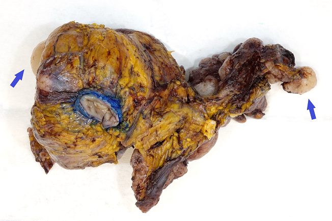
Omental nodules
Frozen section description
- See Microscopic description below
- Potential for sampling error
- Any amount of high grade cytology should be considered high grade disease
- Reference: Am J Surg Pathol 2016;40:14
Frozen section images
Contributed by Alex Placek, M.D.
Low cellularity

Low grade cytology
Increased cellularity
High grade cytology
Microscopic (histologic) description
- WHO Digestive System Tumors (5th edition) grading system of peritoneal metastases of appendiceal mucinous neoplasms
- Grade 1: hypocellular mucinous deposits, low grade cytology, lacks infiltrative type invasion
- Grade 2: hypercellular mucinous deposits, high grade cytology, infiltrative type invasion (angulated glands with desmoplasia or numerous small mucin pools containing clusters of tumor cells)
- Grade 3: mucinous tumor deposits with signet ring cells
- Although uncommon, discordance of histologic grade between primary appendiceal lesion and peritoneal disease may exist (J Clin Pathol 2012;65:919)
Microscopic (histologic) images
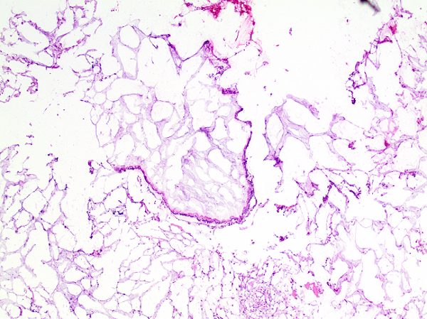
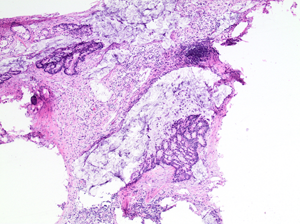
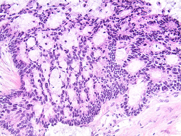
Contributed by Alex Placek, M.D.
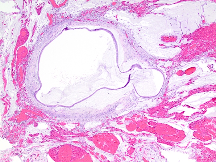
Low cellularity
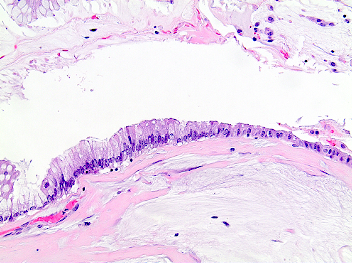
Low grade cytology

Increased cellularity
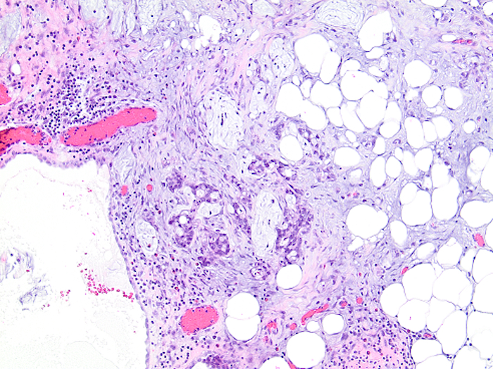
Invasion with desmoplasia

Signet ring cells
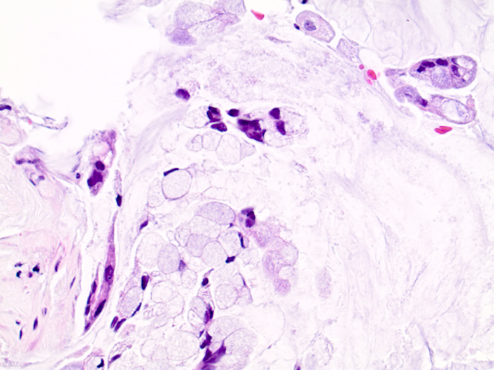
Degenerated mucinous cells
Molecular / cytogenetics description
- Similar to mutation profile of associated primary tumor (appendiceal mucinous neoplasms):
- KRAS exon 2 (Mod Pathol 2014;27:1521)
- GNAS (Ann Oncol 2016;27:2097)
Sample pathology report
- Omentum, omentectomy:
- Acellular mucin (or)
- Low grade peritoneal mucinous neoplasia / low grade mucinous carcinoma peritonei (or)
- High grade peritoneal mucinous neoplasia / high grade mucinous carcinoma peritonei (or)
- High grade peritoneal mucinous neoplasia with signet ring cells / high grade mucinous carcinoma peritonei with signet ring cells
Additional references
Board review style question #1
A 45 year old woman presents with a 2 week history of abdominal pain and increased abdominal girth. Clinical and radiologic findings are concerning for pseudomyxoma peritonei. A biopsy from an omental nodule is shown. The primary neoplasm is most likely from which anatomic site?
- Appendix
- Colon
- Ovary
- Pancreas
Board review style answer #1
A. Appendix. Pseudomyxoma peritonei is a clinical syndrome characterized by extravasation of mucin into the abdomen due to a ruptured mucinous neoplasm. Primary tumors from all of the sites listed have been associated with pseudomyxoma peritonei. However, the large majority arise from the appendix. Furthermore, the low grade cytology displayed in the image strongly argues for a low grade appendiceal mucinous neoplasm primary.
Comment Here
Reference: Pseudomyxoma peritonei
Comment Here
Reference: Pseudomyxoma peritonei
Board review style question #2
Based on the WHO (5th edition) grading system of peritoneal metastases of appendiceal mucinous neoplasms, which of the following is a feature of grade 2?
- Destructive invasion
- Infiltrating signet ring cells
- Low grade cytology
- Signet ring appearing cells floating in mucin pools
Board review style answer #2
A. Destructive invasion. The WHO (5th edition) recommends a 3 tier system for grading peritoneal disease of primary appendiceal mucinous neoplasia (grade 1 - 3). The grade of peritoneal disease has prognostic significance and guides therapeutic interventions. Hypocellular deposits with low grade cytology is classified as grade 1. Hypercellular deposits with high grade cytology or destructive invasion is classified as grade 2. Finally, grade 3 requires infiltrative signet ring cells. Importantly, degenerative cells floating in pools of mucin that morphologically resemble signet ring cells are not associated with a poor prognosis and do not warrant categorization as grade 3.
Comment Here
Reference: Pseudomyxoma peritonei
Comment Here
Reference: Pseudomyxoma peritonei
