

Bladder & urothelial tract
General
Histology
Editorial Board Member: Debra L. Zynger, M.D.
Deputy Editor-in-Chief: Maria Tretiakova, M.D., Ph.D.


Last author update: 31 August 2023
Last staff update: 31 August 2023
Copyright: 2003-2024, PathologyOutlines.com, Inc.
PubMed Search: Bladder histology
Table of Contents
Definition / general | Essential features | Physiology | Diagrams / tables | Clinical features | Gross description | Microscopic (histologic) description | Microscopic (histologic) images | Cytology description | Positive stains | Negative stains | Electron microscopy description | Videos | Additional references | Board review style question #1 | Board review style answer #1 | Board review style question #2 | Board review style answer #2Cite this page: Sardana R, Parwani A. Histology. PathologyOutlines.com website. https://www.pathologyoutlines.com/topic/bladderhistology.html. Accessed April 25th, 2024.
Definition / general
- Hollow muscular elastic organ that sits on the pelvic floor and stores urine
Essential features
- Layers of bladder wall (from inside out)
- Urothelium: specialized stratified lining epithelium, impermeable barrier
- Lamina propria: connective tissue containing vessels, lymphatics, nerve endings and a few elastic fibers
- Muscularis propria: also called detrusor muscle; consists of 3 layers (inner longitudinal, middle circular and outer longitudinal)
- Serosa / adventitia: serosa is loose connective tissue covering bladder dome; the remaining area is covered by adventitia
- Important to histomorphologically differentiate the layers in order to properly stage the tumors, determine therapy and estimate survival
Physiology
- Located in the extraperitoneal space of the pelvis, behind the pubic bones (StatPearls: Histology, Bladder [Accessed 14 April 2022])
- Extends into the abdomen when distended
- 2 main parts
- Upper part is above the ureteric orifices and is composed of the apex and body
- Lower part is beneath the ureteric orifices and is composed of the fundus, trigone and neck
- Urothelium is 5 - 7 cell layers thick in a contracted bladder and 2 - 3 cells thick in a distended bladder (Am J Physiol Renal Physiol 2009;297:F1477)
Diagrams / tables
Images hosted on other servers:

Staging of bladder carcinoma
Clinical features
- Urothelium is exclusive to the conducting passages of the urinary system (renal pelvis, ureter, bladder, upper urethra and prostatic ducts) (Am J Physiol Renal Physiol 2009;297:F1477)
- > 90% of bladder carcinomas arise from the urothelium, mostly from the posterior wall of the bladder (Nat Cell Biol 2014;16:982)
- In practice, sampling tangentially to the basement membrane can generate an artificially thick mucosa, hence urothelial thickness is of low utility in the assessment of urothelial neoplasms
- Squamous epithelium is common in the trigone area of females; abundant intracytoplasmic glycogen and lack of keratinization makes it histologically similar to vagina or cervix (Am J Anat 1962;111:319)
- von Brunn nests, cystitis cystica and cystitis glandularis represent a continuum of proliferative or reactive changes
- von Brunn nests (proliferative cystitis)
- Reactive proliferative change is present in 85 - 95% of bladders; frequency increases with age; more common at the trigone
- Nests of cytologically benign urothelium in lamina propria; with regular spacing and same extent to horizontal level at base of proliferation
- Bladder wall urachal remnants: cystically dilated epithelial lined structures are sometimes found in biopsies from the dome or anterior wall of the bladder
Gross description
- Hollow muscular viscus resembling an inverted pyramid when empty and a sphere when distended (Microbiol Spectr 2015;3:10)
- Flat internal (mucosal) surface when distended; throws into abundant folds when empty
- Lies posterior to the pubic bone, anterior to the uterus in females and anterior to the rectum in males
- Partially retroperitoneal, with its peritoneal covered dome projecting into the abdomen when fully distended
- Triangular shaped area at the base of the bladder called the trigone
Microscopic (histologic) description
- 4 layers (from inside out): urothelium, lamina propria, muscularis propria, serosa / adventitia
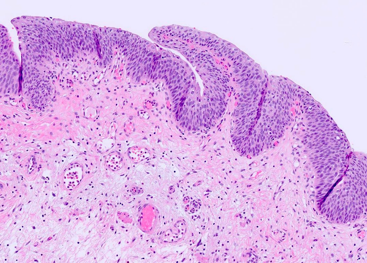
- Urothelium: 3 layers of cells
- Umbrella / apical cells (superficial layer)
- Formerly called transitional epithelium, intermediate between nonkeratinizing squamous and pseudostratified columnar epithelium
- Dome / pyramid shaped, large and ovoid, frequently binucleated cells with abundant eosinophilic cytoplasm
- Scalloped surface outline often overlapping 2 or more of the underlying cells
- Superficial cytoplasm is fuzzy, indistinct and more intensely stained than the rest of the cytoplasm
- Luminal surface of the cells appears thickened and more densely stained
- Form an impermeable barrier; tight junctions between the cells decrease paracellular flux while uroplakins form a superficial plaque
- Intermediate cells
- Multicell layering, depending on the stage of distension
- Cuboidal to low columnar with well defined borders and amphophilic cytoplasm that is rich in glycogen
- Uninucleated cells: ovoid nuclei with fine granular chromatin, no mitotic figures arranged regularly along the long axis at right angles to surface
- Basal cells
- Single layer in contact with basal lamina
- Mononucleated, cuboidal cells with mitotic capability
- Cylindrical, can be flat when bladder wall is stretched; some have longitudinal nuclear grooves
- Gradual turnover but significant regenerative ability
- Umbrella / apical cells (superficial layer)
- Lamina propria
- Dense connective tissue containing a rich vascular network, lymphatic channels, sensory nerve endings, elastic fibers and interspersed adipose tissue
- Forms thick mucosal folds when the viscus is contracted
- Thickness varies with the degree of distention and is generally thinner in the areas of the trigone and bladder neck
- Discontinuous isolated bundles of muscularis mucosa (wisps of smooth muscle) may be present; 5% of bladders may have a well developed, continuous muscularis mucosa (Am J Surg Pathol 1987;11:668)
- Hyperplastic muscularis mucosa (more common in women) may resemble muscularis propria (Am J Surg Pathol 2007;31:1420, Ann Diagn Pathol 2007;11:395)
- Important to distinguish muscularis mucosa from muscularis propria for accurate staging; smoothelin antibody is negative, while vimentin is positive in the former (Int J Biol Markers 2017;32:e305)
- Interstitial cells of Cajal act as nerve signal transducer between the smooth muscle cells and nerve endings
- Adipose tissue may be present within deep lamina propria (in small localized aggregates), while it is always found within muscularis propria; carefully differentiate between the two, especially in transurethral resection of bladder tumor (TURBT) specimens as it can overstage the tumor (Am J Surg Pathol 2000;24:1286)
- Muscularis propria
- 3 layers: inner and outer longitudinal layers and a central circular layer
- Layers are distinct near bladder neck, elsewhere there is no definite orientation
- Bladder's body has a higher smooth muscle content compared with the trigone
- Rarely nests or cords of paraganglia associated with nerves and vessels; clear or granular cytoplasm with round or vesicular nuclei
- Carefully distinguish from invasive carcinoma
- Cytokeratin negative and chromogranin positive
- Significantly thickened in urinary flow obstruction
- Serosa / adventitia
- Thin connective tissue layer covering the bladder dome and continuous with the peritoneal layer of the abdominal wall
- Contains blood vessels of various sizes
- Adventitia, an outermost layer of loose connective tissue in areas where there is no serosa
Microscopic (histologic) images
Contributed by Anil Parwani, M.D., Ph.D., M.B.A.
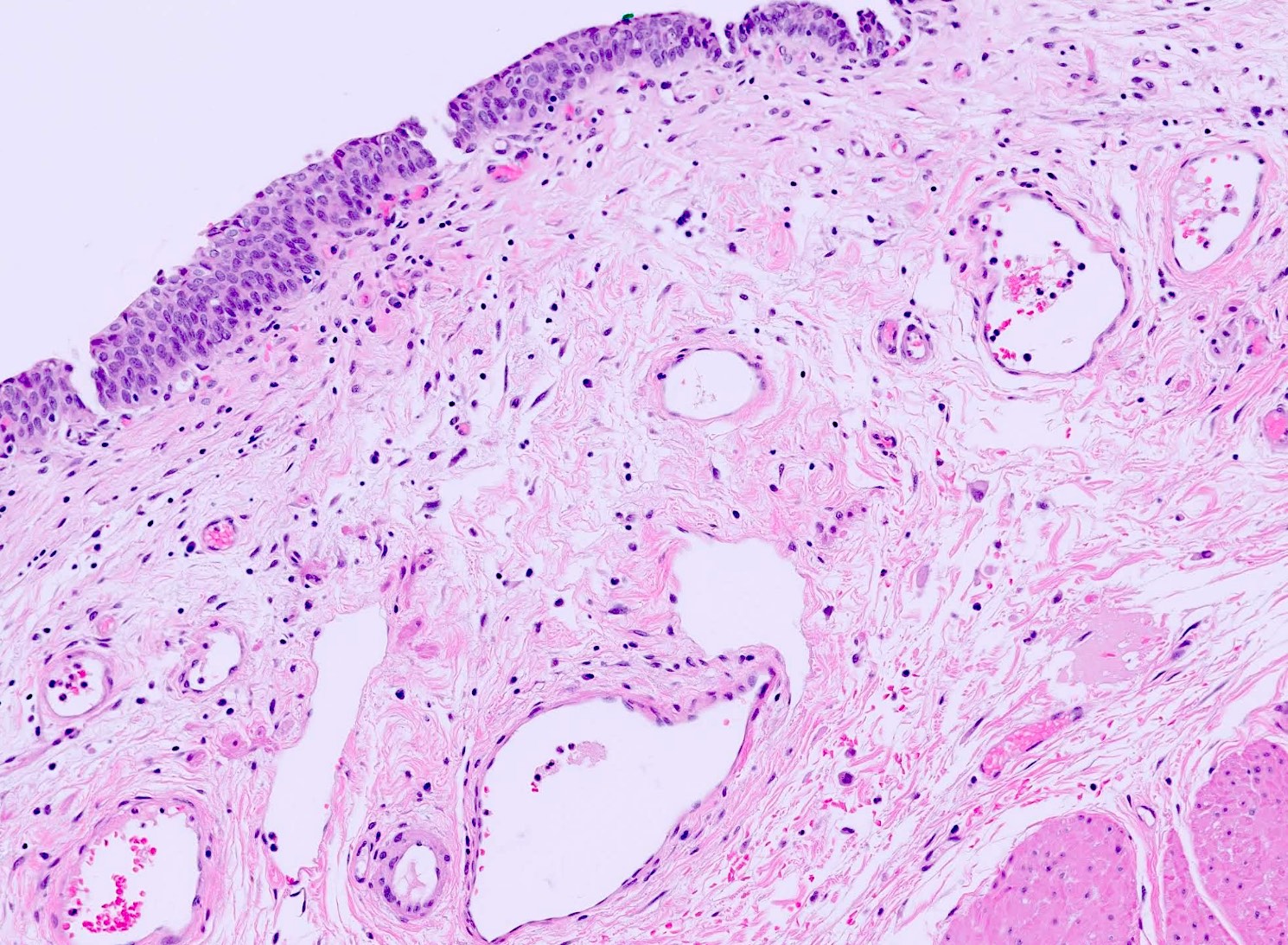
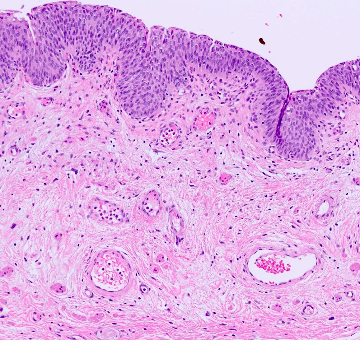
Bladder wall
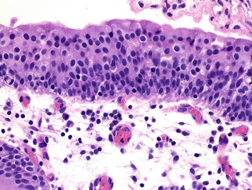
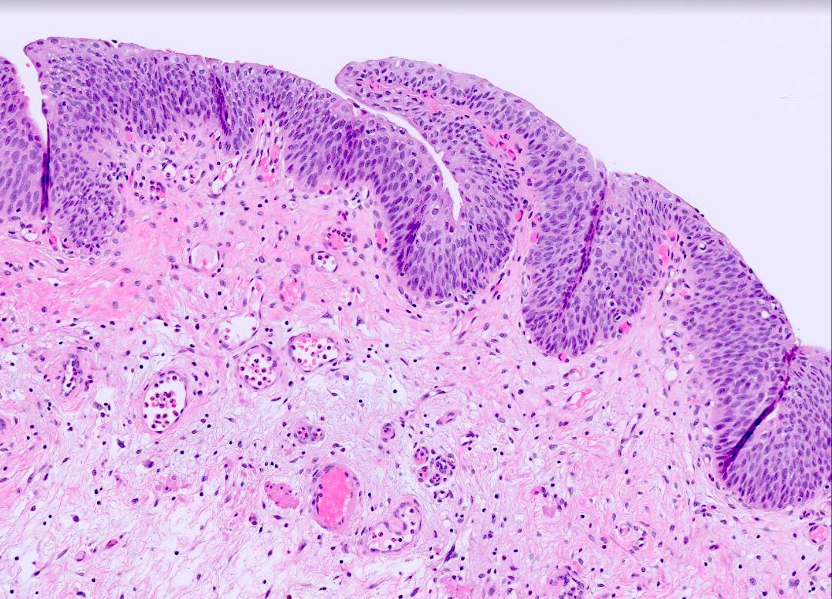
Urothelium
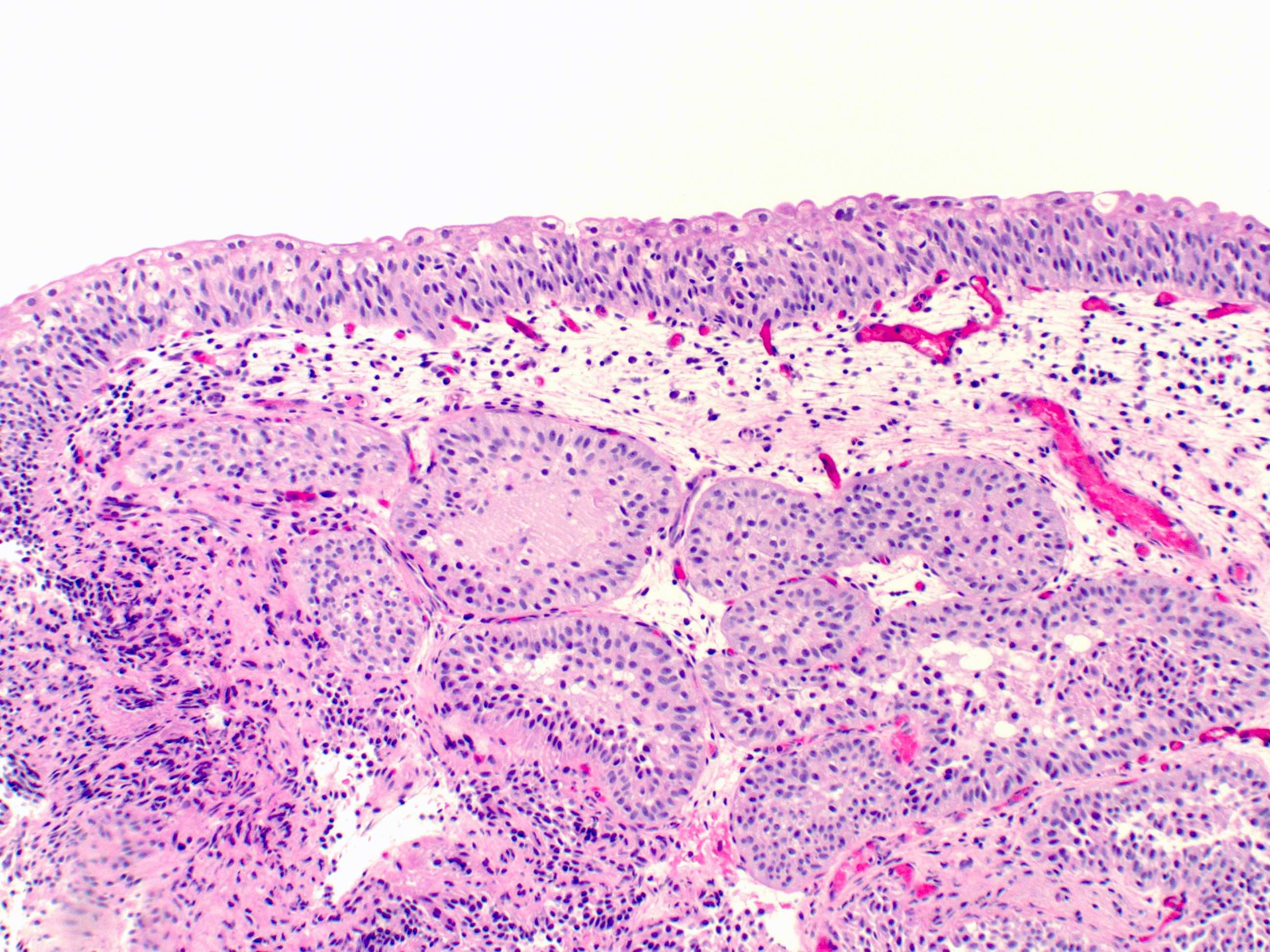
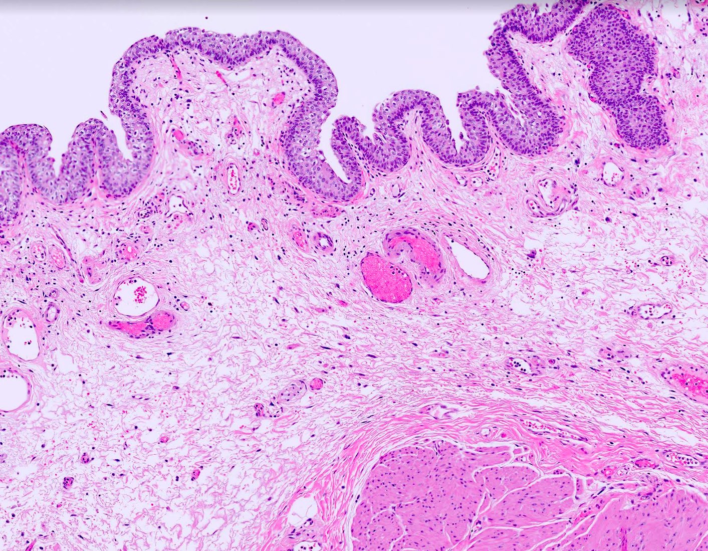
von Brunn nests
Urothelium and lamina propria
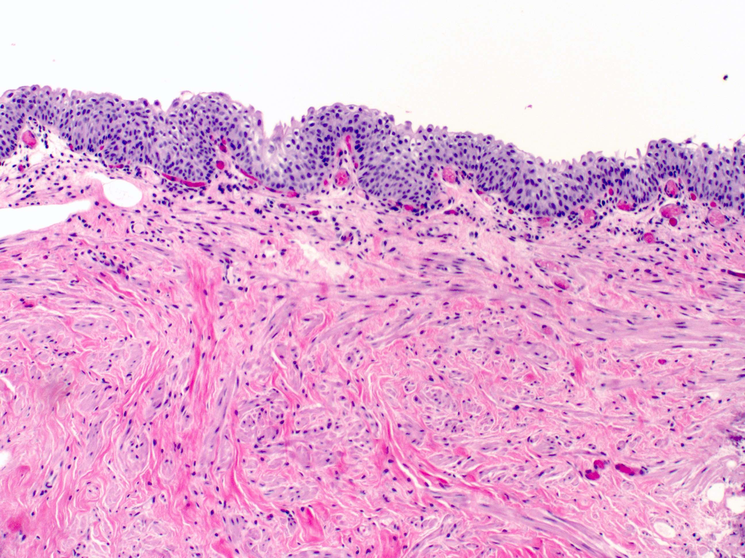
Muscularis mucosa
Muscularis propria
Cytology description
- In voided urines, transitional cells are shed singly; whereas in washings / brushings, the cells are aggregated in sheets and clusters
- Superficial and intermediate urothelial cells are seen in voided urine
- Superficial, intermediate and basal urothelial cells are seen in catheterized urine and bladder washings
- Superficial / umbrella cells:
- Low N:C ratio with pale finely granular chromatin, smooth nuclear contour and clear cytoplasm
- Intermediate and basal cells:
- High N:C ratio, smaller nuclei with even spacing and darker chromatin than superficial cells
- Melamed-Wolinska bodies: round to oval, hyaline, red or green-blue cytoplasmic inclusions seen within degenerated, benign or malignant urothelial cells (Diagn Cytopathol 2011;39:117)
Positive stains
- Full thickness: GATA3, CK7, CK8/18, CK19, CD138, S100P, thrombomodulin
- Basal / parabasal: p63, 34betaE12, CK5/6, CD44s
- Umbrella cells only: CK20, uroplakin II, uroplakin III (Adv Urol 2020;2020:4920236, Am J Clin Pathol 2014;142:864)
Electron microscopy description
- Superficial urothelial cells form an impermeable barrier via numerous junctional complexes
- Trilaminar (asymmetric) unit membrane composed of 2 dense layers of unequal thickness and a central lucent layer with interspersed normal areas which act like hinges
- Surface plasma membrane consists of inflexible apical plaques containing uroplakins (Kidney Int 2016;89:612)
Videos
Shotgun histology bladder
Additional references
Board review style question #1
Which of the following characteristics is true regarding the histology displayed in the picture?
- Muscular layer is consistently organized into 3 distinct layers
- Superficial layer consists of only umbrella cells, which have a high regenerative capacity
- Umbrella cells are CK20+ and CD44s-
- Well defined continuous muscularis mucosa can be always seen
Board review style answer #1
C. Umbrella cells are CK20+ and CD44s-. Superficial cells or the umbrella cells of urothelium are CK20+ and CD44s-, while basal cells are CK20- and CD44s+.
Comment Here
Reference: Bladder - Histology
Comment Here
Reference: Bladder - Histology
Board review style question #2
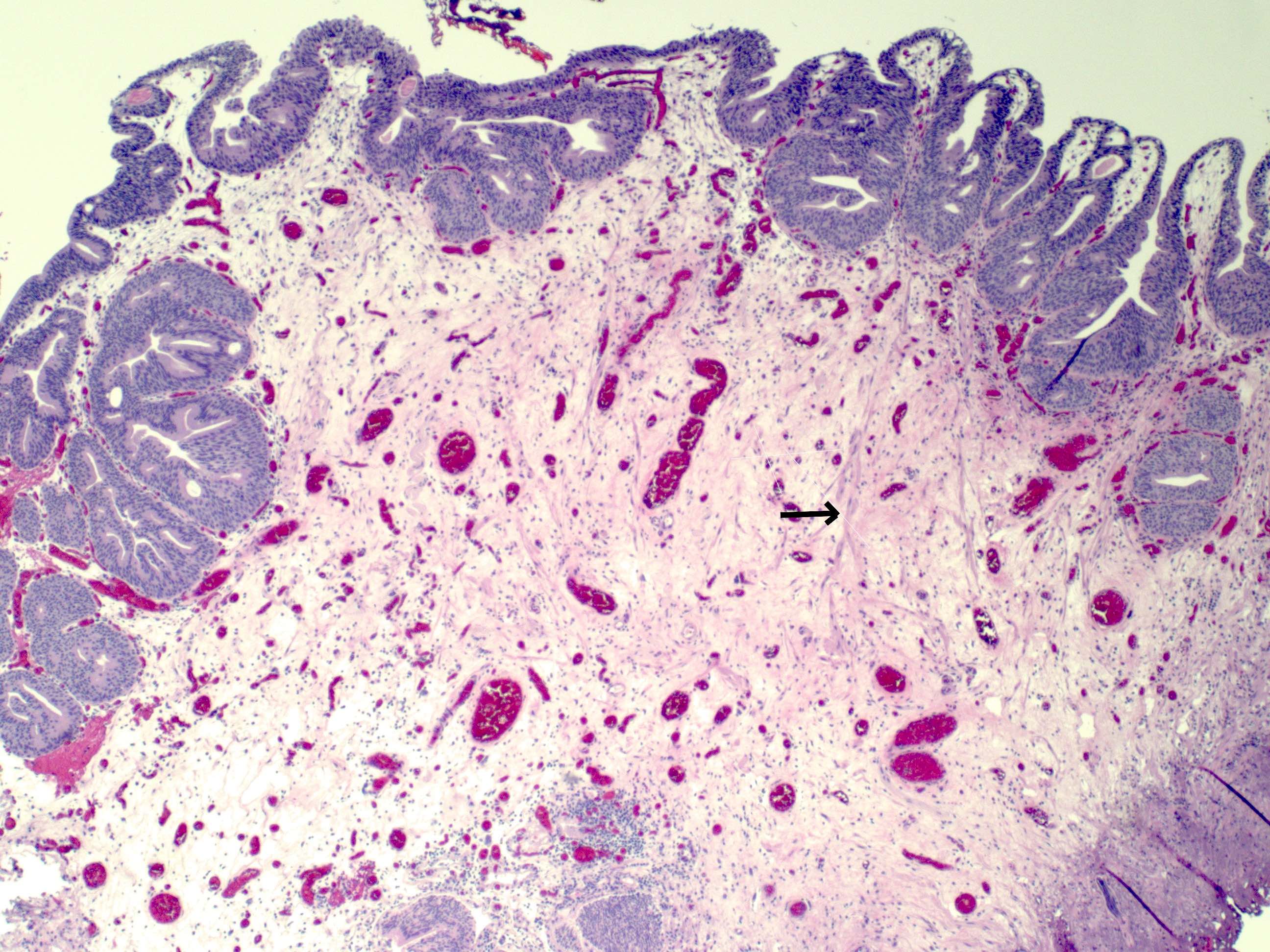
What does the arrow indicate in the micrograph shown above?
- Desmoplastic response to invasive tumor
- Elastosis
- Muscularis mucosa
- Muscularis propria
Board review style answer #2
C. Muscularis mucosa. The wispy, incomplete fascicles of muscle represent muscularis mucosae. They are morphologically and histochemically distinct from muscularis propria.
Comment Here
Reference: Bladder - Histology
Comment Here
Reference: Bladder - Histology
