

Bladder & urothelial tract
Squamous cell neoplasms
Squamous cell papilloma
Editorial Board Member: Bonnie Choy, M.D.
Deputy Editor-in-Chief: Maria Tretiakova, M.D., Ph.D.


Last author update: 12 April 2022
Last staff update: 12 April 2022
Copyright: 2020-2024, PathologyOutlines.com, Inc.
PubMed Search: Squamous cell papilloma bladder[TIAB] "last 5 years"[DP]
Table of Contents
Definition / general | Essential features | Terminology | ICD coding | Epidemiology | Sites | Pathophysiology | Etiology | Clinical features | Diagnosis | Laboratory | Radiology description | Prognostic factors | Case reports | Treatment | Clinical images | Gross description | Microscopic (histologic) description | Microscopic (histologic) images | Positive stains | Negative stains | Molecular / cytogenetics description | Sample pathology report | Differential diagnosis | Board review style question #1 | Board review style answer #1 | Board review style question #2 | Board review style answer #2Cite this page: Batra H, Parwani A. Squamous cell papilloma. PathologyOutlines.com website. https://www.pathologyoutlines.com/topic/bladdersquamouspapilloma.html. Accessed April 20th, 2024.
Definition / general
- Rare, benign tumor of the urinary bladder showing delicate fibrovascular cores lined by squamous epithelium
Essential features
- Rare, benign bladder lesion with female predilection
- Presents with gross hematuria and irritative bladder symptoms
- Recurrence is rare
- Not related to condyloma acuminatum
Terminology
- Squamous papilloma
Epidemiology
- Rare, benign urothelial lesion with < 20 cases reported in known literature
- F > M (6:1) (Cancer 2000;88:1679)
- Fourth to seventh decade (Cancer 2000;88:1679)
- Can arise de novo or along with previous urothelial lesions
Sites
- Most cases involve the dome and the lateral and posterior walls of the bladder (Cancer 2000;88:1679)
Pathophysiology
- Largely unknown
Etiology
- Smoking
- Occupational exposure to amines
Clinical features
- Macroscopic hematuria
- Irritative voiding symptoms (e.g., stress incontinence, neurogenic bladder, dysuria) (Cancer 2000;88:1679, Am J Surg Pathol 2006;30:883)
Diagnosis
- Cystoscopy: papillary / exophytic, whitish plaque, erythematous variable size lesions, often unifocal (Case Rep Pathol 2013;2013:486312)
Laboratory
- Urine cytology is negative for malignant cells
Radiology description
- Ultrasonography may show thickened bladder wall
Prognostic factors
- Recurrence is very rare and progression to carcinoma has not been reported (Cancer 2000;88:1679)
Case reports
- 32 - 82 year old patients with squamous cell papilloma of urinary bladder, including squamous papilloma related to HPV (Cancer 2000;88:1679)
- 74 year old man with irritative voiding symptoms and exophytic mass found at the mucosa of the floor and the posterior wall of the bladder (Case Rep Pathol 2013;2013:486312)
- Noninvasive squamous lesions in the urinary bladder; a case series with 5 reported cases of squamous cell papilloma of the urinary bladder (Am J Surg Pathol 2006;30:883)
Treatment
- Resection: transurethral resection of bladder tumor (TURBT) (Cancer 2000;88:1679)
Clinical images
Images hosted on other servers:

Cystoscopy finding: whitish exophytic lesion
Gross description
- Small, whitish, polypoid lesion
Microscopic (histologic) description
- Multiple, free floating papillary fronds with thin, well developed fibrovascular cores composed of bland, benign appearing squamous cells (Case Rep Pathol 2013;2013:486312)
- No dysplasia seen
- Stromal invasion is not seen
- Hyperplasia and mitotic activity restricted to basal / parabasal layers (Case Rep Pathol 2013;2013:486312)
- Koilocytes are rare or absent (Cancer 2000;88:1679)
Microscopic (histologic) images
Contributed by Harsh Batra, M.B.B.S., D.C.P., D.N.B. and Anil Parwani, M.D., Ph.D., M.B.A.
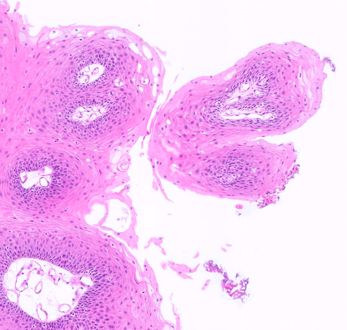
Transverse section of papillae

Transverse section through a single papilla
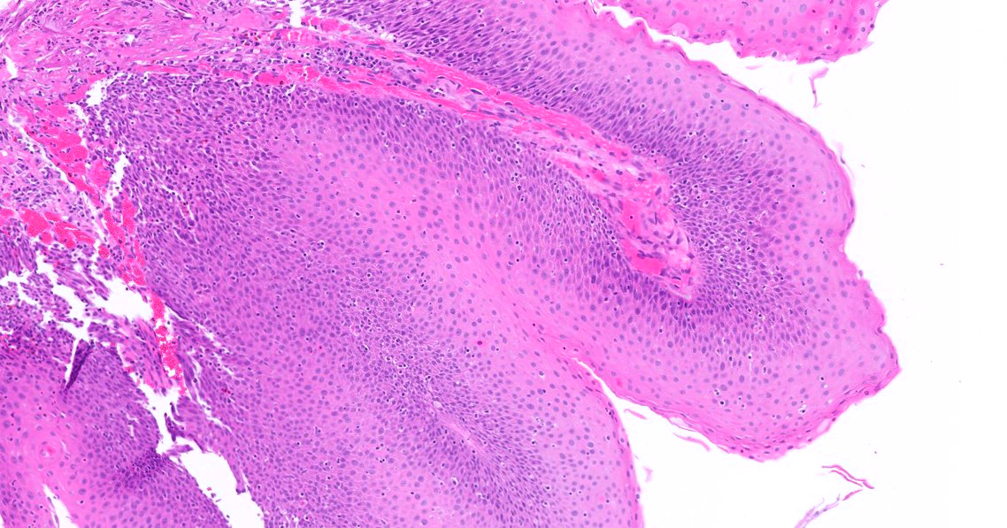
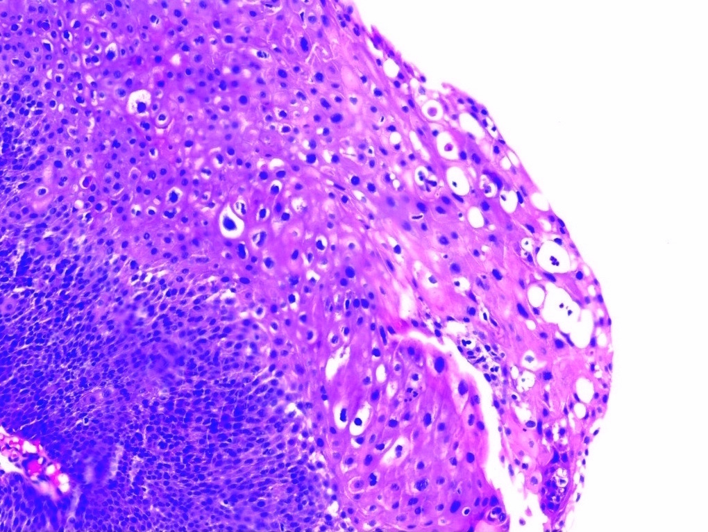
Fibrovascular cores
Positive stains
- p63: nuclear positivity (Case Rep Pathol 2013;2013:486312)
- EGFR: membranous positivity (Am J Surg Pathol 2006;30:883)
- Ki67: positive in the basal layer (Case Rep Pathol 2013;2013:486312)
Negative stains
Molecular / cytogenetics description
- DNA ploidy: diploid (Cancer 2000;88:1679)
- Negative for HPV DNA (Cancer 2000;88:1679)
Sample pathology report
- Urinary bladder, lateral wall, transurethral resection of bladder tumor:
- Squamous papilloma (see comment)
- Comment: Microscopic examination shows multiple papillae with fibrovascular core lined by keratinized squamous epithelium. No evidence of stromal invasion, atypia or koilocytic changes seen in the biopsy submitted. Clinical correlation with follow up is advised.
Differential diagnosis
- Condyloma acuminatum:
- Multiple, extensive lesions
- External genitalia condyloma or history of immunosuppression
- Papillary fronds lined by hyperplastic, metaplastic squamous epithelium that may be hyperkeratotic
- Presence of koilocytes
- p53 overexpression by IHC
- HPV DNA present
- Aneuploid
- Verrucous carcinoma:
- Rare variant of squamous cell carcinoma
- Diffuse, extensive lesions
- Endophytic growth pattern with broad nests of tumor cells intimately associated with lamina propria or detrusor muscle with minimal stromal reaction
- Margins of the lesion are pushing
- Nuclear atypia is minimal to moderate
- p53 staining is positive
- HPV DNA negative
- Keratinizing squamous metaplasia:
- Nonpolypoid, flat lesion showing thick squamous epithelium with pronounced hyperkeratosis and hypergranulosis
- Extensive lesions are associated with neoplasia, so it is recommended to report the presence and the extent (focal versus extensive) of keratinizing squamous metaplasia
- Squamous cell carcinoma:
- Irregular infiltrating nests or sheets of malignant squamous cells with destructive stromal invasion
- Presence of keratin pearls, individual cell keratinization or intercellular bridges
- Often associated with surface keratinizing squamous metaplasia and dysplasia
Board review style question #1
Squamous papilloma of the urinary bladder is which of the following?
- Benign lesion
- Carcinoma in situ
- Malignant
- Papillary urothelial neoplasm of unknown malignant potential (PUNLMP)
Board review style answer #1
Board review style question #2
Which of the following histopathological features distinguishes squamous papilloma of the bladder from condyloma acuminatum?
- Absence of fibrovascular cores in the papillae
- Absence of koilocytes
- Presence of invasion into submucosa
- Presence of umbrella cells
Board review style answer #2
B. Absence of koilocytes. Squamous papilloma shows absence of koilocytic change, whereas koilocytes are present in condyloma acuminatum.
Comment Here
Reference: Squamous cell papilloma
Comment Here
Reference: Squamous cell papilloma
