

Bone & joints
General
Bone formation and growth
Author: Dariusz Borys, M.D.

Last author update: 1 January 2012
Last staff update: 10 December 2021
Copyright: 2003-2024, PathologyOutlines.com, Inc.
PubMed Search: Bone formation [title] growth
Table of Contents
Definition / general | Intramembranous ossification | Endochondral ossification | Histology of bone growth | Bone growth | Modeling and remodelingCite this page: Borys D. Bone formation and growth. PathologyOutlines.com website. https://www.pathologyoutlines.com/topic/bonebonegrowth.html. Accessed April 18th, 2024.
Definition / general
- Bone tissue is formed by intramembranous ossification or by endochondral ossification
- The original or model tissue is gradually destroyed and replaced with bone tissue
- Woven bone is primarily formed and later converted to lamellar bone by subsequent remodeling
Intramembranous ossification
- Source of flat and less commonly short bones
- Occurs through condensation of mesenchymal tissue
- Process begins when multiple groups of cells differentiate into osteoblasts in a primary ossification center
- Osteoid is synthesized, then mineralizes surrounding the osteoblasts, which mature to osteocytes
- When ossification centers fuse, loose trabecular structures known as primary spongiosa are formed
- Then blood vessels grow into the connective tissue between trabeculae
Images hosted on other servers:


Intramembranous bone formation


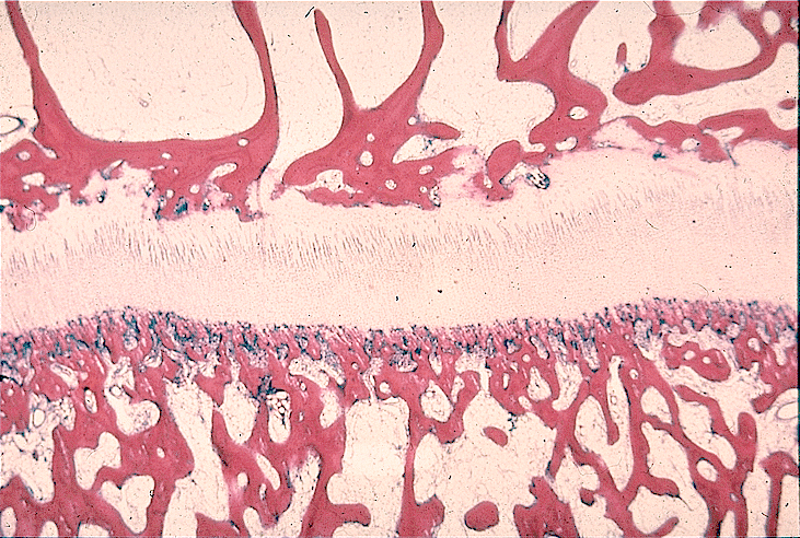
Endochondral ossification
- Responsible for formation of long and short bones
- Hyaline cartilage model, which provides template of shape of the bone
- May be divided into 2 phases:
- 1st phase: chondrocytes are hypertrophic and degenerated, then calcified
- 2nd phase: osteoprogenitor cells and blood capillaries invade the spaces left by degenerating cartilage; osteoblasts arise from osteoprogenitor cells and lay down a layer of rapidly mineralized osteoid on the surface of calcified cartilage, called primary spongiosa, which later is remodeled to lamellar bone (secondary spongiosa); calcified cartilage is resorbed by chondroblasts and replaced by bone and marrow cavities
Images hosted on other servers:


Endochondral ossification


Histology of bone growth
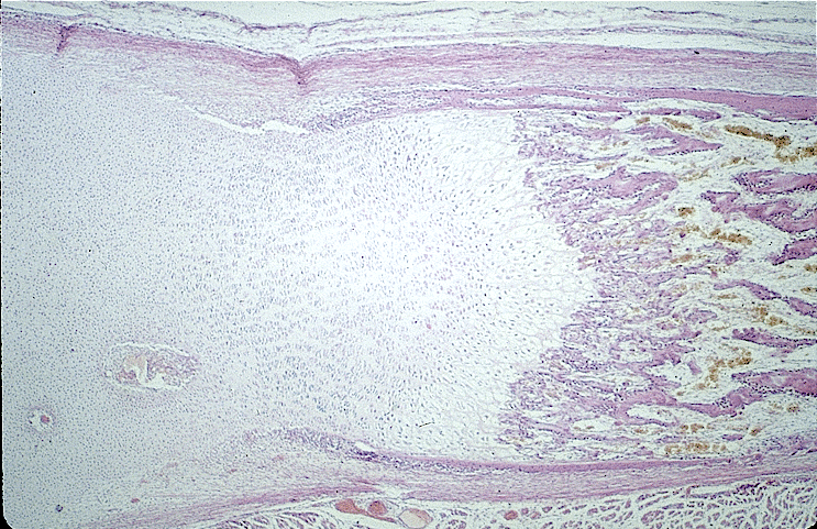
- Epiphyseal cartilage of long bone is located between epiphysis and metaphysis, is responsible for longitudinal growth; has 5 zones:
(a) Resting zone – small chondrocytes
(b) Proliferative zone – rapidly dividing chondrocytes in columns, parallel to the long axis of bone
(c) Hypertrophic zone – large chondrocytes with clear cytoplasmic glycogen
(d) Calcified cartilage zone (zone of provisional calcification) – chondrocyte graveyard, followed by blood vessel invasion and bone deposition
(e) Ossification zone – formation of primary spongiosa by rapidly mineralized osteoid
Images hosted on other servers:


Epiphyseal plate
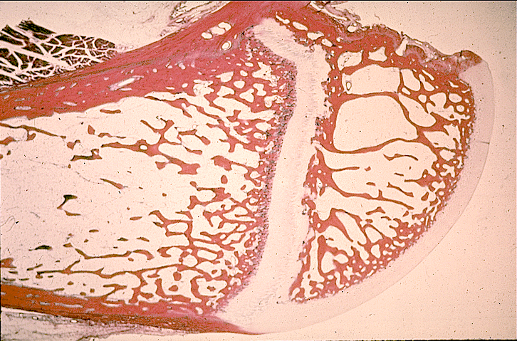
Bone growth
- Bone grows by either endochondral or intramembranous ossification
- Endochondreal ossification of the epiphyseal plate is responsible for longitudinal growth of long bones
- Periosteal deposition is responsible for length and thickness of long bones
- Endosteal bone deposition is responsible for growth of trabecular bone and endosteal cortex, including the haversian system
Modeling and remodeling
- Bone formation is an ongoing process that alters the size and shape of bone by partial resorption of preformed bone tissue and simultaneous deposition of new bone (modeling and remodeling)
- Modeling is a process in which bone achieve its proper shape
- Modeling is responsible for the circumferential growth of bone and expansion of marrow cavity
- Remodeling is a continuous process, in which damaged bone is repaired, ion homeostasis is maintained, and bone is reinforced for increased stress; entire remodeling cycle requires ~ 6 months
- In healthy adults, remodeling rate varies by type of bone: 25% per year in trabecular bone versus 3% in cortical bone
- Resorption and deposition are normally balanced, and bone density is maintained
- Resorptive activity exceeding deposition activity represents a pathologic state, may cause lytic lesions
- The cement line (reversal line) is evidence of previous remodeling activity; is formed by filling of new bone in a previously resorbed cavity; is strongly basophilic due to high content of inorganic matrix and is normally found in the haversian and interstitial systems of adult bone
- Cement line from normal remodeling is relatively long and straight; indented or mosaic pattern indicates a pathologically accelerated remodeling process
