
Bone marrow nonneoplastic
Normal
Hematogones
Editorial Board Member: Anamarija M. Perry, M.D.
Deputy Editor-in-Chief: Genevieve M. Crane, M.D., Ph.D.


Last author update: 4 March 2024
Last staff update: 4 March 2024
Copyright: 2002-2024, PathologyOutlines.com, Inc.
PubMed Search: Hematogones
Table of Contents
Definition / general | Essential features | Sites | Diagrams / tables | Clinical features | Case reports | Microscopic (histologic) description | Microscopic (histologic) images | Peripheral smear description | Flow cytometry description | Flow cytometry images | Videos | Differential diagnosis | Additional references | Board review style question #1 | Board review style answer #1Cite this page: George GV, Evans AG. Hematogones. PathologyOutlines.com website. https://www.pathologyoutlines.com/topic/bonemarrowhematogones.html. Accessed April 19th, 2024.
Definition / general
- Hematogones are normal B lineage progenitors, which normally account for < 1% of the bone marrow cellularity
- Based on sequential antigen expression, they can be divided into 3 stages of maturation: stages 1, 2 and 3 (Leuk Res 2013;37:1404)
Essential features
- Hematogones are benign B cell precursors and their degree of maturation can be classified by flow cytometry into 3 stages (1, 2 and 3)
- Morphologically, they contain scant cytoplasm and often have round, indented nuclei with absent or inconspicuous nucleoli; the presence of nucleoli generally indicates immaturity
- Increased numbers of hematogones may mimic B lymphoblastic leukemia (B ALL) due to the morphologic and immunophenotypic similarities
Sites
- Bone marrow
Diagrams / tables
Images hosted on other servers:

Normal hematogone maturation
Clinical features
- Conditions associated with increased hematogones include (Blood 2001;98:2498)
- Various tumors, including lymphomas and solid tumors
- Nonneoplastic blood cytopenias and autoimmune conditions
- Postchemotherapy, post-bone marrow transplantation
- Viral infections
- Acquired immunodeficiency syndrome (AIDS)
Case reports
- 7 year old boy with hematogone hyperplasia masquerading as acute lymphoblastic leukemia with concurrent hypercupremia (J Pediatr Hematol Oncol 2020;42:e670)
- 64 year old man with TCL1 positive hematogones in a case of T cell prolymphocytic leukemia after therapy (Hum Pathol 2017;65:175)
- 75 year old man with κ light chain expressing hematogones in a case of λ restricted chronic lymphocytic leukemia (CLL) and multiple myeloma on daratumumab therapy (Blood 2022;140:1917)
Microscopic (histologic) description
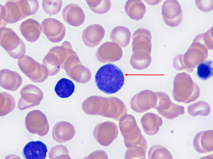
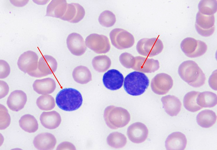
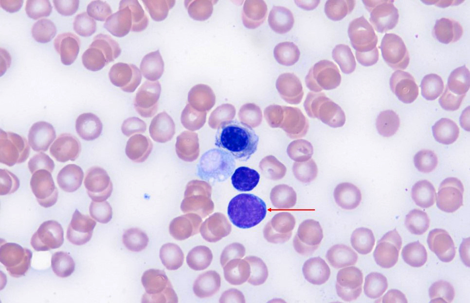
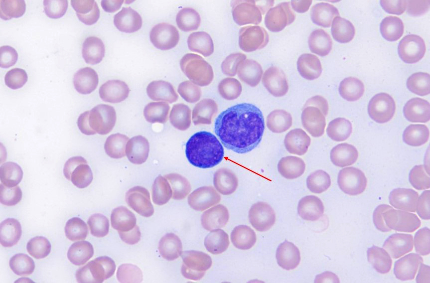
- Hematogones are lymphoid appearing cells whose cytomorphology can be best appreciated on bone marrow aspirate smears (typically pediatric)
- Stage 1 hematogones typically have scant to absent cytoplasm (may be moderately to deeply basophilic with no inclusions, granules or vacuoles) and large, round, sometimes indented nuclei with inconspicuous nucleoli
- Generally, the presence of 1 or more nucleoli denotes immaturity; therefore, immature hematogones may resemble lymphoblasts
- Mature hematogones resemble mature lymphocytes with condensed nuclear chromatin; nucleoli are absent
Microscopic (histologic) images
Contributed by Giby V. George, M.B.B.S. and AFIP
Bone marrow aspirate hematogones
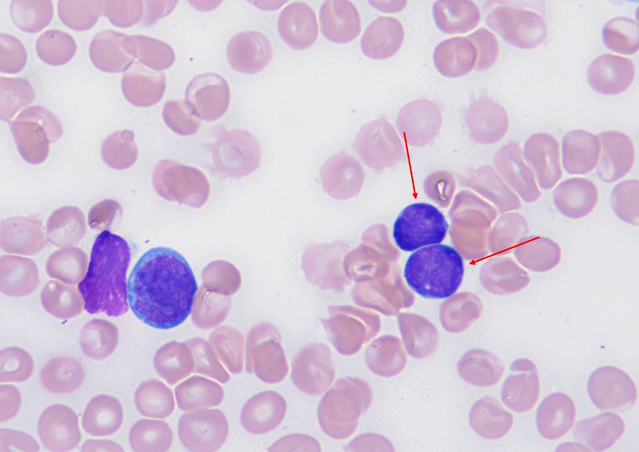
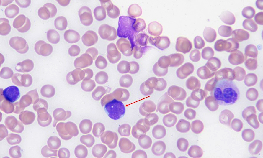
Bone marrow aspirate hematogones
Sparse, lightly basophilic cytoplasm
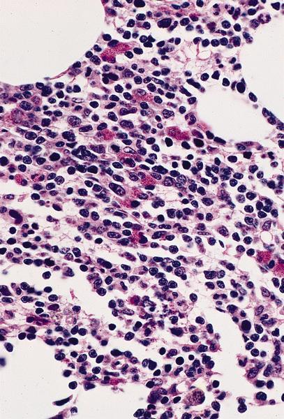
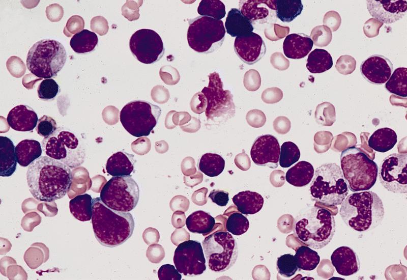
Increased hematogones evenly distributed

Clumped nuclear chromatin
Peripheral smear description
- Hematogones are not usually seen on the peripheral blood smear except in neonates or in umbilical cord blood
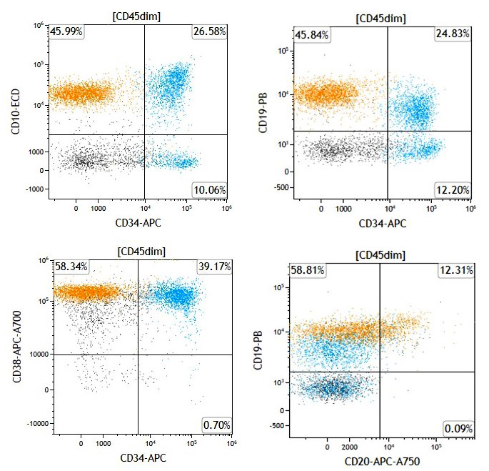
Flow cytometry description
- Generally, hematogones demonstrate low side scatter (SSC) by flow cytometry with dimmer expression of CD45 when compared to normal lymphocytes; thus, they lie within the CD45 dim, low SSC gate (Leuk Res 2013;37:1404)
- Stage 1 hematogones exhibit bright expression for TdT, CD34, CD10, CD38 and CD43; they show dim expression of CD19 and CD22 while negative for CD20 and cytoplasmic immunoglobulin (Leuk Res 2013;37:1404)
- Stage 2 hematogones begin to show down regulation / negativity of TdT, CD34 and CD10; they exhibit increased expression of CD19 and CD20 with continued bright expression of CD38 and CD43, with dim expression of CD22 and CD45 and they are generally positive for cytoplasmic immunoglobulin (Leuk Res 2013;37:1404)
- Stage 3 hematogones are negative for TdT and CD34; they demonstrate bright expression of CD19, CD20, CD38, CD43, cytoplasmic and surface immunoglobulin, while dim for CD10, CD22 and CD45 (Leuk Res 2013;37:1404)
- Stage 3 hematogones progress to mature B cells, which are negative for TdT, CD34, CD10 and CD43, while positive for CD19, CD20, CD22, CD45, cytoplasmic / surface immunoglobulin with variable expression of CD38 (Leuk Res 2013;37:1404)
- CD5 expression has also been reported on stage 3 hematogones and mature B cells; in these cases, CD5 expression in conjunction with surface immunoglobulin helps differentiate them from CD5+ neoplastic B cells (Am J Clin Pathol 2009;132:733)
- Rarely, hematogones may exhibit light chain restriction (Leuk Res 2021;111:106704, Leuk Res Rep 2022;17:100316)
- Hematogones may rarely be identified by flow cytometry and IHC in benign lymph nodes (Am J Clin Path 2022;157:202, Hum Pathol 2018;81:131, Int J Lab Hematol 2023;45:592)
- In this case, they should account for 1% or less of the total lymph node population
- Additionally, discrimination from abnormal lymphoblasts is imperative
Flow cytometry images
Contributed by Giby V. George, M.B.B.S.
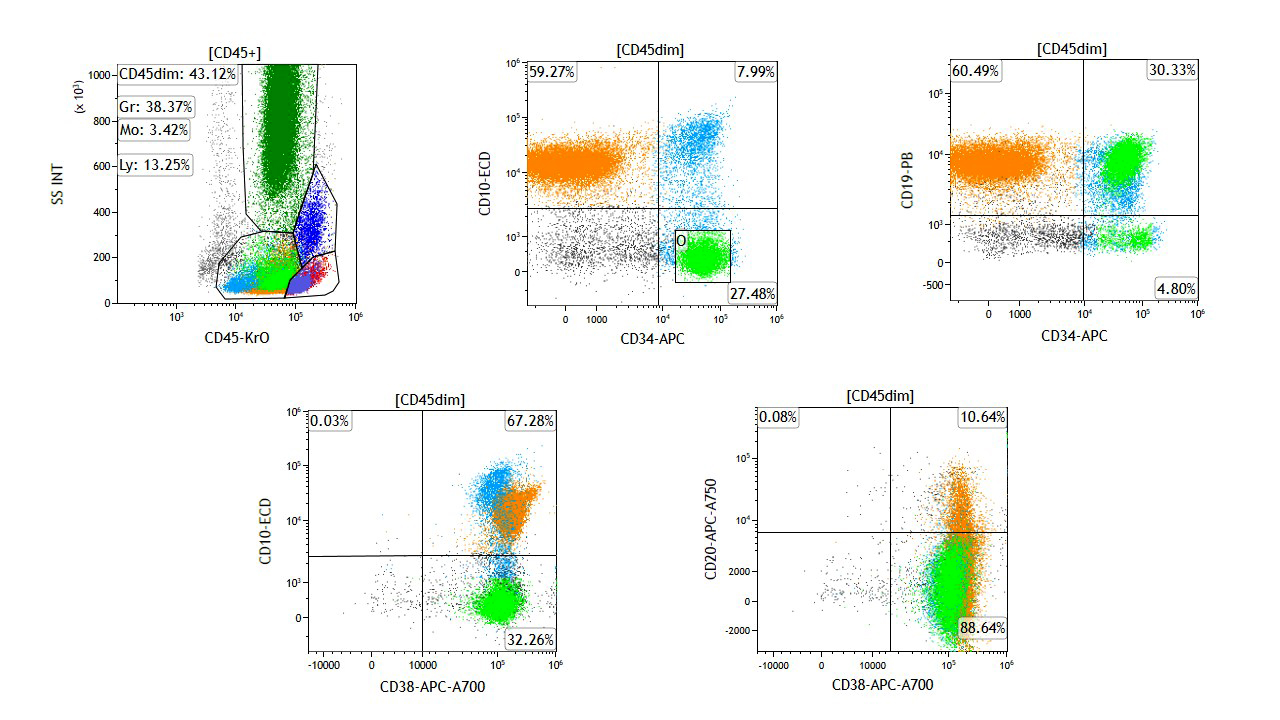
Hematogone maturation in the background of B ALL
Images hosted on other servers:


Normal hematogone maturation by flow cytometry
Videos
ICCS: B cells, features of immaturity
Ace My Path: B cell maturation
Differential diagnosis
- Precursor B lymphoblastic leukemia:
- Immature hematogones may resemble lymphoblasts; however, while hematogones usually have inconspicuous nucleoli with scant basophilic cytoplasm (devoid of granules, inclusions or vacuoles), lymphoblasts typically have indented nuclei with fine chromatin and variable nucleoli surrounded by deeply basophilic cytoplasm, which may demonstrate vacuoles
- Flow cytometry will greatly aid in this distinction
Additional references
Board review style question #1
A bone marrow aspirate sample obtained from a 2 year old girl with a history of B lymphoblastic leukemia (B ALL) is submitted for flow cytometric and cytogenetic analysis for minimal residual disease (MRD) assessment. Although cytogenetic workup does not show any abnormalities in the submitted bone marrow specimen, flow cytometry reveals an expanded CD45 dim, low side scatter gate containing 10% of events with distinct subpopulations. One population shows coexpression of CD34, CD10, CD19 and CD38, while another population is positive for CD10 (dimmer), CD19 and CD38 and negative for CD34. What could these events represent?
- Hematogones
- Mature B cell neoplasm
- Normal myeloblasts
- Residual B ALL
Board review style answer #1
A. Hematogones. The cell markers displayed are those typical for B lymphoid progenitors (hematogones), which are seen in a regenerating bone marrow. Answer D is incorrect because there is no clear evidence of B ALL. These markers are those of normal B cell maturation with no aberrancies. Answer C is incorrect because these are markers of normal B cell maturation, not myeloid maturation. Normal myeloblasts typically express CD34 and CD117. Answer B is incorrect because the phenotype observed in this bone marrow aspirate sample is that of precursor B cells.
Comment Here
Reference: Hematogones
Comment Here
Reference: Hematogones
