
Breast
Other benign tumors
Syringomatous tumor
Author: Mary Ann G. Sanders, M.D., Ph.D.
Editorial Board Member: Emily S. Reisenbichler, M.D.

Last author update: 1 October 2017
Last staff update: 29 December 2021
Copyright: 2002-2024, PathologyOutlines.com, Inc.
PubMed Search: Syringomatous tumor nipple
Table of Contents
Definition / general | Terminology | Epidemiology | Etiology | Clinical features | Case reports | Treatment | Clinical images | Gross description | Gross images | Microscopic (histologic) description | Microscopic (histologic) images | Positive stains | Negative stains | Differential diagnosis | Board review style question #1 | Board review style answer #1Cite this page: Sanders MAG. Syringomatous tumor. PathologyOutlines.com website. https://www.pathologyoutlines.com/topic/breastsyringomatousadenomanipple.html. Accessed April 24th, 2024.
Definition / general
- Benign, locally infiltrative nonmetastasizing tumor of the nipple
- First described by Rosen in 1983 (Am J Surg Pathol 1983;7:739)
Terminology
- Also called syringomatous adenoma of nipple or infiltrating syringomatous adenoma of nipple
Epidemiology
- Rare
- Age at diagnosis ranges from 11 to 67 years (average 40 years)
- Mostly females; at least 2 cases reported in males
Etiology
- May originate from eccrine sweat ducts of the nipple
Clinical features
- Presents as a mass in the nipple or subareolar region
- May be associated with pain, skin changes (crusting or ulceration), nipple discharge or nipple retraction
Case reports
- 32 year old woman with infiltrating syringomatous eccrine adenoma of the nipple (Cases J 2009;2:9118)
- 40 year old woman with syringomatous adenoma of the nipple occurring within a supernumerary breast (J Cutan Pathol 2009;36:1206)
- 51 year old man with syringomatous adenoma of the nipple (Case Rep Dermatol 2012;4:98)
- 71 year old woman with bilateral syringomatous adenomas of the nipple (Am J Clin Pathol 2014;141:727)
Treatment
- Excision with adequate margins (may require nipple areolar resection)
- May recur if incompletely excised
Clinical images
Images hosted on other servers:

Firm subareolar mass, 17 mm in diameter
Gross description
- Firm, discrete mass, 1 - 3 cm, present in the nipple or subareolar region
Gross images
Contributed by Dr. Mark R. Wick:
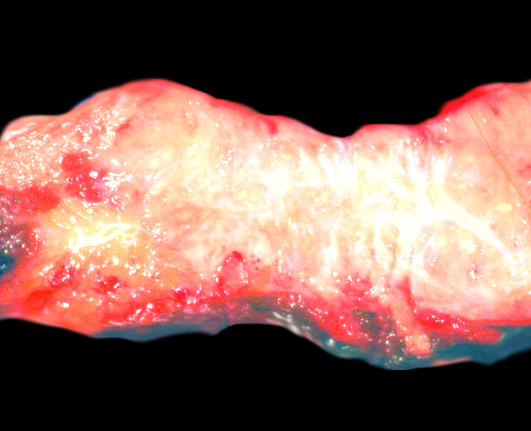
Syringomatous tumor
Microscopic (histologic) description
- Situated in the dermis of the nipple areolar complex
- May focally involve the subcutaneous tissue; does not involve the epidermis
- Infiltrative haphazard proliferation of elongated ductules and tubules lined by a two layer epithelium (luminal and basal)
- Ducts have teardrop, comma or branching shapes
- Identical to cutaneous syringoma
- Tumor cells are bland with absent or rare mitoses
- Background of hyalinized or myxoid fibrous stroma
- Infiltrates around smooth muscle bundle and lactiferous ducts of nipple
- Perineural invasion may be present
- Variable squamous metaplasia with small keratinizing cysts with calcifications
Microscopic (histologic) images
Contributed by Emily S. Reisenbichler, M.D.
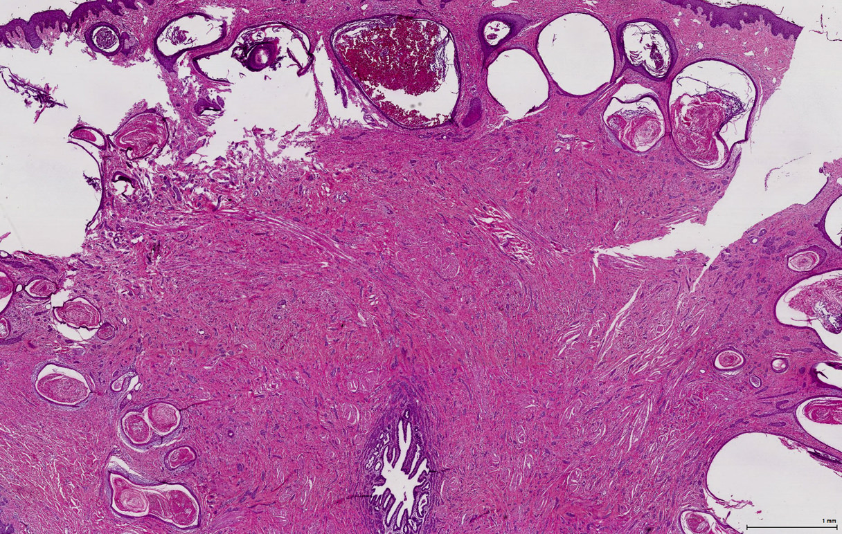

Syringomatous tumor of nipple
AFIP images

Keratinization in syringomatous duct
Contributed by Dr. Mark R. Wick

Syringomatous tumor
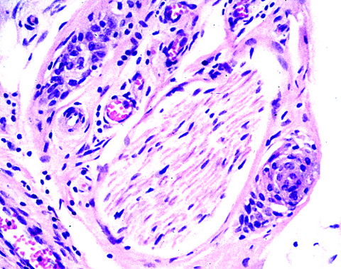
Perineurial invasion
Images hosted on other servers:

Acanthotic epidermis

Small, solitary, evenly spaced duct-like structures

Layers of cuboidal or squamoid epithelial cells

Small, dark tumor cells

Keratotic cysts
Positive stains
- Luminal and myoepithelial cells: HMWCK / 34 beta E12
- Myoepithelial cells: actin, p63, S100
- Squamous metaplasia: p63
Negative stains
Differential diagnosis
- Low grade adenosquamous carcinoma: location is deeper in the breast; arises in breast parenchyma
- Nipple adenoma (sclerosing adenosis pattern): usually seen in association with papillomatous pattern of nipple adenoma (mixed pattern); lacks pattern of stromal infiltration
- Tubular carcinoma: lacks a myoepithelial cell layer, no squamous metaplasia (Arch Pathol Lab Med 2009;133:1487)
Board review style question #1
A 35 year old woman presents with a nipple mass. An excisional biopsy is performed and histomorphology is consistent with syringomatous tumor. Of the following, which histologic description best describes what was seen on the histologic slide of the nipple mass?
- Angulated tubules lined by a single layer of epithelial cells
- Bland tumor cells in a background of mucin
- Comma or teardrop shaped ducts infiltrating stroma
- Ducts with squamous metaplasia communicating with a fistula track to the skin surface
- High grade tumor cells with perineural invasion
Board review style answer #1
C. Comma or teardrop shaped ducts infiltrating stroma. Syringomatous tumor (SyT) of the nipple is identical to syringoma of the skin. These tumors are characteristically described as having comma or teardrop shaped ducts. Although perineural invasion can be seen with SyT, the tumor cells are bland. Option A is a description of tubular carcinoma. Tubules of SyT have more than 1 epithelial layer. Option B is a description of mucinous carcinoma. SyT has a background stroma that can be either myxoid or sclerotic. Option D is a description of squamous metaplasia of lactiferous ducts (SMOLD). Although SyT can have keratin cysts, fistula tracks out to the skin surface is not a feature.
Comment here
Reference: Syringomatous tumor of nipple
Comment here
Reference: Syringomatous tumor of nipple
