
Stains & CD markers
CD138

Copyright: 2002-2024, PathologyOutlines.com, Inc.
PubMed Search: CD138
CD138
Author: Pamela Wirth, Ph.D.
Editorial Board Member: Christian M. Schürch, M.D., Ph.D.
Last staff update: 23 January 2024 (update in progress)
Copyright: 2002-2024, PathologyOutlines.com, Inc.
PubMed Search: CD138

Table of Contents
Definition / general | Essential features | Terminology | Pathophysiology | Clinical features | Interpretation | Uses by pathologists | Prognostic factors | Microscopic (histologic) description | Microscopic (histologic) images | Positive staining - normal | Positive staining - disease | Negative staining | Molecular / cytogenetics description | Sample pathology report | Board review style question #1 | Board review style answer #1 | Board review style question #2 | Board review style answer #2Cite this page: Wirth P. CD138. PathologyOutlines.com website. https://www.pathologyoutlines.com/topic/cdmarkerscd138.html. Accessed April 23rd, 2024.
Definition / general
- Transmembrane proteoglycan expressed in various normal and malignant tissues
- 1 of 4 members of the syndecan family, a cell surface protein involved in the regulation of cell proliferation, migration and organization of the cytoskeleton
- Syndecan derives from the Greek word syndein, which means to bind together (Oncotarget 2015;6:28693)
Essential features
- Used to distinguish and quantitate plasma cells in hematolymphoid tissues and as a biomarker for multiple myeloma cells
- CD138 expression is highly variable among different cancer types
- Both high and low expression levels can be associated with poor prognosis depending on the tumor type
Terminology
- Syndecan 1 (SDC1)
Pathophysiology
- Involved in cell proliferation, migration, adhesion and angiogenesis with loss of CD138 expression leading to enhanced motility and invasion of neoplastic cells (Oncotarget 2015;6:28693)
- Acts as coreceptor to bind growth factors and promote cell survival and proliferation; can also be shed into the surrounding cellular environment
- Has been implicated in the regulation of the Wnt signaling, focal adhesion kinase (FAK), nuclear factor kappa B (NFkB) and mitogen activated protein kinase (MAPK) pathways (J Histochem Cytochem 2015;63:465, Int J Mol Sci 2022;23:9037)
- Involved in leukocyte recruitment and extracellular matrix remodeling during injury repair (Front Immunol 2020;11:227)
Clinical features
- Expressed on cell surface of immature B cells and mature plasma cells
- Overexpressed on malignant plasma cells
Interpretation
- Predominantly membranous but sometimes expressed in the cytoplasm or nucleus
Uses by pathologists
- Used in routine diagnostic pathology to distinguish and quantitate plasma cells in hematolymphoid tissues (Dis Markers 2019;2019:4928315)
- Biomarker for multiple myeloma (Int J Mol Sci 2022;23:9037)
Prognostic factors
- CD138 expression levels generally decrease as tumor grade, stage and invasiveness increase; however, variable expression levels may depend on tumor type
- Expression levels are reduced in squamous cell lung cancer, advanced ovarian cancer, poorly differentiated head and neck squamous cell carcinomas and prostate cancer (Oncotarget 2015;6:28693)
- CD138 expression levels are increased in
- Neoplastic cells of urothelial carcinoma and correlates with high tumor grade, advanced stage and tumor recurrence
- Increased CD138 expression in gallbladder cancer is associated with lymph node metastasis and poor survival (Dis Markers 2019;2019:4928315)
- Most studies show increase of CD138 in breast carcinomas, which correlates with triple negative ductal tumors (negative for ER, PR and HER2) with poor survival (Oncotarget 2015;6:28693)
- Pancreatic cancer, with better prognosis for epithelial expression of CD138 versus stromal expression (Oncotarget 2015;6:28693)
- Some tumors lose CD138 membrane expression but show abnormal cytoplasmic or nuclear expression (Int J Mol Sci 2021;22:4227)
- Appearance of CD138 in tumor stroma is almost always a sign of poor prognosis (Int J Mol Sci 2021;22:4227)
Microscopic (histologic) description
- Expressed on mature plasma cells and immature B cells (Oncotarget 2015;6:28693)
- Expressed widely on the bone marrow hematopoietic cells, endothelial cells and on some breast cancer cells (Leukemia 2020;34:245)
- Negative for other hematolymphoid cells
Microscopic (histologic) images
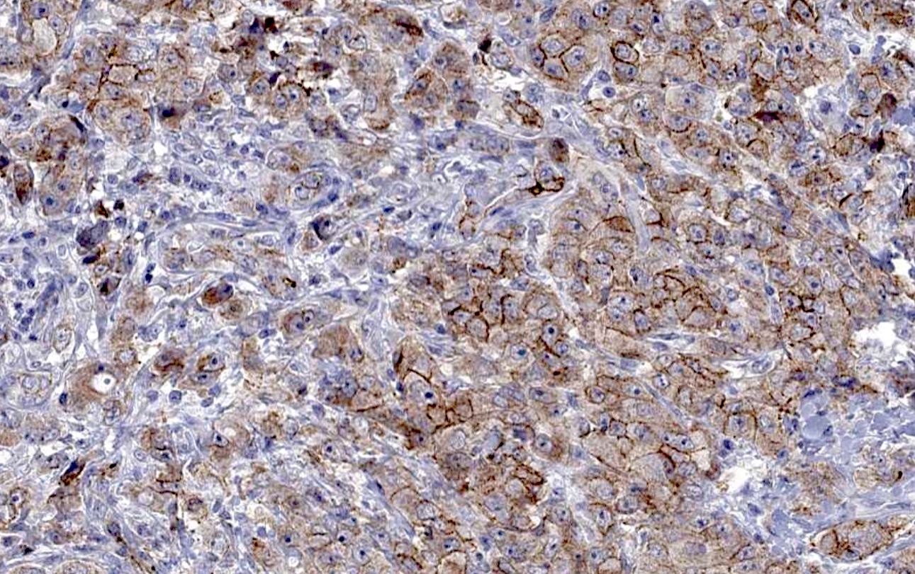
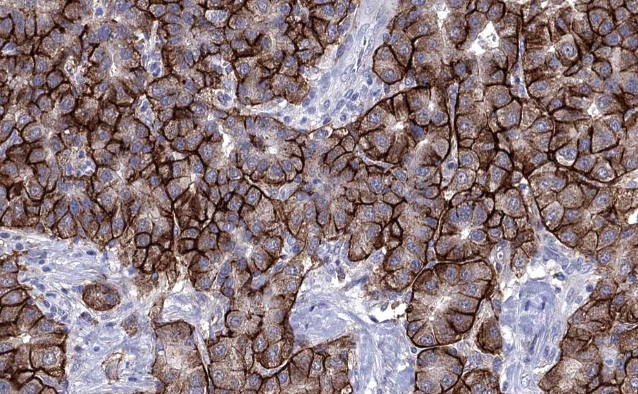
Contributed by Pamela Wirth, Ph.D. (source: University of Toronto and Human Protein Atlas) and Yuri Tachibana, M.D.
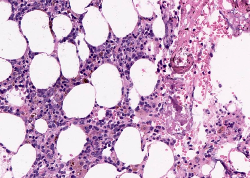
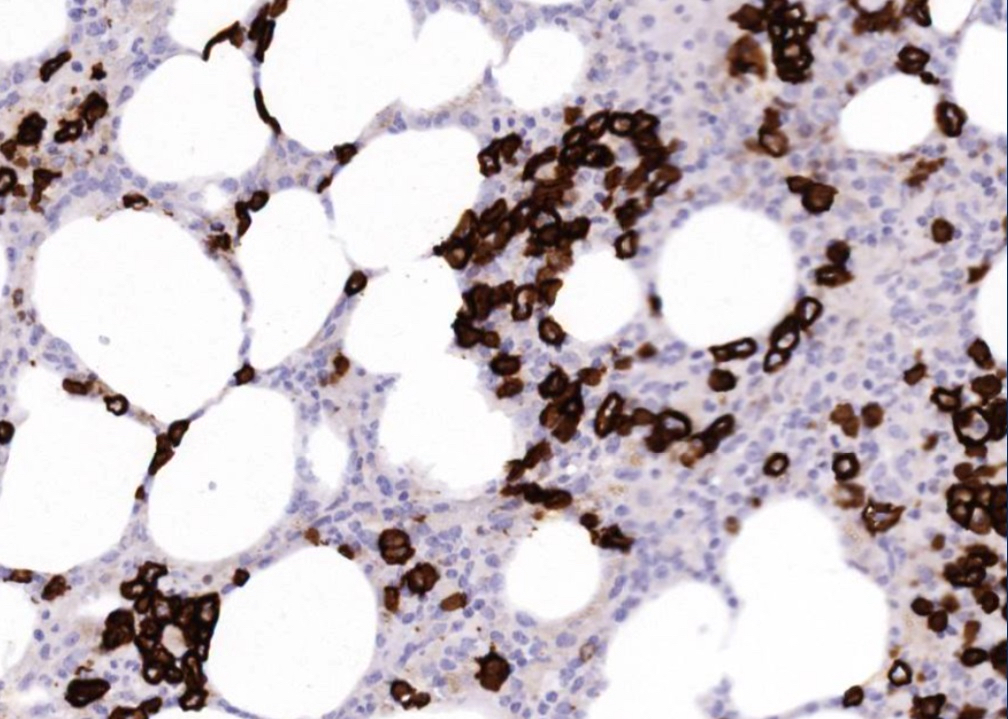
Bone marrow biopsy
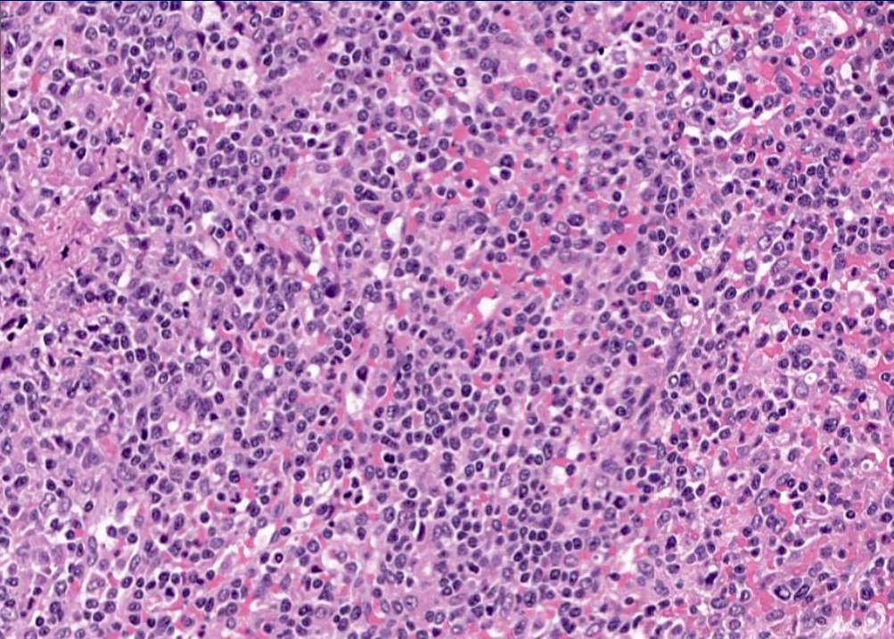
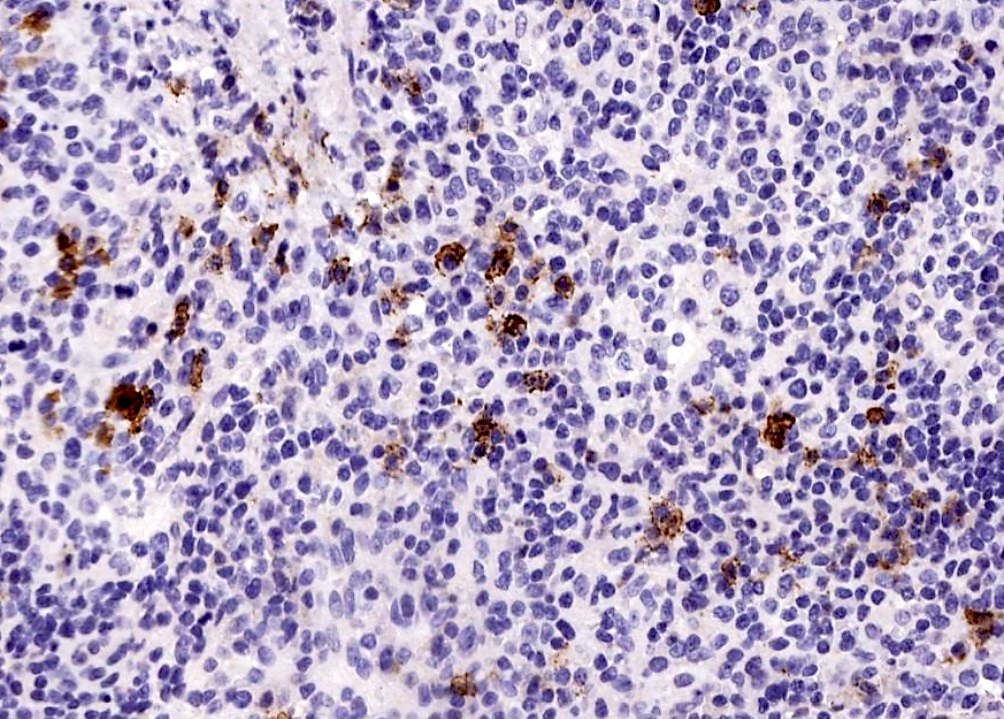
Spleen
Breast carcinoma
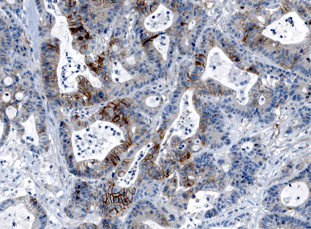
Colon carcinoma
Liver carcinoma
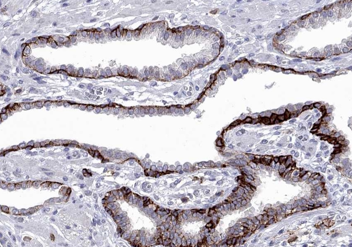
Prostate carcinoma
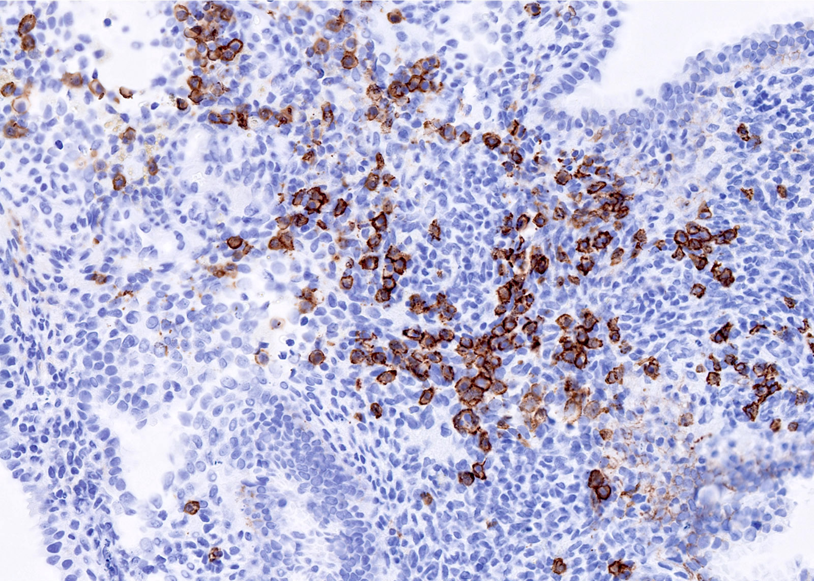
Chronic endometritis
Positive staining - normal
- Normal plasma cells and immature B cells of hematolymphoid organs, plasma cell malignancies
- Strong expression in normal squamous epithelium of various organs
- Goblet and columnar cells of the gastrointestinal tract (Dis Markers 2019;2019:4928315)
Positive staining - disease
- Multiple myeloma tumor cells (95%)
- Carcinomas with strong CD138 expression (tissue samples from microarray)
- Anal carcinoma (63.6%)
- Esophageal squamous cell carcinoma (78.8%)
- Colon adenoma (92%)
- Urothelial carcinoma pTa (90.5%)
- Squamous cell carcinoma of skin (61.9%)
- Basal cell carcinoma of skin (82.9%)
- Hepatocellular carcinoma (72.7%)
- Granular cell tumor (62.5%)
- Squamous cell lung carcinoma (61.9%) (Dis Markers 2019;2019:4928315)
- Invasive ductal breast carcinoma (90%) (Cancer Treat Res Commun 2021;27:100312, Anal Cell Pathol 2018;2018:9432375)
- Loss of membranous staining and the cytoplasmic accumulation of CD138 reflects a poorer outcome (Mol Carcinog 2019;58:2306)
Negative staining
- Low or absent expression in testicular germ cell tumors, sarcomas, melanoma and small cell urinary bladder carcinoma (Dis Markers 2019;2019:4928315)
Molecular / cytogenetics description
- Encoded on chromosome 2p24.1 (Cancer Treat Res Commun 2021;27:100312)
- Mutant KRAS has been shown to induce CD138 expression in pancreatic ductal adenocarcinoma cells (Am J Physiol Cell Physiol 2022;323:C29)
Sample pathology report
- Core biopsy, bone marrow:
- Multiple myeloma (see comment)
- Comment: The patient is a 57 year old man with symptoms of exertional fatigue, anemia and back pain. Immunohistochemistry was performed to quantify plasma cells (CD138) in addition to Ig kappa and lambda to establish clonality. CD138 positive cells represent 20 - 25% of total cellularity. Results will be correlated with flow cytometry and FISH.
Board review style question #1
CD138 assists in the identification of multiple myeloma by labeling which of the following types of cells (as shown in the image)?
- Activated T cells
- Early B cell precursor cells
- Immature T cells
- Peripheral B lymphocytes
- Plasma cells
Board review style answer #1
E. Plasma cells. CD138 is a useful marker of plasma cells and can help support a diagnosis of multiple myeloma. Answers B, C and D are incorrect because CD138 is not a useful marker in identifying T or B cells. While B cells may express CD138 during earlier stages of development, they lose expression of this marker at maturity. Answer A is incorrect because CD138 is rarely expressed on other hematopoietic cells; CD38 and not CD138 would be a better marker for activated T cells.
Comment Here
Reference: CD138
Comment Here
Reference: CD138
Board review style question #2
Increased expression of CD138 has been found in which of the following cancer types?
- Liposarcomas
- Melanomas
- Sarcomas
- Triple negative breast cancer
Board review style answer #2
D. Triple negative breast cancer. CD138 is expressed in most triple negative breast cancers (TNBC). The increased expression of CD138 may contribute to the formation of a tumor supportive microenvironment, promoting the aggressive growth and invasion of cancer cells that are commonly noted in TNBC. Answers A and C are incorrect because CD138 is rarely expressed in soft tissue tumors. Answer B is incorrect because expression of CD138 in melanomas is low to absent; however, low levels of staining have been observed in metastatic forms of melanoma.
Comment Here
Reference: CD138
Comment Here
Reference: CD138

