
Stains & CD markers
CD15

Copyright: 2003-2024, PathologyOutlines.com, Inc.
PubMed Search: CD15
Related topics: CD15s, CD15u
CD15
Author: Nat Pernick, M.D.
Last author update: 1 January 2014
Last staff update: 19 March 2024
Copyright: 2003-2024, PathologyOutlines.com, Inc.
PubMed Search: CD15
Related topics: CD15s, CD15u

Table of Contents
Definition / general | Clinical features | Uses by pathologists | Case reports | Microscopic (histologic) images | Positive staining - normal | Positive staining - disease | Negative stainingCite this page: Pernick N. CD15. PathologyOutlines.com website. https://www.pathologyoutlines.com/topic/cdmarkerscd15.html. Accessed April 17th, 2024.
Definition / general
- A carbohydrate (not a protein) widely used for diagnosis of Hodgkin lymphoma
- Also known as LeuM1, Lewis X, 3-fucosyl-N-acetyl-lactosamine
- Mediates phagocytosis and chemotaxis
- Synthesis is directed by FUT4 (OMIM #104230) in lymphoid cells and mature granulocytes, and by FUT9 (OMIM #606865) in promyelocytes and monocytes
- References: Quality control for CD15
Clinical features
- Poor prognostic marker in acute promyelocytic leukemia (Leuk Res 2014;38:194)
- May be useful to differentiate recent antemortem from postmortem injuries (Int J Legal Med 2013;127:957)
- Helps differentiate pulmonary adenocarcinoma (CD15+) from mesothelioma (CD15-), although other markers are more specific
- May help define neural stem cells (Stem Cells 2009;27:2928)
Uses by pathologists
- Hodgkin lymphoma: membranous, diffuse cytoplasmic or Golgi staining of Reed-Sternberg cells; CD15 staining is used to confirm diagnosis, or to differentiate Hodgkin lymphoma (CD15+) from anaplastic large cell lymphoma (usually CD15-)
- Granulocyte marker
Case reports
- 64 year old woman with CD15+ pre-B ALL (Arch Pathol Lab Med 2001;125:1227)
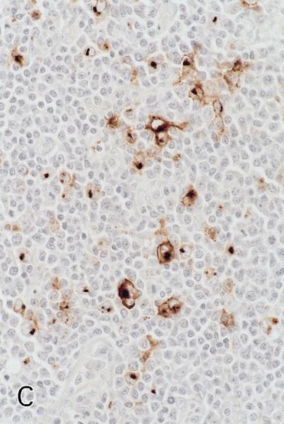
Microscopic (histologic) images
Contributed by Raghava Munivenkatappa, M.D. (Case #48) and AFIP
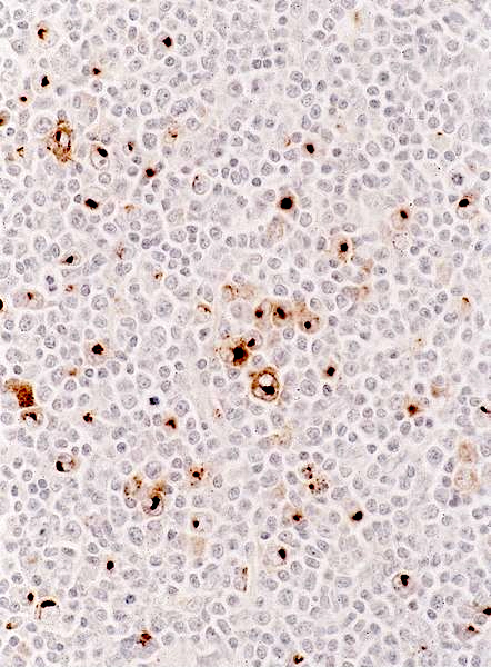
Paranuclear / Golgi staining pattern
Nodular lymphocyte predominant
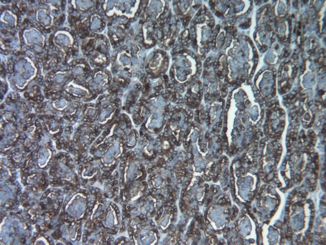
Renal cell carcinoma: papillary type
Images hosted on other servers:

Small intestine: normal Paneth cells (fig B)


Colon: invasive colorectal carcinoma
Positive staining - normal
- Myeloid cells and eosinophils; activated B and T cells (including infectious mononucleosis); variable monocytes and basophils
- Kidney proximal convoluted tubules; small intestine Paneth cells (J Clin Pathol 1996;49:474)
Positive staining - disease
- Hodgkin lymphoma: Reed-Sternberg cells in classic and follicular Hodgkin lymphoma (Am J Clin Pathol 2002;117:29)
- 50% of carcinomas, including some colorectal carcinomas (Korean J Pathol 2013;47:340), ovarian / peritoneal serous tumors (Am J Surg Pathol 1998;22:1203), renal cell carcinomas
- 15% of peripheral T cell lymphoma (Am J Surg Pathol 2003;27:1513, Int J Oncol 2003;22:319)
- 5% of B cell lymphomas, including some B-CLL and pre-pre B ALL (Am J Clin Pathol 2002;117:380, Arch Pathol Lab Med 2001;125:1227)
- Some AML, particularly AML-M4 / M5 (flow cytometry is more sensitive than immunohistochemistry), some granulocytic sarcomas (Histopathology 1999;34:391), some histiocytic sarcomas
- Occasionally anaplastic large cell lymphoma (Am J Clin Pathol 2003;119:205, but usually negative, Am J Surg Pathol 2006;30:223)
- Note: EBV infections can have Reed-Sternberg-like cells that are focally CD15+ (Am J Surg Pathol 2010;34:1715, Am J Surg Pathol 2010;34:405)
Negative staining
- Non-activated lymphocytes; erythroid cells, histiocytes (usually), osteoblasts and platelets
- LP/L&H cells in nodular lymphocyte predominant Hodgkin lymphoma
- Diffuse large B cell lymphoma, hairy cell leukemia, post-transplant lymphoproliferative disorders and systemic mastocytosis (Hum Pathol 2001;32:545)
- Langerhans cell histiocytosis, mesothelioma at various sites (usually, Hum Pathol 2001;32:529)

