
CD1-9


- Family of nonpolymorphic MHC class I-like glycoproteins on surface of various antigen presenting cells; member of immunoglobulin superfamily
- Mediate presentation of endogenous and foreign lipids (including bacterial lipid antigens) on cell surface for recognition by T cell receptors
- To sample a diverse antigen pool, CD1 proteins are repeatedly internalized and recycled; this may be assisted by lipid transfer proteins such as saposins (J Biol Chem 2013;288:19528, Proc Natl Acad Sci USA 2012;109:4357)
- May also mediate thymic T cell development
- Has 5 different subsets (CD1a, CD1b, CD1c, CD1d, CD1e), all noncovalently associated with beta 2 microglobulin, all on #1q22-23 (non MHC linked)
- Different CD1 forms bind to different types of lipid antigen based on differences in their antigen binding grooves (Nat Rev Immunol 2005;5:387)
- Separated into 2 groups based on sequence homology:
- Group 1 (CD1a, CD1b, CD1c) present in humans, some other mammals, not in mouse or rat
- Group 2 (CD1d) present in all mammals studied (Curr Pharm Des 2009;15:3311)
- CD1e is an intermediate form, expressed intracellularly (J Biol Chem 2000;275:37757)
- Pathophysiology:
- Cellular infection with Mycobacteria tuberculosis or exposure to mycobacterial cell wall products converts CD1- myeloid precursors into competent CD1+ antigen presenting cells (J Immunol 2005;175:1758)
- However M. tuberculosis possesses a successful tactic based, at least in part, on CD1 downregulation to evade CD1 dependent immunity (Clin Dev Immunol 2011;2011:790460)
- Pollen lipids are also recognized as antigens by T cells via CD1 dependent pathway (J Exp Med 2005;202:295)
- Cellular infection with Mycobacteria tuberculosis or exposure to mycobacterial cell wall products converts CD1- myeloid precursors into competent CD1+ antigen presenting cells (J Immunol 2005;175:1758)
- Clinical features:
- Inhibition of CD1 expression may be a mechanism of immune system evasion by:
- Metastatic melanoma (Am J Pathol 2004;165:1853)
- Leishmania donovani (Infect Immun 2004;72:589)
- Some mycobacteria (Infect Immun 2009;77:4947)
- Primary deficiency of microsomal triglyceride transfer protein in abetalipoproteinemia is associated with loss of CD1 function (J Clin Invest 2010;120:2889)
- Expression altered in multiple sclerosis (Clin Exp Immunol 2012;169:10)
- Inhibition of CD1 expression may be a mechanism of immune system evasion by:
- Uses by pathologists: CD1a is used to diagnose Langerhans cell histiocytosis and to exclude other entities that are CD1a negative
- See CD1a
- On chromosome 1q22-23 (not MHC linked); noncovalently associated with beta2 microglobulin
- Capable of binding ligands with greatly varying alkyl chain lengths (including Mycobacteria tuberculosis and Mycobacteria leprae) through a complex network of interconnected hydrophobic pockets (J Immunol 2004;172:2382, Proc Natl Acad Sci USA 2011;108:19335, OMIM: 188360 [Accessed 13 May 2021])
- Part of immune response to intracellular bacteria
- Human gammadelta T cells recognize Lipid A in a CD1b or CD1c restricted manner as a first response to gram negative bacteria (Biol Direct 2009;4:47)
- No significant clinical use by pathologists
- Positive staining - normal: cortical thymocytes, Langerhans cells (weaker staining than CD1a), myeloid dendritic cells, brain pyramidal cells and subpopulation of B cells
- Positive staining - disease:
- Dendritic cells in mycosis fungoides (J Cutan Pathol 1995;22:223)
- Myeloid leukemia, some B and T cell malignancies
- Monocytes of most sickle cell anemia patients (Hum Immunol 2004;65:1370)
- Negative staining: normal B cells
- On chromosome 1q22-23 (not MHC linked); noncovalently associated with beta2 microglobulin
- Mediates the presentation of primarily lipid and glycolipid antigens of self or microbial origin to T cells (Immunity 2010;33:853)
- Has the capacity to present a wide repertoire of antigens to reactive T cells (Mol Immunol 2013;55:182, J Biol Chem 2011;286:37692)
- Expressed on a subset of myeloid dendritic cells; secrete IL10 in response to Escherichia coli (Eur J Immunol 2012;42:1512)
- Common CD1c antibody is BDCA1
- Clinical features:
- Assists with presentation of the phospholipid antigen mannosyl beta (1) phosphomycoketide (Clin Dev Immunol 2012;2012:981821)
- Produced by Mycobacteria tuberculosis and M. bovis Bacille-Calmette-Guerin (J Exp Med 2004;200:1559, Nature 2000;404:884)
- May activate intrathyroidal T cells in Hashimoto thyroiditis and Grave disease (J Immunol 2005;174:3773)
- May promote autoantibodies in systemic lupus erythematosus (J Immunol 2000;165:5338)
- No significant clinical use by pathologists
- Positive staining - normal:
- Cortical thymocytes, Langerhans cells (weaker staining than CD1a), immature myeloid dendritic cells (J Clin Invest 2007;117:2517)
- Subset of normal peripheral B cells, subset of activated T cells
- Positive staining - disease:
- Dendritic cells in mycosis fungoides (J Cutan Pathol 1995;22:223)
- Myeloid leukemia, some B and T cell malignancies and monocytes of most sickle cell anemia patients (Hum Immunol 2004;65:1370)
- Negative staining: normal B cells
- Electron microscopy description: present in early and late endosomes (J Exp Med 2000;192:281)
- CD1d presents lipid antigens to natural killer T (NKT) cells; when activated, NKT cells rapidly produce Th1 and Th2 cytokines, typically represented by interferon gamma and IL4 (Wikipedia: CD1D [Accessed 31 July 2018], OMIM: 188410 [Accessed 31 July 2018])
- B cells also present lipid antigen to CD1d restricted invariant NKT cells, which contributes to maintaining tolerance in autoimmunity
- On chromosome 1q22-23 (not MHC linked)
- Also called R3G1
- Sole group 2 member of the CD1 family of MHC-like glycoproteins
- Clinical features:
- CD1d expression < 45% is predictive of CLL (Am J Clin Pathol 2011;136:400)
- Downregulated by B-CLL (Leukemia 2002;16:2429)
- Although the invariant natural killer T cell and CD1d axis is fundamentally intact in patients with early stage disease (Haematologica 2013;98:376)
- In paroxysmal nocturnal hemoglobinuria, a novel, autoreactive, CD1d restricted, glycosylphosphatidylinositol specific T cell population is expanded and may cause bone marrow failure (Blood 2013;121:2753)
- B cells are essential for iNKT cell expansion and activation in healthy donors but fail to exert a similar effect in SLE patients (Immunity 2012;36:477, J Exp Med 2010;207:943)
- May protect against lipid antigen rich infectious microbes on human scalp (J Clin Pathol 2005;58:1278)
- Downregulated by HPV and some other viruses (J Virol 2010;84:11614, Viruses 2012;4:2379)
- But not CMV (Biochem Biophys Res Commun 2011;413:616)
- Expressed in NKT cells active in autoimmune diabetes, tumor rejection and some microbial infections
- CD1d expression < 45% is predictive of CLL (Am J Clin Pathol 2011;136:400)
- Uses by pathologists: no significant clinical use by pathologists; may play a role in future vaccine development (Proc Natl Acad Sci U S A 2010;107:13010)
- Case reports: 6 year old boy with no apparent immunodeficiency who suffered severe life threatening infection with varicella vaccine virus, with low invariant natural killer T cells and diminished CD1d expression (J Infect Dis 2011;204:1893)
- Positive staining - normal:
- Dendritic cells, intestinal epithelial cells, B cell subset, NK T cell subset, low levels in thymus and monocytes (Immunol Lett 2005;100:42, J Immunol 2005;175:4416)
- Bronchial epithelium (PLoS One 2011;6:e22726)
- Scalp epidermis (basal and granular keratinocytes), pilosebaceous units and eccrine glands
- Testis: Leydig cell, Sertoli cells, spermatogonia; moderate expression in spermatocytes (Ultrastruct Pathol 2011;35:124)
- Positive staining - disease: some B and T cell malignancies; keratinocytes in psoriasis
- Negative staining: normal B cells
- Participates in lipid antigen presentation without interacting with T cell receptor; binds lipids in lysosomes and facilitates processing of complex glycolipids, thus promoting editing of lipid antigens; may positively or negatively affect lipid presentation by CD1b, CD1c, CD1d (Proc Natl Acad Sci USA 2011;108:14228)
- Naturally occurring mutation in CD1e impairs lipid antigen presentation (J Immunol 2008;180:3642)
- Only CD1 protein existing in soluble form in late endosomes of dendritic cells; not expressed on cell surface
- On chromosome 1q22-23 (not MHC linked); noncovalently associated with beta2 microglobulin
- Clinical features:
- Processes mycobacterial antigen (instead of presenting antigen directly) and may help expand repertoire of glycolipid T cell antigens to optimize the immune response (Science 2005;310:1321, Proc Natl Acad Sci USA 2011;108:13230)
- Presentment is done by CD1b (J Biol Chem 2012;287:31494)
- Initially accumulates in Golgi of immature dendritic cells or transfected cells as a membrane associated form, then is transported to early and then late endosomes, then is cleaved into a soluble form, which facilitates the processing of glycolipids presented by other CD1 molecules transiting through the same compartments (Biochem J 2009;419:661)
- +6129 A/G gene polymorphisms are associated with multiple sclerosis (Immunol Invest 2010;39:874)
- Processes mycobacterial antigen (instead of presenting antigen directly) and may help expand repertoire of glycolipid T cell antigens to optimize the immune response (Science 2005;310:1321, Proc Natl Acad Sci USA 2011;108:13230)
- No significant clinical use by pathologists
- Positive staining - normal: dendritic cells
- Early T cell marker expressed on all peripheral blood T cells, 95% of thymocytes, NK cells but no B cells (OMIM: 186990 [Accessed 13 May 2021], J Biol Chem 2010;285:41755)
- Gene is at 1p13.1; protein is member of immunoglobulin superfamily
- Also called E rosette receptor because CD2 antibodies inhibit formation of rosettes with sheep erythrocytes
- Also called LFA2 (leukocyte function antigen), T11
- Pathophysiology:
- Binds CD58 (LFA3) on antigen presenting cells, which enables T cells to respond to lower concentrations of antigen (J Exp Med 1999;190:1383)
- Mediates adhesion between T cells and antigen presenting cells, induces costimulatory signals in T cells and T cell cytokine production, inhibits apoptosis of activated peripheral T cells (Immunology 2013;139:48)
- Regulates T cell anergy
- Regulates T and NK mediated cytolysis
- CD2 distinguishes 2 subsets of human plasmacytoid dendritic cells with distinct phenotype and functions (J Immunol 2009;182:6815)
- CD2 (low) plasmacytoid dendritic cells are depleted in HIV+ patients (Cell Mol Immunol 2011;8:441)
- CD2 promotes NK cell membrane nanotube formation (PLoS One 2012;7:e47664)
- Clinical features:
- Abnormal levels of CD2 expression by flow cytometry are frequently observed in mature T cell malignancies but clinical significance is unclear because of variability of CD2 expression in normal T cells (Cytometry B Clin Cytom 2010;78:169)
- Rare aberrant expression in B cell lymphomas (Appl Immunohistochem Mol Morphol 2011;19:579)
- High CD2 expression identifies latently infected resting memory CD4(+) T cells in vivo (J Virol 2013;87:9148)
- Siplizumab, an anti CD2 monoclonal antibody, was previously studied to treat plaque psoriasis; no current clinical trials as of last update (Int J Dermatol 2010;49:818)
- Uses by pathologists: T cell marker (membranous staining), although CD3 is more commonly used; marker of systemic mastocytosis
- Positive staining - normal:
- Thymocytes (95%), mature peripheral T cells (almost all), NK cells (80 - 90%), mast cells
- Variable thymic B cells (50%), Langerhans cells
- Positive staining - disease:
- T-ALL, other T cell lymphoma / leukemia, acute promyelocytic leukemia microgranular variant / FAB M3v (Leukemia 1995;9:1461)
- AML with pseudo-Chédiak-Higashi anomaly (Am J Clin Pathol 2006;125:791)
- Systemic mastocytosis (a minor criteria, Hum Pathol 2001;32:545)
- And other mast cell disease, thymoma (lymphocyte predominant)
- Variable:
- Acute myeloid lymphoma M0
- Rare:
- Pyothorax associated B cell lymphoma (Am J Surg Pathol 2002;26:724)
- Other non-Hodgkin B cell lymphomas, Reed-Sternberg / Hodgkins cells (Mod Pathol 2005;18:1542)
- Myeloma (Mod Pathol 1990;3:302)
- Myeloid leukemia
- Negative staining: B cells, basophils, nonhematopoietic neoplasms and mast cells in nonmastocytosis disorders (Am J Clin Pathol 2003;120:64)
- CD2 epitope present on activated T cells, unmasked by conformational change of CD2 glycoprotein during activation
- No recent articles in literature
- No significant clinical use by pathologists
- Positive staining - normal: activated T cells
- Positive staining - disease: blood and synovial fluid T cells in rheumatoid arthritis; peripheral blood T cells in juvenile rheumatoid arthritis, SLE, ankylosing spondylitis and Lyme disease (Scand J Immunol 1991;34:351)
- Negative staining: resting T cells
- See CD3
- See CD4
- See CD5
- Adhesion molecule mediating binding of developing thymocytes with thymic epithelial cells (OMIM: 186720 [Accessed 13 May 2021])
- Belongs to ancient scavenger receptor superfamily; at #11q13
- Also called T cell differentiation antigen, T12
- Essential for stable contact between T cells and antigen presenting cells, which leads to T cell proliferation (Blood 2006;107:3212)
- Pathophysiology:
- Binds to CD166 (ALCAM)
- Monomeric 105 or 130kD membrane glycoprotein; size difference is due to differences in phosphorylation (Eur J Immunol 1995;25:2765)
- May be signaling attenuator whose expression alone is sufficient to restrain signaling in T cells (Eur J Immunol 2012;42:195)
- Clinical features:
- Antibodies to CD6 are used to deplete T cells from bone marrow transplants to prevent graft versus host disease (J Clin Oncol 2001;19:1152, Int J Hematol 1999;69:27, J Allergy Clin Immunol 2008;122:1185)
- Single nucleotide polymorphisms may be risk factor for multiple sclerosis (J Neuroimmunol 2010;223:100, PLoS One 2013;8:e62376)
- Due to altered proliferation of CD4+ T cells (J Immunol 2011;187:3286)
- No significant clinical use by pathologists
- Positive staining - normal:
- Mature T cells, B cell subset (B1 cells), NK cells (subset), CNS cells (J Innate Immun 2011;3:420)
- High levels on mature (medullary) thymocytes, low levels on immature (cortical) thymocytes
- T cell cell surface protein that plays important role in T cell B cell interaction in early lymphoid development
- Member of immunoglobulin superfamily at 17q25.2-25.3
- Membrane expression early during T cell development, before TCR rearrangement; persists until terminal stages of T cell development (OMIM: 186820 [Accessed 13 May 2021])
- Has costimulatory activity for T cells (Immunol Res 2001;24:31)
- Binds galectin 1 and is essential for galectin 1 mediated T cell death; loss of CD7 expression and altered cellular glycosylation may contribute to apoptosis resistance in mycosis fungoides (Mod Pathol 2003;16:543)
- Clinical features:
- Positive expression is a negative prognostic marker:
- Myeloid malignancies (Blood 2008;111:1067, Int J Hematol 2010;91:303)
- Epidermotropic CD8+ cytotoxic T cell lymphoma (Clin Exp Dermatol 2012;37:128)
- Loss of expression a negative prognostic marker:
- Acute myelogenous leukemia with FLT3 / ITD mutation (Am J Clin Pathol 2008;129:624)
- Adult T cell leukemia / lymphoma (Histopathology 2009;54:214)
- Aggressive NK cell lymphoma (Korean J Lab Med 2009;29:491)
- HTLV1 (PLoS One 2013;8:e53728)
- Downregulated in infectious mononucleosis (Am J Clin Pathol 2003;120:49)
- Positive expression is a negative prognostic marker:
- Uses by pathologists: marker for T-ALL; CD7 and CD33 may be useful to quantify lymphocyte subsets (Cytometry A 2013;83:316)
- Positive staining - normal:
- Thymocytes, mature T cells (85%), NK cells (majority) (Blood 2009;114:4823)
- Also monocytes, pluripotent hematopoietic progenitor cells, early myeloid cells, pre-B cells
- Positive staining - disease:
- T-ALL (very good marker) and other malignant immature T cells, CD7+ stem cell lymphoma (Am J Surg Pathol 2003;27:1366)
- NK lymphoma but expression lost in aggressive NK cell lymphoma (Korean J Lab Med 2009;29:491)
- Chronic myelogenous leukemia, Down syndrome associated transient myeloproliferative disorder and AML (Am J Clin Pathol 2001;116:204)
- AML (some), lymphocyte rich thymoma (Am J Clin Pathol 2004;121:268)
- Cholangiocarcinoma, epithelioid sarcoma, pancreatic ductal carcinoma (Am J Clin Pathol 2003;120:64)
- T cells in classical Hodgkin lymphoma (Cytometry B Clin Cytom 2009;76:169)
- Reed-Sternberg cells are negative (Mod Pathol 2005;18:1542)
- Rarely B cell lymphomas (Am J Clin Pathol 2001;115:396)
- Negative staining:
- Mature B cells, basophils, granulocytes
- B cell ALL, mycosis fungoides, adult T cell leukemia / lymphoma (Histopathology 2009;54:214)
- See CD8
- Cell surface protein that mediates adhesion, migration, signal transduction
- Also called tetraspanin CD9, motility related protein 1 (MRP1) (note: MRP is multidrug resistance associated protein)
- Antibodies are used to purge bone marrow in acute lymphoblastic leukemia prior to peripheral stem cell bone marrow transplant (Am J Pediatr Hematol Oncol 1993;15:162)
- Pathophysiology:
- Member of transmembrane 4 superfamily (tetraspanin family); tetraspanins have 4 hydrophobic domains, organize multimolecular complexes in plasma membrane; each tetraspanin associates specifically with integrins and other tetraspanins, forming primary complexes and leading to molecular network of interactions, the "tetraspanin web"
- Regulates cell motility, development, activation, growth and adhesion (Blood 2011;117:1840)
- Differentiation (OMIM: 143030 [Accessed 13 May 2021])
- Fertilization (oocyte CD9 is required for sperm egg fusion)
- Regulates paranodal junction formation (between neurons and glia)
- Required for microparticle release from coated platelets (Platelets 2009;20:361)
- Triggers platelet activation and aggregation
- Supports myotube maintenance and promotes muscle cell fusion
- Downregulation in ovarian carcinoma may promote tumor dissemination (Cancer Res 2005;65:2617)
- May suppress metastasis in small cell lung cancer by promoting apoptosis via calretinin expression (Cancer Res 2010;70:8025, FEBS Open Bio 2013;3:225)
- May mediate invasion by upregulating MMP9 (PLoS One 2013;8:e67766)
- Clinical features:
- Favorable prognostic marker for:
- Gallbladder carcinoma (World J Surg Oncol 2012;10:92)
- Gastric GIST (Cancer Sci 2011;102:883)
- Malignant mesothelioma (Oncol Rep 2013;29:21)
- Oral squamous cell carcinoma (Oral Oncol 2010;46:166)
- Poor prognosis factor in gastric carcinoma (Pathol Res Pract 2010;206:607)
- Favorable prognostic marker for:
- No significant clinical use by pathologists
- Positive staining - normal:
- Pre B cells, B cell subset, activated T cells, basophils, eosinophils (Am J Respir Cell Mol Biol 2012;46:188)
- Macrophages, megakaryocytes, plasma cells, plasma cell precursors in germinal centers (Biochem Biophys Res Commun 2013;431:41), platelets
- Brain, cardiac muscle, GI system, kidney (glomeruli, tubules and collective ducts), liver, lymphatic epithelium, ovarian surface epithelium, peripheral nerve, skin, spleen, thyroid, tonsil
- Positive staining - disease:
- Pre-B ALL, acute promyelocytic leukemia, astrocytoma, mast cell disease (J Histochem Cytochem 2002;50:1195)
- Vascular tumors: angiosarcoma, juvenile nasopharyngeal angiofibroma, Kaposi sarcoma (Mod Pathol 2003;16:1028)
- Negative staining: red blood cells, renal collecting duct carcinomas (almost all)
Images hosted on other servers:

CD1d: antigen presentation

CD1d: viral evasion of antigen presentation

CD1d: intestinal epithelial CD1d expression

CD2: T cells and antigen presenting cells

CD9: structure
Images hosted on other servers:

CD1: primary and metastatic melanoma

CD1: Langerhans cells of anal mucosa

CD1b: leprosy patient

CD1c: marginal zone B cells (spleen)

CD1c: stomach: H. pylori+ mucosa

CD1c: thyroid gland

CD1c: immature dendritic cells

CD1d: HPV associated lesions (cervix)

CD1d: trophoblast (placenta)


CD1d: scalp skin

CD2: hydroa
vacciniforme-like
lymphoma

Brain (CD6-)

CD6: colon

Kidney (CD6-)

Skeletal muscle (CD6-)

CD6: tonsil

Uterus (CD6-)
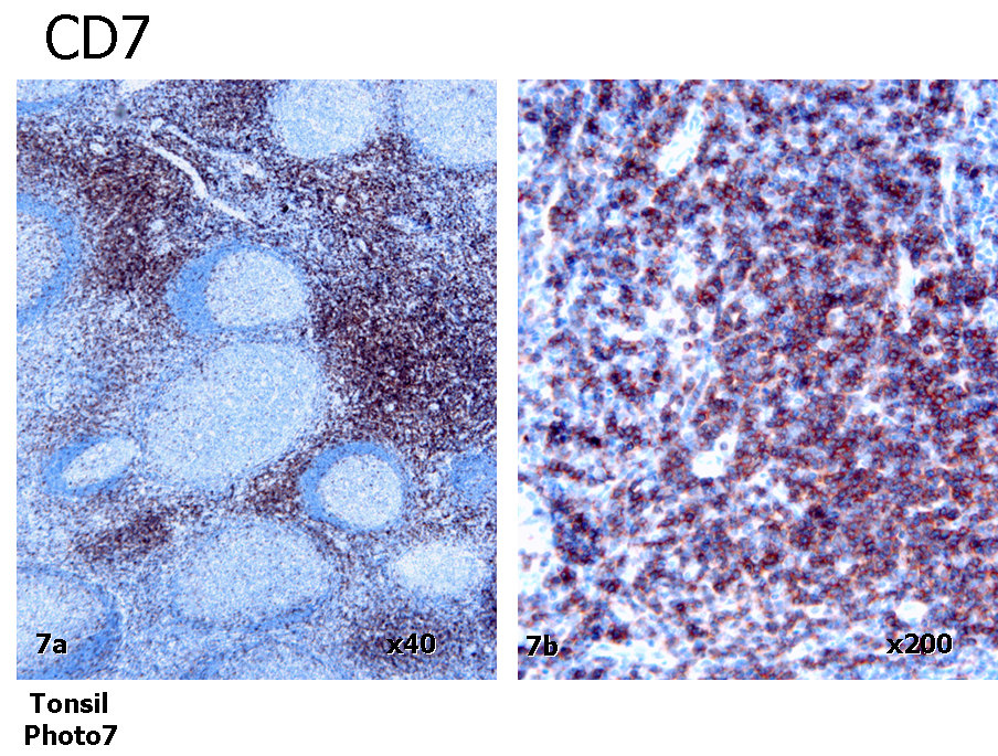
CD7: normal tonsil

CD7: cutaneous T cell lymphoma

CD9: normal tissue (cerebrum)

CD9: normal tissue (cervical squamous epithelium)

CD9: cervical carcinoma


CD9: CNS nonneuroepithelial tumors

CD9: well differentiated adenocarcinoma (gallbladder)

CD9: mesothelioma

CD9: basal and
squamous cell
carcinoma and
actinic keratosis


CD9: small cell lung cancer
Image hosted on other servers:

CD1c: various cellular locations

