

Fallopian tubes & broad ligament
Fallopian tube nontumor
Metaplastic changes
Last author update: 1 October 2017
Last staff update: 24 December 2022
Copyright: 2002-2024, PathologyOutlines.com, Inc.
PubMed Search: Metaplasia fallopian tube


Table of Contents
Definition / general | Essential features | Epidemiology | Sites | Etiology | Diagnosis | Prognostic factors | Case reports | Gross description | Microscopic (histologic) description | Microscopic (histologic) images | Negative stains | Differential diagnosis | Board review style question #1 | Board review style answer #1Cite this page: Qiao JH., Riddle N, Shutter J. Metaplastic changes. PathologyOutlines.com website. https://www.pathologyoutlines.com/topic/fallopiantubestransitionalcellmetaplasiafimbriae.html. Accessed April 25th, 2024.
Definition / general
- Defined as alteration in which the normal tubal epithelium is replaced by metaplastic cells resembling benign transitional (urothelial) cells of urinary bladder (Am J Surg Pathol 2009;33:111)
Essential features
- Transitional cell metaplasia is a benign epithelial alteration but is underrecognized in the tubal fimbriae, where it may mimic serous tubal intraepithelial neoplasm / carcinoma
- It is increasing found because over the past two decades, risk reducing salpingo-oophorectomy (RRSO) for BRCA1 and BRCA2 mutation patients is being performed to identify epithelial atypia and early intraepithelial carcinoma in the distal fallopian tube / fimbriae
- Opportunistic salpingectomy is a viable alternative to salpingo-oophorectomy for BRCA+ women, which reduces the risk of serous carcinoma while preserving ovarian function
Epidemiology
- Present in 26% of all RRSO specimens
- Usually multifocal, with involvement of tip, edges or base of the fimbrial plicae
- Average size of a metaplastic focus is 1.3 mm (ranging from 0.1 to 10 mm)
- Also due to fallopian tube prolapse or peritoneal dialysis (squamous) (Int J Gynecol Pathol 2008;27:465)
- Also associated with ovarian mucinous tumors, Peutz-Jeghers syndrome, endometriosis within fallopian tube
Sites
- Distal fallopian tube / fimbriae
Etiology
- Unclear: it is possible that transitional cell metaplasia of the fimbriae is a reaction to mechanical irritation, inflammation or infection
- Findings that only the distal fimbriae are involved and not the isthmus, ampulla or infundibulum supports the notion of a reaction to peritoneal based stimulus
- Prior pregnancy is a potential inciting incident
Diagnosis
- Diagnosis of transitional cell metaplasia of fimbriae is only possible after salpingectomy by microscopic examination of distal fallopian tube / fimbriae
Prognostic factors
- Clinical followup of transitional cell metaplasia of the fimbriae shows no biologic significance
Case reports
- 45 year old woman with prolapsed fallopian tube with squamous metaplasia (J Postgrad Med 2002;48:241)
- 52 year old woman with papillary syncytial metaplasia of fallopian tube endometriosis (Arch Pathol Lab Med 2013;137:126)
Gross description
- Generally, such lesions are undetectable grossly and are only identified by careful histologic examination of sections of fallopian tubes; thus, fallopian tubes have to be submitted entirely in serial cross sections whereas fimbriae have to be submitted entirely in longitudinal sections
Microscopic (histologic) description
- Mostly commonly mucinous metaplasia; urethelial and squamous metaplasia less common
- Characteristic features of transitional cell metaplasia are the presence of uniform nuclei with pale chromatin and nuclear grooves and abundant cytoplasm; these features are not seen in serous tubal intraepithelial neoplasia, including serous tubal intraepithelial carcinoma (STIC) and serous tubal epithelial proliferation of uncertain significance
- Careful histologic examination, attention to cytologic details and p53 and Ki67 staining will help prevent the misinterpretation of transitional cell metaplasia as malignancy
Microscopic (histologic) images
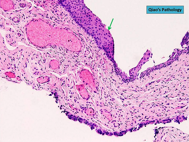
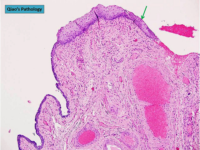
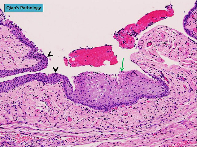
Contributed by Jian-Hua Qiao, M.D.
Extensive transitional cell metaplasia
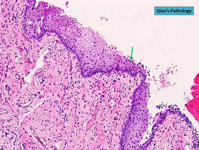
Focal transitional cell metaplasia
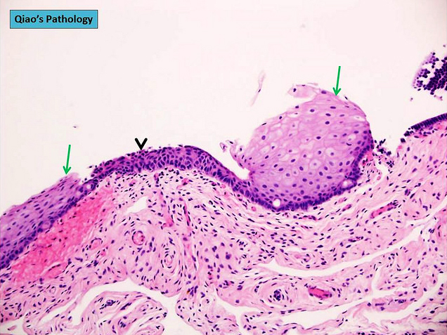
Multifocal transitional cell metaplasia
Negative stains
Differential diagnosis
- Serous tubal epithelial proliferation of uncertain significance: nuclear enlargement, increased N/C ratio and polarity is preserved; evidence of TP53 mutation ("p53 signature") by immunohistochemistry; Ki67 index is variably increased (Mod Pathol 2017;30:710)
- Serous tubal intraepithelial carcinoma (STIC): nuclear enlargement, increased N/C ratio, lost cellular polarity, irregular nuclear stacking, small detached papillary clusters, horizontally oriented surface nuclei and intraepithelial fractures; evidence of TP53 mutation by immunohistochemistry; Ki67 index is variably increased (Mod Pathol 2017;30:710)
Board review style question #1
The following statements about transitional cell metaplasia of the fimbriae are correct, except
- Cells of transitional cell metaplasia show uniform nuclei, pale chromatin and nuclear grooves
- Clinical followup of transitional cell metaplasia of the fimbriae shows no biological significance
- p53 signature and increased Ki67 are usually present in transitional cell metaplasia of the fimbriae
- Transitional cell metaplasia is undetectable by gross examination
- Transitional cell metaplasia shows no evidence of nuclear enlargement, loss of cellular polarity or irregular nuclear stacking
Board review style answer #1
C. p53 signature and increased Ki67 are not associated with transitional cell metaplasia of distal fimbriae but are characteristic of serous tubal intraepithelial carcinoma and serous tubal epithelial proliferation of uncertain significance.
Comment Here
Reference: Metaplastic changes
Comment Here
Reference: Metaplastic changes
