

Cervix
Benign / nonneoplastic epithelial lesions
Endocervical polyp
Author: Natalia Buza, M.D.
Editorial Board Member: David B. Chapel, M.D.
Editor-in-Chief: Debra L. Zynger, M.D.

Last author update: 13 September 2022
Last staff update: 13 September 2022
Copyright: 2007-2024, PathologyOutlines.com, Inc.
PubMed Search: Endocervical polyp

Table of Contents
Definition / general | Essential features | ICD coding | Epidemiology | Sites | Pathophysiology | Etiology | Clinical features | Diagnosis | Case reports | Treatment | Clinical images | Gross description | Gross images | Frozen section description | Frozen section images | Microscopic (histologic) description | Microscopic (histologic) images | Positive stains | Negative stains | Sample pathology report | Differential diagnosis | Board review style question #1 | Board review style answer #1 | Board review style question #2 | Board review style answer #2Cite this page: Buza N. Endocervical polyp. PathologyOutlines.com website. https://www.pathologyoutlines.com/topic/cervixendocervpolyp.html. Accessed April 16th, 2024.
Definition / general
- Benign exophytic proliferation composed of a variable admixture of endocervical glandular and metaplastic squamous epithelium with a fibrovascular core
Essential features
- Endocervical polyps are common, benign proliferations composed of a fibrovascular core and endocervical glandular or metaplastic squamous epithelium
- Chronic inflammation, surface erosion and reactive epithelial changes are common
- Rare cases may harbor in situ or invasive squamous or glandular lesions
- Lack of leaf-like architecture and periglandular stromal condensation, which are key features of Müllerian adenosarcoma (Am J Surg Pathol 2009;33:278)
ICD coding
- ICD-10: N84.1 - polyp of cervix uteri
Epidemiology
- Endocervical polyps are very common and may present at any age, although they are more common in patients over age 40 (Menopause 2009;16:524)
Sites
- Cervix / endocervix
Pathophysiology
- Most likely nonneoplastic in nature
Etiology
- No definite, known etiologic factors, although the role of chronic inflammation has been hypothesized
- Endocervical polyps with microglandular hyperplasia / polypoid cervical microglandular hyperplasia may be associated with pregnancy or exogenous progestin effect (Int J Gynecol Pathol 1995;14:50)
Clinical features
- Most are found incidentally during a routine gynecological exam
- May also present with abnormal vaginal spotting or bleeding (postcoital or contact) or abnormal vaginal discharge (Obstet Gynecol 1963;21:659)
Diagnosis
- Endocervical polyps often protrude through the cervical os and can be detected during routine gynecologic examination or colposcopy (Arch Gynecol Obstet 2007;276:299)
- Cervical Pap smear may show atypical cells due to reactive surface epithelial changes (Obstet Gynecol 2006;107:701)
Case reports
- 36 year old woman with endocervical polyp with an unusual cytology finding (Cytopathology 2007;18:316)
- 42 year old woman with endocervical polyp with florid epidermal metaplasia (Int J Gynecol Pathol 2016;35:478)
- 57 year old woman with large endocervical polyp with cartilaginous and osseous metaplasia (Int J Gynecol Pathol 2009;28:98)
Treatment
- Polypectomy is recommended for both symptomatic and large (> 3 cm) endocervical polyps (Arch Gynecol Obstet 2007;276:299)
- Data is limited regarding the removal of endocervical polyps during pregnancy; however, a recent study reported increased risk of pregnancy loss and preterm birth following endocervical polypectomy in pregnant women (Arch Gynecol Obstet 2022 Apr 9 [Epub ahead of print])
Clinical images
Images hosted on other servers:

Colposcopy

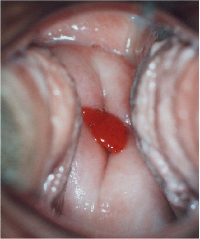
Polyp
Gross description
- Usually single, most often measures < 1 cm
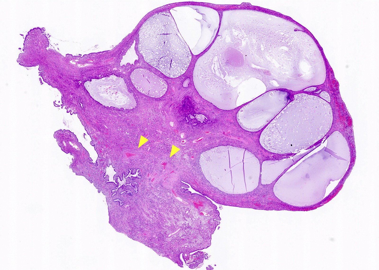
Gross images
Contributed by Natalia Buza, M.D.
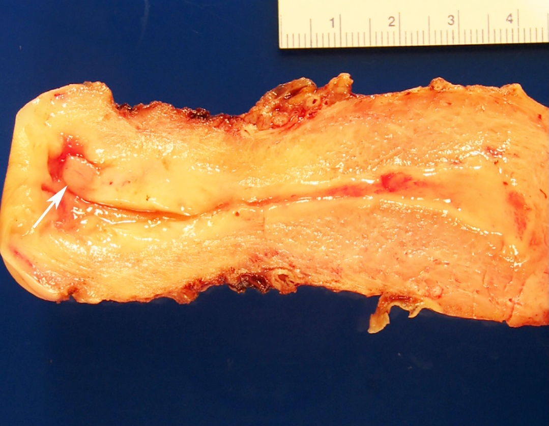
Small polyp
Frozen section description
- Microscopic features of endocervical polyp on frozen sections are similar to those seen on permanent sections
- Most important differential diagnosis on frozen section is adenosarcoma
- Unlike adenosarcoma, endocervical polyps lack periglandular stromal condensation and papillary intraglandular projections; no significant stromal or epithelial cell atypia is identified (Am J Surg Pathol 2015;39:116)
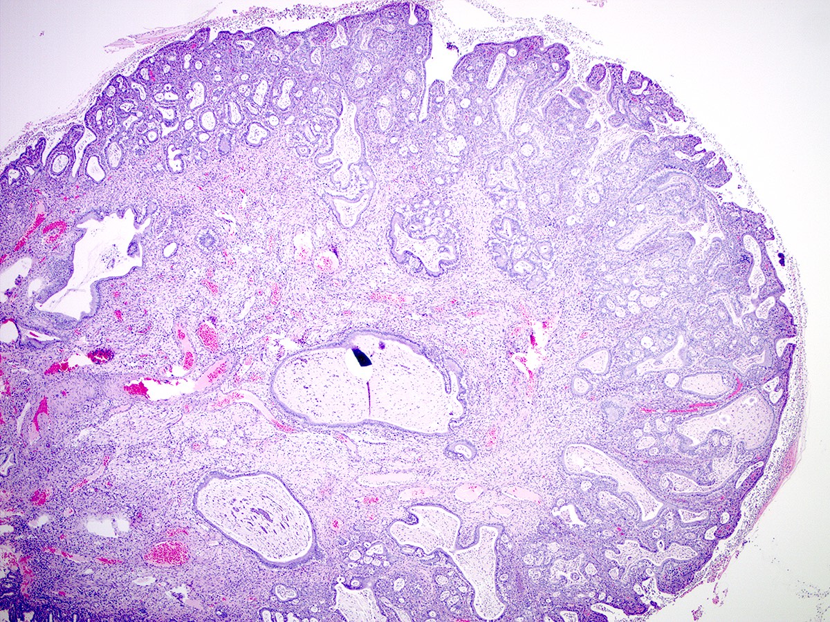
Frozen section images
Contributed by Natalia Buza, M.D.
Glands within uniform stroma
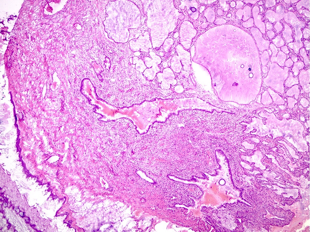
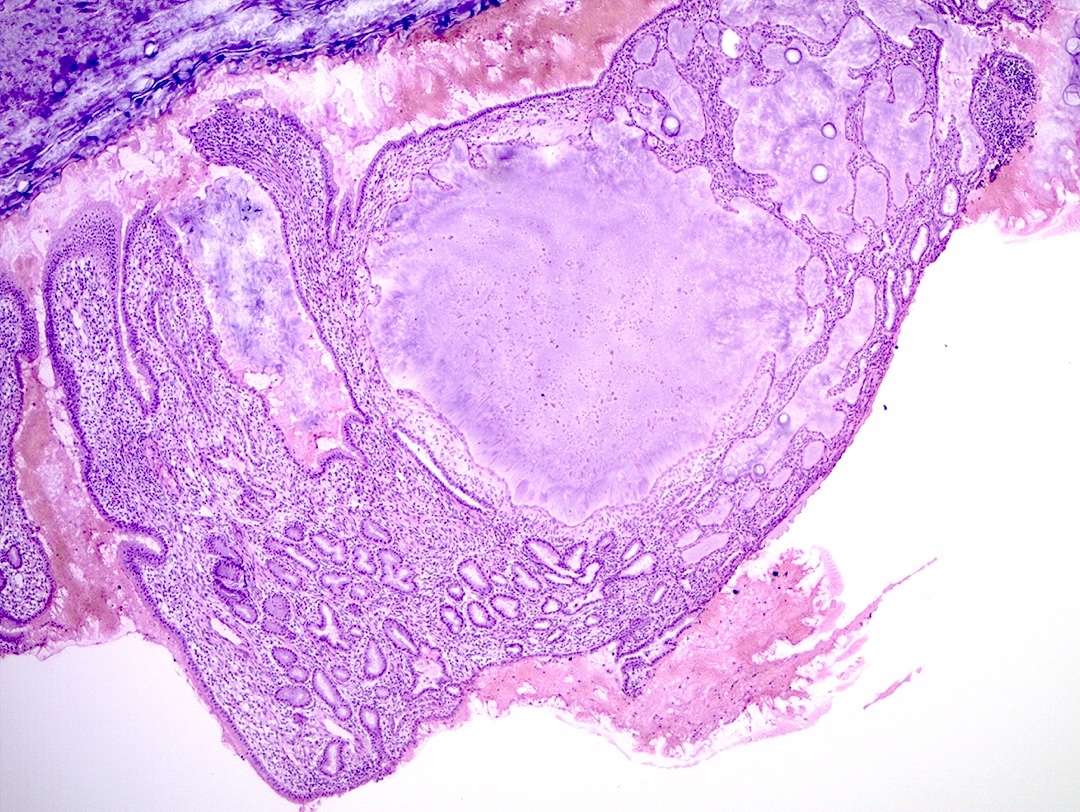
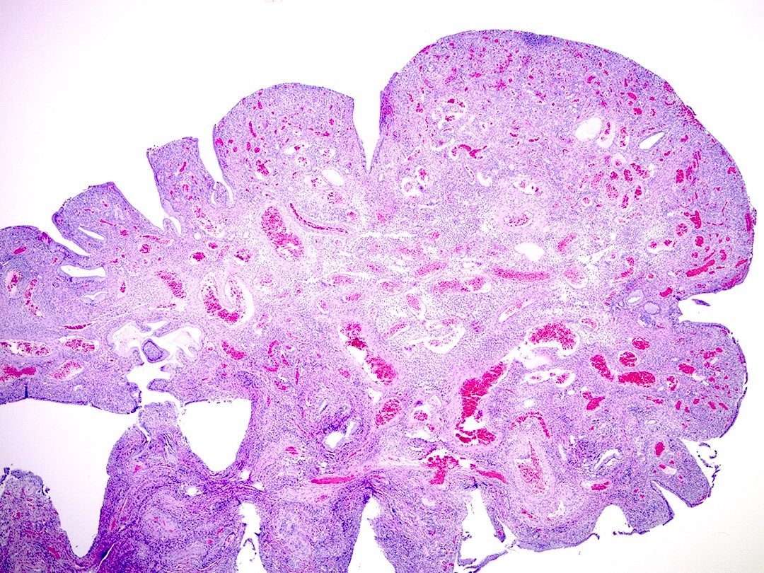
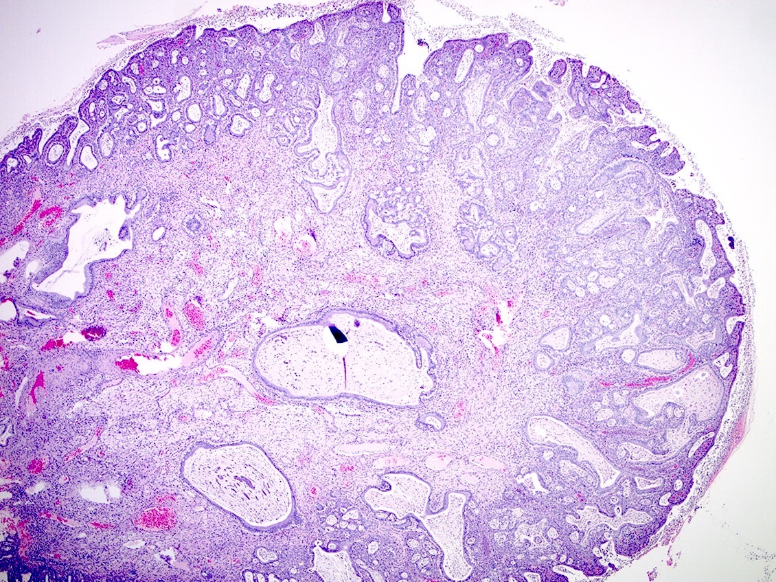
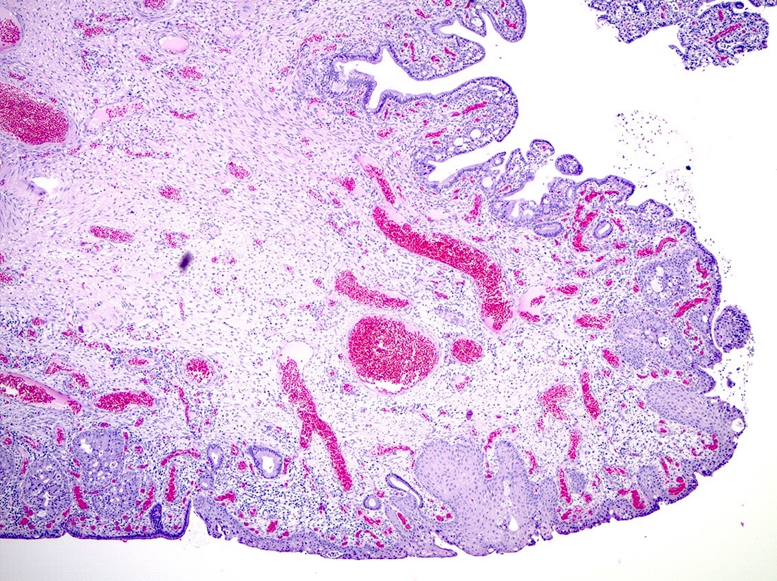
Microscopic (histologic) description
- Fibrovascular core with variably sized vasculature (often including thick walled arteries)
- Variable stromal cellularity often with mixed chronic inflammation
- Surface epithelium is endocervical glandular type and may show squamous metaplasia, erosion and reactive / reparative changes
- Proliferation of endocervical glands, which may be cystic or may show benign microglandular hyperplasia
- Mitotic activity may be noticeable, especially in cases with marked inflammation or florid microglandular hyperplasia (Int J Gynecol Pathol 2014;33:524)
- Epidermal metaplasia may be seen with skin appendage structures (Int J Gynecol Pathol 2016;35:478)
- Stromal cells may be multinucleated, may show decidual change or may contain heterologous elements (fat, cartilage, bone, glial tissue), which could represent retained fetal tissues from a previous gestation (Int J Gynecol Pathol 2010;29:394)
- Endocervical polyps may rarely harbor in situ or invasive squamous and glandular lesions (i.e., high grade squamous intraepithelial lesion, adenocarcinoma in situ, squamous carcinoma and adenocarcinoma) (J Womens Health (Larchmt) 2007;16:1317, Int J Gynecol Pathol 2009;28:567)
Microscopic (histologic) images
Contributed by Natalia Buza, M.D. and Andrey Bychkov, M.D., Ph.D.
Thick walled arteries
Microglandular hyperplasia
Squamous metaplasia
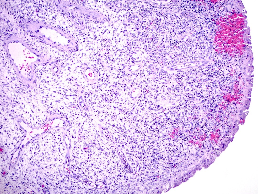
Chronic inflammation
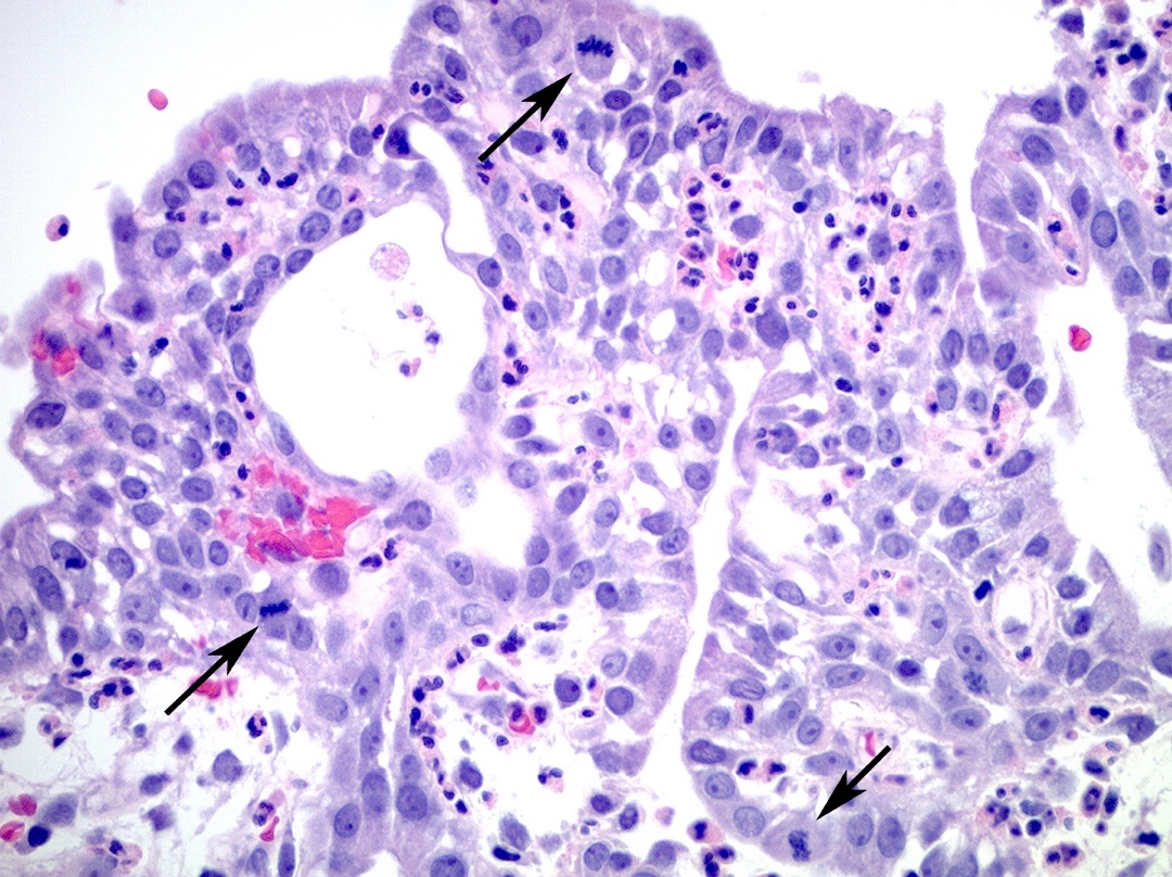
Mitoses
Cystically dilated glands
Positive stains
- Immunohistochemical stains are typically not necessary to make the diagnosis
- Epithelial elements stain with various epithelial markers (cytokeratins, EMA) and the stromal component stains with vimentin and smooth muscle markers
Negative stains
- p16 is negative or only patchy, weakly positive, which helps rule out high grade squamous intraepithelial lesion / adenocarcinoma in situ
Sample pathology report
- Cervix, polypectomy:
- Benign endocervical polyp with chronic inflammation and reactive changes
Differential diagnosis
- Adenosarcoma:
- Leaf-like glandular architecture, resembling phyllodes tumor of the breast, with intraglandular papillary projections and prominent periglandular stromal condensation
- Stromal cell atypia and stromal mitotic figures are also present
- Endometrial polyp:
- Proliferation of benign endometrial stromal and glandular elements, protruding into the endometrial cavity
- Occasionally a larger polyp, arising in the lower uterine segment, may protrude into the endocervical canal or may be visible through the cervical os, mimicking an endocervical polyp
- Polypoid adenomyoma:
- Polypoid mass composed of smooth muscle and irregular benign endocervical type glands, often showing a lobular architecture (Case Rep Pathol 2014;2014:275421)
- Condyloma:
- Exophytic, warty lesion of the ectocervix with squamous epithelium showing papillomatosis and koilocytosis, commonly associated with low risk human papillomavirus infection
Board review style question #1
Which of the following statements is true about the endocervical lesion shown above?
- Presence of any mitotic activity indicates malignancy
- Presence of heterologous mesenchymal elements indicates malignancy
- Rarely, such lesions may harbor in situ or invasive malignancy
- Such lesions are most common in children and adolescents
Board review style answer #1
C. Rarely, such lesions may harbor in situ or invasive malignancy
Comment Here
Reference: Endocervical polyp
Comment Here
Reference: Endocervical polyp
Board review style question #2
Which of the following morphological features are characteristic of benign endocervical polyps?
- Bland endocervical glandular proliferation with squamous metaplasia
- Marked nuclear atypia of surface epithelium with strong diffuse p16 immunoreactivity
- Periglandular stromal condensation
- Prominent leaf-like glandular architecture with intraglandular projections
Board review style answer #2
A. Bland endocervical glandular proliferation with squamous metaplasia
Comment Here
Reference: Endocervical polyp
Comment Here
Reference: Endocervical polyp
