

Cervix
Metaplasia
Tuboendometrioid metaplasia
Last author update: 1 April 2017
Last staff update: 25 January 2024
Copyright: 2002-2024, PathologyOutlines.com, Inc.
PubMed Search: Tubo endometrioid metaplasia


Table of Contents
Definition / general | Microscopic (histologic) description | Microscopic (histologic) images | Positive stains | Negative staining - disease | Cytology description | Differential diagnosis | Additional referencesCite this page: Asirvatham JR, Hodgson A. Tuboendometrioid metaplasia. PathologyOutlines.com website. https://www.pathologyoutlines.com/topic/cervixmetaplasiatuboendometrial.html. Accessed April 18th, 2024.
Definition / general
- Common (1/3 of women)
- In upper portion of endocervical canal, often in deep glands (Int J Gynecol Pathol 1992;11:89)
- Often seen after cervical cone biopsy
- May represent response to injury
- Tubal epithelium is invariably seen at the cervix - isthmus junction and is not considered a metaplastic process at that site (Int J Gynecol Pathol 2013;32:122)
Microscopic (histologic) description
- Endocervix contains:
- Ciliated cells (clear cytoplasm, abundant apical cilia and large, oval, variably hyperchromatic nuclei)
- Secretory cells (nonciliated with dark eosinophilic or basophilic cytoplasm, apical cytoplasmic protrusions but no mucin vacuoles, basal nuclei)
- Intercalated cells (also called peg cells, scant cytoplasm, thin and long nuclei), as found in normal fallopian tube
- Glands are regular
- Minimal mitotic activity, rare crowding or atypia
- Associated with endometrial type cells
- Usually near squamocolumnar junction, usually no inflammation
- May have cystic glands and periglandular stromal alterations suggestive of premalignant conditions or deep glands with periglandular edema suggestive of well differentiated adenocarcinoma but cells are ciliated with bland cytology, no mitotic figures, no definite desmoplastic stroma (Am J Clin Pathol 1995;103:618)
Microscopic (histologic) images
Contributed by Carlos Parra-Herran, M.D.
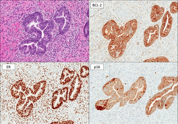
TEM and relevant stains
Positive stains
Negative staining - disease
- p16 is considered to be negative or only focally positive or show a mosaic pattern in tubal metaplasia (Adv Anat Pathol 2006;13:8, Cell Oncol 2007;29:37), useful to differentiate from high grade CIN and invasive cervical carcinomas that are diffusely positive
Cytology description
Differential diagnosis
- Adenocarcinoma in situ:
- Tubal metaplasia may coexist with adenocarcinoma in situ and cases of AIS in a background of tubal metaplasia and a ciliated variant of adenocarcinoma in situ have been reported (Int J Gynecol Pathol 1999;18:1)
- Endometrioid adenocarcinoma:
- Invasive growth pattern, marked nuclear atypia, increased Ki67 staining
- Endometriosis
- Mesonephric hyperplasia
- Microglandular hyperplasia
- Pseudoinfiltrative tuboendometrioid hyperplasia simulating minimal deviation adenocarcinoma has been reported in 3 cases of in utero diethyl stilbestrol exposure (Int J Gynecol Pathol 2005;24:391)
- Tunnel clusters
Additional references
