

CNS & pituitary tumors
General
Anatomy & histology-pineal gland
Author: Nat Pernick, M.D.

Last author update: 1 January 2006
Last staff update: 22 July 2020
Copyright: 2002-2024 PathologyOutlines.com, Inc.
PubMed Search: "Normal pineal gland"

Table of Contents
Definition / general | Diagrams / tables | Gross description | Gross images | Microscopic (histologic) description | Positive stainsCite this page: Pernick N. Anatomy & histology-pineal gland. PathologyOutlines.com website. https://www.pathologyoutlines.com/topic/cnstumorpinealglandnormal.html. Accessed April 19th, 2024.
Definition / general
- Also called epiphysis, pineal body
- Between superior colliculi at base of brain; 100 - 180 mg
- Develops at month 2 of gestation as diverticulum in diencephalic roof of third ventricle
- Replaced by connective tissue after puberty
- Produces melatonin, which helps regulate circadian rhythms
Diagrams / tables
Images hosted on other servers:
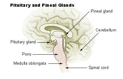
Location in brain
Gross description
- Shaped like a pine cone, midline, attached to posterior end of roof of third ventricle in front of cerebellum, 1 cm long, red gray
Gross images
Images hosted on other servers:

Local anatomy (horse)
Microscopic (histologic) description
- Loose neuroglial stroma with nests of pineocytes containing well defined neurosecretory (melatonin) granules
- Also astrocytes
- Has features of photoreceptors and concretions ("brain sand")
Positive stains
- Synaptophysin
- Retinal S antigen
