
Coagulation
Coagulation laboratory tests
Viscoelastic hemostatic assays
Authors: João D. Dias, Ph.D., Jan Hartmann, M.D.
Editorial Board Member: Kyle Annen, D.O.
Deputy Editor-in-Chief: Patricia Tsang, M.D., M.B.A.


Last author update: 31 January 2022
Last staff update: 31 January 2022
Copyright: 2021, PathologyOutlines.com, Inc.
PubMed Search: Viscoelastic hemostatic assays coagulation pathology
Table of Contents
Definition / general | Essential features | Terminology | Pathophysiology | Diagrams / tables | Clinical features | Laboratory | Case reports | Treatment | Additional references | Board review style question #1 | Board review style answer #1 | Board review style question #2 | Board review style answer #2 | Board review style question #3 | Board review style answer #3Cite this page: Dias JD, Hartmann J. Viscoelastic hemostatic assays. PathologyOutlines.com website. https://www.pathologyoutlines.com/topic/coagulationviscohemoassays.html. Accessed April 23rd, 2024.
Definition / general
- Viscoelastic hemostatic assays provide comprehensive, real time analysis of coagulation status (Diagnostics (Basel) 2020;10:118)
- Contribute to a better understanding of blood coagulation in conjunction with conventional coagulation tests, which measure the impact of individual clotting components but not interactions between them
- Different reagents allow the tests to determine the relative contribution of circulating plasma components and cellular elements on blood coagulation
- First generation devices: reagents are pipetted into the device separately from the blood sample (e.g. TEG®5000, ROTEM® Delta)
- Newer generation devices: use a single cartridge for reagents so no pipetting is required (e.g. TEG®6s, ROTEM® Sigma, Quantra®)
- Some newer generation devices also include platelet function testing (e.g. TEG®6s)
- Support rapid treatment for patients in a variety of clinical settings and allow goal directed transfusion of blood products
Essential features
- Viscoelastic hemostatic assays measure dynamics of whole blood clotting
- Provide a comprehensive real time analysis of hemostasis, from initial fibrin fiber formation to platelet interactions and fibrinolysis
- Newer generation viscoelastic hemostatic assays are designed to be fast, effective and portable
- Results are clinically relevant, allowing goal directed coagulation factor transfusions
Terminology
- Viscoelastic hemostatic assay / analyzer: an in vitro diagnostic method of global coagulation / hemostasis analysis that provides a real time, holistic view of ex vivo clotting; it allows for examination of both cellular and plasma protein contributions to clotting, including platelet number and function, fibrin(ogen) function and coagulation factor function
- Thromboelastography: a subtype of in vitro viscoelastic hemostatic diagnostic technology that monitors and analyzes the coagulation state of a whole blood sample by measuring the shear modulus / dynamics of clot formation in low shear conditions in order to assist in the assessment of a patient’s hemostatic status and its clinical implications
- Thrombelastograph (TEG®): an in vitro diagnostic hemostasis system by Haemonetics®
- Rotational thromboelastometry (ROTEM®): an in vitro diagnostic hemostasis system by Werfen®
- TEG®5000 (legacy version): a Thrombelastograph® analyzer using thromboelastography by Haemonetics®
- TEG®6s (next generation device): a Thrombelastograph® analyzer using multichannel cartridge based thromboelastography by Haemonetics®
- ROTEM® Delta (legacy version): a viscoelastic hemostasis analyzer using rotational thromboelastometry by Werfen®
- ROTEM® Sigma (next generation device, not FDA cleared as of January 2022): a viscoelastic hemostasis analyzer using multichannel cartridge based rotational thromboelastometry by Werfen®
- Sonoclot® SC1, SCP1, SCP2 (legacy versions): viscoelastic hemostatic analyzers using linear motion by Sienco
- Sonoclot® SC1P4 (next generation device): a viscoelastic hemostatic analyzer using linear motion by Sienco
- Quantra®: a viscoelastic hemostasis analyzer using sonic estimation of elasticity via resonance (SEER) sonorheometry by Hemosonics®
- ClotPro®: a viscoelastic hemostasis analyzer using elastic motion thromboelastography produced by enicor, acquired by Haemonetics®
- Emerging technologies include laser speckle rheometry, mechanical resonant frequency, ultrasonic deformation and parallel plate viscometry:
- Laser speckle rheometry is a noninvasive optical technique that can measure viscoelastic properties of blood without applying any external force or physically contacting the test sample
- Mechanical resonant frequency is an object's natural frequency of vibration, which is affected by the surrounding medium and can be used to detect changes in a detector's vibration frequency as the surrounding blood sample changes during blood clot formation
- Ultrasonic deformation measures shape deformations of suspended blood droplets, which are directly correlated to the viscoelastic properties of the sample
- Parallel plate viscometry measures the shear stress of a blood sample positioned between two parallel plates to calculate the sample's viscoelastic properties
Pathophysiology
- Abnormalities in hemostasis can result in excessive blood clotting (thrombosis) or bleeding (hemorrhage) (Physiol Rev 2013;93:327)
- Viscoelastic hemostatic assays measure the dynamics of whole blood clotting (Diagnostics (Basel) 2020;10:118)
- Viscoelasticity is characteristic of a material that behaves in both a viscous (permanent deformation) and elastic (temporary deformation) manner
- Earliest viscoelastic hemostatic assays used rotation of cup and pin to measure the viscoelasticity of whole blood
- Thromboelastography: the cup rotates and the resulting rotation in the pin is measured
- Rotational thromboelastometry: the cup is held stationary and a rotational force is applied to the pin
- Before clotting, the whole blood sample remains viscous and the pin does not move
- Response to shear stress will be permanent deformation
- As clot strength develops, the sample gradually develops an elastic element, which results in movement of the pin
- Total pin deflection is tracked and plotted
- Other viscoelastic hemostatic assays are based on linear motion of the pin rather than rotation
- For the TEG®6s analyzer, new measurement techniques include fixed vibration resonance frequency measured with LED illumination
- Assays and associated reagents have been developed to investigate different coagulation pathways, the contributions of platelets and fibrinogen to clot strength and the effect of clot lysis
- Some of the newer devices include platelet function testing, useful in measuring the impact of antiplatelet medication or platelet function post injury
- More recent technologies are available as cartridge based systems that automate all sample aliquoting, reagent mixing and testing
Diagrams / tables


Clinical features
- Viscoelastic hemostatic assays are currently used for the management of major bleeding and guiding transfusion therapy
- Most common indications are trauma hemorrhage, cardiac surgery, managing goal directed antiplatelet therapies, obstetric hemorrhage and liver disease
- Viscoelastic hemostatic assays are recommended in trauma guidelines for assessing coagulopathy and guiding blood product transfusions in acutely bleeding trauma, surgical and critically ill patients (J Trauma Acute Care Surg 2020;89:999, Crit Care 2016;20:100, American College of Surgeons: Massive Transfusion in Trauma Guidelines [Accessed 26 May 2021], Anaesth Crit Care Pain Med 2019;38:539)
- Viscoelastic monitoring is also recommended in guidelines for:
- Monitoring and managing hemostasis in cardiac surgery (NICE: Detecting, managing and monitoring haemostasis - viscoelastometric point‑of‑care testing [Accessed 26 May 2021], Eur J Anaesthesiol 2017;34:332, Anesthesiology 2015;122:241, National Blood Authority Australia: Patient Blood Management Guidelines - Module 2 Perioperative [Accessed 23 December 2021], Eur J Cardiothorac Surg 2018;53:79, Anaesth Crit Care Pain Med 2019;38:539)
- Liver transplant surgery (Anaesth Crit Care Pain Med 2019;38:539, Eur J Anaesthesiol 2017;34:332)
- Postpartum hemorrhage (Anaesth Crit Care Pain Med 2019;38:539, Eur J Anaesthesiol 2017;34:332, J Thromb Haemost 2016;14:205, Obstetric Anaesthetists' Association: OAA/AAGBI Guidelines for Obstetric Anaesthetic Services 2013 [Accessed 26 May 2021])
- Viscoelastic monitoring has potential for use in the clinical management of patients with stroke / congenital bleeding disorders and in the management of treatment with direct acting oral anticoagulants
- Platelet inhibition and aggregation assays, available with some technologies, allow for the quantification of response to aspirin and P2Y12 receptor inhibitors, with potential uses in:
- Personalized dual antiplatelet therapy for cardiology patients
- Optimizing timing for cardiac surgery in patients on antiplatelet therapy
- Monitoring hemostasis during cardiac surgery
- Reducing blood transfusions and risk of complications following cardiac surgery
- Predicting ischemic event risk following coronary interventions
- Monitoring of platelet function also has a range of potential uses in the emergency setting:
- Assessing coagulopathy profiles in trauma patients with burns and traumatic brain injury
- Guiding treatment of patients undergoing surgery and neurological treatment
- Evidence on the use of platelet function tests available on viscoelastic devices is currently limited to research studies
- Such uses do not yet reflect established evidence or clinical practice and may go beyond the intended use as indicated by the manufacturer
- Limitations of viscoelastic monitoring include (Hong Kong Med J 2015;21:45):
- User variability
- Lack of universally agreed algorithms to guide clinical use
- Relatively few large scale randomized clinical trials completed using viscoelastic testing
- Inability to measure the influence of all in vivo pathologies; in particular, the influence of epithelial cells on the coagulation process
- Interchangeability of results between viscoelastic assays because of the different coagulation activators used
- Older models are labor intensive and require skilled trained staff to operate with accuracy
Laboratory
- Variables used to assess the clotting process are based on changes in clot strength over time
- Details of those most likely to be used in clinical practice are illustrated in the example thromboelastography parameter tracing in Diagram 1
- Below is some of the terminology commonly associated with thromboelastography / rotational thromboelastometry (respectively) (Diagnostics (Basel) 2020;10:118)
- Reaction time (R) / clotting time (CT): the time taken for the amplitude of the clot to reach 2 mm
- Reflects the process of thrombin generation and the initial formation of fibrin fibers
- Increases in R time / CT reflect hypocoagulability; low R time / CT reflects hypercoagulability
- Coagulation time (K) / clot formation time (CFT) is the interval between the amplitudes of 2 - 20 mm
- Represents the process of clot strengthening, which is mainly dependent on fibrinogen availability
- Prolonged K time / CFT suggests hypocoagulability, while hypercoagulability can shorten K time / CFT
- Angle (α) is the tangent line of the tracing that starts as the tracing leaves the baseline
- Reflects the speed of clotting and depends on coagulation factors, including fibrinogen
- Angle that is smaller than the reference range suggests that the patient is in a hypocoagulable state, while a larger angle is likely to be encountered in hypercoagulable patients
- Maximum amplitude (MA) / maximum clot firmness (MCF) is the most divergent point of the tracing, representing the maximum strength of the clot
- Depends largely on the concentrations and functionality of platelets and fibrin
- Lysis 30 (LY30) / lysis index 30 (LI30) is the percentage reduction in the area under the tracing at 30 minutes after the maximum amplitude
- LY30 / clot formation (LI30)
- LY30 / LI30 is an assessment of clot stability and shows the speed and extent of fibrinolysis (high values are associated with hyperfibrinolysis while for the LI30 smaller values represent hyperfibrinolysis)
- Examples of traces from thromboelastography and rotational thromboelastometry most likely to be observed in hypocoagulable and hypercoagulable states in clinical settings are shown in Diagram 2 and Diagram 3, respectively
Case reports
- 38 year old woman with antiphospholipid syndrome who developed septic shock after labor induction (J Anesth 2020;34:781)
- 39 year old man who developed acute limb ischemia secondary to infection with COVID-19 (J Thromb Thrombolysis 2020;50:292)
- 42 year old man with ventricular assist device with intracranial hemorrhage underwent a thromboelastometry guided anticoagulation reversal (Am J Emerg Med 2021;41:265.e5)
- 45 year old woman underwent anticoagulation management during cardiopulmonary bypass complicated by antiphospholipid syndrome (J Card Surg 2020;35:1354)
- 52 year old woman with aneurysmal subarachnoid hemorrhage and hypercoagulation assessed comprehensively by thromboelastometry (Acta Neurochir Suppl 2020;127:165)
Treatment
- Utilization of viscoelastic hemostatic assays for the diagnosis and management of numerous disorders have been previously summarized in the following reference: Transfusion 2020;60:S6
Additional references
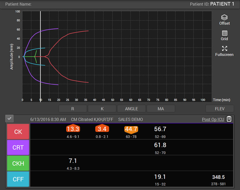
Board review style question #1
Considering the thromboelastography citrated kaolin (CK) assay tracing shown above (red line), what are the probable causes suggested by a normal reaction time (R) value, low α angle value and low maximum amplitude?
- Fibrinolytic state; abnormal fibrinolysis
- Hypercoagulable state; a tendency towards increased blood clotting
- Hypocoagulable state; excessive bleeding caused by low fibrinogen levels
- Hypocoagulable state; excessive bleeding caused by low platelet number / platelet dysfunction
Board review style answer #1
D. Hypocoagulable state; excessive bleeding caused by low platelet number / platelet dysfunction
Comment Here
Reference: Viscoelastic hemostatic assays
Comment Here
Reference: Viscoelastic hemostatic assays
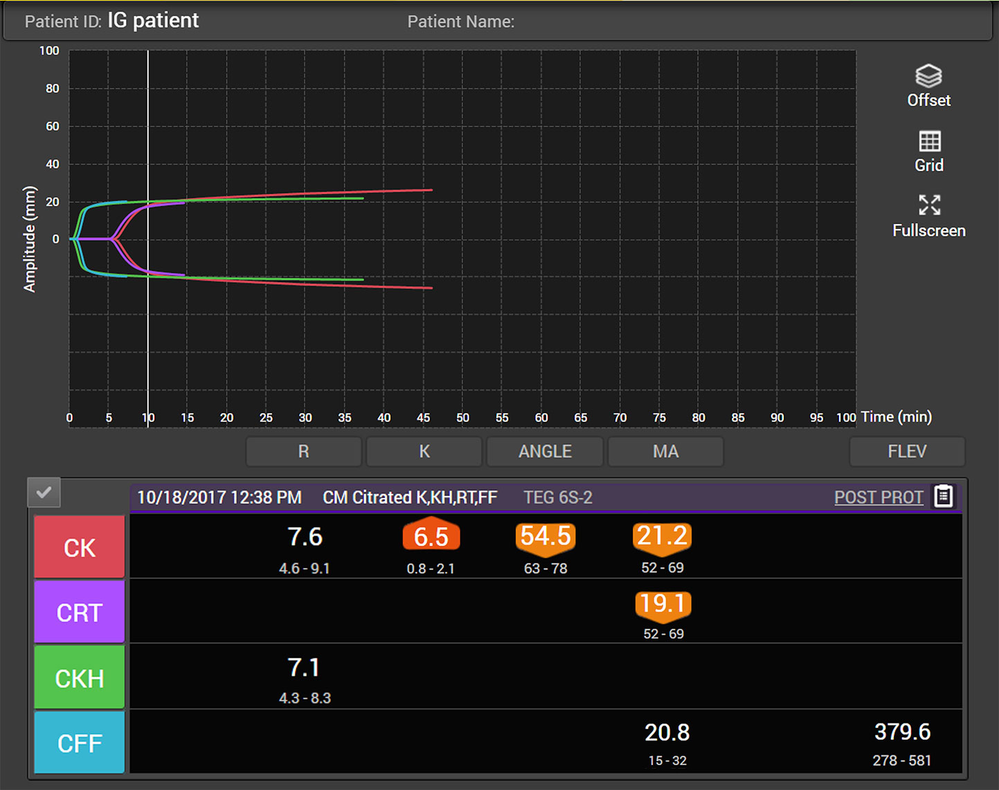
Board review style question #2
Considering the thromboelastography citrated kaolin (CK) and citrated kaolin heparinase (CKH) assay tracings shown above (red and green lines, respectively), what are the probable causes suggested by a prolonged R time, low α angle value and normal maximum amplitude on the CK tracing, corresponding with a normalized reaction time on the CKH tracing?
- Fibrinolytic state; abnormal fibrinolysis
- Hypercoagulable state; a tendency towards increased blood clotting
- Hypocoagulable state; excessive bleeding caused by the presence of heparin
- Hypocoagulable state; excessive bleeding caused by low platelet number / platelet dysfunction
Board review style answer #2
C. Hypocoagulable state; excessive bleeding caused by the presence of heparin
Comment Here
Reference: Viscoelastic hemostatic assays
Comment Here
Reference: Viscoelastic hemostatic assays
Board review style question #3
Maximum amplitude represents the maximum strength of the clot and is largely dependent on
- Concentration of coagulation factors
- Concentrations and functionality of platelets and fibrin
- Lupus anticoagulation
- von Willebrand factor
Board review style answer #3
B. Concentrations and functionality of platelets and fibrin
Comment Here
Reference: Viscoelastic hemostatic assays
Comment Here
Reference: Viscoelastic hemostatic assays
