
Colon
Drug induced colitides
Pill fragment associated colitis
Author: Raul S. Gonzalez, M.D.

Last author update: 4 February 2021
Last staff update: 4 February 2021
Copyright: 2003-2024, PathologyOutlines.com, Inc.
PubMed Search: Kayexalate colitis


Table of Contents
Definition / general | Essential features | Sites | Diagnosis | Case reports | Microscopic (histologic) description | Microscopic (histologic) images | Positive stains | Sample pathology report | Differential diagnosis | Additional references | Board review style question #1 | Board review style answer #1Cite this page: Gonzalez RS. Pill fragment associated colitis. PathologyOutlines.com website. https://www.pathologyoutlines.com/topic/colonpillfragmentassociatedcolitis.html. Accessed April 25th, 2024.
Definition / general
- Sodium polystyrene sulfonate (Kayexalate) is an ion exchange resin that binds intraluminal potassium; it can cause ischemia and intestinal necrosis, especially (but not only) if given with sorbitol (Am J Surg Pathol 1997;21:60)
- Sevelamer, used to treat hyperphosphatemia in patients with chronic kidney disease, binds phosphate and has may cause injury to the gastrointestinal tract (Am J Surg Pathol 2013;37:1686)
- Bile acid sequestrants (e.g. cholestyramine) may microscopically mimic other pill fragments but do not appear to cause injury (Am J Surg Pathol 2014;38:1530)
Essential features
- Kayexalate causes mucosal injury and sevelamer may also as well
- Other pill fragments (such as bile acid sequestrants) can also be identified microscopically but do not cause mucosal injury
Sites
- Colon is most commonly involved gastrointestinal organ but any can be affected
- Resins can rarely be encountered outside the gastrointestinal tract (due to perforation, aspiration, etc.) (Diagn Pathol 2008;3:27)
Diagnosis
- Microscopic examination and clinical confirmation of medication use
Case reports
- 27 week boy with necrotizing enterocolitis caused by Kayexalate (J Perinatol 2007;27:247)
- 46 year old man with rectal stenosis caused by Kayexalate (Ann Diagn Pathol 2007;11:217)
- 79 year old man with colonic pseudotumor caused by sevelamer injury (Clin Gastroenterol Hepatol 2015;13:A39)
Microscopic (histologic) description
- Kayexalate: bright purple crystals with fish scale appearance
- Sevelamer: yellow / pink crystals; also fish scale appearance
- Bile acid sequestrants: opaque orange polygonal / rhomboid crystals; usually no fish scale appearance; may be spherical (Histopathology 2015;67:141)
Microscopic (histologic) images
Contributed by Raul S. Gonzalez, M.D.
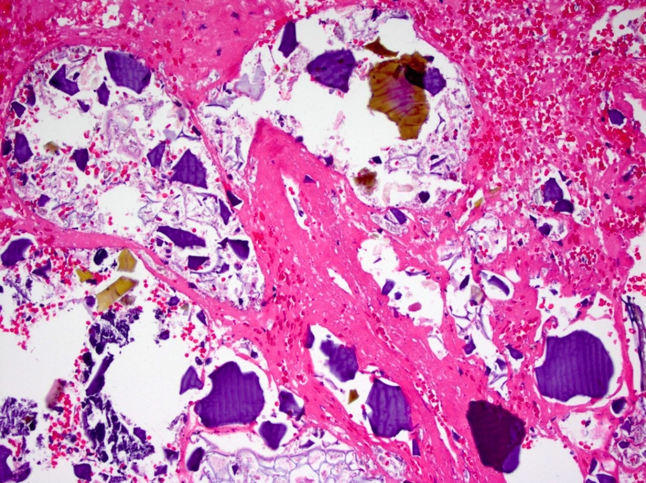
Kayexalate and sevelamer in a rectal ulcer
Images hosted on other servers:

Incidental luminal polystyrene
sulphonate resin particles in
jejunal diverticular tissue

Particles at site of colonic
necrosis (direct Schiff stain
with light counterstain)

Particles at site of aspiration
pneumonia (Ziehl-Neelsen stain
with light counterstain)
Positive stains
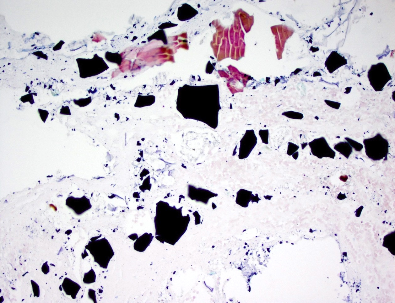
- AFB: Kayexalate appears black, sevelamer appears magenta and bile acid sequestrants appear dull yellow
Sample pathology report
- Ascending colon, ulcer, biopsy:
- Fibrinopurulent debris, consistent with ulceration (see comment)
- Negative for malignancy.
- Comment: Several fragments of pill material, consistent with sevelamer, are present within the ulcer debris. The pill material may be responsible for the ulceration or secondarily trapped within.
Differential diagnosis
- Dystrophic calcification:
- Can mimic Kayexalate but is a brighter purple
- Bile:
- Can mimic sevelamer but has no pink coloration
Additional references
Board review style question #1
Which of the following pill material fragments usually shows fish scales on histology?
- Colestipol
- Crospovidone
- Microcrystalline cellulose
- Sevelamer
Board review style answer #1
