

Heart & vascular pathology
Benign tumors / other nonneoplastic
Cystic tumor of the atrioventricular node
Author: Nat Pernick, M.D.

Last author update: 1 November 2013
Last staff update: 1 June 2021
Copyright: 2007-2024, PathologyOutlines.com, Inc.
PubMed Search: Atrioventricular node tumor
Table of Contents
Definition / general | Case reports | Radiology images | Treatment | Gross description | Gross images | Microscopic (histologic) description | Microscopic (histologic) images | Positive stains | Negative stains | Electron microscopy description | Differential diagnosisCite this page: Pernick N. Cystic tumor of the atrioventricular node. PathologyOutlines.com website. https://www.pathologyoutlines.com/topic/hearttumormesotheliomaofAVnode.html. Accessed April 19th, 2024.
Definition / general
- Also called cystic tumor of AV node or mesothelioma of AV node but does not appear to have mesothelial origin
- Rare; usually identified at autopsy
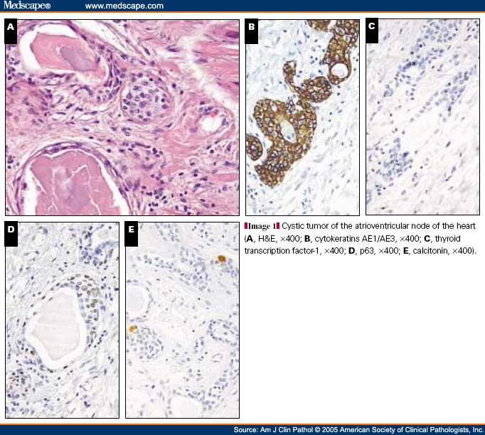
- May be congenital rests of endodermal origin (Arch Pathol Lab Med 1990;114:1057) or ultimobranchial heterotopia identical to solid cell nests of thyroid (Am J Clin Pathol 2005;123:369)
- Associated with other congenital anomalies
- Often causes heart block and sudden death
- Mean age 38 years, 75% female
- Should examine conduction system in all patients with sudden death, particularly if a history of arrhythmia or heart block
Case reports
- 7 year old boy with rare cause of sudden cardiac death (Arch Pathol Lab Med 2001;125:573)
- 24 year old woman with sudden cardiac death (Am J Forensic Med Pathol 2005;26:349)
- Patient with mesothelioma of the atrioventricular node (Am J Clin Pathol 1975;63:377)
- Patient with unusual site for the AV node tumor (Cardiovasc Pathol 1999;8:325)
- Patient with congenital cystic tumors of the atrioventricular node (Cardiovasc Pathol 1999;8:233)
- Patient with with atrioventricular nodal tumor associated with polyendocrine anomalies (Pathol Res Pract 1996;192:54)
Radiology images
Images hosted on other servers:

MRI of the thorax showing tumor
Treatment
- Pacemaker implantation, anti-arrhythmic drugs, possibly surgical excision (Heart 2000;83:E6)
Gross description
- May not be visible due to small size (2 to 20 mm)
- Multicystic lesion in area of atrioventricular node and membranous septum
Gross images
AFIP images
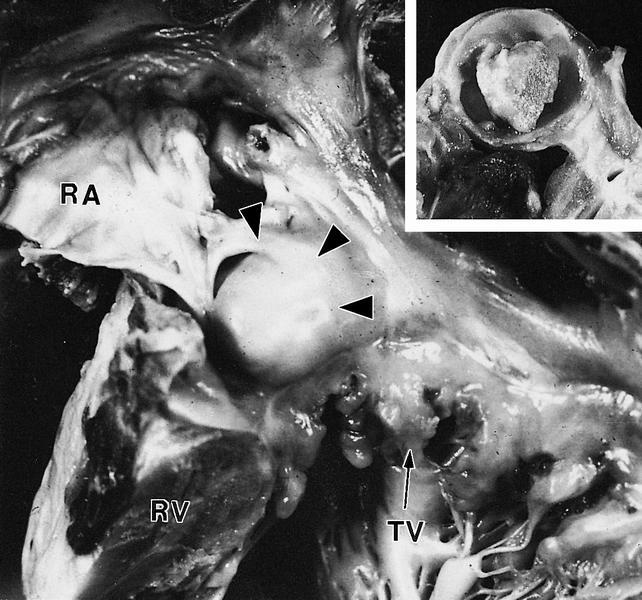
arrowheads (RA-right
atrium, RV-right ventricle,
TV-tricuspid valve)
Microscopic (histologic) description
- Must sample conduction system
- Solid and cystic areas lined by nonciliated, epithelial appearing cuboidal cells (main cells), mixed with occasional clear cells (neuroendocrine or C cells)
- Lumina contains PAS+ diastase resistant material which may calcify
- May have inflammatory cells and fibrosis
- No smooth muscle, no mitotic figures, no atypia
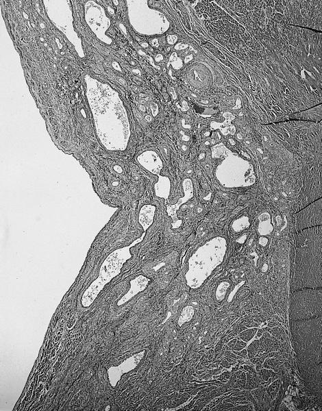
Microscopic (histologic) images
AFIP images
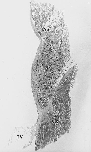
IAS-interatrial septum (tumors are
located inferiorly), TV-tricuspid
valve and VS-ventricular septum
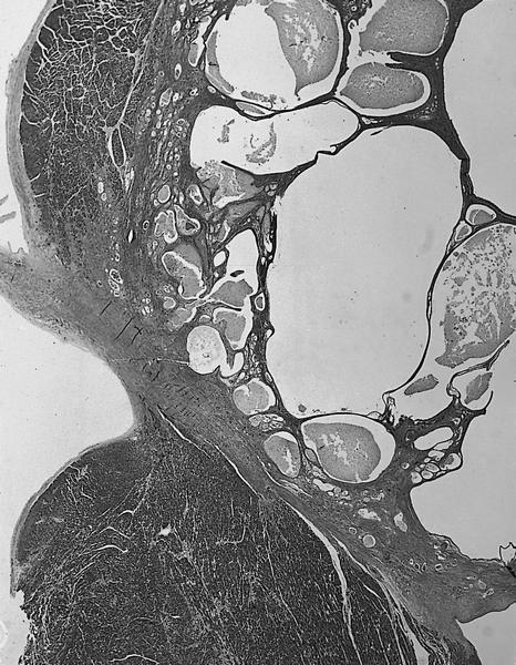
filled with
proteinaceous
debris
Multiple small cysts not visible to naked eye
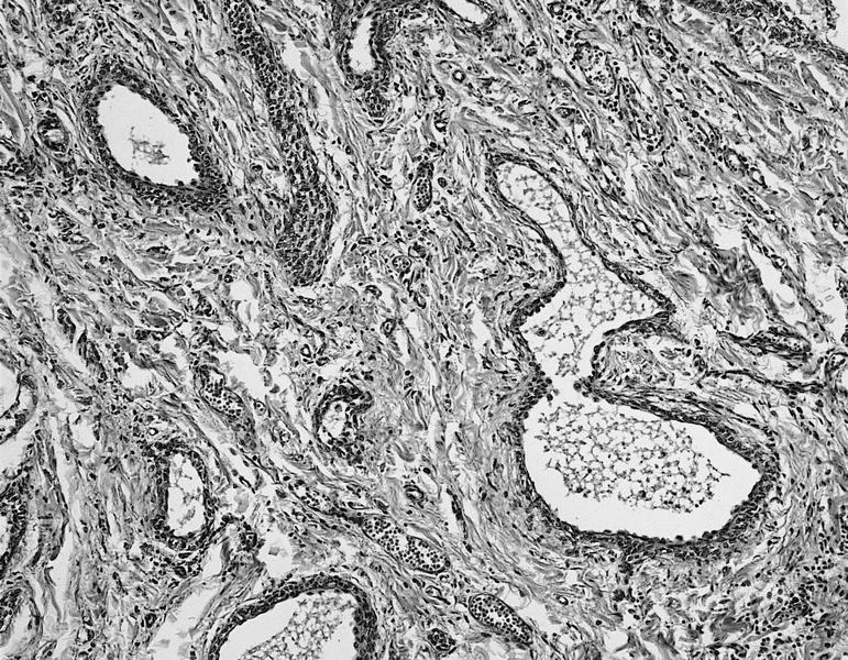
Cysts with irregular shape surrounded by fibrous stroma
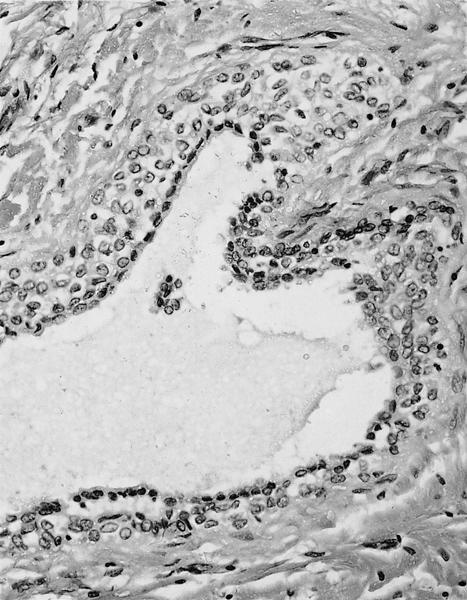
cell lining, with inner
lining composed of
small cuboidal cells
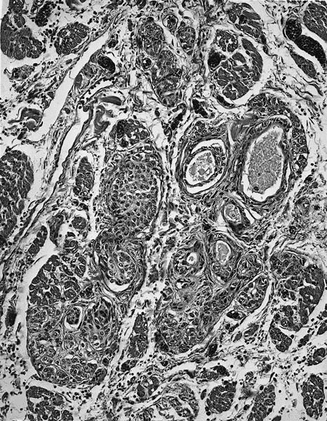
cysts replace muscle
bundles in inferior
interatrial septum
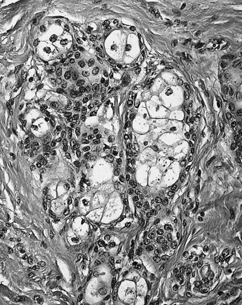
Cuboidal cells and clear, sebaceous-type cells
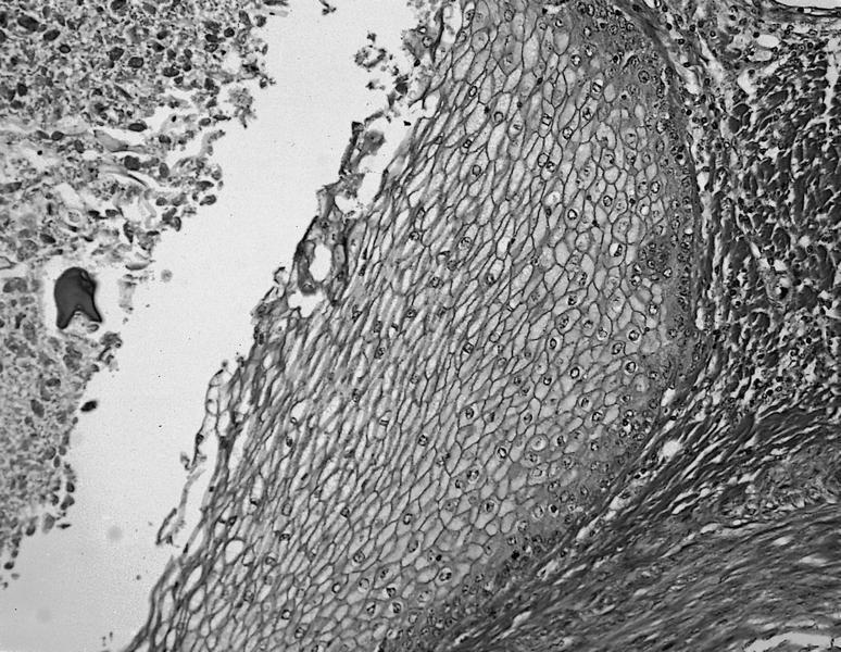
Squamous differentiation and calcification of luminal debris
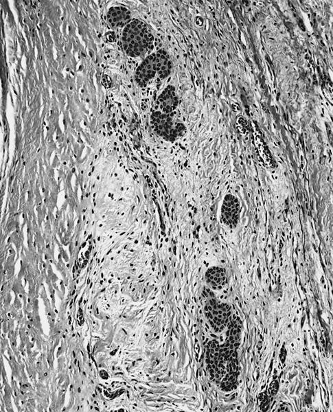
Nests of cells resembling urothelium
Images hosted on other servers:
H&E and AE1/AE3+

cyst lining of simple
cuboidal epithelium
Positive stains
Negative stains
Electron microscopy description
- Cells form solid nests with well formed basement membranes, cytoplasmic tonofilaments and desmosomes or glandular structures with desmosomes, electron dense material and short microvilli
Differential diagnosis
- Bronchogenic cyst: solitary, grossly visible, on epicardial surface, smooth muscle present
- Mesothelial cyst: larger, unilocular, on surface of heart
- Teratoma: has neural or other ectodermal structures (Pediatr Pathol 1994;14:913)
